EP3513192B1 - Cell death biomarker - Google Patents
Cell death biomarker Download PDFInfo
- Publication number
- EP3513192B1 EP3513192B1 EP17771553.9A EP17771553A EP3513192B1 EP 3513192 B1 EP3513192 B1 EP 3513192B1 EP 17771553 A EP17771553 A EP 17771553A EP 3513192 B1 EP3513192 B1 EP 3513192B1
- Authority
- EP
- European Patent Office
- Prior art keywords
- bap1
- protein
- wild
- cancer
- type
- Prior art date
- Legal status (The legal status is an assumption and is not a legal conclusion. Google has not performed a legal analysis and makes no representation as to the accuracy of the status listed.)
- Active
Links
- 230000030833 cell death Effects 0.000 title claims description 96
- 239000000090 biomarker Substances 0.000 title claims description 10
- 101000740048 Homo sapiens Ubiquitin carboxyl-terminal hydrolase BAP1 Proteins 0.000 claims description 318
- 101001057193 Homo sapiens Membrane-associated guanylate kinase, WW and PDZ domain-containing protein 1 Proteins 0.000 claims description 316
- 101001095815 Homo sapiens E3 ubiquitin-protein ligase RING2 Proteins 0.000 claims description 315
- 101000740049 Latilactobacillus curvatus Bioactive peptide 1 Proteins 0.000 claims description 315
- 206010028980 Neoplasm Diseases 0.000 claims description 106
- 108090000623 proteins and genes Proteins 0.000 claims description 85
- 201000011510 cancer Diseases 0.000 claims description 72
- 230000014509 gene expression Effects 0.000 claims description 72
- 101150054061 BAP1 gene Proteins 0.000 claims description 67
- 102000004169 proteins and genes Human genes 0.000 claims description 67
- 239000003112 inhibitor Substances 0.000 claims description 55
- 108010049207 Death Domain Receptors Proteins 0.000 claims description 54
- 102000009058 Death Domain Receptors Human genes 0.000 claims description 54
- 108020004999 messenger RNA Proteins 0.000 claims description 53
- 239000003446 ligand Substances 0.000 claims description 47
- 230000002829 reductive effect Effects 0.000 claims description 42
- 239000000203 mixture Substances 0.000 claims description 39
- 238000000034 method Methods 0.000 claims description 35
- 239000000523 sample Substances 0.000 claims description 27
- 206010027406 Mesothelioma Diseases 0.000 claims description 25
- 108091033409 CRISPR Proteins 0.000 claims description 24
- 230000035945 sensitivity Effects 0.000 claims description 23
- 239000003814 drug Substances 0.000 claims description 21
- 208000006178 malignant mesothelioma Diseases 0.000 claims description 20
- 108091027967 Small hairpin RNA Proteins 0.000 claims description 19
- -1 Usp24 Proteins 0.000 claims description 19
- 201000005282 malignant pleural mesothelioma Diseases 0.000 claims description 18
- 239000004055 small Interfering RNA Substances 0.000 claims description 18
- 238000010354 CRISPR gene editing Methods 0.000 claims description 12
- 108091030071 RNAI Proteins 0.000 claims description 12
- 230000009368 gene silencing by RNA Effects 0.000 claims description 12
- 102000053642 Catalytic RNA Human genes 0.000 claims description 11
- 108090000994 Catalytic RNA Proteins 0.000 claims description 11
- 108020004459 Small interfering RNA Proteins 0.000 claims description 11
- 125000003275 alpha amino acid group Chemical group 0.000 claims description 11
- 230000000692 anti-sense effect Effects 0.000 claims description 11
- 108091070501 miRNA Proteins 0.000 claims description 11
- 239000002679 microRNA Substances 0.000 claims description 11
- 239000008194 pharmaceutical composition Substances 0.000 claims description 11
- 108091092562 ribozyme Proteins 0.000 claims description 11
- 239000002924 silencing RNA Substances 0.000 claims description 11
- GFARQYQBWJLZMW-JYFOCSDGSA-N (3e,5e)-3,5-bis[(4-nitrophenyl)methylidene]-1-prop-2-enoylpiperidin-4-one Chemical compound C1=CC([N+](=O)[O-])=CC=C1\C=C(/CN(C\1)C(=O)C=C)C(=O)C/1=C/C1=CC=C([N+]([O-])=O)C=C1 GFARQYQBWJLZMW-JYFOCSDGSA-N 0.000 claims description 10
- 101100371648 Caenorhabditis elegans usp-14 gene Proteins 0.000 claims description 10
- 101000809126 Homo sapiens Ubiquitin carboxyl-terminal hydrolase isozyme L5 Proteins 0.000 claims description 10
- 102100038443 Ubiquitin carboxyl-terminal hydrolase isozyme L5 Human genes 0.000 claims description 10
- 239000002773 nucleotide Substances 0.000 claims description 10
- 125000003729 nucleotide group Chemical group 0.000 claims description 10
- 230000009467 reduction Effects 0.000 claims description 10
- 238000001514 detection method Methods 0.000 claims description 9
- 101000728110 Homo sapiens Putative Polycomb group protein ASXL3 Proteins 0.000 claims description 8
- 102100029749 Putative Polycomb group protein ASXL3 Human genes 0.000 claims description 8
- 239000012472 biological sample Substances 0.000 claims description 8
- 238000000338 in vitro Methods 0.000 claims description 8
- 238000002560 therapeutic procedure Methods 0.000 claims description 8
- 206010006187 Breast cancer Diseases 0.000 claims description 7
- 208000026310 Breast neoplasm Diseases 0.000 claims description 7
- 101000728236 Homo sapiens Polycomb group protein ASXL1 Proteins 0.000 claims description 7
- 101000728107 Homo sapiens Putative Polycomb group protein ASXL2 Proteins 0.000 claims description 7
- 102100029799 Polycomb group protein ASXL1 Human genes 0.000 claims description 7
- 102100029750 Putative Polycomb group protein ASXL2 Human genes 0.000 claims description 7
- 239000013642 negative control Substances 0.000 claims description 6
- 201000005969 Uveal melanoma Diseases 0.000 claims description 5
- 206010008342 Cervix carcinoma Diseases 0.000 claims description 3
- 208000008839 Kidney Neoplasms Diseases 0.000 claims description 3
- 208000006105 Uterine Cervical Neoplasms Diseases 0.000 claims description 3
- 201000010881 cervical cancer Diseases 0.000 claims description 3
- 239000013068 control sample Substances 0.000 claims description 3
- 208000014829 head and neck neoplasm Diseases 0.000 claims description 3
- 201000001441 melanoma Diseases 0.000 claims description 3
- 210000003516 pericardium Anatomy 0.000 claims description 3
- 239000013641 positive control Substances 0.000 claims description 3
- 239000013074 reference sample Substances 0.000 claims description 3
- 208000003174 Brain Neoplasms Diseases 0.000 claims description 2
- 206010017993 Gastrointestinal neoplasms Diseases 0.000 claims description 2
- 206010025323 Lymphomas Diseases 0.000 claims description 2
- 206010061535 Ovarian neoplasm Diseases 0.000 claims description 2
- 208000000236 Prostatic Neoplasms Diseases 0.000 claims description 2
- 206010038389 Renal cancer Diseases 0.000 claims description 2
- 206010039491 Sarcoma Diseases 0.000 claims description 2
- 208000024313 Testicular Neoplasms Diseases 0.000 claims description 2
- 206010057644 Testis cancer Diseases 0.000 claims description 2
- 230000003187 abdominal effect Effects 0.000 claims description 2
- 208000020790 biliary tract neoplasm Diseases 0.000 claims description 2
- 239000012496 blank sample Substances 0.000 claims description 2
- 201000010982 kidney cancer Diseases 0.000 claims description 2
- 208000032839 leukemia Diseases 0.000 claims description 2
- 210000004185 liver Anatomy 0.000 claims description 2
- 208000014018 liver neoplasm Diseases 0.000 claims description 2
- 208000020816 lung neoplasm Diseases 0.000 claims description 2
- 201000002628 peritoneum cancer Diseases 0.000 claims description 2
- 201000003437 pleural cancer Diseases 0.000 claims description 2
- 201000008261 skin carcinoma Diseases 0.000 claims description 2
- 201000003120 testicular cancer Diseases 0.000 claims description 2
- 206010044412 transitional cell carcinoma Diseases 0.000 claims description 2
- 102100027240 Membrane-associated guanylate kinase, WW and PDZ domain-containing protein 1 Human genes 0.000 claims 35
- 238000010459 TALEN Methods 0.000 claims 5
- 108010043645 Transcription Activator-Like Effector Nucleases Proteins 0.000 claims 5
- 206010058467 Lung neoplasm malignant Diseases 0.000 claims 1
- 206010033128 Ovarian cancer Diseases 0.000 claims 1
- 206010060862 Prostate cancer Diseases 0.000 claims 1
- 201000009036 biliary tract cancer Diseases 0.000 claims 1
- 201000007270 liver cancer Diseases 0.000 claims 1
- 201000005202 lung cancer Diseases 0.000 claims 1
- 208000026037 malignant tumor of neck Diseases 0.000 claims 1
- 102100037587 Ubiquitin carboxyl-terminal hydrolase BAP1 Human genes 0.000 description 284
- 210000004027 cell Anatomy 0.000 description 185
- 101100369992 Homo sapiens TNFSF10 gene Proteins 0.000 description 64
- 108700012411 TNFSF10 Proteins 0.000 description 64
- 102100024598 Tumor necrosis factor ligand superfamily member 10 Human genes 0.000 description 64
- 108020004414 DNA Proteins 0.000 description 55
- 235000018102 proteins Nutrition 0.000 description 52
- 239000003795 chemical substances by application Substances 0.000 description 47
- 150000007523 nucleic acids Chemical class 0.000 description 46
- 108091028043 Nucleic acid sequence Proteins 0.000 description 42
- 101100495925 Schizosaccharomyces pombe (strain 972 / ATCC 24843) chr3 gene Proteins 0.000 description 38
- 230000011664 signaling Effects 0.000 description 37
- 230000035772 mutation Effects 0.000 description 34
- 230000000694 effects Effects 0.000 description 31
- 239000003981 vehicle Substances 0.000 description 24
- 238000011282 treatment Methods 0.000 description 22
- 229940024606 amino acid Drugs 0.000 description 19
- 230000002441 reversible effect Effects 0.000 description 19
- 235000001014 amino acid Nutrition 0.000 description 18
- 150000001413 amino acids Chemical class 0.000 description 18
- 241000282414 Homo sapiens Species 0.000 description 16
- 230000037361 pathway Effects 0.000 description 15
- 239000007788 liquid Substances 0.000 description 14
- UFPFGVNKHCLJJO-SSKFGXFMSA-N (2s)-n-[(1s)-1-cyclohexyl-2-[(2s)-2-[4-(4-fluorobenzoyl)-1,3-thiazol-2-yl]pyrrolidin-1-yl]-2-oxoethyl]-2-(methylamino)propanamide Chemical compound C1([C@H](NC(=O)[C@H](C)NC)C(=O)N2[C@@H](CCC2)C=2SC=C(N=2)C(=O)C=2C=CC(F)=CC=2)CCCCC1 UFPFGVNKHCLJJO-SSKFGXFMSA-N 0.000 description 12
- 238000003556 assay Methods 0.000 description 12
- 229940079593 drug Drugs 0.000 description 12
- 241000699670 Mus sp. Species 0.000 description 11
- 230000037396 body weight Effects 0.000 description 11
- 230000005764 inhibitory process Effects 0.000 description 11
- 241000713666 Lentivirus Species 0.000 description 10
- 108060008682 Tumor Necrosis Factor Proteins 0.000 description 10
- 102000000852 Tumor Necrosis Factor-alpha Human genes 0.000 description 10
- 102000018594 Tumour necrosis factor Human genes 0.000 description 10
- 108050007852 Tumour necrosis factor Proteins 0.000 description 10
- 230000006907 apoptotic process Effects 0.000 description 10
- 238000012217 deletion Methods 0.000 description 10
- 230000037430 deletion Effects 0.000 description 10
- 230000004044 response Effects 0.000 description 10
- 108090000672 Annexin A5 Proteins 0.000 description 9
- 102000004121 Annexin A5 Human genes 0.000 description 9
- 102000001477 Deubiquitinating Enzymes Human genes 0.000 description 9
- 108010093668 Deubiquitinating Enzymes Proteins 0.000 description 9
- IAZDPXIOMUYVGZ-UHFFFAOYSA-N Dimethylsulphoxide Chemical compound CS(C)=O IAZDPXIOMUYVGZ-UHFFFAOYSA-N 0.000 description 9
- 238000004458 analytical method Methods 0.000 description 9
- 238000002474 experimental method Methods 0.000 description 9
- 102000038030 PI3Ks Human genes 0.000 description 8
- 108091007960 PI3Ks Proteins 0.000 description 8
- 150000001875 compounds Chemical class 0.000 description 8
- LOKCTEFSRHRXRJ-UHFFFAOYSA-I dipotassium trisodium dihydrogen phosphate hydrogen phosphate dichloride Chemical compound P(=O)(O)(O)[O-].[K+].P(=O)(O)([O-])[O-].[Na+].[Na+].[Cl-].[K+].[Cl-].[Na+] LOKCTEFSRHRXRJ-UHFFFAOYSA-I 0.000 description 8
- 230000006870 function Effects 0.000 description 8
- 239000002953 phosphate buffered saline Substances 0.000 description 8
- 230000004543 DNA replication Effects 0.000 description 7
- 108050002568 Tumor necrosis factor ligand superfamily member 6 Proteins 0.000 description 7
- 102100031988 Tumor necrosis factor ligand superfamily member 6 Human genes 0.000 description 7
- 102220519993 Ubiquitin carboxyl-terminal hydrolase BAP1_C91S_mutation Human genes 0.000 description 7
- 230000001640 apoptogenic effect Effects 0.000 description 7
- 238000013459 approach Methods 0.000 description 7
- 238000004422 calculation algorithm Methods 0.000 description 7
- 238000000684 flow cytometry Methods 0.000 description 7
- 238000001727 in vivo Methods 0.000 description 7
- 108090000765 processed proteins & peptides Proteins 0.000 description 7
- 230000025915 regulation of apoptotic process Effects 0.000 description 7
- 230000001225 therapeutic effect Effects 0.000 description 7
- XLYOFNOQVPJJNP-UHFFFAOYSA-N water Substances O XLYOFNOQVPJJNP-UHFFFAOYSA-N 0.000 description 7
- FWBHETKCLVMNFS-UHFFFAOYSA-N 4',6-Diamino-2-phenylindol Chemical compound C1=CC(C(=N)N)=CC=C1C1=CC2=CC=C(C(N)=N)C=C2N1 FWBHETKCLVMNFS-UHFFFAOYSA-N 0.000 description 6
- 108700003785 Baculoviral IAP Repeat-Containing 3 Proteins 0.000 description 6
- 102000051819 Baculoviral IAP Repeat-Containing 3 Human genes 0.000 description 6
- 241001465754 Metazoa Species 0.000 description 6
- 108700025695 Suppressor Genes Proteins 0.000 description 6
- 230000022131 cell cycle Effects 0.000 description 6
- 102000039446 nucleic acids Human genes 0.000 description 6
- 108020004707 nucleic acids Proteins 0.000 description 6
- 239000000843 powder Substances 0.000 description 6
- 229940075439 smac mimetic Drugs 0.000 description 6
- DAEPDZWVDSPTHF-UHFFFAOYSA-M sodium pyruvate Chemical compound [Na+].CC(=O)C([O-])=O DAEPDZWVDSPTHF-UHFFFAOYSA-M 0.000 description 6
- 239000000243 solution Substances 0.000 description 6
- 239000000725 suspension Substances 0.000 description 6
- 239000003826 tablet Substances 0.000 description 6
- 238000012360 testing method Methods 0.000 description 6
- 210000001519 tissue Anatomy 0.000 description 6
- 230000035899 viability Effects 0.000 description 6
- 108091007065 BIRCs Proteins 0.000 description 5
- 108091003079 Bovine Serum Albumin Proteins 0.000 description 5
- 102000002259 TNF-Related Apoptosis-Inducing Ligand Receptors Human genes 0.000 description 5
- 239000013543 active substance Substances 0.000 description 5
- 239000010425 asbestos Substances 0.000 description 5
- 230000003833 cell viability Effects 0.000 description 5
- 230000005754 cellular signaling Effects 0.000 description 5
- 230000008859 change Effects 0.000 description 5
- 230000034994 death Effects 0.000 description 5
- 239000012634 fragment Substances 0.000 description 5
- 230000001939 inductive effect Effects 0.000 description 5
- 238000002347 injection Methods 0.000 description 5
- 239000007924 injection Substances 0.000 description 5
- 230000004777 loss-of-function mutation Effects 0.000 description 5
- 229910052895 riebeckite Inorganic materials 0.000 description 5
- 238000012163 sequencing technique Methods 0.000 description 5
- 230000019491 signal transduction Effects 0.000 description 5
- 239000007787 solid Substances 0.000 description 5
- 238000006467 substitution reaction Methods 0.000 description 5
- 238000001262 western blot Methods 0.000 description 5
- 102000011727 Caspases Human genes 0.000 description 4
- 108010076667 Caspases Proteins 0.000 description 4
- 239000006144 Dulbecco’s modified Eagle's medium Substances 0.000 description 4
- 108010039471 Fas Ligand Protein Proteins 0.000 description 4
- 102000015212 Fas Ligand Protein Human genes 0.000 description 4
- 101001034652 Homo sapiens Insulin-like growth factor 1 receptor Proteins 0.000 description 4
- 102100039688 Insulin-like growth factor 1 receptor Human genes 0.000 description 4
- 101710163270 Nuclease Proteins 0.000 description 4
- 102100023712 Poly [ADP-ribose] polymerase 1 Human genes 0.000 description 4
- FAPWRFPIFSIZLT-UHFFFAOYSA-M Sodium chloride Chemical compound [Na+].[Cl-] FAPWRFPIFSIZLT-UHFFFAOYSA-M 0.000 description 4
- 108010000449 TNF-Related Apoptosis-Inducing Ligand Receptors Proteins 0.000 description 4
- 230000004913 activation Effects 0.000 description 4
- 230000003197 catalytic effect Effects 0.000 description 4
- 238000010822 cell death assay Methods 0.000 description 4
- 210000004292 cytoskeleton Anatomy 0.000 description 4
- 239000012091 fetal bovine serum Substances 0.000 description 4
- 239000000499 gel Substances 0.000 description 4
- 238000003119 immunoblot Methods 0.000 description 4
- 230000003993 interaction Effects 0.000 description 4
- 239000011159 matrix material Substances 0.000 description 4
- 230000011278 mitosis Effects 0.000 description 4
- 238000007911 parenteral administration Methods 0.000 description 4
- 239000013612 plasmid Substances 0.000 description 4
- 238000003752 polymerase chain reaction Methods 0.000 description 4
- 102000004196 processed proteins & peptides Human genes 0.000 description 4
- UCSJYZPVAKXKNQ-HZYVHMACSA-N streptomycin Chemical compound CN[C@H]1[C@H](O)[C@@H](O)[C@H](CO)O[C@H]1O[C@@H]1[C@](C=O)(O)[C@H](C)O[C@H]1O[C@@H]1[C@@H](NC(N)=N)[C@H](O)[C@@H](NC(N)=N)[C@H](O)[C@H]1O UCSJYZPVAKXKNQ-HZYVHMACSA-N 0.000 description 4
- CCEKAJIANROZEO-UHFFFAOYSA-N sulfluramid Chemical group CCNS(=O)(=O)C(F)(F)C(F)(F)C(F)(F)C(F)(F)C(F)(F)C(F)(F)C(F)(F)C(F)(F)F CCEKAJIANROZEO-UHFFFAOYSA-N 0.000 description 4
- 210000004881 tumor cell Anatomy 0.000 description 4
- 231100000747 viability assay Toxicity 0.000 description 4
- 238000003026 viability measurement method Methods 0.000 description 4
- 108091032973 (ribonucleotides)n+m Proteins 0.000 description 3
- 102100032306 Aurora kinase B Human genes 0.000 description 3
- MLDQJTXFUGDVEO-UHFFFAOYSA-N BAY-43-9006 Chemical compound C1=NC(C(=O)NC)=CC(OC=2C=CC(NC(=O)NC=3C=C(C(Cl)=CC=3)C(F)(F)F)=CC=2)=C1 MLDQJTXFUGDVEO-UHFFFAOYSA-N 0.000 description 3
- 206010005003 Bladder cancer Diseases 0.000 description 3
- 102100037024 E3 ubiquitin-protein ligase XIAP Human genes 0.000 description 3
- 102000004190 Enzymes Human genes 0.000 description 3
- 108090000790 Enzymes Proteins 0.000 description 3
- 108700024394 Exon Proteins 0.000 description 3
- 101000798306 Homo sapiens Aurora kinase B Proteins 0.000 description 3
- 101000692455 Homo sapiens Platelet-derived growth factor receptor beta Proteins 0.000 description 3
- 101000830565 Homo sapiens Tumor necrosis factor ligand superfamily member 10 Proteins 0.000 description 3
- 101000610604 Homo sapiens Tumor necrosis factor receptor superfamily member 10B Proteins 0.000 description 3
- 101000997832 Homo sapiens Tyrosine-protein kinase JAK2 Proteins 0.000 description 3
- 102000040104 IAP family Human genes 0.000 description 3
- 108091069885 IAP family Proteins 0.000 description 3
- 102000055031 Inhibitor of Apoptosis Proteins Human genes 0.000 description 3
- 239000005511 L01XE05 - Sorafenib Substances 0.000 description 3
- 102100023482 Mitogen-activated protein kinase 14 Human genes 0.000 description 3
- 102100023421 Nuclear receptor ROR-gamma Human genes 0.000 description 3
- 229930040373 Paraformaldehyde Natural products 0.000 description 3
- 102100026547 Platelet-derived growth factor receptor beta Human genes 0.000 description 3
- 208000006265 Renal cell carcinoma Diseases 0.000 description 3
- 102100040112 Tumor necrosis factor receptor superfamily member 10B Human genes 0.000 description 3
- 102100022596 Tyrosine-protein kinase ABL1 Human genes 0.000 description 3
- 102100033444 Tyrosine-protein kinase JAK2 Human genes 0.000 description 3
- 108090000848 Ubiquitin Proteins 0.000 description 3
- 102000044159 Ubiquitin Human genes 0.000 description 3
- 108091008605 VEGF receptors Proteins 0.000 description 3
- 102000013814 Wnt Human genes 0.000 description 3
- 108050003627 Wnt Proteins 0.000 description 3
- 108700031544 X-Linked Inhibitor of Apoptosis Proteins 0.000 description 3
- 230000001464 adherent effect Effects 0.000 description 3
- 230000015572 biosynthetic process Effects 0.000 description 3
- 239000000872 buffer Substances 0.000 description 3
- 239000002775 capsule Substances 0.000 description 3
- 208000006990 cholangiocarcinoma Diseases 0.000 description 3
- 229950007276 conatumumab Drugs 0.000 description 3
- 239000006071 cream Substances 0.000 description 3
- 229940127089 cytotoxic agent Drugs 0.000 description 3
- 201000010099 disease Diseases 0.000 description 3
- 208000037265 diseases, disorders, signs and symptoms Diseases 0.000 description 3
- 231100000673 dose–response relationship Toxicity 0.000 description 3
- 229950000317 dulanermin Drugs 0.000 description 3
- 239000012636 effector Substances 0.000 description 3
- 102000052116 epidermal growth factor receptor activity proteins Human genes 0.000 description 3
- 108700015053 epidermal growth factor receptor activity proteins Proteins 0.000 description 3
- 239000012530 fluid Substances 0.000 description 3
- 238000002866 fluorescence resonance energy transfer Methods 0.000 description 3
- 210000004602 germ cell Anatomy 0.000 description 3
- 102000044949 human TNFSF10 Human genes 0.000 description 3
- 238000003384 imaging method Methods 0.000 description 3
- 238000001802 infusion Methods 0.000 description 3
- 238000003780 insertion Methods 0.000 description 3
- 230000037431 insertion Effects 0.000 description 3
- 229950002884 lexatumumab Drugs 0.000 description 3
- 239000006166 lysate Substances 0.000 description 3
- 238000004519 manufacturing process Methods 0.000 description 3
- 230000001404 mediated effect Effects 0.000 description 3
- 239000012528 membrane Substances 0.000 description 3
- 238000002493 microarray Methods 0.000 description 3
- HYWYRSMBCFDLJT-UHFFFAOYSA-N nimesulide Chemical compound CS(=O)(=O)NC1=CC=C([N+]([O-])=O)C=C1OC1=CC=CC=C1 HYWYRSMBCFDLJT-UHFFFAOYSA-N 0.000 description 3
- 239000003921 oil Substances 0.000 description 3
- 235000019198 oils Nutrition 0.000 description 3
- 229920002866 paraformaldehyde Polymers 0.000 description 3
- 238000003068 pathway analysis Methods 0.000 description 3
- 229920001184 polypeptide Polymers 0.000 description 3
- 230000008569 process Effects 0.000 description 3
- 238000004393 prognosis Methods 0.000 description 3
- 108091008598 receptor tyrosine kinases Proteins 0.000 description 3
- 102000027426 receptor tyrosine kinases Human genes 0.000 description 3
- 238000011160 research Methods 0.000 description 3
- 238000012552 review Methods 0.000 description 3
- 238000009097 single-agent therapy Methods 0.000 description 3
- 150000003384 small molecules Chemical class 0.000 description 3
- 239000011780 sodium chloride Substances 0.000 description 3
- 229940054269 sodium pyruvate Drugs 0.000 description 3
- 229960003787 sorafenib Drugs 0.000 description 3
- 239000008174 sterile solution Substances 0.000 description 3
- 229960005322 streptomycin Drugs 0.000 description 3
- 239000000126 substance Substances 0.000 description 3
- 239000000758 substrate Substances 0.000 description 3
- 239000000375 suspending agent Substances 0.000 description 3
- 230000002195 synergetic effect Effects 0.000 description 3
- 230000008685 targeting Effects 0.000 description 3
- 229950004742 tigatuzumab Drugs 0.000 description 3
- 238000010361 transduction Methods 0.000 description 3
- 230000026683 transduction Effects 0.000 description 3
- 238000001890 transfection Methods 0.000 description 3
- 239000013598 vector Substances 0.000 description 3
- MZOFCQQQCNRIBI-VMXHOPILSA-N (3s)-4-[[(2s)-1-[[(2s)-1-[[(1s)-1-carboxy-2-hydroxyethyl]amino]-4-methyl-1-oxopentan-2-yl]amino]-5-(diaminomethylideneamino)-1-oxopentan-2-yl]amino]-3-[[2-[[(2s)-2,6-diaminohexanoyl]amino]acetyl]amino]-4-oxobutanoic acid Chemical compound OC[C@@H](C(O)=O)NC(=O)[C@H](CC(C)C)NC(=O)[C@H](CCCN=C(N)N)NC(=O)[C@H](CC(O)=O)NC(=O)CNC(=O)[C@@H](N)CCCCN MZOFCQQQCNRIBI-VMXHOPILSA-N 0.000 description 2
- JSKUWFIZUALZLX-UHFFFAOYSA-N 5-(5,6-dimethoxy-1-benzimidazolyl)-3-[[2-(trifluoromethyl)phenyl]methoxy]-2-thiophenecarboxamide Chemical compound C1=2C=C(OC)C(OC)=CC=2N=CN1C(SC=1C(N)=O)=CC=1OCC1=CC=CC=C1C(F)(F)F JSKUWFIZUALZLX-UHFFFAOYSA-N 0.000 description 2
- 239000012099 Alexa Fluor family Substances 0.000 description 2
- 102000000412 Annexin Human genes 0.000 description 2
- 108050008874 Annexin Proteins 0.000 description 2
- 102100021569 Apoptosis regulator Bcl-2 Human genes 0.000 description 2
- 102100021676 Baculoviral IAP repeat-containing protein 1 Human genes 0.000 description 2
- 102100021663 Baculoviral IAP repeat-containing protein 5 Human genes 0.000 description 2
- 102100027515 Baculoviral IAP repeat-containing protein 6 Human genes 0.000 description 2
- 102100027522 Baculoviral IAP repeat-containing protein 7 Human genes 0.000 description 2
- 102100027517 Baculoviral IAP repeat-containing protein 8 Human genes 0.000 description 2
- 101150104237 Birc3 gene Proteins 0.000 description 2
- 101150050673 CHK1 gene Proteins 0.000 description 2
- 102100025064 Cellular tumor antigen p53 Human genes 0.000 description 2
- 108020004705 Codon Proteins 0.000 description 2
- 101150082208 DIABLO gene Proteins 0.000 description 2
- 230000005778 DNA damage Effects 0.000 description 2
- 231100000277 DNA damage Toxicity 0.000 description 2
- 229940123217 Deubiquitinase inhibitor Drugs 0.000 description 2
- 102100031480 Dual specificity mitogen-activated protein kinase kinase 1 Human genes 0.000 description 2
- 101710146526 Dual specificity mitogen-activated protein kinase kinase 1 Proteins 0.000 description 2
- 238000002965 ELISA Methods 0.000 description 2
- 108010010803 Gelatin Proteins 0.000 description 2
- DHMQDGOQFOQNFH-UHFFFAOYSA-N Glycine Chemical compound NCC(O)=O DHMQDGOQFOQNFH-UHFFFAOYSA-N 0.000 description 2
- 229920000209 Hexadimethrine bromide Polymers 0.000 description 2
- 108010033040 Histones Proteins 0.000 description 2
- 101000971171 Homo sapiens Apoptosis regulator Bcl-2 Proteins 0.000 description 2
- 101000936081 Homo sapiens Baculoviral IAP repeat-containing protein 6 Proteins 0.000 description 2
- 101000936076 Homo sapiens Baculoviral IAP repeat-containing protein 8 Proteins 0.000 description 2
- 101000721661 Homo sapiens Cellular tumor antigen p53 Proteins 0.000 description 2
- 101000579425 Homo sapiens Proto-oncogene tyrosine-protein kinase receptor Ret Proteins 0.000 description 2
- 101000779418 Homo sapiens RAC-alpha serine/threonine-protein kinase Proteins 0.000 description 2
- 101000984753 Homo sapiens Serine/threonine-protein kinase B-raf Proteins 0.000 description 2
- 101000610605 Homo sapiens Tumor necrosis factor receptor superfamily member 10A Proteins 0.000 description 2
- AGPKZVBTJJNPAG-WHFBIAKZSA-N L-isoleucine Chemical compound CC[C@H](C)[C@H](N)C(O)=O AGPKZVBTJJNPAG-WHFBIAKZSA-N 0.000 description 2
- 229940124647 MEK inhibitor Drugs 0.000 description 2
- 241000124008 Mammalia Species 0.000 description 2
- 102000008135 Mechanistic Target of Rapamycin Complex 1 Human genes 0.000 description 2
- 108010035196 Mechanistic Target of Rapamycin Complex 1 Proteins 0.000 description 2
- 206010028851 Necrosis Diseases 0.000 description 2
- 108010006696 Neuronal Apoptosis-Inhibitory Protein Proteins 0.000 description 2
- 108700020796 Oncogene Proteins 0.000 description 2
- 229930182555 Penicillin Natural products 0.000 description 2
- JGSARLDLIJGVTE-MBNYWOFBSA-N Penicillin G Chemical compound N([C@H]1[C@H]2SC([C@@H](N2C1=O)C(O)=O)(C)C)C(=O)CC1=CC=CC=C1 JGSARLDLIJGVTE-MBNYWOFBSA-N 0.000 description 2
- 108010064218 Poly (ADP-Ribose) Polymerase-1 Proteins 0.000 description 2
- 229920000776 Poly(Adenosine diphosphate-ribose) polymerase Polymers 0.000 description 2
- 102000052575 Proto-Oncogene Human genes 0.000 description 2
- 108700020978 Proto-Oncogene Proteins 0.000 description 2
- 102100028286 Proto-oncogene tyrosine-protein kinase receptor Ret Human genes 0.000 description 2
- 102100033810 RAC-alpha serine/threonine-protein kinase Human genes 0.000 description 2
- 102000007056 Recombinant Fusion Proteins Human genes 0.000 description 2
- 108010008281 Recombinant Fusion Proteins Proteins 0.000 description 2
- 101100379220 Saccharomyces cerevisiae (strain ATCC 204508 / S288c) API2 gene Proteins 0.000 description 2
- 206010070834 Sensitisation Diseases 0.000 description 2
- 102100027103 Serine/threonine-protein kinase B-raf Human genes 0.000 description 2
- 238000000692 Student's t-test Methods 0.000 description 2
- 108010002687 Survivin Proteins 0.000 description 2
- NKANXQFJJICGDU-QPLCGJKRSA-N Tamoxifen Chemical compound C=1C=CC=CC=1C(/CC)=C(C=1C=CC(OCCN(C)C)=CC=1)/C1=CC=CC=C1 NKANXQFJJICGDU-QPLCGJKRSA-N 0.000 description 2
- BPEGJWRSRHCHSN-UHFFFAOYSA-N Temozolomide Chemical compound O=C1N(C)N=NC2=C(C(N)=O)N=CN21 BPEGJWRSRHCHSN-UHFFFAOYSA-N 0.000 description 2
- 102000004243 Tubulin Human genes 0.000 description 2
- 108090000704 Tubulin Proteins 0.000 description 2
- 102100040113 Tumor necrosis factor receptor superfamily member 10A Human genes 0.000 description 2
- 108010005656 Ubiquitin Thiolesterase Proteins 0.000 description 2
- 208000007097 Urinary Bladder Neoplasms Diseases 0.000 description 2
- 241000700605 Viruses Species 0.000 description 2
- 239000000556 agonist Substances 0.000 description 2
- 150000001298 alcohols Chemical class 0.000 description 2
- 239000005557 antagonist Substances 0.000 description 2
- 239000002246 antineoplastic agent Substances 0.000 description 2
- 230000005775 apoptotic pathway Effects 0.000 description 2
- 230000008901 benefit Effects 0.000 description 2
- 230000033228 biological regulation Effects 0.000 description 2
- 210000004369 blood Anatomy 0.000 description 2
- 239000008280 blood Substances 0.000 description 2
- 210000000481 breast Anatomy 0.000 description 2
- 238000004364 calculation method Methods 0.000 description 2
- 231100000504 carcinogenesis Toxicity 0.000 description 2
- 239000001913 cellulose Substances 0.000 description 2
- 229920002678 cellulose Polymers 0.000 description 2
- 235000010980 cellulose Nutrition 0.000 description 2
- DQLATGHUWYMOKM-UHFFFAOYSA-L cisplatin Chemical compound N[Pt](N)(Cl)Cl DQLATGHUWYMOKM-UHFFFAOYSA-L 0.000 description 2
- 229960004316 cisplatin Drugs 0.000 description 2
- 239000002299 complementary DNA Substances 0.000 description 2
- 238000007906 compression Methods 0.000 description 2
- 230000006835 compression Effects 0.000 description 2
- 230000000875 corresponding effect Effects 0.000 description 2
- 239000002254 cytotoxic agent Substances 0.000 description 2
- 231100000599 cytotoxic agent Toxicity 0.000 description 2
- 239000003937 drug carrier Substances 0.000 description 2
- 239000000975 dye Substances 0.000 description 2
- 230000008482 dysregulation Effects 0.000 description 2
- IRSFLDGTOHBADP-UHFFFAOYSA-N embelin Chemical compound CCCCCCCCCCCC1=C(O)C(=O)C=C(O)C1=O IRSFLDGTOHBADP-UHFFFAOYSA-N 0.000 description 2
- 239000000839 emulsion Substances 0.000 description 2
- 239000013604 expression vector Substances 0.000 description 2
- 230000034725 extrinsic apoptotic signaling pathway Effects 0.000 description 2
- 239000000796 flavoring agent Substances 0.000 description 2
- 235000013355 food flavoring agent Nutrition 0.000 description 2
- 235000003599 food sweetener Nutrition 0.000 description 2
- 230000037433 frameshift Effects 0.000 description 2
- 239000008273 gelatin Substances 0.000 description 2
- 229920000159 gelatin Polymers 0.000 description 2
- 235000019322 gelatine Nutrition 0.000 description 2
- 235000011852 gelatine desserts Nutrition 0.000 description 2
- 230000002209 hydrophobic effect Effects 0.000 description 2
- 101150100002 iap gene Proteins 0.000 description 2
- 238000003018 immunoassay Methods 0.000 description 2
- 230000002779 inactivation Effects 0.000 description 2
- 230000006698 induction Effects 0.000 description 2
- 230000002401 inhibitory effect Effects 0.000 description 2
- 230000003834 intracellular effect Effects 0.000 description 2
- 238000001990 intravenous administration Methods 0.000 description 2
- AGPKZVBTJJNPAG-UHFFFAOYSA-N isoleucine Natural products CCC(C)C(N)C(O)=O AGPKZVBTJJNPAG-UHFFFAOYSA-N 0.000 description 2
- 229960000310 isoleucine Drugs 0.000 description 2
- HQKMJHAJHXVSDF-UHFFFAOYSA-L magnesium stearate Chemical compound [Mg+2].CCCCCCCCCCCCCCCCCC([O-])=O.CCCCCCCCCCCCCCCCCC([O-])=O HQKMJHAJHXVSDF-UHFFFAOYSA-L 0.000 description 2
- 239000000463 material Substances 0.000 description 2
- 239000002609 medium Substances 0.000 description 2
- 210000004379 membrane Anatomy 0.000 description 2
- 230000004060 metabolic process Effects 0.000 description 2
- 210000004688 microtubule Anatomy 0.000 description 2
- 230000000869 mutational effect Effects 0.000 description 2
- 230000021597 necroptosis Effects 0.000 description 2
- 230000017074 necrotic cell death Effects 0.000 description 2
- 238000010899 nucleation Methods 0.000 description 2
- 239000002674 ointment Substances 0.000 description 2
- FAQDUNYVKQKNLD-UHFFFAOYSA-N olaparib Chemical compound FC1=CC=C(CC2=C3[CH]C=CC=C3C(=O)N=N2)C=C1C(=O)N(CC1)CCN1C(=O)C1CC1 FAQDUNYVKQKNLD-UHFFFAOYSA-N 0.000 description 2
- 229960000572 olaparib Drugs 0.000 description 2
- 230000036961 partial effect Effects 0.000 description 2
- 229940049954 penicillin Drugs 0.000 description 2
- 210000004224 pleura Anatomy 0.000 description 2
- 108010056274 polo-like kinase 1 Proteins 0.000 description 2
- 239000003755 preservative agent Substances 0.000 description 2
- 238000000903 protein complex immunoprecipitation Methods 0.000 description 2
- 230000004853 protein function Effects 0.000 description 2
- RXWNCPJZOCPEPQ-NVWDDTSBSA-N puromycin Chemical compound C1=CC(OC)=CC=C1C[C@H](N)C(=O)N[C@H]1[C@@H](O)[C@H](N2C3=NC=NC(=C3N=C2)N(C)C)O[C@@H]1CO RXWNCPJZOCPEPQ-NVWDDTSBSA-N 0.000 description 2
- 238000003127 radioimmunoassay Methods 0.000 description 2
- 238000010814 radioimmunoprecipitation assay Methods 0.000 description 2
- 229940044601 receptor agonist Drugs 0.000 description 2
- 239000000018 receptor agonist Substances 0.000 description 2
- 102000005962 receptors Human genes 0.000 description 2
- 108020003175 receptors Proteins 0.000 description 2
- 230000000306 recurrent effect Effects 0.000 description 2
- 238000007790 scraping Methods 0.000 description 2
- 238000012216 screening Methods 0.000 description 2
- 238000002864 sequence alignment Methods 0.000 description 2
- 239000008247 solid mixture Substances 0.000 description 2
- 239000002904 solvent Substances 0.000 description 2
- 238000010186 staining Methods 0.000 description 2
- 238000012289 standard assay Methods 0.000 description 2
- 238000007619 statistical method Methods 0.000 description 2
- 239000003765 sweetening agent Substances 0.000 description 2
- 239000006188 syrup Substances 0.000 description 2
- 235000020357 syrup Nutrition 0.000 description 2
- 229960004964 temozolomide Drugs 0.000 description 2
- 238000013518 transcription Methods 0.000 description 2
- 230000035897 transcription Effects 0.000 description 2
- 108091006106 transcriptional activators Proteins 0.000 description 2
- 229940121358 tyrosine kinase inhibitor Drugs 0.000 description 2
- 239000005483 tyrosine kinase inhibitor Substances 0.000 description 2
- 150000004917 tyrosine kinase inhibitor derivatives Chemical class 0.000 description 2
- 201000005112 urinary bladder cancer Diseases 0.000 description 2
- 238000012447 xenograft mouse model Methods 0.000 description 2
- MTCFGRXMJLQNBG-REOHCLBHSA-N (2S)-2-Amino-3-hydroxypropansäure Chemical compound OC[C@H](N)C(O)=O MTCFGRXMJLQNBG-REOHCLBHSA-N 0.000 description 1
- JNYAEWCLZODPBN-JGWLITMVSA-N (2r,3r,4s)-2-[(1r)-1,2-dihydroxyethyl]oxolane-3,4-diol Chemical compound OC[C@@H](O)[C@H]1OC[C@H](O)[C@H]1O JNYAEWCLZODPBN-JGWLITMVSA-N 0.000 description 1
- ZVAGBRFUYHSUHA-LZOXOEDVSA-N (2z)-2-[(5z)-5-[(3,5-dimethyl-1h-pyrrol-2-yl)methylidene]-4-methoxypyrrol-2-ylidene]indole;methanesulfonic acid Chemical compound CS(O)(=O)=O.COC1=C\C(=C/2N=C3C=CC=CC3=C\2)N\C1=C/C=1NC(C)=CC=1C ZVAGBRFUYHSUHA-LZOXOEDVSA-N 0.000 description 1
- LKJPYSCBVHEWIU-KRWDZBQOSA-N (R)-bicalutamide Chemical compound C([C@@](O)(C)C(=O)NC=1C=C(C(C#N)=CC=1)C(F)(F)F)S(=O)(=O)C1=CC=C(F)C=C1 LKJPYSCBVHEWIU-KRWDZBQOSA-N 0.000 description 1
- CEGSUKYESLWKJP-UHFFFAOYSA-N 1-n-[2-(1h-indol-3-yl)ethyl]-4-n-pyridin-4-ylbenzene-1,4-diamine Chemical compound C=1NC2=CC=CC=C2C=1CCNC(C=C1)=CC=C1NC1=CC=NC=C1 CEGSUKYESLWKJP-UHFFFAOYSA-N 0.000 description 1
- LSECOAJFCKFQJG-UHFFFAOYSA-N 2-(morpholin-4-ylmethyl)-5-[5-[7-(trifluoromethyl)quinolin-4-yl]sulfanylpentoxy]pyran-4-one;dihydrochloride Chemical compound Cl.Cl.C=1C=NC2=CC(C(F)(F)F)=CC=C2C=1SCCCCCOC(C(C=1)=O)=COC=1CN1CCOCC1 LSECOAJFCKFQJG-UHFFFAOYSA-N 0.000 description 1
- YQNRVGJCPCNMKT-LFVJCYFKSA-N 2-[(e)-[[2-(4-benzylpiperazin-1-ium-1-yl)acetyl]hydrazinylidene]methyl]-6-prop-2-enylphenolate Chemical compound [O-]C1=C(CC=C)C=CC=C1\C=N\NC(=O)C[NH+]1CCN(CC=2C=CC=CC=2)CC1 YQNRVGJCPCNMKT-LFVJCYFKSA-N 0.000 description 1
- IRTDIKMSKMREGO-OAHLLOKOSA-N 2-[[(1R)-1-[7-methyl-2-(4-morpholinyl)-4-oxo-9-pyrido[1,2-a]pyrimidinyl]ethyl]amino]benzoic acid Chemical compound N([C@H](C)C=1C=2N(C(C=C(N=2)N2CCOCC2)=O)C=C(C)C=1)C1=CC=CC=C1C(O)=O IRTDIKMSKMREGO-OAHLLOKOSA-N 0.000 description 1
- IAYGCINLNONXHY-LBPRGKRZSA-N 3-(carbamoylamino)-5-(3-fluorophenyl)-N-[(3S)-3-piperidinyl]-2-thiophenecarboxamide Chemical compound NC(=O)NC=1C=C(C=2C=C(F)C=CC=2)SC=1C(=O)N[C@H]1CCCNC1 IAYGCINLNONXHY-LBPRGKRZSA-N 0.000 description 1
- WEVYNIUIFUYDGI-UHFFFAOYSA-N 3-[6-[4-(trifluoromethoxy)anilino]-4-pyrimidinyl]benzamide Chemical compound NC(=O)C1=CC=CC(C=2N=CN=C(NC=3C=CC(OC(F)(F)F)=CC=3)C=2)=C1 WEVYNIUIFUYDGI-UHFFFAOYSA-N 0.000 description 1
- KIWODJBCHRADND-UHFFFAOYSA-N 3-anilino-4-[1-[3-(1-imidazolyl)propyl]-3-indolyl]pyrrole-2,5-dione Chemical compound O=C1NC(=O)C(C=2C3=CC=CC=C3N(CCCN3C=NC=C3)C=2)=C1NC1=CC=CC=C1 KIWODJBCHRADND-UHFFFAOYSA-N 0.000 description 1
- 102100037263 3-phosphoinositide-dependent protein kinase 1 Human genes 0.000 description 1
- AOJJSUZBOXZQNB-VTZDEGQISA-N 4'-epidoxorubicin Chemical compound O([C@H]1C[C@@](O)(CC=2C(O)=C3C(=O)C=4C=CC=C(C=4C(=O)C3=C(O)C=21)OC)C(=O)CO)[C@H]1C[C@H](N)[C@@H](O)[C@H](C)O1 AOJJSUZBOXZQNB-VTZDEGQISA-N 0.000 description 1
- BDUHCSBCVGXTJM-WUFINQPMSA-N 4-[[(4S,5R)-4,5-bis(4-chlorophenyl)-2-(4-methoxy-2-propan-2-yloxyphenyl)-4,5-dihydroimidazol-1-yl]-oxomethyl]-2-piperazinone Chemical compound CC(C)OC1=CC(OC)=CC=C1C1=N[C@@H](C=2C=CC(Cl)=CC=2)[C@@H](C=2C=CC(Cl)=CC=2)N1C(=O)N1CC(=O)NCC1 BDUHCSBCVGXTJM-WUFINQPMSA-N 0.000 description 1
- HIQIXEFWDLTDED-UHFFFAOYSA-N 4-hydroxy-1-piperidin-4-ylpyrrolidin-2-one Chemical compound O=C1CC(O)CN1C1CCNCC1 HIQIXEFWDLTDED-UHFFFAOYSA-N 0.000 description 1
- 102100036009 5'-AMP-activated protein kinase catalytic subunit alpha-2 Human genes 0.000 description 1
- NEQZWEXWOFPKOT-BYRRXHGESA-N 5Z-7-oxozeaenol Chemical compound O([C@@H](C)C\C=C/C(=O)[C@@H](O)[C@@H](O)C/C=C/1)C(=O)C=2C\1=CC(OC)=CC=2O NEQZWEXWOFPKOT-BYRRXHGESA-N 0.000 description 1
- RTRQQBHATOEIAF-UHFFFAOYSA-N AICA riboside Natural products NC1=C(C(=O)N)N=CN1C1C(O)C(O)C(CO)O1 RTRQQBHATOEIAF-UHFFFAOYSA-N 0.000 description 1
- 102100030088 ATP-dependent RNA helicase A Human genes 0.000 description 1
- 101710164022 ATP-dependent RNA helicase A Proteins 0.000 description 1
- KVLFRAWTRWDEDF-IRXDYDNUSA-N AZD-8055 Chemical compound C1=C(CO)C(OC)=CC=C1C1=CC=C(C(=NC(=N2)N3[C@H](COCC3)C)N3[C@H](COCC3)C)C2=N1 KVLFRAWTRWDEDF-IRXDYDNUSA-N 0.000 description 1
- 206010069754 Acquired gene mutation Diseases 0.000 description 1
- 102100021580 Active regulator of SIRT1 Human genes 0.000 description 1
- 229940126638 Akt inhibitor Drugs 0.000 description 1
- GUBGYTABKSRVRQ-XLOQQCSPSA-N Alpha-Lactose Chemical compound O[C@@H]1[C@@H](O)[C@@H](O)[C@@H](CO)O[C@H]1O[C@@H]1[C@@H](CO)O[C@H](O)[C@H](O)[C@H]1O GUBGYTABKSRVRQ-XLOQQCSPSA-N 0.000 description 1
- 102000002659 Amyloid Precursor Protein Secretases Human genes 0.000 description 1
- 108010043324 Amyloid Precursor Protein Secretases Proteins 0.000 description 1
- 102100032187 Androgen receptor Human genes 0.000 description 1
- 101100064323 Arabidopsis thaliana DTX47 gene Proteins 0.000 description 1
- 235000003911 Arachis Nutrition 0.000 description 1
- 244000105624 Arachis hypogaea Species 0.000 description 1
- 239000004475 Arginine Substances 0.000 description 1
- DCXYFEDJOCDNAF-UHFFFAOYSA-N Asparagine Natural products OC(=O)C(N)CC(N)=O DCXYFEDJOCDNAF-UHFFFAOYSA-N 0.000 description 1
- XEAOPVUAMONVLA-QGZVFWFLSA-N Avagacestat Chemical compound C=1C=C(Cl)C=CC=1S(=O)(=O)N([C@H](CCC(F)(F)F)C(=O)N)CC(C(=C1)F)=CC=C1C=1N=CON=1 XEAOPVUAMONVLA-QGZVFWFLSA-N 0.000 description 1
- DTEKTGDVSARYDS-UHFFFAOYSA-N BI-D1870 Chemical compound N1=C2N(CCC(C)C)C(C)C(=O)N(C)C2=CN=C1NC1=CC(F)=C(O)C(F)=C1 DTEKTGDVSARYDS-UHFFFAOYSA-N 0.000 description 1
- LQVXSNNAFNGRAH-QHCPKHFHSA-N BMS-754807 Chemical compound C([C@@]1(C)C(=O)NC=2C=NC(F)=CC=2)CCN1C(=NN1C=CC=C11)N=C1NC(=NN1)C=C1C1CC1 LQVXSNNAFNGRAH-QHCPKHFHSA-N 0.000 description 1
- 108091005625 BRD4 Proteins 0.000 description 1
- 101710177963 Baculoviral IAP repeat-containing protein 7 Proteins 0.000 description 1
- 102000051485 Bcl-2 family Human genes 0.000 description 1
- 108700038897 Bcl-2 family Proteins 0.000 description 1
- 102100026596 Bcl-2-like protein 1 Human genes 0.000 description 1
- 102100033641 Bromodomain-containing protein 2 Human genes 0.000 description 1
- 102100033642 Bromodomain-containing protein 3 Human genes 0.000 description 1
- 102100029895 Bromodomain-containing protein 4 Human genes 0.000 description 1
- 101150012716 CDK1 gene Proteins 0.000 description 1
- AQGNHMOJWBZFQQ-UHFFFAOYSA-N CT 99021 Chemical compound CC1=CNC(C=2C(=NC(NCCNC=3N=CC(=CC=3)C#N)=NC=2)C=2C(=CC(Cl)=CC=2)Cl)=N1 AQGNHMOJWBZFQQ-UHFFFAOYSA-N 0.000 description 1
- 101100005789 Caenorhabditis elegans cdk-4 gene Proteins 0.000 description 1
- 101100220616 Caenorhabditis elegans chk-2 gene Proteins 0.000 description 1
- KLWPJMFMVPTNCC-UHFFFAOYSA-N Camptothecin Natural products CCC1(O)C(=O)OCC2=C1C=C3C4Nc5ccccc5C=C4CN3C2=O KLWPJMFMVPTNCC-UHFFFAOYSA-N 0.000 description 1
- 108090000397 Caspase 3 Proteins 0.000 description 1
- 102100029855 Caspase-3 Human genes 0.000 description 1
- 102100026548 Caspase-8 Human genes 0.000 description 1
- 108090000538 Caspase-8 Proteins 0.000 description 1
- ZEOWTGPWHLSLOG-UHFFFAOYSA-N Cc1ccc(cc1-c1ccc2c(n[nH]c2c1)-c1cnn(c1)C1CC1)C(=O)Nc1cccc(c1)C(F)(F)F Chemical compound Cc1ccc(cc1-c1ccc2c(n[nH]c2c1)-c1cnn(c1)C1CC1)C(=O)Nc1cccc(c1)C(F)(F)F ZEOWTGPWHLSLOG-UHFFFAOYSA-N 0.000 description 1
- 206010050337 Cerumen impaction Diseases 0.000 description 1
- 108010077544 Chromatin Proteins 0.000 description 1
- 102100039195 Cullin-1 Human genes 0.000 description 1
- 108010009392 Cyclin-Dependent Kinase Inhibitor p16 Proteins 0.000 description 1
- 102100024458 Cyclin-dependent kinase inhibitor 2A Human genes 0.000 description 1
- CMSMOCZEIVJLDB-UHFFFAOYSA-N Cyclophosphamide Chemical compound ClCCN(CCCl)P1(=O)NCCCO1 CMSMOCZEIVJLDB-UHFFFAOYSA-N 0.000 description 1
- FBPFZTCFMRRESA-FSIIMWSLSA-N D-Glucitol Natural products OC[C@H](O)[C@H](O)[C@@H](O)[C@H](O)CO FBPFZTCFMRRESA-FSIIMWSLSA-N 0.000 description 1
- 108090000323 DNA Topoisomerases Proteins 0.000 description 1
- 230000000970 DNA cross-linking effect Effects 0.000 description 1
- 229940121750 DNA helicase inhibitor Drugs 0.000 description 1
- 238000001712 DNA sequencing Methods 0.000 description 1
- 102100024607 DNA topoisomerase 1 Human genes 0.000 description 1
- 102100022204 DNA-dependent protein kinase catalytic subunit Human genes 0.000 description 1
- 239000004375 Dextrin Substances 0.000 description 1
- 229920001353 Dextrin Polymers 0.000 description 1
- 102100033189 Diablo IAP-binding mitochondrial protein Human genes 0.000 description 1
- 101100499270 Drosophila melanogaster Diap1 gene Proteins 0.000 description 1
- 102100023266 Dual specificity mitogen-activated protein kinase kinase 2 Human genes 0.000 description 1
- 101710146529 Dual specificity mitogen-activated protein kinase kinase 2 Proteins 0.000 description 1
- 108050002772 E3 ubiquitin-protein ligase Mdm2 Proteins 0.000 description 1
- 102000012199 E3 ubiquitin-protein ligase Mdm2 Human genes 0.000 description 1
- KCXVZYZYPLLWCC-UHFFFAOYSA-N EDTA Chemical compound OC(=O)CN(CC(O)=O)CCN(CC(O)=O)CC(O)=O KCXVZYZYPLLWCC-UHFFFAOYSA-N 0.000 description 1
- LVGKNOAMLMIIKO-UHFFFAOYSA-N Elaidinsaeure-aethylester Natural products CCCCCCCCC=CCCCCCCCC(=O)OCC LVGKNOAMLMIIKO-UHFFFAOYSA-N 0.000 description 1
- HTIJFSOGRVMCQR-UHFFFAOYSA-N Epirubicin Natural products COc1cccc2C(=O)c3c(O)c4CC(O)(CC(OC5CC(N)C(=O)C(C)O5)c4c(O)c3C(=O)c12)C(=O)CO HTIJFSOGRVMCQR-UHFFFAOYSA-N 0.000 description 1
- IAYPIBMASNFSPL-UHFFFAOYSA-N Ethylene oxide Chemical compound C1CO1 IAYPIBMASNFSPL-UHFFFAOYSA-N 0.000 description 1
- HKVAMNSJSFKALM-GKUWKFKPSA-N Everolimus Chemical compound C1C[C@@H](OCCO)[C@H](OC)C[C@@H]1C[C@@H](C)[C@H]1OC(=O)[C@@H]2CCCCN2C(=O)C(=O)[C@](O)(O2)[C@H](C)CC[C@H]2C[C@H](OC)/C(C)=C/C=C/C=C/[C@@H](C)C[C@@H](C)C(=O)[C@H](OC)[C@H](O)/C(C)=C/[C@@H](C)C(=O)C1 HKVAMNSJSFKALM-GKUWKFKPSA-N 0.000 description 1
- 206010063599 Exposure to chemical pollution Diseases 0.000 description 1
- 102100023593 Fibroblast growth factor receptor 1 Human genes 0.000 description 1
- 101710182386 Fibroblast growth factor receptor 1 Proteins 0.000 description 1
- 101710182396 Fibroblast growth factor receptor 3 Proteins 0.000 description 1
- GHASVSINZRGABV-UHFFFAOYSA-N Fluorouracil Chemical compound FC1=CNC(=O)NC1=O GHASVSINZRGABV-UHFFFAOYSA-N 0.000 description 1
- VWUXBMIQPBEWFH-WCCTWKNTSA-N Fulvestrant Chemical compound OC1=CC=C2[C@H]3CC[C@](C)([C@H](CC4)O)[C@@H]4[C@@H]3[C@H](CCCCCCCCCS(=O)CCCC(F)(F)C(F)(F)F)CC2=C1 VWUXBMIQPBEWFH-WCCTWKNTSA-N 0.000 description 1
- 102000013446 GTP Phosphohydrolases Human genes 0.000 description 1
- 108091006109 GTPases Proteins 0.000 description 1
- 101100272587 Gallus gallus ITA gene Proteins 0.000 description 1
- 101100181195 Gallus gallus RPS6KA gene Proteins 0.000 description 1
- WQZGKKKJIJFFOK-GASJEMHNSA-N Glucose Natural products OC[C@H]1OC(O)[C@H](O)[C@@H](O)[C@@H]1O WQZGKKKJIJFFOK-GASJEMHNSA-N 0.000 description 1
- WHUUTDBJXJRKMK-UHFFFAOYSA-N Glutamic acid Natural products OC(=O)C(N)CCC(O)=O WHUUTDBJXJRKMK-UHFFFAOYSA-N 0.000 description 1
- 239000004471 Glycine Substances 0.000 description 1
- 108010051975 Glycogen Synthase Kinase 3 beta Proteins 0.000 description 1
- 102100038104 Glycogen synthase kinase-3 beta Human genes 0.000 description 1
- 101001113903 Grapevine leafroll-associated virus 3 (isolate United States/NY1) Protein P4 Proteins 0.000 description 1
- 238000010268 HPLC based assay Methods 0.000 description 1
- 102100034051 Heat shock protein HSP 90-alpha Human genes 0.000 description 1
- 102100035108 High affinity nerve growth factor receptor Human genes 0.000 description 1
- 101000600756 Homo sapiens 3-phosphoinositide-dependent protein kinase 1 Proteins 0.000 description 1
- 101000783681 Homo sapiens 5'-AMP-activated protein kinase catalytic subunit alpha-2 Proteins 0.000 description 1
- 101000936083 Homo sapiens Baculoviral IAP repeat-containing protein 7 Proteins 0.000 description 1
- 101000871850 Homo sapiens Bromodomain-containing protein 2 Proteins 0.000 description 1
- 101000871851 Homo sapiens Bromodomain-containing protein 3 Proteins 0.000 description 1
- 101000746063 Homo sapiens Cullin-1 Proteins 0.000 description 1
- 101000619536 Homo sapiens DNA-dependent protein kinase catalytic subunit Proteins 0.000 description 1
- 101000838335 Homo sapiens Dual specificity protein phosphatase 2 Proteins 0.000 description 1
- 101001016865 Homo sapiens Heat shock protein HSP 90-alpha Proteins 0.000 description 1
- 101000596894 Homo sapiens High affinity nerve growth factor receptor Proteins 0.000 description 1
- 101000954986 Homo sapiens Merlin Proteins 0.000 description 1
- 101000950669 Homo sapiens Mitogen-activated protein kinase 9 Proteins 0.000 description 1
- 101000844245 Homo sapiens Non-receptor tyrosine-protein kinase TYK2 Proteins 0.000 description 1
- 101001080401 Homo sapiens Proteasome assembly chaperone 1 Proteins 0.000 description 1
- 101000932478 Homo sapiens Receptor-type tyrosine-protein kinase FLT3 Proteins 0.000 description 1
- 101000669917 Homo sapiens Rho-associated protein kinase 1 Proteins 0.000 description 1
- 101000944909 Homo sapiens Ribosomal protein S6 kinase alpha-1 Proteins 0.000 description 1
- 101000665442 Homo sapiens Serine/threonine-protein kinase TBK1 Proteins 0.000 description 1
- 101001001645 Homo sapiens Serine/threonine-protein kinase pim-3 Proteins 0.000 description 1
- 101000997835 Homo sapiens Tyrosine-protein kinase JAK1 Proteins 0.000 description 1
- 101001117146 Homo sapiens [Pyruvate dehydrogenase (acetyl-transferring)] kinase isozyme 1, mitochondrial Proteins 0.000 description 1
- 102000004157 Hydrolases Human genes 0.000 description 1
- 108090000604 Hydrolases Proteins 0.000 description 1
- 108060006678 I-kappa-B kinase Proteins 0.000 description 1
- 101150032161 IAP1 gene Proteins 0.000 description 1
- 238000012404 In vitro experiment Methods 0.000 description 1
- 102100021854 Inhibitor of nuclear factor kappa-B kinase subunit beta Human genes 0.000 description 1
- 102100024319 Intestinal-type alkaline phosphatase Human genes 0.000 description 1
- 241000713321 Intracisternal A-particles Species 0.000 description 1
- 108010055717 JNK Mitogen-Activated Protein Kinases Proteins 0.000 description 1
- 102000019145 JUN kinase activity proteins Human genes 0.000 description 1
- XUJNEKJLAYXESH-REOHCLBHSA-N L-Cysteine Chemical compound SC[C@H](N)C(O)=O XUJNEKJLAYXESH-REOHCLBHSA-N 0.000 description 1
- ONIBWKKTOPOVIA-BYPYZUCNSA-N L-Proline Chemical compound OC(=O)[C@@H]1CCCN1 ONIBWKKTOPOVIA-BYPYZUCNSA-N 0.000 description 1
- QNAYBMKLOCPYGJ-REOHCLBHSA-N L-alanine Chemical compound C[C@H](N)C(O)=O QNAYBMKLOCPYGJ-REOHCLBHSA-N 0.000 description 1
- ODKSFYDXXFIFQN-BYPYZUCNSA-P L-argininium(2+) Chemical compound NC(=[NH2+])NCCC[C@H]([NH3+])C(O)=O ODKSFYDXXFIFQN-BYPYZUCNSA-P 0.000 description 1
- DCXYFEDJOCDNAF-REOHCLBHSA-N L-asparagine Chemical compound OC(=O)[C@@H](N)CC(N)=O DCXYFEDJOCDNAF-REOHCLBHSA-N 0.000 description 1
- CKLJMWTZIZZHCS-REOHCLBHSA-N L-aspartic acid Chemical compound OC(=O)[C@@H](N)CC(O)=O CKLJMWTZIZZHCS-REOHCLBHSA-N 0.000 description 1
- WHUUTDBJXJRKMK-VKHMYHEASA-N L-glutamic acid Chemical compound OC(=O)[C@@H](N)CCC(O)=O WHUUTDBJXJRKMK-VKHMYHEASA-N 0.000 description 1
- ZDXPYRJPNDTMRX-VKHMYHEASA-N L-glutamine Chemical compound OC(=O)[C@@H](N)CCC(N)=O ZDXPYRJPNDTMRX-VKHMYHEASA-N 0.000 description 1
- HNDVDQJCIGZPNO-YFKPBYRVSA-N L-histidine Chemical compound OC(=O)[C@@H](N)CC1=CN=CN1 HNDVDQJCIGZPNO-YFKPBYRVSA-N 0.000 description 1
- ROHFNLRQFUQHCH-YFKPBYRVSA-N L-leucine Chemical compound CC(C)C[C@H](N)C(O)=O ROHFNLRQFUQHCH-YFKPBYRVSA-N 0.000 description 1
- KDXKERNSBIXSRK-YFKPBYRVSA-N L-lysine Chemical compound NCCCC[C@H](N)C(O)=O KDXKERNSBIXSRK-YFKPBYRVSA-N 0.000 description 1
- FFEARJCKVFRZRR-BYPYZUCNSA-N L-methionine Chemical compound CSCC[C@H](N)C(O)=O FFEARJCKVFRZRR-BYPYZUCNSA-N 0.000 description 1
- COLNVLDHVKWLRT-QMMMGPOBSA-N L-phenylalanine Chemical compound OC(=O)[C@@H](N)CC1=CC=CC=C1 COLNVLDHVKWLRT-QMMMGPOBSA-N 0.000 description 1
- AYFVYJQAPQTCCC-GBXIJSLDSA-N L-threonine Chemical compound C[C@@H](O)[C@H](N)C(O)=O AYFVYJQAPQTCCC-GBXIJSLDSA-N 0.000 description 1
- QIVBCDIJIAJPQS-VIFPVBQESA-N L-tryptophane Chemical compound C1=CC=C2C(C[C@H](N)C(O)=O)=CNC2=C1 QIVBCDIJIAJPQS-VIFPVBQESA-N 0.000 description 1
- OUYCCCASQSFEME-QMMMGPOBSA-N L-tyrosine Chemical compound OC(=O)[C@@H](N)CC1=CC=C(O)C=C1 OUYCCCASQSFEME-QMMMGPOBSA-N 0.000 description 1
- KZSNJWFQEVHDMF-BYPYZUCNSA-N L-valine Chemical compound CC(C)[C@H](N)C(O)=O KZSNJWFQEVHDMF-BYPYZUCNSA-N 0.000 description 1
- 239000005411 L01XE02 - Gefitinib Substances 0.000 description 1
- 239000005551 L01XE03 - Erlotinib Substances 0.000 description 1
- 239000005536 L01XE08 - Nilotinib Substances 0.000 description 1
- 239000002145 L01XE14 - Bosutinib Substances 0.000 description 1
- UIARLYUEJFELEN-LROUJFHJSA-N LSM-1231 Chemical compound C12=C3N4C5=CC=CC=C5C3=C3C(=O)NCC3=C2C2=CC=CC=C2N1[C@]1(C)[C@](CO)(O)C[C@H]4O1 UIARLYUEJFELEN-LROUJFHJSA-N 0.000 description 1
- GUBGYTABKSRVRQ-QKKXKWKRSA-N Lactose Natural products OC[C@H]1O[C@@H](O[C@H]2[C@H](O)[C@@H](O)C(O)O[C@@H]2CO)[C@H](O)[C@@H](O)[C@H]1O GUBGYTABKSRVRQ-QKKXKWKRSA-N 0.000 description 1
- 240000007472 Leucaena leucocephala Species 0.000 description 1
- 235000010643 Leucaena leucocephala Nutrition 0.000 description 1
- ROHFNLRQFUQHCH-UHFFFAOYSA-N Leucine Natural products CC(C)CC(N)C(O)=O ROHFNLRQFUQHCH-UHFFFAOYSA-N 0.000 description 1
- 108060001084 Luciferase Proteins 0.000 description 1
- 239000005089 Luciferase Substances 0.000 description 1
- KDXKERNSBIXSRK-UHFFFAOYSA-N Lysine Natural products NCCCCC(N)C(O)=O KDXKERNSBIXSRK-UHFFFAOYSA-N 0.000 description 1
- 239000004472 Lysine Substances 0.000 description 1
- 229940124640 MK-2206 Drugs 0.000 description 1
- 102100037106 Merlin Human genes 0.000 description 1
- YBHQCJILTOVLHD-YVMONPNESA-N Mirin Chemical compound S1C(N)=NC(=O)\C1=C\C1=CC=C(O)C=C1 YBHQCJILTOVLHD-YVMONPNESA-N 0.000 description 1
- 102100037809 Mitogen-activated protein kinase 9 Human genes 0.000 description 1
- 102100026888 Mitogen-activated protein kinase kinase kinase 7 Human genes 0.000 description 1
- 241000699666 Mus <mouse, genus> Species 0.000 description 1
- PSPFQEBFYXJZEV-UHFFFAOYSA-N N'-(1,8-dimethyl-4-imidazo[1,2-a]quinoxalinyl)ethane-1,2-diamine Chemical compound C1=C(C)C=C2N3C(C)=CN=C3C(NCCN)=NC2=C1 PSPFQEBFYXJZEV-UHFFFAOYSA-N 0.000 description 1
- MOSKATHMXWSZTQ-UHFFFAOYSA-N N-(2,6-difluorophenyl)-5-[3-[2-[5-ethyl-2-methoxy-4-[4-(4-methylsulfonyl-1-piperazinyl)-1-piperidinyl]anilino]-4-pyrimidinyl]-2-imidazo[1,2-a]pyridinyl]-2-methoxybenzamide Chemical compound COC=1C=C(N2CCC(CC2)N2CCN(CC2)S(C)(=O)=O)C(CC)=CC=1NC(N=1)=NC=CC=1C(N1C=CC=CC1=N1)=C1C(C=1)=CC=C(OC)C=1C(=O)NC1=C(F)C=CC=C1F MOSKATHMXWSZTQ-UHFFFAOYSA-N 0.000 description 1
- ZDZOTLJHXYCWBA-VCVYQWHSSA-N N-debenzoyl-N-(tert-butoxycarbonyl)-10-deacetyltaxol Chemical compound O([C@H]1[C@H]2[C@@](C([C@H](O)C3=C(C)[C@@H](OC(=O)[C@H](O)[C@@H](NC(=O)OC(C)(C)C)C=4C=CC=CC=4)C[C@]1(O)C3(C)C)=O)(C)[C@@H](O)C[C@H]1OC[C@]12OC(=O)C)C(=O)C1=CC=CC=C1 ZDZOTLJHXYCWBA-VCVYQWHSSA-N 0.000 description 1
- 102000048850 Neoplasm Genes Human genes 0.000 description 1
- 108700019961 Neoplasm Genes Proteins 0.000 description 1
- 102000015532 Nicotinamide phosphoribosyltransferase Human genes 0.000 description 1
- 108010064862 Nicotinamide phosphoribosyltransferase Proteins 0.000 description 1
- 239000000020 Nitrocellulose Substances 0.000 description 1
- 102100032028 Non-receptor tyrosine-protein kinase TYK2 Human genes 0.000 description 1
- 108020004485 Nonsense Codon Proteins 0.000 description 1
- 238000000636 Northern blotting Methods 0.000 description 1
- 108010077850 Nuclear Localization Signals Proteins 0.000 description 1
- 108010027206 Nucleopolyhedrovirus inhibitor of apoptosis Proteins 0.000 description 1
- 102000043276 Oncogene Human genes 0.000 description 1
- 229960005552 PAC-1 Drugs 0.000 description 1
- YZDJQTHVDDOVHR-UHFFFAOYSA-N PLX-4720 Chemical compound CCCS(=O)(=O)NC1=CC=C(F)C(C(=O)C=2C3=CC(Cl)=CN=C3NC=2)=C1F YZDJQTHVDDOVHR-UHFFFAOYSA-N 0.000 description 1
- 229930012538 Paclitaxel Natural products 0.000 description 1
- 206010035603 Pleural mesothelioma Diseases 0.000 description 1
- 102100037664 Poly [ADP-ribose] polymerase tankyrase-1 Human genes 0.000 description 1
- 229920001213 Polysorbate 20 Polymers 0.000 description 1
- 102000029797 Prion Human genes 0.000 description 1
- 108091000054 Prion Proteins 0.000 description 1
- 206010036790 Productive cough Diseases 0.000 description 1
- ONIBWKKTOPOVIA-UHFFFAOYSA-N Proline Natural products OC(=O)C1CCCN1 ONIBWKKTOPOVIA-UHFFFAOYSA-N 0.000 description 1
- 102100027583 Proteasome assembly chaperone 1 Human genes 0.000 description 1
- 229940123250 RNA helicase inhibitor Drugs 0.000 description 1
- 239000012980 RPMI-1640 medium Substances 0.000 description 1
- 101150026963 RPS19BP1 gene Proteins 0.000 description 1
- 102100030086 Receptor tyrosine-protein kinase erbB-2 Human genes 0.000 description 1
- 102100020718 Receptor-type tyrosine-protein kinase FLT3 Human genes 0.000 description 1
- 102100039313 Rho-associated protein kinase 1 Human genes 0.000 description 1
- 102100033536 Ribosomal protein S6 kinase alpha-1 Human genes 0.000 description 1
- 102100024908 Ribosomal protein S6 kinase beta-1 Human genes 0.000 description 1
- 101710108924 Ribosomal protein S6 kinase beta-1 Proteins 0.000 description 1
- 238000012300 Sequence Analysis Methods 0.000 description 1
- MTCFGRXMJLQNBG-UHFFFAOYSA-N Serine Natural products OCC(N)C(O)=O MTCFGRXMJLQNBG-UHFFFAOYSA-N 0.000 description 1
- 102100031463 Serine/threonine-protein kinase PLK1 Human genes 0.000 description 1
- 102100038192 Serine/threonine-protein kinase TBK1 Human genes 0.000 description 1
- 102100036119 Serine/threonine-protein kinase pim-3 Human genes 0.000 description 1
- 229920002472 Starch Polymers 0.000 description 1
- 101710199392 TATA-box-binding protein 1 Proteins 0.000 description 1
- 108091007178 TNFRSF10A Proteins 0.000 description 1
- 108010065917 TOR Serine-Threonine Kinases Proteins 0.000 description 1
- 102000013530 TOR Serine-Threonine Kinases Human genes 0.000 description 1
- 108010017601 Tankyrases Proteins 0.000 description 1
- AYFVYJQAPQTCCC-UHFFFAOYSA-N Threonine Natural products CC(O)C(N)C(O)=O AYFVYJQAPQTCCC-UHFFFAOYSA-N 0.000 description 1
- 239000004473 Threonine Substances 0.000 description 1
- 239000013504 Triton X-100 Substances 0.000 description 1
- 229920004890 Triton X-100 Polymers 0.000 description 1
- 102000004142 Trypsin Human genes 0.000 description 1
- 108090000631 Trypsin Proteins 0.000 description 1
- QIVBCDIJIAJPQS-UHFFFAOYSA-N Tryptophan Natural products C1=CC=C2C(CC(N)C(O)=O)=CNC2=C1 QIVBCDIJIAJPQS-UHFFFAOYSA-N 0.000 description 1
- 108060008683 Tumor Necrosis Factor Receptor Proteins 0.000 description 1
- 102100033438 Tyrosine-protein kinase JAK1 Human genes 0.000 description 1
- 102000006275 Ubiquitin-Protein Ligases Human genes 0.000 description 1
- 108010083111 Ubiquitin-Protein Ligases Proteins 0.000 description 1
- KZSNJWFQEVHDMF-UHFFFAOYSA-N Valine Natural products CC(C)C(N)C(O)=O KZSNJWFQEVHDMF-UHFFFAOYSA-N 0.000 description 1
- 102100033177 Vascular endothelial growth factor receptor 2 Human genes 0.000 description 1
- 241000251539 Vertebrata <Metazoa> Species 0.000 description 1
- JXLYSJRDGCGARV-WWYNWVTFSA-N Vinblastine Natural products O=C(O[C@H]1[C@](O)(C(=O)OC)[C@@H]2N(C)c3c(cc(c(OC)c3)[C@]3(C(=O)OC)c4[nH]c5c(c4CCN4C[C@](O)(CC)C[C@H](C3)C4)cccc5)[C@@]32[C@H]2[C@@]1(CC)C=CCN2CC3)C JXLYSJRDGCGARV-WWYNWVTFSA-N 0.000 description 1
- 101150040313 Wee1 gene Proteins 0.000 description 1
- KLGQSVMIPOVQAX-UHFFFAOYSA-N XAV939 Chemical compound N=1C=2CCSCC=2C(O)=NC=1C1=CC=C(C(F)(F)F)C=C1 KLGQSVMIPOVQAX-UHFFFAOYSA-N 0.000 description 1
- 210000000683 abdominal cavity Anatomy 0.000 description 1
- 238000002679 ablation Methods 0.000 description 1
- RTRQQBHATOEIAF-UUOKFMHZSA-N acadesine Chemical compound NC1=C(C(=O)N)N=CN1[C@H]1[C@H](O)[C@H](O)[C@@H](CO)O1 RTRQQBHATOEIAF-UUOKFMHZSA-N 0.000 description 1
- DPXJVFZANSGRMM-UHFFFAOYSA-N acetic acid;2,3,4,5,6-pentahydroxyhexanal;sodium Chemical compound [Na].CC(O)=O.OCC(O)C(O)C(O)C(O)C=O DPXJVFZANSGRMM-UHFFFAOYSA-N 0.000 description 1
- 239000000980 acid dye Substances 0.000 description 1
- 230000002378 acidificating effect Effects 0.000 description 1
- 230000003213 activating effect Effects 0.000 description 1
- 239000012190 activator Substances 0.000 description 1
- 239000000654 additive Substances 0.000 description 1
- 239000000443 aerosol Substances 0.000 description 1
- 229960001686 afatinib Drugs 0.000 description 1
- ULXXDDBFHOBEHA-CWDCEQMOSA-N afatinib Chemical compound N1=CN=C2C=C(O[C@@H]3COCC3)C(NC(=O)/C=C/CN(C)C)=CC2=C1NC1=CC=C(F)C(Cl)=C1 ULXXDDBFHOBEHA-CWDCEQMOSA-N 0.000 description 1
- 238000002299 affinity electrophoresis Methods 0.000 description 1
- 230000004931 aggregating effect Effects 0.000 description 1
- 235000004279 alanine Nutrition 0.000 description 1
- 230000004075 alteration Effects 0.000 description 1
- 210000004381 amniotic fluid Anatomy 0.000 description 1
- 238000000540 analysis of variance Methods 0.000 description 1
- 229960002932 anastrozole Drugs 0.000 description 1
- YBBLVLTVTVSKRW-UHFFFAOYSA-N anastrozole Chemical compound N#CC(C)(C)C1=CC(C(C)(C#N)C)=CC(CN2N=CN=C2)=C1 YBBLVLTVTVSKRW-UHFFFAOYSA-N 0.000 description 1
- 108010080146 androgen receptors Proteins 0.000 description 1
- 150000008064 anhydrides Chemical class 0.000 description 1
- 238000010171 animal model Methods 0.000 description 1
- 230000000340 anti-metabolite Effects 0.000 description 1
- 229940100197 antimetabolite Drugs 0.000 description 1
- 239000002256 antimetabolite Substances 0.000 description 1
- 238000003782 apoptosis assay Methods 0.000 description 1
- 230000005735 apoptotic response Effects 0.000 description 1
- 210000001742 aqueous humor Anatomy 0.000 description 1
- ODKSFYDXXFIFQN-UHFFFAOYSA-N arginine Natural products OC(=O)C(N)CCCNC(N)=N ODKSFYDXXFIFQN-UHFFFAOYSA-N 0.000 description 1
- 235000009582 asparagine Nutrition 0.000 description 1
- 229960001230 asparagine Drugs 0.000 description 1
- 235000003704 aspartic acid Nutrition 0.000 description 1
- 239000005441 aurora Substances 0.000 description 1
- 229960003005 axitinib Drugs 0.000 description 1
- RITAVMQDGBJQJZ-FMIVXFBMSA-N axitinib Chemical compound CNC(=O)C1=CC=CC=C1SC1=CC=C(C(\C=C\C=2N=CC=CC=2)=NN2)C2=C1 RITAVMQDGBJQJZ-FMIVXFBMSA-N 0.000 description 1
- VSRXQHXAPYXROS-UHFFFAOYSA-N azanide;cyclobutane-1,1-dicarboxylic acid;platinum(2+) Chemical compound [NH2-].[NH2-].[Pt+2].OC(=O)C1(C(O)=O)CCC1 VSRXQHXAPYXROS-UHFFFAOYSA-N 0.000 description 1
- WQZGKKKJIJFFOK-VFUOTHLCSA-N beta-D-glucose Chemical compound OC[C@H]1O[C@@H](O)[C@H](O)[C@@H](O)[C@@H]1O WQZGKKKJIJFFOK-VFUOTHLCSA-N 0.000 description 1
- OQFSQFPPLPISGP-UHFFFAOYSA-N beta-carboxyaspartic acid Natural products OC(=O)C(N)C(C(O)=O)C(O)=O OQFSQFPPLPISGP-UHFFFAOYSA-N 0.000 description 1
- 229960000997 bicalutamide Drugs 0.000 description 1
- 239000003833 bile salt Substances 0.000 description 1
- 229940093761 bile salts Drugs 0.000 description 1
- 238000010378 bimolecular fluorescence complementation Methods 0.000 description 1
- 239000011230 binding agent Substances 0.000 description 1
- 239000012148 binding buffer Substances 0.000 description 1
- 230000004071 biological effect Effects 0.000 description 1
- 230000029918 bioluminescence Effects 0.000 description 1
- 238000005415 bioluminescence Methods 0.000 description 1
- 238000001574 biopsy Methods 0.000 description 1
- OWMVSZAMULFTJU-UHFFFAOYSA-N bis-tris Chemical compound OCCN(CCO)C(CO)(CO)CO OWMVSZAMULFTJU-UHFFFAOYSA-N 0.000 description 1
- 201000001531 bladder carcinoma Diseases 0.000 description 1
- 210000000988 bone and bone Anatomy 0.000 description 1
- 210000001185 bone marrow Anatomy 0.000 description 1
- UBPYILGKFZZVDX-UHFFFAOYSA-N bosutinib Chemical compound C1=C(Cl)C(OC)=CC(NC=2C3=CC(OC)=C(OCCCN4CCN(C)CC4)C=C3N=CC=2C#N)=C1Cl UBPYILGKFZZVDX-UHFFFAOYSA-N 0.000 description 1
- 229960003736 bosutinib Drugs 0.000 description 1
- 229940098773 bovine serum albumin Drugs 0.000 description 1
- 201000008275 breast carcinoma Diseases 0.000 description 1
- 239000001506 calcium phosphate Substances 0.000 description 1
- 229910000389 calcium phosphate Inorganic materials 0.000 description 1
- 235000011010 calcium phosphates Nutrition 0.000 description 1
- VSJKWCGYPAHWDS-FQEVSTJZSA-N camptothecin Chemical compound C1=CC=C2C=C(CN3C4=CC5=C(C3=O)COC(=O)[C@]5(O)CC)C4=NC2=C1 VSJKWCGYPAHWDS-FQEVSTJZSA-N 0.000 description 1
- 229940127093 camptothecin Drugs 0.000 description 1
- 229960004562 carboplatin Drugs 0.000 description 1
- 239000001768 carboxy methyl cellulose Substances 0.000 description 1
- 210000000845 cartilage Anatomy 0.000 description 1
- 230000015556 catabolic process Effects 0.000 description 1
- 238000004113 cell culture Methods 0.000 description 1
- 230000006721 cell death pathway Effects 0.000 description 1
- 239000013592 cell lysate Substances 0.000 description 1
- 210000000170 cell membrane Anatomy 0.000 description 1
- 239000013553 cell monolayer Substances 0.000 description 1
- 238000003570 cell viability assay Methods 0.000 description 1
- 230000001413 cellular effect Effects 0.000 description 1
- 238000005119 centrifugation Methods 0.000 description 1
- 210000002939 cerumen Anatomy 0.000 description 1
- 101150113535 chek1 gene Proteins 0.000 description 1
- HWGQMRYQVZSGDQ-HZPDHXFCSA-N chembl3137320 Chemical compound CN1N=CN=C1[C@H]([C@H](N1)C=2C=CC(F)=CC=2)C2=NNC(=O)C3=C2C1=CC(F)=C3 HWGQMRYQVZSGDQ-HZPDHXFCSA-N 0.000 description 1
- ZWVZORIKUNOTCS-OAQYLSRUSA-N chembl401930 Chemical compound C1([C@H](O)CNC2=C(C(NC=C2)=O)C=2NC=3C=C(C=C(C=3N=2)C)N2CCOCC2)=CC=CC(Cl)=C1 ZWVZORIKUNOTCS-OAQYLSRUSA-N 0.000 description 1
- 238000010382 chemical cross-linking Methods 0.000 description 1
- 239000003638 chemical reducing agent Substances 0.000 description 1
- 238000002512 chemotherapy Methods 0.000 description 1
- 239000012829 chemotherapy agent Substances 0.000 description 1
- 229940044683 chemotherapy drug Drugs 0.000 description 1
- 210000003483 chromatin Anatomy 0.000 description 1
- 230000002759 chromosomal effect Effects 0.000 description 1
- 210000000349 chromosome Anatomy 0.000 description 1
- 238000000576 coating method Methods 0.000 description 1
- 235000019864 coconut oil Nutrition 0.000 description 1
- 239000003240 coconut oil Substances 0.000 description 1
- 239000003086 colorant Substances 0.000 description 1
- 229940000425 combination drug Drugs 0.000 description 1
- 238000002648 combination therapy Methods 0.000 description 1
- 238000011284 combination treatment Methods 0.000 description 1
- 238000003271 compound fluorescence assay Methods 0.000 description 1
- 230000002596 correlated effect Effects 0.000 description 1
- 229960005061 crizotinib Drugs 0.000 description 1
- 229960004397 cyclophosphamide Drugs 0.000 description 1
- 235000018417 cysteine Nutrition 0.000 description 1
- XUJNEKJLAYXESH-UHFFFAOYSA-N cysteine Natural products SCC(N)C(O)=O XUJNEKJLAYXESH-UHFFFAOYSA-N 0.000 description 1
- 210000000172 cytosol Anatomy 0.000 description 1
- 231100000433 cytotoxic Toxicity 0.000 description 1
- 230000001472 cytotoxic effect Effects 0.000 description 1
- 229960002465 dabrafenib Drugs 0.000 description 1
- BFSMGDJOXZAERB-UHFFFAOYSA-N dabrafenib Chemical compound S1C(C(C)(C)C)=NC(C=2C(=C(NS(=O)(=O)C=3C(=CC=CC=3F)F)C=CC=2)F)=C1C1=CC=NC(N)=N1 BFSMGDJOXZAERB-UHFFFAOYSA-N 0.000 description 1
- 229950006418 dactolisib Drugs 0.000 description 1
- JOGKUKXHTYWRGZ-UHFFFAOYSA-N dactolisib Chemical compound O=C1N(C)C2=CN=C3C=CC(C=4C=C5C=CC=CC5=NC=4)=CC3=C2N1C1=CC=C(C(C)(C)C#N)C=C1 JOGKUKXHTYWRGZ-UHFFFAOYSA-N 0.000 description 1
- 230000003247 decreasing effect Effects 0.000 description 1
- 238000006731 degradation reaction Methods 0.000 description 1
- 230000003111 delayed effect Effects 0.000 description 1
- 230000001419 dependent effect Effects 0.000 description 1
- 230000009504 deubiquitination Effects 0.000 description 1
- 238000011161 development Methods 0.000 description 1
- 235000019425 dextrin Nutrition 0.000 description 1
- 235000005911 diet Nutrition 0.000 description 1
- 230000037213 diet Effects 0.000 description 1
- 229940042399 direct acting antivirals protease inhibitors Drugs 0.000 description 1
- VSJKWCGYPAHWDS-UHFFFAOYSA-N dl-camptothecin Natural products C1=CC=C2C=C(CN3C4=CC5=C(C3=O)COC(=O)C5(O)CC)C4=NC2=C1 VSJKWCGYPAHWDS-UHFFFAOYSA-N 0.000 description 1
- 229960003668 docetaxel Drugs 0.000 description 1
- MVCOAUNKQVWQHZ-UHFFFAOYSA-N doramapimod Chemical compound C1=CC(C)=CC=C1N1C(NC(=O)NC=2C3=CC=CC=C3C(OCCN3CCOCC3)=CC=2)=CC(C(C)(C)C)=N1 MVCOAUNKQVWQHZ-UHFFFAOYSA-N 0.000 description 1
- 230000002900 effect on cell Effects 0.000 description 1
- 239000003995 emulsifying agent Substances 0.000 description 1
- 210000002472 endoplasmic reticulum Anatomy 0.000 description 1
- 230000005183 environmental health Effects 0.000 description 1
- 230000001973 epigenetic effect Effects 0.000 description 1
- 229960001904 epirubicin Drugs 0.000 description 1
- 229960001433 erlotinib Drugs 0.000 description 1
- AAKJLRGGTJKAMG-UHFFFAOYSA-N erlotinib Chemical compound C=12C=C(OCCOC)C(OCCOC)=CC2=NC=NC=1NC1=CC=CC(C#C)=C1 AAKJLRGGTJKAMG-UHFFFAOYSA-N 0.000 description 1
- 150000002148 esters Chemical class 0.000 description 1
- LVGKNOAMLMIIKO-QXMHVHEDSA-N ethyl oleate Chemical compound CCCCCCCC\C=C/CCCCCCCC(=O)OCC LVGKNOAMLMIIKO-QXMHVHEDSA-N 0.000 description 1
- 229940093471 ethyl oleate Drugs 0.000 description 1
- 229960005167 everolimus Drugs 0.000 description 1
- 230000005284 excitation Effects 0.000 description 1
- 238000000605 extraction Methods 0.000 description 1
- NEQZWEXWOFPKOT-UHFFFAOYSA-N f152A1 Natural products C1=CCC(O)C(O)C(=O)C=CCC(C)OC(=O)C=2C1=CC(OC)=CC=2O NEQZWEXWOFPKOT-UHFFFAOYSA-N 0.000 description 1
- 108010052621 fas Receptor Proteins 0.000 description 1
- 102000018823 fas Receptor Human genes 0.000 description 1
- 239000003925 fat Substances 0.000 description 1
- 235000019197 fats Nutrition 0.000 description 1
- 239000000945 filler Substances 0.000 description 1
- 238000001914 filtration Methods 0.000 description 1
- 239000007850 fluorescent dye Substances 0.000 description 1
- 229960002949 fluorouracil Drugs 0.000 description 1
- 235000013305 food Nutrition 0.000 description 1
- 238000009472 formulation Methods 0.000 description 1
- 231100000221 frame shift mutation induction Toxicity 0.000 description 1
- 229960002258 fulvestrant Drugs 0.000 description 1
- 230000004927 fusion Effects 0.000 description 1
- 229960002584 gefitinib Drugs 0.000 description 1
- XGALLCVXEZPNRQ-UHFFFAOYSA-N gefitinib Chemical compound C=12C=C(OCCCN3CCOCC3)C(OC)=CC2=NC=NC=1NC1=CC=C(F)C(Cl)=C1 XGALLCVXEZPNRQ-UHFFFAOYSA-N 0.000 description 1
- 230000030279 gene silencing Effects 0.000 description 1
- 238000012252 genetic analysis Methods 0.000 description 1
- 230000002068 genetic effect Effects 0.000 description 1
- 238000003205 genotyping method Methods 0.000 description 1
- 239000008103 glucose Substances 0.000 description 1
- 235000013922 glutamic acid Nutrition 0.000 description 1
- 239000004220 glutamic acid Substances 0.000 description 1
- ZDXPYRJPNDTMRX-UHFFFAOYSA-N glutamine Natural products OC(=O)C(N)CCC(N)=O ZDXPYRJPNDTMRX-UHFFFAOYSA-N 0.000 description 1
- 235000004554 glutamine Nutrition 0.000 description 1
- 150000002334 glycols Chemical class 0.000 description 1
- PCHJSUWPFVWCPO-UHFFFAOYSA-N gold Chemical compound [Au] PCHJSUWPFVWCPO-UHFFFAOYSA-N 0.000 description 1
- 210000002288 golgi apparatus Anatomy 0.000 description 1
- 239000008187 granular material Substances 0.000 description 1
- 150000008282 halocarbons Chemical class 0.000 description 1
- 201000010536 head and neck cancer Diseases 0.000 description 1
- 238000004128 high performance liquid chromatography Methods 0.000 description 1
- HNDVDQJCIGZPNO-UHFFFAOYSA-N histidine Natural products OC(=O)C(N)CC1=CN=CN1 HNDVDQJCIGZPNO-UHFFFAOYSA-N 0.000 description 1
- 230000006195 histone acetylation Effects 0.000 description 1
- 229940121372 histone deacetylase inhibitor Drugs 0.000 description 1
- 239000003276 histone deacetylase inhibitor Substances 0.000 description 1
- 102000053093 human BAP1 Human genes 0.000 description 1
- 238000009396 hybridization Methods 0.000 description 1
- 239000000017 hydrogel Substances 0.000 description 1
- 238000000760 immunoelectrophoresis Methods 0.000 description 1
- 238000010166 immunofluorescence Methods 0.000 description 1
- 238000003364 immunohistochemistry Methods 0.000 description 1
- 238000001114 immunoprecipitation Methods 0.000 description 1
- 238000011534 incubation Methods 0.000 description 1
- 208000015181 infectious disease Diseases 0.000 description 1
- 238000007918 intramuscular administration Methods 0.000 description 1
- 238000007912 intraperitoneal administration Methods 0.000 description 1
- 238000007913 intrathecal administration Methods 0.000 description 1
- 230000034727 intrinsic apoptotic signaling pathway Effects 0.000 description 1
- 239000003456 ion exchange resin Substances 0.000 description 1
- 229920003303 ion-exchange polymer Polymers 0.000 description 1
- 230000002147 killing effect Effects 0.000 description 1
- 239000008101 lactose Substances 0.000 description 1
- GOTYRUGSSMKFNF-UHFFFAOYSA-N lenalidomide Chemical compound C1C=2C(N)=CC=CC=2C(=O)N1C1CCC(=O)NC1=O GOTYRUGSSMKFNF-UHFFFAOYSA-N 0.000 description 1
- 229960004942 lenalidomide Drugs 0.000 description 1
- 229950001762 linsitinib Drugs 0.000 description 1
- PKCDDUHJAFVJJB-VLZXCDOPSA-N linsitinib Chemical compound C1[C@](C)(O)C[C@@H]1C1=NC(C=2C=C3N=C(C=CC3=CC=2)C=2C=CC=CC=2)=C2N1C=CN=C2N PKCDDUHJAFVJJB-VLZXCDOPSA-N 0.000 description 1
- 239000002502 liposome Substances 0.000 description 1
- 244000144972 livestock Species 0.000 description 1
- 230000004807 localization Effects 0.000 description 1
- 238000011866 long-term treatment Methods 0.000 description 1
- 239000000314 lubricant Substances 0.000 description 1
- 210000004072 lung Anatomy 0.000 description 1
- 235000019359 magnesium stearate Nutrition 0.000 description 1
- 230000036210 malignancy Effects 0.000 description 1
- 238000013507 mapping Methods 0.000 description 1
- 239000003550 marker Substances 0.000 description 1
- 108010082117 matrigel Proteins 0.000 description 1
- 238000002844 melting Methods 0.000 description 1
- 230000008018 melting Effects 0.000 description 1
- 210000005033 mesothelial cell Anatomy 0.000 description 1
- 229930182817 methionine Natural products 0.000 description 1
- 230000011987 methylation Effects 0.000 description 1
- 238000007069 methylation reaction Methods 0.000 description 1
- 235000013336 milk Nutrition 0.000 description 1
- 239000008267 milk Substances 0.000 description 1
- 210000004080 milk Anatomy 0.000 description 1
- 238000005065 mining Methods 0.000 description 1
- 230000004048 modification Effects 0.000 description 1
- 238000012986 modification Methods 0.000 description 1
- RAHBGWKEPAQNFF-UHFFFAOYSA-N motesanib Chemical compound C=1C=C2C(C)(C)CNC2=CC=1NC(=O)C1=CC=CN=C1NCC1=CC=NC=C1 RAHBGWKEPAQNFF-UHFFFAOYSA-N 0.000 description 1
- 238000000491 multivariate analysis Methods 0.000 description 1
- KQMPRSZTUSSXND-UHFFFAOYSA-N n-(4-amino-5-cyano-6-ethoxypyridin-2-yl)-2-(2,5-dimethoxyphenyl)acetamide Chemical compound NC1=C(C#N)C(OCC)=NC(NC(=O)CC=2C(=CC=C(OC)C=2)OC)=C1 KQMPRSZTUSSXND-UHFFFAOYSA-N 0.000 description 1
- UPSFMJHZUCSEHU-JYGUBCOQSA-N n-[(2s,3r,4r,5s,6r)-2-[(2r,3s,4r,5r,6s)-5-acetamido-4-hydroxy-2-(hydroxymethyl)-6-(4-methyl-2-oxochromen-7-yl)oxyoxan-3-yl]oxy-4,5-dihydroxy-6-(hydroxymethyl)oxan-3-yl]acetamide Chemical compound CC(=O)N[C@@H]1[C@@H](O)[C@H](O)[C@@H](CO)O[C@H]1O[C@H]1[C@H](O)[C@@H](NC(C)=O)[C@H](OC=2C=C3OC(=O)C=C(C)C3=CC=2)O[C@@H]1CO UPSFMJHZUCSEHU-JYGUBCOQSA-N 0.000 description 1
- YOVNFNXUCOWYSG-UHFFFAOYSA-N n-[3-[2-(4-amino-1,2,5-oxadiazol-3-yl)-1-ethylimidazo[4,5-c]pyridin-6-yl]oxyphenyl]-4-(2-morpholin-4-ylethoxy)benzamide Chemical compound C1=C2N(CC)C(C=3C(=NON=3)N)=NC2=CN=C1OC(C=1)=CC=CC=1NC(=O)C(C=C1)=CC=C1OCCN1CCOCC1 YOVNFNXUCOWYSG-UHFFFAOYSA-N 0.000 description 1
- YOHYSYJDKVYCJI-UHFFFAOYSA-N n-[3-[[6-[3-(trifluoromethyl)anilino]pyrimidin-4-yl]amino]phenyl]cyclopropanecarboxamide Chemical compound FC(F)(F)C1=CC=CC(NC=2N=CN=C(NC=3C=C(NC(=O)C4CC4)C=CC=3)C=2)=C1 YOHYSYJDKVYCJI-UHFFFAOYSA-N 0.000 description 1
- 210000000282 nail Anatomy 0.000 description 1
- 229950004847 navitoclax Drugs 0.000 description 1
- JLYAXFNOILIKPP-KXQOOQHDSA-N navitoclax Chemical compound C([C@@H](NC1=CC=C(C=C1S(=O)(=O)C(F)(F)F)S(=O)(=O)NC(=O)C1=CC=C(C=C1)N1CCN(CC1)CC1=C(CCC(C1)(C)C)C=1C=CC(Cl)=CC=1)CSC=1C=CC=CC=1)CN1CCOCC1 JLYAXFNOILIKPP-KXQOOQHDSA-N 0.000 description 1
- 230000001338 necrotic effect Effects 0.000 description 1
- 230000007935 neutral effect Effects 0.000 description 1
- HHZIURLSWUIHRB-UHFFFAOYSA-N nilotinib Chemical compound C1=NC(C)=CN1C1=CC(NC(=O)C=2C=C(NC=3N=C(C=CN=3)C=3C=NC=CC=3)C(C)=CC=2)=CC(C(F)(F)F)=C1 HHZIURLSWUIHRB-UHFFFAOYSA-N 0.000 description 1
- 229960001346 nilotinib Drugs 0.000 description 1
- 229920001220 nitrocellulos Polymers 0.000 description 1
- 230000037434 nonsense mutation Effects 0.000 description 1
- 239000002777 nucleoside Substances 0.000 description 1
- 210000004940 nucleus Anatomy 0.000 description 1
- 210000004287 null lymphocyte Anatomy 0.000 description 1
- 235000015097 nutrients Nutrition 0.000 description 1
- 229960005554 obatoclax mesylate Drugs 0.000 description 1
- 230000009437 off-target effect Effects 0.000 description 1
- FDLYAMZZIXQODN-UHFFFAOYSA-N olaparib Chemical compound FC1=CC=C(CC=2C3=CC=CC=C3C(=O)NN=2)C=C1C(=O)N(CC1)CCN1C(=O)C1CC1 FDLYAMZZIXQODN-UHFFFAOYSA-N 0.000 description 1
- ZQPPMHVWECSIRJ-KTKRTIGZSA-N oleic acid Chemical class CCCCCCCC\C=C/CCCCCCCC(O)=O ZQPPMHVWECSIRJ-KTKRTIGZSA-N 0.000 description 1
- 239000003960 organic solvent Substances 0.000 description 1
- 238000004806 packaging method and process Methods 0.000 description 1
- 229960001592 paclitaxel Drugs 0.000 description 1
- 229960004390 palbociclib Drugs 0.000 description 1
- QOFFJEBXNKRSPX-ZDUSSCGKSA-N pemetrexed Chemical compound C1=N[C]2NC(N)=NC(=O)C2=C1CCC1=CC=C(C(=O)N[C@@H](CCC(O)=O)C(O)=O)C=C1 QOFFJEBXNKRSPX-ZDUSSCGKSA-N 0.000 description 1
- 229960005079 pemetrexed Drugs 0.000 description 1
- 239000000137 peptide hydrolase inhibitor Substances 0.000 description 1
- 201000010236 pericardium cancer Diseases 0.000 description 1
- 210000004303 peritoneum Anatomy 0.000 description 1
- 239000000546 pharmaceutical excipient Substances 0.000 description 1
- COLNVLDHVKWLRT-UHFFFAOYSA-N phenylalanine Natural products OC(=O)C(N)CC1=CC=CC=C1 COLNVLDHVKWLRT-UHFFFAOYSA-N 0.000 description 1
- 235000021178 picnic Nutrition 0.000 description 1
- LHNIIDJUOCFXAP-UHFFFAOYSA-N pictrelisib Chemical compound C1CN(S(=O)(=O)C)CCN1CC1=CC2=NC(C=3C=4C=NNC=4C=CC=3)=NC(N3CCOCC3)=C2S1 LHNIIDJUOCFXAP-UHFFFAOYSA-N 0.000 description 1
- 239000006187 pill Substances 0.000 description 1
- 210000002381 plasma Anatomy 0.000 description 1
- 102000054765 polymorphisms of proteins Human genes 0.000 description 1
- 239000000256 polyoxyethylene sorbitan monolaurate Substances 0.000 description 1
- 235000010486 polyoxyethylene sorbitan monolaurate Nutrition 0.000 description 1
- 239000000244 polyoxyethylene sorbitan monooleate Substances 0.000 description 1
- 235000010482 polyoxyethylene sorbitan monooleate Nutrition 0.000 description 1
- 229920000053 polysorbate 80 Polymers 0.000 description 1
- 229940068968 polysorbate 80 Drugs 0.000 description 1
- 239000001267 polyvinylpyrrolidone Substances 0.000 description 1
- 229920000036 polyvinylpyrrolidone Polymers 0.000 description 1
- 235000013855 polyvinylpyrrolidone Nutrition 0.000 description 1
- 238000012805 post-processing Methods 0.000 description 1
- 238000002203 pretreatment Methods 0.000 description 1
- 230000009219 proapoptotic pathway Effects 0.000 description 1
- 230000005522 programmed cell death Effects 0.000 description 1
- 239000003380 propellant Substances 0.000 description 1
- 210000002307 prostate Anatomy 0.000 description 1
- 230000001681 protective effect Effects 0.000 description 1
- 239000003197 protein kinase B inhibitor Substances 0.000 description 1
- 230000004850 protein–protein interaction Effects 0.000 description 1
- 238000010384 proximity ligation assay Methods 0.000 description 1
- 229950010131 puromycin Drugs 0.000 description 1
- 238000011002 quantification Methods 0.000 description 1
- 238000011470 radical surgery Methods 0.000 description 1
- 230000001105 regulatory effect Effects 0.000 description 1
- 208000015347 renal cell adenocarcinoma Diseases 0.000 description 1
- 230000028617 response to DNA damage stimulus Effects 0.000 description 1
- 102220285421 rs1553646022 Human genes 0.000 description 1
- HFNKQEVNSGCOJV-OAHLLOKOSA-N ruxolitinib Chemical compound C1([C@@H](CC#N)N2N=CC(=C2)C=2C=3C=CNC=3N=CN=2)CCCC1 HFNKQEVNSGCOJV-OAHLLOKOSA-N 0.000 description 1
- 210000003296 saliva Anatomy 0.000 description 1
- 210000000582 semen Anatomy 0.000 description 1
- 230000008313 sensitization Effects 0.000 description 1
- 230000001235 sensitizing effect Effects 0.000 description 1
- 238000013207 serial dilution Methods 0.000 description 1
- 235000004400 serine Nutrition 0.000 description 1
- 210000002966 serum Anatomy 0.000 description 1
- 238000002741 site-directed mutagenesis Methods 0.000 description 1
- 235000019812 sodium carboxymethyl cellulose Nutrition 0.000 description 1
- 229920001027 sodium carboxymethylcellulose Polymers 0.000 description 1
- 239000001509 sodium citrate Substances 0.000 description 1
- NLJMYIDDQXHKNR-UHFFFAOYSA-K sodium citrate Chemical compound O.O.[Na+].[Na+].[Na+].[O-]C(=O)CC(O)(CC([O-])=O)C([O-])=O NLJMYIDDQXHKNR-UHFFFAOYSA-K 0.000 description 1
- 238000002415 sodium dodecyl sulfate polyacrylamide gel electrophoresis Methods 0.000 description 1
- 230000037439 somatic mutation Effects 0.000 description 1
- 239000000600 sorbitol Substances 0.000 description 1
- 239000007921 spray Substances 0.000 description 1
- 210000003802 sputum Anatomy 0.000 description 1
- 208000024794 sputum Diseases 0.000 description 1
- 239000003381 stabilizer Substances 0.000 description 1
- 238000011255 standard chemotherapy Methods 0.000 description 1
- 239000008107 starch Substances 0.000 description 1
- 235000019698 starch Nutrition 0.000 description 1
- 239000008223 sterile water Substances 0.000 description 1
- 238000007920 subcutaneous administration Methods 0.000 description 1
- 238000010254 subcutaneous injection Methods 0.000 description 1
- 239000007929 subcutaneous injection Substances 0.000 description 1
- 235000000346 sugar Nutrition 0.000 description 1
- 150000005846 sugar alcohols Polymers 0.000 description 1
- 150000008163 sugars Chemical class 0.000 description 1
- 239000006228 supernatant Substances 0.000 description 1
- 230000004083 survival effect Effects 0.000 description 1
- 210000004243 sweat Anatomy 0.000 description 1
- 238000003786 synthesis reaction Methods 0.000 description 1
- 238000012353 t test Methods 0.000 description 1
- 239000000454 talc Substances 0.000 description 1
- 229910052623 talc Inorganic materials 0.000 description 1
- 235000012222 talc Nutrition 0.000 description 1
- 229960001603 tamoxifen Drugs 0.000 description 1
- 238000004885 tandem mass spectrometry Methods 0.000 description 1
- 229950007866 tanespimycin Drugs 0.000 description 1
- AYUNIORJHRXIBJ-TXHRRWQRSA-N tanespimycin Chemical compound N1C(=O)\C(C)=C\C=C/[C@H](OC)[C@@H](OC(N)=O)\C(C)=C\[C@H](C)[C@@H](O)[C@@H](OC)C[C@H](C)CC2=C(NCC=C)C(=O)C=C1C2=O AYUNIORJHRXIBJ-TXHRRWQRSA-N 0.000 description 1
- RCINICONZNJXQF-MZXODVADSA-N taxol Chemical compound O([C@@H]1[C@@]2(C[C@@H](C(C)=C(C2(C)C)[C@H](C([C@]2(C)[C@@H](O)C[C@H]3OC[C@]3([C@H]21)OC(C)=O)=O)OC(=O)C)OC(=O)[C@H](O)[C@@H](NC(=O)C=1C=CC=CC=1)C=1C=CC=CC=1)O)C(=O)C1=CC=CC=C1 RCINICONZNJXQF-MZXODVADSA-N 0.000 description 1
- 210000001138 tear Anatomy 0.000 description 1
- 108091008743 testicular receptors 4 Proteins 0.000 description 1
- 210000001550 testis Anatomy 0.000 description 1
- 229940124597 therapeutic agent Drugs 0.000 description 1
- 239000002562 thickening agent Substances 0.000 description 1
- 210000000779 thoracic wall Anatomy 0.000 description 1
- 235000008521 threonine Nutrition 0.000 description 1
- 238000004448 titration Methods 0.000 description 1
- 230000000699 topical effect Effects 0.000 description 1
- 231100000419 toxicity Toxicity 0.000 description 1
- 230000001988 toxicity Effects 0.000 description 1
- 229960004066 trametinib Drugs 0.000 description 1
- LIRYPHYGHXZJBZ-UHFFFAOYSA-N trametinib Chemical compound CC(=O)NC1=CC=CC(N2C(N(C3CC3)C(=O)C3=C(NC=4C(=CC(I)=CC=4)F)N(C)C(=O)C(C)=C32)=O)=C1 LIRYPHYGHXZJBZ-UHFFFAOYSA-N 0.000 description 1
- 230000002463 transducing effect Effects 0.000 description 1
- 239000012096 transfection reagent Substances 0.000 description 1
- 238000012546 transfer Methods 0.000 description 1
- 238000013519 translation Methods 0.000 description 1
- QORWJWZARLRLPR-UHFFFAOYSA-H tricalcium bis(phosphate) Chemical compound [Ca+2].[Ca+2].[Ca+2].[O-]P([O-])([O-])=O.[O-]P([O-])([O-])=O QORWJWZARLRLPR-UHFFFAOYSA-H 0.000 description 1
- 239000003656 tris buffered saline Substances 0.000 description 1
- 239000012588 trypsin Substances 0.000 description 1
- 102000003298 tumor necrosis factor receptor Human genes 0.000 description 1
- OUYCCCASQSFEME-UHFFFAOYSA-N tyrosine Natural products OC(=O)C(N)CC1=CC=C(O)C=C1 OUYCCCASQSFEME-UHFFFAOYSA-N 0.000 description 1
- 108700022907 ubiquitin ethyl ester Proteins 0.000 description 1
- 238000005199 ultracentrifugation Methods 0.000 description 1
- 208000010570 urinary bladder carcinoma Diseases 0.000 description 1
- 210000002700 urine Anatomy 0.000 description 1
- 238000010200 validation analysis Methods 0.000 description 1
- 239000004474 valine Substances 0.000 description 1
- 229960003048 vinblastine Drugs 0.000 description 1
- JXLYSJRDGCGARV-XQKSVPLYSA-N vincaleukoblastine Chemical compound C([C@@H](C[C@]1(C(=O)OC)C=2C(=CC3=C([C@]45[C@H]([C@@]([C@H](OC(C)=O)[C@]6(CC)C=CCN([C@H]56)CC4)(O)C(=O)OC)N3C)C=2)OC)C[C@@](C2)(O)CC)N2CCC2=C1NC1=CC=CC=C21 JXLYSJRDGCGARV-XQKSVPLYSA-N 0.000 description 1
- 230000003612 virological effect Effects 0.000 description 1
- 210000001835 viscera Anatomy 0.000 description 1
- BPQMGSKTAYIVFO-UHFFFAOYSA-N vismodegib Chemical compound ClC1=CC(S(=O)(=O)C)=CC=C1C(=O)NC1=CC=C(Cl)C(C=2N=CC=CC=2)=C1 BPQMGSKTAYIVFO-UHFFFAOYSA-N 0.000 description 1
- 229960004449 vismodegib Drugs 0.000 description 1
- WAEXFXRVDQXREF-UHFFFAOYSA-N vorinostat Chemical compound ONC(=O)CCCCCCC(=O)NC1=CC=CC=C1 WAEXFXRVDQXREF-UHFFFAOYSA-N 0.000 description 1
- 229960000237 vorinostat Drugs 0.000 description 1
- 230000002618 waking effect Effects 0.000 description 1
- 239000001993 wax Substances 0.000 description 1
- 238000007482 whole exome sequencing Methods 0.000 description 1
Images
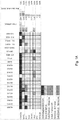

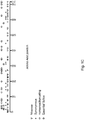

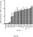


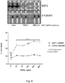


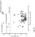
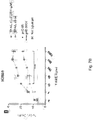


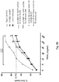


Classifications
-
- G—PHYSICS
- G01—MEASURING; TESTING
- G01N—INVESTIGATING OR ANALYSING MATERIALS BY DETERMINING THEIR CHEMICAL OR PHYSICAL PROPERTIES
- G01N33/00—Investigating or analysing materials by specific methods not covered by groups G01N1/00 - G01N31/00
- G01N33/48—Biological material, e.g. blood, urine; Haemocytometers
- G01N33/50—Chemical analysis of biological material, e.g. blood, urine; Testing involving biospecific ligand binding methods; Immunological testing
- G01N33/5005—Chemical analysis of biological material, e.g. blood, urine; Testing involving biospecific ligand binding methods; Immunological testing involving human or animal cells
- G01N33/5008—Chemical analysis of biological material, e.g. blood, urine; Testing involving biospecific ligand binding methods; Immunological testing involving human or animal cells for testing or evaluating the effect of chemical or biological compounds, e.g. drugs, cosmetics
- G01N33/5014—Chemical analysis of biological material, e.g. blood, urine; Testing involving biospecific ligand binding methods; Immunological testing involving human or animal cells for testing or evaluating the effect of chemical or biological compounds, e.g. drugs, cosmetics for testing toxicity
- G01N33/5017—Chemical analysis of biological material, e.g. blood, urine; Testing involving biospecific ligand binding methods; Immunological testing involving human or animal cells for testing or evaluating the effect of chemical or biological compounds, e.g. drugs, cosmetics for testing toxicity for testing neoplastic activity
-
- A—HUMAN NECESSITIES
- A61—MEDICAL OR VETERINARY SCIENCE; HYGIENE
- A61K—PREPARATIONS FOR MEDICAL, DENTAL OR TOILETRY PURPOSES
- A61K31/00—Medicinal preparations containing organic active ingredients
- A61K31/70—Carbohydrates; Sugars; Derivatives thereof
- A61K31/7088—Compounds having three or more nucleosides or nucleotides
- A61K31/7105—Natural ribonucleic acids, i.e. containing only riboses attached to adenine, guanine, cytosine or uracil and having 3'-5' phosphodiester links
-
- A—HUMAN NECESSITIES
- A61—MEDICAL OR VETERINARY SCIENCE; HYGIENE
- A61P—SPECIFIC THERAPEUTIC ACTIVITY OF CHEMICAL COMPOUNDS OR MEDICINAL PREPARATIONS
- A61P35/00—Antineoplastic agents
-
- A—HUMAN NECESSITIES
- A61—MEDICAL OR VETERINARY SCIENCE; HYGIENE
- A61P—SPECIFIC THERAPEUTIC ACTIVITY OF CHEMICAL COMPOUNDS OR MEDICINAL PREPARATIONS
- A61P35/00—Antineoplastic agents
- A61P35/02—Antineoplastic agents specific for leukemia
-
- G—PHYSICS
- G01—MEASURING; TESTING
- G01N—INVESTIGATING OR ANALYSING MATERIALS BY DETERMINING THEIR CHEMICAL OR PHYSICAL PROPERTIES
- G01N33/00—Investigating or analysing materials by specific methods not covered by groups G01N1/00 - G01N31/00
- G01N33/48—Biological material, e.g. blood, urine; Haemocytometers
- G01N33/50—Chemical analysis of biological material, e.g. blood, urine; Testing involving biospecific ligand binding methods; Immunological testing
- G01N33/5005—Chemical analysis of biological material, e.g. blood, urine; Testing involving biospecific ligand binding methods; Immunological testing involving human or animal cells
- G01N33/5008—Chemical analysis of biological material, e.g. blood, urine; Testing involving biospecific ligand binding methods; Immunological testing involving human or animal cells for testing or evaluating the effect of chemical or biological compounds, e.g. drugs, cosmetics
- G01N33/502—Chemical analysis of biological material, e.g. blood, urine; Testing involving biospecific ligand binding methods; Immunological testing involving human or animal cells for testing or evaluating the effect of chemical or biological compounds, e.g. drugs, cosmetics for testing non-proliferative effects
- G01N33/5023—Chemical analysis of biological material, e.g. blood, urine; Testing involving biospecific ligand binding methods; Immunological testing involving human or animal cells for testing or evaluating the effect of chemical or biological compounds, e.g. drugs, cosmetics for testing non-proliferative effects on expression patterns
-
- G—PHYSICS
- G01—MEASURING; TESTING
- G01N—INVESTIGATING OR ANALYSING MATERIALS BY DETERMINING THEIR CHEMICAL OR PHYSICAL PROPERTIES
- G01N33/00—Investigating or analysing materials by specific methods not covered by groups G01N1/00 - G01N31/00
- G01N33/48—Biological material, e.g. blood, urine; Haemocytometers
- G01N33/50—Chemical analysis of biological material, e.g. blood, urine; Testing involving biospecific ligand binding methods; Immunological testing
- G01N33/53—Immunoassay; Biospecific binding assay; Materials therefor
- G01N33/574—Immunoassay; Biospecific binding assay; Materials therefor for cancer
-
- G—PHYSICS
- G01—MEASURING; TESTING
- G01N—INVESTIGATING OR ANALYSING MATERIALS BY DETERMINING THEIR CHEMICAL OR PHYSICAL PROPERTIES
- G01N2496/00—Reference solutions for assays of biological material
- G01N2496/05—Reference solutions for assays of biological material containing blood cells or plasma
-
- G—PHYSICS
- G01—MEASURING; TESTING
- G01N—INVESTIGATING OR ANALYSING MATERIALS BY DETERMINING THEIR CHEMICAL OR PHYSICAL PROPERTIES
- G01N2500/00—Screening for compounds of potential therapeutic value
- G01N2500/02—Screening involving studying the effect of compounds C on the interaction between interacting molecules A and B (e.g. A = enzyme and B = substrate for A, or A = receptor and B = ligand for the receptor)
-
- G—PHYSICS
- G01—MEASURING; TESTING
- G01N—INVESTIGATING OR ANALYSING MATERIALS BY DETERMINING THEIR CHEMICAL OR PHYSICAL PROPERTIES
- G01N2500/00—Screening for compounds of potential therapeutic value
- G01N2500/10—Screening for compounds of potential therapeutic value involving cells
-
- G—PHYSICS
- G01—MEASURING; TESTING
- G01N—INVESTIGATING OR ANALYSING MATERIALS BY DETERMINING THEIR CHEMICAL OR PHYSICAL PROPERTIES
- G01N2510/00—Detection of programmed cell death, i.e. apoptosis
-
- G—PHYSICS
- G01—MEASURING; TESTING
- G01N—INVESTIGATING OR ANALYSING MATERIALS BY DETERMINING THEIR CHEMICAL OR PHYSICAL PROPERTIES
- G01N2800/00—Detection or diagnosis of diseases
- G01N2800/52—Predicting or monitoring the response to treatment, e.g. for selection of therapy based on assay results in personalised medicine; Prognosis
Definitions
- the present invention relates to cell death of cancer cells, and in particular to biomarkers that may be used to identify cancer cells that are sensitive to death receptor ligand (DRL)-induced cell death.
- DRL death receptor ligand
- the invention also extends to prognostic methods and kits for identifying cancer cells that are sensitive to DRL-induced cell death.
- the invention further extends to novel compositions and therapeutic methods using such compositions for treating cancer.
- MCM Malignant pleural mesothelioma
- asbestos mining and usage has been the subject of legislation in many nations, there remains evidence of increased use and production of asbestos as the trend towards global industrialisation increases (3).
- Current treatment options for patients diagnosed with MPM are limited. Radical surgery, for example, offers limited benefit and the cancer is refractory to most chemotherapeutic agents (4).
- the current gold standard chemotherapy regimen of pemetrexed plus cisplatin offers a median overall survival of 12.1 months and a time to progression of 5.7 months (5).
- Tumour necrosis factor (TNF)-related apoptosis-inducing ligand TRAIL/Apo2L
- TNF Tumour necrosis factor
- TRAIL/Apo2L Tumour necrosis factor-related apoptosis-inducing ligand
- Numerous chemotherapeutic drugs have also been shown to sensitize tumour cells to TRAIL-mediated cell death. While some mesothelioma cells are sensitive to TRAIL induced cell death others are resistant to the induction of cell death in vitro and this disruption of core-cell death machinery has been implicated in the general resistance to conventional cytotoxic agents observed clinically (11).
- cancers are sensitive to the pro-cell death effects of known death receptor ligands, while others are not. Without knowledge of which cancers are sensitive to DRL-induced cell death, individuals with cancer must rely on treatment with non-specific cytotoxic therapies, such as chemotherapy, with the toxicities and problems described (5). Furthermore, with no clear indication pre-treatment of whether a patient will respond or not, the benefit of such treatment is uncertain at the outset.
- DRL death receptor ligand
- WO2013/106002 A1 discloses assays and kits for the prognosis of melanoma comprising the assessment of BAP1 protein and nucleic acid.
- Polanski et al, 'Caspase-8 activation by TRAIL monotherapy predicts responses to IAPi and TRAIL combination treatment in breast cancer cell lines', Cell death and disease, vol. 6, no. 10, 2015, page e1893 , discloses the synergistic effect of the combination of TRAIL and Smac mimetics in the treatment of cancer.
- EP2044434A2 discloses methods for identifying cancer cells that are responsive to therapeutic agents that induce apoptosis.
- a method of determining if an individual's cancer cell is sensitive to death receptor ligand (DRL)-induced cell death comprising detecting, in a biological sample taken from the individual, for:
- kits for determining if an individual's cancer cell is sensitive to DRL-induced cell death comprising detection means for detecting the expression of a mutant BAP1 gene or mutant BAP1 protein, or for detecting a reduced level of expression of a wild-type BAP1 gene or a lower wild-type BAP1 protein concentration compared to the level of expression or protein concentration in a reference cell that is resistant to DRL-induced cell death, or for detecting non-binding or reduced binding of an ASXL protein to a BAP1 protein compared to the level of binding in a reference cell that is a BAP1 wild-type cell that is resistant to DRL-induced cell death, wherein the presence, in the sample, of the mutant BAP1 gene or the BAP1 protein, or of a reduced level expression of the wild-type BAP1 gene or a lower wild-type BAP1 protein concentration, or reduced or non-binding of an ASXL protein to a wild-type BAP1 protein, is indicative of
- a mutant BAP1 gene or a mutant BAP1 protein or (ii) a cancer cell with a reduced level of expression of a wild-type BAP1 gene or a lower wild-type BAP1 protein concentration compared to the level of expression or protein concentration in a reference cell that is a BAP1 wild-type cell, as a biomarker of sensitivity to DRL-induced cell death.
- composition comprising (i) a BAP1 inhibitor, and (ii) a death receptor ligand, wherein the BAP1 inhibitor is WP1130, Usp9x, Usp5, Usp14, Usp24, UCH37, b-AP15, CRISPR/CAS9, an RNAi molecule including shRNA, siRNA, miRNA, ribozymes and antisense molecules, or a TALEN.
- BAP1 inhibitor is WP1130, Usp9x, Usp5, Usp14, Usp24, UCH37, b-AP15, CRISPR/CAS9, an RNAi molecule including shRNA, siRNA, miRNA, ribozymes and antisense molecules, or a TALEN.
- composition comprising (i) a BAP1 inhibitor, and (ii) a death receptor ligand, for use in therapy or as a medicament, wherein the BAP1 inhibitor is WP1130, Usp9x, Usp5, Usp14, Usp24, UCH37, b-AP15, CRISPR/CAS9, an RNAi molecule including shRNA, siRNA, miRNA, ribozymes and antisense molecules, or a TALEN.
- BAP1 inhibitor is WP1130, Usp9x, Usp5, Usp14, Usp24, UCH37, b-AP15, CRISPR/CAS9, an RNAi molecule including shRNA, siRNA, miRNA, ribozymes and antisense molecules, or a TALEN.
- composition comprising (i) a BAP1 inhibitor, and (ii) a death receptor ligand, for use in treating, preventing or ameliorating cancer, wherein the BAP1 inhibitor is WP1130, Usp9x, Usp5, Usp14, Usp24, UCH37, b-AP15, CRISPR/CAS9, an RNAi molecule including shRNA, siRNA, miRNA, ribozymes and antisense molecules, or a TALEN.
- BAP1 inhibitor is WP1130, Usp9x, Usp5, Usp14, Usp24, UCH37, b-AP15, CRISPR/CAS9, an RNAi molecule including shRNA, siRNA, miRNA, ribozymes and antisense molecules, or a TALEN.
- a pharmaceutical composition comprising the composition according to the fourth aspect, and a pharmaceutically acceptable vehicle.
- a method for preparing the pharmaceutical composition according to the seventh aspect comprising contacting a therapeutically effective amount of a BAP1 inhibitor and a death receptor ligand, and a pharmaceutically acceptable vehicle, wherein the BAP1 inhibitor is WP1130, Usp9x, Usp5, Usp14, Usp24, UCH37, b-AP15, CRISPR/CAS9, an RNAi molecule including shRNA, siRNA, miRNA, ribozymes and antisense molecules, or a TALEN.
- the BAP1 inhibitor is WP1130, Usp9x, Usp5, Usp14, Usp24, UCH37, b-AP15, CRISPR/CAS9, an RNAi molecule including shRNA, siRNA, miRNA, ribozymes and antisense molecules, or a TALEN.
- a method of determining if an individual's cancer cell is sensitive to death receptor ligand (DRL)-induced cell death comprising detecting, in a biological sample taken from the individual, for:
- BAP1 is a tumour suppressor gene that is somatically mutated in a variety of cancer types including mesothelioma (7), uveal melanoma (8), renal cell carcinoma (9) and cholangiocarcinomas (10).
- the inventors have discovered that the expression of BAP1 mutants in cancer cells causes increased sensitivity to cell death induced by death receptor ligands, which exhibit fewer off-target effects.
- Tumour necrosis factor (TNF)-related apoptosis-inducing ligand (TRAIL) for example, induces cell death in tumour cells in vitro and in vivo but not in most normal cells.
- the invention therefore increases the likelihood that an individual with a cancer, which is associated with expression of a mutant BAP1 gene or synthesis of a mutant BAP1 protein, or a reduced level of expression of a wild-type BAP1 gene or a lower wild-type BAP1 protein concentration, will exhibit a positive outcome when treated with death receptor ligand therapy. Accordingly, the method of the invention provides a prognosis. Thus, such individuals are less likely to be treated with non-specific therapies and suffer the associated undesirable side-effects.
- BAP1 is a deubiquitinating enzyme encoded by the BAP1 gene. It is an 80.4kDa protein, which comprises a nuclear-localising sequence and a ubiquitin carboxy-terminal hydrolase (UCH) domain, which provides BAP1 with deubiquitinase activity.
- the BAP1 gene may be mutated in the germline or somatically in a variety of forms of cancer.
- the nucleotide sequence encoding one embodiment of the human wild-type BAP1 gene (BAP1 Whole gene sequence >gi
- the cDNA sequence encoding one embodiment of the human wild-type BAP1 gene is provided herein as SEQ ID No. 2, as follows:
- amino acid sequence of one embodiment of human wild-type BAP1 is referred to herein as SEQ ID No. 3, as follows:
- Tumours arise due to mutations in proto-oncogenes or tumour suppressor genes.
- a gain-of-function mutation in a proto-oncogene converts it into an oncogene, which causes tumourigenesis.
- Such gain-of-function mutations are usually missense mutations in the DNA bases, which confer a change in the amino acid sequence, or a gain of copy number of the gene.
- a loss-of-function mutation in a tumour suppressor gene will also lead to tumourigenesis.
- a loss-of-function mutation in a tumour suppressor however is not restricted to the change of a few amino acids. Any change in an amino acid which impairs the function of the protein, results in a loss-of-function mutation.
- tumour suppressors unlike oncogenes, it is not always possible to identify a specific mutation which results in the loss of function of a tumour suppressor gene. Loss-of-function truncating mutations of tumour suppressors are therefore typically identified throughout the coding exons of a gene.
- BAP1 is a tumour suppressor gene.
- a mutant BAP1 gene is one that comprises a mutation.
- the mutant BAP1 gene is a gene that encodes a non-functional or enzymatically inactive BAP1 protein, or a BAP1 protein that exhibits reduced binding to an ASXL protein compared to the level of binding in a reference cell, which is resistant to DRL-induced cell death.
- a mutant BAP1 protein is one that is non-functional or enzymatically inactive or incapable of binding to an ASXL protein or exhibits reduced binding to an ASXL protein compared to the level of binding in a reference cell, which is resistant to DRL-induced cell death.
- BAP1 has surprisingly been shown to form a complex with ASXL1, ASXL2 or ASXL3. These proteins will be collectively referred to herein as an ASXL protein.
- ASXL protein proteins will be collectively referred to herein as an ASXL protein.
- BAP1-ASXL complexes have been shown to deubiquitinate Histone 2A, and other substrates. Inhibiting the formation of this complex renders BAP1 non-functional or enzymatically inactive.
- the mutant BAP1 protein may be a full-length protein with mutations at specific loci.
- the mutant BAP1 protein may be a partial or complete deletion or mutation of the wild-type BAP1 protein. Partial deletion or mutation may occur in the nuclear localisation sequence (NLS), the active site of wild-type BAP1, the binding site of ASXL, or at any place in the gene that would result in the loss of function of BAP1.
- Mutation may include one or more point mutations. Point mutations may be a substitution, an insertion, a deletion or a frameshift mutation.
- a reduced level of expression of a wild-type BAP1 gene compared to the level of expression in a reference cell may result in a lower wild-type BAP1 protein concentration compared to the protein concentration in the same reference cell.
- the reduced level of expression of the wild-type BAP1 gene may be at least a 10%, 15%, 25%, 35%, 45%, 50%, 55%, 60%, 65%, 70%, 75%, 80%, 85%, 90%, 95% or a 100% reduction compared to the reference cell.
- the lower concentration of wild-type BAP1 protein may be lower by at least 10%, 15%, 25%, 35%, 45%, 50%, 55%, 60%, 65%, 70%, 75%, 80%, 85%, 90%, 95% or 100% compared to the reference cell.
- the skilled person would know how to detect the extent of the reduction in BAP1 gene expression or lower BAP1 protein expression.
- non-functional BAP1 protein can refer to but is not limited to a BAP1 protein that does not exhibit deubiquitinase (enzyme) activity.
- standard assays for measuring deubiquitinase activity include, but are not limited to, fluorescence assays using a fluorogenic substrate, such as ubiquitin-amidomethylcoumarin; and HPLC assays using ubiquitin ethyl ester or ubiquitin fusion peptides as model substrates to monitor deubiquitinating activity.
- amino acid sequence of a mutant BAP1 gene (p.R60Q) is referred to herein as SEQ ID N0. 4, as follows:
- amino acid sequence of the mutant BAP1 protein may be encoded by SEQ ID NO. 4 or a fragment or variant thereof.
- nucleotide sequence of a mutant BAP1 gene is referred to herein as SEQ ID No. 5, as follows:
- nucleotide sequence of the mutant BAP1 gene may be encoded by SEQ ID NO. 5 or a fragment or variant thereof.
- Reduced or non-binding between an ASXL protein (ASXL1, ASXL2 or ASXL3) and a wild-type BAP1 protein may be at least a 10%, 15%, 25%, 35%, 45%, 50%, 55%, 60%, 65%, 70%, 75%, 80%, 85%, 90%, 95% or a 100% reduction compared to a reference cell in which binding between a wild-type BAP1 protein and an ASXL protein occurs.
- the skilled person would know how to detect the extent of the reduction in wild-type BAP1 protein binding to an ASXL protein.
- Standard assays for measuring binding between a wild-type BAP1 protein and an ASXL protein include but are not limited to protein complex immunoprecipitation or fluorescence resonance energy transfer (FRET).
- DRL-induced cell death can refer to, but is not limited to, apoptosis and other types of cell death caused by a death receptor ligand (DRL), such as necroptosis and necrosis.
- DRL death receptor ligand
- the term “death receptor ligand” refers to any agent that binds to a cellular receptor and induces death of the cell on which the receptor is located.
- cell death can refer to cellular apoptosis, necrosis and necroptosis. Preferably, it refers to cellular apoptosis.
- Apoptosis refers to programmed cell death caused by activation of an apoptotic signal transduction pathway. This may be achieved through the binding of a DRL to a death receptor.
- Death receptor ligands may be TRAIL, TNFalpha, FAS ligand (FASL), recombinant TRAIL (dulanermin), antibodies to death receptors, especially antibodies to death receptors of the ligand TRAIL (such as mapatumuab, drozitumumab, conatumumab, lexatumumab, tigatuzumab, LBY-135), or a combination thereof.
- the DRL may be an agonist molecule such as Medi-3039 or any agent that activates an apoptotic signal transduction pathway.
- An extrinsic apoptotic signal transduction pathway may be the FAS ligand pathway, the TNFalpha pathway or the TRAIL pathway.
- express(ed) or expression can refer to a transcribed gene (i.e. DNA), or corresponding RNA that has been translated into a polypeptide or protein. Expression of a BAP1 mutant gene or BAP1 polypeptide may be detected in any compartment of the cell (e.g. in the nucleus, cytosol, the Endoplasmic Reticulum, the Golgi apparatus or the intracellular surface of the plasma membrane).
- Detecting according to (i), (ii) or (iii) of this disclosure may comprise the use of any one of the following assays for detecting the presence of a gene or its corresponding protein in a sample: polymerase chain reaction (PCR); northern blotting; hybridisation-based detection techniques; flow cytometry; immunoassays, such as enzyme-linked immunosorbent assays (ELISAs), an enzyme immunoassay (EIAs), radioimmunoassay (RIAs), Western Blots, immuno-precipitation or immunohistochemistry; immunofluorescence; chromogenic (enzyme activity) assays; fluorometric imaging plate reader (FLIPR) assay; high performance liquid chromatography (HPLC) tandem mass spectrometry (MS/MS).
- PCR polymerase chain reaction
- IAs enzyme immunoassay
- RIAs radioimmunoassay
- Western Blots immuno-precipitation or immunohistochemistry
- immunofluorescence chrom
- the biological sample is preferably a cancerous bodily sample taken from a test individual. Detection for the presence of a BAP1 mutant gene or mutant BAP1 protein in the sample is therefore preferably carried out in vitro.
- the sample may comprise tissue, blood, plasma, serum, spinal fluid, urine, sweat, saliva, sputum, tears, breast aspirate, prostate fluid, seminal fluid, vaginal fluid, stool, cervical scraping, amniotic fluid, intraocular fluid, mucous, moisture in breath, animal tissue, cell lysates, tumour tissue, hair, skin, buccal scrapings, nails, bone marrow, cartilage, prions, bone powder, ear wax, or combinations thereof.
- the sample may be a biopsy.
- the sample may be contained within the test subject, which may be an experimental animal (e.g. a mouse or rat) or a human, wherein the method is an in vivo based test.
- the sample may be an ex vivo sample or an in vitro sample. Therefore, the cells being tested may be in a tissue sample (for ex vivo based tests) or the cells may be grown in culture (an in vitro sample).
- the biological sample is an ex vivo sample.
- the inventors have developed a prognostic kit for determining a subject's sensitivity or otherwise to DRL-induced cell death.
- kits for determining if an individual's cancer cell is sensitive to DRL-induced cell death comprising detection means for detecting the expression of a mutant BAP1 gene or mutant BAP1 protein, or for detecting a reduced level of expression of a wild-type BAP1 gene or a lower wild-type BAP1 protein concentration compared to the level of expression or protein concentration in a reference cell that is resistant to DRL-induced cell death, or for detecting non-binding or reduced binding of an ASXL protein to a BAP1 protein compared to the level of binding in a reference cell that is a BAP1 wild-type cell that is resistant to DRL-induced cell death, wherein the presence, in the sample, of the mutant BAP1 gene or the BAP1 protein, or of a reduced level expression of the wild-type BAP1 gene or a lower wild-type BAP1 protein concentration, or reduced or non-binding of an ASXL protein to a wild-type BAP1 protein, is indicative of the individual
- the kit is used to provide a prognosis for an individual being treated with a death receptor ligand (DRL) or any agents activating the apoptotic pathways.
- a death receptor ligand may be TRAIL, TNFalpha, FAS ligand (FASL), recombinant TRAIL (dulanermin), death receptor antibodies (such as mapatumuab, drozitumumab, conatumumab, lexatumumab, tigatuzumab), death receptor agonists, such as Medi-3038 or Medi-3039, or a combination thereof.
- the kit comprises at least one control or reference sample.
- the kit may comprise a negative control and/or a positive control.
- a negative control may comprise a wild-type BAP1 protein that is resistant to DRL-induced cell death.
- a positive control may comprise a mutant BAP1 mRNA, or mutant BAP1 protein, or a blank sample. The skilled person will appreciate that the level of mRNA in a sample is indicative of the level of gene expression in a cell.
- the detection means is preferably configured to detect the expression or the concentration of a mutant BAP1 protein or mRNA, or wild-type BAP1 mRNA or BAP1 protein in the biological sample taken from the test individual.
- the presence of the mutant BAP1 mRNA or mutant BAP1 protein, or reduced level of expression of wild-type BAP1 mRNA or lower protein expression to the control, in the sample is indicative that the test sample is sensitive to DRL-induced cell death.
- the level of expression of the wild-type BAP1 mRNA or concentration of BAP1 protein in the biological sample may be reduced or lower compared to the concentration or level of expression of the wild-type BAP1 mRNA or protein in a negative control.
- the reduction in expression may be at least a 10%, 15%, 25%, 35%, 45%, 50%, 55%, 60%, 65%, 70%, 75%, 80%, 85%, 90%, 95% or 100% reduction compared to the negative control.
- concentration of the wild-type BAP1 protein in the biological sample may be lower by at least a 10%, 15%, 25%, 35%, 45%, 50%, 55%, 60%, 65%, 70%, 75%, 80%, 85%, 90%, 95% or 100%.
- the absence of a mutant BAP1 gene or protein, or normal to high expression of the wild-type BAP1 gene or protein, in the sample is indicative that the sample is insensitive to DRL-induced cell death.
- Detection of a mutation in a BAP1 gene or protein can be achieved using a number of sequencing approaches.
- whole exome or targeted gene sequencing is undertaken with massively parallel sequencing of tumour DNA with target genes enriched using commercially available RNA baits.
- a capillary sequencing approach is utilised with PCR primers designed to each of the exons in the BAP1 gene footprint.
- the detection means may be a primer.
- nucleic acid sequence of a forward primer used to detect exon 12 (chr3:52438255-52438817F) of wild-type BAP1 mRNA is referred to herein as SEQ ID No. 6 as follows: acctagaacctggtagccttag [SEQ ID No. 6]
- nucleic acid sequence of a forward primer used to detect exon 8 (chr3:5244063i-5244H38F) of wild-type BAP1 mRNA is referred to herein as SEQ ID No. 7 as follows: gtacagctccagagagtagaac [SEQ ID No. 7]
- nucleic acid sequence of a forward primer used to detect exon 1 (chr3:52443644-52444094F) of wild-type BAP1 mRNA is referred to herein as SEQ ID No. 8 as follows: tcttaccgaaatcttccacgag [SEQ ID No. 8]
- nucleic acid sequence of a forward primer used to detect exon 3 (chr3:52443356-52443839F) of wild-type BAP1 mRNA is referred to herein as SEQ ID No. 9 as follows: ctgctgctttctgtgagatttt [SEQ ID No. 9]
- nucleic acid sequence of a forward primer used to detect exon 6 (chr3:52436404-52436905F) of wild-type BAP1 mRNA is referred to herein as SEQ ID No. 10 as follows: agggcattccagttaagacag [SEQ ID No. 10]
- nucleic acid sequence of a forward primer used to detect exon 17 (chr3:52436105-52436652F) of wild-type BAP1 mRNA is referred to herein as SEQ ID No. 11 as follows: caagagtgggctgcagag [SEQ ID No. 11]
- nucleic acid sequence of a forward primer used to detect exon 6 (chr3:52441201-52441691F) of wild-type BAP1 mRNA is referred to herein as SEQ ID No. 12 as follows: actaaggccattctgcttctc [SEQ ID No. 12]
- nucleic acid sequence of a forward primer used to detect exon 4 (chr3:52442276-52442837F) of wild-type BAP1 mRNA is referred to herein as SEQ ID No. 13 as follows: atcccaccctccaaacaaag [SEQ ID No. 13]
- nucleic acid sequence of a forward primer used to detect exon 13a (chr3:52437218-52437786F) of wild-type BAP1 mRNA is referred to herein as SEQ ID No. 14 as follows: caccaagtggccagtgag [SEQ ID No. 14]
- nucleic acid sequence of a forward primer used to detect exon 13b (chr3:52437388-52437956F) of wild-type BAP1 mRNA is referred to herein as SEQ ID No. 15 as follows: ggctgtcatcctctccaaaa [SEQ ID No. 15]
- nucleic acid sequence of a forward primer used to detect exon 13c (chr3:52437558-52438125F) of wild-type BAP1 mRNA is referred to herein as SEQ ID No. 16 as follows: gagggctgcgagtgtgtg [SEQ ID No. 16]
- nucleic acid sequence of a forward primer used to detect exon 14 (chr3:52436940-52437529F) of wild-type BAP1 mRNA is referred to herein as SEQ ID No. 17 as follows: ctctgccaggattaaaggagaa [SEQ ID No. 17]
- nucleic acid sequence of a forward primer used to detect exon 9 (chr3:52440055-52440607F) of wild-type BAP1 mRNA is referred to herein as SEQ ID No. 18 as follows: gaatgcagggagggttgg [SEQ ID No. 18]
- nucleic acid sequence of a forward primer used to detect exon 5 (chr3:52441760-524423o8F) of wild-type BAP1 mRNA is referred to herein as SEQ ID No. 19 as follows: acccaatatcatgtggtagcat [SEQ ID No. 19]
- nucleic acid sequence of a forward primer used to detect exon 2 (chr3:52443516-52443974F) of wild-type BAP1 mRNA is referred to herein as SEQ ID No. 20 as follows: aaggacagccctgatga [SEQ ID No. 20]
- nucleic acid sequence of a forward primer used to detect exon 7 (chr3:52440976-52441547F) of wild-type BAP1 mRNA is referred to herein as SEQ ID No. 21 as follows: gtaggcagagacacccaac [SEQ ID No. 21]
- nucleic acid sequence of a forward primer used to detect exon 15 (chr3:52436581-52437102F) of wild-type BAP1 mRNA is referred to herein as SEQ ID No. 22 as follows: ccttctctggtcatcaatctgt [SEQ ID No. 22]
- nucleic acid sequence of a forward primer used to detect exon 10 (chr3:52439567-52440143F) of wild-type BAP1 mRNA is referred to herein as SEQ ID No. 23 as follows: ctctgaggtccacaagaggt [SEQ ID No. 23]
- nucleic acid sequence of a forward primer used to detect exon 11 (chr3:52438912-52439525F) of wild-type BAP1 mRNA is referred to herein as SEQ ID No. 24 as follows: tcaagtagagaatcctgcaagg [SEQ ID No. 24]
- nucleic acid sequence of a reverse primer used to detect exon 12 (chr3:52438255-52438817R) of wild-type BAP1 mRNA is referred to herein as SEQ ID No. 25 as follows: gagcagcacttgtttgtaactg [SEQ ID No. 25]
- nucleic acid sequence of a reverse primer used to detect exon 8 (chr3:52440631-52441138R) of wild-type BAP1 mRNA is referred to herein as SEQ ID No. 26 as follows: ctcaactgctcttctctgtctt [SEQ ID No. 26]
- nucleic acid sequence of a reverse primer used to detect exon 1 (chr3:52443644-52444094R) of wild-type BAP1 mRNA is referred to herein as SEQ ID No. 27 as follows: gagggagggcctggacat [SEQ ID No. 27]
- nucleic acid sequence of a reverse primer used to detect exon 3 (chr3:52443356-52443839R) of wild-type BAP1 mRNA is referred to herein as SEQ ID No. 28 as follows: ctgtccttccctactgctttc [SEQ ID No. 28]
- nucleic acid sequence of a reverse primer used to detect exon 16 (chr3:52436404-52436905R) of wild-type BAP1 mRNA is referred to herein as SEQ ID No. 29 as follows: gaagttcaaggtgggtgatttc [SEQ ID No. 29]
- nucleic acid sequence of a reverse primer used to detect exon 17 (chr3:52436105-52436652R) of wild-type BAP1 mRNA is referred to herein as SEQ ID No. 30 as follows: ctcagctcctggcctgag [SEQ ID No. 30]
- nucleic acid sequence of a reverse primer used to detect exon 6 (chr3:52441201-52441691R) of wild-type BAP1 mRNA is referred to herein as SEQ ID No. 31 as follows: ggagaaattattctgatacggcc [SEQ ID No. 31]
- nucleic acid sequence of a reverse primer used to detect exon 4 (chr3:52442276-52442837R) of wild-type BAP1 mRNA is referred to herein as SEQ ID No. 32 as follows: gaagggaatgctgattgtcttc [SEQ ID No. 32]
- nucleic acid sequence of a reverse primer used to detect exon 13a (chr3:52437218-52437786R) of wild-type BAP1 mRNA is referred to herein as SEQ ID No. 33 as follows: ctatccgctcagccaacc [SEQ ID No. 33]
- nucleic acid sequence of a reverse primer used to detect exon 13b (chr3:52437388-52437956R) of wild-type BAP1 mRNA is referred to herein as SEQ ID No. 34 as follows: ctctcaattcctctgtccatca [SEQ ID No. 34]
- nucleic acid sequence of a reverse primer used to detect exon 13c (chr3:52437558-52438125R) of wild-type BAP1 mRNA is referred to herein as SEQ ID No. 35 as follows: cgttcccttgcttcacatct [SEQ ID No. 35]
- nucleic acid sequence of a reverse primer used to detect exon 14 (chr3:52436940-52437529R) of wild-type BAP1 mRNA is referred to herein as SEQ ID No. 36 as follows: tcctgcactctgatgattttct [SEQ ID No. 36]
- nucleic acid sequence of a reverse primer used to detect exon 9 (chr3:52440055-52440607R) of wild-type BAP1 mRNA is referred to herein as SEQ ID No. 37 as follows: gatatctgcctcaacctgatgg [SEQ ID No. 37]
- nucleic acid sequence of a reverse primer used to detect exon 5 (chr3:52441760-524423o8R) of wild-type BAP1 mRNA is referred to herein as SEQ ID No. 38 as follows: gtgctgtgtatgggtgacta [SEQ ID No. 38]
- nucleic acid sequence of a reverse primer used to detect exon 2 (chr3:52443516-52443974R) of wild-type BAP1 mRNA is referred to herein as SEQ ID No. 39 as follows: gaagatgaataagggctggct [SEQ ID No. 39]
- nucleic acid sequence of a reverse primer used to detect exon 7 (chr3:52440976-52441547R) of wild-type BAP1 mRNA is referred to herein as SEQ ID No. 40 as follows: tgatgtggggtgggagtag [SEQ ID No.40]
- nucleic acid sequence of a reverse primer used to detect exon 15 (chr3:52436581-52437102R) of wild-type BAP1 mRNA is referred to herein as SEQ ID No. 41 as follows: cccgatcagaggtgcaat [SEQ ID No. 41]
- nucleic acid sequence of a reverse primer used to detect exon 10 (chr3:52439567-52440143R) of wild-type BAP1 mRNA is referred to herein as SEQ ID No. 42 as follows: agctatttaaggtagaagcccg [SEQ ID No. 42]
- nucleic acid sequence of a reverse primer used to detect exon 11 (chr3:52438912-52439525R) of wild-type BAP1 mRNA is referred to herein as SEQ ID No. 43 as follows: actgtgagcttttcttggagat [SEQ ID No. 43]
- an ASXL protein to a wild-type BAP1 protein may be achieved using a variety of techniques known in the art, which include protein complex immunoprecipitation, Bimolecular Fluorescence complementation, Affinity electrophoresis, Immunoelectrophoresis, chemical cross linking, Proximity ligation assay and FRET.
- a mutant BAP1 gene or a mutant BAP1 protein or (ii) a cancer cell with a reduced level of expression of a wild-type BAP1 gene or a lower wild-type BAP1 protein concentration compared to the level of expression or protein concentration in a reference cell that is a BAP1 wild-type cell, as a biomarker of sensitivity to DRL-induced cell death.
- the inventors have developed a method of treating subjects suffering from cancer, and especially those suffering from a cancer that is completely or partially insensitive to death receptor ligand-induced cell death.
- the death receptor ligand may be administered as a monotherapy or in combination with other agents that are capable of killing cancer cells.
- the "subject” or “individual” may be a vertebrate, mammal, or domestic animal.
- medicaments according to this disclosure may be used to treat any mammal, for example livestock (e.g. a horse), pets, or may be used in other veterinary applications.
- livestock e.g. a horse
- pets e.g. a human
- the subject is a human being.
- IAP inhibitors of apoptosis
- BIR ⁇ 70-amino acid baculovirus IAP repeat
- BAP1 gene expression can be reduced in mesothelioma cell lines using a BAP1 shRNA-expressing lentivirus and that this reduction of BAP1 gene expression results in increased sensitivity to DLR-induced cell death in these cell lines. Accordingly, the inventors have determined that it is possible to sensitise to DRL-induced cell death, an individual suffering from a cancer that is normally insensitive to DRL-induced cell death, by administering a BAP1 inhibitor.
- a BAP1 inhibitor is any agent that targets the BAP1 gene or protein directly, whereas an agent that mimics the effect of BAP1 inhibition is a molecule that targets a signalling molecule downstream of the BAP1 gene/protein signalling pathway.
- a BAP1 inhibitor may be any molecule that inactivates BAP1 protein function, such as a deubiquitinase inhibitor or antagonist, or which silences or reduces BAP1 gene or protein expression or function (such as a small molecule inhibitor of BAP1), or which prevents or reduces binding of a wild-type BAP1 protein to an ASXL protein.
- the BAP1 inhibitor or agent may also be any molecule that mutates a wild-type BAP1 gene to create a mutant BAP1 gene.
- the BAP1 inhibitor or agent may be an RNAi molecule, including shRNA, siRNA, miRNA, ribozymes and antisense molecules; a TALEN (Transcriptional Activator Like-Effector Nuclease); or a CRISPR/CAS9 nuclease.
- an agent that mimics the effect of BAP1 inhibition is an agent that targets signalling or effector molecules downstream of BAP1, such agents may be used to induce DLR resistance in cancer cell.
- the BAP1 inhibitor may be an IAP inhibitor (a SMAC mimetic).
- An IAP may be BIRC2, BIRC3, BIRC5, BIRC6, BIRC7, BIRC8, NAIP, XIAP or a fragment thereof.
- the inventors have found that BAP1 expression increases the expression of BIRC3 protein and that inhibition of BIRC3 protein with an IAP inhibitor, such as LCL161, results in sensitization to DRL induced cell death.
- An IAP inhibitor is an agent that reduces expression or translation of an IAP, or renders an IAP functionally inactive.
- the IAP inhibitor or agent may also be any molecule that mutates an IAP gene or protein to create a mutant IAP gene or protein.
- the IAP1 inhibitor or agent may be an RNAi molecule, including shRNA, siRNA, miRNA, ribozymes and antisense molecules; a TALEN (Transcriptional Activator Like-Effector Nuclease); or a CRISPR/CAS9 nuclease.
- An agent that mimics the effect of BAP1 inhibition may be an RNA helicase inhibitor, such as, YK-4279 or a tyrosine kinase inhibitor, such as sorafenib.
- BAP1 plays a role in regulating DRL-induced cell death of cancer cells
- novel targeted approach for treatment of any cancer comprising mutants of BAP1 can be used to develop a novel targeted approach for treatment of any cancer comprising mutants of BAP1.
- composition comprising (i) a BAP1 inhibitor or an agent that mimics the effect of BAP1 inhibition and (ii) a death receptor ligand.
- composition comprising (i) a BAP1 inhibitor or an agent that mimics the effect of BAP1 inhibition and (ii) a death receptor ligand, for use in therapy or as a medicament.
- composition comprising (i) a BAP1 inhibitor or an agent that mimics the effect of BAP1 inhibition and (ii) a death receptor ligand, for use in treating, preventing or ameliorating cancer.
- the BAP1 inhibitor or agent that mimics the effect of BAP1 inhibition and the death receptor ligand may be administered simultaneously, or the BAP1 inhibitor may be administered prior to the death receptor ligand.
- administration of the BAP1 inhibitor first sensitizes the subject to DRL-induced cell death. Then, administration of the death receptor ligand can be effectively used to induce cell death in cancerous cells.
- the BAP1 inhibitor is any molecule or approach which inactivates BAP1 protein function, such as a deubiquitinase inhibitor or antagonist, or which silences or reduces BAP1 gene transcription or BAP1 protein expression, or which mutates a wild-type BAP1 gene to create a BAP1 mutant gene and protein.
- the BAP1 inhibitor may be WP1130, Usp9x, Usp5, Usp14, Usp24, UCH37, b-AP15, CRISPR/CAS9 or a small molecule inhibitor of BAP1.
- Preferred death receptor ligands may be, but are not limited to TRAIL, TNFalpha, FAS ligand (FASL), recombinant TRAIL (dulanermin), death receptor antibodies (such as mapatumuab, drozitumumab, conatumumab, lexatumumab, tigatuzumab, LBY-135) or death receptor agonists such as Medi 3038 or Medi 3039, or a combination thereof.
- Mesothelioma (or Malignant Pleural Mesothelioma) is a rare form of cancer that develops from cells of the mesothelium, the protective lining that covers many of the internal organs of the body.
- the most common anatomical site for mesothelioma is the pleura (the outer lining of the lungs and internal chest wall), but it can also arise in the peritoneum (the lining of the abdominal cavity), the pericardium (the sac that surrounds the heart) or the tunica vaginalis (a sac that surrounds the testis).
- the cancer which may be treated according to any this disclosure may be selected from mesothelioma, Malignant Pleural Mesothelioma, uveal melanoma, melanoma, non-melanoma skin cancer, renal cancers, cholangiocarcinomas, lung cancers, cancer of the pleura, abdominal cancer, peritoneal cancer, cancer of the pericardium, head and neck cancers, brain cancers, breast cancers, liver and biliary tract cancers, gastrointestinal cancers including upper and lower tracts, urothelial cancers, prostate cancers, testicular cancer, cancer of the tunica vaginalis, ovarian cancers, cervical cancers, sarcomas, lymphomas and leukaemia.
- the cancer which is treated is mesothelioma. Most preferably, the cancer which is treated is an asbestos-induced cancer.
- compositions according to this disclosure may have a number of different forms depending, in particular, on the manner in which the composition is to be used.
- the composition may be in the form of a powder, tablet, capsule, liquid, ointment, cream, gel, hydrogel, aerosol, spray, micellar solution, transdermal patch, liposome suspension or any other suitable form that may be administered to a person or animal in need of treatment.
- vehicle of medicaments according to this disclosure should be one which is well tolerated by the subject to whom it is given.
- compositions according to this disclosure may be used in a number of ways. For instance, oral administration may be required, in which case the agents may be contained within a composition that may, for example, be ingested orally in the form of a tablet, capsule or liquid. Compositions comprising agents of this disclosure may be administered by inhalation (e.g. intranasally). Compositions may also be formulated for topical use. For instance, creams or ointments may be applied to the skin.
- Agents or compositions according to this disclosure may also be incorporated within a slow- or delayed-release device.
- Such devices may, for example, be inserted on or under the skin, and the medicament may be released over weeks or even months.
- the device may be located at least adjacent to the treatment site.
- Such devices may be particularly advantageous when long-term treatment with agents used according to this disclosure is required and which would normally require frequent administration (e.g. at least daily injection).
- compositions and agents according to this disclosure may be administered to a subject by injection into the blood stream or directly into a site requiring treatment.
- the medicament may be injected at least adjacent to a sensitive cell, or within a tumour.
- Injections may be intravenous (bolus or infusion) or subcutaneous (bolus or infusion), or intradermal (bolus or infusion), or intrapleural.
- the amount of the composition and agent that is required is determined by its biological activity and bioavailability, which in turn depends on the mode of administration, the physicochemical properties of the modulator and whether it is being used as a monotherapy or in a combined therapy.
- the frequency of administration will also be influenced by the half-life of the BAP1 inhibitors and DRLs within the subject being treated.
- Optimal dosages to be administered may be determined by those skilled in the art, and will vary with the particular agent in use, the strength of the pharmaceutical composition, the mode of administration, and the advancement of the cancer. Additional factors depending on the particular subject being treated will result in a need to adjust dosages, including subject age, weight, gender, diet, and time of administration.
- a daily dose of between 0.01 ⁇ g/kg of body weight and 500mg/kg of body weight of the agents (e.g. the inhibitor or the DRL) according to this disclosure may be used for treating, ameliorating, or preventing cancer, depending upon which agent is used. More preferably, the daily dose is between 0.01mg/kg of body weight and 400mg/kg of body weight, more preferably between 0.1mg/kg and 200mg/kg body weight, and most preferably between approximately 1mg/kg and 100mg/kg body weight.
- composition or agent(s) may be administered before, during or after onset of the cancer.
- Daily doses may be given as a single administration (e.g. a single daily injection).
- the agent may require administration twice or more times during a day.
- agents may be administered as two (or more depending upon the severity of the disease being treated) daily doses of between 25mg and 7000 mg (i.e. assuming a body weight of 70 kg).
- a subject receiving treatment may take a first dose upon waking and then a second dose in the evening (if on a two dose regime) or at 3- or 4-hourly intervals thereafter.
- a slow release device may be used to provide optimal doses of agents according to this disclosure to a patient without the need to administer repeated doses.
- Known procedures such as those conventionally employed by the pharmaceutical industry (e.g. in vivo experimentation, clinical trials, etc.), may be used to form specific formulations comprising the agents according to this disclosure and precise therapeutic regimes (such as daily doses of the agents and the frequency of administration).
- the inventors believe that they are the first to describe a pharmaceutical composition for treating cancer, based on the use of a BAP1 inhibitor to sensitise the subject to death receptor-induced cell death, and a death receptor ligand, which induces cell death of the cells that express death receptors and previously insensitive to death receptor-induced cell death.
- composition comprising the composition according to this disclosure, and a pharmaceutically acceptable vehicle.
- a method for preparing the pharmaceutical composition according to this disclosure comprising contacting a therapeutically effective amount of a BAP1 inhibitor or agent that mimics the effect of BAP1 inhibition and a death receptor ligand, and a pharmaceutically acceptable vehicle.
- a “therapeutically effective amount” of BAP1 inhibitor is any amount which, when administered to a subject, is the amount needed to sensitise an individual's cells to death receptor-induced cell death.
- a “therapeutically effective amount” of death receptor ligand is any amount which, when administered to a subject, is the amount needed to treat the cancer, or produce the desired effect.
- the therapeutically effective amount of active agent i.e. BAP1 inhibitor and a death receptor ligand
- the amount of agent is an amount from about 0.1 mg to about 250 mg, and most preferably from about 0.1 mg/ kg body weight to about 20 mg/ kg body weight.
- a "pharmaceutically acceptable vehicle” as referred to herein, is any known compound or combination of known compounds that are known to those skilled in the art to be useful in formulating pharmaceutical compositions.
- the pharmaceutically acceptable vehicle may be a solid, and the composition may be in the form of a powder or tablet.
- a solid pharmaceutically acceptable vehicle may include one or more substances which may also act as flavouring agents, lubricants, solubilisers, suspending agents, dyes, fillers, glidants, compression aids, inert binders, sweeteners, preservatives, dyes, coatings, or tablet-disintegrating agents.
- the vehicle may also be an encapsulating material.
- the vehicle is a finely divided solid that is in admixture with the finely divided active agents according to this disclosure.
- the active agent e.g.
- the peptide, antibody, DRL or BAP1 inhibitor may be mixed with a vehicle having the necessary compression properties in suitable proportions and compacted in the shape and size desired.
- the powders and tablets preferably contain up to 99% of the active agents.
- Suitable solid vehicles include, for example calcium phosphate, magnesium stearate, talc, sugars, lactose, dextrin, starch, gelatin, cellulose, polyvinylpyrrolidone, low melting waxes and ion exchange resins.
- the pharmaceutical vehicle may be a gel and the composition may be in the form of a cream or the like.
- the pharmaceutical vehicle may be a liquid, and the pharmaceutical composition is in the form of a solution.
- Liquid vehicles are used in preparing solutions, suspensions, emulsions, syrups, elixirs and pressurized compositions.
- the active agent according to this disclosure may be dissolved or suspended in a pharmaceutically acceptable liquid vehicle such as water, an organic solvent, a mixture of both or pharmaceutically acceptable oils or fats.
- the liquid vehicle can contain other suitable pharmaceutical additives such as solubilisers, emulsifiers, buffers, preservatives, sweeteners, flavouring agents, suspending agents, thickening agents, colours, viscosity regulators, stabilizers or osmo-regulators.
- suitable examples of liquid vehicles for oral and parenteral administration include water (partially containing additives as above, e.g.
- cellulose derivatives preferably sodium carboxymethyl cellulose solution
- alcohols including monohydric alcohols and polyhydric alcohols, e.g. glycols) and their derivatives, and oils (e.g. fractionated coconut oil and arachis oil).
- the vehicle can also be an oily ester such as ethyl oleate and isopropyl myristate.
- Sterile liquid vehicles are useful in sterile liquid form compositions for parenteral administration.
- the liquid vehicle for pressurized compositions can be a halogenated hydrocarbon or other pharmaceutically acceptable propellant.
- Liquid pharmaceutical compositions which are sterile solutions or suspensions, can be utilised by, for example, intramuscular, intrathecal, epidural, intraperitoneal, intravenous and particularly subcutaneous injection.
- the composition or antibody may be prepared as a sterile solid composition that may be dissolved or suspended at the time of administration using sterile water, saline, or other appropriate sterile injectable medium.
- agents and compositions of this disclosure may be administered orally in the form of a sterile solution or suspension containing other solutes or suspending agents (for example, enough saline or glucose to make the solution isotonic), bile salts, acacia, gelatin, sorbitan monoleate, polysorbate 80 (oleate esters of sorbitol and its anhydrides copolymerized with ethylene oxide) and the like.
- the agent or composition according to this disclosure can also be administered orally either in liquid or solid composition form.
- Compositions and agents suitable for oral administration include solid forms, such as pills, capsules, granules, tablets, and powders, and liquid forms, such as solutions, syrups, elixirs, and suspensions.
- Forms useful for parenteral administration include sterile solutions, emulsions, and suspensions.
- nucleic acid or peptide or variant, derivative or analogue thereof which comprises substantially the amino acid or nucleic acid sequences of any of the sequences referred to herein, including variants or fragments thereof.
- substantially the amino acid/nucleotide/peptide sequence can be a sequence that has at least 40% sequence identity with the amino acid/nucleotide/peptide sequences of any one of the sequences referred to herein, for example 40% identity with the polypeptide identified as SEQ ID Nos. 3 or 4, and so on.
- amino acid/polynucleotide/polypeptide sequences with a sequence identity which is greater than 50%, more preferably greater than 65%, 70%, 75%, and still more preferably greater than 80% sequence identity to any of the sequences referred to are also envisaged.
- the amino acid/polynucleotide/polypeptide sequence has at least 85% identity with any of the sequences referred to, more preferably at least 90%, 92%, 95%, 97%, 98%, and most preferably at least 99% identity with any of the sequences referred to herein.
- the skilled technician will appreciate how to calculate the percentage identity between two amino acid/polynucleotide/polypeptide sequences.
- an alignment of the two sequences must first be prepared, followed by calculation of the sequence identity value.
- the percentage identity for two sequences may take different values depending on:- (i) the method used to align the sequences, for example, ClustalW, BLAST , FASTA , Smith-Waterman (implemented in different programs), or structural alignment from 3D comparison; and (ii) the parameters used by the alignment method, for example, local vs global alignment, the pair-score matrix used (e.g. BLOSUM62, PAM250, Gonnet etc.), and gap-penalty, e.g. functional form and constants.
- percentage identity between the two sequences. For example, one may divide the number of identities by: (i) the length of shortest sequence; (ii) the length of alignment; (iii) the mean length of sequence; (iv) the number of non-gap positions; or (iv) the number of equivalenced positions excluding overhangs.
- percentage identity is also strongly length dependent. Therefore, the shorter a pair of sequences is, the higher the sequence identity one may expect to occur by chance. Hence, it will be appreciated that the accurate alignment of protein or DNA sequences is a complex process.
- a substantially similar nucleotide sequence will be encoded by a sequence which hybridizes to any sequences referred to herein or their complements under stringent conditions.
- stringent conditions we mean the nucleotide hybridises to filter-bound DNA or RNA in 3x sodium chloride/sodium citrate (SSC) at approximately 45°C followed by at least one wash in o.2x SSC/0.1% SDS at approximately 20-65°C.
- a substantially similar polypeptide may differ by at least 1, but less than 5, 10, 20, 50 or 100 amino acids from the sequences shown in SEQ ID Nos. 3 or 4.
- nucleic acid sequence described herein could be varied or changed without substantially affecting the sequence of the protein encoded thereby, to provide a variant thereof.
- Suitable nucleotide variants are those having a sequence altered by the substitution of different codons that encode the same amino acid within the sequence, thus producing a silent change.
- Other suitable variants are those having homologous nucleotide sequences but comprising all, or portions of, sequence, which are altered by the substitution of different codons that encode an amino acid with a side chain of similar biophysical properties to the amino acid it substitutes, to produce a conservative change.
- small non-polar, hydrophobic amino acids include glycine, alanine, leucine, isoleucine, valine, proline, and methionine.
- Large non-polar, hydrophobic amino acids include phenylalanine, tryptophan and tyrosine.
- the polar neutral amino acids include serine, threonine, cysteine, asparagine and glutamine.
- the positively charged (basic) amino acids include lysine, arginine and histidine.
- the negatively charged (acidic) amino acids include aspartic acid and glutamic acid. It will therefore be appreciated which amino acids may be replaced with an amino acid having similar biophysical properties, and the skilled technician will know the nucleotide sequences encoding these amino acids.
- the inventors have discovered that mutation of the BAP1 tumour suppressor gene confers sensitivity to therapeutic modulation of the apoptotic pathway in human cancers. They have explored and validated this association in malignant pleural mesothelioma, bladder carcinoma and breast carcinoma and have evidence that it can be extended to between 1-36% human cancers, including renal cell carcinoma, and cervical cancer and uveal melanoma.
- TRAIL a recombinant protein that activates the TRAIL pathway by binding to TRAIL receptor 1 (TRAIL-Ri, also known as death receptor 4; DR4) and TRAIL-R2 (also known as DR5)
- TRAIL-Ri TRAIL receptor 1
- DR4 death receptor 4
- TRAIL-R2 TRAIL-R2
- BAP1 is also found to modulate other pro-apoptotic pathways, such as the FAS ligand pathway or the TNF pathway or intrinsic apoptotic pathway (see Example 6).
- DNA was extracted using the column extraction technique as per manufacturer's instructions (QIAGEN). Genomic libraries were prepared using the Illumina paired end sample prep kit following the manufacturer's instructions. Exome enrichment was performed using the Agilent SureSelect Human All Exon 50Mb kit following the manufacturer's recommended protocol. Each exome was sequenced using the 75-bp paired end protocol on an Illumina HiSeq 2000 DNA Analyser to produce approximately 5-10 Gb of sequence per exome. Sequencing reads were aligned to the human genome (NCBI build GrCh 37) using the Burrows- Wheeler aligner (BWA) algorithm with default settings (17). Unmapped reads and PCR duplicates were excluded from the analysis. Average coverage of the cell line exomes at 20x or higher was 80%.
- the CaVEMan algorithm was used to call single nucleotide substitutions [19].
- the algorithm uses a naive Bayesian classifier to estimate the posterior probability of each possible genotype (wild-type, germline or somatic mutation) at each base.
- split read mapping was implemented as a modification of the Pindel Algorithm [19].
- Pindel searches for one read anchored on the genome with the other read mapped in two portions, spanning a putative insertion/ deletion.
- an identical putative normal from the CGP panel of tumours was nominated that has been used in all cell lines studied without available matched normal tissue.
- Significant post processing filtering against various panels of normal was subsequently undertaken to eliminate as many germline single nucleotide polymorphisms as possible. These include the 1000 genomes database, DB SNP, and an internal panel of CGP normal. Following these steps missense variants were annotated using the FATHM algorithm (Cancer Genome Project) as to potential functional consequence of the variant.
- Single agent high throughput 5 point viability screening was undertaken in 384 well formats [18] using robotic liquid handling with fixing with 4% paraformaldehyde and staining for viability with Syto60 nucleic acid dye (Invitrogen) (see below).
- Single agent dose response curves were derived for each library of 85-95 drugs according to the experiment used, and log IC50 or area under the curve (AUC) metrics were derived for each library compound in each cell line according to a previously derived formula [18]. Using this data various 2-drug synergy was measured with a Delta AUC metric.
- a binary event matrix was compiled for the cell lines in the mesothelioma screen by aggregating copy number and exome data and this was used as input classifiers for genomic correlation. Data from this therapeutic screen was then analysed using a Multivariate Analysis of Variance (MANOVA) [18] to annotate the sensitizing effect of genotype on dose response. The results are presented as a volcano plot demonstrating significance of the interaction (above a Benjamin Hochberg false discovery threshold) and magnitude of effect size.
- MANOVA Multivariate Analysis of Variance
- 293T cells were cultured in Dulbecco's modified Eagle's medium (DMEM) supplemented with 10% fetal bovine serum (FBS), nonessential amino acids, 50 U/mL penicillin, 50 ⁇ g/mL streptomycin, and 1% sodium pyruvate.
- DMEM Dulbecco's modified Eagle's medium
- FBS fetal bovine serum
- nonessential amino acids 50 U/mL penicillin
- 50 ⁇ g/mL streptomycin 50 ⁇ g/mL streptomycin
- sodium pyruvate sodium pyruvate
- Human mesothelioma cell lines were cultured in RPMI-1640 medium supplemented with 10% FBS, 1% penicillin-streptomycin and 1% sodium pyruvate (H2369, H2373, H2461, H2591, H2595, H2722, H2731, H2795, H2803, H2804, H2869, H290, H513, IST-MES1, MPP-89, MSTO-211H, NCI-H2052, NCI-H2452, NCI-H226, NCI-H28) or Dulbecco's Modified Eagle Medium/Nutrient Mixture F-12 (DMEM/F-12) supplemented with 10% FBS, nonessential amino acids, 50 U/mL penicillin, 50 ⁇ g/mL streptomycin, and 1% sodium pyruvate (H2818, H2810). Cells were maintained at 37°C at 5% CO 2 .
- Lysates were centrifuged at 14000 rpm for ten minutes and the supernatant aspirated. Protein concentration was calculated from a standard curve of bovine serum albumin using the BCA assay (Calbiotech) according to the manufacturer's instructions. Lysates were prepared to the appropriate concentration and 4x Laemelli buffer and 10x reducing agent added prior to the sample being heated at 70°C for ten minutes.
- Lysates were subjected to SDS-PAGE on pre- cast 4-12% Bis-Tris gels (Invitrogen) at 200V for 1 hr. Protein was transferred onto a nitrocellulose membrane using an iBlot gel transfer device (Invitrogen) as per manufacturer's instructions. Membranes were blocked in 5% milk in tris-buffered saline with Tween 20 (TBS-T) before the addition of primary antibody (at 1:1000 in TBS-T unless otherwise stated) overnight at 4°C.
- TBS-T tris-buffered saline with Tween 20
- Antibodies used include BAP1 (C-4; Santa Cruz sc-28383), alpha tubulin (Cell Signalling #2125), c-IAP1 (Cell signaling #7065), cIAP2 (Cell signaling, #3130), Livin (Cell Signalling #5471), Survivin (Cell signaling, #2803), Alexa Fluor® 488 (Invitrogen A-21202). Immunoblots were imaged using an ImageQuantTM LAS 4000 biomolecular imager.
- Adherent cell lines were seeded 24 hours before drugging. Cells were trypsinised and counted before seeding at the optimal density for the size of well (96 or 384) and duration of assay. 72hr after drug treatment cells were fixed with 4% paraformaldehyde for 30 minutes. Following two washes of dH20 100 ⁇ l Syto60 nucleic acid stain (Invitrogen) was added at a final concentration of 1 ⁇ M and plates fixed for 1hr at room temperature. Quantification of fluorescent signal was achieved using excitation/emission wavelength of 630/695 nM.
- Adherent mesothelioma cell lines were plated in a 96 well plate at approximately 10000 cells per well. Cells were plated and given 1 day to adhere at which time drug was added. After 48 hrs media, including floating cells, was collected from each well. The remaining adherent cells were washed with PBS and mobilised with 0.05% trypsin in EDTA. All cells were collected into tubes containing the previously removed media and pelleted by centrifugation (300g, 5 minutes). Cells were then re-suspended in 1X Annexin V binding buffer with 10 ⁇ l/1ml concentration of Annexin V-647 antibody (Invitrogen) and incubated for 15 minutes at room temperature.
- Annexin V-/DAPI- cells were judged to be viable, AnnexinV+/DAPI- cells were considered to be undergoing apoptosis (early apoptotic phase), and Annexin V+/DAPI+ cells were considered late apoptotic or necrotic, and recorded as dead.
- the mRNA from catalytically inactive BAP1 expressing H226 cells (H226 C91A) and WT BAP1 expressing H226 cells (H226 BAP1) was extracted and run on an Illumina HT12 array.
- the significantly differentially expressed genes identified from the mRNA microarray were analysed using KEGG pathway analysis.
- the cDNA full-length clone of human BAP1 was obtained in a pCMV6-AC backbone (Origene, SC117256), which was cloned into a PCCL.CMV lentiviral backbone for all further experiments.
- BAP1 mutant constructs were generated using site directed mutagenesis kits (NEB) and confirmed by full length DNA sequencing.
- Short hairpin RNAs were obtained through UCL RNAi library in a GIPZ shRNAmir lentiviral vector (Dharmacon V2LHS41473 ).
- the sequence (SEQ ID NO. 44) for the short hairpin is as follows: TAAAGGTGCAGATGAACTC [SEQ ID NO. 44]
- Lentiviruses were generated by transfection of 293T cells with the lentivirus vector plasmids together with the packaging plasmid pCMVdR8.2 and envelope plasmid pMDG.2 using jetPEI (Polyplus Transfection) as the transfection reagent.
- the 293T cells were incubated at 37°C and the medium containing the lentiviruses harvested at 24 and 48 hrs.
- the lentivirus was concentrated by ultracentrifugation at 18000rpm for 2hours at 4°C (SW28 rotor, Optima LE80K Ultracentrifuge, Beckman) and stored at -80°C before use.
- MPM cells were then transduced with a range of multiplicity of infections (MOIs) in the presence of 4 ⁇ g/ml polybrene and transduction efficacy assessed by flow cytometry analysis.
- MOIs multiplicity of infections
- Lentivirus encoding shRNA targeting BAP1 was generated as per the lentivirus production protocol above.
- MPM cells H2818
- puromycin 200 ⁇ g/mL until a pure population was achieved. Immunoblotting was performed to assess efficacy of the shRNA knockdown.
- mice All animal studies were approved by the University College London Biological Services Ethical Review Committee and licensed under the UK Home Office regulations and the Evidence for the Operation of Animals (Scientific Procedures) Act 1986 (Home Office, London, UK). Mice were purchased from Charles River, kept in individually ventilated cages under specific pathogen-free conditions and had access to sterile-irradiated food and autoclaved water ad libitum.
- NOD SCID mice Groups of 8 week old NOD.CB17-Prkdcscid/NcrCrl (NOD SCID) mice (Charles River) were injected on each flank with 1 million cells of luciferase transduced mesothelioma cell lines (H226 BAP and H226 C91A) in a 1:1 mixture of matrigel and media.
- IVIS bioluminescent imaging
- izTRAIL isoleucine zipper TRAIL
- Tumour size was assessed at days 0, 13, 19, 26 and 41 using bioluminescent imaging (IVIS). Mice were culled at 42 days and tumours removed and weighed. Six mice per group were treated. researchers were not blinded in these experiments.
- Example 1 - TRAIL targets BAP1 mutant mesothelioma cells
- the inventors carried out a combinatorial chemical screen in 15 mesothelioma cell lines (together with the Met5a mesothelial normal control line) using 94 small molecule inhibitors and chemotherapy agents (see Table 1) either alone or in combination with the ligand tumour necrosis factor (TNF)-related cell death inducing ligand (TRAIL).
- TNF tumour necrosis factor
- TRAIL ligand tumour necrosis factor-related cell death inducing ligand
- Example 2 Modulation of BAP1 expression determines TRAIL sensitivity through activation of cell death
- TRAIL binds via two active transmembrane death receptors, DR4 and DR5, triggering a caspase cascade and subsequently cell death.
- the viability effect of TRAIL observed in BAP1 mutant cells was indeed associated with an increased fraction of cells stained with the apoptotic marker Annexin V (see Figure 3B ).
- the inventors therefore next examined whether modulation of BAP1 expression in mesothelioma cells resulted in changes in TRAIL sensitivity.
- the ablation of BAP1 protein with the use of a lentiviral shRNA in the BAP1 wild-type cell line H2818 promoted a shift towards increased sensitivity in the BAP1 null compared to the BAP1 competent parental line (see Figure 4 ).
- Example 3- Loss of BAP1 expression and function modulates components of the apoptotic machinery
- the H226 mesothelioma cell line harbours a homozygous deletion of BAP1, resulting in complete loss of BAP1 expression.
- the inventors further examined the effect of this catalytically inactive BAP1 on differential mRNA gene expression as well as carrying out a signalling pathway impact analysis (SPIA), as previously described (PMID 18990722).
- SPIA signalling pathway impact analysis
- Example 4 - Combination drug screen demonstrates synergy between SMAC mimetic LCL161 and rTRAIL in BAP1 competent cell lines.
- rTRAIL was used as an anchor drug in combination with the library of 94 single agent compounds described above. Synergy was described using delta AUC metric (ref Wessles et al ) and this was correlated with the previously described genomic subgroups. The inventors have shown that drugs such as SMAC mimetic LCL161, DNA helicase inhibitor YK-4279 and the tyrosine kinase inhibitor sorafenib to increase the efficacy of DRL - induced apoptosis in otherwise resistant cells.
- drugs such as SMAC mimetic LCL161, DNA helicase inhibitor YK-4279 and the tyrosine kinase inhibitor sorafenib to increase the efficacy of DRL - induced apoptosis in otherwise resistant cells.
- One of the most synergistic findings of this screen was the association of sensitivity to the SMAC mimetic LCL161 and rTRAIL in BAP1 wild-type MPM (see Figure 7
- Endogenous SMAC/Diablo is a specific natural inhibitor of IAP's (14). This data suggests that in the BAP1 competent state, BAP1 loss can be phenocopied by specifically mimicking this inhibitory effect on IAPs resulting in a net inactivation of IAP's and sensitivity to rTRAIL. This would lend further support to the idea that the BAPi/ extrinsic apoptotic pathway perturbation seen is related to a specific dysregulation of net activity of IAP's.
- the deubiquitinase BAP1 is frequently mutated in pleural mesothelioma (36%), uveal melanoma (47%) and intrahepatic cholangiocarcinomas (25%) as previously noted.
- TCGA Cancer Genome Atlas
- Truncating BAP1 mutations were also observed in a diverse range of cancer types, with frequencies of up to 6% (see Figure 1b and c ) Carbone, M.
- the inventors also inactivated BAP1 using a lentiviral shRNA in the breast cancer cell line, MDA-MB231, and observed an exaggerated apoptotic response to rTRAIL (see Figure 8B ).
- TRAIL therapy may be efficacious in other forms of cancer (in addition to mesothelioma), and that inhibition of BAP1 or by targeting the pathway which BAP1 induces TRAIL resistance it is possible to sensitise cancer cells to TRAIL.
- Mutant and wild-type BAP1 cells were injected subcutaneously into left and right flanks of mice.
- the mice were treated with either rTRAIL or vehicle (see figure 9A ).
- the tumours were weighed at the end of the experiment and the weights of mutated BAP1 tumours in mice that received TRAIL was significantly less than wild-type BAP1 tumours with TRAIL treatment or mutant and wild-type tumours of mice with vehicle treatment (see figure 9B ).
- the tumour burden was tracked in real time over a period of 4 weeks.
- tumour burden of mutated BAP1 xenografts in mice that received TRAIL was significantly less than wild-type BAP1 xenografts with TRAIL treatment or mutant and wild-type xenografts of mice with vehicle treatment (see figure 9C ).
- BAP1 has been shown to form a complex with proteins ASXL1, ASXL2 or ASXL3. Mutation of the binding site for ASXL protein inhibits formation of BAP1-ASXL complexes.
- the BAP1-ASXL complex has been shown to deubiquitinate Histone 2A, and other substrates, and both BAP1 and ASXL1, ASXL2, or ASXL3 are required for this deubiquitination function. This complex is an important regulator of the Polycomb Respressor Complex and gene transcription.
- the inventors have shown that the BAP1 wild-type and ASXL3 mutant (truncating mutation) cell line H513 is TRAIL sensitive. Hence loss of function of ASXL1, ASXL2 or ASXL3 increases the sensitivity of cells to DRL induced cell death. Mutations of ASXL1, ASXL2 or ASXL3 also predict sensitivity to DLR and hence can be used as a biomarker for cell death independent of BAP1 mutational status.
- BAP1 is an important regulator of whether a cell will undergo cell death in response to the activation of a death receptor by a death receptor ligand, such as TRAIL, TNF alpha (TNF ⁇ ) and FAS ligand (FASL).
- a death receptor ligand such as TRAIL, TNF alpha (TNF ⁇ ) and FAS ligand (FASL).
- TRAIL TNF alpha
- FAS ligand FAS ligand
Description
- The present invention relates to cell death of cancer cells, and in particular to biomarkers that may be used to identify cancer cells that are sensitive to death receptor ligand (DRL)-induced cell death. The invention also extends to prognostic methods and kits for identifying cancer cells that are sensitive to DRL-induced cell death. The invention further extends to novel compositions and therapeutic methods using such compositions for treating cancer.
- Malignant pleural mesothelioma (MPM) is a rare but invariably fatal malignancy that occurs most frequently in the pleura and is almost always associated with asbestos exposure (1, 2). Although asbestos mining and usage has been the subject of legislation in many nations, there remains evidence of increased use and production of asbestos as the trend towards global industrialisation increases (3). Given a cancer latency period of anywhere from 10-40 years, it is therefore likely that in such countries asbestos-related cancers will increase over the next five decades. Current treatment options for patients diagnosed with MPM are limited. Radical surgery, for example, offers limited benefit and the cancer is refractory to most chemotherapeutic agents (4). Indeed, the current gold standard chemotherapy regimen of pemetrexed plus cisplatin offers a median overall survival of 12.1 months and a time to progression of 5.7 months (5).
- Tumour necrosis factor (TNF)-related apoptosis-inducing ligand (TRAIL/Apo2L) is a member of the TNF ligand family that induces cell death in tumour cells in vitro and in vivo but not in most normal cells. Numerous chemotherapeutic drugs have also been shown to sensitize tumour cells to TRAIL-mediated cell death. While some mesothelioma cells are sensitive to TRAIL induced cell death others are resistant to the induction of cell death in vitro and this disruption of core-cell death machinery has been implicated in the general resistance to conventional cytotoxic agents observed clinically (11).
- Certain cancers are sensitive to the pro-cell death effects of known death receptor ligands, while others are not. Without knowledge of which cancers are sensitive to DRL-induced cell death, individuals with cancer must rely on treatment with non-specific cytotoxic therapies, such as chemotherapy, with the toxicities and problems described (5). Furthermore, with no clear indication pre-treatment of whether a patient will respond or not, the benefit of such treatment is uncertain at the outset.
- There is thus a compelling need for more effective therapeutic interventions for the treatment of cancers that are sensitive to cell death caused by death receptor ligand (DRL)-induced cell death. There is also a need to provide a biomarker that can be used to identify cancerous cells that are sensitive to cell death caused by death receptor ligands when bound to death receptors, such as TRAIL-
receptor 1 & 2 (Death Receptors 4&5), TNF receptor and FAS receptor. -
WO2013/106002 A1 discloses assays and kits for the prognosis of melanoma comprising the assessment of BAP1 protein and nucleic acid. Polanski et al, 'Caspase-8 activation by TRAIL monotherapy predicts responses to IAPi and TRAIL combination treatment in breast cancer cell lines', Cell death and disease, vol. 6, no. 10, 2015, page e1893, discloses the synergistic effect of the combination of TRAIL and Smac mimetics in the treatment of cancer.EP2044434A2 discloses methods for identifying cancer cells that are responsive to therapeutic agents that induce apoptosis. Pershang Farshi et al, 'Deubiquitinases (DUBs) and DUB inhibitors: a patent review', Expert opinion on therapeutic patents, vol. 25, no. 10, 2015, pages 1191-1208, discusses various compounds that target deubiquitinases. - The invention is as defined in the claims.
- According to a first aspect of the invention, there is provided a method of determining if an individual's cancer cell is sensitive to death receptor ligand (DRL)-induced cell death, the method comprising detecting, in a biological sample taken from the individual, for:
- (i) the presence of a mutant BAP1 gene or mutant BAP1 protein;
- (ii) a reduced level of expression of a wild-type BAP1 gene or a lower wild-type BAP1 protein concentration compared to the level of expression or protein concentration in a reference cell that is a BAP1 wild-type cell, which is resistant to DRL-induced cell death; or
- (iii) reduced or non-binding of an ASXL protein to a wild-type BAP1 protein compared to the level of binding in a reference cell that is a BAP1 wild-type cell, which is resistant to DRL-induced cell death;
- According to a second aspect, there is provided a kit for determining if an individual's cancer cell is sensitive to DRL-induced cell death, the kit comprising detection means for detecting the expression of a mutant BAP1 gene or mutant BAP1 protein, or for detecting a reduced level of expression of a wild-type BAP1 gene or a lower wild-type BAP1 protein concentration compared to the level of expression or protein concentration in a reference cell that is resistant to DRL-induced cell death, or for detecting non-binding or reduced binding of an ASXL protein to a BAP1 protein compared to the level of binding in a reference cell that is a BAP1 wild-type cell that is resistant to DRL-induced cell death, wherein the presence, in the sample, of the mutant BAP1 gene or the BAP1 protein, or of a reduced level expression of the wild-type BAP1 gene or a lower wild-type BAP1 protein concentration, or reduced or non-binding of an ASXL protein to a wild-type BAP1 protein, is indicative of the individual's cancer cell being sensitive to DRL-induced cell death, wherein the kit comprises at least one control or reference sample, and wherein the kit comprises a negative control comprising a wild-type BAP1 protein that is resistant to DRL-induced cell death.
- According to a third aspect, there is provided the in vitro use of (i) a mutant BAP1 gene or a mutant BAP1 protein, or (ii) a cancer cell with a reduced level of expression of a wild-type BAP1 gene or a lower wild-type BAP1 protein concentration compared to the level of expression or protein concentration in a reference cell that is a BAP1 wild-type cell, as a biomarker of sensitivity to DRL-induced cell death.
- According to a fourth aspect, there is provided a composition comprising (i) a BAP1 inhibitor, and (ii) a death receptor ligand, wherein the BAP1 inhibitor is WP1130, Usp9x, Usp5, Usp14, Usp24, UCH37, b-AP15, CRISPR/CAS9, an RNAi molecule including shRNA, siRNA, miRNA, ribozymes and antisense molecules, or a TALEN.
- According to a fifth aspect, there is provided a composition comprising (i) a BAP1 inhibitor, and (ii) a death receptor ligand, for use in therapy or as a medicament, wherein the BAP1 inhibitor is WP1130, Usp9x, Usp5, Usp14, Usp24, UCH37, b-AP15, CRISPR/CAS9, an RNAi molecule including shRNA, siRNA, miRNA, ribozymes and antisense molecules, or a TALEN.
- According to a sixth aspect, there is provided a composition comprising (i) a BAP1 inhibitor, and (ii) a death receptor ligand, for use in treating, preventing or ameliorating cancer, wherein the BAP1 inhibitor is WP1130, Usp9x, Usp5, Usp14, Usp24, UCH37, b-AP15, CRISPR/CAS9, an RNAi molecule including shRNA, siRNA, miRNA, ribozymes and antisense molecules, or a TALEN.
- According to a seventh aspect, there is provided a pharmaceutical composition comprising the composition according to the fourth aspect, and a pharmaceutically acceptable vehicle.
- According to an eighth aspect, there is provided a method for preparing the pharmaceutical composition according to the seventh aspect, the method comprising contacting a therapeutically effective amount of a BAP1 inhibitor and a death receptor ligand, and a pharmaceutically acceptable vehicle, wherein the BAP1 inhibitor is WP1130, Usp9x, Usp5, Usp14, Usp24, UCH37, b-AP15, CRISPR/CAS9, an RNAi molecule including shRNA, siRNA, miRNA, ribozymes and antisense molecules, or a TALEN.
- Hence, in an example, there is provided a method of determining if an individual's cancer cell is sensitive to death receptor ligand (DRL)-induced cell death, the method comprising detecting, in a biological sample taken from the individual, for:
- (i) the presence of a mutant BAP1 gene or mutant BAP1 protein;
- (ii) a reduced level of expression of a wild-type BAP1 gene or a lower wild-type BAP1 protein concentration compared to the level of expression or protein concentration in a reference cell that is a BAP1 wildtype cell, which is resistant to DRL-induced cell death; or
- (iii) reduced or non-binding of an ASXL protein to a wild-type BAP1 protein compared to the level of binding in a reference cell that is a BAP1 wild-type cell, which is resistant to DRL-induced cell death;
- Previous genetic analyses have identified several key recurrent alterations, including inactivation of CDKN2A, NF2 and BAP1, as well as frequent losses and gains of a number of chromosomal arms. More recently, the first whole exome sequence analysis of 22 malignant mesothelioma patients confirmed the importance of these three genes as probable driver events in tumour development as well as identifying recurrent non-synonymous mutations in CUL1, an essential component of the SCF E3 ubiquitin ligase complex (6).
- BAP1 is a tumour suppressor gene that is somatically mutated in a variety of cancer types including mesothelioma (7), uveal melanoma (8), renal cell carcinoma (9) and cholangiocarcinomas (10). The inventors have discovered that the expression of BAP1 mutants in cancer cells causes increased sensitivity to cell death induced by death receptor ligands, which exhibit fewer off-target effects. Tumour necrosis factor (TNF)-related apoptosis-inducing ligand (TRAIL), for example, induces cell death in tumour cells in vitro and in vivo but not in most normal cells. Advantageously, the invention therefore increases the likelihood that an individual with a cancer, which is associated with expression of a mutant BAP1 gene or synthesis of a mutant BAP1 protein, or a reduced level of expression of a wild-type BAP1 gene or a lower wild-type BAP1 protein concentration, will exhibit a positive outcome when treated with death receptor ligand therapy. Accordingly, the method of the invention provides a prognosis. Thus, such individuals are less likely to be treated with non-specific therapies and suffer the associated undesirable side-effects.
- BAP1 is a deubiquitinating enzyme encoded by the BAP1 gene. It is an 80.4kDa protein, which comprises a nuclear-localising sequence and a ubiquitin carboxy-terminal hydrolase (UCH) domain, which provides BAP1 with deubiquitinase activity. The BAP1 gene may be mutated in the germline or somatically in a variety of forms of cancer. The nucleotide sequence encoding one embodiment of the human wild-type BAP1 gene (BAP1 Whole gene sequence >gi|568815595:c52410105-52401004 Homo sapiens chromosome 3) is provided herein as SEQ ID No.1, as follows:


- The cDNA sequence encoding one embodiment of the human wild-type BAP1 gene is provided herein as SEQ ID No. 2, as follows:

- The amino acid sequence of one embodiment of human wild-type BAP1 is referred to herein as SEQ ID No. 3, as follows:

- Tumours arise due to mutations in proto-oncogenes or tumour suppressor genes. A gain-of-function mutation in a proto-oncogene converts it into an oncogene, which causes tumourigenesis. Such gain-of-function mutations are usually missense mutations in the DNA bases, which confer a change in the amino acid sequence, or a gain of copy number of the gene. A loss-of-function mutation in a tumour suppressor gene will also lead to tumourigenesis. A loss-of-function mutation in a tumour suppressor however is not restricted to the change of a few amino acids. Any change in an amino acid which impairs the function of the protein, results in a loss-of-function mutation. Hence, unlike oncogenes, it is not always possible to identify a specific mutation which results in the loss of function of a tumour suppressor gene. Loss-of-function truncating mutations of tumour suppressors are therefore typically identified throughout the coding exons of a gene.
- BAP1 is a tumour suppressor gene. According to the invention, a mutant BAP1 gene is one that comprises a mutation. The mutant BAP1 gene is a gene that encodes a non-functional or enzymatically inactive BAP1 protein, or a BAP1 protein that exhibits reduced binding to an ASXL protein compared to the level of binding in a reference cell, which is resistant to DRL-induced cell death. Thus, a mutant BAP1 protein is one that is non-functional or enzymatically inactive or incapable of binding to an ASXL protein or exhibits reduced binding to an ASXL protein compared to the level of binding in a reference cell, which is resistant to DRL-induced cell death. As described in the Examples (see
Figure 7d ), BAP1 has surprisingly been shown to form a complex with ASXL1, ASXL2 or ASXL3. These proteins will be collectively referred to herein as an ASXL protein. BAP1-ASXL complexes have been shown to deubiquitinate Histone 2A, and other substrates. Inhibiting the formation of this complex renders BAP1 non-functional or enzymatically inactive. - The mutant BAP1 protein may be a full-length protein with mutations at specific loci. The mutant BAP1 protein may be a partial or complete deletion or mutation of the wild-type BAP1 protein. Partial deletion or mutation may occur in the nuclear localisation sequence (NLS), the active site of wild-type BAP1, the binding site of ASXL, or at any place in the gene that would result in the loss of function of BAP1. Mutation may include one or more point mutations. Point mutations may be a substitution, an insertion, a deletion or a frameshift mutation.
- A reduced level of expression of a wild-type BAP1 gene compared to the level of expression in a reference cell may result in a lower wild-type BAP1 protein concentration compared to the protein concentration in the same reference cell. The reduced level of expression of the wild-type BAP1 gene may be at least a 10%, 15%, 25%, 35%, 45%, 50%, 55%, 60%, 65%, 70%, 75%, 80%, 85%, 90%, 95% or a 100% reduction compared to the reference cell. Similarly, the lower concentration of wild-type BAP1 protein may be lower by at least 10%, 15%, 25%, 35%, 45%, 50%, 55%, 60%, 65%, 70%, 75%, 80%, 85%, 90%, 95% or 100% compared to the reference cell. The skilled person would know how to detect the extent of the reduction in BAP1 gene expression or lower BAP1 protein expression.
- Lower expression of wild-type BAP1 protein may be caused by epigenetic silencing, methylation or low levels of BAP1 gene expression. The term "non-functional BAP1 protein" can refer to but is not limited to a BAP1 protein that does not exhibit deubiquitinase (enzyme) activity. The skilled person would appreciate that standard assays for measuring deubiquitinase activity include, but are not limited to, fluorescence assays using a fluorogenic substrate, such as ubiquitin-amidomethylcoumarin; and HPLC assays using ubiquitin ethyl ester or ubiquitin fusion peptides as model substrates to monitor deubiquitinating activity.
- In one embodiment, the amino acid sequence of a mutant BAP1 gene (p.R60Q) is referred to herein as SEQ ID N0. 4, as follows:


- Therefore, the amino acid sequence of the mutant BAP1 protein may be encoded by SEQ ID NO. 4 or a fragment or variant thereof.
- In one embodiment, the nucleotide sequence of a mutant BAP1 gene is referred to herein as SEQ ID No. 5, as follows:


- Therefore, the nucleotide sequence of the mutant BAP1 gene may be encoded by SEQ ID NO. 5 or a fragment or variant thereof.
- Reduced or non-binding between an ASXL protein (ASXL1, ASXL2 or ASXL3) and a wild-type BAP1 protein may be at least a 10%, 15%, 25%, 35%, 45%, 50%, 55%, 60%, 65%, 70%, 75%, 80%, 85%, 90%, 95% or a 100% reduction compared to a reference cell in which binding between a wild-type BAP1 protein and an ASXL protein occurs. The skilled person would know how to detect the extent of the reduction in wild-type BAP1 protein binding to an ASXL protein. Standard assays for measuring binding between a wild-type BAP1 protein and an ASXL protein include but are not limited to protein complex immunoprecipitation or fluorescence resonance energy transfer (FRET).
- The term "DRL-induced cell death" can refer to, but is not limited to, apoptosis and other types of cell death caused by a death receptor ligand (DRL), such as necroptosis and necrosis. Thus, the term "death receptor ligand" refers to any agent that binds to a cellular receptor and induces death of the cell on which the receptor is located. The term "cell death" can refer to cellular apoptosis, necrosis and necroptosis. Preferably, it refers to cellular apoptosis. Apoptosis refers to programmed cell death caused by activation of an apoptotic signal transduction pathway. This may be achieved through the binding of a DRL to a death receptor. Death receptor ligands may be TRAIL, TNFalpha, FAS ligand (FASL), recombinant TRAIL (dulanermin), antibodies to death receptors, especially antibodies to death receptors of the ligand TRAIL (such as mapatumuab, drozitumumab, conatumumab, lexatumumab, tigatuzumab, LBY-135), or a combination thereof.
- The DRL may be an agonist molecule such as Medi-3039 or any agent that activates an apoptotic signal transduction pathway. An extrinsic apoptotic signal transduction pathway may be the FAS ligand pathway, the TNFalpha pathway or the TRAIL pathway.
- The term "express(ed) or expression" can refer to a transcribed gene (i.e. DNA), or corresponding RNA that has been translated into a polypeptide or protein. Expression of a BAP1 mutant gene or BAP1 polypeptide may be detected in any compartment of the cell (e.g. in the nucleus, cytosol, the Endoplasmic Reticulum, the Golgi apparatus or the intracellular surface of the plasma membrane).
- Detecting according to (i), (ii) or (iii) of this disclosure may comprise the use of any one of the following assays for detecting the presence of a gene or its corresponding protein in a sample: polymerase chain reaction (PCR); northern blotting; hybridisation-based detection techniques; flow cytometry; immunoassays, such as enzyme-linked immunosorbent assays (ELISAs), an enzyme immunoassay (EIAs), radioimmunoassay (RIAs), Western Blots, immuno-precipitation or immunohistochemistry; immunofluorescence; chromogenic (enzyme activity) assays; fluorometric imaging plate reader (FLIPR) assay; high performance liquid chromatography (HPLC) tandem mass spectrometry (MS/MS).
- The biological sample is preferably a cancerous bodily sample taken from a test individual. Detection for the presence of a BAP1 mutant gene or mutant BAP1 protein in the sample is therefore preferably carried out in vitro. The sample may comprise tissue, blood, plasma, serum, spinal fluid, urine, sweat, saliva, sputum, tears, breast aspirate, prostate fluid, seminal fluid, vaginal fluid, stool, cervical scraping, amniotic fluid, intraocular fluid, mucous, moisture in breath, animal tissue, cell lysates, tumour tissue, hair, skin, buccal scrapings, nails, bone marrow, cartilage, prions, bone powder, ear wax, or combinations thereof. The sample may be a biopsy.
- In another example, the sample may be contained within the test subject, which may be an experimental animal (e.g. a mouse or rat) or a human, wherein the method is an in vivo based test. Alternatively, the sample may be an ex vivo sample or an in vitro sample. Therefore, the cells being tested may be in a tissue sample (for ex vivo based tests) or the cells may be grown in culture (an in vitro sample). Preferably, the biological sample is an ex vivo sample.
- The inventors have developed a prognostic kit for determining a subject's sensitivity or otherwise to DRL-induced cell death.
- According to another example, there is provided a kit for determining if an individual's cancer cell is sensitive to DRL-induced cell death, the kit comprising detection means for detecting the expression of a mutant BAP1 gene or mutant BAP1 protein, or for detecting a reduced level of expression of a wild-type BAP1 gene or a lower wild-type BAP1 protein concentration compared to the level of expression or protein concentration in a reference cell that is resistant to DRL-induced cell death, or for detecting non-binding or reduced binding of an ASXL protein to a BAP1 protein compared to the level of binding in a reference cell that is a BAP1 wild-type cell that is resistant to DRL-induced cell death, wherein the presence, in the sample, of the mutant BAP1 gene or the BAP1 protein, or of a reduced level expression of the wild-type BAP1 gene or a lower wild-type BAP1 protein concentration, or reduced or non-binding of an ASXL protein to a wild-type BAP1 protein, is indicative of the individual's cancer cell being sensitive to DRL-induced cell death.
- Preferably, the kit is used to provide a prognosis for an individual being treated with a death receptor ligand (DRL) or any agents activating the apoptotic pathways. A death receptor ligand may be TRAIL, TNFalpha, FAS ligand (FASL), recombinant TRAIL (dulanermin), death receptor antibodies (such as mapatumuab, drozitumumab, conatumumab, lexatumumab, tigatuzumab), death receptor agonists, such as Medi-3038 or Medi-3039, or a combination thereof.
- Preferably, the kit comprises at least one control or reference sample. The kit may comprise a negative control and/or a positive control. A negative control may comprise a wild-type BAP1 protein that is resistant to DRL-induced cell death. A positive control may comprise a mutant BAP1 mRNA, or mutant BAP1 protein, or a blank sample. The skilled person will appreciate that the level of mRNA in a sample is indicative of the level of gene expression in a cell.
- The detection means is preferably configured to detect the expression or the concentration of a mutant BAP1 protein or mRNA, or wild-type BAP1 mRNA or BAP1 protein in the biological sample taken from the test individual. The presence of the mutant BAP1 mRNA or mutant BAP1 protein, or reduced level of expression of wild-type BAP1 mRNA or lower protein expression to the control, in the sample, is indicative that the test sample is sensitive to DRL-induced cell death. The level of expression of the wild-type BAP1 mRNA or concentration of BAP1 protein in the biological sample may be reduced or lower compared to the concentration or level of expression of the wild-type BAP1 mRNA or protein in a negative control. The reduction in expression may be at least a 10%, 15%, 25%, 35%, 45%, 50%, 55%, 60%, 65%, 70%, 75%, 80%, 85%, 90%, 95% or 100% reduction compared to the negative control. Similarly, the concentration of the wild-type BAP1 protein in the biological sample may be lower by at least a 10%, 15%, 25%, 35%, 45%, 50%, 55%, 60%, 65%, 70%, 75%, 80%, 85%, 90%, 95% or 100%. Conversely, the absence of a mutant BAP1 gene or protein, or normal to high expression of the wild-type BAP1 gene or protein, in the sample, is indicative that the sample is insensitive to DRL-induced cell death.
- Detection of a mutation in a BAP1 gene or protein can be achieved using a number of sequencing approaches. In one approach, whole exome or targeted gene sequencing is undertaken with massively parallel sequencing of tumour DNA with target genes enriched using commercially available RNA baits.
- In another example, a capillary sequencing approach is utilised with PCR primers designed to each of the exons in the BAP1 gene footprint. Thus, the detection means may be a primer.
- In one example, the nucleic acid sequence of a forward primer used to detect exon 12 (chr3:52438255-52438817F) of wild-type BAP1 mRNA is referred to herein as SEQ ID No. 6 as follows:
acctagaacctggtagccttag [SEQ ID No. 6] - In one example, the nucleic acid sequence of a forward primer used to detect exon 8 (chr3:5244063i-5244H38F) of wild-type BAP1 mRNA is referred to herein as SEQ ID No. 7 as follows:
gtacagctccagagagtagaac [SEQ ID No. 7] - In one example, the nucleic acid sequence of a forward primer used to detect exon 1 (chr3:52443644-52444094F) of wild-type BAP1 mRNA is referred to herein as SEQ ID No. 8 as follows:
tcttaccgaaatcttccacgag [SEQ ID No. 8] - In one example, the nucleic acid sequence of a forward primer used to detect exon 3 (chr3:52443356-52443839F) of wild-type BAP1 mRNA is referred to herein as SEQ ID No. 9 as follows:
ctgctgctttctgtgagatttt [SEQ ID No. 9] - In one example, the nucleic acid sequence of a forward primer used to detect exon 6 (chr3:52436404-52436905F) of wild-type BAP1 mRNA is referred to herein as SEQ ID No. 10 as follows:
agggcattccagttaagacag [SEQ ID No. 10] - In one example, the nucleic acid sequence of a forward primer used to detect exon 17 (chr3:52436105-52436652F) of wild-type BAP1 mRNA is referred to herein as SEQ ID No. 11 as follows:
caagagtgggctgcagag [SEQ ID No. 11] - In one example, the nucleic acid sequence of a forward primer used to detect exon 6 (chr3:52441201-52441691F) of wild-type BAP1 mRNA is referred to herein as SEQ ID No. 12 as follows:
actaaggccattctgcttctc [SEQ ID No. 12] - In one example, the nucleic acid sequence of a forward primer used to detect exon 4 (chr3:52442276-52442837F) of wild-type BAP1 mRNA is referred to herein as SEQ ID No. 13 as follows:
atcccaccctccaaacaaag [SEQ ID No. 13] - In one example, the nucleic acid sequence of a forward primer used to detect exon 13a (chr3:52437218-52437786F) of wild-type BAP1 mRNA is referred to herein as SEQ ID No. 14 as follows:
caccaagtggccagtgag [SEQ ID No. 14] - In one example, the nucleic acid sequence of a forward primer used to detect exon 13b (chr3:52437388-52437956F) of wild-type BAP1 mRNA is referred to herein as SEQ ID No. 15 as follows:
ggctgtcatcctctccaaaa [SEQ ID No. 15] - In one example, the nucleic acid sequence of a forward primer used to detect exon 13c (chr3:52437558-52438125F) of wild-type BAP1 mRNA is referred to herein as SEQ ID No. 16 as follows:
gagggctgcgagtgtgtg [SEQ ID No. 16] - In one example, the nucleic acid sequence of a forward primer used to detect exon 14 (chr3:52436940-52437529F) of wild-type BAP1 mRNA is referred to herein as SEQ ID No. 17 as follows:
ctctgccaggattaaaggagaa [SEQ ID No. 17] - In one example, the nucleic acid sequence of a forward primer used to detect exon 9 (chr3:52440055-52440607F) of wild-type BAP1 mRNA is referred to herein as SEQ ID No. 18 as follows:
gaatgcagggagggttgg [SEQ ID No. 18] - In one example, the nucleic acid sequence of a forward primer used to detect exon 5 (chr3:52441760-524423o8F) of wild-type BAP1 mRNA is referred to herein as SEQ ID No. 19 as follows:
acccaatatcatgtggtagcat [SEQ ID No. 19] - In one example, the nucleic acid sequence of a forward primer used to detect exon 2 (chr3:52443516-52443974F) of wild-type BAP1 mRNA is referred to herein as SEQ ID No. 20 as follows:
aaggacagcccctgatga [SEQ ID No. 20] - In one example, the nucleic acid sequence of a forward primer used to detect exon 7 (chr3:52440976-52441547F) of wild-type BAP1 mRNA is referred to herein as SEQ ID No. 21 as follows:
gtaggcagagacacccaac [SEQ ID No. 21] - In one example, the nucleic acid sequence of a forward primer used to detect exon 15 (chr3:52436581-52437102F) of wild-type BAP1 mRNA is referred to herein as SEQ ID No. 22 as follows:
ccttctctggtcatcaatctgt [SEQ ID No. 22] - In one example, the nucleic acid sequence of a forward primer used to detect exon 10 (chr3:52439567-52440143F) of wild-type BAP1 mRNA is referred to herein as SEQ ID No. 23 as follows:
ctctgaggtccacaagaggt [SEQ ID No. 23] - In one example, the nucleic acid sequence of a forward primer used to detect exon 11 (chr3:52438912-52439525F) of wild-type BAP1 mRNA is referred to herein as SEQ ID No. 24 as follows:
tcaagtagagaatcctgcaagg [SEQ ID No. 24] - In one example, the nucleic acid sequence of a reverse primer used to detect exon 12 (chr3:52438255-52438817R) of wild-type BAP1 mRNA is referred to herein as SEQ ID No. 25 as follows:
gagcagcacttgtttgtaactg [SEQ ID No. 25] - In one example, the nucleic acid sequence of a reverse primer used to detect exon 8 (chr3:52440631-52441138R) of wild-type BAP1 mRNA is referred to herein as SEQ ID No. 26 as follows:
ctcaactgctcttctctgtctt [SEQ ID No. 26] - In one example, the nucleic acid sequence of a reverse primer used to detect exon 1 (chr3:52443644-52444094R) of wild-type BAP1 mRNA is referred to herein as SEQ ID No. 27 as follows:
gagggagggcctggacat [SEQ ID No. 27] - In one example, the nucleic acid sequence of a reverse primer used to detect exon 3 (chr3:52443356-52443839R) of wild-type BAP1 mRNA is referred to herein as SEQ ID No. 28 as follows:
ctgtccttccctactgctttc [SEQ ID No. 28] - In one example, the nucleic acid sequence of a reverse primer used to detect exon 16 (chr3:52436404-52436905R) of wild-type BAP1 mRNA is referred to herein as SEQ ID No. 29 as follows:
gaagttcaaggtgggtgatttc [SEQ ID No. 29] - In one example, the nucleic acid sequence of a reverse primer used to detect exon 17 (chr3:52436105-52436652R) of wild-type BAP1 mRNA is referred to herein as SEQ ID No. 30 as follows:
ctcagctcctggcctgag [SEQ ID No. 30] - In one example, the nucleic acid sequence of a reverse primer used to detect exon 6 (chr3:52441201-52441691R) of wild-type BAP1 mRNA is referred to herein as SEQ ID No. 31 as follows:
ggagaaattattctgatacggcc [SEQ ID No. 31] - In one example, the nucleic acid sequence of a reverse primer used to detect exon 4 (chr3:52442276-52442837R) of wild-type BAP1 mRNA is referred to herein as SEQ ID No. 32 as follows:
gaagggaatgctgattgtcttc [SEQ ID No. 32] - In one example, the nucleic acid sequence of a reverse primer used to detect exon 13a (chr3:52437218-52437786R) of wild-type BAP1 mRNA is referred to herein as SEQ ID No. 33 as follows:
ctatccgctcagccaacc [SEQ ID No. 33] - In one example, the nucleic acid sequence of a reverse primer used to detect exon 13b (chr3:52437388-52437956R) of wild-type BAP1 mRNA is referred to herein as SEQ ID No. 34 as follows:
ctctcaattcctctgtccatca [SEQ ID No. 34] - In one example, the nucleic acid sequence of a reverse primer used to detect exon 13c (chr3:52437558-52438125R) of wild-type BAP1 mRNA is referred to herein as SEQ ID No. 35 as follows:
cgttcccttgcttcacatct [SEQ ID No. 35] - In one example, the nucleic acid sequence of a reverse primer used to detect exon 14 (chr3:52436940-52437529R) of wild-type BAP1 mRNA is referred to herein as SEQ ID No. 36 as follows:
tcctgcactctgatgattttct [SEQ ID No. 36] - In one example, the nucleic acid sequence of a reverse primer used to detect exon 9 (chr3:52440055-52440607R) of wild-type BAP1 mRNA is referred to herein as SEQ ID No. 37 as follows:
gatatctgcctcaacctgatgg [SEQ ID No. 37] - In one example, the nucleic acid sequence of a reverse primer used to detect exon 5 (chr3:52441760-524423o8R) of wild-type BAP1 mRNA is referred to herein as SEQ ID No. 38 as follows:
gtgctgtgtatgggtgacta [SEQ ID No. 38] - In one example, the nucleic acid sequence of a reverse primer used to detect exon 2 (chr3:52443516-52443974R) of wild-type BAP1 mRNA is referred to herein as SEQ ID No. 39 as follows:
gaagatgaataagggctggct [SEQ ID No. 39] - In one example, the nucleic acid sequence of a reverse primer used to detect exon 7 (chr3:52440976-52441547R) of wild-type BAP1 mRNA is referred to herein as SEQ ID No. 40 as follows:
tgatgtggggtgggagtag [SEQ ID No.40] - In one example, the nucleic acid sequence of a reverse primer used to detect exon 15 (chr3:52436581-52437102R) of wild-type BAP1 mRNA is referred to herein as SEQ ID No. 41 as follows:
cccgatcagaggtgcaat [SEQ ID No. 41] - In one example, the nucleic acid sequence of a reverse primer used to detect exon 10 (chr3:52439567-52440143R) of wild-type BAP1 mRNA is referred to herein as SEQ ID No. 42 as follows:
agctatttaaggtagaagcccg [SEQ ID No. 42] - In one example, the nucleic acid sequence of a reverse primer used to detect exon 11 (chr3:52438912-52439525R) of wild-type BAP1 mRNA is referred to herein as SEQ ID No. 43 as follows:
actgtgagcttttcttggagat [SEQ ID No. 43] - The skilled person would appreciate that the binding of an ASXL protein to a wild-type BAP1 protein may be achieved using a variety of techniques known in the art, which include protein complex immunoprecipitation, Bimolecular Fluorescence complementation, Affinity electrophoresis, Immunoelectrophoresis, chemical cross linking, Proximity ligation assay and FRET.
- The inventors believe that their findings (i.e. that cells containing a mutant BAP1 gene or a mutant BAP1 protein, or cells expressing reduced levels of a wild-type BAP1 gene or with a lower concentration of BAP1 protein, are sensitive to DRL-induced cell death) means that they have identified a robust biomarker for sensitivity to DRL-induced cell death.
- Therefore, in another example, there is provided a use of (i) a mutant BAP1 gene or a mutant BAP1 protein, or (ii) a cancer cell with a reduced level of expression of a wild-type BAP1 gene or a lower wild-type BAP1 protein concentration compared to the level of expression or protein concentration in a reference cell that is a BAP1 wild-type cell, as a biomarker of sensitivity to DRL-induced cell death.
- The inventors have developed a method of treating subjects suffering from cancer, and especially those suffering from a cancer that is completely or partially insensitive to death receptor ligand-induced cell death.
- The death receptor ligand may be administered as a monotherapy or in combination with other agents that are capable of killing cancer cells.
- The "subject" or "individual" may be a vertebrate, mammal, or domestic animal. Hence, medicaments according to this disclosure may be used to treat any mammal, for example livestock (e.g. a horse), pets, or may be used in other veterinary applications. Most preferably, the subject is a human being.
- As mentioned above, mesothelioma cells are resistant to the induction of cell death. This disruption of core-cell death machinery has been implicated in the resistance to conventional cytotoxic agents generally observed clinically. It is believed that disruption of cell death machinery is mediated downstream through elevated expression of anti-cell death proteins such as members of the IAP (inhibitors of apoptosis) family or the BCL-2 family. The IAP family consist of 8 members (BIRC2, BIRC3, BIRC5, BIRC6, BIRC7, BIRC8, NAIP, XIAP). The defining feature of an IAP protein is the presence of a ∼70-amino acid baculovirus IAP repeat (BIR) domain that mediates protein-protein interactions (12). Through these domains, IAP members act as endogenous inhibitors of caspases, the main executioners of cell death, and act through either direct caspase inhibition or ubiquitin-mediated regulation of caspase degradation (13).
- The inventors have demonstrated that BAP1 gene expression can be reduced in mesothelioma cell lines using a BAP1 shRNA-expressing lentivirus and that this reduction of BAP1 gene expression results in increased sensitivity to DLR-induced cell death in these cell lines. Accordingly, the inventors have determined that it is possible to sensitise to DRL-induced cell death, an individual suffering from a cancer that is normally insensitive to DRL-induced cell death, by administering a BAP1 inhibitor.
- Thus, a BAP1 inhibitor is any agent that targets the BAP1 gene or protein directly, whereas an agent that mimics the effect of BAP1 inhibition is a molecule that targets a signalling molecule downstream of the BAP1 gene/protein signalling pathway.
- A BAP1 inhibitor may be any molecule that inactivates BAP1 protein function, such as a deubiquitinase inhibitor or antagonist, or which silences or reduces BAP1 gene or protein expression or function (such as a small molecule inhibitor of BAP1), or which prevents or reduces binding of a wild-type BAP1 protein to an ASXL protein. The BAP1 inhibitor or agent may also be any molecule that mutates a wild-type BAP1 gene to create a mutant BAP1 gene. Thus, the BAP1 inhibitor or agent may be an RNAi molecule, including shRNA, siRNA, miRNA, ribozymes and antisense molecules; a TALEN (Transcriptional Activator Like-Effector Nuclease); or a CRISPR/CAS9 nuclease. The inventors have demonstrated in the Examples below that BAP1 gene expression can be reduced in mesothelioma cell lines using shRNA expressing lentivirus and that this reduction of BAP1 gene expression results in increased sensitivity to DLR-induced cell death in these cell lines.
- In addition to a BAP1 inhibitor, an agent that mimics the effect of BAP1 inhibition is an agent that targets signalling or effector molecules downstream of BAP1, such agents may be used to induce DLR resistance in cancer cell. In one example, the BAP1 inhibitor may be an IAP inhibitor (a SMAC mimetic). An IAP may be BIRC2, BIRC3, BIRC5, BIRC6, BIRC7, BIRC8, NAIP, XIAP or a fragment thereof. The inventors have found that BAP1 expression increases the expression of BIRC3 protein and that inhibition of BIRC3 protein with an IAP inhibitor, such as LCL161, results in sensitization to DRL induced cell death. An IAP inhibitor is an agent that reduces expression or translation of an IAP, or renders an IAP functionally inactive.
- The IAP inhibitor or agent may also be any molecule that mutates an IAP gene or protein to create a mutant IAP gene or protein. Thus, the IAP1 inhibitor or agent may be an RNAi molecule, including shRNA, siRNA, miRNA, ribozymes and antisense molecules; a TALEN (Transcriptional Activator Like-Effector Nuclease); or a CRISPR/CAS9 nuclease. An agent that mimics the effect of BAP1 inhibition may be an RNA helicase inhibitor, such as, YK-4279 or a tyrosine kinase inhibitor, such as sorafenib.
- The inventors believe that their surprising observation (i.e. that BAP1 plays a role in regulating DRL-induced cell death of cancer cells) can be used to develop a novel targeted approach for treatment of any cancer comprising mutants of BAP1.
- Thus, in another example, there is provided a composition comprising (i) a BAP1 inhibitor or an agent that mimics the effect of BAP1 inhibition and (ii) a death receptor ligand.
- In another example, there is provided a composition comprising (i) a BAP1 inhibitor or an agent that mimics the effect of BAP1 inhibition and (ii) a death receptor ligand, for use in therapy or as a medicament.
- In another example, there is provided a composition comprising (i) a BAP1 inhibitor or an agent that mimics the effect of BAP1 inhibition and (ii) a death receptor ligand, for use in treating, preventing or ameliorating cancer.
- The BAP1 inhibitor or agent that mimics the effect of BAP1 inhibition and the death receptor ligand may be administered simultaneously, or the BAP1 inhibitor may be administered prior to the death receptor ligand.
- It will be appreciated that administration of the BAP1 inhibitor first sensitizes the subject to DRL-induced cell death. Then, administration of the death receptor ligand can be effectively used to induce cell death in cancerous cells.
- Preferably, the BAP1 inhibitor is any molecule or approach which inactivates BAP1 protein function, such as a deubiquitinase inhibitor or antagonist, or which silences or reduces BAP1 gene transcription or BAP1 protein expression, or which mutates a wild-type BAP1 gene to create a BAP1 mutant gene and protein. The BAP1 inhibitor may be WP1130, Usp9x, Usp5, Usp14, Usp24, UCH37, b-AP15, CRISPR/CAS9 or a small molecule inhibitor of BAP1.
- Preferred death receptor ligands may be, but are not limited to TRAIL, TNFalpha, FAS ligand (FASL), recombinant TRAIL (dulanermin), death receptor antibodies (such as mapatumuab, drozitumumab, conatumumab, lexatumumab, tigatuzumab, LBY-135) or death receptor agonists such as Medi 3038 or Medi 3039, or a combination thereof.
- Mesothelioma (or Malignant Pleural Mesothelioma) is a rare form of cancer that develops from cells of the mesothelium, the protective lining that covers many of the internal organs of the body. The most common anatomical site for mesothelioma is the pleura (the outer lining of the lungs and internal chest wall), but it can also arise in the peritoneum (the lining of the abdominal cavity), the pericardium (the sac that surrounds the heart) or the tunica vaginalis (a sac that surrounds the testis). Thus, the cancer which may be treated according to any this disclosure may be selected from mesothelioma, Malignant Pleural Mesothelioma, uveal melanoma, melanoma, non-melanoma skin cancer, renal cancers, cholangiocarcinomas, lung cancers, cancer of the pleura, abdominal cancer, peritoneal cancer, cancer of the pericardium, head and neck cancers, brain cancers, breast cancers, liver and biliary tract cancers, gastrointestinal cancers including upper and lower tracts, urothelial cancers, prostate cancers, testicular cancer, cancer of the tunica vaginalis, ovarian cancers, cervical cancers, sarcomas, lymphomas and leukaemia.
- More preferably, the cancer which is treated is mesothelioma. Most preferably, the cancer which is treated is an asbestos-induced cancer.
- The compositions according to this disclosure may have a number of different forms depending, in particular, on the manner in which the composition is to be used. Thus, for example, the composition may be in the form of a powder, tablet, capsule, liquid, ointment, cream, gel, hydrogel, aerosol, spray, micellar solution, transdermal patch, liposome suspension or any other suitable form that may be administered to a person or animal in need of treatment. It will be appreciated that the vehicle of medicaments according to this disclosure should be one which is well tolerated by the subject to whom it is given.
- Compositions according to this disclosure may be used in a number of ways. For instance, oral administration may be required, in which case the agents may be contained within a composition that may, for example, be ingested orally in the form of a tablet, capsule or liquid. Compositions comprising agents of this disclosure may be administered by inhalation (e.g. intranasally). Compositions may also be formulated for topical use. For instance, creams or ointments may be applied to the skin.
- Agents or compositions according to this disclosure may also be incorporated within a slow- or delayed-release device. Such devices may, for example, be inserted on or under the skin, and the medicament may be released over weeks or even months. The device may be located at least adjacent to the treatment site. Such devices may be particularly advantageous when long-term treatment with agents used according to this disclosure is required and which would normally require frequent administration (e.g. at least daily injection).
- In an example, compositions and agents according to this disclosure may be administered to a subject by injection into the blood stream or directly into a site requiring treatment. For example, the medicament may be injected at least adjacent to a sensitive cell, or within a tumour. Injections may be intravenous (bolus or infusion) or subcutaneous (bolus or infusion), or intradermal (bolus or infusion), or intrapleural.
- It will be appreciated that the amount of the composition and agent that is required is determined by its biological activity and bioavailability, which in turn depends on the mode of administration, the physicochemical properties of the modulator and whether it is being used as a monotherapy or in a combined therapy. The frequency of administration will also be influenced by the half-life of the BAP1 inhibitors and DRLs within the subject being treated. Optimal dosages to be administered may be determined by those skilled in the art, and will vary with the particular agent in use, the strength of the pharmaceutical composition, the mode of administration, and the advancement of the cancer. Additional factors depending on the particular subject being treated will result in a need to adjust dosages, including subject age, weight, gender, diet, and time of administration.
- Generally, a daily dose of between 0.01µg/kg of body weight and 500mg/kg of body weight of the agents (e.g. the inhibitor or the DRL) according to this disclosure may be used for treating, ameliorating, or preventing cancer, depending upon which agent is used. More preferably, the daily dose is between 0.01mg/kg of body weight and 400mg/kg of body weight, more preferably between 0.1mg/kg and 200mg/kg body weight, and most preferably between approximately 1mg/kg and 100mg/kg body weight.
- The composition or agent(s) may be administered before, during or after onset of the cancer. Daily doses may be given as a single administration (e.g. a single daily injection). Alternatively, the agent may require administration twice or more times during a day. As an example, agents may be administered as two (or more depending upon the severity of the disease being treated) daily doses of between 25mg and 7000 mg (i.e. assuming a body weight of 70 kg). A subject receiving treatment may take a first dose upon waking and then a second dose in the evening (if on a two dose regime) or at 3- or 4-hourly intervals thereafter. Alternatively, a slow release device may be used to provide optimal doses of agents according to this disclosure to a patient without the need to administer repeated doses.
- Known procedures, such as those conventionally employed by the pharmaceutical industry (e.g. in vivo experimentation, clinical trials, etc.), may be used to form specific formulations comprising the agents according to this disclosure and precise therapeutic regimes (such as daily doses of the agents and the frequency of administration). The inventors believe that they are the first to describe a pharmaceutical composition for treating cancer, based on the use of a BAP1 inhibitor to sensitise the subject to death receptor-induced cell death, and a death receptor ligand, which induces cell death of the cells that express death receptors and previously insensitive to death receptor-induced cell death.
- According to another example, there is provided a pharmaceutical composition comprising the composition according to this disclosure, and a pharmaceutically acceptable vehicle.
- According to another example, there is provided a method for preparing the pharmaceutical composition according to this disclosure, the method comprising contacting a therapeutically effective amount of a BAP1 inhibitor or agent that mimics the effect of BAP1 inhibition and a death receptor ligand, and a pharmaceutically acceptable vehicle.
- A "therapeutically effective amount" of BAP1 inhibitor is any amount which, when administered to a subject, is the amount needed to sensitise an individual's cells to death receptor-induced cell death. A "therapeutically effective amount" of death receptor ligand is any amount which, when administered to a subject, is the amount needed to treat the cancer, or produce the desired effect.
- For example, the therapeutically effective amount of active agent (i.e. BAP1 inhibitor and a death receptor ligand) used may be from about 0.01 mg/kg body weight to about 800 mg/kg body weight, and preferably from about 0.01 mg to about 500 mg. It is preferred that the amount of agent is an amount from about 0.1 mg to about 250 mg, and most preferably from about 0.1 mg/ kg body weight to about 20 mg/ kg body weight.
- A "pharmaceutically acceptable vehicle" as referred to herein, is any known compound or combination of known compounds that are known to those skilled in the art to be useful in formulating pharmaceutical compositions.
- In one example, the pharmaceutically acceptable vehicle may be a solid, and the composition may be in the form of a powder or tablet. A solid pharmaceutically acceptable vehicle may include one or more substances which may also act as flavouring agents, lubricants, solubilisers, suspending agents, dyes, fillers, glidants, compression aids, inert binders, sweeteners, preservatives, dyes, coatings, or tablet-disintegrating agents. The vehicle may also be an encapsulating material. In powders, the vehicle is a finely divided solid that is in admixture with the finely divided active agents according to this disclosure. In tablets, the active agent (e.g. the peptide, antibody, DRL or BAP1 inhibitor) may be mixed with a vehicle having the necessary compression properties in suitable proportions and compacted in the shape and size desired. The powders and tablets preferably contain up to 99% of the active agents. Suitable solid vehicles include, for example calcium phosphate, magnesium stearate, talc, sugars, lactose, dextrin, starch, gelatin, cellulose, polyvinylpyrrolidone, low melting waxes and ion exchange resins. In another example, the pharmaceutical vehicle may be a gel and the composition may be in the form of a cream or the like. However, the pharmaceutical vehicle may be a liquid, and the pharmaceutical composition is in the form of a solution. Liquid vehicles are used in preparing solutions, suspensions, emulsions, syrups, elixirs and pressurized compositions. The active agent according to this disclosure may be dissolved or suspended in a pharmaceutically acceptable liquid vehicle such as water, an organic solvent, a mixture of both or pharmaceutically acceptable oils or fats. The liquid vehicle can contain other suitable pharmaceutical additives such as solubilisers, emulsifiers, buffers, preservatives, sweeteners, flavouring agents, suspending agents, thickening agents, colours, viscosity regulators, stabilizers or osmo-regulators. Suitable examples of liquid vehicles for oral and parenteral administration include water (partially containing additives as above, e.g. cellulose derivatives, preferably sodium carboxymethyl cellulose solution), alcohols (including monohydric alcohols and polyhydric alcohols, e.g. glycols) and their derivatives, and oils (e.g. fractionated coconut oil and arachis oil). For parenteral administration, the vehicle can also be an oily ester such as ethyl oleate and isopropyl myristate. Sterile liquid vehicles are useful in sterile liquid form compositions for parenteral administration. The liquid vehicle for pressurized compositions can be a halogenated hydrocarbon or other pharmaceutically acceptable propellant.
- Liquid pharmaceutical compositions, which are sterile solutions or suspensions, can be utilised by, for example, intramuscular, intrathecal, epidural, intraperitoneal, intravenous and particularly subcutaneous injection. The composition or antibody may be prepared as a sterile solid composition that may be dissolved or suspended at the time of administration using sterile water, saline, or other appropriate sterile injectable medium.
- The agents and compositions of this disclosure may be administered orally in the form of a sterile solution or suspension containing other solutes or suspending agents (for example, enough saline or glucose to make the solution isotonic), bile salts, acacia, gelatin, sorbitan monoleate, polysorbate 80 (oleate esters of sorbitol and its anhydrides copolymerized with ethylene oxide) and the like. The agent or composition according to this disclosure can also be administered orally either in liquid or solid composition form. Compositions and agents suitable for oral administration include solid forms, such as pills, capsules, granules, tablets, and powders, and liquid forms, such as solutions, syrups, elixirs, and suspensions.
- Forms useful for parenteral administration include sterile solutions, emulsions, and suspensions.
- It will be appreciated that this disclosure extends to any nucleic acid or peptide or variant, derivative or analogue thereof, which comprises substantially the amino acid or nucleic acid sequences of any of the sequences referred to herein, including variants or fragments thereof. The terms "substantially the amino acid/nucleotide/peptide sequence", " variant" and "fragment", can be a sequence that has at least 40% sequence identity with the amino acid/nucleotide/peptide sequences of any one of the sequences referred to herein, for example 40% identity with the polypeptide identified as SEQ ID Nos. 3 or 4, and so on.
- Amino acid/polynucleotide/polypeptide sequences with a sequence identity which is greater than 50%, more preferably greater than 65%, 70%, 75%, and still more preferably greater than 80% sequence identity to any of the sequences referred to are also envisaged. Preferably, the amino acid/polynucleotide/polypeptide sequence has at least 85% identity with any of the sequences referred to, more preferably at least 90%, 92%, 95%, 97%, 98%, and most preferably at least 99% identity with any of the sequences referred to herein.
- The skilled technician will appreciate how to calculate the percentage identity between two amino acid/polynucleotide/polypeptide sequences. In order to calculate the percentage identity between two amino acid/polynucleotide/polypeptide sequences, an alignment of the two sequences must first be prepared, followed by calculation of the sequence identity value. The percentage identity for two sequences may take different values depending on:- (i) the method used to align the sequences, for example, ClustalW, BLAST, FASTA, Smith-Waterman (implemented in different programs), or structural alignment from 3D comparison; and (ii) the parameters used by the alignment method, for example, local vs global alignment, the pair-score matrix used (e.g. BLOSUM62, PAM250, Gonnet etc.), and gap-penalty, e.g. functional form and constants.
- Having made the alignment, there are many different ways of calculating percentage identity between the two sequences. For example, one may divide the number of identities by: (i) the length of shortest sequence; (ii) the length of alignment; (iii) the mean length of sequence; (iv) the number of non-gap positions; or (iv) the number of equivalenced positions excluding overhangs. Furthermore, it will be appreciated that percentage identity is also strongly length dependent. Therefore, the shorter a pair of sequences is, the higher the sequence identity one may expect to occur by chance. Hence, it will be appreciated that the accurate alignment of protein or DNA sequences is a complex process. The popular multiple alignment program ClustalW (Thompson et al., 1994, Nucleic Acids Research, 22, 4673-4680; Thompson et al., 1997, Nucleic Acids Research, 24, 4876-4882) is a preferred way for generating multiple alignments of proteins or DNA in accordance with this disclosure. Suitable parameters for ClustalW may be as follows: For DNA alignments: Gap Open Penalty = 15.0, Gap Extension Penalty = 6.66, and Matrix = Identity. For protein alignments: Gap Open Penalty = 10.0, Gap Extension Penalty = 0.2, and Matrix = Gonnet. For DNA and Protein alignments: ENDGAP = -1, and GAPDIST = 4. Those skilled in the art will be aware that it may be necessary to vary these and other parameters for optimal sequence alignment. Preferably, calculation of percentage identities between two amino acid/polynucleotide/polypeptide sequences may then be calculated from such an alignment as (N/T)*100, where N is the number of positions at which the sequences share an identical residue, and T is the total number of positions compared including gaps but excluding overhangs. Hence, a most preferred method for calculating percentage identity between two sequences comprises (i) preparing a sequence alignment using the ClustalW program using a suitable set of parameters, for example, as set out above; and (ii) inserting the values of N and T into the following formula:-Sequence Identity = (N/T)*100.
- Alternative methods for identifying similar sequences will be known to those skilled in the art. For example, a substantially similar nucleotide sequence will be encoded by a sequence which hybridizes to any sequences referred to herein or their complements under stringent conditions. By stringent conditions, we mean the nucleotide hybridises to filter-bound DNA or RNA in 3x sodium chloride/sodium citrate (SSC) at approximately 45°C followed by at least one wash in o.2x SSC/0.1% SDS at approximately 20-65°C. Alternatively, a substantially similar polypeptide may differ by at least 1, but less than 5, 10, 20, 50 or 100 amino acids from the sequences shown in SEQ ID Nos. 3 or 4.
- Due to the degeneracy of the genetic code, it is clear that any nucleic acid sequence described herein could be varied or changed without substantially affecting the sequence of the protein encoded thereby, to provide a variant thereof. Suitable nucleotide variants are those having a sequence altered by the substitution of different codons that encode the same amino acid within the sequence, thus producing a silent change. Other suitable variants are those having homologous nucleotide sequences but comprising all, or portions of, sequence, which are altered by the substitution of different codons that encode an amino acid with a side chain of similar biophysical properties to the amino acid it substitutes, to produce a conservative change. For example small non-polar, hydrophobic amino acids include glycine, alanine, leucine, isoleucine, valine, proline, and methionine. Large non-polar, hydrophobic amino acids include phenylalanine, tryptophan and tyrosine. The polar neutral amino acids include serine, threonine, cysteine, asparagine and glutamine. The positively charged (basic) amino acids include lysine, arginine and histidine. The negatively charged (acidic) amino acids include aspartic acid and glutamic acid. It will therefore be appreciated which amino acids may be replaced with an amino acid having similar biophysical properties, and the skilled technician will know the nucleotide sequences encoding these amino acids.
- All of the features described herein (including any accompanying claims, abstract and drawings), and/or all of the steps of any method or process so disclosed, may be combined with any of the above aspects in any combination, except combinations where at least some of such features and/or steps are mutually exclusive.
- For a better understanding of the invention, and to show how embodiments of the same may be carried into effect, reference will now be made, by way of example, to the accompanying diagrammatic drawings, in which:-
-
Figure 1A shows various types of mutations in different malignant pleural mesothelioma (MPM) cell lines.Figure 1B is an analysis of TCGA exomes for enrichment of BAP1 loss of function mutations.Figure 1c is a schematic of BAP1 gene exons with Mutations annotated from 5180 TCGA exomes. -
Figure 2A is a volcano plot showing drug-genome interactions in MPM. The volcano plot displays the mean delta AUC by genotype for 92 library compounds. The Y-axis denotes adjusted p-Value, X-axis denotes effect size. Size of circle indicates number of mutant lines in cohort tested.Figure 2B is a histogram which shows the results of a 6-day viability assay for multiple mesothelioma cell lines (n=19). rTRAIL (40ng/ml). Met 5a is a mesothelial normal control line.Figure 2C is a scatter plot showing AUC following 6 day viability assay in response to 4ong/ml rTRAIL (normalised to DMSO treated control) in cell lines discretized by BAP1 mutation status. BAP1 mutation status significantly correlates with response to rTRAIL. Two-tailed t-test, p=0.015. -
Figure 3A shows the results of a viability assay in which various MPM cells lines are treated with rTRAIL. MPM cell lines were treated with a dose range from 0.5ng/ml to 100ng/ml and cell viability was measured using Syto-60 assay. Based on their cell viability, the cell lines were classified into resistant (red), partially sensitive (orange) and sensitive (green). Cell lines were western blotted to probe the expression of BAP1 protein expression.Figure 3B shows the results of a viability assay in which various MPM cells lines are treated with rTRAIL. Three BAP1 wild-type cell lines (MPP-89, H2869 & H2818) and four BAP1 mutant cell lines (H2722, H2461, H28 and H2731) were treated with TRAIL (0-1000 ng/ml) for 24 hours and cell death quantified using an Annexin V/DAPI cell death assay. -
Figure 4 is a graph (and Western blot), which shows that knocking down BAP1 in a BAP1 wild-type mesothelioma cell line H2818 confers increased cell death response to rTRAIL. -
Figure 5A is an immunoblot for BAP1 protein in BAP1 null mesothelioma lines following transfection with empty vector and BAP1 expression vector.Figure 5B is a dose-response curve for an Annexin V/DAPI cell death assay performed with rTRAIL on the BAP1 null H226 parental line, a BAP1 wt overexpressing stable line, and BAP1 C91 hydrolase inactive stable cell line.Figure 5C is a graph showing the effect of rTRAIL on cell viability of an untransduced H226 cell line, a BAP1 (transduced) cell line, and a H226 cell line with the NLS deleted. -
Figure 6A shows that cell death is significantly dysregulated with the loss of the BAP1 catalytic ubiquitin hydrolation domain. Comparing GEX profile of C91 variant (catalytically inactive) BAP1 transduced H226 with H226 BAP1 wild-type transduced H226 Kegg pathway analysis on significantly dysregulated genes analysis with adj p <0.05 and FDR <20%.Figure 6B is a graph showing RMA normalised gene centered mRNA expression of IAP genes BIRC2 and BIRC3 in C91 mutant BAP1 vs wild-type BAP1 expressing H226 cell line. The Western blot shows dysregulation of IAP family proteins in H226 cell line expressing catalytically inactivated C91 mutant BAP1. -
Figure 7A is a volcano plot showing drug-genome interactions when rTRAIL was used as an anchor drug in combination with the library of 94 single agent compounds. Synergy was described using delta AUC metric.Figure 7B &7C are graphs showing the effect of rTRAIL on cell viability of MPM cells in the presence of LCL161. TRAIL resistant MPM cells were treated with either 0-1000 ng/ml of TRAIL alone or a combination of 5 µM LCL161 and 0-1000 ng/ml of TRAIL for 24 hours and cell death was quantified by Annexin V/DAPI assay.Figure 7D is a graph showing the effect of rTRAIL on cell viability of cells in the presence of the IAP inhibitor, LCL161. BAPi-transduced or BAP1 C91A-transduced H226 cells were treated either with 0-1000 ng/ml of TRAIL alone or combination of 5 µM LCL161 and 0-1000 ng/ml of TRAIL for 24 hours and cell death was quantified by Annexin V/DAPI assay. -
Figure 8A is a graph showing the effect of rTRAIL on the viability of various cancer cell lines. Bladder (RT4) and Breast (HCC1187) cancer cell lines with nonsense mutations in BAP1 show sensitivity to rTRAIL while renal cell cancer cell lines (769P & RCC10RGB) with missense mutation and wild-type renal (BB65RCC) and bladder cancer (SW1710) cell lines are resistant to TRAIL.Figure 8B shows that knockdown of BAP1 in Breast cancer cell line MDA MB-231 increases sensitivity to rTRAIL. -
Figure 9A is a protocol of an in vivo experiment.Figure 9B is a box plot showing the weight of tumours extracted from mice injected with mutant or wild-type BAP1-expressing cells after treatment with rTRAIL. Tumour weights of mutated BAP1 xenografts are significantly smaller than wild-type BAP1 xenografts after TRAIL treatment.Figure 9C is a graph showing that TRAIL treatment reduces the tumour burden (measured by bioluminescence) of mutated BAP1 xenografts when compared to TRAIL treated wild-type BAP1 xenografts or untreated BAP1 mutated and BAP1 wild-type xenografts. -
Figure 10 is a graph, which shows that BAP1 the that catalytic domain of BAP1 also regulates the sensitivity of H226 cells to the cell death-inducing ligands, TRAIL, FASL and TNFα. Untransduced BAPi-negative H226 cells, BAPi-expressing and catalytically dead BAPi-expressing H226 cells were treated with 100 ng/ml of FASL, TRAIL and TNF-alpha for 24 hours and cell death was quantified by Annexin V/DAPI assay. * p<0.05 indicating significant difference between untransduced H226 cells H226 BAP1 expressing cells. NS no significant difference between untransduced H226 cells and BAP1 C91A transduced cells. # p<0.05 indicating significant difference between untransduced H226 cells and BAP1 C91A transduced cells. -
Figure 11 is a graph, which shows that BAP1 that catalytic domain of BAP1 also regulates the sensitivity of H226 cells to the cell death inducing ligands, TRAIL, FASL and TNFα. Untransduced BAPi-negative H226 cells, BAPi-expressing, ASXL binding site deleted BAPi-expressing H226 cells and catalytically dead BAPi-expressing H226 cells were treated with 100 ng/ml of FASL, TRAIL and TNF-alpha for 24 hours and cell death was quantified by Annexin V/DAPI assay. - The inventors have discovered that mutation of the BAP1 tumour suppressor gene confers sensitivity to therapeutic modulation of the apoptotic pathway in human cancers. They have explored and validated this association in malignant pleural mesothelioma, bladder carcinoma and breast carcinoma and have evidence that it can be extended to between 1-36% human cancers, including renal cell carcinoma, and cervical cancer and uveal melanoma. Although the data described herein focuses on rTRAIL, a recombinant protein that activates the TRAIL pathway by binding to TRAIL receptor 1 (TRAIL-Ri, also known as death receptor 4; DR4) and TRAIL-R2 (also known as DR5), BAP1 is also found to modulate other pro-apoptotic pathways, such as the FAS ligand pathway or the TNF pathway or intrinsic apoptotic pathway (see Example 6).
- DNA was extracted using the column extraction technique as per manufacturer's instructions (QIAGEN). Genomic libraries were prepared using the Illumina paired end sample prep kit following the manufacturer's instructions. Exome enrichment was performed using the Agilent SureSelect Human All Exon 50Mb kit following the manufacturer's recommended protocol. Each exome was sequenced using the 75-bp paired end protocol on an Illumina HiSeq 2000 DNA Analyser to produce approximately 5-10 Gb of sequence per exome. Sequencing reads were aligned to the human genome (NCBI build GrCh 37) using the Burrows- Wheeler aligner (BWA) algorithm with default settings (17). Unmapped reads and PCR duplicates were excluded from the analysis. Average coverage of the cell line exomes at 20x or higher was 80%.
- DNA was extracted as above. DNA was outsourced to AROS for SNP 6.0 array (http://arosab/services/microarrays/genotyping/). Copy number annotation was derived from the PICNIC algorithm [18].
- The CaVEMan algorithm was used to call single nucleotide substitutions [19]. The algorithm uses a naive Bayesian classifier to estimate the posterior probability of each possible genotype (wild-type, germline or somatic mutation) at each base. To call insertions and deletions, split read mapping was implemented as a modification of the Pindel Algorithm [19]. Pindel searches for one read anchored on the genome with the other read mapped in two portions, spanning a putative insertion/ deletion. For both algorithms, an identical putative normal from the CGP panel of tumours was nominated that has been used in all cell lines studied without available matched normal tissue. Significant post processing filtering against various panels of normal was subsequently undertaken to eliminate as many germline single nucleotide polymorphisms as possible. These include the 1000 genomes database, DB SNP, and an internal panel of CGP normal. Following these steps missense variants were annotated using the FATHM algorithm (Cancer Genome Project) as to potential functional consequence of the variant.
- Manual "single dose" combination screening was undertaken using 96 well formats. Cells were plated on
day 1 in previously optimized seeding densities in 180µl if media. Onday 2 20 µl of a 10X concentration of media from a stock of drugs was added. Cells were then allowed to grow for 72hrs or 6 days and fixed at the end of the assay. Drug wells were compared to DMSO treated control wells. - Single agent
high throughput 5 point viability screening was undertaken in 384 well formats [18] using robotic liquid handling with fixing with 4% paraformaldehyde and staining for viability with Syto60 nucleic acid dye (Invitrogen) (see below). Single agent dose response curves were derived for each library of 85-95 drugs according to the experiment used, and log IC50 or area under the curve (AUC) metrics were derived for each library compound in each cell line according to a previously derived formula [18]. Using this data various 2-drug synergy was measured with a Delta AUC metric. - A binary event matrix was compiled for the cell lines in the mesothelioma screen by aggregating copy number and exome data and this was used as input classifiers for genomic correlation. Data from this therapeutic screen was then analysed using a Multivariate Analysis of Variance (MANOVA) [18] to annotate the sensitizing effect of genotype on dose response. The results are presented as a volcano plot demonstrating significance of the interaction (above a Benjamin Hochberg false discovery threshold) and magnitude of effect size.
- Frequency of BAP1 truncating mutations in various cancer types is based upon data generated by The Cancer Genome Atlas (TCGA) Research Network: http://cancergenome.nih.gov/.
- 293T cells were cultured in Dulbecco's modified Eagle's medium (DMEM) supplemented with 10% fetal bovine serum (FBS), nonessential amino acids, 50 U/mL penicillin, 50 µg/mL streptomycin, and 1% sodium pyruvate. Human mesothelioma cell lines were cultured in RPMI-1640 medium supplemented with 10% FBS, 1% penicillin-streptomycin and 1% sodium pyruvate (H2369, H2373, H2461, H2591, H2595, H2722, H2731, H2795, H2803, H2804, H2869, H290, H513, IST-MES1, MPP-89, MSTO-211H, NCI-H2052, NCI-H2452, NCI-H226, NCI-H28) or Dulbecco's Modified Eagle Medium/Nutrient Mixture F-12 (DMEM/F-12) supplemented with 10% FBS, nonessential amino acids, 50 U/mL penicillin, 50 µg/mL streptomycin, and 1% sodium pyruvate (H2818, H2810). Cells were maintained at 37°C at 5% CO2.
- Cell monolayers were washed in phosphate buffered saline (PBS) and lysed in radioimmunoprecipitation assay (RIPA) buffer (Sigma-Aldrich) and protease inhibitors (Complete-mini; Roche) on ice. Lysates were centrifuged at 14000 rpm for ten minutes and the supernatant aspirated. Protein concentration was calculated from a standard curve of bovine serum albumin using the BCA assay (Calbiotech) according to the manufacturer's instructions. Lysates were prepared to the appropriate concentration and 4x Laemelli buffer and 10x reducing agent added prior to the sample being heated at 70°C for ten minutes. Lysates were subjected to SDS-PAGE on pre- cast 4-12% Bis-Tris gels (Invitrogen) at 200V for 1 hr. Protein was transferred onto a nitrocellulose membrane using an iBlot gel transfer device (Invitrogen) as per manufacturer's instructions. Membranes were blocked in 5% milk in tris-buffered saline with Tween 20 (TBS-T) before the addition of primary antibody (at 1:1000 in TBS-T unless otherwise stated) overnight at 4°C. The following day the membrane was washed three times in TBS-T and secondary antibody added (at 1:2500 in TBS-T) Antibodies used include BAP1 (C-4; Santa Cruz sc-28383), alpha tubulin (Cell Signalling #2125), c-IAP1 (Cell signaling #7065), cIAP2 (Cell signaling, #3130), Livin (Cell Signalling #5471), Survivin (Cell signaling, #2803), Alexa Fluor® 488 (Invitrogen A-21202). Immunoblots were imaged using an ImageQuant™ LAS 4000 biomolecular imager.
- Adherent cell lines were seeded 24 hours before drugging. Cells were trypsinised and counted before seeding at the optimal density for the size of well (96 or 384) and duration of assay. 72hr after drug treatment cells were fixed with 4% paraformaldehyde for 30 minutes. Following two washes of dH20 100µl Syto60 nucleic acid stain (Invitrogen) was added at a final concentration of 1µM and plates fixed for 1hr at room temperature. Quantification of fluorescent signal was achieved using excitation/emission wavelength of 630/695 nM.
- Adherent mesothelioma cell lines were plated in a 96 well plate at approximately 10000 cells per well. Cells were plated and given 1 day to adhere at which time drug was added. After 48 hrs media, including floating cells, was collected from each well. The remaining adherent cells were washed with PBS and mobilised with 0.05% trypsin in EDTA. All cells were collected into tubes containing the previously removed media and pelleted by centrifugation (300g, 5 minutes). Cells were then re-suspended in 1X Annexin V binding buffer with 10µl/1ml concentration of Annexin V-647 antibody (Invitrogen) and incubated for 15 minutes at room temperature. DAPI (2µg/ml) was then added to each sample before flow cytometry analysis as below. Annexin V-/DAPI- cells were judged to be viable, AnnexinV+/DAPI- cells were considered to be undergoing apoptosis (early apoptotic phase), and Annexin V+/DAPI+ cells were considered late apoptotic or necrotic, and recorded as dead.
- Cells were washed with phosphate buffered saline and fixed by incubation in 4% paraformaldehyde for 20 minutes at room temperature. For intracellular BAP1 staining, fixed cells were permeabilised in 0.1% triton X-100 in PBS for 20 minutes on ice, washed twice with PBS, incubated with primary antibody (C-4; Santa Cruz sc-28383,1:100) for 20 minutes on ice, washed twice again and incubated with a fluorescent secondary antibody (Alexa Fluor® 488, 1:200 (Invitrogen A-21202)). Cells were washed twice with PBS and suspended in PBS for flow cytometry analysis. Cells analysed as part of the cell death assays were prepared as above.
- Flow cytometry analysis was conducted on an LSRFortessa cell analyser (BD Biosciences) and data analysed with FlowJo software.
- The mRNA from catalytically inactive BAP1 expressing H226 cells (H226 C91A) and WT BAP1 expressing H226 cells (H226 BAP1) was extracted and run on an Illumina HT12 array.
- The significantly differentially expressed genes identified from the mRNA microarray were analysed using KEGG pathway analysis.
- The cDNA full-length clone of human BAP1 was obtained in a pCMV6-AC backbone (Origene, SC117256), which was cloned into a PCCL.CMV lentiviral backbone for all further experiments. BAP1 mutant constructs were generated using site directed mutagenesis kits (NEB) and confirmed by full length DNA sequencing. Short hairpin RNAs were obtained through UCL RNAi library in a GIPZ shRNAmir lentiviral vector (Dharmacon V2LHS41473). The sequence (SEQ ID NO. 44) for the short hairpin is as follows:
TAAAGGTGCAGATGAACTC [SEQ ID NO. 44] - Lentiviruses were generated by transfection of 293T cells with the lentivirus vector plasmids together with the packaging plasmid pCMVdR8.2 and envelope plasmid pMDG.2 using jetPEI (Polyplus Transfection) as the transfection reagent. The 293T cells were incubated at 37°C and the medium containing the lentiviruses harvested at 24 and 48 hrs. The lentivirus was concentrated by ultracentrifugation at 18000rpm for 2hours at 4°C (SW28 rotor, Optima LE80K Ultracentrifuge, Beckman) and stored at -80°C before use.
- Lentivirus titration was performed by transducing 293T cells with serial dilutions of virus in the presence of 4µg/ml polybrene. After 4 days cells were analyzed for the percentage of BAP1 positive cells using flow cytometry. Viral was calculated as follows:

- MPM cells were then transduced with a range of multiplicity of infections (MOIs) in the presence of 4µg/ml polybrene and transduction efficacy assessed by flow cytometry analysis. The optimal population (lowest MOI at which >90% transduction achieved) was selected for further experiments.
- Lentivirus encoding shRNA targeting BAP1 was generated as per the lentivirus production protocol above. MPM cells (H2818) were transduced and treated with puromycin 200µg/mL until a pure population was achieved. Immunoblotting was performed to assess efficacy of the shRNA knockdown.
- All animal studies were approved by the University College London Biological Services Ethical Review Committee and licensed under the UK Home Office regulations and the Evidence for the Operation of Animals (Scientific Procedures) Act 1986 (Home Office, London, UK). Mice were purchased from Charles River, kept in individually ventilated cages under specific pathogen-free conditions and had access to sterile-irradiated food and autoclaved water ad libitum.
- Groups of 8 week old NOD.CB17-Prkdcscid/NcrCrl (NOD SCID) mice (Charles River) were injected on each flank with 1 million cells of luciferase transduced mesothelioma cell lines (H226 BAP and H226 C91A) in a 1:1 mixture of matrigel and media. When tumours were established, as assessed by bioluminescent imaging (IVIS), at 14 days following injection of tumour cells, treatment was began with either vehicle or isoleucine zipper TRAIL (izTRAIL) [20]. Either vehicle or izTRAIL were given intraperitoneally once daily at a dose of 600mcg for the duration of the experiment. Tumour size was assessed at
days - Statistical analysis was performed using GraphPad Prism V.4 (GraphPad Software). In vivo experiments with multiple groups were analysed using repeated measures ANOVA, and single-group data were assessed using Student t test. All in vitro experiments were performed in triplicate unless specified otherwise.
- The inventors carried out a combinatorial chemical screen in 15 mesothelioma cell lines (together with the Met5a mesothelial normal control line) using 94 small molecule inhibitors and chemotherapy agents (see Table 1) either alone or in combination with the ligand tumour necrosis factor (TNF)-related cell death inducing ligand (TRAIL). To detect examples of extreme drug sensitivity, the inventors analysed for statistical associations between response and the mutational status of these cell lines based on a set of 8 genes recently identified as being candidate cancer genes in mesothelioma (see
Figure 1A ). Of note BAP1 mutations are well recognised across cancer types (seeFigure 1B andC ). The largest effect of a mutation on drug response was that of mesothelioma cells harbouring a mutation in the deubiquitinase BAP1 and treated with TRAIL (seeFigure 2A ). There was no significant effect on cell viability observed in the control normal mesothelial cell line MET-5A included in the screen (seeFigure 2B ). BAP1 mutant cells were significantly more sensitive to TRAIL than their wild-type counterparts (seeFigures 2B &2C ). Furthermore, the BAP1 mutations detected in these cell lines would be predicted to be truncating (see Table 2). The inventors confirmed by immunoblot that BAP1 mutations were usually associated with loss of protein expression and the mutant cell lines are generally sensitive to TRAIL (seeFigure 3A ).Table 1 - Compounds used in combinatorial chemical screen with 15 mesothelioma cell lines together with the Met5a mesothelial normal control line. compound target Targeted process/pathway min conc (uM) max conc (uM) AICAR AMPK agonist metabolism 7.81 2000 Camptothecin DNA topoisomerase I DNA replication 0.0004 0.1 Vinblastine Microtubules cytoskeleton 0.0004 0.1 Cisplatin DNA crosslinking DNA replication 0.0234 6 Docetaxel Microtubules cytoskeleton 0.0000 0.0125 Gefitinib EGFR EGFR signalling 0.0020 0.5 ABT-263 Bcl-2, Bcl-xL, and Bcl-w apoptosis regulation 0.0078 2 Vorinostat HDAC inhibitor Class I, IIa, IIb, IV chromain histone acetylation 0.0391 10 Nilotinib Bcr-Abl ABL signalling 0.0078 2 AZD-2281 PARP1/2 Genome integrity 0.0195 5 Bosutinib SRC, ABL, TEC ABL signalling 0.0078 2 Lenalidomide TNF alpha other 0.0195 5 Axitinib PDGFR, KIT, VEGFR RTK signalling 0.0078 2 AZD7762 Chk 1/2 Genome integrity 0.0078 2 GW 441756 Trk A RTK signalling 0.0078 2 CEP-701 FLT3, JAK2, NTRK1, RET RTK signalling 0.0078 2 SB 216763 GSKa/b WNT signalling 0.0391 10 17-AAG Hsp90 other 0.0039 1 AMG-706 VEGFR, RET, c-KIT, PDGFR RTK signalling 0.0078 2 KU-55933 ATM Genome integrity 0.0391 10 BIBW2992 EGFR, HER2 EGFR signalling 0.0020 0.5 GDC-0449 SMO other 0.0391 10 PLX4720 RAF ERK MAPK signalling 0.0391 10 BX-795 TBK1, PDK1, IKK, AURKB/C other 0.0195 5 NU-7441 DNAPK Genome integrity 0.0078 2 SL 0101-1 RSK, AURKB, PIM3 ERK MAPK signalling 0.0391 10 BI-D1870 RSK1/2/3/5, PLK1, AURKB cell cycle 0.0195 5 BIRB 0796 p38, JNK2 JNK and p38 signalling 0.0391 10 JNK Inhibitor VIII JNK JNK and p38 signalling 0.0391 10 681640 Wee1, Chk1 cell cycle 0.0078 2 Nutlin-3a p53-MDM2 interaction p53 pathway 0.0313 8 mirin MRE11-Rad50-Nbs1 complex cell cycle 0.3906 100 PD-173074 FGFR1, FGFR3 RTK signalling 0.0078 2 ZM-447439 Aurora B mitosis 0.0156 4 RO-3306 Cdk1 cell cycle 0.0195 5 MK-2206 AKT1/2 PI3K signalling 0.0156 4 PD-0332991 Cdk 4/6 cell cycle 0.0156 4 PF477736 Chk 1 (Chk2) cell cycle 0.0039 1 GW843682X (AN-13) Plk1 mitosis 0.0195 5 NVP-BEZ235 PI3K Class 1 and mTORC1/2 PI3K signalling 0.0010 0.25 GDC0941 PI3K (class 1) PI3K signalling 0.0156 4 AZD8055 mTORC1/2 TOR signalling 0.0078 2 PD-0325901 MEK 1/2 ERK MAPK signalling 0.0010 0.25 AZD6482 PI3K beta PI3K signalling 0.0195 5 Obatoclax Mesylate Bc1-2, Bx1-x1, Mc1-1 apoptosis regulation 0.0391 10 EHT 1864 Rac GTPases cytoskeleton 0.0391 10 BMS-708163 gamma-secretase complex other 0.0195 5 5-Fluorouracil antimetabolite mitosis 0.0781 20 Paclitaxel Beta subunit of tubulin cytoskeleton 0.0000 0.01 PF-02341066 MET, ALK RTK signalling 0.0039 1 Sorafenib PDGFR, KIT, VEGFR RTK signalling 0.0156 4 BI-2536 Plk1,2,3 mitosis 0.0020 0.5 BMS-536924 IGF-1R IGFR signalling 0.0156 4 GSK1904529A IGF-IR and IR IGFR signalling 0.0195 5 AKT inhibitor VIII AKT1/2/3 PI3K signalling 0.0195 5 PF-4708671 p70 S6KA TOR signalling 0.0391 10 JNJ-26854165 MDM2 p53 pathway 0.0391 10 LY317615 PKC beta other 0.0391 10 BMS-754807 IGF-1R/IR IGFR signalling 0.0391 10 TW 37 BCL-2, BCL-XL apoptosis regulation 0.0195 5 Embelin XIAP apoptosis regulation 0.0391 10 Erlotinib EGFR EGFR signalling 0.0078 2 AZ628 BRAF ERK MAPK signalling 0.0078 2 AG-014699 PARP1/2 Genome integrity 0.0195 5 Gemcitibine nucleoside analog DNA replication 0.0391 10 GSK269962A ROCK1/2 cytoskeleton 0.0195 5 SB-505124 TGFbetaR-I (ALK5) other 0.0391 10 Tamoxifen ER other 0.0195 5 Fulvestrant ER other 0.0039 1 Anastrozole ER other 0.0391 10 JQ1 BRD2, BRD3, BRD4 chromatin other 0.0039 1 YK 4-279 RNA helicase A other 0.0391 10 CHIR-99021 GSK3B WNT signalling 0.0391 10 (5Z)-7 -Oxozeaenol TAK1 other 0.0391 10 FK866 NAMPT inhibitor metabolism 0.0039 1 BMS-345541 IKK-beta other 0.0391 10 AZ960 JAK2 other 0.0391 10 BMN-673 PARP Genome integrity 0.0391 10 XAV 939 Tankyrase (PARP5a) WNT signalling 0.0195 5 GSK1120212 MEK1, MEK2 ERK MAPK signalling 0.0039 1 GSK2118436 BRAF ERK MAPK signalling 0.0391 10 Temozolomide DNA akylating agent DNA replication 0.1172 30 Olaparib + Temozolomide DNA damage response Genome integrity 0.0391 10 AZD2281 PARP Genome integrity 0.0391 10 Bicalutamide Androgen receptor other 0.0391 10 PF-562271 FAK cytoskeleton 0.0391 10 PAC-1 Caspase 3 activator apoptosis regulation 0.0391 10 INCB-18424 JAK1, JAK2, TYK2 other 0.0391 10 OSI-906 IGFR-1 IGFR signalling 0.0098 2.5 Epirubicin DNA damage DNA replication 0.0391 10 Cyclophosphamide DNA akylating agent DNA replication 0.0391 10 Carboplatin DNA damage DNA replication 0.0391 10 Everolimus mTOR TOR signalling 0.0195 5 LCL161 SMAC mimetic apoptosis regulation 0.0391 10 rTRAIL Death receptor ligand apoptosis regulation 0.39ng/ml 100ng/ml DMSO CONTROL NA Table 2 - BAP1 mutation status in selected cell lines. SAMPLE_NAME COSMIC_ID DESCRIPTION ZYGOSITY rTRAIL response BAP1 mRNA expression H226 Deletion Homozygous Sensitive H2461 1290810 Deletion - Frameshift heterozygous Sensitive 0.32 H2722 1290812 HomDel homozygous Resistant -2.52 H2731 1240134 Essential Splice heterozygous Sensitive -0.41 H2795 1290813 Essential Splice heterozygous Sensitive* -0.45 H2804 1240136 Essential Splice heterozygous Sensitive 0.51 IST- MES 1907173 Essential Splice homozygous Unknown -0.33 NCI-H2452 908462 Substitution - Missense homozygous Resistant -0.04 NCI-H28 908470 Essential Splice heterozygous Sensitive 1.27 NCI-H226 905941 HomDel homozygous Sensitive** ND H2595 1240132 Wild-type heterozygous Unknown -2.05 H2369 1290808 Wild-type heterozygous Resistant -0.64 H2373 1290809 Wild-type heterozygous Resistant 1.65 H2591 1240131 Wild-type heterozygous Resistant 0.93 H2803 1240135 Wild-type heterozygous Resistant 0.62 H2810 1240137 Wild-type heterozygous Resistant 1.06 H2818 1290814 Wild-type heterozygous Resistant 0.41 H2869 1240138 Wild-type heterozygous Resistant -0.67 H290 1240139 Wild-type heterozygous Unknown -0.15 H513 1240141 Wild-type heterozygous Resistant 0.11 MPP-89 908150 Wild-type heterozygous Resistant 0.54 MSTO-211H 908152 Wild-type heterozygous Sensitive** -0.28 NCI-H2052 688058 Wild-type heterozygous Resistant 0.92 - TRAIL binds via two active transmembrane death receptors, DR4 and DR5, triggering a caspase cascade and subsequently cell death. The viability effect of TRAIL observed in BAP1 mutant cells was indeed associated with an increased fraction of cells stained with the apoptotic marker Annexin V (see
Figure 3B ). - The inventors therefore next examined whether modulation of BAP1 expression in mesothelioma cells resulted in changes in TRAIL sensitivity. The ablation of BAP1 protein with the use of a lentiviral shRNA in the BAP1 wild-type cell line H2818 promoted a shift towards increased sensitivity in the BAP1 null compared to the BAP1 competent parental line (see
Figure 4 ). The BAP1 null cell line NCI-H226, which possesses a homozygous deletion of BAP1, was transduced with a BAP1 expression vector to restore expression of wild-type full length BAP1 (seeFigure 5A ) or the catalytically dead C91A mutant. Treatment of the null NCI-H226 cell line with a dose range of TRAIL resulted in increased cell death which was significantly reduced in the BAP1 expressing H226 cell line (seeFigure 5b ). The C91A variant however phenocopied the response of the BAP1 null parental cell line indicating a functional Ubiquitin hydrolase catalytic domain is critical for sensitivity to TRAIL. The nuclear localization signal (NLS) also plays a key role in imparting TRAIL resistance as deletion of NLS results in significant reduction in BAP1 induced TRAIL resistance (seeFigure 5c ). - The H226 mesothelioma cell line harbours a homozygous deletion of BAP1, resulting in complete loss of BAP1 expression. The inventors further examined the effect of this catalytically inactive BAP1 on differential mRNA gene expression as well as carrying out a signalling pathway impact analysis (SPIA), as previously described (PMID 18990722). Among those pathways significantly altered when comparing wild-type versus c91a mt BAP1, was that of cell death pathways (see
Figure 6A ) (http://www.genome.jp/dbget-bin/www bget?path:map04210). In particular, there was decreased expression of members of the IAP family in H226 cells stably transduced with the catalytically dead C91A mutant (seeFigure 6B ). The largest effects were seen in CIAP2, and this was confirmed by western blot (see Figure 6C). - rTRAIL was used as an anchor drug in combination with the library of 94 single agent compounds described above. Synergy was described using delta AUC metric (ref Wessles et al) and this was correlated with the previously described genomic subgroups. The inventors have shown that drugs such as SMAC mimetic LCL161, DNA helicase inhibitor YK-4279 and the tyrosine kinase inhibitor sorafenib to increase the efficacy of DRL - induced apoptosis in otherwise resistant cells. One of the most synergistic findings of this screen was the association of sensitivity to the SMAC mimetic LCL161 and rTRAIL in BAP1 wild-type MPM (see
Figure 7 A) . This was validated by treating TRAIL resistant wild-type BAP1 expressing cells with combination of LCL161 and TRAIL. Further validation was also performed in the H226 cell lines stably expressing wild-type BAP1 that was previously demonstrated to be resistant to TRAIL alone (seeFigure 5 and7B-D ). The combination of the IAP inhibitor, LCL161, and TRAIL showed a synergistic increase in cell death in the both mutant and wild-type line indicating that DRL induced cell death in BAP1 mutant and wild-type cells can be enhanced by combining with other agents (seeFigure 7B-D ).Figure 7D in particular shows that both BAP1 mutant and wild-type cells undergo cell death in response to treatment with the combination of TRAIL and LCL161. Endogenous SMAC/Diablo is a specific natural inhibitor of IAP's (14). This data suggests that in the BAP1 competent state, BAP1 loss can be phenocopied by specifically mimicking this inhibitory effect on IAPs resulting in a net inactivation of IAP's and sensitivity to rTRAIL. This would lend further support to the idea that the BAPi/ extrinsic apoptotic pathway perturbation seen is related to a specific dysregulation of net activity of IAP's. - The deubiquitinase BAP1 is frequently mutated in pleural mesothelioma (36%), uveal melanoma (47%) and intrahepatic cholangiocarcinomas (25%) as previously noted. To determine whether additional BAP1 mutant tumours occur that may also be amenable to this therapeutic approach, the inventors extended this analysis to a cohort of 5180 tumour samples in 20 cancer types using variant data from The Cancer Genome Atlas (TCGA) (http: //cancergenome.nih.gov/). Truncating BAP1 mutations were also observed in a diverse range of cancer types, with frequencies of up to 6% (see
Figure 1b and c ) Carbone, M. et al, ). When the inventors extended their analysis to a panel of 1001 cancer cell lines that had previously been submitted for whole exome and copy number analysis, they identified 17 cell lines harbouring truncating mutations in BAP1 (http://cancer.sanger.ac.uk/cancergenome/projects/cell lines/). These included clear cell kidney cancer, bladder cancer and breast cancer cell lines. Treatment of these cell lines with TRAIL resulted in a marked viability effect compared to BAP1 wild-type cell lines from the same cancer type (seeFigure 8A ). The inventors also inactivated BAP1 using a lentiviral shRNA in the breast cancer cell line, MDA-MB231, and observed an exaggerated apoptotic response to rTRAIL (seeFigure 8B ). This suggests that TRAIL therapy may be efficacious in other forms of cancer (in addition to mesothelioma), and that inhibition of BAP1 or by targeting the pathway which BAP1 induces TRAIL resistance it is possible to sensitise cancer cells to TRAIL. - The inventors demonstrated the efficacy of targeting BAP1 mutant cells with TRAIL in vivo by mice xenograft models. Mutant and wild-type BAP1 cells were injected subcutaneously into left and right flanks of mice. The mice were treated with either rTRAIL or vehicle (see
figure 9A ). The tumours were weighed at the end of the experiment and the weights of mutated BAP1 tumours in mice that received TRAIL was significantly less than wild-type BAP1 tumours with TRAIL treatment or mutant and wild-type tumours of mice with vehicle treatment (seefigure 9B ). The tumour burden was tracked in real time over a period of 4 weeks. The tumour burden of mutated BAP1 xenografts in mice that received TRAIL was significantly less than wild-type BAP1 xenografts with TRAIL treatment or mutant and wild-type xenografts of mice with vehicle treatment (seefigure 9C ). - The inventors have also demonstrated that a mutation in the ASXL protein binding site of the BAP1 gene impairs BAPi-induced TRAIL resistance (see
Figure 11 ). BAP1 has been shown to form a complex with proteins ASXL1, ASXL2 or ASXL3. Mutation of the binding site for ASXL protein inhibits formation of BAP1-ASXL complexes. The BAP1-ASXL complex has been shown to deubiquitinate Histone 2A, and other substrates, and both BAP1 and ASXL1, ASXL2, or ASXL3 are required for this deubiquitination function. This complex is an important regulator of the Polycomb Respressor Complex and gene transcription. The inventors have shown that the BAP1 wild-type and ASXL3 mutant (truncating mutation) cell line H513 is TRAIL sensitive. Hence loss of function of ASXL1, ASXL2 or ASXL3 increases the sensitivity of cells to DRL induced cell death. Mutations of ASXL1, ASXL2 or ASXL3 also predict sensitivity to DLR and hence can be used as a biomarker for cell death independent of BAP1 mutational status. - Although the data in this application focus on rTRAIL, a recombinant protein that activates the TRAIL pathway by binding to DR4 and DR5 receptors, the observed BAP1 mutation-sensitisation extends to other extrinsic apoptotic pathways including the FAS ligand pathway and the TNFalpha pathway (see
figure 10 ). - The inventors have found that BAP1 is an important regulator of whether a cell will undergo cell death in response to the activation of a death receptor by a death receptor ligand, such as TRAIL, TNF alpha (TNFα) and FAS ligand (FASL). Specifically, non-functional or low expression of wild-type BAP1 causes cells to become sensitive to death receptor ligand-induced cell death. Consequently, it has been discovered that a mutant BAP1 gene or a mutant BAP1 protein, or a cancer cell with low expression of a wild-type BAP1 protein may be used as a biomarker of sensitivity to DRL-induced cell death.
-
- 1. Lanphear BP, Buncher CR. Latent period for malignant mesothelioma of occupational origin. Journal of occupational medicine : official publication of the Industrial Medical Association. 1992;34(7):718-21. Epub 1992/07/01.
- 2. Lemen RA, Dement JM, Wagoner JK. Epidemiology of asbestos-related diseases. Environmental health perspectives. 1980;34:1-11. Epub 1980/02/01.
- 3. Algranti E, Saito CA, Carneiro AP, Moreira B, Mendonca EM, Bussacos MA. The next mesothelioma wave: Mortality trends and forecast to 2030 in Brazil. Cancer epidemiology. 2015. Epub 2015/09/01.
- 4. Treasure T, Waller D, Tan C, Entwisle J, O'Brien M, O'Byrne K, et al. The Mesothelioma and Radical surgery randomized controlled trial: the Mars feasibility study. Journal of thoracic oncology: official publication of the International Association for the Study of Lung Cancer. 2009;4(10):1254-8. Epub 2009/08/08.
- 5. Vogelzang NJ, Rusthoven JJ, Symanowski J, Denham C, Kaukel E, Ruffie P, et al. Phase III study of pemetrexed in combination with cisplatin versus cisplatin alone in patients with malignant pleural mesothelioma. Journal of clinical oncology: official journal of the American Society of Clinical Oncology. 2003;21(14):2636-44. Epub 2003/07/16.
- 6. Guo G, Chmielecki J, Goparaju C, Heguy A, Dolgalev I, Carbone M, et al. Whole-exome sequencing reveals frequent genetic alterations in BAP1, NF2, CDKN2A, and CUL1 in malignant pleural mesothelioma. Cancer research. 2015;75(2):264-9.
Epub 2014/12/10. - 7. Bott M, Brevet M, Taylor BS, Shimizu S, Ito T, Wang L, et al. The nuclear deubiquitinase BAP1 is commonly inactivated by somatic mutations and 3p21.1 losses in malignant pleural mesothelioma. Nature genetics. 2011;43(7):668-72. Epub 2011/06/07.
- 8. Martin M, Masshofer L, Temming P, Rahmann S, Metz C, Bornfeld N, et al. Exome sequencing identifies recurrent somatic mutations in EIFlAX and SF3B1 in uveal melanoma with disomy 3. Nature genetics. 2013;45(8):933-6. Epub 2013/06/26.
- 9. Gerlinger M, Horswell S, Larkin J, Rowan AJ, Salm MP, Varela I, et al. Genomic architecture and evolution of clear cell renal cell carcinomas defined by multiregion sequencing. Nature genetics. 2014;46(3):225-33.
Epub 2014/02/04. - 10. Chan-On W, Nairismagi ML, Ong CK, Lim WK, Dima S, Pairojkul C, et al. Exome sequencing identifies distinct mutational patterns in liver fluke-related and non-infection-related bile duct cancers. Nature genetics. 2013;45(12):1474-8. Epub 2013/11/05.
- 11. Narasimhan SR, Yang L, Gerwin BI, Broaddus VC. Resistance of pleural mesothelioma cell lines to apoptosis: relation to expression of Bcl-2 and Bax. The American journal of physiology. 1998;275(1 Pt 1):L165-71. Epub 1998/08/05.
- 12. Hinds MG, Norton RS, Vaux DL, Day CL. Solution structure of a baculoviral inhibitor of apoptosis (IAP) repeat. Nature structural biology. 1999;6(7):648-51. Epub 1999/07/15.
- 13. de Almagro MC, Vucic D. The inhibitor of apoptosis (IAP) proteins are critical regulators of signaling pathways and targets for anti-cancer therapy. Experimental oncology. 2012;34(3):200-11. Epub 2012/10/17.
- 14. Yu J, Wang P, Ming L, Wood MA, Zhang L. SMAC/Diablo mediates the proapoptotic function of PUMA by regulating PUMA-induced mitochondrial events. Oncogene. 2007;26(29):4189-98. Epub 2007/01/24.
- 15. Behjati S et al. Recurrent PTPRB and PLCG1 mutations in angiosarcoma. Nat Genet. 2014 Apr;46(4):376-9. doi: 10.1038/ng.2921.
- 16. Tarpey PS et al Frequent mutation of the major cartilage collagen gene COL2A1 in Chondrosarcoma. Nat Genet. 2013 Aug: 45(8): 923-926 .
- 17. Li, H and Durbin, R Fast and accurate short read alignment with Burrows-Wheeler transform. Bioinformatics, 2009. 25(14): p1754-60.
- 18. Garnett, M.J., et al., Systemic identification of genomic markers of drug sensitivity in cancer cells. Nature, 2012. 483 (7391): p.473-81.
- 19. Behjati, S. et al., Mobile DNA in cancer. Extensive transduction of nonrepetitive DNA mediated by Li retrotransposition in cancer genome. Science, 2014. 345(6196): p.1251343.
- 20. Ganten T, Koschny R, Sykora J, Schulze-Bergkamen H, Buchler P, Haas T, et al. Preclinical differentiation between apparently safe and potentially hepatotoxic applications of TRAIL either alone or in combination with chemotherapeutic drugs. Clin Cancer Res. 2006;12:2640-6
-
- <110> UCL Business PLC
- <120> CELL DEATH BIOMARKER
- <130> AXH/75476PCT1
- <150>
GB1615842.0
<151> 2016-09-16 - <160> 44
- <170> PatentIn version 3.5
- <210> 1
<211> 9102
<212> DNA
<213> Homo sapiens - <400> 1


- <210> 2
<211> 2190
<212> DNA
<213> Homo sapiens - <400> 2


- <210> 3
<211> 729
<212> PRT
<213> Homo sapiens - <400> 3


- <210> 4
<211> 729
<212> PRT
<213> Homo sapiens - <400> 4


- <210> 5
<211> 2190
<212> DNA
<213> Homo sapiens - <400> 5


- <210> 6
<211> 22
<212> DNA
<213> Artificial Sequence - <220>
<223> Synthetic Primer - <400> 6
acctagaacc tggtagcctt ag 22 - <210> 7
<211> 22
<212> DNA
<213> Artificial Sequence - <220>
<223> Synthetic Primer - <400> 7
gtacagctcc agagagtaga ac 22 - <210> 8
<211> 22
<212> DNA
<213> Artificial Sequence - <220>
<223> Synthetic Primer - <400> 8
tcttaccgaa atcttccacg ag 22 - <210> 9
<211> 22
<212> DNA
<213> Artificial Sequence - <220>
<223> Synthetic Primer - <400> 9
ctgctgcttt ctgtgagatt tt 22 - <210> 10
<211> 21
<212> DNA
<213> Artificial Sequence - <220>
<223> Synthetic Primer - <400> 10
agggcattcc agttaagaca g 21 - <210> 11
<211> 18
<212> DNA
<213> Artificial Sequence - <220>
<223> Synthetic Primer - <400> 11
caagagtggg ctgcagag 18 - <210> 12
<211> 21
<212> DNA
<213> Artificial Sequence - <220>
<223> Synthetic Primer - <400> 12
actaaggcca ttctgcttct c 21 - <210> 13
<211> 20
<212> DNA
<213> Artificial Sequence - <220>
<223> Synthetic Primer - <400> 13
atcccaccct ccaaacaaag 20 - <210> 14
<211> 18
<212> DNA
<213> Artificial Sequence - <220>
<223> Synthetic Primer - <400> 14
caccaagtgg ccagtgag 18 - <210> 15
<211> 20
<212> DNA
<213> Artificial Sequence - <220>
<223> Synthetic Primer - <400> 15
ggctgtcatc ctctccaaaa 20 - <210> 16
<211> 18
<212> DNA
<213> Artificial Sequence - <220>
<223> Synthetic Primer - <400> 16
gagggctgcg agtgtgtg 18 - <210> 17
<211> 22
<212> DNA
<213> Artificial Sequence - <220>
<223> Synthetic Primer - <400> 17
ctctgccagg attaaaggag aa 22 - <210> 18
<211> 18
<212> DNA
<213> Artificial Sequence - <220>
<223> Synthetic Primer - <400> 18
gaatgcaggg agggttgg 18 - <210> 19
<211> 22
<212> DNA
<213> Artificial Sequence - <220>
<223> Synthetic Primer - <400> 19
acccaatatc atgtggtagc at 22 - <210> 20
<211> 18
<212> DNA
<213> Artificial Sequence - <220>
<223> Synthetic Primer - <400> 20
aaggacagcc cctgatga 18 - <210> 21
<211> 19
<212> DNA
<213> Artificial Sequence - <220>
<223> Synthetic Primer - <400> 21
gtaggcagag acacccaac 19 - <210> 22
<211> 22
<212> DNA
<213> Artificial Sequence - <220>
<223> Synthetic Primer - <400> 22
ccttctctgg tcatcaatct gt 22 - <210> 23
<211> 20
<212> DNA
<213> Artificial Sequence - <220>
<223> Synthetic Primer - <400> 23
ctctgaggtc cacaagaggt 20 - <210> 24
<211> 22
<212> DNA
<213> Artificial Sequence - <220>
<223> Synthetic Primer - <400> 24
tcaagtagag aatcctgcaa gg 22 - <210> 25
<211> 22
<212> DNA
<213> Artificial Sequence - <220>
<223> Synthetic Primer - <400> 25
gagcagcact tgtttgtaac tg 22 - <210> 26
<211> 22
<212> DNA
<213> Artificial Sequence - <220>
<223> Synthetic Primer - <400> 26
ctcaactgct cttctctgtc tt 22 - <210> 27
<211> 18
<212> DNA
<213> Artificial Sequence - <220>
<223> Synthetic Primer - <400> 27
gagggagggc ctggacat 18 - <210> 28
<211> 21
<212> DNA
<213> Artificial Sequence - <220>
<223> Synthetic Primer - <400> 28
ctgtccttcc ctactgcttt c 21 - <210> 29
<211> 22
<212> DNA
<213> Artificial Sequence - <220>
<223> Synthetic Primer - <400> 29
gaagttcaag gtgggtgatt tc 22 - <210> 30
<211> 18
<212> DNA
<213> Artificial Sequence - <220>
<223> Synthetic Primer - <400> 30
ctcagctcct ggcctgag 18 - <210> 31
<211> 23
<212> DNA
<213> Artificial Sequence - <220>
<223> Synthetic Primer - <400> 31
ggagaaatta ttctgatacggcc 23 - <210> 32
<211> 22
<212> DNA
<213> Artificial Sequence - <220>
<223> Synthetic Primer - <400> 32
gaagggaatg ctgattgtct tc 22 - <210> 33
<211> 18
<212> DNA
<213> Artificial Sequence - <220>
<223> Synthetic Primer - <400> 33
ctatccgctc agccaacc 18 - <210> 34
<211> 22
<212> DNA
<213> Artificial Sequence - <220>
<223> Synthetic Primer - <400> 34
ctctcaattc ctctgtccat ca 22 - <210> 35
<211> 20
<212> DNA
<213> Artificial Sequence - <220>
<223> Synthetic Primer - <400> 35
cgttcccttg cttcacatct 20 - <210> 36
<211> 22
<212> DNA
<213> Artificial Sequence - <220>
<223> Synthetic Primer - <400> 36
tcctgcactc tgatgatttt ct 22 - <210> 37
<211> 22
<212> DNA
<213> Artificial Sequence - <220>
<223> Synthetic Primer - <400> 37
gatatctgcc tcaacctgat gg 22 - <210> 38
<211> 20
<212> DNA
<213> Artificial Sequence - <220>
<223> Synthetic Primer - <400> 38
gtgctgtgta tgggtgacta 20 - <210> 39
<211> 21
<212> DNA
<213> Artificial Sequence - <220>
<223> Synthetic Primer - <400> 39
gaagatgaat aagggctggc t 21 - <210> 40
<211> 19
<212> DNA
<213> Artificial Sequence - <220>
<223> Synthetic Primer - <400> 40
tgatgtgggg tgggagtag 19 - <210> 41
<211> 18
<212> DNA
<213> Artificial Sequence - <220>
<223> Synthetic Primer - <400> 41
cccgatcaga ggtgcaat 18 - <210> 42
<211> 22
<212> DNA
<213> Artificial Sequence - <220>
<223> Synthetic Primer - <400> 42
agctatttaa ggtagaagcc cg 22 - <210> 43
<211> 22
<212> DNA
<213> Artificial Sequence - <220>
<223> Synthetic Primer - <400> 43
actgtgagct tttcttggag at 22 - <210> 44
<211> 19
<212> DNA
<213> Artificial Sequence - <220>
<223> Synthetic sequence for short hairpin - <400> 44
taaaggtgca gatgaactc 19
Claims (15)
- A method of determining if an individual's cancer cell is sensitive to death receptor ligand (DRL)-induced cell death, the method comprising detecting, in a biological sample taken from the individual, for:(i) the presence of a mutant BAP1 gene or mutant BAP1 protein;(ii) a reduced level of expression of a wild-type BAP1 gene or a lower wild-type BAP1 protein concentration compared to the level of expression or protein concentration in a reference cell that is a BAP1 wild-type cell, which is resistant to DRL-induced cell death; or(iii) reduced or non-binding of an ASXL protein to a wild-type BAP1 protein compared to the level of binding in a reference cell that is a BAP1 wild-type cell, which is resistant to DRL-induced cell death;wherein the presence, in the sample, of the mutant BAP1 gene or the mutant BAP1 protein, or of a reduced level expression of the wild-type BAP1 gene or a lower protein concentration, or reduced or non-binding of an ASXL protein to a wild-type BAP1 protein, is indicative of the individual's cancer cell being sensitive to DRL-induced cell death.
- The method according to claim 1, wherein the mutant BAP1 gene is a gene that encodes a non-functional or enzymatically inactive BAP1 protein, or a BAP1 protein that exhibits reduced binding to an ASXL protein compared to the level of binding in a reference cell, which is resistant to DRL-induced cell death, optionally wherein the ASXL protein is ASXL1, ASXL2 or ASXL3.
- The method according to claim 1, wherein a mutant BAP1 protein is one that is non-functional or enzymatically inactive or incapable of binding to an ASXL protein or exhibits reduced binding to an ASXL protein compared to the level of binding in a reference cell, which is resistant to DRL-induced cell death.
- The method according to any one of the previous claims, wherein the reduced level of expression is at least a 10%, 15%, 25%, 35%, 45%, 50%, 55%, 60%, 65%, 70%, 75%, 80%, 85%, 90%, 95% or a 100% reduction compared to the reference cell, and wherein the lower concentration of wild-type BAP1 protein may be lower by at least 10%, 15%, 25%, 35%, 45%, 50%, 55%, 60%, 65%, 70%, 75%, 80%, 85%, 90%, 95% or 100% compared to the reference cell.
- The method according to anyone of the previous claims, wherein the amino acid sequence of the mutant BAP1 protein is encoded by SEQ ID NO. 4.
- The method according to any one of the previous claims, wherein the nucleotide sequence of the mutant BAP1 gene is encoded by SEQ ID NO. 5.
- A kit for determining if an individual's cancer cell is sensitive to DRL-induced cell death, the kit comprising detection means for detecting the expression of a mutant BAP1 gene or mutant BAP1 protein, or for detecting a reduced level of expression of a wild-type BAP1 gene or a lower wild-type BAP1 protein concentration compared to the level of expression or protein concentration in a reference cell that is resistant to DRL-induced cell death, or for detecting non-binding or reduced binding of an ASXL protein to a BAP1 protein compared to the level of binding in a reference cell that is a BAP1 wild-type cell that is resistant to DRL-induced cell death, wherein the presence, in the sample, of the mutant BAP1 gene or the BAP1 protein, or of a reduced level expression of the wild-type BAP1 gene or a lower wild-type BAP1 protein concentration, or reduced or non-binding of an ASXL protein to a wild-type BAP1 protein, is indicative of the individual's cancer cell being sensitive to DRL-induced cell death, wherein the kit comprises at least one control or reference sample, and wherein the kit comprises a negative control comprising a wild-type BAP1 protein that is resistant to DRL-induced cell death.
- The kit according to claim 7, wherein the kit comprises a positive control comprising a mutant BAP1 mRNA, or mutant BAP1 protein, or a blank sample.
- In vitro use of (i) a mutant BAP1 gene or a mutant BAP1 protein, or (ii) a cancer cell with a reduced level of expression of a wild-type BAP1 gene or a lower wild-type BAP1 protein concentration compared to the level of expression or protein concentration in a reference cell that is a BAP1 wild-type cell, as a biomarker of sensitivity to DRL-induced cell death.
- A composition comprising (i) a BAP1 inhibitor, and (ii) a death receptor ligand, wherein the BAP1 inhibitor is WP1130, Usp9x, Usp5, Usp14, Usp24, UCH37, b-AP15, CRISPR/CAS9, an RNAi molecule including shRNA, siRNA, miRNA, ribozymes and antisense molecules, or a TALEN.
- A composition comprising (i) a BAP1 inhibitor, and (ii) a death receptor ligand, for use in therapy or as a medicament, wherein the BAP1 inhibitor is WP1130, Usp9x, Usp5, Usp14, Usp24, UCH37, b-AP15, CRISPR/CAS9, an RNAi molecule including shRNA, siRNA, miRNA, ribozymes and antisense molecules, or a TALEN.
- A composition comprising (i) a BAP1 inhibitor, and (ii) a death receptor ligand, for use in treating, preventing or ameliorating cancer, wherein the BAP1 inhibitor is WP1130, Usp9x, Usp5, Usp14, Usp24, UCH37, b-AP15, CRISPR/CAS9, an RNAi molecule including shRNA, siRNA, miRNA, ribozymes and antisense molecules, or a TALEN.
- A composition comprising (i) a BAP1 inhibitor, and (ii) a death receptor ligand, for use according to claim 12, wherein the BAP1 inhibitor is WP1130, Usp9x, Usp5, Usp14, Usp24, UCH37, b-AP15, CRISPR/CAS9, an RNAi molecule including shRNA, siRNA, miRNA, ribozymes and antisense molecules, or a TALEN, wherein the cancer is mesothelioma, Malignant Pleural Mesothelioma, uveal melanoma, melanoma, non-melanoma skin cancer, renal cancer, lung cancer, cancer of the pleura, abdominal cancer, peritoneal cancer, cancer of the pericardium, a head or neck cancer, brain cancer, breast cancer, liver or biliary tract cancer, gastrointestinal cancer including upper and lower tracts, urothelial cancer, prostate cancer, testicular cancer, cancer of the tunica vaginalis, ovarian cancer, cervical cancer, sarcoma, lymphoma or leukaemia.
- A pharmaceutical composition comprising the composition according to claim 10, and a pharmaceutically acceptable vehicle.
- A method for preparing the pharmaceutical composition according to claim 14, the method comprising contacting a therapeutically effective amount of a BAP1 inhibitor and a death receptor ligand, and a pharmaceutically acceptable vehicle, wherein the BAP1 inhibitor is WP1130, Usp9x, Usp5, Usp14, Usp24, UCH37, b-AP15, CRISPR/CAS9, an RNAi molecule including shRNA, siRNA, miRNA, ribozymes and antisense molecules, or a TALEN.
Applications Claiming Priority (2)
| Application Number | Priority Date | Filing Date | Title |
|---|---|---|---|
| GBGB1615842.0A GB201615842D0 (en) | 2016-09-16 | 2016-09-16 | Cell death biomarker |
| PCT/GB2017/052733 WO2018051110A1 (en) | 2016-09-16 | 2017-09-15 | Cell death biomarker |
Publications (2)
| Publication Number | Publication Date |
|---|---|
| EP3513192A1 EP3513192A1 (en) | 2019-07-24 |
| EP3513192B1 true EP3513192B1 (en) | 2020-12-23 |
Family
ID=57288813
Family Applications (1)
| Application Number | Title | Priority Date | Filing Date |
|---|---|---|---|
| EP17771553.9A Active EP3513192B1 (en) | 2016-09-16 | 2017-09-15 | Cell death biomarker |
Country Status (8)
| Country | Link |
|---|---|
| US (2) | US11789012B2 (en) |
| EP (1) | EP3513192B1 (en) |
| JP (1) | JP7211936B2 (en) |
| AU (1) | AU2017327994B2 (en) |
| DK (1) | DK3513192T3 (en) |
| ES (1) | ES2863274T3 (en) |
| GB (1) | GB201615842D0 (en) |
| WO (1) | WO2018051110A1 (en) |
Families Citing this family (3)
| Publication number | Priority date | Publication date | Assignee | Title |
|---|---|---|---|---|
| GB201919219D0 (en) | 2019-12-23 | 2020-02-05 | Otsuka Pharma Co Ltd | Cancer biomarkers |
| CN112961864A (en) * | 2021-02-26 | 2021-06-15 | 广州市妇女儿童医疗中心 | Mutant ASXL3 gene and application |
| CN114478797B (en) * | 2021-11-24 | 2023-10-27 | 中山大学附属第五医院 | Hydrolysis targeting chimeric body targeting DDX24 protein and application thereof |
Family Cites Families (10)
| Publication number | Priority date | Publication date | Assignee | Title |
|---|---|---|---|---|
| US20070037165A1 (en) | 2000-09-08 | 2007-02-15 | Applera Corporation | Polymorphisms in known genes associated with human disease, methods of detection and uses thereof |
| US20020169123A1 (en) * | 2001-02-27 | 2002-11-14 | The Trustees Of The University Of Pennsylvania | Regulating apoptosis in TRAIL-resistant cancer cells, while protecting normal, non-cancerous cells |
| AU2007261405B2 (en) | 2006-06-20 | 2013-02-14 | Genentech, Inc. | Methods and materials for observing apoptosis by detecting fragments of clathrin-pathway components |
| CA2810039A1 (en) * | 2010-09-23 | 2012-03-29 | The Washington University | Compositions and methods for detecting cancer metastasis |
| CA2827506A1 (en) * | 2011-02-16 | 2013-07-18 | Memorial Sloan-Kettering Cancer Center | Bap1 mutational analysis in determining susceptibility to, prognosis of, and treatment of melanocytic neoplasia |
| WO2014156527A1 (en) | 2013-03-28 | 2014-10-02 | 国立大学法人東北大学 | Method and kit for determining onset risk of esophageal cancer |
| US9868736B2 (en) | 2013-10-10 | 2018-01-16 | The Regents Of The University Of Michigan | Deubiquitinase inhibitors and methods for use of the same |
| US20160101071A1 (en) * | 2014-04-28 | 2016-04-14 | Wisconsin Alumni Research Foundation | Combination cancer treatment |
| EP3148971A4 (en) | 2014-06-02 | 2017-10-25 | Pharmakea, Inc. | Deubiquitinase inhibitors |
| BR112016029911A2 (en) | 2014-06-19 | 2017-10-24 | Memorial Sloan Kettering Cancer Center | biomarkers for response to ezh2 inhibitors |
-
2016
- 2016-09-16 GB GBGB1615842.0A patent/GB201615842D0/en not_active Ceased
-
2017
- 2017-09-15 US US16/333,590 patent/US11789012B2/en active Active
- 2017-09-15 JP JP2019514263A patent/JP7211936B2/en active Active
- 2017-09-15 AU AU2017327994A patent/AU2017327994B2/en active Active
- 2017-09-15 ES ES17771553T patent/ES2863274T3/en active Active
- 2017-09-15 DK DK17771553.9T patent/DK3513192T3/en active
- 2017-09-15 WO PCT/GB2017/052733 patent/WO2018051110A1/en unknown
- 2017-09-15 EP EP17771553.9A patent/EP3513192B1/en active Active
-
2023
- 2023-04-12 US US18/299,415 patent/US20240118266A1/en active Pending
Non-Patent Citations (1)
| Title |
|---|
| None * |
Also Published As
| Publication number | Publication date |
|---|---|
| US20240118266A1 (en) | 2024-04-11 |
| DK3513192T3 (en) | 2021-03-29 |
| JP7211936B2 (en) | 2023-01-24 |
| GB201615842D0 (en) | 2016-11-02 |
| ES2863274T3 (en) | 2021-10-11 |
| WO2018051110A1 (en) | 2018-03-22 |
| AU2017327994B2 (en) | 2023-09-14 |
| JP2020500502A (en) | 2020-01-16 |
| US20190257818A1 (en) | 2019-08-22 |
| US11789012B2 (en) | 2023-10-17 |
| EP3513192A1 (en) | 2019-07-24 |
| AU2017327994A1 (en) | 2019-04-04 |
Similar Documents
| Publication | Publication Date | Title |
|---|---|---|
| US20240118266A1 (en) | Cell death biomarker | |
| US20200206344A1 (en) | Methods for modulating the interaction between ews-fli1 and baf complexes | |
| KR20210049117A (en) | Bronchial Precancerous Lesion Severity and Methods Related to Progression | |
| WO2020081556A2 (en) | Non-canonical swi/snf complex and uses thereof | |
| WO2012177925A1 (en) | Akt inhibitors for treating cancer expressing a magi3 - akt3 fusion gene | |
| WO2019108589A1 (en) | Compositions and methods for treating cancer | |
| EP3638242A1 (en) | Compositions and methods for treating cancers with covalent inhibitors of cyclin-dependent kinase 7 (cdk7) | |
| WO2020097324A1 (en) | Targeting the transcription factor nf-kb with harmine | |
| WO2020023768A1 (en) | Methods and materials for identifying and treating bet inhibitor-resistant cancers | |
| WO2021202780A2 (en) | Methods and compositions for treating cancer | |
| Lu et al. | Emerging pharmacotherapeutic strategies to overcome undruggable proteins in cancer | |
| US20200101070A1 (en) | Methods of treating cancer having an active wnt/beta-catenin pathway | |
| Zhang et al. | FAK-mediated phosphorylation at Y464 regulates p85β nuclear translocation to promote tumorigenesis of ccRCC by repressing RB1 expression | |
| US20230042367A1 (en) | Methods and compositions for treating cancers having f-box and wd-repeat protein 7 (fbxw7) alterations and/or cyclin l1 (ccnl1) gain or amplification | |
| WO2019032810A2 (en) | Methods and materials for identifying and treating bet inhibitor-resistant cancers | |
| US20230158034A1 (en) | Co-treatment with cdk4/6 and cdk2 inhibitors to suppress tumor adaptation to cdk2 inhibitors | |
| Gregg | BSc (Hons.) Biomedical Science | |
| US20220128562A1 (en) | Biomarkers predictive of cancer cell response to ml329 or a derivative thereof | |
| Classen | The dysregulation of BRCA1, PTEN, or CHK1 influences therapy resistance of breast cancer cells by affecting DNA repair and DNA replication stress signaling | |
| Perl | Leveraging Small Molecule Activators of Protein Phosphatase 2A (PP2A) to Elucidate PP2As Role in Regulating DNA Replication and Apoptosis | |
| WO2023159124A2 (en) | Methods for overcoming tazemetostat-resistance in cancer patients | |
| Gini et al. | CDK4 and CDK6 upregulation promotes DNA replication stress, genomic instability and resistance to EGFR targeted therapy in lung cancer | |
| EP3125896A2 (en) | EXPRESSION LEVELS OF BCL-xL, BCL2, BCL-w, AND BAD AND CANCER THERAPIES | |
| WO2023166345A2 (en) | Precision therapy for the treatment of cancer | |
| CA3207288A1 (en) | Ep300 degrader and uses thereof in neuroblastoma |
Legal Events
| Date | Code | Title | Description |
|---|---|---|---|
| STAA | Information on the status of an ep patent application or granted ep patent |
Free format text: STATUS: UNKNOWN |
|
| STAA | Information on the status of an ep patent application or granted ep patent |
Free format text: STATUS: THE INTERNATIONAL PUBLICATION HAS BEEN MADE |
|
| PUAI | Public reference made under article 153(3) epc to a published international application that has entered the european phase |
Free format text: ORIGINAL CODE: 0009012 |
|
| STAA | Information on the status of an ep patent application or granted ep patent |
Free format text: STATUS: REQUEST FOR EXAMINATION WAS MADE |
|
| 17P | Request for examination filed |
Effective date: 20190325 |
|
| AK | Designated contracting states |
Kind code of ref document: A1 Designated state(s): AL AT BE BG CH CY CZ DE DK EE ES FI FR GB GR HR HU IE IS IT LI LT LU LV MC MK MT NL NO PL PT RO RS SE SI SK SM TR |
|
| AX | Request for extension of the european patent |
Extension state: BA ME |
|
| RAP1 | Party data changed (applicant data changed or rights of an application transferred) |
Owner name: UCL BUSINESS LTD |
|
| DAV | Request for validation of the european patent (deleted) | ||
| DAX | Request for extension of the european patent (deleted) | ||
| STAA | Information on the status of an ep patent application or granted ep patent |
Free format text: STATUS: EXAMINATION IS IN PROGRESS |
|
| 17Q | First examination report despatched |
Effective date: 20200227 |
|
| GRAP | Despatch of communication of intention to grant a patent |
Free format text: ORIGINAL CODE: EPIDOSNIGR1 |
|
| STAA | Information on the status of an ep patent application or granted ep patent |
Free format text: STATUS: GRANT OF PATENT IS INTENDED |
|
| INTG | Intention to grant announced |
Effective date: 20200721 |
|
| GRAS | Grant fee paid |
Free format text: ORIGINAL CODE: EPIDOSNIGR3 |
|
| GRAA | (expected) grant |
Free format text: ORIGINAL CODE: 0009210 |
|
| STAA | Information on the status of an ep patent application or granted ep patent |
Free format text: STATUS: THE PATENT HAS BEEN GRANTED |
|
| AK | Designated contracting states |
Kind code of ref document: B1 Designated state(s): AL AT BE BG CH CY CZ DE DK EE ES FI FR GB GR HR HU IE IS IT LI LT LU LV MC MK MT NL NO PL PT RO RS SE SI SK SM TR |
|
| REG | Reference to a national code |
Ref country code: GB Ref legal event code: FG4D |
|
| REG | Reference to a national code |
Ref country code: DE Ref legal event code: R096 Ref document number: 602017030095 Country of ref document: DE |
|
| REG | Reference to a national code |
Ref country code: AT Ref legal event code: REF Ref document number: 1348235 Country of ref document: AT Kind code of ref document: T Effective date: 20210115 |
|
| REG | Reference to a national code |
Ref country code: IE Ref legal event code: FG4D |
|
| REG | Reference to a national code |
Ref country code: DK Ref legal event code: T3 Effective date: 20210326 |
|
| REG | Reference to a national code |
Ref country code: NL Ref legal event code: FP |
|
| PG25 | Lapsed in a contracting state [announced via postgrant information from national office to epo] |
Ref country code: NO Free format text: LAPSE BECAUSE OF FAILURE TO SUBMIT A TRANSLATION OF THE DESCRIPTION OR TO PAY THE FEE WITHIN THE PRESCRIBED TIME-LIMIT Effective date: 20210323 Ref country code: GR Free format text: LAPSE BECAUSE OF FAILURE TO SUBMIT A TRANSLATION OF THE DESCRIPTION OR TO PAY THE FEE WITHIN THE PRESCRIBED TIME-LIMIT Effective date: 20210324 Ref country code: RS Free format text: LAPSE BECAUSE OF FAILURE TO SUBMIT A TRANSLATION OF THE DESCRIPTION OR TO PAY THE FEE WITHIN THE PRESCRIBED TIME-LIMIT Effective date: 20201223 Ref country code: FI Free format text: LAPSE BECAUSE OF FAILURE TO SUBMIT A TRANSLATION OF THE DESCRIPTION OR TO PAY THE FEE WITHIN THE PRESCRIBED TIME-LIMIT Effective date: 20201223 |
|
| REG | Reference to a national code |
Ref country code: AT Ref legal event code: MK05 Ref document number: 1348235 Country of ref document: AT Kind code of ref document: T Effective date: 20201223 |
|
| PG25 | Lapsed in a contracting state [announced via postgrant information from national office to epo] |
Ref country code: LV Free format text: LAPSE BECAUSE OF FAILURE TO SUBMIT A TRANSLATION OF THE DESCRIPTION OR TO PAY THE FEE WITHIN THE PRESCRIBED TIME-LIMIT Effective date: 20201223 Ref country code: SE Free format text: LAPSE BECAUSE OF FAILURE TO SUBMIT A TRANSLATION OF THE DESCRIPTION OR TO PAY THE FEE WITHIN THE PRESCRIBED TIME-LIMIT Effective date: 20201223 Ref country code: BG Free format text: LAPSE BECAUSE OF FAILURE TO SUBMIT A TRANSLATION OF THE DESCRIPTION OR TO PAY THE FEE WITHIN THE PRESCRIBED TIME-LIMIT Effective date: 20210323 |
|
| PG25 | Lapsed in a contracting state [announced via postgrant information from national office to epo] |
Ref country code: HR Free format text: LAPSE BECAUSE OF FAILURE TO SUBMIT A TRANSLATION OF THE DESCRIPTION OR TO PAY THE FEE WITHIN THE PRESCRIBED TIME-LIMIT Effective date: 20201223 |
|
| REG | Reference to a national code |
Ref country code: LT Ref legal event code: MG9D |
|
| PG25 | Lapsed in a contracting state [announced via postgrant information from national office to epo] |
Ref country code: PT Free format text: LAPSE BECAUSE OF FAILURE TO SUBMIT A TRANSLATION OF THE DESCRIPTION OR TO PAY THE FEE WITHIN THE PRESCRIBED TIME-LIMIT Effective date: 20210423 Ref country code: RO Free format text: LAPSE BECAUSE OF FAILURE TO SUBMIT A TRANSLATION OF THE DESCRIPTION OR TO PAY THE FEE WITHIN THE PRESCRIBED TIME-LIMIT Effective date: 20201223 Ref country code: SK Free format text: LAPSE BECAUSE OF FAILURE TO SUBMIT A TRANSLATION OF THE DESCRIPTION OR TO PAY THE FEE WITHIN THE PRESCRIBED TIME-LIMIT Effective date: 20201223 Ref country code: SM Free format text: LAPSE BECAUSE OF FAILURE TO SUBMIT A TRANSLATION OF THE DESCRIPTION OR TO PAY THE FEE WITHIN THE PRESCRIBED TIME-LIMIT Effective date: 20201223 Ref country code: LT Free format text: LAPSE BECAUSE OF FAILURE TO SUBMIT A TRANSLATION OF THE DESCRIPTION OR TO PAY THE FEE WITHIN THE PRESCRIBED TIME-LIMIT Effective date: 20201223 Ref country code: CZ Free format text: LAPSE BECAUSE OF FAILURE TO SUBMIT A TRANSLATION OF THE DESCRIPTION OR TO PAY THE FEE WITHIN THE PRESCRIBED TIME-LIMIT Effective date: 20201223 Ref country code: EE Free format text: LAPSE BECAUSE OF FAILURE TO SUBMIT A TRANSLATION OF THE DESCRIPTION OR TO PAY THE FEE WITHIN THE PRESCRIBED TIME-LIMIT Effective date: 20201223 |
|
| PG25 | Lapsed in a contracting state [announced via postgrant information from national office to epo] |
Ref country code: AT Free format text: LAPSE BECAUSE OF FAILURE TO SUBMIT A TRANSLATION OF THE DESCRIPTION OR TO PAY THE FEE WITHIN THE PRESCRIBED TIME-LIMIT Effective date: 20201223 Ref country code: PL Free format text: LAPSE BECAUSE OF FAILURE TO SUBMIT A TRANSLATION OF THE DESCRIPTION OR TO PAY THE FEE WITHIN THE PRESCRIBED TIME-LIMIT Effective date: 20201223 |
|
| REG | Reference to a national code |
Ref country code: DE Ref legal event code: R097 Ref document number: 602017030095 Country of ref document: DE |
|
| PG25 | Lapsed in a contracting state [announced via postgrant information from national office to epo] |
Ref country code: IS Free format text: LAPSE BECAUSE OF FAILURE TO SUBMIT A TRANSLATION OF THE DESCRIPTION OR TO PAY THE FEE WITHIN THE PRESCRIBED TIME-LIMIT Effective date: 20210423 |
|
| REG | Reference to a national code |
Ref country code: ES Ref legal event code: FG2A Ref document number: 2863274 Country of ref document: ES Kind code of ref document: T3 Effective date: 20211011 |
|
| PG25 | Lapsed in a contracting state [announced via postgrant information from national office to epo] |
Ref country code: AL Free format text: LAPSE BECAUSE OF FAILURE TO SUBMIT A TRANSLATION OF THE DESCRIPTION OR TO PAY THE FEE WITHIN THE PRESCRIBED TIME-LIMIT Effective date: 20201223 |
|
| PLBE | No opposition filed within time limit |
Free format text: ORIGINAL CODE: 0009261 |
|
| STAA | Information on the status of an ep patent application or granted ep patent |
Free format text: STATUS: NO OPPOSITION FILED WITHIN TIME LIMIT |
|
| 26N | No opposition filed |
Effective date: 20210924 |
|
| PG25 | Lapsed in a contracting state [announced via postgrant information from national office to epo] |
Ref country code: SI Free format text: LAPSE BECAUSE OF FAILURE TO SUBMIT A TRANSLATION OF THE DESCRIPTION OR TO PAY THE FEE WITHIN THE PRESCRIBED TIME-LIMIT Effective date: 20201223 |
|
| REG | Reference to a national code |
Ref country code: CH Ref legal event code: PL |
|
| PG25 | Lapsed in a contracting state [announced via postgrant information from national office to epo] |
Ref country code: MC Free format text: LAPSE BECAUSE OF FAILURE TO SUBMIT A TRANSLATION OF THE DESCRIPTION OR TO PAY THE FEE WITHIN THE PRESCRIBED TIME-LIMIT Effective date: 20201223 |
|
| PG25 | Lapsed in a contracting state [announced via postgrant information from national office to epo] |
Ref country code: LU Free format text: LAPSE BECAUSE OF NON-PAYMENT OF DUE FEES Effective date: 20210915 Ref country code: IE Free format text: LAPSE BECAUSE OF NON-PAYMENT OF DUE FEES Effective date: 20210915 |
|
| PG25 | Lapsed in a contracting state [announced via postgrant information from national office to epo] |
Ref country code: LI Free format text: LAPSE BECAUSE OF NON-PAYMENT OF DUE FEES Effective date: 20210930 Ref country code: CH Free format text: LAPSE BECAUSE OF NON-PAYMENT OF DUE FEES Effective date: 20210930 |
|
| P01 | Opt-out of the competence of the unified patent court (upc) registered |
Effective date: 20230522 |
|
| PG25 | Lapsed in a contracting state [announced via postgrant information from national office to epo] |
Ref country code: CY Free format text: LAPSE BECAUSE OF FAILURE TO SUBMIT A TRANSLATION OF THE DESCRIPTION OR TO PAY THE FEE WITHIN THE PRESCRIBED TIME-LIMIT Effective date: 20201223 |
|
| P02 | Opt-out of the competence of the unified patent court (upc) changed |
Effective date: 20230526 |
|
| PG25 | Lapsed in a contracting state [announced via postgrant information from national office to epo] |
Ref country code: HU Free format text: LAPSE BECAUSE OF FAILURE TO SUBMIT A TRANSLATION OF THE DESCRIPTION OR TO PAY THE FEE WITHIN THE PRESCRIBED TIME-LIMIT; INVALID AB INITIO Effective date: 20170915 |
|
| PGFP | Annual fee paid to national office [announced via postgrant information from national office to epo] |
Ref country code: NL Payment date: 20230921 Year of fee payment: 7 Ref country code: GB Payment date: 20230915 Year of fee payment: 7 |
|
| PGFP | Annual fee paid to national office [announced via postgrant information from national office to epo] |
Ref country code: FR Payment date: 20230921 Year of fee payment: 7 Ref country code: DK Payment date: 20230920 Year of fee payment: 7 Ref country code: DE Payment date: 20230919 Year of fee payment: 7 Ref country code: BE Payment date: 20230921 Year of fee payment: 7 |
|
| PGFP | Annual fee paid to national office [announced via postgrant information from national office to epo] |
Ref country code: ES Payment date: 20231019 Year of fee payment: 7 |
|
| PGFP | Annual fee paid to national office [announced via postgrant information from national office to epo] |
Ref country code: IT Payment date: 20230922 Year of fee payment: 7 |