EP3512873B1 - Peptides and uses thereof for diagnosing and treating myasthenia gravis - Google Patents
Peptides and uses thereof for diagnosing and treating myasthenia gravis Download PDFInfo
- Publication number
- EP3512873B1 EP3512873B1 EP17849543.8A EP17849543A EP3512873B1 EP 3512873 B1 EP3512873 B1 EP 3512873B1 EP 17849543 A EP17849543 A EP 17849543A EP 3512873 B1 EP3512873 B1 EP 3512873B1
- Authority
- EP
- European Patent Office
- Prior art keywords
- peptide
- antibodies
- seq
- mir
- amino acid
- Prior art date
- Legal status (The legal status is an assumption and is not a legal conclusion. Google has not performed a legal analysis and makes no representation as to the accuracy of the status listed.)
- Active
Links
- 108090000765 processed proteins & peptides Proteins 0.000 title claims description 270
- 206010028417 myasthenia gravis Diseases 0.000 title claims description 117
- 102000004196 processed proteins & peptides Human genes 0.000 title claims description 101
- 150000001413 amino acids Chemical class 0.000 claims description 111
- 108010009685 Cholinergic Receptors Proteins 0.000 claims description 108
- 230000027455 binding Effects 0.000 claims description 94
- 125000003275 alpha amino acid group Chemical group 0.000 claims description 77
- 238000000034 method Methods 0.000 claims description 43
- 150000007523 nucleic acids Chemical class 0.000 claims description 38
- 239000000203 mixture Substances 0.000 claims description 35
- 102000039446 nucleic acids Human genes 0.000 claims description 34
- 108020004707 nucleic acids Proteins 0.000 claims description 34
- 239000000523 sample Substances 0.000 claims description 34
- 208000024891 symptom Diseases 0.000 claims description 30
- 239000011324 bead Substances 0.000 claims description 28
- 239000007787 solid Substances 0.000 claims description 23
- 229920002101 Chitin Polymers 0.000 claims description 21
- 239000012634 fragment Substances 0.000 claims description 20
- 238000001262 western blot Methods 0.000 claims description 18
- 230000002163 immunogen Effects 0.000 claims description 17
- 239000012472 biological sample Substances 0.000 claims description 14
- 210000002966 serum Anatomy 0.000 claims description 14
- 239000000020 Nitrocellulose Substances 0.000 claims description 11
- 229920001220 nitrocellulos Polymers 0.000 claims description 11
- 238000011282 treatment Methods 0.000 claims description 11
- 238000002965 ELISA Methods 0.000 claims description 10
- 230000003472 neutralizing effect Effects 0.000 claims description 9
- 210000004369 blood Anatomy 0.000 claims description 8
- 239000008280 blood Substances 0.000 claims description 8
- 239000003937 drug carrier Substances 0.000 claims description 8
- 210000002381 plasma Anatomy 0.000 claims description 8
- 238000002493 microarray Methods 0.000 claims description 7
- 238000003556 assay Methods 0.000 claims description 6
- 238000009533 lab test Methods 0.000 claims description 5
- 238000002616 plasmapheresis Methods 0.000 claims description 5
- QVGXLLKOCUKJST-UHFFFAOYSA-N atomic oxygen Chemical compound [O] QVGXLLKOCUKJST-UHFFFAOYSA-N 0.000 claims description 4
- 230000036772 blood pressure Effects 0.000 claims description 4
- 210000004899 c-terminal region Anatomy 0.000 claims description 4
- 230000001747 exhibiting effect Effects 0.000 claims description 4
- 238000003384 imaging method Methods 0.000 claims description 4
- 210000001806 memory b lymphocyte Anatomy 0.000 claims description 4
- 229910052760 oxygen Inorganic materials 0.000 claims description 4
- 239000001301 oxygen Substances 0.000 claims description 4
- 238000003127 radioimmunoassay Methods 0.000 claims description 4
- 238000012413 Fluorescence activated cell sorting analysis Methods 0.000 claims description 3
- 108010093488 His-His-His-His-His-His Proteins 0.000 claims description 3
- 238000010166 immunofluorescence Methods 0.000 claims description 3
- 238000001114 immunoprecipitation Methods 0.000 claims description 3
- 229940098197 human immunoglobulin g Drugs 0.000 claims description 2
- 102000034337 acetylcholine receptors Human genes 0.000 claims 9
- 235000001014 amino acid Nutrition 0.000 description 124
- 229940024606 amino acid Drugs 0.000 description 115
- 102000009660 Cholinergic Receptors Human genes 0.000 description 99
- 102220032811 rs367543159 Human genes 0.000 description 45
- 102220031036 rs3740912 Human genes 0.000 description 37
- 210000004027 cell Anatomy 0.000 description 25
- 238000006467 substitution reaction Methods 0.000 description 25
- 102220495708 NAD(P)H pyrophosphatase NUDT13, mitochondrial_D70A_mutation Human genes 0.000 description 24
- 241000700159 Rattus Species 0.000 description 22
- 230000004048 modification Effects 0.000 description 20
- 238000012986 modification Methods 0.000 description 20
- 230000001717 pathogenic effect Effects 0.000 description 19
- 238000012360 testing method Methods 0.000 description 17
- 101001128447 Homo sapiens E3 ubiquitin-protein ligase MYLIP Proteins 0.000 description 16
- 241000699666 Mus <mouse, genus> Species 0.000 description 16
- 201000010099 disease Diseases 0.000 description 16
- 208000037265 diseases, disorders, signs and symptoms Diseases 0.000 description 16
- 239000002552 dosage form Substances 0.000 description 16
- 102000051571 human MYLIP Human genes 0.000 description 16
- 230000003278 mimic effect Effects 0.000 description 13
- 230000035772 mutation Effects 0.000 description 13
- 230000000694 effects Effects 0.000 description 12
- 239000012528 membrane Substances 0.000 description 12
- 102220580699 Mucolipin-1_D111Q_mutation Human genes 0.000 description 11
- 210000000715 neuromuscular junction Anatomy 0.000 description 10
- 108090000623 proteins and genes Proteins 0.000 description 10
- 102200128633 rs104893843 Human genes 0.000 description 10
- 102200000436 rs1805010 Human genes 0.000 description 10
- 239000013598 vector Substances 0.000 description 10
- 102220624687 Atrial natriuretic peptide receptor 1_A70D_mutation Human genes 0.000 description 9
- 208000034172 Autoimmune Experimental Myasthenia Gravis Diseases 0.000 description 9
- 102000008394 Immunoglobulin Fragments Human genes 0.000 description 9
- 108010021625 Immunoglobulin Fragments Proteins 0.000 description 9
- -1 coatings Substances 0.000 description 9
- 108020001507 fusion proteins Proteins 0.000 description 9
- 102000037865 fusion proteins Human genes 0.000 description 9
- 230000000670 limiting effect Effects 0.000 description 9
- YBJHBAHKTGYVGT-ZKWXMUAHSA-N (+)-Biotin Chemical compound N1C(=O)N[C@@H]2[C@H](CCCCC(=O)O)SC[C@@H]21 YBJHBAHKTGYVGT-ZKWXMUAHSA-N 0.000 description 8
- 238000003018 immunoassay Methods 0.000 description 8
- 239000013642 negative control Substances 0.000 description 8
- 230000001242 postsynaptic effect Effects 0.000 description 8
- 102000005962 receptors Human genes 0.000 description 8
- 108020003175 receptors Proteins 0.000 description 8
- 102200058896 rs121909552 Human genes 0.000 description 8
- 102200135024 rs35877720 Human genes 0.000 description 8
- 102220244406 rs761122171 Human genes 0.000 description 8
- 239000000243 solution Substances 0.000 description 8
- 230000001225 therapeutic effect Effects 0.000 description 8
- 230000008901 benefit Effects 0.000 description 7
- 239000003795 chemical substances by application Substances 0.000 description 7
- 239000013068 control sample Substances 0.000 description 7
- 230000006378 damage Effects 0.000 description 7
- 210000000031 electric organ Anatomy 0.000 description 7
- 238000004519 manufacturing process Methods 0.000 description 7
- 210000003205 muscle Anatomy 0.000 description 7
- 239000002773 nucleotide Substances 0.000 description 7
- 125000003729 nucleotide group Chemical group 0.000 description 7
- 229920000642 polymer Polymers 0.000 description 7
- 241000282412 Homo Species 0.000 description 6
- FAPWRFPIFSIZLT-UHFFFAOYSA-M Sodium chloride Chemical compound [Na+].[Cl-] FAPWRFPIFSIZLT-UHFFFAOYSA-M 0.000 description 6
- 238000007792 addition Methods 0.000 description 6
- 239000013060 biological fluid Substances 0.000 description 6
- 239000000872 buffer Substances 0.000 description 6
- 238000003745 diagnosis Methods 0.000 description 6
- 238000009472 formulation Methods 0.000 description 6
- HQKMJHAJHXVSDF-UHFFFAOYSA-L magnesium stearate Chemical compound [Mg+2].CCCCCCCCCCCCCCCCCC([O-])=O.CCCCCCCCCCCCCCCCCC([O-])=O HQKMJHAJHXVSDF-UHFFFAOYSA-L 0.000 description 6
- 239000013641 positive control Substances 0.000 description 6
- 235000018102 proteins Nutrition 0.000 description 6
- 102000004169 proteins and genes Human genes 0.000 description 6
- ODKSFYDXXFIFQN-SCSAIBSYSA-N D-arginine Chemical compound OC(=O)[C@H](N)CCCNC(N)=N ODKSFYDXXFIFQN-SCSAIBSYSA-N 0.000 description 5
- 229930028154 D-arginine Natural products 0.000 description 5
- CKLJMWTZIZZHCS-UWTATZPHSA-N D-aspartic acid Chemical compound OC(=O)[C@H](N)CC(O)=O CKLJMWTZIZZHCS-UWTATZPHSA-N 0.000 description 5
- 102000053602 DNA Human genes 0.000 description 5
- 108020004414 DNA Proteins 0.000 description 5
- DHMQDGOQFOQNFH-UHFFFAOYSA-N Glycine Chemical compound NCC(O)=O DHMQDGOQFOQNFH-UHFFFAOYSA-N 0.000 description 5
- 102000014415 Muscarinic acetylcholine receptor Human genes 0.000 description 5
- 108050003473 Muscarinic acetylcholine receptor Proteins 0.000 description 5
- 206010028372 Muscular weakness Diseases 0.000 description 5
- 125000000539 amino acid group Chemical group 0.000 description 5
- 210000003719 b-lymphocyte Anatomy 0.000 description 5
- 239000003153 chemical reaction reagent Substances 0.000 description 5
- 230000001186 cumulative effect Effects 0.000 description 5
- 238000012217 deletion Methods 0.000 description 5
- 230000037430 deletion Effects 0.000 description 5
- 230000006870 function Effects 0.000 description 5
- 239000000499 gel Substances 0.000 description 5
- 230000001404 mediated effect Effects 0.000 description 5
- 239000000546 pharmaceutical excipient Substances 0.000 description 5
- 230000001105 regulatory effect Effects 0.000 description 5
- 239000000126 substance Substances 0.000 description 5
- 210000001519 tissue Anatomy 0.000 description 5
- 238000001890 transfection Methods 0.000 description 5
- 150000008574 D-amino acids Chemical class 0.000 description 4
- WHUUTDBJXJRKMK-GSVOUGTGSA-N D-glutamic acid Chemical compound OC(=O)[C@H](N)CCC(O)=O WHUUTDBJXJRKMK-GSVOUGTGSA-N 0.000 description 4
- 208000003164 Diplopia Diseases 0.000 description 4
- 102000004190 Enzymes Human genes 0.000 description 4
- 108090000790 Enzymes Proteins 0.000 description 4
- ODKSFYDXXFIFQN-BYPYZUCNSA-N L-arginine Chemical compound OC(=O)[C@@H](N)CCCN=C(N)N ODKSFYDXXFIFQN-BYPYZUCNSA-N 0.000 description 4
- FFEARJCKVFRZRR-BYPYZUCNSA-N L-methionine Chemical compound CSCC[C@H](N)C(O)=O FFEARJCKVFRZRR-BYPYZUCNSA-N 0.000 description 4
- OUYCCCASQSFEME-QMMMGPOBSA-N L-tyrosine Chemical compound OC(=O)[C@@H](N)CC1=CC=C(O)C=C1 OUYCCCASQSFEME-QMMMGPOBSA-N 0.000 description 4
- 241000124008 Mammalia Species 0.000 description 4
- 208000010428 Muscle Weakness Diseases 0.000 description 4
- 102000019315 Nicotinic acetylcholine receptors Human genes 0.000 description 4
- 108050006807 Nicotinic acetylcholine receptors Proteins 0.000 description 4
- ISWSIDIOOBJBQZ-UHFFFAOYSA-N Phenol Chemical compound OC1=CC=CC=C1 ISWSIDIOOBJBQZ-UHFFFAOYSA-N 0.000 description 4
- 229920002472 Starch Polymers 0.000 description 4
- CZMRCDWAGMRECN-UGDNZRGBSA-N Sucrose Chemical compound O[C@H]1[C@H](O)[C@@H](CO)O[C@@]1(CO)O[C@@H]1[C@H](O)[C@@H](O)[C@H](O)[C@@H](CO)O1 CZMRCDWAGMRECN-UGDNZRGBSA-N 0.000 description 4
- 229930006000 Sucrose Natural products 0.000 description 4
- OIPILFWXSMYKGL-UHFFFAOYSA-N acetylcholine Chemical compound CC(=O)OCC[N+](C)(C)C OIPILFWXSMYKGL-UHFFFAOYSA-N 0.000 description 4
- 229960004373 acetylcholine Drugs 0.000 description 4
- 229960002685 biotin Drugs 0.000 description 4
- 235000020958 biotin Nutrition 0.000 description 4
- 239000011616 biotin Substances 0.000 description 4
- OSASVXMJTNOKOY-UHFFFAOYSA-N chlorobutanol Chemical compound CC(C)(O)C(Cl)(Cl)Cl OSASVXMJTNOKOY-UHFFFAOYSA-N 0.000 description 4
- 230000003247 decreasing effect Effects 0.000 description 4
- 238000001514 detection method Methods 0.000 description 4
- 238000011161 development Methods 0.000 description 4
- 238000002372 labelling Methods 0.000 description 4
- 229930182817 methionine Natural products 0.000 description 4
- 102000040430 polynucleotide Human genes 0.000 description 4
- 108091033319 polynucleotide Proteins 0.000 description 4
- 239000002157 polynucleotide Substances 0.000 description 4
- 239000000843 powder Substances 0.000 description 4
- 230000008569 process Effects 0.000 description 4
- 238000002415 sodium dodecyl sulfate polyacrylamide gel electrophoresis Methods 0.000 description 4
- 241000894007 species Species 0.000 description 4
- 235000019698 starch Nutrition 0.000 description 4
- 239000005720 sucrose Substances 0.000 description 4
- XLYOFNOQVPJJNP-UHFFFAOYSA-N water Substances O XLYOFNOQVPJJNP-UHFFFAOYSA-N 0.000 description 4
- GUBGYTABKSRVRQ-XLOQQCSPSA-N Alpha-Lactose Chemical compound O[C@@H]1[C@@H](O)[C@@H](O)[C@@H](CO)O[C@H]1O[C@@H]1[C@@H](CO)O[C@H](O)[C@H](O)[C@H]1O GUBGYTABKSRVRQ-XLOQQCSPSA-N 0.000 description 3
- 208000023275 Autoimmune disease Diseases 0.000 description 3
- 108091006146 Channels Proteins 0.000 description 3
- 108020004705 Codon Proteins 0.000 description 3
- 206010013975 Dyspnoeas Diseases 0.000 description 3
- 241000588724 Escherichia coli Species 0.000 description 3
- LFQSCWFLJHTTHZ-UHFFFAOYSA-N Ethanol Chemical compound CCO LFQSCWFLJHTTHZ-UHFFFAOYSA-N 0.000 description 3
- 108010010803 Gelatin Proteins 0.000 description 3
- WQZGKKKJIJFFOK-GASJEMHNSA-N Glucose Natural products OC[C@H]1OC(O)[C@H](O)[C@@H](O)[C@@H]1O WQZGKKKJIJFFOK-GASJEMHNSA-N 0.000 description 3
- WHUUTDBJXJRKMK-UHFFFAOYSA-N Glutamic acid Natural products OC(=O)C(N)CCC(O)=O WHUUTDBJXJRKMK-UHFFFAOYSA-N 0.000 description 3
- PEDCQBHIVMGVHV-UHFFFAOYSA-N Glycerine Chemical compound OCC(O)CO PEDCQBHIVMGVHV-UHFFFAOYSA-N 0.000 description 3
- 241000238631 Hexapoda Species 0.000 description 3
- 150000008575 L-amino acids Chemical class 0.000 description 3
- DCXYFEDJOCDNAF-REOHCLBHSA-N L-asparagine Chemical compound OC(=O)[C@@H](N)CC(N)=O DCXYFEDJOCDNAF-REOHCLBHSA-N 0.000 description 3
- AGPKZVBTJJNPAG-WHFBIAKZSA-N L-isoleucine Chemical compound CC[C@H](C)[C@H](N)C(O)=O AGPKZVBTJJNPAG-WHFBIAKZSA-N 0.000 description 3
- ROHFNLRQFUQHCH-YFKPBYRVSA-N L-leucine Chemical compound CC(C)C[C@H](N)C(O)=O ROHFNLRQFUQHCH-YFKPBYRVSA-N 0.000 description 3
- COLNVLDHVKWLRT-QMMMGPOBSA-N L-phenylalanine Chemical compound OC(=O)[C@@H](N)CC1=CC=CC=C1 COLNVLDHVKWLRT-QMMMGPOBSA-N 0.000 description 3
- GUBGYTABKSRVRQ-QKKXKWKRSA-N Lactose Natural products OC[C@H]1O[C@@H](O[C@H]2[C@H](O)[C@@H](O)C(O)O[C@@H]2CO)[C@H](O)[C@@H](O)[C@H]1O GUBGYTABKSRVRQ-QKKXKWKRSA-N 0.000 description 3
- KDXKERNSBIXSRK-UHFFFAOYSA-N Lysine Natural products NCCCCC(N)C(O)=O KDXKERNSBIXSRK-UHFFFAOYSA-N 0.000 description 3
- 241001465754 Metazoa Species 0.000 description 3
- 108091028043 Nucleic acid sequence Proteins 0.000 description 3
- DNIAPMSPPWPWGF-UHFFFAOYSA-N Propylene glycol Chemical compound CC(O)CO DNIAPMSPPWPWGF-UHFFFAOYSA-N 0.000 description 3
- 229920002125 Sokalan® Polymers 0.000 description 3
- KZSNJWFQEVHDMF-UHFFFAOYSA-N Valine Natural products CC(C)C(N)C(O)=O KZSNJWFQEVHDMF-UHFFFAOYSA-N 0.000 description 3
- 235000004279 alanine Nutrition 0.000 description 3
- 150000001371 alpha-amino acids Chemical class 0.000 description 3
- 235000008206 alpha-amino acids Nutrition 0.000 description 3
- 229940121375 antifungal agent Drugs 0.000 description 3
- 239000003429 antifungal agent Substances 0.000 description 3
- 235000003704 aspartic acid Nutrition 0.000 description 3
- WQZGKKKJIJFFOK-VFUOTHLCSA-N beta-D-glucose Chemical compound OC[C@H]1O[C@@H](O)[C@H](O)[C@@H](O)[C@@H]1O WQZGKKKJIJFFOK-VFUOTHLCSA-N 0.000 description 3
- OQFSQFPPLPISGP-UHFFFAOYSA-N beta-carboxyaspartic acid Natural products OC(=O)C(N)C(C(O)=O)C(O)=O OQFSQFPPLPISGP-UHFFFAOYSA-N 0.000 description 3
- 239000001506 calcium phosphate Substances 0.000 description 3
- 239000002775 capsule Substances 0.000 description 3
- 239000000969 carrier Substances 0.000 description 3
- 229920002678 cellulose Polymers 0.000 description 3
- 239000001913 cellulose Substances 0.000 description 3
- 235000010980 cellulose Nutrition 0.000 description 3
- 150000001875 compounds Chemical class 0.000 description 3
- 239000013604 expression vector Substances 0.000 description 3
- 239000000796 flavoring agent Substances 0.000 description 3
- 239000008273 gelatin Substances 0.000 description 3
- 229920000159 gelatin Polymers 0.000 description 3
- 235000019322 gelatine Nutrition 0.000 description 3
- 235000011852 gelatine desserts Nutrition 0.000 description 3
- 238000011534 incubation Methods 0.000 description 3
- 238000001990 intravenous administration Methods 0.000 description 3
- 239000008101 lactose Substances 0.000 description 3
- 239000000314 lubricant Substances 0.000 description 3
- 235000019359 magnesium stearate Nutrition 0.000 description 3
- 210000004962 mammalian cell Anatomy 0.000 description 3
- 239000000463 material Substances 0.000 description 3
- 239000002858 neurotransmitter agent Substances 0.000 description 3
- 230000002265 prevention Effects 0.000 description 3
- 238000000746 purification Methods 0.000 description 3
- 229920002477 rna polymer Polymers 0.000 description 3
- 239000011780 sodium chloride Substances 0.000 description 3
- 235000002639 sodium chloride Nutrition 0.000 description 3
- 239000002904 solvent Substances 0.000 description 3
- 239000008107 starch Substances 0.000 description 3
- 230000001954 sterilising effect Effects 0.000 description 3
- 238000004659 sterilization and disinfection Methods 0.000 description 3
- 239000003826 tablet Substances 0.000 description 3
- 238000013518 transcription Methods 0.000 description 3
- 238000012546 transfer Methods 0.000 description 3
- OUYCCCASQSFEME-UHFFFAOYSA-N tyrosine Natural products OC(=O)C(N)CC1=CC=C(O)C=C1 OUYCCCASQSFEME-UHFFFAOYSA-N 0.000 description 3
- JVTIXNMXDLQEJE-UHFFFAOYSA-N 2-decanoyloxypropyl decanoate 2-octanoyloxypropyl octanoate Chemical compound C(CCCCCCC)(=O)OCC(C)OC(CCCCCCC)=O.C(=O)(CCCCCCCCC)OCC(C)OC(=O)CCCCCCCCC JVTIXNMXDLQEJE-UHFFFAOYSA-N 0.000 description 2
- KDCGOANMDULRCW-UHFFFAOYSA-N 7H-purine Chemical compound N1=CNC2=NC=NC2=C1 KDCGOANMDULRCW-UHFFFAOYSA-N 0.000 description 2
- 244000215068 Acacia senegal Species 0.000 description 2
- 235000006491 Acacia senegal Nutrition 0.000 description 2
- 102000002260 Alkaline Phosphatase Human genes 0.000 description 2
- 108020004774 Alkaline Phosphatase Proteins 0.000 description 2
- 239000004475 Arginine Substances 0.000 description 2
- DCXYFEDJOCDNAF-UHFFFAOYSA-N Asparagine Natural products OC(=O)C(N)CC(N)=O DCXYFEDJOCDNAF-UHFFFAOYSA-N 0.000 description 2
- 108090001008 Avidin Proteins 0.000 description 2
- 108091008875 B cell receptors Proteins 0.000 description 2
- OYPRJOBELJOOCE-UHFFFAOYSA-N Calcium Chemical compound [Ca] OYPRJOBELJOOCE-UHFFFAOYSA-N 0.000 description 2
- DCXYFEDJOCDNAF-UWTATZPHSA-N D-Asparagine Chemical compound OC(=O)[C@H](N)CC(N)=O DCXYFEDJOCDNAF-UWTATZPHSA-N 0.000 description 2
- XUJNEKJLAYXESH-UWTATZPHSA-N D-Cysteine Chemical compound SC[C@@H](N)C(O)=O XUJNEKJLAYXESH-UWTATZPHSA-N 0.000 description 2
- AGPKZVBTJJNPAG-RFZPGFLSSA-N D-Isoleucine Chemical compound CC[C@@H](C)[C@@H](N)C(O)=O AGPKZVBTJJNPAG-RFZPGFLSSA-N 0.000 description 2
- FBPFZTCFMRRESA-KVTDHHQDSA-N D-Mannitol Chemical compound OC[C@@H](O)[C@@H](O)[C@H](O)[C@H](O)CO FBPFZTCFMRRESA-KVTDHHQDSA-N 0.000 description 2
- ONIBWKKTOPOVIA-SCSAIBSYSA-N D-Proline Chemical compound OC(=O)[C@H]1CCCN1 ONIBWKKTOPOVIA-SCSAIBSYSA-N 0.000 description 2
- MTCFGRXMJLQNBG-UWTATZPHSA-N D-Serine Chemical compound OC[C@@H](N)C(O)=O MTCFGRXMJLQNBG-UWTATZPHSA-N 0.000 description 2
- 229930195711 D-Serine Natural products 0.000 description 2
- QNAYBMKLOCPYGJ-UWTATZPHSA-N D-alanine Chemical compound C[C@@H](N)C(O)=O QNAYBMKLOCPYGJ-UWTATZPHSA-N 0.000 description 2
- QNAYBMKLOCPYGJ-UHFFFAOYSA-N D-alpha-Ala Natural products CC([NH3+])C([O-])=O QNAYBMKLOCPYGJ-UHFFFAOYSA-N 0.000 description 2
- 229930182846 D-asparagine Natural products 0.000 description 2
- 229930182847 D-glutamic acid Natural products 0.000 description 2
- ZDXPYRJPNDTMRX-GSVOUGTGSA-N D-glutamine Chemical compound OC(=O)[C@H](N)CCC(N)=O ZDXPYRJPNDTMRX-GSVOUGTGSA-N 0.000 description 2
- 229930195715 D-glutamine Natural products 0.000 description 2
- HNDVDQJCIGZPNO-RXMQYKEDSA-N D-histidine Chemical compound OC(=O)[C@H](N)CC1=CN=CN1 HNDVDQJCIGZPNO-RXMQYKEDSA-N 0.000 description 2
- 229930195721 D-histidine Natural products 0.000 description 2
- 229930182845 D-isoleucine Natural products 0.000 description 2
- ROHFNLRQFUQHCH-RXMQYKEDSA-N D-leucine Chemical compound CC(C)C[C@@H](N)C(O)=O ROHFNLRQFUQHCH-RXMQYKEDSA-N 0.000 description 2
- 229930182819 D-leucine Natural products 0.000 description 2
- KDXKERNSBIXSRK-RXMQYKEDSA-N D-lysine Chemical compound NCCCC[C@@H](N)C(O)=O KDXKERNSBIXSRK-RXMQYKEDSA-N 0.000 description 2
- FFEARJCKVFRZRR-SCSAIBSYSA-N D-methionine Chemical compound CSCC[C@@H](N)C(O)=O FFEARJCKVFRZRR-SCSAIBSYSA-N 0.000 description 2
- 229930182818 D-methionine Natural products 0.000 description 2
- COLNVLDHVKWLRT-MRVPVSSYSA-N D-phenylalanine Chemical compound OC(=O)[C@H](N)CC1=CC=CC=C1 COLNVLDHVKWLRT-MRVPVSSYSA-N 0.000 description 2
- 229930182832 D-phenylalanine Natural products 0.000 description 2
- 229930182820 D-proline Natural products 0.000 description 2
- AYFVYJQAPQTCCC-STHAYSLISA-N D-threonine Chemical compound C[C@H](O)[C@@H](N)C(O)=O AYFVYJQAPQTCCC-STHAYSLISA-N 0.000 description 2
- 229930182822 D-threonine Natural products 0.000 description 2
- 229930182827 D-tryptophan Natural products 0.000 description 2
- QIVBCDIJIAJPQS-SECBINFHSA-N D-tryptophane Chemical compound C1=CC=C2C(C[C@@H](N)C(O)=O)=CNC2=C1 QIVBCDIJIAJPQS-SECBINFHSA-N 0.000 description 2
- OUYCCCASQSFEME-MRVPVSSYSA-N D-tyrosine Chemical compound OC(=O)[C@H](N)CC1=CC=C(O)C=C1 OUYCCCASQSFEME-MRVPVSSYSA-N 0.000 description 2
- 229930195709 D-tyrosine Natural products 0.000 description 2
- KZSNJWFQEVHDMF-SCSAIBSYSA-N D-valine Chemical compound CC(C)[C@@H](N)C(O)=O KZSNJWFQEVHDMF-SCSAIBSYSA-N 0.000 description 2
- 229930182831 D-valine Natural products 0.000 description 2
- 208000000059 Dyspnea Diseases 0.000 description 2
- 229930195710 D‐cysteine Natural products 0.000 description 2
- 101710190174 E3 ubiquitin-protein ligase MYLIP Proteins 0.000 description 2
- 241000277303 Electrophorus Species 0.000 description 2
- 241000196324 Embryophyta Species 0.000 description 2
- 206010015995 Eyelid ptosis Diseases 0.000 description 2
- 239000004471 Glycine Substances 0.000 description 2
- 229920000084 Gum arabic Polymers 0.000 description 2
- DGAQECJNVWCQMB-PUAWFVPOSA-M Ilexoside XXIX Chemical compound C[C@@H]1CC[C@@]2(CC[C@@]3(C(=CC[C@H]4[C@]3(CC[C@@H]5[C@@]4(CC[C@@H](C5(C)C)OS(=O)(=O)[O-])C)C)[C@@H]2[C@]1(C)O)C)C(=O)O[C@H]6[C@@H]([C@H]([C@@H]([C@H](O6)CO)O)O)O.[Na+] DGAQECJNVWCQMB-PUAWFVPOSA-M 0.000 description 2
- 108060003951 Immunoglobulin Proteins 0.000 description 2
- 108091092195 Intron Proteins 0.000 description 2
- QNAYBMKLOCPYGJ-REOHCLBHSA-N L-alanine Chemical compound C[C@H](N)C(O)=O QNAYBMKLOCPYGJ-REOHCLBHSA-N 0.000 description 2
- 229930064664 L-arginine Natural products 0.000 description 2
- 235000014852 L-arginine Nutrition 0.000 description 2
- QIVBCDIJIAJPQS-VIFPVBQESA-N L-tryptophane Chemical compound C1=CC=C2C(C[C@H](N)C(O)=O)=CNC2=C1 QIVBCDIJIAJPQS-VIFPVBQESA-N 0.000 description 2
- KZSNJWFQEVHDMF-BYPYZUCNSA-N L-valine Chemical compound CC(C)[C@H](N)C(O)=O KZSNJWFQEVHDMF-BYPYZUCNSA-N 0.000 description 2
- ROHFNLRQFUQHCH-UHFFFAOYSA-N Leucine Natural products CC(C)CC(N)C(O)=O ROHFNLRQFUQHCH-UHFFFAOYSA-N 0.000 description 2
- 239000004472 Lysine Substances 0.000 description 2
- 229930195725 Mannitol Natural products 0.000 description 2
- 102000018697 Membrane Proteins Human genes 0.000 description 2
- 108010052285 Membrane Proteins Proteins 0.000 description 2
- 101001128448 Mus musculus E3 ubiquitin-protein ligase MYLIP Proteins 0.000 description 2
- 108010049175 N-substituted Glycines Proteins 0.000 description 2
- 102000003992 Peroxidases Human genes 0.000 description 2
- 108010004729 Phycoerythrin Proteins 0.000 description 2
- ZLMJMSJWJFRBEC-UHFFFAOYSA-N Potassium Chemical compound [K] ZLMJMSJWJFRBEC-UHFFFAOYSA-N 0.000 description 2
- 240000004808 Saccharomyces cerevisiae Species 0.000 description 2
- MTCFGRXMJLQNBG-UHFFFAOYSA-N Serine Natural products OCC(N)C(O)=O MTCFGRXMJLQNBG-UHFFFAOYSA-N 0.000 description 2
- 108010090804 Streptavidin Proteins 0.000 description 2
- 210000001744 T-lymphocyte Anatomy 0.000 description 2
- 241000251733 Tetronarce californica Species 0.000 description 2
- AYFVYJQAPQTCCC-UHFFFAOYSA-N Threonine Natural products CC(O)C(N)C(O)=O AYFVYJQAPQTCCC-UHFFFAOYSA-N 0.000 description 2
- 239000004473 Threonine Substances 0.000 description 2
- 241001415771 Torpedinidae Species 0.000 description 2
- QIVBCDIJIAJPQS-UHFFFAOYSA-N Tryptophan Natural products C1=CC=C2C(CC(N)C(O)=O)=CNC2=C1 QIVBCDIJIAJPQS-UHFFFAOYSA-N 0.000 description 2
- 241000251539 Vertebrata <Metazoa> Species 0.000 description 2
- 235000010489 acacia gum Nutrition 0.000 description 2
- 230000002411 adverse Effects 0.000 description 2
- 125000003295 alanine group Chemical group N[C@@H](C)C(=O)* 0.000 description 2
- 238000004458 analytical method Methods 0.000 description 2
- 239000003242 anti bacterial agent Substances 0.000 description 2
- 230000000844 anti-bacterial effect Effects 0.000 description 2
- 230000000845 anti-microbial effect Effects 0.000 description 2
- 239000000043 antiallergic agent Substances 0.000 description 2
- 239000000427 antigen Substances 0.000 description 2
- 108091007433 antigens Proteins 0.000 description 2
- 102000036639 antigens Human genes 0.000 description 2
- 230000006907 apoptotic process Effects 0.000 description 2
- 239000007864 aqueous solution Substances 0.000 description 2
- ODKSFYDXXFIFQN-UHFFFAOYSA-N arginine Natural products OC(=O)C(N)CCCNC(N)=N ODKSFYDXXFIFQN-UHFFFAOYSA-N 0.000 description 2
- 235000009697 arginine Nutrition 0.000 description 2
- 235000009582 asparagine Nutrition 0.000 description 2
- 229960001230 asparagine Drugs 0.000 description 2
- 230000001580 bacterial effect Effects 0.000 description 2
- 230000009286 beneficial effect Effects 0.000 description 2
- 239000011230 binding agent Substances 0.000 description 2
- 239000011575 calcium Substances 0.000 description 2
- 229910052791 calcium Inorganic materials 0.000 description 2
- 229910000389 calcium phosphate Inorganic materials 0.000 description 2
- 235000011010 calcium phosphates Nutrition 0.000 description 2
- 229910052799 carbon Inorganic materials 0.000 description 2
- 150000001768 cations Chemical class 0.000 description 2
- 210000001175 cerebrospinal fluid Anatomy 0.000 description 2
- 230000008859 change Effects 0.000 description 2
- 229960004926 chlorobutanol Drugs 0.000 description 2
- 230000024203 complement activation Effects 0.000 description 2
- 230000000295 complement effect Effects 0.000 description 2
- 239000002299 complementary DNA Substances 0.000 description 2
- 230000001268 conjugating effect Effects 0.000 description 2
- 235000018417 cysteine Nutrition 0.000 description 2
- XUJNEKJLAYXESH-UHFFFAOYSA-N cysteine Natural products SCC(N)C(O)=O XUJNEKJLAYXESH-UHFFFAOYSA-N 0.000 description 2
- 230000001419 dependent effect Effects 0.000 description 2
- 238000013461 design Methods 0.000 description 2
- 239000003085 diluting agent Substances 0.000 description 2
- 239000007884 disintegrant Substances 0.000 description 2
- 239000006185 dispersion Substances 0.000 description 2
- 239000002612 dispersion medium Substances 0.000 description 2
- 208000029444 double vision Diseases 0.000 description 2
- 239000000839 emulsion Substances 0.000 description 2
- 239000003623 enhancer Substances 0.000 description 2
- BEFDCLMNVWHSGT-UHFFFAOYSA-N ethenylcyclopentane Chemical compound C=CC1CCCC1 BEFDCLMNVWHSGT-UHFFFAOYSA-N 0.000 description 2
- 210000000744 eyelid Anatomy 0.000 description 2
- 235000019634 flavors Nutrition 0.000 description 2
- 239000012530 fluid Substances 0.000 description 2
- GNBHRKFJIUUOQI-UHFFFAOYSA-N fluorescein Chemical compound O1C(=O)C2=CC=CC=C2C21C1=CC=C(O)C=C1OC1=CC(O)=CC=C21 GNBHRKFJIUUOQI-UHFFFAOYSA-N 0.000 description 2
- 235000003599 food sweetener Nutrition 0.000 description 2
- 239000008103 glucose Substances 0.000 description 2
- 235000013922 glutamic acid Nutrition 0.000 description 2
- 239000004220 glutamic acid Substances 0.000 description 2
- ZDXPYRJPNDTMRX-UHFFFAOYSA-N glutamine Natural products OC(=O)C(N)CCC(N)=O ZDXPYRJPNDTMRX-UHFFFAOYSA-N 0.000 description 2
- 230000002209 hydrophobic effect Effects 0.000 description 2
- 210000000987 immune system Anatomy 0.000 description 2
- 102000018358 immunoglobulin Human genes 0.000 description 2
- 229940072221 immunoglobulins Drugs 0.000 description 2
- 230000006872 improvement Effects 0.000 description 2
- 230000000415 inactivating effect Effects 0.000 description 2
- 230000001939 inductive effect Effects 0.000 description 2
- 238000003780 insertion Methods 0.000 description 2
- 230000037431 insertion Effects 0.000 description 2
- 238000007918 intramuscular administration Methods 0.000 description 2
- 238000007912 intraperitoneal administration Methods 0.000 description 2
- AGPKZVBTJJNPAG-UHFFFAOYSA-N isoleucine Natural products CCC(C)C(N)C(O)=O AGPKZVBTJJNPAG-UHFFFAOYSA-N 0.000 description 2
- 229960000310 isoleucine Drugs 0.000 description 2
- 239000007951 isotonicity adjuster Substances 0.000 description 2
- 239000002502 liposome Substances 0.000 description 2
- 239000000594 mannitol Substances 0.000 description 2
- 235000010355 mannitol Nutrition 0.000 description 2
- 239000011159 matrix material Substances 0.000 description 2
- 238000005259 measurement Methods 0.000 description 2
- 230000028161 membrane depolarization Effects 0.000 description 2
- 125000000250 methylamino group Chemical group [H]N(*)C([H])([H])[H] 0.000 description 2
- 244000005700 microbiome Species 0.000 description 2
- 239000003068 molecular probe Substances 0.000 description 2
- 108700026460 mouse core Proteins 0.000 description 2
- 108010087904 neutravidin Proteins 0.000 description 2
- 210000001002 parasympathetic nervous system Anatomy 0.000 description 2
- 238000007911 parenteral administration Methods 0.000 description 2
- 210000001428 peripheral nervous system Anatomy 0.000 description 2
- 108040007629 peroxidase activity proteins Proteins 0.000 description 2
- 229960003742 phenol Drugs 0.000 description 2
- COLNVLDHVKWLRT-UHFFFAOYSA-N phenylalanine Natural products OC(=O)C(N)CC1=CC=CC=C1 COLNVLDHVKWLRT-UHFFFAOYSA-N 0.000 description 2
- 229920002704 polyhistidine Polymers 0.000 description 2
- 229920001184 polypeptide Polymers 0.000 description 2
- 229920000036 polyvinylpyrrolidone Polymers 0.000 description 2
- 239000001267 polyvinylpyrrolidone Substances 0.000 description 2
- 235000013855 polyvinylpyrrolidone Nutrition 0.000 description 2
- 239000011591 potassium Substances 0.000 description 2
- 229910052700 potassium Inorganic materials 0.000 description 2
- 238000002360 preparation method Methods 0.000 description 2
- 239000003755 preservative agent Substances 0.000 description 2
- 230000003518 presynaptic effect Effects 0.000 description 2
- 239000000047 product Substances 0.000 description 2
- 230000000069 prophylactic effect Effects 0.000 description 2
- 235000004252 protein component Nutrition 0.000 description 2
- 201000003004 ptosis Diseases 0.000 description 2
- 239000002096 quantum dot Substances 0.000 description 2
- 238000011084 recovery Methods 0.000 description 2
- 230000002829 reductive effect Effects 0.000 description 2
- 230000003362 replicative effect Effects 0.000 description 2
- 230000004044 response Effects 0.000 description 2
- 230000009991 second messenger activation Effects 0.000 description 2
- 208000013220 shortness of breath Diseases 0.000 description 2
- 239000011734 sodium Substances 0.000 description 2
- 229910052708 sodium Inorganic materials 0.000 description 2
- 229940075582 sorbic acid Drugs 0.000 description 2
- 235000010199 sorbic acid Nutrition 0.000 description 2
- 239000004334 sorbic acid Substances 0.000 description 2
- 239000003381 stabilizer Substances 0.000 description 2
- 238000007920 subcutaneous administration Methods 0.000 description 2
- 239000000725 suspension Substances 0.000 description 2
- 239000003765 sweetening agent Substances 0.000 description 2
- 210000000225 synapse Anatomy 0.000 description 2
- 239000006188 syrup Substances 0.000 description 2
- 235000020357 syrup Nutrition 0.000 description 2
- 239000000454 talc Substances 0.000 description 2
- 235000012222 talc Nutrition 0.000 description 2
- 229910052623 talc Inorganic materials 0.000 description 2
- 238000002560 therapeutic procedure Methods 0.000 description 2
- 230000000699 topical effect Effects 0.000 description 2
- 230000035897 transcription Effects 0.000 description 2
- 230000009466 transformation Effects 0.000 description 2
- 238000013519 translation Methods 0.000 description 2
- QORWJWZARLRLPR-UHFFFAOYSA-H tricalcium bis(phosphate) Chemical compound [Ca+2].[Ca+2].[Ca+2].[O-]P([O-])([O-])=O.[O-]P([O-])([O-])=O QORWJWZARLRLPR-UHFFFAOYSA-H 0.000 description 2
- 239000004474 valine Substances 0.000 description 2
- 239000003981 vehicle Substances 0.000 description 2
- 230000003612 virological effect Effects 0.000 description 2
- 238000011179 visual inspection Methods 0.000 description 2
- SNICXCGAKADSCV-JTQLQIEISA-N (-)-Nicotine Chemical compound CN1CCC[C@H]1C1=CC=CN=C1 SNICXCGAKADSCV-JTQLQIEISA-N 0.000 description 1
- LNAZSHAWQACDHT-XIYTZBAFSA-N (2r,3r,4s,5r,6s)-4,5-dimethoxy-2-(methoxymethyl)-3-[(2s,3r,4s,5r,6r)-3,4,5-trimethoxy-6-(methoxymethyl)oxan-2-yl]oxy-6-[(2r,3r,4s,5r,6r)-4,5,6-trimethoxy-2-(methoxymethyl)oxan-3-yl]oxyoxane Chemical compound CO[C@@H]1[C@@H](OC)[C@H](OC)[C@@H](COC)O[C@H]1O[C@H]1[C@H](OC)[C@@H](OC)[C@H](O[C@H]2[C@@H]([C@@H](OC)[C@H](OC)O[C@@H]2COC)OC)O[C@@H]1COC LNAZSHAWQACDHT-XIYTZBAFSA-N 0.000 description 1
- UCTWMZQNUQWSLP-VIFPVBQESA-N (R)-adrenaline Chemical compound CNC[C@H](O)C1=CC=C(O)C(O)=C1 UCTWMZQNUQWSLP-VIFPVBQESA-N 0.000 description 1
- 229930182837 (R)-adrenaline Natural products 0.000 description 1
- UKAUYVFTDYCKQA-UHFFFAOYSA-N -2-Amino-4-hydroxybutanoic acid Natural products OC(=O)C(N)CCO UKAUYVFTDYCKQA-UHFFFAOYSA-N 0.000 description 1
- VGONTNSXDCQUGY-RRKCRQDMSA-N 2'-deoxyinosine Chemical group C1[C@H](O)[C@@H](CO)O[C@H]1N1C(N=CNC2=O)=C2N=C1 VGONTNSXDCQUGY-RRKCRQDMSA-N 0.000 description 1
- AUVALWUPUHHNQV-UHFFFAOYSA-N 2-hydroxy-3-propylbenzoic acid Chemical class CCCC1=CC=CC(C(O)=O)=C1O AUVALWUPUHHNQV-UHFFFAOYSA-N 0.000 description 1
- FJKROLUGYXJWQN-UHFFFAOYSA-N 4-hydroxybenzoic acid Chemical compound OC(=O)C1=CC=C(O)C=C1 FJKROLUGYXJWQN-UHFFFAOYSA-N 0.000 description 1
- 101710195183 Alpha-bungarotoxin Proteins 0.000 description 1
- 241000702419 Ambidensovirus Species 0.000 description 1
- 241000416162 Astragalus gummifer Species 0.000 description 1
- 241000271566 Aves Species 0.000 description 1
- BVKZGUZCCUSVTD-UHFFFAOYSA-M Bicarbonate Chemical compound OC([O-])=O BVKZGUZCCUSVTD-UHFFFAOYSA-M 0.000 description 1
- 241000283690 Bos taurus Species 0.000 description 1
- 241000283707 Capra Species 0.000 description 1
- OKTJSMMVPCPJKN-UHFFFAOYSA-N Carbon Chemical compound [C] OKTJSMMVPCPJKN-UHFFFAOYSA-N 0.000 description 1
- 241000251730 Chondrichthyes Species 0.000 description 1
- 108091026890 Coding region Proteins 0.000 description 1
- 241000938605 Crocodylia Species 0.000 description 1
- 229920000858 Cyclodextrin Polymers 0.000 description 1
- FBPFZTCFMRRESA-FSIIMWSLSA-N D-Glucitol Natural products OC[C@H](O)[C@H](O)[C@@H](O)[C@H](O)CO FBPFZTCFMRRESA-FSIIMWSLSA-N 0.000 description 1
- FBPFZTCFMRRESA-JGWLITMVSA-N D-glucitol Chemical compound OC[C@H](O)[C@@H](O)[C@H](O)[C@H](O)CO FBPFZTCFMRRESA-JGWLITMVSA-N 0.000 description 1
- 229920002307 Dextran Polymers 0.000 description 1
- 235000019739 Dicalciumphosphate Nutrition 0.000 description 1
- 239000004338 Dichlorodifluoromethane Substances 0.000 description 1
- 241000251729 Elasmobranchii Species 0.000 description 1
- 239000001856 Ethyl cellulose Substances 0.000 description 1
- ZZSNKZQZMQGXPY-UHFFFAOYSA-N Ethyl cellulose Chemical compound CCOCC1OC(OC)C(OCC)C(OCC)C1OC1C(O)C(O)C(OC)C(CO)O1 ZZSNKZQZMQGXPY-UHFFFAOYSA-N 0.000 description 1
- 108700024394 Exon Proteins 0.000 description 1
- 101150002627 F12L gene Proteins 0.000 description 1
- 102000003688 G-Protein-Coupled Receptors Human genes 0.000 description 1
- 108090000045 G-Protein-Coupled Receptors Proteins 0.000 description 1
- 230000005526 G1 to G0 transition Effects 0.000 description 1
- PMMYEEVYMWASQN-DMTCNVIQSA-N Hydroxyproline Chemical compound O[C@H]1CN[C@H](C(O)=O)C1 PMMYEEVYMWASQN-DMTCNVIQSA-N 0.000 description 1
- 102000001706 Immunoglobulin Fab Fragments Human genes 0.000 description 1
- 108010054477 Immunoglobulin Fab Fragments Proteins 0.000 description 1
- 102000017727 Immunoglobulin Variable Region Human genes 0.000 description 1
- 108010067060 Immunoglobulin Variable Region Proteins 0.000 description 1
- 108700001097 Insect Genes Proteins 0.000 description 1
- 102000004310 Ion Channels Human genes 0.000 description 1
- 108090000862 Ion Channels Proteins 0.000 description 1
- CKLJMWTZIZZHCS-REOHCLBHSA-N L-aspartic acid Chemical compound OC(=O)[C@@H](N)CC(O)=O CKLJMWTZIZZHCS-REOHCLBHSA-N 0.000 description 1
- UKAUYVFTDYCKQA-VKHMYHEASA-N L-homoserine Chemical compound OC(=O)[C@@H](N)CCO UKAUYVFTDYCKQA-VKHMYHEASA-N 0.000 description 1
- QEFRNWWLZKMPFJ-ZXPFJRLXSA-N L-methionine (R)-S-oxide Chemical compound C[S@@](=O)CC[C@H]([NH3+])C([O-])=O QEFRNWWLZKMPFJ-ZXPFJRLXSA-N 0.000 description 1
- QEFRNWWLZKMPFJ-UHFFFAOYSA-N L-methionine sulphoxide Natural products CS(=O)CCC(N)C(O)=O QEFRNWWLZKMPFJ-UHFFFAOYSA-N 0.000 description 1
- LRQKBLKVPFOOQJ-YFKPBYRVSA-N L-norleucine Chemical compound CCCC[C@H]([NH3+])C([O-])=O LRQKBLKVPFOOQJ-YFKPBYRVSA-N 0.000 description 1
- 102000004086 Ligand-Gated Ion Channels Human genes 0.000 description 1
- 108090000543 Ligand-Gated Ion Channels Proteins 0.000 description 1
- 108700005443 Microbial Genes Proteins 0.000 description 1
- 229920000168 Microcrystalline cellulose Polymers 0.000 description 1
- 241001529936 Murinae Species 0.000 description 1
- 101100118019 Mus musculus Ecd gene Proteins 0.000 description 1
- 241000699670 Mus sp. Species 0.000 description 1
- 108091061960 Naked DNA Proteins 0.000 description 1
- 108091005461 Nucleic proteins Proteins 0.000 description 1
- 241000283973 Oryctolagus cuniculus Species 0.000 description 1
- 239000002202 Polyethylene glycol Substances 0.000 description 1
- 241000288906 Primates Species 0.000 description 1
- ONIBWKKTOPOVIA-UHFFFAOYSA-N Proline Natural products OC(=O)C1CCCN1 ONIBWKKTOPOVIA-UHFFFAOYSA-N 0.000 description 1
- 241000125945 Protoparvovirus Species 0.000 description 1
- 208000003251 Pruritus Diseases 0.000 description 1
- CZPWVGJYEJSRLH-UHFFFAOYSA-N Pyrimidine Chemical compound C1=CN=CN=C1 CZPWVGJYEJSRLH-UHFFFAOYSA-N 0.000 description 1
- 101001128487 Rattus norvegicus E3 ubiquitin-protein ligase MYLIP Proteins 0.000 description 1
- 241001535144 Torpedo fuscomaculata Species 0.000 description 1
- 241001311502 Torpedo mackayana Species 0.000 description 1
- 241000251735 Torpedo marmorata Species 0.000 description 1
- 241000514805 Torpedo panthera Species 0.000 description 1
- 241001311504 Torpedo sinuspersici Species 0.000 description 1
- 241001311499 Torpedo torpedo Species 0.000 description 1
- 229920001615 Tragacanth Polymers 0.000 description 1
- 101100389989 Vaccinia virus (strain Ankara) MVA042L gene Proteins 0.000 description 1
- 101100389992 Vaccinia virus (strain Western Reserve) VACWR051 gene Proteins 0.000 description 1
- 101100389993 Variola virus (isolate Human/India/Ind3/1967) C16L gene Proteins 0.000 description 1
- 108700005077 Viral Genes Proteins 0.000 description 1
- 239000003070 absorption delaying agent Substances 0.000 description 1
- 238000009825 accumulation Methods 0.000 description 1
- 230000002378 acidificating effect Effects 0.000 description 1
- 230000009471 action Effects 0.000 description 1
- 230000004913 activation Effects 0.000 description 1
- 239000011149 active material Substances 0.000 description 1
- 239000013543 active substance Substances 0.000 description 1
- 230000033289 adaptive immune response Effects 0.000 description 1
- 239000002671 adjuvant Substances 0.000 description 1
- 239000000443 aerosol Substances 0.000 description 1
- 235000010443 alginic acid Nutrition 0.000 description 1
- 229920000615 alginic acid Polymers 0.000 description 1
- 125000001931 aliphatic group Chemical group 0.000 description 1
- 230000004075 alteration Effects 0.000 description 1
- 125000003277 amino group Chemical group 0.000 description 1
- 230000001004 anti-acetylcholinic effect Effects 0.000 description 1
- 230000003302 anti-idiotype Effects 0.000 description 1
- 239000000739 antihistaminic agent Substances 0.000 description 1
- 229940125715 antihistaminic agent Drugs 0.000 description 1
- 239000004599 antimicrobial Substances 0.000 description 1
- 239000003963 antioxidant agent Substances 0.000 description 1
- 238000013459 approach Methods 0.000 description 1
- 238000003491 array Methods 0.000 description 1
- 229920002988 biodegradable polymer Polymers 0.000 description 1
- 239000004621 biodegradable polymer Substances 0.000 description 1
- 230000033228 biological regulation Effects 0.000 description 1
- 239000000090 biomarker Substances 0.000 description 1
- 238000009534 blood test Methods 0.000 description 1
- 210000001124 body fluid Anatomy 0.000 description 1
- 229940124630 bronchodilator Drugs 0.000 description 1
- 239000000168 bronchodilator agent Substances 0.000 description 1
- 239000000378 calcium silicate Substances 0.000 description 1
- 229910052918 calcium silicate Inorganic materials 0.000 description 1
- 229960003340 calcium silicate Drugs 0.000 description 1
- 235000012241 calcium silicate Nutrition 0.000 description 1
- OYACROKNLOSFPA-UHFFFAOYSA-N calcium;dioxido(oxo)silane Chemical compound [Ca+2].[O-][Si]([O-])=O OYACROKNLOSFPA-UHFFFAOYSA-N 0.000 description 1
- BPKIGYQJPYCAOW-FFJTTWKXSA-I calcium;potassium;disodium;(2s)-2-hydroxypropanoate;dichloride;dihydroxide;hydrate Chemical compound O.[OH-].[OH-].[Na+].[Na+].[Cl-].[Cl-].[K+].[Ca+2].C[C@H](O)C([O-])=O BPKIGYQJPYCAOW-FFJTTWKXSA-I 0.000 description 1
- 229940075510 carbopol 981 Drugs 0.000 description 1
- 125000003178 carboxy group Chemical group [H]OC(*)=O 0.000 description 1
- UHBYWPGGCSDKFX-UHFFFAOYSA-N carboxyglutamic acid Chemical compound OC(=O)C(N)CC(C(O)=O)C(O)=O UHBYWPGGCSDKFX-UHFFFAOYSA-N 0.000 description 1
- 210000003169 central nervous system Anatomy 0.000 description 1
- 239000002738 chelating agent Substances 0.000 description 1
- 238000006243 chemical reaction Methods 0.000 description 1
- 230000001055 chewing effect Effects 0.000 description 1
- 230000001684 chronic effect Effects 0.000 description 1
- 238000000576 coating method Methods 0.000 description 1
- 239000003086 colorant Substances 0.000 description 1
- 238000013170 computed tomography imaging Methods 0.000 description 1
- 239000000470 constituent Substances 0.000 description 1
- 238000011109 contamination Methods 0.000 description 1
- 239000012059 conventional drug carrier Substances 0.000 description 1
- 239000006071 cream Substances 0.000 description 1
- 238000004132 cross linking Methods 0.000 description 1
- 231100000433 cytotoxic Toxicity 0.000 description 1
- 230000001472 cytotoxic effect Effects 0.000 description 1
- 230000034994 death Effects 0.000 description 1
- 230000006735 deficit Effects 0.000 description 1
- 230000002939 deleterious effect Effects 0.000 description 1
- 230000000881 depressing effect Effects 0.000 description 1
- 230000006866 deterioration Effects 0.000 description 1
- 230000001627 detrimental effect Effects 0.000 description 1
- 239000008121 dextrose Substances 0.000 description 1
- NEFBYIFKOOEVPA-UHFFFAOYSA-K dicalcium phosphate Chemical compound [Ca+2].[Ca+2].[O-]P([O-])([O-])=O NEFBYIFKOOEVPA-UHFFFAOYSA-K 0.000 description 1
- 229910000390 dicalcium phosphate Inorganic materials 0.000 description 1
- 229940038472 dicalcium phosphate Drugs 0.000 description 1
- PXBRQCKWGAHEHS-UHFFFAOYSA-N dichlorodifluoromethane Chemical compound FC(F)(Cl)Cl PXBRQCKWGAHEHS-UHFFFAOYSA-N 0.000 description 1
- 235000019404 dichlorodifluoromethane Nutrition 0.000 description 1
- 238000010790 dilution Methods 0.000 description 1
- 239000012895 dilution Substances 0.000 description 1
- 238000009826 distribution Methods 0.000 description 1
- PMMYEEVYMWASQN-UHFFFAOYSA-N dl-hydroxyproline Natural products OC1C[NH2+]C(C([O-])=O)C1 PMMYEEVYMWASQN-UHFFFAOYSA-N 0.000 description 1
- 229940079593 drug Drugs 0.000 description 1
- 239000003814 drug Substances 0.000 description 1
- 238000000635 electron micrograph Methods 0.000 description 1
- 238000004520 electroporation Methods 0.000 description 1
- 239000003995 emulsifying agent Substances 0.000 description 1
- 230000002255 enzymatic effect Effects 0.000 description 1
- 229960005139 epinephrine Drugs 0.000 description 1
- 235000019325 ethyl cellulose Nutrition 0.000 description 1
- 229920001249 ethyl cellulose Polymers 0.000 description 1
- 238000011156 evaluation Methods 0.000 description 1
- 210000001097 facial muscle Anatomy 0.000 description 1
- 239000000835 fiber Substances 0.000 description 1
- 239000000945 filler Substances 0.000 description 1
- 238000011049 filling Methods 0.000 description 1
- 238000001914 filtration Methods 0.000 description 1
- 230000004907 flux Effects 0.000 description 1
- 239000006260 foam Substances 0.000 description 1
- 235000013305 food Nutrition 0.000 description 1
- 235000013355 food flavoring agent Nutrition 0.000 description 1
- 210000001035 gastrointestinal tract Anatomy 0.000 description 1
- 229940014259 gelatin Drugs 0.000 description 1
- 230000002068 genetic effect Effects 0.000 description 1
- 235000001727 glucose Nutrition 0.000 description 1
- 150000004676 glycans Chemical class 0.000 description 1
- 210000003128 head Anatomy 0.000 description 1
- 210000003630 histaminocyte Anatomy 0.000 description 1
- HNDVDQJCIGZPNO-UHFFFAOYSA-N histidine Natural products OC(=O)C(N)CC1=CN=CN1 HNDVDQJCIGZPNO-UHFFFAOYSA-N 0.000 description 1
- 239000001257 hydrogen Substances 0.000 description 1
- 229910052739 hydrogen Inorganic materials 0.000 description 1
- 125000004435 hydrogen atom Chemical group [H]* 0.000 description 1
- 229960002591 hydroxyproline Drugs 0.000 description 1
- 239000001866 hydroxypropyl methyl cellulose Substances 0.000 description 1
- 235000010979 hydroxypropyl methyl cellulose Nutrition 0.000 description 1
- 229920003088 hydroxypropyl methyl cellulose Polymers 0.000 description 1
- UFVKGYZPFZQRLF-UHFFFAOYSA-N hydroxypropyl methyl cellulose Chemical compound OC1C(O)C(OC)OC(CO)C1OC1C(O)C(O)C(OC2C(C(O)C(OC3C(C(O)C(O)C(CO)O3)O)C(CO)O2)O)C(CO)O1 UFVKGYZPFZQRLF-UHFFFAOYSA-N 0.000 description 1
- 230000002102 hyperpolarization Effects 0.000 description 1
- 238000003119 immunoblot Methods 0.000 description 1
- 230000001771 impaired effect Effects 0.000 description 1
- 238000000338 in vitro Methods 0.000 description 1
- 238000001727 in vivo Methods 0.000 description 1
- 208000015181 infectious disease Diseases 0.000 description 1
- 230000036512 infertility Effects 0.000 description 1
- 238000001802 infusion Methods 0.000 description 1
- 239000004615 ingredient Substances 0.000 description 1
- 230000000977 initiatory effect Effects 0.000 description 1
- 238000007917 intracranial administration Methods 0.000 description 1
- 238000007913 intrathecal administration Methods 0.000 description 1
- 238000010253 intravenous injection Methods 0.000 description 1
- 238000007914 intraventricular administration Methods 0.000 description 1
- 230000002427 irreversible effect Effects 0.000 description 1
- 230000007803 itching Effects 0.000 description 1
- 235000015110 jellies Nutrition 0.000 description 1
- 238000005304 joining Methods 0.000 description 1
- 230000002147 killing effect Effects 0.000 description 1
- 150000002617 leukotrienes Chemical class 0.000 description 1
- 150000002632 lipids Chemical class 0.000 description 1
- 239000007788 liquid Substances 0.000 description 1
- 239000006210 lotion Substances 0.000 description 1
- 239000007937 lozenge Substances 0.000 description 1
- ZLNQQNXFFQJAID-UHFFFAOYSA-L magnesium carbonate Chemical compound [Mg+2].[O-]C([O-])=O ZLNQQNXFFQJAID-UHFFFAOYSA-L 0.000 description 1
- 239000001095 magnesium carbonate Substances 0.000 description 1
- 229910000021 magnesium carbonate Inorganic materials 0.000 description 1
- 238000013507 mapping Methods 0.000 description 1
- 230000007246 mechanism Effects 0.000 description 1
- 239000002609 medium Substances 0.000 description 1
- 108020004999 messenger RNA Proteins 0.000 description 1
- 229920000609 methyl cellulose Polymers 0.000 description 1
- 239000001923 methylcellulose Substances 0.000 description 1
- 235000010981 methylcellulose Nutrition 0.000 description 1
- XLTANAWLDBYGFU-UHFFFAOYSA-N methyllycaconitine hydrochloride Natural products C1CC(OC)C2(C3C4OC)C5CC(C(C6)OC)C(OC)C5C6(O)C4(O)C2N(CC)CC31COC(=O)C1=CC=CC=C1N1C(=O)CC(C)C1=O XLTANAWLDBYGFU-UHFFFAOYSA-N 0.000 description 1
- LSDPWZHWYPCBBB-UHFFFAOYSA-O methylsulfide anion Chemical compound [SH2+]C LSDPWZHWYPCBBB-UHFFFAOYSA-O 0.000 description 1
- 239000011325 microbead Substances 0.000 description 1
- 230000000813 microbial effect Effects 0.000 description 1
- 239000003094 microcapsule Substances 0.000 description 1
- 235000019813 microcrystalline cellulose Nutrition 0.000 description 1
- 239000008108 microcrystalline cellulose Substances 0.000 description 1
- 229940016286 microcrystalline cellulose Drugs 0.000 description 1
- 238000000520 microinjection Methods 0.000 description 1
- 239000002480 mineral oil Substances 0.000 description 1
- 235000010446 mineral oil Nutrition 0.000 description 1
- 150000007522 mineralic acids Chemical class 0.000 description 1
- 108091005601 modified peptides Proteins 0.000 description 1
- 238000010369 molecular cloning Methods 0.000 description 1
- 230000003551 muscarinic effect Effects 0.000 description 1
- 230000004118 muscle contraction Effects 0.000 description 1
- 230000004220 muscle function Effects 0.000 description 1
- 230000001538 myasthenic effect Effects 0.000 description 1
- 239000006199 nebulizer Substances 0.000 description 1
- 230000001537 neural effect Effects 0.000 description 1
- 230000002232 neuromuscular Effects 0.000 description 1
- 210000002569 neuron Anatomy 0.000 description 1
- 229960002715 nicotine Drugs 0.000 description 1
- SNICXCGAKADSCV-UHFFFAOYSA-N nicotine Natural products CN1CCCC1C1=CC=CN=C1 SNICXCGAKADSCV-UHFFFAOYSA-N 0.000 description 1
- 238000010606 normalization Methods 0.000 description 1
- 239000002674 ointment Substances 0.000 description 1
- 238000005457 optimization Methods 0.000 description 1
- 210000000056 organ Anatomy 0.000 description 1
- 150000007524 organic acids Chemical class 0.000 description 1
- 235000005985 organic acids Nutrition 0.000 description 1
- 239000003002 pH adjusting agent Substances 0.000 description 1
- 230000036961 partial effect Effects 0.000 description 1
- 239000008194 pharmaceutical composition Substances 0.000 description 1
- 230000000144 pharmacologic effect Effects 0.000 description 1
- BZQFBWGGLXLEPQ-REOHCLBHSA-N phosphoserine Chemical compound OC(=O)[C@@H](N)COP(O)(O)=O BZQFBWGGLXLEPQ-REOHCLBHSA-N 0.000 description 1
- 239000006187 pill Substances 0.000 description 1
- 229920001223 polyethylene glycol Polymers 0.000 description 1
- 229920005862 polyol Polymers 0.000 description 1
- 150000003077 polyols Chemical class 0.000 description 1
- 229920001282 polysaccharide Polymers 0.000 description 1
- 239000005017 polysaccharide Substances 0.000 description 1
- 239000011148 porous material Substances 0.000 description 1
- 125000002924 primary amino group Chemical group [H]N([H])* 0.000 description 1
- 230000000750 progressive effect Effects 0.000 description 1
- 210000001236 prokaryotic cell Anatomy 0.000 description 1
- 239000003380 propellant Substances 0.000 description 1
- 238000011321 prophylaxis Methods 0.000 description 1
- 230000012846 protein folding Effects 0.000 description 1
- 230000009467 reduction Effects 0.000 description 1
- 230000007363 regulatory process Effects 0.000 description 1
- 230000010076 replication Effects 0.000 description 1
- 230000000241 respiratory effect Effects 0.000 description 1
- 230000029058 respiratory gaseous exchange Effects 0.000 description 1
- 102200159381 rs121912984 Human genes 0.000 description 1
- 102200153731 rs7122277 Human genes 0.000 description 1
- CVHZOJJKTDOEJC-UHFFFAOYSA-N saccharin Chemical compound C1=CC=C2C(=O)NS(=O)(=O)C2=C1 CVHZOJJKTDOEJC-UHFFFAOYSA-N 0.000 description 1
- 210000003296 saliva Anatomy 0.000 description 1
- HFHDHCJBZVLPGP-UHFFFAOYSA-N schardinger α-dextrin Chemical compound O1C(C(C2O)O)C(CO)OC2OC(C(C2O)O)C(CO)OC2OC(C(C2O)O)C(CO)OC2OC(C(O)C2O)C(CO)OC2OC(C(C2O)O)C(CO)OC2OC2C(O)C(O)C1OC2CO HFHDHCJBZVLPGP-UHFFFAOYSA-N 0.000 description 1
- 230000035945 sensitivity Effects 0.000 description 1
- 238000002864 sequence alignment Methods 0.000 description 1
- 210000002027 skeletal muscle Anatomy 0.000 description 1
- 239000000600 sorbitol Substances 0.000 description 1
- 235000010356 sorbitol Nutrition 0.000 description 1
- 238000001179 sorption measurement Methods 0.000 description 1
- 230000009870 specific binding Effects 0.000 description 1
- 239000007921 spray Substances 0.000 description 1
- 230000000087 stabilizing effect Effects 0.000 description 1
- 238000010186 staining Methods 0.000 description 1
- 238000007619 statistical method Methods 0.000 description 1
- 239000012058 sterile packaged powder Substances 0.000 description 1
- 239000008174 sterile solution Substances 0.000 description 1
- 150000003431 steroids Chemical class 0.000 description 1
- 230000000638 stimulation Effects 0.000 description 1
- 238000003860 storage Methods 0.000 description 1
- 239000000758 substrate Substances 0.000 description 1
- 235000000346 sugar Nutrition 0.000 description 1
- 150000008163 sugars Chemical class 0.000 description 1
- 229910052717 sulfur Inorganic materials 0.000 description 1
- 239000013589 supplement Substances 0.000 description 1
- 239000000829 suppository Substances 0.000 description 1
- 239000004094 surface-active agent Substances 0.000 description 1
- 230000004083 survival effect Effects 0.000 description 1
- 239000000375 suspending agent Substances 0.000 description 1
- 238000013268 sustained release Methods 0.000 description 1
- 239000012730 sustained-release form Substances 0.000 description 1
- 230000009747 swallowing Effects 0.000 description 1
- 210000000106 sweat gland Anatomy 0.000 description 1
- 230000008961 swelling Effects 0.000 description 1
- 230000002889 sympathetic effect Effects 0.000 description 1
- 210000002820 sympathetic nervous system Anatomy 0.000 description 1
- RTKIYNMVFMVABJ-UHFFFAOYSA-L thimerosal Chemical compound [Na+].CC[Hg]SC1=CC=CC=C1C([O-])=O RTKIYNMVFMVABJ-UHFFFAOYSA-L 0.000 description 1
- 229940033663 thimerosal Drugs 0.000 description 1
- 238000011200 topical administration Methods 0.000 description 1
- 230000001988 toxicity Effects 0.000 description 1
- 231100000419 toxicity Toxicity 0.000 description 1
- 230000002110 toxicologic effect Effects 0.000 description 1
- 231100000759 toxicological effect Toxicity 0.000 description 1
- 235000010487 tragacanth Nutrition 0.000 description 1
- 239000000196 tragacanth Substances 0.000 description 1
- 229940116362 tragacanth Drugs 0.000 description 1
- FGMPLJWBKKVCDB-UHFFFAOYSA-N trans-L-hydroxy-proline Natural products ON1CCCC1C(O)=O FGMPLJWBKKVCDB-UHFFFAOYSA-N 0.000 description 1
- 230000005026 transcription initiation Effects 0.000 description 1
- 230000002103 transcriptional effect Effects 0.000 description 1
- 238000011426 transformation method Methods 0.000 description 1
- 230000001052 transient effect Effects 0.000 description 1
- 230000014621 translational initiation Effects 0.000 description 1
- 238000013024 troubleshooting Methods 0.000 description 1
- 241000701447 unidentified baculovirus Species 0.000 description 1
- 241001430294 unidentified retrovirus Species 0.000 description 1
- 210000002438 upper gastrointestinal tract Anatomy 0.000 description 1
- 210000002700 urine Anatomy 0.000 description 1
- 239000005526 vasoconstrictor agent Substances 0.000 description 1
- 235000015112 vegetable and seed oil Nutrition 0.000 description 1
- 239000008158 vegetable oil Substances 0.000 description 1
- 239000013603 viral vector Substances 0.000 description 1
- 238000005406 washing Methods 0.000 description 1
- 230000003442 weekly effect Effects 0.000 description 1
- 239000000080 wetting agent Substances 0.000 description 1
- LYTCVQQGCSNFJU-LKGYBJPKSA-N α-bungarotoxin Chemical compound C(/[C@H]1O[C@H]2C[C@H]3O[C@@H](CC(=C)C=O)C[C@H](O)[C@]3(C)O[C@@H]2C[C@@H]1O[C@@H]1C2)=C/C[C@]1(C)O[C@H]1[C@@]2(C)O[C@]2(C)CC[C@@H]3O[C@@H]4C[C@]5(C)O[C@@H]6C(C)=CC(=O)O[C@H]6C[C@H]5O[C@H]4C[C@@H](C)[C@H]3O[C@H]2C1 LYTCVQQGCSNFJU-LKGYBJPKSA-N 0.000 description 1
Images


Classifications
-
- C—CHEMISTRY; METALLURGY
- C07—ORGANIC CHEMISTRY
- C07K—PEPTIDES
- C07K14/00—Peptides having more than 20 amino acids; Gastrins; Somatostatins; Melanotropins; Derivatives thereof
- C07K14/435—Peptides having more than 20 amino acids; Gastrins; Somatostatins; Melanotropins; Derivatives thereof from animals; from humans
- C07K14/705—Receptors; Cell surface antigens; Cell surface determinants
- C07K14/70571—Receptors; Cell surface antigens; Cell surface determinants for neuromediators, e.g. serotonin receptor, dopamine receptor
-
- G—PHYSICS
- G01—MEASURING; TESTING
- G01N—INVESTIGATING OR ANALYSING MATERIALS BY DETERMINING THEIR CHEMICAL OR PHYSICAL PROPERTIES
- G01N33/00—Investigating or analysing materials by specific methods not covered by groups G01N1/00 - G01N31/00
- G01N33/48—Biological material, e.g. blood, urine; Haemocytometers
- G01N33/50—Chemical analysis of biological material, e.g. blood, urine; Testing involving biospecific ligand binding methods; Immunological testing
- G01N33/53—Immunoassay; Biospecific binding assay; Materials therefor
- G01N33/564—Immunoassay; Biospecific binding assay; Materials therefor for pre-existing immune complex or autoimmune disease, i.e. systemic lupus erythematosus, rheumatoid arthritis, multiple sclerosis, rheumatoid factors or complement components C1-C9
-
- A—HUMAN NECESSITIES
- A61—MEDICAL OR VETERINARY SCIENCE; HYGIENE
- A61K—PREPARATIONS FOR MEDICAL, DENTAL OR TOILETRY PURPOSES
- A61K47/00—Medicinal preparations characterised by the non-active ingredients used, e.g. carriers or inert additives; Targeting or modifying agents chemically bound to the active ingredient
- A61K47/50—Medicinal preparations characterised by the non-active ingredients used, e.g. carriers or inert additives; Targeting or modifying agents chemically bound to the active ingredient the non-active ingredient being chemically bound to the active ingredient, e.g. polymer-drug conjugates
- A61K47/51—Medicinal preparations characterised by the non-active ingredients used, e.g. carriers or inert additives; Targeting or modifying agents chemically bound to the active ingredient the non-active ingredient being chemically bound to the active ingredient, e.g. polymer-drug conjugates the non-active ingredient being a modifying agent
- A61K47/68—Medicinal preparations characterised by the non-active ingredients used, e.g. carriers or inert additives; Targeting or modifying agents chemically bound to the active ingredient the non-active ingredient being chemically bound to the active ingredient, e.g. polymer-drug conjugates the non-active ingredient being a modifying agent the modifying agent being an antibody, an immunoglobulin or a fragment thereof, e.g. an Fc-fragment
- A61K47/6835—Medicinal preparations characterised by the non-active ingredients used, e.g. carriers or inert additives; Targeting or modifying agents chemically bound to the active ingredient the non-active ingredient being chemically bound to the active ingredient, e.g. polymer-drug conjugates the non-active ingredient being a modifying agent the modifying agent being an antibody, an immunoglobulin or a fragment thereof, e.g. an Fc-fragment the modifying agent being an antibody or an immunoglobulin bearing at least one antigen-binding site
- A61K47/6849—Medicinal preparations characterised by the non-active ingredients used, e.g. carriers or inert additives; Targeting or modifying agents chemically bound to the active ingredient the non-active ingredient being chemically bound to the active ingredient, e.g. polymer-drug conjugates the non-active ingredient being a modifying agent the modifying agent being an antibody, an immunoglobulin or a fragment thereof, e.g. an Fc-fragment the modifying agent being an antibody or an immunoglobulin bearing at least one antigen-binding site the antibody targeting a receptor, a cell surface antigen or a cell surface determinant
-
- A—HUMAN NECESSITIES
- A61—MEDICAL OR VETERINARY SCIENCE; HYGIENE
- A61P—SPECIFIC THERAPEUTIC ACTIVITY OF CHEMICAL COMPOUNDS OR MEDICINAL PREPARATIONS
- A61P21/00—Drugs for disorders of the muscular or neuromuscular system
- A61P21/04—Drugs for disorders of the muscular or neuromuscular system for myasthenia gravis
-
- A—HUMAN NECESSITIES
- A61—MEDICAL OR VETERINARY SCIENCE; HYGIENE
- A61P—SPECIFIC THERAPEUTIC ACTIVITY OF CHEMICAL COMPOUNDS OR MEDICINAL PREPARATIONS
- A61P37/00—Drugs for immunological or allergic disorders
- A61P37/02—Immunomodulators
- A61P37/06—Immunosuppressants, e.g. drugs for graft rejection
-
- C—CHEMISTRY; METALLURGY
- C07—ORGANIC CHEMISTRY
- C07K—PEPTIDES
- C07K14/00—Peptides having more than 20 amino acids; Gastrins; Somatostatins; Melanotropins; Derivatives thereof
- C07K14/435—Peptides having more than 20 amino acids; Gastrins; Somatostatins; Melanotropins; Derivatives thereof from animals; from humans
- C07K14/46—Peptides having more than 20 amino acids; Gastrins; Somatostatins; Melanotropins; Derivatives thereof from animals; from humans from vertebrates
- C07K14/47—Peptides having more than 20 amino acids; Gastrins; Somatostatins; Melanotropins; Derivatives thereof from animals; from humans from vertebrates from mammals
-
- C—CHEMISTRY; METALLURGY
- C07—ORGANIC CHEMISTRY
- C07K—PEPTIDES
- C07K14/00—Peptides having more than 20 amino acids; Gastrins; Somatostatins; Melanotropins; Derivatives thereof
- C07K14/435—Peptides having more than 20 amino acids; Gastrins; Somatostatins; Melanotropins; Derivatives thereof from animals; from humans
- C07K14/705—Receptors; Cell surface antigens; Cell surface determinants
- C07K14/71—Receptors; Cell surface antigens; Cell surface determinants for growth factors; for growth regulators
-
- G—PHYSICS
- G01—MEASURING; TESTING
- G01N—INVESTIGATING OR ANALYSING MATERIALS BY DETERMINING THEIR CHEMICAL OR PHYSICAL PROPERTIES
- G01N33/00—Investigating or analysing materials by specific methods not covered by groups G01N1/00 - G01N31/00
- G01N33/48—Biological material, e.g. blood, urine; Haemocytometers
- G01N33/50—Chemical analysis of biological material, e.g. blood, urine; Testing involving biospecific ligand binding methods; Immunological testing
- G01N33/68—Chemical analysis of biological material, e.g. blood, urine; Testing involving biospecific ligand binding methods; Immunological testing involving proteins, peptides or amino acids
- G01N33/6854—Immunoglobulins
- G01N33/6857—Antibody fragments
-
- A—HUMAN NECESSITIES
- A61—MEDICAL OR VETERINARY SCIENCE; HYGIENE
- A61K—PREPARATIONS FOR MEDICAL, DENTAL OR TOILETRY PURPOSES
- A61K38/00—Medicinal preparations containing peptides
-
- C—CHEMISTRY; METALLURGY
- C07—ORGANIC CHEMISTRY
- C07K—PEPTIDES
- C07K2319/00—Fusion polypeptide
-
- C—CHEMISTRY; METALLURGY
- C07—ORGANIC CHEMISTRY
- C07K—PEPTIDES
- C07K2319/00—Fusion polypeptide
- C07K2319/20—Fusion polypeptide containing a tag with affinity for a non-protein ligand
- C07K2319/21—Fusion polypeptide containing a tag with affinity for a non-protein ligand containing a His-tag
-
- C—CHEMISTRY; METALLURGY
- C07—ORGANIC CHEMISTRY
- C07K—PEPTIDES
- C07K2319/00—Fusion polypeptide
- C07K2319/30—Non-immunoglobulin-derived peptide or protein having an immunoglobulin constant or Fc region, or a fragment thereof, attached thereto
-
- G—PHYSICS
- G01—MEASURING; TESTING
- G01N—INVESTIGATING OR ANALYSING MATERIALS BY DETERMINING THEIR CHEMICAL OR PHYSICAL PROPERTIES
- G01N2800/00—Detection or diagnosis of diseases
- G01N2800/10—Musculoskeletal or connective tissue disorders
-
- G—PHYSICS
- G01—MEASURING; TESTING
- G01N—INVESTIGATING OR ANALYSING MATERIALS BY DETERMINING THEIR CHEMICAL OR PHYSICAL PROPERTIES
- G01N2800/00—Detection or diagnosis of diseases
- G01N2800/28—Neurological disorders
- G01N2800/2835—Movement disorders, e.g. Parkinson, Huntington, Tourette
Definitions
- MG Myasthenia gravis
- AChRs acetylcholine receptors
- MG can become fatal when the muscles that control breathing are affected. MG is estimated to affect between 50 and 200 million people, with between 3 and 30 million new diagnoses being made each year.
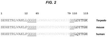
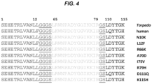
- the present invention provides an isolated peptide comprising the amino acid sequence SEHETRLVAX 1 LFZ 1 LKWNPX 2 DYGGX 3 KKIHZ 2 LX 4 YTGH (SEQ ID NO:31), wherein:
- the peptide binds to an antibody that binds to the main immunogenic region (MIR) of an acetylcholine receptor (AChR).
- the peptide further comprises an intein-chitin binding domain tag.
- the peptide further comprises a hexahistidine tag.
- the peptide further comprises an antibody heavy chain fragment that is conjugated to the peptide.
- the antibody is human immunoglobulin G.
- the hinge region of the heavy chain fragment is conjugated to the C-terminal end of the peptide.
- the present invention provides a composition comprising a peptide of the present invention or a plurality thereof.
- the composition further comprises a pharmaceutically acceptable carrier.
- the plurality of peptides comprises at least 2, 3, 4, or 5 different peptides.
- the present invention provides a kit comprising a peptide of the present invention or a plurality thereof and a solid support.
- the present invention provides an isolated nucleic acid encoding an isolated peptide of the present invention.
- the solid support is a multiwell plate, an ELISA plate, a microarray, a chip, a bead, a porous strip, or a nitrocellulose filter.
- the bead comprises chitin.
- the peptide or plurality thereof is immobilized on the solid support.
- the plurality of peptides binds to the same antibody or different antibodies that bind to the main immunogenic region (MIR) of an acetylcholine receptor (AChR).
- the kit further comprises instructions for use.
- the invention provides methods for detecting or determining the severity of myasthenia gravis (MG) in a subject, the method comprising detecting in a biological sample from the subject the presence or absence of antibodies that bind to a peptide of the present invention or a plurality thereof, wherein the presence of antibodies that bind to the peptide or plurality thereof indicates the presence or an increased severity of MG based on one or more measurable values selected from the group consisting of blood oxygen levels, heart rate, blood pressure, the results of laboratory tests, radiographic or other imaging results, and electromyelogram readings.
- MG myasthenia gravis
- the antibodies bind to the main immunogenic region (MIR) of an acetylcholine receptor (AChR).
- MIR main immunogenic region
- AChR acetylcholine receptor
- the sample is whole blood, serum, or plasma.
- the peptide or plurality thereof is attached to a solid support.
- the solid support is a multiwell plate, an ELISA plate, a microarray, a chip, a bead, a porous strip, or a nitrocellulose filter.
- the bead comprises chitin.
- the antibodies are detected by Western blot, dot blot, ELISA, radioimmunoassay, immunoprecipitation, electrochemiluminescence, immunofluorescence, FACS analysis, or multiplex bead assay.
- the sample is compared to a control.
- the control is obtained from a subject who does not have MG. In other instances, the control is obtained from the subject before developing symptoms of MG or after receiving treatment for MG.
- the invention provides a peptide of the present invention or a plurality thereof for use in a method of preventing or treating myasthenia gravis (MG) in a subject, the method comprising administering to the subject a therapeutically effective amount of the peptide or plurality thereof, wherein the peptide or plurality thereof binds to antibodies circulating in the subject to form neutralizing complexes, thereby preventing or treating MG.
- MG myasthenia gravis
- the circulating antibodies bind to the main immunogenic region (MIR) of an acetylcholine receptor (AChR).
- the peptide or plurality thereof inactivates or reduces the number of memory B-cells that produce antibodies that bind to the MIR of an AChR.
- the subject is exhibiting symptoms of MG. In some instances, treating the subject results in a decrease in the symptoms of MG. In still other embodiments, the method further comprises removing the neutralizing complexes from the subject. In some instances, the neutralizing complexes are removed by affinity plasmapheresis. In other embodiments, the peptide or plurality thereof is administered intravenously, intramuscularly, or a combination thereof.
- Nicotinic acetylcholine receptors are expressed in high densities at the crests of the folds of postsynaptic membranes of neuromuscular junctions.
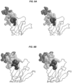
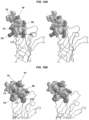
- the AChR is a 290 kDa integral membrane protein with an extracellular domain (ECD) that binds acetylcholine.
- ECD extracellular domain
- MG Myasthenia gravis
- NMJs neuromuscular junctions
- the target of the antibodies is the AChR.
- the binding of anti-AChR antibodies to the AChR initiates the immune system destruction of the postsynaptic membrane folded architecture and concomitant loss of receptor density.
- the present invention is based, in part, on the design of peptides that can bind to pathogenic antibodies that bind to the main immunogenic region (MIR) of an AChR.
- MIR main immunogenic region
- the peptides, conjugates comprising the peptides and antibody fragments, and methods of use thereof are useful for detecting or determining the severity of MG, as well as preventing and treating MG.
- the positions of amino acid modifications refer to positions within the amino acid sequence of the nicotinic AChR alpha subunit.
- the sequence for Torpedo californica nicotinic AChR alpha subunit amino acids 1-161 of the extracellular domain is provided in SEQ ID NO:28.
- a "Q111" modification means that the amino acid corresponding to the 111 th amino acid in a nicotinic AChR alpha subunit, which is normally Q in the reference sequence ( e.g., human sequence) has been modified.
- the modification can comprise a substitution to any amino acid other than Q, an insertion, or a deletion.
- a "Q111D" modification means that the amino acid corresponding to the 111 th amino acid in a nicotinic AChR alpha subunit, which is normally Q in the reference sequence ( e.g., human sequence) has been substituted with the amino acid D.
- the terms “about” and “approximately” shall generally mean an acceptable degree of error for the quantity measured given the nature or precision of the measurements. Typical, exemplary degrees of error are within 20 percent (%), preferably within 10%, and more preferably within 5% of a given value or range of values. Alternatively, and particularly in biological systems, the terms “about” and “approximately” may mean values that are within an order of magnitude, preferably within 5-fold and more preferably within 2-fold of a given value. Numerical quantities given herein are approximate unless stated otherwise, meaning that the term “about” or “approximately” can be inferred when not expressly stated.
- subject means a vertebrate, preferably a mammal, more preferably a human.
- Mammals include, but are not limited to, murines, rats, simians, humans, farm animals, sport animals, and pets.
- Tissues, cells and their progeny of a biological entity obtained in vivo or cultured in vitro are also encompassed.
- severity when used in the context of disease severity (e.g., the severity of myasthenia gravis), can refer to, as a non-limiting example, one or more signs or symptoms that are observable on physical exam.
- severity can refer to a qualitative or quantitative measurement of muscle weakness. Severity can also refer to how many muscles or body systems are exhibiting muscle weakness.
- severity can also refer to other sequelae resulting from the disease. For example, the development of shortness of breath can be construed as an increase in severity compared to before shortness of breath developed. As another non-limiting example, severity can reflect one or more measurable values.
- blood oxygen levels, heart rate, blood pressure, the results of laboratory tests (e.g., the presence of antibodies or other biomarkers in a sample), radiographic or other imaging results, and electromyelogram readings can all be used as indicators of disease severity.
- severity may be determined by, or be related to, how quickly signs, symptoms, or other observable or measurable phenomena change, and/or whether the changes are permanent or transient. Methods and criteria for determining disease severity will be known to one of skill in the art.
- administering includes oral administration, topical contact, administration as a suppository, intravenous, intraperitoneal, intramuscular, intralesional, intrathecal, intranasal, or subcutaneous administration to a subject. Administration is by any route, including parenteral and transmucosal (e.g., buccal, sublingual, palatal, gingival, nasal, vaginal, rectal, or transdermal). Parenteral administration includes, e.g., intravenous, intramuscular, intra-arteriole, intradermal, subcutaneous, intraperitoneal, intraventricular, and intracranial. Other modes of delivery include, but are not limited to, the use of liposomal formulations, intravenous infusion, transdermal patches, etc.
- treating refers to an approach for obtaining beneficial or desired results including, but not limited to, a therapeutic benefit and/or a prophylactic benefit.
- therapeutic benefit is meant any therapeutically relevant improvement in or effect on one or more diseases, conditions, or symptoms under treatment.
- Therapeutic benefit can also mean to effect a cure of one or more diseases, conditions, or symptoms under treatment.
- the compositions may be administered to a subject at risk of developing a particular disease, condition, or symptom, or to a subject reporting one or more of the physiological symptoms of a disease, even though the disease, condition, or symptom may not have yet been manifested.
- the term "effective amount” or “sufficient amount” refers to the amount of a peptide, nucleic acid, or composition that is sufficient to effect beneficial or desired results.
- the therapeutically effective amount may vary depending upon one or more of: the subject and disease condition being treated, the weight and age of the subject, the severity of the disease condition, the manner of administration and the like, which can readily be determined by one of ordinary skill in the art.
- the specific amount may vary depending on one or more of: the particular agent chosen, the target cell type, the location of the target cell in the subject, the dosing regimen to be followed, whether it is administered in combination with other compounds, timing of administration, and the physical delivery system in which it is carried.
- an effective amount is determined by such considerations as may be known in the art.
- the amount must be effective to achieve the desired therapeutic effect in a subject suffering from myasthenia gravis.
- the desired therapeutic effect may include, for example, amelioration of undesired symptoms associated with myasthenia gravis, prevention of the manifestation of such symptoms before they occur, slowing down the progression of symptoms associated with myasthenia gravis, slowing down the deterioration of muscle function associated with myasthenia gravis, slowing down or limiting any irreversible damage caused by myasthenia gravis, lessening the severity of or curing myasthenia gravis, or improving the survival rate or providing more rapid recovery from myasthenia gravis.
- the amount may also be effective to prevent the development of myasthenia gravis.
- the effective amount depends, inter alia, on the type and severity of the disease to be treated and the treatment regime.
- the effective amount is typically determined in appropriately designed clinical trials (dose range studies) and the person versed in the art will know how to properly conduct such trials in order to determine the effective amount.
- an effective amount depends on a variety of factors including the affinity of the molecule (e.g., peptide) for its target, its distribution profile within the body, the relationship between a variety of pharmacological parameters (e.g., half-life in the body) and undesired side effects, and other factors such as age and gender, etc.
- pharmaceutically acceptable carrier refers to a substance that aids the administration of an active agent to a cell, an organism, or a subject.
- “Pharmaceutically acceptable carrier” refers to a carrier or excipient that can be included in the compositions of the invention and that causes no significant adverse toxicological effect on the patient.
- Non-limiting examples of pharmaceutically acceptable carrier include water, NaCl, normal saline solutions, lactated Ringer's, normal sucrose, normal glucose, binders, fillers, disintegrants, lubricants, coatings, sweeteners, flavors and colors, liposomes, dispersion media, microcapsules, cationic lipid carriers, isotonic and absorption delaying agents, and the like.
- the carrier may also be substances for providing the formulation with stability, sterility and isotonicity (e.g. antimicrobial preservatives, antioxidants, chelating agents and buffers), for preventing the action of microorganisms (e.g. antimicrobial and antifungal agents, such as parabens, chlorobutanol, phenol, sorbic acid and the like) or for providing the formulation with an edible flavor etc.
- the carrier is an agent that facilitates the delivery of a peptide to a target cell or tissue.
- sterility and isotonicity e.g. antimicrobial preservatives, antioxidants, chelating agents and buffers
- microorganisms e.g. antimicrobial and antifungal agents, such as parabens, chlorobutanol, phenol, sorbic acid and the like
- the carrier is an agent that facilitates the delivery of a peptide to a target cell or tissue.
- MG myasthenia gravis
- AChR nicotinic acetylcholine receptors
- MG Middle myeloma
- Common early symptoms include diplopia (i.e., double vision) and ptosis (i.e., difficulty in raising the eyelids).
- Common early symptoms also include difficulty chewing, swallowing, and choking, due to the development of weakness in the bulbar muscles. Weakness can subsequently progress to other facial muscles, neck, arms, legs, and torso, causing symptoms such as difficulty lifting objects, walking, holding one's head up, and speaking.
- muscle weakness can progress to the point where the patient has difficulty breathing, and in some instances will develop respiratory In some cases MG will result in death.
- Diagnosing MG can include obtaining a detailed medical history, performing a careful physical exam, obtaining an electromyelogram to assess the response of specific muscles to electrical stimulation, laboratory tests (e. g ., blood tests), and CT imaging.
- Torpedo refers to a genus of rays, also commonly called “electric rays,” “torpedo rays,” or “torpedoes”.
- AChRs have been extensively studied, for example, in Torpedo californica.
- Other species of Torpedo include but are not limited to Torpedo adenensis, Torpedo alexandrinsis, Torpedo andersoni, Torpedo bauchotae, Torpedo fuscomaculata, Torpedo mackayana, Torpedo marmorata, Torpedo microdiscus, Torpedo panthera, Torpedo semipelagica, Torpedo sinuspersici, Torpedo suessii, and Torpedo torpedo.
- acetylcholine receptor refers an integral membrane protein that responds to the binding of the neurotransmitter acetylcholine.
- AChRs are broadly grouped into two groups: muscarinic and nicotinic AChRs. Muscarinic AChRs are G-protein coupled receptors that activate ion channels via second messenger signaling. Muscarinic AChRs function as the main end-receptor stimulated by acetylcholine that is released from postganglionic fibers of the parasympathetic nervous system.
- Muscarinic AChRs also function in the recovery of postganglionic neurons (hyperpolarization and slow depolarization, as opposed to fast depolarization which is facilitated by nicotinic AChRs), and are found in innervated tissue (e.g., muscarinic AChRs are found in sweat glands) and are distributed in both pre-synaptic and post-synaptic locations throughout the central nervous system.
- Muscarinic AChRs comprise five types (M 1 , M 2 , M 3 , M 4 , and M 5 ), although other classification systems are used, depending on the particular context.
- Nicotinic AChRs are Cys-loop proteins that are ligand-gated ion channels which respond to drugs, including nicotine, in addition to responding to acetylcholine. Nicotinic AChRs play at least two important roles in the peripheral nervous system. The first role is that nicotinic AChRs pass signals from presynaptic cells to postsynaptic cells of the sympathetic and parasympathetic nervous systems. The second significant peripheral nervous system role is that nicotinic AChRs are found on skeletal muscle and receive signals required for muscle contractions.
- Nicotinic AChRs are non-selectively permeable to cations, allowing sodium, potassium, and calcium to pass through an open channel, depending on the particular combination of subunits in a given channel. Typically, upon channel opening sodium will pass into a cell and potassium will leave the cell, with a net inward flux of these cations. The passage of calcium through nicotinic AChRs can lead to the release of other neurotransmitters and the activation of other processes via second messenger signaling (e.g., gene regulatory processes).
- second messenger signaling e.g., gene regulatory processes
- Nicotinic AChRs have a molecular mass of about 290 kDa and comprise five subunits that are arranged symmetrically around a central pore ( e.g., see the Torpedo example illustrated in FIG. 12A ).
- nicotinic AChRs are classified as either muscle-type or neuronal-type receptors.
- Neuronal type receptors can comprise various homomeric and heteromeric combinations of twelve different subunits: ⁇ 2, ⁇ 3, ⁇ 4, ⁇ 5, ⁇ 6, ⁇ 7, ⁇ 8, ⁇ 9, ⁇ 10, ⁇ 2 ⁇ 3, and ⁇ 4.
- the embryonic form of the receptor comprises ⁇ , ⁇ , ⁇ , and ⁇ subunits, which are present in a 2:1:1:1 ratio, while the adult form comprises ⁇ 1, ⁇ 1, ⁇ , and ⁇ subunits, which are present in a 2:1:1:1 ratio.
- main immunogenic region refers to a region of the extracellular domain of the nicotinic AChR alpha subunit that commonly serves as the binding site for antibodies that target the nicotinic AChR.
- the MIR typically comprises three non-contiguous segments of the alpha subunit that are arranged in close physical proximity following protein folding.
- the MIR can comprise amino acids 1-12, 65-79, and 110-115 of the nicotinic AChR alpha subunit.
- polypeptide peptide
- protein protein
- amino acid polymers in which one or more amino acid residue is an artificial chemical mimic of a corresponding naturally occurring amino acid, as well as to naturally occurring amino acid polymers and non-naturally occurring amino acid polymer.
- amino acid includes naturally-occurring ⁇ -amino acids and their stereoisomers, as well as unnatural (non-naturally occurring) amino acids and their stereoisomers.
- “Stereoisomers” of amino acids refers to mirror image isomers of the amino acids, such as L-amino acids or D-amino acids.
- a stereoisomer of a naturally-occurring amino acid refers to the mirror image isomer of the naturally-occurring amino acid, i.e., the D-amino acid.
- Naturally-occurring amino acids are those encoded by the genetic code, as well as those amino acids that are later modified, e.g., hydroxyproline, ⁇ -carboxyglutamate and O- phosphoserine.
- Naturally-occurring ⁇ -amino acids include, without limitation, alanine (Ala), cysteine (Cys), aspartic acid (Asp), glutamic acid (Glu), phenylalanine (Phe), glycine (Gly), histidine (His), isoleucine (Ile), arginine (Arg), lysine (Lys), leucine (Leu), methionine (Met), asparagine (Asn), proline (Pro), glutamine (Gln), serine (Ser), threonine (Thr), valine (Val), tryptophan (Trp), tyrosine (Tyr), and combinations thereof.
- Stereoisomers of a naturally-occurring ⁇ -amino acids include, without limitation, D-alanine (D-Ala), D-cysteine (D-Cys), D-aspartic acid (D-Asp), D-glutamic acid (D-Glu), D-phenylalanine (D-Phe), D-histidine (D-His), D-isoleucine (D-Ile), D-arginine (D-Arg), D-lysine (D-Lys), D-leucine (D-Leu), D-methionine (D-Met), D-asparagine (D-Asn), D-proline (D-Pro), D-glutamine (D-Gln), D-serine (D-Ser), D-threonine (D-Thr), D-valine (D-Val), D-tryptophan (D-Trp), D-tyrosine (D-Tyr), and combinations thereof.
- Unnatural (non-naturally occurring) amino acids include, without limitation, amino acid analogs, amino acid mimetics, synthetic amino acids, N-substituted glycines, and N- methyl amino acids in either the L- or D-configuration that function in a manner similar to the naturally-occurring amino acids.
- amino acid analogs are unnatural amino acids that have the same basic chemical structure as naturally-occurring amino acids, i.e., an ⁇ carbon that is bound to a hydrogen, a carboxyl group, an amino group, but have modified R ( i.e., side-chain) groups or modified peptide backbones, e.g., homoserine, norleucine, methionine sulfoxide, methionine methyl sulfonium.
- amino acid mimetics refer to chemical compounds that have a structure that is different from the general chemical structure of an amino acid, but that functions in a manner similar to a naturally-occurring amino acid.
- Amino acids may be referred to herein by either their commonly known three letter symbols or by the one-letter symbols recommended by the IUPAC-IUB Biochemical Nomenclature Commission.
- an L-amino acid may be represented herein by its commonly known three letter symbol (e.g., Arg for L-arginine) or by an upper-case one-letter amino acid symbol (e.g., R for L-arginine).
- a D-amino acid may be represented herein by its commonly known three letter symbol (e.g., D-Arg for D-arginine) or by a lower-case one-letter amino acid symbol (e.g., r for D-arginine).
- amino acid sequences With respect to amino acid sequences, one of skill in the art will recognize that individual substitutions, additions, or deletions to a peptide, polypeptide, or protein sequence which alters, adds, or deletes a single amino acid or a small percentage of amino acids in the encoded sequence is a "conservatively modified variant" where the alteration results in the substitution of an amino acid with a chemically similar amino acid.
- the chemically similar amino acid includes, without limitation, a naturally-occurring amino acid such as an L-amino acid, a stereoisomer of a naturally occurring amino acid such as a D-amino acid, and an unnatural amino acid such as an amino acid analog, amino acid mimetic, synthetic amino acid, N -substituted glycine, and N -methyl amino acid.
- a naturally-occurring amino acid such as an L-amino acid
- a stereoisomer of a naturally occurring amino acid such as a D-amino acid
- an unnatural amino acid such as an amino acid analog, amino acid mimetic, synthetic amino acid, N -substituted glycine, and N -methyl amino acid.
- amino acids e.g., G, A, I, L, or V
- an aliphatic polar-uncharged group such as C, S, T, M, N, or Q
- basic residues e.g., K, R, or H
- An amino acid with an acidic side chain e.g., E or D
- may be substituted with its uncharged counterpart e.g., Q or N, respectively; or vice versa.
- Each of the following eight groups contains other exemplary amino acids that are conservative substitutions for one another:
- amino acid modification refers to a substitution, a deletion, or an insertion of one or more amino acids.
- nucleic acid refers to deoxyribonucleic acids (DNA) or ribonucleic acids (RNA) and polymers thereof in either single-, double- or multi-stranded form.
- the term includes, but is not limited to, single-, double- or multi-stranded DNA or RNA, genomic DNA, cDNA, DNA-RNA hybrids, or a polymer comprising purine and/or pyrimidine bases or other natural, chemically modified, biochemically modified, non-natural, synthetic or derivatized nucleotide bases.
- a particular nucleic acid sequence also implicitly encompasses conservatively modified variants thereof (e.g., degenerate codon substitutions), orthologs, and complementary sequences as well as the sequence explicitly indicated.
- degenerate codon substitutions may be achieved by generating sequences in which the third position of one or more selected (or all) codons is substituted with mixed-base and/or deoxyinosine residues ( Batzer et al., Nucleic Acid Res.
- nucleic acid is used interchangeably with gene, cDNA, and mRNA encoded by a gene.
- nucleotide sequence encoding a peptide or "gene” means the segment of DNA involved in producing a peptide chain, it includes regions preceding and following the coding region (leader and trailer) involved in the transcription/translation of the gene product and the regulation of the transcription/translation, as well as intervening sequences (introns) between individual coding segments (exons).
- neutralizing complex refers to a complex comprising an antibody bound to a peptide that prevents/inhibits/blocks the antibody from binding to its antigen (e.g., the MIR of an AChR).
- affinity plasmapheresis refers to an extracorporeal blood purification procedure for the removal of deleterious agents (e.g., disease-causing agents) from the plasma of a subject.
- Percentage of sequence identity is determined by comparing two optimally aligned sequences over a comparison window, wherein the portion of the sequence (e.g., a peptide of the invention) in the comparison window may comprise additions or deletions ( i.e., gaps) as compared to the reference sequence which does not comprise additions or deletions, for optimal alignment of the two sequences. The percentage is calculated by determining the number of positions at which the identical amino acid residue occurs in both sequences to yield the number of matched positions, dividing the number of matched positions by the total number of positions in the window of comparison and multiplying the result by 100 to yield the percentage of sequence identity.
- percent identity in the context of describing two or more polynucleotide or amino acid sequences, refers to two or more sequences or subsequences that are the same or have a specified percentage of amino acid residues or nucleotides that are the same (for example, a variant of a peptide of interest).
- a peptide or polynucleotide has at least about 80% sequence identity, preferably at least about 85%, 86%, 87%, 88%, 89%, 90%, 91%, 92%, 93, 94%, 95%, 96%, 97%, 98%, 99%, or 100% identity, to a reference sequence, when compared and aligned for maximum correspondence over a comparison window, or designated region as measured using one of the following sequence comparison algorithms or by manual alignment and visual inspection, such sequences are then said to be "substantially identical.”
- this definition also refers to the complement of a test sequence.
- the identity exists over a region that is at least about 6, 7, or 8 amino acids in length, or more preferably over a region that is at least 6-25 or at least 6-12 amino acids in length.
- sequence comparison typically one sequence acts as a reference sequence, to which test sequences are compared.
- test and reference sequences are entered into a computer, subsequence coordinates are designated, if necessary, and sequence algorithm program parameters are designated. Default program parameters can be used, or alternative parameters can be designated.
- sequence comparison algorithm then calculates the percent sequence identities for the test sequences relative to the reference sequence, based on the program parameters. For sequence comparison of nucleic acids and proteins, the BLAST and BLAST 2.0 algorithms and the default parameters discussed below are used.
- Optimal alignment of sequences for comparison can be conducted, e.g., by the local homology algorithm of Smith & Waterman, Adv. Appl. Math. 2:482 (1981 ), by the homology alignment algorithm of Needleman & Wunsch, J. Mol. Biol. 48:443 (1970 ), by the search for similarity method of Pearson & Lipman, Proc. Nat'l. Acad. Sci.
- BLAST and BLAST 2.0 algorithms are described in Altschul et al., (1990) J. Mol. Biol. 215: 403-410 and Altschul et al. (1977) Nucleic Acids Res. 25: 3389-3402 , respectively.
- Software for performing BLAST analyses is publicly available at the National Center for Biotechnology Information website, ncbi.nlm.nih.gov.
- the algorithm involves first identifying high scoring sequence pairs (HSPs) by identifying short words of length W in the query sequence, which either match or satisfy some positive-valued threshold score T when aligned with a word of the same length in a database sequence.
- HSPs high scoring sequence pairs
- T is referred to as the neighborhood word score threshold (Altschul et al., supra ) .
- These initial neighborhood word hits acts as seeds for initiating searches to find longer HSPs containing them.
- the word hits are then extended in both directions along each sequence for as far as the cumulative alignment score can be increased.
- Cumulative scores are calculated using, for nucleotide sequences, the parameters M (reward score for a pair of matching residues; always >0) and N (penalty score for mismatching residues; always ⁇ 0). For amino acid sequences, a scoring matrix is used to calculate the cumulative score.
- Extension of the word hits in each direction are halted when: the cumulative alignment score falls off by the quantity X from its maximum achieved value; the cumulative score goes to zero or below, due to the accumulation of one or more negative-scoring residue alignments; or the end of either sequence is reached.
- the BLAST algorithm parameters W, T, and X determine the sensitivity and speed of the alignment.
- the BLASTP program uses as defaults a word size (W) of 3, an expectation (E) of 10, and the BLOSUM62 scoring matrix (see, e.g., Henikoff and Henikoff, Proc. Natl. Acad. Sci. USA 89: 10915 (1989 )).
- the BLAST algorithm also performs a statistical analysis of the similarity between two sequences (see, e.g., Karlin and Altschul, Proc. Nat'l. Acad. Sci. USA, 90:5873-5787 (1993 )).
- One measure of similarity provided by the BLAST algorithm is the smallest sum probability (P(N)), which provides an indication of the probability by which a match between two nucleotide or amino acid sequences would occur by chance.
- P(N) the smallest sum probability
- a nucleic acid is considered similar to a reference sequence if the smallest sum probability in a comparison of the test nucleic acid to the reference nucleic acid is less than about 0.2, more preferably less than about 0.01, and most preferably less than about 0.001.
- nucleic acid sequences or peptides are substantially identical is that the peptide encoded by the first nucleic acid is immunologically cross reactive with the antibodies raised against the peptide encoded by the second nucleic acid.
- a peptide is typically substantially identical to a second peptide, for example, where the two peptides differ only by conservative substitutions.
- Another indication that two nucleic acid sequences are substantially identical is that the two molecules or their complements hybridize to each other under stringent conditions, as described below.
- Yet another indication that two nucleic acid sequences are substantially identical is that the same primers can be used to amplify the sequence.
- Described herein is an isolated peptide comprising the amino acid sequence SEHETRLVAX 1 LFZ 1 LKWNPX 2 DYGGX 3 KKIHZ 2 LX 4 YTGH (SEQ ID NO:31).
- the peptide further comprises amino acid modifications relative to the amino acid sequence set forth in SEQ ID NO:1 at two or more positions selected from the group consisting of X 1 , X 2 , X 3 , and X 4 , wherein X 1 , X 2 , X 3 , and X 4 are independently selected amino acids and Z 1 and Z 2 are linker sequences of independent lengths comprising independently selected amino acids.
- amino acid modifications can be made with respect to the amino acid sequence set forth in SEQ ID NO:1 at X 1 and X 2 , X 1 and X 3 , X 1 and X 4 , X 2 and X 3 , X 2 and X 4 , X 3 and X 4 , X 1 , X 2 , and X 3 , X 1 , X 2 , and X 4 , X 1 , X 3 , and X 4 , X 2 , X 3 , and X 4 , or X 1 , X 2 , X 3 , and X 4 .
- the linkers Z 1 and Z 2 comprise independently selected amino acids.
- the amino acids can be any amino acid.
- Z 1 and Z 2 comprise the amino acids G and S.
- Z 1 comprises the amino acid sequence GGGS.
- Z 2 comprises the amino acid sequence GS.
- Z 1 and Z 2 are of the same length.
- Z 1 and Z 2 are of different lengths.
- the length of each linker can be any length.
- the linkers are between about 1 and 10 ( i.e., about 1, 2, 3, 4, 5, 6, 7, 8, 9, or 10) amino acids long.
- the linkers are between about 10 and 30 ( i.e., about 10, 11, 12, 13, 14, 15, 16, 17, 18, 19, 20, 21, 22, 23, 24, 25, 26, 27, 28, 29, or 30) amino acids long. In still other aspects, the linkers are between about 30 and 100 ( i.e., about 30, 35, 40, 45, 50, 55, 60, 65, 70, 75, 80, 85, 90, or 100) amino acids long.
- the peptide comprises amino acid modifications at two or more positions selected from the group consisting of K10, D22, V27, and Q35 relative to the amino acid sequence set forth in SEQ ID NO:8, where K10, D22, V27, and Q35 denote positions within the peptide.
- the amino acid modifications are at X 1 and X 2 relative to the amino acid sequence set forth in SEQ ID NO:1.
- X 1 is N
- X 2 is A
- the length of Z 1 is 4, and the length of Z 2 is 2 (SEQ ID NO:2).
- the amino acid modifications are at positions K10 and D22 relative to the amino acid sequence set forth in SEQ ID NO:8, where K10 and D22 denote positions within the peptide.
- the peptide comprises the amino acid sequence set forth in SEQ ID NO:23.
- the amino acid modifications are at X 1 and X 3 relative to the amino acid sequence set forth in SEQ ID NO: 1.
- X 1 is N
- X 3 is I
- the length of Z 1 is 4
- the length of Z 2 is 2 (SEQ ID NO:3).
- the amino acid modifications are at positions K10 and V27 relative to the amino acid sequence set forth in SEQ ID NO:8, where K10 and V27 denote positions within the peptide.
- the peptide comprises the amino acid sequence set forth in SEQ ID NO:24.
- the amino acid modifications are at X 1 and X 4 relative to the amino acid sequence set forth in SEQ ID NO:1.
- X 1 is N
- X 4 is D
- the length of Z 1 is 4
- the length of Z 2 is 2 (SEQ ID NO:4).
- the amino acid modifications are at positions K10 and Q35 relative to the amino acid sequence set forth in SEQ ID NO:8, where K10 and Q35 denote positions within the peptide.
- the peptide comprises the amino acid sequence set forth in SEQ ID NO:25.
- the amino acid modifications are at X 3 and X 4 relative to the amino acid sequence set forth in SEQ ID NO:1. In other aspects, the amino acid modifications are at positions V27 and Q35 relative to the amino acid sequence set forth in SEQ ID NO:8, where V27 and Q35 denote positions within the peptide. In some aspects, the amino acid sequence of the peptide is not SEQ ID NO: 5.
- the amino acid modifications are at X 1 , X 3 , and X 4 relative to the amino acid sequence set forth in SEQ ID NO:1.
- X 1 is N
- X 3 is I
- X 4 is D
- the length of Z 1 is 4
- the length of Z 2 is 2 (SEQ ID NO:6).
- the amino acid modifications are at positions K10, V27, and Q35 relative to the amino acid sequence set forth in SEQ ID NO:8, where K10, V27, and Q35 denote positions within the peptide.
- the peptide comprises the amino acid sequence set forth in SEQ ID NO:26.
- the amino acid modifications are at X 1 , X 2 , X 3 , and X 4 relative to the amino acid sequence set forth in SEQ ID NO: 1.
- X 1 is N
- X 2 is A
- X 3 is I
- X 4 is D
- the length of Z 1 is 4
- the length of Z 2 is 2 (SEQ ID NO:7).
- the amino acid modifications are at positions K10, D22, V27, and Q35 relative to the amino acid sequence set forth in SEQ ID NO:8, where K10, D22, V27, and Q35 denote positions within the peptide.
- the peptide comprises the amino acid sequence set forth in SEQ ID NO:27.
- the peptide does not consist of the amino acid sequence set forth in SEQ ID NO:29.
- the peptide has at least about 80% sequence identity (e.g ., at least about 80%, 81%, 82%, 83%, 84%, 85%, 86%, 87%, 88%, 89%, 90%, 91%, 92%, 93%, 94%, 95%, 96%, 97%, 98%, or 99% sequence identity) to any one of the amino acid sequences set forth in SEQ ID NOS: 1, 2, 3, 4, 6, and 7.
- the peptide has at least about 90% sequence identity (e.g., at least about 90%, 91%, 92%, 93%, 94%, 95%, 96%, 97%, 98%, or 99% sequence identity) to any one of the amino acid sequences set forth in SEQ ID NOS: 1, 2, 3, 4, 6, and 7.
- the peptide has at least about 95% sequence identity (e.g., at least about 95%, 96%, 97%, 98%, or 99% sequence identity) to any one of the amino acid sequences set forth in SEQ ID NOS: 1, 2, 3, 4, 6, and 7.
- linker sequences e.g., Z 1 and/or Z 2 linker sequences
- the peptide comprises an amino acid sequence comprising or consisting of any one of the amino acid sequences set forth in SEQ ID NOS:1, 2, 3, 4, 6, and 7.
- the peptide e.g., isolated peptide
- the peptide has 100% sequence identity to any one of the amino acid sequences set forth in SEQ ID NOS:1, 2, 3, 4, 6, and 7.
- the peptide e.g., isolated peptide
- linker sequences e.g., Z 1 and/or Z 2 linker sequences
- the peptide has at least about 80% sequence identity (e.g., at least about 80%, 81%, 82%, 83%, 84%, 85%, 86%, 87%, 88%, 89%, 90%, 91%, 92%, 93%, 94%, 95%, 96%, 97%, 98%, or 99% sequence identity) to any one of the amino acid sequences set forth in SEQ ID NOS:23, 24, 25, 26, 27, and 30.
- the peptide has at least about 90% sequence identity (e.g., at least about 90%, 91%, 92%, 93%, 94%, 95%, 96%, 97%, 98%, or 99% sequence identity) to any one of the amino acid sequences set forth in SEQ ID NOS:23, 24, 25, 26, 27, and 30.
- the peptide has at least about 95% sequence identity (e.g., at least about 95%, 96%, 97%, 98%, or 99% sequence identity) to any one of the amino acid sequences set forth in SEQ ID NOS:23, 24, 25, 26, 27, and 30.
- linker sequences e.g., Z 1 linker sequences such as GGGS and/or Z 2 linker sequences such as GS are not taken into account when determining the percent sequence identity.
- the peptide comprises an amino acid sequence comprising or consisting of any one of the amino acid sequences set forth in SEQ ID NOS:23, 24, 25, 26, 27, and 30.
- the peptide e.g., isolated peptide
- the peptide has 100% sequence identity to any one of the amino acid sequences set forth in SEQ ID NOS:23, 24, 25, 26, 27, and 30.
- the peptide e.g., isolated peptide
- linker sequences e.g., Z 1 linker sequences such as GGGS and/or Z 2 linker sequences such as GS are not taken into account when determining the percent sequence identity.
- the peptide comprises at least about 6, 7, 8, 9, 10, 11, 12, 13, 14, or 15 contiguous amino acids of any one of the amino acid sequence set forth in SEQ ID NOS:1, 2, 3, 4, 6, 7, 23, 24, 25, 26, 27, and 30.
- the peptide comprises an amino acid sequence comprising or consisting of at least about 6, 7, 8, 9, 10, 11, 12, 13, 14, or 15 contiguous amino acids as set forth in any one of SEQ ID NOS:1, 2, 3, 4, 6, 7, 23, 24, 25, 26, 27, and 30.
- the peptide is between about 6 and about 50 ( e.g., about 6, 7, 8, 9, 10, 11, 12, 13, 14, 15, 16, 17, 18, 19, 20, 21, 22, 23, 24, 25, 26, 27, 28, 29, 30, 31, 32, 33, 34, 35, 36, 37, 38, 39, 40, 41, 42, 43, 44, 45, 46, 47, 48, 49, or 50) amino acids in length.
- the peptide is between about 30 and about 40 ( e.g., about 30, 31, 32, 33, 34, 35, 36, 37, 38, 39, or 40) amino acids in length.
- the peptide is about 39 amino acids in length.
- the peptide is at least about 50 amino acids ( e.g., at least about 50, 55, 60, 65, 70, 75, 80, 85, 90, 95, 100, or more) amino acids in length.
- the peptide binds to an antibody that binds to the main immunogenic region (MIR) of an acetylcholine receptor (AChR).
- MIR main immunogenic region
- AChR is a nicotinic AChR.
- the peptide further comprises an intein-chitin biding domain tag.
- the peptide further comprises a polyhistidine tag. In some instances, the polyhistidine tag is a hexahistidine tag.
- the peptide further comprises one or more antibodies or antibody fragments that are conjugated to the peptide.
- the antibodies or fragments thereof can comprise one or more light chains, heavy chains , fragments thereof, or combinations thereof. Any fragment or combination of heavy chain domains or regions can be conjugated to the peptide, including the variable domain (V H ), hinge region, and constant (C H 1, C H 2, and C H 3) domains.
- the hinge region of the antibody fragment is conjugated to the C-terminal end of the peptide.
- the antibody fragment comprises the hinge region, C H 2 domain, and C H 3 domain of the heavy chain.
- the antibody fragment comprises the hinge region and C H 2 and C H 3 domains of a human IgG heavy chain.
- the one or antibody fragments comprise one or more Fc domains, one or more Fab domains, or a combination thereof.
- Antibodies or fragments thereof from any number of animals can be conjugated to peptides of the present invention (e.g., rabbits, goats, birds, mice, rats, reptiles, sharks, primates, humans).
- the one or more antibodies or fragments thereof are human.
- the antibodies or fragments thereof are from human IgA, IgD, IgE, IgG, IgM, or a combination thereof.
- the antibody or fragment thereof is from human IgG.
- the present invention provides an isolated nucleic acid that encodes an isolated peptide of the invention.
- the nucleic acid encodes a peptide that has at least about 80% sequence identity (e.g., at least about 80%, 81%, 82%, 83%, 84%, 85%, 86%, 87%, 88%, 89%, 90%, 91%, 92%, 93%, 94%, 95%, 96%, 97%, 98%, or 99% sequence identity) to any one of the amino acid sequences set forth in SEQ ID NOS: 1, 2, 3, 4, 6, 7, 23, 24, 25, 26, 27, and 30.
- the nucleic acid encodes a peptide that has at least about 90% sequence identity (e.g., at least about 90%, 91%, 92%, 93%, 94%, 95%, 96%, 97%, 98%, or 99% sequence identity) to any one of the amino acid sequences set forth in SEQ ID NOS: 1, 2, 3, 4, 6, 7, 23, 24, 25, 26, 27, and 30.
- the nucleic acid encodes a peptide that has at least about 95% sequence identity (e.g., at least about 95%, 96%, 97%, 98%, or 99% sequence identity) to any one of the amino acid sequences set forth in SEQ ID NOS:23, 24, 25, 26, 27, and 30.
- linker sequences e.g., Z 1 linker sequences such as GGGS and/or Z 2 linker sequences such as GS
- Z 1 linker sequences such as GGGS and/or Z 2 linker sequences such as GS
- Z 2 linker sequences such as GS
- the amino acid sequence of the peptide encoded by the nucleic acid is not SEQ ID NO: 5.
- the amino acid sequence of the peptide encoded by the nucleic acid is not SEQ ID NO:29.
- the nucleic acid encodes a peptide that comprises at least about 6, 7, 8, 9, 10, 11, 12, 13, 14, or 15 contiguous amino acids of any one of the amino acid sequence set forth in SEQ ID NOS:1, 2, 3, 4, 6, 7, 23, 24, 25, 26, 27, and 30. In some aspects, the nucleic acid encodes a peptide that comprises an amino acid sequence comprising or consisting of at least about 6, 7, 8, 9, 10, 11, 12, 13, 14, or 15 contiguous amino acids as set forth in any one of SEQ ID NOS:1, 2, 3, 4, 6, 7, 23, 24, 25, 26, 27, and 30.
- the present disclosure also provides a vector comprising an isolated nucleic acid provided herein, a host cell comprising a vector provided herein, and a peptide encoded by a nucleic acid provided herein.
- the vectors can include the peptide-encoding nucleic acid operably linked to suitable transcriptional and/or translational regulatory elements (e.g., a sequence to control expression) to effect expression in a suitable host cell.
- suitable transcriptional and/or translational regulatory elements e.g., a sequence to control expression
- the regulatory elements may be derived from mammalian, microbial, viral or insect genes, and include, for example, promoters, enhancers, transcription and translation initiation sequences, termination sequences, origins of replication, and sequences encoding leader and transport sequences. Suitable regulatory elements are selected for optimal expression in a desired host cell.
- Useful expression vectors can be constructed by methods known to one of ordinary skill in the art, and are also commercially available. Exemplary recombinant viral vectors include
- the vector can include a strong constitutive or inducible promoter operatively linked to a nucleic acid provided herein.
- Suitable promoters are well known and readily available to one of ordinary skill in the art and include, for example, bacterial, yeast, viral, mammalian, and insect promoters.
- Exemplary expression vectors are vectors compatible with mammalian cells.
- the host cell can include a vector or an isolated nucleic acid as provided herein.
- the host cell may be prokaryotic or eukaryotic, including bacterial, yeast, insect or mammalian cells. In some cases, the host cell can be a plant cell. In some aspects, the host cells are plant, insect or mammalian cells.
- the isolated nucleic acids or vectors e.g., expression vectors, may be introduced into the host cells by methods known to one of ordinary skill in the art, including transformation, transfection and infection. For example, transfection may be accomplished by any known method, such as liposome-mediated transfection, calcium phosphate-mediated transfection, naked DNA transfection, microinjection or electroporation.
- Transformation methods suitable for prokaryotic cells are described, for example, in Cohen et al., Proc. Natl. Acad. Sci. (USA) 69:2110 (1972 ). Transformation of eukaryotic host cells is described, for example, in Sambrook et al., Molecular Cloning: A Laboratory Manual, 3rd edition, Cold Spring Harbor Press, Cold Spring Harbor, N.Y. (2000 ). The host cells containing the isolated nucleic acid or vectors are useful for replicating the vector and expressing the nucleic acid encoding the peptide of interest, or for replicating and expressing the isolated nucleic acid.
- the invention provides compositions comprising a peptide of the present invention or a plurality thereof.
- the compositions include any one of the isolated peptides described herein.
- the compositions include two or more of any of the peptides provided herein.
- the compositions may include 1, 2, 3, 4, 5, 6, 7, 8, 9, 10, or more different peptides.
- the compositions may include peptides comprising amino acid sequences set forth in SEQ ID NOS:2, 3, 4, 6, 7, 23, 24, 25, 26, 27, or a combination thereof.
- the composition includes one or more nucleic acids encoding peptides of the present invention.
- compositions of the present invention further comprise a pharmaceutically acceptable carrier.
- a pharmaceutically acceptable carrier for example, REMINGTON'S PHARMACEUTICAL SCIENCES, 18TH ED., Mack Publishing Co., Easton, PA (1990 )).
- Prevention against microorganism contamination can be achieved through the addition of one or more of various antibacterial and antifungal agents.
- compositions suitable for administration include sterile aqueous solutions or dispersions and sterile powders for the extemporaneous preparation of sterile injectable solutions or dispersions.
- Typical carriers include a solvent or dispersion medium containing, for example, water-buffered aqueous solutions (i.e ., biocompatible buffers), ethanol, polyols such as glycerol, propylene glycol, polyethylene glycol, suitable mixtures thereof, surfactants, or vegetable oils.
- Sterilization can be accomplished by an art-recognized technique, including but not limited to filtration or addition of antibacterial or antifungal agents, for example, paraben, chlorobutanol, phenol, sorbic acid or thimerosal. Further, isotonic agents such as sugars or sodium chloride may be incorporated in the subject compositions.
- antibacterial or antifungal agents for example, paraben, chlorobutanol, phenol, sorbic acid or thimerosal.
- isotonic agents such as sugars or sodium chloride may be incorporated in the subject compositions.
- sterile injectable solutions containing peptide(s) and/or composition(s) of the present invention is accomplished by incorporating the compound(s) in the required amount(s) in the appropriate solvent with various ingredients enumerated above, as required, followed by sterilization (e.g., filter sterilization). To obtain a sterile powder, the above sterile solutions are vacuum-dried or freeze-dried as necessary.
- the peptide(s) and/or composition(s) provided herein are formulated for administration, e.g., oral, nasal, topical, or parental administration in unit dosage form for ease of administration and uniformity of dosage.
- Unit dosage forms refers to physically discrete units suited as unitary dosages for the subjects, e.g., humans or other mammals to be treated, each unit containing a predetermined quantity of active material calculated to produce the desired therapeutic effect in association with the required pharmaceutical carrier.
- more concentrated dosage forms may be prepared, from which the more dilute unit dosage forms may then be produced. The more concentrated dosage forms thus will contain substantially more than, e.g., at least 1, 2, 3, 4, 5, 6, 7, 8, 9, 10, or more times the amount of the peptide(s) and/or composition(s).
- the dosage forms typically include a conventional pharmaceutical carrier or excipient and may additionally include other medicinal agents, carriers, adjuvants, diluents, tissue permeation enhancers, solubilizers, and the like.
- Appropriate excipients can be tailored to the particular dosage form and route of administration by methods well known in the art ( see, e.g., REMINGTON'S PHARMACEUTICAL SCIENCES, supra ) .
- excipients include, but are not limited to, lactose, dextrose, sucrose, sorbitol, mannitol, starches, gum acacia, calcium phosphate, alginates, tragacanth, gelatin, calcium silicate, microcrystalline cellulose, polyvinylpyrrolidone, cellulose, water, saline, syrup, methylcellulose, ethylcellulose, hydroxypropylmethylcellulose, and polyacrylic acids such as Carbopols, e.g., Carbopol 941, Carbopol 980, Carbopol 981, etc.
- Carbopols e.g., Carbopol 941, Carbopol 980, Carbopol 981, etc.
- the dosage forms can additionally include lubricating agents such as talc, magnesium stearate, and mineral oil; wetting agents; emulsifying agents; suspending agents; preserving agents such as methyl-, ethyl-, and propyl-hydroxy-benzoates ( i.e., the parabens); pH adjusting agents such as inorganic and organic acids and bases; sweetening agents; and flavoring agents.
- lubricating agents such as talc, magnesium stearate, and mineral oil
- wetting agents such as talc, magnesium stearate, and mineral oil
- emulsifying agents such as methyl-, ethyl-, and propyl-hydroxy-benzoates ( i.e., the parabens)
- pH adjusting agents such as inorganic and organic acids and bases
- sweetening agents i.e., the parabens
- flavoring agents such as inorganic and organic acids and bases.
- the dosage forms may also comprise biodegradable poly
- the composition for administration may be an oral delivery vehicle such as a capsule, cachet or tablet, each of which contains a predetermined amount of the peptide to provide the correct incremental dose to the patient.
- Oral delivery vehicles may be useful, for example, in avoiding contact between the peptide and the mouth and upper gastrointestinal tract.
- the therapeutically effective dose can be in the form of tablets, capsules, emulsions, suspensions, solutions, syrups, sprays, lozenges, powders, and sustained-release formulations.
- Suitable excipients for oral administration include pharmaceutical grades of mannitol, lactose, starch, magnesium stearate, sodium saccharine, talcum, cellulose, glucose, gelatin, sucrose, magnesium carbonate, and the like.
- the therapeutically effective dose takes the form of a pill, tablet, or capsule, and thus, the dosage form can contain, along with a peptide(s) and/or composition(s) described herein, any of the following: a diluent such as lactose, sucrose, dicalcium phosphate, and the like; a disintegrant such as starch or derivatives thereof; a lubricant such as magnesium stearate and the like; and a binder such a starch, gum acacia, polyvinylpyrrolidone, gelatin, cellulose and derivatives thereof.
- a diluent such as lactose, sucrose, dicalcium phosphate, and the like
- a disintegrant such as starch or derivatives thereof
- a lubricant such as magnesium stearate and the like
- a binder such a starch, gum acacia, polyvinylpyrrolidone, gelatin, cellulose and derivatives thereof.
- a suitable carrier masks the composition, e.g., the peptide, from the mouth and upper gastrointestinal (GI) tract and reduces or prevents local itching/swelling reactions in these regions during administration.
- a carrier may contain one or more lipid, polysaccharide or protein constituents.
- the carrier is a food product.
- the therapeutically effective dose can be in the form of emulsions, lotions, gels, foams, creams, jellies, solutions, suspensions, ointments, and transdermal patches.
- a peptide(s) and/or composition(s) described herein can be delivered as a dry powder or in liquid form via a nebulizer. Aerosol formulations can be placed into pressurized acceptable propellants such as dichlorodifluoromethane.
- the therapeutically effective dose can be in the form of sterile injectable solutions and sterile packaged powders.
- injectable solutions are formulated at a pH of from about 4.5 to about 7.5.
- the therapeutically effective dose can also be provided in a lyophilized form.
- dosage forms may include a buffer, e.g., bicarbonate, for reconstitution prior to administration, or the buffer may be included in the lyophilized dosage form for reconstitution with, e.g., water.
- the lyophilized dosage form may further comprise a suitable vasoconstrictor, e.g., epinephrine.
- the lyophilized dosage form can be provided in a syringe, optionally packaged in combination with the buffer for reconstitution, such that the reconstituted dosage form can be immediately administered to an individual.
- the therapeutically effective dose may further comprise other components, for example, anti-allergy drugs, such as antihistamines, steroids, bronchodilators, leukotriene stabilizers and mast cell stabilizers. Suitable anti-allergy drugs are well known in the art.
- kits comprising a peptide of the present invention or a plurality thereof and a solid support.
- the kits are useful for detecting or determining the severity of myasthenia gravis in a subject.
- the kits are also useful for preventing or treating myasthenia gravis in a subject.
- kits Materials and reagents to carry out these various methods can be provided in kits to facilitate execution of the methods.
- kit includes a combination of articles that facilitates a process, assay, analysis, or manipulation.
- kits comprising the peptides or compositions of the present invention find utility in a wide range of applications including, for example, diagnostics, prognostics, therapy, and the like.
- kits comprise peptides that comprise the amino acid sequences set forth in SEQ ID NOS:2, 3, 4, 6, 7, 23, 24, 25, 26, 27, and combinations thereof.
- the kits comprise at least one peptide (e.g., at least 1, 2, 3, 4, 5, 6, 7, 8, 9, 10, or more peptides).
- the one or plurality of peptides in the kits further comprise an antibody or fragment thereof that is conjugated to the peptide or plurality thereof.
- the plurality of peptides in the kits bind to the same anti-MIR antibody (i.e., an antibody that binds to the MIR of an AChR). In particular embodiments, the plurality of peptides bind to different anti-MIR antibodies.
- the solid support is a multiwell plate, an ELISA plate, a microarray, a chip, a bead, a porous strip, or a nitrocellulose filter.
- the bead comprises chitin.
- the peptide or plurality of peptides can be immobilized on the solid support. The immobilization can be via covalent or non-covalent binding. In some embodiments, the immobilization is through a capture antibody that specifically binds to the one or more peptides. In certain instances, the solid support in the kits are provided prepared with one or more immobilized peptides.
- Kits can contain chemical reagents as well as other components.
- the kits of the present invention can include, without limitation, instructions to the kit user, apparatus and reagents for sample collection and/or purification, apparatus and reagents for product collection and/or purification, sample tubes, holders, trays, racks, dishes, plates, solutions, buffers or other chemical reagents, suitable samples to be used for standardization, normalization, and/or control samples.
- Kits of the present invention can also be packaged for convenient storage and safe shipping, for example, in a box having a lid.
- kits also comprise labeled secondary antibodies used to detect the presence of anti-MIR autoantibodies that bind to one or more peptides.
- the secondary antibodies bind to the constant or "C" regions of different classes or isotypes of immunoglobulins IgM, IgD, IgG, IgA, and IgE.
- a secondary antibody against an IgG constant region is included in the kits, such as, e.g., secondary antibodies against one of the IgG subclasses ( e.g., IgG1, IgG2, IgG3, and IgG4).
- Secondary antibodies can be labeled with any directly or indirectly detectable moiety, including a fluorophore (e.g., fluorescein, phycoerythrin, quantum dot, Luminex bead, fluorescent bead), an enzyme (e.g., peroxidase, alkaline phosphatase), a radioisotope ( e.g ., 3 H, 32 P, 125 I), or a chemiluminescent moiety. Labeling signals can be amplified using a complex of biotin and a biotin binding moiety (e.g., avidin, streptavidin, neutravidin).
- a fluorophore e.g., fluorescein, phycoerythrin, quantum dot, Luminex bead, fluorescent bead
- an enzyme e.g., peroxidase, alkaline phosphatase
- a radioisotope e.g ., 3 H, 32 P, 125
- Fluorescently labeled anti-human IgG antibodies are commercially available from Molecular Probes, Eugene, OR. Enzyme-labeled anti-human IgG antibodies are commercially available from Sigma-Aldrich, St. Louis, MO and Chemicon, Temecula, CA.
- kits may further comprise instructions for contacting the solid support with a biological sample from a subject, and for correlating the presence of antibodies or levels of antibodies above a threshold level with the presence or severity of myasthenia gravis.
- kits also contain negative and positive control samples for detection of antibodies.
- the negative control samples are obtained from individuals or groups of individuals who do not have myasthenia gravis.
- the positive control samples are obtained from individuals or groups of individuals who have myasthenia gravis.
- the kits contain samples for the preparation of a titrated curve of antibodies in a sample, to assist in the evaluation of quantified levels of antibodies in a test biological sample.
- the present invention provides methods for detecting or determining the severity of myasthenia gravis (MG) in a subject, the method comprising detecting in a biological sample from the subject the presence or absence of antibodies that bind to a peptide of the present invention or a plurality thereof, wherein the presence of antibodies that bind to the peptide or plurality thereof indicates the presence or an increased severity of MG based on one or more measurable values selected from the group consisting of blood oxygen levels, heart rate, blood pressure, the results of laboratory tests, radiographic or other imaging results, and electromyelogram readings.
- the plurality of peptides comprises 2 or more ( e.g., 2, 3, 4, 5, 6, 7, 8, 9, 10, or more) different peptides.
- the plurality of peptides comprises amino acid sequences set forth in SEQ ID NOS: 2, 3, 4, 6, 7, 23, 24, 25, 26, 27, or a combination thereof.
- the peptides bind to antibodies that bind to the MIR of an AChR.
- the AChR is a nicotinic AChR.
- each of the different peptides binds to the same anti-MIR antibody, e.g., all of the different peptides bind to the same antibody that binds to the MIR of an AChR. In other instances, each of the different peptides binds to a different anti-MIR antibody.
- the method further comprises obtaining the sample from the subject.
- any fluid sample containing antibodies can be used.
- the biological sample may be whole blood, serum, plasma, urine, saliva, or cerebrospinal fluid (CSF).
- CSF cerebrospinal fluid
- One or more different bodily fluids can be evaluated for antibodies that specifically bind to the one or more peptides.
- the level or titer of the antibodies in the biological sample is compared to a threshold level or titer.
- a level or titer of the antibodies in the biological sample that is greater than the threshold level or titer indicates an increased probability that the subject has MG or has an increased severity of MG.
- a level or titer of the antibodies in the biological sample that is less than the threshold level or titer does not indicate that the subject has MG or has an increased severity of MG.
- the presence of the antibodies in the biological sample can be determined before or after the development of symptoms of MG.
- the presence of the antibodies may be determined once or more than once, as needed or desired.
- the presence or absence of antibodies or the quantified levels of antibodies are evaluated every week, every month, several times a year ( e.g., 2, 3, 4, 5, 6, 7, 8, 9, 10, 11, 12, or more times a year), or more or less often, as appropriate.
- presence of the antibodies is made without comparing the test sample (i.e., a biological sample from the subject) to a control sample.
- the test sample is compared to a control.
- the control can be from the same individual at a different time point.
- the control is obtained from the subject before developing symptoms of MG. In other instances, the control is obtained from the subject after developing symptoms of MG and/or after receiving treatment for MG.
- the control can also be from a different individual with a known status for the presence of the antibodies or symptoms or diagnosis of MG.
- the control can also be a calculated value from a population of individuals with a known status for the presence of antibodies or symptoms or diagnosis of MG.
- the control may be a positive control or a negative control.
- the control is a negative control from another individual or a population of individuals ( e.g., an individual or population of individuals who do not have MG). If the known status of the control sample is negative for the antibodies, then a higher level of antibodies in the test sample than in the negative control sample indicates that the subject has MG or an increased severity of MG. A similar level of antibodies in the test sample compared to the negative control sample indicates that the subject does not have MG or an increased severity of MG.
- control is a positive control from another individual or a population of individuals, or the control reflects a predetermined threshold level of antibodies. If the known status of the control sample is positive for antibodies, then a similar or higher level of antibodies in the test sample than in the positive control sample indicates that the subject has MG or an increased severity of MG. A lower level of antibodies in the test sample compared to the positive control sample indicates that the subject does not have MG or an increased severity of MG.
- an increased level of antibodies in the test sample is determined when the antibody levels are at least, e.g., 10%, 25%, 50%, 1-fold, 2-fold, 3-fold, 4-fold, or more in comparison to a negative or a prior-measured control.
- the antibodies can be detected using any method known in the art. Exemplary methods include, without limitation, Western Blot, dot blot, enzyme-linked immunosorbent assay (ELISA), radioimmunoassay (RIA), immunoprecipitation, immunofluorescence, FACS analysis, electrochemiluminescence, and multiplex bead assays (e.g., using Luminex or fluorescent microbeads).
- the peptide or plurality thereof used to detect antibodies can be immobilized on a solid support.
- the solid support can be, for example, a multiwell plate, a microarray, a chip, a bead, a porous strip, or a nitrocellulose filter.
- the bead comprises chitin.
- the immobilization can be via covalent or non-covalent binding.
- the immobilization is through a capture antibody that specifically binds to the target epitope.
- a sample can be incubated with one or more of the peptides described herein under conditions (e.g., time, temperature, concentration of sample) sufficient to allow specific binding of antibodies to the peptides.
- the one or more peptides can be bound to a solid support.
- the one or more peptides can be exposed to a sample for about 0.5, 1.0, 1.5, 2.0, 2.5, 3.0 hours, or overnight, about 8, 10, 12, 14, or 16 hours.
- incubation time can be more or less depending on, e.g., the composition of the one or more peptides, the composition of the one or more antibodies, the dilution of the sample, and the temperature for incubation.
- Incubations are usually carried out at room temperature (about 25 °C) or at biological temperature (about 37 °C), and can be carried out in a refrigerator (about 4 °C). Washing to remove unbound sample before addition of a secondary antibody is carried according to known immunoassay methods.
- Labeled secondary antibodies are generally used to detect antibodies in a sample that have bound to one or more of the peptides described herein. Secondary antibodies bind to the constant or "C" regions of different classes or isotypes of immunoglobulins IgM, IgD, IgG, IgA, and IgE. Usually, a secondary antibody against an IgG constant region is used in the present methods. Secondary antibodies against the IgG subclasses, for example, IgG1, IgG2, IgG3, and IgG4, also find use in the present methods.
- Secondary antibodies can be labeled with any directly or indirectly detectable moiety, including a fluorophore (e.g., fluorescein, phycoerythrin, quantum dot, Luminex bead, fluorescent bead), an enzyme (e.g., peroxidase, alkaline phosphatase), a radioisotope ( e.g ., 3 H, 32 P, 125 I) or a chemiluminescent moiety. Labeling signals can be amplified using a complex of biotin and a biotin binding moiety (e.g., avidin, streptavidin, neutravidin).
- a fluorophore e.g., fluorescein, phycoerythrin, quantum dot, Luminex bead, fluorescent bead
- an enzyme e.g., peroxidase, alkaline phosphatase
- a radioisotope e.g ., 3 H, 32 P, 125
- Fluorescently labeled anti-human IgG antibodies are commercially available from Molecular Probes, Eugene, OR. Enzyme-labeled anti-human IgG antibodies are commercially available from Sigma-Aldrich, St. Louis, MO and Chemicon, Temecula, CA.
- the method of detection of the presence or absence, or differential presence, of antibodies in a sample will correspond with the choice of label of the secondary antibody.
- the detectable signals i.e ., blots
- the detectable signals can be quantified using a digital imager if enzymatic labeling is used or an x-ray film developer if radioisotope labeling is used.
- the detectable signals can be quantified using an automated plate reader capable of detecting and quantifying fluorescent, chemiluminescent, and/or colorimetric signals. Such methods of detection are well known in the art.
- the presence or increased presence of antibodies is indicated by a detectable signal (e.g., a blot, fluorescence, chemiluminescence, color, radioactivity) in an immunoassay, where the biological sample from the subject is contacted with one or more of the peptides described herein.
- a detectable signal e.g., a blot, fluorescence, chemiluminescence, color, radioactivity
- This detectable signal can be compared to the signal from a control sample or to a threshold value.
- increased presence is detected, and the presence or increased severity of MG is indicated, when the detectable signal of antibodies in the test sample is at least about 10%, 20%, 30%, 50%, 75% greater in comparison to the signal of antibodies in the control sample or the predetermined threshold value.
- an increased presence is detected, and the presence or increased severity of MG is indicated, when the detectable signal of antibodies in the test sample is at least about 1-fold, 2-fold, 3-fold, 4-fold or more, greater in comparison to the signal of antibodies in the control sample or the predetermined threshold value.
- the results of the antibody determinations are recorded in a tangible medium.
- the results of the present diagnostic assays e.g., the observation of the presence or increased presence of antibodies
- the diagnosis of whether or not there is an increased severity or the presence of MG can be recorded, e.g., on paper or on electronic media (e.g ., audio tape, a computer disk, a CD, a flash drive, etc. ) .
- the methods further comprise the step of providing the diagnosis to the patient (i.e., the subject).
- the invention provides a peptide of the invention or a plurality thereof for use in methods for preventing or treating myasthenia gravis (MG) in a subject, the method comprising administering to the subject a therapeutically effective amount of the peptide or plurality thereof, wherein the peptide or plurality thereof binds to antibodies circulating in the subject to form neutralizing complexes, thereby preventing or treating MG.
- the circulating antibodies bind to the main immunogenic region (MIR) of an acetylcholine receptor (AChR).
- the AChR is a nicotinic AChR.
- the peptide or plurality thereof binds to and blocks an antibody combining site.
- the plurality of peptides comprises 2 or more ( e.g., 2, 3, 4, 5, 6, 7, 8, 9, 10, or more) different peptides.
- the plurality of peptides comprises amino acid sequences set forth in SEQ ID NOS: 2, 3, 4, 6, 7, 23, 24, 25, 26, 27, or a combination thereof.
- each of the different peptides binds to the same anti-MIR antibody, e.g., all of the different peptides bind to the same antibody that binds to the MIR of an AChR. In other instances, each of the different peptides binds to a different anti-MIR antibody.
- the peptides or plurality thereof that are administered to the subject further comprise one or more antibodies or fragments thereof that are conjugated to the peptide or plurality thereof.
- the antibodies or fragments thereof can comprise one or more light chains, heavy chains , fragments thereof, or combinations thereof. Any fragment or combination of heavy chain domains or regions can be conjugated to the peptide(s), including the variable domain (V H ), hinge region, and constant (C H 1, C H 2, and C H 3) domains.
- the hinge region of the antibody fragment is conjugated to the C-terminal end of the peptide.
- the antibody fragment comprises the hinge region, C H 2 domain, and C H 3 domain of the heavy chain.
- the one or antibodies or fragments thereof comprise one or more Fc domains, one or more Fab domains, or a combination thereof.
- Conjugating antibody fragments to peptides of the present invention can be useful for extending the bioavailability of the peptides.
- peptide-antibody fragment conjugates are useful for inactivating and/or reducing the number of B-cells that are involved in the production of pathogenic anti-MIR antibodies, e.g., by complement activation, antibody-dependent cell-mediated toxicity, induced apoptosis, or a combination thereof.
- the subject being treated is exhibiting one or more symptoms of MG. In some instances, treating the subject results in a decrease or abolishment of one or more symptoms of MG.
- biological fluid containing antibodies can be removed from the subject and contacted with one or more of the peptides described herein.
- one or more of the peptides described herein can be administered to the subject to block the binding between the antibodies and their AChR targets, thereby neutralizing the antibodies, and the neutralized complexes present in the biological fluid are removed using an extracorporeal therapy, such as affinity plasmapheresis.
- the peptide of the present invention or plurality thereof can be administered to the subject intravenously, intramuscularly, intrathecally, or a combination thereof.
- biological fluid from the subject is contacted with one or more of the peptides immobilized on a solid support.
- the solid support can be, for example, a multiwell plate, an ELISA plate, a microarray, a chip, a bead, a column, a porous strip, a membrane, or a nitrocellulose filter.
- the bead can comprise chitin.
- the immobilization can be via covalent or non-covalent binding.
- the immobilization is through a capture antibody that specifically binds to the target peptide epitope.
- the peptide(s) attached to the solid support form a stationary phase that captures the antibodies in the biological fluid, allowing the biological fluid with reduced or eliminated levels of pathogenic anti-AChR or anti-MIR antibodies to be separated from the solid support, i.e ., as the mobile phase, and returned to the subject.
- the biological fluid that is processed ex vivo is plasma, and the antibodies are removed by plasmapheresis, a process well known in the art.
- the plasma is contacted with a solid support with one or more immobilized peptides.
- Antibodies in the plasma bind to the immobilized peptides. Plasma with reduced or eliminated levels of antibodies is then returned to the subject.
- the ex vivo removal of antibodies can be carried out on a subject before or after developing symptoms or receiving a diagnosis of MG.
- the process of ex vivo antibody removal can be performed one, two, three, four, or more times, as needed to eliminate or reduce the antibodies from the subject.
- Ex vivo removal of the antibodies can be performed daily, weekly, bi-weekly, monthly, bi-monthly, as appropriate.
- the levels of antibodies in the subject are monitored and ex vivo antibody removal performed if the presence of antibodies are above a predetermined threshold level.
- Ex vivo antibody removal can be carried out over a time period of a 1, 2, 3, 4, 5, 10, 12, 15, 20, 30, 40, or 50 weeks, or longer or shorter, as appropriate.
- ex vivo removal of antibodies can be discontinued if the level of antibodies falls below a predetermined threshold level.
- This example describes the creation of peptides that bind to antibodies against that main immunogenic region (MIR) of the nicotinic acetylcholine receptor (AChR) and shows that some of the peptides can bind to pathogenic anti-MIR antibodies in serum samples obtained from human myasthenia gravis (MG) patients.
- MIR main immunogenic region
- AChR nicotinic acetylcholine receptor
- the nicotinic acetylcholine receptor (AChR) main immunogenic region (MIR) is the target of many pathogenic antibodies (Abs) in patients with autoimmune myasthenia gravis (MG). This disease results in the destruction of the postsynaptic membrane folded architecture and concomitant loss of AChR density (1,2). Electron micrographs of single chain Fv and/or Fab fragments of monoclonal Ab (mAb) 35 bound to 2-D crystalline arrays of Torpedo AChRs ( T AChRs) reveal that the MIR is on top of the extracellular region of each of the two AChR alpha subunits (3).
- the MIR consists of a group of overlapping epitopes of mAbs raised in rats against the Torpedo and/or Electrophorus electric organ AChR (4-6). Many of these mAbs cross-react with the AChRs of many different species, including humans (7,8).
- the MIR has stimulated considerable interest, as a substantial fraction of the mAbs raised against the Torpedo or Electrophorus electric organ AChR are directed to this region and mutually inhibit one another's binding (10,11).
- mAbs 132A does bind to the TAChR alpha subunit in Western blots and has consequently proved quite useful in mapping its epitope to three non-contiguous segments of the TAChR alpha subunit: ⁇ (1-12), ⁇ (65-79), and ⁇ (110-115) (9).
- ⁇ (1-12) is part of the N-terminal ⁇ -helix, and it sits next to ⁇ (65-79), the core segment (13) of the MIR.
- ⁇ (65-79) consists of a short helix, ⁇ (69-74), nestled between two coil/ ⁇ -strand legs, ⁇ (65-68) and ⁇ (75-79).
- ⁇ (65-79) is draped over the ⁇ (110-115), a ⁇ -turn (12).
- ⁇ (1-12) and ⁇ (65-79) appear to function as the epitope recognition site, while ⁇ (110-115) appears to serve as a structural anchor stabilizing the ⁇ (1-12)/ ⁇ (65-79) complex (9).
- a Torpedo 39 amino acid (aa) MIR peptide mimic (39MIR) of the Torpedo MIR was synthesized and characterized.
- the mimic consists of three non-contiguous segments of the AChR that are joined by flexible GGGS and GS amino acid linkers, and is fused to the amino terminus of an intein-chitin binding domain (IChBD) (14).
- IChBD intein-chitin binding domain
- This peptide construct was also able to adsorb all of the MIR directed Abs in EAMG sera that block the binding of biotinylated mAb 132A to the Torpedo AChR (14).
- Another fusion protein has been engineered with the 39MIR C-terminus linked to the N-terminus of the hinge of human and rat IgG antibody heavy chains (15). These constructs can also be used therapeutically to neutralize MIR directed Abs in EAMG sera or a human homolog can be used to neutralize MIR-directed Abs in MG sera.
- Mammalian 39MIR homologs have also been produced. Initial attempts at making a mammalian MIR mimic involved putting the rat/mouse and human sequences in the corresponding positions of the Torpedo 39MIR that were recognized by anti- TAChR MIR mAbs and anti -TAChR MIR Abs in EAMG serum.
- the rat/mouse and human 39MIRs incorporate the same three segments of the rat/human alpha subunit as those from the Torpedo alpha subunit: ⁇ (1-12), ⁇ (65-79), and ⁇ (110-115). Like the Torpedo peptide, they are linked by flexible GGGS and GS linkers.
- the peptide homologs were fused to an IChBD tag and affinity purified from disrupted E.coli by adsorption onto chitin beads.
- the human homolog contains eight amino acids that are different from the Torpedo 39MIR sequence while the rat homologue contains only seven ( FIG. 2 ).
- the AChR MIR is evolutionarily conserved between Torpedo and mammals.
- the Torpedo and human alpha subunits have 80% sequence similarity ( 16 ).
- mAb 35 cross-reacts with receptors from Torpedo, bovines, and humans (8,17,18).
- mAb 132A raised against Torpedo electric organ AChR in rats, binds to the Torpedo MIR and stains the neuromuscular junction (NMJ) of mouse and human tissue, and causes disease in naive rats when injected (7,17). Additionally, EAMG rats produce Abs that bind to the rat MIR (17-20).
- the human 39MIR does not bind to mAbs 35 or 132A despite evidence that both mAbs recognize human and rat/mouse AChR (4,7).
- amino acid substitutions were made at positions that differ between the Torpedo and human 39MIRs, and the effect of the substitutions on the binding of anti-MIR mAbs 35, 132A, 198, 334, and 371A was tested.
- Two Torpedo /human chimeric 39MIRs were designed that exhibit binding of mAbs 35, 132A, and mAb 198.
- the chimeric mimics also bind Abs from EAMG serum and human MG sera, and can be used in the production of a pseudo anti-idiotype Ab construct with the human and rat IgG heavy chains that will not only neutralize anti-MIR Abs, but also target the memory B-cells involved in the production of pathogenic anti-MIR Abs.
- the human 39MIR peptide mimic is recognized by mAb 198 but not mAbs 35 and 132A.
- the Torpedo (SEQ ID NO:9) and human (SEQ ID NO:8) 39MIRs were made by joining the ⁇ (1-12), ⁇ (65-79), and ⁇ (110-115) segments of their respective ⁇ subunits with GS linkers. These two MIR mimics were expressed in E.coli as fusion proteins with an IChBD tag to permit peptide collection on chitin beads. These two peptides differ from each other by eight amino acids ( FIG. 2 ). Both mimics were probed with mAb 198.
- mAb 198 which was derived from rats that had been immunized with human muscle AChR, is a MIR-directed mAb that binds mammalian as well as Torpedo MIR (13,21).
- mAb 198 binds to the Torpedo and human MIR peptide mimics in Western blots ( FIG. 3 ). However, mAb 198 binds to the Torpedo mimic about 50 times more strongly than to the human MIR mimic ( FIG. 3 ).
- the human 39MIR is not recognized by mAbs 35 or 132A at all in Western blots.
- both mAbs bind to the assembled Torpedo AChR and to the AChR of a variety of species including humans, (7) and transfer disease to naive rats.
- This result suggests that perhaps the human 39MIR mimic folds slightly differently from the Torpedo 39MIR in the Western blot format, and slightly differently than the human MIR assembled in the AChR.
- the single amino acid substitutions in the Torpedo MIR were: N10K (SEQ ID NO: 11), L12F (SEQ ID NO:12), R66K (SEQ ID NO:13), A70D (SEQ ID NO:14), I75V (SEQ ID NO:15), R79H (SEQ ID NO:16), D111Q (SEQ ID NO:17), and K115H (SEQ ID NO:18). Sequence alignments are shown in FIG. 4 .
- mAbs 35, 132A, and 334 are all anti-electric organ AChR MIR-directed mAbs.
- mAb 198 raised against purified human AChR, bound to mammalian and Torpedo MIR to a similar degree as to MIR mimics (14,22,23).
- mAb 147G a rat anti-fluorescein mAb, served as the negative control and binds to nothing in the blots.
- FIG. 5 Western blots (also see Table 1), it can be seen that mAb 35 did not bind to the I75V, D111Q or K115H peptides. mAb 35 exhibited improved binding to the L12F (1.42x) and R79H (1.91x) peptides and decreased binding to N10K (0.29x) and R66K (0.62x) peptides, relative to binding to the Torpedo 39MIR. Binding to A70D (0.93x) remained relatively unchanged.
- mAb 132A exhibited substantially weakened binding to the N10K (0.03x), A70D (0.20x), I75V (0.07x), D111Q (0.07x) and K115H (0.40x) peptides, and stronger binding to the R66K (1.55x) and R79H (1.65x) peptides, relative to binding of mAb 132A to the Torpedo 39MIR (Table 1). Binding to L12F (1.10x) remained relatively unchanged.
- mAb 198 exhibited decreased binding to the N10K (0.82x), I75V (0.16x) and D111Q (0.16x) peptides, and fairly uniform binding to the other five chimeric point mutated peptides (Table 1).
- mAb 334 exhibited weakened binding to the N10K (0.59x), L12F (0.88x), I75V (0.03x), D111Q (0.02x), and K115H (0.42x) peptides, stronger binding to the R66K (2.75x) and R79H (2.40x) peptides, and unchanged binding to A70D (1.04x) peptides (Table 1).
- mAb 147G the negative control, did not bind to any of the peptides.
- the N10K, I75V and D111Q substitutions resulted in the most consistent impairment of MIR-directed mAb binding.
- the A70D substitution primarily affected the binding of mAb 132A, while not affecting the binding of the other mAbs.
- the K115H substitution also impaired binding of mAb 35, 132A, and mAb 334, with the mAb 35 binding being completely abolished.
- the human 39MIR peptide was point mutated to induce amino acid substitutions corresponding to the analogous Torpedo sequence at four positions, resulting in the K10N, D70A, V75I, and Q111D peptides (SEQ ID NOS:19, 20, 21, and 22, respectively), in an attempt to identify the contribution of these amino acids to mAb binding and to improve the binding of electric organ mAbs 132A and 35 ( FIG. 6 ).
- Three of the single point mutated peptides, (K10N (0.01), V75I (0.03), and Q111D (0.06)) of the human 39MIR restored mAb 35 binding, although to very low levels compared to the Torpedo 39MIR peptide.
- the D70A mutated human 39MIR failed to restore mAb 35 binding to the human 39MIR. None of these mutant peptides restored mAb 132 binding.
- the human 39MIR was originally recognized by mAb 198.
- the K10N (13x), V75I (40x), and Q111D (35.7x) mutations greatly improved mAb 198 binding compared to its binding to the human 39MIR.
- mAb 198 binding to the D70A mutant was comparable to binding to human 39MIR. Taken together, these single point mutations are not responsible for the loss of mAb binding. It is more likely that a combination of all or a few of the mutations are responsible for the loss of mAb binding.
- K10N/D70A SEQ ID NO:23
- K10N/V75I SEQ ID NO:24
- K10N/Q111D SEQ ID NO:25
- V75I/Q111D SEQ ID NO:5
- a K10N//V75I/Q111D mutant (SEQ ID NO:26) was made to test the contribution of the D amino acid at position 70.
- the human 39MIR was not recognized by mAb 35 or 132A
- this triple mutant exhibited improved binding to both mAb 35 (2.97x) and 132A (2.46x) when compared to the Torpedo 39 MIR.
- the triple mutant also bound to mAb 198 (2.52x) better than the Torpedo 39 MIR, resulting in a 28x improvement compared to the human 39MIR.
- this triple mutant was better at binding the mAbs than a mutant peptide containing all four ( i.e., K10N, D70A, V75I, and Q111D) mutations.
- the K10N/D70A/V75I/Q111D mutant restored binding to mAb 35 (1.70x) and mAb 132A (0.99x). It also improved on mAb 198 recognition, improving binding by 11x comparable to the human 39MIR ( FIG. 7 ).
- the K10N//V75I/Q111D triple mutant appeared to be the peptide most optimized for binding to the MIR-directed mAbs 35, 132A, and 198. Results are summarized in Table 2.
- the Torpedo and mammalian MIR structures may be different enough to affect mAb recognition.
- An examination of the Torpedo MIR and mammalian MIR show some striking differences in the orientation and accessibility of critical MIR amino acids.
- the three MIR segments from the Torpedo alpha subunit are more loosely packed than those of from the mouse, and the Torpedo N-terminal helix protrudes more from the cluster than does the mouse N-terminal helix ( FIG. 8 ).
- the core MIR segment was mapped to the AChR ⁇ (67-76) (5,13). This cluster of amino acids is both tightly packed and surface accessible in both the Torpedo and mouse structure. However, their structural arrangement in the MIR is quite different.
- the core MIR is essential for MIR-directed mAb recognition.
- the Torpedo core MIR is much more centrally located in the ECD while the mouse core MIR sits more at the top in the mouse ECD structure ( FIG. 9 ). This difference in the core MIR may account for the lack of mAb 35 and 132A binding to the human 39MIR.
- MIR peptide mimics refold into structures that present epitopes recognized by mAb 198 despite the amino acid differences.
- mAb 35 and mAb 132A did not bind to either the human or rat 39 MIR mimics. Both mAbs bind to mammalian synapses and cause a passive transfer of EAMG in naive rats, so they bind to the mammalian MIR in the native, folded, assembled mammalian AChR (7,17,20).
- the mAbs may have different preferences for residues that differ between the Torpedo and human MIR. These can disrupt important contacts. There is also the possibility that the human MIR mimics adopt an alternative more compact structure that is more stable than the Torpedo MIR mimic and consequently do not present the sub-states that mAbs 35 and 132A recognize.
- Both mAb 35 and mAb 132A bind to TAChR and bind to rat/mouse and human synapses, suggesting the presence of their epitopes at mammalian neuromuscular junctions as well as in the Torpedo electric organ (7).
- the fact that both mAbs bound to the Torpedo but not the human MIR mimics was surprising. There are likely small but important differences in the orientation of certain important residues and the loss of surface exposure of these residues may be important for one mAb and not another.
- the eight amino acid difference between the Torpedo the human MIR was enough to render the mimics unrecognizable by mAbs 35 and 132A ( FIG. 3 ).
- a small number of amino acids determine the mAb epitopes and changes in these critical amino acids can affect mAb binding.
- K10, 175, and D111 appear to be critical for all of the mAbs tested, as their substitution to the homologous human amino acids caused significantly decreased binding of mAbs raised initially against electric organ AChR. Additionally, I75 and D111 mutations also resulted in the complete loss of binding to the mutated mimic by mAb 35.
- the I75V and D111Q mutated peptides had the most dramatic overall effect on MIR-directed mAb binding.
- the human 39MIR was back mutated at each of the most detrimental point mutations. However, none of the single back mutations restored mAb 132A binding, while the K10N, V75I, and Q111D mutations restored mAb 35 binding. The single point mutations improved mAb 198 binding compared to the human 39MIR. While individually the mutations did not greatly restore mAb binding, pairwise mutations started to restore binding to mAb 35 and 132A. The K10N/V75I/Q111D mutant showed improved binding to mAb 35 and 198 compared to the Torpedo 39MIR.
- the K10N/D70A/V75I/Q111D mutant did not improve anti-MIR mAb binding compared to the Torpedo 39MIR.
- the alanine at position 70 reduces mAb binding.
- the positive charge from the aspartic acid appears to be important for mAb binding. While no single substitution restored mAb 132A binding, a combination of two or more substitutions was able to restore partial mAb binding.
- the effect of the V75I mutation on mAb binding was surprising. The mutation was a minor change, going from a hydrophobic amino acid to a slightly larger hydrophobic amino acid.
- the K10N and D70A substitutions both remove a charge from the human 39MIR. Table 1. Summary of Western blot of FIG. 5.
- Numerical values represent fold-change in binding relative to Torpedo 39MIR. '-' denotes no binding. Mutant mAb 35 mAb 132A mAb 198 mAb 334 Torpedo 39MIR 1.00 1.00 1.00 1.00 N10K 0.29 0.03 0.82 0.59 L12F 1.42 1.10 0.90 0.88 R66K 0.62 1.55 1.03 2.75 A70D 0.93 0.20 1.08 1.04 175V - 0.06 0.16 0.03 R79H 1.91 1.65 0.97 2.40 D111Q - 0,07 0.16 0.02 K115H - 0,40 1.07 0.42 Table 2. Summary of Western blot of FIG. 7. Numerical values represent fold-change in binding relative to Torpedo 39MIR.
- This example shows the design and creation of a fusion protein comprising peptides of the present invention and an antibody Fc domain that is suitable for the prevention or treatment of myasthenia gravis (MG).
- MG myasthenia gravis
- a peptide of the present invention has been designed (SEQ ID NO:27) that combines parts of the Torpedo and human MIR peptides (SEQ. ID NOS:8 and 9, respectively). This peptide binds anti-MIR monoclonal antibodies (mAbs) derived from rats and well as a fraction of Abs in MG patient sera.
- a conjugate has been engineered ( FIG. 12B ) that fuses the chimeric Torpedo- human peptide (SEQ ID NO:27) to an antibody Fc domain.
- the Western blot in FIG. 13 shows that while the Torpedo and human-based peptide-Fc conjugates (i.e., conjugates comprising the peptides of SEQ ID NOS; 8 and 9 fused to antibody Fc domains) did not bind the pathogenic mAbs 132A and 198, the chimeric peptide-Fc conjugate (i.e., a conjugate comprising the peptide of SEQ ID NO:27 fused to an antibody Fc domain) did recognize and bind both mAbs.
- the chimeric conjugate can bind and neutralize pathogenic Abs in MG patient sera.
- a peptide-Fc conjugate of the present invention e.g ., a peptide of SEQ ID NO:27 fused to an antibody Fc domain
- the conjugate can also target the B-cell receptors on memory B-cells that are responsible for the production of the pathogenic anti-MIR Abs in MG ( FIG. 14B ).
- the construct can bind to those B-cells and induce the immune system destruction of them via complement activation, antibody-dependent cell-mediated cytotoxic killing, or crosslinking the B cell receptor to the FcyRIIb receptors, thus inducing apoptosis.
- the conjugate is useful for inactivating, in an antigen-specific manner, only the pathogenic Ab response rather than depressing all adaptive immune responses.
Landscapes
- Health & Medical Sciences (AREA)
- Life Sciences & Earth Sciences (AREA)
- Chemical & Material Sciences (AREA)
- Immunology (AREA)
- Engineering & Computer Science (AREA)
- Organic Chemistry (AREA)
- Medicinal Chemistry (AREA)
- General Health & Medical Sciences (AREA)
- Molecular Biology (AREA)
- Biochemistry (AREA)
- Cell Biology (AREA)
- Biomedical Technology (AREA)
- Proteomics, Peptides & Aminoacids (AREA)
- Hematology (AREA)
- Gastroenterology & Hepatology (AREA)
- Biophysics (AREA)
- Bioinformatics & Cheminformatics (AREA)
- Genetics & Genomics (AREA)
- Zoology (AREA)
- Toxicology (AREA)
- Urology & Nephrology (AREA)
- Veterinary Medicine (AREA)
- Public Health (AREA)
- Animal Behavior & Ethology (AREA)
- Pharmacology & Pharmacy (AREA)
- Neurology (AREA)
- General Chemical & Material Sciences (AREA)
- Chemical Kinetics & Catalysis (AREA)
- Nuclear Medicine, Radiotherapy & Molecular Imaging (AREA)
- Food Science & Technology (AREA)
- Biotechnology (AREA)
- Microbiology (AREA)
- General Physics & Mathematics (AREA)
- Analytical Chemistry (AREA)
- Physics & Mathematics (AREA)
- Pathology (AREA)
- Orthopedic Medicine & Surgery (AREA)
- Physical Education & Sports Medicine (AREA)
- Epidemiology (AREA)
- Rehabilitation Therapy (AREA)
Description
- Myasthenia gravis (MG) is a chronic neuromuscular autoimmune disease that arises from an antibody-mediated attack on acetylcholine receptors (AChRs), resulting in the destruction of the postsynaptic membrane folded architecture and a decrease in AChR density in neuromuscular junctions. The loss of AChR results in generalized muscle weakness, often in the limbs. Other symptoms include ptosis (drooping eye lids) and diplopia (double vision). MG can become fatal when the muscles that control breathing are affected. MG is estimated to affect between 50 and 200 million people, with between 3 and 30 million new diagnoses being made each year.
- Unfortunately, there is no cure for MG. While multiple treatments for MG are available, the available treatment options are associated with significant adverse side effects. Accordingly, there is a need for new treatments and preventative measures for MG. The present invention satisfies this need, and provides related advantages as well. Trinh et al. (2004) Ph.D Thesis, University of California, discloses therapeutic peptide mimics of the acetylcholine receptor main immunogenic region for treating myasthenia gravis.
- In one aspect, the present invention provides an isolated peptide comprising the amino acid sequence SEHETRLVAX1LFZ1LKWNPX2DYGGX3KKIHZ2LX4YTGH (SEQ ID NO:31), wherein:
- (a) X1 is N, X2 is D, X3 is I, X4 is D, the length of Z1 is 4, and the length of Z2 is 2 (SEQ ID NO:6); or
- (b) X1 is N, X2 is A, X3 is I, X4 is D, the length of Z1 is 4, and the length of Z2 is 2 (SEQ ID NO:7), and
- Z1 and Z2 are linker sequences of independent lengths comprising independently selected amino acids.
- In other embodiments, the peptide binds to an antibody that binds to the main immunogenic region (MIR) of an acetylcholine receptor (AChR). In still other embodiments, the peptide further comprises an intein-chitin binding domain tag. In some embodiments, the peptide further comprises a hexahistidine tag. In some other embodiments, the peptide further comprises an antibody heavy chain fragment that is conjugated to the peptide. In some instances, the antibody is human immunoglobulin G. In other instances, the hinge region of the heavy chain fragment is conjugated to the C-terminal end of the peptide.
- In another aspect, the present invention provides a composition comprising a peptide of the present invention or a plurality thereof.
- In some embodiments, the composition further comprises a pharmaceutically acceptable carrier. In other embodiments, the plurality of peptides comprises at least 2, 3, 4, or 5 different peptides.
- In yet another aspect, the present invention provides a kit comprising a peptide of the present invention or a plurality thereof and a solid support. In another aspect, the present invention provides an isolated nucleic acid encoding an isolated peptide of the present invention.
- In some embodiments, the solid support is a multiwell plate, an ELISA plate, a microarray, a chip, a bead, a porous strip, or a nitrocellulose filter. In some instances, the bead comprises chitin. In other embodiments, the peptide or plurality thereof is immobilized on the solid support. In some other embodiments, the plurality of peptides binds to the same antibody or different antibodies that bind to the main immunogenic region (MIR) of an acetylcholine receptor (AChR). In still other embodiments, the kit further comprises instructions for use.
- In another aspect, the invention provides methods for detecting or determining the severity of myasthenia gravis (MG) in a subject, the method comprising detecting in a biological sample from the subject the presence or absence of antibodies that bind to a peptide of the present invention or a plurality thereof, wherein the presence of antibodies that bind to the peptide or plurality thereof indicates the presence or an increased severity of MG based on one or more measurable values selected from the group consisting of blood oxygen levels, heart rate, blood pressure, the results of laboratory tests, radiographic or other imaging results, and electromyelogram readings.
- In some embodiments, the antibodies bind to the main immunogenic region (MIR) of an acetylcholine receptor (AChR).
- In some instances, the sample is whole blood, serum, or plasma.
- In particular embodiments, the peptide or plurality thereof is attached to a solid support. In some instances, the solid support is a multiwell plate, an ELISA plate, a microarray, a chip, a bead, a porous strip, or a nitrocellulose filter. In some instances, the bead comprises chitin. In some other embodiments, the antibodies are detected by Western blot, dot blot, ELISA, radioimmunoassay, immunoprecipitation, electrochemiluminescence, immunofluorescence, FACS analysis, or multiplex bead assay.
- In other embodiments, the sample is compared to a control. In some instances, the control is obtained from a subject who does not have MG. In other instances, the control is obtained from the subject before developing symptoms of MG or after receiving treatment for MG.
- In another aspect, the invention provides a peptide of the present invention or a plurality thereof for use in a method of preventing or treating myasthenia gravis (MG) in a subject, the method comprising administering to the subject a therapeutically effective amount of the peptide or plurality thereof, wherein the peptide or plurality thereof binds to antibodies circulating in the subject to form neutralizing complexes, thereby preventing or treating MG.
- In some embodiments, the circulating antibodies bind to the main immunogenic region (MIR) of an acetylcholine receptor (AChR). In still other embodiments, the peptide or plurality thereof inactivates or reduces the number of memory B-cells that produce antibodies that bind to the MIR of an AChR.
- In other embodiments, the subject is exhibiting symptoms of MG. In some instances, treating the subject results in a decrease in the symptoms of MG. In still other embodiments, the method further comprises removing the neutralizing complexes from the subject. In some instances, the neutralizing complexes are removed by affinity plasmapheresis. In other embodiments, the peptide or plurality thereof is administered intravenously, intramuscularly, or a combination thereof.
- Other objects, features, and advantages of the present invention will be apparent to one of skill in the art from the following detailed description and figures.
-
-
FIG. 1 shows a stereo view of the main immunogenic region (MIR) of the Torpedo acetylcholine receptor (AChR), depicting a Corey-Pauling-Koltun (CPK) space-filling model of the extracellular domain (ECD) of the alpha subunit. The MIR is located at the top of the ECD on the alpha subunit of the AChR. -
FIG. 2 shows the sequences of the Torpedo 39MIR (SEQ ID NO:9), mouse 39MIR (SEQ ID NO:10), and human39MIR (SEQ ID NO:8) peptides. Amino acid position numbers shown at the top correspond to those of the AChR alpha subunit amino acid sequence set forth in SEQ ID NO:28. The three non-contiguous alpha subunit segments (corresponding to amino acids 1-12, 65-79, and 110-115) are joined by linkers, denoted by underlined amino acids. -
FIG. 3 shows a Western blot illustrating binding between four monoclonal antibodies (mAb 35,mAb 132A,mAb 198, and an anti-chitin-binding domain mAb) and six peptide-intein-chitin-binding domain (IChBD) fusion proteins (the six peptides being Torpedo 39MIR (SEQ ID NO:9), human 39MIR (SEQ ID NO:8), K10N human 39MIR mutant (SEQ ID NO:19), D70A human 39MIR mutant (SEQ ID NO:20), V75I human 39MIR mutant (SEQ ID NO:21), and Q111D human 39MIR mutant(SEQ ID NO:22)). Chitin beads adsorbed with peptide-IChBD fusion proteins were run on an SDS-PAGE gel and transferred to a nitrocellulose membrane and assayed with the four mAbs. -
FIG. 4 shows the sequences of the Torpedo 39MIR (SEQ ID NO:9) and human39MIR (SEQ ID NO:8) peptides, as well as the sequences of eight mutant Torpedo 39MIR peptides (N10K (SEQ ID NO:11), L12F (SEQ ID NO:12), R66K (SEQ ID NO:13), A70D (SEQ ID NO:14), I75V (SEQ ID NO:15), R79H (SEQ ID NO:16), D111Q (SEQ ID NO:17), and K115H (SEQ ID NO:18)). Amino acid position numbers shown at the top correspond to those of the AChR alpha subunit amino acid sequence set forth in SEQ ID NO:28. The three non-contiguous alpha subunit segments (corresponding to amino acids 1-12, 65-79, and 110-115) are joined by linkers, denoted by underlined amino acids. -
FIG. 5 shows a Western blot illustrating binding between five monoclonal antibodies (mAb 35,mAb 132A,mAb 198,mAb 334, and an anti-chitin-binding domain mAb) and nine peptide-IChBD fusion proteins (the nine peptides being Torpedo 39MIR (SEQ ID NO:9) and eight Torpedo 39MIR mutants: N10K (SEQ ID NO:11), L12F (SEQ ID NO:12), R66K (SEQ ID NO:13), A70D (SEQ ID NO:14), I75V (SEQ ID NO:15), R79H (SEQ ID NO:16), D111Q (SEQ ID NO:17), and K115H (SEQ ID NO:18)). Chitin beads adsorbed with peptide- IChBD fusion proteins were run on an SDS-PAGE gel and transferred to a nitrocellulose membrane and assayed with the five mAbs. -
FIG. 6 shows the sequences of the human39MIR (SEQ ID NO:8) peptide and four mutant human 39MIR peptides (K10N (SEQ ID NO:19), D70A (SEQ ID NO:20), V75I (SEQ ID NO:21), and Q111D (SEQ ID NO:22)). Amino acid position numbers shown at the top correspond to those of the AChR alpha subunit amino acid sequence set forth in SEQ ID NO:28. The three non-contiguous alpha subunit segments (corresponding to amino acids 1-12, 65-79, and 110-115) are joined by linkers, denoted by underlined amino acids. -
FIG. 7 shows a Western blot illustrating binding between four monoclonal antibodies (mAb 35,mAb 132A,mAb 198, and an anti-chitin-binding domain mAb) and eight -IChBD fusion proteins (the eight peptides being Torpedo 39MIR (SEQ ID NO:9), human 39MIR (SEQ ID NO:8), K10N/D70A human 39MIR double mutant (SEQ ID NO:23), K10N/V75I human 39MIR double mutant (SEQ ID NO:24), K10N/Q111D human 39MIR double mutant (SEQ ID NO25:), V75I/Q111D human 39MIR double mutant (SEQ ID NO:5), K10N/V75I/Q111D human 39MIR triple mutant (SEQ ID NO:26), and K10N/D70A/V75I/Q111D human 39MIR quadruple mutant (SEQ ID NO:27)). Chitin beads adsorbed with peptide-IChBD fusion proteins were run on an SDS-PAGE gel and transferred to a nitrocellulose membrane and assayed with the four mAbs. -
FIGS. 8A and 8B show a comparison of the Torpedo and mouse MIRs.FIG. 8A shows a stereo view of the ECD of the alpha subunit from Torpedo.FIG. 8B shows a stereo view of the ECD of the alpha subunit from mouse. -
FIGS. 9A and 9B show a comparison of the Torpedo and mouse core MIRs. Cores correspond to amino acids 67-76 and are shown with dark shading.FIG. 9A shows a stereo view of the ECD of the alpha subunit from Torpedo.FIG. 9B shows a stereo view of the ECD of the alpha subunit from mouse. -
FIGS. 10A and 10B show a comparison of the Torpedo and MIRs, denoting locations of amino acids that differ between Torpedo and mouse (labeled and shown with dark shading).FIG. 10A shows a stereo view of the ECD of the alpha subunit from Torpedo.FIG. 10B shows a stereo view of the ECD of the alpha subunit from mouse. -
FIG. 11 shows a dot blot of four peptides against various human serum samples. Sample were obtained from either normal subjects (i.e., without a diagnosis of myasthenia gravis (MG)) or subjects diagnosed with MG. Samples were contacted with the Torpedo 39MIR peptide (SEQ ID NO:9), the human 39MIR peptide (SEQ ID NO:8), the K10N/V75I/Q11D human 39MIR triple mutant (SEQ ID NO:26), or the K10N/D70A/V75I/Q111D human 39MIR quadruple mutant (SEQ ID NO:27). -
FIGS. 12A and 12B show compositions of the present invention.FIG. 12A shows a peptide of the present invention (SEQ ID NO:27) and its relationship to the structure of the MIR of an AChR.FIG. 12B shows the peptide conjugated to an antibody Fc domain. -
FIG. 13 shows a Western blot that illustrates binding ofmAbs -
FIGS. 14A and 14B show different ways in which a peptide-Fc conjugate of the present invention can prevent or treat MG.FIG. 14A shows that conjugates of the present invention can block the combining sites of pathogenic antibodies.FIG. 14B shows the various mechanisms by which the conjugates can inactivate and reduce the number of B-cells involved in making pathogenic mAbs. - Nicotinic acetylcholine receptors (AChRs) are expressed in high densities at the crests of the folds of postsynaptic membranes of neuromuscular junctions. The AChR is a 290 kDa integral membrane protein with an extracellular domain (ECD) that binds acetylcholine. Myasthenia gravis (MG) is an autoimmune disease that develops due to B-cell directed T-cell regulated production of pathogenic antibodies that are directed to protein components of neuromuscular junctions (NMJs). In most cases, the target of the antibodies is the AChR. The binding of anti-AChR antibodies to the AChR initiates the immune system destruction of the postsynaptic membrane folded architecture and concomitant loss of receptor density.
- The present invention is based, in part, on the design of peptides that can bind to pathogenic antibodies that bind to the main immunogenic region (MIR) of an AChR. The peptides, conjugates comprising the peptides and antibody fragments, and methods of use thereof are useful for detecting or determining the severity of MG, as well as preventing and treating MG.
- Unless specifically indicated otherwise, all technical and scientific terms used herein have the same meaning as commonly understood by those of ordinary skill in the art to which this invention belongs. In addition, any method or material similar or equivalent to a method or material described herein can be used in the practice of the present invention. For purposes of the present invention, the following terms are defined.
- Unless specifically indicated otherwise, the positions of amino acid modifications refer to positions within the amino acid sequence of the nicotinic AChR alpha subunit. As an example, the sequence for Torpedo californica nicotinic AChR alpha subunit amino acids 1-161 of the extracellular domain is provided in SEQ ID NO:28. As a non-limiting example, a "Q111" modification means that the amino acid corresponding to the 111th amino acid in a nicotinic AChR alpha subunit, which is normally Q in the reference sequence (e.g., human sequence) has been modified. The modification can comprise a substitution to any amino acid other than Q, an insertion, or a deletion. Similarly, a "Q111D" modification means that the amino acid corresponding to the 111th amino acid in a nicotinic AChR alpha subunit, which is normally Q in the reference sequence (e.g., human sequence) has been substituted with the amino acid D.
- The terms "a," "an," or "the" as used herein not only include aspects with one member, but also include aspects with more than one member. For instance, the singular forms "a," "an," and "the" include plural referents unless the context clearly dictates otherwise. Thus, for example, reference to "a cell" includes a plurality of such cells and reference to "the agent" includes reference to one or more agents known to those skilled in the art, and so forth.
- The terms "about" and "approximately" shall generally mean an acceptable degree of error for the quantity measured given the nature or precision of the measurements. Typical, exemplary degrees of error are within 20 percent (%), preferably within 10%, and more preferably within 5% of a given value or range of values. Alternatively, and particularly in biological systems, the terms "about" and "approximately" may mean values that are within an order of magnitude, preferably within 5-fold and more preferably within 2-fold of a given value. Numerical quantities given herein are approximate unless stated otherwise, meaning that the term "about" or "approximately" can be inferred when not expressly stated.
- The terms "subject," "individual," and "patient" are used interchangeably herein to refer to a vertebrate, preferably a mammal, more preferably a human. Mammals include, but are not limited to, murines, rats, simians, humans, farm animals, sport animals, and pets. Tissues, cells and their progeny of a biological entity obtained in vivo or cultured in vitro are also encompassed.
- The term "severity" when used in the context of disease severity (e.g., the severity of myasthenia gravis), can refer to, as a non-limiting example, one or more signs or symptoms that are observable on physical exam. For example, severity can refer to a qualitative or quantitative measurement of muscle weakness. Severity can also refer to how many muscles or body systems are exhibiting muscle weakness. Furthermore, severity can also refer to other sequelae resulting from the disease. For example, the development of shortness of breath can be construed as an increase in severity compared to before shortness of breath developed. As another non-limiting example, severity can reflect one or more measurable values. For example, blood oxygen levels, heart rate, blood pressure, the results of laboratory tests (e.g., the presence of antibodies or other biomarkers in a sample), radiographic or other imaging results, and electromyelogram readings can all be used as indicators of disease severity. As yet another non-limiting example, severity may be determined by, or be related to, how quickly signs, symptoms, or other observable or measurable phenomena change, and/or whether the changes are permanent or transient. Methods and criteria for determining disease severity will be known to one of skill in the art.
- As used herein, the term "administering" includes oral administration, topical contact, administration as a suppository, intravenous, intraperitoneal, intramuscular, intralesional, intrathecal, intranasal, or subcutaneous administration to a subject. Administration is by any route, including parenteral and transmucosal (e.g., buccal, sublingual, palatal, gingival, nasal, vaginal, rectal, or transdermal). Parenteral administration includes, e.g., intravenous, intramuscular, intra-arteriole, intradermal, subcutaneous, intraperitoneal, intraventricular, and intracranial. Other modes of delivery include, but are not limited to, the use of liposomal formulations, intravenous infusion, transdermal patches, etc.
- The term "treating" refers to an approach for obtaining beneficial or desired results including, but not limited to, a therapeutic benefit and/or a prophylactic benefit. By therapeutic benefit is meant any therapeutically relevant improvement in or effect on one or more diseases, conditions, or symptoms under treatment. Therapeutic benefit can also mean to effect a cure of one or more diseases, conditions, or symptoms under treatment. For prophylactic benefit, the compositions may be administered to a subject at risk of developing a particular disease, condition, or symptom, or to a subject reporting one or more of the physiological symptoms of a disease, even though the disease, condition, or symptom may not have yet been manifested.
- The term "effective amount" or "sufficient amount" refers to the amount of a peptide, nucleic acid, or composition that is sufficient to effect beneficial or desired results. The therapeutically effective amount may vary depending upon one or more of: the subject and disease condition being treated, the weight and age of the subject, the severity of the disease condition, the manner of administration and the like, which can readily be determined by one of ordinary skill in the art. The specific amount may vary depending on one or more of: the particular agent chosen, the target cell type, the location of the target cell in the subject, the dosing regimen to be followed, whether it is administered in combination with other compounds, timing of administration, and the physical delivery system in which it is carried.
- For the purposes herein an effective amount is determined by such considerations as may be known in the art. The amount must be effective to achieve the desired therapeutic effect in a subject suffering from myasthenia gravis. The desired therapeutic effect may include, for example, amelioration of undesired symptoms associated with myasthenia gravis, prevention of the manifestation of such symptoms before they occur, slowing down the progression of symptoms associated with myasthenia gravis, slowing down the deterioration of muscle function associated with myasthenia gravis, slowing down or limiting any irreversible damage caused by myasthenia gravis, lessening the severity of or curing myasthenia gravis, or improving the survival rate or providing more rapid recovery from myasthenia gravis. Further, in the context of prophylactic treatment the amount may also be effective to prevent the development of myasthenia gravis.
- The effective amount depends, inter alia, on the type and severity of the disease to be treated and the treatment regime. The effective amount is typically determined in appropriately designed clinical trials (dose range studies) and the person versed in the art will know how to properly conduct such trials in order to determine the effective amount. As generally known, an effective amount depends on a variety of factors including the affinity of the molecule (e.g., peptide) for its target, its distribution profile within the body, the relationship between a variety of pharmacological parameters (e.g., half-life in the body) and undesired side effects, and other factors such as age and gender, etc.
- The term "pharmaceutically acceptable carrier" refers to a substance that aids the administration of an active agent to a cell, an organism, or a subject. "Pharmaceutically acceptable carrier" refers to a carrier or excipient that can be included in the compositions of the invention and that causes no significant adverse toxicological effect on the patient. Non-limiting examples of pharmaceutically acceptable carrier include water, NaCl, normal saline solutions, lactated Ringer's, normal sucrose, normal glucose, binders, fillers, disintegrants, lubricants, coatings, sweeteners, flavors and colors, liposomes, dispersion media, microcapsules, cationic lipid carriers, isotonic and absorption delaying agents, and the like. The carrier may also be substances for providing the formulation with stability, sterility and isotonicity (e.g. antimicrobial preservatives, antioxidants, chelating agents and buffers), for preventing the action of microorganisms (e.g. antimicrobial and antifungal agents, such as parabens, chlorobutanol, phenol, sorbic acid and the like) or for providing the formulation with an edible flavor etc. In some instances, the carrier is an agent that facilitates the delivery of a peptide to a target cell or tissue. One of skill in the art will recognize that other pharmaceutical carriers are useful in the present invention.
- The term "myasthenia gravis (MG)" refers to an autoimmune disease that causes progressive muscle weakness in multiple regions of the body and results, at least in part, from a B-cell directed T-cell regulated production of pathogenic antibodies that are directed to protein components of neuromuscular junctions. In particular, many of the antibodies bind to nicotinic acetylcholine receptors (AChRs) in neuromuscular junctions, resulting in the destruction of the postsynaptic membrane folded architecture and concomitant loss of AChR density. The destruction of postsynaptic membrane architecture and reduction of AChR density leads to a decreased or complete loss of the ability for muscles to respond to neurotransmitter signals.
- Early symptoms of MG commonly result from weakness in the extraocular muscles. Common early symptoms include diplopia (i.e., double vision) and ptosis (i.e., difficulty in raising the eyelids). Common early symptoms also include difficulty chewing, swallowing, and choking, due to the development of weakness in the bulbar muscles. Weakness can subsequently progress to other facial muscles, neck, arms, legs, and torso, causing symptoms such as difficulty lifting objects, walking, holding one's head up, and speaking. Eventually, muscle weakness can progress to the point where the patient has difficulty breathing, and in some instances will develop respiratory In some cases MG will result in death.
- Diagnosing MG can include obtaining a detailed medical history, performing a careful physical exam, obtaining an electromyelogram to assess the response of specific muscles to electrical stimulation, laboratory tests (e.g., blood tests), and CT imaging.
- The term "Torpedo" refers to a genus of rays, also commonly called "electric rays," "torpedo rays," or "torpedoes". AChRs have been extensively studied, for example, in Torpedo californica. Other species of Torpedo include but are not limited to Torpedo adenensis, Torpedo alexandrinsis, Torpedo andersoni, Torpedo bauchotae, Torpedo fuscomaculata, Torpedo mackayana, Torpedo marmorata, Torpedo microdiscus, Torpedo panthera, Torpedo semipelagica, Torpedo sinuspersici, Torpedo suessii, and Torpedo torpedo.
- The term "acetylcholine receptor (AChR)" refers an integral membrane protein that responds to the binding of the neurotransmitter acetylcholine. AChRs are broadly grouped into two groups: muscarinic and nicotinic AChRs. Muscarinic AChRs are G-protein coupled receptors that activate ion channels via second messenger signaling. Muscarinic AChRs function as the main end-receptor stimulated by acetylcholine that is released from postganglionic fibers of the parasympathetic nervous system. Muscarinic AChRs also function in the recovery of postganglionic neurons (hyperpolarization and slow depolarization, as opposed to fast depolarization which is facilitated by nicotinic AChRs), and are found in innervated tissue (e.g., muscarinic AChRs are found in sweat glands) and are distributed in both pre-synaptic and post-synaptic locations throughout the central nervous system. Muscarinic AChRs comprise five types (M1, M2, M3, M4, and M5), although other classification systems are used, depending on the particular context.
- Nicotinic AChRs are Cys-loop proteins that are ligand-gated ion channels which respond to drugs, including nicotine, in addition to responding to acetylcholine. Nicotinic AChRs play at least two important roles in the peripheral nervous system. The first role is that nicotinic AChRs pass signals from presynaptic cells to postsynaptic cells of the sympathetic and parasympathetic nervous systems. The second significant peripheral nervous system role is that nicotinic AChRs are found on skeletal muscle and receive signals required for muscle contractions. Nicotinic AChRs are non-selectively permeable to cations, allowing sodium, potassium, and calcium to pass through an open channel, depending on the particular combination of subunits in a given channel. Typically, upon channel opening sodium will pass into a cell and potassium will leave the cell, with a net inward flux of these cations. The passage of calcium through nicotinic AChRs can lead to the release of other neurotransmitters and the activation of other processes via second messenger signaling (e.g., gene regulatory processes).
- Nicotinic AChRs have a molecular mass of about 290 kDa and comprise five subunits that are arranged symmetrically around a central pore (e.g., see the Torpedo example illustrated in
FIG. 12A ). In vertebrates, nicotinic AChRs are classified as either muscle-type or neuronal-type receptors. Neuronal type receptors can comprise various homomeric and heteromeric combinations of twelve different subunits: α2, α3, α4, α5, α6, α7, α8, α9, α10, β2 β3, and β4. For muscle-type receptors, which are found in neuromuscular junctions, the embryonic form of the receptor comprises α, β, γ, and δ subunits, which are present in a 2:1:1:1 ratio, while the adult form comprises α1, β1, δ, and ε subunits, which are present in a 2:1:1:1 ratio. - The term "main immunogenic region (MIR)" refers to a region of the extracellular domain of the nicotinic AChR alpha subunit that commonly serves as the binding site for antibodies that target the nicotinic AChR. The MIR typically comprises three non-contiguous segments of the alpha subunit that are arranged in close physical proximity following protein folding. As one non-limiting example, the MIR can comprise amino acids 1-12, 65-79, and 110-115 of the nicotinic AChR alpha subunit.
- The terms "polypeptide," "peptide," and "protein" are used interchangeably herein to refer to a polymer of amino acid residues, or an assembly of multiple polymers of amino acid residues. The terms apply to amino acid polymers in which one or more amino acid residue is an artificial chemical mimic of a corresponding naturally occurring amino acid, as well as to naturally occurring amino acid polymers and non-naturally occurring amino acid polymer.
- The term "amino acid" includes naturally-occurring α-amino acids and their stereoisomers, as well as unnatural (non-naturally occurring) amino acids and their stereoisomers. "Stereoisomers" of amino acids refers to mirror image isomers of the amino acids, such as L-amino acids or D-amino acids. For example, a stereoisomer of a naturally-occurring amino acid refers to the mirror image isomer of the naturally-occurring amino acid, i.e., the D-amino acid.
- Naturally-occurring amino acids are those encoded by the genetic code, as well as those amino acids that are later modified, e.g., hydroxyproline, γ-carboxyglutamate and O-phosphoserine. Naturally-occurring α-amino acids include, without limitation, alanine (Ala), cysteine (Cys), aspartic acid (Asp), glutamic acid (Glu), phenylalanine (Phe), glycine (Gly), histidine (His), isoleucine (Ile), arginine (Arg), lysine (Lys), leucine (Leu), methionine (Met), asparagine (Asn), proline (Pro), glutamine (Gln), serine (Ser), threonine (Thr), valine (Val), tryptophan (Trp), tyrosine (Tyr), and combinations thereof. Stereoisomers of a naturally-occurring α-amino acids include, without limitation, D-alanine (D-Ala), D-cysteine (D-Cys), D-aspartic acid (D-Asp), D-glutamic acid (D-Glu), D-phenylalanine (D-Phe), D-histidine (D-His), D-isoleucine (D-Ile), D-arginine (D-Arg), D-lysine (D-Lys), D-leucine (D-Leu), D-methionine (D-Met), D-asparagine (D-Asn), D-proline (D-Pro), D-glutamine (D-Gln), D-serine (D-Ser), D-threonine (D-Thr), D-valine (D-Val), D-tryptophan (D-Trp), D-tyrosine (D-Tyr), and combinations thereof.
- Unnatural (non-naturally occurring) amino acids include, without limitation, amino acid analogs, amino acid mimetics, synthetic amino acids, N-substituted glycines, and N-methyl amino acids in either the L- or D-configuration that function in a manner similar to the naturally-occurring amino acids. For example, "amino acid analogs" are unnatural amino acids that have the same basic chemical structure as naturally-occurring amino acids, i.e., an α carbon that is bound to a hydrogen, a carboxyl group, an amino group, but have modified R (i.e., side-chain) groups or modified peptide backbones, e.g., homoserine, norleucine, methionine sulfoxide, methionine methyl sulfonium. "Amino acid mimetics" refer to chemical compounds that have a structure that is different from the general chemical structure of an amino acid, but that functions in a manner similar to a naturally-occurring amino acid.
- Amino acids may be referred to herein by either their commonly known three letter symbols or by the one-letter symbols recommended by the IUPAC-IUB Biochemical Nomenclature Commission. For example, an L-amino acid may be represented herein by its commonly known three letter symbol (e.g., Arg for L-arginine) or by an upper-case one-letter amino acid symbol (e.g., R for L-arginine). A D-amino acid may be represented herein by its commonly known three letter symbol (e.g., D-Arg for D-arginine) or by a lower-case one-letter amino acid symbol (e.g., r for D-arginine).
- With respect to amino acid sequences, one of skill in the art will recognize that individual substitutions, additions, or deletions to a peptide, polypeptide, or protein sequence which alters, adds, or deletes a single amino acid or a small percentage of amino acids in the encoded sequence is a "conservatively modified variant" where the alteration results in the substitution of an amino acid with a chemically similar amino acid. The chemically similar amino acid includes, without limitation, a naturally-occurring amino acid such as an L-amino acid, a stereoisomer of a naturally occurring amino acid such as a D-amino acid, and an unnatural amino acid such as an amino acid analog, amino acid mimetic, synthetic amino acid, N-substituted glycine, and N-methyl amino acid.
- Conservative substitution tables providing functionally similar amino acids are well known in the art. For example, substitutions may be made wherein an aliphatic amino acid (e.g., G, A, I, L, or V) is substituted with another member of the group. Similarly, an aliphatic polar-uncharged group such as C, S, T, M, N, or Q, may be substituted with another member of the group; and basic residues, e.g., K, R, or H, may be substituted for one another. An amino acid with an acidic side chain, e.g., E or D, may be substituted with its uncharged counterpart, e.g., Q or N, respectively; or vice versa. Each of the following eight groups contains other exemplary amino acids that are conservative substitutions for one another:
- 1) Alanine (A), Glycine (G);
- 2) Aspartic acid (D), Glutamic acid (E);
- 3) Asparagine (N), Glutamine (Q);
- 4) Arginine (R), Lysine (K);
- 5) Isoleucine (I), Leucine (L), Methionine (M), Valine (V);
- 6) Phenylalanine (F), Tyrosine (Y), Tryptophan (W);
- 7) Serine (S), Threonine (T); and
- 8) Cysteine (C), Methionine (M)
(see, e.g., Creighton, Proteins, 1993). - The term "amino acid modification" refers to a substitution, a deletion, or an insertion of one or more amino acids.
- The term "nucleic acid," "nucleotide" or "polynucleotide" refers to deoxyribonucleic acids (DNA) or ribonucleic acids (RNA) and polymers thereof in either single-, double- or multi-stranded form. The term includes, but is not limited to, single-, double- or multi-stranded DNA or RNA, genomic DNA, cDNA, DNA-RNA hybrids, or a polymer comprising purine and/or pyrimidine bases or other natural, chemically modified, biochemically modified, non-natural, synthetic or derivatized nucleotide bases. Unless specifically limited, the term encompasses nucleic acids containing known analogs of natural nucleotides that have similar binding properties as the reference nucleic acid and are metabolized in a manner similar to naturally occurring nucleotides. Unless otherwise indicated, a particular nucleic acid sequence also implicitly encompasses conservatively modified variants thereof (e.g., degenerate codon substitutions), orthologs, and complementary sequences as well as the sequence explicitly indicated. Specifically, degenerate codon substitutions may be achieved by generating sequences in which the third position of one or more selected (or all) codons is substituted with mixed-base and/or deoxyinosine residues (Batzer et al., Nucleic Acid Res. 19:5081 (1991), Ohtsuka et al., J. Biol. Chem. 260:2605-2608 (1985), and Rossolini et al., Mol. Cell. Probes 8:91-98 (1994)). The term nucleic acid is used interchangeably with gene, cDNA, and mRNA encoded by a gene.
- The term "nucleotide sequence encoding a peptide" or "gene" means the segment of DNA involved in producing a peptide chain, it includes regions preceding and following the coding region (leader and trailer) involved in the transcription/translation of the gene product and the regulation of the transcription/translation, as well as intervening sequences (introns) between individual coding segments (exons).
- The term "neutralizing complex" refers to a complex comprising an antibody bound to a peptide that prevents/inhibits/blocks the antibody from binding to its antigen (e.g., the MIR of an AChR).
- The term "affinity plasmapheresis" refers to an extracorporeal blood purification procedure for the removal of deleterious agents (e.g., disease-causing agents) from the plasma of a subject.
- "Percentage of sequence identity" is determined by comparing two optimally aligned sequences over a comparison window, wherein the portion of the sequence (e.g., a peptide of the invention) in the comparison window may comprise additions or deletions (i.e., gaps) as compared to the reference sequence which does not comprise additions or deletions, for optimal alignment of the two sequences. The percentage is calculated by determining the number of positions at which the identical amino acid residue occurs in both sequences to yield the number of matched positions, dividing the number of matched positions by the total number of positions in the window of comparison and multiplying the result by 100 to yield the percentage of sequence identity.
- The term "percent identity" or "percent sequence identity," in the context of describing two or more polynucleotide or amino acid sequences, refers to two or more sequences or subsequences that are the same or have a specified percentage of amino acid residues or nucleotides that are the same (for example, a variant of a peptide of interest). When a peptide or polynucleotide has at least about 80% sequence identity, preferably at least about 85%, 86%, 87%, 88%, 89%, 90%, 91%, 92%, 93, 94%, 95%, 96%, 97%, 98%, 99%, or 100% identity, to a reference sequence, when compared and aligned for maximum correspondence over a comparison window, or designated region as measured using one of the following sequence comparison algorithms or by manual alignment and visual inspection, such sequences are then said to be "substantially identical." With regard to polynucleotide sequences, this definition also refers to the complement of a test sequence. Preferably, the identity exists over a region that is at least about 6, 7, or 8 amino acids in length, or more preferably over a region that is at least 6-25 or at least 6-12 amino acids in length.
- For sequence comparison, typically one sequence acts as a reference sequence, to which test sequences are compared. When using a sequence comparison algorithm, test and reference sequences are entered into a computer, subsequence coordinates are designated, if necessary, and sequence algorithm program parameters are designated. Default program parameters can be used, or alternative parameters can be designated. The sequence comparison algorithm then calculates the percent sequence identities for the test sequences relative to the reference sequence, based on the program parameters. For sequence comparison of nucleic acids and proteins, the BLAST and BLAST 2.0 algorithms and the default parameters discussed below are used.
- Methods of alignment of sequences for comparison are well-known in the art. Optimal alignment of sequences for comparison can be conducted, e.g., by the local homology algorithm of Smith & Waterman, Adv. Appl. Math. 2:482 (1981), by the homology alignment algorithm of Needleman & Wunsch, J. Mol. Biol. 48:443 (1970), by the search for similarity method of Pearson & Lipman, Proc. Nat'l. Acad. Sci. USA 85:2444 (1988), by computerized implementations of these algorithms (GAP, BESTFIT, FASTA, and TFASTA in the Wisconsin Genetics Software Package, Genetics Computer Group, 575 Science Dr., Madison, WI), or by manual alignment and visual inspection (see, e.g., Current Protocols in Molecular Biology (Ausubel et al., eds. 1995 supplement)).
- Additional examples of algorithms that are suitable for determining percent sequence identity and sequence similarity are the BLAST and BLAST 2.0 algorithms, which are described in Altschul et al., (1990) J. Mol. Biol. 215: 403-410 and Altschul et al. (1977) Nucleic Acids Res. 25: 3389-3402, respectively. Software for performing BLAST analyses is publicly available at the National Center for Biotechnology Information website, ncbi.nlm.nih.gov. The algorithm involves first identifying high scoring sequence pairs (HSPs) by identifying short words of length W in the query sequence, which either match or satisfy some positive-valued threshold score T when aligned with a word of the same length in a database sequence. T is referred to as the neighborhood word score threshold (Altschul et al., supra). These initial neighborhood word hits acts as seeds for initiating searches to find longer HSPs containing them. The word hits are then extended in both directions along each sequence for as far as the cumulative alignment score can be increased. Cumulative scores are calculated using, for nucleotide sequences, the parameters M (reward score for a pair of matching residues; always >0) and N (penalty score for mismatching residues; always <0). For amino acid sequences, a scoring matrix is used to calculate the cumulative score. Extension of the word hits in each direction are halted when: the cumulative alignment score falls off by the quantity X from its maximum achieved value; the cumulative score goes to zero or below, due to the accumulation of one or more negative-scoring residue alignments; or the end of either sequence is reached. The BLAST algorithm parameters W, T, and X determine the sensitivity and speed of the alignment. The BLASTN program (for nucleotide sequences) uses as defaults a word size (W) of 28, an expectation (E) of 10, M=1, N=-2, and a comparison of both strands. For amino acid sequences, the BLASTP program uses as defaults a word size (W) of 3, an expectation (E) of 10, and the BLOSUM62 scoring matrix (see, e.g., Henikoff and Henikoff, Proc. Natl. Acad. Sci. USA 89: 10915 (1989)).
- The BLAST algorithm also performs a statistical analysis of the similarity between two sequences (see, e.g., Karlin and Altschul, Proc. Nat'l. Acad. Sci. USA, 90:5873-5787 (1993)). One measure of similarity provided by the BLAST algorithm is the smallest sum probability (P(N)), which provides an indication of the probability by which a match between two nucleotide or amino acid sequences would occur by chance. For example, a nucleic acid is considered similar to a reference sequence if the smallest sum probability in a comparison of the test nucleic acid to the reference nucleic acid is less than about 0.2, more preferably less than about 0.01, and most preferably less than about 0.001.
- An indication that two nucleic acid sequences or peptides are substantially identical is that the peptide encoded by the first nucleic acid is immunologically cross reactive with the antibodies raised against the peptide encoded by the second nucleic acid. Thus, a peptide is typically substantially identical to a second peptide, for example, where the two peptides differ only by conservative substitutions. Another indication that two nucleic acid sequences are substantially identical is that the two molecules or their complements hybridize to each other under stringent conditions, as described below. Yet another indication that two nucleic acid sequences are substantially identical is that the same primers can be used to amplify the sequence.
- Described herein is an isolated peptide comprising the amino acid sequence SEHETRLVAX1LFZ1LKWNPX2DYGGX3KKIHZ2LX4YTGH (SEQ ID NO:31). In some aspects, the peptide further comprises amino acid modifications relative to the amino acid sequence set forth in SEQ ID NO:1 at two or more positions selected from the group consisting of X1, X2, X3, and X4, wherein X1, X2, X3, and X4 are independently selected amino acids and Z1 and Z2 are linker sequences of independent lengths comprising independently selected amino acids. For example, amino acid modifications can be made with respect to the amino acid sequence set forth in SEQ ID NO:1 at X1 and X2, X1 and X3, X1 and X4, X2 and X3, X2 and X4, X3 and X4, X1, X2, and X3, X1, X2, and X4, X1, X3, and X4, X2, X3, and X4, or X1, X2, X3, and X4.
- The linkers Z1 and Z2 comprise independently selected amino acids. The amino acids can be any amino acid. In preferred embodiments, Z1 and Z2 comprise the amino acids G and S. In some instances Z1 comprises the amino acid sequence GGGS. In other instances, Z2 comprises the amino acid sequence GS. In some aspects, Z1 and Z2 are of the same length. In some aspects, Z1 and Z2 are of different lengths. The length of each linker can be any length. In some aspects the linkers are between about 1 and 10 (i.e., about 1, 2, 3, 4, 5, 6, 7, 8, 9, or 10) amino acids long. In other aspects, the linkers are between about 10 and 30 (i.e., about 10, 11, 12, 13, 14, 15, 16, 17, 18, 19, 20, 21, 22, 23, 24, 25, 26, 27, 28, 29, or 30) amino acids long. In still other aspects, the linkers are between about 30 and 100 (i.e., about 30, 35, 40, 45, 50, 55, 60, 65, 70, 75, 80, 85, 90, or 100) amino acids long.
- In some aspects, the peptide comprises amino acid modifications at two or more positions selected from the group consisting of K10, D22, V27, and Q35 relative to the amino acid sequence set forth in SEQ ID NO:8, where K10, D22, V27, and Q35 denote positions within the peptide.
- In some aspects, the amino acid modifications are at X1 and X2 relative to the amino acid sequence set forth in SEQ ID NO:1. In some aspects, X1 is N, X2 is A, the length of Z1 is 4, and the length of Z2 is 2 (SEQ ID NO:2). In other aspects, the amino acid modifications are at positions K10 and D22 relative to the amino acid sequence set forth in SEQ ID NO:8, where K10 and D22 denote positions within the peptide. In some aspects, the peptide comprises the amino acid sequence set forth in SEQ ID NO:23.
- In some aspects, the amino acid modifications are at X1 and X3 relative to the amino acid sequence set forth in SEQ ID NO: 1. In some aspects, X1 is N, X3 is I, the length of Z1 is 4, and the length of Z2 is 2 (SEQ ID NO:3). In other aspects, the amino acid modifications are at positions K10 and V27 relative to the amino acid sequence set forth in SEQ ID NO:8, where K10 and V27 denote positions within the peptide. In some aspects, the peptide comprises the amino acid sequence set forth in SEQ ID NO:24.
- In some aspects, the amino acid modifications are at X1 and X4 relative to the amino acid sequence set forth in SEQ ID NO:1. In some aspects, X1 is N, X4 is D, the length of Z1 is 4, and the length of Z2 is 2 (SEQ ID NO:4). In other aspects, the amino acid modifications are at positions K10 and Q35 relative to the amino acid sequence set forth in SEQ ID NO:8, where K10 and Q35 denote positions within the peptide. In some aspects, the peptide comprises the amino acid sequence set forth in SEQ ID NO:25.
- In some aspects, the amino acid modifications are at X3 and X4 relative to the amino acid sequence set forth in SEQ ID NO:1. In other aspects, the amino acid modifications are at positions V27 and Q35 relative to the amino acid sequence set forth in SEQ ID NO:8, where V27 and Q35 denote positions within the peptide. In some aspects, the amino acid sequence of the peptide is not SEQ ID NO: 5.
- In some aspects, the amino acid modifications are at X1, X3, and X4 relative to the amino acid sequence set forth in SEQ ID NO:1. In some instances, X1 is N, X3 is I, X4 is D, the length of Z1 is 4, and the length of Z2 is 2 (SEQ ID NO:6). In other aspects, the amino acid modifications are at positions K10, V27, and Q35 relative to the amino acid sequence set forth in SEQ ID NO:8, where K10, V27, and Q35 denote positions within the peptide. In some instances, the peptide comprises the amino acid sequence set forth in SEQ ID NO:26.
- In some aspects, the amino acid modifications are at X1, X2, X3, and X4 relative to the amino acid sequence set forth in SEQ ID NO: 1. In some instances, X1 is N, X2 is A, X3 is I, X4 is D, the length of Z1 is 4, and the length of Z2 is 2 (SEQ ID NO:7). In other aspects, the amino acid modifications are at positions K10, D22, V27, and Q35 relative to the amino acid sequence set forth in SEQ ID NO:8, where K10, D22, V27, and Q35 denote positions within the peptide. In some instances, the peptide comprises the amino acid sequence set forth in SEQ ID NO:27. In particular instances, the peptide does not consist of the amino acid sequence set forth in SEQ ID NO:29.
- In some aspects, the peptide has at least about 80% sequence identity (e.g., at least about 80%, 81%, 82%, 83%, 84%, 85%, 86%, 87%, 88%, 89%, 90%, 91%, 92%, 93%, 94%, 95%, 96%, 97%, 98%, or 99% sequence identity) to any one of the amino acid sequences set forth in SEQ ID NOS: 1, 2, 3, 4, 6, and 7. In some aspects, the peptide has at least about 90% sequence identity (e.g., at least about 90%, 91%, 92%, 93%, 94%, 95%, 96%, 97%, 98%, or 99% sequence identity) to any one of the amino acid sequences set forth in SEQ ID NOS: 1, 2, 3, 4, 6, and 7. In some aspects, the peptide has at least about 95% sequence identity (e.g., at least about 95%, 96%, 97%, 98%, or 99% sequence identity) to any one of the amino acid sequences set forth in SEQ ID NOS: 1, 2, 3, 4, 6, and 7. In particular aspects, linker sequences (e.g., Z1 and/or Z2 linker sequences) are not taken into account when determining the percent sequence identity.
- In some aspects, the peptide comprises an amino acid sequence comprising or consisting of any one of the amino acid sequences set forth in SEQ ID NOS:1, 2, 3, 4, 6, and 7. In some aspects, the peptide (e.g., isolated peptide) has 100% sequence identity to any one of the amino acid sequences set forth in SEQ ID NOS:1, 2, 3, 4, 6, and 7. In some aspects, the peptide (e.g., isolated peptide) comprises an amino acid sequence consisting of any one of the amino acid sequences set forth in SEQ ID NOS: 1, 2, 3, 4, 6, and 7. In particular aspects, linker sequences (e.g., Z1 and/or Z2 linker sequences) are not taken into account when determining the percent sequence identity.
- In some aspects, the peptide has at least about 80% sequence identity (e.g., at least about 80%, 81%, 82%, 83%, 84%, 85%, 86%, 87%, 88%, 89%, 90%, 91%, 92%, 93%, 94%, 95%, 96%, 97%, 98%, or 99% sequence identity) to any one of the amino acid sequences set forth in SEQ ID NOS:23, 24, 25, 26, 27, and 30. In some aspects, the peptide has at least about 90% sequence identity (e.g., at least about 90%, 91%, 92%, 93%, 94%, 95%, 96%, 97%, 98%, or 99% sequence identity) to any one of the amino acid sequences set forth in SEQ ID NOS:23, 24, 25, 26, 27, and 30. In some aspects, the peptide has at least about 95% sequence identity (e.g., at least about 95%, 96%, 97%, 98%, or 99% sequence identity) to any one of the amino acid sequences set forth in SEQ ID NOS:23, 24, 25, 26, 27, and 30. In particular aspects, linker sequences (e.g., Z1 linker sequences such as GGGS and/or Z2 linker sequences such as GS) are not taken into account when determining the percent sequence identity.
- In some aspects, the peptide comprises an amino acid sequence comprising or consisting of any one of the amino acid sequences set forth in SEQ ID NOS:23, 24, 25, 26, 27, and 30. In some aspects, the peptide (e.g., isolated peptide) has 100% sequence identity to any one of the amino acid sequences set forth in SEQ ID NOS:23, 24, 25, 26, 27, and 30. In some aspects, the peptide (e.g., isolated peptide) comprises and amino acid sequence consisting of any one of the amino acid sequences set forth in SEQ ID NOS:23, 24, 25, 26, 27, and 30. In particular aspects, linker sequences (e.g., Z1 linker sequences such as GGGS and/or Z2 linker sequences such as GS) are not taken into account when determining the percent sequence identity.
- In some aspects, the peptide comprises at least about 6, 7, 8, 9, 10, 11, 12, 13, 14, or 15 contiguous amino acids of any one of the amino acid sequence set forth in SEQ ID NOS:1, 2, 3, 4, 6, 7, 23, 24, 25, 26, 27, and 30. In some aspects, the peptide comprises an amino acid sequence comprising or consisting of at least about 6, 7, 8, 9, 10, 11, 12, 13, 14, or 15 contiguous amino acids as set forth in any one of SEQ ID NOS:1, 2, 3, 4, 6, 7, 23, 24, 25, 26, 27, and 30.
- In some aspects, the peptide is between about 6 and about 50 (e.g., about 6, 7, 8, 9, 10, 11, 12, 13, 14, 15, 16, 17, 18, 19, 20, 21, 22, 23, 24, 25, 26, 27, 28, 29, 30, 31, 32, 33, 34, 35, 36, 37, 38, 39, 40, 41, 42, 43, 44, 45, 46, 47, 48, 49, or 50) amino acids in length. In some aspects, the peptide is between about 30 and about 40 (e.g., about 30, 31, 32, 33, 34, 35, 36, 37, 38, 39, or 40) amino acids in length. In particular embodiments, the peptide is about 39 amino acids in length. In some embodiments, the peptide is at least about 50 amino acids (e.g., at least about 50, 55, 60, 65, 70, 75, 80, 85, 90, 95, 100, or more) amino acids in length.
- In some embodiments, the peptide binds to an antibody that binds to the main immunogenic region (MIR) of an acetylcholine receptor (AChR). In some instances, the AChR is a nicotinic AChR. In still other embodiments, the peptide further comprises an intein-chitin biding domain tag. In some embodiments, the peptide further comprises a polyhistidine tag. In some instances, the polyhistidine tag is a hexahistidine tag.
- In some embodiments, the peptide further comprises one or more antibodies or antibody fragments that are conjugated to the peptide. The antibodies or fragments thereof can comprise one or more light chains, heavy chains , fragments thereof, or combinations thereof. Any fragment or combination of heavy chain domains or regions can be conjugated to the peptide, including the variable domain (VH), hinge region, and constant (
C H1, CH2, and CH3) domains. In particular instances, the hinge region of the antibody fragment is conjugated to the C-terminal end of the peptide. In other instances, the antibody fragment comprises the hinge region, CH2 domain, and CH3 domain of the heavy chain. In particular instances, the antibody fragment comprises the hinge region and CH2 and CH3 domains of a human IgG heavy chain. In some instances, the one or antibody fragments comprise one or more Fc domains, one or more Fab domains, or a combination thereof. - Antibodies or fragments thereof from any number of animals can be conjugated to peptides of the present invention (e.g., rabbits, goats, birds, mice, rats, reptiles, sharks, primates, humans). In preferred embodiments, the one or more antibodies or fragments thereof are human. In some instances, the antibodies or fragments thereof are from human IgA, IgD, IgE, IgG, IgM, or a combination thereof. In particular instances, the antibody or fragment thereof is from human IgG.
- In another aspect, the present invention provides an isolated nucleic acid that encodes an isolated peptide of the invention. In some aspects, the nucleic acid encodes a peptide that has at least about 80% sequence identity (e.g., at least about 80%, 81%, 82%, 83%, 84%, 85%, 86%, 87%, 88%, 89%, 90%, 91%, 92%, 93%, 94%, 95%, 96%, 97%, 98%, or 99% sequence identity) to any one of the amino acid sequences set forth in SEQ ID NOS: 1, 2, 3, 4, 6, 7, 23, 24, 25, 26, 27, and 30. In some aspects, the nucleic acid encodes a peptide that has at least about 90% sequence identity (e.g., at least about 90%, 91%, 92%, 93%, 94%, 95%, 96%, 97%, 98%, or 99% sequence identity) to any one of the amino acid sequences set forth in SEQ ID NOS: 1, 2, 3, 4, 6, 7, 23, 24, 25, 26, 27, and 30. In some aspects, the nucleic acid encodes a peptide that has at least about 95% sequence identity (e.g., at least about 95%, 96%, 97%, 98%, or 99% sequence identity) to any one of the amino acid sequences set forth in SEQ ID NOS:23, 24, 25, 26, 27, and 30. In particular aspects, linker sequences (e.g., Z1 linker sequences such as GGGS and/or Z2 linker sequences such as GS) are not taken into account when determining the percent sequence identity. In some instances, the amino acid sequence of the peptide encoded by the nucleic acid is not SEQ ID NO: 5. In other instances, the amino acid sequence of the peptide encoded by the nucleic acid is not SEQ ID NO:29.
- In some aspects, the nucleic acid encodes a peptide that comprises at least about 6, 7, 8, 9, 10, 11, 12, 13, 14, or 15 contiguous amino acids of any one of the amino acid sequence set forth in SEQ ID NOS:1, 2, 3, 4, 6, 7, 23, 24, 25, 26, 27, and 30. In some aspects, the nucleic acid encodes a peptide that comprises an amino acid sequence comprising or consisting of at least about 6, 7, 8, 9, 10, 11, 12, 13, 14, or 15 contiguous amino acids as set forth in any one of SEQ ID NOS:1, 2, 3, 4, 6, 7, 23, 24, 25, 26, 27, and 30.
- The present disclosure also provides a vector comprising an isolated nucleic acid provided herein, a host cell comprising a vector provided herein, and a peptide encoded by a nucleic acid provided herein. The vectors can include the peptide-encoding nucleic acid operably linked to suitable transcriptional and/or translational regulatory elements (e.g., a sequence to control expression) to effect expression in a suitable host cell. The regulatory elements may be derived from mammalian, microbial, viral or insect genes, and include, for example, promoters, enhancers, transcription and translation initiation sequences, termination sequences, origins of replication, and sequences encoding leader and transport sequences. Suitable regulatory elements are selected for optimal expression in a desired host cell. Useful expression vectors can be constructed by methods known to one of ordinary skill in the art, and are also commercially available. Exemplary recombinant viral vectors include retrovirus, parvovirus, densovirus and baculovirus vectors.
- The vector can include a strong constitutive or inducible promoter operatively linked to a nucleic acid provided herein. Suitable promoters are well known and readily available to one of ordinary skill in the art and include, for example, bacterial, yeast, viral, mammalian, and insect promoters. Exemplary expression vectors are vectors compatible with mammalian cells.
- The host cell can include a vector or an isolated nucleic acid as provided herein. The host cell may be prokaryotic or eukaryotic, including bacterial, yeast, insect or mammalian cells. In some cases, the host cell can be a plant cell. In some aspects, the host cells are plant, insect or mammalian cells. The isolated nucleic acids or vectors, e.g., expression vectors, may be introduced into the host cells by methods known to one of ordinary skill in the art, including transformation, transfection and infection. For example, transfection may be accomplished by any known method, such as liposome-mediated transfection, calcium phosphate-mediated transfection, naked DNA transfection, microinjection or electroporation. Transformation methods suitable for prokaryotic cells are described, for example, in Cohen et al., Proc. Natl. Acad. Sci. (USA) 69:2110 (1972). Transformation of eukaryotic host cells is described, for example, in Sambrook et al., Molecular Cloning: A Laboratory Manual, 3rd edition, Cold Spring Harbor Press, Cold Spring Harbor, N.Y. (2000). The host cells containing the isolated nucleic acid or vectors are useful for replicating the vector and expressing the nucleic acid encoding the peptide of interest, or for replicating and expressing the isolated nucleic acid.
- In another aspect, the invention provides compositions comprising a peptide of the present invention or a plurality thereof. In some embodiments, the compositions include any one of the isolated peptides described herein. In other embodiments, the compositions include two or more of any of the peptides provided herein. For example, the compositions may include 1, 2, 3, 4, 5, 6, 7, 8, 9, 10, or more different peptides. As a non-limiting example, the compositions may include peptides comprising amino acid sequences set forth in SEQ ID NOS:2, 3, 4, 6, 7, 23, 24, 25, 26, 27, or a combination thereof. In some aspects, the composition includes one or more nucleic acids encoding peptides of the present invention.
- In other embodiments, compositions of the present invention further comprise a pharmaceutically acceptable carrier. The formulation of pharmaceutical compositions is generally known in the art (see, e.g., REMINGTON'S PHARMACEUTICAL SCIENCES, 18TH ED., Mack Publishing Co., Easton, PA (1990)). Prevention against microorganism contamination can be achieved through the addition of one or more of various antibacterial and antifungal agents.
- Pharmaceutical forms suitable for administration include sterile aqueous solutions or dispersions and sterile powders for the extemporaneous preparation of sterile injectable solutions or dispersions. Typical carriers include a solvent or dispersion medium containing, for example, water-buffered aqueous solutions (i.e., biocompatible buffers), ethanol, polyols such as glycerol, propylene glycol, polyethylene glycol, suitable mixtures thereof, surfactants, or vegetable oils.
- Sterilization can be accomplished by an art-recognized technique, including but not limited to filtration or addition of antibacterial or antifungal agents, for example, paraben, chlorobutanol, phenol, sorbic acid or thimerosal. Further, isotonic agents such as sugars or sodium chloride may be incorporated in the subject compositions.
- Production of sterile injectable solutions containing peptide(s) and/or composition(s) of the present invention is accomplished by incorporating the compound(s) in the required amount(s) in the appropriate solvent with various ingredients enumerated above, as required, followed by sterilization (e.g., filter sterilization). To obtain a sterile powder, the above sterile solutions are vacuum-dried or freeze-dried as necessary.
- In some embodiments, the peptide(s) and/or composition(s) provided herein are formulated for administration, e.g., oral, nasal, topical, or parental administration in unit dosage form for ease of administration and uniformity of dosage. Unit dosage forms, as used herein, refers to physically discrete units suited as unitary dosages for the subjects, e.g., humans or other mammals to be treated, each unit containing a predetermined quantity of active material calculated to produce the desired therapeutic effect in association with the required pharmaceutical carrier. In some instances, more concentrated dosage forms may be prepared, from which the more dilute unit dosage forms may then be produced. The more concentrated dosage forms thus will contain substantially more than, e.g., at least 1, 2, 3, 4, 5, 6, 7, 8, 9, 10, or more times the amount of the peptide(s) and/or composition(s).
- Methods for preparing such dosage forms are known to those skilled in the art (see, e.g., REMINGTON'S PHARMACEUTICAL SCIENCES, supra). The dosage forms typically include a conventional pharmaceutical carrier or excipient and may additionally include other medicinal agents, carriers, adjuvants, diluents, tissue permeation enhancers, solubilizers, and the like. Appropriate excipients can be tailored to the particular dosage form and route of administration by methods well known in the art (see, e.g., REMINGTON'S PHARMACEUTICAL SCIENCES, supra).
- Examples of suitable excipients include, but are not limited to, lactose, dextrose, sucrose, sorbitol, mannitol, starches, gum acacia, calcium phosphate, alginates, tragacanth, gelatin, calcium silicate, microcrystalline cellulose, polyvinylpyrrolidone, cellulose, water, saline, syrup, methylcellulose, ethylcellulose, hydroxypropylmethylcellulose, and polyacrylic acids such as Carbopols, e.g., Carbopol 941, Carbopol 980, Carbopol 981, etc. The dosage forms can additionally include lubricating agents such as talc, magnesium stearate, and mineral oil; wetting agents; emulsifying agents; suspending agents; preserving agents such as methyl-, ethyl-, and propyl-hydroxy-benzoates (i.e., the parabens); pH adjusting agents such as inorganic and organic acids and bases; sweetening agents; and flavoring agents. The dosage forms may also comprise biodegradable polymer beads, dextran, and cyclodextrin inclusion complexes.
- In some embodiments, the composition for administration may be an oral delivery vehicle such as a capsule, cachet or tablet, each of which contains a predetermined amount of the peptide to provide the correct incremental dose to the patient. Oral delivery vehicles may be useful, for example, in avoiding contact between the peptide and the mouth and upper gastrointestinal tract. For oral administration, the therapeutically effective dose can be in the form of tablets, capsules, emulsions, suspensions, solutions, syrups, sprays, lozenges, powders, and sustained-release formulations. Suitable excipients for oral administration include pharmaceutical grades of mannitol, lactose, starch, magnesium stearate, sodium saccharine, talcum, cellulose, glucose, gelatin, sucrose, magnesium carbonate, and the like.
- In some embodiments, the therapeutically effective dose takes the form of a pill, tablet, or capsule, and thus, the dosage form can contain, along with a peptide(s) and/or composition(s) described herein, any of the following: a diluent such as lactose, sucrose, dicalcium phosphate, and the like; a disintegrant such as starch or derivatives thereof; a lubricant such as magnesium stearate and the like; and a binder such a starch, gum acacia, polyvinylpyrrolidone, gelatin, cellulose and derivatives thereof.
- In some embodiments, a suitable carrier masks the composition, e.g., the peptide, from the mouth and upper gastrointestinal (GI) tract and reduces or prevents local itching/swelling reactions in these regions during administration. For example, a carrier may contain one or more lipid, polysaccharide or protein constituents. In some cases, the carrier is a food product.
- For topical administration, the therapeutically effective dose can be in the form of emulsions, lotions, gels, foams, creams, jellies, solutions, suspensions, ointments, and transdermal patches. For administration by inhalation, a peptide(s) and/or composition(s) described herein can be delivered as a dry powder or in liquid form via a nebulizer. Aerosol formulations can be placed into pressurized acceptable propellants such as dichlorodifluoromethane. For parenteral administration, the therapeutically effective dose can be in the form of sterile injectable solutions and sterile packaged powders. Preferably, injectable solutions are formulated at a pH of from about 4.5 to about 7.5.
- The therapeutically effective dose can also be provided in a lyophilized form. Such dosage forms may include a buffer, e.g., bicarbonate, for reconstitution prior to administration, or the buffer may be included in the lyophilized dosage form for reconstitution with, e.g., water. The lyophilized dosage form may further comprise a suitable vasoconstrictor, e.g., epinephrine. The lyophilized dosage form can be provided in a syringe, optionally packaged in combination with the buffer for reconstitution, such that the reconstituted dosage form can be immediately administered to an individual.
- In some embodiments, the therapeutically effective dose may further comprise other components, for example, anti-allergy drugs, such as antihistamines, steroids, bronchodilators, leukotriene stabilizers and mast cell stabilizers. Suitable anti-allergy drugs are well known in the art.
- In another aspect, the present invention provides kits comprising a peptide of the present invention or a plurality thereof and a solid support. The kits are useful for detecting or determining the severity of myasthenia gravis in a subject. The kits are also useful for preventing or treating myasthenia gravis in a subject.
- Materials and reagents to carry out these various methods can be provided in kits to facilitate execution of the methods. As used herein, the term "kit" includes a combination of articles that facilitates a process, assay, analysis, or manipulation. In particular, kits comprising the peptides or compositions of the present invention find utility in a wide range of applications including, for example, diagnostics, prognostics, therapy, and the like.
- In some aspects, the kits comprise peptides that comprise the amino acid sequences set forth in SEQ ID NOS:2, 3, 4, 6, 7, 23, 24, 25, 26, 27, and combinations thereof. In other embodiments, the kits comprise at least one peptide (e.g., at least 1, 2, 3, 4, 5, 6, 7, 8, 9, 10, or more peptides). In some other embodiments, the one or plurality of peptides in the kits further comprise an antibody or fragment thereof that is conjugated to the peptide or plurality thereof.
- In other embodiments, the plurality of peptides in the kits bind to the same anti-MIR antibody (i.e., an antibody that binds to the MIR of an AChR). In particular embodiments, the plurality of peptides bind to different anti-MIR antibodies.
- In some embodiments, the solid support is a multiwell plate, an ELISA plate, a microarray, a chip, a bead, a porous strip, or a nitrocellulose filter. In some instances, the bead comprises chitin. In other embodiments, the peptide or plurality of peptides can be immobilized on the solid support. The immobilization can be via covalent or non-covalent binding. In some embodiments, the immobilization is through a capture antibody that specifically binds to the one or more peptides. In certain instances, the solid support in the kits are provided prepared with one or more immobilized peptides.
- Kits can contain chemical reagents as well as other components. In addition, the kits of the present invention can include, without limitation, instructions to the kit user, apparatus and reagents for sample collection and/or purification, apparatus and reagents for product collection and/or purification, sample tubes, holders, trays, racks, dishes, plates, solutions, buffers or other chemical reagents, suitable samples to be used for standardization, normalization, and/or control samples. Kits of the present invention can also be packaged for convenient storage and safe shipping, for example, in a box having a lid.
- In some embodiments, the kits also comprise labeled secondary antibodies used to detect the presence of anti-MIR autoantibodies that bind to one or more peptides. The secondary antibodies bind to the constant or "C" regions of different classes or isotypes of immunoglobulins IgM, IgD, IgG, IgA, and IgE. Usually, a secondary antibody against an IgG constant region is included in the kits, such as, e.g., secondary antibodies against one of the IgG subclasses (e.g., IgG1, IgG2, IgG3, and IgG4). Secondary antibodies can be labeled with any directly or indirectly detectable moiety, including a fluorophore (e.g., fluorescein, phycoerythrin, quantum dot, Luminex bead, fluorescent bead), an enzyme (e.g., peroxidase, alkaline phosphatase), a radioisotope (e.g., 3H, 32P, 125I), or a chemiluminescent moiety. Labeling signals can be amplified using a complex of biotin and a biotin binding moiety (e.g., avidin, streptavidin, neutravidin). Fluorescently labeled anti-human IgG antibodies are commercially available from Molecular Probes, Eugene, OR. Enzyme-labeled anti-human IgG antibodies are commercially available from Sigma-Aldrich, St. Louis, MO and Chemicon, Temecula, CA.
- The kits may further comprise instructions for contacting the solid support with a biological sample from a subject, and for correlating the presence of antibodies or levels of antibodies above a threshold level with the presence or severity of myasthenia gravis.
- In some embodiments, the kits also contain negative and positive control samples for detection of antibodies. In some instances, the negative control samples are obtained from individuals or groups of individuals who do not have myasthenia gravis. In other instances, the positive control samples are obtained from individuals or groups of individuals who have myasthenia gravis. In some embodiments, the kits contain samples for the preparation of a titrated curve of antibodies in a sample, to assist in the evaluation of quantified levels of antibodies in a test biological sample.
- In other aspects, the present invention provides methods for detecting or determining the severity of myasthenia gravis (MG) in a subject, the method comprising detecting in a biological sample from the subject the presence or absence of antibodies that bind to a peptide of the present invention or a plurality thereof, wherein the presence of antibodies that bind to the peptide or plurality thereof indicates the presence or an increased severity of MG based on one or more measurable values selected from the group consisting of blood oxygen levels, heart rate, blood pressure, the results of laboratory tests, radiographic or other imaging results, and electromyelogram readings. In some embodiments, the plurality of peptides comprises 2 or more (e.g., 2, 3, 4, 5, 6, 7, 8, 9, 10, or more) different peptides. In some aspects the plurality of peptides comprises amino acid sequences set forth in SEQ ID NOS: 2, 3, 4, 6, 7, 23, 24, 25, 26, 27, or a combination thereof.
- In some embodiments, the peptides bind to antibodies that bind to the MIR of an AChR. In some instances, the AChR is a nicotinic AChR. In certain instances, each of the different peptides binds to the same anti-MIR antibody, e.g., all of the different peptides bind to the same antibody that binds to the MIR of an AChR. In other instances, each of the different peptides binds to a different anti-MIR antibody.
- In particular aspects, the method further comprises obtaining the sample from the subject. With respect to the biological sample obtained from the subject, any fluid sample containing antibodies can be used. As non-limiting examples, the biological sample may be whole blood, serum, plasma, urine, saliva, or cerebrospinal fluid (CSF). One or more different bodily fluids can be evaluated for antibodies that specifically bind to the one or more peptides.
- In some embodiments, the level or titer of the antibodies in the biological sample is compared to a threshold level or titer. A level or titer of the antibodies in the biological sample that is greater than the threshold level or titer indicates an increased probability that the subject has MG or has an increased severity of MG. Likewise, a level or titer of the antibodies in the biological sample that is less than the threshold level or titer does not indicate that the subject has MG or has an increased severity of MG.
- The presence of the antibodies in the biological sample (e.g., fluid obtained from the subject) can be determined before or after the development of symptoms of MG. The presence of the antibodies may be determined once or more than once, as needed or desired. In some embodiments, the presence or absence of antibodies or the quantified levels of antibodies are evaluated every week, every month, several times a year (e.g., 2, 3, 4, 5, 6, 7, 8, 9, 10, 11, 12, or more times a year), or more or less often, as appropriate.
- In some embodiments, presence of the antibodies is made without comparing the test sample (i.e., a biological sample from the subject) to a control sample. In other embodiments, the test sample is compared to a control. The control can be from the same individual at a different time point. In some instances, the control is obtained from the subject before developing symptoms of MG. In other instances, the control is obtained from the subject after developing symptoms of MG and/or after receiving treatment for MG.
- The control can also be from a different individual with a known status for the presence of the antibodies or symptoms or diagnosis of MG. The control can also be a calculated value from a population of individuals with a known status for the presence of antibodies or symptoms or diagnosis of MG. The control may be a positive control or a negative control. In some embodiments, the control is a negative control from another individual or a population of individuals (e.g., an individual or population of individuals who do not have MG). If the known status of the control sample is negative for the antibodies, then a higher level of antibodies in the test sample than in the negative control sample indicates that the subject has MG or an increased severity of MG. A similar level of antibodies in the test sample compared to the negative control sample indicates that the subject does not have MG or an increased severity of MG.
- In some embodiments, the control is a positive control from another individual or a population of individuals, or the control reflects a predetermined threshold level of antibodies. If the known status of the control sample is positive for antibodies, then a similar or higher level of antibodies in the test sample than in the positive control sample indicates that the subject has MG or an increased severity of MG. A lower level of antibodies in the test sample compared to the positive control sample indicates that the subject does not have MG or an increased severity of MG.
- The differences between the control sample or value and the test sample need only be sufficient to be detected. In some embodiments, an increased level of antibodies in the test sample, and hence the presence of MG or increased severity of MG, is determined when the antibody levels are at least, e.g., 10%, 25%, 50%, 1-fold, 2-fold, 3-fold, 4-fold, or more in comparison to a negative or a prior-measured control.
- The antibodies can be detected using any method known in the art. Exemplary methods include, without limitation, Western Blot, dot blot, enzyme-linked immunosorbent assay (ELISA), radioimmunoassay (RIA), immunoprecipitation, immunofluorescence, FACS analysis, electrochemiluminescence, and multiplex bead assays (e.g., using Luminex or fluorescent microbeads).
- In some embodiments, the peptide or plurality thereof used to detect antibodies can be immobilized on a solid support. The solid support can be, for example, a multiwell plate, a microarray, a chip, a bead, a porous strip, or a nitrocellulose filter. In some instances, the bead comprises chitin. The immobilization can be via covalent or non-covalent binding. In some embodiments, the immobilization is through a capture antibody that specifically binds to the target epitope.
- For detection of antibodies, a sample can be incubated with one or more of the peptides described herein under conditions (e.g., time, temperature, concentration of sample) sufficient to allow specific binding of antibodies to the peptides. The one or more peptides can be bound to a solid support. For example, the one or more peptides can be exposed to a sample for about 0.5, 1.0, 1.5, 2.0, 2.5, 3.0 hours, or overnight, about 8, 10, 12, 14, or 16 hours. However, incubation time can be more or less depending on, e.g., the composition of the one or more peptides, the composition of the one or more antibodies, the dilution of the sample, and the temperature for incubation. Incubations are usually carried out at room temperature (about 25 °C) or at biological temperature (about 37 °C), and can be carried out in a refrigerator (about 4 °C). Washing to remove unbound sample before addition of a secondary antibody is carried according to known immunoassay methods.
- Labeled secondary antibodies are generally used to detect antibodies in a sample that have bound to one or more of the peptides described herein. Secondary antibodies bind to the constant or "C" regions of different classes or isotypes of immunoglobulins IgM, IgD, IgG, IgA, and IgE. Usually, a secondary antibody against an IgG constant region is used in the present methods. Secondary antibodies against the IgG subclasses, for example, IgG1, IgG2, IgG3, and IgG4, also find use in the present methods. Secondary antibodies can be labeled with any directly or indirectly detectable moiety, including a fluorophore (e.g., fluorescein, phycoerythrin, quantum dot, Luminex bead, fluorescent bead), an enzyme (e.g., peroxidase, alkaline phosphatase), a radioisotope (e.g., 3H, 32P, 125I) or a chemiluminescent moiety. Labeling signals can be amplified using a complex of biotin and a biotin binding moiety (e.g., avidin, streptavidin, neutravidin). Fluorescently labeled anti-human IgG antibodies are commercially available from Molecular Probes, Eugene, OR. Enzyme-labeled anti-human IgG antibodies are commercially available from Sigma-Aldrich, St. Louis, MO and Chemicon, Temecula, CA.
- The method of detection of the presence or absence, or differential presence, of antibodies in a sample will correspond with the choice of label of the secondary antibody. For example, if one or more of the peptides described herein are transferred onto a membrane substrate suitable for immunoblotting, the detectable signals (i.e., blots) can be quantified using a digital imager if enzymatic labeling is used or an x-ray film developer if radioisotope labeling is used. In another example, if one or more of the peptides described herein are transferred to a multi-well plate, the detectable signals can be quantified using an automated plate reader capable of detecting and quantifying fluorescent, chemiluminescent, and/or colorimetric signals. Such methods of detection are well known in the art.
- General immunoassay techniques are well known in the art. Guidance for optimization of parameters can be found in, for example, Wu, Quantitative Immunoassay: A Practical Guide for Assay Establishment, Troubleshooting, and Clinical Application, 2000, AACC Press; Principles and Practice of Immunoassay, Price and Newman, eds., 1997, Groves Dictionaries, Inc.; The Immunoassay Handbook, Wild, ed., 2005, Elsevier Science Ltd.; Ghindilis, Pavlov and Atanassov, Immunoassay Methods and Protocols, 2003, Humana Press; Harlow and Lane, Using Antibodies: A Laboratory Manual, 1998, Cold Spring Harbor Laboratory Press; and Immunoassay Automation: An Updated Guide to Systems, Chan, ed., 1996, Academic Press.
- In certain embodiments, the presence or increased presence of antibodies is indicated by a detectable signal (e.g., a blot, fluorescence, chemiluminescence, color, radioactivity) in an immunoassay, where the biological sample from the subject is contacted with one or more of the peptides described herein. This detectable signal can be compared to the signal from a control sample or to a threshold value. In some embodiments, increased presence is detected, and the presence or increased severity of MG is indicated, when the detectable signal of antibodies in the test sample is at least about 10%, 20%, 30%, 50%, 75% greater in comparison to the signal of antibodies in the control sample or the predetermined threshold value. In some embodiments, an increased presence is detected, and the presence or increased severity of MG is indicated, when the detectable signal of antibodies in the test sample is at least about 1-fold, 2-fold, 3-fold, 4-fold or more, greater in comparison to the signal of antibodies in the control sample or the predetermined threshold value.
- In some embodiments, the results of the antibody determinations are recorded in a tangible medium. For example, the results of the present diagnostic assays (e.g., the observation of the presence or increased presence of antibodies) and the diagnosis of whether or not there is an increased severity or the presence of MG can be recorded, e.g., on paper or on electronic media (e.g., audio tape, a computer disk, a CD, a flash drive, etc.).
- In other embodiments, the methods further comprise the step of providing the diagnosis to the patient (i.e., the subject).
- In another aspect, the invention provides a peptide of the invention or a plurality thereof for use in methods for preventing or treating myasthenia gravis (MG) in a subject, the method comprising administering to the subject a therapeutically effective amount of the peptide or plurality thereof, wherein the peptide or plurality thereof binds to antibodies circulating in the subject to form neutralizing complexes, thereby preventing or treating MG. In particular embodiments, the circulating antibodies bind to the main immunogenic region (MIR) of an acetylcholine receptor (AChR). In some instances, the AChR is a nicotinic AChR. In particular instances, the peptide or plurality thereof binds to and blocks an antibody combining site.
- In some embodiments, the plurality of peptides comprises 2 or more (e.g., 2, 3, 4, 5, 6, 7, 8, 9, 10, or more) different peptides. In some aspects the plurality of peptides comprises amino acid sequences set forth in SEQ ID NOS: 2, 3, 4, 6, 7, 23, 24, 25, 26, 27, or a combination thereof. In certain instances, each of the different peptides binds to the same anti-MIR antibody, e.g., all of the different peptides bind to the same antibody that binds to the MIR of an AChR. In other instances, each of the different peptides binds to a different anti-MIR antibody.
- In particular embodiments, the peptides or plurality thereof that are administered to the subject further comprise one or more antibodies or fragments thereof that are conjugated to the peptide or plurality thereof. The antibodies or fragments thereof can comprise one or more light chains, heavy chains , fragments thereof, or combinations thereof. Any fragment or combination of heavy chain domains or regions can be conjugated to the peptide(s), including the variable domain (VH), hinge region, and constant (
C H1, CH2, and CH3) domains. In particular instances, the hinge region of the antibody fragment is conjugated to the C-terminal end of the peptide. In other particular instances, the antibody fragment comprises the hinge region, CH2 domain, and CH3 domain of the heavy chain. In some instances, the one or antibodies or fragments thereof comprise one or more Fc domains, one or more Fab domains, or a combination thereof. - Conjugating antibody fragments to peptides of the present invention (e.g., conjugating an IgG heavy chain fragment comprising the hinge region and CH2 and CH3 domains to a peptide of the present invention) can be useful for extending the bioavailability of the peptides. In addition, as discussed in Example 2 and
FIG. 14B , peptide-antibody fragment conjugates are useful for inactivating and/or reducing the number of B-cells that are involved in the production of pathogenic anti-MIR antibodies, e.g., by complement activation, antibody-dependent cell-mediated toxicity, induced apoptosis, or a combination thereof. - In some embodiments, the subject being treated is exhibiting one or more symptoms of MG. In some instances, treating the subject results in a decrease or abolishment of one or more symptoms of MG.
- In some embodiments, biological fluid containing antibodies can be removed from the subject and contacted with one or more of the peptides described herein. In other embodiments, one or more of the peptides described herein can be administered to the subject to block the binding between the antibodies and their AChR targets, thereby neutralizing the antibodies, and the neutralized complexes present in the biological fluid are removed using an extracorporeal therapy, such as affinity plasmapheresis. As non-limiting examples, the peptide of the present invention or plurality thereof can be administered to the subject intravenously, intramuscularly, intrathecally, or a combination thereof.
- In some embodiments, biological fluid from the subject is contacted with one or more of the peptides immobilized on a solid support. The solid support can be, for example, a multiwell plate, an ELISA plate, a microarray, a chip, a bead, a column, a porous strip, a membrane, or a nitrocellulose filter. The bead can comprise chitin. The immobilization can be via covalent or non-covalent binding. In some embodiments, the immobilization is through a capture antibody that specifically binds to the target peptide epitope. The peptide(s) attached to the solid support form a stationary phase that captures the antibodies in the biological fluid, allowing the biological fluid with reduced or eliminated levels of pathogenic anti-AChR or anti-MIR antibodies to be separated from the solid support, i.e., as the mobile phase, and returned to the subject.
- In some embodiments, the biological fluid that is processed ex vivo is plasma, and the antibodies are removed by plasmapheresis, a process well known in the art. The plasma is contacted with a solid support with one or more immobilized peptides. Antibodies in the plasma bind to the immobilized peptides. Plasma with reduced or eliminated levels of antibodies is then returned to the subject.
- The ex vivo removal of antibodies can be carried out on a subject before or after developing symptoms or receiving a diagnosis of MG. The process of ex vivo antibody removal can be performed one, two, three, four, or more times, as needed to eliminate or reduce the antibodies from the subject. Ex vivo removal of the antibodies can be performed daily, weekly, bi-weekly, monthly, bi-monthly, as appropriate. In some embodiments, the levels of antibodies in the subject are monitored and ex vivo antibody removal performed if the presence of antibodies are above a predetermined threshold level. Ex vivo antibody removal can be carried out over a time period of a 1, 2, 3, 4, 5, 10, 12, 15, 20, 30, 40, or 50 weeks, or longer or shorter, as appropriate. For example, ex vivo removal of antibodies can be discontinued if the level of antibodies falls below a predetermined threshold level.
- The present invention will be described in greater detail by way of specific examples. The following examples are offered for illustrative purposes only, and are not intended to limit the invention in any manner. Those of skill in the art will readily recognize a variety of noncritical parameters which can be changed or modified to yield essentially the same results.
- This example describes the creation of peptides that bind to antibodies against that main immunogenic region (MIR) of the nicotinic acetylcholine receptor (AChR) and shows that some of the peptides can bind to pathogenic anti-MIR antibodies in serum samples obtained from human myasthenia gravis (MG) patients.
- The nicotinic acetylcholine receptor (AChR) main immunogenic region (MIR) is the target of many pathogenic antibodies (Abs) in patients with autoimmune myasthenia gravis (MG). This disease results in the destruction of the postsynaptic membrane folded architecture and concomitant loss of AChR density (1,2). Electron micrographs of single chain Fv and/or Fab fragments of monoclonal Ab (mAb) 35 bound to 2-D crystalline arrays of Torpedo AChRs (TAChRs) reveal that the MIR is on top of the extracellular region of each of the two AChR alpha subunits (3). The MIR consists of a group of overlapping epitopes of mAbs raised in rats against the Torpedo and/or Electrophorus electric organ AChR (4-6). Many of these mAbs cross-react with the AChRs of many different species, including humans (7,8). The binding of
mAb 132A, an anti-TAChR mAb, is competitively inhibited by mAb 35 (9), the classic anti-MIR directed mAb. The MIR has stimulated considerable interest, as a substantial fraction of the mAbs raised against the Torpedo or Electrophorus electric organ AChR are directed to this region and mutually inhibit one another's binding (10,11). In addition, several of these mAbs seem to be particularly effective in passively transferring myasthenic symptoms to naive rats upon intravenous injection. The region also presents the conundrum of being present in the folded assembled AChR, but frequently not detected by the mAbs binding to a particular AChR subunit in Western blots. However,mAb 132A does bind to the TAChR alpha subunit in Western blots and has consequently proved quite useful in mapping its epitope to three non-contiguous segments of the TAChR alpha subunit: α(1-12), α(65-79), and α(110-115) (9). Locating these segments on Unwin's 4Å 3-D model of the TAChR (12), indicates their close proximity to one another (FIG. 1 ). α(1-12) is part of the N-terminal α-helix, and it sits next to α(65-79), the core segment (13) of the MIR. α(65-79) consists of a short helix, α(69-74), nestled between two coil/β-strand legs, α(65-68) and α(75-79). α(65-79) is draped over the α(110-115), a β-turn (12). α(1-12) and α(65-79) appear to function as the epitope recognition site, while α(110-115) appears to serve as a structural anchor stabilizing the α(1-12)/α(65-79) complex (9). - Because of the pathogenic potential of Abs-directed against the MIR in an experimental autoimmune myasthenia gravis (EAMG) and human MG sera, a Torpedo 39 amino acid (aa) MIR peptide mimic (39MIR) of the Torpedo MIR was synthesized and characterized. The mimic consists of three non-contiguous segments of the AChR that are joined by flexible GGGS and GS amino acid linkers, and is fused to the amino terminus of an intein-chitin binding domain (IChBD) (14). This mimic binds
mAb 132A with a Kd of 2.1 X 10⁻¹⁰ M, in contrast to the Kd of the complex of 132A with the AChR itself (3.4 X 10⁻¹⁰ M). This peptide construct was also able to adsorb all of the MIR directed Abs in EAMG sera that block the binding ofbiotinylated mAb 132A to the Torpedo AChR (14). Another fusion protein has been engineered with the 39MIR C-terminus linked to the N-terminus of the hinge of human and rat IgG antibody heavy chains (15). These constructs can also be used therapeutically to neutralize MIR directed Abs in EAMG sera or a human homolog can be used to neutralize MIR-directed Abs in MG sera. - Mammalian 39MIR homologs have also been produced. Initial attempts at making a mammalian MIR mimic involved putting the rat/mouse and human sequences in the corresponding positions of the Torpedo 39MIR that were recognized by anti-TAChR MIR mAbs and anti-TAChR MIR Abs in EAMG serum. The rat/mouse and human 39MIRs incorporate the same three segments of the rat/human alpha subunit as those from the Torpedo alpha subunit: α(1-12), α(65-79), and α(110-115). Like the Torpedo peptide, they are linked by flexible GGGS and GS linkers. The peptide homologs were fused to an IChBD tag and affinity purified from disrupted E.coli by adsorption onto chitin beads. The human homolog contains eight amino acids that are different from the Torpedo 39MIR sequence while the rat homologue contains only seven (
FIG. 2 ). - The AChR MIR is evolutionarily conserved between Torpedo and mammals. The Torpedo and human alpha subunits have 80% sequence similarity (16). There is also 76% sequence similarity between the human and Torpedo MIR mimic segments: α(1-12), α(65-79), and α(110-115). Some MIR-directed mAbs raised against electric organ AChR cross react with AChRs from other species (11). For example,
mAb 35 cross-reacts with receptors from Torpedo, bovines, and humans (8,17,18). In addition,mAb 132A, raised against Torpedo electric organ AChR in rats, binds to the Torpedo MIR and stains the neuromuscular junction (NMJ) of mouse and human tissue, and causes disease in naive rats when injected (7,17). Additionally, EAMG rats produce Abs that bind to the rat MIR (17-20). - The human 39MIR does not bind to
mAbs anti-MIR mAbs mAbs mAb 198. In addition, the chimeric mimics also bind Abs from EAMG serum and human MG sera, and can be used in the production of a pseudo anti-idiotype Ab construct with the human and rat IgG heavy chains that will not only neutralize anti-MIR Abs, but also target the memory B-cells involved in the production of pathogenic anti-MIR Abs. - The Torpedo (SEQ ID NO:9) and human (SEQ ID NO:8) 39MIRs were made by joining the α(1-12), α(65-79), and α(110-115) segments of their respective α subunits with GS linkers. These two MIR mimics were expressed in E.coli as fusion proteins with an IChBD tag to permit peptide collection on chitin beads. These two peptides differ from each other by eight amino acids (
FIG. 2 ). Both mimics were probed withmAb 198.mAb 198, which was derived from rats that had been immunized with human muscle AChR, is a MIR-directed mAb that binds mammalian as well as Torpedo MIR (13,21). It was also able to passively transfer EAMG to rats and binds to both native assembled AChR and to α-subunit fragments in Western blots (22,23).mAb 198 binds to the Torpedo and human MIR peptide mimics in Western blots (FIG. 3 ). However,mAb 198 binds to the Torpedo mimic about 50 times more strongly than to the human MIR mimic (FIG. 3 ). - Referring to
FIG. 3 again, it can be seen that the human 39MIR is not recognized bymAbs - To explore the effects of individual amino acid substitutions between the Torpedo and the human AChR MIR mimics, eight peptides were prepared, each with an amino acid substituted from the human sequence into the Torpedo sequence. The single amino acid substitutions in the Torpedo MIR were: N10K (SEQ ID NO: 11), L12F (SEQ ID NO:12), R66K (SEQ ID NO:13), A70D (SEQ ID NO:14), I75V (SEQ ID NO:15), R79H (SEQ ID NO:16), D111Q (SEQ ID NO:17), and K115H (SEQ ID NO:18). Sequence alignments are shown in
FIG. 4 . These peptides, each attached to an IChBD tag, were collected on chitin beads from the E. coli extract, and run on an SDS-PAGE gel and transferred to nitrocellulose for staining with the five mAbs in the Western blots displayed inFIG. 5 . The effects on binding are shown in Table 1. -
mAbs mAb 198, raised against purified human AChR, bound to mammalian and Torpedo MIR to a similar degree as to MIR mimics (14,22,23). mAb 147G, a rat anti-fluorescein mAb, served as the negative control and binds to nothing in the blots. - In the
FIG. 5 Western blots (also see Table 1), it can be seen thatmAb 35 did not bind to the I75V, D111Q or K115H peptides.mAb 35 exhibited improved binding to the L12F (1.42x) and R79H (1.91x) peptides and decreased binding to N10K (0.29x) and R66K (0.62x) peptides, relative to binding to the Torpedo 39MIR. Binding to A70D (0.93x) remained relatively unchanged. -
mAb 132A exhibited substantially weakened binding to the N10K (0.03x), A70D (0.20x), I75V (0.07x), D111Q (0.07x) and K115H (0.40x) peptides, and stronger binding to the R66K (1.55x) and R79H (1.65x) peptides, relative to binding ofmAb 132A to the Torpedo 39MIR (Table 1). Binding to L12F (1.10x) remained relatively unchanged. -
mAb 198 exhibited decreased binding to the N10K (0.82x), I75V (0.16x) and D111Q (0.16x) peptides, and fairly uniform binding to the other five chimeric point mutated peptides (Table 1). -
mAb 334 exhibited weakened binding to the N10K (0.59x), L12F (0.88x), I75V (0.03x), D111Q (0.02x), and K115H (0.42x) peptides, stronger binding to the R66K (2.75x) and R79H (2.40x) peptides, and unchanged binding to A70D (1.04x) peptides (Table 1). mAb 147G, the negative control, did not bind to any of the peptides. - The N10K, I75V and D111Q substitutions resulted in the most consistent impairment of MIR-directed mAb binding. The A70D substitution primarily affected the binding of
mAb 132A, while not affecting the binding of the other mAbs. The K115H substitution also impaired binding ofmAb mAb 334, with themAb 35 binding being completely abolished. - The human 39MIR peptide was point mutated to induce amino acid substitutions corresponding to the analogous Torpedo sequence at four positions, resulting in the K10N, D70A, V75I, and Q111D peptides (SEQ ID NOS:19, 20, 21, and 22, respectively), in an attempt to identify the contribution of these amino acids to mAb binding and to improve the binding of
electric organ mAbs 132A and 35 (FIG. 6 ). Three of the single point mutated peptides, (K10N (0.01), V75I (0.03), and Q111D (0.06)) of the human 39MIR restoredmAb 35 binding, although to very low levels compared to the Torpedo 39MIR peptide. The D70A mutated human 39MIR failed to restoremAb 35 binding to the human 39MIR. None of these mutant peptides restored mAb 132 binding. The human 39MIR was originally recognized bymAb 198. However, the K10N (13x), V75I (40x), and Q111D (35.7x) mutations greatly improvedmAb 198 binding compared to its binding to the human 39MIR.mAb 198 binding to the D70A mutant was comparable to binding to human 39MIR. Taken together, these single point mutations are not responsible for the loss of mAb binding. It is more likely that a combination of all or a few of the mutations are responsible for the loss of mAb binding. - Various double mutants were also tested for binding to the MIR-directed mAbs. The K10N/D70A (SEQ ID NO:23), K10N/V75I (SEQ ID NO:24), K10N/Q111D (SEQ ID NO:25), and V75I/Q111D (SEQ ID NO:5) mutants all restored binding with
mAb FIG. 7 and Table 2). - A K10N//V75I/Q111D mutant (SEQ ID NO:26) was made to test the contribution of the D amino acid at
position 70. Whereas the human 39MIR was not recognized bymAb - Like the triple mutant, the K10N/D70A/V75I/Q111D mutant (SEQ ID NO:27) restored binding to mAb 35 (1.70x) and
mAb 132A (0.99x). It also improved onmAb 198 recognition, improving binding by 11x comparable to the human 39MIR (FIG. 7 ). The K10N//V75I/Q111D triple mutant appeared to be the peptide most optimized for binding to the MIR-directedmAbs - His-tagged variants of the Torpedo 39MIR, human 39MIR, K10N/V75I/Q111D triple mutant, and K10N/D70A/V75I/Q111D quadruple mutant peptides were expressed and probed for binding to antibodies in human MG serum samples. The peptides were dot-blotted onto a membrane and probed for binding to antibodies in our human MG serum library. The Torpedo 39MIR and human 39MIR peptides did not bind any antibodies in the MG serum samples. However, both K10N//V75I/Q111D and K10N/D70A/V75I/Q111D His-tagged peptides bound antibodies in the human sera. Surprisingly, the triple mutant (SEQ ID NO:26), which had higher affinity to our pathogenic mAb library, only bound antibodies in one MG serum sample, while the quadruple mutant peptide (SEQ ID NO:27) was able to bind antibodies in all of our human MG serum samples. None of the His-tagged peptides bound any antibodies in normal human serum samples (
FIG. 11 ). Although the triple mutant peptide (SEQ ID NO:26) bound better overall tomAb 35,mAb 132A, andmAb 198, the quadruple mutant peptide (SEQ ID NO:27) was better for binding antibodies in MG sera. That the peptides are able to bind pathogenic MIR-directed mAbs in MG serum samples shows that they have therapeutic potential for the treatment of MG. - The Torpedo and mammalian MIR structures may be different enough to affect mAb recognition. An examination of the Torpedo MIR and mammalian MIR show some striking differences in the orientation and accessibility of critical MIR amino acids. The three MIR segments from the Torpedo alpha subunit are more loosely packed than those of from the mouse, and the Torpedo N-terminal helix protrudes more from the cluster than does the mouse N-terminal helix (
FIG. 8 ). The core MIR segment was mapped to the AChR α(67-76) (5,13). This cluster of amino acids is both tightly packed and surface accessible in both the Torpedo and mouse structure. However, their structural arrangement in the MIR is quite different. The core MIR is essential for MIR-directed mAb recognition. The Torpedo core MIR is much more centrally located in the ECD while the mouse core MIR sits more at the top in the mouse ECD structure (FIG. 9 ). This difference in the core MIR may account for the lack ofmAb - An evolutionarily conserved MIR correlates with the finding that
mAb 198 was able to bind the Torpedo 39MIR and human 39MIR. The MIR peptide mimics refold into structures that present epitopes recognized bymAb 198 despite the amino acid differences. UnlikemAb 198,mAb 35 andmAb 132A did not bind to either the human or rat 39 MIR mimics. Both mAbs bind to mammalian synapses and cause a passive transfer of EAMG in naive rats, so they bind to the mammalian MIR in the native, folded, assembled mammalian AChR (7,17,20). Instead of a completely different epitope, the mAbs may have different preferences for residues that differ between the Torpedo and human MIR. These can disrupt important contacts. There is also the possibility that the human MIR mimics adopt an alternative more compact structure that is more stable than the Torpedo MIR mimic and consequently do not present the sub-states thatmAbs - Both
mAb 35 andmAb 132A bind to TAChR and bind to rat/mouse and human synapses, suggesting the presence of their epitopes at mammalian neuromuscular junctions as well as in the Torpedo electric organ (7). The fact that both mAbs bound to the Torpedo but not the human MIR mimics was surprising. There are likely small but important differences in the orientation of certain important residues and the loss of surface exposure of these residues may be important for one mAb and not another. - The eight amino acid difference between the Torpedo the human MIR was enough to render the mimics unrecognizable by
mAbs FIG. 3 ). A small number of amino acids determine the mAb epitopes and changes in these critical amino acids can affect mAb binding. For example, K10, 175, and D111 appear to be critical for all of the mAbs tested, as their substitution to the homologous human amino acids caused significantly decreased binding of mAbs raised initially against electric organ AChR. Additionally, I75 and D111 mutations also resulted in the complete loss of binding to the mutated mimic bymAb 35. - The I75V and D111Q mutated peptides had the most dramatic overall effect on MIR-directed mAb binding. The human 39MIR was back mutated at each of the most detrimental point mutations. However, none of the single back mutations restored
mAb 132A binding, while the K10N, V75I, and Q111D mutations restoredmAb 35 binding. The single point mutations improvedmAb 198 binding compared to the human 39MIR. While individually the mutations did not greatly restore mAb binding, pairwise mutations started to restore binding tomAb mAb position 70 reduces mAb binding. The positive charge from the aspartic acid appears to be important for mAb binding. While no single substitution restoredmAb 132A binding, a combination of two or more substitutions was able to restore partial mAb binding. The effect of the V75I mutation on mAb binding was surprising. The mutation was a minor change, going from a hydrophobic amino acid to a slightly larger hydrophobic amino acid. The K10N and D70A substitutions both remove a charge from the human 39MIR.Table 1. Summary of Western blot of FIG. 5. Numerical values represent fold-change in binding relative to Torpedo 39MIR. '-' denotes no binding. Mutant mAb 35 mAb 132AmAb 198 mAb 334Torpedo 39MIR 1.00 1.00 1.00 1.00 N10K 0.29 0.03 0.82 0.59 L12F 1.42 1.10 0.90 0.88 R66K 0.62 1.55 1.03 2.75 A70D 0.93 0.20 1.08 1.04 175V - 0.06 0.16 0.03 R79H 1.91 1.65 0.97 2.40 D111Q - 0,07 0.16 0.02 K115H - 0,40 1.07 0.42 Table 2. Summary of Western blot of FIG. 7. Numerical values represent fold-change in binding relative to Torpedo 39MIR. '-' denotes no binding. Mutant mAb 35 mAb 132AmAb 198 Torpedo 39MIR 1.00 1.00 1.00 Human 39MIR - - 0.09 K10N/D70A 0.31 0.16 0.72 K10N/V75I 1.10 1.25 1.79 K10N/Q111D 2.31 1.07 1.19 V75I/Q111D 0.92 3.34 2.71 K10N/V75I/Q111D 2.97 2.46 2.52 K10N/D70A/V75I/Q111D 1.70 0.99 0.99 Table 3. Summary of binding by single-substitution human 39MIR mutants. Numerical values represent fold-change in binding relative to Torpedo 39MIR. '-' denotes no binding. Mutant mAb 35 mAb 132AmAb 198 Torpedo 39MIR 1.00 1.00 1.00 Human 39MIR - - 0.02 K10N 0.01 - 0.23 D70A - - 0.02 V75I 0.03 - 0.65 Q111D 0.06 - 0.59 -
- 1. Lindstrom, J.M., Acetylcholine receptors and myasthenia. Muscle Nerve, 2000. 23(4): p. 453-77.
- 2. Richman, D.P., et al., Effector mechanisms of myasthenic antibodies. Ann N Y Acad Sci, 1993. 681: p. 264-73.
- 3. Beroukhim, R. and N. Unwin, Three-dimensional location of the main immunogenic region of the acetylcholine receptor. Neuron, 1995. 15(2): p. 323-31.
- 4. Conti-Tronconi, B., S. Tzartos, and J. Lindstrom, Monoclonal antibodies as probes of acetylcholine receptor structure. 2. Binding to native receptor. Biochemistry, 1981. 20(8): p. 2181-91.
- 5. Papadouli, I., et al., Antigenic role of single residues within the main immunogenic region of the nicotinic acetylcholine receptor. Biochem J, 1990. 269(1): p. 239-45.
- 6. Tzartos, S.J., et al., Main immunogenic region of Torpedo electroplax and human muscle acetylcholine receptor: localization and microheterogeneity revealed by the use of synthetic peptides. J Neurochem, 1990. 54(1): p. 51-61.
- 7. Gomez, C.M., et al., Monoclonal hybridoma anti-acetylcholine receptor antibodies: antibody specificity and effect of passive transfer. Ann N Y Acad Sci, 1981. 377: p. 97-109.
- 8. Tzartos, S.J., M.E. Seybold, and J.M. Lindstrom, Specificities of antibodies to acetylcholine receptors in sera from myasthenia gravis patients measured by monoclonal antibodies. Proc Natl Acad Sci USA, 1982. 79(1): p. 188-92.
- 9. Morell, S.W., et al., Structural characterization of the main immunogenic region of the Torpedo acetylcholine receptor. Mol Immunol, 2014. 58(1): p. 116-31.
- 10. Tzartos, S.J. and J.M. Lindstrom, Monoclonal antibodies used to probe acetylcholine receptor structure: localization of the main immunogenic region and detection of similarities between subunits. Proc Natl Acad Sci USA, 1980. 77(2): p. 755-9.
- 11. Tzartos, S.J., et al., Mapping of surface structures of electrophorus acetylcholine receptor using monoclonal antibodies. J Biol Chem, 1981. 256(16): p. 8635-45.
- 12. Unwin, N., Refined structure of the nicotinic acetylcholine receptor at 4A resolution. J Mol Biol, 2005. 346(4): p. 967-89.
- 13. Tzartos, S.J., et al., Localization of the main immunogenic region of human muscle acetylcholine receptor to residues 67-76 of the alpha subunit. Proc Natl Acad Sci USA, 1988. 85(9): p. 2899-903.
- 14. Trinh, V.B., A.J. Foster, and R.H. Fairclough, Design, synthesis, and characterization of a 39 amino acid peptide mimic of the main immunogenic region of the Torpedo acetylcholine receptor. Mol Immunol, 2014. 59(1): p. 79-90.
- 15. Trinh, V.B., Therapeutic peptide mimics of the acetylcholine receptor main immunogenic region for treating myasthenia gravis. 2013, (Doctoral dissertation). Retrieved from ProQuest Dissertations and Theses. (Accession Order No. 1449416596).
- 16. Boulter, J., et al., Isolation of a clone coding for the alpha-subunit of a mouse acetylcholine receptor. J Neurosci, 1985. 5(9): p. 2545-52.
- 17. Gomez, C.M. and D.P. Richman, Monoclonal anti-acetylcholine receptor antibodies with differing capacities to induce experimental autoimmune myasthenia gravis. J Immunol, 1985. 135(1): p. 234-41.
- 18. Richman, D.P., et al., Monoclonal anti-acetylcholine receptor antibodies can cause experimental myasthenia. Nature, 1980. 286(5774): p. 738-9.
- 19. Lindstrom, J.M., et al., Pathological mechanisms in experimental autoimmune myasthenia gravis. II. Passive transfer of experimental autoimmune myasthenia gravis in rats with anti-acetylcholine recepotr antibodies. J Exp Med, 1976. 144(3): p. 739-53.
- 20. Tzartos, S., et al., Passive transfer of experimental autoimmune myasthenia gravis by monoclonal antibodies to the main immunogenic region of the acetylcholine receptor. J Neuroimmunol, 1987. 15(2): p. 185-94.
- 21. Fostieri, E., D. Beeson, and S.J. Tzartos, The conformation of the main immunogenic region on the alpha-subunit of muscle acetylcholine receptor is affected by neighboring receptor subunits. FEBS Lett, 2000. 481(2): p. 127-30.
- 22. Barchan, D., et al., Modulation of the anti-acetylcholine receptor response and experimental autoimmune myasthenia gravis by recombinant fragments of the acetylcholine receptor. Eur J Immunol, 1998. 28(2): p. 616-24.
- 23. Venkatesh, N., et al., Prevention of passively transferred experimental autoimmune myasthenia gravis by a phage library-derived cyclic peptide. Proc Natl Acad Sci U S A, 2000. 97(2): p. 761-6.
- This example shows the design and creation of a fusion protein comprising peptides of the present invention and an antibody Fc domain that is suitable for the prevention or treatment of myasthenia gravis (MG).
- Seventy percent or more of the anti-acetylcholine receptor (AChR) antibodies (Abs) in the sera of MG patients are directed to the main immunogenic region (MIR) of the AChR. As shown in
FIG. 12A , a peptide of the present invention has been designed (SEQ ID NO:27) that combines parts of the Torpedo and human MIR peptides (SEQ. ID NOS:8 and 9, respectively). This peptide binds anti-MIR monoclonal antibodies (mAbs) derived from rats and well as a fraction of Abs in MG patient sera. - A conjugate has been engineered (
FIG. 12B ) that fuses the chimeric Torpedo-human peptide (SEQ ID NO:27) to an antibody Fc domain. The Western blot inFIG. 13 shows that while the Torpedo and human-based peptide-Fc conjugates (i.e., conjugates comprising the peptides of SEQ ID NOS; 8 and 9 fused to antibody Fc domains) did not bind thepathogenic mAbs - These results show that a peptide-Fc conjugate of the present invention (e.g., a peptide of SEQ ID NO:27 fused to an antibody Fc domain) is useful for preventing or treating MG. In addition to acting as a recognition domain that can bind to and block the combining sites of pathogenic anti-MIR Abs (
FIG. 14A ), the conjugate can also target the B-cell receptors on memory B-cells that are responsible for the production of the pathogenic anti-MIR Abs in MG (FIG. 14B ). The construct can bind to those B-cells and induce the immune system destruction of them via complement activation, antibody-dependent cell-mediated cytotoxic killing, or crosslinking the B cell receptor to the FcyRIIb receptors, thus inducing apoptosis. Thus the conjugate is useful for inactivating, in an antigen-specific manner, only the pathogenic Ab response rather than depressing all adaptive immune responses. -
Table 4. Peptide sequences. SEQ ID NO: Sequence Notes 1 SEHETRLVAKLFZ1LKWNPDDYGGVKKIHZ2LQYTGH Human MIR (variable linkers) 2 SEHETRLVANLFZ1LKWNPADYGGVKKIHZ2LQYTGH Human MIR double mutant (K10N/D70A) (variable linkers) 3 SEHETRLVANLFZ1LKWNPDDYGGIKKIHZ2LQYTGH Human MIR double mutant (K10N/V75I) (variable linkers) 4 SEHETRLVANLFZ1LKWNPDDYGGVKKIHZ2LDYTGH Human MIR double mutant (K10N/Q111D) (variable linkers) 5 SEHETRLVAKLFGGGSLKWNPDDYGGIKKIHGSLDYTGH Human MIR double mutant (V75VQ111D) 6 SEHETRLVANLFZ1LKWNPDDYGGIKKIHZ2LDYTGH Human MIR triple mutant (K10N/V75I/Q111D) (variable linkers) 7 SEHETRLVANLFZ1LKWNPADYGGIKKIHZ2LDYTGH Human MIR quadruple mutant (K10N/D70A/V75I/Q 111D) (variable linkers) 8 SEHETRLVAKLFGGGSLKWNPDDYGGVKKIHGSLQYTGH Human 39MIR 9 SEHETRLVANLLGGGSLRWNPADYGGIKKIRGSLDYTGK Torpedo 39MIR 10 SEHETRLVAKLFGGGSLKWNPDDYGGVKKIHGSLDYTGH Mouse 39MIR 11 SEHETRLVAKLLGGGSLRWNPADYGGIKKIRGSLDYTGK Torpedo 39MIR single mutant (N10K) 12 SEHETRLVANLFGGGSLRWNPADYGGIKKIRGSLDYTGK Torpedo 39MIR single mutant (L12F) 13 SEHETRLVANLLGGGSLKWNPADYGGIKKIRGSLDYTGK Torpedo 39MIR single mutant (R66K) 14 SEHETRLVANLLGGGSLRWNPDDYGGIKKIRGSLDYTGK Torpedo 39MIR single mutant (A70D) 15 SEHETRLVANLLGGGSLRWNPADYGGVKKIRGSLDYTGK Torpedo 39MIR single mutant (I75V) 16 SEHETRLVANLLGGGSLRWNPADYGGIKKIHGSLDYTGK Torpedo 39MIR single mutant (R79H) 17 SEHETRLVANLLGGGSLRWNPADYGGIKKIRGSLQYTGK Torpedo 39MIR single mutant (D111Q) 18 SEHETRLVANLLGGGSLRWNPADYGGIKKIRGSLDYTGH Torpedo 39MIR single mutant (K115H) 19 SEHETRLVANLFGGGSLKWNPDDYGGVKKIHGSLQYTGH Human 39MIR single mutant (K10N) 20 SEHETRLVAKLFGGGSLKWNPADYGGVKKIHGSLQYTGH Human 39MIR single mutant (D70A) 21 SEHETRLVAKLFGGGSLKWNPDDYGGIKKIHGSLQYTGH Human 39MIR single mutant (V75I) 22 SEHETRLVAKLFGGGSLKWNPDDYGGVKKIHGSLDYTGH Human 39MIR single mutant (Q111D) 23 SEHETRLVANLFGGGSLKWNPADYGGVKKIHGSLQYTGH Human 39MIR double mutant (K10N/D70A) 24 SEHETRLVANLFGGGSLKWNPDDYGGIKKIHGSLQYTGH Human 39MIR double mutant (K10N/V75I) 25 SEHETRLVANLFGGGSLKWNPDDYGGVKKIHGSLDYTGH Human 39MIR double mutant (K10N/Q111D) 26 SEHETRLVANLFGGGSLKWNPDDYGGIKKIHGSLDYTGH Human 39MIR triple mutant (K10N/V75I/Q111D) 27 SEHETRLVANLFGGGSLKWNPADYGGIKKIHGSLDYTGH Human 39MIR quadruple mutant (K10N/D70A/V75I/Q111D) 28 
Torpedo nicotinic AChR alpha subunit, amino acids 1-161 of the extracellular domain 29 SEHETRLVANLLGGGSLRWNPADYGGIKKIHGSLDYTGH Human 39MIR sextuple mutant (K10N/F12L/K66R/D70A/V75 I/Q111D) 30 SEHETRLVAKLFGGGSLKWNPDDYGGVKKIHGSLQYTGH Human MIR (Z1 linker is GGGS and Z2 linker is GS; both underlined) 31 SEHETRLVAX1LFZ1LKWNPX2DYGGX3KKIHZ2LX4YTGH Isolated peptide sequence
Claims (15)
- An isolated peptide comprising the amino acid sequence SEHETRLVAX1LFZ1LKWNPX2DYGGX3KKIHZ2LX4YTGH (SEQ ID NO:31), wherein:(a) X1 is N, X2 is D, X3 is I, X4 is D, the length of Z1 is 4, and the length of Z2 is 2 (SEQ ID NO:6); or(b) X1 is N, X2 is A, X3 is I, X4 is D, the length of Z1 is 4, and the length of Z2 is 2 (SEQ ID NO:7), andZ1 and Z2 are linker sequences of independent lengths comprising independently selected amino acids.
- The peptide of claim 1, wherein the peptide comprises the sequence of SEQ ID NO:26 or SEQ ID NO:27.
- The peptide of claim 1 or 2, wherein the peptide binds to an antibody that binds to the main immunogenic region (MIR) of an acetylcholine receptor (AChR).
- The peptide of any one of claims 1 to 3, further comprising:(a) an intein-chitin binding domain tag; or(b) a hexahistidine tag.
- The peptide of any one of claims 1 to 4, further comprising an antibody heavy chain fragment that is conjugated to the peptide; optionally wherein:(a) the antibody is human immunoglobulin G; or(b) the hinge region of the heavy chain fragment is conjugated to the C-terminal end of the peptide.
- A composition comprising a peptide of any one of claims 1 to 5 or a plurality thereof.
- The composition of claim 6, further comprising a pharmaceutically acceptable carrier.
- The composition of claim 6 or 7, wherein the plurality of peptides comprises at least 2, 3, 4, or 5 different peptides.
- A kit comprising a peptide of any one of claims 1 to 5 or a plurality thereof and a solid support.
- The kit of claim 9, wherein:(a) the solid support is a multiwell plate, an ELISA plate, a microarray, a chip, a bead, a porous strip, or a nitrocellulose filter; optionally wherein the bead comprises chitin;(b) the peptide or plurality thereof is immobilized on the solid support;(c) the plurality of peptides binds to the same antibody or different antibodies that bind to the main immunogenic region (MIR) of an acetylcholine receptor (AChR); or(d) said kit further comprises instructions for use.
- An isolated nucleic acid encoding an isolated peptide of any one of claims 1 to 5.
- A method for detecting or determining the severity of myasthenia gravis (MG) in a subject, the method comprising:
detecting in a biological sample from the subject the presence or absence of antibodies that bind to a peptide of any one of claims 1 to 5 or a plurality thereof, wherein the presence of antibodies that bind to the peptide or plurality thereof indicates the presence or an increased severity of MG based on one or more measurable values selected from the group consisting of blood oxygen levels, heart rate, blood pressure, the results of laboratory tests, radiographic or other imaging results, and electromyelogram readings. - The method of claim 12, wherein:(a) the antibodies bind to the main immunogenic region (MIR) of an acetylcholine receptor (AChR);(b) the sample is whole blood, serum, or plasma;(c) the peptide or plurality thereof is attached to a solid support; optionally wherein the solid support is a multiwell plate, an ELISA plate, a microarray, a chip, a bead, a porous strip, or a nitrocellulose filter; further optionally wherein the bead comprises chitin;(d) the antibodies are detected by Western blot, dot blot, ELISA, radioimmunoassay, immunoprecipitation, electrochemiluminescence, immunofluorescence, FACS analysis, or multiplex bead assay;(e) the sample is compared to a control; optionally wherein (i) the control is obtained from a subject who does not have MG; or (ii) the control is obtained from the subject before developing symptoms of MG or after receiving treatment for MG; or(f) the level or titer of the antibodies in the biological sample is compared to a threshold level or titer.
- A peptide of any one of claims 1 to 5 or a plurality thereof for use in a method of preventing or treating myasthenia gravis (MG) in a subject, the method comprising:
administering to the subject a therapeutically effective amount of the peptide or plurality thereof, wherein the peptide or plurality thereof binds to antibodies circulating in the subject to form neutralizing complexes, thereby preventing or treating MG. - The peptide or plurality thereof for the use of claim 14, wherein:(a) the circulating antibodies bind to the main immunogenic region (MIR) of an acetylcholine receptor (AChR);(b) the peptide or plurality thereof inactivates or reduces the number of memory B-cells that produce antibodies that bind to the MIR of an AChR;(c) the subject is exhibiting symptoms of MG; optionally wherein treating the subject results in a decrease in the symptoms of MG;(d) said method further comprises removing the neutralizing complexes from the subject; optionally wherein the neutralizing complexes are removed by affinity plasmapheresis; or(e) the peptide or plurality thereof is administered intravenously, intramuscularly, or a combination thereof.
Applications Claiming Priority (2)
| Application Number | Priority Date | Filing Date | Title |
|---|---|---|---|
| US201662384896P | 2016-09-08 | 2016-09-08 | |
| PCT/US2017/050521 WO2018049053A2 (en) | 2016-09-08 | 2017-09-07 | Peptides and uses thereof for diagnosing and treating myasthenia gravis |
Publications (4)
| Publication Number | Publication Date |
|---|---|
| EP3512873A2 EP3512873A2 (en) | 2019-07-24 |
| EP3512873A4 EP3512873A4 (en) | 2020-03-18 |
| EP3512873C0 EP3512873C0 (en) | 2023-11-01 |
| EP3512873B1 true EP3512873B1 (en) | 2023-11-01 |
Family
ID=61562129
Family Applications (1)
| Application Number | Title | Priority Date | Filing Date |
|---|---|---|---|
| EP17849543.8A Active EP3512873B1 (en) | 2016-09-08 | 2017-09-07 | Peptides and uses thereof for diagnosing and treating myasthenia gravis |
Country Status (8)
| Country | Link |
|---|---|
| US (1) | US11422132B2 (en) |
| EP (1) | EP3512873B1 (en) |
| JP (1) | JP7169267B2 (en) |
| KR (1) | KR102501179B1 (en) |
| CN (1) | CN109996812A (en) |
| AU (1) | AU2017322312B2 (en) |
| CA (1) | CA3036130A1 (en) |
| WO (1) | WO2018049053A2 (en) |
Families Citing this family (4)
| Publication number | Priority date | Publication date | Assignee | Title |
|---|---|---|---|---|
| KR102501179B1 (en) | 2016-09-08 | 2023-02-16 | 더 리젠츠 오브 더 유니버시티 오브 캘리포니아 | Peptides and uses thereof for the diagnosis and treatment of myasthenia gravis |
| WO2021236724A2 (en) * | 2020-05-20 | 2021-11-25 | Remd Biotherapeutics, Inc. | Human endothelin receptor a (etar) antagonist antibodies |
| CN112029880B (en) * | 2020-09-15 | 2023-04-28 | 石家庄市人民医院(石家庄市第一医院、石家庄市肿瘤医院、河北省重症肌无力医院、石家庄市心血管病医院) | Microorganism for detecting myasthenia gravis and application |
| CN116710143A (en) | 2020-09-24 | 2023-09-05 | 艾柏力维亚生技有限公司 | Compounds for preventing or treating myasthenia gravis |
Family Cites Families (8)
| Publication number | Priority date | Publication date | Assignee | Title |
|---|---|---|---|---|
| JP3391831B2 (en) * | 1993-01-13 | 2003-03-31 | 株式会社クラレ | Cyclic peptide |
| US20020081652A1 (en) * | 1997-05-07 | 2002-06-27 | Sara Fuchs | Recombinant fragments of the human acetylcholine receptor and their use for treatment of myasthenia gravis |
| EP1218502A2 (en) * | 1999-09-15 | 2002-07-03 | Janssen Pharmaceutica N.V. | Nicotinic acetylcholine receptor : alpha10 subunit |
| RU2002129112A (en) * | 2000-03-31 | 2004-03-27 | Корикса Корпорейшн (Us) | IDENTIFICATION OF POTENTIALLY IMMUNO-DOMINANT ALPHA-SUBUNIT PEPTIDES OF ACETYLCHOLINE RECEPTOR |
| JP4283812B2 (en) * | 2006-01-06 | 2009-06-24 | 財団法人工業技術研究院 | Diagnostic method of myasthenia gravis and its kit |
| WO2010014723A1 (en) * | 2008-07-29 | 2010-02-04 | The Board Of Regents Of The University Of Texas System | Methods and composition related to acetylcholine receptor conjugates |
| JP4495776B1 (en) * | 2009-07-30 | 2010-07-07 | 日本製薬株式会社 | Fusion protein |
| KR102501179B1 (en) | 2016-09-08 | 2023-02-16 | 더 리젠츠 오브 더 유니버시티 오브 캘리포니아 | Peptides and uses thereof for the diagnosis and treatment of myasthenia gravis |
-
2017
- 2017-09-07 KR KR1020197009929A patent/KR102501179B1/en active IP Right Grant
- 2017-09-07 JP JP2019513015A patent/JP7169267B2/en active Active
- 2017-09-07 CA CA3036130A patent/CA3036130A1/en active Pending
- 2017-09-07 WO PCT/US2017/050521 patent/WO2018049053A2/en unknown
- 2017-09-07 CN CN201780064528.7A patent/CN109996812A/en active Pending
- 2017-09-07 AU AU2017322312A patent/AU2017322312B2/en active Active
- 2017-09-07 EP EP17849543.8A patent/EP3512873B1/en active Active
-
2019
- 2019-03-06 US US16/294,820 patent/US11422132B2/en active Active
Also Published As
| Publication number | Publication date |
|---|---|
| CA3036130A1 (en) | 2018-03-15 |
| AU2017322312A1 (en) | 2019-03-28 |
| JP7169267B2 (en) | 2022-11-10 |
| US11422132B2 (en) | 2022-08-23 |
| WO2018049053A3 (en) | 2018-05-17 |
| CN109996812A (en) | 2019-07-09 |
| KR20190045323A (en) | 2019-05-02 |
| KR102501179B1 (en) | 2023-02-16 |
| WO2018049053A2 (en) | 2018-03-15 |
| JP2019531716A (en) | 2019-11-07 |
| US20200033338A1 (en) | 2020-01-30 |
| EP3512873A2 (en) | 2019-07-24 |
| EP3512873C0 (en) | 2023-11-01 |
| AU2017322312B2 (en) | 2021-10-28 |
| EP3512873A4 (en) | 2020-03-18 |
Similar Documents
| Publication | Publication Date | Title |
|---|---|---|
| US11422132B2 (en) | Peptides and uses thereof for diagnosing and treating myasthenia gravis | |
| US6455267B1 (en) | Methods and kits useful for determining the status of and detecting pancreatic B-cell associated autoimmune diseases | |
| CA2476755C (en) | Compositions and methods for the therapy and diagnosis of inflammatory bowel disease | |
| RU2644335C2 (en) | Epitope, specific to amyloid beta oligomer and antibodies | |
| US11098082B2 (en) | Antigenic peptides and uses thereof for diagnosing and treating autism | |
| JP2010536356A (en) | Mutant double cyclized receptor peptides that inhibit β1-adrenergic receptor antibodies | |
| KR19990087777A (en) | Vaccine compositions and methods useful for inducing immunoprotection against rheumatoid arthritis hospital-associated arthritic peptides | |
| JPH09502346A (en) | Use of myelin oligodendrocyte glycoprotein and its peptide moieties in protocols associated with autoimmune disease | |
| JPH02479A (en) | Snrnp-a antigen and its fragment | |
| Jensen et al. | Autoantibodies to pancreatic hsp60 precede the development of glucose intolerance in patients with cystic fibrosis | |
| WO2006116874A1 (en) | Compositions and methods for modulating dopamine neurotransmission | |
| US20220324913A1 (en) | Antigenic neuron specific enolase peptides for diagnosing and treating autism | |
| US20230053258A1 (en) | Anti-ide antibodies and uses of same | |
| US20030215884A1 (en) | Method of regenerating neurons | |
| WO2003000718A2 (en) | Polypeptides comprising t cell epitopes from alpha-fodrin and the uses thereof in treatment of sjögren's syndrome | |
| Cunnick | The interaction of retinal rod outer segment cyclic guanosine monophosphate phosphodiesterase with transducin during phototransduction and the role of the phosphodiesterase in retinal degeneration | |
| Popiolek | Characterization of the D-amino acid oxidase interactome |
Legal Events
| Date | Code | Title | Description |
|---|---|---|---|
| STAA | Information on the status of an ep patent application or granted ep patent |
Free format text: STATUS: THE INTERNATIONAL PUBLICATION HAS BEEN MADE |
|
| PUAI | Public reference made under article 153(3) epc to a published international application that has entered the european phase |
Free format text: ORIGINAL CODE: 0009012 |
|
| STAA | Information on the status of an ep patent application or granted ep patent |
Free format text: STATUS: REQUEST FOR EXAMINATION WAS MADE |
|
| 17P | Request for examination filed |
Effective date: 20190328 |
|
| AK | Designated contracting states |
Kind code of ref document: A2 Designated state(s): AL AT BE BG CH CY CZ DE DK EE ES FI FR GB GR HR HU IE IS IT LI LT LU LV MC MK MT NL NO PL PT RO RS SE SI SK SM TR |
|
| AX | Request for extension of the european patent |
Extension state: BA ME |
|
| DAV | Request for validation of the european patent (deleted) | ||
| DAX | Request for extension of the european patent (deleted) | ||
| A4 | Supplementary search report drawn up and despatched |
Effective date: 20200214 |
|
| RIC1 | Information provided on ipc code assigned before grant |
Ipc: C07K 14/705 20060101ALI20200210BHEP Ipc: A61K 38/00 20060101ALI20200210BHEP Ipc: C07K 14/71 20060101AFI20200210BHEP Ipc: C07K 19/00 20060101ALI20200210BHEP |
|
| STAA | Information on the status of an ep patent application or granted ep patent |
Free format text: STATUS: EXAMINATION IS IN PROGRESS |
|
| STAA | Information on the status of an ep patent application or granted ep patent |
Free format text: STATUS: EXAMINATION IS IN PROGRESS |
|
| 17Q | First examination report despatched |
Effective date: 20211110 |
|
| REG | Reference to a national code |
Ref country code: DE Ref legal event code: R079 Ref document number: 602017076112 Country of ref document: DE Free format text: PREVIOUS MAIN CLASS: C07K0014705000 Ipc: A61P0021040000 Ref legal event code: R079 |
|
| GRAP | Despatch of communication of intention to grant a patent |
Free format text: ORIGINAL CODE: EPIDOSNIGR1 |
|
| STAA | Information on the status of an ep patent application or granted ep patent |
Free format text: STATUS: GRANT OF PATENT IS INTENDED |
|
| RIC1 | Information provided on ipc code assigned before grant |
Ipc: C07K 14/71 20060101ALI20230315BHEP Ipc: A61K 38/00 20060101ALI20230315BHEP Ipc: C07K 14/705 20060101ALI20230315BHEP Ipc: A61P 37/06 20060101ALI20230315BHEP Ipc: A61P 21/04 20060101AFI20230315BHEP |
|
| INTG | Intention to grant announced |
Effective date: 20230406 |
|
| P01 | Opt-out of the competence of the unified patent court (upc) registered |
Effective date: 20230524 |
|
| GRAS | Grant fee paid |
Free format text: ORIGINAL CODE: EPIDOSNIGR3 |
|
| GRAA | (expected) grant |
Free format text: ORIGINAL CODE: 0009210 |
|
| STAA | Information on the status of an ep patent application or granted ep patent |
Free format text: STATUS: THE PATENT HAS BEEN GRANTED |
|
| AK | Designated contracting states |
Kind code of ref document: B1 Designated state(s): AL AT BE BG CH CY CZ DE DK EE ES FI FR GB GR HR HU IE IS IT LI LT LU LV MC MK MT NL NO PL PT RO RS SE SI SK SM TR |
|
| REG | Reference to a national code |
Ref country code: GB Ref legal event code: FG4D |
|
| REG | Reference to a national code |
Ref country code: CH Ref legal event code: EP |
|
| REG | Reference to a national code |
Ref country code: DE Ref legal event code: R096 Ref document number: 602017076112 Country of ref document: DE |
|
| REG | Reference to a national code |
Ref country code: IE Ref legal event code: FG4D |
|
| P04 | Withdrawal of opt-out of the competence of the unified patent court (upc) registered |
Effective date: 20231106 |
|
| U01 | Request for unitary effect filed |
Effective date: 20231103 |
|
| U07 | Unitary effect registered |
Designated state(s): AT BE BG DE DK EE FI FR IT LT LU LV MT NL PT SE SI Effective date: 20231109 |
|
| PG25 | Lapsed in a contracting state [announced via postgrant information from national office to epo] |
Ref country code: GR Free format text: LAPSE BECAUSE OF FAILURE TO SUBMIT A TRANSLATION OF THE DESCRIPTION OR TO PAY THE FEE WITHIN THE PRESCRIBED TIME-LIMIT Effective date: 20240202 |
|
| PG25 | Lapsed in a contracting state [announced via postgrant information from national office to epo] |
Ref country code: IS Free format text: LAPSE BECAUSE OF FAILURE TO SUBMIT A TRANSLATION OF THE DESCRIPTION OR TO PAY THE FEE WITHIN THE PRESCRIBED TIME-LIMIT Effective date: 20240301 |
|
| PG25 | Lapsed in a contracting state [announced via postgrant information from national office to epo] |
Ref country code: ES Free format text: LAPSE BECAUSE OF FAILURE TO SUBMIT A TRANSLATION OF THE DESCRIPTION OR TO PAY THE FEE WITHIN THE PRESCRIBED TIME-LIMIT Effective date: 20231101 |
|
| PG25 | Lapsed in a contracting state [announced via postgrant information from national office to epo] |
Ref country code: IS Free format text: LAPSE BECAUSE OF FAILURE TO SUBMIT A TRANSLATION OF THE DESCRIPTION OR TO PAY THE FEE WITHIN THE PRESCRIBED TIME-LIMIT Effective date: 20240301 Ref country code: GR Free format text: LAPSE BECAUSE OF FAILURE TO SUBMIT A TRANSLATION OF THE DESCRIPTION OR TO PAY THE FEE WITHIN THE PRESCRIBED TIME-LIMIT Effective date: 20240202 Ref country code: ES Free format text: LAPSE BECAUSE OF FAILURE TO SUBMIT A TRANSLATION OF THE DESCRIPTION OR TO PAY THE FEE WITHIN THE PRESCRIBED TIME-LIMIT Effective date: 20231101 |