EP3504236B1 - Anti-vegf-a antibodies and uses thereof - Google Patents
Anti-vegf-a antibodies and uses thereof Download PDFInfo
- Publication number
- EP3504236B1 EP3504236B1 EP17754722.1A EP17754722A EP3504236B1 EP 3504236 B1 EP3504236 B1 EP 3504236B1 EP 17754722 A EP17754722 A EP 17754722A EP 3504236 B1 EP3504236 B1 EP 3504236B1
- Authority
- EP
- European Patent Office
- Prior art keywords
- artificial sequence
- antibody
- note
- source
- binding
- Prior art date
- Legal status (The legal status is an assumption and is not a legal conclusion. Google has not performed a legal analysis and makes no representation as to the accuracy of the status listed.)
- Active
Links
- 230000027455 binding Effects 0.000 claims description 178
- 108091033319 polynucleotide Proteins 0.000 claims description 38
- 102000040430 polynucleotide Human genes 0.000 claims description 38
- 239000002157 polynucleotide Substances 0.000 claims description 38
- 108010073929 Vascular Endothelial Growth Factor A Proteins 0.000 claims description 35
- 102000009524 Vascular Endothelial Growth Factor A Human genes 0.000 claims description 34
- 239000002773 nucleotide Substances 0.000 claims description 34
- 125000003729 nucleotide group Chemical group 0.000 claims description 34
- 238000000034 method Methods 0.000 claims description 28
- 230000033115 angiogenesis Effects 0.000 claims description 19
- 108010047041 Complementarity Determining Regions Proteins 0.000 claims description 17
- 239000013598 vector Substances 0.000 claims description 9
- 238000004519 manufacturing process Methods 0.000 claims description 6
- 238000012258 culturing Methods 0.000 claims description 4
- 150000007523 nucleic acids Chemical group 0.000 claims description 3
- 108091028043 Nucleic acid sequence Proteins 0.000 claims description 2
- 125000003275 alpha amino acid group Chemical group 0.000 claims 2
- 150000001413 amino acids Chemical group 0.000 description 98
- 108090000765 processed proteins & peptides Proteins 0.000 description 89
- 101000808011 Homo sapiens Vascular endothelial growth factor A Proteins 0.000 description 73
- 102000058223 human VEGFA Human genes 0.000 description 73
- 206010028980 Neoplasm Diseases 0.000 description 50
- 210000004027 cell Anatomy 0.000 description 40
- 108020004414 DNA Proteins 0.000 description 37
- 210000004602 germ cell Anatomy 0.000 description 35
- 229920001184 polypeptide Polymers 0.000 description 35
- 102000004196 processed proteins & peptides Human genes 0.000 description 35
- 230000026731 phosphorylation Effects 0.000 description 33
- 238000006366 phosphorylation reaction Methods 0.000 description 33
- 230000004614 tumor growth Effects 0.000 description 31
- 101000851007 Homo sapiens Vascular endothelial growth factor receptor 2 Proteins 0.000 description 30
- 241001529936 Murinae Species 0.000 description 27
- 108010019530 Vascular Endothelial Growth Factors Proteins 0.000 description 22
- 102100033177 Vascular endothelial growth factor receptor 2 Human genes 0.000 description 20
- 108090000623 proteins and genes Proteins 0.000 description 19
- 239000000872 buffer Substances 0.000 description 17
- 239000012634 fragment Substances 0.000 description 14
- 239000002609 medium Substances 0.000 description 13
- 238000002965 ELISA Methods 0.000 description 12
- 239000007640 basal medium Substances 0.000 description 11
- 230000000694 effects Effects 0.000 description 11
- 239000006143 cell culture medium Substances 0.000 description 10
- 102000055590 human KDR Human genes 0.000 description 10
- 108091003079 Bovine Serum Albumin Proteins 0.000 description 9
- 241000699670 Mus sp. Species 0.000 description 9
- 108091007433 antigens Proteins 0.000 description 9
- 102000036639 antigens Human genes 0.000 description 9
- 238000006467 substitution reaction Methods 0.000 description 9
- 108010029485 Protein Isoforms Proteins 0.000 description 8
- 102000001708 Protein Isoforms Human genes 0.000 description 8
- 239000000427 antigen Substances 0.000 description 8
- 229940120638 avastin Drugs 0.000 description 8
- 239000000203 mixture Substances 0.000 description 8
- 102000004169 proteins and genes Human genes 0.000 description 8
- 239000000243 solution Substances 0.000 description 8
- 239000011324 bead Substances 0.000 description 7
- 230000000903 blocking effect Effects 0.000 description 7
- 229940098773 bovine serum albumin Drugs 0.000 description 7
- 238000004113 cell culture Methods 0.000 description 7
- 230000014509 gene expression Effects 0.000 description 7
- 238000005259 measurement Methods 0.000 description 7
- 238000004091 panning Methods 0.000 description 7
- 230000009467 reduction Effects 0.000 description 7
- 101000808007 Mus musculus Vascular endothelial growth factor A Proteins 0.000 description 6
- 210000002889 endothelial cell Anatomy 0.000 description 6
- 230000006872 improvement Effects 0.000 description 6
- 238000011534 incubation Methods 0.000 description 6
- 235000015097 nutrients Nutrition 0.000 description 6
- 235000018102 proteins Nutrition 0.000 description 6
- 239000006144 Dulbecco’s modified Eagle's medium Substances 0.000 description 5
- LFQSCWFLJHTTHZ-UHFFFAOYSA-N Ethanol Chemical compound CCO LFQSCWFLJHTTHZ-UHFFFAOYSA-N 0.000 description 5
- 108010001336 Horseradish Peroxidase Proteins 0.000 description 5
- 108060003951 Immunoglobulin Proteins 0.000 description 5
- 241000699666 Mus <mouse, genus> Species 0.000 description 5
- FAPWRFPIFSIZLT-UHFFFAOYSA-M Sodium chloride Chemical compound [Na+].[Cl-] FAPWRFPIFSIZLT-UHFFFAOYSA-M 0.000 description 5
- 230000015572 biosynthetic process Effects 0.000 description 5
- 238000006243 chemical reaction Methods 0.000 description 5
- 238000002474 experimental method Methods 0.000 description 5
- 102000018358 immunoglobulin Human genes 0.000 description 5
- UAIUNKRWKOVEES-UHFFFAOYSA-N 3,3',5,5'-tetramethylbenzidine Chemical compound CC1=C(N)C(C)=CC(C=2C=C(C)C(N)=C(C)C=2)=C1 UAIUNKRWKOVEES-UHFFFAOYSA-N 0.000 description 4
- 241000283707 Capra Species 0.000 description 4
- 241001465754 Metazoa Species 0.000 description 4
- 108010009736 Protein Hydrolysates Proteins 0.000 description 4
- 238000003556 assay Methods 0.000 description 4
- 210000000270 basal cell Anatomy 0.000 description 4
- 230000004071 biological effect Effects 0.000 description 4
- 239000013604 expression vector Substances 0.000 description 4
- 238000012004 kinetic exclusion assay Methods 0.000 description 4
- 239000013641 positive control Substances 0.000 description 4
- 230000008569 process Effects 0.000 description 4
- 239000000047 product Substances 0.000 description 4
- 238000000746 purification Methods 0.000 description 4
- 230000002829 reductive effect Effects 0.000 description 4
- 238000012216 screening Methods 0.000 description 4
- 210000002966 serum Anatomy 0.000 description 4
- 108010054477 Immunoglobulin Fab Fragments Proteins 0.000 description 3
- 102000001706 Immunoglobulin Fab Fragments Human genes 0.000 description 3
- 229920001213 Polysorbate 20 Polymers 0.000 description 3
- 238000002835 absorbance Methods 0.000 description 3
- 238000004458 analytical method Methods 0.000 description 3
- 238000000423 cell based assay Methods 0.000 description 3
- 230000010261 cell growth Effects 0.000 description 3
- 239000003153 chemical reaction reagent Substances 0.000 description 3
- 238000007796 conventional method Methods 0.000 description 3
- 238000010790 dilution Methods 0.000 description 3
- 239000012895 dilution Substances 0.000 description 3
- 201000010099 disease Diseases 0.000 description 3
- 208000037265 diseases, disorders, signs and symptoms Diseases 0.000 description 3
- 231100000673 dose–response relationship Toxicity 0.000 description 3
- 230000013020 embryo development Effects 0.000 description 3
- 230000012010 growth Effects 0.000 description 3
- 239000001963 growth medium Substances 0.000 description 3
- 238000012417 linear regression Methods 0.000 description 3
- 230000005012 migration Effects 0.000 description 3
- 238000013508 migration Methods 0.000 description 3
- 230000035772 mutation Effects 0.000 description 3
- 239000000256 polyoxyethylene sorbitan monolaurate Substances 0.000 description 3
- 235000010486 polyoxyethylene sorbitan monolaurate Nutrition 0.000 description 3
- 238000012545 processing Methods 0.000 description 3
- 230000002207 retinal effect Effects 0.000 description 3
- 238000013207 serial dilution Methods 0.000 description 3
- 239000011780 sodium chloride Substances 0.000 description 3
- 206010043554 thrombocytopenia Diseases 0.000 description 3
- 210000001519 tissue Anatomy 0.000 description 3
- 210000005166 vasculature Anatomy 0.000 description 3
- 230000004862 vasculogenesis Effects 0.000 description 3
- 238000005406 washing Methods 0.000 description 3
- 229920001817 Agar Polymers 0.000 description 2
- 241000272517 Anseriformes Species 0.000 description 2
- 241000271566 Aves Species 0.000 description 2
- 241000282693 Cercopithecidae Species 0.000 description 2
- 108020004705 Codon Proteins 0.000 description 2
- 238000012286 ELISA Assay Methods 0.000 description 2
- WSFSSNUMVMOOMR-UHFFFAOYSA-N Formaldehyde Chemical compound O=C WSFSSNUMVMOOMR-UHFFFAOYSA-N 0.000 description 2
- 241000287828 Gallus gallus Species 0.000 description 2
- 239000006147 Glasgow's Minimal Essential Medium Substances 0.000 description 2
- HTTJABKRGRZYRN-UHFFFAOYSA-N Heparin Chemical compound OC1C(NC(=O)C)C(O)OC(COS(O)(=O)=O)C1OC1C(OS(O)(=O)=O)C(O)C(OC2C(C(OS(O)(=O)=O)C(OC3C(C(O)C(O)C(O3)C(O)=O)OS(O)(=O)=O)C(CO)O2)NS(O)(=O)=O)C(C(O)=O)O1 HTTJABKRGRZYRN-UHFFFAOYSA-N 0.000 description 2
- 101000595923 Homo sapiens Placenta growth factor Proteins 0.000 description 2
- 108010021625 Immunoglobulin Fragments Proteins 0.000 description 2
- 102000008394 Immunoglobulin Fragments Human genes 0.000 description 2
- 108010067060 Immunoglobulin Variable Region Proteins 0.000 description 2
- 102000017727 Immunoglobulin Variable Region Human genes 0.000 description 2
- XEEYBQQBJWHFJM-UHFFFAOYSA-N Iron Chemical compound [Fe] XEEYBQQBJWHFJM-UHFFFAOYSA-N 0.000 description 2
- ZDXPYRJPNDTMRX-VKHMYHEASA-N L-glutamine Chemical compound OC(=O)[C@@H](N)CCC(N)=O ZDXPYRJPNDTMRX-VKHMYHEASA-N 0.000 description 2
- 206010024264 Lethargy Diseases 0.000 description 2
- 208000034038 Pathologic Neovascularization Diseases 0.000 description 2
- 241001494479 Pecora Species 0.000 description 2
- NBIIXXVUZAFLBC-UHFFFAOYSA-N Phosphoric acid Chemical compound OP(O)(O)=O NBIIXXVUZAFLBC-UHFFFAOYSA-N 0.000 description 2
- 102100035194 Placenta growth factor Human genes 0.000 description 2
- 206010035226 Plasma cell myeloma Diseases 0.000 description 2
- 108020004511 Recombinant DNA Proteins 0.000 description 2
- 102000007056 Recombinant Fusion Proteins Human genes 0.000 description 2
- 108010008281 Recombinant Fusion Proteins Proteins 0.000 description 2
- 208000006265 Renal cell carcinoma Diseases 0.000 description 2
- 238000012300 Sequence Analysis Methods 0.000 description 2
- CDBYLPFSWZWCQE-UHFFFAOYSA-L Sodium Carbonate Chemical compound [Na+].[Na+].[O-]C([O-])=O CDBYLPFSWZWCQE-UHFFFAOYSA-L 0.000 description 2
- PXIPVTKHYLBLMZ-UHFFFAOYSA-N Sodium azide Chemical compound [Na+].[N-]=[N+]=[N-] PXIPVTKHYLBLMZ-UHFFFAOYSA-N 0.000 description 2
- 230000002159 abnormal effect Effects 0.000 description 2
- 230000004913 activation Effects 0.000 description 2
- 239000008272 agar Substances 0.000 description 2
- 230000002491 angiogenic effect Effects 0.000 description 2
- 230000000964 angiostatic effect Effects 0.000 description 2
- 230000006399 behavior Effects 0.000 description 2
- 201000011510 cancer Diseases 0.000 description 2
- FPPNZSSZRUTDAP-UWFZAAFLSA-N carbenicillin Chemical compound N([C@H]1[C@H]2SC([C@@H](N2C1=O)C(O)=O)(C)C)C(=O)C(C(O)=O)C1=CC=CC=C1 FPPNZSSZRUTDAP-UWFZAAFLSA-N 0.000 description 2
- 229960003669 carbenicillin Drugs 0.000 description 2
- 230000001413 cellular effect Effects 0.000 description 2
- 235000013330 chicken meat Nutrition 0.000 description 2
- 210000004978 chinese hamster ovary cell Anatomy 0.000 description 2
- 239000011248 coating agent Substances 0.000 description 2
- 238000000576 coating method Methods 0.000 description 2
- 230000009260 cross reactivity Effects 0.000 description 2
- 238000001514 detection method Methods 0.000 description 2
- 229940079593 drug Drugs 0.000 description 2
- 239000003814 drug Substances 0.000 description 2
- 208000021045 exocrine pancreatic carcinoma Diseases 0.000 description 2
- 239000012091 fetal bovine serum Substances 0.000 description 2
- 235000012041 food component Nutrition 0.000 description 2
- -1 for example Substances 0.000 description 2
- 238000009472 formulation Methods 0.000 description 2
- 239000003102 growth factor Substances 0.000 description 2
- 229960002897 heparin Drugs 0.000 description 2
- 229920000669 heparin Polymers 0.000 description 2
- 210000005260 human cell Anatomy 0.000 description 2
- 238000009396 hybridization Methods 0.000 description 2
- 230000002998 immunogenetic effect Effects 0.000 description 2
- 238000000338 in vitro Methods 0.000 description 2
- 210000003734 kidney Anatomy 0.000 description 2
- 239000003550 marker Substances 0.000 description 2
- OSWPMRLSEDHDFF-UHFFFAOYSA-N methyl salicylate Chemical compound COC(=O)C1=CC=CC=C1O OSWPMRLSEDHDFF-UHFFFAOYSA-N 0.000 description 2
- 238000002703 mutagenesis Methods 0.000 description 2
- 231100000350 mutagenesis Toxicity 0.000 description 2
- 201000000050 myeloid neoplasm Diseases 0.000 description 2
- 238000005457 optimization Methods 0.000 description 2
- 208000008443 pancreatic carcinoma Diseases 0.000 description 2
- 239000013612 plasmid Substances 0.000 description 2
- 239000013615 primer Substances 0.000 description 2
- 230000035755 proliferation Effects 0.000 description 2
- 102000005962 receptors Human genes 0.000 description 2
- 108020003175 receptors Proteins 0.000 description 2
- 230000029058 respiratory gaseous exchange Effects 0.000 description 2
- 230000000717 retained effect Effects 0.000 description 2
- 238000002864 sequence alignment Methods 0.000 description 2
- 235000020183 skimmed milk Nutrition 0.000 description 2
- 230000010473 stable expression Effects 0.000 description 2
- 238000010561 standard procedure Methods 0.000 description 2
- 239000012089 stop solution Substances 0.000 description 2
- 239000004094 surface-active agent Substances 0.000 description 2
- 238000003786 synthesis reaction Methods 0.000 description 2
- 238000012360 testing method Methods 0.000 description 2
- 235000013343 vitamin Nutrition 0.000 description 2
- 239000011782 vitamin Substances 0.000 description 2
- 229940088594 vitamin Drugs 0.000 description 2
- 229930003231 vitamin Natural products 0.000 description 2
- NFGXHKASABOEEW-UHFFFAOYSA-N 1-methylethyl 11-methoxy-3,7,11-trimethyl-2,4-dodecadienoate Chemical compound COC(C)(C)CCCC(C)CC=CC(C)=CC(=O)OC(C)C NFGXHKASABOEEW-UHFFFAOYSA-N 0.000 description 1
- JKMHFZQWWAIEOD-UHFFFAOYSA-N 2-[4-(2-hydroxyethyl)piperazin-1-yl]ethanesulfonic acid Chemical compound OCC[NH+]1CCN(CCS([O-])(=O)=O)CC1 JKMHFZQWWAIEOD-UHFFFAOYSA-N 0.000 description 1
- 101001084702 Arabidopsis thaliana Histone H2B.10 Proteins 0.000 description 1
- 206010003210 Arteriosclerosis Diseases 0.000 description 1
- 208000037260 Atherosclerotic Plaque Diseases 0.000 description 1
- 241000894006 Bacteria Species 0.000 description 1
- 241000283690 Bos taurus Species 0.000 description 1
- 206010006187 Breast cancer Diseases 0.000 description 1
- 208000026310 Breast neoplasm Diseases 0.000 description 1
- UXVMQQNJUSDDNG-UHFFFAOYSA-L Calcium chloride Chemical compound [Cl-].[Cl-].[Ca+2] UXVMQQNJUSDDNG-UHFFFAOYSA-L 0.000 description 1
- 241000282472 Canis lupus familiaris Species 0.000 description 1
- BVKZGUZCCUSVTD-UHFFFAOYSA-L Carbonate Chemical compound [O-]C([O-])=O BVKZGUZCCUSVTD-UHFFFAOYSA-L 0.000 description 1
- 241000700198 Cavia Species 0.000 description 1
- 241000699800 Cricetinae Species 0.000 description 1
- 241000699802 Cricetulus griseus Species 0.000 description 1
- 102000053602 DNA Human genes 0.000 description 1
- 108020001019 DNA Primers Proteins 0.000 description 1
- 239000003155 DNA primer Substances 0.000 description 1
- 102000016928 DNA-directed DNA polymerase Human genes 0.000 description 1
- 108010014303 DNA-directed DNA polymerase Proteins 0.000 description 1
- 229920002307 Dextran Polymers 0.000 description 1
- 206010012689 Diabetic retinopathy Diseases 0.000 description 1
- 208000005189 Embolism Diseases 0.000 description 1
- 241000196324 Embryophyta Species 0.000 description 1
- 102000004190 Enzymes Human genes 0.000 description 1
- 108090000790 Enzymes Proteins 0.000 description 1
- 241000283086 Equidae Species 0.000 description 1
- 241000588724 Escherichia coli Species 0.000 description 1
- 206010015548 Euthanasia Diseases 0.000 description 1
- 241000282326 Felis catus Species 0.000 description 1
- 241000272496 Galliformes Species 0.000 description 1
- 206010017877 Gastrointestinal fistula Diseases 0.000 description 1
- WQZGKKKJIJFFOK-GASJEMHNSA-N Glucose Natural products OC[C@H]1OC(O)[C@H](O)[C@@H](O)[C@@H]1O WQZGKKKJIJFFOK-GASJEMHNSA-N 0.000 description 1
- 102000003886 Glycoproteins Human genes 0.000 description 1
- 108090000288 Glycoproteins Proteins 0.000 description 1
- 239000007995 HEPES buffer Substances 0.000 description 1
- 241000238631 Hexapoda Species 0.000 description 1
- 241001272567 Hominoidea Species 0.000 description 1
- 241000282412 Homo Species 0.000 description 1
- 101001001487 Homo sapiens Phosphatidylinositol-glycan biosynthesis class F protein Proteins 0.000 description 1
- 206010020772 Hypertension Diseases 0.000 description 1
- 206010021079 Hypopnoea Diseases 0.000 description 1
- 102000009490 IgG Receptors Human genes 0.000 description 1
- 108010073807 IgG Receptors Proteins 0.000 description 1
- 102000012745 Immunoglobulin Subunits Human genes 0.000 description 1
- 108010079585 Immunoglobulin Subunits Proteins 0.000 description 1
- 239000007760 Iscove's Modified Dulbecco's Medium Substances 0.000 description 1
- 208000007766 Kaposi sarcoma Diseases 0.000 description 1
- 229930182816 L-glutamine Natural products 0.000 description 1
- FBOZXECLQNJBKD-ZDUSSCGKSA-N L-methotrexate Chemical compound C=1N=C2N=C(N)N=C(N)C2=NC=1CN(C)C1=CC=C(C(=O)N[C@@H](CCC(O)=O)C(O)=O)C=C1 FBOZXECLQNJBKD-ZDUSSCGKSA-N 0.000 description 1
- 102000003960 Ligases Human genes 0.000 description 1
- 108090000364 Ligases Proteins 0.000 description 1
- 239000004472 Lysine Substances 0.000 description 1
- KDXKERNSBIXSRK-UHFFFAOYSA-N Lysine Natural products NCCCCC(N)C(O)=O KDXKERNSBIXSRK-UHFFFAOYSA-N 0.000 description 1
- 241000124008 Mammalia Species 0.000 description 1
- 241000699660 Mus musculus Species 0.000 description 1
- 206010028347 Muscle twitching Diseases 0.000 description 1
- 241000282579 Pan Species 0.000 description 1
- 208000037273 Pathologic Processes Diseases 0.000 description 1
- 241000286209 Phasianidae Species 0.000 description 1
- 241000235648 Pichia Species 0.000 description 1
- 108010038512 Platelet-Derived Growth Factor Proteins 0.000 description 1
- 102000010780 Platelet-Derived Growth Factor Human genes 0.000 description 1
- 102100040681 Platelet-derived growth factor C Human genes 0.000 description 1
- 229920002594 Polyethylene Glycol 8000 Polymers 0.000 description 1
- 208000009989 Posterior Leukoencephalopathy Syndrome Diseases 0.000 description 1
- 206010071066 Posterior reversible encephalopathy syndrome Diseases 0.000 description 1
- 241000288906 Primates Species 0.000 description 1
- 201000004681 Psoriasis Diseases 0.000 description 1
- 239000012980 RPMI-1640 medium Substances 0.000 description 1
- 241000700159 Rattus Species 0.000 description 1
- 241000283984 Rodentia Species 0.000 description 1
- 238000011579 SCID mouse model Methods 0.000 description 1
- 240000004808 Saccharomyces cerevisiae Species 0.000 description 1
- 102220497176 Small vasohibin-binding protein_T47D_mutation Human genes 0.000 description 1
- 241000282887 Suidae Species 0.000 description 1
- QAOWNCQODCNURD-UHFFFAOYSA-N Sulfuric acid Chemical compound OS(O)(=O)=O QAOWNCQODCNURD-UHFFFAOYSA-N 0.000 description 1
- AYFVYJQAPQTCCC-UHFFFAOYSA-N Threonine Natural products CC(O)C(N)C(O)=O AYFVYJQAPQTCCC-UHFFFAOYSA-N 0.000 description 1
- 239000004473 Threonine Substances 0.000 description 1
- 208000007536 Thrombosis Diseases 0.000 description 1
- 101710120037 Toxin CcdB Proteins 0.000 description 1
- 241000255993 Trichoplusia ni Species 0.000 description 1
- 239000007983 Tris buffer Substances 0.000 description 1
- 108010073925 Vascular Endothelial Growth Factor B Proteins 0.000 description 1
- 108010073923 Vascular Endothelial Growth Factor C Proteins 0.000 description 1
- 108010073919 Vascular Endothelial Growth Factor D Proteins 0.000 description 1
- 102100038217 Vascular endothelial growth factor B Human genes 0.000 description 1
- 102100038232 Vascular endothelial growth factor C Human genes 0.000 description 1
- 102100038234 Vascular endothelial growth factor D Human genes 0.000 description 1
- 230000009471 action Effects 0.000 description 1
- 229910000147 aluminium phosphate Inorganic materials 0.000 description 1
- 230000003321 amplification Effects 0.000 description 1
- 238000010171 animal model Methods 0.000 description 1
- 235000021120 animal protein Nutrition 0.000 description 1
- 230000008485 antagonism Effects 0.000 description 1
- 230000001580 bacterial effect Effects 0.000 description 1
- 230000003542 behavioural effect Effects 0.000 description 1
- 230000008901 benefit Effects 0.000 description 1
- 230000031018 biological processes and functions Effects 0.000 description 1
- 230000033228 biological regulation Effects 0.000 description 1
- 210000004369 blood Anatomy 0.000 description 1
- 239000008280 blood Substances 0.000 description 1
- 230000023555 blood coagulation Effects 0.000 description 1
- 210000004204 blood vessel Anatomy 0.000 description 1
- 239000001110 calcium chloride Substances 0.000 description 1
- 229910001628 calcium chloride Inorganic materials 0.000 description 1
- 238000005119 centrifugation Methods 0.000 description 1
- 230000008859 change Effects 0.000 description 1
- 238000004587 chromatography analysis Methods 0.000 description 1
- 239000013611 chromosomal DNA Substances 0.000 description 1
- 210000000349 chromosome Anatomy 0.000 description 1
- 238000003776 cleavage reaction Methods 0.000 description 1
- 238000004440 column chromatography Methods 0.000 description 1
- 239000000470 constituent Substances 0.000 description 1
- 239000012228 culture supernatant Substances 0.000 description 1
- 230000034994 death Effects 0.000 description 1
- 230000007812 deficiency Effects 0.000 description 1
- 230000003413 degradative effect Effects 0.000 description 1
- 230000001419 dependent effect Effects 0.000 description 1
- 230000004069 differentiation Effects 0.000 description 1
- 239000003085 diluting agent Substances 0.000 description 1
- XEYBHCRIKKKOSS-UHFFFAOYSA-N disodium;azanylidyneoxidanium;iron(2+);pentacyanide Chemical compound [Na+].[Na+].[Fe+2].N#[C-].N#[C-].N#[C-].N#[C-].N#[C-].[O+]#N XEYBHCRIKKKOSS-UHFFFAOYSA-N 0.000 description 1
- 230000009429 distress Effects 0.000 description 1
- 230000008482 dysregulation Effects 0.000 description 1
- 210000003372 endocrine gland Anatomy 0.000 description 1
- 239000003623 enhancer Substances 0.000 description 1
- 230000002255 enzymatic effect Effects 0.000 description 1
- 238000011156 evaluation Methods 0.000 description 1
- 230000001747 exhibiting effect Effects 0.000 description 1
- GNBHRKFJIUUOQI-UHFFFAOYSA-N fluorescein Chemical compound O1C(=O)C2=CC=CC=C2C21C1=CC=C(O)C=C1OC1=CC(O)=CC=C21 GNBHRKFJIUUOQI-UHFFFAOYSA-N 0.000 description 1
- 238000000799 fluorescence microscopy Methods 0.000 description 1
- 230000006870 function Effects 0.000 description 1
- 230000004927 fusion Effects 0.000 description 1
- 108020001507 fusion proteins Proteins 0.000 description 1
- 102000037865 fusion proteins Human genes 0.000 description 1
- 239000008103 glucose Substances 0.000 description 1
- ZDXPYRJPNDTMRX-UHFFFAOYSA-N glutamine Natural products OC(=O)C(N)CCC(N)=O ZDXPYRJPNDTMRX-UHFFFAOYSA-N 0.000 description 1
- 230000013595 glycosylation Effects 0.000 description 1
- 238000006206 glycosylation reaction Methods 0.000 description 1
- 238000003306 harvesting Methods 0.000 description 1
- 230000036541 health Effects 0.000 description 1
- 201000011066 hemangioma Diseases 0.000 description 1
- 206010073071 hepatocellular carcinoma Diseases 0.000 description 1
- 231100000844 hepatocellular carcinoma Toxicity 0.000 description 1
- 238000003384 imaging method Methods 0.000 description 1
- 230000001900 immune effect Effects 0.000 description 1
- 229940127121 immunoconjugate Drugs 0.000 description 1
- 229940072221 immunoglobulins Drugs 0.000 description 1
- 238000001727 in vivo Methods 0.000 description 1
- 239000004615 ingredient Substances 0.000 description 1
- 230000003993 interaction Effects 0.000 description 1
- 238000005342 ion exchange Methods 0.000 description 1
- 229910052742 iron Inorganic materials 0.000 description 1
- 238000002955 isolation Methods 0.000 description 1
- 229930027917 kanamycin Natural products 0.000 description 1
- SBUJHOSQTJFQJX-NOAMYHISSA-N kanamycin Chemical compound O[C@@H]1[C@@H](O)[C@H](O)[C@@H](CN)O[C@@H]1O[C@H]1[C@H](O)[C@@H](O[C@@H]2[C@@H]([C@@H](N)[C@H](O)[C@@H](CO)O2)O)[C@H](N)C[C@@H]1N SBUJHOSQTJFQJX-NOAMYHISSA-N 0.000 description 1
- 229960000318 kanamycin Drugs 0.000 description 1
- 229930182823 kanamycin A Natural products 0.000 description 1
- 210000003292 kidney cell Anatomy 0.000 description 1
- 230000007775 late Effects 0.000 description 1
- 230000000670 limiting effect Effects 0.000 description 1
- 230000007774 longterm Effects 0.000 description 1
- 210000004698 lymphocyte Anatomy 0.000 description 1
- 238000012423 maintenance Methods 0.000 description 1
- 210000004962 mammalian cell Anatomy 0.000 description 1
- 239000000463 material Substances 0.000 description 1
- 230000035800 maturation Effects 0.000 description 1
- 230000007246 mechanism Effects 0.000 description 1
- 229960000485 methotrexate Drugs 0.000 description 1
- 229960001047 methyl salicylate Drugs 0.000 description 1
- 238000000386 microscopy Methods 0.000 description 1
- 239000007758 minimum essential medium Substances 0.000 description 1
- 239000003226 mitogen Substances 0.000 description 1
- 238000002156 mixing Methods 0.000 description 1
- 230000004048 modification Effects 0.000 description 1
- 238000012986 modification Methods 0.000 description 1
- 239000013642 negative control Substances 0.000 description 1
- 238000003199 nucleic acid amplification method Methods 0.000 description 1
- 102000039446 nucleic acids Human genes 0.000 description 1
- 108020004707 nucleic acids Proteins 0.000 description 1
- 230000000050 nutritive effect Effects 0.000 description 1
- 210000001328 optic nerve Anatomy 0.000 description 1
- 230000003287 optical effect Effects 0.000 description 1
- 210000000056 organ Anatomy 0.000 description 1
- 210000001672 ovary Anatomy 0.000 description 1
- 230000036961 partial effect Effects 0.000 description 1
- 230000009054 pathological process Effects 0.000 description 1
- 230000000737 periodic effect Effects 0.000 description 1
- 239000008363 phosphate buffer Substances 0.000 description 1
- 230000035790 physiological processes and functions Effects 0.000 description 1
- 230000007505 plaque formation Effects 0.000 description 1
- 108010017992 platelet-derived growth factor C Proteins 0.000 description 1
- 230000008488 polyadenylation Effects 0.000 description 1
- 230000001323 posttranslational effect Effects 0.000 description 1
- 230000003389 potentiating effect Effects 0.000 description 1
- 239000000843 powder Substances 0.000 description 1
- 238000002360 preparation method Methods 0.000 description 1
- 239000013587 production medium Substances 0.000 description 1
- 238000000159 protein binding assay Methods 0.000 description 1
- 230000009145 protein modification Effects 0.000 description 1
- 239000012460 protein solution Substances 0.000 description 1
- 230000005180 public health Effects 0.000 description 1
- 238000010188 recombinant method Methods 0.000 description 1
- 230000001105 regulatory effect Effects 0.000 description 1
- 230000008439 repair process Effects 0.000 description 1
- 230000001850 reproductive effect Effects 0.000 description 1
- 238000011160 research Methods 0.000 description 1
- 230000004044 response Effects 0.000 description 1
- 210000001525 retina Anatomy 0.000 description 1
- 230000004263 retinal angiogenesis Effects 0.000 description 1
- 206010039073 rheumatoid arthritis Diseases 0.000 description 1
- 102220209728 rs1057523598 Human genes 0.000 description 1
- 230000007017 scission Effects 0.000 description 1
- 239000006152 selective media Substances 0.000 description 1
- 230000009919 sequestration Effects 0.000 description 1
- 239000012679 serum free medium Substances 0.000 description 1
- 239000004017 serum-free culture medium Substances 0.000 description 1
- 238000004088 simulation Methods 0.000 description 1
- 238000002741 site-directed mutagenesis Methods 0.000 description 1
- 238000004513 sizing Methods 0.000 description 1
- 239000002002 slurry Substances 0.000 description 1
- 229910000029 sodium carbonate Inorganic materials 0.000 description 1
- 229940083618 sodium nitroprusside Drugs 0.000 description 1
- 241000894007 species Species 0.000 description 1
- 239000000126 substance Substances 0.000 description 1
- 239000000758 substrate Substances 0.000 description 1
- 235000011149 sulphuric acid Nutrition 0.000 description 1
- 239000006228 supernatant Substances 0.000 description 1
- 230000000153 supplemental effect Effects 0.000 description 1
- 230000004083 survival effect Effects 0.000 description 1
- 230000008685 targeting Effects 0.000 description 1
- 231100000419 toxicity Toxicity 0.000 description 1
- 230000001988 toxicity Effects 0.000 description 1
- 238000013518 transcription Methods 0.000 description 1
- 230000035897 transcription Effects 0.000 description 1
- 238000011830 transgenic mouse model Methods 0.000 description 1
- 230000010474 transient expression Effects 0.000 description 1
- 238000003146 transient transfection Methods 0.000 description 1
- 230000014616 translation Effects 0.000 description 1
- LENZDBCJOHFCAS-UHFFFAOYSA-N tris Chemical compound OCC(N)(CO)CO LENZDBCJOHFCAS-UHFFFAOYSA-N 0.000 description 1
- 210000005239 tubule Anatomy 0.000 description 1
- 230000006444 vascular growth Effects 0.000 description 1
- 230000008728 vascular permeability Effects 0.000 description 1
- 230000006426 vascular sprouting Effects 0.000 description 1
- 210000003462 vein Anatomy 0.000 description 1
- 230000007998 vessel formation Effects 0.000 description 1
- 239000011534 wash buffer Substances 0.000 description 1
- XLYOFNOQVPJJNP-UHFFFAOYSA-N water Substances O XLYOFNOQVPJJNP-UHFFFAOYSA-N 0.000 description 1
- 230000029663 wound healing Effects 0.000 description 1
- 210000005253 yeast cell Anatomy 0.000 description 1
Images


Classifications
-
- C—CHEMISTRY; METALLURGY
- C07—ORGANIC CHEMISTRY
- C07K—PEPTIDES
- C07K16/00—Immunoglobulins [IGs], e.g. monoclonal or polyclonal antibodies
- C07K16/18—Immunoglobulins [IGs], e.g. monoclonal or polyclonal antibodies against material from animals or humans
- C07K16/22—Immunoglobulins [IGs], e.g. monoclonal or polyclonal antibodies against material from animals or humans against growth factors ; against growth regulators
-
- A—HUMAN NECESSITIES
- A61—MEDICAL OR VETERINARY SCIENCE; HYGIENE
- A61P—SPECIFIC THERAPEUTIC ACTIVITY OF CHEMICAL COMPOUNDS OR MEDICINAL PREPARATIONS
- A61P17/00—Drugs for dermatological disorders
- A61P17/06—Antipsoriatics
-
- A—HUMAN NECESSITIES
- A61—MEDICAL OR VETERINARY SCIENCE; HYGIENE
- A61P—SPECIFIC THERAPEUTIC ACTIVITY OF CHEMICAL COMPOUNDS OR MEDICINAL PREPARATIONS
- A61P19/00—Drugs for skeletal disorders
- A61P19/02—Drugs for skeletal disorders for joint disorders, e.g. arthritis, arthrosis
-
- A—HUMAN NECESSITIES
- A61—MEDICAL OR VETERINARY SCIENCE; HYGIENE
- A61P—SPECIFIC THERAPEUTIC ACTIVITY OF CHEMICAL COMPOUNDS OR MEDICINAL PREPARATIONS
- A61P27/00—Drugs for disorders of the senses
- A61P27/02—Ophthalmic agents
-
- A—HUMAN NECESSITIES
- A61—MEDICAL OR VETERINARY SCIENCE; HYGIENE
- A61P—SPECIFIC THERAPEUTIC ACTIVITY OF CHEMICAL COMPOUNDS OR MEDICINAL PREPARATIONS
- A61P29/00—Non-central analgesic, antipyretic or antiinflammatory agents, e.g. antirheumatic agents; Non-steroidal antiinflammatory drugs [NSAID]
-
- A—HUMAN NECESSITIES
- A61—MEDICAL OR VETERINARY SCIENCE; HYGIENE
- A61P—SPECIFIC THERAPEUTIC ACTIVITY OF CHEMICAL COMPOUNDS OR MEDICINAL PREPARATIONS
- A61P35/00—Antineoplastic agents
-
- A—HUMAN NECESSITIES
- A61—MEDICAL OR VETERINARY SCIENCE; HYGIENE
- A61P—SPECIFIC THERAPEUTIC ACTIVITY OF CHEMICAL COMPOUNDS OR MEDICINAL PREPARATIONS
- A61P43/00—Drugs for specific purposes, not provided for in groups A61P1/00-A61P41/00
-
- A—HUMAN NECESSITIES
- A61—MEDICAL OR VETERINARY SCIENCE; HYGIENE
- A61P—SPECIFIC THERAPEUTIC ACTIVITY OF CHEMICAL COMPOUNDS OR MEDICINAL PREPARATIONS
- A61P9/00—Drugs for disorders of the cardiovascular system
-
- A—HUMAN NECESSITIES
- A61—MEDICAL OR VETERINARY SCIENCE; HYGIENE
- A61K—PREPARATIONS FOR MEDICAL, DENTAL OR TOILETRY PURPOSES
- A61K39/00—Medicinal preparations containing antigens or antibodies
- A61K2039/505—Medicinal preparations containing antigens or antibodies comprising antibodies
-
- C—CHEMISTRY; METALLURGY
- C07—ORGANIC CHEMISTRY
- C07K—PEPTIDES
- C07K2317/00—Immunoglobulins specific features
- C07K2317/20—Immunoglobulins specific features characterized by taxonomic origin
- C07K2317/21—Immunoglobulins specific features characterized by taxonomic origin from primates, e.g. man
-
- C—CHEMISTRY; METALLURGY
- C07—ORGANIC CHEMISTRY
- C07K—PEPTIDES
- C07K2317/00—Immunoglobulins specific features
- C07K2317/30—Immunoglobulins specific features characterized by aspects of specificity or valency
- C07K2317/33—Crossreactivity, e.g. for species or epitope, or lack of said crossreactivity
-
- C—CHEMISTRY; METALLURGY
- C07—ORGANIC CHEMISTRY
- C07K—PEPTIDES
- C07K2317/00—Immunoglobulins specific features
- C07K2317/50—Immunoglobulins specific features characterized by immunoglobulin fragments
- C07K2317/56—Immunoglobulins specific features characterized by immunoglobulin fragments variable (Fv) region, i.e. VH and/or VL
- C07K2317/565—Complementarity determining region [CDR]
-
- C—CHEMISTRY; METALLURGY
- C07—ORGANIC CHEMISTRY
- C07K—PEPTIDES
- C07K2317/00—Immunoglobulins specific features
- C07K2317/90—Immunoglobulins specific features characterized by (pharmaco)kinetic aspects or by stability of the immunoglobulin
- C07K2317/92—Affinity (KD), association rate (Ka), dissociation rate (Kd) or EC50 value
Description
- The invention relates to antibodies having activity against vascular endothelial growth factor (VEGF) and uses of such antibodies.
- Angiogenesis, the formation of new blood vessels from existing vasculature, is a complex biological process required for the formation and physiological functions of virtually all the organs. It is an essential element of embryogenesis, normal physiological growth, repair and pathological processes such as tumour expansion. Normally, angiogenesis is tightly regulated by the local balance of angiogenic and angiostatic factors in a multi-step process involving vessel sprouting, branching and tubule formation by endothelial cells (involving processes such as activation of endothelial cells (ECs), vessel destabilisation, synthesis and release of degradative enzymes, EC migration, EC proliferation, EC organisation and differentiation and vessel maturation).
- In the adult, physiological angiogenesis is largely confined to wound healing and several components of female reproductive function and embryonic development. In disease-related angiogenesis which includes any abnormal, undesirable or pathological angiogenesis, the local balance between angiogenic and angiostatic factors is dysregulated leading to inappropriate and/or structurally abnormal blood vessel formation. Pathological angiogenesis has been associated with disease states including diabetic retinopathy, psoriasis, cancer, rheumatoid arthritis, atheroma, Kaposi's sarcoma and haemangioma (Fan et al, 1995, Trends Pharmacology. Science. 16: 57-66; Folkman, 1995, Nature Medicine 1: 27-31). In cancer, growth of primary and secondary tumours beyond 1-2 mm3 requires angiogenesis (Folkman, J. New England Journal of Medicine 1995; 33, 1757-1763).
- VEGF is a potent and ubiquitous vascular growth factor. Prior to identification of the role of VEGF as a secreted mitogen for endothelial cells, it was identified as a vascular permeability factor, highlighting VEGF's ability to control many distinct aspects of endothelial cell behaviour, including proliferation, migration, specialization and survival (Ruhrberg, 2003 BioEssays 25:1052-1060). VEGF-A was the first member of the VEGF family of structurally related dimeric glycoproteins belonging to the platelet-derived growth factor superfamily to be identified. Beside the founding member, VEGF-A, the VEGF family includes VEGF-B, VEGF-C, VEGF-D, VEGF-E, placental growth factor (PIGF) and endocrine gland-derived VEGF (EG-VEGF). Active forms of VEGF are synthesised either as homodimers or heterodimers with other VEGF family members. Human VEGF-A exists in six isoforms generated by alternative splicing: VEGF121, VEGF145, VEGF165, VEGF183, VEGF189 and VEGF206. These isoforms differ primarily in their bioavailability, with VEGF165 being the predominant isoform (Podar, et al. 2005 Blood 105(4): 1383-1395) but with the others also having biological activity. The regulation of splicing during embryogenesis to produce stage- and tissue-specific ratios of the various isoforms creates rich potential for distinct and context dependent behavior of endothelial cells in response to VEGF.
- VEGF is believed to be an important stimulator of both normal and disease-related angiogenesis (Jakeman, et al. 1993 Endocrinology: 133,848-859; Kolch, et al. 1995 Breast Cancer Research and Treatment: 36,139-155) and vascular permeability (Connolly, et al. 1989 J. Biol. Chem: 264,20017-20024). Antagonism of VEGF action by sequestration of VEGF with antibodies can result in reduction of tumor growth (Kim, et al. 1993 Nature: 362,841-844). Heterozygous disruption of the VEGF gene resulted in fatal deficiencies in vascularisation (Carmeliet, et al. 1996 Nature 380:435-439; Ferrara, et al. 1996 Nature 380:439-442).
- There is at least one commercially marketed anti-VEGF-A antibody, which is Avastin® (see also
WO98/45331 WO 2009/120922 ). However, there are serious, sometimes fatal, toxicities associated with its use, including non-gastrointestinal fistulas, thromboembolic events, hypertension, reversible posterior leukoencephalopathy syndrome, etc. As such, there is an unmet need at least as it relates to improving the safety associated with targeting VEGF-A. To this end, the antibodies of the invention have binding characteristics that support such an improvement over the art, including the ability of these antibodies to differentially bind VEGF-A isoforms. - The scope of the present invention is defined by the claims and any information that does not fall within the claims is provided for information only. The invention relates to binding molecules, including antibodies, that bind to VEGF-A. The invention further relates to binding molecules, including antibodies, that bind to one or more VEGF-A isoforms with greater affinity when compared to one or more other VEGF-A isoforms. The invention also relates to binding molecules, including antibodies, that bind to VEGF-A and reduce the activity of at least one biological activity of VEGF-A.
-
-
Figure 1 . Representative data demonstrating binding to human VEGF165 and mouse VEGF164. -
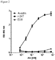
Figure 2 . Representative data demonstrating lack of binding to VEGF121. -
Figure 3 . Sequence alignment of clone E06 and the most sequence homologous germline genes. -
Figure 4 . Representative data demonstrating improved binding of affinity optimized variants. -
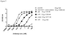
Figure 5 . Representative data demonstrating improved binding of affinity optimized variants as Fabs and IgGs. Fabs are shown in the top two graphs. IgGs are shown in the bottom two graphs. -
Figure 6 . Representative data demonstrating binding of affinity optimized variants to murine VEGF164. -
Figure 7 . Representative data demonstrating lack of binding of affinity optimized variants to VEGF121. -
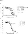
Figure 8 A - B . Representative data demonstrating activity of affinity optimized variants in functional cell-based assays. -
Figure 9 . Representative data demonstrating activity of an affinity optimized variant in a retinal vasculogenesis model. - Before describing the present invention in detail, it is to be understood that this invention is not limited to specific compositions or process steps, as such can vary. As used in this specification and the appended claims, the singular forms "a," "an" and "the" include plural referents unless the context clearly dictates otherwise. The terms "a" (or "an"), as well as the terms "one or more," and "at least one" can be used interchangeably herein. Further it is understood that wherever aspects are described herein with the language "comprising," otherwise analogous aspects described in terms of "consisting of' and/or "consisting essentially of' are also provided.
- As used herein, the term "binding molecule" refers to a molecule that is capable of binding to a target molecule or antigen in a manner similar to that of an antibody binding to an antigen. Examples of binding molecules include full-length antibodies and antigen-binding fragments. Examples of "antigen-binding fragments" of an antibody include (i) a Fab fragment, a monovalent fragment that includes a VL, VH, CL and CH1 domain of an antibody; (ii) a F(ab')2 fragment, a bivalent fragment that includes two Fab fragments linked by a disulfide bridge at a hinge region; (iii) a Fd fragment that includes the VH and CH1 domains; (iv) a Fv fragment that includes VL and VH domains of a single arm of an antibody, (v) a dAb fragment (Ward et al., (1989) Nature 341:544-546), which includes a VH domain; and (vi) an isolated complementarity determining region (CDR). Antigen-binding fragments can be produced by recombinant DNA techniques, or by enzymatic or chemical cleavage of intact immunoglobulins. In one embodiment, the antigen-binding fragment includes a single chain antibody, including, for example, a "single-chain variable fragment" or "scFv." scFv refers to a fusion protein that includes at least one variable region of a heavy chain (VH) and at least one variable region of a light chain (VL) of an immunoglobulin. These single chain antibody fragments can be obtained using conventional techniques known to those with skill in the art. For example, the VH and VL domains of a Fv fragment, which are encoded by separate genes, can be joined, using recombinant methods, by a synthetic linker that enables them to be made as a single polypeptide chain in which the VH and VL regions pair to form a monovalent molecule (See, Bird et al. (1988) Science 242:423-426; and Huston et al. (1988) Proc. Natl. Acad. Sci. USA 85:5879-5883).
- Complementarity determining regions (CDRs) are responsible for antibody binding to its antigen. CDRs are determined by a number of methods in the art (including Kabat (Kabat et al., Sequences of Proteins of Immunological Interest, 5th Ed. Public Health Service, National Institutes of Health, Bethesda, Md. (1991)); Chothia (Chothia and Lesk, J. Mol. Biol. 196:901-917 (1987)); IMGT (ImMunoGeneTics) (Lefranc, M.P. et al., Dev. Comp. Immunol. 27: 55-77 (2003)); and other methods). Although specific CDR sequences are mentioned and claimed herein, the invention also encompasses CDR sequences defined by any method known in the art.
- As use herein, the term "subject" refers to any member of the subphylum cordata, including, without limitation, humans and other primates, including non-human primates such as chimpanzees and other apes and monkey species; farm animals such as cattle, sheep, pigs, goats and horses; domestic mammals such as dogs and cats; laboratory animals including rodents such as mice, rats and guinea pigs; birds, including domestic, wild and game birds such as chickens, turkeys and other gallinaceous birds, ducks, geese, and the like are also non-limiting examples.
- A binding molecule can include a full length or intact antibody, an antibody fragment, including an antigen binding fragment, a human, humanized, post-translationally modified, chimeric or fusion antibody, immunoconjugate, or a functional fragment thereof.
- Suitable immunoglobulin molecules or portions thereof of the invention (i.e., binding molecules) can be or are derived from any isotype (e.g., IgG, IgE, IgM, IgD, IgA and IgY), sub-isotype (e.g., IgG1, IgG2, IgG3, IgG4, IgA1 and IgA2) or allotype (e.g., Gm, e.g., G1m(f, z, a or x), G2m(n), G3m(g, b, or c), Am, Em, and Km(1, 2 or 3)). Immunoglobulin molecules can include light chains classified as either lambda chains or kappa chains based on the amino acid sequence of the light chain constant region.
- Recombinant DNA methods for producing and screening for polypeptides, such as the binding molecules described herein, are known in the art (e.g.
U.S. Patent No. 4,816,567 ). DNA encoding the binding molecules or fragments thereof, for example, DNA encoding a VH domain, a VL domain, an scFv, or combinations thereof can be inserted into a suitable expression vector, which can then be transfected into a suitable host cell, such as E. coli cells, simian COS cells, Chinese Hamster Ovary (CHO) cells, or myeloma cells that do not otherwise produce an antibody protein, to obtain the binding molecule. - Suitable expression vectors are known in the art. An expression vector can contain a polynucleotide that encodes an antibody linked to a promoter. Such vectors may include the nucleotide sequence encoding the constant region of the antibody molecule (see, e.g.,
U.S. Patent Nos. 5,981,216 ;5,591,639 ;5,658,759 and5,122,464 ) and the variable domain of the antibody may be cloned into such a vector for expression of the entire heavy, the entire light chain, or both the entire heavy and light chains. The expression vector can be transferred to a host cell by conventional techniques and the transfected cells can be cultured by conventional techniques to produce the binding molecule. - Mammalian cell lines suitable as hosts for expression of recombinant antibodies are known in the art and include many immortalized cell lines available from the American Type Culture Collection, including but not limit to CHO cells, HeLa cells, baby hamster kidney (BHK) cells, monkey kidney cells (COS), human hepatocellular carcinoma cells (e.g., Hep G2), human epithelial kidney 293 cells, and a number of other cell lines. Different host cells have characteristic and specific mechanisms for the post-translational processing and modification of proteins and gene products. Appropriate cell lines or host systems can be chosen to ensure the correct modification and processing of the binding molecule. To this end, eukaryotic host cells which possess the cellular machinery for proper processing of the primary transcript, glycosylation, and phosphorylation of the gene product may be used. Such mammalian host cells include CHO, VERY, BHK, Hela, COS, MDCK, 293, 3T3, W138, BT483, Hs578T, HTB2, BT2O and T47D, NS0 (a murine myeloma cell line that does not endogenously produce any functional immunoglobulin chains), SP20, CRL7O3O and HsS78Bst cells. Human cell lines developed by immortalizing human lymphocytes can be used to recombinantly produce monoclonal antibodies. The human cell line PER.C6®. (Crucell, Netherlands) can be used to recombinantly produce monoclonal antibodies. Additional cell lines which may be used as hosts for expression of recombinant antibodies include insect cells (e.g. Sf21/Sf9, Trichoplusia ni Bti-Tn5b1-4) or yeast cells (e.g. S. cerevisiae, Pichia,
US7326681 ; etc.), plants cells (US20080066200 ); and chicken cells (WO2008142124 ). - Antibodies can be stably expressed in a cell line using methods known in the art. Stable expression can be used for long-term, high-yield production of recombinant proteins. For stable expression, host cells can be transformed with an appropriately engineered vector that includes expression control elements (e.g., promoter, enhancer, transcription terminators, polyadenylation sites, etc.), and a selectable marker gene. Following the introduction of the foreign DNA, cells are allowed to grow for 1-2 days in an enriched media, and are then switched to a selective media. The selectable marker in the recombinant plasmid confers resistance to the selection and allows cells that have stably integrated the plasmid into their chromosomes to grow and form foci which in turn can be cloned and expanded into cell lines. Methods for producing stable cell lines with a high yield are known in the art and reagents are generally available commercially. Transient expression can also be carried out by using methods known in the art. Transient transfection is a process in which the nucleic acid introduced into a cell does not integrate into the genome or chromosomal DNA of that cell and is maintained as an extra-chromosomal element in the cell (e.g., as an episome).
- A cell line, either stable or transiently transfected, is maintained in cell culture medium and conditions known in the art resulting in the expression and production of the binding molecule. Cell culture media can be based on commercially available media formulations, including, for example, DMEM or Ham's F12. In addition, the cell culture media can be modified to support increases in both cell growth and biologic protein expression. As used herein, the terms "cell culture medium," "culture medium," and "medium formulation" refer to a nutritive solution for the maintenance, growth, propagation, or expansion of cells in an artificial in vitro environment outside of a multicellular organism or tissue. Cell culture medium may be optimized for a specific cell culture use, including cell culture growth medium which is formulated to promote cellular growth or cell culture production medium which is formulated to promote recombinant protein production. The terms nutrient, ingredient, and component are used interchangeably herein to refer to the constituents that make up a cell culture medium. Cell lines can be maintained using a fed batch method. As used herein, "fed batch method," refers to a method by which a cell culture is supplied with additional nutrients after first being incubated with a basal medium. For example, a fed batch method may include adding supplemental media according to a determined feeding schedule within a given time period. Thus, a "fed batch cell culture" refers to a cell culture wherein the cells, typically mammalian, and culture medium are supplied to the culturing vessel initially and additional culture nutrients are fed, continuously or in discrete increments, to the culture during culturing, with or without periodic cell and/or product harvest before termination of culture.
- Cell culture media and the nutrients contained therein are known to one of skilled in the art. The cell culture medium may include a basal medium and at least one hydrolysate, e.g., soy-based hydrolysate, a yeast-based hydrolysate, or a combination of the two types of hydrolysates resulting in a modified basal medium. The additional nutrients may include only a basal medium, such as a concentrated basal medium, or may include only hydrolysates, or concentrated hydrolysates. Suitable basal media include, but are not limited to Dulbecco's Modified Eagle's Medium (DMEM), DME/F12, Minimal Essential Medium (MEM), Basal Medium Eagle (BME), RPMI 1640, F-10, F-12, α-Minimal Essential Medium (α-MEM), Glasgow's Minimal Essential Medium (G-MEM), PF CHO (see, e.g., CHO protein free medium (Sigma) or EX-CELL™ 325 PF CHO Serum-Free Medium for CHO Cells Protein-Free (SAFC Bioscience), and Iscove's Modified Dulbecco's Medium. Other examples of basal media which may be used in the invention include BME Basal Medium (Gibco-Invitrogen; see also Eagle, H (1965) Proc. Soc. Exp. Biol. Med. 89, 36); Dulbecco's Modified Eagle Medium (DMEM, powder) (Gibco-Invitrogen (# 31600); see also Dulbecco and Freeman (1959) Virology. 8:396; Smith et al. (1960) Virology. 12:185. Tissue Culture Standards Committee, In Vitro 6:2, 93); CMRL 1066 Medium (Gibco-Invitrogen (#11530); see also Parker et al. (1957) Special Publications, N.Y. Academy of Sciences, 5:303).
- The basal medium may be serum-free, meaning that the medium contains no serum (e.g., fetal bovine serum (FBS), horse serum, goat serum, or any other animal-derived serum known to one skilled in the art) or animal protein free media or chemically defined media.
- The basal medium may be modified in order to remove certain non-nutritional components found in standard basal medium, such as various inorganic and organic buffers, surfactant(s), and sodium chloride. Removing such components from basal cell medium allows an increased concentration of the remaining nutritional components, and may improve overall cell growth and protein expression. In addition, omitted components may be added back into the cell culture medium containing the modified basal cell medium according to the requirements of the cell culture conditions. The cell culture medium may contain a modified basal cell medium, and at least one of the following nutrients, an iron source, a recombinant growth factor; a buffer; a surfactant; an osmolarity regulator; an energy source; and non-animal hydrolysates. In addition, the modified basal cell medium may optionally contain amino acids, vitamins, or a combination of both amino acids and vitamins. A modified basal medium may further contain glutamine, e.g, L-glutamine, and/or methotrexate.
- Once a binding molecule has been produced, it may be purified by methods known in the art for purification of an immunoglobulin molecule, for example, by chromatography (e.g., ion exchange, affinity, particularly by affinity for the specific antigens Protein A or Protein G, and sizing column chromatography), centrifugation, differential solubility, or by any other standard technique for the purification of proteins. Further, the binding molecules of the invention may be fused to heterologous polypeptide sequences (referred to herein as "tags") to facilitate purification.
- Binding molecules of the invention can be used in a number of ways. For example, antibodies of the invention can be used to bind to VEGF-A and thereby reduce at least one biological activity of VEGF-A. More particularly, the antibodies of the invention can be used to bind to VEGF-165 and thereby reduce at least one biological activity of VEGF-165, which may include a reduction in activation or phosphorylation of its receptor, a reduction in angiogenesis in connection with cellular dysregulation, a reduction in tumor growth, a reduction in tumor volume, and/or reduction in tumor growth and tumor volume.
- An embodiment of the invention relates to a binding molecule comprising heavy chain complementarity determining regions 1 - 3 (i.e., HCDR1, HCDR2, and HCDR3) and light chain complementarity determining regions 1 - 3 (i.e., LCDR1, LCDR2, and LCDR3) of an antibody described herein.
- Another embodiment relates to a binding molecule comprising HCDR1, HCDR2, and HCDR3 and LCDR1, LCDR2, and LCDR3 of an antibody described herein, wherein the binding molecule binds VEGF165.
- Another embodiment relates to a binding molecule comprising HCDR1, HCDR2, and HCDR3 and LCDR1, LCDR2, and LCDR3 of an antibody described herein, wherein the binding molecule binds VEGF165 with greater affinity compared to VEGF121.
- Another embodiment relates to a binding molecule comprising HCDR1, HCDR2, and HCDR3 and LCDR1, LCDR2, and LCDR3 of an antibody described herein, wherein the binding molecule binds VEGF165 with greater affinity compared to VEGF189.
- Another embodiment relates to a binding molecule comprising HCDR1, HCDR2, and HCDR3 and LCDR1, LCDR2, and LCDR3 of an antibody described herein, wherein the binding molecule binds VEGF165 with greater affinity compared to VEGF121 and VEGF189.
- Another embodiment relates to a binding molecule comprising HCDR1, HCDR2, and HCDR3 and LCDR1, LCDR2, and LCDR3 of an antibody described herein, wherein the binding molecule reduces human VEGFR2 phosphorylation, murine VEGFR2 phosphorylation, or both human and murine VEGFR2 phosphorylation.
- Another embodiment relates to a binding molecule comprising HCDR1, HCDR2, and HCDR3 and LCDR1, LCDR2, and LCDR3 of an antibody described herein, wherein the binding molecule reduces angiogenesis.
- Another embodiment relates to a binding molecule comprising HCDR1, HCDR2, and HCDR3 and LCDR1, LCDR2, and LCDR3 of an antibody described herein, wherein the binding molecule reduces tumor growth, reduces tumor volume, or reduces tumor growth and tumor volume as a result of being provided to a subject having a tumor.
- Another embodiment relates to a binding molecule comprising HCDR1, HCDR2, and HCDR3 and LCDR1, LCDR2, and LCDR3 of an antibody described herein, wherein the binding molecule has one or more or any combination of the characteristics described herein, including binding VEGF165, binding VEGF165 with greater affinity compared to VEGF121, binding VEGF165 with greater affinity compared to VEGF189, binding VEGF165 with greater affinity compared to VEGF121 and VEGF189, reducing human VEGFR2 phosphorylation, murine VEGFR2 phosphorylation, or both human and murine VEGFR2 phosphorylation, reducing angiogenesis, or reducing tumor growth, reducing tumor volume, or reducing tumor growth and tumor volume as a result of being provided to a subject having a tumor.
- An embodiment of the invention relates to a binding molecule comprising a heavy chain variable domain comprising HCDR1, HCDR2, and HCDR3 and a light chain variable domain comprising LCDR1, LCDR2, and LCDR3 of an antibody described herein.
- Another embodiment relates to a binding molecule comprising a heavy chain variable domain comprising HCDR1, HCDR2, and HCDR3 and a light chain variable domain comprising LCDR1, LCDR2, and LCDR3 of an antibody described herein, wherein the binding molecule binds VEGF165.
- Another embodiment relates to a binding molecule comprising a heavy chain variable domain comprising HCDR1, HCDR2, and HCDR3 and a light chain variable domain comprising LCDR1, LCDR2, and LCDR3 of an antibody described herein, wherein the binding molecule binds VEGF165 with greater affinity compared to VEGF121.
- Another embodiment relates to a binding molecule comprising a heavy chain variable domain comprising HCDR1, HCDR2, and HCDR3 and a light chain variable domain comprising LCDR1, LCDR2, and LCDR3 of an antibody described herein, wherein the binding molecule binds VEGF165 with greater affinity compared to VEGF189.
- Another embodiment relates to a binding molecule comprising a heavy chain variable domain comprising HCDR1, HCDR2, and HCDR3 and a light chain variable domain comprising LCDR1, LCDR2, and LCDR3 of an antibody described herein, wherein the binding molecule binds VEGF165 with greater affinity compared to VEGF121 and VEGF189.
- Another embodiment relates to a binding molecule comprising a heavy chain variable domain comprising HCDR1, HCDR2, and HCDR3 and a light chain variable domain comprising LCDR1, LCDR2, and LCDR3 of an antibody described herein, wherein the binding molecule reduces human VEGFR2 phosphorylation, murine VEGFR2 phosphorylation, or both human and murine VEGFR2 phosphorylation.
- Another embodiment relates to a binding molecule comprising a heavy chain variable domain comprising HCDR1, HCDR2, and HCDR3 and comprising a light chain variable domain comprising LCDR1, LCDR2, and LCDR3 of an antibody described herein, wherein the binding molecule reduces angiogenesis.
- Another embodiment relates to a binding molecule comprising a heavy chain variable domain comprising HCDR1, HCDR2, and HCDR3 and comprising a light chain variable domain comprising LCDR1, LCDR2, and LCDR3 of an antibody described herein, wherein the binding molecule reduces tumor growth, reduces tumor volume, or reduces tumor growth and tumor volume as a result of being provided to a subject having a tumor.
- Another embodiment relates to a binding molecule comprising a heavy chain variable domain comprising HCDR1, HCDR2, and HCDR3 and comprising a light chain variable domain comprising LCDR1, LCDR2, and LCDR3 of an antibody described herein, wherein the binding molecule has one or more or any combination of the characteristics described herein, including binding VEGF165, binding VEGF165 with greater affinity compared to VEGF121, binding VEGF165 with greater affinity compared to VEGF189, binding VEGF165 with greater affinity compared to VEGF121 and VEGF189, reducing human VEGFR2 phosphorylation, murine VEGFR2 phosphorylation, or both human and murine VEGFR2 phosphorylation, reducing angiogenesis, or reducing tumor growth, reducing tumor volume, or reducing tumor growth and tumor volume as a result of being provided to a subject having a tumor.
- An embodiment of the invention relates to a binding molecule comprising a full-length antibody comprising HCDR1, HCDR2, and HCDR3 and LCDR1, LCDR2, and LCDR3 of an antibody described herein.
- Another embodiment relates to a binding molecule comprising a full-length antibody comprising HCDR1, HCDR2, and HCDR3 and LCDR1, LCDR2, and LCDR3 of an antibody described herein, wherein the binding molecule binds VEGF165.
- Another embodiment relates to a binding molecule comprising a full-length antibody comprising HCDR1, HCDR2, and HCDR3 and LCDR1, LCDR2, and LCDR3 of an antibody described herein, wherein the binding molecule binds VEGF165 with greater affinity compared to VEGF121.
- Another embodiment relates to a binding molecule comprising a full-length antibody comprising HCDR1, HCDR2, and HCDR3 and LCDR1, LCDR2, and LCDR3 of an antibody described herein, wherein the binding molecule binds VEGF165 with greater affinity compared to VEGF189.
- Another embodiment relates to a binding molecule comprising a full-length antibody comprising HCDR1, HCDR2, and HCDR3 and LCDR1, LCDR2, and LCDR3 of an antibody described herein, wherein the binding molecule binds VEGF165 with greater affinity compared to VEGF121 and VEGF189.
- Another embodiment relates to a binding molecule comprising a full-length antibody comprising HCDR1, HCDR2, and HCDR3 and LCDR1, LCDR2, and LCDR3 of an antibody described herein, wherein the binding molecule reduces human VEGFR2 phosphorylation, murine VEGFR2 phosphorylation, or both human and murine VEGFR2 phosphorylation.
- Another embodiment relates to a binding molecule comprising a full-length antibody comprising HCDR1, HCDR2, and HCDR3 and LCDR1, LCDR2, and LCDR3 of an antibody described herein, wherein the binding molecule reduces angiogenesis.
- Another embodiment relates to a binding molecule comprising a full-length antibody comprising HCDR1, HCDR2, and HCDR3 and LCDR1, LCDR2, and LCDR3 of an antibody described herein, wherein the binding molecule reduces tumor growth, reduces tumor volume, or reduces tumor growth and tumor volume as a result of being provided to a subject having a tumor.
- Another embodiment relates to a binding molecule comprising a full-length antibody comprising HCDR1, HCDR2, and HCDR3 and LCDR1, LCDR2, and LCDR3 of an antibody described herein, wherein the binding molecule has one or more or any combination of the characteristics described herein, including binding VEGF165, binding VEGF165 with greater affinity compared to VEGF121, binding VEGF165 with greater affinity compared to VEGF189, binding VEGF165 with greater affinity compared to VEGF121 and VEGF189, reducing human VEGFR2 phosphorylation, murine VEGFR2 phosphorylation, or both human and murine VEGFR2 phosphorylation, reducing angiogenesis, or reducing tumor growth, reducing tumor volume, or reducing tumor growth and tumor volume as a result of being provided to a subject having a tumor.
- An embodiment of the invention relates to a binding molecule comprising a full-length IgG1 antibody comprising HCDR1, HCDR2, and HCDR3 and LCDR1, LCDR2, and LCDR3 of an antibody described herein.
- Another embodiment relates to a binding molecule comprising a full-length IgG1 antibody comprising HCDR1, HCDR2, and HCDR3 and LCDR1, LCDR2, and LCDR3 of an antibody described herein, wherein the binding molecule binds VEGF165.
- Another embodiment relates to a binding molecule comprising a full-length IgG1 antibody comprising HCDR1, HCDR2, and HCDR3 and LCDR1, LCDR2, and LCDR3 of an antibody described herein, wherein the binding molecule binds VEGF165 with greater affinity compared to VEGF121.
- Another embodiment relates to a binding molecule comprising a full-length IgG1 antibody comprising HCDR1, HCDR2, and HCDR3 and LCDR1, LCDR2, and LCDR3 of an antibody described herein, wherein the binding molecule binds VEGF165 with greater affinity compared to VEGF189.
- Another embodiment relates to a binding molecule comprising a full-length IgG1 antibody comprising HCDR1, HCDR2, and HCDR3 and LCDR1, LCDR2, and LCDR3 of an antibody described herein, wherein the binding molecule binds VEGF165 with greater affinity compared to VEGF121 and VEGF189.
- Another embodiment relates to a binding molecule comprising a full-length IgG1 antibody comprising HCDR1, HCDR2, and HCDR3 and LCDR1, LCDR2, and LCDR3 of an antibody described herein, wherein the binding molecule reduces human VEGFR2 phosphorylation, murine VEGFR2 phosphorylation, or both human and murine VEGFR2 phosphorylation.
- Another embodiment relates to a binding molecule comprising a full-length IgG1 antibody comprising HCDR1, HCDR2, and HCDR3 and LCDR1, LCDR2, and LCDR3 of an antibody described herein, wherein the binding molecule reduces angiogenesis.
- Another embodiment relates to a binding molecule comprising a full-length IgG1 antibody comprising HCDR1, HCDR2, and HCDR3 and LCDR1, LCDR2, and LCDR3 of an antibody described herein, wherein the binding molecule reduces tumor growth, reduces tumor volume, or reduces tumor growth and tumor volume as a result of being provided to a subject having a tumor.
- Another embodiment relates to a binding molecule comprising a full-length IgG1 antibody comprising HCDR1, HCDR2, and HCDR3 and LCDR1, LCDR2, and LCDR3 of an antibody described herein, wherein the binding molecule has one or more or any combination of the characteristics described herein, including binding VEGF165, binding VEGF165 with greater affinity compared to VEGF121, binding VEGF165 with greater affinity compared to VEGF189, binding VEGF165 with greater affinity compared to VEGF121 and VEGF189, reducing human VEGFR2 phosphorylation, murine VEGFR2 phosphorylation, or both human and murine VEGFR2 phosphorylation, reducing angiogenesis, or reducing tumor growth, reducing tumor volume, or reducing tumor growth and tumor volume as a result of being provided to a subject having a tumor.
- An embodiment of the invention relates to a binding molecule which is a full-length antibody, including a full-length IgG1 antibody, comprising HCDR1, HCDR2, and HCDR3 and LCDR1, LCDR2, and LCDR3 of an antibody described herein.
- Another embodiment relates to a binding molecule which is a full-length antibody, including a full-length IgG1 antibody, comprising HCDR1, HCDR2, and HCDR3 and LCDR1, LCDR2, and LCDR3 of an antibody described herein, wherein the binding molecule binds VEGF165.
- Another embodiment relates to a binding molecule which is a full-length antibody, including a full-length IgG1 antibody, comprising HCDR1, HCDR2, and HCDR3 and LCDR1, LCDR2, and LCDR3 of an antibody described herein, wherein the binding molecule binds VEGF165 with greater affinity compared to VEGF121.
- Another embodiment relates to a binding molecule which is a full-length antibody, including a full-length IgG1 antibody, comprising HCDR1, HCDR2, and HCDR3 and LCDR1, LCDR2, and LCDR3 of an antibody described herein, wherein the binding molecule binds VEGF165 with greater affinity compared to VEGF189.
- Another embodiment relates to a binding molecule which is a full-length antibody, including a full-length IgG1 antibody, comprising HCDR1, HCDR2, and HCDR3 and LCDR1, LCDR2, and LCDR3 of an antibody described herein, wherein the binding molecule binds VEGF165 with greater affinity compared to VEGF121 and VEGF189.
- Another embodiment relates to a binding molecule which is a full-length antibody, including a full-length IgG1 antibody, comprising HCDR1, HCDR2, and HCDR3 and LCDR1, LCDR2, and LCDR3 of an antibody described herein, wherein the binding molecule reduces human VEGFR2 phosphorylation, murine VEGFR2 phosphorylation, or both human and murine VEGFR2 phosphorylation.
- Another embodiment relates to a binding molecule which is a full-length antibody, including a full-length IgG1 antibody, comprising HCDR1, HCDR2, and HCDR3 and LCDR1, LCDR2, and LCDR3 of an antibody described herein, wherein the binding molecule reduces angiogenesis.
- Another embodiment relates to a binding molecule which is a full-length antibody, including a full-length IgG1 antibody, comprising HCDR1, HCDR2, and HCDR3 and LCDR1, LCDR2, and LCDR3 of an antibody described herein, wherein the binding molecule reduces tumor growth, reduces tumor volume, or reduces tumor growth and tumor volume as a result of being provided to a subject having a tumor.
- Another embodiment relates to a binding molecule which is a full-length antibody, including a full-length IgG1 antibody, comprising HCDR1, HCDR2, and HCDR3 and LCDR1, LCDR2, and LCDR3 of an antibody described herein, wherein the binding molecule has one or more or any combination of the characteristics described herein, including binding VEGF165, binding VEGF165 with greater affinity compared to VEGF121, binding VEGF165 with greater affinity compared to VEGF189, binding VEGF165 with greater affinity compared to VEGF121 and VEGF189, reducing human VEGFR2 phosphorylation, murine VEGFR2 phosphorylation, or both human and murine VEGFR2 phosphorylation, reducing angiogenesis, or reducing tumor growth, reducing tumor volume, or reducing tumor growth and tumor volume as a result of being provided to a subject having a tumor.
- In a specific embodiment, there is an antibody comprising an HCDR1, HCDR2, and HCDR3 and an LCDR1, LCDR2, and LCDR3, wherein HCDR1, HCDR2, and HCDR3 and LCDR1, LCDR2, and LCDR3 comprise SEQ ID NOs: 79 - 84, respectively.
- In another specific embodiment, there is an antibody comprising a heavy chain and a light chain comprising SEQ ID NOs: 73 and 77, respectively.
- In another specific embodiment, there is an antibody comprising a heavy chain amino acid sequence comprising SEQ ID NO: 71 and a light chain amino acid sequence comprising SEQ ID NO: 75.
- In another specific embodiment, there is an antibody comprising an HCDR1, HCDR2, and HCDR3 and an LCDR1, LCDR2, and LCDR3, wherein HCDR1, HCDR2, and HCDR3 and LCDR1, LCDR2, and LCDR3 comprise SEQ ID NOs: 79 - 84, respectively, and wherein the antibody is a monoclonal antibody.
- In another specific embodiment, there is a nucleic acid sequence comprising polynucleotides encoding an antibody comprising an HCDR1, HCDR2, and HCDR3 and an LCDR1, LCDR2, and LCDR3, wherein HCDR1, HCDR2, and HCDR3 and LCDR1, LCDR2, and LCDR3 comprise SEQ ID NOs: 79 - 84, respectively.
- In another specific embodiment, there is a vector comprising polynucleotides encoding an antibody comprising an HCDR1, HCDR2, and HCDR3 and an LCDR1, LCDR2, and LCDR3, wherein HCDR1, HCDR2, and HCDR3 and LCDR1, LCDR2, and LCDR3 comprise SEQ ID NOs: 79 - 84, respectively.
- In another specific embodiment, there is a cell comprising a vector comprising polynucleotides encoding an antibody comprising an HCDR1, HCDR2, and HCDR3 and an LCDR1, LCDR2, and LCDR3, wherein HCDR1, HCDR2, and HCDR3 and LCDR1, LCDR2, and LCDR3 comprise SEQ ID NOs: 79 - 84, respectively.
- In another specific embodiment, there is a method of making an antibody comprising culturing a cell comprising a vector comprising polynucleotides encoding an antibody comprising an HCDR1, HCDR2, and HCDR3 and an LCDR1, LCDR2, and LCDR3, wherein HCDR1, HCDR2, and HCDR3 and LCDR1, LCDR2, and LCDR3 comprise SEQ ID NOs: 79 - 84, respectively.
- In another specific embodiment, there is a method of reducing angiogenesis comprising providing an antibody to a subject wherein the antibody comprises an HCDR1, HCDR2, and HCDR3 and an LCDR1, LCDR2, and LCDR3, wherein HCDR1, HCDR2, and HCDR3 and LCDR1, LCDR2, and LCDR3 comprise SEQ ID NOs: 79 - 84, respectively.
-
SEQ ID NO SEQUENCE DESCRIPTION 1 
Amino acid sequence of the heavy chain of E06 2 
Nucleotide sequence of the heavy chain of E06 
3 
Amino acid sequence of the heavy chain variable domain of E06 4 
Nucleotide sequence of the heavy chain variable domain of E06 5 
Amino acid sequence of the light chain of E06 6 
Nucleotide sequence of the light chain of E06 7 
Amino acid sequence of the light chain variable domain of E06 8 
Nucleotide sequence of the light chain variable domain of E06 9 WYEMY Amino acid sequence of HCDR1 of E06 10 SISPSGGWTMYADSVKG Amino acid sequence of HCDR2 of E06 11 PLYSSDGLSAGDI Amino acid sequence of HCDR3 of E06 12 RASQSVSSSYLA Amino acid sequence of LCDR1 of E06 13 GASSRAT Amino acid sequence of LCDR2 of E06 14 QQSYSTPS Amino acid sequence of LCDR3 of E06 15 SAME AS E06 Amino acid sequence of the heavy chain of E06 germline M4 16 SAME AS E06 Nucleotide sequence of the heavy chain of E06 germline M4 17 SAME AS E06 Amino acid sequence of the heavy chain variable domain of E06 germline M4 18 SAME AS E06 Nucleotide sequence of the heavy chain variable domain of E06 germline M4 19 
Amino acid sequence of the light chain of E06 germline M4 20 
Nucleotide sequence of the light chain of E06 germline M4 21 
Amino acid sequence of the light chain variable domain of E06 germline M4 22 
Nucleotide sequence of the light chain variable domain of E06 germline M4 
23 SAME AS E06 HCDR1 Amino acid sequence of HCDR1 of E06 germline M4 24 SAME AS E06 HCDR2 Amino acid sequence of HCDR2 of E06 germline M4 25 SAME AS E06 HCDR3 Amino acid sequence of HCDR3 of E06 germline M4 26 SAME AS E06 LCDR1 Amino acid sequence of LCDR1 of E06 germline M4 27 SAME AS E06 LCDR2 Amino acid sequence of LCDR2 of E06 germline M4 28 SAME AS E06 LCDR3 Amino acid sequence of LCDR3 or E06 germline M4 29 SAME AS E06 Amino acid sequence of the heavy chain of D04 30 SAME AS E06 Nucleotide sequence of the heavy chain of D04 31 SAME AS E06 Amino acid sequence of the heavy chain variable domain of D04 32 SAME AS E06 Nucleotide sequence of the heavy chain variable domain of D04 33 
Amino acid sequence of the light chain of D04 34 
Nucleotide sequence of the light chain of D04 35 
Amino acid sequence of the light chain variable domain of D04 36 
Nucleotide sequence of the light chain variable domain of D04 37 SAME AS E06 HCDR1 Amino acid sequence of HCDR1 of D04 38 SAME AS E06 HCDR2 Amino acid sequence of HCDR2 of D04 39 SAME AS E06 HCDR3 Amino acid sequence of HCDR3 of D04 40 RASQSVHSSYLA Amino acid sequence of LCDR1 of D04 41 SAME AS E06 LCDR2 Amino acid sequence of LCDR2 of D04 42 SAME AS E06 LCDR3 Amino acid sequence of LCDR3 of D04 43 SAME AS E06 Amino acid sequence of the heavy chain of J05 44 SAME AS E06 Nucleotide sequence of the heavy chain of J05 45 SAME AS E06 Amino acid sequence of the heavy chain variable domain of J05 46 SAME AS E06 Nucleotide sequence of the heavy chain variable domain of J05 47 
Amino acid sequence of the light chain of J05 48 
Nucleotide sequence of the light chain of J05 49 
Amino acid sequence of the light chain variable domain of J05 50 
Nucleotide sequence of the light chain variable domain of J05 51 SAME AS E06 HCDR1 Amino acid sequence of HCDR1 of J05 52 SAME AS E06 HCDR2 Amino acid sequence of HCDR2 of J05 53 SAME AS E06 HCDR3 Amino acid sequence of HCDR3 of J05 54 SAME AS E06 LCDR1 Amino acid sequence of LCDR1 of J05 55 SAME AS E06 LCDR2 Amino acid sequence of LCDR2 of J05 56 QQSYRTPS Amino acid sequence of LCDR3 of J05 57 SAME AS E06 Amino acid sequence of the heavy chain of I20 58 SAME AS E06 Nucleotide sequence of the heavy chain of I20 59 SAME AS E06 Amino acid sequence of the heavy chain variable domain of I20 60 SAME AS E06 Nucleotide sequence of the heavy chain variable domain of I20 61 
Amino acid sequence of the light chain of I20 62 
Nucleotide sequence of the light chain of I20 
63 
Amino acid sequence of the light chain variable domain of I20 64 
Nucleotide sequence of the light chain variable domain of I20 65 SAME AS E06 HCDR1 Amino acid sequence of HCDR1 of I20 66 SAME AS E06 HCDR2 Amino acid sequence of HCDR2 of I20 67 SAME AS E06 HCDR3 Amino acid sequence of HCDR3 of I20 68 SAME AS E06 LCDR1 Amino acid sequence of LCDR1 of I20 69 SAME AS E06 LCDR2 Amino acid sequence of LCDR2 of I20 70 QQDYSTPS Amino acid sequence of LCDR3 of I20 71 SAME AS E06 Amino acid sequence of the heavy chain of H1R 72 SAME AS E06 Nucleotide sequence of the heavy chain of H1R 73 SAME AS E06 Amino acid sequence of the heavy chain variable domain of H1R 74 SAME AS E06 Nucleotide sequence of the heavy chain variable domain of H1R 75 
Amino acid sequence of the light chain of H1R 76 
Nucleotide sequence of the light chain of H1R 
77 
Amino acid sequence of the light chain variable domain of H1R 78 
Nucleotide sequence of the light chain variable domain of H1R 79 SAME AS E06 HCDR1 Amino acid sequence of HCDR1 of H1R 80 SAME AS E06 HCDR2 Amino acid sequence of HCDR2 of H1R 81 SAME AS E06 HCDR3 Amino acid sequence of HCDR3 of H1R 82 RASQSVHSSYLA Amino acid sequence of LCDR1 of H1R 83 SAME AS E06 LCDR2 Amino acid sequence of LCDR2 of H1R 84 QQSYRTPS Amino acid sequence of LCDR3 of H1R 85 
Amino acid sequence of the heavy chain of H1RK 86 
Nucleotide sequence of the heavy chain of H1RK 
87 
Amino acid sequence of the heavy chain variable domain of H1RK 88 
Nucleotide sequence of the heavy chain variable domain of H1RK 89 
Amino acid sequence of the light chain of H1RK 90 
Nucleotide sequence of the light chain of H1RK 
91 
Amino acid sequence of the light chain variable domain of H1RK 92 
Nucleotide sequence of the light chain variable domain of H1RK 93 WYEMY Amino acid sequence of HCDR1 of H1RK 94 SISPSGGWTMYADSVKG Amino acid sequence of HCDR2 of H1RK 95 PLYSSDGLSAGDI Amino acid sequence of HCDR3 of H1RK 96 RASQSVHSSYLA Amino acid sequence of LCDR1 of H1RK 97 GASSRAT Amino acid sequence of LCDR2 of H1RK 98 QQSYRTPS Amino acid sequence of LCDR3 of H1RK 99 SAME AS E06 Amino acid sequence of the heavy chain of H1DR 100 SAME AS E06 Nucleotide sequence of the heavy chain of H1DR 101 SAME AS E06 Amino acid sequence of the heavy chain variable domain of H1DR 102 SAME AS E06 Nucleotide sequence of the heavy chain variable domain of H1DR 103 
Amino acid sequence of the light chain of H1DR 104 
Nucleotide sequence of the light chain of H1DR 
105 
Amino acid sequence of the light chain variable domain of H1DR 106 
Nucleotide sequence of the light chain variable domain of H1DR 107 SAME AS E06 HCDR1 Amino acid sequence of HCDR1 of H1DR 108 SAME AS E06 HCDR2 Amino acid sequence of HCDR2 of H1DR 109 SAME AS E06 HCDR3 Amino acid sequence of HCDR3 of H1DR 110 RASQSVHSSYLA Amino acid sequence of LCDR1 of H1DR 111 SAME AS E06 LCDR2 Amino acid sequence of LCDR2 of H1DR 112 QQDYRTPS Amino acid sequence of LCDR3 of H1DR - For the experiments described herein various antibodies were used including, Avastin® (Ferrara, N et al. Biochem Biophys Res Comm, 333:328-335, 2005), G6-31 (Liang, WC et al. J Biol Chem, 281: 951-961, 2006), B20-4.1 (Liang, WC et al. J Biol Chem, 281: 951-961, 2006), and an isotype control, designated R347, as a monospecific or a bispecific antibody as needed. An anti-VEGF IgG1 antibody capable of binding all VEGF isoforms that is not cross-reactive with mouse can used as a positive control for some binding and functional studies. Where cross reactivity to mouse VEGF is needed the antibodies G6-31 and B20-4.1 can be used as a positive control.
- Solution phage panning was applied to isolate anti-VEGF antibodies from antibody phage libraries. The phage libraries were incubated with biotinylated VEGF165 (PeproTech, NJ) at the final concentration of 4 µg/ml (human VEGF165 for first and third round and mVEGF164 for the second and fourth round panning). After incubation for 30 minutes, the VEGF bound phages were captured from the solution by adding straptavidin beads (Invitrogen), which were prewashed with phosphate buffer saline (PBS) and blocked with 3% bovine serum albumin (BSA) in PBS. The beads captured phages were eluted with 100 mM TEA buffer, neutralized with 1 M Tris-HCI (pH8.0), and then 1 ml of eluted phage were used to infect 5 ml of log phase TG1 and 4 ml of 2YT medium (Teknova) for phage amplification. After incubation at 37° C for 30 minutes in water bath, the cells were spun down at 4000g, resuspended in 2YT, spread on 2YT agar plates (Teknova) with 100 µg/ml of carbenicillin and 2% glucose, and incubated at 30° C overnight. On the second day, the colonies were collected from the agar plates, inoculated into 2YT medium at the final density of OD600 = 0.1, grown at 37° C until log phase, infected with helper phage (Invitrogen) and then let it grow overnight in 2YT with carbenicillin (Invitrogen) and kanamycin (Sigma) at 30° C to generate high titer phages. The amplified phages were precipitated with PEG8000, resuspended in PBS and used for the next round of panning using the standard procedure. A total of four rounds of panning were applied to isolate VEGF specific antibodies. A significant enrichment of output phage numbers was observed from 2 x 10⁵ pfu in the first round to 4 x 10⁷ pfu in the fourth round of panning.
- Specificity of individual phage Fab from the fourth round of panning was assessed by phage ELISA. The 96-well microtiter plates were coated with human VEGF165 at a concentration of 5 µg/ml in PBS at 40° C overnight. After being washed three times with PBS, the wells were blocked with the blocking buffer (4% skimmed milk in PBS) for 1 h at 37° C. Then, 50 µl/well phage was added and incubated for 1 h at 37° C. After washing, 50 µl of horseradish peroxidase (HRP)-conjugated mouse anti-M13 (Amersham Pharmacia) in blocking buffer with 1:1,000 dilution was added for 30 minutes at 37° C. For detection, 50 µl/well of SureBlue Reserve TMB substrate (KPL) for 5 minutes at room temperature and the reaction was stopped by adding 50 µL of TMB Stop Solution (KPL). The absorbance was read at 450 nm. After four rounds of panning, more than 50% of clones are VEGF specific with absorbance higher than 1, which is 20-fold higher than the nonspecific background reading approximately 0.05. Representative data is show in Table 1.
Table 1 1 2 3 4 5 6 7 8 9 10 11 12 0.091 2.276 2.406 0.111 0.116 0.116 0.087 2.534 0.059 2.478 0.098 0.193 0.091 2.755 3.161 1.508 0.054 3.166 0.062 0.066 0.061 3.2 2.354 1.971 2328 0.078 2.697 2.919 3.216 3.258 3.133 2.948 2.859 2.6 1.314 2.595 2.209 3.056 0.084 3.139 3.181 0.055 3.336 2.472 3.074 0.062 2.741 0.073 0.05 0.052 0.053 3.02 3.57 0.049 0.047 2.862 2.761 2.609 0.051 0.05 0.05 0.05 0.053 2.861 3.469 3.155 0.057 0.387 1.325 0.052 3.067 2.446 1.963 2.641 3.27 3.349 3.246 0.047 3.509 2.727 0.046 2.67 2.679 0.053 0.044 0.05 3.105 0.053 3.215 2.483 0.059 3.004 2.294 2.994 3.091 1.336 - After initial phage screening, one clone (E06) with cross-reactivity to mouse VEGF164 was further converted and expressed as the full length IgG using standard molecular biology materials and methods. The 96-well microtiter plates (Corning) were coated with 50 µl /well of human and mouse VEGF165 or VEGF121 at a concentration of 5 µg/ml in PBS at 4° C overnight. After being washed three times with PBS containing 0.1% Tween-20 (PBST), the wells were blocked with blocking buffer (4% skimmed milk in PBS). After 1 h incubation at room temperature, the plates were washed with PBST and a 2-fold serial dilution of the antibodies (starting from 8 nM) in blocking buffer were added and incubated for 1 hour at room temperature. The antibody solution was removed by washing with PBST followed by 1 hour incubation at room temperature with a 1:3000 dilution of an anti-human-Fc-HRP antibody (Thermo Scientific) prepared in PBST. Binding was visualized with the addition of 50 µL of SureBlue Reserve TMB substrate (KPL) for 5 minutes at room temperature and the reaction was stopped by adding 50 µL of TMB Stop Solution (KPL). The absorbance at 450 nm was measured using a microtiter plate reader. The data were analyzed using Prism 5 software (GraphPad).
- Similar binding activity of antibody E06 to human VEGF165 and mouse VEGF164 was observed with an EC50 of 0.048 nM and 0.049 nM, respectively. As expected, Avastin® (an anti-VEGF antibody) showed strong binding to human VEGF165 but no detectable binding to mouse VEGF164. The isotype control antibody R347 did not bind either human or mouse forms of VEGF. Representative data are shown in
Figure 1 . - To evaluate whether antibody E06 binds to a different epitope than Avastin® (an anti-VEGF antibody), an ELISA assay was conducted using human VEGF121. Avastin® (an anti-VEGF antibody) showed very strong binding to human VEGF121. However, no binding was deteceted for E06, indicating antibody E06 binds to a different epitope than Avastin® (an anti-VEGF antibody). The isotype control antibody R347 also did not demonstrate any binding to human VEGF121. Representative data are shown in
Figure 2 . - Framework sequences of clone E06 were engineered to match to its closest germline sequences. A sequence analysis against the IgG germline gene database showed that the E06 VL sequence best matches to the germline gene VK3-NL5∗01 with 4 amino acid differences at
positions Figure 3 shows the sequence alignment of the parental E06 and the most homologous germline genes. - The germline variants were expressed and purified as Fabs and their binding to the recombinant VEGF165 was determined by ELISA. The binding results showed that the germline variants M4 and F1, which contain the full or partial VL germline amino acid substitutions retained E06 WT Fab binding activity. In addition, M4 showed similar activity compared to the WT E06 Fab in pVEGFR2 assay using HUVECs. On the other hand, germline variants M1 and M7 containing the VH germline amino acid substitution at position T98 demonstrated drastically reduced binding and pVEGFR2 phosphorylation, indicating that T98 participated in binding and activity and must be retained. The germline clone, M4, was used as the template for further affinity optimization.
- Each amino acid of all 6 CDRs of germline clone M4 was individually mutated to the other 20 amino acids using a hybridization mutagenesis method (Kunkel, Proc. Natl. Acad. Sci. USA Vol. 82, pp. 488-492, 1985). Two sets of DNA primers, one containing a NSS codon encoding 8 amino acids and the other containing a NWS codon encoding 12 different amino acids, were used to introduce mutations to each targeted CDR position. The individual degenerate primers were used in hybridization mutagenesis reactions. Briefly, each degenerate primer was phosphorylated and used in a 10:1 ratio with the uridinylated M4 Fab ssDNA. The mixture was heated to 95° C then cooled down to 55° C over 1 hour. Thereafter, T4 ligase and T7 DNA polymerase were added and the mix was incubated for 1.5 hours at 37° C. Synthesis products for VH and VL CDRs were pooled respectively; however, NSS and NWS libraries were kept separate and screened independently. Typically, 1 µL of the pooled library DNA was electroporated into XL1-Blue for plaque formation on XL1-Blue bacterial lawn or for production of Fab fragments (Wu H, An LL. Tailoring kinetics of antibodies using focused combinatorial libraries. Methods Mol Biol 2003; 207:213-33). These mutants were then screened for activity.
- The primary screen consisted of a single point ELISA (SPE) assay which was carried out using culture supernatant of bacteria grown in 96-well plates (deep well) and infected with individual recombinant M13 clones as described elsewhere (Wu H, An LL. Tailoring kinetics of antibodies using focused combinatorial libraries. Methods Mol Biol 2003; 207:213-33). Briefly, this capture ELISA involved coating individual wells of a 96-well Maxisorp Immunoplate with approximately 50 ng of a sheep anti-human Fd antibody (Biodesign International, ME) in a carbonate buffer at pH 8.5 overnight at 4° C. The next day, the plate was blocked with 3% BSA in PBS buffer for 1 hour at room temperature. Fab supernatant was then added to the plate and incubated at room temperature for 1 hour. After washing, the biotinylated VEGF165 protein was added to the well and the mixture was incubated for 1.5 hours at room temperature. This was followed by incubation with neutravidin-horseradish peroxydase (HRP) conjugate (Pierce, IL) for approximately 40 minutes at room temperature. HRP activity was detected with tetra-methyl-benzidine (TMB) substrate and the reaction quenched with 0.2 M H2SO4. Plates were read at 450 nm.
- Clones exhibiting an optical density (OD) signal at 450 nm greater than the parental clone M4 Fab were picked and regrown (15 mL) (Wu H, An LL. Tailoring kinetics of antibodies using focused combinatorial libraries. Methods Mol Biol 2003; 207:213-33) and re-assayed by ELISA (as described above) in duplicate to confirm positive results. Clones that repeatedly exhibited a signal greater than that of the M4 Fab were sequenced. The Fab protein concentration of each clone that had a CDR change was then determined by a quantitative Fab ELISA, where a Fab with known concentration was used as a reference. The Fab concentration was determined by comparing the ELISA signals with the signals generated by the reference Fab. The binding assay was repeated once more for all positive variants under normalized Fab concentrations in order to determine the relative binding affinity of the mutant Fabs and the parental M4 Fab.
- Many point mutations showed binding improvements over M4 to VEGF165. Among those mutants, D04, J05, and 120 showed in excess of 10-fold improvement in EC50 compared to germline E06 as Fabs. Representative data are shown in
Figure 4 . Sequence analysis revealed that amino acid substitutions that benefit to the VEGF165 binding are found in the VL, especially in the VL-CDR3. For example, a point mutation S94R in mutant J05 improved binding approximately 25-fold over E06, indicating key contributions of this amino acid in binding. - The point mutants demonstrating improved binding were then combined using site-directed mutagenesis methods. The combination mutants were expressed as Fabs and IgGs and tested in a VEGF165 ELISA. Representative data are shown in
Figure 5 . - Combination mutants showed significantly higher binding than a point mutant, J05, in an ELISA assay. The apparent binding affinity of the combination mutants was improved approximately 3- to 10-fold over J05 as Fabs. Similarly, all combination mutants tested as IgGs showed significant binding improvement compared to the parental clone E06. The equilibrium binding constants (KD) of the affinity optimized clones were measurements using KinExA. Representative data are shown in Table 2. Sequences of point mutants as well as combination mutants are shown in Table 3.
Table 2 IgG K D , pM (95% CI) (Std. Aff. model - ref [VEGF165]) FoldΔ K D vs. EO6-wt H1R 23.2 (10.8-40.9) 109 J05 220.3 (179.1-268.4) 11 Wt (E06) 2520 (1800-4260) - Avastin® 99.9 (71.1-137.0) 25 Table 3 VL CDR 1 CDR 3E06 R A S Q S V S S S Y L A Q Q S Y S T P S D04 R A S Q S V H S S Y L A Q Q S Y S T P S J05 R A S Q S V S S S Y L A Q Q S Y R T P S 120 R A S Q S V S S S Y L A Q Q D Y S T P S H1R R A S Q S V H S S Y L A Q Q S Y R T P S H1DR R A S Q S V H S S Y L A Q Q D Y R T P S - Equilibrium binding constant (KD) measurements were performed on KinExA 3000 and 3200 instruments (Sapidyne Instruments, Boise, ID). Human VEGF 165 (huVEGF) protein was coated onto UltraLink® Biosupport beads (PIERCE, Rockford, IL) at concentrations of 2 ug/mL, 3 ug/mL, and 30 ug/mL in coating buffer (50 mM sodium carbonate buffer, pH 9). Coated beads were then separated (spin) from unreacted huVEGF protein solution, and blocked with 1M Tris, pH 8, containing BSA at 10 mg/mL), for approximately 15 minutes at room temperature. After this, the bead slurry was spun to remove the blocking solution, and then the block step was repeated for approximately 2 hours using fresh block buffer, and stored at 4° C until used. Prior to use, the huVEGF-coated beads were transferred to a bead vial, resuspended in approximately 27 mLs of instrument buffer (10mM HEPES+300mM NaCl+5mM CaCl2+0.05% P20+0.02% NaN3, pH8), and affixed to the KinExA instrument. For the KD measurements, separate solutions of antibodies were prepared at 100 pM and 2.5 nM concentrations in instrument buffer containing BSA (mg/mL), then dispensed into two separate series of 13 tubes. These concentrations of mAbs were chosen to allow each KD measurement to be made under both receptor- and KD -controlled conditions, leading to more rigorous estimations of reagent activity and affinity, respectively. Owing to its relatively weak KD, a 3rd concentration series (50 nM) for mAb EO6-wt was also prepared to satisfy the requirement for a fully receptor-controlled measurement. Two-fold serial dilutions of huVEGF protein were then titrated across 9 of the tubes in each mAb series, followed by two additional 10-fold-dilutions, leaving one tube as the mAb-only, "zero" control. In so doing, this yielded concentration series' of huVEGF protein that ranged from 488 fM - 25 nM (100 pM mAb experiments), 3.91 pM - 200 nM (2.5 nM mAb experiments), and 9.77 pM - 500 nM (50 nM mAb experiment). Based on theory curve simulations available through the vendor software (Sapidyne Instruments, Boise, Idaho), the mixtures were incubated 1-4 days at room temperature to allow binding to reach equilibrium. At the end of this time, signal-testing experiments were conducted to determine the appropriate run conditions for each set of measurements. Detection of free antibody was made possible using a species-specific, secondary antibody reagent (Goat AntiHuman IgG (H+L)-DyLight649, Part #109-495-088, Jackson ImmunoResearch Laboratories), employed at 0.75 ug/mL, 1.0 ug/mL or 2 ug/mL in instrument buffer containing BSA at 1 mg/mL. Data obtained for each mAb/huVEGF interaction was then simultaneously fit to a one-site binding model using the vendor's software to obtain the equilibrium KDs. The KD of the affinity optimized clones were measured using KinExA and with results summarized in Table 2.
- Affinity optimized variants were screened to confirm binding to murine VEGF164, similarly as previously described for VEGF165 ELISA except with murine VEGF164. EC50 values were determined using non-linear regression analysis (log dose response, 4-parameter fit curves) in GraphPad Prism, version 5.01 (San Diego, CA). Representative data are shown in
Figure 6 . All affinity optimized variants exhibited strong binding to murine VEGF164, similar to the parental E06 antibody. - Affinity optimized variants were screened to confirm reduced binding to human VEGF121 in an ELISA, similarly as previously described for VEGF165 ELISA. EC50 values were determined using non-linear regression analysis (log dose response, 4-parameter fit curves) in GraphPad Prism, version 5.01 (San Diego, CA). Representative data are shown in
Figure 7 . VEGF121 binding was reduced for J05, H1R, and H1DR in contrast to the VEGF positive control antibody with strong binding to VEGF121 (EC50 of 0.0063 nM). - Affinity optimized variant screening for binding to VEGF189 is in an ELISA format. 96-well half well maxisorp plates will be coated with 25 µl of 2 µg/mL human VEGF189 (R&D Systems), diluted in PBS without Ca++ or Mg++, and refrigerated overnight. Plates will be decanted, then blocked for 1.5 hours at 37° C with 180 µl of Blocking Buffer containing 3% BSA (Sigma, Cat #A-3059) and 0.1% Tween-20 in 1X PBS. Plates will be washed 3 times with 1 X PBS containing 0.1% Tween-20. 50 µl of 6.7 nM and serial dilutions of affinity optimized variants, a positive control, and a negative control in blocking buffer is added in duplicate and incubated at 37° C for 1 hour. Plates will be washed 3 times with wash buffer, then 50 µl of 1:5000 goat antihuman HRP IgG H+L (Jackson Immunoresearch) is added to each well and incubated at room temperature for 1 hour. Plates will developed by adding 50 µl of TMB solution (KPL) to each well, followed by stopping the reaction with 50 µl of 1M phosphoric acid. Plates will be read at 450nm using a microplate reader. Affinity optimized variants show a decrease in binding to VEGF189.
- Human and murine pVEGFR2 cell based assays were performed as previously described to confirm potency of affinity optimized variants. EC50 values were determined using non-linear regression analysis (log dose response, 4-parameter fit curves) in GraphPad Prism, version 5.01 (San Diego, CA). Representative data are shown in
Figure 8A and 8B . Clones J05, H1R, and H1DR exhibited up to 20-fold improvement (EC50 range 2.6-10.15 nM) vs. the E06 parental control (EC50= 52.25 nM) in the human pVEGFR2 assay (Figure 8A ), while improvement in the mouse pVEGFR2 assay was up to 2-fold compared to the parental antibody (EC50 range 2.38-3.57 nM vs 5.34 nM (Figure 8B )). - Affinity optimized variant testing for in vivo activity will be carried out in a retinal vasculogenesis model, and a 786-0 renal cell carcinoma and BxPC3 pancreatic carcinoma model. In addition to these models, evaluation in a thrombocytopenia model will be carried out.
- For the retinal vasculogenesis model, CD1 mice were intraperitoneally dosed at birth,
days Figure 9 . H1RK differs from H1R in that the light chain at position 107 (germline corrected position at 107) contains a threonine instead of a lysine. - For the 786-0 renal cell carcinoma model, 786-0 fragments will be implanted subcutaneously into the right flank. After tumor volume reaches approximately 200 mm3, mice will be put on treatment. Mice will be treated 2X per week for a total of 6 doses. The affinity optimized variants demonstrate effectiveness at reducing tumor growth, reducing tumor volume, or reducing tumor growth and tumor volume. In addition to reducing tumor growth, reducing tumor volume, or reducing tumor growth and tumor volume, the affinity optimized variants also reduce tumor vasculature. Briefly, mice will be pretreated with heparin to prevent blood clotting 15 minutes prior to euthanasia. A solution of 0.1mM sodium nitroprusside will be perfused at a rate of approximately 6 mL/min. Microfil MV-122 will be prepared by mixing 8 mL of lates, 10 mL of diluent and 900 uL of cure. After the mixture settles (1 minute) it will be perfused at a rate of approximately 2mL/min until a total volume of 17 mL is administered. After 60-90 minutes the tumor will be dissected and immersed in 10% NBF for 24 hours. The sample will then be transferred to 25% ETOH/PBS, 50% ETOH/PBS, 75% ETOH/PBS, 95% ETOH, and then 100 % ETOH for 24 hours each. After the final incubation the sample will be immersed in methyl salicylate to clear the dehydrated tumor sample before imaging by light microscopy.
- For the BxPC3 pancreatic carcinoma model, female SCID mice will be implanted subcutaneously into the right flank. After tumor volume reaches approximately 200 mm3, mice will be put on treatment. Mice will be treated 2X per week for a total of 6 doses. The affinity optimized variants demonstrate effectiveness at reducing tumor growth, reducing tumor volume, or reducing tumor growth and tumor volume. In addition to reducing tumor growth, reducing tumor volume, or reducing tumor growth and tumor volume, the affinity optimized variants also reduce tumor vasculature.
- For the thrombocytopenia model, a method will be used that is adopted from Meyer et al, 2009 (J Thromb Haemost 7:171-81, 2009). Briefly FC gamma receptor 2A transgenic mice, 8-16 weeks old will be injected with premixed VEGF165, 0.6 units heparin, and an affinity optimized variant into the lateral tail vein. Mice will then be observed for behavioral signs of distress and scored as: (-) stopped and moved constantly from corner to corner, breathing normal, (+) signs of lethargy, stopped and moved in longer duration, breathing shallow, (++) very lethargic, stopped moving, staying in mostly one side of the box, breathing deeply, (+++) sever thrombotic event-twitching and twirling, (++++) death. The affinity optimized variants demonstrate a reduction in thrombocytopenia as compared to the anti-VEGF control.
-
- <110> MEDIMMUNE LIMITED
- <120> ANTI-VEGF-A ANTIBODIES AND USES THEREOF
- <130> VEGF-105-WO-PCT
- <140>
PCT/EP2017/071106
<141> 2017-08-22 - <150>
62/378,391
<151> 2016-08-23 - <160> 118
- <170> PatentIn version 3.5
- <210> 1
<211> 452
<212> PRT
<213> Artificial Sequence - <220>
<221> source
<223> /note="Description of Artificial Sequence: Synthetic polypeptide" - <400> 1


- <210> 2
<211> 1356
<212> DNA
<213> Artificial Sequence - <220>
<221> source
<223> /note="Description of Artificial Sequence: Synthetic polynucleotide" - <400> 2


- <210> 3
<211> 122
<212> PRT
<213> Artificial Sequence - <220>
<221> source
<223> /note="Description of Artificial Sequence: Synthetic polypeptide" - <400> 3

- <210> 4
<211> 366
<212> DNA
<213> Artificial Sequence - <220>
<221> source
<223> /note="Description of Artificial Sequence: Synthetic polynucleotide" - <400> 4

- <210> 5
<211> 214
<212> PRT
<213> Artificial Sequence - <220>
<221> source
<223> /note="Description of Artificial Sequence: Synthetic polypeptide" - <400> 5


- <210> 6
<211> 642
<212> DNA
<213> Artificial Sequence - <220>
<221> source
<223> /note="Description of Artificial Sequence: Synthetic polynucleotide" - <400> 6

- <210> 7
<211> 107
<212> PRT
<213> Artificial Sequence - <220>
<221> source
<223> /note="Description of Artificial Sequence: Synthetic polypeptide" - <400> 7

- <210> 8
<211> 321
<212> DNA
<213> Artificial Sequence - <220>
<221> source
<223> /note="Description of Artificial Sequence: Synthetic polynucleotide" - <400> 8

- <210> 9
<211> 5
<212> PRT
<213> Artificial Sequence - <220>
<221> source
<223> /note="Description of Artificial Sequence: Synthetic peptide" - <400> 9

- <210> 10
<211> 17
<212> PRT
<213> Artificial Sequence - <220>
<221> source
<223> /note="Description of Artificial Sequence: Synthetic peptide" - <400> 10

- <210> 11
<211> 13
<212> PRT
<213> Artificial Sequence - <220>
<221> source
<223> /note="Description of Artificial Sequence: Synthetic peptide" - <400> 11

- <210> 12
<211> 12
<212> PRT
<213> Artificial Sequence - <220>
<221> source
<223> /note="Description of Artificial Sequence: Synthetic peptide" - <400> 12

- <210> 13
<211> 7
<212> PRT
<213> Artificial Sequence - <220>
<221> source
<223> /note="Description of Artificial Sequence: Synthetic peptide" - <400> 13

- <210> 14
<211> 8
<212> PRT
<213> Artificial Sequence - <220>
<221> source
<223> /note="Description of Artificial Sequence: Synthetic peptide" - <400> 14

- <210> 15
<211> 452
<212> PRT
<213> Artificial Sequence - <220>
<221> source
<223> /note="Description of Artificial Sequence: Synthetic polypeptide" - <400> 15


- <210> 16
<211> 1356
<212> DNA
<213> Artificial Sequence - <220>
<221> source
<223> /note="Description of Artificial Sequence: Synthetic polynucleotide" - <400> 16


- <210> 17
<211> 122
<212> PRT
<213> Artificial Sequence - <220>
<221> source
<223> /note="Description of Artificial Sequence: Synthetic polypeptide" - <400> 17

- <210> 18
<211> 366
<212> DNA
<213> Artificial Sequence - <220>
<221> source
<223> /note="Description of Artificial Sequence: Synthetic polynucleotide" - <400> 18

- <210> 19
<211> 214
<212> PRT
<213> Artificial Sequence - <220>
<221> source
<223> /note="Description of Artificial Sequence: Synthetic polypeptide" - <400> 19


- <210> 20
<211> 642
<212> DNA
<213> Artificial Sequence - <220>
<221> source
<223> /note="Description of Artificial Sequence: Synthetic polynucleotide" - <400> 20


- <210> 21
<211> 107
<212> PRT
<213> Artificial Sequence - <220>
<221> source
<223> /note="Description of Artificial Sequence: Synthetic polypeptide" - <400> 21

- <210> 22
<211> 321
<212> DNA
<213> Artificial Sequence - <220>
<221> source
<223> /note="Description of Artificial Sequence: Synthetic polynucleotide" - <400> 22


- <210> 23
<211> 5
<212> PRT
<213> Artificial Sequence - <220>
<221> source
<223> /note="Description of Artificial Sequence: Synthetic peptide" - <400> 23

- <210> 24
<211> 17
<212> PRT
<213> Artificial Sequence - <220>
<221> source
<223> /note="Description of Artificial Sequence: Synthetic peptide" - <400> 24

- <210> 25
<211> 13
<212> PRT
<213> Artificial Sequence - <220>
<221> source
<223> /note="Description of Artificial Sequence: Synthetic peptide" - <400> 25

- <210> 26
<211> 12
<212> PRT
<213> Artificial Sequence - <220>
<221> source
<223> /note="Description of Artificial Sequence: Synthetic peptide" - <400> 26

- <210> 27
<211> 7
<212> PRT
<213> Artificial Sequence - <220>
<221> source
<223> /note="Description of Artificial Sequence: Synthetic peptide" - <400> 27

- <210> 28
<211> 8
<212> PRT
<213> Artificial Sequence - <220>
<221> source
<223> /note="Description of Artificial Sequence: Synthetic peptide" - <400> 28

- <210> 29
<211> 452
<212> PRT
<213> Artificial Sequence - <220>
<221> source
<223> /note="Description of Artificial Sequence: Synthetic polypeptide" - <400> 29


- <210> 30
<211> 1356
<212> DNA
<213> Artificial Sequence - <220>
<221> source
<223> /note="Description of Artificial Sequence: Synthetic polynucleotide" - <400> 30


- <210> 31
<211> 122
<212> PRT
<213> Artificial Sequence - <220>
<221> source
<223> /note="Description of Artificial Sequence: Synthetic polypeptide" - <400> 31


- <210> 32
<211> 366
<212> DNA
<213> Artificial Sequence - <220>
<221> source
<223> /note="Description of Artificial Sequence: Synthetic polynucleotide" - <400> 32

- <210> 33
<211> 214
<212> PRT
<213> Artificial Sequence - <220>
<221> source
<223> /note="Description of Artificial Sequence: Synthetic polypeptide" - <400> 33


- <210> 34
<211> 642
<212> DNA
<213> Artificial Sequence - <220>
<221> source
<223> /note="Description of Artificial Sequence: Synthetic polynucleotide" - <400> 34


- <210> 35
<211> 107
<212> PRT
<213> Artificial Sequence - <220>
<221> source
<223> /note="Description of Artificial Sequence: Synthetic polypeptide" - <400> 35

- <210> 36
<211> 321
<212> DNA
<213> Artificial Sequence - <220>
<221> source
<223> /note="Description of Artificial Sequence: Synthetic polynucleotide" - <400> 36


- <210> 37
<211> 5
<212> PRT
<213> Artificial Sequence - <220>
<221> source
<223> /note="Description of Artificial Sequence: Synthetic peptide" - <400> 37

- <210> 38
<211> 17
<212> PRT
<213> Artificial Sequence - <220>
<221> source
<223> /note="Description of Artificial Sequence: Synthetic peptide" - <400> 38

- <210> 39
<211> 13
<212> PRT
<213> Artificial Sequence - <220>
<221> source
<223> /note="Description of Artificial Sequence: Synthetic peptide" - <400> 39

- <210> 40
<211> 12
<212> PRT
<213> Artificial Sequence - <220>
<221> source
<223> /note="Description of Artificial Sequence: Synthetic peptide" - <400> 40

- <210> 41
<211> 7
<212> PRT
<213> Artificial Sequence - <220>
<221> source
<223> /note="Description of Artificial Sequence: Synthetic peptide" - <400> 41

- <210> 42
<211> 8
<212> PRT
<213> Artificial Sequence - <220>
<221> source
<223> /note="Description of Artificial Sequence: Synthetic peptide" - <400> 42

- <210> 43
<211> 452
<212> PRT
<213> Artificial Sequence - <220>
<221> source
<223> /note="Description of Artificial Sequence: Synthetic polypeptide" - <400> 43


- <210> 44
<211> 1356
<212> DNA
<213> Artificial Sequence - <220>
<221> source
<223> /note="Description of Artificial Sequence: Synthetic polynucleotide" - <400> 44


- <210> 45
<211> 122
<212> PRT
<213> Artificial Sequence - <220>
<221> source
<223> /note="Description of Artificial Sequence: Synthetic polypeptide" - <400> 45


- <210> 46
<211> 366
<212> DNA
<213> Artificial Sequence - <220>
<221> source
<223> /note="Description of Artificial Sequence: Synthetic polynucleotide" - <400> 46

- <210> 47
<211> 214
<212> PRT
<213> Artificial Sequence - <220>
<221> source
<223> /note="Description of Artificial Sequence: Synthetic polypeptide" - <400> 47


- <210> 48
<211> 642
<212> DNA
<213> Artificial Sequence - <220>
<221> source
<223> /note="Description of Artificial Sequence: Synthetic polynucleotide" - <400> 48


- <210> 49
<211> 107
<212> PRT
<213> Artificial Sequence - <220>
<221> source
<223> /note="Description of Artificial Sequence: Synthetic polypeptide" - <400> 49

- <210> 50
<211> 321
<212> DNA
<213> Artificial Sequence - <220>
<221> source
<223> /note="Description of Artificial Sequence: Synthetic polynucleotide" - <400> 50

- <210> 51
<211> 5
<212> PRT
<213> Artificial Sequence - <220>
<221> source
<223> /note="Description of Artificial Sequence: Synthetic peptide" - <400> 51

- <210> 52
<211> 17
<212> PRT
<213> Artificial Sequence - <220>
<221> source
<223> /note="Description of Artificial Sequence: Synthetic peptide" - <400> 52
Gly

- <210> 53
<211> 13
<212> PRT
<213> Artificial Sequence - <220>
<221> source
<223> /note="Description of Artificial Sequence: Synthetic peptide" - <400> 53

- <210> 54
<211> 12
<212> PRT
<213> Artificial Sequence - <220>
<221> source
<223> /note="Description of Artificial Sequence: Synthetic peptide" - <400> 54

- <210> 55
<211> 7
<212> PRT
<213> Artificial Sequence - <220>
<221> source
<223> /note="Description of Artificial Sequence: Synthetic peptide" - <400> 55

- <210> 56
<211> 8
<212> PRT
<213> Artificial Sequence - <220>
<221> source
<223> /note="Description of Artificial Sequence: Synthetic peptide" - <400> 56

- <210> 57
<211> 452
<212> PRT
<213> Artificial Sequence - <220>
<221> source
<223> /note="Description of Artificial Sequence: Synthetic polypeptide" - <400> 57


- <210> 58
<211> 1356
<212> DNA
<213> Artificial Sequence - <220>
<221> source
<223> /note="Description of Artificial Sequence: Synthetic polynucleotide" - <400> 58


- <210> 59
<211> 122
<212> PRT
<213> Artificial Sequence - <220>
<221> source
<223> /note="Description of Artificial Sequence: Synthetic polypeptide" - <400> 59


- <210> 60
<211> 366
<212> DNA
<213> Artificial Sequence - <220>
<221> source
<223> /note="Description of Artificial Sequence: Synthetic polynucleotide" - <400> 60

- <210> 61
<211> 214
<212> PRT
<213> Artificial Sequence - <220>
<221> source
<223> /note="Description of Artificial Sequence: Synthetic polypeptide" - <400> 61

- <210> 62
<211> 642
<212> DNA
<213> Artificial Sequence - <220>
<221> source
<223> /note="Description of Artificial Sequence: Synthetic polynucleotide" - <400> 62

- <210> 63
<211> 107
<212> PRT
<213> Artificial Sequence - <220>
<221> source
<223> /note="Description of Artificial Sequence: Synthetic polypeptide" - <400> 63


- <210> 64
<211> 321
<212> DNA
<213> Artificial Sequence - <220>
<221> source
<223> /note="Description of Artificial Sequence: Synthetic polynucleotide" - <400> 64

- <210> 65
<211> 5
<212> PRT
<213> Artificial Sequence - <220>
<221> source
<223> /note="Description of Artificial Sequence: Synthetic peptide" - <400> 65

- <210> 66
<211> 17
<212> PRT
<213> Artificial Sequence - <220>
<221> source
<223> /note="Description of Artificial Sequence: Synthetic peptide" - <400> 66

- <210> 67
<211> 13
<212> PRT
<213> Artificial Sequence - <220>
<221> source
<223> /note="Description of Artificial Sequence: Synthetic peptide" - <400> 67

- <210> 68
<211> 12
<212> PRT
<213> Artificial Sequence - <220>
<221> source
<223> /note="Description of Artificial Sequence: Synthetic peptide" - <400> 68

- <210> 69
<211> 7
<212> PRT
<213> Artificial Sequence - <220>
<221> source
<223> /note="Description of Artificial Sequence: Synthetic peptide" - <400> 69

- <210> 70
<211> 8
<212> PRT
<213> Artificial Sequence - <220>
<221> source
<223> /note="Description of Artificial Sequence: Synthetic peptide" - <400> 70

- <210> 71
<211> 452
<212> PRT
<213> Artificial Sequence - <220>
<221> source
<223> /note="Description of Artificial Sequence: Synthetic polypeptide" - <400> 71


- <210> 72
<211> 1356
<212> DNA
<213> Artificial Sequence - <220>
<221> source
<223> /note="Description of Artificial Sequence: Synthetic polynucleotide" - <400> 72

- <210> 73
<211> 122
<212> PRT
<213> Artificial Sequence - <220>
<221> source
<223> /note="Description of Artificial Sequence: Synthetic polypeptide" - <400> 73

- <210> 74
<211> 366
<212> DNA
<213> Artificial Sequence - <220>
<221> source
<223> /note="Description of Artificial Sequence: Synthetic polynucleotide" - <400> 74

- <210> 75
<211> 214
<212> PRT
<213> Artificial Sequence - <220>
<221> source
<223> /note="Description of Artificial Sequence: Synthetic polypeptide" - <400> 75

- <210> 76
<211> 642
<212> DNA
<213> Artificial Sequence - <220>
<221> source
<223> /note="Description of Artificial Sequence: Synthetic polynucleotide" - <400> 76

- <210> 77
<211> 107
<212> PRT
<213> Artificial Sequence - <220>
<221> source
<223> /note="Description of Artificial Sequence: Synthetic polypeptide" - <400> 77


- <210> 78
<211> 321
<212> DNA
<213> Artificial Sequence - <220>
<221> source
<223> /note="Description of Artificial Sequence: Synthetic polynucleotide" - <400> 78

- <210> 79
<211> 5
<212> PRT
<213> Artificial Sequence - <220>
<221> source
<223> /note="Description of Artificial Sequence: Synthetic peptide" - <400> 79

- <210> 80
<211> 17
<212> PRT
<213> Artificial Sequence - <220>
<221> source
<223> /note="Description of Artificial Sequence: Synthetic peptide" - <400> 80


- <210> 81
<211> 13
<212> PRT
<213> Artificial Sequence - <220>
<221> source
<223> /note="Description of Artificial Sequence: Synthetic peptide" - <400> 81

- <210> 82
<211> 12
<212> PRT
<213> Artificial Sequence - <220>
<221> source
<223> /note="Description of Artificial Sequence: Synthetic peptide" - <400> 82

- <210> 83
<211> 7
<212> PRT
<213> Artificial Sequence - <220>
<221> source
<223> /note="Description of Artificial Sequence: Synthetic peptide" - <400> 83

- <210> 84
<211> 8
<212> PRT
<213> Artificial Sequence - <220>
<221> source
<223> /note="Description of Artificial Sequence: Synthetic peptide" - <400> 84


- <210> 85
<211> 452
<212> PRT
<213> Artificial Sequence - <220>
<221> source
<223> /note="Description of Artificial Sequence: Synthetic polypeptide" - <400> 85


- <210> 86
<211> 1356
<212> DNA
<213> Artificial Sequence - <220>
<221> source
<223> /note="Description of Artificial Sequence: Synthetic polynucleotide" - <400> 86

- <210> 87
<211> 122
<212> PRT
<213> Artificial Sequence - <220>
<221> source
<223> /note="Description of Artificial Sequence: Synthetic polypeptide" - <400> 87

- <210> 88
<211> 366
<212> DNA
<213> Artificial Sequence - <220>
<221> source
<223> /note="Description of Artificial Sequence: Synthetic polynucleotide" - <400> 88


- <210> 89
<211> 214
<212> PRT
<213> Artificial Sequence - <220>
<221> source
<223> /note="Description of Artificial Sequence: Synthetic polypeptide" - <400> 89


- <210> 90
<211> 642
<212> DNA
<213> Artificial Sequence - <220>
<221> source
<223> /note="Description of Artificial Sequence: Synthetic polynucleotide" - <400> 90

- <210> 91
<211> 107
<212> PRT
<213> Artificial Sequence - <220>
<221> source
<223> /note="Description of Artificial Sequence: Synthetic polypeptide" - <400> 91


- <210> 92
<211> 321
<212> DNA
<213> Artificial Sequence - <220>
<221> source
<223> /note="Description of Artificial Sequence: Synthetic polynucleotide" - <400> 92

- <210> 93
<211> 5
<212> PRT
<213> Artificial Sequence - <220>
<221> source
<223> /note="Description of Artificial Sequence: Synthetic peptide" - <400> 93

- <210> 94
<211> 17
<212> PRT
<213> Artificial Sequence - <220>
<221> source
<223> /note="Description of Artificial Sequence: Synthetic peptide" - <400> 94

- <210> 95
<211> 13
<212> PRT
<213> Artificial Sequence - <220>
<221> source
<223> /note="Description of Artificial Sequence: Synthetic peptide" - <400> 95

- <210> 96
<211> 12
<212> PRT
<213> Artificial Sequence - <220>
<221> source
<223> /note="Description of Artificial Sequence: Synthetic peptide" - <400> 96

- <210> 97
<211> 7
<212> PRT
<213> Artificial Sequence - <220>
<221> source
<223> /note="Description of Artificial Sequence: Synthetic peptide" - <400> 97

- <210> 98
<211> 8
<212> PRT
<213> Artificial Sequence - <220>
<221> source
<223> /note="Description of Artificial Sequence: Synthetic peptide" - <400> 98

- <210> 99
<211> 452
<212> PRT
<213> Artificial Sequence - <220>
<221> source
<223> /note="Description of Artificial Sequence: Synthetic polypeptide" - <400> 99


- <210> 100
<211> 1356
<212> DNA
<213> Artificial Sequence - <220>
<221> source
<223> /note="Description of Artificial Sequence: Synthetic polynucleotide" - <400> 100


- <210> 101
<211> 122
<212> PRT
<213> Artificial Sequence - <220>
<221> source
<223> /note="Description of Artificial Sequence: Synthetic polypeptide" - <400> 101

- <210> 102
<211> 366
<212> DNA
<213> Artificial Sequence - <220>
<221> source
<223> /note="Description of Artificial Sequence: Synthetic polynucleotide" - <400> 102


- <210> 103
<211> 214
<212> PRT
<213> Artificial Sequence - <220>
<221> source
<223> /note="Description of Artificial Sequence: Synthetic polypeptide" - <400> 103


- <210> 104
<211> 642
<212> DNA
<213> Artificial Sequence - <220>
<221> source
<223> /note="Description of Artificial Sequence: Synthetic polynucleotide" - <400> 104

- <210> 105
<211> 107
<212> PRT
<213> Artificial Sequence - <220>
<221> source
<223> /note="Description of Artificial Sequence: Synthetic polypeptide" - <400> 105


- <210> 106
<211> 321
<212> DNA
<213> Artificial Sequence - <220>
<221> source
<223> /note="Description of Artificial Sequence: Synthetic polynucleotide" - <400> 106

- <210> 107
<211> 5
<212> PRT
<213> Artificial Sequence - <220>
<221> source
<223> /note="Description of Artificial Sequence: Synthetic peptide" - <400> 107


- <210> 108
<211> 17
<212> PRT
<213> Artificial Sequence - <220>
<221> source
<223> /note="Description of Artificial Sequence: Synthetic peptide" - <400> 108

- <210> 109
<211> 13
<212> PRT
<213> Artificial Sequence - <220>
<221> source
<223> /note="Description of Artificial Sequence: Synthetic peptide" - <400> 109

- <210> 110
<211> 12
<212> PRT
<213> Artificial Sequence - <220>
<221> source
<223> /note="Description of Artificial Sequence: Synthetic peptide" - <400> 110

- <210> 111
<211> 7
<212> PRT
<213> Artificial Sequence - <220>
<221> source
<223> /note="Description of Artificial Sequence: Synthetic peptide" - <400> 111

- <210> 112
<211> 8
<212> PRT
<213> Artificial Sequence - <220>
<221> source
<223> /note="Description of Artificial Sequence: Synthetic peptide" - <400> 112

- <210> 113
<211> 12
<212> PRT
<213> Artificial Sequence - <220>
<221> source
<223> /note="Description of Artificial Sequence: Synthetic peptide" - <400> 113

- <210> 114
<211> 12
<212> PRT
<213> Artificial Sequence - <220>
<221> source
<223> /note="Description of Artificial Sequence: Synthetic peptide" - <400> 114

- <210> 115
<211> 8
<212> PRT
<213> Artificial Sequence - <220>
<221> source
<223> /note="Description of Artificial Sequence: Synthetic peptide" - <400> 115

- <210> 116
<211> 8
<212> PRT
<213> Artificial Sequence - <220>
<221> source
<223> /note="Description of Artificial Sequence: Synthetic peptide" - <400> 116

- <210> 117
<211> 8
<212> PRT
<213> Artificial Sequence - <220>
<221> source
<223> /note="Description of Artificial Sequence: Synthetic peptide" - <400> 117

- <210> 118
<211> 8
<212> PRT
<213> Artificial Sequence - <220>
<221> source
<223> /note="Description of Artificial Sequence: Synthetic peptide" - <400> 118

Claims (7)
- A monoclonal antibody binding to VEGF-A comprising heavy chain complementarity determining regions 1 - 3 (HCDR1, HCDR2, and HCDR3) and light chain complementarity determining regions 1 - 3 (LCDR1, LCDR2, and LCDR3), wherein HCDR1, HCDR2, and HCDR3 and LCDR1, LCDR2, and LCDR3 comprise SEQ ID NOs: 79 - 84, respectively, wherein the antibody comprises a heavy chain and a light chain comprising SEQ ID NOs: 73 and 77, respectively.
- The antibody of claim 1, wherein the heavy chain amino acid sequence comprises SEQ ID NO: 71 and the light chain amino acid sequence comprises SEQ ID NO: 75.
- A nucleic acid sequence comprising polynucleotides encoding the antibody of claim 1.
- A vector comprising the nucleotide sequence of claim 3.
- A cell comprising the vector of claim 4.
- A method of making an antibody of claim 1 comprising culturing a cell comprising a vector of claim 4.
- The antibody of claim 1 for use in a method of treatment, wherein the method of treatment comprises reducing angiogenesis.
Priority Applications (1)
| Application Number | Priority Date | Filing Date | Title |
|---|---|---|---|
| EP20189960.6A EP3783022A3 (en) | 2016-08-23 | 2017-08-22 | Anti-vegf-a antibodies and uses thereof |
Applications Claiming Priority (2)
| Application Number | Priority Date | Filing Date | Title |
|---|---|---|---|
| US201662378391P | 2016-08-23 | 2016-08-23 | |
| PCT/EP2017/071106 WO2018037001A1 (en) | 2016-08-23 | 2017-08-22 | Anti-vegf-a antibodies and uses thereof |
Related Child Applications (2)
| Application Number | Title | Priority Date | Filing Date |
|---|---|---|---|
| EP20189960.6A Division-Into EP3783022A3 (en) | 2016-08-23 | 2017-08-22 | Anti-vegf-a antibodies and uses thereof |
| EP20189960.6A Division EP3783022A3 (en) | 2016-08-23 | 2017-08-22 | Anti-vegf-a antibodies and uses thereof |
Publications (2)
| Publication Number | Publication Date |
|---|---|
| EP3504236A1 EP3504236A1 (en) | 2019-07-03 |
| EP3504236B1 true EP3504236B1 (en) | 2020-09-23 |
Family
ID=59677248
Family Applications (2)
| Application Number | Title | Priority Date | Filing Date |
|---|---|---|---|
| EP20189960.6A Pending EP3783022A3 (en) | 2016-08-23 | 2017-08-22 | Anti-vegf-a antibodies and uses thereof |
| EP17754722.1A Active EP3504236B1 (en) | 2016-08-23 | 2017-08-22 | Anti-vegf-a antibodies and uses thereof |
Family Applications Before (1)
| Application Number | Title | Priority Date | Filing Date |
|---|---|---|---|
| EP20189960.6A Pending EP3783022A3 (en) | 2016-08-23 | 2017-08-22 | Anti-vegf-a antibodies and uses thereof |
Country Status (9)
| Country | Link |
|---|---|
| US (2) | US10919958B2 (en) |
| EP (2) | EP3783022A3 (en) |
| JP (2) | JP6949106B2 (en) |
| KR (1) | KR102538827B1 (en) |
| CN (1) | CN109863172B (en) |
| AU (2) | AU2017315076B2 (en) |
| CA (1) | CA3034576A1 (en) |
| ES (1) | ES2836286T3 (en) |
| WO (1) | WO2018037001A1 (en) |
Family Cites Families (15)
| Publication number | Priority date | Publication date | Assignee | Title |
|---|---|---|---|---|
| US4816567A (en) | 1983-04-08 | 1989-03-28 | Genentech, Inc. | Recombinant immunoglobin preparations |
| US5981216A (en) | 1985-04-01 | 1999-11-09 | Alusuisse Holdings A.G. | Transformed myeloma cell-line and a process for the expression of a gene coding for a eukaryotic polypeptide employing same |
| GB8601597D0 (en) | 1986-01-23 | 1986-02-26 | Wilson R H | Nucleotide sequences |
| GB8717430D0 (en) | 1987-07-23 | 1987-08-26 | Celltech Ltd | Recombinant dna product |
| AU740738B2 (en) * | 1997-04-07 | 2001-11-15 | Genentech Inc. | Humanized antibodies and methods for forming humanized antibodies |
| TR199903123T2 (en) * | 1997-04-07 | 2000-05-22 | Genentech, Inc. | Anti-Vegf antikorlar�. |
| EP2322644A1 (en) | 2000-06-28 | 2011-05-18 | GlycoFi, Inc. | Methods for producing modified glycoproteins |
| US7884264B2 (en) | 2006-01-17 | 2011-02-08 | Biolex Therapeutics, Inc. | Compositions and methods for inhibition of fucosyltransferase and xylosyltransferase expression in duckweed plants |
| US20110076279A1 (en) * | 2006-10-20 | 2011-03-31 | Schering Corporation | Fully human anti-vegf antibodies and methods of using |
| EP1995309A1 (en) | 2007-05-21 | 2008-11-26 | Vivalis | Recombinant protein production in avian EBx® cells |
| US9441034B2 (en) * | 2008-03-27 | 2016-09-13 | Zymogenetics, Inc. | Compositions and methods for inhibiting PDGFRβ and VEGF-A |
| WO2010124009A2 (en) * | 2009-04-21 | 2010-10-28 | Schering Corporation | Fully human anti-vegf antibodies and methods of using |
| CN102276722B (en) * | 2011-01-14 | 2013-05-08 | 中国科学院北京基因组研究所 | Novel vascular endothelial growth factor humanized monoclonal antibody |
| US9527925B2 (en) * | 2011-04-01 | 2016-12-27 | Boehringer Ingelheim International Gmbh | Bispecific binding molecules binding to VEGF and ANG2 |
| US9580509B2 (en) * | 2011-11-07 | 2017-02-28 | Medimmune, Llc | Multispecific and multivalent binding proteins and uses thereof |
-
2017
- 2017-08-22 AU AU2017315076A patent/AU2017315076B2/en active Active
- 2017-08-22 CN CN201780064561.XA patent/CN109863172B/en active Active
- 2017-08-22 CA CA3034576A patent/CA3034576A1/en active Pending
- 2017-08-22 EP EP20189960.6A patent/EP3783022A3/en active Pending
- 2017-08-22 US US16/327,362 patent/US10919958B2/en active Active
- 2017-08-22 KR KR1020197007757A patent/KR102538827B1/en active IP Right Grant
- 2017-08-22 WO PCT/EP2017/071106 patent/WO2018037001A1/en unknown
- 2017-08-22 ES ES17754722T patent/ES2836286T3/en active Active
- 2017-08-22 EP EP17754722.1A patent/EP3504236B1/en active Active
- 2017-08-22 JP JP2019510850A patent/JP6949106B2/en active Active
-
2020
- 2020-12-18 AU AU2020289850A patent/AU2020289850A1/en active Pending
-
2021
- 2021-01-19 US US17/152,409 patent/US11634483B2/en active Active
- 2021-09-21 JP JP2021152836A patent/JP7277531B2/en active Active
Non-Patent Citations (1)
| Title |
|---|
| None * |
Also Published As
| Publication number | Publication date |
|---|---|
| ES2836286T3 (en) | 2021-06-24 |
| KR102538827B1 (en) | 2023-05-31 |
| JP6949106B2 (en) | 2021-10-13 |
| AU2017315076A1 (en) | 2019-04-04 |
| US11634483B2 (en) | 2023-04-25 |
| US10919958B2 (en) | 2021-02-16 |
| CA3034576A1 (en) | 2018-03-01 |
| JP2019528074A (en) | 2019-10-10 |
| AU2017315076B2 (en) | 2020-10-08 |
| CN109863172B (en) | 2022-08-26 |
| EP3783022A3 (en) | 2021-06-30 |
| US20210147531A1 (en) | 2021-05-20 |
| JP2022008447A (en) | 2022-01-13 |
| WO2018037001A1 (en) | 2018-03-01 |
| US20190202903A1 (en) | 2019-07-04 |
| EP3504236A1 (en) | 2019-07-03 |
| JP7277531B2 (en) | 2023-05-19 |
| AU2020289850A1 (en) | 2021-01-28 |
| KR20190040029A (en) | 2019-04-16 |
| EP3783022A2 (en) | 2021-02-24 |
| CN109863172A (en) | 2019-06-07 |
Similar Documents
| Publication | Publication Date | Title |
|---|---|---|
| JP6891217B2 (en) | Anti-NGF antibody and its use | |
| TWI679211B (en) | Anti-il-33 antibodies, compositions, methods and uses thereof | |
| US11566068B2 (en) | Nucleic acids encoding bispecific anti-VEGF and anti-ANG2 antibodies and uses thereof | |
| TWI714545B (en) | Compositions and methods for antibodies targeting bmp6 | |
| CN106905431B (en) | Monoclonal antibody against human complement factor D and use thereof | |
| KR20180068952A (en) | Novel anti-human GPVI antibodies and uses thereof | |
| CN114751982A (en) | Anti-human NGF antibody and preparation method and application thereof | |
| US20230406922A1 (en) | Humanized cd19 antibody and use thereof | |
| US20160102142A1 (en) | Antibodies Targeting M-CSF | |
| US11634483B2 (en) | Nucleic acids encoding anti-VEGF-A antibodies and uses thereof | |
| RU2778884C2 (en) | Isolated humanized protein binding to human glycoprotein vi, isolated antibody binding to human glycoprotein vi | |
| KR20230052963A (en) | FGFR3 Antibodies and Methods of Use |
Legal Events
| Date | Code | Title | Description |
|---|---|---|---|
| STAA | Information on the status of an ep patent application or granted ep patent |
Free format text: STATUS: UNKNOWN |
|
| STAA | Information on the status of an ep patent application or granted ep patent |
Free format text: STATUS: THE INTERNATIONAL PUBLICATION HAS BEEN MADE |
|
| PUAI | Public reference made under article 153(3) epc to a published international application that has entered the european phase |
Free format text: ORIGINAL CODE: 0009012 |
|
| STAA | Information on the status of an ep patent application or granted ep patent |
Free format text: STATUS: REQUEST FOR EXAMINATION WAS MADE |
|
| 17P | Request for examination filed |
Effective date: 20190325 |
|
| AK | Designated contracting states |
Kind code of ref document: A1 Designated state(s): AL AT BE BG CH CY CZ DE DK EE ES FI FR GB GR HR HU IE IS IT LI LT LU LV MC MK MT NL NO PL PT RO RS SE SI SK SM TR |
|
| AX | Request for extension of the european patent |
Extension state: BA ME |
|
| RAV | Requested validation state of the european patent: fee paid |
Extension state: MA Effective date: 20190325 |
|
| GRAP | Despatch of communication of intention to grant a patent |
Free format text: ORIGINAL CODE: EPIDOSNIGR1 |
|
| STAA | Information on the status of an ep patent application or granted ep patent |
Free format text: STATUS: GRANT OF PATENT IS INTENDED |
|
| INTG | Intention to grant announced |
Effective date: 20200331 |
|
| REG | Reference to a national code |
Ref country code: HK Ref legal event code: DE Ref document number: 40010498 Country of ref document: HK |
|
| GRAS | Grant fee paid |
Free format text: ORIGINAL CODE: EPIDOSNIGR3 |
|
| GRAA | (expected) grant |
Free format text: ORIGINAL CODE: 0009210 |
|
| STAA | Information on the status of an ep patent application or granted ep patent |
Free format text: STATUS: THE PATENT HAS BEEN GRANTED |
|
| AK | Designated contracting states |
Kind code of ref document: B1 Designated state(s): AL AT BE BG CH CY CZ DE DK EE ES FI FR GB GR HR HU IE IS IT LI LT LU LV MC MK MT NL NO PL PT RO RS SE SI SK SM TR |
|
| AX | Request for extension of the european patent |
Extension state: BA ME |
|
| REG | Reference to a national code |
Ref country code: GB Ref legal event code: FG4D |
|
| REG | Reference to a national code |
Ref country code: CH Ref legal event code: EP |
|
| REG | Reference to a national code |
Ref country code: DE Ref legal event code: R096 Ref document number: 602017024201 Country of ref document: DE |
|
| REG | Reference to a national code |
Ref country code: IE Ref legal event code: FG4D |
|
| REG | Reference to a national code |
Ref country code: AT Ref legal event code: REF Ref document number: 1316310 Country of ref document: AT Kind code of ref document: T Effective date: 20201015 |
|
| PG25 | Lapsed in a contracting state [announced via postgrant information from national office to epo] |
Ref country code: HR Free format text: LAPSE BECAUSE OF FAILURE TO SUBMIT A TRANSLATION OF THE DESCRIPTION OR TO PAY THE FEE WITHIN THE PRESCRIBED TIME-LIMIT Effective date: 20200923 Ref country code: BG Free format text: LAPSE BECAUSE OF FAILURE TO SUBMIT A TRANSLATION OF THE DESCRIPTION OR TO PAY THE FEE WITHIN THE PRESCRIBED TIME-LIMIT Effective date: 20201223 Ref country code: SE Free format text: LAPSE BECAUSE OF FAILURE TO SUBMIT A TRANSLATION OF THE DESCRIPTION OR TO PAY THE FEE WITHIN THE PRESCRIBED TIME-LIMIT Effective date: 20200923 Ref country code: GR Free format text: LAPSE BECAUSE OF FAILURE TO SUBMIT A TRANSLATION OF THE DESCRIPTION OR TO PAY THE FEE WITHIN THE PRESCRIBED TIME-LIMIT Effective date: 20201224 Ref country code: FI Free format text: LAPSE BECAUSE OF FAILURE TO SUBMIT A TRANSLATION OF THE DESCRIPTION OR TO PAY THE FEE WITHIN THE PRESCRIBED TIME-LIMIT Effective date: 20200923 Ref country code: NO Free format text: LAPSE BECAUSE OF FAILURE TO SUBMIT A TRANSLATION OF THE DESCRIPTION OR TO PAY THE FEE WITHIN THE PRESCRIBED TIME-LIMIT Effective date: 20201223 |
|
| REG | Reference to a national code |
Ref country code: AT Ref legal event code: MK05 Ref document number: 1316310 Country of ref document: AT Kind code of ref document: T Effective date: 20200923 |
|
| PG25 | Lapsed in a contracting state [announced via postgrant information from national office to epo] |
Ref country code: RS Free format text: LAPSE BECAUSE OF FAILURE TO SUBMIT A TRANSLATION OF THE DESCRIPTION OR TO PAY THE FEE WITHIN THE PRESCRIBED TIME-LIMIT Effective date: 20200923 Ref country code: LV Free format text: LAPSE BECAUSE OF FAILURE TO SUBMIT A TRANSLATION OF THE DESCRIPTION OR TO PAY THE FEE WITHIN THE PRESCRIBED TIME-LIMIT Effective date: 20200923 |
|
| REG | Reference to a national code |
Ref country code: NL Ref legal event code: MP Effective date: 20200923 |
|
| REG | Reference to a national code |
Ref country code: LT Ref legal event code: MG4D |
|
| PG25 | Lapsed in a contracting state [announced via postgrant information from national office to epo] |
Ref country code: SM Free format text: LAPSE BECAUSE OF FAILURE TO SUBMIT A TRANSLATION OF THE DESCRIPTION OR TO PAY THE FEE WITHIN THE PRESCRIBED TIME-LIMIT Effective date: 20200923 Ref country code: EE Free format text: LAPSE BECAUSE OF FAILURE TO SUBMIT A TRANSLATION OF THE DESCRIPTION OR TO PAY THE FEE WITHIN THE PRESCRIBED TIME-LIMIT Effective date: 20200923 Ref country code: LT Free format text: LAPSE BECAUSE OF FAILURE TO SUBMIT A TRANSLATION OF THE DESCRIPTION OR TO PAY THE FEE WITHIN THE PRESCRIBED TIME-LIMIT Effective date: 20200923 Ref country code: CZ Free format text: LAPSE BECAUSE OF FAILURE TO SUBMIT A TRANSLATION OF THE DESCRIPTION OR TO PAY THE FEE WITHIN THE PRESCRIBED TIME-LIMIT Effective date: 20200923 Ref country code: PT Free format text: LAPSE BECAUSE OF FAILURE TO SUBMIT A TRANSLATION OF THE DESCRIPTION OR TO PAY THE FEE WITHIN THE PRESCRIBED TIME-LIMIT Effective date: 20210125 Ref country code: RO Free format text: LAPSE BECAUSE OF FAILURE TO SUBMIT A TRANSLATION OF THE DESCRIPTION OR TO PAY THE FEE WITHIN THE PRESCRIBED TIME-LIMIT Effective date: 20200923 |
|
| PG25 | Lapsed in a contracting state [announced via postgrant information from national office to epo] |
Ref country code: AL Free format text: LAPSE BECAUSE OF FAILURE TO SUBMIT A TRANSLATION OF THE DESCRIPTION OR TO PAY THE FEE WITHIN THE PRESCRIBED TIME-LIMIT Effective date: 20200923 Ref country code: AT Free format text: LAPSE BECAUSE OF FAILURE TO SUBMIT A TRANSLATION OF THE DESCRIPTION OR TO PAY THE FEE WITHIN THE PRESCRIBED TIME-LIMIT Effective date: 20200923 Ref country code: PL Free format text: LAPSE BECAUSE OF FAILURE TO SUBMIT A TRANSLATION OF THE DESCRIPTION OR TO PAY THE FEE WITHIN THE PRESCRIBED TIME-LIMIT Effective date: 20200923 Ref country code: IS Free format text: LAPSE BECAUSE OF FAILURE TO SUBMIT A TRANSLATION OF THE DESCRIPTION OR TO PAY THE FEE WITHIN THE PRESCRIBED TIME-LIMIT Effective date: 20210123 |
|
| REG | Reference to a national code |
Ref country code: ES Ref legal event code: FG2A Ref document number: 2836286 Country of ref document: ES Kind code of ref document: T3 Effective date: 20210624 Ref country code: DE Ref legal event code: R097 Ref document number: 602017024201 Country of ref document: DE |
|
| PG25 | Lapsed in a contracting state [announced via postgrant information from national office to epo] |
Ref country code: SK Free format text: LAPSE BECAUSE OF FAILURE TO SUBMIT A TRANSLATION OF THE DESCRIPTION OR TO PAY THE FEE WITHIN THE PRESCRIBED TIME-LIMIT Effective date: 20200923 |
|
| PLBE | No opposition filed within time limit |
Free format text: ORIGINAL CODE: 0009261 |
|
| STAA | Information on the status of an ep patent application or granted ep patent |
Free format text: STATUS: NO OPPOSITION FILED WITHIN TIME LIMIT |
|
| PG25 | Lapsed in a contracting state [announced via postgrant information from national office to epo] |
Ref country code: DK Free format text: LAPSE BECAUSE OF FAILURE TO SUBMIT A TRANSLATION OF THE DESCRIPTION OR TO PAY THE FEE WITHIN THE PRESCRIBED TIME-LIMIT Effective date: 20200923 Ref country code: SI Free format text: LAPSE BECAUSE OF FAILURE TO SUBMIT A TRANSLATION OF THE DESCRIPTION OR TO PAY THE FEE WITHIN THE PRESCRIBED TIME-LIMIT Effective date: 20200923 |
|
| 26N | No opposition filed |
Effective date: 20210624 |
|
| REG | Reference to a national code |
Ref country code: CH Ref legal event code: PL |
|
| PG25 | Lapsed in a contracting state [announced via postgrant information from national office to epo] |
Ref country code: MC Free format text: LAPSE BECAUSE OF FAILURE TO SUBMIT A TRANSLATION OF THE DESCRIPTION OR TO PAY THE FEE WITHIN THE PRESCRIBED TIME-LIMIT Effective date: 20200923 |
|
| REG | Reference to a national code |
Ref country code: BE Ref legal event code: MM Effective date: 20210831 |
|
| PG25 | Lapsed in a contracting state [announced via postgrant information from national office to epo] |
Ref country code: LI Free format text: LAPSE BECAUSE OF NON-PAYMENT OF DUE FEES Effective date: 20210831 Ref country code: CH Free format text: LAPSE BECAUSE OF NON-PAYMENT OF DUE FEES Effective date: 20210831 |
|
| PG25 | Lapsed in a contracting state [announced via postgrant information from national office to epo] |
Ref country code: LU Free format text: LAPSE BECAUSE OF NON-PAYMENT OF DUE FEES Effective date: 20210822 |
|
| PG25 | Lapsed in a contracting state [announced via postgrant information from national office to epo] |
Ref country code: BE Free format text: LAPSE BECAUSE OF NON-PAYMENT OF DUE FEES Effective date: 20210831 |
|
| P01 | Opt-out of the competence of the unified patent court (upc) registered |
Effective date: 20230329 |
|
| PG25 | Lapsed in a contracting state [announced via postgrant information from national office to epo] |
Ref country code: NL Free format text: LAPSE BECAUSE OF NON-PAYMENT OF DUE FEES Effective date: 20200923 Ref country code: CY Free format text: LAPSE BECAUSE OF FAILURE TO SUBMIT A TRANSLATION OF THE DESCRIPTION OR TO PAY THE FEE WITHIN THE PRESCRIBED TIME-LIMIT Effective date: 20200923 |
|
| PG25 | Lapsed in a contracting state [announced via postgrant information from national office to epo] |
Ref country code: HU Free format text: LAPSE BECAUSE OF FAILURE TO SUBMIT A TRANSLATION OF THE DESCRIPTION OR TO PAY THE FEE WITHIN THE PRESCRIBED TIME-LIMIT; INVALID AB INITIO Effective date: 20170822 |
|
| PGFP | Annual fee paid to national office [announced via postgrant information from national office to epo] |
Ref country code: IT Payment date: 20230829 Year of fee payment: 7 Ref country code: IE Payment date: 20230823 Year of fee payment: 7 Ref country code: GB Payment date: 20230824 Year of fee payment: 7 Ref country code: ES Payment date: 20230908 Year of fee payment: 7 |
|
| PGFP | Annual fee paid to national office [announced via postgrant information from national office to epo] |
Ref country code: FR Payment date: 20230821 Year of fee payment: 7 Ref country code: DE Payment date: 20230822 Year of fee payment: 7 |