EP3494992A1 - Refolding and purification of moxetumomab pasudotox - Google Patents
Refolding and purification of moxetumomab pasudotox Download PDFInfo
- Publication number
- EP3494992A1 EP3494992A1 EP18200555.3A EP18200555A EP3494992A1 EP 3494992 A1 EP3494992 A1 EP 3494992A1 EP 18200555 A EP18200555 A EP 18200555A EP 3494992 A1 EP3494992 A1 EP 3494992A1
- Authority
- EP
- European Patent Office
- Prior art keywords
- immunoconjugate
- buffer
- refold
- column
- moxetumomab pasudotox
- Prior art date
- Legal status (The legal status is an assumption and is not a legal conclusion. Google has not performed a legal analysis and makes no representation as to the accuracy of the status listed.)
- Granted
Links
- 229950000720 moxetumomab pasudotox Drugs 0.000 title claims description 109
- 238000000746 purification Methods 0.000 title claims description 76
- 238000000034 method Methods 0.000 claims abstract description 210
- 229940127121 immunoconjugate Drugs 0.000 claims abstract description 147
- 230000008569 process Effects 0.000 claims abstract description 66
- 239000000872 buffer Substances 0.000 claims description 135
- 210000003000 inclusion body Anatomy 0.000 claims description 102
- XSQUKJJJFZCRTK-UHFFFAOYSA-N Urea Chemical compound NC(N)=O XSQUKJJJFZCRTK-UHFFFAOYSA-N 0.000 claims description 67
- 238000004587 chromatography analysis Methods 0.000 claims description 41
- 108090000765 processed proteins & peptides Proteins 0.000 claims description 40
- 229920001184 polypeptide Polymers 0.000 claims description 38
- 102000004196 processed proteins & peptides Human genes 0.000 claims description 38
- VHJLVAABSRFDPM-QWWZWVQMSA-N dithiothreitol Chemical compound SC[C@@H](O)[C@H](O)CS VHJLVAABSRFDPM-QWWZWVQMSA-N 0.000 claims description 37
- 239000004202 carbamide Substances 0.000 claims description 35
- 239000000203 mixture Substances 0.000 claims description 33
- 239000004475 Arginine Substances 0.000 claims description 28
- ODKSFYDXXFIFQN-UHFFFAOYSA-N arginine Natural products OC(=O)C(N)CCCNC(N)=N ODKSFYDXXFIFQN-UHFFFAOYSA-N 0.000 claims description 28
- 238000010923 batch production Methods 0.000 claims description 27
- 238000005342 ion exchange Methods 0.000 claims description 26
- 229940051026 immunotoxin Drugs 0.000 claims description 16
- 239000002596 immunotoxin Substances 0.000 claims description 16
- 230000002637 immunotoxin Effects 0.000 claims description 16
- 231100000608 immunotoxin Toxicity 0.000 claims description 16
- ODKSFYDXXFIFQN-BYPYZUCNSA-P L-argininium(2+) Chemical compound NC(=[NH2+])NCCC[C@H]([NH3+])C(O)=O ODKSFYDXXFIFQN-BYPYZUCNSA-P 0.000 claims description 12
- 238000002360 preparation method Methods 0.000 claims description 9
- 238000005349 anion exchange Methods 0.000 claims description 3
- 108090000623 proteins and genes Proteins 0.000 description 99
- 102000004169 proteins and genes Human genes 0.000 description 92
- 235000018102 proteins Nutrition 0.000 description 91
- 101000762949 Pseudomonas aeruginosa (strain ATCC 15692 / DSM 22644 / CIP 104116 / JCM 14847 / LMG 12228 / 1C / PRS 101 / PAO1) Exotoxin A Proteins 0.000 description 79
- FAPWRFPIFSIZLT-UHFFFAOYSA-M Sodium chloride Chemical compound [Na+].[Cl-] FAPWRFPIFSIZLT-UHFFFAOYSA-M 0.000 description 72
- 239000000047 product Substances 0.000 description 61
- HEMHJVSKTPXQMS-UHFFFAOYSA-M sodium hydroxide Inorganic materials [OH-].[Na+] HEMHJVSKTPXQMS-UHFFFAOYSA-M 0.000 description 59
- 210000004027 cell Anatomy 0.000 description 58
- 239000012634 fragment Substances 0.000 description 57
- 229920002684 Sepharose Polymers 0.000 description 47
- 239000000243 solution Substances 0.000 description 47
- 230000027455 binding Effects 0.000 description 46
- 235000002639 sodium chloride Nutrition 0.000 description 43
- 238000010828 elution Methods 0.000 description 41
- 238000007792 addition Methods 0.000 description 40
- 235000001014 amino acid Nutrition 0.000 description 37
- 229940024606 amino acid Drugs 0.000 description 36
- 239000011780 sodium chloride Substances 0.000 description 36
- 150000001413 amino acids Chemical class 0.000 description 35
- 229910019142 PO4 Inorganic materials 0.000 description 34
- NBIIXXVUZAFLBC-UHFFFAOYSA-K phosphate Chemical compound [O-]P([O-])([O-])=O NBIIXXVUZAFLBC-UHFFFAOYSA-K 0.000 description 34
- 239000010452 phosphate Substances 0.000 description 34
- 239000000427 antigen Substances 0.000 description 32
- 125000001997 phenyl group Chemical group [H]C1=C([H])C([H])=C(*)C([H])=C1[H] 0.000 description 32
- 239000003053 toxin Substances 0.000 description 32
- 108091007433 antigens Proteins 0.000 description 31
- 102000036639 antigens Human genes 0.000 description 31
- 231100000765 toxin Toxicity 0.000 description 31
- 108700012359 toxins Proteins 0.000 description 31
- 229910052588 hydroxylapatite Inorganic materials 0.000 description 30
- XYJRXVWERLGGKC-UHFFFAOYSA-D pentacalcium;hydroxide;triphosphate Chemical compound [OH-].[Ca+2].[Ca+2].[Ca+2].[Ca+2].[Ca+2].[O-]P([O-])([O-])=O.[O-]P([O-])([O-])=O.[O-]P([O-])([O-])=O XYJRXVWERLGGKC-UHFFFAOYSA-D 0.000 description 30
- 239000012535 impurity Substances 0.000 description 28
- HZAXFHJVJLSVMW-UHFFFAOYSA-N 2-Aminoethan-1-ol Chemical compound NCCO HZAXFHJVJLSVMW-UHFFFAOYSA-N 0.000 description 26
- 239000000463 material Substances 0.000 description 22
- 241000894007 species Species 0.000 description 22
- 108020004414 DNA Proteins 0.000 description 20
- 239000000356 contaminant Substances 0.000 description 20
- 239000006167 equilibration buffer Substances 0.000 description 20
- 238000009295 crossflow filtration Methods 0.000 description 19
- KCXVZYZYPLLWCC-UHFFFAOYSA-N EDTA Chemical compound OC(=O)CN(CC(O)=O)CCN(CC(O)=O)CC(O)=O KCXVZYZYPLLWCC-UHFFFAOYSA-N 0.000 description 18
- 101000884305 Homo sapiens B-cell receptor CD22 Proteins 0.000 description 18
- 150000007523 nucleic acids Chemical group 0.000 description 18
- 239000011347 resin Substances 0.000 description 18
- 229920005989 resin Polymers 0.000 description 18
- 125000003275 alpha amino acid group Chemical group 0.000 description 17
- 238000011012 sanitization Methods 0.000 description 17
- 239000012528 membrane Substances 0.000 description 16
- 239000013598 vector Substances 0.000 description 16
- 102100038080 B-cell receptor CD22 Human genes 0.000 description 14
- 239000008186 active pharmaceutical agent Substances 0.000 description 14
- 238000005352 clarification Methods 0.000 description 14
- 229940088679 drug related substance Drugs 0.000 description 14
- 239000002158 endotoxin Substances 0.000 description 14
- 231100000433 cytotoxic Toxicity 0.000 description 13
- 230000001472 cytotoxic effect Effects 0.000 description 13
- 230000001965 increasing effect Effects 0.000 description 13
- 239000007790 solid phase Substances 0.000 description 13
- 239000011537 solubilization buffer Substances 0.000 description 13
- LFQSCWFLJHTTHZ-UHFFFAOYSA-N Ethanol Chemical compound CCO LFQSCWFLJHTTHZ-UHFFFAOYSA-N 0.000 description 12
- 101150030083 PE38 gene Proteins 0.000 description 12
- 239000012465 retentate Substances 0.000 description 12
- 238000010977 unit operation Methods 0.000 description 12
- XLYOFNOQVPJJNP-UHFFFAOYSA-N water Chemical compound O XLYOFNOQVPJJNP-UHFFFAOYSA-N 0.000 description 12
- 108010053187 Diphtheria Toxin Proteins 0.000 description 11
- 102000016607 Diphtheria Toxin Human genes 0.000 description 11
- 108060003951 Immunoglobulin Proteins 0.000 description 11
- 102000018358 immunoglobulin Human genes 0.000 description 11
- 239000003446 ligand Substances 0.000 description 11
- 108020004707 nucleic acids Proteins 0.000 description 11
- 102000039446 nucleic acids Human genes 0.000 description 11
- 238000004007 reversed phase HPLC Methods 0.000 description 11
- 238000004140 cleaning Methods 0.000 description 10
- 230000006240 deamidation Effects 0.000 description 10
- 230000014509 gene expression Effects 0.000 description 10
- 238000004255 ion exchange chromatography Methods 0.000 description 10
- 230000002829 reductive effect Effects 0.000 description 10
- 238000005063 solubilization Methods 0.000 description 10
- 230000007928 solubilization Effects 0.000 description 10
- DNIAPMSPPWPWGF-UHFFFAOYSA-N Propylene glycol Chemical compound CC(O)CO DNIAPMSPPWPWGF-UHFFFAOYSA-N 0.000 description 9
- 108010039491 Ricin Proteins 0.000 description 9
- 230000003013 cytotoxicity Effects 0.000 description 9
- 231100000135 cytotoxicity Toxicity 0.000 description 9
- 239000013604 expression vector Substances 0.000 description 9
- 230000006870 function Effects 0.000 description 9
- 238000004519 manufacturing process Methods 0.000 description 9
- 239000002002 slurry Substances 0.000 description 9
- BFSVOASYOCHEOV-UHFFFAOYSA-N 2-diethylaminoethanol Chemical compound CCN(CC)CCO BFSVOASYOCHEOV-UHFFFAOYSA-N 0.000 description 8
- 108010066676 Abrin Proteins 0.000 description 8
- 108010001857 Cell Surface Receptors Proteins 0.000 description 8
- 238000011993 High Performance Size Exclusion Chromatography Methods 0.000 description 8
- 239000012149 elution buffer Substances 0.000 description 8
- 102000006240 membrane receptors Human genes 0.000 description 8
- 238000003860 storage Methods 0.000 description 8
- 102100031334 Elongation factor 2 Human genes 0.000 description 7
- 241000588724 Escherichia coli Species 0.000 description 7
- 108010021625 Immunoglobulin Fragments Proteins 0.000 description 7
- 102000008394 Immunoglobulin Fragments Human genes 0.000 description 7
- 108010077519 Peptide Elongation Factor 2 Proteins 0.000 description 7
- 239000007983 Tris buffer Substances 0.000 description 7
- 229920004890 Triton X-100 Polymers 0.000 description 7
- 239000013504 Triton X-100 Substances 0.000 description 7
- 238000002835 absorbance Methods 0.000 description 7
- 238000004458 analytical method Methods 0.000 description 7
- 239000012537 formulation buffer Substances 0.000 description 7
- 239000000178 monomer Substances 0.000 description 7
- 239000013612 plasmid Substances 0.000 description 7
- 238000004153 renaturation Methods 0.000 description 7
- 150000003839 salts Chemical class 0.000 description 7
- 239000000126 substance Substances 0.000 description 7
- LENZDBCJOHFCAS-UHFFFAOYSA-N tris Chemical compound OCC(N)(CO)CO LENZDBCJOHFCAS-UHFFFAOYSA-N 0.000 description 7
- 108030001720 Bontoxilysin Proteins 0.000 description 6
- 108020004705 Codon Proteins 0.000 description 6
- BWGNESOTFCXPMA-UHFFFAOYSA-N Dihydrogen disulfide Chemical compound SS BWGNESOTFCXPMA-UHFFFAOYSA-N 0.000 description 6
- 230000002378 acidificating effect Effects 0.000 description 6
- 150000001875 compounds Chemical class 0.000 description 6
- 238000010790 dilution Methods 0.000 description 6
- 239000012895 dilution Substances 0.000 description 6
- 238000001914 filtration Methods 0.000 description 6
- 230000005764 inhibitory process Effects 0.000 description 6
- 230000003993 interaction Effects 0.000 description 6
- 230000009467 reduction Effects 0.000 description 6
- 230000001105 regulatory effect Effects 0.000 description 6
- NWUYHJFMYQTDRP-UHFFFAOYSA-N 1,2-bis(ethenyl)benzene;1-ethenyl-2-ethylbenzene;styrene Chemical compound C=CC1=CC=CC=C1.CCC1=CC=CC=C1C=C.C=CC1=CC=CC=C1C=C NWUYHJFMYQTDRP-UHFFFAOYSA-N 0.000 description 5
- 108091028043 Nucleic acid sequence Proteins 0.000 description 5
- 241000700605 Viruses Species 0.000 description 5
- 125000000539 amino acid group Chemical group 0.000 description 5
- 150000001450 anions Chemical class 0.000 description 5
- 230000001580 bacterial effect Effects 0.000 description 5
- 230000004071 biological effect Effects 0.000 description 5
- 238000006243 chemical reaction Methods 0.000 description 5
- 238000011118 depth filtration Methods 0.000 description 5
- 235000019441 ethanol Nutrition 0.000 description 5
- 238000011068 loading method Methods 0.000 description 5
- 230000004048 modification Effects 0.000 description 5
- 238000012986 modification Methods 0.000 description 5
- 238000012545 processing Methods 0.000 description 5
- 238000011084 recovery Methods 0.000 description 5
- 238000000108 ultra-filtration Methods 0.000 description 5
- 238000013060 ultrafiltration and diafiltration Methods 0.000 description 5
- 239000011534 wash buffer Substances 0.000 description 5
- DHMQDGOQFOQNFH-UHFFFAOYSA-N Glycine Chemical compound NCC(O)=O DHMQDGOQFOQNFH-UHFFFAOYSA-N 0.000 description 4
- WHUUTDBJXJRKMK-VKHMYHEASA-N L-glutamic acid Chemical compound OC(=O)[C@@H](N)CCC(O)=O WHUUTDBJXJRKMK-VKHMYHEASA-N 0.000 description 4
- 101150112800 PE35 gene Proteins 0.000 description 4
- VYPSYNLAJGMNEJ-UHFFFAOYSA-N Silicium dioxide Chemical compound O=[Si]=O VYPSYNLAJGMNEJ-UHFFFAOYSA-N 0.000 description 4
- 108010003723 Single-Domain Antibodies Proteins 0.000 description 4
- 239000003957 anion exchange resin Substances 0.000 description 4
- 239000000919 ceramic Substances 0.000 description 4
- 238000011210 chromatographic step Methods 0.000 description 4
- YKCWQPZFAFZLBI-UHFFFAOYSA-N cibacron blue Chemical compound C1=2C(=O)C3=CC=CC=C3C(=O)C=2C(N)=C(S(O)(=O)=O)C=C1NC(C=C1S(O)(=O)=O)=CC=C1NC(N=1)=NC(Cl)=NC=1NC1=CC=CC=C1S(O)(=O)=O YKCWQPZFAFZLBI-UHFFFAOYSA-N 0.000 description 4
- 231100000599 cytotoxic agent Toxicity 0.000 description 4
- 238000011026 diafiltration Methods 0.000 description 4
- 238000011067 equilibration Methods 0.000 description 4
- 231100000776 exotoxin Toxicity 0.000 description 4
- 239000002095 exotoxin Substances 0.000 description 4
- 239000000706 filtrate Substances 0.000 description 4
- 102000044389 human CD22 Human genes 0.000 description 4
- 229910052739 hydrogen Inorganic materials 0.000 description 4
- 239000001257 hydrogen Substances 0.000 description 4
- 230000002209 hydrophobic effect Effects 0.000 description 4
- 239000003456 ion exchange resin Substances 0.000 description 4
- 229920003303 ion-exchange polymer Polymers 0.000 description 4
- 239000011159 matrix material Substances 0.000 description 4
- 238000005259 measurement Methods 0.000 description 4
- 229920000642 polymer Polymers 0.000 description 4
- 239000000244 polyoxyethylene sorbitan monooleate Substances 0.000 description 4
- 235000010482 polyoxyethylene sorbitan monooleate Nutrition 0.000 description 4
- 229940068968 polysorbate 80 Drugs 0.000 description 4
- 229920000053 polysorbate 80 Polymers 0.000 description 4
- 238000001243 protein synthesis Methods 0.000 description 4
- 231100000331 toxic Toxicity 0.000 description 4
- 230000002588 toxic effect Effects 0.000 description 4
- 230000014616 translation Effects 0.000 description 4
- 239000003981 vehicle Substances 0.000 description 4
- OTLLEIBWKHEHGU-UHFFFAOYSA-N 2-[5-[[5-(6-aminopurin-9-yl)-3,4-dihydroxyoxolan-2-yl]methoxy]-3,4-dihydroxy-6-(hydroxymethyl)oxan-2-yl]oxy-3,5-dihydroxy-4-phosphonooxyhexanedioic acid Chemical compound C1=NC=2C(N)=NC=NC=2N1C(C(C1O)O)OC1COC1C(CO)OC(OC(C(O)C(OP(O)(O)=O)C(O)C(O)=O)C(O)=O)C(O)C1O OTLLEIBWKHEHGU-UHFFFAOYSA-N 0.000 description 3
- 230000005730 ADP ribosylation Effects 0.000 description 3
- UHOVQNZJYSORNB-UHFFFAOYSA-N Benzene Chemical compound C1=CC=CC=C1 UHOVQNZJYSORNB-UHFFFAOYSA-N 0.000 description 3
- WVDDGKGOMKODPV-UHFFFAOYSA-N Benzyl alcohol Chemical compound OCC1=CC=CC=C1 WVDDGKGOMKODPV-UHFFFAOYSA-N 0.000 description 3
- WHUUTDBJXJRKMK-UHFFFAOYSA-N Glutamic acid Natural products OC(=O)C(N)CCC(O)=O WHUUTDBJXJRKMK-UHFFFAOYSA-N 0.000 description 3
- PEDCQBHIVMGVHV-UHFFFAOYSA-N Glycerine Chemical compound OCC(O)CO PEDCQBHIVMGVHV-UHFFFAOYSA-N 0.000 description 3
- 108010067060 Immunoglobulin Variable Region Proteins 0.000 description 3
- 102000017727 Immunoglobulin Variable Region Human genes 0.000 description 3
- HNDVDQJCIGZPNO-YFKPBYRVSA-N L-histidine Chemical compound OC(=O)[C@@H](N)CC1=CN=CN1 HNDVDQJCIGZPNO-YFKPBYRVSA-N 0.000 description 3
- 241001465754 Metazoa Species 0.000 description 3
- 241000288906 Primates Species 0.000 description 3
- 241000589517 Pseudomonas aeruginosa Species 0.000 description 3
- PMZURENOXWZQFD-UHFFFAOYSA-L Sodium Sulfate Chemical compound [Na+].[Na+].[O-]S([O-])(=O)=O PMZURENOXWZQFD-UHFFFAOYSA-L 0.000 description 3
- 238000005411 Van der Waals force Methods 0.000 description 3
- 239000002253 acid Substances 0.000 description 3
- 239000000654 additive Substances 0.000 description 3
- 239000002246 antineoplastic agent Substances 0.000 description 3
- 229960000074 biopharmaceutical Drugs 0.000 description 3
- 239000012501 chromatography medium Substances 0.000 description 3
- 210000000172 cytosol Anatomy 0.000 description 3
- 229940127089 cytotoxic agent Drugs 0.000 description 3
- 230000003247 decreasing effect Effects 0.000 description 3
- 238000012217 deletion Methods 0.000 description 3
- 230000037430 deletion Effects 0.000 description 3
- 210000003527 eukaryotic cell Anatomy 0.000 description 3
- -1 for example Proteins 0.000 description 3
- 238000009472 formulation Methods 0.000 description 3
- 230000002068 genetic effect Effects 0.000 description 3
- 235000013922 glutamic acid Nutrition 0.000 description 3
- 239000004220 glutamic acid Substances 0.000 description 3
- HNDVDQJCIGZPNO-UHFFFAOYSA-N histidine Natural products OC(=O)C(N)CC1=CN=CN1 HNDVDQJCIGZPNO-UHFFFAOYSA-N 0.000 description 3
- RAXXELZNTBOGNW-UHFFFAOYSA-N imidazole Natural products C1=CNC=N1 RAXXELZNTBOGNW-UHFFFAOYSA-N 0.000 description 3
- 239000012160 loading buffer Substances 0.000 description 3
- 108020004999 messenger RNA Proteins 0.000 description 3
- 238000002156 mixing Methods 0.000 description 3
- 229920001467 poly(styrenesulfonates) Polymers 0.000 description 3
- 239000002243 precursor Substances 0.000 description 3
- 229910052938 sodium sulfate Inorganic materials 0.000 description 3
- 235000011152 sodium sulphate Nutrition 0.000 description 3
- 230000009870 specific binding Effects 0.000 description 3
- 238000006467 substitution reaction Methods 0.000 description 3
- 230000008685 targeting Effects 0.000 description 3
- 230000001225 therapeutic effect Effects 0.000 description 3
- 238000012546 transfer Methods 0.000 description 3
- 239000008215 water for injection Substances 0.000 description 3
- 241000894006 Bacteria Species 0.000 description 2
- 108091026890 Coding region Proteins 0.000 description 2
- 101710112752 Cytotoxin Proteins 0.000 description 2
- FBPFZTCFMRRESA-KVTDHHQDSA-N D-Mannitol Chemical compound OC[C@@H](O)[C@@H](O)[C@H](O)[C@H](O)CO FBPFZTCFMRRESA-KVTDHHQDSA-N 0.000 description 2
- AOJJSUZBOXZQNB-TZSSRYMLSA-N Doxorubicin Chemical compound O([C@H]1C[C@@](O)(CC=2C(O)=C3C(=O)C=4C=CC=C(C=4C(=O)C3=C(O)C=21)OC)C(=O)CO)[C@H]1C[C@H](N)[C@H](O)[C@H](C)O1 AOJJSUZBOXZQNB-TZSSRYMLSA-N 0.000 description 2
- 241000196324 Embryophyta Species 0.000 description 2
- 102000004190 Enzymes Human genes 0.000 description 2
- 108090000790 Enzymes Proteins 0.000 description 2
- 108010053070 Glutathione Disulfide Proteins 0.000 description 2
- 239000004471 Glycine Substances 0.000 description 2
- 241000701024 Human betaherpesvirus 5 Species 0.000 description 2
- QNAYBMKLOCPYGJ-REOHCLBHSA-N L-alanine Chemical compound C[C@H](N)C(O)=O QNAYBMKLOCPYGJ-REOHCLBHSA-N 0.000 description 2
- CKLJMWTZIZZHCS-REOHCLBHSA-N L-aspartic acid Chemical compound OC(=O)[C@@H](N)CC(O)=O CKLJMWTZIZZHCS-REOHCLBHSA-N 0.000 description 2
- FFEARJCKVFRZRR-BYPYZUCNSA-N L-methionine Chemical group CSCC[C@H](N)C(O)=O FFEARJCKVFRZRR-BYPYZUCNSA-N 0.000 description 2
- LRQKBLKVPFOOQJ-YFKPBYRVSA-N L-norleucine Chemical group CCCC[C@H]([NH3+])C([O-])=O LRQKBLKVPFOOQJ-YFKPBYRVSA-N 0.000 description 2
- 229930195725 Mannitol Natural products 0.000 description 2
- BAVYZALUXZFZLV-UHFFFAOYSA-N Methylamine Chemical compound NC BAVYZALUXZFZLV-UHFFFAOYSA-N 0.000 description 2
- 241001529936 Murinae Species 0.000 description 2
- NWIBSHFKIJFRCO-WUDYKRTCSA-N Mytomycin Chemical compound C1N2C(C(C(C)=C(N)C3=O)=O)=C3[C@@H](COC(N)=O)[C@@]2(OC)[C@@H]2[C@H]1N2 NWIBSHFKIJFRCO-WUDYKRTCSA-N 0.000 description 2
- SEQKRHFRPICQDD-UHFFFAOYSA-N N-tris(hydroxymethyl)methylglycine Chemical compound OCC(CO)(CO)[NH2+]CC([O-])=O SEQKRHFRPICQDD-UHFFFAOYSA-N 0.000 description 2
- 108091007491 NSP3 Papain-like protease domains Proteins 0.000 description 2
- 241000283973 Oryctolagus cuniculus Species 0.000 description 2
- 108010077524 Peptide Elongation Factor 1 Proteins 0.000 description 2
- 102000010292 Peptide Elongation Factor 1 Human genes 0.000 description 2
- ISWSIDIOOBJBQZ-UHFFFAOYSA-N Phenol Chemical compound OC1=CC=CC=C1 ISWSIDIOOBJBQZ-UHFFFAOYSA-N 0.000 description 2
- 102000011755 Phosphoglycerate Kinase Human genes 0.000 description 2
- GLUUGHFHXGJENI-UHFFFAOYSA-N Piperazine Chemical compound C1CNCCN1 GLUUGHFHXGJENI-UHFFFAOYSA-N 0.000 description 2
- NQRYJNQNLNOLGT-UHFFFAOYSA-N Piperidine Chemical compound C1CCNCC1 NQRYJNQNLNOLGT-UHFFFAOYSA-N 0.000 description 2
- WCUXLLCKKVVCTQ-UHFFFAOYSA-M Potassium chloride Chemical compound [Cl-].[K+] WCUXLLCKKVVCTQ-UHFFFAOYSA-M 0.000 description 2
- 108700033844 Pseudomonas aeruginosa toxA Proteins 0.000 description 2
- JUJWROOIHBZHMG-UHFFFAOYSA-N Pyridine Chemical compound C1=CC=NC=C1 JUJWROOIHBZHMG-UHFFFAOYSA-N 0.000 description 2
- 108091027981 Response element Proteins 0.000 description 2
- 235000004443 Ricinus communis Nutrition 0.000 description 2
- 240000000528 Ricinus communis Species 0.000 description 2
- 108010017507 Ricinus communis agglutinin-1 Proteins 0.000 description 2
- 241000714474 Rous sarcoma virus Species 0.000 description 2
- 240000004808 Saccharomyces cerevisiae Species 0.000 description 2
- 241000700584 Simplexvirus Species 0.000 description 2
- 229930006000 Sucrose Natural products 0.000 description 2
- CZMRCDWAGMRECN-UGDNZRGBSA-N Sucrose Chemical compound O[C@H]1[C@H](O)[C@@H](CO)O[C@@]1(CO)O[C@@H]1[C@H](O)[C@@H](O)[C@H](O)[C@@H](CO)O1 CZMRCDWAGMRECN-UGDNZRGBSA-N 0.000 description 2
- 101001099217 Thermotoga maritima (strain ATCC 43589 / DSM 3109 / JCM 10099 / NBRC 100826 / MSB8) Triosephosphate isomerase Proteins 0.000 description 2
- 102000006601 Thymidine Kinase Human genes 0.000 description 2
- 108020004440 Thymidine kinase Proteins 0.000 description 2
- 241000723873 Tobacco mosaic virus Species 0.000 description 2
- 239000007984 Tris EDTA buffer Substances 0.000 description 2
- 208000034953 Twin anemia-polycythemia sequence Diseases 0.000 description 2
- 238000011481 absorbance measurement Methods 0.000 description 2
- FRTNIYVUDIHXPG-UHFFFAOYSA-N acetic acid;ethane-1,2-diamine Chemical compound CC(O)=O.CC(O)=O.CC(O)=O.CC(O)=O.NCCN FRTNIYVUDIHXPG-UHFFFAOYSA-N 0.000 description 2
- 230000009471 action Effects 0.000 description 2
- 235000004279 alanine Nutrition 0.000 description 2
- 125000003277 amino group Chemical group 0.000 description 2
- 210000003719 b-lymphocyte Anatomy 0.000 description 2
- WGQKYBSKWIADBV-UHFFFAOYSA-N benzylamine Chemical compound NCC1=CC=CC=C1 WGQKYBSKWIADBV-UHFFFAOYSA-N 0.000 description 2
- 239000011230 binding agent Substances 0.000 description 2
- 230000015572 biosynthetic process Effects 0.000 description 2
- 125000003178 carboxy group Chemical group [H]OC(*)=O 0.000 description 2
- 238000005277 cation exchange chromatography Methods 0.000 description 2
- 230000001413 cellular effect Effects 0.000 description 2
- 150000001793 charged compounds Chemical class 0.000 description 2
- 239000002254 cytotoxic agent Substances 0.000 description 2
- 239000002619 cytotoxin Substances 0.000 description 2
- 239000003599 detergent Substances 0.000 description 2
- 201000010099 disease Diseases 0.000 description 2
- 208000037265 diseases, disorders, signs and symptoms Diseases 0.000 description 2
- 229940079593 drug Drugs 0.000 description 2
- 239000003814 drug Substances 0.000 description 2
- 230000000694 effects Effects 0.000 description 2
- 238000005516 engineering process Methods 0.000 description 2
- 239000003623 enhancer Substances 0.000 description 2
- 239000004931 filters and membranes Substances 0.000 description 2
- 235000004554 glutamine Nutrition 0.000 description 2
- YPZRWBKMTBYPTK-BJDJZHNGSA-N glutathione disulfide Chemical compound OC(=O)[C@@H](N)CCC(=O)N[C@H](C(=O)NCC(O)=O)CSSC[C@@H](C(=O)NCC(O)=O)NC(=O)CC[C@H](N)C(O)=O YPZRWBKMTBYPTK-BJDJZHNGSA-N 0.000 description 2
- 102000006602 glyceraldehyde-3-phosphate dehydrogenase Human genes 0.000 description 2
- 108020004445 glyceraldehyde-3-phosphate dehydrogenase Proteins 0.000 description 2
- YMAWOPBAYDPSLA-UHFFFAOYSA-N glycylglycine Chemical compound [NH3+]CC(=O)NCC([O-])=O YMAWOPBAYDPSLA-UHFFFAOYSA-N 0.000 description 2
- 239000013628 high molecular weight specie Substances 0.000 description 2
- 229940072221 immunoglobulins Drugs 0.000 description 2
- 230000006872 improvement Effects 0.000 description 2
- 238000001727 in vivo Methods 0.000 description 2
- 230000002779 inactivation Effects 0.000 description 2
- 230000001939 inductive effect Effects 0.000 description 2
- JVTAAEKCZFNVCJ-UHFFFAOYSA-N lactic acid Chemical compound CC(O)C(O)=O JVTAAEKCZFNVCJ-UHFFFAOYSA-N 0.000 description 2
- 238000004895 liquid chromatography mass spectrometry Methods 0.000 description 2
- 239000007791 liquid phase Substances 0.000 description 2
- 230000007774 longterm Effects 0.000 description 2
- 210000004962 mammalian cell Anatomy 0.000 description 2
- 239000000594 mannitol Substances 0.000 description 2
- 235000010355 mannitol Nutrition 0.000 description 2
- 239000003550 marker Substances 0.000 description 2
- 229930182817 methionine Chemical group 0.000 description 2
- 125000002496 methyl group Chemical group [H]C([H])([H])* 0.000 description 2
- YPZRWBKMTBYPTK-UHFFFAOYSA-N oxidized gamma-L-glutamyl-L-cysteinylglycine Natural products OC(=O)C(N)CCC(=O)NC(C(=O)NCC(O)=O)CSSCC(C(=O)NCC(O)=O)NC(=O)CCC(N)C(O)=O YPZRWBKMTBYPTK-UHFFFAOYSA-N 0.000 description 2
- 239000002245 particle Substances 0.000 description 2
- 230000008506 pathogenesis Effects 0.000 description 2
- 239000000825 pharmaceutical preparation Substances 0.000 description 2
- 229940127557 pharmaceutical product Drugs 0.000 description 2
- 239000012071 phase Substances 0.000 description 2
- 229920005644 polyethylene terephthalate glycol copolymer Polymers 0.000 description 2
- 230000002285 radioactive effect Effects 0.000 description 2
- 229940051173 recombinant immunotoxin Drugs 0.000 description 2
- 230000008929 regeneration Effects 0.000 description 2
- 238000011069 regeneration method Methods 0.000 description 2
- 230000010076 replication Effects 0.000 description 2
- 230000002441 reversible effect Effects 0.000 description 2
- 238000000926 separation method Methods 0.000 description 2
- 239000000377 silicon dioxide Substances 0.000 description 2
- 238000002415 sodium dodecyl sulfate polyacrylamide gel electrophoresis Methods 0.000 description 2
- 239000001488 sodium phosphate Substances 0.000 description 2
- 229910000162 sodium phosphate Inorganic materials 0.000 description 2
- 239000007858 starting material Substances 0.000 description 2
- 239000012536 storage buffer Substances 0.000 description 2
- KDYFGRWQOYBRFD-UHFFFAOYSA-L succinate(2-) Chemical compound [O-]C(=O)CCC([O-])=O KDYFGRWQOYBRFD-UHFFFAOYSA-L 0.000 description 2
- 239000005720 sucrose Substances 0.000 description 2
- 230000005945 translocation Effects 0.000 description 2
- LWIHDJKSTIGBAC-UHFFFAOYSA-K tripotassium phosphate Chemical compound [K+].[K+].[K+].[O-]P([O-])([O-])=O LWIHDJKSTIGBAC-UHFFFAOYSA-K 0.000 description 2
- RYFMWSXOAZQYPI-UHFFFAOYSA-K trisodium phosphate Chemical compound [Na+].[Na+].[Na+].[O-]P([O-])([O-])=O RYFMWSXOAZQYPI-UHFFFAOYSA-K 0.000 description 2
- 241000701161 unidentified adenovirus Species 0.000 description 2
- 241000701447 unidentified baculovirus Species 0.000 description 2
- HDTRYLNUVZCQOY-UHFFFAOYSA-N α-D-glucopyranosyl-α-D-glucopyranoside Natural products OC1C(O)C(O)C(CO)OC1OC1C(O)C(O)C(O)C(CO)O1 HDTRYLNUVZCQOY-UHFFFAOYSA-N 0.000 description 1
- DIGQNXIGRZPYDK-WKSCXVIASA-N (2R)-6-amino-2-[[2-[[(2S)-2-[[2-[[(2R)-2-[[(2S)-2-[[(2R,3S)-2-[[2-[[(2S)-2-[[2-[[(2S)-2-[[(2S)-2-[[(2R)-2-[[(2S,3S)-2-[[(2R)-2-[[(2S)-2-[[(2S)-2-[[(2S)-2-[[2-[[(2S)-2-[[(2R)-2-[[2-[[2-[[2-[(2-amino-1-hydroxyethylidene)amino]-3-carboxy-1-hydroxypropylidene]amino]-1-hydroxy-3-sulfanylpropylidene]amino]-1-hydroxyethylidene]amino]-1-hydroxy-3-sulfanylpropylidene]amino]-1,3-dihydroxypropylidene]amino]-1-hydroxyethylidene]amino]-1-hydroxypropylidene]amino]-1,3-dihydroxypropylidene]amino]-1,3-dihydroxypropylidene]amino]-1-hydroxy-3-sulfanylpropylidene]amino]-1,3-dihydroxybutylidene]amino]-1-hydroxy-3-sulfanylpropylidene]amino]-1-hydroxypropylidene]amino]-1,3-dihydroxypropylidene]amino]-1-hydroxyethylidene]amino]-1,5-dihydroxy-5-iminopentylidene]amino]-1-hydroxy-3-sulfanylpropylidene]amino]-1,3-dihydroxybutylidene]amino]-1-hydroxy-3-sulfanylpropylidene]amino]-1,3-dihydroxypropylidene]amino]-1-hydroxyethylidene]amino]-1-hydroxy-3-sulfanylpropylidene]amino]-1-hydroxyethylidene]amino]hexanoic acid Chemical compound C[C@@H]([C@@H](C(=N[C@@H](CS)C(=N[C@@H](C)C(=N[C@@H](CO)C(=NCC(=N[C@@H](CCC(=N)O)C(=NC(CS)C(=N[C@H]([C@H](C)O)C(=N[C@H](CS)C(=N[C@H](CO)C(=NCC(=N[C@H](CS)C(=NCC(=N[C@H](CCCCN)C(=O)O)O)O)O)O)O)O)O)O)O)O)O)O)O)N=C([C@H](CS)N=C([C@H](CO)N=C([C@H](CO)N=C([C@H](C)N=C(CN=C([C@H](CO)N=C([C@H](CS)N=C(CN=C(C(CS)N=C(C(CC(=O)O)N=C(CN)O)O)O)O)O)O)O)O)O)O)O)O DIGQNXIGRZPYDK-WKSCXVIASA-N 0.000 description 1
- 108091032973 (ribonucleotides)n+m Proteins 0.000 description 1
- UKAUYVFTDYCKQA-UHFFFAOYSA-N -2-Amino-4-hydroxybutanoic acid Natural products OC(=O)C(N)CCO UKAUYVFTDYCKQA-UHFFFAOYSA-N 0.000 description 1
- OWEGMIWEEQEYGQ-UHFFFAOYSA-N 100676-05-9 Natural products OC1C(O)C(O)C(CO)OC1OCC1C(O)C(O)C(O)C(OC2C(OC(O)C(O)C2O)CO)O1 OWEGMIWEEQEYGQ-UHFFFAOYSA-N 0.000 description 1
- QZTKDVCDBIDYMD-UHFFFAOYSA-N 2,2'-[(2-amino-2-oxoethyl)imino]diacetic acid Chemical compound NC(=O)CN(CC(O)=O)CC(O)=O QZTKDVCDBIDYMD-UHFFFAOYSA-N 0.000 description 1
- IHPYMWDTONKSCO-UHFFFAOYSA-N 2,2'-piperazine-1,4-diylbisethanesulfonic acid Chemical compound OS(=O)(=O)CCN1CCN(CCS(O)(=O)=O)CC1 IHPYMWDTONKSCO-UHFFFAOYSA-N 0.000 description 1
- LTMRRSWNXVJMBA-UHFFFAOYSA-N 2,2-diethylpropanedioic acid Chemical compound CCC(CC)(C(O)=O)C(O)=O LTMRRSWNXVJMBA-UHFFFAOYSA-N 0.000 description 1
- ZBHSAYWIYAVUOP-UHFFFAOYSA-N 2-(benzylamino)-1-[3-(trifluoromethyl)phenyl]ethanol Chemical compound C=1C=CC(C(F)(F)F)=CC=1C(O)CNCC1=CC=CC=C1 ZBHSAYWIYAVUOP-UHFFFAOYSA-N 0.000 description 1
- JKMHFZQWWAIEOD-UHFFFAOYSA-N 2-[4-(2-hydroxyethyl)piperazin-1-yl]ethanesulfonic acid Chemical compound OCC[NH+]1CCN(CCS([O-])(=O)=O)CC1 JKMHFZQWWAIEOD-UHFFFAOYSA-N 0.000 description 1
- AJTVSSFTXWNIRG-UHFFFAOYSA-N 2-[bis(2-hydroxyethyl)amino]ethanesulfonic acid Chemical compound OCC[NH+](CCO)CCS([O-])(=O)=O AJTVSSFTXWNIRG-UHFFFAOYSA-N 0.000 description 1
- PUAQLLVFLMYYJJ-UHFFFAOYSA-N 2-aminopropiophenone Chemical compound CC(N)C(=O)C1=CC=CC=C1 PUAQLLVFLMYYJJ-UHFFFAOYSA-N 0.000 description 1
- LVQFQZZGTZFUNF-UHFFFAOYSA-N 2-hydroxy-3-[4-(2-hydroxy-3-sulfonatopropyl)piperazine-1,4-diium-1-yl]propane-1-sulfonate Chemical compound OS(=O)(=O)CC(O)CN1CCN(CC(O)CS(O)(=O)=O)CC1 LVQFQZZGTZFUNF-UHFFFAOYSA-N 0.000 description 1
- DVLFYONBTKHTER-UHFFFAOYSA-N 3-(N-morpholino)propanesulfonic acid Chemical compound OS(=O)(=O)CCCN1CCOCC1 DVLFYONBTKHTER-UHFFFAOYSA-N 0.000 description 1
- NUFBIAUZAMHTSP-UHFFFAOYSA-N 3-(n-morpholino)-2-hydroxypropanesulfonic acid Chemical compound OS(=O)(=O)CC(O)CN1CCOCC1 NUFBIAUZAMHTSP-UHFFFAOYSA-N 0.000 description 1
- UMCMPZBLKLEWAF-BCTGSCMUSA-N 3-[(3-cholamidopropyl)dimethylammonio]propane-1-sulfonate Chemical compound C([C@H]1C[C@H]2O)[C@H](O)CC[C@]1(C)[C@@H]1[C@@H]2[C@@H]2CC[C@H]([C@@H](CCC(=O)NCCC[N+](C)(C)CCCS([O-])(=O)=O)C)[C@@]2(C)[C@@H](O)C1 UMCMPZBLKLEWAF-BCTGSCMUSA-N 0.000 description 1
- STQGQHZAVUOBTE-UHFFFAOYSA-N 7-Cyan-hept-2t-en-4,6-diinsaeure Natural products C1=2C(O)=C3C(=O)C=4C(OC)=CC=CC=4C(=O)C3=C(O)C=2CC(O)(C(C)=O)CC1OC1CC(N)C(O)C(C)O1 STQGQHZAVUOBTE-UHFFFAOYSA-N 0.000 description 1
- 239000007991 ACES buffer Substances 0.000 description 1
- 239000007988 ADA buffer Substances 0.000 description 1
- 101710171728 Abrin-b Proteins 0.000 description 1
- 241000220436 Abrus Species 0.000 description 1
- QTBSBXVTEAMEQO-UHFFFAOYSA-M Acetate Chemical compound CC([O-])=O QTBSBXVTEAMEQO-UHFFFAOYSA-M 0.000 description 1
- 102100022900 Actin, cytoplasmic 1 Human genes 0.000 description 1
- 108010085238 Actins Proteins 0.000 description 1
- 229920000936 Agarose Polymers 0.000 description 1
- 108010088751 Albumins Proteins 0.000 description 1
- 102000009027 Albumins Human genes 0.000 description 1
- QGZKDVFQNNGYKY-UHFFFAOYSA-O Ammonium Chemical compound [NH4+] QGZKDVFQNNGYKY-UHFFFAOYSA-O 0.000 description 1
- USFZMSVCRYTOJT-UHFFFAOYSA-N Ammonium acetate Chemical compound N.CC(O)=O USFZMSVCRYTOJT-UHFFFAOYSA-N 0.000 description 1
- 239000005695 Ammonium acetate Substances 0.000 description 1
- PAYRUJLWNCNPSJ-UHFFFAOYSA-N Aniline Chemical compound NC1=CC=CC=C1 PAYRUJLWNCNPSJ-UHFFFAOYSA-N 0.000 description 1
- DCXYFEDJOCDNAF-UHFFFAOYSA-N Asparagine Natural products OC(=O)C(N)CC(N)=O DCXYFEDJOCDNAF-UHFFFAOYSA-N 0.000 description 1
- 208000004736 B-Cell Leukemia Diseases 0.000 description 1
- 208000003950 B-cell lymphoma Diseases 0.000 description 1
- 239000007992 BES buffer Substances 0.000 description 1
- 235000014469 Bacillus subtilis Nutrition 0.000 description 1
- 108010077805 Bacterial Proteins Proteins 0.000 description 1
- 239000008000 CHES buffer Substances 0.000 description 1
- 101100476210 Caenorhabditis elegans rnt-1 gene Proteins 0.000 description 1
- OKTJSMMVPCPJKN-UHFFFAOYSA-N Carbon Chemical compound [C] OKTJSMMVPCPJKN-UHFFFAOYSA-N 0.000 description 1
- 241000701489 Cauliflower mosaic virus Species 0.000 description 1
- KRKNYBCHXYNGOX-UHFFFAOYSA-K Citrate Chemical compound [O-]C(=O)CC(O)(CC([O-])=O)C([O-])=O KRKNYBCHXYNGOX-UHFFFAOYSA-K 0.000 description 1
- GUBGYTABKSRVRQ-WFVLMXAXSA-N DEAE-cellulose Chemical compound OC1C(O)C(O)C(CO)O[C@H]1O[C@@H]1C(CO)OC(O)C(O)C1O GUBGYTABKSRVRQ-WFVLMXAXSA-N 0.000 description 1
- 230000004543 DNA replication Effects 0.000 description 1
- 230000004568 DNA-binding Effects 0.000 description 1
- 102100036912 Desmin Human genes 0.000 description 1
- 108010044052 Desmin Proteins 0.000 description 1
- 238000002965 ELISA Methods 0.000 description 1
- PIICEJLVQHRZGT-UHFFFAOYSA-N Ethylenediamine Chemical compound NCCN PIICEJLVQHRZGT-UHFFFAOYSA-N 0.000 description 1
- 101710082714 Exotoxin A Proteins 0.000 description 1
- 102000001690 Factor VIII Human genes 0.000 description 1
- 108010054218 Factor VIII Proteins 0.000 description 1
- BDAGIHXWWSANSR-UHFFFAOYSA-M Formate Chemical compound [O-]C=O BDAGIHXWWSANSR-UHFFFAOYSA-M 0.000 description 1
- 229930091371 Fructose Natural products 0.000 description 1
- RFSUNEUAIZKAJO-ARQDHWQXSA-N Fructose Chemical compound OC[C@H]1O[C@](O)(CO)[C@@H](O)[C@@H]1O RFSUNEUAIZKAJO-ARQDHWQXSA-N 0.000 description 1
- 239000005715 Fructose Substances 0.000 description 1
- 108700039691 Genetic Promoter Regions Proteins 0.000 description 1
- 102100039289 Glial fibrillary acidic protein Human genes 0.000 description 1
- 101710193519 Glial fibrillary acidic protein Proteins 0.000 description 1
- WQZGKKKJIJFFOK-GASJEMHNSA-N Glucose Natural products OC[C@H]1OC(O)[C@H](O)[C@@H](O)[C@@H]1O WQZGKKKJIJFFOK-GASJEMHNSA-N 0.000 description 1
- 108090000288 Glycoproteins Proteins 0.000 description 1
- 102000003886 Glycoproteins Human genes 0.000 description 1
- 108010008488 Glycylglycine Proteins 0.000 description 1
- 239000007995 HEPES buffer Substances 0.000 description 1
- OWXMKDGYPWMGEB-UHFFFAOYSA-N HEPPS Chemical compound OCCN1CCN(CCCS(O)(=O)=O)CC1 OWXMKDGYPWMGEB-UHFFFAOYSA-N 0.000 description 1
- 239000007996 HEPPS buffer Substances 0.000 description 1
- GIZQLVPDAOBAFN-UHFFFAOYSA-N HEPPSO Chemical compound OCCN1CCN(CC(O)CS(O)(=O)=O)CC1 GIZQLVPDAOBAFN-UHFFFAOYSA-N 0.000 description 1
- 241000238631 Hexapoda Species 0.000 description 1
- 101100321817 Human parvovirus B19 (strain HV) 7.5K gene Proteins 0.000 description 1
- PMMYEEVYMWASQN-DMTCNVIQSA-N Hydroxyproline Chemical compound O[C@H]1CN[C@H](C(O)=O)C1 PMMYEEVYMWASQN-DMTCNVIQSA-N 0.000 description 1
- 206010020751 Hypersensitivity Diseases 0.000 description 1
- 108010091358 Hypoxanthine Phosphoribosyltransferase Proteins 0.000 description 1
- 102000018251 Hypoxanthine Phosphoribosyltransferase Human genes 0.000 description 1
- 102000009786 Immunoglobulin Constant Regions Human genes 0.000 description 1
- 108010009817 Immunoglobulin Constant Regions Proteins 0.000 description 1
- 108091092195 Intron Proteins 0.000 description 1
- 102000011782 Keratins Human genes 0.000 description 1
- 108010076876 Keratins Proteins 0.000 description 1
- DCXYFEDJOCDNAF-REOHCLBHSA-N L-asparagine Chemical compound OC(=O)[C@@H](N)CC(N)=O DCXYFEDJOCDNAF-REOHCLBHSA-N 0.000 description 1
- UKAUYVFTDYCKQA-VKHMYHEASA-N L-homoserine Chemical group OC(=O)[C@@H](N)CCO UKAUYVFTDYCKQA-VKHMYHEASA-N 0.000 description 1
- KDXKERNSBIXSRK-YFKPBYRVSA-N L-lysine Chemical compound NCCCC[C@H](N)C(O)=O KDXKERNSBIXSRK-YFKPBYRVSA-N 0.000 description 1
- QEFRNWWLZKMPFJ-ZXPFJRLXSA-N L-methionine (R)-S-oxide Chemical group C[S@@](=O)CC[C@H]([NH3+])C([O-])=O QEFRNWWLZKMPFJ-ZXPFJRLXSA-N 0.000 description 1
- QEFRNWWLZKMPFJ-UHFFFAOYSA-N L-methionine sulphoxide Chemical group CS(=O)CCC(N)C(O)=O QEFRNWWLZKMPFJ-UHFFFAOYSA-N 0.000 description 1
- FBOZXECLQNJBKD-ZDUSSCGKSA-N L-methotrexate Chemical compound C=1N=C2N=C(N)N=C(N)C2=NC=1CN(C)C1=CC=C(C(=O)N[C@@H](CCC(O)=O)C(O)=O)C=C1 FBOZXECLQNJBKD-ZDUSSCGKSA-N 0.000 description 1
- 108090001090 Lectins Proteins 0.000 description 1
- 102000004856 Lectins Human genes 0.000 description 1
- KDXKERNSBIXSRK-UHFFFAOYSA-N Lysine Natural products NCCCCC(N)C(O)=O KDXKERNSBIXSRK-UHFFFAOYSA-N 0.000 description 1
- 239000004472 Lysine Substances 0.000 description 1
- 239000007993 MOPS buffer Substances 0.000 description 1
- GUBGYTABKSRVRQ-PICCSMPSSA-N Maltose Natural products O[C@@H]1[C@@H](O)[C@H](O)[C@@H](CO)O[C@@H]1O[C@@H]1[C@@H](CO)OC(O)[C@H](O)[C@H]1O GUBGYTABKSRVRQ-PICCSMPSSA-N 0.000 description 1
- 102000003792 Metallothionein Human genes 0.000 description 1
- 108090000157 Metallothionein Proteins 0.000 description 1
- FSVCELGFZIQNCK-UHFFFAOYSA-N N,N-bis(2-hydroxyethyl)glycine Chemical compound OCCN(CCO)CC(O)=O FSVCELGFZIQNCK-UHFFFAOYSA-N 0.000 description 1
- DBXNUXBLKRLWFA-UHFFFAOYSA-N N-(2-acetamido)-2-aminoethanesulfonic acid Chemical compound NC(=O)CNCCS(O)(=O)=O DBXNUXBLKRLWFA-UHFFFAOYSA-N 0.000 description 1
- MKWKNSIESPFAQN-UHFFFAOYSA-N N-cyclohexyl-2-aminoethanesulfonic acid Chemical compound OS(=O)(=O)CCNC1CCCCC1 MKWKNSIESPFAQN-UHFFFAOYSA-N 0.000 description 1
- UEEJHVSXFDXPFK-UHFFFAOYSA-N N-dimethylaminoethanol Chemical compound CN(C)CCO UEEJHVSXFDXPFK-UHFFFAOYSA-N 0.000 description 1
- JOCBASBOOFNAJA-UHFFFAOYSA-N N-tris(hydroxymethyl)methyl-2-aminoethanesulfonic acid Chemical compound OCC(CO)(CO)NCCS(O)(=O)=O JOCBASBOOFNAJA-UHFFFAOYSA-N 0.000 description 1
- 239000007990 PIPES buffer Substances 0.000 description 1
- 241000235648 Pichia Species 0.000 description 1
- 239000004698 Polyethylene Substances 0.000 description 1
- 102100037935 Polyubiquitin-C Human genes 0.000 description 1
- 238000012356 Product development Methods 0.000 description 1
- XBDQKXXYIPTUBI-UHFFFAOYSA-M Propionate Chemical compound CCC([O-])=O XBDQKXXYIPTUBI-UHFFFAOYSA-M 0.000 description 1
- 108010029485 Protein Isoforms Proteins 0.000 description 1
- 102000001708 Protein Isoforms Human genes 0.000 description 1
- 241000589516 Pseudomonas Species 0.000 description 1
- 108020004511 Recombinant DNA Proteins 0.000 description 1
- 102000007056 Recombinant Fusion Proteins Human genes 0.000 description 1
- 108010008281 Recombinant Fusion Proteins Proteins 0.000 description 1
- 241000235070 Saccharomyces Species 0.000 description 1
- 229920002125 Sokalan® Polymers 0.000 description 1
- UZMAPBJVXOGOFT-UHFFFAOYSA-N Syringetin Natural products COC1=C(O)C(OC)=CC(C2=C(C(=O)C3=C(O)C=C(O)C=C3O2)O)=C1 UZMAPBJVXOGOFT-UHFFFAOYSA-N 0.000 description 1
- 239000007994 TES buffer Substances 0.000 description 1
- HDTRYLNUVZCQOY-WSWWMNSNSA-N Trehalose Natural products O[C@@H]1[C@@H](O)[C@@H](O)[C@@H](CO)O[C@@H]1O[C@@H]1[C@H](O)[C@@H](O)[C@@H](O)[C@@H](CO)O1 HDTRYLNUVZCQOY-WSWWMNSNSA-N 0.000 description 1
- 239000007997 Tricine buffer Substances 0.000 description 1
- 102000004243 Tubulin Human genes 0.000 description 1
- 108090000704 Tubulin Proteins 0.000 description 1
- 108010056354 Ubiquitin C Proteins 0.000 description 1
- 241000700618 Vaccinia virus Species 0.000 description 1
- 102100035071 Vimentin Human genes 0.000 description 1
- 108010065472 Vimentin Proteins 0.000 description 1
- JXLYSJRDGCGARV-WWYNWVTFSA-N Vinblastine Natural products O=C(O[C@H]1[C@](O)(C(=O)OC)[C@@H]2N(C)c3c(cc(c(OC)c3)[C@]3(C(=O)OC)c4[nH]c5c(c4CCN4C[C@](O)(CC)C[C@H](C3)C4)cccc5)[C@@]32[C@H]2[C@@]1(CC)C=CCN2CC3)C JXLYSJRDGCGARV-WWYNWVTFSA-N 0.000 description 1
- 229940122803 Vinca alkaloid Drugs 0.000 description 1
- 230000006978 adaptation Effects 0.000 description 1
- 238000001042 affinity chromatography Methods 0.000 description 1
- 238000001261 affinity purification Methods 0.000 description 1
- 150000001298 alcohols Chemical class 0.000 description 1
- GZCGUPFRVQAUEE-KCDKBNATSA-N aldehydo-D-galactose Chemical group OC[C@@H](O)[C@H](O)[C@H](O)[C@@H](O)C=O GZCGUPFRVQAUEE-KCDKBNATSA-N 0.000 description 1
- 208000026935 allergic disease Diseases 0.000 description 1
- 230000007815 allergy Effects 0.000 description 1
- HDTRYLNUVZCQOY-LIZSDCNHSA-N alpha,alpha-trehalose Chemical compound O[C@@H]1[C@@H](O)[C@H](O)[C@@H](CO)O[C@@H]1O[C@@H]1[C@H](O)[C@@H](O)[C@H](O)[C@@H](CO)O1 HDTRYLNUVZCQOY-LIZSDCNHSA-N 0.000 description 1
- 230000004075 alteration Effects 0.000 description 1
- 150000001408 amides Chemical group 0.000 description 1
- 150000001412 amines Chemical class 0.000 description 1
- 229940043376 ammonium acetate Drugs 0.000 description 1
- 235000019257 ammonium acetate Nutrition 0.000 description 1
- 238000005571 anion exchange chromatography Methods 0.000 description 1
- 239000003242 anti bacterial agent Substances 0.000 description 1
- 230000000259 anti-tumor effect Effects 0.000 description 1
- 229940088710 antibiotic agent Drugs 0.000 description 1
- 230000000890 antigenic effect Effects 0.000 description 1
- 238000013459 approach Methods 0.000 description 1
- 235000009582 asparagine Nutrition 0.000 description 1
- 229960001230 asparagine Drugs 0.000 description 1
- 229940009098 aspartate Drugs 0.000 description 1
- 235000003704 aspartic acid Nutrition 0.000 description 1
- 238000003556 assay Methods 0.000 description 1
- 208000006673 asthma Diseases 0.000 description 1
- 230000009286 beneficial effect Effects 0.000 description 1
- 230000008901 benefit Effects 0.000 description 1
- WPYMKLBDIGXBTP-UHFFFAOYSA-N benzoic acid Chemical compound OC(=O)C1=CC=CC=C1 WPYMKLBDIGXBTP-UHFFFAOYSA-N 0.000 description 1
- 235000019445 benzyl alcohol Nutrition 0.000 description 1
- WQZGKKKJIJFFOK-VFUOTHLCSA-N beta-D-glucose Chemical compound OC[C@H]1O[C@@H](O)[C@H](O)[C@@H](O)[C@@H]1O WQZGKKKJIJFFOK-VFUOTHLCSA-N 0.000 description 1
- OQFSQFPPLPISGP-UHFFFAOYSA-N beta-carboxyaspartic acid Natural products OC(=O)C(N)C(C(O)=O)C(O)=O OQFSQFPPLPISGP-UHFFFAOYSA-N 0.000 description 1
- GUBGYTABKSRVRQ-QUYVBRFLSA-N beta-maltose Chemical compound OC[C@H]1O[C@H](O[C@H]2[C@H](O)[C@@H](O)[C@H](O)O[C@@H]2CO)[C@H](O)[C@@H](O)[C@@H]1O GUBGYTABKSRVRQ-QUYVBRFLSA-N 0.000 description 1
- 239000007998 bicine buffer Substances 0.000 description 1
- 239000001045 blue dye Substances 0.000 description 1
- 239000004327 boric acid Substances 0.000 description 1
- 229940053031 botulinum toxin Drugs 0.000 description 1
- 239000006227 byproduct Substances 0.000 description 1
- 210000004899 c-terminal region Anatomy 0.000 description 1
- 150000001720 carbohydrates Chemical class 0.000 description 1
- 235000014633 carbohydrates Nutrition 0.000 description 1
- 229910052799 carbon Inorganic materials 0.000 description 1
- BVKZGUZCCUSVTD-UHFFFAOYSA-N carbonic acid Chemical compound OC(O)=O BVKZGUZCCUSVTD-UHFFFAOYSA-N 0.000 description 1
- UHBYWPGGCSDKFX-UHFFFAOYSA-N carboxyglutamic acid Chemical compound OC(=O)C(N)CC(C(O)=O)C(O)=O UHBYWPGGCSDKFX-UHFFFAOYSA-N 0.000 description 1
- 150000001735 carboxylic acids Chemical class 0.000 description 1
- 239000003729 cation exchange resin Substances 0.000 description 1
- 230000011712 cell development Effects 0.000 description 1
- 239000002771 cell marker Substances 0.000 description 1
- 239000001913 cellulose Substances 0.000 description 1
- 229920002678 cellulose Polymers 0.000 description 1
- 230000008859 change Effects 0.000 description 1
- 229920001429 chelating resin Polymers 0.000 description 1
- 230000009920 chelation Effects 0.000 description 1
- 239000003795 chemical substances by application Substances 0.000 description 1
- JCKYGMPEJWAADB-UHFFFAOYSA-N chlorambucil Chemical compound OC(=O)CCCC1=CC=C(N(CCCl)CCCl)C=C1 JCKYGMPEJWAADB-UHFFFAOYSA-N 0.000 description 1
- 229960004630 chlorambucil Drugs 0.000 description 1
- 229940089960 chloroacetate Drugs 0.000 description 1
- FOCAUTSVDIKZOP-UHFFFAOYSA-M chloroacetate Chemical compound [O-]C(=O)CCl FOCAUTSVDIKZOP-UHFFFAOYSA-M 0.000 description 1
- 239000012539 chromatography resin Substances 0.000 description 1
- 210000000349 chromosome Anatomy 0.000 description 1
- 238000010367 cloning Methods 0.000 description 1
- 238000004440 column chromatography Methods 0.000 description 1
- 230000002301 combined effect Effects 0.000 description 1
- 230000000295 complement effect Effects 0.000 description 1
- 239000002299 complementary DNA Substances 0.000 description 1
- 239000005289 controlled pore glass Substances 0.000 description 1
- 125000000151 cysteine group Chemical group N[C@@H](CS)C(=O)* 0.000 description 1
- 230000006378 damage Effects 0.000 description 1
- STQGQHZAVUOBTE-VGBVRHCVSA-N daunorubicin Chemical compound O([C@H]1C[C@@](O)(CC=2C(O)=C3C(=O)C=4C=CC=C(C=4C(=O)C3=C(O)C=21)OC)C(C)=O)[C@H]1C[C@H](N)[C@H](O)[C@H](C)O1 STQGQHZAVUOBTE-VGBVRHCVSA-N 0.000 description 1
- 229960000975 daunorubicin Drugs 0.000 description 1
- 108091006028 deamidated proteins Proteins 0.000 description 1
- 238000011188 deamidation reaction Methods 0.000 description 1
- 230000001419 dependent effect Effects 0.000 description 1
- 210000005045 desmin Anatomy 0.000 description 1
- 238000011161 development Methods 0.000 description 1
- 230000018109 developmental process Effects 0.000 description 1
- KCFYHBSOLOXZIF-UHFFFAOYSA-N dihydrochrysin Natural products COC1=C(O)C(OC)=CC(C2OC3=CC(O)=CC(O)=C3C(=O)C2)=C1 KCFYHBSOLOXZIF-UHFFFAOYSA-N 0.000 description 1
- 125000000118 dimethyl group Chemical group [H]C([H])([H])* 0.000 description 1
- 230000003292 diminished effect Effects 0.000 description 1
- PMMYEEVYMWASQN-UHFFFAOYSA-N dl-hydroxyproline Natural products OC1C[NH2+]C(C([O-])=O)C1 PMMYEEVYMWASQN-UHFFFAOYSA-N 0.000 description 1
- 229960004679 doxorubicin Drugs 0.000 description 1
- 238000009509 drug development Methods 0.000 description 1
- 230000008030 elimination Effects 0.000 description 1
- 238000003379 elimination reaction Methods 0.000 description 1
- 125000001495 ethyl group Chemical group [H]C([H])([H])C([H])([H])* 0.000 description 1
- VJJPUSNTGOMMGY-MRVIYFEKSA-N etoposide Chemical compound COC1=C(O)C(OC)=CC([C@@H]2C3=CC=4OCOC=4C=C3[C@@H](O[C@H]3[C@@H]([C@@H](O)[C@@H]4O[C@H](C)OC[C@H]4O3)O)[C@@H]3[C@@H]2C(OC3)=O)=C1 VJJPUSNTGOMMGY-MRVIYFEKSA-N 0.000 description 1
- 229960005420 etoposide Drugs 0.000 description 1
- 229960000301 factor viii Drugs 0.000 description 1
- 238000011062 flow through chromatography Methods 0.000 description 1
- 230000002538 fungal effect Effects 0.000 description 1
- 108020001507 fusion proteins Proteins 0.000 description 1
- 102000037865 fusion proteins Human genes 0.000 description 1
- 210000005046 glial fibrillary acidic protein Anatomy 0.000 description 1
- 239000008103 glucose Substances 0.000 description 1
- 229930195712 glutamate Natural products 0.000 description 1
- ZDXPYRJPNDTMRX-UHFFFAOYSA-N glutamine Natural products OC(=O)C(N)CCC(N)=O ZDXPYRJPNDTMRX-UHFFFAOYSA-N 0.000 description 1
- 150000002309 glutamines Chemical class 0.000 description 1
- 150000004676 glycans Chemical class 0.000 description 1
- 235000011187 glycerol Nutrition 0.000 description 1
- 229940043257 glycylglycine Drugs 0.000 description 1
- 239000012561 harvest cell culture fluid Substances 0.000 description 1
- 231100000304 hepatotoxicity Toxicity 0.000 description 1
- 229920001903 high density polyethylene Polymers 0.000 description 1
- 239000004700 high-density polyethylene Substances 0.000 description 1
- 150000002431 hydrogen Chemical group 0.000 description 1
- 229960002591 hydroxyproline Drugs 0.000 description 1
- 238000000338 in vitro Methods 0.000 description 1
- 238000011065 in-situ storage Methods 0.000 description 1
- 230000000415 inactivating effect Effects 0.000 description 1
- 230000000977 initiatory effect Effects 0.000 description 1
- 238000003780 insertion Methods 0.000 description 1
- 230000037431 insertion Effects 0.000 description 1
- 230000010354 integration Effects 0.000 description 1
- 239000000138 intercalating agent Substances 0.000 description 1
- 210000003963 intermediate filament Anatomy 0.000 description 1
- 238000001155 isoelectric focusing Methods 0.000 description 1
- 230000002147 killing effect Effects 0.000 description 1
- 239000004310 lactic acid Substances 0.000 description 1
- 235000014655 lactic acid Nutrition 0.000 description 1
- 239000002523 lectin Substances 0.000 description 1
- 230000000670 limiting effect Effects 0.000 description 1
- 239000007788 liquid Substances 0.000 description 1
- 230000007056 liver toxicity Effects 0.000 description 1
- VZCYOOQTPOCHFL-UPHRSURJSA-N maleic acid Chemical compound OC(=O)\C=C/C(O)=O VZCYOOQTPOCHFL-UPHRSURJSA-N 0.000 description 1
- 239000012092 media component Substances 0.000 description 1
- 239000002609 medium Substances 0.000 description 1
- SGDBTWWWUNNDEQ-LBPRGKRZSA-N melphalan Chemical compound OC(=O)[C@@H](N)CC1=CC=C(N(CCCl)CCCl)C=C1 SGDBTWWWUNNDEQ-LBPRGKRZSA-N 0.000 description 1
- 229960001924 melphalan Drugs 0.000 description 1
- 229910021645 metal ion Inorganic materials 0.000 description 1
- 229960000485 methotrexate Drugs 0.000 description 1
- LSDPWZHWYPCBBB-UHFFFAOYSA-O methylsulfide anion Chemical compound [SH2+]C LSDPWZHWYPCBBB-UHFFFAOYSA-O 0.000 description 1
- 244000005700 microbiome Species 0.000 description 1
- 229960004857 mitomycin Drugs 0.000 description 1
- 238000012434 mixed-mode chromatography Methods 0.000 description 1
- 108091005601 modified peptides Proteins 0.000 description 1
- 238000010369 molecular cloning Methods 0.000 description 1
- 238000002703 mutagenesis Methods 0.000 description 1
- 231100000350 mutagenesis Toxicity 0.000 description 1
- 230000035772 mutation Effects 0.000 description 1
- 210000005044 neurofilament Anatomy 0.000 description 1
- 230000009871 nonspecific binding Effects 0.000 description 1
- 230000001293 nucleolytic effect Effects 0.000 description 1
- 239000002773 nucleotide Substances 0.000 description 1
- 125000003729 nucleotide group Chemical group 0.000 description 1
- PBLZLIFKVPJDCO-UHFFFAOYSA-N omega-Aminododecanoic acid Natural products NCCCCCCCCCCCC(O)=O PBLZLIFKVPJDCO-UHFFFAOYSA-N 0.000 description 1
- 230000003287 optical effect Effects 0.000 description 1
- 230000037361 pathway Effects 0.000 description 1
- 239000008363 phosphate buffer Substances 0.000 description 1
- BZQFBWGGLXLEPQ-REOHCLBHSA-N phosphoserine Chemical compound OC(=O)[C@@H](N)COP(O)(O)=O BZQFBWGGLXLEPQ-REOHCLBHSA-N 0.000 description 1
- 229920002401 polyacrylamide Polymers 0.000 description 1
- 229920001282 polysaccharide Polymers 0.000 description 1
- 239000005017 polysaccharide Substances 0.000 description 1
- 230000001323 posttranslational effect Effects 0.000 description 1
- 239000001103 potassium chloride Substances 0.000 description 1
- 235000011164 potassium chloride Nutrition 0.000 description 1
- 229910000160 potassium phosphate Inorganic materials 0.000 description 1
- 235000011009 potassium phosphates Nutrition 0.000 description 1
- 125000002924 primary amino group Chemical group [H]N([H])* 0.000 description 1
- 210000001236 prokaryotic cell Anatomy 0.000 description 1
- 230000004952 protein activity Effects 0.000 description 1
- 238000001742 protein purification Methods 0.000 description 1
- 230000004850 protein–protein interaction Effects 0.000 description 1
- 230000002797 proteolythic effect Effects 0.000 description 1
- UMJSCPRVCHMLSP-UHFFFAOYSA-N pyridine Natural products COC1=CC=CN=C1 UMJSCPRVCHMLSP-UHFFFAOYSA-N 0.000 description 1
- 238000003259 recombinant expression Methods 0.000 description 1
- 230000022532 regulation of transcription, DNA-dependent Effects 0.000 description 1
- 238000012552 review Methods 0.000 description 1
- 239000013017 sartobind Substances 0.000 description 1
- 238000012216 screening Methods 0.000 description 1
- 125000005629 sialic acid group Chemical group 0.000 description 1
- 150000003384 small molecules Chemical class 0.000 description 1
- 210000000130 stem cell Anatomy 0.000 description 1
- 238000003756 stirring Methods 0.000 description 1
- 239000000758 substrate Substances 0.000 description 1
- 235000000346 sugar Nutrition 0.000 description 1
- 150000008163 sugars Chemical class 0.000 description 1
- 210000001519 tissue Anatomy 0.000 description 1
- 231100000167 toxic agent Toxicity 0.000 description 1
- 230000001988 toxicity Effects 0.000 description 1
- 231100000419 toxicity Toxicity 0.000 description 1
- FGMPLJWBKKVCDB-UHFFFAOYSA-N trans-L-hydroxy-proline Natural products ON1CCCC1C(O)=O FGMPLJWBKKVCDB-UHFFFAOYSA-N 0.000 description 1
- VZCYOOQTPOCHFL-UHFFFAOYSA-N trans-butenedioic acid Natural products OC(=O)C=CC(O)=O VZCYOOQTPOCHFL-UHFFFAOYSA-N 0.000 description 1
- 238000013518 transcription Methods 0.000 description 1
- 230000035897 transcription Effects 0.000 description 1
- 230000009466 transformation Effects 0.000 description 1
- 210000004881 tumor cell Anatomy 0.000 description 1
- 241001515965 unidentified phage Species 0.000 description 1
- 210000005048 vimentin Anatomy 0.000 description 1
- 229960003048 vinblastine Drugs 0.000 description 1
- JXLYSJRDGCGARV-XQKSVPLYSA-N vincaleukoblastine Chemical compound C([C@@H](C[C@]1(C(=O)OC)C=2C(=CC3=C([C@]45[C@H]([C@@]([C@H](OC(C)=O)[C@]6(CC)C=CCN([C@H]56)CC4)(O)C(=O)OC)N3C)C=2)OC)C[C@@](C2)(O)CC)N2CCC2=C1NC1=CC=CC=C21 JXLYSJRDGCGARV-XQKSVPLYSA-N 0.000 description 1
- OGWKCGZFUXNPDA-XQKSVPLYSA-N vincristine Chemical compound C([N@]1C[C@@H](C[C@]2(C(=O)OC)C=3C(=CC4=C([C@]56[C@H]([C@@]([C@H](OC(C)=O)[C@]7(CC)C=CCN([C@H]67)CC5)(O)C(=O)OC)N4C=O)C=3)OC)C[C@@](C1)(O)CC)CC1=C2NC2=CC=CC=C12 OGWKCGZFUXNPDA-XQKSVPLYSA-N 0.000 description 1
- 229960004528 vincristine Drugs 0.000 description 1
- OGWKCGZFUXNPDA-UHFFFAOYSA-N vincristine Natural products C1C(CC)(O)CC(CC2(C(=O)OC)C=3C(=CC4=C(C56C(C(C(OC(C)=O)C7(CC)C=CCN(C67)CC5)(O)C(=O)OC)N4C=O)C=3)OC)CN1CCC1=C2NC2=CC=CC=C12 OGWKCGZFUXNPDA-UHFFFAOYSA-N 0.000 description 1
- 230000003612 virological effect Effects 0.000 description 1
- 238000005406 washing Methods 0.000 description 1
Images


Classifications
-
- C—CHEMISTRY; METALLURGY
- C07—ORGANIC CHEMISTRY
- C07K—PEPTIDES
- C07K16/00—Immunoglobulins [IGs], e.g. monoclonal or polyclonal antibodies
- C07K16/18—Immunoglobulins [IGs], e.g. monoclonal or polyclonal antibodies against material from animals or humans
- C07K16/28—Immunoglobulins [IGs], e.g. monoclonal or polyclonal antibodies against material from animals or humans against receptors, cell surface antigens or cell surface determinants
- C07K16/2851—Immunoglobulins [IGs], e.g. monoclonal or polyclonal antibodies against material from animals or humans against receptors, cell surface antigens or cell surface determinants against the lectin superfamily, e.g. CD23, CD72
-
- A—HUMAN NECESSITIES
- A61—MEDICAL OR VETERINARY SCIENCE; HYGIENE
- A61K—PREPARATIONS FOR MEDICAL, DENTAL OR TOILETRY PURPOSES
- A61K39/00—Medicinal preparations containing antigens or antibodies
- A61K39/395—Antibodies; Immunoglobulins; Immune serum, e.g. antilymphocytic serum
- A61K39/39591—Stabilisation, fragmentation
-
- C—CHEMISTRY; METALLURGY
- C07—ORGANIC CHEMISTRY
- C07K—PEPTIDES
- C07K1/00—General methods for the preparation of peptides, i.e. processes for the organic chemical preparation of peptides or proteins of any length
- C07K1/107—General methods for the preparation of peptides, i.e. processes for the organic chemical preparation of peptides or proteins of any length by chemical modification of precursor peptides
- C07K1/113—General methods for the preparation of peptides, i.e. processes for the organic chemical preparation of peptides or proteins of any length by chemical modification of precursor peptides without change of the primary structure
- C07K1/1136—General methods for the preparation of peptides, i.e. processes for the organic chemical preparation of peptides or proteins of any length by chemical modification of precursor peptides without change of the primary structure by reversible modification of the secondary, tertiary or quarternary structure, e.g. using denaturating or stabilising agents
-
- C—CHEMISTRY; METALLURGY
- C07—ORGANIC CHEMISTRY
- C07K—PEPTIDES
- C07K1/00—General methods for the preparation of peptides, i.e. processes for the organic chemical preparation of peptides or proteins of any length
- C07K1/14—Extraction; Separation; Purification
- C07K1/36—Extraction; Separation; Purification by a combination of two or more processes of different types
-
- C—CHEMISTRY; METALLURGY
- C07—ORGANIC CHEMISTRY
- C07K—PEPTIDES
- C07K14/00—Peptides having more than 20 amino acids; Gastrins; Somatostatins; Melanotropins; Derivatives thereof
- C07K14/195—Peptides having more than 20 amino acids; Gastrins; Somatostatins; Melanotropins; Derivatives thereof from bacteria
- C07K14/21—Peptides having more than 20 amino acids; Gastrins; Somatostatins; Melanotropins; Derivatives thereof from bacteria from Pseudomonadaceae (F)
-
- C—CHEMISTRY; METALLURGY
- C07—ORGANIC CHEMISTRY
- C07K—PEPTIDES
- C07K16/00—Immunoglobulins [IGs], e.g. monoclonal or polyclonal antibodies
- C07K16/18—Immunoglobulins [IGs], e.g. monoclonal or polyclonal antibodies against material from animals or humans
- C07K16/28—Immunoglobulins [IGs], e.g. monoclonal or polyclonal antibodies against material from animals or humans against receptors, cell surface antigens or cell surface determinants
- C07K16/2803—Immunoglobulins [IGs], e.g. monoclonal or polyclonal antibodies against material from animals or humans against receptors, cell surface antigens or cell surface determinants against the immunoglobulin superfamily
-
- C—CHEMISTRY; METALLURGY
- C07—ORGANIC CHEMISTRY
- C07K—PEPTIDES
- C07K16/00—Immunoglobulins [IGs], e.g. monoclonal or polyclonal antibodies
- C07K16/18—Immunoglobulins [IGs], e.g. monoclonal or polyclonal antibodies against material from animals or humans
- C07K16/28—Immunoglobulins [IGs], e.g. monoclonal or polyclonal antibodies against material from animals or humans against receptors, cell surface antigens or cell surface determinants
- C07K16/30—Immunoglobulins [IGs], e.g. monoclonal or polyclonal antibodies against material from animals or humans against receptors, cell surface antigens or cell surface determinants from tumour cells
-
- C—CHEMISTRY; METALLURGY
- C07—ORGANIC CHEMISTRY
- C07K—PEPTIDES
- C07K2317/00—Immunoglobulins specific features
- C07K2317/10—Immunoglobulins specific features characterized by their source of isolation or production
- C07K2317/14—Specific host cells or culture conditions, e.g. components, pH or temperature
-
- C—CHEMISTRY; METALLURGY
- C07—ORGANIC CHEMISTRY
- C07K—PEPTIDES
- C07K2317/00—Immunoglobulins specific features
- C07K2317/50—Immunoglobulins specific features characterized by immunoglobulin fragments
- C07K2317/56—Immunoglobulins specific features characterized by immunoglobulin fragments variable (Fv) region, i.e. VH and/or VL
-
- C—CHEMISTRY; METALLURGY
- C07—ORGANIC CHEMISTRY
- C07K—PEPTIDES
- C07K2319/00—Fusion polypeptide
- C07K2319/55—Fusion polypeptide containing a fusion with a toxin, e.g. diphteria toxin
Definitions
- the present invention provides methods of preparing active immunoconjugates, including anti-CD22 immunoconjugates.
- the methods include a fed-batch process and/or column elution process that result in an increase in yield of the immunoconjugate over other processes that do not utilize the methods.
- Therapeutic proteins are typically produced using prokaryotic or eukaryotic cell lines that are engineered to express the protein of interest from a recombinant plasmid containing the gene encoding the protein. Separation of the desired protein from the mixture of components fed to the cells and cellular by-products to an adequate purity, e.g., sufficient for use as a human therapeutic, poses a daunting challenge to biologics manufacturers for several reasons.
- FDA Food and Drug Administration
- protein-based pharmaceutical products are substantially free from impurities, including both product related contaminants such as aggregates, fragments and variants of the recombinant protein and process related contaminants such as host cell proteins, media components, viruses, DNA and endotoxins.
- product related contaminants such as aggregates, fragments and variants of the recombinant protein
- process related contaminants such as host cell proteins, media components, viruses, DNA and endotoxins.
- affinity -purification step such as Protein A purification in the case of antibodies, in order to reach a pharmaceutically acceptable degree of purity.
- methods of preparing an active immunoconjugate are provided.
- the immunoconjugate is deamidated at one or more residues, and the deamidation results in an inhibition of potency of the immunoconjugate.
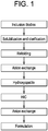
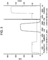
- the methods comprise refolding the immunoconjugate in a fed-batch process and purifying the refolded immunoconjugate on one or more chromatography columns.
- an active immunoconjugate wherein said immunoconjugate is deamidated at one or more residues, and wherein said deamidation results in an inhibition of potency of said immunoconjugate
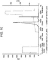
- the method comprising refolding said immunoconjugate and purifying the refolded immunoconjugate using a two cycle elution on an ion exchange column, wherein the column is stripped between a first elution and a second elution with a stripping buffer comprising ethanolamine, arginine, Ethylenediaminetetraacetic acid (EDTA), urea and dithiothreitol (DTT).
- a stripping buffer comprising ethanolamine, arginine, Ethylenediaminetetraacetic acid (EDTA), urea and dithiothreitol (DTT).
- refolding the immunoconjugate comprises a refold buffer having a pH 9.5 or less.
- the immunoconjugate comprises an antibody or antigen binding fragment thereof, for example an antibody or antigen binding fragment comprises a Fab, a Fab', a F(ab')2, a Fd, a single chain Fv or scFv, a disulfide linked Fv, a V-NAR domain, an IgNar, an intrabody, an IgG ⁇ CH2, a minibody, a F(ab')3 a tetrabody, a triabody, a diabody, a single-domain antibody, DVD-Ig, Fcab, mAb2, a (scFv)2, or a scFv-Fc.
- an antibody or antigen binding fragment comprises a Fab, a Fab', a F(ab')2, a Fd, a single chain Fv or scFv, a disulfide linked Fv, a V-NAR domain, an IgNar, an intrabody, an IgG ⁇ CH
- the antibody or antigen binding fragment binds a cell surface receptor, suitably CD22.
- the immunoconjugate comprises a toxin, for example, toxins including, but not limited to, Pseudomonas exotoxin, ricin, abrin, diphtheria toxin and subunits thereof, as well as botulinum toxins A through F and variants, and derivatives thereof.
- toxins including, but not limited to, Pseudomonas exotoxin, ricin, abrin, diphtheria toxin and subunits thereof, as well as botulinum toxins A through F and variants, and derivatives thereof.
- the toxin is Pseudomonas exotoxin, or variant thereof, suitably having an amino acid sequence selected from the group consisting of SEQ ID NOs: 16-22.
- the antibody or antigen binding fragment thereof comprises a VH and a VL sequence
- the VH sequence is selected from the group consisting of SEQ ID NOs: 6-11
- the VL sequence is selected from the group consisting of SEQ ID NOs: 2, and 12-15.
- the immunoconjugate comprises an anti-CD22 antibody or antigen binding fragment thereof and a PE or variant thereof, suitably the immunoconjugate is the Moxetumomab pasudotox immunotoxin comprising the VH-PE38 subunit of SEQ ID NO: 1 and the VL subunit of SEQ ID NO:2.
- the refold buffer has a pH of 9.4.
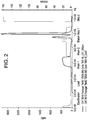
- the fed batch process uses an addition rate of about 52 mL of solubilized inclusion bodies per L of refold buffer per hour to about 13 mL solubilized inclusion bodies per L refold buffer per hour, more suitably an addition rate of about 35 mL of solubilized inclusion bodies per L of refold buffer per hour to about 17 mL solubilized inclusion bodies per L refold buffer per hour, or an addition rate of about 30 mL of solubilized inclusion bodies per L of refold buffer per hour to about 18 mL solubilized inclusion bodies per L refold buffer per hour, or an addition rate of about 26 mL of solubilized inclusion bodies per L of refold buffer per hour.
- the stripping buffer for use in the various methods comprises about 30-60 mM ethanolamine, about 0.25 to about 0.75 M arginine, about 1-3 mM EDTA, about 7-9 M urea and about 9-11 mM DTT.
- compositions comprising an immunoconjugate having less than between about 25% and about 1% deamidated species, wherein the immunoconjugate is prepared by the various methods disclosed herein.
- the method comprises refolding the immunoconjugate in a fed-batch process in a refold buffer having a pH of 9.5 or less, and purifying the refolded immunoconjugate using a two cycle elution on an ion exchange column, wherein the column is stripped between a first elution and a second elution with a stripping buffer comprising ethanolamine, arginine, Ethylenediaminetetraacetic acid (EDTA), urea and dithiothreitol (DTT).
- a stripping buffer comprising ethanolamine, arginine, Ethylenediaminetetraacetic acid (EDTA), urea and dithiothreitol (DTT).
- an amount of the immunoconjugate recovered from the method of preparation is at least three-hundred % (300%) greater than an amount of the immunoconjugate recovered utilizing a method that does not comprise a fed-batch refolding process and/or a two cycle elution on an ion exchange column that has been stripped using the stripping buffer.
- polypeptide peptide
- protein protein fragment
- amino acid polymers in which one or more amino acid residue is an artificial chemical mimetic of a corresponding naturally occurring amino acid, as well as to naturally occurring amino acid polymers and non-naturally occurring amino acid polymers.
- amino acid refers to naturally occurring and synthetic amino acids, as well as amino acid analogs and amino acid mimetics that function similarly to the naturally occurring amino acids.
- Naturally occurring amino acids are those encoded by the genetic code, as well as those amino acids that are later modified, e.g., hydroxyproline, gamma-carboxyglutamate, and O-phosphoserine.
- Amino acid analogs refer to compounds that have the same basic chemical structure as a naturally occurring amino acid, e.g., an alpha carbon that is bound to a hydrogen, a carboxyl group, an amino group, and an R group, e.g., homoserine, norleucine, methionine sulfoxide, methionine methyl sulfonium. Such analogs can have modified R groups (e.g., norleucine) or modified peptide backbones, but retain the same basic chemical structure as a naturally occurring amino acid.
- Amino acid mimetics refer to chemical compounds that have a structure that is different from the general chemical structure of an amino acid, but that functions similarly to a naturally occurring amino acid. Negatively charged amino acids include aspartic acid (or aspartate) and glutamic acid (or glutamate). Positively charged amino acids include arginine, histidine, and lysine.
- the composition may be "partially purified” (i.e., having been subjected to one or more purification steps, or may be obtained directly from a host cell or organism producing the polypeptide (e.g., the composition may comprise harvested cell culture fluid).
- an “acidic variant” is a variant of a polypeptide or immunoconjugate which is more acidic (e.g., as determined by cation exchange chromatography) than the polypeptide of interest.
- An example of an acidic variant is a deamidated variant.
- Deamidated proteins are those that have had some or all of the free amide functional groups hydrolyzed to carboxylic acids, such as conversion of glutamines to glutamic acid. The rate of this reaction is dependent on the primary sequence, three-dimensional structure, pH, temperature, buffer type, ionic strength and other solution properties.
- the deamidation reaction introduces a negative charge into the molecule. As described further below, the protein deamidation can have a negative impact on protein activity.
- antibody and “immunoglobulin” are used interchangeably in the broadest sense and include monoclonal antibodies (e.g., full length or intact monoclonal antibodies), polyclonal antibodies, multivalent antibodies, multispecific antibodies (e.g., bispecific antibodies so long as they exhibit the desired biological activity) and antibody fragments as described herein.
- bispecific antibody is intended to include any antibody that has two different binding specificities, i.e., the antibody binds two different epitopes, which can be located on the same target antigen or, more commonly, on different target antigens.
- Native antibodies and immunoglobulins are usually heterotetrameric glycoproteins of about 150,000 daltons, composed of two identical light (L) chains and two identical heavy (H) chains. Each light chain is linked to a heavy chain by one covalent disulfide bond, while the number of disulfide linkages varies between the heavy chains of different immunoglobulin isotypes. Each heavy and light chain also has regularly spaced intrachain disulfide bridges. Each heavy chain has at one end a variable domain (VH) followed by a number of constant domains. Each light chain has a variable domain at one end (VL) and a constant domain at its other end.
- VH variable domain
- VL variable domain at one end
- the constant domain of the light chain is aligned with the first constant domain of the heavy chain, and the light chain variable domain is aligned with the variable domain of the heavy chain. Particular amino acid residues are believed to form an interface between the light and heavy chain variable domains ( Clothia et al., J. Mol. Biol. 186, 651-66, 1985 ); Novotny and Haber, Proc. Natl. Acad. Sci. USA 82, 4592-4596 (1985 )).
- Five human immunoglobulin classes are defined on the basis of their heavy chain composition, and are named IgG, IgM, IgA, IgE, and IgD.
- the IgG-class and IgA-class antibodies are further divided into subclasses, namely, IgG1, IgG2, IgG3, and IgG4, and IgAl and IgA2.
- the heavy chains in IgG, IgA, and IgD antibodies have three constant region domains, that are designated CHI, CH2, and CH3, and the heavy chains in IgM and IgE antibodies have four constant region domains, CHI, CH2, CH3, and CH4.
- heavy chains have one variable region and three or four constant regions. Immunoglobulin structure and function are reviewed, for example, in Harlow et al., Eds., Antibodies: A Laboratory Manual, Chapter 14, Cold Spring Harbor Laboratory, Cold Spring Harbor (1988 ).
- antibody fragment refers to a portion of an intact antibody and refers to the antigenic determining variable regions of an intact antibody.
- antibody fragments include, but are not limited to Fab, Fab', F(ab')2, Fv and single chain Fv fragments, linear antibodies, single chain antibodies, and multispecific antibodies formed from antibody fragments.
- monoclonal antibody refers to an antibody obtained from a population of substantially homogeneous antibodies, i.e., the individual antibodies comprising the population are identical except for possible naturally occurring mutations that may be present in minor amounts. Monoclonal antibodies are highly specific and bind a single antigen. Furthermore, in contrast to polyclonal antibody preparations that typically include different antibodies directed against different determinants (epitopes), each monoclonal antibody is directed against a single determinant on the antigen.
- an antibody “selectively binds” or “specifically binds” means that the antibody reacts or associates more frequently, more rapidly, with greater duration, with greater affinity, or with some combination of the above to an epitope than with alternative substances, including unrelated proteins.
- “Selectively binds” or “specifically binds” means, for instance, that an antibody binds to a protein with a K D of at least about 0.1 mM, but more usually at least about 1 ⁇ M.
- “Selectively binds” or “specifically binds” means at times that an antibody binds to a protein at times with a K D of at least about 0.1 ⁇ M or better, and at other times at least about 0.01 ⁇ M or better. Because of the sequence identity between homologous proteins in different species, specific binding can include an antibody that recognizes a tumor cell marker protein in more than one species.
- the antibodies herein specifically include "chimeric" antibodies in which a portion of the heavy and/or light chain is identical with or homologous to corresponding sequences in antibodies derived from a particular species or belonging to a particular antibody class or subclass, while the remainder of the chain(s) is identical with or homologous to corresponding sequences in antibodies derived from another species or belonging to another antibody class or subclass, as well as fragments of such antibodies, so long as they exhibit the desired biological activity ( U.S. Patent No. 4,816,567 ; and Morrison et al., Proc. Natl. Acad. Sci. USA 57:6851-6855 (1984 )).
- Humanized forms of non-human (e.g., murine) antibodies are chimeric antibodies that contain minimal sequence derived from non-human immunoglobulin.
- humanized antibodies are human immunoglobulins (recipient antibody) in which residues from a hypervariable region of the recipient are replaced by residues from a hypervariable region of a non-human species (donor antibody) such as mouse, rat, rabbit or nonhuman primate having the desired specificity, affinity, and capacity.
- donor antibody such as mouse, rat, rabbit or nonhuman primate having the desired specificity, affinity, and capacity.
- framework region (FR) residues of the human immunoglobulin are replaced by corresponding non-human residues.
- humanized antibodies can comprise residues that are not found in the recipient antibody or in the donor antibody. These modifications are made to further refine antibody performance.
- the humanized antibody will comprise substantially all of at least one, and typically two, variable domains, in which all or substantially all of the hypervariable loops correspond to those of a non- human immunoglobulin and all, or substantially all, of the FRs are those of a human immunoglobulin sequence.
- the humanized antibody optionally will also comprise at least a portion of an immunoglobulin constant region (Fc), typically that of a human immunoglobulin.
- Fc immunoglobulin constant region
- a "human antibody” is one that possesses an amino acid sequence that corresponds to that of an antibody produced by a human and/or has been made using any of the techniques for making human antibodies as disclosed herein. This definition of a human antibody specifically excludes a humanized antibody comprising non-human antigen-binding residues.
- Immunoconjugates can also be defined by the generic formula in reverse order: A-L-C.
- cytotoxin refers to a substance that inhibits or prevents the function of cells and/or causes destruction of cells.
- the term is intended to include radioactive isotopes (e.g., At 211 , I 131 , I 125 , Y 90 , Re 186 , Re 188 , Sm 153 , Bi 212 , P 32 and radioactive isotopes of Lu), chemotherapeutic agents e.g., methotrexate, adriamicin, vinca alkaloids (vincristine, vinblastine, etoposide), doxorubicin, melphalan, mitomycin C, chlorambucil, daunorubicin or other intercalating agents, enzymes and fragments thereof such as nucleolytic enzymes, antibiotics, and toxins such as small molecule toxins or enzymatically active toxins of bacterial, fungal, plant or animal origin, including fragments and/or variant
- cytotoxic agents include, but are not limited to, abrin, ricin, Pseudomonas exotoxin (PE), diphtheria toxin (DT), botulinum toxin, or modified toxins thereof.
- PE and DT are highly toxic compounds that typically bring about death through liver toxicity.
- PE and DT can be modified into a form for use as an immunotoxin by removing the native targeting component of the toxin (e.g., domain la of PE or the B chain of DT) and replacing it with a different targeting moiety, such as an antibody.
- the toxin is Pseudomonas exotoxin.
- Pseudomonas exotoxin A PE is an extremely active monomeric protein (molecular weight 66 kD), secreted by Pseudomonas aeruginosa, which inhibits protein synthesis in eukaryotic cells through the inactivation of elongation factor 2 (EF-2) by catalyzing its ADP-ribosylation (catalyzing the transfer of the ADP ribosyl moiety of oxidized NAD onto EF-2).
- EF-2 elongation factor 2
- PE immunoconjugate or "PE immunotoxin” is an immunoconjugate or immunotoxin comprising an antibody or antigen binding fragment thereof and a PE toxin or variant thereof.
- purifying a polypeptide or immunoconjugate from a composition comprising the polypeptide and one or more impurities, is meant increasing the degree of purity of the polypeptide in the composition by removing (completely or partially) at least one impurity from the composition. According to the present invention, purification is performed without the use of an affinity chromatography step.
- chromatography refers to the process by which a solute of interest in a mixture is separated from other solutes in a mixture as a result of differences in rates at which the individual solutes of the mixture migrate through a stationary medium under the influence of a moving phase, or in bind and elute processes.
- ion-exchange and ion-exchange chromatography refers to the chromatographic process in which a solute of interest (such as a protein) in a mixture interacts with a charged compound linked (such as by covalent attachment) to a solid phase ion exchange material such that the solute of interest interacts non-specifically with the charged compound more or less than solute impurities or contaminants in the mixture.
- the contaminating solutes in the mixture elute from a column of the ion exchange material faster or slower than the solute of interest or are bound to or excluded from the resin relative to the solute of interest.
- Ion-exchange chromatography specifically includes cation exchange, anion exchange, and mixed mode chromatography.
- ion exchange material refers to a solid phase that is negatively charged (i.e., a cation exchange resin) or positively charged (i.e., an anion exchange resin).
- the charge may be provided by attaching one or more charged ligands to the solid phase, e.g., by covalent linking.
- the charge may be an inherent property of the solid phase (e.g., as is the case for silica, which has an overall negative charge).
- anion exchange resin refers to a solid phase which is positively charged, thus having one or more positively charged ligands attached thereto. Any positively charged ligand attached to the solid phase suitable to form the anionic exchange resin can be used, such as quaternary amino groups
- Commercially available anion exchange resins include DEAE cellulose, Poros PI 20, PI 50, HQ 10, HQ 20, HQ 50, D 50 from Applied Biosystems, Sartobind Q from Sartorius, MonoQ, MiniQ, Source 15Q and 30Q, Q, DEAE and ANX Sepharose Fast Flow, Q Sepharose High Performance, QAE SEPHADEXTM and FAST Q SEPHAROSETM (GE Healthcare), WP PEI, WP DEAM, WP QUAT from J. T.
- solid phase is meant a non-aqueous matrix to which one or more charged ligands can adhere.
- the solid phase may be a purification column, a discontinuous phase of discrete particles, a membrane, or filter etc.
- materials for forming the solid phase include polysaccharides (such as agarose and cellulose); and other mechanically stable matrices such as silica (e.g., controlled pore glass), poly(styrenedivinyl)benzene, polyacrylamide, ceramic particles and derivatives of any of the above.
- specific binding refers to the generally reversible binding of a protein of interest to a ligand through the combined effects of spatial complementarity of protein and ligand structures at a binding site coupled with electrostatic forces, hydrogen bonding, hydrophobic forces, and/or van der Waals forces at the binding site.
- Specific binding include antibody-antigen binding, enzyme-substrate binding, enzyme-cofactor binding, metal ion chelation, DNA binding protein-DNA binding, regulatory protein-protein interactions, and the like.
- non-specific binding refers to binding of a protein of interest to the ligand or compound on a solid phase matrix through electrostatic forces, hydrogen bonding, hydrophobic forces, and/or van der Waals forces at an interaction site, but lacking structural complementarity that enhances the effects of the non-structural forces.
- non-specific interactions include, but are not limited to, electrostatic, hydrophobic, and van der Waals forces as well as hydrogen bonding.
- a "buffer” used in the present invention is a solution that resists changes in pH by the addition of acid or base by the action of its acid-base conjugates components.
- Various buffers can be employed in a method of the present invention depending on the desired pH of the buffer and the particular step in the purification process [see Buffers. A Guide for the Preparation and Use of Buffers in Biological Systems, Gueffroy, D., ed. Calbiochem Corporation (1975 )].
- Non-limiting examples of buffer components that can be used to control the pH range desirable for a method of the invention include acetate, citrate, histidine, phosphate, ammonium buffers such as ammonium acetate, succinate, MES, CHAPS, MOPS, MOPSO, HEPES, Tris, and the like, as well as combinations of these TRIS-malic acid-NaOH, maleate, chloroacetate, formate, benzoate, propionate, pyridine, piperazine, ADA, PIPES, ACES, BES, TES, tricine, bicine, TAPS, ethanolamine, CHES, CAPS, methylamine, piperidine, O-boric acid, carbonic acid, lactic acid, butaneandioic acid, diethylmalonic acid, glycylglycine, HEPPS, HEPPSO, imidazole, phenol, POPSO, succinate, TAPS, amine-based, benzylamine, trimethyl or dimethyl
- the "loading buffer” is that which is used to load the composition comprising the polypeptide molecule of interest and one or more impurities onto the ion exchange resin.
- the loading buffer has a conductivity and/or pH such that the polypeptide molecule of interest (and generally one or more impurities) is/are bound to the ion exchange resin or such that the protein of interest flows through the column while the impurities bind to the resin.
- wash buffer when used herein refers to a buffer used to wash or re-equilibrate the ion exchange resin, prior to eluting the polypeptide molecule of interest. Conveniently, the wash buffer and loading buffer may be the same, but this is not required.
- the "elution buffer” is used to elute the polypeptide of interest from the solid phase.
- the conductivity and/or pH of the elution buffer is/are such that the polypeptide of interest is eluted from the ion exchange resin.
- pi isoelectric point of a polypeptide refer to the pH at which the polypeptide's positive charge balances its negative charge
- pi can be calculated from the net charge of the amino acid residues or sialic acid residues of attached carbohydrates of the polypeptide or can be determined by isoelectric focusing.
- binding a molecule to an ion exchange material
- washing By “washing" the ion exchange material is meant passing an appropriate buffer through or over the ion exchange material.
- a molecule e.g., polypeptide or impurity
- an ion exchange material is meant to remove the molecule therefrom by altering the ionic strength of the buffer surrounding the ion exchange material such that the buffer competes with the molecule for the charged sites on the ion exchange material.
- Toxins can be employed with antibodies of the present invention to yield immunotoxins.
- Exemplary toxins include ricin, abrin, diphtheria toxin and subunits thereof, as well as botulinum toxins A through F. These toxins are readily available from commercial sources (e.g., Sigma Chemical Company, St. Louis, Mo.). Diphtheria toxin is isolated from Corynebaclerium diphtheriae. Ricin is the lectin RCA60 from Ricinus communis (Castor bean). The term also references toxic variants thereof. For example, see, U.S. Pat. Nos. 5,079 , 163 and 4,689,401 .
- Ricinus communis agglutinin occurs in two forms designated RCA 60 and RCA 120 according to their molecular weights of approximately 65 and 120 kD, respectively ( Nicholson & Blaustein, J. Biochem. Biophys. Acta 266:543 (1972 )).
- the A chain is responsible for inactivating protein synthesis and killing cells.
- the B chain binds ricin to cell-surface galactose residues and facilitates transport of the A chain into the cytosol ( Olsnes, et al., Nature 249:621-631 (1974 ) and U.S. Pat. No. 3,060,165 ).
- Abrin includes toxic lectins from Abrus precalorius.
- the toxic principles, abrin a, b, c, and d have a molecular weight of from about 63 and 67 kD and are composed of two disulfide-linked polypeptide chains A and B.
- the A chain inhibits protein synthesis; the B-chain (abrin-b) binds to D-galactose residues (see, Funatsu, et al., Agr. Biol. Chem. 52: 1095 (1988 ); and Olsnes, Methods Enzymol. 50:330-335 (1978 )).
- the toxin is Pseudomonas exotoxin (PE).
- PE Pseudomonas exotoxin
- the Pseudomonas exotoxin (or exotoxin A) is an exotoxin produced by Pseudomonas aeruginosa.
- the term "Pseudomonas exotoxin" as used herein refers to a full-length native (naturally occurring) PE or a PE that has been modified.
- Such modifications may include, but are not limited to, elimination of domain la, various amino acid deletions in domains Ib, II and III, single amino acid substitutions and the addition of one or more sequences at the carboxyl terminus such as KDEL (SEQ ID NO:3) and REDL (SEQ ID NO:4). See Siegall, et al., J. Biol. Chem. 264: 14256-14261 (1989 ).
- the cytotoxic fragment of PE retains at least 50%, preferably 75%, more preferably at least 90%, and most preferably 95% of the cytotoxicity of native PE. In a most preferred embodiment, the cytotoxic fragment is more toxic than native PE.
- PE Native Pseudomonas exotoxin A
- EF-2 elongation factor 2
- the exotoxin contains three structural domains that act in concert to cause cytotoxicity. Domain Ia (amino acids 1-252) mediates cell binding.
- Domain II (amino acids 253-364) is responsible for translocation into the cytosol and domain III (amino acids 400-613) mediates ADP ribosylation of elongation factor 2.
- domain b (amino acids 365-399) remains undefined, although a large part of it, amino acids 365-380, can be deleted without loss of cytotoxicity. See Siegall, et al., (1989), supra.
- PE employed in the present invention includes the native sequence, cytotoxic fragments of the native sequence, and conservatively modified variants of native PE and its cytotoxic fragments.
- PE variants useful in the invention are described in US 7,355,012 , and WO 2007/016150 and WO 2009/032954 , the disclosures of each of which are incorporated by reference herein in their entireties.
- Cytotoxic fragments of PE include those which are cytotoxic with or without subsequent proteolytic or other processing in the target cell (e.g., as a protein or pre-protein). Cytotoxic fragments of PE include PE40, PE38, and PE35.
- the PE has been modified to reduce or eliminate nonspecific cell binding, frequently by deleting domain Ia as taught in U.S. Pat. No. 4,892,827 , although this can also be achieved, for example, by mutating certain residues of domain Ia.
- U.S. Pat. No. 5,512,658 discloses that a mutated PE in which Domain Ia is present but in which the basic residues of domain Ia at positions 57, 246, 247, and 249 are replaced with acidic residues (glutamic acid, or "E")) exhibits greatly diminished non-specific cytotoxicity.
- This mutant form of PE is sometimes referred to as PE4E.
- PE40 is a truncated derivative of PE as previously described in the art, with a deletion of domain la of the native PE molecule. See, Pai, et al., Proc. Nat'l Acad. Sci USA 55:3358-62 (1991 ); and Kondo, et al., J. Biol. Chem. 263:9470-9475 (1988 ).
- PE35 is a 35 kD carboxyl-terminal fragment of PE in which amino acid residues 1-279 have been deleted and the molecule commences with a met at position 280 followed by amino acids 281-364 and 381-613 of native PE.
- PE35 and PE40 are disclosed, for example, in U.S. Pat. Nos. 5,602,095 and 4,892,827 .
- PE4E is a form of PE where all of the domains of native PE are present, but where the basic residues of domain la at positions 57, 246, 247 and 249 are replaced with acidic residues (glutamine acid, or "E"
- the cytotoxic fragment PE38 is employed.
- PE38 is a truncated PE pro-protein composed of amino acids 253-364 and 381-613 which is activated to its cytotoxic form upon processing within a cell (see e.g., U.S. Pat. Nos. 5,608,039 , 7,355,012 , and Pastan et al., Biochim. Biophys. Acta 1333:C1-C6 (1997 ), the disclosures of each of which are incorporated by reference herein in their entireties).
- domain lb may be deleted, and the remaining portions joined by a linker or directly by a peptide bond. Some of the amino portion of domain II may be deleted. And, the C-terminal end may contain the native sequence of residues 609-613 (REDLK) (SEQ ID NO: 5), or may contain a variation found to maintain the ability of the construct to translocate into the cytosol, such as REDL (SEQ ID NO:4) or KDEL (SEQ ID NO:3), and repeats of these sequences. See, e.g., U.S. Pat. Nos. 5,854,044 ; 5,821,238 ; and 5,602,095 and WO 99/51643 .
- REDLK native sequence of residues 609-613
- REDL SEQ ID NO:4
- KDEL SEQ ID NO:3
- the PE is PE4E, PE40, or PE38
- any form of PE in which non-specific cytotoxicity has been eliminated or reduced to levels in which significant toxicity to non-targeted cells does not occur can be used in the immunotoxins of the present invention so long as it remains capable of translocation and EF-2 ribosylation in a targeted cell.
- PE or cytotoxic fragments thereof have at least 80% sequence similarity, preferably at least 85% sequence similarity, more preferably at least 90% sequence similarity, and most preferably at least 95% sequence similarity at the amino acid level, with the PE of interest, such as PE38.
- conservatively modified variants refer to those nucleic acid sequences which encode identical or essentially identical amino acid sequences, or if the nucleic acid does not encode an amino acid sequence, to essentially identical nucleic acid sequences.
- a large number of functionally identical nucleic acids encode any given polypeptide. For instance, the codons GCA, GCC, GCG and GCU all encode the amino acid alanine. Thus, at every position where an alanine is specified by a codon, the codon can be altered to any of the corresponding codons described without altering the encoded polypeptide.
- nucleic acid variations are "silent variations," which are one species of conservatively modified variations. Every nucleic acid sequence herein which encodes a polypeptide also describes every possible silent variation of the nucleic acid.
- each codon in a nucleic acid except AUG, which is ordinarily the only codon for methionine
- each silent variation of a nucleic acid which encodes a polypeptide is implicit in each described sequence.
- amino acid sequences one of ordinary skill in the art will recognize that individual substitutions, deletions or additions to a nucleic acid, peptide, polypeptide, or protein sequence which alters, adds or deletes a single amino acid or a small percentage of amino acids in the encoded sequence is a "conservatively modified variant" where the alteration results in the substitution of an amino acid with a chemically similar amino acid.
- Pseudomonas exotoxins employed in the invention can be assayed for the desired level of cytotoxicity by assays well known to those of skill in the art.
- cytotoxic fragments of PE and conservatively modified variants of such fragments can be readily assayed for cytotoxicity.
- a large number of candidate PE molecules can be assayed simultaneously for cytotoxicity by methods well known in the art.
- subgroups of the candidate molecules can be assayed for cytotoxicity.
- Positively reacting subgroups of the candidate molecules can be continually subdivided and reassayed until the desired cytotoxic fragment(s) is identified.
- Such methods allow rapid screening of large numbers of cytotoxic fragments or conservative variants of PE.
- the polypeptide of interest comprises an antibody that specifically binds CD22.
- CD22 refers to a lineage-restricted B cell antigen belonging to the Ig superfamily. It is expressed in 60-70% of B cell lymphomas and leukemias and is not present on the cell surface in early stages of B cell development or on stem cells. See, e.g., Vaickus et al., Crit. Rev. Oncol/Hematol. 77:267-297 (1991 ).
- the polypeptide of interest is an antibody fragment that binds CD22 (e.g., Fab, or scFv).
- the term "anti-CD22" in reference to an antibody refers to an antibody that specifically binds CD22 and includes reference to an antibody which is generated against CD22.
- the CD22 is a primate CD22 such as human CD22.
- the antibody is generated against human CD22 synthesized by a non-primate mammal after introduction into the animal of cDNA which encodes human CD22.
- the polypeptide of interest is a CD22 antibody immunoconjugate that comprises the PE38 exotoxin.
- CD22/PE38 immunoconjugate is Moxetumomab pasudotox described in International Patent Application Publication Nos. WO 2012/015912 , WO 98/41641 and WO2003/27135 , US Patent Nos. 7,541,034 , 7,355,012 , and U.S. Publication No. 2007/0189962 , all of which are herein incorporated by reference in their entireties.
- Moxetumomab pasudotox is a recombinant immunotoxin protein composed of an antibody Fv fragment based on the murine anti-CD22 antibody RFB4 fused to a truncated form of the Pseudomonas exotoxin protein, PE38.
- the anti-CD22 Fv fragment consists of two domains, a VL and a VH, where the latter was modified to improve binding to the human CD22 target.
- the Moxetumomab pasudotox protein is comprised of two independent polypeptides, the VL chain (SEQ ID NO:2), and the VH chain, fused at the C- terminus to the PE38 domain (VH-PE38) (SEQ ID NO: 1).
- VL and VH-PE38 sequences useful in this invention are described in US 7,541,034 , 7,355,012 , 2007/0189962 and WO 2012/015912 , the disclosures of each of which are incorporated by reference herein in their entireties. Both domains were designed to each contain engineered cysteine residues that permit formation of an intermolecular disulfide bond. This feature increases the stability of the fusion protein.
- amino acid sequence of the VH-P38 Subunit (SEQ ID NO: 1) of Moxetumomab pasudotox is the following:
- the PE38 sequence is shown in bold, and the five amino acid linker between the VH domain and the PE38 domain is shown underlined.
- amino acid sequence of the VL Subunit (SEQ ID NO:2) of Moxetumomab pasudotox is the following:
- amino acid sequence of the VH domain of the immunoconjugate is one of the following:
- amino acid sequence of the VL domain of the immunoconjugate is one of the following:
- the PE toxin of the immunoconjugate is a PE or variant thereof selected from the following:
- the PE toxin of the immunoconjugate is fused or conjugated to either the VH or VL domain directly or via a linker at either the N-terminus or the C-terminus of the VH or VL domain.
- a linker is described above for Moxetumomab pasudotox and corresponds to the amino acid sequence KASGG (SEQ ID NO: 23). Additional linkers can be readily generated by techniques known in the art.
- the PE immunoconjugate of the present invention is suitably expressed in cells, such as bacterial cells, and then isolated from inclusion bodies.
- the PE immunoconjugate isolated from inclusion bodies is then further purified/isolated using downstream steps as described herein.
- host-expression vector systems may be utilized to express the PE immunoconjugate of the present invention.
- host-expression systems represent vehicles by which the coding sequences of interest may be produced and subsequently purified, but also represent cells which may, when transformed or transfected with the appropriate nucleotide coding sequences, express an antibody molecule of the invention in situ.
- host-expression systems include vehicles by which the coding sequences of interest may be produced and subsequently purified, but also represent cells which may, when transformed or transfected with the appropriate nucleotide coding sequences, express an antibody molecule of the invention in situ.
- microorganisms such as bacteria (e.g., E. coli, B.
- subtilis transformed with recombinant bacteriophage DNA, plasmid DNA or cosmid DNA expression vectors containing antibody coding sequences; yeast (e.g., Saccharomyces, Pichia ) transformed with recombinant yeast expression vectors containing antibody coding sequences; insect cell systems infected with recombinant virus expression vectors (e.g., baculovirus) containing antibody coding sequences; plant cell systems infected with recombinant virus expression vectors (e.g., cauliflower mosaic virus, CaMV; tobacco mosaic virus, TMV) or transformed with recombinant plasmid expression vectors containing antibody coding sequences; or mammalian cell systems (e.g., COS, CHO, BLK, 293, 3T3 cells) harboring recombinant expression constructs containing promoters derived from the genome of mammalian cells (e.g., metallothionein promoter) or from mammalian viruses (e.g., the
- VL and VH-PE toxin e.g., VH-PE38
- VL and VH-PE toxin e.g., VH-PE38
- DNA encoding each of the VL and VH-PE toxin (e.g., VH-PE38) polypeptides can be introduced into an expression vector by techniques well known in the art.
- a “vector” refers to any vehicle for the cloning of and/or transfer of a nucleic acid into a host cell.
- a vector may be a replicon to which another DNA segment may be attached so as to bring about the replication of the attached segment.
- a “replicon” refers to any genetic element (e.g., plasmid, phage, cosmid, chromosome, virus) that functions as an autonomous unit of DNA replication in vivo, i.e., capable of replication under its own control.
- the term “vector” includes vehicles for introducing the nucleic acid into a cell in vitro, ex vivo or in vivo.
- a large number of vectors known in the art may be used to manipulate nucleic acids, incorporate response elements and promoters, such as inducible promoters, into genes, etc.
- Possible vectors include, for example, plasmids such as pBR322 or pUC plasmid derivatives, or the Bluescript vector.
- the insertion of the DNA fragments corresponding to response elements and promoters into a suitable vector can be accomplished by ligating the appropriate DNA fragments into a chosen vector that has complementary cohesive termini.
- the ends of the DNA molecules may be enzymatically modified or any site may be produced by ligating nucleotide sequences (linkers) into the DNA termini.
- Such vectors may be engineered to contain selectable marker genes that provide for the selection of cells. Such markers allow identification and/or selection of host cells that express the proteins encoded by the marker.
- expression vector refers to a vector, plasmid or vehicle designed to enable the expression of an inserted nucleic acid sequence following transformation into the host.
- the cloned gene i.e., the inserted nucleic acid sequence, e.g., a gene encoding an anti-CD22 VH, anti-CD22 VL, or anti-CD22 VH or VL fused to a PE toxin, is usually placed under the control of control elements such as a promoter, a minimal promoter, an enhancer, or the like.
- Initiation control regions or promoters which are useful to drive expression of a nucleic acid in the desired host cell are numerous and familiar to those skilled in the art.
- any promoter capable of driving expression of these genes can be used in an expression vector, including but not limited to, viral promoters, bacterial promoters, animal promoters, mammalian promoters, synthetic promoters, constitutive promoters, tissue specific promoters, pathogenesis or disease related promoters, developmental specific promoters, inducible promoters, light regulated promoters; including, but are not limited to, the SV40 early (SV40) promoter region, the promoter contained in the 3' long terminal repeat (LTR) of Rous sarcoma virus (RSV), the EIA or major late promoter (MLP) of adenoviruses (Ad), the human cytomegalovirus (HCMV) immediate early promoter, the herpes simplex virus (HSV) thymidine kinase (TK) promoter, the baculovirus IE1 promoter, the elongation factor 1 alpha (EF1) promoter, the glyceraldehyde-3 -
- RNA product refers to the biological production of a product encoded by a coding sequence.
- a DNA sequence including the coding sequence, is transcribed to form a messenger-RNA (mRNA).
- mRNA messenger-RNA
- the messenger-RNA is then translated to form a polypeptide product which has a relevant biological activity.
- the process of expression may involve further processing steps to the RNA product of transcription, such as splicing to remove introns, and/or post-translational processing of a polypeptide product.
- the VL and VH-PE38 polypeptides are suitably expressed in cells, e.g., bacterial cells, such as E. coli.
- the polypeptides are expressed, e.g., in E. coli cells and isolated from inclusion bodies.
- the VL and VH-PE38 subunits are expressed in different cells.
- the VL is expressed in one cell on a first vector and the VH-PE38 is expressed in a different cell on a second vector.
- the VL and VH-PE38 subunits are expressed in the same cell on different vectors, for example, the VL is expressed in one cell on a first vector, and the VH-PE38 is expressed in the same cell on a different vector.
- the VL and VH-PE38 subunits are expressed on the same vector in the same cell. Inclusion bodies from the cells are recovered, solubilized and the VL and VH-PE38 subunits combined to form an immunoconjugate, as described herein.
- active immunoconjugates i.e., immunoconjugates capable of binding a desired target and delivering the compound (e.g., immunotoxin) that is attached to the cell targeting agent (e.g., an antibody, antibody fragment or other protein).
- the compound e.g., immunotoxin
- the cell targeting agent e.g., an antibody, antibody fragment or other protein
- the methods described herein are suitably used to prepare immunoconjugates that are deamidated at one or more residues. As described herein, such deamidation often results in an inhibition of potency of an immunoconjugate, and thus the methods provided are beneficial to preparing active immunoconjugates suitable for clinical settings.
- immunoconjugates are suitably prepared using expression systems from bacteria, including E. coli. Inclusion bodies from the cells are recovered, solubilized and the proteins are recovered. In embodiments, the VL and VH-toxin subunits designed to form an immunoconjugate, as described herein, are prepared in the bacterial cells.
- inclusion bodies comprising the desired immunoconjugate subunits are solubilized, concentrated and clarified. Suitable methods of clarification are described herein as well as in the disclosure and examples of WO2012/015912 , the disclosure of which is incorporated by reference herein in its entirety. Following clarification, refolding of the immunoconjugate is carried out.
- methods of preparing immunoconjugates suitably comprise refolding an immunoconjugate using a fed-batch process. Following the refolding, the refolded immunoconjugate is purified with one or more chromatography columns as described herein as well as in WO2012/015912 .
- refolding refers to the process under which a protein, isolated from inclusion bodies, is folded into its characteristic and functional three-dimensional structure from a prior random orientation.
- a “fed-batch” process refers to a refolding process in which a solubilized inclusion body mixture (containing the desired immunoconjugate) is added (i.e., injected or mixed) to a suitable refold buffer at a controlled rate over a period of time.
- a solubilized inclusion body mixture containing the desired immunoconjugate
- the addition occurs at a steady rate over the entire time course, though the rate can also be varied during the process if desired.
- This addition time course is referred to herein as the "addition rate,” and suitably is expressed in L/hr.
- the yield of recovered immunoconjugate is suitably at least about 20% greater, at least about 30% greater, at least about 40% greater, at least about 50% greater, at least about 60% greater, at least about 70% greater, at least about 80% greater, at least about 90% greater, at least about 100% greater, at least about 125% greater, at least about 150% greater, at least about 175% greater, at least about 200% greater, at least about 300% greater, at least about 400% greater, at least about 500% greater, at least about 600% greater, at least about 700% greater, at least about 800% greater, at least about 900% greater, etc., in comparison to non-fed-batch processes that utilizes a bulk addition of subunits and a constant concentration of subunits.
- the yield of recovered immunoconjugate is suitably at least about 20% greater, at least about 30% greater, at least about 40% greater, at least about 50% greater, at least about 60% greater, at least about 70% greater, at least about 80% greater, at least about 90% greater, at least about 100% greater, at least about 125% greater, at least about 150% greater, at least about 175% greater, at least about 200% greater, at least about 300% greater, at least about 400% greater, at least about 500% greater, at least about 600% greater, at least about 700% greater, at least about 800% greater, at least about 900% greater, etc., wherein the immunoconjugate is refolded in a fed-batch process in a refold buffer having a pH of 9.5 or less.
- the refolded immunoconjugate is purified using a two cycle elution on an ion exchange column, wherein the column is stripped between a first elution and a secon elution with a stripping buffer comprising ethanolamine, arginine, Ethylendiaminetetraacetic acid (EDTA), urea and dithiothreitol (DTT).
- a stripping buffer comprising ethanolamine, arginine, Ethylendiaminetetraacetic acid (EDTA), urea and dithiothreitol (DTT).
- the process of recovering said immunoconjugate corresponds to that of a non-fed batch process, except for having the refold performed in a fed-batch process in a refold buffer having a pH or 9.5 or less and/or purifying said immunoconjugate using a two cycle elution on an ion exchange column, wherein the column is stripped between a first elution and a secon elution with a stripping buffer comprising ethanolamine, arginine, Ethylendiaminetetraacetic acid (EDTA), urea and dithiothreitol (DTT).
- a stripping buffer comprising ethanolamine, arginine, Ethylendiaminetetraacetic acid (EDTA), urea and dithiothreitol (DTT).
- the addition rate (L/hr) of the solubilized inclusion bodies is set so that a solubilized inclusion body mixture is added to a refold buffer (suitably pre-chilled) over the course of approximately 2-8 hours, suitably 3-6 hours, 3-5 hours, or more suitably over the course of about 4 hours.
- the addition is over the course of 1-10 hours, using an addition rate of about 100 mL of solubilized inclusion bodies per L of refold buffer per hour to an addition rate of about 5 mL solubilized inclusion bodies per L refold buffer per hour is utilized (about 100 mL/L/hr to about 5 mL/L/hr).
- the addition is over the course of 2-8 hours, using an addition rate of about 52 mL of solubilized inclusion bodies per L of refold buffer per hour to an addition rate of about 13 mL solubilized inclusion bodies per L refold buffer per hour is utilized (about 52 mL/L/hr to about 13 mL/L/hr).
- the addition is over the course of 3-6 hours, using an addition rate of about 35 mL solubilized inclusion bodies per L refold buffer per hour to an addition rate of about 17 mL solubilized inclusion bodies per L refold buffer per hour (about 35 mL/L/hr to about 17 mL/L/hr). In further embodiments, the addition is over the course of 3.5-5 hours, using an addition rate of about 30 mL solubilized inclusion bodies per L refold buffer per hour to an addition rate of about 18 mL solubilized inclusion bodies per L refold buffer per hour (about 30 mL/L/hr to about 18 mL/L/hr).
- the addition is over the course of about 4 hours, using an addition rate of about 26 mL solubilized inclusion bodies per L refold buffer per hour (about 26 mL/L/hr). In other embodiments, further addition rates can also be utilized in the fed-batch processes described herein.
- kits for preparing an immunoconjugate wherein the immunoconjugate is deamidated at one or more residues, and wherein the deamidation results in an inhibition of potency of the immunoconjugate.
- the methods comprise refolding the immunoconjugate using any of the methods described herein or as disclosed in WO 2012/0152912 .
- the refolded immunoconjugate is then purified with a two-cycle elution on an ion exchange column.
- the column is stripped between operating cycles (i.e.,column load, wash, elute) of the refolded immunoconjugate utilizing a stripping buffer.
- the stripping buffer that is utilized in the methods described herein comprises buffered, arginine, urea and dithiothreitol (DTT).
- DTT dithiothreitol
- the methods described herein can also utilize 3-, 4-, 5-, 6-, 7-, 8-, 9-, 10-, etc, column cycles , as desired, with stripping with the described stripping buffer between each consecutive cycle.
- the yield of recovered immunoconjugate is suitably at least about 20% greater, at least about 30% greater, at least about 40% greater, at least about 50% greater, at least about 60% greater, at least about 70% greater, at least about 80% greater, at least about 90% greater, at least about 100% greater, at least about 125% greater, at least about 150% greater, at least about 175% greater, at least about 200% greater, at least about 300% greater, at least about 400% greater, at least about 500% greater, at least about 600% greater, at least about 700% greater, at least about 800% greater, at least about 900% greater, etc., in comparison to elution methods and columns that do not utilize a stripping buffer as described herein between consecutive elutions
- the stripping buffer useful in the methods described herein comprises about 0.10 to about 0.9 M arginine, about 5-10 M urea and about 7-15 mM DTT. More suitably, the stripping buffer comprises , about 0.25 to about 0.75 M arginine, about 7-9 M urea and about 9-11 mM DTT. More suitably, about 0.45 to about 0.55 M arginine, about 7.5-8.5 M urea and about 9.5-10.5 mM DTT. Most suitably, the stripping buffer comprises about about 0.50 M arginine, about 8.0 M urea and about 10.0 mM DTT.
- the refolding buffer that is utilized in the refolding steps described herein has a pH of less than or about 10.0, suitably less than or about 9.5, and more suitably less than or about 9.4 (e.g., a pH of about 10.0, about 9.9, about 9.8, about 9.7, about 9.6, about 9.5, about 9.3, about 9.2, about 9.1 or about 9.0). It has been surprisingly found that the use of a refolding buffer having a pH of less than or about 10.0, less than or about 9.5 and most suitably less than about 9.4, results in an increased yield of the final immunoconjugate, as compared to processes that utilize refold buffers having pHs greater than these recited values.
- the yield of recovered immunoconjugate is suitably at least about 20% greater, at least about 30% greater, at least about 40% greater, at least about 50% greater, at least about 60% greater, at least about 70% greater, at least about 80% greater, at least about 90% greater, at least about 100% greater, at least about 125% greater, at least about 150% greater, at least about 175% greater, at least about 200% greater, at least about 300% greater, at least about 400% greater, at least about 500% greater, at least about 600% greater, at least about 700% greater, at least about 800% greater, at least about 900% greater, etc., in comparison to processes that utilize refold buffers having pHs greater than these recited values.
- the immunoconjugate comprises an antibody or antigen binding fragment thereof.
- the antibody or antigen binding fragment comprises a Fab, a Fab', a F(ab')2, a Fd, a single chain Fv or scFv, a disulfide linked Fv, a V-NAR domain, an IgNar, an intrabody, an IgG ⁇ CH2, a minibody, a F(ab') 3 a tetrabody, a triabody, a diabody, a single-domain antibody, DVD-Ig, Fcab, mAb 2 , a (scFv) 2 , or a scFv-Fc.
- the antibody or antigen binding fragment of the immunoconjugate binds a cell surface receptor.
- An exemplary cell surface receptor includes CD22.
- the immunoconjugate that is prepared according to the methods described herein comprises a toxin.
- a toxin Exemplary toxins and methods of preparing such toxins are described throughout.
- the toxin is selected from the group consisting of: Pseudomonas exotoxin, ricin, abrin, diphtheria toxin and subunits thereof, as well as botulinum toxins A through F or variants, or derivatives thereof.
- the toxin is a Pseudomonas exotoxin, or variant thereof. Exemplary methods of preparing Pseudomonas exotoxin (PE) are described herein in detail as well as in WO2012/015912 .
- the Pseudomonas exotoxin for use in the immunoconjugates described herein has an amino acid sequence selected from the group consisting of SEQ ID NOs: 16-22.
- the Pseudomonas exotoxin, or variant thereof has the amino acid sequence of SEQ ID NO:22.
- the antibody or antigen binding fragment thereof that is a component of the immunotoxin comprises a VH and a VL sequence.
- the VH sequence is selected from the group consisting of SEQ ID NOs: 6-11
- the VL sequence is selected from the group consisting of SEQ ID NOs: 2, and 12-15.
- the methods of preparing immunoconjugates are suitably used for preparing immunoconjugates that comprise an anti-CD22 antibody or antigen binding fragment thereof and a PE or variant thereof.
- the immunoconjugate that is prepared by the various methods described herein is the Moxetumomab pasudotox immunotoxin comprising the VH-PE38 subunit of SEQ ID NO: 1 and the VL subunit of SEQ ID NO:2.
- methods of preparing an active immunoconjugate that combine the various processes described herein that have been determined to increase the yield of an active immunoconjugate.
- the immunoconjugate is deamidated at one or more residues, and the deamidation results in an inhibition of potency of the immunoconjugate.
- such methods suitably comprise refolding an immunoconjugate with a fed-batch process in a refold buffer having a pH of less than 9.5 and purifying the refolded immunoconjugate with a two cycle elution on an ion exchange column, wherein the column is stripped between a first elution and a second elution with a stripping buffer comprising ethanolamine, arginine, Ethylenediaminetetraacetic acid (EDTA), urea and dithiothreitol (DTT).
- a stripping buffer comprising ethanolamine, arginine, Ethylenediaminetetraacetic acid (EDTA), urea and dithiothreitol (DTT).
- the various methods described herein provide an amount of the immunoconjugate recovered from the methods that is at least three-hundred % (300%) greater than an amount of the immunoconjugate recovered utilizing a method that does not comprise a fed-batch refolding process and/or a two cycle elution on an ion exchange column that has been stripped using the described stripping buffer and/or does not utilize a refolding buffer having a pH less than 9.4.
- the amount of recovered immunoconjugate is suitably at least about 20% greater, at least about 30% greater, at least about 40% greater, at least about 50% greater, at least about 60% greater, at least about 70% greater, at least about 80% greater, at least about 90% greater, at least about 100% greater, at least about 125% greater, at least about 150% greater, at least about 175% greater, at least about 200% greater, at least about 300% greater, at least about 400% greater, at least about 500% greater, at least about 600% greater, at least about 700% greater, at least about 800% greater, at least about 900% greater, etc., in comparison to processes that does not comprise a fed-batch refolding process and/or a two cycle elution on an ion exchange column that has been stripped using the described stripping buffer and/or does not utilize a refolding buffer having a pH less than 9.4.
- the immunoconjugate comprises an antibody or antigen binding fragment thereof, including an antibody or antigen binding fragment of the immunoconjugate binds a cell surface receptor such as CD22.
- the immunoconjugate comprises a toxin, suitably Pseudomonas exotoxin (PE).
- PE Pseudomonas exotoxin
- the antibody or antigen binding fragment thereof that is a component of the immunotoxin comprises a VH and a VL sequence.
- the VH sequence is selected from the group consisting of SEQ ID NOs: 6-11
- the VL sequence is selected from the group consisting of SEQ ID NOs: 2, and 12-15.
- the methods of preparing immunoconjugates are suitably used for preparing immunoconjugates that comprise an anti-CD22 antibody or antigen binding fragment thereof and a PE or variant thereof.
- the immunoconjugate that is prepared by the various methods described herein is the Moxetumomab pasudotox immunotoxin comprising the VH-PE38 subunit of SEQ ID NO: 1 and the VL subunit of SEQ ID NO:2.
- compositions comprising an immunoconjugate prepared by the various methods described herein are provided.
- the immunoconjugates prepared by such methods have less than between about 25% and about 1% deamidated species. More suitably, less than about 25% of the deamidated species is present, or less than about 20% of the deamidated species is present, or less than about 10% of the deamidated species is present, or less than about 5% of the deamidated species is present, or less than about 3% of the deamidated species is present, or less than about 2% of the deamidated species is present, or less than about 1% of the deamidated species is present.
- CAT-8015 (Moxetumomab pasudotox) is a recombinant immunotoxin produced in E. coli inclusion bodies.
- Generation of active Moxetumomab pasudotox suitably utilizes refolding from inactive pre-cursors and purification of the refolded product by a 4-column process.
- FIG. 1 provides an overview of the renaturation and purification processes.
- the purpose of the solubilization process is to extract and transfer VH and VL suitably from insoluble inclusion bodies into the liquid phase and to denature both subunits prior to refolding.
- Depth filtration removes insoluble cell debris and inclusion body components from solubilized VH and VL.
- the filtrate is subsequently concentrated by tangential flow filtration to a fixed retentate weight, which is determined by the retentate dilution factor and final refold weight.
- the function of the concentration step is to ensure consistent refold starting conditions in terms of VH and VL concentrations and Dithiothreitol (DTT) to oxidized glutathione ratio.
- DTT Dithiothreitol
- the objective of the 4 column purification process is to separate correctly-folded, active Moxetumomab pasudotox from product-related contaminants, such as misfolded product variants, aggregates, fragments and biologically inactive product charge isoforms as well as process-related contaminants including host cell DNA, host cell proteins and endotoxins.
- product-related contaminants such as misfolded product variants, aggregates, fragments and biologically inactive product charge isoforms as well as process-related contaminants including host cell DNA, host cell proteins and endotoxins.
- VH-PE38 (VH) and VL inclusion bodies produced from suitable cells are thawed for 12-24 hours at room temperature.
- the VH and VL IB solubilization starting concentration are 0.3g VH per liter refold and 0.07g VL per liter refold.
- Inclusion bodies are combined in a 1:1 molar ratio of VH to VL and adjusted to a final VH concentration of 10g/L by adding Tris/EDTA buffer (50 mM Tris, 20 mM EDTA, pH 7.4).
- Inclusion bodies are solubilized by adding 6 kg inclusion body solubilization buffer (50 mM ethanolamine, 8M urea, 0.5 M arginine, 2 mM EDTA, 10 mM DTT, pH 9.3 ⁇ 0.1) to each kg of concentration-adjusted inclusion body solution. Solubilization is carried out for 90 ⁇ 15 minutes at room temperature with constant stirring.
- inclusion body solubilization buffer 50 mM ethanolamine, 8M urea, 0.5 M arginine, 2 mM EDTA, 10 mM DTT, pH 9.3 ⁇ 0.1
- Solubilized inclusion bodies are clarified by filtration through a series of depth filters (see WO 2012/059212 ).
- the clarified filtrate is concentrated by tangential flow filtration to 1/10th of the final refold weight using a 5 kDa molecular weight cutoff (MWCO) ultrafiltration membrane.
- MWCO molecular weight cutoff
- Moxetumomab pasudotox is refolded by a 10-fold (w/w) dilution of the clarified and concentrated inclusion body filtrate into pre-chilled (2-8°C) refolding buffer (50 mM ethanolamine, 1 M arginine, 2 mM EDTA, 1.0 mM oxidized glutathione, pH 9.4).
- the addition (L/hr) is set so that the clarified and concentrated inclusion body filtrate is added to pre-chilled refold buffer over the course of 4 hours (suitably 26 mL solubilized inclusion bodies per L refold buffer per hour).
- the refold reaction is allowed to proceed for 48-72 hours at 2-8°C with continuous mixing and is warmed to room temperature prior to concentration and diafiltration.
- the refold solution is concentrated by tangential flow filtration with a 10 kDa MWCO membrane and then diafiltered with 10 volumes of TMAE equilibration buffer (20 mM phosphate, pH 7.4).
- the concentrated and diafiltered refold solution is sterile filtered through a 0.2 ⁇ m filter and loaded onto a Fractogel TMAE column (EMD Biosciences or equivalent) equilibrated with 10 column volumes (CVs) TMAE equilibration buffer (20 mM phosphate buffer, pH 7.4).
- the chromatography steps are performed at a linear flow rate of 200 cm/hr unless otherwise noted.
- the column is first washed with 4 CVs TMAE equilibration buffer (20 mM phosphate, pH 7.4), followed by a 6 CV wash with wash buffer 1 (20 mM phosphate, 0.1% Triton X-100) and a 8 CV wash with wash buffer 2 (20 mM phosphate , 100 mM NaCl, pH 7.4).
- the product is eluted from the column with 3 CVs elution buffer (20 mM phosphate, 200 mM sodium chloride pH 7.4.
- 3 CVs stripping buffer 50 mM ethanolamine, 0.5 M arginine, 2 mM EDTA, 8 M urea, 10 mM DTT, pH 9.3.
- the flow rate may be reduced during the strip step.
- the column is subsequently washed with 3 CVs water for injection (WFI) and regenerated with 3 CVs regeneration solution (2M NaCl).
- the column is sanitized with at least 3 CVs sanitization solution (1N sodium hydroxide) and stored with 3 CVs short term storage solution (0.1 N sodium hydroxide) or 3 CVs long term storage solution (20 mM phosphate, 20% (w/v) ethanol, pH 7.4).
- the hydroxyapatite chromatography step is operated as a flow-through chromatography step.
- the chromatography steps are performed at a linear flow rate of 250 cm/hr unless otherwise noted.
- the capture step product is loaded onto a ceramic hydroxyapatite column (Bio-Rad Laboratories or equivalent) equilibrated with 5 CVs pre-equilibration buffer (400 mM phosphate, 200 mM sodium chloride, pH 7.4) and 5 CVs equilibration buffer (20 mM potassium phosphate, 200 mM sodium chloride, pH 7.4).
- the product is collected in the flow-through fraction.
- the column is washed with 3 CVs equilibration buffer.
- the column is regenerated with 3 CVs pre-equilibration buffer, sanitized with 3 CVs sanitization buffer (1N sodium hydroxide) and stored in 3 CVs storage buffer (10mM phosphate, 0.1 N sodium hydroxide) at room temperature.
- the hydroxyapatite product is diluted at a 1:1 ratio (w/w) with load preparation buffer (20 mM phosphate, 1.2 M sodium sulfate, pH 7.4) and loaded onto a Phenyl 650 M column (Tosoh or equivalent) equilibrated with 5 CVs equilibration buffer (20 mM phosphate, 0.6 M sodium sulfate, pH 7.4). After loading, the column is washed with 1 CV equilibration buffer. The product is eluted with a 20 CV linear gradient from 0 to 100% elution buffer (20 mM sodium phosphate, pH 7.4). The column is stripped with 2 CVs water for injection and regenerated with 2 CVs 8 M urea. The column is sanitized with 3 CVs sanitization buffer (0.5 N sodium hydroxide) and stored with 3 CVs storage buffer (20 mM phosphate, 20% (w/w) ethanol, pH 7.4) at room temperature.
- load preparation buffer (20 mM phosphat
- the Phenyl 650 M product pool is diafiltered with 10 volumes of 10 mM Tris, pH 8.0, using tangential flow filtration with a 10 kDa MWCO membrane.
- the diafiltered Phenyl 650 M product is loaded onto a Q Sepharose HP column (GE Healthcare or equivalent), pre-equilibrated with 5 CVs pre-equilibration buffer (10 mM Tris, 1 M sodium chloride, pH 8.0, and equilibrated with 5 CVs equilibration buffer (10 mM Tris, pH 8.0).
- the column is washed with 1 CV equilibration buffer and then washed with 3CVs 65% (v/v) equilibration buffer, 35% (v/v) elution buffer (10 mM Tris, 0.5 M sodium chloride, pH 8.0).
- the product is eluted with a 10 CV linear gradient from 65% (v/v) equilibration buffer, 35% (v/v) elution buffer to 45% (v/v) equilibration buffer, 55% (v/v) elution buffer.
- the column is stripped with 2 CVs pre-equilibration buffer and sanitized with 3 CVs sanitization buffer (1 N sodium hydroxide).
- the column is stored either in 3 CVs short term storage solution (0.1 N sodium hydroxide) or 3 CVs long term storage solution (20 mM phosphate, 20% (w/v) ethanol, pH 7.4).
- the Q Sepharose HP product is concentrated by tangential flow filtration using a 10 kDa MWCO membrane to a target protein concentration of 1.3-2 mg/mL.
- the concentrated Q Sepharose product is diafiltered with at least six volumes of formulation buffer (25 mM sodium phosphate, 4% (w/v) sucrose, 8% (w/v) glycine, pH, 7.40).
- the diafiltered Q Sepharose HP product is diluted with formulation buffer to a final protein concentration of 0.95-1.05 mg/mL.
- the diafiltered Q Sepharose HP product is diluted with formulation buffer to a final protein concentration of 0.95-1.05 mg/mL and subsequently adjusted to 0.02% (w/v) polysorbate-80 with formulation spike solution (10% (w/v) polysorbate-80) to make the drug substance.
- the drug substance is 0.2 ⁇ m filtered into sterile HDPE bottles and stored at ⁇ -70°C.
- C0HC and X0HC Millistak+ HC POD filters (0.55 m 2 each) were from Millipore.
- Pellicon 2 BioMax-5 V screen, 2m 2
- Pellicon 2 BioMax-10 A screen, 0.5 and 2.5m 2 ) tangential flow filtration membranes were from Millipore.
- Durapore Millipak 20, SHC Opticap XL150 and SHC Opticap XL300 filters were from Millipore.
- Fractogel TMAE (M) was from EMD. Hydroxyapatite, Type 1, 40 ⁇ m, was from BioRad. Phenyl 650 M was from Tosoh. Q Sepharose HP was from GE Healthcare. Fractogel TMAE purification was performed in a BPG 140x500 column (GE Healthcare). Hydroxyapatite and Phenyl 650 M purification was performed in a BPG 100x500 column. Q Sepharose HP purification was performed in a Millipore QuikScale 70x550 column (Millipore). All purifications were performed on an AKTA Pilot chromatography system.
- VH inclusion body (IB) slurry and 0.33kg VL IB slurry were diluted with 3.77kg TE buffer to a final VH concentration of 10g/L.
- IBs were solubilized with 44.9kg of IB solubilization buffer for 90 minutes at room temperature.
- the solubilized IB solution was clarified with a C0HC depth filter (0.55m 2 ) connected in series with a X0HC depth filter (0.55m 2 ).
- the solubilized and clarified IB solution was concentrated to a final ultrafiltrate (UF) 1 retentate weight of 25.5kg by tangential flow filtration using a Pellicon 2 BioMax-5 (V screen, 2m 2 ) membrane.
- UF ultrafiltrate
- the hydroxyapatite flow through pool was diluted 1:1 with 7.91 kg load preparation buffer and loaded onto a Phenyl 650M column (bed height: 17.5 cm; volume: 1.37L).
- the Phenyl 650M product pool (11.6 kg) was concentrated to 8.1 kg by tangential flow filtration with using a Pellicon 2 BioMax-10 membrane (A screen, 0.5m 2 ) and subsequently diafiltered with 10 volumes Q Sepharose HP equilibration buffer.
- the concentrated and diafiltered Phenyl 650 M product was loaded onto a Q Sepharose HP column (Bed height: 18.2cm,; volume 0.70 L).
- the Q Sepharose HP product pool (2.8 kg) was concentrated to 2g/L by tangential flow filtration using a Pellicon 2 BioMax-10 (A screen, 0.5m 2 ) membrane and subsequently diafiltered with 7 volumes formulation buffer.
- the concentrated and diafiltered Q Sepharose HP product was diluted with formulation buffer to a final protein concentration of 1.02 g/L (volume: 4.7kg) and subsequently adjusted to 0.02% (w/v) polysorbate-80 with formulation spike solution.
- Formulated Moxetumomab pasudotox was sterile filtered with Durapore Millipak 20 filters into PETG bottles and stored at ⁇ -70°C.
- Refolding of Moxetumomab pasudotox was initiated by 10-fold dilution of UF 1 retentate into refold buffer.
- the UF 1 retentate was added over the course of 4 hours to the refold buffer to maximize refold yields.
- the Moxetumomab pasudotox refold titer was determined by LC-MS, the Moxetumomab pasudotox concentration in the UFDF 2 pool by RP-HPLC.
- Refold yield was calculated based on the initial VH concentration in the refold reaction, based on dilution ratio and the RP-HPLC concentration of VH in the UF 1 retentate pool.
- Refold titer, Moxetumomab pasudotox UFDF2 concentration and step yields are shown in Table IV.
- Table IV Refold and UFDF Step Yields Step Volume (kg) Moxetumomab pasudotox Conc. (g/L) Total Moxetumomab pasudotox (g) Step Yield (%) Refold Product 250 0.068 17.1 17.3 UFDF2 Product 25 0.59 14.8 86.9
- Refolding of heterodimeric proteins represents a significant challenge due to the fact that the separate subunits can proceed along multiple unproductive folding pathways and form insoluble aggregates or misfolded inactive product variants. Refolding of heterodimeric proteins is therefore often characterized by low refold titers and step yields. Refold conditions described herein have been carefully optimized toward efficient utilization of inclusion bodies starting material and maximizing refold titers (Table IV). As a result, refold titers and step yields were approximately 2-3 fold higher compared to previous refold processes.
- the function of the post-refold tangential flow filtration unit operation is to terminate the refold by removing refold buffer components and preparing the refolded material for capture step purification.
- a step yield of nearly 87% refolded Moxetumomab pasudotox was within the expected range for this type of unit operation and starting material.
- Tables III-IV demonstrates the performance of the disclosed methods and the capability of this process to generate biologically active Moxetumomab pasudotox from inactive pre-cursors suitable for manufacturing of Moxetumomab pasudotox drug substance.
- Moxetumomab pasudotox was purified from concentrated and diafiltered refold solution at the 200L refold scale by Fractogel TMAE (M), Hydroxyapatite, Phenyl 650 M and Q Sepharose HP chromatography. Chromatograms for each purification step are shown in FIGs. 2-5 .
- the Fractogel TMAE (M) chromatogram shows that not only refolded Moxetumomab pasudotox, but also most the product- and process-related impurities bound to the column with little or no protein detected in the column flow through fraction.
- Some hydrophobically-bound impurities were removed from the column with the non-ionic detergent Triton X-100 (wash 1) whereas weak ionically-bound impurities were removed with a low concentration salt wash (wash 2).
- Elution of folded Moxetumomab pasudotox was achieved with buffered 200mM sodium chloride.
- Most of the bound impurities were stripped from the Fractogel TMAE column with IB solubilization buffer (strip 1). The effectiveness of this buffer for column cleaning purposes is demonstrated by the observation that very little protein was eluted off the column during a second strip with 2M sodium chloride (strip 2).
- the chromatogram in FIG. 3 shows that Moxetumomab pasudotox did not bind to hydroxyapatite in the presence of 20mM phosphate and was recovered in the flow through fraction.
- Process-related contaminants such as host cell proteins, DNA and endotoxin bound tightly to hydroxyapatite under these conditions and were subsequently removed from the resin with a 400mM phosphate, 200mM sodium chloride strip buffer.
- the chromatogram shows that Moxetumomab pasudotox and product- and process-related impurities bound to Phenyl 650 M resin with little or no protein detected in the column flow through fraction.
- Moxetumomab pasudotox was eluted with a decreasing salt gradient and recovered from the column in the conductivity range 60-30 mS/cm.
- Product and process related impurities were stripped from the column with a water wash (Strip) and 8M urea solution (Regeneration).
- Strip water wash
- 8M urea solution Regeneration
- the Q Sepharose HP chromatogram in FIG. 5 shows that under the current binding conditions Moxetumomab pasudotox and product- and process-related impurities bound to the column. No protein was detected in the column flow through fraction (Load, Chase) or wash fraction (Wash). Moxetumomab pasudotox was recovered from the column with an increasing, buffered salt gradient from 175mM sodium chloride to 275 mM sodium chloride at pH 8.0. Moxetumomab pasudotox eluted off the column in a very narrow conductivity range between 21 and 24 mS/cm. Remaining impurities were eluted from the column with a 1M sodium chloride strip and 1 N sodium hydroxide solution.
- Total protein step yields were determined by absorbance measurements at 280nm. Total protein step yields for purification lot 250L3 are shown in V. Table V Total Protein Step Yield Table Step Pool Volume (kg) Total Protein Yield (g) Step Yield (%) TMAE 7.1 10.8 13.5 HA 8.2 9.7 96.1 Phenyl 11.6 8.0 92.5 UFDF3 8.1 7.6 98.4 QHP 2.8 5.0 68.8 UFDF4 4.7 4.6 102.2
- the concentrated and diafiltered refold solution contains refolded Moxetumomab pasudotox but also other proteins including misfolded and aggregated product variants and host cell proteins. Protein concentration measurements based on absorbance at 280 nm are therefore not specific for correctly folded Moxetumomab pasudotox and as a consequence do not reflect the Moxetumomab pasudotox specific yield of the Fractogel TMAE (M) capture step.
- the total protein and Moxetumomab pasudotox step yields for the hydroxyapatite purification step are much more closely aligned with each other due the increased purity of the Fractogel TMAE (M) product pool (hydroxyapatite load) in which most of the product- and process-related impurities have been removed. High recoveries were also obtained for the subsequent purification and buffer exchange steps.
- the final purification yield was 4.6g of drug substance at the 200L refold scale and purification scale.
- Table VI shows the clearance of product-related contaminants including deamidated Moxetumomab pasudotox, aggregates and fragments.
- Moxetumomab pasudotox depends on the extent of deamidation of asparagine 358 in the VH subunit. Deamidation of Moxetumomab pasudotox was analyzed by high performance ion exchange chromatography (IEC) and correlates with the measured % pre-peak area (see FIG. 6 ).
- IEC high performance ion exchange chromatography
- Fractogel TMAE (M) chromatography resulted in a significant increase in monomer purity and removed most of the low molecular weight proteins and fragments from the process stream as measured by high performance size exclusion chromatography (HPSEC).
- Phenyl 650 M chromatography provided additional clearance of fragments and aggregates and generated a Moxetumomab pasudotox product pool that had 99% monomer purity by HPSEC analysis.
- Table VII shows the clearance of process-related contaminants including host cell proteins, DNA and endotoxins. Table VII Clearance of Process-Related Contaminants in Purification Process Step HCP (ng/mg) Endotoxin (EU/mg) DNA (ng/mg) UFDF2 1615 68134 Not tested Fractogel TMAE (M) 312 109 0.88 Hydroxyapatite 40 3 ⁇ 0.8x10 -3 Phenyl 650 M 6 2 ⁇ 1.5x10 -3 Q Sepharose HP 2.8 0.1 ⁇ 0.5x10 -3
- Table VII illustrates the effectiveness of the Fractogel TMAE (M) capture step and hydroxyapatite chromatography in removing process-related contaminants from the Moxetumomab pasudotox process stream.
- Fractogel TMAE (M) chromatography reduced host cell protein concentrations over 5-fold and endotoxin concentrations over 600-fold. Hydroxyapatite chromatography further reduced host cell protein concentrations approximately 8-fold, endotoxin concentrations over 36-fold and residual DNA concentrations over a 1000-fold to below the limit of quantitation.
- Phenyl 650 M chromatography achieved an additional 6.5-fold reduction in host cell protein concentration whereas Q Sepharose HP chromatography provided an additional 20-fold reduction in endotoxin concentration.
- Tables VI and VII demonstrates the performance of the purification methods described herein and the capability of these processes to generate high quality drug substance suitable for clinical trials.
- C0HC and X0HC Millistak+ HC POD filters (0.55 m2each) were from Millipore.
- Pellicon 2 BioMax-5 V screen, 2m2
- Pellicon 2 BioMax-10 A screen, 0.5 and 2.5m2 tangential flow filtration membranes were from Millipore.
- Durapore Millipak 20, SHC Opticap XL150 and SHC Opticap XL300 filters were from Millipore.
- Fractogel TMAE (M) was from EMD. Hydroxyapatite, Type 1, 40 ⁇ m, was from BioRad. Phenyl 650 M was from Tosoh. Q Sepharose HP was from GE Healthcare. Fractogel TMAE purification was performed in a BPG 140x500 column (GE Healthcare). Hydroxyapatite, Phenyl 650 M and Q Sepharose HP purifications were performed in a BPG 100x500 column. All purifications were performed on an AKTA Pilot chromatography system.
- VH IB slurry and 0.33kg VL IB slurry were diluted with 3.77kg TE buffer to a final VH concentration of 10g/L.
- IBs were solubilized with 45.0kg of IB solubilization buffer for 90 minutes at room temperature.
- the solubilized IB solution was clarified with a C0HC depth filter (0.55m2) connected in series with a X0HC depth filter (0.55m 2 ).
- the solubilized and clarified IB solution was concentrated to a final UF 1 retentate weight of 25.5kg by tangential flow filtration using a Pellicon 2 BioMax-5 (V screen, 2m 2 ) membrane.
- the hydroxyapatite flow through pool was diluted 1:1 with 6.7 kg load preparation buffer and loaded onto a Phenyl 650M column (bed height: 17.5 cm; volume: 1.37 L).
- the Phenyl 650M product pool (10.1 kg) was concentrated to 8.0 kg by tangential flow filtration with using a Pellicon 2 BioMax-10 membrane (A screen, 0.5m 2 ) and subsequently diafiltered with 10 volumes Q Sepharose HP equilibration buffer.
- the concentrated and diafiltered Phenyl 650 M product was loaded onto a Q Sepharose HP column (Bed height: 18.2cm; volume 1.4 L).
- the Q Sepharose HP product pool (5.1 kg) was concentrated to 2g/L by tangential flow filtration using a Pellicon 2 BioMax-10 (A screen, 0.5m 2 ) membrane and subsequently diafiltered with 7 volumes formulation buffer.
- the concentrated and diafiltered Q Sepharose HP product was diluted with formulation buffer to a final protein concentration of 1.05 g/L (volume: 5.4kg) and subsequently adjusted to 0.02% (w/v) polysorbate-80 with formulation spike solution.
- Formulated Moxetumomab pasudotox was sterile filtered with Durapore Millipak 20 filters into PETG bottles and stored at ⁇ -70°C.
- Refolding of Moxetumomab pasudotox was initiated by 10-fold dilution of UF 1 retentate into pre-chilled refold buffer over the course of 4 hours.
- the UF 1 retentate was kept at 2-8°C during the addition to the refold buffer to minimize the generation of deamidated product variants.
- the Moxetumomab pasudotox refold titer was determined by LC-MS.
- the Moxetumomab pasudotox concentration in the UFDF 2 pool was determined by RP-HPLC.
- Refold step yield was calculated based on the initial VH concentration in the refold reaction, determined by the UF 1 VH concentration and dilution factor.
- the refold titer was approximately 2 to 3-fold higher compared to previous Moxetumomab pasudotox refold processes.
- the step yield of 10% was comparable to previous Moxetumomab pasudotox refold processes.
- the function of the post-refold tangential flow filtration unit operation is to terminate the refold by removing refold buffer components and preparing the refolded material for capture step purification.
- the step yield data in table XI shows that refolded Moxetumomab pasudotox was quantitatively recovered from the UFDF2 unit operation.
- Moxetumomab pasudotox was purified from concentrated and diafiltered refold solution at the 200L refold scale by Fractogel TMAE (M), Hydroxyapatite, Phenyl 650 M and Q Sepharose HP chromatography. Chromatograms for each purification step are shown in FIGs 7-10 .
- the Fractogel TMAE (M) chromatogram shows that refolded Moxetumomab pasudotox and most the product- and process-related impurities bound to the column. Break-through of protein was observed at the end of the load step; in contrast to purification in Example 2 in which protein break through at the end of the column load step was not detected.
- the chromatogram in FIG. 8 shows that Moxetumomab pasudotox did not bind to hydroxyapatite in the presence of 20mM phosphate and was recovered in the flow through fraction.
- the chromatogram in FIG. 9 shows that Moxetumomab pasudotox and product- and process-related impurities bound to Phenyl 650 M resin with little or no protein detected in the column flow through fraction. Moxetumomab pasudotox was eluted with a decreasing salt gradient and recovered from the column in the conductivity range 60-30 mS/cm.
- the Q Sepharose HP chromatogram in FIG. 10 shows Moxetumomab pasudotox and product- and process-related impurities bound to the column.
- No protein was detected in the column flow through fraction (Load, Chase) or wash fraction (Wash).
- Moxetumomab pasudotox was recovered from the column with an increasing, buffered salt gradient from 175mM sodium chloride to 275mM sodium chloride at pH 8.0.
- Active Moxetumomab pasudotox eluted off the column in a very narrow conductivity range between 21 and 24 mS/cm. Remaining impurities were eluted from the column with a 1M sodium chloride strip and 1 N sodium hydroxide solution.
- Total protein step yields were determined by absorbance measurements at 280nm. Total protein step yields are shown in Table XII.
- Table XII Protein and Moxetumomab pasudotox Step Yield Table Step Pool Volume (kg) Total Protein Yield a (g) Step Yield (%) Fractogel TMAE (M) 6.2 9.5 9.2 Hydroxyapatite 6.9 8.4 95.9 Phenyl 650 M 10.1 7.1 94.2 UFDF3 8.0 6.3 92.0 Q Sepharose HP 5.1 5.5 88.4 UFDF4 5.4 5.6 105.2
- the concentrated and diafiltered refold solution contains refolded Moxetumomab pasudotox but also other proteins including misfolded and aggregated product variants and host cell proteins. Protein concentration measurements based on absorbance at 280nm are therefore not specific for correctly folded Moxetumomab pasudotox and as a consequence does not reflect the Moxetumomab pasudotox specific yield of the Fractogel TMAE (M) capture step.
- M Fractogel TMAE
- the total protein and Moxetumomab pasudotox step yields for the hydroxyapatite purification step are much more closely aligned with each other due the increased purity of the Fractogel TMAE (M) product pool (hydroxyapatite load) in which most of the product- and process-related impurities have been removed.
- the Phenyl 650 M and UFDF3 step yields for Example 3 were comparable to the step yields achieved in Example 2 for the same unit operations.
- the volume of the Q Sepharose HP column was increased from 0.7 L to 1.4 L to decrease the column load challenge for this purification step.
- the column load challenge decreased from 10.4g/L resin in Example 2 to 4.5g/L resin in Example 3.
- Q Sepharose HP step yield improved by nearly 20% from 69% in Example 2 to 88% in Example 3.
- Q Sepharose HP step yields were 2-3 fold higher compared to previous Moxetumomab pasudotox Q Sepharose HP process yields.
- the final purification yield was 5.6g of drug substance at the 200L refold scale and purification scale.
- Table XIII shows the clearance of product related contaminants including deamidated Moxetumomab pasudotox, aggregates and fragments.
- Table XIII Clearance of Product Related Contaminants in Purification Process Step IEC Pre-Peak (%) HPSEC Purity Mon a , Agg b , Other c (%) Fragments by RP-HPLC (%) UFDF2 NA 87.2,0.0, 12.8 Not tested TMAE 7.8,8.6 96.9, 1.6, 1.5 Not tested HA 8.2 97.4, 1.5,1.1 Not tested Phenyl 5.2 99.5, 0.5 1.7 QHP 3.4 99.6, 0.4 1.6 a Mon Monomer b
- Agg Aggregates
- Others Fragments, low molecular weight proteins
- Table XIII demonstrates that Fractogel TMAE (M) chromatography significantly increase in monomer purity and removed most of the low molecular weight proteins and fragments from the process stream as measured by high performance size exclusion chromatography (HPSEC). Phenyl 650 M chromatography provided additional clearance of fragments and aggregates and generated a Moxetumomab pasudotox product pool that had 99.5% monomer purity by HPSEC analysis.
- Table XIV shows the clearance of process-related contaminants including host cell proteins, DNA and endotoxins.
- Table XIV Clearance of Process-Related Contaminants in Purification Process Step HCP ng/mg
- DNA ng/mg
- HA 37.3 11.9 ⁇ 0.8x10 -3 Phenyl 4.2 2.8 ⁇ 1.4x10 -3 QHP 2.8 0.092 None detected
- the data in Table XIV demonstrates the effectiveness of the Fractogel TMAE (M) capture step and hydroxyapatite chromatography in removing process-related contaminants from the Moxetumomab pasudotox process stream.
- Fractogel TMAE (M) chromatography reduced host cell protein concentrations approximately 20-fold and endotoxin concentrations over 800-fold.
- Fractogel TMAE (M) chromatography achieved clearance of host cell DNA to below the limit of quantitation. Hydroxyapatite chromatography further reduced the host cell protein concentration approximately 2-fold and endotoxin concentrations over 10-fold.
- Phenyl 650 M chromatography achieved a nearly 9-fold reduction in host cell protein concentration whereas Q Sepharose HP chromatography provided an additional 30-fold reduction in endotoxin concentration.
- the methods described herein also contribute to a significant improvement in overall process yields compared to previous Moxetumomab pasudotox renaturation and purification methods. These process improvements will play an important role in making the manufacture of Moxetumomab pasudotox an economically viable process.
- Fractogel TMAE was from EMD. Purifications were performed in a Tricorn 5/200 column. All purifications were performed on an AKTA Explorer chromatography system.
- Fractogel TMAE (M) purifications were performed as described above. Fractogel TMAE (M) columns were loaded with concentrated and diafiltered refold solution to 20g protein/L resin. For carry over analysis, Fractogel TMAE columns were run as described above but without protein loading and, where noted, without Triton X-100 wash buffer.
- Figure 11 shows a Fractogel TMAE capture step chromatogram using 2M NaCl (Strip 1), 8M urea (Strip 2) and IN sodium hydroxide as column cleaning and sanitization solutions.
- the Fractogel TMAE (M) purification sequence is described in Table XV.
- Table XVI describes the Fractogel TMAE purification sequence for the blank run.
- FIG. 12 shows that significant carryover peaks were observed when the previously cleaned column was stripped again with 2M NaCl (Strip 1) and 8 M urea (Strip 2). The same profile was observed for at least two additional runs executed according to table XVI, demonstrating that the above described cleaning and sanitization method did not effectively remove product- and process-related contaminants from the resin.
- Moxetumomab pasudotox IB solubilization buffer 50mM ethanolamine, 8M urea, 0.5M arginine, 2mM EDTA, 10 mM DTT, pH 9.5
- the purification sequence is described in table XVII.
- Figure 13 shows a Fractogel TMAE M capture chromatogram with a 5 CV strip using the Moxetumomab pasudotox IB solubilization buffer.
- Strip 1 IB solubilization buffer
- Strip 2 the subsequent high salt wash
- Figure 14 shows the subsequent Fractogel TMAE (M) chromatogram without protein load.
- the purification sequence is described in table XVIII.
- Cibacron Blue Dye chromatography was used to capture Moxetumomab pasudotox from post-refold UF/DF 2 pool under slightly acidic conditions as an alternative to anion exchange capture.
- the interaction of Moxetumomab pasudotox with the Cibacron Blue dye ligand is multimodal in nature and quite strong.
- Selective binding of Moxetumomab pasudotox on Cibacron Blue resins can be modulated with ionic components as well as hydrophobic components such as propylene glycol. Capture of Moxetumomab pasudotox by Cibacron Blue Dye as described herein.
- Capto Blue Sepharose was from GE Healthcare. Capto Blue Sepharose purifications were performed in XK16/20 columns (GE Healthcare). All purifications were performed on the AKTA Explorer chromatography system.
- Capto Blue Sepharose purifications were performed as described above. Concentrated, diafiltered and adjusted refold solution were loaded onto a Capto Blue column (column volume: 39mL) up to a load challenge of 10g Moxetumomab pasudotox/L resin.
- FIG. 17 shows a representative Capto Blue capture step chromatogram. Selective binding of Moxetumomab pasudotox can be seen in the SDS-PAGE gel in Fig. 18 as very little Moxetumomab pasudotox was detected in the load flow through fractions. Capto Blue Sepharose showed excellent removal of high molecular weight species. As seen in Fig.
- the UF/DF 2 pool contained high levels of high molecular weight species that were also seen in the flow-through fractions as well as in the 8M urea strip peak but that were not detected in the elution pool. As a result, the elution pool was quite clean when compared to the UF/DF 2 pool.
- Figure 19 shows a second representative Capto Blue Sepharose capture step chromatogram.
- Table XIX summarizes the performance of the Capto Blue Sepharose purification shown in Figure 19 .
- Table XIX Capto Blue Column Performance Summary UF/DF 2 Pool Capto Blue Pool Total Protein (mg; measured by A280) 583.7 54.6 Moxetumomab pasudotox (mg; measured by RP-HPLC) 58.4 53.5 Total Protein Yield (%; measured by A280) - 9.4 % Moxetumomab pasudotox Yield (%; measured by RP-HPLC) - 91.7 % HCP (ng/mg; rabbit ELISA) 74,368 16,112 DNA (ng/mg) 1.66 x 10 2 2.20 x 10 1 SEC - Aggregate (%) - 2.4 % SEC - Monomer (%) - 86.5 % SEC - Other (%) - 11.1 %
- Table XIX shows that the UF/DF 2 pool contained 583.7 milligram of total protein (measured by absorbance at 280 nm) but only 58.4 milligrams of Moxetumomab pasudotox product (measured by RP-HPLC).
- the Capto Blue pool contained 58.4 milligrams of total protein, of which 53.5 milligrams were Moxetumomab pasudotox, resulting in a total protein yield of only 9.4%, but a Moxetumomab pasudotox yield of 91.7%.
- FIGs. 17-19 demonstrates the reproducibility of the Capto Blue Sepharose purification methods described herein and their capability to selectively capture refolded immunoconjugates, including Moxetumomab pasudotox, from a complex solution consisting of intact product as well as misfolded, aggregated product variants and process-related impurities.
Abstract
Description
- The present invention provides methods of preparing active immunoconjugates, including anti-CD22 immunoconjugates. Suitably, the methods include a fed-batch process and/or column elution process that result in an increase in yield of the immunoconjugate over other processes that do not utilize the methods.
- The large-scale, economic purification of proteins is a critical factor in production in the biopharmaceutical industry. Therapeutic proteins are typically produced using prokaryotic or eukaryotic cell lines that are engineered to express the protein of interest from a recombinant plasmid containing the gene encoding the protein. Separation of the desired protein from the mixture of components fed to the cells and cellular by-products to an adequate purity, e.g., sufficient for use as a human therapeutic, poses a formidable challenge to biologics manufacturers for several reasons.
- Manufacturers of protein-based pharmaceutical products must comply with strict regulatory standards, including extremely stringent purity requirements. To ensure safety, regulatory agencies, such as Food and Drug Administration (FDA), require that protein-based pharmaceutical products are substantially free from impurities, including both product related contaminants such as aggregates, fragments and variants of the recombinant protein and process related contaminants such as host cell proteins, media components, viruses, DNA and endotoxins. While various protein purification schemes are available and widely used in the biopharmaceutical industry, they typically include an affinity -purification step, such as Protein A purification in the case of antibodies, in order to reach a pharmaceutically acceptable degree of purity.
- The development of a purification scheme applicable to both a particular biomolecule and various biomolecules that is scaleable, controllable, and provides for high yield of a purified biomolecule, will allow its integration into product development at a very early stage in overall drug development. Therefore, it is desirable and advantageous to provide a simple and efficient process that can produce a drug substance of high quality and safety.
- In one embodiment methods of preparing an active immunoconjugate are provided. Suitably the immunoconjugate is deamidated at one or more residues, and the deamidation results in an inhibition of potency of the immunoconjugate. Suitably the methods comprise refolding the immunoconjugate in a fed-batch process and purifying the refolded immunoconjugate on one or more chromatography columns.
- In further embodiments of preparing an active immunoconjugate, wherein said immunoconjugate is deamidated at one or more residues, and wherein said deamidation results in an inhibition of potency of said immunoconjugate, the method comprising refolding said immunoconjugate and purifying the refolded immunoconjugate using a two cycle elution on an ion exchange column, wherein the column is stripped between a first elution and a second elution with a stripping buffer comprising ethanolamine, arginine, Ethylenediaminetetraacetic acid (EDTA), urea and dithiothreitol (DTT).
- In embodiments of the methods, refolding the immunoconjugate comprises a refold buffer having a pH 9.5 or less.
- Suitably, the immunoconjugate comprises an antibody or antigen binding fragment thereof, for example an antibody or antigen binding fragment comprises a Fab, a Fab', a F(ab')2, a Fd, a single chain Fv or scFv, a disulfide linked Fv, a V-NAR domain, an IgNar, an intrabody, an IgGΔCH2, a minibody, a F(ab')3 a tetrabody, a triabody, a diabody, a single-domain antibody, DVD-Ig, Fcab, mAb2, a (scFv)2, or a scFv-Fc.
- In exemplary embodiments, the antibody or antigen binding fragment binds a cell surface receptor, suitably CD22.
- Suitably the immunoconjugate comprises a toxin, for example, toxins including, but not limited to, Pseudomonas exotoxin, ricin, abrin, diphtheria toxin and subunits thereof, as well as botulinum toxins A through F and variants, and derivatives thereof.
- In exemplary embodiments, the toxin is Pseudomonas exotoxin, or variant thereof, suitably having an amino acid sequence selected from the group consisting of SEQ ID NOs: 16-22.
- In embodiments, the antibody or antigen binding fragment thereof comprises a VH and a VL sequence, suitably the VH sequence is selected from the group consisting of SEQ ID NOs: 6-11, and the VL sequence is selected from the group consisting of SEQ ID NOs: 2, and 12-15.
- In exemplary embodiments, the immunoconjugate comprises an anti-CD22 antibody or antigen binding fragment thereof and a PE or variant thereof, suitably the immunoconjugate is the Moxetumomab pasudotox immunotoxin comprising the VH-PE38 subunit of SEQ ID NO: 1 and the VL subunit of SEQ ID NO:2.
- In embodiments, the refold buffer has a pH of 9.4.
- In suitable embodiments, the fed batch process uses an addition rate of about 52 mL of solubilized inclusion bodies per L of refold buffer per hour to about 13 mL solubilized inclusion bodies per L refold buffer per hour, more suitably an addition rate of about 35 mL of solubilized inclusion bodies per L of refold buffer per hour to about 17 mL solubilized inclusion bodies per L refold buffer per hour, or an addition rate of about 30 mL of solubilized inclusion bodies per L of refold buffer per hour to about 18 mL solubilized inclusion bodies per L refold buffer per hour, or an addition rate of about 26 mL of solubilized inclusion bodies per L of refold buffer per hour.
- Suitably, the stripping buffer for use in the various methods comprises about 30-60 mM ethanolamine, about 0.25 to about 0.75 M arginine, about 1-3 mM EDTA, about 7-9 M urea and about 9-11 mM DTT.
- Also provided are compositions comprising an immunoconjugate having less than between about 25% and about 1% deamidated species, wherein the immunoconjugate is prepared by the various methods disclosed herein.
- Also provided herein are methods of preparing an active immunoconjugate, wherein the immunoconjugate is deamidated at one or more residues, and wherein the deamidation results in an inhibition of potency of said immunoconjugate. Suitably, the method comprises refolding the immunoconjugate in a fed-batch process in a refold buffer having a pH of 9.5 or less, and purifying the refolded immunoconjugate using a two cycle elution on an ion exchange column, wherein the column is stripped between a first elution and a second elution with a stripping buffer comprising ethanolamine, arginine, Ethylenediaminetetraacetic acid (EDTA), urea and dithiothreitol (DTT).
- Suitably, an amount of the immunoconjugate recovered from the method of preparation is at least three-hundred % (300%) greater than an amount of the immunoconjugate recovered utilizing a method that does not comprise a fed-batch refolding process and/or a two cycle elution on an ion exchange column that has been stripped using the stripping buffer.
- Further embodiments, features, and advantages of the embodiments, as well as the structure and operation of the various embodiments, are described in detail below with reference to accompanying drawings.
-
-
FIG 1 . shows a suitable renaturation and purification process flow for Moxetumomab pasudotox as described herein. -
FIG. 2 shows the results of a Fractogel TMAE (M) capture step (Cycle 1). -
FIG. 3 shows the results of a hydroxyapatite chromatography. -
FIG. 4 shows the results of aPhenyl 650 M chromatography. -
FIG. 5 shows the results of a Q Sepharose HP chromatography. Column load challenge was 10.4g/L. -
FIG. 6 shows the results of an IEC analysis of partially purified Moxetumomab pasudotox. -
FIG. 7 shows the results of a Fractogel TMAE (M) capture Step (Cycle 1). -
FIG. 8 shows the results of a Hydroxyapatite chromatography. -
FIG. 9 shows the results of aPhenyl 650 M chromatography. -
FIG. 10 shows the results of a Q Sepharose HP chromatography. -
FIG. 11 shows the results of a Fractogel TMAE (M) capture step chromatogram. -
FIG. 12 shows the results of a Fractogel TMAE (M) carryover chromatogram. -
FIG. 13 shows the results of a Fractogel TMAE (M) chromatogram using Moxetumomab pasudotox inclusion body (IB) solubilization buffer for column cleaning. -
FIG. 14 shows the results of a Fractogel TMAE (M) carry over chromatogram with IB solubilization buffer. -
FIG. 15 shows the results of a Fractogel TMAE (M) blank buffer chromatogram with IB solubilization buffer. -
FIG. 16 shows the results of a Fractogel TMAE (M) carry over chromatogram after 9 purification cycles. -
FIG. 17 shows the results of a Representative Capto Blue Sepharose chromatogram. -
FIG. 18 shows the results of a Non-reduced SDS-PAGE analysis of Capto Blue Sepharose purification fractions shown inFIG. 17 . -
FIG. 19 shows the results of a Representative Capto Blue Sepharose capture step chromatogram. - It should be appreciated that the particular implementations shown and described herein are examples and are not intended to otherwise limit the scope of the application in any way.
- The published patents, patent applications, websites, company names, and scientific literature referred to herein are hereby incorporated by reference in their entirety to the same extent as if each was specifically and individually indicated to be incorporated by reference. Any conflict between any reference cited herein and the specific teachings of this specification shall be resolved in favor of the latter. Likewise, any conflict between an art-understood definition of a word or phrase and a definition of the word or phrase as specifically taught in this specification shall be resolved in favor of the latter.
- As used in this specification, the singular forms "a," "an" and "the" specifically also encompass the plural forms of the terms to which they refer, unless the content clearly dictates otherwise. The term "about" is used herein to mean approximately, in the region of, roughly, or around. When the term "about" is used in conjunction with a numerical range, it modifies that range by extending the boundaries above and below the numerical values set forth. In general, the term "about" is used herein to modify a numerical value above and below the stated value by a variance of 20%.
- Technical and scientific terms used herein have the meaning commonly understood by one of ordinary skill in the art to which the present application pertains, unless otherwise defined. Reference is made herein to various methodologies and materials known to those of skill in the art. Standard reference works setting forth the general principles of recombinant DNA technology include Sambrook et al., "Molecular Cloning: A Laboratory Manual," 2nd Ed., Cold Spring Harbor Laboratory Press, New York (1989); Kaufman et al., Eds., "Handbook of Molecular and Cellular Methods in Biology in Medicine," CRC Press, Boca Raton (1995); and McPherson, Ed., "Directed Mutagenesis: A Practical Approach," IRL Press, Oxford (1991), the disclosures of each of which are incorporated by reference herein in their entireties.
- The terms "polypeptide," "peptide," "protein," and "protein fragment" are used interchangeably herein to refer to a polymer of amino acid residues. The terms apply to amino acid polymers in which one or more amino acid residue is an artificial chemical mimetic of a corresponding naturally occurring amino acid, as well as to naturally occurring amino acid polymers and non-naturally occurring amino acid polymers.
- The term "amino acid" refers to naturally occurring and synthetic amino acids, as well as amino acid analogs and amino acid mimetics that function similarly to the naturally occurring amino acids. Naturally occurring amino acids are those encoded by the genetic code, as well as those amino acids that are later modified, e.g., hydroxyproline, gamma-carboxyglutamate, and O-phosphoserine. Amino acid analogs refer to compounds that have the same basic chemical structure as a naturally occurring amino acid, e.g., an alpha carbon that is bound to a hydrogen, a carboxyl group, an amino group, and an R group, e.g., homoserine, norleucine, methionine sulfoxide, methionine methyl sulfonium. Such analogs can have modified R groups (e.g., norleucine) or modified peptide backbones, but retain the same basic chemical structure as a naturally occurring amino acid. Amino acid mimetics refer to chemical compounds that have a structure that is different from the general chemical structure of an amino acid, but that functions similarly to a naturally occurring amino acid. Negatively charged amino acids include aspartic acid (or aspartate) and glutamic acid (or glutamate). Positively charged amino acids include arginine, histidine, and lysine.
- The "composition" to be purified herein comprises the polypeptide of interest and one or more impurities. The composition may be "partially purified" (i.e., having been subjected to one or more purification steps, or may be obtained directly from a host cell or organism producing the polypeptide (e.g., the composition may comprise harvested cell culture fluid).
- An "acidic variant" is a variant of a polypeptide or immunoconjugate which is more acidic (e.g., as determined by cation exchange chromatography) than the polypeptide of interest. An example of an acidic variant is a deamidated variant. Deamidated proteins are those that have had some or all of the free amide functional groups hydrolyzed to carboxylic acids, such as conversion of glutamines to glutamic acid. The rate of this reaction is dependent on the primary sequence, three-dimensional structure, pH, temperature, buffer type, ionic strength and other solution properties. Importantly, the deamidation reaction introduces a negative charge into the molecule. As described further below, the protein deamidation can have a negative impact on protein activity.
- As used herein, the terms "antibody" and "immunoglobulin" are used interchangeably in the broadest sense and include monoclonal antibodies (e.g., full length or intact monoclonal antibodies), polyclonal antibodies, multivalent antibodies, multispecific antibodies (e.g., bispecific antibodies so long as they exhibit the desired biological activity) and antibody fragments as described herein. The term "bispecific antibody" is intended to include any antibody that has two different binding specificities, i.e., the antibody binds two different epitopes, which can be located on the same target antigen or, more commonly, on different target antigens.
- Native antibodies and immunoglobulins are usually heterotetrameric glycoproteins of about 150,000 daltons, composed of two identical light (L) chains and two identical heavy (H) chains. Each light chain is linked to a heavy chain by one covalent disulfide bond, while the number of disulfide linkages varies between the heavy chains of different immunoglobulin isotypes. Each heavy and light chain also has regularly spaced intrachain disulfide bridges. Each heavy chain has at one end a variable domain (VH) followed by a number of constant domains. Each light chain has a variable domain at one end (VL) and a constant domain at its other end. The constant domain of the light chain is aligned with the first constant domain of the heavy chain, and the light chain variable domain is aligned with the variable domain of the heavy chain. Particular amino acid residues are believed to form an interface between the light and heavy chain variable domains (Clothia et al., J. Mol. Biol. 186, 651-66, 1985); Novotny and Haber, Proc. Natl. Acad. Sci. USA 82, 4592-4596 (1985)). Five human immunoglobulin classes are defined on the basis of their heavy chain composition, and are named IgG, IgM, IgA, IgE, and IgD. The IgG-class and IgA-class antibodies are further divided into subclasses, namely, IgG1, IgG2, IgG3, and IgG4, and IgAl and IgA2. The heavy chains in IgG, IgA, and IgD antibodies have three constant region domains, that are designated CHI, CH2, and CH3, and the heavy chains in IgM and IgE antibodies have four constant region domains, CHI, CH2, CH3, and CH4. Thus, heavy chains have one variable region and three or four constant regions. Immunoglobulin structure and function are reviewed, for example, in Harlow et al., Eds., Antibodies: A Laboratory Manual, ).
- The term "antibody fragment" refers to a portion of an intact antibody and refers to the antigenic determining variable regions of an intact antibody. Examples of antibody fragments include, but are not limited to Fab, Fab', F(ab')2, Fv and single chain Fv fragments, linear antibodies, single chain antibodies, and multispecific antibodies formed from antibody fragments.
- The term "monoclonal antibody" as used herein refers to an antibody obtained from a population of substantially homogeneous antibodies, i.e., the individual antibodies comprising the population are identical except for possible naturally occurring mutations that may be present in minor amounts. Monoclonal antibodies are highly specific and bind a single antigen. Furthermore, in contrast to polyclonal antibody preparations that typically include different antibodies directed against different determinants (epitopes), each monoclonal antibody is directed against a single determinant on the antigen. That an antibody "selectively binds" or "specifically binds" means that the antibody reacts or associates more frequently, more rapidly, with greater duration, with greater affinity, or with some combination of the above to an epitope than with alternative substances, including unrelated proteins. "Selectively binds" or "specifically binds" means, for instance, that an antibody binds to a protein with a KD of at least about 0.1 mM, but more usually at least about 1 µM. "Selectively binds" or "specifically binds" means at times that an antibody binds to a protein at times with a KD of at least about 0.1 µM or better, and at other times at least about 0.01 µM or better. Because of the sequence identity between homologous proteins in different species, specific binding can include an antibody that recognizes a tumor cell marker protein in more than one species.
- The antibodies herein specifically include "chimeric" antibodies in which a portion of the heavy and/or light chain is identical with or homologous to corresponding sequences in antibodies derived from a particular species or belonging to a particular antibody class or subclass, while the remainder of the chain(s) is identical with or homologous to corresponding sequences in antibodies derived from another species or belonging to another antibody class or subclass, as well as fragments of such antibodies, so long as they exhibit the desired biological activity (
U.S. Patent No. 4,816,567 ; and Morrison et al., Proc. Natl. Acad. Sci. USA 57:6851-6855 (1984)). - "Humanized" forms of non-human (e.g., murine) antibodies are chimeric antibodies that contain minimal sequence derived from non-human immunoglobulin. For the most part, humanized antibodies are human immunoglobulins (recipient antibody) in which residues from a hypervariable region of the recipient are replaced by residues from a hypervariable region of a non-human species (donor antibody) such as mouse, rat, rabbit or nonhuman primate having the desired specificity, affinity, and capacity. In some instances, framework region (FR) residues of the human immunoglobulin are replaced by corresponding non-human residues. Furthermore, humanized antibodies can comprise residues that are not found in the recipient antibody or in the donor antibody. These modifications are made to further refine antibody performance. In general, the humanized antibody will comprise substantially all of at least one, and typically two, variable domains, in which all or substantially all of the hypervariable loops correspond to those of a non- human immunoglobulin and all, or substantially all, of the FRs are those of a human immunoglobulin sequence. The humanized antibody optionally will also comprise at least a portion of an immunoglobulin constant region (Fc), typically that of a human immunoglobulin. For further details, see Jones et al., Nature 327:522-525 (1986); Riechmann et al., Nature 332:323-329 (1988); and Presta, Curr. Op. Struct. Biol. 2:593-596 (1992). See also the following review articles and references cited therein: Vaswani and Hamilton, Ann. Allergy, Asthma & Immunol. 7: 105-1 15 (1998); Harris, Biochem. Soc. Transactions 23: 1035-1038 (1995); Hurle and Gross, Curr. Op. Biotech. 5:428-433 (1994).
- A "human antibody" is one that possesses an amino acid sequence that corresponds to that of an antibody produced by a human and/or has been made using any of the techniques for making human antibodies as disclosed herein. This definition of a human antibody specifically excludes a humanized antibody comprising non-human antigen-binding residues.
- The term "immunoconjugate" or "conjugate" or "immunotoxin" as used herein refers to a compound or a derivative thereof that is linked to a cell binding agent (e.g., an anti-CD22 antibody or fragment thereof) and is defined by a generic formula: C-L-A, wherein C = cytotoxin, L = linker, and A = cell binding agent (e.g., anti-CD22 antibody or antibody fragment). Immunoconjugates can also be defined by the generic formula in reverse order: A-L-C.
- The term "cytotoxin" or "cytotoxic agent" as used herein refers to a substance that inhibits or prevents the function of cells and/or causes destruction of cells. The term is intended to include radioactive isotopes (e.g., At211, I131, I125, Y90, Re186, Re188, Sm153, Bi212, P32 and radioactive isotopes of Lu), chemotherapeutic agents e.g., methotrexate, adriamicin, vinca alkaloids (vincristine, vinblastine, etoposide), doxorubicin, melphalan, mitomycin C, chlorambucil, daunorubicin or other intercalating agents, enzymes and fragments thereof such as nucleolytic enzymes, antibiotics, and toxins such as small molecule toxins or enzymatically active toxins of bacterial, fungal, plant or animal origin, including fragments and/or variants thereof, and the various antitumor or anticancer agents disclosed below. Examples of cytotoxic agents include, but are not limited to, abrin, ricin, Pseudomonas exotoxin (PE), diphtheria toxin (DT), botulinum toxin, or modified toxins thereof. For example, PE and DT are highly toxic compounds that typically bring about death through liver toxicity. PE and DT, however, can be modified into a form for use as an immunotoxin by removing the native targeting component of the toxin (e.g., domain la of PE or the B chain of DT) and replacing it with a different targeting moiety, such as an antibody.
- In some embodiments, the toxin is Pseudomonas exotoxin. Pseudomonas exotoxin A (PE) is an extremely active monomeric protein (molecular weight 66 kD), secreted by Pseudomonas aeruginosa, which inhibits protein synthesis in eukaryotic cells through the inactivation of elongation factor 2 (EF-2) by catalyzing its ADP-ribosylation (catalyzing the transfer of the ADP ribosyl moiety of oxidized NAD onto EF-2).
- A "PE immunoconjugate" or "PE immunotoxin" is an immunoconjugate or immunotoxin comprising an antibody or antigen binding fragment thereof and a PE toxin or variant thereof.
- By "purifying" a polypeptide or immunoconjugate from a composition comprising the polypeptide and one or more impurities, is meant increasing the degree of purity of the polypeptide in the composition by removing (completely or partially) at least one impurity from the composition. According to the present invention, purification is performed without the use of an affinity chromatography step.
- The term "chromatography" refers to the process by which a solute of interest in a mixture is separated from other solutes in a mixture as a result of differences in rates at which the individual solutes of the mixture migrate through a stationary medium under the influence of a moving phase, or in bind and elute processes.
- The term "ion-exchange" and "ion-exchange chromatography" refers to the chromatographic process in which a solute of interest (such as a protein) in a mixture interacts with a charged compound linked (such as by covalent attachment) to a solid phase ion exchange material such that the solute of interest interacts non-specifically with the charged compound more or less than solute impurities or contaminants in the mixture. The contaminating solutes in the mixture elute from a column of the ion exchange material faster or slower than the solute of interest or are bound to or excluded from the resin relative to the solute of interest. "Ion-exchange chromatography" specifically includes cation exchange, anion exchange, and mixed mode chromatography.
- The phrase "ion exchange material" refers to a solid phase that is negatively charged (i.e., a cation exchange resin) or positively charged (i.e., an anion exchange resin). The charge may be provided by attaching one or more charged ligands to the solid phase, e.g., by covalent linking. Alternatively, or in addition, the charge may be an inherent property of the solid phase (e.g., as is the case for silica, which has an overall negative charge).
- An "anion exchange resin" refers to a solid phase which is positively charged, thus having one or more positively charged ligands attached thereto. Any positively charged ligand attached to the solid phase suitable to form the anionic exchange resin can be used, such as quaternary amino groups Commercially available anion exchange resins include DEAE cellulose,
Poros PI 20,PI 50,HQ 10,HQ 20,HQ 50,D 50 from Applied Biosystems, Sartobind Q from Sartorius, MonoQ, MiniQ, Source 15Q and 30Q, Q, DEAE and ANX Sepharose Fast Flow, Q Sepharose High Performance, QAE SEPHADEX™ and FAST Q SEPHAROSE™ (GE Healthcare), WP PEI, WP DEAM, WP QUAT from J. T. Baker, Hydrocell DEAE and Hydrocell QA from Biochrom Labs Inc., U Osphere Q, Macro-Prep DEAE and Macro-Prep High Q from Biorad, Ceramic HyperD Q, ceramic HyperD DEAE, Trisacryl M and LS DEAE, Spherodex LS DEAE, QMA Spherosil LS, QMA Spherosil M and Mustang Q from Pall Technologies, DOWEX Fine Mesh Strong Base Type I and Type II Anion Resins and DOWEX MONOSPHER E 77, weak base anion from Dow Liquid Separations, Intercept Q membrane, Matrex Cellufine A200, A500, Q500, and Q800, from Millipore, Fractogel EMD TMAE, Fractogel EMD DEAE and Fractogel EMD DMAE from EMD, Amberlite weak strong anion exchangers type I and II, DOWEX weak and strong anion exchangers type I and II, Diaion weak and strong anion exchangers type I and II, Duolite from Sigma-Aldrich, TSK gel Q and DEAE 5PW and 5PW-HR, Toyopearl SuperQ-650S, 650M and 650C, QAE-550C and 650S, DEAE-650M and 650C from Tosoh, QA52, DE23, DE32, DE51, DE52, DE53, Express-Ion D and Express-Ion Q from Whatman. - By "solid phase" is meant a non-aqueous matrix to which one or more charged ligands can adhere. The solid phase may be a purification column, a discontinuous phase of discrete particles, a membrane, or filter etc. Examples of materials for forming the solid phase include polysaccharides (such as agarose and cellulose); and other mechanically stable matrices such as silica (e.g., controlled pore glass), poly(styrenedivinyl)benzene, polyacrylamide, ceramic particles and derivatives of any of the above.
- The term "specific binding" as used herein, such as to describe interactions between a molecule of interest and a ligand bound to a solid phase matrix, refers to the generally reversible binding of a protein of interest to a ligand through the combined effects of spatial complementarity of protein and ligand structures at a binding site coupled with electrostatic forces, hydrogen bonding, hydrophobic forces, and/or van der Waals forces at the binding site. The greater the spatial complementarity and the stronger the other forces at the binding site, the greater will be the binding specificity of a protein for its respective ligand. Non-limiting examples of specific binding include antibody-antigen binding, enzyme-substrate binding, enzyme-cofactor binding, metal ion chelation, DNA binding protein-DNA binding, regulatory protein-protein interactions, and the like.
- The term "non-specific binding" as used herein, such as to describe interactions between a molecule of interest and a ligand or other compound bound to a solid phase matrix, refers to binding of a protein of interest to the ligand or compound on a solid phase matrix through electrostatic forces, hydrogen bonding, hydrophobic forces, and/or van der Waals forces at an interaction site, but lacking structural complementarity that enhances the effects of the non-structural forces. Examples of non-specific interactions include, but are not limited to, electrostatic, hydrophobic, and van der Waals forces as well as hydrogen bonding.
- A "buffer" used in the present invention is a solution that resists changes in pH by the addition of acid or base by the action of its acid-base conjugates components. Various buffers can be employed in a method of the present invention depending on the desired pH of the buffer and the particular step in the purification process [see Buffers. A Guide for the Preparation and Use of Buffers in Biological Systems, Gueffroy, D., ed. Calbiochem Corporation (1975)]. Non-limiting examples of buffer components that can be used to control the pH range desirable for a method of the invention include acetate, citrate, histidine, phosphate, ammonium buffers such as ammonium acetate, succinate, MES, CHAPS, MOPS, MOPSO, HEPES, Tris, and the like, as well as combinations of these TRIS-malic acid-NaOH, maleate, chloroacetate, formate, benzoate, propionate, pyridine, piperazine, ADA, PIPES, ACES, BES, TES, tricine, bicine, TAPS, ethanolamine, CHES, CAPS, methylamine, piperidine, O-boric acid, carbonic acid, lactic acid, butaneandioic acid, diethylmalonic acid, glycylglycine, HEPPS, HEPPSO, imidazole, phenol, POPSO, succinate, TAPS, amine-based, benzylamine, trimethyl or dimethyl or ethyl or phenyl amine, ethylenediamine, or mopholine Additional components (additives) can be present in a buffer as needed, e.g., salts can be used to adjust buffer ionic strength, such as sodium chloride, sodium sulfate and potassium chloride; and other additives such as amino acids (such as glycine and histidine), chaotropes (such as urea), alcohols (such as ethanol, mannitol, glycerol, and benzyl alcohol), detergents (see supra.), and sugars (such as sucrose, mannitol, maltose, trehalose, glucose, and fructose). The buffer components and additives, and the concentrations used, can vary according to the type of chromatography practiced in the invention.
- The "loading buffer" is that which is used to load the composition comprising the polypeptide molecule of interest and one or more impurities onto the ion exchange resin. The loading buffer has a conductivity and/or pH such that the polypeptide molecule of interest (and generally one or more impurities) is/are bound to the ion exchange resin or such that the protein of interest flows through the column while the impurities bind to the resin.
- The term "wash buffer" when used herein refers to a buffer used to wash or re-equilibrate the ion exchange resin, prior to eluting the polypeptide molecule of interest. Conveniently, the wash buffer and loading buffer may be the same, but this is not required.
- The "elution buffer" is used to elute the polypeptide of interest from the solid phase. The conductivity and/or pH of the elution buffer is/are such that the polypeptide of interest is eluted from the ion exchange resin.
- The "pI" or "isoelectric point" of a polypeptide refer to the pH at which the polypeptide's positive charge balances its negative charge, pi can be calculated from the net charge of the amino acid residues or sialic acid residues of attached carbohydrates of the polypeptide or can be determined by isoelectric focusing.
- By "binding" a molecule to an ion exchange material, is meant exposing the molecule to the ion exchange material under appropriate conditions (pH/conductivity) such that the molecule is reversibly immobilized in or on the ion exchange material by virtue of ionic interactions between the molecule and a charged group or charged groups of the ion exchange material.
- By "washing" the ion exchange material is meant passing an appropriate buffer through or over the ion exchange material.
- To "elute" a molecule (e.g., polypeptide or impurity) from an ion exchange material is meant to remove the molecule therefrom by altering the ionic strength of the buffer surrounding the ion exchange material such that the buffer competes with the molecule for the charged sites on the ion exchange material.
- As used in the present disclosure and claims, the singular forms "a," "an," and "the" include plural forms unless the context clearly dictates otherwise.
- It is understood that wherever embodiments are described herein with the language "comprising," otherwise analogous embodiments described in terms of "consisting of and/or "consisting essentially of' are also provided.
- The term "and/or" as used in a phrase such as "A and/or B" herein is intended to include both "A and B," "A or B," "A," and "B." Likewise, the term "and/or" as used in a phrase such as "A, B, and/or C" is intended to encompass each of the following embodiments: A, B, and C; A, B, or C; A or C; A or B; B or C; A and C; A and B; B and C; A (alone); B (alone); and C (alone).
- Toxins can be employed with antibodies of the present invention to yield immunotoxins. Exemplary toxins include ricin, abrin, diphtheria toxin and subunits thereof, as well as botulinum toxins A through F. These toxins are readily available from commercial sources (e.g., Sigma Chemical Company, St. Louis, Mo.). Diphtheria toxin is isolated from Corynebaclerium diphtheriae. Ricin is the lectin RCA60 from Ricinus communis (Castor bean). The term also references toxic variants thereof. For example, see,
U.S. Pat. Nos. 5,079 ,163 and4,689,401 . Ricinus communis agglutinin (RCA) occurs in two forms designated RCA60 and RCA120 according to their molecular weights of approximately 65 and 120 kD, respectively (Nicholson & Blaustein, J. Biochem. Biophys. Acta 266:543 (1972)). The A chain is responsible for inactivating protein synthesis and killing cells. The B chain binds ricin to cell-surface galactose residues and facilitates transport of the A chain into the cytosol (Olsnes, et al., Nature 249:621-631 (1974) andU.S. Pat. No. 3,060,165 ). - Abrin includes toxic lectins from Abrus precalorius. The toxic principles, abrin a, b, c, and d, have a molecular weight of from about 63 and 67 kD and are composed of two disulfide-linked polypeptide chains A and B. The A chain inhibits protein synthesis; the B-chain (abrin-b) binds to D-galactose residues (see, Funatsu, et al., Agr. Biol. Chem. 52: 1095 (1988); and Olsnes, Methods Enzymol. 50:330-335 (1978)).
- In preferred embodiments of the present invention, the toxin is Pseudomonas exotoxin (PE). The Pseudomonas exotoxin (or exotoxin A) is an exotoxin produced by Pseudomonas aeruginosa. The term "Pseudomonas exotoxin" as used herein refers to a full-length native (naturally occurring) PE or a PE that has been modified. Such modifications may include, but are not limited to, elimination of domain la, various amino acid deletions in domains Ib, II and III, single amino acid substitutions and the addition of one or more sequences at the carboxyl terminus such as KDEL (SEQ ID NO:3) and REDL (SEQ ID NO:4). See Siegall, et al., J. Biol. Chem. 264: 14256-14261 (1989). In a preferred embodiment, the cytotoxic fragment of PE retains at least 50%, preferably 75%, more preferably at least 90%, and most preferably 95% of the cytotoxicity of native PE. In a most preferred embodiment, the cytotoxic fragment is more toxic than native PE.
- Native Pseudomonas exotoxin A (PE) is an extremely active monomeric protein (molecular weight 66 kD), secreted by Pseudomonas aeruginosa, which inhibits protein synthesis in eukaryotic cells. The native PE sequence is provided in commonly assigned
U.S. Pat. No. 5,602,095 , incorporated herein by reference. The method of action is inactivation of the ADP-ribosylation of elongation factor 2 (EF-2). The exotoxin contains three structural domains that act in concert to cause cytotoxicity. Domain Ia (amino acids 1-252) mediates cell binding. Domain II (amino acids 253-364) is responsible for translocation into the cytosol and domain III (amino acids 400-613) mediates ADP ribosylation ofelongation factor 2. The function of domain b (amino acids 365-399) remains undefined, although a large part of it, amino acids 365-380, can be deleted without loss of cytotoxicity. See Siegall, et al., (1989), supra. - PE employed in the present invention includes the native sequence, cytotoxic fragments of the native sequence, and conservatively modified variants of native PE and its cytotoxic fragments. PE variants useful in the invention are described in
US 7,355,012 , andWO 2007/016150 andWO 2009/032954 , the disclosures of each of which are incorporated by reference herein in their entireties. Cytotoxic fragments of PE include those which are cytotoxic with or without subsequent proteolytic or other processing in the target cell (e.g., as a protein or pre-protein). Cytotoxic fragments of PE include PE40, PE38, and PE35. - In preferred embodiments, the PE has been modified to reduce or eliminate nonspecific cell binding, frequently by deleting domain Ia as taught in
U.S. Pat. No. 4,892,827 , although this can also be achieved, for example, by mutating certain residues of domain Ia.U.S. Pat. No. 5,512,658 , for instance, discloses that a mutated PE in which Domain Ia is present but in which the basic residues of domain Ia at positions 57, 246, 247, and 249 are replaced with acidic residues (glutamic acid, or "E")) exhibits greatly diminished non-specific cytotoxicity. This mutant form of PE is sometimes referred to as PE4E. - PE40 is a truncated derivative of PE as previously described in the art, with a deletion of domain la of the native PE molecule. See, Pai, et al., Proc. Nat'l Acad. Sci USA 55:3358-62 (1991); and Kondo, et al., J. Biol. Chem. 263:9470-9475 (1988). PE35 is a 35 kD carboxyl-terminal fragment of PE in which amino acid residues 1-279 have been deleted and the molecule commences with a met at position 280 followed by amino acids 281-364 and 381-613 of native PE. PE35 and PE40 are disclosed, for example, in
U.S. Pat. Nos. 5,602,095 and4,892,827 . PE4E is a form of PE where all of the domains of native PE are present, but where the basic residues of domain la at positions 57, 246, 247 and 249 are replaced with acidic residues (glutamine acid, or "E"). - In some preferred embodiments, the cytotoxic fragment PE38 is employed. PE38 is a truncated PE pro-protein composed of amino acids 253-364 and 381-613 which is activated to its cytotoxic form upon processing within a cell (see e.g.,
U.S. Pat. Nos. 5,608,039 ,7,355,012 , and Pastan et al., Biochim. Biophys. Acta 1333:C1-C6 (1997), the disclosures of each of which are incorporated by reference herein in their entireties). - As noted above, some or all of domain lb may be deleted, and the remaining portions joined by a linker or directly by a peptide bond. Some of the amino portion of domain II may be deleted. And, the C-terminal end may contain the native sequence of residues 609-613 (REDLK) (SEQ ID NO: 5), or may contain a variation found to maintain the ability of the construct to translocate into the cytosol, such as REDL (SEQ ID NO:4) or KDEL (SEQ ID NO:3), and repeats of these sequences. See, e.g.,
U.S. Pat. Nos. 5,854,044 ;5,821,238 ; and5,602,095 andWO 99/51643 - Conservatively modified variants of PE or cytotoxic fragments thereof have at least 80% sequence similarity, preferably at least 85% sequence similarity, more preferably at least 90% sequence similarity, and most preferably at least 95% sequence similarity at the amino acid level, with the PE of interest, such as PE38.
- The term "conservatively modified variants" applies to both amino acid and nucleic acid sequences. With respect to particular nucleic acid sequences, conservatively modified variants refer to those nucleic acid sequences which encode identical or essentially identical amino acid sequences, or if the nucleic acid does not encode an amino acid sequence, to essentially identical nucleic acid sequences. Because of the degeneracy of the genetic code, a large number of functionally identical nucleic acids encode any given polypeptide. For instance, the codons GCA, GCC, GCG and GCU all encode the amino acid alanine. Thus, at every position where an alanine is specified by a codon, the codon can be altered to any of the corresponding codons described without altering the encoded polypeptide. Such nucleic acid variations are "silent variations," which are one species of conservatively modified variations. Every nucleic acid sequence herein which encodes a polypeptide also describes every possible silent variation of the nucleic acid. One of skill will recognize that each codon in a nucleic acid (except AUG, which is ordinarily the only codon for methionine) can be modified to yield a functionally identical molecule. Accordingly, each silent variation of a nucleic acid which encodes a polypeptide is implicit in each described sequence.
- As to amino acid sequences, one of ordinary skill in the art will recognize that individual substitutions, deletions or additions to a nucleic acid, peptide, polypeptide, or protein sequence which alters, adds or deletes a single amino acid or a small percentage of amino acids in the encoded sequence is a "conservatively modified variant" where the alteration results in the substitution of an amino acid with a chemically similar amino acid.
- Pseudomonas exotoxins employed in the invention can be assayed for the desired level of cytotoxicity by assays well known to those of skill in the art. Thus, cytotoxic fragments of PE and conservatively modified variants of such fragments can be readily assayed for cytotoxicity. A large number of candidate PE molecules can be assayed simultaneously for cytotoxicity by methods well known in the art. For example, subgroups of the candidate molecules can be assayed for cytotoxicity. Positively reacting subgroups of the candidate molecules can be continually subdivided and reassayed until the desired cytotoxic fragment(s) is identified. Such methods allow rapid screening of large numbers of cytotoxic fragments or conservative variants of PE.
- In one embodiment, the polypeptide of interest comprises an antibody that specifically binds CD22. "CD22" refers to a lineage-restricted B cell antigen belonging to the Ig superfamily. It is expressed in 60-70% of B cell lymphomas and leukemias and is not present on the cell surface in early stages of B cell development or on stem cells. See, e.g., Vaickus et al., Crit. Rev. Oncol/Hematol. 77:267-297 (1991). In another embodiment, the polypeptide of interest is an antibody fragment that binds CD22 (e.g., Fab, or scFv).
- As used herein, the term "anti-CD22" in reference to an antibody, refers to an antibody that specifically binds CD22 and includes reference to an antibody which is generated against CD22. In some embodiments, the CD22 is a primate CD22 such as human CD22. In one embodiment, the antibody is generated against human CD22 synthesized by a non-primate mammal after introduction into the animal of cDNA which encodes human CD22. In a further embodiment, the polypeptide of interest is a CD22 antibody immunoconjugate that comprises the PE38 exotoxin.
- One example of a CD22/PE38 immunoconjugate is Moxetumomab pasudotox described in International Patent Application Publication Nos.
WO 2012/015912 ,WO 98/41641 WO2003/27135 US Patent Nos. 7,541,034 ,7,355,012 , andU.S. Publication No. 2007/0189962 , all of which are herein incorporated by reference in their entireties. Moxetumomab pasudotox (CAT-8015) is a recombinant immunotoxin protein composed of an antibody Fv fragment based on the murine anti-CD22 antibody RFB4 fused to a truncated form of the Pseudomonas exotoxin protein, PE38. The anti-CD22 Fv fragment consists of two domains, a VL and a VH, where the latter was modified to improve binding to the human CD22 target. The Moxetumomab pasudotox protein is comprised of two independent polypeptides, the VL chain (SEQ ID NO:2), and the VH chain, fused at the C- terminus to the PE38 domain (VH-PE38) (SEQ ID NO: 1). Other VL and VH-PE38 sequences useful in this invention are described inUS 7,541,034 ,7,355,012 ,2007/0189962 andWO 2012/015912 , the disclosures of each of which are incorporated by reference herein in their entireties. Both domains were designed to each contain engineered cysteine residues that permit formation of an intermolecular disulfide bond. This feature increases the stability of the fusion protein. - The amino acid sequence of the VH-P38 Subunit (SEQ ID NO: 1) of Moxetumomab pasudotox is the following:


- The PE38 sequence is shown in bold, and the five amino acid linker between the VH domain and the PE38 domain is shown underlined.
- The amino acid sequence of the VL Subunit (SEQ ID NO:2) of Moxetumomab pasudotox is the following:

- In further embodiments, the amino acid sequence of the VH domain of the immunoconjugate is one of the following:


- In additional embodiments, the amino acid sequence of the VL domain of the immunoconjugate is one of the following:


- In certain other embodiments, the PE toxin of the immunoconjugate is a PE or variant thereof selected from the following:
- Native PE


- PE40

- PE38

- PE35


- PE-LR

- PE-LR-6X

- PE-38 (Moxetumomab pasudotox)

- The PE toxin of the immunoconjugate is fused or conjugated to either the VH or VL domain directly or via a linker at either the N-terminus or the C-terminus of the VH or VL domain. An example of a linker is described above for Moxetumomab pasudotox and corresponds to the amino acid sequence KASGG (SEQ ID NO: 23). Additional linkers can be readily generated by techniques known in the art.
- The PE immunoconjugate of the present invention is suitably expressed in cells, such as bacterial cells, and then isolated from inclusion bodies. The PE immunoconjugate isolated from inclusion bodies is then further purified/isolated using downstream steps as described herein.
- A variety of host-expression vector systems may be utilized to express the PE immunoconjugate of the present invention. Such host-expression systems represent vehicles by which the coding sequences of interest may be produced and subsequently purified, but also represent cells which may, when transformed or transfected with the appropriate nucleotide coding sequences, express an antibody molecule of the invention in situ. These include but are not limited to microorganisms such as bacteria (e.g., E. coli, B. subtilis) transformed with recombinant bacteriophage DNA, plasmid DNA or cosmid DNA expression vectors containing antibody coding sequences; yeast (e.g., Saccharomyces, Pichia) transformed with recombinant yeast expression vectors containing antibody coding sequences; insect cell systems infected with recombinant virus expression vectors (e.g., baculovirus) containing antibody coding sequences; plant cell systems infected with recombinant virus expression vectors (e.g., cauliflower mosaic virus, CaMV; tobacco mosaic virus, TMV) or transformed with recombinant plasmid expression vectors containing antibody coding sequences; or mammalian cell systems (e.g., COS, CHO, BLK, 293, 3T3 cells) harboring recombinant expression constructs containing promoters derived from the genome of mammalian cells (e.g., metallothionein promoter) or from mammalian viruses (e.g., the adenovirus late promoter; the vaccinia virus 7.5K promoter).
- DNA encoding each of the VL and VH-PE toxin (e.g., VH-PE38) polypeptides can be introduced into an expression vector by techniques well known in the art.
- A "vector" refers to any vehicle for the cloning of and/or transfer of a nucleic acid into a host cell. A vector may be a replicon to which another DNA segment may be attached so as to bring about the replication of the attached segment. A "replicon" refers to any genetic element (e.g., plasmid, phage, cosmid, chromosome, virus) that functions as an autonomous unit of DNA replication in vivo, i.e., capable of replication under its own control. The term "vector" includes vehicles for introducing the nucleic acid into a cell in vitro, ex vivo or in vivo. A large number of vectors known in the art may be used to manipulate nucleic acids, incorporate response elements and promoters, such as inducible promoters, into genes, etc. Possible vectors include, for example, plasmids such as pBR322 or pUC plasmid derivatives, or the Bluescript vector. For example, the insertion of the DNA fragments corresponding to response elements and promoters into a suitable vector can be accomplished by ligating the appropriate DNA fragments into a chosen vector that has complementary cohesive termini. Alternatively, the ends of the DNA molecules may be enzymatically modified or any site may be produced by ligating nucleotide sequences (linkers) into the DNA termini. Such vectors may be engineered to contain selectable marker genes that provide for the selection of cells. Such markers allow identification and/or selection of host cells that express the proteins encoded by the marker.
- The term "expression vector" refers to a vector, plasmid or vehicle designed to enable the expression of an inserted nucleic acid sequence following transformation into the host. The cloned gene, i.e., the inserted nucleic acid sequence, e.g., a gene encoding an anti-CD22 VH, anti-CD22 VL, or anti-CD22 VH or VL fused to a PE toxin, is usually placed under the control of control elements such as a promoter, a minimal promoter, an enhancer, or the like. Initiation control regions or promoters, which are useful to drive expression of a nucleic acid in the desired host cell are numerous and familiar to those skilled in the art. Virtually any promoter capable of driving expression of these genes can be used in an expression vector, including but not limited to, viral promoters, bacterial promoters, animal promoters, mammalian promoters, synthetic promoters, constitutive promoters, tissue specific promoters, pathogenesis or disease related promoters, developmental specific promoters, inducible promoters, light regulated promoters; including, but are not limited to, the SV40 early (SV40) promoter region, the promoter contained in the 3' long terminal repeat (LTR) of Rous sarcoma virus (RSV), the EIA or major late promoter (MLP) of adenoviruses (Ad), the human cytomegalovirus (HCMV) immediate early promoter, the herpes simplex virus (HSV) thymidine kinase (TK) promoter, the baculovirus IE1 promoter, the elongation factor 1 alpha (EF1) promoter, the glyceraldehyde-3 -phosphate dehydrogenase (GAPDH) promoter, the phosphoglycerate kinase (PGK) promoter, the ubiquitin C (Ube) promoter, the albumin promoter, the regulatory sequences of the mouse metallothionein-L promoter and transcriptional control regions, the ubiquitous promoters (HPRT, vimentin, β-actin, tubulin and the like), the promoters of the intermediate filaments (desmin, neurofilaments, keratin, GFAP, and the like), the promoters of therapeutic genes (of the MDR, CFTR or factor VIII type, and the like), pathogenesis or disease related-promoters. In addition, these expression sequences may be modified by addition of enhancer or regulatory sequences and the like.
- The term "expression" refers to the biological production of a product encoded by a coding sequence. In most cases a DNA sequence, including the coding sequence, is transcribed to form a messenger-RNA (mRNA). The messenger-RNA is then translated to form a polypeptide product which has a relevant biological activity. Also, the process of expression may involve further processing steps to the RNA product of transcription, such as splicing to remove introns, and/or post-translational processing of a polypeptide product.
- The VL and VH-PE38 polypeptides are suitably expressed in cells, e.g., bacterial cells, such as E. coli. The polypeptides are expressed, e.g., in E. coli cells and isolated from inclusion bodies. In certain embodiments, the VL and VH-PE38 subunits are expressed in different cells. For example, the VL is expressed in one cell on a first vector and the VH-PE38 is expressed in a different cell on a second vector. In other embodiments, the VL and VH-PE38 subunits are expressed in the same cell on different vectors, for example, the VL is expressed in one cell on a first vector, and the VH-PE38 is expressed in the same cell on a different vector. In certain other embodiments, the VL and VH-PE38 subunits are expressed on the same vector in the same cell. Inclusion bodies from the cells are recovered, solubilized and the VL and VH-PE38 subunits combined to form an immunoconjugate, as described herein.
- In embodiments, provided herein are methods of preparing active immunoconjugates, i.e., immunoconjugates capable of binding a desired target and delivering the compound (e.g., immunotoxin) that is attached to the cell targeting agent (e.g., an antibody, antibody fragment or other protein).
- The methods described herein are suitably used to prepare immunoconjugates that are deamidated at one or more residues. As described herein, such deamidation often results in an inhibition of potency of an immunoconjugate, and thus the methods provided are beneficial to preparing active immunoconjugates suitable for clinical settings.
- As described herein, immunoconjugates are suitably prepared using expression systems from bacteria, including E. coli. Inclusion bodies from the cells are recovered, solubilized and the proteins are recovered. In embodiments, the VL and VH-toxin subunits designed to form an immunoconjugate, as described herein, are prepared in the bacterial cells.
- As described herein, inclusion bodies comprising the desired immunoconjugate subunits are solubilized, concentrated and clarified. Suitable methods of clarification are described herein as well as in the disclosure and examples of
WO2012/015912 , the disclosure of which is incorporated by reference herein in its entirety. Following clarification, refolding of the immunoconjugate is carried out. - In embodiments, methods of preparing immunoconjugates suitably comprise refolding an immunoconjugate using a fed-batch process. Following the refolding, the refolded immunoconjugate is purified with one or more chromatography columns as described herein as well as in
WO2012/015912 . - As used herein "refolding" refers to the process under which a protein, isolated from inclusion bodies, is folded into its characteristic and functional three-dimensional structure from a prior random orientation.
- A "fed-batch" process refers to a refolding process in which a solubilized inclusion body mixture (containing the desired immunoconjugate) is added (i.e., injected or mixed) to a suitable refold buffer at a controlled rate over a period of time. Suitably, the addition occurs at a steady rate over the entire time course, though the rate can also be varied during the process if desired. This addition time course is referred to herein as the "addition rate," and suitably is expressed in L/hr.
- It has been unexpectedly discovered that the use of a fed-batch process in which the subunits of an immunoconjugate are initially present at a low concentration, and the concentration is then increased over a relatively extended period of time (suitably at a fixed addition rate over the course of 2, 3, 4, 5, 6 hours, etc.), results in an increased yield of the immunoconjugate in comparison to non-fed-batch processes that utilizes a bulk addition of subunits, in which the concentration is initially higher and does not change with time. This discovery is particularly surprising when utilizing the VL and VH-PE38 subunits described herein which must refold together (i.e., come together in a dilute solution) in order to form the final immunoconjugate, in contrast to a single component protein.
- In exemplary embodiments, the yield of recovered immunoconjugate is suitably at least about 20% greater, at least about 30% greater, at least about 40% greater, at least about 50% greater, at least about 60% greater, at least about 70% greater, at least about 80% greater, at least about 90% greater, at least about 100% greater, at least about 125% greater, at least about 150% greater, at least about 175% greater, at least about 200% greater, at least about 300% greater, at least about 400% greater, at least about 500% greater, at least about 600% greater, at least about 700% greater, at least about 800% greater, at least about 900% greater, etc., in comparison to non-fed-batch processes that utilizes a bulk addition of subunits and a constant concentration of subunits.
- In certain further embodiments, the yield of recovered immunoconjugate is suitably at least about 20% greater, at least about 30% greater, at least about 40% greater, at least about 50% greater, at least about 60% greater, at least about 70% greater, at least about 80% greater, at least about 90% greater, at least about 100% greater, at least about 125% greater, at least about 150% greater, at least about 175% greater, at least about 200% greater, at least about 300% greater, at least about 400% greater, at least about 500% greater, at least about 600% greater, at least about 700% greater, at least about 800% greater, at least about 900% greater, etc., wherein the immunoconjugate is refolded in a fed-batch process in a refold buffer having a pH of 9.5 or less. In further embodiments, the refolded immunoconjugate is purified using a two cycle elution on an ion exchange column, wherein the column is stripped between a first elution and a secon elution with a stripping buffer comprising ethanolamine, arginine, Ethylendiaminetetraacetic acid (EDTA), urea and dithiothreitol (DTT). In other embodiments, the process of recovering said immunoconjugate corresponds to that of a non-fed batch process, except for having the refold performed in a fed-batch process in a refold buffer having a pH or 9.5 or less and/or purifying said immunoconjugate using a two cycle elution on an ion exchange column, wherein the column is stripped between a first elution and a secon elution with a stripping buffer comprising ethanolamine, arginine, Ethylendiaminetetraacetic acid (EDTA), urea and dithiothreitol (DTT).
- Suitably, the addition rate (L/hr) of the solubilized inclusion bodies is set so that a solubilized inclusion body mixture is added to a refold buffer (suitably pre-chilled) over the course of approximately 2-8 hours, suitably 3-6 hours, 3-5 hours, or more suitably over the course of about 4 hours.
- In embodiments, the addition is over the course of 1-10 hours, using an addition rate of about 100 mL of solubilized inclusion bodies per L of refold buffer per hour to an addition rate of about 5 mL solubilized inclusion bodies per L refold buffer per hour is utilized (about 100 mL/L/hr to about 5 mL/L/hr). In exemplary embodiments, the addition is over the course of 2-8 hours, using an addition rate of about 52 mL of solubilized inclusion bodies per L of refold buffer per hour to an addition rate of about 13 mL solubilized inclusion bodies per L refold buffer per hour is utilized (about 52 mL/L/hr to about 13 mL/L/hr). In further embodiments, the addition is over the course of 3-6 hours, using an addition rate of about 35 mL solubilized inclusion bodies per L refold buffer per hour to an addition rate of about 17 mL solubilized inclusion bodies per L refold buffer per hour (about 35 mL/L/hr to about 17 mL/L/hr). In further embodiments, the addition is over the course of 3.5-5 hours, using an addition rate of about 30 mL solubilized inclusion bodies per L refold buffer per hour to an addition rate of about 18 mL solubilized inclusion bodies per L refold buffer per hour (about 30 mL/L/hr to about 18 mL/L/hr). In still further embodiments, the addition is over the course of about 4 hours, using an addition rate of about 26 mL solubilized inclusion bodies per L refold buffer per hour (about 26 mL/L/hr). In other embodiments, further addition rates can also be utilized in the fed-batch processes described herein.
- Also provided are additional methods for preparing an immunoconjugate, wherein the immunoconjugate is deamidated at one or more residues, and wherein the deamidation results in an inhibition of potency of the immunoconjugate. Suitably the methods comprise refolding the immunoconjugate using any of the methods described herein or as disclosed in
WO 2012/0152912 - As described herein, for column cleaning and reuse, suitably the column is stripped between operating cycles (i.e.,column load, wash, elute) of the refolded immunoconjugate utilizing a stripping buffer. In exemplary embodiments, the stripping buffer that is utilized in the methods described herein comprises buffered, arginine, urea and dithiothreitol (DTT). The methods described herein can also utilize 3-, 4-, 5-, 6-, 7-, 8-, 9-, 10-, etc, column cycles , as desired, with stripping with the described stripping buffer between each consecutive cycle.
- It has been surprisingly found that the use of a stripping buffer between consecutive cycles having the composition described herein, suitably comprising urea and arginine, results in an increased yield of the final immunoconjugate, as compared to elution methods and columns that do not utilize a stripping buffer as described herein between consecutive elutions.
- In embodiments, the yield of recovered immunoconjugate is suitably at least about 20% greater, at least about 30% greater, at least about 40% greater, at least about 50% greater, at least about 60% greater, at least about 70% greater, at least about 80% greater, at least about 90% greater, at least about 100% greater, at least about 125% greater, at least about 150% greater, at least about 175% greater, at least about 200% greater, at least about 300% greater, at least about 400% greater, at least about 500% greater, at least about 600% greater, at least about 700% greater, at least about 800% greater, at least about 900% greater, etc., in comparison to elution methods and columns that do not utilize a stripping buffer as described herein between consecutive elutions
- In suitable embodiments, the stripping buffer useful in the methods described herein comprises about 0.10 to about 0.9 M arginine, about 5-10 M urea and about 7-15 mM DTT. More suitably, the stripping buffer comprises , about 0.25 to about 0.75 M arginine, about 7-9 M urea and about 9-11 mM DTT. More suitably, about 0.45 to about 0.55 M arginine, about 7.5-8.5 M urea and about 9.5-10.5 mM DTT. Most suitably, the stripping buffer comprises about about 0.50 M arginine, about 8.0 M urea and about 10.0 mM DTT.
- In the various methods described herein, the refolding buffer that is utilized in the refolding steps described herein has a pH of less than or about 10.0, suitably less than or about 9.5, and more suitably less than or about 9.4 (e.g., a pH of about 10.0, about 9.9, about 9.8, about 9.7, about 9.6, about 9.5, about 9.3, about 9.2, about 9.1 or about 9.0). It has been surprisingly found that the use of a refolding buffer having a pH of less than or about 10.0, less than or about 9.5 and most suitably less than about 9.4, results in an increased yield of the final immunoconjugate, as compared to processes that utilize refold buffers having pHs greater than these recited values.
- In embodiments, the yield of recovered immunoconjugate is suitably at least about 20% greater, at least about 30% greater, at least about 40% greater, at least about 50% greater, at least about 60% greater, at least about 70% greater, at least about 80% greater, at least about 90% greater, at least about 100% greater, at least about 125% greater, at least about 150% greater, at least about 175% greater, at least about 200% greater, at least about 300% greater, at least about 400% greater, at least about 500% greater, at least about 600% greater, at least about 700% greater, at least about 800% greater, at least about 900% greater, etc., in comparison to processes that utilize refold buffers having pHs greater than these recited values.
- In the various preparation methods described herein, suitably the immunoconjugate comprises an antibody or antigen binding fragment thereof. As described throughout, suitably the antibody or antigen binding fragment comprises a Fab, a Fab', a F(ab')2, a Fd, a single chain Fv or scFv, a disulfide linked Fv, a V-NAR domain, an IgNar, an intrabody, an IgGΔCH2, a minibody, a F(ab')3 a tetrabody, a triabody, a diabody, a single-domain antibody, DVD-Ig, Fcab, mAb2, a (scFv)2, or a scFv-Fc.
- As described herein, suitably the antibody or antigen binding fragment of the immunoconjugate binds a cell surface receptor. An exemplary cell surface receptor includes CD22.
- In suitable embodiments, the immunoconjugate that is prepared according to the methods described herein comprises a toxin. Exemplary toxins and methods of preparing such toxins are described throughout. Suitably, the toxin is selected from the group consisting of: Pseudomonas exotoxin, ricin, abrin, diphtheria toxin and subunits thereof, as well as botulinum toxins A through F or variants, or derivatives thereof. In embodiments, the toxin is a Pseudomonas exotoxin, or variant thereof. Exemplary methods of preparing Pseudomonas exotoxin (PE) are described herein in detail as well as in
WO2012/015912 . - In embodiments, the Pseudomonas exotoxin for use in the immunoconjugates described herein has an amino acid sequence selected from the group consisting of SEQ ID NOs: 16-22. Suitably, the Pseudomonas exotoxin, or variant thereof has the amino acid sequence of SEQ ID NO:22.
- Suitably, the antibody or antigen binding fragment thereof that is a component of the immunotoxin comprises a VH and a VL sequence. Suitably, the VH sequence is selected from the group consisting of SEQ ID NOs: 6-11, and the VL sequence is selected from the group consisting of SEQ ID NOs: 2, and 12-15.
- As described throughout, the methods of preparing immunoconjugates are suitably used for preparing immunoconjugates that comprise an anti-CD22 antibody or antigen binding fragment thereof and a PE or variant thereof. In suitable embodiments the immunoconjugate that is prepared by the various methods described herein is the Moxetumomab pasudotox immunotoxin comprising the VH-PE38 subunit of SEQ ID NO: 1 and the VL subunit of SEQ ID NO:2.
- In further embodiments, methods of preparing an active immunoconjugate are provided that combine the various processes described herein that have been determined to increase the yield of an active immunoconjugate. Suitably, the immunoconjugate is deamidated at one or more residues, and the deamidation results in an inhibition of potency of the immunoconjugate.
- As described herein, such methods suitably comprise refolding an immunoconjugate with a fed-batch process in a refold buffer having a pH of less than 9.5 and purifying the refolded immunoconjugate with a two cycle elution on an ion exchange column, wherein the column is stripped between a first elution and a second elution with a stripping buffer comprising ethanolamine, arginine, Ethylenediaminetetraacetic acid (EDTA), urea and dithiothreitol (DTT).
- Suitably, the various methods described herein provide an amount of the immunoconjugate recovered from the methods that is at least three-hundred % (300%) greater than an amount of the immunoconjugate recovered utilizing a method that does not comprise a fed-batch refolding process and/or a two cycle elution on an ion exchange column that has been stripped using the described stripping buffer and/or does not utilize a refolding buffer having a pH less than 9.4.
- In embodiments, the amount of recovered immunoconjugate is suitably at least about 20% greater, at least about 30% greater, at least about 40% greater, at least about 50% greater, at least about 60% greater, at least about 70% greater, at least about 80% greater, at least about 90% greater, at least about 100% greater, at least about 125% greater, at least about 150% greater, at least about 175% greater, at least about 200% greater, at least about 300% greater, at least about 400% greater, at least about 500% greater, at least about 600% greater, at least about 700% greater, at least about 800% greater, at least about 900% greater, etc., in comparison to processes that does not comprise a fed-batch refolding process and/or a two cycle elution on an ion exchange column that has been stripped using the described stripping buffer and/or does not utilize a refolding buffer having a pH less than 9.4.
- As described herein, suitably the immunoconjugate comprises an antibody or antigen binding fragment thereof, including an antibody or antigen binding fragment of the immunoconjugate binds a cell surface receptor such as CD22.
- In suitable embodiments, the immunoconjugate comprises a toxin, suitably Pseudomonas exotoxin (PE). Suitably, the antibody or antigen binding fragment thereof that is a component of the immunotoxin comprises a VH and a VL sequence. Suitably, the VH sequence is selected from the group consisting of SEQ ID NOs: 6-11, and the VL sequence is selected from the group consisting of SEQ ID NOs: 2, and 12-15. As described throughout, the methods of preparing immunoconjugates are suitably used for preparing immunoconjugates that comprise an anti-CD22 antibody or antigen binding fragment thereof and a PE or variant thereof. In suitable embodiments the immunoconjugate that is prepared by the various methods described herein is the Moxetumomab pasudotox immunotoxin comprising the VH-PE38 subunit of SEQ ID NO: 1 and the VL subunit of SEQ ID NO:2.
- In additional embodiments, compositions comprising an immunoconjugate prepared by the various methods described herein are provided. Suitably, the immunoconjugates prepared by such methods have less than between about 25% and about 1% deamidated species. More suitably, less than about 25% of the deamidated species is present, or less than about 20% of the deamidated species is present, or less than about 10% of the deamidated species is present, or less than about 5% of the deamidated species is present, or less than about 3% of the deamidated species is present, or less than about 2% of the deamidated species is present, or less than about 1% of the deamidated species is present.
- It will be readily apparent to one of ordinary skill in the relevant arts that other suitable modifications and adaptations to the methods and applications described herein can be made without departing from the scope of any of the embodiments. The following examples are included herewith for purposes of illustration only and are not intended to be limiting.
- CAT-8015 (Moxetumomab pasudotox) is a recombinant immunotoxin produced in E. coli inclusion bodies. Generation of active Moxetumomab pasudotox suitably utilizes refolding from inactive pre-cursors and purification of the refolded product by a 4-column process.
FIG. 1 provides an overview of the renaturation and purification processes. - The purpose of the solubilization process is to extract and transfer VH and VL suitably from insoluble inclusion bodies into the liquid phase and to denature both subunits prior to refolding. Depth filtration removes insoluble cell debris and inclusion body components from solubilized VH and VL. The filtrate is subsequently concentrated by tangential flow filtration to a fixed retentate weight, which is determined by the retentate dilution factor and final refold weight. The function of the concentration step is to ensure consistent refold starting conditions in terms of VH and VL concentrations and Dithiothreitol (DTT) to oxidized glutathione ratio.
- The objective of the 4 column purification process is to separate correctly-folded, active Moxetumomab pasudotox from product-related contaminants, such as misfolded product variants, aggregates, fragments and biologically inactive product charge isoforms as well as process-related contaminants including host cell DNA, host cell proteins and endotoxins.
- In order to achieve a commercially viable process, refold and purification yields are maximized while maintaining product quality, activity and safety. The methods and procedures described herein have been developed for the manufacture of immunoconjugates, including the Moxetumomab pasudotox drug substance.
- VH-PE38 (VH) and VL inclusion bodies produced from suitable cells are thawed for 12-24 hours at room temperature. The VH and VL IB solubilization starting concentration are 0.3g VH per liter refold and 0.07g VL per liter refold. Inclusion bodies are combined in a 1:1 molar ratio of VH to VL and adjusted to a final VH concentration of 10g/L by adding Tris/EDTA buffer (50 mM Tris, 20 mM EDTA, pH 7.4). Inclusion bodies are solubilized by adding 6 kg inclusion body solubilization buffer (50 mM ethanolamine, 8M urea, 0.5 M arginine, 2 mM EDTA, 10 mM DTT, pH 9.3±0.1) to each kg of concentration-adjusted inclusion body solution. Solubilization is carried out for 90±15 minutes at room temperature with constant stirring.
- Solubilized inclusion bodies are clarified by filtration through a series of depth filters (see
WO 2012/059212 ). The clarified filtrate is concentrated by tangential flow filtration to 1/10th of the final refold weight using a 5 kDa molecular weight cutoff (MWCO) ultrafiltration membrane. - Moxetumomab pasudotox is refolded by a 10-fold (w/w) dilution of the clarified and concentrated inclusion body filtrate into pre-chilled (2-8°C) refolding buffer (50 mM ethanolamine, 1 M arginine, 2 mM EDTA, 1.0 mM oxidized glutathione, pH 9.4). The addition (L/hr) is set so that the clarified and concentrated inclusion body filtrate is added to pre-chilled refold buffer over the course of 4 hours (suitably 26 mL solubilized inclusion bodies per L refold buffer per hour). The refold reaction is allowed to proceed for 48-72 hours at 2-8°C with continuous mixing and is warmed to room temperature prior to concentration and diafiltration.
- The refold solution is concentrated by tangential flow filtration with a 10 kDa MWCO membrane and then diafiltered with 10 volumes of TMAE equilibration buffer (20 mM phosphate, pH 7.4).
- The concentrated and diafiltered refold solution is sterile filtered through a 0.2 µm filter and loaded onto a Fractogel TMAE column (EMD Biosciences or equivalent) equilibrated with 10 column volumes (CVs) TMAE equilibration buffer (20 mM phosphate buffer, pH 7.4). The chromatography steps are performed at a linear flow rate of 200 cm/hr unless otherwise noted. After loading, the column is first washed with 4 CVs TMAE equilibration buffer (20 mM phosphate, pH 7.4), followed by a 6 CV wash with wash buffer 1 (20 mM phosphate, 0.1% Triton X-100) and a 8 CV wash with wash buffer 2 (20 mM phosphate , 100 mM NaCl, pH 7.4). The product is eluted from the column with 3 CVs elution buffer (20 mM phosphate, 200 mM sodium chloride pH 7.4. After elution the column is stripped with 3 CVs stripping buffer (50 mM ethanolamine, 0.5 M arginine, 2 mM EDTA, 8 M urea, 10 mM DTT, pH 9.3). The flow rate may be reduced during the strip step. The column is subsequently washed with 3 CVs water for injection (WFI) and regenerated with 3 CVs regeneration solution (2M NaCl). The column is sanitized with at least 3 CVs sanitization solution (1N sodium hydroxide) and stored with 3 CVs short term storage solution (0.1 N sodium hydroxide) or 3 CVs long term storage solution (20 mM phosphate, 20% (w/v) ethanol, pH 7.4).
- The hydroxyapatite chromatography step is operated as a flow-through chromatography step. The chromatography steps are performed at a linear flow rate of 250 cm/hr unless otherwise noted. The capture step product is loaded onto a ceramic hydroxyapatite column (Bio-Rad Laboratories or equivalent) equilibrated with 5 CVs pre-equilibration buffer (400 mM phosphate, 200 mM sodium chloride, pH 7.4) and 5 CVs equilibration buffer (20 mM potassium phosphate, 200 mM sodium chloride, pH 7.4). The product is collected in the flow-through fraction. After loading, the column is washed with 3 CVs equilibration buffer. The column is regenerated with 3 CVs pre-equilibration buffer, sanitized with 3 CVs sanitization buffer (1N sodium hydroxide) and stored in 3 CVs storage buffer (10mM phosphate, 0.1 N sodium hydroxide) at room temperature.
- The hydroxyapatite product is diluted at a 1:1 ratio (w/w) with load preparation buffer (20 mM phosphate, 1.2 M sodium sulfate, pH 7.4) and loaded onto a
Phenyl 650 M column (Tosoh or equivalent) equilibrated with 5 CVs equilibration buffer (20 mM phosphate, 0.6 M sodium sulfate, pH 7.4). After loading, the column is washed with 1 CV equilibration buffer. The product is eluted with a 20 CV linear gradient from 0 to 100% elution buffer (20 mM sodium phosphate, pH 7.4). The column is stripped with 2 CVs water for injection and regenerated with 2 CVs 8 M urea. The column is sanitized with 3 CVs sanitization buffer (0.5 N sodium hydroxide) and stored with 3 CVs storage buffer (20 mM phosphate, 20% (w/w) ethanol, pH 7.4) at room temperature. - The
Phenyl 650 M product pool is diafiltered with 10 volumes of 10 mM Tris, pH 8.0, using tangential flow filtration with a 10 kDa MWCO membrane. - The
diafiltered Phenyl 650 M product is loaded onto a Q Sepharose HP column (GE Healthcare or equivalent), pre-equilibrated with 5 CVs pre-equilibration buffer (10 mM Tris, 1 M sodium chloride, pH 8.0, and equilibrated with 5 CVs equilibration buffer (10 mM Tris, pH 8.0). The column is washed with 1 CV equilibration buffer and then washed with 3CVs 65% (v/v) equilibration buffer, 35% (v/v) elution buffer (10 mM Tris, 0.5 M sodium chloride, pH 8.0). The product is eluted with a 10 CV linear gradient from 65% (v/v) equilibration buffer, 35% (v/v) elution buffer to 45% (v/v) equilibration buffer, 55% (v/v) elution buffer. The column is stripped with 2 CVs pre-equilibration buffer and sanitized with 3 CVs sanitization buffer (1 N sodium hydroxide). The column is stored either in 3 CVs short term storage solution (0.1 N sodium hydroxide) or 3 CVs long term storage solution (20 mM phosphate, 20% (w/v) ethanol, pH 7.4). - The Q Sepharose HP product is concentrated by tangential flow filtration using a 10 kDa MWCO membrane to a target protein concentration of 1.3-2 mg/mL. The concentrated Q Sepharose product is diafiltered with at least six volumes of formulation buffer (25 mM sodium phosphate, 4% (w/v) sucrose, 8% (w/v) glycine, pH, 7.40). The diafiltered Q Sepharose HP product is diluted with formulation buffer to a final protein concentration of 0.95-1.05 mg/mL.
- The diafiltered Q Sepharose HP product is diluted with formulation buffer to a final protein concentration of 0.95-1.05 mg/mL and subsequently adjusted to 0.02% (w/v) polysorbate-80 with formulation spike solution (10% (w/v) polysorbate-80) to make the drug substance. The drug substance is 0.2 µm filtered into sterile HDPE bottles and stored at ≤ -70°C.
- C0HC and X0HC Millistak+ HC POD filters (0.55 m2 each) were from Millipore. Pellicon 2 BioMax-5 (V screen, 2m2) and
Pellicon 2 BioMax-10 (A screen, 0.5 and 2.5m2) tangential flow filtration membranes were from Millipore.Durapore Millipak 20, SHC Opticap XL150 and SHC Opticap XL300 filters were from Millipore. - Fractogel TMAE (M) was from EMD. Hydroxyapatite,
Type 1, 40µm, was from BioRad.Phenyl 650 M was from Tosoh. Q Sepharose HP was from GE Healthcare. Fractogel TMAE purification was performed in a BPG 140x500 column (GE Healthcare). Hydroxyapatite andPhenyl 650 M purification was performed in a BPG 100x500 column. Q Sepharose HP purification was performed in a Millipore QuikScale 70x550 column (Millipore). All purifications were performed on an AKTA Pilot chromatography system. - 3.39kg VH inclusion body (IB) slurry and 0.33kg VL IB slurry were diluted with 3.77kg TE buffer to a final VH concentration of 10g/L. IBs were solubilized with 44.9kg of IB solubilization buffer for 90 minutes at room temperature. The solubilized IB solution was clarified with a C0HC depth filter (0.55m2) connected in series with a X0HC depth filter (0.55m2). The solubilized and clarified IB solution was concentrated to a final ultrafiltrate (UF) 1 retentate weight of 25.5kg by tangential flow filtration using a
Pellicon 2 BioMax-5 (V screen, 2m2) membrane. Twenty five kg ofUF 1 retentate were temperature adjusted to 2-8°C and added in the course of 4 hours to 225kg pre-chilled refold buffer (pH 9.4) with constant mixing (suitably 26 mL solubilized inclusion bodies per L refold buffer per hour). The refold was terminated after 66 hours by increasing the temperature of the refold solution to room temperature. The refold solution was concentrated to 24.9kg by tangential flow filtration using aPellicon 2 BioMax-10 (A screen, 2.5m2) membrane and subsequently diafiltered with 10 volumes TMAE equilibration buffer. - Purification of Moxetumomab pasudotox was executed as described above. 9.9L and 9.3L of concentrated and diafiltered refold solution were loaded onto a packed Fractogel TMAE (M) column (bed height: 18cm, volume: 2.77L). Two purification cycles were performed (with stripping buffer utilized between consecutive elutions). The Fractogel TMAE (M) eluate fractions were combined into a single TMAE product pool (7.1 kg) and loaded onto a hydroxyapatite column (bed height: 21.8cm; volume: 1.71 L). The hydroxyapatite flow through pool was diluted 1:1 with 7.91 kg load preparation buffer and loaded onto a
Phenyl 650M column (bed height: 17.5 cm; volume: 1.37L). ThePhenyl 650M product pool (11.6 kg) was concentrated to 8.1 kg by tangential flow filtration with using aPellicon 2 BioMax-10 membrane (A screen, 0.5m2) and subsequently diafiltered with 10 volumes Q Sepharose HP equilibration buffer. The concentrated anddiafiltered Phenyl 650 M product was loaded onto a Q Sepharose HP column (Bed height: 18.2cm,; volume 0.70 L). The Q Sepharose HP product pool (2.8 kg) was concentrated to 2g/L by tangential flow filtration using aPellicon 2 BioMax-10 (A screen, 0.5m2) membrane and subsequently diafiltered with 7 volumes formulation buffer. The concentrated and diafiltered Q Sepharose HP product was diluted with formulation buffer to a final protein concentration of 1.02 g/L (volume: 4.7kg) and subsequently adjusted to 0.02% (w/v) polysorbate-80 with formulation spike solution. Formulated Moxetumomab pasudotox was sterile filtered withDurapore Millipak 20 filters into PETG bottles and stored at ≤ -70°C. - Solubilization, clarification and
UF 1 step yields were evaluated by RP-HPLC and are shown in Tables I and II. Clarification efficiency was evaluated by turbidity measurements before and after depth filtration (Table III).Table I VH Recovery (%) for Pre-Refold Unit Operations Step Volume (kg) Concentration (g/L) Total VH (g) Step Yield (%) IB Slurry 3.4 22.1 75.0 NA Solubilization 52.4 2.0 104.8 140.0 Clarification 55.1 1.7 93.7 89.4 UF 125.5 3.2 81.6 87.1 Overall Yield 109.0 Table II VL Recovery (%) for Pre-Refold Unit Operations Step Volume (kg) Concentration (g/L) Total VL (g) Step Yield (%) IB Slurry 0.3 52.4 15.7 NA Solubilization 52.4 0.4 21.0 133.7 Clarification 55.1 0.3 16.5 78.8 UF 125.5 0.6 15.3 92.6 Overall Yield 97.6 Table III Clarification Efficiency of Depth Filtration Unit Operation Step Turbidity (NTU) Solubilized IB solution (before filtration) 32 Clarified IB solution (after filtration) 4.3 - The data in Tables IV and II demonstrates the effectiveness of the IB solubilization buffer composition in extracting and transferring VH and VL from inclusion bodies into the liquid phase. Both subunits were quantitatively recovered in the solubilized IB solution. The COHC-X0HC depth filter train achieved an 8-fold reduction in turbidity of the solubilized IB solution and produced an optically clear solution suitable for further processing by tangential flow filtration. Clarification step yields were comparable for VH and VL (89.4 and 78.8% respectively) despite the 4-fold difference in molecular weight of the two subunits. The optimal performance of the pre-refold unit operations is demonstrated by the fact that the
final UF 1 VH yield met the required amount to achieve a target refold concentration of 0.3g VH per L refold. - Refolding of Moxetumomab pasudotox was initiated by 10-fold dilution of
UF 1 retentate into refold buffer. TheUF 1 retentate was added over the course of 4 hours to the refold buffer to maximize refold yields. The Moxetumomab pasudotox refold titer was determined by LC-MS, the Moxetumomab pasudotox concentration in theUFDF 2 pool by RP-HPLC. Refold yield was calculated based on the initial VH concentration in the refold reaction, based on dilution ratio and the RP-HPLC concentration of VH in theUF 1 retentate pool. Refold titer, Moxetumomab pasudotox UFDF2 concentration and step yields are shown in Table IV.Table IV Refold and UFDF Step Yields Step Volume (kg) Moxetumomab pasudotox Conc. (g/L) Total Moxetumomab pasudotox (g) Step Yield (%) Refold Product 250 0.068 17.1 17.3 UFDF2 Product 25 0.59 14.8 86.9 - Refolding of heterodimeric proteins represents a significant challenge due to the fact that the separate subunits can proceed along multiple unproductive folding pathways and form insoluble aggregates or misfolded inactive product variants. Refolding of heterodimeric proteins is therefore often characterized by low refold titers and step yields. Refold conditions described herein have been carefully optimized toward efficient utilization of inclusion bodies starting material and maximizing refold titers (Table IV). As a result, refold titers and step yields were approximately 2-3 fold higher compared to previous refold processes.
- The function of the post-refold tangential flow filtration unit operation is to terminate the refold by removing refold buffer components and preparing the refolded material for capture step purification. A step yield of nearly 87% refolded Moxetumomab pasudotox was within the expected range for this type of unit operation and starting material.
- The data in Tables III-IV demonstrates the performance of the disclosed methods and the capability of this process to generate biologically active Moxetumomab pasudotox from inactive pre-cursors suitable for manufacturing of Moxetumomab pasudotox drug substance.
- Moxetumomab pasudotox was purified from concentrated and diafiltered refold solution at the 200L refold scale by Fractogel TMAE (M), Hydroxyapatite,
Phenyl 650 M and Q Sepharose HP chromatography. Chromatograms for each purification step are shown inFIGs. 2-5 . - The Fractogel TMAE (M) chromatogram shows that not only refolded Moxetumomab pasudotox, but also most the product- and process-related impurities bound to the column with little or no protein detected in the column flow through fraction. Some hydrophobically-bound impurities were removed from the column with the non-ionic detergent Triton X-100 (wash 1) whereas weak ionically-bound impurities were removed with a low concentration salt wash (wash 2). Elution of folded Moxetumomab pasudotox was achieved with buffered 200mM sodium chloride. Most of the bound impurities were stripped from the Fractogel TMAE column with IB solubilization buffer (strip 1). The effectiveness of this buffer for column cleaning purposes is demonstrated by the observation that very little protein was eluted off the column during a second strip with 2M sodium chloride (strip 2).
- The chromatogram in
FIG. 3 shows that Moxetumomab pasudotox did not bind to hydroxyapatite in the presence of 20mM phosphate and was recovered in the flow through fraction. Process-related contaminants such as host cell proteins, DNA and endotoxin bound tightly to hydroxyapatite under these conditions and were subsequently removed from the resin with a 400mM phosphate, 200mM sodium chloride strip buffer. - The chromatogram shows that Moxetumomab pasudotox and product- and process-related impurities bound to
Phenyl 650 M resin with little or no protein detected in the column flow through fraction. Moxetumomab pasudotox was eluted with a decreasing salt gradient and recovered from the column in the conductivity range 60-30 mS/cm. Product and process related impurities were stripped from the column with a water wash (Strip) and 8M urea solution (Regeneration). The effectiveness of the post elution cleaning protocol is demonstrated by the observation that an increase in absorbance at 280nm is not seen during the 0.5N sodium hydroxide sanitization step. - The Q Sepharose HP chromatogram in
FIG. 5 shows that under the current binding conditions Moxetumomab pasudotox and product- and process-related impurities bound to the column. No protein was detected in the column flow through fraction (Load, Chase) or wash fraction (Wash). Moxetumomab pasudotox was recovered from the column with an increasing, buffered salt gradient from 175mM sodium chloride to 275 mM sodium chloride at pH 8.0. Moxetumomab pasudotox eluted off the column in a very narrow conductivity range between 21 and 24 mS/cm. Remaining impurities were eluted from the column with a 1M sodium chloride strip and 1 N sodium hydroxide solution. - Total protein step yields were determined by absorbance measurements at 280nm. Total protein step yields for purification lot 250L3 are shown in V.
Table V Total Protein Step Yield Table Step Pool Volume (kg) Total Protein Yield (g) Step Yield (%) TMAE 7.1 10.8 13.5 HA 8.2 9.7 96.1 Phenyl 11.6 8.0 92.5 UFDF3 8.1 7.6 98.4 QHP 2.8 5.0 68.8 UFDF4 4.7 4.6 102.2 - The concentrated and diafiltered refold solution (UFDF2 product, TMAE load sample) contains refolded Moxetumomab pasudotox but also other proteins including misfolded and aggregated product variants and host cell proteins. Protein concentration measurements based on absorbance at 280 nm are therefore not specific for correctly folded Moxetumomab pasudotox and as a consequence do not reflect the Moxetumomab pasudotox specific yield of the Fractogel TMAE (M) capture step. In contrast, the total protein and Moxetumomab pasudotox step yields for the hydroxyapatite purification step are much more closely aligned with each other due the increased purity of the Fractogel TMAE (M) product pool (hydroxyapatite load) in which most of the product- and process-related impurities have been removed. High recoveries were also obtained for the subsequent purification and buffer exchange steps. The final purification yield was 4.6g of drug substance at the 200L refold scale and purification scale.
- Table VI shows the clearance of product-related contaminants including deamidated Moxetumomab pasudotox, aggregates and fragments.
Table VI Clearance of Product-Related Contaminants in Purification Process Step IEC Pre-Peak (%) HPSEC Purity Mona, Aggb, Othersc (%) Fragment by RP-HPLC (%) UFDF2 NA 85.0, 0.0, 15.0 Not submitted TMAE 3.6, 4.2 96.2, 2.2, 1.5 Not submitted HA 3.6 96.4, 2.1, 1.6 Not submitted Phenyl 3.4 99.0,1.0, 0.0 1.2 QHP 1.8 99.0, 1.0, 00 1.2 aMon = Monomer
bAgg = Aggregates
cOthers = fragments, low molecular weight proteins - The biological activity of Moxetumomab pasudotox depends on the extent of deamidation of asparagine 358 in the VH subunit. Deamidation of Moxetumomab pasudotox was analyzed by high performance ion exchange chromatography (IEC) and correlates with the measured % pre-peak area (see
FIG. 6 ). - The data in Table VI demonstrates that the IEC pre-peak area of Moxetumomab pasudotox in the TMAE product was less than 5% and that Q Sepharose HP chromatography reduced the IEC pre-peak area further to less than 2%, providing a biologically active product.
- Fractogel TMAE (M) chromatography resulted in a significant increase in monomer purity and removed most of the low molecular weight proteins and fragments from the process stream as measured by high performance size exclusion chromatography (HPSEC).
Phenyl 650 M chromatography provided additional clearance of fragments and aggregates and generated a Moxetumomab pasudotox product pool that had 99% monomer purity by HPSEC analysis. - Table VII shows the clearance of process-related contaminants including host cell proteins, DNA and endotoxins.
Table VII Clearance of Process-Related Contaminants in Purification Process Step HCP (ng/mg) Endotoxin (EU/mg) DNA (ng/mg) UFDF2 1615 68134 Not tested Fractogel TMAE (M) 312 109 0.88 Hydroxyapatite 40 3 <0.8x10⁻³ Phenyl 650 M 6 2 <1.5x10⁻³ Q Sepharose HP 2.8 0.1 <0.5x10⁻³ - Table VII illustrates the effectiveness of the Fractogel TMAE (M) capture step and hydroxyapatite chromatography in removing process-related contaminants from the Moxetumomab pasudotox process stream. Fractogel TMAE (M) chromatography reduced host cell protein concentrations over 5-fold and endotoxin concentrations over 600-fold. Hydroxyapatite chromatography further reduced host cell protein concentrations approximately 8-fold, endotoxin concentrations over 36-fold and residual DNA concentrations over a 1000-fold to below the limit of quantitation.
Phenyl 650 M chromatography achieved an additional 6.5-fold reduction in host cell protein concentration whereas Q Sepharose HP chromatography provided an additional 20-fold reduction in endotoxin concentration. - The data in Tables VI and VII demonstrates the performance of the purification methods described herein and the capability of these processes to generate high quality drug substance suitable for clinical trials.
- C0HC and X0HC Millistak+ HC POD filters (0.55 m2each) were from Millipore. Pellicon 2 BioMax-5 (V screen, 2m2) and
Pellicon 2 BioMax-10 (A screen, 0.5 and 2.5m2) tangential flow filtration membranes were from Millipore.Durapore Millipak 20, SHC Opticap XL150 and SHC Opticap XL300 filters were from Millipore. - Fractogel TMAE (M) was from EMD. Hydroxyapatite,
Type 1, 40µm, was from BioRad.Phenyl 650 M was from Tosoh. Q Sepharose HP was from GE Healthcare. Fractogel TMAE purification was performed in a BPG 140x500 column (GE Healthcare). Hydroxyapatite,Phenyl 650 M and Q Sepharose HP purifications were performed in a BPG 100x500 column. All purifications were performed on an AKTA Pilot chromatography system. - 3.40kg VH IB slurry and 0.33kg VL IB slurry were diluted with 3.77kg TE buffer to a final VH concentration of 10g/L. IBs were solubilized with 45.0kg of IB solubilization buffer for 90 minutes at room temperature. The solubilized IB solution was clarified with a C0HC depth filter (0.55m2) connected in series with a X0HC depth filter (0.55m2). The solubilized and clarified IB solution was concentrated to a
final UF 1 retentate weight of 25.5kg by tangential flow filtration using aPellicon 2 BioMax-5 (V screen, 2m2) membrane. Twenty five kg ofUF 1 retentate were temperature adjusted to 2-8°C and added in the course of 4 hours to 225kg pre-chilled refold buffer (pH 9.4) with constant mixing (suitably 25 mL solubilized inclusion bodies per L refold buffer per hour). The refold was terminated after 66 hours by increasing the temperature of the refold solution to room temperature. The refold solution was concentrated to 22.6kg by tangential flow filtration using aPellicon 2 BioMax-10 (A screen, 2.5m2) membrane and subsequently diafiltered with 10 volumes TMAE equilibration buffer. - Purification of Moxetumomab pasudotox was executed as described above. Nine and 8.7 liters of concentrated and diafiltered refold solution were loaded onto a packed Fractogel TMAE (M) column (bed height: 18cm, volume: 2.77 L). Two purification cycles were performed, stripping with the disclosed stripping buffer between consecutive elutions. The Fractogel TMAE (M) eluate fractions were combined into a single TMAE product pool (6.2 kg) and loaded onto a hydroxyapatite column (bed height: 21.8cm; volume: 1.71 L). The hydroxyapatite flow through pool was diluted 1:1 with 6.7 kg load preparation buffer and loaded onto a
Phenyl 650M column (bed height: 17.5 cm; volume: 1.37 L). ThePhenyl 650M product pool (10.1 kg) was concentrated to 8.0 kg by tangential flow filtration with using aPellicon 2 BioMax-10 membrane (A screen, 0.5m2) and subsequently diafiltered with 10 volumes Q Sepharose HP equilibration buffer. The concentrated anddiafiltered Phenyl 650 M product was loaded onto a Q Sepharose HP column (Bed height: 18.2cm; volume 1.4 L). The Q Sepharose HP product pool (5.1 kg) was concentrated to 2g/L by tangential flow filtration using aPellicon 2 BioMax-10 (A screen, 0.5m2) membrane and subsequently diafiltered with 7 volumes formulation buffer. The concentrated and diafiltered Q Sepharose HP product was diluted with formulation buffer to a final protein concentration of 1.05 g/L (volume: 5.4kg) and subsequently adjusted to 0.02% (w/v) polysorbate-80 with formulation spike solution. Formulated Moxetumomab pasudotox was sterile filtered withDurapore Millipak 20 filters into PETG bottles and stored at ≤ -70°C. - Solubilization, clarification and
UF 1 step yields were evaluated by RP-HPLC and are shown in Tables VIII and IX. Clarification efficiency was evaluated by turbidity measurements before and after filtration (Table 3.2.2.1-3).Table VIII VH Recovery (%) for Pre-Refold Unit Operations Step Volume (kg) Concentration (g/L) Total VH (g) Step Yield (%) IB Slurry 3.4 22.1 75.0 NA Solubilization 52.5 2.1 110.3 147.7 Clarification 57.8 1.9 109.8 99.6 UF 125.5 4.6 117.3 106.8 Overall Yield 157.1 Table IX VL Recovery (%) for Pre-Refold Unit Operations Step Volume (kg) Concentration (g/L) Total VL (g) Step Yield (%) IB Slurry 0.3 52.4 15.7 NA Solubilization 52.5 0.4 21.0 133.8 Clarification 57.8 0.3 17.3 82.6 UF 125.5 0.8 20.4 117.6 Overall Yield 129.2 Table X Clarification Efficiency of Depth Filtration Unit Operation Step Turbidity (NTU) Solubilized IB solution (before filtration) 35.7 Clarified IB solution (after filtration) 6.0 - The data in tables VIII and IX shows that VH and VL were quantitatively recovered from the initial IB slurry in the
UF 1 retentate pool as measured by RP-HPLC analysis. Depth filtration reduced the turbidity of the solubilized IB solution approximately 6-fold and produced an optical clear solution that was further concentrated by tangential flow filtration prior to refolding. - Refolding of Moxetumomab pasudotox was initiated by 10-fold dilution of
UF 1 retentate into pre-chilled refold buffer over the course of 4 hours. TheUF 1 retentate was kept at 2-8°C during the addition to the refold buffer to minimize the generation of deamidated product variants. The Moxetumomab pasudotox refold titer was determined by LC-MS. The Moxetumomab pasudotox concentration in theUFDF 2 pool was determined by RP-HPLC. Refold step yield was calculated based on the initial VH concentration in the refold reaction, determined by theUF 1 VH concentration and dilution factor. Refold and UFDF2 step yields are shown in Table XI.Table XI Refold and UFDF Step Yields Step Volume (kg) Moxetumomab pasudotox Conc. (g/L) Total Moxetumomab pasudotox (g) Step Yield (%) Refold Product 250 0.058 14.1 10.2 UFDF2 Product 23 0.68 15.4 106.6 - The refold titer was approximately 2 to 3-fold higher compared to previous Moxetumomab pasudotox refold processes. The step yield of 10% was comparable to previous Moxetumomab pasudotox refold processes.
- The function of the post-refold tangential flow filtration unit operation is to terminate the refold by removing refold buffer components and preparing the refolded material for capture step purification. The step yield data in table XI shows that refolded Moxetumomab pasudotox was quantitatively recovered from the UFDF2 unit operation.
- The data in tables VIII through IX demonstrates the performance of the disclosed methods and the capability of these processes to generate biologically active immunoconjugates (suitably Moxetumomab pasudotox) from inactive pre-cursors suitable for manufacturing of drug substance.
- Moxetumomab pasudotox was purified from concentrated and diafiltered refold solution at the 200L refold scale by Fractogel TMAE (M), Hydroxyapatite,
Phenyl 650 M and Q Sepharose HP chromatography. Chromatograms for each purification step are shown inFIGs 7-10 . - The Fractogel TMAE (M) chromatogram shows that refolded Moxetumomab pasudotox and most the product- and process-related impurities bound to the column. Break-through of protein was observed at the end of the load step; in contrast to purification in Example 2 in which protein break through at the end of the column load step was not detected.
- The chromatogram in
FIG. 8 shows that Moxetumomab pasudotox did not bind to hydroxyapatite in the presence of 20mM phosphate and was recovered in the flow through fraction. - The chromatogram in
FIG. 9 shows that Moxetumomab pasudotox and product- and process-related impurities bound toPhenyl 650 M resin with little or no protein detected in the column flow through fraction. Moxetumomab pasudotox was eluted with a decreasing salt gradient and recovered from the column in the conductivity range 60-30 mS/cm. - The Q Sepharose HP chromatogram in
FIG. 10 shows Moxetumomab pasudotox and product- and process-related impurities bound to the column. No protein was detected in the column flow through fraction (Load, Chase) or wash fraction (Wash). Moxetumomab pasudotox was recovered from the column with an increasing, buffered salt gradient from 175mM sodium chloride to 275mM sodium chloride at pH 8.0. Active Moxetumomab pasudotox eluted off the column in a very narrow conductivity range between 21 and 24 mS/cm. Remaining impurities were eluted from the column with a 1M sodium chloride strip and 1 N sodium hydroxide solution. - Total protein step yields were determined by absorbance measurements at 280nm. Total protein step yields are shown in Table XII.
Table XII Protein and Moxetumomab pasudotox Step Yield Table Step Pool Volume (kg) Total Protein Yield a (g) Step Yield (%) Fractogel TMAE (M) 6.2 9.5 9.2 Hydroxyapatite 6.9 8.4 95.9 Phenyl 650 M10.1 7.1 94.2 UFDF3 8.0 6.3 92.0 Q Sepharose HP 5.1 5.5 88.4 UFDF4 5.4 5.6 105.2 - As above, the concentrated and diafiltered refold solution (UFDF2 product, TMAE load sample) contains refolded Moxetumomab pasudotox but also other proteins including misfolded and aggregated product variants and host cell proteins. Protein concentration measurements based on absorbance at 280nm are therefore not specific for correctly folded Moxetumomab pasudotox and as a consequence does not reflect the Moxetumomab pasudotox specific yield of the Fractogel TMAE (M) capture step. In contrast, the total protein and Moxetumomab pasudotox step yields for the hydroxyapatite purification step are much more closely aligned with each other due the increased purity of the Fractogel TMAE (M) product pool (hydroxyapatite load) in which most of the product- and process-related impurities have been removed. The
Phenyl 650 M and UFDF3 step yields for Example 3 were comparable to the step yields achieved in Example 2 for the same unit operations. Here the volume of the Q Sepharose HP column was increased from 0.7 L to 1.4 L to decrease the column load challenge for this purification step. As a result, the column load challenge decreased from 10.4g/L resin in Example 2 to 4.5g/L resin in Example 3. Q Sepharose HP step yield improved by nearly 20% from 69% in Example 2 to 88% in Example 3. Q Sepharose HP step yields were 2-3 fold higher compared to previous Moxetumomab pasudotox Q Sepharose HP process yields. The final purification yield was 5.6g of drug substance at the 200L refold scale and purification scale. - Table XIII shows the clearance of product related contaminants including deamidated Moxetumomab pasudotox, aggregates and fragments.
Table XIII Clearance of Product Related Contaminants in Purification Process Step IEC Pre-Peak (%) HPSEC Purity Mon a, Agg b, Other c (%) Fragments by RP-HPLC (%) UFDF2 NA 87.2,0.0, 12.8 Not tested TMAE 7.8,8.6 96.9, 1.6, 1.5 Not tested HA 8.2 97.4, 1.5,1.1 Not tested Phenyl 5.2 99.5, 0.5 1.7 QHP 3.4 99.6, 0.4 1.6 aMon=Monomer
bAgg=Aggregates
cOthers=Fragments, low molecular weight proteins - The correlation of % IEC pre-peak area, deamidation and Moxetumomab pasudotox biological activity has been previously discussed above. The data in table XIII shows that the IEC pre-peak area of Moxetumomab pasudotox in the hydroxyapatite product pool of Example 3 was over 2-fold higher than in Example 2.
Phenyl 650M and Q Sepharose HP chromatography reduced the IEC pre-peak area by approximately 5% to less than 3.5%, providing a biologically active product. - Table XIII demonstrates that Fractogel TMAE (M) chromatography significantly increase in monomer purity and removed most of the low molecular weight proteins and fragments from the process stream as measured by high performance size exclusion chromatography (HPSEC).
Phenyl 650 M chromatography provided additional clearance of fragments and aggregates and generated a Moxetumomab pasudotox product pool that had 99.5% monomer purity by HPSEC analysis. - Table XIV shows the clearance of process-related contaminants including host cell proteins, DNA and endotoxins.
Table XIV Clearance of Process-Related Contaminants in Purification Process Step HCP (ng/mg) Endotoxin (EU/mg) DNA (ng/mg) UFDF2 1345 107965 0.72 TMAE 70.4 130.4 <0.7.0x10-3 HA 37.3 11.9 <0.8x10⁻³ Phenyl 4.2 2.8 <1.4x10⁻³ QHP 2.8 0.092 None detected - The data in Table XIV demonstrates the effectiveness of the Fractogel TMAE (M) capture step and hydroxyapatite chromatography in removing process-related contaminants from the Moxetumomab pasudotox process stream. Fractogel TMAE (M) chromatography reduced host cell protein concentrations approximately 20-fold and endotoxin concentrations over 800-fold. Fractogel TMAE (M) chromatography achieved clearance of host cell DNA to below the limit of quantitation. Hydroxyapatite chromatography further reduced the host cell protein concentration approximately 2-fold and endotoxin concentrations over 10-fold.
Phenyl 650 M chromatography achieved a nearly 9-fold reduction in host cell protein concentration whereas Q Sepharose HP chromatography provided an additional 30-fold reduction in endotoxin concentration. - A comparable drug substance yield was achieved for the two purifications described in Examples 2 and 3. The data demonstrates the reproducibility of the renaturation and purification methods described herein and the capability of these methods to generate high quality drug substance suitable for clinical trials.
- The methods described herein also contribute to a significant improvement in overall process yields compared to previous Moxetumomab pasudotox renaturation and purification methods. These process improvements will play an important role in making the manufacture of Moxetumomab pasudotox an economically viable process.
- Operation of an economically viable purification process dictates that chromatography resins are used for multiple production cycles. This requires efficient column cleaning methods and the demonstration that product as well as product- and process-related contaminants are removed from the column to below acceptable levels prior to the start of the next purification cycle.
- Low refold titers, but high concentrations of product- and process related impurities including aggregates, misfolded proteins, bacterial DNA, host cell proteins and endotoxins pose a significant challenge for post-refold capture columns in E. coli inclusion body-based drug substance manufacturing processes.
- For the Moxetumomab pasudotox manufacturing process, most of the impurities were found to bind tightly to the Fractogel TMAE (M) resin and were not efficiently removed from the column with two wash solutions containing either 2 M sodium chloride or unbuffered 8M urea. A buffer containing 8M urea, 0.5M arginine, 50mM ethanolamine, 2mM EDTA and 10mM DTT at pH 9.3 was found to be effective in removing process- and product-related contaminants from Fractogel TMAE (M) resin.
- Fractogel TMAE (M) was from EMD. Purifications were performed in a
Tricorn 5/200 column. All purifications were performed on an AKTA Explorer chromatography system. - Fractogel TMAE (M) purifications were performed as described above. Fractogel TMAE (M) columns were loaded with concentrated and diafiltered refold solution to 20g protein/L resin. For carry over analysis, Fractogel TMAE columns were run as described above but without protein loading and, where noted, without Triton X-100 wash buffer.
-
Figure 11 shows a Fractogel TMAE capture step chromatogram using 2M NaCl (Strip 1), 8M urea (Strip 2) and IN sodium hydroxide as column cleaning and sanitization solutions. The Fractogel TMAE (M) purification sequence is described in Table XV.Table XV Fractogel TMAE (M) Purification Sequence for FIG. 11 Step Buffer Duration (No. of CVs) Equilibration 20m M phosphate, pH 7.4 10 Load Not applicable Not applicable Chase 20m M phosphate, pH 7.4 3 Wash 120m M phosphate, 0.1% Triton X-100, pH 7.4 6 Wash 220m M phosphate, 100mM NaCl, pH 7.4 8 Elution 20m M phosphate, 200mM NaCl, pH 7.4 5 Strip 12M NaCl 3 Strip 28 M urea 5 Water Flush Water 3 Sanitization 1N NaOH 3 Storage 0.1 N NaOH 3 - Several peaks were observed during the column cleaning steps. A blank run without protein load and wash 1 buffer was subsequently performed on the same column. Table XVI describes the Fractogel TMAE purification sequence for the blank run.
Table XVI Fractogel TMAE (M) Purification Sequence for FIG. 12 Step Buffer Duration (No. of CVs) Equilibration 20m M phosphate, pH 7.4 10 Wash 120m M phosphate, 0.1% Triton X-100, pH 7.4 6 Wash 220m M phosphate, 100mM NaCl, pH 7.4 8 Elution 20m M phosphate, 200mM NaCl, pH 7.4 5 Strip 12M NaCl 3 Strip 28 M urea 5 Water Flush Water 3 Sanitization 1N NaOH 3 Storage 0.1 N NaOH 3 -
FIG. 12 shows that significant carryover peaks were observed when the previously cleaned column was stripped again with 2M NaCl (Strip 1) and 8 M urea (Strip 2). The same profile was observed for at least two additional runs executed according to table XVI, demonstrating that the above described cleaning and sanitization method did not effectively remove product- and process-related contaminants from the resin. - Based on the observation that a buffer containing 8M urea, 0.5M arginine and 10mM DTT was able to dissolve E. coli inclusion bodies, Moxetumomab pasudotox IB solubilization buffer (50mM ethanolamine, 8M urea, 0.5M arginine, 2mM EDTA, 10 mM DTT, pH 9.5) was tested as capture step column cleaning solution. The purification sequence is described in table XVII.
Table XVII Fractogel TMAE (M) Purification Sequence for FIG. 13 Step Buffer Duration (No. of CVs) Equilibration 20m M phosphate, pH 7.4 10 Load Not applicable Not applicable Chase 20m M phosphate, pH 7.4 3 Wash 120m M phosphate, 0.1% Triton X-100, pH 7.4 6 Wash 220m M phosphate, 100mM NaCl, pH 7.4 8 Elution 20m M phosphate, 200mM NaCl, pH 7.4 5 Strip 150mM ethanolamine, 8M urea, 0.5M arginine, 2mM EDTA, 10 mM DTT, pH 9.5 5 Water Flush Water 3 Strip 22M NaCl 3 Sanitization 1N NaOH 3 Storage 0.1 N NaOH 3 -
Figure 13 shows a Fractogel TMAE M capture chromatogram with a 5 CV strip using the Moxetumomab pasudotox IB solubilization buffer. A very strong peak was observed when the column was cleaned with IB solubilization buffer (Strip 1); with little or no additional proteins eluting from the column in the subsequent high salt wash (Strip 2) or sanitization step. -
Figure 14 shows the subsequent Fractogel TMAE (M) chromatogram without protein load. The purification sequence is described in table XVIII.Table XVIII Fractogel TMAE (M) Purification Sequence for FIGs. 14 to 16 Step Buffer Duration (No. of CVs) Equilibration 20m M phosphate, pH 7.4 10 Wash 120m M phosphate, 0.1% Triton X-100, pH 7.4 6 Wash 220m M phosphate, 100mM NaCl, pH 7.4 8 Elution 20m M phosphate, 200mM NaCl, pH 7.4 5 Strip 150mM ethanolamine, 8M urea, 0.5M arginine, 2mM EDTA, 10 mM DTT, pH 9.5 5 Water Flush Water 3 Strip 22M NaCl 3 Sanitization 1N NaOH 3 Storage 0.1 N NaOH 3 - Carryover of product- and process-related contaminants was not observed. The absorbance peaks observed during the strip and sanitization steps were due to background absorbance of the IB solubilization buffer components (see
FIG. 15 for blank buffer chromatogram with new resin). The Fractogel TMAE (M) carry over chromatogram after 9 purification cycles seen inFIG. 16 was identical to the blank buffer chromatogram with new resin seen inFIG. 15 , demonstrating that the IB solubilization buffer effectively cleans and regenerates the resin to its original state. - Cibacron Blue Dye chromatography was used to capture Moxetumomab pasudotox from post-refold UF/
DF 2 pool under slightly acidic conditions as an alternative to anion exchange capture. The interaction of Moxetumomab pasudotox with the Cibacron Blue dye ligand is multimodal in nature and quite strong. Selective binding of Moxetumomab pasudotox on Cibacron Blue resins can be modulated with ionic components as well as hydrophobic components such as propylene glycol. Capture of Moxetumomab pasudotox by Cibacron Blue Dye as described herein. - Capto Blue Sepharose was from GE Healthcare. Capto Blue Sepharose purifications were performed in XK16/20 columns (GE Healthcare). All purifications were performed on the AKTA Explorer chromatography system.
- Capto Blue Sepharose purifications were performed as described above. Concentrated, diafiltered and adjusted refold solution were loaded onto a Capto Blue column (column volume: 39mL) up to a load challenge of 10g Moxetumomab pasudotox/L resin.
- During the loading, a flow-through absorbance signal was seen which can be attributed to non-bound impurities, while nearly all of the Moxetumomab pasudotox product was captured by the Capto Blue Sepharose resin.
FIG. 17 shows a representative Capto Blue capture step chromatogram. Selective binding of Moxetumomab pasudotox can be seen in the SDS-PAGE gel inFig. 18 as very little Moxetumomab pasudotox was detected in the load flow through fractions. Capto Blue Sepharose showed excellent removal of high molecular weight species. As seen inFig. 18 , the UF/DF 2 pool contained high levels of high molecular weight species that were also seen in the flow-through fractions as well as in the 8M urea strip peak but that were not detected in the elution pool. As a result, the elution pool was quite clean when compared to the UF/DF 2 pool. -
Figure 19 shows a second representative Capto Blue Sepharose capture step chromatogram. Table XIX summarizes the performance of the Capto Blue Sepharose purification shown inFigure 19 .Table XIX Capto Blue Column Performance Summary UF/ DF 2 PoolCapto Blue Pool Total Protein (mg; measured by A280) 583.7 54.6 Moxetumomab pasudotox (mg; measured by RP-HPLC) 58.4 53.5 Total Protein Yield (%; measured by A280) - 9.4 % Moxetumomab pasudotox Yield (%; measured by RP-HPLC) - 91.7 % HCP (ng/mg; rabbit ELISA) 74,368 16,112 DNA (ng/mg) 1.66 x 10² 2.20 x 101 SEC - Aggregate (%) - 2.4 % SEC - Monomer (%) - 86.5 % SEC - Other (%) - 11.1 % - Breakthrough of the non-bound impurities was observed during the load, followed by an elution peak during sodium chloride elution gradient. Table XIX shows that the UF/
DF 2 pool contained 583.7 milligram of total protein (measured by absorbance at 280 nm) but only 58.4 milligrams of Moxetumomab pasudotox product (measured by RP-HPLC). The Capto Blue pool contained 58.4 milligrams of total protein, of which 53.5 milligrams were Moxetumomab pasudotox, resulting in a total protein yield of only 9.4%, but a Moxetumomab pasudotox yield of 91.7%. A 4.6-fold reduction in HCP was achieved by the Capto Blue chromatography step. Propylene glycol was also added to the elution buffers in combination with sodium chloride to modulate Moxetumomab pasudotox binding. Elution with sodium chloride alone resulted in a broad elution peak; however, the presence of propylene glycol in the sodium chloride gradient elution sharpened the elution peak and increased product yield. -
FIGs. 17-19 demonstrates the reproducibility of the Capto Blue Sepharose purification methods described herein and their capability to selectively capture refolded immunoconjugates, including Moxetumomab pasudotox, from a complex solution consisting of intact product as well as misfolded, aggregated product variants and process-related impurities. - All documents, patents, journal articles and other materials cited in the present application are hereby incorporated by reference.
- Although the present invention has been fully described in conjunction with several embodiments thereof with reference to the accompanying drawings, it is to be understood that various changes and modifications can be apparent to those skilled in the art. Such changes and modifications are to be understood as included within the scope of the present invention as defined by the appended claims, unless they depart there from.
- Some embodiments of the invention include:
- 1. A method of preparing an active immunoconjugate, wherein said immunoconjugate is composed of two polypeptide chains joined by a disulfide bond, the method comprising refolding said immunoconjugate in a fed-batch refolding process and purifying the refolded immunoconjugate on one or more chromatography columns.
- 2. A method of preparing an active immunoconjugate, wherein said immunoconjugate is composed of two polypeptide chains joined by a disulfide bond, the method comprising refolding said immunoconjugate and purifying the refolded immunoconjugate using anion exchange column, wherein the column is stripped between column cycles with a stripping buffer comprising arginine, urea and dithiothreitol (DTT).
- 3. The method of
embodiment - 4. The method of any one of embodiments 1-3, wherein the immunoconjugate comprises an antibody or antigen binding fragment thereof.
- 5. The method of
embodiment 4, wherein the antibody or antigen binding fragment comprises a Fab, a Fab', a F(ab')2, a Fd, a single chain Fv or scFv, a disulfide linked Fv, a V-NAR domain, an IgNar, an intrabody, an IgGΔCH2, a minibody, a F(ab')3 a tetrabody, a triabody, a diabody, a single-domain antibody, DVD-Ig, Fcab, mAb2, a (scFv)2, or a scFv-Fc. - 6. The method of
embodiment - 7. The method of
embodiment 6, wherein the cell surface receptor is CD22. - 8. The method of any of embodiments 1-7, wherein the immunoconjugate comprises a toxin.
- 9. The method of
embodiment 8, wherein the toxin is selected from the group consisting of: Pseudomonas exotoxin, ricin, abrin, diphtheria toxin and subunits thereof, as well as botulinum toxins A through F and variants, and derivatives thereof. - 10. The method of
embodiment - 11. The method of
embodiment 10, wherein said Pseudomonas exotoxin, or variant thereof has an amino acid sequence selected from the group consisting of SEQ ID NOs: 16-22. - 12. The method of
embodiment 11, wherein said Pseudomonas exotoxin, or variant thereof has the amino acid sequence of SEQ ID NO:22. - 13. The method of
embodiment - 14. The method of
embodiment 13, wherein said VH sequence is selected from the group consisting of SEQ ID NOs: 6-11. - 15. The method of
embodiment 13, wherein said VL sequence is selected from the group consisting of SEQ ID NOs: 2, and 12-15. - 16. The method of any of embodiments 1-15, wherein the immunoconjugate comprises an anti-CD22 antibody or antigen binding fragment thereof and a PE or variant thereof.
- 17. The method of embodiment 16, wherein the immunoconjugate is the Moxetumomab pasudotox immunotoxin comprising the VH-PE38 subunit of SEQ ID NO: 1 and the VL subunit of SEQ ID NO:2.
- 18. The method of
embodiment 3, wherein the refold buffer has a pH of 9.4. - 19. The method of
embodiment 1, wherein the fed batch process uses an addition rate of about 52 mL of solubilized inclusion bodies per L of refold buffer per hour to about 13 mL solubilized inclusion bodies per L refold buffer per hour. - 20. The method of embodiment 19, wherein the fed batch process uses an addition rate of about 35 mL of solubilized inclusion bodies per L of refold buffer per hour to about 17 mL solubilized inclusion bodies per L refold buffer per hour.
- 21. The method of
embodiment 20, wherein the fed batch process uses an addition rate of about 30 mL of solubilized inclusion bodies per L of refold buffer per hour to about 18 mL solubilized inclusion bodies per L refold buffer per hour. - 22. The method of
embodiment 21, wherein the fed batch process uses an addition rate of about 26 mL of solubilized inclusion bodies per L of refold buffer per hour. - 23. The method of
embodiment 2, wherein stripping buffer comprises about 0.25 to about 0.75 M arginine, about 7-9 M urea and about 9-11 mM DTT. - 24. A composition comprising an immunoconjugate having less than between about 25% and about 1% deamidated species, wherein said immunoconjugate is prepared by the method of any of embodiments 1-23.
- 25. The composition of embodiment 24, wherein less than about 25% of the deamidated species is present.
- 26. The composition of embodiment 24, wherein less than about 20% of the deamidated species is present.
- 27. The composition of embodiment 24, wherein less than about 10% of the deamidated species is present.
- 28. The composition of embodiment 24, wherein less than about 5% of the deamidated species is present.
- 29. The composition of embodiment 24, wherein less than about 3% of the deamidated species is present.
- 30. The composition of embodiment 24, wherein less than about 2% of the deamidated species is present.
- 31. The composition of embodiment 24, wherein less than about 1% of the deamidated species is present.
- 32. A method of preparing an active immunoconjugate, wherein said immunoconjugate is composed of two polypeptide chains joined by a disulfide bond, the method comprising refolding said immunoconjugate in a fed-batch refolding process in a refold buffer having a pH of 9.5 or less, and purifying the refolded immunoconjugate on an ion exchange column, wherein the column is stripped between column cycles with a stripping buffer comprising arginine, urea and dithiothreitol (DTT).
- 33. The method of embodiment 32, wherein an amount of the immunoconjugate recovered from the method of preparation is at least three-hundred % (300%) greater than an amount of the immunoconjugate recovered utilizing a method that does not comprise a fed-batch refolding process and/or purification on an ion exchange column that has been stripped using the stripping buffer.
- 34. The method of embodiment 33, wherein the polypeptide or immunoconjugate comprises an antibody or antigen binding fragment thereof.
- 35. The method of embodiment 33, wherein the antibody or antigen binding fragment comprises a Fab, a Fab', a F(ab')2, a Fd, a single chain Fv or scFv, a disulfide linked Fv, a V-NAR domain, an IgNar, an intrabody, an IgGΔCH2, a minibody, a F(ab')3 a tetrabody, a triabody, a diabody, a single-domain antibody, DVD-Ig, Fcab, mAb2, a (scFv)2, or a scFv-Fc.
- 36. The method of embodiment 35, wherein the antibody or antigen binding fragment binds a cell surface receptor.
- 37. The method of embodiment 36, wherein the cell surface receptor is CD22.
- 38. The method of any of embodiments 33, wherein the polypeptide or immunoconjugate comprises a toxin.
- 39. The method of embodiment 38, wherein the toxin is selected from the group consisting of: Pseudomonas exotoxin, ricin, abrin, diphtheria toxin and subunits thereof, as well as botulinum toxins A through F and variants, and derivatives thereof.
- 40. The method of embodiment 38 or 39, wherein the toxin is a Pseudomonas exotoxin, or variant thereof.
- 41. The method of
embodiment 40, wherein said Pseudomonas exotoxin, or variant thereof has an amino acid sequence selected from the group consisting of SEQ ID NOs: 16-22. - 42. The method of embodiment 41, wherein said Pseudomonas exotoxin, or variant thereof has the amino acid sequence of SEQ ID NO:22.
- 43. The method of embodiment 34 or 35, wherein said antibody or antigen binding fragment thereof comprises a VH and a VL sequence.
- 44. The method of embodiment 43, wherein said VH sequence is selected from the group consisting of SEQ ID NOs: 6-11.
- 45. The method of embodiment 43, wherein said VL sequence is selected from the group consisting of SEQ ID NOs: 2, and 12-15.
- 46. The method of embodiment 33, wherein the immunoconjugate comprises an anti-CD22 antibody or antigen binding fragment thereof and a PE or variant thereof.
- 47. The method of embodiment 46, wherein the immunoconjugate is the Moxetumomab pasudotox immunotoxin comprising the VH-PE38 subunit of SEQ ID NO: 1 and the VL subunit of SEQ ID NO:2.
- 48. The method of embodiment 33, wherein the refold buffer has a pH of 9.4.
- 49. The method of embodiment 33, wherein the fed batch process uses an addition rate of about 52 mL of solubilized inclusion bodies per L of refold buffer per hour to about 13 mL solubilized inclusion bodies per L refold buffer per hour.
- 50. The method of embodiment 49, wherein the fed batch process uses an addition rate of about 35 mL of solubilized inclusion bodies per L of refold buffer per hour to about 17 mL solubilized inclusion bodies per L refold buffer per hour.
- 51. The method of
embodiment 50, wherein the fed batch process uses an addition rate of about 30 mL of solubilized inclusion bodies per L of refold buffer per hour to about 18 mL solubilized inclusion bodies per L refold buffer per hour. - 52. The method of embodiment 51, wherein the fed batch process uses an addition rate of about 26 mL of solubilized inclusion bodies per L of refold buffer per hour.
- 53. The method of embodiment 33, wherein stripping buffer comprises about 0.25 to about 0.75 M arginine, about 7-9 M urea and about 9-11 mM DTT.
- 54. The method of any one of embodiments 19-22 or 49-52, wherein the fed batch process occurs over a period of about 2 to about 8 hours.


Claims (12)
- A method of preparing an active immunoconjugate, wherein said immunoconjugate is composed of two polypeptide chains joined by a disulfide bond, and wherein the immunoconjugate is the Moxetumomab pasudotox immunotoxin comprising the VH-PE38 subunit of SEQ ID NO: 1 and the VL subunit of SEQ ID NO:2, the method comprising refolding said immunoconjugate in a fed-batch refolding process and purifying the refolded immunoconjugate on one or more chromatography columns.
- The method of claim 1, the method comprising purifying the refolded immunoconjugate using anion exchange column, wherein the column is stripped between column cycles with a stripping buffer comprising arginine, urea and dithiothreitol (DTT).
- The method of claim 1 or 2, wherein the refolding said immunoconjugate comprises a refold buffer having a pH 9.5 or less.
- The method of claim 1, the method comprising refolding said immunoconjugate in a fed-batch refolding process in a refold buffer having a pH of 9.5 or less, and purifying the refolded immunoconjugate on an ion exchange column, wherein the column is stripped between column cycles with a stripping buffer comprising arginine, urea and dithiothreitol (DTT).
- The method of claim 4, wherein an amount of the immunoconjugate recovered from the method of preparation is at least three-hundred % (300%) greater than an amount of the immunoconjugate recovered utilizing a method that does not comprise a fed-batch refolding process and/or purification on an ion exchange column that has been stripped using the stripping buffer.
- The method of any one of claims 1 to 5, wherein the fed batch process uses an addition rate of about 52 mL of solubilized inclusion bodies per L of refold buffer per hour to about 13 mL solubilized inclusion bodies per L refold buffer per hour.
- The method of claim 6, wherein the fed batch process uses an addition rate of about 35 mL of solubilized inclusion bodies per L of refold buffer per hour to about 17 mL solubilized inclusion bodies per L refold buffer per hour.
- The method of claim 7, wherein the fed batch process uses an addition rate of about 30 mL of solubilized inclusion bodies per L of refold buffer per hour to about 18 mL solubilized inclusion bodies per L refold buffer per hour.
- The method of claim 8, wherein the fed batch process uses an addition rate of about 26 mL of solubilized inclusion bodies per L of refold buffer per hour.
- The method of claim 2 or 4, wherein stripping buffer comprises about 0.25 to about 0.75 M arginine, about 7-9 M urea and about 9-11 mM DTT.
- The method of any one of claims 1-10, wherein the fed batch process occurs over a period of about 2 to about 8 hours
- A composition comprising an immunoconjugate having less than between about 25% and about 1% deamidated species, wherein the immunoconjugate is the Moxetumomab pasudotox immunotoxin comprising the VH-PE38 subunit of SEQ ID NO: 1 and the VL subunit of SEQ ID NO:2, and wherein said immunoconjugate is prepared by the method of any of claims 1-11.
Applications Claiming Priority (3)
| Application Number | Priority Date | Filing Date | Title |
|---|---|---|---|
| US201261740111P | 2012-12-20 | 2012-12-20 | |
| PCT/US2013/076625 WO2014100443A2 (en) | 2012-12-20 | 2013-12-19 | Methods of producing immunoconjugates |
| EP13863999.2A EP2934597B1 (en) | 2012-12-20 | 2013-12-19 | Methods of producing immunoconjugates |
Related Parent Applications (1)
| Application Number | Title | Priority Date | Filing Date |
|---|---|---|---|
| EP13863999.2A Division EP2934597B1 (en) | 2012-12-20 | 2013-12-19 | Methods of producing immunoconjugates |
Publications (2)
| Publication Number | Publication Date |
|---|---|
| EP3494992A1 true EP3494992A1 (en) | 2019-06-12 |
| EP3494992B1 EP3494992B1 (en) | 2022-07-27 |
Family
ID=50979399
Family Applications (2)
| Application Number | Title | Priority Date | Filing Date |
|---|---|---|---|
| EP13863999.2A Active EP2934597B1 (en) | 2012-12-20 | 2013-12-19 | Methods of producing immunoconjugates |
| EP18200555.3A Active EP3494992B1 (en) | 2012-12-20 | 2013-12-19 | Refolding and purification of moxetumomab pasudotox |
Family Applications Before (1)
| Application Number | Title | Priority Date | Filing Date |
|---|---|---|---|
| EP13863999.2A Active EP2934597B1 (en) | 2012-12-20 | 2013-12-19 | Methods of producing immunoconjugates |
Country Status (10)
| Country | Link |
|---|---|
| US (1) | US9920124B2 (en) |
| EP (2) | EP2934597B1 (en) |
| JP (1) | JP6553515B2 (en) |
| CN (2) | CN110950955A (en) |
| AU (2) | AU2013361235B2 (en) |
| CA (1) | CA2894908C (en) |
| DK (1) | DK2934597T3 (en) |
| ES (2) | ES2929876T3 (en) |
| HK (2) | HK1215547A1 (en) |
| WO (1) | WO2014100443A2 (en) |
Families Citing this family (2)
| Publication number | Priority date | Publication date | Assignee | Title |
|---|---|---|---|---|
| EP4209499A1 (en) * | 2015-08-13 | 2023-07-12 | Amgen Inc. | Charged depth filtration of antigen-binding proteins |
| JP2023544457A (en) * | 2020-09-29 | 2023-10-23 | クンミン シノウェイ ナチュラル ファーマシューティカルズ カンパニー リミテッド | Humanized anti-CD22 recombinant immunotoxin and its uses |
Citations (18)
| Publication number | Priority date | Publication date | Assignee | Title |
|---|---|---|---|---|
| US3060165A (en) | 1962-10-23 | Preparation of toxic ricin | ||
| US4689401A (en) | 1986-03-06 | 1987-08-25 | Cetus Corporation | Method of recovering microbially produced recombinant ricin toxin a chain |
| US4816567A (en) | 1983-04-08 | 1989-03-28 | Genentech, Inc. | Recombinant immunoglobin preparations |
| US4892827A (en) | 1986-09-24 | 1990-01-09 | The United States Of America As Represented By The Department Of Health And Human Services | Recombinant pseudomonas exotoxins: construction of an active immunotoxin with low side effects |
| US5079163A (en) | 1985-03-29 | 1992-01-07 | Cetus Corporation | Recombinant ricin toxin fragments |
| US5512658A (en) | 1990-05-11 | 1996-04-30 | The United States Of America As Represented By The Secretary Of The Department Of Health And Human Services | Pseudomonas exotoxins (PE) and conjugates thereof having lower animal toxicity with high cytocidal activity through substitution of positively charged amino acids |
| US5602095A (en) | 1992-06-18 | 1997-02-11 | The United States Of America As Represented By The Secretary Of The Department Of Health And Human Services | Recombinant pseudomonas exotoxin with increased activity |
| US5608039A (en) | 1990-10-12 | 1997-03-04 | The United States Of America As Represented By The Secretary Of The Department Of Health And Human Services | Single chain B3 antibody fusion proteins and their uses |
| WO1998041641A1 (en) | 1997-03-20 | 1998-09-24 | The Government Of The United States As Represented By The Secretary Of The Department Of Health And Human Services | Recombinant antibodies and immunoconjugates targeted to cd-22 bearing cells and tumors |
| WO1999051643A1 (en) | 1998-04-03 | 1999-10-14 | The Penn State Research Foundation | Mutagenized il13-based chimeric molecules |
| WO2003027135A2 (en) | 2001-09-26 | 2003-04-03 | The Government Of The United States, As Represented By The Secretary Of Health And Human Services | Mutated anti-cd22 antibodies with increased affinity to cd22-expressing leukemia cells |
| WO2007016150A2 (en) | 2005-07-29 | 2007-02-08 | THE GOVERNMENT OF THE UNITED STATES OF AMERICA, as represented by THE SECRETARY OF HEALTH AND HUMAN SERVICES NATIONAL INSTITUTES OF HEALTH | Mutated pseudomonas exotoxins with reduced antigenicity |
| US20070189962A1 (en) | 2003-11-25 | 2007-08-16 | The Gov.Of Us, As Represented By The Sec.Of Health | Mutated anti-cd22 antibodies and immunoconjugates |
| EP1845103A1 (en) * | 2006-04-10 | 2007-10-17 | Boehringer Ingelheim Austria GmbH | Method for refolding a protein |
| WO2009032954A1 (en) | 2007-09-04 | 2009-03-12 | The Government Of The United States Of America, As Represented By The Secretary Of The Department Of Health And Human Services | Deletions in domain ii of pseudomonas exotoxin a that reduce non-specific toxicity |
| WO2012015912A1 (en) | 2010-07-30 | 2012-02-02 | Medimmune, Llc | Method for purifying active polypeptides or immunocojugates |
| WO2012059212A1 (en) | 2010-11-05 | 2012-05-10 | Alcatel Lucent | Control message encoding |
| WO2012152912A1 (en) | 2011-05-12 | 2012-11-15 | Newvectys | Genetically modified pig as a cancer prone model |
Family Cites Families (6)
| Publication number | Priority date | Publication date | Assignee | Title |
|---|---|---|---|---|
| ES2619171T3 (en) * | 2003-07-24 | 2017-06-23 | Innate Pharma S.A. | Methods and compositions for increasing the efficacy of therapeutic antibodies using compounds for the potentiation of NK lymphocytes |
| ATE550437T1 (en) * | 2003-08-01 | 2012-04-15 | Us Gov Health & Human Serv | METHOD FOR EXPRESSING AND PURIFYING IMMUNOTOXINS |
| CN1676531A (en) * | 2004-04-02 | 2005-10-05 | 北京安波特基因工程技术有限公司 | Recombinant immunotoxin based on pseudomonas exotoxin containing (Arg) 9 peptide segment |
| ZA200809776B (en) * | 2006-05-30 | 2010-03-31 | Genentech Inc | Antibodies and immunoconjugates and uses therefor |
| JP5566104B2 (en) * | 2006-07-14 | 2014-08-06 | ジェネンテック, インコーポレイテッド | Refolding of recombinant proteins |
| WO2011032022A1 (en) * | 2009-09-11 | 2011-03-17 | The Government Of The United States Of America As Represented By The Secretary Of The Department Of Health And Human Services | Improved pseudomonas exotoxin a with reduced immunogenicity |
-
2013
- 2013-12-19 ES ES18200555T patent/ES2929876T3/en active Active
- 2013-12-19 CN CN201911117704.6A patent/CN110950955A/en not_active Withdrawn
- 2013-12-19 DK DK13863999.2T patent/DK2934597T3/en active
- 2013-12-19 EP EP13863999.2A patent/EP2934597B1/en active Active
- 2013-12-19 JP JP2015549722A patent/JP6553515B2/en not_active Expired - Fee Related
- 2013-12-19 CN CN201380067573.XA patent/CN105007937B/en active Active
- 2013-12-19 WO PCT/US2013/076625 patent/WO2014100443A2/en active Application Filing
- 2013-12-19 EP EP18200555.3A patent/EP3494992B1/en active Active
- 2013-12-19 CA CA2894908A patent/CA2894908C/en active Active
- 2013-12-19 AU AU2013361235A patent/AU2013361235B2/en active Active
- 2013-12-19 US US14/654,384 patent/US9920124B2/en active Active
- 2013-12-19 ES ES13863999T patent/ES2710314T3/en active Active
-
2016
- 2016-03-29 HK HK16103571.3A patent/HK1215547A1/en unknown
- 2016-04-08 HK HK16104008.4A patent/HK1216142A1/en unknown
-
2018
- 2018-09-20 AU AU2018232979A patent/AU2018232979A1/en not_active Abandoned
Patent Citations (22)
| Publication number | Priority date | Publication date | Assignee | Title |
|---|---|---|---|---|
| US3060165A (en) | 1962-10-23 | Preparation of toxic ricin | ||
| US4816567A (en) | 1983-04-08 | 1989-03-28 | Genentech, Inc. | Recombinant immunoglobin preparations |
| US5079163A (en) | 1985-03-29 | 1992-01-07 | Cetus Corporation | Recombinant ricin toxin fragments |
| US4689401A (en) | 1986-03-06 | 1987-08-25 | Cetus Corporation | Method of recovering microbially produced recombinant ricin toxin a chain |
| US4892827A (en) | 1986-09-24 | 1990-01-09 | The United States Of America As Represented By The Department Of Health And Human Services | Recombinant pseudomonas exotoxins: construction of an active immunotoxin with low side effects |
| US5512658A (en) | 1990-05-11 | 1996-04-30 | The United States Of America As Represented By The Secretary Of The Department Of Health And Human Services | Pseudomonas exotoxins (PE) and conjugates thereof having lower animal toxicity with high cytocidal activity through substitution of positively charged amino acids |
| US5608039A (en) | 1990-10-12 | 1997-03-04 | The United States Of America As Represented By The Secretary Of The Department Of Health And Human Services | Single chain B3 antibody fusion proteins and their uses |
| US5854044A (en) | 1992-06-18 | 1998-12-29 | National Institutes Of Health | Recombinant pseudomonas exotoxin with increased activity |
| US5821238A (en) | 1992-06-18 | 1998-10-13 | The United States Of America As Represented By The Department Of Health And Human Services | Recombinant pseudomonas exotoxin with increased activity |
| US5602095A (en) | 1992-06-18 | 1997-02-11 | The United States Of America As Represented By The Secretary Of The Department Of Health And Human Services | Recombinant pseudomonas exotoxin with increased activity |
| US7541034B1 (en) | 1997-03-20 | 2009-06-02 | The United States Of America As Represented By The Department Of Health And Human Services | Recombinant antibodies and immunoconjugates targeted to CD-22 bearing cells and tumors |
| WO1998041641A1 (en) | 1997-03-20 | 1998-09-24 | The Government Of The United States As Represented By The Secretary Of The Department Of Health And Human Services | Recombinant antibodies and immunoconjugates targeted to cd-22 bearing cells and tumors |
| WO1999051643A1 (en) | 1998-04-03 | 1999-10-14 | The Penn State Research Foundation | Mutagenized il13-based chimeric molecules |
| US7355012B2 (en) | 2001-09-26 | 2008-04-08 | United States Of America As Represented By The Secretary Of The Department Of Health And Human Services | Mutated anti-CD22 antibodies with increased affinity to CD22-expressing leukemia cells |
| WO2003027135A2 (en) | 2001-09-26 | 2003-04-03 | The Government Of The United States, As Represented By The Secretary Of Health And Human Services | Mutated anti-cd22 antibodies with increased affinity to cd22-expressing leukemia cells |
| US20070189962A1 (en) | 2003-11-25 | 2007-08-16 | The Gov.Of Us, As Represented By The Sec.Of Health | Mutated anti-cd22 antibodies and immunoconjugates |
| WO2007016150A2 (en) | 2005-07-29 | 2007-02-08 | THE GOVERNMENT OF THE UNITED STATES OF AMERICA, as represented by THE SECRETARY OF HEALTH AND HUMAN SERVICES NATIONAL INSTITUTES OF HEALTH | Mutated pseudomonas exotoxins with reduced antigenicity |
| EP1845103A1 (en) * | 2006-04-10 | 2007-10-17 | Boehringer Ingelheim Austria GmbH | Method for refolding a protein |
| WO2009032954A1 (en) | 2007-09-04 | 2009-03-12 | The Government Of The United States Of America, As Represented By The Secretary Of The Department Of Health And Human Services | Deletions in domain ii of pseudomonas exotoxin a that reduce non-specific toxicity |
| WO2012015912A1 (en) | 2010-07-30 | 2012-02-02 | Medimmune, Llc | Method for purifying active polypeptides or immunocojugates |
| WO2012059212A1 (en) | 2010-11-05 | 2012-05-10 | Alcatel Lucent | Control message encoding |
| WO2012152912A1 (en) | 2011-05-12 | 2012-11-15 | Newvectys | Genetically modified pig as a cancer prone model |
Non-Patent Citations (27)
| Title |
|---|
| "Antibodies: A Laboratory Manual", 1988, COLD SPRING HARBOR LABORATORY |
| "Directed Mutagenesis: A Practical Approach", 1991, IRL PRESS |
| "Handbook of Molecular and Cellular Methods in Biology in Medicine", 1995, CRC PRESS |
| BUFFERS: "A Guide for the Preparation and Use of Buffers in Biological Systems", 1975, CALBIOCHEM CORPORATION |
| CHEN Y ET AL: "Adsorptive refolding of a highly disulfide-bonded inclusion body protein using anion-exchange chromatography", JOURNAL OF CHROMATOGRAPHY A, ELSEVIER, AMSTERDAM, NL, vol. 1216, no. 24, 12 June 2009 (2009-06-12), pages 4877 - 4886, XP026120872, ISSN: 0021-9673, [retrieved on 20090418], DOI: 10.1016/J.CHROMA.2009.04.043 * |
| CLOTHIA ET AL., J. MOL. BIOL., vol. 186, 1985, pages 651 - 66 |
| FUNATSU ET AL., AGR. BIOL. CHEM., vol. 52, 1988, pages 1095 |
| GARETH J. MANNALL ET AL: "Factors affecting protein refolding yields in a fed-batch and batch-refolding system", BIOTECHNOLOGY AND BIOENGINEERING, vol. 97, no. 6, 15 August 2007 (2007-08-15), pages 1523 - 1534, XP055265951, ISSN: 0006-3592, DOI: 10.1002/bit.21377 * |
| HARRIS, BIOCHEM. SOC. TRANSACTIONS, vol. 23, 1995, pages 1035 - 1038 |
| HURLE; GROSS, CURR. OP. BIOTECH., vol. 5, 1994, pages 428 - 433 |
| JONES ET AL., NATURE, vol. 327, 1986, pages 522 - 525 |
| KONDO ET AL., J. BIOL. CHEM., vol. 263, 1988, pages 9470 - 9475 |
| MORRISON ET AL., PROC. NATL. ACAD. SCI. USA, vol. 57, 1984, pages 6851 - 6855 |
| NICHOLSON; BLAUSTEIN, J. BIOCHEM. BIOPHYS. ACTA, vol. 266, 1972, pages 543 |
| NOVOTNY; HABER, PROC. NATL. ACAD. SCI. USA, vol. 82, 1985, pages 4592 - 4596 |
| OLSNES ET AL., NATURE, vol. 249, 1974, pages 621 - 631 |
| OLSNES, METHODS ENZYMOL., vol. 50, 1978, pages 330 - 335 |
| PAI ET AL., PROC. NAT'L ACAD. SCI USA, vol. 55, 1991, pages 3358 - 62 |
| PASTAN ET AL., BIOCHIM. BIOPHYS. ACTA, vol. 1333, 1997, pages CI - C6 |
| PRESTA, CURR. OP. STRUCT. BIOL., vol. 2, 1992, pages 593 - 596 |
| RIECHMANN ET AL., NATURE, vol. 332, 1988, pages 323 - 329 |
| SAMBROOK ET AL.: "Molecular Cloning: A Laboratory Manual", 1989, COLD SPRING HARBOR LABORATORY PRESS |
| SHIGEO KATOH ET AL: "Continuous refolding of lysozyme with fed-batch addition of denatured", PROCESS BIOCHEMISTRY, ELSEVIER LTD, GB, vol. 35, no. 10, 31 July 2000 (2000-07-31), pages 1119 - 1124, XP002382852, ISSN: 1359-5113, DOI: 10.1016/S0032-9592(00)00145-X * |
| SIEGALL ET AL., J. BIOL. CHEM., vol. 264, 1989, pages 14256 - 14261 |
| THOMAS LINKE ET AL: "Development and scale-up of a commercial fed batch refolding process for an anti-CD22 two chain immunotoxin", BIOTECHNOLOGY PROGRESS., vol. 30, no. 6, 1 November 2014 (2014-11-01), US, pages 1380 - 1389, XP055277867, ISSN: 8756-7938, DOI: 10.1002/btpr.1983 * |
| VAICKUS ET AL., CRIT. REV. ONCOLLHEMATOL., vol. 77, 1991, pages 267 - 297 |
| VASWANI; HAMILTON, ANN. ALLERGY, ASTHMA & IMMUNOL., vol. 7, 1998, pages 105 - 1 15 |
Also Published As
| Publication number | Publication date |
|---|---|
| EP2934597B1 (en) | 2018-10-17 |
| CN110950955A (en) | 2020-04-03 |
| EP3494992B1 (en) | 2022-07-27 |
| DK2934597T3 (en) | 2019-02-11 |
| US20150337040A1 (en) | 2015-11-26 |
| CA2894908C (en) | 2022-07-12 |
| CN105007937A (en) | 2015-10-28 |
| CA2894908A1 (en) | 2014-06-26 |
| CN105007937B (en) | 2019-11-19 |
| JP2016503062A (en) | 2016-02-01 |
| WO2014100443A3 (en) | 2015-07-16 |
| AU2018232979A1 (en) | 2018-10-11 |
| HK1215547A1 (en) | 2016-09-02 |
| WO2014100443A2 (en) | 2014-06-26 |
| AU2013361235A1 (en) | 2015-07-02 |
| US9920124B2 (en) | 2018-03-20 |
| ES2710314T3 (en) | 2019-04-24 |
| EP2934597A4 (en) | 2016-07-20 |
| HK1216142A1 (en) | 2016-10-21 |
| ES2929876T3 (en) | 2022-12-02 |
| EP2934597A2 (en) | 2015-10-28 |
| JP6553515B2 (en) | 2019-07-31 |
| AU2013361235B2 (en) | 2018-07-26 |
Similar Documents
| Publication | Publication Date | Title |
|---|---|---|
| US11136396B2 (en) | Method for purifying active polypeptides or immunoconjugates | |
| KR20130131307A (en) | Protein purification | |
| EP2519536A2 (en) | Protein purification by ion exchange | |
| AU2018232979A1 (en) | Methods of producing immunoconjugates | |
| AU2011282774B9 (en) | Method for purifying active polypeptides or immunoconjugates |
Legal Events
| Date | Code | Title | Description |
|---|---|---|---|
| PUAI | Public reference made under article 153(3) epc to a published international application that has entered the european phase |
Free format text: ORIGINAL CODE: 0009012 |
|
| STAA | Information on the status of an ep patent application or granted ep patent |
Free format text: STATUS: THE APPLICATION HAS BEEN PUBLISHED |
|
| AC | Divisional application: reference to earlier application |
Ref document number: 2934597 Country of ref document: EP Kind code of ref document: P |
|
| AK | Designated contracting states |
Kind code of ref document: A1 Designated state(s): AL AT BE BG CH CY CZ DE DK EE ES FI FR GB GR HR HU IE IS IT LI LT LU LV MC MK MT NL NO PL PT RO RS SE SI SK SM TR |
|
| STAA | Information on the status of an ep patent application or granted ep patent |
Free format text: STATUS: REQUEST FOR EXAMINATION WAS MADE |
|
| 17P | Request for examination filed |
Effective date: 20191205 |
|
| RBV | Designated contracting states (corrected) |
Designated state(s): AL AT BE BG CH CY CZ DE DK EE ES FI FR GB GR HR HU IE IS IT LI LT LU LV MC MK MT NL NO PL PT RO RS SE SI SK SM TR |
|
| STAA | Information on the status of an ep patent application or granted ep patent |
Free format text: STATUS: EXAMINATION IS IN PROGRESS |
|
| STAA | Information on the status of an ep patent application or granted ep patent |
Free format text: STATUS: EXAMINATION IS IN PROGRESS |
|
| 17Q | First examination report despatched |
Effective date: 20200403 |
|
| REG | Reference to a national code |
Ref country code: HK Ref legal event code: DE Ref document number: 40010027 Country of ref document: HK |
|
| GRAP | Despatch of communication of intention to grant a patent |
Free format text: ORIGINAL CODE: EPIDOSNIGR1 |
|
| STAA | Information on the status of an ep patent application or granted ep patent |
Free format text: STATUS: GRANT OF PATENT IS INTENDED |
|
| INTG | Intention to grant announced |
Effective date: 20211124 |
|
| GRAS | Grant fee paid |
Free format text: ORIGINAL CODE: EPIDOSNIGR3 |
|
| GRAA | (expected) grant |
Free format text: ORIGINAL CODE: 0009210 |
|
| STAA | Information on the status of an ep patent application or granted ep patent |
Free format text: STATUS: THE PATENT HAS BEEN GRANTED |
|
| RAP1 | Party data changed (applicant data changed or rights of an application transferred) |
Owner name: MEDIMMUNE LIMITED |
|
| AC | Divisional application: reference to earlier application |
Ref document number: 2934597 Country of ref document: EP Kind code of ref document: P |
|
| AK | Designated contracting states |
Kind code of ref document: B1 Designated state(s): AL AT BE BG CH CY CZ DE DK EE ES FI FR GB GR HR HU IE IS IT LI LT LU LV MC MK MT NL NO PL PT RO RS SE SI SK SM TR |
|
| REG | Reference to a national code |
Ref country code: CH Ref legal event code: EP |
|
| REG | Reference to a national code |
Ref country code: AT Ref legal event code: REF Ref document number: 1506625 Country of ref document: AT Kind code of ref document: T Effective date: 20220815 |
|
| REG | Reference to a national code |
Ref country code: DE Ref legal event code: R096 Ref document number: 602013082185 Country of ref document: DE |
|
| REG | Reference to a national code |
Ref country code: IE Ref legal event code: FG4D |
|
| REG | Reference to a national code |
Ref country code: SE Ref legal event code: TRGR |
|
| REG | Reference to a national code |
Ref country code: NL Ref legal event code: FP |
|
| REG | Reference to a national code |
Ref country code: LT Ref legal event code: MG9D |
|
| REG | Reference to a national code |
Ref country code: ES Ref legal event code: FG2A Ref document number: 2929876 Country of ref document: ES Kind code of ref document: T3 Effective date: 20221202 |
|
| PG25 | Lapsed in a contracting state [announced via postgrant information from national office to epo] |
Ref country code: RS Free format text: LAPSE BECAUSE OF FAILURE TO SUBMIT A TRANSLATION OF THE DESCRIPTION OR TO PAY THE FEE WITHIN THE PRESCRIBED TIME-LIMIT Effective date: 20220727 Ref country code: PT Free format text: LAPSE BECAUSE OF FAILURE TO SUBMIT A TRANSLATION OF THE DESCRIPTION OR TO PAY THE FEE WITHIN THE PRESCRIBED TIME-LIMIT Effective date: 20221128 Ref country code: NO Free format text: LAPSE BECAUSE OF FAILURE TO SUBMIT A TRANSLATION OF THE DESCRIPTION OR TO PAY THE FEE WITHIN THE PRESCRIBED TIME-LIMIT Effective date: 20221027 Ref country code: LV Free format text: LAPSE BECAUSE OF FAILURE TO SUBMIT A TRANSLATION OF THE DESCRIPTION OR TO PAY THE FEE WITHIN THE PRESCRIBED TIME-LIMIT Effective date: 20220727 Ref country code: LT Free format text: LAPSE BECAUSE OF FAILURE TO SUBMIT A TRANSLATION OF THE DESCRIPTION OR TO PAY THE FEE WITHIN THE PRESCRIBED TIME-LIMIT Effective date: 20220727 Ref country code: FI Free format text: LAPSE BECAUSE OF FAILURE TO SUBMIT A TRANSLATION OF THE DESCRIPTION OR TO PAY THE FEE WITHIN THE PRESCRIBED TIME-LIMIT Effective date: 20220727 |
|
| PGFP | Annual fee paid to national office [announced via postgrant information from national office to epo] |
Ref country code: SE Payment date: 20221216 Year of fee payment: 10 Ref country code: NL Payment date: 20221216 Year of fee payment: 10 Ref country code: GB Payment date: 20221219 Year of fee payment: 10 Ref country code: FR Payment date: 20221219 Year of fee payment: 10 Ref country code: DE Payment date: 20221215 Year of fee payment: 10 |
|
| REG | Reference to a national code |
Ref country code: AT Ref legal event code: MK05 Ref document number: 1506625 Country of ref document: AT Kind code of ref document: T Effective date: 20220727 |
|
| PG25 | Lapsed in a contracting state [announced via postgrant information from national office to epo] |
Ref country code: PL Free format text: LAPSE BECAUSE OF FAILURE TO SUBMIT A TRANSLATION OF THE DESCRIPTION OR TO PAY THE FEE WITHIN THE PRESCRIBED TIME-LIMIT Effective date: 20220727 Ref country code: IS Free format text: LAPSE BECAUSE OF FAILURE TO SUBMIT A TRANSLATION OF THE DESCRIPTION OR TO PAY THE FEE WITHIN THE PRESCRIBED TIME-LIMIT Effective date: 20221127 Ref country code: HR Free format text: LAPSE BECAUSE OF FAILURE TO SUBMIT A TRANSLATION OF THE DESCRIPTION OR TO PAY THE FEE WITHIN THE PRESCRIBED TIME-LIMIT Effective date: 20220727 Ref country code: GR Free format text: LAPSE BECAUSE OF FAILURE TO SUBMIT A TRANSLATION OF THE DESCRIPTION OR TO PAY THE FEE WITHIN THE PRESCRIBED TIME-LIMIT Effective date: 20221028 |
|
| PG25 | Lapsed in a contracting state [announced via postgrant information from national office to epo] |
Ref country code: SM Free format text: LAPSE BECAUSE OF FAILURE TO SUBMIT A TRANSLATION OF THE DESCRIPTION OR TO PAY THE FEE WITHIN THE PRESCRIBED TIME-LIMIT Effective date: 20220727 Ref country code: RO Free format text: LAPSE BECAUSE OF FAILURE TO SUBMIT A TRANSLATION OF THE DESCRIPTION OR TO PAY THE FEE WITHIN THE PRESCRIBED TIME-LIMIT Effective date: 20220727 Ref country code: DK Free format text: LAPSE BECAUSE OF FAILURE TO SUBMIT A TRANSLATION OF THE DESCRIPTION OR TO PAY THE FEE WITHIN THE PRESCRIBED TIME-LIMIT Effective date: 20220727 Ref country code: CZ Free format text: LAPSE BECAUSE OF FAILURE TO SUBMIT A TRANSLATION OF THE DESCRIPTION OR TO PAY THE FEE WITHIN THE PRESCRIBED TIME-LIMIT Effective date: 20220727 Ref country code: AT Free format text: LAPSE BECAUSE OF FAILURE TO SUBMIT A TRANSLATION OF THE DESCRIPTION OR TO PAY THE FEE WITHIN THE PRESCRIBED TIME-LIMIT Effective date: 20220727 |
|
| PGFP | Annual fee paid to national office [announced via postgrant information from national office to epo] |
Ref country code: ES Payment date: 20230103 Year of fee payment: 10 Ref country code: CH Payment date: 20221221 Year of fee payment: 10 |
|
| REG | Reference to a national code |
Ref country code: DE Ref legal event code: R097 Ref document number: 602013082185 Country of ref document: DE |
|
| PG25 | Lapsed in a contracting state [announced via postgrant information from national office to epo] |
Ref country code: SK Free format text: LAPSE BECAUSE OF FAILURE TO SUBMIT A TRANSLATION OF THE DESCRIPTION OR TO PAY THE FEE WITHIN THE PRESCRIBED TIME-LIMIT Effective date: 20220727 Ref country code: EE Free format text: LAPSE BECAUSE OF FAILURE TO SUBMIT A TRANSLATION OF THE DESCRIPTION OR TO PAY THE FEE WITHIN THE PRESCRIBED TIME-LIMIT Effective date: 20220727 |
|
| PGFP | Annual fee paid to national office [announced via postgrant information from national office to epo] |
Ref country code: IT Payment date: 20221219 Year of fee payment: 10 |
|
| PLBE | No opposition filed within time limit |
Free format text: ORIGINAL CODE: 0009261 |
|
| STAA | Information on the status of an ep patent application or granted ep patent |
Free format text: STATUS: NO OPPOSITION FILED WITHIN TIME LIMIT |
|
| P01 | Opt-out of the competence of the unified patent court (upc) registered |
Effective date: 20230329 |
|
| PG25 | Lapsed in a contracting state [announced via postgrant information from national office to epo] |
Ref country code: AL Free format text: LAPSE BECAUSE OF FAILURE TO SUBMIT A TRANSLATION OF THE DESCRIPTION OR TO PAY THE FEE WITHIN THE PRESCRIBED TIME-LIMIT Effective date: 20220727 |
|
| 26N | No opposition filed |
Effective date: 20230502 |
|
| REG | Reference to a national code |
Ref country code: BE Ref legal event code: MM Effective date: 20221231 |
|
| PG25 | Lapsed in a contracting state [announced via postgrant information from national office to epo] |
Ref country code: SI Free format text: LAPSE BECAUSE OF FAILURE TO SUBMIT A TRANSLATION OF THE DESCRIPTION OR TO PAY THE FEE WITHIN THE PRESCRIBED TIME-LIMIT Effective date: 20220727 Ref country code: LU Free format text: LAPSE BECAUSE OF NON-PAYMENT OF DUE FEES Effective date: 20221219 |
|
| PG25 | Lapsed in a contracting state [announced via postgrant information from national office to epo] |
Ref country code: IE Free format text: LAPSE BECAUSE OF NON-PAYMENT OF DUE FEES Effective date: 20221219 |
|
| PG25 | Lapsed in a contracting state [announced via postgrant information from national office to epo] |
Ref country code: BE Free format text: LAPSE BECAUSE OF NON-PAYMENT OF DUE FEES Effective date: 20221231 |
|
| PG25 | Lapsed in a contracting state [announced via postgrant information from national office to epo] |
Ref country code: HU Free format text: LAPSE BECAUSE OF FAILURE TO SUBMIT A TRANSLATION OF THE DESCRIPTION OR TO PAY THE FEE WITHIN THE PRESCRIBED TIME-LIMIT; INVALID AB INITIO Effective date: 20131219 |