EP3494913A1 - Small-diameter endoscope surgical instrument - Google Patents
Small-diameter endoscope surgical instrument Download PDFInfo
- Publication number
- EP3494913A1 EP3494913A1 EP17836786.8A EP17836786A EP3494913A1 EP 3494913 A1 EP3494913 A1 EP 3494913A1 EP 17836786 A EP17836786 A EP 17836786A EP 3494913 A1 EP3494913 A1 EP 3494913A1
- Authority
- EP
- European Patent Office
- Prior art keywords
- small
- pipe
- diameter endoscope
- surgical instrument
- diameter
- Prior art date
- Legal status (The legal status is an assumption and is not a legal conclusion. Google has not performed a legal analysis and makes no representation as to the accuracy of the status listed.)
- Granted
Links
Images


Classifications
-
- A—HUMAN NECESSITIES
- A61—MEDICAL OR VETERINARY SCIENCE; HYGIENE
- A61B—DIAGNOSIS; SURGERY; IDENTIFICATION
- A61B1/00—Instruments for performing medical examinations of the interior of cavities or tubes of the body by visual or photographical inspection, e.g. endoscopes; Illuminating arrangements therefor
- A61B1/00064—Constructional details of the endoscope body
- A61B1/00071—Insertion part of the endoscope body
- A61B1/0008—Insertion part of the endoscope body characterised by distal tip features
- A61B1/00087—Tools
-
- A—HUMAN NECESSITIES
- A61—MEDICAL OR VETERINARY SCIENCE; HYGIENE
- A61B—DIAGNOSIS; SURGERY; IDENTIFICATION
- A61B17/00—Surgical instruments, devices or methods
- A61B17/34—Trocars; Puncturing needles
- A61B17/3478—Endoscopic needles, e.g. for infusion
-
- A—HUMAN NECESSITIES
- A61—MEDICAL OR VETERINARY SCIENCE; HYGIENE
- A61B—DIAGNOSIS; SURGERY; IDENTIFICATION
- A61B1/00—Instruments for performing medical examinations of the interior of cavities or tubes of the body by visual or photographical inspection, e.g. endoscopes; Illuminating arrangements therefor
- A61B1/00064—Constructional details of the endoscope body
- A61B1/00071—Insertion part of the endoscope body
- A61B1/0008—Insertion part of the endoscope body characterised by distal tip features
-
- A—HUMAN NECESSITIES
- A61—MEDICAL OR VETERINARY SCIENCE; HYGIENE
- A61B—DIAGNOSIS; SURGERY; IDENTIFICATION
- A61B1/00—Instruments for performing medical examinations of the interior of cavities or tubes of the body by visual or photographical inspection, e.g. endoscopes; Illuminating arrangements therefor
- A61B1/00131—Accessories for endoscopes
- A61B1/00135—Oversleeves mounted on the endoscope prior to insertion
-
- A—HUMAN NECESSITIES
- A61—MEDICAL OR VETERINARY SCIENCE; HYGIENE
- A61B—DIAGNOSIS; SURGERY; IDENTIFICATION
- A61B1/00—Instruments for performing medical examinations of the interior of cavities or tubes of the body by visual or photographical inspection, e.g. endoscopes; Illuminating arrangements therefor
- A61B1/00147—Holding or positioning arrangements
- A61B1/00154—Holding or positioning arrangements using guiding arrangements for insertion
-
- A—HUMAN NECESSITIES
- A61—MEDICAL OR VETERINARY SCIENCE; HYGIENE
- A61B—DIAGNOSIS; SURGERY; IDENTIFICATION
- A61B1/00—Instruments for performing medical examinations of the interior of cavities or tubes of the body by visual or photographical inspection, e.g. endoscopes; Illuminating arrangements therefor
- A61B1/005—Flexible endoscopes
- A61B1/01—Guiding arrangements therefore
-
- A—HUMAN NECESSITIES
- A61—MEDICAL OR VETERINARY SCIENCE; HYGIENE
- A61B—DIAGNOSIS; SURGERY; IDENTIFICATION
- A61B1/00—Instruments for performing medical examinations of the interior of cavities or tubes of the body by visual or photographical inspection, e.g. endoscopes; Illuminating arrangements therefor
- A61B1/313—Instruments for performing medical examinations of the interior of cavities or tubes of the body by visual or photographical inspection, e.g. endoscopes; Illuminating arrangements therefor for introducing through surgical openings, e.g. laparoscopes
- A61B1/3135—Instruments for performing medical examinations of the interior of cavities or tubes of the body by visual or photographical inspection, e.g. endoscopes; Illuminating arrangements therefor for introducing through surgical openings, e.g. laparoscopes for examination of the epidural or the spinal space
-
- A—HUMAN NECESSITIES
- A61—MEDICAL OR VETERINARY SCIENCE; HYGIENE
- A61B—DIAGNOSIS; SURGERY; IDENTIFICATION
- A61B10/00—Instruments for taking body samples for diagnostic purposes; Other methods or instruments for diagnosis, e.g. for vaccination diagnosis, sex determination or ovulation-period determination; Throat striking implements
- A61B10/02—Instruments for taking cell samples or for biopsy
- A61B10/04—Endoscopic instruments, e.g. catheter-type instruments
-
- A—HUMAN NECESSITIES
- A61—MEDICAL OR VETERINARY SCIENCE; HYGIENE
- A61B—DIAGNOSIS; SURGERY; IDENTIFICATION
- A61B17/00—Surgical instruments, devices or methods
- A61B17/00234—Surgical instruments, devices or methods for minimally invasive surgery
-
- A—HUMAN NECESSITIES
- A61—MEDICAL OR VETERINARY SCIENCE; HYGIENE
- A61B—DIAGNOSIS; SURGERY; IDENTIFICATION
- A61B17/00—Surgical instruments, devices or methods
- A61B17/32—Surgical cutting instruments
- A61B17/320016—Endoscopic cutting instruments, e.g. arthroscopes, resectoscopes
-
- A—HUMAN NECESSITIES
- A61—MEDICAL OR VETERINARY SCIENCE; HYGIENE
- A61B—DIAGNOSIS; SURGERY; IDENTIFICATION
- A61B17/00—Surgical instruments, devices or methods
- A61B17/34—Trocars; Puncturing needles
- A61B17/3417—Details of tips or shafts, e.g. grooves, expandable, bendable; Multiple coaxial sliding cannulas, e.g. for dilating
- A61B17/3421—Cannulas
-
- A—HUMAN NECESSITIES
- A61—MEDICAL OR VETERINARY SCIENCE; HYGIENE
- A61B—DIAGNOSIS; SURGERY; IDENTIFICATION
- A61B18/00—Surgical instruments, devices or methods for transferring non-mechanical forms of energy to or from the body
- A61B18/04—Surgical instruments, devices or methods for transferring non-mechanical forms of energy to or from the body by heating
- A61B18/12—Surgical instruments, devices or methods for transferring non-mechanical forms of energy to or from the body by heating by passing a current through the tissue to be heated, e.g. high-frequency current
- A61B18/14—Probes or electrodes therefor
- A61B18/1482—Probes or electrodes therefor having a long rigid shaft for accessing the inner body transcutaneously in minimal invasive surgery, e.g. laparoscopy
-
- A—HUMAN NECESSITIES
- A61—MEDICAL OR VETERINARY SCIENCE; HYGIENE
- A61B—DIAGNOSIS; SURGERY; IDENTIFICATION
- A61B18/00—Surgical instruments, devices or methods for transferring non-mechanical forms of energy to or from the body
- A61B18/04—Surgical instruments, devices or methods for transferring non-mechanical forms of energy to or from the body by heating
- A61B18/12—Surgical instruments, devices or methods for transferring non-mechanical forms of energy to or from the body by heating by passing a current through the tissue to be heated, e.g. high-frequency current
- A61B18/14—Probes or electrodes therefor
- A61B18/1492—Probes or electrodes therefor having a flexible, catheter-like structure, e.g. for heart ablation
-
- A—HUMAN NECESSITIES
- A61—MEDICAL OR VETERINARY SCIENCE; HYGIENE
- A61M—DEVICES FOR INTRODUCING MEDIA INTO, OR ONTO, THE BODY; DEVICES FOR TRANSDUCING BODY MEDIA OR FOR TAKING MEDIA FROM THE BODY; DEVICES FOR PRODUCING OR ENDING SLEEP OR STUPOR
- A61M3/00—Medical syringes, e.g. enemata; Irrigators
- A61M3/02—Enemata; Irrigators
- A61M3/0233—Enemata; Irrigators characterised by liquid supply means, e.g. from pressurised reservoirs
- A61M3/0254—Enemata; Irrigators characterised by liquid supply means, e.g. from pressurised reservoirs the liquid being pumped
- A61M3/0262—Enemata; Irrigators characterised by liquid supply means, e.g. from pressurised reservoirs the liquid being pumped manually, e.g. by squeezing a bulb
-
- A—HUMAN NECESSITIES
- A61—MEDICAL OR VETERINARY SCIENCE; HYGIENE
- A61M—DEVICES FOR INTRODUCING MEDIA INTO, OR ONTO, THE BODY; DEVICES FOR TRANSDUCING BODY MEDIA OR FOR TAKING MEDIA FROM THE BODY; DEVICES FOR PRODUCING OR ENDING SLEEP OR STUPOR
- A61M3/00—Medical syringes, e.g. enemata; Irrigators
- A61M3/02—Enemata; Irrigators
- A61M3/0279—Cannula; Nozzles; Tips; their connection means
-
- A—HUMAN NECESSITIES
- A61—MEDICAL OR VETERINARY SCIENCE; HYGIENE
- A61B—DIAGNOSIS; SURGERY; IDENTIFICATION
- A61B17/00—Surgical instruments, devices or methods
- A61B17/00234—Surgical instruments, devices or methods for minimally invasive surgery
- A61B2017/00349—Needle-like instruments having hook or barb-like gripping means, e.g. for grasping suture or tissue
-
- A—HUMAN NECESSITIES
- A61—MEDICAL OR VETERINARY SCIENCE; HYGIENE
- A61B—DIAGNOSIS; SURGERY; IDENTIFICATION
- A61B18/00—Surgical instruments, devices or methods for transferring non-mechanical forms of energy to or from the body
- A61B2018/00053—Mechanical features of the instrument of device
- A61B2018/00059—Material properties
- A61B2018/00071—Electrical conductivity
- A61B2018/00083—Electrical conductivity low, i.e. electrically insulating
-
- A—HUMAN NECESSITIES
- A61—MEDICAL OR VETERINARY SCIENCE; HYGIENE
- A61B—DIAGNOSIS; SURGERY; IDENTIFICATION
- A61B18/00—Surgical instruments, devices or methods for transferring non-mechanical forms of energy to or from the body
- A61B2018/00982—Surgical instruments, devices or methods for transferring non-mechanical forms of energy to or from the body combined with or comprising means for visual or photographic inspections inside the body, e.g. endoscopes
-
- A—HUMAN NECESSITIES
- A61—MEDICAL OR VETERINARY SCIENCE; HYGIENE
- A61B—DIAGNOSIS; SURGERY; IDENTIFICATION
- A61B18/00—Surgical instruments, devices or methods for transferring non-mechanical forms of energy to or from the body
- A61B18/04—Surgical instruments, devices or methods for transferring non-mechanical forms of energy to or from the body by heating
- A61B18/12—Surgical instruments, devices or methods for transferring non-mechanical forms of energy to or from the body by heating by passing a current through the tissue to be heated, e.g. high-frequency current
- A61B18/14—Probes or electrodes therefor
- A61B2018/1405—Electrodes having a specific shape
- A61B2018/1412—Blade
-
- A—HUMAN NECESSITIES
- A61—MEDICAL OR VETERINARY SCIENCE; HYGIENE
- A61B—DIAGNOSIS; SURGERY; IDENTIFICATION
- A61B18/00—Surgical instruments, devices or methods for transferring non-mechanical forms of energy to or from the body
- A61B18/04—Surgical instruments, devices or methods for transferring non-mechanical forms of energy to or from the body by heating
- A61B18/12—Surgical instruments, devices or methods for transferring non-mechanical forms of energy to or from the body by heating by passing a current through the tissue to be heated, e.g. high-frequency current
- A61B18/14—Probes or electrodes therefor
- A61B2018/1405—Electrodes having a specific shape
- A61B2018/1422—Hook
-
- A—HUMAN NECESSITIES
- A61—MEDICAL OR VETERINARY SCIENCE; HYGIENE
- A61B—DIAGNOSIS; SURGERY; IDENTIFICATION
- A61B2218/00—Details of surgical instruments, devices or methods for transferring non-mechanical forms of energy to or from the body
- A61B2218/001—Details of surgical instruments, devices or methods for transferring non-mechanical forms of energy to or from the body having means for irrigation and/or aspiration of substances to and/or from the surgical site
- A61B2218/002—Irrigation
Definitions
- the present invention relates to an endoscope surgical instrument.
- the endoscope surgical instrument is used in surgery to perform abscission, mobilization (an act to move tissue.), separation, attraction, extraction, electrocautery, electrocoagulation, etc. of small tissue in a body cavity, such as thoracic sympathectomy.
- endoscopic surgery Surgery using an endoscope is called endoscopic surgery.
- surgical instruments such as a scalpel, forceps, an electrode for coagulation
- a body cavity through another skin incision different from a skin incision for an endoscope. Therefore, at least two skin incisions are required, and depending on a kind of the operation, it is necessary to provide skin incisions more than three places. However, fewer and smaller skin incisions are desirable, in order to reduce a surgical damage on a patient and to make fast postoperative recovery.
- this type of instruments used in endoscopic surgery are called "conventional endoscope surgical instruments”.
- PTL 1 shows an endoscope power supplying appliance, having a cylinder unit which includes an endoscope in its inside. It can be inserted through a single skin incision into a body cavity and perform an operation, such as electrocautery, electrocoagulation, incision, etc. while observing a surgical area with the endoscope. It is an instrument to solve the above-mentioned issues of the conventional endoscope surgical instruments.
- the endoscope power supplying appliance perform electrocautery or electrocoagulation, using the leading end of the cylinder unit as an electrode and the cylinder unit itself as power supply unit. Also, the endoscope power supplying appliance can perform incision, etc., using the leading end of the cylinder unit which is formed in a blade or a needle shape.
- the cylinder unit of the endoscope power supplying appliance has an inner diameter which allows the endoscope to move in its inside, and the endoscope is supported so as to be movable in the axial direction of the cylinder unit. Then, a relative position in their axial direction of the leading end of the cylinder unit and the leading end of the endoscope becomes variable.
- the endoscope is projected over the leading end of the cylinder unit so as to observe in the body cavity with a wide visual field, and at the time of the operation, the endoscope is pulled into the cylinder unit and the endoscope can focus on both of the target point of the operation and the leading end of the treatment mean, such as electrode.
- the endoscope and the surgical instruments are inserted as an unity into the body cavity through only one skin incision, and a further skin incision is not required.
- NPL 1 Details and results of an operation using this endoscope power supplying appliance are shown in NPL 1.
- PTL 2 shows an endoscope power supplying appliance suitable for sympathicotomy, which can attract, move, or cut fiber organisation interfering with an endoscope's view, by adding an attraction mean to the endoscope power supplying appliance according to PTL 1.
- the endoscope power supplying appliances shown in PTL 1 or PTL 2 need only a single skin incision.
- a size of the skin incision can be made smaller, as those endoscope power supplying appliances use small-diameter endoscopes.
- an operator can insert a grasper, such as forceps, to grip tissue, or to extract an incised tissue as a sample for a biopsy from the body cavity. But, to do so, additional skin incision is required for inserting a grasper, such as forceps.
- a grasper such as forceps
- An ordinary-used remote control grasper such as remote control forceps, has a large number of small parts, such as a joint pin and driving mechanisms to move the grasping hands relatively. Those parts may be broken and fallen apart within a body cavity.
- the present invention provides an endoscope surgical instrument which discharges functions to grip and fix tissue, additional to the functions of the endoscope power supplying appliances shown in PLT 1 or PLT 2.
- the endoscope surgical instrument can be inserted in a body smoothly, and do not damage the neighboring tissue or the leading end of the endoscope surgical instrument itself. And during the insertion, an operator can judge easily, up to where the leading end of the endoscope surgical instrument has arrived.
- an endoscope included in the endoscope surgical instrument it is essential to obtain a clear view, on both of the surgery target point and treatment mean, such as an electrode etc. and a clear view on the surrounding area around the leading end of the endoscope surgical instrument.
- a small-diameter endoscope surgical instrument is of coaxial and multiplex-pipe structure, includes a small-diameter endoscope, an inner pipe, a guide pipe, a treatment pipe and their operation handles.
- the inner pipe has an outside diameter same as the one of a pipe part of the small-diameter endoscope, and is used when the small-diameter endoscope surgical instrument is inserted into a body, and replaced with the small-diameter endoscope after the insertion.
- the guide pipe in which the small-diameter endoscope or the inner pipe is inserted, supports them so as to be movable in their axial direction.
- the treatment pipe in which the guide pipe is inserted and supported so as to be movable in their axial direction, has one or more treatment means, such as an electrode, a blade, a hook, etc., at its leading end.
- the operation handles, independently included to the small-diameter endoscope, the inner pipe, the guide pipe, and the treatment pipe, at their each proximate end, that is an end locating outside of a body of a patient being operated with the small-diameter endoscope surgical instrument.
- Those operation handles can change their positions in their axial direction and some of their rotating angles.
- the following description is on thoracic sympathectomy, as an example.
- the inner pipe which has an outside diameter same as the one of a pipe part of the small-diameter endoscope is inserted in the guide pipe, and the guide pipe is inserted in the treatment pipe.
- the small-diameter endoscope surgical instrument is of coaxial and triple-pipe structure.
- the inner pipe is replaced with the small-diameter endoscope.
- whole items required to an endoscopic operation are inserted in a body cavity through only one skin incision.
- Surgical instruments inserted into a body such as an endoscope surgical instrument, used to be in a shape of a pole which is filled up with a material, and not in a shape of a pipe.
- the surgical instrument is in a pipe shape, a part of tissue or body fluid enters into an inside of the pipe. In this case, it is hard to prevent pollution of such a surgical instrument and to sterilize it thoroughly.
- One aspect of the present invention is the small-diameter endoscope surgical instrument of multiplex-pipe structure. When it is inserted into a body, it is of triple-pipe structure. When it is in an operation, it is of double-pipe structure plus a small-diameter endoscope.
- the inner pipe, the guide pipe and the treatment pipe have a shape of simple pipe, and also they are easy to be separated each other, washing and sterilization of them can be performed easily and thoroughly.
- the small-diameter endoscope surgical instrument has a small outside diameter, by selecting an endoscope which has a pipe part of a very small outside diameter, the inner pipe has a same outside diameter, and the guide pipe and the treatment pipe with thin pipe walls. Consequently, the small skin incision is enough to insert it into a body.
- the small-diameter endoscope, the inner pipe, the guide pipe and the treatment pipe independently include operation handles, at their proximate end.
- An operator of the small-diameter endoscope surgical instrument may change their positions in their axial direction and their rotating angles, depending on a requirement.
- the treatment pipe requires to change its rotating angle.
- the guide pipe requires to change its rotating angle in order to change the small-diameter endoscope's view in case it has at least one slit. But the guide pipe without any slit does not require to change its rotating angle.
- the inner pipe does not require to change its rotating angle.
- the operation handles are provided, taking these conditions into consideration.
- An operator can perform not only electrocautery and electrocoagulation of a tissue which is a target of the operation (it will be called “target tissue” hereunder), using the electrode at the leading end of the treatment pipe, but also abscission, mobilization, separation and attraction, using the blade, the nook, etc., at the leading end of the treatment pipe.
- the operator may grip a target tissue. Consequently, the operator may mobilize, separate and attract the target tissue, and also he may extract the gripped target tissue from a body cavity as a sample for a biopsy.
- the operator may detach a tissue entangled in the blade or remove blood clots stuck on the blade, by fixing them between the leading end of the guide pipe and a rib, etc..
- the operator can perform these operations effectively and efficiently, managing an operation handle included to each component.
- All components that is the small-diameter endoscope, the inner pipe, the guide pipe, and the treatment pipe, are formed in a linear shape or in a part of an arc shape with a same radius, as they are to be changed their relative positions in their axial direction.
- the small-diameter endoscope surgical instrument may include a treatment pipe which has a special hook.
- the hook is formed by cutting out from the wall of the distal end of the treatment pipe, and a near edge of a tip of the hook has a blade and is in a plain approximately perpendicular to the axial direction of the treatment pipe.
- the hook is formed by cutting out from the wall of the treatment pipe " means a hook made by cutting out from the pipe wall of the treatment pipe, keeping a radius of the pipe wall as it was, and the hook part are located within the same inside and outside diameter as the treatment pipe itself.
- the hook part does not disturb the relative movement between the treatment pipe and the guide pipe. Also, when the small-diameter endoscope surgical instrument is inserted into a body, the hook part does not damage a neighboring tissue, and a tip of the hook is not forced to be damaged, as the hook part accompanies the guide pipe, just like as the treatment pipe itself.
- the hook has a shape that a pushing-out part from the leading end of the treatment pipe lengthens ahead and then turns to make the hook.
- a near edge, that is a side near to the operator, of a tip of the turning part of the hook are in a plain approximately perpendicular to the axial direction of the treatment pipe, and it is parallel to the leading end of the guide pipe which is formed perpendicular to its axial direction.
- An operator puts a target tissue between the near edge of a tip of the hook and the leading end of the guide pipe, and brings the near edge of a tip of the hook and the leading end of the guide pipe close each other by managing their operation handles.
- an operator can perform abscission and separation of a target tissue more easily and more surely, by pulling the operation pipe in and turning it, managing its operation handle.
- the small-diameter endoscope surgical instrument With the small-diameter endoscope surgical instrument, an operator can detach or remove them easily without taking out the small-diameter endoscope surgical instrument itself from a body. It helps a smooth progress of operation and reduces surgical damage on patient.
- the small-diameter endoscope is supported so as to be movable in its axial direction by the guide pipe, and the guide pipe is supported so as to be movable in its axial direction by the treatment pipe.
- an operator When searching a target tissue, an operator can observe in a body cavity with a wide visual field, by pushing out the small-diameter endoscope from the leading ends of the guide pipe and the treatment pipe.
- the operator can focus on both a target tissue and the treatment mean of the leading end of the treatment pipe, by pulling the small-diameter endoscope into the guide pipe and the treatment pipe.
- the guide pipe and the treatment pipe of the small-diameter endoscope surgical instrument are combined in coaxial, an operator can observe both a target tissue and a treatment mean, such as an electrode.
- the operator needs to make an effort searching a treatment mean in a body cavity with an endoscope, and chasing the treatment mean with the endoscope during whole operation period, so that the treatment mean is within the endoscope's view.
- the small-diameter endoscope surgical instrument may have a guide pipe, which includes one or more slits near its leading end.
- An operator can get a view around a target tissue through the slits, when the operator pulls the small-diameter endoscope into the guide pipe to perform an operation, such as grip, etc., and the leading end of the guide pipe narrows the endoscope's view.
- the small-diameter endoscope surgical instrument may include an treatment pipe, which has an electrode at its leading end and its pipe part supplies electricity to the electrode.
- the treatment pipe is made of current-carrying material, and has an electrode for electrocautery or electrocoagulation at its leading end.
- a pipe part of the treatment pipe works as an electricity supplying wire.
- an outer surface of the treatment pipe being inserted into a body except the electrode is insulated.
- the insulation is carried out by coating, printing or baking of insulating material.
- the electrode is either mono-polar or bipolar.
- Mono-polar electrode is suitable for a delicate operation, such as thoracic sympathectomy, as an electric current is concentrated in relatively small portion around the electrode
- the small-diameter endoscope surgical instrument may include an inner pipe which has a leading end of a polished smooth round head and a physiological saline solution or air supplying equipment supplying them to an inside of the inner pipe.
- the inner pipe is used as a replacement of the small-diameter endoscope, and leads the small-diameter endoscope surgical instrument. With a leading end of a polished smooth round head of the inner pipe, the insertion is performed smoothly.
- a physiological saline solution or air supplying equipment such as a syringe filled with physiological saline solution or air, is prepared. It is connected to the inner pipe with a flexible tube, to supply them to an inside of the inner pipe
- an operator can notice an advancing movement e of a piston of the syringe, when the leading end of the inner pipe reached into a pleural cavity, as a pressure of the physiological saline solution or air falls down.
- the operator can judge easily up to where the leading end of the small-diameter endoscope surgical instrument reached.
- an operator can perform abscission, mobilization, separation, attraction, extraction, electrocautery, electrocoagulation, etc. for a small issue in a body cavity, with only one instrument.
- he can detach a tissue entangled in a blade or remove a blood clot stuck on a blade at the operating place, and he can extract an incised tissue from a body cavity as a sample for a biopsy, as he can perform griping and fixing of a tissue easily and surely.
- the small-diameter endoscope surgical instrument needs only one and small skin incision. It contributes to deduce surgical damage of the patient and early recovery of a postoperative stage.
- An operator can insert the small-diameter endoscope surgical instrument in a body smoothly, and do not damage the neighboring tissue or the leading end of the small-diameter endoscope surgical instrument. During the insertion, he can judge easily up to where the leading end of the small-diameter endoscope surgical instrument has arrived.
- the small-diameter endoscope surgical instrument is used easily and extremely handy for an operator.
- NPL 1 presents an endoscope power supplying appliance which performs sympathectomy, sympathicotomy and ablation, such as electrocautery, to destroy sympathetic trunk. But it is hard to perform sympathetic ganglionectomy with this endoscope power supplying appliance, as sympathetic ganglians usually locate between ribs and contact with brood vessels in many cases.
- the use of the small-diameter endoscope surgical instrument of the present invention makes possible all of these operations. Also, it has a big advantage for a patient praying for low invasion and high cosmesis.
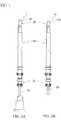
- FIG. 1A is a perspective view of a small-diameter endoscope surgical instrument according to Embodiment 1 of the invention, during an operation.
- FIG. 1B is a perspective view of the same, during an insertion into a body.
- FIG. 2 shows components which compose the small-diameter endoscope surgical instrument according to Embodiment 1 of the invention.
- FIG. 2A is a perspective view of a small-diameter endoscope.
- FIG 2B is a sectional view of an inner pipe.
- FIG. 2C is a sectional view of a guide pipe.
- FIG. 2D is a sectional view of a treatment pipe.
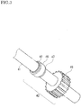
- FIG. 3 is an explanatory diagram of a handle of the treatment pipe according to Embodiment 1 of the invention.
- FIG. 4 shows a detail of the treatment pipe according to Embodiment 1 of the invention.
- FIG. 4A is an explanatory diagram of a hook provided at a leading end of the treatment pipe.
- FIG. 4B is its A-Aview.
- a small-diameter endoscope surgical instrument 1 according to Embodiment 1 of the present invention during an operation includes a small-diameter endoscope 10, a guide pipe 30, and a treatment pipe 40, as shown in FIG. 1A .
- the small-diameter endoscope surgical instrument 1a during its insertion into a body, replace the small-diameter endoscope with an inner pipe 20. It is in coaxial and triple-pipe structure.
- the small-diameter endoscope 10 of this embodiment includes a rigid fiberscope in a pipe part 11.
- An outside diameter of the rigid fiberscope is 2mm.
- the small-diameter endoscope 10 is led to a standard endoscopic monitor system (It is not shown in a figure.) by a connector 13, and an operator procced an operation watching a surgical target area and its around through the endoscopic monitor.
- a standard endoscopic monitor system It is not shown in a figure.
- An optical fiber for illumination to light up a neighboring space around a leading end 12 of the small-diameter endoscope is included in the pipe part 11 and light is sent through a light fiber connector 15.
- An operator uses the conrector 13 as a handle of the small-diameter endoscope, and moves the leading end of the small-diameter endoscope instrument to a surgical target point.
- a guide pipe 30 is a pipe with an inside diameter 2.05mm, which is slightly bigger than the outside diameter of the pipe part 11 of the small-diameter endoscope, and it has an outside diameter 2.35mm.
- the guide pipe 30 supports the small-diameter endoscope 10 so as to be movable in its axial direction.
- one or more slits 33 are provided at a leading end 34 of the guide pipe, it may help an operator to get a view around the surgical target area and remove quickly white smoke in the front of the small-diameter endoscope 10, when the small-diameter endoscope is pulled into the guide pipe 30.
- a treatment pipe 40 is a pipe with an inside diameter 2.40mm, which is slightly bigger than the outside diameter 2.35mm of the guide pipe 30, and supports the guide pipe so as to be movable in its axial direction.
- the treatment pipe 40 is a simple pipe without any project aside portion. It can be inserted smoothly through a skin incision of approximately 3mm in length.
- the treatment pipe 40 includes a hook 43 at its leading end.
- the hook 43 is made by cutting out from a wall of the treatment pipe into a hook shape, without any transformation on a curved surface of the treatment pipe wall.
- the hook 43 is located between curved inside and outside surfaces, which has the same inside and outside diameter of the treatment pipe wall. Therefore, the hook 43 does not disturb an axial movement of guide pipe 30 in the treatment pipe 40, and not project aside from the outer diameter of the treatment pipe 40.
- Many conventional endoscope surgical instruments may include a leading end with a sharp hook or a sharp blade. When they are inserted in a body, it may damage the neighboring tissue, or the leading end of the instruments may be damaged, and the insertion cannot be performed smoothly.
- the hook 43 has the shape as shown in FIG 4A and FIG 4B , and it advances accompanying the guide pipe 30 when it is inserted in a body, it does not damage the neighboring tissue, and does not be damaged.
- a near edge, that is a side near to the operation handle, of a tip 44 of the hook 43 is parallel to the leading end 34 of the guide pipe 30, which is formed perpendicular to its axial direction.
- an operator can grip a tissue tightly, by managing handles 42 and 32 and bringing the near edge of the tip 34 close to the leading end 34 of the guide pipe.
- the operator can perform abscission or separation of a target tissue very easily and surely, by pulling and turning the treatment pipe 40 to a direction 49 in FIG 4B .
- the operator pulls the leading end 12 of the small-diameter endoscope 10 into the treatment pipe 40, and can get a clear view of both the blade of the tip 44 and a target tissue, as shown in FIG 4A .
- the guide pipe 30 is pulled in the treatment pipe 40, in order not to interfere with the view.
- the tip 44 of the hook 43 is used as a blade, and also as an electrode for electrocautery or electrocoagulation.
- a pipe part 41 of the treatment pipe is made of current-carrying material and works as an electricity supplying wire itself.
- an outer surface of the treatment pipe being inserted into a body except the electrode is insulated.
- the insulation 45 is carried out by coating, printing or baking of insulating material.
- the electrode is either mono-polar or bipolar.
- Mono-polar electrode is suitable for a delicate operation on a small tissue in a body cavity, such as thoracic sympathectomy, as an electric current is concentrated in relatively small portion around the electrode.
- another electrode may be provided at a part of patient's body, such as a buttock.
- the treatment pipe 40 includes a handle 42 at its operator's near side, as shown in FIG 3 .
- the handle 42 includes a rotator 48, a hold 46 and snap rings 47.
- the rotator 48 is fixed to the pipe part 41 of the treatment pipe.
- the hold 46 is put on the pipe part 41 so as to be rotatable, but not removable in the treatment pipe's axial direction by the snap rings 47 fixed to the pipe part 41.
- a guide pipe 30 may have at least one slit, as shown in FIG 2B .
- the slit gives an operator a view around a leading end 34 of the guide pipe, and the operator can change a direction of the view by rotating the guide pipe.
- the guide pipe 30 needs a handle 32, which is similar to the handle 42 of the treatment pipe.
- a guide pipe without any slit does not needs to rotate itself and to have a rotator. It has only a hold fixed to the pipe part of the guide pipe.
- the inner pipe 20 is a pipe which has a same outside diameter as a one of a pipe part 11 of the small-diameter endoscope. Its leading end 23 works as a leader of the small-diameter endoscope surgical instrument 1a, when it is inserted into a body, as shown in FIG1B .
- the leading end 23 may be in polished smooth round head, in order to be inserted smoothly and not to injure a neighboring tissue.
- the inner pipe 20 needs to change its axial position but does not need to rotate itself.
- the hold 22 has a shape easily pinched with operator's thumb and forefinger.
- Each of the inner pipe 20, the guide pipe 30, and the treatment pipe 40 have minimal diameter and minimal wall thickness, under the condition that each component can perform its function.
- the clearances between those pipes are selected small. Consequently, a diameter of the pipe part of the small-diameter endoscope instrument 1 according to Embodiment 1 of this invention, is only 2.85mm. It contributes to reduce a size of a skin incision for its insertion.
- the guide pipe 30 and the treatment pipe 40 are in simple pipe form, and they are easy to be separated each other, washing and sterilization of them can be performed easily and thoroughly
- FIG. 5A is an explanatory diagram of an operation to move a tissue (mobilization), using the hook 43 of the treatment pipe.
- an operator may want to remove a tissue which interfere with the small-diameter endoscope surgical instrument's advance or the small-diameter endoscope's view.
- the operator puts the hook 43 of the treatment pipe close to the interfering tissue 51, managing the hold 46 and watching the hook 43 and the interfering tissue 51 with the small-diameter endoscope 10. Then, he manages the rotator 48 to change an angle of the hook 43, and hooks the interfering tissue 51.
- FIG. 5B is an explanatory diagram of an operation to grip a tissue.
- An operator puts the hook 43 of the treatment pipe close to a target tissue 52, managing the hold 46 and watching the hook 43 and the target tissue 52 with the small-diameter endoscope 10. Then, he manages the rotator 48 to change an angle of the hook 43, and hooks the target tissue 52, just like as mobilization.
- the slit 33 on the guide pipe helps him to get a small-diameter endoscope's view, when the guide pipe 30 has moved ahead.
- the tip 44 of the hook 43 curves, so that a near edge of the tip is parallel to the leading end 34 of the guide pipe 30.
- the operator can grip the tissue 52 tightly between the near edge of the tip 44 of the hook and the leading end 34 of the guide pipe, and he can perform mobilization, isolation and attraction of the tissue 52.
- the tissue 52 can be used for biopsy, by pulling the small-diameter endoscope surgical instrument 1 gripping the tissue 52, out from the body.
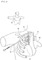
- FIG. 6 is an explanatory diagram of an operation to fix and detach a tissue 53 entangled in a tip 44 of a hook 43 or remove a blood clot stack on a blade.
- An operator searches for a rigid object, such as a rib 64, using a small-diameter endoscope, and brings a hook 43 of the treatment pipe close to the rib 64, managing a handle 42 of the treatment pipe. Then, he moves the guide pipe 30 ahead, managing the handle 32 of the guide pipe, and fixes the tissue 53 or the blood clot, by pushing them on the rib.
- Entanglement with a tissue and sticking with a blood clot happened many times during an operation. At that time, an operator had to take an endoscope surgical instrument out from a body every time, to detach an entangled tissue or to remove a stuck blood clot from a hook.
- the small-diameter endoscope surgical instrument Using the small-diameter endoscope surgical instrument according to this Embodiment, he can detach the entangled tissue or remove the stuck blood clot, without taking the small-diameter endoscope surgical instrument out from a body.
- FIG. 7 is an explanatory diagram of electrocautery or electrocoagulation, using an electrode of a treatment pipe.
- An operator puts a crocodile clip on a non-insulated pipe part 41 of the treatment pipe 40, for supplying a power. Also, he puts an electrode on a buttock of the patient, which is not shown on the figure.
- he can perform electrocautery or electrocoagulation on a target tissue 57. He can get a small-diameter endoscope's view including both the electrode (a tip 44 of a hook) and a target tissue 57, without any interference by a guide pipe because the guide pipe is pulled into the treatment pipe 40.
- FIG. 8 is an explanatory diagram of insertion of the small-diameter endoscope surgical instrument according to Embodiment 1 of the invention. ( This diagram is quoted from J Thoracic Cardiovasc Surg. 2000 ; 120 : 276-9 .)
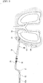
- FIG. 9 is an explanatory diagram of judging up to where the leading end of the small-diameter endoscope surgical instrument has reached, according to Embodiment 2 of the invention.
- An operator makes a skin incision 61 with a length 3mm on an armpit of a patient, to perform thoracic sympathectomy using a small-diameter endoscope surgical instrument according to Embodiment 1.
- the operator inserts the small-diameter endoscope surgical instrument 1a, which has an inner pipe 20 substituted for a small-diameter endoscope 10, through the skin incision 61 into a thoracic cavity.
- a target tissue of this operation is a thoracic sympathetic trunk 63 placed along a backbone.
- the small-diameter endoscope surgical instrument 1a advances through a space between ribs 64, to reaches near the target tissue 63.
- the area is surrounded with an apex of a lung 65, a clavicle 66, a super vena cava 67 and an aorta 68, besides the ribs 64.
- the inner pipe 20 of the small-diameter endoscope surgical instrument 1a is connected to a syringe 92 with a flexible tube 91.
- the syringe 92 includes physiological saline solution or air.
- An operator can notice the advance movement of a piston 92a, and judges easily and with confidence, the arrival of the leading end of the small-diameter endoscope surgical instrument 1a to the pleural cavity 93.
- the small-diameter endoscope surgical instrument of the invention has a simple structure with a few components, even so, it discharges many functions.
- It can perform electrocautery, electrocoagulation, abscission, mobilization, separation, attraction of a target tissue. Also, it can perform mobilization separation and attraction of a target tissue, by griping or fixing the tissue.
- tissue can take out a tissue as a sample for biopsy, and it can detach an entangled tissue or remove a blood clot from the hook, without taking the small-diameter endoscope surgical instrument out of the body cavity.
- the small-diameter endoscope surgical instrument is expected to be used in more various operations in the future.
Landscapes
- Health & Medical Sciences (AREA)
- Life Sciences & Earth Sciences (AREA)
- Surgery (AREA)
- Engineering & Computer Science (AREA)
- Veterinary Medicine (AREA)
- Public Health (AREA)
- General Health & Medical Sciences (AREA)
- Biomedical Technology (AREA)
- Heart & Thoracic Surgery (AREA)
- Animal Behavior & Ethology (AREA)
- Nuclear Medicine, Radiotherapy & Molecular Imaging (AREA)
- Medical Informatics (AREA)
- Molecular Biology (AREA)
- Pathology (AREA)
- Physics & Mathematics (AREA)
- Radiology & Medical Imaging (AREA)
- Optics & Photonics (AREA)
- Biophysics (AREA)
- Anesthesiology (AREA)
- Hematology (AREA)
- Plasma & Fusion (AREA)
- Otolaryngology (AREA)
- Orthopedic Medicine & Surgery (AREA)
- Cardiology (AREA)
- Neurology (AREA)
- Surgical Instruments (AREA)
- Endoscopes (AREA)
- Infusion, Injection, And Reservoir Apparatuses (AREA)
Abstract
Description
- The present invention relates to an endoscope surgical instrument. The endoscope surgical instrument is used in surgery to perform abscission, mobilization (an act to move tissue.), separation, attraction, extraction, electrocautery, electrocoagulation, etc. of small tissue in a body cavity, such as thoracic sympathectomy.
- Surgery using an endoscope is called endoscopic surgery. In endoscopic surgery, surgical instruments, such as a scalpel, forceps, an electrode for coagulation, are used to be inserted in a body cavity through another skin incision different from a skin incision for an endoscope. Therefore, at least two skin incisions are required, and depending on a kind of the operation, it is necessary to provide skin incisions more than three places. However, fewer and smaller skin incisions are desirable, in order to reduce a surgical damage on a patient and to make fast postoperative recovery. In the following explanations, this type of instruments used in endoscopic surgery are called "conventional endoscope surgical instruments".
- PTL 1 shows an endoscope power supplying appliance, having a cylinder unit which includes an endoscope in its inside. It can be inserted through a single skin incision into a body cavity and perform an operation, such as electrocautery, electrocoagulation, incision, etc. while observing a surgical area with the endoscope. It is an instrument to solve the above-mentioned issues of the conventional endoscope surgical instruments.
- The endoscope power supplying appliance perform electrocautery or electrocoagulation, using the leading end of the cylinder unit as an electrode and the cylinder unit itself as power supply unit. Also, the endoscope power supplying appliance can perform incision, etc., using the leading end of the cylinder unit which is formed in a blade or a needle shape.
- The cylinder unit of the endoscope power supplying appliance has an inner diameter which allows the endoscope to move in its inside, and the endoscope is supported so as to be movable in the axial direction of the cylinder unit. Then, a relative position in their axial direction of the leading end of the cylinder unit and the leading end of the endoscope becomes variable.
- At the time of searching the target point of the operation, the endoscope is projected over the leading end of the cylinder unit so as to observe in the body cavity with a wide visual field, and at the time of the operation, the endoscope is pulled into the cylinder unit and the endoscope can focus on both of the target point of the operation and the leading end of the treatment mean, such as electrode.
- Using this endoscope power supplying appliance, the endoscope and the surgical instruments are inserted as an unity into the body cavity through only one skin incision, and a further skin incision is not required.
- Details and results of an operation using this endoscope power supplying appliance are shown in NPL 1.
- PTL 2 shows an endoscope power supplying appliance suitable for sympathicotomy, which can attract, move, or cut fiber organisation interfering with an endoscope's view, by adding an attraction mean to the endoscope power supplying appliance according to PTL 1.
- The endoscope power supplying appliances shown in PTL 1 or PTL 2 need only a single skin incision.
- Also, a size of the skin incision can be made smaller, as those endoscope power supplying appliances use small-diameter endoscopes.
- It contributes to reduce a surgical damage on patient and to make fast postoperative recovery.
- However, these endoscope power supplying appliances cannot grip or fix tissue. Therefore, they cannot detach tissue entangled in a blade or remove a blood clot stuck on a blade during an incision operation, without pulling out these endoscope power supplying appliances themselves from a body cavity.
- Also, they are not able to grip and extract an incised tissue from the body cavity, as a sample for a biopsy.
- With the conventional endoscope surgical instruments, an operator can insert a grasper, such as forceps, to grip tissue, or to extract an incised tissue as a sample for a biopsy from the body cavity. But, to do so, additional skin incision is required for inserting a grasper, such as forceps.
- An ordinary-used remote control grasper, such as remote control forceps, has a large number of small parts, such as a joint pin and driving mechanisms to move the grasping hands relatively. Those parts may be broken and fallen apart within a body cavity.
- Incidentally, an existence of the fallen-apart small parts can be confirmed by radiography from the outside of the body. But, as it is difficult to detect the exact position of the fallen-apart parts even with an endoscope, it is almost impossible to take out those fallen-apart small parts from a body, even using endoscope surgical instruments.
- In addition to those issues, many of the conventional endoscope surgical instruments have leading ends with a sharp or a complicated shape. When they are inserted in a body, it may damage the neighboring tissue, or the leading end of the endoscope surgical instrument itself may be damaged, and the insertion cannot be performed smoothly.
- Also, during the insertion, it is rather difficult for the operator to judge up to where the leading end of the endoscope surgical instrument have arrived.
-
- [PTL 1]
WO-A1-2000/016707 - [PTL 2]
JP-A1-2007/089690 - [NPL 1] Hidehiro Yamamoto, MD. etc. The Journal of Thoracic and Cardiovascular Surgery, 2000, Volume 120, Page 276-279
- In response to the above issues, the present invention provides an endoscope surgical instrument which discharges functions to grip and fix tissue, additional to the functions of the endoscope power supplying appliances shown in PLT 1 or PLT 2.
- Consequently, it can detach tissue entangled in a blade and remove a blood clot stuck on a blade during an incision operation, and it can grip and extract an incised tissue from the body cavity as a sample for a biopsy.
- Also, the endoscope surgical instrument can be inserted in a body smoothly, and do not damage the neighboring tissue or the leading end of the endoscope surgical instrument itself. And during the insertion, an operator can judge easily, up to where the leading end of the endoscope surgical instrument has arrived.
- Of course, with an endoscope included in the endoscope surgical instrument, it is essential to obtain a clear view, on both of the surgery target point and treatment mean, such as an electrode etc. and a clear view on the surrounding area around the leading end of the endoscope surgical instrument.
- A small-diameter endoscope surgical instrument according to a first aspect of the present invention is of coaxial and multiplex-pipe structure, includes a small-diameter endoscope, an inner pipe, a guide pipe, a treatment pipe and their operation handles.
- The inner pipe has an outside diameter same as the one of a pipe part of the small-diameter endoscope, and is used when the small-diameter endoscope surgical instrument is inserted into a body, and replaced with the small-diameter endoscope after the insertion.
- The guide pipe, in which the small-diameter endoscope or the inner pipe is inserted, supports them so as to be movable in their axial direction.
- The treatment pipe, in which the guide pipe is inserted and supported so as to be movable in their axial direction, has one or more treatment means, such as an electrode, a blade, a hook, etc., at its leading end.
- The operation handles, independently included to the small-diameter endoscope, the inner pipe, the guide pipe, and the treatment pipe, at their each proximate end, that is an end locating outside of a body of a patient being operated with the small-diameter endoscope surgical instrument. Those operation handles can change their positions in their axial direction and some of their rotating angles.
- The following description is on thoracic sympathectomy, as an example. When the small-diameter endoscope surgical instrument is introduced into a thoracic cavity through a single skin incision made in an armpit of a patient, the inner pipe which has an outside diameter same as the one of a pipe part of the small-diameter endoscope is inserted in the guide pipe, and the guide pipe is inserted in the treatment pipe.
- Therefore, the small-diameter endoscope surgical instrument is of coaxial and triple-pipe structure.
- After confirming the distal end of the small-diameter endoscope surgical instrument has reached to the target point, the inner pipe is replaced with the small-diameter endoscope. As a result, whole items required to an endoscopic operation are inserted in a body cavity through only one skin incision.
- Surgical instruments inserted into a body, such as an endoscope surgical instrument, used to be in a shape of a pole which is filled up with a material, and not in a shape of a pipe.
- If the surgical instrument is in a pipe shape, a part of tissue or body fluid enters into an inside of the pipe. In this case, it is hard to prevent pollution of such a surgical instrument and to sterilize it thoroughly.
- One aspect of the present invention is the small-diameter endoscope surgical instrument of multiplex-pipe structure. When it is inserted into a body, it is of triple-pipe structure. When it is in an operation, it is of double-pipe structure plus a small-diameter endoscope.
- By decreasing a difference between an outer diameter of the small-diameter endoscope or the inner pipe and an inner diameter of the guide pipe, and by decreasing a difference between an outer diameter of the guide pipe and an inner diameter of the treatment pipe, to be minimal, under a condition that each items are supported so as to be movable in their axial direction, it gives a minimal possibility that a part of tissue or body fluid enters into the clearances of the small-diameter endoscope surgical instrument, as explained in the description of embodiments.
- As the inner pipe, the guide pipe and the treatment pipe have a shape of simple pipe, and also they are easy to be separated each other, washing and sterilization of them can be performed easily and thoroughly.
- The small-diameter endoscope surgical instrument has a small outside diameter, by selecting an endoscope which has a pipe part of a very small outside diameter, the inner pipe has a same outside diameter, and the guide pipe and the treatment pipe with thin pipe walls. Consequently, the small skin incision is enough to insert it into a body.
- When an operator inserts the small-diameter endoscope surgical instrument into a body cavity, leading ends of the inner pipe, the guide pipe and the treatment pipe are slightly shifted to their operation handle side in this order. Each of those components has a shape of simple pipe and has no part projecting outside of the pipe part of the treatment pipe, the operator can insert it in a body cavity very smoothly.
- The small-diameter endoscope, the inner pipe, the guide pipe and the treatment pipe independently include operation handles, at their proximate end.
- An operator of the small-diameter endoscope surgical instrument may change their positions in their axial direction and their rotating angles, depending on a requirement.
- The treatment pipe requires to change its rotating angle. The guide pipe requires to change its rotating angle in order to change the small-diameter endoscope's view in case it has at least one slit. But the guide pipe without any slit does not require to change its rotating angle. The inner pipe does not require to change its rotating angle. The operation handles are provided, taking these conditions into consideration.
- An operator can perform not only electrocautery and electrocoagulation of a tissue which is a target of the operation (it will be called "target tissue" hereunder), using the electrode at the leading end of the treatment pipe, but also abscission, mobilization, separation and attraction, using the blade, the nook, etc., at the leading end of the treatment pipe.
- Furthermore, using the hook at the distal end of the treatment pipe and a distal end of the guide pipe, the operator may grip a target tissue. Consequently, the operator may mobilize, separate and attract the target tissue, and also he may extract the gripped target tissue from a body cavity as a sample for a biopsy.
- Also, the operator may detach a tissue entangled in the blade or remove blood clots stuck on the blade, by fixing them between the leading end of the guide pipe and a rib, etc..
- The operator can perform these operations effectively and efficiently, managing an operation handle included to each component.
- All components, that is the small-diameter endoscope, the inner pipe, the guide pipe, and the treatment pipe, are formed in a linear shape or in a part of an arc shape with a same radius, as they are to be changed their relative positions in their axial direction.
- The small-diameter endoscope surgical instrument according to a second aspect of the present invention, may include a treatment pipe which has a special hook. The hook is formed by cutting out from the wall of the distal end of the treatment pipe, and a near edge of a tip of the hook has a blade and is in a plain approximately perpendicular to the axial direction of the treatment pipe.
- " The hook is formed by cutting out from the wall of the treatment pipe " means a hook made by cutting out from the pipe wall of the treatment pipe, keeping a radius of the pipe wall as it was, and the hook part are located within the same inside and outside diameter as the treatment pipe itself.
- Consequently, the hook part does not disturb the relative movement between the treatment pipe and the guide pipe. Also, when the small-diameter endoscope surgical instrument is inserted into a body, the hook part does not damage a neighboring tissue, and a tip of the hook is not forced to be damaged, as the hook part accompanies the guide pipe, just like as the treatment pipe itself.
- The hook has a shape that a pushing-out part from the leading end of the treatment pipe lengthens ahead and then turns to make the hook. A near edge, that is a side near to the operator, of a tip of the turning part of the hook are in a plain approximately perpendicular to the axial direction of the treatment pipe, and it is parallel to the leading end of the guide pipe which is formed perpendicular to its axial direction.
- An operator puts a target tissue between the near edge of a tip of the hook and the leading end of the guide pipe, and brings the near edge of a tip of the hook and the leading end of the guide pipe close each other by managing their operation handles.
- With this special hook, the operator grip the target tissue more easily and more tightly, than the case that a near edge of a tip of a hook is not parallel to a leading end of a guide pipe.
- Consequently, mobilization, separation, attraction of the target tissue and also a sample extraction for a biopsy can be performed more easily and more surely.
- Providing a blade on the near edge of a tip of the hook, an operator can perform abscission and separation of a target tissue more easily and more surely, by pulling the operation pipe in and turning it, managing its operation handle.
- When an operator wants to detach a tissue entangled in the blade or remove a blood clot stuck on the blade, he can detach or remove them very easily, by fixing them between the leading end of the guide pipe and a rib, etc., and then rotating the treatment pipe.
- With the small-diameter endoscope surgical instrument, an operator can detach or remove them easily without taking out the small-diameter endoscope surgical instrument itself from a body. It helps a smooth progress of operation and reduces surgical damage on patient.
- With a conventional endoscope surgical instrument, an operator cannot perform these operation, without pulling the the conventional endoscope surgical instrument out from a body, in many cases.
- The small-diameter endoscope is supported so as to be movable in its axial direction by the guide pipe, and the guide pipe is supported so as to be movable in its axial direction by the treatment pipe.
- When searching a target tissue, an operator can observe in a body cavity with a wide visual field, by pushing out the small-diameter endoscope from the leading ends of the guide pipe and the treatment pipe.
- In operation, the operator can focus on both a target tissue and the treatment mean of the leading end of the treatment pipe, by pulling the small-diameter endoscope into the guide pipe and the treatment pipe.
- In both cases, he can get clear view.
- As the small-diameter endoscope, the guide pipe and the treatment pipe of the small-diameter endoscope surgical instrument are combined in coaxial, an operator can observe both a target tissue and a treatment mean, such as an electrode.
- In the conventional endoscope surgical instrument, the operator needs to make an effort searching a treatment mean in a body cavity with an endoscope, and chasing the treatment mean with the endoscope during whole operation period, so that the treatment mean is within the endoscope's view.
- With the small-diameter endoscope surgical instrument, an operator does not need such actions. It decreases an operator's burden and operation is performed more smoothly.
- The small-diameter endoscope surgical instrument according to a third aspect of the present invention, may have a guide pipe, which includes one or more slits near its leading end.
- An operator can get a view around a target tissue through the slits, when the operator pulls the small-diameter endoscope into the guide pipe to perform an operation, such as grip, etc., and the leading end of the guide pipe narrows the endoscope's view.
- It is a great help for the operator.
- The small-diameter endoscope surgical instrument according to a fourth aspect of the present invention, may include an treatment pipe, which has an electrode at its leading end and its pipe part supplies electricity to the electrode. An outer surface of the treatment pipe, being inserted into a body except the electrode, is insulated.
- The treatment pipe is made of current-carrying material, and has an electrode for electrocautery or electrocoagulation at its leading end.
- A pipe part of the treatment pipe works as an electricity supplying wire. For preventing short-circuit to an outside, an outer surface of the treatment pipe being inserted into a body except the electrode, is insulated.
- The insulation is carried out by coating, printing or baking of insulating material.
- The electrode is either mono-polar or bipolar. Mono-polar electrode is suitable for a delicate operation, such as thoracic sympathectomy, as an electric current is concentrated in relatively small portion around the electrode
- The small-diameter endoscope surgical instrument according to a fifth aspect of the present invention, may include an inner pipe which has a leading end of a polished smooth round head and a physiological saline solution or air supplying equipment supplying them to an inside of the inner pipe.
- During an insertion of the small-diameter endoscope surgical instrument into a body, the inner pipe is used as a replacement of the small-diameter endoscope, and leads the small-diameter endoscope surgical instrument. With a leading end of a polished smooth round head of the inner pipe, the insertion is performed smoothly.
- A physiological saline solution or air supplying equipment, such as a syringe filled with physiological saline solution or air, is prepared. It is connected to the inner pipe with a flexible tube, to supply them to an inside of the inner pipe
- For example, in case of sympathectomy, an operator can notice an advancing movement e of a piston of the syringe, when the leading end of the inner pipe reached into a pleural cavity, as a pressure of the physiological saline solution or air falls down. The operator can judge easily up to where the leading end of the small-diameter endoscope surgical instrument reached.
- Using the small-diameter endoscope surgical instrument of the present invention, an operator can perform abscission, mobilization, separation, attraction, extraction, electrocautery, electrocoagulation, etc. for a small issue in a body cavity, with only one instrument.
- Especially, he can detach a tissue entangled in a blade or remove a blood clot stuck on a blade at the operating place, and he can extract an incised tissue from a body cavity as a sample for a biopsy, as he can perform griping and fixing of a tissue easily and surely.
- An operator does not bother about taking out of fallen parts in a body cavity, as the small-diameter endoscope surgical instrument is simple structure made of only a few components.
- The small-diameter endoscope surgical instrument needs only one and small skin incision. It contributes to deduce surgical damage of the patient and early recovery of a postoperative stage.
- An operator can insert the small-diameter endoscope surgical instrument in a body smoothly, and do not damage the neighboring tissue or the leading end of the small-diameter endoscope surgical instrument. During the insertion, he can judge easily up to where the leading end of the small-diameter endoscope surgical instrument has arrived.
- With a small-diameter endoscope inserted within the small-diameter endoscope surgical instrument, he can get a clear surgical view, on both of the surgery target point and treatment mean, such as the electrode etc. and on the surrounding area around the leading end of the small-diameter endoscope surgical instrument.
- The small-diameter endoscope surgical instrument is used easily and extremely handy for an operator.
- NPL 1 presents an endoscope power supplying appliance which performs sympathectomy, sympathicotomy and ablation, such as electrocautery, to destroy sympathetic trunk. But it is hard to perform sympathetic ganglionectomy with this endoscope power supplying appliance, as sympathetic ganglians usually locate between ribs and contact with brood vessels in many cases.
- The use of the small-diameter endoscope surgical instrument of the present invention makes possible all of these operations. Also, it has a big advantage for a patient praying for low invasion and high cosmesis.
-
-
FIG. 1A is a perspective view of a small-diameter endoscope surgical instrument according to Embodiment 1 of the invention, during an operation.FIG. 1B is a perspective view of the same, during an insertion into a body. -
FIG. 2 shows components which compose the small-diameter endoscope surgical instrument according to Embodiment 1 of the invention.FIG. 2A is a perspective view of a small-diameter endoscope.FIG 2B is a sectional view of an inner pipe.FIG. 2C is a sectional view of a guide pipe. And,FIG. 2D is a sectional view of a treatment pipe. -
FIG. 3 is an explanatory diagram of a handle of the treatment pipe according to Embodiment 1 of the invention. -
FIG. 4 shows a detail of the treatment pipe according to Embodiment 1 of the invention.FIG. 4A is an explanatory diagram of a hook provided at a leading end of the treatment pipe.FIG. 4B is its A-A view. -
FIG. 5 shows operations using the hook of the treatment pipe according to Embodiment 1.FIG. 5A is an explanatory diagram of mobilization.FIG. 5B is an explanatory diagram of griping. -
FIG. 6 is an explanatory diagram of an operation to fix and detach a tissue entangled in a blade, or remove a blood clot stuck on a blade, using the guide pipe. -
FIG. 7 is an explanatory diagram of electrocautery or electrocoagulation, using an electrode of the treatment pipe. -
FIG. 8 is an explanatory diagram of insertion of the small-diameter endoscope surgical instrument according to Embodiment 1 of the invention (Embodiment 2). (This diagram is quoted from J Thoracic Cardiovasc Surg. 2000; 120 : 276-9.) -
FIG. 9 is an explanatory diagram of judging up to where the leading end of the small-diameter endoscope surgical instrument has reached, according to Embodiment 2 of the invention. - A small-diameter endoscope surgical instrument according to embodiments of the present invention will now be described with reference to the drawings. The present invention will not be limited only to these embodiments, and may include various changes on its structure within Claims.
- The following descriptions are on thoracic sympathectomy, but a use of the present invention is not limited to thoracic sympathectomy.
-
FIG. 1A is a perspective view of a small-diameter endoscope surgical instrument according to Embodiment 1 of the invention, during an operation.FIG. 1B is a perspective view of the same, during an insertion into a body. -
FIG. 2 shows components which compose the small-diameter endoscope surgical instrument according to Embodiment 1 of the invention.FIG. 2A is a perspective view of a small-diameter endoscope.FIG 2B is a sectional view of an inner pipe.FIG. 2C is a sectional view of a guide pipe. And,FIG. 2D is a sectional view of a treatment pipe. -
FIG. 3 is an explanatory diagram of a handle of the treatment pipe according to Embodiment 1 of the invention. -
FIG. 4 shows a detail of the treatment pipe according to Embodiment 1 of the invention.FIG. 4A is an explanatory diagram of a hook provided at a leading end of the treatment pipe.FIG. 4B is its A-Aview. - A small-diameter endoscope surgical instrument 1 according to Embodiment 1 of the present invention during an operation, includes a small-
diameter endoscope 10, aguide pipe 30, and atreatment pipe 40, as shown inFIG. 1A . - The small-diameter endoscope
surgical instrument 1a during its insertion into a body, replace the small-diameter endoscope with aninner pipe 20. It is in coaxial and triple-pipe structure. - To minimize the size of a skin incision, the small-
diameter endoscope 10 of this embodiment includes a rigid fiberscope in apipe part 11. An outside diameter of the rigid fiberscope is 2mm. - The small-
diameter endoscope 10 is led to a standard endoscopic monitor system (It is not shown in a figure.) by aconnector 13, and an operator procced an operation watching a surgical target area and its around through the endoscopic monitor. - An optical fiber for illumination to light up a neighboring space around a leading
end 12 of the small-diameter endoscope is included in thepipe part 11 and light is sent through alight fiber connector 15. - An operator uses the
conrector 13 as a handle of the small-diameter endoscope, and moves the leading end of the small-diameter endoscope instrument to a surgical target point. - A
guide pipe 30 is a pipe with an inside diameter 2.05mm, which is slightly bigger than the outside diameter of thepipe part 11 of the small-diameter endoscope, and it has an outside diameter 2.35mm. Theguide pipe 30 supports the small-diameter endoscope 10 so as to be movable in its axial direction. - In a case that one or
more slits 33 are provided at aleading end 34 of the guide pipe, it may help an operator to get a view around the surgical target area and remove quickly white smoke in the front of the small-diameter endoscope 10, when the small-diameter endoscope is pulled into theguide pipe 30. - A
treatment pipe 40 is a pipe with an inside diameter 2.40mm, which is slightly bigger than the outside diameter 2.35mm of theguide pipe 30, and supports the guide pipe so as to be movable in its axial direction. - All components of the small-diameter endoscope
surgical instrument 1a inserted into a body, are within the treatment pipe. - The
treatment pipe 40 is a simple pipe without any project aside portion. It can be inserted smoothly through a skin incision of approximately 3mm in length. - The
treatment pipe 40 includes ahook 43 at its leading end. Thehook 43 is made by cutting out from a wall of the treatment pipe into a hook shape, without any transformation on a curved surface of the treatment pipe wall. - Consequently, the
hook 43 is located between curved inside and outside surfaces, which has the same inside and outside diameter of the treatment pipe wall. Therefore, thehook 43 does not disturb an axial movement ofguide pipe 30 in thetreatment pipe 40, and not project aside from the outer diameter of thetreatment pipe 40. - Many conventional endoscope surgical instruments may include a leading end with a sharp hook or a sharp blade. When they are inserted in a body, it may damage the neighboring tissue, or the leading end of the instruments may be damaged, and the insertion cannot be performed smoothly.
- As the
hook 43 has the shape as shown inFIG 4A and FIG 4B , and it advances accompanying theguide pipe 30 when it is inserted in a body, it does not damage the neighboring tissue, and does not be damaged. - A near edge, that is a side near to the operation handle, of a
tip 44 of thehook 43 is parallel to theleading end 34 of theguide pipe 30, which is formed perpendicular to its axial direction. - As the near edge of the
tip 44 of the hook is parallel to theleading end 34 of the guide pipe, an operator can grip a tissue tightly, by managinghandles tip 34 close to theleading end 34 of the guide pipe. - With a blade provided at the near edge of the
tip 44 of thehook 43, the operator can perform abscission or separation of a target tissue very easily and surely, by pulling and turning thetreatment pipe 40 to adirection 49 inFIG 4B . - During this operation, the operator pulls the
leading end 12 of the small-diameter endoscope 10 into thetreatment pipe 40, and can get a clear view of both the blade of thetip 44 and a target tissue, as shown inFIG 4A . In this occasion, theguide pipe 30 is pulled in thetreatment pipe 40, in order not to interfere with the view. - The
tip 44 of thehook 43 is used as a blade, and also as an electrode for electrocautery or electrocoagulation. Apipe part 41 of the treatment pipe is made of current-carrying material and works as an electricity supplying wire itself. - For preventing short-circuit to an outside, an outer surface of the treatment pipe being inserted into a body except the electrode, is insulated.
- The
insulation 45 is carried out by coating, printing or baking of insulating material. - The electrode is either mono-polar or bipolar. Mono-polar electrode is suitable for a delicate operation on a small tissue in a body cavity, such as thoracic sympathectomy, as an electric current is concentrated in relatively small portion around the electrode.
- When the
tip 44 is used as a mono-polar electrode, another electrode may be provided at a part of patient's body, such as a buttock. - The
treatment pipe 40 includes ahandle 42 at its operator's near side, as shown inFIG 3 . Thehandle 42 includes arotator 48, ahold 46 and snap rings 47. Therotator 48 is fixed to thepipe part 41 of the treatment pipe. Thehold 46 is put on thepipe part 41 so as to be rotatable, but not removable in the treatment pipe's axial direction by the snap rings 47 fixed to thepipe part 41. - The operator pinches the
hold 46 with his thumb and forefinger and touches therotator 48 with his middle finger. He can change the axial position of thetreatment pipe 40 by moving thehold 46, and can change the rotating angle of thetreatment pipe 40 by rotating therotator 48. - A
guide pipe 30 may have at least one slit, as shown inFIG 2B . The slit gives an operator a view around a leadingend 34 of the guide pipe, and the operator can change a direction of the view by rotating the guide pipe. In this case, theguide pipe 30 needs ahandle 32, which is similar to thehandle 42 of the treatment pipe. - A guide pipe without any slit, does not needs to rotate itself and to have a rotator. It has only a hold fixed to the pipe part of the guide pipe.
- The
inner pipe 20 is a pipe which has a same outside diameter as a one of apipe part 11 of the small-diameter endoscope. Its leadingend 23 works as a leader of the small-diameter endoscopesurgical instrument 1a, when it is inserted into a body, as shown inFIG1B . The leadingend 23 may be in polished smooth round head, in order to be inserted smoothly and not to injure a neighboring tissue. - The
inner pipe 20 needs to change its axial position but does not need to rotate itself. - Therefore, it does not need a rotator, and needs only a
hold 22 fixed to thepipe part 21 of the inner pipe. Thehold 22 has a shape easily pinched with operator's thumb and forefinger. - Each of the
inner pipe 20, theguide pipe 30, and thetreatment pipe 40 have minimal diameter and minimal wall thickness, under the condition that each component can perform its function. The clearances between those pipes are selected small. Consequently, a diameter of the pipe part of the small-diameter endoscope instrument 1 according to Embodiment 1 of this invention, is only 2.85mm. It contributes to reduce a size of a skin incision for its insertion. - Also, by decreasing a difference between an outer diameter and an inner diameter of pipes being close to each other, that is a clearance between pipes being close to each other, it gives a minimal possibility that a part of tissue or a body fluid enters into the appliance.
- As the
inner pipe 20, theguide pipe 30 and thetreatment pipe 40 are in simple pipe form, and they are easy to be separated each other, washing and sterilization of them can be performed easily and thoroughly -
FIG. 5A is an explanatory diagram of an operation to move a tissue (mobilization), using thehook 43 of the treatment pipe. - In some operations, an operator may want to remove a tissue which interfere with the small-diameter endoscope surgical instrument's advance or the small-diameter endoscope's view.
- In such a case, the operator puts the
hook 43 of the treatment pipe close to the interferingtissue 51, managing thehold 46 and watching thehook 43 and the interferingtissue 51 with the small-diameter endoscope 10. Then, he manages therotator 48 to change an angle of thehook 43, and hooks the interferingtissue 51. - Under this situation, he can perform mobilization, isolation and attraction of the interfering
tissue 51, and he can also perform abscission of it. using the blade on thehook 43. -
FIG. 5B is an explanatory diagram of an operation to grip a tissue. - An operator puts the
hook 43 of the treatment pipe close to atarget tissue 52, managing thehold 46 and watching thehook 43 and thetarget tissue 52 with the small-diameter endoscope 10. Then, he manages therotator 48 to change an angle of thehook 43, and hooks thetarget tissue 52, just like as mobilization. - Then, he moves the
guide pipe 30 ahead, managing thehandle 32 and grips the hookedtissue 52 between aleading end 34 of the guide pipe and atip 44 of thehook 43. - The
slit 33 on the guide pipe helps him to get a small-diameter endoscope's view, when theguide pipe 30 has moved ahead. - The
tip 44 of thehook 43 curves, so that a near edge of the tip is parallel to theleading end 34 of theguide pipe 30. The operator can grip thetissue 52 tightly between the near edge of thetip 44 of the hook and theleading end 34 of the guide pipe, and he can perform mobilization, isolation and attraction of thetissue 52. - Also, the
tissue 52 can be used for biopsy, by pulling the small-diameter endoscope surgical instrument 1 gripping thetissue 52, out from the body. -
FIG. 6 is an explanatory diagram of an operation to fix and detach atissue 53 entangled in atip 44 of ahook 43 or remove a blood clot stack on a blade. - An operator searches for a rigid object, such as a
rib 64, using a small-diameter endoscope, and brings ahook 43 of the treatment pipe close to therib 64, managing ahandle 42 of the treatment pipe. Then, he moves theguide pipe 30 ahead, managing thehandle 32 of the guide pipe, and fixes thetissue 53 or the blood clot, by pushing them on the rib. - Under this situation, he may take the
tissue 53 or the blood clot off from thehook 43 easily, by turning thetreatment pipe 40 in anarrow 56 shown inFIG. 6 , managing therotator 48. - Entanglement with a tissue and sticking with a blood clot happened many times during an operation. At that time, an operator had to take an endoscope surgical instrument out from a body every time, to detach an entangled tissue or to remove a stuck blood clot from a hook.
- Using the small-diameter endoscope surgical instrument according to this Embodiment, he can detach the entangled tissue or remove the stuck blood clot, without taking the small-diameter endoscope surgical instrument out from a body.
- It helps a smooth progress of an operation and reduces surgical damage on a patient.
-
FIG. 7 is an explanatory diagram of electrocautery or electrocoagulation, using an electrode of a treatment pipe. An operator puts a crocodile clip on anon-insulated pipe part 41 of thetreatment pipe 40, for supplying a power. Also, he puts an electrode on a buttock of the patient, which is not shown on the figure. - With such an arrangement, he can perform electrocautery or electrocoagulation on a
target tissue 57. He can get a small-diameter endoscope's view including both the electrode (atip 44 of a hook) and atarget tissue 57, without any interference by a guide pipe because the guide pipe is pulled into thetreatment pipe 40. -
FIG. 8 is an explanatory diagram of insertion of the small-diameter endoscope surgical instrument according to Embodiment 1 of the invention. (This diagram is quoted from J Thoracic Cardiovasc Surg. 2000 ; 120 : 276-9.)FIG. 9 is an explanatory diagram of judging up to where the leading end of the small-diameter endoscope surgical instrument has reached, according to Embodiment 2 of the invention. - An operator makes a
skin incision 61 with a length 3mm on an armpit of a patient, to perform thoracic sympathectomy using a small-diameter endoscope surgical instrument according to Embodiment 1. - The operator inserts the small-diameter endoscope
surgical instrument 1a, which has aninner pipe 20 substituted for a small-diameter endoscope 10, through theskin incision 61 into a thoracic cavity. - A target tissue of this operation is a thoracic
sympathetic trunk 63 placed along a backbone. - The small-diameter endoscope
surgical instrument 1a advances through a space betweenribs 64, to reaches near thetarget tissue 63. The area is surrounded with an apex of alung 65, aclavicle 66, asuper vena cava 67 and anaorta 68, besides theribs 64. - The
inner pipe 20 of the small-diameter endoscopesurgical instrument 1a is connected to asyringe 92 with aflexible tube 91. Thesyringe 92 includes physiological saline solution or air. - When the leading end of the small-diameter endoscope
surgical instrument 1a, that is theleading end 23 of theinner pipe 20 reaches a pleural cavity, a pressure of physiological saline solution or air decreases, as a pressure in the pleural cavity is in a negative pressure. And apiston 92a of thesyringe 92 moves ahead. - An operator can notice the advance movement of a
piston 92a, and judges easily and with confidence, the arrival of the leading end of the small-diameter endoscopesurgical instrument 1a to thepleural cavity 93. - After this judgement, he takes out the
inner pipe 20 from the small-diameter endoscopesurgical instrument 1a, and inserts a small-diameter endoscope 10 as a substitution of the inner pipe. - It changes the small-diameter endoscope
surgical instrument 1a into a small-diameter endoscope surgical instrument 1 during an operation. - The small-diameter endoscope surgical instrument of the invention has a simple structure with a few components, even so, it discharges many functions.
- It can perform electrocautery, electrocoagulation, abscission, mobilization, separation, attraction of a target tissue. Also, it can perform mobilization separation and attraction of a target tissue, by griping or fixing the tissue.
- Moreover, it can take out a tissue as a sample for biopsy, and it can detach an entangled tissue or remove a blood clot from the hook, without taking the small-diameter endoscope surgical instrument out of the body cavity.
- Using the small-diameter endoscope surgical instrument, an operator can perform an operation smoothly with an easy action, watching a small-diameter endoscope's clear view.
- For a patient, only one sn all skin incision reduces surgical damage.
- With those advantages, the small-diameter endoscope surgical instrument is expected to be used in more various operations in the future.
-
- 1, 1a
- A small-diameter endoscope surgical instrument of Embodiment 1
- 10
- A small-diameter endoscope
- 11
- A pipe part of the small-diameter endoscope
- 12
- A leading end of the small-diameter endoscope
- 13
- A connector (a candle) of the small-diameter endoscope
- 15
- A light fiber connecter of the small-diameter endoscope
- 20
- An inner pipe
- 21
- A pipe part of the inner pipe
- 22
- A handle of the inner pipe
- 23
- A leading end of the inner pipe
- 30
- A guide pipe
- 31
- A pipe part of the guide pipe
- 32
- A handle of the guide pipe
- 33
- A slit of the guide pipe
- 34
- A leading end of the guide pipe
- 40
- A treatment pipe
- 41
- A pipe part of the treatment pipe
- 42
- A handle of the treatment pipe
- 43
- A hook of the treatment pipe
- 44
- A tip of the hook of the treatment pipe
- 45
- An insulation on the treatment pipe
- 46
- A hold of the treatment pipe
- 47
- Snap rings of the treatment pipe
- 48
- A rotator of the treatment pipe
- 49, 56
- A rotating direction of the treatment pipe
- 51, 52, 57
- Tissue
- 53
- Blood clot
- 58
- A crocodile clip
- 61
- A skin incision
- 63
- A thoracic sympathetic trunk
- 64
- A rib
- 65
- An apex of a lung
- 66
- A clavicle
- 67
- A superior vena cava
- 68
- An aorta
- 91
- A flexible tube
- 92, 92a
- A syringe
- 93
- A pleural cavity
Claims (5)
- A small-diameter endoscope surgical instrument of coaxial and multiplex-pipe structure, comprising:a small-diameter endoscope;an inner pipe, having an outside diameter same as the diameter of a pipe part of the small-diameter endoscope, and being used when the small-diameter endoscope surgical instrument is inserted into a body, and being replaced with the small-diameter endoscope after the insertion of the small-diameter endoscope surgical instrument;a guide pipe, in which the small-diameter endoscope or the inner pipe is inserted and supported so as to be movable in their axial direction;a treatment pipe, having one or more treatment means, such as an electrode, a blade,a hook, etc., at its leading end, and in which the guide pipe is inserted and supported so as to be movable in its axial direction; andoperation handles, independently included to the small-diameter endoscope, the inner pipe, the guide pipe, and the treatment pipe, at their each proximate end, (that is their each end locating outside of a body of a patient being operated with the small-diameter endoscope surgical instrument), and being able to change their relative positions in their axial direction and some of their rotating angles.
- A small-diameter endoscope surgical instrument according to claim 1, further comprising:
a treatment pipe including a hook, formed by cutting out from a wall of the treatment pipe and having a blade at a near edge of a tip of the hook which is in a plain approximately perpendicular to the axial direction of the treatment pipe. - A small-diameter endoscope surgical instrument according to claim 1, further comprising:
a treatment pipe, having an electrode at its leading end, and supplying a power to the electrode through its pipe part, wherein an outer surface of a pipe part of the treatment pipe being inserted into a body, except the electrode, is insulated. - A small-diameter endoscope surgical instrument according to claim 1, further comprising:an inner pipe, having a leading end of a polished smooth round head; anda physiological saline solution or air supplying equipment, supplying them to an inside of the inner pipe.
- A small-diameter endoscope surgical instrument according to claim 1, further comprising:
a guide pipe, having at least one slit near at its leading end, wherein the slit is bored from an outside to an inside of a wall of the guide pipe.
Applications Claiming Priority (2)
| Application Number | Priority Date | Filing Date | Title |
|---|---|---|---|
| JP2016151724A JP6749545B2 (en) | 2016-08-02 | 2016-08-02 | Thin endoscopic surgical instrument |
| PCT/JP2017/026598 WO2018025680A1 (en) | 2016-08-02 | 2017-07-24 | Small-diameter endoscope surgical instrument |
Publications (3)
| Publication Number | Publication Date |
|---|---|
| EP3494913A1 true EP3494913A1 (en) | 2019-06-12 |
| EP3494913A4 EP3494913A4 (en) | 2020-03-25 |
| EP3494913B1 EP3494913B1 (en) | 2024-04-24 |
Family
ID=61072829
Family Applications (1)
| Application Number | Title | Priority Date | Filing Date |
|---|---|---|---|
| EP17836786.8A Active EP3494913B1 (en) | 2016-08-02 | 2017-07-24 | Small-diameter endoscope surgical instrument |
Country Status (6)
| Country | Link |
|---|---|
| US (1) | US11457798B2 (en) |
| EP (1) | EP3494913B1 (en) |
| JP (1) | JP6749545B2 (en) |
| KR (1) | KR102253461B1 (en) |
| CN (1) | CN109561921B (en) |
| WO (1) | WO2018025680A1 (en) |
Cited By (1)
| Publication number | Priority date | Publication date | Assignee | Title |
|---|---|---|---|---|
| WO2021214132A1 (en) * | 2020-04-21 | 2021-10-28 | Novatech Sa | Medical device and endoscopic method |
Families Citing this family (6)
| Publication number | Priority date | Publication date | Assignee | Title |
|---|---|---|---|---|
| WO2020005218A1 (en) * | 2018-06-27 | 2020-01-02 | Wright Medical Technology, Inc. | Burr with irrigation and imaging |
| CN111265290B (en) * | 2020-03-25 | 2025-03-14 | 扬州大学附属医院 | A facial surgery endoscope system |
| CN118415571A (en) * | 2020-11-24 | 2024-08-02 | 奥林巴斯株式会社 | Treatment methods and devices with endoscopic ultrasound |
| CN113499133A (en) * | 2021-07-05 | 2021-10-15 | 上海生知医疗科技有限公司 | Dual-function surgical instrument |
| CN114668503B (en) * | 2022-03-29 | 2024-06-04 | 吉林省金博弘智能科技有限责任公司 | Diagnosis and treatment integrated operation robot |
| KR102539420B1 (en) | 2022-08-09 | 2023-06-01 | 조백현 | A Surgical Equipment |
Family Cites Families (25)
| Publication number | Priority date | Publication date | Assignee | Title |
|---|---|---|---|---|
| DE3313325A1 (en) * | 1983-04-13 | 1984-10-18 | Knut Dr. 7802 Merzhausen Korth | Surgical instrument |
| US5578053A (en) * | 1993-06-24 | 1996-11-26 | Yoon; Inbae | Safety needle instrument having a triggered safety member |
| US5478329A (en) * | 1994-05-06 | 1995-12-26 | Ternamian; Artin M. | Trocarless rotational entry cannula |
| US5709671A (en) * | 1995-10-16 | 1998-01-20 | Ethicon Endo-Surgery, Inc. | Trocar having an improved tip configuration |
| DE19547246C1 (en) * | 1995-12-18 | 1997-03-20 | Riek Siegfried | Medicinal needle containing spring-loaded guard |
| AU722939B2 (en) * | 1996-04-10 | 2000-08-17 | Linvatec Corporation | Process for shaping and sharpening a rotatable surgical shaver blade |
| JPH10155807A (en) * | 1996-11-29 | 1998-06-16 | Olympus Optical Co Ltd | Treatment tool for endoscope |
| DE19730127C2 (en) * | 1997-07-14 | 2001-04-12 | Erbe Elektromedizin | Dissecting instrument |
| JP4430238B2 (en) * | 1998-09-18 | 2010-03-10 | 英博 山本 | Endoscope electrification device |
| US6428539B1 (en) * | 2000-03-09 | 2002-08-06 | Origin Medsystems, Inc. | Apparatus and method for minimally invasive surgery using rotational cutting tool |
| JP4460718B2 (en) * | 2000-05-12 | 2010-05-12 | オリンパス株式会社 | Endoscopic treatment device |
| JP3722729B2 (en) * | 2001-06-04 | 2005-11-30 | オリンパス株式会社 | Endoscope treatment device |
| US7556633B2 (en) * | 2004-03-01 | 2009-07-07 | Terumo Corporation | Method and apparatus for endoscopic dissection of blood vessels |
| DE102005013714A1 (en) * | 2004-04-07 | 2005-12-22 | Carl Zeiss Meditec Ag | Electrical probe for microsurgery |
| EP3744271B1 (en) * | 2004-06-29 | 2022-02-23 | Applied Medical Resources Corporation | Insufflating optical surgical instrument |
| WO2006064868A1 (en) * | 2004-12-17 | 2006-06-22 | Kyoto University | Hood with excising function and endoscope |
| JP2007089690A (en) * | 2005-09-27 | 2007-04-12 | Hidehiro Yamamoto | Conductive traction device for endoscope |
| DE102005049021B4 (en) | 2005-10-11 | 2008-08-21 | Richard Wolf Gmbh | endoscope |
| US20070203395A1 (en) * | 2006-02-28 | 2007-08-30 | Takayasu Mikkaichi | Cap installable on distal end portion of endoscope |
| US8728089B2 (en) * | 2008-03-28 | 2014-05-20 | Olympus Medical Systems Corp. | Endoscope treatment instrument |
| US8531064B2 (en) * | 2010-02-11 | 2013-09-10 | Ethicon Endo-Surgery, Inc. | Ultrasonically powered surgical instruments with rotating cutting implement |
| CA2796347A1 (en) * | 2010-04-13 | 2011-10-20 | Sentreheart, Inc. | Methods and devices for pericardial access |
| CN201701297U (en) * | 2010-04-15 | 2011-01-12 | 欧普康光电(厦门)有限公司 | Surgical endoscope with digital camera device |
| JP5941776B2 (en) * | 2012-07-13 | 2016-06-29 | オリンパス株式会社 | Guide wire introduction device |
| JP7166761B2 (en) * | 2015-06-17 | 2022-11-08 | サフィナ・メディカル・インコーポレイテッド | single endoscope angiography device |
-
2016
- 2016-08-02 JP JP2016151724A patent/JP6749545B2/en active Active
-
2017
- 2017-07-24 CN CN201780048717.5A patent/CN109561921B/en active Active
- 2017-07-24 KR KR1020197003078A patent/KR102253461B1/en active Active
- 2017-07-24 WO PCT/JP2017/026598 patent/WO2018025680A1/en not_active Ceased
- 2017-07-24 EP EP17836786.8A patent/EP3494913B1/en active Active
- 2017-07-24 US US16/322,187 patent/US11457798B2/en active Active
Cited By (1)
| Publication number | Priority date | Publication date | Assignee | Title |
|---|---|---|---|---|
| WO2021214132A1 (en) * | 2020-04-21 | 2021-10-28 | Novatech Sa | Medical device and endoscopic method |
Also Published As
| Publication number | Publication date |
|---|---|
| JP2018019815A (en) | 2018-02-08 |
| US11457798B2 (en) | 2022-10-04 |
| KR20190026809A (en) | 2019-03-13 |
| EP3494913A4 (en) | 2020-03-25 |
| JP6749545B2 (en) | 2020-09-02 |
| CN109561921B (en) | 2021-07-09 |
| KR102253461B1 (en) | 2021-05-17 |
| US20190183320A1 (en) | 2019-06-20 |
| CN109561921A (en) | 2019-04-02 |
| WO2018025680A1 (en) | 2018-02-08 |
| EP3494913B1 (en) | 2024-04-24 |
Similar Documents
| Publication | Publication Date | Title |
|---|---|---|
| EP3494913B1 (en) | Small-diameter endoscope surgical instrument | |
| CN204890053U (en) | biopsy components | |
| US6692445B2 (en) | Biopsy sampler | |
| EP2617377B1 (en) | Treatment tool for surgical use | |
| JP4430238B2 (en) | Endoscope electrification device | |
| JP4344331B2 (en) | Endoscopic blood vessel collection device | |
| US20080208227A1 (en) | Self-contained dissector/harvester device | |
| US8372096B2 (en) | Ring vessel dissector/harvester device | |
| US20100292533A1 (en) | Endoscopic Cutter with Reconfigurable Guides | |
| JP6211541B2 (en) | Cutting tool with circulation wire | |
| JP2002511772A (en) | Minimally invasive surgical instrument to be mounted on fingertip and method of using the same | |
| US10117700B2 (en) | Endoscopic vessel harvester with blunt and active ring dissection | |
| WO2015191816A1 (en) | Unitary endoscopic vessel harvesting devices | |
| JPWO2015064616A1 (en) | Endoscopic treatment system | |
| KR101649279B1 (en) | Endoscope medical instrument for polyp remove | |
| EP2979658A1 (en) | High-frequency treatment tool | |
| US9549661B2 (en) | Medical device actuation systems and related methods of use | |
| US20230404611A1 (en) | Unitary Endoscopic Vessel Harvesting Devices | |
| US20190015128A1 (en) | Medical instrument for ablation of tissue | |
| WO2016162761A1 (en) | A composite polypectomy device | |
| JP2018117975A (en) | Small-diameter endoscope operation device | |
| HK40007986A (en) | Small-diameter endoscope surgical instrument | |
| HK40007986B (en) | Small-diameter endoscope surgical instrument | |
| US10548626B2 (en) | Endoscopic tissue manipulation tool | |
| JP3211674U (en) | Surgical scissors |
Legal Events
| Date | Code | Title | Description |
|---|---|---|---|
| STAA | Information on the status of an ep patent application or granted ep patent |
Free format text: STATUS: THE INTERNATIONAL PUBLICATION HAS BEEN MADE |
|
| PUAI | Public reference made under article 153(3) epc to a published international application that has entered the european phase |
Free format text: ORIGINAL CODE: 0009012 |
|
| STAA | Information on the status of an ep patent application or granted ep patent |
Free format text: STATUS: REQUEST FOR EXAMINATION WAS MADE |
|
| 17P | Request for examination filed |
Effective date: 20190301 |
|
| AK | Designated contracting states |
Kind code of ref document: A1 Designated state(s): AL AT BE BG CH CY CZ DE DK EE ES FI FR GB GR HR HU IE IS IT LI LT LU LV MC MK MT NL NO PL PT RO RS SE SI SK SM TR |
|
| AX | Request for extension of the european patent |
Extension state: BA ME |
|
| DAV | Request for validation of the european patent (deleted) | ||
| DAX | Request for extension of the european patent (deleted) | ||
| A4 | Supplementary search report drawn up and despatched |
Effective date: 20200226 |
|
| RIC1 | Information provided on ipc code assigned before grant |
Ipc: A61B 18/14 20060101ALI20200220BHEP Ipc: A61B 1/012 20060101ALI20200220BHEP Ipc: A61M 3/02 20060101ALI20200220BHEP Ipc: A61B 10/04 20060101ALI20200220BHEP Ipc: A61B 1/313 20060101ALI20200220BHEP Ipc: A61B 17/00 20060101ALI20200220BHEP Ipc: A61B 1/018 20060101ALI20200220BHEP Ipc: A61B 17/94 20060101AFI20200220BHEP Ipc: A61B 17/32 20060101ALI20200220BHEP Ipc: A61B 1/00 20060101ALI20200220BHEP Ipc: A61B 1/01 20060101ALI20200220BHEP |
|
| REG | Reference to a national code |
Ref country code: HK Ref legal event code: DE Ref document number: 40007986 Country of ref document: HK |
|
| GRAP | Despatch of communication of intention to grant a patent |
Free format text: ORIGINAL CODE: EPIDOSNIGR1 |
|
| STAA | Information on the status of an ep patent application or granted ep patent |
Free format text: STATUS: GRANT OF PATENT IS INTENDED |
|
| INTG | Intention to grant announced |
Effective date: 20231121 |
|
| GRAS | Grant fee paid |
Free format text: ORIGINAL CODE: EPIDOSNIGR3 |
|
| GRAA | (expected) grant |
Free format text: ORIGINAL CODE: 0009210 |
|
| STAA | Information on the status of an ep patent application or granted ep patent |
Free format text: STATUS: THE PATENT HAS BEEN GRANTED |
|
| AK | Designated contracting states |
Kind code of ref document: B1 Designated state(s): AL AT BE BG CH CY CZ DE DK EE ES FI FR GB GR HR HU IE IS IT LI LT LU LV MC MK MT NL NO PL PT RO RS SE SI SK SM TR |
|
| P01 | Opt-out of the competence of the unified patent court (upc) registered |
Effective date: 20240318 |
|
| REG | Reference to a national code |
Ref country code: GB Ref legal event code: FG4D |
|
| REG | Reference to a national code |
Ref country code: CH Ref legal event code: EP |
|
| REG | Reference to a national code |
Ref country code: DE Ref legal event code: R096 Ref document number: 602017081357 Country of ref document: DE |
|
| REG | Reference to a national code |
Ref country code: IE Ref legal event code: FG4D |
|
| REG | Reference to a national code |
Ref country code: LT Ref legal event code: MG9D |
|
| REG | Reference to a national code |
Ref country code: NL Ref legal event code: MP Effective date: 20240424 |
|
| REG | Reference to a national code |
Ref country code: AT Ref legal event code: MK05 Ref document number: 1678757 Country of ref document: AT Kind code of ref document: T Effective date: 20240424 |
|
| PG25 | Lapsed in a contracting state [announced via postgrant information from national office to epo] |
Ref country code: NL Free format text: LAPSE BECAUSE OF FAILURE TO SUBMIT A TRANSLATION OF THE DESCRIPTION OR TO PAY THE FEE WITHIN THE PRESCRIBED TIME-LIMIT Effective date: 20240424 |
|
| PG25 | Lapsed in a contracting state [announced via postgrant information from national office to epo] |
Ref country code: NL Free format text: LAPSE BECAUSE OF FAILURE TO SUBMIT A TRANSLATION OF THE DESCRIPTION OR TO PAY THE FEE WITHIN THE PRESCRIBED TIME-LIMIT Effective date: 20240424 |
|
| PG25 | Lapsed in a contracting state [announced via postgrant information from national office to epo] |
Ref country code: IS Free format text: LAPSE BECAUSE OF FAILURE TO SUBMIT A TRANSLATION OF THE DESCRIPTION OR TO PAY THE FEE WITHIN THE PRESCRIBED TIME-LIMIT Effective date: 20240824 |
|
| PG25 | Lapsed in a contracting state [announced via postgrant information from national office to epo] |
Ref country code: BG Free format text: LAPSE BECAUSE OF FAILURE TO SUBMIT A TRANSLATION OF THE DESCRIPTION OR TO PAY THE FEE WITHIN THE PRESCRIBED TIME-LIMIT Effective date: 20240424 |
|
| PG25 | Lapsed in a contracting state [announced via postgrant information from national office to epo] |
Ref country code: HR Free format text: LAPSE BECAUSE OF FAILURE TO SUBMIT A TRANSLATION OF THE DESCRIPTION OR TO PAY THE FEE WITHIN THE PRESCRIBED TIME-LIMIT Effective date: 20240424 Ref country code: FI Free format text: LAPSE BECAUSE OF FAILURE TO SUBMIT A TRANSLATION OF THE DESCRIPTION OR TO PAY THE FEE WITHIN THE PRESCRIBED TIME-LIMIT Effective date: 20240424 |
|
| PG25 | Lapsed in a contracting state [announced via postgrant information from national office to epo] |
Ref country code: GR Free format text: LAPSE BECAUSE OF FAILURE TO SUBMIT A TRANSLATION OF THE DESCRIPTION OR TO PAY THE FEE WITHIN THE PRESCRIBED TIME-LIMIT Effective date: 20240725 |
|
| PG25 | Lapsed in a contracting state [announced via postgrant information from national office to epo] |
Ref country code: PT Free format text: LAPSE BECAUSE OF FAILURE TO SUBMIT A TRANSLATION OF THE DESCRIPTION OR TO PAY THE FEE WITHIN THE PRESCRIBED TIME-LIMIT Effective date: 20240826 |
|
| PG25 | Lapsed in a contracting state [announced via postgrant information from national office to epo] |
Ref country code: ES Free format text: LAPSE BECAUSE OF FAILURE TO SUBMIT A TRANSLATION OF THE DESCRIPTION OR TO PAY THE FEE WITHIN THE PRESCRIBED TIME-LIMIT Effective date: 20240424 |
|
| PG25 | Lapsed in a contracting state [announced via postgrant information from national office to epo] |
Ref country code: AT Free format text: LAPSE BECAUSE OF FAILURE TO SUBMIT A TRANSLATION OF THE DESCRIPTION OR TO PAY THE FEE WITHIN THE PRESCRIBED TIME-LIMIT Effective date: 20240424 |
|
| PG25 | Lapsed in a contracting state [announced via postgrant information from national office to epo] |
Ref country code: PL Free format text: LAPSE BECAUSE OF FAILURE TO SUBMIT A TRANSLATION OF THE DESCRIPTION OR TO PAY THE FEE WITHIN THE PRESCRIBED TIME-LIMIT Effective date: 20240424 |
|
| PG25 | Lapsed in a contracting state [announced via postgrant information from national office to epo] |
Ref country code: LV Free format text: LAPSE BECAUSE OF FAILURE TO SUBMIT A TRANSLATION OF THE DESCRIPTION OR TO PAY THE FEE WITHIN THE PRESCRIBED TIME-LIMIT Effective date: 20240424 |
|
| PG25 | Lapsed in a contracting state [announced via postgrant information from national office to epo] |
Ref country code: PT Free format text: LAPSE BECAUSE OF FAILURE TO SUBMIT A TRANSLATION OF THE DESCRIPTION OR TO PAY THE FEE WITHIN THE PRESCRIBED TIME-LIMIT Effective date: 20240826 Ref country code: PL Free format text: LAPSE BECAUSE OF FAILURE TO SUBMIT A TRANSLATION OF THE DESCRIPTION OR TO PAY THE FEE WITHIN THE PRESCRIBED TIME-LIMIT Effective date: 20240424 Ref country code: NO Free format text: LAPSE BECAUSE OF FAILURE TO SUBMIT A TRANSLATION OF THE DESCRIPTION OR TO PAY THE FEE WITHIN THE PRESCRIBED TIME-LIMIT Effective date: 20240724 Ref country code: LV Free format text: LAPSE BECAUSE OF FAILURE TO SUBMIT A TRANSLATION OF THE DESCRIPTION OR TO PAY THE FEE WITHIN THE PRESCRIBED TIME-LIMIT Effective date: 20240424 Ref country code: IS Free format text: LAPSE BECAUSE OF FAILURE TO SUBMIT A TRANSLATION OF THE DESCRIPTION OR TO PAY THE FEE WITHIN THE PRESCRIBED TIME-LIMIT Effective date: 20240824 Ref country code: HR Free format text: LAPSE BECAUSE OF FAILURE TO SUBMIT A TRANSLATION OF THE DESCRIPTION OR TO PAY THE FEE WITHIN THE PRESCRIBED TIME-LIMIT Effective date: 20240424 Ref country code: GR Free format text: LAPSE BECAUSE OF FAILURE TO SUBMIT A TRANSLATION OF THE DESCRIPTION OR TO PAY THE FEE WITHIN THE PRESCRIBED TIME-LIMIT Effective date: 20240725 Ref country code: FI Free format text: LAPSE BECAUSE OF FAILURE TO SUBMIT A TRANSLATION OF THE DESCRIPTION OR TO PAY THE FEE WITHIN THE PRESCRIBED TIME-LIMIT Effective date: 20240424 Ref country code: ES Free format text: LAPSE BECAUSE OF FAILURE TO SUBMIT A TRANSLATION OF THE DESCRIPTION OR TO PAY THE FEE WITHIN THE PRESCRIBED TIME-LIMIT Effective date: 20240424 Ref country code: BG Free format text: LAPSE BECAUSE OF FAILURE TO SUBMIT A TRANSLATION OF THE DESCRIPTION OR TO PAY THE FEE WITHIN THE PRESCRIBED TIME-LIMIT Effective date: 20240424 Ref country code: AT Free format text: LAPSE BECAUSE OF FAILURE TO SUBMIT A TRANSLATION OF THE DESCRIPTION OR TO PAY THE FEE WITHIN THE PRESCRIBED TIME-LIMIT Effective date: 20240424 Ref country code: RS Free format text: LAPSE BECAUSE OF FAILURE TO SUBMIT A TRANSLATION OF THE DESCRIPTION OR TO PAY THE FEE WITHIN THE PRESCRIBED TIME-LIMIT Effective date: 20240724 |
|
| PG25 | Lapsed in a contracting state [announced via postgrant information from national office to epo] |
Ref country code: DK Free format text: LAPSE BECAUSE OF FAILURE TO SUBMIT A TRANSLATION OF THE DESCRIPTION OR TO PAY THE FEE WITHIN THE PRESCRIBED TIME-LIMIT Effective date: 20240424 |
|
| PG25 | Lapsed in a contracting state [announced via postgrant information from national office to epo] |
Ref country code: EE Free format text: LAPSE BECAUSE OF FAILURE TO SUBMIT A TRANSLATION OF THE DESCRIPTION OR TO PAY THE FEE WITHIN THE PRESCRIBED TIME-LIMIT Effective date: 20240424 |
|
| PG25 | Lapsed in a contracting state [announced via postgrant information from national office to epo] |
Ref country code: CZ Free format text: LAPSE BECAUSE OF FAILURE TO SUBMIT A TRANSLATION OF THE DESCRIPTION OR TO PAY THE FEE WITHIN THE PRESCRIBED TIME-LIMIT Effective date: 20240424 |
|
| PG25 | Lapsed in a contracting state [announced via postgrant information from national office to epo] |
Ref country code: SK Free format text: LAPSE BECAUSE OF FAILURE TO SUBMIT A TRANSLATION OF THE DESCRIPTION OR TO PAY THE FEE WITHIN THE PRESCRIBED TIME-LIMIT Effective date: 20240424 Ref country code: RO Free format text: LAPSE BECAUSE OF FAILURE TO SUBMIT A TRANSLATION OF THE DESCRIPTION OR TO PAY THE FEE WITHIN THE PRESCRIBED TIME-LIMIT Effective date: 20240424 |
|
| REG | Reference to a national code |
Ref country code: DE Ref legal event code: R097 Ref document number: 602017081357 Country of ref document: DE |
|
| PG25 | Lapsed in a contracting state [announced via postgrant information from national office to epo] |
Ref country code: SM Free format text: LAPSE BECAUSE OF FAILURE TO SUBMIT A TRANSLATION OF THE DESCRIPTION OR TO PAY THE FEE WITHIN THE PRESCRIBED TIME-LIMIT Effective date: 20240424 |
|
| PG25 | Lapsed in a contracting state [announced via postgrant information from national office to epo] |
Ref country code: SM Free format text: LAPSE BECAUSE OF FAILURE TO SUBMIT A TRANSLATION OF THE DESCRIPTION OR TO PAY THE FEE WITHIN THE PRESCRIBED TIME-LIMIT Effective date: 20240424 Ref country code: SK Free format text: LAPSE BECAUSE OF FAILURE TO SUBMIT A TRANSLATION OF THE DESCRIPTION OR TO PAY THE FEE WITHIN THE PRESCRIBED TIME-LIMIT Effective date: 20240424 Ref country code: RO Free format text: LAPSE BECAUSE OF FAILURE TO SUBMIT A TRANSLATION OF THE DESCRIPTION OR TO PAY THE FEE WITHIN THE PRESCRIBED TIME-LIMIT Effective date: 20240424 Ref country code: EE Free format text: LAPSE BECAUSE OF FAILURE TO SUBMIT A TRANSLATION OF THE DESCRIPTION OR TO PAY THE FEE WITHIN THE PRESCRIBED TIME-LIMIT Effective date: 20240424 Ref country code: DK Free format text: LAPSE BECAUSE OF FAILURE TO SUBMIT A TRANSLATION OF THE DESCRIPTION OR TO PAY THE FEE WITHIN THE PRESCRIBED TIME-LIMIT Effective date: 20240424 Ref country code: CZ Free format text: LAPSE BECAUSE OF FAILURE TO SUBMIT A TRANSLATION OF THE DESCRIPTION OR TO PAY THE FEE WITHIN THE PRESCRIBED TIME-LIMIT Effective date: 20240424 |
|
| REG | Reference to a national code |
Ref country code: DE Ref legal event code: R119 Ref document number: 602017081357 Country of ref document: DE |
|
| PG25 | Lapsed in a contracting state [announced via postgrant information from national office to epo] |
Ref country code: MC Free format text: LAPSE BECAUSE OF FAILURE TO SUBMIT A TRANSLATION OF THE DESCRIPTION OR TO PAY THE FEE WITHIN THE PRESCRIBED TIME-LIMIT Effective date: 20240424 |
|
| PLBE | No opposition filed within time limit |
Free format text: ORIGINAL CODE: 0009261 |
|
| REG | Reference to a national code |
Ref country code: CH Ref legal event code: PL |
|
| STAA | Information on the status of an ep patent application or granted ep patent |
Free format text: STATUS: NO OPPOSITION FILED WITHIN TIME LIMIT |
|
| PG25 | Lapsed in a contracting state [announced via postgrant information from national office to epo] |
Ref country code: LU Free format text: LAPSE BECAUSE OF NON-PAYMENT OF DUE FEES Effective date: 20240724 |
|
| PG25 | Lapsed in a contracting state [announced via postgrant information from national office to epo] |
Ref country code: LU Free format text: LAPSE BECAUSE OF NON-PAYMENT OF DUE FEES Effective date: 20240724 |
|
| 26N | No opposition filed |
Effective date: 20250127 |
|
| PG25 | Lapsed in a contracting state [announced via postgrant information from national office to epo] |
Ref country code: DE Free format text: LAPSE BECAUSE OF NON-PAYMENT OF DUE FEES Effective date: 20250201 |
|
| PG25 | Lapsed in a contracting state [announced via postgrant information from national office to epo] |
Ref country code: SI Free format text: LAPSE BECAUSE OF FAILURE TO SUBMIT A TRANSLATION OF THE DESCRIPTION OR TO PAY THE FEE WITHIN THE PRESCRIBED TIME-LIMIT Effective date: 20240424 Ref country code: CH Free format text: LAPSE BECAUSE OF NON-PAYMENT OF DUE FEES Effective date: 20240731 Ref country code: BE Free format text: LAPSE BECAUSE OF NON-PAYMENT OF DUE FEES Effective date: 20240731 |
|
| PG25 | Lapsed in a contracting state [announced via postgrant information from national office to epo] |
Ref country code: FR Free format text: LAPSE BECAUSE OF NON-PAYMENT OF DUE FEES Effective date: 20240731 |
|
| REG | Reference to a national code |
Ref country code: BE Ref legal event code: MM Effective date: 20240731 |
|
| PG25 | Lapsed in a contracting state [announced via postgrant information from national office to epo] |
Ref country code: IE Free format text: LAPSE BECAUSE OF NON-PAYMENT OF DUE FEES Effective date: 20240724 |
|
| PG25 | Lapsed in a contracting state [announced via postgrant information from national office to epo] |
Ref country code: SE Free format text: LAPSE BECAUSE OF FAILURE TO SUBMIT A TRANSLATION OF THE DESCRIPTION OR TO PAY THE FEE WITHIN THE PRESCRIBED TIME-LIMIT Effective date: 20240424 |
|
| PGFP | Annual fee paid to national office [announced via postgrant information from national office to epo] |
Ref country code: GB Payment date: 20250722 Year of fee payment: 9 |
|
| PG25 | Lapsed in a contracting state [announced via postgrant information from national office to epo] |
Ref country code: IT Free format text: LAPSE BECAUSE OF FAILURE TO SUBMIT A TRANSLATION OF THE DESCRIPTION OR TO PAY THE FEE WITHIN THE PRESCRIBED TIME-LIMIT Effective date: 20240424 Ref country code: CY Free format text: LAPSE BECAUSE OF FAILURE TO SUBMIT A TRANSLATION OF THE DESCRIPTION OR TO PAY THE FEE WITHIN THE PRESCRIBED TIME-LIMIT; INVALID AB INITIO Effective date: 20170724 |
|
| PG25 | Lapsed in a contracting state [announced via postgrant information from national office to epo] |
Ref country code: HU Free format text: LAPSE BECAUSE OF FAILURE TO SUBMIT A TRANSLATION OF THE DESCRIPTION OR TO PAY THE FEE WITHIN THE PRESCRIBED TIME-LIMIT; INVALID AB INITIO Effective date: 20170724 |