CN109561921B - Minor diameter endoscope operation instrument - Google Patents
Minor diameter endoscope operation instrument Download PDFInfo
- Publication number
- CN109561921B CN109561921B CN201780048717.5A CN201780048717A CN109561921B CN 109561921 B CN109561921 B CN 109561921B CN 201780048717 A CN201780048717 A CN 201780048717A CN 109561921 B CN109561921 B CN 109561921B
- Authority
- CN
- China
- Prior art keywords
- tube
- treatment
- diameter
- surgical instrument
- endoscope
- Prior art date
- Legal status (The legal status is an assumption and is not a legal conclusion. Google has not performed a legal analysis and makes no representation as to the accuracy of the status listed.)
- Active
Links
- 238000003780 insertion Methods 0.000 claims abstract description 17
- 230000037431 insertion Effects 0.000 claims abstract description 17
- 230000005611 electricity Effects 0.000 claims description 6
- 238000005520 cutting process Methods 0.000 claims description 5
- FAPWRFPIFSIZLT-UHFFFAOYSA-M Sodium chloride Chemical compound [Na+].[Cl-] FAPWRFPIFSIZLT-UHFFFAOYSA-M 0.000 claims description 4
- 238000010292 electrical insulation Methods 0.000 claims description 4
- 239000011780 sodium chloride Substances 0.000 claims description 4
- 208000007536 Thrombosis Diseases 0.000 abstract description 14
- 238000009297 electrocoagulation Methods 0.000 abstract description 12
- 238000000926 separation method Methods 0.000 abstract description 5
- 230000007170 pathology Effects 0.000 abstract description 4
- 210000001519 tissue Anatomy 0.000 description 84
- 238000000034 method Methods 0.000 description 20
- 230000008569 process Effects 0.000 description 14
- 210000000115 thoracic cavity Anatomy 0.000 description 14
- 238000001356 surgical procedure Methods 0.000 description 11
- 230000002889 sympathetic effect Effects 0.000 description 10
- 210000005036 nerve Anatomy 0.000 description 8
- 230000008859 change Effects 0.000 description 6
- 230000000007 visual effect Effects 0.000 description 5
- 238000001514 detection method Methods 0.000 description 4
- 238000009413 insulation Methods 0.000 description 4
- 210000003281 pleural cavity Anatomy 0.000 description 4
- 230000003730 sympathetic denervation Effects 0.000 description 4
- 230000008901 benefit Effects 0.000 description 3
- 210000001124 body fluid Anatomy 0.000 description 3
- 239000010839 body fluid Substances 0.000 description 3
- 238000010586 diagram Methods 0.000 description 3
- 238000002674 endoscopic surgery Methods 0.000 description 3
- 230000007246 mechanism Effects 0.000 description 3
- 230000002093 peripheral effect Effects 0.000 description 3
- 239000002504 physiological saline solution Substances 0.000 description 3
- 238000011084 recovery Methods 0.000 description 3
- 238000002271 resection Methods 0.000 description 3
- 230000001954 sterilising effect Effects 0.000 description 3
- 238000004659 sterilization and disinfection Methods 0.000 description 3
- 241000270728 Alligator Species 0.000 description 2
- 210000000709 aorta Anatomy 0.000 description 2
- 210000003109 clavicle Anatomy 0.000 description 2
- 238000004140 cleaning Methods 0.000 description 2
- 239000011248 coating agent Substances 0.000 description 2
- 238000000576 coating method Methods 0.000 description 2
- 230000008878 coupling Effects 0.000 description 2
- 238000010168 coupling process Methods 0.000 description 2
- 238000005859 coupling reaction Methods 0.000 description 2
- 210000003811 finger Anatomy 0.000 description 2
- 239000011810 insulating material Substances 0.000 description 2
- 239000000463 material Substances 0.000 description 2
- 239000013307 optical fiber Substances 0.000 description 2
- 238000010422 painting Methods 0.000 description 2
- 210000000331 sympathetic ganglia Anatomy 0.000 description 2
- 210000003813 thumb Anatomy 0.000 description 2
- 210000003462 vein Anatomy 0.000 description 2
- 208000024172 Cardiovascular disease Diseases 0.000 description 1
- 238000013459 approach Methods 0.000 description 1
- 230000003416 augmentation Effects 0.000 description 1
- 210000004204 blood vessel Anatomy 0.000 description 1
- 238000013130 cardiovascular surgery Methods 0.000 description 1
- 210000000038 chest Anatomy 0.000 description 1
- 238000005345 coagulation Methods 0.000 description 1
- 230000015271 coagulation Effects 0.000 description 1
- 239000004020 conductor Substances 0.000 description 1
- 238000011109 contamination Methods 0.000 description 1
- 230000007423 decrease Effects 0.000 description 1
- 230000000694 effects Effects 0.000 description 1
- 238000000605 extraction Methods 0.000 description 1
- 210000005224 forefinger Anatomy 0.000 description 1
- 238000000227 grinding Methods 0.000 description 1
- 238000005286 illumination Methods 0.000 description 1
- 238000003384 imaging method Methods 0.000 description 1
- 238000011065 in-situ storage Methods 0.000 description 1
- 230000001678 irradiating effect Effects 0.000 description 1
- 210000004072 lung Anatomy 0.000 description 1
- 230000002980 postoperative effect Effects 0.000 description 1
- 238000002360 preparation method Methods 0.000 description 1
- 238000003825 pressing Methods 0.000 description 1
- 230000002265 prevention Effects 0.000 description 1
- 230000002685 pulmonary effect Effects 0.000 description 1
- 230000000630 rising effect Effects 0.000 description 1
- 238000006748 scratching Methods 0.000 description 1
- 230000002393 scratching effect Effects 0.000 description 1
- 239000000779 smoke Substances 0.000 description 1
Images


Classifications
-
- A—HUMAN NECESSITIES
- A61—MEDICAL OR VETERINARY SCIENCE; HYGIENE
- A61B—DIAGNOSIS; SURGERY; IDENTIFICATION
- A61B17/00—Surgical instruments, devices or methods, e.g. tourniquets
- A61B17/34—Trocars; Puncturing needles
- A61B17/3478—Endoscopic needles, e.g. for infusion
-
- A—HUMAN NECESSITIES
- A61—MEDICAL OR VETERINARY SCIENCE; HYGIENE
- A61B—DIAGNOSIS; SURGERY; IDENTIFICATION
- A61B1/00—Instruments for performing medical examinations of the interior of cavities or tubes of the body by visual or photographical inspection, e.g. endoscopes; Illuminating arrangements therefor
- A61B1/00064—Constructional details of the endoscope body
- A61B1/00071—Insertion part of the endoscope body
- A61B1/0008—Insertion part of the endoscope body characterised by distal tip features
- A61B1/00087—Tools
-
- A—HUMAN NECESSITIES
- A61—MEDICAL OR VETERINARY SCIENCE; HYGIENE
- A61B—DIAGNOSIS; SURGERY; IDENTIFICATION
- A61B1/00—Instruments for performing medical examinations of the interior of cavities or tubes of the body by visual or photographical inspection, e.g. endoscopes; Illuminating arrangements therefor
- A61B1/00064—Constructional details of the endoscope body
- A61B1/00071—Insertion part of the endoscope body
- A61B1/0008—Insertion part of the endoscope body characterised by distal tip features
-
- A—HUMAN NECESSITIES
- A61—MEDICAL OR VETERINARY SCIENCE; HYGIENE
- A61B—DIAGNOSIS; SURGERY; IDENTIFICATION
- A61B1/00—Instruments for performing medical examinations of the interior of cavities or tubes of the body by visual or photographical inspection, e.g. endoscopes; Illuminating arrangements therefor
- A61B1/00131—Accessories for endoscopes
- A61B1/00135—Oversleeves mounted on the endoscope prior to insertion
-
- A—HUMAN NECESSITIES
- A61—MEDICAL OR VETERINARY SCIENCE; HYGIENE
- A61B—DIAGNOSIS; SURGERY; IDENTIFICATION
- A61B1/00—Instruments for performing medical examinations of the interior of cavities or tubes of the body by visual or photographical inspection, e.g. endoscopes; Illuminating arrangements therefor
- A61B1/00147—Holding or positioning arrangements
- A61B1/00154—Holding or positioning arrangements using guiding arrangements for insertion
-
- A—HUMAN NECESSITIES
- A61—MEDICAL OR VETERINARY SCIENCE; HYGIENE
- A61B—DIAGNOSIS; SURGERY; IDENTIFICATION
- A61B1/00—Instruments for performing medical examinations of the interior of cavities or tubes of the body by visual or photographical inspection, e.g. endoscopes; Illuminating arrangements therefor
- A61B1/005—Flexible endoscopes
- A61B1/01—Guiding arrangements therefore
-
- A—HUMAN NECESSITIES
- A61—MEDICAL OR VETERINARY SCIENCE; HYGIENE
- A61B—DIAGNOSIS; SURGERY; IDENTIFICATION
- A61B1/00—Instruments for performing medical examinations of the interior of cavities or tubes of the body by visual or photographical inspection, e.g. endoscopes; Illuminating arrangements therefor
- A61B1/313—Instruments for performing medical examinations of the interior of cavities or tubes of the body by visual or photographical inspection, e.g. endoscopes; Illuminating arrangements therefor for introducing through surgical openings, e.g. laparoscopes
- A61B1/3135—Instruments for performing medical examinations of the interior of cavities or tubes of the body by visual or photographical inspection, e.g. endoscopes; Illuminating arrangements therefor for introducing through surgical openings, e.g. laparoscopes for examination of the epidural or the spinal space
-
- A—HUMAN NECESSITIES
- A61—MEDICAL OR VETERINARY SCIENCE; HYGIENE
- A61B—DIAGNOSIS; SURGERY; IDENTIFICATION
- A61B10/00—Other methods or instruments for diagnosis, e.g. instruments for taking a cell sample, for biopsy, for vaccination diagnosis; Sex determination; Ovulation-period determination; Throat striking implements
- A61B10/02—Instruments for taking cell samples or for biopsy
- A61B10/04—Endoscopic instruments
-
- A—HUMAN NECESSITIES
- A61—MEDICAL OR VETERINARY SCIENCE; HYGIENE
- A61B—DIAGNOSIS; SURGERY; IDENTIFICATION
- A61B17/00—Surgical instruments, devices or methods, e.g. tourniquets
- A61B17/00234—Surgical instruments, devices or methods, e.g. tourniquets for minimally invasive surgery
-
- A—HUMAN NECESSITIES
- A61—MEDICAL OR VETERINARY SCIENCE; HYGIENE
- A61B—DIAGNOSIS; SURGERY; IDENTIFICATION
- A61B17/00—Surgical instruments, devices or methods, e.g. tourniquets
- A61B17/32—Surgical cutting instruments
- A61B17/320016—Endoscopic cutting instruments, e.g. arthroscopes, resectoscopes
-
- A—HUMAN NECESSITIES
- A61—MEDICAL OR VETERINARY SCIENCE; HYGIENE
- A61B—DIAGNOSIS; SURGERY; IDENTIFICATION
- A61B17/00—Surgical instruments, devices or methods, e.g. tourniquets
- A61B17/34—Trocars; Puncturing needles
- A61B17/3417—Details of tips or shafts, e.g. grooves, expandable, bendable; Multiple coaxial sliding cannulas, e.g. for dilating
- A61B17/3421—Cannulas
-
- A—HUMAN NECESSITIES
- A61—MEDICAL OR VETERINARY SCIENCE; HYGIENE
- A61B—DIAGNOSIS; SURGERY; IDENTIFICATION
- A61B18/00—Surgical instruments, devices or methods for transferring non-mechanical forms of energy to or from the body
- A61B18/04—Surgical instruments, devices or methods for transferring non-mechanical forms of energy to or from the body by heating
- A61B18/12—Surgical instruments, devices or methods for transferring non-mechanical forms of energy to or from the body by heating by passing a current through the tissue to be heated, e.g. high-frequency current
- A61B18/14—Probes or electrodes therefor
- A61B18/1482—Probes or electrodes therefor having a long rigid shaft for accessing the inner body transcutaneously in minimal invasive surgery, e.g. laparoscopy
-
- A—HUMAN NECESSITIES
- A61—MEDICAL OR VETERINARY SCIENCE; HYGIENE
- A61B—DIAGNOSIS; SURGERY; IDENTIFICATION
- A61B18/00—Surgical instruments, devices or methods for transferring non-mechanical forms of energy to or from the body
- A61B18/04—Surgical instruments, devices or methods for transferring non-mechanical forms of energy to or from the body by heating
- A61B18/12—Surgical instruments, devices or methods for transferring non-mechanical forms of energy to or from the body by heating by passing a current through the tissue to be heated, e.g. high-frequency current
- A61B18/14—Probes or electrodes therefor
- A61B18/1492—Probes or electrodes therefor having a flexible, catheter-like structure, e.g. for heart ablation
-
- A—HUMAN NECESSITIES
- A61—MEDICAL OR VETERINARY SCIENCE; HYGIENE
- A61M—DEVICES FOR INTRODUCING MEDIA INTO, OR ONTO, THE BODY; DEVICES FOR TRANSDUCING BODY MEDIA OR FOR TAKING MEDIA FROM THE BODY; DEVICES FOR PRODUCING OR ENDING SLEEP OR STUPOR
- A61M3/00—Medical syringes, e.g. enemata; Irrigators
- A61M3/02—Enemata; Irrigators
- A61M3/0233—Enemata; Irrigators characterised by liquid supply means, e.g. from pressurised reservoirs
- A61M3/0254—Enemata; Irrigators characterised by liquid supply means, e.g. from pressurised reservoirs the liquid being pumped
- A61M3/0262—Enemata; Irrigators characterised by liquid supply means, e.g. from pressurised reservoirs the liquid being pumped manually, e.g. by squeezing a bulb
-
- A—HUMAN NECESSITIES
- A61—MEDICAL OR VETERINARY SCIENCE; HYGIENE
- A61M—DEVICES FOR INTRODUCING MEDIA INTO, OR ONTO, THE BODY; DEVICES FOR TRANSDUCING BODY MEDIA OR FOR TAKING MEDIA FROM THE BODY; DEVICES FOR PRODUCING OR ENDING SLEEP OR STUPOR
- A61M3/00—Medical syringes, e.g. enemata; Irrigators
- A61M3/02—Enemata; Irrigators
- A61M3/0279—Cannula; Nozzles; Tips; their connection means
-
- A—HUMAN NECESSITIES
- A61—MEDICAL OR VETERINARY SCIENCE; HYGIENE
- A61B—DIAGNOSIS; SURGERY; IDENTIFICATION
- A61B17/00—Surgical instruments, devices or methods, e.g. tourniquets
- A61B17/00234—Surgical instruments, devices or methods, e.g. tourniquets for minimally invasive surgery
- A61B2017/00349—Needle-like instruments having hook or barb-like gripping means, e.g. for grasping suture or tissue
-
- A—HUMAN NECESSITIES
- A61—MEDICAL OR VETERINARY SCIENCE; HYGIENE
- A61B—DIAGNOSIS; SURGERY; IDENTIFICATION
- A61B18/00—Surgical instruments, devices or methods for transferring non-mechanical forms of energy to or from the body
- A61B2018/00053—Mechanical features of the instrument of device
- A61B2018/00059—Material properties
- A61B2018/00071—Electrical conductivity
- A61B2018/00083—Electrical conductivity low, i.e. electrically insulating
-
- A—HUMAN NECESSITIES
- A61—MEDICAL OR VETERINARY SCIENCE; HYGIENE
- A61B—DIAGNOSIS; SURGERY; IDENTIFICATION
- A61B18/00—Surgical instruments, devices or methods for transferring non-mechanical forms of energy to or from the body
- A61B2018/00982—Surgical instruments, devices or methods for transferring non-mechanical forms of energy to or from the body combined with or comprising means for visual or photographic inspections inside the body, e.g. endoscopes
-
- A—HUMAN NECESSITIES
- A61—MEDICAL OR VETERINARY SCIENCE; HYGIENE
- A61B—DIAGNOSIS; SURGERY; IDENTIFICATION
- A61B18/00—Surgical instruments, devices or methods for transferring non-mechanical forms of energy to or from the body
- A61B18/04—Surgical instruments, devices or methods for transferring non-mechanical forms of energy to or from the body by heating
- A61B18/12—Surgical instruments, devices or methods for transferring non-mechanical forms of energy to or from the body by heating by passing a current through the tissue to be heated, e.g. high-frequency current
- A61B18/14—Probes or electrodes therefor
- A61B2018/1405—Electrodes having a specific shape
- A61B2018/1412—Blade
-
- A—HUMAN NECESSITIES
- A61—MEDICAL OR VETERINARY SCIENCE; HYGIENE
- A61B—DIAGNOSIS; SURGERY; IDENTIFICATION
- A61B18/00—Surgical instruments, devices or methods for transferring non-mechanical forms of energy to or from the body
- A61B18/04—Surgical instruments, devices or methods for transferring non-mechanical forms of energy to or from the body by heating
- A61B18/12—Surgical instruments, devices or methods for transferring non-mechanical forms of energy to or from the body by heating by passing a current through the tissue to be heated, e.g. high-frequency current
- A61B18/14—Probes or electrodes therefor
- A61B2018/1405—Electrodes having a specific shape
- A61B2018/1422—Hook
-
- A—HUMAN NECESSITIES
- A61—MEDICAL OR VETERINARY SCIENCE; HYGIENE
- A61B—DIAGNOSIS; SURGERY; IDENTIFICATION
- A61B2218/00—Details of surgical instruments, devices or methods for transferring non-mechanical forms of energy to or from the body
- A61B2218/001—Details of surgical instruments, devices or methods for transferring non-mechanical forms of energy to or from the body having means for irrigation and/or aspiration of substances to and/or from the surgical site
- A61B2218/002—Irrigation
Abstract
A minor-diameter endoscopic surgical instrument comprises an inner tube (20), a flow guide tube (30) and a disposal tube (40), wherein the inner tube (20) has the same outer diameter as that of a minor-diameter endoscope (10) to be used, is used when the minor-diameter endoscopic surgical instrument is inserted into a body, and is replaced by the minor-diameter endoscope (10) after the insertion; the guide tube (30) is inserted with a small diameter endoscope or the inner tube and is supported in an axially movable state, the treatment tube (40) is inserted with a guide tube and is supported in an axially movable state to the treatment tube (40), the treatment tube has treatment elements such as an electrode, a knife, a hook and the like at the front end part, the small diameter endoscope, the inner tube, the guide tube and the treatment tube have independent operation parts at the respective proximal end parts, and the axial position and the rotation angle of each can be changed by the members. The tissue can be grasped and fixed by a simple structure, electrocautery, electrocoagulation, excision, movement, separation, and traction of the tissue can be performed, and a blood clot or the like stuck to a hook part can be removed during an operation to take out a sample for detecting a biological pathology.
Description
Technical Field
The present invention relates to an endoscopic surgical instrument used in operations such as excision, augmentation (movement of tissue), separation, traction, extraction, electrocautery, and electrocoagulation for small tissues in a body cavity, such as sympathetic denervation in the thoracic cavity.
Background
In a surgery using an endoscope, a so-called endoscopic surgery, conventionally, treatment instruments such as a scalpel, a forceps, and a coagulation electrode and an endoscope are inserted through different skin incision holes. In order to perform the insertion, at least two skin incision holes are required, and three skin incision holes are required depending on the type of treatment. However, in order to reduce the burden on the patient and to achieve a quick recovery after the operation, the smaller the number of skin incisions, the better the size of the skin incisions.
In the following description, the above-described type of endoscopic surgical instrument is referred to as a "conventional type of endoscopic surgical instrument".
As a method for solving the problems of the above-described conventional endoscopic surgical instrument, patent document 1 discloses an endoscopic conduction instrument which is capable of inserting a tubular treatment instrument with an endoscope incorporated therein into a body from a single skin incision hole and performing a treatment such as electrocautery, electrocoagulation, incision, or the like by the tubular treatment instrument while observing a surgical site through the endoscope. The distal end of the tubular treatment instrument is used as an electrode, and the tubular portion of the tubular treatment instrument is used as an electrifier for energizing the electrode to perform electrocautery or electrocoagulation, and the distal end of the tubular treatment instrument is formed into a knife-like or needle-like shape to enable treatment such as incision.
Further, since the endoscope energizing apparatus has an inner diameter through which the endoscope can pass and is supported so as to be movable in the axial direction of the tube, the relative position between the distal end of the tubular treatment apparatus and the distal end of the endoscope can be changed, and when a target site of an operation is searched, the endoscope can be projected from the distal end portion of the tubular treatment apparatus, and the endoscope can be pulled into the tubular treatment apparatus during the operation to align the focus with both the target site and the electrode or the like while observing the body in a wide field of view. In the above-described apparatus, the endoscope and the treatment device are integrally inserted, so that only one skin incision hole is required, and further skin incision holes are not required.
In non-patent document 1, details of a surgery using the above-described instrument and a surgical result are shown.
Patent document 2 discloses a device suitable for sympathetic nerve block surgery, in which a traction portion is provided in the endoscope conduction device of patent document 1, and fibrous tissues or the like that obstruct an operation or a visual field can be pulled and moved or cut.
The endoscope powered instrument disclosed in the above two documents has only one skin incision hole, and the use of a small-diameter endoscope can reduce the size of the skin incision hole, thereby significantly reducing the burden on the patient and contributing to the achievement of quick recovery after the operation.
However, in the above endoscope energizing instrument, since the tissue cannot be held or fixed, the tissue pierced by the knife for incision during the operation cannot be removed from the knife, the blood clot stuck to the knife for incision can be removed without pulling out the instrument to the outside of the body, and the piece of the incised tissue can be grasped and taken out to the outside of the body to be used for the biopathology detection.
In addition, in the conventional endoscopic surgical instrument, a grasping instrument such as a remotely operated forceps is inserted to grasp a tissue or a thin piece of an incised tissue is taken out of the body to be used for a biopathology examination. However, not only is it necessary to insert another skin incision hole of a grasping device such as forceps, but also the number of components is increased by using a large number of micro-components such as a coupling mechanism such as a fixing pin for relatively moving the grasping piece of the grasping device or a driving element for moving the grasping piece in a grasping device such as a general forceps for remote operation, and therefore, there is a high possibility that these components are broken in the body and come off. Incidentally, when these micro-components fall out of the body, although the presence of these micro-components can be confirmed from the outside by X-ray imaging, it is extremely difficult to confirm the positions of these micro-components using an endoscope, and therefore, even if an endoscopic surgical instrument is used, it is not possible to take out these components in many cases.
Further, many of the existing types of endoscopic surgical instruments require insertion of an instrument having a sharp distal end shape or an instrument having a complicated distal end shape into the body, but when these instruments are inserted, peripheral tissues are damaged or excessive force is applied to deform the distal end of the instrument, so that smooth insertion is difficult. In addition, it is sometimes difficult to determine where to reach during the insertion process.
Documents of the prior art
Patent document
Patent document 1: international publication No. WO2000/016707
Patent document 2: japanese patent laid-open No. 2007-089690
Non-patent document
Non-patent document 1: hidehiro Yamamoto et al, J.thoracic cardiovascular surgery, 2000, col.120, p.276-279, medical doctor.
Disclosure of Invention
Technical problem to be solved by the invention
An object of the present invention is to provide an endoscopic surgical instrument having a simple structure with a small number of parts without using a coupling mechanism and a driving element thereof, and having a function of grasping and fixing a tissue in addition to the function of the endoscopic surgical instrument shown in patent document 1 and the like, so that the tissue punctured by a knife or a blood clot attached to the knife can be removed, and an incised tissue piece can be grasped and taken out to the outside for use in a biopathology detection, whereby the operation can be smoothly performed at the time of insertion, the peripheral tissue is not damaged, the tip of the instrument is not deformed, and further, it is possible to easily determine where the tip of the instrument has reached during the insertion.
Of course, it is necessary to obtain a clear image of both the target site of the tissue to be operated and the treatment element such as the electrode, and a clear image of the periphery of the distal end of the endoscopic surgical instrument by the inserted endoscope.
Technical scheme for solving technical problem
The present invention is a small-diameter endoscopic surgical instrument used after insertion of a small-diameter endoscope, the small-diameter endoscopic surgical instrument being a small-diameter endoscopic surgical instrument of a coaxial multiple tube structure and including an inner tube, a flow guide tube and a treatment tube, wherein the inner tube has the same outer diameter as a tube portion of the small-diameter endoscope used when the small-diameter endoscopic surgical instrument is inserted into a body and is replaced with the small-diameter endoscope after insertion, the small-diameter endoscope or the inner tube is inserted into the flow guide tube and supports the small-diameter endoscope or the inner tube in an axially movable state, the treatment tube is inserted into the flow guide tube and supports the flow guide tube in an axially movable state, and a treatment element such as an electrode, a knife, a hook, or the like is provided at a tip portion of the treatment tube, and the small-diameter endoscope, the flow guide tube, The inner tube, the flow guide tube, and the treatment tube have independent operation portions at their proximal end portions, i.e., portions located outside the patient's body during an operation using the small-diameter endoscope, and are operated by using the operation portions, so that their axial positions can be changed and their rotation angles can be changed as needed.
In the case of a sympathetic denervation operation in the thoracic cavity, for example, a skin incision is made in the armpit, and a small-diameter endoscopic surgical instrument is inserted into the thoracic cavity. In this case, instead of the small-diameter endoscope, an inner tube having the same outer diameter as the tube portion of the small-diameter endoscope is inserted into the guide tube, and the guide tube is inserted into the treatment tube and is inserted into the body in a coaxial and triple-tube structure. After it is confirmed that the distal end of the instrument has reached the predetermined position, the inner tube is pulled out and the small-diameter endoscope is inserted. As a result, all devices necessary for endoscopic surgery can be inserted from one skin incision hole.
Conventionally, in order to achieve the thoroughness of prevention of contamination and sterilization and to prevent a part of tissue and body fluid from entering the inside, a rod-shaped article filled inside the body is used instead of a tubular article. In the present invention, a triple tube structure composed of three tubes is used at the time of insertion, and a double tube composed of two tubes is used at the time of operation in a form in which a small-diameter endoscope is added, but as shown in the embodiment, by reducing both the difference between the outer diameter of the small-diameter endoscope or the inner tube and the inner diameter of the flow tube and the difference between the outer diameter of the flow tube and the inner diameter of the treatment tube to respective ranges within which they are held in a state of being movable in the axial direction, it is possible to reduce the possibility that the cut tissue or body fluid enters between the gaps, and to reduce the occurrence of troubles in the operation. On the other hand, the inner tube, the flow guide tube and the treatment tube are simple tube shapes, and the three are easily separated after being pulled out, so that the cleaning and sterilization in advance can be easily and thoroughly performed.
In addition, the endoscope used was selected from a member in which the tube portion was extremely fine, and the inner tube had the same outer diameter as the tube portion of the endoscope. If the thin-walled article is selected within the range in which the function of the guide tube and the treatment tube can be exerted, the small-diameter endoscopic surgical instrument of the present invention has an extremely small outer diameter, and accordingly, the size of the required skin incision hole can be suppressed to the maximum extent.
The structure is as follows: when the catheter is inserted into a body cavity, the catheter and the treatment tube are sequentially arranged in a state that the inner tube is used as a tip and the tip is slightly staggered. Since the above members are all pipe-shaped members having no laterally protruding portion, they can be smoothly inserted.
Further, the small-diameter endoscope, the inner tube, the flow guide tube, and the treatment tube each have an independent operation portion at their proximal end portions, and the operator can change their axial positions and can change their rotation angles as needed by operating the operation portions of the above members. Here, "as needed" means that the rotation angle needs to be changed in the process tube. In the duct, when a slit described later is provided, rotation is required to change the direction of the field of view, and when the slit is not provided, rotation is not required. In the inner tube, the axial position needs to be changed, but the rotational angle does not need to be changed. The operation unit and its contents may be considered in consideration of these conditions.
By utilizing the above-described functions, not only can the operation of electrocautery and electrocoagulation be performed using the electrode pair provided in the treatment tube on the tissue to be operated (hereinafter referred to as "target tissue"), but also the target tissue can be used for the detection of the living pathology by excising, teaching, separating, and pulling the target tissue using the knife and hook provided in the treatment tube, then grasping the target tissue using the hook provided in the treatment tube and the distal end portion of the flow guide tube to teach, separate, and pull the target tissue, and taking out the minor-diameter endoscopic surgical instrument from the patient while maintaining the grasped state of the target tissue. In addition, it is also possible to fix tissues punctured by the hook portion and blood clots adhering to the hook portion by pressing them against ribs or the like with the distal end of the catheter during surgery, and to remove them. The above-described operation can be performed efficiently and effectively by operating each operation portion with the fingertip of the operator performing the operation.
In the small-diameter endoscopic surgical instrument according to the present invention, since the relative positions in the axial direction of the small-diameter endoscope, the inner tube, the flow guide tube, and the treatment tube constituting the small-diameter endoscopic surgical instrument need to be changed, it is necessary to form all of them in a linear shape and combine them, or to form all of them in a part of a circular arc having the same radius and combine them.
The present invention is the minor-diameter endoscopic surgical instrument according to claim 1, wherein a hook portion is provided at a distal end portion of the treatment tube, the hook portion has a shape obtained by cutting a part of the treatment tube, a proximal end portion side of a distal end of the hook portion is located in a plane substantially perpendicular to an axial direction of the treatment tube, and a knife is provided at the portion.
The hook portion having a shape obtained by cutting a part of the process tube is a hook portion cut out from a curved surface of a wall of a pipe constituting the process tube while maintaining the curved surface. That is, the hook portion is also constituted in a curved surface having the same inner and outer diameters as the process tube. As a result, the hook portion does not obstruct the axial movement of the flow guide tube inserted into the treatment tube, and when the small-diameter endoscopic surgical instrument is inserted into the body, the hook portion enters along the flow guide tube in the same manner as the treatment tube, so that the distal end of the hook portion is not deformed by damage to surrounding tissues or application of excessive force.
The hook portion extends in a manner protruding from the treatment tube, and is finally bent into a hook shape. The inner side of the tip of the curved hook, i.e., the proximal end side of the treatment tube, is in a plane substantially perpendicular to the axial direction of the treatment tube, and therefore, is substantially parallel to the tip of the flow guide tube formed at right angles to the axial direction. When the target tissue is positioned between the inside of the hook portion of the treatment tube and the distal end portion of the flow tube and both the operation portions are operated so that the inside of the hook portion is close to the distal end portion of the flow tube, the target tissue can be gripped more easily and more firmly than in the case where the inside of the hook portion is not substantially parallel to the distal end portion of the flow tube. As a result, the target tissue can be easily and reliably taught, separated, and pulled, and the target tissue can be easily and reliably taken out of the patient body to take out the tissue.
Further, by providing the knife inside the distal end portion of the hook portion, the operation portion of the treatment tube can be operated to rotate the treatment tube while drawing it forward, and the target tissue can be easily and reliably excised and separated.
In addition, when a tissue which is not originally punctured by a knife is removed or when a blood clot sticks to the hook portion and is intended to be removed, the punctured tissue or the attached blood clot can be easily removed by rotating the treatment tube in a state where the tissue or the blood clot is pressed against and fixed to a rib or the like by the distal end portion of the flow guide tube. This is a very difficult operation in the current types of endoscopic surgical instruments, which in most cases must be removed outside the body to be solved. Since the above-described operation can be easily performed when performing an operation, the operation can be facilitated and the burden on the patient can be reduced.
Further, since the small-diameter endoscope is supported in a state of being movable in the axial direction of the guide tube relative to the guide tube and the guide tube is supported in a state of being movable in the axial direction of the treatment tube relative to the treatment tube, when searching for a target tissue, the small-diameter endoscope is projected from the distal end portions of the guide tube and the treatment tube to observe the inside of the body cavity in a wide field of view, and is pulled into the guide tube and the treatment tube during an operation, so that the focus can be aligned with both the target tissue and the treatment element at the distal end of the treatment tube, and a clear image can be obtained in any case.
Since the small-diameter endoscope, the flow guide tube, and the treatment tube are coaxially configured, the treatment elements such as the target tissue and the electrode can be observed at the same time at all times during the operation, and therefore, unlike the case of using the conventional endoscopic surgical instrument, there is no need to search for the treatment element in the body cavity by the endoscope, and the operation is performed while following the treatment unit by the endoscope in order to fit the treatment element into the frame of the image.
On the other hand, when the small-diameter endoscope is pulled into the guide tube for the purpose of performing a treatment such as grasping, the distal end portion of the guide tube reduces the visual field of the endoscope. If one or more slits are provided near the distal end of the catheter, an image of the periphery of the treatment site can be obtained through the slits, which is also helpful for the operator performing the operation.
The present invention is a minor-diameter endoscopic surgical instrument according to claim 1, further comprising,
the electrode is formed at the tip of the treatment tube, the conduit of the treatment tube itself forms a current-carrying portion for supplying electricity to the electrode, and the portion of the treatment tube inserted into the body is subjected to an electrical insulation treatment on the outer surface thereof except for the electrode.
The treatment tube is made of an electrically conductive material, and has electrodes for electrocautery and electrocoagulation at the front end thereof, and the conduit portion forms an electrically conductive portion for feeding electricity to the electrodes. In order to avoid electric leakage to the outside, the portion of the treatment tube inserted into the body is subjected to an electric insulation treatment on the outer surface thereof except for the electrodes. The insulation treatment is performed by coating, painting, burning, or the like of an insulating material. Although it is also conceivable to provide a monopolar (japanese: モノポーラ) and a bipolar (japanese: バイポーラ) electrode, when a delicate operation is performed on small tissues in a body cavity, such as a sympathetic denervation operation in a thoracic cavity, a monopolar electrode capable of concentrating a current in a relatively narrow field of view is more suitable
The present invention is a minor-diameter endoscopic surgical instrument according to claim 1, further comprising,
the front end of the inner tube is ground into a rounded shape and includes a means for feeding physiological saline or air through the inner tube.
When a small-diameter endoscopic surgical instrument is inserted into the body, an inner tube is used instead of the small-diameter endoscope. Since the inner tube is positioned at the foremost end of the device during insertion, the insertion work can be smoothly performed by grinding the front end portion of the inner tube into a rounded corner shape.
Further, a device for feeding the saline or air through the inner tube is provided, for example, the inner tube is connected to a syringe via a tube, and the saline or air is injected into the syringe. In the above-described arrangement, when a small-diameter endoscopic surgical instrument inserted with an inner tube is inserted into the thoracic cavity, for example, to perform a sympathectomy, when the distal end of the inner tube reaches the pleural cavity, the pressure of the physiological saline or air in the syringe is reduced, and the advancement of the piston of the syringe can be observed.
Effects of the invention
The minor-diameter endoscopic surgical instrument can perform various operations such as excision, motivating, separation, traction, extirpation, electrocautery, electrocoagulation and the like by taking small tissues in a body cavity as objects through one instrument. In particular, since the tissue can be easily and reliably grasped and fixed, the tissue pierced by the knife or the blood clot stuck to the knife can be removed in situ, and the cut tissue piece can be grasped and taken out to the outside for use in the pathology examination of the living body.
Further, since the present invention has a simple structure with a small number of parts without using a connecting mechanism and a driving element thereof, fine parts do not fall off in the body and are difficult to take out.
By adopting the minor-diameter endoscopic surgical instrument, the number of the skin incision holes can be only one, and the size of the skin incision holes can be reduced, so that the burden of a patient can be greatly reduced, and the postoperative recovery can be promoted as soon as possible.
On the other hand, when the device is inserted into the body, the operation can be smoothly performed, the peripheral tissue is not damaged, the tip of the instrument is not deformed, and it is possible to easily determine where the tip of the instrument has reached during the insertion. Of course, with the small-diameter endoscope inserted, it is possible to obtain a clear image of both the target site of the tissue to be operated and the treatment element such as the electrode, and to obtain a clear image of the periphery of the distal end of the endoscopic surgical instrument. Moreover, the operation of the device is easy, and the device is convenient for the personnel performing the operation.
As a result, in the conventional small-diameter endoscopic surgical instrument described in patent document 1, for example, in the sympathetic nerve operation in the thoracic cavity, although an evapotranspiration operation for destroying the sympathetic nerve trunk can be performed using a sympathetic nerve resection operation, a sympathetic nerve block operation, an electrocautery, and the like, the sympathetic ganglion is usually positioned between the rib and the blood vessel, and therefore, is extremely dangerous in the sympathetic ganglion resection operation. However, all of the above-described operations can be performed by using the minor-diameter endoscopic surgical instrument of the present invention. Furthermore, the surgical outcome is less invasive and does not detract from aesthetics, and therefore has a great advantage for patients who desire the surgical outcome.
Drawings
Fig. 1 (a) is an overall view of a small-diameter endoscopic surgical instrument according to embodiment 1 of the present invention during surgery, and fig. 1 (b) is an overall view of the instrument inserted into a body.
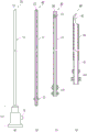
Fig. 2 (a), (b), (c), and (d) are side views or sectional views of an endoscope, an inner tube, a flow guide tube, and a treatment tube constituting the small-diameter endoscopic surgical instrument according to example 1.
FIG. 3 is an explanatory view of an operation part of the process tube.
Fig. 4 (a) is a detailed view of the hook portion of the process tube, and fig. 4 (b) is a view along the line a-a in fig. 4 (a).
Fig. 5 (a) is an explanatory view of an operation of moving a tissue by using the hook portion of the treatment tube, and fig. 5 (b) is an explanatory view of an operation of grasping a tissue.
FIG. 6 is an explanatory view of the operation of fixing and removing the tissue under the knife-prick or the blood clot adhered to the hook of the tube to be treated using the flow guide tube.
FIG. 7 is an explanatory view of the operation of electrocautery and electrocoagulation using the electrode part of the treatment tube.
Fig. 8 is an explanatory view of a method for inserting the small-diameter endoscopic surgical instrument according to example 1 into a body (example 2). (cited from J chest cardiovascular disease 2000; 120: 276-9)
Fig. 9 is an explanatory diagram of a method of using the inner tube to determine where the tip of the small-diameter endoscopic surgical instrument has reached.
Detailed Description
A mode for carrying out the present invention will be described with reference to the accompanying drawings. It is needless to say that the present invention is not limited to the above-described embodiments, and various changes may be made in the specific configuration within the scope of the present invention. The following description is made for sympathetic nerve surgery in the thoracic cavity, but the present invention is not limited to sympathetic nerve surgery in the thoracic cavity.
Example 1
Fig. 1 (a) is an overall view of a small-diameter endoscopic surgical instrument according to an embodiment 1 of the present invention at the time of surgery, fig. 1 (b) is an overall view of the small-diameter endoscopic surgical instrument inserted into a body, fig. 2 (a), (b), (c), and (d) are side views or sectional views of an endoscope, an inner tube, a catheter, and a treatment tube constituting the small-diameter endoscopic surgical instrument according to the embodiment 1, fig. 3 (a) is an explanatory view of an operation portion of the treatment tube, fig. 4 (a) is a detailed view of a hook portion of the treatment tube, and fig. 4 (b) is a view along a-a of fig. 4 (a).
As shown in fig. 1 (a), the minor-diameter endoscopic surgical instrument 1 of the present embodiment at the time of surgery is constituted by a combination of an endoscope 10, a drainage tube 30, and a disposal tube 40, and in the minor-diameter endoscopic surgical instrument 1a of the present embodiment inserted into the body, the inner tube 20 is used in place of the endoscope 10 to constitute a coaxial triple tube structure. In the present embodiment, in order to reduce the size of the skin incision as much as possible, a fiberscope having a small diameter with an outer diameter of 2mm of the tube part 11 is used as the endoscope 10.
The endoscope 10 is connected to a display system (not shown) used in a standard thoracic endoscopic surgery through the connecting portion 13 of the endoscope 10, so that the operation can be performed while observing the site of the operation and the periphery thereof through the display. In addition, an illumination optical fiber for irradiating a target site of a tissue to be operated and the periphery of the distal end portion 12 of the endoscope is also built in the tube portion 11 to send light from the connection terminal 15. The operator can hold the connection section 13 with his hand and move the connection section 13 to move the distal end portion 12 of the endoscope to the optimum position.
The guide tube 30 is a tube having an inner diameter slightly larger than the outer diameter of the tube portion of the endoscope 10, and the guide tube 30 has an inner diameter of 2.05mm and an outer diameter of 2.35mm, and supports the endoscope in an axially movable state. When the endoscope 10 is pulled into the guide tube 30 to perform a treatment such as electrocautery, it is also helpful for a person performing the operation if the endoscope 10 is provided with one or more slits 33 near the distal end portion of the guide tube in order to obtain an image around the treatment site or to eliminate white smoke rising in the front surface of the endoscope as soon as possible.
The treatment tube 40 has an inner diameter slightly larger than the outer diameter of the draft tube 30 by 2.35mm, and the treatment tube 40 has an inner diameter of 2.40mm and an outer diameter of 2.85mm, and supports the draft tube in an axially movable and rotatable state. The small-diameter endoscopic surgical instrument 1a is entirely contained in the treatment tube 40 having an outer diameter of 2.85mm, and has no laterally protruding portion, and therefore can be smoothly inserted through a skin incision having a size of only about 3 mm.
A hook 43 is provided at the front end of the treatment tube. The hook portion 43 has a shape cut into a hook shape from a curved surface of the wall of the duct 41 constituting the process tube without deforming the curved surface. As a result, as shown in fig. 4 (b), the hook 43 is also formed in a curved surface having the same inner and outer diameters as those of the treatment tube. As a result, the hook 43 does not obstruct the axial movement of the flow conduit 30 inserted into the process tube 40, nor does the hook 43 laterally protrude beyond the outer diameter of the process tube. .
In many of the conventional endoscopic surgical instruments, the sharp hook and knife provided at the distal end of the endoscopic surgical instrument may be difficult to smoothly insert the instrument by scratching surrounding tissues or deforming the distal end of the instrument when the instrument is inserted into the body, but the hook 43 is formed in the above-described shape, so that the hook portion is just along the flow guide tube 30 and does not come into contact with the surrounding tissues, thereby preventing the surrounding tissues from being damaged or the hook 43 from being deformed by an excessive force.
The tip 44 of the hook 43 is largely bent, and the inner edge of the treatment tube on the operation portion side is formed substantially parallel to the tip of the built-in flow guide tube, that is, the edge of the tip 34 formed at right angles to the axial direction. By forming the above-described shape, the tissue can be firmly gripped by operating the operating portions 42 and 32 so that the inner side of the distal end portion 44 of the hook portion is close to the distal end portion 34 of the catheter.
The treatment tube 40 is pulled forward by operating the operation portion 42 of the treatment tube by providing a knife inside the distal end portion 44 of the hook portion, and the target tissue can be easily and reliably excised and separated by rotating the same in the direction of the arrow 49 in fig. 4 (b). In performing the above-described operation, as shown in fig. 4 (a), the position of the distal end portion 12 of the endoscope 10 can be manipulated so as to be drawn into the treatment tube 40, and a clear image of the blade of the distal end portion 44 and the target tissue can be obtained. In the case of fig. 4 (a), the duct 30 is drawn into the duct 40 so as not to obstruct the view.
The front end 44 of the hook serves as a knife and also as an electrode for electrocautery and electrocoagulation. The tube portion 41 of the treatment tube is made of a material having electrical conductivity, and constitutes a conductive portion for supplying electricity to the electrode, and in order to prevent leakage of electricity to the outside, the outer surface of the tube portion 41 inserted into the body of the treatment tube is subjected to electrical insulation treatment 45 except for the electrode.
The insulation treatment is performed by coating, painting, burning, or the like of an insulating material. Although it is also conceivable to provide a monopolar (japanese: モノポーラ) and a bipolar (japanese: バイポーラ) on the electrode, a monopolar capable of concentrating a current in a relatively narrow field of view is more suitable when a delicate operation is performed on a small tissue in a body cavity, such as a sympathetic denervation operation in a thoracic cavity. In this case, the other electrode may be disposed on a part of the body of the patient, for example, the hip part, separately from the electrode of the distal end portion 44 of the hook portion.
As shown in fig. 3, an operation portion 42 is provided at the proximal end portion of the treatment tube 40. The operation unit 42 is composed of a rotation unit 48 and an annular support unit 46, wherein the rotation unit 48 is fixed to the duct portion 41 of the process tube, and the support unit 46 is attached to be rotatable with respect to the duct portion so as not to be movable in the axial direction by two stopper rings 47 fixed to the duct portion. The operator grasps support portion 46 with his thumb and forefinger and attaches the middle finger to swivel portion 48. The axial position of the processing tube 40 is changed by moving the support portion 46 in the axial direction, and the rotational angle of the processing tube 40 is changed by rotating the rotating portion 48.
In fig. 2 (b), the same operation unit 32 is also provided in the duct 30. When the slit 33 described above is provided in the flow guide tube, the rotation angle needs to be changed in order to change the direction of the field of view, and therefore, an operation portion is required similarly to the treatment tube 40. On the other hand, in the case where the guide pipe is not provided with the notch 33, since it is not necessary to change the rotation angle of the guide pipe 30, a rotation portion is not provided, and only a support portion fixed to the pipe portion of the guide pipe may be simply provided.
The inner tube 20 is a tube having the same outer diameter as the tube part 11 of the endoscope 10, and its end 23 is held at the foremost end of the small-diameter endoscopic surgical instrument 1a when inserted into the body as shown in fig. 1 (a), and therefore, it is preferable to grind it into a rounded corner shape to smoothly perform the insertion operation without damaging surrounding tissues. Further, since it is necessary to change the axial position of the inner tube 10 without changing the rotation angle thereof, it is not necessary to provide a rotation part of the operation part 42 provided in the treatment tube 40, and it is only necessary to provide the operation part 22 having a size which is fixed to the tube portion of the inner tube and can be easily grasped with the thumb and the index finger.
The inner tube 20, the flow guide tube 30, and the treatment tube 40 described above are small-diameter and thin-walled members in the range in which the respective functions can be exerted, and the difference between the inner diameter and the outer diameter of the tubes in contact with each other is extremely small, so that the outer diameter of the entire small-diameter endoscopic surgical instrument 1 is reduced to only 2.85m, which greatly contributes to reducing the size of the skin incision hole for insertion. Further, since the difference between the inner diameter and the outer diameter of the tubes that are in contact with each other, that is, the gap between the tubes is extremely small, it is possible to reduce the possibility that the cut tissue or body fluid enters the gap between the tubes and causes a trouble in the operation.
On the other hand, the inner tube 20, the flow guide tube 30 and the treatment tube 40 are each in the shape of a simple pipe, and are easily separated after the three are pulled out, so that the cleaning and sterilization in advance can be easily and thoroughly performed.
FIG. 5 (a) is an explanatory diagram of the operation (feeding) of moving the tissue by using the hook portion 43 of the treatment tube. During surgery, the tissue 51 that is an obstacle needs to be moved for the purpose of approaching a target site or for the purpose of securing a visual field. In this case, the endoscope 10 brings the hook portion 43 and the tissue 51 that is a hindrance into the field of view, and the supporting portion 46 of the operating portion of the treatment tube 40 is operated so that the hook portion 43 approaches the tissue 51, and the tissue 51 is hooked while the direction of the hook portion is changed by the operation of the rotating portion 48. The delivery tube 30 is now pulled into the treatment tube 40. When the above state is achieved, the tissue 51 can be taught, separated, and pulled. Further, since the hook portion 43 is provided with a knife, the tissue 51 can be excised by using the knife.
Fig. 5 (b) is an explanatory diagram of an operation (b) of grasping a tissue. Similarly to the case of the above-described operation, the endoscope 10 brings the hook portion 43 and the tissue 52 to be grasped into the visual field, and the support portion 46 of the operation portion of the treatment tube 40 is operated to bring the hook portion 43 close to the tissue 52, and the tissue 52 is grasped while changing the direction of the hook portion by the operation of the rotating portion 48, and then the operation portion 32 of the catheter 30 is operated to move the catheter 30 forward, and the tissue 52 grasped by the hook portion 43 is grasped. The slit 33 provided in the guide tube helps to ensure the visual field of the endoscope 10 when the guide tube 30 is moved forward.
The distal end 44 of the hook 43 is largely bent, and the inner edge thereof is formed substantially parallel to the edge of the distal end 34 of the duct 30, so that the tissue 52 is firmly held therebetween, and the tissue 52 can be taught, separated, and pulled. Further, the small-diameter endoscopic surgical instrument 1 is taken out of the body of the patient while holding the tissue 52, whereby the tissue 52 can be used for the biopathology examination.
Fig. 6 is an explanatory view of an operation of fixing and removing a tissue not originally pierced by the distal end portion 44 of the hook portion of the treatment tube 40 or a blood clot adhering to the hook portion 43 using the flow guide tube 30. With the endoscope 10, a firm object, such as the rib 64, located nearby is found, and the operating section 42 is operated so that the hook portion 43 of the treatment tube comes close to the rib 64. Next, the operation section 32 of the catheter 30 is operated to advance the catheter 30, so that the punctured tissue or blood clot 53 is pressed against the rib 64 and fixed. In this state, when the rotation portion 48 of the treatment tube 40 is operated to rotate the treatment tube 40 in the direction of the arrow 56 in the figure, the blood clot or the like 53 can be easily removed from the hook portion 43.
Such a situation often occurs in the operation, and conventionally, there has been no method better than pulling out the endoscopic surgical instrument to the outside of the body and removing the tissue and blood clots stuck to the hook portion, but by performing the removal operation in this way, it is possible to not only facilitate the smooth progress of the operation but also reduce the burden on the patient.
FIG. 7 is an explanatory view of the operation of electrocautery or electrocoagulation using the distal end 44 of the hook of the treatment tube 40 as an electrode. To feed electricity to the front end 44 of the hook, an alligator clamp 58 is provided in the pipe section 41 of the outer surface of the process tube 40, which is not subjected to an electrical insulation process. The other electrode (not shown) is provided on the hip of the patient. Since the duct portion 41 of the treatment tube is made of a material having an electrical conductivity, the tissue 57 to be operated can be electrocautery or electrocoagulation performed by the preparation described above using the distal end portion 44 of the hook portion as an electrode. The guide tube is pulled into the treatment tube 40, so that the endoscope 10 does not interfere with the guide tube, and the distal end portion 44 of the hook portion serving as the electrode and the tissue 57 can be accommodated in the field of view.
Example 2
Fig. 8 is an explanatory view of a method of inserting the small-diameter endoscopic surgical instrument 1a of example 1 into a body (example 2), and fig. 9 is an explanatory view of a manner of using the inner tube 20 to know where the tip of the small-diameter endoscopic surgical instrument 1a has arrived at the time of the insertion.
When a sympathetic nerve trunk resection operation or the like in the thoracic cavity is performed using the small-diameter endoscopic surgical instrument 1 of example 1, first, a skin incision 61 having a size of 3mm is opened in the center line of the armpit under the armpit. Through the cut hole, a small-diameter endoscopic surgical instrument 1a into which an inner tube 20 is inserted in place of the endoscope 10 is inserted. The subject tissue of the surgery is a thoracic sympathetic trunk 63 extending in the up-down direction along the spine. The minor diameter endoscopic surgical instrument 1a is passed through the space between the ribs 64 and in proximity to the subject tissue. In the periphery, in addition to the ribs 64, there are a lung apex 65, a clavicle 66, an upper great vein 67, an aorta 68, and the like.
The inner tube 20 of the small-diameter endoscopic surgical instrument 1a is connected to a syringe 92 through a flexible tube 91, and saline or air is injected into the syringe. When the tip of the small-diameter endoscopic surgical instrument 1a, that is, the tip portion 23 of the inner tube 20 reaches the pleural cavity 93, the pressure of the physiological saline or the air decreases due to the negative pressure in the pleural cavity 93, and the piston 92a of the syringe can be observed to advance.
After the above determination, the operation can be performed by removing the inner tube 20 from the small-diameter endoscopic surgical instrument 1a and inserting the endoscope 10 into the small-diameter endoscopic surgical instrument 1 at the time of the operation.
Industrial applicability of the invention
The minor-diameter endoscopic surgical instrument of the present invention is a simple structure with a small number of components, and not only can perform electrocautery, electrocoagulation, excision, affection, separation and traction of target tissues, but also has the following multifunctionality: the subject tissue is held or fixed to perform feeding, separation, and traction of the subject tissue, and can be used in the detection of a living pathology, and in addition, blood clots and the like adhering to the hook portion can be removed in the above-described manner in the operation. In addition, the operator can perform the operation with easy operation while observing a clear image. Further, the skin incision hole is small and only one place is needed, which makes the burden on the patient small. It is desirable to take full advantage of these advantages and to make them widely available for use in a variety of future procedures.
(symbol description)
1. 1a the minor diameter endoscopic surgical instrument of embodiment 1;
10 endoscope;
11 a tube portion of an endoscope;
12 a front end portion of an endoscope;
13 a connecting section (operation section) of the endoscope;
15 an optical fiber connection terminal of an endoscope;
20 an inner tube;
21 a conduit portion of the inner conduit;
22 an operating portion of the inner tube;
23 the front end of the inner tube;
30 flow guide pipes;
31 the duct portion of the draft tube;
an operation part of 32 guide pipes;
33, cutting grooves of a guide pipe;
34 the front end of the draft tube;
40 treating the tube;
41 treating the conduit portion of the tube;
42 a handling section for handling the tube;
43 treatment of the hook of the tube;
44 treating the front end of the hook of the tube;
45, electric insulation treatment of the treatment pipe;
46 a support for the process tube;
47 disposal of a confinement ring of the tube;
48 rotating part of the process tube;
49. 56 the direction of rotation of the tube;
51. 52, 57 tissue;
53 coagulum, etc.;
58 alligator clips;
61 cutting a hole on the skin;
63 thoracic sympathetic trunk;
64 ribs;
65 pulmonary apices;
66 clavicle;
67 upper great vein;
68 aorta;
91 a flexible hose;
92. 92a syringe;
93 pleural cavity.
Claims (5)
1. A minor-diameter endoscope surgical instrument used after being inserted into a minor-diameter endoscope is characterized in that,
the minor-diameter endoscopic surgical instrument is a minor-diameter endoscopic surgical instrument with a coaxial and multiple-tube structure and comprises an inner tube, a guide tube and a processing tube, wherein,
the inner tube has the same outer diameter as a tube portion of the small-diameter endoscope used, is used when the small-diameter endoscopic surgical instrument is inserted into a body, and is replaced with the small-diameter endoscope after insertion,
the guide tube is inserted with the minor-diameter endoscope or the inner tube and supports the minor-diameter endoscope or the inner tube in a state of being movable in the axial direction,
the treatment tube is inserted with the guide tube and supports the guide tube in a state of being capable of moving along the axial direction, a treatment element is arranged at the front end part of the treatment tube, the treatment element comprises an electrode, a knife and a hook part,
the small-diameter endoscope, the inner tube, the flow guide tube, and the treatment tube have independent operation portions at their proximal end portions, that is, portions located outside the patient body when performing an operation using the small-diameter endoscope, and are operated using the operation portions, so that their respective axial positions can be changed, and their respective rotation angles can be changed as needed.
2. The minor diameter endoscopic surgical instrument of claim 1,
the treatment tube has a hook portion at a distal end portion thereof, the hook portion having a shape obtained by cutting a part of the treatment tube, and a blade provided at the proximal end portion of the hook portion on the side of the proximal end portion of the treatment tube in a plane substantially perpendicular to the axial direction of the treatment tube.
3. The minor diameter endoscopic surgical instrument of claim 1,
the tip of the treatment tube is formed with an electrode, the tube of the treatment tube itself is formed with a current-carrying portion for feeding electricity to the electrode, and the portion of the treatment tube inserted into the body is subjected to an electrical insulation treatment on the outer surface except for the electrode.
4. The minor diameter endoscopic surgical instrument of claim 1,
the front end of the inner tube is ground into a rounded corner shape and includes a means for feeding saline or air through the inner tube.
5. The minor diameter endoscopic surgical instrument of claim 1,
the draft tube has a cut-out near a front end portion, and the cut-out penetrates inside and outside the draft tube.
Applications Claiming Priority (3)
| Application Number | Priority Date | Filing Date | Title |
|---|---|---|---|
| JP2016151724A JP6749545B2 (en) | 2016-08-02 | 2016-08-02 | Thin endoscopic surgical instrument |
| JP2016-151724 | 2016-08-02 | ||
| PCT/JP2017/026598 WO2018025680A1 (en) | 2016-08-02 | 2017-07-24 | Small-diameter endoscope surgical instrument |
Publications (2)
| Publication Number | Publication Date |
|---|---|
| CN109561921A CN109561921A (en) | 2019-04-02 |
| CN109561921B true CN109561921B (en) | 2021-07-09 |
Family
ID=61072829
Family Applications (1)
| Application Number | Title | Priority Date | Filing Date |
|---|---|---|---|
| CN201780048717.5A Active CN109561921B (en) | 2016-08-02 | 2017-07-24 | Minor diameter endoscope operation instrument |
Country Status (6)
| Country | Link |
|---|---|
| US (1) | US11457798B2 (en) |
| EP (1) | EP3494913B1 (en) |
| JP (1) | JP6749545B2 (en) |
| KR (1) | KR102253461B1 (en) |
| CN (1) | CN109561921B (en) |
| WO (1) | WO2018025680A1 (en) |
Families Citing this family (4)
| Publication number | Priority date | Publication date | Assignee | Title |
|---|---|---|---|---|
| CA3094068C (en) * | 2018-06-27 | 2023-09-19 | Wright Medical Technology, Inc. | Burr with irrigation and imaging |
| DE102020110845A1 (en) * | 2020-04-21 | 2021-10-21 | Novatech Sa | Medical device and endoscopic procedure |
| CN114533213A (en) * | 2020-11-24 | 2022-05-27 | 奥林巴斯株式会社 | Treatment method and device with ultrasound endoscope |
| KR102539420B1 (en) | 2022-08-09 | 2023-06-01 | 조백현 | A Surgical Equipment |
Citations (4)
| Publication number | Priority date | Publication date | Assignee | Title |
|---|---|---|---|---|
| EP0800793B1 (en) * | 1996-04-10 | 2001-06-13 | Linvatec Corporation | Process for shaping and sharpening a rotatable surgical shaver blade |
| CN1317950A (en) * | 1998-09-18 | 2001-10-17 | 山本英博 | Endoscope power supplying appliance |
| CN201701297U (en) * | 2010-04-15 | 2011-01-12 | 欧普康光电(厦门)有限公司 | Surgical endoscope with digital camera device |
| JP2014018299A (en) * | 2012-07-13 | 2014-02-03 | Olympus Corp | Guide wire introduction device |
Family Cites Families (21)
| Publication number | Priority date | Publication date | Assignee | Title |
|---|---|---|---|---|
| DE3313325A1 (en) | 1983-04-13 | 1984-10-18 | Knut Dr. 7802 Merzhausen Korth | Surgical instrument |
| US5578053A (en) * | 1993-06-24 | 1996-11-26 | Yoon; Inbae | Safety needle instrument having a triggered safety member |
| US5478329A (en) * | 1994-05-06 | 1995-12-26 | Ternamian; Artin M. | Trocarless rotational entry cannula |
| US5709671A (en) * | 1995-10-16 | 1998-01-20 | Ethicon Endo-Surgery, Inc. | Trocar having an improved tip configuration |
| DE19547246C1 (en) * | 1995-12-18 | 1997-03-20 | Riek Siegfried | Medicinal needle containing spring-loaded guard |
| JPH10155807A (en) * | 1996-11-29 | 1998-06-16 | Olympus Optical Co Ltd | Treatment tool for endoscope |
| DE19730127C2 (en) * | 1997-07-14 | 2001-04-12 | Erbe Elektromedizin | Dissecting instrument |
| US6428539B1 (en) * | 2000-03-09 | 2002-08-06 | Origin Medsystems, Inc. | Apparatus and method for minimally invasive surgery using rotational cutting tool |
| JP4460718B2 (en) * | 2000-05-12 | 2010-05-12 | オリンパス株式会社 | Endoscopic treatment device |
| JP3722729B2 (en) * | 2001-06-04 | 2005-11-30 | オリンパス株式会社 | Endoscope treatment device |
| US7556633B2 (en) * | 2004-03-01 | 2009-07-07 | Terumo Corporation | Method and apparatus for endoscopic dissection of blood vessels |
| DE102005013714A1 (en) * | 2004-04-07 | 2005-12-22 | Carl Zeiss Meditec Ag | Electrical probe for microsurgery |
| CA2572192A1 (en) * | 2004-06-29 | 2006-01-12 | Applied Medical Resources Corporation | Insufflating optical surgical instrument |
| CN101111200B (en) * | 2004-12-17 | 2010-05-12 | 国立大学法人京都大学 | Hood with excising function and endoscope |
| JP2007089690A (en) * | 2005-09-27 | 2007-04-12 | Hidehiro Yamamoto | Conductive traction device for endoscope |
| DE102005049021B4 (en) * | 2005-10-11 | 2008-08-21 | Richard Wolf Gmbh | endoscope |
| US20070203395A1 (en) * | 2006-02-28 | 2007-08-30 | Takayasu Mikkaichi | Cap installable on distal end portion of endoscope |
| US8728089B2 (en) * | 2008-03-28 | 2014-05-20 | Olympus Medical Systems Corp. | Endoscope treatment instrument |
| US8531064B2 (en) * | 2010-02-11 | 2013-09-10 | Ethicon Endo-Surgery, Inc. | Ultrasonically powered surgical instruments with rotating cutting implement |
| US8986278B2 (en) * | 2010-04-13 | 2015-03-24 | Sentreheart, Inc. | Methods and devices for pericardial access |
| US10363056B2 (en) * | 2015-06-17 | 2019-07-30 | Saphena Medical, Inc. | Unitary endoscopic vessel harvesting devices |
-
2016
- 2016-08-02 JP JP2016151724A patent/JP6749545B2/en active Active
-
2017
- 2017-07-24 WO PCT/JP2017/026598 patent/WO2018025680A1/en unknown
- 2017-07-24 US US16/322,187 patent/US11457798B2/en active Active
- 2017-07-24 KR KR1020197003078A patent/KR102253461B1/en active IP Right Grant
- 2017-07-24 CN CN201780048717.5A patent/CN109561921B/en active Active
- 2017-07-24 EP EP17836786.8A patent/EP3494913B1/en active Active
Patent Citations (4)
| Publication number | Priority date | Publication date | Assignee | Title |
|---|---|---|---|---|
| EP0800793B1 (en) * | 1996-04-10 | 2001-06-13 | Linvatec Corporation | Process for shaping and sharpening a rotatable surgical shaver blade |
| CN1317950A (en) * | 1998-09-18 | 2001-10-17 | 山本英博 | Endoscope power supplying appliance |
| CN201701297U (en) * | 2010-04-15 | 2011-01-12 | 欧普康光电(厦门)有限公司 | Surgical endoscope with digital camera device |
| JP2014018299A (en) * | 2012-07-13 | 2014-02-03 | Olympus Corp | Guide wire introduction device |
Non-Patent Citations (1)
| Title |
|---|
| Needlescopic Surgery for Palmar Hyperhidrosis;Hidehiro Yamamoto等;《The Journal of Thoracic and Cardiovascular Surgery》;20000831;第120卷(第2期);第276-279页 * |
Also Published As
| Publication number | Publication date |
|---|---|
| EP3494913A4 (en) | 2020-03-25 |
| KR102253461B1 (en) | 2021-05-17 |
| JP6749545B2 (en) | 2020-09-02 |
| JP2018019815A (en) | 2018-02-08 |
| KR20190026809A (en) | 2019-03-13 |
| WO2018025680A1 (en) | 2018-02-08 |
| US20190183320A1 (en) | 2019-06-20 |
| US11457798B2 (en) | 2022-10-04 |
| EP3494913B1 (en) | 2024-04-24 |
| CN109561921A (en) | 2019-04-02 |
| EP3494913A1 (en) | 2019-06-12 |
Similar Documents
| Publication | Publication Date | Title |
|---|---|---|
| CN105411632B (en) | Apparatus, system, and method for obtaining tissue samples using biopsy tools | |
| CN109561921B (en) | Minor diameter endoscope operation instrument | |
| JP4982699B2 (en) | Biopsy forceps | |
| US8486010B2 (en) | Bendable catheter | |
| JP5928862B2 (en) | Endoscopic treatment tool and incision system | |
| JP4430238B2 (en) | Endoscope electrification device | |
| JP5792416B1 (en) | Endoscopic treatment system | |
| KR20010052456A (en) | Device and method for resecting body tissues | |
| US9173697B2 (en) | Surgical cannula for dissipating electric charge | |
| US20160367311A1 (en) | Instrumentation with Embedded Imaging Systems | |
| EP2636379A1 (en) | Tissue dissection and removal device | |
| JP6579601B2 (en) | Endoscope | |
| US20110105838A1 (en) | Suction device for endoscopic instruments and method | |
| JP6886569B2 (en) | Small diameter endoscopic surgical instruments | |
| US20080294160A1 (en) | RF endoscopic electrosurgical instrument | |
| JP2023514646A (en) | Systems and methods for surgical procedures using torque-driven guidewires | |
| EP3272266A1 (en) | Endoscope device | |
| US20200178945A1 (en) | Method and apparatus for minimally invasive amelioration of spinal epidural lipomatosis | |
| JP2003310628A (en) | In-vivo tissue collection device | |
| JP7448313B2 (en) | sheath with detectable reader | |
| JP2007089690A (en) | Conductive traction device for endoscope | |
| JP2018530409A (en) | Biological tissue sampling device | |
| JP7396805B2 (en) | Sheath tip with angled distal surface | |
| WO2017091803A1 (en) | Percutaneous tunneling devices and methods of use | |
| US20210244463A1 (en) | Assembly aid tool for securing electrodes in a resectoscope |
Legal Events
| Date | Code | Title | Description |
|---|---|---|---|
| PB01 | Publication | ||
| PB01 | Publication | ||
| SE01 | Entry into force of request for substantive examination | ||
| SE01 | Entry into force of request for substantive examination | ||
| GR01 | Patent grant | ||
| GR01 | Patent grant |