EP3492497B1 - Mg7, single-chain antibody specifically binding highly-glycosylated cea and use thereof in detection and treatment - Google Patents
Mg7, single-chain antibody specifically binding highly-glycosylated cea and use thereof in detection and treatment Download PDFInfo
- Publication number
- EP3492497B1 EP3492497B1 EP17833578.2A EP17833578A EP3492497B1 EP 3492497 B1 EP3492497 B1 EP 3492497B1 EP 17833578 A EP17833578 A EP 17833578A EP 3492497 B1 EP3492497 B1 EP 3492497B1
- Authority
- EP
- European Patent Office
- Prior art keywords
- seq
- set forth
- cells
- cell
- cdr
- Prior art date
- Legal status (The legal status is an assumption and is not a legal conclusion. Google has not performed a legal analysis and makes no representation as to the accuracy of the status listed.)
- Active
Links
Images




Classifications
-
- G—PHYSICS
- G01—MEASURING; TESTING
- G01N—INVESTIGATING OR ANALYSING MATERIALS BY DETERMINING THEIR CHEMICAL OR PHYSICAL PROPERTIES
- G01N33/00—Investigating or analysing materials by specific methods not covered by groups G01N1/00 - G01N31/00
- G01N33/48—Biological material, e.g. blood, urine; Haemocytometers
- G01N33/50—Chemical analysis of biological material, e.g. blood, urine; Testing involving biospecific ligand binding methods; Immunological testing
- G01N33/53—Immunoassay; Biospecific binding assay; Materials therefor
- G01N33/575—Immunoassay; Biospecific binding assay; Materials therefor for cancer
-
- C—CHEMISTRY; METALLURGY
- C07—ORGANIC CHEMISTRY
- C07K—PEPTIDES
- C07K16/00—Immunoglobulins [IG], e.g. monoclonal or polyclonal antibodies
- C07K16/18—Immunoglobulins [IG], e.g. monoclonal or polyclonal antibodies against material from animals or humans
- C07K16/28—Immunoglobulins [IG], e.g. monoclonal or polyclonal antibodies against material from animals or humans against receptors, cell surface antigens or cell surface determinants
- C07K16/30—Immunoglobulins [IG], e.g. monoclonal or polyclonal antibodies against material from animals or humans against receptors, cell surface antigens or cell surface determinants from tumour cells
-
- C—CHEMISTRY; METALLURGY
- C07—ORGANIC CHEMISTRY
- C07K—PEPTIDES
- C07K16/00—Immunoglobulins [IG], e.g. monoclonal or polyclonal antibodies
- C07K16/18—Immunoglobulins [IG], e.g. monoclonal or polyclonal antibodies against material from animals or humans
- C07K16/28—Immunoglobulins [IG], e.g. monoclonal or polyclonal antibodies against material from animals or humans against receptors, cell surface antigens or cell surface determinants
- C07K16/30—Immunoglobulins [IG], e.g. monoclonal or polyclonal antibodies against material from animals or humans against receptors, cell surface antigens or cell surface determinants from tumour cells
- C07K16/3007—Carcino-embryonic Antigens
-
- C—CHEMISTRY; METALLURGY
- C07—ORGANIC CHEMISTRY
- C07K—PEPTIDES
- C07K14/00—Peptides having more than 20 amino acids; Gastrins; Somatostatins; Melanotropins; Derivatives thereof
- C07K14/435—Peptides having more than 20 amino acids; Gastrins; Somatostatins; Melanotropins; Derivatives thereof from animals; from humans
- C07K14/705—Receptors; Cell surface antigens; Cell surface determinants
- C07K14/70503—Immunoglobulin superfamily
- C07K14/7051—T-cell receptor (TcR)-CD3 complex
-
- A—HUMAN NECESSITIES
- A61—MEDICAL OR VETERINARY SCIENCE; HYGIENE
- A61K—PREPARATIONS FOR MEDICAL, DENTAL OR TOILETRY PURPOSES
- A61K40/00—Cellular immunotherapy
- A61K40/10—Cellular immunotherapy characterised by the cell type used
- A61K40/11—T-cells, e.g. tumour infiltrating lymphocytes [TIL] or regulatory T [Treg] cells; Lymphokine-activated killer [LAK] cells
-
- A—HUMAN NECESSITIES
- A61—MEDICAL OR VETERINARY SCIENCE; HYGIENE
- A61K—PREPARATIONS FOR MEDICAL, DENTAL OR TOILETRY PURPOSES
- A61K40/00—Cellular immunotherapy
- A61K40/30—Cellular immunotherapy characterised by the recombinant expression of specific molecules in the cells of the immune system
- A61K40/31—Chimeric antigen receptors [CAR]
-
- A—HUMAN NECESSITIES
- A61—MEDICAL OR VETERINARY SCIENCE; HYGIENE
- A61K—PREPARATIONS FOR MEDICAL, DENTAL OR TOILETRY PURPOSES
- A61K40/00—Cellular immunotherapy
- A61K40/40—Cellular immunotherapy characterised by antigens that are targeted or presented by cells of the immune system
- A61K40/41—Vertebrate antigens
- A61K40/42—Cancer antigens
- A61K40/4264—Cancer antigens from embryonic or fetal origin
- A61K40/4266—Carcinoembryonic antigen [CEA]
-
- A—HUMAN NECESSITIES
- A61—MEDICAL OR VETERINARY SCIENCE; HYGIENE
- A61P—SPECIFIC THERAPEUTIC ACTIVITY OF CHEMICAL COMPOUNDS OR MEDICINAL PREPARATIONS
- A61P35/00—Antineoplastic agents
-
- C—CHEMISTRY; METALLURGY
- C07—ORGANIC CHEMISTRY
- C07K—PEPTIDES
- C07K14/00—Peptides having more than 20 amino acids; Gastrins; Somatostatins; Melanotropins; Derivatives thereof
- C07K14/435—Peptides having more than 20 amino acids; Gastrins; Somatostatins; Melanotropins; Derivatives thereof from animals; from humans
- C07K14/705—Receptors; Cell surface antigens; Cell surface determinants
- C07K14/70503—Immunoglobulin superfamily
- C07K14/70517—CD8
-
- C—CHEMISTRY; METALLURGY
- C07—ORGANIC CHEMISTRY
- C07K—PEPTIDES
- C07K14/00—Peptides having more than 20 amino acids; Gastrins; Somatostatins; Melanotropins; Derivatives thereof
- C07K14/435—Peptides having more than 20 amino acids; Gastrins; Somatostatins; Melanotropins; Derivatives thereof from animals; from humans
- C07K14/705—Receptors; Cell surface antigens; Cell surface determinants
- C07K14/70503—Immunoglobulin superfamily
- C07K14/70521—CD28, CD152
-
- C—CHEMISTRY; METALLURGY
- C07—ORGANIC CHEMISTRY
- C07K—PEPTIDES
- C07K14/00—Peptides having more than 20 amino acids; Gastrins; Somatostatins; Melanotropins; Derivatives thereof
- C07K14/435—Peptides having more than 20 amino acids; Gastrins; Somatostatins; Melanotropins; Derivatives thereof from animals; from humans
- C07K14/705—Receptors; Cell surface antigens; Cell surface determinants
- C07K14/70575—NGF/TNF-superfamily, e.g. CD70, CD95L, CD153, CD154
-
- C—CHEMISTRY; METALLURGY
- C07—ORGANIC CHEMISTRY
- C07K—PEPTIDES
- C07K14/00—Peptides having more than 20 amino acids; Gastrins; Somatostatins; Melanotropins; Derivatives thereof
- C07K14/435—Peptides having more than 20 amino acids; Gastrins; Somatostatins; Melanotropins; Derivatives thereof from animals; from humans
- C07K14/705—Receptors; Cell surface antigens; Cell surface determinants
- C07K14/70596—Molecules with a "CD"-designation not provided for elsewhere
-
- C—CHEMISTRY; METALLURGY
- C07—ORGANIC CHEMISTRY
- C07K—PEPTIDES
- C07K16/00—Immunoglobulins [IG], e.g. monoclonal or polyclonal antibodies
- C07K16/40—Immunoglobulins [IG], e.g. monoclonal or polyclonal antibodies against enzymes
-
- C—CHEMISTRY; METALLURGY
- C07—ORGANIC CHEMISTRY
- C07K—PEPTIDES
- C07K19/00—Hybrid peptides, i.e. peptides covalently bound to nucleic acids, or non-covalently bound protein-protein complexes
-
- C—CHEMISTRY; METALLURGY
- C12—BIOCHEMISTRY; BEER; SPIRITS; WINE; VINEGAR; MICROBIOLOGY; ENZYMOLOGY; MUTATION OR GENETIC ENGINEERING
- C12N—MICROORGANISMS OR ENZYMES; COMPOSITIONS THEREOF; PROPAGATING, PRESERVING, OR MAINTAINING MICROORGANISMS; MUTATION OR GENETIC ENGINEERING; CULTURE MEDIA
- C12N15/00—Mutation or genetic engineering; DNA or RNA concerning genetic engineering, vectors, e.g. plasmids, or their isolation, preparation or purification; Use of hosts therefor
- C12N15/09—Recombinant DNA-technology
- C12N15/11—DNA or RNA fragments; Modified forms thereof; Non-coding nucleic acids having a biological activity
- C12N15/62—DNA sequences coding for fusion proteins
-
- C—CHEMISTRY; METALLURGY
- C12—BIOCHEMISTRY; BEER; SPIRITS; WINE; VINEGAR; MICROBIOLOGY; ENZYMOLOGY; MUTATION OR GENETIC ENGINEERING
- C12N—MICROORGANISMS OR ENZYMES; COMPOSITIONS THEREOF; PROPAGATING, PRESERVING, OR MAINTAINING MICROORGANISMS; MUTATION OR GENETIC ENGINEERING; CULTURE MEDIA
- C12N5/00—Undifferentiated human, animal or plant cells, e.g. cell lines; Tissues; Cultivation or maintenance thereof; Culture media therefor
- C12N5/06—Animal cells or tissues; Human cells or tissues
- C12N5/0602—Vertebrate cells
- C12N5/0634—Cells from the blood or the immune system
- C12N5/0636—T lymphocytes
-
- C—CHEMISTRY; METALLURGY
- C12—BIOCHEMISTRY; BEER; SPIRITS; WINE; VINEGAR; MICROBIOLOGY; ENZYMOLOGY; MUTATION OR GENETIC ENGINEERING
- C12N—MICROORGANISMS OR ENZYMES; COMPOSITIONS THEREOF; PROPAGATING, PRESERVING, OR MAINTAINING MICROORGANISMS; MUTATION OR GENETIC ENGINEERING; CULTURE MEDIA
- C12N5/00—Undifferentiated human, animal or plant cells, e.g. cell lines; Tissues; Cultivation or maintenance thereof; Culture media therefor
- C12N5/10—Cells modified by introduction of foreign genetic material
-
- A—HUMAN NECESSITIES
- A61—MEDICAL OR VETERINARY SCIENCE; HYGIENE
- A61K—PREPARATIONS FOR MEDICAL, DENTAL OR TOILETRY PURPOSES
- A61K39/00—Medicinal preparations containing antigens or antibodies
- A61K2039/505—Medicinal preparations containing antigens or antibodies comprising antibodies
-
- A—HUMAN NECESSITIES
- A61—MEDICAL OR VETERINARY SCIENCE; HYGIENE
- A61K—PREPARATIONS FOR MEDICAL, DENTAL OR TOILETRY PURPOSES
- A61K2121/00—Preparations for use in therapy
-
- A—HUMAN NECESSITIES
- A61—MEDICAL OR VETERINARY SCIENCE; HYGIENE
- A61K—PREPARATIONS FOR MEDICAL, DENTAL OR TOILETRY PURPOSES
- A61K2239/00—Indexing codes associated with cellular immunotherapy of group A61K40/00
- A61K2239/46—Indexing codes associated with cellular immunotherapy of group A61K40/00 characterised by the cancer treated
- A61K2239/51—Stomach
-
- A—HUMAN NECESSITIES
- A61—MEDICAL OR VETERINARY SCIENCE; HYGIENE
- A61K—PREPARATIONS FOR MEDICAL, DENTAL OR TOILETRY PURPOSES
- A61K2300/00—Mixtures or combinations of active ingredients, wherein at least one active ingredient is fully defined in groups A61K31/00 - A61K41/00
-
- C—CHEMISTRY; METALLURGY
- C07—ORGANIC CHEMISTRY
- C07K—PEPTIDES
- C07K2317/00—Immunoglobulins specific features
- C07K2317/50—Immunoglobulins specific features characterized by immunoglobulin fragments
- C07K2317/56—Immunoglobulins specific features characterized by immunoglobulin fragments variable (Fv) region, i.e. VH and/or VL
- C07K2317/565—Complementarity determining region [CDR]
-
- C—CHEMISTRY; METALLURGY
- C07—ORGANIC CHEMISTRY
- C07K—PEPTIDES
- C07K2317/00—Immunoglobulins specific features
- C07K2317/60—Immunoglobulins specific features characterized by non-natural combinations of immunoglobulin fragments
- C07K2317/62—Immunoglobulins specific features characterized by non-natural combinations of immunoglobulin fragments comprising only variable region components
- C07K2317/622—Single chain antibody (scFv)
-
- C—CHEMISTRY; METALLURGY
- C07—ORGANIC CHEMISTRY
- C07K—PEPTIDES
- C07K2317/00—Immunoglobulins specific features
- C07K2317/70—Immunoglobulins specific features characterized by effect upon binding to a cell or to an antigen
- C07K2317/73—Inducing cell death, e.g. apoptosis, necrosis or inhibition of cell proliferation
-
- C—CHEMISTRY; METALLURGY
- C07—ORGANIC CHEMISTRY
- C07K—PEPTIDES
- C07K2319/00—Fusion polypeptide
- C07K2319/01—Fusion polypeptide containing a localisation/targetting motif
- C07K2319/02—Fusion polypeptide containing a localisation/targetting motif containing a signal sequence
-
- C—CHEMISTRY; METALLURGY
- C07—ORGANIC CHEMISTRY
- C07K—PEPTIDES
- C07K2319/00—Fusion polypeptide
- C07K2319/01—Fusion polypeptide containing a localisation/targetting motif
- C07K2319/03—Fusion polypeptide containing a localisation/targetting motif containing a transmembrane segment
-
- C—CHEMISTRY; METALLURGY
- C07—ORGANIC CHEMISTRY
- C07K—PEPTIDES
- C07K2319/00—Fusion polypeptide
- C07K2319/33—Fusion polypeptide fusions for targeting to specific cell types, e.g. tissue specific targeting, targeting of a bacterial subspecies
-
- C—CHEMISTRY; METALLURGY
- C12—BIOCHEMISTRY; BEER; SPIRITS; WINE; VINEGAR; MICROBIOLOGY; ENZYMOLOGY; MUTATION OR GENETIC ENGINEERING
- C12N—MICROORGANISMS OR ENZYMES; COMPOSITIONS THEREOF; PROPAGATING, PRESERVING, OR MAINTAINING MICROORGANISMS; MUTATION OR GENETIC ENGINEERING; CULTURE MEDIA
- C12N2510/00—Genetically modified cells
-
- G—PHYSICS
- G01—MEASURING; TESTING
- G01N—INVESTIGATING OR ANALYSING MATERIALS BY DETERMINING THEIR CHEMICAL OR PHYSICAL PROPERTIES
- G01N2333/00—Assays involving biological materials from specific organisms or of a specific nature
- G01N2333/435—Assays involving biological materials from specific organisms or of a specific nature from animals; from humans
- G01N2333/46—Assays involving biological materials from specific organisms or of a specific nature from animals; from humans from vertebrates
- G01N2333/47—Assays involving proteins of known structure or function as defined in the subgroups
Definitions
- the present invention relates to the field of tumor immunotherapy, and more particularly, the present invention relates to a glycosylated CEA-specific single-chain antibody.
- CEA cancerembryonic antigen
- the serum level is elevated, which has auxiliary significance for the early diagnosis of pancreatic cancer and colon cancer, and has certain reference value for tumor spread, curative effect, recurrence and prognosis.
- Professor Fan Daiming from the Fourth Military Medical University found that the glycosylated CEA is highly sensitive and specific to a variety of digestive tract cancers. Clinical studies have shown that the positive rate of glycosylated CEA in gastric cancer tissues is above 80%, the positive rate in colon cancer tissues is above 40%, the positive rate in gastric precancerous lesions is above 30%, and the positive rate in esophageal cancer tissues is 18% or more.
- CAR-T chimeric antigen receptor T cell
- TAA tumor-associated antigen
- CD3zeta T cell activation signal
- the fusion protein is specifically expressed on T cell surface by a lentivirus or the like, allowing the modified T cell to specifically recognize and kill tumor cells, being independent of major histocompatibility antigen (MHC), and avoiding tumor immune escape due to MHC deletion.
- MHC major histocompatibility antigen
- the chimeric antigen receptor includes an extracellular antigen targeting and recognizing region, a hinge region, a transmembrane region, and an intracellular costimulatory signaling region.
- the antigen recognizing region is mostly a single-chain antibody or a specific receptor, which ensures that the modified T cell can specifically recognize the target cell and be activated to specifically kill the target cell;
- the hinge region generally adopts CD8 ⁇ , CD28ECD, IgG Fc fragment and the like, which ensures that the T cell contacts the target cell and affects the T cell action;
- the intracellular signal region adopts an immunoreceptor tyrosine activation motif (ITAM), for example CD3zeta and costimulatory signal such as CD28, CD137, CD27, ICOS, OX40, DAP10, etc. ( Sadelain et al., Cancer Discov 3(4): 388-98, 2013 ).
- ITAM immunoreceptor tyrosine activation motif
- CAR-T has shown good application value in the treatment of hematological malignancies
- CAR-T often does not achieve the expected results in the treatment of solid tumors.
- CAR-T may exhibit toxicity due to off-target effects.
- the important reason is that there is often no effective and specific target in solid tumors.
- tumor-associated antigens there is often a certain degree of expression in normal tissues.
- HER2 is an important target for malignant tumors such as malignant glioma and breast cancer, but in the CAR-T clinical trial for HER2 target, the patient suffered pulmonary failure after CAR-T re-infusion, and "cytokine storm" was triggered and caused the death of patients in a short period of time. Therefore, finding a highly specific and sensitive target in solid tumors is a key point in the application of CAR-T treatments in solid tumors ( Morgan et al., Mol Ther 18(4): 843-51, 2010 ).
- NK cells are lymphocytes that express CD16 and CD56 and are important components in innate immunity. NK cells can kill MHC-deficient target cells. Compared with T cells, NK cells do not express T-cell receptors and thus do not undergo graft versus host response (GVHD). NK cell activation is in the equilibrium state of signals for KAR (Killer Activation Receptor) and KIR (Killer Inhibition Receptor). The main KIR ligand in vivo is MHC. For mismatch or deletion of KIR ligand, it will cause the activation of NK cells.
- KAR Killer Activation Receptor
- KIR Kitiller Inhibition Receptor
- CAR-NK technology is an emerging technology that uses CAR structure expression on the surface of NK cells or NK cell lines (such as NK92) for the treatment of cancers, which advantage is that NK cells do not secrete IL6, NK cells have a very short circulating half-life in the periphery blood, NK cells have a high transfection efficiency (non-viral vector), and NK cell lines can be cultured on a large scale at GMP level.
- the CAR structure expression on the surface of NK cells or NK cell lines can endow NK cells with targeting ability.
- the mismatch of KIR receptor may enhance the activation of NK cells to achieve better tumor elimination.
- the circulation time of NK cell lines in vivo is very short, thus providing safety considerations ( Han et al., Sci Rep 5: 11483, 2015 ).
- WO 2007/146172 A2 relates to antibodies that specifically bind to an epitope on CD43 and CEA expressed on nonhematopoietic cancer cells, but do not specifically bind to a CD43 expressed by a leukocyte or by a Jurkat cell, and is capable of inducing apoptosis of the nonhematopoietic cancer cell after binding to the epitope on cell surface of the nonhematopoietic cancer cell in the absence of cytotoxin conjugation and immune effector function, wherein the epitope comprises a carbohydrate structure and the binding of the antibody to the epitope is inhibited by a carbohydrate.
- EP1505076A2 relates to specific antibody fragments for the human carcinoembryonic antigen (CEA) and refers to mono- and divalent (diabody) single chain Fv (scFv) antibody fragments, obtained by recombinant DNA techniques from the anti-carcinoembryonic antigen (CEA) monoclonal antibody (Mab) CB/ior-CEA.1.
- CEA human carcinoembryonic antigen
- McFv single chain Fv
- This antibody has high affinity for CEA and is employed in the diagnosis and follow-up of human colorectal tumors.
- OSBOURN J K ET AL describe in the paper : "Isolation of a panel of human anti-CEA single chain Fv from a large phage display library " a phage display library of human single chain (scFv) which have been selected using immobillised human carcinoembryonic antigen (CEA).
- US5872215A describes specific binding members for human carcinoembryonic antigen (CEA) comprising a human antibody antigen binding domain. Said binding members may have a dissociation constant less than 10 ⁇ 10-8M and may be substantially non-crossreactive with human liver and/or other normal tissues. These four prior art documents focus on antibodies or scFv which may bind to CEA.
- DAN BLAT ET AL "Suppression of Murine Colitis and its Associated Cancer by Carcinoembryonic Antigen-Specific Regulatory T Cells ", describes that adoptive transfer of regulatory T cells (Tregs) offers a promising strategy to combat pathologies that are characterized by aberrant immune activation, includ- ing graft rejection and autoinflammatory diseases.
- Expression of a chimeric antigen receptor (CAR) gene in Tregs redirects them to the site of autoimmune activity, thereby increasing their suppressive efficiency while avoiding systemic immunosuppression.
- CAR chimeric antigen receptor
- CEA-transgenic mice that were induced to develop colitis and treated with CEA-specific CAR Tregs. Two disease models were employed. The study provides a proof of concept for the therapeutic potential of CAR Tregs in colitis patients as well as in other autoimmune inflammatory disorders.
- CN104910279A describes a chimeric antigen receptor of targeted carcino-embryonic antigens, a lentiviral expression vector as well as preparation methods and applications thereof.
- the chimeric antigen receptor of targeted carcino-embryonic antigens is composed of a leading peptide, a single-chain antibody of targeted carcino-embryonic antigens, a hinge region for connecting ScFv, a membrane spanning structural domain and an immunoreceptor tyrosine-based activation motif.
- the chimeric antigen receptor is built into the lentiviral expression vector, and then infects immune cells, so that the chimeric antigen receptor of targeted carcino-embryonic antigens can be expressed in the immune cells; and by identifying carcino-embryonic antigens on the surfaces of tumor cells, the tumor expressing the carcino-embryonic antigens is killed, so that the purpose of tumor treatment is achieved, thereby providing a new tool for the targeting therapy of tumor.
- Gianpietro Dotti ET AL reviewed in "Design and development of therapies using chimeric antigen receptor-expressing T cells", Immunological Reviews, vol. 257, no. 1, 13 December 2013 (2013-12-13), p 107-126, XP055177338 the design and development of therapies using chimeric antigen receptor-expressing T cells.
- the article describes basic design of CARs and describes how antigenic targets are selected, and describe the initial clinical experience with CAR-T cells.
- WO 2011/163401 A2 relates to NPC-1 antigen on the MUC5AC protein and 16C3 antigen on CEACAM5 and CEACAM6 proteins, and 31.1 epitope on the A33 protein are differentially expressed in cancers including, lung cancer, ovarian cancer, pancreas cancer, breast cancer, and colon cancer, and diagnostic and therapeutic usages.
- WO 2007/071426 A1 relates to pharmaceutical compositions for the treatment of an epithelial tumor in a human, said pharmaceutical composition comprising a bispecific single chain antibody which has a first binding domain specifically binding to human CD3, and a second binding domain specifically binding to human CEA, wherein said second binding domain comprises at least a part of the CDR-H3 or the complete CDR-H3 of murine monoclonal antibody A5B7.
- a bispecific single chain antibody which has a first binding domain specifically binding to human CD3, and a second binding domain specifically binding to human CEA, wherein said second binding domain comprises at least a part of the CDR-H3 or the complete CDR-H3 of murine monoclonal antibody A5B7.
- EP3034514A1 describes an antibody binding to GLP-1R and a fusion protein thereof with GLP-1.
- the fusion proteins can effectively bind to a human GLP-1R receptor and activate a receptor signaling pathway, which is useful for treating diabetes, excessive weight, obesity and related disorders thereof.
- WO2015/153685A1 relates to humanized antibodies which bind the pertussis toxin protein and their use as therapeutic agents.
- the present invention is directed to improved humanized 1B7 and 11E6 antibodies which bind the pertussis toxin protein.
- WO 2007/071422 A2 relates to pharmaceutical compositions for the treatment of an epithelial tumor in a human, said pharmaceutical composition comprising an IgG1 antibody specifically binding to human CEA, wherein the variable region of said IgG1 antibody comprises at least (i) a CDR-H1 having the amino acid sequence "SYWMH” and a CDR-H2 having the amino acid sequence "FIRNKANGGTTEYAASVKG” and a CDR-H3 having the amino acid sequence "DRGLRFYFDY” or (ii) a CDR-H 1 having the amino acid sequence "TYAMH” and a CDR-H2 having the amino acid sequence "LISNDGSNKYYADSVKG” and a CDR-H3 having the amino acid sequence "DRGLRFYFDY".
- the invention provides a chimeric antigen receptor (CAR), having the features of claim 1.
- CAR chimeric antigen receptor
- the heavy chain variable region and the light chain variable region are selected from any one of the following combinations: the heavy chain variable region comprises a polypeptide fragment set forth in SEQ ID NO: 35, and the light chain variable region comprises a polypeptide fragment set forth in SEQ ID NO: 36;
- the antigen-binding domain of the invention is a single-chain antibody that specifically recognizes a glycosylated CEA, and the amino acid sequence of the single-chain antibody is SEQ ID NO: 173 or SEQ ID NO: 178.
- the chimeric antigen receptor of the present invention further comprises a hinge region, wherein the transmembrane domain is or comprises the transmembrane domain from CD8 ⁇ and/or CD28, and the intracellular signaling domain comprises the intracellular signaling domain from one or more of CD28, CD137, and CD3zeta.
- the hinge region is encoded by the sequence set forth in SEQ ID NO: 52
- the CD8 ⁇ transmembrane region is encoded by the sequence set forth in SEQ ID NO:54
- the CD28 hinge region is encoded by the sequence set forth in SEQ ID NO:53
- the CD28 transmembrane region is encoded by the sequence set forth in SEQ ID NO: 55
- the CD28 costimulatory domain is encoded by the sequence set forth in SEQ ID NO:56
- the CD137 costimulatory domain is encoded by the sequence set forth in SEQ ID NO:57
- CD3zeta is encoded by the sequence set forth in SEQ ID NO:58.
- the invention provides a nucleic acid molecule encoding a chimeric antigen receptor of the invention.
- the invention provides a cell expressing a chimeric antigen receptor of the invention, the cell is selected from the group consisting of a T cell, an NK cell and a B cell.
- the description describes a lymphocyte expressing a chimeric antigen receptor
- the chimeric antigen receptor comprises an extracellular targeted recognition antigen sequence, a hinge region sequence, a transmembrane region sequence, and an intracellular signal sequence, where are connected in order.
- the extracellular recognition antigen sequence is a single-chain antibody that specifically recognizes the glycosylated CEA as described in the present invention, which is defined as above.
- the hinge region is selected from the group consisting of CD8 ⁇ , CD28ECD, and IgG Fc fragments.
- the intracellular signal region may employ an immunoreceptor tyrosine activation motif (ITAM) such as CD3zeta and costimulatory signals such as CD28, CD137, CD27, ICOS, OX40, DAP10.
- ITAM immunoreceptor tyrosine activation motif
- the hinge region sequence in the chimeric antigen receptor comprises CD8 ⁇ or CD28, encoded by the sequence set forth in SEQ ID NO: 52 or SEQ ID NO: 53, respectively;
- the transmembrane region sequence in the chimeric antigen receptor comprises CD8 ⁇ or CD28, encoded by the sequence set forth in SEQ ID NO: 54 or SEQ ID NO: 55, respectively;
- the intracellular signal sequence in the chimeric antigen receptor comprises CD28, CD137, CD3zeta and combination thereof, CD28 is encoded by the sequence set forth in SEQ ID NO: 56, CD137 is encoded by the sequence set forth in SEQ ID NO: 57, and CD3zeta is encoded by the sequence set forth in
- the chimeric antigen receptor as constructed in the invention has a sequence set forth in SEQ ID NOs: 69-128.
- the cell comprising the chimeric antigen receptor exhibits anti-tumor immunity.
- the present invention constructs a series of single-chain antibodies against glycosylated CEA.
- the single-chain antibodies of the present invention can be used for detection and treatment of tumors such as gastric cancer, colorectal cancer, and esophageal cancer. These single-chain antibodies can be expressed on the surface of lymphocytes such as T cells and NK cells to construct and form chimeric antigen receptor T cells or chimeric antigen receptor NK cells against glycosylated CEA, for specific killing glycosylated CEA expression-positive cells and tissues.
- the present invention provides single-chain antibodies against glycosylated CEA, according to claim 8 , 9 and 10.
- the description describes a single-chain antibody against glycosylated CEA, which is arranged in the form of VH-Linker-VL or VL-Linker-VH, wherein VH is selected from the heavy chain variable region sequences described above, VL is selected from the light chain variable region sequences described above.
- the Linker i.e., linker peptide, is the sequence set forth in SEQ ID NO:41.
- nucleotide sequence encoding the heavy chain variable region of the single-chain antibody is SEQ ID NO: 42, SEQ ID NO: 44, SEQ ID NO: 46, SEQ ID NO: 48 or SEQ ID NO: 50.
- nucleotide sequence encoding the light chain variable region of the single-chain antibody is SEQ ID NO: 43, SEQ ID NO: 45, SEQ ID NO: 47, SEQ ID NO: 49 or SEQ ID NO: 51.
- the description also provides a vector comprising a nucleic acid sequence encoding a chimeric antigen receptor.
- the vector is pGC-EIF1 ⁇ -MCS (CV185, Genechem) and pGC-EIF1 ⁇ -MCS-2A-EGFP (CV178, Genechem).
- the chimeric antigen receptor, or the cell, or the single-chain antibody according to the present invention for use in treating a tumor, wherein the tumor is a digestive tract tumor.
- the description also describes a method of providing anti-tumor immunity in a mammal.
- the method comprises administering to the mammal an effective amount of the chimeric antigen receptor T cell of the invention, thereby providing anti-tumor immunity in the mammal.
- the description also describes a method of treating a mammal having a disease, disorder or condition associated with an elevated tumor antigen expression, for example treatment of a gastrointestinal tumor with high expression of CEA.
- the description describes the administering to the mammal an effective amount of the chimeric antigen receptor T cell of the invention, thereby suitable for use in treating a tumor in the mammal.
- the description also describes a method of diagnosing a disease, disorder, or condition associated with an elevated tumor antigen expression, for example diagnosing a digestive tract tumor with high CEA expression.
- the description describes the expression of an amount of glycosylated CEA for detecting a tumor and a surrounding tissue of the tumor.
- the "single-chain antibody is a recombinant protein formed by linking a heavy chain variable region (VH) and a light chain variable region (VL) of an immunoglobulin through a ligation peptide, which is the smallest antibody fragment with a complete antigen binding site.
- Human immunoglobulin comprises five subtypes of IgA, IgD, IgM, IgE and IgG, of which IgG accounts for 75% of human immunoglobulin;
- IgG is a "Y" type antibody structure formed by two heavy chain IgH and two light chain IgL via interchain disulfide bonds; wherein IgH comprises a heavy chain variable region (VH) and a constant region (Constant Region);
- IgL comprises a light chain variable region (VL) and a constant region (Constant Region).
- the diversity of VH and VL is the basis for the binding of immunoglobulins to antigens.
- VH and VL are composed of a frame region (FR) and a complementary determining region (CDR), the CDR regions are highly variable and determine the specific binding of antigens and antibodies; wherein VH comprises three CDR regions, designated CDRH1, CDRH2, CDRH3; VL comprises three CDR regions, designated CDRL1, CDRL2, CDRL3.
- FR frame region
- CDR complementary determining region
- the invention also comprises the variants, derivatives and analogs of the single-chain antibodies.
- variants derivatives and analogs of the single-chain antibodies.
- analogs refer to a polypeptide that substantially retains the same biological function or activity of the single-chain antibody of the invention.
- the polypeptide variant, derivative or analog of the invention can be (i) a polypeptide in which one or more conservative or non-conservative amino acid residues (preferably conservative amino acid residues) are substituted, and such substituted amino acid residues may or may not be encoded by genetic codes, or (ii) a polypeptide in which one or more amino acid residues have substituent groups, or (iii) a polypeptide which is formed by fusing an additional amino acid sequence into the polypeptide sequence (such as a leader sequence or a secretory sequence or a sequence or polypeptide sequence used to purify the polypeptide, or a fusion polypeptide).
- a “single-chain antibody” of the invention refers to a polypeptide that specifically binds to a glycosylated CEA.
- the term also comprises variant forms having a polypeptide sequence capable of specifically binding a glycosylated CEA.
- These variants comprises (but are not limited to): deletion, insertion and/or substitution of several (usually 1-50, preferably 1-30, more preferably 1-20, most preferably 1-10, further more preferably 1-8 or 1-5) amino acids, and addition or deletion of one or more (usually within 20, preferably within 10, more preferably 5 or less) amino acids at C-terminal and/or N-terminal.
- the substitution with an amino acid of close or similar property usually does not change the function of protein.
- the addition or deletion of one or more amino acids at the C-terminal and/or N-terminal generally does not change the function of protein as well.
- the invention also provides analogs of the single-chain antibody.
- the difference between these analogs and the natural single-chain antibody may be a difference in amino acid sequence, a difference in modification form which does not affect the sequence, or a combination thereof.
- These polypeptides comprises natural or induced genetic variants. Induced variants can be obtained by a variety of techniques, such as random mutagenesis by irradiation or exposure to a mutagen, or site-directed mutagenesis or other known techniques of molecular biology.
- the analogs also comprise analogs having residues other than the native L-amino acids (e.g., D-amino acids), as well as analogs having non-naturally occurring or synthetic amino acids (e.g., ⁇ -, ⁇ -amino acids). It should be understood that the polypeptides of the present invention are not limited to the representative polypeptides exemplified above.
- amino acid sequences which do not substantially affect the activity, expression amount and stability of the single-chain antibody of the present invention may be added to the amino terminus or the carboxy terminus of the single-chain antibody.
- these added amino acid sequences facilitate expression (e.g., signal peptides), facilitate purification (e.g., 6 ⁇ His sequences), or other sequences that promote the activity, expression, or stability of the single-chain antibody.
- the present invention also comprises DNA molecules encoding the single-chain antibodies of the present invention or variants, derivatives thereof.
- the DNA molecules can be all artificially synthesized or obtained by PCR amplification.
- the coding sequence of the single-chain antibody of the present invention can be engineered, for example, using a host cell-preferred codon to eliminate sequences which are not conducive to gene transcription and translation.
- the DNA sequence encoding the novel single-chain antibody of the present invention or a variant or derivative thereof is cloned into a suitable expression vector and transferred into a suitable host cell. Finally, the transformed host cell is cultured, and the novel single-chain antibody of the present invention is obtained by isolation and purification.
- vector as used herein comprises plasmids, cosmids, expression vectors, cloning vectors, viral vectors, and the like.
- vectors known in the art can be used.
- a commercially available vector is selected, and then a nucleotide sequence encoding a novel single-chain antibody of the present invention is operably linked to an expression regulatory sequence to form an expression vector.
- the term "host cell” comprises prokaryotic cells and eukaryotic cells.
- prokaryotic host cells include Escherichia coli, Bacillus subtilis and the like.
- Host cells for expressing single-chain antibodies include Escherichia coli, yeast cells, insect cells, COS cells, CHO cells, and the like.
- the host cell is a prokaryotic cell, more preferably an E. coli cell.
- the cell After obtaining the transformed host cell, the cell can be cultured under conditions suitable for expression of the single-chain antibody of the present invention to express a single-chain antibody; and then the expressed single-chain antibody can be isolated.
- linker peptide refers to a short peptide between a heavy chain variable region of antibody (or a variant thereof) and a light chain variable region of antibody (or a variant thereof), which acts as a linker and allows the heavy and light chain variable regions to fold freely, leaving the antigen binding site in an appropriate configuration without causing a change in molecular dynamics.
- the linker peptide does not affect or not significantly affect the formation of correct folding and spatial conformation of the amino acid sequences of the heavy chain variable region (VH) and the light chain variable region (VL); alternatively, the linker peptide constitutes a flexible connection between the heavy chain variable region (VH) and the light chain variable region (VL), which facilitates their normal folding.
- the length of the linker peptide is not particularly limited as long as it allows the heavy and light chain variable regions to be freely folded, and the antigen binding site to be in an appropriate configuration without causing a change in molecular dynamics.
- the length of the linker peptide can also be 4-30 aa.
- the sequence of the linker peptide of the present invention is set forth in SEQ ID NO: 1.
- the single-chain antibody of the present invention can be used for detection and treatment of a tumor such as gastric cancer, colorectal cancer, and esophageal cancer.
- the single-chain antibody of the present invention can be specifically expressed on the surface of T cells to construct a chimeric antigen receptor T cell against glycosylated CEA, and specifically kill glycosylated CEA expression-positive cells and tissues.
- CAR-Ts of three structures are constructed separately and the functional differences of the three structures under each antibody are compared, and a suitable CAR-T structure is adopted for an appropriate target cell.
- the CAR-Ts of three structures separately are:
- the hinge region can be an extracellular region selected from the group consisting of CD8 ⁇ , CD28, human IgG1 Fc, human IgG4 Fc, DAP10 and the like.
- the transmembrane regions can be an transmembrane region selected from the group consisting of CD8 ⁇ , CD28, DAP10 and the like.
- the costimulatory domain can be an intracellular region selected from the group consisting of CD28, CD134 (OX40), CD137 (4-1BB), ICOS, DAP10 and the like.
- the essential signal domain can be selected from: CD3zeta.
- Each of the synthesized nucleotide sequences FM2, 3, 4, 5, and 6 was added with 8His (CACCATCACCATCACCATCACCAT) at 3'-terminal, the amplified fragment was added with sfiI and NotI restriction sites at both ends thereof, and then inserted into a pCANTAB5E vector (GE HealthCare).
- the constructed pCANTAB5E (GE) vector was transformed into E. coli-HB2151 strain and induced by IPTG to express overnight. The cells were resuspended in PBS buffer, sonicated on ice for 10 min.
- Table 2 Virus Number Name Structure Seq ID 1 FM2 (VH ⁇ VL) -BBz-EG FP FM2 (VH ⁇ VL) -CD8 ⁇ -CD137-CD3zeta-2A-EGFP SEQ ID NO: 69 2 FM2 (VL ⁇ VH) -BBz-EG FP FM2 (VL ⁇ VH) -CD8 ⁇ -CD137-CD3zeta-2A-EGFP SEQ ID NO: 70 3 FM3 (VH ⁇ VL) -BBz-EG FP FM3 (VH ⁇ VL) -CD8 ⁇ -CD137-CD3zeta-2A-EGFP SEQ ID NO: 71 4 FM3 (VL ⁇ VH) -BBz-EG FP FM3 (VL ⁇ VH) -CD8 ⁇ -CD137-CD3zeta-2A-EGFP SEQ ID NO: 72 5 FM4 (VH ⁇ VL) -BBz-EG FP FM4 (
- the synthesized gene products numbered as SEQ ID NO. 69-78, 89-98, 109-118 were amplified with the upstream primers (SEQ ID NO. 159-168) and the downstream primer (SEQ ID NO. 169).
- the PCR amplification conditions were: pre-denaturation: 95°C for 5 minutes; denaturation: 95°C for 30 seconds; annealing: 55°C for 30 seconds; extension 72°C for 1 minute, 35 cycles; final extension: 72°C for 10 minutes.
- the synthesized gene products numbered as SEQ ID NO. 79-88, 99-108, 119-128 were amplified with the upstream primers (SEQ ID NO. 159-168) and the downstream primer (SEQ ID NO. 170).
- the PCR amplification conditions were the same as above.
- the digestion products of SEQ ID NO. 79-88, 99-108, 119-128 were ligated to the EGFP expression-free vector (CV185, Genechem); and the amplified digestion products of SEQ ID NO. 69-78, 89-98, 109-118 were ligated to the EGFP expression vector (CV178, Genechem).
- the ligation system was as follows: 25 ng of insertion fragment, 100 ng of vector fragment, 2 ⁇ l of T4 ligase buffer, 1 ⁇ l of T4 ligase, supplemented with sterile water to 20 ⁇ l , and ligated at 22°C for 1 hour.
- HEK-293T cells were supplied according to the 24h passage period, and the medium was exchanged before transfection. Each plate of cells was replaced with 5 ml of DMEM medium containing 2% FBS using an electric pipette.
- HBW, H1 (12 ⁇ g/plate, the plate referred to a 10cm cell culture dish, similarly hereinafter), recombinant H2 (10 ⁇ g/plate), Vector (24 ⁇ g/plate), CaCl 2 (50 ⁇ l/plate) were added in sequence, and finally oscillated on a vortex oscillator with an addition of 2 ⁇ HBS (500 ⁇ l/disc) dropwise, and the transfection system was 1 ml/plate.
- the Vector was the vector constructed in the Step 1.
- the transfection system was carefully pipetted, 1000 ⁇ l of the mixture was taken and added dropwise to 293T cells after mixing, and the operation was kept stable and the transfection system was evenly distributed on the 10 cm plate.
- the plate was kept level and the liquid in the plate was shaken ten times in each of the front, rear, left and right directions, and the mixing process should be sufficient, but no liquid could be spilled or flowed to the outside of the plate, and then placed in a 37°C, 5% CO 2 incubator. Eight hours after transfection, the supernatant was discarded and the DMEM medium was replaced with an electric pipette.
- the supernatant was collected for the first time, and 10 ml of DMEM medium was added for supplement. Between 48 to 50 hours after the end of transfection, the supernatant was collected for the second time. After ultracentrifugation, it was resuspended in 100 ⁇ l of DMEM for later use.
- Example 3 Infection of T lymphocytes with recombinant lentivirus
- Anti-human CD3 antibody (OKT3 clone, MACS) and anti-human CD28 antibody (15E8 clone, MACS) were diluted with PBS to a final concentration of 1 ⁇ g/ml, and the diluted antibody mixtures were added to a cell culture dish to spread the culture in the dish. After incubation for 2 hours at room temperature, the dish was washed once with PBS, set aside.
- the isolated PBMCs were resuspended in T lymphocyte culture medium (TexMACS medium + 10% FBS + 30 IU/recombinant human IL-2) to a final concentration of 1 ⁇ 10 6 cells/ml, placed and cultured in the dish treated in the Step 2.
- the culture condition was 37°C + 5% CO 2 and the culture time was 24 hours.
- T cell culture medium was taken, added with synperonic F108 to reach a final concentration of 1 mg/ml, mixed well, and heated to 37°C in a water bath, set aside.
- 1 mg/ml CD3 and 0.5 mg/ml CD28 antibodies were taken and diluted at a 1:1000 volume ratio to an appropriate amount of PBS buffer, and retronectin reagent (takara, Cat. No. T100A) was taken and diluted to the PBS buffer at a volume ratio of 1:40. After well mixing, the buffer was spread evenly to cell dishes and incubated for 2 hours at room temperature. After 2 hours, the dishes were washed with PBS and set aside.
- the cell density was monitored after infection to maintain the cells at 1 ⁇ 10 6 cells/ml; typically, after 14 days, the cells were amplified for 30-100 folds.
- Example 4 Detection of glycosylated CEA expression in cancer cell lines of digestive tract source
- Flow cytometry was used to detect the expression level of glycosylated CEA in various target cells.
- the specific detection method was as follows:
- SW620-CEA cell line SW620 cells were maintained and cultured in 1640 medium which was added with 10% FBS; CEA gene (CEACAM5, NM-004363) in full length was obtained by whole gene synthesis; after being cloned into GV348 vector (Genechem), the gene with two helper plasmids transfected 293T cells by calcium phosphate and were packaged to form lentiviruses.
- CEA gene CEACAM5, NM-004363
- the CEA expression was detected by FACS. The results are shown in Figure 1 .
- Table 3 Name of cell Source Property SW620 ATCC CCL-227 Not expressing CEA S W620-CEA Constructed and preserved in the inventors' laboratory Expressing CEA LOVO ATCC CCL-229 Expressing CEA KATO3 ATCC HTB103 Expressing glycosylated CEA antigen CRYPT Fan Daiming Laboratory, Fourth Military Medical University Expressing glycosylated CEA antigen
- Example 4a Detection of CEA expression in tumor samples using FM4 single-chain antibody and CEA antibody
- the patient's gastric cancer tumor tissue samples were embedded in liquid paraffin and frozen, the sections were fixed on glass slides; after dewaxing with xylene, antigen recovery was performed using citric acid buffer; the repaired samples were blocked with 5% FBS in PBS buffer for half an hour, 2 ⁇ g/ml of the FM4 single-chain antibody of the present invention or CEA antibody (clone CB30) (ebioscience, Cat. No. 14-0669-82) was added to the refrigerator at 4°C overnight; the tissue slides were washed 3 times with PBS, added with second antibody and subjected to color development.
- CEA antibody clone CB30
- the FM4 single-chain antibody showed higher sensitivity and tissue specificity (as shown in Table 4).
- Table 4 Antibody Gastric cancer Para-carcinoma FM4 73% 24% CEA antibody 51% 20%
- CAR-T specifically recognizes and produces specific functions in glycosylated CEA-positive cells (LoVo, KATO3, CRYPT, and SW620-CEA with low-level expression of antigen)
- the present laboratory detected the specific cytokine release and target cell-specific killing of the four constructed CAR-Ts, i.e., FM2( VL ⁇ VH )-BBz, FM4( VL ⁇ VH )-BBz, FM5( VL ⁇ VH )-BBz, and FM6( VL ⁇ VH )-BBz (SEQ ID NO. 80, SEQ ID NO. 84, SEQ ID NO. 86, SEQ ID NO. 88), after being co-cultured with target cells, under similar infection efficiency, using T lymphocytes not infected with CAR virus as control.
- Table 5 Name CD3+ T lymphocyte EGFP positive rate Mock T cell 0% FM2( VL ⁇ VH )-BBz 27% FM4( VL ⁇ VH )-BBz 71% FM5( VL ⁇ VH )-BBz 50% FM6( VL ⁇ VH )-BBz 93%
- the target cells were SW620, SW620-CEA, LoVo, and KATO3 cells.
- the effector cells were the five cells mentioned in Table 5, and the cytokine secretion was detected 10 days after the CAR virus infection.
- the method was as follows: 1 ⁇ 10 5 target cells were separately mixed with effector cells at a ratio of 1:1 in 100 ⁇ l of RPMI 1640+2% FBS medium, and incubated for about 16 hours in a 37°C 5% CO 2 incubator. After 16 hours, centrifugation was carried out for 5 minutes at 200 g, and the supernatant was taken to measure the level of cytokine secretion in the supernatant. The cytokine content was measured by HU TH1-TH2 CBA KIT produced by BD Company.
- the mechanism of the measurement was that the cytokines in the reaction solution could bind to the antibodies on the corresponding beads, and the beads corresponding to each cytokine had APC fluorescence labels with different intensity; after the cytokines bound to the beads, the cytokines bound to the beads were further labeled with another PE fluorescence labeled antibody, and the content of cytokines was determined by measuring the fluorescence intensity of PE, and different cytokine species were distinguished by the differences in APC fluorescence intensities.
- the secretion levels of three cytokines i.e., IL-2, IFN- ⁇ and TNF- ⁇ , were detected.
- the specific test methods referred to the kit instructions.
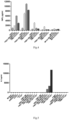
- the test results were analyzed by FCAP Array v3 software, and the results are shown in Figures 2 , 3 , 4, 5 , 6, and 7 .
- the antigen expression-positive cells LoVo, KATO3 and CRYPT, as well as SW620-CEA with low level of antigen expression were used in this study.
- the used kits were Cytotox96 non-radioactive cytotoxicity assay kit (Promega).
- the mechanism of this method was that the traditional radioactive elements were replaced with lactate dehydrogenase (LDH) which was stably expressed in the cells and was not secreted; when apoptosis of the cells occurred, LDH was released extracellularly, the content of formazan oxidized by LDH was detected to determine the level of enzyme in the supernatant, thereby determining the level of apoptosis.
- the effector cells were FM4( VL ⁇ VH )-28z, and the ratio of effector cells to target cells was 1:2, 1:5, 1:10, 1:20, 1:30, respectively.
- the number of target cells was 10,000 cells/well, two accessory wells were set in each group, and the detection time was 4 hours after the interaction.
- each of the experimental groups and the control groups were set as follows:
- the specific experimental methods referred to the kit instructions The specific experimental methods referred to the kit instructions.
- FM4( VL ⁇ VH )-28z specifically recognizes glycosylated CEA antigens and is less sensitive to CEA antigens with a lower degree of glycosylation.
- NCG mice purchased from the Institute of Model Biology of Nanjing University
- NCG mice purchased from the Institute of Model Biology of Nanjing University
- the connective tissue and blood vessels on the surface were removed; the tumor mass was cut along the midline, and the necrotic tissues, the large calcification points and the secretions were removed, and after careful cleaning, tumor tissues with good quality and toughness were transferred to fresh, ice-cold RPMI-1640 medium.
- the tumor tissues were cut into small tumor masses of about 3 ⁇ 3 mm 2 and transplanted unilaterally into the NSG mice. When the average tumor volume reached 160-180 mm 3 , the model animals were intratumorally injected with effector cells.
- the effector cells used in this study were FM4( VL ⁇ VH )-BBz, and the control group was T cells not transfected with CAR and an equal amount of PBS. Before the injection, the effector cells were washed twice with PBS, and resuspended in PBS to reach 3E7/ml and 1E8/ml, respectively, and recorded as low dose group and high dose group, respectively. Each mouse was intratumorally injected with 30 ⁇ l of effector cells/PBS.
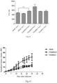
- the tumor volume results are shown in Figure 10 . From the fourth day after injection of CAR-T, the tumor volume of the FM4 (VL ⁇ VH) -BBz high-dose group began to decline, while the empty T-cell group and the PBS group showed no downward trend. This indicates that CAR-T has a significant inhibitory effect on the glycosylated CEA-positive tumors. After injection of CAR-T, there was no significant difference in body weight of the mice injected with CAR-T in comparison with the empty T cell group and the PBS control group.
- the cytokine secretion trend in peripheral blood is shown in Figure 11 .
- the secretion of a variety of human cytokines IL-2, TNF- ⁇ , IFN- ⁇
- IL-2, TNF- ⁇ , IFN- ⁇ human cytokines
- NSG mice (NOD scid IL2R ⁇ null) were inoculated subcutaneously with 1E7/mouse of Lovo cells (CCL229, ATCC) to form tumors with volume of 200mm 3 -300mm 3 .
- the tumors were injected with PBMC as control and FM4( VL ⁇ VH )-BBz, and the specific groups are as follows: Group (5NSG/groups) Treatment PBMC 3E6 PBMC/100 ⁇ l PBS, intratumoral injection FM4BBz high-dose 3E6 FM4BBz/100 ⁇ l PBS, intratumoral injection FM4BBz low-dose 1E6 PM4BBz/100 ⁇ l PBS, intratumoral injection
- NSG mice that were transfused with PBMC or FM4BBz T cells were bled 40 ⁇ l (day 3 and day 7) through tail vein, the samples were resuspended in equal volume of PBS and centrifuged, the supernatant was used for cytokine release assay (BD Cytometric Bead Array). The results are shown in Figure 14 .
- Example 7 Infection of NK92 cells with FM4 (VL ⁇ VH) -BBz lentivirus
- NK92 cells were cultured in 1640 medium containing 20% FBS, 150 IU/ml hIL-2. According to the method of Example 3, the NK92 cell line was infected with FM4 (VL ⁇ VH) -BBz CAR virus, and cultured continuously for 10 days. FM4 (VL ⁇ VH) -BBz -NK92 cells were used for killing experiments and cytokine release assays, and the results are shown in Figures 12 and 13 .
- the antibodies FM2, FM4, FM5, and FM6 were diluted with PBS buffer containing 1% BSA, and incubated with SW620-CEA or CRYPT cells (final concentrations: 50 ⁇ g/ml, 5 ⁇ g/ml, 0.5 ⁇ g/ml, 0.05 ⁇ g/ml, 0.005 ⁇ g/ml, 0.0005 ⁇ g/ml) for 1 hour at room temperature; the cells were washed twice with PBS buffer containing 1% BSA, and then incubated with Goat Anti Mouse IgG-APC for 1 hour; the cells were washed twice with PBS buffer containing 1% BSA and resuspended in PBS buffer; the APC fluorescence values of the cells were read using BD Accuri C6 FACS.
- SW620-CEA is CEA overexpressed in the SW620 cell line (CEACAM5), and its surface CEA is in a non-glycosylated state;
- CRYPT is a tumor cell line expressing glycosylated CEA.
- the antibody FM4 specifically binds to the glycosylated CEA and has high specificity (selective > 100-fold).
Landscapes
- Health & Medical Sciences (AREA)
- Life Sciences & Earth Sciences (AREA)
- Chemical & Material Sciences (AREA)
- Organic Chemistry (AREA)
- Immunology (AREA)
- General Health & Medical Sciences (AREA)
- Genetics & Genomics (AREA)
- Engineering & Computer Science (AREA)
- Biochemistry (AREA)
- Molecular Biology (AREA)
- Zoology (AREA)
- Medicinal Chemistry (AREA)
- Biomedical Technology (AREA)
- Cell Biology (AREA)
- Biophysics (AREA)
- Proteomics, Peptides & Aminoacids (AREA)
- Biotechnology (AREA)
- Bioinformatics & Cheminformatics (AREA)
- Wood Science & Technology (AREA)
- Public Health (AREA)
- Veterinary Medicine (AREA)
- Animal Behavior & Ethology (AREA)
- Gastroenterology & Hepatology (AREA)
- Toxicology (AREA)
- Epidemiology (AREA)
- Microbiology (AREA)
- General Engineering & Computer Science (AREA)
- Hematology (AREA)
- Urology & Nephrology (AREA)
- Physics & Mathematics (AREA)
- Pharmacology & Pharmacy (AREA)
- Nuclear Medicine, Radiotherapy & Molecular Imaging (AREA)
- General Chemical & Material Sciences (AREA)
- Chemical Kinetics & Catalysis (AREA)
- General Physics & Mathematics (AREA)
- Food Science & Technology (AREA)
- Analytical Chemistry (AREA)
- Pathology (AREA)
- Oncology (AREA)
- Plant Pathology (AREA)
Description
- The present invention relates to the field of tumor immunotherapy, and more particularly, the present invention relates to a glycosylated CEA-specific single-chain antibody.
- The incidence of gastric cancer ranks first in the digestive tract malignant tumors in China, and second in the world. The annual deaths account for about 24% of the malignant tumor deaths, and the five-year survival rate is only about 27%. The diagnosis of gastric cancer patients is often late, so the prognosis is extremely poor. There is no effective target therapy for gastric cancer. CEA (carcinoembryonic antigen) is a membrane-bound protein, and generally expressed in fetal liver, intestine and pancreas. Normally, it is secreted into the intestine, and its serum level is low. When the cell is cancerous, the serum level is elevated, which has auxiliary significance for the early diagnosis of pancreatic cancer and colon cancer, and has certain reference value for tumor spread, curative effect, recurrence and prognosis. Professor Fan Daiming from the Fourth Military Medical University found that the glycosylated CEA is highly sensitive and specific to a variety of digestive tract cancers. Clinical studies have shown that the positive rate of glycosylated CEA in gastric cancer tissues is above 80%, the positive rate in colon cancer tissues is above 40%, the positive rate in gastric precancerous lesions is above 30%, and the positive rate in esophageal cancer tissues is 18% or more. At the same time, the results of clinical trials showed that the positive rate of serum glycosylated CEA in 28 patients with gastric cancer decreased significantly in comparison with that before surgery, suggesting that there is a close relationship between glycosylated CEA and gastric cancer (Gadler et al., Int J Cancer 25 (1): 91-4, 1980).
- At present, the emerging chimeric antigen receptor T cell (CAR-T) with targeted adoptive cell therapy technology plays an important role in the treatment of various malignant tumors. CAR-T is a fusion of a tumor-associated antigen (TAA)-specific recognition peptide, such as a single-chain antibody, a specific receptor, with a T cell activation signal such as CD3zeta, and the fusion protein is specifically expressed on T cell surface by a lentivirus or the like, allowing the modified T cell to specifically recognize and kill tumor cells, being independent of major histocompatibility antigen (MHC), and avoiding tumor immune escape due to MHC deletion.
- The chimeric antigen receptor includes an extracellular antigen targeting and recognizing region, a hinge region, a transmembrane region, and an intracellular costimulatory signaling region. The antigen recognizing region is mostly a single-chain antibody or a specific receptor, which ensures that the modified T cell can specifically recognize the target cell and be activated to specifically kill the target cell; the hinge region generally adopts CD8α, CD28ECD, IgG Fc fragment and the like, which ensures that the T cell contacts the target cell and affects the T cell action; and the intracellular signal region adopts an immunoreceptor tyrosine activation motif (ITAM), for example CD3zeta and costimulatory signal such as CD28, CD137, CD27, ICOS, OX40, DAP10, etc. (Sadelain et al., Cancer Discov 3(4): 388-98, 2013).
- Although CAR-T has shown good application value in the treatment of hematological malignancies, CAR-T often does not achieve the expected results in the treatment of solid tumors. Even in some clinical studies, CAR-T may exhibit toxicity due to off-target effects. The important reason is that there is often no effective and specific target in solid tumors. For many tumor-associated antigens, there is often a certain degree of expression in normal tissues. For example, HER2 is an important target for malignant tumors such as malignant glioma and breast cancer, but in the CAR-T clinical trial for HER2 target, the patient suffered pulmonary failure after CAR-T re-infusion, and "cytokine storm" was triggered and caused the death of patients in a short period of time. Therefore, finding a highly specific and sensitive target in solid tumors is a key point in the application of CAR-T treatments in solid tumors (Morgan et al., Mol Ther 18(4): 843-51, 2010).
- NK cells are lymphocytes that express CD16 and CD56 and are important components in innate immunity. NK cells can kill MHC-deficient target cells. Compared with T cells, NK cells do not express T-cell receptors and thus do not undergo graft versus host response (GVHD). NK cell activation is in the equilibrium state of signals for KAR (Killer Activation Receptor) and KIR (Killer Inhibition Receptor). The main KIR ligand in vivo is MHC. For mismatch or deletion of KIR ligand, it will cause the activation of NK cells. CAR-NK technology is an emerging technology that uses CAR structure expression on the surface of NK cells or NK cell lines (such as NK92) for the treatment of cancers, which advantage is that NK cells do not secrete IL6, NK cells have a very short circulating half-life in the periphery blood, NK cells have a high transfection efficiency (non-viral vector), and NK cell lines can be cultured on a large scale at GMP level. The CAR structure expression on the surface of NK cells or NK cell lines can endow NK cells with targeting ability. At the same time, in CAR-NK allograft, the mismatch of KIR receptor may enhance the activation of NK cells to achieve better tumor elimination. The circulation time of NK cell lines in vivo is very short, thus providing safety considerations (Han et al., Sci Rep 5: 11483, 2015).
-
WO 2007/146172 A2 relates to antibodies that specifically bind to an epitope on CD43 and CEA expressed on nonhematopoietic cancer cells, but do not specifically bind to a CD43 expressed by a leukocyte or by a Jurkat cell, and is capable of inducing apoptosis of the nonhematopoietic cancer cell after binding to the epitope on cell surface of the nonhematopoietic cancer cell in the absence of cytotoxin conjugation and immune effector function, wherein the epitope comprises a carbohydrate structure and the binding of the antibody to the epitope is inhibited by a carbohydrate. -
EP1505076A2 relates to specific antibody fragments for the human carcinoembryonic antigen (CEA) and refers to mono- and divalent (diabody) single chain Fv (scFv) antibody fragments, obtained by recombinant DNA techniques from the anti-carcinoembryonic antigen (CEA) monoclonal antibody (Mab) CB/ior-CEA.1. This antibody has high affinity for CEA and is employed in the diagnosis and follow-up of human colorectal tumors. - OSBOURN J K ET AL describe in the paper : "Isolation of a panel of human anti-CEA single chain Fv from a large phage display library" a phage display library of human single chain (scFv) which have been selected using immobillised human carcinoembryonic antigen (CEA).
-
US5872215A describes specific binding members for human carcinoembryonic antigen (CEA) comprising a human antibody antigen binding domain. Said binding members may have a dissociation constant less than 10×10-8M and may be substantially non-crossreactive with human liver and/or other normal tissues. These four prior art documents focus on antibodies or scFv which may bind to CEA. - DAN BLAT ET AL: "Suppression of Murine Colitis and its Associated Cancer by Carcinoembryonic Antigen-Specific Regulatory T Cells", describes that adoptive transfer of regulatory T cells (Tregs) offers a promising strategy to combat pathologies that are characterized by aberrant immune activation, includ- ing graft rejection and autoinflammatory diseases. Expression of a chimeric antigen receptor (CAR) gene in Tregs redirects them to the site of autoimmune activity, thereby increasing their suppressive efficiency while avoiding systemic immunosuppression. CEA-transgenic mice that were induced to develop colitis and treated with CEA-specific CAR Tregs. Two disease models were employed. The study provides a proof of concept for the therapeutic potential of CAR Tregs in colitis patients as well as in other autoimmune inflammatory disorders.
-
CN104910279A describes a chimeric antigen receptor of targeted carcino-embryonic antigens, a lentiviral expression vector as well as preparation methods and applications thereof. The chimeric antigen receptor of targeted carcino-embryonic antigens is composed of a leading peptide, a single-chain antibody of targeted carcino-embryonic antigens, a hinge region for connecting ScFv, a membrane spanning structural domain and an immunoreceptor tyrosine-based activation motif. The chimeric antigen receptor is built into the lentiviral expression vector, and then infects immune cells, so that the chimeric antigen receptor of targeted carcino-embryonic antigens can be expressed in the immune cells; and by identifying carcino-embryonic antigens on the surfaces of tumor cells, the tumor expressing the carcino-embryonic antigens is killed, so that the purpose of tumor treatment is achieved, thereby providing a new tool for the targeting therapy of tumor. - M. Chmielewski ET AL describe in "IL-12 Release by Engineered T Cells Expressing Chimeric Antigen Receptors Can Effectively Muster an Antigen-Independent Macrophage Response on Tumor Cells That Have Shut Down Tumor Antigen Expression", Cancer research, vol. 71, no. 17, 8 July 2011 (2011 -07-08), pages 5697-5706, XP055185302 that Cytotoxic T cells were engineered to release inducible IL-12 upon CAR engagement in the tumor lesion, resulting in destruction of antigen-loss cancer cells that would normally escape.
- Gianpietro Dotti ET AL reviewed in "Design and development of therapies using chimeric antigen receptor-expressing T cells", Immunological Reviews, vol. 257, no. 1, 13 December 2013 (2013-12-13), p 107-126, XP055177338 the design and development of therapies using chimeric antigen receptor-expressing T cells. The article describes basic design of CARs and describes how antigenic targets are selected, and describe the initial clinical experience with CAR-T cells.
-
WO 2011/163401 A2 relates to NPC-1 antigen on the MUC5AC protein and 16C3 antigen on CEACAM5 and CEACAM6 proteins, and 31.1 epitope on the A33 protein are differentially expressed in cancers including, lung cancer, ovarian cancer, pancreas cancer, breast cancer, and colon cancer, and diagnostic and therapeutic usages. -
WO 2007/071426 A1 relates to pharmaceutical compositions for the treatment of an epithelial tumor in a human, said pharmaceutical composition comprising a bispecific single chain antibody which has a first binding domain specifically binding to human CD3, and a second binding domain specifically binding to human CEA, wherein said second binding domain comprises at least a part of the CDR-H3 or the complete CDR-H3 of murine monoclonal antibody A5B7. The processes for the production of said pharmaceutical compositions as well as medical/pharmaceutical uses are described for the specific bispecific single chain antibody molecules bearing specificities for the human CD3 antigen and the human CEA antigen. -
EP3034514A1 describes an antibody binding to GLP-1R and a fusion protein thereof with GLP-1. The fusion proteins can effectively bind to a human GLP-1R receptor and activate a receptor signaling pathway, which is useful for treating diabetes, excessive weight, obesity and related disorders thereof. -
WO2015/153685A1 relates to humanized antibodies which bind the pertussis toxin protein and their use as therapeutic agents. In particular, the present invention is directed to improved humanized 1B7 and 11E6 antibodies which bind the pertussis toxin protein. -
WO 2007/071422 A2 relates to pharmaceutical compositions for the treatment of an epithelial tumor in a human, said pharmaceutical composition comprising an IgG1 antibody specifically binding to human CEA, wherein the variable region of said IgG1 antibody comprises at least (i) a CDR-H1 having the amino acid sequence "SYWMH" and a CDR-H2 having the amino acid sequence "FIRNKANGGTTEYAASVKG" and a CDR-H3 having the amino acid sequence "DRGLRFYFDY" or (ii) a CDR-H 1 having the amino acid sequence "TYAMH" and a CDR-H2 having the amino acid sequence "LISNDGSNKYYADSVKG" and a CDR-H3 having the amino acid sequence "DRGLRFYFDY". - In a first aspect, the invention provides a chimeric antigen receptor (CAR), having the features of
claim 1. - In a specific embodiment, the heavy chain variable region and the light chain variable region are selected from any one of the following combinations: the heavy chain variable region comprises a polypeptide fragment set forth in SEQ ID NO: 35, and the light chain variable region comprises a polypeptide fragment set forth in SEQ ID NO: 36;
- In another embodiment, the antigen-binding domain of the invention is a single-chain antibody that specifically recognizes a glycosylated CEA, and the amino acid sequence of the single-chain antibody is SEQ ID NO: 173 or SEQ ID NO: 178.
- Preferably, the chimeric antigen receptor of the present invention further comprises a hinge region, wherein the transmembrane domain is or comprises the transmembrane domain from CD8α and/or CD28, and the intracellular signaling domain comprises the intracellular signaling domain from one or more of CD28, CD137, and CD3zeta. Preferably the hinge region is encoded by the sequence set forth in SEQ ID NO: 52, the CD8α transmembrane region is encoded by the sequence set forth in SEQ ID NO:54, the CD28 hinge region is encoded by the sequence set forth in SEQ ID NO:53, the CD28 transmembrane region is encoded by the sequence set forth in SEQ ID NO: 55, the CD28 costimulatory domain is encoded by the sequence set forth in SEQ ID NO:56, the CD137 costimulatory domain is encoded by the sequence set forth in SEQ ID NO:57, and CD3zeta is encoded by the sequence set forth in SEQ ID NO:58.
- In another aspect, the invention provides a nucleic acid molecule encoding a chimeric antigen receptor of the invention.
- In another aspect, the invention provides a cell expressing a chimeric antigen receptor of the invention, the cell is selected from the group consisting of a T cell, an NK cell and a B cell.
- In another aspect, the description describes a lymphocyte expressing a chimeric antigen receptor, the chimeric antigen receptor comprises an extracellular targeted recognition antigen sequence, a hinge region sequence, a transmembrane region sequence, and an intracellular signal sequence, where are connected in order. Wherein the extracellular recognition antigen sequence is a single-chain antibody that specifically recognizes the glycosylated CEA as described in the present invention, which is defined as above. The hinge region is selected from the group consisting of CD8α, CD28ECD, and IgG Fc fragments. The intracellular signal region may employ an immunoreceptor tyrosine activation motif (ITAM) such as CD3zeta and costimulatory signals such as CD28, CD137, CD27, ICOS, OX40, DAP10. In one embodiment, the hinge region sequence in the chimeric antigen receptor comprises CD8α or CD28, encoded by the sequence set forth in SEQ ID NO: 52 or SEQ ID NO: 53, respectively; the transmembrane region sequence in the chimeric antigen receptor comprises CD8α or CD28, encoded by the sequence set forth in SEQ ID NO: 54 or SEQ ID NO: 55, respectively; the intracellular signal sequence in the chimeric antigen receptor comprises CD28, CD137, CD3zeta and combination thereof, CD28 is encoded by the sequence set forth in SEQ ID NO: 56, CD137 is encoded by the sequence set forth in SEQ ID NO: 57, and CD3zeta is encoded by the sequence set forth in SEQ ID NO:58. In one embodiment, the lymphocyte may be a T cell, a B cell or a NK cell, and the like. In a specific embodiment not encompassed by the claims, the lymphocyte is a T cell and the chimeric antigen receptor is expressed as follows:
- scFv-CD8α-CD137-CD3zeta,
- scFv-CD28-CD28-CD 137-CD3zeta,
- scFv-CD28-CD28-CD3zeta.
- In a preferred embodiment not encompassed by the claims, the chimeric antigen receptor as constructed in the invention has a sequence set forth in SEQ ID NOs: 69-128.
- When the antigen-binding domain of the chimeric antigen receptor binds its corresponding antigen, the cell comprising the chimeric antigen receptor exhibits anti-tumor immunity.
- The present invention constructs a series of single-chain antibodies against glycosylated CEA. The single-chain antibodies of the present invention can be used for detection and treatment of tumors such as gastric cancer, colorectal cancer, and esophageal cancer. These single-chain antibodies can be expressed on the surface of lymphocytes such as T cells and NK cells to construct and form chimeric antigen receptor T cells or chimeric antigen receptor NK cells against glycosylated CEA, for specific killing glycosylated CEA expression-positive cells and tissues.
- In a second aspect, the present invention provides single-chain antibodies against glycosylated CEA, according to
claim - In a passage , the description describes a single-chain antibody against glycosylated CEA, which is arranged in the form of VH-Linker-VL or VL-Linker-VH, wherein VH is selected from the heavy chain variable region sequences described above, VL is selected from the light chain variable region sequences described above. In a preferred embodiment, the Linker, i.e., linker peptide, is the sequence set forth in SEQ ID NO:41.
- The description provides an isolated nucleic acid molecule encoding the single-chain antibody of the invention. In one embodiment, the nucleotide sequence encoding the heavy chain variable region of the single-chain antibody is SEQ ID NO: 42, SEQ ID NO: 44, SEQ ID NO: 46, SEQ ID NO: 48 or SEQ ID NO: 50. In one embodiment, the nucleotide sequence encoding the light chain variable region of the single-chain antibody is SEQ ID NO: 43, SEQ ID NO: 45, SEQ ID NO: 47, SEQ ID NO: 49 or SEQ ID NO: 51.
- The description also provides a vector comprising a nucleic acid sequence encoding a chimeric antigen receptor. In a specific embodiment, the vector is pGC-EIF1α-MCS (CV185, Genechem) and pGC-EIF1α-MCS-2A-EGFP (CV178, Genechem).
- The chimeric antigen receptor, or the cell, or the single-chain antibody according to the present invention for use in treating a tumor, wherein the tumor is a digestive tract tumor. The chimeric antigen receptor, or the cell, or the single-chain antibody according to the present invention for use in treating a tumor wherein the tumor is selected from the group consisting of gastric cancer, colorectal cancer, esophageal cancer.
- The description also describes a method of providing anti-tumor immunity in a mammal. In one embodiment, the method comprises administering to the mammal an effective amount of the chimeric antigen receptor T cell of the invention, thereby providing anti-tumor immunity in the mammal.
- The description also describes a method of treating a mammal having a disease, disorder or condition associated with an elevated tumor antigen expression, for example treatment of a gastrointestinal tumor with high expression of CEA. In a passage, the description describes the administering to the mammal an effective amount of the chimeric antigen receptor T cell of the invention, thereby suitable for use in treating a tumor in the mammal.
- The description also describes a method of diagnosing a disease, disorder, or condition associated with an elevated tumor antigen expression, for example diagnosing a digestive tract tumor with high CEA expression. In a passage the description describes the expression of an amount of glycosylated CEA for detecting a tumor and a surrounding tissue of the tumor.
-
-
Figure 1 . Detection of CEA expression in SW620-CEA by flow cytometry. -
Figure 2 . FM2(VL→VH), FM4(VL→VH), FM5(VL→VH), FM6(VL→VH) were designed according to the structure scFv-CD8α-CD137-CD3zeta and infected human T cells as lentivirus, cultured untilday 10, incubated with the target cells (SW620-CEA, LOVO, KATO3) in the figure for 16 hours and IL2 release was measured. -
Figure 3 . FM2(VL→VH), FM4(VL→VH), FM5(VL→VH), FM6(VL→VH) were designed according to the structure scFv-CD8α-CD137-CD3zeta and infected human T cells as lentivirus, cultured untilday 10, incubated with the target cells (SW620-CEA, LOVO, KATO3) in the figure for 16 hours and TNFα release was measured. -
Figure 4 . FM2(VL→VH), FM4(VL→VH), FM5(VL→VH), FM6(VL→VH) were designed according to the structure scFv-CD8α-CD137-CD3zeta and infected human T cells as lentivirus, cultured untilday 10, incubated with the target cells (SW620-CEA, LOVO, KATO3) in the figure for 16 hours and INPγ release was measured. -
Figure 5 . FM2(VL→VH), FM4(VL→VH) were designed according to the structure scFv-CD28-CD28-CD3zeta and infected human T cells as lentivirus , cultured untilday 10, incubated with the target cells (SW620, SW620-CEA, LOVO, KATO3, CRYPT) in the figure for 16 hours and IL2 release was measured. -
Figure 6 . FM2(VL→VH), FM4(VL→VH) were designed according to the structure scFv-CD28-CD28-CD3zeta and infected human T cells as lentivirus , cultured untilday 10, incubated with the target cells (SW620, SW620-CEA, LOVO, KATO3, CRYPT) in the figure for 16 hours and TNFα release was measured. -
Figure 7 . FM2(VL→VH), FM4(VL→VH) were designed according to the structure scFv-CD28-CD28-CD3zeta and infected human T cells as lentivirus , cultured untilday 10, incubated with the target cells (SW620, SW620-CEA, LOVO, KATO3, CRYPT) in the figure for 16 hours and INPγ release was measured. -
Figure 8 . FM2(VL→VH)-BBz was mixed with target cells for 4 hours in an E:T ratio, and LDH release in the supernatant was measured to determine the killing of target cells by FM2(VL→VH)-BBz. -
Figure 9 . FM4(VL→VH)-BBz was mixed with target cells for 4 hours in an E:T ratio, and LDH release in the supernatant was measured to determine the killing of target cells by FM4(VL→VH)-BBz. -
Figure 10 . Detection of IFNγ release in peripheral blood of PDX model mice -
Figure 11 . Tumor growth curve of the PDX model. -
Figure 12 . FM4(VL→VH)-BBz-NK92 cytokine secretion assay. -
Figure 13 . FM4(VL→VH)-BBz-NK92 cell killing assay. -
Figure 14 . Detection of INPγ release in mice of tumor model. -
Figure 15 . Tumor killing effect in mice of tumor model. - Firstly, certain terms are defined so that the invention can be more readily understood. All technical and scientific terms used herein have the same meaning as commonly understood by one of ordinary skill in the art to which the invention belongs, unless otherwise indicated. Although methods and materials similar or equivalent to those described herein can be used in the practice or testing of the present invention, suitable methods and materials are described below. All publications, patent applications, patents, and other references cited herein are merely for a better understanding of the invention. In case of conflict, the present specification (including definitions) shall prevail. In addition, the materials, methods, and examples are illustrative only and not limiting.
- As used herein, the "single-chain antibody (single-chain Fv, scFv)" is a recombinant protein formed by linking a heavy chain variable region (VH) and a light chain variable region (VL) of an immunoglobulin through a ligation peptide, which is the smallest antibody fragment with a complete antigen binding site. Human immunoglobulin (Ig) comprises five subtypes of IgA, IgD, IgM, IgE and IgG, of which IgG accounts for 75% of human immunoglobulin; IgG is a "Y" type antibody structure formed by two heavy chain IgH and two light chain IgL via interchain disulfide bonds; wherein IgH comprises a heavy chain variable region (VH) and a constant region (Constant Region); IgL comprises a light chain variable region (VL) and a constant region (Constant Region). The diversity of VH and VL is the basis for the binding of immunoglobulins to antigens. VH and VL are composed of a frame region (FR) and a complementary determining region (CDR), the CDR regions are highly variable and determine the specific binding of antigens and antibodies; wherein VH comprises three CDR regions, designated CDRH1, CDRH2, CDRH3; VL comprises three CDR regions, designated CDRL1, CDRL2, CDRL3.
- The invention also comprises the variants, derivatives and analogs of the single-chain antibodies. As used in the text, the terms "variant", "derivative" and "analog" refer to a polypeptide that substantially retains the same biological function or activity of the single-chain antibody of the invention. The polypeptide variant, derivative or analog of the invention can be (i) a polypeptide in which one or more conservative or non-conservative amino acid residues (preferably conservative amino acid residues) are substituted, and such substituted amino acid residues may or may not be encoded by genetic codes, or (ii) a polypeptide in which one or more amino acid residues have substituent groups, or (iii) a polypeptide which is formed by fusing an additional amino acid sequence into the polypeptide sequence (such as a leader sequence or a secretory sequence or a sequence or polypeptide sequence used to purify the polypeptide, or a fusion polypeptide). These variants, derivatives and analogs are within the purview of those skilled in the art in accordance with the definitions herein.
- A "single-chain antibody" of the invention refers to a polypeptide that specifically binds to a glycosylated CEA. The term also comprises variant forms having a polypeptide sequence capable of specifically binding a glycosylated CEA. These variants comprises (but are not limited to): deletion, insertion and/or substitution of several (usually 1-50, preferably 1-30, more preferably 1-20, most preferably 1-10, further more preferably 1-8 or 1-5) amino acids, and addition or deletion of one or more (usually within 20, preferably within 10, more preferably 5 or less) amino acids at C-terminal and/or N-terminal. For example, in the art, the substitution with an amino acid of close or similar property usually does not change the function of protein. As another example, the addition or deletion of one or more amino acids at the C-terminal and/or N-terminal generally does not change the function of protein as well.
- The invention also provides analogs of the single-chain antibody. The difference between these analogs and the natural single-chain antibody may be a difference in amino acid sequence, a difference in modification form which does not affect the sequence, or a combination thereof. These polypeptides comprises natural or induced genetic variants. Induced variants can be obtained by a variety of techniques, such as random mutagenesis by irradiation or exposure to a mutagen, or site-directed mutagenesis or other known techniques of molecular biology. The analogs also comprise analogs having residues other than the native L-amino acids (e.g., D-amino acids), as well as analogs having non-naturally occurring or synthetic amino acids (e.g., β-, γ-amino acids). It should be understood that the polypeptides of the present invention are not limited to the representative polypeptides exemplified above.
- Furthermore, other amino acid sequences which do not substantially affect the activity, expression amount and stability of the single-chain antibody of the present invention may be added to the amino terminus or the carboxy terminus of the single-chain antibody. Preferably, these added amino acid sequences facilitate expression (e.g., signal peptides), facilitate purification (e.g., 6 × His sequences), or other sequences that promote the activity, expression, or stability of the single-chain antibody.
- The present invention also comprises DNA molecules encoding the single-chain antibodies of the present invention or variants, derivatives thereof. The DNA molecules can be all artificially synthesized or obtained by PCR amplification.
- In order to further increase the expression level of the host cell, the coding sequence of the single-chain antibody of the present invention can be engineered, for example, using a host cell-preferred codon to eliminate sequences which are not conducive to gene transcription and translation.
- After obtaining the DNA sequence encoding the novel single-chain antibody of the present invention or a variant or derivative thereof, it is cloned into a suitable expression vector and transferred into a suitable host cell. Finally, the transformed host cell is cultured, and the novel single-chain antibody of the present invention is obtained by isolation and purification.
- The term "vector" as used herein comprises plasmids, cosmids, expression vectors, cloning vectors, viral vectors, and the like.
- In the present invention, various vectors known in the art can be used. For example, a commercially available vector is selected, and then a nucleotide sequence encoding a novel single-chain antibody of the present invention is operably linked to an expression regulatory sequence to form an expression vector.
- In the present invention, the term "host cell" comprises prokaryotic cells and eukaryotic cells. Examples of commonly used prokaryotic host cells include Escherichia coli, Bacillus subtilis and the like. Host cells for expressing single-chain antibodies include Escherichia coli, yeast cells, insect cells, COS cells, CHO cells, and the like. Preferably, the host cell is a prokaryotic cell, more preferably an E. coli cell.
- After obtaining the transformed host cell, the cell can be cultured under conditions suitable for expression of the single-chain antibody of the present invention to express a single-chain antibody; and then the expressed single-chain antibody can be isolated.
- As used herein, the term "linker peptide" (linker) refers to a short peptide between a heavy chain variable region of antibody (or a variant thereof) and a light chain variable region of antibody (or a variant thereof), which acts as a linker and allows the heavy and light chain variable regions to fold freely, leaving the antigen binding site in an appropriate configuration without causing a change in molecular dynamics. In general, the linker peptide does not affect or not significantly affect the formation of correct folding and spatial conformation of the amino acid sequences of the heavy chain variable region (VH) and the light chain variable region (VL); alternatively, the linker peptide constitutes a flexible connection between the heavy chain variable region (VH) and the light chain variable region (VL), which facilitates their normal folding. The length of the linker peptide is not particularly limited as long as it allows the heavy and light chain variable regions to be freely folded, and the antigen binding site to be in an appropriate configuration without causing a change in molecular dynamics. For example, the length of the linker peptide can also be 4-30 aa. Preferably, the sequence of the linker peptide of the present invention is set forth in SEQ ID NO: 1.
- The single-chain antibody of the present invention can be used for detection and treatment of a tumor such as gastric cancer, colorectal cancer, and esophageal cancer. The single-chain antibody of the present invention can be specifically expressed on the surface of T cells to construct a chimeric antigen receptor T cell against glycosylated CEA, and specifically kill glycosylated CEA expression-positive cells and tissues. For each of the single-chain antibodies of the present invention, CAR-Ts of three structures are constructed separately and the functional differences of the three structures under each antibody are compared, and a suitable CAR-T structure is adopted for an appropriate target cell. The CAR-Ts of three structures separately are:
- scFv-CD8α-CD137-CD3zeta,
- scFv-CD28-CD28-CD137-CD3zeta,
- scFv-CD28-CD28-CD3zeta.
- In the chimeric antigen receptor structure of the invention:
The hinge region can be an extracellular region selected from the group consisting of CD8α, CD28, human IgG1 Fc, human IgG4 Fc, DAP10 and the like. - The transmembrane regions can be an transmembrane region selected from the group consisting of CD8α, CD28, DAP10 and the like.
- The costimulatory domain can be an intracellular region selected from the group consisting of CD28, CD134 (OX40), CD137 (4-1BB), ICOS, DAP10 and the like.
- The essential signal domain can be selected from: CD3zeta.
- The invention is further described below in conjunction with the specific examples and the accompanying drawings, which are not intended to limit the invention. The experimental methods in the following examples which conditions are not specifically described are performed according to the conventional conditions such as those described by J. Sambrook et al., Molecular Cloning Experiment Guide, Science Press, 2002, or according to the manufacturer's recommended conditions. Percentages and parts are by weight unless otherwise stated.
-
Table 1 Single-chain antibody Seq ID VH Seq ID VL Seq ID FM2(VH→VL) SEQ ID NO: 59 SEQ ID NO: 31 SEQ ID NO: 32 FM2(VL→VH) SEQ ID NO: 64 SEQ ID NO: 31 SEQ ID NO: 32 FM3(VH→VL) SEQ ID NO: 60 SEQ ID NO: 33 SEQ ID NO: 34 FM3(VL→VH) SEQ ID NO: 65 SEQ ID NO: 33 SEQ ID NO: 34 FM4(VH→VL) SEQ ID NO: 61 SEQ ID NO: 35 SEQ ID NO: 36 FM4(VL→VH) SEQ ID NO: 66 SEQ ID NO: 35 SEQ ID NO: 36 FM5(VH→VL) SEQ ID NO: 62 SEQ ID NO: 37 SEQ ID NO: 38 FM5(VL→VH) SEQ ID NO: 67 SEQ ID NO: 37 SEQ ID NO: 38 FM6(VH→VL) SEQ ID NO: 63 SEQ ID NO: 39 SEQ ID NO: 40 FM6(VL→VH) SEQ ID NO: 68 SEQ ID NO: 39 SEQ ID NO: 40 - Each of the synthesized nucleotide sequences FM2, 3, 4, 5, and 6 was added with 8His (CACCATCACCATCACCATCACCAT) at 3'-terminal, the amplified fragment was added with sfiI and NotI restriction sites at both ends thereof, and then inserted into a pCANTAB5E vector (GE HealthCare). The constructed pCANTAB5E (GE) vector was transformed into E. coli-HB2151 strain and induced by IPTG to express overnight. The cells were resuspended in PBS buffer, sonicated on ice for 10 min. After centrifugation, the supernatant was purified by His Trap Column (GE), washed with PBS buffer containing 20 mM imidazole for 5-10 column volumes, and then eluted with PBS containing 500 mM imidazole; the subsequent imidazole was desalted with Desalting G25 column (GE) to obtain a single-chain antibody dissolved in PBS.
- The virus constructed in the present invention is shown in Table 2:
Table 2: Virus Number Name Structure Seq ID 1 FM2(VH→VL)-BBz-EG FP FM2(VH→VL)-CD8α-CD137-CD3zeta-2A-EGFP SEQ ID NO: 69 2 FM2(VL→VH)-BBz-EG FP FM2(VL→VH)-CD8α-CD137-CD3zeta-2A-EGFP SEQ ID NO: 70 3 FM3(VH→VL)-BBz-EG FP FM3(VH→VL)-CD8α-CD137-CD3zeta-2A-EGFP SEQ ID NO: 71 4 FM3(VL→VH)-BBz-EG FP FM3(VL→VH)-CD8α-CD137-CD3zeta-2A-EGFP SEQ ID NO: 72 5 FM4(VH→VL)-BBz-EG FP FM4(VH→VL)-CD8α-CD137-CD3zeta-2A-EGFP SEQ ID NO: 73 6 FM4(VL→VH)-BBz-EG FP FM4(VL→VH)-CD8α-CD137-CD3zeta-2A-EGFP SEQ ID NO: 74 7 FM5(VH→VL)-BBz-EG FP FM5(VH→VL)-CD8α-CD137-CD3zeta-2A-EGFP SEQ ID NO: 75 8 FM5(VL→VH)-BBz-EG FP FM5(VL→VH)-CD8α-CD137-CD3zeta-2A-EGFP SEQ ID NO: 76 9 FM6(VH→VL)-BBz-EG FP FM6(VH→VL)-CD8α-CD137-CD3zeta-2A-EGFP SEQ ID NO: 77 10 FM6(VL→VH)-BBz-EG FP FM6(VL→VH)-CD8α-CD137-CD3zeta-2A-EGFP SEQ ID NO: 78 11 FM2(VH→VL)-BBz FM2(VH→VL)-CD8α-CD137-CD3zeta SEQ ID NO: 79 12 FM2(VL→VH)-BBz FM2(VL→VH)-CD8α-CD137-CD3zeta SEQ ID NO: 80 13 FM3(VH→VL)-BBz FM3(VH→VL)-CD8α-CD137-CD3zeta SEQ ID NO: 81 14 FM3(VL→VH)-BBz FM3(VL→VH)-CD8α-CD137-CD3zeta SEQ ID NO: 82 15 FM4(VH→VL)-BBz FM4(VH→VL)-CD8α-CD137-CD3zeta SEQ ID NO: 83 16 FM4(VL→VH)-BBz FM4(VL→VH)-CD8α-CD137-CD3zeta SEQ ID NO: 84 17 FM5(VH→VL)-BBz FM5(VH→VL)-CD8α-CD137-CD3zeta SEQ ID NO: 85 18 FM5(VL→VH)-BBz FM5(VL→VH)-CD8α-CD137-CD3zeta SEQ ID NO: 86 19 FM6(VH→VL)-BBz FM6(VH→VL)-CD8α-CD137-CD3zeta SEQ ID NO: 87 20 FM6(VL→VH)-BBz FM6(VL→VH)-CD8α-CD137-CD3zeta SEQ ID NO: 88 21 FM2(VH→VL)--28BBz-EGFP FM2(VH→VL)-CD28-CD28-CD137-CD3zeta-2A-EGFP SEQ ID NO: 89 22 FM2(VL→VH)--28BBz-EGFP FM2(VL→VH)-CD28-CD28-CD137-CD3zeta-2A-EGFP SEQ ID NO: 90 23 FM3(VH→VL)--28BBz-EGFP FM3(VH→VL)-CD28-CD28-CD137-CD3zeta-2A-EGFP SEQ ID NO: 91 24 FM3(VL→VH)--28BBz-EGFP FM3(VL→VH)-CD28-CD28-CD137-CD3zeta-2A-EGFP SEQ ID NO: 92 25 FM4(VH→VL)--28BBz-EGFP FM4(VH→VL)-CD28-CD28-CD137-CD3zeta-2A-EGFP SEQ ID NO: 93 26 FM4(VL→VH)--28BBz-EGFP FM4(VL→VH)-CD28-CD28-CD137-CD3zeta-2A-EGFP SEQ ID NO: 94 27 FM5(VH→VL)--28BBz-EGFP FM5(VH→VL)-CD28-CD28-CD137-CD3zeta-2A-EGFP SEQ ID NO: 95 28 FM5(VL→VH)--28BBz-EGFP FM5(VL→VH)-CD28-CD28-CD137-CD3zeta-2A-EGFP SEQ ID NO: 96 29 FM6(VH→VL)--28BBz-EGFP FM6(VH→VL)-CD28-CD28-CD137-CD3zeta-2A-EGFP SEQ ID NO: 97 30 FM6(vL→VH)--28BBz-EGFP FM6(VL→VH)-CD28-CD28-CD 1 37-CD3zeta-2A-EGFP SEQ ID NO: 98 31 FM2(VH→VL)--28BBz FM2(VH→VL)-CD28-CD28-CD137-CD3zeta SEQ ID NO: 99 32 FM2(VL→VH)--28BBz FM2(VL→VH)-CD28-CD28-CD137-CD3zeta SEQ ID NO: 100 33 FM3(VH→VL)--28BBz FM3(VH→VL)-CD28-CD28-CD137-CD3zeta SEQ ID NO: 101 34 FM3(VL→VH)--28BBz FM3(VL→VH)-CD28-CD28-CD137-CD3zeta SEQ ID NO: 102 35 FM4(VH→VL)--28BBz FM4(VH→VL)-CD28-CD28-CD137-CD3zeta SEQ ID NO: 103 36 FM 4(VL→VH)--28BBz FM4(VL→VH)-CD28-CD28-CD137-CD3zeta SEQ ID NO: 104 37 FM5(VH→VL)--28BBz FM5(VH→VL)-CD28-CD28-CD137-CD3zeta SEQ ID NO: 105 38 FM5(VL→VH)--28BBz FM5(VL→VH)-CD28-CD28-CD137-CD3zeta SEQ ID NO: 106 39 FM6(VH→VL)--28BBz FM6(VH→VL)-CD28-CD28-CD137-CD3zeta SEQ ID NO: 107 40 FM6(VL→VH)--28BBz FM6(VL→VH)-CD28-CD28-CD137-CD3zeta SEQ ID NO: 108 41 FM2(VH→VL)-28z-EG FP FM2(VH→VL)-CD28-CD28-CD3zeta-2A-EGFP SEQ ID NO: 109 42 FM2(VL→VH)-28z-EG FP FM2(VL→VH)-CD28-CD28-CD3zeta-2A-EGFP SEQ ID NO: 110 43 FM3(VH→VL)-28z-EG FP FM3(VH→VL)-CD28-CD28-CD3zeta-2A-EGFP SEQ ID NO: 111 44 FM3(VL→VH)-28z-EG FP FM3(VL→VH)-CD28-CD28-CD3zeta-2A-EGFP SEQ ID NO: 112 45 FM4(VH→VL)-28z-EG FP FM4(VH→VL)-CD28-CD28-CD3zeta-2A-EGFP SEQ ID NO: 113 46 FM4(VL→VH)-28z-EG FP FM4(VL→VH)-CD28-CD28-CD3zeta-2A-EGFP SEQ ID NO: 114 47 FM5(VH→VL)-28z-EG FM5(VH→VL)-CD28-CD28-CD3zeta-2A-EGFP SEQ ID NO: FP 115 48 FM5(VL→VH)-28z-EG FP FM5(VL→VH)-CD28-CD28-CD3zeta-2A-EGFP SEQ ID NO: 116 49 FM6(VH→VL)-28z-EG FP FM6(VH→VL)-CD28-CD28-CD3zeta-2A-EGFP SEQ ID NO: 117 50 FM6(VL→VH)-28z-EG FP FM6(VL→VH)-CD28-CD28-CD3zeta-2A-EGFP SEQ ID NO: 118 51 FM2(VH→VL)-28z FM2(VH→VL)-CD28-CD28-CD3zeta SEQ ID NO: 119 52 FM2(VL→VH)-28z FM2(VL→VH)-CD28-CD28-CD3zeta SEQ ID NO: 120 53 FM3(VH→VL)-28z FM3(VH→VL)-CD28-CD28-CD3zeta SEQ ID NO: 121 54 FM3(VL→VH)-28z FM3(VL→VH)-CD28-CD28-CD3zeta SEQ ID NO: 122 55 FM4(VH→VL)-28z FM4(VH→VL)-CD28-CD28-CD3zeta SEQ ID NO: 123 56 FM4(VL→VH)-28z FM4(VL→VH)-CD28-CD28-CD3zeta SEQ ID NO: 124 57 FM5(VH→VL)-28z FM5(VH→VL)-CD28-CD28-CD3zeta SEQ ID NO: 125 58 FM5(VL→VH)-28z FM5(VL→VH)-CD28-CD28-CD3zeta SEQ ID NO: 126 59 FM6(VH→VL)-28z FM6(VH→VL)-CD28-CD28-CD3zeta SEQ ID NO: 127 60 FM6(VL→VH)-28z FM6(VL→VH)-CD28-CD28-CD3zeta SEQ ID NO: 128 - To construct the lentivirus of Table 2, the essential gene sequences were subjected to whole gene synthesis according to the structure shown in Table 2.
- The synthesized gene products numbered as SEQ ID NO. 69-78, 89-98, 109-118 were amplified with the upstream primers (SEQ ID NO. 159-168) and the downstream primer (SEQ ID NO. 169). The PCR amplification conditions were: pre-denaturation: 95°C for 5 minutes; denaturation: 95°C for 30 seconds; annealing: 55°C for 30 seconds; extension 72°C for 1 minute, 35 cycles; final extension: 72°C for 10 minutes.
- The synthesized gene products numbered as SEQ ID NO. 79-88, 99-108, 119-128 were amplified with the upstream primers (SEQ ID NO. 159-168) and the downstream primer (SEQ ID NO. 170). The PCR amplification conditions were the same as above.
-
- a) The amplified fragments of SEQ ID NO. 79-88, 99-108, 119-128 were subjected to restriction endonuclease digestion. The restriction enzymes used were BamHI and EcoRI, and the digestion system was as follows: 0.5 µl of BamHI, 0.5 µl of EcoRI, 2 µl of Buffer, 2 µl of BSA, 2 µg of amplified fragment, supplemented with sterile water to 20 µl. After being incubated for 2 hours at 37°C, the enzyme digestion system was subjected to DNA cleaning and recovery. The recovery method was as follows: after adding 80 µl of Buffer PCR-A to the digestion system, the mixture was transferred to a preparation tube, and the preparation tube was placed in a 2 ml centrifuge tube, centrifuged at 12,000 × g for 1 min, and the filtrate was discarded. The preparation tube was placed back in a 2 ml centrifuge tube, added with 700 µl of Buffer W2, centrifuged at 12,000 × g for 1 min, and the filtrate was discarded. The preparation tube was placed in a clean 1.5 ml centrifuge tube, and 25-30 µl of Eluent or deionized water was added to the membrane center of the preparation tube, stood for 1 min at room temperature. The DNA was eluted by centrifugation at 12,000 × g for 1 min.
The amplified fragments of SEQ ID NO. 69-78, 89-98, 109-118 were subjected to restriction endonuclease digestion. The restriction endonuclease used was BamHI. The digestion system was as follows: 0.5 µl of BamHI, 0.5 µl of EcoRI, 2 µl of Buffer, 2 µl of BSA, 2 µg of amplified fragment, supplemented with sterile water to 20 µl. After being incubated for 2 hours at 37 °C, the enzyme digestion system was subjected to DNA cleaning and recovery. The recycling method was the same as above. - b) The vector fragments were subjected to restriction endonuclease digestion, and the enzyme digestion system and method were the same as shown above. After the digestion, the system was subjected to DNA agarose gel electrophoresis, and the vector fragments (about 8 Kb) were recovered by gel. The recovery system was as follows: the DNA fragment of interest was separated from other DNA bands by agarose electrophoresis, and then the agarose gel block containing the DNA to be recovered was cut by a clean scalpel and placed in a pre-numbered 1.5 ml EP tube. The agarose gel block should be cut as thin as possible, and the time of ultraviolet light irradiation should be as short as possible. 500 µl of the gel solution was added to each tube, and was incubated with a warm bath at 65°C for 5 to 10 minutes, during which time the mixture was inverted every 2 minutes to completely melt the gel. The dissolved gel was taken out of the water bath and allowed to stand at room temperature for 2 min until it was cooled to room temperature. A UNIQ-10 recovery column was added with 500 µl of an equilibration solution, centrifuged at 12,000 rpm for 1 min, and the collection solution was discarded, set aside. The liquid was transferred to a UNIQ-10 column and stood for 1 minute at room temperature, centrifuged at 8,000 rpm for 1 minute. The UNIQ-10 column was got down and the waste liquid in the collection tube was discarded (if the target strip was very light, the liquid in the collection tube can pass the UNIQ-10 column again), the UNIQ-10 column was placed in the same collection tube, added with 700 µl of Wash buffer, centrifuged at 10,000 rpm for 30 seconds, and repeated once. The UNIQ-10 column was got down, the waste liquid in the collection tube was discarded, the UNIQ-10 column was placed in the same collection tube, and centrifuged at 12,000 rpm for 2 min. The UNIQ-10 column was placed in a new 1.5 ml EP tube, the lid of the UNIQ-10 column was opened, and placed in an oven at 65°C for 10 min to allow the ethanol to evaporate sufficiently. 40 µl of Elution Buffer was added to the column membrane center (note: the tip should be replaced to avoid contamination), the lid of the UNIQ-10 column was closed, and placed in an oven at 65°C for 2 minutes. The DNA of interest was recovered by centrifugation at 12,000 rpm for 1 minute.
The EGFP expression vector was subjected to restriction endonuclease digestion, the restriction enzyme used was BamHI, and the enzyme digestion system and the gel recovery method were the same as described above. - c) Ligation of plasmid vector and fragment of interest
- The digestion products of SEQ ID NO. 79-88, 99-108, 119-128 were ligated to the EGFP expression-free vector (CV185, Genechem); and the amplified digestion products of SEQ ID NO. 69-78, 89-98, 109-118 were ligated to the EGFP expression vector (CV178, Genechem).
- The ligation system was as follows: 25 ng of insertion fragment, 100 ng of vector fragment, 2 µl of T4 ligase buffer, 1 µl of T4 ligase, supplemented with sterile water to 20 µl , and ligated at 22°C for 1 hour.
- The third generation of lentiviral packaging system was used: Transfer plasmid: CV178 or CV185 (Genechem), Envelop plasmid: H1 (Genechem), Packaging plasmid: H2 (Genechem) (Zufferey et al., J Virol 72 (12): 9873-80, 1998).
- HEK-293T cells were supplied according to the 24h passage period, and the medium was exchanged before transfection. Each plate of cells was replaced with 5 ml of DMEM medium containing 2% FBS using an electric pipette. HBW, H1 (12µg/plate, the plate referred to a 10cm cell culture dish, similarly hereinafter), recombinant H2 (10µg/plate), Vector (24µg/plate), CaCl2 (50µl/plate) were added in sequence, and finally oscillated on a vortex oscillator with an addition of 2 × HBS (500 µl/disc) dropwise, and the transfection system was 1 ml/plate. Among them, the Vector was the vector constructed in the
Step 1. The transfection system was carefully pipetted, 1000 µl of the mixture was taken and added dropwise to 293T cells after mixing, and the operation was kept stable and the transfection system was evenly distributed on the 10 cm plate. The plate was kept level and the liquid in the plate was shaken ten times in each of the front, rear, left and right directions, and the mixing process should be sufficient, but no liquid could be spilled or flowed to the outside of the plate, and then placed in a 37°C, 5% CO2 incubator. Eight hours after transfection, the supernatant was discarded and the DMEM medium was replaced with an electric pipette. Between 28 and 30 hours after the end of transfection, the supernatant was collected for the first time, and 10 ml of DMEM medium was added for supplement. Between 48 to 50 hours after the end of transfection, the supernatant was collected for the second time. After ultracentrifugation, it was resuspended in 100 µl of DMEM for later use. - Infection experiments were performed according to conventional methods known to those skilled in the art. The infection steps were briefly described as follows:
- 1. Peripheral blood mononuclear lymphocytes (PBMC) were obtained, and >1 × 10⁷ cells were obtained by the blood apheresis system.
- 2. Experimental anti-human CD3/CD28 antibody treatment of cell culture dishes.
- Anti-human CD3 antibody (OKT3 clone, MACS) and anti-human CD28 antibody (15E8 clone, MACS) were diluted with PBS to a final concentration of 1 µg/ml, and the diluted antibody mixtures were added to a cell culture dish to spread the culture in the dish. After incubation for 2 hours at room temperature, the dish was washed once with PBS, set aside.
- The isolated PBMCs were resuspended in T lymphocyte culture medium (TexMACS medium + 10% FBS + 30 IU/recombinant human IL-2) to a final concentration of 1∗106 cells/ml, placed and cultured in the dish treated in the Step 2. The culture condition was 37°C + 5% CO2 and the culture time was 24 hours.
- A certain amount of T cell culture medium was taken, added with synperonic F108 to reach a final concentration of 1 mg/ml, mixed well, and heated to 37°C in a water bath, set aside.
- 1 mg/ml CD3 and 0.5 mg/ml CD28 antibodies were taken and diluted at a 1:1000 volume ratio to an appropriate amount of PBS buffer, and retronectin reagent (takara, Cat. No. T100A) was taken and diluted to the PBS buffer at a volume ratio of 1:40. After well mixing, the buffer was spread evenly to cell dishes and incubated for 2 hours at room temperature. After 2 hours, the dishes were washed with PBS and set aside.
- The infection reagents prepared in 1) was used to dilute the activated T lymphocytes, lentivirus was added according to MOI=3, and mixed. The mixture was spread evenly in the dish as treated in 2).
- The cell density was monitored after infection to maintain the cells at 1∗106 cells/ml; typically, after 14 days, the cells were amplified for 30-100 folds.
- Flow cytometry was used to detect the expression level of glycosylated CEA in various target cells. The specific detection method was as follows:
- 1. 6∗105 cells/group as shown in Table 3, were taken, centrifuged at 200g for 5 minutes, and the supernatant was discarded;
- 2. After being resuspended in 200 µl PBS, the resuspended cells were divided into two groups, one of which was added with 1 µg of the single-chain antibody against glycosylated CEA of the present invention, and incubated at 4°C for 2 hours;
- 3. 1 ml of PBS was added to each group, mixed and centrifuged at 200 g for 5 min, the supernatant was discarded; After the cells were resuspended with 100 µl of PBS, 5 µl of goat anti-mouse IgG1 FITC-labeled secondary antibody was added to the cell suspension, and incubated at 4 °C for 1 hour;
- 4. The cells were washed three times with PBS, measured and analyzed with flow cytometry.
- The results are shown in Table 3. The expression of glycosylated CEA antigen was not detected in SW620, while the glycosylated CEA expression in different degrees was detected in KATO3, CRYPT as well as SW620-CEA and LOVO cell lines overexpressing CEA.
- Construction method of SW620-CEA cell line: SW620 cells were maintained and cultured in 1640 medium which was added with 10% FBS; CEA gene (CEACAM5, NM-004363) in full length was obtained by whole gene synthesis; after being cloned into GV348 vector (Genechem), the gene with two helper plasmids transfected 293T cells by calcium phosphate and were packaged to form lentiviruses. SW620 cells were inoculated to a 24-well plate, 105/well, and cultured overnight; the SW620 was infected with CEA-expressing lentivirus according to MOI=3, and puromycin was added to reach a final concentration of 1 µg/
ml 24 hours after infection; the cells were screened by puromycin to obtain monoclone. The CEA expression was detected by FACS. The results are shown inFigure 1 .Table 3: Name of cell Source Property SW620 ATCC CCL-227 Not expressing CEA S W620-CEA Constructed and preserved in the inventors' laboratory Expressing CEA LOVO ATCC CCL-229 Expressing CEA KATO3 ATCC HTB103 Expressing glycosylated CEA antigen CRYPT Fan Daiming Laboratory, Fourth Military Medical University Expressing glycosylated CEA antigen - The patient's gastric cancer tumor tissue samples were embedded in liquid paraffin and frozen, the sections were fixed on glass slides; after dewaxing with xylene, antigen recovery was performed using citric acid buffer; the repaired samples were blocked with 5% FBS in PBS buffer for half an hour, 2 µg/ml of the FM4 single-chain antibody of the present invention or CEA antibody (clone CB30) (ebioscience, Cat. No. 14-0669-82) was added to the refrigerator at 4°C overnight; the tissue slides were washed 3 times with PBS, added with second antibody and subjected to color development.
- The FM4 single-chain antibody showed higher sensitivity and tissue specificity (as shown in Table 4).
Table 4: Antibody Gastric cancer Para-carcinoma FM4 73% 24% CEA antibody 51% 20% - In order to investigate whether CAR-T specifically recognizes and produces specific functions in glycosylated CEA-positive cells (LoVo, KATO3, CRYPT, and SW620-CEA with low-level expression of antigen), the present laboratory detected the specific cytokine release and target cell-specific killing of the four constructed CAR-Ts, i.e., FM2(VL→VH)-BBz, FM4(VL→VH)-BBz, FM5(VL→VH)-BBz, and FM6(VL→VH)-BBz (SEQ ID NO. 80, SEQ ID NO. 84, SEQ ID NO. 86, SEQ ID NO. 88), after being co-cultured with target cells, under similar infection efficiency, using T lymphocytes not infected with CAR virus as control.
- Since EGFP protein and CAR protein were co-expressed in CAR-T cells, the percentage of EGFP-positive cells detected by flow cytometry could represent CAR-positive cells. The T cells not infected with CAR virus were used as control, and the test results were shown in the following table (Table 5):
Table 5: Name CD3+ T lymphocyte EGFP positive rate Mock T cell 0% FM2(VL→VH)-BBz 27% FM4(VL→VH)-BBz 71% FM5(VL→VH)-BBz 50% FM6(VL→VH)-BBz 93% - The target cells were SW620, SW620-CEA, LoVo, and KATO3 cells. The effector cells were the five cells mentioned in Table 5, and the cytokine secretion was detected 10 days after the CAR virus infection.
- The method was as follows: 1∗105 target cells were separately mixed with effector cells at a ratio of 1:1 in 100 µl of RPMI 1640+2% FBS medium, and incubated for about 16 hours in a 37°C 5% CO2 incubator. After 16 hours, centrifugation was carried out for 5 minutes at 200 g, and the supernatant was taken to measure the level of cytokine secretion in the supernatant. The cytokine content was measured by HU TH1-TH2 CBA KIT produced by BD Company. The mechanism of the measurement was that the cytokines in the reaction solution could bind to the antibodies on the corresponding beads, and the beads corresponding to each cytokine had APC fluorescence labels with different intensity; after the cytokines bound to the beads, the cytokines bound to the beads were further labeled with another PE fluorescence labeled antibody, and the content of cytokines was determined by measuring the fluorescence intensity of PE, and different cytokine species were distinguished by the differences in APC fluorescence intensities. In this study, the secretion levels of three cytokines, i.e., IL-2, IFN-γ and TNF-α, were detected. The specific test methods referred to the kit instructions. The test results were analyzed by FCAP Array v3 software, and the results are shown in
Figures 2 ,3 ,4, 5 ,6, and 7 . - The results showed that IL-2, TNF-α, IFN-γ and other cytokines increased significantly after FM2(VL→VH)-BBz and FM4(VL→VH)-BBz interacted with the target cells.
- In order to verify the killing effect of CAR-T on glycosylated CEA-positive target cells, the antigen expression-positive cells LoVo, KATO3 and CRYPT, as well as SW620-CEA with low level of antigen expression were used in this study. The used kits were Cytotox96 non-radioactive cytotoxicity assay kit (Promega). The mechanism of this method was that the traditional radioactive elements were replaced with lactate dehydrogenase (LDH) which was stably expressed in the cells and was not secreted; when apoptosis of the cells occurred, LDH was released extracellularly, the content of formazan oxidized by LDH was detected to determine the level of enzyme in the supernatant, thereby determining the level of apoptosis. The effector cells were FM4(VL→VH)-28z, and the ratio of effector cells to target cells was 1:2, 1:5, 1:10, 1:20, 1:30, respectively. The number of target cells was 10,000 cells/well, two accessory wells were set in each group, and the detection time was 4 hours after the interaction.
- Among them, each of the experimental groups and the control groups were set as follows:
- Experimental groups: CAR-T cells with different ratios of effector cells to target cells and different target cells;
- Control group 1: group with maximum release of LDH in those of target cells;
- Control group 2: group with spontaneous release of LDH in those of target cells;
- Control group 3: group with spontaneous release in those of effector cells;
- The specific experimental methods referred to the kit instructions. The cytotoxicity was calculated by the formula of:

- The results showed that FM4(VL→VH)-28z had a strong killing effect on LoVo, KATO3 and CRYPT cells, but almost no killing effect on SW620-CEA. The relevant results are shown in
Figures 8 and 9 . - This study demonstrates that FM4(VL→VH)-28z specifically recognizes glycosylated CEA antigens and is less sensitive to CEA antigens with a lower degree of glycosylation.
- NCG mice (purchased from the Institute of Model Biology of Nanjing University) were subcutaneously injected with glycosylated CEA antigen-positive patient-derived tumor cells. After the patient's tumor tissue was removed, the connective tissue and blood vessels on the surface were removed; the tumor mass was cut along the midline, and the necrotic tissues, the large calcification points and the secretions were removed, and after careful cleaning, tumor tissues with good quality and toughness were transferred to fresh, ice-cold RPMI-1640 medium. The tumor tissues were cut into small tumor masses of about 3×3 mm2 and transplanted unilaterally into the NSG mice. When the average tumor volume reached 160-180 mm3, the model animals were intratumorally injected with effector cells.
- The effector cells used in this study were FM4(VL→VH)-BBz, and the control group was T cells not transfected with CAR and an equal amount of PBS. Before the injection, the effector cells were washed twice with PBS, and resuspended in PBS to reach 3E7/ml and 1E8/ml, respectively, and recorded as low dose group and high dose group, respectively. Each mouse was intratumorally injected with 30 µl of effector cells/PBS.
- The research contents of this study included:
- Mouse body weight and tumor volume / 3 days / time;
- Mouse peripheral blood cytokine detection / 7 days / time;
- Mouse peripheral blood CAR copy number / 7 days / time.
- The tumor volume results are shown in
Figure 10 . From the fourth day after injection of CAR-T, the tumor volume of the FM4(VL→VH)-BBz high-dose group began to decline, while the empty T-cell group and the PBS group showed no downward trend. This indicates that CAR-T has a significant inhibitory effect on the glycosylated CEA-positive tumors. After injection of CAR-T, there was no significant difference in body weight of the mice injected with CAR-T in comparison with the empty T cell group and the PBS control group. - The cytokine secretion trend in peripheral blood is shown in
Figure 11 . In the CAR-T injection group, the secretion of a variety of human cytokines (IL-2, TNF-α, IFN-γ) was detected in peripheral blood, and the secretion amount of cytokines gradually decreased with the decrease of tumor volume, which proved that CAR-T produced a significant activation response to tumor cells. - NSG mice (NOD scid IL2Rγnull) were inoculated subcutaneously with 1E7/mouse of Lovo cells (CCL229, ATCC) to form tumors with volume of 200mm3-300mm3. The tumors were injected with PBMC as control and FM4(VL→VH)-BBz, and the specific groups are as follows:
Group (5NSG/groups) Treatment PBMC 3E6 PBMC/100µl PBS, intratumoral injection FM4BBz high-dose 3E6 FM4BBz/100µl PBS, intratumoral injection FM4BBz low-dose 1E6 PM4BBz/100µl PBS, intratumoral injection - NSG mice that were transfused with PBMC or FM4BBz T cells were bled 40 µl (
day 3 and day 7) through tail vein, the samples were resuspended in equal volume of PBS and centrifuged, the supernatant was used for cytokine release assay (BD Cytometric Bead Array). The results are shown inFigure 14 . - As shown in
Figure 14 , IFNγ was detected in peripheral blood in both high-dose or low-dose FM4BBz-transfused groups, suggesting that FM4BBz contacted tumor cells and caused the release of cytokines, while in the control group, PBMC did not induce the release of cytokines (FM4BBzH/L vs. PBMC group, p value <0.001). The tumor volume was measured twice a week, for 35 days, and the tumor growth curve is shown inFigure 15 . - As shown in
Figure 15 , both high- and low-dose FM4BBz groups exhibited significant inhibition on tumor growth, suggesting that FM4BBz had effects of killing tumor. - NK92 cells were cultured in 1640 medium containing 20% FBS, 150 IU/ml hIL-2. According to the method of Example 3, the NK92 cell line was infected with FM4(VL→VH)-BBz CAR virus, and cultured continuously for 10 days. FM4(VL→VH)-BBz -NK92 cells were used for killing experiments and cytokine release assays, and the results are shown in
Figures 12 and 13 . - The antibodies FM2, FM4, FM5, and FM6 were diluted with PBS buffer containing 1% BSA, and incubated with SW620-CEA or CRYPT cells (final concentrations: 50 µg/ml, 5 µg/ml, 0.5 µg/ml, 0.05 µg/ml, 0.005 µg/ml, 0.0005 µg/ml) for 1 hour at room temperature; the cells were washed twice with PBS buffer containing 1% BSA, and then incubated with Goat Anti Mouse IgG-APC for 1 hour; the cells were washed twice with PBS buffer containing 1% BSA and resuspended in PBS buffer; the APC fluorescence values of the cells were read using BD Accuri C6 FACS.
- Using the median fluorescence value (MFI) as the ordinate and the antibody concentration (µg/ml) as the abscissa, the S-curve was fitted using a logistic equation to calculate the antibody binding EC50, the results are shown in the following table:
Table 6: Antibody EC50-SW620-CEA µg/ml EC50-CRYPT µg/ml FM2 0.8 0.02 FM4 >50 0.51 FM5 0.2 0.04 FM6 0.3 0.02 - SW620-CEA is CEA overexpressed in the SW620 cell line (CEACAM5), and its surface CEA is in a non-glycosylated state; CRYPT is a tumor cell line expressing glycosylated CEA.
- It can be concluded from the EC50 values of antibody calculated according to the antibody-cell binding assay that the antibody FM4 specifically binds to the glycosylated CEA and has high specificity (selective > 100-fold).
Claims (12)
- A chimeric antigen receptor (CAR), wherein the CAR comprises:i) an antigen-binding domain targeting a glycosylated CEA;ii) a transmembrane domain, andiii) an intracellular signaling domain comprising a costimulatory domain,wherein the antigen-binding domain targeting the glycosylated CEA comprises a heavy chain variable region and a light chain variable region, characterized in that the heavy chain variable region comprises : CDR-H1 set forth in SEQ ID NO: 13, CDR-H2 set forth in SEQ ID NO: 14, and CDR-H3 set forth in SEQ ID NO: 15; and the light chain variable region comprises : CDR-L1 set forth in SEQ ID NO: 16, CDR-L2 set forth in SEQ ID NO: 17, and CDR-L3 set forth in SEQ ID NO: 18.
- The chimeric antigen receptor according to Claim 1, wherein the heavy chain variable region comprises a polypeptide fragment set forth in SEQ ID NO: 35, and the light chain variable region comprises a polypeptide fragment set forth in SEQ ID NO: 36.
- The chimeric antigen receptor according to Claim 1, wherein the antigen-binding domain is a single-chain antibody that specifically recognizes a human glycosylated CEA, and the amino acid sequence of the single-chain antibody is SEQ ID NO: 173 or SEQ ID NO: 178.
- The chimeric antigen receptor according to any one of Claims 1 to 3, further comprising a hinge region, wherein the transmembrane domain is or comprises the transmembrane domain from CD8α and/or CD28, and the intracellular signaling domain comprises the intracellular signaling domain from one or more of CD28, CD137, and CD3zeta.
- The chimeric antigen receptor according to Claim 4, wherein the hinge region is encoded by the sequence set forth in SEQ ID NO: 52 or SEQ ID NO: 53, the transmembrane region is encoded by the sequence set forth in SEQ ID NO:54 or SEQ ID NO: 55, the intracellular signaling domain is encoded by the sequence set forth in SEQ ID NO:56 or SEQ ID NO: 57 or SEQ ID NO: 58.
- A nucleic acid molecule encoding the chimeric antigen receptor according to any one of Claims 1 to 5.
- A cell expressing the chimeric antigen receptor according to any one of Claims 1 to 5, wherein the cell is selected from the group consisting of a T cell, an NK cell and a B cell.
- A single-chain antibody that specifically binds to a human glycosylated CEA, the single-chain antibody comprising a heavy chain variable region and a light chain variable region, characterized in that the heavy chain variable region comprises : CDR-H1 set forth in SEQ ID NO: 13, CDR-H2 set forth in SEQ ID NO: 14, and CDR-H3 set forth in SEQ ID NO: 15; and the light chain variable region comprises : CDR-L1 set forth in SEQ ID NO: 16, CDR-L2 set forth in SEQ ID NO: 17, and CDR-L3 set forth in SEQ ID NO: 18.
- The single-chain antibody according to Claim 8, wherein the heavy chain variable region comprises a polypeptide fragment set forth in SEQ ID NO: 35, and the light chain variable region comprises a polypeptide fragment set forth in SEQ ID NO: 36.
- The single-chain antibody according to Claim 8 or 9, wherein the amino acid sequence of the single-chain antibody is SEQ ID NO:173 or SEQ ID NO:178.
- The chimeric antigen receptor according to any one of Claims 1 to 5, or the cell according to Claim 7, or the single-chain antibody according to any one of Claims 8 to 10, for use in treating a tumor, wherein the tumor is a digestive tract tumor.
- The chimeric antigen receptor according to any one of Claims 1 to 5, or the cell according to Claim 7, or the single-chain antibody according to any one of Claims 8 to 10, for use in treating a tumor selected from the group consisting of gastric cancer, colorectal cancer, and esophageal cancer.
Applications Claiming Priority (2)
| Application Number | Priority Date | Filing Date | Title |
|---|---|---|---|
| CN201610614701.3A CN107663240B (en) | 2016-07-29 | 2016-07-29 | Single-chain antibody specifically bound by highly glycosylated CEA and application thereof in detection and treatment |
| PCT/CN2017/094755 WO2018019275A1 (en) | 2016-07-29 | 2017-07-27 | Mg7, single-chain antibody specifically binding highly-glycosylated cea and use thereof in detection and treatment |
Publications (3)
| Publication Number | Publication Date |
|---|---|
| EP3492497A1 EP3492497A1 (en) | 2019-06-05 |
| EP3492497A4 EP3492497A4 (en) | 2020-06-17 |
| EP3492497B1 true EP3492497B1 (en) | 2023-09-06 |
Family
ID=61017506
Family Applications (1)
| Application Number | Title | Priority Date | Filing Date |
|---|---|---|---|
| EP17833578.2A Active EP3492497B1 (en) | 2016-07-29 | 2017-07-27 | Mg7, single-chain antibody specifically binding highly-glycosylated cea and use thereof in detection and treatment |
Country Status (7)
| Country | Link |
|---|---|
| US (1) | US11266688B2 (en) |
| EP (1) | EP3492497B1 (en) |
| JP (1) | JP7195253B2 (en) |
| KR (1) | KR102460843B1 (en) |
| CN (2) | CN107663240B (en) |
| AU (1) | AU2017303944C1 (en) |
| WO (1) | WO2018019275A1 (en) |
Families Citing this family (10)
| Publication number | Priority date | Publication date | Assignee | Title |
|---|---|---|---|---|
| US11786551B2 (en) | 2017-09-26 | 2023-10-17 | The Trustees Of The University Of Pennsylvania | Methods for treating heart disease via redirected T cell immunotherapies |
| CN109517798B (en) * | 2018-11-28 | 2022-05-10 | 北京康爱瑞浩生物科技股份有限公司 | A kind of NK cell of chimeric CEA antigen receptor and its preparation method and application |
| EP3966236A4 (en) * | 2019-05-07 | 2023-05-10 | The Board of Trustees of the Leland Stanford Junior University | IMPROVEMENT OF POLYPEPTIDES AND CHIMERA ANTIGEN RECEPTORS ACROSS HINGE DOMAINS |
| KR20220066090A (en) * | 2019-09-23 | 2022-05-23 | 더 트러스티스 오브 더 유니버시티 오브 펜실바니아 | Monoclonal Antibodies to Canine Fibroblast Activation Protein Cross-Reacting with Mouse and Human Fibroblast Activation Protein (FAP) |
| KR20220066312A (en) | 2019-09-23 | 2022-05-24 | 더 트러스티스 오브 더 유니버시티 오브 펜실바니아 | Tumor tissue destruction by targeting of fibroblast activation protein (FAP) |
| AU2021329456A1 (en) * | 2020-08-21 | 2023-03-23 | Shanghai GenBase Biotechnology Co., Ltd. | Antibody specifically bound to glycosylated CEACAM5 |
| TWI804099B (en) * | 2021-12-10 | 2023-06-01 | 大陸商上海吉倍生物技術有限公司 | Antibody specifically binding to glycosylated CEACAM5 and preparation method thereof |
| JP2026502549A (en) * | 2023-01-13 | 2026-01-23 | シャンハイ・カイジン・バイオテクノロジー,リミテッド | Multispecific Polypeptide Complexes |
| CN117088981B (en) * | 2023-08-15 | 2024-09-10 | 福建医科大学附属协和医院 | Single chain antibody against B7-H3 |
| CN118005795B (en) * | 2024-04-09 | 2024-06-28 | 天津旷博同生生物技术有限公司 | Anti-human CD64 antibody and application thereof |
Family Cites Families (18)
| Publication number | Priority date | Publication date | Assignee | Title |
|---|---|---|---|---|
| US5872215A (en) * | 1991-12-02 | 1999-02-16 | Medical Research Council | Specific binding members, materials and methods |
| JP3949712B2 (en) * | 1991-12-02 | 2007-07-25 | メディカル リサーチ カウンシル | Production of anti-autoantibodies derived from antibody segment repertoire and displayed on phage |
| CU23228A1 (en) | 2002-04-29 | 2007-09-26 | Ct Ingenieria Genetica Biotech | FRAGMENTS OF SPECIFIC ANTIBODIES FOR THE HUMAN CARCINOEMBRIONARY ANTIGEN (CEA) SEQUENCES OF ITS VARIABLE REGIONS AND VECTORS FOR THE MICROBIAL EXPRESSION OF THE SAME |
| CA2501757C (en) | 2002-10-08 | 2012-04-24 | Immunomedics, Inc. | Combination therapy with class iii anti-cea monoclonal antibodies and therapeutic agents |
| PT1976880T (en) * | 2005-12-21 | 2016-09-28 | Amgen Res Munich Gmbh | PHARMACEUTICAL COMPOSITIONS WITH SOLID ECA RESISTANCE |
| AU2006328882B2 (en) * | 2005-12-21 | 2011-06-16 | Amgen Research (Munich) Gmbh | Pharmaceutical antibody compositions with resistance to soluble CEA |
| CN101516912B (en) | 2006-06-07 | 2013-10-30 | 生物联合公司 | Antibodies recognizing sugar-containing epitopes of CD-43 and CEA expressed on cancer cells and methods of use thereof |
| US8314213B2 (en) * | 2008-04-18 | 2012-11-20 | Xencor, Inc. | Human equivalent monoclonal antibodies engineered from nonhuman variable regions |
| AU2010288469A1 (en) | 2009-08-31 | 2012-03-01 | Roche Glycart Ag | Affinity-matured humanized anti CEA monoclonal antibodies |
| CA2803391C (en) | 2010-06-22 | 2021-11-09 | Neogenix Oncology, Inc. | Npc1 antibodies that bind a muc5ac epitope |
| CN104371019B (en) | 2013-08-13 | 2019-09-10 | 鸿运华宁(杭州)生物医药有限公司 | It is a kind of can with GLP-1R specifically bind antibody and its with the fused protein of GLP-1 |
| CN106459183B (en) | 2014-03-31 | 2020-05-19 | 德克萨斯大学系统董事会 | Humanized pertussis antibodies and uses thereof |
| US20170158749A1 (en) * | 2014-04-23 | 2017-06-08 | Board Of Regents, The University Of Texas System | Chimeric antigen receptors (car) and methods for making and using the same |
| CN104628859A (en) | 2015-01-26 | 2015-05-20 | 佳德资本投资管理(Bvi)有限公司 | Anti-human carcino-embryonic antigen antibody as well as coding gene and application thereof |
| CN104829733B (en) | 2015-05-25 | 2018-06-05 | 广州百暨基因科技有限公司 | The Chimeric antigen receptor and preparation method and application that antigen-binding unit is stablized |
| CN104910279B (en) * | 2015-06-05 | 2019-02-22 | 重庆精准生物技术有限公司 | Chimeric antigen receptor targeting carcinoembryonic antigen, lentiviral expression vector and preparation method and application thereof |
| CN105331585A (en) * | 2015-11-13 | 2016-02-17 | 科济生物医药(上海)有限公司 | Chimeric antigen receptor-modified immunologic effector cell with PD-L1 blocking agent |
| CN105330750B (en) * | 2015-11-20 | 2019-02-01 | 上海细胞治疗研究院 | A molecular brake for rapidly suspending the killing effect of CAR-T cells and its use |
-
2016
- 2016-07-29 CN CN201610614701.3A patent/CN107663240B/en active Active
-
2017
- 2017-07-27 CN CN201780045050.3A patent/CN110072892B/en active Active
- 2017-07-27 AU AU2017303944A patent/AU2017303944C1/en active Active
- 2017-07-27 JP JP2019526356A patent/JP7195253B2/en active Active
- 2017-07-27 WO PCT/CN2017/094755 patent/WO2018019275A1/en not_active Ceased
- 2017-07-27 KR KR1020197006047A patent/KR102460843B1/en active Active
- 2017-07-27 EP EP17833578.2A patent/EP3492497B1/en active Active
- 2017-07-27 US US16/321,529 patent/US11266688B2/en active Active
Also Published As
| Publication number | Publication date |
|---|---|
| US20190167721A1 (en) | 2019-06-06 |
| EP3492497A1 (en) | 2019-06-05 |
| JP2019526275A (en) | 2019-09-19 |
| CN107663240A (en) | 2018-02-06 |
| KR20190034626A (en) | 2019-04-02 |
| US11266688B2 (en) | 2022-03-08 |
| AU2017303944A1 (en) | 2019-02-21 |
| WO2018019275A1 (en) | 2018-02-01 |
| KR102460843B1 (en) | 2022-10-31 |
| CN110072892A (en) | 2019-07-30 |
| AU2017303944C1 (en) | 2022-09-08 |
| CN107663240B (en) | 2021-01-12 |
| JP7195253B2 (en) | 2022-12-23 |
| AU2017303944B2 (en) | 2022-03-31 |
| CN110072892B (en) | 2023-08-29 |
| EP3492497A4 (en) | 2020-06-17 |
Similar Documents
| Publication | Publication Date | Title |
|---|---|---|
| US11266688B2 (en) | Single-chain antibody specifically binding MG7, highly glycosylated CEA and use thereof in detection and therapy | |
| KR102710963B1 (en) | Monoclonal antibodies against B7-H3 and their applications in cell therapy | |
| JP7510246B2 (en) | Anti-glypican 3 antibodies and uses thereof | |
| CN112119096B (en) | Chimeric Antigen Receptor | |
| US12018077B2 (en) | Methods of treatments using antigen-binding proteins targeting CD56 | |
| CN109734813B (en) | Chimeric antigen receptor and application thereof | |
| AU2010299895B2 (en) | Anti-CD33 antibodies and use thereof for immunotargeting in treating CD33-associated illnesses | |
| KR20220145411A (en) | Combined chimeric antigen receptors targeting CD19 and CD20 and their applications | |
| CN106349389B (en) | Tumor-specific anti-EGFR antibody and its application | |
| RS66502B1 (en) | Bispecific or-gate chimeric antigen receptor responsive to cd19 and cd20 | |
| EP4455266A1 (en) | Fully human antibody targeting gprc5d and chimeric antigen receptor (car) and use thereof | |
| TW202547876A (en) | Gpc3 binding molecules | |
| CN116462770B (en) | Humanized antibody of CD19, CAR-T cell expressing bispecific chimeric antigen receptor and application thereof | |
| WO2024114404A1 (en) | Chimeric antigen receptor specifically binding to gpc3 and use thereof | |
| JP7849891B2 (en) | Chimeric antigen receptor (CAR) expressing cells that recognize CEA | |
| CN113817056A (en) | Single-chain antibody targeting CD70 and application thereof | |
| WO2024128219A1 (en) | Chimeric antigen receptor | |
| US20240050567A1 (en) | Modified immune effector cell and use thereof | |
| HK40007287B (en) | Mg7, single-chain antibody specifically binding highly-glycosylated cea and use thereof in detection and treatment | |
| CN119156404A (en) | GPC 2-targeting chimeric antigen receptor containing CD28 hinge and transmembrane structure and uses thereof | |
| HK40007287A (en) | Mg7, single-chain antibody specifically binding highly-glycosylated cea and use thereof in detection and treatment | |
| Aichelin | Development of a CD22-specific chimeric antigen receptor (CAR) for the adoptive T cell therapy of leukemia and lymphoma | |
| WO2023246574A1 (en) | Gpc3-targeting antibody and use thereof | |
| CN121532497A (en) | CLDN18.2 and GUCY2C targeted antagonist combination therapy | |
| WO2025133609A1 (en) | Polypeptide binding glypican-3 |
Legal Events
| Date | Code | Title | Description |
|---|---|---|---|
| STAA | Information on the status of an ep patent application or granted ep patent |
Free format text: STATUS: THE INTERNATIONAL PUBLICATION HAS BEEN MADE |
|
| PUAI | Public reference made under article 153(3) epc to a published international application that has entered the european phase |
Free format text: ORIGINAL CODE: 0009012 |
|
| STAA | Information on the status of an ep patent application or granted ep patent |
Free format text: STATUS: REQUEST FOR EXAMINATION WAS MADE |
|
| 17P | Request for examination filed |
Effective date: 20190227 |
|
| AK | Designated contracting states |
Kind code of ref document: A1 Designated state(s): AL AT BE BG CH CY CZ DE DK EE ES FI FR GB GR HR HU IE IS IT LI LT LU LV MC MK MT NL NO PL PT RO RS SE SI SK SM TR |
|
| AX | Request for extension of the european patent |
Extension state: BA ME |
|
| DAV | Request for validation of the european patent (deleted) | ||
| DAX | Request for extension of the european patent (deleted) | ||
| RIC1 | Information provided on ipc code assigned before grant |
Ipc: A61P 35/00 20060101ALI20200212BHEP Ipc: A61K 39/00 20060101ALI20200212BHEP Ipc: C12N 5/10 20060101ALI20200212BHEP Ipc: C07K 14/705 20060101ALI20200212BHEP Ipc: G01N 33/574 20060101ALI20200212BHEP Ipc: C07K 16/30 20060101ALI20200212BHEP Ipc: C07K 19/00 20060101AFI20200212BHEP Ipc: C12N 15/62 20060101ALI20200212BHEP Ipc: C07K 14/725 20060101ALI20200212BHEP Ipc: A61K 35/17 20150101ALI20200212BHEP |
|
| REG | Reference to a national code |
Ref country code: HK Ref legal event code: DE Ref document number: 40007287 Country of ref document: HK |
|
| A4 | Supplementary search report drawn up and despatched |
Effective date: 20200515 |
|
| RIC1 | Information provided on ipc code assigned before grant |
Ipc: A61K 35/17 20150101ALI20200511BHEP Ipc: C07K 14/705 20060101ALI20200511BHEP Ipc: C07K 16/30 20060101ALI20200511BHEP Ipc: C07K 19/00 20060101AFI20200511BHEP Ipc: C12N 15/62 20060101ALI20200511BHEP Ipc: A61P 35/00 20060101ALI20200511BHEP Ipc: A61K 39/00 20060101ALI20200511BHEP Ipc: C12N 5/10 20060101ALI20200511BHEP Ipc: C07K 14/725 20060101ALI20200511BHEP Ipc: G01N 33/574 20060101ALI20200511BHEP |
|
| STAA | Information on the status of an ep patent application or granted ep patent |
Free format text: STATUS: EXAMINATION IS IN PROGRESS |
|
| 17Q | First examination report despatched |
Effective date: 20201208 |
|
| GRAP | Despatch of communication of intention to grant a patent |
Free format text: ORIGINAL CODE: EPIDOSNIGR1 |
|
| STAA | Information on the status of an ep patent application or granted ep patent |
Free format text: STATUS: GRANT OF PATENT IS INTENDED |
|
| INTG | Intention to grant announced |
Effective date: 20210824 |
|
| GRAJ | Information related to disapproval of communication of intention to grant by the applicant or resumption of examination proceedings by the epo deleted |
Free format text: ORIGINAL CODE: EPIDOSDIGR1 |
|
| STAA | Information on the status of an ep patent application or granted ep patent |
Free format text: STATUS: EXAMINATION IS IN PROGRESS |
|
| INTC | Intention to grant announced (deleted) | ||
| GRAP | Despatch of communication of intention to grant a patent |
Free format text: ORIGINAL CODE: EPIDOSNIGR1 |
|
| STAA | Information on the status of an ep patent application or granted ep patent |
Free format text: STATUS: GRANT OF PATENT IS INTENDED |
|
| INTG | Intention to grant announced |
Effective date: 20230324 |
|
| GRAS | Grant fee paid |
Free format text: ORIGINAL CODE: EPIDOSNIGR3 |
|
| GRAA | (expected) grant |
Free format text: ORIGINAL CODE: 0009210 |
|
| STAA | Information on the status of an ep patent application or granted ep patent |
Free format text: STATUS: THE PATENT HAS BEEN GRANTED |
|
| AK | Designated contracting states |
Kind code of ref document: B1 Designated state(s): AL AT BE BG CH CY CZ DE DK EE ES FI FR GB GR HR HU IE IS IT LI LT LU LV MC MK MT NL NO PL PT RO RS SE SI SK SM TR |
|
| REG | Reference to a national code |
Ref country code: GB Ref legal event code: FG4D |
|
| REG | Reference to a national code |
Ref country code: CH Ref legal event code: EP |
|
| REG | Reference to a national code |
Ref country code: DE Ref legal event code: R096 Ref document number: 602017073902 Country of ref document: DE |
|
| REG | Reference to a national code |
Ref country code: IE Ref legal event code: FG4D |
|
| P01 | Opt-out of the competence of the unified patent court (upc) registered |
Effective date: 20230926 |
|
| REG | Reference to a national code |
Ref country code: LT Ref legal event code: MG9D |
|
| REG | Reference to a national code |
Ref country code: NL Ref legal event code: MP Effective date: 20230906 |
|
| PG25 | Lapsed in a contracting state [announced via postgrant information from national office to epo] |
Ref country code: GR Free format text: LAPSE BECAUSE OF FAILURE TO SUBMIT A TRANSLATION OF THE DESCRIPTION OR TO PAY THE FEE WITHIN THE PRESCRIBED TIME-LIMIT Effective date: 20231207 |
|
| PG25 | Lapsed in a contracting state [announced via postgrant information from national office to epo] |
Ref country code: SE Free format text: LAPSE BECAUSE OF FAILURE TO SUBMIT A TRANSLATION OF THE DESCRIPTION OR TO PAY THE FEE WITHIN THE PRESCRIBED TIME-LIMIT Effective date: 20230906 Ref country code: RS Free format text: LAPSE BECAUSE OF FAILURE TO SUBMIT A TRANSLATION OF THE DESCRIPTION OR TO PAY THE FEE WITHIN THE PRESCRIBED TIME-LIMIT Effective date: 20230906 Ref country code: NO Free format text: LAPSE BECAUSE OF FAILURE TO SUBMIT A TRANSLATION OF THE DESCRIPTION OR TO PAY THE FEE WITHIN THE PRESCRIBED TIME-LIMIT Effective date: 20231206 Ref country code: LV Free format text: LAPSE BECAUSE OF FAILURE TO SUBMIT A TRANSLATION OF THE DESCRIPTION OR TO PAY THE FEE WITHIN THE PRESCRIBED TIME-LIMIT Effective date: 20230906 Ref country code: LT Free format text: LAPSE BECAUSE OF FAILURE TO SUBMIT A TRANSLATION OF THE DESCRIPTION OR TO PAY THE FEE WITHIN THE PRESCRIBED TIME-LIMIT Effective date: 20230906 Ref country code: HR Free format text: LAPSE BECAUSE OF FAILURE TO SUBMIT A TRANSLATION OF THE DESCRIPTION OR TO PAY THE FEE WITHIN THE PRESCRIBED TIME-LIMIT Effective date: 20230906 Ref country code: GR Free format text: LAPSE BECAUSE OF FAILURE TO SUBMIT A TRANSLATION OF THE DESCRIPTION OR TO PAY THE FEE WITHIN THE PRESCRIBED TIME-LIMIT Effective date: 20231207 Ref country code: FI Free format text: LAPSE BECAUSE OF FAILURE TO SUBMIT A TRANSLATION OF THE DESCRIPTION OR TO PAY THE FEE WITHIN THE PRESCRIBED TIME-LIMIT Effective date: 20230906 |
|
| REG | Reference to a national code |
Ref country code: AT Ref legal event code: MK05 Ref document number: 1608481 Country of ref document: AT Kind code of ref document: T Effective date: 20230906 |
|
| PG25 | Lapsed in a contracting state [announced via postgrant information from national office to epo] |
Ref country code: NL Free format text: LAPSE BECAUSE OF FAILURE TO SUBMIT A TRANSLATION OF THE DESCRIPTION OR TO PAY THE FEE WITHIN THE PRESCRIBED TIME-LIMIT Effective date: 20230906 |
|
| PG25 | Lapsed in a contracting state [announced via postgrant information from national office to epo] |
Ref country code: IS Free format text: LAPSE BECAUSE OF FAILURE TO SUBMIT A TRANSLATION OF THE DESCRIPTION OR TO PAY THE FEE WITHIN THE PRESCRIBED TIME-LIMIT Effective date: 20240106 |
|
| PG25 | Lapsed in a contracting state [announced via postgrant information from national office to epo] |
Ref country code: AT Free format text: LAPSE BECAUSE OF FAILURE TO SUBMIT A TRANSLATION OF THE DESCRIPTION OR TO PAY THE FEE WITHIN THE PRESCRIBED TIME-LIMIT Effective date: 20230906 |
|
| PG25 | Lapsed in a contracting state [announced via postgrant information from national office to epo] |
Ref country code: ES Free format text: LAPSE BECAUSE OF FAILURE TO SUBMIT A TRANSLATION OF THE DESCRIPTION OR TO PAY THE FEE WITHIN THE PRESCRIBED TIME-LIMIT Effective date: 20230906 |
|
| PG25 | Lapsed in a contracting state [announced via postgrant information from national office to epo] |
Ref country code: SM Free format text: LAPSE BECAUSE OF FAILURE TO SUBMIT A TRANSLATION OF THE DESCRIPTION OR TO PAY THE FEE WITHIN THE PRESCRIBED TIME-LIMIT Effective date: 20230906 Ref country code: RO Free format text: LAPSE BECAUSE OF FAILURE TO SUBMIT A TRANSLATION OF THE DESCRIPTION OR TO PAY THE FEE WITHIN THE PRESCRIBED TIME-LIMIT Effective date: 20230906 Ref country code: IS Free format text: LAPSE BECAUSE OF FAILURE TO SUBMIT A TRANSLATION OF THE DESCRIPTION OR TO PAY THE FEE WITHIN THE PRESCRIBED TIME-LIMIT Effective date: 20240106 Ref country code: ES Free format text: LAPSE BECAUSE OF FAILURE TO SUBMIT A TRANSLATION OF THE DESCRIPTION OR TO PAY THE FEE WITHIN THE PRESCRIBED TIME-LIMIT Effective date: 20230906 Ref country code: EE Free format text: LAPSE BECAUSE OF FAILURE TO SUBMIT A TRANSLATION OF THE DESCRIPTION OR TO PAY THE FEE WITHIN THE PRESCRIBED TIME-LIMIT Effective date: 20230906 Ref country code: CZ Free format text: LAPSE BECAUSE OF FAILURE TO SUBMIT A TRANSLATION OF THE DESCRIPTION OR TO PAY THE FEE WITHIN THE PRESCRIBED TIME-LIMIT Effective date: 20230906 Ref country code: AT Free format text: LAPSE BECAUSE OF FAILURE TO SUBMIT A TRANSLATION OF THE DESCRIPTION OR TO PAY THE FEE WITHIN THE PRESCRIBED TIME-LIMIT Effective date: 20230906 Ref country code: SK Free format text: LAPSE BECAUSE OF FAILURE TO SUBMIT A TRANSLATION OF THE DESCRIPTION OR TO PAY THE FEE WITHIN THE PRESCRIBED TIME-LIMIT Effective date: 20230906 Ref country code: PT Free format text: LAPSE BECAUSE OF FAILURE TO SUBMIT A TRANSLATION OF THE DESCRIPTION OR TO PAY THE FEE WITHIN THE PRESCRIBED TIME-LIMIT Effective date: 20240108 |
|
| PG25 | Lapsed in a contracting state [announced via postgrant information from national office to epo] |
Ref country code: PL Free format text: LAPSE BECAUSE OF FAILURE TO SUBMIT A TRANSLATION OF THE DESCRIPTION OR TO PAY THE FEE WITHIN THE PRESCRIBED TIME-LIMIT Effective date: 20230906 Ref country code: IT Free format text: LAPSE BECAUSE OF FAILURE TO SUBMIT A TRANSLATION OF THE DESCRIPTION OR TO PAY THE FEE WITHIN THE PRESCRIBED TIME-LIMIT Effective date: 20230906 |
|
| REG | Reference to a national code |
Ref country code: DE Ref legal event code: R097 Ref document number: 602017073902 Country of ref document: DE |
|
| REG | Reference to a national code |
Ref country code: CH Ref legal event code: PK Free format text: BERICHTIGUNGEN |
|
| RAP2 | Party data changed (patent owner data changed or rights of a patent transferred) |
Owner name: THE FOURTH MILITARY MEDICAL UNIVERSITY |
|
| PG25 | Lapsed in a contracting state [announced via postgrant information from national office to epo] |
Ref country code: DK Free format text: LAPSE BECAUSE OF FAILURE TO SUBMIT A TRANSLATION OF THE DESCRIPTION OR TO PAY THE FEE WITHIN THE PRESCRIBED TIME-LIMIT Effective date: 20230906 |
|
| PLBE | No opposition filed within time limit |
Free format text: ORIGINAL CODE: 0009261 |
|
| STAA | Information on the status of an ep patent application or granted ep patent |
Free format text: STATUS: NO OPPOSITION FILED WITHIN TIME LIMIT |
|
| REG | Reference to a national code |
Ref country code: DE Ref legal event code: R081 Ref document number: 602017073902 Country of ref document: DE Owner name: THE FOURTH MILITARY MEDICAL UNIVERSITY, XI'AN, CN Free format text: FORMER OWNERS: SHANGHAI GENECHEM CO., LTD., SHANGHAI, CN; THE FOURTH MILITARY MEDICAL UNIVERSITY, XI'AN, SHAANXI, CN |
|
| PG25 | Lapsed in a contracting state [announced via postgrant information from national office to epo] |
Ref country code: DK Free format text: LAPSE BECAUSE OF FAILURE TO SUBMIT A TRANSLATION OF THE DESCRIPTION OR TO PAY THE FEE WITHIN THE PRESCRIBED TIME-LIMIT Effective date: 20230906 Ref country code: SI Free format text: LAPSE BECAUSE OF FAILURE TO SUBMIT A TRANSLATION OF THE DESCRIPTION OR TO PAY THE FEE WITHIN THE PRESCRIBED TIME-LIMIT Effective date: 20230906 |
|
| REG | Reference to a national code |
Ref country code: GB Ref legal event code: 732E Free format text: REGISTERED BETWEEN 20240704 AND 20240710 |
|
| 26N | No opposition filed |
Effective date: 20240607 |
|
| PG25 | Lapsed in a contracting state [announced via postgrant information from national office to epo] |
Ref country code: BG Free format text: LAPSE BECAUSE OF FAILURE TO SUBMIT A TRANSLATION OF THE DESCRIPTION OR TO PAY THE FEE WITHIN THE PRESCRIBED TIME-LIMIT Effective date: 20230906 |
|
| PG25 | Lapsed in a contracting state [announced via postgrant information from national office to epo] |
Ref country code: BG Free format text: LAPSE BECAUSE OF FAILURE TO SUBMIT A TRANSLATION OF THE DESCRIPTION OR TO PAY THE FEE WITHIN THE PRESCRIBED TIME-LIMIT Effective date: 20230906 |
|
| PG25 | Lapsed in a contracting state [announced via postgrant information from national office to epo] |
Ref country code: MC Free format text: LAPSE BECAUSE OF FAILURE TO SUBMIT A TRANSLATION OF THE DESCRIPTION OR TO PAY THE FEE WITHIN THE PRESCRIBED TIME-LIMIT Effective date: 20230906 |
|
| PG25 | Lapsed in a contracting state [announced via postgrant information from national office to epo] |
Ref country code: LU Free format text: LAPSE BECAUSE OF NON-PAYMENT OF DUE FEES Effective date: 20240727 |
|
| PG25 | Lapsed in a contracting state [announced via postgrant information from national office to epo] |
Ref country code: LU Free format text: LAPSE BECAUSE OF NON-PAYMENT OF DUE FEES Effective date: 20240727 |
|
| PG25 | Lapsed in a contracting state [announced via postgrant information from national office to epo] |
Ref country code: BE Free format text: LAPSE BECAUSE OF NON-PAYMENT OF DUE FEES Effective date: 20240731 |
|
| REG | Reference to a national code |
Ref country code: BE Ref legal event code: MM Effective date: 20240731 |
|
| PG25 | Lapsed in a contracting state [announced via postgrant information from national office to epo] |
Ref country code: IE Free format text: LAPSE BECAUSE OF NON-PAYMENT OF DUE FEES Effective date: 20240727 |
|
| PGFP | Annual fee paid to national office [announced via postgrant information from national office to epo] |
Ref country code: DE Payment date: 20250722 Year of fee payment: 9 |
|
| PGFP | Annual fee paid to national office [announced via postgrant information from national office to epo] |
Ref country code: GB Payment date: 20250717 Year of fee payment: 9 |
|
| PGFP | Annual fee paid to national office [announced via postgrant information from national office to epo] |
Ref country code: FR Payment date: 20250721 Year of fee payment: 9 |
|
| PGFP | Annual fee paid to national office [announced via postgrant information from national office to epo] |
Ref country code: CH Payment date: 20250801 Year of fee payment: 9 |
|
| PG25 | Lapsed in a contracting state [announced via postgrant information from national office to epo] |
Ref country code: CY Free format text: LAPSE BECAUSE OF FAILURE TO SUBMIT A TRANSLATION OF THE DESCRIPTION OR TO PAY THE FEE WITHIN THE PRESCRIBED TIME-LIMIT; INVALID AB INITIO Effective date: 20170727 |
|
| PG25 | Lapsed in a contracting state [announced via postgrant information from national office to epo] |
Ref country code: HU Free format text: LAPSE BECAUSE OF FAILURE TO SUBMIT A TRANSLATION OF THE DESCRIPTION OR TO PAY THE FEE WITHIN THE PRESCRIBED TIME-LIMIT; INVALID AB INITIO Effective date: 20170727 |