EP3476399A1 - Lag-3 antibody, antigen-binding fragment thereof, and pharmaceutical application thereof - Google Patents
Lag-3 antibody, antigen-binding fragment thereof, and pharmaceutical application thereof Download PDFInfo
- Publication number
- EP3476399A1 EP3476399A1 EP17814733.6A EP17814733A EP3476399A1 EP 3476399 A1 EP3476399 A1 EP 3476399A1 EP 17814733 A EP17814733 A EP 17814733A EP 3476399 A1 EP3476399 A1 EP 3476399A1
- Authority
- EP
- European Patent Office
- Prior art keywords
- seq
- variable region
- chain variable
- antibody
- heavy chain
- Prior art date
- Legal status (The legal status is an assumption and is not a legal conclusion. Google has not performed a legal analysis and makes no representation as to the accuracy of the status listed.)
- Granted
Links
Images

Classifications
-
- C—CHEMISTRY; METALLURGY
- C07—ORGANIC CHEMISTRY
- C07K—PEPTIDES
- C07K16/00—Immunoglobulins [IG], e.g. monoclonal or polyclonal antibodies
- C07K16/18—Immunoglobulins [IG], e.g. monoclonal or polyclonal antibodies against material from animals or humans
- C07K16/28—Immunoglobulins [IG], e.g. monoclonal or polyclonal antibodies against material from animals or humans against receptors, cell surface antigens or cell surface determinants
- C07K16/2803—Immunoglobulins [IG], e.g. monoclonal or polyclonal antibodies against material from animals or humans against receptors, cell surface antigens or cell surface determinants against the immunoglobulin superfamily
-
- A—HUMAN NECESSITIES
- A61—MEDICAL OR VETERINARY SCIENCE; HYGIENE
- A61K—PREPARATIONS FOR MEDICAL, DENTAL OR TOILETRY PURPOSES
- A61K39/00—Medicinal preparations containing antigens or antibodies
-
- A—HUMAN NECESSITIES
- A61—MEDICAL OR VETERINARY SCIENCE; HYGIENE
- A61K—PREPARATIONS FOR MEDICAL, DENTAL OR TOILETRY PURPOSES
- A61K39/00—Medicinal preparations containing antigens or antibodies
- A61K39/395—Antibodies; Immunoglobulins; Immune serum, e.g. antilymphocytic serum
-
- A—HUMAN NECESSITIES
- A61—MEDICAL OR VETERINARY SCIENCE; HYGIENE
- A61P—SPECIFIC THERAPEUTIC ACTIVITY OF CHEMICAL COMPOUNDS OR MEDICINAL PREPARATIONS
- A61P35/00—Antineoplastic agents
-
- C—CHEMISTRY; METALLURGY
- C07—ORGANIC CHEMISTRY
- C07K—PEPTIDES
- C07K16/00—Immunoglobulins [IG], e.g. monoclonal or polyclonal antibodies
- C07K16/18—Immunoglobulins [IG], e.g. monoclonal or polyclonal antibodies against material from animals or humans
- C07K16/28—Immunoglobulins [IG], e.g. monoclonal or polyclonal antibodies against material from animals or humans against receptors, cell surface antigens or cell surface determinants
-
- A—HUMAN NECESSITIES
- A61—MEDICAL OR VETERINARY SCIENCE; HYGIENE
- A61K—PREPARATIONS FOR MEDICAL, DENTAL OR TOILETRY PURPOSES
- A61K39/00—Medicinal preparations containing antigens or antibodies
- A61K2039/505—Medicinal preparations containing antigens or antibodies comprising antibodies
-
- C—CHEMISTRY; METALLURGY
- C07—ORGANIC CHEMISTRY
- C07K—PEPTIDES
- C07K2317/00—Immunoglobulins specific features
- C07K2317/20—Immunoglobulins specific features characterized by taxonomic origin
- C07K2317/24—Immunoglobulins specific features characterized by taxonomic origin containing regions, domains or residues from different species, e.g. chimeric, humanized or veneered
-
- C—CHEMISTRY; METALLURGY
- C07—ORGANIC CHEMISTRY
- C07K—PEPTIDES
- C07K2317/00—Immunoglobulins specific features
- C07K2317/50—Immunoglobulins specific features characterized by immunoglobulin fragments
- C07K2317/51—Complete heavy chain or Fd fragment, i.e. VH + CH1
-
- C—CHEMISTRY; METALLURGY
- C07—ORGANIC CHEMISTRY
- C07K—PEPTIDES
- C07K2317/00—Immunoglobulins specific features
- C07K2317/50—Immunoglobulins specific features characterized by immunoglobulin fragments
- C07K2317/56—Immunoglobulins specific features characterized by immunoglobulin fragments variable (Fv) region, i.e. VH and/or VL
- C07K2317/565—Complementarity determining region [CDR]
-
- C—CHEMISTRY; METALLURGY
- C07—ORGANIC CHEMISTRY
- C07K—PEPTIDES
- C07K2317/00—Immunoglobulins specific features
- C07K2317/50—Immunoglobulins specific features characterized by immunoglobulin fragments
- C07K2317/56—Immunoglobulins specific features characterized by immunoglobulin fragments variable (Fv) region, i.e. VH and/or VL
- C07K2317/567—Framework region [FR]
-
- C—CHEMISTRY; METALLURGY
- C07—ORGANIC CHEMISTRY
- C07K—PEPTIDES
- C07K2317/00—Immunoglobulins specific features
- C07K2317/70—Immunoglobulins specific features characterized by effect upon binding to a cell or to an antigen
- C07K2317/76—Antagonist effect on antigen, e.g. neutralization or inhibition of binding
-
- C—CHEMISTRY; METALLURGY
- C07—ORGANIC CHEMISTRY
- C07K—PEPTIDES
- C07K2317/00—Immunoglobulins specific features
- C07K2317/90—Immunoglobulins specific features characterized by (pharmaco)kinetic aspects or by stability of the immunoglobulin
- C07K2317/92—Affinity (KD), association rate (Ka), dissociation rate (Kd) or EC50 value
-
- C—CHEMISTRY; METALLURGY
- C07—ORGANIC CHEMISTRY
- C07K—PEPTIDES
- C07K2317/00—Immunoglobulins specific features
- C07K2317/90—Immunoglobulins specific features characterized by (pharmaco)kinetic aspects or by stability of the immunoglobulin
- C07K2317/94—Stability, e.g. half-life, pH, temperature or enzyme-resistance
Definitions
- the present invention relates to a LAG-3 antibody, antigen-binding fragment thereof, a chimeric antibody or a humanized antibody comprising the CDR regions of the LAG-3 antibody, as well as pharmaceutical compositions comprising the LAG-3 antibody and the antigen-binding fragment thereof, as well as its use as an anti-cancer drug.
- Lymphocyte Activation Gene-3 also known as LAG-3 or CD223, is a member of the immunoglobulin superfamily, which can negatively regulate various functions and survival cycles of immune cells. Studies have shown that LAG-3 plays an important role in viral infection, autoimmune diseases and tumor-induced immune system dysfunction. Influencing the function of LAG-3 can improve the status of immune dysfunction during the development of these diseases, so as to improve the prognosis of the diseases.
- LAG-3 As a member of the immunoglobulin superfamily, LAG-3 is composed of three regions: extracellular domain, transmembrane region and the cytoplasmic domain.
- the mature LAG-3 molecule which was first discovered by Triebel et al. in 1990 (J Exp Med, 1990, 171 (5): 1393-405 ), consists of 470 amino acids with a relative molecular weight of 70kDa. It has been found that LAG-3, like CTLA-4 and PD-1, is a negative co-stimulatory molecule, the activation of which can negatively regulate function of lymphocyte. Structurally, LAG-3 is closely related to CD4, but it has reverse function to CD4.
- LAG-3 molecule has high similarity to CD4 molecule, and both can bind to MHC-II (Major Histocompatibility Complex) class molecules.
- MHC-II Major Histocompatibility Complex
- the binding avidity of LAG-3 to MHC-II molecules is higher than that of CD4.
- it intervenes in TCR activation induced by CD4 + T lymphocyte cells and inhibits the activation of T lymphocyte ( Curr Opin Immunol, 2009, 21(2):179-86 ; Eur J Immunol, 2003, 33 (4): 970-9 ).
- LAG-3 can inhibit the proliferation of T lymphocyte induced by antigen.
- Blocking LAG-3 will improve activation and proliferation of T lymphocyte, and improve the cytokines secreted by type 1 T helper cells (Th1). Huang et al. have showed that the level of LAG-3 on the activated CD4 + Treg cells was significantly increased, and LAG-3 was a necessary condition for CD4 + Tregs to exert the greatest immunosuppressive effect ( Immunity, 2004, 21 (4): 503-13 ). In addition, anti-LAG-3 antibody also maintains the homeostasis of CD4 + and CD8 + T lymphocyte, blocking LAG-3 will significantly enhance the ability of CD8 + T lymphocytes to kill tumor cells ( J Clin Invest, 2007, 117 (11): 3383-92 ).
- LAG-3 plays an important role in the regulating development and progression of a disease.
- Vogel et al. verified that the expression level of LAG-3 in T lymphocytes of human lymphoma tissue is associated with T lymphocyte dysfunction, and clearance of LAG-3 + T lymphocytes can significantly enhance the ability of eliminating tumor cell by T lymphocytes ( Blood, 2006, 108 (7): 2280-9 ).
- the results show that LAG-3 is an important inhibitory molecule on the surface of immune cells and has a significant negative regulatory effect on T lymphocytes.
- LAG-3 is mainly expressed on T lymphocytes, B lymphocytes, NK cells, Treg cells and DC cells ( Proc Natl Acad Sci USA, 1997, 94 (11): 5744-9 . Eur J Immunol, 2005, 35 (7): 2081-8 ; J Immunol, 2009, 182 (4): 1885-91 ).
- LAG-3 is a class of immunosuppressive molecules, and is one of the components constituting the co-receptor of TCR. It intervenes in TCR activation induced by T lymphocyte, and plays a negatively regulatory role in the activation of T lymphocytes. In some diseases, the expression of LAG-3 was increased, and the corresponding immunosuppression was observed. See et al.
- LAG-3 is associated with the negative regulation of the immune function of specific T cells, inhibiting the function of LAG-3 molecule can enhance the anti-tumor effect of T cell, so that LAG-3 molecule may be a potential target for tumor immunotherapy ( Blood, 2006, 108 (7): 2280-9 ).
- the present invention provides a LAG-3 antibody with high affinity, high selectivity, and high biological activity.
- the present invention provides a LAG-3 antibody or an antigen-binding fragment thereof, comprising any one or more of the CDR region selected from the following (i) or (ii) or sequences with at least 85% identity(preferably 95%) to the following:
- the LAG-3 antibody or the antigen-binding fragment thereof according to the present invention wherein the heavy chain variable region comprises HCDR1, HCDR2 and HCDR3 as shown in SEQ ID NO:9, SEQ ID NO:10 and SEQ ID NO: 11, respectively, or sequences with at least 85% (preferably 95%) identity to these sequences.
- the LAG-3 antibody or antigen-binding fragment thereof according to the present invention wherein the heavy chain variable region comprises HCDR1, HCDR2 and HCDR3 as shown in SEQ ID NO: 12, SEQ ID NO: 13 and SEQ ID NO: 14, respectively, or sequences with at least 85% (preferably 95%) identity to these sequences.
- the LAG-3 antibody or the antigen-binding fragment thereof according to the present invention wherein the light chain variable region comprises LCDR1, LCDR2 and LCDR3 as shown in SEQ ID NO: 15, SEQ ID NO: 16 and SEQ ID NO: 17, respectively, or sequences with at least 85% (preferably 95%) identity to these sequences;
- the LAG-3 antibody or the antigen-binding fragment thereof according to the present invention wherein the light chain variable region comprises LCDR1, LCDR2 and LCDR3 as shown in SEQ ID NO: 18, SEQ ID NO: 19 and SEQ ID NO: 20, respectively, or sequences with at least 85% (preferably 95%) identity to these sequences.
- the LAG-3 antibody or the antigen-binding fragment thereof according to the present invention, wherein the antibody or the antigen-binding fragment thereof is a murine antibody or a fragment thereof.
- the antibody light chain variable region further comprises light chain FR region derived from murine ⁇ chain or a variant thereof, or light chain FR region derived from murine ⁇ chain or a variant thereof; wherein the antibody heavy chain variable region further comprises heavy chain FR region derived from murine IgG1 or a variant thereof, or heavy chain FR region derived from murine IgG2 or a variant thereof, or heavy chain FR region derived from murine IgG3 or a variant thereof.
- a murine LAG-3 antibody or antigen-binding fragment thereof according to the present invention, wherein the murine antibody comprises a heavy chain variable region as shown in SEQ ID NO: 5 and a light chain variable region as shown in SEQ ID NO: 6.
- a murine LAG-3 antibody or the antigen-binding fragment thereof according to the present invention wherein the murine antibody comprises a heavy chain variable region as shown in SEQ ID NO: 7 and a light chain variable region as shown in SEQ ID NO: 8.
- the antibody light chain further comprises light chain constant region derived from murine ⁇ chain or a variant thereof, or light chain constant region derived from murine ⁇ chain or a variant thereof; wherein the antibody heavy chain variable region further comprises heavy chain FR region derived from murine IgG1 or a variant thereof, or heavy chain FR region derived from murine IgG2 or a variant thereof, or heavy chain FR region derived from murine IgG3 or a variant thereof.
- the LAG-3 antibody or antigen-binding fragment thereof according to the present invention, wherein the antibody or the antigen-binding fragment thereof is a chimeric antibody or a fragment thereof.
- the LAG-3 antibody or antigen-binding fragment thereof according to the present invention, wherein the antibody or the antigen-binding fragment thereof is a humanized antibody or a fragment thereof.
- the heavy chain FR sequence of the heavy chain variable region of the humanized antibody is derived from a combination sequence of human germline heavy chain IGHV7-4-1*02 and hjh6.1, or derived from mutant sequence thereof; It comprises FR1, FR2, FR3 from human germline heavy chain IGHV7-4-1*02 and FR4 from hjh6.1, or the mutant sequence thereof; preferably, the heavy chain FR sequence of the humanized antibody has 0-10 amino acid back-mutations, more preferably has one or more back-mutations selected from the group consisting of E46K, R38K, V93T and Y95F.
- the LAG-3 antibody or antigen-binding fragment thereof according to the present invention, wherein the humanized antibody heavy chain variable region sequence is as shown in SEQ ID NO: 21, or a sequence with at least 85% (preferably 95%) identity to the sequence; preferably there are 1-10 amino acid changes in the heavy chain variable region. These amino acid changes may be made based on technology of affinity maturation in the art.
- humanized antibody heavy chain variable region comprises the sequence of SEQ ID NO: 21, SEQ ID NO: 23, SEQ ID NO: 24 or SEQ ID NO: 25, or a sequence with at least 85% (preferably 95%) identity to these sequences.
- the heavy chain FR sequence of the heavy chain variable region of the humanized antibody is derived from a combination sequence of human germline heavy chain IGHV1-3*01 and hjh6.1, or mutant sequences thereof; preferably it comprises FR1, FR2, FR3 from human germline heavy chain IGHV1-3*01 and FR4 from hjh6.1, or the mutant sequence thereof; wherein the heavy chain FR sequence of the humanized antibody has 0-10 amino acid back-mutations, more preferably one or more back-mutations selected from the group consisting of F29L, A97T, M48I, V68A, I70L, R72V and T74K.
- a humanized LAG-3 antibody or antigen-binding fragments thereof according to the present invention, wherein the humanized antibody heavy chain variable region sequence is as shown in SEQ ID NO: 29, or a sequence with at least 85% (preferably 95%) identity to the above sequence; preferably there are 1-10 amino acid changes in the heavy chain variable region; These amino acid changes may be made based on technology of affinity maturation in the art.
- the humanized LAG-3 antibody or the antigen-binding fragment thereof according to the present invention wherein the heavy chain variable region sequence of the humanized antibody is selected from the sequences of SEQ ID NO: 29, SEQ ID NO: 31, SEQ ID NO: 32 or SEQ ID NO: 33, or a sequence with at least 85% (preferably 95%) identity to these sequences.
- the humanized LAG-3 antibody or the antigen-binding fragment thereof according to the present invention, wherein the light chain FR sequence of the humanized antibody light chain variable region is derived from a combination sequence of human germline light chain IGKV1-39*01 and hjk4.1 and the mutant sequences thereof, it comprises FR1, FR2, FR3 from human germline light chain IGKV1-39*01, and FR4 from hjk4.1 and the mutant sequence thereof.
- the humanized LAG-3 antibody or the antigen-binding fragment thereof according to the present invention wherein the light chain variable region sequence of the humanized antibody is as shown in SEQ ID NO: 22, or a sequence with at least 85% identity to this sequence; preferably there are 1-10 amino acid changes in the light chain variable region. These amino acid changes may be made based on technology of affinity maturation in the art.
- the humanized LAG-3 antibody or the antigen-binding fragment thereof according to the present invention wherein the light chain FR sequence of the humanized antibody has 0-10 amino acid back-mutations, preferably has one or more back-mutations selected from the group consisting of D70Q, F71Y, I48V and A43S.
- the humanized LAG-3 antibody or the antigen-binding fragment thereof according to the present invention wherein the light chain variable region sequence of the humanized antibody is selected from sequence of SEQ ID NO: 22, SEQ ID NO: 26, SEQ ID NO: 27 or SEQ ID NO: 28, or a sequence with at least 85% (preferably 95%) identity to these sequences.
- the humanized LAG-3 antibody or the antigen-binding fragment thereof according to the present invention wherein the light chain variable region sequence of the humanized antibody is shown in SEQ ID NO: 30, or a sequence with at least 85% identity to this sequence; preferably there are 0-10 amino acid changes in the light chain variable region; These amino acid changes may be made based on technology of affinity maturation in the art.
- the humanized LAG-3 antibody or the antigen-binding fragment thereof according to the present invention wherein the light chain FR sequence of the humanized antibody has 0-10 amino acid back-mutations, preferably has one or more back-mutations selected from the group consisting of L46R, G66R, S60K, P44F, Y36L, K42G, I21L and T85D.
- the humanized LAG-3 antibody or the antigen-binding fragment thereof according to the present invention wherein the light chain variable region sequence of the humanized antibody is selected from sequence of SEQ ID NO: 30, SEQ ID NO: 34, SEQ ID NO: 35, SEQ ID NO: 36 or SEQ ID NO: 37, or a sequence with at least 85% (preferably 95%) identity to these sequences.
- the humanized LAG-3 antibody or the antigen-binding fragment thereof according to the present invention wherein the humanized antibody comprises:
- the LAG-3 antibody or the antigen-binding fragment thereof according to the present invention wherein the humanized antibody comprises:
- the LAG-3 antibody or the antigen-binding fragment thereof according to the present invention wherein the antibody comprises a combination of a heavy chain variable region and a light chain variable region selected from the group consisting of:
- the LAG-3 antibody or the antigen-binding fragment thereof according to the present invention wherein the antibody comprises a combination of a heavy chain variable region and a light chain variable region selected from the group consisting of:
- a chimeric or humanized LAG-3 antibody or the antigen-binding fragment thereof wherein the heavy chain of the chimeric antibody or the humanized antibody further comprises a heavy chain constant region derived from human IgG1, IgG2, IgG3 or IgG4 or a variant thereof, preferably comprises a heavy chain constant region derived from human IgG4 or a variant thereof, most preferably comprises a heavy chain constant region as shown in SEQ ID NO: 38.
- the light chain of said chimeric antibody or said humanized antibody further comprises light chain constant region derived from human ⁇ chain, human ⁇ chain or a variant thereof, most preferably comprises a light chain constant region as shown in SEQ ID NO: 39.
- the present invention further provides a pharmaceutical composition which comprises a therapeutically effective amount of the LAG-3 antibody or the antigen-binging fragment thereof described herein and one or more pharmaceutically acceptable carriers, diluents or excipients.
- the invention further provides an isolated monoclonal antibody or antigen-binding fragment thereof which competes for binding to LAG-3 with the monoclonal antibody or antigen-binding fragment thereof as described above.
- the present invention further provides an isolated nucleic acid encoding the LAG-3 antibody or the antigen-binding fragment described above.
- the present invention further provides an expression vector comprising the isolated nucleic acid as described above.
- the present invention further provides a host cell transformed with the expression vector as described above, wherein the host cell is selected from the group consisting of prokaryotic cells and eukaryotic cells, preferably eukaryotic cells, more preferably mammalian cells.
- the present invention further provides a method for preparing a LAG-3 antibody or the antigen-binding fragment thereof, comprising expressing the antibody or the antigen-binding fragment thereof in the host cell as described above and isolating the antibody or the antigen-binding fragment thereof from the host cell.
- the present invention further provides a method for inhibiting the growth of tumor cells in a subject, comprising administering to the subject a therapeutically effective amount of the LAG-3 antibody or the antigen-binding fragment thereof according to the invention, or the pharmaceutical composition comprising the same, thereby inhibiting the tumor growth in the subject.
- the present invention further provides use of the above LAG-3 antibody or the antigen-binding fragment thereof, or the pharmaceutical composition containing the same, in the inhibition of tumor cell growth in a subject.
- the present invention further provides use of said LAG-3 antibody or the antigen-binding fragment thereof, or the pharmaceutical composition containing the same, in the preparation of a medicament for inhibiting the growth of tumor cells in a subject.
- the present invention further provides use of the LAG-3 antibody or the antigen-binding fragment thereof according to the present invention, or the pharmaceutical composition comprising the same, or the nucleic acid described above, in the preparation of a medicament for the treatment of a disease or a condition associated with the involvement of immune cells, wherein the disease or the condition is preferably a cancer.
- the cancer described herein includes but not limited to ovarian cancer, melanoma (for example, metastatic malignant melanoma), prostate cancer, intestinal cancer (for example, colon and small intestinal cancer), stomach cancer, esophageal cancer, breast cancer, lung cancer, renal cancer (for example, clear cell carcinoma), pancreatic cancer, uterine cancer, liver cancer, bladder cancer, cervical cancer, oral cancer, brain cancer, testicular cancer, skin cancer, thyroid cancer, and hematological malignant tumors including myeloma and chronic/acute leukemia.
- melanoma for example, metastatic malignant melanoma
- prostate cancer for example, intestinal cancer (for example, colon and small intestinal cancer)
- stomach cancer for example, esophageal cancer
- breast cancer for example, lung cancer, renal cancer (for example, clear cell carcinoma)
- pancreatic cancer uterine cancer
- liver cancer bladder cancer
- cervical cancer for example, oral cancer
- brain cancer testicular cancer
- skin cancer skin cancer
- thyroid cancer and
- LAG-3 refers to Lymphocyte Activation Gene-3.
- LAG-3 includes variants, isoforms, homologs, orthologs and paralogs.
- human LAG-3 refers to the sequence of human LAG-3, such as the complete amino acid sequence of human LAG-3 with Uniprot No. P18627. LAG-3 is also known as in the art, for example, CD223.
- the human LAG-3 sequence may differ from human LAG-3 of Uniprot No. P18627 in that, e.g., the human LAG-3 has conserved mutations or mutations in non-conserved regions and it has substantially the same biological function as the human LAG-3 of Uniprot No. P18627.
- a biological function of human LAG-3 is an epitope in the extracellular domain of LAG-3 that is specifically bound by the antibody disclosed herein, or a biological function of human LAG-3 is binding to MHC Class II molecules.
- a particular human LAG-3 sequence will generally have at least 90% identity in amino acid sequence to human LAG-3 of Uniprot No. P18627 and contains amino acid residues which are identified as being human amino acid sequences when compared to LAG-3 amino acid sequences from other species (e.g., murine).
- a human LAG-3 can have at least 85%, or even at least 95%, 96%, 97%, 98%, or 99% identity in amino acid sequence to LAG-3 of Uniprot No. P18627.
- a human LAG-3 sequence will display no more than 10 amino acid differences from the LAG-3 sequence of Uniprot No. P18627.
- the human LAG-3 can display no more than 5, or even no more than 4, 3, 2, or 1 amino acid difference from the LAG-3 sequence of Uniprot No. P18627. Percent identity can be determined as described herein.
- sequence identity indicates the degree of identity between two nucleic acids or two amino acid sequences when optimally aligned and compared in the case of having mutations such as appropriate substitutions, insertions or deletions.
- sequence identity between the sequence described in the present invention and the corresponding sequence is at least 85%, 90% or 95%, preferably at least 95%.
- Representative examples include, but are not limited to, 85%, 86%, 87%, 88%, 89%, 90%, 91%, 92%, 93%, 94%, 95%, 96%, 97%, 98%, 99%, 100%.
- the comparison of sequences and determination of percent identity between two sequences can be accomplished using the default settings of the BLASTN/BLASTP algorithm available on the National Center for Biotechnology Institute's website.
- Antibody refers to immunoglobulin, a four-peptide chain structure connected together by disulfide bonds between two identity heavy chains and two identity light chains. Different immunoglobulin heavy chain constant regions exhibit different amino acid compositions and rank orders, hence present different kinds of antigenicity. Accordingly, immunoglobulins can be divided into five categories, or called as immunoglobulin isotypes, namely IgM, IgD, IgG, IgA and IgE, their heavy chains are ⁇ chain, ⁇ chain, ⁇ chain, ⁇ chain and ⁇ chain, respectively.
- IgG can be divided into IgG1, IgG2, IgG3, and IgG4.
- Light chain can be divided into ⁇ or ⁇ chain considering of different constant region.
- Each of the five types of IgG can have ⁇ or ⁇ chain.
- the antibody light chain mentioned herein further comprises a light chain constant region, which comprises a human or murine ⁇ , ⁇ chain or a variant thereof.
- the antibody heavy chain mentioned herein further comprises a heavy chain constant region, which comprises human or murine IgG1, IgG 2, IgG 3, IgG 4 or a variant thereof.
- variable region Near the N-terminal sequence of the antibody heavy and light chains, about 110 of amino acids change largely, known as variable region (F V region); the rest of the amino acid sequence near the C-terminus is relative stable, known as constant region.
- Variable region comprises three hypervariable regions (HVR) and four relatively conserved sequence framework regions (FR). Three hypervariable regions determine the specificity of the antibody, also known as complementarity determining region (CDR).
- CDR complementarity determining region
- Each light chain variable region (LCVR) and each heavy chain variable region (HCVR) is composed of three CDR regions and four FR regions, with sequential order from the amino terminus to the carboxyl terminus being: FR1, CDR1, FR2, CDR2, FR3, CDR3, and FR4.
- Three light chain CDRs refer to LCDR1, LCDR2, and LCDR3; three heavy chain CDRs refer to HCDR1, HCDR2 and HCDR3.
- the number and location of CDR region amino acid residues in LCVR and HCVR regions of the antibody or antigen binding fragment herein comply with known Kabat numbering criteria (LCDR1-3, HCDE2-3), or comply with Kabat and Chothia numbering criteria (HCDR1).
- the antibody of the present invention comprises murine antibody, chimeric antibody and humanized antibody, preferable humanized antibody.
- murine antibody in the present invention refers to anti-human LAG-3 monoclonal antibody prepared according to the knowledge and skills of the field. During the preparation, a test subject was injected with LAG-3 antigen, and then hybridoma expressing antibody which possesses desired sequence or functional characteristics was separated.
- the murine LAG-3 antibody or antigen binding fragment thereof further comprises light chain constant region of murine ⁇ , ⁇ chain or a variant thereof, or further comprises heavy chain constant region of murine IgG1, IgG2, IgG3, or a variant thereof.
- chimeric antibody is an antibody which is formed by fusing the variable region of a murine antibody with the constant region of a human antibody, the chimeric antibody can alleviate the murine antibody-induced immune response.
- hybridoma secreting specific murine monoclonal antibody is first established, a variable region gene is then cloned from mouse hybridoma cells, then a constant region gene of a human antibody is cloned as desired, the mouse variable region gene is ligated with human constant region gene to form a chimeric gene which can be inserted into a human vector, and finally the chimeric antibody molecule is expressed in the eukaryotic or prokaryotic system.
- the light chain of LAG-3 chimeric antibody further comprises the light chain constant regions of human ⁇ , ⁇ chain or a variant thereof.
- the heavy chain of LAG-3 chimeric antibody further comprises the heavy chain constant regions of human IgG1, IgG2, IgG3, IgG4 or a variant thereof, preferably comprises heavy chain constant region of human IgG1, IgG2, IgG3 or IgG4, or preferably comprises heavy chain constant region of human IgG1, IgG2 or IgG4, or a variant thereof with amino acid mutations (e.g., YTE mutations).
- humanized antibody also known as CDR-grafted antibody, refers to an antibody generated by grafting murine CDR sequences into a variable region framework of a human antibody, namely, an antibody produced among different types of human germline antibody framework sequences. Humanized antibody overcomes the heterogenous response induced by the chimeric antibody which carries a large amount of murine protein components.
- framework sequences can be obtained from public DNA database covering germline antibody gene sequences or from published references.
- variable region frame sequence of the human antibody is subjected to a minimum back mutation to maintain the activity.
- the humanized antibody of the present invention also comprises a humanized antibody which is further subjected to CDR affinity maturation by phage display.
- the murine CDR sequences of the humanized LAG-3 antibody are selected from SEQ ID NOs: 9-20;
- the variable region frame of human antibody is designed to be selected, wherein the heavy chain FR sequence of the heavy chain variable region of the antibody is derived from the combination sequence of human germline heavy chains IGKV1-39*01 and hjk4.1; wherein the light chain FR sequence of the light chain variable region of the antibody is derived from the combination sequence of human germline heavy chains IGHV3-23*04 and hjh6.1.
- the variable region of the human antibody described herein can be subjected to minimal back-mutations to maintain the activity of antibody.
- the grafting of CDRs may result in a decrease in the affinity of the LAG-3 antibody or antigen-binding fragment thereof to the antigen due to the change of framework residues in contact with the antigen. Such interactions may be the result of highly somatic mutations. Thus, it may still be necessary to implant such donor framework amino acids to the framework of humanized antibodies.
- the amino acid residues involved in antigen binding from nonhuman LAG-3 antibody or antigen-binding fragment thereof can be identified by examining the variable region sequence and structure of murine monoclonal antibody. Each of the residues in the CDR donor framework that is different from the germline may be considered to be relevant.
- the sequence can be compared to a consensus sequence of a subtype consensus sequence or a murine sequence with a high similarity percentage.
- Rare frame residues are thought to be the result of a highly somatic cell mutation, which plays an important role in binding.
- antibody fragment refers to one or more fragments of an antibody that retain the ability to specifically bind to an antigen (e.g., a LAG-3 protein). It has been shown that the antigen-binding function of an antibody can be performed by a full length antibody fragment.
- binding fragments encompassed within the term "antigen binding fragment" of an antibody include (i) a Fab fragment, a monovalent fragment consisting of the VL, VH, CL and CH1 domains; (ii) a F(ab') 2 fragment, a bivalent fragment comprising two Fab fragments linked by a disulfide bridge on the hinge region; (iii) a Fd fragment consisting of the VH and CH1 domains; (iv) a Fv fragment consisting of the VL and VH domains of a single arm of an antibody; (v) a single domain or a dAb fragment ( Ward et al., (1989) Nature 10 341:544-546 ), which consists of a VH domain; and (vi) a separate complementarity determining region (CDR) or (vii) optionally a combination of two or more separate CDRs linked by a synthetic linker.
- CDR complementarity determining region
- the two domains of the Fv fragment, VL and VH are coded for by separate genes, they can be joined, using recombinant methods, by a synthetic linker that enables them to be made as a single protein chain in which the VL and VH regions pair to form monovalent molecules (known as single chain Fv (scFv); see e.g., Bird et al. (1988) Science 242:423-426 ; and Huston et al. (1988) Proc. Natl. Acad. Sci. USA 85:5879-5883 ).
- single chain Fv single chain Fv
- Such single chain antibodies are also intended to be encompassed within the term "antigen binding fragment" of an antibody.
- the antigen-binding moiety can be produced by recombinant DNA techniques or by enzymatic or chemical cleavage of intact immunoglobulins.
- the antibodies may be antibodies of different isoforms, for example, IgG (e.g., IgG1, IgG2, IgG3 or IgG4 subtype), IgA1, IgA2, IgD, IgE or IgM antibodies.
- single chain antibody single chain Fv or “scFv” is intended to refer to a molecule comprising an antibody heavy chain variable domain (or region; VH) and an antibody light chain variable domain (or region; VL) connected by a linker.
- Such scFv molecules can have the general structures: NH 2 -VL-linker-VH-COOH or NH 2 -VH-linker-VL-COOH.
- a suitable linker in the prior art is composed of a repetitive GGGGS amino acid sequence or a variant thereof, for example a variant with 1-4 repeat ( Holliger et al.(1993), Proc. Natl. Acad. Sci. USA90: 6444-6448 ).
- linkers that may be used in the present invention are described by Alfthan et al., Protein Eng.8:725-731 , Choi et al (2001), Eur. J. Immuno 1.31:94-106 , Hu et al. (1996), Cancer Res.56:3055-3061 , Kipriyanov et al. (1999), J.Mol.Biol.293:41-56 and Roovers et al (2001), Cancer Immunol.
- CDR refers to one of the six hypervariable regions within the variable domains of an antibody that mainly contribute to antigen binding.
- One of the most commonly used definitions for the six CDRs was provided by Kabat E.A. et al, (1991) Sequences of proteins of immunological interest. NIH Publication 91-3242 ).
- Kabat's definition of CDRs only applies for CDR1, CDR2 and CDR3 of the light chain variable domain (LCDR 1, LCDR 2, LCDR 3, or L1, L2, L3), as well as for CDR2 and CDR3 of the heavy chain variable domain (HCDR2, HCDR3, or H2, H3).
- antibody framework refers to the part of the variable domain, either VL or VH, which serves as a scaffold for the antigen binding loops (CDRs) of this variable domain. In essence it is the variable domain without the CDRs.
- epitope refers to a site on an antigen (e.g., particular sites on LAG-3 molecule) to which an immunoglobulin or antibody specifically binds.
- An epitope typically includes at least 3, 4, 5, 6, 7, 8, 9, 10, 11, 12, 13, 14 or 15 consecutive or non-consecutive amino acids in a unique spatial conformation. See, e.g., Epitope Mapping Protocols in Methods in Molecular Biology, Vol. 66, G. E. Morris, Ed. (1996 ).
- binding refers to binding of an antibody to an epitope on a predetermined antigen.
- the antibody binds with an affinity (KD) of approximately less than 10 -7 M, such as approximately less than 10 -8 M, 10 -9 M or 10 -10 M or even lower.
- competitive binding refers to an antibody that recognizes the same epitope (also referred to as an antigenic determinant) or a portion of the same epitope on the extracellular region of human LAG-3 and binds to the antigen as the monoclonal antibody of the present invention.
- An antibody that binds to the same epitope as the monoclonal antibody of the present invention refers to an antibody that recognizes and binds to the amino acid sequence of human LAG-3 recognized by the monoclonal antibody of the present invention.
- KD of "Kd” refers to the dissociation equilibrium constant of a particular antibody-antigen interaction.
- the antibodies of the invention bind to LAG-3 with a dissociation equilibrium constant (KD) of less than approximately 10 -7 M, such as less than approximately 10 -8 M, 10 -9 M or 10 -10 M or even lower, for example, as determined using surface plasmon resonance (SPR) technology in a BIACORE instrument.
- SPR surface plasmon resonance
- nucleic acid molecule refers to DNA molecules and RNA molecules.
- a nucleic acid molecule may be single-stranded or double-stranded, but preferably is double-stranded DNA.
- a nucleic acid is "effectively linked" when it is placed into a functional relationship with another nucleic acid sequence. For instance, a promoter or enhancer is effectively linked to a coding sequence if it affects the transcription of the sequence.
- vector refers to a nucleic acid molecule capable of transporting another nucleic acid to which it has been linked.
- vector is a "plasmid,” which refers to a circular double stranded DNA loop into which additional DNA segments may be ligated.
- vector is a viral vector, wherein additional DNA segments may be ligated into the viral genome.
- the vectors are capable of autonomous replication in a host cell into which they are introduced (e.g., bacterial vectors having a bacterial origin of replication and episomal mammalian vectors), or can be integrated into the genome of a host cell upon introduction into the host cell, and thereby are replicated along with the host genome (e.g., non-episomal mammalian vectors).
- mice can be immunized with human LAG-3, or fragments thereof, and the resulting antibodies can then be re-natured, purified and sequenced by using conventional methods well known in the art.
- Antigen-binding fragments can also be prepared by conventional methods.
- the antibody or the antigen-binding fragment of the present invention is genetically engineered to introduce one or more human framework regions (FRs) to a non-human derived CDR.
- Human FR germline sequences can be obtained from ImMunoGeneTics (IMGT) via their website http://imgt.cines.fr, or from The Immunoglobulin FactsBook, 2001ISBN012441351 .
- host cell refers to a cell into which an expression vector has been introduced.
- Host cells can include bacterial, microbial, plant or animal cells.
- Bacteria, which are susceptible to be transformed include members of the enterobacteriaceae, such as strains of Escherichia coli or Salmonella; Bacillaceae, such as Bacillus subtilis; Pneumococcus; Streptococcus, and Haemophilus influenzae.
- Suitable microorganisms include Saccharomyces cerevisiae and Pichia pastoris.
- Suitable animal host cell lines include CHO (Chinese Hamster Ovary lines) and NSO cells.
- the engineered antibody or antigen-binding fragment of the present invention may be prepared and purified using conventional methods.
- cDNA sequences encoding a heavy chain and a light chain may be cloned and recombined into a GS expression vector.
- the recombinant immunoglobulin expression vector may then stably transfect CHO cells.
- mammalian expression system will result in glycosylation, typically at the highly conserved N-terminus in the FC region.
- Stable clones are obtained through expression of an antibody specifically binding to human LAG-3. Positive clones may be expanded in a serum-free culture medium for antibody production in bioreactors.
- Culture medium into which an antibody has been secreted, may be purified by conventional techniques.
- the medium may be conveniently applied to a Protein A or G Sepharose FF column that has been equilibrated with a compatible buffer. The column is washed to remove nonspecific binding components.
- the bound antibody is eluted by PH gradient and antibody fragments are detected by SDS-PAGE, and then pooled.
- the antibody may be filtered and concentrated using common techniques. Soluble mixture and aggregate may be effectively removed by common techniques, including size exclusion or ion exchange.
- the obtained product may be immediately frozen, for example at -70°C, or may be lyophilized.
- administering refers to contacting an exogenous pharmaceutical, therapeutic, diagnostic agent, or composition with the animal, human, subject, cell, tissue, organ, or biological fluid.
- administering can refer, e.g., to therapeutic, pharmacokinetic, diagnostic, research, and experimental methods.
- Treatment of a cell encompasses contacting a reagent with the cell, as well as contacting a reagent with a fluid, where the fluid is in contact with the cell.
- administering also means in vitro and ex vivo treatments, e.g., of a cell, by a reagent, diagnostic, binding compound, or by another cell.
- Treatment as it applies to a human, veterinary, or a subject to be studied, refers to therapeutic treatment, prophylactic or preventative measures, to research and diagnostic applications.
- Treat” means to administer a therapeutic agent, such as a composition comprising any of the binding compounds of the present invention, internally or externally to a patient having one or more disease symptoms for which the agent has known therapeutic activity.
- the agent is administered in an amount effective to alleviate one or more disease symptoms in the treated patient or population, whether by inducing the regression of or inhibiting the progression of such symptom(s) to any clinically measurable degree.
- the amount of a therapeutic agent that is effective to alleviate any particular disease symptom may vary according to factors such as the disease state, age, and weight of the patient, and the ability of the drug to elicit a desired response in the patient.
- Whether a disease symptom has been alleviated can be assessed by any clinical measurement typically used by physicians or other skilled healthcare providers to assess the severity or progression status of that symptom. While an embodiment of the present invention (e.g., a treatment method or article of manufacture) may not be effective in alleviating the disease symptom(s) of interest in every patient, it should alleviate the target disease symptom(s) of interest in a statistically significant number of patients as determined by any statistical test known in the art such as the Student's t-test, the chi-square test, the U-test according to Mann and Whitney, the Kruskal-Wallis test (H-test), Jonckheere-Terpstra-test and the Wilcoxon-test.
- any statistical test known in the art such as the Student's t-test, the chi-square test, the U-test according to Mann and Whitney, the Kruskal-Wallis test (H-test), Jonckheere-Terpstra-test and the Wilcoxon-test.
- Constant modifications or “conservative replacement or substitution” refers to substitutions of amino acids in a protein with other amino acids having similar characteristics (e.g. charge, side-chain size, hydrophobicity/hydrophilicity, backbone conformation and rigidity, etc.), such that the changes can frequently be made without altering the biological activity of the protein.
- Those of skill in this art recognize that, in general, single amino acid substitutions in non-essential regions of a polypeptide do not substantially alter biological activity (see, e.g., Watson et al. (1987) Molecular Biology of the Gene, The Benjamin/Cummings Pub. Co., p. 224 (4.sup.th Ed .)). In addition, substitutions with structurally or functionally similar amino acids are less likely to disrupt biological activity.
- Effective amount encompasses an amount sufficient to ameliorate or prevent a symptom or sign of a medical condition. Effective amount also means an amount sufficient to allow or facilitate diagnosis. An effective amount for a particular patient or veterinary subject may vary depending on factors such as the condition being treated, the general health of the patient, the route and dose of administration and the severity of side effects. An effective amount can be the maximal dose or dosing protocol that avoids significant side effects or toxic effects.
- Exogenous refers to substances that are produced outside an organism, cell, or human body, depending on the context.
- Endogenous refers to substances that are produced within a cell, organism, or human body, depending on the context.
- “Homology” refers to sequence similarity between two polynucleotide sequences or between two polypeptides. When a position in both of the two sequences to be compared is occupied by the same base or amino acid monomer subunit, e.g., if a position in each of two DNA molecules is occupied by adenine, then the molecules are homologous at that position.
- the percent of homology between two sequences is a function of the number of matching or homologous positions shared by the two sequences divided by the number of positions compared multiplying by 100. For example, if 6 of 10 positions in two sequences are matched or homologous when the sequences are optimally aligned, then the two sequences are 60% homologous. If 95 of 100 positions in two sequences are matched or homologous when the sequences are optimally aligned, then the two sequences are 95% homologous. Generally, the comparison is made when two sequences are aligned to give maximum percent homology.
- the expressions "cell,” “cell line,” and “cell culture” are used interchangeably and all such designations include progeny.
- the words “transformants” and “transformed cells” include the primary subject cell and cultures derived therefrom without considering the number of transfers. It is also understood that all progeny may not be precisely identity in DNA content, due to deliberate or inadvertent mutations. Mutant progeny that have the same function or biological activity as screened for in the originally transformed cell are included. Where distinct designations are intended, it will be clear from the context.
- PCR polymerase chain reaction
- sequence information from the ends of, or beyond the region of interest needs to be available, such that oligonucleotide primers can be designed; these primers will be identical or similar in sequence to the corresponding strands of the template to be amplified.
- the 5' terminal nucleotides of the two primers can be identical to the ends of the material to be amplified.
- PCR can be used to amplify specific RNA sequences, specific DNA sequences from total genomic DNA, and cDNA transcribed from total cellular RNA, bacteriophage or plasmid sequences, etc. See generally Mullis et al. (1987) Cold Spring Harbor Symp. Ouant. Biol. 51:263 ; Erlich, ed., (1989) PCR TECHNOLOGY (Stockton Press, N.Y .).
- PCR is considered as one, but not the only, example of a nucleic acid polymerase reaction method for amplifying a nucleic acid test sample, comprising the use of a known nucleic acid as a primer and a nucleic acid polymerase to amplify or generate a specific moiety of the nucleic acid.
- “Pharmaceutical composition” refers to a mixture containing one or more compounds according to the present invention or a physiologically/pharmaceutically acceptable salt or prodrug thereof and other chemical components, such as physiologically/pharmaceutically acceptable carriers and excipients.
- the pharmaceutical composition aims at promoting the administration to an organism, facilitating the absorption of the active ingredient and thereby exerting a biological effect.
- the present invention is further described with reference to the examples. However, the scope of the present invention is not limited thereto.
- the experiments are generally conducted under conventional conditions as described in Antibody Technology Laboratory Manual and Molecular Cloning Manual of Cold Spring Harbor, or under conditions proposed by the material or product manufacturers.
- the reagents are commercially available conventional reagents.
- UniProt Lymphocyte activation gene 3 protein human LAG-3, Uniprot: P18627 was used as the template of the LAG-3, and the amino acid sequences of the antigen and the protein used for detection were designed, optionally different labels were fused to the LAG-3 protein and then cloned into pHr vector (produced in-house) or pTT5 vector (Biovector, Cat#: 102762) or pTargeT vector (Promega, A1410).
- the antigen protein and the detection protein of the present invention were transiently expressed in 293 cells or stably expressed in CHO-S, purified and obtained.
- LAG-3 antigens are referred to human LAG-3 if not specifically described.
- LAG-3 Extracellular domain with a Flag tag: LAG-3-Flag, for immunization of mice.
- LAG-3 Used to construct LAG-3 overexpressing cell line, for immunization of mice and detection
- Underlined sequence represents a signal peptide
- double underlined sequence represents a linker
- the italic part represents Fc.
- Underlined sequence represents a signal peptide
- double underlined sequence represents a linker
- the italic part represents Fc.
- the sample was centrifuged at high speed to remove impurities and concentrated to an appropriate volume. After that, the flag affinity column was equilibrated with 0.5 ⁇ PBS and washed with 2-5 column volumes. The supernatant samples were loaded on the column after removing the impurity. Washing the column with 0.5 ⁇ PBS until the A280 reading was reduced to baseline. Then, the column was washed with PBS, and the impurity protein was washed off and then collected. The target protein was eluted with 100mM glycine, pH 3.0 and collected for further activation and purification in vitro.
- the cell-expressing supernatant was centrifuged at high speed to remove impurities, hybridoma expressing supernatant was purified by Protein G column, recombinant antibody and Fc fusion protein were purified by Protein A column. Washing the column with 0.5 ⁇ PBS until the A280 reading was reduced to baseline. After that, the target protein was eluted with 100 mM acetic acid (pH 3.0) and neutralized with 1 M Tris-HCl, pH 8.0. The eluted sample was properly concentrated and further purified using gel chromatography Superdex200 (GE), which was equilibrated with PBS, the mismatch peak was excluded and the correct sample was aliquoted for use.
- GE gel chromatography Superdex200
- the anti-human LAG-3 monoclonal antibody was produced by immunizing mice.
- Experimental SJL white mice female, 6-week old (Beijing Weitong Lihua Experimental Animal Technology Co., Ltd., animal production license number: SCXK (Beijing) 2012-0001).
- Feeding environment SPF level. After the mice were purchased, the animals were kept in the laboratory for 1 week, with 12/12-hour light/dark cycle, at temperature of 20-25°C, and with a humidity of 40-60%. The mice that had been adapted to the environment were immunized according to the following schemes. Immune antigen was extracellular domain of LAG-3 with Flag tag (SEQ ID NO: 1).
- mice On day 0, the mice were, on several locations, subcutaneously (s.c.) injected with emulsified antigen, 50 ⁇ g/mouse. On day 7, the mice were intraperitoneally (i.p.) injected with 25 ⁇ g/mouse. On days 14, 28, 35 and 42, either back or intraperitoneal injection of antigen was selected according to the lumps on the back and the swelling conditions in abdomen. Blood samples were collected on days 21, 35, 49, and antibody titers in mouse serum were determined by ELISA. After 7 immunizations, mice with higher antibody titer and the titer tending to platform in their serum were selected for splenocyte fusion, a booster immunization was performed by i.p. injection of antigen solution formulated with saline, 50 ⁇ g/mouse, 3 days prior to splenocyte fusion.
- Scheme B Mice were immunized with QuickAntibody-Mouse5W (KX0210041). The ratio of antigen to adjuvant was 1:1, 25 ⁇ g/mouse (first immunization/booster immunization). The antigen and adjuvant were rapidly mixed and used for inoculation. Mice were inoculated on days 0, 21 and 35. On day 0, the mouse was injected with antigens via posterior calf muscles (i.m.), 25 ⁇ g/mouse, On days 21 and 35, the same way of injection was repeated, 25 ⁇ g/mouse, (whether the third immunization was performed or not is dependent on the antibody titer). Blood samples were collected on days 28 and 42.
- the antibody titer in mouse serum was determined by ELISA. Mice with higher antibody titer and the titer tending to platform in their serum were selected for splenocyte fusion, a booster immunization was performed by i.p. injection of antigen solution formulated with saline, 50 ⁇ g/mouse, 3 days prior to splenocyte fusion.
- Hybridoma cells were obtained by fusing splenic lymphocyte with myeloma Sp2/0 cells (ATCC® CRL-8287TM) by using an optimized PEG-mediated fusion procedure.
- the hybridoma cells obtained were resuspended in a complete medium (DMEM medium containing 20% FBS, 1 x HAT and 1 x OPI) at a density of 0.5-1 x 10 6 /ml, and incubated in 96-well cell culture plates, 100 ⁇ l/well. After incubation at 37°C, 5%CO 2 , for 3-4 days, 100 ⁇ l/well of the HAT complete medium was supplemented and the culture was continued for 3-4 days to form a needle-like clone.
- DMEM medium containing 20% FBS, 1 x HAT and 1 x OPI
- HT complete medium RPMI-1640 medium containing 20% FBS, 1 ⁇ HAT, 1 ⁇ OPI
- RPMI-1640 medium containing 20% FBS, 1 ⁇ HAT, 1 ⁇ OPI was added, cultured at 5% CO 2 ,37°C for three days and then detected by ELISA assay.
- Hybridoma culture supernatant was detected by binding ELISA (see Test Example 1) according to the growth density of hybridoma cells. And cell-blocking experiments were performed with positive wells of ELISA (see Test Example 3). Cells which were positive both for binding and blocking experiments were expanded and frozen stored in timely, and the cells were subcloned twice to three times until single cell clone was obtained.
- the cells were subjected to LAG-3 binding ELISA and cell blocking assay (see Test Example 1 and Test Example 3).
- the hybridoma clones were obtained by the above screening experiments, and the antibody was further prepared by serum-free cell culture method, and then the antibody was purified according to purification example for use in the test example.
- the process of cloning sequence from the positive hybridoma was as follows: Collecting the hybridoma cells at logarithmic growth phase, and extracting RNA with Trizol (Invitrogen Cat, No. 15596-018) according to the kit instructions, and then performing reverse transcription with the PrimeScriptTM Reverse Transcriptase kit (Takara, Cat No. 2680A).
- the cDNAs obtained by reverse transcription were amplified by PCR using the mouse Ig-Primer Set (Novagen, TB326 Rev.B 0503) and sequencing was performed in a sequencing company.
- the heavy chain and light chain amino acid sequences corresponding to DNA sequences of hybridoma clone mAb229 and mAb303 are shown in SEQ ID NOs: 5, 6 and SEQ ID NOs: 7, 8, respectively.
- the obtained positive clones were subjected to an ELISA assay of binding to human LAG-3 (Test Example 1, the results of EC50 value for the protein binding activity are shown in Table 2), ELISA assay of binding to human LAG-3 overexpressing CHO-s cells (Test Example 2, the results of EC50 values for the cell binding activity are shown in Table 2), and an assay for blocking the binding of LAG-3 antigen to Daudi cells (Test Example 3, the results of EC50 value for blocking activity are shown in Table 2), and assay for its affinity with human LAG-3 protein (see Test Example 4, results are shown in Table 3).
- the heavy and light chain variable region germline genes with high homology to mAb229 were selected as templates, the CDRs derived from murine antibodies were grafted into the corresponding human source template to form a variable region sequence with the order in FR1-CDR1-FR2-CDR2-FR3-CDR3-FR4.
- amino acid residues were identified and annotated according to Kabat Numbering System.
- the light chain template for humanizing murine antibody mAb229 is IGKV1-39*01 and hjk4.1
- the heavy chain template for humanization is IGHV7-4-1*01 and hjh6.1
- the sequences of humanized variable region are as follows:
- the heavy and light chain variable region germline genes with high homology to mAb303 were selected as templates, the CDRs derived from murine antibodies were grafted into the corresponding human source template to form a variable region sequence with the order in FR1-CDR1-FR2-CDR2-FR3-CDR3-FR4.
- amino acid residues were identified and annotated according to the Kabat Numbering System.
- the light chain template for humanizing murine antibody mAb303 is IGKV1-39*01 and hjk4.1
- the heavy chain template for humanization is IGHV1-3*01 and hjh6.1
- the sequences of humanized variable region are as follows:
- Hu303_VL Hu303_VH Hu303_VL.1 Grafted Hu303_VH.1 Grafted Hu303_VL.1A L46R, G66R Hu303_VH.1A R72V, T74K, A97T Hu303_VL.1B L46R, G66R, S60K Hu303_VH.1B R72V, T74K, A97T, F29L Hu303_VL.1C L46R, G66R, S60K, P44F, Y36L Hu303_VH.1C R72V, T74K, F29L, A97T, M48I, V68A, I70L Hu303_VL.1D L46R, G66R, S60K, P44F, Y36L, K42G, I21L, T85D NOTE: For example, L46R denotes a back mutation from L to R at position 46 according to Kabat numbering system. Grafted indicates that the murine antibody CDR was implanted into human germ
- the antibody was constructed with constant region derived from human heavy chain IgG4/light chain kappa in combination with each variable region, and a S228P mutation was made in Fc to increase the stability of the IgG4 antibody.
- the other mutations known in the art can also be used to increase its performance.
- variable region coding sequences were obtained by sequencing the positive antibody molecules obtained from hybridoma screening.
- the primers were designed according to the obtained sequence, the sequencing gene was used as template, and various antibody VH/VK gene fragments were constructed by PCR, and then reconstituted with the expression vector pHr (with a signal peptide and hIgG4/hkappa constant region (CH1-FC/CL) fragment) to construct an expression plasmid VH-CH1-FC-pHr/VL-CL-pHr for full-length recombinant antibody.
- pHr with a signal peptide and hIgG4/hkappa constant region (CH1-FC/CL) fragment
- the designed humanized antibody sequence was subjected to codon optimization and a coding sequence with human codon preference was generated.
- Primers were designed and various VH/VK gene fragments of the antibodies were constructed by PCR, and reconstituted with the expression vector pHr (with a signal peptide and hIgG4/hkappa constant region (CH1-FC/CL) fragment) to construct an expression plasmid VH-CH1-FC-pHr/VL-CL-pHr for full-length humanized antibody.
- the plasmids for separate expression of antibody light chain and heavy chain were co-transfected into HEK293E cell at a ratio of 1: 1.2.
- the expression supernatant was collected after 6 days and impurities were removed by high-speed centrifugation and then purified by Protein A column. The column was washed with PBS until the A280 reading was reduced to baseline.
- the target protein was eluted with acidic elution buffer, pH 3.0-pH 3.5, and neutralized with 1 M Tris-HCl, pH 8.0-9.0.
- the eluent was properly concentrated and further purified by gel chromatography Superdex200 (GE) which was equilibrated with PBS. The mismatch peak was excluded and the elution peak was collected. Then the correct sample was aliquoted and for use.
- Test Example 1 ELISA assay for the binding of LAG-3 antibody to human LAG-3 protein
- LAG-3 fusion protein with Fc or mFc tag was immobilized into 96-well microtiter plate by binding to anti-Fc or mFc antibody coated in the microtiter plate, the strength of the signal after the addition of the antibody was used to determine the binding activity of the antibody to LAG-3, the specific experimental method is as follows.
- the goat anti-human Fc antibody (Jackson Immuno Research, Cat No. 109-005-008) or goat anti-mouse Fc antibody (Sigma, Cat No. M3534-1ML) was diluted to a concentration of 2 ⁇ g/ml with PBS buffer at pH 7.4, and added to a 96-well plate at a volume of 50 ⁇ l/well and then, the plate was incubated in the incubator at 37°C for 2 hours. After discarding the liquid, the plates were blocked with 200 ⁇ l/well of blocking solution containing 5% skim milk (Guangming skim milk) in PBS, and incubated in the incubator at 37°C for 2.5 hours or overnight at 4°C (16-18 hours).
- LAG-3-Fc fusion protein (SEQ ID NO:3, produced in-house) or LAG-3-mFc fusion protein (SEQ ID NO: 4, produced in-house) was diluted with sample dilution (PH7.4 PBS containing 1%BSA) to 1 ⁇ g/ml and was added to each well, 50 ⁇ l/well. Then the plate was incubated in the incubator at 37°C for 1h or overnight at 4°C.
- test antibody hybrida purified antibody or humanized antibody
- the plates was washed 5 times with PBST after incubation, and was added with 100 ⁇ l/well of goat anti-mouse (Jackson Immuno Research, Cat No. 115-035-003) or goat anti-human secondary antibody (Jackson Immuno Research, Cat No. 109-035-003) labeled with HRP, diluted in sample dilution, and the plate was incubated at 37°C for 1h.
- TMB chromogenic substrate KPL, Cat No. 52-00-03
- 50 ⁇ l/well of TMB chromogenic substrate KPL, Cat No. 52-00-03
- 50 ⁇ l/well 1M H 2 SO 4 50 ⁇ l/well 1M H 2 SO 4
- the OD value at a wavelength of 450nm was read on NOVOStar microplate reader, and then EC50 values of the binding of LAG-3 antibody to human LAG-3 were calculated.
- Table 8 The results are shown in Table 8. The data showed that all the humanized antibodies obtained by the screening method in the present invention showed excellent binding activities to human LAG-3 protein.
- Test Example 2 Binding assay of LAG-3 antibody with human LAG-3 over-expressing CHO-s cells
- the binding ability of anti-LAG-3 antibody to LAG-3 protein over-expressing CHO-S cells was detected by binding assay.
- the full-length LAG-3 plasmid (produced in-house, SEQ ID NO: 2) was transfected into CHO-S cells by electroporation, and the expression level of LAG-3 was detected after two weeks of post-pressure screening.
- the LAG-3 over-expressing cells were fixed to the bottom of the 96-well plate, and the strength of the signal after the addition of the antibody was used to determine the binding activity of the antibody to human LAG-3 over-expressing CHO-s cells, the specific experimental method is as follows.
- PBST buffer PH7.4 PBS containing 0.05% tweeen-20
- test antibody Hybridoma purified antibody or humanized antibody
- the plate was washed 5 times with PBST after incubation, added with 100 ⁇ l/well of goat anti-mouse (Jackson Immuno Research, Cat No. 115-035-003) or goat anti-human secondary antibody (Jackson Immuno Research, Cat No.
- Test Example 3 Assay for the Anti-LAG-3 antibody in blocking the binding of LAG-3 antigen to Daudi cells
- Daudi cells human leukemia cells, purchased from the Chinese Academy of Sciences cell bank
- 96-well plate with a density of 3 x 10 5 /well. After centrifugation at 1000 rpm, the supernatant was discarded and then the plate was added with 4% PFA to fix for 30 minutes at room temperature. The plate was washed 4 times with PBS after discarding the fixed solution, and the plate was blocked with 200 ⁇ l/well of blocking solution containing 5% skim milk (Guangming skim milk) diluted in PBS, and incubated at 37°C for 2.5 hours.
- blocking solution containing 5% skim milk (Guangming skim milk) diluted in PBS
- the blocking solution was discarded and the plate was washed 5 times with PBST buffer (PH7.4 PBS containing 0.05% tweeen-20), added with 50 ⁇ l/well mixture of biotin-labeled LAG-3-Fc fusion protein (produced in-house, SEQ ID NO: 3), diluted with dilution solution (PH7.4 PBS containing 1% BSA) at final concentration of 0.4 ⁇ g/ml, wherein the biotin (Biotin labeling kit, Dojindo Chemical, Cat No. LK03) was pre-mixed for an hour, and gradient concentrations of the antibody to be tested, then the plate was incubated at 37°C for 1h.
- PBST buffer PH7.4 PBS containing 0.05% tweeen-20
- biotin-labeled LAG-3-Fc fusion protein produced in-house, SEQ ID NO: 3
- dilution solution PH7.4 PBS containing 1% BSA
- reaction solution was discarded and the plate was washed 5 times with PBST after incubation, added with 50 ⁇ l/well of HRP-labeled Streptavidin (Sigma,Cat No. S2438) which was diluted with sample dilution and the plate was incubated at 37°C for 1h.
- HRP-labeled Streptavidin Sigma,Cat No. S24308
- TMB chromogenic substrate KPL, Cat No. 52-00-03
- the OD value at a wavelength of 450nm was read on NOVOStar microplate reader, and then the activity of the LAG-3 antibody in blocking the binding of the antigen to Daudi cells was calculated.
- the results are shown in Table 9. The data show that all of the humanized antibodies obtained by the screening method in the present invention significantly blocked the binding of human LAG-3 antigen to Daudi cells.
- PBMCs peripheral blood mononuclear cells
- SEB Staphylococcus aureus enterotoxin B
- Test Example 6 Inhibition of Subcutaneously Inoculated U-87MG Tumor by LAG-3 Antibody
- mice 100 ⁇ l of human glioma U87 MG cells (3.5 ⁇ 10 6 cells) were inoculated subcutaneously in right ribs of NOD-SCID mice (Purchased from Changzhou Cavion Experimental Animal Co., Ltd.).
- the mice excluding ones with too large or too small body weight or tumor volume, were randomly divided into three groups: a control group with Isotype matched hIgG, a group with LAG-3 candidate antibodies Hu229-013 and a group with LAG-3 candidate antibodies Hu303-005, according to the tumor volume (Grouping and dosage see Table 1), each group of 8 mice (D0).
- Tumor proliferation rate T / C % T ⁇ T 0 / C ⁇ C 0 ⁇ 100 ⁇ %
- Tumor inhibition rate % TGI 1 ⁇ T / C %
- Test Example 7 Pharmacokinetics (PK) assay of humanized anti-LAG-3 antibodies Hu229-013 and Hu303-005 in mouse
- mice Eighteen ICR mice, male, weighing from 18 to 22g, were purchased from the West Poole-Bikai Experimental Animal Co., Ltd. During the feeding period, the mice were given ad libitum access to water and diet, the duration for adaption in the laboratory environment is not less than 3 days, with 12/12 hour light/dark cycle regulation, at the temperature of 16-26°C and relative humidity of 40-70%. ICR mice were numbered and randomly divided into different groups one day before starting the experiment, each group of 3 mice.
- mice On the day of the experiment, two groups of mice were injected intravenously with humanized candidate antibody (Hu229-013) at dose of 3 mg/kg and 10 mg/kg, respectively; The other two groups of mice were injected intravenously with humanized candidate antibody (Hu303-005) at dose of 3 mg/kg and 10 mg/kg, respectively.
- the volume for intravenous injection is of 20 ml/kg.
- the blood samples were collected at time point of 15min, 8h, 1d, 2d, 7d, 10d, 14d, 21d, 28d, and 35d post administration. Each time about 0.1ml of whole blood was taken into the centrifuge tube without anticoagulant, placed at 4°C for 30min, and then centrifuged at 1000g for 15 min. After that, the supernatant was pipetted into the EP tube and stored at -80°C.
- mice The AUCs of humanized LAG-3 antibodies Hu229-013 and Hu303-005 in mice were similar, and the AUCs and peak concentrations of these two antibodies at the dose of 3 and 10mg/kg were linearly correlated with the increasing dose, and showed linear pharmacokinetic characteristic.
- This test example was used to detect the physical stability of the humanized anti-LAG-3 antibodies Hu229-013 and Hu303-005.
- the thermal stability of different antibodies was detected by DSC (Differential scanning calorimetry), and the stability of antibody in different buffer systems and different pH conditions was compared.
- the buffer systems corresponding to different pH values are, for example, PBS (pH7.4), 10mM His/135mM NaCl (pH6.0), 10mM Acetate/135mM NaCl (pH5.2).
- the sample was dissolved in the corresponding buffer, and the concentration of the sample was controlled at about 1 mg/ml, and was detected using MicroCal* VP-Capillary DSC (Malvern).
- each sample and blank buffer were degassed with vacuum degassing device for 1 to 2min. 400 ⁇ l of sample or blank buffer was added to each well (the loading quantity was 300 ⁇ l). Finally, the two pairs of wells were added with 14% Decon 90 and ddH 2 O, respectively, for washing. After the plate was loaded with sample, the plate was sealed with a plastic cover. Scanning was started from the temperature of 25°C to 100°C and the scanning rate is 60°C/h.
- the purity of the samples was monitored by SEC-HPLC, and the periodic stability under certain conditions was investigated. Exemplary conditions are, for example, controlling the concentration of the sample at about 50 mg/ml and comparing the stability of the different antibodies which were repeatedly frozen and thawed at -80°C for 5 times in PBS (pH 7.4) system and stored at 4°C and 40°C for one month.
- the Xbridge protein BEH SEC 200A (Waters) HPLC column was used for detection. The results are shown in Table 14, indicating that both antibodies showed better stability.
- Table 14 Hu229-013( ⁇ %) Hu303-005( ⁇ %) 4°C 0.2% 0.2% 40°C 2.2% 2.7% -80°C frozen-thawed 4% 5%
- ⁇ % indicates that the rate of the reduced HPLC purity
- Deamidation is a common chemical modification of antibodies that may affect the stability at later stage, especially, it is generally desirable to avoid high degree of deamidation modification in amino acid residues at the CDR region or try to reduce the mutations.
- 500 ⁇ g of the test antibody was dissolved in 500 ⁇ l of PBS at pH 7.4, and then was placed in water-bath at 40°C. Samples were taken at day 0, 3, 7 for the enzymatic assay. 100 ⁇ g of each sample which was taken at different time points was dissolved in 100 ⁇ l of 0.2 M His-HCl, 8 M G ⁇ a-HCl solution, pH 6.0, and 3 ⁇ l of 0.1g/ml of DTT was added, and then the sample was placed in water-bath at 50°C for 1h.
Landscapes
- Health & Medical Sciences (AREA)
- Chemical & Material Sciences (AREA)
- Life Sciences & Earth Sciences (AREA)
- Immunology (AREA)
- Medicinal Chemistry (AREA)
- General Health & Medical Sciences (AREA)
- Organic Chemistry (AREA)
- Pharmacology & Pharmacy (AREA)
- Veterinary Medicine (AREA)
- Public Health (AREA)
- Animal Behavior & Ethology (AREA)
- Mycology (AREA)
- Epidemiology (AREA)
- Microbiology (AREA)
- Genetics & Genomics (AREA)
- Biochemistry (AREA)
- Biophysics (AREA)
- Molecular Biology (AREA)
- Proteomics, Peptides & Aminoacids (AREA)
- Chemical Kinetics & Catalysis (AREA)
- General Chemical & Material Sciences (AREA)
- Nuclear Medicine, Radiotherapy & Molecular Imaging (AREA)
- Engineering & Computer Science (AREA)
- Bioinformatics & Cheminformatics (AREA)
- Peptides Or Proteins (AREA)
- Micro-Organisms Or Cultivation Processes Thereof (AREA)
- Medicines Containing Antibodies Or Antigens For Use As Internal Diagnostic Agents (AREA)
- Preparation Of Compounds By Using Micro-Organisms (AREA)
- Medicines That Contain Protein Lipid Enzymes And Other Medicines (AREA)
Abstract
Description
- The present invention relates to a LAG-3 antibody, antigen-binding fragment thereof, a chimeric antibody or a humanized antibody comprising the CDR regions of the LAG-3 antibody, as well as pharmaceutical compositions comprising the LAG-3 antibody and the antigen-binding fragment thereof, as well as its use as an anti-cancer drug.
- Lymphocyte Activation Gene-3, also known as LAG-3 or CD223, is a member of the immunoglobulin superfamily, which can negatively regulate various functions and survival cycles of immune cells. Studies have shown that LAG-3 plays an important role in viral infection, autoimmune diseases and tumor-induced immune system dysfunction. Influencing the function of LAG-3 can improve the status of immune dysfunction during the development of these diseases, so as to improve the prognosis of the diseases.
- As a member of the immunoglobulin superfamily, LAG-3 is composed of three regions: extracellular domain, transmembrane region and the cytoplasmic domain. The mature LAG-3 molecule, which was first discovered by Triebel et al. in 1990 (J Exp Med, 1990, 171 (5): 1393-405), consists of 470 amino acids with a relative molecular weight of 70kDa. It has been found that LAG-3, like CTLA-4 and PD-1, is a negative co-stimulatory molecule, the activation of which can negatively regulate function of lymphocyte. Structurally, LAG-3 is closely related to CD4, but it has reverse function to CD4. For example, LAG-3 molecule has high similarity to CD4 molecule, and both can bind to MHC-II (Major Histocompatibility Complex) class molecules. However, the binding avidity of LAG-3 to MHC-II molecules is higher than that of CD4. Thus, it intervenes in TCR activation induced by CD4+T lymphocyte cells and inhibits the activation of T lymphocyte (Curr Opin Immunol, 2009, 21(2):179-86; Eur J Immunol, 2003, 33 (4): 970-9). In vitro studies, it has been shown that LAG-3 can inhibit the proliferation of T lymphocyte induced by antigen. Blocking LAG-3 will improve activation and proliferation of T lymphocyte, and improve the cytokines secreted by type 1 T helper cells (Th1). Huang et al. have showed that the level of LAG-3 on the activated CD4+ Treg cells was significantly increased, and LAG-3 was a necessary condition for CD4+ Tregs to exert the greatest immunosuppressive effect (Immunity, 2004, 21 (4): 503-13). In addition, anti-LAG-3 antibody also maintains the homeostasis of CD4+ and CD8+T lymphocyte, blocking LAG-3 will significantly enhance the ability of CD8+T lymphocytes to kill tumor cells (J Clin Invest, 2007, 117 (11): 3383-92). Some studies on diseases have also indicated that LAG-3 plays an important role in the regulating development and progression of a disease. Gandhi et al. verified that the expression level of LAG-3 in T lymphocytes of human lymphoma tissue is associated with T lymphocyte dysfunction, and clearance of LAG-3+T lymphocytes can significantly enhance the ability of eliminating tumor cell by T lymphocytes (Blood, 2006, 108 (7): 2280-9). The results show that LAG-3 is an important inhibitory molecule on the surface of immune cells and has a significant negative regulatory effect on T lymphocytes.
- LAG-3 is mainly expressed on T lymphocytes, B lymphocytes, NK cells, Treg cells and DC cells (Proc Natl Acad Sci USA, 1997, 94 (11): 5744-9. Eur J Immunol, 2005, 35 (7): 2081-8; J Immunol, 2009, 182 (4): 1885-91). LAG-3 is a class of immunosuppressive molecules, and is one of the components constituting the co-receptor of TCR. It intervenes in TCR activation induced by T lymphocyte, and plays a negatively regulatory role in the activation of T lymphocytes. In some diseases, the expression of LAG-3 was increased, and the corresponding immunosuppression was observed. Gandhi et al. found that the lymphocytes in the blood and tumor tissues from patients with Hoggkin's lymphoma highly expressed LAG-3; and the function of specific CD8+T cells was obviously impaired in tumor tissues, if the LAG-3-positive T cell was removed, the anti-tumor function was restored and cytokine secretion was increased. It was speculated that the expression of LAG-3 is associated with the negative regulation of the immune function of specific T cells, inhibiting the function of LAG-3 molecule can enhance the anti-tumor effect of T cell, so that LAG-3 molecule may be a potential target for tumor immunotherapy (Blood, 2006, 108 (7): 2280-9).
- Currently there are several multinational pharmaceutical companies, such as BMS and Novartis, engaging in the study of monoclonal antibodies against LAG-3, which enhance the anti-tumor effect of T cells and maximize the patients' own immune response to the tumor by stimulating antigen-specific T cell responses, and subsequently achieve the purpose to kill tumor cells. The currently relating patents are, such as
WO2010019570 ,WO2014008218 ,WO9530750 WO2004078928 ,WO2008132601 ,WO2014140180 andWO2015138920 . - The present invention provides a LAG-3 antibody with high affinity, high selectivity, and high biological activity.
- The present invention provides a LAG-3 antibody or an antigen-binding fragment thereof, comprising any one or more of the CDR region selected from the following (i) or (ii) or sequences with at least 85% identity(preferably 95%) to the following:
- (i) HCDR regions as shown in SEQ ID NO: 9, SEQ ID NO: 10 and SEQ ID NO: 11; and LCDR regions as shown in SEQ ID NO: 15, SEQ ID NO: 16 and SEQ ID NO: 17; or
- (ii) HCDR regions as shown in: SEQ ID NO: 12, SEQ ID NO: 13 and SEQ ID NO: 14; and LCDR regions as shown in SEQ ID NO: 18, SEQ ID NO: 19 and SEQ ID NO: 20.
- In another preferred embodiment of the present invention, provided the LAG-3 antibody or the antigen-binding fragment thereof according to the present invention, wherein the heavy chain variable region comprises HCDR1, HCDR2 and HCDR3 as shown in SEQ ID NO:9, SEQ ID NO:10 and SEQ ID NO: 11, respectively, or sequences with at least 85% (preferably 95%) identity to these sequences.
- In another preferred embodiment of the present invention, provided the LAG-3 antibody or antigen-binding fragment thereof according to the present invention, wherein the heavy chain variable region comprises HCDR1, HCDR2 and HCDR3 as shown in SEQ ID NO: 12, SEQ ID NO: 13 and SEQ ID NO: 14, respectively, or sequences with at least 85% (preferably 95%) identity to these sequences.
- In another preferred embodiment of the present invention, provided the LAG-3 antibody or the antigen-binding fragment thereof according to the present invention, wherein the light chain variable region comprises LCDR1, LCDR2 and LCDR3 as shown in SEQ ID NO: 15, SEQ ID NO: 16 and SEQ ID NO: 17, respectively, or sequences with at least 85% (preferably 95%) identity to these sequences;
- In another preferred embodiment of the present invention, provided the LAG-3 antibody or the antigen-binding fragment thereof according to the present invention, wherein the light chain variable region comprises LCDR1, LCDR2 and LCDR3 as shown in SEQ ID NO: 18, SEQ ID NO: 19 and SEQ ID NO: 20, respectively, or sequences with at least 85% (preferably 95%) identity to these sequences.
- In another preferred embodiment of the present invention, provided the LAG-3 antibody or the antigen-binding fragment thereof according to the present invention, wherein the antibody or the antigen-binding fragment thereof is a murine antibody or a fragment thereof.
- In another preferred embodiment of the present invention, provided the LAG-3 antibody or the antigen-binding fragment thereof according to the present invention, wherein the antibody light chain variable region further comprises light chain FR region derived from murine κ chain or a variant thereof, or light chain FR region derived from murine λ chain or a variant thereof; wherein the antibody heavy chain variable region further comprises heavy chain FR region derived from murine IgG1 or a variant thereof, or heavy chain FR region derived from murine IgG2 or a variant thereof, or heavy chain FR region derived from murine IgG3 or a variant thereof.
- In another preferred embodiment of the present invention, provided a murine LAG-3 antibody or antigen-binding fragment thereof according to the present invention, wherein the murine antibody comprises a heavy chain variable region as shown in SEQ ID NO: 5 and a light chain variable region as shown in SEQ ID NO: 6.
- In another preferred embodiment of the present invention, provided a murine LAG-3 antibody or the antigen-binding fragment thereof according to the present invention, wherein the murine antibody comprises a heavy chain variable region as shown in SEQ ID NO: 7 and a light chain variable region as shown in SEQ ID NO: 8.
- In another preferred embodiment of the present invention, provided the LAG-3 antibody or the antigen-binding fragment thereof according to the present invention, wherein the antibody light chain further comprises light chain constant region derived from murine κ chain or a variant thereof, or light chain constant region derived from murine λ chain or a variant thereof; wherein the antibody heavy chain variable region further comprises heavy chain FR region derived from murine IgG1 or a variant thereof, or heavy chain FR region derived from murine IgG2 or a variant thereof, or heavy chain FR region derived from murine IgG3 or a variant thereof.
- In another preferred embodiment of the present invention, provided the LAG-3 antibody or antigen-binding fragment thereof according to the present invention, wherein the antibody or the antigen-binding fragment thereof is a chimeric antibody or a fragment thereof.
- In another preferred embodiment of the present invention, provided the LAG-3 antibody or antigen-binding fragment thereof according to the present invention, wherein the antibody or the antigen-binding fragment thereof is a humanized antibody or a fragment thereof.
- In another preferred embodiment of the present invention, provided the LAG-3 antibody or the antigen-binding fragment thereof according to the present invention, wherein the heavy chain FR sequence of the heavy chain variable region of the humanized antibody is derived from a combination sequence of human germline heavy chain IGHV7-4-1*02 and hjh6.1, or derived from mutant sequence thereof; It comprises FR1, FR2, FR3 from human germline heavy chain IGHV7-4-1*02 and FR4 from hjh6.1, or the mutant sequence thereof; preferably, the heavy chain FR sequence of the humanized antibody has 0-10 amino acid back-mutations, more preferably has one or more back-mutations selected from the group consisting of E46K, R38K, V93T and Y95F.
- In another preferred embodiment of the present invention, provided the LAG-3 antibody or antigen-binding fragment thereof according to the present invention, wherein the humanized antibody heavy chain variable region sequence is as shown in SEQ ID NO: 21, or a sequence with at least 85% (preferably 95%) identity to the sequence; preferably there are 1-10 amino acid changes in the heavy chain variable region. These amino acid changes may be made based on technology of affinity maturation in the art.
- In another preferred embodiment of the present invention, provided the LAG-3 antibody or the antigen-binding fragment thereof according to the present invention, wherein humanized antibody heavy chain variable region comprises the sequence of SEQ ID NO: 21, SEQ ID NO: 23, SEQ ID NO: 24 or SEQ ID NO: 25, or a sequence with at least 85% (preferably 95%) identity to these sequences.
- In another preferred embodiment of the present invention, provided the LAG-3 antibody or the antigen-binding fragment thereof according to the present invention, wherein the heavy chain FR sequence of the heavy chain variable region of the humanized antibody is derived from a combination sequence of human germline heavy chain IGHV1-3*01 and hjh6.1, or mutant sequences thereof; preferably it comprises FR1, FR2, FR3 from human germline heavy chain IGHV1-3*01 and FR4 from hjh6.1, or the mutant sequence thereof; wherein the heavy chain FR sequence of the humanized antibody has 0-10 amino acid back-mutations, more preferably one or more back-mutations selected from the group consisting of F29L, A97T, M48I, V68A, I70L, R72V and T74K.
- In another preferred embodiment of the present invention, provided a humanized LAG-3 antibody or antigen-binding fragments thereof according to the present invention, wherein the humanized antibody heavy chain variable region sequence is as shown in SEQ ID NO: 29, or a sequence with at least 85% (preferably 95%) identity to the above sequence; preferably there are 1-10 amino acid changes in the heavy chain variable region; These amino acid changes may be made based on technology of affinity maturation in the art.
- In another preferred embodiment of the present invention, provided the humanized LAG-3 antibody or the antigen-binding fragment thereof according to the present invention, wherein the heavy chain variable region sequence of the humanized antibody is selected from the sequences of SEQ ID NO: 29, SEQ ID NO: 31, SEQ ID NO: 32 or SEQ ID NO: 33, or a sequence with at least 85% (preferably 95%) identity to these sequences.
- In another preferred embodiment of the present invention, provided the humanized LAG-3 antibody or the antigen-binding fragment thereof according to the present invention, wherein the light chain FR sequence of the humanized antibody light chain variable region is derived from a combination sequence of human germline light chain IGKV1-39*01 and hjk4.1 and the mutant sequences thereof, it comprises FR1, FR2, FR3 from human germline light chain IGKV1-39*01, and FR4 from hjk4.1 and the mutant sequence thereof.
- In another preferred embodiment of the present invention, provided the humanized LAG-3 antibody or the antigen-binding fragment thereof according to the present invention, wherein the light chain variable region sequence of the humanized antibody is as shown in SEQ ID NO: 22, or a sequence with at least 85% identity to this sequence; preferably there are 1-10 amino acid changes in the light chain variable region. These amino acid changes may be made based on technology of affinity maturation in the art.
- In another preferred embodiment of the present invention, provided the humanized LAG-3 antibody or the antigen-binding fragment thereof according to the present invention, wherein the light chain FR sequence of the humanized antibody has 0-10 amino acid back-mutations, preferably has one or more back-mutations selected from the group consisting of D70Q, F71Y, I48V and A43S.
- In another preferred embodiment of the present invention, provided the humanized LAG-3 antibody or the antigen-binding fragment thereof according to the present invention, wherein the light chain variable region sequence of the humanized antibody is selected from sequence of SEQ ID NO: 22, SEQ ID NO: 26, SEQ ID NO: 27 or SEQ ID NO: 28, or a sequence with at least 85% (preferably 95%) identity to these sequences.
- In another preferred embodiment of the present invention, provided the humanized LAG-3 antibody or the antigen-binding fragment thereof according to the present invention, wherein the light chain variable region sequence of the humanized antibody is shown in SEQ ID NO: 30, or a sequence with at least 85% identity to this sequence; preferably there are 0-10 amino acid changes in the light chain variable region; These amino acid changes may be made based on technology of affinity maturation in the art.
- In another preferred embodiment of the present invention, provided the humanized LAG-3 antibody or the antigen-binding fragment thereof according to the present invention, wherein the light chain FR sequence of the humanized antibody has 0-10 amino acid back-mutations, preferably has one or more back-mutations selected from the group consisting of L46R, G66R, S60K, P44F, Y36L, K42G, I21L and T85D.
- In another preferred embodiment of the present invention, provided the humanized LAG-3 antibody or the antigen-binding fragment thereof according to the present invention, wherein the light chain variable region sequence of the humanized antibody is selected from sequence of SEQ ID NO: 30, SEQ ID NO: 34, SEQ ID NO: 35, SEQ ID NO: 36 or SEQ ID NO: 37, or a sequence with at least 85% (preferably 95%) identity to these sequences.
- In another preferred embodiment of the present invention, provided the humanized LAG-3 antibody or the antigen-binding fragment thereof according to the present invention, wherein the humanized antibody comprises:
- (a) heavy chain variable region sequence, wherein the heavy chain variable region sequence has at least 85% (preferably 95%) identity to the sequence selected from the group consisting of SEQ ID NO: 21, SEQ ID NO: 23, SEQ ID NO: 24 and SEQ ID NO: 25; and
- (b) light chain variable region sequence, wherein the light chain variable region sequence has at least 85% identity to sequence selected from the group consisting of SEQ ID NO: 22, SEQ ID NO: 26, SEQ ID NO: 27 and SEQ ID NO: 28.
- In another preferred embodiment of the present invention, provided the LAG-3 antibody or the antigen-binding fragment thereof according to the present invention, wherein the humanized antibody comprises:
- (a) heavy chain variable region sequence, wherein the heavy chain variable region sequence has at least 85% (preferably 95%) identity to the sequence selected from the group consisting of SEQ ID NO: 29, SEQ ID NO: 31, SEQ ID NO: 32 and SEQ ID NO: 33, and
- (b) light chain variable region sequence, wherein the light chain variable region sequence has at least 85% (preferably 95%) identity to the sequence selected from the group consisting of SEQ ID NO: 30, SEQ ID NO: 34, SEQ ID NO: 35, SEQ ID NO: 36 and SEQ ID NO: 37.
- In another preferred embodiment of the present invention, provided the LAG-3 antibody or the antigen-binding fragment thereof according to the present invention, wherein the antibody comprises a combination of a heavy chain variable region and a light chain variable region selected from the group consisting of:
- 1) the heavy chain variable region of SEQ ID NO: 21 and the light chain variable region of SEQ ID NO: 22;
- 2) the heavy chain variable region of SEQ ID NO: 21 and the light chain variable region of SEQ ID NO: 26;
- 3) the heavy chain variable region of SEQ ID NO: 21 and the light chain variable region of SEQ ID NO: 27;
- 4) the heavy chain variable region of SEQ ID NO: 21 and the light chain variable region of SEQ ID NO: 28;
- 5) the heavy chain variable region of SEQ ID NO: 23 and the light chain variable region of SEQ ID NO: 22;
- 6) the heavy chain variable region of SEQ ID NO: 23 and the light chain variable region of SEQ ID NO: 26;
- 7) the heavy chain variable region of SEQ ID NO: 23 and the light chain variable region of SEQ ID NO: 27;
- 8) the heavy chain variable region of SEQ ID NO: 23 and the light chain variable region of SEQ ID NO: 28;
- 9) the heavy chain variable region of SEQ ID NO: 24 and the light chain variable region of SEQ ID NO: 22;
- 10) the heavy chain variable region of SEQ ID NO: 24 and the light chain variable region of SEQ ID NO: 26;
- 11) the heavy chain variable region of SEQ ID NO: 24 and the light chain variable region of SEQ ID NO: 27;
- 12) the heavy chain variable region of SEQ ID NO: 24 and the light chain variable region of SEQ ID NO: 28;
- 13) the heavy chain variable region of SEQ ID NO: 25 and the light chain variable region of SEQ ID NO: 22;
- 14) the heavy chain variable region of SEQ ID NO: 25 and the light chain variable region of SEQ ID NO: 26;
- 15) the heavy chain variable region of SEQ ID NO: 25 and the light chain variable region of SEQ ID NO: 27; and
- 16) the heavy chain variable region of SEQ ID NO: 25 and the light chain variable region of SEQ ID NO: 28.
- In another preferred embodiment of the present invention, provided the LAG-3 antibody or the antigen-binding fragment thereof according to the present invention, wherein the antibody comprises a combination of a heavy chain variable region and a light chain variable region selected from the group consisting of:
- 1) the heavy chain variable region of SEQ ID NO: 29 and the light chain variable region of SEQ ID NO: 30;
- 2) the heavy chain variable region of SEQ ID NO: 29 and the light chain variable region of SEQ ID NO: 34;
- 3) the heavy chain variable region of SEQ ID NO: 29 and the light chain variable region of SEQ ID NO: 35;
- 4) the heavy chain variable region of SEQ ID NO: 29 and the light chain variable region of SEQ ID NO: 36;
- 5) the heavy chain variable region of SEQ ID NO: 29 and the light chain variable region of SEQ ID NO: 37;
- 6) the heavy chain variable region of SEQ ID NO: 31 and the light chain variable region of SEQ ID NO: 30;
- 7) the heavy chain variable region of SEQ ID NO: 31 and the light chain variable region of SEQ ID NO: 34;
- 8) the heavy chain variable region of SEQ ID NO: 31 and the light chain variable region of SEQ ID NO: 35;
- 9) the heavy chain variable region of SEQ ID NO: 31 and the light chain variable region of SEQ ID NO: 36;
- 10) the heavy chain variable region of SEQ ID NO: 31 and the light chain variable region of SEQ ID NO: 37;
- 11) the heavy chain variable region of SEQ ID NO: 32 and the light chain variable region of SEQ ID NO: 30;
- 12) the heavy chain variable region of SEQ ID NO: 32 and the light chain variable region of SEQ ID NO: 34;
- 13) the heavy chain variable region of SEQ ID NO: 32 and the light chain variable region of SEQ ID NO: 35;
- 14) the heavy chain variable region of SEQ ID NO: 32 and the light chain variable region of SEQ ID NO: 36;
- 15) the heavy chain variable region of SEQ ID NO: 32 and the light chain variable region sequence of SEQ ID NO: 37;
- 16) the heavy chain variable region of SEQ ID NO: 33 and the light chain variable region of SEQ ID NO: 30;
- 17) the heavy chain variable region of SEQ ID NO: 33 and the light chain variable region of SEQ ID NO: 34;
- 18) the heavy chain variable region of SEQ ID NO: 33 and the light chain variable region of SEQ ID NO: 35;
- 19) the heavy chain variable region of SEQ ID NO: 33 and the light chain variable region of SEQ ID NO: 36; and
- 20) the heavy chain variable region of SEQ ID NO: 33 and the light chain variable region of SEQ ID NO: 37.
- In another preferred embodiment of the present invention, provided a chimeric or humanized LAG-3 antibody or the antigen-binding fragment thereof according to the present invention, wherein the heavy chain of the chimeric antibody or the humanized antibody further comprises a heavy chain constant region derived from human IgG1, IgG2, IgG3 or IgG4 or a variant thereof, preferably comprises a heavy chain constant region derived from human IgG4 or a variant thereof, most preferably comprises a heavy chain constant region as shown in SEQ ID NO: 38.
- The light chain of said chimeric antibody or said humanized antibody further comprises light chain constant region derived from human κ chain, human λ chain or a variant thereof, most preferably comprises a light chain constant region as shown in SEQ ID NO: 39.
- The present invention further provides a pharmaceutical composition which comprises a therapeutically effective amount of the LAG-3 antibody or the antigen-binging fragment thereof described herein and one or more pharmaceutically acceptable carriers, diluents or excipients.
- The invention further provides an isolated monoclonal antibody or antigen-binding fragment thereof which competes for binding to LAG-3 with the monoclonal antibody or antigen-binding fragment thereof as described above.
- The present invention further provides an isolated nucleic acid encoding the LAG-3 antibody or the antigen-binding fragment described above.
- The present invention further provides an expression vector comprising the isolated nucleic acid as described above.
- The present invention further provides a host cell transformed with the expression vector as described above, wherein the host cell is selected from the group consisting of prokaryotic cells and eukaryotic cells, preferably eukaryotic cells, more preferably mammalian cells.
- The present invention further provides a method for preparing a LAG-3 antibody or the antigen-binding fragment thereof, comprising expressing the antibody or the antigen-binding fragment thereof in the host cell as described above and isolating the antibody or the antigen-binding fragment thereof from the host cell.
- The present invention further provides a method for inhibiting the growth of tumor cells in a subject, comprising administering to the subject a therapeutically effective amount of the LAG-3 antibody or the antigen-binding fragment thereof according to the invention, or the pharmaceutical composition comprising the same, thereby inhibiting the tumor growth in the subject.
- The present invention further provides use of the above LAG-3 antibody or the antigen-binding fragment thereof, or the pharmaceutical composition containing the same, in the inhibition of tumor cell growth in a subject.
- The present invention further provides use of said LAG-3 antibody or the antigen-binding fragment thereof, or the pharmaceutical composition containing the same, in the preparation of a medicament for inhibiting the growth of tumor cells in a subject.
- The present invention further provides use of the LAG-3 antibody or the antigen-binding fragment thereof according to the present invention, or the pharmaceutical composition comprising the same, or the nucleic acid described above, in the preparation of a medicament for the treatment of a disease or a condition associated with the involvement of immune cells, wherein the disease or the condition is preferably a cancer. The cancer described herein includes but not limited to ovarian cancer, melanoma (for example, metastatic malignant melanoma), prostate cancer, intestinal cancer (for example, colon and small intestinal cancer), stomach cancer, esophageal cancer, breast cancer, lung cancer, renal cancer (for example, clear cell carcinoma), pancreatic cancer, uterine cancer, liver cancer, bladder cancer, cervical cancer, oral cancer, brain cancer, testicular cancer, skin cancer, thyroid cancer, and hematological malignant tumors including myeloma and chronic/acute leukemia.
-
-
Figure 1 : Humanized anti-LAG-3 antibody enhances secretion of IL-2 cytokine from T lymphocytes activated by SEB. The results show that humanized LAG-3 antibody candidates, Hu229-013 and Hu303-005, can enhance the secretion of cytokine IL-2 from the activated T lymphocytes, and with dose-effect of drug concentration. -
Figure 2 : Effect of humanized anti-LAG-3 antibody on tumor volume of U-87MG tumor-bearing mice. The results show that both LAG-3 antibody Hu229-013 6mpk and Hu303-005 6mpk have certain anti-tumor effects, and the tumor inhibition rates were 27.25% (p <0.05) and 34.94% (p <0.01), respectively, and there were significant differences compared to the control group (p<0.001 vs hIGg). - In order to more readily understand the invention, certain technical and scientific terms are specifically defined below. Unless specifically defined elsewhere in this document, all other technical and scientific terms used herein have the meaning commonly understood by one of ordinary skill in the art to which this invention belongs.
- As used herein, the single-letter code and the three-letter code for amino acids are as described in J. Biol. Chem, 243, (1968) p3558.
- The term "LAG-3" refers to Lymphocyte Activation Gene-3. The term "LAG-3" includes variants, isoforms, homologs, orthologs and paralogs. The term "human LAG-3" refers to the sequence of human LAG-3, such as the complete amino acid sequence of human LAG-3 with Uniprot No. P18627. LAG-3 is also known as in the art, for example, CD223. The human LAG-3 sequence may differ from human LAG-3 of Uniprot No. P18627 in that, e.g., the human LAG-3 has conserved mutations or mutations in non-conserved regions and it has substantially the same biological function as the human LAG-3 of Uniprot No. P18627. For example, a biological function of human LAG-3 is an epitope in the extracellular domain of LAG-3 that is specifically bound by the antibody disclosed herein, or a biological function of human LAG-3 is binding to MHC Class II molecules.
- A particular human LAG-3 sequence will generally have at least 90% identity in amino acid sequence to human LAG-3 of Uniprot No. P18627 and contains amino acid residues which are identified as being human amino acid sequences when compared to LAG-3 amino acid sequences from other species (e.g., murine). In certain cases, a human LAG-3 can have at least 85%, or even at least 95%, 96%, 97%, 98%, or 99% identity in amino acid sequence to LAG-3 of Uniprot No. P18627. In certain embodiments, a human LAG-3 sequence will display no more than 10 amino acid differences from the LAG-3 sequence of Uniprot No. P18627. In certain embodiments, the human LAG-3 can display no more than 5, or even no more than 4, 3, 2, or 1 amino acid difference from the LAG-3 sequence of Uniprot No. P18627. Percent identity can be determined as described herein.
- As used herein, "Sequence identity" indicates the degree of identity between two nucleic acids or two amino acid sequences when optimally aligned and compared in the case of having mutations such as appropriate substitutions, insertions or deletions. The sequence identity between the sequence described in the present invention and the corresponding sequence is at least 85%, 90% or 95%, preferably at least 95%. Representative examples include, but are not limited to, 85%, 86%, 87%, 88%, 89%, 90%, 91%, 92%, 93%, 94%, 95%, 96%, 97%, 98%, 99%, 100%.
- The percent identity between two sequences is a function of the number of identical positions shared by the sequences (i.e., % identity = number of identical positions/total number of positions multiplied by 100), taking into account the number of gaps, and the length of each gap, which need to be introduced for optimal alignment of the two sequences. The comparison of sequences and determination of percent identity between two sequences can be accomplished using the default settings of the BLASTN/BLASTP algorithm available on the National Center for Biotechnology Institute's website.
- As used herein, "Antibody" refers to immunoglobulin, a four-peptide chain structure connected together by disulfide bonds between two identity heavy chains and two identity light chains. Different immunoglobulin heavy chain constant regions exhibit different amino acid compositions and rank orders, hence present different kinds of antigenicity. Accordingly, immunoglobulins can be divided into five categories, or called as immunoglobulin isotypes, namely IgM, IgD, IgG, IgA and IgE, their heavy chains are µ chain, δ chain, γ chain, α chain and ε chain, respectively. According to its amino acid composition of hinge region and the number and location of heavy chain disulfide bonds, the same type of Ig can be divided into different sub-categories, for example, IgG can be divided into IgG1, IgG2, IgG3, and IgG4. Light chain can be divided into κ or λ chain considering of different constant region. Each of the five types of IgG can have κ or λ chain.
- In the present invention, the antibody light chain mentioned herein further comprises a light chain constant region, which comprises a human or murine κ, λ chain or a variant thereof.
- In the present invention, the antibody heavy chain mentioned herein further comprises a heavy chain constant region, which comprises human or murine IgG1,
IgG 2, IgG 3,IgG 4 or a variant thereof. - Near the N-terminal sequence of the antibody heavy and light chains, about 110 of amino acids change largely, known as variable region (FV region); the rest of the amino acid sequence near the C-terminus is relative stable, known as constant region. Variable region comprises three hypervariable regions (HVR) and four relatively conserved sequence framework regions (FR). Three hypervariable regions determine the specificity of the antibody, also known as complementarity determining region (CDR). Each light chain variable region (LCVR) and each heavy chain variable region (HCVR) is composed of three CDR regions and four FR regions, with sequential order from the amino terminus to the carboxyl terminus being: FR1, CDR1, FR2, CDR2, FR3, CDR3, and FR4. Three light chain CDRs refer to LCDR1, LCDR2, and LCDR3; three heavy chain CDRs refer to HCDR1, HCDR2 and HCDR3. The number and location of CDR region amino acid residues in LCVR and HCVR regions of the antibody or antigen binding fragment herein comply with known Kabat numbering criteria (LCDR1-3, HCDE2-3), or comply with Kabat and Chothia numbering criteria (HCDR1).
- The antibody of the present invention comprises murine antibody, chimeric antibody and humanized antibody, preferable humanized antibody.
- The term "murine antibody" in the present invention refers to anti-human LAG-3 monoclonal antibody prepared according to the knowledge and skills of the field. During the preparation, a test subject was injected with LAG-3 antigen, and then hybridoma expressing antibody which possesses desired sequence or functional characteristics was separated. In a preferred embodiment of the present invention, the murine LAG-3 antibody or antigen binding fragment thereof, further comprises light chain constant region of murine κ, λ chain or a variant thereof, or further comprises heavy chain constant region of murine IgG1, IgG2, IgG3, or a variant thereof.
- The term "chimeric antibody", is an antibody which is formed by fusing the variable region of a murine antibody with the constant region of a human antibody, the chimeric antibody can alleviate the murine antibody-induced immune response. To establish chimeric antibody, hybridoma secreting specific murine monoclonal antibody is first established, a variable region gene is then cloned from mouse hybridoma cells, then a constant region gene of a human antibody is cloned as desired, the mouse variable region gene is ligated with human constant region gene to form a chimeric gene which can be inserted into a human vector, and finally the chimeric antibody molecule is expressed in the eukaryotic or prokaryotic system. In a preferred embodiment of the present invention, the light chain of LAG-3 chimeric antibody further comprises the light chain constant regions of human κ, λ chain or a variant thereof. The heavy chain of LAG-3 chimeric antibody further comprises the heavy chain constant regions of human IgG1, IgG2, IgG3, IgG4 or a variant thereof, preferably comprises heavy chain constant region of human IgG1, IgG2, IgG3 or IgG4, or preferably comprises heavy chain constant region of human IgG1, IgG2 or IgG4, or a variant thereof with amino acid mutations (e.g., YTE mutations).
- The term "humanized antibody", also known as CDR-grafted antibody, refers to an antibody generated by grafting murine CDR sequences into a variable region framework of a human antibody, namely, an antibody produced among different types of human germline antibody framework sequences. Humanized antibody overcomes the heterogenous response induced by the chimeric antibody which carries a large amount of murine protein components. Such framework sequences can be obtained from public DNA database covering germline antibody gene sequences or from published references. For example, germline DNA sequences of human heavy and light chain variable region genes can be found in "VBase" human germline sequence database (available on web www.mrccpe.com.ac.uk/vbase), as well as can be found in Kabat, EA, et al, 1991 Sequences of Proteins of Immunological Interest, 5th Ed. To avoid the decrease in the activity during immunogenicity reduction, the variable region frame sequence of the human antibody is subjected to a minimum back mutation to maintain the activity. The humanized antibody of the present invention also comprises a humanized antibody which is further subjected to CDR affinity maturation by phage display. In a preferred embodiment of the present invention, the murine CDR sequences of the humanized LAG-3 antibody are selected from SEQ ID NOs: 9-20; The variable region frame of human antibody is designed to be selected, wherein the heavy chain FR sequence of the heavy chain variable region of the antibody is derived from the combination sequence of human germline heavy chains IGKV1-39*01 and hjk4.1; wherein the light chain FR sequence of the light chain variable region of the antibody is derived from the combination sequence of human germline heavy chains IGHV3-23*04 and hjh6.1. In order to avoid the decrease of the activity caused by the decrease of immunogenicity, the variable region of the human antibody described herein can be subjected to minimal back-mutations to maintain the activity of antibody.
- The grafting of CDRs may result in a decrease in the affinity of the LAG-3 antibody or antigen-binding fragment thereof to the antigen due to the change of framework residues in contact with the antigen. Such interactions may be the result of highly somatic mutations. Thus, it may still be necessary to implant such donor framework amino acids to the framework of humanized antibodies. The amino acid residues involved in antigen binding from nonhuman LAG-3 antibody or antigen-binding fragment thereof can be identified by examining the variable region sequence and structure of murine monoclonal antibody. Each of the residues in the CDR donor framework that is different from the germline may be considered to be relevant. If it is not possible to determine the most closely related species, the sequence can be compared to a consensus sequence of a subtype consensus sequence or a murine sequence with a high similarity percentage. Rare frame residues are thought to be the result of a highly somatic cell mutation, which plays an important role in binding.
- The term "antigen-binding fragment" of an antibody (or for short, "antibody fragment"), refers to one or more fragments of an antibody that retain the ability to specifically bind to an antigen (e.g., a LAG-3 protein). It has been shown that the antigen-binding function of an antibody can be performed by a full length antibody fragment. Examples of binding fragments encompassed within the term "antigen binding fragment" of an antibody include (i) a Fab fragment, a monovalent fragment consisting of the VL, VH, CL and CH1 domains; (ii) a F(ab')2 fragment, a bivalent fragment comprising two Fab fragments linked by a disulfide bridge on the hinge region; (iii) a Fd fragment consisting of the VH and CH1 domains; (iv) a Fv fragment consisting of the VL and VH domains of a single arm of an antibody; (v) a single domain or a dAb fragment (Ward et al., (1989) ), which consists of a VH domain; and (vi) a separate complementarity determining region (CDR) or (vii) optionally a combination of two or more separate CDRs linked by a synthetic linker. Furthermore, although the two domains of the Fv fragment, VL and VH, are coded for by separate genes, they can be joined, using recombinant methods, by a synthetic linker that enables them to be made as a single protein chain in which the VL and VH regions pair to form monovalent molecules (known as single chain Fv (scFv); see e.g., Bird et al. (1988) Science 242:423-426; and Huston et al. (1988) Proc. Natl. Acad. Sci. USA 85:5879-5883). Such single chain antibodies are also intended to be encompassed within the term "antigen binding fragment" of an antibody. These antibody fragments are obtained using conventional techniques known to those with skill in the art, and the fragments are screened for utility in the same manner as for intact antibodies. The antigen-binding moiety can be produced by recombinant DNA techniques or by enzymatic or chemical cleavage of intact immunoglobulins. The antibodies may be antibodies of different isoforms, for example, IgG (e.g., IgG1, IgG2, IgG3 or IgG4 subtype), IgA1, IgA2, IgD, IgE or IgM antibodies.
- The term "single chain antibody", "single chain Fv" or "scFv" is intended to refer to a molecule comprising an antibody heavy chain variable domain (or region; VH) and an antibody light chain variable domain (or region; VL) connected by a linker. Such scFv molecules can have the general structures: NH2-VL-linker-VH-COOH or NH2-VH-linker-VL-COOH. A suitable linker in the prior art is composed of a repetitive GGGGS amino acid sequence or a variant thereof, for example a variant with 1-4 repeat (Holliger et al.(1993), Proc. Natl. Acad. Sci. USA90: 6444-6448). Other linkers that may be used in the present invention are described by Alfthan et al., Protein Eng.8:725-731, Choi et al (2001), Eur. J. Immuno 1.31:94-106, Hu et al. (1996), Cancer Res.56:3055-3061, Kipriyanov et al. (1999), J.Mol.Biol.293:41-56 and Roovers et al (2001), Cancer Immunol.
- The term "CDR" refers to one of the six hypervariable regions within the variable domains of an antibody that mainly contribute to antigen binding. One of the most commonly used definitions for the six CDRs was provided by Kabat E.A. et al, (1991) Sequences of proteins of immunological interest. NIH Publication 91-3242). As used herein, Kabat's definition of CDRs only applies for CDR1, CDR2 and CDR3 of the light chain variable domain (LCDR 1,
LCDR 2, LCDR 3, or L1, L2, L3), as well as for CDR2 and CDR3 of the heavy chain variable domain (HCDR2, HCDR3, or H2, H3). - The term "antibody framework", as used herein refers to the part of the variable domain, either VL or VH, which serves as a scaffold for the antigen binding loops (CDRs) of this variable domain. In essence it is the variable domain without the CDRs.
- The term "epitope" or "antigenic determinant" refers to a site on an antigen (e.g., particular sites on LAG-3 molecule) to which an immunoglobulin or antibody specifically binds. An epitope typically includes at least 3, 4, 5, 6, 7, 8, 9, 10, 11, 12, 13, 14 or 15 consecutive or non-consecutive amino acids in a unique spatial conformation. See, e.g., Epitope Mapping Protocols in Methods in Molecular Biology, Vol. 66, G. E. Morris, Ed. (1996).
- The terms "specific binding," "selective binding," "selectively binds," and "specifically binds," refer to binding of an antibody to an epitope on a predetermined antigen. Typically, the antibody binds with an affinity (KD) of approximately less than 10-7 M, such as approximately less than 10-8 M, 10-9 M or 10-10 M or even lower.
- The term "competitive binding" refers to an antibody that recognizes the same epitope (also referred to as an antigenic determinant) or a portion of the same epitope on the extracellular region of human LAG-3 and binds to the antigen as the monoclonal antibody of the present invention. An antibody that binds to the same epitope as the monoclonal antibody of the present invention refers to an antibody that recognizes and binds to the amino acid sequence of human LAG-3 recognized by the monoclonal antibody of the present invention.
- The term "KD" of "Kd" refers to the dissociation equilibrium constant of a particular antibody-antigen interaction. Typically, the antibodies of the invention bind to LAG-3 with a dissociation equilibrium constant (KD) of less than approximately 10-7 M, such as less than approximately 10-8 M, 10-9 M or 10-10 M or even lower, for example, as determined using surface plasmon resonance (SPR) technology in a BIACORE instrument.
- The term "nucleic acid molecule" as used herein refers to DNA molecules and RNA molecules. A nucleic acid molecule may be single-stranded or double-stranded, but preferably is double-stranded DNA. A nucleic acid is "effectively linked" when it is placed into a functional relationship with another nucleic acid sequence. For instance, a promoter or enhancer is effectively linked to a coding sequence if it affects the transcription of the sequence.
- The term "vector," refers to a nucleic acid molecule capable of transporting another nucleic acid to which it has been linked. In one embodiment, vector is a "plasmid," which refers to a circular double stranded DNA loop into which additional DNA segments may be ligated. In another embodiment, vector is a viral vector, wherein additional DNA segments may be ligated into the viral genome. In present invention, the vectors are capable of autonomous replication in a host cell into which they are introduced (e.g., bacterial vectors having a bacterial origin of replication and episomal mammalian vectors), or can be integrated into the genome of a host cell upon introduction into the host cell, and thereby are replicated along with the host genome (e.g., non-episomal mammalian vectors).
- Methods for producing and purifying antibodies and antigen-binding fragments are well known in the art and can be found, for example, in Antibody Experimental Technology Guide of Cold Spring Harbor, Chapters 5-8 and 15. For example, mice can be immunized with human LAG-3, or fragments thereof, and the resulting antibodies can then be re-natured, purified and sequenced by using conventional methods well known in the art. Antigen-binding fragments can also be prepared by conventional methods. The antibody or the antigen-binding fragment of the present invention is genetically engineered to introduce one or more human framework regions (FRs) to a non-human derived CDR. Human FR germline sequences can be obtained from ImMunoGeneTics (IMGT) via their website http://imgt.cines.fr, or from The Immunoglobulin FactsBook, 2001ISBN012441351.
- The term "host cell" refers to a cell into which an expression vector has been introduced. Host cells can include bacterial, microbial, plant or animal cells. Bacteria, which are susceptible to be transformed, include members of the enterobacteriaceae, such as strains of Escherichia coli or Salmonella; Bacillaceae, such as Bacillus subtilis; Pneumococcus; Streptococcus, and Haemophilus influenzae. Suitable microorganisms include Saccharomyces cerevisiae and Pichia pastoris. Suitable animal host cell lines include CHO (Chinese Hamster Ovary lines) and NSO cells.
- The engineered antibody or antigen-binding fragment of the present invention may be prepared and purified using conventional methods. For example, cDNA sequences encoding a heavy chain and a light chain may be cloned and recombined into a GS expression vector. The recombinant immunoglobulin expression vector may then stably transfect CHO cells. As a more recommended method well known in the art, mammalian expression system will result in glycosylation, typically at the highly conserved N-terminus in the FC region. Stable clones are obtained through expression of an antibody specifically binding to human LAG-3. Positive clones may be expanded in a serum-free culture medium for antibody production in bioreactors. Culture medium, into which an antibody has been secreted, may be purified by conventional techniques. For example, the medium may be conveniently applied to a Protein A or G Sepharose FF column that has been equilibrated with a compatible buffer. The column is washed to remove nonspecific binding components. The bound antibody is eluted by PH gradient and antibody fragments are detected by SDS-PAGE, and then pooled. The antibody may be filtered and concentrated using common techniques. Soluble mixture and aggregate may be effectively removed by common techniques, including size exclusion or ion exchange. The obtained product may be immediately frozen, for example at -70°C, or may be lyophilized.
- "Administration" and "treatment," as it applies to an animal, human, experimental subject, cell, tissue, organ, or biological fluid, refers to contacting an exogenous pharmaceutical, therapeutic, diagnostic agent, or composition with the animal, human, subject, cell, tissue, organ, or biological fluid. "Administration" and "treatment" can refer, e.g., to therapeutic, pharmacokinetic, diagnostic, research, and experimental methods. Treatment of a cell encompasses contacting a reagent with the cell, as well as contacting a reagent with a fluid, where the fluid is in contact with the cell. "Administration" and "treatment" also means in vitro and ex vivo treatments, e.g., of a cell, by a reagent, diagnostic, binding compound, or by another cell. "Treatment," as it applies to a human, veterinary, or a subject to be studied, refers to therapeutic treatment, prophylactic or preventative measures, to research and diagnostic applications.
- "Treat" means to administer a therapeutic agent, such as a composition comprising any of the binding compounds of the present invention, internally or externally to a patient having one or more disease symptoms for which the agent has known therapeutic activity. Typically, the agent is administered in an amount effective to alleviate one or more disease symptoms in the treated patient or population, whether by inducing the regression of or inhibiting the progression of such symptom(s) to any clinically measurable degree. The amount of a therapeutic agent that is effective to alleviate any particular disease symptom (also referred to "therapeutically effective amount") may vary according to factors such as the disease state, age, and weight of the patient, and the ability of the drug to elicit a desired response in the patient. Whether a disease symptom has been alleviated can be assessed by any clinical measurement typically used by physicians or other skilled healthcare providers to assess the severity or progression status of that symptom. While an embodiment of the present invention (e.g., a treatment method or article of manufacture) may not be effective in alleviating the disease symptom(s) of interest in every patient, it should alleviate the target disease symptom(s) of interest in a statistically significant number of patients as determined by any statistical test known in the art such as the Student's t-test, the chi-square test, the U-test according to Mann and Whitney, the Kruskal-Wallis test (H-test), Jonckheere-Terpstra-test and the Wilcoxon-test.
- "Conservative modifications" or "conservative replacement or substitution" refers to substitutions of amino acids in a protein with other amino acids having similar characteristics (e.g. charge, side-chain size, hydrophobicity/hydrophilicity, backbone conformation and rigidity, etc.), such that the changes can frequently be made without altering the biological activity of the protein. Those of skill in this art recognize that, in general, single amino acid substitutions in non-essential regions of a polypeptide do not substantially alter biological activity (see, e.g., Watson et al. (1987) Molecular Biology of the Gene, The Benjamin/Cummings Pub. Co., p. 224 (4.sup.th Ed.)). In addition, substitutions with structurally or functionally similar amino acids are less likely to disrupt biological activity.
- "Effective amount" encompasses an amount sufficient to ameliorate or prevent a symptom or sign of a medical condition. Effective amount also means an amount sufficient to allow or facilitate diagnosis. An effective amount for a particular patient or veterinary subject may vary depending on factors such as the condition being treated, the general health of the patient, the route and dose of administration and the severity of side effects. An effective amount can be the maximal dose or dosing protocol that avoids significant side effects or toxic effects.
- "Exogenous" refers to substances that are produced outside an organism, cell, or human body, depending on the context. "Endogenous" refers to substances that are produced within a cell, organism, or human body, depending on the context.
- "Homology" refers to sequence similarity between two polynucleotide sequences or between two polypeptides. When a position in both of the two sequences to be compared is occupied by the same base or amino acid monomer subunit, e.g., if a position in each of two DNA molecules is occupied by adenine, then the molecules are homologous at that position. The percent of homology between two sequences is a function of the number of matching or homologous positions shared by the two sequences divided by the number of positions compared multiplying by 100. For example, if 6 of 10 positions in two sequences are matched or homologous when the sequences are optimally aligned, then the two sequences are 60% homologous. If 95 of 100 positions in two sequences are matched or homologous when the sequences are optimally aligned, then the two sequences are 95% homologous. Generally, the comparison is made when two sequences are aligned to give maximum percent homology.
- As used herein, the expressions "cell," "cell line," and "cell culture" are used interchangeably and all such designations include progeny. Thus, the words "transformants" and "transformed cells" include the primary subject cell and cultures derived therefrom without considering the number of transfers. It is also understood that all progeny may not be precisely identity in DNA content, due to deliberate or inadvertent mutations. Mutant progeny that have the same function or biological activity as screened for in the originally transformed cell are included. Where distinct designations are intended, it will be clear from the context.
- As used herein, "polymerase chain reaction" or "PCR" refers to a procedure or technique in which minute amounts of a specific moiety of nucleic acid, RNA and/or DNA, are amplified as described in, e.g.,
U.S. Pat. No. 4,683,195 . Generally, sequence information from the ends of, or beyond the region of interest needs to be available, such that oligonucleotide primers can be designed; these primers will be identical or similar in sequence to the corresponding strands of the template to be amplified. The 5' terminal nucleotides of the two primers can be identical to the ends of the material to be amplified. PCR can be used to amplify specific RNA sequences, specific DNA sequences from total genomic DNA, and cDNA transcribed from total cellular RNA, bacteriophage or plasmid sequences, etc. See generally Mullis et al. (1987) Cold Spring Harbor Symp. Ouant. Biol. 51:263; Erlich, ed., (1989) PCR TECHNOLOGY (Stockton Press, N.Y.). As used herein, PCR is considered as one, but not the only, example of a nucleic acid polymerase reaction method for amplifying a nucleic acid test sample, comprising the use of a known nucleic acid as a primer and a nucleic acid polymerase to amplify or generate a specific moiety of the nucleic acid. - "Optional" or "optionally" means that the event or situation that follows may but does not necessarily occur, and the description includes the instances in which the event or circumstance does or does not occur. For example, "optionally comprises 1-3 antibody heavy chain variable regions" means the antibody heavy chain variable region with specific sequence can be, but not necessarily be present.
- "Pharmaceutical composition" refers to a mixture containing one or more compounds according to the present invention or a physiologically/pharmaceutically acceptable salt or prodrug thereof and other chemical components, such as physiologically/pharmaceutically acceptable carriers and excipients. The pharmaceutical composition aims at promoting the administration to an organism, facilitating the absorption of the active ingredient and thereby exerting a biological effect.
- Hereinafter, the present invention is further described with reference to the examples. However, the scope of the present invention is not limited thereto. In the examples of the present invention, where specific conditions are not described, the experiments are generally conducted under conventional conditions as described in Antibody Technology Laboratory Manual and Molecular Cloning Manual of Cold Spring Harbor, or under conditions proposed by the material or product manufacturers. Where the source of the reagents is not specifically given, the reagents are commercially available conventional reagents.
- UniProt Lymphocyte activation gene 3 protein (human LAG-3, Uniprot: P18627) was used as the template of the LAG-3, and the amino acid sequences of the antigen and the protein used for detection were designed, optionally different labels were fused to the LAG-3 protein and then cloned into pHr vector (produced in-house) or pTT5 vector (Biovector, Cat#: 102762) or pTargeT vector (Promega, A1410). The antigen protein and the detection protein of the present invention were transiently expressed in 293 cells or stably expressed in CHO-S, purified and obtained.
- The following LAG-3 antigens are referred to human LAG-3 if not specifically described.
- LAG-3 Extracellular domain with a Flag tag: LAG-3-Flag, for immunization of mice.

- NOTE: Underlined sequence represents a signal peptide, and italic part refers to the Flag-tag sequence.
- The full length sequence of LAG-3: Used to construct LAG-3 overexpressing cell line, for immunization of mice and detection

- NOTE: Signal peptide + extracellular domain + transmembrane region + intracellular domain
- A fusion protein of LAG-3 extracellular domain and hIgG1 Fc: LAG-3-Fc, for detection

- NOTE: Underlined sequence represents a signal peptide, double underlined sequence represents a linker, and the italic part represents Fc.
- A fusion protein of LAG-3 extracellular domain and mIgG2a Fc: LAG-3-mFc, for detection

- NOTE: Underlined sequence represents a signal peptide, double underlined sequence represents a linker, and the italic part represents Fc.
- The sample was centrifuged at high speed to remove impurities and concentrated to an appropriate volume. After that, the flag affinity column was equilibrated with 0.5 × PBS and washed with 2-5 column volumes. The supernatant samples were loaded on the column after removing the impurity. Washing the column with 0.5 × PBS until the A280 reading was reduced to baseline. Then, the column was washed with PBS, and the impurity protein was washed off and then collected. The target protein was eluted with 100mM glycine, pH 3.0 and collected for further activation and purification in vitro.
- The cell-expressing supernatant was centrifuged at high speed to remove impurities, hybridoma expressing supernatant was purified by Protein G column, recombinant antibody and Fc fusion protein were purified by Protein A column. Washing the column with 0.5 × PBS until the A280 reading was reduced to baseline. After that, the target protein was eluted with 100 mM acetic acid (pH 3.0) and neutralized with 1 M Tris-HCl, pH 8.0. The eluted sample was properly concentrated and further purified using gel chromatography Superdex200 (GE), which was equilibrated with PBS, the mismatch peak was excluded and the correct sample was aliquoted for use.
- The anti-human LAG-3 monoclonal antibody was produced by immunizing mice. Experimental SJL white mice, female, 6-week old (Beijing Weitong Lihua Experimental Animal Technology Co., Ltd., animal production license number: SCXK (Beijing) 2012-0001). Feeding environment: SPF level. After the mice were purchased, the animals were kept in the laboratory for 1 week, with 12/12-hour light/dark cycle, at temperature of 20-25°C, and with a humidity of 40-60%. The mice that had been adapted to the environment were immunized according to the following schemes. Immune antigen was extracellular domain of LAG-3 with Flag tag (SEQ ID NO: 1).
- Scheme A: Mice were cross-immunized with TiterMax® Gold Adjuvant (sigma Lot Num: T2684) and Thermo Imject® Alum (Thremo Lot Num: 77161). The ratio of antigen to adjuvant (TiterMax® Gold Adjuvant) was 1:1, and the ratio of antigen to adjuvant (Thermo Imject® Alum) was 3:1, with a dose of 50µg/mouse (first immunization) and 25µg/mouse (booster immunization). After the antigen was emulsified, the mice were inoculated on
day 0, 7, 14, 21, 28, 35 and 42. Onday 0, the mice were, on several locations, subcutaneously (s.c.) injected with emulsified antigen, 50µg/mouse. On day 7, the mice were intraperitoneally (i.p.) injected with 25µg/mouse. On days 14, 28, 35 and 42, either back or intraperitoneal injection of antigen was selected according to the lumps on the back and the swelling conditions in abdomen. Blood samples were collected on days 21, 35, 49, and antibody titers in mouse serum were determined by ELISA. After 7 immunizations, mice with higher antibody titer and the titer tending to platform in their serum were selected for splenocyte fusion, a booster immunization was performed by i.p. injection of antigen solution formulated with saline, 50µg/mouse, 3 days prior to splenocyte fusion. - Scheme B: Mice were immunized with QuickAntibody-Mouse5W (KX0210041). The ratio of antigen to adjuvant was 1:1, 25µg/mouse (first immunization/booster immunization). The antigen and adjuvant were rapidly mixed and used for inoculation. Mice were inoculated on
days 0, 21 and 35. Onday 0, the mouse was injected with antigens via posterior calf muscles (i.m.), 25µg/mouse, On days 21 and 35, the same way of injection was repeated, 25µg/mouse, (whether the third immunization was performed or not is dependent on the antibody titer). Blood samples were collected on days 28 and 42. The antibody titer in mouse serum was determined by ELISA. Mice with higher antibody titer and the titer tending to platform in their serum were selected for splenocyte fusion, a booster immunization was performed by i.p. injection of antigen solution formulated with saline, 50µg/mouse, 3 days prior to splenocyte fusion. - Hybridoma cells were obtained by fusing splenic lymphocyte with myeloma Sp2/0 cells (ATCC® CRL-8287™) by using an optimized PEG-mediated fusion procedure. The hybridoma cells obtained were resuspended in a complete medium (DMEM medium containing 20% FBS, 1 x HAT and 1 x OPI) at a density of 0.5-1
x 106/ml, and incubated in 96-well cell culture plates, 100µl/well. After incubation at 37°C, 5%CO2, for 3-4 days, 100µl/well of the HAT complete medium was supplemented and the culture was continued for 3-4 days to form a needle-like clone. The supernatant was removed and 200µl/well of HT complete medium (RPMI-1640 medium containing 20% FBS, 1×HAT, 1×OPI) was added, cultured at 5% CO2,37°C for three days and then detected by ELISA assay. - Hybridoma culture supernatant was detected by binding ELISA (see Test Example 1) according to the growth density of hybridoma cells. And cell-blocking experiments were performed with positive wells of ELISA (see Test Example 3). Cells which were positive both for binding and blocking experiments were expanded and frozen stored in timely, and the cells were subcloned twice to three times until single cell clone was obtained.
- After each subcloning procedure, the cells were subjected to LAG-3 binding ELISA and cell blocking assay (see Test Example 1 and Test Example 3). The hybridoma clones were obtained by the above screening experiments, and the antibody was further prepared by serum-free cell culture method, and then the antibody was purified according to purification example for use in the test example.
- The process of cloning sequence from the positive hybridoma was as follows: Collecting the hybridoma cells at logarithmic growth phase, and extracting RNA with Trizol (Invitrogen Cat, No. 15596-018) according to the kit instructions, and then performing reverse transcription with the PrimeScript™ Reverse Transcriptase kit (Takara, Cat No. 2680A). The cDNAs obtained by reverse transcription were amplified by PCR using the mouse Ig-Primer Set (Novagen, TB326 Rev.B 0503) and sequencing was performed in a sequencing company. The heavy chain and light chain amino acid sequences corresponding to DNA sequences of hybridoma clone mAb229 and mAb303 are shown in SEQ ID NOs: 5, 6 and SEQ ID NOs: 7, 8, respectively.
- mAb229- VH

- mAb229-VL

- mAb303-VH

- mAb303-VL

- The obtained positive clones were subjected to an ELISA assay of binding to human LAG-3 (Test Example 1, the results of EC50 value for the protein binding activity are shown in Table 2), ELISA assay of binding to human LAG-3 overexpressing CHO-s cells (Test Example 2, the results of EC50 values for the cell binding activity are shown in Table 2), and an assay for blocking the binding of LAG-3 antigen to Daudi cells (Test Example 3, the results of EC50 value for blocking activity are shown in Table 2), and assay for its affinity with human LAG-3 protein (see Test Example 4, results are shown in Table 3).
Table 2 In vitro Activity of Murine LAG-3 Antibody Candidate antibody Protein binding activity EC50(nM) Cell binding activity EC50(nM) Blocking activity IC50 (nM) mAb229 0.129 0.191 1.327 mAb303 0.172 0.279 0.596 Table 3 Affinity of Murine LAG-3 antibody Stationary phase Mobile phase Affinity(M) mAb229 LAG-3-Flag 4.26E-10 mAb303 4.70E-10 - The results shown in table 2 demonstrate that both the LAG-3 antibody mAb229 and mAb303 showed excellent binding activity to human LAG-3 protein and the two also showed excellent binding activity to CHO-S cells overexpressing full-length of human LAG-3 protein. Both LAG-3 antibody mAb229 and mAb303 significantly blocked the binding of human LAG-3 antigen with Daudi cells.
- The results shown in table 3 demonstrate that the LAG-3 antibody mAb229 and mAb303 of the present invention showed a stronger binding activity and affinity to human LAG-3 protein.
- Through comparison in the IMGT human antibody heavy and light chain variable region gene database and MOE software, the heavy and light chain variable region germline genes with high homology to mAb229 were selected as templates, the CDRs derived from murine antibodies were grafted into the corresponding human source template to form a variable region sequence with the order in FR1-CDR1-FR2-CDR2-FR3-CDR3-FR4. Wherein, amino acid residues were identified and annotated according to Kabat Numbering System.
- The light chain template for humanizing murine antibody mAb229 is IGKV1-39*01 and hjk4.1, the heavy chain template for humanization is IGHV7-4-1*01 and hjh6.1, the sequences of humanized variable region are as follows:
- Hu229VH-CDR graft

- Hu229VL-CDR graft

- NOTE: The order is FR1-CDR1-FR2-CDR2-FR3-CDR3-FR4, italic sequence represents FR sequence, and underlined sequence represents CDR sequence.
-
Hu229_VL Hu229_VH Hu229_VL.1 Grafted Hu229_VH.1 Grafted Hu229_VL.1A I48V, F71Y Hu229_VH.1A E46K Hu229_VL.1B D70Q, F71Y, 148V Hu229_VH.1B E46K, R38K, V93T Hu229_VL.1C D70Q, F71Y, I48V, A43S Hu229_VH.1C E46K, R38K, V93T, Y95F NOTE: For example, I48V denotes a back mutation from I to V at position 48 according to Kabat numbering system. Grafted indicates that the murine antibody CDR was implanted into human germline FR sequences. Table 5: Sequence combinations for humanizing murine antibody mAb229 Hu229_VL.1 Hu229_VL.1A Hu229_VL.1B Hu229_VL.1C Hu229_VH.1 Hu229-004 LF 229-005 Hu229-006 Hu229-007 Hu229_VH.1A Hu229-008 Hu229-009 Hu229-010 Hu229-011 Hu229_VH.1B Hu229-012 Hu229-013 Hu229-014 Hu229-015 Hu229_VH.1C Hu229-016 Hu229-017 Hu229-018 Hu229-019 NOTE: This table shows various sequence combinations of different mutations. For example, Hu229-005 indicates that two mutations (light chain HumAb229_VL.1A and heavy chain HumAb229_VH.1) are present in the humanized murine antibody Hu229-005, and so on. - Sequences of humanized antibody mAb229 are as follows:
- Hu229VH.1 (identical to Hu229VH-CDR graft)

- Hu229VH.1A

- Hu229VH.1B


- Hu229VH.1C

- Hu229VL.1 (identical to Hu229VL-CDR graft)

- Hu229VL.1A

- Hu229VL.1B

- Hu229VL.1C

- Through comparison in the IMGT human antibody heavy and light chain variable region gene database and MOE software, the heavy and light chain variable region germline genes with high homology to mAb303 were selected as templates, the CDRs derived from murine antibodies were grafted into the corresponding human source template to form a variable region sequence with the order in FR1-CDR1-FR2-CDR2-FR3-CDR3-FR4. Wherein, amino acid residues were identified and annotated according to the Kabat Numbering System.
- The light chain template for humanizing murine antibody mAb303 is IGKV1-39*01 and hjk4.1, the heavy chain template for humanization is IGHV1-3*01 and hjh6.1, the sequences of humanized variable region are as follows:
- Hu303VH-CDR graft

- Hu303VL-CDR graft

- NOTE: The order is FR1-CDR1-FR2-CDR2-FR3-CDR3-FR4, italic sequence represents FR sequence, and the underlined sequence represents CDR sequence.
-
Hu303_VL Hu303_VH Hu303_VL.1 Grafted Hu303_VH.1 Grafted Hu303_VL.1A L46R, G66R Hu303_VH.1A R72V, T74K, A97T Hu303_VL.1B L46R, G66R, S60K Hu303_VH.1B R72V, T74K, A97T, F29L Hu303_VL.1C L46R, G66R, S60K, P44F, Y36L Hu303_VH.1C R72V, T74K, F29L, A97T, M48I, V68A, I70L Hu303_VL.1D L46R, G66R, S60K, P44F, Y36L, K42G, I21L, T85D NOTE: For example, L46R denotes a back mutation from L to R at position 46 according to Kabat numbering system. Grafted indicates that the murine antibody CDR was implanted into human germline FR sequences. - Sequence combinations of different mutations are as follows:
Table 7: Sequence combinations for humanization of murine antibody mAb303 Hu303_VL.1 Hu303_VL.1A Hu303_VL.1B Hu303_VL.1C Hu303_VL.1D Hu303_VH.1 Hu303-004 Hu303-005 Hu303-006 Hu303-007 Hu303-008 Hu303_VH.1A Hu303-009 Hu303-010 Hu303-011 Hu303-012 Hu303-013 Hu303_VH.1B Hu303-014 Hu303-015 Hu303-016 Hu303-017 Hu303-018 Hu303_VH.1C Hu303-019 Hu303-020 Hu303-021 Hu303-022 Hu303-023 NOTE: This table shows various sequence combinations of different mutations. For example, Hu303-005 indicates that two mutations (light chain HumAb303_VL.1A and heavy chain HumAb303_VH.1) are present on the humanized murine antibody Hu303-005, and so on. - Sequences of humanized antibody mAb303 are as follows:
- Hu303_VH.1 (identical to Hu303VH-CDR graf)

- Hu303_VH.1A
Hu303_VH.1B
 Hu303_VH.1C

- Hu303_VL.1 (identical to Hu303VL-CDR graft)

- Hu303_VL.1A


- Hu303_VL.1B

- Hu303_VL.1C

- Hu303_VL.1D

- The antibody was constructed with constant region derived from human heavy chain IgG4/light chain kappa in combination with each variable region, and a S228P mutation was made in Fc to increase the stability of the IgG4 antibody. The other mutations known in the art can also be used to increase its performance.
- Constant region of heavy chain:

- Constant region of light chain:

- The variable region coding sequences were obtained by sequencing the positive antibody molecules obtained from hybridoma screening. The primers were designed according to the obtained sequence, the sequencing gene was used as template, and various antibody VH/VK gene fragments were constructed by PCR, and then reconstituted with the expression vector pHr (with a signal peptide and hIgG4/hkappa constant region (CH1-FC/CL) fragment) to construct an expression plasmid VH-CH1-FC-pHr/VL-CL-pHr for full-length recombinant antibody.
- The designed humanized antibody sequence was subjected to codon optimization and a coding sequence with human codon preference was generated. Primers were designed and various VH/VK gene fragments of the antibodies were constructed by PCR, and reconstituted with the expression vector pHr (with a signal peptide and hIgG4/hkappa constant region (CH1-FC/CL) fragment) to construct an expression plasmid VH-CH1-FC-pHr/VL-CL-pHr for full-length humanized antibody.
- The plasmids for separate expression of antibody light chain and heavy chain were co-transfected into HEK293E cell at a ratio of 1: 1.2. The expression supernatant was collected after 6 days and impurities were removed by high-speed centrifugation and then purified by Protein A column. The column was washed with PBS until the A280 reading was reduced to baseline. The target protein was eluted with acidic elution buffer, pH 3.0-pH 3.5, and neutralized with 1 M Tris-HCl, pH 8.0-9.0. The eluent was properly concentrated and further purified by gel chromatography Superdex200 (GE) which was equilibrated with PBS. The mismatch peak was excluded and the elution peak was collected. Then the correct sample was aliquoted and for use.
- The performance and benefits of the antibody of the present invention are verified by biochemical test methods as below.
- The binding ability of anti-LAG-3 antibody to human LAG-3 protein was detected by ELISA assay. LAG-3 fusion protein with Fc or mFc tag was immobilized into 96-well microtiter plate by binding to anti-Fc or mFc antibody coated in the microtiter plate, the strength of the signal after the addition of the antibody was used to determine the binding activity of the antibody to LAG-3, the specific experimental method is as follows.
- The goat anti-human Fc antibody (Jackson Immuno Research, Cat No. 109-005-008) or goat anti-mouse Fc antibody (Sigma, Cat No. M3534-1ML) was diluted to a concentration of 2µg/ml with PBS buffer at pH 7.4, and added to a 96-well plate at a volume of 50µl/well and then, the plate was incubated in the incubator at 37°C for 2 hours. After discarding the liquid, the plates were blocked with 200µl/well of blocking solution containing 5% skim milk (Guangming skim milk) in PBS, and incubated in the incubator at 37°C for 2.5 hours or overnight at 4°C (16-18 hours). After blocking, the blocking solution was discarded and the plate was washed 5 times with PBST buffer (PH7.4 PBS containing 0.05% tweeen-20). LAG-3-Fc fusion protein (SEQ ID NO:3, produced in-house) or LAG-3-mFc fusion protein (SEQ ID NO: 4, produced in-house) was diluted with sample dilution (PH7.4 PBS containing 1%BSA) to 1µg/ml and was added to each well, 50µl/well. Then the plate was incubated in the incubator at 37°C for 1h or overnight at 4°C. After incubation, the reaction solution in the plate was discarded, and the plate was washed with PBST for 6 times, and then was added with 50µl of various concentrations of the test antibody (hybridoma purified antibody or humanized antibody) diluted with sample dilution and the plate was incubated at 37°C for 1h. The plates was washed 5 times with PBST after incubation, and was added with 100µl/well of goat anti-mouse (Jackson Immuno Research, Cat No. 115-035-003) or goat anti-human secondary antibody (Jackson Immuno Research, Cat No. 109-035-003) labeled with HRP, diluted in sample dilution, and the plate was incubated at 37°C for 1h. After washing the
plates 6 times with PBST, 50µl/well of TMB chromogenic substrate (KPL, Cat No. 52-00-03) was added to each well, and incubated at room temperature for 5-15 min, the reaction was stopped by the addition of 50µl/well 1M H2SO4 to each well. The OD value at a wavelength of 450nm was read on NOVOStar microplate reader, and then EC50 values of the binding of LAG-3 antibody to human LAG-3 were calculated. The results are shown in Table 8. The data showed that all the humanized antibodies obtained by the screening method in the present invention showed excellent binding activities to human LAG-3 protein.Table 8 Determination of EC50 value for Candidate Antibody in Binding Assay Candidate Antibody Binding ELISA EC50(nM) mAb229 0.129 Hu229-008 0.506 Hu229-009 0.152 Hu229-010 0.174 Hu229-011 0.201 Hu229-012 0.268 Hu229-013 0.106 Hu229-014 0.153 Hu229-015 0.156 Hu229-016 0.154 Hu229-017 0.048 Hu229-019 0.068 mAb303 0.172 Hu303-004 0.278 Hu303-005 0.309 Hu303-006 0.288 Hu303-007 0.135 Hu303-008 0.140 Hu303-009 0.316 Hu303-010 0.137 Hu303-011 0.314 Hu303-012 0.164 Hu303-013 0.166 Hu303-014 0.232 Hu303-015 0.172 Hu303-016 0.161 Hu303-017 0.168 Hu303-018 0.244 Hu303-019 0.277 Hu303-020 0.140 Hu303-021 0.170 Hu303-022 0.145 Hu303-023 0.152 - The binding ability of anti-LAG-3 antibody to LAG-3 protein over-expressing CHO-S cells was detected by binding assay. The full-length LAG-3 plasmid (produced in-house, SEQ ID NO: 2) was transfected into CHO-S cells by electroporation, and the expression level of LAG-3 was detected after two weeks of post-pressure screening. The LAG-3 over-expressing cells were fixed to the bottom of the 96-well plate, and the strength of the signal after the addition of the antibody was used to determine the binding activity of the antibody to human LAG-3 over-expressing CHO-s cells, the specific experimental method is as follows.
- 100µl/well of cells were seeded into 96-well plate with a density of 4 x 10⁵/ml and incubated overnight. The supernatant was discarded, and the plate was washed three times with PBS, added with 4% PFA, 100µl/well, to fix for half an hour at room temperature, and then the plate was washed three times with PBS. After discarding the liquid, the plate was blocked with 200µl/well of blocking solution containing 5% skim milk (Guangming skim milk) diluted in PBS, and incubated at 37°C for 2.5 hours. After blocking, the blocking solution was discarded and the plate was washed 5 times with PBST buffer (PH7.4 PBS containing 0.05% tweeen-20), added with 50µl/well of test antibody (Hybridoma purified antibody or humanized antibody) with different concentrations diluted with sample dilution, and then incubated in incubator at 37°C for 1h. The plate was washed 5 times with PBST after incubation, added with 100µl/well of goat anti-mouse (Jackson Immuno Research, Cat No. 115-035-003) or goat anti-human secondary antibody (Jackson Immuno Research, Cat No. 109-035-003) labeled with HRP, diluted in sample dilution, and the plate was incubated at 37°C for 1h. After washing the
plates 6 times with PBST, 50µl of TMB chromogenic substrate (KPL, Cat No. 52-00-03) was added to each well, and incubated at room temperature for 5-15 min, the reaction was stopped by the addition of 50µl 1M H2SO4 to each well. The OD value at a wavelength of 450nm was read on NOVOStar microplate reader, and then the EC50 values of the binding of LAG-3 antibody to LAG-3 over-expressing CHOs cell were calculated. - Daudi cells (human leukemia cells, purchased from the Chinese Academy of Sciences cell bank) were seeded in 96-well plate with a density of 3 x 10⁵/well. After centrifugation at 1000 rpm, the supernatant was discarded and then the plate was added with 4% PFA to fix for 30 minutes at room temperature. The plate was washed 4 times with PBS after discarding the fixed solution, and the plate was blocked with 200µl/well of blocking solution containing 5% skim milk (Guangming skim milk) diluted in PBS, and incubated at 37°C for 2.5 hours. After blocking, the blocking solution was discarded and the plate was washed 5 times with PBST buffer (PH7.4 PBS containing 0.05% tweeen-20), added with 50µl/well mixture of biotin-labeled LAG-3-Fc fusion protein (produced in-house, SEQ ID NO: 3), diluted with dilution solution (PH7.4 PBS containing 1% BSA) at final concentration of 0.4µg/ml, wherein the biotin (Biotin labeling kit, Dojindo Chemical, Cat No. LK03) was pre-mixed for an hour, and gradient concentrations of the antibody to be tested, then the plate was incubated at 37°C for 1h. The reaction solution was discarded and the plate was washed 5 times with PBST after incubation, added with 50µl/well of HRP-labeled Streptavidin (Sigma,Cat No. S2438) which was diluted with sample dilution and the plate was incubated at 37°C for 1h. After washing the
plate 5 times with PBST, 50µl/well of TMB chromogenic substrate (KPL, Cat No. 52-00-03) was added to each well, and incubated at room temperature for 5-15 min, the reaction was stopped by the addition of 50µl 1M H2SO4 to each well. The OD value at a wavelength of 450nm was read on NOVOStar microplate reader, and then the activity of the LAG-3 antibody in blocking the binding of the antigen to Daudi cells was calculated. The results are shown in Table 9. The data show that all of the humanized antibodies obtained by the screening method in the present invention significantly blocked the binding of human LAG-3 antigen to Daudi cells.Table 9 Determination of IC50 value of Candidate Antibody in Blocking Assay Candidate Antibody Binding assay IC50 (nM) mAb229 1.327 Hu229-009 0.559 Hu229-010 0.453 Hu229-011 0.566 Hu229-013 0.39 Hu229-014 0.718 Hu229-015 0.808 Hu229-016 0.875 Hu229-017 0.239 Hu229-019 0.289 mAb303 0.596 Hu303-004 0.502 Hu303-005 0.622 Hu303-006 0.821 Hu303-007 0.343 Hu303-008 0.346 Hu303-009 0.417 Hu303-010 0.346 Hu303-011 0.728 Hu303-012 0.361 Hu303-013 0.347 Hu303-014 0.467 Hu303-015 0.398 Hu303-016 0.395 Hu303-017 0.398 Hu303-018 0.608 Hu303-019 0.471 Hu303-020 0.345 Hu303-021 0.456 Hu303-022 0.360 Hu303-023 0.369 -
- 1. The mouse anti-capture antibody was covalently linked to the CM5 biochip (Cat. # BR-1000-12, GE) according to the method described in the mouse anti-capture kit (Cat. #BR-1008-38, GE), so that the test antibody was captured via affinity. Then, the LAG-3-Flag antigen (produced in-house, SEQ ID NO:1) was flowed through the surface of the biochip, and the reaction signal was detected in real time by using a Biacore instrument to obtain the binding and dissociation curves, the value of affinity was obtained by fitting, see above table 2. After each cycle of dissociation is finished in the experiment, the biochip was washed and regenerated with a regeneration solution provided in the mouse anti-capture kit. The results demonstrate that the LAG-3 antibody mAb229 and mAb303 showed excellent binding activity and affinity to human LAG-3 protein.
- 2. The human anti-capture antibody was covalently linked to the CM5 biochip (Cat. # BR-1000-12, GE) according to the method described in the human anti-capture kit (Cat. # BR-1008-39, GE), so that the test antibody was captured via affinity. Then, the LAG-3-Flag antigen (produced in-house, SEQ ID NO:1) was flowed through the surface of the biochip, and the reaction signal was detected in real time using a Biacore instrument to obtain the binding and dissociation curves, the value of affinity was obtained by fitting, see table 10 below. After each cycle of dissociation is finished in the experiment, the biochip was washed and regenerated with a regeneration solution provided in the human anti-capture kit. The results demonstrate that the antibodies obtained by the screening method in present invention showed excellent binding activity and affinity to human LAG-3 protein.
- In order to study the effect of LAG-3 antibody on activating T lymphocytes, human peripheral blood mononuclear cells (PBMCs) were collected and purified. The secretion level of IL-2 cytokines was measured after stimulated with super-antigen of Staphylococcus aureus enterotoxin B (SEB) in vitro for 72h. The experimental process is briefly described below:
Freshly isolated and purified PBMCs were seeded into 96-well cell culture plate with a cell density of about 1 x 10⁵/well, and 100ng/ml SEB super-antigen stimulus was added, and gradiently diluted antibody samples (diluted with medium) or medium as a blank control were added at the same time. Then, the plate was incubated at 37°C, 5% CO2 for 72h, the cell culture supernatant was collected. The level of the secreted IL-2 in the culture supernatant was measured by ELISA (BD, CAT # 550611). Detailed procedures are indicated in the manufacturers' manual. The result was shown inFigure 1 . Both humanized LAG-3 antibodies Hu229-013 and Hu303-005 can enhance the levels of cytokine IL-2 secreted by the activated T lymphocytes to different degree, and has dose-depending effect of drug concentration. - In this study, the effect of humanized anti LAG-3 antibody on the tumor volume of U-87 MG tumor bearing mice was measured.
- 100µl of human glioma U87 MG cells (3.5×10⁶ cells) were inoculated subcutaneously in right ribs of NOD-SCID mice (Purchased from Changzhou Cavion Experimental Animal Co., Ltd.). When the tumor grew to 40 mm3 after 10 to 14 days, the mice, excluding ones with too large or too small body weight or tumor volume, were randomly divided into three groups: a control group with Isotype matched hIgG, a group with LAG-3 candidate antibodies Hu229-013 and a group with LAG-3 candidate antibodies Hu303-005, according to the tumor volume (Grouping and dosage see Table 1), each group of 8 mice (D0). The PBMCs stimulated by CD3 antibody were injected into the tumor tissues at 5×10⁵ cells/60 µl, and the injection of test antibody was started via i.p. injection, three times a week for total of 6 times. Mice were measured for tumor volume twice a week, data were recorded. Tumor volume (V) was calculated as:
Tumor volume (TV)=1/2×a×b2, wherein a and b represent length and width, respectively. - The tumor volume of each group was expressed as mean ± standard error (Mean ± SEM), and plotted with
Graphpad Prism 5 software, analyzed with two way ANOVA statistical analysis, and the tumor inhibition rate was calculate according to the following formula:

- The result was shown in table 11 and
figure 2 , indicating that 14-day post administration, LAG-3 antibodies Hu229-013 6mpk and Hu303-005 6mpk both have certain anti-tumor effect, and the tumor inhibition rates were 27.25% (p <0.05) and 34.94% (p <0.01), respectively. There was a significant difference compared to control group (p <0.001 vs hIGg).Table11. Effect of humanized anti-LAG-3 antibody on subcutaneously inoculated U-87MG Tumor in Mice. Groups Dose(mpk) Day 0 Mean ± SEM (mm3 )Day 14 Mean ± SEM(mm3 ) P(vs hIgG) %TGI at Day14 hIgG control 6 37.9±2.6 247.1±26.5 - - Hu229-013 6 37.9±2.5 190.1±26.2* <0.05 27.25% Hu303-005 6 37.7±2.4 173.5±26.5** <0.01 34.94% D0: First time of administration; *p<0.05, **p<0.01, ***p<0.001 vs hIGg by two way ANOVA. - Eighteen ICR mice, male, weighing from 18 to 22g, were purchased from the West Poole-Bikai Experimental Animal Co., Ltd. During the feeding period, the mice were given ad libitum access to water and diet, the duration for adaption in the laboratory environment is not less than 3 days, with 12/12 hour light/dark cycle regulation, at the temperature of 16-26°C and relative humidity of 40-70%. ICR mice were numbered and randomly divided into different groups one day before starting the experiment, each group of 3 mice. On the day of the experiment, two groups of mice were injected intravenously with humanized candidate antibody (Hu229-013) at dose of 3 mg/kg and 10 mg/kg, respectively; The other two groups of mice were injected intravenously with humanized candidate antibody (Hu303-005) at dose of 3 mg/kg and 10 mg/kg, respectively. The volume for intravenous injection is of 20 ml/kg.
- The blood samples were collected at time point of 15min, 8h, 1d, 2d, 7d, 10d, 14d, 21d, 28d, and 35d post administration. Each time about 0.1ml of whole blood was taken into the centrifuge tube without anticoagulant, placed at 4°C for 30min, and then centrifuged at 1000g for 15 min. After that, the supernatant was pipetted into the EP tube and stored at -80°C.
- The concentration of drug in serum was measured by ELISA, and the T1/2 and other main parameters were calculated by Winnolin software. The main pharmacokinetic parameters are shown in Table 12:
Table12. Pharmacokinetic parameters of Hu229-013 and Hu303-005 in mice Hu229-013 Hu303-005 Dose(mg/kg) 3mg/kg 10mg/kg 3mg/kg 10mg/kg tmax(hour) 0.25 0.25 0.25 0.25 Cmax(ug/ml) 51.6±1.2 130±20.2 68.2±8.4 243.±19.9 AUC0-t (ug/ml*h) 5556±891 17120±4177 6386±453 22609±1567 AUC0-∞ (ug/ml*h) 5871±1036 19736±6142 7124±581 27061±5154 t1/2(h) 183±54 276±193 232±24 330±194 CLz/F(ml/min/kg) 0.0087±0.0015 0.0092±0.0034 0.007±0.0006 0.0063±0.0011 Vz/F(ml/kg) 134±16 186±107 141±14 168±66 MRT0-∞ (h) 241±59 353±191 324±37 411±181 - The AUCs of humanized LAG-3 antibodies Hu229-013 and Hu303-005 in mice were similar, and the AUCs and peak concentrations of these two antibodies at the dose of 3 and 10mg/kg were linearly correlated with the increasing dose, and showed linear pharmacokinetic characteristic.
- This test example was used to detect the physical stability of the humanized anti-LAG-3 antibodies Hu229-013 and Hu303-005.
- The thermal stability of different antibodies was detected by DSC (Differential scanning calorimetry), and the stability of antibody in different buffer systems and different pH conditions was compared. The buffer systems corresponding to different pH values are, for example, PBS (pH7.4), 10mM His/135mM NaCl (pH6.0), 10mM Acetate/135mM NaCl (pH5.2).
- The sample was dissolved in the corresponding buffer, and the concentration of the sample was controlled at about 1 mg/ml, and was detected using MicroCal* VP-Capillary DSC (Malvern). Prior to the test, each sample and blank buffer were degassed with vacuum degassing device for 1 to 2min. 400µl of sample or blank buffer was added to each well (the loading quantity was 300µl). Finally, the two pairs of wells were added with 14% Decon 90 and ddH2O, respectively, for washing. After the plate was loaded with sample, the plate was sealed with a plastic cover. Scanning was started from the temperature of 25°C to 100°C and the scanning rate is 60°C/h. The results are shown in table 13, indicating that both antibodies Hu229-013 and Hu303-005 showed excellent thermal stability in several test systems.
Table 13: Sample Buffer Tm-onset (°C) TM (°C) Hu229-013 pH7.4 61.67 71.68 pH 6.0 61.96 72.84 pH 5.2 67.3 73.14 Hu303-005 pH7.4 61.85 70.19 pH 6.0 61.01 71.06 pH 5.2 60.84 70.45 - The purity of the samples was monitored by SEC-HPLC, and the periodic stability under certain conditions was investigated. Exemplary conditions are, for example, controlling the concentration of the sample at about 50 mg/ml and comparing the stability of the different antibodies which were repeatedly frozen and thawed at -80°C for 5 times in PBS (pH 7.4) system and stored at 4°C and 40°C for one month. The Xbridge protein BEH SEC 200A (Waters) HPLC column was used for detection. The results are shown in Table 14, indicating that both antibodies showed better stability.
Table 14 Hu229-013(Δ%) Hu303-005(Δ%) 4°C 0.2% 0.2% 40°C 2.2% 2.7% -80°C frozen-thawed 4% 5% NOTE: Δ% indicates that the rate of the reduced HPLC purity - Deamidation is a common chemical modification of antibodies that may affect the stability at later stage, especially, it is generally desirable to avoid high degree of deamidation modification in amino acid residues at the CDR region or try to reduce the mutations. 500µg of the test antibody was dissolved in 500µl of PBS at pH 7.4, and then was placed in water-bath at 40°C. Samples were taken at
day 0, 3, 7 for the enzymatic assay. 100µg of each sample which was taken at different time points was dissolved in 100µl of 0.2 M His-HCl, 8 M Gµa-HCl solution, pH 6.0, and 3µl of 0.1g/ml of DTT was added, and then the sample was placed in water-bath at 50°C for 1h. After ultrafiltration twice with 0.02 M His-HCl, pH 6.0, 3µl of 0.25 mg/mL trypsin was added for enzymatic digestion in water-bath at 37°C overnight. The deamidation modification was examined using LC-MS (Agilent 6530 Q-TOF), and the results are shown in Table 15.Table 15: Sample Light/Heavy chain Modification point Day 0 Day 7 Hu229-013 Light chain N28 1.22 % 1.27 % N53 0.83 % 2.91 % Hu303-005 Heavy chain N55 2.86 % 3.55 % Light chain N34 2.1 % 1.93 % NOTE: N represents modified asparagine which was detected, the number represents the position counted from the N-terminus of the light chain or heavy chain. Percentage represents the ratio of the deamidation modification to the total peptide signal at this site detected by LC-MS. - The results of mass spectrometry show that there was no obvious high proportion of deamidation modification sites in the detected antibodies, suggesting that chemical stability of the antibody is good at later stage.


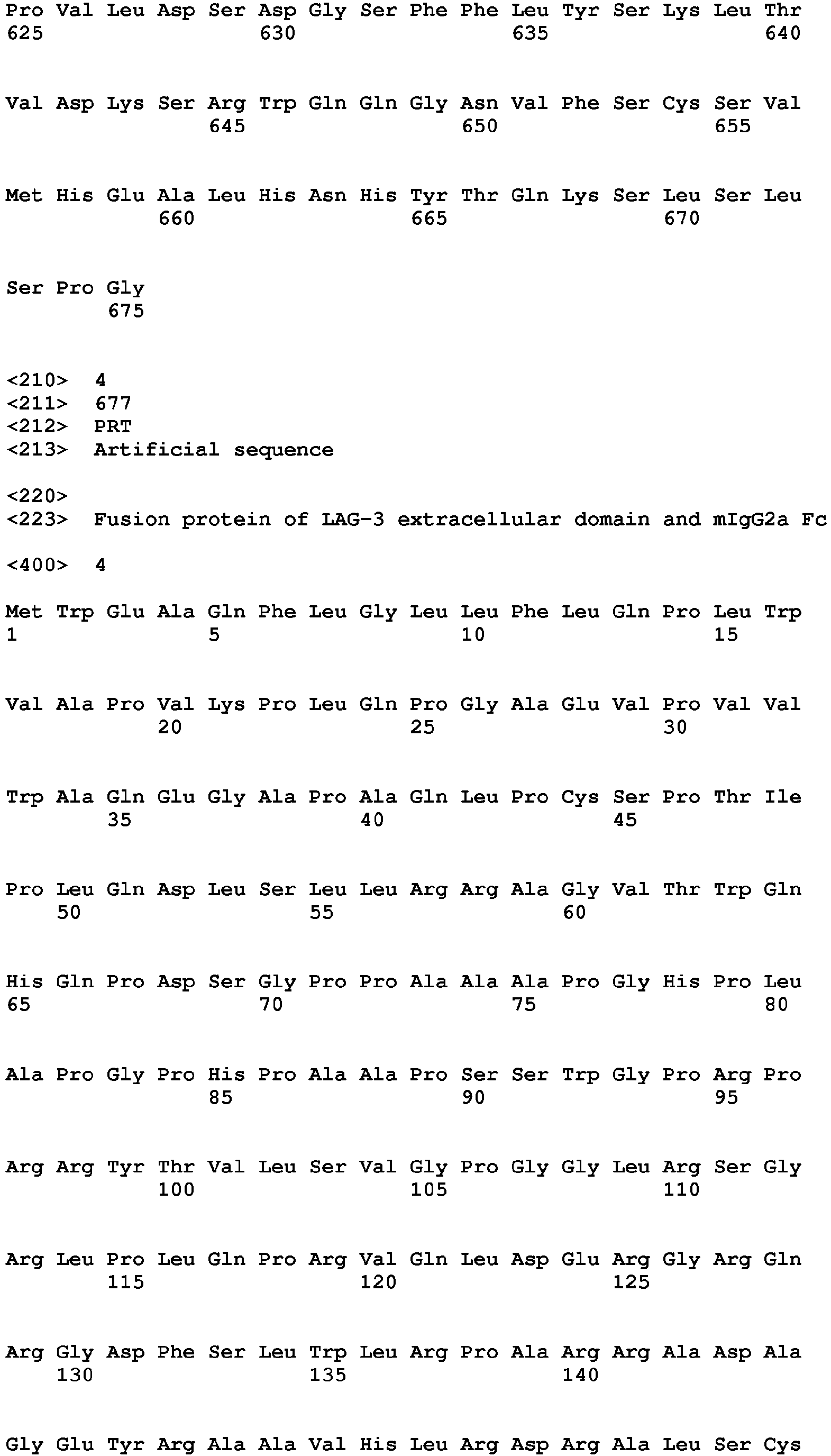

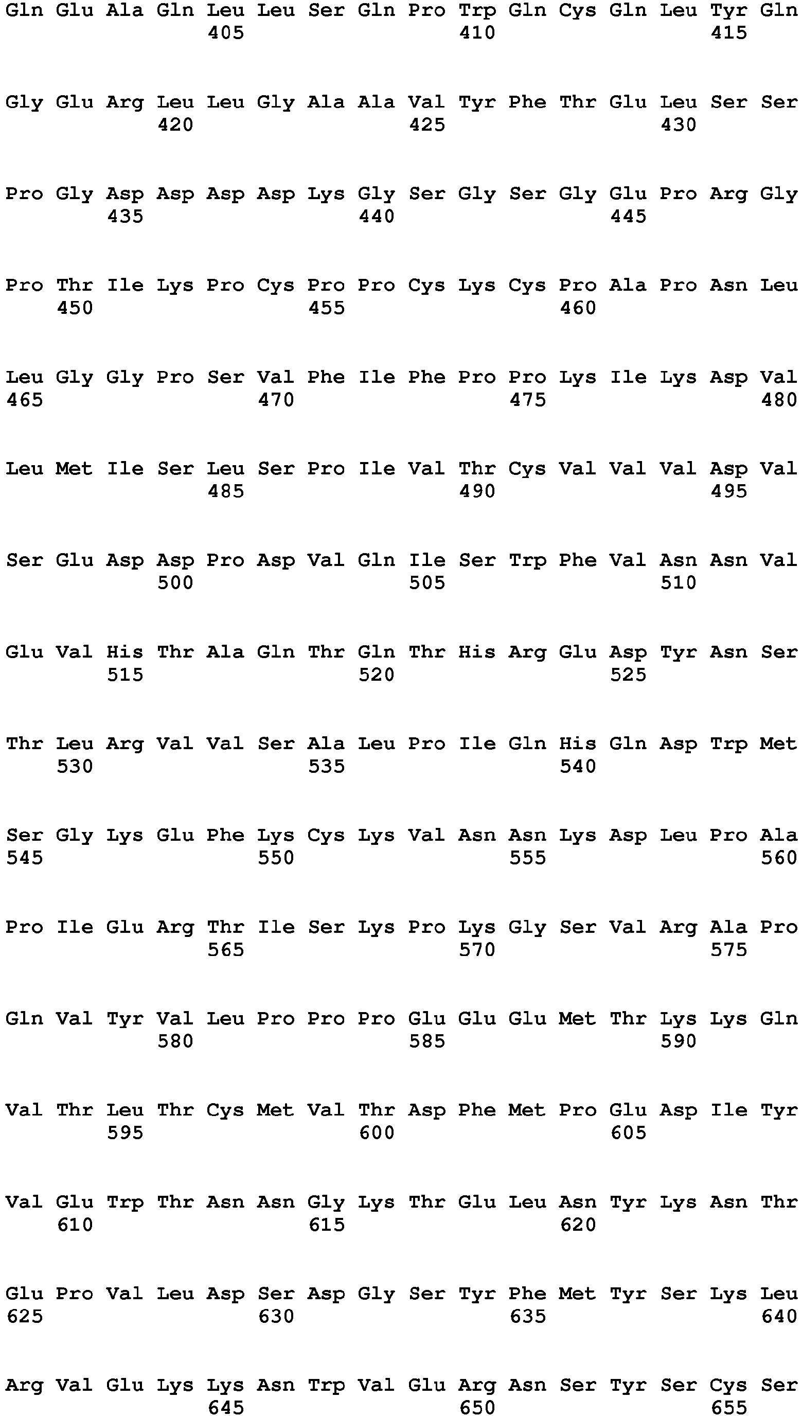


| Heavy chain | Light chain | |||
| mAb229 | HCDR1 | TSGMS | LCDR1 | RASENIYSNLA |
| SEQ ID NO: 9 | SEQ ID NO: 15 | |||
| HCDR2 | WINTYSGVPTY ADDFKG | LCDR2 | AATNLAD | |
| SEQ ID NO: 10 | SEQ ID NO: 16 | |||
| HCDR3 | DNYDARDVYYY AMDY | LCDR3 | QHFWITPWT | |
| SEQ ID NO: 11 | SEQ ID NO: 17 | |||
| mAb303 | HCDR1 | DYYMN | LCDR1 | RASQDIGSRLN |
| SEQ ID NO: 12 | SEQ ID NO: 18 | |||
| HCDR2 | VINPYNGDT A YNQKFKG | LCDR2 | A TSTLDS | |
| SEQ ID NO: 13 | SEQ ID NO: 19 | |||
| HCDR3 | DDGYYDYYFDV | LCDR3 | LQLASSPPT | |
| SEQ ID NO: 14 | SEQ ID NO: 20 | |||
| Stationary phase | Mobile phase | Affinity(M) |
| mAb229 | 1.72E-11 | |
| Hu229-009 | 4.88E-11 | |
| Hu229-010 | 3.82E-11 | |
| Hu229-013 | 2.81E-11 | |
| Hu229-014 | 3.74E-11 | |
| Hu229-015 | 4.59E-11 | |
| Hu229-017 | LAG-3-Flag | 6.71E-11 |
| Hu229-019 | 7.29E-11 | |
| mAb303 | 7.49E-11 | |
| Hu303-004 | 1.06E-09 | |
| Hu303-005 | 7.15E-11 | |
| Hu303-006 | 7.53E-11 | |
| Hu303-009 | 9.43E-10 | |
| Hu303-010 | 1.47E-10 | |
| Hu303-014 | 4.91E-10 | |
| Hu303-016 | 7.48E-11 |
Claims (28)
- A LAG-3 antibody or the antigen-binding fragment thereof, comprising any one or more CDR region sequences selected from the following (i) or (ii), or the amino acid sequences with at least 85% identity to the follows:(i) HCDR regions as shown in SEQ ID NO: 9, SEQ ID NO: 10, SEQ ID NO: 11; and LCDR regions as shown in SEQ ID NO: 15, SEQ ID NO: 16 and SEQ ID NO: 17; or(ii) HCDR regions as shown in SEQ ID NO: 12, SEQ ID NO: 13 and SEQ ID NO: 14; and LCDR regions as shown in SEQ ID NO: 18, SEQ ID NO: 19 and SEQ ID NO: 20.
- The LAG-3 antibody or the antigen-binding fragment thereof according to claim 1, wherein the antibody heavy chain variable region comprises HCDR1, HCDR2 and HCDR3 as shown in SEQ ID NO:9, SEQ ID NO:10 and SEQ ID NO: 11, respectively, or the amino acid sequences with at least 85% identity to these sequences; or the antibody heavy chain variable region comprises HCDR1, HCDR2 and HCDR3 as shown in SEQ ID NO: 12,SEQ ID NO: 13 and SEQ ID NO: 14, respectively, or the amino acid sequences with at least 85% identity to these sequences.
- The LAG-3 antibody or the antigen-binding fragment thereof according to claim 1, wherein the antibody light chain variable region comprises LCDR1, LCDR2 and LCDR3 as shown in SEQ ID NO: 15, SEQ ID NO: 16 and SEQ ID NO: 17, respectively, or the amino acid sequences with at least 85% identity to these sequences; or the antibody light chain variable region comprises LCDR1, LCDR2 and LCDR3 as shown in SEQ ID NO: 18, SEQ ID NO: 19 and SEQ ID NO: 20, respectively, or the amino acid sequences with at least 85% identity to these sequences.
- The LAG-3 antibody or the antigen-binding fragment thereof according to claim 1, wherein the antibody or the antigen-binding fragment thereof is a murine antibody or the fragment thereof.
- The LAG-3 antibody or the antigen-binding fragment thereof according to claim 1, wherein the antibody light chain variable region further comprises light chain FR region derived from murine κ chain or a variant thereof, or light chain FR region derived from murine λ chain or a variant thereof; wherein the antibody heavy chain variable region further comprises heavy chain FR region derived from murine IgG1 or a variant thereof, or heavy chain FR region derived from murine IgG2 or a variant thereof, or heavy chain FR region derived from murine IgG3 or a variant thereof.
- The LAG-3 antibody or the antigen-binding fragment thereof according to claim 4, wherein the murine antibody comprises a heavy chain variable region as shown in SEQ ID NO: 5 and a light chain variable region as shown in SEQ ID NO: 6, or comprises a heavy chain variable region as shown in SEQ ID NO: 7 and a light chain variable region as shown in SEQ ID NO: 8.
- The LAG-3 antibody or the antigen-binding fragment thereof according to claim 1, wherein the antibody light chain further comprises light chain constant region derived from murine κ chain or a variant thereof, or light chain constant region derived from murine λ chain or a variant thereof; wherein the antibody heavy chain further comprises heavy chain constant region derived from murine IgG1 or a variant thereof, or heavy chain constant region derived from murine IgG2 or a variant thereof, or heavy chain constant region derived from murine IgG3 or a variant thereof.
- The LAG-3 antibody or the antigen-binding fragment thereof according to claim 1, wherein the antibody or the antigen-binding fragment thereof is a chimeric antibody or the antigen-binding fragment thereof.
- The LAG-3 antibody or the antigen-binding fragment thereof according to claim 1, wherein the antibody or the antigen-binding fragment thereof is a humanized antibody or the antigen-binding fragment thereof.
- The LAG-3 antibody or the antigen-binding fragment thereof according to claim 9, wherein the heavy chain FR sequence of the humanized antibody heavy chain variable region is derived from a combination sequence of human germline heavy chains IGHV7-4-1*02 and hjh6.1, or a mutant sequence thereof, preferably comprises FR1, FR2, FR3 from IGHV7-4-1*02 and FR4 from hjh6.1 or a mutant sequence thereof;
alternatively, the heavy chain FR sequence of the humanized antibody heavy chain variable region is derived from a combination sequence of human germline heavy chain IGHV1-3*01 and hjh6.1, or a mutant sequence thereof, preferably comprises FR1, FR2, FR3 from IGHV1-3*01 and FR4 from hjh6.1 or a mutant sequence thereof. - The LAG-3 antibody or the antigen-binding fragment thereof according to claim 10, wherein the heavy chain variable region sequence of the humanized antibody is as shown in SEQ ID NO: 21 or SEQ ID NO: 29, or has at least 85% identity to these amino acid sequences; preferably has 1-10 amino acid changes in the heavy chain variable region.
- The LAG-3 antibody or antigen-binding fragment thereof according to claim 10, wherein the heavy chain FR sequence of the humanized antibody has 0-10 amino acid back-mutations;
preferably, the back-mutation is present on heavy chain FR region of the combination sequence of human germline heavy chains IGHV7-4-1*02 and hjh6.1, and is one or more amino acid back-mutations selected from the group consisting of E46K, R38K, V93T and Y95F;
or, preferably, the back-mutation is present on heavy chain FR region of the combination sequence of human germline heavy chains IGHV1-3*01 and hjh6.1, and is one or more amino acid back-mutations selected from the group consisting of F29L, A97T, M48I, V68A, I70L, R72V and T74K. - The LAG-3 antibody or the antigen-binding fragment thereof according to claim 10, wherein the heavy chain variable region sequence of the humanized antibody is selected from the group consisting of SEQ ID NO:21, SEQ ID NO:23, SEQ ID NO:24 and SEQ ID NO: 25, or the sequences with at least 85% identity to these sequences; or selected from the group consisting of SEQ ID NO:29, SEQ ID NO:31, SEQ ID NO:32 and SEQ ID NO: 33, or the sequences with at least 85% identity to these sequences.
- The LAG-3 antibody or the antigen-binding fragment thereof according to claim 9, wherein the light chain FR sequence of the humanized antibody light chain variable region is derived from a combination sequence of human germline light chain IGKV1-39*01 and hjk4.1, or a mutant sequence thereof; and comprises FR1, FR2, FR3 from human germline light chain IGHV1-39*01 and FR4 from hjh4.1 or a mutant sequence thereof.
- The LAG-3 antibody or the antigen-binding fragment thereof according to claim 14, wherein the light chain variable region sequence of the humanized antibody is as shown in SEQ ID NO: 22 or SEQ ID NO: 30, or the sequence with at least 85% identity to these sequences; preferably has 1-10 amino acid changes on the light chain variable region.
- The LAG-3 antibody or the antigen-binding fragment thereof according to claim 14, wherein the light chain FR sequence of the humanized antibody has 0-10 amino acid back-mutations, preferably one or more back-mutations selected from the group consisting of D70Q, F71Y, I48V and A43S, or preferably one or more back-mutations selected from the group consisting of L46R, G66R, S60K, P44F, Y36L, K42G, I21L and T85D.
- The LAG-3 antibody or the antigen-binding fragment thereof according to claim 14, wherein the light chain variable region sequence of the humanized antibody is selected from the group consisting of SEQ ID NO: 22, SEQ ID NO: 26, SEQ ID NO: 27 and SEQ ID NO: 28 or the sequence with at least 85% identity to these sequences; or is selected from the group consisting of SEQ ID NO: 30, SEQ ID NO: 34, SEQ ID NO: 35, SEQ ID NO: 36 and SEQ ID NO: 37, or the sequence with at least 85% identity to these sequences.
- The LAG-3 antibody or the antigen-binding fragment thereof according to claim 9, wherein the humanized antibody comprising:(a) heavy chain variable region, wherein the heavy chain variable region sequence is selected from the group consisting of SEQ ID NO: 21, SEQ ID NO: 23, SEQ ID NO: 24 and SEQ ID NO: 25, or the sequence with at least 85% identity to these sequences;
alternatively, the heavy chain variable region sequence is selected from the group consisting of SEQ ID NO: 29, SEQ ID NO: 31, SEQ ID NO: 32 and SEQ ID NO: 33, or the sequence with at least 85% identity to these sequences; and/or(b) light chain variable region, wherein the light chain variable region sequence is selected from the group consisting of SEQ ID NO: 22, SEQ ID NO: 26, SEQ ID NO: 27 and SEQ ID NO: 28, or the sequence with at least 85% identity to these sequences;
alternatively, the light chain variable region sequence is selected from the group consisting of sequence SEQ ID NO: 30, SEQ ID NO: 34, SEQ ID NO: 35, SEQ ID NO: 36 and SEQ ID NO: 37, or the sequence with at least 85% identity to these sequences. - The LAG-3 antibody or the antigen-binding fragment thereof according to claim 9, wherein the antibody comprising a combination of a heavy chain variable region and a light chain variable region selected from the group consisting of:1) the heavy chain variable region of SEQ ID NO: 21 and the light chain variable region of SEQ ID NO: 22;2) the heavy chain variable region of SEQ ID NO: 21 and the light chain variable region of SEQ ID NO: 26;3) the heavy chain variable region of SEQ ID NO: 21 and the light chain variable region of SEQ ID NO: 27;4) the heavy chain variable region of SEQ ID NO: 21 and the light chain variable region of SEQ ID NO: 28;5) the heavy chain variable region of SEQ ID NO: 23 and the light chain variable region of SEQ ID NO: 22;6) the heavy chain variable region of SEQ ID NO: 23 and the light chain variable region of SEQ ID NO: 26;7) the heavy chain variable region of SEQ ID NO: 23 and the light chain variable region of SEQ ID NO: 27;8) the heavy chain variable region of SEQ ID NO: 23 and the light chain variable region of SEQ ID NO: 28;9) the heavy chain variable region of SEQ ID NO: 24 and the light chain variable region of SEQ ID NO: 22;10) the heavy chain variable region of SEQ ID NO: 24 and the light chain variable region of SEQ ID NO: 26;11) the heavy chain variable region of SEQ ID NO: 24 and the light chain variable region of SEQ ID NO: 27;12) the heavy chain variable region of SEQ ID NO: 24 and the light chain variable region of SEQ ID NO: 28;13) the heavy chain variable region of SEQ ID NO: 25 and the light chain variable region of SEQ ID NO: 22;14) the heavy chain variable region of SEQ ID NO: 25 and the light chain variable region of SEQ ID NO: 26;15) the heavy chain variable region of SEQ ID NO: 25 and the light chain variable region of SEQ ID NO: 27;16) the heavy chain variable region of SEQ ID NO: 25 and the light chain variable region of SEQ ID NO: 28;17) the heavy chain variable region of SEQ ID NO: 29 and the light chain variable region of SEQ ID NO: 30;18) the heavy chain variable region of SEQ ID NO: 29 and the light chain variable region of SEQ ID NO: 34;19) the heavy chain variable region of SEQ ID NO: 29 and the light chain variable region of SEQ ID NO: 35;20) the heavy chain variable region of SEQ ID NO: 29 and the light chain variable region of SEQ ID NO: 36;21) the heavy chain variable region of SEQ ID NO: 29 and the light chain variable region of SEQ ID NO: 37;22) the heavy chain variable region of SEQ ID NO: 31 and the light chain variable region of SEQ ID NO: 30;23) the heavy chain variable region of SEQ ID NO: 31 and the light chain variable region of SEQ ID NO: 34;24) the heavy chain variable region of SEQ ID NO: 31 and the light chain variable region of SEQ ID NO: 35;25) the heavy chain variable region of SEQ ID NO: 31 and the light chain variable region of SEQ ID NO: 36;26) the heavy chain variable region of SEQ ID NO: 31 and the light chain variable region of SEQ ID NO: 37;27) the heavy chain variable region of SEQ ID NO: 32 and the light chain variable region of SEQ ID NO: 30;28) the heavy chain variable region of SEQ ID NO: 32 and the light chain variable region of SEQ ID NO: 34;29) the heavy chain variable region of SEQ ID NO: 32 and the light chain variable region of SEQ ID NO: 35;30) the heavy chain variable region of SEQ ID NO: 32 and the light chain variable region of SEQ ID NO: 36;31) the heavy chain variable region of SEQ ID NO: 32 and the light chain variable region of SEQ ID NO: 37;32) the heavy chain variable region of SEQ ID NO: 33 and the light chain variable region of SEQ ID NO: 30;33) the heavy chain variable region of SEQ ID NO: 33 and the light chain variable region of SEQ ID NO: 34;34) the heavy chain variable region of SEQ ID NO: 33 and the light chain variable region of SEQ ID NO: 35;35) the heavy chain variable region of SEQ ID NO: 33 and the light chain variable region of SEQ ID NO: 36; and36) the heavy chain variable region of SEQ ID NO: 33 and the light chain variable region of SEQ ID NO: 37.
- The LAG-3 antibody or the antigen-binding fragment thereof according to any one of claims 7-15, wherein the heavy chain of the chimeric antibody or the humanized antibody further comprises a heavy chain constant region derived from human IgG1, IgG2, IgG3 or IgG4 or a variant thereof, preferably a heavy chain constant region derived from human IgG4 or a variant thereof, most preferably a heavy chain constant region as shown in SEQ ID NO: 38;
wherein the light chain of the chimeric antibody or the humanized antibody further comprises a light chain constant region derived from human κ chain, human λ chain or a variant thereof, most preferably a light chain constant region as shown in SEQ ID NO: 39. - An isolated monoclonal antibody or antigen-binding fragment thereof that competes for binding to LAG-3 with the monoclonal antibody or antigen-binding fragment thereof according to any one of claims 1-20.
- A pharmaceutical composition, comprising therapeutically effective amount of the LAG-3 antibody or the antigen-binging fragment thereof according to any one of claims 1-21 and one or more pharmaceutically acceptable carriers, diluents or excipients.
- An isolated nucleic acid encoding the LAG-3 antibody or the antigen-binding fragment thereof according to any one of claims 1 to 21.
- An expression vector comprising the nucleic acid according to claim 23.
- A host cell transformed with the expression vector according to claim 24, wherein the host cell is selected from the group consisting of prokaryotic cells and eukaryotic cells, preferably eukaryotic cells, more preferably mammalian cells.
- A method for preparing a LAG-3 antibody or the antigen-binding fragment thereof, wherein the method comprises expressing the antibody or the antigen-binding fragment thereof in the host cell of claim 25 and isolating the antibody or the antigen-binding fragment thereof from the host cell.
- A method for inhibiting the growth of tumor cells in a subject, wherein the method comprises administering to the subject therapeutically effective amount of the LAG-3 antibody or the antigen-binding fragment thereof according to any one of claims 1 to 21, or administering to the subject the pharmaceutical composition according to claim 22 or the nucleic acid according to claim 23, thereby inhibiting tumor growth in the subject.
- The use of the LAG-3 antibody or the antigen-binding fragment according to any one of claims 1-21, or the pharmaceutical composition according to claim 22 or the nucleic acid according to claim 23, in the preparation of a medicament for treatment of a disease or a condition associated with the involvement of pathogenic T cells, wherein the disease or the condition is preferably cancer; the cancer includes but not limited to ovarian cancer, melanoma, prostate cancer, intestinal cancer, stomach cancer, esophageal cancer, breast cancer, lung cancer, renal cancer, pancreatic cancer, uterine cancer, liver cancer, bladder cancer, cervical cancer, oral cancer, brain cancer, testicular cancer, skin cancer, thyroid cancer, and hematological malignant tumors including myeloma and chronic and acute leukemia.
Priority Applications (1)
| Application Number | Priority Date | Filing Date | Title |
|---|---|---|---|
| PL17814733T PL3476399T3 (en) | 2016-06-23 | 2017-06-22 | Lag-3 antibody, antigen-binding fragment thereof, and pharmaceutical application thereof |
Applications Claiming Priority (2)
| Application Number | Priority Date | Filing Date | Title |
|---|---|---|---|
| CN201610465386 | 2016-06-23 | ||
| PCT/CN2017/089492 WO2017219995A1 (en) | 2016-06-23 | 2017-06-22 | Lag-3 antibody, antigen-binding fragment thereof, and pharmaceutical application thereof |
Publications (3)
| Publication Number | Publication Date |
|---|---|
| EP3476399A1 true EP3476399A1 (en) | 2019-05-01 |
| EP3476399A4 EP3476399A4 (en) | 2020-01-29 |
| EP3476399B1 EP3476399B1 (en) | 2022-04-20 |
Family
ID=60783834
Family Applications (1)
| Application Number | Title | Priority Date | Filing Date |
|---|---|---|---|
| EP17814733.6A Active EP3476399B1 (en) | 2016-06-23 | 2017-06-22 | Lag-3 antibody, antigen-binding fragment thereof, and pharmaceutical application thereof |
Country Status (18)
| Country | Link |
|---|---|
| US (2) | US11155617B2 (en) |
| EP (1) | EP3476399B1 (en) |
| JP (1) | JP6998895B2 (en) |
| KR (1) | KR102497259B1 (en) |
| CN (1) | CN108472349B (en) |
| AU (1) | AU2017282892B2 (en) |
| BR (1) | BR112018074155A2 (en) |
| CA (1) | CA3024359A1 (en) |
| DK (1) | DK3476399T3 (en) |
| ES (1) | ES2915410T3 (en) |
| HU (1) | HUE058657T2 (en) |
| MX (1) | MX2018015393A (en) |
| PL (1) | PL3476399T3 (en) |
| PT (1) | PT3476399T (en) |
| RU (1) | RU2757813C2 (en) |
| TW (1) | TWI757304B (en) |
| UA (1) | UA127048C2 (en) |
| WO (1) | WO2017219995A1 (en) |
Cited By (2)
| Publication number | Priority date | Publication date | Assignee | Title |
|---|---|---|---|---|
| EP3768720A4 (en) * | 2018-03-20 | 2022-01-05 | Wuxi Biologics Ireland Limited | NEW ANTI-LAG-3 ANTIBODY POLYPEPTIDE |
| EP4242234A4 (en) * | 2020-12-10 | 2024-09-25 | Beijing Dongfang Biotech Co. Ltd. | ANTI-LAG-3 MONOCLONAL ANTIBODY, ANTIGEN-BINDING FRAGMENT AND USE THEREOF |
Families Citing this family (71)
| Publication number | Priority date | Publication date | Assignee | Title |
|---|---|---|---|---|
| TWI756187B (en) | 2015-10-09 | 2022-03-01 | 美商再生元醫藥公司 | Anti-lag3 antibodies and uses thereof |
| US11155617B2 (en) | 2016-06-23 | 2021-10-26 | Jiangsu Hengrui Medicine Co., Ltd. | LAG-3 antibody, antigen-binding fragment thereof, and pharmaceutical application thereof |
| JP7066696B2 (en) | 2016-10-11 | 2022-05-13 | アジェナス インコーポレイテッド | Anti-LAG-3 antibody and its usage |
| EA201991673A1 (en) | 2017-02-10 | 2020-01-17 | Регенерон Фармасьютикалз, Инк. | LAB3-labeled RADIOACTIVE ISOTOPE ANTIBODIES FOR IMMUNO-PET VISUALIZATION |
| CN110678200B (en) | 2017-05-30 | 2024-05-17 | 百时美施贵宝公司 | Compositions comprising an anti-LAG-3 antibody or an anti-LAG-3 antibody and an anti-PD-1 or anti-PD-L1 antibody |
| JP2020522495A (en) | 2017-05-30 | 2020-07-30 | ブリストル−マイヤーズ スクイブ カンパニーBristol−Myers Squibb Company | Composition comprising a combination of anti-LAG-3 antibody, PD-1 pathway inhibitor and immunotherapeutic agent |
| ES2965352T3 (en) | 2017-05-30 | 2024-04-12 | Bristol Myers Squibb Co | Treatment of lymphocyte activation gene 3 (LAG-3) positive tumors |
| CA3085656A1 (en) * | 2017-12-22 | 2019-06-27 | Jiangsu Hengrui Medicine Co., Ltd. | Lag-3 antibody pharmaceutical composition and use thereof |
| CN110305215B (en) * | 2018-03-20 | 2023-11-03 | 基石药业 | Novel anti-LAG-3 antibody peptides |
| WO2019185164A1 (en) | 2018-03-29 | 2019-10-03 | Hummingbird Bioscience Holdings Pte. Ltd. | Her3 antigen-binding molecules |
| CN110343178B (en) * | 2018-04-03 | 2022-07-22 | 上海开拓者生物医药有限公司 | Anti-human LAG-3 monoclonal antibody and application thereof |
| CN110606892B (en) * | 2018-06-14 | 2023-09-26 | 华博生物医药技术(上海)有限公司 | LAG-3 antibody with high affinity and high biological activity and application thereof |
| CN110615840A (en) * | 2018-06-19 | 2019-12-27 | 信达生物制药(苏州)有限公司 | Fully human anti-LAG-3 antibodies and uses thereof |
| CN112739371A (en) | 2018-07-26 | 2021-04-30 | 百时美施贵宝公司 | LAG-3 combination therapy for the treatment of cancer |
| LT4445958T (en) | 2018-10-19 | 2025-12-10 | Bristol-Myers Squibb Company | Combination therapy for melanoma |
| EP3872093A4 (en) * | 2018-10-22 | 2022-10-19 | Shanghai Genbase Biotechnology Co., Ltd. | Anti-cldn18.2 antibody and uses thereof |
| CN111620949A (en) * | 2019-02-28 | 2020-09-04 | 三生国健药业(上海)股份有限公司 | Antibodies that bind human LAG-3, methods of making, and uses thereof |
| KR20220016155A (en) | 2019-05-30 | 2022-02-08 | 브리스톨-마이어스 스큅 컴퍼니 | Methods of Identifying Suitable Subjects for Immuno-Oncology (I-O) Therapy |
| US20220363760A1 (en) | 2019-05-30 | 2022-11-17 | Bristol-Myers Squibb Company | Multi-tumor gene signature for suitability to immuno-oncology therapy |
| WO2020243570A1 (en) | 2019-05-30 | 2020-12-03 | Bristol-Myers Squibb Company | Cell localization signature and combination therapy |
| CN115340606B (en) * | 2019-05-31 | 2023-12-19 | 瑞阳(苏州)生物科技有限公司 | Antibody combined with human LAG-3 protein, encoding gene and application thereof |
| WO2021024020A1 (en) | 2019-08-06 | 2021-02-11 | Astellas Pharma Inc. | Combination therapy involving antibodies against claudin 18.2 and immune checkpoint inhibitors for treatment of cancer |
| WO2021055994A1 (en) | 2019-09-22 | 2021-03-25 | Bristol-Myers Squibb Company | Quantitative spatial profiling for lag-3 antagonist therapy |
| WO2021092221A1 (en) | 2019-11-06 | 2021-05-14 | Bristol-Myers Squibb Company | Methods of identifying a subject with a tumor suitable for a checkpoint inhibitor therapy |
| WO2021092220A1 (en) | 2019-11-06 | 2021-05-14 | Bristol-Myers Squibb Company | Methods of identifying a subject with a tumor suitable for a checkpoint inhibitor therapy |
| US20220411499A1 (en) | 2019-11-08 | 2022-12-29 | Bristol-Myers Squibb Company | LAG-3 Antagonist Therapy for Melanoma |
| CN113348182B (en) * | 2019-12-30 | 2022-07-12 | 上海海路生物技术有限公司 | LAG-3 antibody and medical application thereof |
| JP7498889B2 (en) * | 2020-01-30 | 2024-06-13 | 東亞合成株式会社 | Peptide that inhibits binding between CTLA4 and B7 protein and its use |
| JP2023514152A (en) | 2020-02-06 | 2023-04-05 | ブリストル-マイヤーズ スクイブ カンパニー | IL-10 and its uses |
| KR20230035576A (en) | 2020-07-07 | 2023-03-14 | 비온테크 에스이 | RNA for the treatment of HPV-positive cancer |
| IL300813A (en) | 2020-08-28 | 2023-04-01 | Bristol Myers Squibb Co | Lag-3 antagonist therapy for hepatocellular carcinoma |
| AU2021334361A1 (en) | 2020-08-31 | 2023-05-11 | Bristol-Myers Squibb Company | Cell localization signature and immunotherapy |
| KR20230093282A (en) | 2020-10-23 | 2023-06-27 | 브리스톨-마이어스 스큅 컴퍼니 | LAG-3 antagonist therapy for lung cancer |
| WO2022120179A1 (en) | 2020-12-03 | 2022-06-09 | Bristol-Myers Squibb Company | Multi-tumor gene signatures and uses thereof |
| WO2022135666A1 (en) | 2020-12-21 | 2022-06-30 | BioNTech SE | Treatment schedule for cytokine proteins |
| WO2022135667A1 (en) | 2020-12-21 | 2022-06-30 | BioNTech SE | Therapeutic rna for treating cancer |
| TW202245808A (en) | 2020-12-21 | 2022-12-01 | 德商拜恩迪克公司 | Therapeutic rna for treating cancer |
| JP2024501029A (en) | 2020-12-28 | 2024-01-10 | ブリストル-マイヤーズ スクイブ カンパニー | Subcutaneous administration of PD1/PD-L1 antibodies |
| US20220233693A1 (en) | 2020-12-28 | 2022-07-28 | Bristol-Myers Squibb Company | Antibody Compositions and Methods of Use Thereof |
| US20240384350A1 (en) | 2021-01-29 | 2024-11-21 | Board Of Regents, The University Of Texas System | Methods of treating cancer with kinase inhibitors |
| CN114907478A (en) * | 2021-02-08 | 2022-08-16 | 迈威(上海)生物科技股份有限公司 | LAG-3 binding antibodies and uses thereof |
| CN117858719A (en) | 2021-03-29 | 2024-04-09 | 朱诺治疗学股份有限公司 | Methods of administering and treating with a combination of checkpoint inhibitor therapy and CAR T cell therapy |
| EP4319755A4 (en) | 2021-04-08 | 2025-03-26 | Board of Regents, The University of Texas System | Compounds and methods for theranostic targeting of PARP activity |
| EP4337694A1 (en) | 2021-05-12 | 2024-03-20 | Dana-Farber Cancer Institute, Inc. | Lag3 and gal3 inhibitory agents, xbp1, cs1, and cd138 peptides, and methods of use thereof |
| CA3225254A1 (en) | 2021-07-13 | 2023-01-19 | BioNTech SE | Multispecific binding agents against cd40 and cd137 in combination therapy for cancer |
| TW202333802A (en) | 2021-10-11 | 2023-09-01 | 德商拜恩迪克公司 | Therapeutic rna for lung cancer |
| CA3224890A1 (en) | 2021-10-29 | 2023-05-04 | Bristol-Myers Squibb Company | Lag-3 antagonist therapy for hematological cancer |
| KR20240135661A (en) | 2022-01-26 | 2024-09-11 | 브리스톨-마이어스 스큅 컴퍼니 | Combination therapy for hepatocellular carcinoma |
| WO2023164638A1 (en) | 2022-02-25 | 2023-08-31 | Bristol-Myers Squibb Company | Combination therapy for colorectal carcinoma |
| WO2023168404A1 (en) | 2022-03-04 | 2023-09-07 | Bristol-Myers Squibb Company | Methods of treating a tumor |
| JP2025508076A (en) | 2022-03-08 | 2025-03-21 | アレンティス・セラピューティクス・アクチェンゲゼルシャフト | Use of anti-claudin-1 antibodies to improve T cell availability |
| WO2023178329A1 (en) | 2022-03-18 | 2023-09-21 | Bristol-Myers Squibb Company | Methods of isolating polypeptides |
| CN114874324B (en) * | 2022-05-13 | 2023-02-03 | 苏州旭光科星抗体生物科技有限公司 | Enzyme-linked immunoassay kit for detecting content of soluble LAG-3 protein and application thereof |
| KR20250022071A (en) | 2022-06-02 | 2025-02-14 | 브리스톨-마이어스 스큅 컴퍼니 | Antibody composition and method of use thereof |
| EP4626552A1 (en) | 2022-12-01 | 2025-10-08 | MedImmune Limited | Combination therapy for treatment of cancer comprising anti-pd-l1 and anti-cd73 antibodies |
| JP2026501506A (en) | 2022-12-14 | 2026-01-16 | アステラス・ファーマ・ヨーロッパ・ベスローデン・フェンノートシャップ | Combination therapy comprising a bispecific binding agent that binds to CLDN18.2 and CD3 and an immune checkpoint inhibitor |
| EP4638503A1 (en) | 2022-12-21 | 2025-10-29 | Bristol-Myers Squibb Company | Combination therapy for lung cancer |
| EP4658687A1 (en) | 2023-01-31 | 2025-12-10 | University of Rochester | Immune checkpoint blockade therapy for treating staphylococcus aureus infections |
| EP4665410A1 (en) | 2023-02-17 | 2025-12-24 | Regeneron Pharmaceuticals, Inc. | Radiolabeled anti-lag3 antibodies for immuno-pet imaging |
| WO2024196952A1 (en) | 2023-03-20 | 2024-09-26 | Bristol-Myers Squibb Company | Tumor subtype assessment for cancer therapy |
| WO2025038763A1 (en) | 2023-08-15 | 2025-02-20 | Bristol-Myers Squibb Company | Ceramic hydroxyapatite chromatography flow through method |
| TW202535926A (en) | 2023-10-30 | 2025-09-16 | 美商再生元醫藥公司 | Stable antibody formulation |
| TW202541837A (en) | 2023-12-08 | 2025-11-01 | 日商安斯泰來製藥公司 | Combination therapy involving bispecific binding agents binding to cldn18.2 and cd3 and agents stabilizing or increasing expression of cldn18.2 |
| WO2026033885A1 (en) | 2024-08-08 | 2026-02-12 | Astellas Pharma Inc. | Combination therapy involving bispecific binding agents binding to cldn18.2 and cd3 and agents stabilizing or increasing expression of cldn18.2 |
| WO2025120867A1 (en) | 2023-12-08 | 2025-06-12 | Astellas Pharma Inc. | Combination therapy involving bispecific binding agents binding to cldn18.2 and cd3 and anti-vegfr2 antibodies |
| WO2025120866A1 (en) | 2023-12-08 | 2025-06-12 | Astellas Pharma Inc. | Combination therapy involving bispecific binding agents binding to cldn18.2 and cd3 and agents stabilizing or increasing expression of cldn18.2 |
| WO2025145207A1 (en) | 2023-12-29 | 2025-07-03 | Bristol-Myers Squibb Company | Combination therapy of kras inhibitor and treg-depleting agent |
| WO2025184208A1 (en) | 2024-02-27 | 2025-09-04 | Bristol-Myers Squibb Company | Anti-ceacam5 antibodies and uses thereof |
| US12521446B2 (en) | 2024-02-27 | 2026-01-13 | Bristol-Myers Squibb Company | Anti-CEACAM5 antibody drug conjugates |
| WO2025226695A1 (en) | 2024-04-23 | 2025-10-30 | Regeneron Pharmaceuticals, Inc. | Stable antibody formulation |
| WO2025245489A1 (en) | 2024-05-24 | 2025-11-27 | Bristol-Myers Squibb Company | Treatment of tumors in subjects having fgl-1 positive samples |
Family Cites Families (19)
| Publication number | Priority date | Publication date | Assignee | Title |
|---|---|---|---|---|
| US5976877A (en) | 1990-01-08 | 1999-11-02 | Institut National De La Sante Et De La Recherche Medicale (Inserm) | Proteins produced by human lymphocytes DNA sequence encoding these proteins and their pharmaceutical and biological uses |
| EP0519866A1 (en) * | 1991-06-18 | 1992-12-23 | Ciba-Geigy Ag | Novel anti-HIV antibodies |
| CA2189657C (en) | 1994-05-06 | 2002-03-12 | Florence Faure | Lag-3 protein soluble polypeptide fractions, method of production, therapeutic composition and anti-idiotype antibody |
| PL1897548T5 (en) * | 2003-02-28 | 2025-03-17 | The Johns Hopkins University | T cell regulation |
| EP2860253B1 (en) * | 2006-03-01 | 2018-08-01 | Aduro Biotech, Inc. | Engineered listeria and methods of use thereof |
| TW201716437A (en) | 2006-11-02 | 2017-05-16 | 建南德克公司 | Humanized anti-factor D antibodies and uses thereof |
| US9244059B2 (en) * | 2007-04-30 | 2016-01-26 | Immutep Parc Club Orsay | Cytotoxic anti-LAG-3 monoclonal antibody and its use in the treatment or prevention of organ transplant rejection and autoimmune disease |
| EP1987839A1 (en) | 2007-04-30 | 2008-11-05 | I.N.S.E.R.M. Institut National de la Sante et de la Recherche Medicale | Cytotoxic anti-LAG-3 monoclonal antibody and its use in the treatment or prevention of organ transplant rejection and autoimmune disease |
| AR072999A1 (en) | 2008-08-11 | 2010-10-06 | Medarex Inc | HUMAN ANTIBODIES THAT JOIN GEN 3 OF LYMPHOCYTARY ACTIVATION (LAG-3) AND THE USES OF THESE |
| MX343227B (en) * | 2010-04-09 | 2016-10-28 | Aveo Pharmaceuticals Inc | ANTI-ERBB3 ANTIBODIES. |
| UY34887A (en) | 2012-07-02 | 2013-12-31 | Bristol Myers Squibb Company Una Corporacion Del Estado De Delaware | OPTIMIZATION OF ANTIBODIES THAT FIX THE LYMPHOCYTE ACTIVATION GEN 3 (LAG-3) AND ITS USES |
| FR3003261B1 (en) | 2013-03-14 | 2015-05-01 | Agronutrition | PROCESS FOR THE SYNTHESIS OF ACYL GLYCEROL OLIGOMERS |
| US10344088B2 (en) * | 2013-03-15 | 2019-07-09 | Glaxosmithkline Intellectual Property Development Limited | Antigen binding proteins |
| KR20150131177A (en) * | 2013-03-15 | 2015-11-24 | 제넨테크, 인크. | Anti-crth2 antibodies and their use |
| RU2551235C2 (en) * | 2013-08-26 | 2015-05-20 | Илья Владимирович Духовлинов | Humanised monoclonal antibody specific to syndecan-1 |
| JP6595458B2 (en) | 2013-09-20 | 2019-10-23 | ブリストル−マイヤーズ スクイブ カンパニー | Combination of anti-LAG-3 antibody and anti-PD-1 antibody for treating tumor |
| KR102442436B1 (en) | 2014-03-14 | 2022-09-15 | 노파르티스 아게 | Antibody molecules to lag-3 and uses thereof |
| JO3663B1 (en) * | 2014-08-19 | 2020-08-27 | Merck Sharp & Dohme | Anti-lag3 antibodies and antigen-binding fragments |
| US11155617B2 (en) | 2016-06-23 | 2021-10-26 | Jiangsu Hengrui Medicine Co., Ltd. | LAG-3 antibody, antigen-binding fragment thereof, and pharmaceutical application thereof |
-
2017
- 2017-06-22 US US16/312,174 patent/US11155617B2/en active Active
- 2017-06-22 TW TW106120891A patent/TWI757304B/en active
- 2017-06-22 JP JP2018564887A patent/JP6998895B2/en active Active
- 2017-06-22 BR BR112018074155-8A patent/BR112018074155A2/en not_active Application Discontinuation
- 2017-06-22 CA CA3024359A patent/CA3024359A1/en active Pending
- 2017-06-22 HU HUE17814733A patent/HUE058657T2/en unknown
- 2017-06-22 UA UAA201900286A patent/UA127048C2/en unknown
- 2017-06-22 DK DK17814733.6T patent/DK3476399T3/en active
- 2017-06-22 PT PT178147336T patent/PT3476399T/en unknown
- 2017-06-22 ES ES17814733T patent/ES2915410T3/en active Active
- 2017-06-22 RU RU2018145852A patent/RU2757813C2/en active
- 2017-06-22 PL PL17814733T patent/PL3476399T3/en unknown
- 2017-06-22 KR KR1020187037172A patent/KR102497259B1/en active Active
- 2017-06-22 WO PCT/CN2017/089492 patent/WO2017219995A1/en not_active Ceased
- 2017-06-22 EP EP17814733.6A patent/EP3476399B1/en active Active
- 2017-06-22 MX MX2018015393A patent/MX2018015393A/en unknown
- 2017-06-22 AU AU2017282892A patent/AU2017282892B2/en not_active Expired - Fee Related
- 2017-06-22 CN CN201780004996.5A patent/CN108472349B/en active Active
-
2021
- 2021-09-17 US US17/478,497 patent/US11981733B2/en active Active
Cited By (3)
| Publication number | Priority date | Publication date | Assignee | Title |
|---|---|---|---|---|
| EP3768720A4 (en) * | 2018-03-20 | 2022-01-05 | Wuxi Biologics Ireland Limited | NEW ANTI-LAG-3 ANTIBODY POLYPEPTIDE |
| EP4242234A4 (en) * | 2020-12-10 | 2024-09-25 | Beijing Dongfang Biotech Co. Ltd. | ANTI-LAG-3 MONOCLONAL ANTIBODY, ANTIGEN-BINDING FRAGMENT AND USE THEREOF |
| AU2021398061B2 (en) * | 2020-12-10 | 2025-09-11 | Beijing Dongfang Biotech Co., Ltd | Anti-lag-3 monoclonal antibody and antigen binding fragment thereof, and use thereof |
Also Published As
| Publication number | Publication date |
|---|---|
| MX2018015393A (en) | 2019-04-29 |
| PT3476399T (en) | 2022-05-31 |
| CA3024359A1 (en) | 2017-12-28 |
| AU2017282892A1 (en) | 2019-01-24 |
| PL3476399T3 (en) | 2022-06-20 |
| AU2017282892B2 (en) | 2023-10-26 |
| KR102497259B1 (en) | 2023-02-07 |
| HUE058657T2 (en) | 2022-09-28 |
| EP3476399B1 (en) | 2022-04-20 |
| EP3476399A4 (en) | 2020-01-29 |
| WO2017219995A1 (en) | 2017-12-28 |
| RU2757813C2 (en) | 2021-10-21 |
| CN108472349B (en) | 2022-05-24 |
| RU2018145852A3 (en) | 2020-10-26 |
| BR112018074155A2 (en) | 2019-03-06 |
| US11981733B2 (en) | 2024-05-14 |
| ES2915410T3 (en) | 2022-06-22 |
| US11155617B2 (en) | 2021-10-26 |
| TW201800419A (en) | 2018-01-01 |
| JP6998895B2 (en) | 2022-01-18 |
| US20220002405A1 (en) | 2022-01-06 |
| TWI757304B (en) | 2022-03-11 |
| UA127048C2 (en) | 2023-03-29 |
| JP2019518459A (en) | 2019-07-04 |
| CN108472349A (en) | 2018-08-31 |
| US20190233513A1 (en) | 2019-08-01 |
| DK3476399T3 (en) | 2022-05-30 |
| RU2018145852A (en) | 2020-07-23 |
| KR20190021248A (en) | 2019-03-05 |
Similar Documents
| Publication | Publication Date | Title |
|---|---|---|
| US11981733B2 (en) | LAG-3 antibody, antigen-binding fragment thereof, and pharmaceutical application thereof | |
| US11512129B2 (en) | TIGIT antibody, antigen-binding fragment thereof, and medical use thereof | |
| CN112969716B (en) | Anti-PD-1 antibody, its antigen-binding fragment and its medical use | |
| US12227583B2 (en) | Anti-OX40 antibody, antigen-binding fragment thereof, and the pharmaceutical use | |
| CN110790839B (en) | anti-PD-1 antibody, antigen binding fragment thereof and medical application thereof | |
| CN111094339B (en) | Application of anti-PD-1 antibody and anti-LAG-3 antibody in preparation of medicine for treating tumor | |
| TW202128763A (en) | Fusion protein containing tgf-β receptor and the pharmaceutical use thereof | |
| WO2022247826A1 (en) | Specific binding protein targeting pd-l1 and cd73 | |
| RU2807484C2 (en) | Antibody to pd-1, its antigen-binding fragment and its pharmaceutical use | |
| TW202305007A (en) | Specific binding protein targeting pd-l1 and cd73 | |
| HK40051455A (en) | Anti-pd-1 antibody, antigen-binding fragment thereof and pharmaceutical use thereof |
Legal Events
| Date | Code | Title | Description |
|---|---|---|---|
| STAA | Information on the status of an ep patent application or granted ep patent |
Free format text: STATUS: THE INTERNATIONAL PUBLICATION HAS BEEN MADE |
|
| PUAI | Public reference made under article 153(3) epc to a published international application that has entered the european phase |
Free format text: ORIGINAL CODE: 0009012 |
|
| STAA | Information on the status of an ep patent application or granted ep patent |
Free format text: STATUS: REQUEST FOR EXAMINATION WAS MADE |
|
| 17P | Request for examination filed |
Effective date: 20181231 |
|
| AK | Designated contracting states |
Kind code of ref document: A1 Designated state(s): AL AT BE BG CH CY CZ DE DK EE ES FI FR GB GR HR HU IE IS IT LI LT LU LV MC MK MT NL NO PL PT RO RS SE SI SK SM TR |
|
| AX | Request for extension of the european patent |
Extension state: BA ME |
|
| DAV | Request for validation of the european patent (deleted) | ||
| DAX | Request for extension of the european patent (deleted) | ||
| A4 | Supplementary search report drawn up and despatched |
Effective date: 20200108 |
|
| RIC1 | Information provided on ipc code assigned before grant |
Ipc: A61K 39/395 20060101ALI20191220BHEP Ipc: C07K 16/28 20060101ALI20191220BHEP Ipc: A61K 39/00 20060101AFI20191220BHEP |
|
| GRAP | Despatch of communication of intention to grant a patent |
Free format text: ORIGINAL CODE: EPIDOSNIGR1 |
|
| STAA | Information on the status of an ep patent application or granted ep patent |
Free format text: STATUS: GRANT OF PATENT IS INTENDED |
|
| INTG | Intention to grant announced |
Effective date: 20211015 |
|
| GRAJ | Information related to disapproval of communication of intention to grant by the applicant or resumption of examination proceedings by the epo deleted |
Free format text: ORIGINAL CODE: EPIDOSDIGR1 |
|
| STAA | Information on the status of an ep patent application or granted ep patent |
Free format text: STATUS: REQUEST FOR EXAMINATION WAS MADE |
|
| GRAS | Grant fee paid |
Free format text: ORIGINAL CODE: EPIDOSNIGR3 |
|
| STAA | Information on the status of an ep patent application or granted ep patent |
Free format text: STATUS: GRANT OF PATENT IS INTENDED |
|
| GRAP | Despatch of communication of intention to grant a patent |
Free format text: ORIGINAL CODE: EPIDOSNIGR1 |
|
| INTC | Intention to grant announced (deleted) | ||
| GRAA | (expected) grant |
Free format text: ORIGINAL CODE: 0009210 |
|
| STAA | Information on the status of an ep patent application or granted ep patent |
Free format text: STATUS: THE PATENT HAS BEEN GRANTED |
|
| INTG | Intention to grant announced |
Effective date: 20220224 |
|
| AK | Designated contracting states |
Kind code of ref document: B1 Designated state(s): AL AT BE BG CH CY CZ DE DK EE ES FI FR GB GR HR HU IE IS IT LI LT LU LV MC MK MT NL NO PL PT RO RS SE SI SK SM TR |
|
| REG | Reference to a national code |
Ref country code: GB Ref legal event code: FG4D |
|
| REG | Reference to a national code |
Ref country code: CH Ref legal event code: EP |
|
| REG | Reference to a national code |
Ref country code: IE Ref legal event code: FG4D |
|
| REG | Reference to a national code |
Ref country code: DE Ref legal event code: R096 Ref document number: 602017056315 Country of ref document: DE |
|
| REG | Reference to a national code |
Ref country code: AT Ref legal event code: REF Ref document number: 1484655 Country of ref document: AT Kind code of ref document: T Effective date: 20220515 |
|
| REG | Reference to a national code |
Ref country code: DK Ref legal event code: T3 Effective date: 20220523 |
|
| REG | Reference to a national code |
Ref country code: PT Ref legal event code: SC4A Ref document number: 3476399 Country of ref document: PT Date of ref document: 20220531 Kind code of ref document: T Free format text: AVAILABILITY OF NATIONAL TRANSLATION Effective date: 20220525 |
|
| REG | Reference to a national code |
Ref country code: NL Ref legal event code: FP |
|
| REG | Reference to a national code |
Ref country code: SE Ref legal event code: TRGR |
|
| REG | Reference to a national code |
Ref country code: ES Ref legal event code: FG2A Ref document number: 2915410 Country of ref document: ES Kind code of ref document: T3 Effective date: 20220622 |
|
| REG | Reference to a national code |
Ref country code: LT Ref legal event code: MG9D |
|
| REG | Reference to a national code |
Ref country code: GR Ref legal event code: EP Ref document number: 20220401124 Country of ref document: GR Effective date: 20220707 |
|
| REG | Reference to a national code |
Ref country code: HU Ref legal event code: AG4A Ref document number: E058657 Country of ref document: HU |
|
| PG25 | Lapsed in a contracting state [announced via postgrant information from national office to epo] |
Ref country code: NO Free format text: LAPSE BECAUSE OF FAILURE TO SUBMIT A TRANSLATION OF THE DESCRIPTION OR TO PAY THE FEE WITHIN THE PRESCRIBED TIME-LIMIT Effective date: 20220720 Ref country code: LT Free format text: LAPSE BECAUSE OF FAILURE TO SUBMIT A TRANSLATION OF THE DESCRIPTION OR TO PAY THE FEE WITHIN THE PRESCRIBED TIME-LIMIT Effective date: 20220420 Ref country code: HR Free format text: LAPSE BECAUSE OF FAILURE TO SUBMIT A TRANSLATION OF THE DESCRIPTION OR TO PAY THE FEE WITHIN THE PRESCRIBED TIME-LIMIT Effective date: 20220420 Ref country code: FI Free format text: LAPSE BECAUSE OF FAILURE TO SUBMIT A TRANSLATION OF THE DESCRIPTION OR TO PAY THE FEE WITHIN THE PRESCRIBED TIME-LIMIT Effective date: 20220420 Ref country code: BG Free format text: LAPSE BECAUSE OF FAILURE TO SUBMIT A TRANSLATION OF THE DESCRIPTION OR TO PAY THE FEE WITHIN THE PRESCRIBED TIME-LIMIT Effective date: 20220720 |
|
| PG25 | Lapsed in a contracting state [announced via postgrant information from national office to epo] |
Ref country code: RS Free format text: LAPSE BECAUSE OF FAILURE TO SUBMIT A TRANSLATION OF THE DESCRIPTION OR TO PAY THE FEE WITHIN THE PRESCRIBED TIME-LIMIT Effective date: 20220420 Ref country code: LV Free format text: LAPSE BECAUSE OF FAILURE TO SUBMIT A TRANSLATION OF THE DESCRIPTION OR TO PAY THE FEE WITHIN THE PRESCRIBED TIME-LIMIT Effective date: 20220420 Ref country code: IS Free format text: LAPSE BECAUSE OF FAILURE TO SUBMIT A TRANSLATION OF THE DESCRIPTION OR TO PAY THE FEE WITHIN THE PRESCRIBED TIME-LIMIT Effective date: 20220820 |
|
| REG | Reference to a national code |
Ref country code: DE Ref legal event code: R097 Ref document number: 602017056315 Country of ref document: DE |
|
| PG25 | Lapsed in a contracting state [announced via postgrant information from national office to epo] |
Ref country code: SM Free format text: LAPSE BECAUSE OF FAILURE TO SUBMIT A TRANSLATION OF THE DESCRIPTION OR TO PAY THE FEE WITHIN THE PRESCRIBED TIME-LIMIT Effective date: 20220420 Ref country code: SK Free format text: LAPSE BECAUSE OF FAILURE TO SUBMIT A TRANSLATION OF THE DESCRIPTION OR TO PAY THE FEE WITHIN THE PRESCRIBED TIME-LIMIT Effective date: 20220420 Ref country code: RO Free format text: LAPSE BECAUSE OF FAILURE TO SUBMIT A TRANSLATION OF THE DESCRIPTION OR TO PAY THE FEE WITHIN THE PRESCRIBED TIME-LIMIT Effective date: 20220420 Ref country code: MC Free format text: LAPSE BECAUSE OF FAILURE TO SUBMIT A TRANSLATION OF THE DESCRIPTION OR TO PAY THE FEE WITHIN THE PRESCRIBED TIME-LIMIT Effective date: 20220420 Ref country code: EE Free format text: LAPSE BECAUSE OF FAILURE TO SUBMIT A TRANSLATION OF THE DESCRIPTION OR TO PAY THE FEE WITHIN THE PRESCRIBED TIME-LIMIT Effective date: 20220420 Ref country code: CZ Free format text: LAPSE BECAUSE OF FAILURE TO SUBMIT A TRANSLATION OF THE DESCRIPTION OR TO PAY THE FEE WITHIN THE PRESCRIBED TIME-LIMIT Effective date: 20220420 |
|
| PLBE | No opposition filed within time limit |
Free format text: ORIGINAL CODE: 0009261 |
|
| STAA | Information on the status of an ep patent application or granted ep patent |
Free format text: STATUS: NO OPPOSITION FILED WITHIN TIME LIMIT |
|
| 26N | No opposition filed |
Effective date: 20230123 |
|
| PG25 | Lapsed in a contracting state [announced via postgrant information from national office to epo] |
Ref country code: AL Free format text: LAPSE BECAUSE OF FAILURE TO SUBMIT A TRANSLATION OF THE DESCRIPTION OR TO PAY THE FEE WITHIN THE PRESCRIBED TIME-LIMIT Effective date: 20220420 |
|
| PG25 | Lapsed in a contracting state [announced via postgrant information from national office to epo] |
Ref country code: LU Free format text: LAPSE BECAUSE OF NON-PAYMENT OF DUE FEES Effective date: 20220622 Ref country code: IE Free format text: LAPSE BECAUSE OF NON-PAYMENT OF DUE FEES Effective date: 20220622 |
|
| PG25 | Lapsed in a contracting state [announced via postgrant information from national office to epo] |
Ref country code: SI Free format text: LAPSE BECAUSE OF FAILURE TO SUBMIT A TRANSLATION OF THE DESCRIPTION OR TO PAY THE FEE WITHIN THE PRESCRIBED TIME-LIMIT Effective date: 20220420 |
|
| PGFP | Annual fee paid to national office [announced via postgrant information from national office to epo] |
Ref country code: NL Payment date: 20230525 Year of fee payment: 7 Ref country code: DK Payment date: 20230531 Year of fee payment: 7 |
|
| PGFP | Annual fee paid to national office [announced via postgrant information from national office to epo] |
Ref country code: TR Payment date: 20230606 Year of fee payment: 7 Ref country code: SE Payment date: 20230626 Year of fee payment: 7 Ref country code: PL Payment date: 20230518 Year of fee payment: 7 Ref country code: HU Payment date: 20230525 Year of fee payment: 7 Ref country code: GR Payment date: 20230524 Year of fee payment: 7 Ref country code: AT Payment date: 20230522 Year of fee payment: 7 |
|
| PGFP | Annual fee paid to national office [announced via postgrant information from national office to epo] |
Ref country code: BE Payment date: 20230616 Year of fee payment: 7 |
|
| PGFP | Annual fee paid to national office [announced via postgrant information from national office to epo] |
Ref country code: CH Payment date: 20230702 Year of fee payment: 7 |
|
| PG25 | Lapsed in a contracting state [announced via postgrant information from national office to epo] |
Ref country code: MK Free format text: LAPSE BECAUSE OF FAILURE TO SUBMIT A TRANSLATION OF THE DESCRIPTION OR TO PAY THE FEE WITHIN THE PRESCRIBED TIME-LIMIT Effective date: 20220420 Ref country code: CY Free format text: LAPSE BECAUSE OF FAILURE TO SUBMIT A TRANSLATION OF THE DESCRIPTION OR TO PAY THE FEE WITHIN THE PRESCRIBED TIME-LIMIT Effective date: 20220420 |
|
| PG25 | Lapsed in a contracting state [announced via postgrant information from national office to epo] |
Ref country code: MT Free format text: LAPSE BECAUSE OF FAILURE TO SUBMIT A TRANSLATION OF THE DESCRIPTION OR TO PAY THE FEE WITHIN THE PRESCRIBED TIME-LIMIT Effective date: 20220420 |
|
| PG25 | Lapsed in a contracting state [announced via postgrant information from national office to epo] |
Ref country code: BG Free format text: LAPSE BECAUSE OF FAILURE TO SUBMIT A TRANSLATION OF THE DESCRIPTION OR TO PAY THE FEE WITHIN THE PRESCRIBED TIME-LIMIT Effective date: 20220420 |
|
| PG25 | Lapsed in a contracting state [announced via postgrant information from national office to epo] |
Ref country code: BG Free format text: LAPSE BECAUSE OF FAILURE TO SUBMIT A TRANSLATION OF THE DESCRIPTION OR TO PAY THE FEE WITHIN THE PRESCRIBED TIME-LIMIT Effective date: 20220420 |
|
| REG | Reference to a national code |
Ref country code: DK Ref legal event code: EBP Effective date: 20240630 |
|
| REG | Reference to a national code |
Ref country code: SE Ref legal event code: EUG |
|
| REG | Reference to a national code |
Ref country code: CH Ref legal event code: PL |
|
| REG | Reference to a national code |
Ref country code: NL Ref legal event code: MM Effective date: 20240701 |
|
| REG | Reference to a national code |
Ref country code: AT Ref legal event code: MM01 Ref document number: 1484655 Country of ref document: AT Kind code of ref document: T Effective date: 20240622 |
|
| PG25 | Lapsed in a contracting state [announced via postgrant information from national office to epo] |
Ref country code: HU Free format text: LAPSE BECAUSE OF NON-PAYMENT OF DUE FEES Effective date: 20240623 |
|
| PG25 | Lapsed in a contracting state [announced via postgrant information from national office to epo] |
Ref country code: NL Free format text: LAPSE BECAUSE OF NON-PAYMENT OF DUE FEES Effective date: 20240701 |
|
| PG25 | Lapsed in a contracting state [announced via postgrant information from national office to epo] |
Ref country code: NL Free format text: LAPSE BECAUSE OF NON-PAYMENT OF DUE FEES Effective date: 20240701 |
|
| PG25 | Lapsed in a contracting state [announced via postgrant information from national office to epo] |
Ref country code: CH Free format text: LAPSE BECAUSE OF NON-PAYMENT OF DUE FEES Effective date: 20240630 Ref country code: AT Free format text: LAPSE BECAUSE OF NON-PAYMENT OF DUE FEES Effective date: 20240622 Ref country code: BE Free format text: LAPSE BECAUSE OF NON-PAYMENT OF DUE FEES Effective date: 20240630 Ref country code: GR Free format text: LAPSE BECAUSE OF NON-PAYMENT OF DUE FEES Effective date: 20250108 |
|
| REG | Reference to a national code |
Ref country code: BE Ref legal event code: MM Effective date: 20240630 |
|
| PGFP | Annual fee paid to national office [announced via postgrant information from national office to epo] |
Ref country code: DE Payment date: 20250617 Year of fee payment: 9 |
|
| PG25 | Lapsed in a contracting state [announced via postgrant information from national office to epo] |
Ref country code: DK Free format text: LAPSE BECAUSE OF NON-PAYMENT OF DUE FEES Effective date: 20240630 |
|
| PGFP | Annual fee paid to national office [announced via postgrant information from national office to epo] |
Ref country code: GB Payment date: 20250625 Year of fee payment: 9 |
|
| PGFP | Annual fee paid to national office [announced via postgrant information from national office to epo] |
Ref country code: PT Payment date: 20250523 Year of fee payment: 9 |
|
| PGFP | Annual fee paid to national office [announced via postgrant information from national office to epo] |
Ref country code: FR Payment date: 20250630 Year of fee payment: 9 |
|
| PGFP | Annual fee paid to national office [announced via postgrant information from national office to epo] |
Ref country code: ES Payment date: 20250710 Year of fee payment: 9 |
|
| PG25 | Lapsed in a contracting state [announced via postgrant information from national office to epo] |
Ref country code: SE Free format text: LAPSE BECAUSE OF NON-PAYMENT OF DUE FEES Effective date: 20240623 |
|
| PG25 | Lapsed in a contracting state [announced via postgrant information from national office to epo] |
Ref country code: PL Free format text: LAPSE BECAUSE OF NON-PAYMENT OF DUE FEES Effective date: 20240622 |
|
| PGFP | Annual fee paid to national office [announced via postgrant information from national office to epo] |
Ref country code: IT Payment date: 20250606 Year of fee payment: 9 |