EP3472206B1 - Lag-3 binding members - Google Patents
Lag-3 binding members Download PDFInfo
- Publication number
- EP3472206B1 EP3472206B1 EP17734668.1A EP17734668A EP3472206B1 EP 3472206 B1 EP3472206 B1 EP 3472206B1 EP 17734668 A EP17734668 A EP 17734668A EP 3472206 B1 EP3472206 B1 EP 3472206B1
- Authority
- EP
- European Patent Office
- Prior art keywords
- lag
- seq
- domain
- antibody molecule
- fragment
- Prior art date
- Legal status (The legal status is an assumption and is not a legal conclusion. Google has not performed a legal analysis and makes no representation as to the accuracy of the status listed.)
- Active
Links
Images



Classifications
-
- C—CHEMISTRY; METALLURGY
- C07—ORGANIC CHEMISTRY
- C07K—PEPTIDES
- C07K16/00—Immunoglobulins [IG], e.g. monoclonal or polyclonal antibodies
- C07K16/18—Immunoglobulins [IG], e.g. monoclonal or polyclonal antibodies against material from animals or humans
- C07K16/28—Immunoglobulins [IG], e.g. monoclonal or polyclonal antibodies against material from animals or humans against receptors, cell surface antigens or cell surface determinants
- C07K16/2803—Immunoglobulins [IG], e.g. monoclonal or polyclonal antibodies against material from animals or humans against receptors, cell surface antigens or cell surface determinants against the immunoglobulin superfamily
-
- A—HUMAN NECESSITIES
- A61—MEDICAL OR VETERINARY SCIENCE; HYGIENE
- A61K—PREPARATIONS FOR MEDICAL, DENTAL OR TOILETRY PURPOSES
- A61K45/00—Medicinal preparations containing active ingredients not provided for in groups A61K31/00 - A61K41/00
- A61K45/06—Mixtures of active ingredients without chemical characterisation, e.g. antiphlogistics and cardiaca
-
- A—HUMAN NECESSITIES
- A61—MEDICAL OR VETERINARY SCIENCE; HYGIENE
- A61K—PREPARATIONS FOR MEDICAL, DENTAL OR TOILETRY PURPOSES
- A61K47/00—Medicinal preparations characterised by the non-active ingredients used, e.g. carriers or inert additives; Targeting or modifying agents chemically bound to the active ingredient
- A61K47/50—Medicinal preparations characterised by the non-active ingredients used, e.g. carriers or inert additives; Targeting or modifying agents chemically bound to the active ingredient the non-active ingredient being chemically bound to the active ingredient, e.g. polymer-drug conjugates
- A61K47/51—Medicinal preparations characterised by the non-active ingredients used, e.g. carriers or inert additives; Targeting or modifying agents chemically bound to the active ingredient the non-active ingredient being chemically bound to the active ingredient, e.g. polymer-drug conjugates the non-active ingredient being a modifying agent
- A61K47/68—Medicinal preparations characterised by the non-active ingredients used, e.g. carriers or inert additives; Targeting or modifying agents chemically bound to the active ingredient the non-active ingredient being chemically bound to the active ingredient, e.g. polymer-drug conjugates the non-active ingredient being a modifying agent the modifying agent being an antibody, an immunoglobulin or a fragment thereof, e.g. an Fc-fragment
- A61K47/6801—Drug-antibody or immunoglobulin conjugates defined by the pharmacologically or therapeutically active agent
- A61K47/6803—Drugs conjugated to an antibody or immunoglobulin, e.g. cisplatin-antibody conjugates
- A61K47/6811—Drugs conjugated to an antibody or immunoglobulin, e.g. cisplatin-antibody conjugates the drug being a protein or peptide, e.g. transferrin or bleomycin
- A61K47/6813—Drugs conjugated to an antibody or immunoglobulin, e.g. cisplatin-antibody conjugates the drug being a protein or peptide, e.g. transferrin or bleomycin the drug being a peptidic cytokine, e.g. an interleukin or interferon
-
- A—HUMAN NECESSITIES
- A61—MEDICAL OR VETERINARY SCIENCE; HYGIENE
- A61K—PREPARATIONS FOR MEDICAL, DENTAL OR TOILETRY PURPOSES
- A61K51/00—Preparations containing radioactive substances for use in therapy or testing in vivo
- A61K51/02—Preparations containing radioactive substances for use in therapy or testing in vivo characterised by the carrier, i.e. characterised by the agent or material covalently linked or complexing the radioactive nucleus
- A61K51/04—Organic compounds
- A61K51/08—Peptides, e.g. proteins, carriers being peptides, polyamino acids, proteins
- A61K51/10—Antibodies or immunoglobulins; Fragments thereof, the carrier being an antibody, an immunoglobulin or a fragment thereof, e.g. a camelised human single domain antibody or the Fc fragment of an antibody
- A61K51/1093—Antibodies or immunoglobulins; Fragments thereof, the carrier being an antibody, an immunoglobulin or a fragment thereof, e.g. a camelised human single domain antibody or the Fc fragment of an antibody conjugates with carriers being antibodies
-
- A—HUMAN NECESSITIES
- A61—MEDICAL OR VETERINARY SCIENCE; HYGIENE
- A61P—SPECIFIC THERAPEUTIC ACTIVITY OF CHEMICAL COMPOUNDS OR MEDICINAL PREPARATIONS
- A61P35/00—Antineoplastic agents
-
- A—HUMAN NECESSITIES
- A61—MEDICAL OR VETERINARY SCIENCE; HYGIENE
- A61P—SPECIFIC THERAPEUTIC ACTIVITY OF CHEMICAL COMPOUNDS OR MEDICINAL PREPARATIONS
- A61P35/00—Antineoplastic agents
- A61P35/02—Antineoplastic agents specific for leukemia
-
- C—CHEMISTRY; METALLURGY
- C07—ORGANIC CHEMISTRY
- C07K—PEPTIDES
- C07K14/00—Peptides having more than 20 amino acids; Gastrins; Somatostatins; Melanotropins; Derivatives thereof
- C07K14/435—Peptides having more than 20 amino acids; Gastrins; Somatostatins; Melanotropins; Derivatives thereof from animals; from humans
- C07K14/52—Cytokines; Lymphokines; Interferons
-
- C—CHEMISTRY; METALLURGY
- C07—ORGANIC CHEMISTRY
- C07K—PEPTIDES
- C07K16/00—Immunoglobulins [IG], e.g. monoclonal or polyclonal antibodies
- C07K16/18—Immunoglobulins [IG], e.g. monoclonal or polyclonal antibodies against material from animals or humans
- C07K16/28—Immunoglobulins [IG], e.g. monoclonal or polyclonal antibodies against material from animals or humans against receptors, cell surface antigens or cell surface determinants
- C07K16/2803—Immunoglobulins [IG], e.g. monoclonal or polyclonal antibodies against material from animals or humans against receptors, cell surface antigens or cell surface determinants against the immunoglobulin superfamily
- C07K16/2818—Immunoglobulins [IG], e.g. monoclonal or polyclonal antibodies against material from animals or humans against receptors, cell surface antigens or cell surface determinants against the immunoglobulin superfamily against CD28 or CD152
-
- C—CHEMISTRY; METALLURGY
- C07—ORGANIC CHEMISTRY
- C07K—PEPTIDES
- C07K16/00—Immunoglobulins [IG], e.g. monoclonal or polyclonal antibodies
- C07K16/18—Immunoglobulins [IG], e.g. monoclonal or polyclonal antibodies against material from animals or humans
- C07K16/28—Immunoglobulins [IG], e.g. monoclonal or polyclonal antibodies against material from animals or humans against receptors, cell surface antigens or cell surface determinants
- C07K16/2866—Immunoglobulins [IG], e.g. monoclonal or polyclonal antibodies against material from animals or humans against receptors, cell surface antigens or cell surface determinants against receptors for cytokines, lymphokines, interferons
-
- C—CHEMISTRY; METALLURGY
- C07—ORGANIC CHEMISTRY
- C07K—PEPTIDES
- C07K16/00—Immunoglobulins [IG], e.g. monoclonal or polyclonal antibodies
- C07K16/18—Immunoglobulins [IG], e.g. monoclonal or polyclonal antibodies against material from animals or humans
- C07K16/28—Immunoglobulins [IG], e.g. monoclonal or polyclonal antibodies against material from animals or humans against receptors, cell surface antigens or cell surface determinants
- C07K16/2896—Immunoglobulins [IG], e.g. monoclonal or polyclonal antibodies against material from animals or humans against receptors, cell surface antigens or cell surface determinants against molecules with a "CD"-designation, not provided for elsewhere
-
- A—HUMAN NECESSITIES
- A61—MEDICAL OR VETERINARY SCIENCE; HYGIENE
- A61K—PREPARATIONS FOR MEDICAL, DENTAL OR TOILETRY PURPOSES
- A61K39/00—Medicinal preparations containing antigens or antibodies
- A61K2039/505—Medicinal preparations containing antigens or antibodies comprising antibodies
-
- A—HUMAN NECESSITIES
- A61—MEDICAL OR VETERINARY SCIENCE; HYGIENE
- A61K—PREPARATIONS FOR MEDICAL, DENTAL OR TOILETRY PURPOSES
- A61K39/00—Medicinal preparations containing antigens or antibodies
- A61K2039/51—Medicinal preparations containing antigens or antibodies comprising whole cells, viruses or DNA/RNA
- A61K2039/515—Animal cells
- A61K2039/5152—Tumor cells
-
- C—CHEMISTRY; METALLURGY
- C07—ORGANIC CHEMISTRY
- C07K—PEPTIDES
- C07K2317/00—Immunoglobulins specific features
- C07K2317/30—Immunoglobulins specific features characterized by aspects of specificity or valency
- C07K2317/31—Immunoglobulins specific features characterized by aspects of specificity or valency multispecific
-
- C—CHEMISTRY; METALLURGY
- C07—ORGANIC CHEMISTRY
- C07K—PEPTIDES
- C07K2317/00—Immunoglobulins specific features
- C07K2317/30—Immunoglobulins specific features characterized by aspects of specificity or valency
- C07K2317/33—Crossreactivity, e.g. for species or epitope, or lack of said crossreactivity
-
- C—CHEMISTRY; METALLURGY
- C07—ORGANIC CHEMISTRY
- C07K—PEPTIDES
- C07K2317/00—Immunoglobulins specific features
- C07K2317/30—Immunoglobulins specific features characterized by aspects of specificity or valency
- C07K2317/34—Identification of a linear epitope shorter than 20 amino acid residues or of a conformational epitope defined by amino acid residues
-
- C—CHEMISTRY; METALLURGY
- C07—ORGANIC CHEMISTRY
- C07K—PEPTIDES
- C07K2317/00—Immunoglobulins specific features
- C07K2317/50—Immunoglobulins specific features characterized by immunoglobulin fragments
- C07K2317/52—Constant or Fc region; Isotype
- C07K2317/526—CH3 domain
-
- C—CHEMISTRY; METALLURGY
- C07—ORGANIC CHEMISTRY
- C07K—PEPTIDES
- C07K2317/00—Immunoglobulins specific features
- C07K2317/50—Immunoglobulins specific features characterized by immunoglobulin fragments
- C07K2317/52—Constant or Fc region; Isotype
- C07K2317/53—Hinge
-
- C—CHEMISTRY; METALLURGY
- C07—ORGANIC CHEMISTRY
- C07K—PEPTIDES
- C07K2317/00—Immunoglobulins specific features
- C07K2317/70—Immunoglobulins specific features characterized by effect upon binding to a cell or to an antigen
- C07K2317/73—Inducing cell death, e.g. apoptosis, necrosis or inhibition of cell proliferation
-
- C—CHEMISTRY; METALLURGY
- C07—ORGANIC CHEMISTRY
- C07K—PEPTIDES
- C07K2317/00—Immunoglobulins specific features
- C07K2317/70—Immunoglobulins specific features characterized by effect upon binding to a cell or to an antigen
- C07K2317/74—Inducing cell proliferation
-
- C—CHEMISTRY; METALLURGY
- C07—ORGANIC CHEMISTRY
- C07K—PEPTIDES
- C07K2317/00—Immunoglobulins specific features
- C07K2317/70—Immunoglobulins specific features characterized by effect upon binding to a cell or to an antigen
- C07K2317/76—Antagonist effect on antigen, e.g. neutralization or inhibition of binding
-
- C—CHEMISTRY; METALLURGY
- C07—ORGANIC CHEMISTRY
- C07K—PEPTIDES
- C07K2317/00—Immunoglobulins specific features
- C07K2317/90—Immunoglobulins specific features characterized by (pharmaco)kinetic aspects or by stability of the immunoglobulin
- C07K2317/92—Affinity (KD), association rate (Ka), dissociation rate (Kd) or EC50 value
-
- C—CHEMISTRY; METALLURGY
- C07—ORGANIC CHEMISTRY
- C07K—PEPTIDES
- C07K2318/00—Antibody mimetics or scaffolds
- C07K2318/20—Antigen-binding scaffold molecules wherein the scaffold is not an immunoglobulin variable region or antibody mimetics
Definitions
- the present invention relates to specific binding members which bind to lymphocyte-activation gene 3 (LAG-3).
- the specific binding members comprise a LAG-3 antigen-binding site which is located in two or more structural loops of a CH3 domain of the specific binding member.
- the specific binding members of the invention find application, for example, in cancer therapy.
- a specific binding member according to the present invention is an antibody molecule or fragment thereof.
- the LAG-3 gene is located on human chromosome 12, adjacent to the CD4 gene, and spans 8 exons.
- the full-length transcript encodes a 525 amino acid protein with a molecular weight of 70 kDa and has functional activity, while the soluble form appears not to bind MHC class II molecules and its function is unknown.
- Human full-length LAG-3 protein has 93% sequence identity to Macaca fascicularis (cynomolgus monkey) LAG-3 and 70% sequence identity to Mus musculus (house mouse) LAG-3.
- LAG-3 is a transmembrane protein with four extracellular Ig-like domains (D1-D4), and a cytoplasmic portion responsible for LAG-3 signalling.
- the cytoplasmic domain has an EP (glutamic acid/proline) motif that associates with LAG-3-associated protein (LAP) as well as a KIEELE motif thought to be required for LAG-3 modulation of T cell function. Reports on the role of the EP motif suggest that it may be responsible for trafficking of LAG-3 to the T cell surface membrane (Bae et al, 2014), or may be directly responsible for modulating downstream signalling of STAT5 during T cell activation (Durham et al, 2014), or possibly both.
- EP glutamic acid/proline
- LAG-3 on T cells The immuno-suppressive mechanism of LAG-3 on T cells is thought to be driven by cross-linking of LAG-3 on activated T cells resulting in decreased calcium flux and IL-2 release during T cell activation (Huard et al, 1997).
- APCs Antigen Presenting Cells
- binding to MHC II molecules by LAG-3 positive regulatory T cells causes decreased IL-12 secretion and down regulation of CD86 (Liang et al, 2008), a "secondary signal" of activation, resulting in T cell anergy from improper activation and/or reduced antigen presentation by the APCs.
- LAG-3 knock out mouse models are viable, with only mild lympho-hyperproliferation (Workman et al, 2003), indicating that LAG-3 acts as a modest immune "brake”.
- Tregs supress the immune response either by release of suppressive cytokines (such as IL-10 and TGF ⁇ ), manipulation of inflammatory metabolism (such as CD73 catabolised adenosine), regulating APC maturation, or direct interaction between regulatory T cells and effector T cells.
- suppressive cytokines such as IL-10 and TGF ⁇
- manipulation of inflammatory metabolism such as CD73 catabolised adenosine
- regulating APC maturation or direct interaction between regulatory T cells and effector T cells.
- MHC class II positive Tregs are more suppressive than MHC class II negative Tregs (Baecher-Allen et al, 2006) and actively suppress the immune response through direct interaction with LAG-3 expressed on effector T cells.
- LAG-3 negative Tregs can suppress conventional T cell proliferation
- LAG-3 negative CD4 and CD8 T cells are resistant to Treg immune suppression. This process was described to occur between human T cells through a process known as trogocytosis (Sega et al, 2014) whereby Tregs not only prevent APC maturation but also acquire MHC class II to suppress primed LAG-3 positive CD4 T cells.
- LAG-3 expression is also a marker of repeated antigen stimulation.
- T cells commonly adopt an "exhausted" phenotype, involving expression of immuno-suppressors such as PD-1, CTLA-4, TIM-3, and LAG-3 (Wherry et al, 2011), where the cells have a general inability to properly proliferate and secrete chemokines in response to antigen. Inhibition of these immune-suppressors lowers the immune threshold and (re-)enables a proper anti-cancer response by the T cells. In preclinical models, this has been borne out using antagonist antibodies against LAG-3, CTLA-4 and PD-1 where a decrease in tumour burden was seen.
- LAG-3 inhibition by antagonistic antibodies is thought to reactivate the immune response in the tumour microenvironment, where expression of LAG-3 on CD4 positive T cells and CD8 positive T cells is associated with an exhausted phenotype, and LAG-3 expression on Tregs is associated with potent immuno-suppressive capabilities.
- Antibodies blocking LAG-3 increase T effector cell proliferation, cytokine production, cytotoxicity, and decrease Treg suppressor activity leading to a decrease in tumour growth.
- tumour-infiltrating lymphocytes TILs
- melanomas and lymphomas TILs
- melanomas and lymphomas TILs
- LAG-3 is also closely correlated with T cell dysfunction in patients with chronic viral infection (Workman et al, 2005) and cancer (Workman et al, 2003).
- LAG-3 has also been identified as a surface marker for tumour-infiltrating Tregs in a variety of human cancers (Camisachi et al. 2010; briefly et al, 2006).
- Monoclonal antibodies to human LAG-3 are in clinical development to abrogate immune suppression and potentially enhance antigen presentation in cancers (solid and haematological malignancies).
- LAG-525 and IMP-701 are human antibodies against LAG-3 and have advanced to Phase II and I clinical studies, respectively, in kidney cancer (Renal Cell Cancer); Non-Small Cell Lung Cancer (NSCLC); Nasopharyngeal Cancer; Colorectal Cancer; Melanoma; Gastric Cancer and Adenocarcinoma of the Gastroesophageal Junction.
- Anti-LAG-3 antibody BMS-986016 (Bristol-Myers Squibb Company), is currently in Phase I clinical testing for Ovarian Cancer; NSCLC; Colorectal Cancer; Cervical Cancer; Melanoma; Gastric Cancer; Bladder Cancer; Head And Neck Cancer Squamous Cell Carcinoma; Renal Cell Carcinoma and in Phase II studies in NSCLC; Relapsed Chronic Lymphocytic Leukemia (CLL); Refractory Chronic Lymphocytic Leukemia (CLL); Melanoma; Non-Hodgkin Lymphoma; Hodgkin Lymphoma; Diffuse Large B-Cell Lymphoma; Indolent Lymphoma; Mantle Cell Lymphoma; Refractory Multiple Myeloma; and Relapsed Multiple Myeloma as either monotherapy or as part of combination therapies.
- the present inventors were able to identify ten specific binding members comprising a binding site specific for LAG-3 in the CH3 domain of the molecule. These molecules were shown to have a high affinity for both human and cynomolgus LAG-3.
- the high affinity for human LAG-3 is expected to be advantageous in the treatment of e.g. cancers containing tumour-infiltrating lymphocytes (TILs) expressing LAG-3 in human patients, while the high affinity for cynomolgus LAG-3, which is comparable to the affinity for human LAG-3, is expected to be useful in the evaluation of the properties of the specific binding members in cynomolgus monkey disease models.
- TILs tumour-infiltrating lymphocytes
- the specific binding members were also shown to have high activity in a T cell activation assay, which is expected to be predictive of improved efficacy in human patients through enhanced inhibition of LAG-3.
- Surrogate murine versions of the specific binding members which bind to murine LAG-3 were also prepared by the inventors and shown to be capable of significantly inhibiting tumour growth in a syngeneic mouse model of cancer when the specific binding member further comprised a CDR-based antigen-binding site for a second tumour antigen. Based on the similar mechanism of action of mouse and human LAG-3 in the tumour environment, murine studies that show efficacy in diminishing tumour burden are expected to translate into clinical therapeutic benefits in human cancer patients. Based on these data, it is therefore expected that the specific binding members will find application in methods of treating cancers expressing LAG-3 in human patients.
- the present invention provides a specific binding member comprising a LAG-3 antigen-binding site located in a CH3 domain of the specific binding member, as set out in the claims.
- the LAG-3 binding site comprises the amino acid sequences WDEPWGED (SEQ ID NO: 1) and PYDRWVWPDE (SEQ ID NO: 3).
- the amino acid sequence WDEPWGED is located in the AB loop of the CH3 domain of the specific binding member and the amino acid sequence PYDRWVWPDE is located in the EF loop of the CH3 domain.
- amino acid sequence set forth in SEQ ID NO: 1 is preferably located at residues 11 to 18 of the CH3 domain; and/or the amino acid sequence set forth in SEQ ID NO: 3 is located at residues 92 to 101 of the CH3 domain; wherein the amino acid residue numbering is according to the ImMunoGeneTics IMGT) numbering scheme.
- the LAG-3 antigen-binding site of the specific binding member may further comprise one of the following sequences, preferably in the CD loop of the CH3 domain of the specific binding member:
- the LAG-3 antigen-binding site of the specific binding member further comprises one of the following sequences, preferably in the CD loop of the CH3 domain of the specific binding member: the amino acid sequence set forth in SEQ ID NO: 2, 28, or 38 in the CD loop of the CH3 domain. More preferably, the LAG-3 antigen-binding site of the specific binding member further comprises the amino acid sequence set forth in SEQ ID NO: 2 in the CD loop of the CH3 domain.
- amino acid sequence set forth in SEQ ID NO: 2, 8, 13, 18, 23, 28, 33, 38, 43, or 48 is preferably located at residues 43 to 78 of the CH3 domain of the specific binding member, wherein the residues are numbered according to the IMGT numbering scheme.
- CH3 domain is a human immunoglobulin G domain, such as a human IgG1, IgG2, IgG3, or IgG4 CH3 domain, most preferably a human IgG1 CH3 domain.
- the sequences of human IgG1, IgG2, IgG3, or IgG4 CH3 domains are known in the art.
- the specific binding member comprises the CH3 domain set forth in SEQ ID NO: 5, 10, 15, 20, 25, 30, 35, 40, 45, or 50, more preferably the CH3 domain set forth in SEQ ID NO: 5, 30, or 40, most preferably the CH3 domain set forth in SEQ ID NO: 5.
- the specific binding member may comprise a CH3 domain with an amino acid sequence which has at least 70%, at least 75%, at least 80%, at least 85%, at least 90%, at least 95%, at least 96%, at least 97%, at least 98%, or at least 99% sequence identity to SEQ ID NO: 5, 10, 15, 20, 25, 30, 35, 40, 45, or 50, preferably SEQ ID NO: 5, 30, or 40, more preferably SEQ ID NO: 5.
- the specific binding member may further comprise a CH2 domain.
- the CH2 domain is preferably located at the N-terminus of the CH3 domain, as in the case in a human IgG molecule.
- the CH2 domain of the specific binding member is preferably the CH2 domain of human IgG1, IgG2, IgG3, or IgG4, more preferably the CH2 domain of human IgG1.
- the sequences of human IgG domains are known in the art.
- the specific binding member comprises an IgG CH2 domain with the sequence set forth in SEQ ID NO: 53, or a CH2 domain with an amino acid sequence which has at least 70%, at least 75%, at least 80%, at least 85%, at least 90%, at least 95%, at least 96%, at least 97%, at least 98%, or at least 99% sequence identity to SEQ ID NO: 53.
- the specific binding member comprises the sequence set forth in SEQ ID NO: 6, 7, 11, 12, 16, 17, 21, 22, 26, 27, 31, 32, 36, 37, 41, 42, 46, 47, 51, or 52, or a sequence which has at least 70%, at least 75%, at least 80%, at least 85%, at least 90%, at least 95%, at least 96%, at least 97%, at least 98%, or at least 99% sequence identity to the sequence set forth in SEQ ID NO: 6, 7, 11, 12, 16, 17, 21, 22, 26, 27, 31, 32, 36, 37, 41, 42, 46, 47, 51, or 52.
- the specific binding member comprises the sequence set forth in SEQ ID NO: 6, 7, 31, 32, 41, or 42, or a sequence which has at least 70%, at least 75%, at least 80%, at least 85%, at least 90%, at least 95%, at least 96%, at least 97%, at least 98%, or at least 99% sequence identity to the sequence set forth in SEQ ID NO: 6, 7, 31, 32, 41, or 42.
- the specific binding member comprises the sequence set forth in SEQ ID NO: 6 or 7, or a sequence which has at least 70%, at least 75%, at least 80%, at least 85%, at least 90%, at least 95%, at least 96%, at least 97%, at least 98%, or at least 99% sequence identity to the sequence set forth in SEQ ID NO: 6 or 7.
- the specific binding member comprises an immunoglobulin hinge region, or part thereof, at the N-terminus of the CH2 domain.
- the immunoglobulin hinge region allows the two CH2-CH3 domain sequences to associate and form a dimer.
- the hinge region, or part thereof is a human IgG1, IgG2, IgG3 or IgG4 hinge region, or part thereof. More preferably, the hinge region, or part thereof, is an IgG1 hinge region, or part thereof.
- the sequence of the human IgG1 hinge region is shown in SEQ ID NO: 57.
- a suitable truncated hinge region which may form part of specific binding member is shown in SEQ ID NO: 58.
- the specific binding member preferably comprises an immunoglobulin hinge region, or part thereof, at the N-terminus of the CH2 domain, wherein the hinge region has the sequence set forth in SEQ ID NO: 57 or SEQ ID NO: 58, or wherein the hinge region has an amino acid sequence which has at least 70%, at least 75%, at least 80%, at least 85%, at least 90%, at least 95%, at least 96%, at least 97%, at least 98%, or at least 99% sequence identity to the sequence set forth in SEQ ID NO: 57 or 58.
- the specific binding member may comprises an immunoglobulin hinge region, or part thereof, at the N-terminus of the CH2 domain, wherein the hinge region comprises the sequence set forth in SEQ ID NO: 57, or a fragment thereof, wherein said fragment comprises at least five, at least six, at least seven, at least eight, at least nine or more, at least ten, at least eleven, at least twelve, at least thirteen, or at least fourteen of the amino acid residues of SEQ ID NO: 57.
- the specific binding member may further comprise one or more additional antigen-binding sites to create a bi- or multi-specific molecule.
- the specific binding member comprises a CDR-based antigen-binding site.
- CDR-based antigen binding sites are found in naturally-occurring immunoglobulin molecules and their structure is well-known in the art.
- the specific binding member comprises a CDR-based antigen binding site
- the specific binding member is preferably an antibody molecule.
- the antibody molecule is not particularly limited, provided that it comprises a CH3 domain as herein defined and a CDR-based antigen binding site.
- the antibody molecule is a human immunoglobulin G molecule, such as a human IgG1, IgG2, IgG3 or IgG4 molecule, more preferably a human IgG1 molecule.
- human immunoglobulin G molecules such as a human IgG1, IgG2, IgG3 or IgG4 molecule, more preferably a human IgG1 molecule.
- sequences of human immunoglobulin G molecules are known in the art and introducing a CH3 domain or CH3 domain sequence as disclosed here into such a molecule would not present any difficulty to the skilled person.
- the CDR-based antigen binding site preferably binds to a molecule which is an immune system modulator.
- immune system modulators include immunomodulatory receptors and ligands of immunomodulatory receptors.
- the CDR-based antigen binding site binds to an immune system inhibitor or activator, most preferably an immune system inhibitor.
- preferred immune system inhibitors are: cytotoxic T-lymphocyte-associated protein 4 (CTLA-4), T cell immunoglobulin and mucin-domain containing-3 (TIM-3), CD73, and Colony stimulating factor 1 receptor (CSF-1R).
- CTL-4 cytotoxic T-lymphocyte-associated protein 4
- TIM-3 T cell immunoglobulin and mucin-domain containing-3
- CD73 Colony stimulating factor 1 receptor
- CSF-1R Colony stimulating factor 1 receptor
- the CDR-based antigen-binding site is not a binding site for PD-L1.
- the specific binding member may further be conjugated to an immune system modulator, cytotoxic molecule, radioisotope, or detectable label.
- the immune system modulator may be cytotoxic molecule is a cytokine.
- the present invention also provides a nucleic acid encoding a specific binding member of the invention, as well as a vector comprising such a nucleic acid, as set out in the claims.
- a recombinant host cell comprising a nucleic acid or the vector of the invention is also provided. Such a recombinant host cell may be used to poduce a specific binding member of the invention.
- a method of producing a specific binding member of the invention comprising culturing the recombinant host cell under conditions for production of the specific binding member, as set out in the claims.
- the method may further comprise a step of isolating and/or purifying the specific binding member.
- the specific binding members and antibodies of the present invention are expected to find application in therapeutic applications, in particular therapeutic applications in humans, such as cancer treatment.
- a pharmaceutical composition comprising a specific binding member according to the invention and a pharmaceutically acceptable excipient.
- the present invention also provides a specific binding member of the invention, for use in a method of treating cancer in a patient, as set out in the claims.
- a patient as referred to herein, is preferably a human patient.
- the treatment may further comprise administering an anti-tumour vaccine and/or a chemotherapeutic agent to the patient.
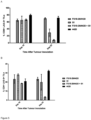
- the present inventors have shown that treatment of TILs with a specific binding member according to the present invention comprising a LAG-3 antigen-binding site located in a CH3 domain of the specific binding member, in combination with an anti-PD-L1 antibody results in reduced LAG-3 expression by the TILs.
- the reduction in LAG-3 expression is expected to reduce the inhibitory effect of LAG-3 and thereby allow the TILs to overcome exhaustion.
- the specific binding member of the invention may therefore be administered to a patient in combination with a second specific binding member which binds to PD-L1, such as an antibody molecule which binds to PD-L1.
- the present invention relates to a specific binding member of the invention, for use in a method of treating cancer in a patient, wherein the CDR-based antigen-binding site of the specific binding member binds to a molecule which is an immune system modulator, and wherein the method comprises administering the specific binding member of the invention, and a second specific binding member which binds to PD-L1 to the patient.
- the specific binding member of the invention and the specific binding member which binds to PD-L1 may be administered to the patient simultaneously, separately, or sequentially.
- the specific binding member of the invention may not comprise a CDR-based antigen binding site for a second antigen.
- the specific binding member of the invention may therefore only bind to LAG-3.
- the specific binding member which binds PD-L1 may be antibody molecule, or fragment thereof.
- Antibody molecules which bind PD-L1 are known in the art.
- the antibody molecule may be human or humanised.
- the antibody molecule is preferably a monoclonal antibody molecule.
- Examples of antibody molecules are the immunoglobulin isotypes, such as immunoglobulin G, and their isotypic subclasses, such as IgG1, IgG2, IgG3 and IgG4, as well as fragments thereof.
- the specific binding member which binds PD-L1 does not comprise a LAG-3 antigen-binding site.
- the present invention relates to specific binding members which bind to LAG-3.
- the specific binding members of the present invention comprise a LAG-3 antigen binding site located in a CH3 domain of the specific binding member, as set out in the claims.
- the term “LAG-3” may refer to human LAG-3, murine LAG-3, and/or cynomolgus monkey LAG-3, unless the context requires otherwise.
- the term “LAG-3” refers to human LAG-3.
- the term "specific binding member” describes an immunoglobulin, or fragment thereof, comprising a constant domain, preferably a CH3 domain, comprising a LAG-3 antigen-binding site.
- the specific binding member comprises a CH2 and CH3 domain, wherein the CH2 or CH3 domain, preferably the CH3 domain, comprises a LAG-3 antigen-binding site.
- the specific binding member further comprises an immunoglobulin hinge region, or part thereof, at the N-terminus of the CH2 domain.
- Such a molecule is also referred to as an antigen-binding Fc fragment, or Fcab TM , herein.
- the specific binding member may be partly or wholly synthetically produced.
- specific binding member thus includes fragments, provided said fragments comprise a LAG-3 antigen binding site located in a CH3 domain of the specific binding member. Unless the context requires otherwise, the term “specific binding member”, as used herein, is thus equivalent to "specific binding member or fragment thereof".
- the specific binding member is an antibody molecule or fragment thereof.
- the antibody molecule may be human or humanised.
- the antibody molecule is preferably a monoclonal antibody molecule.
- Examples of antibody molecules are the immunoglobulin isotypes, such as immunoglobulin G, and their isotypic subclasses, such as IgG1, IgG2, IgG3 and IgG4, as well as fragments thereof.
- An example of an antibody fragment comprising a CH3 domain is an Fc domain of an antibody.
- An example of an antibody fragment comprising both CDR sequences and CH3 domain is a minibody, which comprises an scFv joined to a CH3 domain ( Hu et al. (1996), Cancer Res., 56(13):3055-61 ).
- the specific binding member of the present invention binds to LAG-3. Binding in this context may refer to specific binding.
- the term “specific” may refer to the situation in which the specific binding member will not show any significant binding to molecules other than its specific binding partner(s), here LAG-3.
- the term “specific” is also applicable where the specific binding member is specific for particular epitopes, such as epitopes on LAG-3, that are carried by a number of antigens in which case the specific binding member will be able to bind to the various antigens carrying the epitope.
- LAG-3 shares 40% sequence identity with CD4, its most closely related protein.
- the present inventors tested the FS18-7-9 Fcab, which comprises the amino acid sequences set forth in SEQ ID NOs 1 to 3, for binding to CD4.
- the FS18-7-9 Fcab showed no binding to CD4, demonstrating that the specific binding member binds LAG-3 specifically.
- the LAG-3 binding site of a specific binding member of the present invention does not bind, or does not show any significant binding, to CD4.
- a specific binding member of the invention comprises a LAG-3 antigen binding site, as set out in the claims.
- the LAG-3 antigen binding site is located in a CH3 domain.
- the LAG-3 binding site comprises the amino acid sequences WDEPWGED (SEQ ID NO: 1) and PYDRWVWPDE (SEQ ID NO: 3). These sequences were present in all of the lead anti-LAG-3 Fcab clones identified by the present inventors following an extensive screening and characterisation program as described in the examples.
- amino acid sequences set forth in SEQ ID NOs 1 and 2 are located in structural loops of the constant domain of the specific binding member, as set out in the claims.
- the introduction of sequences into the structural loop regions of antibody constant domains to create new antigen-binding sites is described, for example, in WO2006/072620 and WO2009/132876 .
- the structural loops of antibody constant domains include the AB, CD and EF loops.
- the AB, CD, and EF loops are located at residues 11-18, 43-78 and 92-101 of the CH3 domain, where the amino acid residue numbering is according to the ImMunoGeneTics (IMGT) numbering scheme.
- the amino acid sequence set forth in SEQ ID NO: 1 is preferably located in the AB loop of the constant domain.
- the amino acid sequence set forth in SEQ ID NO: 3 is preferably located in the EF loop of the constant domain.
- amino acid sequence set forth in SEQ ID NO: 1 is located at residues 11 to 18 of the CH3 domain; and/or the amino acid sequence set forth in SEQ ID NO: 3 is located at residues 92 to 101 of the CH3 domain, wherein the amino acid residue numbering is according to the IMGT numbering scheme.
- the specific binding member preferably comprises the amino acid sequence set forth in SEQ ID NO: 2, 8, 13, 18, 23, 28, 33, 38, 43, or 48, more preferably SEQ ID NO: 2, 28, or 38, yet more preferably SEQ ID NO: 2, in a structural loop of a constant domain of the specific binding member.
- the structural loop is preferably the CD loop and the constant domain is preferably the CH3 domain.
- the amino acid sequence set forth in SEQ ID NO: 2, 8, 13, 18, 23, 28, 33, 38, 43, or 48 is preferably located at residues 43 to 78 of the CH3 domain, wherein the amino acid residue numbering is according to the IMGT numbering scheme.
- a specific binding member of the invention may further comprise a glutamic acid residue (E) at position 36 and/or a tyrosine residue (Y) at position 85.2 of the CH3 domain (as shown in Figure 1A ), wherein the amino acid residue numbering is according to the IMGT numbering scheme.
- a specific binding member which comprises the CD structural loop region set forth in SEQ ID NO: 8 preferably further comprises a glutamic acid residue (E) at position 36 of the CH3 domain.
- a specific binding member which comprises the CD structural loop region set forth in SEQ ID NO: 18 preferably further comprises a tyrosine residue (Y) at position 85.2 of the CH3 domain.
- the specific binding member of the invention comprises a CH3 domain which comprises, has, or consists of the sequence set forth in SEQ ID NO: 5, 10, 15, 20, 25, 30, 35, 40, 45, or 50, preferably a CH3 domain with the sequence set forth in SEQ ID NO: 5, 30, or 40, more preferably, a CH3 domain with the sequence set forth in SEQ ID NO: 5.
- the specific binding member of the invention may comprise a CH3 domain which comprises, has, or consists of the sequence set forth in SEQ ID NO: 5, 10, 15, 20, 25, 30, 35, 40, 45, or 50, wherein the CH3 domain sequence further comprises a lysine residue (K) at the immediate C-terminus of the sequence shown in SEQ ID NO: 5, 10, 15, 20, 25, 30, 35, 40, 45, or 50.
- the specific binding member of the invention may comprise a CH3 domain which comprises, has, or consists of the sequence set forth in SEQ ID NO: 5 with a lysine residue at the C-terminus of the sequence shown in SEQ ID NO: 5.
- the sequence of such a CH3 domain would then be as follows:
- the specific binding member of the invention may comprise a CH2 domain of an immunoglobulin G molecule, such as a CH2 domain of an IgG1, IgG2, IgG3, or IgG4 molecule.
- the specific binding member of the invention comprises a CH2 domain of an IgG1 molecule.
- the CH2 domain may have the sequence set forth in SEQ ID NO: 53.
- the CH2 domain of the specific binding member may comprise a mutation to reduce or abrogate binding of the CH2 domain to one or more Fc y receptors, such as Fc ⁇ RI, Fc ⁇ Rlla, Fc ⁇ RIIb, Fc ⁇ RIII and/or to complement.
- Fc y receptors such as Fc ⁇ RI, Fc ⁇ Rlla, Fc ⁇ RIIb, Fc ⁇ RIII and/or to complement.
- CH2 domains of human IgG domains normally bind to Fc y receptors and complement and the inventors postulate that reduced binding to Fc y receptors will reduce the antibody-dependent cell-mediated cytotoxicity (ADCC) and reduced binding to complement will reduce the complement-dependent cytotoxicity (CDC) activity of the specific binding member.
- ADCC antibody-dependent cell-mediated cytotoxicity
- CDC complement-dependent cytotoxicity
- the specific binding member may comprise a CH2 domain, wherein the CH2 domain comprises alanine residues at positions 4 and 5 of the CH2 domain, wherein the numbering is according to the IMGT numbering scheme.
- the specific binding member comprises an IgG1 CH2 domain which comprises, has, or consists of the sequence set forth in SEQ ID NO: 54.
- a specific binding member according to the present invention may comprise a second antigen-binding site, preferably a CDR-based antigen binding site.
- CDR-based antigen binding site refers to the antigen-binding site of a specific binding member variable region which is composed of six CDR residues.
- the second antigen-binding site is preferably specific for a tumour antigen. More preferably, the second antigen-binding site may bind to a molecule which is an immune system modulator, such as an immunomodulatory receptor or a ligand for an immunomodulatory receptor. For example, the second antigen-binding site may bind to a molecule which is an immune system inhibitor or activator, preferably an immune system inhibitor.
- immune system inhibitors include cytotoxic T-lymphocyte-associated protein 4 (CTLA-4), T cell immunoglobulin and mucin-domain containing-3 (TIM-3), and Colony stimulating factor 1 receptor (CSF1R).
- the antibody molecules against a given antigen such as a tumour antigen, and determination of the CDR sequences of such an antibody molecule, is well within the capabilities of the skilled person and many suitable techniques are known in the art. Furthermore, antibodies, including the CDR sequences, against various immune system modulators are known in the art. Thus, the skilled person would have no difficulty in preparing a specific binding member comprising in addition to a LAG-3 binding site as described herein a CDR-based antigen-binding site for a second antigen.
- the specific binding members of the present invention may also comprise variants of the structural loop, CH3 domain, CH2 domain, CH2 and CH3 domain, CDR, VH domain, VL domain, light chain or heavy chain sequences disclosed herein, provided these are within the scope of the claims. Suitable variants can be obtained by means of methods of sequence alteration, or mutation, and screening.
- a specific binding member comprising one or more variant sequences retains one or more of the functional characteristics of the parent specific binding member, such as binding specificity and/or binding affinity for LAG-3.
- a specific binding member comprising one or more variant sequences preferably binds to LAG-3 with the same affinity, or a higher affinity, than the (parent) specific binding member.
- the parent specific binding member is a specific binding member which does not comprise the amino acid substitution(s), deletion(s), and/or insertion(s) which have been incorporated into the variant specific binding member.
- a specific binding member of the invention may comprise a structural loop, CH3 domain, CH2 domain, CH2 and CH3 domain, CDR, VH domain, VL domain, light chain or heavy chain sequence which has at least 70%, at least 75%, at least 80%, at least 85%, at least 90%, at least 95%, at least 96%, at least 97%, at least 98%, at least 99%, at least 99.1%, at least 99.2%, at least 99.3%, at least 99.4%, at least 99.5%, at least 99.6%, at least 99.7%, at least 99.8%, or at least 99.9% sequence identity to a structural loop, CH3 domain, CH2 domain, CH2 and CH3 domain, CDR, VH domain, VL domain, light chain or heavy chain sequence disclosed herein, provided it is within the scope of the claims.
- the specific binding member of the invention comprises a CH3 domain sequence which has at least 97%, at least 98%, at least 99%, at least 99.1%, at least 99.2%, at least 99.3%, at least 99.4%, at least 99.5%, at least 99.6%, at least 99.7%, at least 99.8%, or at least 99.9% sequence identity to the CH3 domain sequence set forth in SEQ ID NO: 4, 5, or 98.
- the specific binding member of the invention comprises a CH3 and CH2 domain sequence, which has at least 97%, at least 98%, at least 99%, at least 99.1%, at least 99.2%, at least 99.3%, at least 99.4%, at least 99.5%, at least 99.6%, at least 99.7%, at least 99.8%, or at least 99.9% sequence identity to the CH2 and CH3 domain sequence set forth in SEQ ID NO: 6 or 7.
- GAP Garnier GAP (1990) J. Mol. Biol. 215: 405-410 ), FASTA (which uses the method of Pearson and Lipman (1988) PNAS USA 85: 2444-2448 ), or the Smith-Waterman algorithm ( Smith and Waterman (1981) J. Mol Biol.
- a specific binding member of the invention may also comprise a structural loop, CH3 domain, CH2 domain, CH2 and CH3 domain, CDR, VH domain, VL domain, light chain or heavy chain sequence which has one or more amino acid sequence alterations (addition, deletion, substitution and/or insertion of an amino acid residue), preferably 20 alterations or fewer, 15 alterations or fewer, 10 alterations or fewer, 5 alterations or fewer, 4 alterations or fewer, 3 alterations or fewer, 2 alterations or fewer, or 1 alteration compared with a structural loop, CH3 domain, CH2 domain, CH2 and CH3 domain, CDR, VH domain, VL domain, light chain or heavy chain sequence disclosed herein, provided it is within the scope of the claims.
- alterations may be made in one or more framework regions of the specific binding member.
- the specific binding member of the invention may comprise a CH3 domain sequence with one or more amino acid sequence alterations (addition, deletion, substitution and/or insertion of an amino acid residue), preferably 20 alterations or fewer, 15 alterations or fewer, 10 alterations or fewer, 5 alterations or fewer, 4 alterations or fewer, 3 alterations or fewer, 2 alterations or fewer, or 1 alteration compared with the CH3 domain sequence set forth in SEQ ID NO: 4, 5, or 98.
- the specific binding member of the invention comprises a CH3 and CH2 domain sequence, with one or more amino acid sequence alterations (addition, deletion, substitution and/or insertion of an amino acid residue), preferably 20 alterations or fewer, 15 alterations or fewer, 10 alterations or fewer, 5 alterations or fewer, 4 alterations or fewer, 3 alterations or fewer, 2 alterations or fewer, or 1 alteration compared with the CH2 and CH3 domain sequence set forth in SEQ ID NO: 6 or 7.
- the specific binding member of the invention preferably binds to LAG-3 with an affinity (K D ) of 1 ⁇ 10 -9 M or an affinity which is greater.
- the specific binding member of the invention may bind to LAG-3 with an affinity (K D ) of 8 ⁇ 10 -10 M, or an affinity which is greater.
- the binding affinity of a specific binding member to a cognate antigen, such as LAG-3 can be determined by surface plasmon resonance (SPR), for example.
- SPR surface plasmon resonance
- the binding affinity of a specific binding member to a cognate antigen, such as LAG-3, expressed on a cell surface can be determined by flow cytometry.
- Fcabs have a smaller binding interface than monoclonal antibodies as the binding sites of Fcabs form a relatively compact antibody fragment with two binding sites situated in close proximity.
- the Fab arms of a typical mAb are separated by a flexible hinge region.
- the two antigen binding sites of an Fcab are also spatially close to each other, as compared with those of a typical mAb. Based on this smaller binding interface and reduced flexibility of the two binding sites it was surprising that the anti-LAG-3 Fcabs were able to bind to and inhibit LAG-3 with similar affinity and potency as a monoclonal antibody benchmark.
- the specific binding member of the present invention is preferably capable of binding to LAG-3 expressed on the surface of a cell.
- the cell is preferably a cancer cell.
- the specific binding member comprises a second antigen-binding site, such as CDR-based antigen binding site, specific for a second antigen
- the specific binding member is preferably capable of simultaneously binding to LAG-3 and the second antigen.
- the specific binding member is capable of simultaneously binding to LAG-3 and the second antigen, wherein the LAG-3 and the second antigen are expressed on the surface of a single cell, or on the surface of two separate cells.
- the specific binding member of the invention may bind to human LAG-3, murine LAG-3, and/or cynomolgus monkey LAG-3. Preferably, the specific binding member of the invention binds to human LAG-3.
- the specific binding member of the invention is not a specific binding member, such as an antibody molecule, which comprises an antigen binding site, such as a CDR-based antigen-binding site, for PD-L1.
- the specific binding member of the invention is not a specific binding member, such as an antibody molecule, which comprises (i) a CDR-based antigen binding site for PD-L1; and (ii) a LAG-3 antigen binding site located in a CH3 domain of the specific binding member.
- the specific binding member of the invention is not a specific binding member, such as an antibody molecule, which binds to PD-L1 and LAG-3, wherein the antibody molecule comprises:
- the specific binding member of the present invention may be conjugated to a therapeutic agent or detectable label.
- the specific binding member may be referred to as a conjugate.
- the specific binding member may be conjugated to an immune system modulator, cytotoxic molecule, radioisotope, or detectable label.
- the immune system modulator or cytotoxic molecule may be a cytokine.
- the detectable label may be a radioisotope, e.g. a non-therapeutic radioisotope.
- the specific binding member may be conjugated to the therapeutic agent or detectable label, by means of a peptide bond or linker, i.e. within a fusion polypeptide comprising said therapeutic agent or detectable label and the specific binding member or a polypeptide chain component thereof.
- Other means for conjugation include chemical conjugation, especially cross-linking using a bifunctional reagent (e.g. employing DOUBLE-REAGENTS TM Cross-linking Reagents Selection Guide, Pierce).
- the specific binding member and the therapeutic agent or detectable label may thus be connected to each other directly, for example through any suitable chemical bond or through a linker, for example a peptide linker.
- the peptide linker may be a short (2-20, preferably 2-15, residue stretch of amino acids). Suitable examples of peptide linker sequences are known in the art. One or more different linkers may be used. The linker may be about 5 amino acids in length.
- the chemical bond may be, for example, a covalent or ionic bond.
- covalent bonds include peptide bonds (amide bonds) and disulphide bonds.
- the specific binding member and therapeutic or diagnostic agent may be covalently linked.
- amide bonds peptide bonds
- the specific binding member and therapeutic or diagnostic agent may be produced (secreted) as a single chain polypeptide.
- the invention also provides isolated nucleic acids encoding the antibodies molecules of the invention.
- An isolated nucleic acid may be used to express the specific binding member of the invention, for example, by expression in a bacterial, yeast, insect or mammalian host cell.
- a preferred host cell is a mammalian cell such as a CHO, HEK or NS0 cell.
- the nucleic acid will generally be provided in the form of a recombinant vector for expression.
- the isolated nucleic acid may, for example, comprise the sequence set forth in SEQ ID NO: 99, 4, 9, 14, 19, 24, 29, 34, 39, 44, or 49, which encode the CH3 domains of FS18-7-9 (CHO codon optimised nucleotide sequence), FS18-7-9 (HEK293-expressed nucleotide sequence), FS18-7-32, FS18-7-33, FS18-7-36, FS18-7-58, FS18-7-62, FS18-7-65, FS18-7-78, FS18-7-88, and FS18-7-95, respectively.
- FS18-7-9 CHO codon optimised nucleotide sequence
- FS18-7-9 HEK293-expressed nucleotide sequence
- In vitro host cells comprising such nucleic acids and vectors are part of the invention, as is their use for expressing the specific binding members of the invention, which may subsequently be purified from cell culture and optionally formulated into a pharmaceutical composition.
- the present invention thus further provides a method of producing the specific binding member of the invention, comprising culturing the recombinant host cell of the invention under conditions for production of the specific binding member. Methods for culturing suitable host cells as mentioned above are well-known in the art.
- the method may further comprise isolating and/or purifying the specific binding member.
- the method may also comprise formulating the specific binding member into a pharmaceutical composition, optionally with a pharmaceutically acceptable excipient or other substance as described below.
- LAG-3 is known to be expressed on cells of the immune system.
- LAG-3 is known to be expressed on exhausted T cells within the tumour environment, and a limited number of cancer cells.
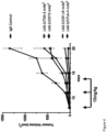
- the present inventors have shown that the use of a specific binding member which binds to LAG-3 is effective in suppressing tumour growth in syngeneic mouse models of cancer.
- a specific binding member of the invention may be used in a method of treating cancer in a patient.
- the patient is preferably a human patient. Methods of treatment do not form part of the present invention.
- Cells of the cancer to be treated using the specific binding member of the invention may express LAG-3, e.g. on their cell surface.
- cells of the cancer to be treated may have been determined to express LAG-3, e.g. on their cell surface.
- B cell lymphomas have been shown to express LAG-3 on their cell surface.
- tumours of the cancer to be treated using the specific binding members of the invention may comprise LAG-3 expressing immune cells.
- LAG-3 expressing immune cells such as LAG-3 expressing TILs, are present between tumour cells in many cancers.
- tumours of the cancer to be treated using the specific binding member of the invention have been determined to contain LAG-3 expressing immune cells. Methods for determining the presence of LAG-3 expressing immune cells in a tumour or in the periphery of the tumour are known in the art.
- a cancer to be treated using a specific binding member of the invention may be selected from the group consisting of Hodgkin's lymphoma, non-Hodgkin's lymphoma (such as diffuse large B-cell lymphoma, follicular lymphoma, indolent non-Hodgkin's lymphoma, mantle cell lymphoma), ovarian cancer, prostate cancer, colorectal cancer, fibrosarcoma, renal cell carcinoma, melanoma, pancreatic cancer, breast cancer, glioblastoma multiforme, lung cancer (such as non-small cell lung cancer), head and neck cancer (such as head and neck squamous cell carcinoma), stomach cancer (gastric cancer), bladder cancer, cervical cancer, uterine cancer, vulvar cancer, testicular cancer, penile cancer, leukemia (such as chronic lymphocytic leukemia, myeloid leukemia, acute lymphoblastoid leukaemia, or chronic lymphoblastoid leukaemia),
- lung cancer such as non-small cell lung cancer
- nasopharyngeal cancer colorectal cancer
- melanoma stomach cancer (gastric cancer)
- esophageal cancer such as adenocarcinoma of the gastroesophageal junction
- ovarian cancer cervical cancer, bladder cancer, head and neck cancer (such as head and neck squamous cell carcinoma)
- leukemia such as chronic lymphocytic leukemia, Hodgkin's lymphoma, non-Hodgkin's lymphoma (such as diffuse large B-cell lymphoma, indolent non-Hodgkin's lymphoma, mantle cell lymphoma), and multiple myeloma using anti-LAG-3 antibodies
- leukemia such as chronic lymphocytic leukemia
- Hodgkin's lymphoma non-Hodgkin's lymphoma
- non-Hodgkin's lymphoma such as diffuse large B-cell
- the cancer to be treated using the specific binding members of the present invention may be a renal cell carcinoma, lung cancer (such as non-small cell lung cancer), nasopharyngeal cancer, colorectal cancer, melanoma, stomach cancer (gastric cancer), esophageal cancer (such as adenocarcinoma of the gastroesophageal junction), ovarian cancer, cervical cancer, bladder cancer, head and neck cancer (such as head and neck squamous cell carcinoma), leukemia (such as chronic lymphocytic leukemia, Hodgkin's lymphoma, non-Hodgkin's lymphoma (such as diffuse large B-cell lymphoma, indolent non-Hodgkin's lymphoma, mantle cell lymphoma), or multiple myeloma.
- lung cancer such as non-small cell lung cancer
- nasopharyngeal cancer colorectal cancer
- melanoma stomach cancer
- stomach cancer gastric cancer
- Preferred cancers for treatment using the specific binding members of the present invention are lung cancer (such as non-small-cell lung cancer), bladder cancer, head and neck cancer (squamous cell carcinoma of the head and neck), diffuse large B cell lymphoma, gastric cancer, pancreatic cancer and hepatocellular carcinoma.
- Tumours of these cancers are known to comprise LAG-3 expressing immune cells and to express PD-L1 either on their cell surface or to comprise immune cells expressing PD-L1.
- cancer refers to a particular type of cancer, such as breast cancer
- a cancer which originates from malignant transformation of a different tissue, e.g. ovarian tissue, may result in metastatic lesions in another location in the body, such as the breast, but is not thereby a breast cancer as referred to herein but an ovarian cancer.
- the cancer may be a primary or secondary cancer.
- the specific binding member of the present invention may be for use in a method of treating cancer in a patient, wherein the CDR-based antigen-binding site of the specific binding member binds to a molecule which is an immune system modulator, and wherein the cancer is a primary tumour and/or a tumour metastasis.
- the specific binding members of the invention are designed to be used in methods of treatment of patients, preferably human patients.
- Specific binding members will usually be administered in the form of a pharmaceutical composition, which may comprise at least one component in addition to the specific binding member, such as a pharmaceutically acceptable excipient.
- a pharmaceutical composition of the present invention comprises, in addition to active ingredient, a pharmaceutically acceptable excipient.
- the specific binding member may be administered intravenously, or subcutaneously.
- Liquid pharmaceutical compositions generally comprise a liquid carrier such as water, petroleum, animal or vegetable oils, mineral oil or synthetic oil.
- a liquid carrier such as water, petroleum, animal or vegetable oils, mineral oil or synthetic oil.
- Physiological saline solution, dextrose or other saccharide solution or glycols such as ethylene glycol, propylene glycol or polyethylene glycol may be included.
- the specific binding member, or pharmaceutical composition comprising the specific binding member is preferably in the form of a parenterally acceptable aqueous solution which is pyrogen-free and has suitable pH, isotonicity and stability.
- a parenterally acceptable aqueous solution which is pyrogen-free and has suitable pH, isotonicity and stability.
- isotonic vehicles such as Sodium Chloride Injection, Ringer's Injection, Lactated Ringer's Injection.
- Preservatives, stabilizers, buffers, antioxidants and/or other additives may be employed, as required.
- Many methods for the preparation of pharmaceutical formulations are known to those skilled in the art. See e.g. Robinson ed., Sustained and Controlled Release Drug Delivery Systems, Marcel Dekker, Inc., New York, 1978 .
- a composition comprising a specific binding members according to the present invention may be administered alone or in combination with other treatments, concurrently or sequentially or as a combined preparation with another therapeutic agent or agents, dependent upon the condition to be treated.
- a specific binding member of the invention may be administered in combination with an existing therapeutic agent for the disease to be treated, e.g. a cancer as mentioned above.
- a specific binding member of the present invention may be administered to the patient in combination with a second anti-cancer therapy, such as chemotherapy, anti-tumour vaccination (also referred to as a cancer vaccination), radiotherapy, immunotherapy, an oncolytic virus, chimeric antigen receptor (CAR) T-cell therapy, or hormone therapy.
- a second anti-cancer therapy such as chemotherapy, anti-tumour vaccination (also referred to as a cancer vaccination), radiotherapy, immunotherapy, an oncolytic virus, chimeric antigen receptor (CAR) T-cell therapy, or hormone therapy.
- the specific binding member of the invention may act as an adjuvant in anti-cancer therapy, such as chemotherapy, anti-tumour vaccination, or radiotherapy.
- anti-cancer therapy such as chemotherapy, anti-tumour vaccination, or radiotherapy.
- administration of the specific binding member to the patient as part of chemotherapy, anti-tumour vaccination, or radiotherapy will trigger a greater immune response against the cancer associated antigen LAG-3, than is achieved with chemotherapy, anti-tumour vaccination, or radiotherapy alone.
- anti-LAG-3 therapies have shown good efficacy in treating viral based pathologies in mice ( Blackburn SD, et al., 2009, Nature Immunology 10 (1): 29-37 ).
- a method of treating cancer in a patient may thus comprise administering to the patient a therapeutically effective amount of a specific binding member according to the present invention in combination with a chemotherapeutic agent, anti-tumour vaccine, radionuclide, immunotherapeutic agent, oncolytic virus, CAR-T cell, or agent for hormone therapy.
- a chemotherapeutic agent, anti-tumour vaccine, radionuclide, immunotherapeutic agent, oncolytic virus, CAR-T cell, or agent for hormone therapy is preferably a chemotherapeutic agent, anti-tumour vaccine, radionuclide, immunotherapeutic agent, oncolytic virus, CAR-T cell, or agent for hormone therapy for the cancer in question, i.e.
- a chemotherapeutic agent, anti-tumour vaccine, radionuclide, immunotherapeutic agent, oncolytic virus, CAR-T cell, or agent for hormone therapy which has been shown to be effective in the treatment of the cancer in question.
- the selection of a suitable chemotherapeutic agent, anti-tumour vaccine, radionuclide, immunotherapeutic agent, oncolytic virus, CAR-T cell, or agent for hormone therapy which have been shown to be effective for the cancer in question is well within the capabilities of the skilled practitioner.
- the method comprises administering to the patient a therapeutically effective amount of a specific binding member according to the present invention in combination with a chemotherapeutic agent
- the chemotherapeutic agent may be selected from the group consisting of: taxanes, cyctotoxic antibiotics, tyrosine kinase inhibitors, PARP inhibitors, B_RAF enzyme inhibitors, alkylating agents, platinum analogs, nucleoside analogs, thalidomide derivatives, antineoplastic chemotherapeutic agents and others.
- Taxanes include docetaxel, paclitaxel and nab-paclitaxel; cytotoxic antibiotics include actinomycin, bleomycin, anthracyclines, doxorubicin and valrubicin; tyrosine kinase inhibitors include erlotinib, gefitinib, axitinib, PLX3397, imatinib, cobemitinib and trametinib; PARP inhibitors include piraparib; B-Raf enzyme inhibitors include vemurafenib and dabrafenib; alkylating agents include dacarbazine, cyclophosphamide, temozolomide; platinum analogs include carboplatin, cisplatin and oxaliplatin; nucleoside analogs include gemcitabine and azacitidine; antineoplastics include fludarabine.
- chemotherapeutic agents suitable for use in the present invention include methotrexate, defactinib, entinostat, pemetrexed, capecitabine, eribulin, irinotecan, fluorouracil, and vinblastine.
- Vaccination strategies for the treatment of cancers has been both implemented in the clinic and discussed in detail within scientific literature (such as Rosenberg, S. 2000 Development of Cancer Vaccines ). This mainly involves strategies to prompt the immune system to respond to various cellular markers expressed by autologous or allogenic cancer cells by using those cells as a vaccination method, both with or without granulocyte-macrophage colony-stimulating factor (GM-CSF). GM-CSF provokes a strong response in antigen presentation and works particularly well when employed with said strategies.
- GM-CSF granulocyte-macrophage colony-stimulating factor
- Administration may be in a "therapeutically effective amount", this being sufficient to show benefit to a patient. Such benefit may be at least amelioration of at least one symptom.
- treatment of a specified disease refers to amelioration of at least one symptom.
- the actual amount administered, and rate and time-course of administration will depend on the nature and severity of what is being treated, the particular patient being treated, the clinical condition of the individual patient, the cause of the disorder, the site of delivery of the composition, the type of specific binding member, the method of administration, the scheduling of administration and other factors known to medical practitioners. Prescription of treatment, e.g. decisions on dosage etc., is within the responsibility of general practitioners and other medical doctors, and may depend on the severity of the symptoms and/or progression of a disease being treated.
- Treatments may be repeated at daily, twice-weekly, weekly or monthly intervals, at the discretion of the physician. Treatment may be given before, and/or after surgery, and may be administered or applied directly at the anatomical site of surgical treatment.
- Naive phage libraries displaying the CH3 domain of human IgG1 (IMGT numbering 1.4-130) with randomisation within the AB (residues 14-18) and EF (residues 92-101) loops were used for selection with recombinant Fc-tagged human LAG-3 (LAG-3 Fc) antigen (R&D systems, 2319-L3-050).
- the libraries were selected in three rounds using antigen captured on Protein A (Life Technologies, 10002D) or Protein G (Life Technologies, 10004D) beads.
- the outputs were screened by ELISA and positive binders sub-cloned and expressed as soluble Fcabs (containing a truncated hinge) in Pichia pastoris using EasySelect Pichia Expression Kit (Life Technologies, K1740-01). The Fcabs were then screened for binding to recombinant human LAG-3 Fc on the Biacore 3000 (GE Healthcare). Briefly, LAG-3 Fc (R&D systems, 2319-L3-050) was coupled at a density of 7200 RU to a CM5 chip (GE Healthcare, BR-100012) using amine coupling (GE Healthcare, BR-1000-50).
- Fcabs were diluted in HBS-P (GE Healthcare, BR100368) buffer and injected at 250 nM, 500 nM and 1000 nM for 3 min and then allowed to dissociate in buffer for 5 min.
- Reference subtracted data (LAG-3 Fc flow cell 2 -blank flow cell) was analyzed using BIAevaluation 3.2 software to identify binding. Fcabs were then tested for binding to HEK cell-expressed human LAG-3 (LAG-3 cloned into pcDNA5FRT vector [Life Technologies, V6010-20] [See section 1.4.5 for methodology]).
- HEK 293 cells overexpressing human LAG-3 grown in DMEM (Life Technologies, 61965-026) containing 10% FBS (Life Technologies, 10270-1-6), 100 ⁇ g/ml Hygromycin B (Melford Laboratories Ltd, Z2475), 15 ⁇ g/ml Blasticidin (Melford Laboratories Ltd, B1105) and 1 ⁇ g/ml Doxycyclin (Sigma, D9891) were detached from tissue culture flasks using cell dissociation buffer (Life Technologies, 13151-014) and seeded in V-bottom 96-well plates at 2 ⁇ 10 5 cells/well. Fcabs were incubated with the cells at 5 ⁇ M in a 100 ⁇ l volume for 1 h at 4 °C.
- the plates were washed the secondary antibody (Anti-human Fc-488, Jackson ImmunoResearch, 109-546-098) was diluted 1:1000 in PBS and 100 ⁇ l was added to the cells and incubated for 30 min at 4 °C. The plates were washed and the cells were resuspended in 100 ⁇ l PBS containing 1 ⁇ g/ml DAPI (Biotium, 40043). The plate was read on a BD FACSCanto II cytometer (BD Biosciences) and the data analysed using FlowJoX. The Fcabs were then expressed in mammalian cells by transformation using lipofectamine (Life Technologies, 11668-019) into Flp-In T-Rex 293 cells (Life Technologies, R780-07).
- lipofectamine Life Technologies, 11668-019
- Flp-In T-Rex 293 cells Life Technologies, R780-07
- LAG-3 binding Fcabs were tested for inhibition of binding of human MHC class II on A375 cells (ATCC, CRL-1619) to recombinant LAG-3 Fc (using the methodology in example 1.6).
- 54 unique Fcab sequences were identified from three rounds of phage selection, and 12 of these Fcabs were determined to bind to LAG-3 Fc by BIAcore analysis and/or bind to LAG-3 expressing HEK cells.
- Three of the selected Fcabs were also able to inhibit the interaction of LAG-3 with MHC class II and were selected for affinity maturation. The three Fcabs were termed FS18-3, FS18-7 and FS18-21.
- phage display affinity maturation libraries were constructed by randomising five residues in the AB loop (residues 14-18) and either five (residues 92-94 and 97-98) or eight (residues 92-94 and 97-101) residues in the EF loop of each of the three Fcabs identified using the naive selection process described above.
- the affinity maturation libraries were selected using recombinant human LAG-3 Fc (R&D systems, 2319-L3-050) and HEK cells expressing human LAG-3 (as described above).
- the outputs were screened by phage ELISA, the positive binders were subcloned and expressed as soluble Fcabs (containing a truncated hinge) in HEK Expi293 cells (Fcabs cloned into pTT5 vector [National Research Council of Canada] transfected using ExpiFectamine 293 Transfection kit [Life Technologies, A14524] into Expi293F cells [Life technologies, A14527]).
- the HEK expressed soluble Fcabs were then screened for binding to cell expressed human LAG-3, binding to cell expressed cynomolgus LAG-3 (methodology as example 1.4.3), and the ability to block MHC class II binding to recombinant LAG-3 Fc (methodology as in example 1.6).
- the blocking Fcabs were further tested to determine whether they were able to reverse LAG-3 induced inhibition of IL-2 secretion in a T cell activation assay (methodology as in example 2.1).
- 61 unique anti-LAG-3 Fcabs were identified from the six affinity maturation libraries using these screening methods. Affinity matured Fcabs from the FS18-7 lineage were shown to have the highest level of cross-reactivity with cynomolgus monkey LAG-3.
- the three Fcabs from this lineage with the strongest binding to cynomolgus monkey LAG-3 Fc and the highest activity in the T cell activation assay were selected for further affinity maturation. These three Fcabs were also shown to block the interaction of LAG-3 Fc with cell expressed MHC class II.
- a pool of the three Fcabs (FS18-7-7, FS18-7-9, and FS18-7-11) from the first affinity maturation was used to create further affinity maturation libraries.
- the CD loop was hard randomized using randomized primers from ELLA Biotech.
- a portion of amino acid positions in the CD loop (residues 45.1-78) was randomized using an equimolar distribution of amino acids excluding cysteine. Error prone PCR was also carried out across the entire CH3 domain sequence to introduce additional mutations that might enhance binding.
- the affinity maturation libraries were generated in phage and selections performed against biotinylated recombinant LAG-3 avi-Fc (BPS Bioscience, 71147) and HEK hLAG-3 cells and screened for binding to recombinant LAG-3 Fc (R&D systems, 2319-L3-050) by phage ELISA.
- 86 unique Fcabs (containing a truncated hinge) were expressed in HEK293F cells. Selected Fcabs were also screened for activity in a T cell activation assay as described above.
- the nine Fcabs identified during the second affinity maturation with the highest activity in the T cell activation assay (FS18-7-32; FS18-7-33; FS18-7-36; FS18-7-58; FS18-7-62; FS18-7-65; FS18-7-78; FS18-7-88; and FS18-7-95), as well as the parental Fcab clone, FS18-7-9, were then further characterised as described below.
- a sequence alignment of these nine Fcabs against the parental Fcab clone, FS18-7-9 is shown in Figure 1A .
- Figure 1B details the percentage sequence identity of each of the nine Fcab clones to the parental Fcab clone, FS18-7-9.
- Fcabs originating from affinity maturation of the two other parental Fcab clones, FS18-7-7 and FS18-7-11 were not as promising candidates as those originating from affinity maturation of FS18-7-9 and were therefore not pursued further.
- Fcab FS18-7 which was selected using the naive selection protocol described above, was used to generate phage libraries to select against mouse LAG-3. Two rounds of affinity maturation were performed, and Fcab clones FS18-7-108-29 and FS18-7-108-35, which showed high-affinity, specific binding to mouse LAG-3 were selected following affinity maturation. The ability of FS18-7-108-29 and FS18-7-108-35 to inhibit mouse LAG-3 in a T cell activation assay was confirmed. Epitope mapping using the Octet (Forteo Bio) showed that the anti-mouse LAG-3 Fcabs compete with the anti-human LAG-3 Fcabs (selected following the second affinity maturation as described above) for binding to human LAG-3.
- “mock” mAb 2 comprising the lead anti-human LAG-3 and anti-mouse LAG-3 Fcabs identified in 1.1 and 1.2 above were prepared in order to allow the characterisation of these Fcabs in mAb 2 format.
- These mock mAb 2 were prepared from the anti-LAG-3 Fcabs and the variable regions of anti-FITC antibody 4420 (see SEQ ID NO: 83, SEQ ID NO: 84, and SEQ ID NO: 85 for details) (Bedzyk, W. D., et al. 1989 and Bedzyk, W. D., et al. 1990).
- the mock mAb 2 were prepared both with (SEQ ID NO: 63, 65, 67, 69, 71, 73, 75, 77, 79, and 81) and without (SEQ ID NO: 64, 66, 68, 70, 72, 74, 76, 78, 80, and 82) the LALA mutation in the CH2 domain of the heavy chain (see section 1.5 below for details) and further comprised the light chain of the anti-FITC mAb 4420 (SEQ ID NO: 85).
- the mock mAb 2 were produced by transient expression in HEK293-6E cells and purified using mAb Select SuRe protein A columns.
- a BIAcore T200 (GE Healthcare) was used to measure the affinity of the anti-human LAG-3 Fcabs in the mock mAb 2 format for human LAG-3.
- Flow cell 4 of a CM5 sensor chip (GE Healthcare, BR1005-30) was immobilised with human LAG-3-Fc (R&D Systems, 2319-L3-050), and flow cell 3 was immobilised with buffer for reference using the amine coupling kit (GE Healthcare, BR-1000-50).
- LAG-3-Fc was diluted to 5 ⁇ g/ml in sodium acetate pH5 (ForteoBio, 18-1069) and injected at a flow rate of 10 ⁇ l/min for 12 seconds followed by deactivation of the surface by injection of ethanolamine for 420 sec.
- the Immobilisation level was 158 RU.
- the mock mAb 2 (or control anti-human LAG-3 mAb, 25F7) were diluted in HBS-P buffer (GE Healthcare, BR-1003-68) in a 2-fold dilution series from 4 ⁇ g/ml.
- the control mAb/mock mAb 2 were injected with an association time of 240 seconds at 30 ⁇ l/min, and a dissociation time 300 seconds at 30 ⁇ l/min.
- the surface was regenerated using 25mM NaOH for 30 seconds at 100 ⁇ l/min.
- the data was double reference subtracted and analysed using the BIAevaluation 3.2 software to calculate kinetic constants.
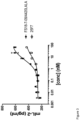
- the Fcabs in mock mAb 2 format had affinities for human LAG-3 in the range of 0.8 - 1.1 nM ( Table 1 ), which is similar to the affinity of the benchmark anti-human LAG-3 mAb 25F7.
- Table 1 The affinity of the benchmark anti-human LAG-3 mAb 25F7.
- Fcabs have a smaller binding interface than monoclonal antibodies as the binding sites of Fcabs form a relatively compact antibody fragment with two binding sites situated in close proximity.
- the Fab arms of a typical mAb are separated by a flexible hinge region. Based on this smaller binding interface and the associated reduced flexibility of the two binding sites in the Fc region, it was unexpected that the anti-LAG-3 Fcabs were able to bind to and inhibit LAG-3 with similar affinity and potency as the benchmark antibody 25F7.
- Table 1 Binding affinity of LAG-3 specific Fcabs in mock mAb 2 format to human LAG-3 Anti-human LAG-3 Fcab in mock mAb 2 format and benchmark anti-human LAG-3 mAb, 25F7 K D (M) FS18-7-9 8.3 ⁇ 10 -10 FS 18-7-62 9.5 ⁇ 10 -10 FS 18-7-78 8.4 ⁇ 10 -10 FS18-7-32 8.6 ⁇ 10 -10 FS 18-7-36 8.9 ⁇ 10 -10 FS18-7-65 1.1 ⁇ 10 -9 25F7 3.2 ⁇ 10 -10
- a Biacore 3000 (GE Healthcare) was used to measure the affinity of the surrogate Fcabs specific for mouse LAG-3 to mouse LAG-3.
- Amine coupling (amine coupling kit, GE Healthcare, BR-1000-50) was used to coat mLAG-3 Fc (R&D Systems, 3328-L3-050) diluted in 10 mM sodium acetate pH 5.0 (ForteBio, 18-1069) directly to a CM5 chip (GE Healthcare, BR-1000-12).
- Flow cell 1 was coated with Mouse Fc (SinoBiological, 51094-MNAH), and flow cell 2 was coated with mLAG-3 Fc at 950 RU.
- Fcabs were diluted in HBS-P buffer (GE Healthcare, BR-1003-68) and injected at various concentrations (fourfold dilutions from 100 nM) for 3 min at 20 ⁇ l/min and then allowed to dissociate in buffer for 12 min.
- the chip was regenerated by injection of 10 mM glycine pH 2.5 for 30 s at 30 ⁇ l/min. Data was double reference subtracted and analyzed using BIAevaluation 3.2 software to calculate kinetic constants.
- the tested surrogate Fcabs bound to mouse LAG-3 with single digit nanomolar affinity as set out in Table 2.
- Lentiviral transduction methodology was used to generate DO11.10 cells (National Jewish Health) over-expressing human, cynomolgus or mouse LAG-3 using the Lenti-X HTX Packaging System (Clontech, Cat. No 631249).
- Lenti-X expression vector (pLVX) (Clontech, Cat. No 631253), containing the mouse LAG-3 cDNA (SEQ ID NO: 96), human LAG-3 cDNA (SEQ ID NO: 95) or cynomolgus LAG-3 cDNA (SEQ ID NO: 97), was co-transfected with a Lenti-X HTX Packaging Mix into the Lenti-X 293T Cell Line (Clontech, Cat. No 632180) to generate virus.
- the DO11.10 cell line was transduced using the lentiviral vectors produced with the Lenti-X HTX Packaging System.
- the affinity of the anti-human LAG-3 Fcabs in mock mAb 2 format to cells expressing human LAG-3 (DO11.10 cell line transfected with human LAG-3) was measured using flow cytometry.
- mAb 2 and control mAb dilutions (2 ⁇ final concentration) were prepared in triplicate in 1 ⁇ DPBS (Gibco, 14190-094).
- D011.10:LAG-3 cell suspensions were prepared in PBS+2%BSA (Sigma, A7906) and seeded at 4 ⁇ 10 -6 cell/ml with 50 ⁇ l/well in V-bottomed 96-well plates (Costar, 3897).
- mAb 2 or control mAb anti human LAG-3 mAb, 25F7 dilutions were added to the wells containing cells (final volume 100 ⁇ l) and incubated at 4°C for 1 hour.
- the plates were washed and 100 ⁇ l/well of secondary antibody (anti-human Fc-488 antibody, Jackson ImmunoResearch, 109-546-098) diluted 1:1000 in PBS+2% BSA was then added and incubated for 30 mins at 4°C in the dark.
- the plates were washed and resuspended in 100 ⁇ l of PBS containing DAPI (Biotium, 40043) at 1 mg/ml. The plates were read using Canto II flow cytometer (BD Bioscience).
- Dead cells were excluded and the fluorescence in the FITC channel (488nm/530/30) was measured.
- the data was fitted using log (agonist) vs response in GraphPad Prism Software. All tested Fcabs in mock mAb 2 format and the benchmark anti-human LAG-3 mAb, 25F7, bound human LAG-3 with similar affinity (EC 50 ), in the range of 1.2 - 2.1 nM as set out in Table 3.
- Table 3 Binding affinity of anti-human LAG-3 Fcabs in mock mAb 2 format to DO11.10 cells expressing human LAG-3 as determined by flow cytometry Anti-human LAG-3 Fcab in mock mAb 2 format and benchmark anti-human LAG-3 mAb, 25F7 EC 50 (nM) FS 18-7-9 1.2 FS 18-7-32 1.6 FS 18-7-33 1.5 FS 18-7-36 1.5 FS 18-7-62 2.1 FS 18-7-65 1.6 FS 18-7-78 1.7 25F7 2.1
- the affinity of the anti-human LAG-3 Fcabs in mock mAb 2 format to cells expressing cynomolgus LAG-3 was measured using flow cytometry.
- mAb 2 and control mAb dilutions (2 ⁇ final concentration) were prepared in triplicate in 1 ⁇ DPBS (Gibco, 14190-094).
- DO11.10:LAG-3 cell suspensions were prepared in PBS+2%BSA (Sigma, A7906) and seeded at 4 ⁇ 10 -6 cell/ml with 50 ⁇ l/well in V-bottomed 96-well plates (Costar, 3897).
- mAb 2 or control mAb anti human LAG-3 mAb, 25F7 dilutions were added to the wells containing cells (final volume 100 ⁇ l) and incubated at 4°C for 1 hour.
- the plates were washed and 100 ⁇ l/well of secondary antibody (anti-human Fc-488 antibody, Jackson ImmunoResearch, 109-546-098) diluted 1:1000 in PBS+2% BSA was then added and incubated for 30 mins at 4°C in the dark.
- the plates were washed and resuspended in 100 ⁇ l of PBS containing DAPI (Biotium, 40043) at 1 mg/ml. The plates were read using Canto II flow cytometer (BD Bioscience).
- the dead cells were excluded and the fluorescence in the FITC channel (488nm/530/30) was measured.
- the data was fit using log (agonist) vs response in GraphPad Prism Software.
- the tested Fcabs in mock mAb 2 format bound to cynomolgus LAG-3 with 0.5-0.6 nM affinity indicating that toxicology studies in cynomolgus monkeys would be expected to be predictive of effects seen in humans (see Table 4 ).
- Table 4 Binding affinity of anti-LAG-3 Fcabs to DO11.10 cells expressing cynomolgus LAG-3 by flow cytometry Anti-human LAG-3 Fcab in mock mAb 2 format and benchmark anti-human LAG-3 mAb, 25F7 EC 50 (nM) FS18-7-9 0.6 FS18-7-62 0.5 FS 18-7-78 0.5 25F7 9.0
- the mouse LAG-3 sequence (SEQ ID NO: 96) was subcloned into pcDNA5FRT vector (Life Technologies, V6010-20) using Kpnl (NEB, R0142) and NotI (NEB, R0146) restriction digestion. The vector was then transformed into Flp-In T-REx 293 HEK cell line (Life Technologies, R780-07) using Lipofectamine 2000 (Life Technologies, 11668-019).
- Transformed Flp-In T-REx 293 cells were grown in DMEM (Life Technologies, 61965-026) containing 10% FBS (Life Technologies, 10270-1-6), 100 ⁇ g/ml Hygromycin B (Melford Laboratories Ltd, Z2475), 15 ⁇ g/ml Blasticidin (Melford Laboratories Ltd, B1105) for 3-4 weeks until colonies of stably transformed cells were apparent. These colonies were amplified in the presence of 1 ⁇ g/ml Doxycyclin (Sigma, D9891) and tested for mouse LAG-3 expression using PE conjugated anti-mouse LAG-3 (clone C9B7W, BD Biosciences, 552380).
- HEK cells expressing mLAG-3 grown in DMEM (Life Technologies, 61965-026) containing 10% FBS (Life Technologies, 10270-1-6), 100 ⁇ g/ml Hygromycin B (Melford Laboratories Ltd, Z2475), 15 ⁇ g/ml Blasticidin (Melford Laboratories Ltd, B1105) and 1 ⁇ g/ml Doxycyclin (Sigma, D9891) were detached from tissue culture flasks using cell dissociation buffer (Life Technologies, 13151-014) and seeded in V-bottom 96-well plates (Costar, 3897) at 2 ⁇ 10 5 cells/well.
- the plates were centrifuged at 1500 rpm for 3 min at 4°C to pellet the cells.
- a dilution series of the Fcabs (or control mAb) were incubated with the cells in a 100 ⁇ l volume for 1 h at 4 °C.
- the plates were washed and secondary antibody (Anti-human Fc-488, Jackson ImmunoResearch, 109-546-098 for Fcabs or Anti-Rat IgG (H+L), Alexa Fluor 488 Conjugate, ThermoFisher, A-11006 for C9B7W) was diluted 1:1000 in PBS and 100 ⁇ l was added to the cells for 30 min at 4 °C (plates were kept in the dark).
- the plates were then washed and the cells resuspended in 100 ⁇ l PBS containing 1 ⁇ g/ml DAPI (Biotium, 40043). The plates were read using Canto II flow cytometer (BD Bioscience). Dead cells were excluded and the fluorescence in the FITC channel (488nm/530/30) was measured. The data was fit using log (agonist) vs response in GraphPad Prism Software.
- the tested Fcabs bound to mouse LAG-3 with similar affinity (see Table 5 ).
- the benchmark LAG-3 mAb, C9B7W (2B Scientific, BE0174-50MG) binds mouse LAG-3 with 17-fold poorer affinity (EC 50 ) than the Fcabs ( Table 5 ).
- Table 5 Binding affinity of surrogate anti-mouse LAG-3 Fcabs to HEK cells expressing mouse LAG-3 by flow cytometry Anti-mouse LAG-3 Fcabs and benchmark anti-mouse LAG-3 mAb, C9B7W EC 50 (nM) FS18-7-108-29 4.5 FS18-7-108-35 4.5 C9B7W 79
- HEK cells expressing mLAG-3 grown in DMEM (Life Technologies, 61965-026) containing 10% FBS (Life Technologies, 10270-1-6), 100 ⁇ g/ml Hygromycin B (Melford Laboratories Ltd, Z2475), 15 ⁇ g/ml Blasticidin (Melford Laboratories Ltd, B1105) and 1 ⁇ g/ml Doxycyclin (Sigma, D9891) were detached from tissue culture flasks using cell dissociation buffer (Life Technologies, 13151-014).
- the plates were then washed and the cells resuspended in 60 ⁇ l PBS containing 1 ⁇ g/ml DAPI (Biotium, 40043). The plates were read using Canto II flow cytometer (BD Bioscience). Dead cells were excluded and the fluorescence in the FITC channel (488nm/530/30) was measured. The data was fitted using log (agonist) vs response in GraphPad Prism Software.
- the human mAb, 25F7 does not show any detectable binding to mouse LAG-3, indicating that the human LAG-3 Fcab, FS18-7-9, has a different binding epitope on LAG-3 than that of 25F7.
- the introduction of the LALA mutation in the CH2 domain of human IgG1 is known to reduce Fc y receptor binding (Bruhns, P., et al. (2009) and Xu, D. et al. (2000)).
- BIAcore was used to confirm that the LALA mutation had reduced the binding affinity of the Fcabs (in mock mAb 2 format) to Fcy receptors.
- the human FcyR binding assay was performed on a Biacore T200 instrument (GE Healthcare) using the Fcabs in the mock mAb 2 format.
- Human FcyRs (R&D Systems, 1257-FC, 1330-CD, 1875-CD, 4325-FC) were immobilized using amine coupling (amine coupling kit, GE Healthcare, BR-1000-50) onto a Series S CM5 chip (GE Healthcare, BR-1005-30) to a surface density of 370 RU for Fc ⁇ RI, 264 RU for Fc ⁇ RIII (high affinity human FcyRs) and 500 RU for Fc ⁇ Rlla and Fc ⁇ RIIb (low affinity human FcyRs). For each immobilized chip a flow cell was left blank for background subtraction.
- amine coupling amine coupling kit, GE Healthcare, BR-1000-50
- Series S CM5 chip GE Healthcare, BR-1005-30
- FcyR were immobilized using a concentration of 5 ⁇ g/ml in sodium acetate pH5 (ForteBio, 18-1069) and injected at a flow rate of 10 ⁇ l/min in 15 second cycles until the required immobilization level was reached.
- the positive control was a wild type IgG1 isotype mAb, which was compared to controls LALA IgG1 mAb and monoclonal IgG2 and IgG4 isotype mAbs to irrelevant targets.
- the flow cells were regenerated by injecting 10 mM sodium hydroxide (VWR, 28244.262) at a flow rate of 100 ⁇ l/min for 30 seconds.
- the data analysis was performed with BiaEvaluation software version 3.2 RC1 by double referencing against the blank flow cell (without immobilized FcyR) and subtracting a buffer cycle from test mAb 2 . The results are shown in Table 7.
- Table 7 Binding response of anti-human LAG-3 Fcabs in mock mAb 2 format (comprising LALA mutation as detailed above) to human Fcy receptors by SPR mAb/mock mAb 2 Binding response at end of association (RU) Fc ⁇ RI Fc ⁇ RIII Fc ⁇ RIIa Fc ⁇ RIIb FS18-7-9 1.4 6.6 -9.8 -8.5 FS18-7-62 -0.9 0.7 -10 -8.5 FS18-7-78 -0.3 4.0 -10.7 -9.2 mock mAb LALA 2 8.0 -12.7 -9.6 IgG2 0 1.9 9.7 7.4 IgG4 9 3.1 4.3 15.1 mock mAb IgG1 26 44 13 17.7
- Fcabs containing the truncated hinge; SEQ ID NO: 58
- SEQ ID NO: 58 The ability of the Fcabs (containing the truncated hinge; SEQ ID NO: 58) to block the interaction between recombinant human or mouse LAG-3 Fc and human MHC Class II was studied by measuring binding of LAG-3 Fc to A375 cells, a melanoma cell line that expresses human MHC Class II.
- A375 ATCC, CRL-1619 cells grown in DMEM (Life Technologies, 61965-026) containing 10% FBS (Life Technologies, 10270-106) were detached from cell culture flasks using cell dissociation buffer (Life Technologies, 13151-014) and seeded in V-bottom 96-well plates (Costar, 3897) at 2 ⁇ 10 5 cells/well.
- the plates were centrifuged at 1500 rpm for 3 min at 4 °C to pellet the cells.
- the relevant concentrations of Fcab or control mAb were incubated with 1 ⁇ g/ml LAG-3 Fc (human LAG-3-Fc R&D Systems, 2319-L3-050 or mouse LAG-3 Fc R&D Systems, 3328-L3-050) in 100 ⁇ l DMEM containing 10% FBS for 1 h at 4 °C.
- LAG-3/Fcab mix was added to the A375 cells and incubated for 1 h at 4 °C. Cells were washed.
- the anti-human LAG-3 Fcabs tested were also able to inhibit the interaction of human MHC class II with human LAG-3 with a similar potency as the control anti-human LAG-3 mAb (25F7).
- Table 9 Anti-human LAG-3 Fcabs inhibit binding of human LAG-3 to MHC class II
- the panel of lead Fcabs (with a truncated hinge; SEQ ID NO: 58) containing the LALA mutation were tested in a DO11.10 based T cell activation assay.
- Dilutions of Fcabs or benchmark mAb were prepared in 200 ⁇ l experimental media.
- Fcabs were mixed 1:1 (170 ⁇ l + 170 ⁇ l) with 4 ⁇ 10 5 /ml DO11.10 LAG-3 cells in experimental media and incubated at 37° C, 5% CO 2 for 1 hour.
- 2 ⁇ 10 5 A20 cells/ml experimental media were incubated with 1 ⁇ M OVA peptide for 30 min.
- 360 ⁇ l of the A20 cells + OVA mixture were added to 360 ⁇ l of the DO11.10 LAG-3 cell/Fcab mix.
- the cells were then mixed in a deep well plate and cultured in a 96-round bottom plate with 200 ⁇ l of the mix/well.
- the assay was run in triplicate.
- the human lead Fcabs show significant activity in the T cell activation assay with potencies in the range of 1-2 nM.
- the Fcabs have slightly improved potency than the benchmark anti human-LAG-3 mAb 25F7.
- Improved potency in the T cell activation assay is expected to be predictive of improved efficacy in human patients through enhanced inhibition of LAG-3.
- the surrogate anti-mouse LAG-3 Fcabs (with a truncated hinge; SEQ ID NO: 58) containing the LALA mutation were tested in a DO11.10 based T cell activation assay.
- Fcabs or benchmark mAb Dilutions of Fcabs or benchmark mAb were prepared in 200 ⁇ l experimental media.
- Fcabs were mixed 1:1 (170 ⁇ l + 170 ⁇ l) with 4 ⁇ 10 5 /ml DO11.10 LAG-3 cells in experimental media and incubated at 37°C, 5% CO2 for 1 hour.
- 2 ⁇ 10 5 A20 cells/ml experimental media were incubated with 1 ⁇ M OVA peptide for 30 min.
- 360 ⁇ l of the A20 cells + OVA mixture were added to 360 ⁇ l of the DO11.10 LAG-3 cell/Fcab mix.
- the cells were then mixed in a deep well plate and cultured in a 96-round bottom plate with 200 ⁇ l of the mix/well. The screen was assayed in triplicates.
- Table 11 EC 50 values and maximal IL-2 release of surrogate anti-LAG-3 Fcabs in DO11.10 T cell activation assay Surrogate Fcabs specific for mouse LAG-3 (with LALA mutation) and benchmark anti-mouse LAG-3 mAb, C9B7W EC 50 (nM) Maximal IL-2 release (pg/ml) FS18-7-108-29 1.9 205 FS18-7-108-35 1.8 176 C9B7W 5.1 48
- the mouse surrogate anti-mouse LAG-3 Fcabs showed significant activity in the T cell activation assay with potencies in the range of 2 nM.
- the surrogate anti-mouse LAG-3 Fcabs had higher potency than the benchmark anti mouse-LAG-3 antibody as is evident from the improved EC 50 and the 4-fold higher maximal activation of IL-2 release.
- Improved potency and maximal activation of these Fcabs compared to the benchmark is expected to result in improved activity in murine efficacy studies compared to the benchmark through improved inhibition of LAG-3.
- Dilutions of the FS18-7-9 Fcab in mock mAb 2 format (FS18-7-9/4420LALA) or benchmark mAb were prepared in experimental media.
- DO11.10 cells 0.3 ⁇ 10 6 cells/ml
- Antibodies and cells were incubated at 37° C, 5% CO 2 for 1 hour.
- LK 35.2 cells were incubated at 3 ⁇ 10 5 cells/ml experimental media with the OVA peptide at 1.5 ⁇ M for 30 min.
- 70 ⁇ l of LK 35.2 cells + OVA were added to 140 ⁇ l of the DO11.10/antibody. Cells were incubated at 37°C, 5% CO 2 for 24 hours.
- mice IL-2 ELISA kit (eBioscience, 88-7024-88 or R&D systems, SM2000) following the manufacturer's instructions. Plates were read at 450 nm using the plate reader with the Gen5 Software, BioTek. Absorbance values of 570 nm were subtracted from those of 450 nm (Correction). The standard curve for calculation of cytokine concentration was based on four parameter logistic curve fit (Gen5 Software, BioTek). The concentration of mIL-2 was plotted vs the log concentration of Fcab in mock mAb 2 format or benchmark mAb and the resulting curves were fitted using the log (agonist) vs response equation in GraphPad Prism.
- Table 12 EC 50 values and maximal IL-2 release of anti-LAG-3 Fcabs in cynomolgus DO11.10 T cell activation assay FS18-7-9 Fcab in mock mAb 2 format or benchmark anti-human LAG-3 mAb EC 50 (nM) Maximal IL-2 release (pg/ml) FS18-7-9/4420LALA 5.6 608 25F7 11.4 430
- the FS18-7-9 Fcab in mock mAb 2 format showed significant activity in the T cell activation assay with a potency of 5.6 nM. Specifically, the FS18-7-9 Fcab in mock mAb 2 format had higher potency against cynomolgus LAG-3 than the benchmark anti human-LAG-3 antibody, as is evident from the improved EC 50 and the higher maximal activation of IL-2 release.
- the EC 50 and maximal activation of the Fcab in mock mAb 2 format on human and cyno-LAG-3, as determined using a T cell activation assay is more similar (the benchmark has lower EC 50 on cyno-LAG-3 than the Fcab but has a similar EC 50 on human-LAG-3 in T-cell activation assay). It is therefore expected that the results of studies in cynomolgus monkeys using the Fcabs, e.g. in mock mAb 2 format, will be more predictive of the response in human patients.
- mAb 2 molecules comprising the surrogate anti-mouse LAG-3 Fcab, FS18-7-108-29, were prepared.
- the mAb 2 molecules comprised a Fab region specific for murine CD73 (TY11.8), murine TIM-3 (RMT3-23), murine CSF-1R (AFS98) or murine CLTA-4 (9D9), and were tested for in vivo anti-tumour activity using a MC38 syngeneic mouse tumour growth model.
- the Fab sequences were sourced as follows:
- variable heavy regions from each of the above sources were joined to the human IgG1 (G1m17) constant regions
- variable light regions from each of the above sources was joined to the human constant region (Km1) via human kappa J- region 1 (except 9D9 where mouse kappa J-region 1 was used).
- the mAb 2 for the in vivo studies were generated by substituting the CH3 domains of the reformatted constructs described above with FS18-7-108-29.
- the MC38 syngeneic tumour model was used in this experiment as MC38 tumours are known to be highly immunogenic resulting in increased LAG-3 expression on immune cells in the tumour environment and tumour periphery.
- mice C57BL/6 female mice (Charles River) aged 8-10 weeks and weighing 20-25 g each were rested for one week prior to the study start. All animals were micro-chipped and given a unique identifier. Each cohort had 10 mice.
- the MC38 colon carcinoma cell line (S. Rosenberg, NIH) was initially expanded, stored, and then pre-screened by IDEXX Bioresearch for pathogens using the IMPACT I protocol and shown to be pathogen free. MC38 cells were thawed from -150 °C storage and added to 20 ml DMEM (Gibco, 61965-026) with 10% FCS (Gibco, 10270-106) in a T175 tissue culture flask.
- mice received 2 ⁇ 10 6 cells injected subcutaneously in the left flank. 7-8 days following tumour cell inoculation, mice which did not have tumours at this point were removed from the study. All of the mAb 2 molecules and the control antibody were analysed within 24 hours prior to injection by SEC-HPLC profiling and checked for impurities.
- CTLA-4 inhibition results in increased T cell priming in the lymph nodes. Once primed, the T cell migrates to the tumour microenvironment where blockade of the PD-1/PD-L1 axis enhances activation of the primed T cell, resulting in synergistic anti-tumour effects of the anti-CTLA-4 and anti-PD-1/PD-L1 combination. This is reflected in the increased efficacy of the PD-1 and CTLA-4 combination in the clinic. It is hypothesised that the profound tumour inhibition caused by LAG-3/CTLA-4 mAb 2 in the syngeneic MC38 tumour model is through a similar synergy; inhibition of CTLA-4 increasing T cell priming and inhibition of LAG-3 increasing T cell activation at the tumour site.
- LAG-3 and TIM-3 suppressing exhausted T cells with a similar mode of action. These are secondary signals that are expressed after PD-1 is on T cells. With PD-1 inhibition still retained, inhibiting TIM-3 and LAG-3 can only achieve mild results.
- Macrophages are critical in maintaining an immunosuppressive tumour environment. Targeting of CSF-1R results in a decrease in tumour associated macrophages due to inhibition of macrophage differentiation and elimination of survival signals. It is hypothesised that the profound tumour inhibition caused by LAG-3/CSF-1R mAb 2 in the syngeneic MC38 tumour model is through synergy resulting from a release of the macrophage-induced immmunosupression in the tumour environment that allows LAG-3 antibodies to increase T cell activation at the tumour site. Combinations of CSF-1R and checkpoint inhibitors are already being evaluated in the clinic, which will help inform the viability of combining CSF-1R with LAG-3 inhibition.
- Blockade of CD73 also results in inhibition of macrophages however the intermediate tumour suppressive effect of CD73/LAG-3 mAb 2 suggests that anti-CD73 may be a less potent macrophage inhibitor.
- the surrogate anti-mouse LAG-3 Fcabs are so closely related in sequence to those of the anti-human Fcabs (and were derived from the same parental anti-human LAG-3 Fcab), they both bind to a very similar epitope of LAG-3 (mouse and human, respectively) despite the difference in homology between human and murine LAG-3. Consequently, it is expected that the results observed in mice following their treatment with mAb 2 comprising the surrogate anti-mouse LAG-3 Fcab are predictive of treatment of human patients with mAb 2 comprising the anti-human LAG-3 Fcab.
- FS18-29/4420 The effect of the LAG-3/mock mAb 2 , FS18-7-108-29/4420 containing the LALA mutation (SEQ ID NOs: 132 and 85), referred to as FS18-29/4420, the benchmark PD-L1 mAb S1 containing the LALA mutation (SEQ ID NOs: 122 and 119) and a combination of FS18-29/4420 and S1, on TIL LAG-3 expression was tested.
- MC38.OVA cells were harvested during log phase growth (Confluency -75%) and resuspended in PBS at a concentration of 1 ⁇ 10 7 cells/mL. Tumours were initiated by first anesthetizing each animal with isoflurane, then subcutaneously implanting 1 ⁇ 10 6 MC38.OVA cells (0.1 mL suspension) into the left flank of each test animal. Eleven days after tumour cell implantation animals were randomised, using a deterministic randomisation method, into five groups with individual tumour volumes of 32 to 62.5 mm 3 .
- TILs were examined for LAG-3 expression after the third dose of antibody/mock mAb 2 had been administered, when a separation in the growth of the tumour between control and non-control treatments is seen but before there is a large difference between tumour sizes which might skew results.
- LAG-3 expression on TILs was found to be markedly decreased in animals treated with the combination of FS18-29/4420 and S1.
- LAG-3 expression on CD8, CD4 and FoxP3 tumour infiltrating lymphocytes (TILs) was decreased after treatment with a combination of FS18-29/4420 and S1 by day 23, while treatment with FS18-29/4420 or S1 administered individually had little to no effect on LAG-3 expression on TILs.
Landscapes
- Health & Medical Sciences (AREA)
- Chemical & Material Sciences (AREA)
- Immunology (AREA)
- Organic Chemistry (AREA)
- Life Sciences & Earth Sciences (AREA)
- General Health & Medical Sciences (AREA)
- Medicinal Chemistry (AREA)
- Proteomics, Peptides & Aminoacids (AREA)
- Molecular Biology (AREA)
- Genetics & Genomics (AREA)
- Biophysics (AREA)
- Biochemistry (AREA)
- Pharmacology & Pharmacy (AREA)
- Veterinary Medicine (AREA)
- Animal Behavior & Ethology (AREA)
- Public Health (AREA)
- Engineering & Computer Science (AREA)
- Bioinformatics & Cheminformatics (AREA)
- Nuclear Medicine, Radiotherapy & Molecular Imaging (AREA)
- General Chemical & Material Sciences (AREA)
- Chemical Kinetics & Catalysis (AREA)
- Epidemiology (AREA)
- Optics & Photonics (AREA)
- Toxicology (AREA)
- Zoology (AREA)
- Gastroenterology & Hepatology (AREA)
- Physics & Mathematics (AREA)
- Hematology (AREA)
- Oncology (AREA)
- Peptides Or Proteins (AREA)
- Micro-Organisms Or Cultivation Processes Thereof (AREA)
- Medicines Containing Antibodies Or Antigens For Use As Internal Diagnostic Agents (AREA)
- Preparation Of Compounds By Using Micro-Organisms (AREA)
- Medicines Containing Material From Animals Or Micro-Organisms (AREA)
- Medicinal Preparation (AREA)
- Medicines That Contain Protein Lipid Enzymes And Other Medicines (AREA)
Description
- The present invention relates to specific binding members which bind to lymphocyte-activation gene 3 (LAG-3). The specific binding members comprise a LAG-3 antigen-binding site which is located in two or more structural loops of a CH3 domain of the specific binding member. The specific binding members of the invention find application, for example, in cancer therapy. A specific binding member according to the present invention is an antibody molecule or fragment thereof.
- Lymphocyte Activation Gene-3 (LAG-3; CD223) is a member of the Ig superfamily, and is genetically and structurally related to CD4 (though with only 20% sequence identity). Like CD4, LAG-3 binds to MHC class II molecules but with higher affinity than CD4 (K D = 60 nM). LAG-3 is expressed on activated T cells, NK cells, pDCs, B cells, γδ T cells and participates in immune suppression, particularly through persistent strong expression in a percentage of regulatory T cells (Tregs) (Liang et al, 2008).
- The LAG-3 gene is located on
human chromosome 12, adjacent to the CD4 gene, and spans 8 exons. There are five alternative transcripts, two of which generate protein products: a full length transmembrane protein and an alternatively spliced soluble monomeric form. The full-length transcript encodes a 525 amino acid protein with a molecular weight of 70 kDa and has functional activity, while the soluble form appears not to bind MHC class II molecules and its function is unknown. Human full-length LAG-3 protein has 93% sequence identity to Macaca fascicularis (cynomolgus monkey) LAG-3 and 70% sequence identity to Mus musculus (house mouse) LAG-3. - LAG-3 is a transmembrane protein with four extracellular Ig-like domains (D1-D4), and a cytoplasmic portion responsible for LAG-3 signalling. The cytoplasmic domain has an EP (glutamic acid/proline) motif that associates with LAG-3-associated protein (LAP) as well as a KIEELE motif thought to be required for LAG-3 modulation of T cell function. Reports on the role of the EP motif suggest that it may be responsible for trafficking of LAG-3 to the T cell surface membrane (Bae et al, 2014), or may be directly responsible for modulating downstream signalling of STAT5 during T cell activation (Durham et al, 2014), or possibly both.
- The immuno-suppressive mechanism of LAG-3 on T cells is thought to be driven by cross-linking of LAG-3 on activated T cells resulting in decreased calcium flux and IL-2 release during T cell activation (Huard et al, 1997). On Antigen Presenting Cells (APCs), binding to MHC II molecules by LAG-3 positive regulatory T cells causes decreased IL-12 secretion and down regulation of CD86 (Liang et al, 2008), a "secondary signal" of activation, resulting in T cell anergy from improper activation and/or reduced antigen presentation by the APCs. LAG-3 knock out mouse models are viable, with only mild lympho-hyperproliferation (Workman et al, 2003), indicating that LAG-3 acts as a modest immune "brake".
- This suppressive interaction between LAG-3 and MHC class II has also been proposed to occur between Tregs and CD4 positive T cells (Sega et al, 2014). Tregs supress the immune response either by release of suppressive cytokines (such as IL-10 and TGFβ), manipulation of inflammatory metabolism (such as CD73 catabolised adenosine), regulating APC maturation, or direct interaction between regulatory T cells and effector T cells. There is evidence in humans that MHC class II positive Tregs are more suppressive than MHC class II negative Tregs (Baecher-Allen et al, 2006) and actively suppress the immune response through direct interaction with LAG-3 expressed on effector T cells. While LAG-3 negative Tregs can suppress conventional T cell proliferation, LAG-3 negative CD4 and CD8 T cells are resistant to Treg immune suppression. This process was described to occur between human T cells through a process known as trogocytosis (Sega et al, 2014) whereby Tregs not only prevent APC maturation but also acquire MHC class II to suppress primed LAG-3 positive CD4 T cells.
- LAG-3 expression is also a marker of repeated antigen stimulation. In cancer, T cells commonly adopt an "exhausted" phenotype, involving expression of immuno-suppressors such as PD-1, CTLA-4, TIM-3, and LAG-3 (Wherry et al, 2011), where the cells have a general inability to properly proliferate and secrete chemokines in response to antigen. Inhibition of these immune-suppressors lowers the immune threshold and (re-)enables a proper anti-cancer response by the T cells. In preclinical models, this has been borne out using antagonist antibodies against LAG-3, CTLA-4 and PD-1 where a decrease in tumour burden was seen. LAG-3 inhibition by antagonistic antibodies is thought to reactivate the immune response in the tumour microenvironment, where expression of LAG-3 on CD4 positive T cells and CD8 positive T cells is associated with an exhausted phenotype, and LAG-3 expression on Tregs is associated with potent immuno-suppressive capabilities. Antibodies blocking LAG-3 increase T effector cell proliferation, cytokine production, cytotoxicity, and decrease Treg suppressor activity leading to a decrease in tumour growth.
- In human tumours, increased expression of LAG-3 was found on tumour-infiltrating lymphocytes (TILs) from human renal cell carcinomas and other tumours, such as melanomas and lymphomas (Demeure et al, 2001; Wolchock et al, 2013). Importantly, LAG-3 is also closely correlated with T cell dysfunction in patients with chronic viral infection (Workman et al, 2005) and cancer (Workman et al, 2003). LAG-3 has also been identified as a surface marker for tumour-infiltrating Tregs in a variety of human cancers (Camisachi et al. 2010; Gandhi et al, 2006).
- Monoclonal antibodies to human LAG-3 are in clinical development to abrogate immune suppression and potentially enhance antigen presentation in cancers (solid and haematological malignancies).
- LAG-525 and IMP-701 (Novartis AG), are human antibodies against LAG-3 and have advanced to Phase II and I clinical studies, respectively, in kidney cancer (Renal Cell Cancer); Non-Small Cell Lung Cancer (NSCLC); Nasopharyngeal Cancer; Colorectal Cancer; Melanoma; Gastric Cancer and Adenocarcinoma of the Gastroesophageal Junction.
- Anti-LAG-3 antibody BMS-986016 (Bristol-Myers Squibb Company), is currently in Phase I clinical testing for Ovarian Cancer; NSCLC; Colorectal Cancer; Cervical Cancer; Melanoma; Gastric Cancer; Bladder Cancer; Head And Neck Cancer Squamous Cell Carcinoma; Renal Cell Carcinoma and in Phase II studies in NSCLC; Relapsed Chronic Lymphocytic Leukemia (CLL); Refractory Chronic Lymphocytic Leukemia (CLL); Melanoma; Non-Hodgkin Lymphoma; Hodgkin Lymphoma; Diffuse Large B-Cell Lymphoma; Indolent Lymphoma; Mantle Cell Lymphoma; Refractory Multiple Myeloma; and Relapsed Multiple Myeloma as either monotherapy or as part of combination therapies.
- Further antibodies against LAG-3 are also in preclinical development.
- A combination of anti-LAG-3 and anti-PD-L1 antibodies in the treatment of cancer is described in Goding et al., "Combination of adoptive cell transfer, anti-PD-L1 and anti-LAG-3 antibodies for the treatment of recurrent tumors", Oncoimmunology, Landes Bioscience, vol. 2, no. 8, 2013, pages e25050-1.
- However, few anti-LAG-3 therapies are currently in clinical testing and none have been approved for therapy so there remains a need to develop additional molecules which target LAG-3, which can be used in the context of cancer therapy.
- Following an extensive screening and affinity maturation programme, the present inventors were able to identify ten specific binding members comprising a binding site specific for LAG-3 in the CH3 domain of the molecule. These molecules were shown to have a high affinity for both human and cynomolgus LAG-3. The high affinity for human LAG-3 is expected to be advantageous in the treatment of e.g. cancers containing tumour-infiltrating lymphocytes (TILs) expressing LAG-3 in human patients, while the high affinity for cynomolgus LAG-3, which is comparable to the affinity for human LAG-3, is expected to be useful in the evaluation of the properties of the specific binding members in cynomolgus monkey disease models. The reason for this is that the results obtained are more likely to be predictive of the effects of the specific binding member in human patients than when a molecule which has a higher variability in its affinity for human and cynomolgus LAG-3 is tested in cynomolgus monkey models.
- The specific binding members were also shown to have high activity in a T cell activation assay, which is expected to be predictive of improved efficacy in human patients through enhanced inhibition of LAG-3.
- Surrogate murine versions of the specific binding members which bind to murine LAG-3 were also prepared by the inventors and shown to be capable of significantly inhibiting tumour growth in a syngeneic mouse model of cancer when the specific binding member further comprised a CDR-based antigen-binding site for a second tumour antigen. Based on the similar mechanism of action of mouse and human LAG-3 in the tumour environment, murine studies that show efficacy in diminishing tumour burden are expected to translate into clinical therapeutic benefits in human cancer patients. Based on these data, it is therefore expected that the specific binding members will find application in methods of treating cancers expressing LAG-3 in human patients.
- Thus, the present invention provides a specific binding member comprising a LAG-3 antigen-binding site located in a CH3 domain of the specific binding member, as set out in the claims.
- The LAG-3 binding site comprises the amino acid sequences WDEPWGED (SEQ ID NO: 1) and PYDRWVWPDE (SEQ ID NO: 3). The amino acid sequence WDEPWGED is located in the AB loop of the CH3 domain of the specific binding member and the amino acid sequence PYDRWVWPDE is located in the EF loop of the CH3 domain.
- The amino acid sequence set forth in SEQ ID NO: 1 is preferably located at
residues 11 to 18 of the CH3 domain; and/or the amino acid sequence set forth in SEQ ID NO: 3 is located atresidues 92 to 101 of the CH3 domain; wherein the amino acid residue numbering is according to the ImMunoGeneTics IMGT) numbering scheme. - The LAG-3 antigen-binding site of the specific binding member may further comprise one of the following sequences, preferably in the CD loop of the CH3 domain of the specific binding member:
- (i) SNGQPENNY (
SEQ ID NOS 2, 8 and 18); - (ii) SNGQPEDNY (SEQ ID NO: 13);
- (iii) SNGYPEIEF (SEQ ID NO: 23);
- (iv) SNGIPEWNY (SEQ ID NO: 28);
- (v) SNGYAEYNY (SEQ ID NO: 33);
- (vi) SNGYKEENY (SEQ ID NO: 38);
- (vii) SNGVPELNV (SEQ ID NO: 43); or
- (viii) SNGYQEDNY (SEQ ID NO: 48).
- Preferably, the LAG-3 antigen-binding site of the specific binding member further comprises one of the following sequences, preferably in the CD loop of the CH3 domain of the specific binding member: the amino acid sequence set forth in SEQ ID NO: 2, 28, or 38 in the CD loop of the CH3 domain. More preferably, the LAG-3 antigen-binding site of the specific binding member further comprises the amino acid sequence set forth in SEQ ID NO: 2 in the CD loop of the CH3 domain.
- The amino acid sequence set forth in SEQ ID NO: 2, 8, 13, 18, 23, 28, 33, 38, 43, or 48 is preferably located at
residues 43 to 78 of the CH3 domain of the specific binding member, wherein the residues are numbered according to the IMGT numbering scheme. - The sequence of the CH3 domain of the specific binding member, other the sequences of the LAG-3 antigen-binding site, is not particularly limited. Preferably, CH3 domain is a human immunoglobulin G domain, such as a human IgG1, IgG2, IgG3, or IgG4 CH3 domain, most preferably a human IgG1 CH3 domain. The sequences of human IgG1, IgG2, IgG3, or IgG4 CH3 domains are known in the art.
- In a preferred embodiment, the specific binding member comprises the CH3 domain set forth in SEQ ID NO: 5, 10, 15, 20, 25, 30, 35, 40, 45, or 50, more preferably the CH3 domain set forth in SEQ ID NO: 5, 30, or 40, most preferably the CH3 domain set forth in SEQ ID NO: 5. Alternatively, the specific binding member may comprise a CH3 domain with an amino acid sequence which has at least 70%, at least 75%, at least 80%, at least 85%, at least 90%, at least 95%, at least 96%, at least 97%, at least 98%, or at least 99% sequence identity to SEQ ID NO: 5, 10, 15, 20, 25, 30, 35, 40, 45, or 50, preferably SEQ ID NO: 5, 30, or 40, more preferably SEQ ID NO: 5.
- The specific binding member may further comprise a CH2 domain. The CH2 domain is preferably located at the N-terminus of the CH3 domain, as in the case in a human IgG molecule. The CH2 domain of the specific binding member is preferably the CH2 domain of human IgG1, IgG2, IgG3, or IgG4, more preferably the CH2 domain of human IgG1. The sequences of human IgG domains are known in the art. In a preferred embodiment, the specific binding member comprises an IgG CH2 domain with the sequence set forth in SEQ ID NO: 53, or a CH2 domain with an amino acid sequence which has at least 70%, at least 75%, at least 80%, at least 85%, at least 90%, at least 95%, at least 96%, at least 97%, at least 98%, or at least 99% sequence identity to SEQ ID NO: 53.
- In a preferred embodiment, the specific binding member comprises the sequence set forth in SEQ ID NO: 6, 7, 11, 12, 16, 17, 21, 22, 26, 27, 31, 32, 36, 37, 41, 42, 46, 47, 51, or 52, or a sequence which has at least 70%, at least 75%, at least 80%, at least 85%, at least 90%, at least 95%, at least 96%, at least 97%, at least 98%, or at least 99% sequence identity to the sequence set forth in SEQ ID NO: 6, 7, 11, 12, 16, 17, 21, 22, 26, 27, 31, 32, 36, 37, 41, 42, 46, 47, 51, or 52. More preferably, the specific binding member comprises the sequence set forth in SEQ ID NO: 6, 7, 31, 32, 41, or 42, or a sequence which has at least 70%, at least 75%, at least 80%, at least 85%, at least 90%, at least 95%, at least 96%, at least 97%, at least 98%, or at least 99% sequence identity to the sequence set forth in SEQ ID NO: 6, 7, 31, 32, 41, or 42. Yet more preferably, the specific binding member comprises the sequence set forth in SEQ ID NO: 6 or 7, or a sequence which has at least 70%, at least 75%, at least 80%, at least 85%, at least 90%, at least 95%, at least 96%, at least 97%, at least 98%, or at least 99% sequence identity to the sequence set forth in SEQ ID NO: 6 or 7.
- Preferably, the specific binding member comprises an immunoglobulin hinge region, or part thereof, at the N-terminus of the CH2 domain. The immunoglobulin hinge region allows the two CH2-CH3 domain sequences to associate and form a dimer. Preferably, the hinge region, or part thereof, is a human IgG1, IgG2, IgG3 or IgG4 hinge region, or part thereof. More preferably, the hinge region, or part thereof, is an IgG1 hinge region, or part thereof. The sequence of the human IgG1 hinge region is shown in SEQ ID NO: 57. A suitable truncated hinge region which may form part of specific binding member is shown in SEQ ID NO: 58. This hinge region was present in the Fcab molecules tested in the Examples, whereas a full length hinge region was present in mock mAb2 format. Thus, the specific binding member preferably comprises an immunoglobulin hinge region, or part thereof, at the N-terminus of the CH2 domain, wherein the hinge region has the sequence set forth in SEQ ID NO: 57 or SEQ ID NO: 58, or wherein the hinge region has an amino acid sequence which has at least 70%, at least 75%, at least 80%, at least 85%, at least 90%, at least 95%, at least 96%, at least 97%, at least 98%, or at least 99% sequence identity to the sequence set forth in SEQ ID NO: 57 or 58. Alternatively, the specific binding member may comprises an immunoglobulin hinge region, or part thereof, at the N-terminus of the CH2 domain, wherein the hinge region comprises the sequence set forth in SEQ ID NO: 57, or a fragment thereof, wherein said fragment comprises at least five, at least six, at least seven, at least eight, at least nine or more, at least ten, at least eleven, at least twelve, at least thirteen, or at least fourteen of the amino acid residues of SEQ ID NO: 57.
- In addition to the LAG-3 antigen binding site in the CH3 domain of the specific binding member, the specific binding member may further comprise one or more additional antigen-binding sites to create a bi- or multi-specific molecule. Preferably, the specific binding member comprises a CDR-based antigen-binding site. CDR-based antigen binding sites are found in naturally-occurring immunoglobulin molecules and their structure is well-known in the art. Where the specific binding member comprises a CDR-based antigen binding site, the specific binding member is preferably an antibody molecule. The antibody molecule is not particularly limited, provided that it comprises a CH3 domain as herein defined and a CDR-based antigen binding site. In a preferred embodiment, the antibody molecule is a human immunoglobulin G molecule, such as a human IgG1, IgG2, IgG3 or IgG4 molecule, more preferably a human IgG1 molecule. The sequences of human immunoglobulin G molecules are known in the art and introducing a CH3 domain or CH3 domain sequence as disclosed here into such a molecule would not present any difficulty to the skilled person.
- Where the specific binding member comprises one or more CDR-based antigen binding sites, the CDR-based antigen binding site preferably binds to a molecule which is an immune system modulator. Examples of immune system modulators include immunomodulatory receptors and ligands of immunomodulatory receptors. Preferably, the CDR-based antigen binding site binds to an immune system inhibitor or activator, most preferably an immune system inhibitor. Examples of preferred immune system inhibitors are: cytotoxic T-lymphocyte-associated protein 4 (CTLA-4), T cell immunoglobulin and mucin-domain containing-3 (TIM-3), CD73, and
Colony stimulating factor 1 receptor (CSF-1R). In certain embodiments, the CDR-based antigen-binding site is not a binding site for PD-L1. - The specific binding member may further be conjugated to an immune system modulator, cytotoxic molecule, radioisotope, or detectable label. The immune system modulator may be cytotoxic molecule is a cytokine.
- The present invention also provides a nucleic acid encoding a specific binding member of the invention, as well as a vector comprising such a nucleic acid, as set out in the claims.
- A recombinant host cell comprising a nucleic acid or the vector of the invention is also provided. Such a recombinant host cell may be used to poduce a specific binding member of the invention. Thus, also provided is a method of producing a specific binding member of the invention, the method comprising culturing the recombinant host cell under conditions for production of the specific binding member, as set out in the claims. The method may further comprise a step of isolating and/or purifying the specific binding member.
- The specific binding members and antibodies of the present invention are expected to find application in therapeutic applications, in particular therapeutic applications in humans, such as cancer treatment. Thus, also provided is a pharmaceutical composition comprising a specific binding member according to the invention and a pharmaceutically acceptable excipient.
- The present invention also provides a specific binding member of the invention, for use in a method of treating cancer in a patient, as set out in the claims. A patient, as referred to herein, is preferably a human patient. The treatment may further comprise administering an anti-tumour vaccine and/or a chemotherapeutic agent to the patient.
- The present inventors have shown that treatment of TILs with a specific binding member according to the present invention comprising a LAG-3 antigen-binding site located in a CH3 domain of the specific binding member, in combination with an anti-PD-L1 antibody results in reduced LAG-3 expression by the TILs. The reduction in LAG-3 expression is expected to reduce the inhibitory effect of LAG-3 and thereby allow the TILs to overcome exhaustion. Once the TILs become activated, it is expected that they will be able to recognise neo-antigens expressed by the tumour and mount a response against it, thereby reducing the tumour burden.
- The specific binding member of the invention may therefore be administered to a patient in combination with a second specific binding member which binds to PD-L1, such as an antibody molecule which binds to PD-L1.
- Thus, in a further aspect, the present invention relates to a specific binding member of the invention, for use in a method of treating cancer in a patient, wherein the CDR-based antigen-binding site of the specific binding member binds to a molecule which is an immune system modulator, and wherein the method comprises administering the specific binding member of the invention, and a second specific binding member which binds to PD-L1 to the patient.
- The specific binding member of the invention and the specific binding member which binds to PD-L1 may be administered to the patient simultaneously, separately, or sequentially.
- In this context, the specific binding member of the invention, may not comprise a CDR-based antigen binding site for a second antigen. The specific binding member of the invention, may therefore only bind to LAG-3.
- The specific binding member which binds PD-L1 may be antibody molecule, or fragment thereof. Antibody molecules which bind PD-L1 are known in the art. The antibody molecule may be human or humanised. The antibody molecule is preferably a monoclonal antibody molecule. Examples of antibody molecules are the immunoglobulin isotypes, such as immunoglobulin G, and their isotypic subclasses, such as IgG1, IgG2, IgG3 and IgG4, as well as fragments thereof. The specific binding member which binds PD-L1 does not comprise a LAG-3 antigen-binding site.
-
-
Figure 1A shows a sequence alignment of the nine Fcabs identified following the second affinity maturation, FS18-7-32; FS18-7-33; FS18-7-36; FS18-7-58; FS18-7-62; FS18-7-65; FS18-7-78; FS18-7-88; and FS18-7-95, against the parental Fcab, FS18-7-9. The sequence identity of each of these Fcabs with the sequence of the parental Fcab, FS18-7-9, is shown inFigure 1B . -
Figure 2 shows that the surrogate anti-mouse LAG-3 Fcabs inhibit mouse LAG-3 leading to release of mIL-2 in a DO11.10 T-cell activation assay. The benchmark anti-mouse LAG-3 mAb, C9B7W, shows an increase in mIL-2 release, however the maximal release was significantly less than that of the anti-mouse LAG-3 Fcabs. The WT Fcab showed no activity in this assay. -
Figure 3 shows that the anti-LAG-3 Fcab, FS18-7-9, in mock mAb2 format inhibits cynomolgus monkey LAG-3 leading to release of mIL-2 in a DO11.10 T-cell activation assay. The benchmark anti-LAG-3 mAb, 25F7, showed an increase in mIL-2 release, however the maximal release was approximately two thirds that of the Fcab in mock mAb2 format. -
Figure 4 shows that mAb2 molecules comprising the surrogate anti-mouse LAG-3 Fcab, FS18-7-108-29 and a Fab region specific for murine TIM-3, CD73, CSF-1R or CTLA-4 are able to significantly reduce tumour growth in a MC38 syngeneic tumour model compared to IgG control treated mice. -
Figure 5 shows the effect of antibody treatment on T cell LAG-3 expression. LAG-3 expression on CD8 (A), CD4 (B) and FoxP3 (C) tumour infiltrating lymphocytes (TILs) treated with FS18-29/4420, S1, FS18-29/4420 and S1, or controlantibody 4420 is shown atday days day 23, while FS18-29/4420 or S1 administered individually resulted in little to no decrease in LAG-3 expression. - The present invention relates to specific binding members which bind to LAG-3. Specifically, the specific binding members of the present invention comprise a LAG-3 antigen binding site located in a CH3 domain of the specific binding member, as set out in the claims. The term "LAG-3" may refer to human LAG-3, murine LAG-3, and/or cynomolgus monkey LAG-3, unless the context requires otherwise. Preferably the term "LAG-3" refers to human LAG-3.
- The term "specific binding member" describes an immunoglobulin, or fragment thereof, comprising a constant domain, preferably a CH3 domain, comprising a LAG-3 antigen-binding site. Preferably, the specific binding member comprises a CH2 and CH3 domain, wherein the CH2 or CH3 domain, preferably the CH3 domain, comprises a LAG-3 antigen-binding site. In a preferred embodiment, the specific binding member further comprises an immunoglobulin hinge region, or part thereof, at the N-terminus of the CH2 domain. Such a molecule is also referred to as an antigen-binding Fc fragment, or Fcab™, herein. The specific binding member may be partly or wholly synthetically produced.
- The term "specific binding member", as used herein, thus includes fragments, provided said fragments comprise a LAG-3 antigen binding site located in a CH3 domain of the specific binding member. Unless the context requires otherwise, the term "specific binding member", as used herein, is thus equivalent to "specific binding member or fragment thereof".
- The specific binding member is an antibody molecule or fragment thereof. The antibody molecule may be human or humanised. The antibody molecule is preferably a monoclonal antibody molecule. Examples of antibody molecules are the immunoglobulin isotypes, such as immunoglobulin G, and their isotypic subclasses, such as IgG1, IgG2, IgG3 and IgG4, as well as fragments thereof.
- It is possible to take monoclonal and other antibodies and use techniques of recombinant DNA technology to produce other antibodies or chimeric molecules which retain the specificity of the original antibody. Such techniques may involve introducing the CDRs, or variable regions, into a different immunoglobulin. Introduction of the CDRs of one immunoglobulin into another immunoglobulin is described for example in
EP-A-184187 GB 2188638A EP-A-239400 - An example of an antibody fragment comprising a CH3 domain is an Fc domain of an antibody. An example of an antibody fragment comprising both CDR sequences and CH3 domain is a minibody, which comprises an scFv joined to a CH3 domain (Hu et al. (1996), Cancer Res., 56(13):3055-61).
- The specific binding member of the present invention binds to LAG-3. Binding in this context may refer to specific binding. The term "specific" may refer to the situation in which the specific binding member will not show any significant binding to molecules other than its specific binding partner(s), here LAG-3. The term "specific" is also applicable where the specific binding member is specific for particular epitopes, such as epitopes on LAG-3, that are carried by a number of antigens in which case the specific binding member will be able to bind to the various antigens carrying the epitope.
- LAG-3
shares 40% sequence identity with CD4, its most closely related protein. The present inventors tested the FS18-7-9 Fcab, which comprises the amino acid sequences set forth inSEQ ID NOs 1 to 3, for binding to CD4. The FS18-7-9 Fcab showed no binding to CD4, demonstrating that the specific binding member binds LAG-3 specifically. Thus, in a preferred embodiment, the LAG-3 binding site of a specific binding member of the present invention does not bind, or does not show any significant binding, to CD4. - A specific binding member of the invention comprises a LAG-3 antigen binding site, as set out in the claims. The LAG-3 antigen binding site is located in a CH3 domain. The LAG-3 binding site comprises the amino acid sequences WDEPWGED (SEQ ID NO: 1) and PYDRWVWPDE (SEQ ID NO: 3). These sequences were present in all of the lead anti-LAG-3 Fcab clones identified by the present inventors following an extensive screening and characterisation program as described in the examples.
- The amino acid sequences set forth in
SEQ ID NOs WO2006/072620 andWO2009/132876 . - The structural loops of antibody constant domains include the AB, CD and EF loops. In the CH3 domain, the AB, CD, and EF loops are located at residues 11-18, 43-78 and 92-101 of the CH3 domain, where the amino acid residue numbering is according to the ImMunoGeneTics (IMGT) numbering scheme. The amino acid sequence set forth in SEQ ID NO: 1 is preferably located in the AB loop of the constant domain. The amino acid sequence set forth in SEQ ID NO: 3 is preferably located in the EF loop of the constant domain. More preferably, the amino acid sequence set forth in SEQ ID NO: 1 is located at
residues 11 to 18 of the CH3 domain; and/or the amino acid sequence set forth in SEQ ID NO: 3 is located atresidues 92 to 101 of the CH3 domain, wherein the amino acid residue numbering is according to the IMGT numbering scheme. - In addition, the specific binding member preferably comprises the amino acid sequence set forth in SEQ ID NO: 2, 8, 13, 18, 23, 28, 33, 38, 43, or 48, more preferably SEQ ID NO: 2, 28, or 38, yet more preferably SEQ ID NO: 2, in a structural loop of a constant domain of the specific binding member. The structural loop is preferably the CD loop and the constant domain is preferably the CH3 domain. The amino acid sequence set forth in SEQ ID NO: 2, 8, 13, 18, 23, 28, 33, 38, 43, or 48 is preferably located at
residues 43 to 78 of the CH3 domain, wherein the amino acid residue numbering is according to the IMGT numbering scheme. - A specific binding member of the invention may further comprise a glutamic acid residue (E) at position 36 and/or a tyrosine residue (Y) at position 85.2 of the CH3 domain (as shown in
Figure 1A ), wherein the amino acid residue numbering is according to the IMGT numbering scheme. In particular, a specific binding member which comprises the CD structural loop region set forth in SEQ ID NO: 8 preferably further comprises a glutamic acid residue (E) at position 36 of the CH3 domain. Similarly, a specific binding member which comprises the CD structural loop region set forth in SEQ ID NO: 18 preferably further comprises a tyrosine residue (Y) at position 85.2 of the CH3 domain. - In a preferred embodiment, the specific binding member of the invention comprises a CH3 domain which comprises, has, or consists of the sequence set forth in SEQ ID NO: 5, 10, 15, 20, 25, 30, 35, 40, 45, or 50, preferably a CH3 domain with the sequence set forth in SEQ ID NO: 5, 30, or 40, more preferably, a CH3 domain with the sequence set forth in SEQ ID NO: 5.
- The specific binding member of the invention may comprise a CH3 domain which comprises, has, or consists of the sequence set forth in SEQ ID NO: 5, 10, 15, 20, 25, 30, 35, 40, 45, or 50, wherein the CH3 domain sequence further comprises a lysine residue (K) at the immediate C-terminus of the sequence shown in SEQ ID NO: 5, 10, 15, 20, 25, 30, 35, 40, 45, or 50. Thus, for example, the specific binding member of the invention may comprise a CH3 domain which comprises, has, or consists of the sequence set forth in SEQ ID NO: 5 with a lysine residue at the C-terminus of the sequence shown in SEQ ID NO: 5. The sequence of such a CH3 domain would then be as follows:

- In addition, the specific binding member of the invention may comprise a CH2 domain of an immunoglobulin G molecule, such as a CH2 domain of an IgG1, IgG2, IgG3, or IgG4 molecule. Preferably the specific binding member of the invention comprises a CH2 domain of an IgG1 molecule. The CH2 domain may have the sequence set forth in SEQ ID NO: 53.
- The CH2 domain of the specific binding member may comprise a mutation to reduce or abrogate binding of the CH2 domain to one or more Fc y receptors, such as FcγRI, FcγRlla, FcγRIIb, FcγRIII and/or to complement. CH2 domains of human IgG domains normally bind to Fc y receptors and complement and the inventors postulate that reduced binding to Fc y receptors will reduce the antibody-dependent cell-mediated cytotoxicity (ADCC) and reduced binding to complement will reduce the complement-dependent cytotoxicity (CDC) activity of the specific binding member. Mutations for reduce or abrogate binding of the CH2 domain to one or more Fc y receptors and complement are known and include the "LALA mutation" described in Bruhns, et al. (2009) and Xu et al. (2000). Thus, the specific binding member may comprise a CH2 domain, wherein the CH2 domain comprises alanine residues at positions 4 and 5 of the CH2 domain, wherein the numbering is according to the IMGT numbering scheme. For example, the specific binding member comprises an IgG1 CH2 domain which comprises, has, or consists of the sequence set forth in SEQ ID NO: 54.
- A specific binding member according to the present invention may comprise a second antigen-binding site, preferably a CDR-based antigen binding site. The term "CDR-based antigen binding site" refers to the antigen-binding site of a specific binding member variable region which is composed of six CDR residues.
- The second antigen-binding site is preferably specific for a tumour antigen. More preferably, the second antigen-binding site may bind to a molecule which is an immune system modulator, such as an immunomodulatory receptor or a ligand for an immunomodulatory receptor. For example, the second antigen-binding site may bind to a molecule which is an immune system inhibitor or activator, preferably an immune system inhibitor. Examples of immune system inhibitors include cytotoxic T-lymphocyte-associated protein 4 (CTLA-4), T cell immunoglobulin and mucin-domain containing-3 (TIM-3), and
Colony stimulating factor 1 receptor (CSF1R). - The antibody molecules against a given antigen, such as a tumour antigen, and determination of the CDR sequences of such an antibody molecule, is well within the capabilities of the skilled person and many suitable techniques are known in the art. Furthermore, antibodies, including the CDR sequences, against various immune system modulators are known in the art. Thus, the skilled person would have no difficulty in preparing a specific binding member comprising in addition to a LAG-3 binding site as described herein a CDR-based antigen-binding site for a second antigen.
- The specific binding members of the present invention may also comprise variants of the structural loop, CH3 domain, CH2 domain, CH2 and CH3 domain, CDR, VH domain, VL domain, light chain or heavy chain sequences disclosed herein, provided these are within the scope of the claims. Suitable variants can be obtained by means of methods of sequence alteration, or mutation, and screening. In a preferred embodiment, a specific binding member comprising one or more variant sequences retains one or more of the functional characteristics of the parent specific binding member, such as binding specificity and/or binding affinity for LAG-3. For example, a specific binding member comprising one or more variant sequences preferably binds to LAG-3 with the same affinity, or a higher affinity, than the (parent) specific binding member. The parent specific binding member is a specific binding member which does not comprise the amino acid substitution(s), deletion(s), and/or insertion(s) which have been incorporated into the variant specific binding member.
- For example, a specific binding member of the invention may comprise a structural loop, CH3 domain, CH2 domain, CH2 and CH3 domain, CDR, VH domain, VL domain, light chain or heavy chain sequence which has at least 70%, at least 75%, at least 80%, at least 85%, at least 90%, at least 95%, at least 96%, at least 97%, at least 98%, at least 99%, at least 99.1%, at least 99.2%, at least 99.3%, at least 99.4%, at least 99.5%, at least 99.6%, at least 99.7%, at least 99.8%, or at least 99.9% sequence identity to a structural loop, CH3 domain, CH2 domain, CH2 and CH3 domain, CDR, VH domain, VL domain, light chain or heavy chain sequence disclosed herein, provided it is within the scope of the claims.
- In a preferred embodiment, the specific binding member of the invention comprises a CH3 domain sequence which has at least 97%, at least 98%, at least 99%, at least 99.1%, at least 99.2%, at least 99.3%, at least 99.4%, at least 99.5%, at least 99.6%, at least 99.7%, at least 99.8%, or at least 99.9% sequence identity to the CH3 domain sequence set forth in SEQ ID NO: 4, 5, or 98.
- In a further preferred embodiment, the specific binding member of the invention comprises a CH3 and CH2 domain sequence, which has at least 97%, at least 98%, at least 99%, at least 99.1%, at least 99.2%, at least 99.3%, at least 99.4%, at least 99.5%, at least 99.6%, at least 99.7%, at least 99.8%, or at least 99.9% sequence identity to the CH2 and CH3 domain sequence set forth in SEQ ID NO: 6 or 7.
- Sequence identity is commonly defined with reference to the algorithm GAP (Wisconsin GCG package, Accelerys Inc, San Diego USA). GAP uses the Needleman and Wunsch algorithm to align two complete sequences that maximizes the number of matches and minimizes the number of gaps. Generally, default parameters are used, with a gap creation penalty = 12 and gap extension penalty = 4. Use of GAP may be preferred but other algorithms may be used, e.g. BLAST (which uses the method of Altschul et al. (1990) J. Mol. Biol. 215: 405-410), FASTA (which uses the method of Pearson and Lipman (1988) PNAS USA 85: 2444-2448), or the Smith-Waterman algorithm (Smith and Waterman (1981) J. Mol Biol. 147: 195-197), or the TBLASTN program, of Altschul et al. (1990) supra, generally employing default parameters. In particular, the psi-Blast algorithm (Nucl. Acids Res. (1997) 25 3389-3402) may be used.
- A specific binding member of the invention may also comprise a structural loop, CH3 domain, CH2 domain, CH2 and CH3 domain, CDR, VH domain, VL domain, light chain or heavy chain sequence which has one or more amino acid sequence alterations (addition, deletion, substitution and/or insertion of an amino acid residue), preferably 20 alterations or fewer, 15 alterations or fewer, 10 alterations or fewer, 5 alterations or fewer, 4 alterations or fewer, 3 alterations or fewer, 2 alterations or fewer, or 1 alteration compared with a structural loop, CH3 domain, CH2 domain, CH2 and CH3 domain, CDR, VH domain, VL domain, light chain or heavy chain sequence disclosed herein, provided it is within the scope of the claims. In particular, alterations may be made in one or more framework regions of the specific binding member.
- In a preferred embodiment, the specific binding member of the invention may comprise a CH3 domain sequence with one or more amino acid sequence alterations (addition, deletion, substitution and/or insertion of an amino acid residue), preferably 20 alterations or fewer, 15 alterations or fewer, 10 alterations or fewer, 5 alterations or fewer, 4 alterations or fewer, 3 alterations or fewer, 2 alterations or fewer, or 1 alteration compared with the CH3 domain sequence set forth in SEQ ID NO: 4, 5, or 98.
- In a further preferred embodiment, the specific binding member of the invention comprises a CH3 and CH2 domain sequence, with one or more amino acid sequence alterations (addition, deletion, substitution and/or insertion of an amino acid residue), preferably 20 alterations or fewer, 15 alterations or fewer, 10 alterations or fewer, 5 alterations or fewer, 4 alterations or fewer, 3 alterations or fewer, 2 alterations or fewer, or 1 alteration compared with the CH2 and CH3 domain sequence set forth in SEQ ID NO: 6 or 7.
- The specific binding member of the invention preferably binds to LAG-3 with an affinity (KD) of 1 × 10⁻⁹ M or an affinity which is greater. For example, the specific binding member of the invention may bind to LAG-3 with an affinity (KD) of 8 × 10⁻¹⁰ M, or an affinity which is greater.
- The binding affinity of a specific binding member to a cognate antigen, such as LAG-3 can be determined by surface plasmon resonance (SPR), for example. The binding affinity of a specific binding member to a cognate antigen, such as LAG-3, expressed on a cell surface can be determined by flow cytometry.
- Fcabs have a smaller binding interface than monoclonal antibodies as the binding sites of Fcabs form a relatively compact antibody fragment with two binding sites situated in close proximity. In contrast, the Fab arms of a typical mAb are separated by a flexible hinge region. The two antigen binding sites of an Fcab are also spatially close to each other, as compared with those of a typical mAb. Based on this smaller binding interface and reduced flexibility of the two binding sites it was surprising that the anti-LAG-3 Fcabs were able to bind to and inhibit LAG-3 with similar affinity and potency as a monoclonal antibody benchmark.
- The specific binding member of the present invention is preferably capable of binding to LAG-3 expressed on the surface of a cell. The cell is preferably a cancer cell.
- Where the specific binding member comprises a second antigen-binding site, such as CDR-based antigen binding site, specific for a second antigen, the specific binding member is preferably capable of simultaneously binding to LAG-3 and the second antigen. Preferably, the specific binding member is capable of simultaneously binding to LAG-3 and the second antigen, wherein the LAG-3 and the second antigen are expressed on the surface of a single cell, or on the surface of two separate cells.
- The specific binding member of the invention may bind to human LAG-3, murine LAG-3, and/or cynomolgus monkey LAG-3. Preferably, the specific binding member of the invention binds to human LAG-3.
- In one embodiment, the specific binding member of the invention is not a specific binding member, such as an antibody molecule, which comprises an antigen binding site, such as a CDR-based antigen-binding site, for PD-L1.
- In certain examples, the specific binding member of the invention is not a specific binding member, such as an antibody molecule, which comprises (i) a CDR-based antigen binding site for PD-L1; and (ii) a LAG-3 antigen binding site located in a CH3 domain of the specific binding member.
- In a further example, the specific binding member of the invention is not a specific binding member, such as an antibody molecule, which binds to PD-L1 and LAG-3, wherein the antibody molecule comprises:
- (i) a CDR-based antigen binding site for PD-L1; and
- (ii) a LAG-3 antigen binding site located in a CH3 domain of the antibody molecule,
- The specific binding member of the present invention may be conjugated to a therapeutic agent or detectable label. In this case, the specific binding member may be referred to as a conjugate. For example, the specific binding member may be conjugated to an immune system modulator, cytotoxic molecule, radioisotope, or detectable label. The immune system modulator or cytotoxic molecule may be a cytokine. The detectable label may be a radioisotope, e.g. a non-therapeutic radioisotope.
- The specific binding member may be conjugated to the therapeutic agent or detectable label, by means of a peptide bond or linker, i.e. within a fusion polypeptide comprising said therapeutic agent or detectable label and the specific binding member or a polypeptide chain component thereof. Other means for conjugation include chemical conjugation, especially cross-linking using a bifunctional reagent (e.g. employing DOUBLE-REAGENTS™ Cross-linking Reagents Selection Guide, Pierce).
- The specific binding member and the therapeutic agent or detectable label may thus be connected to each other directly, for example through any suitable chemical bond or through a linker, for example a peptide linker.
- The peptide linker may be a short (2-20, preferably 2-15, residue stretch of amino acids). Suitable examples of peptide linker sequences are known in the art. One or more different linkers may be used. The linker may be about 5 amino acids in length.
- The chemical bond may be, for example, a covalent or ionic bond. Examples of covalent bonds include peptide bonds (amide bonds) and disulphide bonds. For example the specific binding member and therapeutic or diagnostic agent may be covalently linked. For example by peptide bonds (amide bonds). Thus, the specific binding member and therapeutic or diagnostic agent may be produced (secreted) as a single chain polypeptide.
- The invention also provides isolated nucleic acids encoding the antibodies molecules of the invention. The skilled person would have no difficulty in preparing such nucleic acids using methods well-known in the art. An isolated nucleic acid may be used to express the specific binding member of the invention, for example, by expression in a bacterial, yeast, insect or mammalian host cell. A preferred host cell is a mammalian cell such as a CHO, HEK or NS0 cell. The nucleic acid will generally be provided in the form of a recombinant vector for expression.
- The isolated nucleic acid may, for example, comprise the sequence set forth in SEQ ID NO: 99, 4, 9, 14, 19, 24, 29, 34, 39, 44, or 49, which encode the CH3 domains of FS18-7-9 (CHO codon optimised nucleotide sequence), FS18-7-9 (HEK293-expressed nucleotide sequence), FS18-7-32, FS18-7-33, FS18-7-36, FS18-7-58, FS18-7-62, FS18-7-65, FS18-7-78, FS18-7-88, and FS18-7-95, respectively.
- In vitro host cells comprising such nucleic acids and vectors are part of the invention, as is their use for expressing the specific binding members of the invention, which may subsequently be purified from cell culture and optionally formulated into a pharmaceutical composition. The present invention thus further provides a method of producing the specific binding member of the invention, comprising culturing the recombinant host cell of the invention under conditions for production of the specific binding member. Methods for culturing suitable host cells as mentioned above are well-known in the art. The method may further comprise isolating and/or purifying the specific binding member. The method may also comprise formulating the specific binding member into a pharmaceutical composition, optionally with a pharmaceutically acceptable excipient or other substance as described below.
- LAG-3 is known to be expressed on cells of the immune system. In particular, LAG-3 is known to be expressed on exhausted T cells within the tumour environment, and a limited number of cancer cells. In addition, the present inventors have shown that the use of a specific binding member which binds to LAG-3 is effective in suppressing tumour growth in syngeneic mouse models of cancer.
- Thus, a specific binding member of the invention may be used in a method of treating cancer in a patient. The patient is preferably a human patient. Methods of treatment do not form part of the present invention.
- Cells of the cancer to be treated using the specific binding member of the invention may express LAG-3, e.g. on their cell surface. In one embodiment, cells of the cancer to be treated may have been determined to express LAG-3, e.g. on their cell surface. For example, B cell lymphomas have been shown to express LAG-3 on their cell surface. Methods for determining the expression of an antigen on a cell surface are known in the art and include, for example, flow cytometry.
- Example 3 below shows that the specific binding members of the present invention can be used to treat tumours with high levels of LAG-3-expressing immune cells, such as LAG-3-expressing TILs, in mice. Thus, in addition, or alternatively, tumours of the cancer to be treated using the specific binding members of the invention may comprise LAG-3 expressing immune cells. LAG-3 expressing immune cells, such as LAG-3 expressing TILs, are present between tumour cells in many cancers. In one embodiment, tumours of the cancer to be treated using the specific binding member of the invention have been determined to contain LAG-3 expressing immune cells. Methods for determining the presence of LAG-3 expressing immune cells in a tumour or in the periphery of the tumour are known in the art.
- A cancer to be treated using a specific binding member of the invention may be selected from the group consisting of Hodgkin's lymphoma, non-Hodgkin's lymphoma (such as diffuse large B-cell lymphoma, follicular lymphoma, indolent non-Hodgkin's lymphoma, mantle cell lymphoma), ovarian cancer, prostate cancer, colorectal cancer, fibrosarcoma, renal cell carcinoma, melanoma, pancreatic cancer, breast cancer, glioblastoma multiforme, lung cancer (such as non-small cell lung cancer), head and neck cancer (such as head and neck squamous cell carcinoma), stomach cancer (gastric cancer), bladder cancer, cervical cancer, uterine cancer, vulvar cancer, testicular cancer, penile cancer, leukemia (such as chronic lymphocytic leukemia, myeloid leukemia, acute lymphoblastoid leukaemia, or chronic lymphoblastoid leukaemia), multiple myeloma, squamous cell cancer, testicular cancer, esophageal cancer (such as adenocarcinoma of the gastroesophageal junction), Kaposi's sarcoma, and central nervous system (CNS) lymphoma, hepatocellular carcinoma, nasopharyngeal cancer, Merkel cell carcinoma, and mesothelioma. Tumours of these cancers are known, or expected, to contain immune cells, such as TILs, expressing LAG-3.
- Treatment of renal cell carcinoma, lung cancer (such as non-small cell lung cancer), nasopharyngeal cancer, colorectal cancer, melanoma, stomach cancer (gastric cancer), esophageal cancer (such as adenocarcinoma of the gastroesophageal junction), ovarian cancer, cervical cancer, bladder cancer, head and neck cancer (such as head and neck squamous cell carcinoma), leukemia (such as chronic lymphocytic leukemia, Hodgkin's lymphoma, non-Hodgkin's lymphoma (such as diffuse large B-cell lymphoma, indolent non-Hodgkin's lymphoma, mantle cell lymphoma), and multiple myeloma using anti-LAG-3 antibodies has been investigated in clinical trials and shown promising results. Thus, the cancer to be treated using the specific binding members of the present invention may be a renal cell carcinoma, lung cancer (such as non-small cell lung cancer), nasopharyngeal cancer, colorectal cancer, melanoma, stomach cancer (gastric cancer), esophageal cancer (such as adenocarcinoma of the gastroesophageal junction), ovarian cancer, cervical cancer, bladder cancer, head and neck cancer (such as head and neck squamous cell carcinoma), leukemia (such as chronic lymphocytic leukemia, Hodgkin's lymphoma, non-Hodgkin's lymphoma (such as diffuse large B-cell lymphoma, indolent non-Hodgkin's lymphoma, mantle cell lymphoma), or multiple myeloma.
- Preferred cancers for treatment using the specific binding members of the present invention are lung cancer (such as non-small-cell lung cancer), bladder cancer, head and neck cancer (squamous cell carcinoma of the head and neck), diffuse large B cell lymphoma, gastric cancer, pancreatic cancer and hepatocellular carcinoma. Tumours of these cancers are known to comprise LAG-3 expressing immune cells and to express PD-L1 either on their cell surface or to comprise immune cells expressing PD-L1.
- Where the application refers to a particular type of cancer, such as breast cancer, this refers to a malignant transformation of the relevant tissue, in this case a breast tissue. A cancer which originates from malignant transformation of a different tissue, e.g. ovarian tissue, may result in metastatic lesions in another location in the body, such as the breast, but is not thereby a breast cancer as referred to herein but an ovarian cancer.
- The cancer may be a primary or secondary cancer. Thus, the specific binding member of the present invention may be for use in a method of treating cancer in a patient, wherein the CDR-based antigen-binding site of the specific binding member binds to a molecule which is an immune system modulator, and wherein the cancer is a primary tumour and/or a tumour metastasis.
- The specific binding members of the invention are designed to be used in methods of treatment of patients, preferably human patients. Specific binding members will usually be administered in the form of a pharmaceutical composition, which may comprise at least one component in addition to the specific binding member, such as a pharmaceutically acceptable excipient. For example, a pharmaceutical composition of the present invention comprises, in addition to active ingredient, a pharmaceutically acceptable excipient. The specific binding member may be administered intravenously, or subcutaneously.
- Liquid pharmaceutical compositions generally comprise a liquid carrier such as water, petroleum, animal or vegetable oils, mineral oil or synthetic oil. Physiological saline solution, dextrose or other saccharide solution or glycols such as ethylene glycol, propylene glycol or polyethylene glycol may be included.
- For intravenous injection, or injection at the site of affliction, the specific binding member, or pharmaceutical composition comprising the specific binding member, is preferably in the form of a parenterally acceptable aqueous solution which is pyrogen-free and has suitable pH, isotonicity and stability. Those of relevant skill in the art are well able to prepare suitable solutions using, for example, isotonic vehicles such as Sodium Chloride Injection, Ringer's Injection, Lactated Ringer's Injection. Preservatives, stabilizers, buffers, antioxidants and/or other additives may be employed, as required. Many methods for the preparation of pharmaceutical formulations are known to those skilled in the art. See e.g. Robinson ed., Sustained and Controlled Release Drug Delivery Systems, Marcel Dekker, Inc., New York, 1978.
- A composition comprising a specific binding members according to the present invention may be administered alone or in combination with other treatments, concurrently or sequentially or as a combined preparation with another therapeutic agent or agents, dependent upon the condition to be treated. For example, a specific binding member of the invention may be administered in combination with an existing therapeutic agent for the disease to be treated, e.g. a cancer as mentioned above. For example, a specific binding member of the present invention may be administered to the patient in combination with a second anti-cancer therapy, such as chemotherapy, anti-tumour vaccination (also referred to as a cancer vaccination), radiotherapy, immunotherapy, an oncolytic virus, chimeric antigen receptor (CAR) T-cell therapy, or hormone therapy.
- It is expected that the specific binding member of the invention may act as an adjuvant in anti-cancer therapy, such as chemotherapy, anti-tumour vaccination, or radiotherapy. Without wishing to be bound by theory, it is thought that administration of the specific binding member to the patient as part of chemotherapy, anti-tumour vaccination, or radiotherapy will trigger a greater immune response against the cancer associated antigen LAG-3, than is achieved with chemotherapy, anti-tumour vaccination, or radiotherapy alone. For example, anti-LAG-3 therapies have shown good efficacy in treating viral based pathologies in mice (Blackburn SD, et al., 2009, Nature Immunology 10 (1): 29-37).
- A method of treating cancer in a patient may thus comprise administering to the patient a therapeutically effective amount of a specific binding member according to the present invention in combination with a chemotherapeutic agent, anti-tumour vaccine, radionuclide, immunotherapeutic agent, oncolytic virus, CAR-T cell, or agent for hormone therapy. The chemotherapeutic agent, anti-tumour vaccine, radionuclide, immunotherapeutic agent, oncolytic virus, CAR-T cell, or agent for hormone therapy is preferably a chemotherapeutic agent, anti-tumour vaccine, radionuclide, immunotherapeutic agent, oncolytic virus, CAR-T cell, or agent for hormone therapy for the cancer in question, i.e. a chemotherapeutic agent, anti-tumour vaccine, radionuclide, immunotherapeutic agent, oncolytic virus, CAR-T cell, or agent for hormone therapy which has been shown to be effective in the treatment of the cancer in question. The selection of a suitable chemotherapeutic agent, anti-tumour vaccine, radionuclide, immunotherapeutic agent, oncolytic virus, CAR-T cell, or agent for hormone therapy which have been shown to be effective for the cancer in question is well within the capabilities of the skilled practitioner.
- For example, where the method comprises administering to the patient a therapeutically effective amount of a specific binding member according to the present invention in combination with a chemotherapeutic agent, the chemotherapeutic agent may be selected from the group consisting of: taxanes, cyctotoxic antibiotics, tyrosine kinase inhibitors, PARP inhibitors, B_RAF enzyme inhibitors, alkylating agents, platinum analogs, nucleoside analogs, thalidomide derivatives, antineoplastic chemotherapeutic agents and others. Taxanes include docetaxel, paclitaxel and nab-paclitaxel; cytotoxic antibiotics include actinomycin, bleomycin, anthracyclines, doxorubicin and valrubicin; tyrosine kinase inhibitors include erlotinib, gefitinib, axitinib, PLX3397, imatinib, cobemitinib and trametinib; PARP inhibitors include piraparib; B-Raf enzyme inhibitors include vemurafenib and dabrafenib; alkylating agents include dacarbazine, cyclophosphamide, temozolomide; platinum analogs include carboplatin, cisplatin and oxaliplatin; nucleoside analogs include gemcitabine and azacitidine; antineoplastics include fludarabine. Other chemotherapeutic agents suitable for use in the present invention include methotrexate, defactinib, entinostat, pemetrexed, capecitabine, eribulin, irinotecan, fluorouracil, and vinblastine.
- Vaccination strategies for the treatment of cancers has been both implemented in the clinic and discussed in detail within scientific literature (such as Rosenberg, S. 2000 Development of Cancer Vaccines). This mainly involves strategies to prompt the immune system to respond to various cellular markers expressed by autologous or allogenic cancer cells by using those cells as a vaccination method, both with or without granulocyte-macrophage colony-stimulating factor (GM-CSF). GM-CSF provokes a strong response in antigen presentation and works particularly well when employed with said strategies.
- Administration may be in a "therapeutically effective amount", this being sufficient to show benefit to a patient. Such benefit may be at least amelioration of at least one symptom. Thus "treatment" of a specified disease refers to amelioration of at least one symptom. The actual amount administered, and rate and time-course of administration, will depend on the nature and severity of what is being treated, the particular patient being treated, the clinical condition of the individual patient, the cause of the disorder, the site of delivery of the composition, the type of specific binding member, the method of administration, the scheduling of administration and other factors known to medical practitioners. Prescription of treatment, e.g. decisions on dosage etc., is within the responsibility of general practitioners and other medical doctors, and may depend on the severity of the symptoms and/or progression of a disease being treated. Appropriate doses of specific binding members are well known in the art (Ledermann et al. (1991) Int. J. Cancer 47: 659-664; and Bagshawe et al. (1991) Antibody, Immunoconjugates and Radiopharmaceuticals 4: 915-922). Specific dosages indicated herein, or in the Physician's Desk Reference (2003) as appropriate for a specific binding member being administered, may be used. A therapeutically effective amount or suitable dose of a specific binding member can be determined by comparing it's in vitro activity and in vivo activity in an animal model. Methods for extrapolation of effective dosages in mice and other test animals to humans are known. The precise dose will depend upon a number of factors, including whether the size and location of the area to be treated, and the precise nature of the specific binding member. Treatments may be repeated at daily, twice-weekly, weekly or monthly intervals, at the discretion of the physician. Treatment may be given before, and/or after surgery, and may be administered or applied directly at the anatomical site of surgical treatment.
- Further aspects and embodiments of the invention, within the scope of the present claims, will be apparent to those skilled in the art given the present disclosure including the following experimental exemplification.
- "and/or" where used herein is to be taken as specific disclosure of each of the two specified features or components with or without the other. For example "A and/or B" is to be taken as specific disclosure of each of (i) A, (ii) B and (iii) A and B, just as if each is set out individually herein.
- Unless context dictates otherwise, the descriptions and definitions of the features set out above are not limited to any particular aspect or embodiment of the invention and apply equally to all aspects and embodiments which are described.
- Certain aspects and embodiments of the invention will now be illustrated by way of example and with reference to the figures described above.
- Naive phage libraries displaying the CH3 domain of human IgG1 (IMGT numbering 1.4-130) with randomisation within the AB (residues 14-18) and EF (residues 92-101) loops were used for selection with recombinant Fc-tagged human LAG-3 (LAG-3 Fc) antigen (R&D systems, 2319-L3-050). The libraries were selected in three rounds using antigen captured on Protein A (Life Technologies, 10002D) or Protein G (Life Technologies, 10004D) beads. The outputs were screened by ELISA and positive binders sub-cloned and expressed as soluble Fcabs (containing a truncated hinge) in Pichia pastoris using EasySelect Pichia Expression Kit (Life Technologies, K1740-01). The Fcabs were then screened for binding to recombinant human LAG-3 Fc on the Biacore 3000 (GE Healthcare). Briefly, LAG-3 Fc (R&D systems, 2319-L3-050) was coupled at a density of 7200 RU to a CM5 chip (GE Healthcare, BR-100012) using amine coupling (GE Healthcare, BR-1000-50). Fcabs were diluted in HBS-P (GE Healthcare, BR100368) buffer and injected at 250 nM, 500 nM and 1000 nM for 3 min and then allowed to dissociate in buffer for 5 min. Reference subtracted data (LAG-3 Fc flow cell 2 -blank flow cell) was analyzed using BIAevaluation 3.2 software to identify binding. Fcabs were then tested for binding to HEK cell-expressed human LAG-3 (LAG-3 cloned into pcDNA5FRT vector [Life Technologies, V6010-20] [See section 1.4.5 for methodology]). Briefly, HEK 293 cells overexpressing human LAG-3 grown in DMEM (Life Technologies, 61965-026) containing 10% FBS (Life Technologies, 10270-1-6), 100 µg/ml Hygromycin B (Melford Laboratories Ltd, Z2475), 15 µg/ml Blasticidin (Melford Laboratories Ltd, B1105) and 1 µg/ml Doxycyclin (Sigma, D9891) were detached from tissue culture flasks using cell dissociation buffer (Life Technologies, 13151-014) and seeded in V-bottom 96-well plates at 2×10⁵ cells/well. Fcabs were incubated with the cells at 5 µM in a 100 µl volume for 1 h at 4 °C. The plates were washed the secondary antibody (Anti-human Fc-488, Jackson ImmunoResearch, 109-546-098) was diluted 1:1000 in PBS and 100 µl was added to the cells and incubated for 30 min at 4 °C. The plates were washed and the cells were resuspended in 100 µl PBS containing 1 µg/ml DAPI (Biotium, 40043). The plate was read on a BD FACSCanto II cytometer (BD Biosciences) and the data analysed using FlowJoX. The Fcabs were then expressed in mammalian cells by transformation using lipofectamine (Life Technologies, 11668-019) into Flp-In T-Rex 293 cells (Life Technologies, R780-07). The LAG-3 binding Fcabs were tested for inhibition of binding of human MHC class II on A375 cells (ATCC, CRL-1619) to recombinant LAG-3 Fc (using the methodology in example 1.6). 54 unique Fcab sequences were identified from three rounds of phage selection, and 12 of these Fcabs were determined to bind to LAG-3 Fc by BIAcore analysis and/or bind to LAG-3 expressing HEK cells. Three of the selected Fcabs were also able to inhibit the interaction of LAG-3 with MHC class II and were selected for affinity maturation. The three Fcabs were termed FS18-3, FS18-7 and FS18-21.
- Six phage display affinity maturation libraries were constructed by randomising five residues in the AB loop (residues 14-18) and either five (residues 92-94 and 97-98) or eight (residues 92-94 and 97-101) residues in the EF loop of each of the three Fcabs identified using the naive selection process described above.
- The affinity maturation libraries were selected using recombinant human LAG-3 Fc (R&D systems, 2319-L3-050) and HEK cells expressing human LAG-3 (as described above). The outputs were screened by phage ELISA, the positive binders were subcloned and expressed as soluble Fcabs (containing a truncated hinge) in HEK Expi293 cells (Fcabs cloned into pTT5 vector [National Research Council of Canada] transfected using ExpiFectamine 293 Transfection kit [Life Technologies, A14524] into Expi293F cells [Life technologies, A14527]). The HEK expressed soluble Fcabs were then screened for binding to cell expressed human LAG-3, binding to cell expressed cynomolgus LAG-3 (methodology as example 1.4.3), and the ability to block MHC class II binding to recombinant LAG-3 Fc (methodology as in example 1.6). The blocking Fcabs were further tested to determine whether they were able to reverse LAG-3 induced inhibition of IL-2 secretion in a T cell activation assay (methodology as in example 2.1). 61 unique anti-LAG-3 Fcabs were identified from the six affinity maturation libraries using these screening methods. Affinity matured Fcabs from the FS18-7 lineage were shown to have the highest level of cross-reactivity with cynomolgus monkey LAG-3. The three Fcabs from this lineage with the strongest binding to cynomolgus monkey LAG-3 Fc and the highest activity in the T cell activation assay (termed FS18-7-7, FS18-7-9, and FS18-7-11) were selected for further affinity maturation. These three Fcabs were also shown to block the interaction of LAG-3 Fc with cell expressed MHC class II.
- A pool of the three Fcabs (FS18-7-7, FS18-7-9, and FS18-7-11) from the first affinity maturation was used to create further affinity maturation libraries. The CD loop was hard randomized using randomized primers from ELLA Biotech. A portion of amino acid positions in the CD loop (residues 45.1-78) was randomized using an equimolar distribution of amino acids excluding cysteine. Error prone PCR was also carried out across the entire CH3 domain sequence to introduce additional mutations that might enhance binding.
- The affinity maturation libraries were generated in phage and selections performed against biotinylated recombinant LAG-3 avi-Fc (BPS Bioscience, 71147) and HEK hLAG-3 cells and screened for binding to recombinant LAG-3 Fc (R&D systems, 2319-L3-050) by phage ELISA. 86 unique Fcabs (containing a truncated hinge) were expressed in HEK293F cells. Selected Fcabs were also screened for activity in a T cell activation assay as described above. The nine Fcabs identified during the second affinity maturation with the highest activity in the T cell activation assay (FS18-7-32; FS18-7-33; FS18-7-36; FS18-7-58; FS18-7-62; FS18-7-65; FS18-7-78; FS18-7-88; and FS18-7-95), as well as the parental Fcab clone, FS18-7-9, were then further characterised as described below. A sequence alignment of these nine Fcabs against the parental Fcab clone, FS18-7-9, is shown in
Figure 1A .Figure 1B details the percentage sequence identity of each of the nine Fcab clones to the parental Fcab clone, FS18-7-9. Fcabs originating from affinity maturation of the two other parental Fcab clones, FS18-7-7 and FS18-7-11, were not as promising candidates as those originating from affinity maturation of FS18-7-9 and were therefore not pursued further. - Fcab FS18-7, which was selected using the naive selection protocol described above, was used to generate phage libraries to select against mouse LAG-3. Two rounds of affinity maturation were performed, and Fcab clones FS18-7-108-29 and FS18-7-108-35, which showed high-affinity, specific binding to mouse LAG-3 were selected following affinity maturation. The ability of FS18-7-108-29 and FS18-7-108-35 to inhibit mouse LAG-3 in a T cell activation assay was confirmed. Epitope mapping using the Octet (Forteo Bio) showed that the anti-mouse LAG-3 Fcabs compete with the anti-human LAG-3 Fcabs (selected following the second affinity maturation as described above) for binding to human LAG-3. There are between 4 and 8 residue differences between the anti-human LAG-3 and anti-mouse LAG-3 Fcabs. It is therefore expected that the anti-mouse LAG-3 Fcabs represent suitable surrogates for the binding and function of the anti-human LAG-3 Fcabs in mice.
- "mock" mAb2 comprising the lead anti-human LAG-3 and anti-mouse LAG-3 Fcabs identified in 1.1 and 1.2 above were prepared in order to allow the characterisation of these Fcabs in mAb2 format. These mock mAb2 were prepared from the anti-LAG-3 Fcabs and the variable regions of anti-FITC antibody 4420 (see SEQ ID NO: 83, SEQ ID NO: 84, and SEQ ID NO: 85 for details) (Bedzyk, W. D., et al. 1989 and Bedzyk, W. D., et al. 1990). The mock mAb2 were prepared both with (SEQ ID NO: 63, 65, 67, 69, 71, 73, 75, 77, 79, and 81) and without (SEQ ID NO: 64, 66, 68, 70, 72, 74, 76, 78, 80, and 82) the LALA mutation in the CH2 domain of the heavy chain (see section 1.5 below for details) and further comprised the light chain of the anti-FITC mAb 4420 (SEQ ID NO: 85). The mock mAb2 were produced by transient expression in HEK293-6E cells and purified using mAb Select SuRe protein A columns.
- A BIAcore T200 (GE Healthcare) was used to measure the affinity of the anti-human LAG-3 Fcabs in the mock mAb2 format for human LAG-3. Flow cell 4 of a CM5 sensor chip (GE Healthcare, BR1005-30) was immobilised with human LAG-3-Fc (R&D Systems, 2319-L3-050), and flow
cell 3 was immobilised with buffer for reference using the amine coupling kit (GE Healthcare, BR-1000-50). LAG-3-Fc was diluted to 5µg/ml in sodium acetate pH5 (ForteoBio, 18-1069) and injected at a flow rate of 10µl/min for 12 seconds followed by deactivation of the surface by injection of ethanolamine for 420 sec. The Immobilisation level was 158 RU. The mock mAb2 (or control anti-human LAG-3 mAb, 25F7) were diluted in HBS-P buffer (GE Healthcare, BR-1003-68) in a 2-fold dilution series from 4 µg/ml. The control mAb/mock mAb2 were injected with an association time of 240 seconds at 30µl/min, and a dissociation time 300 seconds at 30µl/min. The surface was regenerated using 25mM NaOH for 30 seconds at 100 µl/min. The data was double reference subtracted and analysed using the BIAevaluation 3.2 software to calculate kinetic constants. The Fcabs in mock mAb2 format had affinities for human LAG-3 in the range of 0.8 - 1.1 nM (Table 1), which is similar to the affinity of the benchmark anti-human LAG-3 mAb 25F7. This was surprising because Fcabs have a smaller binding interface than monoclonal antibodies as the binding sites of Fcabs form a relatively compact antibody fragment with two binding sites situated in close proximity. In contrast, the Fab arms of a typical mAb are separated by a flexible hinge region. Based on this smaller binding interface and the associated reduced flexibility of the two binding sites in the Fc region, it was unexpected that the anti-LAG-3 Fcabs were able to bind to and inhibit LAG-3 with similar affinity and potency as the benchmark antibody 25F7.Table 1: Binding affinity of LAG-3 specific Fcabs in mock mAb2 format to human LAG-3 Anti-human LAG-3 Fcab in mock mAb2 format and benchmark anti-human LAG-3 mAb, 25F7 KD (M) FS18-7-9 8.3 × 10⁻¹⁰ FS 18-7-62 9.5 × 10⁻¹⁰ FS 18-7-78 8.4 × 10⁻¹⁰ FS18-7-32 8.6 × 10⁻¹⁰ FS 18-7-36 8.9 × 10⁻¹⁰ FS18-7-65 1.1 × 10⁻⁹ 25F7 3.2 × 10⁻¹⁰ - A Biacore 3000 (GE Healthcare) was used to measure the affinity of the surrogate Fcabs specific for mouse LAG-3 to mouse LAG-3. Amine coupling (amine coupling kit, GE Healthcare, BR-1000-50) was used to coat mLAG-3 Fc (R&D Systems, 3328-L3-050) diluted in 10 mM sodium acetate pH 5.0 (ForteBio, 18-1069) directly to a CM5 chip (GE Healthcare, BR-1000-12). Flow
cell 1 was coated with Mouse Fc (SinoBiological, 51094-MNAH), and flowcell 2 was coated with mLAG-3 Fc at 950 RU. Fcabs were diluted in HBS-P buffer (GE Healthcare, BR-1003-68) and injected at various concentrations (fourfold dilutions from 100 nM) for 3 min at 20 µl/min and then allowed to dissociate in buffer for 12 min. The chip was regenerated by injection of 10 mM glycine pH 2.5 for 30 s at 30 µl/min. Data was double reference subtracted and analyzed using BIAevaluation 3.2 software to calculate kinetic constants. The tested surrogate Fcabs bound to mouse LAG-3 with single digit nanomolar affinity as set out in Table 2.Table 2: Binding affinity (KD) of surrogate LAG-3 specific Fcabs to mouse LAG-3 Surrogate Fcabs specific for mouse LAG-3 Affinity KD (nM) FS18-7-108-29 1.5 FS18-7-108-35 2.1 - Lentiviral transduction methodology was used to generate DO11.10 cells (National Jewish Health) over-expressing human, cynomolgus or mouse LAG-3 using the Lenti-X HTX Packaging System (Clontech, Cat. No 631249). Lenti-X expression vector (pLVX) (Clontech, Cat. No 631253), containing the mouse LAG-3 cDNA (SEQ ID NO: 96), human LAG-3 cDNA (SEQ ID NO: 95) or cynomolgus LAG-3 cDNA (SEQ ID NO: 97), was co-transfected with a Lenti-X HTX Packaging Mix into the Lenti-X 293T Cell Line (Clontech, Cat. No 632180) to generate virus. The DO11.10 cell line was transduced using the lentiviral vectors produced with the Lenti-X HTX Packaging System.
- The affinity of the anti-human LAG-3 Fcabs in mock mAb2 format to cells expressing human LAG-3 (DO11.10 cell line transfected with human LAG-3) was measured using flow cytometry. mAb2 and control mAb dilutions (2 × final concentration) were prepared in triplicate in 1 × DPBS (Gibco, 14190-094). D011.10:LAG-3 cell suspensions were prepared in PBS+2%BSA (Sigma, A7906) and seeded at 4 × 10⁻⁶ cell/ml with 50 µl/well in V-bottomed 96-well plates (Costar, 3897). 50µl of the mAb2 or control mAb (anti human LAG-3 mAb, 25F7) dilutions were added to the wells containing cells (
final volume 100 µl) and incubated at 4°C for 1 hour. The plates were washed and 100µl/well of secondary antibody (anti-human Fc-488 antibody, Jackson ImmunoResearch, 109-546-098) diluted 1:1000 in PBS+2% BSA was then added and incubated for 30 mins at 4°C in the dark. The plates were washed and resuspended in 100 µl of PBS containing DAPI (Biotium, 40043) at 1 mg/ml. The plates were read using Canto II flow cytometer (BD Bioscience). Dead cells were excluded and the fluorescence in the FITC channel (488nm/530/30) was measured. The data was fitted using log (agonist) vs response in GraphPad Prism Software. All tested Fcabs in mock mAb2 format and the benchmark anti-human LAG-3 mAb, 25F7, bound human LAG-3 with similar affinity (EC50), in the range of 1.2 - 2.1 nM as set out in Table 3.Table 3: Binding affinity of anti-human LAG-3 Fcabs in mock mAb2 format to DO11.10 cells expressing human LAG-3 as determined by flow cytometry Anti-human LAG-3 Fcab in mock mAb2 format and benchmark anti-human LAG-3 mAb, 25F7 EC50 (nM) FS 18-7-9 1.2 FS 18-7-32 1.6 FS 18-7-33 1.5 FS 18-7-36 1.5 FS 18-7-62 2.1 FS 18-7-65 1.6 FS 18-7-78 1.7 25F7 2.1 - The affinity of the anti-human LAG-3 Fcabs in mock mAb2 format to cells expressing cynomolgus LAG-3 (DO11.10 cell line transfected with cynomolgus LAG-3) was measured using flow cytometry. mAb2 and control mAb dilutions (2 × final concentration) were prepared in triplicate in 1 × DPBS (Gibco, 14190-094). DO11.10:LAG-3 cell suspensions were prepared in PBS+2%BSA (Sigma, A7906) and seeded at 4 × 10⁻⁶ cell/ml with 50 µl/well in V-bottomed 96-well plates (Costar, 3897). 50µl of the mAb2 or control mAb (anti human LAG-3 mAb, 25F7) dilutions were added to the wells containing cells (
final volume 100 µl) and incubated at 4°C for 1 hour. The plates were washed and 100µl/well of secondary antibody (anti-human Fc-488 antibody, Jackson ImmunoResearch, 109-546-098) diluted 1:1000 in PBS+2% BSA was then added and incubated for 30 mins at 4°C in the dark. The plates were washed and resuspended in 100 µl of PBS containing DAPI (Biotium, 40043) at 1 mg/ml. The plates were read using Canto II flow cytometer (BD Bioscience). The dead cells were excluded and the fluorescence in the FITC channel (488nm/530/30) was measured. The data was fit using log (agonist) vs response in GraphPad Prism Software. The tested Fcabs in mock mAb2 format bound to cynomolgus LAG-3 with 0.5-0.6 nM affinity indicating that toxicology studies in cynomolgus monkeys would be expected to be predictive of effects seen in humans (see Table 4). The benchmark anti-human LAG-3 mAb, 25F7, binds cynomolgus LAG-3 with a 15-fold poorer affinity (EC50) (Table 4).Table 4: Binding affinity of anti-LAG-3 Fcabs to DO11.10 cells expressing cynomolgus LAG-3 by flow cytometry Anti-human LAG-3 Fcab in mock mAb2 format and benchmark anti-human LAG-3 mAb, 25F7 EC50 (nM) FS18-7-9 0.6 FS18-7-62 0.5 FS 18-7-78 0.5 25F7 9.0 - The mouse LAG-3 sequence (SEQ ID NO: 96) was subcloned into pcDNA5FRT vector (Life Technologies, V6010-20) using Kpnl (NEB, R0142) and NotI (NEB, R0146) restriction digestion. The vector was then transformed into Flp-In T-REx 293 HEK cell line (Life Technologies, R780-07) using Lipofectamine 2000 (Life Technologies, 11668-019). Transformed Flp-In T-REx 293 cells were grown in DMEM (Life Technologies, 61965-026) containing 10% FBS (Life Technologies, 10270-1-6), 100 µg/ml Hygromycin B (Melford Laboratories Ltd, Z2475), 15 µg/ml Blasticidin (Melford Laboratories Ltd, B1105) for 3-4 weeks until colonies of stably transformed cells were apparent. These colonies were amplified in the presence of 1 µg/ml Doxycyclin (Sigma, D9891) and tested for mouse LAG-3 expression using PE conjugated anti-mouse LAG-3 (clone C9B7W, BD Biosciences, 552380).
- The affinity of the surrogate anti-mouse LAG-3 Fcabs (containing the truncated hinge) to cell-expressed mouse LAG-3 was determined using flow cytometry. HEK cells expressing mLAG-3 grown in DMEM (Life Technologies, 61965-026) containing 10% FBS (Life Technologies, 10270-1-6), 100 µg/ml Hygromycin B (Melford Laboratories Ltd, Z2475), 15 µg/ml Blasticidin (Melford Laboratories Ltd, B1105) and 1 µg/ml Doxycyclin (Sigma, D9891) were detached from tissue culture flasks using cell dissociation buffer (Life Technologies, 13151-014) and seeded in V-bottom 96-well plates (Costar, 3897) at 2×10⁵ cells/well. The plates were centrifuged at 1500 rpm for 3 min at 4°C to pellet the cells. A dilution series of the Fcabs (or control mAb) were incubated with the cells in a 100 µl volume for 1 h at 4 °C. The plates were washed and secondary antibody (Anti-human Fc-488, Jackson ImmunoResearch, 109-546-098 for Fcabs or Anti-Rat IgG (H+L), Alexa Fluor 488 Conjugate, ThermoFisher, A-11006 for C9B7W) was diluted 1:1000 in PBS and 100 µl was added to the cells for 30 min at 4 °C (plates were kept in the dark). The plates were then washed and the cells resuspended in 100 µl PBS containing 1 µg/ml DAPI (Biotium, 40043). The plates were read using Canto II flow cytometer (BD Bioscience). Dead cells were excluded and the fluorescence in the FITC channel (488nm/530/30) was measured. The data was fit using log (agonist) vs response in GraphPad Prism Software. The tested Fcabs bound to mouse LAG-3 with similar affinity (see Table 5). The benchmark LAG-3 mAb, C9B7W (2B Scientific, BE0174-50MG), binds mouse LAG-3 with 17-fold poorer affinity (EC50) than the Fcabs (Table 5).
Table 5: Binding affinity of surrogate anti-mouse LAG-3 Fcabs to HEK cells expressing mouse LAG-3 by flow cytometry Anti-mouse LAG-3 Fcabs and benchmark anti-mouse LAG-3 mAb, C9B7W EC50 (nM) FS18-7-108-29 4.5 FS18-7-108-35 4.5 C9B7W 79 - The affinity of the anti-human LAG-3 Fcab FS18-7-9 in mock mAb2 format to cell-expressed mouse LAG-3 was determined using flow cytometry. HEK cells expressing mLAG-3 grown in DMEM (Life Technologies, 61965-026) containing 10% FBS (Life Technologies, 10270-1-6), 100 µg/ml Hygromycin B (Melford Laboratories Ltd, Z2475), 15 µg/ml Blasticidin (Melford Laboratories Ltd, B1105) and 1 µg/ml Doxycyclin (Sigma, D9891) were detached from tissue culture flasks using cell dissociation buffer (Life Technologies, 13151-014). Cells were collected by centrifuged at 1500 rpm for 3 min at 4°C to pellet the cells and then resuspended in 1 × DPBS then seeded in V-bottom 96-well plates (Costar, 3897) at 1.2×10⁵ cells/well in 30 µl. A 1:1 volume of a dilution series of the mAb2 (or control mAb) was added and incubated with the cells for 1 h at 4 °C. The plates were washed and secondary antibody (Anti-human Fc-488, Jackson ImmunoResearch, 109-546-098) was diluted 1:1000 in PBS and 60 µl was added to the cells for 30 min at 4 °C (plates were kept in the dark). The plates were then washed and the cells resuspended in 60 µl PBS containing 1 µg/ml DAPI (Biotium, 40043). The plates were read using Canto II flow cytometer (BD Bioscience). Dead cells were excluded and the fluorescence in the FITC channel (488nm/530/30) was measured. The data was fitted using log (agonist) vs response in GraphPad Prism Software.
- The anti-human LAG-3 Fcab FS18-7-9 in mock mAb2 format bound to mouse LAG-3 with an EC50 of 19 nM compared to an EC50 of 2.6 nM for the surrogate anti-mouse LAG-3 Fcab FS18-7-9-108 (Table 6). The human mAb, 25F7 does not show any detectable binding to mouse LAG-3, indicating that the human LAG-3 Fcab, FS18-7-9, has a different binding epitope on LAG-3 than that of 25F7.
Table 6: Binding affinity of human anti-LAG-3 Fcab FS18-7-9 to HEK cells expressing mouse LAG-3 by flow cytometry Anti-human LAG-3 Fcab, anti-mouse LAG-3 Fcab and benchmark anti-human LAG-3 mAb, 25F7 EC50 (nM) FS18-7-108-29 2.6 FS 18-7-9 19 25F7 No binding - The introduction of the LALA mutation in the CH2 domain of human IgG1 is known to reduce Fc y receptor binding (Bruhns, P., et al. (2009) and Xu, D. et al. (2000)). BIAcore was used to confirm that the LALA mutation had reduced the binding affinity of the Fcabs (in mock mAb2 format) to Fcy receptors. The human FcyR binding assay was performed on a Biacore T200 instrument (GE Healthcare) using the Fcabs in the mock mAb2 format. Human FcyRs (R&D Systems, 1257-FC, 1330-CD, 1875-CD, 4325-FC) were immobilized using amine coupling (amine coupling kit, GE Healthcare, BR-1000-50) onto a Series S CM5 chip (GE Healthcare, BR-1005-30) to a surface density of 370 RU for FcγRI, 264 RU for FcγRIII (high affinity human FcyRs) and 500 RU for FcγRlla and FcγRIIb (low affinity human FcyRs). For each immobilized chip a flow cell was left blank for background subtraction. FcyR were immobilized using a concentration of 5 µg/ml in sodium acetate pH5 (ForteBio, 18-1069) and injected at a flow rate of 10 µl/min in 15 second cycles until the required immobilization level was reached.
- For the high affinity FcγRI and FcγRIII, 200 µg/ml of mAbs or mock mAb2 were flowed across the chip for 3 min at a flow rate of 30 µl/min and the dissociation was followed for 5 min. Running buffer was HBS-P (0.01 M HEPES pH 7.4, 0.15 M NaCl, 0.005% v/v Surfactant P20, GE Healthcare, BR-1003-68). For the low affinity FcγRlla and FcγRIIb the concentration of mock mAb2 was increased to 500 µg/ml.
- The positive control was a wild type IgG1 isotype mAb, which was compared to controls LALA IgG1 mAb and monoclonal IgG2 and IgG4 isotype mAbs to irrelevant targets. The flow cells were regenerated by injecting 10 mM sodium hydroxide (VWR, 28244.262) at a flow rate of 100 µl/min for 30 seconds. The data analysis was performed with BiaEvaluation software version 3.2 RC1 by double referencing against the blank flow cell (without immobilized FcyR) and subtracting a buffer cycle from test mAb2. The results are shown in Table 7.
Table 7: Binding response of anti-human LAG-3 Fcabs in mock mAb2 format (comprising LALA mutation as detailed above) to human Fcy receptors by SPR mAb/mock mAb2 Binding response at end of association (RU) FcγRI FcγRIII FcγRIIa FcγRIIb FS18-7-9 1.4 6.6 -9.8 -8.5 FS18-7-62 -0.9 0.7 -10 -8.5 FS18-7-78 -0.3 4.0 -10.7 -9.2 mock mAb LALA 2 8.0 -12.7 -9.6 IgG2 0 1.9 9.7 7.4 IgG4 9 3.1 4.3 15.1 mock mAb IgG1 26 44 13 17.7 - All mock mAb2 tested (all comprising the LALA mutation as set out above) showed significantly reduced binding to the tested Fcy receptors compared to the control antibody (mock mAb IgG1) without the LALA mutation, indicating that the LALA mutation has reduced Fcy receptor binding by these mock mAb2 and therefore is expected to reduce ADCC activity of the mAb2.
- The ability of the Fcabs (containing the truncated hinge; SEQ ID NO: 58) to block the interaction between recombinant human or mouse LAG-3 Fc and human MHC Class II was studied by measuring binding of LAG-3 Fc to A375 cells, a melanoma cell line that expresses human MHC Class II. A375 (ATCC, CRL-1619) cells grown in DMEM (Life Technologies, 61965-026) containing 10% FBS (Life Technologies, 10270-106) were detached from cell culture flasks using cell dissociation buffer (Life Technologies, 13151-014) and seeded in V-bottom 96-well plates (Costar, 3897) at 2×10⁵ cells/well. The plates were centrifuged at 1500 rpm for 3 min at 4 °C to pellet the cells. The relevant concentrations of Fcab or control mAb were incubated with 1 µg/ml LAG-3 Fc (human LAG-3-Fc R&D Systems, 2319-L3-050 or mouse LAG-3 Fc R&D Systems, 3328-L3-050) in 100 µl DMEM containing 10% FBS for 1 h at 4 °C. LAG-3/Fcab mix was added to the A375 cells and incubated for 1 h at 4 °C. Cells were washed. Secondary antibody (Alexa Fluor 488 conjugated goat anti-human Fc F(ab')2, Jackson Immunoresearch, 109-546-098 or Goat anti-mouse IgG (H+L) 488 conjugate, Life Technologies, A-1101) was diluted 1:1000 in PBS and 100 µl was added to the cells for 30 min at 4 °C (plates were kept in the dark). Cells were washed once in PBS and resuspended in 100 µl PBS + 1 µg/ml DAPI (Biotium, 40043). The plates were read on a BD FACSCanto II cytometer (BD Biosciences) and the data analysed using FlowJo software.
- Both anti-mouse LAG-3 Fcabs were able to inhibit the interaction of human MHC class II with mouse LAG-3, whereas the control anti-mouse LAG-3 mAb (C9B7W, 2B Scientific, BE0174-50MG) was not (see Table 8).
Table 8: Surrogate anti-mouse LAG-3 Fcabs inhibit binding of mouse LAG-3 to MHC class II Surrogate anti-mouse LAG-3 Fcabs and control anti-mouse LAG-3 mAb C9B7W IC50 (nM) FS18-7-108-29 0.6 FS18-7-108-35 0.7 C9B7W No blocking - The anti-human LAG-3 Fcabs tested were also able to inhibit the interaction of human MHC class II with human LAG-3 with a similar potency as the control anti-human LAG-3 mAb (25F7).
Table 9: Anti-human LAG-3 Fcabs inhibit binding of human LAG-3 to MHC class II Anti-human LAG-3 Fcabs and control anti-human LAG-3 mAb 25F7 IC50 (nM) FS18-7-108-33 2.6 FS 18-7-108-78 2.4 25F7 3.6 - The panel of lead Fcabs (with a truncated hinge; SEQ ID NO: 58) containing the LALA mutation were tested in a DO11.10 based T cell activation assay.
- Cell culture media and peptide:
- DO11.10 Cell culture medium: DMEM (Gibco, 61965-026) 10% FBS (Gibco, 10270-106), 1 mM Sodium Pyruvate (Gibco, 11360-070), 1 µg/ml puromycin (Gibco, A11138-03)
- Experimental medium: complete DO11.10 culture medium without puromycin.
- A20 Cell culture media: RPMI (Gibco, 61870-010) 10% FBS (Gibco, 10270-106), 1 mM Sodium Pyruvate (Gibco, 11360-070)
- OVA peptide (MW = 1773.9 Da): H-ISQAVHAAHAEINEAGR-OH (Pepscan)
-
- DO11.10 hLAG-3: DO11.10 T cells transduced with a lentiviral vector to overexpress human LAG-3 (as above)
- A20: BALB/c B cell lymphoma line expressing MHC Class II (ATCC, TIB-208)
- Dilutions of Fcabs or benchmark mAb were prepared in 200 µl experimental media. Fcabs were mixed 1:1 (170 µl + 170 µl) with 4×10⁵/ml DO11.10 LAG-3 cells in experimental media and incubated at 37° C, 5% CO2 for 1 hour. 2×10⁵ A20 cells/ml experimental media were incubated with 1 µM OVA peptide for 30 min. 360 µl of the A20 cells + OVA mixture were added to 360 µl of the DO11.10 LAG-3 cell/Fcab mix. The cells were then mixed in a deep well plate and cultured in a 96-round bottom plate with 200 µl of the mix/well. The assay was run in triplicate. Cell were incubated at 37°C, 5% CO2 for 24 hours. Supernatants were collected and assayed with mouse IL-2 ELISA kit (eBioscience, 88-7024-88 or R&D systems, SM2000) following the manufacturer's instructions. Plates were read at 450 nm using the plate reader with the Gen5 Software, BioTek. Absorbance values of 570 nm were subtracted from those of 450 nm (Correction). The standard curve for calculation of cytokine concentration was based on four parameter logistic curve fit (Gen5 Software, BioTek). The concentration of mIL-2 was plotted vs the log concentration of Fcab or benchmark mAb and the resulting curves were fitted using the log (agonist) vs response equation in GraphPad Prism. The results are shown in Table 10.
Table 10: EC50 values of anti-human LAG-3 Fcabs in DO11.10 T cell activation assay Anti-human LAG-3 Fcabs (with LALA mutation) and benchmark anti-human LAG-3 mAb, 25F7 EC50 (nM) FS18-7-95 0.8 FS18-7-78 1.0 FS18-7-62 1.1 FS18-7-33 1.1 FS18-7-65 1.1 FS18-7-9 1.2 FS18-7-36 1.3 FS18-7-58 1.3 FS18-7-88 1.3 FS18-7-32 1.7 25F7 2.2 - The human lead Fcabs show significant activity in the T cell activation assay with potencies in the range of 1-2 nM. The Fcabs have slightly improved potency than the benchmark anti human-LAG-3 mAb 25F7. Improved potency in the T cell activation assay is expected to be predictive of improved efficacy in human patients through enhanced inhibition of LAG-3.
- The surrogate anti-mouse LAG-3 Fcabs (with a truncated hinge; SEQ ID NO: 58) containing the LALA mutation were tested in a DO11.10 based T cell activation assay.
- Cell culture media and peptide:
- DO11.10 Cell culture medium: DMEM (Gibco, 61965-026) 10% FBS (Gibco, 10270-106), 1 mM Sodium Pyruvate (Gibco, 11360-070), 1 µg/ml puromycin (Gibco, A11138-03)
- Experimental medium: complete DO11.10 culture medium without puromycin.
- A20 Cell culture media: RPMI (Gibco, 61870-010) 10% FBS (Gibco, 10270-106), 1 mM Sodium Pyruvate (Gibco, 11360-070)
- OVA peptide (MW = 1773.9 Da): H-ISQAVHAAHAEINEAGR-OH (Pepscan)
-
- DO11.10 mLAG-3: DO11.10 T cells transduced with a lentiviral vector to overexpress mouse LAG-3 (as above)
- A20: BALB/c B cell lymphoma line expressing MHC Class II (ATCC, TIB-208)
- Dilutions of Fcabs or benchmark mAb were prepared in 200 µl experimental media. Fcabs were mixed 1:1 (170 µl + 170 µl) with 4×10⁵/ml DO11.10 LAG-3 cells in experimental media and incubated at 37°C, 5% CO2 for 1 hour. 2×10⁵ A20 cells/ml experimental media were incubated with 1 µM OVA peptide for 30 min. 360 µl of the A20 cells + OVA mixture were added to 360 µl of the DO11.10 LAG-3 cell/Fcab mix. The cells were then mixed in a deep well plate and cultured in a 96-round bottom plate with 200 µl of the mix/well. The screen was assayed in triplicates. Cell were incubated at 37°C, 5% CO2 for 24 hours. Supernatants were collected and assayed with mouse IL-2 ELISA kit (eBioscience, 88-7024-88 or R&D systems, SM2000) following the manufacturer's instructions. Plates were read at 450 nm using the plate reader with the Gen5 Software, BioTek. Absorbance values of 570 nm were subtracted from those of 450 nm (Correction). The standard curve for calculation of cytokine concentration was based on four parameter logistic curve fit (Gen5 Software, BioTek). The concentration of mIL-2 was plotted vs the log concentration of Fcab or benchmark mAb and the resulting curves were fitted using the log (agonist) vs response equation in GraphPad Prism. The results are shown in Table 11 and
Figure 2 .Table 11: EC50 values and maximal IL-2 release of surrogate anti-LAG-3 Fcabs in DO11.10 T cell activation assay Surrogate Fcabs specific for mouse LAG-3 (with LALA mutation) and benchmark anti-mouse LAG-3 mAb, C9B7W EC50 (nM) Maximal IL-2 release (pg/ml) FS18-7-108-29 1.9 205 FS18-7-108-35 1.8 176 C9B7W 5.1 48 - The mouse surrogate anti-mouse LAG-3 Fcabs showed significant activity in the T cell activation assay with potencies in the range of 2 nM. The surrogate anti-mouse LAG-3 Fcabs had higher potency than the benchmark anti mouse-LAG-3 antibody as is evident from the improved EC50 and the 4-fold higher maximal activation of IL-2 release. Improved potency and maximal activation of these Fcabs compared to the benchmark is expected to result in improved activity in murine efficacy studies compared to the benchmark through improved inhibition of LAG-3.
- One of the lead Fcabs, FS18-7-9 was tested in a cynomolgus LAG-3 DO11.10 based T cell activation assay in the mock mAb2 format comprising the LALA mutation described above.
- Cell culture media and peptide:
- DO11.10 Cell culture medium: DMEM (Gibco, 61965-026) 10% FBS (Gibco, 10270-106), 1 mM Sodium Pyruvate (Gibco, 11360-070), 1 µg/ml puromycin (Gibco, A11138-03)
- Experimental medium: complete DO11.10 culture medium without puromycin.
- A20 Cell culture media: DMEM (Gibco, 61965-026) 10% FBS (Gibco, 10270-106), 1 mM Sodium Pyruvate (Gibco, 11360-070), 1 µg/ml puromycin (Gibco, A1 1138-03)
- OVA peptide (MW = 1773.9 Da): H-ISQAVHAAHAEINEAGR-OH (Pepscan)
-
- DO11.10 cynoLAG-3: DO11.10 T cells transduced with a lentiviral vector to overexpress cynomolgus LAG-3 (as above)
- LK 35.2 PLVX: B cell hybridoma transduced with an empty lentiviral (pLVX) vector;
- Dilutions of the FS18-7-9 Fcab in mock mAb2 format (FS18-7-9/4420LALA) or benchmark mAb were prepared in experimental media. DO11.10 cells (0.3 × 10⁶ cells/ml) were mixed at a 1:1 ratio with antibodies at 3 × final concentration. Antibodies and cells were incubated at 37° C, 5% CO2 for 1 hour. LK 35.2 cells were incubated at 3×10⁵ cells/ml experimental media with the OVA peptide at 1.5 µM for 30 min. 70 µl of LK 35.2 cells + OVA were added to 140 µl of the DO11.10/antibody. Cells were incubated at 37°C, 5% CO2 for 24 hours. Supernatants were collected and assayed with mouse IL-2 ELISA kit (eBioscience, 88-7024-88 or R&D systems, SM2000) following the manufacturer's instructions. Plates were read at 450 nm using the plate reader with the Gen5 Software, BioTek. Absorbance values of 570 nm were subtracted from those of 450 nm (Correction). The standard curve for calculation of cytokine concentration was based on four parameter logistic curve fit (Gen5 Software, BioTek). The concentration of mIL-2 was plotted vs the log concentration of Fcab in mock mAb2 format or benchmark mAb and the resulting curves were fitted using the log (agonist) vs response equation in GraphPad Prism. The results are shown in Table 12 and
Figure 3 .Table 12: EC50 values and maximal IL-2 release of anti-LAG-3 Fcabs in cynomolgus DO11.10 T cell activation assay FS18-7-9 Fcab in mock mAb2 format or benchmark anti-human LAG-3 mAb EC50 (nM) Maximal IL-2 release (pg/ml) FS18-7-9/4420LALA 5.6 608 25F7 11.4 430 - The FS18-7-9 Fcab in mock mAb2 format showed significant activity in the T cell activation assay with a potency of 5.6 nM. Specifically, the FS18-7-9 Fcab in mock mAb2 format had higher potency against cynomolgus LAG-3 than the benchmark anti human-LAG-3 antibody, as is evident from the improved EC50 and the higher maximal activation of IL-2 release. Compared with the benchmark, the EC50 and maximal activation of the Fcab in mock mAb2 format on human and cyno-LAG-3, as determined using a T cell activation assay, is more similar (the benchmark has lower EC50 on cyno-LAG-3 than the Fcab but has a similar EC50 on human-LAG-3 in T-cell activation assay). It is therefore expected that the results of studies in cynomolgus monkeys using the Fcabs, e.g. in mock mAb2 format, will be more predictive of the response in human patients. For example, if higher potency resulted in higher toxicity, it is expected that this would be seen when conducting testing in a cynomolgus monkey model, whereas a molecule with lower potency in cynomolgus monkeys than humans would not see this ahead of commencing trials in human patients.
- mAb2 molecules comprising the surrogate anti-mouse LAG-3 Fcab, FS18-7-108-29, were prepared. The mAb2 molecules comprised a Fab region specific for murine CD73 (TY11.8), murine TIM-3 (RMT3-23), murine CSF-1R (AFS98) or murine CLTA-4 (9D9), and were tested for in vivo anti-tumour activity using a MC38 syngeneic mouse tumour growth model. The Fab sequences were sourced as follows:
- Rat anti-mouse TIM-3 antibody
- Clone name - RMT3-23
- Reference - Nakayama, M et.al, 2009
- Mouse anti-mouse CTLA-4 antibody
- Clone name - 9D9
- Reference - Patent application
US 2011/0044953 A1
- Rat anti-mouse CSF-1R antibody
- Clone name - AFS98
- Reference - Sudo T, et al 1995
- Rat anti-mouse CD73 antibody
- Clone name - TY11.8
- Reference - Yamashita, Y. et al 1998
- In order to produce the control antibodies for the in vivo experiments, the variable heavy regions from each of the above sources were joined to the human IgG1 (G1m17) constant regions, the variable light regions from each of the above sources was joined to the human constant region (Km1) via human kappa J- region 1 (except 9D9 where mouse kappa J-
region 1 was used). The mAb2 for the in vivo studies were generated by substituting the CH3 domains of the reformatted constructs described above with FS18-7-108-29. - The MC38 syngeneic tumour model was used in this experiment as MC38 tumours are known to be highly immunogenic resulting in increased LAG-3 expression on immune cells in the tumour environment and tumour periphery.
- C57BL/6 female mice (Charles River) aged 8-10 weeks and weighing 20-25 g each were rested for one week prior to the study start. All animals were micro-chipped and given a unique identifier. Each cohort had 10 mice. The MC38 colon carcinoma cell line (S. Rosenberg, NIH) was initially expanded, stored, and then pre-screened by IDEXX Bioresearch for pathogens using the IMPACT I protocol and shown to be pathogen free. MC38 cells were thawed from -150 °C storage and added to 20 ml DMEM (Gibco, 61965-026) with 10% FCS (Gibco, 10270-106) in a T175 tissue culture flask. Each animal received 2×10⁶ cells injected subcutaneously in the left flank. 7-8 days following tumour cell inoculation, mice which did not have tumours at this point were removed from the study. All of the mAb2 molecules and the control antibody were analysed within 24 hours prior to injection by SEC-HPLC profiling and checked for impurities.
- The mAb2 molecules and the control antibody were injected into mice at a final concentration of 10 mg/kg in PBS. Each mouse received mAb2 or control antibody mixture by intraperitoneal (IP) injection on
days 
- The trial was halted at day 25 when tumour burden was considered close to restrictions.
- As shown in
Figure 4 , all of the tested mAb2 molecules showed significant tumour growth inhibition compared to mice treated with the IgG control. The study shows that in mice with a fully functioning immune system, inhibition of LAG-3 in combination with inhibition with TIM-3, CSF-1R, CTLA-4 or CD73 leads to a reduction in tumour growth, presumably through increased activity of the immune system. Syngeneic mouse models are accepted as appropriate murine systems for testing the anti-tumour effect of inhibiting therapeutic targets and have been used extensively to validate development of human therapeutics. - It has been shown that CTLA-4 inhibition results in increased T cell priming in the lymph nodes. Once primed, the T cell migrates to the tumour microenvironment where blockade of the PD-1/PD-L1 axis enhances activation of the primed T cell, resulting in synergistic anti-tumour effects of the anti-CTLA-4 and anti-PD-1/PD-L1 combination. This is reflected in the increased efficacy of the PD-1 and CTLA-4 combination in the clinic. It is hypothesised that the profound tumour inhibition caused by LAG-3/CTLA-4 mAb2 in the syngeneic MC38 tumour model is through a similar synergy; inhibition of CTLA-4 increasing T cell priming and inhibition of LAG-3 increasing T cell activation at the tumour site. The more modest effect of dual inhibition of LAG-3 and TIM-3 may be ascribed to LAG-3 and TIM-3 suppressing exhausted T cells with a similar mode of action. These are secondary signals that are expressed after PD-1 is on T cells. With PD-1 inhibition still retained, inhibiting TIM-3 and LAG-3 can only achieve mild results.
- Macrophages are critical in maintaining an immunosuppressive tumour environment. Targeting of CSF-1R results in a decrease in tumour associated macrophages due to inhibition of macrophage differentiation and elimination of survival signals. It is hypothesised that the profound tumour inhibition caused by LAG-3/CSF-1R mAb2 in the syngeneic MC38 tumour model is through synergy resulting from a release of the macrophage-induced immmunosupression in the tumour environment that allows LAG-3 antibodies to increase T cell activation at the tumour site. Combinations of CSF-1R and checkpoint inhibitors are already being evaluated in the clinic, which will help inform the viability of combining CSF-1R with LAG-3 inhibition. Blockade of CD73 also results in inhibition of macrophages however the intermediate tumour suppressive effect of CD73/LAG-3 mAb2 suggests that anti-CD73 may be a less potent macrophage inhibitor. Since the surrogate anti-mouse LAG-3 Fcabs are so closely related in sequence to those of the anti-human Fcabs (and were derived from the same parental anti-human LAG-3 Fcab), they both bind to a very similar epitope of LAG-3 (mouse and human, respectively) despite the difference in homology between human and murine LAG-3. Consequently, it is expected that the results observed in mice following their treatment with mAb2 comprising the surrogate anti-mouse LAG-3 Fcab are predictive of treatment of human patients with mAb2 comprising the anti-human LAG-3 Fcab.
- The effect of the LAG-3/mock mAb2, FS18-7-108-29/4420 containing the LALA mutation (SEQ ID NOs: 132 and 85), referred to as FS18-29/4420, the benchmark PD-L1 mAb S1 containing the LALA mutation (SEQ ID NOs: 122 and 119) and a combination of FS18-29/4420 and S1, on TIL LAG-3 expression was tested.
- On the day of implant, cultured MC38.OVA cells were harvested during log phase growth (Confluency -75%) and resuspended in PBS at a concentration of 1 × 10⁷ cells/mL. Tumours were initiated by first anesthetizing each animal with isoflurane, then subcutaneously implanting 1 × 10⁶ MC38.OVA cells (0.1 mL suspension) into the left flank of each test animal. Eleven days after tumour cell implantation animals were randomised, using a deterministic randomisation method, into five groups with individual tumour volumes of 32 to 62.5 mm3. Animals were dosed at 10 mg/kg antibody or mAb2 on
day days - In this experiment, TILs were examined for LAG-3 expression after the third dose of antibody/mock mAb2 had been administered, when a separation in the growth of the tumour between control and non-control treatments is seen but before there is a large difference between tumour sizes which might skew results. At this time point, LAG-3 expression on TILs was found to be markedly decreased in animals treated with the combination of FS18-29/4420 and S1. Specifically, as shown in
Figure 5 , LAG-3 expression on CD8, CD4 and FoxP3 tumour infiltrating lymphocytes (TILs) was decreased after treatment with a combination of FS18-29/4420 and S1 byday 23, while treatment with FS18-29/4420 or S1 administered individually had little to no effect on LAG-3 expression on TILs. - These results show that dual inhibition of LAG-3 and PD-L1 is required for a decrease in LAG-3 expression by TILs, as this phenomenon was not seen in animals treated with single agents against LAG-3 or PD-L1. Without wishing to be bound by theory, it is thought that dual anti-LAG-3 and anti-PD-L1 treatment leads to a decrease in LAG-3 expression on TILS, thereby reducing the inhibitory effect of LAG-3 and allowing the TILs to overcome exhaustion. Once the TILs become activated, it is expected that they will be able to recognise neo-antigens expressed by the tumour and mount a response against it, and thereby reduce the tumour burden.
-
-
Amino acid sequences of Fcab FS18-7-9 loop regions
- FS18-7-9 AB loop - WDEPWGED (SEQ ID NO: 1)
- FS18-7-9 CD loop - SNGQPENNY (SEQ ID NO: 2)
- FS18-7-9 EF loop - PYDRWVWPDE (SEQ ID NO: 3)
-
Nucleotide sequence of Fcab FS18-7-9 CH3 domain (SEQ ID NO: 4)

-
CHO codon optimised nucleotide sequence of Fcab FS18-7-9 CH3 domain (SEQ ID NO: 99)

-
Amino acid sequence of Fcab FS18-7-9 CH3 domain (SEQ ID NO: 5)

-
Amino acid sequence of Fcab FS18-7-9 CH2 and CH3 domains, comprising LALA mutation (underlined) (SEQ ID NO: 6)

-
Amino acid sequence of Fcab FS18-7-9 CH2 and CH3 domains without LALA mutation (SEQ ID NO: 7)

-
Amino acid sequences of Fcab FS18-7-32 loop regions
- FS18-7-32 AB loop - WDEPWGED (SEQ ID NO: 1)
- FS18-7-32 CD loop - SNGQPENNY (SEQ ID NO: 8)
- FS18-7-32 EF loop - PYDRWVWPDE (SEQ ID NO: 3)
-
Nucleotide sequence of Fcab FS18-7-32 CH3 domain (SEQ ID NO: 9)

-
Amino acid sequence of Fcab FS18-7-32 CH3 domain (SEQ ID NO: 101

-
Amino acid sequence of Fcab FS18-7-32 CH2 and CH3 domains, comprising LALA mutation (underlined) (SEQ ID NO: 11)

-
Amino acid sequence of Fcab FS18-7-32 CH2 and CH3 domains without LALA mutation (SEQ ID NO: 12)

-
Amino acid sequences of Fcab FS18-7-33 loop regions
- FS18-7-33 AB loop - WDEPWGED (SEQ ID NO: 1)
- FS18-7-33 CD loop - SNGQPEDNY (SEQ ID NO: 13)
- FS18-7-33 EF loop - PYDRWVWPDE (SEQ ID NO: 3)
-
Nucleotide sequence of Fcab FS18-7-33 CH3 domain (SEQ ID NO: 14)


-
Amino acid sequence of Fcab FS18-7-33 CH3 domain (SEQ ID NO: 15)

-
Amino acid sequence of Fcab FS18-7-33 CH2 and CH3 domains, comprising LALA mutation (underlined) (SEQ ID NO: 16)

-
Amino acid sequence of Fcab FS18-7-33 CH2 and CH3 domains without LALA mutation (SEQ ID NO: 17)

-
Amino acid sequences of Fcab FS18-7-36 loop regions
- FS18-7-36 AB loop - WDEPWGED (SEQ ID NO: 1)
- FS18-7-36 CD loop - SNGQPENNY (SEQ ID NO: 18)
- FS18-7-36 EF loop - PYDRWVWPDE (SEQ ID NO: 3)
-
Nucleotide sequence of Fcab FS18-7-36 CH3 domain (SEQ ID NO: 19)

-
Amino acid sequence of Fcab FS18-7-36 CH3 domain (SEQ ID NO: 20)

-
Amino acid sequence of CH2+CH3 of Fcab FS18-7-36 CH2 and CH3 domains, comprising LALA mutation (underlined) (SEQ ID NO: 211

-
Amino acid sequence of Fcab FS18-7-36 CH2 and CH3 domains without LALA mutation (SEQ ID NO: 22)

-
Amino acid sequences of Fcab FS18-7-58 loop regions
- FS18-7-58 AB loop - WDEPWGED (SEQ ID NO: 1)
- FS18-7-58 CD loop - SNGYPEIEF (SEQ ID NO: 23)
- FS18-7-58 EF loop - PYDRWVWPDE (SEQ ID NO: 3)
-
Nucleotide sequence of Fcab FS18-7-58 CH3 domain (SEQ ID NO: 24)

-
Amino acid sequence of Fcab FS18-7-58 CH3 domain (SEQ ID NO: 25)

-
Amino acid sequence of Fcab FS18-7-58 CH2 and CH3 domains, comprising LALA mutation (underlined) (SEQ ID NO: 26)

-
Amino acid sequence of Fcab FS18-7-58 CH2 and CH3 domains without LALA mutation (SEQ ID NO: 27)


-
Amino acid sequences of Fcab FS18-7-62 loop regions
- FS18-7-62 AB loop - WDEPWGED (SEQ ID NO: 1)
- FS18-7-62 CD loop - SNGIPEWNY (SEQ ID NO: 28)
- FS18-7-62 EF loop - PYDRWVWPDE (SEQ ID NO: 3)
-
Nucleotide sequence of Fcab FS18-7-62 CH3 domain (SEQ ID NO: 29)

-
Amino acid sequence of Fcab FS18-7-62 CH3 domain (SEQ ID NO: 30)

-
Amino acid sequence of Fcab FS18-7-62 CH2 and CH3 domains, comprising LALA mutation (underlined) (SEQ ID NO: 311

-
Amino acid sequence of Fcab FS18-7-62 CH2 and CH3 domains without LALA mutation (SEQ ID NO: 32)

-
Amino acid sequences of Fcab FS18-7-65 loop regions
- FS18-7-65 AB loop - WDEPWGED (SEQ ID NO: 1)
- FS18-7-65 CD loop - SNGYAEYNY (SEQ ID NO: 33)
- FS18-7-65 EF loop - PYDRWVWPDE (SEQ ID NO: 3)
-
Nucleotide sequence of Fcab FS18-7-65 CH3 domain (SEQ ID NO: 34)

-
Amino acid sequence of Fcab FS18-7-65 CH3 domain (SEQ ID NO: 35)

-
Amino acid sequence of Fcab FS18-7-65 CH2 and CH3 domains, comprising LALA mutation (underlined) (SEQ ID NO: 36)

-
Amino acid sequence of Fcab FS18-7-65 CH2 and CH3 domains without LALA mutation (SEQ ID NO: 37)

-
Amino acid sequences of Fcab FS18-7-78 loop regions
- FS18-7-78 AB loop - WDEPWGED (SEQ ID NO: 1)
- FS18-7-78 CD loop - SNGYKEENY (SEQ ID NO: 38)
- FS18-7-78 EF loop - PYDRWVWPDE (SEQ ID NO: 3)
-
Nucleotide sequence of Fcab FS18-7-78 CH3 domain (SEQ ID NO: 39)

-
Amino acid sequence of Fcab FS18-7-78 CH3 domain (SEQ ID NO: 40)

-
Amino acid sequence of Fcab FS18-7-78 CH2 and CH3 domains, comprising LALA mutation (underlined) (SEQ ID NO: 41)

-
Amino acid sequence of Fcab FS18-7-78 CH2 and CH3 domains without LALA mutation (SEQ ID NO: 42)

-
Amino acid sequences of Fcab FS18-7-88 loop regions
- FS18-7-88 AB loop - WDEPWGED (SEQ ID NO: 1)
- FS18-7-88 CD loop - SNGVPELNV (SEQ ID NO: 43)
- FS18-7-88 EF loop - PYDRWVWPDE (SEQ ID NO: 3)
-
Nucleotide sequence of Fcab FS18-7-88 CH3 domain (SEQ ID NO: 44)

-
Amino acid sequence of Fcab FS18-7-88 CH3 domain (SEQ ID NO: 45)

-
Amino acid sequence of Fcab FS18-7-88 CH2 and CH3 domains, comprising LALA mutation (underlined) (SEQ ID NO: 46)

-
Amino acid sequence of Fcab FS18-7-88 CH2 and CH3 domains without LALA mutation (SEQ ID NO: 47)

-
Amino acid sequences of Fcab FS18-7-95 loop regions
- FS18-7-95 AB loop - WDEPWGED (SEQ ID NO: 1)
- FS18-7-95 CD loop - SNGYQEDNY (SEQ ID NO: 48)
- FS18-7-95 EF loop - PYDRWVWPDE (SEQ ID NO: 3)
-
Nucleotide sequence of Fcab FS18-7-95 CH3 domain (SEQ ID NO: 49)

-
Amino acid sequence of Fcab FS18-7-95 CH3 domain (SEQ ID NO: 50)

-
Amino acid sequence of Fcab FS18-7-95 CH2 and CH3 domains, comprising LALA mutation (underlined) (SEQ ID NO: 51)

-
Amino acid sequence of Fcab FS18-7-95 CH2 and CH3 domains without LALA mutation (SEQ ID NO: 52)

-
Amino acid sequence of the wild-type human IgG1 CH2 domain (SEQ ID NO: 53)

-
Amino acid sequence of the human IgG1 CH2 domain comprising the "LALA mutation" (underlined) (SEQ ID NO: 54)

-
Amino acid sequence of "wild-type" Fcab CH2 and CH3 domains without LALA mutation (SEQ ID NO: 55).

-
Amino acid sequence of "wild-type" Fcab CH2 and CH3 domains, comprising LALA mutation (underlined) (SEQ ID NO: 56)

- Amino acid sequence of the human IqG1 hinge region (SEQ ID NO: 57)
EPKSCDKTHTCPPCP - Amino acid sequence of the human IgG1 truncated hinge region (SEQ ID NO: 58)
TCPPCP -
Amino acid sequence anti-mouse LAG-3 Fcab FS18-7-108-29, comprising LALA mutation (underlined) (SEQ ID NO: 59)
The CH3 domain is shown in italics. The AB, CD and EF loops of the CH3 domain are shown in bold and underlined.
-
Amino acid sequence of the anti-mouse LAG-3 Fcab FS18-7-108-29 without LALA mutation (SEQ ID NO: 60)
The CH3 domain is shown in italics. The AB, CD and EF loops of the CH3 domain are shown in bold and underlined.
-
Amino acid sequence of the anti-mouse LAG-3 Fcab FS18-7-108-35, comprising LALA mutation (underlined) (SEQ ID NO: 61)
The CH3 domain is shown in italics. The AB, CD and EF loop regions are shown in bold and underlined.
-
Amino acid sequence of the anti-mouse LAG-3 Fcab FS18-7-108-35 without LALA mutation (SEQ ID NO: 62)
The CH3 domain is shown in italics. The AB, CD and EF loop regions are shown in bold and underlined.
-
Amino acid sequence of the heavy chain of anti-human LAG-3/FITC mAb2
FS18-7-9/4420 comprising LALA mutation (SEQ ID NO: 63)
Position of the CDRs are underlined, and the AB, CD, and EF loop sequences are in bold and underlined. Position of LALA mutation is in bold.
-
Amino acid sequence of the heavy chain of anti-human LAG-3/FITC mAb2
FS18-7-9/4420 without LALA mutation (SEQ ID NO: 64)
Position of the CDRs are underlined, and the AB, CD, and EF loop sequences are in bold and underlined.
-
Amino acid sequence of the heavy chain of anti-human LAG-3/FITC mAb2
FS18-7-32/4420 comprising LALA mutation (SEQ ID NO: 65)
Position of the CDRs are underlined, and the AB, CD, and EF loop sequences are in bold and underlined. Position of LALA mutation is in bold.
-
Amino acid sequence of the heavy chain of anti-human LAG-3/FITC mAb2
FS18-7-32/without LALA mutation (SEQ ID NO: 66)
Position of the CDRs are underlined, and the AB, CD, and EF loop sequences are in bold and underlined.
-
Amino acid sequence of the heavy chain of anti-human LAG-3/FITC mAb2
FS18-7-33/4420 comprising LALA mutation (SEQ ID NO: 67)
Position of the CDRs are underlined, and the AB, CD, and EF loop sequences are in bold and underlined. Position of LALA mutation is in bold.

-
Amino acid sequence of the heavy chain of anti-human LAG-3/FITC mAb2
FS18-7-33/4420 without LALA mutation (SEQ ID NO: 68)
Position of the CDRs are underlined, and the AB, CD, and EF loop sequences are in bold and underlined.
-
Amino acid sequence of the heavy chain of anti-human LAG-3/FITC mAb2
FS18-7-36/4420 comprising LALA mutation (SEQ ID NO: 69)
Position of the CDRs are underlined, and the AB, CD, and EF loop sequences are in bold and underlined. Position of LALA mutation is in bold.
-
Amino acid sequence of the heavy chain of anti-human LAG-3/FITC mAb2
FS18-7-36/4420 without LALA mutation (SEQ ID NO: 70)
Position of the CDRs are underlined, and the AB, CD, and EF loop sequences are in bold and underlined.
-
Amino acid sequence of the heavy chain of anti-human LAG-3/FITC mAb2
FS18-7-58/4420 comprising LALA mutation (SEQ ID NO: 71)
Position of the CDRs are underlined, and the AB, CD, and EF loop sequences are in bold and underlined. Position of LALA mutation is in bold.
-
Amino acid sequence of the heavy chain of anti-human LAG-3/FITC mAb2
FS18-7-58/4420 without LALA mutation (SEQ ID NO: 72)
Position of the CDRs are underlined, and the AB, CD, and EF loop sequences are in bold and underlined.
-
Amino acid sequence of the heavy chain of anti-human LAG-3/FITC mAb2
FS18-7-62/4420 comprising LALA mutation (SEQ ID NO: 73)
Position of the CDRs are underlined, and the AB, CD, and EF loop sequences are in bold and underlined. Position of LALA mutation is in bold.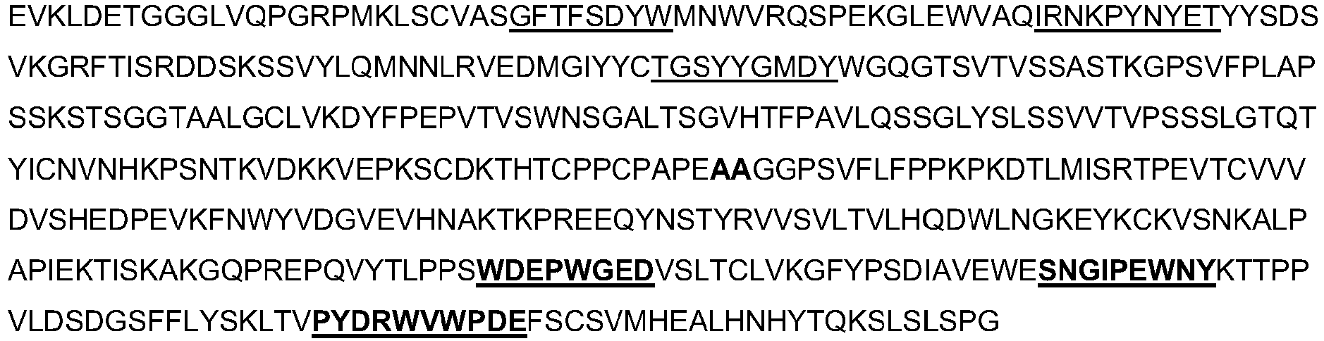
-
Amino acid sequence of the heavy chain of anti-human LAG-3/FITC mAb2
FS18-7-62/4420 without LALA mutation (SEQ ID NO: 74)
Position of the CDRs are underlined, and the AB, CD, and EF loop sequences are in bold and underlined.
-
Amino acid sequence of the heavy chain of anti-human LAG-3/FITC mAb2
FS18-7-65/4420 comprising LALA mutation (SEQ ID NO: 75)
Position of the CDRs are underlined, and the AB, CD, and EF loop sequences are in bold and underlined. Position of LALA mutation is in bold.
-
Amino acid sequence of the heavy chain of anti-human LAG-3/FITC mAb2
FS18-7-65/4420 without LALA mutation (SEQ ID NO: 76)
Position of the CDRs are underlined, and the AB, CD, and EF loop sequences are in bold and underlined.
-
Amino acid sequence of the heavy chain of anti-human LAG-3/FITC mAb2
FS18-7-78/4420 comprising LALA mutation (SEQ ID NO: 77)
Position of the CDRs are underlined, and the AB, CD, and EF loop sequences are in bold and underlined. Position of LALA mutation is in bold.
-
Amino acid sequence of the heavy chain of anti-human LAG-3/FITC mAb2
FS18-7-78/4420 without LALA mutation (SEQ ID NO: 78)
Position of the CDRs are underlined, and the AB, CD, and EF loop sequences are in bold and underlined.
-
Amino acid sequence of the heavy chain of anti-human LAG-3/FITC mAb2
FS18-7-88/4420 comprising LALA mutation (SEQ ID NO: 79)
Position of the CDRs are underlined, and the AB, CD, and EF loop sequences are in bold and underlined. Position of LALA mutation is in bold.
-
Amino acid sequence of the heavy chain of anti-human LAG-3/FITC mAb2
FS18-7-88/4420 without LALA mutation (SEQ ID NO: 80)
Position of the CDRs are underlined, and the AB, CD, and EF loop sequences are in bold and underlined.
-
Amino acid sequence of the heavy chain of anti-human LAG-3/FITC mAb2
FS18-7-95/4420 comprising LALA mutation (SEQ ID NO: 81)
Position of the CDRs are underlined, and the AB, CD, and EF loop sequences are in bold and underlined. Position of LALA mutation is in bold.
-
Amino acid sequence of the heavy chain of anti-human LAG-3/FITC mAb2
FS18-7-95/4420 without LALA mutation (SEQ ID NO: 82)
Position of the CDRs are underlined, and the AB, CD, and EF loop sequences are in bold and underlined.
-
Amino acid sequence of the heavy chain of
anti-FITC mAb 4420 comprising LALA mutation (SEQ ID NO: 83)
Position of the CDRs are underlined. Position of LALA mutation is in bold.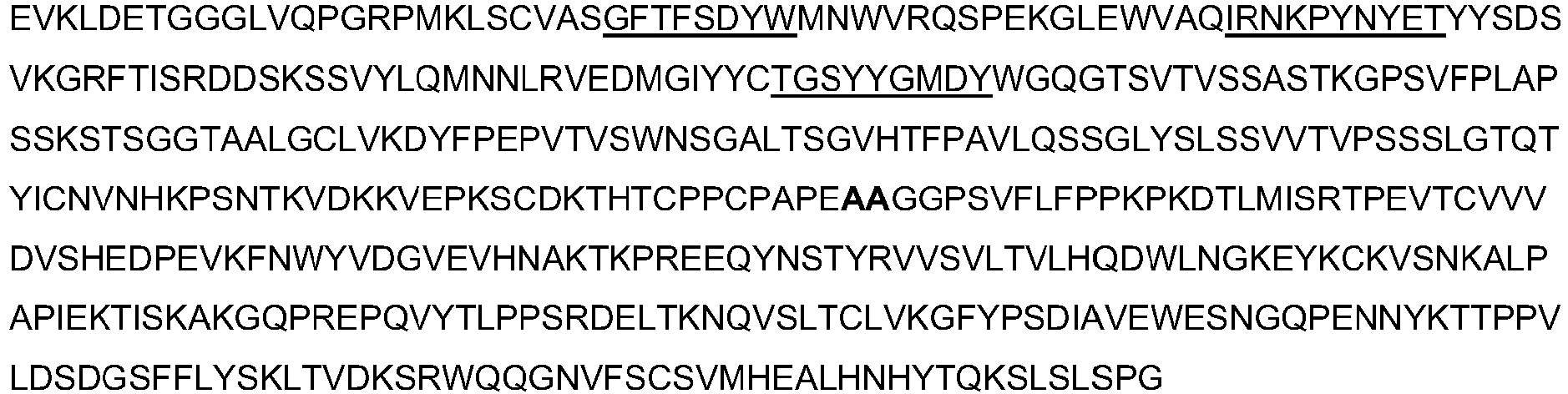
-
Amino acid sequence of the heavy chain of
anti-FITC mAb 4420 without LALA mutation (SEQ ID NO: 84)
Position of the CDRs are underlined.
-
Amino acid sequence of the
anti-FITC mAb 4420 light chain (SEQ ID NO: 85)
Position of the CDRs are underlined.
-
Amino acid sequence of the heavy chain of anti-mouse LAG-3/PD-L1 mAb2
FS18-7-108-29/S1 with LALA mutation (SEQ ID NO: 86)
Position of the CDRs are underlined, and the AB, CD, and EF loop sequences are in bold and underlined. Position of LALA mutation is in bold.
-
Amino acid sequence of the heavy chain of anti-mouse LAG-3/PD-L1 mAb2
FS18-7-108-29/S1 without LALA mutation (SEQ ID NO: 87)
Position of the CDRs are underlined, and the AB, CD, and EF loop sequences are in bold and underlined.
-
Amino acid sequence of the heavy chain of anti-mouse LAG-3/PD-L1 mAb2
FS18-7-108-35/S1 with LALA mutation (SEQ ID NO: 88)
Position of the CDRs are underlined, and the AB, CD, and EF loop sequences are in bold and underlined. Position of LALA mutation is in bold.
-
Amino acid sequence of the heavy chain of anti-mouse LAG-3/PD-L1 mAb2
FS18-7-108-35/S1 without LALA mutation (SEQ ID NO: 89)
Position of the CDRs are underlined, and the AB, CD, and EF loop sequences are in bold and underlined.
-
Amino acid sequence of the anti-mouse PD-L1 mAb S1 heavy chain with LALA mutation (SEQ ID NO: 90)
Position of the CDRs are underlined. Position of LALA mutation is in bold.
-
Amino acid sequence of the anti-mouse PD-L1 mAb S1 heavy chain without LALA mutation (SEQ ID NO: 91)
Position of the CDRs are underlined.
-
Amino acid sequence of the anti-mouse PD-L1 mAb S1 light chain (SEQ ID NO: 92)
Position of the CDRs are underlined.
-
Amino acid sequence of the anti-human LAG-3 mAb 25F7 heavy chain (SEQ ID NO: 93)
Position of the CDRs are underlined.
-
Amino acid sequence of the anti-human LAG-3 mAb 25F7 light chain (SEQ ID NO: 94)
Position of the CDRs are underlined.

-
Amino acid sequence of human LAG-3 (SEQ ID NO: 95)

-
Amino acid sequence of mouse LAG-3 (SEQ ID NO: 96)

-
Amino acid sequence of cvnomolqus LAG-3 (SEQ ID NO: 97)

-
- Bae J, Lee SJ, Park CG, Lee YS, Chun T. Trafficking of LAG-3 to the surface on activated T cells via its cytoplasmic domain and protein kinase C signaling. J Immunol. 193(6),3101-12 (2014).
- Baecher-Allan C, Wolf E, Hafler DA. MHC class II expression identifies functionally distinct human regulatory T cells. J Immunol. 176(8),4622-31 (2006).
- Bedzyk WD, Johnson LS, Riordan GS, Voss EW Jr. Comparison of Variable Region Primary Structures within an Anti-Fluorescein Idiotype Family. Biol. Chem. 264, 1565-1569 (1989).
- Bedzyk WD, Herron JN, Edmundson AB, Voss EW Jr. Active site structure and antigen binding properties of idiotypically cross-reactive anti-fluorescein monoclonal antibodies. J Biol Chem. 265(1),133-8 (1990).
- Bruhns P, lannascoli B, England P, Mancardi DA, Fernandez N, Jorieux S, Daëron M. Specificity and affinity of human Fcy receptors and their polymorphic variants for human IgG subclasses, Blood, 113, 3716-25, (2009).
- Camisaschi C, Casati C, Rini F, Perego M, De Filippo A, Triebel F, Parmiani G, Belli F, Rivoltini L, Castelli C. LAG-3 expression defines a subset of CD4(+)CD25(high)Foxp3(+) regulatory T cells that are expanded at tumor sites. J Immunol. 184(11),6545-51 (2010).
- Demeure, C. E., Wolfers, J., Martin-Garcia, N., Gaulard, P. & Triebel, F. T Lymphocytes infiltrating various tumour types express the MHC class II ligand lymphocyte activation gene-3 (LAG-3): role of LAG-3/MHC class II interactions in cell-cell contacts. European Journal of Cancer 37, 1709-1718 (2001).
- Durham NM, Nirschl CJ, Jackson CM, Elias J, Kochel CM, Anders RA, Drake CG. Lymphocyte Activation Gene 3 (LAG-3) modulates the ability of CD4 T cells to be suppressed in vivo. PLoS One. 9(11), e109080 (2015).
- Gandhi MK, Lambley E, Duraiswamy J, Dua U, Smith C, Elliott S, Gill D, Marlton P, Seymour J, Khanna R. Expression of LAG-3 by tumor-infiltrating lymphocytes is coincident with the suppression of latent membrane antigen-specific CD8+ T cell function in Hodgkin lymphoma patients. Blood 108(7),2280-9 (2006).
- Huard B, Mastrangeli R, Prigent P,et al. Characterization of the major histocompatibility complex class II binding site on LAG-3 protein. Proc Natl Acad Sci USA 94,5744-9 (1997).
- Liang B, Workman C, Lee J, Chew C, Dale BM, Colonna L, Flores M, Li N, Schweighoffer E, Greenberg S, Tybulewicz V, Vignali D, Clynes R. Regulatory T cells inhibit dendritic cells by lymphocyte activation gene-3 engagement of MHC class II.J Immunol. 180(9),5916-26 (2008).
- Nakayama M, Akiba H, Takeda K, Kojima Y, Hashiguchi M, Azuma M, Yagita H, Okumura K. Tim-3 mediates phagocytosis of apoptotic cells and cross-presentation. Blood. 113(16).3821-3 (2009).
- Sega El, Leveson-Gower DB, Florek M, Schneidawind D, Luong RH, Negrin RS. Role of lymphocyte activation gene-3 (Lag-3) in conventional and regulatory T cell function in allogeneic transplantation.PLoS One. 9(1), e86551 (2014).
- Sudo T, Nishikawa S, Ogawa M, Kataoka H, Ohno N, Izawa A, Hayashi S, Nishikawa S. Functional hierarchy of c-kit and c-fms in intramarrow production of CFU-M. Oncogene. 11 (12),2469-76 (1995).
- Wherry EJ. T cell exhaustion. Nat Immunol. 12(6),492-9 (2011).
- Wolchok Jet al; Nivolumab plus ipilimumab in advanced melanoma.N Engl J Med. 369(2),122-33 (2013)
- Woo SR, Turnis ME, Goldberg MV, Bankoti J, Selby M, Nirschl CJ, Bettini ML, Gravano DM, Vogel P, Liu CL, Tangsombatvisit S, Grosso JF, Netto G, Smeltzer MP, Chaux A, Utz PJ, Workman CJ, Pardoll DM, Korman AJ, Drake CG, Vignali DA. Immune inhibitory molecules LAG-3 and PD-1 synergistically regulate T cell function to promote tumoral immune escape. Cancer Res. 72(4),917-2 (2012).
- Workman CJ, Vignali DA. Negative regulation of T cell homeostasis by lymphocyte activation gene-3 (CD223). J Immunol. 174(2),688-95 (2005).
- Workman CJ, Vignali DA. The CD4-related molecule, LAG-3 (CD223), regulates the expansion of activated T cells. Eur J Immunol. 33(4),970-9 (2003).
- Xu D, Alegre ML, Varga SS, Rothermel AL, Collins AM, Pulito VL, Hanna LS, Dolan KP, Parren PW, Bluestone JA, Jolliffe LK, Zivin RA. In vitro characterization of five humanized OKT3 effector function variant antibodies. Cell. Immunol. 200, 16-26 (2000)
- Yamashita Y, Hooker SW, Jiang H, Laurent AB, Resta R, Khare K, Coe A, Kincade PW, Thompson LF. CD73 expression and fyn-dependent signaling on murine lymphocytes. Eur J Immunol. 28(10),2981-90 (1998).
Claims (15)
- An antibody molecule or fragment thereof comprising a LAG-3 antigen-binding site located in a CH3 domain of the antibody molecule or fragment thereof, wherein the LAG-3 antigen-binding site comprises the amino acid sequences WDEPWGED (SEQ ID NO: 1) and PYDRWVWPDE (SEQ ID NO: 3), and wherein the amino acid sequence WDEPWGED is located in the AB loop of the CH3 domain and the amino acid sequence PYDRWVWPDE is located in the EF loop of the CH3 domain.
- The antibody molecule or fragment thereof according to claim 1, wherein the LAG-3 antigen-binding site further comprises one of the following sequences in the CD loop of the CH3 domain:(i) SNGQPENNY (SEQ ID NO: 2);(ii) SNGQPEDNY (SEQ ID NO: 13);(iii) SNGYPEIEF (SEQ ID NO: 23);(iv) SNGIPEWNY (SEQ ID NO: 28);(v) SNGYAEYNY (SEQ ID NO: 33);(vi) SNGYKEENY (SEQ ID NO: 38);(vii) SNGVPELNV (SEQ ID NO: 43); or(viii) SNGYQEDNY (SEQ ID NO: 48);preferably SEQ ID NO: 2, 28, or 38, preferably SEQ ID NO: 2.
- The antibody molecule or fragment thereof according to claim 1 or 2, wherein:(i) the CH3 domain is a human IgG1, IgG2, IgG3, or IgG4 CH3 domain, preferably a human IgG1 CH3 domain; and/or(ii) the antibody molecule or fragment thereof comprises the CH3 domain set forth in SEQ ID NO: 5, 10, 15, 20, 25, 30, 35, 40, 45, or 50, preferably SEQ ID NO: 5, 30, or 40, preferably SEQ ID NO: 5.
- The antibody molecule according to any one of claims 1 to 3, wherein the CH2 domain of the antibody molecule is the CH2 domain of human IgG1, IgG2, IgG3, or IgG4, preferably human IgG1, wherein optionally the CH2 domain has the sequence set forth in SEQ ID NO: 53 or SEQ ID NO: 54.
- The antibody fragment according to any one of claims 1 to 3, wherein the antibody fragment further comprises a CH2 domain, optionally wherein the antibody fragment comprises the CH2 domain of human IgG1, IgG2, IgG3, or IgG4, preferably human IgG1, optionally wherein the CH2 domain has the sequence set forth in SEQ ID NO: 53 or SEQ ID NO: 54.
- The antibody molecule or fragment thereof according to any one of claims 1 to 5, wherein the antibody molecule or fragment thereof comprises the sequence set forth in SEQ ID NO: 6, 7, 11, 12, 16, 17, 21, 22, 26, 27, 31, 32, 36, 37, 41, 42, 46, 47, 51, or 52, preferably SEQ ID NO: 6, 7, 31, 32, 41, or 42, preferably SEQ ID NO: 6 or 7.
- The antibody molecule or fragment thereof according to any one of claims 1 to 6 comprising an immunoglobulin hinge region, or part thereof, at the N-terminus of the CH2 domain, optionally wherein the hinge region, or part thereof, is a human IgG1, IgG2, IgG3 or IgG4 hinge region, or part thereof, preferably a human IgG1 hinge region, or part thereof, optionally wherein the hinge region or part thereof has the sequence set forth in SEQ ID NO: 57 or SEQ ID NO: 58.
- The antibody molecule according to any one of claims 1 to 4, and 6 to 7, wherein the antibody molecule is a human IgG1, IgG2, IgG3 or IgG4 molecule, preferably a human IgG1 molecule.
- An antibody molecule according to any one of claims 1 to 4, and 6 to 8, wherein the CDR-based antigen-binding site of the antibody molecule binds to a molecule which is an immune system modulator, optionally wherein:(a)(i) the immune system modulator is an immunomodulatory receptor or a ligand for an immunomodulatory receptor; or(ii) the CDR-based antigen-binding site is specific for a molecule which is an immune system inhibitor or activator, preferably an immune system inhibitor;and/or(b) the CDR-based antigen-binding site is specific for an antigen selected from the group consisting of: cytotoxic T-lymphocyte-associated protein 4 (CTLA-4), T cell immunoglobulin and mucin-domain containing-3 (TIM-3), CD73 and Colony stimulating factor 1 receptor (CSF1R).
- The antibody molecule or fragment thereof according to any one of claims 1 to 9, wherein the antibody molecule or fragment thereof is conjugated to an immune system modulator, cytotoxic molecule, radioisotope, or detectable label, optionally wherein the immune system modulator or cytotoxic molecule is a cytokine.
- A nucleic acid encoding an antibody molecule or fragment thereof according to any one of claims 1 to 10, or a vector comprising a nucleic acid encoding an antibody molecule or fragment thereof according to any one of claims 1 to 10.
- A recombinant host cell comprising the nucleic acid or vector of claim 11.
- A method of producing an antibody molecule or fragment thereof according to any one of claims 1 to 10, comprising culturing the recombinant host cell of claim 12 under conditions for production of the antibody molecule or fragment thereof, optionally further comprising isolating and/or purifying the antibody molecule or fragment thereof.
- A pharmaceutical composition comprising an antibody molecule according to any one of claims 1 to 4, and 6 to 10, and a pharmaceutically acceptable excipient.
- The antibody molecule according to any one of claims 1 to 4, and 6 to 10, for use in a method of treating cancer in a patient, wherein the CDR-based antigen-binding site of the antibody molecule binds to a molecule which is an immune system modulator, and optionally wherein the method further comprises administering to the patient:(i) an anti-tumour vaccine; or(ii) a chemotherapeutic agent.
Applications Claiming Priority (2)
| Application Number | Priority Date | Filing Date | Title |
|---|---|---|---|
| US201662352470P | 2016-06-20 | 2016-06-20 | |
| PCT/EP2017/065052 WO2017220555A1 (en) | 2016-06-20 | 2017-06-20 | Lag -3 binding members |
Publications (2)
| Publication Number | Publication Date |
|---|---|
| EP3472206A1 EP3472206A1 (en) | 2019-04-24 |
| EP3472206B1 true EP3472206B1 (en) | 2025-01-15 |
Family
ID=59269995
Family Applications (1)
| Application Number | Title | Priority Date | Filing Date |
|---|---|---|---|
| EP17734668.1A Active EP3472206B1 (en) | 2016-06-20 | 2017-06-20 | Lag-3 binding members |
Country Status (15)
| Country | Link |
|---|---|
| US (2) | US11214618B2 (en) |
| EP (1) | EP3472206B1 (en) |
| JP (2) | JP7085708B2 (en) |
| KR (1) | KR20190022632A (en) |
| CN (1) | CN109311993B (en) |
| AU (1) | AU2017281157B2 (en) |
| BR (1) | BR112018076525A2 (en) |
| CA (1) | CA3027302A1 (en) |
| ES (1) | ES3010640T3 (en) |
| IL (1) | IL263715A (en) |
| MX (1) | MX2018016344A (en) |
| RU (1) | RU2018142573A (en) |
| SG (1) | SG11201811184UA (en) |
| TW (1) | TW201831513A (en) |
| WO (1) | WO2017220555A1 (en) |
Families Citing this family (70)
| Publication number | Priority date | Publication date | Assignee | Title |
|---|---|---|---|---|
| EP3858858A1 (en) | 2016-06-20 | 2021-08-04 | F-Star Delta Limited | Binding molecules binding pd-l1 and lag-3 |
| BR112018076525A2 (en) | 2016-06-20 | 2019-04-02 | F-Star Beta Limited | lag-3 binding members |
| GB201612520D0 (en) | 2016-07-19 | 2016-08-31 | F-Star Beta Ltd | Binding molecules |
| KR102576042B1 (en) | 2016-10-11 | 2023-09-07 | 아게누스 인코포레이티드 | Anti-LAG-3 Antibodies and Methods of Using The Same |
| SG11201909154SA (en) | 2017-04-05 | 2019-10-30 | Hoffmann La Roche | Bispecific antibodies specifically binding to pd1 and lag3 |
| CA3060984A1 (en) | 2017-05-30 | 2018-12-06 | Bristol-Myers Squibb Company | Treatment of lag-3 positive tumors |
| MX2019012038A (en) | 2017-05-30 | 2019-11-18 | Bristol Myers Squibb Co | Compositions comprising a combination of an anti-lag-3 antibody, a pd-1 pathway inhibitor, and an immunotherapeutic agent. |
| KR102721137B1 (en) | 2017-05-30 | 2024-10-25 | 브리스톨-마이어스 스큅 컴퍼니 | Composition comprising anti-LAG-3 antibody or anti-LAG-3 antibody and anti-PD-1 or anti-PD-L1 antibody |
| JP7489316B2 (en) | 2017-12-19 | 2024-05-23 | エフ-スター セラピューティクス リミテッド | FC-binding fragment having PD-LI antigen-binding site |
| MX2020012227A (en) * | 2018-05-17 | 2021-04-13 | Immunome Inc | CH3 DOMAIN MARKER EPITOPES. |
| PL3820569T3 (en) | 2018-07-12 | 2025-07-14 | Invox Pharma Limited | Antibody molecules that bind pd-l1 and cd137 |
| AU2019301206B2 (en) | 2018-07-12 | 2025-10-30 | Invox Pharma Limited | Antibody molecules that bind CD137 and OX40 |
| GB201811403D0 (en) | 2018-07-12 | 2018-08-29 | F Star Beta Ltd | Antibody molecules |
| GB201811404D0 (en) | 2018-07-12 | 2018-08-29 | F Star Beta Ltd | Anti-CD137 Antibodies |
| GB201811415D0 (en) | 2018-07-12 | 2018-08-29 | F Star Beta Ltd | Anti-Mesothelin Anti bodies |
| GB201811410D0 (en) | 2018-07-12 | 2018-08-29 | F Star Beta Ltd | OX40 Binding molecules |
| GB201811408D0 (en) | 2018-07-12 | 2018-08-29 | F Star Beta Ltd | CD137 Binding Molecules |
| GB201811450D0 (en) | 2018-07-12 | 2018-08-29 | F Star Delta Ltd | Mesothelin and CD137 binding molecules |
| EP3826660A1 (en) | 2018-07-26 | 2021-06-02 | Bristol-Myers Squibb Company | Lag-3 combination therapy for the treatment of cancer |
| EP4445958B1 (en) | 2018-10-19 | 2025-09-24 | Bristol-Myers Squibb Company | Combination therapy for melanoma |
| CN114174537A (en) | 2019-05-30 | 2022-03-11 | 百时美施贵宝公司 | Cell localization features and combination therapies |
| CN114174538A (en) | 2019-05-30 | 2022-03-11 | 百时美施贵宝公司 | Multiple tumor gene signatures suitable for immunooncology therapy |
| WO2020243568A1 (en) | 2019-05-30 | 2020-12-03 | Bristol-Myers Squibb Company | Methods of identifying a subject suitable for an immuno-oncology (i-o) therapy |
| WO2021024020A1 (en) | 2019-08-06 | 2021-02-11 | Astellas Pharma Inc. | Combination therapy involving antibodies against claudin 18.2 and immune checkpoint inhibitors for treatment of cancer |
| CN114450028A (en) | 2019-09-22 | 2022-05-06 | 百时美施贵宝公司 | Quantitative spatial profiling for LAG-3antagonist therapy |
| WO2021092221A1 (en) | 2019-11-06 | 2021-05-14 | Bristol-Myers Squibb Company | Methods of identifying a subject with a tumor suitable for a checkpoint inhibitor therapy |
| WO2021092220A1 (en) | 2019-11-06 | 2021-05-14 | Bristol-Myers Squibb Company | Methods of identifying a subject with a tumor suitable for a checkpoint inhibitor therapy |
| EP4054723A1 (en) | 2019-11-08 | 2022-09-14 | Bristol-Myers Squibb Company | Lag-3 antagonist therapy for melanoma |
| KR20220139915A (en) | 2020-02-06 | 2022-10-17 | 브리스톨-마이어스 스큅 컴퍼니 | IL-10 and its uses |
| CA3182579A1 (en) | 2020-07-07 | 2022-01-13 | Ugur Sahin | Therapeutic rna for hpv-positive cancer |
| CA3193421A1 (en) | 2020-08-28 | 2022-03-03 | Bristol-Myers Squibb Company | Lag-3 antagonist therapy for hepatocellular carcinoma |
| CA3190660A1 (en) | 2020-08-31 | 2022-03-03 | George C. Lee | Cell localization signature and immunotherapy |
| US20240101666A1 (en) | 2020-10-23 | 2024-03-28 | Bristol-Myers Squibb Company | Lag-3 antagonist therapy for lung cancer |
| WO2022120179A1 (en) | 2020-12-03 | 2022-06-09 | Bristol-Myers Squibb Company | Multi-tumor gene signatures and uses thereof |
| TW202245808A (en) | 2020-12-21 | 2022-12-01 | 德商拜恩迪克公司 | Therapeutic rna for treating cancer |
| WO2022135667A1 (en) | 2020-12-21 | 2022-06-30 | BioNTech SE | Therapeutic rna for treating cancer |
| WO2022135666A1 (en) | 2020-12-21 | 2022-06-30 | BioNTech SE | Treatment schedule for cytokine proteins |
| SMT202500208T1 (en) | 2020-12-28 | 2025-07-22 | Bristol Myers Squibb Co | Antibody compositions and methods of use thereof |
| JP2024501029A (en) | 2020-12-28 | 2024-01-10 | ブリストル-マイヤーズ スクイブ カンパニー | Subcutaneous administration of PD1/PD-L1 antibodies |
| AU2022212123A1 (en) | 2021-01-29 | 2023-09-07 | Board Of Regents, The University Of Texas System | Methods of treating cancer with kinase inhibitors |
| WO2022212400A1 (en) | 2021-03-29 | 2022-10-06 | Juno Therapeutics, Inc. | Methods for dosing and treatment with a combination of a checkpoint inhibitor therapy and a car t cell therapy |
| WO2022216979A1 (en) | 2021-04-08 | 2022-10-13 | Board Of Regents, The University Of Texas System | Compounds and methods for theranostic targeting of parp activity |
| EP4337694A1 (en) | 2021-05-12 | 2024-03-20 | Dana-Farber Cancer Institute, Inc. | Lag3 and gal3 inhibitory agents, xbp1, cs1, and cd138 peptides, and methods of use thereof |
| CA3225254A1 (en) | 2021-07-13 | 2023-01-19 | BioNTech SE | Multispecific binding agents against cd40 and cd137 in combination therapy for cancer |
| CN115873116B (en) * | 2021-09-29 | 2026-04-28 | 中山康方生物医药有限公司 | Anti-LAG3 antibodies, pharmaceutical compositions and uses |
| TW202333802A (en) | 2021-10-11 | 2023-09-01 | 德商拜恩迪克公司 | Therapeutic rna for lung cancer |
| US20240417473A1 (en) | 2021-10-29 | 2024-12-19 | Bristol-Myers Squibb Company | Lag-3 antagonist therapy for hematological cancer |
| IL314050A (en) | 2022-01-26 | 2024-09-01 | Bristol Myers Squibb Co | Combined treatment method for hepatocellular carcinoma |
| US20250179174A1 (en) | 2022-02-25 | 2025-06-05 | Bristol-Myers Squibb Company | Combination therapy for colorectal carcinoma |
| WO2023168404A1 (en) | 2022-03-04 | 2023-09-07 | Bristol-Myers Squibb Company | Methods of treating a tumor |
| JP2025508076A (en) | 2022-03-08 | 2025-03-21 | アレンティス・セラピューティクス・アクチェンゲゼルシャフト | Use of anti-claudin-1 antibodies to improve T cell availability |
| JP2025509749A (en) | 2022-03-18 | 2025-04-11 | ブリストル-マイヤーズ スクイブ カンパニー | Methods for isolating polypeptides |
| CN119562830A (en) | 2022-06-02 | 2025-03-04 | 百时美施贵宝公司 | Antibody compositions and methods of use thereof |
| CN114807054B (en) * | 2022-06-24 | 2022-09-13 | 北京索莱宝科技有限公司 | Mouse anti-human IgG monoclonal antibody hybridoma cell strain, antibody composition and kit |
| JP2025541725A (en) | 2022-12-01 | 2025-12-23 | メディミューン リミテッド | Combination therapy for the treatment of cancer comprising an anti-PD-L1 antibody and an anti-CD73 antibody |
| EP4634224A1 (en) | 2022-12-14 | 2025-10-22 | Astellas Pharma Europe Bv | Combination therapy involving bispecific binding agents binding to cldn18.2 and cd3 and immune checkpoint inhibitors |
| CN120731228A (en) | 2022-12-21 | 2025-09-30 | 百时美施贵宝公司 | Combination therapy for lung cancer |
| WO2024163477A1 (en) | 2023-01-31 | 2024-08-08 | University Of Rochester | Immune checkpoint blockade therapy for treating staphylococcus aureus infections |
| WO2024196952A1 (en) | 2023-03-20 | 2024-09-26 | Bristol-Myers Squibb Company | Tumor subtype assessment for cancer therapy |
| WO2025038763A1 (en) | 2023-08-15 | 2025-02-20 | Bristol-Myers Squibb Company | Ceramic hydroxyapatite chromatography flow through method |
| CN119971023A (en) * | 2023-11-02 | 2025-05-13 | 正大天晴(广州)医药有限公司 | Pharmaceutical composition containing anti-LAG-3 antibody and use thereof |
| AR134560A1 (en) | 2023-12-08 | 2026-01-28 | Astellas Pharma Inc | COMBINATION THERAPY INVOLVING BISPECIFIC BINDING AGENTS THAT BIND TO CLDN18.2 AND CD3 AND AGENTS THAT STABILIZE OR INCREASE CLDN18.2 EXPRESSION |
| WO2025120867A1 (en) | 2023-12-08 | 2025-06-12 | Astellas Pharma Inc. | Combination therapy involving bispecific binding agents binding to cldn18.2 and cd3 and anti-vegfr2 antibodies |
| WO2026033885A1 (en) | 2024-08-08 | 2026-02-12 | Astellas Pharma Inc. | Combination therapy involving bispecific binding agents binding to cldn18.2 and cd3 and agents stabilizing or increasing expression of cldn18.2 |
| WO2025120866A1 (en) | 2023-12-08 | 2025-06-12 | Astellas Pharma Inc. | Combination therapy involving bispecific binding agents binding to cldn18.2 and cd3 and agents stabilizing or increasing expression of cldn18.2 |
| WO2025145207A1 (en) | 2023-12-29 | 2025-07-03 | Bristol-Myers Squibb Company | Combination therapy of kras inhibitor and treg-depleting agent |
| US20250361320A1 (en) | 2024-02-27 | 2025-11-27 | Bristol-Myers Squibb Company | Anti-ceacam5 antibodies and uses thereof |
| WO2025184211A1 (en) | 2024-02-27 | 2025-09-04 | Bristol-Myers Squibb Company | Anti-ceacam5 antibody drug conjugates |
| WO2025245489A1 (en) | 2024-05-24 | 2025-11-27 | Bristol-Myers Squibb Company | Treatment of tumors in subjects having fgl-1 positive samples |
| US20260115307A1 (en) | 2024-10-31 | 2026-04-30 | Bristol-Myers Squibb Company | Anti-fucosyl-gm1 antibody drug conjugates |
Citations (1)
| Publication number | Priority date | Publication date | Assignee | Title |
|---|---|---|---|---|
| WO2017220569A1 (en) * | 2016-06-20 | 2017-12-28 | F-Star Delta Limited | Binding molecules binding pd-l1 and lag-3 |
Family Cites Families (108)
| Publication number | Priority date | Publication date | Assignee | Title |
|---|---|---|---|---|
| DE2245404C3 (en) | 1972-09-15 | 1978-08-31 | Robert Bosch Gmbh, 7000 Stuttgart | Ground resistance, especially for spark plugs, and methods of manufacturing the same |
| JPS531908B2 (en) | 1973-11-12 | 1978-01-23 | ||
| JPS5746634B2 (en) | 1974-05-10 | 1982-10-04 | ||
| JPS5146628A (en) | 1974-10-17 | 1976-04-21 | Nippon Denso Co | TEIKOIRISUPAAKUPURAGU |
| JPS61134325A (en) | 1984-12-04 | 1986-06-21 | Teijin Ltd | Expression of hybrid antibody gene |
| GB8607679D0 (en) | 1986-03-27 | 1986-04-30 | Winter G P | Recombinant dna product |
| US5595756A (en) | 1993-12-22 | 1997-01-21 | Inex Pharmaceuticals Corporation | Liposomal compositions for enhanced retention of bioactive agents |
| ES2255194T3 (en) | 1997-12-01 | 2006-06-16 | The Government Of The Usa, As Represented By The Secretary, Department Of Health And Human Services | ANTIBODIES, WHICH INCLUDE PV MOLECULES, AND IMMUNOCONJUGADOS THAT PRESENT A RAISED LINK AFFINITY FOR MESOTHELINE AND METHODS FOR USE. |
| DE19818214A1 (en) | 1998-04-24 | 1999-10-28 | Bosch Gmbh Robert | Spark plug for combustion engine |
| DE60039510D1 (en) | 1999-05-27 | 2008-08-28 | Us Gov Health & Human Serv | IMMUNOKON JUGATES WITH HIGH BINDING SAFFINITY |
| AU2001247616B2 (en) | 2000-04-11 | 2007-06-14 | Genentech, Inc. | Multivalent antibodies and uses therefor |
| JP4578025B2 (en) | 2001-07-06 | 2010-11-10 | 日本特殊陶業株式会社 | Spark plug |
| US7288638B2 (en) | 2003-10-10 | 2007-10-30 | Bristol-Myers Squibb Company | Fully human antibodies against human 4-1BB |
| US20090298195A1 (en) | 2005-01-05 | 2009-12-03 | F-Star Biotechnologische Forschungs-Und Entwicklun Gseges M.B.H. | Synthetic immunoglobulin domains with binding properties engineered in regions of the molecule different from the complementarity determining regions |
| EP2399935A3 (en) | 2005-02-15 | 2012-02-22 | GTC Biotherapeutics, Inc. | An anti-CD137 antibody as an agent in the treatment of cancer and glycosylation variants thereof |
| EP2322560B1 (en) | 2005-03-10 | 2013-09-04 | Morphotek, Inc. | Anti-mesothelin antibodies |
| CA2612241C (en) | 2005-07-01 | 2018-11-06 | Medarex, Inc. | Human monoclonal antibodies to programmed death ligand 1 (pd-l1) |
| US7612181B2 (en) | 2005-08-19 | 2009-11-03 | Abbott Laboratories | Dual variable domain immunoglobulin and uses thereof |
| US9868961B2 (en) | 2006-03-30 | 2018-01-16 | The Regents Of The University Of California | Methods and compositions for localized secretion of anti-CTLA-4 antibodies |
| AT503889B1 (en) | 2006-07-05 | 2011-12-15 | Star Biotechnologische Forschungs Und Entwicklungsges M B H F | MULTIVALENT IMMUNE LOBULINE |
| GB0624500D0 (en) | 2006-12-07 | 2007-01-17 | Istituto Superiore Di Sanito | A novel passive vaccine for candida infections |
| PL3241842T3 (en) | 2007-06-26 | 2024-06-10 | F-Star Therapeutics Limited | Display of binding agents |
| HUE027358T2 (en) | 2007-11-26 | 2016-09-28 | Bayer Ip Gmbh | Anti-mesothelin antibodies and uses therefor |
| KR101577843B1 (en) | 2007-12-14 | 2015-12-16 | 브리스톨-마이어스 스큅 컴퍼니 | Binding molecules to the human ox40 receptor |
| PT2132228E (en) | 2008-04-11 | 2011-10-11 | Emergent Product Dev Seattle | Cd37 immunotherapeutic and combination with bifunctional chemotherapeutic thereof |
| EP2113255A1 (en) * | 2008-05-02 | 2009-11-04 | f-star Biotechnologische Forschungs- und Entwicklungsges.m.b.H. | Cytotoxic immunoglobulin |
| AR072999A1 (en) | 2008-08-11 | 2010-10-06 | Medarex Inc | HUMAN ANTIBODIES THAT JOIN GEN 3 OF LYMPHOCYTARY ACTIVATION (LAG-3) AND THE USES OF THESE |
| WO2010057047A1 (en) | 2008-11-13 | 2010-05-20 | Trubion Pharmaceutics, Inc. | Cd37 immunotherapeutic combination therapies and uses thereof |
| EP4209510B1 (en) | 2008-12-09 | 2024-01-31 | F. Hoffmann-La Roche AG | Anti-pd-l1 antibodies and their use to enhance t-cell function |
| AU2010230063B2 (en) | 2009-03-24 | 2015-06-25 | The Government Of The United States Of America, As Represented By The Secretary Of The Department Of Health And Human Services | Anti-mesothelin antibodies |
| UY32560A (en) | 2009-04-29 | 2010-11-30 | Bayer Schering Pharma Ag | IMMUNOCONJUGADOS OF ANTIMESOTELINA AND USES OF THE SAME |
| NO2504364T3 (en) | 2009-11-24 | 2018-01-06 | ||
| EP2407487A1 (en) | 2010-07-14 | 2012-01-18 | F-Star Biotechnologische Forschungs - und Entwicklungsges. M.B.H. | Multispecific modular antibody |
| SG10201912092VA (en) | 2010-09-09 | 2020-02-27 | Pfizer | 4-1bb binding molecules |
| PE20170917A1 (en) | 2010-12-20 | 2017-07-12 | Genentech Inc | ANTI-MESOTHELIN ANTIBODIES AND IMMUNOCONJUGATES |
| ES2692268T5 (en) | 2011-03-29 | 2025-02-26 | Roche Glycart Ag | Antibody fc variants |
| EP2546268A1 (en) | 2011-07-13 | 2013-01-16 | F-Star Biotechnologische Forschungs - und Entwicklungsges. M.B.H. | Internalising immunoglobulin |
| US9624298B2 (en) | 2011-11-28 | 2017-04-18 | Merck Patent Gmbh | Anti-PD-L1 antibodies and uses thereof |
| AU2013267161A1 (en) | 2012-05-31 | 2014-11-20 | Sorrento Therapeutics, Inc. | Antigen binding proteins that bind PD-L1 |
| WO2014004549A2 (en) | 2012-06-27 | 2014-01-03 | Amgen Inc. | Anti-mesothelin binding proteins |
| UY34887A (en) | 2012-07-02 | 2013-12-31 | Bristol Myers Squibb Company Una Corporacion Del Estado De Delaware | OPTIMIZATION OF ANTIBODIES THAT FIX THE LYMPHOCYTE ACTIVATION GEN 3 (LAG-3) AND ITS USES |
| JP5276742B1 (en) | 2012-08-09 | 2013-08-28 | 日本特殊陶業株式会社 | Spark plug |
| WO2014052064A1 (en) | 2012-09-27 | 2014-04-03 | The United States Of America, As Represented By The Secretary, Department Of Health And Human Services | Mesothelin antibodies and methods for eliciting potent antitumor activity |
| EP2925350B1 (en) | 2012-12-03 | 2019-01-30 | Bristol-Myers Squibb Company | Enhancing anti-cancer activity of immunomodulatory fc fusion proteins |
| US10280221B2 (en) | 2013-03-15 | 2019-05-07 | Glaxosmithkline Intellectual Property Development Limited | Anti-LAG-3 binding proteins |
| MX357011B (en) | 2013-03-15 | 2018-06-22 | Amgen Inc | Heterodimeric bispecific antibodies. |
| EP3757130A1 (en) | 2013-09-26 | 2020-12-30 | Costim Pharmaceuticals Inc. | Methods for treating hematologic cancers |
| GB201317622D0 (en) | 2013-10-04 | 2013-11-20 | Star Biotechnology Ltd F | Cancer biomarkers and uses thereof |
| ES2783026T3 (en) | 2014-02-04 | 2020-09-16 | Pfizer | Combination of a PD-1 antagonist and a 4-1BB agonist for the treatment of cancer |
| WO2015138920A1 (en) * | 2014-03-14 | 2015-09-17 | Novartis Ag | Antibody molecules to lag-3 and uses thereof |
| WO2015198312A1 (en) | 2014-06-24 | 2015-12-30 | Ccam Therapeutics Ltd. | Compositions comprising antibodies to ceacam-1 and lag-3 for cancer therapy |
| JP5902757B2 (en) | 2014-06-24 | 2016-04-13 | 日本特殊陶業株式会社 | Spark plug |
| TWI693232B (en) | 2014-06-26 | 2020-05-11 | 美商宏觀基因股份有限公司 | Covalently bonded diabodies having immunoreactivity with pd-1 and lag-3, and methods of use thereof |
| BR112017002596A2 (en) | 2014-08-10 | 2018-01-30 | Federal-Mogul Ignition Company | improved sealing spark plug |
| JO3663B1 (en) | 2014-08-19 | 2020-08-27 | Merck Sharp & Dohme | Anti-lag3 antibodies and antigen-binding fragments |
| WO2016040882A1 (en) | 2014-09-13 | 2016-03-17 | Novartis Ag | Combination therapies of egfr inhibitors |
| TW201619200A (en) | 2014-10-10 | 2016-06-01 | 麥迪紐有限責任公司 | Humanized anti-OX40 antibodies and uses thereof |
| CA2874083C (en) | 2014-12-05 | 2024-01-02 | Universite Laval | Tdp-43-binding polypeptides useful for the treatment of neurodegenerative diseases |
| GB201500319D0 (en) | 2015-01-09 | 2015-02-25 | Agency Science Tech & Res | Anti-PD-L1 antibodies |
| AU2016219772A1 (en) | 2015-02-22 | 2017-09-21 | Sorrento Therapeutics, Inc. | Antibody therapeutics that bind CD137 |
| WO2016162505A1 (en) | 2015-04-08 | 2016-10-13 | F-Star Biotechnology Limited | Her2 binding agent therapies |
| AU2016258977C1 (en) | 2015-05-04 | 2022-07-14 | Pieris Pharmaceuticals Gmbh | Anti-cancer fusion polypeptide |
| AU2016265447B2 (en) | 2015-05-21 | 2022-06-16 | Alligator Bioscience Ab | Novel polypeptides |
| TWI773646B (en) | 2015-06-08 | 2022-08-11 | 美商宏觀基因股份有限公司 | Lag-3-binding molecules and methods of use thereof |
| JP6025921B1 (en) | 2015-06-22 | 2016-11-16 | 日本特殊陶業株式会社 | Spark plug |
| CA2988831A1 (en) | 2015-07-15 | 2017-01-19 | Pieris Pharmaceuticals Gmbh | Novel proteins specific for lag-3 |
| CN114853891A (en) | 2015-07-22 | 2022-08-05 | 索伦托药业有限公司 | Antibody therapeutics that bind to LAG3 |
| AU2016298227B9 (en) | 2015-07-30 | 2019-10-31 | Macrogenics, Inc. | PD-1-binding molecules and methods of use thereof |
| EP3331901A1 (en) | 2015-08-07 | 2018-06-13 | Pieris Pharmaceuticals GmbH | Novel fusion polypeptide specific for lag-3 and pd-1 |
| CN113214398B (en) | 2015-09-22 | 2022-04-08 | 苏州丁孚靶点生物技术有限公司 | Fully human antibodies against human CD137 and uses thereof |
| KR101782487B1 (en) | 2015-09-24 | 2017-09-27 | 재단법인 목암생명과학연구소 | Novel Antibody Binding to Mesothelin(MSLN), and Composition Comprising the Same |
| BR112018002570A2 (en) | 2015-10-02 | 2018-10-16 | Hoffmann La Roche | bispecific antigen binding molecule, bispecific antibody, polynucleotides, ox40-specific binding antibody, pharmaceutical composition and method for inhibiting tumor cell growth in an individual |
| TWI756187B (en) | 2015-10-09 | 2022-03-01 | 美商再生元醫藥公司 | Anti-lag3 antibodies and uses thereof |
| GB201519481D0 (en) | 2015-11-04 | 2015-12-16 | Cancer Rec Tech Ltd | Immunomodulatory antibodies |
| CA3132021C (en) | 2015-11-18 | 2024-03-12 | Merck Sharp & Dohme Corp. | Pd1 and/or lag3 binders |
| US20190330336A1 (en) | 2015-11-19 | 2019-10-31 | Sutro Biopharma, Inc. | Anti-lag3 antibodies, compositions comprising anti-lag3 antibodies and methods of making and using anti-lag3 antibodies |
| MX2018008308A (en) | 2016-01-11 | 2019-05-15 | Inhibrx Inc | Multivalent and multispecific 41bb-binding fusion proteins. |
| CN108602371B (en) | 2016-03-18 | 2020-03-24 | 大日本印刷株式会社 | Intermediate transfer medium, combination of intermediate transfer medium and thermal transfer sheet, and method for forming printed matter |
| WO2017182672A1 (en) | 2016-04-22 | 2017-10-26 | Alligator Bioscience Ab | Novel bispecific polypeptides against cd137 |
| AR108377A1 (en) | 2016-05-06 | 2018-08-15 | Medimmune Llc | BISPECIFIC UNION PROTEINS AND ITS USES |
| KR20190014525A (en) | 2016-05-27 | 2019-02-12 | 애브비 바이오테라퓨틱스 인크. | A bispecific binding protein that binds immuno-regulatory proteins to tumor antigens |
| US20190106494A1 (en) | 2016-06-13 | 2019-04-11 | Askgene Pharma Inc. | PD-L1 Specific Monoclonal Antibodies for Disease Treatment and Diagnosis |
| CN107523546A (en) | 2016-06-20 | 2017-12-29 | 上海细胞治疗研究院 | A kind of NK cells of high efficiency stable expression antibody and application thereof |
| JP7461741B2 (en) | 2016-06-20 | 2024-04-04 | カイマブ・リミテッド | Anti-PD-L1 and IL-2 Cytokines |
| BR112018076525A2 (en) | 2016-06-20 | 2019-04-02 | F-Star Beta Limited | lag-3 binding members |
| US9567399B1 (en) | 2016-06-20 | 2017-02-14 | Kymab Limited | Antibodies and immunocytokines |
| GB201612520D0 (en) | 2016-07-19 | 2016-08-31 | F-Star Beta Ltd | Binding molecules |
| KR20190031299A (en) | 2016-07-20 | 2019-03-25 | 주식회사 에스티큐브 | Methods of treating cancer using a combination of antibodies that bind to glycosylated PD-L1 |
| CN109983033B (en) | 2016-09-23 | 2023-10-31 | 美勒斯公司 | Biologically active binding molecules that regulate cell expression |
| GB201616699D0 (en) | 2016-09-30 | 2016-11-16 | Mab Designs Ltd | Antibodies |
| GB201619648D0 (en) | 2016-11-21 | 2017-01-04 | Alligator Bioscience Ab | Novel antibodies and uses thereof |
| WO2018115859A1 (en) | 2016-12-20 | 2018-06-28 | Kymab Limited | Multispecific antibody with combination therapy for immuno-oncology |
| GB201700345D0 (en) | 2017-01-09 | 2017-02-22 | F-Star Beta Ltd | Conditional agonists of immune responses |
| CA3065008A1 (en) | 2017-05-26 | 2018-11-29 | Novimmune Sa | Anti-cd47 x anti-mesothelin antibodies and methods of use thereof |
| MX2019012038A (en) | 2017-05-30 | 2019-11-18 | Bristol Myers Squibb Co | Compositions comprising a combination of an anti-lag-3 antibody, a pd-1 pathway inhibitor, and an immunotherapeutic agent. |
| IL272159B2 (en) | 2017-08-04 | 2026-03-01 | Genmab As | Binding agents binding to pd-l1 and cd137 and use thereof |
| EP3470426A1 (en) | 2017-10-10 | 2019-04-17 | Numab Therapeutics AG | Multispecific antibody |
| EP3728314A1 (en) | 2017-12-19 | 2020-10-28 | Kymab Limited | Bispecific antibody for icos and pd-l1 |
| JP7489316B2 (en) | 2017-12-19 | 2024-05-23 | エフ-スター セラピューティクス リミテッド | FC-binding fragment having PD-LI antigen-binding site |
| GB201811415D0 (en) | 2018-07-12 | 2018-08-29 | F Star Beta Ltd | Anti-Mesothelin Anti bodies |
| PL3820569T3 (en) | 2018-07-12 | 2025-07-14 | Invox Pharma Limited | Antibody molecules that bind pd-l1 and cd137 |
| GB201811408D0 (en) | 2018-07-12 | 2018-08-29 | F Star Beta Ltd | CD137 Binding Molecules |
| GB201811410D0 (en) | 2018-07-12 | 2018-08-29 | F Star Beta Ltd | OX40 Binding molecules |
| GB201811450D0 (en) | 2018-07-12 | 2018-08-29 | F Star Delta Ltd | Mesothelin and CD137 binding molecules |
| GB201811404D0 (en) | 2018-07-12 | 2018-08-29 | F Star Beta Ltd | Anti-CD137 Antibodies |
| AU2019301206B2 (en) | 2018-07-12 | 2025-10-30 | Invox Pharma Limited | Antibody molecules that bind CD137 and OX40 |
| GB201811403D0 (en) | 2018-07-12 | 2018-08-29 | F Star Beta Ltd | Antibody molecules |
| CA3139003A1 (en) | 2019-05-14 | 2020-11-19 | F-Star Therapeutics Limited | Dosage regimes for the administration of a lag-3/pd-l1 bispecific antibody |
-
2017
- 2017-06-20 BR BR112018076525-2A patent/BR112018076525A2/en not_active Application Discontinuation
- 2017-06-20 RU RU2018142573A patent/RU2018142573A/en not_active Application Discontinuation
- 2017-06-20 JP JP2019518145A patent/JP7085708B2/en active Active
- 2017-06-20 CN CN201780038556.1A patent/CN109311993B/en active Active
- 2017-06-20 CA CA3027302A patent/CA3027302A1/en active Pending
- 2017-06-20 ES ES17734668T patent/ES3010640T3/en active Active
- 2017-06-20 EP EP17734668.1A patent/EP3472206B1/en active Active
- 2017-06-20 US US16/311,596 patent/US11214618B2/en active Active
- 2017-06-20 TW TW106120621A patent/TW201831513A/en unknown
- 2017-06-20 MX MX2018016344A patent/MX2018016344A/en unknown
- 2017-06-20 AU AU2017281157A patent/AU2017281157B2/en not_active Expired - Fee Related
- 2017-06-20 WO PCT/EP2017/065052 patent/WO2017220555A1/en not_active Ceased
- 2017-06-20 KR KR1020197001120A patent/KR20190022632A/en not_active Withdrawn
- 2017-06-20 SG SG11201811184UA patent/SG11201811184UA/en unknown
-
2018
- 2018-12-16 IL IL263715A patent/IL263715A/en unknown
-
2021
- 2021-11-23 US US17/534,315 patent/US12187798B2/en active Active
-
2022
- 2022-03-28 JP JP2022051878A patent/JP7394162B2/en active Active
Patent Citations (1)
| Publication number | Priority date | Publication date | Assignee | Title |
|---|---|---|---|---|
| WO2017220569A1 (en) * | 2016-06-20 | 2017-12-28 | F-Star Delta Limited | Binding molecules binding pd-l1 and lag-3 |
Non-Patent Citations (1)
| Title |
|---|
| LEFRANC MARIE-PAULE ET AL: "IMGT unique numbering for immunoglobulin and T cell receptor constant domains and Ig superfamily C-like domains", DEVELOPMENTAL AND COMPARATIVE IMMUNOLOGY, vol. 29, no. 3, 1 January 2005 (2005-01-01), US, pages 185 - 203, XP093138816, ISSN: 0145-305X, DOI: 10.1016/j.dci.2004.07.003 * |
Also Published As
| Publication number | Publication date |
|---|---|
| US12187798B2 (en) | 2025-01-07 |
| IL263715A (en) | 2019-01-31 |
| US20190330344A1 (en) | 2019-10-31 |
| JP2019522490A (en) | 2019-08-15 |
| EP3472206A1 (en) | 2019-04-24 |
| CN109311993B (en) | 2022-12-20 |
| AU2017281157A1 (en) | 2019-01-31 |
| US20220185890A1 (en) | 2022-06-16 |
| JP7085708B2 (en) | 2022-06-17 |
| SG11201811184UA (en) | 2019-01-30 |
| US11214618B2 (en) | 2022-01-04 |
| JP2022089856A (en) | 2022-06-16 |
| MX2018016344A (en) | 2019-05-30 |
| JP7394162B2 (en) | 2023-12-07 |
| AU2017281157B2 (en) | 2024-07-25 |
| ES3010640T3 (en) | 2025-04-04 |
| CN109311993A (en) | 2019-02-05 |
| KR20190022632A (en) | 2019-03-06 |
| TW201831513A (en) | 2018-09-01 |
| RU2018142573A (en) | 2020-07-21 |
| CA3027302A1 (en) | 2017-12-28 |
| WO2017220555A1 (en) | 2017-12-28 |
| BR112018076525A2 (en) | 2019-04-02 |
Similar Documents
| Publication | Publication Date | Title |
|---|---|---|
| US12187798B2 (en) | LAG-3 binding members | |
| JP7464644B2 (en) | Binding molecules that bind to PD-L1 and LAG-3 | |
| JP7803907B2 (en) | Mesothelin and CD137 binding molecules | |
| HK40001083B (en) | Lag-3 binding members | |
| HK40001083A (en) | Lag-3 binding members | |
| CA3027612C (en) | Binding molecules binding pd-l1 and lag-3 | |
| HK40057934A (en) | Binding molecules binding pd-l1 and lag-3 | |
| HK40001116A (en) | Binding molecules binding pd-l1 and lag-3 | |
| HK40001116B (en) | Binding molecules binding pd-l1 and lag-3 |
Legal Events
| Date | Code | Title | Description |
|---|---|---|---|
| STAA | Information on the status of an ep patent application or granted ep patent |
Free format text: STATUS: UNKNOWN |
|
| STAA | Information on the status of an ep patent application or granted ep patent |
Free format text: STATUS: THE INTERNATIONAL PUBLICATION HAS BEEN MADE |
|
| PUAI | Public reference made under article 153(3) epc to a published international application that has entered the european phase |
Free format text: ORIGINAL CODE: 0009012 |
|
| STAA | Information on the status of an ep patent application or granted ep patent |
Free format text: STATUS: REQUEST FOR EXAMINATION WAS MADE |
|
| 17P | Request for examination filed |
Effective date: 20181212 |
|
| AK | Designated contracting states |
Kind code of ref document: A1 Designated state(s): AL AT BE BG CH CY CZ DE DK EE ES FI FR GB GR HR HU IE IS IT LI LT LU LV MC MK MT NL NO PL PT RO RS SE SI SK SM TR |
|
| AX | Request for extension of the european patent |
Extension state: BA ME |
|
| DAV | Request for validation of the european patent (deleted) | ||
| DAX | Request for extension of the european patent (deleted) | ||
| STAA | Information on the status of an ep patent application or granted ep patent |
Free format text: STATUS: EXAMINATION IS IN PROGRESS |
|
| REG | Reference to a national code |
Ref country code: HK Ref legal event code: DE Ref document number: 40001083 Country of ref document: HK |
|
| 17Q | First examination report despatched |
Effective date: 20200207 |
|
| RAP1 | Party data changed (applicant data changed or rights of an application transferred) |
Owner name: F-STAR THERAPEUTICS LIMITED |
|
| P01 | Opt-out of the competence of the unified patent court (upc) registered |
Effective date: 20240116 |
|
| GRAP | Despatch of communication of intention to grant a patent |
Free format text: ORIGINAL CODE: EPIDOSNIGR1 |
|
| STAA | Information on the status of an ep patent application or granted ep patent |
Free format text: STATUS: GRANT OF PATENT IS INTENDED |
|
| RIC1 | Information provided on ipc code assigned before grant |
Ipc: A61P 35/02 20060101ALI20240301BHEP Ipc: A61K 39/00 20060101ALI20240301BHEP Ipc: A61P 35/00 20060101ALI20240301BHEP Ipc: C07K 16/28 20060101AFI20240301BHEP |
|
| INTG | Intention to grant announced |
Effective date: 20240320 |
|
| GRAJ | Information related to disapproval of communication of intention to grant by the applicant or resumption of examination proceedings by the epo deleted |
Free format text: ORIGINAL CODE: EPIDOSDIGR1 |
|
| STAA | Information on the status of an ep patent application or granted ep patent |
Free format text: STATUS: EXAMINATION IS IN PROGRESS |
|
| INTC | Intention to grant announced (deleted) | ||
| GRAP | Despatch of communication of intention to grant a patent |
Free format text: ORIGINAL CODE: EPIDOSNIGR1 |
|
| STAA | Information on the status of an ep patent application or granted ep patent |
Free format text: STATUS: GRANT OF PATENT IS INTENDED |
|
| INTG | Intention to grant announced |
Effective date: 20240910 |
|
| RAP1 | Party data changed (applicant data changed or rights of an application transferred) |
Owner name: INVOX PHARMA LIMITED |
|
| GRAS | Grant fee paid |
Free format text: ORIGINAL CODE: EPIDOSNIGR3 |
|
| GRAA | (expected) grant |
Free format text: ORIGINAL CODE: 0009210 |
|
| STAA | Information on the status of an ep patent application or granted ep patent |
Free format text: STATUS: THE PATENT HAS BEEN GRANTED |
|
| AK | Designated contracting states |
Kind code of ref document: B1 Designated state(s): AL AT BE BG CH CY CZ DE DK EE ES FI FR GB GR HR HU IE IS IT LI LT LU LV MC MK MT NL NO PL PT RO RS SE SI SK SM TR |
|
| REG | Reference to a national code |
Ref country code: CH Ref legal event code: EP Ref country code: GB Ref legal event code: FG4D |
|
| REG | Reference to a national code |
Ref country code: DE Ref legal event code: R096 Ref document number: 602017087338 Country of ref document: DE |
|
| P02 | Opt-out of the competence of the unified patent court (upc) changed |
Free format text: CASE NUMBER: APP_68583/2024 Effective date: 20241230 |
|
| REG | Reference to a national code |
Ref country code: IE Ref legal event code: FG4D |
|
| REG | Reference to a national code |
Ref country code: NL Ref legal event code: FP |
|
| REG | Reference to a national code |
Ref country code: ES Ref legal event code: FG2A Ref document number: 3010640 Country of ref document: ES Kind code of ref document: T3 Effective date: 20250404 |
|
| PG25 | Lapsed in a contracting state [announced via postgrant information from national office to epo] |
Ref country code: RS Free format text: LAPSE BECAUSE OF FAILURE TO SUBMIT A TRANSLATION OF THE DESCRIPTION OR TO PAY THE FEE WITHIN THE PRESCRIBED TIME-LIMIT Effective date: 20250415 |
|
| PG25 | Lapsed in a contracting state [announced via postgrant information from national office to epo] |
Ref country code: FI Free format text: LAPSE BECAUSE OF FAILURE TO SUBMIT A TRANSLATION OF THE DESCRIPTION OR TO PAY THE FEE WITHIN THE PRESCRIBED TIME-LIMIT Effective date: 20250115 |
|
| PG25 | Lapsed in a contracting state [announced via postgrant information from national office to epo] |
Ref country code: PL Free format text: LAPSE BECAUSE OF FAILURE TO SUBMIT A TRANSLATION OF THE DESCRIPTION OR TO PAY THE FEE WITHIN THE PRESCRIBED TIME-LIMIT Effective date: 20250115 |
|
| PGFP | Annual fee paid to national office [announced via postgrant information from national office to epo] |
Ref country code: DE Payment date: 20250618 Year of fee payment: 9 |
|
| PGFP | Annual fee paid to national office [announced via postgrant information from national office to epo] |
Ref country code: GB Payment date: 20250618 Year of fee payment: 9 |
|
| REG | Reference to a national code |
Ref country code: LT Ref legal event code: MG9D |
|
| PG25 | Lapsed in a contracting state [announced via postgrant information from national office to epo] |
Ref country code: NO Free format text: LAPSE BECAUSE OF FAILURE TO SUBMIT A TRANSLATION OF THE DESCRIPTION OR TO PAY THE FEE WITHIN THE PRESCRIBED TIME-LIMIT Effective date: 20250415 Ref country code: IS Free format text: LAPSE BECAUSE OF FAILURE TO SUBMIT A TRANSLATION OF THE DESCRIPTION OR TO PAY THE FEE WITHIN THE PRESCRIBED TIME-LIMIT Effective date: 20250515 |
|
| PGFP | Annual fee paid to national office [announced via postgrant information from national office to epo] |
Ref country code: NL Payment date: 20250618 Year of fee payment: 9 Ref country code: BE Payment date: 20250618 Year of fee payment: 9 |
|
| REG | Reference to a national code |
Ref country code: AT Ref legal event code: MK05 Ref document number: 1759838 Country of ref document: AT Kind code of ref document: T Effective date: 20250115 |
|
| PG25 | Lapsed in a contracting state [announced via postgrant information from national office to epo] |
Ref country code: HR Free format text: LAPSE BECAUSE OF FAILURE TO SUBMIT A TRANSLATION OF THE DESCRIPTION OR TO PAY THE FEE WITHIN THE PRESCRIBED TIME-LIMIT Effective date: 20250115 |
|
| PG25 | Lapsed in a contracting state [announced via postgrant information from national office to epo] |
Ref country code: PT Free format text: LAPSE BECAUSE OF FAILURE TO SUBMIT A TRANSLATION OF THE DESCRIPTION OR TO PAY THE FEE WITHIN THE PRESCRIBED TIME-LIMIT Effective date: 20250515 Ref country code: LV Free format text: LAPSE BECAUSE OF FAILURE TO SUBMIT A TRANSLATION OF THE DESCRIPTION OR TO PAY THE FEE WITHIN THE PRESCRIBED TIME-LIMIT Effective date: 20250115 |
|
| PGFP | Annual fee paid to national office [announced via postgrant information from national office to epo] |
Ref country code: FR Payment date: 20250625 Year of fee payment: 9 |
|
| PG25 | Lapsed in a contracting state [announced via postgrant information from national office to epo] |
Ref country code: GR Free format text: LAPSE BECAUSE OF FAILURE TO SUBMIT A TRANSLATION OF THE DESCRIPTION OR TO PAY THE FEE WITHIN THE PRESCRIBED TIME-LIMIT Effective date: 20250416 Ref country code: BG Free format text: LAPSE BECAUSE OF FAILURE TO SUBMIT A TRANSLATION OF THE DESCRIPTION OR TO PAY THE FEE WITHIN THE PRESCRIBED TIME-LIMIT Effective date: 20250115 |
|
| PG25 | Lapsed in a contracting state [announced via postgrant information from national office to epo] |
Ref country code: AT Free format text: LAPSE BECAUSE OF FAILURE TO SUBMIT A TRANSLATION OF THE DESCRIPTION OR TO PAY THE FEE WITHIN THE PRESCRIBED TIME-LIMIT Effective date: 20250115 |
|
| PGFP | Annual fee paid to national office [announced via postgrant information from national office to epo] |
Ref country code: IE Payment date: 20250618 Year of fee payment: 9 |
|
| PG25 | Lapsed in a contracting state [announced via postgrant information from national office to epo] |
Ref country code: SE Free format text: LAPSE BECAUSE OF FAILURE TO SUBMIT A TRANSLATION OF THE DESCRIPTION OR TO PAY THE FEE WITHIN THE PRESCRIBED TIME-LIMIT Effective date: 20250115 |
|
| PG25 | Lapsed in a contracting state [announced via postgrant information from national office to epo] |
Ref country code: SM Free format text: LAPSE BECAUSE OF FAILURE TO SUBMIT A TRANSLATION OF THE DESCRIPTION OR TO PAY THE FEE WITHIN THE PRESCRIBED TIME-LIMIT Effective date: 20250115 |
|
| PGFP | Annual fee paid to national office [announced via postgrant information from national office to epo] |
Ref country code: ES Payment date: 20250728 Year of fee payment: 9 |
|
| PG25 | Lapsed in a contracting state [announced via postgrant information from national office to epo] |
Ref country code: DK Free format text: LAPSE BECAUSE OF FAILURE TO SUBMIT A TRANSLATION OF THE DESCRIPTION OR TO PAY THE FEE WITHIN THE PRESCRIBED TIME-LIMIT Effective date: 20250115 |
|
| PGFP | Annual fee paid to national office [announced via postgrant information from national office to epo] |
Ref country code: IT Payment date: 20250624 Year of fee payment: 9 |
|
| REG | Reference to a national code |
Ref country code: DE Ref legal event code: R097 Ref document number: 602017087338 Country of ref document: DE |
|
| PGFP | Annual fee paid to national office [announced via postgrant information from national office to epo] |
Ref country code: CH Payment date: 20250701 Year of fee payment: 9 |
|
| PG25 | Lapsed in a contracting state [announced via postgrant information from national office to epo] |
Ref country code: EE Free format text: LAPSE BECAUSE OF FAILURE TO SUBMIT A TRANSLATION OF THE DESCRIPTION OR TO PAY THE FEE WITHIN THE PRESCRIBED TIME-LIMIT Effective date: 20250115 Ref country code: CZ Free format text: LAPSE BECAUSE OF FAILURE TO SUBMIT A TRANSLATION OF THE DESCRIPTION OR TO PAY THE FEE WITHIN THE PRESCRIBED TIME-LIMIT Effective date: 20250115 |
|
| PG25 | Lapsed in a contracting state [announced via postgrant information from national office to epo] |
Ref country code: RO Free format text: LAPSE BECAUSE OF FAILURE TO SUBMIT A TRANSLATION OF THE DESCRIPTION OR TO PAY THE FEE WITHIN THE PRESCRIBED TIME-LIMIT Effective date: 20250115 |
|
| PG25 | Lapsed in a contracting state [announced via postgrant information from national office to epo] |
Ref country code: SK Free format text: LAPSE BECAUSE OF FAILURE TO SUBMIT A TRANSLATION OF THE DESCRIPTION OR TO PAY THE FEE WITHIN THE PRESCRIBED TIME-LIMIT Effective date: 20250115 |
|
| PLBE | No opposition filed within time limit |
Free format text: ORIGINAL CODE: 0009261 |
|
| STAA | Information on the status of an ep patent application or granted ep patent |
Free format text: STATUS: NO OPPOSITION FILED WITHIN TIME LIMIT |
|
| 26N | No opposition filed |
Effective date: 20251016 |
|
| PG25 | Lapsed in a contracting state [announced via postgrant information from national office to epo] |
Ref country code: MC Free format text: LAPSE BECAUSE OF FAILURE TO SUBMIT A TRANSLATION OF THE DESCRIPTION OR TO PAY THE FEE WITHIN THE PRESCRIBED TIME-LIMIT Effective date: 20250115 |
|
| PG25 | Lapsed in a contracting state [announced via postgrant information from national office to epo] |
Ref country code: LU Free format text: LAPSE BECAUSE OF NON-PAYMENT OF DUE FEES Effective date: 20250620 |