EP3467059A1 - Method of recovering extracellular vesicles and container for extracellular vesicles - Google Patents
Method of recovering extracellular vesicles and container for extracellular vesicles Download PDFInfo
- Publication number
- EP3467059A1 EP3467059A1 EP17802773.6A EP17802773A EP3467059A1 EP 3467059 A1 EP3467059 A1 EP 3467059A1 EP 17802773 A EP17802773 A EP 17802773A EP 3467059 A1 EP3467059 A1 EP 3467059A1
- Authority
- EP
- European Patent Office
- Prior art keywords
- exosomes
- coating agent
- recovered
- extracellular vesicles
- adsorption
- Prior art date
- Legal status (The legal status is an assumption and is not a legal conclusion. Google has not performed a legal analysis and makes no representation as to the accuracy of the status listed.)
- Granted
Links
Images


Classifications
-
- G—PHYSICS
- G01—MEASURING; TESTING
- G01N—INVESTIGATING OR ANALYSING MATERIALS BY DETERMINING THEIR CHEMICAL OR PHYSICAL PROPERTIES
- G01N33/00—Investigating or analysing materials by specific methods not covered by groups G01N1/00 - G01N31/00
- G01N33/48—Biological material, e.g. blood, urine; Haemocytometers
- G01N33/50—Chemical analysis of biological material, e.g. blood, urine; Testing involving biospecific ligand binding methods; Immunological testing
- G01N33/53—Immunoassay; Biospecific binding assay; Materials therefor
- G01N33/543—Immunoassay; Biospecific binding assay; Materials therefor with an insoluble carrier for immobilising immunochemicals
-
- C—CHEMISTRY; METALLURGY
- C08—ORGANIC MACROMOLECULAR COMPOUNDS; THEIR PREPARATION OR CHEMICAL WORKING-UP; COMPOSITIONS BASED THEREON
- C08F—MACROMOLECULAR COMPOUNDS OBTAINED BY REACTIONS ONLY INVOLVING CARBON-TO-CARBON UNSATURATED BONDS
- C08F120/00—Homopolymers of compounds having one or more unsaturated aliphatic radicals, each having only one carbon-to-carbon double bond, and only one being terminated by only one carboxyl radical or a salt, anhydride, ester, amide, imide or nitrile thereof
- C08F120/02—Monocarboxylic acids having less than ten carbon atoms; Derivatives thereof
- C08F120/10—Esters
- C08F120/20—Esters of polyhydric alcohols or polyhydric phenols, e.g. 2-hydroxyethyl (meth)acrylate or glycerol mono-(meth)acrylate
-
- C—CHEMISTRY; METALLURGY
- C08—ORGANIC MACROMOLECULAR COMPOUNDS; THEIR PREPARATION OR CHEMICAL WORKING-UP; COMPOSITIONS BASED THEREON
- C08F—MACROMOLECULAR COMPOUNDS OBTAINED BY REACTIONS ONLY INVOLVING CARBON-TO-CARBON UNSATURATED BONDS
- C08F20/00—Homopolymers and copolymers of compounds having one or more unsaturated aliphatic radicals, each having only one carbon-to-carbon double bond, and only one being terminated by only one carboxyl radical or a salt, anhydride, ester, amide, imide or nitrile thereof
- C08F20/02—Monocarboxylic acids having less than ten carbon atoms, Derivatives thereof
- C08F20/10—Esters
- C08F20/26—Esters containing oxygen in addition to the carboxy oxygen
- C08F20/28—Esters containing oxygen in addition to the carboxy oxygen containing no aromatic rings in the alcohol moiety
-
- C—CHEMISTRY; METALLURGY
- C08—ORGANIC MACROMOLECULAR COMPOUNDS; THEIR PREPARATION OR CHEMICAL WORKING-UP; COMPOSITIONS BASED THEREON
- C08F—MACROMOLECULAR COMPOUNDS OBTAINED BY REACTIONS ONLY INVOLVING CARBON-TO-CARBON UNSATURATED BONDS
- C08F220/00—Copolymers of compounds having one or more unsaturated aliphatic radicals, each having only one carbon-to-carbon double bond, and only one being terminated by only one carboxyl radical or a salt, anhydride ester, amide, imide or nitrile thereof
- C08F220/02—Monocarboxylic acids having less than ten carbon atoms; Derivatives thereof
- C08F220/10—Esters
- C08F220/12—Esters of monohydric alcohols or phenols
- C08F220/16—Esters of monohydric alcohols or phenols of phenols or of alcohols containing two or more carbon atoms
- C08F220/18—Esters of monohydric alcohols or phenols of phenols or of alcohols containing two or more carbon atoms with acrylic or methacrylic acids
- C08F220/1804—C4-(meth)acrylate, e.g. butyl (meth)acrylate, isobutyl (meth)acrylate or tert-butyl (meth)acrylate
-
- C—CHEMISTRY; METALLURGY
- C08—ORGANIC MACROMOLECULAR COMPOUNDS; THEIR PREPARATION OR CHEMICAL WORKING-UP; COMPOSITIONS BASED THEREON
- C08F—MACROMOLECULAR COMPOUNDS OBTAINED BY REACTIONS ONLY INVOLVING CARBON-TO-CARBON UNSATURATED BONDS
- C08F230/00—Copolymers of compounds having one or more unsaturated aliphatic radicals, each having only one carbon-to-carbon double bond, and containing phosphorus, selenium, tellurium or a metal
- C08F230/02—Copolymers of compounds having one or more unsaturated aliphatic radicals, each having only one carbon-to-carbon double bond, and containing phosphorus, selenium, tellurium or a metal containing phosphorus
-
- C—CHEMISTRY; METALLURGY
- C09—DYES; PAINTS; POLISHES; NATURAL RESINS; ADHESIVES; COMPOSITIONS NOT OTHERWISE PROVIDED FOR; APPLICATIONS OF MATERIALS NOT OTHERWISE PROVIDED FOR
- C09D—COATING COMPOSITIONS, e.g. PAINTS, VARNISHES OR LACQUERS; FILLING PASTES; CHEMICAL PAINT OR INK REMOVERS; INKS; CORRECTING FLUIDS; WOODSTAINS; PASTES OR SOLIDS FOR COLOURING OR PRINTING; USE OF MATERIALS THEREFOR
- C09D133/00—Coating compositions based on homopolymers or copolymers of compounds having one or more unsaturated aliphatic radicals, each having only one carbon-to-carbon double bond, and at least one being terminated by only one carboxyl radical, or of salts, anhydrides, esters, amides, imides, or nitriles thereof; Coating compositions based on derivatives of such polymers
- C09D133/04—Homopolymers or copolymers of esters
- C09D133/06—Homopolymers or copolymers of esters of esters containing only carbon, hydrogen and oxygen, the oxygen atom being present only as part of the carboxyl radical
- C09D133/062—Copolymers with monomers not covered by C09D133/06
- C09D133/066—Copolymers with monomers not covered by C09D133/06 containing -OH groups
-
- C—CHEMISTRY; METALLURGY
- C09—DYES; PAINTS; POLISHES; NATURAL RESINS; ADHESIVES; COMPOSITIONS NOT OTHERWISE PROVIDED FOR; APPLICATIONS OF MATERIALS NOT OTHERWISE PROVIDED FOR
- C09D—COATING COMPOSITIONS, e.g. PAINTS, VARNISHES OR LACQUERS; FILLING PASTES; CHEMICAL PAINT OR INK REMOVERS; INKS; CORRECTING FLUIDS; WOODSTAINS; PASTES OR SOLIDS FOR COLOURING OR PRINTING; USE OF MATERIALS THEREFOR
- C09D133/00—Coating compositions based on homopolymers or copolymers of compounds having one or more unsaturated aliphatic radicals, each having only one carbon-to-carbon double bond, and at least one being terminated by only one carboxyl radical, or of salts, anhydrides, esters, amides, imides, or nitriles thereof; Coating compositions based on derivatives of such polymers
- C09D133/04—Homopolymers or copolymers of esters
- C09D133/14—Homopolymers or copolymers of esters of esters containing halogen, nitrogen, sulfur or oxygen atoms in addition to the carboxy oxygen
-
- C—CHEMISTRY; METALLURGY
- C09—DYES; PAINTS; POLISHES; NATURAL RESINS; ADHESIVES; COMPOSITIONS NOT OTHERWISE PROVIDED FOR; APPLICATIONS OF MATERIALS NOT OTHERWISE PROVIDED FOR
- C09D—COATING COMPOSITIONS, e.g. PAINTS, VARNISHES OR LACQUERS; FILLING PASTES; CHEMICAL PAINT OR INK REMOVERS; INKS; CORRECTING FLUIDS; WOODSTAINS; PASTES OR SOLIDS FOR COLOURING OR PRINTING; USE OF MATERIALS THEREFOR
- C09D143/00—Coating compositions based on homopolymers or copolymers of compounds having one or more unsaturated aliphatic radicals, each having only one carbon-to-carbon double bond, and containing boron, silicon, phosphorus, selenium, tellurium, or a metal; Coating compositions based on derivatives of such polymers
- C09D143/02—Homopolymers or copolymers of monomers containing phosphorus
-
- C—CHEMISTRY; METALLURGY
- C09—DYES; PAINTS; POLISHES; NATURAL RESINS; ADHESIVES; COMPOSITIONS NOT OTHERWISE PROVIDED FOR; APPLICATIONS OF MATERIALS NOT OTHERWISE PROVIDED FOR
- C09D—COATING COMPOSITIONS, e.g. PAINTS, VARNISHES OR LACQUERS; FILLING PASTES; CHEMICAL PAINT OR INK REMOVERS; INKS; CORRECTING FLUIDS; WOODSTAINS; PASTES OR SOLIDS FOR COLOURING OR PRINTING; USE OF MATERIALS THEREFOR
- C09D5/00—Coating compositions, e.g. paints, varnishes or lacquers, characterised by their physical nature or the effects produced; Filling pastes
- C09D5/16—Antifouling paints; Underwater paints
- C09D5/1656—Antifouling paints; Underwater paints characterised by the film-forming substance
- C09D5/1662—Synthetic film-forming substance
- C09D5/1668—Vinyl-type polymers
-
- C—CHEMISTRY; METALLURGY
- C09—DYES; PAINTS; POLISHES; NATURAL RESINS; ADHESIVES; COMPOSITIONS NOT OTHERWISE PROVIDED FOR; APPLICATIONS OF MATERIALS NOT OTHERWISE PROVIDED FOR
- C09D—COATING COMPOSITIONS, e.g. PAINTS, VARNISHES OR LACQUERS; FILLING PASTES; CHEMICAL PAINT OR INK REMOVERS; INKS; CORRECTING FLUIDS; WOODSTAINS; PASTES OR SOLIDS FOR COLOURING OR PRINTING; USE OF MATERIALS THEREFOR
- C09D129/00—Coating compositions based on homopolymers or copolymers of compounds having one or more unsaturated aliphatic radicals, each having only one carbon-to-carbon double bond, and at least one being terminated by an alcohol, ether, aldehydo, ketonic, acetal, or ketal radical; Coating compositions based on hydrolysed polymers of esters of unsaturated alcohols with saturated carboxylic acids; Coating compositions based on derivatives of such polymers
- C09D129/02—Homopolymers or copolymers of unsaturated alcohols
- C09D129/04—Polyvinyl alcohol; Partially hydrolysed homopolymers or copolymers of esters of unsaturated alcohols with saturated carboxylic acids
Definitions
- the present invention relates to a method for efficiently recovering cell-derived vesicles represented by exosomes, and a container and a tool for the recovery. Moreover, it relates to a substrate, channel or the like to use in methods for testing with exosomes.
- Extracellular vesicles are vesicles originating from cells, present outside the cell, and classified as exosomes, microvesicles, apoptotic bodies depending on their size. Exosomes and microvesicles contain proteins and nucleic acids such as mRNA and miRNA, and apoptotic bodies contain fragmented nuclei and organelles. Among extracellular vesicles, since exosomes and microvesicles contain proteins and nucleic acids, they also contain information from the emitting cell, and thereby have the possibility to be used as biomarkers. Especially, exosomes include the possibility to be used as diagnostic markers and in treatments and have attracted attention in recent years.
- Exosomes are vesicles with a diameter of 30 to 100 nm, formed by a lipid bilayer membrane secreted by the cell. Exosomes are known to be secreted by most cells and their presence has been confirmed in body fluids such as blood, saliva, urine and breast milk. Since exosomes contain RNA and proteins from the cell from which they were secreted, it has been suggested that they may transmit information to distant cells by circulating in body fluids (Non Patent Literature 1). It has been shown that the progress of cancer may be promoted by the nucleic acids and proteins contained in the exosomes from cancer cells, thereby the possibility of a establishing cancer diagnosis system using exosomes has been suggested (Non Patent Literature 2).
- exosomes differ depending on the cell type they were secreted from. Therefore, the nucleic acids and proteins from exosomes are useful as diagnostic markers. Moreover, since exosomes are also contained in saliva and urine, it is possible to have a non-invasive diagnosis method if a testing method with high accuracy using exosomes can be established. Moreover, attempts are made to use exosomes taken and collected from healthy cells for the recovery of damaged cells or diseased cells, by using the information transmission function of exosomes (Patent Literature 1).
- a purifying method with a good recovery rate is necessary to purify exosomes from a small amount of sample, such as saliva, or to purify a large amount of exosomes for therapeutic purposes.
- sample such as saliva
- Patent Literatures 2 and 3 methods for isolating them by centrifugal separation or columns are disclosed.
- centrifugal separation method using ultracentrifugation is an established recovery technique and is an excellent technique in that the shape of exosomes can be maintained.
- the exosomes can be recovered as pellets by ultracentrifugation for about 2 h.
- the thus obtained exosomes can be redispersed in a buffer solution such as PBS and further purified, or directly used for analysis in tests, research and the like.
- Patent Literatures 4 and 5 A method for preventing proteins, which are tissue-derived biomaterials, and cells to non-specifically adsorb to the container by a hydrophilic polymer, is disclosed (Patent Literatures 4 and 5).
- Patent Literature 4 discloses that the adsorption of proteins can be prevented by coating a polystyrene container with a PBS solution containing a 2-methacryloyloxyethyl phosphorylcholine polymer or a copolymer of a component containing 2-methacryloyloxyethyl phosphorylcholine.
- Patent Literature 5 discloses that the adhesion of ES cells to the culture vessel can be prevented and the embryoid body can be formed efficiently, by covering the culture vessel using a compound containing a phosphorylcholine analogous group.
- Patent Literature 4 the method is specific to proteins, and although proteins appear on the surface of exosomes etc., it was difficult to completely prevent the adsorption of the extracellular vesicles formed by a lipid bilayer membrane. Moreover, the method in Patent Literature 5 prevents the adsorption of cells formed by a lipid bilayer membrane, but between cells and extracellular vesicles, especially exosomes, their size is completely different, and thus the degree of adsorption to the container is significantly different, so that the adsorption of the exosomes to the container could not be completely prevented.
- the present invention aims to provide a coating agent to prevent the adsorption of cell-derived vesicles to the container used for purification such as a centrifuge tube or to tools such as pipette chips, when purifying cell-derived vesicles represented by exosomes, and to improve the recovery rate. Moreover, it aims to provide a coating agent that not only can be used during the purification process, but also can improve the accuracy of exosome analysis by coating the substrates, channels or the like used for exosome analysis. Furthermore, it aims to provide containers or analytical tools such as a chip or a substrate coated with these coating agents.
- the present invention relates to the following coating agents, tools, purification methods and analysis methods.
- an extracellular vesicle means a vesicle derived from a cell, and concretely, refers to exosomes, microvesicles and apoptotic bodies.
- the following embodiments focus mainly on exosomes, but it is needless to say that they can also be applied to extracellular vesicles other than exosomes.
- the tools used for the purification of extracellular vesicles like exosomes such as centrifuge tubes, tubes and pipette chips
- the coating agent of the present invention it can prevent their adsorption and improve the recovery rate.
- analytical tools and analytical instruments used in this field such as beads, microtiter plates, slide glass, micro-TAS and lab-on-a-chip with it, it can prevent nonspecific adsorption and improve the analytical precision.
- Tools of any materials can be coated with the coating agent of the present invention.
- the coating agent of the present invention by coating polycarbonate, polypropylene and polyallomer that are often used for centrifuge tubes, vinyl chloride, polystyrene, polyethylene terephthalate, silicon, hydrophilic polydimethylsiloxane, hydrophobic polydimethylsiloxane, glass and metals such as stainless and aluminum that are used as base materials for analytical tools, with the coating agent of the present invention, the adsorption of extracellular vesicles such as exosomes can be prevented.
- the contact angle of the coated layer obtained by the coating agent of the present invention is 0 degree or more and 30 degrees or less, and it is preferable that it be 0 degree or more and 25 degrees or less.
- the weight average molecular weight is 10,000 or more and 1,000,000 or less, and in terms of durability of the coated layer, 100,000 or more is preferable, and it is more preferable to be 700,000 or less to suppress coating unevenness due to the high viscosity during coating.
- the hydrophilic monomer constituting the hydrophilic polymer can be any hydrophilic monomer as long as it can synthesize a hydrophilic polymer or copolymer, and among these, it is preferable that it be 2-methacryloyloxyethyl phosphorylcholine, vinyl alcohol, vinylpyrrolidone, methoxy alkylene glycol monomethacrylate or 2-hydroxyethyl methacrylate.
- These hydrophilic monomers may be used as a polymer, or may be used as a copolymer with another monomer.
- the copolymer of 2-methacryloyloxyethyl phosphorylcholine (MPC) and a hydrophobic monomer can be used preferably.
- the hydrophobic monomer methyl methacrylate (MMA), n-butyl methacrylate (BMA), 2-ethylhexyl methacrylate (EhMA), dodecyl methacrylate (DMA), stearyl methacrylate (SMA), etc.
- the copolymer with n-butyl methacrylate is particularly preferable in terms of availability and film-forming properties.
- the composition ratio of MPC and the hydrophobic monomer should be 20/80 to 50/50.
- the coating agent of the present invention can be used as a solution containing the above polymer or copolymer in a concentration of 0.05% or more and 5.0% or less.
- the desired effect cannot be produced when it is less than 0.05 weight%, and a reduction of the effect due to the coating unevenness of the coating agent and the physical peeling of the coating agent may occur when it is more than 5 weight%.
- the solvent alcohols such as ethanol and methanol, or buffer solutions conventionally used in this field such as a phosphate buffer solution, a tris buffer solution, PBS or TBS can be used.
- the coated layer thickness when forming a coated layer on a tool with the coating agent is preferably 50 nm or more and 2000 nm or less, and more preferably, is preferably 90 nm or more and 900 nm or less.
- the coated layer thickness is 50 nm or more and 2000 nm or less, it is effective to prevent the adsorption of exosomes. Moreover, when the coated layer thickness is 90 nm or more and 900 or less, the adsorption of exosomes can be prevented and stable measurement results can be obtained since the coated layer can be formed at a stable thickness.
- MPC (23.5 g) and BMA (26.5 g) were dissolved in ethanol (50 g) and placed in a four-neck flask, nitrogen was bubbled for 30 min, then t-butyl peroxyneodecanoate (0.1 g) was added at 45°C and the polymerization reaction was performed for 24 h.
- the polymerization liquid was added dropwise to 3 L of diethyl ether while stirring, the precipitate was filtered, then dried in vacuum for 48 h at 45°C, to obtain 36.8 g of powder.
- the molecular weight was measured by gel permeation chromatography (manufactured by Tosoh Corporation) using polyethylene glycol as the standard. The weight-average molecular weight was 580,000.
- the above polymer (0.5 g) was dissolved in ethanol (100 g) to prepare a solution of 0.5 weight%. After applying this on a PET film, it was air-dried and washed with water. This was used as the sample for measuring the contact angle.
- the contact angle measurement was performed by the bubble method using a DropMaster (manufactured by Kyowa Interface Science Co., LTD.) and the value of the contact angle was obtained as the water's contact angle. Concretely, it was calculated as "180° - bubble angle". The value of the contact angle for the coated layer obtained was 25 degrees.
- Poly(2-hydroxyethyl methacrylate) with a molecular weight of 300,000 (manufactured by Sigma-Aldrich) was used for polymer 3.
- the value of the contact angle for the coated layer obtained was 30 degrees.
- the value of the contact angle for the coated layer obtained was 42 degrees.
- Composition Composition ratio (mole ratio) Weight-average molecular weight Contact angle Polymer 1 MPC/BMA 30/70 580,000 25 Polymer 2 MPC/BMA 30/70 120,000 20 Polymer 3 HEMA 100 300,000 30 Polymer 4 MPC/BMA 10/90 340,000 42
- the coating of the container used for purification was performed by using the coating agent containing the above hydrophilic polymers, and the analysis of the amount of exosome adsorbed was performed.
- the container surface was wetted with an ethanol solution containing 0.5 wt% of each hydrophilic polymer, i.e. polymers 1 to 3 and polymer 4, that is the comparative example, shown in Table 1, then dried after eliminating the excess liquid.
- the exosomes were obtained from the culture supernatant of HuH-7 cells derived from human liver cancer, or HT-29 cells derived from colon cancer. The following was performed to obtain exosomes from HuH-7 cells.
- HuH-7 cells (1x10 6 ) were suspended in 25 mL of RPMI-1640 medium supplemented with 10% fetal bovine serum (manufactured by Thermo Fisher Scientific Inc.) and 1/100 of penicillin-streptomycin (Penicillin-Streptomycin Solution x100, manufactured by Wako Pure Chemical Industries, Ltd.), then seeded in a 150 mm petri dish (cell culture dishes 150 mm, manufactured by IWAKI & CO., LTD.) and cultured in 5% CO 2 , at 37°C for 72 h.
- the medium was sucked and removed, 25 mL of RPMI-1640 medium without fetal bovine serum was added and cultured in 5% CO 2 , at 37°C for 66 h. After 66 h, the culture supernatant was recovered, subjected to low-speed centrifugation at 8,900 x g for 10 min, and the supernatant was passed through a 0.22 ⁇ m filter (milliex-GS SLGV033RS, manufactured by Merck).
- HT-29 cells (2x10 8 ) were seeded in 500 mL of McCoy's 5A medium supplemented with 10% fetal bovine serum and penicillin-streptomycin and cultured in 5% CO 2 , at 37°C for 96 h, using a Bellocell incubator (manufactured by CESCO). Next, the medium was removed by decantation, 500 mL of McCoy's 5A medium without fetal bovine serum was added and further cultured in 5% CO 2 , at 37°C for 72 h. After 72 h, the culture supernatant was recovered, subjected to low-speed centrifugation as above, and the culture supernatant containing the exosomes was obtained by passing the supernatant through the filter.
- Concentration of the exosomes was performed by centrifuging 480 mL of culture supernatant of HuH-7 cells prepared above at 30,700 rpm for 70 min. The precipitate was resuspended in 12 mL of culture supernatant and 60 mL of culture supernatant was further added to obtain a sample in which the exosomes were concentrated.
- the samples containing the exosomes were dispensed in equal amounts in tubes for ultracentrifugation (UC tube, Ultra-Clear Tubes, manufactured by Beckman Coulter, Inc.) coated with the coating agents, each containing polymer 1 as the hydrophilic polymer, or an uncoated centrifuge tubes, and the purification was performed, then the exosome recovery rate was compared according to the presence or absence of the coating agent.
- the coated layer thickness of the coating agent used in the following experiments was about 90 nm unless otherwise stated.
- the sample containing the concentrated exosomes was centrifuged at 30,700 rpm for 120 min.
- the obtained precipitate was suspended by pipetting in 3 mL of 45% OptiPrep (manufactured by COSMO BIO CO., LTD.), centrifuged at 24,200 rpm for 20 min using a 8-40% gradient, then collected in 10 fractions.
- Each collected fraction was centrifuged at 30,700 rpm for 120 min, the precipitate was suspended in 250 ⁇ L of PBS and then recovered.
- the exosomes recovered in each fraction and the amount of exosome adsorbed on the centrifuge tube was analyzed by western blotting.
- Western blotting was performed according to a conventional method, and CD9 and CD63, which are known as exosome markers, were detected.
- Anti-CD9 antibodies and anti-CD63 antibodies (both manufactured by COSMO BIO CO., LTD.) were reacted each as primary antibodies with HRP-labeled anti-mouse IgG antibodies (manufactured by Bio-Rad Laboratories, Inc.) as secondary antibodies and chemiluminescence detection was performed.
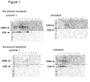
- Figure 1 is a comparison of the amount of exosome recovered (the amount recovered) in each fraction fractionated in 10 fractions and the amount of exosome that was adsorbed to the centrifuge tube and could not be recovered (the amount adsorbed) when using the coating agent containing polymer 1 as the hydrophilic polymer or an untreated centrifuge tube. As shown in Figure 1 , when performing the purification of exosomes without using the coating agent, the amount of exosome recovered was very small and many of the exosomes were adsorbed to the centrifuge tube.
- the precipitate was suspended in 250 ⁇ L of PBS and recovered.
- the centrifuge tube after recovering the precipitate was eluted by adding 150 ⁇ L of 1 x SDS sample buffer (first centrifuge tube adsorption sample).
- Example 2 The number of particles of exosome recovered in Example 2 was measured with NanoSight LM10 (manufactured by Malvern Panalytical Ltd.). The calibration was performed with Silica Microspheres: 100 nm (Polysiciences, Inc. #24041) diluted with Milli-Q water passed through a GE Puradisc 13 (NYL 0.1 ⁇ m). Next, the exosomes were diluted to approximately 10 8 with PBS passed through a GE Puradisc 13, the sample solution was injected in the measurement cell with a syringe and waited 10 s, then data measurement for 30 s was performed. The measurement was performed 5 times while replacing the inside of the cell with a new sample, by pushing the syringe and moving the sample solution in the chamber. Figure 3 shows the average value with standard deviation. The Detection threshold was 4 and the Camera level was 14.
- the exosomes were purified as below from the culture supernatant of HT-29 cells.
- the culture supernatant (500 mL) of HT-29 cells prepared as above was centrifuged at 160,000 x g for 70 min to precipitate the exosomes, then the precipitate was resuspended in 180 mL of PBS, and further centrifuged at 160,000 x g for 70 min and washed. The obtained precipitate was resuspended in 1.3 mL of PBS, to form an exosome crude fraction.
- the exosome crude fraction (1.3 mL) was mixed with 1.7 mL of 46% Iodixanol (OptiPrep, manufactured by COSMO BIO CO., LTD.) and placed on the bottom of a centrifuge tube, then 8% to 40% of Iodixanol continuous density gradient was superposed on top of it.
- Equilibrium density gradient centrifugation was performed at 100,000 x g for 17 h in the centrifuge tube by a Beckman L-90K centrifuge with a SW 32 Ti rotor. After centrifugation, 3 fractions of 4.26 mL each were recovered from the upper liquid surface of the sample, then 6 fractions of 3.26 mL each were recovered, dividing it into a total of 9 fractions.
- fraction 2 (density: 1.36 g/cm 3 ) was used as the purified exosome sample in this experiment.
- the exosome solution (30 ⁇ L) of a density of 1.36 g/cm 3 obtained from the culture supernatant of HT-29 cells was each placed in microtubes (manufactured by Eppendorf AG). Tubes coated in advance with the hydrophilic polymers, polymer 1 or polymer 4 were prepared. For the adsorption experiment, it was allowed to stand still for 10 min, then after centrifuging at 13,200 rpm at 4°C for 30 min, it was stirred for 10 s with a vortex mixer and the exosomes were recovered. If performing the centrifugation twice, the exosomes were recovered after performing the same step twice.
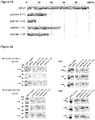
- Figure 4A shows the analysis results of the number of particles of exosomes recovered.
- the average value of the three experiments is shown as the ratio to the input, and the numbers between parentheses show the number of centrifugations.
- polymer 1 50% or more of the particles to the input can be recovered when performing the centrifugation once, and 30% or more even when performing the centrifugation twice.
- the recovery rate was only 30% when performing the centrifugation once, and 15% when performing it twice.
- Figure 4B shows the results of the analysis by western blotting.
- the recovered samples and the samples adsorbed to the tube in the first, second and third experiments were analyzed, and each panel shows the analysis results of the first experiment to the third experiment in this order from the top.
- Detection was performed using anti-EpCAM antibodies (manufactured by R&D Systems, Inc.) and anti-CD9 antibodies.
- polymer 4 with the sample recovered after performing the centrifugation twice, almost nothing was detected when using either anti-EpCAM antibodies or anti-CD9 antibodies, but when using polymer 1, it was detectable in both cases.
- Example 4 A comparison of the amount adsorbed according to the coated layer thickness of the coating agent was conducted.
- Polymer 1 was dissolved in ethanol so as to be 0.5, 1.0, 2.0 and 4.0 wt% and then microtubes (manufactured by Treff AG) were coated with them changing the coated layer thickness from 90 nm to 1400 nm, to use for the analysis.
- the coated layer thickness was measured using a reflection spectroscopic film thickness meter (FE-3000, manufactured by OTSUKA ELECTRONICS Co., LTD.).
- the amount of exosomes recovered was compared by using the exosomes obtained from the culture supernatant of HT-29 cells and performing centrifugation once or twice.
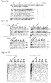
- Figure 5A shows the analysis results of the number of particles of exosomes recovered.
- the numbers 90, 140, 370 and 1400, that are shown on the left, indicate the thickness (nm) of the coated layer with which the microtubes were coated.
- the average value of the three experiments is shown as the ratio to the input, and the numbers between parentheses show the number of centrifugations.
- a symbol, - indicates the results when using an untreated tube. With either coated layer thickness, when using a coated tube, 50% or more of the particles to the input could be recovered. By contrast, with the untreated tube, only approximately 45% on average of the particles could be recovered when performing the centrifugation once, and 20% or less on average when performing the centrifugation twice.
- the thickness of the coating agent No difference between the amounts recovered according to the thickness of the coating agent was observed in the analysis of the number of particles, but it is preferable to be a coated layer thickness of 50 nm or more and 2000 nm or less when considering the variation during the coated layer formation and the work efficiency. If the coated layer thickness is within this range, the adsorption of exosomes should be prevented and stable measurement results should be obtained.
- Figure 5B shows the results of the analysis by western blotting. As in Example 4, it shows the results of the detection by anti-EpCAM antibodies and anti-CD9 antibodies. With either coated layer thickness, results clearly showing that the adsorption of exosomes can be suppressed compared to an uncoated tube, were obtained.
- Figure 5C shows the results of analysis of the recovered samples and the samples adsorbed to the tube by SDS electrophoresis and silver staining.
- the amount recovered clearly decreases.
- the adsorbed sample decreases and the recovered sample increases.
- the adsorption of exosomes to the container decreases and the recovery rate significantly improves, by using the coating agents containing polymers 1 to 3. These coating agents allow not only to improve the exosome recovery rate, but also to accurately perform quantitative analysis.
- Using the coating agents of the present invention allows to prevent that extracellular vesicles including exosomes adsorb to the container, the substrate used in the analysis or the like. Thus, it allows to significantly improve the recovery rate of extracellular vesicles and also improve the accuracy of the analysis.
Landscapes
- Chemical & Material Sciences (AREA)
- Health & Medical Sciences (AREA)
- Organic Chemistry (AREA)
- Life Sciences & Earth Sciences (AREA)
- Engineering & Computer Science (AREA)
- Medicinal Chemistry (AREA)
- Materials Engineering (AREA)
- Wood Science & Technology (AREA)
- Chemical Kinetics & Catalysis (AREA)
- Polymers & Plastics (AREA)
- Immunology (AREA)
- Hematology (AREA)
- Molecular Biology (AREA)
- Urology & Nephrology (AREA)
- Biomedical Technology (AREA)
- Analytical Chemistry (AREA)
- Microbiology (AREA)
- Cell Biology (AREA)
- Food Science & Technology (AREA)
- Physics & Mathematics (AREA)
- Biotechnology (AREA)
- Biochemistry (AREA)
- General Health & Medical Sciences (AREA)
- General Physics & Mathematics (AREA)
- Pathology (AREA)
- Solid-Sorbent Or Filter-Aiding Compositions (AREA)
- Apparatus Associated With Microorganisms And Enzymes (AREA)
- Paints Or Removers (AREA)
Abstract
Description
- The present invention relates to a method for efficiently recovering cell-derived vesicles represented by exosomes, and a container and a tool for the recovery. Moreover, it relates to a substrate, channel or the like to use in methods for testing with exosomes.
- Extracellular vesicles are vesicles originating from cells, present outside the cell, and classified as exosomes, microvesicles, apoptotic bodies depending on their size. Exosomes and microvesicles contain proteins and nucleic acids such as mRNA and miRNA, and apoptotic bodies contain fragmented nuclei and organelles. Among extracellular vesicles, since exosomes and microvesicles contain proteins and nucleic acids, they also contain information from the emitting cell, and thereby have the possibility to be used as biomarkers. Especially, exosomes include the possibility to be used as diagnostic markers and in treatments and have attracted attention in recent years.
- Exosomes are vesicles with a diameter of 30 to 100 nm, formed by a lipid bilayer membrane secreted by the cell. Exosomes are known to be secreted by most cells and their presence has been confirmed in body fluids such as blood, saliva, urine and breast milk. Since exosomes contain RNA and proteins from the cell from which they were secreted, it has been suggested that they may transmit information to distant cells by circulating in body fluids (Non Patent Literature 1). It has been shown that the progress of cancer may be promoted by the nucleic acids and proteins contained in the exosomes from cancer cells, thereby the possibility of a establishing cancer diagnosis system using exosomes has been suggested (Non Patent Literature 2).
- As mentioned above, it has been made clear that the proteins and nucleic acids contained in exosomes differ depending on the cell type they were secreted from. Therefore, the nucleic acids and proteins from exosomes are useful as diagnostic markers. Moreover, since exosomes are also contained in saliva and urine, it is possible to have a non-invasive diagnosis method if a testing method with high accuracy using exosomes can be established. Moreover, attempts are made to use exosomes taken and collected from healthy cells for the recovery of damaged cells or diseased cells, by using the information transmission function of exosomes (Patent Literature 1).
- A purifying method with a good recovery rate is necessary to purify exosomes from a small amount of sample, such as saliva, or to purify a large amount of exosomes for therapeutic purposes. As conventional methods for purifying exosomes, methods for isolating them by centrifugal separation or columns are disclosed (
Patent Literatures 2 and 3). Among these, centrifugal separation method using ultracentrifugation is an established recovery technique and is an excellent technique in that the shape of exosomes can be maintained. - For example, if recovering exosomes from the culture supernatant of cells, after removing cell debris from the culture medium containing the exosomes by low-speed centrifugation, the exosomes can be recovered as pellets by ultracentrifugation for about 2 h. The thus obtained exosomes can be redispersed in a buffer solution such as PBS and further purified, or directly used for analysis in tests, research and the like.
-
- Patent Literature 1: National Publication of International Patent Publication No.
2014-507140 - Patent Literature 2: National Publication of International Patent Publication No.
2003-531864 - Patent Literature 3: National Publication of International Patent Publication No.
2002-535665 - Patent Literature 4: Japanese Patent Laid-Open No.
7-83923 - Patent Literature 5: International Publication No.
WO 2005/001019 -
- Non Patent Literature 1: Valadi, H. et al., 2007, Nat. Cell Biol., Vol.9, p.654-659.
- Non Patent Literature 2: Skog, J. et al., 2008, Nat. Cell Biol., Vol.10, pp.1470-1476.
- However, in the above recovery method by ultracentrifugation, the problem was that the exosomes non-specifically adsorbed to the plastic surface of the recovery container that is used, and thus a stable recovery rate could not be obtained and the recovery rate decreased at each operation. Especially when performing quantitative analysis, the adsorption to the surface of the containers used for purification and of the analytical instruments influences not only the efficiency of the analysis, but also the analysis results. However, an effective method to prevent the adsorption of extracellular vesicles such as exosomes has not yet been developed.
- A method for preventing proteins, which are tissue-derived biomaterials, and cells to non-specifically adsorb to the container by a hydrophilic polymer, is disclosed (
Patent Literatures 4 and 5).Patent Literature 4 discloses that the adsorption of proteins can be prevented by coating a polystyrene container with a PBS solution containing a 2-methacryloyloxyethyl phosphorylcholine polymer or a copolymer of a component containing 2-methacryloyloxyethyl phosphorylcholine.Patent Literature 5 discloses that the adhesion of ES cells to the culture vessel can be prevented and the embryoid body can be formed efficiently, by covering the culture vessel using a compound containing a phosphorylcholine analogous group. - However, in
Patent Literature 4, the method is specific to proteins, and although proteins appear on the surface of exosomes etc., it was difficult to completely prevent the adsorption of the extracellular vesicles formed by a lipid bilayer membrane. Moreover, the method inPatent Literature 5 prevents the adsorption of cells formed by a lipid bilayer membrane, but between cells and extracellular vesicles, especially exosomes, their size is completely different, and thus the degree of adsorption to the container is significantly different, so that the adsorption of the exosomes to the container could not be completely prevented. - The present invention aims to provide a coating agent to prevent the adsorption of cell-derived vesicles to the container used for purification such as a centrifuge tube or to tools such as pipette chips, when purifying cell-derived vesicles represented by exosomes, and to improve the recovery rate. Moreover, it aims to provide a coating agent that not only can be used during the purification process, but also can improve the accuracy of exosome analysis by coating the substrates, channels or the like used for exosome analysis. Furthermore, it aims to provide containers or analytical tools such as a chip or a substrate coated with these coating agents.
- The present invention relates to the following coating agents, tools, purification methods and analysis methods.
- (1) A coating agent for preventing the adsorption of extracellular vesicles, comprising: a hydrophilic polymer having a weight average molecular weight of 10,000 or more and 1,000,000 or less, wherein a coated layer formed by the coating agent has a contact angle of 0 degree or more and 30 degrees or less.
- (2) The coating agent according to (1), wherein the hydrophilic polymer comprises one or more hydrophilic monomers of 2-methacryloyloxyethyl phosphorylcholine, vinyl alcohol, vinylpyrrolidone, methoxy alkylene glycol monomethacrylate and 2-hydroxyethyl methacrylate.
- (3) The coating agent according to (2), wherein the hydrophilic monomer constituting the hydrophilic polymer is 2-methacryloyloxyethyl phosphorylcholine, and wherein the coating agent is a copolymer containing 30 mol% or more and 50 mol% or less of constituting unit based on 2-methacryloyloxyethyl phosphorylcholine, with the balance being a hydrophobic group-containing monomer.
- (4) A tool coated with the coating agent according to any one of (1) to (3).
- (5) A method for purifying and/or analyzing extracellular vesicles using the tool according to (4).
-
-
Figure 1 is a drawing showing the amount of exosomes recovered according to the presence or absence of the coating agent. -
Figure 2 is a drawing showing the amount of exosomes recovered according to the type of coating agent. -
Figure 3 is a drawing showing the results of examining the amount of exosomes recovered according to the coating agent by the number of particles. -
Figure 4A is a drawing showing the results of examining the amount of exosomes recovered according to the coating agent by the number of particles, andFigure 4B by western blotting. -
Figure 5A is a drawing showing the results of examining the amount of exosomes recovered according to the coated layer thickness of the coating agent by the number of particles, andFigure 5B is a drawing analyzing the results by western blotting.Figure 5C is a drawing analyzing the recovered sample and adsorbed sample by electrophoresis and silver staining. - In the present invention, an extracellular vesicle means a vesicle derived from a cell, and concretely, refers to exosomes, microvesicles and apoptotic bodies. The following embodiments focus mainly on exosomes, but it is needless to say that they can also be applied to extracellular vesicles other than exosomes.
- Moreover, by coating the tools used for the purification of extracellular vesicles like exosomes, such as centrifuge tubes, tubes and pipette chips with the coating agent of the present invention, it can prevent their adsorption and improve the recovery rate. Moreover, by coating analytical tools and analytical instruments used in this field, such as beads, microtiter plates, slide glass, micro-TAS and lab-on-a-chip with it, it can prevent nonspecific adsorption and improve the analytical precision.
- Tools of any materials can be coated with the coating agent of the present invention. For example, by coating polycarbonate, polypropylene and polyallomer that are often used for centrifuge tubes, vinyl chloride, polystyrene, polyethylene terephthalate, silicon, hydrophilic polydimethylsiloxane, hydrophobic polydimethylsiloxane, glass and metals such as stainless and aluminum that are used as base materials for analytical tools, with the coating agent of the present invention, the adsorption of extracellular vesicles such as exosomes can be prevented.
- The contact angle of the coated layer obtained by the coating agent of the present invention is 0 degree or more and 30 degrees or less, and it is preferable that it be 0 degree or more and 25 degrees or less. Moreover, regarding the molecular weight of the hydrophilic polymer of the present invention, the weight average molecular weight is 10,000 or more and 1,000,000 or less, and in terms of durability of the coated layer, 100,000 or more is preferable, and it is more preferable to be 700,000 or less to suppress coating unevenness due to the high viscosity during coating.
- Moreover, the hydrophilic monomer constituting the hydrophilic polymer can be any hydrophilic monomer as long as it can synthesize a hydrophilic polymer or copolymer, and among these, it is preferable that it be 2-methacryloyloxyethyl phosphorylcholine, vinyl alcohol, vinylpyrrolidone, methoxy alkylene glycol monomethacrylate or 2-hydroxyethyl methacrylate. These hydrophilic monomers may be used as a polymer, or may be used as a copolymer with another monomer.
- Among these, the copolymer of 2-methacryloyloxyethyl phosphorylcholine (MPC) and a hydrophobic monomer can be used preferably. As the hydrophobic monomer, methyl methacrylate (MMA), n-butyl methacrylate (BMA), 2-ethylhexyl methacrylate (EhMA), dodecyl methacrylate (DMA), stearyl methacrylate (SMA), etc. can be used, and the copolymer with n-butyl methacrylate is particularly preferable in terms of availability and film-forming properties. Moreover, when preparing a copolymer of MPC and a hydrophobic monomer, the composition ratio of MPC and the hydrophobic monomer should be 20/80 to 50/50.
- The coating agent of the present invention can be used as a solution containing the above polymer or copolymer in a concentration of 0.05% or more and 5.0% or less. The desired effect cannot be produced when it is less than 0.05 weight%, and a reduction of the effect due to the coating unevenness of the coating agent and the physical peeling of the coating agent may occur when it is more than 5 weight%. As the solvent, alcohols such as ethanol and methanol, or buffer solutions conventionally used in this field such as a phosphate buffer solution, a tris buffer solution, PBS or TBS can be used.
- Moreover, the coated layer thickness when forming a coated layer on a tool with the coating agent, is preferably 50 nm or more and 2000 nm or less, and more preferably, is preferably 90 nm or more and 900 nm or less.
- When forming a coated layer on a tool, it is not possible to form the coated layer completely evenly. Since it is very difficult to form a coated layer with a thin thickness, when the average thickness is less than 50 nm, the variation of the coated layer thickness increases and spots where the effect preventing the adsorption of the exosomes is weak are generated. Moreover, when the coated layer thickness is greater than 2000 nm, the viscosity of the coating liquid (polymer solution) used to form the coated layer increases and the amount of polymer used also increases, making it difficult in terms of work efficiency during coated layer forming (in terms of drying and operability) and economic efficiency. If the coated layer thickness is 50 nm or more and 2000 nm or less, it is effective to prevent the adsorption of exosomes. Moreover, when the coated layer thickness is 90 nm or more and 900 or less, the adsorption of exosomes can be prevented and stable measurement results can be obtained since the coated layer can be formed at a stable thickness.
- The present invention is described below in details while showing examples.
- In the following examples, the polymers shown in the following Table 1 were used.
- MPC (23.5 g) and BMA (26.5 g) were dissolved in ethanol (50 g) and placed in a four-neck flask, nitrogen was bubbled for 30 min, then t-butyl peroxyneodecanoate (0.1 g) was added at 45°C and the polymerization reaction was performed for 24 h. The polymerization liquid was added dropwise to 3 L of diethyl ether while stirring, the precipitate was filtered, then dried in vacuum for 48 h at 45°C, to obtain 36.8 g of powder. The molecular weight was measured by gel permeation chromatography (manufactured by Tosoh Corporation) using polyethylene glycol as the standard. The weight-average molecular weight was 580,000. An analysis of the composition by 1H-NMR showed that MPC/BMA = 30/70 (mole ratio).
- The above polymer (0.5 g) was dissolved in ethanol (100 g) to prepare a solution of 0.5 weight%. After applying this on a PET film, it was air-dried and washed with water. This was used as the sample for measuring the contact angle. The contact angle measurement was performed by the bubble method using a DropMaster (manufactured by Kyowa Interface Science Co., LTD.) and the value of the contact angle was obtained as the water's contact angle. Concretely, it was calculated as "180° - bubble angle". The value of the contact angle for the coated layer obtained was 25 degrees.
- MPC (4.7 g) and BMA (5.3 g) were dissolved in ethanol (90 g) and placed in a four-neck flask, nitrogen was bubbled for 30 min, then t-butyl peroxyneodecanoate (0.25 g) was added at 60°C and the polymerization reaction was performed for 24 h. The polymerization liquid was added dropwise to 3 L of diethyl ether while stirring, the precipitate was filtered, then dried in vacuum for 48 h at 45°C, to obtain 36.8 g of powder. The weight-average molecular weight was 120,000. An analysis of the composition by 1H-NMR showed that MPC/BMA = 30/70 (mole ratio). The value of the contact angle for the coated layer obtained was 20 degrees.
- Poly(2-hydroxyethyl methacrylate) with a molecular weight of 300,000 (manufactured by Sigma-Aldrich) was used for
polymer 3. The value of the contact angle for the coated layer obtained was 30 degrees. - For
polymer 4, 8.4 g of MPC and 36.6 g of BMA were dissolved in 54.6 g of ethanol and placed in a four-neck flask, nitrogen was bubbled for 30 min, then 0.37 g of t-butyl peroxyneodecanoate was added at 60°C and the polymerization reaction was performed for 24 h. The polymerization liquid was added dropwise to 3 L of diethyl ether while stirring, the precipitate was filtered, then dried in vacuum for 48 h at 45°C, to obtain 26.3 g of powder. The weight-average molecular weight was 340,000. An analysis of the composition by 1H-NMR showed that MPC/BMA = 10/90 (mole ratio). The value of the contact angle for the coated layer obtained was 42 degrees.[Table 1] Composition Composition ratio (mole ratio) Weight-average molecular weight Contact angle Polymer 1 MPC/ BMA 30/70 580,000 25 Polymer 2MPC/ BMA 30/70 120,000 20 Polymer 3HEMA 100 300,000 30 Polymer 4MPC/ BMA 10/90 340,000 42 - The coating of the container used for purification was performed by using the coating agent containing the above hydrophilic polymers, and the analysis of the amount of exosome adsorbed was performed. For the coating, the container surface was wetted with an ethanol solution containing 0.5 wt% of each hydrophilic polymer, i.e.
polymers 1 to 3 andpolymer 4, that is the comparative example, shown in Table 1, then dried after eliminating the excess liquid. - The exosomes were obtained from the culture supernatant of HuH-7 cells derived from human liver cancer, or HT-29 cells derived from colon cancer. The following was performed to obtain exosomes from HuH-7 cells. HuH-7 cells (1x10⁶) were suspended in 25 mL of RPMI-1640 medium supplemented with 10% fetal bovine serum (manufactured by Thermo Fisher Scientific Inc.) and 1/100 of penicillin-streptomycin (Penicillin-Streptomycin Solution x100, manufactured by Wako Pure Chemical Industries, Ltd.), then seeded in a 150 mm petri dish (cell culture dishes 150 mm, manufactured by IWAKI & CO., LTD.) and cultured in 5% CO2, at 37°C for 72 h. Then, the medium was sucked and removed, 25 mL of RPMI-1640 medium without fetal bovine serum was added and cultured in 5% CO2, at 37°C for 66 h. After 66 h, the culture supernatant was recovered, subjected to low-speed centrifugation at 8,900 x g for 10 min, and the supernatant was passed through a 0.22 µm filter (milliex-GS SLGV033RS, manufactured by Merck).
- The following was performed to obtain exosomes from HT-29 cells. HT-29 cells (2x10⁸) were seeded in 500 mL of McCoy's 5A medium supplemented with 10% fetal bovine serum and penicillin-streptomycin and cultured in 5% CO2, at 37°C for 96 h, using a Bellocell incubator (manufactured by CESCO). Next, the medium was removed by decantation, 500 mL of McCoy's 5A medium without fetal bovine serum was added and further cultured in 5% CO2, at 37°C for 72 h. After 72 h, the culture supernatant was recovered, subjected to low-speed centrifugation as above, and the culture supernatant containing the exosomes was obtained by passing the supernatant through the filter.
- Concentration of the exosomes was performed by centrifuging 480 mL of culture supernatant of HuH-7 cells prepared above at 30,700 rpm for 70 min. The precipitate was resuspended in 12 mL of culture supernatant and 60 mL of culture supernatant was further added to obtain a sample in which the exosomes were concentrated.
- In the following ultracentrifugation step, the samples containing the exosomes were dispensed in equal amounts in tubes for ultracentrifugation (UC tube, Ultra-Clear Tubes, manufactured by Beckman Coulter, Inc.) coated with the coating agents, each containing
polymer 1 as the hydrophilic polymer, or an uncoated centrifuge tubes, and the purification was performed, then the exosome recovery rate was compared according to the presence or absence of the coating agent. The coated layer thickness of the coating agent used in the following experiments was about 90 nm unless otherwise stated. - The sample containing the concentrated exosomes was centrifuged at 30,700 rpm for 120 min. The obtained precipitate was suspended by pipetting in 3 mL of 45% OptiPrep (manufactured by COSMO BIO CO., LTD.), centrifuged at 24,200 rpm for 20 min using a 8-40% gradient, then collected in 10 fractions. Each collected fraction was centrifuged at 30,700 rpm for 120 min, the precipitate was suspended in 250 µL of PBS and then recovered.
- The exosomes recovered in each fraction and the amount of exosome adsorbed on the centrifuge tube was analyzed by western blotting. Western blotting was performed according to a conventional method, and CD9 and CD63, which are known as exosome markers, were detected. Anti-CD9 antibodies and anti-CD63 antibodies (both manufactured by COSMO BIO CO., LTD.) were reacted each as primary antibodies with HRP-labeled anti-mouse IgG antibodies (manufactured by Bio-Rad Laboratories, Inc.) as secondary antibodies and chemiluminescence detection was performed.
-
Figure 1 is a comparison of the amount of exosome recovered (the amount recovered) in each fraction fractionated in 10 fractions and the amount of exosome that was adsorbed to the centrifuge tube and could not be recovered (the amount adsorbed) when using the coatingagent containing polymer 1 as the hydrophilic polymer or an untreated centrifuge tube. As shown inFigure 1 , when performing the purification of exosomes without using the coating agent, the amount of exosome recovered was very small and many of the exosomes were adsorbed to the centrifuge tube. By contrast, when performing the purification using a centrifuge tube coated withpolymer 1 as the hydrophilic polymer, the amount of exosome adsorbed to the centrifuge tube was very small and most of the exosomes could be resuspended and recovered. - A sample in which 23 mL of PBS was added to 12 mL of culture supernatant of HuH-7 cells which were filtered through a 0.22 µm filter after low-speed centrifugation as above, was placed in a centrifuge tube coated with the
polymers 1 to 3 as the hydrophilic polymer, or an untreated centrifuge tube, and was subjected to ultracentrifugation at 37,000 rpm for 70 min. The precipitate was suspended in 250 µL of PBS and recovered. The centrifuge tube after recovering the precipitate was eluted by adding 150 µL of 1 x SDS sample buffer (first centrifuge tube adsorption sample). 30 mL of PBS was added to the recovered exosomes and the mixture was subjected to centrifugation again at 37,000 rpm for 120 min, using a new centrifuge tube coated withpolymers 1 to 3 as the hydrophilic polymer, or a new untreated centrifuge tube. The exosomes were recovered by suspending them with PBS to amount to 150 µL. Then, the centrifuge tube after recovering the exosomes was eluted by adding 150 µL of 1 x SDS sample buffer (second centrifuge tube adsorption sample). - The recovered exosomes and the exosomes adsorbed to the centrifuge tube at the first and second ultracentrifugation were analyzed by western blotting in the same manner as in Example 1. The results are shown in
Figure 2 . - The same experiment was performed 3 times and
Figure 2 shows the results of the western blotting for each experiment. In the drawing, supernatant (sup) indicates that the culture supernatant was used, - the untreated centrifuge tube, and 1 to 3 the centrifuge tubes coated with the coatingagent containing polymers 1 to 3 as the hydrophilic polymer. - In either instance detecting the marker CD63 or CD9, the exosomes were efficiently recovered when using the coating agent. On the other hand, when using the untreated (-) centrifuge tube, the amount of exosome adsorbed non-specifically to the centrifuge tube increases and the recovery rate greatly decreases.
- The number of particles of exosome recovered in Example 2 was measured with NanoSight LM10 (manufactured by Malvern Panalytical Ltd.). The calibration was performed with Silica Microspheres: 100 nm (Polysiciences, Inc. #24041) diluted with Milli-Q water passed through a GE Puradisc 13 (NYL 0.1 µm). Next, the exosomes were diluted to approximately 108 with PBS passed through a GE Puradisc 13, the sample solution was injected in the measurement cell with a syringe and waited 10 s, then data measurement for 30 s was performed. The measurement was performed 5 times while replacing the inside of the cell with a new sample, by pushing the syringe and moving the sample solution in the chamber.
Figure 3 shows the average value with standard deviation. The Detection threshold was 4 and the Camera level was 14. - It is clear that the recovery rate of the exosomes greatly improves compared to when performing the purification using an untreated centrifuge tube, in all instances using the coating agent containing either hydrophilic polymers,
polymers 1 to 3. - Centrifugation was performed once or twice as below, and the amount of exosome recovered and the amount adsorbed to the tube were examined. The exosomes were purified as below from the culture supernatant of HT-29 cells. The culture supernatant (500 mL) of HT-29 cells prepared as above was centrifuged at 160,000 x g for 70 min to precipitate the exosomes, then the precipitate was resuspended in 180 mL of PBS, and further centrifuged at 160,000 x g for 70 min and washed. The obtained precipitate was resuspended in 1.3 mL of PBS, to form an exosome crude fraction.
- The exosome crude fraction (1.3 mL) was mixed with 1.7 mL of 46% Iodixanol (OptiPrep, manufactured by COSMO BIO CO., LTD.) and placed on the bottom of a centrifuge tube, then 8% to 40% of Iodixanol continuous density gradient was superposed on top of it. Equilibrium density gradient centrifugation was performed at 100,000 x g for 17 h in the centrifuge tube by a Beckman L-90K centrifuge with a SW 32 Ti rotor. After centrifugation, 3 fractions of 4.26 mL each were recovered from the upper liquid surface of the sample, then 6 fractions of 3.26 mL each were recovered, dividing it into a total of 9 fractions. Each fraction was suspended in 27 mL of PBS and centrifuged at 160,000 x g for 120 min, then the precipitate was suspended in 500 µL of PBS to obtain the purified exosomes. Of these 9 fractions, fraction 2 (density: 1.36 g/cm3) was used as the purified exosome sample in this experiment.
- The exosome solution (30 µL) of a density of 1.36 g/cm3 obtained from the culture supernatant of HT-29 cells was each placed in microtubes (manufactured by Eppendorf AG). Tubes coated in advance with the hydrophilic polymers,
polymer 1 orpolymer 4 were prepared. For the adsorption experiment, it was allowed to stand still for 10 min, then after centrifuging at 13,200 rpm at 4°C for 30 min, it was stirred for 10 s with a vortex mixer and the exosomes were recovered. If performing the centrifugation twice, the exosomes were recovered after performing the same step twice. Regarding the recovered exosomes was analyzed by western blotting, and the number of particles was examined using a NanoSight LM10. The experiment was performed three times, and the average number of particles and the results of the three experiments by western blotting are shown (Figure 4 ). -
Figure 4A shows the analysis results of the number of particles of exosomes recovered. The average value of the three experiments is shown as the ratio to the input, and the numbers between parentheses show the number of centrifugations. When usingpolymer 1, 50% or more of the particles to the input can be recovered when performing the centrifugation once, and 30% or more even when performing the centrifugation twice. By contrast, when using the Comparative Example,polymer 4, the recovery rate was only 30% when performing the centrifugation once, and 15% when performing it twice. -
Figure 4B shows the results of the analysis by western blotting. The recovered samples and the samples adsorbed to the tube in the first, second and third experiments were analyzed, and each panel shows the analysis results of the first experiment to the third experiment in this order from the top. Detection was performed using anti-EpCAM antibodies (manufactured by R&D Systems, Inc.) and anti-CD9 antibodies. When usingpolymer 4, with the sample recovered after performing the centrifugation twice, almost nothing was detected when using either anti-EpCAM antibodies or anti-CD9 antibodies, but when usingpolymer 1, it was detectable in both cases. - A comparison of the amount adsorbed according to the coated layer thickness of the coating agent was conducted.
Polymer 1 was dissolved in ethanol so as to be 0.5, 1.0, 2.0 and 4.0 wt% and then microtubes (manufactured by Treff AG) were coated with them changing the coated layer thickness from 90 nm to 1400 nm, to use for the analysis. The coated layer thickness was measured using a reflection spectroscopic film thickness meter (FE-3000, manufactured by OTSUKA ELECTRONICS Co., LTD.). As in Example 4, the amount of exosomes recovered was compared by using the exosomes obtained from the culture supernatant of HT-29 cells and performing centrifugation once or twice. -
Figure 5A shows the analysis results of the number of particles of exosomes recovered. Thenumbers - No difference between the amounts recovered according to the thickness of the coating agent was observed in the analysis of the number of particles, but it is preferable to be a coated layer thickness of 50 nm or more and 2000 nm or less when considering the variation during the coated layer formation and the work efficiency. If the coated layer thickness is within this range, the adsorption of exosomes should be prevented and stable measurement results should be obtained.
-
Figure 5B shows the results of the analysis by western blotting. As in Example 4, it shows the results of the detection by anti-EpCAM antibodies and anti-CD9 antibodies. With either coated layer thickness, results clearly showing that the adsorption of exosomes can be suppressed compared to an uncoated tube, were obtained. -
Figure 5C shows the results of analysis of the recovered samples and the samples adsorbed to the tube by SDS electrophoresis and silver staining. When using an untreated tube, the amount recovered clearly decreases. By contrast, when performing coating with either coated layer thickness, it is clear that the adsorbed sample decreases and the recovered sample increases. - As shown in the above examples, the adsorption of exosomes to the container decreases and the recovery rate significantly improves, by using the coating
agents containing polymers 1 to 3. These coating agents allow not only to improve the exosome recovery rate, but also to accurately perform quantitative analysis. - Using the coating agents of the present invention allows to prevent that extracellular vesicles including exosomes adsorb to the container, the substrate used in the analysis or the like. Thus, it allows to significantly improve the recovery rate of extracellular vesicles and also improve the accuracy of the analysis.
Claims (5)
- A coating agent for preventing the adsorption of extracellular vesicles, comprising:a hydrophilic polymer having a weight average molecular weight of 10,000 or more and 1,000,000 or less,wherein a coated layer formed by the coating agent has a contact angle of 0 degree or more and 30 degrees or less.
- The coating agent according to claim 1,
wherein the hydrophilic polymer comprises one or more hydrophilic monomers of 2-methacryloyloxyethyl phosphorylcholine, vinyl alcohol, vinylpyrrolidone, methoxy alkylene glycol monomethacrylate and 2-hydroxyethyl methacrylate. - The coating agent according to claim 2, wherein
the hydrophilic monomer constituting the hydrophilic polymer is 2-methacryloyloxyethyl phosphorylcholine, and wherein
the coating agent is a copolymer containing 30 mol% or more and 50 mol% or less of constituting unit based on 2-methacryloyloxyethyl phosphorylcholine,
with the balance being a hydrophobic group-containing monomer. - A tool coated with the coating agent according to any one of claims 1 to 3.
- A method for purifying and/or analyzing extracellular vesicles using the tool according to claim 4.
Applications Claiming Priority (2)
| Application Number | Priority Date | Filing Date | Title |
|---|---|---|---|
| JP2016103091 | 2016-05-24 | ||
| PCT/JP2017/019121 WO2017204187A1 (en) | 2016-05-24 | 2017-05-23 | Method of recovering extracellular vesicles and container for extracellular vesicles |
Publications (3)
| Publication Number | Publication Date |
|---|---|
| EP3467059A1 true EP3467059A1 (en) | 2019-04-10 |
| EP3467059A4 EP3467059A4 (en) | 2020-03-11 |
| EP3467059B1 EP3467059B1 (en) | 2022-02-23 |
Family
ID=60411962
Family Applications (1)
| Application Number | Title | Priority Date | Filing Date |
|---|---|---|---|
| EP17802773.6A Active EP3467059B1 (en) | 2016-05-24 | 2017-05-23 | Method of recovering extracellular vesicles and container for extracellular vesicles |
Country Status (5)
| Country | Link |
|---|---|
| US (1) | US10955410B2 (en) |
| EP (1) | EP3467059B1 (en) |
| JP (1) | JP6970665B2 (en) |
| CN (1) | CN109153882B (en) |
| WO (1) | WO2017204187A1 (en) |
Cited By (2)
| Publication number | Priority date | Publication date | Assignee | Title |
|---|---|---|---|---|
| WO2022203603A1 (en) * | 2021-03-26 | 2022-09-29 | N-Lab Technology Center Pte. Ltd. | Water-soluble polymer to prevent non-specific adsorption |
| US12269003B2 (en) | 2019-12-30 | 2025-04-08 | Industrial Technology Research Institute | Extracellular vesicle separation method, colloidal particle and preparation method thereof |
Families Citing this family (3)
| Publication number | Priority date | Publication date | Assignee | Title |
|---|---|---|---|---|
| JP7451385B2 (en) * | 2020-12-07 | 2024-03-18 | 大日本塗料株式会社 | Method for detecting particulate matter by immunochromatography and kit therefor |
| CN115887758B (en) * | 2022-11-15 | 2024-01-26 | 西南交通大学 | Copolymerized hydrogel for supporting exosomes and promoting diabetic wound repair |
| CN118345022A (en) * | 2024-01-24 | 2024-07-16 | 上海市肺科医院(上海市职业病防治院) | A method for enriching and extracting exosomes in saliva and its application in screening latent tuberculosis infection |
Family Cites Families (20)
| Publication number | Priority date | Publication date | Assignee | Title |
|---|---|---|---|---|
| JP2890316B2 (en) * | 1989-07-07 | 1999-05-10 | 科学技術振興事業団 | Materials for biocompatible medical devices |
| JPH075177A (en) * | 1993-06-15 | 1995-01-10 | Nippon Oil & Fats Co Ltd | Method and reaction vessel for biochemical analysis |
| JP3443891B2 (en) | 1993-09-14 | 2003-09-08 | 日本油脂株式会社 | Protein adsorption inhibitor |
| JP2005099040A (en) * | 1998-12-24 | 2005-04-14 | Sumitomo Bakelite Co Ltd | Vessel for immunoassay |
| CA2356857A1 (en) * | 1998-12-24 | 2000-07-06 | Sumitomo Bakelite Co., Ltd. | Container for immunologic assay |
| FR2788780B1 (en) | 1999-01-27 | 2001-03-30 | Ap Cells Inc | PROCESS FOR THE PREPARATION OF MEMBRANE VESICLES |
| US20040241176A1 (en) | 2000-04-27 | 2004-12-02 | Ap Cells. Inc. | Method of producing membrane vesicles |
| JP3985578B2 (en) * | 2001-04-26 | 2007-10-03 | 日本油脂株式会社 | Clinical laboratory method |
| JP2004275862A (en) * | 2003-03-14 | 2004-10-07 | Sumitomo Bakelite Co Ltd | Laboratory instrument for controlling adsorption of sugar chain compound |
| DK1657302T3 (en) | 2003-06-25 | 2013-02-18 | Nof Corp | Process for Forming Embryoid Bodies |
| JP4931342B2 (en) * | 2004-11-26 | 2012-05-16 | 株式会社トクヤマ | Denture base coating material |
| ES2350403T3 (en) * | 2005-12-09 | 2011-01-21 | Dsm Ip Assets B.V. | HYDROPHILE COATING THAT INCLUDES A POLYELECTRÓLITO. |
| CN101909581B (en) * | 2007-11-14 | 2012-11-14 | 加利福尼亚大学董事会 | Sterol-modified amphiphilic lipids |
| JP2008058334A (en) * | 2007-11-21 | 2008-03-13 | Towns:Kk | Developing solvents, methods and kits for immunochromatography |
| US8957173B2 (en) * | 2009-04-24 | 2015-02-17 | Next21 K.K. | Resin product for medical use and respiration-assisting tube |
| SG183579A1 (en) | 2011-02-11 | 2012-09-27 | Agency Science Tech & Res | Methods of detecting therapeutic exosomes |
| WO2014084219A1 (en) | 2012-11-29 | 2014-06-05 | 大阪有機化学工業株式会社 | Hydrophilic coating agent |
| JPWO2015029979A1 (en) | 2013-08-30 | 2017-03-02 | 国立大学法人 東京大学 | Exosome analysis method, exosome analysis chip, and exosome analyzer |
| WO2015068772A1 (en) * | 2013-11-06 | 2015-05-14 | Jsr株式会社 | Separation method, detection method, signal measurement method, method for determining disease, method for evaluating drug efficacy of disease treatment drug, kit, and liquid composition |
| WO2016033695A1 (en) * | 2014-09-05 | 2016-03-10 | Exerkine Corporation | Exosome isolation |
-
2017
- 2017-05-23 WO PCT/JP2017/019121 patent/WO2017204187A1/en not_active Ceased
- 2017-05-23 EP EP17802773.6A patent/EP3467059B1/en active Active
- 2017-05-23 US US16/303,798 patent/US10955410B2/en active Active
- 2017-05-23 JP JP2018519547A patent/JP6970665B2/en active Active
- 2017-05-23 CN CN201780032132.4A patent/CN109153882B/en active Active
Cited By (2)
| Publication number | Priority date | Publication date | Assignee | Title |
|---|---|---|---|---|
| US12269003B2 (en) | 2019-12-30 | 2025-04-08 | Industrial Technology Research Institute | Extracellular vesicle separation method, colloidal particle and preparation method thereof |
| WO2022203603A1 (en) * | 2021-03-26 | 2022-09-29 | N-Lab Technology Center Pte. Ltd. | Water-soluble polymer to prevent non-specific adsorption |
Also Published As
| Publication number | Publication date |
|---|---|
| EP3467059B1 (en) | 2022-02-23 |
| WO2017204187A1 (en) | 2017-11-30 |
| CN109153882B (en) | 2022-03-04 |
| US10955410B2 (en) | 2021-03-23 |
| JPWO2017204187A1 (en) | 2019-03-22 |
| EP3467059A4 (en) | 2020-03-11 |
| CN109153882A (en) | 2019-01-04 |
| JP6970665B2 (en) | 2021-11-24 |
| US20200025750A1 (en) | 2020-01-23 |
Similar Documents
| Publication | Publication Date | Title |
|---|---|---|
| EP3467059B1 (en) | Method of recovering extracellular vesicles and container for extracellular vesicles | |
| Wang et al. | Label-free isolation and mRNA detection of circulating tumor cells from patients with metastatic lung cancer for disease diagnosis and monitoring therapeutic efficacy | |
| US20090289213A1 (en) | Method of processing a biological and/or chemical sample | |
| JP6895662B2 (en) | Fractionation method and capture method of specific cells | |
| US12540948B2 (en) | Systems and methods for generating droplets and performing digital analyses | |
| JP6535050B2 (en) | Circulating tumor cell enrichment separation device and enrichment method for circulating tumor cells | |
| WO2021112119A1 (en) | Blood collection container and plasma separation method | |
| CN108137990A (en) | Flow path coating agent | |
| Sato | Microdevice in cellular pathology: microfluidic platforms for fluorescence in situ hybridization and analysis of circulating tumor cells | |
| CN104062428B (en) | A kind of test kit detecting circulating tumor cell | |
| Chen et al. | Blood-typing and irregular antibody screening through multi-channel microfluidic discs with surface antifouling modification | |
| US10858690B2 (en) | Processing blood samples to detect target nucleic acids | |
| EP3686598B1 (en) | Specific cell fractionating and capturing methods | |
| Khoo et al. | Advancing techniques and insights in circulating tumor cell (CTC) research | |
| JP6272296B2 (en) | Method and apparatus for homogeneous distribution of suspended cellular components | |
| JP7580076B2 (en) | Cell culture device and cell culture method | |
| JP6886668B2 (en) | Medical testing equipment and cell testing method | |
| EP3052614B1 (en) | Method of enriching or isolating a target cell population | |
| JP7355473B1 (en) | Circulating tumor cell isolation kit, circulating tumor cell isolation container, and circulating tumor cell isolation method | |
| Zhong | Detection and Isolation of Circulating Tumor Cells from Whole Blood Using a High-Throughput Microchip System | |
| Bae et al. | Automatic Separation and Collection of Cancer-Related Substances from Clinical Samples | |
| Truong et al. | Robotic Centrifugal Microfluidics with In-Rotation Liquid Supply for the Extraction of Multiple Liquid Biopsy Analytes in One Platform | |
| Rawal et al. | Capture and release of viable circulating tumor cells from blood | |
| Kim | Fully Automated Lab-on-a-Disc for Isolation of Circulating Biomarkers in Blood | |
| Opsteen | Introduction of Terminal Azide and Alkyne Functionalities in Polymers |
Legal Events
| Date | Code | Title | Description |
|---|---|---|---|
| STAA | Information on the status of an ep patent application or granted ep patent |
Free format text: STATUS: THE INTERNATIONAL PUBLICATION HAS BEEN MADE |
|
| PUAI | Public reference made under article 153(3) epc to a published international application that has entered the european phase |
Free format text: ORIGINAL CODE: 0009012 |
|
| STAA | Information on the status of an ep patent application or granted ep patent |
Free format text: STATUS: REQUEST FOR EXAMINATION WAS MADE |
|
| 17P | Request for examination filed |
Effective date: 20181219 |
|
| AK | Designated contracting states |
Kind code of ref document: A1 Designated state(s): AL AT BE BG CH CY CZ DE DK EE ES FI FR GB GR HR HU IE IS IT LI LT LU LV MC MK MT NL NO PL PT RO RS SE SI SK SM TR |
|
| AX | Request for extension of the european patent |
Extension state: BA ME |
|
| DAV | Request for validation of the european patent (deleted) | ||
| DAX | Request for extension of the european patent (deleted) | ||
| A4 | Supplementary search report drawn up and despatched |
Effective date: 20200207 |
|
| RIC1 | Information provided on ipc code assigned before grant |
Ipc: C08F 230/02 20060101ALI20200203BHEP Ipc: C09D 139/06 20060101ALI20200203BHEP Ipc: C09D 185/02 20060101AFI20200203BHEP Ipc: C09D 133/14 20060101ALI20200203BHEP Ipc: C09D 129/04 20060101ALI20200203BHEP Ipc: G01N 33/543 20060101ALI20200203BHEP |
|
| GRAP | Despatch of communication of intention to grant a patent |
Free format text: ORIGINAL CODE: EPIDOSNIGR1 |
|
| STAA | Information on the status of an ep patent application or granted ep patent |
Free format text: STATUS: GRANT OF PATENT IS INTENDED |
|
| INTG | Intention to grant announced |
Effective date: 20210923 |
|
| GRAS | Grant fee paid |
Free format text: ORIGINAL CODE: EPIDOSNIGR3 |
|
| GRAA | (expected) grant |
Free format text: ORIGINAL CODE: 0009210 |
|
| STAA | Information on the status of an ep patent application or granted ep patent |
Free format text: STATUS: THE PATENT HAS BEEN GRANTED |
|
| AK | Designated contracting states |
Kind code of ref document: B1 Designated state(s): AL AT BE BG CH CY CZ DE DK EE ES FI FR GB GR HR HU IE IS IT LI LT LU LV MC MK MT NL NO PL PT RO RS SE SI SK SM TR |
|
| REG | Reference to a national code |
Ref country code: GB Ref legal event code: FG4D |
|
| REG | Reference to a national code |
Ref country code: CH Ref legal event code: EP |
|
| REG | Reference to a national code |
Ref country code: AT Ref legal event code: REF Ref document number: 1470473 Country of ref document: AT Kind code of ref document: T Effective date: 20220315 |
|
| REG | Reference to a national code |
Ref country code: IE Ref legal event code: FG4D |
|
| REG | Reference to a national code |
Ref country code: DE Ref legal event code: R096 Ref document number: 602017053771 Country of ref document: DE |
|
| REG | Reference to a national code |
Ref country code: LT Ref legal event code: MG9D |
|
| REG | Reference to a national code |
Ref country code: NL Ref legal event code: MP Effective date: 20220223 |
|
| REG | Reference to a national code |
Ref country code: AT Ref legal event code: MK05 Ref document number: 1470473 Country of ref document: AT Kind code of ref document: T Effective date: 20220223 |
|
| PG25 | Lapsed in a contracting state [announced via postgrant information from national office to epo] |
Ref country code: SE Free format text: LAPSE BECAUSE OF FAILURE TO SUBMIT A TRANSLATION OF THE DESCRIPTION OR TO PAY THE FEE WITHIN THE PRESCRIBED TIME-LIMIT Effective date: 20220223 Ref country code: RS Free format text: LAPSE BECAUSE OF FAILURE TO SUBMIT A TRANSLATION OF THE DESCRIPTION OR TO PAY THE FEE WITHIN THE PRESCRIBED TIME-LIMIT Effective date: 20220223 Ref country code: PT Free format text: LAPSE BECAUSE OF FAILURE TO SUBMIT A TRANSLATION OF THE DESCRIPTION OR TO PAY THE FEE WITHIN THE PRESCRIBED TIME-LIMIT Effective date: 20220623 Ref country code: NO Free format text: LAPSE BECAUSE OF FAILURE TO SUBMIT A TRANSLATION OF THE DESCRIPTION OR TO PAY THE FEE WITHIN THE PRESCRIBED TIME-LIMIT Effective date: 20220523 Ref country code: NL Free format text: LAPSE BECAUSE OF FAILURE TO SUBMIT A TRANSLATION OF THE DESCRIPTION OR TO PAY THE FEE WITHIN THE PRESCRIBED TIME-LIMIT Effective date: 20220223 Ref country code: LT Free format text: LAPSE BECAUSE OF FAILURE TO SUBMIT A TRANSLATION OF THE DESCRIPTION OR TO PAY THE FEE WITHIN THE PRESCRIBED TIME-LIMIT Effective date: 20220223 Ref country code: HR Free format text: LAPSE BECAUSE OF FAILURE TO SUBMIT A TRANSLATION OF THE DESCRIPTION OR TO PAY THE FEE WITHIN THE PRESCRIBED TIME-LIMIT Effective date: 20220223 Ref country code: ES Free format text: LAPSE BECAUSE OF FAILURE TO SUBMIT A TRANSLATION OF THE DESCRIPTION OR TO PAY THE FEE WITHIN THE PRESCRIBED TIME-LIMIT Effective date: 20220223 Ref country code: BG Free format text: LAPSE BECAUSE OF FAILURE TO SUBMIT A TRANSLATION OF THE DESCRIPTION OR TO PAY THE FEE WITHIN THE PRESCRIBED TIME-LIMIT Effective date: 20220523 |
|
| PG25 | Lapsed in a contracting state [announced via postgrant information from national office to epo] |
Ref country code: PL Free format text: LAPSE BECAUSE OF FAILURE TO SUBMIT A TRANSLATION OF THE DESCRIPTION OR TO PAY THE FEE WITHIN THE PRESCRIBED TIME-LIMIT Effective date: 20220223 Ref country code: LV Free format text: LAPSE BECAUSE OF FAILURE TO SUBMIT A TRANSLATION OF THE DESCRIPTION OR TO PAY THE FEE WITHIN THE PRESCRIBED TIME-LIMIT Effective date: 20220223 Ref country code: GR Free format text: LAPSE BECAUSE OF FAILURE TO SUBMIT A TRANSLATION OF THE DESCRIPTION OR TO PAY THE FEE WITHIN THE PRESCRIBED TIME-LIMIT Effective date: 20220524 Ref country code: FI Free format text: LAPSE BECAUSE OF FAILURE TO SUBMIT A TRANSLATION OF THE DESCRIPTION OR TO PAY THE FEE WITHIN THE PRESCRIBED TIME-LIMIT Effective date: 20220223 Ref country code: AT Free format text: LAPSE BECAUSE OF FAILURE TO SUBMIT A TRANSLATION OF THE DESCRIPTION OR TO PAY THE FEE WITHIN THE PRESCRIBED TIME-LIMIT Effective date: 20220223 |
|
| PG25 | Lapsed in a contracting state [announced via postgrant information from national office to epo] |
Ref country code: IS Free format text: LAPSE BECAUSE OF FAILURE TO SUBMIT A TRANSLATION OF THE DESCRIPTION OR TO PAY THE FEE WITHIN THE PRESCRIBED TIME-LIMIT Effective date: 20220623 |
|
| PG25 | Lapsed in a contracting state [announced via postgrant information from national office to epo] |
Ref country code: SM Free format text: LAPSE BECAUSE OF FAILURE TO SUBMIT A TRANSLATION OF THE DESCRIPTION OR TO PAY THE FEE WITHIN THE PRESCRIBED TIME-LIMIT Effective date: 20220223 Ref country code: SK Free format text: LAPSE BECAUSE OF FAILURE TO SUBMIT A TRANSLATION OF THE DESCRIPTION OR TO PAY THE FEE WITHIN THE PRESCRIBED TIME-LIMIT Effective date: 20220223 Ref country code: RO Free format text: LAPSE BECAUSE OF FAILURE TO SUBMIT A TRANSLATION OF THE DESCRIPTION OR TO PAY THE FEE WITHIN THE PRESCRIBED TIME-LIMIT Effective date: 20220223 Ref country code: EE Free format text: LAPSE BECAUSE OF FAILURE TO SUBMIT A TRANSLATION OF THE DESCRIPTION OR TO PAY THE FEE WITHIN THE PRESCRIBED TIME-LIMIT Effective date: 20220223 Ref country code: DK Free format text: LAPSE BECAUSE OF FAILURE TO SUBMIT A TRANSLATION OF THE DESCRIPTION OR TO PAY THE FEE WITHIN THE PRESCRIBED TIME-LIMIT Effective date: 20220223 Ref country code: CZ Free format text: LAPSE BECAUSE OF FAILURE TO SUBMIT A TRANSLATION OF THE DESCRIPTION OR TO PAY THE FEE WITHIN THE PRESCRIBED TIME-LIMIT Effective date: 20220223 |
|
| REG | Reference to a national code |
Ref country code: DE Ref legal event code: R097 Ref document number: 602017053771 Country of ref document: DE |
|
| PG25 | Lapsed in a contracting state [announced via postgrant information from national office to epo] |
Ref country code: AL Free format text: LAPSE BECAUSE OF FAILURE TO SUBMIT A TRANSLATION OF THE DESCRIPTION OR TO PAY THE FEE WITHIN THE PRESCRIBED TIME-LIMIT Effective date: 20220223 |
|
| PLBE | No opposition filed within time limit |
Free format text: ORIGINAL CODE: 0009261 |
|
| REG | Reference to a national code |
Ref country code: CH Ref legal event code: PL |
|
| STAA | Information on the status of an ep patent application or granted ep patent |
Free format text: STATUS: NO OPPOSITION FILED WITHIN TIME LIMIT |
|
| REG | Reference to a national code |
Ref country code: BE Ref legal event code: MM Effective date: 20220531 |
|
| PG25 | Lapsed in a contracting state [announced via postgrant information from national office to epo] |
Ref country code: MC Free format text: LAPSE BECAUSE OF FAILURE TO SUBMIT A TRANSLATION OF THE DESCRIPTION OR TO PAY THE FEE WITHIN THE PRESCRIBED TIME-LIMIT Effective date: 20220223 Ref country code: LU Free format text: LAPSE BECAUSE OF NON-PAYMENT OF DUE FEES Effective date: 20220523 Ref country code: LI Free format text: LAPSE BECAUSE OF NON-PAYMENT OF DUE FEES Effective date: 20220531 Ref country code: CH Free format text: LAPSE BECAUSE OF NON-PAYMENT OF DUE FEES Effective date: 20220531 |
|
| 26N | No opposition filed |
Effective date: 20221124 |
|
| PG25 | Lapsed in a contracting state [announced via postgrant information from national office to epo] |
Ref country code: SI Free format text: LAPSE BECAUSE OF FAILURE TO SUBMIT A TRANSLATION OF THE DESCRIPTION OR TO PAY THE FEE WITHIN THE PRESCRIBED TIME-LIMIT Effective date: 20220223 |
|
| PG25 | Lapsed in a contracting state [announced via postgrant information from national office to epo] |
Ref country code: IE Free format text: LAPSE BECAUSE OF NON-PAYMENT OF DUE FEES Effective date: 20220523 |
|
| PG25 | Lapsed in a contracting state [announced via postgrant information from national office to epo] |
Ref country code: BE Free format text: LAPSE BECAUSE OF NON-PAYMENT OF DUE FEES Effective date: 20220531 |
|
| P01 | Opt-out of the competence of the unified patent court (upc) registered |
Effective date: 20230410 |
|
| PG25 | Lapsed in a contracting state [announced via postgrant information from national office to epo] |
Ref country code: HU Free format text: LAPSE BECAUSE OF FAILURE TO SUBMIT A TRANSLATION OF THE DESCRIPTION OR TO PAY THE FEE WITHIN THE PRESCRIBED TIME-LIMIT; INVALID AB INITIO Effective date: 20170523 |
|
| PG25 | Lapsed in a contracting state [announced via postgrant information from national office to epo] |
Ref country code: MK Free format text: LAPSE BECAUSE OF FAILURE TO SUBMIT A TRANSLATION OF THE DESCRIPTION OR TO PAY THE FEE WITHIN THE PRESCRIBED TIME-LIMIT Effective date: 20220223 Ref country code: CY Free format text: LAPSE BECAUSE OF FAILURE TO SUBMIT A TRANSLATION OF THE DESCRIPTION OR TO PAY THE FEE WITHIN THE PRESCRIBED TIME-LIMIT Effective date: 20220223 |
|
| PG25 | Lapsed in a contracting state [announced via postgrant information from national office to epo] |
Ref country code: MT Free format text: LAPSE BECAUSE OF FAILURE TO SUBMIT A TRANSLATION OF THE DESCRIPTION OR TO PAY THE FEE WITHIN THE PRESCRIBED TIME-LIMIT Effective date: 20220223 |
|
| PGFP | Annual fee paid to national office [announced via postgrant information from national office to epo] |
Ref country code: DE Payment date: 20250521 Year of fee payment: 9 |
|
| PGFP | Annual fee paid to national office [announced via postgrant information from national office to epo] |
Ref country code: GB Payment date: 20250527 Year of fee payment: 9 |
|
| PGFP | Annual fee paid to national office [announced via postgrant information from national office to epo] |
Ref country code: IT Payment date: 20250527 Year of fee payment: 9 |
|
| PGFP | Annual fee paid to national office [announced via postgrant information from national office to epo] |
Ref country code: FR Payment date: 20250528 Year of fee payment: 9 |
|
| PG25 | Lapsed in a contracting state [announced via postgrant information from national office to epo] |
Ref country code: TR Free format text: LAPSE BECAUSE OF FAILURE TO SUBMIT A TRANSLATION OF THE DESCRIPTION OR TO PAY THE FEE WITHIN THE PRESCRIBED TIME-LIMIT Effective date: 20220223 |