EP3455375B1 - Detection of met exon 14 deletions - Google Patents
Detection of met exon 14 deletions Download PDFInfo
- Publication number
- EP3455375B1 EP3455375B1 EP17726561.8A EP17726561A EP3455375B1 EP 3455375 B1 EP3455375 B1 EP 3455375B1 EP 17726561 A EP17726561 A EP 17726561A EP 3455375 B1 EP3455375 B1 EP 3455375B1
- Authority
- EP
- European Patent Office
- Prior art keywords
- exon
- junction
- met
- probe
- seq
- Prior art date
- Legal status (The legal status is an assumption and is not a legal conclusion. Google has not performed a legal analysis and makes no representation as to the accuracy of the status listed.)
- Active
Links
- 230000037430 deletion Effects 0.000 title claims description 44
- 238000012217 deletion Methods 0.000 title claims description 44
- 238000001514 detection method Methods 0.000 title description 42
- 239000000523 sample Substances 0.000 claims description 201
- 230000003321 amplification Effects 0.000 claims description 70
- 238000003199 nucleic acid amplification method Methods 0.000 claims description 70
- 150000007523 nucleic acids Chemical class 0.000 claims description 58
- 108020004707 nucleic acids Proteins 0.000 claims description 56
- 102000039446 nucleic acids Human genes 0.000 claims description 56
- 238000006243 chemical reaction Methods 0.000 claims description 45
- 238000000034 method Methods 0.000 claims description 30
- 206010028980 Neoplasm Diseases 0.000 claims description 27
- 238000010839 reverse transcription Methods 0.000 claims description 20
- 201000011510 cancer Diseases 0.000 claims description 17
- 239000002299 complementary DNA Substances 0.000 claims description 17
- 239000003112 inhibitor Substances 0.000 claims description 15
- 208000002154 non-small cell lung carcinoma Diseases 0.000 claims description 10
- 208000029729 tumor suppressor gene on chromosome 11 Diseases 0.000 claims description 10
- WSFSSNUMVMOOMR-UHFFFAOYSA-N Formaldehyde Chemical compound O=C WSFSSNUMVMOOMR-UHFFFAOYSA-N 0.000 claims description 8
- 239000013642 negative control Substances 0.000 claims description 7
- 239000012188 paraffin wax Substances 0.000 claims description 5
- 239000013641 positive control Substances 0.000 claims description 3
- 230000004797 therapeutic response Effects 0.000 claims description 3
- 108091032973 (ribonucleotides)n+m Proteins 0.000 description 43
- 210000004027 cell Anatomy 0.000 description 35
- 238000003556 assay Methods 0.000 description 27
- 125000003729 nucleotide group Chemical group 0.000 description 18
- 239000002773 nucleotide Substances 0.000 description 16
- 230000000295 complement effect Effects 0.000 description 14
- 108020004414 DNA Proteins 0.000 description 13
- UDGUGZTYGWUUSG-UHFFFAOYSA-N 4-[4-[[2,5-dimethoxy-4-[(4-nitrophenyl)diazenyl]phenyl]diazenyl]-n-methylanilino]butanoic acid Chemical compound COC=1C=C(N=NC=2C=CC(=CC=2)N(C)CCCC(O)=O)C(OC)=CC=1N=NC1=CC=C([N+]([O-])=O)C=C1 UDGUGZTYGWUUSG-UHFFFAOYSA-N 0.000 description 12
- 238000003762 quantitative reverse transcription PCR Methods 0.000 description 12
- 238000013461 design Methods 0.000 description 11
- 210000002381 plasma Anatomy 0.000 description 11
- 230000035772 mutation Effects 0.000 description 10
- 210000001519 tissue Anatomy 0.000 description 9
- 210000004369 blood Anatomy 0.000 description 8
- 239000008280 blood Substances 0.000 description 8
- 230000000694 effects Effects 0.000 description 8
- 108090000623 proteins and genes Proteins 0.000 description 8
- 238000012360 testing method Methods 0.000 description 8
- 239000000306 component Substances 0.000 description 7
- 108091033319 polynucleotide Proteins 0.000 description 7
- 239000002157 polynucleotide Substances 0.000 description 7
- 102000040430 polynucleotide Human genes 0.000 description 7
- 108091093088 Amplicon Proteins 0.000 description 6
- 108010014303 DNA-directed DNA polymerase Proteins 0.000 description 6
- 102000016928 DNA-directed DNA polymerase Human genes 0.000 description 6
- 102100034343 Integrase Human genes 0.000 description 6
- 108010092799 RNA-directed DNA polymerase Proteins 0.000 description 6
- 238000001574 biopsy Methods 0.000 description 6
- 239000003153 chemical reaction reagent Substances 0.000 description 6
- 239000000203 mixture Substances 0.000 description 6
- 102000004169 proteins and genes Human genes 0.000 description 6
- 238000003753 real-time PCR Methods 0.000 description 6
- 230000035945 sensitivity Effects 0.000 description 6
- 102000004190 Enzymes Human genes 0.000 description 5
- 108090000790 Enzymes Proteins 0.000 description 5
- 108700024394 Exon Proteins 0.000 description 5
- 230000037364 MAPK/ERK pathway Effects 0.000 description 4
- 108091034117 Oligonucleotide Proteins 0.000 description 4
- 206010039491 Sarcoma Diseases 0.000 description 4
- 238000007792 addition Methods 0.000 description 4
- 150000001413 amino acids Chemical class 0.000 description 4
- 239000000872 buffer Substances 0.000 description 4
- 230000002496 gastric effect Effects 0.000 description 4
- 210000003296 saliva Anatomy 0.000 description 4
- 238000011255 standard chemotherapy Methods 0.000 description 4
- 210000002700 urine Anatomy 0.000 description 4
- 238000007399 DNA isolation Methods 0.000 description 3
- 206010058467 Lung neoplasm malignant Diseases 0.000 description 3
- VNBRGSXVFBYQNN-UHFFFAOYSA-N N-[4-[(2-amino-3-chloro-4-pyridinyl)oxy]-3-fluorophenyl]-4-ethoxy-1-(4-fluorophenyl)-2-oxo-3-pyridinecarboxamide Chemical compound O=C1C(C(=O)NC=2C=C(F)C(OC=3C(=C(N)N=CC=3)Cl)=CC=2)=C(OCC)C=CN1C1=CC=C(F)C=C1 VNBRGSXVFBYQNN-UHFFFAOYSA-N 0.000 description 3
- 238000010802 RNA extraction kit Methods 0.000 description 3
- 238000011529 RT qPCR Methods 0.000 description 3
- 208000009956 adenocarcinoma Diseases 0.000 description 3
- 230000000692 anti-sense effect Effects 0.000 description 3
- 230000008901 benefit Effects 0.000 description 3
- 239000012472 biological sample Substances 0.000 description 3
- 230000015572 biosynthetic process Effects 0.000 description 3
- 108091092259 cell-free RNA Proteins 0.000 description 3
- 150000001875 compounds Chemical class 0.000 description 3
- 230000001419 dependent effect Effects 0.000 description 3
- 239000010432 diamond Substances 0.000 description 3
- 230000003828 downregulation Effects 0.000 description 3
- 239000012636 effector Substances 0.000 description 3
- 238000000338 in vitro Methods 0.000 description 3
- 210000004072 lung Anatomy 0.000 description 3
- 201000005202 lung cancer Diseases 0.000 description 3
- 208000020816 lung neoplasm Diseases 0.000 description 3
- 108020004999 messenger RNA Proteins 0.000 description 3
- 238000011002 quantification Methods 0.000 description 3
- 210000002966 serum Anatomy 0.000 description 3
- 238000003786 synthesis reaction Methods 0.000 description 3
- ANRHNWWPFJCPAZ-UHFFFAOYSA-M thionine Chemical compound [Cl-].C1=CC(N)=CC2=[S+]C3=CC(N)=CC=C3N=C21 ANRHNWWPFJCPAZ-UHFFFAOYSA-M 0.000 description 3
- YBJHBAHKTGYVGT-ZKWXMUAHSA-N (+)-Biotin Chemical compound N1C(=O)N[C@@H]2[C@H](CCCCC(=O)O)SC[C@@H]21 YBJHBAHKTGYVGT-ZKWXMUAHSA-N 0.000 description 2
- SGTNSNPWRIOYBX-UHFFFAOYSA-N 2-(3,4-dimethoxyphenyl)-5-{[2-(3,4-dimethoxyphenyl)ethyl](methyl)amino}-2-(propan-2-yl)pentanenitrile Chemical compound C1=C(OC)C(OC)=CC=C1CCN(C)CCCC(C#N)(C(C)C)C1=CC=C(OC)C(OC)=C1 SGTNSNPWRIOYBX-UHFFFAOYSA-N 0.000 description 2
- 208000010507 Adenocarcinoma of Lung Diseases 0.000 description 2
- AOJJSUZBOXZQNB-TZSSRYMLSA-N Doxorubicin Chemical compound O([C@H]1C[C@@](O)(CC=2C(O)=C3C(=O)C=4C=CC=C(C=4C(=O)C3=C(O)C=21)OC)C(=O)CO)[C@H]1C[C@H](N)[C@H](O)[C@H](C)O1 AOJJSUZBOXZQNB-TZSSRYMLSA-N 0.000 description 2
- 102100031334 Elongation factor 2 Human genes 0.000 description 2
- 101710139370 Eukaryotic translation elongation factor 2 Proteins 0.000 description 2
- ZRALSGWEFCBTJO-UHFFFAOYSA-N Guanidine Chemical compound NC(N)=N ZRALSGWEFCBTJO-UHFFFAOYSA-N 0.000 description 2
- 239000002176 L01XE26 - Cabozantinib Substances 0.000 description 2
- 108091028043 Nucleic acid sequence Proteins 0.000 description 2
- IQFYYKKMVGJFEH-XLPZGREQSA-N Thymidine Chemical compound O=C1NC(=O)C(C)=CN1[C@@H]1O[C@H](CO)[C@@H](O)C1 IQFYYKKMVGJFEH-XLPZGREQSA-N 0.000 description 2
- ISAKRJDGNUQOIC-UHFFFAOYSA-N Uracil Chemical compound O=C1C=CNC(=O)N1 ISAKRJDGNUQOIC-UHFFFAOYSA-N 0.000 description 2
- OIRDTQYFTABQOQ-KQYNXXCUSA-N adenosine Chemical compound C1=NC=2C(N)=NC=NC=2N1[C@@H]1O[C@H](CO)[C@@H](O)[C@H]1O OIRDTQYFTABQOQ-KQYNXXCUSA-N 0.000 description 2
- 229960001292 cabozantinib Drugs 0.000 description 2
- 239000013592 cell lysate Substances 0.000 description 2
- 230000004663 cell proliferation Effects 0.000 description 2
- -1 cell-free portions Substances 0.000 description 2
- 230000001413 cellular effect Effects 0.000 description 2
- KTEIFNKAUNYNJU-GFCCVEGCSA-N crizotinib Chemical compound O([C@H](C)C=1C(=C(F)C=CC=1Cl)Cl)C(C(=NC=1)N)=CC=1C(=C1)C=NN1C1CCNCC1 KTEIFNKAUNYNJU-GFCCVEGCSA-N 0.000 description 2
- 229960005061 crizotinib Drugs 0.000 description 2
- OPTASPLRGRRNAP-UHFFFAOYSA-N cytosine Chemical compound NC=1C=CNC(=O)N=1 OPTASPLRGRRNAP-UHFFFAOYSA-N 0.000 description 2
- 238000005516 engineering process Methods 0.000 description 2
- 230000014509 gene expression Effects 0.000 description 2
- 102000006602 glyceraldehyde-3-phosphate dehydrogenase Human genes 0.000 description 2
- 108020004445 glyceraldehyde-3-phosphate dehydrogenase Proteins 0.000 description 2
- 238000009396 hybridization Methods 0.000 description 2
- 239000000178 monomer Substances 0.000 description 2
- 230000001537 neural effect Effects 0.000 description 2
- 238000007481 next generation sequencing Methods 0.000 description 2
- 239000002245 particle Substances 0.000 description 2
- 229920000642 polymer Polymers 0.000 description 2
- 238000002360 preparation method Methods 0.000 description 2
- 238000000746 purification Methods 0.000 description 2
- 210000000582 semen Anatomy 0.000 description 2
- 239000000126 substance Substances 0.000 description 2
- ABZLKHKQJHEPAX-UHFFFAOYSA-N tetramethylrhodamine Chemical compound C=12C=CC(N(C)C)=CC2=[O+]C2=CC(N(C)C)=CC=C2C=1C1=CC=CC=C1C([O-])=O ABZLKHKQJHEPAX-UHFFFAOYSA-N 0.000 description 2
- 230000001225 therapeutic effect Effects 0.000 description 2
- 230000003612 virological effect Effects 0.000 description 2
- GLBZSOQDAOLMGC-UHFFFAOYSA-N 1-(2-hydroxy-2-methylpropyl)-n-[5-(7-methoxyquinolin-4-yl)oxypyridin-2-yl]-5-methyl-3-oxo-2-phenylpyrazole-4-carboxamide Chemical compound C=1C=NC2=CC(OC)=CC=C2C=1OC(C=N1)=CC=C1NC(=O)C(C1=O)=C(C)N(CC(C)(C)O)N1C1=CC=CC=C1 GLBZSOQDAOLMGC-UHFFFAOYSA-N 0.000 description 1
- LIOLIMKSCNQPLV-UHFFFAOYSA-N 2-fluoro-n-methyl-4-[7-(quinolin-6-ylmethyl)imidazo[1,2-b][1,2,4]triazin-2-yl]benzamide Chemical compound C1=C(F)C(C(=O)NC)=CC=C1C1=NN2C(CC=3C=C4C=CC=NC4=CC=3)=CN=C2N=C1 LIOLIMKSCNQPLV-UHFFFAOYSA-N 0.000 description 1
- WCKQPPQRFNHPRJ-UHFFFAOYSA-N 4-[[4-(dimethylamino)phenyl]diazenyl]benzoic acid Chemical compound C1=CC(N(C)C)=CC=C1N=NC1=CC=C(C(O)=O)C=C1 WCKQPPQRFNHPRJ-UHFFFAOYSA-N 0.000 description 1
- JRWCBEOAFGHNNU-UHFFFAOYSA-N 6-[difluoro-[6-(1-methyl-4-pyrazolyl)-[1,2,4]triazolo[4,3-b]pyridazin-3-yl]methyl]quinoline Chemical compound C1=NN(C)C=C1C1=NN2C(C(F)(F)C=3C=C4C=CC=NC4=CC=3)=NN=C2C=C1 JRWCBEOAFGHNNU-UHFFFAOYSA-N 0.000 description 1
- 102100040131 60S ribosomal protein L37 Human genes 0.000 description 1
- 102100030982 60S ribosomal protein L38 Human genes 0.000 description 1
- 102100022900 Actin, cytoplasmic 1 Human genes 0.000 description 1
- 108010085238 Actins Proteins 0.000 description 1
- 108700028369 Alleles Proteins 0.000 description 1
- 108091023037 Aptamer Proteins 0.000 description 1
- DWRXFEITVBNRMK-UHFFFAOYSA-N Beta-D-1-Arabinofuranosylthymine Natural products O=C1NC(=O)C(C)=CN1C1C(O)C(O)C(CO)O1 DWRXFEITVBNRMK-UHFFFAOYSA-N 0.000 description 1
- 239000002126 C01EB10 - Adenosine Substances 0.000 description 1
- 108091026890 Coding region Proteins 0.000 description 1
- CMSMOCZEIVJLDB-UHFFFAOYSA-N Cyclophosphamide Chemical compound ClCCN(CCCl)P1(=O)NCCCO1 CMSMOCZEIVJLDB-UHFFFAOYSA-N 0.000 description 1
- 230000004544 DNA amplification Effects 0.000 description 1
- 101710088194 Dehydrogenase Proteins 0.000 description 1
- 102100024746 Dihydrofolate reductase Human genes 0.000 description 1
- 108700039887 Essential Genes Proteins 0.000 description 1
- 108010089790 Eukaryotic Initiation Factor-3 Proteins 0.000 description 1
- 102100034295 Eukaryotic translation initiation factor 3 subunit A Human genes 0.000 description 1
- 102100021519 Hemoglobin subunit beta Human genes 0.000 description 1
- 108091005904 Hemoglobin subunit beta Proteins 0.000 description 1
- 102000003745 Hepatocyte Growth Factor Human genes 0.000 description 1
- 108090000100 Hepatocyte Growth Factor Proteins 0.000 description 1
- 101000671735 Homo sapiens 60S ribosomal protein L37 Proteins 0.000 description 1
- 101001127039 Homo sapiens 60S ribosomal protein L38 Proteins 0.000 description 1
- 101000686031 Homo sapiens Proto-oncogene tyrosine-protein kinase ROS Proteins 0.000 description 1
- 101000851007 Homo sapiens Vascular endothelial growth factor receptor 2 Proteins 0.000 description 1
- 102000008394 Immunoglobulin Fragments Human genes 0.000 description 1
- 108010021625 Immunoglobulin Fragments Proteins 0.000 description 1
- 239000002146 L01XE16 - Crizotinib Substances 0.000 description 1
- UCEQXRCJXIVODC-PMACEKPBSA-N LSM-1131 Chemical compound C1CCC2=CC=CC3=C2N1C=C3[C@@H]1C(=O)NC(=O)[C@H]1C1=CNC2=CC=CC=C12 UCEQXRCJXIVODC-PMACEKPBSA-N 0.000 description 1
- 101150105382 MET gene Proteins 0.000 description 1
- 241000713869 Moloney murine leukemia virus Species 0.000 description 1
- CHJJGSNFBQVOTG-UHFFFAOYSA-N N-methyl-guanidine Natural products CNC(N)=N CHJJGSNFBQVOTG-UHFFFAOYSA-N 0.000 description 1
- 108020004711 Nucleic Acid Probes Proteins 0.000 description 1
- 238000012408 PCR amplification Methods 0.000 description 1
- 238000002944 PCR assay Methods 0.000 description 1
- 102100040283 Peptidyl-prolyl cis-trans isomerase B Human genes 0.000 description 1
- 108010020062 Peptidylprolyl Isomerase Proteins 0.000 description 1
- 206010036790 Productive cough Diseases 0.000 description 1
- 108010029485 Protein Isoforms Proteins 0.000 description 1
- 102000001708 Protein Isoforms Human genes 0.000 description 1
- 108090000412 Protein-Tyrosine Kinases Proteins 0.000 description 1
- 102000004022 Protein-Tyrosine Kinases Human genes 0.000 description 1
- 108700020978 Proto-Oncogene Proteins 0.000 description 1
- 102000052575 Proto-Oncogene Human genes 0.000 description 1
- 102100023347 Proto-oncogene tyrosine-protein kinase ROS Human genes 0.000 description 1
- 108091028733 RNTP Proteins 0.000 description 1
- 108091028664 Ribonucleotide Proteins 0.000 description 1
- 241000239226 Scorpiones Species 0.000 description 1
- 108010090804 Streptavidin Proteins 0.000 description 1
- 102000019259 Succinate Dehydrogenase Human genes 0.000 description 1
- 108010012901 Succinate Dehydrogenase Proteins 0.000 description 1
- 101001110004 Tetrahymena thermophila 60S acidic ribosomal protein P1 Proteins 0.000 description 1
- 108090000848 Ubiquitin Proteins 0.000 description 1
- 102000044159 Ubiquitin Human genes 0.000 description 1
- 102000006275 Ubiquitin-Protein Ligases Human genes 0.000 description 1
- 108010083111 Ubiquitin-Protein Ligases Proteins 0.000 description 1
- 102100033177 Vascular endothelial growth factor receptor 2 Human genes 0.000 description 1
- JLCPHMBAVCMARE-UHFFFAOYSA-N [3-[[3-[[3-[[3-[[3-[[3-[[3-[[3-[[3-[[3-[[3-[[5-(2-amino-6-oxo-1H-purin-9-yl)-3-[[3-[[3-[[3-[[3-[[3-[[5-(2-amino-6-oxo-1H-purin-9-yl)-3-[[5-(2-amino-6-oxo-1H-purin-9-yl)-3-hydroxyoxolan-2-yl]methoxy-hydroxyphosphoryl]oxyoxolan-2-yl]methoxy-hydroxyphosphoryl]oxy-5-(5-methyl-2,4-dioxopyrimidin-1-yl)oxolan-2-yl]methoxy-hydroxyphosphoryl]oxy-5-(6-aminopurin-9-yl)oxolan-2-yl]methoxy-hydroxyphosphoryl]oxy-5-(6-aminopurin-9-yl)oxolan-2-yl]methoxy-hydroxyphosphoryl]oxy-5-(6-aminopurin-9-yl)oxolan-2-yl]methoxy-hydroxyphosphoryl]oxy-5-(6-aminopurin-9-yl)oxolan-2-yl]methoxy-hydroxyphosphoryl]oxyoxolan-2-yl]methoxy-hydroxyphosphoryl]oxy-5-(5-methyl-2,4-dioxopyrimidin-1-yl)oxolan-2-yl]methoxy-hydroxyphosphoryl]oxy-5-(4-amino-2-oxopyrimidin-1-yl)oxolan-2-yl]methoxy-hydroxyphosphoryl]oxy-5-(5-methyl-2,4-dioxopyrimidin-1-yl)oxolan-2-yl]methoxy-hydroxyphosphoryl]oxy-5-(5-methyl-2,4-dioxopyrimidin-1-yl)oxolan-2-yl]methoxy-hydroxyphosphoryl]oxy-5-(6-aminopurin-9-yl)oxolan-2-yl]methoxy-hydroxyphosphoryl]oxy-5-(6-aminopurin-9-yl)oxolan-2-yl]methoxy-hydroxyphosphoryl]oxy-5-(4-amino-2-oxopyrimidin-1-yl)oxolan-2-yl]methoxy-hydroxyphosphoryl]oxy-5-(4-amino-2-oxopyrimidin-1-yl)oxolan-2-yl]methoxy-hydroxyphosphoryl]oxy-5-(4-amino-2-oxopyrimidin-1-yl)oxolan-2-yl]methoxy-hydroxyphosphoryl]oxy-5-(6-aminopurin-9-yl)oxolan-2-yl]methoxy-hydroxyphosphoryl]oxy-5-(4-amino-2-oxopyrimidin-1-yl)oxolan-2-yl]methyl [5-(6-aminopurin-9-yl)-2-(hydroxymethyl)oxolan-3-yl] hydrogen phosphate Polymers Cc1cn(C2CC(OP(O)(=O)OCC3OC(CC3OP(O)(=O)OCC3OC(CC3O)n3cnc4c3nc(N)[nH]c4=O)n3cnc4c3nc(N)[nH]c4=O)C(COP(O)(=O)OC3CC(OC3COP(O)(=O)OC3CC(OC3COP(O)(=O)OC3CC(OC3COP(O)(=O)OC3CC(OC3COP(O)(=O)OC3CC(OC3COP(O)(=O)OC3CC(OC3COP(O)(=O)OC3CC(OC3COP(O)(=O)OC3CC(OC3COP(O)(=O)OC3CC(OC3COP(O)(=O)OC3CC(OC3COP(O)(=O)OC3CC(OC3COP(O)(=O)OC3CC(OC3COP(O)(=O)OC3CC(OC3COP(O)(=O)OC3CC(OC3COP(O)(=O)OC3CC(OC3COP(O)(=O)OC3CC(OC3COP(O)(=O)OC3CC(OC3CO)n3cnc4c(N)ncnc34)n3ccc(N)nc3=O)n3cnc4c(N)ncnc34)n3ccc(N)nc3=O)n3ccc(N)nc3=O)n3ccc(N)nc3=O)n3cnc4c(N)ncnc34)n3cnc4c(N)ncnc34)n3cc(C)c(=O)[nH]c3=O)n3cc(C)c(=O)[nH]c3=O)n3ccc(N)nc3=O)n3cc(C)c(=O)[nH]c3=O)n3cnc4c3nc(N)[nH]c4=O)n3cnc4c(N)ncnc34)n3cnc4c(N)ncnc34)n3cnc4c(N)ncnc34)n3cnc4c(N)ncnc34)O2)c(=O)[nH]c1=O JLCPHMBAVCMARE-UHFFFAOYSA-N 0.000 description 1
- 230000004913 activation Effects 0.000 description 1
- 229960005305 adenosine Drugs 0.000 description 1
- 239000007864 aqueous solution Substances 0.000 description 1
- 238000002820 assay format Methods 0.000 description 1
- IQFYYKKMVGJFEH-UHFFFAOYSA-N beta-L-thymidine Natural products O=C1NC(=O)C(C)=CN1C1OC(CO)C(O)C1 IQFYYKKMVGJFEH-UHFFFAOYSA-N 0.000 description 1
- 229960000074 biopharmaceutical Drugs 0.000 description 1
- 229960002685 biotin Drugs 0.000 description 1
- 235000020958 biotin Nutrition 0.000 description 1
- 239000011616 biotin Substances 0.000 description 1
- 239000012503 blood component Substances 0.000 description 1
- ONIQOQHATWINJY-UHFFFAOYSA-N cabozantinib Chemical compound C=12C=C(OC)C(OC)=CC2=NC=CC=1OC(C=C1)=CC=C1NC(=O)C1(C(=O)NC=2C=CC(F)=CC=2)CC1 ONIQOQHATWINJY-UHFFFAOYSA-N 0.000 description 1
- 230000015556 catabolic process Effects 0.000 description 1
- 210000003169 central nervous system Anatomy 0.000 description 1
- 238000005119 centrifugation Methods 0.000 description 1
- 210000001175 cerebrospinal fluid Anatomy 0.000 description 1
- 239000003795 chemical substances by application Substances 0.000 description 1
- 238000004891 communication Methods 0.000 description 1
- 239000000470 constituent Substances 0.000 description 1
- 238000011109 contamination Methods 0.000 description 1
- 239000013068 control sample Substances 0.000 description 1
- 210000004748 cultured cell Anatomy 0.000 description 1
- 229960004397 cyclophosphamide Drugs 0.000 description 1
- 229940104302 cytosine Drugs 0.000 description 1
- 230000007423 decrease Effects 0.000 description 1
- 238000006731 degradation reaction Methods 0.000 description 1
- 239000005547 deoxyribonucleotide Substances 0.000 description 1
- 125000002637 deoxyribonucleotide group Chemical group 0.000 description 1
- 238000009795 derivation Methods 0.000 description 1
- 239000005546 dideoxynucleotide Substances 0.000 description 1
- 108020001096 dihydrofolate reductase Proteins 0.000 description 1
- SWSQBOPZIKWTGO-UHFFFAOYSA-N dimethylaminoamidine Natural products CN(C)C(N)=N SWSQBOPZIKWTGO-UHFFFAOYSA-N 0.000 description 1
- 229960004679 doxorubicin Drugs 0.000 description 1
- 230000009977 dual effect Effects 0.000 description 1
- 230000008482 dysregulation Effects 0.000 description 1
- 210000002919 epithelial cell Anatomy 0.000 description 1
- 210000003743 erythrocyte Anatomy 0.000 description 1
- VJJPUSNTGOMMGY-MRVIYFEKSA-N etoposide Chemical compound COC1=C(O)C(OC)=CC([C@@H]2C3=CC=4OCOC=4C=C3[C@@H](O[C@H]3[C@@H]([C@@H](O)[C@@H]4O[C@H](C)OC[C@H]4O3)O)[C@@H]3[C@@H]2C(OC3)=O)=C1 VJJPUSNTGOMMGY-MRVIYFEKSA-N 0.000 description 1
- 229960005420 etoposide Drugs 0.000 description 1
- 238000001704 evaporation Methods 0.000 description 1
- 230000008020 evaporation Effects 0.000 description 1
- 210000001808 exosome Anatomy 0.000 description 1
- 238000001914 filtration Methods 0.000 description 1
- 239000007850 fluorescent dye Substances 0.000 description 1
- 239000012634 fragment Substances 0.000 description 1
- 230000004077 genetic alteration Effects 0.000 description 1
- 231100000118 genetic alteration Toxicity 0.000 description 1
- 239000011521 glass Substances 0.000 description 1
- PCHJSUWPFVWCPO-UHFFFAOYSA-N gold Chemical compound [Au] PCHJSUWPFVWCPO-UHFFFAOYSA-N 0.000 description 1
- 239000010931 gold Substances 0.000 description 1
- 229910052737 gold Inorganic materials 0.000 description 1
- 229960004198 guanidine Drugs 0.000 description 1
- 210000003917 human chromosome Anatomy 0.000 description 1
- 230000000984 immunochemical effect Effects 0.000 description 1
- 230000000977 initiatory effect Effects 0.000 description 1
- 238000002347 injection Methods 0.000 description 1
- 239000007924 injection Substances 0.000 description 1
- 238000002955 isolation Methods 0.000 description 1
- 230000003902 lesion Effects 0.000 description 1
- 150000002632 lipids Chemical class 0.000 description 1
- 239000007788 liquid Substances 0.000 description 1
- 239000000891 luminescent agent Substances 0.000 description 1
- 201000005249 lung adenocarcinoma Diseases 0.000 description 1
- 210000005265 lung cell Anatomy 0.000 description 1
- 210000002751 lymph Anatomy 0.000 description 1
- 239000006166 lysate Substances 0.000 description 1
- 238000004519 manufacturing process Methods 0.000 description 1
- 239000000463 material Substances 0.000 description 1
- 238000005259 measurement Methods 0.000 description 1
- 230000001404 mediated effect Effects 0.000 description 1
- 239000012528 membrane Substances 0.000 description 1
- 230000001394 metastastic effect Effects 0.000 description 1
- 206010061289 metastatic neoplasm Diseases 0.000 description 1
- 238000012986 modification Methods 0.000 description 1
- 230000004048 modification Effects 0.000 description 1
- 238000010369 molecular cloning Methods 0.000 description 1
- 210000000214 mouth Anatomy 0.000 description 1
- 210000003097 mucus Anatomy 0.000 description 1
- 238000007837 multiplex assay Methods 0.000 description 1
- PIXHJAPVPCVZSV-YNEHKIRRSA-N n-[9-[(2r,4s,5r)-4-hydroxy-5-(hydroxymethyl)oxolan-2-yl]purin-6-yl]benzamide Chemical compound C1[C@H](O)[C@@H](CO)O[C@H]1N1C2=NC=NC(NC(=O)C=3C=CC=CC=3)=C2N=C1 PIXHJAPVPCVZSV-YNEHKIRRSA-N 0.000 description 1
- 210000000653 nervous system Anatomy 0.000 description 1
- QJGQUHMNIGDVPM-UHFFFAOYSA-N nitrogen group Chemical group [N] QJGQUHMNIGDVPM-UHFFFAOYSA-N 0.000 description 1
- 239000002853 nucleic acid probe Substances 0.000 description 1
- 230000037361 pathway Effects 0.000 description 1
- 230000000144 pharmacologic effect Effects 0.000 description 1
- 125000002467 phosphate group Chemical group [H]OP(=O)(O[H])O[*] 0.000 description 1
- 229920001184 polypeptide Polymers 0.000 description 1
- OIGNJSKKLXVSLS-VWUMJDOOSA-N prednisolone Chemical compound O=C1C=C[C@]2(C)[C@H]3[C@@H](O)C[C@](C)([C@@](CC4)(O)C(=O)CO)[C@@H]4[C@@H]3CCC2=C1 OIGNJSKKLXVSLS-VWUMJDOOSA-N 0.000 description 1
- 229960005205 prednisolone Drugs 0.000 description 1
- 230000008569 process Effects 0.000 description 1
- 102000004196 processed proteins & peptides Human genes 0.000 description 1
- 108090000765 processed proteins & peptides Proteins 0.000 description 1
- 108020003175 receptors Proteins 0.000 description 1
- 102000005962 receptors Human genes 0.000 description 1
- 230000009467 reduction Effects 0.000 description 1
- 238000003757 reverse transcription PCR Methods 0.000 description 1
- 239000002336 ribonucleotide Substances 0.000 description 1
- 125000002652 ribonucleotide group Chemical group 0.000 description 1
- 229960004641 rituximab Drugs 0.000 description 1
- 150000003384 small molecules Chemical class 0.000 description 1
- 230000000392 somatic effect Effects 0.000 description 1
- 241000894007 species Species 0.000 description 1
- 210000003802 sputum Anatomy 0.000 description 1
- 208000024794 sputum Diseases 0.000 description 1
- 238000006467 substitution reaction Methods 0.000 description 1
- 125000002730 succinyl group Chemical group C(CCC(=O)*)(=O)* 0.000 description 1
- 230000004083 survival effect Effects 0.000 description 1
- 230000004654 survival pathway Effects 0.000 description 1
- 230000002459 sustained effect Effects 0.000 description 1
- 208000024891 symptom Diseases 0.000 description 1
- 210000001138 tear Anatomy 0.000 description 1
- MPLHNVLQVRSVEE-UHFFFAOYSA-N texas red Chemical compound [O-]S(=O)(=O)C1=CC(S(Cl)(=O)=O)=CC=C1C(C1=CC=2CCCN3CCCC(C=23)=C1O1)=C2C1=C(CCC1)C3=[N+]1CCCC3=C2 MPLHNVLQVRSVEE-UHFFFAOYSA-N 0.000 description 1
- 238000002560 therapeutic procedure Methods 0.000 description 1
- 229940104230 thymidine Drugs 0.000 description 1
- 229950005976 tivantinib Drugs 0.000 description 1
- 229940035893 uracil Drugs 0.000 description 1
- OGWKCGZFUXNPDA-XQKSVPLYSA-N vincristine Chemical compound C([N@]1C[C@@H](C[C@]2(C(=O)OC)C=3C(=CC4=C([C@]56[C@H]([C@@]([C@H](OC(C)=O)[C@]7(CC)C=CCN([C@H]67)CC5)(O)C(=O)OC)N4C=O)C=3)OC)C[C@@](C1)(O)CC)CC1=C2NC2=CC=CC=C12 OGWKCGZFUXNPDA-XQKSVPLYSA-N 0.000 description 1
- 229960004528 vincristine Drugs 0.000 description 1
- OGWKCGZFUXNPDA-UHFFFAOYSA-N vincristine Natural products C1C(CC)(O)CC(CC2(C(=O)OC)C=3C(=CC4=C(C56C(C(C(OC(C)=O)C7(CC)C=CCN(C67)CC5)(O)C(=O)OC)N4C=O)C=3)OC)CN1CCC1=C2NC2=CC=CC=C12 OGWKCGZFUXNPDA-UHFFFAOYSA-N 0.000 description 1
- 238000011179 visual inspection Methods 0.000 description 1
- XLYOFNOQVPJJNP-UHFFFAOYSA-N water Substances O XLYOFNOQVPJJNP-UHFFFAOYSA-N 0.000 description 1
Images


Classifications
-
- C—CHEMISTRY; METALLURGY
- C12—BIOCHEMISTRY; BEER; SPIRITS; WINE; VINEGAR; MICROBIOLOGY; ENZYMOLOGY; MUTATION OR GENETIC ENGINEERING
- C12Q—MEASURING OR TESTING PROCESSES INVOLVING ENZYMES, NUCLEIC ACIDS OR MICROORGANISMS; COMPOSITIONS OR TEST PAPERS THEREFOR; PROCESSES OF PREPARING SUCH COMPOSITIONS; CONDITION-RESPONSIVE CONTROL IN MICROBIOLOGICAL OR ENZYMOLOGICAL PROCESSES
- C12Q1/00—Measuring or testing processes involving enzymes, nucleic acids or microorganisms; Compositions therefor; Processes of preparing such compositions
- C12Q1/68—Measuring or testing processes involving enzymes, nucleic acids or microorganisms; Compositions therefor; Processes of preparing such compositions involving nucleic acids
- C12Q1/6876—Nucleic acid products used in the analysis of nucleic acids, e.g. primers or probes
-
- C—CHEMISTRY; METALLURGY
- C12—BIOCHEMISTRY; BEER; SPIRITS; WINE; VINEGAR; MICROBIOLOGY; ENZYMOLOGY; MUTATION OR GENETIC ENGINEERING
- C12Q—MEASURING OR TESTING PROCESSES INVOLVING ENZYMES, NUCLEIC ACIDS OR MICROORGANISMS; COMPOSITIONS OR TEST PAPERS THEREFOR; PROCESSES OF PREPARING SUCH COMPOSITIONS; CONDITION-RESPONSIVE CONTROL IN MICROBIOLOGICAL OR ENZYMOLOGICAL PROCESSES
- C12Q1/00—Measuring or testing processes involving enzymes, nucleic acids or microorganisms; Compositions therefor; Processes of preparing such compositions
- C12Q1/68—Measuring or testing processes involving enzymes, nucleic acids or microorganisms; Compositions therefor; Processes of preparing such compositions involving nucleic acids
- C12Q1/6844—Nucleic acid amplification reactions
- C12Q1/686—Polymerase chain reaction [PCR]
-
- C—CHEMISTRY; METALLURGY
- C12—BIOCHEMISTRY; BEER; SPIRITS; WINE; VINEGAR; MICROBIOLOGY; ENZYMOLOGY; MUTATION OR GENETIC ENGINEERING
- C12Q—MEASURING OR TESTING PROCESSES INVOLVING ENZYMES, NUCLEIC ACIDS OR MICROORGANISMS; COMPOSITIONS OR TEST PAPERS THEREFOR; PROCESSES OF PREPARING SUCH COMPOSITIONS; CONDITION-RESPONSIVE CONTROL IN MICROBIOLOGICAL OR ENZYMOLOGICAL PROCESSES
- C12Q1/00—Measuring or testing processes involving enzymes, nucleic acids or microorganisms; Compositions therefor; Processes of preparing such compositions
- C12Q1/68—Measuring or testing processes involving enzymes, nucleic acids or microorganisms; Compositions therefor; Processes of preparing such compositions involving nucleic acids
- C12Q1/6876—Nucleic acid products used in the analysis of nucleic acids, e.g. primers or probes
- C12Q1/6883—Nucleic acid products used in the analysis of nucleic acids, e.g. primers or probes for diseases caused by alterations of genetic material
- C12Q1/6886—Nucleic acid products used in the analysis of nucleic acids, e.g. primers or probes for diseases caused by alterations of genetic material for cancer
-
- C—CHEMISTRY; METALLURGY
- C12—BIOCHEMISTRY; BEER; SPIRITS; WINE; VINEGAR; MICROBIOLOGY; ENZYMOLOGY; MUTATION OR GENETIC ENGINEERING
- C12Q—MEASURING OR TESTING PROCESSES INVOLVING ENZYMES, NUCLEIC ACIDS OR MICROORGANISMS; COMPOSITIONS OR TEST PAPERS THEREFOR; PROCESSES OF PREPARING SUCH COMPOSITIONS; CONDITION-RESPONSIVE CONTROL IN MICROBIOLOGICAL OR ENZYMOLOGICAL PROCESSES
- C12Q2600/00—Oligonucleotides characterized by their use
- C12Q2600/106—Pharmacogenomics, i.e. genetic variability in individual responses to drugs and drug metabolism
-
- C—CHEMISTRY; METALLURGY
- C12—BIOCHEMISTRY; BEER; SPIRITS; WINE; VINEGAR; MICROBIOLOGY; ENZYMOLOGY; MUTATION OR GENETIC ENGINEERING
- C12Q—MEASURING OR TESTING PROCESSES INVOLVING ENZYMES, NUCLEIC ACIDS OR MICROORGANISMS; COMPOSITIONS OR TEST PAPERS THEREFOR; PROCESSES OF PREPARING SUCH COMPOSITIONS; CONDITION-RESPONSIVE CONTROL IN MICROBIOLOGICAL OR ENZYMOLOGICAL PROCESSES
- C12Q2600/00—Oligonucleotides characterized by their use
- C12Q2600/156—Polymorphic or mutational markers
-
- C—CHEMISTRY; METALLURGY
- C12—BIOCHEMISTRY; BEER; SPIRITS; WINE; VINEGAR; MICROBIOLOGY; ENZYMOLOGY; MUTATION OR GENETIC ENGINEERING
- C12Q—MEASURING OR TESTING PROCESSES INVOLVING ENZYMES, NUCLEIC ACIDS OR MICROORGANISMS; COMPOSITIONS OR TEST PAPERS THEREFOR; PROCESSES OF PREPARING SUCH COMPOSITIONS; CONDITION-RESPONSIVE CONTROL IN MICROBIOLOGICAL OR ENZYMOLOGICAL PROCESSES
- C12Q2600/00—Oligonucleotides characterized by their use
- C12Q2600/158—Expression markers
-
- C—CHEMISTRY; METALLURGY
- C12—BIOCHEMISTRY; BEER; SPIRITS; WINE; VINEGAR; MICROBIOLOGY; ENZYMOLOGY; MUTATION OR GENETIC ENGINEERING
- C12Q—MEASURING OR TESTING PROCESSES INVOLVING ENZYMES, NUCLEIC ACIDS OR MICROORGANISMS; COMPOSITIONS OR TEST PAPERS THEREFOR; PROCESSES OF PREPARING SUCH COMPOSITIONS; CONDITION-RESPONSIVE CONTROL IN MICROBIOLOGICAL OR ENZYMOLOGICAL PROCESSES
- C12Q2600/00—Oligonucleotides characterized by their use
- C12Q2600/16—Primer sets for multiplex assays
Definitions
- MET is a tyrosine kinase and proto-oncogene encoded on human chromosome 7.
- the preprotein is cleaved to form alpha and beta subunits which remain associated via disulfide bonds, and act as a receptor for Hepatocyte Growth Factor.
- Activated MET induces the PI3K-AKT-mTOR pathway, involved in cell survival, and the RAS-RAF-MEK-ERK pathway, involved in cell proliferation.
- Mutations in MET are associated with a number of cancers, including renal, gastric, nervous system, sarcomas, and lung cancer. Mutations that result in higher activity or expression, or deletion of negative regulation sites are often implicated in these cancers. In particular, deletion of exon 14 and the negative regulation site at Tyr 1003 is associated with a significant percentage of non-small cell lung cancers (NSCLC) and adenocarcinomas.
- NSCLC non-small cell lung cancers
- CBL an E3 ubiquitin protein ligase, binds Tyr 1003 on the MET protein. MET is not degraded normally when the site is deleted. The resulting dysregulation of MET causes sustained activation of downstream cell proliferation and survival pathways. Detection of MET exon 14 deletion is more predictive of a positive therapeutic response to MET inhibitors than detection of gene amplification or increased mRNA or protein expression.
- Mutations that cause MET exon 14 deletions are heterogeneous, often affecting splice acceptor or donor sites. Because of the heterogeneity, current detection methods are limited to next generation sequencing (NGS).
- NGS next generation sequencing
- a method of detecting a MET exon 14 deletion comprises: (a) providing a nucleic acid sample derived from a plasma or formalin fixed paraffin embedded tissue (FFPET) sample comprising RNA from an individual ( e .
- FFFPET formalin fixed paraffin embedded tissue
- RNA, DNA, or both RNA and DNA comprising RNA, DNA, or both RNA and DNA
- (c) carrying out an amplification reaction comprising contacting the cDNA with (i) a primer set and first probe labeled with a first label that specifically amplify and detect MET exon 13-exon 14 junction, (ii) a primer set and second probe labeled with a second label that specifically amplify and detect MET exon 14-exon 15 junction, and (iii) a primer set and third probe labeled with a third label that specifically amplify and detect MET exon 13-exon 15 junction; and (d) detecting the presence of a MET exon 14 deletion if an amplification product is formed and detected by the primer set and probe of (iii), wherein steps (b) and (c) are carried out in the same vessel and wherein the first probe is specific for the junction point of the MET ex
- the multiplex reaction further includes an internal control, e.g., a primer set and probe labeled with a fourth label (e . g ., labeled IC probe) that specifically amplify and detect an internal control.
- an internal control e.g., a primer set and probe labeled with a fourth label (e . g ., labeled IC probe) that specifically amplify and detect an internal control.
- the reverse transcription and amplification/ detection reactions are carried out using quantitative reverse transcription-PCR (qRT-PCR).
- the individual has cancer.
- the individual has non-small cell lung cancer (NSCLC).
- NSCLC non-small cell lung cancer
- detecting the presence of a MET exon 14 deletion is predictive for the individual's positive therapeutic response to treatment with MET inhibitor.
- the primer set for amplifying the MET exon 13-14 junction includes a forward primer having a sequence selected from the group consisting of: SEQ ID NOs:1-8 and a reverse primer having a sequence selected from the group consisting of: SEQ ID NOs: 17-24.
- the probe for detecting the MET exon 13-14 junction has a sequence selected from the group consisting of: SEQ ID NOs: 44-46.
- the probe for detecting the MET exon 13-14 junction has the sequence of SEQ ID NO:44.
- the probe for detecting the MET exon 13-14 junction has the sequence of SEQ ID NO:45.
- the probe for detecting the MET exon 13-14 junction has the sequence of SEQ ID NO:46.
- the primer set for amplifying the MET exon 14-15 junction includes a forward primer having a sequence selected from the group consisting of: SEQ ID NOs:9-16 and a reverse primer having a sequence selected from the group consisting of: SEQ ID NOs: 25-38.
- the probe for detecting the MET 14-15 junction has a sequence selected from the group consisting of: SEQ ID NOs: 47-50.
- the probe for detecting the MET 14-15 junction has the sequence of SEQ ID NO:47.
- the probe for detecting the MET 14-15 junction has the sequence of SEQ ID NO:48.
- the probe for detecting the MET 14-15 junction has the sequence of SEQ ID NO:49. In some embodiments, the probe for detecting the MET 14-15 junction has the sequence of SEQ ID NO:50.
- the primer set for amplifying the MET exon 13-15 junction includes a forward primer selected from the group consisting of: SEQ ID NOs:1-8 and a reverse primer selected from the group consisting of: SEQ ID NOs: 25-38. In some embodiments, the probe for detecting the MET exon 13-15 junction has a sequence selected from the group consisting of SEQ ID NOs: 39-43 and 51-54. In some embodiments, the probe for detecting the MET 13-15 junction has the sequence of SEQ ID NO:39.
- the probe for detecting the MET 13-15 junction has the sequence of SEQ ID NO:40. In some embodiments, the probe for detecting the MET 13-15 junction has the sequence of SEQ ID NO:41. In some embodiments, the probe for detecting the MET 13-15 junction has the sequence of SEQ ID NO:42. In some embodiments, the probe for detecting the MET 13-15 junction has the sequence of SEQ ID NO:43. In some embodiments, the probe for detecting the MET 13-15 junction has the sequence of SEQ ID NO:51. In some embodiments, the probe for detecting the MET 13-15 junction has the sequence of SEQ ID NO:52. In some embodiments, the probe for detecting the MET 13-15 junction has the sequence of SEQ ID NO:53. In some embodiments, the probe for detecting the MET 13-15 junction has the sequence of SEQ ID NO:54.
- a method of detecting a MET exon 14 deletion comprises: (a) obtaining a nucleic acid sample from an individual ( e . g ., comprising RNA, DNA, or both RNA and DNA); (b) carrying out an amplification/ detection reaction using the sample to selectively amplify and detect a MET exon 13-exon 15 junction; and (c) detecting the presence of a MET exon 14 deletion if the MET exon 13-exon 15 junction is detected.
- the method may further include carrying out a reverse transcription reaction using the sample to produce cDNA before step (b).
- the reverse transcription reaction and step (b) may occur in a single vessel (tube, well, microfluidic chamber, etc.).
- step (b) comprises contacting the cDNA with a primer set and labeled probe (e . g ., with a non-naturally occurring fluorophore or fluorophore and quencher) that specifically amplify and detect MET exon 13-15 junction.
- the primer set comprises a forward primer complementary to a sequence in MET exon 13 and a reverse primer complementary to a sequence in MET exon 15.
- the labeled probe specifically hybridizes to the amplification product of the primer set, and includes sequence complementary to exon 13 only, exon 15 only, or from both exon 13 and 15.
- step (c) comprises detection of the labeled probe.
- the method includes in step (b), carrying out an amplification/ detection reaction using the sample to selectively amplify and detect an internal control, and in step (c), detecting the presence of the internal control if the internal control is detected.
- amplification and detection of the internal control comprises contacting the cDNA with a primer set and labeled IC probe ( e . g ., with a non-naturally occurring fluorophore or fluorophore/ quencher) that specifically amplify and detect the internal control.
- step (c) comprises detection of the labeled IC probe.
- the primer set for amplifying the MET exon 13-15 junction includes a forward primer selected from the group consisting of SEQ ID NOs:1-8 and a reverse primer selected from the group consisting of: SEQ ID NOs: 25-38. Also disclosed is that the probe for detecting the MET exon 13-15 junction has a sequence selected from the group consisting of SEQ ID NOs: 39-43 and 51-54. Also disclosed is that the probe for detecting the MET exon 13-15 junction has a sequence selected from the group consisting of: SEQ ID NO:43, SEQ ID NO:52, and SEQ ID NO:53. Also disclosed is that the probe for detecting the MET 13-15 junction has the sequence of SEQ ID NO:39.
- the probe for detecting the MET 13-15 junction has the sequence of SEQ ID NO:40. Also disclosed is that the probe for detecting the MET 13-15 junction has the sequence of SEQ ID NO:41. Also disclosed is that the probe for detecting the MET 13-15 junction has the sequence of SEQ ID NO:42. Also disclosed is that the probe for detecting the MET 13-15 junction has the sequence of SEQ ID NO:43. Also disclosed is that the probe for detecting the MET 13-15 junction has the sequence of SEQ ID NO:51. Also disclosed is that the probe for detecting the MET 13-15 junction has the sequence of SEQ ID NO:52. Also disclosed is that the probe for detecting the MET 13-15 junction has the sequence of SEQ ID NO:53. Also disclosed is that the probe for detecting the MET 13-15 junction has the sequence of SEQ ID NO:54.
- step (b) is a multiplex reaction, e.g., so that the primer sets and probes are included in a single vessel. Also disclosed is that the reverse transcription and amplification/ detection reactions are carried out using quantitative reverse transcription-PCR (qRT-PCR).
- qRT-PCR quantitative reverse transcription-PCR
- the sample is enriched for RNA before reverse transcription. Also disclosed is that the sample is from a non-invasive source (e . g ., blood, plasma, serum, urine, mucosal swab, saliva, skin, etc.). Also disclosed is that the sample is a biopsy, e.g., a tumor biopsy, optionally an FFPET sample. Also disclosed is that the individual has cancer, e.g., lung, renal, gastric, neuronal cancer, sarcoma, or adenocarcinoma. Also disclosed is that the individual has NSCLC.
- a non-invasive source e. g ., blood, plasma, serum, urine, mucosal swab, saliva, skin, etc.
- the sample is a biopsy, e.g., a tumor biopsy, optionally an FFPET sample.
- cancer e.g., lung, renal, gastric, neuronal cancer, sarcoma, or adenocarcinoma. Also disclosed is that the
- a method of identifying an individual with cancer comprising: (a) obtaining a sample comprising RNA from the individual; (b) carrying out a reverse transcription reaction on the RNA to produce cDNA; (c) carrying out an amplification reaction comprising contacting the cDNA with a primer set and labeled probe that specifically amplify and detect MET exon 13-15 junction (exon 13-15 primer set and labeled exon 13-15 probe); and (d) detecting the presence of a MET exon 14 deletion if an amplification product is formed and detected by the exon 13-15 primer set and labeled exon 13-15 probe; whereby the presence of a MET exon 14 deletion mutation in the individual's sample indicates sensitivity of said individual to a MET inhibitor compound.
- the method further comprises identifying an individual indicating sensitivity to an inhibitor of a downstream effector of MET (e.g., an inhibitor of the PI3K-AKT-mTOR or RAS-RAF-MEK-ERK pathway), or with standard chemotherapy, if a MET exon 14 deletion is detected.
- a downstream effector of MET e.g., an inhibitor of the PI3K-AKT-mTOR or RAS-RAF-MEK-ERK pathway
- methods of identifying an individual with cancer comprising: (a) obtaining a nucleic acid sample from an individual (e.g., comprising RNA both RNA and DNA); (b) carrying out an amplification/ detection reaction using the sample to selectively amplify and detect MET exon 13-exon 14 junction, MET exon 14-exon 15 junction, and MET exon 13-exon 15 junction; and (c) detecting the presence of a MET exon 14 deletion if the MET exon 13-exon 15 junction is detected; whereby the presence of a MET exon 14 deletion mutation in the individual's sample indicates sensitivity of said individual to a MET inhibitor compound.
- step (b) comprises contacting the cDNA with (i) a primer set and probe labeled with a first label that specifically amplify and detect MET exon 13-14 junction, (ii) a primer set and probe labeled with a second label that specifically amplify and detect MET exon 14-15 junction, and (iii) a primer set and probe labeled with a third label that specifically amplify and detect MET exon 13-15 junction.
- step (c) comprises detection of the probe labeled with the third label.
- step (b) is a multiplex reaction, e.g., so that the primer sets and probes of (i), (ii), and (iii) are included in a single vessel.
- the multiplex reaction further includes an internal control, e.g., a primer set and probe labeled with a fourth label (e.g., labeled IC probe) that specifically amplify and detect an internal control.
- step (b) is carried out in separate vessels, e.g., each carrying one of the primer sets and probes of (i), (ii), and (iv), optionally multiplexed with the internal control.
- the reverse transcription and amplification/ detection reactions are carried out using quantitative reverse transcription-PCR (qRT-PCR).
- the method of identifying an individual with cancer comprises: (a) obtaining a nucleic acid sample from an individual (e.g., comprising RNA, DNA, or both RNA and DNA); (b) carrying out an amplification/ detection reaction using the sample to selectively amplify and detect MET exon 13-exon 15 junction; and (c) detecting the presence of a MET exon 14 deletion if the MET exon 13-exon 15 junction is detected; whereby the presence of a MET exon 14 deletion mutation in the individual's sample indicates sensitivity of said individual to a MET inhibitor compound. Also disclosed is that the method further includes carrying out a reverse transcription reaction using the sample to produce cDNA before step (b).
- step (b) comprises contacting the cDNA with a primer set and labeled probe that specifically amplify and detect MET exon 13-15 junction.
- the primer set comprises a forward primer complementary to a sequence in MET exon 13 and a reverse primer complementary to a sequence in MET exon 15.
- the labeled probe specifically hybridizes to the amplification product of the primer set, and includes sequence complementary to exon 13 only, exon 15 only, or from both exon 13 and 15.
- step (c) comprises detection of the labeled probe.
- the method includes in step (b), carrying out an amplification/ detection reaction using the sample to selectively amplify and detect an internal control, and in step (c), detecting the presence of the internal control if the internal control is detected.
- amplification and detection of the internal control comprises contacting the cDNA with a primer set and labeled IC probe (e.g., with a non-naturally occurring fluorophore or fluorophore and quencher) that specifically amplify and detect the internal control.
- step (c) comprises detection of the labeled IC probe.
- step (b) is a multiplex reaction, e.g., so that the primer sets and probes are included in a single vessel. Also disclosed is that the reverse transcription and amplification/ detection reactions are carried out using quantitative reverse transcription-PCR (qRT-PCR).
- qRT-PCR quantitative reverse transcription-PCR
- the method further comprises providing treatment for the individual with an inhibitor of a downstream effector of MET (e . g ., an inhibitor of the PI3K-AKT-mTOR or RAS-RAF-MEK-ERK pathway), or with standard chemotherapy, if a MET exon 14 deletion is detected.
- an inhibitor of a downstream effector of MET e . g ., an inhibitor of the PI3K-AKT-mTOR or RAS-RAF-MEK-ERK pathway
- the sample is enriched for RNA before reverse transcription. Also disclosed is that the sample is from a non-invasive source (e . g ., blood, plasma, serum, urine, mucosal swab, saliva, skin, etc.). Also disclosed is that the sample is a biopsy, e.g., a tumor biopsy, optionally an FFPET sample. Also disclosed is that the individual has cancer, e.g., lung, renal, gastric, neuronal cancer, sarcoma, or adenocarcinoma. Also disclosed is that the individual has NSCLC.
- a non-invasive source e. g ., blood, plasma, serum, urine, mucosal swab, saliva, skin, etc.
- the sample is a biopsy, e.g., a tumor biopsy, optionally an FFPET sample.
- cancer e.g., lung, renal, gastric, neuronal cancer, sarcoma, or adenocarcinoma. Also disclosed is that the
- the method further comprises identifying an individual with cancer, wherein, if a MET exon 14 deletion is detected in the individual's sample, the individual's sample indicates sensitivity of said individual to an inhibitor of a downstream effector of MET (e . g ., an inhibitor of the PI3K-AKT-mTOR or RAS-RAF-MEK-ERK pathway), or with standard chemotherapy.
- a downstream effector of MET e . g ., an inhibitor of the PI3K-AKT-mTOR or RAS-RAF-MEK-ERK pathway
- kits for detecting MET exon 14 deletion in a nucleic acid sample.
- the kit comprises (a) a primer set and a first probe labeled with a first label that specifically amplify and detect MET exon 13-14 junction; (b) a primer set and a second probe labeled with a second label that specifically amplify and detect MET exon 14-15 junction; and (c) a primer set and a third probe labeled with a third label that specifically amplify and detect MET exon 13-15 junction, wherein the first probe is specific for the junction point of the MET exon 13-14 junction and comprises a sequence selected from the group consisting of SEQ ID NOs: 44-46, the second probe is specific for the junction point of the MET exon 14-15 junction and comprises a sequence selected from the group consisting of SEQ ID NOs: 47-50, and the third probe is specific for the junction point of the MET exon 13-15 junction and comprises a sequence selected from the group consisting
- the primer set comprises a forward primer complementary to a sequence in MET exon 13 and a reverse primer complementary to a sequence in MET exon 15.
- the kit further includes a reverse transcriptase enzyme and a thermostable DNA polymerase, or an enzyme that has both activities.
- the kit further comprises buffer (e.g., buffer that facilitates amplification) and/or dNTPs.
- the kit further comprises a positive control comprising nucleic acids encoding MET exon 14 deletion.
- the kit further comprises a negative control comprising nucleic acids that do not encode MET exon 14 deletion.
- the kit further comprises components for purification (enrichment) of RNA from a sample from an individual.
- qRT-PCR assays to detect MET exon 14 deletion, despite the variable nature of mutations that cause exon 14 deletion.
- the presently described assays thus rely on proven, widely adopted technology and provide accurate, reproducible, and rapid results.
- the results provided herein show that the assays can be effectively multiplexed and carried out in a single tube, while still providing extraordinarily specific and sensitive results.
- a multiplex reaction refers to an assay in which more than one target can be detected.
- a multiplex reaction can include more than one primer set and more than one probe, specific for different portions or variants of a gene or transcript, or specific for different genes or transcripts.
- receptacle refers to a container that can hold reagents or an assay. If the receptacle is in a kit and holds reagents, or is being used for an amplification reaction, it can be closed or sealed to avoid contamination or evaporation. If the receptacle is being used for an assay, it can be open or accessible, at least during set up of the assay.
- MET exon 14 deletion refers to a MET gene that is chromosomally rearranged or mutated, or a MET transcript that is spliced to remove at least a portion of exon 14 of MET, i.e., the portion encoding negative regulation site Tyr 1003 in the juxtamembrane region of the MET protein.
- Various mutations at the DNA level can result in exon 14 skipping (see, e.g., Kong-Beltran et al. (2006) Cancer Res. 66 ; Dhanasekharan et al. (2014) Nature Communications 10:1038 ).
- the presently described assays detect deletion of the entire exon 14 of MET, which encodes 47 amino acids.
- obtaining a sample from an individual means that a biological sample from the individual is provided for testing.
- the obtaining can be directly from the individual, or from a third party that directly obtained the sample from the individual.
- providing treatment for an individual means that the treatment is actually administered to the individual (e . g ., an in-patient injection), or that it is made available to the individual, so that the individual or third party actually administers the treatment.
- the term "formed and detected,” in reference to an amplification product, indicates that the amplification product is formed and detected at a level that is higher than a base-line negative level.
- the base-line can be based on a negative control, e.g., set at the level of signal generated by a wild type sample in the case of amplifying and detecting a mutant sequence.
- the base-line can also be set slightly above the signal of the negative control ( e.g., 1-5%) to allow for some variation in assay performance while avoiding false positive results for the mutant sequence.
- a negative control e.g., set at the level of signal generated by a wild type sample in the case of amplifying and detecting a mutant sequence.
- the base-line can also be set slightly above the signal of the negative control (e.g., 1-5%) to allow for some variation in assay performance while avoiding false positive results for the mutant sequence.
- One of skill in the art of nucleic acid detection will understand the appropriate controls and variation levels for a given
- nucleic acid refers to polymers of nucleotides (e . g ., ribonucleotides or deoxyribo-nucleotides) and includes naturally-occurring (adenosine, guanidine, cytosine, uracil and thymidine), non-naturally occurring, and modified nucleic acids.
- the term is not limited by length ( e . g ., number of monomers) of the polymer.
- a nucleic acid may be single-stranded or doublestranded and will generally contain 5'-3' phosphodiester bonds, although in some cases, nucleotide analogs may have other linkages. Monomers are typically referred to as nucleotides.
- the term "non-natural nucleotide” or “modified nucleotide” refers to a nucleotide that contains a modified nitrogenous base, sugar or phosphate group, or that incorporates a non-natural moiety in its structure. Examples of non-natural nucleotides include dideoxynucleotides, biotinylated, aminated, deaminated, alkylated, benzylated and fluorophore-labeled nucleotides.
- primer refers to a short nucleic acid (an oligonucleotide) that acts as a point of initiation of polynucleotide strand synthesis by a nucleic acid polymerase under suitable conditions.
- Polynucleotide synthesis and amplification reactions typically include an appropriate buffer, dNTPs and/or rNTPs, and one or more optional cofactors, and are carried out at a suitable temperature.
- a primer typically includes at least one target-hybridized region that is at least substantially complementary to the target sequence ( e . g ., having 0, 1, 2, or 3 mismatches).
- a "primer set” refers to a forward and reverse primer that are oriented in opposite directions relative to the target sequence, and that produce an amplification product in amplification conditions.
- the primer set can further include and additional forward or reverse primer, e.g., to carry out allele-specific amplification.
- a primer set can also share a forward or reverse primer with another primer set, e.g., a common forward or reverse primer.
- the terms forward primer and reverse primer are arbitrarily assigned and do not indicate orientation in relation to coding sequence, etc., unless otherwise noted.
- probe means any molecule that is capable of selectively binding to a specifically intended target biomolecule, for example, a nucleic acid sequence of interest that hybridizes to the probes.
- the probe is detectably labeled with at least one non-nucleotide moiety ( i . e ., non-naturally occurring moiety).
- the probe is labeled with a fluorophore and quencher.
- the term “specifically amplifies” indicates that a primer set amplifies a target sequence more than non-target sequence at a statistically significant level.
- the term “specifically detects” indicates that a probe will detect a target sequence more than non-target sequence at a statistically significant level.
- specific amplification and detection can be determined using a negative control, e.g., a sample that includes the same nucleic acids as the test sample, but not the target sequence.
- primers and probes that specifically amplify and detect a target sequence result in a Ct that is readily distinguishable from background (non-target sequence), e.g., a Ct that is at least 2, 3, 4, 5, 5-10, 10-20, or 10-30 cycles less than background.
- complementarity refers to the ability of a nucleic acid in a polynucleotide to form a base pair with another nucleic acid in a second polynucleotide.
- sequence A-G-T A-G-U for RNA
- T-C-A U-C-A for RNA
- Complementarity may be partial, in which only some of the nucleic acids match according to base pairing, or complete, where all the nucleic acids match according to base pairing.
- a probe or primer is considered “specific for" a target sequence if it is at least partially complementary to the target sequence.
- the degree of complementarity to the target sequence is typically higher for a shorter nucleic acid such as a primer ( e . g ., greater than 80%, 90%, 95%, or higher) than for a longer sequence.
- nucleic acids in the context of two or more nucleic acids, or two or more polypeptides, refer to two or more sequences or subsequences that are the same or have a specified percentage of nucleotides, or amino acids, that are the same (e.g., about 60% identity, e.g., at least any of 65%, 70%, 75%, 80%, 85%, 90%, 91%, 92%, 93%, 94%, 95%, 96%, 97%, 98%, 99%, or higher identity over a specified region, when compared and aligned for maximum correspondence over a comparison window or designated region) as measured using a BLAST or BLAST 2.0 sequence comparison algorithms with default parameters, or by manual alignment and visual inspection.
- Percent identity is typically determined over optimally aligned sequences, so that the definition applies to sequences that have deletions and/or additions, as well as those that have substitutions.
- the algorithms commonly used in the art account for gaps and the like.
- identity exists over a region comprising an a sequence that is at least about 8-25 amino acids or nucleotides in length, or over a region that is 50-100 amino acids or nucleotides in length, or over the entire length of the reference sequence.
- kit refers to any manufacture (e.g., a package or a container) including at least one reagent, such as a nucleic acid probe or probe pool or the like, for specifically amplifying, capturing, tagging/converting or detecting RNA or DNA as described herein.
- manufacture e.g., a package or a container
- reagent such as a nucleic acid probe or probe pool or the like
- amplification conditions refers to conditions in a nucleic acid amplification reaction (e.g., PCR amplification) that allow for hybridization and template-dependent extension of the primers.
- amplicon or “amplification product” refers to a nucleic acid molecule that contains all or a fragment of the target nucleic acid sequence and that is formed as the product of in vitro amplification by any suitable amplification method.
- a forward and reverse primer defines the borders of an amplification product.
- primer pair primer pair
- amplification product refers to the product of an amplification reaction.
- the amplification product includes the primers used to initiate each round of polynucleotide synthesis.
- An "amplicon” is the sequence targeted for amplification, and the term can also be used to refer to amplification product.
- the 5' and 3' borders of the amplicon are defined by the forward and reverse primers.
- sample refers to any composition containing or presumed to contain nucleic acid.
- the term includes purified or separated components of cells, tissues, or blood, e.g., DNA, RNA, proteins, cell-free portions, or cell lysates.
- the sample is typically FFPET, e.g., from a tumor or metastatic lesion.
- the sample can also be from frozen or fresh tissue, or from a liquid sample, e.g., blood or a blood component (plasma or serum), urine, semen, saliva, sputum, mucus, semen, tear, lymph, cerebral spinal fluid, mouth/throat rinse, bronchial alveolar lavage, material washed from a swab, etc.
- Samples also may include constituents and components of in vitro cultures of cells obtained from an individual, including cell lines.
- the sample can also be partially processed from a sample directly obtained from an individual, e.g., cell lysate or blood depleted of red blood cells.
- a "control" sample or value refers to a value that serves as a reference, usually a known reference, for comparison to a test sample or test conditions.
- a test sample can be taken from a test condition, e.g., from an individual suspected of having cancer, and compared to samples from known conditions, e.g., from a cancer-free individual (negative control), or from an individual known to have cancer (positive control).
- a control can also represent an average value or a range gathered from a number of tests or results.
- a control can also be prepared for reaction conditions. For example, a control for the presence, quality, and/ or quantity of nucleic acid ( e .
- g ., internal control can include primers or probes that will detect a sequence known to be present in the sample (e . g ., a housekeeping gene such as beta actin, beta globin, glyceraldehyde 3-phosphate dehydrogenase (GAPDH), ribosomal protein L37 and L38, PPIase, EIF3, eukaryotic translation elongation factor 2 (eEF2), DHFR, or succinate dehydrogenase)
- a sequence known to be present in the sample e . g ., a housekeeping gene such as beta actin, beta globin, glyceraldehyde 3-phosphate dehydrogenase (GAPDH), ribosomal protein L37 and L38, PPIase, EIF3, eukaryotic translation elongation factor 2 (eEF2), DHFR, or succinate dehydrogenase
- a known added polynucleotide e.g
- a negative control is one free of nucleic acids, or one including primers or probes specific for a sequence that would not be present in the sample, e.g., from a different species.
- controls can be designed for assessment of any number of parameters. For example, a control can be devised to compare therapeutic benefit based on pharmacological data ( e . g ., half-life) or therapeutic measures ( e . g ., comparison of benefit and/or side effects). Controls can be designed for in vitro applications.
- Controls are valuable in a given situation and be able to analyze data based on comparisons to control values. Controls are also valuable for determining the significance of data. For example, if values for a given parameter are widely variant in controls, variation in test samples will not be considered as significant.
- label refers to a composition detectable by spectroscopic, photochemical, biochemical, immunochemical, chemical, or other physical means.
- useful labels include fluorescent dyes, luminescent agents, radioisotopes (e.g., 32 P, 3 H), electron-dense reagents, or an affinity-based moiety, e.g., a poly-A (interacts with poly-T) or poly-T tag (interacts with poly-A), a His tag (interacts with Ni), or a strepavidin tag (separable with biotin).
- a biomolecule DNA, RNA, protein, etc. attached to a label, tag, or detectable moiety is not a composition that occurs in nature
- Samples for nucleic acid amplification can be obtained from any source suspected of containing nucleic acid.
- the sample is from a human source.
- the sample is obtained in a non-invasive manner, e.g., blood or a blood fraction, urine, skin, swab, or saliva.
- Samples can also be taken from formalin fixed paraffin embedded tissue (FFPET), tissue biopsy, brochoalveolar lavage, or cultured cells (e.g., obtained from a patient, or representing a control).
- the sample is taken from lung tissue or a cell population that includes lung cells, e.g., lung cancer cells.
- the cells can be separated out ( e.g., using size-based filtration or centrifugation), thereby leaving cell free nucleic acids (cfNA), including nucleic acids in exosomes, microvesicles, viral particles, or those circulating freely.
- cfNA cell free nucleic acids
- the cells can be lysed to obtain cellular nucleic acids, either in the presence of magnetic glass particles (MGPs) or before addition of the cellular lysate to the MGPs.
- MGPs magnetic glass particles
- the nucleic acids are isolated from other components of the sample, e.g., proteins, membranes, lipids, etc., before amplification and/or detection.
- isolated e.g., proteins, membranes, lipids, etc.
- purified can be 2-, 3-, 4-, 5-, 10-, 20-, 50-, 100-fold enriched compared to the original sample, or more.
- purified nucleic acids are in aqueous solution having less than 20%, 10%, 5%, 2%, 1%, 0.5% or 0.1% non-nucleic acid residual from the sample.
- the presently described assays rely on mRNA encoding MET. Because many of the mutations causing exon 14 deletion are somatic, the deletion would be expected to occur in transcripts from any cell type expressing MET, e.g., epithelial cells. Samples can be taken from any cell or cell-free source suspected of carrying tumor nucleic acids (e . g ., cancer biopsy or circulating nucleic acids).
- tumor nucleic acids e . g ., cancer biopsy or circulating nucleic acids
- RNA is collected.
- the nucleic acid sample (isolated or not) can be used for detection and quantification, e.g., using nucleic acid amplification, e.g., using any primer-dependent method.
- a preliminary reverse transcription step is carried out (also referred to as RT-PCR, not to be confused with real time PCR). See, e.g., Hierro et al. (2006) 72:7148.
- qRT-PCR refers to reverse transcription followed by quantitative PCR. Both reactions can be carried out in a single tube without interruption, e.g., to add reagents.
- a polyT primer can be used to reverse transcribe all mRNAs in a sample with a polyA tail, random oligonucleotides can be used, or a primer can be designed that is specific for a particular target transcript that will be reverse transcribed into cDNA.
- the cDNA can form the initial template strand to be for quantitative amplification (real time or quantitative PCR, i.e., RTPCR or qPCR).
- qPCR allows for reliable detection and measurement of products generated during each cycle of PCR process.
- kits and reagents are commercially available, e.g., from Roche Molecular Systems, Life Technologies, Bio-Rad, etc. See, e.g., Pfaffl, Methods: The ongoing evolution of qPCR vol. 50(4), (2010), p. 215-216 .
- a separate reverse transcriptase and thermostable DNA polymerase can be used, e.g., in a two-step (reverse transcription followed by addition of DNA polymerase and amplification) or combined reaction (with both enzymes added at once).
- the target nucleic acid is amplified with a thermostable polymerase with both reverse transcriptase activity and DNA template-dependent activity.
- Exemplary enzymes include Tth DNA polymerase, the C. therm Polymerase system, and those disclosed in US20140170730 and US20140051126 .
- Taq or a Taq derivative is used for amplification ( e . g ., Z05, C21, etc.).
- the reverse transcriptase is from MMLV, AMV, or is a derivative thereof.
- Probes for use as described herein can be labeled with a fluorophore and quencher (e . g ., TaqMan, LightCycler, Molecular Beacon, Scorpion, or Dual Labeled probes).
- a fluorophore and quencher e . g ., TaqMan, LightCycler, Molecular Beacon, Scorpion, or Dual Labeled probes.
- fluorophores include FAM, JOE, JA270, TET, Cal Fluor Gold 540, HEX, VIC, Cal Fluor Orang 560, TAMRA, Cyanine 3, Quasar 570, Cal Fluor Red 590, Rox, Texas Red, Cyanine 5, Quasar 670, and Cyanine 5.5.
- Appropriate quenchers include TAMRA (for FAM, JOE, and TET), DABCYL, and BHQ1-3.
- Detection devices are known in the art and can be selected as appropriate for the selected labels. Detection devices appropriate for quantitative PCR include the cobas ® and Light Cycler ® systems (Roche), PRISM 7000 and 7300 real-time PCR systems (Applied Biosystems), etc. Six-channel detection is available on the CFX96 Real Time PCR Detection System (Bio-Rad) and Rotorgene Q (Qiagen), allowing for a higher degree of multiplexing.
- Results can be expressed in terms of a threshold cycle (abbreviated as Ct, and in some instances Cq or Cp).
- Ct a threshold cycle
- a lower Ct value reflects the rapid achievement of a predetermined threshold level, e.g., because of higher target nucleic acid concentration or a more efficient amplification.
- a higher Ct value may reflect lower target nucleic acid concentration, or inefficient or inhibited amplification.
- the threshold cycle is generally selected to be in the linear range of amplification for a given target.
- the Ct is set as the cycle at which the growth signal exceeds a pre-defined threshold line, e.g., in relation to the baseline, or by determining the maximum of the second derivation of the growth curve. Determination of Ct is known in the art, and described, e.g., in US Patent No. 7363168 .
- kits for multiplex qRT-PCR assays to detect MET exon 14 deletions in a sample includes primers and probes for amplification, detection, and/ or quantification of transcripts encoding MET exon 13-exon 14, exon 14-exon 15, and exon 13-exon 15 junctions.
- junction-specific primer sets and probes can be mixed and matched in any combination.
- each of the three junctions can be detected in a single vessel, along with an internal control.
- the kit includes primers and a probe for amplification, detection and/ or quantification of transcripts encoding MET exon 13-exon 15 junctions.
- primers e . g ., RT primer, and forward and reverse primers that specifically amplify a MET exon 13-exon 15 junction amplification product
- probe specific for the MET exon 13-exon 15 junction amplification product
- primers and a probe can be mixed with primers and a probe that specifically amplify and detect an internal control, or provided in a separate vessel.
- the mixtures further comprise buffers, dNTPs, and other elements (e . g ., cofactors or aptamers) appropriate for reverse transcription and amplification.
- the mixture is concentrated, so that an aliquot is added to the final reaction volume, along with sample (e.g., RNA), enzymes, and/ or water.
- the kit further comprises reverse transcriptase (or an enzyme with reverse transcriptase activity), and/or DNA polymerase (e.g., thermostable DNA polymerase such as Taq, ZO5, and derivatives thereof).
- the kit further includes components for RNA purification from a sample, e.g., a plasma or FFPET sample.
- a sample e.g., a plasma or FFPET sample.
- the kit can include components from the cobas ® 6800/8800 Sample Prep Kits, High Pure or MagNA Pure RNA Isolation Kits (Roche), miRNeasy or RNeasy FFPE Kits (Qiagen), PureLink FFPE RNA Isolation Kit (Thermo Fisher), ThruPLEX Plasma-seq (Beckman Coulter), etc.
- the kit further includes at least one control sample, e.g., nucleic acids from non-cancer sample (or pooled samples), or from a known MET exon 14 deleted sample (or pooled samples).
- the kit further includes consumables, e.g., plates or tubes for nucleic acid preparation, tubes for sample collection, etc.
- the kit further includes instructions for use, reference to a website, or software.
- MET mutations are associated with a number of different cancers, including renal, gastric, and those of the central nervous system, as well as sarcomas.
- MET exon 14 deletion in particular is associated with non-small cell lung cancer (NSCLC) and lung adenocarcinomas, found in 2% and 4% of these cancers, respectively.
- NSCLC non-small cell lung cancer
- NCCN National Comprehensive Cancer Network
- MET inhibitors crizotinib also inhibits ALK and ROS1
- cabozantinib also inhibits VEGFR2 and RET
- Additional small molecule MET inhibitors are being tested, including INCB28060 (Incyte), AMG-458 (Amgen), PF-04217903 (Pfizer), PF-02341066 (Pfizer), E7050 (Eisai), MK-2461 (Merck), BMS-777607 (BMS), JNJ-38877605 (Johnson & Johnson), ARQ197 (ArQule), GSK/1363089/XL880 (GSK/ Exelexis), and XL184 (BMS/ Exelexis).
- Additional MET inhibitors include biologics, e.g., antibodies or antibody fragments specific for MET (e.g., activated MET).
- Patients having a MET exon 14 deletion can also benefit from standard chemotherapy.
- This can include CHOP (cyclophosphamide; doxorubicin; vincristine; and prednisolone) or R-CHOP, which further includes rituximab and/or etoposide.
- the cocktail can be administered periodically for a set period of time, or until reduction in tumor size and/or symptoms are detected.
- the CHOP or R-CHOP can be administered every 2 or 3 weeks. Treatment typically begins with a low dose so that side effects can be determined, and the dose increased until side effects appear or within the patient's tolerance.
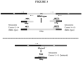
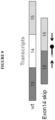
- Primers were designed to amplify across the junction of exon 13-exon 15 (mutant or exon 14 deleted) and exon 13-exon 14 and exon 14-exon 15 (wild type). Probes specific for the junction point of each amplicon were also prepared, and labeled with different labels for multiplex amplification and detection. Primers and a probe were also designed for an internal control, in this case SDH (succinyl dehydrogenase).
- Non-natural nucleic acids and mismatches from the target sequence were introduced to stabilize the specific target-oligo hybridizations. The best modifications and positions for mismatch are unpredictable. Suitable oligonucleotide sequences are shown in Table 1 below.
- MET13-FWD refers to a forward primer in MET exon 13;
- MET14-FWD refers to a forward primer in MET exon 14;
- MET14-REV refers to a reverse primer in MET exon 14;
- MET15-REV refers to a reverse primer in MET exon 15;
- MET13-15-PRB refers to a probe that hybridizes with nucleotides at the junction of MET exons 13 and 15;
- MET13-14-PRB refers to a probe that hybridizes with nucleotides at the junction of MET exons 13-14;
- MET 14-15-PRB refers to a probe that hybridizes with nucleotides at the junction of MET
- MET13-15-PRB9 is an antisense orientation probe that does not hybridize to MET exon 13-exon 15 amplification products, and that can be used for a control.
- Table 1 Primer Name SEQ ID NO Sequence MET13-FWD1 1 TTGGGTTTTTCCTGTGGCT MET13-FWD2 2 TTGGGTTTTTCCTGTGG ⁇ pdC>T MET13-FWD3 3 TTGGGTTTTTCCTGTGGC ⁇ pdU> MET13-FWD4 4 TTGGGTTTTTCCTGTGG ⁇ pdC> ⁇ pdU> MET13-FWD5 5 CTTGGGTTTTTCCTGTGGCT MET13-FWD6 6 CTTGGGTTTTTCCTGTGGCTG MET13-FWD7 7 TGGGTTTTTCCTGTGGCT MET13-FWD8 8 TTGGGTTTTTCCTGTGGCTG MET14-FWD1 9 TCAAATGAATCTGTAGACTACCG MET14-FWD2 10 TCAA
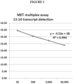
- a multiplex assay was run with MET 13 FWD, MET 14 FWD, MET 14 REV, and MET 15 REV primers, and one each of the differently labeled MET13-14-PRB, MET13-15-PRB, and MET14-15-PRB probes. Internal control primer and Cy5.5-labeled probe were also included.
- Results are shown in Figures 4-6 .
- Figure 4 shows signal of MET exon 14 deleted sample vs copy number of exon 14 deleted RNA.
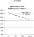
- Figures 5 and 6 show signal of wild type MET (exon 13-exon 14 and exon 14-exon 15, respectively) vs copy number of wild type RNA. Each of the graphs show that the assay is sensitive enough to detect 30 copies of target RNA input.
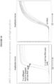
- Figure 7 shows that the assay is also specific.
- Signal for exon 13-exon 15 junction FAM is shown for mutant cell lines (Mut 1 and Mut 2), wild type cell lines (WT1 and WT2), and universal human RNA (UHR). As shown in Figure 7 , no signal was detected for samples lacking the MET exon 14 deletion.
- Nucleic acids from mutant cell lines was spiked into wild type (normal) plasma to ensure that plasma components (including wild type nucleic acids) would not interfere with detection of the mutant signal. As shown in Figure 8 , MET exon 14 deletion could be reliably detected in samples with 30 copies mutant RNA, with signal declining slightly with only 10 copies.
- exon 13-exon 14 and exon 14-exon 15 amplification products are almost always produced regardless of whether the sample is MET wild type or has a MET exon 14 deletion.
- a different assay format was designed to focus on amplification and detection of the exon 13-exon 15 junction. In this case, only one MET amplification reaction is carried out, with a forward primer complementary to a sequence in exon 13 and a reverse primer complementary to a sequence in the opposite orientation in exon 15.
- the resulting amplification product has sequence from exon 13 and sequence from exon 15, and can be detected with a probe specific for any portion of the amplification product.
- the probe can be specific for exon 13 sequence only, exon 15 sequence only, or junction of the exon sequences.
- MET splicing is heterogeneous, and thus multiple variant amplification products can be produced.
- the probe can be designed to capture multiple exon 14 splice variants, or multiple probes can be designed to capture different exon 14 splice variants.
- MET13-FWD and MET15-REV primers Suitable MET13-FWD and MET15-REV primers, and MET13-15-PRB probes are shown in Table 1 above. An exception is the MET13-15-PRB9 antisense orientation probe that does not hybridize to MET exon 13-exon 15 amplification products, and that can be used for a control.
- Figure 10 shows the specificity of the assay design depicted in Figure 9 .
- the probe specific for the exon 13- exon 15 amplification product is labeled with FAM (left graph), while a probe specific for an internal control RNA is labeled with CY5.5 (right graph).
- qRT-PCR is shown for RNA from 2 cell lines with exon 14 spliced out (noted in left graph), 2 wild type cell lines, and UHR (Universal Human RNA). Only the exon 14 spliced samples show signal.
- Figure 11 shows Limit of Detection (LOD) and Linearity data using the amount of MET exon 14 spliced transcript indicated on the x-axis spiked into 0.1 ng/ reaction UHR.
- the exon 14 spliced transcript is detectable in the linear range down to 10 copies. Internal control is indicated with squares and exon 14 spliced transcript is indicated with diamonds.
- Figure 12 shows LOD and Linearity using MET exon 14 spliced transcript from 2 different cell lines in the amount indicated on the x-axis spiked into 50 ng RNA from FFPET (formaldehye fixed paraffin embedded tissue).
- the exon 14 spliced transcript is detectable in the linear range down to 10 copies.
- the exon 14 spliced cell line transcripts from cell lines are shown in squares and diamonds (along the line) with the internal controls in squares and triangles, respectively.
- Figure 13 shows LOD and Linearity data using MET exon 14 spliced transcript from 2 different cell lines in the amount indicated on the x-axis spiked into RNA from healthy donor plasma cfRNA. Again, the LOD is about 10 copies in the linear range.
- the exon 14 spliced cell line transcripts from cell lines are shown in squares and diamonds (along the line) with the internal controls in squares and triangles, respectively.
Description
- MET is a tyrosine kinase and proto-oncogene encoded on human chromosome 7.
- The preprotein is cleaved to form alpha and beta subunits which remain associated via disulfide bonds, and act as a receptor for Hepatocyte Growth Factor. Activated MET induces the PI3K-AKT-mTOR pathway, involved in cell survival, and the RAS-RAF-MEK-ERK pathway, involved in cell proliferation.
- Mutations in MET are associated with a number of cancers, including renal, gastric, nervous system, sarcomas, and lung cancer. Mutations that result in higher activity or expression, or deletion of negative regulation sites are often implicated in these cancers. In particular, deletion of
exon 14 and the negative regulation site at Tyr 1003 is associated with a significant percentage of non-small cell lung cancers (NSCLC) and adenocarcinomas. CBL, an E3 ubiquitin protein ligase, binds Tyr 1003 on the MET protein. MET is not degraded normally when the site is deleted. The resulting dysregulation of MET causes sustained activation of downstream cell proliferation and survival pathways. Detection ofMET exon 14 deletion is more predictive of a positive therapeutic response to MET inhibitors than detection of gene amplification or increased mRNA or protein expression. - Mutations that cause MET
exon 14 deletions are heterogeneous, often affecting splice acceptor or donor sites. Because of the heterogeneity, current detection methods are limited to next generation sequencing (NGS). Lee et al, Oncotarget, vol.6, no.29, pages 28211-28222 (2015) discloses methods for detecting aMET exon 14 deletion using a forward primer forMET exon 13, a reverse primer forMET exon 15 and a probe overlapping a MET exon 13-exon 15 junction. - The scope of the invention is defined by the appended claims.
- Provided herein are methods and compositions for detecting
MET exon 14 deletions in human nucleic acid samples. Herein, a method of detecting aMET exon 14 deletion comprises: (a) providing a nucleic acid sample derived from a plasma or formalin fixed paraffin embedded tissue (FFPET) sample comprising RNA from an individual (e.g., comprising RNA, DNA, or both RNA and DNA); (b) carrying out a reverse transcription reaction on the RNA to produce cDNA; (c) carrying out an amplification reaction comprising contacting the cDNA with (i) a primer set and first probe labeled with a first label that specifically amplify and detect MET exon 13-exon 14 junction, (ii) a primer set and second probe labeled with a second label that specifically amplify and detect MET exon 14-exon 15 junction, and (iii) a primer set and third probe labeled with a third label that specifically amplify and detect MET exon 13-exon 15 junction; and (d) detecting the presence of aMET exon 14 deletion if an amplification product is formed and detected by the primer set and probe of (iii), wherein steps (b) and (c) are carried out in the same vessel and wherein the first probe is specific for the junction point of the MET exon 13-14 junction, the second probe is specific for the junction point of the MET exon 14-15 junction, and the third probe is specific for the junction point of the MET exon 13-15 junction. - In some embodiments, the multiplex reaction further includes an internal control, e.g., a primer set and probe labeled with a fourth label (e.g., labeled IC probe) that specifically amplify and detect an internal control. In some embodiments, the reverse transcription and amplification/ detection reactions are carried out using quantitative reverse transcription-PCR (qRT-PCR).
- In some embodiments, the individual has cancer. In particular embodiments, the individual has non-small cell lung cancer (NSCLC). In some embodiments, detecting the presence of a
MET exon 14 deletion is predictive for the individual's positive therapeutic response to treatment with MET inhibitor. - In some embodiments, the primer set for amplifying the MET exon 13-14 junction includes a forward primer having a sequence selected from the group consisting of: SEQ ID NOs:1-8 and a reverse primer having a sequence selected from the group consisting of: SEQ ID NOs: 17-24. In some embodiments, the probe for detecting the MET exon 13-14 junction has a sequence selected from the group consisting of: SEQ ID NOs: 44-46. In some embodiments, the probe for detecting the MET exon 13-14 junction has the sequence of SEQ ID NO:44. In some embodiments, the probe for detecting the MET exon 13-14 junction has the sequence of SEQ ID NO:45. In some embodiments, the probe for detecting the MET exon 13-14 junction has the sequence of SEQ ID NO:46. In some embodiments, the primer set for amplifying the MET exon 14-15 junction includes a forward primer having a sequence selected from the group consisting of: SEQ ID NOs:9-16 and a reverse primer having a sequence selected from the group consisting of: SEQ ID NOs: 25-38. In some embodiments, the probe for detecting the MET 14-15 junction has a sequence selected from the group consisting of: SEQ ID NOs: 47-50. In some embodiments, the probe for detecting the MET 14-15 junction has the sequence of SEQ ID NO:47. In some embodiments, the probe for detecting the MET 14-15 junction has the sequence of SEQ ID NO:48. In some embodiments, the probe for detecting the MET 14-15 junction has the sequence of SEQ ID NO:49. In some embodiments, the probe for detecting the MET 14-15 junction has the sequence of SEQ ID NO:50. In some embodiments, the primer set for amplifying the MET exon 13-15 junction includes a forward primer selected from the group consisting of: SEQ ID NOs:1-8 and a reverse primer selected from the group consisting of: SEQ ID NOs: 25-38. In some embodiments, the probe for detecting the MET exon 13-15 junction has a sequence selected from the group consisting of SEQ ID NOs: 39-43 and 51-54. In some embodiments, the probe for detecting the MET 13-15 junction has the sequence of SEQ ID NO:39. In some embodiments, the probe for detecting the MET 13-15 junction has the sequence of SEQ ID NO:40. In some embodiments, the probe for detecting the MET 13-15 junction has the sequence of SEQ ID NO:41. In some embodiments, the probe for detecting the MET 13-15 junction has the sequence of SEQ ID NO:42. In some embodiments, the probe for detecting the MET 13-15 junction has the sequence of SEQ ID NO:43. In some embodiments, the probe for detecting the MET 13-15 junction has the sequence of SEQ ID NO:51. In some embodiments, the probe for detecting the MET 13-15 junction has the sequence of SEQ ID NO:52. In some embodiments, the probe for detecting the MET 13-15 junction has the sequence of SEQ ID NO:53. In some embodiments, the probe for detecting the MET 13-15 junction has the sequence of SEQ ID NO:54.
- Further disclosed is a method of detecting a
MET exon 14 deletion comprises: (a) obtaining a nucleic acid sample from an individual (e.g., comprising RNA, DNA, or both RNA and DNA); (b) carrying out an amplification/ detection reaction using the sample to selectively amplify and detect a MET exon 13-exon 15 junction; and (c) detecting the presence of aMET exon 14 deletion if the MET exon 13-exon 15 junction is detected. The method may further include carrying out a reverse transcription reaction using the sample to produce cDNA before step (b). Herein, the reverse transcription reaction and step (b) may occur in a single vessel (tube, well, microfluidic chamber, etc.). Also disclosed is that step (b) comprises contacting the cDNA with a primer set and labeled probe (e.g., with a non-naturally occurring fluorophore or fluorophore and quencher) that specifically amplify and detect MET exon 13-15 junction. Also disclosed is that the primer set comprises a forward primer complementary to a sequence inMET exon 13 and a reverse primer complementary to a sequence inMET exon 15. Also disclosed is that the labeled probe specifically hybridizes to the amplification product of the primer set, and includes sequence complementary to exon 13 only,exon 15 only, or from bothexon - Also disclosed is that the primer set for amplifying the MET exon 13-15 junction includes a forward primer selected from the group consisting of SEQ ID NOs:1-8 and a reverse primer selected from the group consisting of: SEQ ID NOs: 25-38. Also disclosed is that the probe for detecting the MET exon 13-15 junction has a sequence selected from the group consisting of SEQ ID NOs: 39-43 and 51-54. Also disclosed is that the probe for detecting the MET exon 13-15 junction has a sequence selected from the group consisting of: SEQ ID NO:43, SEQ ID NO:52, and SEQ ID NO:53. Also disclosed is that the probe for detecting the MET 13-15 junction has the sequence of SEQ ID NO:39. Also disclosed is that the probe for detecting the MET 13-15 junction has the sequence of SEQ ID NO:40. Also disclosed is that the probe for detecting the MET 13-15 junction has the sequence of SEQ ID NO:41. Also disclosed is that the probe for detecting the MET 13-15 junction has the sequence of SEQ ID NO:42. Also disclosed is that the probe for detecting the MET 13-15 junction has the sequence of SEQ ID NO:43. Also disclosed is that the probe for detecting the MET 13-15 junction has the sequence of SEQ ID NO:51. Also disclosed is that the probe for detecting the MET 13-15 junction has the sequence of SEQ ID NO:52. Also disclosed is that the probe for detecting the MET 13-15 junction has the sequence of SEQ ID NO:53. Also disclosed is that the probe for detecting the MET 13-15 junction has the sequence of SEQ ID NO:54.
- Also disclosed is that step (b) is a multiplex reaction, e.g., so that the primer sets and probes are included in a single vessel. Also disclosed is that the reverse transcription and amplification/ detection reactions are carried out using quantitative reverse transcription-PCR (qRT-PCR).
- Also disclosed is that the sample is enriched for RNA before reverse transcription. Also disclosed is that the sample is from a non-invasive source (e.g., blood, plasma, serum, urine, mucosal swab, saliva, skin, etc.). Also disclosed is that the sample is a biopsy, e.g., a tumor biopsy, optionally an FFPET sample. Also disclosed is that the individual has cancer, e.g., lung, renal, gastric, neuronal cancer, sarcoma, or adenocarcinoma. Also disclosed is that the individual has NSCLC.
- Further disclosed is a method of identifying an individual with cancer comprising: (a) obtaining a sample comprising RNA from the individual; (b) carrying out a reverse transcription reaction on the RNA to produce cDNA; (c) carrying out an amplification reaction comprising contacting the cDNA with a primer set and labeled probe that specifically amplify and detect MET exon 13-15 junction (exon 13-15 primer set and labeled exon 13-15 probe); and (d) detecting the presence of a
MET exon 14 deletion if an amplification product is formed and detected by the exon 13-15 primer set and labeled exon 13-15 probe; whereby the presence of aMET exon 14 deletion mutation in the individual's sample indicates sensitivity of said individual to a MET inhibitor compound. Also disclosed is that the method further comprises identifying an individual indicating sensitivity to an inhibitor of a downstream effector of MET (e.g., an inhibitor of the PI3K-AKT-mTOR or RAS-RAF-MEK-ERK pathway), or with standard chemotherapy, if aMET exon 14 deletion is detected. - Further disclosed are methods of identifying an individual with cancer comprising: (a) obtaining a nucleic acid sample from an individual (e.g., comprising RNA both RNA and DNA); (b) carrying out an amplification/ detection reaction using the sample to selectively amplify and detect MET exon 13-
exon 14 junction, MET exon 14-exon 15 junction, and MET exon 13-exon 15 junction; and (c) detecting the presence of aMET exon 14 deletion if the MET exon 13-exon 15 junction is detected; whereby the presence of aMET exon 14 deletion mutation in the individual's sample indicates sensitivity of said individual to a MET inhibitor compound. Also disclosed is that the method further includes carrying out a reverse transcription reaction using the sample to produce cDNA before step (b). Also disclosed is that the reverse transcription reaction and step (b) occur in a single vessel (tube, well, microfluidic chamber, etc.). Also disclosed is that step (b) comprises contacting the cDNA with (i) a primer set and probe labeled with a first label that specifically amplify and detect MET exon 13-14 junction, (ii) a primer set and probe labeled with a second label that specifically amplify and detect MET exon 14-15 junction, and (iii) a primer set and probe labeled with a third label that specifically amplify and detect MET exon 13-15 junction. Also disclosed is that step (c) comprises detection of the probe labeled with the third label. Also disclosed is that step (b) is a multiplex reaction, e.g., so that the primer sets and probes of (i), (ii), and (iii) are included in a single vessel. Also disclosed is that the multiplex reaction further includes an internal control, e.g., a primer set and probe labeled with a fourth label (e.g., labeled IC probe) that specifically amplify and detect an internal control. Also disclosed is that step (b) is carried out in separate vessels, e.g., each carrying one of the primer sets and probes of (i), (ii), and (iv), optionally multiplexed with the internal control. Also disclosed is that the reverse transcription and amplification/ detection reactions are carried out using quantitative reverse transcription-PCR (qRT-PCR). - Also disclosed is that the method of identifying an individual with cancer comprises: (a) obtaining a nucleic acid sample from an individual (e.g., comprising RNA, DNA, or both RNA and DNA); (b) carrying out an amplification/ detection reaction using the sample to selectively amplify and detect MET exon 13-
exon 15 junction; and (c) detecting the presence of aMET exon 14 deletion if the MET exon 13-exon 15 junction is detected; whereby the presence of aMET exon 14 deletion mutation in the individual's sample indicates sensitivity of said individual to a MET inhibitor compound. Also disclosed is that the method further includes carrying out a reverse transcription reaction using the sample to produce cDNA before step (b). Also disclosed is that the reverse transcription reaction and step (b) occur in a single vessel (tube, well, microfluidic chamber, etc.). Also disclosed is that step (b) comprises contacting the cDNA with a primer set and labeled probe that specifically amplify and detect MET exon 13-15 junction. Also disclosed is that the primer set comprises a forward primer complementary to a sequence inMET exon 13 and a reverse primer complementary to a sequence inMET exon 15. Also disclosed is that the labeled probe specifically hybridizes to the amplification product of the primer set, and includes sequence complementary toexon 13 only,exon 15 only, or from bothexon - Also disclosed is that step (b) is a multiplex reaction, e.g., so that the primer sets and probes are included in a single vessel. Also disclosed is that the reverse transcription and amplification/ detection reactions are carried out using quantitative reverse transcription-PCR (qRT-PCR).
- Also disclosed is that the method further comprises providing treatment for the individual with an inhibitor of a downstream effector of MET (e.g., an inhibitor of the PI3K-AKT-mTOR or RAS-RAF-MEK-ERK pathway), or with standard chemotherapy, if a
MET exon 14 deletion is detected. - Also disclosed is that the sample is enriched for RNA before reverse transcription. Also disclosed is that the sample is from a non-invasive source (e.g., blood, plasma, serum, urine, mucosal swab, saliva, skin, etc.). Also disclosed is that the sample is a biopsy, e.g., a tumor biopsy, optionally an FFPET sample. Also disclosed is that the individual has cancer, e.g., lung, renal, gastric, neuronal cancer, sarcoma, or adenocarcinoma. Also disclosed is that the individual has NSCLC.
- Also disclosed is that the method further comprises identifying an individual with cancer, wherein, if a
MET exon 14 deletion is detected in the individual's sample, the individual's sample indicates sensitivity of said individual to an inhibitor of a downstream effector of MET (e.g., an inhibitor of the PI3K-AKT-mTOR or RAS-RAF-MEK-ERK pathway), or with standard chemotherapy. - Further provided are kits, e.g., for detecting
MET exon 14 deletion in a nucleic acid sample. Herein, the kit comprises (a) a primer set and a first probe labeled with a first label that specifically amplify and detect MET exon 13-14 junction; (b) a primer set and a second probe labeled with a second label that specifically amplify and detect MET exon 14-15 junction; and (c) a primer set and a third probe labeled with a third label that specifically amplify and detect MET exon 13-15 junction, wherein the first probe is specific for the junction point of the MET exon 13-14 junction and comprises a sequence selected from the group consisting of SEQ ID NOs: 44-46, the second probe is specific for the junction point of the MET exon 14-15 junction and comprises a sequence selected from the group consisting of SEQ ID NOs: 47-50, and the third probe is specific for the junction point of the MET exon 13-15 junction and comprises a sequence selected from the group consisting of SEQ ID NOs: 39-43 and 51-54. In some embodiments, the sequences of the primers are set forth as described above herein. In some embodiments, the kit further comprises (d) a primer set and fourth probe labeled with a fourth label that specifically amplify and detect an internal control. - In some embodiments, the primer set comprises a forward primer complementary to a sequence in
MET exon 13 and a reverse primer complementary to a sequence inMET exon 15. - In some embodiments, the kit further includes a reverse transcriptase enzyme and a thermostable DNA polymerase, or an enzyme that has both activities. In some embodiments, the kit further comprises buffer (e.g., buffer that facilitates amplification) and/or dNTPs. In some embodiments, the kit further comprises a positive control comprising nucleic acids encoding
MET exon 14 deletion. In some embodiments, the kit further comprises a negative control comprising nucleic acids that do not encodeMET exon 14 deletion. In some embodiments, the kit further comprises components for purification (enrichment) of RNA from a sample from an individual. -
-
Figure 1 depicts the alpha and beta subunits of mature MET, and shows the position of the ubiquination site at Y1003. CBL binds to Y1003 when it is phosphorylated (activated state) and targets MET for ubiquitin-mediated degradation. Y1003 is not present inexon 14 deleted MET. -
Figure 2 shows an exemplary assay design for detection ofMET exon 14 deletion. The mutant form is detected using a FAM-labeled probe specific for the exon 13-exon 15 junction, while the wild type forms are detected using a HEX-labeled probe specific for the exon 13-exon 14 junction and a JA270-labeled probe specific for the exon 14-exon15 junction. An internal control is detected with a Cy5.5-labeled probe, e.g., to standardize for nucleic acid concentration and quality. -
Figure 3 provides further information about the exemplary assay design.Exons exons -
Figure 4 shows the result of the multiplex reaction depicted inFigures 2 and3 for the mutant form (FAM channel). The x-axis shows copy number of the mutant sequence and the y-axis shows Ct. The figure shows that Ct decreases with increasing copy number in a linear fashion, and that the assay is sensitive enough to detect 30 copies. -
Figure 5 shows the result of the multiplex reaction for the wild type form exon 13-exon 14 junction (HEX channel). As with the mutant reaction, Ct is inversely related to wild type copy number, the reaction is linear, and sensitive enough to detect 30 copies. -
Figure 6 shows the result of the multiplex for the wild type form exon 14-exon 15 junction (JA270 channel). Again, the data show that Ct is inversely related to wild type copy number, the reaction is linear, and sensitive enough to detect 30 copies. -
Figure 7 shows that detection of the exon 13-exon 15 junction is specific. The x axis shows cycle number and the y axis shows FAM signal.Mut 1 and Mut2 are samples fromMET exon 14 deleted cell lines. Wild type samples from WT1, WT2, and UHR (universal human RNA) do not generate a signal. -
Figure 8 shows the sensitivity of detection of the exon 13-exon 15 junction. Ct increases by about 2 between 100 copies and 30 copies mutant cell line RNA in wild type plasma, while a larger Ct increase is seen with only 10 copies mutant cell line RNA. The right panel shows the result of the internal control, which indicates roughly equal input between the samples. -
Figure 9 shows an exemplary assay design of a Reference Example. The top transcript represents unspliced wild type MET and the bottom transcript represents MET withexon 14 spliced out. The primer set (arrows) includes a first (forward) primer positioned inexon 13 and a second (reverse) primer positioned inexon 15 so that an amplification product is formed whenexon 14 is spliced out. The probe is designed to specifically bind the amplification product, be it a sequence inexon 13 only,exon 15 only, or spanning an exon 13-exon 15 junction (as shown). -
Figure 10 shows the specificity of the assay design of the Reference Example depicted inFigure 9 . The probe specific for the exon 13-exon 15 amplification product is labeled with FAM(left graph), while a probe specific for an internal control RNA is labeled with CY5.5 (right graph). qRT-PCR is shown for RNA from 2 cell lines withexon 14 spliced out (noted in left graph), 2 wild type cell lines, and UHR (Universal Human RNA). -
Figure 11 shows Limit of Detection (LOD) and Linearity data of the assay design of the Reference Example depicted inFigure 9 using the amount ofMET exon 14 spliced transcript indicated on the x-axis spiked into 0.1 ng/ reaction UHR. The splice product is detectable in the linear range down to 10 copies. -
Figure 12 shows LOD and Linearity of the assay design of the Reference Example depicted inFigure 9 usingMET exon 14 spliced transcript from 2 different cell lines in the amount indicated on the x-axis spiked into 50 ng RNA from FFPET (formaldehye fixed paraffin embedded tissue). The splice product is detectable in the linear range down to 10 copies. -
Figure 13 shows LOD and Linearity data of the assay design of the Reference Example depicted inFigure 9 usingMET exon 14 spliced transcript from 2 different cell lines in the amount indicated on the x-axis spiked into RNA from healthy donor plasma cfRNA. Again, the LOD is about 10 copies in the linear range. - Provided herein are novel quantitative reverse transcription (qRT)-PCR assays to detect
MET exon 14 deletion, despite the variable nature of mutations that causeexon 14 deletion. The presently described assays thus rely on proven, widely adopted technology and provide accurate, reproducible, and rapid results. The results provided herein show that the assays can be effectively multiplexed and carried out in a single tube, while still providing extraordinarily specific and sensitive results. - The term "multiplex" refers to an assay in which more than one target can be detected. For example, a multiplex reaction can include more than one primer set and more than one probe, specific for different portions or variants of a gene or transcript, or specific for different genes or transcripts.
- The terms "receptacle," "vessel," "tube," "well," "chamber," "microchamber," etc. refer to a container that can hold reagents or an assay. If the receptacle is in a kit and holds reagents, or is being used for an amplification reaction, it can be closed or sealed to avoid contamination or evaporation. If the receptacle is being used for an assay, it can be open or accessible, at least during set up of the assay.
- The terms "
MET exon 14 deletion," "MET exon 14 skipping," and like terms refer to a MET gene that is chromosomally rearranged or mutated, or a MET transcript that is spliced to remove at least a portion ofexon 14 of MET, i.e., the portion encoding negative regulation site Tyr 1003 in the juxtamembrane region of the MET protein. Various mutations at the DNA level can result inexon 14 skipping (see, e.g., Kong-Beltran et al. (2006) Cancer Res. 66; Dhanasekharan et al. (2014) Nature Communications 10:1038). In some embodiments, the presently described assays detect deletion of theentire exon 14 of MET, which encodes 47 amino acids. - The term "obtaining a sample from an individual" means that a biological sample from the individual is provided for testing. The obtaining can be directly from the individual, or from a third party that directly obtained the sample from the individual.
- The term "providing treatment for an individual" means that the treatment is actually administered to the individual (e.g., an in-patient injection), or that it is made available to the individual, so that the individual or third party actually administers the treatment.
- The term "formed and detected," in reference to an amplification product, indicates that the amplification product is formed and detected at a level that is higher than a base-line negative level. The base-line can be based on a negative control, e.g., set at the level of signal generated by a wild type sample in the case of amplifying and detecting a mutant sequence. The base-line can also be set slightly above the signal of the negative control (e.g., 1-5%) to allow for some variation in assay performance while avoiding false positive results for the mutant sequence. One of skill in the art of nucleic acid detection will understand the appropriate controls and variation levels for a given assay.
- The terms "nucleic acid," "polynucleotide," and "oligonucleotide" refer to polymers of nucleotides (e.g., ribonucleotides or deoxyribo-nucleotides) and includes naturally-occurring (adenosine, guanidine, cytosine, uracil and thymidine), non-naturally occurring, and modified nucleic acids. The term is not limited by length (e.g., number of monomers) of the polymer. A nucleic acid may be single-stranded or doublestranded and will generally contain 5'-3' phosphodiester bonds, although in some cases, nucleotide analogs may have other linkages. Monomers are typically referred to as nucleotides. The term "non-natural nucleotide" or "modified nucleotide" refers to a nucleotide that contains a modified nitrogenous base, sugar or phosphate group, or that incorporates a non-natural moiety in its structure. Examples of non-natural nucleotides include dideoxynucleotides, biotinylated, aminated, deaminated, alkylated, benzylated and fluorophore-labeled nucleotides.
- The term "primer" refers to a short nucleic acid (an oligonucleotide) that acts as a point of initiation of polynucleotide strand synthesis by a nucleic acid polymerase under suitable conditions. Polynucleotide synthesis and amplification reactions typically include an appropriate buffer, dNTPs and/or rNTPs, and one or more optional cofactors, and are carried out at a suitable temperature. A primer typically includes at least one target-hybridized region that is at least substantially complementary to the target sequence (e.g., having 0, 1, 2, or 3 mismatches). This region of is typically about 8 to about 40 nucleotides in length, e.g., 12-25 nucleotides. A "primer set" refers to a forward and reverse primer that are oriented in opposite directions relative to the target sequence, and that produce an amplification product in amplification conditions. The primer set can further include and additional forward or reverse primer, e.g., to carry out allele-specific amplification. A primer set can also share a forward or reverse primer with another primer set, e.g., a common forward or reverse primer. The terms forward primer and reverse primer are arbitrarily assigned and do not indicate orientation in relation to coding sequence, etc., unless otherwise noted.
- As used herein, "probe" means any molecule that is capable of selectively binding to a specifically intended target biomolecule, for example, a nucleic acid sequence of interest that hybridizes to the probes. The probe is detectably labeled with at least one non-nucleotide moiety (i.e., non-naturally occurring moiety). In some embodiments, the probe is labeled with a fluorophore and quencher.
- The term "specifically amplifies" indicates that a primer set amplifies a target sequence more than non-target sequence at a statistically significant level. The term "specifically detects" indicates that a probe will detect a target sequence more than non-target sequence at a statistically significant level. As will be understood in the art, specific amplification and detection can be determined using a negative control, e.g., a sample that includes the same nucleic acids as the test sample, but not the target sequence. For example, primers and probes that specifically amplify and detect a target sequence result in a Ct that is readily distinguishable from background (non-target sequence), e.g., a Ct that is at least 2, 3, 4, 5, 5-10, 10-20, or 10-30 cycles less than background.
- The words "complementary" or "complementarity" refer to the ability of a nucleic acid in a polynucleotide to form a base pair with another nucleic acid in a second polynucleotide. For example, the sequence A-G-T (A-G-U for RNA) is complementary to the sequence T-C-A (U-C-A for RNA). Complementarity may be partial, in which only some of the nucleic acids match according to base pairing, or complete, where all the nucleic acids match according to base pairing. A probe or primer is considered "specific for" a target sequence if it is at least partially complementary to the target sequence. Depending on the conditions, the degree of complementarity to the target sequence is typically higher for a shorter nucleic acid such as a primer (e.g., greater than 80%, 90%, 95%, or higher) than for a longer sequence.
- The terms "identical" or "percent identity," in the context of two or more nucleic acids, or two or more polypeptides, refer to two or more sequences or subsequences that are the same or have a specified percentage of nucleotides, or amino acids, that are the same (e.g., about 60% identity, e.g., at least any of 65%, 70%, 75%, 80%, 85%, 90%, 91%, 92%, 93%, 94%, 95%, 96%, 97%, 98%, 99%, or higher identity over a specified region, when compared and aligned for maximum correspondence over a comparison window or designated region) as measured using a BLAST or BLAST 2.0 sequence comparison algorithms with default parameters, or by manual alignment and visual inspection. See e.g., the NCBI web site at ncbi.nlm.nih.gov/BLAST. Such sequences are then said to be "substantially identical." Percent identity is typically determined over optimally aligned sequences, so that the definition applies to sequences that have deletions and/or additions, as well as those that have substitutions. The algorithms commonly used in the art account for gaps and the like. Typically, identity exists over a region comprising an a sequence that is at least about 8-25 amino acids or nucleotides in length, or over a region that is 50-100 amino acids or nucleotides in length, or over the entire length of the reference sequence.
- The term "kit" refers to any manufacture (e.g., a package or a container) including at least one reagent, such as a nucleic acid probe or probe pool or the like, for specifically amplifying, capturing, tagging/converting or detecting RNA or DNA as described herein.
- The term "amplification conditions" refers to conditions in a nucleic acid amplification reaction (e.g., PCR amplification) that allow for hybridization and template-dependent extension of the primers. The term "amplicon" or "amplification product" refers to a nucleic acid molecule that contains all or a fragment of the target nucleic acid sequence and that is formed as the product of in vitro amplification by any suitable amplification method. One of skill will understand that a forward and reverse primer (primer pair) defines the borders of an amplification product. The term "generate an amplification product" when applied to primers, indicates that the primers, under appropriate conditions (e.g., in the presence of a nucleotide polymerase and NTPs), will produce the defined amplification product. Various PCR conditions are described in PCR Strategies (Innis et al., 1995, Academic Press, San Diego, CA) at
Chapter 14; PCR Protocols : A Guide to Methods and Applications (Innis et al., Academic Press, NY, 1990) - The term "amplification product" refers to the product of an amplification reaction. The amplification product includes the primers used to initiate each round of polynucleotide synthesis. An "amplicon" is the sequence targeted for amplification, and the term can also be used to refer to amplification product. The 5' and 3' borders of the amplicon are defined by the forward and reverse primers.
- The term "sample" or "biological sample" refers to any composition containing or presumed to contain nucleic acid. The term includes purified or separated components of cells, tissues, or blood, e.g., DNA, RNA, proteins, cell-free portions, or cell lysates. In the context of the presently disclosed assay, the sample is typically FFPET, e.g., from a tumor or metastatic lesion. The sample can also be from frozen or fresh tissue, or from a liquid sample, e.g., blood or a blood component (plasma or serum), urine, semen, saliva, sputum, mucus, semen, tear, lymph, cerebral spinal fluid, mouth/throat rinse, bronchial alveolar lavage, material washed from a swab, etc. Samples also may include constituents and components of in vitro cultures of cells obtained from an individual, including cell lines. The sample can also be partially processed from a sample directly obtained from an individual, e.g., cell lysate or blood depleted of red blood cells.
- A "control" sample or value refers to a value that serves as a reference, usually a known reference, for comparison to a test sample or test conditions. For example, a test sample can be taken from a test condition, e.g., from an individual suspected of having cancer, and compared to samples from known conditions, e.g., from a cancer-free individual (negative control), or from an individual known to have cancer (positive control). A control can also represent an average value or a range gathered from a number of tests or results. A control can also be prepared for reaction conditions. For example, a control for the presence, quality, and/ or quantity of nucleic acid (e.g., internal control) can include primers or probes that will detect a sequence known to be present in the sample (e.g., a housekeeping gene such as beta actin, beta globin, glyceraldehyde 3-phosphate dehydrogenase (GAPDH), ribosomal protein L37 and L38, PPIase, EIF3, eukaryotic translation elongation factor 2 (eEF2), DHFR, or succinate dehydrogenase) A known added polynucleotide, e.g., having a designated length and/or sequence, can also be added. An example of a negative control is one free of nucleic acids, or one including primers or probes specific for a sequence that would not be present in the sample, e.g., from a different species. One of skill will understand that the selection of controls will depend on the particular assay, e.g., so that the control is cell type and organism-appropriate. One of skill in the art will recognize that controls can be designed for assessment of any number of parameters. For example, a control can be devised to compare therapeutic benefit based on pharmacological data (e.g., half-life) or therapeutic measures (e.g., comparison of benefit and/or side effects). Controls can be designed for in vitro applications. One of skill in the art will understand which controls are valuable in a given situation and be able to analyze data based on comparisons to control values. Controls are also valuable for determining the significance of data. For example, if values for a given parameter are widely variant in controls, variation in test samples will not be considered as significant.
- The terms "label," "tag," "detectable moiety," and like terms refer to a composition detectable by spectroscopic, photochemical, biochemical, immunochemical, chemical, or other physical means. For example, useful labels include fluorescent dyes, luminescent agents, radioisotopes (e.g., 32P, 3H), electron-dense reagents, or an affinity-based moiety, e.g., a poly-A (interacts with poly-T) or poly-T tag (interacts with poly-A), a His tag (interacts with Ni), or a strepavidin tag (separable with biotin). As used herein, a biomolecule (DNA, RNA, protein, etc.) attached to a label, tag, or detectable moiety is not a composition that occurs in nature
- Unless defined otherwise, technical and scientific terms used herein have the same meaning as commonly understood by a person of ordinary skill in the art. See, e.g., Lackie, DICTIONARY OF CELL AND MOLECULAR BIOLOGY, Elsevier (4th ed. 2007); Sambrook et al., MOLECULAR CLONING, A LABORATORY MANUAL, Cold Springs Harbor Press (Cold Springs Harbor, N.Y. 1989). The term "a" or "an" is intended to mean "one or more." The terms "comprise," "comprises," and "comprising," when preceding the recitation of a step or an element, are intended to mean that the addition of further steps or elements is optional and not excluded.
- Samples for nucleic acid amplification can be obtained from any source suspected of containing nucleic acid. In the context of the present disclosure, the sample is from a human source. In some embodiments, the sample is obtained in a non-invasive manner, e.g., blood or a blood fraction, urine, skin, swab, or saliva. Samples can also be taken from formalin fixed paraffin embedded tissue (FFPET), tissue biopsy, brochoalveolar lavage, or cultured cells (e.g., obtained from a patient, or representing a control). In some embodiments, the sample is taken from lung tissue or a cell population that includes lung cells, e.g., lung cancer cells.
- In a sample that includes cells, the cells can be separated out (e.g., using size-based filtration or centrifugation), thereby leaving cell free nucleic acids (cfNA), including nucleic acids in exosomes, microvesicles, viral particles, or those circulating freely. Alternatively, the cells can be lysed to obtain cellular nucleic acids, either in the presence of magnetic glass particles (MGPs) or before addition of the cellular lysate to the MGPs.
- In some embodiments, the nucleic acids (DNA, RNA, or both) are isolated from other components of the sample, e.g., proteins, membranes, lipids, etc., before amplification and/or detection. The terms "isolated," "separated," "purified," etc. do not indicate that the nucleic acids are 100% free of other substances, but that the nucleic acids are substantially enriched compared to the original sample. For example, purified nucleic acids can be 2-, 3-, 4-, 5-, 10-, 20-, 50-, 100-fold enriched compared to the original sample, or more. In some embodiments, purified nucleic acids are in aqueous solution having less than 20%, 10%, 5%, 2%, 1%, 0.5% or 0.1% non-nucleic acid residual from the sample.
- In some embodiments, the presently described assays rely on mRNA encoding MET. Because many of the
mutations causing exon 14 deletion are somatic, the deletion would be expected to occur in transcripts from any cell type expressing MET, e.g., epithelial cells. Samples can be taken from any cell or cell-free source suspected of carrying tumor nucleic acids (e.g., cancer biopsy or circulating nucleic acids). - Methods for isolating nucleic acids from biological samples are known, e.g., as described in Sambrook supra, and several kits are commercially available (e.g., cobas® cfRNA Sample Preparation Kit, High Pure RNA Isolation Kit, High Pure Viral Nucleic Acid Kit, and MagNA Pure LC Total Nucleic Acid Isolation Kit, DNA Isolation Kit for Cells and Tissues, DNA Isolation Kit for Mammalian Blood, High Pure FFPET DNA Isolation Kit, available from Roche). In the context of the presently disclosed methods, RNA is collected.
- The nucleic acid sample (isolated or not) can be used for detection and quantification, e.g., using nucleic acid amplification, e.g., using any primer-dependent method. Herein, a preliminary reverse transcription step is carried out (also referred to as RT-PCR, not to be confused with real time PCR). See, e.g., Hierro et al. (2006) 72:7148. The term "qRT-PCR" as used herein refers to reverse transcription followed by quantitative PCR. Both reactions can be carried out in a single tube without interruption, e.g., to add reagents. For example, a polyT primer can be used to reverse transcribe all mRNAs in a sample with a polyA tail, random oligonucleotides can be used, or a primer can be designed that is specific for a particular target transcript that will be reverse transcribed into cDNA. The cDNA can form the initial template strand to be for quantitative amplification (real time or quantitative PCR, i.e., RTPCR or qPCR). qPCR allows for reliable detection and measurement of products generated during each cycle of PCR process. Such techniques are well known in the art, and kits and reagents are commercially available, e.g., from Roche Molecular Systems, Life Technologies, Bio-Rad, etc. See, e.g., Pfaffl, Methods: The ongoing evolution of qPCR vol. 50(4), (2010), p. 215-216.
- A separate reverse transcriptase and thermostable DNA polymerase can be used, e.g., in a two-step (reverse transcription followed by addition of DNA polymerase and amplification) or combined reaction (with both enzymes added at once). In some embodiments, the target nucleic acid is amplified with a thermostable polymerase with both reverse transcriptase activity and DNA template-dependent activity. Exemplary enzymes include Tth DNA polymerase, the C. therm Polymerase system, and those disclosed in
US20140170730 andUS20140051126 . In some embodiments, Taq or a Taq derivative is used for amplification (e.g., Z05, C21, etc.). In some embodiments, the reverse transcriptase is from MMLV, AMV, or is a derivative thereof. - Probes for use as described herein can be labeled with a fluorophore and quencher (e.g., TaqMan, LightCycler, Molecular Beacon, Scorpion, or Dual Labeled probes). Appropriate fluorophores include FAM, JOE, JA270, TET, Cal Fluor Gold 540, HEX, VIC, Cal Fluor Orang 560, TAMRA, Cyanine 3, Quasar 570, Cal Fluor Red 590, Rox, Texas Red, Cyanine 5, Quasar 670, and Cyanine 5.5. Appropriate quenchers include TAMRA (for FAM, JOE, and TET), DABCYL, and BHQ1-3.
- Detection devices are known in the art and can be selected as appropriate for the selected labels. Detection devices appropriate for quantitative PCR include the cobas® and Light Cycler® systems (Roche), PRISM 7000 and 7300 real-time PCR systems (Applied Biosystems), etc. Six-channel detection is available on the CFX96 Real Time PCR Detection System (Bio-Rad) and Rotorgene Q (Qiagen), allowing for a higher degree of multiplexing.
- Results can be expressed in terms of a threshold cycle (abbreviated as Ct, and in some instances Cq or Cp). A lower Ct value reflects the rapid achievement of a predetermined threshold level, e.g., because of higher target nucleic acid concentration or a more efficient amplification. A higher Ct value may reflect lower target nucleic acid concentration, or inefficient or inhibited amplification. The threshold cycle is generally selected to be in the linear range of amplification for a given target. In some embodiments, the Ct is set as the cycle at which the growth signal exceeds a pre-defined threshold line, e.g., in relation to the baseline, or by determining the maximum of the second derivation of the growth curve. Determination of Ct is known in the art, and described, e.g., in
US Patent No. 7363168 . - Provided herein are kits for multiplex qRT-PCR assays to detect
MET exon 14 deletions in a sample. Herein, the kit includes primers and probes for amplification, detection, and/ or quantification of transcripts encoding MET exon 13-exon 14, exon 14-exon 15, and exon 13-exon 15 junctions. - The junction-specific primer sets and probes can be mixed and matched in any combination. In a detection system having 4 channels, each of the three junctions can be detected in a single vessel, along with an internal control.
- In some embodiments, the kit includes primers and a probe for amplification, detection and/ or quantification of transcripts encoding MET exon 13-
exon 15 junctions. These primers (e.g., RT primer, and forward and reverse primers that specifically amplify a MET exon 13-exon 15 junction amplification product) and probe (specific for the MET exon 13-exon 15 junction amplification product) can be mixed with primers and a probe that specifically amplify and detect an internal control, or provided in a separate vessel. - In some embodiments, the mixtures further comprise buffers, dNTPs, and other elements (e.g., cofactors or aptamers) appropriate for reverse transcription and amplification. Typically, the mixture is concentrated, so that an aliquot is added to the final reaction volume, along with sample (e.g., RNA), enzymes, and/ or water. In some embodiments, the kit further comprises reverse transcriptase (or an enzyme with reverse transcriptase activity), and/or DNA polymerase (e.g., thermostable DNA polymerase such as Taq, ZO5, and derivatives thereof).
- In some embodiments, the kit further includes components for RNA purification from a sample, e.g., a plasma or FFPET sample. For example, the kit can include components from the cobas® 6800/8800 Sample Prep Kits, High Pure or MagNA Pure RNA Isolation Kits (Roche), miRNeasy or RNeasy FFPE Kits (Qiagen), PureLink FFPE RNA Isolation Kit (Thermo Fisher), ThruPLEX Plasma-seq (Beckman Coulter), etc.
- In some embodiments, the kit further includes at least one control sample, e.g., nucleic acids from non-cancer sample (or pooled samples), or from a known
MET exon 14 deleted sample (or pooled samples). In some embodiments, the kit further includes consumables, e.g., plates or tubes for nucleic acid preparation, tubes for sample collection, etc. In some embodiments, the kit further includes instructions for use, reference to a website, or software. - MET mutations are associated with a number of different cancers, including renal, gastric, and those of the central nervous system, as well as sarcomas.
MET exon 14 deletion in particular is associated with non-small cell lung cancer (NSCLC) and lung adenocarcinomas, found in 2% and 4% of these cancers, respectively. The 2016 National Comprehensive Cancer Network (NCCN) Guidelines now includeMET exon 14 deletion in category 2A for emerging targeted agents for lung cancer patients with genetic alterations. - MET inhibitors crizotinib (also inhibits ALK and ROS1) and cabozantinib (also inhibits VEGFR2 and RET) have been shown to be effective for lung adenocarcinoma patients harboring
MET exon 14 skipping. Additional small molecule MET inhibitors are being tested, including INCB28060 (Incyte), AMG-458 (Amgen), PF-04217903 (Pfizer), PF-02341066 (Pfizer), E7050 (Eisai), MK-2461 (Merck), BMS-777607 (BMS), JNJ-38877605 (Johnson & Johnson), ARQ197 (ArQule), GSK/1363089/XL880 (GSK/ Exelexis), and XL184 (BMS/ Exelexis). Additional MET inhibitors include biologics, e.g., antibodies or antibody fragments specific for MET (e.g., activated MET). - Patients having a
MET exon 14 deletion can also benefit from standard chemotherapy. This can include CHOP (cyclophosphamide; doxorubicin; vincristine; and prednisolone) or R-CHOP, which further includes rituximab and/or etoposide. The cocktail can be administered periodically for a set period of time, or until reduction in tumor size and/or symptoms are detected. For example, the CHOP or R-CHOP can be administered every 2 or 3 weeks. Treatment typically begins with a low dose so that side effects can be determined, and the dose increased until side effects appear or within the patient's tolerance. - Primers were designed to amplify across the junction of exon 13-exon 15 (mutant or
exon 14 deleted) and exon 13-exon 14 and exon 14-exon 15 (wild type). Probes specific for the junction point of each amplicon were also prepared, and labeled with different labels for multiplex amplification and detection. Primers and a probe were also designed for an internal control, in this case SDH (succinyl dehydrogenase). - Non-natural nucleic acids and mismatches from the target sequence were introduced to stabilize the specific target-oligo hybridizations. The best modifications and positions for mismatch are unpredictable. Suitable oligonucleotide sequences are shown in Table 1 below. MET13-FWD refers to a forward primer in
MET exon 13; MET14-FWD refers to a forward primer inMET exon 14; MET14-REV refers to a reverse primer inMET exon 14; MET15-REV refers to a reverse primer inMET exon 15; MET13-15-PRB refers to a probe that hybridizes with nucleotides at the junction ofMET exons exon 15 amplification products, and that can be used for a control.Table 1 Primer Name SEQ ID NO Sequence MET13-FWD1 1 TTGGGTTTTTCCTGTGGCT MET13-FWD2 2 TTGGGTTTTTCCTGTGG<pdC>T MET13-FWD3 3 TTGGGTTTTTCCTGTGGC<pdU> MET13-FWD4 4 TTGGGTTTTTCCTGTGG<pdC><pdU> MET13-FWD5 5 CTTGGGTTTTTCCTGTGGCT MET13-FWD6 6 CTTGGGTTTTTCCTGTGGCTG MET13-FWD7 7 TGGGTTTTTCCTGTGGCT MET13-FWD8 8 TTGGGTTTTTCCTGTGGCTG MET14-FWD1 9 TCAAATGAATCTGTAGACTACCG MET14-FWD2 10 TCAAATGAATCTGTAGACTAC<pdC>G MET14-FWD3 11 TCAAATGAATCTGTAGACTA<pdC>CG MET14-FWD4 12 TCAAATGAATCTGTAGACTAC<5_Me_dC>G MET14-FWDS 13 AATGGTTTCAAATGAATCTGTAGACT MET14-FWD6 14 AAATGGTTTCAAATGAATCTGTAGACT MET14-FWD7 15 AGAAATGGTTTCAAATGAATCTGTAGA MET14-FWD8 16 GAAATGGTTTCAAATGAATCTGTAGAC MET14-REV1 17 GAGTGTGTACTCTTGCATCGTA MET14-REV2 18 GAGTGTGTACTCTTGCATCG<pdU>A MET14-REV3 19 GAGTGTGTACTCTTGCAT<G_Clamp>GT<N6_Bz_dA> MET14-REV4 20 GAGTGTGTACTCTTGCATCG<pdU><N6_Bz_dA> MET14-REVS 21 AGTGTGTACTCTTGCATCGT MET14-REV6 22 GAGTGTGTACTCTTGCATCGTA MET14-REV7 23 TGAGGAGTGTGTACTCTTGCA MET14-REV8 24 GAGGAGTGTGTACTCTTGCA MET15-REV1 25 TGCACTTGTCGGCATGAA MET15-REV2 26 CTGCACTTGTCGGCATGAA MET15-REV3 27 CTGCACTTGTCGGCA<pdU>GAA MET15-REV4 28 CTGCACTTGTCGGCA<pdU>GA<N6_Bz_dA> MET15-REV5 29 CTTGTCGGCATGAACCGTT MET15-REV6 30 TTGTCGGCATGAACCGTT MET15-REV7 31 CTTGTCGGCATGAACCGT MET15-REV8 32 ACTTGTCGGCATGAACCGT MET15-REV9 33 TGCACTTGTCGGCATGAAC MET15-REV10 34 CTGCACTTGTCGGCATGAA MET15-REV11 35 GCACTTGTCGGCATGAACC MET15-REV12 36 CTGCACTTGTCGGCATGAAC MET15-REV13 37 TTGTCGGCATGAACCGTTCT MET15-REV14 38 GTCGGCATGAACCGTTCT Probe Name SEQ ID NO Sequence MET13-15-PRB1 39 
MET13-15-PRB2 40 <FAM_Thr>AGAAAGC<BHQ_2>AAATTAAAGA<pdU><pdC> AGTTTCCTAATTCAT Primer Name SEQ ID NO Sequence MET13-15-PRB3 41 <FAM_Thr>AGAAAGC<BHQ_2>AAATTAAAGA<pdU><5_Me_ dC> AGTTTCCTAATTCAT MET13-15-PRB4 42 <FAM_Thr>AGAAAGC<BHQ_2>AAATTAAAGAT<G_clamp> AGTTTCCTAATTCAT MET13-15-PRB5 43 <FAM_Thr>AGAAAGC<BHQ_2>AAATTAAAGA<pdUxG_cla mp> AGTTTCCTAATTCAT MET13-14-PRB1 44 
MET13-14-PRB2 45 <HEX_Thr>AGAAAGCA<BHQ_2>AATTAAAGA<pdUxpdC> TGGGCAGTGAATTAG MET13-14-PRB3 46 <HEX_Thr>AGAAAGCA<BHQ_2>AATTAAAGAT<G_clamp> TGGGCAGTGAATTAG MET14-15-PRB1 47 
MET14-15-PRB2 48 
MET14-15-PRB3 49 <JA270_Thr>AGCTACTTTT<BHQ_2>CCAGAAGAT<G_clamp> AGTTTCCTAATTCAT MET14-15-PRB4 50 <JA270_Thr>AGCTACTTTT<BHQ_2>CCAGAAGA<pdU><pdC > AGTTTCCTAATTCAT MET13-15-PRB6 51 <FAM_Thr>AGAAA<BHQ_2>GCAAATTAAAGA<pdU><G_cla mp> AGTTTCCTAATTCAT MET13-15-PRB7 52 <FAM_Thr>AGCAA<BHQ_2>ATTAAAGAT<G_Clamp> AGTTTCCTAATTCATCTCAG MET13-15-PRB8 53 
MET13-15-PRB9 54 <FAM_Thr>T<pdU>AAA<BHQ_2>GAT<G_Clamp> AGTTTCCTAATTCATCTCAGAACGG MET13-15-PRB10 55 
Antisense control pdU= C-5 propynyl-dU (non-natural substitute for T)
pdC= C-5 propynyl-dC (non-natural substitute for C)
5 Me dC= 5-methyl-2'-dC (non-natural substitute for C)
G clamp= AP-dC (non-natural substitute for C)
N6 Bz dA= N6-benzoyl-2'-deoxyadenosine (non-natural substitute for A)
FAM, HEX, JA270= fluorophores
BHQ 2= quencher - A multiplex assay was run with
MET 13 FWD,MET 14 FWD,MET 14 REV, andMET 15 REV primers, and one each of the differently labeled MET13-14-PRB, MET13-15-PRB, and MET14-15-PRB probes. Internal control primer and Cy5.5-labeled probe were also included. - Results are shown in
Figures 4-6 .Figure 4 shows signal ofMET exon 14 deleted sample vs copy number ofexon 14 deleted RNA.Figures 5 and6 show signal of wild type MET (exon 13-exon 14 and exon 14-exon 15, respectively) vs copy number of wild type RNA. Each of the graphs show that the assay is sensitive enough to detect 30 copies of target RNA input. -
Figure 7 shows that the assay is also specific. Signal for exon 13-exon 15 junction (FAM) is shown for mutant cell lines (Mut 1 and Mut 2), wild type cell lines (WT1 and WT2), and universal human RNA (UHR). As shown inFigure 7 , no signal was detected for samples lacking theMET exon 14 deletion. - Nucleic acids from mutant cell lines was spiked into wild type (normal) plasma to ensure that plasma components (including wild type nucleic acids) would not interfere with detection of the mutant signal. As shown in
Figure 8 ,MET exon 14 deletion could be reliably detected in samples with 30 copies mutant RNA, with signal declining slightly with only 10 copies. - Using the assay design shown in
Figures 2 and3 , exon 13-exon 14 and exon 14-exon 15 amplification products are almost always produced regardless of whether the sample is MET wild type or has aMET exon 14 deletion. A different assay format was designed to focus on amplification and detection of the exon 13-exon 15 junction. In this case, only one MET amplification reaction is carried out, with a forward primer complementary to a sequence inexon 13 and a reverse primer complementary to a sequence in the opposite orientation inexon 15. The resulting amplification product has sequence fromexon 13 and sequence fromexon 15, and can be detected with a probe specific for any portion of the amplification product. That is, the probe can be specific forexon 13 sequence only,exon 15 sequence only, or junction of the exon sequences. MET splicing is heterogeneous, and thus multiple variant amplification products can be produced. The probe can be designed to capturemultiple exon 14 splice variants, or multiple probes can be designed to capturedifferent exon 14 splice variants. - Suitable MET13-FWD and MET15-REV primers, and MET13-15-PRB probes are shown in Table 1 above. An exception is the MET13-15-PRB9 antisense orientation probe that does not hybridize to MET exon 13-
exon 15 amplification products, and that can be used for a control. -
Figure 10 shows the specificity of the assay design depicted inFigure 9 . The probe specific for the exon 13-exon 15 amplification product is labeled with FAM (left graph), while a probe specific for an internal control RNA is labeled with CY5.5 (right graph). qRT-PCR is shown for RNA from 2 cell lines withexon 14 spliced out (noted in left graph), 2 wild type cell lines, and UHR (Universal Human RNA). Only theexon 14 spliced samples show signal. -
Figure 11 shows Limit of Detection (LOD) and Linearity data using the amount ofMET exon 14 spliced transcript indicated on the x-axis spiked into 0.1 ng/ reaction UHR. Theexon 14 spliced transcript is detectable in the linear range down to 10 copies. Internal control is indicated with squares andexon 14 spliced transcript is indicated with diamonds. -
Figure 12 shows LOD and Linearity usingMET exon 14 spliced transcript from 2 different cell lines in the amount indicated on the x-axis spiked into 50 ng RNA from FFPET (formaldehye fixed paraffin embedded tissue). Theexon 14 spliced transcript is detectable in the linear range down to 10 copies. Theexon 14 spliced cell line transcripts from cell lines are shown in squares and diamonds (along the line) with the internal controls in squares and triangles, respectively. -
Figure 13 shows LOD and Linearity data usingMET exon 14 spliced transcript from 2 different cell lines in the amount indicated on the x-axis spiked into RNA from healthy donor plasma cfRNA. Again, the LOD is about 10 copies in the linear range. Theexon 14 spliced cell line transcripts from cell lines are shown in squares and diamonds (along the line) with the internal controls in squares and triangles, respectively.
Claims (11)
- A method of detecting a MET exon 14 deletion comprising:(a) providing a nucleic acid sample derived from a plasma or formalin fixed paraffin embedded tissue (FFPET) sample comprising RNA from an individual;(b) carrying out a reverse transcription reaction on the RNA to produce cDNA;(c) carrying out an amplification reaction comprising contacting the cDNA with (i) a primer set and first probe labeled with a first label that specifically amplify and detect MET exon 13-14 junction, (ii) a primer set and second probe labeled with a second label that specifically amplify and detect MET exon 14-15 junction, and (iii) a primer set and third probe labeled with a third label that specifically amplify and detect MET exon 13-15 junction; and(d) detecting the presence of a MET exon 14 deletion if an amplification product is formed and detected by the primer set and probe of (iii),wherein steps (b) and (c) are carried out in the same vessel and wherein the first probe is specific for the junction point of the MET exon 13-14 junction, the second probe is specific for the junction point of the MET exon 14-15 junction, and the third probe is specific for the junction point of the MET exon 13-15 junction.
- The method of claim 1, wherein step (c) further comprises contacting the cDNA with (iv) a primer set and probe labeled with a fourth label that specifically amplify and detect an internal control.
- The method of any one of claims 1 to 2, wherein the individual has cancer.
- The method of claim 3, wherein the individual has non-small cell lung cancer (NSCLC).
- The method of any one claims 1 to 4, wherein detecting the presence of a MET exon 14 deletion is predictive for the individual's positive therapeutic response to treatment with MET inhibitor.
- The method of any one of claims 1 to 5, wherein the primer set for amplifying the MET exon 13-14 junction comprises a forward primer comprising a sequence selected from the group consisting of SEQ ID NOs:1-8 and a reverse primer comprising a sequence selected from the group consisting of SEQ ID NOs: 17-24; and/or wherein the primer set for amplifying the MET exon 14-15 junction comprises a forward primer comprising a sequence selected from the group consisting of SEQ ID NOs:9-16 and a reverse primer comprising a sequence selected from the group consisting of SEQ ID NOs: 25-38; and/or wherein the primer set for amplifying the MET exon 13-15 junction comprises a forward primer comprising a sequence selected from the group consisting of SEQ ID NOs:1-8 and reverse primer comprising a sequence selected from the group consisting of SEQ ID NOs: 25-38.
- The method of any one of claims 1 to 6, wherein the first probe comprises a sequence selected from the group consisting of SEQ ID NOs: 44-46, and/or the second probe comprises a sequence selected from the group consisting of SEQ ID NOs: 47-50, and/or the third probe comprises a sequence selected from the group consisting of SEQ ID NOs: 39-43 and 51-54.
- A kit comprising:(a) a primer set and a first probe labeled with a first label that specifically amplify and detect MET exon 13-14 junction;(b) a primer set and second probe labeled with a second label that specifically amplify and detect MET exon 14-15 junction; and(c) a primer set and third probe labeled with a third label that specifically amplify and detect MET exon 13-15 junction,wherein the first probe is specific for the junction point of the MET exon 13-14 junction and comprises a sequence selected from the group consisting of SEQ ID NOs: 44-46, the second probe is specific for the junction point of the MET exon 14-15 junction and comprises a sequence selected from the group consisting of SEQ ID NOs: 47-50, and the third probe is specific for the junction point of the MET exon 13-15 junction and comprises a sequence selected from the group consisting of SEQ ID NOs: 39-43 and 51-54.
- The kit of claim 8, further comprising:
(d) a primer set and fourth probe labeled with a fourth label that specifically amplify and detect an internal control. - The kit of claim 8 or 9, further comprising a positive control having a MET exon 14 deletion or a negative control lacking a MET exon 14 deletion.
- The kit of any one of claims 8 to 10, wherein the primer set for amplifying the MET exon 13-14 junction comprises a forward primer comprising a sequence selected from the group consisting of SEQ ID NOs:1-8 and a reverse primer comprising a sequence selected from the group consisting of SEQ ID NOs: 17-24; and/or wherein the primer set for amplifying the MET exon 14-15 junction comprises a forward primer comprising a sequence selected from the group consisting of SEQ ID NOs:9-16 and a reverse primer comprising a sequence selected from the group consisting of SEQ ID NOs: 25-38; and/or wherein the primer set for amplifying the MET exon 13-15 junction comprises a forward primer comprising a sequence selected from the group consisting of SEQ ID NOs:1-8 and reverse primer comprising a sequence selected from the group consisting of SEQ ID NOs: 25-38.
Applications Claiming Priority (2)
| Application Number | Priority Date | Filing Date | Title |
|---|---|---|---|
| US201662336362P | 2016-05-13 | 2016-05-13 | |
| PCT/EP2017/061310 WO2017194668A1 (en) | 2016-05-13 | 2017-05-11 | Detection of met exon 14 deletions and associated therapies |
Publications (2)
| Publication Number | Publication Date |
|---|---|
| EP3455375A1 EP3455375A1 (en) | 2019-03-20 |
| EP3455375B1 true EP3455375B1 (en) | 2024-03-13 |
Family
ID=58800788
Family Applications (1)
| Application Number | Title | Priority Date | Filing Date |
|---|---|---|---|
| EP17726561.8A Active EP3455375B1 (en) | 2016-05-13 | 2017-05-11 | Detection of met exon 14 deletions |
Country Status (6)
| Country | Link |
|---|---|
| US (1) | US11913072B2 (en) |
| EP (1) | EP3455375B1 (en) |
| JP (1) | JP7389551B2 (en) |
| CN (1) | CN109072293A (en) |
| CA (1) | CA3023839A1 (en) |
| WO (1) | WO2017194668A1 (en) |
Families Citing this family (8)
| Publication number | Priority date | Publication date | Assignee | Title |
|---|---|---|---|---|
| CN108929908A (en) * | 2018-07-17 | 2018-12-04 | 张丽英 | A kind of detection method skipped based on digital pcr platform c-MET gene Exon14 |
| AU2019351130A1 (en) | 2018-09-27 | 2021-04-08 | Grail, Llc | Methylation markers and targeted methylation probe panel |
| TW202122589A (en) * | 2019-08-28 | 2021-06-16 | 行動基因生技股份有限公司 | Kit and methods to detect met gene fusion |
| US11211144B2 (en) | 2020-02-18 | 2021-12-28 | Tempus Labs, Inc. | Methods and systems for refining copy number variation in a liquid biopsy assay |
| US11211147B2 (en) | 2020-02-18 | 2021-12-28 | Tempus Labs, Inc. | Estimation of circulating tumor fraction using off-target reads of targeted-panel sequencing |
| US11475981B2 (en) | 2020-02-18 | 2022-10-18 | Tempus Labs, Inc. | Methods and systems for dynamic variant thresholding in a liquid biopsy assay |
| CN114107490A (en) * | 2020-08-26 | 2022-03-01 | 北京市神经外科研究所 | Kit for evaluating sensitivity of patient to MET inhibitor |
| CN117230204A (en) * | 2023-11-15 | 2023-12-15 | 神州医疗科技股份有限公司 | Primer probe set, kit and detection system for detecting MET gene 14 exon splicing mutation |
Family Cites Families (4)
| Publication number | Priority date | Publication date | Assignee | Title |
|---|---|---|---|---|
| AU2005308594A1 (en) * | 2004-11-24 | 2006-06-01 | St George's Enterprises Limited | Diagnosis of prostate cancer |
| CA2491067A1 (en) * | 2004-12-24 | 2006-06-24 | Stichting Katholieke Universiteit | Mrna rations in urinary sediments and/or urine as a prognostic marker for prostate cancer |
| US8975019B2 (en) * | 2009-10-19 | 2015-03-10 | University Of Massachusetts | Deducing exon connectivity by RNA-templated DNA ligation/sequencing |
| US20130122484A1 (en) * | 2011-10-01 | 2013-05-16 | Life Technologies Corporation | Diagnostic method for determining animals persistently infected (pi) with bovine viral diarrhea virus (bvdv) |
-
2017
- 2017-05-11 US US15/592,475 patent/US11913072B2/en active Active
- 2017-05-11 CN CN201780029236.XA patent/CN109072293A/en active Pending
- 2017-05-11 EP EP17726561.8A patent/EP3455375B1/en active Active
- 2017-05-11 CA CA3023839A patent/CA3023839A1/en active Pending
- 2017-05-11 JP JP2018559197A patent/JP7389551B2/en active Active
- 2017-05-11 WO PCT/EP2017/061310 patent/WO2017194668A1/en unknown
Also Published As
| Publication number | Publication date |
|---|---|
| CA3023839A1 (en) | 2017-11-16 |
| EP3455375A1 (en) | 2019-03-20 |
| WO2017194668A1 (en) | 2017-11-16 |
| US20170327887A1 (en) | 2017-11-16 |
| JP2019520802A (en) | 2019-07-25 |
| JP7389551B2 (en) | 2023-11-30 |
| US11913072B2 (en) | 2024-02-27 |
| CN109072293A (en) | 2018-12-21 |
Similar Documents
| Publication | Publication Date | Title |
|---|---|---|
| EP3455375B1 (en) | Detection of met exon 14 deletions | |
| EP3755813B1 (en) | Improved detection of microsatellite instability | |
| WO2011085491A1 (en) | Multiplex amplification for the detection of nucleic acid variations | |
| CN109790568B (en) | Multiplex allele-specific PCR assay for detecting ESR1 mutation at estrogen receptor | |
| CN110295218B (en) | Method for quantifying mutant allele burden of target gene | |
| WO2019110796A1 (en) | Detection of nucleic acids from platelet enriched plasma samples | |
| AU2006226873A1 (en) | Nucleic acid detection | |
| US11584960B2 (en) | Multiplex detection of short nucleic acids | |
| US9963748B2 (en) | Methods and compositions for classifying DLBCL | |
| EP3592860B1 (en) | Rhd gene allele associated with a weak d phenotype and its uses |
Legal Events
| Date | Code | Title | Description |
|---|---|---|---|
| STAA | Information on the status of an ep patent application or granted ep patent |
Free format text: STATUS: UNKNOWN |
|
| STAA | Information on the status of an ep patent application or granted ep patent |
Free format text: STATUS: THE INTERNATIONAL PUBLICATION HAS BEEN MADE |
|
| PUAI | Public reference made under article 153(3) epc to a published international application that has entered the european phase |
Free format text: ORIGINAL CODE: 0009012 |
|
| STAA | Information on the status of an ep patent application or granted ep patent |
Free format text: STATUS: REQUEST FOR EXAMINATION WAS MADE |
|
| 17P | Request for examination filed |
Effective date: 20181213 |
|
| AK | Designated contracting states |
Kind code of ref document: A1 Designated state(s): AL AT BE BG CH CY CZ DE DK EE ES FI FR GB GR HR HU IE IS IT LI LT LU LV MC MK MT NL NO PL PT RO RS SE SI SK SM TR |
|
| AX | Request for extension of the european patent |
Extension state: BA ME |
|
| DAV | Request for validation of the european patent (deleted) | ||
| DAX | Request for extension of the european patent (deleted) | ||
| STAA | Information on the status of an ep patent application or granted ep patent |
Free format text: STATUS: EXAMINATION IS IN PROGRESS |
|
| 17Q | First examination report despatched |
Effective date: 20200320 |
|
| STAA | Information on the status of an ep patent application or granted ep patent |
Free format text: STATUS: EXAMINATION IS IN PROGRESS |
|
| STAA | Information on the status of an ep patent application or granted ep patent |
Free format text: STATUS: EXAMINATION IS IN PROGRESS |
|
| REG | Reference to a national code |
Ref document number: 602017079959 Country of ref document: DE Ref country code: DE Ref legal event code: R079 Free format text: PREVIOUS MAIN CLASS: C12Q0001680000 Ipc: C12Q0001686000 |
|
| GRAP | Despatch of communication of intention to grant a patent |
Free format text: ORIGINAL CODE: EPIDOSNIGR1 |
|
| STAA | Information on the status of an ep patent application or granted ep patent |
Free format text: STATUS: GRANT OF PATENT IS INTENDED |
|
| RIC1 | Information provided on ipc code assigned before grant |
Ipc: C12Q 1/6886 20180101ALI20230525BHEP Ipc: C12Q 1/686 20180101AFI20230525BHEP |
|
| INTG | Intention to grant announced |
Effective date: 20230613 |
|
| GRAJ | Information related to disapproval of communication of intention to grant by the applicant or resumption of examination proceedings by the epo deleted |
Free format text: ORIGINAL CODE: EPIDOSDIGR1 |
|
| STAA | Information on the status of an ep patent application or granted ep patent |
Free format text: STATUS: EXAMINATION IS IN PROGRESS |
|
| INTC | Intention to grant announced (deleted) | ||
| GRAP | Despatch of communication of intention to grant a patent |
Free format text: ORIGINAL CODE: EPIDOSNIGR1 |
|
| STAA | Information on the status of an ep patent application or granted ep patent |
Free format text: STATUS: GRANT OF PATENT IS INTENDED |
|
| INTG | Intention to grant announced |
Effective date: 20231114 |
|
| GRAS | Grant fee paid |
Free format text: ORIGINAL CODE: EPIDOSNIGR3 |
|
| GRAA | (expected) grant |
Free format text: ORIGINAL CODE: 0009210 |
|
| STAA | Information on the status of an ep patent application or granted ep patent |
Free format text: STATUS: THE PATENT HAS BEEN GRANTED |
|
| AK | Designated contracting states |
Kind code of ref document: B1 Designated state(s): AL AT BE BG CH CY CZ DE DK EE ES FI FR GB GR HR HU IE IS IT LI LT LU LV MC MK MT NL NO PL PT RO RS SE SI SK SM TR |
|
| REG | Reference to a national code |
Ref country code: GB Ref legal event code: FG4D |
|
| REG | Reference to a national code |
Ref country code: CH Ref legal event code: EP |
|
| REG | Reference to a national code |
Ref country code: DE Ref legal event code: R096 Ref document number: 602017079959 Country of ref document: DE |