EP3445239B1 - Verfahren und system zur schätzung einer position einer epileptogenen zone eines säugergehirns - Google Patents
Verfahren und system zur schätzung einer position einer epileptogenen zone eines säugergehirns Download PDFInfo
- Publication number
- EP3445239B1 EP3445239B1 EP17720722.2A EP17720722A EP3445239B1 EP 3445239 B1 EP3445239 B1 EP 3445239B1 EP 17720722 A EP17720722 A EP 17720722A EP 3445239 B1 EP3445239 B1 EP 3445239B1
- Authority
- EP
- European Patent Office
- Prior art keywords
- zone
- brain
- model
- nodes
- location
- Prior art date
- Legal status (The legal status is an assumption and is not a legal conclusion. Google has not performed a legal analysis and makes no representation as to the accuracy of the status listed.)
- Active
Links
Images


Classifications
-
- A—HUMAN NECESSITIES
- A61—MEDICAL OR VETERINARY SCIENCE; HYGIENE
- A61B—DIAGNOSIS; SURGERY; IDENTIFICATION
- A61B5/00—Measuring for diagnostic purposes; Identification of persons
- A61B5/40—Detecting, measuring or recording for evaluating the nervous system
- A61B5/4076—Diagnosing or monitoring particular conditions of the nervous system
- A61B5/4094—Diagnosing or monitoring seizure diseases, e.g. epilepsy
-
- A—HUMAN NECESSITIES
- A61—MEDICAL OR VETERINARY SCIENCE; HYGIENE
- A61B—DIAGNOSIS; SURGERY; IDENTIFICATION
- A61B5/00—Measuring for diagnostic purposes; Identification of persons
- A61B5/05—Detecting, measuring or recording for diagnosis by means of electric currents or magnetic fields; Measuring using microwaves or radio waves
- A61B5/055—Detecting, measuring or recording for diagnosis by means of electric currents or magnetic fields; Measuring using microwaves or radio waves involving electronic [EMR] or nuclear [NMR] magnetic resonance, e.g. magnetic resonance imaging
-
- A—HUMAN NECESSITIES
- A61—MEDICAL OR VETERINARY SCIENCE; HYGIENE
- A61B—DIAGNOSIS; SURGERY; IDENTIFICATION
- A61B5/00—Measuring for diagnostic purposes; Identification of persons
- A61B5/24—Detecting, measuring or recording bioelectric or biomagnetic signals of the body or parts thereof
- A61B5/25—Bioelectric electrodes therefor
- A61B5/279—Bioelectric electrodes therefor specially adapted for particular uses
- A61B5/291—Bioelectric electrodes therefor specially adapted for particular uses for electroencephalography [EEG]
- A61B5/293—Invasive
-
- A—HUMAN NECESSITIES
- A61—MEDICAL OR VETERINARY SCIENCE; HYGIENE
- A61B—DIAGNOSIS; SURGERY; IDENTIFICATION
- A61B5/00—Measuring for diagnostic purposes; Identification of persons
- A61B5/68—Arrangements of detecting, measuring or recording means, e.g. sensors, in relation to patient
- A61B5/6846—Arrangements of detecting, measuring or recording means, e.g. sensors, in relation to patient specially adapted to be brought in contact with an internal body part, i.e. invasive
- A61B5/6867—Arrangements of detecting, measuring or recording means, e.g. sensors, in relation to patient specially adapted to be brought in contact with an internal body part, i.e. invasive specially adapted to be attached or implanted in a specific body part
- A61B5/6868—Brain
-
- G—PHYSICS
- G16—INFORMATION AND COMMUNICATION TECHNOLOGY [ICT] SPECIALLY ADAPTED FOR SPECIFIC APPLICATION FIELDS
- G16H—HEALTHCARE INFORMATICS, i.e. INFORMATION AND COMMUNICATION TECHNOLOGY [ICT] SPECIALLY ADAPTED FOR THE HANDLING OR PROCESSING OF MEDICAL OR HEALTHCARE DATA
- G16H50/00—ICT specially adapted for medical diagnosis, medical simulation or medical data mining; ICT specially adapted for detecting, monitoring or modelling epidemics or pandemics
- G16H50/50—ICT specially adapted for medical diagnosis, medical simulation or medical data mining; ICT specially adapted for detecting, monitoring or modelling epidemics or pandemics for simulation or modelling of medical disorders
-
- A—HUMAN NECESSITIES
- A61—MEDICAL OR VETERINARY SCIENCE; HYGIENE
- A61B—DIAGNOSIS; SURGERY; IDENTIFICATION
- A61B5/00—Measuring for diagnostic purposes; Identification of persons
- A61B5/0033—Features or image-related aspects of imaging apparatus, e.g. for MRI, optical tomography or impedance tomography apparatus; Arrangements of imaging apparatus in a room
- A61B5/004—Features or image-related aspects of imaging apparatus, e.g. for MRI, optical tomography or impedance tomography apparatus; Arrangements of imaging apparatus in a room adapted for image acquisition of a particular organ or body part
- A61B5/0042—Features or image-related aspects of imaging apparatus, e.g. for MRI, optical tomography or impedance tomography apparatus; Arrangements of imaging apparatus in a room adapted for image acquisition of a particular organ or body part for the brain
-
- G—PHYSICS
- G16—INFORMATION AND COMMUNICATION TECHNOLOGY [ICT] SPECIALLY ADAPTED FOR SPECIFIC APPLICATION FIELDS
- G16H—HEALTHCARE INFORMATICS, i.e. INFORMATION AND COMMUNICATION TECHNOLOGY [ICT] SPECIALLY ADAPTED FOR THE HANDLING OR PROCESSING OF MEDICAL OR HEALTHCARE DATA
- G16H30/00—ICT specially adapted for the handling or processing of medical images
- G16H30/40—ICT specially adapted for the handling or processing of medical images for processing medical images, e.g. editing
Definitions
- This application discloses a method for estimating a location of an epileptogenic zone of a mammalian brain optimizing the placement of stereotactic electrodes, a system for performing said method, and a computer-readable medium containing instructions for performing such a method, as outlined by the claims and the content of this description.
- Personalized medicine proposes the customization of healthcare with medical decisions, practices, and products being tailored to the individual patient. Inter-individual variability between different patients has clear effects upon the responsiveness to treatment approaches.
- Patients suffering from brain seizures may receive drug treatment.
- a possible treatment for patients having, for example, a drug-resistant form of seizures is the surgical resection of the epileptogenic zone.
- An epileptogenic zone is a localized region or network within the patient's brain where seizures arise, before recruiting secondary brain networks called the propagation zone.
- the propagation zone includes areas of the patient's brain that are affected by, for example, a seizure arising in the epileptogenic zone, the seizure then spreading into the areas encompassing the propagation zone.
- SEEG stereotactic electroencephalograms
- MRI structural magnetic resonance imaging
- EEG classical electroencephalograms
- MEG magnetoencephalograms
- CT computed tomography
- SPECT single-photon emission computed tomography
- the implantation of electrodes into the mammalian brain for recording SEEGs is a high-risk procedure and should only be performed when the professional tasked with the procedure is quite certain about where said electrodes are most likely to correctly identify the epileptogenic zone.
- the subjet-matter of the present invention is defined by claims 1 to 15.
- a structural skeleton model may be provided by importing structural and dMRI data of a mammalian brain via an input/output interface into a memory unit or a central processing unit of a computer system.
- Such data may include raw data, pre-processed data, or processed data.
- Raw structural or dMRI data of the patient's brain may be processed, for example, by using Scripts (https://github.com/timepx/scripts). Scripts makes use of various tools such as Freesurfer, FSL, MRTRIX3, and Remasher to reconstruct the individual cortical surface and large-scale connectivity of the mammalian brain.
- Structural and dMRI data may be used for building up a structural skeleton model specific to the particular patient's brain. Thus, the structural and dMRI data need to be taken from the specific brain, in which the epileptogenic zone is to be identified.
- a structural skeleton model derived from structural and dMRI data is divided into a plurality of voxels, wherein a single node may be assigned to a single voxel or a plurality of voxels.
- a typical amount of nodes in a structural skeleton model of the human brain may add up to thousands of nodes.
- Each node may represent a volume of the mammalian brain either located on the cortical surface, within one of a plurality of cortical regions or within a plurality of subcortical regions.
- the parcellation may be performed, for example, using the Desikan-Killiany atlas.
- tractography may be performed and a connectivity matrix connecting the different nodes of the structural skeleton model may be obtained, for example by summing track counts over each region of the parcellation and by normalizing the values so that the maximum value of the connectivity matrix is 1.
- the method further provides a coupled brain network model such as a large-scale brain network model.
- a coupled brain network model such as a large-scale brain network model.
- These large-scale brain network models are known to provide insights into the mechanisms underlying the emergence of the resting-state network dynamics.
- a coupled brain network model is provided by populating each node of the structural skeleton model with a neural population model or neural mass model.
- different nodes can be provided with different neural population models.
- nodes of the structural skeleton model are populated with the same neural population model; furthermore different nodes may include different parameters for the neural population model, for example, at least a subset of nodes includes a value of an excitability parameter which allows the neural population model to reach an ictal state, while other nodes include a value of the excitability parameter which does not push the neural population model into an ictal state without external input from other nodes.
- Neural population models are often represented by a system of coupled differential equations.
- One such example is the Epileptor as described, for example, in Jirsa et al., "On the nature of seizure dynamics", Brain, vol.
- the neural population model can be chosen to exhibit a bistable behaviour, such as a saddle-node bifurcation or subcritical Hopf bifurcation, to enter the ictal state.
- the same neural population model can also show a bistable behaviour, such as a saddle homoclinic bifurcation or fold limit cycle bifurcation, to exit the ictal state, i.e. to enter a non-ictal state.
- Each neural population model is coupled to further neural population models representing a different node in the structural skeleton model.
- the coupled neural population models are coupled so as to represent the connectivity information extracted from the non-invasive neuroimaging data.
- the neural population model may include state variables evolving over time and parameters, which govern the behaviour of the state variables over time.
- a neural population model parameter may govern a degree of excitability of the model, i.e. an increase (or decrease) leads to a model more prone to exhibit autonomous seizure (or spiking) activity.
- Other parameters may govern the strength of a connection between two neural population models placed at different, yet connected nodes.
- the neural population model may include two or more coupled differential equations.
- the system of coupled differential equations may include a fast and a slow subsystem. This means that the two differential equations operate on different time scales.
- a biological correlate of a fast subsystem may be local neuronal activity in the respective brain region.
- the slow subsystem may represent modulations of connections between different brain regions and their corresponding biological mechanisms such as neurotransmitter release, metabolic processes, or other, slower mechanisms forwarding signals through different brain networks.
- a programmer inputting an estimated location of an epileptogenic zone by learned experience
- a computer system inputting an estimated location of an epileptogenic zone based on algorithms
- Such an estimate may include at least one of the plurality of nodes.
- the neural population model is provided with a first set of parameters, the set of parameters known to be able to simulate or "trigger" a seizure.
- Triggering a seizure is, in some examples of the method, equivalent to a parameter set of a neural population model or a neural population model triggering seizures autonomously, i.e., the neural population model does not require external input into the neural population model to exhibit fast oscillations representing an ictal state occurring during a seizure.
- the nodes not identified as the first estimate of the location of the epileptogenic zone may be provided with a first set of parameters such that the neural population model does not trigger a seizure autonomously.
- the neural population models of areas in a non-epileptogenic zone require external input to exhibit fast oscillations representing an ictal state of a seizure. External input is understood as input from other nodes.
- a propagation zone may arise, i.e. a simulation of the brain network model may result in a propagation pattern of activity representing a location of a propagation zone in the brain network model.
- an epileptogenic zone exhibiting autonomous seizures may acquire further nodes through the coupling of the neural population models, which may show seizure activity in response to an autonomous seizure in the epileptogenic zone.
- Such a time evolution may be simulated by, for example, a system including a central processing unit, a memory unit, an input/output unit, or a computer-readable medium containing instructions for performing the method described above in connection with the system as outlined described above.
- the method repeatedly estimates an epileptogenic zone and predicts a resulting propagation zone. If the resulting propagation zone does not match an observed propagation zone, or if a clinician, using his experience comes to the conclusion that the resulting propagation zone is unlikely, a new hypothesis of an epileptogenic zone is chosen and a new, resulting propagation zone is determined.
- parameters of the neural population models may be changed. A change in the parameter values may lead to a different predicted propagation zone than the propagation zone determined using a first set of parameter values, and the new propagation zone may resemble previously recorded patient seizure or interictal data.
- the neural population model includes a parameter representing an excitability of the neural population model.
- Neural population models belonging to a node or a plurality of nodes of the epileptogenic zone may be assigned parameter values, such that the neural population models located in the estimated epileptogenic zone exhibit autonomous seizure activity.
- the model may also exhibit interictal and preictal activity.
- Other nodes are also assigned parameter values of the parameter representing a degree of excitability, however, the parameter values are chosen such that the neural model shows less excitability.
- the distribution of the parameter values is based on the distance from the epileptogenic zone.
- a distance can hereby be understood as a spatial distance or a time distance, i.e. the further away a node is from the epileptogenic zone, the lesser the excitability is chosen to be. Whether different nodes are connected may also influence the distribution of the parameter values of the parameter representing excitability.
- the state variables of neural population models may represent biological parameters such as energy consumption, tissue oxygenation, or extracellular ion concentrations, or as previously mentioned a degree of excitability
- the time evolution of simulation data gathered from the coupled brain network model may show a propagation zone that may or may not resemble seizure activity in a real patient, in particular in the patient from which the structural skeleton model was derived.
- a further or new prospective epileptogenic zone can be chosen and a corresponding propagation zone can be predicted.
- An exemplary procedure is outlined in this application.
- the probability of identifying the correct epileptogenic zone is highly increased in contrast to empirical or heuristic approaches.
- a second estimate of the epileptogenic zone replaces the first estimate of the location of the epileptogenic zone if the simulated propagation zone of the first estimate differs from an observed propagation zone.
- a wrongly identified epileptogenic zone can be falsified by simulation.
- a simulated activity or signal may be compared to a real, recorded brain signal by standard statistical methods. If the simulated activity based on a first epileptogenic zone resembles the recorded signal well, or better than simulated activity from other epileptogenic zone estimates, the first epileptogenic zone can be kept and the other zones can be discarded.
- the propagation zone prediction may include the prediction of electrical activity data by employing a forward model.
- Forward models are used to map, for example, electrical activity or tissue oxygenation occurring within the brain volume at different times in different places to surface potentials, which may be measured by invasive or non-invasive surface electrodes during the recording of a SEEG, EEG or MEG. Since the analysis of SEEG and EEG data, in particular the comparison of different EEG and SEEG recordings, is easily accomplished by a plurality of commercial and non-commercial tools, simulated seizures can easily be compared to recorded seizure patterns.
- the implantation of electrodes can be postponed until a number of potential epileptogenic zones have been ruled out by the method as suggested in this application.
- the implantation scheme can be optimized with regard to best coverage and minimal invasiveness. This significantly improves the chances of performing a successful surgical resection.
- the proposed position of the SEEG electrodes can be mapped to a three-dimensional model of the brain, to help a practicioner during the implantation.
- the location of the proposed position can be shown in an augmented reality device, such as augmented reality glasses.
- the position of the SEEG electrodes is forwarded to the augmented reality software which includes a three dimensional model of the patient's brain.
- the coupled brain network model may be further adapted by including a parameter representing a structural anomaly, such as an MRI lesion or a malformation such as pachygyria or an hematoma, in at least one node.
- a structural anomaly such as an MRI lesion or a malformation such as pachygyria or an hematoma
- structural anamolies e.g. MRI lesions
- MRI lesions are known to have an effect on epileptic seizures and are a very good indicator for the location of the epileptogenic zone.
- structural anamolies e.g. MRI lesions
- An MRI lesion or other structural anomalies are important structural data further informing and improving the model.
- an MRI lesion may be identified by characteristic dark patterns by a software or a practicioner when analyzing patient specific neuroimaging data, such as MRI images. While the structural skeleton model may result in showing that two nodes are connected with each other, an MRI lesion may add additional information to the brain network model in that the strength of the connection between nodes located in the area of the MRI lesions and nodes neighboring those nodes needs to be modified. However, in other examples the structural anomalies may represent a change in the dynamics of the node itself. By including MRI lesion information, the quality of the prediction and subsequent correct identification of the epileptogenic zone is greatly enhanced.
- the parameter values of the parameter representing the structural anomaly are distributed throughout the different nodes of the brain network model, such that the distribution resembles the occurrence of a structural anomaly in the mammalian brain.
- Fig. 1 shows a schematic overview of a system configured for estimating the epileptogenic zone in a patient.
- System 10 includes a central processing unit (CPU) 20, a memory unit 30, an input/output interface 40, and several input/output devices coupled to the interface 40.
- the system can be a computer or a distributed system, in which the different units may include further components to function independent of each other, but are coupled via different data connections.
- the CPU may include a multiprocessor and/or multi-core processor for fast algorithm and data execution.
- the memory unit 30 may include primary storage linked to the CPU(s), random access memory (RAM), volatile memory as well as hard disks, flash memory units, or EEPROM units for storing data such as a structural skeleton 32, the coupled brain network model 34, various implementations of a neural population model 36 to placed in different nodes of the structural skeleton, and/or instructions 38 for executing different algorithms when processing and/or comparing the data.
- the input/output interface 40 can include several interfaces such as an input for a keyboard, a wired or wireless data connection for uploading or downloading data into the memory unit, or interfaces for connecting a display, such as a monitor, or a keyboard for inputting commands.
- the interface of Fig. 1 is coupled with the internet 60, a display 70, a keyboard 80 and a flash memory reader 90. Additionally, the interface 40 may be coupled to an EEG displaying unit 100.
- Fig. 2 illustrates how different brain signals can be recorded and how different imaging methods are connected to each other.
- Fig. 2 shows an exemplary scalp EEG recording 210 including different channels, which include different signals, such as ictal periods 212 or 214 and non-ictal or interictal period 216.
- Different channels correspond to different electrodes, which are located outside the human skull.
- EEG recordings have a high temporal resolution, but provide little information on the spatial distribution of recorded electrical signals throughout the brain.
- Neuroimaging methods such as MRI (represented by longitudinal section through a skull 220) or methods based on MRI provide a much better spatial resolution.
- the temporal resolution of MRI based methods is limited as the signal recorded in MRI is not the electrical brain activity, but a signal representing the energy consumption.
- Modern techniques may link the seizure activity 212 or 214 to increased metabolic activity in area 222 or 224, respectively.
- MRI techniques include dMRI techniques, which allow a non-invasive extraction of connectivity information of a patient. Often the connectivity information is displayed in matrices to establish a time-space structure of the coupling (for further information, see for example Jirsa, V.K. "Neural field dynamics with local and global connectivity and time delay.” Philos. Trans. A. Math. Phys. Eng. Sci. 367, 1131-1143 , which is incorporated herewith in its entirety).
- the actual brain activity which underlies the EEG recording 210 and MRI 220, is based on the electrical activity of different brain areas, which are connected with each other.
- the brain activity can be visualized by different nodes representing different areas of the brain, which are connected to each other according to the connectivity information.
- the resulting network (represented by reference numeral 230) displays localized, electric activity.
- the electric (and metabolic) energy needed for generating the electric brain activity can be seen in the MRI images as energy consumption.
- the localized, electric activity can be recorded by EEG electrodes.
- Techniques for mapping localized electric brain activity in different brain areas to EEG-like signals are generally known as forward models 240.
- Techniques for mapping EEG signals to localized, electric activity in different brain areas include inverse models 250 and usually much harder to describe than forward models or solutions (see for example, Jirsa, V.K.
- the forward models map activity in the simulated three-dimensional physical brain (represented by the nodes in different positions in three-dimensional space) to the surface at which the EEG signals are to be recorded.
- Fig. 3A and Fig. 3B illustrate the structural brain skeleton and the coupled brain network model.
- the structural brain skeleton of Fig. 3A is an individualized or patient-specific representation of large-scale brain connectivity.
- the structural skeleton model of the patient represents the patient's individual pattern of connections between different brain areas.
- the structural skeleton model may include information on the time it takes electric activity of one brain area to effect electric activity in another area.
- a structural skeleton often involves a parcellation of a patient's brain in voxels.
- the number of voxels (see an exemplary voxel 299) of a typical structural skeleton model may include between 100 and 100000 voxels, preferably between 5000 and 50000 voxels.
- the structural skeleton may include a representation of the patient's cortical surface only, or include further brain areas such as parts of the temporal gyrus, the frontal gyrus, the frontal lobe, the temporal pole, the occipital lobe, the parietal lobe.
- the dMRI data is used to extract the connections between different voxels of different brain areas.
- the neural network model will include a system of coupled differential or difference equations.
- the neural network model used is the Epileptor as described in Jirsa 2014.
- the Epileptor includes five state variables acting on three different time scales.
- state variables x1 and y1 account for fast discharges during a seizure.
- the permittivity state variable z accounts for slow processes such as a variation in extracellular ion concentrations, energy consumption, and/or tissue oxygenation.
- the system exhibits fast oscillations during the ictal state through the variables x1 and y1. Autonomous switching between interictal and ictal states is realized via the permittivity variable z through saddle-node and homoclinic bifurcation mechanisms (i.e.
- time-scale separated differential systems than the Epileptor may be used, wherein the fast variable of the neural network model represents fast discharges, and wherein the slow variable represents the switching between ictal and interictal states through a bifurcation of the dynamic system of the neural network model.
- the different nodes of the brain network model are coupled by permittivity coupling, i.e. the neural network models of connected nodes are coupled in their slow variables.
- K ij includes the connectome C ij (a value representing the connectivity between different voxels or nodes), and a scaling factor G , wherein K ij ⁇ C ij ⁇ G.
- the values for C ij are determined from dMRI or DTI data.
- the index j runs over all nodes connected to node i.
- the permittivity coupling from node i to node j can also be chosen to include a signal transmission delay to account for real brain transmission delays.
- the delay is introduced by modifying the coupling term such that the input from node j to node i is delayed by a time t delay , i.e. K ij (x1 i (t)-x1 j (t-t delay ).
- the brain network model can be used for estimating the location of an epileptogenic zone using the brain network model.
- the neural network models of a first number of nodes are provided with a parameter set, which allows the neural network models to exhibit autonomous seizures.
- the parameter set may include values for K ij , I1, I2, x0 and optionally others.
- the parameter values of a parameter representing excitability can be distributed by a Gaussian distribution, i.e. the parameter values being highest in the epileptogenic zone and being smaller the further away the corresponding node is from the epileptogenic zone. Changing the distribution scheme may consequently result in different propagation zones. Distance between different nodes can also be based on the strength of connections between different nodes in the brain network model.
- the SEEG data are windowed and Fourier transformed to obtain estimates of their spectral density over time. Then SEEG power above 10 Hz is summed to capture the temporal variation of the fast activity. These time series are corrected to a pre-ictal baseline, log-transformed and linearly detrended over the time window encompassing the seizure. Contacts are selected, which present greater high-frequency activity than their neighbors on the same electrode. Given that, contrary to M/EEG, the SEEG lead field is very sparse, three nodes per contact are used in the network model. Other nodes are not recruited and rest at their fixed points. The effect is approximated by the fitting through a constant sum over the corresponding elements of the structural connectivity matrix. Next, one may use an observation model that incorporates an SEEG forward model, under the assumption that the variable describes fluctuations in the log power of high frequency activity, predicting sensor log power, with normally distributed observation error.
- Uninformative priors are placed on the hidden states' initial conditions, while their evolution follows an Euler-Maruyama discretization of the corresponding stochastic differential equations with linear additive normally distributed noise. Uninformative priors are also placed on the excitability parameter per node, observation baseline power, scale and noise. Finally, the length of the seizure is also allowed to freely vary to match that of a given recorded seizure. Structural connectivity specifies a prior on the connectivity used in the generative method. This model is implemented using Stan, a software for Bayesian inference, which implements both Hamiltonian Monte-Carlo and automatic variational inference algorithms for generic differential probability models (see Hoffman, M.D., Gelman, A.
- an Euler-Maruyama integration scheme with an exemplary integration step of 0.05 may be used.
- Additive white Gaussian noise is introduced in the variables and with mean 0 and variance 0.0025 (see Jirsa 2014).
- Other variables experienced only little or no noise due to their high sensitivity.
- 256 time steps are equivalent to one second of real time to obtain realistic frequency ranges, seizure lengths, and matched intracranial EEG sampling frequency.
- a rectangular function in time was applied on the z variable of the stimulated region (amplitude: 0.5, length: 2 s).
- the result of the above fitting procedure is a parameter value for each node, thereby establishing a distribution (spatial, temporal or spatio-temporal) for each node of the brain network model).
- a map of the distribution may also be referred to as a heat map.
- the choice of the first estimate of an epileptogenic zone may be provided by a clinician, followed by an iterative process between clinician and the method disclosed herein or a fully automated approach, resulting in a refinement of the epileptogenic zone via a second, third or fourth estimate, which may be subsequently provided; however, the systems and methods as disclosed in this application may also include an automated choice simulation of different first, second and further epileptogenic zones to arrive at a preferred epileptogenic zone, the propagation zone, of which resembles previously recorded brain activity best.
- the relationship between the brain network model, an estimate of an epileptogenic zone, a propagation zone, and other regions is illustrated in Fig. 3B .
- the (simplified due to a reduced number of nodes) brain network model 300 includes a plurality of nodes 310. Each node includes a neural network model such as the epileptor. The different nodes are coupled to further nodes according to the connectivity information, for example the connectome C ij .
- An epileptogenic zone is first estimated to include two nodes 312 and 314, i.e. the neural network models of each node of the estimated epileptogenic zone can exhibit autonomous seizures.
- the resulting propagation zone includes nodes which are coupled to the nodes 312 and 314, for example nodes 322, 324 and 326.
- the nodes of the propagation zone while not able to exhibit seizures autonomously, are captured by the nodes of the epileptogenic zone, i.e. receive input from the nodes of the epileptogenic zone through the permittivity coupling and are driven to exhibit seizure behavior as well.
- the remaining nodes of the brain network are not captured by the epileptogenic zone in the sense that the nodes do not receive sufficient input from other nodes to be driven into a seizure state.
- Further information may include structural anomalies.
- an MRI lesion may be included by modifying the local connectivity of the area involved. This may be achieved by changing the scalar factor G to G local , with G ⁇ G local , to represent the structural anomaly.
- the local connectivity topology of the area i.e. the nodes affected by the structural anomaly, is not changed but scaled up by the factor G local .
- anomalies which may be modeled can include pachygaria, hamartoma and others.
- Including a value G local introduces a lesion map into the brain network model.
- structural anomalies may be identified by a practicioner or a software from neuroimaging data, such as MRI images.
- the areas of the patient's brain to be modeled which appear to show a structural anomaly are mapped to the nodes representing the respective area in the network model. While the area including the identified anomaly can in some example include a single node, said area can encompass several nodes, i.e. a subset A of nodes.
- the nodes included in the subset A can be identified through streamlines from DTI measurements, for example.
- the DTI measurement cannot be used for introducing a lesion map, as the DTI streamlines do not indicate the strength of a coupling between areas, but merely the existence of a coupling between areas.
- G local 1 in an initial lesion map, i.e. the local value G of the remaining node does not deviate from the global value G.
- G local is a scalar factor to the global G value introduced earlier.
- each subset is assigned a value G local > 1, i.e.
- the local value of G for the nodes of the subsets, A, B, ... is larger than the global value of G.
- different subsets A and B can be assigned different values of G local , to represent different anomalies or different anomaly effects.
- the values of the G local of all nodes i, i.e. G local,i completes the initial lesion map.
- This lesion map is based on structural anomaly data and can, in some embodiments, be also used in the fitting or estimation scheme discussed in this application, i.e. the initial lesion map can be changed to acquire a better estimate of the epileptogenic zone of the specific patient's brain.
- the personalized brain network model of a patient may therefore include a connectome represented by C ij (derived from DTI), a lesion map represented by G local,i (derived from structural anomalies or malformations of the patient a scaling the global G value for each node) and a heat map or distribution of epileptogenicity or excitability represented by e.g. a map of values x0 for the epileptor neural population model.
- the lesion map can remain the initial lesion map and only the distribution epileptogenicity or excitability is changed to find the most likely epileptogenic zone for the patient, or, in other examples, the initial lesion map is changed while the distribution of excitability (i.e.
- both the lesion map and the heat map are changed to find the most likely candidate for the epileptogenic zone and thereby an adequate candidate area for a implantation site of one or more SEEG electrodes or a lesion site for surgical resectioning.
- the brain network model When using the brain network model to find the propagation zone of an estimate of an epileptogenic zone, it has proven helpful, to translate the brain network model activity, using a forward model, into SEEG, fMRl, or EEG activity. Since SEEG, fMRI and EEG data (i.e. real, not simulated patient data) can be easily obtained from a patient, the translated brain network model activity can be easily compared to real EEG patient data to access whether the estimate of the epileptogenic zone provides brain activity which is similar to measured brain data, or whether a new epileptogenic zone must be chosen to arrive at a better fit between the simulated brain network model activity and the measured brain data.
- SEEG, fMRI and EEG data i.e. real, not simulated patient data
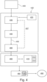
- the systems and methods discussed in this application may also encompass the uploading, reception or generation of a patient's brain network model based on a patient's structural skeleton model as indicated in Fig. 4 .
- a patient's brain network model 400 has been generated or uploaded into a system such as the system of Fig. 1 and real brain data 410 (such as EEG 412, fMRI 414, SPECT and/or SEEG data), from the patient including a period of a simple seizure (i.e. a seizure not capturing a propagation zone) or a complex seizure (i.e.
- an estimation method 420 is initiated which includes: choosing a first estimate of an epileptogenic zone 430; a subsequent simulation of a resulting propagation zone 440 and an optional translation 450 via a forward model into simulated EEG, fMRI, SPECT or SEEG activity 460; a comparison (step 470)between the measured, real brain data 410 and the simulated activity 460.
- the estimation method may optionally include using prior information 435 to obtain an estimate of a first epileptogenic zone including at least one node such as a clinician's estimate of the first epileptogenic zone or an automated starting choice.
- the epileptogenic zone may be varied after generating the simulated activity 460 for a specific epileptogenic zone and steps 430 through 470 may be repeated with a new estimate of a location of the epileptogenic zone.
- the varied epileptogenic zone estimate may include neighboring nodes, additional nodes, or a subset of nodes from the previous or first estimate of the epileptogenic zone.
- the estimation method may then include a decision engine 480 (applying statistical methods as known in the prior art to compare time-series; the decision engine may be a software module for deciding which simulated time-series fits the real data best) to find the best match 485 between simulated brain network model activity and real brain data and output the epileptogenic zone of the best matching data 490 as a suggestion for a brain area of surgical resection or a suggestion for a pre-surgical implantation site of electrodes for recording SEEG data to minimize the traumatic impact for a patient.
- a decision engine 480 applying statistical methods as known in the prior art to compare time-series; the decision engine may be a software module for deciding which simulated time-series fits the real data best) to find the best match 485 between simulated brain network model activity and real brain data and output the epileptogenic zone of the best matching data 490 as a suggestion for a brain area of surgical resection or a suggestion for a pre-surgical implantation site of electrodes for recording SEEG data to minimize
- the method outlined by Fig. 4 and its corresponding description can be performed using non-invasive real brain data, such as EEG data, or invasive real brain data, such as SEEG data.
- non-invasive real brain data such as EEG data
- SEEG data invasive real brain data
- This epileptogenic zone can than be used as a candidate for surgical resection.
- an exemplary implementation of a method embodiment A patient was diagnosed with bi-temporal epilepsy and experienced simple and secondary generalized seizures, which were accompanied by deja-vu hallucinations, associated with palpitations, horripilation, and frisson sensations. Fixed gaze, chewing up and pallor were observed during the seizure. In the post-critic period the patient showed temporal disorientation, repetition of the same questions and retrograde amnesia during one week.
- the MRI examination revealed a hypothalamic hamartoma.
- Surface EEG recordings revealed interictal spikes and indicated a bias towards the left hemisphere. Based on the presurgical evaluation, seven SEEG electrodes were implanted in the left hemisphere, and two in the right hemisphere.
- One electrode was implanted in the hypothalamic hamartoma. During two weeks of continuous SEEG recordings, 6 simple seizures localized in the right hippocampus, and two complex seizures starting in the right hippocampus and then recruiting the left hippocampus, the left temporal lobe and the hypothalamic hamartoma were recorded.
- the large-scale connectivity of the patient was reconstructed, in particular the weight and tract length matrices generating a structural skeleton model.
- the tract length matrix divided by the signal transmission speed defines the time delays, thereby establishing the space-time structure of the coupling and allowing a full virtualization of the patient's brain model.
- a hypothalamic hamartoma was included in the model by changing its effect on local connectivity through factor K ij .
- each voxel of the structural skeleton was replaced by a node.
- Each node of the resulting network was set with an epileptor as described above.
- the nodes were connected via permittivity coupling, which acting on a slow time scale and allowing the spread of the seizure though the network by recruiting regions not in the epileptogenic zone.
- Each node was set with a different excitability parameter x0.
- the value of the excitability was set heterogeneously across the network:
- the epileptogenic zone was set with values of excitability for initiating autonomous triggering of seizures
- the propagation zone was set with values of excitability below a value for autonomous triggering of seizures, however, the values of excitability were higher than in areas which were not considered to be part of the propagation zone.
- a systematic parameter space exploration was performed by varying the following parameters: (i) the global coupling strength G, which is a scalar factor multiplying the whole connectivity matrix, (ii) the local coupling strength G_hyp of the hypothalamus, which is a scalar factor multiplying the contribution of the hypothalamus to the connectivity matrix, (iii) the excitability values of the right hippocampus, (iv) the excitability values of the regions not recruited in the propagation zone (other regions). The excitability values of the other regions in the epileptogenic zone and the propagation zone were fixed (see Table 1).
- the brain network model was used for simulations over a period of 20 seizures and the forward solution for the SEEG electrodes was computed. Simple seizures and complex seizures were generated with similar regions recruited compared to the real SEEG recordings.
- the left hippocampus was stimulated and a propagation pattern in the left temporal lobe, similar to the SEEG recordings, was observed.
- a clinician's prediction was compared to results won from simulations of the brain network model. Comparing the clinician's prediction and the simulated epileptogenic and propagation zone's excitability, the applicant has found a significant overlap between the clinician's prediction and the simulation results, demonstrating the applicability of the approach outlined in this application.
- the simulated EEG data shows a strong similarity with recorded EEG data.
- the spatial distribution of the degree of excitability is based on distance of an area from the epileptogenic zone.
- a parameter value distribution for a parameter representing an MRI lesion can also be included in the brain network model.
- the coupling term K ij (x j - x i ) is weak compared to the other terms of the 1N dimensional system, and in particular the term in x 0 .
- the difference coupling leads to weak coupling terms in a linear stability analysis.
- the weakness depends on the global coupling strength factor G and the normalization of the connectivity matrix, however one can show that this approximation holds over a larger range than for other neural network models.
- the leading eigenvectors are derived and the eigenvectors only depend on the topological connection to the epileptogenic zone.
- G ( Z i ) 4 F ( Z i ) - Z
- the linearization procedure can be easily implemented in a set of instructions for running simulations of a linearized brain network model. Since linear problems can be much more easily be computed than non-linear problems, the simulation time can be greatly reduced.
Landscapes
- Health & Medical Sciences (AREA)
- Life Sciences & Earth Sciences (AREA)
- Engineering & Computer Science (AREA)
- Medical Informatics (AREA)
- Public Health (AREA)
- General Health & Medical Sciences (AREA)
- Pathology (AREA)
- Biomedical Technology (AREA)
- Physics & Mathematics (AREA)
- Neurology (AREA)
- Veterinary Medicine (AREA)
- Biophysics (AREA)
- Surgery (AREA)
- Molecular Biology (AREA)
- Heart & Thoracic Surgery (AREA)
- Animal Behavior & Ethology (AREA)
- Nuclear Medicine, Radiotherapy & Molecular Imaging (AREA)
- Neurosurgery (AREA)
- High Energy & Nuclear Physics (AREA)
- Databases & Information Systems (AREA)
- Epidemiology (AREA)
- Primary Health Care (AREA)
- Data Mining & Analysis (AREA)
- Radiology & Medical Imaging (AREA)
- Physiology (AREA)
- Magnetic Resonance Imaging Apparatus (AREA)
- Measurement And Recording Of Electrical Phenomena And Electrical Characteristics Of The Living Body (AREA)
- Mechanical Engineering (AREA)
- Measuring And Recording Apparatus For Diagnosis (AREA)
Claims (15)
- Computer-implementiertes Verfahren zur Schätzung einer Position einer epileptogenen Zone eines Säugergehirns, wobei das Verfahren folgende Schritte umfasst:a) Erhalt eines strukturellen Gerüstmodells eines Säugergehirns (32), wobei das strukturelle Gerüstmodell (32) eine Vielzahl von Knoten umfasst und auf nicht invasiven Neuroimaging-Daten basiert und wobei die Konnektivitätsinformationen des Gehirns zwischen verschiedenen Knoten aus den nicht invasiven Neuroimaging-Daten extrahiert werden;b) Bereitstellung eines gekoppelten Gehirn-Netzwerkmodells, indem jeder Knoten des strukturelles Gerüstmodells mit einem neuronalen Populationsmodell befüllt wird, wobei das neuronale Populationsmodell, das einem Knoten entspricht, gemäß den Konnektivitätsinformationen mit weiteren neuronalen Populationsmodellen, die weiteren Knoten entsprechen, gekoppelt ist, und das neuronale Populationsmodell ein bistabiles Verhalten aufweist, um in einen iktalen Zustand einzutreten und diesen zu verlassen;c) Bereitstellung einer ersten Schätzung der Position der epileptogenen Zone im Gehirn-Netzwerkmodell, wobei die erste Schätzung der Position mindestens einen der Vielzahl von Knoten einschließt;d) Vorhersage einer Position einer Ausbreitungszone im Gehirn-Netzwerkmodell auf Grundlage der ersten Schätzung der Position der epileptogenen Zone durch mindestens eine Simulation des gekoppelten Gehirn-Netzwerkmodells.
- Verfahren nach Anspruch 1, wobei eine zweite Schätzung der epileptogenen Zone die erste Schätzung der Position der epileptogenen Zone ersetzt, wenn die simulierte Ausbreitungszone der ersten Schätzung von einer beobachteten Ausbreitungszone abweicht.
- Verfahren nach Anspruch 2, wobei weitere Schätzungen der epileptogenen Zone die zweite Schätzung ersetzen, wenn die simulierte Ausbreitungszone der zweiten Schätzung von einer beobachteten Ausbreitungszone abweicht.
- Verfahren nach einem der vorstehenden Ansprüche, wobei die Schritte zur Bereitstellung einer Schätzung einer epileptogenen Zone und zur Vorhersage einer Ausbreitungszone iterativ wiederholt werden, wobei die Position der epileptogenen Zone geändert wird und/oder wobei Parameter des neuronalen Populationsmodells geändert werden.
- Verfahren nach einem der vorstehenden Ansprüche, wobei das neuronale Populationsmodell einen Parameter einschließt, der eine Erregbarkeit des Populationsmodells repräsentiert, und Parameterwerte, die einen ersten Erregbarkeitsgrad angeben, zu dem mindestens einen von der Vielzahl von Knoten der epileptogenen Zone zuweist, und Parameterwerte, die einen niedrigeren als den ersten Erregbarkeitsgrad angeben, zu Knoten, die mit Knoten der epileptogenen Zone gekoppelt sind, zuweist.
- Verfahren nach Anspruch 5, wobei eine räumliche Verteilung der Parameterwerte, die den Erregbarkeitsgrad über das gesamte Gehirn-Netzwerkmodell angeben, auf einem Abstand eines Knotens von der epileptogenen Zone basiert.
- Verfahren nach einem der vorstehenden Ansprüche, wobei das gekoppelte Gehirn-Netzwerkmodell eine Repräsentation einer strukturellen Anomalie, vorzugsweise eine MRT-Läsion, in mindestens einem Knoten einschließt, wobei das neuronale Populationsmodell vorzugsweise einen Parameter einschließt, der einen Grad der strukturellen Anomalie angibt.
- Verfahren nach einem der vorstehenden Ansprüche, wobei das neuronale Populationsmodell durch mindestens eine erste und eine zweite Differentialgleichung repräsentiert ist und eine Zeitskala der ersten Differentialgleichung schneller als eine Zeitskala der zweiten Differentialgleichung ist, wobei verschiedene Knoten vorzugsweise über die zweite Differentialgleichung der jeweiligen Knoten gekoppelt ist.
- Verfahren nach einem der vorstehenden Ansprüche, wobei die Vorhersage der Ausbreitungszone eine Vorhersage elektrischer Aktivitätsdaten einschließt.
- Verfahren nach einem der vorstehenden Ansprüche, wobei das Verfahren ein Vorwärtsmodell für ein Mapping von Gehirndaten mit Elektroenzephalogramm-Daten einschließt, und die Ausbreitungszone repräsentierende Daten in das Vorwärtsmodell gespeist werden.
- Verfahren nach einem der vorstehenden Ansprüche, wobei eine Position zur Implantation stereotaktischer Elektroden in das Säugergehirn auf einer Schätzung der epileptogenen Zone basiert.
- Verfahren nach einem der vorstehenden Ansprüche, wobei eine Kopplung zwischen dem mindestens einen Knoten der epileptogenen Zone und einem Knoten, der mit dem mindestens einen Knoten gekoppelt ist, in einer Simulation geändert wird und das so geänderte Gehirn-Netzwerkmodell verwendet wird, um eine alternative Ausbreitungszone vorherzusagen.
- System, eine zentrale Recheneinheit, eine Speichereinheit und eine Ein-/Ausgabeschnittstelle umfassend, wobei die Vorrichtung konfiguriert ist, die Position einer epileptogenen Zone eines Säugergehirns zu schätzen, einschließlich folgender Schritte:a) Laden eines strukturellen Gerüstmodells eines Säugergehirns in die Speichereinheit, wobei das strukturelle Gerüstmodell eine Vielzahl von Knoten umfasst und auf nicht invasiven Neuroimaging-Daten basiert und wobei die Konnektivitätsinformationen des Gehirns zwischen verschiedenen Knoten aus den nicht invasiven Neuroimaging-Daten extrahiert werden;b) Laden eines gekoppelten Gehirn-Netzwerkmodells in die Speichereinheit, wobei jeder Knoten des strukturellen Gerüstmodells ein neuronales Populationsmodell umfasst, wobei das neuronale Populationsmodell, das einem Knoten entspricht, gemäß den Konnektivitätsinformationen mit weiteren neuronalen Populationsmodellen, die weiteren Knoten entsprechen, gekoppelt ist, und das neuronale Populationsmodell ein bistabiles Verhalten aufweist, um in einen iktalen Zustand einzutreten und diesen zu verlassen;c) Eingabe eines Satzes von Startparametern, um eine erste Schätzung der Position der epileptogenen Zone im Gehirn-Netzwerkmodell bereitzustellen, wobei die erste Schätzung der Position mindestens einen der Vielzahl von Knoten einschließt;d) Entwicklung, durch die zentrale Recheneinheit, und Speicherung einer Position einer Ausbreitungszone im Gehirn-Netzwerkmodell auf Grundlage der ersten Schätzung der Position der epileptogenen Zone durch mindestens eine Simulation des gekoppelten Gehirn-Netzwerkmodells.
- Computer-lesbares Medium, einen Satz Anweisungen zur Ausführung des Verfahrens nach einem der Ansprüche 1 bis 12 umfassend.
- Verfahren nach einem der Ansprüche 1 bis 12, zur Anpassung von Parametern des Gehirn-Netzwerkmodells, das zur Schätzung der Position der epileptogenen Zone eines Säugergehirns verwendet wird, wobei das Verfahren folgende Schritte umfasst:a) Verteilung von Parameterwerten eines Parameters, der einen Erregbarkeitsgrad des neuronalen Populationsmodells über die Vielzahl von Knoten angibt, wobei die Verteilung zumindest teilweise auf dem Abstand eines Knotens zu einem Knoten der Schätzung der epileptogenen Zone beruht;b) Verteilung von Parameterwerten eines Parameters, der eine strukturelle Anomalie über die Vielzahl von Knoten auf Grundlage einer Position einer strukturellen Anomalie angibt, die aus nicht invasiven Neuroimaging-Daten abgeleitet ist, wobei die strukturelle Anomalie durch einen lokalen Skalarfaktor repräsentiert ist;c) Anpassung der Parameterwerte des Parameters, der den Erregbarkeitsgrad angibt, und/oder des Parameters, der die strukturelle Anomalie angibt, in der Weise, dass Simulationsdaten des Gehirn-Netzwerkmodells auf aufgezeichnete Patientendaten abgestimmt werden, wobei die Anpassung auf der Eingabe eines Klinikers oder auf einem automatischen Anpassungsverfahren basiert;d) Änderung der Position der geschätzten epileptogenen Zone im Gehirn-Netzwerkmodell und Vergleich von Simulationsdaten des Gehirn-Netzwerkmodells einschließlich der geänderten Position und aufgezeichneten Patientendaten.
Applications Claiming Priority (2)
| Application Number | Priority Date | Filing Date | Title |
|---|---|---|---|
| EP16166489.1A EP3235427A1 (de) | 2016-04-21 | 2016-04-21 | Verfahren und system zur schätzung einer position einer epileptogenen zone eines säugergehirns |
| PCT/EP2017/059532 WO2017182637A2 (en) | 2016-04-21 | 2017-04-21 | Method and system for estimating a location of an epileptogenic zone of a mammalian brain |
Publications (2)
| Publication Number | Publication Date |
|---|---|
| EP3445239A2 EP3445239A2 (de) | 2019-02-27 |
| EP3445239B1 true EP3445239B1 (de) | 2024-01-31 |
Family
ID=55970776
Family Applications (2)
| Application Number | Title | Priority Date | Filing Date |
|---|---|---|---|
| EP16166489.1A Withdrawn EP3235427A1 (de) | 2016-04-21 | 2016-04-21 | Verfahren und system zur schätzung einer position einer epileptogenen zone eines säugergehirns |
| EP17720722.2A Active EP3445239B1 (de) | 2016-04-21 | 2017-04-21 | Verfahren und system zur schätzung einer position einer epileptogenen zone eines säugergehirns |
Family Applications Before (1)
| Application Number | Title | Priority Date | Filing Date |
|---|---|---|---|
| EP16166489.1A Withdrawn EP3235427A1 (de) | 2016-04-21 | 2016-04-21 | Verfahren und system zur schätzung einer position einer epileptogenen zone eines säugergehirns |
Country Status (4)
| Country | Link |
|---|---|
| US (2) | US20190223779A1 (de) |
| EP (2) | EP3235427A1 (de) |
| ES (1) | ES2974087T3 (de) |
| WO (1) | WO2017182637A2 (de) |
Families Citing this family (14)
| Publication number | Priority date | Publication date | Assignee | Title |
|---|---|---|---|---|
| EP3484355B1 (de) | 2016-07-18 | 2022-03-23 | Universite d'Aix-Marseille (AMU) | Verfahren zur modulation der epileptogenizität im gehirn eines patienten |
| US20180235704A1 (en) * | 2017-02-21 | 2018-08-23 | The Cleveland Clinic Foundation | Configuring a stereo-electroencephalography procedure |
| US11266355B2 (en) * | 2017-05-19 | 2022-03-08 | Cerner Innovation, Inc. | Early warning system and method for predicting patient deterioration |
| WO2020167997A1 (en) * | 2019-02-12 | 2020-08-20 | Surgical Theater, Inc. | System and method for modeling neurological activity |
| EP3770918A1 (de) * | 2019-07-22 | 2021-01-27 | Universite d'Aix-Marseille (AMU) | Verfahren zur bestimmung einer einsatzzeit und erregungsfähigkeit eines gehirnbereichs |
| CN110859600A (zh) * | 2019-12-06 | 2020-03-06 | 深圳市德力凯医疗设备股份有限公司 | 一种生成脑电信号的方法、存储介质及电子设备 |
| CN110931123B (zh) * | 2019-12-06 | 2024-01-05 | 深圳市德力凯医疗设备股份有限公司 | 一种矩阵型脑网络及其构建方法 |
| EP3855453A1 (de) * | 2020-01-24 | 2021-07-28 | Universite d'Aix-Marseille (AMU) | Verfahren zur optimierung eines intrakranialen implantationsschemas eines satzes von elektroenzephalographischen elektroden |
| CN111462887B (zh) * | 2020-03-31 | 2023-08-29 | 首都医科大学宣武医院 | 一种可佩戴的癫痫患者数字助理系统 |
| EP4346674A4 (de) * | 2021-06-03 | 2025-03-26 | Case Western Reserve University | Systeme, verfahren und medien zur darstellung biophysikalischer simulationen in einer interaktiven umgebung mit gemischter realität |
| CN114926396B (zh) * | 2022-04-13 | 2023-06-20 | 四川大学华西医院 | 一种精神障碍类磁共振图像初步筛查模型构建方法 |
| CN114869300B (zh) * | 2022-07-08 | 2022-09-06 | 首都医科大学附属北京天坛医院 | 基于脑电的致痫区定位装置及方法、电子设备和存储介质 |
| CN118380154B (zh) * | 2024-05-06 | 2025-08-26 | 太原理工大学 | 一种基于关键传播区的癫痫全脑动力学建模方法 |
| CN120472180B (zh) * | 2025-04-30 | 2026-04-21 | 深圳市曦嘉医疗科技有限责任公司 | 一种脑图数据的异常区域识别方法 |
Family Cites Families (3)
| Publication number | Priority date | Publication date | Assignee | Title |
|---|---|---|---|---|
| US8560073B2 (en) * | 2009-03-23 | 2013-10-15 | Flint Hills Scientific, Llc | System and apparatus for automated quantitative assessment, optimization and logging of the effects of a therapy |
| GB0919739D0 (en) * | 2009-11-12 | 2009-12-30 | Univ Gent | Methods and systems for channel selection |
| US10729354B2 (en) * | 2013-01-31 | 2020-08-04 | The Regents Of The University Of California | System and method for modeling brain dynamics in normal and diseased states |
-
2016
- 2016-04-21 EP EP16166489.1A patent/EP3235427A1/de not_active Withdrawn
-
2017
- 2017-04-21 WO PCT/EP2017/059532 patent/WO2017182637A2/en not_active Ceased
- 2017-04-21 EP EP17720722.2A patent/EP3445239B1/de active Active
- 2017-04-21 US US16/094,701 patent/US20190223779A1/en not_active Abandoned
- 2017-04-21 ES ES17720722T patent/ES2974087T3/es active Active
-
2024
- 2024-08-08 US US18/797,930 patent/US20250042593A1/en active Pending
Also Published As
| Publication number | Publication date |
|---|---|
| US20250042593A1 (en) | 2025-02-06 |
| WO2017182637A3 (en) | 2017-12-14 |
| WO2017182637A2 (en) | 2017-10-26 |
| US20190223779A1 (en) | 2019-07-25 |
| ES2974087T3 (es) | 2024-06-25 |
| EP3445239A2 (de) | 2019-02-27 |
| EP3235427A1 (de) | 2017-10-25 |
Similar Documents
| Publication | Publication Date | Title |
|---|---|---|
| US20250042593A1 (en) | Method and system for estimating a location of an epileptogenic zone of a mammalian brain | |
| Jirsa et al. | The virtual epileptic patient: individualized whole-brain models of epilepsy spread | |
| US12232883B2 (en) | Method of modulating epileptogenicity in a patient's brain | |
| US12016634B2 (en) | Methods and apparatus for electromagnetic source imaging using deep neural networks | |
| US10588561B1 (en) | Noninvasive system and method for mapping epileptic networks and surgical planning | |
| KR20200086316A (ko) | 개별 환자 데이터 및 치료적 뇌 네트워크 맵을 사용한 효능 및/또는 치료적 파라미터 추천 | |
| EP2499585B1 (de) | Verfahren und systeme zur kanalauswahl | |
| JP2018508321A (ja) | 脳刺激に対する神経血管反応性を測定するシステム及び方法 | |
| WO2023280086A1 (zh) | 靶点确定方法、装置、电子设备、存储介质及神经调控设备 | |
| US7461045B1 (en) | Optimization of spatio-temporal pattern processing for seizure warning and prediction | |
| Wang et al. | Virtual brain twins for stimulation in epilepsy | |
| CN112534279B (zh) | 用于估计介质的物理参数的系统及方法 | |
| Ma et al. | Mapping the electric field of high-definition transcranial electrical stimulation across the lifespan | |
| Sanchez-Todo et al. | Personalization of hybrid brain models from neuroimaging and electrophysiology data | |
| Penas et al. | Parameter estimation in a whole-brain network model of epilepsy: Comparison of parallel global optimization solvers | |
| Chen et al. | Functional magnetic resonance imaging for imaging neural activity in the human brain: the annual progress | |
| US20240006050A1 (en) | Locating an epileptogenic zone for surgical planning | |
| Wang et al. | Virtual brain twins for stimulation in epilepsy | |
| Xiang et al. | Task-specific topographical maps of neural activity in the primate lateral prefrontal cortex | |
| Zhai et al. | Virtual stimulation of the interictal EEG network localizes the EZ as a measure of cortical excitability | |
| Mesejo et al. | A differential evolution-based approach for fitting a nonlinear biophysical model to fMRI BOLD data | |
| Wang et al. | Virtual epileptic patient (VEP): data-driven probabilistic personalized brain modeling in drug-resistant epilepsy | |
| Kalitzin et al. | Prospecting epilepsy surgery outcome using virtual resection paradigm. Computational-model validation | |
| Eiber et al. | A ‘Total Unique Variation Analysis’ for Brain-Machine Interfaces | |
| Vattikonda et al. | High-resolution Bayesian Virtual Epileptic Patient using neural field models |
Legal Events
| Date | Code | Title | Description |
|---|---|---|---|
| STAA | Information on the status of an ep patent application or granted ep patent |
Free format text: STATUS: UNKNOWN |
|
| STAA | Information on the status of an ep patent application or granted ep patent |
Free format text: STATUS: THE INTERNATIONAL PUBLICATION HAS BEEN MADE |
|
| PUAI | Public reference made under article 153(3) epc to a published international application that has entered the european phase |
Free format text: ORIGINAL CODE: 0009012 |
|
| STAA | Information on the status of an ep patent application or granted ep patent |
Free format text: STATUS: REQUEST FOR EXAMINATION WAS MADE |
|
| 17P | Request for examination filed |
Effective date: 20181109 |
|
| AK | Designated contracting states |
Kind code of ref document: A2 Designated state(s): AL AT BE BG CH CY CZ DE DK EE ES FI FR GB GR HR HU IE IS IT LI LT LU LV MC MK MT NL NO PL PT RO RS SE SI SK SM TR |
|
| AX | Request for extension of the european patent |
Extension state: BA ME |
|
| DAV | Request for validation of the european patent (deleted) | ||
| DAX | Request for extension of the european patent (deleted) | ||
| STAA | Information on the status of an ep patent application or granted ep patent |
Free format text: STATUS: EXAMINATION IS IN PROGRESS |
|
| 17Q | First examination report despatched |
Effective date: 20210401 |
|
| RAP3 | Party data changed (applicant data changed or rights of an application transferred) |
Owner name: CODEBOX GMBH |
|
| RIC1 | Information provided on ipc code assigned before grant |
Ipc: A61B 5/00 20060101ALI20230531BHEP Ipc: A61B 5/055 20060101AFI20230531BHEP |
|
| GRAP | Despatch of communication of intention to grant a patent |
Free format text: ORIGINAL CODE: EPIDOSNIGR1 |
|
| STAA | Information on the status of an ep patent application or granted ep patent |
Free format text: STATUS: GRANT OF PATENT IS INTENDED |
|
| INTG | Intention to grant announced |
Effective date: 20230822 |
|
| GRAS | Grant fee paid |
Free format text: ORIGINAL CODE: EPIDOSNIGR3 |
|
| GRAA | (expected) grant |
Free format text: ORIGINAL CODE: 0009210 |
|
| STAA | Information on the status of an ep patent application or granted ep patent |
Free format text: STATUS: THE PATENT HAS BEEN GRANTED |
|
| P01 | Opt-out of the competence of the unified patent court (upc) registered |
Effective date: 20231219 |
|
| AK | Designated contracting states |
Kind code of ref document: B1 Designated state(s): AL AT BE BG CH CY CZ DE DK EE ES FI FR GB GR HR HU IE IS IT LI LT LU LV MC MK MT NL NO PL PT RO RS SE SI SK SM TR |
|
| REG | Reference to a national code |
Ref country code: GB Ref legal event code: FG4D Ref country code: CH Ref legal event code: EP |
|
| REG | Reference to a national code |
Ref country code: DE Ref legal event code: R096 Ref document number: 602017078861 Country of ref document: DE |
|
| REG | Reference to a national code |
Ref country code: IE Ref legal event code: FG4D |
|
| REG | Reference to a national code |
Ref country code: LT Ref legal event code: MG9D |
|
| REG | Reference to a national code |
Ref country code: NL Ref legal event code: MP Effective date: 20240131 |
|
| REG | Reference to a national code |
Ref country code: ES Ref legal event code: FG2A Ref document number: 2974087 Country of ref document: ES Kind code of ref document: T3 Effective date: 20240625 |
|
| PG25 | Lapsed in a contracting state [announced via postgrant information from national office to epo] |
Ref country code: IS Free format text: LAPSE BECAUSE OF FAILURE TO SUBMIT A TRANSLATION OF THE DESCRIPTION OR TO PAY THE FEE WITHIN THE PRESCRIBED TIME-LIMIT Effective date: 20240531 |
|
| PG25 | Lapsed in a contracting state [announced via postgrant information from national office to epo] |
Ref country code: LT Free format text: LAPSE BECAUSE OF FAILURE TO SUBMIT A TRANSLATION OF THE DESCRIPTION OR TO PAY THE FEE WITHIN THE PRESCRIBED TIME-LIMIT Effective date: 20240131 |
|
| PG25 | Lapsed in a contracting state [announced via postgrant information from national office to epo] |
Ref country code: GR Free format text: LAPSE BECAUSE OF FAILURE TO SUBMIT A TRANSLATION OF THE DESCRIPTION OR TO PAY THE FEE WITHIN THE PRESCRIBED TIME-LIMIT Effective date: 20240501 |
|
| REG | Reference to a national code |
Ref country code: AT Ref legal event code: MK05 Ref document number: 1653300 Country of ref document: AT Kind code of ref document: T Effective date: 20240131 |
|
| PG25 | Lapsed in a contracting state [announced via postgrant information from national office to epo] |
Ref country code: RS Free format text: LAPSE BECAUSE OF FAILURE TO SUBMIT A TRANSLATION OF THE DESCRIPTION OR TO PAY THE FEE WITHIN THE PRESCRIBED TIME-LIMIT Effective date: 20240430 Ref country code: HR Free format text: LAPSE BECAUSE OF FAILURE TO SUBMIT A TRANSLATION OF THE DESCRIPTION OR TO PAY THE FEE WITHIN THE PRESCRIBED TIME-LIMIT Effective date: 20240131 Ref country code: NL Free format text: LAPSE BECAUSE OF FAILURE TO SUBMIT A TRANSLATION OF THE DESCRIPTION OR TO PAY THE FEE WITHIN THE PRESCRIBED TIME-LIMIT Effective date: 20240131 |
|
| PG25 | Lapsed in a contracting state [announced via postgrant information from national office to epo] |
Ref country code: AT Free format text: LAPSE BECAUSE OF FAILURE TO SUBMIT A TRANSLATION OF THE DESCRIPTION OR TO PAY THE FEE WITHIN THE PRESCRIBED TIME-LIMIT Effective date: 20240131 |
|
| PG25 | Lapsed in a contracting state [announced via postgrant information from national office to epo] |
Ref country code: RS Free format text: LAPSE BECAUSE OF FAILURE TO SUBMIT A TRANSLATION OF THE DESCRIPTION OR TO PAY THE FEE WITHIN THE PRESCRIBED TIME-LIMIT Effective date: 20240430 Ref country code: NO Free format text: LAPSE BECAUSE OF FAILURE TO SUBMIT A TRANSLATION OF THE DESCRIPTION OR TO PAY THE FEE WITHIN THE PRESCRIBED TIME-LIMIT Effective date: 20240430 Ref country code: NL Free format text: LAPSE BECAUSE OF FAILURE TO SUBMIT A TRANSLATION OF THE DESCRIPTION OR TO PAY THE FEE WITHIN THE PRESCRIBED TIME-LIMIT Effective date: 20240131 Ref country code: LT Free format text: LAPSE BECAUSE OF FAILURE TO SUBMIT A TRANSLATION OF THE DESCRIPTION OR TO PAY THE FEE WITHIN THE PRESCRIBED TIME-LIMIT Effective date: 20240131 Ref country code: IS Free format text: LAPSE BECAUSE OF FAILURE TO SUBMIT A TRANSLATION OF THE DESCRIPTION OR TO PAY THE FEE WITHIN THE PRESCRIBED TIME-LIMIT Effective date: 20240531 Ref country code: HR Free format text: LAPSE BECAUSE OF FAILURE TO SUBMIT A TRANSLATION OF THE DESCRIPTION OR TO PAY THE FEE WITHIN THE PRESCRIBED TIME-LIMIT Effective date: 20240131 Ref country code: GR Free format text: LAPSE BECAUSE OF FAILURE TO SUBMIT A TRANSLATION OF THE DESCRIPTION OR TO PAY THE FEE WITHIN THE PRESCRIBED TIME-LIMIT Effective date: 20240501 Ref country code: FI Free format text: LAPSE BECAUSE OF FAILURE TO SUBMIT A TRANSLATION OF THE DESCRIPTION OR TO PAY THE FEE WITHIN THE PRESCRIBED TIME-LIMIT Effective date: 20240131 Ref country code: BG Free format text: LAPSE BECAUSE OF FAILURE TO SUBMIT A TRANSLATION OF THE DESCRIPTION OR TO PAY THE FEE WITHIN THE PRESCRIBED TIME-LIMIT Effective date: 20240131 Ref country code: AT Free format text: LAPSE BECAUSE OF FAILURE TO SUBMIT A TRANSLATION OF THE DESCRIPTION OR TO PAY THE FEE WITHIN THE PRESCRIBED TIME-LIMIT Effective date: 20240131 |
|
| PG25 | Lapsed in a contracting state [announced via postgrant information from national office to epo] |
Ref country code: PL Free format text: LAPSE BECAUSE OF FAILURE TO SUBMIT A TRANSLATION OF THE DESCRIPTION OR TO PAY THE FEE WITHIN THE PRESCRIBED TIME-LIMIT Effective date: 20240131 Ref country code: PT Free format text: LAPSE BECAUSE OF FAILURE TO SUBMIT A TRANSLATION OF THE DESCRIPTION OR TO PAY THE FEE WITHIN THE PRESCRIBED TIME-LIMIT Effective date: 20240531 |
|
| PG25 | Lapsed in a contracting state [announced via postgrant information from national office to epo] |
Ref country code: SE Free format text: LAPSE BECAUSE OF FAILURE TO SUBMIT A TRANSLATION OF THE DESCRIPTION OR TO PAY THE FEE WITHIN THE PRESCRIBED TIME-LIMIT Effective date: 20240131 Ref country code: PT Free format text: LAPSE BECAUSE OF FAILURE TO SUBMIT A TRANSLATION OF THE DESCRIPTION OR TO PAY THE FEE WITHIN THE PRESCRIBED TIME-LIMIT Effective date: 20240531 Ref country code: PL Free format text: LAPSE BECAUSE OF FAILURE TO SUBMIT A TRANSLATION OF THE DESCRIPTION OR TO PAY THE FEE WITHIN THE PRESCRIBED TIME-LIMIT Effective date: 20240131 Ref country code: LV Free format text: LAPSE BECAUSE OF FAILURE TO SUBMIT A TRANSLATION OF THE DESCRIPTION OR TO PAY THE FEE WITHIN THE PRESCRIBED TIME-LIMIT Effective date: 20240131 |
|
| PG25 | Lapsed in a contracting state [announced via postgrant information from national office to epo] |
Ref country code: DK Free format text: LAPSE BECAUSE OF FAILURE TO SUBMIT A TRANSLATION OF THE DESCRIPTION OR TO PAY THE FEE WITHIN THE PRESCRIBED TIME-LIMIT Effective date: 20240131 |
|
| PG25 | Lapsed in a contracting state [announced via postgrant information from national office to epo] |
Ref country code: SM Free format text: LAPSE BECAUSE OF FAILURE TO SUBMIT A TRANSLATION OF THE DESCRIPTION OR TO PAY THE FEE WITHIN THE PRESCRIBED TIME-LIMIT Effective date: 20240131 |
|
| PG25 | Lapsed in a contracting state [announced via postgrant information from national office to epo] |
Ref country code: CZ Free format text: LAPSE BECAUSE OF FAILURE TO SUBMIT A TRANSLATION OF THE DESCRIPTION OR TO PAY THE FEE WITHIN THE PRESCRIBED TIME-LIMIT Effective date: 20240131 Ref country code: EE Free format text: LAPSE BECAUSE OF FAILURE TO SUBMIT A TRANSLATION OF THE DESCRIPTION OR TO PAY THE FEE WITHIN THE PRESCRIBED TIME-LIMIT Effective date: 20240131 |
|
| PG25 | Lapsed in a contracting state [announced via postgrant information from national office to epo] |
Ref country code: SK Free format text: LAPSE BECAUSE OF FAILURE TO SUBMIT A TRANSLATION OF THE DESCRIPTION OR TO PAY THE FEE WITHIN THE PRESCRIBED TIME-LIMIT Effective date: 20240131 |
|
| PG25 | Lapsed in a contracting state [announced via postgrant information from national office to epo] |
Ref country code: SM Free format text: LAPSE BECAUSE OF FAILURE TO SUBMIT A TRANSLATION OF THE DESCRIPTION OR TO PAY THE FEE WITHIN THE PRESCRIBED TIME-LIMIT Effective date: 20240131 Ref country code: SK Free format text: LAPSE BECAUSE OF FAILURE TO SUBMIT A TRANSLATION OF THE DESCRIPTION OR TO PAY THE FEE WITHIN THE PRESCRIBED TIME-LIMIT Effective date: 20240131 Ref country code: RO Free format text: LAPSE BECAUSE OF FAILURE TO SUBMIT A TRANSLATION OF THE DESCRIPTION OR TO PAY THE FEE WITHIN THE PRESCRIBED TIME-LIMIT Effective date: 20240131 Ref country code: EE Free format text: LAPSE BECAUSE OF FAILURE TO SUBMIT A TRANSLATION OF THE DESCRIPTION OR TO PAY THE FEE WITHIN THE PRESCRIBED TIME-LIMIT Effective date: 20240131 Ref country code: DK Free format text: LAPSE BECAUSE OF FAILURE TO SUBMIT A TRANSLATION OF THE DESCRIPTION OR TO PAY THE FEE WITHIN THE PRESCRIBED TIME-LIMIT Effective date: 20240131 Ref country code: CZ Free format text: LAPSE BECAUSE OF FAILURE TO SUBMIT A TRANSLATION OF THE DESCRIPTION OR TO PAY THE FEE WITHIN THE PRESCRIBED TIME-LIMIT Effective date: 20240131 |
|
| REG | Reference to a national code |
Ref country code: DE Ref legal event code: R097 Ref document number: 602017078861 Country of ref document: DE |
|
| PG25 | Lapsed in a contracting state [announced via postgrant information from national office to epo] |
Ref country code: MC Free format text: LAPSE BECAUSE OF FAILURE TO SUBMIT A TRANSLATION OF THE DESCRIPTION OR TO PAY THE FEE WITHIN THE PRESCRIBED TIME-LIMIT Effective date: 20240131 |
|
| PG25 | Lapsed in a contracting state [announced via postgrant information from national office to epo] |
Ref country code: MC Free format text: LAPSE BECAUSE OF FAILURE TO SUBMIT A TRANSLATION OF THE DESCRIPTION OR TO PAY THE FEE WITHIN THE PRESCRIBED TIME-LIMIT Effective date: 20240131 |
|
| REG | Reference to a national code |
Ref country code: CH Ref legal event code: PL |
|
| PLBE | No opposition filed within time limit |
Free format text: ORIGINAL CODE: 0009261 |
|
| STAA | Information on the status of an ep patent application or granted ep patent |
Free format text: STATUS: NO OPPOSITION FILED WITHIN TIME LIMIT |
|
| PG25 | Lapsed in a contracting state [announced via postgrant information from national office to epo] |
Ref country code: LU Free format text: LAPSE BECAUSE OF NON-PAYMENT OF DUE FEES Effective date: 20240421 |
|
| REG | Reference to a national code |
Ref country code: BE Ref legal event code: MM Effective date: 20240430 |
|
| PG25 | Lapsed in a contracting state [announced via postgrant information from national office to epo] |
Ref country code: LU Free format text: LAPSE BECAUSE OF NON-PAYMENT OF DUE FEES Effective date: 20240421 |
|
| 26N | No opposition filed |
Effective date: 20241101 |
|
| PG25 | Lapsed in a contracting state [announced via postgrant information from national office to epo] |
Ref country code: BE Free format text: LAPSE BECAUSE OF NON-PAYMENT OF DUE FEES Effective date: 20240430 |
|
| PG25 | Lapsed in a contracting state [announced via postgrant information from national office to epo] |
Ref country code: BE Free format text: LAPSE BECAUSE OF NON-PAYMENT OF DUE FEES Effective date: 20240430 Ref country code: CH Free format text: LAPSE BECAUSE OF NON-PAYMENT OF DUE FEES Effective date: 20240430 |
|
| PG25 | Lapsed in a contracting state [announced via postgrant information from national office to epo] |
Ref country code: IE Free format text: LAPSE BECAUSE OF NON-PAYMENT OF DUE FEES Effective date: 20240421 |
|
| PG25 | Lapsed in a contracting state [announced via postgrant information from national office to epo] |
Ref country code: SI Free format text: LAPSE BECAUSE OF FAILURE TO SUBMIT A TRANSLATION OF THE DESCRIPTION OR TO PAY THE FEE WITHIN THE PRESCRIBED TIME-LIMIT Effective date: 20240131 |
|
| PGFP | Annual fee paid to national office [announced via postgrant information from national office to epo] |
Ref country code: DE Payment date: 20250424 Year of fee payment: 9 |
|
| PGFP | Annual fee paid to national office [announced via postgrant information from national office to epo] |
Ref country code: GB Payment date: 20250423 Year of fee payment: 9 Ref country code: ES Payment date: 20250530 Year of fee payment: 9 |
|
| PGFP | Annual fee paid to national office [announced via postgrant information from national office to epo] |
Ref country code: IT Payment date: 20250424 Year of fee payment: 9 |
|
| PGFP | Annual fee paid to national office [announced via postgrant information from national office to epo] |
Ref country code: FR Payment date: 20250425 Year of fee payment: 9 |
|
| PG25 | Lapsed in a contracting state [announced via postgrant information from national office to epo] |
Ref country code: CY Free format text: LAPSE BECAUSE OF FAILURE TO SUBMIT A TRANSLATION OF THE DESCRIPTION OR TO PAY THE FEE WITHIN THE PRESCRIBED TIME-LIMIT; INVALID AB INITIO Effective date: 20170421 |
|
| PG25 | Lapsed in a contracting state [announced via postgrant information from national office to epo] |
Ref country code: HU Free format text: LAPSE BECAUSE OF FAILURE TO SUBMIT A TRANSLATION OF THE DESCRIPTION OR TO PAY THE FEE WITHIN THE PRESCRIBED TIME-LIMIT; INVALID AB INITIO Effective date: 20170421 |