EP3441459B1 - Methods of expanding embryonic stem cells in a suspension culture - Google Patents
Methods of expanding embryonic stem cells in a suspension culture Download PDFInfo
- Publication number
- EP3441459B1 EP3441459B1 EP18186556.9A EP18186556A EP3441459B1 EP 3441459 B1 EP3441459 B1 EP 3441459B1 EP 18186556 A EP18186556 A EP 18186556A EP 3441459 B1 EP3441459 B1 EP 3441459B1
- Authority
- EP
- European Patent Office
- Prior art keywords
- cells
- culture
- embryonic stem
- stem cells
- medium
- Prior art date
- Legal status (The legal status is an assumption and is not a legal conclusion. Google has not performed a legal analysis and makes no representation as to the accuracy of the status listed.)
- Active
Links
- 210000001671 embryonic stem cell Anatomy 0.000 title claims description 214
- 238000000034 method Methods 0.000 title claims description 101
- 238000004114 suspension culture Methods 0.000 title claims description 68
- 210000004027 cell Anatomy 0.000 claims description 368
- 241000282414 Homo sapiens Species 0.000 claims description 120
- 238000012258 culturing Methods 0.000 claims description 99
- 239000001963 growth medium Substances 0.000 claims description 96
- 210000002242 embryoid body Anatomy 0.000 claims description 95
- 102000003974 Fibroblast growth factor 2 Human genes 0.000 claims description 50
- 108090000379 Fibroblast growth factor 2 Proteins 0.000 claims description 50
- 239000000758 substrate Substances 0.000 claims description 13
- 239000003112 inhibitor Substances 0.000 claims description 7
- 208000032839 leukemia Diseases 0.000 claims description 7
- 239000002609 medium Substances 0.000 description 189
- 239000000725 suspension Substances 0.000 description 138
- 210000002966 serum Anatomy 0.000 description 69
- 230000004069 differentiation Effects 0.000 description 48
- 102000004889 Interleukin-6 Human genes 0.000 description 41
- 108090001005 Interleukin-6 Proteins 0.000 description 41
- 229940100601 interleukin-6 Drugs 0.000 description 40
- 102000004058 Leukemia inhibitory factor Human genes 0.000 description 39
- 108090000581 Leukemia inhibitory factor Proteins 0.000 description 39
- 230000015572 biosynthetic process Effects 0.000 description 37
- 108090000623 proteins and genes Proteins 0.000 description 36
- 239000006144 Dulbecco’s modified Eagle's medium Substances 0.000 description 31
- 230000014509 gene expression Effects 0.000 description 31
- TWRXJAOTZQYOKJ-UHFFFAOYSA-L Magnesium chloride Chemical compound [Mg+2].[Cl-].[Cl-] TWRXJAOTZQYOKJ-UHFFFAOYSA-L 0.000 description 30
- 239000000047 product Substances 0.000 description 30
- 238000004458 analytical method Methods 0.000 description 28
- 102000004887 Transforming Growth Factor beta Human genes 0.000 description 26
- 108090001012 Transforming Growth Factor beta Proteins 0.000 description 26
- 210000001519 tissue Anatomy 0.000 description 25
- 210000002459 blastocyst Anatomy 0.000 description 24
- ZDXPYRJPNDTMRX-VKHMYHEASA-N L-glutamine Chemical compound OC(=O)[C@@H](N)CCC(N)=O ZDXPYRJPNDTMRX-VKHMYHEASA-N 0.000 description 22
- 229930182816 L-glutamine Natural products 0.000 description 22
- 206010043276 Teratoma Diseases 0.000 description 21
- 102000004169 proteins and genes Human genes 0.000 description 21
- 210000001654 germ layer Anatomy 0.000 description 18
- 210000000130 stem cell Anatomy 0.000 description 18
- 108010088751 Albumins Proteins 0.000 description 17
- 102000009027 Albumins Human genes 0.000 description 17
- 230000035755 proliferation Effects 0.000 description 17
- 108010067306 Fibronectins Proteins 0.000 description 16
- 102000016359 Fibronectins Human genes 0.000 description 16
- 238000002474 experimental method Methods 0.000 description 16
- 102100035423 POU domain, class 5, transcription factor 1 Human genes 0.000 description 15
- 101710126211 POU domain, class 5, transcription factor 1 Proteins 0.000 description 15
- 230000001965 increasing effect Effects 0.000 description 15
- 229910001629 magnesium chloride Inorganic materials 0.000 description 15
- 210000002950 fibroblast Anatomy 0.000 description 14
- 241000699666 Mus <mouse, genus> Species 0.000 description 13
- 239000003797 essential amino acid Substances 0.000 description 13
- 235000020776 essential amino acid Nutrition 0.000 description 13
- 102000004196 processed proteins & peptides Human genes 0.000 description 13
- 108090000765 processed proteins & peptides Proteins 0.000 description 13
- DGVVWUTYPXICAM-UHFFFAOYSA-N β‐Mercaptoethanol Chemical compound OCCS DGVVWUTYPXICAM-UHFFFAOYSA-N 0.000 description 13
- 102000029816 Collagenase Human genes 0.000 description 12
- 108060005980 Collagenase Proteins 0.000 description 12
- 229960002424 collagenase Drugs 0.000 description 12
- 210000003953 foreskin Anatomy 0.000 description 12
- NOESYZHRGYRDHS-UHFFFAOYSA-N insulin Chemical compound N1C(=O)C(NC(=O)C(CCC(N)=O)NC(=O)C(CCC(O)=O)NC(=O)C(C(C)C)NC(=O)C(NC(=O)CN)C(C)CC)CSSCC(C(NC(CO)C(=O)NC(CC(C)C)C(=O)NC(CC=2C=CC(O)=CC=2)C(=O)NC(CCC(N)=O)C(=O)NC(CC(C)C)C(=O)NC(CCC(O)=O)C(=O)NC(CC(N)=O)C(=O)NC(CC=2C=CC(O)=CC=2)C(=O)NC(CSSCC(NC(=O)C(C(C)C)NC(=O)C(CC(C)C)NC(=O)C(CC=2C=CC(O)=CC=2)NC(=O)C(CC(C)C)NC(=O)C(C)NC(=O)C(CCC(O)=O)NC(=O)C(C(C)C)NC(=O)C(CC(C)C)NC(=O)C(CC=2NC=NC=2)NC(=O)C(CO)NC(=O)CNC2=O)C(=O)NCC(=O)NC(CCC(O)=O)C(=O)NC(CCCNC(N)=N)C(=O)NCC(=O)NC(CC=3C=CC=CC=3)C(=O)NC(CC=3C=CC=CC=3)C(=O)NC(CC=3C=CC(O)=CC=3)C(=O)NC(C(C)O)C(=O)N3C(CCC3)C(=O)NC(CCCCN)C(=O)NC(C)C(O)=O)C(=O)NC(CC(N)=O)C(O)=O)=O)NC(=O)C(C(C)CC)NC(=O)C(CO)NC(=O)C(C(C)O)NC(=O)C1CSSCC2NC(=O)C(CC(C)C)NC(=O)C(NC(=O)C(CCC(N)=O)NC(=O)C(CC(N)=O)NC(=O)C(NC(=O)C(N)CC=1C=CC=CC=1)C(C)C)CC1=CN=CN1 NOESYZHRGYRDHS-UHFFFAOYSA-N 0.000 description 12
- 229920001184 polypeptide Polymers 0.000 description 12
- 230000002035 prolonged effect Effects 0.000 description 12
- 102000005962 receptors Human genes 0.000 description 12
- 108020003175 receptors Proteins 0.000 description 12
- 238000004113 cell culture Methods 0.000 description 11
- 238000009795 derivation Methods 0.000 description 11
- 238000010494 dissociation reaction Methods 0.000 description 11
- 230000005593 dissociations Effects 0.000 description 11
- 238000000338 in vitro Methods 0.000 description 11
- 238000003757 reverse transcription PCR Methods 0.000 description 11
- 241000699670 Mus sp. Species 0.000 description 10
- 108010029485 Protein Isoforms Proteins 0.000 description 10
- 102000001708 Protein Isoforms Human genes 0.000 description 10
- 239000002299 complementary DNA Substances 0.000 description 10
- 238000012136 culture method Methods 0.000 description 10
- 230000000694 effects Effects 0.000 description 10
- 210000001900 endoderm Anatomy 0.000 description 10
- 239000011159 matrix material Substances 0.000 description 10
- 210000003716 mesoderm Anatomy 0.000 description 10
- 108091003079 Bovine Serum Albumin Proteins 0.000 description 9
- IAZDPXIOMUYVGZ-UHFFFAOYSA-N Dimethylsulphoxide Chemical compound CS(C)=O IAZDPXIOMUYVGZ-UHFFFAOYSA-N 0.000 description 9
- 241001465754 Metazoa Species 0.000 description 9
- 101100247004 Rattus norvegicus Qsox1 gene Proteins 0.000 description 9
- 230000006907 apoptotic process Effects 0.000 description 9
- 210000003981 ectoderm Anatomy 0.000 description 9
- 230000012010 growth Effects 0.000 description 9
- 239000003102 growth factor Substances 0.000 description 9
- 238000001727 in vivo Methods 0.000 description 9
- 238000011534 incubation Methods 0.000 description 9
- 238000012423 maintenance Methods 0.000 description 9
- 239000000463 material Substances 0.000 description 9
- 239000000203 mixture Substances 0.000 description 9
- 230000003068 static effect Effects 0.000 description 9
- 239000013598 vector Substances 0.000 description 9
- 102100028072 Fibroblast growth factor 4 Human genes 0.000 description 8
- 101001060274 Homo sapiens Fibroblast growth factor 4 Proteins 0.000 description 8
- 108010038501 Interleukin-6 Receptors Proteins 0.000 description 8
- 238000010790 dilution Methods 0.000 description 8
- 239000012895 dilution Substances 0.000 description 8
- 208000037265 diseases, disorders, signs and symptoms Diseases 0.000 description 8
- 238000002513 implantation Methods 0.000 description 8
- 230000037361 pathway Effects 0.000 description 8
- 230000002062 proliferating effect Effects 0.000 description 8
- 230000008093 supporting effect Effects 0.000 description 8
- 238000002560 therapeutic procedure Methods 0.000 description 8
- 108091032973 (ribonucleotides)n+m Proteins 0.000 description 7
- 208000037051 Chromosomal Instability Diseases 0.000 description 7
- 101000610551 Homo sapiens Prominin-1 Proteins 0.000 description 7
- 241000288906 Primates Species 0.000 description 7
- 102100040120 Prominin-1 Human genes 0.000 description 7
- 102000004495 STAT3 Transcription Factor Human genes 0.000 description 7
- 108010017324 STAT3 Transcription Factor Proteins 0.000 description 7
- 102000004142 Trypsin Human genes 0.000 description 7
- 108090000631 Trypsin Proteins 0.000 description 7
- 239000000427 antigen Substances 0.000 description 7
- 108091007433 antigens Proteins 0.000 description 7
- 102000036639 antigens Human genes 0.000 description 7
- 230000007246 mechanism Effects 0.000 description 7
- 239000002243 precursor Substances 0.000 description 7
- 239000012588 trypsin Substances 0.000 description 7
- 102100039819 Actin, alpha cardiac muscle 1 Human genes 0.000 description 6
- 101710170648 Actin, alpha cardiac muscle 1 Proteins 0.000 description 6
- 102100022900 Actin, cytoplasmic 1 Human genes 0.000 description 6
- 108010085238 Actins Proteins 0.000 description 6
- CIWBSHSKHKDKBQ-JLAZNSOCSA-N Ascorbic acid Chemical compound OC[C@H](O)[C@H]1OC(=O)C(O)=C1O CIWBSHSKHKDKBQ-JLAZNSOCSA-N 0.000 description 6
- 102000006734 Beta-Globulins Human genes 0.000 description 6
- 108010087504 Beta-Globulins Proteins 0.000 description 6
- 102000004127 Cytokines Human genes 0.000 description 6
- 108090000695 Cytokines Proteins 0.000 description 6
- 102000004877 Insulin Human genes 0.000 description 6
- 108090001061 Insulin Proteins 0.000 description 6
- 101710150336 Protein Rex Proteins 0.000 description 6
- 238000010240 RT-PCR analysis Methods 0.000 description 6
- 238000006243 chemical reaction Methods 0.000 description 6
- 229940125396 insulin Drugs 0.000 description 6
- 238000004519 manufacturing process Methods 0.000 description 6
- 230000000877 morphologic effect Effects 0.000 description 6
- 238000011160 research Methods 0.000 description 6
- 239000000523 sample Substances 0.000 description 6
- 102000010781 Interleukin-6 Receptors Human genes 0.000 description 5
- 101710152369 Interleukin-6 receptor subunit beta Proteins 0.000 description 5
- 101150088608 Kdr gene Proteins 0.000 description 5
- 102000008763 Neurofilament Proteins Human genes 0.000 description 5
- 108010088373 Neurofilament Proteins Proteins 0.000 description 5
- 102000016549 Vascular Endothelial Growth Factor Receptor-2 Human genes 0.000 description 5
- 229940024606 amino acid Drugs 0.000 description 5
- 150000001413 amino acids Chemical class 0.000 description 5
- 230000001640 apoptogenic effect Effects 0.000 description 5
- 238000013459 approach Methods 0.000 description 5
- 230000024245 cell differentiation Effects 0.000 description 5
- 210000003754 fetus Anatomy 0.000 description 5
- 210000004392 genitalia Anatomy 0.000 description 5
- 238000002347 injection Methods 0.000 description 5
- 239000007924 injection Substances 0.000 description 5
- 230000000670 limiting effect Effects 0.000 description 5
- 150000002632 lipids Chemical class 0.000 description 5
- 210000001161 mammalian embryo Anatomy 0.000 description 5
- 238000010369 molecular cloning Methods 0.000 description 5
- 230000001537 neural effect Effects 0.000 description 5
- 210000005044 neurofilament Anatomy 0.000 description 5
- 210000004248 oligodendroglia Anatomy 0.000 description 5
- 238000012360 testing method Methods 0.000 description 5
- 238000001262 western blot Methods 0.000 description 5
- 210000004340 zona pellucida Anatomy 0.000 description 5
- 108010010803 Gelatin Proteins 0.000 description 4
- 102100037792 Interleukin-6 receptor subunit alpha Human genes 0.000 description 4
- 206010028980 Neoplasm Diseases 0.000 description 4
- 102000004338 Transferrin Human genes 0.000 description 4
- 108090000901 Transferrin Proteins 0.000 description 4
- 102000004243 Tubulin Human genes 0.000 description 4
- 108090000704 Tubulin Proteins 0.000 description 4
- 230000005856 abnormality Effects 0.000 description 4
- 239000003929 acidic solution Substances 0.000 description 4
- 208000036815 beta tubulin Diseases 0.000 description 4
- 230000000903 blocking effect Effects 0.000 description 4
- 229940098773 bovine serum albumin Drugs 0.000 description 4
- 239000006285 cell suspension Substances 0.000 description 4
- 230000002759 chromosomal effect Effects 0.000 description 4
- 150000001875 compounds Chemical class 0.000 description 4
- 239000003636 conditioned culture medium Substances 0.000 description 4
- 239000000356 contaminant Substances 0.000 description 4
- 238000001514 detection method Methods 0.000 description 4
- 208000035475 disorder Diseases 0.000 description 4
- 108010007093 dispase Proteins 0.000 description 4
- 239000003596 drug target Substances 0.000 description 4
- 238000000684 flow cytometry Methods 0.000 description 4
- 239000008273 gelatin Substances 0.000 description 4
- 229920000159 gelatin Polymers 0.000 description 4
- 235000019322 gelatine Nutrition 0.000 description 4
- 235000011852 gelatine desserts Nutrition 0.000 description 4
- 230000002068 genetic effect Effects 0.000 description 4
- MASNOZXLGMXCHN-ZLPAWPGGSA-N glucagon Chemical compound C([C@@H](C(=O)N[C@H](C(=O)N[C@@H](CCC(N)=O)C(=O)N[C@@H](CC=1C2=CC=CC=C2NC=1)C(=O)N[C@@H](CC(C)C)C(=O)N[C@@H](CCSC)C(=O)N[C@@H](CC(N)=O)C(=O)N[C@@H]([C@@H](C)O)C(O)=O)C(C)C)NC(=O)[C@H](CC(O)=O)NC(=O)[C@H](CCC(N)=O)NC(=O)[C@H](C)NC(=O)[C@H](CCCNC(N)=N)NC(=O)[C@H](CCCNC(N)=N)NC(=O)[C@H](CO)NC(=O)[C@H](CC(O)=O)NC(=O)[C@H](CC(C)C)NC(=O)[C@H](CC=1C=CC(O)=CC=1)NC(=O)[C@H](CCCCN)NC(=O)[C@H](CO)NC(=O)[C@H](CC=1C=CC(O)=CC=1)NC(=O)[C@H](CC(O)=O)NC(=O)[C@H](CO)NC(=O)[C@@H](NC(=O)[C@H](CC=1C=CC=CC=1)NC(=O)[C@@H](NC(=O)CNC(=O)[C@H](CCC(N)=O)NC(=O)[C@H](CO)NC(=O)[C@@H](N)CC=1NC=NC=1)[C@@H](C)O)[C@@H](C)O)C1=CC=CC=C1 MASNOZXLGMXCHN-ZLPAWPGGSA-N 0.000 description 4
- 238000012744 immunostaining Methods 0.000 description 4
- 239000012528 membrane Substances 0.000 description 4
- 235000015097 nutrients Nutrition 0.000 description 4
- 239000008188 pellet Substances 0.000 description 4
- 239000012071 phase Substances 0.000 description 4
- 230000000717 retained effect Effects 0.000 description 4
- 239000000243 solution Substances 0.000 description 4
- 238000010186 staining Methods 0.000 description 4
- 239000012581 transferrin Substances 0.000 description 4
- 208000031404 Chromosome Aberrations Diseases 0.000 description 3
- 108020004414 DNA Proteins 0.000 description 3
- 102000004190 Enzymes Human genes 0.000 description 3
- 108090000790 Enzymes Proteins 0.000 description 3
- 102000051325 Glucagon Human genes 0.000 description 3
- 108060003199 Glucagon Proteins 0.000 description 3
- 101001027128 Homo sapiens Fibronectin Proteins 0.000 description 3
- 229930040373 Paraformaldehyde Natural products 0.000 description 3
- 102000037602 Platelet Endothelial Cell Adhesion Molecule-1 Human genes 0.000 description 3
- 108010069381 Platelet Endothelial Cell Adhesion Molecule-1 Proteins 0.000 description 3
- 108090001027 Troponin Proteins 0.000 description 3
- 241000700605 Viruses Species 0.000 description 3
- 238000013019 agitation Methods 0.000 description 3
- 238000000137 annealing Methods 0.000 description 3
- 229960005070 ascorbic acid Drugs 0.000 description 3
- 235000010323 ascorbic acid Nutrition 0.000 description 3
- 239000011668 ascorbic acid Substances 0.000 description 3
- 239000012888 bovine serum Substances 0.000 description 3
- 210000004413 cardiac myocyte Anatomy 0.000 description 3
- 210000000845 cartilage Anatomy 0.000 description 3
- 238000011161 development Methods 0.000 description 3
- 230000018109 developmental process Effects 0.000 description 3
- 230000029087 digestion Effects 0.000 description 3
- LOKCTEFSRHRXRJ-UHFFFAOYSA-I dipotassium trisodium dihydrogen phosphate hydrogen phosphate dichloride Chemical compound P(=O)(O)(O)[O-].[K+].P(=O)(O)([O-])[O-].[Na+].[Na+].[Cl-].[K+].[Cl-].[Na+] LOKCTEFSRHRXRJ-UHFFFAOYSA-I 0.000 description 3
- 201000010099 disease Diseases 0.000 description 3
- 238000007877 drug screening Methods 0.000 description 3
- 210000002889 endothelial cell Anatomy 0.000 description 3
- 230000002255 enzymatic effect Effects 0.000 description 3
- 229940088598 enzyme Drugs 0.000 description 3
- 238000011156 evaluation Methods 0.000 description 3
- 230000001747 exhibiting effect Effects 0.000 description 3
- 239000013604 expression vector Substances 0.000 description 3
- 239000012894 fetal calf serum Substances 0.000 description 3
- 239000012634 fragment Substances 0.000 description 3
- 230000007045 gastrulation Effects 0.000 description 3
- 239000011521 glass Substances 0.000 description 3
- 229960004666 glucagon Drugs 0.000 description 3
- -1 glucagons Proteins 0.000 description 3
- 210000004408 hybridoma Anatomy 0.000 description 3
- 238000003125 immunofluorescent labeling Methods 0.000 description 3
- 238000003364 immunohistochemistry Methods 0.000 description 3
- 230000001939 inductive effect Effects 0.000 description 3
- 108020004999 messenger RNA Proteins 0.000 description 3
- 210000003205 muscle Anatomy 0.000 description 3
- 238000010899 nucleation Methods 0.000 description 3
- 229920002866 paraformaldehyde Polymers 0.000 description 3
- 239000002953 phosphate buffered saline Substances 0.000 description 3
- 102000040430 polynucleotide Human genes 0.000 description 3
- 108091033319 polynucleotide Proteins 0.000 description 3
- 239000002157 polynucleotide Substances 0.000 description 3
- 108010057417 polysialyl neural cell adhesion molecule Proteins 0.000 description 3
- 230000003389 potentiating effect Effects 0.000 description 3
- 230000008569 process Effects 0.000 description 3
- 238000003753 real-time PCR Methods 0.000 description 3
- 238000013341 scale-up Methods 0.000 description 3
- 238000003786 synthesis reaction Methods 0.000 description 3
- ZRKFYGHZFMAOKI-QMGMOQQFSA-N tgfbeta Chemical compound C([C@H](NC(=O)[C@H](C(C)C)NC(=O)CNC(=O)[C@H](CCC(O)=O)NC(=O)[C@H](CCCNC(N)=N)NC(=O)[C@H](CC(N)=O)NC(=O)[C@H](CC(C)C)NC(=O)[C@H]([C@@H](C)O)NC(=O)[C@H](CCC(O)=O)NC(=O)[C@H]([C@@H](C)O)NC(=O)[C@H](CC(C)C)NC(=O)CNC(=O)[C@H](C)NC(=O)[C@H](CO)NC(=O)[C@H](CCC(N)=O)NC(=O)[C@@H](NC(=O)[C@H](C)NC(=O)[C@H](C)NC(=O)[C@@H](NC(=O)[C@H](CC(C)C)NC(=O)[C@@H](N)CCSC)C(C)C)[C@@H](C)CC)C(=O)N[C@@H]([C@@H](C)O)C(=O)N[C@@H](C(C)C)C(=O)N[C@@H](CC=1C=CC=CC=1)C(=O)N[C@@H](C)C(=O)N1[C@@H](CCC1)C(=O)N[C@@H]([C@@H](C)O)C(=O)N[C@@H](CC(N)=O)C(=O)N[C@@H](CCC(O)=O)C(=O)N[C@@H](C)C(=O)N[C@@H](CC=1C=CC=CC=1)C(=O)N[C@@H](CCCNC(N)=N)C(=O)N[C@@H](C)C(=O)N[C@@H](CC(C)C)C(=O)N1[C@@H](CCC1)C(=O)N1[C@@H](CCC1)C(=O)N[C@@H](CCCNC(N)=N)C(=O)N[C@@H](CCC(O)=O)C(=O)N[C@@H](CCCNC(N)=N)C(=O)N[C@@H](CO)C(=O)N[C@@H](CCCNC(N)=N)C(=O)N[C@@H](CC(C)C)C(=O)N[C@@H](CC(C)C)C(O)=O)C1=CC=C(O)C=C1 ZRKFYGHZFMAOKI-QMGMOQQFSA-N 0.000 description 3
- 239000003104 tissue culture media Substances 0.000 description 3
- 230000017423 tissue regeneration Effects 0.000 description 3
- 238000002054 transplantation Methods 0.000 description 3
- 230000035899 viability Effects 0.000 description 3
- 102000002260 Alkaline Phosphatase Human genes 0.000 description 2
- 108020004774 Alkaline Phosphatase Proteins 0.000 description 2
- IJGRMHOSHXDMSA-UHFFFAOYSA-N Atomic nitrogen Chemical compound N#N IJGRMHOSHXDMSA-UHFFFAOYSA-N 0.000 description 2
- 241000282693 Cercopithecidae Species 0.000 description 2
- KCXVZYZYPLLWCC-UHFFFAOYSA-N EDTA Chemical compound OC(=O)CN(CC(O)=O)CCN(CC(O)=O)CC(O)=O KCXVZYZYPLLWCC-UHFFFAOYSA-N 0.000 description 2
- 238000012413 Fluorescence activated cell sorting analysis Methods 0.000 description 2
- WSFSSNUMVMOOMR-UHFFFAOYSA-N Formaldehyde Chemical compound O=C WSFSSNUMVMOOMR-UHFFFAOYSA-N 0.000 description 2
- WZUVPPKBWHMQCE-UHFFFAOYSA-N Haematoxylin Chemical compound C12=CC(O)=C(O)C=C2CC2(O)C1C1=CC=C(O)C(O)=C1OC2 WZUVPPKBWHMQCE-UHFFFAOYSA-N 0.000 description 2
- 101000599048 Homo sapiens Interleukin-6 receptor subunit alpha Proteins 0.000 description 2
- 101000738771 Homo sapiens Receptor-type tyrosine-protein phosphatase C Proteins 0.000 description 2
- 101000617830 Homo sapiens Sterol O-acyltransferase 1 Proteins 0.000 description 2
- 102100034343 Integrase Human genes 0.000 description 2
- 102000013691 Interleukin-17 Human genes 0.000 description 2
- 108050003558 Interleukin-17 Proteins 0.000 description 2
- 102000042838 JAK family Human genes 0.000 description 2
- CSNNHWWHGAXBCP-UHFFFAOYSA-L Magnesium sulfate Chemical compound [Mg+2].[O-][S+2]([O-])([O-])[O-] CSNNHWWHGAXBCP-UHFFFAOYSA-L 0.000 description 2
- XUMBMVFBXHLACL-UHFFFAOYSA-N Melanin Chemical compound O=C1C(=O)C(C2=CNC3=C(C(C(=O)C4=C32)=O)C)=C2C4=CNC2=C1C XUMBMVFBXHLACL-UHFFFAOYSA-N 0.000 description 2
- 241000713869 Moloney murine leukemia virus Species 0.000 description 2
- 102000006386 Myelin Proteins Human genes 0.000 description 2
- 108010083674 Myelin Proteins Proteins 0.000 description 2
- 101100386053 Neurospora crassa (strain ATCC 24698 / 74-OR23-1A / CBS 708.71 / DSM 1257 / FGSC 987) cys-3 gene Proteins 0.000 description 2
- 108010004729 Phycoerythrin Proteins 0.000 description 2
- RJKFOVLPORLFTN-LEKSSAKUSA-N Progesterone Chemical compound C1CC2=CC(=O)CC[C@]2(C)[C@@H]2[C@@H]1[C@@H]1CC[C@H](C(=O)C)[C@@]1(C)CC2 RJKFOVLPORLFTN-LEKSSAKUSA-N 0.000 description 2
- LCTONWCANYUPML-UHFFFAOYSA-M Pyruvate Chemical compound CC(=O)C([O-])=O LCTONWCANYUPML-UHFFFAOYSA-M 0.000 description 2
- 238000002123 RNA extraction Methods 0.000 description 2
- 102100037422 Receptor-type tyrosine-protein phosphatase C Human genes 0.000 description 2
- 108020004511 Recombinant DNA Proteins 0.000 description 2
- DBMJMQXJHONAFJ-UHFFFAOYSA-M Sodium laurylsulphate Chemical compound [Na+].CCCCCCCCCCCCOS([O-])(=O)=O DBMJMQXJHONAFJ-UHFFFAOYSA-M 0.000 description 2
- 102100021993 Sterol O-acyltransferase 1 Human genes 0.000 description 2
- 101000697584 Streptomyces lavendulae Streptothricin acetyltransferase Proteins 0.000 description 2
- 230000009471 action Effects 0.000 description 2
- 230000004913 activation Effects 0.000 description 2
- 239000000654 additive Substances 0.000 description 2
- 238000000246 agarose gel electrophoresis Methods 0.000 description 2
- 210000004102 animal cell Anatomy 0.000 description 2
- 238000010009 beating Methods 0.000 description 2
- 230000008901 benefit Effects 0.000 description 2
- 210000000988 bone and bone Anatomy 0.000 description 2
- 244000309466 calf Species 0.000 description 2
- 239000000969 carrier Substances 0.000 description 2
- 230000010261 cell growth Effects 0.000 description 2
- 230000004663 cell proliferation Effects 0.000 description 2
- 238000005119 centrifugation Methods 0.000 description 2
- 239000003153 chemical reaction reagent Substances 0.000 description 2
- 210000004978 chinese hamster ovary cell Anatomy 0.000 description 2
- CVSVTCORWBXHQV-UHFFFAOYSA-N creatine Chemical compound NC(=[NH2+])N(C)CC([O-])=O CVSVTCORWBXHQV-UHFFFAOYSA-N 0.000 description 2
- 230000002559 cytogenic effect Effects 0.000 description 2
- 238000004925 denaturation Methods 0.000 description 2
- 230000036425 denaturation Effects 0.000 description 2
- 210000002257 embryonic structure Anatomy 0.000 description 2
- 230000006862 enzymatic digestion Effects 0.000 description 2
- 210000000981 epithelium Anatomy 0.000 description 2
- 239000012091 fetal bovine serum Substances 0.000 description 2
- 238000001914 filtration Methods 0.000 description 2
- 230000006870 function Effects 0.000 description 2
- 210000004602 germ cell Anatomy 0.000 description 2
- 230000013595 glycosylation Effects 0.000 description 2
- 238000006206 glycosylation reaction Methods 0.000 description 2
- 230000005484 gravity Effects 0.000 description 2
- 210000003958 hematopoietic stem cell Anatomy 0.000 description 2
- 210000003630 histaminocyte Anatomy 0.000 description 2
- 238000010562 histological examination Methods 0.000 description 2
- 229940088597 hormone Drugs 0.000 description 2
- 210000005260 human cell Anatomy 0.000 description 2
- 230000001900 immune effect Effects 0.000 description 2
- 238000003018 immunoassay Methods 0.000 description 2
- 238000010166 immunofluorescence Methods 0.000 description 2
- 239000004615 ingredient Substances 0.000 description 2
- 229910052500 inorganic mineral Inorganic materials 0.000 description 2
- 230000006799 invasive growth in response to glucose limitation Effects 0.000 description 2
- 238000002955 isolation Methods 0.000 description 2
- 238000002826 magnetic-activated cell sorting Methods 0.000 description 2
- 238000005259 measurement Methods 0.000 description 2
- 239000011707 mineral Substances 0.000 description 2
- 235000010755 mineral Nutrition 0.000 description 2
- 210000005012 myelin Anatomy 0.000 description 2
- 102000039446 nucleic acids Human genes 0.000 description 2
- 108020004707 nucleic acids Proteins 0.000 description 2
- 150000007523 nucleic acids Chemical class 0.000 description 2
- 238000007747 plating Methods 0.000 description 2
- 230000000270 postfertilization Effects 0.000 description 2
- 238000002360 preparation method Methods 0.000 description 2
- 238000001742 protein purification Methods 0.000 description 2
- KIDHWZJUCRJVML-UHFFFAOYSA-N putrescine Chemical compound NCCCCN KIDHWZJUCRJVML-UHFFFAOYSA-N 0.000 description 2
- 238000003259 recombinant expression Methods 0.000 description 2
- 238000010188 recombinant method Methods 0.000 description 2
- 238000011084 recovery Methods 0.000 description 2
- 238000009256 replacement therapy Methods 0.000 description 2
- 230000002441 reversible effect Effects 0.000 description 2
- 238000000926 separation method Methods 0.000 description 2
- 230000019491 signal transduction Effects 0.000 description 2
- 238000010532 solid phase synthesis reaction Methods 0.000 description 2
- UCSJYZPVAKXKNQ-HZYVHMACSA-N streptomycin Chemical compound CN[C@H]1[C@H](O)[C@@H](O)[C@H](CO)O[C@H]1O[C@@H]1[C@](C=O)(O)[C@H](C)O[C@H]1O[C@@H]1[C@@H](NC(N)=N)[C@H](O)[C@@H](NC(N)=N)[C@H](O)[C@H]1O UCSJYZPVAKXKNQ-HZYVHMACSA-N 0.000 description 2
- 239000000126 substance Substances 0.000 description 2
- XOAAWQZATWQOTB-UHFFFAOYSA-N taurine Chemical compound NCCS(O)(=O)=O XOAAWQZATWQOTB-UHFFFAOYSA-N 0.000 description 2
- 238000012546 transfer Methods 0.000 description 2
- 210000002993 trophoblast Anatomy 0.000 description 2
- 239000011782 vitamin Substances 0.000 description 2
- 235000013343 vitamin Nutrition 0.000 description 2
- 229940088594 vitamin Drugs 0.000 description 2
- 229930003231 vitamin Natural products 0.000 description 2
- DIGQNXIGRZPYDK-WKSCXVIASA-N (2R)-6-amino-2-[[2-[[(2S)-2-[[2-[[(2R)-2-[[(2S)-2-[[(2R,3S)-2-[[2-[[(2S)-2-[[2-[[(2S)-2-[[(2S)-2-[[(2R)-2-[[(2S,3S)-2-[[(2R)-2-[[(2S)-2-[[(2S)-2-[[(2S)-2-[[2-[[(2S)-2-[[(2R)-2-[[2-[[2-[[2-[(2-amino-1-hydroxyethylidene)amino]-3-carboxy-1-hydroxypropylidene]amino]-1-hydroxy-3-sulfanylpropylidene]amino]-1-hydroxyethylidene]amino]-1-hydroxy-3-sulfanylpropylidene]amino]-1,3-dihydroxypropylidene]amino]-1-hydroxyethylidene]amino]-1-hydroxypropylidene]amino]-1,3-dihydroxypropylidene]amino]-1,3-dihydroxypropylidene]amino]-1-hydroxy-3-sulfanylpropylidene]amino]-1,3-dihydroxybutylidene]amino]-1-hydroxy-3-sulfanylpropylidene]amino]-1-hydroxypropylidene]amino]-1,3-dihydroxypropylidene]amino]-1-hydroxyethylidene]amino]-1,5-dihydroxy-5-iminopentylidene]amino]-1-hydroxy-3-sulfanylpropylidene]amino]-1,3-dihydroxybutylidene]amino]-1-hydroxy-3-sulfanylpropylidene]amino]-1,3-dihydroxypropylidene]amino]-1-hydroxyethylidene]amino]-1-hydroxy-3-sulfanylpropylidene]amino]-1-hydroxyethylidene]amino]hexanoic acid Chemical compound C[C@@H]([C@@H](C(=N[C@@H](CS)C(=N[C@@H](C)C(=N[C@@H](CO)C(=NCC(=N[C@@H](CCC(=N)O)C(=NC(CS)C(=N[C@H]([C@H](C)O)C(=N[C@H](CS)C(=N[C@H](CO)C(=NCC(=N[C@H](CS)C(=NCC(=N[C@H](CCCCN)C(=O)O)O)O)O)O)O)O)O)O)O)O)O)O)O)N=C([C@H](CS)N=C([C@H](CO)N=C([C@H](CO)N=C([C@H](C)N=C(CN=C([C@H](CO)N=C([C@H](CS)N=C(CN=C(C(CS)N=C(C(CC(=O)O)N=C(CN)O)O)O)O)O)O)O)O)O)O)O)O DIGQNXIGRZPYDK-WKSCXVIASA-N 0.000 description 1
- XOQABDOICLHPIS-UHFFFAOYSA-N 1-hydroxy-2,1-benzoxaborole Chemical compound C1=CC=C2B(O)OCC2=C1 XOQABDOICLHPIS-UHFFFAOYSA-N 0.000 description 1
- JKMHFZQWWAIEOD-UHFFFAOYSA-N 2-[4-(2-hydroxyethyl)piperazin-1-yl]ethanesulfonic acid Chemical compound OCC[NH+]1CCN(CCS([O-])(=O)=O)CC1 JKMHFZQWWAIEOD-UHFFFAOYSA-N 0.000 description 1
- 102100023635 Alpha-fetoprotein Human genes 0.000 description 1
- 101100454433 Biomphalaria glabrata BG01 gene Proteins 0.000 description 1
- 101100454434 Biomphalaria glabrata BG04 gene Proteins 0.000 description 1
- 102100024505 Bone morphogenetic protein 4 Human genes 0.000 description 1
- 241000701822 Bovine papillomavirus Species 0.000 description 1
- 238000009010 Bradford assay Methods 0.000 description 1
- 206010006187 Breast cancer Diseases 0.000 description 1
- 101100297347 Caenorhabditis elegans pgl-3 gene Proteins 0.000 description 1
- 101100408682 Caenorhabditis elegans pmt-2 gene Proteins 0.000 description 1
- 101100257359 Caenorhabditis elegans sox-2 gene Proteins 0.000 description 1
- 241000283707 Capra Species 0.000 description 1
- 206010008805 Chromosomal abnormalities Diseases 0.000 description 1
- 108010005939 Ciliary Neurotrophic Factor Proteins 0.000 description 1
- 102100031614 Ciliary neurotrophic factor Human genes 0.000 description 1
- 102000008186 Collagen Human genes 0.000 description 1
- 108010035532 Collagen Proteins 0.000 description 1
- 206010010099 Combined immunodeficiency Diseases 0.000 description 1
- 108010062580 Concanavalin A Proteins 0.000 description 1
- 102000001045 Connexin 43 Human genes 0.000 description 1
- 108010069241 Connexin 43 Proteins 0.000 description 1
- 108010079245 Cystic Fibrosis Transmembrane Conductance Regulator Proteins 0.000 description 1
- 201000003883 Cystic fibrosis Diseases 0.000 description 1
- 206010011763 Cystic fibrosis lung Diseases 0.000 description 1
- XUIIKFGFIJCVMT-GFCCVEGCSA-N D-thyroxine Chemical compound IC1=CC(C[C@@H](N)C(O)=O)=CC(I)=C1OC1=CC(I)=C(O)C(I)=C1 XUIIKFGFIJCVMT-GFCCVEGCSA-N 0.000 description 1
- 102000016911 Deoxyribonucleases Human genes 0.000 description 1
- 108010053770 Deoxyribonucleases Proteins 0.000 description 1
- 240000006497 Dianthus caryophyllus Species 0.000 description 1
- 235000009355 Dianthus caryophyllus Nutrition 0.000 description 1
- 241000196324 Embryophyta Species 0.000 description 1
- 241000283073 Equus caballus Species 0.000 description 1
- LFQSCWFLJHTTHZ-UHFFFAOYSA-N Ethanol Chemical compound CCO LFQSCWFLJHTTHZ-UHFFFAOYSA-N 0.000 description 1
- 102000010834 Extracellular Matrix Proteins Human genes 0.000 description 1
- 108010037362 Extracellular Matrix Proteins Proteins 0.000 description 1
- 102100021337 Gap junction alpha-1 protein Human genes 0.000 description 1
- WQZGKKKJIJFFOK-GASJEMHNSA-N Glucose Natural products OC[C@H]1OC(O)[C@H](O)[C@@H](O)[C@@H]1O WQZGKKKJIJFFOK-GASJEMHNSA-N 0.000 description 1
- 102000028180 Glycophorins Human genes 0.000 description 1
- 108091005250 Glycophorins Proteins 0.000 description 1
- 102000006771 Gonadotropins Human genes 0.000 description 1
- 108010086677 Gonadotropins Proteins 0.000 description 1
- 239000012981 Hank's balanced salt solution Substances 0.000 description 1
- 101000762379 Homo sapiens Bone morphogenetic protein 4 Proteins 0.000 description 1
- 101000938351 Homo sapiens Ephrin type-A receptor 3 Proteins 0.000 description 1
- 101001076408 Homo sapiens Interleukin-6 Proteins 0.000 description 1
- 101000599056 Homo sapiens Interleukin-6 receptor subunit beta Proteins 0.000 description 1
- 101500025614 Homo sapiens Transforming growth factor beta-1 Proteins 0.000 description 1
- 101500025624 Homo sapiens Transforming growth factor beta-2 Proteins 0.000 description 1
- 101500026551 Homo sapiens Transforming growth factor beta-3 Proteins 0.000 description 1
- 108091006905 Human Serum Albumin Proteins 0.000 description 1
- 102000008100 Human Serum Albumin Human genes 0.000 description 1
- 108060003951 Immunoglobulin Proteins 0.000 description 1
- 208000026350 Inborn Genetic disease Diseases 0.000 description 1
- 102000015617 Janus Kinases Human genes 0.000 description 1
- 108010024121 Janus Kinases Proteins 0.000 description 1
- 102100021747 Leukemia inhibitory factor receptor Human genes 0.000 description 1
- 101710142062 Leukemia inhibitory factor receptor Proteins 0.000 description 1
- 102000043136 MAP kinase family Human genes 0.000 description 1
- 108091054455 MAP kinase family Proteins 0.000 description 1
- 102000003792 Metallothionein Human genes 0.000 description 1
- 108090000157 Metallothionein Proteins 0.000 description 1
- 241001529936 Murinae Species 0.000 description 1
- 101100257363 Mus musculus Sox2 gene Proteins 0.000 description 1
- 102000008730 Nestin Human genes 0.000 description 1
- 108010088225 Nestin Proteins 0.000 description 1
- 108090000742 Neurotrophin 3 Proteins 0.000 description 1
- 102100029268 Neurotrophin-3 Human genes 0.000 description 1
- 239000000020 Nitrocellulose Substances 0.000 description 1
- 108091028043 Nucleic acid sequence Proteins 0.000 description 1
- 108010068113 Octamer Transcription Factor-6 Proteins 0.000 description 1
- 108700020796 Oncogene Proteins 0.000 description 1
- 108700026244 Open Reading Frames Proteins 0.000 description 1
- 102100026458 POU domain, class 3, transcription factor 1 Human genes 0.000 description 1
- 229930182555 Penicillin Natural products 0.000 description 1
- JGSARLDLIJGVTE-MBNYWOFBSA-N Penicillin G Chemical compound N([C@H]1[C@H]2SC([C@@H](N2C1=O)C(O)=O)(C)C)C(=O)CC1=CC=CC=C1 JGSARLDLIJGVTE-MBNYWOFBSA-N 0.000 description 1
- 229940122907 Phosphatase inhibitor Drugs 0.000 description 1
- 102000003993 Phosphatidylinositol 3-kinases Human genes 0.000 description 1
- 108090000430 Phosphatidylinositol 3-kinases Proteins 0.000 description 1
- RVGRUAULSDPKGF-UHFFFAOYSA-N Poloxamer Chemical compound C1CO1.CC1CO1 RVGRUAULSDPKGF-UHFFFAOYSA-N 0.000 description 1
- 101710182846 Polyhedrin Proteins 0.000 description 1
- 239000005700 Putrescine Substances 0.000 description 1
- 239000012083 RIPA buffer Substances 0.000 description 1
- 241000700159 Rattus Species 0.000 description 1
- 101000716735 Rattus norvegicus Kit ligand Proteins 0.000 description 1
- 101000572983 Rattus norvegicus POU domain, class 3, transcription factor 1 Proteins 0.000 description 1
- 241000714474 Rous sarcoma virus Species 0.000 description 1
- 102000007078 STAT Transcription Factors Human genes 0.000 description 1
- 108010072819 STAT Transcription Factors Proteins 0.000 description 1
- 102000043168 TGF-beta family Human genes 0.000 description 1
- 108091085018 TGF-beta family Proteins 0.000 description 1
- 108010017842 Telomerase Proteins 0.000 description 1
- AUYYCJSJGJYCDS-LBPRGKRZSA-N Thyrolar Chemical compound IC1=CC(C[C@H](N)C(O)=O)=CC(I)=C1OC1=CC=C(O)C(I)=C1 AUYYCJSJGJYCDS-LBPRGKRZSA-N 0.000 description 1
- 102000002070 Transferrins Human genes 0.000 description 1
- 108010015865 Transferrins Proteins 0.000 description 1
- GLNADSQYFUSGOU-GPTZEZBUSA-J Trypan blue Chemical compound [Na+].[Na+].[Na+].[Na+].C1=C(S([O-])(=O)=O)C=C2C=C(S([O-])(=O)=O)C(/N=N/C3=CC=C(C=C3C)C=3C=C(C(=CC=3)\N=N\C=3C(=CC4=CC(=CC(N)=C4C=3O)S([O-])(=O)=O)S([O-])(=O)=O)C)=C(O)C2=C1N GLNADSQYFUSGOU-GPTZEZBUSA-J 0.000 description 1
- HCHKCACWOHOZIP-UHFFFAOYSA-N Zinc Chemical compound [Zn] HCHKCACWOHOZIP-UHFFFAOYSA-N 0.000 description 1
- 239000002253 acid Substances 0.000 description 1
- 230000000996 additive effect Effects 0.000 description 1
- 239000000853 adhesive Substances 0.000 description 1
- 230000001070 adhesive effect Effects 0.000 description 1
- 238000001042 affinity chromatography Methods 0.000 description 1
- 108010026331 alpha-Fetoproteins Proteins 0.000 description 1
- 235000001014 amino acid Nutrition 0.000 description 1
- 230000003321 amplification Effects 0.000 description 1
- 238000010171 animal model Methods 0.000 description 1
- 244000037640 animal pathogen Species 0.000 description 1
- 239000003963 antioxidant agent Substances 0.000 description 1
- 235000006708 antioxidants Nutrition 0.000 description 1
- 210000001130 astrocyte Anatomy 0.000 description 1
- QVGXLLKOCUKJST-UHFFFAOYSA-N atomic oxygen Chemical compound [O] QVGXLLKOCUKJST-UHFFFAOYSA-N 0.000 description 1
- 230000003305 autocrine Effects 0.000 description 1
- 239000011324 bead Substances 0.000 description 1
- 230000003115 biocidal effect Effects 0.000 description 1
- 239000012472 biological sample Substances 0.000 description 1
- 230000029803 blastocyst development Effects 0.000 description 1
- 210000002798 bone marrow cell Anatomy 0.000 description 1
- 230000004641 brain development Effects 0.000 description 1
- 239000011575 calcium Substances 0.000 description 1
- 229910052791 calcium Inorganic materials 0.000 description 1
- 230000030833 cell death Effects 0.000 description 1
- 230000001413 cellular effect Effects 0.000 description 1
- 230000004640 cellular pathway Effects 0.000 description 1
- 230000008668 cellular reprogramming Effects 0.000 description 1
- VIEXQFHKRAHTQS-UHFFFAOYSA-N chloroselanyl selenohypochlorite Chemical compound Cl[Se][Se]Cl VIEXQFHKRAHTQS-UHFFFAOYSA-N 0.000 description 1
- 210000001612 chondrocyte Anatomy 0.000 description 1
- 238000011098 chromatofocusing Methods 0.000 description 1
- 238000004587 chromatography analysis Methods 0.000 description 1
- 229920001436 collagen Polymers 0.000 description 1
- 230000005757 colony formation Effects 0.000 description 1
- 230000001332 colony forming effect Effects 0.000 description 1
- 230000000295 complement effect Effects 0.000 description 1
- 238000009833 condensation Methods 0.000 description 1
- 230000005494 condensation Effects 0.000 description 1
- 210000002808 connective tissue Anatomy 0.000 description 1
- 229960003624 creatine Drugs 0.000 description 1
- 239000006046 creatine Substances 0.000 description 1
- 210000004748 cultured cell Anatomy 0.000 description 1
- 238000005520 cutting process Methods 0.000 description 1
- 230000037416 cystogenesis Effects 0.000 description 1
- 230000001086 cytosolic effect Effects 0.000 description 1
- 230000003247 decreasing effect Effects 0.000 description 1
- 230000007547 defect Effects 0.000 description 1
- 238000013461 design Methods 0.000 description 1
- 238000003745 diagnosis Methods 0.000 description 1
- 238000000502 dialysis Methods 0.000 description 1
- 238000009792 diffusion process Methods 0.000 description 1
- ZPWVASYFFYYZEW-UHFFFAOYSA-L dipotassium hydrogen phosphate Chemical compound [K+].[K+].OP([O-])([O-])=O ZPWVASYFFYYZEW-UHFFFAOYSA-L 0.000 description 1
- 229910000396 dipotassium phosphate Inorganic materials 0.000 description 1
- BVTBRVFYZUCAKH-UHFFFAOYSA-L disodium selenite Chemical compound [Na+].[Na+].[O-][Se]([O-])=O BVTBRVFYZUCAKH-UHFFFAOYSA-L 0.000 description 1
- 229940079593 drug Drugs 0.000 description 1
- 239000003814 drug Substances 0.000 description 1
- 210000001705 ectoderm cell Anatomy 0.000 description 1
- 238000001962 electrophoresis Methods 0.000 description 1
- 238000004520 electroporation Methods 0.000 description 1
- 210000002308 embryonic cell Anatomy 0.000 description 1
- 210000004039 endoderm cell Anatomy 0.000 description 1
- 230000003511 endothelial effect Effects 0.000 description 1
- 230000007515 enzymatic degradation Effects 0.000 description 1
- YQGOJNYOYNNSMM-UHFFFAOYSA-N eosin Chemical compound [Na+].OC(=O)C1=CC=CC=C1C1=C2C=C(Br)C(=O)C(Br)=C2OC2=C(Br)C(O)=C(Br)C=C21 YQGOJNYOYNNSMM-UHFFFAOYSA-N 0.000 description 1
- 210000001339 epidermal cell Anatomy 0.000 description 1
- DEFVIWRASFVYLL-UHFFFAOYSA-N ethylene glycol bis(2-aminoethyl)tetraacetic acid Chemical compound OC(=O)CN(CC(O)=O)CCOCCOCCN(CC(O)=O)CC(O)=O DEFVIWRASFVYLL-UHFFFAOYSA-N 0.000 description 1
- 210000003527 eukaryotic cell Anatomy 0.000 description 1
- 230000007717 exclusion Effects 0.000 description 1
- 210000002744 extracellular matrix Anatomy 0.000 description 1
- 235000013861 fat-free Nutrition 0.000 description 1
- 238000000855 fermentation Methods 0.000 description 1
- 230000004151 fermentation Effects 0.000 description 1
- 230000004720 fertilization Effects 0.000 description 1
- 230000008014 freezing Effects 0.000 description 1
- 238000007710 freezing Methods 0.000 description 1
- 239000012737 fresh medium Substances 0.000 description 1
- 108020001507 fusion proteins Proteins 0.000 description 1
- 102000037865 fusion proteins Human genes 0.000 description 1
- 238000001502 gel electrophoresis Methods 0.000 description 1
- 238000001641 gel filtration chromatography Methods 0.000 description 1
- 238000010363 gene targeting Methods 0.000 description 1
- 238000001415 gene therapy Methods 0.000 description 1
- 238000012252 genetic analysis Methods 0.000 description 1
- 208000016361 genetic disease Diseases 0.000 description 1
- 239000008103 glucose Substances 0.000 description 1
- 239000002622 gonadotropin Substances 0.000 description 1
- 229940094892 gonadotropins Drugs 0.000 description 1
- 239000000122 growth hormone Substances 0.000 description 1
- 230000003394 haemopoietic effect Effects 0.000 description 1
- 210000003494 hepatocyte Anatomy 0.000 description 1
- 239000005556 hormone Substances 0.000 description 1
- 102000057382 human EPHA3 Human genes 0.000 description 1
- 102000052611 human IL6 Human genes 0.000 description 1
- 238000004191 hydrophobic interaction chromatography Methods 0.000 description 1
- 210000001822 immobilized cell Anatomy 0.000 description 1
- 230000036737 immune function Effects 0.000 description 1
- 230000006058 immune tolerance Effects 0.000 description 1
- 102000018358 immunoglobulin Human genes 0.000 description 1
- 238000011065 in-situ storage Methods 0.000 description 1
- 239000000411 inducer Substances 0.000 description 1
- 208000015181 infectious disease Diseases 0.000 description 1
- 230000002401 inhibitory effect Effects 0.000 description 1
- 230000009027 insemination Effects 0.000 description 1
- 210000004692 intercellular junction Anatomy 0.000 description 1
- 230000003834 intracellular effect Effects 0.000 description 1
- 230000006662 intracellular pathway Effects 0.000 description 1
- 102000008371 intracellularly ATP-gated chloride channel activity proteins Human genes 0.000 description 1
- 238000004255 ion exchange chromatography Methods 0.000 description 1
- 210000003734 kidney Anatomy 0.000 description 1
- 238000011005 laboratory method Methods 0.000 description 1
- 210000002414 leg Anatomy 0.000 description 1
- 231100000518 lethal Toxicity 0.000 description 1
- 230000001665 lethal effect Effects 0.000 description 1
- 238000001638 lipofection Methods 0.000 description 1
- 239000007788 liquid Substances 0.000 description 1
- 230000033001 locomotion Effects 0.000 description 1
- 230000007774 longterm Effects 0.000 description 1
- 210000003141 lower extremity Anatomy 0.000 description 1
- 210000001699 lower leg Anatomy 0.000 description 1
- 229910052943 magnesium sulfate Inorganic materials 0.000 description 1
- 238000007885 magnetic separation Methods 0.000 description 1
- 210000005171 mammalian brain Anatomy 0.000 description 1
- 210000004962 mammalian cell Anatomy 0.000 description 1
- 239000003550 marker Substances 0.000 description 1
- 239000012913 medium supplement Substances 0.000 description 1
- 210000001704 mesoblast Anatomy 0.000 description 1
- 239000002207 metabolite Substances 0.000 description 1
- 238000002493 microarray Methods 0.000 description 1
- 238000010208 microarray analysis Methods 0.000 description 1
- 239000011325 microbead Substances 0.000 description 1
- 230000002906 microbiologic effect Effects 0.000 description 1
- 238000000386 microscopy Methods 0.000 description 1
- 239000008267 milk Substances 0.000 description 1
- 210000004080 milk Anatomy 0.000 description 1
- 235000013336 milk Nutrition 0.000 description 1
- 230000000394 mitotic effect Effects 0.000 description 1
- 238000012544 monitoring process Methods 0.000 description 1
- 238000010172 mouse model Methods 0.000 description 1
- 230000035772 mutation Effects 0.000 description 1
- 230000023105 myelination Effects 0.000 description 1
- 230000002107 myocardial effect Effects 0.000 description 1
- 210000000107 myocyte Anatomy 0.000 description 1
- 230000001338 necrotic effect Effects 0.000 description 1
- 210000005036 nerve Anatomy 0.000 description 1
- 210000005055 nestin Anatomy 0.000 description 1
- 210000003061 neural cell Anatomy 0.000 description 1
- 229940032018 neurotrophin 3 Drugs 0.000 description 1
- 230000007935 neutral effect Effects 0.000 description 1
- 229920001220 nitrocellulos Polymers 0.000 description 1
- 229910052757 nitrogen Inorganic materials 0.000 description 1
- 231100001221 nontumorigenic Toxicity 0.000 description 1
- 238000003199 nucleic acid amplification method Methods 0.000 description 1
- 238000007899 nucleic acid hybridization Methods 0.000 description 1
- 239000002773 nucleotide Substances 0.000 description 1
- 125000003729 nucleotide group Chemical group 0.000 description 1
- 238000002515 oligonucleotide synthesis Methods 0.000 description 1
- 210000000287 oocyte Anatomy 0.000 description 1
- 230000002611 ovarian Effects 0.000 description 1
- 230000002018 overexpression Effects 0.000 description 1
- 229910052760 oxygen Inorganic materials 0.000 description 1
- 239000001301 oxygen Substances 0.000 description 1
- 239000012188 paraffin wax Substances 0.000 description 1
- 230000005298 paramagnetic effect Effects 0.000 description 1
- 230000036961 partial effect Effects 0.000 description 1
- 244000052769 pathogen Species 0.000 description 1
- 229940049954 penicillin Drugs 0.000 description 1
- 230000002093 peripheral effect Effects 0.000 description 1
- 102000013415 peroxidase activity proteins Human genes 0.000 description 1
- 108040007629 peroxidase activity proteins Proteins 0.000 description 1
- 230000026731 phosphorylation Effects 0.000 description 1
- 238000006366 phosphorylation reaction Methods 0.000 description 1
- 108010058237 plasma protein fraction Proteins 0.000 description 1
- 229940002993 plasmanate Drugs 0.000 description 1
- 229920001993 poloxamer 188 Polymers 0.000 description 1
- 229920002401 polyacrylamide Polymers 0.000 description 1
- 238000011176 pooling Methods 0.000 description 1
- 239000000186 progesterone Substances 0.000 description 1
- 229960003387 progesterone Drugs 0.000 description 1
- 230000001737 promoting effect Effects 0.000 description 1
- 230000001902 propagating effect Effects 0.000 description 1
- XJMOSONTPMZWPB-UHFFFAOYSA-M propidium iodide Chemical compound [I-].[I-].C12=CC(N)=CC=C2C2=CC=C(N)C=C2[N+](CCC[N+](C)(CC)CC)=C1C1=CC=CC=C1 XJMOSONTPMZWPB-UHFFFAOYSA-M 0.000 description 1
- 238000012514 protein characterization Methods 0.000 description 1
- 238000000746 purification Methods 0.000 description 1
- 230000006798 recombination Effects 0.000 description 1
- 238000005215 recombination Methods 0.000 description 1
- 230000001105 regulatory effect Effects 0.000 description 1
- 238000009877 rendering Methods 0.000 description 1
- 230000008439 repair process Effects 0.000 description 1
- 230000029058 respiratory gaseous exchange Effects 0.000 description 1
- 230000004044 response Effects 0.000 description 1
- 210000001525 retina Anatomy 0.000 description 1
- 230000001177 retroviral effect Effects 0.000 description 1
- 238000004366 reverse phase liquid chromatography Methods 0.000 description 1
- 150000003839 salts Chemical class 0.000 description 1
- 238000012216 screening Methods 0.000 description 1
- 230000003248 secreting effect Effects 0.000 description 1
- 238000010187 selection method Methods 0.000 description 1
- 239000012679 serum free medium Substances 0.000 description 1
- 208000002491 severe combined immunodeficiency Diseases 0.000 description 1
- 230000011664 signaling Effects 0.000 description 1
- 230000036560 skin regeneration Effects 0.000 description 1
- 229940126586 small molecule drug Drugs 0.000 description 1
- 150000003384 small molecules Chemical class 0.000 description 1
- 210000000329 smooth muscle myocyte Anatomy 0.000 description 1
- 239000011734 sodium Substances 0.000 description 1
- 235000002639 sodium chloride Nutrition 0.000 description 1
- 238000002415 sodium dodecyl sulfate polyacrylamide gel electrophoresis Methods 0.000 description 1
- 229960001471 sodium selenite Drugs 0.000 description 1
- 239000011781 sodium selenite Substances 0.000 description 1
- 235000015921 sodium selenite Nutrition 0.000 description 1
- 239000007790 solid phase Substances 0.000 description 1
- 238000005063 solubilization Methods 0.000 description 1
- 230000007928 solubilization Effects 0.000 description 1
- 230000000392 somatic effect Effects 0.000 description 1
- 210000000278 spinal cord Anatomy 0.000 description 1
- 238000003153 stable transfection Methods 0.000 description 1
- 238000010561 standard procedure Methods 0.000 description 1
- 238000004659 sterilization and disinfection Methods 0.000 description 1
- 230000000638 stimulation Effects 0.000 description 1
- 239000011550 stock solution Substances 0.000 description 1
- 210000005127 stratified epithelium Anatomy 0.000 description 1
- 229960005322 streptomycin Drugs 0.000 description 1
- 230000009469 supplementation Effects 0.000 description 1
- 210000000242 supportive cell Anatomy 0.000 description 1
- 230000003319 supportive effect Effects 0.000 description 1
- 230000004083 survival effect Effects 0.000 description 1
- 229920002994 synthetic fiber Polymers 0.000 description 1
- 229960003080 taurine Drugs 0.000 description 1
- 230000001225 therapeutic effect Effects 0.000 description 1
- 229940034208 thyroxine Drugs 0.000 description 1
- XUIIKFGFIJCVMT-UHFFFAOYSA-N thyroxine-binding globulin Natural products IC1=CC(CC([NH3+])C([O-])=O)=CC(I)=C1OC1=CC(I)=C(O)C(I)=C1 XUIIKFGFIJCVMT-UHFFFAOYSA-N 0.000 description 1
- 239000003053 toxin Substances 0.000 description 1
- 231100000765 toxin Toxicity 0.000 description 1
- 108700012359 toxins Proteins 0.000 description 1
- 239000011573 trace mineral Substances 0.000 description 1
- 235000013619 trace mineral Nutrition 0.000 description 1
- 238000013518 transcription Methods 0.000 description 1
- 230000035897 transcription Effects 0.000 description 1
- 230000009466 transformation Effects 0.000 description 1
- 238000012301 transgenic model Methods 0.000 description 1
- 238000003146 transient transfection Methods 0.000 description 1
- 238000013519 translation Methods 0.000 description 1
- 210000000143 trophectoderm cell Anatomy 0.000 description 1
- 229960001814 trypan blue Drugs 0.000 description 1
- 241000701447 unidentified baculovirus Species 0.000 description 1
- 241001430294 unidentified retrovirus Species 0.000 description 1
- 210000005167 vascular cell Anatomy 0.000 description 1
- 239000013603 viral vector Substances 0.000 description 1
- XLYOFNOQVPJJNP-UHFFFAOYSA-N water Substances O XLYOFNOQVPJJNP-UHFFFAOYSA-N 0.000 description 1
- 230000003442 weekly effect Effects 0.000 description 1
- 230000036266 weeks of gestation Effects 0.000 description 1
- 239000011701 zinc Substances 0.000 description 1
- 229910052725 zinc Inorganic materials 0.000 description 1
Images


Classifications
-
- C—CHEMISTRY; METALLURGY
- C12—BIOCHEMISTRY; BEER; SPIRITS; WINE; VINEGAR; MICROBIOLOGY; ENZYMOLOGY; MUTATION OR GENETIC ENGINEERING
- C12N—MICROORGANISMS OR ENZYMES; COMPOSITIONS THEREOF; PROPAGATING, PRESERVING, OR MAINTAINING MICROORGANISMS; MUTATION OR GENETIC ENGINEERING; CULTURE MEDIA
- C12N5/00—Undifferentiated human, animal or plant cells, e.g. cell lines; Tissues; Cultivation or maintenance thereof; Culture media therefor
- C12N5/06—Animal cells or tissues; Human cells or tissues
- C12N5/0602—Vertebrate cells
- C12N5/0603—Embryonic cells ; Embryoid bodies
- C12N5/0606—Pluripotent embryonic cells, e.g. embryonic stem cells [ES]
-
- C—CHEMISTRY; METALLURGY
- C12—BIOCHEMISTRY; BEER; SPIRITS; WINE; VINEGAR; MICROBIOLOGY; ENZYMOLOGY; MUTATION OR GENETIC ENGINEERING
- C12N—MICROORGANISMS OR ENZYMES; COMPOSITIONS THEREOF; PROPAGATING, PRESERVING, OR MAINTAINING MICROORGANISMS; MUTATION OR GENETIC ENGINEERING; CULTURE MEDIA
- C12N5/00—Undifferentiated human, animal or plant cells, e.g. cell lines; Tissues; Cultivation or maintenance thereof; Culture media therefor
- C12N5/0018—Culture media for cell or tissue culture
- C12N5/0031—Serum-free culture media
-
- C—CHEMISTRY; METALLURGY
- C12—BIOCHEMISTRY; BEER; SPIRITS; WINE; VINEGAR; MICROBIOLOGY; ENZYMOLOGY; MUTATION OR GENETIC ENGINEERING
- C12N—MICROORGANISMS OR ENZYMES; COMPOSITIONS THEREOF; PROPAGATING, PRESERVING, OR MAINTAINING MICROORGANISMS; MUTATION OR GENETIC ENGINEERING; CULTURE MEDIA
- C12N5/00—Undifferentiated human, animal or plant cells, e.g. cell lines; Tissues; Cultivation or maintenance thereof; Culture media therefor
- C12N5/0018—Culture media for cell or tissue culture
- C12N5/0037—Serum-free medium, which may still contain naturally-sourced components
-
- C—CHEMISTRY; METALLURGY
- C12—BIOCHEMISTRY; BEER; SPIRITS; WINE; VINEGAR; MICROBIOLOGY; ENZYMOLOGY; MUTATION OR GENETIC ENGINEERING
- C12N—MICROORGANISMS OR ENZYMES; COMPOSITIONS THEREOF; PROPAGATING, PRESERVING, OR MAINTAINING MICROORGANISMS; MUTATION OR GENETIC ENGINEERING; CULTURE MEDIA
- C12N5/00—Undifferentiated human, animal or plant cells, e.g. cell lines; Tissues; Cultivation or maintenance thereof; Culture media therefor
- C12N5/0018—Culture media for cell or tissue culture
- C12N5/0043—Medium free of human- or animal-derived components
-
- C—CHEMISTRY; METALLURGY
- C12—BIOCHEMISTRY; BEER; SPIRITS; WINE; VINEGAR; MICROBIOLOGY; ENZYMOLOGY; MUTATION OR GENETIC ENGINEERING
- C12N—MICROORGANISMS OR ENZYMES; COMPOSITIONS THEREOF; PROPAGATING, PRESERVING, OR MAINTAINING MICROORGANISMS; MUTATION OR GENETIC ENGINEERING; CULTURE MEDIA
- C12N2501/00—Active agents used in cell culture processes, e.g. differentation
- C12N2501/10—Growth factors
- C12N2501/115—Basic fibroblast growth factor (bFGF, FGF-2)
-
- C—CHEMISTRY; METALLURGY
- C12—BIOCHEMISTRY; BEER; SPIRITS; WINE; VINEGAR; MICROBIOLOGY; ENZYMOLOGY; MUTATION OR GENETIC ENGINEERING
- C12N—MICROORGANISMS OR ENZYMES; COMPOSITIONS THEREOF; PROPAGATING, PRESERVING, OR MAINTAINING MICROORGANISMS; MUTATION OR GENETIC ENGINEERING; CULTURE MEDIA
- C12N2501/00—Active agents used in cell culture processes, e.g. differentation
- C12N2501/10—Growth factors
- C12N2501/15—Transforming growth factor beta (TGF-β)
-
- C—CHEMISTRY; METALLURGY
- C12—BIOCHEMISTRY; BEER; SPIRITS; WINE; VINEGAR; MICROBIOLOGY; ENZYMOLOGY; MUTATION OR GENETIC ENGINEERING
- C12N—MICROORGANISMS OR ENZYMES; COMPOSITIONS THEREOF; PROPAGATING, PRESERVING, OR MAINTAINING MICROORGANISMS; MUTATION OR GENETIC ENGINEERING; CULTURE MEDIA
- C12N2501/00—Active agents used in cell culture processes, e.g. differentation
- C12N2501/20—Cytokines; Chemokines
- C12N2501/23—Interleukins [IL]
- C12N2501/2306—Interleukin-6 (IL-6)
-
- C—CHEMISTRY; METALLURGY
- C12—BIOCHEMISTRY; BEER; SPIRITS; WINE; VINEGAR; MICROBIOLOGY; ENZYMOLOGY; MUTATION OR GENETIC ENGINEERING
- C12N—MICROORGANISMS OR ENZYMES; COMPOSITIONS THEREOF; PROPAGATING, PRESERVING, OR MAINTAINING MICROORGANISMS; MUTATION OR GENETIC ENGINEERING; CULTURE MEDIA
- C12N2501/00—Active agents used in cell culture processes, e.g. differentation
- C12N2501/20—Cytokines; Chemokines
- C12N2501/23—Interleukins [IL]
- C12N2501/235—Leukemia inhibitory factor [LIF]
-
- C—CHEMISTRY; METALLURGY
- C12—BIOCHEMISTRY; BEER; SPIRITS; WINE; VINEGAR; MICROBIOLOGY; ENZYMOLOGY; MUTATION OR GENETIC ENGINEERING
- C12N—MICROORGANISMS OR ENZYMES; COMPOSITIONS THEREOF; PROPAGATING, PRESERVING, OR MAINTAINING MICROORGANISMS; MUTATION OR GENETIC ENGINEERING; CULTURE MEDIA
- C12N2533/00—Supports or coatings for cell culture, characterised by material
- C12N2533/10—Mineral substrates
- C12N2533/12—Glass
-
- C—CHEMISTRY; METALLURGY
- C12—BIOCHEMISTRY; BEER; SPIRITS; WINE; VINEGAR; MICROBIOLOGY; ENZYMOLOGY; MUTATION OR GENETIC ENGINEERING
- C12N—MICROORGANISMS OR ENZYMES; COMPOSITIONS THEREOF; PROPAGATING, PRESERVING, OR MAINTAINING MICROORGANISMS; MUTATION OR GENETIC ENGINEERING; CULTURE MEDIA
- C12N2533/00—Supports or coatings for cell culture, characterised by material
- C12N2533/90—Substrates of biological origin, e.g. extracellular matrix, decellularised tissue
Definitions
- the present disclosure relates to a method of expanding and maintaining embryonic stem cells (ESCs) in an undifferentiated state in a suspension culture, and more particularly, to methods of using such ESCs for the generation of lineage-specific cells which can be used for cell-based therapy.
- ESCs embryonic stem cells
- hESCs Human embryonic stem cells
- hESCs Human embryonic stem cells
- hESCs cultures should be scaled-up and optimized.
- culturing of hESCs on any of the currently available 2-dimensional (2-D) culturing systems limits the expansion capacity of the cells.
- 2-D culturing systems i.e., feeder layers or feeder-free matrices
- the cells loose their undifferentiated state and rapidly differentiate (Thomson et al., 1998).
- culturing of hESCs in suspension in Petri dishes usually results in the formation of aggregates containing differentiating cells termed embryoid bodies (EBs) [Itskovitz-Eldor et al, 2000].
- EBs embryoid bodies
- Fok and Zandstra Fok EY, and Zandstra PW, Stem Cells. 2005, 23: 1333-42 ) developed stirred-suspension cultures in which the ESCs are attached to glass microcarriers.
- ESCs cultured under such conditions exhibited typical ESC expression patterns and retained the developmental potential of the starting cell population, the technical difficulties associated with adherence and dissociation of the ESCs from the microcarrier surface limit the robustness potential of such a culturing method.
- PCT/IL03/01017 Another study by Gerecht-Nir and Itskovitz-Eldor (disclosed in PCT/IL03/01017 ) describes a dynamic culturing system for differentiating embryoid bodies or expanding ESCs under non-differentiation conditions. In this system, ESCs are seeded in a bioreactor designed to exert random gravity forces. However, PCT/IL03/01017 does not teach non-dynamic suspension culture systems.
- Another study by Cormier J. et al. (Tissue engineering 12: 3233-3245, 2006 ) describes culturing for 6 days of mouse embryonic stem cells (mESCs) in a suspension culture in the presence of leukemia inhibitory factor (LIF) and bovine serum under constant agitation.
- LIF leukemia inhibitory factor
- WO 2004055155 relates to methods of establishing and propagating human embryonic stem cell lines using feeder cells-free, xeno-free culture systems and stem cells which are capable of being maintained in an undifferentiated, pluripotent and proliferative state in culture which is free of xeno contaminants and feeder cells.
- WO 0244343 provides a system for overcoming HLA mismatch between an allograft derived from stem cells, and a patient being treated for tissue regeneration.
- a state of specific immune tolerance is induced in the patient, by administering a population of tolerizing cells derived from the stem cells. This allows the patient to accept an allograft of differentiated cells derived from the same source. It allows a single line of stem cells to act as a universal donor source for tissue regeneration in any patient, regardless of tissue type.
- WO 2004031369 relates to a method for determining the effect of a plurality of culture conditions on a cell, comprising the steps of: a) providing a first set of groups of cell units each comprising one or more cells, and exposing said groups to desired culture conditions; (b) pooling two or more of said groups to form at least one second pool; (c) subdividing the second pool to create a further set of groups of cell units; (d) exposing said further groups to desired culture conditions; (e) optionally, repeating steps (b) - (d) iteratively as required; and (f) optionally assessing the effect on a given cell unit of the culture conditions to which it has been exposed.
- MARGOT VAN DER JEUGHT ET AL relates to "Application Of Small Molecules Favoring Naive Pluripotency during Human Embryonic Stem Cell Derivation", CELLULAR REPROGRAMMING, US, (20150601), vol. 17, no. 3, doi:10.1089/cell.2014.0085, ISSN 2152-4971, pages 170 - 180 .
- AMIT MICHAL ET AL relates to "Dynamic suspension culture for scalable expansion of undifferentiated human pluripotent stem cells", NATURE PROTOCOLS, NATURE PUBLISHING GROUP, GB, vol. 6, no. 5, doi:10.1038/NPROT.2011.325, ISSN 1750-2799, (20110501), pages 572 - 579 .
- AMIT M ET AL relates to "Feeder Layer- and Serum-Free Culture of Human Embryonic Stem Cells", BIOLOGY OF REPRODUCTION, NEW YORK, NY [U.A.] : ACADEM. PRESS, US, (20040101), vol. 70, no. 3, doi:10.1095/BIOLREPROD.103.021147, ISSN 0006-3363, pages 837 - 845
- the present invention relates to a method of expanding and maintaining human embryonic stem cells in an undifferentiated state, the method comprising culturing the human embryonic stem cells in a suspension culture in the culture medium comprising at least 2000 units per milliliter (u/ml) leukemia inhibitor factor (LIF) and basic fibroblast growth factor (bFGF) under culturing conditions devoid of substrate adherence and which allow expansion of the human embryonic stem cells in the undifferentiated state, thereby expanding and maintaining the human embryonic stem cells in the undifferentiated state.
- LIF leukemia inhibitor factor
- bFGF basic fibroblast growth factor
- the present invention relates also to a method of generating lineage-specific cells from human embryonic stem cells, the method comprising:
- the present invention relates also to a method of generating embryoid bodies from human embryonic stem cells, the method comprising:
- the present invention relates also to a method of generating lineage-specific cells from embryonic stem cells, the method comprising:
- said culturing is effected under conditions devoid of substrate adherence.
- said culturing is effected under xeno-free conditions.
- said bFGF is present at a concentration of at least 2 ng/ml.
- said bFGF is present at a concentration of at least 4 ng/ml.
- the present disclosure is of a method of expanding and maintaining embryonic stem cells (ESCs) in the undifferentiated state in a suspension culture.
- the present disclosure is of methods of generating lineage-specific cells from ESCs which were expanded by the method of the present disclosure and which can be used cell-based therapy.
- hESCs cultures should be scaled-up and optimized.
- culturing of hESCs on any of the currently available 2-dimensional (2-D) culturing systems limits the expansion capacity of the cells.
- 2-D culturing systems i.e., feeder layers or feeder-free matrices
- the cells loose their undifferentiated state and rapidly differentiate (Thomson et al., 1998).
- Fok and Zandstra Fok EY, and Zandstra PW, Stem Cells. 2005, 23: 1333-42 ) developed stirred-suspension cultures in which the ESCs are attached to glass microcarriers.
- ESCs cultured under such conditions exhibited typical ESC expression patterns and retained the developmental potential of the starting cell population, the technical difficulties associated with adherence and dissociation of the ESCs from the microcarrier surface limits the robustness potential of such a culturing method.
- PCT/IL03/01017 Another study by Gerecht-Nir and Itskovitz-Eldor (disclosed in PCT/IL03/01017 ) describes a dynamic culturing system for differentiating embryoid bodies or expanding ESCs under non-differentiation conditions. In this system ESCs are seeded in a bioreactor designed to exert random gravity forces. However, PCT/IL03/01017 does not teach non-dynamic suspension culture systems.
- Another study by Cormier J. et al. (Tissue engineering 12: 3233-3245, 2006 ) describes culturing for 6 days of mouse embryonic stem cells (mESCs) in a suspension culture in the presence of leukemia inhibitory factor (LIF) and bovine serum under constant agitation.
- LIF leukemia inhibitory factor
- hESCs can be cultured in the undifferentiated state in a suspension culture devoid of substrate adherence and that cells cultured in such conditions maintain all typical hESC characteristics including unlimited proliferation in the undifferentiated state while preserving the pluripotent capacity.
- hESCs cultured in a suspension culture devoid of substrate adherence in the presence of a TGF-beta [ ⁇ ]-containing media e.g., the D1, D2 or HA19 medium
- the IL6RIL6 chimera-containing medium e.g., CM100F or HACM100
- soluble IL6 receptor and IL6 e.g., the yFIL25 medium
- leukemia inhibitory factor e.g., the yFL1, yFL2 or yFL3 media
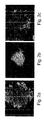
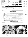
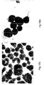
- exhibited typical hESC morphology e.g., round cells with large nuclei; for example Figures 5a-g , 18a-e
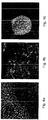
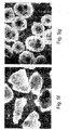
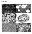
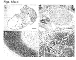
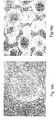
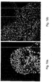
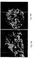
- hESCs cultured in the suspension cultures maintained their pluripotent capacity as evidenced by their ability to form embryoid bodies (EBs) or teratomas containing representative tissues of all three embryonic germ layers ( Figures 12a-d and data not shown).
- EBs embryoid bodies
- teratomas containing representative tissues of all three embryonic germ layers Figures 12a-d and data not shown.
- a method of expanding and maintaining embryonic stem cells in an undifferentiated state is effected by culturing the embryonic stem cells in a suspension culture under culturing conditions devoid of substrate adherence and which allow expansion of the embryonic stem cells in the undifferentiated state, thereby expanding and maintaining the embryonic stem cells in the undifferentiated state.
- embryonic stem cells refers to embryonic cells which are capable of differentiating into cells of all three embryonic germ layers (i.e., endoderm, ectoderm and mesoderm), or remaining in an undifferentiated state.
- Preferred embryonic stem cells according to this aspect of the present disclosure are of a human or primate (e.g., monkey) origin.
- embryonic stem cells can be obtained using well-known cell-culture methods.
- human embryonic stem cells can be isolated from human blastocysts. Human blastocysts are typically obtained from human in vivo preimplantation embryos or from in vitro fertilized (IVF) embryos. Alternatively, a single cell human embryo can be expanded to the blastocyst stage.
- ICM inner cell mass
- the ICM is then plated in a tissue culture flask containing the appropriate medium which enables its outgrowth. Following 9 to 15 days, the ICM derived outgrowth is dissociated into clumps either by a mechanical dissociation or by an enzymatic degradation and the cells are then re-plated on a fresh tissue culture medium. Colonies demonstrating undifferentiated morphology are individually selected by micropipette, mechanically dissociated into clumps, and re-plated. Resulting ES cells are then routinely split every 4-7 days. For further details on methods of preparing human ES cells see Thomson et al., (U.S. Pat. No. 5,843,780 ; Science 282: 1145, 1998 ; Curr. Top. Dev.
- human ESCs can be purchased from the NIH human embryonic stem cells registry (http://escr.nih.gov).
- Non-limiting examples of commercially available embryonic stem cell lines are BG01, BG02, BG03, BG04, CY12, CY30, CY92, CY10, TE03 and TE32.
- EBCs extended blastocyst cells
- the zona pellucida Prior to culturing the blastocyst, the zona pellucida is digested [for example by Tyrode's acidic solution (Sigma Aldrich, St Louis, MO, USA)] so as to expose the inner cell mass.
- the blastocysts are then cultured in vitro as whole embryos for at least nine and no more than fourteen days post fertilization (i.e., prior to the gastrulation event) using standard embryonic stem cell culturing methods.
- WO2006/040763 to the present inventors.
- EG cells are prepared from the primordial germ cells obtained from fetuses of about 8-11 weeks of gestation (in the case of a human fetus) using laboratory techniques known to anyone skilled in the arts.
- the genital ridges are dissociated and cut into small chunks which are thereafter disaggregated into cells by mechanical dissociation.
- the EG cells are then grown in tissue culture flasks with the appropriate medium.
- the cells are cultured with daily replacement of medium until a cell morphology consistent with EG cells is observed, typically after 7-30 days or 1-4 passages.
- Shamblott et al., Proc. Natl. Acad. Sci. USA 95: 13726, 1998 and U.S. Pat. No. 6,090,622 are additional details on methods of preparation human EG cells.
- embryonic stem cells in an undifferentiated state are of a distinct morphology, which is clearly distinguishable by the skilled in the art from that of differentiated cells of embryo or adult origin.
- undifferentiated embryonic stem cells typically have high nuclear/cytoplasmic ratios, prominent nucleoli and compact colony formation with poorly discernable cell junctions. Additional features of the undifferentiated state of the embryonic stem cells are further described hereinunder.
- expanding embryonic stem cells refers to obtaining a plurality of embryonic stem cells from a single or a population of embryonic stem cells.
- expanding embryonic stem cells refers also to increasing the number of embryonic stem cells over the culturing period. It will be appreciated that the number of cells which can be obtained from a single embryonic stem cell depends on the proliferation capacity of the cell.
- the proliferation capacity of an embryonic stem cell can be calculated by the doubling time of the cell (i.e., the time needed for a cell to undergo a mitotic division in the culture) and the period the stem cell can be maintained in the undifferentiated state while in culture (which is equivalent to the number of passages multiplied by the days between each passage).
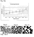
- hESCs could be maintained in the suspension culture of the present disclosure for at least 80 days while being subjected to 17 serial passaging (culture splitting) which occurred every 4-6 days. Given that the hESCs cultured in suspension exhibited a doubling time of 36 hours (e.g., when cultured on the CM100F medium), a single hESC cultured under these conditions could be expanded to give rise to 2 45 hESCs (i.e., 3.5 x 10 13 hESCs).
- the method according to this aspect of the present disclosure is effected by culturing the embryonic stem cells in a suspension culture under culturing conditions devoid of substrate adherence and which allow expansion of the embryonic stem cells in the undifferentiated state.
- suspension culture refers to a culture in which the embryonic stem cells are suspended in a medium rather than adhering to a surface.
- the culture of the present disclosure is "devoid of substrate adherence" in which the embryonic stem cells are capable of expanding without adherence to an external substrate such as components of extracellular matrix, a glass microcarrier or beads.
- Culturing according to this aspect of the present disclosure is effected by plating the stem cells in a culture vessel at a cell density which promotes cell survival and proliferation but limits differentiation. Typically, a plating density of between about 5 x 10 4 - 2 x 10 5 cells per ml is used. It will be appreciated that although single-cell suspensions of stem cells are usually seeded, small clusters such as 10-200 cells may also be used.
- the culture medium can be replaced on a daily basis, or, at a pre-determined schedule such as every 2-3 days.
- replacement of the culture medium can be performed by subjecting the ESC suspension culture to centrifugation for about 3 minutes at 80 g, and resuspension of the formed ESC pellet in a fresh medium.
- a culture system in which the culture medium is subject to constant filtration or dialysis so as to provide a constant supply of nutrients or growth factors to the ESCs may be employed.
- the formed ESC clumps are dissociated every 5-7 days and the single cells or small clumps of cells are either split into additional culture vessels (i.e., passaged) or remained in the same culture vessel yet with additional culture medium.
- a pellet of ESCs which may be achieved by centrifugation as described hereinabove
- an isolated ESC clump can be subject to enzymatic digestion and/or mechanical dissociation.
- Enzymatic digestion of ESC clump(s) can be performed by subjecting the clump(s) to an enzyme such as type IV Collagenase (Worthington biochemical corporation, Lakewood, NJ, USA) and/or Dispase (Invitrogen Corporation products, Grand Island NY, USA).
- an enzyme such as type IV Collagenase (Worthington biochemical corporation, Lakewood, NJ, USA) and/or Dispase (Invitrogen Corporation products, Grand Island NY, USA).
- the time of incubation with the enzyme depends on the size of cell clumps present in the suspension culture. Typically, when hESC cell clumps are dissociated every 5-7 days while in the suspension culture, incubation of 20-60 minutes with 1.5 mg/ml type IV Collagenase results in small cell clumps which can be further cultured in the undifferentiated state.
- ESC clumps can be subjected to incubation of about 25 minutes with 1.5 mg/ml type IV Collagenase followed by five minutes incubation with 1 mg/ml Dispase, essentially as described under "General Materials and Experimental Methods" of the Examples section which follows. It should be noted that passaging of human ESCs with trypsin may result in chromosomal instability and abnormalities (see for example, Mitalipova MM., et al., Nature Biotechnology, 23: 19-20, 2005 and Cowan CA et al., N. Engl. J. of Med. 350: 1353-1356, 2004 ), and therefore should be avoided.
- Mechanical dissociation of large ESC clumps can be performed using a device designed to break the clumps to a predetermined size. Such a device can be obtained from CellArtis Goteborg, Sweden. Additionally or alternatively, mechanical dissociation can be manually performed using a needle such as a 27g needle (BD Microlance, Drogheda, Ireland) while viewing the clumps under an inverted microscope.
- a needle such as a 27g needle (BD Microlance, Drogheda, Ireland) while viewing the clumps under an inverted microscope.
- the dissociated ESC clumps are further broken to small clumps using 200 ⁇ l Gilson pipette tips (e.g., by pipetting up and down the cells).
- the culture vessel used for culturing the ESC in suspension according to the method of this aspect of the present disclosure can be any tissue culture vessel (e.g., with a purity grade suitable for culturing ESCs) having an internal surface designed such that ESC cultured therein are unable to adhere or attach to such a surface (e.g., non-tissue culture treated cells, to prevent attachment or adherence to the surface).
- culturing according to this aspect of the present disclosure is effected using a controlled culturing system (preferably a computer-controlled culturing system) in which culture parameters such as temperature, agitation, pH, and pO 2 is automatically performed using a suitable device. Once the culture parameters are recorded, the system is set for automatic adjustment of culture parameters as needed for ESCs expansion.
- culturing according to the method of this aspect of the present disclosure can be performed under dynamic conditions (i.e., under conditions in which the ESCs are subject to constant movement while in the suspension culture) or under non-dynamic conditions (i.e ., a static culture).
- the ESCs can be cultured in uncoated 58 mm petri dishes (Greiner, Frickenhausen, Germany).
- the ESCs can be cultured in spinner flasks [e.g., of 200 ml to 1000 ml, for example 250 ml which can be obtained from CellSpin of Integra Biosciences, Fernwald, Germany; of 100 ml which can be obtained from Bellco,Vineland, NJ; or in 125 ml Erlenmeyer (Corning Incorporated, Corning NY, USA)] which can be connected to a control unit and thus present a controlled culturing system.
- spinner flasks e.g., of 200 ml to 1000 ml, for example 250 ml which can be obtained from CellSpin of Integra Biosciences, Fernwald, Germany; of 100 ml which can be obtained from Bellco,Vineland, NJ; or in 125 ml Erlenmeyer (Corning Incorporated, Corning NY, USA)
- the medium used to culture the ESCs in suspension according to the method of this aspect of the present disclosure can be any culture medium capable of supporting the growth of ESCs while maintaining them in an undifferentiated state.
- a culture medium can be a water-based medium which includes a combination of substances such as salts, nutrients, minerals, vitamins, amino acids, nucleic acids, proteins such as cytokines, growth factors and hormones, all of which are needed for cell proliferation and are capable of maintaining the ESCs in an undifferentiated state.
- a culture medium according to this aspect of the present disclosure can be a synthetic tissue culture medium such as Ko-DMEM (Gibco-Invitrogen Corporation products, Grand Island, NY, USA), DMEM/F12 (Biological Industries, Biet Haemek, Israel), Mab ADCB medium (HyClone, Utah, USA) or DMEM/F12 (Biological Industries, Biet Haemek, Israel) supplemented with the necessary additives as is further described hereinunder.
- all ingredients included in the culture medium of the present disclosure are substantially pure, with a tissue culture grade.
- the culture medium used by the method of this aspect of the present disclosure should be well-defined (i.e ., with known and constant components) and xeno-free (i.e., devoid of xeno contaminants).
- the culture medium used by the method of this aspect of the present disclosure is serum-free, serum replacement-free, xeno-free, feeder-free (i.e., devoid of feeder cells) and protein carrier-free.
- Serum or serum replacement are usually added to most culture media which are designed for culturing stem cells, and particularly, embryonic stem cells, in order to provide the cells with the optimal environment, similar to that present in vivo (i.e., within the organism from which the cells are derived, e.g., a blastocyst of an embryo or an adult tissue of a postnatal individual).
- serum which is derived from either an animal source (e.g., bovine serum) or a human source (human serum) is limited by the significant variations in serum components between individuals and the risk of having xeno contaminants (in case of an animal serum is used)
- the use of the more defined composition such as the currently available serum replacementTM (Gibco-Invitrogen Corporation, Grand Island, NY USA) may be limited by the presence of Albumax (Bovine serum albumin enriched with lipids) which is from an animal source within the composition (International Patent Publication No. WO 98/30679 to Price, P.J. et al ).
- a protein carrier refers to a protein which acts in the transfer of proteins or nutrients (e.g., minerals such as zinc) to the cells in the culture.
- protein carriers can be, for example, albumin (e.g., bovine serum albumin), Albumax (lipid enriched albumin) or plasmanate (human plasma isolated proteins). Since these carriers are derived from either human or animal sources their use in hESCs cultures is limited by batch-specific variations and/or exposure to pathogens. On the other hand, the recombinant human albumin, which is substantially pure and devoid of animal contaminants is highly expensive, thus not commonly used in hESCs cultures. Thus, a culture medium which is devoid of a protein carrier is highly advantageous since it enables a truly defined medium that can be manufacture from recombinant or synthetic materials.
- a culture medium which is serum-free, serum replacement-free, xeno-free, feeder-free and protein carrier-free can be a culture medium which comprises a TGF ⁇ isoform (for non-limiting examples see the D1, D2, HA16 or HA19 culture media which are described in Examples 1 and 2 of the Examples section which follows).
- TGF ⁇ isoform refers to any isoform of the transforming growth factor beta ( ⁇ ) including TGF ⁇ 1 (e.g., homo sapiens TGF ⁇ 1, GenBank Accession No. NP_000651), TGF ⁇ 2 (e.g., homo sapiens TGF ⁇ 2, GenBank Accession No. NP_003229) and TGF ⁇ 3 (e.g., homo sapiens TGF ⁇ 3, GenBank Accession No. NP_003230) which function through the same receptor signaling system in the control of proliferation, differentiation, and other functions in many cell types.
- TGF ⁇ acts in inducing transformation and also acts as a negative autocrine growth factor.
- the TGF ⁇ isoform which is included in the culture medium of the present disclosure is TGF ⁇ 1 or TGF ⁇ 3 .
- TGF ⁇ isoforms can be obtained from various commercial sources such as R&D Systems Minneapolis MN, USA.
- TGF ⁇ 1 e.g., the D1 medium which contains 0.12 ng/ml TGF ⁇ 1
- TGF ⁇ 3 e.g., the D2 medium, the HA16 medium or the HA19 medium which contain 2 ng/ml TGF ⁇ 3
- TGF ⁇ 1 which is included in the culture medium of this aspect of the present disclosure is present at a concentration of at least 0.06 ng/ml, more preferably, at least 0.07 ng/ml, more preferably, at least 0.08 ng/ml, more preferably, at least 0.09 ng/ml, more preferably, at least 0.1 ng/ml, more preferably, at least 0.11 ng/ml, even more preferably, at least 0.12 ng/ml.
- TGF ⁇ 3 which is included in the culture medium of this aspect of the present disclosure is present at a concentration of at least 0.5 ng/ml, more preferably, at least 0.6 ng/ml, more preferably, at least 0.8 ng/ml, more preferably, at least 0.9 ng/ml, more preferably, at least 1 ng/ml, more preferably, at least 1.2 ng/ml, more preferably, at least 1.4 ng/ml, more preferably, at least 1.6 ng/ml, more preferably, at least 1.8 ng/ml, even more preferably, at least 2 ng/ml.
- the TGF ⁇ -containing culture medium of this aspect of the present disclosure further includes basic fibroblast growth factor (bFGF).
- bFGF can be obtained from any commercial supplier of tissue culture ingredients such as Invitrogen Corporation products, Grand Island NY, USA.
- the bFGF which is included in TGF ⁇ -containing culture medium of this aspect of the present disclosure is present at a concentration of at least 2 ng/ml, at least 3 ng, at least 4 ng/ml, at least 5 ng/ml, at least 6 ng/ml, at least 7 ng, at least 8 ng/ml, at least 9 ng/ml, at least 10 ng/ml.
- a culture medium which is based on the IL6RIL6 chimera and is serum or serum replacement-free, xeno-free and protein carrier-free can be also used along with the method of this aspect of the present disclosure.
- IL6RIL6 refers to a chimeric polypeptide which comprises the soluble portion of interleukin-6 receptor (IL-6-R, e.g., the human IL-6-R as set forth by GenBank Accession No. AAH89410) (e.g., a portion of the soluble IL6 receptors as set forth by amino acids 112-355 of GenBank Accession No. AAH89410) and the interleukin-6 (IL6) (e.g., human IL-6 as set forth by GenBank Accession No. CAG29292) or a biologically active fraction thereof (e.g., a receptor binding domain).
- the IL6RIL6 chimera used by the method according to this aspect of the present disclosure is capable of supporting the undifferentiated growth of human embryonic stem cells, while maintaining their pluripotent capacity.
- the two functional portions i.e., the IL6 and its receptor
- the two functional portions can be directly fused (e.g., attached or translationally fused, i.e., encoded by a single open reading frame) to each other or conjugated (attached or translationally fused) via a suitable linker (e.g., a polypeptide linker).
- the IL6RIL6 chimeric polypeptide exhibits a similar amount and pattern of glycosylation as the naturally occurring IL6 and IL6 receptor.
- a suitable IL6RIL6 chimera is as set forth in SEQ ID NO:31 and in Figure 11 of WO 99/02552 to Revel M., et al.
- the IL6RIL6 chimera which is included in the culture medium of this aspect of the present disclosure is present at a concentration of at least 25 ng/ml, preferably at least 50 ng/ml, preferably, at least 100 ng/ml, preferably, at least 200 ng/ml, preferably, at least 300 ng/ml.
- concentration of the IL6RIL6 chimera can vary depending on the purity of the chimeric polypeptide following its synthesis or recombinant expression and those of skills in the art are capable of adjusting the optimal concentration depending on such purity.
- the IL6RIL6-containing culture medium of this aspect of the present disclosure includes at least 2 ng/ml bFGF, at least 3 ng/ml, at least 4 ng/ml, at least 5 ng/ml, at least 6 ng/ml, at least 7 ng, at least 8 ng/ml, at least 9 ng/ml, at least 10 ng/ml bFGF.
- a suitable IL6RIL6-containing culture medium which can be used for culturing the ESC in a suspension culture can be the HACM100 culture medium described under the "General Materials and Experimental Methods" and Example 2 of the Examples section which follows, which was shown capable of maintaining hESCs in an undifferentiated state for at least 1-2 passages.
- a culture medium which is based on soluble interleukin-6 receptor (sIL6R) [e.g., GenBank Accession No. NM_000565.2, NM_181359.1, NP_000556.1, NP_852004.1] and soluble interleukin-6 (IL6) [e.g., GenBank Accession No. NM_000600.1, NP_000591.1] (separately) can be also used along with the method of the present disclosure.
- sIL6R soluble interleukin-6 receptor
- IL6R soluble interleukin-6 receptor
- a culture medium such as the yFIL25 which comprises 25 ng IL6 and 25 ng sIL6R can be used to culture, expand and maintain human ESCs in a pluripotent, proliferative and undifferentiated state for at least 19 passages.
- human ESCs cultured in such a culture medium expressed markers characteristics of the undifferentiated state, exhibited normal chromosomal karyotype (as tested after 14 passages) and were capable of forming EBs which included all three embryonic germ layers (pluripotent).
- the sIL6R is present at a concentration of at least 10 nanogram per milliliter (ng/ml), more preferably, at least 15 ng/ml, more preferably, at least 20 ng/ml, e.g., at least 22 ng/ml, 25 ng/ml, 27 ng/ml, or 30 ng/ml.
- ng/ml nanogram per milliliter
- sIL6R can be present at a concentration of 15-30 ng/ml, e.g., 25 ng/ml.
- sIL6R and IL6 can be obtained, for example, from R&D systems, Minneapolis, MN, USA.
- a culture medium which is based on leukemia inhibitory factor [e.g., GenBank Accession No. NM_002309.2 (mRNA) or NP_002300.1 (protein)] can be also used along with the method of the present disclosure.
- LIF leukemia inhibitory factor
- a culture medium such as the yFL1, yFL2, or yFL3 can be used to culture, expand and maintain human ESCs in a pluripotent, proliferative and undifferentiated state for at least 18 passages.
- human ESCs cultured in such a culture medium expressed markers characteristics of the undifferentiated state, exhibited normal chromosomal karyotype (as tested after 14 passages) and were capable of forming EBs which included all three embryonic germ layers (pluripotent).
- LIF is present at a concentration of at least 1000 units/ml, more preferably, at least 2000 units/ml, more preferably, at least 3000 units/ml.
- Human recombinant leukemia inhibitory factor (hrLIF) can be obtained from R&D Systems Minneapolis MN, USA.
- a culture medium which is based on leukemia inhibitory factor (LIF) [e.g., GenBank Accession No. NM_002309.2 (mRNA) or NP_002300.1 (protein)] and TGF ⁇ 1 can be used along with the method of the present disclosure.
- LIF leukemia inhibitory factor
- a culture medium such as the TLF medium can be used to culture, expand and maintain human ESCs in a pluripotent, proliferative and undifferentiated state for at least 31 passages.
- human ESCs cultured in such a culture medium expressed markers characteristics of the undifferentiated state, exhibited normal chromosomal karyotype (as tested after 18 passages) and were capable of forming EBs which included all three embryonic germ layers (pluripotent).
- any of the proteinaceous factors used in the culture medium of the present disclosure can be recombinantly expressed or biochemically synthesized.
- naturally occurring proteinaceous factors such as bFGF and TGF ⁇ can be purified from biological samples (e.g., from human serum, cell cultures) using methods well known in the art.
- Biochemical synthesis of the proteinaceous factors of the present disclosure can be performed using standard solid phase techniques. These methods include exclusive solid phase synthesis, partial solid phase synthesis methods, fragment condensation and classical solution synthesis.
- Recombinant expression of the proteinaceous factors of the present disclosure can be generated using recombinant techniques such as described by Bitter et al., (1987) Methods in Enzymol. 153:516-544 , Studier et al. (1990) Methods in Enzymol. 185:60-89 , Brisson et al. (1984) Nature 310:511-514 , Takamatsu et al. (1987) EMBO J. 10-157-311 , Coruzzi et al. (1984) EMBO J. 3:1671-1680 , Brogli et al., (1984) Science 224:838-843 , Gurley et al.
- IL6RIL6 chimera can be generated as described in PCT publication WO 99/02552 to Revel M., et al. and Chebath J,, et al., 1997.
- a polynucleotide sequence encoding the IL6RIL6 chimera (e.g., the polypeptide set forth by SEQ ID NO:31) is preferably ligated into a nucleic acid construct suitable for expression in a host cell [ i.e., a cell in which the polynucleotide encoding the polypeptide-of-choice (e.g., the IL6RIL6 chimera) is expressed].
- the host cell employed is a eukaryotic host cell, more preferably a mammalian host cell such as human cell or CHO cell).
- mammalian expression vectors For expression in mammalian cells [e.g., CHO cells, human HEK 293 cells (ATCC CRL 1573)] a number of mammalian expression vectors can be used. Examples include, but are not limited to, pcDNA3, pcDNA3.1(+/-), pGL3, pZeoSV2(+/-), pSecTag2, pDisplay, pEF/myc/cyto, pCMV/myc/cyto, pCR3.1, pSinRep5, DH26S, DHBB, pNMT1, pNMT41, pNMT81, which are available from Invitrogen, pCI which is available from Promega, pMbac, pPbac, pBK-RSV and pBK-CMV which are available from Strategene, pTRES which is available from Clontech, and their derivatives.
- mammalian expression vectors include, but are not limited to,
- Expression vectors containing regulatory elements from eukaryotic viruses such as retroviruses can be also used.
- SV40 vectors include pSVT7 and pMT2.
- Vectors derived from bovine papilloma virus include pBV-1MTHA, and vectors derived from Epstein Bar virus include pHEBO, and p2O5.
- exemplary vectors include pMSG, pAV009/A + , pMTO10/A + , pMAMneo-5, baculovirus pDSVE, and any other vector allowing expression of proteins under the direction of the SV-40 early promoter, SV-40 later promoter, metallothionein promoter, murine mammary tumor virus promoter, Rous sarcoma virus promoter, polyhedrin promoter, or other promoters shown effective for expression in eukaryotic cells.
- Transformed cells are cultured under effective conditions, which allow for the expression of high amounts of the recombinant polypeptide (e.g., the IL6RIL6 chimera). Following a predetermined time in culture, recovery of the recombinant polypeptide is effected.
- the phrase "recovery of the recombinant polypeptide" used herein refers to collecting the whole fermentation medium containing the polypeptide and need not imply additional steps of separation or purification.
- polypeptides of the present disclosure can be purified using a variety of standard protein purification techniques, such as, but not limited to, affinity chromatography, ion exchange chromatography, filtration, electrophoresis, hydrophobic interaction chromatography, gel filtration chromatography, reverse phase chromatography, concanavalin A chromatography, chromatofocusing and differential solubilization.
- standard protein purification techniques such as, but not limited to, affinity chromatography, ion exchange chromatography, filtration, electrophoresis, hydrophobic interaction chromatography, gel filtration chromatography, reverse phase chromatography, concanavalin A chromatography, chromatofocusing and differential solubilization.
- the polypeptide of the present disclosure (e.g., the IL6RIL6 chimera) is preferably retrieved in "substantially pure” form.
- the phrase “substantially pure” refers to a purity that allows for the effective use of the polypeptide of the present disclosure (e.g., the IL6RIL6 chimera) in maintaining the human embryonic stem cells in an undifferentiated state while in culture.
- the culture media which comprise the IL6RIL6 chimera but also include serum or serum replacementTM (e.g., the CM100F medium or similar media with other concentrations of the IL6RIL6 chimera such as 200 or 300 ng/ml as described in the Examples section which follows) can be used by the method of this aspect of the present disclosure.
- the serum e.g., human serum
- serum replacementTM can be provided at various concentrations, such as a concentration of at least 10 %, e.g., a concentration of at least 15 %, at least 20 %, at least 25 % or at least 30 %.
- Serum replacementTM includes albumin or albumin substitutes, amino acids, vitamins, transferrins or transferrin substitutes, antioxidants, insulin or insulin substitutes, collagen precursors and trace elements (International Patent Publication No. WO 98/30679 to Price, P.J. et al ).
- albumin or albumin substitutes are preferably derived from a human source and/or are prepared using recombinant techniques in host cells as described hereinabove.
- embryonic stem cell growth is monitored to determine their differentiation state.
- the differentiation state can be determined using various approaches including, for example, morphological evaluation (e.g., as shown in Figures 5a-g ) and/or detection of the expression pattern of typical markers of the undifferentiated state using immunological techniques such as flow cytometry for membrane-bound markers, immunohistochemistry or immunofluorescence for extracellular and intracellular markers and enzymatic immunoassay, for secreted molecular markers.
- immunofluorescence employed on hESCs cultured according to the method of this aspect of the present disclosure revealed the expression of Oct4, stage-specific embryonic antigen (SSEA) 4, the tumour-rejecting antigen (TRA)-1-60 and TRA-1-81 ( Figures 4a-c and data not shown).
- SSEA stage-specific embryonic antigen
- TRA tumour-rejecting antigen
- the level of transcripts of specific undifferentiation markers e.g., Oct 4, Nanog, Sox2 and Rexl as shown in Figures 6a-d
- differentiation markers e.g., albumin, glucagons, ⁇ -cardiac actin, ⁇ -globulin, Flk1, AC133 and neurofilament
- RNA-based techniques such as RT-PCR analysis and/or cDNA microarray analysis.
- Determination of ES cell differentiation can be also effected via measurements of alkaline phosphatase activity.
- Undifferentiated human ES cells have alkaline phosphatase activity which can be detected by fixing the cells with 4 % paraformaldehyde and developing with the Vector Red substrate kit according to manufacturer's instructions (Vector Laboratories, Burlingame, California, USA).
- the embryonic stem cells cultured in any of the suspension culture media described hereinabove exhibit normal chromosomal karyotype following at least 1 passage, preferably, following at least 2 passages, preferably, following at least 3 passages, preferably, following at least 4 passages, preferably, following at least 5 passages, preferably, following at least 7 passages, preferably, following at least 10 passages, preferably, following at least 12 passages, preferably, following at least 15 passages, preferably, following at least 20 passages, preferably, following at least 25 passages, preferably, following at least 30 passages (e.g., hESCs exhibited normal karyotype following at least 14, 18, 23 or 36 passages), thus representing genetically stable human ESC lines.
- hESCs exhibited normal karyotype following at least 14, 18, 23 or 36 passages
- the embryonic stem cells cultured in any of the suspension culture media described hereinabove exhibit a doubling time of at least 20 hours, more preferably, a doubling time which is between 20 to 40 hours (e.g., about 36 hours), thus representing a non-tumorigenic, genetically stable human ESCs.
- the present disclosure provides, for the first time, a cell culture which comprises embryonic stem cells and a culture media which comprises the soluble interleukin-6 receptor (sIL6R) and interleukin-6 (IL6) (separately) wherein the soluble IL6R is present at a concentration of at least 10 (ng/ml) (e.g., 25 ng/ml), and whereas the culture medium being capable of maintaining the embryonic stem cells in an undifferentiated state for at least 5 passages (see Example 4 of the Examples section which follows, which demonstrates undifferentiated hESCs following 19 passages).
- sIL6R soluble interleukin-6 receptor
- IL6 interleukin-6
- the present disclosure provides, for the first time, a cell culture which comprises human embryonic stem cells and a culture media which comprises LIF (at a concentration of at least 1000 u/ml), wherein the culture medium being capable of maintaining the human ESCs in an undifferentiated state for at least 18 passages (see Example 4 of the Examples section which follows).
- a method of deriving an embryonic stem cell line is effected by (a) obtaining an embryonic stem cell from a pre-implantation stage blastocyst, post-implantation stage blastocyst and/or a genital tissue of a fetus; and (b) culturing the embryonic stem cell in a suspension culture, under culturing conditions which allow expansion of the embryonic stem cells in an undifferentiated state, thereby deriving the embryonic stem cell line.
- the term "deriving” as used herein refers to generating an embryonic stem cell line from at least one embryonic stem cell.
- embryonic stem cell line refers to embryonic stem cells which are derived from a single or a group of embryonic stem cells of a single organism (e.g., a single human blastocyst), and which are characterized by the ability to proliferate in culture while maintaining the undifferentiated state and the pluripotent capacity.
- obtaining an embryonic stem cell from a pre-implantation stage blastocyst, post-implantation stage blastocyst and/or a genital tissue of a fetus can be performed using methods known in the art, as described hereinabove and in the "General Materials and Experimental Methods" of the Examples section which follows.
- the zona pellucida is removed from a 5-7 day-old blastocyst using Tyrode's acidic solution (Sigma, St Louis MO, USA), the trophoblast layer is specifically removed either by immunosurgery or mechanically using 27g needles and the exposed ICM is either directly cultured in a suspension culture with a suitable culture medium (e.g., the CM100F, HA16 or D2 medium) for 4-10 days (in case a preimplantation blastocyst is used) or subject to in vitro implantation by culturing the ICM for 6-8 days (to obtain cells of a 13 day-old blastocyst in case a post-implantation/pre-gastrulation blastocyst is used) on feeder layers or a feeder-free culturing system which allow implantation of the blastocyst to the surface, following which the implanted cells are isolated and further cultured in suspension as described hereinunder.
- a suitable culture medium e.g., the CM100F
- the genital ridges are dissociated and cut into small chunks which are thereafter disaggregated into cells by mechanical dissociation.
- the single cell EG cells are then cultured in suspension culture with a suitable culture medium (e.g., the CM100F, HA16 or D2 medium) for 4-10 days.
- a suitable culture medium e.g., the CM100F, HA16 or D2 medium
- the ESCs are further cultured in suspension under conditions which allow expansion of the embryonic stem cells in the undifferentiated state, essentially as described hereinabove.
- the cell culture of the present disclosure is characterized by at least 40 %, at least 50 %, at least 60 %, more preferably at least 70 %, more preferably at least 80 %, most preferably at least 85 % of undifferentiated embryonic stem cells.
- an established embryonic stem cell line can be subject to freeze/thaw cycles without hampering the proliferative capacity of the cells in the undifferentiated state while preserving their pluriptent capacity.
- hESCs were successfully frozen and thawed.
- hESCs which were expanded and maintained in the suspension culture described hereinabove are pluripotent (i.e., capable of differentiating into all cell types of the three embryonic germ layers, the ectoderm, the endoderm and the mesoderm) as evidenced in vitro (by the formation of EBs).
- hESCs cultured according to the teachings of the present disclosure can be used as a source for generating differentiated, lineage-specific cells.
- Such cells can be obtained directly from the ESCs by subjecting the ESCs to various differentiation signals (e.g., cytokines, hormones, growth factors) or indirectly, via the formation of embryoid bodies and the subsequent differentiation of cells of the EBs to lineage-specific cells.
- various differentiation signals e.g., cytokines, hormones, growth factors
- a method of generating embryoid bodies from embryonic stem cells is effected by (a) culturing the embryonic stem cells in a suspension culture under culturing conditions which allow expansion of the embryonic stem cells in an undifferentiated state to thereby obtain expanded, undifferentiated embryonic stem cells; and (b) subjecting the expanded, undifferentiated embryonic stem cells to culturing conditions suitable for differentiating the embryonic stem cells to embryoid bodies; thereby generating the embryoid bodies from the embryonic stem cells.
- embryonic bodies refers to morphological structures comprised of a population of ESCs, extended blastocyst cells (EBCs) and/or embryonic germ cells (EGCs) which have undergone differentiation.
- EBCs extended blastocyst cells
- EECs embryonic germ cells
- EBs formation initiates following the removal of differentiation blocking factors from ES cell cultures.
- ESCs proliferate into small masses of cells which then proceed with differentiation.
- a layer of endodermal cells is formed on the outer layer of the small mass, resulting in "simple EBs”.
- complex EBs are formed. Complex EBs are characterized by extensive differentiation of ectodermal and mesodermal cells and derivative tissues.
- the method of this aspect of the present disclosure involves the culturing of ESCs in a suspension culture using any of the culture media described hereinabove in order to obtain expanded, undifferentiated embryonic stem cells and then subjecting the expanded, undifferentiated ESCs to culturing conditions suitable for differentiating the ESCs to embryoid bodies.
- Such culturing conditions are substantially devoid of differentiation inhibitory factors which were employed during step (a), e.g., a TGF ⁇ isoform or the IL6RIL6 chimera.
- a culture medium suitable for EBs formation may include a basic culture medium (e.g., Ko-DMEM or DMEM/F12) supplemented with 20 % FBSd (HyClone, Utah, USA), 1 mM L-glutamine, 0.1 mM ⁇ -mercaptoethanol, and 1 % non-essential amino acid stock.
- EBs can be effected by morphological evaluations (e.g., histological staining as described in Example 2) and determination of expression of differentiation-specific markers [using e.g., immunological techniques or RNA-based analysis (e.g., RT-PCR, cDNA microarray)].
- differentiation-specific markers of all three embryonic germ layers include albumin and glucagon (typical of the embryonic endoderm), ⁇ -cardiac actin, ⁇ -globulin and Flk1 (typical of the embryonic mesoderm), and AC133 and neurofilament (NFH) (typical of the embryonic ectoderm).
- cells of the EBs can be further subjected to culturing conditions suitable for lineage-specific cells.
- the method of this aspect of the present disclosure further includes step (c): subjecting cells of the embryoid bodies to culturing conditions suitable for differentiating and/or expanding lineage specific cells; thereby generating the lineage-specific cells from the embryonic stem cells.
- culturing conditions suitable for differentiating and/or expanding lineage specific cells refers to a combination of a culture system, e.g., feeder cell layers, feeder-free matrix or a suspension culture and a culture medium which are suitable for the differentiation and/or expansion of specific cell lineages derived from cells of the EBs.
- a culture system e.g., feeder cell layers, feeder-free matrix or a suspension culture and a culture medium which are suitable for the differentiation and/or expansion of specific cell lineages derived from cells of the EBs.
- the method of this aspect of the present disclosure further includes isolating lineage specific cells following step (b).
- the phrase "isolating lineage specific cells” refers to the enrichment of a mixed population of cells in a culture with cells predominantly displaying at least one characteristic associated with a specific lineage phenotype. It will be appreciated that all cell lineages are derived from the three embryonic germ layers. Thus, for example, hepatocytes and pancreatic cells are derived from the embryonic endoderm, osseous, cartilaginous, elastic, fibrous connective tissues, myocytes, myocardial cells, bone marrow cells, vascular cells (namely endothelial and smooth muscle cells), and hematopoietic cells are differentiated from embryonic mesoderm and neural, retina and epidermal cells are derived from the embryonic ectoderm.
- isolating is effected by sorting of cells of the EBs via fluorescence activated cell sorter (FACS).
- FACS fluorescence activated cell sorter
- EBs are disaggregated using a solution of Trypsin and EDTA (0.025 % and 0.01 %, respectively), washed with 5 % fetal bovine serum (FBS) in phosphate buffered saline (PBS) and incubated for 30 minutes on ice with fluorescently-labeled antibodies directed against cell surface antigens characteristics to a specific cell lineage.
- FBS fetal bovine serum
- PBS phosphate buffered saline
- endothelial cells are isolated by attaching an antibody directed against the platelet endothelial cell adhesion molecule-1 (PECAM1) such as the fluorescently-labeled PECAM1 antibodies (30884X) available from PharMingen (PharMingen, Becton Dickinson Bio Sciences, San Jose, CA, USA) as described in Levenberg, S. et al., (Endothelial cells derived from human embryonic stem cells. Proc. Natl. Acad. Sci. USA. 2002. 99: 4391-4396 ).
- PECAM1 platelet endothelial cell adhesion molecule-1
- Hematopoietic cells are isolated using fluorescently-labeled antibodies such as CD34-FITC, CD45-PE, CD31-PE, CD38-PE, CD90-FITC, CD117-PE, CD15-FITC, class I-FITC, all of which IgG1 are available from PharMingen, CD133/1-PE (IgG1) (available from Miltenyi Biotec, Auburn, CA), and glycophorin A-PE (IgG1), available from Immunotech (Miami, FL). Live cells ( i.e. , without fixation) are analyzed on a FACScan (Becton Dickinson Bio Sciences) by using propidium iodide to exclude dead cells with either the PC-LYSIS or the CELLQUEST software.
- fluorescently-labeled antibodies such as CD34-FITC, CD45-PE, CD31-PE, CD38-PE, CD90-FITC, CD117-PE, CD15-FITC, class I-FITC, all of which IgG1
- isolated cells can be further enriched using magnetically-labeled second antibodies and magnetic separation columns (MACS, Miltenyi) as described by Kaufman, D.S. et al., (Hematopoietic colony-forming cells derived from human embryonic stem cells. Proc. Natl. Acad. Sci. USA. 2001, 98: 10716-10721 ).
- MCS magnetically-labeled second antibodies and magnetic separation columns
- isolating is effected by a mechanical separation of cells, tissues and/or tissue-like structures contained within the EBs.
- beating cardiomyocytes can be isolated from EBs as disclosed in U.S. Pat. Appl. No. 20030022367 to Xu et al.
- Four-day-old EBs of the present disclosure are transferred to gelatin-coated plates or chamber slides and are allowed to attach and differentiate.
- Spontaneously contracting cells which are observed from day 8 of differentiation, are mechanically separated and collected into a 15-mL tube containing low-calcium medium or PBS.
- Cells are dissociated using Collagenase B digestion for 60-120 minutes at 37 °C, depending on the Collagenase activity.
- Dissociated cells are then resuspended in a differentiation KB medium (85 mM KCI, 30 mM K 2 HPO 4 , 5 mM MgSO 4 , 1 mM EGTA, 5 mM creatine, 20 mM glucose, 2 mM Na 2 ATP, 5 mM pyruvate, and 20 mM taurine, buffered to pH 7.2, Maltsev et al., Circ. Res. 75:233, 1994 ) and incubated at 37 °C for 15-30 minutes. Following dissociation cells are seeded into chamber slides and cultured in the differentiation medium to generate single cardiomyocytes capable of beating.
- a differentiation KB medium 85 mM KCI, 30 mM K 2 HPO 4 , 5 mM MgSO 4 , 1 mM EGTA, 5 mM creatine, 20 mM glucose, 2 mM Na 2 ATP, 5 mM pyruvate, and 20 mM taurine
- isolating is effected by subjecting the EBs to differentiation factors to thereby induce differentiation of the EBs into lineage specific differentiated cells.
- EBs of the present disclosure are cultured for 5-12 days in tissue culture dishes including DMEM/F-12 medium with 5 mg/ml insulin, 50 mg/ml transferrin, 30 nM selenium chloride, and 5 mg/ml fibronectin (ITSFn medium, Okabe, S. et al., 1996, Mech. Dev. 59: 89-102 ).
- the resultant neural precursors can be further transplanted to generate neural cells in vivo ( Brüstle, O. et al., 1997. In vitro-generated neural precursors participate in mammalian brain development. Proc. Natl. Acad. Sci. USA. 94: 14809-14814 ). It will be appreciated that prior to their transplantation, the neural precursors are trypsinized and triturated to single-cell suspensions in the presence of 0.1 % DNase.
- EBs of the present disclosure can differentiate to oligodendrocytes and myelinate cells by culturing the cells in modified SATO medium, i.e., DMEM with bovine serum albumin (BSA), pyruvate, progesterone, putrescine, thyroxine, triiodothryonine, insulin, transferrin, sodium selenite, amino acids, neurotrophin 3, ciliary neurotrophic factor and Hepes ( Bottenstein, J. E. & Sato, G. H., 1979, Proc. Natl. Acad. Sci. USA 76, 514-517 ; Raff, M. C., Miller, R.
- modified SATO medium i.e., DMEM with bovine serum albumin (BSA), pyruvate, progesterone, putrescine, thyroxine, triiodothryonine, insulin, transferrin, sodium selenite, amino acids, neurotrophin 3, ciliary neurotrophic
- EBs are dissociated using 0.25 % Trypsin/EDTA (5 min at 37 °C) and triturated to single cell suspensions. Suspended cells are plated in flasks containing SATO medium supplemented with 5 % equine serum and 5 % fetal calf serum (FCS). Following 4 days in culture, the flasks are gently shaken to suspend loosely adhering cells (primarily oligodendrocytes), while astrocytes are remained adhering to the flasks and further producing conditioned medium. Primary oligodendrocytes are transferred to new flasks containing SATO medium for additional two days.
- FCS fetal calf serum
- oligospheres are either partially dissociated and resuspended in SATO medium for cell transplantation, or completely dissociated and a plated in an oligosphere-conditioned medium which is derived from the previous shaking step [ Liu, S. et al., (2000). Embryonic stem cells differentiate into oligodendrocytes and myelinate in culture and after spinal cord transplantation. Proc. Natl. Acad. Sci. USA. 97: 6126-6131 ].
- two-week-old EBs of the present disclosure are transferred to tissue culture dishes including DMEM medium supplemented with 10 % FCS, 2 mM L-glutamine, 100 units/ml penicillin, 100 mg/ml streptomycin, 20 % (v/v) WEHI-3 cell-conditioned medium and 50 ng/ml recombinant rat stem cell factor (rrSCF, Tsai, M. et al., 2000.
- rrSCF recombinant rat stem cell factor
- hemato-lymphoid cells from the EBs of the present disclosure, 2-3 days-old EBs are transferred to gas-permeable culture dishes in the presence of 7.5 % CO 2 and 5 % O 2 using an incubator with adjustable oxygen content. Following 15 days of differentiation, cells are harvested and dissociated by gentle digestion with Collagenase (0.1 unit/mg) and Dispase (0.8 unit/mg), both are available from F.Hoffman-La Roche Ltd, Basel, Switzerland. CD45-positive cells are isolated using anti-CD45 monoclonal antibody (mAb) M1/9.3.4.HL.2 and paramagnetic microbeads (Miltenyi) conjugated to goat anti-rat immunoglobulin as described in Potocnik, A.J.
- mAb monoclonal antibody
- Miltenyi paramagnetic microbeads
- the isolated CD45-positive cells can be further enriched using a single passage over a MACS column (Miltenyi).
- the culturing conditions suitable for the differentiation and expansion of the isolated lineage specific cells include various tissue culture media, growth factors, antibiotic, amino acids and the like and it is within the capability of one skilled in the art to determine which conditions should be applied in order to expand and differentiate particular cell types and/or cell lineages.
- lineage specific cells can be obtained by directly inducing the expanded, undifferentiated ESCs to culturing conditions suitable for the differentiation of specific cell lineage.
- EBs of the present disclosure can be used to generate lineage-specific cell lines which are capable of unlimited expansion in culture.
- Cell lines of the present disclosure can be produced by immortalizing the EB-derived cells by methods known in the art, including, for example, expressing a telomerase gene in the cells ( Wei, W. et al., 2003. Mol Cell Biol. 23: 2859-2870 ) or co-culturing the cells with NIH 3T3 hph-HOX11 retroviral producer cells ( Hawley, R.G. et al., 1994. Oncogene 9: 1-12 ).
- lineage-specific cells or cell lines obtained according to the teachings of the present disclosure are developed by differentiation processes similar to those naturally occurring in the human embryo they can be further used for human cell-based therapy and tissue regeneration.
- the present disclosure envisages the use of the expanded and/or differentiated lineage-specific cells or cell lines of the present disclosure for treating a disorder requiring cell replacement therapy.
- oligodendrocyte precursors can be used to treat myelin disorders (Repair of myelin disease: Strategies and progress in animal models. Molecular Medicine Today. 1997. pp. 554-561 ), chondrocytes or mesenchymal cells can be used in treatment of bone and cartilage defects ( U.S. Pat. No. 4,642,120 ) and cells of the epithelial lineage can be used in skin regeneration of a wound or burn ( U.S. Pat. No. 5,716,411 ).
- ESC-derived cells are preferably manipulated to over-express the mutated gene prior to their administration to the individual. It will be appreciated that for other disorders, the ESC-derived cells should be manipulated to exclude certain genes.
- Over-expression or exclusion of genes can be effected using knock-in and/or knock-out constructs [see for example, Fukushige, S. and Ikeda, J. E.: Trapping of mammalian promoters by Cre-lox site-specific recombination. DNA Res 3 (1996) 73-50 ; Bedell, M. A., Jerkins, N. A. and Copeland, N. G.: Mouse models of human disease. Part I: Techniques and resources for genetic analysis in mice. Genes and Development 11 (1997) 1-11 ; Bermingham, J. J., Scherer, S. S., O'Connell, S., Arroyo, E., Kalla, K. A., Powell, F. L. and Rosenfeld, M. G.: Tst-1/Oct-6/SCIP regulates a unique step in peripheral myelination and is required for normal respiration. Genes Dev 10 (1996) 1751-62 ].
- the lineage specific cells of the present disclosure can also be utilized to prepare a cDNA library.
- mRNA is prepared by standard techniques from the lineage specific cells and is further reverse transcribed to form cDNA.
- the cDNA preparation can be subtracted with nucleotides from embryonic fibroblasts and other cells of undesired specificity, to produce a subtracted cDNA library by techniques known in the art.
- the lineage specific cells of the present disclosure can be used to screen for factors (such as small molecule drugs, peptides, polynucleotides, and the like) or conditions (such as culture conditions or manipulation) that affect the differentiation of lineage precursor to terminally differentiated cells.
- factors such as small molecule drugs, peptides, polynucleotides, and the like
- conditions such as culture conditions or manipulation
- growth affecting substances, toxins or potential differentiation factors can be tested by their addition to the culture medium.
- ESC culture - Human embryonic stem cell (hESC) lines 1-6, 14 and 1-3 [Amit&Itskovitz-Eldor, 2002] were cultured with inactivated mouse embryonic fibroblasts (MEFs) for 40-60 passages in a "basic hESC culture medium" consisting of 85 % DMEM/F12 (Biological Industries, Biet Haemek, Israel) supplemented with 15 % serum replacement (SR), 2 mM L-glutamine, 0.1 mM ⁇ -mercaptoethanol, 1 % non-essential amino acid stock, and 4 ng/ml basic fibroblast growth factor (bFGF) (all but mentioned are from Gibco Invitrogen Corporation products, Grand Island NY, USA).
- This basic culture medium was used for the routine culture of hESCs in 2D culture with MEFs as control.
- Tested media on the feeder layer, feeder-free or suspension cultures - The tested medium were as follows:
- Feeder layers or feeder-free culturing systems To test the ability of various culture media to support the growth of hESC in an undifferentiated yet pluripotent state the hESCs were transferred to several culture systems:
- hESCs were cultured in suspension in 58 mm petri dishes (Greiner, Frickenhausen, Germany) in a cell density of 5 x 10 4 - 2 x 10 5 cells/ml.
- the HA16 medium was supplemented with 0.1 % F68 (Sigma, St. Louis, MO, USA) for the suspended culture.
- the culture medium in the suspension culture was changed on a daily basis.
- the basic media used for culturing hESCs in suspension (which can be further supplemented with the additive and growth factors as described hereinabove) were DMEM, ko-DMEM, DMEM/F12, MabADCB or NCTC medium.
- RT PCR analysis Total RNA was isolated from hESCs grown for 10-15 passages in the suspension culture using Tri-Reagent (Sigma, St. Louis MO, USA), according to the manufacturer's instructions. cDNA was synthesized from 1 ⁇ g total RNA using MMLV reverse transcriptase RNase H minus (Promega, Madison WI, USA). PCR reactions included denaturation for 5 minutes at 94 °C followed by repeated cycles of 94 °C for 30 seconds, annealing for 30 seconds at an annealing temperature as specified in Table 1, hereinbelow and extension at 72 °C for 30 seconds. PCR primers and reaction conditions used are described in Table 1, hereinbelow.
- Karyotype analysis was performed on at least 20 cells from each sample, two samples per test, as previously described [Amit et al, 2003].
- Karyotypes were analyzed and reported according to the "International System for Human Cytogenetic Nomenclature" (ISCN).
- hESCs cultured in suspension - For the formation of EBs, one to three 58 mm petri dishes (Greiner, Frickenhausen, Germany) containing ESCs in suspension cultures were transferred to new 58 mm petri dishes containing EBs-differentiation medium consisting of 80 % DMEM/F12 (Biological Industries, Beit Haemek, Israel), supplemented with 20 % FBSd (HyClone, Utah, USA), and 1 mM L-glutamine (Invitrogen Corporation, Grand Island NY, USA).
- 80 % DMEM/F12 Biological Industries, Beit Haemek, Israel
- FBSd HyClone, Utah, USA
- 1 mM L-glutamine Invitrogen Corporation, Grand Island NY, USA.
- the ESCs were subject to treatment with 1 mg/ml type IV collagenase and further broken into small clumps using 1000 ⁇ l Gilson pipette tips. 10 day-old EBs were harvested for RNA isolation and histological examination.
- 2-D feeder free or feeder layers
- ESCs were removed from their culture dish using 1 mg/ml type IV collagenase, further broken into small clumps using 1000 ⁇ l Gilson pipette tips, and cultured in suspension in 58 mm petri dishes (Greiner, Frickenhausen, Germany).
- EBs were grown in differentiation medium consisting of 80 % DMEM/F12 (Biological Industries, Beit Haemek, Israel), supplemented with 20 % FBSd (HyClone, Utah, USA), and 1 mM L-glutamine (Invitrogen Corporation, Grand Island NY, USA).
- Teratoma formation For teratoma formation, cells cultured in the offered culture methods for more than 15 passages, were injected into the rear leg muscle of 4-week-old male SCID-beige mice (two mice for each tested culture system). Cell numbers ranged from 5 x 10 6 cells to 10 7 cells per injection. Three to eight to 12 weeks after injection the mice were sacrificed and the resulting teratomas examined histologically.
- Blastocyst cultivation - Zygotes were donated by couples undergoing pre-implantation genetic diagnosis (PGD) or in vitro fertilization (IVF) at Cornell Medical College, NY, who signed informed consent forms. The couples underwent the traditional IVF procedure after ovarian stimulation with gonadotropins and oocyte retrieval. Zygotes were cultured to the blastocyst stage according to IVF laboratory standard protocol: under oil using specialized C1/C2 media for insemination, growth and blastocyst development (Cornell).
- hESC lines in a suspension culture - Following the removal of the zona pellucida using Tyrode's acidic solution (Sigma, St Louis MO, USA), the trophoblast layer is specifically removed either by immunosurgery or mechanically using 27g needles.
- the exposed ICM is further cultured in suspension culture with a suitable culture medium (e.g., the CM100F, HA16 or D2) for 4-10 days. Initially, the cells are mechanically split using 27g needles.
- a suitable culture medium e.g., the CM100F, HA16 or D2
- hESC lines on foreskin fibroblasts After digestion of the zona pellucida by Tyrode's acidic solution (Sigma, St Louis MO, USA) or its mechanical removal, the exposed blastocysts were placed in whole on a mitotically inactivated foreskin fibroblasts feeder layer (line F21 which was cultured in an animal free medium since its derivation until used). For the derivation and initial passages, cells were grown in the D2 or HA16 culture medium. The cells were initially passaged mechanically every four to ten days.
- the basic medium, D1 or D2 is a commercial medium design for industrial and clinical proposes for the culture of hybridomas in suspension.
- the medium is free from animal, serum products and proteins.
- HA16 and HA19 are based on defined materials only.
- the CM100 medium contains the IL6RIL6 chimera and serum replacement.
- TGF ⁇ TGF ⁇ 1 and TGF ⁇ 3
- TGF ⁇ 3 TGF ⁇ 1 and TGF ⁇ 3
- the culture system used were: (1) feeder layer-free method based on fibronectin or MatrigelTM which are the most used matrices; (2) MEF, and (3) foreskin fibroblast. Based on these two parameters, the media supplemented with TGF ⁇ 3 , D2, HA16 and HA19, were found to be the most suitable to support undifferentiated hESC proliferation in all tested culture methods.
- D1 medium on a feeder layer-free system is capable of maintaining all hESCs features along with high proliferation rate -
- the hESCs maintained all hESCs features including high proliferation rates.
- the hESCs demonstrated a relatively high background differentiation rate of 20 % and low proliferation abilities as compared to hESCs cultured at the same feeder layers systems with the D2 HA19 or HA16 medium.
- D1 , D2 and HA16 media in feeder layer-free are capable of maintaining hESCs in a proliferative, undifferentiated state, with chromosomal stability and pluripotency - Human ESCs grown in the presence of the D1, D2 or HA16 medium in feeder-layer free conditions were cultured continuously for up to 53, 24 or 10 passages, respectively, while maintaining their ESC features, including undifferentiated proliferation, chromosomal stability (as test by karyotype analysis, not shown) and pluripotency.
- the background differentiation rates were found to be less than 10 %, which is similar to the differentiation rates occurring when hESCs are cultured in the traditional culture system based on MEFs as the feeder layer and medium supplemented with serum replacement and 4 ng/ml bFGF [Amit et al, 2000].
- Examples of undifferentiated colonies cultured with D1, D2 or HA16 medium in feeder-layer free conditions and with the D2 or HA16 medium with the tested feeder layers are illustrated in Figures 1a-d .
- the D1, D2 or HA16 media are capable of maintaining hESCs with normal population doubling - Similar to cells grown on MEFs, cells cultured with D2 or HA16 medium in all tested culture methods, and the D1 medium in the feeder layer-free systems, were passaged routinely every four to six days, at the same ratio of 1/2 or 1/3, indicating a similar population doubling time as of hESCs grown on MEFs. The cells were passage at the same seeding efficiency of about 1 million cells per 10 cm 2 , with the same viability rate of over 95 %. Using 40 % human serum and 10 % DMSO, cells were successfully frozen and thawed.
- Karyotype analysis revealed normal karyotype of hESCs grown with the D1, D2, CM100 or HA16 media - 15 passages and more after transferring the cells into the tested environments, karyotype analysis was performed by Giemsa banding on two separate cultures, representing the four medium conditions, D1, D2, CM100 and HA16 at the different culture methods. At least 20 cells were tested from each sample, 40 cells from each medium combination. All examined cells were found to sustain normal karyotype of 46,XX for cell lines 13 and 14 and 46,XY for cell line 16 (data not shown). Overall, these results suggest that the cells' karyotype remains stable in the tested conditions, similarly to ESCs grown with MEFs using traditional methods (Amit et al, 2000).
- hESCs cultured with the D1, D2 or HA16 express typical cell surface markers -
- Several surface markers typical of primate undifferentiated ES cells were examined using immunofluorescent staining (Thomson et al, 1995, 1996, 1998).
- hESCs cultured with the D1, D2 or HA16 medium for more than 20 passages, while using the tested culture conditions, were found to be strongly positive to surface markers TRA-1-60 ( Figure 2a ), SSEA4 ( Figure 2b ), TRA-1-81 ( Figure 2c ) and Oct 4 (data not shown).
- staining with SSEA3 was weak and negative for SSEA1 (data not shown).
- hESCs cultured with the D1, D2 or HA16 medium are pluripotent as tested by EBs formation in vitro -
- the developmental potential of the cells after prolonged culture in the tested culture methods was examined in vitro by the formation of embryoid bodies (EBs).
- EBs embryoid bodies
- stem cells differentiated into cell types representative of the three embryonic germ layers as described for EBs formed from hESCs cultured on other culture systems (Itskovitz-Eldor et al, 2000).
- EBs formed from the hESCs cultured on the D1, D2 or HA16 medium are capable of differentiating into the ectoderm, endoderm and mesoderm cell lineages - While undifferentiated cells cultured in the tested medium, feeder layers and matrices, expressed undifferentiated genetic markers such as Oct 4, Nanog, Sox2, Rexl, Cx43 and FGF4 (not shown) [Bhattacharya et al, 2004], cells harvested from 10 day-old EBs expressed genes such as albumin and glucagon (endoderm), ⁇ -cardiac actin, ⁇ -globulin and Flk1 (mesoderm), and AC133 and neurofilament (ectoderm) as demonstrated by RT-PCR analysis (data not shown).
- hESCs cultured with the D1, D2 or HA16 medium are pluripotent as tested by teratomas formation in vivo - The cells pluripotency was also tested in vivo by teratomas formation.
- hESCs cultured for over 12 passages in the HA16, D1 or D2 medium, in the tested culture systems formed teratomas following their injection into SCID-Beige mice.
- hESCs differentiated to representative tissues of the three embryonic germ layers including; cartilage, muscle, bone and fat (mesoderm), stratified epithelium, melanin containing epithelium (ectoderm), and kidney like structure (endoderm and mesoderm), and epithelium of endoderm origin (data not shown).
- Teratomas formation rates of 90 %, and the number of injected cells were identical to those demonstrated by cells cultured using traditional methods (Amit et al, 2000).
- the HA16 and D2 media are suitable for derivation of hESC line on foreskin fibroblast feeder layers in a complete xeno-free system -
- the medium combinations of the present disclosure were also tested for the ability to support hESC line derivation.
- new hESC lines were successfully derived and maintained for at least 2 passages (in the presence of the D2 medium) or at least 18 passages (in the presence of the HA16 medium).
- the hESC line derived on foreskin in the presence of the HA16 culture medium demonstrated stem cells morphology at passage 18 (and the culture is still ongoing), normal XY karyotype and pluripotency as evidenced by the formation of EBs ( Figures 3a-b and data not shown).
- the growth and success rates were similar to those obtained while using traditional culture methods. Since the used foreskin fibroblasts line, F21, were derived without any animal products, this new hESC lines were derived under complete xeno-free conditions. Thus, the new hESC lines exhibit typical hESC morphology and proliferation rates, normal karyotype and pluripotency as evidenced by the formation of EBs.
- hESCs cells subjected to prolonged culture in the tested culture systems demonstrated all hESCs features including; pluripotency, chromosomal stability, expression of specific genes and surface markers and indefinite proliferation as undifferentiated cells.
- the new TGF ⁇ -containing culture medium which is described in Example 1, hereinabove, is designed for massive cell culture (low protein content), suitable for industrial and clinical proposes cell production
- D1, D2, HA19 and HA16 media to support suspension culture of undifferentiated hESCs
- a medium containing the IL6RIL6 chimera such as the CM100F or HACM100 medium (as described in the General Materials and Experimental Methods) to support a suspension culture of hESCs was also examined.
- CM100F contains serum replacement
- the HACM100 medium is serum or serum replacement-free and thus presents a well-defined culture, xeno-free culture medium.
- CM100F, HA16, D1, D2 and HA19 media are suitable for culturing hESCs in suspension - hESCs were cultured in suspension using the newly developed TGF ⁇ -containing medium types which are devoid of serum, serum replacement and albumin.
- TGF ⁇ -containing medium types which are devoid of serum, serum replacement and albumin.
- the highest passage of hESCs grown in suspension in the tested medium types were 3 passages in the D1 medium, 7 passages in the D2 medium, 10 passages in the HA19 medium and 17 passages in the CM100F medium. All hESCs exhibited undifferentiated morphology at these passages and can be further cultured in these media and maintain hESCs features.
- Histological sections of the hESCs clumps formed in the suspension cultures illustrated homogeneous cell population, of round cells with large nucleus ( Figures 5a-g ).
- the cells were plated back on MEFs, they created colonies with typical hESCs morphology ( Figures 5b-e ), and if returned to suspension cultures, they continued proliferation as undifferentiated cells (data not shown).
- hESCs were cultured in a suspension culture in the presence of the serum or serum replacement-free, IL6RIL6-containing HACM100 medium, the cells were expanded and maintained in the undifferentiated state for at least 1-2 passages (data not shown).
- hESCs cultured in suspension in the presence of the D1, D2, HA19 or CM100F media express markers of undifferentiated hESCs - Cells cultured in suspension in the presence of the D2 medium for 3 passages as small clumps of 200-1500 cells expressed stem cells markers such as Oct 4 ( Figure 4a ), TRA-1-60 ( Figure 4b ), TRA-1-81 ( Figure 4c ) and SSEA4 (data not shown). Similar results were obtained with the CM100F, D1 or D2 medium at passage 5 (p-5) (data not shown).
- CM100F or the HA19 medium When cultured in suspension culture in the presence of the CM100F or the HA19 medium the cells expressed high levels of typical stem cells markers such as Oct 4 ( Figure 6a ), Rexl ( Figure 6b ), Sox2 ( Figure 6c ), Nanog ( Figure 6d ) and FGF4 (data not shown) as demonstrated by RT-PCR analysis.
- typical stem cells markers such as Oct 4 ( Figure 6a ), Rexl ( Figure 6b ), Sox2 ( Figure 6c ), Nanog ( Figure 6d ) and FGF4 (data not shown) as demonstrated by RT-PCR analysis.
- ESCs cultured in suspension are capable of forming EBs - When removed from the D1, D2 or HA16 medium and transferred to EBs medium (80 % DMEM/F12 supplemented with 20 % FBSd and 1 mM L-glutamine), the cells formed EBs containing representative tissues of three embryonic germ layers (as evidenced by histological analysis, data not shown).
- EBs medium 80 % DMEM/F12 supplemented with 20 % FBSd and 1 mM L-glutamine
- Rhesus ESCs can be also cultured in the suspension cultures of the present disclosure - Similar results with Rhesus ESCs (monkey embryonic stem cells, line R366.4, University of Wisconsin, primate center, Thomson lab, Madison, Wisconsin), which are regarded as good candidate for transgenic model to human diseases, were obtained when the Rhesus ESCs were cultured in suspension in the HA16, D1 and D2 TGF ⁇ -containing culture media (data not shown).
- the new TGF ⁇ -containing medium which is devoid of serum, serum replacement and albumin, or the IL6RIL6-containing medium are capable of supporting the undifferentiated culture of hESCs, while maintaining hESCs characteristics, and provide methods for massive culture of these cells for industrial and clinical purposes.
- hESCs like mouse ES cells, are traditionally cultured with MEFs, which may expose them to animal pathogens.
- MEFs which may expose them to animal pathogens.
- the present inventors have demonstrated, for the first time, a defined animal, serum and feeder layer-free culture system for hESCs, based on the use of commercial medium supplemented with either TGF ⁇ 3 or TGF ⁇ 1 and bFGF, and human fibronectin matrix as substitute. This medium is designed for massive cultivation of cells in GMP for industrial or clinical purposes. All medium types of the present disclosure (with TGF ⁇ 3 or TGF ⁇ 1 ) support hESCs culture. When using the culture medium with TGF ⁇ isoform 3 the results are better; less background differentiation.
- All media types of the present disclosure support the culture with feeders as good as with the regular serum containing media.
- Cells retained the same proliferation rates and the same background differentiation percentages as hESCs cultured with MEFs using traditional culture methods.
- the medium can also be used for massive suspended culture of undifferentiated hESCs.
- TGF ⁇ 3 and TGF ⁇ 1 Two isoforms of TGF ⁇ , TGF ⁇ 3 and TGF ⁇ 1 , were tested for their ability to maintain hESCs in an undifferentiated state using various culture conditions.
- TGF ⁇ 3 D2 and HA16 media
- ESCs cultured while using these media maintained all the characteristics of ESCs. After prolonged culture of more than 20 passages, the cells remained undifferentiated, as demonstrated by the colony and single cell morphology, and by the expression of markers typical of undifferentiated primate ESCs [Thomson et al, 1995, 1996, 1998; Reubinoff et al, 2000].
- hESCs expressed specific markers for the undifferentiated stage such as Oct 4, Sox 2, Rex1 and Nanog, as demonstrated by RT-PCR.
- the cells' pluripotency was examined in vitro.
- Cells cultured in the tested culture systems for more than 10 passages formed EBs similar to those created when grown on MEFs [Itskovitz-Eldor et al, 2000].
- RT-PCR analysis demonstrated that cells within these EBs differentiated into different cell types representative of the three germ layers.
- hESCs cultured in the presence of the D1 and D2 media formed teratomas containing a multitude of tissues types.
- hESCs cultured in the presence of the HA16 medium also formed teratomas and their histological evaluation is in process.
- the teratoma formation rates were identical to those of cells cultured with MEFs.
- the pluripotency of the cells culture continuously in the tested culture methods remained intact.
- hESCs can be maintained as undifferentiated cells in the proposed defined animal- and serum-free medium combination, without any feeder cells (D1, D2 and HA16) or alternatively, with commonly used acceptable feeder layers (D2 and HA16).
- these media can facilitate hESCs culture for research, industrial and clinical purposes.
- this novel culture media was found to support suspended culture of undifferentiated hESCs, the first and primary step in developing a massive culture system for their growth and scale-up, a crucial step for any industrial and clinical uses.
- hESCs Future clinical uses of hESCs will require a reproducible, well-defined and xeno-free culture system.
- the well-defined media demonstrated in the present study are suitable for culturing hESCs and may be advantageous for undertaking research on the mechanisms of ESC self-maintenance, especially of the possible roles of the TGF ⁇ pathway.
- Other studies using hESCs, such as the research on differentiation pathways and mechanisms, will benefit from the availability of a well-defined and reproducible culture system.
- hESC lines I-3, I-4 and I-6 [Amit & Itskovitz-Eldor, 2002] were cultured with inactivated MEF for 54-89 passages as previously described [Amit et al, 2000]. The following culture medium combinations were tested for their ability to support the suspended culture of hESCs:
- the IL6RIL6 chimera but not low concentrations of the IL6, soluble IL6 or LIF, are capable of maintaining hESCs in an undifferentiated state when cultured on fibronectin (2-D) -
- Three key cytokines of the IL6 family (LIF, IL6 and IL6RIL6 chimera) were tested for their ability to support the culture of undifferentiated hESCs in suspension.
- the ability of the various cytokines to maintain hESC self-renewal was first evaluated in the 2-D feeder layer-free culture system using fibronectin as a matrix. While the background differentiation of hESCs cultured in the presence of LIF or IL6 at the used concentrations (i.e.
- the IL6RIL6 chimera but not low concentrations of the IL6 and soluble IL6, are capable of maintaining cultures of hESCs in suspension in an undifferentiated state -
- the cytokines' ability to support the culture of undifferentiated hESCs in suspension was then examined.
- medium CM6* medium supplementation of 100 ng/ml IL6 and 0.5 ng/ml IL6 soluble receptor (medium CM6*) the cells formed either EBs or neurosphere-like structures within five to seven days of culture (data not shown).
- the neurosphere-like structures demonstrated round morphology when cultured in suspension, and several days after being plated on fibronectin neuron-like structures positively stained with either nestin or ⁇ -tubulin were observed (data not shown).
- hESCs cultured in suspension in the presence of the IL6RIL6 chimera exhibit normal karyotype - Karyotype analyses by Giemsa banding was carried out on each cell line after 23, 18 and 36 passages in suspension, for I-6, I-3 and I-4, respectively, and were found to be normal (data not shown). Similar results were obtained when the cells were re-cultured on MEFs; all samples but one of I-3 (passage 12 on MEFs after 10 passages in suspension) demonstrated normal karyotype. The original culture of 1-3 that remained in suspension demonstrated normal karyotype. Thus the karyotype of the suspended cell culture remained stable.
- hESCs cultured in suspension in the presence of the IL6RIL6 chimera exhibit expression pattern of undifferentiated state -
- CM100F medium for 42 and 43 passages were found to be strongly positive to surface markers SSEA4, TRA-1-60 and TRA-1-81 and Oct 4 ( Figures 8a-d ).
- SSEA3 was weak and negative for SSEA1.
- Similar results were obtained for the cells cultured in suspension and returned to the MEFs. The former were further tested for typical undifferentiation markers using RT-PCR.
- hESCs cultured in suspension in the presence of the ILRIL6 chimera preserve their pluripotency as demonstrated by EBs' formation -
- the developmental potential of the cells after prolonged culture in suspension was examined in vitro by the formation of EBs.
- hESCs formed cystic EBs similar to those created by hESCs grown with MEFs ( Figures 12a-b ).
- stem cells differentiated into cell types representative of the three embryonic germ layers [Itskovitz-Eldor et al, 2000].
- hESCs cultured in suspension in the presence of the ILRIL6 chimera preserve their pluripotency as demonstrated by teratoma formation - Cell pluripotency in vivo was demonstrated by teratoma formation.
- Cells cultured in the presence of the CM100F medium in suspension for 9, 10, 14 or 26 passages were injected into SCID Beige mice, representing the three tested cell lines, and after 10 weeks tumors formed in all four injected mice. Within these teratomas tissues representative of the three germ layers were observed ( Figures 12a-b ). Similar results were obtained when cells cultured for at least 10 passages in suspension were returned to the MEFs and cultured for additional 5-10 passages (data not shown).
- the total sphere number increased from 1.33 x 10 4 ⁇ 461 on the seeding day to 3.5 x 10 5 ⁇ 2.8x10 4 after 10-11 days in the dynamic culture, a 25-fold increase, and the total cell number increased from 5 x 10 8 to 1.31 x 10 10 .
- Figures 17a-b depict examples of differentiated ( Figure 17b ) and undifferentiated ( Figure 17a ) hESC clumps in the presence of the anti-gp 130 antibody.
- yFIL25 + - 85 % DMEM/F12 (Biological Industries, Biet Haemek, Israel), containing 15 % knockout serum replacement (SR), 2 mM L-glutamine, 0.1 mM ⁇ -mercaptoethanol, 1 % non-essential amino acid stock, 4 ng/ml bFGF (all but mentioned are Invitrogen Corporation products, Grand Island NY, USA), 25 ng/ml IL6 and 25 ng/ml sIL6-R (R&D systems, Minneapolis, MN, USA).
- SR knockout serum replacement
- 2 mM L-glutamine 2 mM L-glutamine
- 0.1 mM ⁇ -mercaptoethanol 1 % non-essential amino acid stock
- 4 ng/ml bFGF all but mentioned are Invitrogen Corporation products, Grand Island NY, USA
- 25 ng/ml IL6 and 25 ng/ml sIL6-R (R&D systems, Minneapolis, MN
- yFL3 - 85 % DMEM/F12 (Biological Industries, Biet Haemek, Israel), containing 15 % knockout serum replacement (SR), 2 mM L-glutamine, 0.1 mM ⁇ -mercaptoethanol, 1 % non-essential amino acid stock, 4 ng/ml bFGF (all but mentioned are Invitrogen Corporation products, Grand Island NY, USA), and 3000 u/ml human recombinant leukemia inhibitory factor (hrLIF) (R&D Systems Minneapolis MN, USA).
- SR knockout serum replacement
- 2 mM L-glutamine 0.1 mM ⁇ -mercaptoethanol
- 1 % non-essential amino acid stock 4 ng/ml bFGF (all but mentioned are Invitrogen Corporation products, Grand Island NY, USA)
- hrLIF human recombinant leukemia inhibitory factor
- yFL2 - 85 % DMEM/F12 (Biological Industries, Biet Haemek, Israel), containing 15 % knockout serum replacement (SR), 2 mM L-glutamine, 0.1 mM ⁇ -mercaptoethanol, 1 % non-essential amino acid stock, 4 ng/ml bFGF (all but mentioned are Invitrogen Corporation products, Grand Island NY, USA), and 2000 u/ml hrLIF (R&D Systems Minneapolis MN, USA).
- yFL1 - 85 % DMEM/F12 (Biological Industries, Biet Haemek, Israel), containing 15 % knockout serum replacement (SR), 2 mM L-glutamine, 0.1 mM ⁇ -mercaptoethanol, 1 % non-essential amino acid stock, 4 ng/ml bFGF (all but mentioned are Invitrogen Corporation products, Grand Island NY, USA), and 1000 u/ml hrLIF (R&D Systems Minneapolis MN, USA).
- TLF - 85 % DMEM/F12 (Biological Industries, Biet Haemek, Israel), containing 15 % knockout serum replacement (SR), 2 mM L-glutamine, 0.1 mM ⁇ -mercaptoethanol, 1 % non-essential amino acid stock, 4 ng/ml bFGF (all but mentioned are Invitrogen Corporation products, Grand Island NY, USA), 1000 u/ml hrLIF (R&D Systems Minneapolis MN, USA) and 0.12 ng/ml TGF ⁇ 1 (R&D Systems Minneapolis MN, USA).
- SR knockout serum replacement
- 2 mM L-glutamine 0.1 mM ⁇ -mercaptoethanol
- 1 % non-essential amino acid stock 4 ng/ml bFGF (all but mentioned are Invitrogen Corporation products, Grand Island NY, USA)
- 1000 u/ml hrLIF R&D Systems Minneapolis MN, USA
- 0.12 ng/ml TGF ⁇ 1 R&
- New media combinations are suitable for culturing human ESCs in suspension culture -
- Four additional culture medium were tested for the ability to support suspension culture of hESCs; TLF, yFIL25+, yFL3 and yFL1.
- the human ESCs created spheres, or disc-like structures, 24 hours after being cultured in suspension using non-dynamic culture conditions ( Figure 18a-e ).
- the clumps were split every 5-7 days while maintaining their morphology, and as of the day of writing this report reached 31, 19, 18 and 18 passages for TLF, yFIL25+, yFL3 and yFL1 respectively.
- Several surface markers typical of primate undifferentiated ESCs were examined using immunofluorescence staining according to the methods described elsewhere [Thomson et al, 1995, 1996, 1998].
- Human ESCs cultured in suspension in the presence of the TLF, yFIL25+, yFL3 or yFL1 culture media express markers of pluripotency - Human ESCs cultured in suspension under non-dynamic culture conditions were further tested for typical undifferentiation markers using RT-PCR analysis.
- cells cultured with MEFs cells cultured in suspension for 18, 16, 16 or 18 passages in the presence of medium TLF, yFIL25+, yFL3 or yFL1, respectively, expressed genetic markers of pluripotency Oct 4, Nanog, Sox2, Rex1, and FGF4 (Data not shown).
- Human ESCs cultured in suspension in the presence of the TLF, yFIL25 +, yFL3 or yFL1 are capable of forming EBs which represent all three embryonic germ layers -
- the developmental potential of the cells after prolonged culture in suspension was examined in vitro by the formation of EBs.
- hESCs formed cystic EBs (Data not shown).
- stem cells differentiated into cell types representative of the three embryonic germ layers.
- Undifferentiated hESCs are traditionally cultured in 2D on either feeder-layer cells or on acellular matrix. This study demonstrates for the first time a method of culturing these cells as free floating spheres for prolonged periods in medium consisting of serum replacement, bFGF, and IL6RIL6 chimera.
- Mouse ESCs can be cultured continuously without feeder layers provided that the culture medium is supplemented with leukemia inhibitory factor (LIF), which was found to be involved in the self-maintenance of mouse ESCs [Smith et al, 1988; Williams et al, 1988; Rose et al, 1994; Conover et al, 1993; Niwa et al, 1998].
- LIF leukemia inhibitory factor
- IL6RIL6 chimera An additional candidate for hESC self-maintenance is the IL6RIL6 chimera. While LIF acts through the heteromeric complex of LIF-receptor and gp130, the IL6RIL6 chimera requires only gp130 to activate the intracellular pathway involving activation of JAK kinases and STAT transcription factors.
- the IL6RIL6 chimera is known to be a potent inducer of the LIF/IL6 pathway, which results in a higher response compared with the effect caused by IL6 alone or even the chimera components added separately (IL6 and IL6 soluble receptors IL6R) (Chebath et al, 1997).
- the only method for deriving new mouse ESC lines in feeder layer-free conditions is based on the addition of factors from the IL6 family [Nichols et al, 1994], or the combination of LIF and BMP4 when serum-free conditions are used [Ying QL et al, 2003].
- the IL6RIL6 chimera was demonstrated as the most potent factor in supporting the feeder layer-free isolation of mouse ESC lines [Nichols et al, 1994].
- the chimera has a much higher affinity for human gp130 than the mixture of IL6 and sIL6R [Kollet et al, 1999].
- cytokines of the IL-6 family were tested for their ability to maintain hESCs in their undifferentiated state.
- Medium supplemented with 100 ng/ml of the IL6RIL6 chimera when used in conjunction with 4 ng/ml bFGF supported the culture of three hESC lines in suspension for over 40 passages and retained normal hESC features, including the expression of surface markers and genes typical of undifferentiated hESCs as detected by FACS, RT-PCR and immunostaining, normal karyotype and teratoma formation.
- hESCs self-maintain
- mouse ESCs the role of LIF and other members of the IL6 family, acting through gp130 and the JAK/STAT3 pathway, in maintaining prolonged culture of undifferentiated ESCs is well known [Smith et al, 1988; Williams et al, 1988; Rose et al, 1994; Conover et al, 1993; Niwa et al, 1998].
- the described culture system was also found to support hESCs proliferation.
- the size of each clump during passaging (5-7 days) was found to increase by 1.5 folds, and associated with low apoptosis levels and high viability rates.
- the number of cells in each sphere increased 1.66-fold during each passage (5-7 days). Taking together, the kinetic features of the newly developed culture system indicate that the system is as proficient as the 2D culture systems and could be used as a base for routine culture of undifferentiated hESCs.
- hESCs An important requirement for clinical and industrial application of hESCs is a scalable culture system capable of generating masses of cells.
- the culture system presented here can also be used as a base for the mass production of undifferentiated hESCs using dynamic systems such as spinner flasks and bioreactors.
- hESCs can be cultured continuously and maintain their typical features, while enabling a scale-up of 25 folds in 10 days. Although seeding concentrations and medium metabolites should still be optimized, the results presented here demonstrate that this system could serve as a basis for developing a controlled process for mass production of hESCs in bioreactors.
- mice and human ESCs share only some of their features; they differ in the inability to culture hESCs in 2D using the traditional medium supplemented with calf serum and LIF without a feeder layer [Thomson et al, 1998].
- the ability of the IL6RIL6 chimera to support the self-maintenance of hESCs in suspension illuminates once again the question of LIF-STAT3 pathway's possible role in hESC self-renewal.
- this culture system presented herein is a further step forward toward creating the culture conditions that will make possible to fulfill the promise of hESCs.
Description
- The present disclosure relates to a method of expanding and maintaining embryonic stem cells (ESCs) in an undifferentiated state in a suspension culture, and more particularly, to methods of using such ESCs for the generation of lineage-specific cells which can be used for cell-based therapy.
- Human embryonic stem cells (hESCs) are proliferative, undifferentiated stem cells capable of differentiating into cells of all three embryonic germ layers. As such, hESCs hold promise for various applications including cell-based therapy, pharmaceutical screening, identification of drug targets and cell-based compound delivery which require almost indefinite amounts of proliferating, yet pluripotent hESCs.
- To facilitate the exploitation of hESCs in both cell-based therapy and use in the pharmaceutical industry for drug screening, identification of drug targets and cell-based compound delivery, hESCs cultures should be scaled-up and optimized. However, culturing of hESCs on any of the currently available 2-dimensional (2-D) culturing systems (i.e., feeder layers or feeder-free matrices) limits the expansion capacity of the cells. On the other hand, when ESCs are removed from their feeder layers or feeder-free matrices and transferred to common suspension cultures, the cells loose their undifferentiated state and rapidly differentiate (Thomson et al., 1998). Thus, culturing of hESCs in suspension in Petri dishes usually results in the formation of aggregates containing differentiating cells termed embryoid bodies (EBs) [Itskovitz-Eldor et al, 2000].
- To overcome such limitations, Fok and Zandstra (Fok EY, and Zandstra PW, Stem Cells. 2005, 23: 1333-42) developed stirred-suspension cultures in which the ESCs are attached to glass microcarriers. However, although ESCs cultured under such conditions exhibited typical ESC expression patterns and retained the developmental potential of the starting cell population, the technical difficulties associated with adherence and dissociation of the ESCs from the microcarrier surface limit the robustness potential of such a culturing method. Another study by Gerecht-Nir and Itskovitz-Eldor (disclosed in
PCT/IL03/01017 PCT/IL03/01017 -
WO 2004055155 relates to methods of establishing and propagating human embryonic stem cell lines using feeder cells-free, xeno-free culture systems and stem cells which are capable of being maintained in an undifferentiated, pluripotent and proliferative state in culture which is free of xeno contaminants and feeder cells. -
WO 0244343 -
WO 2004031369 relates to a method for determining the effect of a plurality of culture conditions on a cell, comprising the steps of: a) providing a first set of groups of cell units each comprising one or more cells, and exposing said groups to desired culture conditions; (b) pooling two or more of said groups to form at least one second pool; (c) subdividing the second pool to create a further set of groups of cell units; (d) exposing said further groups to desired culture conditions; (e) optionally, repeating steps (b) - (d) iteratively as required; and (f) optionally assessing the effect on a given cell unit of the culture conditions to which it has been exposed. - MARGOT VAN DER JEUGHT ET AL, relates to "Application Of Small Molecules Favoring Naive Pluripotency during Human Embryonic Stem Cell Derivation", CELLULAR REPROGRAMMING, US, (20150601), vol. 17, no. 3, doi:10.1089/cell.2014.0085, ISSN 2152-4971, pages 170 - 180.
- AMIT MICHAL ET AL, relates to "Dynamic suspension culture for scalable expansion of undifferentiated human pluripotent stem cells", NATURE PROTOCOLS, NATURE PUBLISHING GROUP, GB, vol. 6, no. 5, doi:10.1038/NPROT.2011.325, ISSN 1750-2799, (20110501), pages 572 - 579.
- AMIT M ET AL, relates to "Feeder Layer- and Serum-Free Culture of Human Embryonic Stem Cells", BIOLOGY OF REPRODUCTION, NEW YORK, NY [U.A.] : ACADEM. PRESS, US, (20040101), vol. 70, no. 3, doi:10.1095/BIOLREPROD.103.021147, ISSN 0006-3363, pages 837 - 845
- There is thus a widely recognized need for, and it would be highly advantageous to have, a method of obtaining a scalable culture of hESCs devoid of the above limitations.
- The present invention relates to a method of expanding and maintaining human embryonic stem cells in an undifferentiated state, the method comprising culturing the human embryonic stem cells in a suspension culture in the culture medium comprising at least 2000 units per milliliter (u/ml) leukemia inhibitor factor (LIF) and basic fibroblast growth factor (bFGF) under culturing conditions devoid of substrate adherence and which allow expansion of the human embryonic stem cells in the undifferentiated state, thereby expanding and maintaining the human embryonic stem cells in the undifferentiated state.
- The present invention relates also to a method of generating lineage-specific cells from human embryonic stem cells, the method comprising:
- (a) culturing the human embryonic stem cells in a suspension culture in the culture medium comprising at least 2000 units per milliliter (u/ml) leukemia inhibitor factor (LIF) and basic fibroblast growth factor (bFGF) under culturing conditions which allow expansion of the human embryonic stem cells in an undifferentiated state to thereby obtain expanded, undifferentiated human embryonic stem cells; and
- (b) subjecting said expanded, undifferentiated human embryonic stem cells to culturing conditions suitable for differentiating and/or expanding lineage specific cells;
- The present invention relates also to a method of generating embryoid bodies from human embryonic stem cells, the method comprising:
- (a) culturing the human embryonic stem cells in a suspension culture in the culture medium comprising at least 2000 units per milliliter (u/ml) leukemia inhibitor factor (LIF) and basic fibroblast growth factor (bFGF) under culturing conditions which allow expansion of the human embryonic stem cells in an undifferentiated state to thereby obtain expanded, undifferentiated human embryonic stem cells; and
- (b) subjecting said expanded, undifferentiated human embryonic stem cells to culturing conditions suitable for differentiating said human embryonic stem cells to embryoid bodies;
- The present invention relates also to a method of generating lineage-specific cells from embryonic stem cells, the method comprising:
- (a) culturing the human embryonic stem cells in a suspension culture in the culture medium comprising at least 2000 units per milliliter (u/ml) leukemia inhibitor factor (LIF) and basic fibroblast growth factor (bFGF) under culturing conditions which allow expansion of the human embryonic stem cells in an undifferentiated state to thereby obtain expanded, undifferentiated human embryonic stem cells;
- (b) subjecting said expanded, undifferentiated human embryonic stem cells to culturing conditions suitable for differentiating said expanded, undifferentiated human embryonic stem cells to embryoid bodies; and
- (c) subjecting cells of said embryoid bodies to culturing conditions suitable for differentiating and/or expanding lineage specific cells;
- Preferably, said culturing is effected under conditions devoid of substrate adherence.
- Preferably, said culturing is effected under xeno-free conditions.
- Preferably, said bFGF is present at a concentration of at least 2 ng/ml.
- Preferably, said bFGF is present at a concentration of at least 4 ng/ml.
- The disclosure is herein described, by way of example only, with reference to the accompanying drawings. With specific reference now to the drawings in detail, it is stressed that the particulars shown are by way of example and for purposes of illustrative discussion of the preferred aspects of the present disclosure only, and are presented in the cause of providing what is believed to be the most useful and readily understood description of the principles and conceptual aspects of the disclosure. In this regard, no attempt is made to show structural details of the disclosure in more detail than is necessary for a fundamental understanding of the disclosure, the description taken with the drawings making apparent to those skilled in the art how the several forms of the disclosure may be embodied in practice.
- In the drawings:
-
FIGs. 1a-d are photomicrographs depicting the morphology of undifferentiated hES colonies and hES single cells grown in various culture systems in the presence of the TGFβ-containing culture media.Figure 1a - I4 hESCs cultured for 28 passages on a Matrigel™ matrix in the presence of the D1 medium;Figure 1b - I4 hESCs cultured for 9 passages on MEFs in the presence of the HA16 medium;Figure 1c - I4 hESCs cultured for 20 passages on foreskins fibroblasts in the presence of the D2 medium;Figure 1d - 14 hESCs cultured for 11 passages on a human fibronectin matrix in the presence of the D2 medium. Note the undifferentiated morphology after prolonged culturing with the unique serum-free, serum replacement-free and protein carrier-free TGFβ-containing media types. Magnifications were X15 forFigures 1a-d . -
FIGs. 2a-c are photomicrographs depicting undifferentiated hES colonies stained with surface markers specific to the hESC undifferentiated stage. 14 hESCs were cultured for 36 passages on a Matrigel™ matrix in the presence of the D1 medium and stained with TRA-1-60 (Figure 2a ), SSEA4 (Figure 2b ) and TRA-1-81 (Figure 2c ); Magnifications were X20 forFigures 2a-c . -
FIGs. 3a-b are photomicrographs depicting the derivation of a new hESC line under xeno-free conditions on foreskin fibroblasts using the HA16 medium.Figure 3a - the cultured embryo at first passage (p-1), arrow points at the inner cell mass (ICM);Figure 3b - the isolated ICM at passage 2 (p-2). Magnifications were X20 forFigures 3a-b . -
FIGs. 4a-c are photomicrograph depicting immunostaining of hESCs cultured for three passages in suspension in the presence of the D2 medium. Shown is the expression of Oct4 (Figure 4a ), TRA-1-60 (Figure 4b ) and TRA-1-81 (Figure 4c ); Magnifications were X63 forFigures 4a-c . -
FIGs. 5a-g are photomicrographs depicting histological sections and morphology of suspended hESCs culture.Figure 5a - Histology of a hESC clump (the 14 hESC line) cultured for 3 passages in suspension in the presence of the D1 medium and stained with H&E. Note that the hESC clump is homogeneous, containing small cells with large nuclei typical for hESCs morphology. Magnification wasX 20.Figures 5b-c - I4 hESCs were cultured for 3 passages in suspension in the presence of the D2 medium and were then re-cultured on MEFs. Shown is the morphology of the colonies (as photographed using an inverted microscope) after re-culturing on MEFs (magnification X15 forFigures 15b-c ). Note the typical undifferentiated morphology of the hESCs.Figures 5d-e - I4 hESCs were cultured for 16 passages in suspension in the presence of the CM100F medium and were then re-cultured on MEFs. Shown is the morphology of colonies after re-culturing on MEFs. Note the typical undifferentiated morphology of the hESCs. Magnification X15 forFigures 15d-e .Figures 5f-g - 14 hESCs were cultured for 7 passages in suspension in the presence of the HA19 medium (Figure 5f ) or for 10 passages in the presence of the CM100F medium (Figure 5g ). Magnification X10 for Figures 15f-g. -
FIGs. 6a-d are RT-PCR analyses depicting the expression of representative genes of the undifferentiated state of hESCs cultured in suspension in the presence of the HACM100, CM100F or the HA19 medium. Lane 1 - 1-4 hESCs cultured for 1 passage in suspension in the presence of the HACM100 medium (serum or serum replacement-free, IL6RIL6-containing medium). Lane 2 - 1-4 hESCs cultured for 1 passage in suspension in the presence of the CM100F medium (IL6RIL6 and serum replacement-containing medium). Lane 3 - 1-4 hESCs cultured for 7 passages in suspension in the presence of the HA19 medium (serum or serum replacement-free, protein carrier-free, TGFβ3-containing medium). Lane 4 - 1-4 hESCs cultured for 2 passages in suspension in the presence of the HA19 medium and then re-cultured on MEFs for additional 6 passages.Figure 6a - Oct4;Figure 6b - Rex1;Figure 6c -Sox2;Figure 6d - Nanog; RT mix were tested and found negative for all tested genes. All samples were tested for β-actin and were found evenly positive. -
FIGs. 7a-f are photomicrographs depicting the morphology of hESCs cultured in suspension under non-dynamic conditions (i.e., static culture) or 2-dimesional (2D) cultures in the presence of the CM100F medium (including the IL6RIL6 chimera) (unless stated otherwise).Figure 7a - Phase contrast image of an undifferentiated hESC colony from 13 cell line cultured for 12 passages on human fibronectin (a 2-D culture).Bar 200 µM;Figure 7b -Image of neurosphere-like structures representing a differentiation "background" occurring in up to 5 % of 16 hES cells when cultured in suspension.Bar 300 µM;Figure 7c -Undifferentiated 13 hES cells, cultured in suspension for 43 passages.Bar 300 µM;Figure 7d - Histological section of a clump of undifferentiated cells from 13 hES cells cultured in suspension for 32 passages.Bar 50 µM;Figure 7e - Phase contrast image of a hESC colony formed by 13 cells cultured for 10 passages in suspension and re-cultured on MEFs (passage one with MEFs).Bar 200 µM;Figure 7f - A hESC colony formed by 13 cells cultured for 36 passages in suspension and re-cultured on fibronectin (passage 10). Bar 150 µM. -
FIGs. 8a-d are fluorescent immunostaining analyses depicting the expression of undifferentiated markers by hESCs cultured in suspension under non-dynamic conditions in the presence of the CM100F medium (including the IL6RIL6 chimera).Figure 8a -hESC line 13 cultured for 42 passages in suspension and stained with anti-Oct4 antibodies.Bar 200 µM;Figure 8b -hESC line 13 cultured for 42 passages in suspension and stained with anti-TRA-1-60 antibodies. 200 µM;Figure 8c -hESC line 13 cultured for 32 passages in suspension and stained with anti-TRA-1-81 antibodies. 150 µM;Figure 8d -hESC line 13 cultured for 32 passages in suspension and stained with anti-SSEA4 antibodies. 200 µM. -
FIG. 9 depicts RT-PCR analyses demonstrating the expression of undifferentiated markers (Oct4, Nanog, Rexl, FGF4 and Sox2) in hESCs cultured in suspension under non-dynamic conditions in the presence of the CM100F medium. The 14 hESCs were cultured for 10 (10 p), 15 (15 p) and 20 (20 p) passages in suspension. Similar results were demonstrated for 13 and 16 hESCs when cultured in suspension in the same culture medium, each for 10, 15 and 20 passages (data not shown). RT mix for all genes were negative. -
FIGs. 10a-c are flow cytometry analyses of hESCs cultured in suspension under non-dynamic conditions in the presence of the CM100F medium and stained with SSEA4.Figure 10a - the 16 atpassage 20 in suspension;Figure 10b - the 14 atpassage 30 in suspension;Figure 10c - the 13 at passages 34 in suspension. The percentages of SSEA4-positive cells (indicating undifferentiated cells) in each cell culture were as follows: 16, 94.7 %; 14, 94.5 %; 13 87.8 %. It should be noted that the clumps of differentiated hESCs in the 13 culture (which consisted of 12 % of the cells at passage 34) were removed from the culture and following additional 3 passages 95 % of the 13 hESCs expressed the SSEA4 marker (data not shown). These results demonstrate that as in 2-D cultures it is possible to remove differentiated colonies and continue culturing of undifferentiated human ESCs. -
FIGs. 11a-b are real time PCR analyses depicting relative expression of Oct4 in 16 (Figure 11a ) and14 (Figure 11b ) hESCs that were cultured for 10 passages in suspension under non-dynamic conditions in the presence of the CM100F medium. The expression levels were compared to cells from the same cell line cultured continuously on MEFs, which was used as calibrator. Similar results were obtained when cells cultured for 15 and 20 passages in suspension were used (data not shown). -
FIGs. 12a-d are photomicrographs depicting representative histological sections of EBs (Figure 12a ) and teratomas (Figures 12b-d ).Figure 12a - 14 -days-old cystic EB formed by 14 hESCs cultured for 8 passages in suspension under non-dynamic conditions in the presence of the CM100F medium.Bar 200 µM. Teratomas sections formed by 14 hESCs cultured for 9 passages in suspension in the presence of the CM100F medium created tissues representing of the three embryonic germ layers, including; myelinated nerve (ectoderm) (Figure 12b ), cartilage tissue (mesoderm) (Figure 12c ), and secretory glands-like structures (endoderm) (Figure 12d ).Bar 250 µM forFigure 12a , and 200 µM forFigures 12b-d . -
FIGs. 13a-h depict cell growth (Figures 13a-d ) and apoptosis (Figures 13e-h ). 14 hESCs cultured for more than 20 passages in suspension under non-dynamic conditions in the presence of the CM100F medium were used to measure the culture system kinetics. The cells were cultured without splitting for 14 days and the following parameters were measured: increase in clumps diameter (measured in µm) during 14 days of continuous culture (Figure 13a ); clumps cultured for 2, 6 and 14 days representing the increase in size (Figures 13b-d ).Bar 300 µM; apoptosis percentage of cells cultured for 14 days in suspension (Figure 13e ); and apoptotic cells within clumps cultured for 2, 6 and 14 days (Figures 13f-h ). Note that apoptotic cells in 14 days old clumps are concentrated at the center. Bar 150 µM. -
FIGs. 14a-h are photomicrographs depicting dynamic culture using Erlenmeyer's.Figure 14a - 13 hESC clumps cultured for 1 month in Erlenmeyer in the presence of the CM100F medium.Bar 400 µM;Figure 14b - colony formed by the cells ofFigure 14a after re-culturing for 1 passage (about 5 days) on MEFs.Bar 200 µM.Figures 14c-e - Images of fluorescent immunostaining analyses of 13 hESCs cultured for 4 months in Erlenmeyer in the presence of the CM100F medium using Oct4 (Figure 14c ), SSEA4 (Figure 14d ), and TRA-1-60 (Figure 14e ). Note that the cultured hESCs were positively stained with Oct4, SSEA4 and TRA-1-60, markers of the undifferentiated state.Size bars 200 µM. When the cells (after 1 month of culture in Erlenmeyer) were transferred to serum containing medium they formed EBs. EB were re-plated on Gelatin and positively stained with β-tubulin (Figure 14f ), troponin (Figure 14g ), and PSA-NCAM (Figure 14h ).Size bars 100 µM. -
FIGs. 15a-d are Western blot analyses for STAT3 (Figure 15b ), phosphorylated STAT3 (Figure 15a ), gp130 (Figure 15c ) and β-actin (control) (Figure 15d ), depicting possible involvement of the IL6RIL6 chimera in cells self-maintenance while cultured in suspension under non-dynamic conditions in the presence of the CM100F medium. Human ESCS were cultured for 24 hours in the CM100F medium without the IL6RIL6 chimera and then in CM100F with the chimera as indicated for 0, 30 minutes, 180 minutes and 24 hours. Trigger experiment demonstrated increase inproteins expression 30 minutes after retrieving the IL6RIL6 chimera, which holds after 24 hours. Lane 1 - I3 cultured for 44 passages in suspension in the presence of the IL6RIL6 chimera; lane 2 - 13 cells cultured 37 passages in suspension 24 hours after removing the chimera; lane 3 - I3 cells cultured 37 passages insuspension 30 minutes after the chimera was returned to the medium; lane 4 - 3 hours after the chimera was returned to the medium and lane 5 - 24 hours after the chimera was returned to the culture medium. -
FIG. 16 is a graph depicting the percentages of differentiating clumps while culturing in suspension under non-dynamic conditions in the presence of the CM100F medium with increasing concentrations of anti-gp130 added to culture medium. Note the increase in cell differentiation following increasing concentrations of the anti-gp 130 antibody. -
FIGs. 17a-b are images of clumps depicting hESC clump morphology while culturing in the presence of the CM100F medium and 250 ng/ml of the anti-gp 130 antibody.Figure 17a - depicts morphology of undifferentiated hESC clumps in the presence of the antibody.Figure 17b - depicts morphology of differentiated hESC clump in the presence of the antibody. Bar 150 µM. -
FIGs. 18a-e depict undifferentiated human ESCs cultured in suspension (under non-dynamic conditions with the yFIL25+ (Figure 18a ), yFL1 (Figure 18b ), TLF (Figures 18c ande ) and yFL3 (Figure 18d ) culture media.Figure 18a - Clumps of I4 cells cultured for 16 passages in suspension with yFIL25+ medium;Figure 18b - Clumps of 13 cells cultured for 18 passages in suspension with yFL1 medium;Figure 18c - hESCs colony from 13 cultured with TLF on MEFs for 13 passages after 10 passages in suspension;Figure 18d - Clumps of 13 cells cultured for 1 passage in suspension with yFL3 medium;Figure 18e - Clumps of 14 cells cultured for 18 passages in suspension with TLF medium. -
FIGs. 19a-d are photomicrographs depicting undifferentiated human ESCs cultured under non-dynamic conditions (static) in suspension in the presence of the yFIL25+ (Figure 19a ), TLF (Figures 19b-c ) and yFL3 (Figure 19d ).Figure 19a - is a photomicrograph depicting clumps of 14 hESCs cultured for 18 passages in suspension with yFIL25+ medium and stained with Oct4;Figure 19b - is a photomicrograph depicting clumps of 14 human ESCs cultured for 31 passages in suspension with TLF medium and stained with SSEA4;Figure 19c - is a photomicrograph depicting clumps of 14 cells cultured for 31 passages in suspension with TLF medium and stained with TRA-1-60;Figure 19d - is a photomicrograph depicting clumps of 14 cells cultured for 18 passages in suspension with yFL3 medium and stained with TRA-1-81. -
FIG. 20 is a photomicrograph depicting EBs formed from 14 human ESCs which were cultured for 24 passages in suspension under static conditions with TLF medium. For EB formation, the hESCs were transferred to serum containing medium (which is devoid of the LIF and TGFβ1). - The present disclosure is of a method of expanding and maintaining embryonic stem cells (ESCs) in the undifferentiated state in a suspension culture. In addition, the present disclosure is of methods of generating lineage-specific cells from ESCs which were expanded by the method of the present disclosure and which can be used cell-based therapy.
- The principles and operation of the method of expanding and maintaining ESCs in a suspension culture according to the present disclosure may be better understood with reference to the drawings and accompanying descriptions.
- Before explaining at least one aspect of the disclosure in detail, it is to be understood that the disclosure is not limited in its application to the details set forth in the following description or exemplified by the Examples. The disclosure is capable of other aspects or of being practiced or carried out in various ways. Also, it is to be understood that the phraseology and terminology employed herein is for the purpose of description and should not be regarded as limiting.
- To facilitate the exploitation of hESCs in both cell-based therapy and use in the pharmaceutical industry for drug screening, identification of drug targets and cell-based compound delivery, hESCs cultures should be scaled-up and optimized. However, culturing of hESCs on any of the currently available 2-dimensional (2-D) culturing systems (i.e., feeder layers or feeder-free matrices) limits the expansion capacity of the cells. On the other hand, when ESCs are removed from their feeder layers or feeder-free matrices and transferred to common suspension cultures, the cells loose their undifferentiated state and rapidly differentiate (Thomson et al., 1998).
- To overcome such limitations, Fok and Zandstra (Fok EY, and Zandstra PW, Stem Cells. 2005, 23: 1333-42) developed stirred-suspension cultures in which the ESCs are attached to glass microcarriers. However, although ESCs cultured under such conditions exhibited typical ESC expression patterns and retained the developmental potential of the starting cell population, the technical difficulties associated with adherence and dissociation of the ESCs from the microcarrier surface limits the robustness potential of such a culturing method. Another study by Gerecht-Nir and Itskovitz-Eldor (disclosed in
PCT/IL03/01017 PCT/IL03/01017 - While reducing the present disclosure to practice, the present inventors have uncovered, through laborious experimentations, that hESCs can be cultured in the undifferentiated state in a suspension culture devoid of substrate adherence and that cells cultured in such conditions maintain all typical hESC characteristics including unlimited proliferation in the undifferentiated state while preserving the pluripotent capacity.
- As is shown in
Figures 4a-c ,5a-g ,6a-d ,8a-d ,9 ,10a-c ,11a-b and18a-e and described in Examples 2, 3 and 4 of the Examples section which follows, hESCs cultured in a suspension culture devoid of substrate adherence in the presence of a TGF-beta [β]-containing media (e.g., the D1, D2 or HA19 medium), the IL6RIL6 chimera-containing medium (e.g., CM100F or HACM100), soluble IL6 receptor and IL6 (e.g., the yFIL25 medium), or leukemia inhibitory factor (LIF) (e.g., the yFL1, yFL2 or yFL3 media) exhibited typical hESC morphology (e.g., round cells with large nuclei; for exampleFigures 5a-g ,18a-e ) and expressed hESCs-specific markers of the undifferentiated state such asOct 4, TRA-1-60, TRA-1-81, SSEA4, Rexl, Sox2, Nanog and FGF4 (Figures 4a-c ,6a-d ,8a-d ,9 ,10a-c ,11a-b and data not shown). In addition, hESCs cultured in the suspension cultures maintained their pluripotent capacity as evidenced by their ability to form embryoid bodies (EBs) or teratomas containing representative tissues of all three embryonic germ layers (Figures 12a-d and data not shown). Thus, these results demonstrate, for the first time, a method of obtaining a scalable culture of hESCs in a defined, xeno-free medium which is suitable for cell-based therapy. - Thus, according to one aspect of the present disclosure there is provided a method of expanding and maintaining embryonic stem cells in an undifferentiated state. The method is effected by culturing the embryonic stem cells in a suspension culture under culturing conditions devoid of substrate adherence and which allow expansion of the embryonic stem cells in the undifferentiated state, thereby expanding and maintaining the embryonic stem cells in the undifferentiated state.
- As used herein the phrase "embryonic stem cells" refers to embryonic cells which are capable of differentiating into cells of all three embryonic germ layers (i.e., endoderm, ectoderm and mesoderm), or remaining in an undifferentiated state. Preferred embryonic stem cells according to this aspect of the present disclosure are of a human or primate (e.g., monkey) origin.
- For reference only, embryonic stem cells can be obtained using well-known cell-culture methods. For example, human embryonic stem cells can be isolated from human blastocysts. Human blastocysts are typically obtained from human in vivo preimplantation embryos or from in vitro fertilized (IVF) embryos. Alternatively, a single cell human embryo can be expanded to the blastocyst stage. For the isolation of human ES cells the zona pellucida is removed from a 5-7 day-old blastocyst and the inner cell mass (ICM) is isolated by immunosurgery, in which the trophectoderm cells are lysed and removed from the intact ICM by gentle pipetting. The ICM is then plated in a tissue culture flask containing the appropriate medium which enables its outgrowth. Following 9 to 15 days, the ICM derived outgrowth is dissociated into clumps either by a mechanical dissociation or by an enzymatic degradation and the cells are then re-plated on a fresh tissue culture medium. Colonies demonstrating undifferentiated morphology are individually selected by micropipette, mechanically dissociated into clumps, and re-plated. Resulting ES cells are then routinely split every 4-7 days. For further details on methods of preparing human ES cells see
Thomson et al., (U.S. Pat. No. 5,843,780 ; Science 282: 1145, 1998; Curr. Top. Dev. Biol. 38: 133, 1998; Proc. Natl. Acad. Sci. USA 92: 7844, 1995); Bongso et al., (Hum Reprod 4: 706, 1989); and Gardner et al., (Fertil. Steril. 69: 84, 1998). - For reference only, human ESCs can be purchased from the NIH human embryonic stem cells registry (http://escr.nih.gov). Non-limiting examples of commercially available embryonic stem cell lines are BG01, BG02, BG03, BG04, CY12, CY30, CY92, CY10, TE03 and TE32.
- For reference only, extended blastocyst cells (EBCs) can be obtained from a blastocyst of at least nine days post fertilization at a stage prior to gastrulation. Prior to culturing the blastocyst, the zona pellucida is digested [for example by Tyrode's acidic solution (Sigma Aldrich, St Louis, MO, USA)] so as to expose the inner cell mass. The blastocysts are then cultured in vitro as whole embryos for at least nine and no more than fourteen days post fertilization (i.e., prior to the gastrulation event) using standard embryonic stem cell culturing methods. For further details of methods of obtaining EBCs see
WO2006/040763 to the present inventors. - For reference only, EG cells are prepared from the primordial germ cells obtained from fetuses of about 8-11 weeks of gestation (in the case of a human fetus) using laboratory techniques known to anyone skilled in the arts. The genital ridges are dissociated and cut into small chunks which are thereafter disaggregated into cells by mechanical dissociation. The EG cells are then grown in tissue culture flasks with the appropriate medium. The cells are cultured with daily replacement of medium until a cell morphology consistent with EG cells is observed, typically after 7-30 days or 1-4 passages. For additional details on methods of preparation human EG cells see Shamblott et al., Proc. Natl. Acad. Sci. USA 95: 13726, 1998 and
U.S. Pat. No. 6,090,622 . - It will be appreciated that embryonic stem cells in an undifferentiated state are of a distinct morphology, which is clearly distinguishable by the skilled in the art from that of differentiated cells of embryo or adult origin. Typically, undifferentiated embryonic stem cells have high nuclear/cytoplasmic ratios, prominent nucleoli and compact colony formation with poorly discernable cell junctions. Additional features of the undifferentiated state of the embryonic stem cells are further described hereinunder.
- As used herein the phrase "expanding embryonic stem cells" refers to obtaining a plurality of embryonic stem cells from a single or a population of embryonic stem cells. Preferably, expanding embryonic stem cells refers also to increasing the number of embryonic stem cells over the culturing period. It will be appreciated that the number of cells which can be obtained from a single embryonic stem cell depends on the proliferation capacity of the cell. The proliferation capacity of an embryonic stem cell can be calculated by the doubling time of the cell (i.e., the time needed for a cell to undergo a mitotic division in the culture) and the period the stem cell can be maintained in the undifferentiated state while in culture (which is equivalent to the number of passages multiplied by the days between each passage).
- For example, as described in Example 2 of the Examples section which follows, hESCs could be maintained in the suspension culture of the present disclosure for at least 80 days while being subjected to 17 serial passaging (culture splitting) which occurred every 4-6 days. Given that the hESCs cultured in suspension exhibited a doubling time of 36 hours (e.g., when cultured on the CM100F medium), a single hESC cultured under these conditions could be expanded to give rise to 245 hESCs (i.e., 3.5 x 10¹³ hESCs).
- As mentioned, the method according to this aspect of the present disclosure is effected by culturing the embryonic stem cells in a suspension culture under culturing conditions devoid of substrate adherence and which allow expansion of the embryonic stem cells in the undifferentiated state.
- As used herein the phrase "suspension culture" refers to a culture in which the embryonic stem cells are suspended in a medium rather than adhering to a surface.
- Thus, the culture of the present disclosure is "devoid of substrate adherence" in which the embryonic stem cells are capable of expanding without adherence to an external substrate such as components of extracellular matrix, a glass microcarrier or beads.
- Culturing according to this aspect of the present disclosure is effected by plating the stem cells in a culture vessel at a cell density which promotes cell survival and proliferation but limits differentiation. Typically, a plating density of between about 5 x 10⁴ - 2
x 105 cells per ml is used. It will be appreciated that although single-cell suspensions of stem cells are usually seeded, small clusters such as 10-200 cells may also be used. - In order to provide the ESCs with sufficient and constant supply of nutrients and growth factors while in the suspension culture, the culture medium can be replaced on a daily basis, or, at a pre-determined schedule such as every 2-3 days. For example, replacement of the culture medium can be performed by subjecting the ESC suspension culture to centrifugation for about 3 minutes at 80 g, and resuspension of the formed ESC pellet in a fresh medium. Additionally or alternatively, a culture system in which the culture medium is subject to constant filtration or dialysis so as to provide a constant supply of nutrients or growth factors to the ESCs may be employed.
- Since large clusters of ESCs may cause cell differentiation, measures are taken to avoid large ESCs aggregates. Preferably, the formed ESC clumps are dissociated every 5-7 days and the single cells or small clumps of cells are either split into additional culture vessels (i.e., passaged) or remained in the same culture vessel yet with additional culture medium. For dissociation of large ESC clumps, a pellet of ESCs (which may be achieved by centrifugation as described hereinabove) or an isolated ESC clump can be subject to enzymatic digestion and/or mechanical dissociation.
- Enzymatic digestion of ESC clump(s) can be performed by subjecting the clump(s) to an enzyme such as type IV Collagenase (Worthington biochemical corporation, Lakewood, NJ, USA) and/or Dispase (Invitrogen Corporation products, Grand Island NY, USA). The time of incubation with the enzyme depends on the size of cell clumps present in the suspension culture. Typically, when hESC cell clumps are dissociated every 5-7 days while in the suspension culture, incubation of 20-60 minutes with 1.5 mg/ml type IV Collagenase results in small cell clumps which can be further cultured in the undifferentiated state. Alternatively, ESC clumps can be subjected to incubation of about 25 minutes with 1.5 mg/ml type IV Collagenase followed by five minutes incubation with 1 mg/ml Dispase, essentially as described under "General Materials and Experimental Methods" of the Examples section which follows. It should be noted that passaging of human ESCs with trypsin may result in chromosomal instability and abnormalities (see for example, Mitalipova MM., et al., Nature Biotechnology, 23: 19-20, 2005 and Cowan CA et al., N. Engl. J. of Med. 350: 1353-1356, 2004), and therefore should be avoided.
- Mechanical dissociation of large ESC clumps can be performed using a device designed to break the clumps to a predetermined size. Such a device can be obtained from CellArtis Goteborg, Sweden. Additionally or alternatively, mechanical dissociation can be manually performed using a needle such as a 27g needle (BD Microlance, Drogheda, Ireland) while viewing the clumps under an inverted microscope.
- Preferably, following enzymatic or mechanical dissociation of the large cell clumps, the dissociated ESC clumps are further broken to small clumps using 200 µl Gilson pipette tips (e.g., by pipetting up and down the cells).
- The culture vessel used for culturing the ESC in suspension according to the method of this aspect of the present disclosure can be any tissue culture vessel (e.g., with a purity grade suitable for culturing ESCs) having an internal surface designed such that ESC cultured therein are unable to adhere or attach to such a surface (e.g., non-tissue culture treated cells, to prevent attachment or adherence to the surface). Preferably, in order to obtain a scalable culture, culturing according to this aspect of the present disclosure is effected using a controlled culturing system (preferably a computer-controlled culturing system) in which culture parameters such as temperature, agitation, pH, and pO2 is automatically performed using a suitable device. Once the culture parameters are recorded, the system is set for automatic adjustment of culture parameters as needed for ESCs expansion.
- It will be appreciated that culturing according to the method of this aspect of the present disclosure can be performed under dynamic conditions (i.e., under conditions in which the ESCs are subject to constant movement while in the suspension culture) or under non-dynamic conditions (i.e., a static culture). For non-dynamic culturing of ESCs, the ESCs can be cultured in uncoated 58 mm petri dishes (Greiner, Frickenhausen, Germany). For dynamic culturing of ESCs, the ESCs can be cultured in spinner flasks [e.g., of 200 ml to 1000 ml, for example 250 ml which can be obtained from CellSpin of Integra Biosciences, Fernwald, Germany; of 100 ml which can be obtained from Bellco,Vineland, NJ; or in 125 ml Erlenmeyer (Corning Incorporated, Corning NY, USA)] which can be connected to a control unit and thus present a controlled culturing system.
- The medium used to culture the ESCs in suspension according to the method of this aspect of the present disclosure can be any culture medium capable of supporting the growth of ESCs while maintaining them in an undifferentiated state. Such a culture medium can be a water-based medium which includes a combination of substances such as salts, nutrients, minerals, vitamins, amino acids, nucleic acids, proteins such as cytokines, growth factors and hormones, all of which are needed for cell proliferation and are capable of maintaining the ESCs in an undifferentiated state. For example, a culture medium according to this aspect of the present disclosure can be a synthetic tissue culture medium such as Ko-DMEM (Gibco-Invitrogen Corporation products, Grand Island, NY, USA), DMEM/F12 (Biological Industries, Biet Haemek, Israel), Mab ADCB medium (HyClone, Utah, USA) or DMEM/F12 (Biological Industries, Biet Haemek, Israel) supplemented with the necessary additives as is further described hereinunder. Preferably, all ingredients included in the culture medium of the present disclosure are substantially pure, with a tissue culture grade.
- Preferably, in order to obtain a well-defined, xeno-free ESC culture which can be easily scalable and is suitable for both cell based-therapy and use in the pharmaceutical industry (e.g., for drug screening, identification of drug targets and cell-based compound delivery), the culture medium used by the method of this aspect of the present disclosure should be well-defined (i.e., with known and constant components) and xeno-free (i.e., devoid of xeno contaminants).
- Preferably, the culture medium used by the method of this aspect of the present disclosure is serum-free, serum replacement-free, xeno-free, feeder-free (i.e., devoid of feeder cells) and protein carrier-free.
- Serum or serum replacement are usually added to most culture media which are designed for culturing stem cells, and particularly, embryonic stem cells, in order to provide the cells with the optimal environment, similar to that present in vivo (i.e., within the organism from which the cells are derived, e.g., a blastocyst of an embryo or an adult tissue of a postnatal individual). However, while the use of serum which is derived from either an animal source (e.g., bovine serum) or a human source (human serum) is limited by the significant variations in serum components between individuals and the risk of having xeno contaminants (in case of an animal serum is used), the use of the more defined composition such as the currently available serum replacement™ (Gibco-Invitrogen Corporation, Grand Island, NY USA) may be limited by the presence of Albumax (Bovine serum albumin enriched with lipids) which is from an animal source within the composition (International Patent Publication No.
WO 98/30679 to Price, P.J. et al - A protein carrier refers to a protein which acts in the transfer of proteins or nutrients (e.g., minerals such as zinc) to the cells in the culture. Such protein carriers can be, for example, albumin (e.g., bovine serum albumin), Albumax (lipid enriched albumin) or plasmanate (human plasma isolated proteins). Since these carriers are derived from either human or animal sources their use in hESCs cultures is limited by batch-specific variations and/or exposure to pathogens. On the other hand, the recombinant human albumin, which is substantially pure and devoid of animal contaminants is highly expensive, thus not commonly used in hESCs cultures. Thus, a culture medium which is devoid of a protein carrier is highly advantageous since it enables a truly defined medium that can be manufacture from recombinant or synthetic materials.
- Preferably, a culture medium which is serum-free, serum replacement-free, xeno-free, feeder-free and protein carrier-free can be a culture medium which comprises a TGFβ isoform (for non-limiting examples see the D1, D2, HA16 or HA19 culture media which are described in Examples 1 and 2 of the Examples section which follows).
- As used herein the phrase "TGFβ isoform" refers to any isoform of the transforming growth factor beta (β) including TGFβ1 (e.g., homo sapiens TGFβ1, GenBank Accession No. NP_000651), TGFβ2 (e.g., homo sapiens TGFβ2, GenBank Accession No. NP_003229) and TGFβ3 (e.g., homo sapiens TGFβ3, GenBank Accession No. NP_003230) which function through the same receptor signaling system in the control of proliferation, differentiation, and other functions in many cell types. TGFβ acts in inducing transformation and also acts as a negative autocrine growth factor. According to preferred aspects of the present disclosure the TGFβ isoform which is included in the culture medium of the present disclosure is TGFβ1 or TGFβ3. Such TGFβ isoforms can be obtained from various commercial sources such as R&D Systems Minneapolis MN, USA.
- As described in Example 2 of the Examples section which follows, the present inventors have used various culture media which contain TGFβ1 (e.g., the D1 medium which contains 0.12 ng/ml TGFβ1) or TGFβ3 (e.g., the D2 medium, the HA16 medium or the HA19 medium which contain 2 ng/ml TGFβ3) to successfully culture hESCs in a suspension culture and maintain them in the undifferentiated state.
- Preferably, TGFβ1 which is included in the culture medium of this aspect of the present disclosure is present at a concentration of at least 0.06 ng/ml, more preferably, at least 0.07 ng/ml, more preferably, at least 0.08 ng/ml, more preferably, at least 0.09 ng/ml, more preferably, at least 0.1 ng/ml, more preferably, at least 0.11 ng/ml, even more preferably, at least 0.12 ng/ml.
- Preferably, TGFβ3 which is included in the culture medium of this aspect of the present disclosure is present at a concentration of at least 0.5 ng/ml, more preferably, at least 0.6 ng/ml, more preferably, at least 0.8 ng/ml, more preferably, at least 0.9 ng/ml, more preferably, at least 1 ng/ml, more preferably, at least 1.2 ng/ml, more preferably, at least 1.4 ng/ml, more preferably, at least 1.6 ng/ml, more preferably, at least 1.8 ng/ml, even more preferably, at least 2 ng/ml.
- Preferably, the TGFβ-containing culture medium of this aspect of the present disclosure further includes basic fibroblast growth factor (bFGF). bFGF can be obtained from any commercial supplier of tissue culture ingredients such as Invitrogen Corporation products, Grand Island NY, USA.
- Preferably, the bFGF which is included in TGFβ-containing culture medium of this aspect of the present disclosure is present at a concentration of at least 2 ng/ml, at least 3 ng, at least 4 ng/ml, at least 5 ng/ml, at least 6 ng/ml, at least 7 ng, at least 8 ng/ml, at least 9 ng/ml, at least 10 ng/ml.
- Alternatively, a culture medium which is based on the IL6RIL6 chimera and is serum or serum replacement-free, xeno-free and protein carrier-free can be also used along with the method of this aspect of the present disclosure.
- As used herein the term "IL6RIL6" refers to a chimeric polypeptide which comprises the soluble portion of interleukin-6 receptor (IL-6-R, e.g., the human IL-6-R as set forth by GenBank Accession No. AAH89410) (e.g., a portion of the soluble IL6 receptors as set forth by amino acids 112-355 of GenBank Accession No. AAH89410) and the interleukin-6 (IL6) (e.g., human IL-6 as set forth by GenBank Accession No. CAG29292) or a biologically active fraction thereof (e.g., a receptor binding domain). Preferably, the IL6RIL6 chimera used by the method according to this aspect of the present disclosure is capable of supporting the undifferentiated growth of human embryonic stem cells, while maintaining their pluripotent capacity. It will be appreciated that when constructing the IL6RIL6 chimera the two functional portions (i.e., the IL6 and its receptor) can be directly fused (e.g., attached or translationally fused, i.e., encoded by a single open reading frame) to each other or conjugated (attached or translationally fused) via a suitable linker (e.g., a polypeptide linker). Preferably, the IL6RIL6 chimeric polypeptide exhibits a similar amount and pattern of glycosylation as the naturally occurring IL6 and IL6 receptor. For example, a suitable IL6RIL6 chimera is as set forth in SEQ ID NO:31 and in
Figure 11 ofWO 99/02552 to Revel M., et al. - Preferably, the IL6RIL6 chimera which is included in the culture medium of this aspect of the present disclosure is present at a concentration of at least 25 ng/ml, preferably at least 50 ng/ml, preferably, at least 100 ng/ml, preferably, at least 200 ng/ml, preferably, at least 300 ng/ml. It should be noted that the concentration of the IL6RIL6 chimera can vary depending on the purity of the chimeric polypeptide following its synthesis or recombinant expression and those of skills in the art are capable of adjusting the optimal concentration depending on such purity.
- Preferably, the IL6RIL6-containing culture medium of this aspect of the present disclosure includes at least 2 ng/ml bFGF, at least 3 ng/ml, at least 4 ng/ml, at least 5 ng/ml, at least 6 ng/ml, at least 7 ng, at least 8 ng/ml, at least 9 ng/ml, at least 10 ng/ml bFGF.
- For example, a suitable IL6RIL6-containing culture medium which can be used for culturing the ESC in a suspension culture can be the HACM100 culture medium described under the "General Materials and Experimental Methods" and Example 2 of the Examples section which follows, which was shown capable of maintaining hESCs in an undifferentiated state for at least 1-2 passages.
- Still alternatively, a culture medium which is based on soluble interleukin-6 receptor (sIL6R) [e.g., GenBank Accession No. NM_000565.2, NM_181359.1, NP_000556.1, NP_852004.1] and soluble interleukin-6 (IL6) [e.g., GenBank Accession No. NM_000600.1, NP_000591.1] (separately) can be also used along with the method of the present disclosure. For example, as described in Example 4 of the Examples sections which follows, a culture medium such as the yFIL25 which comprises 25 ng IL6 and 25 ng sIL6R can be used to culture, expand and maintain human ESCs in a pluripotent, proliferative and undifferentiated state for at least 19 passages. Thus, human ESCs cultured in such a culture medium expressed markers characteristics of the undifferentiated state, exhibited normal chromosomal karyotype (as tested after 14 passages) and were capable of forming EBs which included all three embryonic germ layers (pluripotent). Preferably, the sIL6R is present at a concentration of at least 10 nanogram per milliliter (ng/ml), more preferably, at least 15 ng/ml, more preferably, at least 20 ng/ml, e.g., at least 22 ng/ml, 25 ng/ml, 27 ng/ml, or 30 ng/ml. For example, sIL6R can be present at a concentration of 15-30 ng/ml, e.g., 25 ng/ml. sIL6R and IL6 can be obtained, for example, from R&D systems, Minneapolis, MN, USA.
- Still alternatively, a culture medium which is based on leukemia inhibitory factor (LIF) [e.g., GenBank Accession No. NM_002309.2 (mRNA) or NP_002300.1 (protein)] can be also used along with the method of the present disclosure. For example, as described in Example 4 of the Examples sections which follows, a culture medium such as the yFL1, yFL2, or yFL3 can be used to culture, expand and maintain human ESCs in a pluripotent, proliferative and undifferentiated state for at least 18 passages. Thus, human ESCs cultured in such a culture medium expressed markers characteristics of the undifferentiated state, exhibited normal chromosomal karyotype (as tested after 14 passages) and were capable of forming EBs which included all three embryonic germ layers (pluripotent). Preferably, LIF is present at a concentration of at least 1000 units/ml, more preferably, at least 2000 units/ml, more preferably, at least 3000 units/ml. Human recombinant leukemia inhibitory factor (hrLIF) can be obtained from R&D Systems Minneapolis MN, USA.
- Still alternatively, a culture medium which is based on leukemia inhibitory factor (LIF) [e.g., GenBank Accession No. NM_002309.2 (mRNA) or NP_002300.1 (protein)] and TGFβ1 can be used along with the method of the present disclosure. For example, as described in Example 4 of the Examples sections which follows, a culture medium such as the TLF medium can be used to culture, expand and maintain human ESCs in a pluripotent, proliferative and undifferentiated state for at least 31 passages. Thus, human ESCs cultured in such a culture medium expressed markers characteristics of the undifferentiated state, exhibited normal chromosomal karyotype (as tested after 18 passages) and were capable of forming EBs which included all three embryonic germ layers (pluripotent).
- It will be appreciated that any of the proteinaceous factors used in the culture medium of the present disclosure (e.g., the IL6RIL6 chimera, bFGF, TGFβ1, TGFβ3, LIF, sIL6R and IL6) can be recombinantly expressed or biochemically synthesized. In addition, naturally occurring proteinaceous factors such as bFGF and TGFβ can be purified from biological samples (e.g., from human serum, cell cultures) using methods well known in the art.
- Biochemical synthesis of the proteinaceous factors of the present disclosure (e.g., the IL6RIL6 chimera) can be performed using standard solid phase techniques. These methods include exclusive solid phase synthesis, partial solid phase synthesis methods, fragment condensation and classical solution synthesis.
- Recombinant expression of the proteinaceous factors of the present disclosure (e.g., the IL6RIL6 chimera) can be generated using recombinant techniques such as described by Bitter et al., (1987) Methods in Enzymol. 153:516-544, Studier et al. (1990) Methods in Enzymol. 185:60-89, Brisson et al. (1984) Nature 310:511-514, Takamatsu et al. (1987) EMBO J. 6:307-311, Coruzzi et al. (1984) EMBO J. 3:1671-1680, Brogli et al., (1984) Science 224:838-843, Gurley et al. (1986) Mol. Cell. Biol. 6:559-565 and Weissbach & Weissbach, 1988, Methods for Plant Molecular Biology, Academic Press, NY, Section VIII, pp 421-463. Specifically, the IL6RIL6 chimera can be generated as described in
PCT publication WO 99/02552 to Revel M., et al. - For example, to generate the IL6RIL6 chimera, a polynucleotide sequence encoding the IL6RIL6 chimera (e.g., the polypeptide set forth by SEQ ID NO:31) is preferably ligated into a nucleic acid construct suitable for expression in a host cell [i.e., a cell in which the polynucleotide encoding the polypeptide-of-choice (e.g., the IL6RIL6 chimera) is expressed]. Preferably, to generate an IL6RIL6 chimera with the amount and pattern of glycosylation as of the naturally occurring IL6 and IL6-R, the host cell employed is a eukaryotic host cell, more preferably a mammalian host cell such as human cell or CHO cell).
- For expression in mammalian cells [e.g., CHO cells, human HEK 293 cells (ATCC CRL 1573)] a number of mammalian expression vectors can be used. Examples include, but are not limited to, pcDNA3, pcDNA3.1(+/-), pGL3, pZeoSV2(+/-), pSecTag2, pDisplay, pEF/myc/cyto, pCMV/myc/cyto, pCR3.1, pSinRep5, DH26S, DHBB, pNMT1, pNMT41, pNMT81, which are available from Invitrogen, pCI which is available from Promega, pMbac, pPbac, pBK-RSV and pBK-CMV which are available from Strategene, pTRES which is available from Clontech, and their derivatives.
- Expression vectors containing regulatory elements from eukaryotic viruses such as retroviruses can be also used. SV40 vectors include pSVT7 and pMT2. Vectors derived from bovine papilloma virus include pBV-1MTHA, and vectors derived from Epstein Bar virus include pHEBO, and p2O5. Other exemplary vectors include pMSG, pAV009/A+, pMTO10/A+, pMAMneo-5, baculovirus pDSVE, and any other vector allowing expression of proteins under the direction of the SV-40 early promoter, SV-40 later promoter, metallothionein promoter, murine mammary tumor virus promoter, Rous sarcoma virus promoter, polyhedrin promoter, or other promoters shown effective for expression in eukaryotic cells.
- Various methods can be used to introduce the expression vector of the present disclosure into host cells. Such methods are generally described in Sambrook et al., Molecular Cloning: A Laboratory Manual, Cold Springs Harbor Laboratory, New York (1989, 1992), in Ausubel et al., Current Protocols in Molecular Biology, John Wiley and Sons, Baltimore, Md. (1989), Chang et al., Somatic Gene Therapy, CRC Press, Ann Arbor, Mich. (1995), Vega et al., Gene Targeting, CRC Press, Ann Arbor Mich. (1995), Vectors: A Survey of Molecular Cloning Vectors and Their Uses, Butterworths, Boston Mass. (1988) and Gilboa et at. [Biotechniques 4 (6): 504-512, 1986] and include, for example, stable or transient transfection, lipofection, electroporation and infection with recombinant viral vectors. In addition, see
U.S. Pat. Nos. 5,464,764 and5,487,992 for positive-negative selection methods. - Transformed cells are cultured under effective conditions, which allow for the expression of high amounts of the recombinant polypeptide (e.g., the IL6RIL6 chimera). Following a predetermined time in culture, recovery of the recombinant polypeptide is effected. The phrase "recovery of the recombinant polypeptide" used herein refers to collecting the whole fermentation medium containing the polypeptide and need not imply additional steps of separation or purification.
- Thus, polypeptides of the present disclosure can be purified using a variety of standard protein purification techniques, such as, but not limited to, affinity chromatography, ion exchange chromatography, filtration, electrophoresis, hydrophobic interaction chromatography, gel filtration chromatography, reverse phase chromatography, concanavalin A chromatography, chromatofocusing and differential solubilization.
- The polypeptide of the present disclosure (e.g., the IL6RIL6 chimera) is preferably retrieved in "substantially pure" form. As used herein, the phrase "substantially pure" refers to a purity that allows for the effective use of the polypeptide of the present disclosure (e.g., the IL6RIL6 chimera) in maintaining the human embryonic stem cells in an undifferentiated state while in culture.
- Although currently less preferred, other culture media which comprise the IL6RIL6 chimera but also include serum or serum replacement™ (e.g., the CM100F medium or similar media with other concentrations of the IL6RIL6 chimera such as 200 or 300 ng/ml as described in the Examples section which follows) can be used by the method of this aspect of the present disclosure. In this case the serum (e.g., human serum) or serum replacement™ can be provided at various concentrations, such as a concentration of at least 10 %, e.g., a concentration of at least 15 %, at least 20 %, at least 25 % or at least 30 %.
- Serum replacement™ includes albumin or albumin substitutes, amino acids, vitamins, transferrins or transferrin substitutes, antioxidants, insulin or insulin substitutes, collagen precursors and trace elements (International Patent Publication No.
WO 98/30679 to Price, P.J. et al - When cultured according to the method of this aspect of the present disclosure, embryonic stem cell growth is monitored to determine their differentiation state. The differentiation state can be determined using various approaches including, for example, morphological evaluation (e.g., as shown in
Figures 5a-g ) and/or detection of the expression pattern of typical markers of the undifferentiated state using immunological techniques such as flow cytometry for membrane-bound markers, immunohistochemistry or immunofluorescence for extracellular and intracellular markers and enzymatic immunoassay, for secreted molecular markers. For example, immunofluorescence employed on hESCs cultured according to the method of this aspect of the present disclosure revealed the expression of Oct4, stage-specific embryonic antigen (SSEA) 4, the tumour-rejecting antigen (TRA)-1-60 and TRA-1-81 (Figures 4a-c and data not shown). Additionally, the level of transcripts of specific undifferentiation markers (e.g.,Oct 4, Nanog, Sox2 and Rexl as shown inFigures 6a-d ) or differentiation markers (e.g., albumin, glucagons, α-cardiac actin, β-globulin, Flk1, AC133 and neurofilament) can be detected using RNA-based techniques such as RT-PCR analysis and/or cDNA microarray analysis. - Determination of ES cell differentiation can be also effected via measurements of alkaline phosphatase activity. Undifferentiated human ES cells have alkaline phosphatase activity which can be detected by fixing the cells with 4 % paraformaldehyde and developing with the Vector Red substrate kit according to manufacturer's instructions (Vector Laboratories, Burlingame, California, USA).
- Preferably, the embryonic stem cells cultured in any of the suspension culture media described hereinabove exhibit normal chromosomal karyotype following at least 1 passage, preferably, following at least 2 passages, preferably, following at least 3 passages, preferably, following at least 4 passages, preferably, following at least 5 passages, preferably, following at least 7 passages, preferably, following at least 10 passages, preferably, following at least 12 passages, preferably, following at least 15 passages, preferably, following at least 20 passages, preferably, following at least 25 passages, preferably, following at least 30 passages (e.g., hESCs exhibited normal karyotype following at least 14, 18, 23 or 36 passages), thus representing genetically stable human ESC lines.
- Preferably, the embryonic stem cells cultured in any of the suspension culture media described hereinabove exhibit a doubling time of at least 20 hours, more preferably, a doubling time which is between 20 to 40 hours (e.g., about 36 hours), thus representing a non-tumorigenic, genetically stable human ESCs.
- It should be noted that the present disclosure provides, for the first time, a cell culture which comprises embryonic stem cells and a culture media which comprises the soluble interleukin-6 receptor (sIL6R) and interleukin-6 (IL6) (separately) wherein the soluble IL6R is present at a concentration of at least 10 (ng/ml) (e.g., 25 ng/ml), and whereas the culture medium being capable of maintaining the embryonic stem cells in an undifferentiated state for at least 5 passages (see Example 4 of the Examples section which follows, which demonstrates undifferentiated hESCs following 19 passages).
- Similarly, the present disclosure provides, for the first time, a cell culture which comprises human embryonic stem cells and a culture media which comprises LIF (at a concentration of at least 1000 u/ml), wherein the culture medium being capable of maintaining the human ESCs in an undifferentiated state for at least 18 passages (see Example 4 of the Examples section which follows).
- It will be appreciated, that the newly defined suspension culture described hereinabove can be also used to derive new hESC lines in a complete, xeno-free, scalable culture system.
- Thus, according to another aspect of the present disclosure there is provided a method of deriving an embryonic stem cell line. The method is effected by (a) obtaining an embryonic stem cell from a pre-implantation stage blastocyst, post-implantation stage blastocyst and/or a genital tissue of a fetus; and (b) culturing the embryonic stem cell in a suspension culture, under culturing conditions which allow expansion of the embryonic stem cells in an undifferentiated state, thereby deriving the embryonic stem cell line.
- The term "deriving" as used herein refers to generating an embryonic stem cell line from at least one embryonic stem cell.
- As used herein the phrase "embryonic stem cell line" refers to embryonic stem cells which are derived from a single or a group of embryonic stem cells of a single organism (e.g., a single human blastocyst), and which are characterized by the ability to proliferate in culture while maintaining the undifferentiated state and the pluripotent capacity.
- For reference only, obtaining an embryonic stem cell from a pre-implantation stage blastocyst, post-implantation stage blastocyst and/or a genital tissue of a fetus can be performed using methods known in the art, as described hereinabove and in the "General Materials and Experimental Methods" of the Examples section which follows. Briefly, the zona pellucida is removed from a 5-7 day-old blastocyst using Tyrode's acidic solution (Sigma, St Louis MO, USA), the trophoblast layer is specifically removed either by immunosurgery or mechanically using 27g needles and the exposed ICM is either directly cultured in a suspension culture with a suitable culture medium (e.g., the CM100F, HA16 or D2 medium) for 4-10 days (in case a preimplantation blastocyst is used) or subject to in vitro implantation by culturing the ICM for 6-8 days (to obtain cells of a 13 day-old blastocyst in case a post-implantation/pre-gastrulation blastocyst is used) on feeder layers or a feeder-free culturing system which allow implantation of the blastocyst to the surface, following which the implanted cells are isolated and further cultured in suspension as described hereinunder. When using the genital tissue of a fetus, the genital ridges are dissociated and cut into small chunks which are thereafter disaggregated into cells by mechanical dissociation. The single cell EG cells are then cultured in suspension culture with a suitable culture medium (e.g., the CM100F, HA16 or D2 medium) for 4-10 days.
- Once obtained the ESCs are further cultured in suspension under conditions which allow expansion of the embryonic stem cells in the undifferentiated state, essentially as described hereinabove.
- Preferably, the cell culture of the present disclosure is characterized by at least 40 %, at least 50 %, at least 60 %, more preferably at least 70 %, more preferably at least 80 %, most preferably at least 85 % of undifferentiated embryonic stem cells.
- It will be appreciated that an established embryonic stem cell line can be subject to freeze/thaw cycles without hampering the proliferative capacity of the cells in the undifferentiated state while preserving their pluriptent capacity. For example, as is shown in the Examples section which follows, using 15 % SR and 10 % DMSO, hESCs were successfully frozen and thawed.
- As described in Example 2 of the Examples section which follows, hESCs which were expanded and maintained in the suspension culture described hereinabove are pluripotent (i.e., capable of differentiating into all cell types of the three embryonic germ layers, the ectoderm, the endoderm and the mesoderm) as evidenced in vitro (by the formation of EBs). Thus, hESCs cultured according to the teachings of the present disclosure can be used as a source for generating differentiated, lineage-specific cells. Such cells can be obtained directly from the ESCs by subjecting the ESCs to various differentiation signals (e.g., cytokines, hormones, growth factors) or indirectly, via the formation of embryoid bodies and the subsequent differentiation of cells of the EBs to lineage-specific cells.
- Thus, according to yet an additional aspect of the present disclosure there is provided a method of generating embryoid bodies from embryonic stem cells. The method is effected by (a) culturing the embryonic stem cells in a suspension culture under culturing conditions which allow expansion of the embryonic stem cells in an undifferentiated state to thereby obtain expanded, undifferentiated embryonic stem cells; and (b) subjecting the expanded, undifferentiated embryonic stem cells to culturing conditions suitable for differentiating the embryonic stem cells to embryoid bodies; thereby generating the embryoid bodies from the embryonic stem cells.
- As used herein the phrase "embryoid bodies" refers to morphological structures comprised of a population of ESCs, extended blastocyst cells (EBCs) and/or embryonic germ cells (EGCs) which have undergone differentiation. EBs formation initiates following the removal of differentiation blocking factors from ES cell cultures. In the first step of EBs formation, ESCs proliferate into small masses of cells which then proceed with differentiation. In the first phase of differentiation, following 1-4 days in culture for human ESCs, a layer of endodermal cells is formed on the outer layer of the small mass, resulting in "simple EBs". In the second phase, following 3-20 days post-differentiation, "complex EBs" are formed. Complex EBs are characterized by extensive differentiation of ectodermal and mesodermal cells and derivative tissues.
- Thus, the method of this aspect of the present disclosure involves the culturing of ESCs in a suspension culture using any of the culture media described hereinabove in order to obtain expanded, undifferentiated embryonic stem cells and then subjecting the expanded, undifferentiated ESCs to culturing conditions suitable for differentiating the ESCs to embryoid bodies. Such culturing conditions are substantially devoid of differentiation inhibitory factors which were employed during step (a), e.g., a TGFβ isoform or the IL6RIL6 chimera.
- For EBs formation, the ESCs are transferred from the suspension cultures which include a culture medium capable of maintaining the ESCs in an undifferentiated state to a suspension culture in the presence of a culture medium containing serum or serum replacement and being devoid of differentiation-inhibitory factors, essentially as described in the "General Materials and Experimental Methods" of the Examples section which follows. For example, a culture medium suitable for EBs formation may include a basic culture medium (e.g., Ko-DMEM or DMEM/F12) supplemented with 20 % FBSd (HyClone, Utah, USA), 1 mM L-glutamine, 0.1 mM β-mercaptoethanol, and 1 % non-essential amino acid stock.
- Monitoring the formation of EBs can be effected by morphological evaluations (e.g., histological staining as described in Example 2) and determination of expression of differentiation-specific markers [using e.g., immunological techniques or RNA-based analysis (e.g., RT-PCR, cDNA microarray)]. Non-limiting examples of differentiation-specific markers of all three embryonic germ layers include albumin and glucagon (typical of the embryonic endoderm), α-cardiac actin, β-globulin and Flk1 (typical of the embryonic mesoderm), and AC133 and neurofilament (NFH) (typical of the embryonic ectoderm).
- It will be appreciated that in order to obtain lineage-specific cells from the EBs, cells of the EBs can be further subjected to culturing conditions suitable for lineage-specific cells.
- Preferably, the method of this aspect of the present disclosure further includes step (c): subjecting cells of the embryoid bodies to culturing conditions suitable for differentiating and/or expanding lineage specific cells; thereby generating the lineage-specific cells from the embryonic stem cells.
- As used herein the phrase "culturing conditions suitable for differentiating and/or expanding lineage specific cells" refers to a combination of a culture system, e.g., feeder cell layers, feeder-free matrix or a suspension culture and a culture medium which are suitable for the differentiation and/or expansion of specific cell lineages derived from cells of the EBs. Non-limiting examples of such culturing conditions are further described hereinunder.
- Preferably, the method of this aspect of the present disclosure further includes isolating lineage specific cells following step (b).
- As used herein, the phrase "isolating lineage specific cells" refers to the enrichment of a mixed population of cells in a culture with cells predominantly displaying at least one characteristic associated with a specific lineage phenotype. It will be appreciated that all cell lineages are derived from the three embryonic germ layers. Thus, for example, hepatocytes and pancreatic cells are derived from the embryonic endoderm, osseous, cartilaginous, elastic, fibrous connective tissues, myocytes, myocardial cells, bone marrow cells, vascular cells (namely endothelial and smooth muscle cells), and hematopoietic cells are differentiated from embryonic mesoderm and neural, retina and epidermal cells are derived from the embryonic ectoderm.
- According to one preferred aspect of the present disclosure, isolating is effected by sorting of cells of the EBs via fluorescence activated cell sorter (FACS).
- Methods of isolating EB-derived-differentiated cells via FACS analysis are known in the art. According to one method, EBs are disaggregated using a solution of Trypsin and EDTA (0.025 % and 0.01 %, respectively), washed with 5 % fetal bovine serum (FBS) in phosphate buffered saline (PBS) and incubated for 30 minutes on ice with fluorescently-labeled antibodies directed against cell surface antigens characteristics to a specific cell lineage. For example, endothelial cells are isolated by attaching an antibody directed against the platelet endothelial cell adhesion molecule-1 (PECAM1) such as the fluorescently-labeled PECAM1 antibodies (30884X) available from PharMingen (PharMingen, Becton Dickinson Bio Sciences, San Jose, CA, USA) as described in Levenberg, S. et al., (Endothelial cells derived from human embryonic stem cells. Proc. Natl. Acad. Sci. USA. 2002. 99: 4391-4396). Hematopoietic cells are isolated using fluorescently-labeled antibodies such as CD34-FITC, CD45-PE, CD31-PE, CD38-PE, CD90-FITC, CD117-PE, CD15-FITC, class I-FITC, all of which IgG1 are available from PharMingen, CD133/1-PE (IgG1) (available from Miltenyi Biotec, Auburn, CA), and glycophorin A-PE (IgG1), available from Immunotech (Miami, FL). Live cells (i.e., without fixation) are analyzed on a FACScan (Becton Dickinson Bio Sciences) by using propidium iodide to exclude dead cells with either the PC-LYSIS or the CELLQUEST software. It will be appreciated that isolated cells can be further enriched using magnetically-labeled second antibodies and magnetic separation columns (MACS, Miltenyi) as described by Kaufman, D.S. et al., (Hematopoietic colony-forming cells derived from human embryonic stem cells. Proc. Natl. Acad. Sci. USA. 2001, 98: 10716-10721).
- According to yet an additional preferred aspect of the present disclosure, isolating is effected by a mechanical separation of cells, tissues and/or tissue-like structures contained within the EBs.
- For example, beating cardiomyocytes can be isolated from EBs as disclosed in
U.S. Pat. Appl. No. 20030022367 to Xu et al. Four-day-old EBs of the present disclosure are transferred to gelatin-coated plates or chamber slides and are allowed to attach and differentiate. Spontaneously contracting cells, which are observed fromday 8 of differentiation, are mechanically separated and collected into a 15-mL tube containing low-calcium medium or PBS. Cells are dissociated using Collagenase B digestion for 60-120 minutes at 37 °C, depending on the Collagenase activity. Dissociated cells are then resuspended in a differentiation KB medium (85 mM KCI, 30 mM K2HPO4, 5 mM MgSO4, 1 mM EGTA, 5 mM creatine, 20 mM glucose, 2 mM Na2ATP, 5 mM pyruvate, and 20 mM taurine, buffered to pH 7.2, Maltsev et al., Circ. Res. 75:233, 1994) and incubated at 37 °C for 15-30 minutes. Following dissociation cells are seeded into chamber slides and cultured in the differentiation medium to generate single cardiomyocytes capable of beating. - According to still additional preferred aspects of the present disclosure, isolating is effected by subjecting the EBs to differentiation factors to thereby induce differentiation of the EBs into lineage specific differentiated cells.
- Following is a non-limiting description of a number of procedures and approaches for inducing differentiation of EBs to lineage specific cells.
- To differentiate the EBs of the present disclosure into neural precursors, four-day-old EBs are cultured for 5-12 days in tissue culture dishes including DMEM/F-12 medium with 5 mg/ml insulin, 50 mg/ml transferrin, 30 nM selenium chloride, and 5 mg/ml fibronectin (ITSFn medium, Okabe, S. et al., 1996, Mech. Dev. 59: 89-102). The resultant neural precursors can be further transplanted to generate neural cells in vivo (Brüstle, O. et al., 1997. In vitro-generated neural precursors participate in mammalian brain development. Proc. Natl. Acad. Sci. USA. 94: 14809-14814). It will be appreciated that prior to their transplantation, the neural precursors are trypsinized and triturated to single-cell suspensions in the presence of 0.1 % DNase.
- EBs of the present disclosure can differentiate to oligodendrocytes and myelinate cells by culturing the cells in modified SATO medium, i.e., DMEM with bovine serum albumin (BSA), pyruvate, progesterone, putrescine, thyroxine, triiodothryonine, insulin, transferrin, sodium selenite, amino acids,
neurotrophin 3, ciliary neurotrophic factor and Hepes (Bottenstein, J. E. & Sato, G. H., 1979, Proc. Natl. Acad. Sci. USA 76, 514-517; Raff, M. C., Miller, R. H., & Noble, M., 1983, Nature 303: 390-396]. Briefly, EBs are dissociated using 0.25 % Trypsin/EDTA (5 min at 37 °C) and triturated to single cell suspensions. Suspended cells are plated in flasks containing SATO medium supplemented with 5 % equine serum and 5 % fetal calf serum (FCS). Following 4 days in culture, the flasks are gently shaken to suspend loosely adhering cells (primarily oligodendrocytes), while astrocytes are remained adhering to the flasks and further producing conditioned medium. Primary oligodendrocytes are transferred to new flasks containing SATO medium for additional two days. Following a total of 6 days in culture, oligospheres are either partially dissociated and resuspended in SATO medium for cell transplantation, or completely dissociated and a plated in an oligosphere-conditioned medium which is derived from the previous shaking step [Liu, S. et al., (2000). Embryonic stem cells differentiate into oligodendrocytes and myelinate in culture and after spinal cord transplantation. Proc. Natl. Acad. Sci. USA. 97: 6126-6131]. - For mast cell differentiation, two-week-old EBs of the present disclosure are transferred to tissue culture dishes including DMEM medium supplemented with 10 % FCS, 2 mM L-glutamine, 100 units/ml penicillin, 100 mg/ml streptomycin, 20 % (v/v) WEHI-3 cell-conditioned medium and 50 ng/ml recombinant rat stem cell factor (rrSCF, Tsai, M. et al., 2000. In vivo immunological function of mast cells derived from embryonic stem cells: An approach for the rapid analysis of even embryonic lethal mutations in adult mice in vivo. Proc Natl Acad Sci USA. 97: 9186-9190). Cultures are expanded weekly by transferring the cells to new flasks and replacing half of the culture medium.
- To generate hemato-lymphoid cells from the EBs of the present disclosure, 2-3 days-old EBs are transferred to gas-permeable culture dishes in the presence of 7.5 % CO2 and 5 % O2 using an incubator with adjustable oxygen content. Following 15 days of differentiation, cells are harvested and dissociated by gentle digestion with Collagenase (0.1 unit/mg) and Dispase (0.8 unit/mg), both are available from F.Hoffman-La Roche Ltd, Basel, Switzerland. CD45-positive cells are isolated using anti-CD45 monoclonal antibody (mAb) M1/9.3.4.HL.2 and paramagnetic microbeads (Miltenyi) conjugated to goat anti-rat immunoglobulin as described in Potocnik, A.J. et al., (Immunology Hemato-lymphoid in vivo reconstitution potential of subpopulations derived from in vitro differentiated embryonic stem cells. Proc. Natl. Acad. Sci. USA. 1997, 94: 10295-10300). The isolated CD45-positive cells can be further enriched using a single passage over a MACS column (Miltenyi).
- It will be appreciated that the culturing conditions suitable for the differentiation and expansion of the isolated lineage specific cells include various tissue culture media, growth factors, antibiotic, amino acids and the like and it is within the capability of one skilled in the art to determine which conditions should be applied in order to expand and differentiate particular cell types and/or cell lineages.
- Additionally or alternatively, lineage specific cells can be obtained by directly inducing the expanded, undifferentiated ESCs to culturing conditions suitable for the differentiation of specific cell lineage.
- In addition to the lineage-specific primary cultures, EBs of the present disclosure can be used to generate lineage-specific cell lines which are capable of unlimited expansion in culture.
- Cell lines of the present disclosure can be produced by immortalizing the EB-derived cells by methods known in the art, including, for example, expressing a telomerase gene in the cells (Wei, W. et al., 2003. Mol Cell Biol. 23: 2859-2870) or co-culturing the cells with NIH 3T3 hph-HOX11 retroviral producer cells (Hawley, R.G. et al., 1994. Oncogene 9: 1-12).
- It will be appreciated that since the lineage-specific cells or cell lines obtained according to the teachings of the present disclosure are developed by differentiation processes similar to those naturally occurring in the human embryo they can be further used for human cell-based therapy and tissue regeneration.
- Thus, the present disclosure envisages the use of the expanded and/or differentiated lineage-specific cells or cell lines of the present disclosure for treating a disorder requiring cell replacement therapy.
- For example, oligodendrocyte precursors can be used to treat myelin disorders (Repair of myelin disease: Strategies and progress in animal models. Molecular Medicine Today. 1997. pp. 554-561), chondrocytes or mesenchymal cells can be used in treatment of bone and cartilage defects (
U.S. Pat. No. 4,642,120 ) and cells of the epithelial lineage can be used in skin regeneration of a wound or burn (U.S. Pat. No. 5,716,411 ). - For certain disorders, such as genetic disorders in which a specific gene product is missing [e.g., lack of the CFTR gene-product in cystic fibrosis patients (Davies JC, 2002. New therapeutic approaches for cystic fibrosis lung disease. J. R. Soc. Med. 95 Suppl 41:58-67)], ESC-derived cells are preferably manipulated to over-express the mutated gene prior to their administration to the individual. It will be appreciated that for other disorders, the ESC-derived cells should be manipulated to exclude certain genes.
- Over-expression or exclusion of genes can be effected using knock-in and/or knock-out constructs [see for example, Fukushige, S. and Ikeda, J. E.: Trapping of mammalian promoters by Cre-lox site-specific recombination. DNA Res 3 (1996) 73-50; Bedell, M. A., Jerkins, N. A. and Copeland, N. G.: Mouse models of human disease. Part I: Techniques and resources for genetic analysis in mice. Genes and Development 11 (1997) 1-11; Bermingham, J. J., Scherer, S. S., O'Connell, S., Arroyo, E., Kalla, K. A., Powell, F. L. and Rosenfeld, M. G.: Tst-1/Oct-6/SCIP regulates a unique step in peripheral myelination and is required for normal respiration. Genes Dev 10 (1996) 1751-62].
- In addition to cell replacement therapy, the lineage specific cells of the present disclosure can also be utilized to prepare a cDNA library. mRNA is prepared by standard techniques from the lineage specific cells and is further reverse transcribed to form cDNA. The cDNA preparation can be subtracted with nucleotides from embryonic fibroblasts and other cells of undesired specificity, to produce a subtracted cDNA library by techniques known in the art.
- The lineage specific cells of the present disclosure can be used to screen for factors (such as small molecule drugs, peptides, polynucleotides, and the like) or conditions (such as culture conditions or manipulation) that affect the differentiation of lineage precursor to terminally differentiated cells. For example, growth affecting substances, toxins or potential differentiation factors can be tested by their addition to the culture medium.
- As used herein the term "about" refers to ± 10 %.
- Additional objects, advantages, and novel features of the present disclosure will become apparent to one ordinarily skilled in the art upon examination of the following examples, which are not intended to be limiting. Additionally, each of the various aspects and aspects of the present disclosure as delineated hereinabove and as claimed in the claims section below finds experimental support in the following examples.
- Reference is now made to the following examples, which together with the above descriptions, illustrate the disclosure in a non limiting fashion.
- Generally, the nomenclature used herein and the laboratory procedures utilized in the present disclosure include molecular, biochemical, microbiological and recombinant DNA techniques. Such techniques are thoroughly explained in the literature. See, for example, "Molecular Cloning: A laboratory Manual" Sambrook et al., (1989); "Current Protocols in Molecular Biology" Volumes I-III Ausubel, R. M., Ed. (1994); Ausubel et al., "Current Protocols in Molecular Biology", John Wiley and Sons, Baltimore, Maryland (1989); Perbal, "A Practical Guide to Molecular Cloning", John Wiley & Sons, New York (1988); Watson et al., "Recombinant DNA", Scientific American Books, New York; Birren et al. (Eds.) "Genome Analysis: A Laboratory Manual Series", Vols. 1-4, Cold Spring Harbor Laboratory Press, New York (1998); methodologies as set forth in
U.S. Pat. Nos. 4,666,828 ;4,683,202 ;4,801,531 ;5,192,659 and5,272,057 ; "Cell Biology: A Laboratory Handbook", Volumes I-III Cellis, J. E., Ed. (1994); "Culture of Animal Cells - A Manual of Basic Technique" by Freshney, Wiley-Liss, N. Y. (1994), Third Edition; "Current Protocols in Immunology" Volumes I-III Coligan J. E., Ed. (1994); Stites et al. (Eds.), "Basic and Clinical Immunology" (8th Edition), Appleton & Lange, Norwalk, CT (1994); Mishell and Shiigi (Eds.), "Selected Methods in Cellular Immunology", W. H. Freeman and Co., New York (1980); available immunoassays are extensively described in the patent and scientific literature, see, for example,U.S. Pat. Nos. 3,791,932 ;3,839,153 ;3,850,752 ;3,850,578 ;3,853,987 ;3,867,517 ;3,879,262 ;3,901,654 ;3,935,074 ;3,984,533 ;3,996,345 ;4,034,074 ;4,098,876 ;4,879,219 ;5,011,771 and5,281,521 ; "Oligonucleotide Synthesis" Gait, M. J., Ed. (1984); "Nucleic Acid Hybridization" Hames, B. D., and Higgins S. J., Eds. (1985); "Transcription and Translation" Hames, B. D., and Higgins S. J., Eds. (1984); "Animal Cell Culture" Freshney, R. I., Ed. (1986); "Immobilized Cells and Enzymes" IRL Press, (1986); "A Practical Guide to Molecular Cloning" Perbal, B., (1984) and "Methods in Enzymology" Vol. 1-317, Academic Press; "PCR Protocols: A Guide To Methods And Applications", Academic Press, San Diego, CA (1990); Marshak et al., "Strategies for Protein Purification and Characterization - A Laboratory Course Manual" CSHL Press (1996). Other general references are provided throughout this document. The procedures therein are believed to be well known in the art and are provided for the convenience of the reader. - ESC culture - Human embryonic stem cell (hESC) lines 1-6, 14 and 1-3 [Amit&Itskovitz-Eldor, 2002] were cultured with inactivated mouse embryonic fibroblasts (MEFs) for 40-60 passages in a "basic hESC culture medium" consisting of 85 % DMEM/F12 (Biological Industries, Biet Haemek, Israel) supplemented with 15 % serum replacement (SR), 2 mM L-glutamine, 0.1 mM β-mercaptoethanol, 1 % non-essential amino acid stock, and 4 ng/ml basic fibroblast growth factor (bFGF) (all but mentioned are from Gibco Invitrogen Corporation products, Grand Island NY, USA). This basic culture medium was used for the routine culture of hESCs in 2D culture with MEFs as control.
- Tested media on the feeder layer, feeder-free or suspension cultures - The tested medium were as follows:
-
- (i) D1 medium - Mab ADCB medium (HyClone, Utah, USA) supplemented with 2 mM L-glutamine (Invitrogen Corporation products, Grand Island NY, USA), 0.12 ng/ml TGFβ1 (from R&D Systems Minneapolis MN, USA), and 10 ng/ml bFGF (Invitrogen Corporation products, Grand Island NY, USA).
- (ii) D2 medium - Mab ADCB medium (HyClone, Utah, USA) supplemented with 2 mM L-glutamine (Invitrogen Corporation products, Grand Island NY, USA), 2 ng/ml TGFβ3 and 10 ng/ml bFGF (Invitrogen Corporation products, Grand Island NY, USA).
- (iii) HA16 medium - 96 % DMEM/F12 (Biological Industries, Biet Haemek, Israel) supplemented with 1:1000 dilution of the ITS Premix [the ITS premix is a X1000 stock solution obtained from BD Biosciences, Bedford, MA, USA and consists of 12.5 mg Insulin, 12.5 mg Transferrin and 12.5 mg Selenius acid], 2 mM L-glutamine, 2 ng/ml TGFβ3 (from R&D Systems Minneapolis MN, USA), 4 ng/ml bFGF, 500 ng/ml ascorbic acid (Sigma, Steinheim, Germany), and a 1:1000 dilution of a lipid mixture (Sigma Cat. No. L5146, Steinheim, Germany) (all but those otherwise specified were obtained from Gibco Invitrogen Corporation products, Grand Island NY, USA).
- (iv) HA19 medium - 96 % DMEM/F12 (Biological Industries, Beth Haemek, Israel) supplemented with 1:1000 dilution of the ITS premix (BD Biosciences, Bedford, MA, USA), 2 mM L-glutamine, 2 ng/ml TGFβ3 (from R&D Systems Minneapolis MN, USA), 4 ng/ml bFGF, 500 ng/ml ascorbic acid (Sigma, Steinheim, Germany), a 1:1000 dilution of a lipid mixture (Sigma Cat. No. L5146, Steinheim, Germany) and a 1:100 dilution of Simfronic 68 (Pluronic F-68 solution, P5556 from Sigma, Steinheim, Germany, the stock is 10 %, the F-68 in culture is provided at a concentration of 0.1 %) (Sigma, Steinheim, Germany) (all but those otherwise specified were obtained from Gibco Invitrogen Corporation products, Grand Island NY, USA).
-
- (i) CM100F medium - 85 % DMEM/F12 (Biological Industries, Biet Haemek, Israel) supplemented with 15 % serum replacement (SR), 2 mM L-glutamine, 0.1 mM β-mercaptoethanol, 1 % non-essential amino acid stock, 4 ng/ml basic fibroblast growth factor (bFGF) and 100 ng/ml IL6RIL6 chimera (a kind gift from Prof. Revel M, the Weizmann Inst. Rehovot, Israel; Chebath J, et al., 1997 and
WO 99/02552 to Revel M., et al. - (ii) HACM100 medium - 96 % DMEM/F12 (Biological Industries, Biet Haemek, Israel) supplemented with a 1:1000 dilution of the ITS premix (BD Biosciences, Bedford, MA, USA), 2 mM L-glutamine, 4 ng/ml bFGF, 500 ng/ml ascorbic acid (Sigma, Steinheim, Germany), a 1:1000 dilution of a lipid mixture (Sigma Cat. No. L5146, Steinheim, Germany) and 100 ng/ml of IL6RIL6 chimera.
- (iii) CM6 medium - 85 % Ko-DMEM (or 85% DMEM/F12, Biological Industries, Biet Haemek, Israel), supplemented with 15 % serum replacement (SR), 2 mM L-glutamine, 0.1 mM β-mercaptoethanol, 1 % non-essential amino acid stock, and 4 ng/ml bFGF, 0.3 ng/ml Interleukin-6 (IL6) and 0.5 ng/ml IL6 soluble receptor (both from R&D Systems Minneapolis MN, USA) (all Gibco Invitrogen Corporation products, Grand Island NY, USA).
- (iv) "IL6-IL-6 receptor (IL6RIL6) chimera" - 85 % Ko-DMEM, supplemented with 15 % serum replacement (SR), 2 mM L-glutamine, 0.1 mM β-mercaptoethanol, 1 % non-essential amino acid stock, 4 ng/ml bFGF and 50 ng/ml, 100 ng/ml, 200 ng/ml or 300 ng/ml of IL6RIL6 chimera (Chebath J, et al., 1997 and
WO 99/02552 to Revel M., et al. - (v) Control medium - 85 % Ko-DMEM, supplemented with 15 % serum replacement (SR), 2 mM L-glutamine, 0.1 mM β-mercaptoethanol, 1 % non-essential amino acid stock, 4 ng/ml bFGF (all Gibco Invitrogen Corporation products, Grand Island NY, USA).
- Feeder layers or feeder-free culturing systems - To test the ability of various culture media to support the growth of hESC in an undifferentiated yet pluripotent state the hESCs were transferred to several culture systems:
- (i) Fibronectin feeder-free culture system - 50 µg per 10 cm2 fibronectin-covered plates (human plasma fibronectin, Chemicon International, Temecula CA, USA);
- (ii) Matrigel™ feeder-free culture system - Matrigel™ (BD Biosciences, Bedfrod, MA, USA);
- (iii) MEFs - mouse embryonic fibroblast feeder layer system;
- (iv) Foreskins fibroblasts - foreskin fibroblasts feeder layer system.
- Passaging of hESCs on feeder layers or feeder-free culturing systems - Cells were passaged every four to six days using 1.5 mg/ml type IV collagenase (Worthington biochemical corporation, Lakewood, NJ, USA). Cells were frozen in liquid nitrogen using a freezing solution consisting of 10 % DMSO (Sigma, St Louis MO, USA), 40 % human serum (HyClone, Utah, USA) and 50 % DMEM/F12 (Biological Industries, Beit Haemek, Israel).
- Culture in suspension - To examine the possibility of using the TGFβ-containing medium which is devoid of serum, serum replacement and albumin for scalable culture of hESCs in suspension, hESCs were cultured in suspension in 58 mm petri dishes (Greiner, Frickenhausen, Germany) in a cell density of 5 x 10⁴ - 2
x 105 cells/ml. The HA16 medium was supplemented with 0.1 % F68 (Sigma, St. Louis, MO, USA) for the suspended culture. The culture medium in the suspension culture was changed on a daily basis. The basic media used for culturing hESCs in suspension (which can be further supplemented with the additive and growth factors as described hereinabove) were DMEM, ko-DMEM, DMEM/F12, MabADCB or NCTC medium. - "Passaging" of hESCs in suspension culture - The cells were passage every 5-7 days using either 30-60 minute incubation with 1.5 mg/ml type IV Collagenase (Worthington biochemical corporation, Lakewood, NJ, USA) or 25 minutes incubation with 1.5 mg/ml type IV Collagenase followed by five minutes incubation with 1 mg/ml Dispase (Invitrogen Corporation products, Grand Island NY, USA), and further broken into small clumps using 200 µl Gilson pipette tips. Alternatively, the cells were passaged mechanically using 27g needles.
- Following continuous culturing under these conditions the cells were tested for hESC characteristics.
- RT PCR analysis - Total RNA was isolated from hESCs grown for 10-15 passages in the suspension culture using Tri-Reagent (Sigma, St. Louis MO, USA), according to the manufacturer's instructions. cDNA was synthesized from 1 µg total RNA using MMLV reverse transcriptase RNase H minus (Promega, Madison WI, USA). PCR reactions included denaturation for 5 minutes at 94 °C followed by repeated cycles of 94 °C for 30 seconds, annealing for 30 seconds at an annealing temperature as specified in Table 1, hereinbelow and extension at 72 °C for 30 seconds. PCR primers and reaction conditions used are described in Table 1, hereinbelow. PCR products were size-fractionated using 2 % agarose gel electrophoresis. DNA markers were used to confirm the size of the resultant fragments.
Table 1: RT-PCR conditions Gene product (Accession number) Forward (F) and reverse (R) primers (SEQ ID NO:) Reaction Condition Size (bp) Oct-4 (S81255) F: 5'-GAGAACAATGAGAACCTTCAGGA (SEQ ID NO:1) 30 cycles at 60 °C in 1.5 mM MgCl2 219 R: 5'-TTCTGGCGCCGGTTACAGAACCA (SEQ ID NO:2) Albumin (AF542069) F: 5'-TGCTTGAATGTGCTGATGACAGGG (SEQ ID NO:3) 35 cycles at 60 °C in 1.5 mm MgCl2 302 R: 5'-AAGGCAAGTCAGCAGCCATCTCAT (SEQ ID NO:4) α-fetoprotein (BC027881) F: 5'-GCTGGATTGTCTGCAGGATGGGGAA (SEQ ID NO:5) 30 cycles at 60 °C in 1.5 mM MgCl2 216 R: 5'-TCCCCTGAAGAAAATTGGTTAAAAT (SEQ ID NO:6) NF-68KD (NFH (AY156690; X15307; X15309) F: 5'-GAGTGAAATGGCACGATACCTA (SEQ ID NO:7) 30 cycles at 60 °C in 2 mM MgCl2 473 R: 5'-TTTCCTCTCCTTCTTCACCTTC (SEQ ID NO:8) α-cardiac actin (NM_005159) F: 5'-GGAGTTATGGTGGGTATGGGTC (SEQ ID NO:9) 35 cycles at 65 °C in 2 mM MgCl2 486 R: 5'-AGTGGTGACAAAGGAGTAGCCA (SEQ ID NO: 10) β- Actin (NM_001101) F: 5-ATCTGGCACCACACCTTCTACAATGAGCTGCG (SEQ ID NO:11) 35 cycles at 62 °C in 1.5 mM MgCl2 838 R: 5-CGTCATACTCCTGCTTGCTGATCCACATCTGC (SEQ ID NO:12) Sox2 (Z31560) 5' CCCCCGGCGGCAATAGCA (SEQ ID NO:13) 35 cycles at 60 °C in 1.5 mM MgCl2 448 3' TCGGCGCCGGGGAGATACAT (SEQ ID NO:14) Rex1 (AF450454) 5' GCGTACGCAAATTAAAGTCCAGA (SEQ ID NO:15) 35 cycles at 56 °C in 1.5 mM MgCl2 306 3' CAGCATCCTAAACAGCTCGCAGAAT (SEQ ID NO:16) CX43 (NM_000165) 5' TACCATGCGACCAGTGGTGCGCT (SEQ ID NO:17) 35 cycles at 61°C in 1.5 mM MgCl2 295 3'GAATTCTGGTTATCATCGGGGAA (SEQ ID NO:18) FGF4 (NM_002007) 5' CTACAACGCCTACGAGTCCTACA (SEQ ID NO:19) 35 cycles at 52 °C in 1.5 mM MgCl2 370 3' GTTGCACCAGAAAAGTCAGAGTTG (SEQ ID NO:20) Glucagon (X03991) 5' CTCAGTGATCCTGATCAGATGAACG (SEQ ID NO:21) 35 cycles at 65 °C in 1.5 mM MgCl2 370 3' AGTCCCTGGCGGCAAGATTATCAAG (SEQ ID NO:22) β-globulin (V00499) 5' ACCTGACTCCTGAGGAGAAGTCTGC (SEQ ID NO:23) 35 cycles at 65 °C in 1.5 mM MgCl2 410 3' TAGCCACACCAGCCACCACTTTCTG (SEQ ID NO:24) Flkl (NM_002253) 5' ATGCACGGCATCTGGGAATC (SEQ ID NO:25) 35 cycles at 65 °C in 1.5 mM MgCl2 537 3' GCTACTGTCCTGCAAGTTGCTGTC (SEQ ID NO:26) AC133 (NM_006017) 5' CAGTCTGACCAGCGTGAAAA (SEQ ID NO:27) 35 cycles at 65 °C in 1.5 mM MgCl2 200 3' GGCCATCCAAATCTGTCCTA (SEQ ID NO:28) Nanog (NG_004095) 5' ACTAACATGAGTGTGGATCC (SEQ ID NO:29) 35 cycles at 61 °C in 1.5 mM MgCl2 800 3' TCATCTTCACACGTCTTCAG (SEQ ID NO:30) Table 1: RT-PCR primers and PCR conditions are provided along with the GenBank Accession numbers of the amplified transcripts. - Immunohistochemistry - Undifferentiated hESCs grown in the tested culture system were fixed with 4 % paraformaldehyde and exposed to the primary antibodies (1:50) overnight at 4 °C. Stage-specific embryonic antigen (SSEA) 1, 3 and 4 (Hybridoma Bank, Iowa, USA), tumor recognition antigen (TRA) 1-60 and TRA1-81 (Chemicon International, Temecula CA, USA) and Oct 4 (Santa Cruz Biotechnology, Santa Cruz, CA, USA) were used as primary antibodies. Cys 3 conjugated antibodies (Chemicon International, Temecula CA, USA) were used as secondary antibodies (1:200 dilution).
- Karyotype analysis - Karyotype analysis (G-banding) was performed on at least 20 cells from each sample, two samples per test, as previously described [Amit et al, 2003]. Karyotypes were analyzed and reported according to the "International System for Human Cytogenetic Nomenclature" (ISCN).
- EB formation from hESCs cultured in suspension - For the formation of EBs, one to three 58 mm petri dishes (Greiner, Frickenhausen, Germany) containing ESCs in suspension cultures were transferred to new 58 mm petri dishes containing EBs-differentiation medium consisting of 80 % DMEM/F12 (Biological Industries, Beit Haemek, Israel), supplemented with 20 % FBSd (HyClone, Utah, USA), and 1 mM L-glutamine (Invitrogen Corporation, Grand Island NY, USA). Alternatively, prior to their transfer to the EB-differentiation medium, the ESCs were subject to treatment with 1 mg/ml type IV collagenase and further broken into small clumps using 1000 µl Gilson pipette tips. 10 day-old EBs were harvested for RNA isolation and histological examination.
- EB formation from hESCs cultured on 2-D (feeder free or feeder layers) - For the formation of EBs, one three confluent wells were used in a six-well plate (30 cm2). ESCs were removed from their culture dish using 1 mg/ml type IV collagenase, further broken into small clumps using 1000 µl Gilson pipette tips, and cultured in suspension in 58 mm petri dishes (Greiner, Frickenhausen, Germany). EBs were grown in differentiation medium consisting of 80 % DMEM/F12 (Biological Industries, Beit Haemek, Israel), supplemented with 20 % FBSd (HyClone, Utah, USA), and 1 mM L-glutamine (Invitrogen Corporation, Grand Island NY, USA).
- Teratoma formation - For teratoma formation, cells cultured in the offered culture methods for more than 15 passages, were injected into the rear leg muscle of 4-week-old male SCID-beige mice (two mice for each tested culture system). Cell numbers ranged from 5 x 10⁶ cells to 107 cells per injection. Three to eight to 12 weeks after injection the mice were sacrificed and the resulting teratomas examined histologically.
- Blastocyst cultivation - Zygotes were donated by couples undergoing pre-implantation genetic diagnosis (PGD) or in vitro fertilization (IVF) at Cornell Medical College, NY, who signed informed consent forms. The couples underwent the traditional IVF procedure after ovarian stimulation with gonadotropins and oocyte retrieval. Zygotes were cultured to the blastocyst stage according to IVF laboratory standard protocol: under oil using specialized C1/C2 media for insemination, growth and blastocyst development (Cornell).
- Derivation of hESC lines in a suspension culture - Following the removal of the zona pellucida using Tyrode's acidic solution (Sigma, St Louis MO, USA), the trophoblast layer is specifically removed either by immunosurgery or mechanically using 27g needles. The exposed ICM is further cultured in suspension culture with a suitable culture medium (e.g., the CM100F, HA16 or D2) for 4-10 days. Initially, the cells are mechanically split using 27g needles.
- Derivation of hESC lines on foreskin fibroblasts - After digestion of the zona pellucida by Tyrode's acidic solution (Sigma, St Louis MO, USA) or its mechanical removal, the exposed blastocysts were placed in whole on a mitotically inactivated foreskin fibroblasts feeder layer (line F21 which was cultured in an animal free medium since its derivation until used). For the derivation and initial passages, cells were grown in the D2 or HA16 culture medium. The cells were initially passaged mechanically every four to ten days.
- In this study the ability of few medium combinations, HA16, HA19, D1, D2, and CM100 to support undifferentiated and prolonged culture of hESCs in different culture conditions was examined. The basic medium, D1 or D2, is a commercial medium design for industrial and clinical proposes for the culture of hybridomas in suspension. The medium is free from animal, serum products and proteins. HA16 and HA19 are based on defined materials only. The CM100 medium contains the IL6RIL6 chimera and serum replacement.
- The effect of two isoforms of TGFβ, TGFβ1 and TGFβ3, in supporting hESCs undifferentiated culture, was examined. Initially, two measures were used to estimate the ability of hESCs to grow in several culture systems, namely percentage of differentiation and rate of growth. The culture system used were: (1) feeder layer-free method based on fibronectin or Matrigel™ which are the most used matrices; (2) MEF, and (3) foreskin fibroblast. Based on these two parameters, the media supplemented with TGFβ3, D2, HA16 and HA19, were found to be the most suitable to support undifferentiated hESC proliferation in all tested culture methods. Culture medium supplemented with 10 ng/ml bFGF only, failed to support hESC prolonged culture, in all the tested culture conditions. Although 60 % of the hESCs remained at the undifferentiated stage in these conditions for a few passages, the proliferation rate was low and with each passage the number of surviving hESCs decreased and the percentage of background differentiation was increased.
- D1 medium on a feeder layer-free system is capable of maintaining all hESCs features along with high proliferation rate - When cultured in the feeder layer-free systems in the presence of the D1 medium, which is supplemented with TGFβ1, the hESCs maintained all hESCs features including high proliferation rates. When cultured on the tested feeder layers in the presence of the D1 medium, the hESCs demonstrated a relatively high background differentiation rate of 20 % and low proliferation abilities as compared to hESCs cultured at the same feeder layers systems with the D2 HA19 or HA16 medium.
- D1, D2 and HA16 media in feeder layer-free are capable of maintaining hESCs in a proliferative, undifferentiated state, with chromosomal stability and pluripotency - Human ESCs grown in the presence of the D1, D2 or HA16 medium in feeder-layer free conditions were cultured continuously for up to 53, 24 or 10 passages, respectively, while maintaining their ESC features, including undifferentiated proliferation, chromosomal stability (as test by karyotype analysis, not shown) and pluripotency. The background differentiation rates were found to be less than 10 %, which is similar to the differentiation rates occurring when hESCs are cultured in the traditional culture system based on MEFs as the feeder layer and medium supplemented with serum replacement and 4 ng/ml bFGF [Amit et al, 2000]. Examples of undifferentiated colonies cultured with D1, D2 or HA16 medium in feeder-layer free conditions and with the D2 or HA16 medium with the tested feeder layers are illustrated in
Figures 1a-d . - hESCs cultured on feeder layer-free systems in the presence of the D1 or the D2 medium are devoid of autofeeder - Interestingly, when the hESCs were cultured in either the D1 or D2 medium on the feeder layer-free system the cells did not differentiate at the periphery of the colonies and did not form an outgrowth of feeder-like cells (also called "autofeeder") (
Figure 1d ), as described in other reports on feeder layer-free culture methods for hESCs (Xu et al, 2001). No morphological differences could be observed between colonies grown in the feeder layer-free culture system and those grown with feeder layers (Figures 1a-d ). Correspondingly, morphological features remained unchanged on a single-cell level, rendering cells small and round, and exhibiting high nucleus-to-cytoplasm ratio, with a notable presence of one to three nucleoli and typical spacing between the cells (Figures 1a-d ). - The D1, D2 or HA16 media are capable of maintaining hESCs with normal population doubling - Similar to cells grown on MEFs, cells cultured with D2 or HA16 medium in all tested culture methods, and the D1 medium in the feeder layer-free systems, were passaged routinely every four to six days, at the same ratio of 1/2 or 1/3, indicating a similar population doubling time as of hESCs grown on MEFs. The cells were passage at the same seeding efficiency of about 1 million cells per 10 cm2, with the same viability rate of over 95 %. Using 40 % human serum and 10 % DMSO, cells were successfully frozen and thawed.
- Karyotype analysis revealed normal karyotype of hESCs grown with the D1, D2, CM100 or HA16 media - 15 passages and more after transferring the cells into the tested environments, karyotype analysis was performed by Giemsa banding on two separate cultures, representing the four medium conditions, D1, D2, CM100 and HA16 at the different culture methods. At least 20 cells were tested from each sample, 40 cells from each medium combination. All examined cells were found to sustain normal karyotype of 46,XX for
cell lines - hESCs cultured with the D1, D2 or HA16 express typical cell surface markers - Several surface markers typical of primate undifferentiated ES cells were examined using immunofluorescent staining (Thomson et al, 1995, 1996, 1998). hESCs cultured with the D1, D2 or HA16 medium for more than 20 passages, while using the tested culture conditions, were found to be strongly positive to surface markers TRA-1-60 (
Figure 2a ), SSEA4 (Figure 2b ), TRA-1-81 (Figure 2c ) and Oct 4 (data not shown). As in other primate ES cells, staining with SSEA3 was weak and negative for SSEA1 (data not shown). - hESCs cultured with the D1, D2 or HA16 medium are pluripotent as tested by EBs formation in vitro - The developmental potential of the cells after prolonged culture in the tested culture methods was examined in vitro by the formation of embryoid bodies (EBs). After more than 15, 20 and 30 passages in medium D1, D2 and HA16, respectively, hESCs formed EBs similar to those created by hESCs grown on MEFs (not shown). Within these EBs, stem cells differentiated into cell types representative of the three embryonic germ layers as described for EBs formed from hESCs cultured on other culture systems (Itskovitz-Eldor et al, 2000).
- EBs formed from the hESCs cultured on the D1, D2 or HA16 medium are capable of differentiating into the ectoderm, endoderm and mesoderm cell lineages - While undifferentiated cells cultured in the tested medium, feeder layers and matrices, expressed undifferentiated genetic markers such as
Oct 4, Nanog, Sox2, Rexl, Cx43 and FGF4 (not shown) [Bhattacharya et al, 2004], cells harvested from 10 day-old EBs expressed genes such as albumin and glucagon (endoderm), α-cardiac actin, β-globulin and Flk1 (mesoderm), and AC133 and neurofilament (ectoderm) as demonstrated by RT-PCR analysis (data not shown). - hESCs cultured with the D1, D2 or HA16 medium are pluripotent as tested by teratomas formation in vivo - The cells pluripotency was also tested in vivo by teratomas formation. hESCs cultured for over 12 passages in the HA16, D1 or D2 medium, in the tested culture systems formed teratomas following their injection into SCID-Beige mice. Within these teratomas, hESCs differentiated to representative tissues of the three embryonic germ layers including; cartilage, muscle, bone and fat (mesoderm), stratified epithelium, melanin containing epithelium (ectoderm), and kidney like structure (endoderm and mesoderm), and epithelium of endoderm origin (data not shown). Teratomas formation rates of 90 %, and the number of injected cells were identical to those demonstrated by cells cultured using traditional methods (Amit et al, 2000).
- The HA16 and D2 media are suitable for derivation of hESC line on foreskin fibroblast feeder layers in a complete xeno-free system - The medium combinations of the present disclosure were also tested for the ability to support hESC line derivation. Using the HA16 or D2 medium on foreskin fibroblasts as a supportive layer, new hESC lines were successfully derived and maintained for at least 2 passages (in the presence of the D2 medium) or at least 18 passages (in the presence of the HA16 medium). The hESC line derived on foreskin in the presence of the HA16 culture medium demonstrated stem cells morphology at passage 18 (and the culture is still ongoing), normal XY karyotype and pluripotency as evidenced by the formation of EBs (
Figures 3a-b and data not shown). The growth and success rates were similar to those obtained while using traditional culture methods. Since the used foreskin fibroblasts line, F21, were derived without any animal products, this new hESC lines were derived under complete xeno-free conditions. Thus, the new hESC lines exhibit typical hESC morphology and proliferation rates, normal karyotype and pluripotency as evidenced by the formation of EBs. - Altogether, these results demonstrate that hESCs cells subjected to prolonged culture in the tested culture systems demonstrated all hESCs features including; pluripotency, chromosomal stability, expression of specific genes and surface markers and indefinite proliferation as undifferentiated cells.
- Since the new TGFβ-containing culture medium which is described in Example 1, hereinabove, is designed for massive cell culture (low protein content), suitable for industrial and clinical proposes cell production, the ability of D1, D2, HA19 and HA16 media to support suspension culture of undifferentiated hESCs was examined. In addition, the ability of a medium containing the IL6RIL6 chimera, such as the CM100F or HACM100 medium (as described in the General Materials and Experimental Methods) to support a suspension culture of hESCs was also examined. It should be noted that while the CM100F medium contains serum replacement, the HACM100 medium is serum or serum replacement-free and thus presents a well-defined culture, xeno-free culture medium.
- The CM100F, HA16, D1, D2 and HA19 media are suitable for culturing hESCs in suspension - hESCs were cultured in suspension using the newly developed TGFβ-containing medium types which are devoid of serum, serum replacement and albumin. To date, the highest passage of hESCs grown in suspension in the tested medium types were 3 passages in the D1 medium, 7 passages in the D2 medium, 10 passages in the HA19 medium and 17 passages in the CM100F medium. All hESCs exhibited undifferentiated morphology at these passages and can be further cultured in these media and maintain hESCs features. Histological sections of the hESCs clumps formed in the suspension cultures illustrated homogeneous cell population, of round cells with large nucleus (
Figures 5a-g ). In addition, when the cells were plated back on MEFs, they created colonies with typical hESCs morphology (Figures 5b-e ), and if returned to suspension cultures, they continued proliferation as undifferentiated cells (data not shown). When hESCs were cultured in a suspension culture in the presence of the serum or serum replacement-free, IL6RIL6-containing HACM100 medium, the cells were expanded and maintained in the undifferentiated state for at least 1-2 passages (data not shown). - hESCs cultured in suspension in the presence of the D1, D2, HA19 or CM100F media express markers of undifferentiated hESCs - Cells cultured in suspension in the presence of the D2 medium for 3 passages as small clumps of 200-1500 cells expressed stem cells markers such as Oct 4 (
Figure 4a ), TRA-1-60 (Figure 4b ), TRA-1-81 (Figure 4c ) and SSEA4 (data not shown). Similar results were obtained with the CM100F, D1 or D2 medium at passage 5 (p-5) (data not shown). When cultured in suspension culture in the presence of the CM100F or the HA19 medium the cells expressed high levels of typical stem cells markers such as Oct 4 (Figure 6a ), Rexl (Figure 6b ), Sox2 (Figure 6c ), Nanog (Figure 6d ) and FGF4 (data not shown) as demonstrated by RT-PCR analysis. - ESCs cultured in suspension are capable of forming EBs - When removed from the D1, D2 or HA16 medium and transferred to EBs medium (80 % DMEM/F12 supplemented with 20 % FBSd and 1 mM L-glutamine), the cells formed EBs containing representative tissues of three embryonic germ layers (as evidenced by histological analysis, data not shown).
- Rhesus ESCs can be also cultured in the suspension cultures of the present disclosure - Similar results with Rhesus ESCs (monkey embryonic stem cells, line R366.4, University of Wisconsin, primate center, Thomson lab, Madison, Wisconsin), which are regarded as good candidate for transgenic model to human diseases, were obtained when the Rhesus ESCs were cultured in suspension in the HA16, D1 and D2 TGFβ-containing culture media (data not shown).
- Thus the new TGFβ-containing medium, which is devoid of serum, serum replacement and albumin, or the IL6RIL6-containing medium are capable of supporting the undifferentiated culture of hESCs, while maintaining hESCs characteristics, and provide methods for massive culture of these cells for industrial and clinical purposes.
- hESCs, like mouse ES cells, are traditionally cultured with MEFs, which may expose them to animal pathogens. In this study, the present inventors have demonstrated, for the first time, a defined animal, serum and feeder layer-free culture system for hESCs, based on the use of commercial medium supplemented with either TGFβ3 or TGFβ1 and bFGF, and human fibronectin matrix as substitute. This medium is designed for massive cultivation of cells in GMP for industrial or clinical purposes. All medium types of the present disclosure (with TGFβ3 or TGFβ1) support hESCs culture. When using the culture medium with
TGFβ isoform 3 the results are better; less background differentiation. All media types of the present disclosure support the culture with feeders as good as with the regular serum containing media. Cells retained the same proliferation rates and the same background differentiation percentages as hESCs cultured with MEFs using traditional culture methods. Furthermore, the medium can also be used for massive suspended culture of undifferentiated hESCs. - Two isoforms of TGFβ, TGFβ3 and TGFβ1, were tested for their ability to maintain hESCs in an undifferentiated state using various culture conditions. TGFβ3 (D2 and HA16 media) was found to be the most suitable medium supplement, supporting undifferentiated culture of hESCs while using all the tested culture possibilities. All hESCs, from three different cell lines, continued to proliferate while retaining normal hESC features throughout the prolonged culture. Medium supplemented with TGFβ1 (D1 medium) on the contrary, was demonstrated to support undifferentiated hESC culture only while using feeder layer free culture systems.
- Cells cultured while using these media (D1, D2, and HA16) maintained all the characteristics of ESCs. After prolonged culture of more than 20 passages, the cells remained undifferentiated, as demonstrated by the colony and single cell morphology, and by the expression of markers typical of undifferentiated primate ESCs [Thomson et al, 1995, 1996, 1998; Reubinoff et al, 2000]. In addition, while cultured in these conditions, hESCs expressed specific markers for the undifferentiated stage such as
Oct 4,Sox 2, Rex1 and Nanog, as demonstrated by RT-PCR. - Karyotype analysis carried out on representative cell samples demonstrated that the hESCs' karyotype remained stable in the proposed conditions. None of the examined cells exhibited any karyotype abnormalities.
- The cells' pluripotency was examined in vitro. Cells cultured in the tested culture systems for more than 10 passages, formed EBs similar to those created when grown on MEFs [Itskovitz-Eldor et al, 2000]. RT-PCR analysis demonstrated that cells within these EBs differentiated into different cell types representative of the three germ layers. Furthermore, following their injection to SCID-Beige mice, hESCs cultured in the presence of the D1 and D2 media formed teratomas containing a multitude of tissues types. hESCs cultured in the presence of the HA16 medium also formed teratomas and their histological evaluation is in process. The teratoma formation rates were identical to those of cells cultured with MEFs. Thus the pluripotency of the cells culture continuously in the tested culture methods remained intact.
- Additionally, and most importantly, the same measurements were used to characterize cells cultured with the D1, D2 and HA16 media in suspension. Cell culture under these conditions for more than 7 passages, exhibit undifferentiated markers and when transferred to differentiation promoting conditions, demonstrated pluripotency. Thus these media can enable massive culture of undifferentiated hESCs, and facilitate to development of control bioprocesses in industrial bioreactors.
- These results demonstrate that hESCs can be maintained as undifferentiated cells in the proposed defined animal- and serum-free medium combination, without any feeder cells (D1, D2 and HA16) or alternatively, with commonly used acceptable feeder layers (D2 and HA16). Thus, these media can facilitate hESCs culture for research, industrial and clinical purposes. Moreover, this novel culture media was found to support suspended culture of undifferentiated hESCs, the first and primary step in developing a massive culture system for their growth and scale-up, a crucial step for any industrial and clinical uses.
- The mechanism by which hESCs self-maintain is still unclear. Accumulating data suggest the involvement of TGFβ family members in hESCs renewal [Amit et al, 2004; Ludwig et al, 2006; James et al, 2005; Chen et al, 2006, Valdimarsdottir & Mummery, 2006]. Further complementary research is required to explain the underlying mechanisms of action of TGFβ at the level of signal transduction, and the fact that TGFβ is more potent than TGFβ1.
- Future clinical uses of hESCs will require a reproducible, well-defined and xeno-free culture system. The culture method described in this study which uses fibronectin as a feeder-free matrix and D1, D2 or HA16 medium and foreskins fibroblast meet these needs. The well-defined media demonstrated in the present study are suitable for culturing hESCs and may be advantageous for undertaking research on the mechanisms of ESC self-maintenance, especially of the possible roles of the TGFβ pathway. Other studies using hESCs, such as the research on differentiation pathways and mechanisms, will benefit from the availability of a well-defined and reproducible culture system.
- Thus, the present disclosure discloses for the first time:
- 1. A culture system that allows hESC culturing in suspension as undifferentiated without carrier.
- 2. A scalable culture system, suitable for developing control bioprocesses in industrial bioreactors.
- 3. A xeno-free system suitable for both culture and derivation of hESCs. Derivation of new hESC lines directly in suspension.
- 4. Three defined medium combinations, highly effective in supporting hESCs culture in variety of culture conditions. Priority of TGFβ3 over TGFβ1. TGFβ3 was never demonstrated to promote self-renewal of stem cells.
- hESC lines I-3, I-4 and I-6 [Amit & Itskovitz-Eldor, 2002] were cultured with inactivated MEF for 54-89 passages as previously described [Amit et al, 2000]. The following culture medium combinations were tested for their ability to support the suspended culture of hESCs:
- Basic culture medium - consisting of 85% DMEM/F12 (Biological Industries, Biet Haemek, Israel), containing 15% knockout serum replacement (SR), 2 mM L-glutamine, 0.1 mM β-mercaptoethanol, 1% non-essential amino acid stock, and 4 ng/ml bFGF (all but mentioned are Invitrogen Corporation products, Grand Island NY, USA). This basic culture medium was used for the routine culture of hESCs in 2D culture with MEFs as control.
- CM100F medium - consisting of the basic culture medium supplemented with 100 ng/ml IL6RIL6 chimera [Chebath J, et al., 1997 and
WO 99/02552 to Revel M., et al. - CM6* medium - consisting of the basic culture medium with 100 ng/ml IL6 and 0.5 ng/ml IL6 soluble receptor (both from R&D Systems Minneapolis MN, USA).
- CMLIF medium - consisting of the basic culture medium supplemented with 1000 units/ml human recombinant leukemia inhibitory factor (LIF) (R&D Systems Minneapolis MN, USA).
- Culture in non-dynamic (static) suspension culture - The hESCs were removed from their culture dish using 1.5 mg/ml type IV collagenase (Worthington biochemical corporation, Lakewood, NJ, USA), further broken into small clumps using 200 µl Gilson pipette tips, and cultured in suspension in 58 mm Petri dishes (Greiner, Frickenhausen, Germany) in a cell density of 1 x 10⁶ - 5
x 106 cells/plate. The medium in the suspension culture was changed daily, and the cells were passaged every 5-7 days either by manual cutting using 27g needles or by gentle pippeting using 20 µl Gilson pipette tips. - Culture in Erlenmeyer (dynamic suspension culture) - Cells cultured in suspension for at least one passage were transferred to 125 ml Erlenmeyer (Corning Incorporated, Corning NY, USA) in 25 ml CM100F medium, and shaked continuously at 90 rpm using shaker (S3.02.10L, ELMI ltd, Riga, Latvia). Medium was changed daily. Every 5-7 days the clumps were broken with gentle pippetation and split in a ratio of 1:2.
- Immunohistochemistry - Undifferentiated hESCs grown in suspension or re-cultured on MEFs and differentiated cells dissociated using trypsin-EDTA from 10-day-old EBs were fixed with 4 % paraformaldehyde and exposed to the primary antibodies overnight at 4 °
C. Cys 3 conjugated antibodies (Chemicon International, Temecula CA, USA) were used as secondary antibodies (1:200). The primary antibodies (1:50) include stage-specific embryonic antigen (SSEA) 1, 3 and 4 (Hybridoma Bank, Iowa, USA), tumor recognition antigen (TRA) 1-60 and TRA1-81 (Chemicon International, Temecula CA, USA), Oct4 (Santa Cruz Biotechnology, Santa Cruz, CA, USA), β-tubulin (Chemicon International, Temecula, CA, USA), troponin (Chemicon International, Temecula, CA, USA), PSA-NCAM (Chemicon International, Temecula, CA, USA). - Karyotype analysis - Karyotype analysis (G-banding) was performed on at least 10 cells from each sample, two samples per test, as previously described [Amit et al, 2003]. Karyotypes were analyzed and reported according to the "International System for Human Cytogenetic Nomenclature" (ISCN).
- EB formation - For the formation of EBs, hESCs were passaged as described and transferred to 58 mm Petri dishes (Greiner, Frickenhausen, Germany). EBs were grown in medium consisting of 80 % DMEM/F12 (Biological Industries, Biet Haemek, Israel), supplemented with 10 % FBSd (HyClone, Utah, USA), 10 % serum replacement (SR), 2 mM L-glutamine, 0.1 mM β-mercaptoethanol, and 1 % non-essential amino acid stock (Invitrogen Corporation, Grand Island NY, USA). 10-14 day-old EBs were harvested for RNA isolation and histological examination. For staining they were plated on gelatin and cultured for 7-14 additional days. For histoloical analysis EBs were fixed in 10 % neutral-buffered formalin, dehydrated in graduated alcohol (70 %-100 %) and embedded in paraffin. 1-5□ µm sections were deparafined and stained with hematoxylin/eosin (H&E).
- RT PCR - Total RNA was isolated from hESCs grown for 10, 15 and 20 passages in suspension and from 10-14 day-old EBs formed from cells grown in suspension or cells cultured on MEFs) using Tri-Reagent (Sigma, St. Louis MO, USA), according to the manufacturer's instructions. cDNA was synthesized from 1 µg total RNA using MMLV reverse transcriptase RNase H minus (Promega, Madison WI, USA). PCR reaction included denaturation for 5 minutes at 94 °C followed by repeated cycles of 94 °C for 30 seconds, annealing temperature (as shown in Table 1) for 30 seconds and extension at 72 °C for 30 seconds. PCR primers and reaction conditions were as described in Table 1 hereinabove, except that 30 cycles of amplifications were performed for the Nanog, Rex1, FGF4 and Sox2 PCR products. The PCR products were size-fractionated using 2 % agarose gel electrophoresis. DNA markers were used to confirm the size of the resultant fragments.
- Real time PCR - RNA was isolated from undifferentiated cells cultured on MEFs and from cells cultured in suspension for 10, 15 and 20 passages continuously. First-strand cDNA were synthesized as described above (RT-PCR). TaqMan Universal PCR Master Mix and Assay-on-Demand Agene Expression Probes (Applied Biosystems, Foster City, CA, USA) for Oct4 and β-actin were used according to the manufacturer's guidelines. The reaction was performed with Applied Biosystems 7000 DNA Sequence Detection System (Applied Biosystems, Foster City, CA, USA) according to the manufacturer's instructions. The relative expression of Oct4 was normalized to the expression of β-actin for the same sample. The cDNA of cells cultured on MEFs were used as calibrators, and the relative expression of Oct4 was calculated accordingly by using the standard curve method described by the manufacturer. Three biological repeats were conducted for each sample.
- Flow cytometry - The clumps of hESCs cultured in suspension were dissociated to single cells using triple-Express (Invitrogen Corporation products, Grand Island, NY, USA). The cells were stained with anti-h/mSSEA4 Ab conjugated to Phycoerythrin, Phycoerythrin conjugated Rat IgG2B were used as isotype control [both from R&D systems, Minneapolis, MN, USA]. The stained cells were then analyzed with FACScalibur flow cytometer (Becton Dickinson, San Jose, CA, USA) using CellQuest software according to the manufacturer's instructions.
- Teratoma formation - Cells from four to six 58 mm dishes were harvested and injected into the hindlimb muscles of four week-old male of severe combined immunodeficiency (SCID)-beige mice. Ten weeks after the injection the resultant teratomas were harvested and prepared for histological analysis using the same method mentioned for EBs.
- Western blot analysis - The cell pellets were lysed using RIPA buffer (Roche diagnostics, Penzberg, Germany) supplemented with phosphatase inhibitor cocktail (Sigma, St. Louis, MO, USA). Proteins were extracted from 13 ESCs cultured in suspension for 29 passages, from cells cultured on MEFs only, from cells that were cultured in suspension for 10 passages and then re-cultured on MEFs, and from cells of the trigger group experiment. Total protein was measured by Bradford Protein Assay (Bio-Rad laboratories, Hercules, CA, USA) according to the manufacturer's instructions. For Western blot analysis, the proteins were separated on 6-10 % gradient sodium dodecyl sulfate (SDS)-polyacrylamide (SDS-page) mini gel electrophoresis (Sigma, St. Louis, MO, USA), and transferred to nitrocellulose membrane (Schleicher & Schuell, Dassel, Germany). After 1.5 hours blocking in 5 % dry nonfat milk (Nestle carnation, Switzerland), the membrane was incubated with primary antibody for 2 hours at room temperature for β-actin (Sigma, St. Louis, MO, USA), and overnight at 4 °C for gp-130, p-STAT-3, and STAT-3 (All from Sigma, St. Louis, MO, USA). The membrane was washed thrice with Tween-TBS (T-TBS) for 10 minutes, then incubated for 1 hour with peroxidase-conjugated secondary antibody (Jackson Immuno research, Baltimore, PA,USA), followed by incubation for 3 minutes with chemiluminescent substrate HRP (Pierce, Rockford, IL, USA). Detection was performed using ECL western blotting analysis (Amersham Pharmacia Biotech, Piscataway, NJ, USA), and visualized by ImageMaster VDS-CL (Amersham Pharmacia Biotech, Bucks, England).
- Trigger experiments - Forty-eight hours post splitting, cells cultured in suspension were transferred into the culture medium (control; basic culture medium) without the addition of the IL6RIL6 chimera. 24 hours later, the chimera was added (at a concentration of 100 ng/ml) to the culture medium and cells were harvested immediately, after 30 minutes, after 3 hours and after 24 hours. Cells that were continuously cultured with the IL6RIL6 chimera were used as control.
- Apoptosis analysis - Apoptosis levels were examined by In Situ Cell Death Detection Kit, AP (Roche Diagnostics GmbH, Mannheim, Germany) on the 2nd, 4th , 6th, 8th, 10th and 14th days of continuous growth without splitting. Apoptotic cells were detected by incubation for 3 minutes with Trypsin-EDTA (10x) (Invitrogen Corporation, Grand Island NY, USA) of 1 ml cells per pellet, treatment with 4 ml Hank's solution and fixation on the dry slide of the single cells before using the kit. Apoptotic cells were counted by inverted
Zeiss Axiovert 200 fluorescent microscopy. At the same time, cell samples were harvested from the same culture, broken with trypsin using the same method, and stained with trypan-blue to evaluate the number of viable cells. Cells were counted by invertedZeiss Axiovert 200 microscope. - Blocking of pg130 receptor experiments - Cells were cultured in suspension with CM100F medium. Increasing concentrations of anti-gp130 antibodies (Santa Crus Biotechnology, Santa Cruz, CA, USA) of 100 ng/ml, 250 ng/ml and 500 ng/ml, were added directly into the dishes on a daily basis. The number of differentiated clumps was counted 6 days later based on morphology using IX70 Olympus inverted microscope. Characteristics of differentiated clumps included cyst formation, and a ball like structures instead of disc like structures.
- The IL6RIL6 chimera, but not low concentrations of the IL6, soluble IL6 or LIF, are capable of maintaining hESCs in an undifferentiated state when cultured on fibronectin (2-D) - Three key cytokines of the IL6 family (LIF, IL6 and IL6RIL6 chimera) were tested for their ability to support the culture of undifferentiated hESCs in suspension. The ability of the various cytokines to maintain hESC self-renewal was first evaluated in the 2-D feeder layer-free culture system using fibronectin as a matrix. While the background differentiation of hESCs cultured in the presence of LIF or IL6 at the used concentrations (i.e., 1000 u/ml for LIF; 100 ng/ml for IL6 and 0.5 ng/ml for soluble IL6 receptor) was as high as 50 %, it was only 22 % on 2-D culture in the presence of the IL6RIL6 chimera (at concentration of 100 ng/ml IL6RIL6 chimera), mainly by creating "auto-feeders" as reported for other hESC feeder layer-free culture systems (data not shown, demonstrated by FACS analysis). Using 100 ng/ml of IL6RIL6 chimera and fibronectin as matrix it was possible to culture the hESCs continuously for over 40 passages, while maintaining stable karyotype, expression of undifferentiation markers and EB and teratoma formation. An example of the cells cultured on fibronectin in the presence of the CM100F medium is illustrated in
Figure 7a . - The IL6RIL6 chimera, but not low concentrations of the IL6 and soluble IL6, are capable of maintaining cultures of hESCs in suspension in an undifferentiated state - The cytokines' ability to support the culture of undifferentiated hESCs in suspension was then examined. With medium supplementation of 100 ng/ml IL6 and 0.5 ng/ml IL6 soluble receptor (medium CM6*) the cells formed either EBs or neurosphere-like structures within five to seven days of culture (data not shown). The neurosphere-like structures demonstrated round morphology when cultured in suspension, and several days after being plated on fibronectin neuron-like structures positively stained with either nestin or β-tubulin were observed (data not shown). With 100 ng/ml of IL6IL6 chimera (the CM100F medium), the cells created disc-like structures 24 hours after being cultured in suspension (
Figure 7c ). Histological sections of these clumps revealed a homogenous population of small cells with large nuclei (Figure 7d ). The clumps were split every 5-7 days while maintaining their morphology for at least 54 passages (i.e., for at least a year of continuous culture). The three cell lines utilized for this experiment, 1-3, 1-4 and 1-6 hESCs showed no morphological variability. When re-cultured on MEFs or fibronectin after 10 or 25 passages in suspension, 100 % of the clumps adhered to the MEFs or the fibronectin and after 24-48 hours demonstrated typical hESC colony morphology, exhibiting high nucleus-to-cytoplasm ratio with a notable presence of one to three nucleoli and with typical spacing between the cells (Figures 7e and f ). Differentiation background of the hESCs cultured in suspension in the presence of the CM100F medium consisted of up to 5 % and included neurosphere-like structures as shown inFigure 7b . - hESCs cultured in suspension in the presence of the IL6RIL6 chimera exhibit normal karyotype - Karyotype analyses by Giemsa banding was carried out on each cell line after 23, 18 and 36 passages in suspension, for I-6, I-3 and I-4, respectively, and were found to be normal (data not shown). Similar results were obtained when the cells were re-cultured on MEFs; all samples but one of I-3 (
passage 12 on MEFs after 10 passages in suspension) demonstrated normal karyotype. The original culture of 1-3 that remained in suspension demonstrated normal karyotype. Thus the karyotype of the suspended cell culture remained stable. - hESCs cultured in suspension in the presence of the IL6RIL6 chimera exhibit expression pattern of undifferentiated state - Several surface markers typical of primate undifferentiated ESCs were examined using immunofluorescence staining [as described in Thomson et al, 1995, 1996, 1998]. Human ESCs cultured in suspension with CM100F medium for 42 and 43 passages were found to be strongly positive to surface markers SSEA4, TRA-1-60 and TRA-1-81 and Oct 4 (
Figures 8a-d ). As in other primate ESCs, staining with SSEA3 was weak and negative for SSEA1. Similar results were obtained for the cells cultured in suspension and returned to the MEFs. The former were further tested for typical undifferentiation markers using RT-PCR. Similarly to cells cultured with MEFs, cells cultured in suspension for 10, 15 and 20 passages expressed genetic markers ofpluripotency Oct 4, Nanog, Sox2, Rex1, and FGF4 (Figure 9 ) [Bhattacharya et al, 2004]. No difference was detected between cells cultured for various durations, nor between cells re-cultured on MEFs after continuous culture in suspension. - Flow cytometry analysis for SSEA4 revealed that 94.5 % of I-4 hESCs, at
passage 30 in suspension (in the presence of the CM100F medium) and 94.7 % of I-6 hESCs atpassage 20 in suspension (in the presence of the CM100F medium) were positive for SSEA4. For I-3 hESCs, 87.8 % of the cells at passage 34 in suspension (in the presence of the CM100F medium) were positive for SSEA4, demonstrating a slight increase in background differentiation (Figures 10a-c ). When the background differentiation increased, differentiating clumps were removed using dissecting microscope based on their morphology, and a high expression level (> 90 %, e.g., > 95 %) of undifferentiation markers such as SSEA4, was restored (data not shown). Real time PCR analysis for the Oct4 gene expression level demonstrated no significant difference between cells cultured in suspension and those cultured on MEFs, and between different passages (10, 15, 20) of culture in suspension (Figures 11a-b ). - hESCs cultured in suspension in the presence of the ILRIL6 chimera preserve their pluripotency as demonstrated by EBs' formation - The developmental potential of the cells after prolonged culture in suspension was examined in vitro by the formation of EBs. When cells cultured in suspension for over 20 passages were transferred to serum-containing medium where the Il6IL6 receptor chimera was removed, after a lag of 7-10 days, hESCs formed cystic EBs similar to those created by hESCs grown with MEFs (
Figures 12a-b ). Within these EBs, stem cells differentiated into cell types representative of the three embryonic germ layers [Itskovitz-Eldor et al, 2000]. Cells harvested from 21 day-old EBs expressed genes such as albumin (endoderm), α-cardiac actin, β-globulin and Flk1 (mesoderm), and AC133 and neurofilament (ectoderm) as demonstrated by RT-PCR (data not shown). - hESCs cultured in suspension in the presence of the ILRIL6 chimera preserve their pluripotency as demonstrated by teratoma formation - Cell pluripotency in vivo was demonstrated by teratoma formation. Cells cultured in the presence of the CM100F medium in suspension for 9, 10, 14 or 26 passages were injected into SCID Beige mice, representing the three tested cell lines, and after 10 weeks tumors formed in all four injected mice. Within these teratomas tissues representative of the three germ layers were observed (
Figures 12a-b ). Similar results were obtained when cells cultured for at least 10 passages in suspension were returned to the MEFs and cultured for additional 5-10 passages (data not shown). - Culture kinetics was tested by measuring the clumps' average size every second day during continuous culturing of 14 days. On day 7 the diameter increased from 150 µM to 300 µM, and on
day 14 it was measured 500 µM (Figures 13a-d ). Each of these clumps contained 2 x 10⁵ live cells onday 2; 3.32 x 10⁵ cells on the day of splitting (day 6); and 9 x 10⁵ cells onday 14. Mechanical passaging resulted in an apoptosis level of 16 % (Figure 13e ). During the next six days the average level of apoptosis dropped to 4.8 % and increased again to 30 % onday 14 of continuous culture (Figure 13e ). As expected, most of the apoptotic and necrotic cells were located at the center of the clump due to limited diffusion (Figures 13f-h ). The cells' viability was found to be 90 % untilday day 14. The increase in the clumps' diameter and the low level of apoptosis indicate that days 5-7 are indeed the optimal time for splitting, as splitting of the cells at this time point prevents a great loss of cells for apoptosis and enables continuous cell proliferation. - Cells were cultured in suspension in the shaking Erlenmeyer for three months. An examination after one month showed that morphologically the clumps of disc-like, sphere structures remained similar to that of the cells cultured statically (
Figure 14a ), although their size seemed more homogenous. The average sphere diameter in the dynamic system was 112 ± 14.47 µM, each sphere containing 3.75 x 10⁴ ± 3 x 10³ cells. When re-cultured on MEFs the clumps re-attached, formed typical colonies of hESCs as occurred with cells that are cultured statically using Petri dishes (Figure 14b ), and were positively stained with undifferentiation markers (Figures 14c-e ). Culturing of the spheres in Petri dishes with medium supplemented with serum without the Il6IL6 receptor chimera resulted in EB formation, and when the latter were re-plated on gelatin the cells were positively stained for troponin, PSA-NCAM, insulin and β-tubulin (Figures 14f-h ). The karyotype of 13 hESCs cultured for one month in the Erlenmeyer was found to be normal. Finally, the cells' proliferation was tested to evaluate the suitability of this culture system for mass production of hESCs. The total sphere number increased from 1.33 x 10⁴ ± 461 on the seeding day to 3.5 x 10⁵ ± 2.8x10⁴ after 10-11 days in the dynamic culture, a 25-fold increase, and the total cell number increased from 5 x 10⁸ to 1.31 x 10¹⁰. - In order to gain insight into the possible contribution of the IL6RIL6 chimera to the self-maintenance mechanism of hESCs, trigger experiments were conducted. A clear increase in phosphorylated STAT3 levels could be noted three hours after re-adding the IL6RIL6 chimera, followed by an increase in gp130 receptor levels, as demonstrated by Western blot analysis (
Figures 15a-d ). The increase in phosphorylated STAT3 and gp-130 receptor levels could still be noted after 24 hours. Additional support for the IL6RIL6 chimera's involvement in the cells' self-maintenance was obtained from a competition experiment where anti-pg130 antibody was added to the culture medium with increasing concentrations. The level of background differentiation increased in parallel to the increased antibody concentration from 5 % when no antibody was added, to 67 % when 500 ng/ml antibody was added (Figure 16). Figures 17a-b depict examples of differentiated (Figure 17b ) and undifferentiated (Figure 17a ) hESC clumps in the presence of the anti-gp 130 antibody. - yFIL25+ - 85 % DMEM/F12 (Biological Industries, Biet Haemek, Israel), containing 15 % knockout serum replacement (SR), 2 mM L-glutamine, 0.1 mM β-mercaptoethanol, 1 % non-essential amino acid stock, 4 ng/ml bFGF (all but mentioned are Invitrogen Corporation products, Grand Island NY, USA), 25 ng/ml IL6 and 25 ng/ml sIL6-R (R&D systems, Minneapolis, MN, USA).
- yFL3 - 85 % DMEM/F12 (Biological Industries, Biet Haemek, Israel), containing 15 % knockout serum replacement (SR), 2 mM L-glutamine, 0.1 mM β-mercaptoethanol, 1 % non-essential amino acid stock, 4 ng/ml bFGF (all but mentioned are Invitrogen Corporation products, Grand Island NY, USA), and 3000 u/ml human recombinant leukemia inhibitory factor (hrLIF) (R&D Systems Minneapolis MN, USA).
- yFL2 - 85 % DMEM/F12 (Biological Industries, Biet Haemek, Israel), containing 15 % knockout serum replacement (SR), 2 mM L-glutamine, 0.1 mM β-mercaptoethanol, 1 % non-essential amino acid stock, 4 ng/ml bFGF (all but mentioned are Invitrogen Corporation products, Grand Island NY, USA), and 2000 u/ml hrLIF (R&D Systems Minneapolis MN, USA).
- yFL1 - 85 % DMEM/F12 (Biological Industries, Biet Haemek, Israel), containing 15 % knockout serum replacement (SR), 2 mM L-glutamine, 0.1 mM β-mercaptoethanol, 1 % non-essential amino acid stock, 4 ng/ml bFGF (all but mentioned are Invitrogen Corporation products, Grand Island NY, USA), and 1000 u/ml hrLIF (R&D Systems Minneapolis MN, USA).
- TLF - 85 % DMEM/F12 (Biological Industries, Biet Haemek, Israel), containing 15 % knockout serum replacement (SR), 2 mM L-glutamine, 0.1 mM β-mercaptoethanol, 1 % non-essential amino acid stock, 4 ng/ml bFGF (all but mentioned are Invitrogen Corporation products, Grand Island NY, USA), 1000 u/ml hrLIF (R&D Systems Minneapolis MN, USA) and 0.12 ng/ml TGFβ1(R&D Systems Minneapolis MN, USA).
- New media combinations are suitable for culturing human ESCs in suspension culture - Four additional culture medium were tested for the ability to support suspension culture of hESCs; TLF, yFIL25+, yFL3 and yFL1. Using these culture media the human ESCs created spheres, or disc-like structures, 24 hours after being cultured in suspension using non-dynamic culture conditions (
Figure 18a-e ). The clumps were split every 5-7 days while maintaining their morphology, and as of the day of writing this report reached 31, 19, 18 and 18 passages for TLF, yFIL25+, yFL3 and yFL1 respectively. Two lines of hESCs (13 and 14) were tested with each of the TLF, yFIL25+, yFL3 media, except for the yFL1 medium which was tested only with the 13 hESC line. When hESCs which were cultured for 10 passages in suspension with TLF or yFIL25+ media were re-cultured on MEFs or fibronectin, 100 % of the clumps adhered to the MEFs or fibronectin and after 24-48 hours demonstrated typical hESC colony morphology, exhibiting high nucleus-to-cytoplasm ratio with a notable presence of one to three nucleoli and with typical spacing between the cells (Figure 18c ). - Human ESCs cultured in suspension in the presence of the TLF, yFIL25+, or yFL3 culture media exhibit normal karyotype - Karyotype analyses by Giemsa banding were carried out on each cell line after 18 passages in the TLF medium, 14 passages in the yFIL25+ medium and 14 passages in the yFL3 medium and were found to be normal.
- Human ESCs cultured in suspension in the presence of the TLF, yFIL25+, yFL3 or yFL1 media exhibited undifferentiated state as evidenced by the expression of the surface markers SSEA4, TRA-1-60 and TRA-1-81 and Oct 4 - Several surface markers typical of primate undifferentiated ESCs were examined using immunofluorescence staining according to the methods described elsewhere [Thomson et al, 1995, 1996, 1998]. Human ESCs cultured in suspension with TLF, yFIL25+, yFL3 and yFL1 medium for 31, 18, 18 and 18 passages, respectively, were found to be strongly positive to surface markers SSEA4, TRA-1-60 and TRA-1-81 and Oct 4 (
Figures 19a-d ). - Human ESCs cultured in suspension in the presence of the TLF, yFIL25+, yFL3 or yFL1 culture media express markers of pluripotency - Human ESCs cultured in suspension under non-dynamic culture conditions were further tested for typical undifferentiation markers using RT-PCR analysis. Similarly to cells cultured with MEFs, cells cultured in suspension for 18, 16, 16 or 18 passages in the presence of medium TLF, yFIL25+, yFL3 or yFL1, respectively, expressed genetic markers of
pluripotency Oct 4, Nanog, Sox2, Rex1, and FGF4 (Data not shown). - Human ESCs cultured in suspension in the presence of the TLF, yFIL25+, yFL3 or yFL1 are capable of forming EBs which represent all three embryonic germ layers - The developmental potential of the cells after prolonged culture in suspension was examined in vitro by the formation of EBs. When cells cultured in suspension for over 10 passages were transferred to serum-containing medium where the growth factors were removed, after a lag of 7-10 days, hESCs formed cystic EBs (Data not shown). Within these EBs, stem cells differentiated into cell types representative of the three embryonic germ layers. Cells harvested from 21 day-old EBs expressed genes such as albumin (endoderm), α-cardiac actin, β-globulin and Flk1 (mesoderm), and AC133 and neurofilament (ectoderm) as demonstrated by RT-PCR (Data not shown).
- Undifferentiated hESCs are traditionally cultured in 2D on either feeder-layer cells or on acellular matrix. This study demonstrates for the first time a method of culturing these cells as free floating spheres for prolonged periods in medium consisting of serum replacement, bFGF, and IL6RIL6 chimera.
- Mouse ESCs can be cultured continuously without feeder layers provided that the culture medium is supplemented with leukemia inhibitory factor (LIF), which was found to be involved in the self-maintenance of mouse ESCs [Smith et al, 1988; Williams et al, 1988; Rose et al, 1994; Conover et al, 1993; Niwa et al, 1998]. Accumulating data regarding hESCs suggest that LIF has no effect on preventing hESC differentiation [Thomson et al, 1998; Reubinof et al, 2000]. Furthermore, activation of key proteins of the LIF cellular pathway, such as STAT3 was found to be weak or absent in hESCs [Daheron et al, 2004; Sato et al, 2004]. An additional candidate for hESC self-maintenance is the IL6RIL6 chimera. While LIF acts through the heteromeric complex of LIF-receptor and gp130, the IL6RIL6 chimera requires only gp130 to activate the intracellular pathway involving activation of JAK kinases and STAT transcription factors. In mouse cells, the IL6RIL6 chimera is known to be a potent inducer of the LIF/IL6 pathway, which results in a higher response compared with the effect caused by IL6 alone or even the chimera components added separately (IL6 and IL6 soluble receptors IL6R) (Chebath et al, 1997).
- Furthermore, to date the only method for deriving new mouse ESC lines in feeder layer-free conditions is based on the addition of factors from the IL6 family [Nichols et al, 1994], or the combination of LIF and BMP4 when serum-free conditions are used [Ying QL et al, 2003]. Of the IL6 family, the IL6RIL6 chimera was demonstrated as the most potent factor in supporting the feeder layer-free isolation of mouse ESC lines [Nichols et al, 1994]. The chimera has a much higher affinity for human gp130 than the mixture of IL6 and sIL6R [Kollet et al, 1999]. Nevertheless, a recent study demonstrated that although it activates the LIF/STAT pathway in hESCs, on its own the IL6RIL6 chimera is insufficient to maintain hESC pluripotency in adhesive two-dimensional (2D) feeder layer-free culture [Humphrey et al, 2004].
- In the present study several cytokines of the IL-6 family (IL6RIL6 chimera, LIF and IL6) were tested for their ability to maintain hESCs in their undifferentiated state. Medium supplemented with 100 ng/ml of the IL6RIL6 chimera when used in conjunction with 4 ng/ml bFGF supported the culture of three hESC lines in suspension for over 40 passages and retained normal hESC features, including the expression of surface markers and genes typical of undifferentiated hESCs as detected by FACS, RT-PCR and immunostaining, normal karyotype and teratoma formation.
- Similar results were obtained when the cells were transferred back onto the MEFs, including one group which was transferred to and from the MEFs and the suspension for four times and remained stable throughout.
- The mechanism by which hESCs self-maintain is not entirely understood. In mouse ESCs the role of LIF and other members of the IL6 family, acting through gp130 and the JAK/STAT3 pathway, in maintaining prolonged culture of undifferentiated ESCs is well known [Smith et al, 1988; Williams et al, 1988; Rose et al, 1994; Conover et al, 1993; Niwa et al, 1998]. Previous studies did not demonstrate a significant effect of the IL6 family, including a fusion protein of portions of IL6 and the IL6 receptor, on the self-maintenance of undifferentiated hESCs [Daheron et al, 2004; Humphrey et al, 2004; Sato et al, 2004]. In this study the present inventors demonstrate that the IL6RIL6 chimera does support the culture of hESCs in suspension and to a lesser extent in adhesion culture with fibronectin serving as matrix. Trigger experiments demonstrate that IL6RIL6 chimera indeed increases the STAT3 phosphorylation levels in both suspension and 2D cultures (data not shown). Blocking the IL6RIL6 chimera effect by anti-gp130 antibody, increased the level of differentiation, further indicating that the IL6RIL6 chimera is involved in the self maintenance of the cells.
- Further research is required to both elucidate the underlying mechanisms of action of the IL6RIL6 chimera at the level of signal transduction, its time course and intensity at which different pathways (JAK/STAT, PI-3 kinase, MAPK, see Hirano et al, 1997) are activated, and to understand why this pathway is less effective when undifferentiated cells are cultured in 2D.
- The described culture system was also found to support hESCs proliferation. The size of each clump during passaging (5-7 days) was found to increase by 1.5 folds, and associated with low apoptosis levels and high viability rates. The number of cells in each sphere increased 1.66-fold during each passage (5-7 days). Taking together, the kinetic features of the newly developed culture system indicate that the system is as proficient as the 2D culture systems and could be used as a base for routine culture of undifferentiated hESCs.
- An important requirement for clinical and industrial application of hESCs is a scalable culture system capable of generating masses of cells. The culture system presented here, can also be used as a base for the mass production of undifferentiated hESCs using dynamic systems such as spinner flasks and bioreactors.
- Shifting undifferentiated hESCs from adhesion to suspension will facilitate the development of controlled scale-up processes. In addition, the methodology presented here requires Petri dishes and does not require enzymatic splitting, supportive cells nor conditioned media, making it a cost-effective system. Coupled with its simplicity, this approach is an attractive option for the routine culture of hESCs.
- The present inventors' experience with the Erlenmeyer's dynamic system indicate that hESCs can be cultured continuously and maintain their typical features, while enabling a scale-up of 25 folds in 10 days. Although seeding concentrations and medium metabolites should still be optimized, the results presented here demonstrate that this system could serve as a basis for developing a controlled process for mass production of hESCs in bioreactors.
- To date, only one publication reports a successful culture of mouse ESCs in a suspension system for one passage, which resulted in a 31 fold expansion in 5 days [Cormier JT., et al, 2006; Tissue Eng. 2006 Nov;12(11):3233-45]. The system was based on medium supplemented with calf serum and 1000 u/ml LIF. A more recent publication by the same group demonstrates that the same culture system could also be used for somewhat prolonged culture by splitting the cells with trypsin [Zur Nieden et al, 2007]. However, under these conditions, the mESCs exhibited a doubling time of 15 hours which may lead to chromosomal instability. Nevertheless, the authors did not show karyotype analysis of the cultured ESCs. As shown in several publications, mouse and human ESCs share only some of their features; they differ in the inability to culture hESCs in 2D using the traditional medium supplemented with calf serum and LIF without a feeder layer [Thomson et al, 1998]. The ability of the IL6RIL6 chimera to support the self-maintenance of hESCs in suspension illuminates once again the question of LIF-STAT3 pathway's possible role in hESC self-renewal.
- Thus, this culture system presented herein is a further step forward toward creating the culture conditions that will make possible to fulfill the promise of hESCs.
- It is appreciated that certain features of the disclosure, which are, for clarity, described in the context of separate aspects, may also be provided in combination in a single aspect. Conversely, various features of the disclosure, which are, for brevity, described in the context of a single aspect, may also be provided separately or in any suitable subcombination.
-
- Amit M, Carpenter MK, Inokuma MS, Chiu C-P, Harris CP, Waknitz MA, Itskovitz-Eldor J, Thomson JA. Clonally derived human embryonic stem cell lines maintain pluripotency and proliferative potential for prolonged periods of culture. Dev Biol 2000; 227:271-278.
- Amit M. & Itskovitz-Eldor J. Derivation and spontaneous differentiation of human embryonic stem cells. J Anat. 2002; 200:225-232.
- Amit M, Margulets V, Segev H, Shariki C, Laevsky I, Coleman R, and Itskovitz-Eldor J. Human feeder layers for human embryonic stem cells. Biol Reprod 2003; 68:2150-2156.
- Amit M, Shariki K, Margulets V, and Itskovitz-Eldor J. "Feeder and serum-free culture system for human embryonic stem cells". Biol Reprod 70:837-845, 2004.
- Bhattacharya B, Miura T, Brandenberg R, Mejido J, Luo Y, Yang AX, Joshi BH, Irene G, Thies RS, Amit M, Lyons I, Condie BG, Iskovitz-Eldor J, Rao MS, Puri RK. "Gene expression in human embryonic stem cell lines: unique molecular signature". .
- Cameron CM, Hu WS and Kaufman DS. (2006). Improved development of human embryonic stem cell-derived embryoid bodies by stirred vessel cultivation. Biotechnol Bioeng. 94(5): 938-948.
- Chen S, Choo A, Chin A, Oh SK. TGF-beta2 allows pluripotent human embryonic stem cell proliferation on E6/E7 immortalized mouse embryonic fibroblasts. J Biotechnol. 2006, 122:341-361.
- Chebath J, Fischer D, Kumar A, Oh JW, Kolett O, Lapidot T, Fischer M, Rose-John S, Nagler A, Slavin S, Revel M. Interleukin-6 receptor-interleukin-6 fusion proteins with enhanced interleukin-6 type pleiotropic activities. Eur Cytokine Netw. 1997; 8: 359-365.
- Cheng, L., Hammond, H., Ye, Z., Zhan, X., Dravid, G. (2003). Human Adult Marrow Cells Support Prolonged Expansion of Human Embryonic Stem Cells in Culture. Stem Cells 21, 131-142.
- Cormier JT, zur Nieden NI, Rancourt DE, Kallos MS. Expansion of undifferentiated murine embryonic stem cells as aggregates in suspension culture bioreactors. Tissue Eng. 2006 Nov;12(11):3233-45.
- Conover JC, Ip NY, Poueymirou WT, Bates B, Goldfarb MP, DeChiara TM, Yancopoulos GD. Ciliary neurotrophic factor maintains the pluripotentiality of embryonic stem cells. Development. 1993 119(3):559-65.
- Daheron L, Opitz SL, Zaehres H, Lensch WM, Andrews PW, Itskovitz-Eldor J, Daley GQ. LIF/STAT3 signaling fails to maintain self-renewal of human embryonic stem cells. Stem Cells. 2004;22(5):770-8.
- Gerecht-Nir S, Cohen S and Itskovitz-Eldor J. (2004) Bioreactor cultivation enhances the efficiency of human embryoid body (hEB) formation and differentiation. Biotechnol Bioeng. 86:493-502.
- Hovatta O, Mikkola M, Gertow K, Stromberg AM, Inzunza J, Hreinsson J, Rozell B, Blennow E, Andang M, Ahrlund-Richter L. A culture system using human foreskin fibroblasts as feeder cells allows production of human embryonic stem cells. Hum Reprod. 2003 Jul;18(7):1404-9.
- Humphrey RK, Beattie GM, Lopez AD, Bucay N, King CC, Firpo MT, Rose-John S, Hayek A. Maintenance of pluripotency in human embryonic stem cells is STAT3 independent. Stem Cells. 2004;22(4):522-30.
- Itskovitz-Eldor J, Schuldiner M, Karsenti D, Eden A, Yanuka O, Amit M, Soreq H, Benvenisty N. Differentiation of human embryonic stem cells into embryoid bodies comprising the three embryonic germ layers. Mol Med 2000; 6:88-95.
- James, D., Levine, A.J., Besser, D., Hemmati-Brivanlou, A. (2005). TGFbetalactivin/nodal signaling is necessary for the maintenance of pluripotency in human embryonic stem cells. Development. 132, 1273-1282.
- Kollet O., Aviram R, Chebath J, ben-Hur H, Nagler A, Shultz L, Revel M and Lapidot T. The soluble Interleukin-6 (IL6) Receptor/IL6 fusion protein enhances in vitro maintenance and proliferation of human CD34+CD38-/low cells capable of repopulating Severe Combined Immunodeficiency mice. BLOOD 1999; 94: 923-931.
- Ludwig TE, Levenstein ME, Jones JM, Berggren WT, Mitchen ER, Frane JL, Crandall LJ, Daigh CA, Conard KR, Piekarczyk MS, Llanas RA, Thomson JA. Derivation of human embryonic stem cells in defined conditions. Nat Biotechnol. 2006, 24, 185-187.
- Nichols J, Chambers I, Smith A. Derivation of germline competent embryonic stem cells with a combination of interleukin-6 and soluble interleukin-6 receptor. Exp Cell Res. 1994 215(1):237-9.
- Niwa H, Burdon T, Chambers I, Smith A. Self-renewal of pluripotent embryonic stem cells is mediated via activation of STAT3. Genes Dev. 1998 .
- Ying QL, Nichols J, Chambers I, Smith A. BMP induction of Id proteins suppresses differentiation and sustains embryonic stem cell self-renewal in collaboration with STAT3. 2003, Cell, 115(3):281-92.
- Reubinoff BE, Pera MF, Fong C, Trounson A., Bongso A. Embryonic stem cell lines from human blastocysts: somatic differentiation in vitro. Nat Biotechnol 2000; 18:399-404.
- Richards M, Fong CY, Chan WK, Wong PC, Bongso A. Human feeders support prolonged undifferentiated growth of human inner cell masses and embryonic stem cells. Nat Biotechnol 2002; 20:933-936.
- Rose TM, Weiford DM, Gunderson NL, Bruce AG. Oncostatin M (OSM) inhibits the differentiation of pluripotent embryonic stem cells in vitro. Cytokine. 1994 6(1):48-54.
- Sato N, Meijer L, Skaltsounis L, Greengard P, Brivanlou AH. Maintenance of pluripotency in human and mouse embryonic stem cells through activation of Wnt signaling by a pharmacological GSK-3-specific inhibitor. Nat Med. 2004 Jan;10(1):55-63.
- Smith AG, Heath JK, Donaldson DD, Wong GG, Moreau J, Stahl M, Rogers D. Inhibition of pluripotential embryonic stem cell differentiation by purified polypeptides. Nature. 1988 336(6200):688-90.
- Thomson JA, Kalishman J, Golos TG, Durning M, Harris CP, Becker RA, Hearn JP. Isolation of a primate embryonic stem cell line. Proc Natl Acad Sci USA 1995; 92:7844-7848.
- Thomson JA, Kalishman J, Golos TG, Durning M, Harris CP, Hearn JP. Pluripotent cell lines derived from common marmoset (Callithrix jacchus) blastocysts. Biol Reprod 1996; 55: 254-259.
- Thomson JA, Itskovitz-Eldor, J, Shapiro SS, Waknitz MA, Swiergiel JJ, Marshall VS, Jones JM. Embryonic stem cell lines derived from human blastocysts. Science 1998; 282:1145-1147 [erratum in Science 1998;282:1827].
- Valdimarsdottir G, Mummery C. Functions of the TGFbeta superfamily in human embryonic stem cells. APMIS. 2005; 113:773-789.
- Williams R, Hilton D, Pease S, Wilson T, Stewart C, Gearing D, Wagner E, Metcal, D, Nicola N, Gough N. Myeloid leukemia inhibitory factor maintains the developmental potential of embryonic stem cells. Nature 1988; 336:684-687.
- Xu C, Inokuma MS, Denham J, Golds K, Kundu P, Gold JD, Carpenter MK. Feeder-free growth of undifferentiated human embryonic stem cells. Nat Biotechnol 2001, 19, 971-974.
- Xu RH, Peck RM, Li DS, Feng X, Ludwig T, Thomson JA. Basic FGF and suppression of BMP signaling sustain undifferentiated proliferation of human ES cells. Nat Methods. 2005; 2(3):185-190.
- Xu C, Rosler E, Jiang J, Lebkowski JS, Gold JD, O'Sullivan C, Delavan-Boorsma K, Mok M, Bronstein A, Carpenter MK. Basic fibroblast growth factor supports undifferentiated human embryonic stem cell growth without conditioned medium. Stem Cells. 2005, 23:315-23.
- Zandstra PW, Bauwens C, Yin T, Liu Q, Schiller H, Zweigerdt R, Pasumarthi KB and Field LJ. (2003) Scalable production of embryonic stem cell-derived cardiomyocytes. Tissue Eng. 9(4):767-78.
- Zur Nieden NI, Cormier JT, Rancourt DE, Kallos MS. (2007). Embryonic stem cells remain highly pluripotent following long term expansion as aggregates in suspension bioreactors. J Biotechnol. 129(3):421-32.
Claims (8)
- A method of expanding and maintaining human embryonic stem cells in an undifferentiated state, the method comprising culturing the human embryonic stem cells in a suspension culture in the culture medium comprising at least 2000 units per milliliter (u/ml) leukemia inhibitor factor (LIF) and basic fibroblast growth factor (bFGF) under culturing conditions devoid of substrate adherence and which allow expansion of the human embryonic stem cells in the undifferentiated state, thereby expanding and maintaining the human embryonic stem cells in the undifferentiated state.
- A method of generating lineage-specific cells from human embryonic stem cells, the method comprising:(a) culturing the human embryonic stem cells in a suspension culture in the culture medium comprising at least 2000 units per milliliter (u/ml) leukemia inhibitor factor (LIF) and basic fibroblast growth factor (bFGF) under culturing conditions which allow expansion of the human embryonic stem cells in an undifferentiated state to thereby obtain expanded, undifferentiated human embryonic stem cells; and(b) subjecting said expanded, undifferentiated human embryonic stem cells to culturing conditions suitable for differentiating and/or expanding lineage specific cells;thereby generating the lineage-specific cells from the human embryonic stem cells.
- A method of generating embryoid bodies from human embryonic stem cells, the method comprising:(a) culturing the human embryonic stem cells in a suspension culture in the culture medium comprising at least 2000 units per milliliter (u/ml) leukemia inhibitor factor (LIF) and basic fibroblast growth factor (bFGF) under culturing conditions which allow expansion of the human embryonic stem cells in an undifferentiated state to thereby obtain expanded, undifferentiated human embryonic stem cells; and(b) subjecting said expanded, undifferentiated human embryonic stem cells to culturing conditions suitable for differentiating said human embryonic stem cells to embryoid bodies;thereby generating the embryoid bodies from the human embryonic stem cells.
- A method of generating lineage-specific cells from embryonic stem cells, the method comprising:(a) culturing the human embryonic stem cells in a suspension culture in the culture medium comprising at least 2000 units per milliliter (u/ml) leukemia inhibitor factor (LIF) and basic fibroblast growth factor (bFGF) under culturing conditions which allow expansion of the human embryonic stem cells in an undifferentiated state to thereby obtain expanded, undifferentiated human embryonic stem cells;(b) subjecting said expanded, undifferentiated human embryonic stem cells to culturing conditions suitable for differentiating said expanded, undifferentiated human embryonic stem cells to embryoid bodies; and(c) subjecting cells of said embryoid bodies to culturing conditions suitable for differentiating and/or expanding lineage specific cells;thereby generating the lineage-specific cells from the human embryonic stem cells.
- The method of claims 2, 3, or 4, wherein said culturing is effected under conditions devoid of substrate adherence.
- The method of claims 1, 2, 3, 4, or 5, wherein said culturing is effected under xeno-free conditions.
- The method of any one of claims 1-6, wherein said bFGF is present at a concentration of at least 2 ng/ml.
- The method of any one of claims 1-6, wherein said bFGF is present at a concentration of at least 4 ng/ml.
Applications Claiming Priority (5)
| Application Number | Priority Date | Filing Date | Title |
|---|---|---|---|
| US83479506P | 2006-08-02 | 2006-08-02 | |
| US84069206P | 2006-08-29 | 2006-08-29 | |
| EP07790025.6A EP2059586B1 (en) | 2006-08-02 | 2007-08-02 | Methods of expanding embryonic stem cells in a suspension culture |
| EP13185969.6A EP2733203B1 (en) | 2006-08-02 | 2007-08-02 | Methods of expanding embryonic stem cells in a suspension culture |
| PCT/IL2007/000970 WO2008015682A2 (en) | 2006-08-02 | 2007-08-02 | Methods of expanding embryonic stem cells in a suspension culture |
Related Parent Applications (4)
| Application Number | Title | Priority Date | Filing Date |
|---|---|---|---|
| EP07790025.6A Division EP2059586B1 (en) | 2006-08-02 | 2007-08-02 | Methods of expanding embryonic stem cells in a suspension culture |
| EP13185969.6A Division EP2733203B1 (en) | 2006-08-02 | 2007-08-02 | Methods of expanding embryonic stem cells in a suspension culture |
| EP13185969.6A Previously-Filed-Application EP2733203B1 (en) | 2006-08-02 | 2007-08-02 | Methods of expanding embryonic stem cells in a suspension culture |
| EP13185969.6A Division-Into EP2733203B1 (en) | 2006-08-02 | 2007-08-02 | Methods of expanding embryonic stem cells in a suspension culture |
Publications (2)
| Publication Number | Publication Date |
|---|---|
| EP3441459A1 EP3441459A1 (en) | 2019-02-13 |
| EP3441459B1 true EP3441459B1 (en) | 2021-03-17 |
Family
ID=38704894
Family Applications (3)
| Application Number | Title | Priority Date | Filing Date |
|---|---|---|---|
| EP07790025.6A Active EP2059586B1 (en) | 2006-08-02 | 2007-08-02 | Methods of expanding embryonic stem cells in a suspension culture |
| EP18186556.9A Active EP3441459B1 (en) | 2006-08-02 | 2007-08-02 | Methods of expanding embryonic stem cells in a suspension culture |
| EP13185969.6A Active EP2733203B1 (en) | 2006-08-02 | 2007-08-02 | Methods of expanding embryonic stem cells in a suspension culture |
Family Applications Before (1)
| Application Number | Title | Priority Date | Filing Date |
|---|---|---|---|
| EP07790025.6A Active EP2059586B1 (en) | 2006-08-02 | 2007-08-02 | Methods of expanding embryonic stem cells in a suspension culture |
Family Applications After (1)
| Application Number | Title | Priority Date | Filing Date |
|---|---|---|---|
| EP13185969.6A Active EP2733203B1 (en) | 2006-08-02 | 2007-08-02 | Methods of expanding embryonic stem cells in a suspension culture |
Country Status (6)
| Country | Link |
|---|---|
| US (4) | US9040297B2 (en) |
| EP (3) | EP2059586B1 (en) |
| DK (2) | DK3441459T3 (en) |
| ES (2) | ES2704401T3 (en) |
| PT (1) | PT2733203T (en) |
| WO (1) | WO2008015682A2 (en) |
Families Citing this family (39)
| Publication number | Priority date | Publication date | Assignee | Title |
|---|---|---|---|---|
| US7410798B2 (en) | 2001-01-10 | 2008-08-12 | Geron Corporation | Culture system for rapid expansion of human embryonic stem cells |
| WO2007002086A2 (en) | 2005-06-22 | 2007-01-04 | Geron Corporation | Suspension culture of human embryonic stem cells |
| SG177946A1 (en) | 2005-08-29 | 2012-02-28 | Technion Res & Dev Foundation | Media for culturing stem cells |
| EP2059586B1 (en) | 2006-08-02 | 2016-07-20 | Technion Research & Development Foundation Ltd. | Methods of expanding embryonic stem cells in a suspension culture |
| US9080145B2 (en) | 2007-07-01 | 2015-07-14 | Lifescan Corporation | Single pluripotent stem cell culture |
| MX2010001335A (en) | 2007-07-31 | 2010-06-02 | Lifescan Inc | Differentiation of human embryonic stem cells. |
| CA2715878C (en) | 2008-02-21 | 2017-06-13 | Centocor Ortho Biotech Inc. | Methods, surface modified plates and compositions for cell attachment, cultivation and detachment |
| JP2011514169A (en) | 2008-03-17 | 2011-05-06 | エージェンシー フォー サイエンス,テクノロジー アンド リサーチ | Microcarrier for stem cell culture |
| US9458431B2 (en) | 2008-03-17 | 2016-10-04 | Agency For Science, Technology And Research | Microcarriers for stem cell culture |
| US8691569B2 (en) | 2008-03-17 | 2014-04-08 | Agency For Science, Technology And Research | Microcarriers for stem cell culture |
| US8828720B2 (en) | 2008-03-17 | 2014-09-09 | Agency For Science, Technology And Research | Microcarriers for stem cell culture |
| BRPI0914116A2 (en) | 2008-06-30 | 2019-09-17 | Centocor Ortho Biotech Inc | pluripotent stem cell differentiation |
| US8895300B2 (en) | 2008-11-04 | 2014-11-25 | Viacyte, Inc. | Scalable primate pluripotent stem cell aggregate suspension culture and differentiation thereof |
| US20100124781A1 (en) | 2008-11-20 | 2010-05-20 | Shelley Nelson | Pluripotent Stem Cell Culture on Micro-Carriers |
| EP2366022B1 (en) | 2008-11-20 | 2016-04-27 | Janssen Biotech, Inc. | Methods and compositions for cell attachment and cultivation on planar substrates |
| US20110243898A1 (en) * | 2008-12-10 | 2011-10-06 | The University Of Louisville Research Foundation, Inc. | Somatic cell-derived pluripotent cells and methods of use therefor |
| WO2010107392A1 (en) | 2009-03-20 | 2010-09-23 | Agency For Science, Technology And Research | Culture of pluripotent and multipotent cells on microcarriers |
| KR20170118969A (en) | 2009-07-20 | 2017-10-25 | 얀센 바이오테크 인코포레이티드 | Differentiation of human embryonic stem cells |
| ES2932664T3 (en) * | 2009-11-12 | 2023-01-23 | Technion Res & Dev Foundation | Culture media, cell cultures and methods of culturing pluripotent stem cells in the undifferentiated state |
| EP2516625A4 (en) | 2009-12-23 | 2013-07-31 | Janssen Biotech Inc | Differentiation of human embryonic stem cells |
| RU2607380C2 (en) | 2010-03-01 | 2017-01-10 | Янссен Байотек, Инк. | Methods of purifying cells, derived from pluripotent stem cells |
| SG187946A1 (en) | 2010-08-31 | 2013-03-28 | Janssen Biotech Inc | Differentiation of human embryonic stem cells |
| SG10201800628QA (en) | 2010-09-07 | 2018-02-27 | Technion Res & Dev Foundation | Novel Methods And Culture Media For Culturing Pluripotent Stem Cells |
| JP5999658B2 (en) | 2011-11-25 | 2016-09-28 | 国立大学法人京都大学 | Method for culturing pluripotent stem cells |
| SG11201403473QA (en) | 2011-12-22 | 2014-10-30 | Janssen Biotech Inc | Differentiation of human embryonic stem cells into single hormonal insulin positive cells |
| DK3450542T3 (en) | 2012-06-08 | 2021-11-01 | Janssen Biotech Inc | DIFFERENTIATION OF HUMAN EMBRYONAL STEM CELLS TO ENDOCRINE PANCREATIC CELLS |
| SG10201709384SA (en) | 2012-12-31 | 2018-01-30 | Janssen Biotech Inc | Suspension and clustering of human pluripotent cells for differentiation into pancreatic endocrine cells |
| US10344264B2 (en) | 2012-12-31 | 2019-07-09 | Janssen Biotech, Inc. | Culturing of human embryonic stem cells at the air-liquid interface for differentiation into pancreatic endocrine cells |
| US10370644B2 (en) | 2012-12-31 | 2019-08-06 | Janssen Biotech, Inc. | Method for making human pluripotent suspension cultures and cells derived therefrom |
| DK2938723T3 (en) | 2012-12-31 | 2023-02-20 | Janssen Biotech Inc | DIFFERENTIATION OF HUMAN EMBRYONIC STEM CELLS TO PANCREATIC ENDOCRINE CELLS USING HB9 REGULATORS |
| US9738861B2 (en) | 2013-03-06 | 2017-08-22 | Kyoto University | Culture system for pluripotent stem cells and method for subculturing pluripotent stem cells |
| WO2015065524A2 (en) * | 2013-11-01 | 2015-05-07 | Janssen Biotech, Inc. | Suspension and clustering of human pluripotent stem cells for differentiation into pancreatic endocrine cells |
| MX2016015004A (en) | 2014-05-16 | 2017-06-27 | Janssen Biotech Inc | Use of small molecules to enhance mafa expression in pancreatic endocrine cells. |
| JP6800854B2 (en) | 2014-12-19 | 2020-12-16 | ヤンセン バイオテツク,インコーポレーテツド | Suspension culture of pluripotent stem cells |
| MA45479A (en) | 2016-04-14 | 2019-02-20 | Janssen Biotech Inc | DIFFERENTIATION OF PLURIPOTENT STEM CELLS IN ENDODERMAL CELLS OF MIDDLE INTESTINE |
| SG11201900231RA (en) * | 2016-07-19 | 2019-02-27 | Accellta Ltd | Culture media for culturing pluripotent stem cells in suspension |
| JP7417950B2 (en) * | 2018-10-12 | 2024-01-19 | 株式会社ニコン | Passage time calculation device, passage time calculation method, and program |
| US11001810B1 (en) | 2019-11-11 | 2021-05-11 | Lancell AB | Serum-free human pluripotent stem cell culture medium |
| US11735303B2 (en) | 2021-06-22 | 2023-08-22 | David Haase | Apparatus and method for determining a composition of a replacement therapy treatment |
Family Cites Families (125)
| Publication number | Priority date | Publication date | Assignee | Title |
|---|---|---|---|---|
| US5418159A (en) * | 1989-01-10 | 1995-05-23 | Amrad Corporation Limited | Leukaemia inhibitory factor from livestock species and use thereof to enhance implantation and development of embryonic cells |
| US6107543A (en) * | 1992-08-20 | 2000-08-22 | Infigen, Inc. | Culture of totipotent embryonic inner cells mass cells and production of bovine animals |
| AU7243100A (en) | 1995-02-02 | 2001-05-03 | Tristem Trading (Cyprus) Limited | A method of preparing an undifferentiated cell |
| US7410773B2 (en) | 1995-02-02 | 2008-08-12 | Ghazi Jaswinder Dhoot | Method of preparing an undifferentiated cell |
| US5811297A (en) | 1996-03-07 | 1998-09-22 | Amba Biosciences, Llc | Immortalized hematopoietic cell lines, cell system thereof with stromal cells, in vitro, ex vivo and in vivo uses, & in vitro generation of dendritic cells and macrophages |
| CA2248549A1 (en) | 1996-03-15 | 1997-09-18 | Munin Corporation | Extracellular matrix signalling molecules |
| US6331406B1 (en) | 1997-03-31 | 2001-12-18 | The John Hopkins University School Of Medicine | Human enbryonic germ cell and methods of use |
| US6479285B1 (en) | 1997-07-02 | 2002-11-12 | Board Of Regents, The University Of Texas System | p53 as a regulator of cell differentiation |
| IL122818A0 (en) * | 1997-07-10 | 1998-08-16 | Yeda Res & Dev | Chimeric interleukin-6 soluble receptor/ligand protein analogs thereof and uses thereof |
| WO1999020740A2 (en) | 1997-10-23 | 1999-04-29 | Geron Corporation | Methods and materials for the growth of primate-derived primordial stem cells |
| US7413904B2 (en) | 1998-10-23 | 2008-08-19 | Geron Corporation | Human embryonic stem cells having genetic modifications |
| US6667176B1 (en) | 2000-01-11 | 2003-12-23 | Geron Corporation | cDNA libraries reflecting gene expression during growth and differentiation of human pluripotent stem cells |
| US7410798B2 (en) | 2001-01-10 | 2008-08-12 | Geron Corporation | Culture system for rapid expansion of human embryonic stem cells |
| US6875607B1 (en) | 1998-11-09 | 2005-04-05 | Es Cell International Pte Ltd | Embryonic stem cells |
| IL129966A (en) | 1999-05-14 | 2009-12-24 | Technion Res & Dev Foundation | ISOLATED HUMAN EMBRYOID BODIES (hEB) DERIVED FROM HUMAN EMBRYONIC STEM CELLS |
| US20030129745A1 (en) | 1999-10-28 | 2003-07-10 | Robl James M. | Gynogenetic or androgenetic production of pluripotent cells and cell lines, and use thereof to produce differentiated cells and tissues |
| US7455983B2 (en) | 2000-01-11 | 2008-11-25 | Geron Corporation | Medium for growing human embryonic stem cells |
| EP2453010A1 (en) | 2000-03-14 | 2012-05-16 | ES Cell International Pte Ltd. | Neural progenitor cells derived from embryonic stem cells |
| US7473555B2 (en) | 2000-04-27 | 2009-01-06 | Geron Corporation | Protocols for making hepatocytes from embryonic stem cells |
| NL1017973C2 (en) | 2000-05-10 | 2002-11-08 | Tristem Trading Cyprus Ltd | Design. |
| US7250294B2 (en) | 2000-05-17 | 2007-07-31 | Geron Corporation | Screening small molecule drugs using neural cells differentiated from human embryonic stem cells |
| CA2409698C (en) | 2000-05-17 | 2010-10-26 | Geron Corporation | Neural progenitor cell populations |
| US20020090722A1 (en) | 2000-06-15 | 2002-07-11 | Tanja Dominko | Pluripotent mammalian cells |
| GB0025088D0 (en) | 2000-10-13 | 2000-11-29 | Univ Edinburgh | Stem cells |
| IL155728A0 (en) * | 2000-11-22 | 2003-11-23 | Geron Corp | Tolerizing allografts of pluripotent stem cells |
| US6576464B2 (en) | 2000-11-27 | 2003-06-10 | Geron Corporation | Methods for providing differentiated stem cells |
| IL156132A0 (en) | 2000-11-27 | 2003-12-23 | Yissum Res Dev Co | Transfection of human embryonic stem cells |
| WO2002102997A2 (en) | 2000-11-30 | 2002-12-27 | Stemron Inc. | Isolated homozygous stem cells differentiated cells derived therefrom and materials and methods for making and using same |
| ES2328460T3 (en) | 2001-04-24 | 2009-11-13 | Dolores Baksh | POPULATIONS OF PROGENITING CELLS, EXPANSION OF THE SAME AND GROWTH OF TYPES OF NON HEMATOPOYETIC CELLS AND FABRICS FROM THE SAME. |
| EP1393066A4 (en) | 2001-05-15 | 2006-01-25 | Rappaport Family Inst For Res | Insulin producing cells derived from human embryonic stem cells |
| CN101029302B (en) | 2001-06-21 | 2011-03-30 | 杰龙公司 | Dopaminergic neurons and proliferation-competent precursor cells for treating parkinson's disease |
| CA2351156A1 (en) * | 2001-07-04 | 2003-01-04 | Peter W. Zandstra | A bioprocess for the generation of pluripotent cell derived cells and tissues |
| GB2392674B (en) | 2001-07-06 | 2005-08-10 | Geron Corp | Mesenchymal cells and osteoblasts from human embryonic stem cell |
| CN1543500B (en) | 2001-07-12 | 2014-04-09 | 杰龙公司 | Cardiomyocyte precursors from human embryonic stem cells |
| GB2394958B (en) | 2001-07-24 | 2006-03-01 | Es Cell Int Pte Ltd | Methods of inducing differentiation of stem cells |
| GB0119015D0 (en) * | 2001-08-03 | 2001-09-26 | Univ Cardiff | A fusion protein |
| AU2002319780A1 (en) | 2001-08-06 | 2003-02-24 | Bresagen, Ltd. | Alternative compositions and methods for the culture of stem cells |
| US20030211603A1 (en) | 2001-08-14 | 2003-11-13 | Earp David J. | Reprogramming cells for enhanced differentiation capacity using pluripotent stem cells |
| WO2003018780A1 (en) | 2001-08-27 | 2003-03-06 | Advanced Cell Technology, Inc. | De-differentiation and re-differentiation of somatic cells and production of cells for cell therapies |
| JP2005502356A (en) | 2001-09-12 | 2005-01-27 | デヴェロゲン アクチエンゲゼルシャフト フュア エントヴィックルングスビオローギッシェ フォルシュング | Methods for isolation, culture and differentiation of intestinal stem cells for therapy |
| US20050095708A1 (en) | 2001-11-09 | 2005-05-05 | Pera Martin F. | Characterization and isolation of subsets of human embryonic stem cells (HES) and cells associated or derived therefrom |
| CN1630716A (en) | 2001-12-07 | 2005-06-22 | 杰龙公司 | Hematopoietic cells from human embryonic stem cells |
| CN1602351B (en) | 2001-12-07 | 2011-03-30 | 杰龙公司 | Islet cells from human embryonic stem cells |
| EP1463800A4 (en) | 2001-12-07 | 2006-04-26 | Geron Corp | Chondrocyte precursors derived from human embryonic stem cells |
| ATE501167T1 (en) | 2001-12-21 | 2011-03-15 | Thrombogenics Nv | COMPOSITIONS FOR THE IN VITRO GENERATION AND CULTURE OF EMBRYONAL STEM CELL LINES WITH GERMANY TRANSMISSION ABILITY AND FOR THE CULTURE OF ADULT STEM CELLS |
| IL162663A0 (en) | 2001-12-28 | 2005-11-20 | Cellartis Ab | A method for the establishment of apluripotent human blastocyst-derived stem cell line |
| GB0210539D0 (en) * | 2002-05-08 | 2002-06-19 | Univ Edinburgh | Control of es cell self renewal and lineage specification, and medium therefor |
| AU2009213101B2 (en) | 2002-06-05 | 2012-03-08 | Es Cell International Pte Ltd | Generation of neural stem cells from undifferentiated human embryonic stem cells |
| AU2003247514A1 (en) | 2002-06-11 | 2003-12-22 | Roy Ogle | Meningeal-derived stem cells |
| US7662625B2 (en) | 2002-07-02 | 2010-02-16 | Cancer Research Technology Limited | Methods for detecting the differentiation status of cells using 5T4 antigen expression |
| US7285415B2 (en) | 2002-07-11 | 2007-10-23 | The Regents Of The University Of California | Oligodendrocytes derived from human embryonic stem cells for remyelination and treatment of spinal cord injury |
| US20050101014A1 (en) | 2002-07-11 | 2005-05-12 | Keirstead Hans S. | Oligodendrocytes derived from human embryonic stem cells for remyelination and treatment of spinal cord injury |
| US20040077985A1 (en) | 2002-09-03 | 2004-04-22 | Donnie Rudd | Method of replenishing cells damaged by treatment for cancer |
| GB0222846D0 (en) * | 2002-10-03 | 2002-11-06 | Choo Yen | Cell culture |
| US7267981B2 (en) | 2002-10-07 | 2007-09-11 | Technion Research & Development Foundation Ltd. | Human foreskin fibroblasts for culturing ES cells |
| US20060252150A1 (en) | 2002-11-08 | 2006-11-09 | Linzhao Cheng | Human embryonic stem cell cultures, and compositions and methods for growing same |
| AU2003302701A1 (en) | 2002-11-29 | 2004-06-23 | Technion Research And Development Foundation Ltd. | Method of dynamically culturing embryonic stem cells |
| US20040110286A1 (en) | 2002-12-06 | 2004-06-10 | The John P. Robarts Research Institute | Method for making hematopoietic cells |
| PT2457999T (en) | 2002-12-16 | 2019-01-28 | Technion Res & Dev Foundation | Culture medium for pluripotent stem cells |
| WO2004078944A2 (en) | 2003-03-05 | 2004-09-16 | Maine Medical Center Research Institute | Thrombin, soluble jaggedi, and trap and growth of stem cells |
| US20060134636A1 (en) | 2003-03-13 | 2006-06-22 | Stanton Lawrence W | Standardization of growth conditions for human embryonic stem cells intended for use in regenerative medicine |
| US20030224411A1 (en) | 2003-03-13 | 2003-12-04 | Stanton Lawrence W. | Genes that are up- or down-regulated during differentiation of human embryonic stem cells |
| US7153650B2 (en) | 2003-03-13 | 2006-12-26 | Geron Corporation | Marker system for preparing and characterizing high-quality human embryonic stem cells |
| EP1608739A2 (en) | 2003-03-28 | 2005-12-28 | Mount Sinai Hospital | Canine embryonic stem cells |
| CA2524611A1 (en) | 2003-05-08 | 2004-11-18 | Cellartis Ab | A method for efficient transfer of human blastocyst-derived stem cells (hbs cells) from a feeder-supported to a feeder-free culture system |
| US20040229350A1 (en) | 2003-05-12 | 2004-11-18 | Nikolai Strelchenko | Morula derived embryonic stem cells |
| US20050118713A1 (en) | 2003-05-12 | 2005-06-02 | Nikolai Strelchenko | Morula derived embryonic stem cells |
| EP1631662A1 (en) | 2003-06-12 | 2006-03-08 | Yeda Research & Development Company, Ltd. | Enhancement of oligodendrocyte differentiation |
| WO2005017117A2 (en) | 2003-08-14 | 2005-02-24 | Martin Haas | Multipotent amniotic fetal stem cells (mafsc) and banking of same |
| WO2005053601A2 (en) | 2003-12-01 | 2005-06-16 | Technion Research & Development Foundation Ltd. | Methods of generating stem cells and embryonic bodies carrying disease-causing mutations and methods of using same for studying genetic disorders |
| TWI280280B (en) | 2003-12-09 | 2007-05-01 | Ind Tech Res Inst | Culture system and method for expansion and undifferentiated growth of human embryonic stem cells |
| US20050233446A1 (en) | 2003-12-31 | 2005-10-20 | Parsons Xuejun H | Defined media for stem cell culture |
| US20070298453A1 (en) | 2004-02-12 | 2007-12-27 | University Of Newcastle Upon Tyne | Stem Cells |
| US20060216821A1 (en) | 2004-02-26 | 2006-09-28 | Reliance Life Sciences Pvt. Ltd. | Pluripotent embryonic-like stem cells derived from corneal limbus, methods of isolation and uses thereof |
| WO2005086845A2 (en) | 2004-03-10 | 2005-09-22 | Regents Of The University Of California | Compositions and methods for growth of embryonic stem cells |
| AU2005224670B2 (en) | 2004-03-19 | 2010-11-11 | Asterias Biotherapeutics, Inc. | Method for making high purity cardiomyocyte preparations suitable for regenerative medicine |
| IL178006A (en) | 2004-03-19 | 2011-08-31 | Geron Corp | Method for making high purity cardiomyocyte preparations suitable for regenerative medicine |
| JP4688793B2 (en) | 2004-03-23 | 2011-05-25 | 敏宏 赤池 | Proliferation method of pluripotent stem cells |
| US20050214938A1 (en) | 2004-03-26 | 2005-09-29 | Gold Joseph D | Cardiac bodies: clusters of spontaneously contracting cells for regenerating cardiac function |
| US7452718B2 (en) | 2004-03-26 | 2008-11-18 | Geron Corporation | Direct differentiation method for making cardiomyocytes from human embryonic stem cells |
| US20050227352A1 (en) | 2004-04-09 | 2005-10-13 | Stowers Institute For Medical Research | Use of BMP (BAM) signaling to control stem cell fate determination |
| EP1747264B1 (en) | 2004-05-21 | 2012-09-12 | Fraunhofer-Gesellschaft zur Förderung der angewandten Forschung e.V. | Multicellular tissue and organ culture systems |
| US20080038820A1 (en) | 2004-06-22 | 2008-02-14 | Rudy-Reil Diane E | Induction of pluripotent stem cells into mesodermal lineages |
| US20090136559A1 (en) | 2004-07-09 | 2009-05-28 | Athanasiou Kyriacos A | Chondrocyte Differentiation from Human Embryonic Stem Cells and Their Use in Tissue Engineering |
| CA2573437C (en) | 2004-07-13 | 2016-04-05 | Geron Corporation | Medium for growing human embryonic stem cells |
| CA2504451A1 (en) | 2004-08-10 | 2006-02-10 | Geron Corporation | Dendritic cell vaccines for treating cancer made from embryonic stem cells |
| US20080311607A1 (en) | 2004-08-12 | 2008-12-18 | Board Of Regents Of The University Of Texas System | Methods and Compositions for Regulation of Stem Cell Survival, Proliferation, and Differentiation by Protein Ubiquitination |
| AU2005286097A1 (en) | 2004-09-21 | 2006-03-30 | Nvr Labs. Ltd. | Compositions and methods for stem cell expansion and differentiation |
| WO2006041910A2 (en) | 2004-10-05 | 2006-04-20 | Trustees Of The University Of Pennsylvania | Stem cells derived from uniparental embryos and methods of use thereof |
| US7641897B2 (en) | 2004-11-23 | 2010-01-05 | The Board Of Trustees Of The Leland Stanford Junior University | Feeder layer and serum independent embryonic stem cells |
| FR2878860A1 (en) | 2004-12-08 | 2006-06-09 | Vivalis Sa | HUMAN STEM CELL LINES DERIVED FROM ES CELLS AND USES FOR THE PRODUCTION OF RECOMBINANT VACCINES AND PROTEINS |
| WO2006070370A2 (en) * | 2004-12-29 | 2006-07-06 | Hadasit Medical Research Services & Development Limited | Stem cells culture systems |
| US8597947B2 (en) | 2004-12-29 | 2013-12-03 | Hadasit Medical Research Services & Development Limited | Undifferentiated stem cell culture systems |
| US20060182724A1 (en) | 2005-02-15 | 2006-08-17 | Riordan Neil H | Method for expansion of stem cells |
| EP1859027A4 (en) | 2005-02-23 | 2009-10-28 | Agency Science Tech & Res | Immortalised feeder cells |
| US8012751B2 (en) | 2005-03-31 | 2011-09-06 | Wisconsin Alumni Research Foundation | Differentiation of pluripotent embryonic stem cells |
| US9446073B2 (en) | 2005-05-15 | 2016-09-20 | Biodontos, Llc | Non-lineage committed precursor cells from the dental papillary tissue of teeth |
| JP2009517001A (en) | 2005-05-17 | 2009-04-30 | リライアンス ライフ サイエンシーズ プライベイト リミテッド | Construction of human embryonic stem cell lines using mammalian cells |
| US20110311977A1 (en) | 2005-05-17 | 2011-12-22 | Arundhati Mandal | In Vitro Generation of Hepatocytes from Human Embryonic Stem Cells |
| WO2007002210A2 (en) | 2005-06-20 | 2007-01-04 | Bresagen, Inc. | Embryonic stem cell culture compositions and methods of use thereof |
| WO2007002086A2 (en) * | 2005-06-22 | 2007-01-04 | Geron Corporation | Suspension culture of human embryonic stem cells |
| SG177946A1 (en) | 2005-08-29 | 2012-02-28 | Technion Res & Dev Foundation | Media for culturing stem cells |
| US7754483B2 (en) | 2005-11-09 | 2010-07-13 | The Penn State Research Foundation | Systems and methods for selection and maintenance of homogeneous and pluripotent human embryonic stem cells |
| US20080070303A1 (en) | 2005-11-21 | 2008-03-20 | West Michael D | Methods to accelerate the isolation of novel cell strains from pluripotent stem cells and cells obtained thereby |
| US20070249044A1 (en) | 2006-01-30 | 2007-10-25 | University Of Illinois At Chicago | Microstructures in three dimensional gel suspensions for growth of cells |
| EP2468851A1 (en) | 2006-01-31 | 2012-06-27 | Daiichi Sankyo Company, Limited | A method for purifying cardiomyocytes or programmed cardiomyocytes derived from stem cells |
| WO2007122233A1 (en) | 2006-04-25 | 2007-11-01 | Vrije Universiteit Brussel | Preparation of mesenchymal progenitor cells, particularly osteogenic progenitor cells |
| US8241908B2 (en) | 2006-05-16 | 2012-08-14 | Becton, Dickinson And Company | Extracellular matrix coated surface for culturing cells |
| US9096831B2 (en) | 2006-05-19 | 2015-08-04 | Versitech Limited | Methods to enhance cell migration and engraftment |
| GB0613756D0 (en) | 2006-07-12 | 2006-08-23 | Univ Sheffield | Cell culture medium |
| EP2059586B1 (en) * | 2006-08-02 | 2016-07-20 | Technion Research & Development Foundation Ltd. | Methods of expanding embryonic stem cells in a suspension culture |
| WO2008033469A1 (en) | 2006-09-15 | 2008-03-20 | Children's Medical Center Corporation | Methods for producing embryonic stem cells from parthenogenetic embryos |
| US20100166714A1 (en) | 2006-11-02 | 2010-07-01 | The General Hospital Corporation | Cardiovascular stem cells, methods for stem cell isolation, and uses thereof |
| EP2081956B1 (en) | 2006-11-13 | 2013-03-20 | Charité - Universitätsmedizin Berlin | Method of cell culture and method of treatment comprising a vepo protein variant |
| US8414928B2 (en) | 2006-12-06 | 2013-04-09 | George R. Dodge | Methods for the generation of cartilage-like material by mechanical loading |
| US20080182328A1 (en) | 2006-12-19 | 2008-07-31 | The Burnham Institute | Mammalian extraembryonic endoderm cells and methods of isolation |
| WO2008075378A2 (en) | 2006-12-19 | 2008-06-26 | Stempeutics Research Private Limited | Xeno-free culture conditions for human embryonic stem cells and methods thereof |
| EP2137295B1 (en) | 2007-01-04 | 2018-04-18 | Karl Tryggvason | Use of a composition and method for enabling proliferation of pluripotent stem cells |
| US7892830B2 (en) | 2007-01-17 | 2011-02-22 | Wisconsin Alumni Research Foundation | Clonal culture of human pluripotent stem cells |
| WO2008087257A1 (en) | 2007-01-18 | 2008-07-24 | Suomen Punainen Risti, Veripalvelu | Novel methods and reagents directed to production of cells |
| US20110033928A1 (en) | 2007-03-09 | 2011-02-10 | The Regents Of The University Of Michigan | Methods and compositions for growth of cells and embryonic tissue on a synthetic polymer matrix |
| DE102007020302B4 (en) | 2007-04-20 | 2012-03-22 | Fraunhofer-Gesellschaft zur Förderung der angewandten Forschung e.V. | Improved three-dimensional biocompatible scaffold structure that incorporates nanoparticles |
| US20090053182A1 (en) | 2007-05-25 | 2009-02-26 | Medistem Laboratories, Inc. | Endometrial stem cells and methods of making and using same |
| JP2008307007A (en) | 2007-06-15 | 2008-12-25 | Bayer Schering Pharma Ag | Human pluripotent stem cell induced from human tissue-originated undifferentiated stem cell after birth |
| US8815585B2 (en) | 2007-06-29 | 2014-08-26 | Cellular Dynamics International, Inc. | Automated method and apparatus for embryonic stem cell culture |
-
2007
- 2007-08-02 EP EP07790025.6A patent/EP2059586B1/en active Active
- 2007-08-02 WO PCT/IL2007/000970 patent/WO2008015682A2/en active Application Filing
- 2007-08-02 ES ES13185969T patent/ES2704401T3/en active Active
- 2007-08-02 DK DK18186556.9T patent/DK3441459T3/en active
- 2007-08-02 PT PT13185969T patent/PT2733203T/en unknown
- 2007-08-02 US US12/309,817 patent/US9040297B2/en active Active
- 2007-08-02 ES ES18186556T patent/ES2874223T3/en active Active
- 2007-08-02 EP EP18186556.9A patent/EP3441459B1/en active Active
- 2007-08-02 DK DK13185969.6T patent/DK2733203T3/en active
- 2007-08-02 EP EP13185969.6A patent/EP2733203B1/en active Active
-
2015
- 2015-05-25 US US14/720,853 patent/US9834749B2/en active Active
-
2017
- 2017-11-16 US US15/814,432 patent/US10968427B2/en active Active
-
2021
- 2021-02-18 US US17/178,279 patent/US20210189331A1/en active Pending
Non-Patent Citations (1)
| Title |
|---|
| None * |
Also Published As
| Publication number | Publication date |
|---|---|
| ES2704401T3 (en) | 2019-03-18 |
| WO2008015682A3 (en) | 2008-07-31 |
| PT2733203T (en) | 2019-01-23 |
| DK2733203T3 (en) | 2019-02-04 |
| WO2008015682A2 (en) | 2008-02-07 |
| US9040297B2 (en) | 2015-05-26 |
| ES2874223T3 (en) | 2021-11-04 |
| EP2733203A3 (en) | 2014-10-15 |
| EP2059586A2 (en) | 2009-05-20 |
| US10968427B2 (en) | 2021-04-06 |
| EP2733203A2 (en) | 2014-05-21 |
| DK3441459T3 (en) | 2021-06-07 |
| US20150252326A1 (en) | 2015-09-10 |
| US20210189331A1 (en) | 2021-06-24 |
| EP2059586B1 (en) | 2016-07-20 |
| EP3441459A1 (en) | 2019-02-13 |
| US20090311781A1 (en) | 2009-12-17 |
| US20180066227A1 (en) | 2018-03-08 |
| US9834749B2 (en) | 2017-12-05 |
| EP2733203B1 (en) | 2018-10-10 |
Similar Documents
| Publication | Publication Date | Title |
|---|---|---|
| US20210189331A1 (en) | Methods of expanding embryonic stem cells in a suspension culture | |
| US11512283B2 (en) | Media for culturing stem cells | |
| DK2767546T3 (en) | Lipoprotein complexes and their preparation and uses | |
| US20220333066A1 (en) | Culture media, cell cultures and methods of culturing pluripotent stem cells in an undifferentiated state | |
| AU2015203310B2 (en) | Media for Culturing Stem Cells | |
| AU2012261726B2 (en) | Media for Culturing Stem Cells |
Legal Events
| Date | Code | Title | Description |
|---|---|---|---|
| PUAI | Public reference made under article 153(3) epc to a published international application that has entered the european phase |
Free format text: ORIGINAL CODE: 0009012 |
|
| STAA | Information on the status of an ep patent application or granted ep patent |
Free format text: STATUS: REQUEST FOR EXAMINATION WAS MADE |
|
| 17P | Request for examination filed |
Effective date: 20180731 |
|
| AC | Divisional application: reference to earlier application |
Ref document number: 2733203 Country of ref document: EP Kind code of ref document: P Ref document number: 2059586 Country of ref document: EP Kind code of ref document: P |
|
| AK | Designated contracting states |
Kind code of ref document: A1 Designated state(s): AT BE BG CH CY CZ DE DK EE ES FI FR GB GR HU IE IS IT LI LT LU LV MC MT NL PL PT RO SE SI SK TR |
|
| STAA | Information on the status of an ep patent application or granted ep patent |
Free format text: STATUS: EXAMINATION IS IN PROGRESS |
|
| 17Q | First examination report despatched |
Effective date: 20200115 |
|
| GRAJ | Information related to disapproval of communication of intention to grant by the applicant or resumption of examination proceedings by the epo deleted |
Free format text: ORIGINAL CODE: EPIDOSDIGR1 |
|
| GRAP | Despatch of communication of intention to grant a patent |
Free format text: ORIGINAL CODE: EPIDOSNIGR1 |
|
| GRAP | Despatch of communication of intention to grant a patent |
Free format text: ORIGINAL CODE: EPIDOSNIGR1 |
|
| STAA | Information on the status of an ep patent application or granted ep patent |
Free format text: STATUS: GRANT OF PATENT IS INTENDED |
|
| INTG | Intention to grant announced |
Effective date: 20201015 |
|
| GRAS | Grant fee paid |
Free format text: ORIGINAL CODE: EPIDOSNIGR3 |
|
| GRAA | (expected) grant |
Free format text: ORIGINAL CODE: 0009210 |
|
| STAA | Information on the status of an ep patent application or granted ep patent |
Free format text: STATUS: THE PATENT HAS BEEN GRANTED |
|
| AC | Divisional application: reference to earlier application |
Ref document number: 2733203 Country of ref document: EP Kind code of ref document: P Ref document number: 2059586 Country of ref document: EP Kind code of ref document: P |
|
| AK | Designated contracting states |
Kind code of ref document: B1 Designated state(s): AT BE BG CH CY CZ DE DK EE ES FI FR GB GR HU IE IS IT LI LT LU LV MC MT NL PL PT RO SE SI SK TR |
|
| REG | Reference to a national code |
Ref country code: GB Ref legal event code: FG4D |
|
| REG | Reference to a national code |
Ref country code: CH Ref legal event code: EP |
|
| REG | Reference to a national code |
Ref country code: DE Ref legal event code: R096 Ref document number: 602007061000 Country of ref document: DE |
|
| REG | Reference to a national code |
Ref country code: IE Ref legal event code: FG4D |
|
| REG | Reference to a national code |
Ref country code: AT Ref legal event code: REF Ref document number: 1372270 Country of ref document: AT Kind code of ref document: T Effective date: 20210415 |
|
| REG | Reference to a national code |
Ref country code: DK Ref legal event code: T3 Effective date: 20210604 |
|
| REG | Reference to a national code |
Ref country code: NL Ref legal event code: FP |
|
| REG | Reference to a national code |
Ref country code: SE Ref legal event code: TRGR |
|
| REG | Reference to a national code |
Ref country code: LT Ref legal event code: MG9D |
|
| PG25 | Lapsed in a contracting state [announced via postgrant information from national office to epo] |
Ref country code: BG Free format text: LAPSE BECAUSE OF FAILURE TO SUBMIT A TRANSLATION OF THE DESCRIPTION OR TO PAY THE FEE WITHIN THE PRESCRIBED TIME-LIMIT Effective date: 20210617 Ref country code: FI Free format text: LAPSE BECAUSE OF FAILURE TO SUBMIT A TRANSLATION OF THE DESCRIPTION OR TO PAY THE FEE WITHIN THE PRESCRIBED TIME-LIMIT Effective date: 20210317 Ref country code: GR Free format text: LAPSE BECAUSE OF FAILURE TO SUBMIT A TRANSLATION OF THE DESCRIPTION OR TO PAY THE FEE WITHIN THE PRESCRIBED TIME-LIMIT Effective date: 20210618 |
|
| REG | Reference to a national code |
Ref country code: AT Ref legal event code: MK05 Ref document number: 1372270 Country of ref document: AT Kind code of ref document: T Effective date: 20210317 |
|
| PG25 | Lapsed in a contracting state [announced via postgrant information from national office to epo] |
Ref country code: LV Free format text: LAPSE BECAUSE OF FAILURE TO SUBMIT A TRANSLATION OF THE DESCRIPTION OR TO PAY THE FEE WITHIN THE PRESCRIBED TIME-LIMIT Effective date: 20210317 |
|
| PG25 | Lapsed in a contracting state [announced via postgrant information from national office to epo] |
Ref country code: LT Free format text: LAPSE BECAUSE OF FAILURE TO SUBMIT A TRANSLATION OF THE DESCRIPTION OR TO PAY THE FEE WITHIN THE PRESCRIBED TIME-LIMIT Effective date: 20210317 Ref country code: CZ Free format text: LAPSE BECAUSE OF FAILURE TO SUBMIT A TRANSLATION OF THE DESCRIPTION OR TO PAY THE FEE WITHIN THE PRESCRIBED TIME-LIMIT Effective date: 20210317 Ref country code: EE Free format text: LAPSE BECAUSE OF FAILURE TO SUBMIT A TRANSLATION OF THE DESCRIPTION OR TO PAY THE FEE WITHIN THE PRESCRIBED TIME-LIMIT Effective date: 20210317 Ref country code: AT Free format text: LAPSE BECAUSE OF FAILURE TO SUBMIT A TRANSLATION OF THE DESCRIPTION OR TO PAY THE FEE WITHIN THE PRESCRIBED TIME-LIMIT Effective date: 20210317 |
|
| PG25 | Lapsed in a contracting state [announced via postgrant information from national office to epo] |
Ref country code: SK Free format text: LAPSE BECAUSE OF FAILURE TO SUBMIT A TRANSLATION OF THE DESCRIPTION OR TO PAY THE FEE WITHIN THE PRESCRIBED TIME-LIMIT Effective date: 20210317 Ref country code: IS Free format text: LAPSE BECAUSE OF FAILURE TO SUBMIT A TRANSLATION OF THE DESCRIPTION OR TO PAY THE FEE WITHIN THE PRESCRIBED TIME-LIMIT Effective date: 20210717 Ref country code: RO Free format text: LAPSE BECAUSE OF FAILURE TO SUBMIT A TRANSLATION OF THE DESCRIPTION OR TO PAY THE FEE WITHIN THE PRESCRIBED TIME-LIMIT Effective date: 20210317 Ref country code: PL Free format text: LAPSE BECAUSE OF FAILURE TO SUBMIT A TRANSLATION OF THE DESCRIPTION OR TO PAY THE FEE WITHIN THE PRESCRIBED TIME-LIMIT Effective date: 20210317 Ref country code: PT Free format text: LAPSE BECAUSE OF FAILURE TO SUBMIT A TRANSLATION OF THE DESCRIPTION OR TO PAY THE FEE WITHIN THE PRESCRIBED TIME-LIMIT Effective date: 20210719 |
|
| PGFP | Annual fee paid to national office [announced via postgrant information from national office to epo] |
Ref country code: BE Payment date: 20210819 Year of fee payment: 15 |
|
| REG | Reference to a national code |
Ref country code: DE Ref legal event code: R097 Ref document number: 602007061000 Country of ref document: DE |
|
| PLBE | No opposition filed within time limit |
Free format text: ORIGINAL CODE: 0009261 |
|
| STAA | Information on the status of an ep patent application or granted ep patent |
Free format text: STATUS: NO OPPOSITION FILED WITHIN TIME LIMIT |
|
| 26N | No opposition filed |
Effective date: 20211220 |
|
| PG25 | Lapsed in a contracting state [announced via postgrant information from national office to epo] |
Ref country code: SI Free format text: LAPSE BECAUSE OF FAILURE TO SUBMIT A TRANSLATION OF THE DESCRIPTION OR TO PAY THE FEE WITHIN THE PRESCRIBED TIME-LIMIT Effective date: 20210317 |
|
| PG25 | Lapsed in a contracting state [announced via postgrant information from national office to epo] |
Ref country code: MC Free format text: LAPSE BECAUSE OF FAILURE TO SUBMIT A TRANSLATION OF THE DESCRIPTION OR TO PAY THE FEE WITHIN THE PRESCRIBED TIME-LIMIT Effective date: 20210317 |
|
| PG25 | Lapsed in a contracting state [announced via postgrant information from national office to epo] |
Ref country code: IS Free format text: LAPSE BECAUSE OF FAILURE TO SUBMIT A TRANSLATION OF THE DESCRIPTION OR TO PAY THE FEE WITHIN THE PRESCRIBED TIME-LIMIT Effective date: 20210717 Ref country code: LU Free format text: LAPSE BECAUSE OF NON-PAYMENT OF DUE FEES Effective date: 20210802 |
|
| PG25 | Lapsed in a contracting state [announced via postgrant information from national office to epo] |
Ref country code: IE Free format text: LAPSE BECAUSE OF NON-PAYMENT OF DUE FEES Effective date: 20210802 |
|
| REG | Reference to a national code |
Ref country code: BE Ref legal event code: MM Effective date: 20220831 |
|
| PG25 | Lapsed in a contracting state [announced via postgrant information from national office to epo] |
Ref country code: CY Free format text: LAPSE BECAUSE OF FAILURE TO SUBMIT A TRANSLATION OF THE DESCRIPTION OR TO PAY THE FEE WITHIN THE PRESCRIBED TIME-LIMIT Effective date: 20210317 |
|
| PG25 | Lapsed in a contracting state [announced via postgrant information from national office to epo] |
Ref country code: HU Free format text: LAPSE BECAUSE OF FAILURE TO SUBMIT A TRANSLATION OF THE DESCRIPTION OR TO PAY THE FEE WITHIN THE PRESCRIBED TIME-LIMIT; INVALID AB INITIO Effective date: 20070802 |
|
| PG25 | Lapsed in a contracting state [announced via postgrant information from national office to epo] |
Ref country code: BE Free format text: LAPSE BECAUSE OF NON-PAYMENT OF DUE FEES Effective date: 20220831 |
|
| PGFP | Annual fee paid to national office [announced via postgrant information from national office to epo] |
Ref country code: NL Payment date: 20230821 Year of fee payment: 17 |
|
| P01 | Opt-out of the competence of the unified patent court (upc) registered |
Effective date: 20230825 |
|
| PGFP | Annual fee paid to national office [announced via postgrant information from national office to epo] |
Ref country code: IT Payment date: 20230825 Year of fee payment: 17 Ref country code: GB Payment date: 20230822 Year of fee payment: 17 Ref country code: CH Payment date: 20230902 Year of fee payment: 17 |
|
| PGFP | Annual fee paid to national office [announced via postgrant information from national office to epo] |
Ref country code: SE Payment date: 20230821 Year of fee payment: 17 Ref country code: FR Payment date: 20230828 Year of fee payment: 17 Ref country code: DK Payment date: 20230823 Year of fee payment: 17 Ref country code: DE Payment date: 20230821 Year of fee payment: 17 |
|
| PGFP | Annual fee paid to national office [announced via postgrant information from national office to epo] |
Ref country code: ES Payment date: 20231027 Year of fee payment: 17 |