EP3438277B1 - Method for determining whether or not test sample contains phytopathogenic fungus - Google Patents
Method for determining whether or not test sample contains phytopathogenic fungus Download PDFInfo
- Publication number
- EP3438277B1 EP3438277B1 EP16908739.2A EP16908739A EP3438277B1 EP 3438277 B1 EP3438277 B1 EP 3438277B1 EP 16908739 A EP16908739 A EP 16908739A EP 3438277 B1 EP3438277 B1 EP 3438277B1
- Authority
- EP
- European Patent Office
- Prior art keywords
- fungus
- test sample
- cellulose film
- back surface
- phytopathogenic
- Prior art date
- Legal status (The legal status is an assumption and is not a legal conclusion. Google has not performed a legal analysis and makes no representation as to the accuracy of the status listed.)
- Active
Links
- 241000233866 Fungi Species 0.000 title claims description 132
- 230000003032 phytopathogenic effect Effects 0.000 title claims description 101
- 238000000034 method Methods 0.000 title claims description 28
- 239000001913 cellulose Substances 0.000 claims description 97
- 229920002678 cellulose Polymers 0.000 claims description 97
- 239000001963 growth medium Substances 0.000 claims description 43
- 239000000758 substrate Substances 0.000 claims description 27
- 241000223221 Fusarium oxysporum Species 0.000 claims description 25
- 239000007788 liquid Substances 0.000 claims description 15
- 238000009630 liquid culture Methods 0.000 claims description 15
- 239000007787 solid Substances 0.000 claims description 15
- 239000003795 chemical substances by application Substances 0.000 claims description 13
- XLYOFNOQVPJJNP-UHFFFAOYSA-N water Substances O XLYOFNOQVPJJNP-UHFFFAOYSA-N 0.000 claims description 12
- 241001344131 Magnaporthe grisea Species 0.000 claims description 10
- 241001529387 Colletotrichum gloeosporioides Species 0.000 claims description 6
- 238000005406 washing Methods 0.000 claims description 6
- UHPMCKVQTMMPCG-UHFFFAOYSA-N 5,8-dihydroxy-2-methoxy-6-methyl-7-(2-oxopropyl)naphthalene-1,4-dione Chemical compound CC1=C(CC(C)=O)C(O)=C2C(=O)C(OC)=CC(=O)C2=C1O UHPMCKVQTMMPCG-UHFFFAOYSA-N 0.000 claims description 4
- 241000222199 Colletotrichum Species 0.000 claims description 4
- 241000223218 Fusarium Species 0.000 claims description 4
- 241000231139 Pyricularia Species 0.000 claims description 4
- 239000012773 agricultural material Substances 0.000 claims description 3
- 239000002689 soil Substances 0.000 claims description 3
- 238000004043 dyeing Methods 0.000 claims 1
- 230000000052 comparative effect Effects 0.000 description 43
- 238000002474 experimental method Methods 0.000 description 30
- 239000007864 aqueous solution Substances 0.000 description 24
- 238000011534 incubation Methods 0.000 description 16
- 241000196324 Embryophyta Species 0.000 description 9
- 244000052769 pathogen Species 0.000 description 8
- 239000000243 solution Substances 0.000 description 8
- 230000001717 pathogenic effect Effects 0.000 description 7
- 229920000139 polyethylene terephthalate Polymers 0.000 description 6
- 239000005020 polyethylene terephthalate Substances 0.000 description 6
- 240000006439 Aspergillus oryzae Species 0.000 description 5
- 235000002247 Aspergillus oryzae Nutrition 0.000 description 5
- 241000228143 Penicillium Species 0.000 description 5
- 240000004808 Saccharomyces cerevisiae Species 0.000 description 5
- 235000014680 Saccharomyces cerevisiae Nutrition 0.000 description 5
- 239000012982 microporous membrane Substances 0.000 description 5
- -1 polyethylene terephthalate Polymers 0.000 description 5
- LFQSCWFLJHTTHZ-UHFFFAOYSA-N Ethanol Chemical compound CCO LFQSCWFLJHTTHZ-UHFFFAOYSA-N 0.000 description 4
- 230000003287 optical effect Effects 0.000 description 4
- 230000007918 pathogenicity Effects 0.000 description 4
- FHDQNOXQSTVAIC-UHFFFAOYSA-M 1-butyl-3-methylimidazol-3-ium;chloride Chemical compound [Cl-].CCCCN1C=C[N+](C)=C1 FHDQNOXQSTVAIC-UHFFFAOYSA-M 0.000 description 3
- OCKGFTQIICXDQW-ZEQRLZLVSA-N 5-[(1r)-1-hydroxy-2-[4-[(2r)-2-hydroxy-2-(4-methyl-1-oxo-3h-2-benzofuran-5-yl)ethyl]piperazin-1-yl]ethyl]-4-methyl-3h-2-benzofuran-1-one Chemical compound C1=C2C(=O)OCC2=C(C)C([C@@H](O)CN2CCN(CC2)C[C@H](O)C2=CC=C3C(=O)OCC3=C2C)=C1 OCKGFTQIICXDQW-ZEQRLZLVSA-N 0.000 description 3
- 241000948155 Phytophthora sojae Species 0.000 description 3
- 239000000463 material Substances 0.000 description 3
- 239000002608 ionic liquid Substances 0.000 description 2
- 239000006228 supernatant Substances 0.000 description 2
- 101100520152 Arabidopsis thaliana PIR gene Proteins 0.000 description 1
- 241000193738 Bacillus anthracis Species 0.000 description 1
- 229920002799 BoPET Polymers 0.000 description 1
- JOYRKODLDBILNP-UHFFFAOYSA-N Ethyl urethane Chemical compound CCOC(N)=O JOYRKODLDBILNP-UHFFFAOYSA-N 0.000 description 1
- WQZGKKKJIJFFOK-GASJEMHNSA-N Glucose Natural products OC[C@H]1OC(O)[C@H](O)[C@@H](O)[C@@H]1O WQZGKKKJIJFFOK-GASJEMHNSA-N 0.000 description 1
- 244000068988 Glycine max Species 0.000 description 1
- 235000010469 Glycine max Nutrition 0.000 description 1
- 101100519421 Homo sapiens PDZD11 gene Proteins 0.000 description 1
- 241000233654 Oomycetes Species 0.000 description 1
- 102100025661 PDZ domain-containing protein 11 Human genes 0.000 description 1
- 241001483078 Phyto Species 0.000 description 1
- 241001638069 Rigidoporus microporus Species 0.000 description 1
- 244000061456 Solanum tuberosum Species 0.000 description 1
- 235000002595 Solanum tuberosum Nutrition 0.000 description 1
- 229930006000 Sucrose Natural products 0.000 description 1
- CZMRCDWAGMRECN-UGDNZRGBSA-N Sucrose Chemical compound O[C@H]1[C@H](O)[C@@H](CO)O[C@@]1(CO)O[C@@H]1[C@H](O)[C@@H](O)[C@H](O)[C@@H](CO)O1 CZMRCDWAGMRECN-UGDNZRGBSA-N 0.000 description 1
- 230000002411 adverse Effects 0.000 description 1
- 238000003556 assay Methods 0.000 description 1
- 230000004888 barrier function Effects 0.000 description 1
- 230000000663 chemotropic effect Effects 0.000 description 1
- 239000008121 dextrose Substances 0.000 description 1
- 230000000694 effects Effects 0.000 description 1
- 230000003100 immobilizing effect Effects 0.000 description 1
- 229930013032 isoflavonoid Natural products 0.000 description 1
- 150000003817 isoflavonoid derivatives Chemical class 0.000 description 1
- 235000012891 isoflavonoids Nutrition 0.000 description 1
- 239000012528 membrane Substances 0.000 description 1
- IBIKHMZPHNKTHM-RDTXWAMCSA-N merck compound 25 Chemical compound C1C[C@@H](C(O)=O)[C@H](O)CN1C(C1=C(F)C=CC=C11)=NN1C(=O)C1=C(Cl)C=CC=C1C1CC1 IBIKHMZPHNKTHM-RDTXWAMCSA-N 0.000 description 1
- 239000011490 mineral wool Substances 0.000 description 1
- 239000006877 oatmeal agar Substances 0.000 description 1
- 239000011148 porous material Substances 0.000 description 1
- 239000001965 potato dextrose agar Substances 0.000 description 1
- 238000002360 preparation method Methods 0.000 description 1
- 230000005855 radiation Effects 0.000 description 1
- 239000005720 sucrose Substances 0.000 description 1
- 229910052902 vermiculite Inorganic materials 0.000 description 1
- 235000019354 vermiculite Nutrition 0.000 description 1
- 239000010455 vermiculite Substances 0.000 description 1
Images


Classifications
-
- C—CHEMISTRY; METALLURGY
- C08—ORGANIC MACROMOLECULAR COMPOUNDS; THEIR PREPARATION OR CHEMICAL WORKING-UP; COMPOSITIONS BASED THEREON
- C08L—COMPOSITIONS OF MACROMOLECULAR COMPOUNDS
- C08L1/00—Compositions of cellulose, modified cellulose or cellulose derivatives
- C08L1/02—Cellulose; Modified cellulose
-
- C—CHEMISTRY; METALLURGY
- C12—BIOCHEMISTRY; BEER; SPIRITS; WINE; VINEGAR; MICROBIOLOGY; ENZYMOLOGY; MUTATION OR GENETIC ENGINEERING
- C12M—APPARATUS FOR ENZYMOLOGY OR MICROBIOLOGY; APPARATUS FOR CULTURING MICROORGANISMS FOR PRODUCING BIOMASS, FOR GROWING CELLS OR FOR OBTAINING FERMENTATION OR METABOLIC PRODUCTS, i.e. BIOREACTORS OR FERMENTERS
- C12M25/00—Means for supporting, enclosing or fixing the microorganisms, e.g. immunocoatings
- C12M25/06—Plates; Walls; Drawers; Multilayer plates
-
- C—CHEMISTRY; METALLURGY
- C12—BIOCHEMISTRY; BEER; SPIRITS; WINE; VINEGAR; MICROBIOLOGY; ENZYMOLOGY; MUTATION OR GENETIC ENGINEERING
- C12N—MICROORGANISMS OR ENZYMES; COMPOSITIONS THEREOF; PROPAGATING, PRESERVING, OR MAINTAINING MICROORGANISMS; MUTATION OR GENETIC ENGINEERING; CULTURE MEDIA
- C12N1/00—Microorganisms, e.g. protozoa; Compositions thereof; Processes of propagating, maintaining or preserving microorganisms or compositions thereof; Processes of preparing or isolating a composition containing a microorganism; Culture media therefor
- C12N1/14—Fungi; Culture media therefor
-
- C—CHEMISTRY; METALLURGY
- C12—BIOCHEMISTRY; BEER; SPIRITS; WINE; VINEGAR; MICROBIOLOGY; ENZYMOLOGY; MUTATION OR GENETIC ENGINEERING
- C12Q—MEASURING OR TESTING PROCESSES INVOLVING ENZYMES, NUCLEIC ACIDS OR MICROORGANISMS; COMPOSITIONS OR TEST PAPERS THEREFOR; PROCESSES OF PREPARING SUCH COMPOSITIONS; CONDITION-RESPONSIVE CONTROL IN MICROBIOLOGICAL OR ENZYMOLOGICAL PROCESSES
- C12Q1/00—Measuring or testing processes involving enzymes, nucleic acids or microorganisms; Compositions therefor; Processes of preparing such compositions
- C12Q1/02—Measuring or testing processes involving enzymes, nucleic acids or microorganisms; Compositions therefor; Processes of preparing such compositions involving viable microorganisms
- C12Q1/04—Determining presence or kind of microorganism; Use of selective media for testing antibiotics or bacteriocides; Compositions containing a chemical indicator therefor
-
- G—PHYSICS
- G01—MEASURING; TESTING
- G01N—INVESTIGATING OR ANALYSING MATERIALS BY DETERMINING THEIR CHEMICAL OR PHYSICAL PROPERTIES
- G01N2333/00—Assays involving biological materials from specific organisms or of a specific nature
- G01N2333/37—Assays involving biological materials from specific organisms or of a specific nature from fungi
Definitions
- the present invention relates to a method for determining whether or not a test sample contains a phytopathogenic fungus.
- Patent Literature 1 discloses a method for counting the number of mold cells in a specimen by the culture for a short time and capable of accurately counting the cell number.
- FIG. 12 shows a cross-sectional view of a microporous membrane supporting material used for the method disclosed therein. According to this method, the extended multiple pseudomycelia of a mold cell 13 cultured by a liquid culture or a mold cell 13 cultured on a microporous membrane 1 of a microporous membrane supporting material 4 are photographed and the shape, area and luminous intensity are recognized and analyzed by an image analytic means 10. The number of the mold cells 13 can be counted by the culture for a short time.
- the microporous membrane 1 is interposed between a pressing ring 2 and a base 3.
- Patent Literature 2 discloses a method to selectively detect a live pathogen in a sample containing a live and dead pathogens, without detecting the dead pathogen, the method comprising the steps of immobilizing at least a portion of the live and dead pathogens on a solid support with a physical barrier; incubating the solid support in a growth medium, where the live pathogen can multiply and the multiplied pathogen move from the solid support to a supernatant of the growth medium; and detecting the multiplied pathogen in the supernatant by a pathogen assay.
- Non-Patent Literature 1 discloses that pseudohypha of Phytophthora sojae, which is one kind of phytopathogenic fungi, penetrates a PET film having through holes each having diameter of 3 micrometers.
- Non-Patent Literature 1 Paul F. Morris. et. al. "Chemotropic and Contact Responses of Phytophthora sojae Hyphae to Soybean Isoflavonoids and Artificial Substrates", Plant Physiol. (1998) 117: 1171-1178 discloses that hyphae of Phytophthora sojae, which is one of phytopathogenic oomycetes, penetrates the PET membrane having 3-micrometer pores.
- An object of the present invention is to provide a method for determining whether or not a test sample contains a phytopathogenic fungielectively from two kinds of fungi of a phytopathogenic fungus and a non-phytopathogenic fungus.
- the present invention provides a method for determining whether or not a test sample contains a phytopathogenic fungus, the method comprising:
- the present invention provides a method for determining whether or not a test sample contains a phytopathogenic fungus, wherein the phytopathogenic fungus is selected from the group consisting of fusarium genus, pyricularia genus, and colletotrichum genus.
- the phytopathogenic fungus is a Fusarium genus, a Pyricularia genus, or a Colletotrichum genus.
- An example of the phytopathogenic fungus is Fusarium oxysporum, Pyricularia grisea, or Colletotrichum gloeosporioides. These phytopathogenic fungi cause root rot disease, blast, anthrax, or gray mold. These phytopathogenic fungi kill the plant.
- An example of the non-phytopathogenic fungus is Saccharomyces cerevisiae, Penicillium chysogeum or Aspergillus oryzae.
- the term "phytopathogenic” means to have pathogenicity to plants.
- the term “non-phytopathogenic” means not to have pathogenicity to plants. Even if a fungus has pathogenicity, however, if the fungus has no pathogenicity to plants, the fungus is non-phytopathogenic. In other words, if a fungus does not have adverse effects on plants, the fungus is non-phytopathogenic.
- the prefix “non-” included in the term “non-phytopathogenic” does not modify "phyto”. The prefix "non-" modifies "pathogenic".
- a test sample is put on a front surface of a substrate 170 comprising through holes 172.
- a cellulose film 104 is adhered to a back surface 170b of the substrate 170.
- a front surface 140a of the cellulose film 104 is in contact with the back surface 170b of the substrate 170.
- a container 100 is prepared. It is desirable that the container 100 comprises a flange 102 at the upper end thereof. The bottom surface of the container 100 is formed of the substrate 170.
- the substrate 170 comprises the cellulose film 104 on the back surface 170b thereof (i.e., on an outside surface of the bottom part of the container 100).
- the substrate 170 comprises the through hole 172 which penetrates from the front surface 170a to the back surface 170b of the substrate 170.
- the through hole 172 has a diameter of not less than 3 micrometers and not more than 5 micrometers. In other words, the through hole 172 has a cross-sectional area of not less than 7.065 square micrometers and not more than 19.625 square micrometers.
- a test sample 200 is supplied to an inside of this container 100.
- the test sample 200 is put on the front surface 170a of the substrate 170 (i.e., on an inside surface of the bottom part of the container 100).
- the test sample 200 contains a phytopathogenic fungus 202
- the phytopathogenic fungus 202 is put on the front surface 170a of the substrate 170, as shown in FIG. 4 .
- the test sample 200 is solid, liquid, or gaseous. It is desirable that the test sample 200 is solid or liquid.
- An example of the solid test sample 200 is soil or a crushed plant.
- Another example is an agricultural material such as vermiculite, rock wool or urethane.
- An example of the liquid test sample 200 is agricultural water, a solution used for hydroponic culture, a liquid used for washing a plant, a liquid extracted from a plant, a liquid used for washing an agricultural material, or a liquid used for washing clothing or shoes of a worker.
- the test sample 200 is left at rest for a certain incubation time after the step (a). Desirably, the test sample 200 is left at rest for 24 hours. In this way, the fungus is incubated. In other words, the incubation time is approximately 24 hours.
- the importance of the thickness of the cellulose film 104 and the size of the through hole 172 will be described.
- step (b) various fungi contained in the test sample 200 are grown.
- the phytopathogenic fungus 202 grows up so as to penetrate both the through hole 172 and the cellulose film 104, as shown in FIG. 5 .
- the phytopathogenic fungus 202 appears on a back surface 104b of the cellulose film 104.
- the cellulose film 104 has a thickness of not less than 0.5 micrometers and not more than 2 micrometers.
- the through hole 172 has a cross-sectional area of not less than 7.065 square micrometers and not more than 19.625 square micrometers.
- the non-phytopathogenic fungus hardly penetrates the cellulose film 104.
- the number of hyphae which penetrated cellulose film 104 is 31.9.
- the non-phytopathogenic fungus hardly appears on the back surface 104b of the cellulose film 104.
- the phytopathogenic fungus 202 appears on the back surface 104b selectively.
- the number of hyphae which penetrated cellulose film 104 is 77.4.
- the phytopathogenic fungus 202 appears outside of the container 100 selectively.
- the selectivity is lost.
- the selectivity is lost when the cellulose film 104 has a thickness of less than 0.5 micrometers.
- the through hole 172 has a cross-sectional area of less than 7.065 square micrometers (namely, a diameter of less than 3 micrometers)
- neither the non-phytopathogenic fungus nor the phytopathogenic fungus penetrates the cellulose film 104.
- the through hole 172 has a cross-sectional area of more than 19.625 square micrometers (namely, a diameter of more than 5 micrometers)
- the number of hyphae which penetrated cellulose film 104 tends to be lowered, compared to the case where the through hole 172 has a cross-sectional area of 19.625 square micrometers (namely, a diameter of 5 micrometers).
- the cellulose film 104 is stretched tautly on the back surface 170b of the substrate 170. In this way, the substrate 170 supports the cellulose film 104.
- the substrate 170 has a plurality of through holes 172.
- the thickness of the substrate 170 is not limited; however, as one example, it is desirable that the substrate 170 has a thickness of not less than 1 micrometer and not more than 500 micrometers.
- the cellulose film 104 is significantly thin. However, if the cellulose film 104 is arranged on the substrate 170, it is easy to handle the cellulose film 104.
- a culture medium may be supplied to the test sample 200 to accelerate the incubation of the fungus.
- a culture medium may be supplied to the inside of the container 100 containing the test sample 200. It is desirable that the culture medium is liquid.
- the culture medium may be supplied in the step (b).
- the culture medium may be supplied prior to the step (b).
- the culture medium may be supplied in the step (a).
- the culture medium may be supplied to the inside of the container 100 prior to the step (a).
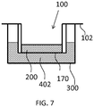
- FIG. 6 shows another method for accelerating the incubation of the fungus.
- the back surface 104b of the cellulose film 104 is in contact with a liquid culture medium 302.
- a second container 300 having the liquid culture medium 302 therein is prepared.
- the container 100 is referred to as "first container 100" to distinguish it from the second container 300.
- the first container 100 is stacked on the second container 300 in such a manner that the lower surface of the flange 102 is in contact with the upper end of the second container 300. In other words, the first container 100 is supported by the upper end of the second container 300. In this way, the liquid culture medium 302 is sandwiched between the back surface 104b of the cellulose film 104 and the bottom surface of the second container 300.
- the liquid culture medium 302 may be supplied between the back surface 104b of the cellulose film 104 and the bottom surface of the second container 300.
- a viscous solid culture medium may also be used. As shown in FIG. 6 , both of a solid culture medium 304 and the liquid culture medium 302 may be used. In this case, the liquid culture medium 302 is sandwiched between the solid culture medium 304 and the cellulose film 104. As shown in FIG. 5 , the incubation of the phytopathogenic fungus which has appeared on the back surface 104b is accelerated by at least one of the liquid culture medium 302 and the solid culture medium 304.
- the back surface 104b of the cellulose film 104 is observed after the step (b). It is desirable that the back surface 104b is observed using an optical microscope.
- the phytopathogenic fungus 202 appears on the back surface 104b of the cellulose film 104, as described in the step (b). On the other hand, the non-phytopathogenic fungus does not appear on the back surface 104b of the cellulose film 104. In this way, in the present invention, the phytopathogenic fungus 202 appears on the back surface 104b of the cellulose film 104 selectively.
- the phytopathogenic fungus 202 penetrates the cellulose film 104, whereas the non-phytopathogenic fungus does not penetrate the cellulose film 104. For this reason, the non-phytopathogenic fungus does not appear on the back surface 104b of the cellulose film 104. In this way, the phytopathogenic fungus 202 appears on the back surface 104b selectively. In other words, the phytopathogenic fungus 202 appears outside of the first container 100 selectively.
- step (c) it is observed whether or not the phytopathogenic fungus 202 appears on the back surface 104b of the cellulose film 104.
- the phytopathogenic fungus 202 is observed optically using a microscope 600 arranged below the back surface 104b of the cellulose film 104.
- the liquid culture medium 302 and the solid culture medium 304 are removed from the second container 300.
- a fluorescent agent having fungus combining ability is added to the inside of the second container 300.
- fungus fluorescent agent such a fluorescent agent is referred to as "fungus fluorescent agent”.
- the reference number of the fungus fluorescent agent is 402.
- the first container 100 is stacked on the second container 300 having the fungus fluorescent agent 402 therein.
- the fungus fluorescent agent 402 may be supplied between the back surface 104b of the cellulose film 104 and the bottom surface of the second container 300 after the first container 100 is stacked on the second container 300.
- a part of the phytopathogenic fungus 202 which has appeared on the back surface 104b of the cellulose film 104 may be dyed with the fungus fluorescent agent 402. Since the first container 100 is separated from the second container 300 by the cellulose film 104, the fungus fluorescent agent 402 does not spread into the first container 100. For this reason, the non-phytopathogenic fungus contained in the first container 100 is not dyed with the fungus fluorescent agent 402.
- the phytopathogenic fungus 202 dyed with the fungus fluorescent agent 402 is observed using the epifluorescence microscope 600 located under the back surface 104b of the cellulose film 104. Needless to say, the phytopathogenic fungus 202 may be observed without using the fungus fluorescent agent.
- step (d) it is determined that the test sample contains a phytopathogenic fungus, if a fungus is found on the back surface 104b of the cellulose film 104 in the step (c). Needless to say, it is determined that the test sample does not contain a phytopathogenic fungus, if a fungus is not found on the back surface 104b of the cellulose film 104 in the step (c).
- Fusarium oxysporum one of phytopathogenic fungi, was inoculated on a potato dextrose agar culture medium. Then, the culture medium was left at rest at a temperature of 25 degrees Celsius for one week. Fusarium oxysporum was given by an associate professor, Mr. Shimizu, who belongs to graduate School of Applied Biological Sciences and Faculty of Applied Biological Sciences, Gifu University .
- a part including ends of hyphae was cut together with the culture medium at a size of 1 centimeter x 1 centimeter.
- the cut part was immersed in pure water disposed on a 12-well plate. Each of the pure water has a volume of 1 milliliter.
- a potato dextrose culture medium having a volume of 650 microliters was added as the liquid culture medium 302 to the second container 300. In this way, the second container 300 containing the liquid culture medium 302 was prepared.
- the experiment 1 is composed of inventive examples 1A - 1D, and comparative examples 1E - 1L.
- the first container 100 shown in FIG. 1 was prepared as below.
- cellulose available from SIGMA-ALDRICH Co. LLC, trade name: Avicel PH-101
- ionic liquid was 1-butyl-3-methyl imidazolium chloride (available from SIGMA-ALDRICH Co. LLC).
- the cellulose solution was warmed to 60 degrees Celsius. Then, the cellulose solution was applied by a spin coat method for thirty seconds at a rotation speed of 2,000 rpm onto a back surface of a container having a polyethylene terephthalate film on the bottom surface thereof (available from Merck KGaA, trade name: Millicell PISP 12R 48).
- the polyethylene terephthalate film was served as the substrate 170.
- the polyethylene terephthalate film randomly had a plurality of through holes 172 each having a diameter of three micrometers. In this way, the cellulose film 104 having a thickness of 2.0 micrometers was formed on the back surface of the polyethylene terephthalate film. According to Merck KGaA, the diameter of the through-hole 172 may have a margin of error of approximately ⁇ 10%.
- the container was left at rest in ethanol at room temperature for 12 hours. In this way, 1-butyl-3-methyl imidazolium chloride was replaced with ethanol. In other words, 1-butyl-3-methyl imidazolium chloride was removed from the cellulose film 104.
- the container was dried in a vacuum desiccator. In this way, the first container 100 shown in FIG. 1 was obtained.
- the polyethylene terephthalate film serving as the substrate 170 is not illustrated.
- the first container 100 was stacked on the second container 300.
- the back surface 104b of the cellulose film 104 was in contact with the liquid culture medium 302.
- water having a volume of 200 microliters was added to the inside of the first container 100.
- the phytopathogenic fungus aqueous solution containing 200 spores of Fusarium oxysporum was added to the inside of the first container 100.
- the first container 100 was left at rest at a temperature of 25 degrees Celsius for 24 hours.
- the incubation time was 24 hours.
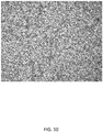
- FIG. 10 is a microscope photograph of the back surface of the cellulose film 104 in the inventive example 1A.
- each of the through holes 172 had a diameter of 5 micrometers.
- the container having a bottom surface comprising the through holes each having a diameter of 5 micrometers was available from Merck KGaA as a trade name: Millicell PIMP 12R 48.
- each of the through holes 172 had a diameter of 1 micrometer.
- the container having a bottom surface comprising the through holes each having a diameter of 1 micrometer was available from Merck KGaA as a trade name: Millicell PIRP 12R 48.
- each of the through holes 172 had a diameter of 8 micrometers.
- the container having a bottom surface comprising the through holes each having a diameter of 8 micrometers was available from Merck KGaA as a trade name: Millicell PIEP 12R 48.
- the cellulose film 104 had a thickness of 0.5 micrometers (namely, the cellulose solution had a concentration of 1.0%) and that the each of the through holes 172 had a diameter of 1 micrometer.
- the cellulose film 104 had a thickness of 0.5 micrometers (namely, the cellulose solution had a concentration of 1.0%) and that the each of the through holes 172 had a diameter of 8 micrometers.
- a non-phytopathogenic fungus aqueous solution containing spores of Saccharomyces cerevisiae was used in place of the phytopathogenic fungus aqueous solution containing spores of Fusarium oxysporum.
- Saccharomyces cerevisiae is one kind of non-phytopathogenic fungus.
- a non-phytopathogenic fungus aqueous solution containing spores of Saccharomyces cerevisiae was prepared similarly to the case of the phytopathogenic fungus aqueous solution containing spores of Fusarium oxysporum.
- the experiment 2 is composed of comparative examples 2A - 2L.
- the comparative examples 2A - 2L were similar to the inventive example 1A - the comparative example 1L, except of using another fungus.
- FIG. 11 is a microscope photograph of the back surface of the cellulose film 104 in the comparative example 2A.
- a non-phytopathogenic fungus aqueous solution containing spores of Pyricularia grisea was used in place of the phytopathogenic fungus aqueous solution containing spores of Fusarium oxysporum.
- Pyricularia grisea is one kind of phytopathogenic fungus.
- a phytopathogenic fungus aqueous solution containing spores of Pyricularia grisea was prepared as below.
- Pyricularia grisea one of phytopathogenic fungi, was inoculated on an oatmeal agar culture medium containing 2% sucrose. Then, the culture medium was left at rest at a temperature of 25 degrees Celsius for one week. Subsequently, the culture medium was left at rest for four days under near-ultraviolet radiation.
- a part including ends of hyphae was cut together with the culture medium at a size of 1 centimeter x 1 centimeter.
- the cut part was immersed in pure water disposed on a 12-well plate. Each of the pure water has a volume of 1 milliliter.
- the water contained in the 12-well plate was observed using an optical microscope. As a result, the present inventors confirmed that spores of Pyricularia grisea were released in the water disposed on the 12-well plate. In this way, an aqueous solution containing Pyricularia grisea was provided.
- the experiment 3 is composed of the inventive examples 3A- 3D and the comparative examples 3E - 3L.
- the inventive examples 3A- 3D and the comparative examples 3E - 3L were similar to the inventive examples 1A - 1D and the comparative examples 1E - 1L, respectively, except of using another fungus.
- a non-phytopathogenic fungus aqueous solution containing spores of Colletotrichum gloeosporioides was used in place of the phytopathogenic fungus aqueous solution containing spores of Fusarium oxysporum.
- Colletotrichum gloeosporioides is one kind of phytopathogenic fungus.
- a phytopathogenic fungus aqueous solution containing spores of Colletotrichum gloeosporioides was prepared similarly to the case of the phytopathogenic fungus aqueous solution containing spores of Fusarium oxysporum.
- the experiment 4 is composed of the inventive examples 4A - 4D and the comparative examples 4E - 4L.
- the inventive examples 4A - 4D and the comparative examples 4E - 4L were similar to the inventive examples 1A - 1D and the comparative examples 1 E - 1L, respectively, except of using another fungus.
- a non-phytopathogenic fungus aqueous solution containing spores of Penicillium chysogeum was used in place of the phytopathogenic fungus aqueous solution containing spores of Fusarium oxysporum.
- Penicillium chysogeum is one kind of non-phytopathogenic fungus.
- a non-phytopathogenic fungus aqueous solution containing spores of Penicillium chysogeum was prepared similarly to the case of the phytopathogenic fungus aqueous solution containing spores of Fusarium oxysporum.
- the experiment 5 is composed of comparative examples 5A - 5L.
- the comparative examples 5A - 5L were similar to the inventive example 1A - the comparative example 1L, except of using another fungus.
- a non-phytopathogenic fungus aqueous solution containing spores of Aspergillus oryzae was used in place of the phytopathogenic fungus aqueous solution containing spores of Fusarium oxysporum.
- Aspergillus oryzae is one kind of non-phytopathogenic fungus.
- a non-phytopathogenic fungus aqueous solution containing spores of Aspergillus oryzae was prepared similarly to the case of the phytopathogenic fungus aqueous solution containing spores of Fusarium oxysporum.
- the experiment 6 is composed of comparative examples 6A - 6L.
- the comparative examples 6A- 6L were similar to the inventive example 1A - the comparative example 1L, except of using another fungus.
- Table 1 - Table 6 show the number of the hyphae which penetrated the cellulose film 104 in the experiments.
- Table 1 Film thickness ( ⁇ m) Diameter of Through hole ( ⁇ m) Name of Fungus Number of hyphae which penetrated cellulose film 104 C.
- example 1E 2 1 Fusarium oxysporum (phytopathogenic) 0 I.
- example 1G 0.5 1 0.5 I. example 1C 3 106.5 I.
- example 3A 3 64.1 I.
- example 3G 0.5 1 8 I.
- example 3C 3 85.3 I.
- example 4E 2 1 Colletotrichum gloeosporioides (phytopathogenic) 0 I.
- example 4A 3 83.3 I.
- example 5E 2 1 Penicillium chysogeum (non-phytopathogenic) 0 C.
- example 5B 5 0 C.
- the phytopathogenic fungus appears on the back surface 104b of the cellulose film 104 selectively.
- the phytopathogenic fungus 202 appears outside of the container 100 selectively.
- the cellulose film 104 has a thickness of not less than 0.5 micrometers and not more than 2 micrometers.
- the through hole 172 has a diameter of not less than 3 micrometers and not more than 5 micrometers.
- the number of hyphae which penetrated cellulose film 104 is 77.4 at a minimum.
- the non-phytopathogenic fungus hardly appears the back surface 104b of the cellulose film 104.
- the number of hyphae which penetrated cellulose film 104 is 31.9 at a maximum.
- the present invention can be used to determine easily whether or not a test sample such as agricultural water or soil contains a phytopathogenic fungus.
Landscapes
- Chemical & Material Sciences (AREA)
- Health & Medical Sciences (AREA)
- Life Sciences & Earth Sciences (AREA)
- Organic Chemistry (AREA)
- Engineering & Computer Science (AREA)
- Zoology (AREA)
- Wood Science & Technology (AREA)
- Bioinformatics & Cheminformatics (AREA)
- Biotechnology (AREA)
- Genetics & Genomics (AREA)
- Biochemistry (AREA)
- General Engineering & Computer Science (AREA)
- General Health & Medical Sciences (AREA)
- Microbiology (AREA)
- Proteomics, Peptides & Aminoacids (AREA)
- Immunology (AREA)
- Medicinal Chemistry (AREA)
- Biomedical Technology (AREA)
- Analytical Chemistry (AREA)
- Biophysics (AREA)
- Molecular Biology (AREA)
- Physics & Mathematics (AREA)
- Toxicology (AREA)
- Virology (AREA)
- Botany (AREA)
- Mycology (AREA)
- Sustainable Development (AREA)
- Tropical Medicine & Parasitology (AREA)
- Chemical Kinetics & Catalysis (AREA)
- Polymers & Plastics (AREA)
- Measuring Or Testing Involving Enzymes Or Micro-Organisms (AREA)
- Apparatus Associated With Microorganisms And Enzymes (AREA)
Description
- The present invention relates to a method for determining whether or not a test sample contains a phytopathogenic fungus.
- Patent Literature 1 discloses a method for counting the number of mold cells in a specimen by the culture for a short time and capable of accurately counting the cell number.
FIG. 12 shows a cross-sectional view of a microporous membrane supporting material used for the method disclosed therein. According to this method, the extended multiple pseudomycelia of amold cell 13 cultured by a liquid culture or amold cell 13 cultured on a microporous membrane 1 of a microporous membrane supporting material 4 are photographed and the shape, area and luminous intensity are recognized and analyzed by an image analytic means 10. The number of themold cells 13 can be counted by the culture for a short time. The microporous membrane 1 is interposed between a pressing ring 2 and abase 3. - Patent Literature 2 discloses a method to selectively detect a live pathogen in a sample containing a live and dead pathogens, without detecting the dead pathogen, the method comprising the steps of immobilizing at least a portion of the live and dead pathogens on a solid support with a physical barrier; incubating the solid support in a growth medium, where the live pathogen can multiply and the multiplied pathogen move from the solid support to a supernatant of the growth medium; and detecting the multiplied pathogen in the supernatant by a pathogen assay.
- Non-Patent Literature 1 discloses that pseudohypha of Phytophthora sojae, which is one kind of phytopathogenic fungi, penetrates a PET film having through holes each having diameter of 3 micrometers.
-
- [Patent Literature 1]
JP 2005-287337 A - [Patent Literature 2]
WO 2011/090802 A1 - [Non-Patent Literature 1] Paul F. Morris. et. al. "Chemotropic and Contact Responses of Phytophthora sojae Hyphae to Soybean Isoflavonoids and Artificial Substrates", Plant Physiol. (1998) 117: 1171-1178 discloses that hyphae of Phytophthora sojae, which is one of phytopathogenic oomycetes, penetrates the PET membrane having 3-micrometer pores.
- An object of the present invention is to provide a method for determining whether or not a test sample contains a phytopathogenic fungielectively from two kinds of fungi of a phytopathogenic fungus and a non-phytopathogenic fungus.
- The present invention provides a method for determining whether or not a test sample contains a phytopathogenic fungus, the method comprising:
- (a) putting the test sample on a front surface of a substrate comprising a through hole;
wherein
the substrate comprises a cellulose film on the back surface thereof;
the cellulose film has a thickness of not less than 0.5 micrometers and not more than 2 micrometers; and
the through hole has a cross-sectional area of not less than 7.065 square micrometers and not more than 19.625 square micrometers; - (b) leaving the test sample at rest after the step (a);
- (c) observing a back surface of the film after the step (b); and
- (d) determining that the test sample contains the phytopathogenic fungus, if a fungus is found on the back surface of the film in the step (c),
- The present invention provides a method for determining whether or not a test sample contains a phytopathogenic fungus, wherein the phytopathogenic fungus is selected from the group consisting of fusarium genus, pyricularia genus, and colletotrichum genus.
-
-
FIG. 1 shows a cross-sectional view of afirst container 100. -
FIG. 2 shows a cross-sectional view of asubstrate 170 comprising acellulose film 104 on a back surface thereof. -
FIG. 3 shows a cross-sectional view of thefirst container 100 to which a test sample has been supplied. -
FIG. 4 shows a cross-sectional view of thesubstrate 170 having a front surface on which a phytopathogenic fungus has been put. -
FIG. 5 is a cross-sectional view showing a state where the phytopathogenic fungus has penetrated a throughhole 172 and thecellulose film 104. -
FIG. 6 shows a cross-sectional view of one example of a method for accelerating an incubation of the fungus. -
FIG. 7 shows a cross-sectional view, subsequently toFIG. 6 , of one example of a method for accelerating the incubation of the fungus. -
FIG. 8 is a cross-sectional view showing how to observe the fungus from the back surface of thecellulose film 104. -
FIG. 9 is a cross-sectional view showing how to observe the fungus from the back surface of thecellulose film 104. -
FIG. 10 is a microscope photograph of the back surface of thecellulose film 104 in an inventive example 1A. -
FIG. 11 is a microscope photograph of the back surface of thecellulose film 104 in a comparative example 2A. -
FIG. 12 shows a cross-sectional view of a microporous membrane supporting material used for a method for counting the number of mold cells disclosed in Japanese Patent Application laid-open Publication No.2005-287337A - First, a fungus will be described. Fungi are roughly divided into a phytopathogenic fungus and a non-phytopathogenic fungus. The phytopathogenic fungus is a Fusarium genus, a Pyricularia genus, or a Colletotrichum genus. An example of the phytopathogenic fungus is Fusarium oxysporum, Pyricularia grisea, or Colletotrichum gloeosporioides. These phytopathogenic fungi cause root rot disease, blast, anthrax, or gray mold. These phytopathogenic fungi kill the plant. An example of the non-phytopathogenic fungus is Saccharomyces cerevisiae, Penicillium chysogeum or Aspergillus oryzae.
- The term "phytopathogenic" means to have pathogenicity to plants. The term "non-phytopathogenic" means not to have pathogenicity to plants. Even if a fungus has pathogenicity, however, if the fungus has no pathogenicity to plants, the fungus is non-phytopathogenic. In other words, if a fungus does not have adverse effects on plants, the fungus is non-phytopathogenic. The prefix "non-" included in the term "non-phytopathogenic" does not modify "phyto". The prefix "non-" modifies "pathogenic".
- Hereinafter, the embodiment of the present invention will be described in more detail with reference to the drawings.
- In the step (a), a test sample is put on a front surface of a
substrate 170 comprising throughholes 172. Acellulose film 104 is adhered to aback surface 170b of thesubstrate 170. In other words, a front surface 140a of thecellulose film 104 is in contact with theback surface 170b of thesubstrate 170. - In particular, as shown in
FIG. 1 , acontainer 100 is prepared. It is desirable that thecontainer 100 comprises aflange 102 at the upper end thereof. The bottom surface of thecontainer 100 is formed of thesubstrate 170. - As shown in
FIG. 2 , thesubstrate 170 comprises thecellulose film 104 on theback surface 170b thereof (i.e., on an outside surface of the bottom part of the container 100). Thesubstrate 170 comprises the throughhole 172 which penetrates from thefront surface 170a to theback surface 170b of thesubstrate 170. The throughhole 172 has a diameter of not less than 3 micrometers and not more than 5 micrometers. In other words, the throughhole 172 has a cross-sectional area of not less than 7.065 square micrometers and not more than 19.625 square micrometers. - As shown in
FIG. 3 , atest sample 200 is supplied to an inside of thiscontainer 100. In this way, thetest sample 200 is put on thefront surface 170a of the substrate 170 (i.e., on an inside surface of the bottom part of the container 100). When thetest sample 200 contains aphytopathogenic fungus 202, thephytopathogenic fungus 202 is put on thefront surface 170a of thesubstrate 170, as shown inFIG. 4 . - The
test sample 200 is solid, liquid, or gaseous. It is desirable that thetest sample 200 is solid or liquid. An example of thesolid test sample 200 is soil or a crushed plant. Another example is an agricultural material such as vermiculite, rock wool or urethane. An example of theliquid test sample 200 is agricultural water, a solution used for hydroponic culture, a liquid used for washing a plant, a liquid extracted from a plant, a liquid used for washing an agricultural material, or a liquid used for washing clothing or shoes of a worker. - In the step (b), the
test sample 200 is left at rest for a certain incubation time after the step (a). Desirably, thetest sample 200 is left at rest for 24 hours. In this way, the fungus is incubated. In other words, the incubation time is approximately 24 hours. Hereinafter, the importance of the thickness of thecellulose film 104 and the size of the throughhole 172 will be described. - In the step (b), various fungi contained in the
test sample 200 are grown. As demonstrated in the experiments which will be described later, if both of the following requirements (I) and (II) are satisfied, thephytopathogenic fungus 202 grows up so as to penetrate both the throughhole 172 and thecellulose film 104, as shown inFIG. 5 . As a result, thephytopathogenic fungus 202 appears on aback surface 104b of thecellulose film 104. - Requirement (I): The
cellulose film 104 has a thickness of not less than 0.5 micrometers and not more than 2 micrometers. - Requirement (II): The through
hole 172 has a cross-sectional area of not less than 7.065 square micrometers and not more than 19.625 square micrometers. - If both of the above requirements (I) and (II) are satisfied, the non-phytopathogenic fungus hardly penetrates the
cellulose film 104. As demonstrated in the comparative example 6C, at a maximum, the number of hyphae which penetratedcellulose film 104 is 31.9. For this reason, the non-phytopathogenic fungus hardly appears on theback surface 104b of thecellulose film 104. On the other hand, thephytopathogenic fungus 202 appears on theback surface 104b selectively. As demonstrated in the inventive example 3D, at a minimum, the number of hyphae which penetratedcellulose film 104 is 77.4. As just described, thephytopathogenic fungus 202 appears outside of thecontainer 100 selectively. - In case where the
cellulose film 104 has a thickness of more than 2 micrometers, neither the non-phytopathogenic fungus nor the phytopathogenic fungus penetrates thecellulose film 104. Therefore, in case where thecellulose film 104 has a thickness of more than 2 micrometers, the selectivity is lost. When thecellulose film 104 has a thickness of less than 0.5 micrometers (including a case where thecellulose film 104 is not provided), not only the non-phytopathogenic fungus but also the phytopathogenic fungus penetrates the cellulose film 104 (or are found on theback surface 170b of the substrate 170). Therefore, the selectivity is lost when thecellulose film 104 has a thickness of less than 0.5 micrometers. - In case where the through
hole 172 has a cross-sectional area of less than 7.065 square micrometers (namely, a diameter of less than 3 micrometers), neither the non-phytopathogenic fungus nor the phytopathogenic fungus penetrates thecellulose film 104. On the other hand, the throughhole 172 has a cross-sectional area of more than 19.625 square micrometers (namely, a diameter of more than 5 micrometers), the number of hyphae which penetratedcellulose film 104 tends to be lowered, compared to the case where the throughhole 172 has a cross-sectional area of 19.625 square micrometers (namely, a diameter of 5 micrometers). - The
cellulose film 104 is stretched tautly on theback surface 170b of thesubstrate 170. In this way, thesubstrate 170 supports thecellulose film 104. - As shown in
FIG. 2 , it is desirable that thesubstrate 170 has a plurality of throughholes 172. The thickness of thesubstrate 170 is not limited; however, as one example, it is desirable that thesubstrate 170 has a thickness of not less than 1 micrometer and not more than 500 micrometers. Thecellulose film 104 is significantly thin. However, if thecellulose film 104 is arranged on thesubstrate 170, it is easy to handle thecellulose film 104. - A culture medium may be supplied to the
test sample 200 to accelerate the incubation of the fungus. In particular, a culture medium may be supplied to the inside of thecontainer 100 containing thetest sample 200. It is desirable that the culture medium is liquid. The culture medium may be supplied in the step (b). Alternatively, the culture medium may be supplied prior to the step (b). In other words, the culture medium may be supplied in the step (a). The culture medium may be supplied to the inside of thecontainer 100 prior to the step (a). -
FIG. 6 shows another method for accelerating the incubation of the fungus. As shown inFIG. 6 , it is desirable that theback surface 104b of thecellulose film 104 is in contact with aliquid culture medium 302. First, asecond container 300 having theliquid culture medium 302 therein is prepared. Hereinafter, thecontainer 100 is referred to as "first container 100" to distinguish it from thesecond container 300. Thefirst container 100 is stacked on thesecond container 300 in such a manner that the lower surface of theflange 102 is in contact with the upper end of thesecond container 300. In other words, thefirst container 100 is supported by the upper end of thesecond container 300. In this way, theliquid culture medium 302 is sandwiched between theback surface 104b of thecellulose film 104 and the bottom surface of thesecond container 300. - Alternatively, after the
first container 100 is stacked on thesecond container 300, theliquid culture medium 302 may be supplied between theback surface 104b of thecellulose film 104 and the bottom surface of thesecond container 300. - In place of the
liquid culture medium 302, a viscous solid culture medium may also be used. As shown inFIG. 6 , both of asolid culture medium 304 and theliquid culture medium 302 may be used. In this case, theliquid culture medium 302 is sandwiched between thesolid culture medium 304 and thecellulose film 104. As shown inFIG. 5 , the incubation of the phytopathogenic fungus which has appeared on theback surface 104b is accelerated by at least one of theliquid culture medium 302 and thesolid culture medium 304. - In the step (c), the
back surface 104b of thecellulose film 104 is observed after the step (b). It is desirable that theback surface 104b is observed using an optical microscope. - The
phytopathogenic fungus 202 appears on theback surface 104b of thecellulose film 104, as described in the step (b). On the other hand, the non-phytopathogenic fungus does not appear on theback surface 104b of thecellulose film 104. In this way, in the present invention, thephytopathogenic fungus 202 appears on theback surface 104b of thecellulose film 104 selectively. - In other words, the
phytopathogenic fungus 202 penetrates thecellulose film 104, whereas the non-phytopathogenic fungus does not penetrate thecellulose film 104. For this reason, the non-phytopathogenic fungus does not appear on theback surface 104b of thecellulose film 104. In this way, thephytopathogenic fungus 202 appears on theback surface 104b selectively. In other words, thephytopathogenic fungus 202 appears outside of thefirst container 100 selectively. - In the step (c), it is observed whether or not the
phytopathogenic fungus 202 appears on theback surface 104b of thecellulose film 104. - In particular, whether or not the
phytopathogenic fungus 202 appears on theback surface 104b of thecellulose film 104 is observed as below. - As shown in
FIG. 8 , while thecellulose film 104 is irradiated with light emitted from alight source 500 arranged above thefront surface 170a of thesubstrate 170, thephytopathogenic fungus 202 is observed optically using amicroscope 600 arranged below theback surface 104b of thecellulose film 104. - The
liquid culture medium 302 and thesolid culture medium 304 are removed from thesecond container 300. Then, a fluorescent agent having fungus combining ability is added to the inside of thesecond container 300. Hereinafter, such a fluorescent agent is referred to as "fungus fluorescent agent". The reference number of the fungus fluorescent agent is 402. Then, as shown inFIG. 7 , thefirst container 100 is stacked on thesecond container 300 having thefungus fluorescent agent 402 therein. Alternatively, thefungus fluorescent agent 402 may be supplied between theback surface 104b of thecellulose film 104 and the bottom surface of thesecond container 300 after thefirst container 100 is stacked on thesecond container 300. - A part of the
phytopathogenic fungus 202 which has appeared on theback surface 104b of thecellulose film 104 may be dyed with thefungus fluorescent agent 402. Since thefirst container 100 is separated from thesecond container 300 by thecellulose film 104, thefungus fluorescent agent 402 does not spread into thefirst container 100. For this reason, the non-phytopathogenic fungus contained in thefirst container 100 is not dyed with thefungus fluorescent agent 402. - As shown in
FIG. 9 , thephytopathogenic fungus 202 dyed with thefungus fluorescent agent 402 is observed using theepifluorescence microscope 600 located under theback surface 104b of thecellulose film 104. Needless to say, thephytopathogenic fungus 202 may be observed without using the fungus fluorescent agent. - In the step (d), it is determined that the test sample contains a phytopathogenic fungus, if a fungus is found on the
back surface 104b of thecellulose film 104 in the step (c). Needless to say, it is determined that the test sample does not contain a phytopathogenic fungus, if a fungus is not found on theback surface 104b of thecellulose film 104 in the step (c). - The present invention will be described in more detail with reference to the following examples.
- Fusarium oxysporum, one of phytopathogenic fungi, was inoculated on a potato dextrose agar culture medium. Then, the culture medium was left at rest at a temperature of 25 degrees Celsius for one week. Fusarium oxysporum was given by an associate professor, Mr. Shimizu, who belongs to Graduate School of Applied Biological Sciences and Faculty of Applied Biological Sciences, Gifu University.
- Then, a part including ends of hyphae was cut together with the culture medium at a size of 1 centimeter x 1 centimeter. The cut part was immersed in pure water disposed on a 12-well plate. Each of the pure water has a volume of 1 milliliter.
- The water contained in the 12-well plate was observed using an optical microscope. As a result, the present inventors confirmed that spores of Fusarium oxysporum were released in the water disposed on the 12-well plate. In this way, an aqueous solution containing Fusarium oxysporum was provided. Hereinafter, this aqueous solution is referred to as "phytopathogenic fungus aqueous solution".
- A potato dextrose culture medium having a volume of 650 microliters was added as the
liquid culture medium 302 to thesecond container 300. In this way, thesecond container 300 containing theliquid culture medium 302 was prepared. - The experiment 1 is composed of inventive examples 1A - 1D, and comparative examples 1E - 1L.
- The
first container 100 shown inFIG. 1 was prepared as below. - First, cellulose (available from SIGMA-ALDRICH Co. LLC, trade name: Avicel PH-101) was dissolved in an ionic liquid to prepare a cellulose solution having a concentration of 2%. The ionic liquid was 1-butyl-3-methyl imidazolium chloride (available from SIGMA-ALDRICH Co. LLC).
- The cellulose solution was warmed to 60 degrees Celsius. Then, the cellulose solution was applied by a spin coat method for thirty seconds at a rotation speed of 2,000 rpm onto a back surface of a container having a polyethylene terephthalate film on the bottom surface thereof (available from Merck KGaA, trade name: Millicell PISP 12R 48). The polyethylene terephthalate film was served as the
substrate 170. The polyethylene terephthalate film randomly had a plurality of throughholes 172 each having a diameter of three micrometers. In this way, thecellulose film 104 having a thickness of 2.0 micrometers was formed on the back surface of the polyethylene terephthalate film. According to Merck KGaA, the diameter of the through-hole 172 may have a margin of error of approximately ± 10%. - The container was left at rest in ethanol at room temperature for 12 hours. In this way, 1-butyl-3-methyl imidazolium chloride was replaced with ethanol. In other words, 1-butyl-3-methyl imidazolium chloride was removed from the
cellulose film 104. - Finally, the container was dried in a vacuum desiccator. In this way, the
first container 100 shown inFIG. 1 was obtained. InFIG. 1 , note that the polyethylene terephthalate film serving as thesubstrate 170 is not illustrated. - Then, as shown in
FIG. 6 , thefirst container 100 was stacked on thesecond container 300. Theback surface 104b of thecellulose film 104 was in contact with theliquid culture medium 302. Subsequently, water having a volume of 200 microliters was added to the inside of thefirst container 100. Furthermore, the phytopathogenic fungus aqueous solution containing 200 spores of Fusarium oxysporum was added to the inside of thefirst container 100. - The
first container 100 was left at rest at a temperature of 25 degrees Celsius for 24 hours. In other words, in the inventive example 1A, the incubation time was 24 hours. - The number of the hyphae of Fusarium oxysporum which appeared on the
back surface 104b of thecellulose film 104 was counted visually with an optical microscope. The inventive example 1A was repeated fifteen times. As a result, the mean value of the number of the hyphae of Fusarium oxysporum which appeared on theback surface 104b was 44.9.FIG. 10 is a microscope photograph of the back surface of thecellulose film 104 in the inventive example 1A. - In the inventive example 1B, an experiment similar to the inventive example 1A was conducted, except that each of the through
holes 172 had a diameter of 5 micrometers. The container having a bottom surface comprising the through holes each having a diameter of 5 micrometers was available from Merck KGaA as a trade name: Millicell PIMP 12R 48. - In the inventive example 1C, an experiment similar to the inventive example 1A was conducted, except that the cellulose solution had a concentration of 1.0% and that the
cellulose film 104 had a thickness of 0.5 micrometers. - In the inventive example 1D, an experiment similar to the inventive example 1A was conducted, except that the cellulose solution had a concentration of 1.0%, that the
cellulose film 104 had a thickness of 0.5 micrometers, and that each of the throughhole 172 had a diameter of 5 micrometers. - In the comparative example 1E, an experiment similar to the inventive example 1A was conducted, except that each of the through
holes 172 had a diameter of 1 micrometer. The container having a bottom surface comprising the through holes each having a diameter of 1 micrometer was available from Merck KGaA as a trade name: Millicell PIRP 12R 48. - In the comparative example 1F, an experiment similar to the inventive example 1A was conducted, except that each of the through
holes 172 had a diameter of 8 micrometers. The container having a bottom surface comprising the through holes each having a diameter of 8 micrometers was available from Merck KGaA as a trade name: Millicell PIEP 12R 48. - In the comparative example 1G, an experiment similar to the inventive example 1A was conducted, except that the
cellulose film 104 had a thickness of 0.5 micrometers (namely, the cellulose solution had a concentration of 1.0%) and that the each of the throughholes 172 had a diameter of 1 micrometer. - In the comparative example 1H, an experiment similar to the inventive example 1A was conducted, except that the
cellulose film 104 had a thickness of 0.5 micrometers (namely, the cellulose solution had a concentration of 1.0%) and that the each of the throughholes 172 had a diameter of 8 micrometers. - In the comparative example 1I, an experiment similar to the inventive example 1A was conducted, except that the
cellulose film 104 was not formed (namely, thecellulose film 104 had a thickness of 0 micrometers) and that the each of the throughholes 172 had a diameter of 1 micrometer. - In the comparative example 1J, an experiment similar to the inventive example 1A was conducted, except that the
cellulose film 104 was not formed (namely, thecellulose film 104 had a thickness of 0 micrometers). - In the comparative example 1K, an experiment similar to the inventive example 1A was conducted, except that the
cellulose film 104 was not formed (namely, thecellulose film 104 had a thickness of 0 micrometers) and that the each of the throughholes 172 had a diameter of 5 micrometers. - In the comparative example 1 L, an experiment similar to the inventive example 1A was conducted, except that the
cellulose film 104 was not formed (namely, thecellulose film 104 had a thickness of 0 micrometers) and that the each of the throughholes 172 had a diameter of 8 micrometers. - In the experiment 2, a non-phytopathogenic fungus aqueous solution containing spores of Saccharomyces cerevisiae was used in place of the phytopathogenic fungus aqueous solution containing spores of Fusarium oxysporum. Unlike Fusarium oxysporum, Saccharomyces cerevisiae is one kind of non-phytopathogenic fungus. A non-phytopathogenic fungus aqueous solution containing spores of Saccharomyces cerevisiae was prepared similarly to the case of the phytopathogenic fungus aqueous solution containing spores of Fusarium oxysporum. The experiment 2 is composed of comparative examples 2A - 2L. The comparative examples 2A - 2L were similar to the inventive example 1A - the comparative example 1L, except of using another fungus.
FIG. 11 is a microscope photograph of the back surface of thecellulose film 104 in the comparative example 2A. - In the
experiment 3, a non-phytopathogenic fungus aqueous solution containing spores of Pyricularia grisea was used in place of the phytopathogenic fungus aqueous solution containing spores of Fusarium oxysporum. Similarly to Fusarium oxysporum, Pyricularia grisea is one kind of phytopathogenic fungus. A phytopathogenic fungus aqueous solution containing spores of Pyricularia grisea was prepared as below. - Pyricularia grisea, one of phytopathogenic fungi, was inoculated on an oatmeal agar culture medium containing 2% sucrose. Then, the culture medium was left at rest at a temperature of 25 degrees Celsius for one week. Subsequently, the culture medium was left at rest for four days under near-ultraviolet radiation.
- Then, a part including ends of hyphae was cut together with the culture medium at a size of 1 centimeter x 1 centimeter. The cut part was immersed in pure water disposed on a 12-well plate. Each of the pure water has a volume of 1 milliliter.
- The water contained in the 12-well plate was observed using an optical microscope. As a result, the present inventors confirmed that spores of Pyricularia grisea were released in the water disposed on the 12-well plate. In this way, an aqueous solution containing Pyricularia grisea was provided.
- The
experiment 3 is composed of the inventive examples 3A- 3D and the comparative examples 3E - 3L. The inventive examples 3A- 3D and the comparative examples 3E - 3L were similar to the inventive examples 1A - 1D and the comparative examples 1E - 1L, respectively, except of using another fungus. - In the experiment 4, a non-phytopathogenic fungus aqueous solution containing spores of Colletotrichum gloeosporioides was used in place of the phytopathogenic fungus aqueous solution containing spores of Fusarium oxysporum. Similarly to Fusarium oxysporum, Colletotrichum gloeosporioides is one kind of phytopathogenic fungus. A phytopathogenic fungus aqueous solution containing spores of Colletotrichum gloeosporioides was prepared similarly to the case of the phytopathogenic fungus aqueous solution containing spores of Fusarium oxysporum. The experiment 4 is composed of the inventive examples 4A - 4D and the comparative examples 4E - 4L. The inventive examples 4A - 4D and the comparative examples 4E - 4L were similar to the inventive examples 1A - 1D and the comparative examples 1 E - 1L, respectively, except of using another fungus.
- In the experiment 5, a non-phytopathogenic fungus aqueous solution containing spores of Penicillium chysogeum was used in place of the phytopathogenic fungus aqueous solution containing spores of Fusarium oxysporum. Unlike Fusarium oxysporum, Penicillium chysogeum is one kind of non-phytopathogenic fungus. A non-phytopathogenic fungus aqueous solution containing spores of Penicillium chysogeum was prepared similarly to the case of the phytopathogenic fungus aqueous solution containing spores of Fusarium oxysporum. The experiment 5 is composed of comparative examples 5A - 5L. The comparative examples 5A - 5L were similar to the inventive example 1A - the comparative example 1L, except of using another fungus.
- In the experiment 6, a non-phytopathogenic fungus aqueous solution containing spores of Aspergillus oryzae was used in place of the phytopathogenic fungus aqueous solution containing spores of Fusarium oxysporum. Unlike Fusarium oxysporum, Aspergillus oryzae is one kind of non-phytopathogenic fungus. A non-phytopathogenic fungus aqueous solution containing spores of Aspergillus oryzae was prepared similarly to the case of the phytopathogenic fungus aqueous solution containing spores of Fusarium oxysporum. The experiment 6 is composed of comparative examples 6A - 6L. The comparative examples 6A- 6L were similar to the inventive example 1A - the comparative example 1L, except of using another fungus.
- The following Table 1 - Table 6 show the number of the hyphae which penetrated the
cellulose film 104 in the experiments.[Table 1] Film thickness (µm) Diameter of Through hole (µm) Name of Fungus Number of hyphae which penetrated cellulose film 104C. example 1E 2 1 Fusarium oxysporum (phytopathogenic) 0 I. example 1A 3 44.9 I. example 1B 5 42.8 C. example 1F 8 16.3 C. example 1G 0.5 1 0.5 I. example 1C 3 106.5 I. example 1D 5 94.1 C. example 1H 8 125.4 C. example 1I 0 1 0.3 C. example 1J 3 125.3 C. example 1K 5 33.3 C. example 1L 8 15 Incubation time: 24 hours
C.: Comparative
I.: Inventive[Table 2] Film thickness (µm) Diameter of Through hole (µm) Name of Fungus Number of hyphae which penetrated cellulose film 104C. example 2E 2 1 Saccharomyces cerevisiae (non-phytopathogenic) 0 C. example 2A 3 0 C. example 2B 5 0 C. example 2F 8 0 C. example 2G 0.5 1 0 C. example 2C 3 0 C. example 2D 5 0 C. example 2H 8 0 C. example 2I 0 1 0 C. example 2J 3 0 C. example 2K 5 0 C. example 2L 8 0 Incubation time: 24 hours
C.: Comparative
I.: Inventive[Table 3] Film thickness (µm) Diameter of Through hole (µm) Name of Fungus Number of hyphae which penetrated cellulose film 104C. example 3E 2 1 Pyricularia grisea (phytopathogen ic) 2 I. example 3A 3 64.1 I. example 3B 5 53 C. example 3F 8 84.9 C. example 3G 0.5 1 8 I. example 3C 3 85.3 I. example 3D 5 77.4 C. example 3H 8 99.4 C. example 3I 0 1 4 C. example 3J 3 11 C. example 3K 5 15.7 C. example 3L 8 7.7 Incubation time: 24 hours
C.: Comparative
I.: Inventive[Table 4] Film thickness (µm) Diameter of Through hole (µm) Name of Fungus Number of hyphae which penetrated cellulose film 104C. example 4E 2 1 Colletotrichum gloeosporioides (phytopathogenic) 0 I. example 4A 3 83.3 I. example 4B 5 55.2 C. example 4F 8 8.7 C. example 4G 0.5 1 4 I. example 4C 3 326 I. example 4D 5 449 C. example 4H 8 165.3 C. example 4I 0 1 1.3 C. example 4J 3 182.7 C. example 4K 5 91.3 C. example 4L 8 62.7 Incubation time: 24 hours
C.: Comparative
I.: Inventive[Table 5] Film thickness (µm) Diameter of Through hole (µm) Name of Fungus Number of hyphae which penetrate d cellulose film 104C. example 5E 2 1 Penicillium chysogeum (non-phytopathogenic) 0 C. example 5A 3 0 C. example 5B 5 0 C. example 5F 8 0 C. example 5G 0.5 1 0 C. example 5C 3 0 C. example 5D 5 0 C. example 5H 8 6.8 C. example 5I 0 1 0 C. example 5J 3 10 C. example 5K 5 11.7 C. example 5L 8 4.3 Incubation time: 24 hours
C.: Comparative
I.: Inventive[Table 6] C.: Comparative I.: Inventive Film thickness (µm) Diameter of Through hole (µm) Name of Fungus Number of hyphae which penetrated cellulose film 104C. example 6E 2 1 Aspergillus oryzae (non-phytopathogenic) 0 C. example 6A 3 0 C. example 6B 5 2.7 C. example 6F 8 1.6 C. example 6G 0.5 1 1 C. example 6C 3 31.9 C. example 6D 5 18.8 C. example 6H 8 18.9 C. example 6I 0 1 1 C. example 6J 3 48 C. example 6K 5 56 C. example 6L 8 23.7 Incubation time: 24 hours
C.: Comparative
I.: Inventive - As is clear from Table 1 - Table 6, when both of the following requirements (I) and (II) are satisfied, the phytopathogenic fungus appears on the
back surface 104b of thecellulose film 104 selectively. In other words, thephytopathogenic fungus 202 appears outside of thecontainer 100 selectively. - Requirement (I): The
cellulose film 104 has a thickness of not less than 0.5 micrometers and not more than 2 micrometers. - Requirement (II): The through
hole 172 has a diameter of not less than 3 micrometers and not more than 5 micrometers. - As demonstrated in the inventive example 3D in which both of the requirements (I) and (II) are satisfied, the number of hyphae which penetrated
cellulose film 104 is 77.4 at a minimum. On the other hand, as long as both of the requirements (I) and (II) are satisfied, the non-phytopathogenic fungus hardly appears theback surface 104b of thecellulose film 104. As demonstrated in the comparative example 6C in which both of the requirements (I) and (II) are satisfied, the number of hyphae which penetratedcellulose film 104 is 31.9 at a maximum. - The present invention can be used to determine easily whether or not a test sample such as agricultural water or soil contains a phytopathogenic fungus.
-
- 100 First container
102 Flange
104 Cellulose film- 104a Front surface
- 104b Back surface
- 170 Substrate
- 170a Front surface
- 170b Back surface
- 200 Test sample
- 202 Phytopathogenic fungus
202a Part of Phytopathogenic fungus - 300 Second container
- 302 Liquid culture medium
- 304 Solid culture medium
- 202 Phytopathogenic fungus
- 402 Fluorescent agent having fungus combining ability
- 500 Light source
- 600 Microscope
Claims (11)
- A method for determining whether or not a test sample contains a phytopathogenic fungus, the method comprising:(a) putting the test sample on a front surface of a substrate comprising a through hole;
wherein
the substrate comprises a cellulose film on the back surface thereof;
the cellulose film has a thickness of not less than 0.5 micrometers and not more than 2 micrometers; and
the through hole has a cross-sectional area of not less than 7.065 square micrometers and not more than 19.625 square micrometers;(b) leaving the test sample at rest after the step (a);(c) observing a back surface of the film after the step (b); and(d) determining that the test sample contains the phytopathogenic fungus, if a fungus is found on the back surface of the film in the step (c),wherein the phytopathogenic fungus is selected from the group consisting of fusarium genus, pyricularia genus, and colletotrichum genus. - The method according to claim 1, wherein
the phytopathogenic fungus is selected from the group consisting of Fusarium oxysporum, Pyricularia grisea, and Colletotrichum gloeosporioides. - The method according to claim 1, further comprising:
a step of bringing the back surface of the cellulose film into contact with a fluorescent agent for dyeing the fungus between the step (b) and the step (c). - The method according to claim 1, further comprising:
a step of supplying a culture medium to the test sample before the step (b). - The method according to claim 4, wherein
the culture medium is a liquid culture medium. - The method according to claim 4, wherein
the test sample is left at rest while the back surface of the cellulose film is in contact with the culture medium in the step (b). - The method according to claim 4, wherein
the culture medium is a solid culture medium. - The method according to claim 1, wherein
the test sample is solid. - The method according to claim 8, wherein
the solid test sample is at least one selected from the group consisting of soil and a crushed plant. - The method according to claim 1, wherein
the test sample is liquid. - The method according to claim 10, wherein
the liquid test sample is at least one selected from the group consisting of agricultural water, a liquid used for hydroponic culture, a liquid used for washing a plant, a liquid extracted from a plant, a liquid used for washing an agricultural material, and a liquid used for washing clothing or a shoe.
Applications Claiming Priority (2)
| Application Number | Priority Date | Filing Date | Title |
|---|---|---|---|
| JP2016139916 | 2016-07-15 | ||
| PCT/JP2016/004417 WO2018011835A1 (en) | 2016-07-15 | 2016-09-30 | Method for determining whether or not test sample contains phytopathogenic fungus |
Publications (3)
| Publication Number | Publication Date |
|---|---|
| EP3438277A1 EP3438277A1 (en) | 2019-02-06 |
| EP3438277A4 EP3438277A4 (en) | 2019-03-06 |
| EP3438277B1 true EP3438277B1 (en) | 2020-06-24 |
Family
ID=60952875
Family Applications (1)
| Application Number | Title | Priority Date | Filing Date |
|---|---|---|---|
| EP16908739.2A Active EP3438277B1 (en) | 2016-07-15 | 2016-09-30 | Method for determining whether or not test sample contains phytopathogenic fungus |
Country Status (6)
| Country | Link |
|---|---|
| US (2) | US11098340B2 (en) |
| EP (1) | EP3438277B1 (en) |
| JP (1) | JP6739005B2 (en) |
| AU (1) | AU2016414842B2 (en) |
| BR (1) | BR112018072475A2 (en) |
| WO (1) | WO2018011835A1 (en) |
Families Citing this family (7)
| Publication number | Priority date | Publication date | Assignee | Title |
|---|---|---|---|---|
| EP3575405B1 (en) * | 2017-01-25 | 2021-09-01 | Panasonic Intellectual Property Management Co., Ltd. | Method for determining whether or not test sample contains phytopathogenic fungus |
| EP3617319B1 (en) | 2017-04-28 | 2022-12-07 | Panasonic Intellectual Property Management Co., Ltd. | Method for determining presence or absence of phytopathogenic microbe in test sample |
| WO2019225171A1 (en) * | 2018-05-23 | 2019-11-28 | パナソニックIpマネジメント株式会社 | Device for detecting fungal tomato pathogen and detection method using same |
| CN111836880B (en) * | 2018-05-23 | 2024-08-20 | 松下知识产权经营株式会社 | Detection device for tomato pathogenic fungi and detection method using detection device |
| SG11202011106RA (en) | 2018-07-09 | 2020-12-30 | Panasonic Ip Man Co Ltd | Device for detecting phytopathogenic fungus, and detection method and agricultural chemical concentration selection method in which said device is used |
| JP7281695B2 (en) * | 2018-09-05 | 2023-05-26 | パナソニックIpマネジメント株式会社 | Detection device for tomato pathogenic fungus and detection method using the same |
| WO2020202161A1 (en) * | 2019-04-04 | 2020-10-08 | Technion Research & Development Foundation Limited | A method for identifying a fungal symbiont |
Citations (1)
| Publication number | Priority date | Publication date | Assignee | Title |
|---|---|---|---|---|
| WO2011090802A1 (en) * | 2010-01-22 | 2011-07-28 | Hitachi Chemical Co., Ltd. | Rapid pathogen detection techniques and apparatus |
Family Cites Families (6)
| Publication number | Priority date | Publication date | Assignee | Title |
|---|---|---|---|---|
| WO2004018623A2 (en) * | 2002-08-16 | 2004-03-04 | Clinical Microarrays, Inc. | Substrates for isolating, reacting and microscopically analyzing materials |
| US6698880B1 (en) * | 2002-09-20 | 2004-03-02 | Eastman Kodak Company | Porous inkjet recording system comprising ink-pigment-trapping surface layer |
| JP4590902B2 (en) | 2004-03-31 | 2010-12-01 | パナソニック株式会社 | Filamentous fungus measurement method |
| FR2957354B1 (en) | 2010-03-11 | 2012-03-30 | Agronomique Inst Nat Rech | TREATMENT OF PLANTS AGAINST OOMYCETAL INFECTION |
| CN103816808B (en) * | 2010-06-07 | 2017-01-11 | 3M创新有限公司 | Filtration methods and devices |
| JP6167309B2 (en) * | 2015-08-03 | 2017-07-26 | パナソニックIpマネジメント株式会社 | Method for determining whether a test sample contains phytopathogenic oomycete |
-
2016
- 2016-09-30 AU AU2016414842A patent/AU2016414842B2/en active Active
- 2016-09-30 EP EP16908739.2A patent/EP3438277B1/en active Active
- 2016-09-30 JP JP2017503624A patent/JP6739005B2/en active Active
- 2016-09-30 BR BR112018072475-0A patent/BR112018072475A2/en not_active Application Discontinuation
- 2016-09-30 WO PCT/JP2016/004417 patent/WO2018011835A1/en active Application Filing
-
2018
- 2018-10-17 US US16/162,467 patent/US11098340B2/en active Active
-
2021
- 2021-07-16 US US17/378,468 patent/US11807740B2/en active Active
Patent Citations (1)
| Publication number | Priority date | Publication date | Assignee | Title |
|---|---|---|---|---|
| WO2011090802A1 (en) * | 2010-01-22 | 2011-07-28 | Hitachi Chemical Co., Ltd. | Rapid pathogen detection techniques and apparatus |
Also Published As
| Publication number | Publication date |
|---|---|
| AU2016414842B2 (en) | 2023-05-11 |
| EP3438277A4 (en) | 2019-03-06 |
| AU2016414842A2 (en) | 2018-11-22 |
| JP6739005B2 (en) | 2020-08-12 |
| BR112018072475A2 (en) | 2019-02-19 |
| JPWO2018011835A1 (en) | 2019-04-25 |
| US20190048388A1 (en) | 2019-02-14 |
| US11098340B2 (en) | 2021-08-24 |
| AU2016414842A1 (en) | 2018-11-15 |
| EP3438277A1 (en) | 2019-02-06 |
| US20210340589A1 (en) | 2021-11-04 |
| US11807740B2 (en) | 2023-11-07 |
| WO2018011835A1 (en) | 2018-01-18 |
Similar Documents
| Publication | Publication Date | Title |
|---|---|---|
| EP3438277B1 (en) | Method for determining whether or not test sample contains phytopathogenic fungus | |
| EP3128002B1 (en) | Method for determining whether or not test sample contains phytopathogenic oomycete | |
| US11913057B2 (en) | Method for determining whether or not test sample contains phytopathogenic fungus | |
| US11713479B2 (en) | Method for determining whether or not test sample contains phytopathogenic fungus | |
| US10526636B2 (en) | Method for determining whether or not test sample contains phytopathogenic fungus | |
| US10538801B2 (en) | Method for determining whether or not test sample contains exserohilum phytopathogenic fungus | |
| US11104932B2 (en) | Method for determining whether or not all of pythiums contained in test sample are non-phytopathogenic |
Legal Events
| Date | Code | Title | Description |
|---|---|---|---|
| STAA | Information on the status of an ep patent application or granted ep patent |
Free format text: STATUS: THE INTERNATIONAL PUBLICATION HAS BEEN MADE |
|
| PUAI | Public reference made under article 153(3) epc to a published international application that has entered the european phase |
Free format text: ORIGINAL CODE: 0009012 |
|
| STAA | Information on the status of an ep patent application or granted ep patent |
Free format text: STATUS: REQUEST FOR EXAMINATION WAS MADE |
|
| 17P | Request for examination filed |
Effective date: 20181030 |
|
| AK | Designated contracting states |
Kind code of ref document: A1 Designated state(s): AL AT BE BG CH CY CZ DE DK EE ES FI FR GB GR HR HU IE IS IT LI LT LU LV MC MK MT NL NO PL PT RO RS SE SI SK SM TR |
|
| AX | Request for extension of the european patent |
Extension state: BA ME |
|
| A4 | Supplementary search report drawn up and despatched |
Effective date: 20190205 |
|
| RIC1 | Information provided on ipc code assigned before grant |
Ipc: C12Q 1/04 20060101AFI20190130BHEP |
|
| DAV | Request for validation of the european patent (deleted) | ||
| DAX | Request for extension of the european patent (deleted) | ||
| STAA | Information on the status of an ep patent application or granted ep patent |
Free format text: STATUS: EXAMINATION IS IN PROGRESS |
|
| 17Q | First examination report despatched |
Effective date: 20191121 |
|
| GRAP | Despatch of communication of intention to grant a patent |
Free format text: ORIGINAL CODE: EPIDOSNIGR1 |
|
| STAA | Information on the status of an ep patent application or granted ep patent |
Free format text: STATUS: GRANT OF PATENT IS INTENDED |
|
| INTG | Intention to grant announced |
Effective date: 20200127 |
|
| GRAS | Grant fee paid |
Free format text: ORIGINAL CODE: EPIDOSNIGR3 |
|
| GRAA | (expected) grant |
Free format text: ORIGINAL CODE: 0009210 |
|
| STAA | Information on the status of an ep patent application or granted ep patent |
Free format text: STATUS: THE PATENT HAS BEEN GRANTED |
|
| AK | Designated contracting states |
Kind code of ref document: B1 Designated state(s): AL AT BE BG CH CY CZ DE DK EE ES FI FR GB GR HR HU IE IS IT LI LT LU LV MC MK MT NL NO PL PT RO RS SE SI SK SM TR |
|
| REG | Reference to a national code |
Ref country code: GB Ref legal event code: FG4D |
|
| REG | Reference to a national code |
Ref country code: CH Ref legal event code: EP |
|
| REG | Reference to a national code |
Ref country code: AT Ref legal event code: REF Ref document number: 1283946 Country of ref document: AT Kind code of ref document: T Effective date: 20200715 |
|
| REG | Reference to a national code |
Ref country code: DE Ref legal event code: R096 Ref document number: 602016038943 Country of ref document: DE |
|
| REG | Reference to a national code |
Ref country code: IE Ref legal event code: FG4D |
|
| REG | Reference to a national code |
Ref country code: NL Ref legal event code: FP |
|
| PG25 | Lapsed in a contracting state [announced via postgrant information from national office to epo] |
Ref country code: LT Free format text: LAPSE BECAUSE OF FAILURE TO SUBMIT A TRANSLATION OF THE DESCRIPTION OR TO PAY THE FEE WITHIN THE PRESCRIBED TIME-LIMIT Effective date: 20200624 Ref country code: FI Free format text: LAPSE BECAUSE OF FAILURE TO SUBMIT A TRANSLATION OF THE DESCRIPTION OR TO PAY THE FEE WITHIN THE PRESCRIBED TIME-LIMIT Effective date: 20200624 Ref country code: GR Free format text: LAPSE BECAUSE OF FAILURE TO SUBMIT A TRANSLATION OF THE DESCRIPTION OR TO PAY THE FEE WITHIN THE PRESCRIBED TIME-LIMIT Effective date: 20200925 Ref country code: NO Free format text: LAPSE BECAUSE OF FAILURE TO SUBMIT A TRANSLATION OF THE DESCRIPTION OR TO PAY THE FEE WITHIN THE PRESCRIBED TIME-LIMIT Effective date: 20200924 Ref country code: SE Free format text: LAPSE BECAUSE OF FAILURE TO SUBMIT A TRANSLATION OF THE DESCRIPTION OR TO PAY THE FEE WITHIN THE PRESCRIBED TIME-LIMIT Effective date: 20200624 |
|
| REG | Reference to a national code |
Ref country code: LT Ref legal event code: MG4D |
|
| PG25 | Lapsed in a contracting state [announced via postgrant information from national office to epo] |
Ref country code: RS Free format text: LAPSE BECAUSE OF FAILURE TO SUBMIT A TRANSLATION OF THE DESCRIPTION OR TO PAY THE FEE WITHIN THE PRESCRIBED TIME-LIMIT Effective date: 20200624 Ref country code: HR Free format text: LAPSE BECAUSE OF FAILURE TO SUBMIT A TRANSLATION OF THE DESCRIPTION OR TO PAY THE FEE WITHIN THE PRESCRIBED TIME-LIMIT Effective date: 20200624 Ref country code: BG Free format text: LAPSE BECAUSE OF FAILURE TO SUBMIT A TRANSLATION OF THE DESCRIPTION OR TO PAY THE FEE WITHIN THE PRESCRIBED TIME-LIMIT Effective date: 20200924 Ref country code: LV Free format text: LAPSE BECAUSE OF FAILURE TO SUBMIT A TRANSLATION OF THE DESCRIPTION OR TO PAY THE FEE WITHIN THE PRESCRIBED TIME-LIMIT Effective date: 20200624 |
|
| REG | Reference to a national code |
Ref country code: AT Ref legal event code: MK05 Ref document number: 1283946 Country of ref document: AT Kind code of ref document: T Effective date: 20200624 |
|
| PG25 | Lapsed in a contracting state [announced via postgrant information from national office to epo] |
Ref country code: AL Free format text: LAPSE BECAUSE OF FAILURE TO SUBMIT A TRANSLATION OF THE DESCRIPTION OR TO PAY THE FEE WITHIN THE PRESCRIBED TIME-LIMIT Effective date: 20200624 |
|
| PG25 | Lapsed in a contracting state [announced via postgrant information from national office to epo] |
Ref country code: PT Free format text: LAPSE BECAUSE OF FAILURE TO SUBMIT A TRANSLATION OF THE DESCRIPTION OR TO PAY THE FEE WITHIN THE PRESCRIBED TIME-LIMIT Effective date: 20201026 Ref country code: ES Free format text: LAPSE BECAUSE OF FAILURE TO SUBMIT A TRANSLATION OF THE DESCRIPTION OR TO PAY THE FEE WITHIN THE PRESCRIBED TIME-LIMIT Effective date: 20200624 Ref country code: RO Free format text: LAPSE BECAUSE OF FAILURE TO SUBMIT A TRANSLATION OF THE DESCRIPTION OR TO PAY THE FEE WITHIN THE PRESCRIBED TIME-LIMIT Effective date: 20200624 Ref country code: CZ Free format text: LAPSE BECAUSE OF FAILURE TO SUBMIT A TRANSLATION OF THE DESCRIPTION OR TO PAY THE FEE WITHIN THE PRESCRIBED TIME-LIMIT Effective date: 20200624 Ref country code: EE Free format text: LAPSE BECAUSE OF FAILURE TO SUBMIT A TRANSLATION OF THE DESCRIPTION OR TO PAY THE FEE WITHIN THE PRESCRIBED TIME-LIMIT Effective date: 20200624 Ref country code: SM Free format text: LAPSE BECAUSE OF FAILURE TO SUBMIT A TRANSLATION OF THE DESCRIPTION OR TO PAY THE FEE WITHIN THE PRESCRIBED TIME-LIMIT Effective date: 20200624 Ref country code: AT Free format text: LAPSE BECAUSE OF FAILURE TO SUBMIT A TRANSLATION OF THE DESCRIPTION OR TO PAY THE FEE WITHIN THE PRESCRIBED TIME-LIMIT Effective date: 20200624 |
|
| PG25 | Lapsed in a contracting state [announced via postgrant information from national office to epo] |
Ref country code: SK Free format text: LAPSE BECAUSE OF FAILURE TO SUBMIT A TRANSLATION OF THE DESCRIPTION OR TO PAY THE FEE WITHIN THE PRESCRIBED TIME-LIMIT Effective date: 20200624 Ref country code: PL Free format text: LAPSE BECAUSE OF FAILURE TO SUBMIT A TRANSLATION OF THE DESCRIPTION OR TO PAY THE FEE WITHIN THE PRESCRIBED TIME-LIMIT Effective date: 20200624 Ref country code: IS Free format text: LAPSE BECAUSE OF FAILURE TO SUBMIT A TRANSLATION OF THE DESCRIPTION OR TO PAY THE FEE WITHIN THE PRESCRIBED TIME-LIMIT Effective date: 20201024 |
|
| REG | Reference to a national code |
Ref country code: DE Ref legal event code: R097 Ref document number: 602016038943 Country of ref document: DE |
|
| REG | Reference to a national code |
Ref country code: DE Ref legal event code: R119 Ref document number: 602016038943 Country of ref document: DE |
|
| PG25 | Lapsed in a contracting state [announced via postgrant information from national office to epo] |
Ref country code: DK Free format text: LAPSE BECAUSE OF FAILURE TO SUBMIT A TRANSLATION OF THE DESCRIPTION OR TO PAY THE FEE WITHIN THE PRESCRIBED TIME-LIMIT Effective date: 20200624 |
|
| PLBE | No opposition filed within time limit |
Free format text: ORIGINAL CODE: 0009261 |
|
| REG | Reference to a national code |
Ref country code: CH Ref legal event code: PL |
|
| STAA | Information on the status of an ep patent application or granted ep patent |
Free format text: STATUS: NO OPPOSITION FILED WITHIN TIME LIMIT |
|
| GBPC | Gb: european patent ceased through non-payment of renewal fee |
Effective date: 20200930 |
|
| 26N | No opposition filed |
Effective date: 20210325 |
|
| REG | Reference to a national code |
Ref country code: BE Ref legal event code: MM Effective date: 20200930 |
|
| PG25 | Lapsed in a contracting state [announced via postgrant information from national office to epo] |
Ref country code: LU Free format text: LAPSE BECAUSE OF NON-PAYMENT OF DUE FEES Effective date: 20200930 |
|
| PG25 | Lapsed in a contracting state [announced via postgrant information from national office to epo] |
Ref country code: DE Free format text: LAPSE BECAUSE OF NON-PAYMENT OF DUE FEES Effective date: 20210401 |
|
| PG25 | Lapsed in a contracting state [announced via postgrant information from national office to epo] |
Ref country code: BE Free format text: LAPSE BECAUSE OF NON-PAYMENT OF DUE FEES Effective date: 20200930 Ref country code: CH Free format text: LAPSE BECAUSE OF NON-PAYMENT OF DUE FEES Effective date: 20200930 Ref country code: SI Free format text: LAPSE BECAUSE OF FAILURE TO SUBMIT A TRANSLATION OF THE DESCRIPTION OR TO PAY THE FEE WITHIN THE PRESCRIBED TIME-LIMIT Effective date: 20200624 Ref country code: GB Free format text: LAPSE BECAUSE OF NON-PAYMENT OF DUE FEES Effective date: 20200930 Ref country code: LI Free format text: LAPSE BECAUSE OF NON-PAYMENT OF DUE FEES Effective date: 20200930 Ref country code: IE Free format text: LAPSE BECAUSE OF NON-PAYMENT OF DUE FEES Effective date: 20200930 |
|
| PG25 | Lapsed in a contracting state [announced via postgrant information from national office to epo] |
Ref country code: TR Free format text: LAPSE BECAUSE OF FAILURE TO SUBMIT A TRANSLATION OF THE DESCRIPTION OR TO PAY THE FEE WITHIN THE PRESCRIBED TIME-LIMIT Effective date: 20200624 Ref country code: MT Free format text: LAPSE BECAUSE OF FAILURE TO SUBMIT A TRANSLATION OF THE DESCRIPTION OR TO PAY THE FEE WITHIN THE PRESCRIBED TIME-LIMIT Effective date: 20200624 Ref country code: CY Free format text: LAPSE BECAUSE OF FAILURE TO SUBMIT A TRANSLATION OF THE DESCRIPTION OR TO PAY THE FEE WITHIN THE PRESCRIBED TIME-LIMIT Effective date: 20200624 |
|
| PG25 | Lapsed in a contracting state [announced via postgrant information from national office to epo] |
Ref country code: MK Free format text: LAPSE BECAUSE OF FAILURE TO SUBMIT A TRANSLATION OF THE DESCRIPTION OR TO PAY THE FEE WITHIN THE PRESCRIBED TIME-LIMIT Effective date: 20200624 Ref country code: MC Free format text: LAPSE BECAUSE OF FAILURE TO SUBMIT A TRANSLATION OF THE DESCRIPTION OR TO PAY THE FEE WITHIN THE PRESCRIBED TIME-LIMIT Effective date: 20200624 |
|
| PGFP | Annual fee paid to national office [announced via postgrant information from national office to epo] |
Ref country code: NL Payment date: 20230920 Year of fee payment: 8 |
|
| PGFP | Annual fee paid to national office [announced via postgrant information from national office to epo] |
Ref country code: FR Payment date: 20230928 Year of fee payment: 8 |
|
| PGFP | Annual fee paid to national office [announced via postgrant information from national office to epo] |
Ref country code: IT Payment date: 20230927 Year of fee payment: 8 |