EP3432930B1 - Inhibitors of cdk12 and their uses - Google Patents
Inhibitors of cdk12 and their uses Download PDFInfo
- Publication number
- EP3432930B1 EP3432930B1 EP17715522.3A EP17715522A EP3432930B1 EP 3432930 B1 EP3432930 B1 EP 3432930B1 EP 17715522 A EP17715522 A EP 17715522A EP 3432930 B1 EP3432930 B1 EP 3432930B1
- Authority
- EP
- European Patent Office
- Prior art keywords
- alkyl
- inhibitor
- cdk12
- formula
- administered
- Prior art date
- Legal status (The legal status is an assumption and is not a legal conclusion. Google has not performed a legal analysis and makes no representation as to the accuracy of the status listed.)
- Active
Links
- 239000003112 inhibitor Substances 0.000 title claims description 118
- 101100327354 Caenorhabditis elegans cdk-12 gene Proteins 0.000 title 1
- 102100038111 Cyclin-dependent kinase 12 Human genes 0.000 claims description 153
- 101710179260 Cyclin-dependent kinase 12 Proteins 0.000 claims description 153
- 150000001875 compounds Chemical class 0.000 claims description 73
- 238000011282 treatment Methods 0.000 claims description 44
- 108091032973 (ribonucleotides)n+m Proteins 0.000 claims description 38
- -1 C2-6alkynl Chemical group 0.000 claims description 28
- 108091034117 Oligonucleotide Proteins 0.000 claims description 27
- 239000003814 drug Substances 0.000 claims description 27
- 108090000623 proteins and genes Proteins 0.000 claims description 26
- 125000004169 (C1-C6) alkyl group Chemical group 0.000 claims description 25
- 108020004459 Small interfering RNA Proteins 0.000 claims description 24
- 102000004169 proteins and genes Human genes 0.000 claims description 24
- 125000004093 cyano group Chemical group *C#N 0.000 claims description 21
- 229940124597 therapeutic agent Drugs 0.000 claims description 21
- 150000003839 salts Chemical class 0.000 claims description 18
- 239000012453 solvate Substances 0.000 claims description 18
- 125000000171 (C1-C6) haloalkyl group Chemical group 0.000 claims description 17
- 208000037265 diseases, disorders, signs and symptoms Diseases 0.000 claims description 17
- 208000035475 disorder Diseases 0.000 claims description 16
- 229910052736 halogen Inorganic materials 0.000 claims description 16
- 125000000882 C2-C6 alkenyl group Chemical group 0.000 claims description 15
- 108010052185 Myotonin-Protein Kinase Proteins 0.000 claims description 15
- 150000002367 halogens Chemical class 0.000 claims description 15
- 108090000765 processed proteins & peptides Proteins 0.000 claims description 15
- 125000003118 aryl group Chemical group 0.000 claims description 13
- 125000000753 cycloalkyl group Chemical group 0.000 claims description 13
- 229910052757 nitrogen Inorganic materials 0.000 claims description 13
- 125000005842 heteroatom Chemical group 0.000 claims description 12
- 125000000956 methoxy group Chemical group [H]C([H])([H])O* 0.000 claims description 10
- UHOVQNZJYSORNB-UHFFFAOYSA-N Benzene Chemical compound C1=CC=CC=C1 UHOVQNZJYSORNB-UHFFFAOYSA-N 0.000 claims description 9
- 108091081062 Repeated sequence (DNA) Proteins 0.000 claims description 9
- 125000000217 alkyl group Chemical group 0.000 claims description 9
- 125000001559 cyclopropyl group Chemical group [H]C1([H])C([H])([H])C1([H])* 0.000 claims description 9
- 125000000623 heterocyclic group Chemical group 0.000 claims description 9
- 229910052760 oxygen Inorganic materials 0.000 claims description 9
- 229910052717 sulfur Inorganic materials 0.000 claims description 9
- 125000006273 (C1-C3) alkyl group Chemical group 0.000 claims description 7
- 239000002773 nucleotide Substances 0.000 claims description 7
- 125000003729 nucleotide group Chemical group 0.000 claims description 7
- 150000003384 small molecules Chemical class 0.000 claims description 7
- GLUUGHFHXGJENI-UHFFFAOYSA-N Piperazine Chemical compound C1CNCCN1 GLUUGHFHXGJENI-UHFFFAOYSA-N 0.000 claims description 6
- NQRYJNQNLNOLGT-UHFFFAOYSA-N Piperidine Chemical compound C1CCNCC1 NQRYJNQNLNOLGT-UHFFFAOYSA-N 0.000 claims description 6
- 230000002265 prevention Effects 0.000 claims description 6
- 125000005913 (C3-C6) cycloalkyl group Chemical group 0.000 claims description 5
- 229940079593 drug Drugs 0.000 claims description 5
- 125000002023 trifluoromethyl group Chemical group FC(F)(F)* 0.000 claims description 5
- CVSVTCORWBXHQV-UHFFFAOYSA-N creatine Chemical compound NC(=[NH2+])N(C)CC([O-])=O CVSVTCORWBXHQV-UHFFFAOYSA-N 0.000 claims description 4
- 125000002757 morpholinyl group Chemical group 0.000 claims description 4
- VMWJCFLUSKZZDX-UHFFFAOYSA-N n,n-dimethylmethanamine Chemical compound [CH2]N(C)C VMWJCFLUSKZZDX-UHFFFAOYSA-N 0.000 claims description 4
- 230000000717 retained effect Effects 0.000 claims description 4
- 239000002679 microRNA Substances 0.000 claims description 3
- 201000009340 myotonic dystrophy type 1 Diseases 0.000 claims description 3
- XDRYMKDFEDOLFX-UHFFFAOYSA-N pentamidine Chemical compound C1=CC(C(=N)N)=CC=C1OCCCCCOC1=CC=C(C(N)=N)C=C1 XDRYMKDFEDOLFX-UHFFFAOYSA-N 0.000 claims description 3
- 229960004448 pentamidine Drugs 0.000 claims description 3
- 101000599951 Homo sapiens Insulin-like growth factor I Proteins 0.000 claims description 2
- 108010000178 IGF-I-IGFBP-3 complex Proteins 0.000 claims description 2
- 102100037852 Insulin-like growth factor I Human genes 0.000 claims description 2
- 208000035955 Proximal myotonic myopathy Diseases 0.000 claims description 2
- 208000037140 Steinert myotonic dystrophy Diseases 0.000 claims description 2
- 102100036049 T-complex protein 1 subunit gamma Human genes 0.000 claims description 2
- 239000002269 analeptic agent Substances 0.000 claims description 2
- 108091008324 binding proteins Proteins 0.000 claims description 2
- 101150062912 cct3 gene Proteins 0.000 claims description 2
- 229960003624 creatine Drugs 0.000 claims description 2
- 239000006046 creatine Substances 0.000 claims description 2
- FMGSKLZLMKYGDP-USOAJAOKSA-N dehydroepiandrosterone Chemical compound C1[C@@H](O)CC[C@]2(C)[C@H]3CC[C@](C)(C(CC4)=O)[C@@H]4[C@@H]3CC=C21 FMGSKLZLMKYGDP-USOAJAOKSA-N 0.000 claims description 2
- 125000006264 diethylaminomethyl group Chemical group [H]C([H])([H])C([H])([H])N(C([H])([H])*)C([H])([H])C([H])([H])[H] 0.000 claims description 2
- 229960003613 mecasermin rinfabate Drugs 0.000 claims description 2
- 108091070501 miRNA Proteins 0.000 claims description 2
- 201000008709 myotonic dystrophy type 2 Diseases 0.000 claims description 2
- 229940002612 prodrug Drugs 0.000 claims description 2
- 239000000651 prodrug Substances 0.000 claims description 2
- 229940125794 sodium channel blocker Drugs 0.000 claims description 2
- 239000003195 sodium channel blocking agent Substances 0.000 claims description 2
- 230000009469 supplementation Effects 0.000 claims description 2
- 229960005486 vaccine Drugs 0.000 claims description 2
- 102000023732 binding proteins Human genes 0.000 claims 1
- 210000004027 cell Anatomy 0.000 description 73
- 230000000694 effects Effects 0.000 description 39
- 230000009467 reduction Effects 0.000 description 33
- 230000005764 inhibitory process Effects 0.000 description 29
- 108091007914 CDKs Proteins 0.000 description 28
- 108091000080 Phosphotransferase Proteins 0.000 description 24
- 102000020233 phosphotransferase Human genes 0.000 description 24
- 235000018102 proteins Nutrition 0.000 description 23
- 101000980930 Homo sapiens Cyclin-dependent kinase 9 Proteins 0.000 description 22
- 102100024457 Cyclin-dependent kinase 9 Human genes 0.000 description 21
- 238000003556 assay Methods 0.000 description 20
- OUSFTKFNBAZUKL-UHFFFAOYSA-N N-(5-{[(5-tert-butyl-1,3-oxazol-2-yl)methyl]sulfanyl}-1,3-thiazol-2-yl)piperidine-4-carboxamide Chemical compound O1C(C(C)(C)C)=CN=C1CSC(S1)=CN=C1NC(=O)C1CCNCC1 OUSFTKFNBAZUKL-UHFFFAOYSA-N 0.000 description 19
- IAZDPXIOMUYVGZ-UHFFFAOYSA-N Dimethylsulphoxide Chemical compound CS(C)=O IAZDPXIOMUYVGZ-UHFFFAOYSA-N 0.000 description 18
- 210000004940 nucleus Anatomy 0.000 description 17
- 238000011002 quantification Methods 0.000 description 17
- 238000004458 analytical method Methods 0.000 description 16
- 230000015556 catabolic process Effects 0.000 description 15
- 238000006731 degradation reaction Methods 0.000 description 15
- 150000002500 ions Chemical group 0.000 description 15
- 239000011324 bead Substances 0.000 description 14
- 229950009859 dinaciclib Drugs 0.000 description 14
- 239000008187 granular material Substances 0.000 description 14
- PIMQWRZWLQKKBJ-SFHVURJKSA-N 2-[(2S)-1-[3-ethyl-7-[(1-oxido-3-pyridin-1-iumyl)methylamino]-5-pyrazolo[1,5-a]pyrimidinyl]-2-piperidinyl]ethanol Chemical compound C=1C(N2[C@@H](CCCC2)CCO)=NC2=C(CC)C=NN2C=1NCC1=CC=C[N+]([O-])=C1 PIMQWRZWLQKKBJ-SFHVURJKSA-N 0.000 description 13
- 108091027967 Small hairpin RNA Proteins 0.000 description 12
- 102100022437 Myotonin-protein kinase Human genes 0.000 description 11
- 238000000034 method Methods 0.000 description 11
- 239000004055 small Interfering RNA Substances 0.000 description 11
- 238000001262 western blot Methods 0.000 description 11
- 239000000284 extract Substances 0.000 description 10
- WEVYAHXRMPXWCK-UHFFFAOYSA-N Acetonitrile Chemical compound CC#N WEVYAHXRMPXWCK-UHFFFAOYSA-N 0.000 description 9
- 241000282414 Homo sapiens Species 0.000 description 9
- SECXISVLQFMRJM-UHFFFAOYSA-N N-Methylpyrrolidone Chemical compound CN1CCCC1=O SECXISVLQFMRJM-UHFFFAOYSA-N 0.000 description 8
- 210000004899 c-terminal region Anatomy 0.000 description 8
- 210000002950 fibroblast Anatomy 0.000 description 8
- 238000003364 immunohistochemistry Methods 0.000 description 8
- 238000012216 screening Methods 0.000 description 8
- 108010024986 Cyclin-Dependent Kinase 2 Proteins 0.000 description 7
- 102100036239 Cyclin-dependent kinase 2 Human genes 0.000 description 7
- 241000699670 Mus sp. Species 0.000 description 7
- 206010061533 Myotonia Diseases 0.000 description 7
- ZMANZCXQSJIPKH-UHFFFAOYSA-N Triethylamine Chemical compound CCN(CC)CC ZMANZCXQSJIPKH-UHFFFAOYSA-N 0.000 description 7
- 238000010790 dilution Methods 0.000 description 7
- 239000012895 dilution Substances 0.000 description 7
- 230000002401 inhibitory effect Effects 0.000 description 7
- 239000000203 mixture Substances 0.000 description 7
- 239000000523 sample Substances 0.000 description 7
- 239000000243 solution Substances 0.000 description 7
- 239000003981 vehicle Substances 0.000 description 7
- IJGRMHOSHXDMSA-UHFFFAOYSA-N Atomic nitrogen Chemical compound N#N IJGRMHOSHXDMSA-UHFFFAOYSA-N 0.000 description 6
- 108010025454 Cyclin-Dependent Kinase 5 Proteins 0.000 description 6
- 102000003903 Cyclin-dependent kinases Human genes 0.000 description 6
- 108090000266 Cyclin-dependent kinases Proteins 0.000 description 6
- 102100026805 Cyclin-dependent-like kinase 5 Human genes 0.000 description 6
- 108020004414 DNA Proteins 0.000 description 6
- 102000004243 Tubulin Human genes 0.000 description 6
- 108090000704 Tubulin Proteins 0.000 description 6
- 230000001413 cellular effect Effects 0.000 description 6
- 229940043378 cyclin-dependent kinase inhibitor Drugs 0.000 description 6
- 238000009396 hybridization Methods 0.000 description 6
- 239000011159 matrix material Substances 0.000 description 6
- 210000003205 muscle Anatomy 0.000 description 6
- 102100032857 Cyclin-dependent kinase 1 Human genes 0.000 description 5
- 101710106279 Cyclin-dependent kinase 1 Proteins 0.000 description 5
- 102100026810 Cyclin-dependent kinase 7 Human genes 0.000 description 5
- LFQSCWFLJHTTHZ-UHFFFAOYSA-N Ethanol Chemical compound CCO LFQSCWFLJHTTHZ-UHFFFAOYSA-N 0.000 description 5
- 101000911952 Homo sapiens Cyclin-dependent kinase 7 Proteins 0.000 description 5
- 101000583839 Homo sapiens Muscleblind-like protein 1 Proteins 0.000 description 5
- 102100030965 Muscleblind-like protein 1 Human genes 0.000 description 5
- 238000009825 accumulation Methods 0.000 description 5
- 230000015572 biosynthetic process Effects 0.000 description 5
- 230000000295 complement effect Effects 0.000 description 5
- 238000011065 in-situ storage Methods 0.000 description 5
- 238000011084 recovery Methods 0.000 description 5
- 230000002829 reductive effect Effects 0.000 description 5
- 208000024891 symptom Diseases 0.000 description 5
- XLYOFNOQVPJJNP-UHFFFAOYSA-N water Substances O XLYOFNOQVPJJNP-UHFFFAOYSA-N 0.000 description 5
- 108010025464 Cyclin-Dependent Kinase 4 Proteins 0.000 description 4
- 108010025468 Cyclin-Dependent Kinase 6 Proteins 0.000 description 4
- 102100036252 Cyclin-dependent kinase 4 Human genes 0.000 description 4
- 102100026804 Cyclin-dependent kinase 6 Human genes 0.000 description 4
- 238000013459 approach Methods 0.000 description 4
- 230000000903 blocking effect Effects 0.000 description 4
- 125000005843 halogen group Chemical group 0.000 description 4
- 238000007901 in situ hybridization Methods 0.000 description 4
- BDAGIHXWWSANSR-UHFFFAOYSA-N methanoic acid Natural products OC=O BDAGIHXWWSANSR-UHFFFAOYSA-N 0.000 description 4
- 230000004048 modification Effects 0.000 description 4
- 238000012986 modification Methods 0.000 description 4
- 239000002243 precursor Substances 0.000 description 4
- 239000000047 product Substances 0.000 description 4
- 230000001225 therapeutic effect Effects 0.000 description 4
- 238000002560 therapeutic procedure Methods 0.000 description 4
- 108091027075 5S-rRNA precursor Proteins 0.000 description 3
- CSCPPACGZOOCGX-UHFFFAOYSA-N Acetone Chemical compound CC(C)=O CSCPPACGZOOCGX-UHFFFAOYSA-N 0.000 description 3
- 108020000948 Antisense Oligonucleotides Proteins 0.000 description 3
- 101100533230 Caenorhabditis elegans ser-2 gene Proteins 0.000 description 3
- YMWUJEATGCHHMB-UHFFFAOYSA-N Dichloromethane Chemical compound ClCCl YMWUJEATGCHHMB-UHFFFAOYSA-N 0.000 description 3
- OKKJLVBELUTLKV-UHFFFAOYSA-N Methanol Chemical compound OC OKKJLVBELUTLKV-UHFFFAOYSA-N 0.000 description 3
- 206010068871 Myotonic dystrophy Diseases 0.000 description 3
- 102000001253 Protein Kinase Human genes 0.000 description 3
- 108010026552 Proteome Proteins 0.000 description 3
- 102000009572 RNA Polymerase II Human genes 0.000 description 3
- 108010009460 RNA Polymerase II Proteins 0.000 description 3
- 229920002684 Sepharose Polymers 0.000 description 3
- 108091036066 Three prime untranslated region Proteins 0.000 description 3
- JLCPHMBAVCMARE-UHFFFAOYSA-N [3-[[3-[[3-[[3-[[3-[[3-[[3-[[3-[[3-[[3-[[3-[[5-(2-amino-6-oxo-1H-purin-9-yl)-3-[[3-[[3-[[3-[[3-[[3-[[5-(2-amino-6-oxo-1H-purin-9-yl)-3-[[5-(2-amino-6-oxo-1H-purin-9-yl)-3-hydroxyoxolan-2-yl]methoxy-hydroxyphosphoryl]oxyoxolan-2-yl]methoxy-hydroxyphosphoryl]oxy-5-(5-methyl-2,4-dioxopyrimidin-1-yl)oxolan-2-yl]methoxy-hydroxyphosphoryl]oxy-5-(6-aminopurin-9-yl)oxolan-2-yl]methoxy-hydroxyphosphoryl]oxy-5-(6-aminopurin-9-yl)oxolan-2-yl]methoxy-hydroxyphosphoryl]oxy-5-(6-aminopurin-9-yl)oxolan-2-yl]methoxy-hydroxyphosphoryl]oxy-5-(6-aminopurin-9-yl)oxolan-2-yl]methoxy-hydroxyphosphoryl]oxyoxolan-2-yl]methoxy-hydroxyphosphoryl]oxy-5-(5-methyl-2,4-dioxopyrimidin-1-yl)oxolan-2-yl]methoxy-hydroxyphosphoryl]oxy-5-(4-amino-2-oxopyrimidin-1-yl)oxolan-2-yl]methoxy-hydroxyphosphoryl]oxy-5-(5-methyl-2,4-dioxopyrimidin-1-yl)oxolan-2-yl]methoxy-hydroxyphosphoryl]oxy-5-(5-methyl-2,4-dioxopyrimidin-1-yl)oxolan-2-yl]methoxy-hydroxyphosphoryl]oxy-5-(6-aminopurin-9-yl)oxolan-2-yl]methoxy-hydroxyphosphoryl]oxy-5-(6-aminopurin-9-yl)oxolan-2-yl]methoxy-hydroxyphosphoryl]oxy-5-(4-amino-2-oxopyrimidin-1-yl)oxolan-2-yl]methoxy-hydroxyphosphoryl]oxy-5-(4-amino-2-oxopyrimidin-1-yl)oxolan-2-yl]methoxy-hydroxyphosphoryl]oxy-5-(4-amino-2-oxopyrimidin-1-yl)oxolan-2-yl]methoxy-hydroxyphosphoryl]oxy-5-(6-aminopurin-9-yl)oxolan-2-yl]methoxy-hydroxyphosphoryl]oxy-5-(4-amino-2-oxopyrimidin-1-yl)oxolan-2-yl]methyl [5-(6-aminopurin-9-yl)-2-(hydroxymethyl)oxolan-3-yl] hydrogen phosphate Polymers Cc1cn(C2CC(OP(O)(=O)OCC3OC(CC3OP(O)(=O)OCC3OC(CC3O)n3cnc4c3nc(N)[nH]c4=O)n3cnc4c3nc(N)[nH]c4=O)C(COP(O)(=O)OC3CC(OC3COP(O)(=O)OC3CC(OC3COP(O)(=O)OC3CC(OC3COP(O)(=O)OC3CC(OC3COP(O)(=O)OC3CC(OC3COP(O)(=O)OC3CC(OC3COP(O)(=O)OC3CC(OC3COP(O)(=O)OC3CC(OC3COP(O)(=O)OC3CC(OC3COP(O)(=O)OC3CC(OC3COP(O)(=O)OC3CC(OC3COP(O)(=O)OC3CC(OC3COP(O)(=O)OC3CC(OC3COP(O)(=O)OC3CC(OC3COP(O)(=O)OC3CC(OC3COP(O)(=O)OC3CC(OC3COP(O)(=O)OC3CC(OC3CO)n3cnc4c(N)ncnc34)n3ccc(N)nc3=O)n3cnc4c(N)ncnc34)n3ccc(N)nc3=O)n3ccc(N)nc3=O)n3ccc(N)nc3=O)n3cnc4c(N)ncnc34)n3cnc4c(N)ncnc34)n3cc(C)c(=O)[nH]c3=O)n3cc(C)c(=O)[nH]c3=O)n3ccc(N)nc3=O)n3cc(C)c(=O)[nH]c3=O)n3cnc4c3nc(N)[nH]c4=O)n3cnc4c(N)ncnc34)n3cnc4c(N)ncnc34)n3cnc4c(N)ncnc34)n3cnc4c(N)ncnc34)O2)c(=O)[nH]c1=O JLCPHMBAVCMARE-UHFFFAOYSA-N 0.000 description 3
- 239000000074 antisense oligonucleotide Substances 0.000 description 3
- 238000012230 antisense oligonucleotides Methods 0.000 description 3
- 125000004432 carbon atom Chemical group C* 0.000 description 3
- 239000003795 chemical substances by application Substances 0.000 description 3
- 231100000673 dose–response relationship Toxicity 0.000 description 3
- 239000012634 fragment Substances 0.000 description 3
- 239000001963 growth medium Substances 0.000 description 3
- 125000001188 haloalkyl group Chemical group 0.000 description 3
- 238000011534 incubation Methods 0.000 description 3
- 208000015181 infectious disease Diseases 0.000 description 3
- 230000014759 maintenance of location Effects 0.000 description 3
- 230000003990 molecular pathway Effects 0.000 description 3
- 238000001964 muscle biopsy Methods 0.000 description 3
- 150000007523 nucleic acids Chemical class 0.000 description 3
- 230000036961 partial effect Effects 0.000 description 3
- 230000026731 phosphorylation Effects 0.000 description 3
- 238000006366 phosphorylation reaction Methods 0.000 description 3
- 230000036515 potency Effects 0.000 description 3
- 238000002360 preparation method Methods 0.000 description 3
- 102000004196 processed proteins & peptides Human genes 0.000 description 3
- 108060006633 protein kinase Proteins 0.000 description 3
- 230000000541 pulsatile effect Effects 0.000 description 3
- CXHVEWNQMCXEGM-UHFFFAOYSA-N pyrazolo[1,5-b]pyridazine Chemical group C1=CC=NN2N=CC=C21 CXHVEWNQMCXEGM-UHFFFAOYSA-N 0.000 description 3
- 210000003314 quadriceps muscle Anatomy 0.000 description 3
- 238000003757 reverse transcription PCR Methods 0.000 description 3
- 239000006228 supernatant Substances 0.000 description 3
- 230000008685 targeting Effects 0.000 description 3
- 238000013518 transcription Methods 0.000 description 3
- 230000035897 transcription Effects 0.000 description 3
- VLPIATFUUWWMKC-SNVBAGLBSA-N (2r)-1-(2,6-dimethylphenoxy)propan-2-amine Chemical compound C[C@@H](N)COC1=C(C)C=CC=C1C VLPIATFUUWWMKC-SNVBAGLBSA-N 0.000 description 2
- OSWFIVFLDKOXQC-UHFFFAOYSA-N 4-(3-methoxyphenyl)aniline Chemical compound COC1=CC=CC(C=2C=CC(N)=CC=2)=C1 OSWFIVFLDKOXQC-UHFFFAOYSA-N 0.000 description 2
- AYZRKFOEZQBUEA-UHFFFAOYSA-N CAN-508 Chemical compound NC1=NNC(N)=C1N=NC1=CC=C(O)C=C1 AYZRKFOEZQBUEA-UHFFFAOYSA-N 0.000 description 2
- 101150059217 CDK12 gene Proteins 0.000 description 2
- 108700015925 CELF1 Proteins 0.000 description 2
- 108091007913 CMGCs Proteins 0.000 description 2
- 102000038625 CMGCs Human genes 0.000 description 2
- 102100033676 CUGBP Elav-like family member 1 Human genes 0.000 description 2
- 102100036876 Cyclin-K Human genes 0.000 description 2
- 102100023263 Cyclin-dependent kinase 10 Human genes 0.000 description 2
- 102100038114 Cyclin-dependent kinase 13 Human genes 0.000 description 2
- 102100036329 Cyclin-dependent kinase 3 Human genes 0.000 description 2
- 208000007590 Disorders of Excessive Somnolence Diseases 0.000 description 2
- ZHNUHDYFZUAESO-UHFFFAOYSA-N Formamide Chemical compound NC=O ZHNUHDYFZUAESO-UHFFFAOYSA-N 0.000 description 2
- 108091007911 GSKs Proteins 0.000 description 2
- 102000004103 Glycogen Synthase Kinases Human genes 0.000 description 2
- 101000713127 Homo sapiens Cyclin-K Proteins 0.000 description 2
- 101000908138 Homo sapiens Cyclin-dependent kinase 10 Proteins 0.000 description 2
- 101000884348 Homo sapiens Cyclin-dependent kinase 13 Proteins 0.000 description 2
- 101000715946 Homo sapiens Cyclin-dependent kinase 3 Proteins 0.000 description 2
- 101000936731 Homo sapiens Sarcoplasmic/endoplasmic reticulum calcium ATPase 1 Proteins 0.000 description 2
- 108091054455 MAP kinase family Proteins 0.000 description 2
- 102000043136 MAP kinase family Human genes 0.000 description 2
- BAVYZALUXZFZLV-UHFFFAOYSA-N Methylamine Chemical compound NC BAVYZALUXZFZLV-UHFFFAOYSA-N 0.000 description 2
- 208000010428 Muscle Weakness Diseases 0.000 description 2
- 206010028372 Muscular weakness Diseases 0.000 description 2
- 102000001708 Protein Isoforms Human genes 0.000 description 2
- 108010029485 Protein Isoforms Proteins 0.000 description 2
- 102000006382 Ribonucleases Human genes 0.000 description 2
- 108010083644 Ribonucleases Proteins 0.000 description 2
- 102100027697 Sarcoplasmic/endoplasmic reticulum calcium ATPase 1 Human genes 0.000 description 2
- 206010041349 Somnolence Diseases 0.000 description 2
- 235000001014 amino acid Nutrition 0.000 description 2
- 150000001413 amino acids Chemical group 0.000 description 2
- 230000003321 amplification Effects 0.000 description 2
- 125000004429 atom Chemical group 0.000 description 2
- 230000009286 beneficial effect Effects 0.000 description 2
- 210000000349 chromosome Anatomy 0.000 description 2
- 239000002875 cyclin dependent kinase inhibitor Substances 0.000 description 2
- 230000001086 cytosolic effect Effects 0.000 description 2
- 230000001419 dependent effect Effects 0.000 description 2
- 238000001514 detection method Methods 0.000 description 2
- 238000011161 development Methods 0.000 description 2
- 125000004982 dihaloalkyl group Chemical group 0.000 description 2
- 238000010494 dissociation reaction Methods 0.000 description 2
- 230000005593 dissociations Effects 0.000 description 2
- 238000009509 drug development Methods 0.000 description 2
- 230000002708 enhancing effect Effects 0.000 description 2
- ZMMJGEGLRURXTF-UHFFFAOYSA-N ethidium bromide Chemical compound [Br-].C12=CC(N)=CC=C2C2=CC=C(N)C=C2[N+](CC)=C1C1=CC=CC=C1 ZMMJGEGLRURXTF-UHFFFAOYSA-N 0.000 description 2
- 229960005542 ethidium bromide Drugs 0.000 description 2
- 235000019253 formic acid Nutrition 0.000 description 2
- 238000009472 formulation Methods 0.000 description 2
- 239000000499 gel Substances 0.000 description 2
- 150000002460 imidazoles Chemical class 0.000 description 2
- 230000006872 improvement Effects 0.000 description 2
- 229940079865 intestinal antiinfectives imidazole derivative Drugs 0.000 description 2
- 239000003446 ligand Substances 0.000 description 2
- 238000011068 loading method Methods 0.000 description 2
- 239000012139 lysis buffer Substances 0.000 description 2
- 239000002609 medium Substances 0.000 description 2
- 108020004999 messenger RNA Proteins 0.000 description 2
- 229960003404 mexiletine Drugs 0.000 description 2
- 125000006682 monohaloalkyl group Chemical group 0.000 description 2
- 238000003199 nucleic acid amplification method Methods 0.000 description 2
- 108020004707 nucleic acids Proteins 0.000 description 2
- 102000039446 nucleic acids Human genes 0.000 description 2
- 230000008506 pathogenesis Effects 0.000 description 2
- 230000007310 pathophysiology Effects 0.000 description 2
- 125000006684 polyhaloalkyl group Polymers 0.000 description 2
- 230000003389 potentiating effect Effects 0.000 description 2
- 230000008569 process Effects 0.000 description 2
- 230000002035 prolonged effect Effects 0.000 description 2
- 230000006916 protein interaction Effects 0.000 description 2
- 230000001105 regulatory effect Effects 0.000 description 2
- BTIHMVBBUGXLCJ-OAHLLOKOSA-N seliciclib Chemical compound C=12N=CN(C(C)C)C2=NC(N[C@@H](CO)CC)=NC=1NCC1=CC=CC=C1 BTIHMVBBUGXLCJ-OAHLLOKOSA-N 0.000 description 2
- 230000009919 sequestration Effects 0.000 description 2
- 210000002966 serum Anatomy 0.000 description 2
- UCSJYZPVAKXKNQ-HZYVHMACSA-N streptomycin Chemical compound CN[C@H]1[C@H](O)[C@@H](O)[C@H](CO)O[C@H]1O[C@@H]1[C@](C=O)(O)[C@H](C)O[C@H]1O[C@@H]1[C@@H](NC(N)=N)[C@H](O)[C@@H](NC(N)=N)[C@H](O)[C@H]1O UCSJYZPVAKXKNQ-HZYVHMACSA-N 0.000 description 2
- 239000000758 substrate Substances 0.000 description 2
- 238000003786 synthesis reaction Methods 0.000 description 2
- 210000001519 tissue Anatomy 0.000 description 2
- 238000013519 translation Methods 0.000 description 2
- YFGHCGITMMYXAQ-UHFFFAOYSA-N 2-[(diphenylmethyl)sulfinyl]acetamide Chemical compound C=1C=CC=CC=1C(S(=O)CC(=O)N)C1=CC=CC=C1 YFGHCGITMMYXAQ-UHFFFAOYSA-N 0.000 description 1
- FWBHETKCLVMNFS-UHFFFAOYSA-N 4',6-Diamino-2-phenylindol Chemical compound C1=CC(C(=N)N)=CC=C1C1=CC2=CC=C(C(N)=N)C=C2N1 FWBHETKCLVMNFS-UHFFFAOYSA-N 0.000 description 1
- HBAQYPYDRFILMT-UHFFFAOYSA-N 8-[3-(1-cyclopropylpyrazol-4-yl)-1H-pyrazolo[4,3-d]pyrimidin-5-yl]-3-methyl-3,8-diazabicyclo[3.2.1]octan-2-one Chemical class C1(CC1)N1N=CC(=C1)C1=NNC2=C1N=C(N=C2)N1C2C(N(CC1CC2)C)=O HBAQYPYDRFILMT-UHFFFAOYSA-N 0.000 description 1
- 241001270131 Agaricus moelleri Species 0.000 description 1
- 239000012103 Alexa Fluor 488 Substances 0.000 description 1
- 108091003079 Bovine Serum Albumin Proteins 0.000 description 1
- 108091007909 CDK-like kinases Proteins 0.000 description 1
- CURLTUGMZLYLDI-UHFFFAOYSA-N Carbon dioxide Chemical compound O=C=O CURLTUGMZLYLDI-UHFFFAOYSA-N 0.000 description 1
- 102000014914 Carrier Proteins Human genes 0.000 description 1
- 108090000994 Catalytic RNA Proteins 0.000 description 1
- 102000053642 Catalytic RNA Human genes 0.000 description 1
- 208000002177 Cataract Diseases 0.000 description 1
- 101150053721 Cdk5 gene Proteins 0.000 description 1
- 102000016736 Cyclin Human genes 0.000 description 1
- 108050006400 Cyclin Proteins 0.000 description 1
- 102100033245 Cyclin-dependent kinase 16 Human genes 0.000 description 1
- 102100033234 Cyclin-dependent kinase 17 Human genes 0.000 description 1
- 102100033145 Cyclin-dependent kinase 19 Human genes 0.000 description 1
- 102100024456 Cyclin-dependent kinase 8 Human genes 0.000 description 1
- 208000031637 Erythroblastic Acute Leukemia Diseases 0.000 description 1
- 208000036566 Erythroleukaemia Diseases 0.000 description 1
- 102100031181 Glyceraldehyde-3-phosphate dehydrogenase Human genes 0.000 description 1
- 102000002254 Glycogen Synthase Kinase 3 Human genes 0.000 description 1
- 108010014905 Glycogen Synthase Kinase 3 Proteins 0.000 description 1
- 229920000209 Hexadimethrine bromide Polymers 0.000 description 1
- 238000010867 Hoechst staining Methods 0.000 description 1
- 101000884345 Homo sapiens Cyclin-dependent kinase 12 Proteins 0.000 description 1
- 101000944357 Homo sapiens Cyclin-dependent kinase 16 Proteins 0.000 description 1
- 101000944358 Homo sapiens Cyclin-dependent kinase 17 Proteins 0.000 description 1
- 101000944345 Homo sapiens Cyclin-dependent kinase 19 Proteins 0.000 description 1
- 101000980937 Homo sapiens Cyclin-dependent kinase 8 Proteins 0.000 description 1
- 101001059454 Homo sapiens Serine/threonine-protein kinase MARK2 Proteins 0.000 description 1
- 102000008394 Immunoglobulin Fragments Human genes 0.000 description 1
- 108010021625 Immunoglobulin Fragments Proteins 0.000 description 1
- FFEARJCKVFRZRR-BYPYZUCNSA-N L-methionine Chemical compound CSCC[C@H](N)C(O)=O FFEARJCKVFRZRR-BYPYZUCNSA-N 0.000 description 1
- OUYCCCASQSFEME-QMMMGPOBSA-N L-tyrosine Chemical compound OC(=O)[C@@H](N)CC1=CC=C(O)C=C1 OUYCCCASQSFEME-QMMMGPOBSA-N 0.000 description 1
- 241000124008 Mammalia Species 0.000 description 1
- 241001465754 Metazoa Species 0.000 description 1
- 108700011259 MicroRNAs Proteins 0.000 description 1
- LRHPLDYGYMQRHN-UHFFFAOYSA-N N-Butanol Chemical compound CCCCO LRHPLDYGYMQRHN-UHFFFAOYSA-N 0.000 description 1
- 102000038030 PI3Ks Human genes 0.000 description 1
- 108091007960 PI3Ks Proteins 0.000 description 1
- 241001494479 Pecora Species 0.000 description 1
- 229930182555 Penicillin Natural products 0.000 description 1
- JGSARLDLIJGVTE-MBNYWOFBSA-N Penicillin G Chemical compound N([C@H]1[C@H]2SC([C@@H](N2C1=O)C(O)=O)(C)C)C(=O)CC1=CC=CC=C1 JGSARLDLIJGVTE-MBNYWOFBSA-N 0.000 description 1
- CXOFVDLJLONNDW-UHFFFAOYSA-N Phenytoin Chemical compound N1C(=O)NC(=O)C1(C=1C=CC=CC=1)C1=CC=CC=C1 CXOFVDLJLONNDW-UHFFFAOYSA-N 0.000 description 1
- 102000004022 Protein-Tyrosine Kinases Human genes 0.000 description 1
- 108090000412 Protein-Tyrosine Kinases Proteins 0.000 description 1
- 238000012228 RNA interference-mediated gene silencing Methods 0.000 description 1
- 239000012979 RPMI medium Substances 0.000 description 1
- 239000012722 SDS sample buffer Substances 0.000 description 1
- 102100028904 Serine/threonine-protein kinase MARK2 Human genes 0.000 description 1
- VMHLLURERBWHNL-UHFFFAOYSA-M Sodium acetate Chemical compound [Na+].CC([O-])=O VMHLLURERBWHNL-UHFFFAOYSA-M 0.000 description 1
- NINIDFKCEFEMDL-UHFFFAOYSA-N Sulfur Chemical compound [S] NINIDFKCEFEMDL-UHFFFAOYSA-N 0.000 description 1
- DRTQHJPVMGBUCF-XVFCMESISA-N Uridine Chemical group O[C@@H]1[C@H](O)[C@@H](CO)O[C@H]1N1C(=O)NC(=O)C=C1 DRTQHJPVMGBUCF-XVFCMESISA-N 0.000 description 1
- 241000700605 Viruses Species 0.000 description 1
- 230000005856 abnormality Effects 0.000 description 1
- 230000021736 acetylation Effects 0.000 description 1
- 238000006640 acetylation reaction Methods 0.000 description 1
- 230000004913 activation Effects 0.000 description 1
- 239000013543 active substance Substances 0.000 description 1
- 208000021841 acute erythroid leukemia Diseases 0.000 description 1
- 239000011543 agarose gel Substances 0.000 description 1
- 230000002776 aggregation Effects 0.000 description 1
- 238000004220 aggregation Methods 0.000 description 1
- 206010003119 arrhythmia Diseases 0.000 description 1
- QVGXLLKOCUKJST-UHFFFAOYSA-N atomic oxygen Chemical compound [O] QVGXLLKOCUKJST-UHFFFAOYSA-N 0.000 description 1
- 230000008901 benefit Effects 0.000 description 1
- 238000001574 biopsy Methods 0.000 description 1
- 125000001246 bromo group Chemical group Br* 0.000 description 1
- 239000000872 buffer Substances 0.000 description 1
- 238000004422 calculation algorithm Methods 0.000 description 1
- 238000007623 carbamidomethylation reaction Methods 0.000 description 1
- 235000011089 carbon dioxide Nutrition 0.000 description 1
- 101150073031 cdk2 gene Proteins 0.000 description 1
- 238000004113 cell culture Methods 0.000 description 1
- 210000000170 cell membrane Anatomy 0.000 description 1
- 238000005119 centrifugation Methods 0.000 description 1
- 238000006243 chemical reaction Methods 0.000 description 1
- 239000003153 chemical reaction reagent Substances 0.000 description 1
- 125000001309 chloro group Chemical group Cl* 0.000 description 1
- 125000004218 chloromethyl group Chemical group [H]C([H])(Cl)* 0.000 description 1
- 208000010877 cognitive disease Diseases 0.000 description 1
- 239000002299 complementary DNA Substances 0.000 description 1
- 238000001816 cooling Methods 0.000 description 1
- 230000008878 coupling Effects 0.000 description 1
- 238000010168 coupling process Methods 0.000 description 1
- 238000005859 coupling reaction Methods 0.000 description 1
- 125000000151 cysteine group Chemical group N[C@@H](CS)C(=O)* 0.000 description 1
- 206010012601 diabetes mellitus Diseases 0.000 description 1
- 125000006003 dichloroethyl group Chemical group 0.000 description 1
- 125000004774 dichlorofluoromethyl group Chemical group FC(Cl)(Cl)* 0.000 description 1
- 125000004772 dichloromethyl group Chemical group [H]C(Cl)(Cl)* 0.000 description 1
- 125000006001 difluoroethyl group Chemical group 0.000 description 1
- 125000001028 difluoromethyl group Chemical group [H]C(F)(F)* 0.000 description 1
- 230000029087 digestion Effects 0.000 description 1
- 125000000532 dioxanyl group Chemical group 0.000 description 1
- 201000010099 disease Diseases 0.000 description 1
- 238000004090 dissolution Methods 0.000 description 1
- 239000003937 drug carrier Substances 0.000 description 1
- 238000001962 electrophoresis Methods 0.000 description 1
- 230000008030 elimination Effects 0.000 description 1
- 238000003379 elimination reaction Methods 0.000 description 1
- 238000010828 elution Methods 0.000 description 1
- 238000005516 engineering process Methods 0.000 description 1
- 125000001495 ethyl group Chemical group [H]C([H])([H])C([H])([H])* 0.000 description 1
- 230000007717 exclusion Effects 0.000 description 1
- 238000002474 experimental method Methods 0.000 description 1
- 239000012894 fetal calf serum Substances 0.000 description 1
- 125000001153 fluoro group Chemical group F* 0.000 description 1
- 125000004216 fluoromethyl group Chemical group [H]C([H])(F)* 0.000 description 1
- 239000006260 foam Substances 0.000 description 1
- 230000030279 gene silencing Effects 0.000 description 1
- 230000009368 gene silencing by RNA Effects 0.000 description 1
- 238000012226 gene silencing method Methods 0.000 description 1
- 238000001415 gene therapy Methods 0.000 description 1
- 108020004445 glyceraldehyde-3-phosphate dehydrogenase Proteins 0.000 description 1
- 125000006343 heptafluoro propyl group Chemical group 0.000 description 1
- 102000051255 human CDK12 Human genes 0.000 description 1
- 102000049326 human CDK9 Human genes 0.000 description 1
- 125000004435 hydrogen atom Chemical group [H]* 0.000 description 1
- 238000003384 imaging method Methods 0.000 description 1
- 238000001727 in vivo Methods 0.000 description 1
- 230000000977 initiatory effect Effects 0.000 description 1
- 238000002347 injection Methods 0.000 description 1
- 239000007924 injection Substances 0.000 description 1
- 230000003993 interaction Effects 0.000 description 1
- 238000010255 intramuscular injection Methods 0.000 description 1
- 239000007927 intramuscular injection Substances 0.000 description 1
- 239000007928 intraperitoneal injection Substances 0.000 description 1
- 238000010253 intravenous injection Methods 0.000 description 1
- 125000002346 iodo group Chemical group I* 0.000 description 1
- 238000005040 ion trap Methods 0.000 description 1
- 125000001449 isopropyl group Chemical group [H]C([H])([H])C([H])(*)C([H])([H])[H] 0.000 description 1
- 229940043355 kinase inhibitor Drugs 0.000 description 1
- 238000002372 labelling Methods 0.000 description 1
- 208000032839 leukemia Diseases 0.000 description 1
- 150000002632 lipids Chemical class 0.000 description 1
- 239000007788 liquid Substances 0.000 description 1
- 238000001294 liquid chromatography-tandem mass spectrometry Methods 0.000 description 1
- 230000007774 longterm Effects 0.000 description 1
- 125000003588 lysine group Chemical group [H]N([H])C([H])([H])C([H])([H])C([H])([H])C([H])([H])C([H])(N([H])[H])C(*)=O 0.000 description 1
- 238000004949 mass spectrometry Methods 0.000 description 1
- 238000001819 mass spectrum Methods 0.000 description 1
- 239000000463 material Substances 0.000 description 1
- 229930182817 methionine Natural products 0.000 description 1
- 125000002496 methyl group Chemical group [H]C([H])([H])* 0.000 description 1
- 229960001165 modafinil Drugs 0.000 description 1
- 238000007479 molecular analysis Methods 0.000 description 1
- 238000009126 molecular therapy Methods 0.000 description 1
- 125000002911 monocyclic heterocycle group Chemical group 0.000 description 1
- 125000004573 morpholin-4-yl group Chemical group N1(CCOCC1)* 0.000 description 1
- 239000012120 mounting media Substances 0.000 description 1
- 238000010172 mouse model Methods 0.000 description 1
- 201000006938 muscular dystrophy Diseases 0.000 description 1
- 229930014626 natural product Natural products 0.000 description 1
- 230000005257 nucleotidylation Effects 0.000 description 1
- 230000003287 optical effect Effects 0.000 description 1
- 150000002894 organic compounds Chemical class 0.000 description 1
- 230000003647 oxidation Effects 0.000 description 1
- 238000007254 oxidation reaction Methods 0.000 description 1
- 239000001301 oxygen Substances 0.000 description 1
- 230000037361 pathway Effects 0.000 description 1
- 239000008188 pellet Substances 0.000 description 1
- 229940049954 penicillin Drugs 0.000 description 1
- 125000006340 pentafluoro ethyl group Chemical group FC(F)(F)C(F)(F)* 0.000 description 1
- 229960002036 phenytoin Drugs 0.000 description 1
- 230000000865 phosphorylative effect Effects 0.000 description 1
- 239000003757 phosphotransferase inhibitor Substances 0.000 description 1
- 125000004193 piperazinyl group Chemical group 0.000 description 1
- 125000003386 piperidinyl group Chemical group 0.000 description 1
- 229920000642 polymer Polymers 0.000 description 1
- 239000000843 powder Substances 0.000 description 1
- 239000002244 precipitate Substances 0.000 description 1
- 238000001556 precipitation Methods 0.000 description 1
- REQCZEXYDRLIBE-UHFFFAOYSA-N procainamide Chemical compound CCN(CC)CCNC(=O)C1=CC=C(N)C=C1 REQCZEXYDRLIBE-UHFFFAOYSA-N 0.000 description 1
- 229960000244 procainamide Drugs 0.000 description 1
- 230000000750 progressive effect Effects 0.000 description 1
- 125000001436 propyl group Chemical group [H]C([*])([H])C([H])([H])C([H])([H])[H] 0.000 description 1
- 238000004725 rapid separation liquid chromatography Methods 0.000 description 1
- 238000011160 research Methods 0.000 description 1
- 108091008146 restriction endonucleases Proteins 0.000 description 1
- 238000010839 reverse transcription Methods 0.000 description 1
- 201000009410 rhabdomyosarcoma Diseases 0.000 description 1
- 108091092562 ribozyme Proteins 0.000 description 1
- 150000003335 secondary amines Chemical class 0.000 description 1
- 230000006807 siRNA silencing Effects 0.000 description 1
- 238000011947 six minute walk test Methods 0.000 description 1
- 210000002027 skeletal muscle Anatomy 0.000 description 1
- 238000010186 staining Methods 0.000 description 1
- 229960005322 streptomycin Drugs 0.000 description 1
- 238000010254 subcutaneous injection Methods 0.000 description 1
- 239000007929 subcutaneous injection Substances 0.000 description 1
- 239000000126 substance Substances 0.000 description 1
- 239000011593 sulfur Substances 0.000 description 1
- 238000012360 testing method Methods 0.000 description 1
- 125000001412 tetrahydropyranyl group Chemical group 0.000 description 1
- 125000005458 thianyl group Chemical group 0.000 description 1
- RWQNBRDOKXIBIV-UHFFFAOYSA-N thymine Chemical group CC1=CNC(=O)NC1=O RWQNBRDOKXIBIV-UHFFFAOYSA-N 0.000 description 1
- 231100000331 toxic Toxicity 0.000 description 1
- 230000002588 toxic effect Effects 0.000 description 1
- 230000001988 toxicity Effects 0.000 description 1
- 231100000419 toxicity Toxicity 0.000 description 1
- 230000005029 transcription elongation Effects 0.000 description 1
- 230000002103 transcriptional effect Effects 0.000 description 1
- 230000001052 transient effect Effects 0.000 description 1
- 125000003866 trichloromethyl group Chemical group ClC(Cl)(Cl)* 0.000 description 1
- OUYCCCASQSFEME-UHFFFAOYSA-N tyrosine Natural products OC(=O)C(N)CC1=CC=C(O)C=C1 OUYCCCASQSFEME-UHFFFAOYSA-N 0.000 description 1
- 238000010200 validation analysis Methods 0.000 description 1
- 239000003643 water by type Substances 0.000 description 1
Images


Classifications
-
- A—HUMAN NECESSITIES
- A61—MEDICAL OR VETERINARY SCIENCE; HYGIENE
- A61K—PREPARATIONS FOR MEDICAL, DENTAL OR TOILETRY PURPOSES
- A61K31/00—Medicinal preparations containing organic active ingredients
- A61K31/33—Heterocyclic compounds
- A61K31/395—Heterocyclic compounds having nitrogen as a ring hetero atom, e.g. guanethidine or rifamycins
- A61K31/495—Heterocyclic compounds having nitrogen as a ring hetero atom, e.g. guanethidine or rifamycins having six-membered rings with two or more nitrogen atoms as the only ring heteroatoms, e.g. piperazine or tetrazines
- A61K31/505—Pyrimidines; Hydrogenated pyrimidines, e.g. trimethoprim
- A61K31/506—Pyrimidines; Hydrogenated pyrimidines, e.g. trimethoprim not condensed and containing further heterocyclic rings
-
- A—HUMAN NECESSITIES
- A61—MEDICAL OR VETERINARY SCIENCE; HYGIENE
- A61K—PREPARATIONS FOR MEDICAL, DENTAL OR TOILETRY PURPOSES
- A61K31/00—Medicinal preparations containing organic active ingredients
- A61K31/33—Heterocyclic compounds
- A61K31/395—Heterocyclic compounds having nitrogen as a ring hetero atom, e.g. guanethidine or rifamycins
- A61K31/41—Heterocyclic compounds having nitrogen as a ring hetero atom, e.g. guanethidine or rifamycins having five-membered rings with two or more ring hetero atoms, at least one of which being nitrogen, e.g. tetrazole
- A61K31/425—Thiazoles
- A61K31/426—1,3-Thiazoles
-
- A—HUMAN NECESSITIES
- A61—MEDICAL OR VETERINARY SCIENCE; HYGIENE
- A61K—PREPARATIONS FOR MEDICAL, DENTAL OR TOILETRY PURPOSES
- A61K31/00—Medicinal preparations containing organic active ingredients
- A61K31/33—Heterocyclic compounds
- A61K31/395—Heterocyclic compounds having nitrogen as a ring hetero atom, e.g. guanethidine or rifamycins
- A61K31/435—Heterocyclic compounds having nitrogen as a ring hetero atom, e.g. guanethidine or rifamycins having six-membered rings with one nitrogen as the only ring hetero atom
- A61K31/44—Non condensed pyridines; Hydrogenated derivatives thereof
- A61K31/445—Non condensed piperidines, e.g. piperocaine
- A61K31/4523—Non condensed piperidines, e.g. piperocaine containing further heterocyclic ring systems
- A61K31/454—Non condensed piperidines, e.g. piperocaine containing further heterocyclic ring systems containing a five-membered ring with nitrogen as a ring hetero atom, e.g. pimozide, domperidone
-
- A—HUMAN NECESSITIES
- A61—MEDICAL OR VETERINARY SCIENCE; HYGIENE
- A61K—PREPARATIONS FOR MEDICAL, DENTAL OR TOILETRY PURPOSES
- A61K31/00—Medicinal preparations containing organic active ingredients
- A61K31/33—Heterocyclic compounds
- A61K31/395—Heterocyclic compounds having nitrogen as a ring hetero atom, e.g. guanethidine or rifamycins
- A61K31/495—Heterocyclic compounds having nitrogen as a ring hetero atom, e.g. guanethidine or rifamycins having six-membered rings with two or more nitrogen atoms as the only ring heteroatoms, e.g. piperazine or tetrazines
- A61K31/505—Pyrimidines; Hydrogenated pyrimidines, e.g. trimethoprim
- A61K31/519—Pyrimidines; Hydrogenated pyrimidines, e.g. trimethoprim ortho- or peri-condensed with heterocyclic rings
-
- A—HUMAN NECESSITIES
- A61—MEDICAL OR VETERINARY SCIENCE; HYGIENE
- A61K—PREPARATIONS FOR MEDICAL, DENTAL OR TOILETRY PURPOSES
- A61K31/00—Medicinal preparations containing organic active ingredients
- A61K31/63—Compounds containing para-N-benzenesulfonyl-N-groups, e.g. sulfanilamide, p-nitrobenzenesulfonyl hydrazide
- A61K31/635—Compounds containing para-N-benzenesulfonyl-N-groups, e.g. sulfanilamide, p-nitrobenzenesulfonyl hydrazide having a heterocyclic ring, e.g. sulfadiazine
-
- A—HUMAN NECESSITIES
- A61—MEDICAL OR VETERINARY SCIENCE; HYGIENE
- A61K—PREPARATIONS FOR MEDICAL, DENTAL OR TOILETRY PURPOSES
- A61K31/00—Medicinal preparations containing organic active ingredients
- A61K31/70—Carbohydrates; Sugars; Derivatives thereof
- A61K31/7088—Compounds having three or more nucleosides or nucleotides
-
- A—HUMAN NECESSITIES
- A61—MEDICAL OR VETERINARY SCIENCE; HYGIENE
- A61K—PREPARATIONS FOR MEDICAL, DENTAL OR TOILETRY PURPOSES
- A61K45/00—Medicinal preparations containing active ingredients not provided for in groups A61K31/00 - A61K41/00
- A61K45/06—Mixtures of active ingredients without chemical characterisation, e.g. antiphlogistics and cardiaca
-
- A—HUMAN NECESSITIES
- A61—MEDICAL OR VETERINARY SCIENCE; HYGIENE
- A61P—SPECIFIC THERAPEUTIC ACTIVITY OF CHEMICAL COMPOUNDS OR MEDICINAL PREPARATIONS
- A61P21/00—Drugs for disorders of the muscular or neuromuscular system
-
- G—PHYSICS
- G01—MEASURING; TESTING
- G01N—INVESTIGATING OR ANALYSING MATERIALS BY DETERMINING THEIR CHEMICAL OR PHYSICAL PROPERTIES
- G01N2333/00—Assays involving biological materials from specific organisms or of a specific nature
- G01N2333/90—Enzymes; Proenzymes
- G01N2333/91—Transferases (2.)
- G01N2333/912—Transferases (2.) transferring phosphorus containing groups, e.g. kinases (2.7)
- G01N2333/91205—Phosphotransferases in general
- G01N2333/9121—Phosphotransferases in general with an alcohol group as acceptor (2.7.1), e.g. general tyrosine, serine or threonine kinases
-
- G—PHYSICS
- G01—MEASURING; TESTING
- G01N—INVESTIGATING OR ANALYSING MATERIALS BY DETERMINING THEIR CHEMICAL OR PHYSICAL PROPERTIES
- G01N2500/00—Screening for compounds of potential therapeutic value
- G01N2500/04—Screening involving studying the effect of compounds C directly on molecule A (e.g. C are potential ligands for a receptor A, or potential substrates for an enzyme A)
-
- G—PHYSICS
- G01—MEASURING; TESTING
- G01N—INVESTIGATING OR ANALYSING MATERIALS BY DETERMINING THEIR CHEMICAL OR PHYSICAL PROPERTIES
- G01N2800/00—Detection or diagnosis of diseases
- G01N2800/28—Neurological disorders
- G01N2800/2814—Dementia; Cognitive disorders
-
- G—PHYSICS
- G01—MEASURING; TESTING
- G01N—INVESTIGATING OR ANALYSING MATERIALS BY DETERMINING THEIR CHEMICAL OR PHYSICAL PROPERTIES
- G01N2800/00—Detection or diagnosis of diseases
- G01N2800/28—Neurological disorders
- G01N2800/2835—Movement disorders, e.g. Parkinson, Huntington, Tourette
-
- G—PHYSICS
- G01—MEASURING; TESTING
- G01N—INVESTIGATING OR ANALYSING MATERIALS BY DETERMINING THEIR CHEMICAL OR PHYSICAL PROPERTIES
- G01N2800/00—Detection or diagnosis of diseases
- G01N2800/28—Neurological disorders
- G01N2800/2878—Muscular dystrophy
- G01N2800/2892—Myotonic dystrophy
Definitions
- the present invention relates to inhibitors of cyclin-dependent kinase 12 (CDK12) and in particular, inhibitors of CDK12 for use in the treatment of disorders caused by the generation of RNA repeat expansion transcripts.
- the RNA repeat expansion transcript may be from a CTG DNA repeat expansion such as seen in Myotonic Dystrophy.
- DM is an inherited and progressive autosomal dominant multisystem disorder and symptoms can be highly variable. Typical features include myotonia, muscle weakness, cardiac arrhythmias, cognitive dysfunction, diabetes and cataracts. There is currently no treatment for DM and clinical management relies on a fragmented approach utilising already marketed drugs to treat specific symptoms of the disorder, such as mexiletine to treat myotonia and modafinal to address daytime sleepiness. However, due to the complex and variable nature of the disorder, treatment of individual symptoms is not an efficient way to manage the condition.
- the term "inhibit” or “inhibition” used in the context of CDK12 herein is understood to mean a reduction or complete elimination of CDK12 activity.
- the reduction in activity of CDK12 may be 100%. Alternatively, the reduction in activity of CDK12 may be at least 90%. The reduction in activity of CDK12 may be at least 80%. The reduction in activity of CDK12 may be at least 70%. The reduction in activity of CDK12 may be at least 60%.
- the CDK12 inhibition may be measured by an assay measuring any inhibition of CDK12 consumption of ATP during the phosphorylation of a substrate peptide in the presence of the molecule to be screened.
- the inhibitor may be specific for CDK12.
- the inhibitor may not inhibit, or not substantially inhibit other cyclin-dependent kinases.
- the activity of one or more other cyclin-dependent kinases may be inhibited by the inhibitor, but similar to or less than the activity of CDK12.
- the activity of one or more other cyclin-dependent kinases may be inhibited by the inhibitor, but significantly less than the activity of CDK12.
- the inhibitor may not be an inhibitor of CDK9 activity or availability.
- the inhibitor of Formula (I) is of the following formula:
- inhibitor of Formula (I) is of the following formula:
- inhibitor of Formula (I) is of the following formula:
- the use in combination with at least one other therapeutic agent may comprise a combined dose formulation or a separate dose formulation.
- the use may be concurrent or sequential.
- the dose may comprise no more than 100 mg/kg in a period of 3 days.
- the dose may comprise no more than 60 mg/kg in a period of 3 days.
- the dose may comprise no more than 30 mg/kg in a period of 3 days.
- the dose may comprise no more than 10 mg/kg in a period of 3 days.
- the dose may comprise no more than 5 mg/kg in a period of 3 days.
- the dose may comprise no more than 100 mg/kg in a period of 1 week.
- the dose may comprise no more than 60 mg/kg in a period of 1 week.
- the dose may comprise no more than 30 mg/kg in a period of 1 week.
- the dose may comprise no more than 20 mg/kg in a period of 1 week.
- the dose may comprise no more than 10 mg/kg in a period of 1 week.
- No more than 3 doses may be administered, or arranged to be administered, in a period of 2 or 4 weeks.
- No more than 2 doses may be administered, or arranged to be administered, in a period of 2 or 4 weeks.
- No more than 1 dose may be administered, or arranged to be administered, in a period of 2 or 4 weeks.
- the method may further comprise a subsequent administration of at least one other therapeutic agent.
- the at least one other therapeutic agent may comprise an oligonucleotide.
- the at least one other therapeutic agent may be suitable for enhancing degradation of nucleic acid repeat transcripts of DMPK.
- the inhibition of CDK12 may be selective inhibition.
- the method of screening may further comprise the step of detecting if the molecule does not significantly inhibit other cyclin dependent kinases, such as CDK9.
- the disclosure also relates to a composition
- a composition comprising a CDK12 inhibitor and a pharmaceutically acceptable carrier (not part of the invention).
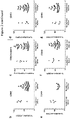
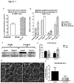
- the known selectivity profile of the six active compounds was examined to identify common kinase targets.
- the pIC50 values generated from the foci assays were compared to the compound inhibition profiles against 224 kinase targets ( Drewry, D.H. et al. (2014). Current topics in medicinal chemistry, 14: 340-342 ).
- a partial least squares (PLS) model was used to cluster the data which suggested that the common target was likely to be a member of the CMGC (cyclin-dependent kinases (CDKs), mitogen-activated protein kinases (MAP kinases), glycogen synthase kinases (GSKs) and CDK-like kinases) family ( Fig. 2A ).
- CDKs cyclin-dependent kinases
- MAP kinases mitogen-activated protein kinases
- GSKs glycogen synthase kinases
- CDK-like kinases CDK-like kinases
- CDK12 is a transcription-regulating kinase associated with nuclear foci in DM cells
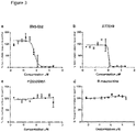
- DM1 fibroblast cells were exposed to the two most potent foci reducing compounds, dinaciclib and SNS-032, for different lengths of time from 2 hours to 48 hours. Both compounds produced a significant reduction in foci but this was most rapid in the case of SNS-032, which was effective following just 2 hours of treatment. Continuous exposure to transcription-regulating inhibitors, would not be a viable therapy option for DM, thus the effect of short term treatment on nuclear foci was examined.
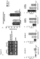
- DM1 fibroblasts were exposed to SNS-032 for 2 hours, after which time the cells were washed thoroughly and allowed to recover in complete growth media. Quantification of nuclear foci showed that 68% of untreated DM1 cells have more than 5 foci and only 5% have no detectable foci ( Fig. 6C ). When cells are treated with SNS-032 for 2 hours, with no recovery time, this distribution shifts to 28% of cells with more than 5 foci and 10% cells with no foci ( Fig. 6D ). However, following 48 and 72 hours of recovery following exposure to SNS-032, the proportion of cells without nuclear foci increases further to 14% and 36%, respectively ( Fig. 6E and 6F ).
- the kinase domain of CDK12 is from amino acid position, 727-1020 (underlined).
- CDK12 has an additional C terminal domain extension that extends around the N and C terminal lobes and contacts bound ATP (underlined and italics). This is unique to CDK12 and is not present in CDK9.
- the contact residues with ATP are Thr737, Lys756, Glu814, Met816 and Asp819 (highlighted by bold type)
Landscapes
- Health & Medical Sciences (AREA)
- Life Sciences & Earth Sciences (AREA)
- Chemical & Material Sciences (AREA)
- Medicinal Chemistry (AREA)
- Pharmacology & Pharmacy (AREA)
- Animal Behavior & Ethology (AREA)
- General Health & Medical Sciences (AREA)
- Public Health (AREA)
- Veterinary Medicine (AREA)
- Epidemiology (AREA)
- Physical Education & Sports Medicine (AREA)
- Engineering & Computer Science (AREA)
- Bioinformatics & Cheminformatics (AREA)
- Neurology (AREA)
- Orthopedic Medicine & Surgery (AREA)
- Chemical Kinetics & Catalysis (AREA)
- General Chemical & Material Sciences (AREA)
- Nuclear Medicine, Radiotherapy & Molecular Imaging (AREA)
- Organic Chemistry (AREA)
- Molecular Biology (AREA)
- Pharmaceuticals Containing Other Organic And Inorganic Compounds (AREA)
- Medicines That Contain Protein Lipid Enzymes And Other Medicines (AREA)
- Measuring Or Testing Involving Enzymes Or Micro-Organisms (AREA)
Description
- The present invention relates to inhibitors of cyclin-dependent kinase 12 (CDK12) and in particular, inhibitors of CDK12 for use in the treatment of disorders caused by the generation of RNA repeat expansion transcripts. The RNA repeat expansion transcript may be from a CTG DNA repeat expansion such as seen in Myotonic Dystrophy.
- Myotonic Dystrophy, also referred to herein as DM, is the most common form of adult muscular dystrophy. DM type 1 (DM1) affects 1 in 8,000 people and is caused by a CTG repeat sequence in the 3' untranslated region of the DMPK (dystrophia myotonica protein kinase) gene, which is greatly expanded in patients who may have anything from 50 repeats to several thousand on affected chromosomes compared to between 5 and 37 repeats on wild-type chromosomes. The expanded DNA repeat is transcribed, and despite being correctly spliced the RNA repeat expansion transcripts remain sequestered in the nucleus forming distinct foci. These foci interact with cellular proteins, such as MBNL1 (muscleblind-like splicing regulator 1), a key splicing regulator, which in turn leads to downstream splicing abnormalities. In addition to the sequestration of proteins the mutant RNA causes activation of CELF1 (CUGBP, Elav-like family member 1), which is also implicated in splicing. Additional molecular pathways are thought to be affected by the toxic RNA, including inhibition of translation.
- DM is an inherited and progressive autosomal dominant multisystem disorder and symptoms can be highly variable. Typical features include myotonia, muscle weakness, cardiac arrhythmias, cognitive dysfunction, diabetes and cataracts. There is currently no treatment for DM and clinical management relies on a fragmented approach utilising already marketed drugs to treat specific symptoms of the disorder, such as mexiletine to treat myotonia and modafinal to address daytime sleepiness. However, due to the complex and variable nature of the disorder, treatment of individual symptoms is not an efficient way to manage the condition.
- Key components of the DM molecular pathway have been identified but how these factors interact is still unclear. Recently there has been considerable effort towards therapeutic treatments for this condition including targeting different points of the molecular pathway with varying levels of success. The most obvious approach is to directly target the repeat expansion transcript to neutralize the harmful repeats or promote transcript degradation and subsequent clearance from the cell. To date this has been attempted using either ribozymes or antisense oligonucleotides (Langlois, M.A. et al. (2003). Molecular therapy: the journal of the American Society of Gene Therapy, 7: 670-680; Wheeler, T.M. et al. Nature, 488: 111-115; and Mulders, S.A. et al. (2009). Proc Natl Acad Sci USA, 106: 13915-13920). Other methods to target the repeat sequence directly have involved the introduction of a blocking molecule, such as morpholino oligonucleotides or small molecules that physically prevent binding of MBNL protein by sitting in the groove of the RNA and preventing protein association and binding (Wheeler, T.M. et al. (2009). Science, 325: 336-339). A series of compounds have been shown to successfully disrupt the CUG repeat:MBNL protein interaction including pentamidine, a bisamidinium inhibitor, a series of peptide ligands and two natural products, lomofugin and dilomofungin. Treatment of DM1-model-CUG-repeat cells with these compounds led to a loss of nuclear foci and a reversal of DM associated splicing events, consistent with release of MBNL protein from this complex. These compounds show that disruption of the RNA:protein interaction may be an option for therapeutic development. However these compounds do not represent suitable starting points for drug development due to high levels of toxicity, poor oral availability and, in the case of peptide ligands, instability in serum.
US2002119963A1 discloses imidazole derivatives indicated to have activity inhibiting cdk5, cdk2, and GSK-3.US2002119963A1 also discloses that the imidazole derivatives may be used to treat a variety of diseases. - To date there is no suitable treatment for DM.
- An object of the present invention is to provide an alternative, preferably an improved, treatment and compound useful for treating disorder caused by the generation of repeat expansion transcripts, such as CTG repeat expansion transcripts as seen in DM-1, with an aim to address at least one of the aforementioned disadvantages.
- According to a first aspect of the invention, there is provided an inhibitor for use in the treatment or prevention of a disorder in a subject caused by the generation of repeat expansion transcripts, wherein the disorder is Myotonic Dystrophy
type 1 orMyotonic Dystrophy type 2, and wherein the inhibitor is an inhibitor of CDK12 (cyclin-dependent kinase 12) comprising - (a) a compound of Formula (I) or a pharmaceutically acceptable salt or solvate thereof:

- R1 is H, -OH, C1-6alkyl, C2-6alkenyl, C2-6alkynl, C1-6haloalkyl, halogen, -CN, -OC1-6alkyl;
- R2 is H, -OH, C1-6alkyl, C2-6alkenyl, C2-6alkynl, C1-6haloalkyl, halogen, -CN, -OC1-6alkyl or a five or 6 membered cycloaryl, cycloalkyl or heterocycl having one, two or three heteroatoms selected from O, S and N, for example benzene, morpholinyl, piperidine, piperazine;
- R3 is C3-6cycloalkyl, for example cyclopropyl,

- R4 is H, -OH, C1-6alkyl, C2-6alkenyl, C2-6alkynl, C1-6haloalkyl, halogen, -CN, -OC1-6alkyl;
- R5 is H; -OH; C1-6alkyl; C2-6alkenyl; C2-6alkynl; C1-6haloalkyl; halogen; -CN; -OC1-6alkyl; C1-6alkyl-N-(X)(Y); a five or six membered cycloaryl, cycloalkyl or heterocycl having one, two or three heteroatoms selected from O, S and N and said cycloaryl, cycloalkyl or heterocycle being optionally substituted with a C1-3alkyl, for example N-methylpiperazinyl; or -OC1-6alkyl-N(X)(Y);
- wherein X is H or C1-6alkyl, and Y is H or C1-6alkyl;
- and wherein the alkyl groups are optionally substituted by one or more -OH groups; and
- R6 is H, -OH, C1-6alkyl, C2-6alkenyl, C2-6alkynl, C1-6haloalkyl, halogen, -CN, -OC1-6alkyl; or
- (b) a compound of Formula (X) or a pharmaceutically acceptable salt or solvate thereof:

- (c) a compound of Formula (XI) or a pharmaceutically acceptable salt or solvate thereof:

- (d) a compound of Formula (XII) or a pharmaceutically acceptable salt or solvate thereof:

- (e) a compound of Formula (XIII) or a pharmaceutically acceptable salt or solvate thereof:

- (f) a compound of Formula (XIV) or a pharmaceutically acceptable salt or solvate thereof:

- The repeat expansion transcript may result in the transcript being retained in the nucleus. The term "retained in the nucleus" may refer to no detectable repeat expansion transcript leaving the nucleus. The term may refer to a delay in the transcript leaving the nucleus (i.e. a transient retention in the nucleus).
- In one embodiment, the repeat expansion transcript may comprise RNA from a CTG repeat (i.e. the RNA may comprise a CUG repeat sequence). In another embodiment, the repeat expansion transcript may comprise RNA from a CCTG repeat (i.e. the RNA may comprise a CCUG repeat sequence). In another embodiment, the repeat expansion transcript may comprise RNA from a CGG repeat. In another embodiment, the repeat expansion transcript may comprise RNA from a GGGGCC repeat. In another embodiment, the repeat expansion transcript may comprise RNA from a ATTCT repeat (i.e. the RNA may comprise a AUUCU repeat sequence). In another embodiment, the repeat expansion transcript may comprise RNA from a CAG repeat. In another embodiment, the repeat expansion transcript may comprise RNA from TGGAA repeat (i.e. the RNA may comprise a UGGAA repeat sequence).
- CDK12 is a transcription elongation associated C-terminal repeat domain kinase, which has shown to associate with elongating transcripts, rather than being involved in the initiation of transcription. The invention herein has found that inhibition of CDK12 results in the removal of nuclear foci from DM cells. Further advantageously, as CDK12 is not required at the start of transcription and its inhibition does not result in global transcriptional arrest, it is suitable as a target for long term DM treatment, and other disorders caused by the generation of repeat expansion transcripts, such as transcripts from CTG repeat expansions.
- The inhibitor of CDK12 according to the present invention is defined in the appended claims. CDK12 inhibitors described in the following paragraphs which are different from the CDK12 inhibitors of the claims are not part of the invention.
- The term "inhibit" or "inhibition" used in the context of CDK12 herein is understood to mean a reduction or complete elimination of CDK12 activity. The reduction in activity of CDK12 may be 100%. Alternatively, the reduction in activity of CDK12 may be at least 90%. The reduction in activity of CDK12 may be at least 80%. The reduction in activity of CDK12 may be at least 70%. The reduction in activity of CDK12 may be at least 60%. The CDK12 inhibition may be measured by an assay measuring any inhibition of CDK12 consumption of ATP during the phosphorylation of a substrate peptide in the presence of the molecule to be screened.
- The CDK12 inhibition may involve blocking the CDK12 active site directly or indirectly; changing the conformation of CDK12; blocking CDK12 interactions; preventing cyclin k binding; preventing phosphorylation of Ser2 on the C-terminal domain of RNA polymerase II; reducing the presence of CDK12; or sequestering the CDK12, for example through aggregation.
- The inhibition of CDK12 activity may be by reduction in the presence of CDK12 (i.e. the activity of CDK12 itself may not be inhibited, but the amount of active CDK12 available in the cells and tissue may be reduced). Therefore, the inhibition of CDK12 may be provided by reducing the expression of CDK12. Alternatively, CDK12 may be mutated to an inactive form. Alternatively, CDK12 may be targeted for degradation or sequestration to reduce the amount of active CDK12 available in the cells or tissue. The reduction in amount of CDK12 may be 100%. Alternatively, the reduction in amount of CDK12 may be at least 90%. The reduction in amount of CDK12 may be at least 80%. The reduction in amount of CDK12 may be at least 70%. The reduction in amount of CDK12 may be at least 60%. The inhibition of CDK12 activity by reduction in the presence of CDK12 may be measured by RT-PCR of CDK12 transcripts in a sample, or by western blot. The skilled person would understand that there are several methods that may be used to determine the presence and level of any particular protein, or transcripts thereof, in a sample.
- The term "inhibitor" used herein is understood to include an agent, such as a molecule, that it capable of causing the inhibition of CDK12 activity. Additionally, or alternatively, the term "inhibitor" used herein is understood to include an agent, such as a molecule, that it capable of causing the inhibition of CDK12 availability.
- The inhibitor may be specific for CDK12. For example, the inhibitor may not inhibit, or not substantially inhibit other cyclin-dependent kinases. Alternatively, the activity of one or more other cyclin-dependent kinases may be inhibited by the inhibitor, but similar to or less than the activity of CDK12. Alternatively, the activity of one or more other cyclin-dependent kinases may be inhibited by the inhibitor, but significantly less than the activity of CDK12. The inhibitor may not be an inhibitor of CDK9 activity or availability. The other CDKs that may not be inhibited, or not significantly inhibited, may be selected from the group consisting of CDK1, CDK2, CDK3, CDK4, CDK5, CDK6, CDK7, CDK8, CDK9, CDK10, CDK11, and CDK13; or combinations thereof. The other CDKs that may not be inhibited, or not significantly inhibited, may be selected from the group comprising CDK1, CDK2, CDK5, and CDK9; or combinations thereof. Preferably CDK9 is not inhibited, or is not significantly inhibited by an inhibitor of the invention.
- The inhibitor may comprise an inhibitor of CDK12 expression. The inhibitor may comprise an oligonucleotide, such as siRNA, capable of inhibiting CDK12 expression. The oligonucleotide may comprise a sequence capable of binding to nucleic acid of the CDK12 gene, or regulatory elements thereof, or a mRNA transcript thereof. The oligonucleotide may comprise a sequence substantially complementary to the CDK12 gene, or regulatory elements thereof. The oligonucleotide may comprise a sequence substantially complementary to CDK12 mRNA transcript. The sequence may be substantially complementary over a region of at least 5 nucleotides. The sequence may be substantially complementary over a region of at least 8 nucleotides. The sequence may be substantially complementary over a region of at least 10, 13, 15, or 18 nucleotides. The oligonucleotide may comprise a sequence capable of binding to SEQ ID NO: 1, and reducing translation thereof, e.g. siRNA gene silencing.
- The inhibitor may comprise a molecule capable of binding to CDK12. The inhibitor molecule may be capable of blocking binding of CDK12 to its target molecule. The inhibitor may comprise a molecule capable of preventing CDK12 binding to cyclin K.
- The inhibitor may comprise a molecule capable of preventing CDK12 phosphorylating Ser2 on the c-terminal domain of RNA polymerase II. The binding of the inhibitor to CDK12 may be at, or adjacent to, the CDK12 active site, such that the active site is blocked. The binding of the inhibitor to CDK12 may be at amino acid position, 727-1020 (underlined in the sequence below). The binding of the inhibitor to CDK12 may be at a C terminal domain extension that extends around the N and C terminal lobes and contacts bound ATP (C terminal domain extension comprises residues 1011-1039 of CDK12). Such a domain is unique to CDK12 and is not present in CDK9. The binding of the inhibitor to CDK12 may be at any one or more of the ATP contact residues selected from Thr737, Lys756, Glu814, Met816 and Asp819 (in bold-type on the sequence below).
- CDK12 binds to cyclin K and phosphorylates Ser2 on the C-terminal domain of RNA polymerase II. Advantageously, the inhibitor treatment will result in a shift in the relative proportions of wild type and mutant DMPK transcripts (CDK12 inhibition leads to a preferential loss of the expanded repeat transcript).
- The inhibitor may comprise a therapeutically active agent. The inhibitor may comprise a small molecule. The term "small molecule" means any compound that has a molecular weight of less than 1kDa. The inhibitor may be an organic compound with a molecular weight of less than 1KDa. Molecular weight is understood to be the sum of the atomic weights of all the atoms in a molecule.
- The inhibitor may comprise nucleic acid such as an oligonucleotide. The oligonucleotide may comprise DNA, RNA, or synthetic nucleic analogues such as PMO, LNA or PNA. The oligonucleotide may comprise siRNA or microRNA. The oligonucleotide may comprise the sequence of CGAAAUAAUGAUGUUGGCACCAGUU, or a variant thereof, for siRNA silencing of CDK12.
- The inhibitor may comprise a peptide or protein capable of binding to CDK12. The inhibitor may comprise an antibody, or an antibody fragment, or antibody mimetic.
- The inhibitor may be cell membrane permeable.
- The inhibitor may comprise a pyrazolo[1,5b]pyridazine core structure, and be capable of inhibiting CDK12 activity.
- The inhibitor may comprise a compound of Formula (I) or a pharmaceutically acceptable salt or solvate thereof:

- R1 is H, -OH, C1-6alkyl, C2-6alkenyl, C2-6alkynl, C1-6haloalkyl, halogen, -CN, -OC1-6alkyl;
- R2 is H, -OH, C1-6alkyl, C2-6alkenyl, C2-6alkynl, C1-6haloalkyl, halogen, -CN, -OC1-6alkyl or a five or 6 membered cycloaryl, cycloalkyl or heterocycl having one, two or three heteroatoms selected from O, S and N, for example benzene, morpholinyl, piperidine, piperazine;
- R3 is C3-6cycloalkyl, for example cyclopropyl,

- R4 is H, -OH, C1-6alkyl, C2-6alkenyl, C2-6alkynl, C1-6haloalkyl, halogen, -CN, -OC1-6alkyl;
- R5 is H; -OH; C1-6alkyl; C2-6alkenyl; C2-6alkynl; C1-6haloalkyl: halogen; -CN; -OC1-6alkyl; C1-6alkyl-N-(X)(Y); a five or six membered cycloaryl, cycloalkyl or heterocycl having one, two or three heteroatoms selected from O, S and N and said cycloaryl, cycloalkyl or heterocycle being optionally substituted with a C1-3alkyl, for example N-methylpiperazinyl; or - OC1-6alkyl-N(X)(Y);
- wherein X is H or C1-6alkyl, and Y is H or C1-6alkyl;
- and wherein the alkyl groups are optionally substituted by one or more -OH groups; and
- R6 is H, -OH, C1-6alkyl, C2-6alkenyl, C2-6alkynl, C1-6haloalkyl, halogen, -CN, -OC1-6alkyl.
- Preferably:
- R1 is H;
- R2 is H;
- R3 is C3-6cycloalkyl, for example cyclopropyl,

- R4 is H, -CN, -OC1-6alkyl, C1-6haloalkyl;
- R5 is H, ; C1-6alkyl-N-(X)(Y); a five or six membered cycloaryl, cycloalkyl or heterocycl having one, two or three heteroatoms selected from O, S and N and said cycloaryl, cycloalkyl or heterocycle being optionally substituted with a C1-3alkyl, for example N-methylpiperazinyl, -OC1-6alkyl-N(X)(Y);
- wherein X is H or C1-6alkyl, and Y is H or C1-6alkyl;
- and wherein the alkyl groups are optionally substituted by one or more -OH groups; and
- R6 is H or -OC1-6alkyl.
- Preferably:
- R1 is H;
- R2 is H;
- R3 is cyclopropyl,

- R4 is H, -CN, -OCH3, CF3;
- R5 is H, -CH2N(CH3)2 N-methylpiperazinyl,-OCH2CH(OH)CH2N(CH3)2; and
- R6 is H or -OCH3.
- Preferably R1 is H, R2 is H, R3 is cyclopropyl,

- R4 is H; R5 is -CH2NEt2, or -OCH2CH(OH)CH2N(CH3)2; and R6 is H; or
- R4 is -CN, -OCH3; R5 is H; and R6 is H; or
- R4 is -CF3; R5 is N-methylpiperazinyl; and R6 is H; or
- R4 is -OCH3; R5 is H; and R6 is -OCH3.
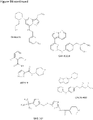
- In one embodiment, the inhibitor of Formula (I) is of the following formula:


- In another embodiment, the inhibitor of Formula (I) is of the following formula:

- In another embodiment, the inhibitor of Formula (I) is of the following formula:

- In another embodiment, the inhibitor of Formula (I) is of the following formula:

- In one embodiment the inhibitor of Formula (I) is of the following formula:


- In one embodiment the inhibitor of Formula (I) is of the following formula:

- In one embodiment the inhibitor of Formula (I) is of the following formula:

- In one embodiment the inhibitor of Formula (I) is of the following formula:

- The inhibitor may comprise dinaciclib. For example, in one embodiment the inhibitor comprises a compound of Formula (X) or a pharmaceutically acceptable salt or solvate thereof:

- In one embodiment, the inhibitor comprises a compound of Formula (XI) or a pharmaceutically acceptable salt or solvate thereof:

- In one embodiment, the inhibitor comprises a compound of Formula (XII) or a pharmaceutically acceptable salt or solvate thereof:

- In one embodiment, the inhibitor comprises a compound of Formula (XIII) or a pharmaceutically acceptable salt or solvate thereof:


- In one embodiment, the inhibitor comprises a compound of Formula (XIV) or a pharmaceutically acceptable salt or solvate thereof:

- As used herein, the term "C1-C3 alkyl" refers to a straight-chain or branched-chain alkyl group containing from 1 to 3 carbon atoms. Examples of alkyl groups include methyl, ethyl, propyl or isopropyl.
- As used herein, the term "6 member heterocyclyl" refers to a saturated monocyclic heterocyclic group having 6 ring members and containing at least one heteroatom as a ring member, wherein each of said at least one heteroatoms may be independently selected from the group consisting of nitrogen, oxygen, and sulfur. One group of heterocyclyls has 1 heteroatom as ring member. Another group of heterocyclyls has 2 heteroatoms as ring members. Examples of heterocyclyl groups include piperazinyl, morpholinyl, piperidinyl, oxanyl, thianyl or dioxanyl.
- As used herein, the term "haloalkyl" refers to an alkyl group having the meaning as defined above wherein one or more hydrogens are replaced with a halogen. A halo C1-3 alkyl group refers to a haloalkyl group wherein the alkyl moiety has from 1 to 3 C atoms. Specifically embraced are monohaloalkyl, dihaloalkyl or polyhaloalkyl groups. A monohaloalkyl group, for one example, may have an iodo, bromo, chloro or fluoro atom within the group. Dihalo or polyhaloalkyl groups may have two or more of the same halo atoms or a combination of different halo groups. Examples of haloalkyl groups include fluoromethyl, difluoromethyl, trifluoromethyl, chloromethyl, dichloromethyl, trichloromethyl, pentafluoroethyl, heptafluoropropyl, difluorochloromethyl, dichlorofluoromethyl, difluoroethyl, difluoropropyl, dichloroethyl or dichloropropyl.
- Additionally, the compounds of Formulas I-XVI can contain asymmetric carbon atoms (chiral atoms) and can therefore exist in racemic and optically active forms. Thus, optical isomers or enantiomers, racemates, tautomers, and diastereomers are also encompassed in the compounds of Formulas I-XVI.
- The inhibitor may be administered, or arranged to be administered, in combination with at least one other therapeutic agent. The one other therapeutic agent may comprise an oligonucleotide, such as siRNA or miRNA or equivalents thereof. The oligonucleotide may be capable of targeting expanded repeat transcript for degradation, for example through the RNA interference pathway. The oligonucleotide may comprise the sequence of (CAG)7. The oligonucleotide may comprise the sequence of AGC AGC AGC AGC AG. The oligonucleotide may comprise the sequence of CAG CAG CAG CAG CAG C. The oligonucleotide may comprise the sequence of CGGAGCGGTTGTGAACTGGC.
- The oligonucleotide may target the repeat sequence such as CUGn, CCUGn, CGGn, GGGGCCn, CAGn, or AUUCUn. Alternatively the siRNA oligonucleotide may bind anywhere in a target RNA which includes an expanded repeat transcript such as CUGn, CCUGn, CGGn, GGGGCCn, CAGn, AUUCUn. The skilled person will understand that U (uridine) residues will substitute T (thymine) residues in RNA relative to DNA. The siRNA oligonucleotide may bind anywhere in a target RNA which includes an expanded repeat transcript such as transcripts from any one of the transcripts from GenBank accession numbers: NM_001081563, AY329622, AF389886, AF389887, AH010982, L19493, JN681271, AF126749, NM_013236, NM_001271604, NM_002111, NM_000332, NM_002973, NM_004993, NM_000068, NM_000333, CR456776, D31840, or M34233.
- The oligonucleotide may be at least 10, 11, 12, 13, 14, or 15 nucleotides long. The oligonucleotide may be at least 15 nucleotides long.
- Advantageously, continuous exposure to inhibitor may be unnecessary to produce a beneficial effect in disorders caused by the generation of repeat expansion transcripts, such as DM-1. A pulsatile treatment may be sufficient. The inhibitor would remove nuclear foci from afflicted cells after a short exposure, and this short exposure leads to a prolonged effect, wherein the inhibitor treatment results in a shift in the relative proportions of wild type and mutant DMPK transcripts (CDK12 inhibition leads to a preferential loss of the expanded repeat transcript). This may result from CDK12 removal from the repeat expansion transcript with subsequent dissociation of the nuclear foci and degradation of the mutant transcript. The nuclear foci protect repeat expansion transcripts from degradation and once released, they may be vulnerable to cellular or targeted degradation. Thus, a "two-hit" therapy regime may be provided in which a short treatment with a CDK12 inhibitor is used to disperse foci and expose repeat expansion transcripts to degradation via endogenous processes, or by, for example, antisense oligonucleotides.
- The at least one other therapeutic agent may comprise a small molecule, drug, prodrug, peptide, protein, antibody, nucleotide or vaccine. The at least one other therapeutic agent may comprise a sodium channel blocker, such as mexiletine, phenytoin or procainamide. The at least one other therapeutic agent may comprise a CNS stimulant drug, such as modafinil, for example for treating excessive daytime sleepiness. The at least one other therapeutic agent may comprise dehydroepiandrosterone (DHEA), creatine supplementation or mecasermin rinfabate (IPLEX, combination of recombinant insulin-
like growth factor 1 and its binding protein, BP-3), which may be provided, for example, for improving muscle weakness. The at least one other therapeutic agent may comprise any one or more of pentamidine, a bisamidinium inhibitor, lomofugin or dilomofungin. - A CDK inhibitor may be used in combination with an oligonucleotide that targets the DMPK gene. The DMPK gene may be in the expanded repeat in the 3' untranslated region or it may be targeted anywhere in the gene.
- The use in combination with at least one other therapeutic agent may comprise a combined dose formulation or a separate dose formulation. The use may be concurrent or sequential.
- The inhibitor may be administered, or arranged to be administered, intermittently. For example, the inhibitor may be administered, or arranged to be administered, once in a three-day treatment cycle. Alternatively, the inhibitor may be administered, or arranged to be administered, once in a 2-day treatment cycle. Alternatively, the inhibitor may be administered, or arranged to be administered, once in a 4-day treatment cycle. Alternatively, the inhibitor may be administered, or arranged to be administered, once in a treatment cycle of at least 3 days. Alternatively, the inhibitor may be administered, or arranged to be administered, once in a treatment cycle of at least 5 days. The inhibitor may be administered, or arranged to be administered, on day one of a three-day treatment cycle and wherein the dose is released within a 2-hour period immediately following administration.
- The inhibitor may be provided and/or administered in a therapeutically effective dose. The dose may be sufficient to cause an alleviation or prevention of one or more symptoms of the disorder caused by the generation of repeat expansion transcripts, such as DM-1. The dose may be sufficient to cause an alleviation or prevention of all symptoms of the disorder caused by the generation of repeat expansion transcripts, such as DM-1. The dose may be sufficient to reduce the activity or presence of CDK12 by at least 50%. The dose may be sufficient to reduce the nuclear retention of repeat expansion transcripts by at least 50%. The dose may be sufficient to improve myotonia and muscle strength as judged by a six-minute walk test and hand grip test.
- The dose may comprise between about 2 and about 100 mg/kg. The dose may comprise between about 5 and about 80 mg/kg. The dose may comprise between about 2 and about 60 mg/kg. The dose may comprise between about 5 and about 60 mg/kg. The dose may comprise between about 5 and about 30 mg/kg. The dose may comprise between about 5 and about 20 mg/kg. The dose may comprise between about 5 and about 10 mg/kg. The dose may comprise no more than 60 mg/kg in a period of 24 hours. The dose may comprise no more than 30 mg/kg in a period of 24 hours. The dose may comprise no more than 10 mg/kg in a period of 24 hours. The dose may comprise no more than 5 mg/kg in a period of 24 hours. The dose may comprise no more than 100 mg/kg in a period of 3 days. The dose may comprise no more than 60 mg/kg in a period of 3 days. The dose may comprise no more than 30 mg/kg in a period of 3 days. The dose may comprise no more than 10 mg/kg in a period of 3 days. The dose may comprise no more than 5 mg/kg in a period of 3 days. The dose may comprise no more than 100 mg/kg in a period of 1 week. The dose may comprise no more than 60 mg/kg in a period of 1 week. The dose may comprise no more than 30 mg/kg in a period of 1 week. The dose may comprise no more than 20 mg/kg in a period of 1 week. The dose may comprise no more than 10 mg/kg in a period of 1 week. The dose may comprise no more than 100 mg/kg in a period of 2 or 4 weeks. The dose may comprise no more than 60 mg/kg in a period of 2 or 4 weeks. The dose may comprise no more than 30 mg/kg in a period of 2 or 4 weeks. The dose may comprise no more than 10 mg/kg in a period of 2 or 4 weeks. No more than 4 doses may be administered, or arranged to be administered, in a period of 24 hours. No more than 3 doses may be administered, or arranged to be administered, in a period of 24 hours. No more than 2 doses may be administered, or arranged to be administered, in a period of 24 hours. No more than 1 dose may be administered, or arranged to be administered, in a period of 24 hours. No more than 4 doses may be administered, or arranged to be administered, in a period of 3 days. No more than 3 doses may be administered, or arranged to be administered, in a period of 3 days. No more than 2 doses may be administered, or arranged to be administered, in a period of 3 days. No more than 1 dose may be administered, or arranged to be administered, in a period of 3 days. No more than 4 doses may be administered, or arranged to be administered, in a period of 1 week. No more than 3 doses may be administered, or arranged to be administered, in a period of 1 week. No more than 2 doses may be administered, or arranged to be administered, in a period of 1 week. No more than 1 dose may be administered, or arranged to be administered, in a period of 1 week. No more than 20 doses may be administered, or arranged to be administered, in a period of 2 or 4 weeks. No more than 15 doses may be administered, or arranged to be administered, in a period of 2 or 4 weeks. No more than 10 doses may be administered, or arranged to be administered, in a period of 2 or 4 weeks. No more than 8 doses may be administered, or arranged to be administered, in a period of 2 or 4 weeks. No more than 5 doses may be administered, or arranged to be administered, in a period of 2 or 4 weeks. No more than 4 doses may be administered, or arranged to be administered, in a period of 2 or 4 weeks. No more than 3 doses may be administered, or arranged to be administered, in a period of 2 or 4 weeks. No more than 2 doses may be administered, or arranged to be administered, in a period of 2 or 4 weeks. No more than 1 dose may be administered, or arranged to be administered, in a period of 2 or 4 weeks.
- Advantageously, continuous exposure to the inhibitor may not be necessary to produce a beneficial effect in the treated subject and that a pulsatile treatment may be sufficient. This method of administration may have the advantage of reducing side effects as it would allow a lower duration/frequency of inhibitor application.
- The inhibitor may be administered orally or parenterally, for example, by intravenous, intramuscular or subcutaneous injection. In one embodiment, the inhibitor is suitable for oral administration.
- The subject may be a mammal. The subject may be human.
- The disclosure also relates to the use of a CDK12 inhibitor in the preparation of a medicament for the treatment or prevention of a disorder caused by the generation of repeat expansion transcripts in a subject, for example RNA transcripts from a CTG repeat expansion (not part of the invention).
- The disclosure also relates to a method of treatment or prevention of a disorder caused by the generation of repeat expansion transcripts in a subject, comprising administering an inhibitor of CDK12 to the subject (not part of the invention). The RNA repeat expansion transcripts may be from CTG repeat expansions.
- The method may further comprise a subsequent administration of at least one other therapeutic agent. The at least one other therapeutic agent may comprise an oligonucleotide. The at least one other therapeutic agent may be suitable for enhancing degradation of nucleic acid repeat transcripts of DMPK.
- The disclosure also relates to a method of screening for a therapeutic agent for a disorder caused by the generation of repeat expansion transcripts, such as RNA repeat expansion transcripts as in DM, comprising:
- providing a molecule for screening;
- detecting if the molecule inhibits the activity of CDK12,
- Detecting if the molecule inhibits the activity of CDK12 may comprise the use of an assay measuring any inhibition of CDK12 consumption of ATP during the phosphorylation of a substrate peptide in the presence of the molecule to be screened.
- The inhibition of CDK12 may be selective inhibition. The method of screening may further comprise the step of detecting if the molecule does not significantly inhibit other cyclin dependent kinases, such as CDK9.
- The disclosure also relates to a composition comprising a CDK12 inhibitor and a pharmaceutically acceptable carrier (not part of the invention).
- The composition may further comprise at least one other therapeutic agent. The least one other therapeutic agent may be capable of enhancing repeat expansion transcript degradation through cellular or targeted degradation. The at least one other therapeutic agent may comprise any one or more therapeutic agent described herein.
- The disclosure also relates to a CDK12 inhibitor as described herein (not part of the invention).
- There now follows by way of example only a detailed description of the present invention with reference to the following drawings.
-
Figure 1 : Screening the PKIS compound collection identified 6 inhibitors that reduce nuclear foci. (A-F) Six inhibitors cause reduced nuclear foci across an 11 point dilution. Graphs show percentage of nuclear foci relative to DMSO treated cells. All six compounds share a pyrazolo[1,5b]pyridazine core structure. -
Figure 2 : Analysis of the PKIS inhibition profiles to identify common kinase targets. (A) Loading plot of 2-compound partial least squares model. Kinase activities correlating with the nuclear foci assay are labelled, with cyclin-dependent kinases highlighted with ∗. (B-G) Plots show the percentage inhibition of the kinase target at 0.1µM concentration of compounds, grouped according to activity in the nuclear foci assay, including the six active compounds. Compounds with less than 25% inhibitory active activity on the kinase are not included on the graphs. -
Figure 3 : Focussed screening of CDK family inhibitors. 12 point dilution graphs plotting percentage of nuclear foci relative to DMSO treated cells for (A) SNS-032 (B) AT7519 (C) PD0332991 (reference compound) (D) R-roscovitine (reference compound) (E) dinaciclib (F) CDK9 inhibitor II (reference compound). -
Figure 4 : Chemoproteomics target deconvolution. IC50 values were generated by affinity capturing of kinases from K562 or A204 cell extract using beads derivatized with SNS-032, in the presence of different concentrations of free competing compound or vehicle (DMSO). pIC50 values are plotted against the pIC50 in the foci inhibition assay for each compound. A good correlation of kinase binding affinity with the inhibitory activity on foci is observed for CDK5, CDK7, CDK9, and CDK12. r: Pearson correlation coefficient; p: p-value (calculated probability). -
Figure 5 : CDK9 and CDK12 protein expression in DM. Western blot of vastus lateralis muscle biopsy samples in non-DM and DM1 patients for (A) CDK9 and (B) CDK12. Both blots are normalised to α-tubulin. (C) Histogram to quantify levels of CDK9 protein normalised to α-tubulin. (D) Histogram to quantify levels of CDK12 protein normalised to α-tubulin. (E) Immunohistochemistry of CDK12 in non-DM and DM1 fibroblast cells. (F) Quantification of the number of CDK12 nuclear granules. (G) Immunohistochemistry and in situ hybridisation shows co-localisation of repeat expansion foci with CDK12 nuclear granules (CDK12 in green/bright spots, repeat expansion RNA (Arrow)) (H) CDK12 protein knockdown by shRNA results in reduced CDK12 protein granules (green) and a subsequent reduction in CUG repeat expansion RNA foci (red/Arrow). -
Figure 6 : Inhibitor treatment as a therapeutic for DM. (A) Ethidium bromide stained gel showing RT-PCR products from nuclear (N) and cytoplasmic (C) RNA fractions following amplification and BpmI digest of a fragment of DMPK. GAPDH is used as a loading control. (B) Histograms showing the relative proportions of nuclear mutant DMPK transcripts compared to wild type DMPK transcripts. The relative proportions of the mutant and wild type DMPK transcripts in DM1 fibroblast cells were assessed following treatment with dinaciclib (1µM) for 24 hours. (C-F) Histograms show percentage of cells in the population with 0, <2, <5 and 5+ foci per nucleus (C) Untreated DM1 cells. (D) DM1 cells treated with SNS-032 for 2 hours. (E) DM1 cells treated with SNS-032 for 2 hours with 48 hours recovery in growth media (F) DM1 cells treated with SNS-032 for 2 hours with 72 hours recovery in growth media. -
Figure 7 : Inhibitor treatment in a DM1 mouse model (A) Combined myotonia grade scores in quadriceps, gastrocnemius and paraspinal muscles from HSALR mice following vehicle and dinaciclib compound treatment (n=6 per treatment group) (B) Myotonia grades by muscle type in dinaciclib and vehicle treated HSALR mice. (C) Ethidium bromide stained gel to assess ATP2A1 splice isoforms in quadriceps and gastrocnemius muscle samples from vehicle and dinaciclib treated mice (D) Histogram showing the relative proportion of exon 22 exclusion and inclusion in vehicle and dinaciclib treated HSALR mice (E-F) Laminin stain demonstrates a reduction in centralised nuclei in muscle fibres of dinaciclib treated mice compared to vehicle control animals. -
Figure 8 : Screening the PKIS collection identifies the CMGC kinase family. Plots to analyse the PKIS inhibition profiles of kinases identified from the partial least squares model as possible cellular targets comparing active versus inactive compounds. Compounds with less than 25% inhibition are not included on the graphs. -
Figure 9 : Kinobead profiling of 11 compounds. Kinobeads profiling of a set of 11 compounds which represent a range of activities in the nuclear foci assay. Target profiles were generated by adding each compound to K562 cell extract at a concentration of 2 µM followed by incubation with kinobeads and quantification of bead-bound proteins. A: Values indicate target binding compared to a DMSO control where a value of 1 represents 100% binding and therefore no target inhibition and a value of 0 indicates 0% binding and 100% inhibition of the target kinase. B: Shows the structure of each compound tested. -
Figure 10 : Comparison of protein binding profiles for immobilised inhibitors SNS-032 and AT7519. -
Figure 11 : Examples of dose-response competition binding curves for different compound/target combinations. An affinity matrix was generated by immobilisation of SNS-032 to sepharose beads and affinity capturing was performed from K562- or A204 cell extract in the presence of vehicle (DMSO) or different concentrations of inhibitor as indicated. IC50 concentrations and inflection points of the dose-response competition curves are indicated by dotted lines. -
Figure 12 : CDK12 protein knockdown by siRNA and shRNA. (A) CDK12 immunohistochemistry following knockdown with scrambled and CDK12 shRNA (B) Quantification of CDK12 protein granules within the nucleus in CDK12 shRNA cells shows a 56% reduction in CDK12 granule number compared to scrambled shRNA (C) In situ hybridization analysis following knockdown with scrambled and CDK12 shRNA (D) Quantification of CUG repeat RNA foci within the nucleus in CDK12 shRNA cells shows a 69% reduction compared to scrambled shRNA (E) Histogram show percentage of cells in the population with 0, 1-2, 3-4 and 5+ foci per nucleus, in scrambled and CDK12 shRNA treated cells. (F) CDK12 immunohistochemistry following knockdown with scrambled and CDK12 siRNA (G) Quantification of CDK12 granules within the nucleus in CDK12 siRNA cells shows a 47% reduction in CDK12 granule number compared to scrambled siRNA (H) siRNA validation by western blot analysis for CDK12 and α-tubulin (I) Histogram of intensity scan of western blot analysis showed a 34% reduction in CDK12 protein compared to scrambled controls. Data normalized to α-tubulin. (J) In situ hybridization analysis following knockdown with scrambled and CDK12 siRNA (K) Quantification of CUG repeat RNA foci within the nucleus in CDK12 siRNA cells shows a 46% reduction compared to scrambled siRNA (L) Histograms show percentage of cells in the population with 0, 1-2, 3-4 and 5+ foci per nucleus, in scrambled and CDK12 siRNA treated cells. -
Figure 13 : IC50 values of previously reported CDK inhibitors. -
Figure 14 : pIC50 values generated by affinity capturing with the SNS-032 affinity matrix in K562 cell extract for the different CDK inhibitors added to the cell extracts. - Myotonic dystrophy is caused by a CTG repeat expansion within the 3' untranslated region of the DMPK gene, leading to the formation of distinct nuclear foci. The involvement of kinases has been linked to the pathophysiology of the condition but to date a definitive kinase target for drug development has not been identified. It has been observed herein that CDK12 is elevated in DM cell lines and in DM patient muscle biopsies. Repeat expansion transcripts accumulate at the periphery of nuclear speckles and CDK12 co-localises with these nuclear speckles. It has been found that inhibition of CDK12 leads to the dispersal of DM-associated nuclear foci and degradation of repeat expansion transcripts.
- Using a previously reported assay the PKIS collection was screened for compounds that reduce nuclear foci and the compounds were then analysed for their known selectivity profiles to identify the common kinase targets (Ketley, A. et al. (2014). Hum Mol Genet, 23: 1551-1562). DM1 fibroblasts were treated with compounds in an 11 point dilution series from 2011M-19nM for 24 hours. Following treatment, fluorescent in situ hybridisation was performed with a cy3 labelled CAG10 probe, to visualize nuclear foci and cells were analysed on a Molecular Devices plate reader with customised MetaExpress software (Ketley, A. et al. (2014). Hum Mol Genet, 23: 1551-1562). Compounds that reduced nuclear foci in a concentration dependent manner, compared to DMSO treated cells were identified and prioritized for further study. Six compounds that share a pyrazolo[1,5b]pyridazine core were found to reduce the number of nuclear foci following 24 hour treatment of DM cells (
Fig 1 ). - The known selectivity profile of the six active compounds was examined to identify common kinase targets. The pIC50 values generated from the foci assays were compared to the compound inhibition profiles against 224 kinase targets (Drewry, D.H. et al. (2014). Current topics in medicinal chemistry, 14: 340-342). A partial least squares (PLS) model was used to cluster the data which suggested that the common target was likely to be a member of the CMGC (cyclin-dependent kinases (CDKs), mitogen-activated protein kinases (MAP kinases), glycogen synthase kinases (GSKs) and CDK-like kinases) family (
Fig. 2A ). Indeed, these compounds were originally designed to target CDK family members (Stevens, K.L. et al. (2008). Bioorganic & medicinal chemistry letters, 18: 5758-5762) and the inhibitors active in the nuclear foci assay all showed significant activity across many members of the CDK family. The kinase inhibition profiles for the six hit compounds were compared to their activity in the nuclear foci assay, which suggested that CDK1, CDK2, CDK3, CDK5 and CDK6 are unlikely to be responsible for foci formation (Fig. 2B-G ). Of the targets covered by the PKIS collection, CDK4 appeared more likely to play a role (Fig. 2E ) but other CDK family members required consideration as they are absent from the PKIS annotations. - Next, additional small molecule CDK inhibitors with well-annotated selectivity profiles were tested (
Figure 13 ) in the nuclear foci assay. This subset of inhibitors displayed differential activities in the nuclear foci assay and a diverse range of potencies across the CDK family members (Fig 3 ). To confirm involvement of the CDK family the kinobeads methodology was employed, which is based on sepharose beads derivatized with a combination of promiscuous kinase inhibitors (Bantscheff, M. et al. (2007). Nat Biotechnol, 25: 1035-1044; Werner, T. et al. (2012). Analytical chemistry, 84: 7188-7194; and Kruse, U. et al. (2011). Leukemia, 25: 89-100) toprofile 10 compounds, which represent a range of activities in the nuclear foci assay. Target profiles were generated by adding each compound to K562 erythroleukemia cell extract at a concentration of 2 µM, followed by incubation with two variations of kinobeads and quantification of bead-bound proteins. The first type of bead was developed for the profiling of tyrosine and serine/threonine kinases of the eukaryotic protein kinase family (Bantscheff, M. et al. (2007). Nat Biotechnol, 25: 1035-1044), whereas the second type of bead captures additional kinases from the PI3K/lipid kinase family (Bergamini, G. et al. (2012). Nature chemical biology, 8: 576-582). The profiling results clearly indicated that the most active compounds in this subset exhibited shared activities only across the CDK family (Fig. 9A ), consistent with the PKIS data. - To refine the possible target kinases, the IC50 values were compared for the nuclear foci active inhibitors and the nuclear foci inactive inhibitors. Small molecules SNS-032 (also known as BMS 387032) and AT-7519 demonstrate high activity in the nuclear foci assay and display the highest potency against CDK2 and CDK9 (
Fig. 3a and 3b andFigure 13 ). However CDK2 cannot be responsible for nuclear foci reduction due to significant overlap in the IC50 values of active and inactive compounds against this target. Likewise CDK1, CDK4, CDK5, CDK6 and CDK7 can all be discounted for similar reasons (Figure 13 ). - The analysis of this subset of known CDK inhibitor compounds confirms the results of the PKIS screen but raises the possibility that the target responsible for nuclear foci reduction is a less well described CDK family member. To investigate this possibility four additional CDK inhibitors, with a range of potencies, were tested to determine the potential involvement of CDK9 as a possible target (
Fig. 3E-F andFigure 13 ). Interestingly, dinaciclib and SNS-032 have the same IC50 value against CDK9, however, dinaciclib displays a significantly increased activity in the nuclear foci assay. Furthermore, CDK9 Inhibitor II (Fig. 3F , reference compound), which is a specific but less potent CDK9 inhibitor, with an IC50 value of 350nM, reduced nuclear foci at the highest concentration whereas two other compounds; PD0332991 and R-roscovitine (reference compounds), with comparable CDK9 IC50 values (400nM and 500nM, respectively) are inactive in the nuclear foci assay at the concentrations tested (Fig 3C and 3D ). These data suggest an additional CDK family member as the target for nuclear foci reduction. - To determine the specific CDKs responsible for the reduction of nuclear foci it was sought to expand the target coverage within the CDK family by the immobilization of two of the active compounds containing a suitable secondary amine; SNS-032 and AT7519 (
Fig. 10 ). Beads derivatized with either compound showed good coverage of the CDK family, including family members which are not present in commercial kinase panels. For an in depth chemoproteomics study profiling inhibitor selectivity across the CDK family, dose-response competition-binding profiles were generated for all active and one of the inactive compounds (PD0332991) in K562 cells or in A204 rhabdomyosarcoma cells. To confirm that the K562, A204 and DM1 cell lines express the same kinases a whole proteome analysis was conducted. All CDK/PCTK proteins identified by profiling were also identified in the cells used for the phenotypic foci assay as follows:
CDK family member proteins identified by whole proteome analysis of DM fibroblasts (protein accession numbers for CDK family proteins). - CDK1: P06493
- CDK2: CAA43807.1
- CDK4: CAG47043
- CDK5: CAG33322
- CDK6: Q00534
- CDK7: P50613
- CDK9: AAF72183
- CDK10: AAH25301
- CDK12: Q9NYV4.2
- CDK13: Q14004.2
- PCTK1: Q00536
- PCTK2: Q00537.2
- The resulting dataset comprises IC50 curves for 12 CDK family kinases (
Fig. 11 ,Figure 14 ). The best correlation across the compound set of kinase IC50 values with the inhibitory activity on foci was observed for CDK12, followed by CDK9 (Figure 4 ). The IC50 values for CDK9 and CDK12 are closely in line with their observed activity in the foci assay, whereas the IC50 values for CDK2 and CDK7 appear too low to implicate them as targets. Consistent with the previous analysis, presented inFigure 3E , dinaciclib was the most active inhibitor against the CDK family, in particular for CDK12, suggesting it may be the most likely kinase target. - Thus far the experiments point to CDK12 as the most likely kinase to have an association with repeat expansion foci. To examine the potential involvement in DM pathogenesis, the endogenous levels of CDK12 were assessed, in addition to CDK9, in vastus lateralis muscle biopsy samples from four DM1 and four healthy volunteers using Western blots. No significant difference in CDK9 protein level compared to that in controls could be detected (
Fig. 5A and 5C ). However, there was a clear increase in levels of CDK12 in DM biopsies versus those from healthy volunteers, with 48% more CDK12 protein detected in DM samples (p=0.0025) (Fig. 5B and 5D ). - To understand the relationship between nuclear foci and CDK12 in DM pathophysiology, the cellular location of the protein was established by immunohistochemistry in DM and non-DM fibroblasts. Consistent with previously published data in non-DM cells CDK12 was localised in the nucleus in granular structures in both DM and non-DM cells (Ko, T.K. et al. (2001). Journal of cell science, 114: 2591-2603) (
Fig. 5E ). Quantification of these structures in 400 cells showed that the number of granules was elevated in DM cells, with an average number of 18.74 (±3.88), compared to 12.07 (±2.27) in non-DM cells (p<0.0001) (Fig. 5f ). The size of CDK12 granules was not significantly different in non-DM and DM cells. This increase in number is consistent with the increased overall levels of CDK12 protein detected by Western blot. Next immunohistochemistry of CDK12 was conducted followed by in situ hybridisation to detect the repeat expansion RNA foci. Co-localisation of the repeat expansion foci with CDK12 protein was found. Quantification using customised MetaXpress software revealed that 100% of repeat expansion foci co-localised with CDK12 protein (Fig. 5g ). To understand the impact of increased CDK12 protein in DM and the relationship between CDK12 protein granules and repeat expansion foci shRNA and siRNA were used to reduce the number of CDK12 granules in DM cells. The effect on repeat expansion foci was then assessed. Following lentiviral infection expressing three shRNAs against CDK12 a 56% reduction in the number of CDK12 granules and a 69% reduction in repeat expansion foci compared to control cells was observed (Fig. 5H andFig. 12A-E ). This was verified by siRNA knockdown of CDK12 and quantification by Western blot analysis, which confirmed a 34% reduction in protein levels. This resulted in a 46% reduction in CDK12 protein granules and a 47% reduction in repeat expansion RNA foci indicating that specific inhibition of CDK12 and dissolution of CDK12 protein from nuclear granules leads to a dispersal of repeat expansion foci (Fig. 12F-L ). - As an association between CDK12 and nuclear foci has been established, and nuclear foci comprise repeat expansion transcripts, it was sought to establish the effect of inhibitor treatment on the level of repeat expansion transcripts. For this an RT-PCR assay was employed that utilises a Bpml polymorphism to distinguish between wild-type and mutant DMPK transcripts (
Fig. 6A ) (Hamshere, M.G. et al. (1997). Pro Natl Acad Sci USA, 94: 7394-7399). Following treatment with dinaciclib, analysis of nuclear and cytoplasmic cell extracts showed that the repeat expansion transcripts were still retained within the nuclear fraction. However, quantification using Genescan analysis showed a 59% decrease in the relative proportion of repeat expansion transcripts compared to wild type transcripts in the nucleus (Fig 6B ). These data indicate that exposure to dinaciclib, leads to preferential loss of the repeat containing transcript, which in turn suggests that targeting CDK12 provides a viable option for DM treatment development. - Loss of the repeat transcript may result from incomplete transcription of the expanded transcript following inhibitor treatment or it may be due to CDK12 removal from the repeat expansion transcript with subsequent dissociation of the nuclear foci and degradation of the mutant transcript. If the latter is correct it would suggest that nuclear foci protect repeat expansion transcripts from degradation and once released, they may be vulnerable to cellular or targeted degradation. Thus, a possible two-hit therapy regime is proposed in which short treatment with a CDK12 inhibitor is used to disperse foci and expose repeat expansion transcripts to degradation via endogenous processes or by antisense oligonucleotides (Mulders, S.A. et al. (2009). Proc Natl Acad Sci USA, 106: 13915-13920).
- As CDK12 is a transcription-regulating kinase associated with nuclear foci in DM cells, it was sought to establish the effect of inhibiting this target on the kinetics of nuclear foci formation and dispersal. To do this DM1 fibroblast cells were exposed to the two most potent foci reducing compounds, dinaciclib and SNS-032, for different lengths of time from 2 hours to 48 hours. Both compounds produced a significant reduction in foci but this was most rapid in the case of SNS-032, which was effective following just 2 hours of treatment. Continuous exposure to transcription-regulating inhibitors, would not be a viable therapy option for DM, thus the effect of short term treatment on nuclear foci was examined. DM1 fibroblasts were exposed to SNS-032 for 2 hours, after which time the cells were washed thoroughly and allowed to recover in complete growth media. Quantification of nuclear foci showed that 68% of untreated DM1 cells have more than 5 foci and only 5% have no detectable foci (
Fig. 6C ). When cells are treated with SNS-032 for 2 hours, with no recovery time, this distribution shifts to 28% of cells with more than 5 foci and 10% cells with no foci (Fig. 6D ). However, following 48 and 72 hours of recovery following exposure to SNS-032, the proportion of cells without nuclear foci increases further to 14% and 36%, respectively (Fig. 6E and 6F ). Taken together this data suggests that a short (2hr) treatment with inhibitor, followed by a prolonged (72hr) recovery leads to a significant reduction in numbers of nuclear foci and a preferential reduction in mutant transcripts, and therefore that pulsatile treatment could be an efficacious approach to DM therapy. - To establish the in vivo effect of this inhibitor HSALR mice were treated by intraperitoneal injection for a 28 day treatment period comprising 12 injections in total. Following inhibitor treatment the mice were analysed by EMG analysis to assess the functional effect on myotonia and demonstrated a significant improvement in the myotonia grade score across the four muscle types tested; quadriceps, gastrocnemius, tibialis anterior and the lumbar paraspinals (p=0.0021, n=6) (
Fig. 7A-B ). Molecular analysis demonstrated an improvement in the inclusion of exon 22 for the splice isoforms of ATP2A1 (Fig. 7C-D ) and a reduction in the presence of centralised nuclei within muscle fibres following inhibitor treatment (Fig 7E-F ). - Fibroblast cells were grown in Dulbecco's Modified Eagles Medium (DMEM) with penicillin and streptomycin, and 10% fetal calf serum (Sigma).
- Cells were exposed to compounds for 24hrs after which in situ hybridization was performed to identify foci using a Cy3 labelled (CAG)10 probe. Plates were analysed on a Molecular Devices Micro High Content Imaging system, with nine fields imaged per well to give approximately 100 cells per well, per compound treatment. The nuclear area was identified by Hoechst stain and the number, size and intensity of foci was determined by scoring adjacent pixels that were 80 grayscales or more above background.
- K562 and A204 cells were obtained from ATCC and cultured in RPMI medium containing 10% FCS. Cells were expanded to 1,5 x 10⁶ cells/ml. A204 cells were cultured in McCoy's 5A medium containing 15% FCS. Cells were expanded to 100% confluency. Cells were harvested and subjected to 3 washes with ice-cold PBS. Aliquots were snap frozen in liquid nitrogen and stored at -80 °C. Cell extracts were prepared as described (Bantscheff, M. et al. (2011). Nat Biotechnol, 29: 255-265).
- Affinity profiling was performed as described previously (Bantscheff, M. et al. (2007). Nat Biotechnol, 25: 1035-1044 and Bantscheff, M. et al. (2011). Nat Biotechnol, 29: 255-265). Sepharose beads were derivatized with SNS-032 at a concentration of 1 mM to generate a bead matrix, or KinobeadsTM were used as a matrix for profiling. Beads (35 µl in case of KinobeadsTM or 5 µl in case of SNS-032) were washed and equilibrated in lysis buffer at 4 °C for 1 h with 1 ml (5 mg) K562 cell extract, which was pre-incubated with compound or buffer. Beads were transferred to disposable columns (MoBiTec), washed extensively with lysis buffer and eluted with SDS sample buffer. Proteins were alkylated, separated on 4-12 % NuPAGE (Invitrogen), stained with colloidal Coomassie, and quantified by isobaric mass tagging and LC-MS/MS.
- Sample preparation and labeling with TMT isobaric mass tags was performed essentially as described (Bantscheff, M. et al. (2011). Nat Biotechnol, 29: 255-265). For mass spectrometric analyses samples were dried in vacuo and resuspended in 0.1% formic acid in water and aliquots of the sample were injected into a nano-LC system coupled to a mass spectrometer: Eksigent 1D+ coupled to LTQ-OrbitrapXL mass spectrometer, Waters nanoAcquity coupled to Orbitrap Elite mass spectrometer, or Ultimate 3000 RSLC nano coupled to Q Exactive mass spectrometer (Thermo Fisher Scientific). Peptides were separated on
custom 50 cm x 75 µM (internal diameter) reversed-phase columns (Reprosil) at 40 °C. Gradient elution was performed from 3% acetonitrile to 40% acetonitrile in 0.1% formic acid over 120-270min. LTQ-Orbitrap XL was operated with Xcalibur 2.0, Orbitrap Elite and Q Exactive instruments were operated with Xcalibur 2.2 software. Intact peptides were detected in the LTQ-OrbitrapXL/Orbitrap Elite at 30.000 resolution (measured at m/z = 400), in the Q Exactive at 70.000 resolution (m/z = 200). Internal calibration was performed with LTQ-OrbitrapXL using the ion signal from (Si(CH3)20)6H+ at m/z 445.120025. Data-dependent tandem mass spectra were generated for up to ten peptide precursors (LTQ-OrbitrapXL/Orbitrap Elite six precursor, Q Exactive ten) using a combined CID/HCD (LTQ-Orbitrap XL) approach or using HCD only (Orbitrap Elite/Q Exactive) at a resolution of 15.000/17.500. For CID up to 5,000 ions (LTQ-Orbitrap XL) were accumulated in the ion trap (maximum ion accumulation time = 150 msec), for HCD up to 50.000 ions (LTQ-OrbitrapXL, maximum ion accumulation time = 350 msec), up to 30.000 ions (Orbitrap Elite, maximum ion accumulation time = 150 msec) and 1e6 ions (Q Exactive, maximum ion accumulation time = 60 msec) were accumulated in the HCD cell. Mascot 2.3 and 2.4 (Matrix Science) was used for protein identification using 10 p.p.m. mass tolerance for peptide precursors and 0.6 Da (CID) or 20 mDa (HCD) tolerance for fragment ions. Carbamidomethylation of cysteine residues and TMT modification of lysine residues were set as fixed modifications and methionine oxidation, N-terminal acetylation of proteins and TMT modification of peptide N-termini were set as variable modifications. The search database consisted of a customized version of the International Protein Index database combined with a decoy version of this database created using a script supplied by Matrix Science. Criteria for protein quantification were: a minimum of 2 sequence assignments matching to unique peptides was required (FDR for quantified proteins <<0.1 %), Mascot ion score > 10, signal to background ratio of the precursor ion > 4, signal to interference > 0.5 (Savitski, M.M. et al. (2010). Journal of the American Society for Mass Spectrometry, 21: 1668-1679). Reporter ion intensities were multiplied with the ion accumulation time yielding an area value proportional to the number of reporter ions present in the mass analyser. Peptide fold changes were corrected for isotope purity as described and adjusted for interference caused by co-eluting nearly isobaric peaks as estimated by the signal-to-interference measure (Savitski. M.M. et al. (2013). Journal of proteome research, 12: 3586-3598). Protein quantification was achieved using a sum-based bootstrap algorithm (Savitski, M.M. (2011). Analytical chemistry, 83: 8959-8967). - Reverse transcription was performed using 1 µg total RNA from compound-treated and untreated cells. PCR was carried out using 1/20 of the synthesized cDNA with primers N11, 5'-CACTGTCGGACATTCGGGAAGGTGC and 133, 5'GCTTGCACGTGTGGCTCAAGCAGCTG. For Genescan analysis primer N11 was labelled with FAM. Amplification was performed with a Tm of 58°C. The PCR product was subsequently heated to 95°C for 2 minutes followed by cooling to 4°C. For Bpml restriction digestion analysis of DMPK PCR products, 8 µl of PCR mixture was digested overnight with restriction enzyme Bpml (NEB) in a total reaction volume of 20 µl at 37°C. The final products were analysed by electrophoresis at 90V with 3% agarose gels and the density of bands quantified using ImageJ software or by fragment analysis on an AB1377 sequencer followed by Genescan quantification.
- Western blotting was performed using a commercial NuPage system (Invitrogen, UK) according to the manufacturer's instructions. The primary antibodies used in this study were human CDK9 (Abcam, 1:1000 dilution), human CDK12 (Abcam, 1:400 dilution), human α-tubulin and human Lamin B (both obtained from Santa Cruz and used at dilutions of 1:500). Anti-mouse IgG-horseradish peroxidise (HRP) was used as the secondary antibody. ImageJ software was used for the quantification of bands on western blots.
- Cells were grown on coverslips for 24 hours before being fixed and permeabilised with 50:50 ice cold acetone:methanol. Cells were blocked in 5% BSA with 5% sheep serum. Anti-CDK12 antibody (Abcam) was used at 1:1000 dilution at 4°C overnight followed by staining with Alexafluor-488 anti-mouse secondary antibody (1:500). Cells were incubated in 4% PFA for 5 minutes, followed by 15 minutes in pre-hybridisation solution (40% formamide, 10% 20xSSC, 50% DEPC water) and incubated with a cy3 labelled CAGio probe overnight at 37°C. Coverslips were mounted on slides using Vectorshield Mounting Media with DAPI. Images were acquired using a Zeiss 710 confocal microscope and analysed using LSM image browser.
- The siRNA oligonucleotides were synthesized on an ABI 394 DNA/RNA synthesizer. Columns (SynBase™ CPG 1000Å, RNA: 0.2 µmol), standard 2'-OTBDMS RNA-phosphoramidites and reagents for the synthesizer were purchased from Link Technologies Ltd., MeNH2 solution (33 wt.% in ethanol) was obtained from Fluka, NEt3•3HF, N-methylpyrrolidinone (NMP) were purchased from Aldrich, illustra Nap™-10 columns were obtained from GE Healthcare Europe GmbH. Dichloromethane and acetonitrile were freshly distilled from CaH2 before use on the synthesizer.
- The siRNA oligonucleotides were synthesized using a standard 0.2 µM scale protocol, but with a 10 min coupling time for each nucleotide addition step. The polymer-bound oligoribonucleotide was transferred from the synthesis column to a 1.5 mL microfuge tube and suspended in MeNH2 solution (1 mL). The mixture was heated to 65 ºC for 10 min, cooled to room temperature (water/icebath) and centrifuged for 1 min (10 000 g). The supernatant was separated from the CPG beads, the beads were washed with RNase free water (2 x 0.25 mL), all supernatants were combined and dried (2 h under nitrogen stream, then freeze dried). The oligoribonucleotide was resuspended in anhydrous NEt3•3HF/NEt3/NMP solution (250 µl of a solution of 1.5 mL NMP, 750 µl NEt3 and 1.0 mL NEt3•3HF), heated to 65 ºC for 1.5 h, cooled to room temperature and quenched with 3M NaOAc solution (25 µL). n-BuOH (1 mL) was added to the mixture, which was then thoroughly mixed, cooled to -70 ºC for 1 - 2 h (dry ice) to encourage further precipitation and centrifuged for 30 min (4 ºC, 13 000 g). The supernatant was removed, the pellet washed with 70% EtOH (2 x 500 µL) and then dried in vacuo (30 min). The dry precipitate was dissolved in RNase free water (1 mL) and desalted using a Nap™-10 column following the standard protocol. The resulting solution was freeze dried overnight leaving the oligoribonucleotide as a white foam/powder.
- Scrambled: 5' ACGUGACACGUUCGGAGAAUU and CDK12: 5' CGAAAUAAUGAUGUUGGCACCAGUU siRNA sequences. Cells were electroporated on
day 1 andday 4 with 800nM of scrambled or CDK12 siRNA using the Amaxa Nucleofector system. Cells were collected on day 7 for immunohistochemistry, in situ hybridisation and western blot analysis. - Cells were plated at 40% confluency the day before infection in 96 well format. Lentiviral titre (SantaCruz sc-44343-V) was added at an MOI of 10 in 5µg/ml polybrene diluted in DMEM media. Cells were spin inoculated by centrifugation at 2500rpm for 30 minutes. Following 24 hours incubation the virus was removed and replaced with fresh DMEM media. The infection was repeated on
day 4 and cells were collected on day 7 for immunohistochemistry and in situ hybridisation analysis. -


- The kinase domain of CDK12 is from amino acid position, 727-1020 (underlined). CDK12 has an additional C terminal domain extension that extends around the N and C terminal lobes and contacts bound ATP (underlined and italics). This is unique to CDK12 and is not present in CDK9.
- The contact residues with ATP are Thr737, Lys756, Glu814, Met816 and Asp819 (highlighted by bold type)
-


-
- <110> The University Of Nottingham
- <120> INHIBITORS AND THEIR USES
- <130> JA76890P.WOP
- <150>
GB1605126.0
<151> 2016-03-24 - <160> 6
- <170> PatentIn version 3.5
- <210> 1
<211> 8283
<212> DNA
<213> Homo sapiens - <400> 1


- <210> 2
<211> 1490
<212> PRT
<213> Homo sapiens - <400> 2


- <210> 3
<211> 25
<212> DNA
<213> Homo sapiens - <400> 3
cactgtcgga cattcgggaa ggtgc 25 - <210> 4
<211> 26
<212> DNA
<213> Homo sapiens - <400> 4
gcttgcacgt gtggctcaag cagctg 26 - <210> 5
<211> 21
<212> RNA
<213> Homo sapiens - <400> 5
acgugacacg uucggagaau u 21 - <210> 6
<211> 25
<212> RNA
<213> Homo sapiens - <400> 6
cgaaauaaug auguuggcac caguu 25
Claims (11)
- An inhibitor for use in the treatment or prevention of a disorder in a subject caused by the generation of repeat expansion transcripts, wherein the disorder is Myotonic Dystrophy type 1 or Myotonic Dystrophy type 2, and wherein the inhibitor is an inhibitor of CDK12 (cyclin-dependent kinase 12) comprising:(a) a compound of Formula (I) or a pharmaceutically acceptable salt or solvate thereof:
 R1 is H, -OH, C1-6alkyl, C2-6alkenyl, C2-6alkynl, C1-6haloalkyl, halogen, -CN, -OC1-6alkyl;R2 is H, -OH, C1-6alkyl, C2-6alkenyl, C2-6alkynl, C1-6haloalkyl, halogen, -CN, -OC1-6alkyl or a five or 6 membered cycloaryl, cycloalkyl or heterocycl having one, two or three heteroatoms selected from O, S and N, for example benzene, morpholinyl, piperidine, piperazine;R3 is C3-6cycloalkyl, for example cyclopropyl,
 R4 is H, -OH, C1-6alkyl, C2-6alkenyl, C2-6alkynl, C1-6haloalkyl, halogen, -CN, -OC1-6alkyl;R5 is H; -OH; C1-6alkyl; C2-6alkenyl; C2-6alkynl; C1-6haloalkyl; halogen; -CN; -OC1-6alkyl; C1-6alkyl-N-(X)(Y); a five or six membered cycloaryl, cycloalkyl or heterocycl having one, two or three heteroatoms selected from O, S and N and said cycloaryl, cycloalkyl or heterocycle being optionally substituted with a C1-3alkyl, for example N-methylpiperazinyl; or -OC1-6alkyl-N(X)(Y);wherein X is H or C1-6alkyl, and Y is H or C1-6alkyl;and wherein the alkyl groups are optionally substituted by one or more -OH groups; andR6 is H, -OH, C1-6alkyl, C2-6alkenyl, C2-6alkynl, C1-6haloalkyl, halogen, -CN, -OC1-6alkyl; or(b) a compound of Formula (X) or a pharmaceutically acceptable salt or solvate thereof:
 (c) a compound of Formula (XI) or a pharmaceutically acceptable salt or solvate thereof:
 (d) a compound of Formula (XII) or a pharmaceutically acceptable salt or solvate thereof:
 (e) a compound of Formula (XIII) or a pharmaceutically acceptable salt or solvate thereof:
 (f) a compound of Formula (XIV) or a pharmaceutically acceptable salt or solvate thereof:
- The inhibitor for use according to claim 1, wherein the repeat expansion transcript results in the transcript being retained in the nucleus.
- The inhibitor for use according to any preceding claim, wherein the repeat expansion transcript comprises RNA from a CTG DNA repeat; a CCTG DNA repeat; a CGG DNA repeat; a GGGGCC DNA repeat; a ATTCT DNA repeat; a TGGAA DNA repeat or a CAG DNA repeat.
- The inhibitor for use according to any one of the preceding claims, wherein for compounds of Formula (1):R1 is H;R2 is H;R3 is C3-6cycloalkyl, for example cyclopropyl,
 R4 is H, -CN, -OC1-6alkyl, C1-6haloalkyl;R5 is H,; C1-6alkyl-N-(X)(Y); a five or six membered cycloaryl, cycloalkyl or heterocycl having one, two or three heteroatoms selected from O, S and N and said cycloaryl, cycloalkyl or heterocycle being optionally substituted with a C1-3alkyl, for example N-methylpiperazinyl,-OC1-6alkyl-N(X)(Y);;wherein X is H or C1-6alkyl, and Y is H or C1-6alkyl;and wherein the alkyl groups are optionally substituted by one or more -OH groups; andR6 is H or -OC1-6alkyl.
 - The inhibitor for use according to any one of claims 1 to 3, wherein for compounds of Formula (1):R1 is H;R2 is H;R3 is cyclopropyl,
 R4 is H, -CN, -OCH3, CF3;R5 is H, -CH2N(CH3)2, N-methylpiperazinyl, -OCH2CH(OH)CH2N(CH3)2; andR6 is H or -OCH3.
 - The inhibitor for use according to any one of claims 1 to 3, wherein for compounds of Formula (1):R1 is H,R2 is H,R3 is cyclopropyl,
 R4 is H; R5 is -CH2NEt2, or -OCH2CH(OH)CH2N(CH3)2; and R6 is H; orR4 is -CN, -OCH3; R5 is H; and R6 is H; orR4 is -CF3; R5 is N-methylpiperazinyl; and R6 is H; orR4 is -OCH3; R5 is H; and R6 is -OCH3.
 - The inhibitor for use according to any one of claims 1 to 3, wherein the inhibitor of Formula (I) is of the following formula:


- The inhibitor for use according to any preceding claim, wherein the inhibitor is administered, or arranged to be administered, in combination with at least one other therapeutic agent.
- The inhibitor for use according to claim 8, wherein the one other therapeutic agent comprises an oligonucleotide, such as siRNA or miRNA or equivalents thereof; or
wherein the at least one other therapeutic agent may comprise a small molecule, drug, prodrug, peptide, protein, antibody, nucleotide or vaccine; or wherein the at least one other therapeutic agent comprises a sodium channel blocker; a CNS stimulant drug; dehydroepiandrosterone (DHEA); creatine supplementation; mecasermin rinfabate (IPLEX, combination of recombinant insulin-like growth factor 1 and its binding protein, BP-3); pentamidine; a bisamidinium inhibitor; lomofugin; or dilomofungin; or combinations thereof. - The inhibitor for use according to any preceding claim, wherein the use is in combination with an oligonucleotide that targets the DMPK gene.
- The inhibitor for use according to any preceding claim, wherein the inhibitor is administered, or arranged to be administered, intermittently.
Applications Claiming Priority (2)
| Application Number | Priority Date | Filing Date | Title |
|---|---|---|---|
| GBGB1605126.0A GB201605126D0 (en) | 2016-03-24 | 2016-03-24 | Inhibitors and their uses |
| PCT/GB2017/050824 WO2017163076A1 (en) | 2016-03-24 | 2017-03-23 | Inhibitors and their uses |
Publications (2)
| Publication Number | Publication Date |
|---|---|
| EP3432930A1 EP3432930A1 (en) | 2019-01-30 |
| EP3432930B1 true EP3432930B1 (en) | 2022-10-05 |
Family
ID=56027420
Family Applications (1)
| Application Number | Title | Priority Date | Filing Date |
|---|---|---|---|
| EP17715522.3A Active EP3432930B1 (en) | 2016-03-24 | 2017-03-23 | Inhibitors of cdk12 and their uses |
Country Status (5)
| Country | Link |
|---|---|
| US (1) | US11596631B2 (en) |
| EP (1) | EP3432930B1 (en) |
| JP (1) | JP7245520B2 (en) |
| GB (1) | GB201605126D0 (en) |
| WO (1) | WO2017163076A1 (en) |
Families Citing this family (19)
| Publication number | Priority date | Publication date | Assignee | Title |
|---|---|---|---|---|
| GB201715342D0 (en) * | 2017-09-22 | 2017-11-08 | Univ Nottingham | Compounds |
| WO2019200214A1 (en) * | 2018-04-13 | 2019-10-17 | The Regents Of The University Of Michigan | Compositions and methods for treating cancer |
| WO2019213403A1 (en) | 2018-05-02 | 2019-11-07 | Kinnate Biopharma Inc. | Inhibitors of cyclin-dependent kinases |
| WO2020006497A1 (en) | 2018-06-29 | 2020-01-02 | Kinnate Biopharma Inc. | Inhibitors of cyclin-dependent kinases |
| US11066404B2 (en) | 2018-10-11 | 2021-07-20 | Incyte Corporation | Dihydropyrido[2,3-d]pyrimidinone compounds as CDK2 inhibitors |
| WO2020168197A1 (en) | 2019-02-15 | 2020-08-20 | Incyte Corporation | Pyrrolo[2,3-d]pyrimidinone compounds as cdk2 inhibitors |
| US11472791B2 (en) | 2019-03-05 | 2022-10-18 | Incyte Corporation | Pyrazolyl pyrimidinylamine compounds as CDK2 inhibitors |
| US11919904B2 (en) | 2019-03-29 | 2024-03-05 | Incyte Corporation | Sulfonylamide compounds as CDK2 inhibitors |
| US11440914B2 (en) | 2019-05-01 | 2022-09-13 | Incyte Corporation | Tricyclic amine compounds as CDK2 inhibitors |
| WO2020223558A1 (en) | 2019-05-01 | 2020-11-05 | Incyte Corporation | Tricyclic amine compounds as cdk2 inhibitors |
| AR119765A1 (en) | 2019-08-14 | 2022-01-12 | Incyte Corp | IMIDAZOLIL PYRIMIDINILAMINE COMPOUNDS AS CDK2 INHIBITORS |
| WO2021072232A1 (en) | 2019-10-11 | 2021-04-15 | Incyte Corporation | Bicyclic amines as cdk2 inhibitors |
| WO2021193982A1 (en) * | 2020-03-27 | 2021-09-30 | 国立大学法人京都大学 | Nerve cell degeneration inhibitor |
| CN114133394B (en) * | 2020-08-12 | 2023-12-08 | 赛诺哈勃药业(成都)有限公司 | Compound selectively aiming at activity of cell cycle dependent kinase 12, preparation method and medical application |
| KR20220100763A (en) | 2021-01-08 | 2022-07-18 | 삼성디스플레이 주식회사 | Manufacturing method of display apparatus |
| US11981671B2 (en) | 2021-06-21 | 2024-05-14 | Incyte Corporation | Bicyclic pyrazolyl amines as CDK2 inhibitors |
| US11976073B2 (en) | 2021-12-10 | 2024-05-07 | Incyte Corporation | Bicyclic amines as CDK2 inhibitors |
| WO2023143482A1 (en) * | 2022-01-29 | 2023-08-03 | 上海辉启生物医药科技有限公司 | 2-aminopyrimidine compound or salt, preparation method therefor and use thereof |
| WO2023250430A1 (en) * | 2022-06-22 | 2023-12-28 | Incyte Corporation | Bicyclic amine cdk12 inhibitors |
Family Cites Families (5)
| Publication number | Priority date | Publication date | Assignee | Title |
|---|---|---|---|---|
| US6756385B2 (en) | 2000-07-31 | 2004-06-29 | Pfizer Inc. | Imidazole derivatives |
| GT200100147A (en) * | 2000-07-31 | 2002-06-25 | IMIDAZOL DERIVATIVES | |
| DE60210819T2 (en) * | 2001-12-17 | 2007-04-19 | Smithkline Beecham Corp. | PYRAZOLOPYRIDAZINDERIVATE |
| JP2014047191A (en) * | 2012-09-03 | 2014-03-17 | Nagoya Univ | Anti-neurodegenerative disease agent |
| US11013743B2 (en) * | 2013-12-20 | 2021-05-25 | Biomed Valley Discoveries, Inc. | Cancer treatments using combinations of CDK and ERK inhibitors |
-
2016
- 2016-03-24 GB GBGB1605126.0A patent/GB201605126D0/en not_active Ceased
-
2017
- 2017-03-23 US US16/087,484 patent/US11596631B2/en active Active
- 2017-03-23 JP JP2019501767A patent/JP7245520B2/en active Active
- 2017-03-23 EP EP17715522.3A patent/EP3432930B1/en active Active
- 2017-03-23 WO PCT/GB2017/050824 patent/WO2017163076A1/en active Application Filing
Non-Patent Citations (1)
| Title |
|---|
| AMI KETLEY ET AL: "CDK12 inhibition reduces abnormalities in cells from patients with myotonic dystrophy and in a mouse model", SCIENCE TRANSLATIONAL MEDICINE, vol. 12, no. 541, 29 April 2020 (2020-04-29), US, pages eaaz2415, XP055703518, ISSN: 1946-6234, DOI: 10.1126/scitranslmed.aaz2415 * |
Also Published As
| Publication number | Publication date |
|---|---|
| JP2019512554A (en) | 2019-05-16 |
| JP7245520B2 (en) | 2023-03-24 |
| US20190038625A1 (en) | 2019-02-07 |
| EP3432930A1 (en) | 2019-01-30 |
| GB201605126D0 (en) | 2016-05-11 |
| WO2017163076A1 (en) | 2017-09-28 |
| US11596631B2 (en) | 2023-03-07 |
Similar Documents
| Publication | Publication Date | Title |
|---|---|---|
| EP3432930B1 (en) | Inhibitors of cdk12 and their uses | |
| Wang et al. | N6-methyladenosine demethyltransferase FTO-mediated autophagy in malignant development of oral squamous cell carcinoma | |
| Turnham et al. | An acquired scaffolding function of the DNAJ-PKAc fusion contributes to oncogenic signaling in fibrolamellar carcinoma | |
| Mattioli et al. | Altered modulation of lamin A/C‐HDAC2 interaction and p21 expression during oxidative stress response in HGPS | |
| WO2010139784A1 (en) | Lysine-specific demethylase 1 (lsd1) is a biomarker for breast cancer | |
| Kozak et al. | Interactions between microRNA-200 family and Sestrin proteins in endometrial cancer cell lines and their significance to anoikis | |
| JP2016522675A (en) | Molecular targets useful in the treatment of diseases associated with epithelial-mesenchymal transition and inhibitors of said targets | |
| He et al. | Long noncoding RNA LINC00930 promotes PFKFB3-mediated tumor glycolysis and cell proliferation in nasopharyngeal carcinoma | |
| US20170355997A1 (en) | Methods and compositions for treating or preventing pruritis | |
| Jones et al. | CPEB1 regulates β‐catenin mRNA translation and cell migration in astrocytes | |
| Wu et al. | Selective androgen receptor degrader (SARD) to overcome antiandrogen resistance in castration-resistant prostate cancer | |
| US8841269B2 (en) | Polynucleotides for use in treating and diagnosing cancers | |
| US20210363587A1 (en) | EPIGENETIC HISTONE REGULATION MEDIATED BY CXorf67 | |
| US20120231015A1 (en) | Fragile x mental retardation protein (fmrp), compositions, and methods related thereto | |
| US20120213801A1 (en) | Phosphorylated Twist1 and cancer | |
| Wang et al. | RNA N6‐methyladenosine in nonocular and ocular disease | |
| WO2015164716A1 (en) | Methods for the diagnosis and treatment of pulmonary fibrosis in subjects with hermansky pudlak syndrome | |
| US20230250478A1 (en) | Diagnosis and treatment of chronic diabetic complications using long noncoding rnas as targets | |
| US20220275458A1 (en) | Prediction of cancer therapy efficacy | |
| Zhou et al. | RNA cytosine methyltransferase NSUN5 promotes protein synthesis and tumorigenic phenotypes in glioblastoma | |
| CN112462072A (en) | Application of TMUB1 protein in preparation of tumor immunosuppressive molecule detection agent | |
| WO2020141213A1 (en) | Transpochimeric gene trancripts (tcgts) as cancer biomarkers | |
| KR101900278B1 (en) | Method for Screening a Composition for Preventing or Treating MYC-AP4 Activated Cancers | |
| JP2016518812A (en) | Molecular targets useful in the treatment of fibrosis and inhibitors of said targets | |
| EP4257146A1 (en) | Cystic lymphangioma treatment drug |
Legal Events
| Date | Code | Title | Description |
|---|---|---|---|
| STAA | Information on the status of an ep patent application or granted ep patent |
Free format text: STATUS: UNKNOWN |
|
| STAA | Information on the status of an ep patent application or granted ep patent |
Free format text: STATUS: THE INTERNATIONAL PUBLICATION HAS BEEN MADE |
|
| PUAI | Public reference made under article 153(3) epc to a published international application that has entered the european phase |
Free format text: ORIGINAL CODE: 0009012 |
|
| STAA | Information on the status of an ep patent application or granted ep patent |
Free format text: STATUS: REQUEST FOR EXAMINATION WAS MADE |
|
| 17P | Request for examination filed |
Effective date: 20181015 |
|
| AK | Designated contracting states |
Kind code of ref document: A1 Designated state(s): AL AT BE BG CH CY CZ DE DK EE ES FI FR GB GR HR HU IE IS IT LI LT LU LV MC MK MT NL NO PL PT RO RS SE SI SK SM TR |
|
| AX | Request for extension of the european patent |
Extension state: BA ME |
|
| DAV | Request for validation of the european patent (deleted) | ||
| DAX | Request for extension of the european patent (deleted) | ||
| STAA | Information on the status of an ep patent application or granted ep patent |
Free format text: STATUS: EXAMINATION IS IN PROGRESS |
|
| 17Q | First examination report despatched |
Effective date: 20200616 |
|
| STAA | Information on the status of an ep patent application or granted ep patent |
Free format text: STATUS: EXAMINATION IS IN PROGRESS |
|
| REG | Reference to a national code |
Ref country code: DE Ref legal event code: R079 Ref document number: 602017062350 Country of ref document: DE Free format text: PREVIOUS MAIN CLASS: A61K0045060000 Ipc: A61K0031506000 |
|
| GRAP | Despatch of communication of intention to grant a patent |
Free format text: ORIGINAL CODE: EPIDOSNIGR1 |
|
| STAA | Information on the status of an ep patent application or granted ep patent |
Free format text: STATUS: GRANT OF PATENT IS INTENDED |
|
| RIC1 | Information provided on ipc code assigned before grant |
Ipc: A61K 31/635 20060101ALI20220329BHEP Ipc: A61P 21/00 20060101ALI20220329BHEP Ipc: A61K 31/7088 20060101ALI20220329BHEP Ipc: A61K 31/519 20060101ALI20220329BHEP Ipc: A61K 31/454 20060101ALI20220329BHEP Ipc: A61K 31/426 20060101ALI20220329BHEP Ipc: A61K 45/06 20060101ALI20220329BHEP Ipc: A61K 31/506 20060101AFI20220329BHEP |
|
| INTG | Intention to grant announced |
Effective date: 20220426 |
|
| GRAS | Grant fee paid |
Free format text: ORIGINAL CODE: EPIDOSNIGR3 |
|
| GRAA | (expected) grant |
Free format text: ORIGINAL CODE: 0009210 |
|
| STAA | Information on the status of an ep patent application or granted ep patent |
Free format text: STATUS: THE PATENT HAS BEEN GRANTED |
|
| AK | Designated contracting states |
Kind code of ref document: B1 Designated state(s): AL AT BE BG CH CY CZ DE DK EE ES FI FR GB GR HR HU IE IS IT LI LT LU LV MC MK MT NL NO PL PT RO RS SE SI SK SM TR |
|
| REG | Reference to a national code |
Ref country code: GB Ref legal event code: FG4D |
|
| REG | Reference to a national code |
Ref country code: CH Ref legal event code: EP |
|
| REG | Reference to a national code |
Ref country code: AT Ref legal event code: REF Ref document number: 1522316 Country of ref document: AT Kind code of ref document: T Effective date: 20221015 |
|
| REG | Reference to a national code |
Ref country code: IE Ref legal event code: FG4D |
|
| REG | Reference to a national code |
Ref country code: DE Ref legal event code: R096 Ref document number: 602017062350 Country of ref document: DE |
|
| REG | Reference to a national code |
Ref country code: LT Ref legal event code: MG9D |
|
| REG | Reference to a national code |
Ref country code: NL Ref legal event code: MP Effective date: 20221005 |
|
| REG | Reference to a national code |
Ref country code: AT Ref legal event code: MK05 Ref document number: 1522316 Country of ref document: AT Kind code of ref document: T Effective date: 20221005 |
|
| PG25 | Lapsed in a contracting state [announced via postgrant information from national office to epo] |
Ref country code: NL Free format text: LAPSE BECAUSE OF FAILURE TO SUBMIT A TRANSLATION OF THE DESCRIPTION OR TO PAY THE FEE WITHIN THE PRESCRIBED TIME-LIMIT Effective date: 20221005 |
|
| PG25 | Lapsed in a contracting state [announced via postgrant information from national office to epo] |
Ref country code: SE Free format text: LAPSE BECAUSE OF FAILURE TO SUBMIT A TRANSLATION OF THE DESCRIPTION OR TO PAY THE FEE WITHIN THE PRESCRIBED TIME-LIMIT Effective date: 20221005 Ref country code: PT Free format text: LAPSE BECAUSE OF FAILURE TO SUBMIT A TRANSLATION OF THE DESCRIPTION OR TO PAY THE FEE WITHIN THE PRESCRIBED TIME-LIMIT Effective date: 20230206 Ref country code: NO Free format text: LAPSE BECAUSE OF FAILURE TO SUBMIT A TRANSLATION OF THE DESCRIPTION OR TO PAY THE FEE WITHIN THE PRESCRIBED TIME-LIMIT Effective date: 20230105 Ref country code: LT Free format text: LAPSE BECAUSE OF FAILURE TO SUBMIT A TRANSLATION OF THE DESCRIPTION OR TO PAY THE FEE WITHIN THE PRESCRIBED TIME-LIMIT Effective date: 20221005 Ref country code: FI Free format text: LAPSE BECAUSE OF FAILURE TO SUBMIT A TRANSLATION OF THE DESCRIPTION OR TO PAY THE FEE WITHIN THE PRESCRIBED TIME-LIMIT Effective date: 20221005 Ref country code: ES Free format text: LAPSE BECAUSE OF FAILURE TO SUBMIT A TRANSLATION OF THE DESCRIPTION OR TO PAY THE FEE WITHIN THE PRESCRIBED TIME-LIMIT Effective date: 20221005 Ref country code: AT Free format text: LAPSE BECAUSE OF FAILURE TO SUBMIT A TRANSLATION OF THE DESCRIPTION OR TO PAY THE FEE WITHIN THE PRESCRIBED TIME-LIMIT Effective date: 20221005 |
|
| PGFP | Annual fee paid to national office [announced via postgrant information from national office to epo] |
Ref country code: FR Payment date: 20230330 Year of fee payment: 7 |
|
| PG25 | Lapsed in a contracting state [announced via postgrant information from national office to epo] |
Ref country code: RS Free format text: LAPSE BECAUSE OF FAILURE TO SUBMIT A TRANSLATION OF THE DESCRIPTION OR TO PAY THE FEE WITHIN THE PRESCRIBED TIME-LIMIT Effective date: 20221005 Ref country code: PL Free format text: LAPSE BECAUSE OF FAILURE TO SUBMIT A TRANSLATION OF THE DESCRIPTION OR TO PAY THE FEE WITHIN THE PRESCRIBED TIME-LIMIT Effective date: 20221005 Ref country code: LV Free format text: LAPSE BECAUSE OF FAILURE TO SUBMIT A TRANSLATION OF THE DESCRIPTION OR TO PAY THE FEE WITHIN THE PRESCRIBED TIME-LIMIT Effective date: 20221005 Ref country code: IS Free format text: LAPSE BECAUSE OF FAILURE TO SUBMIT A TRANSLATION OF THE DESCRIPTION OR TO PAY THE FEE WITHIN THE PRESCRIBED TIME-LIMIT Effective date: 20230205 Ref country code: HR Free format text: LAPSE BECAUSE OF FAILURE TO SUBMIT A TRANSLATION OF THE DESCRIPTION OR TO PAY THE FEE WITHIN THE PRESCRIBED TIME-LIMIT Effective date: 20221005 Ref country code: GR Free format text: LAPSE BECAUSE OF FAILURE TO SUBMIT A TRANSLATION OF THE DESCRIPTION OR TO PAY THE FEE WITHIN THE PRESCRIBED TIME-LIMIT Effective date: 20230106 |
|
| REG | Reference to a national code |
Ref country code: DE Ref legal event code: R097 Ref document number: 602017062350 Country of ref document: DE |
|
| P01 | Opt-out of the competence of the unified patent court (upc) registered |
Effective date: 20230530 |
|
| PG25 | Lapsed in a contracting state [announced via postgrant information from national office to epo] |
Ref country code: SM Free format text: LAPSE BECAUSE OF FAILURE TO SUBMIT A TRANSLATION OF THE DESCRIPTION OR TO PAY THE FEE WITHIN THE PRESCRIBED TIME-LIMIT Effective date: 20221005 Ref country code: RO Free format text: LAPSE BECAUSE OF FAILURE TO SUBMIT A TRANSLATION OF THE DESCRIPTION OR TO PAY THE FEE WITHIN THE PRESCRIBED TIME-LIMIT Effective date: 20221005 Ref country code: EE Free format text: LAPSE BECAUSE OF FAILURE TO SUBMIT A TRANSLATION OF THE DESCRIPTION OR TO PAY THE FEE WITHIN THE PRESCRIBED TIME-LIMIT Effective date: 20221005 Ref country code: DK Free format text: LAPSE BECAUSE OF FAILURE TO SUBMIT A TRANSLATION OF THE DESCRIPTION OR TO PAY THE FEE WITHIN THE PRESCRIBED TIME-LIMIT Effective date: 20221005 Ref country code: CZ Free format text: LAPSE BECAUSE OF FAILURE TO SUBMIT A TRANSLATION OF THE DESCRIPTION OR TO PAY THE FEE WITHIN THE PRESCRIBED TIME-LIMIT Effective date: 20221005 |
|
| PLBE | No opposition filed within time limit |
Free format text: ORIGINAL CODE: 0009261 |
|
| STAA | Information on the status of an ep patent application or granted ep patent |
Free format text: STATUS: NO OPPOSITION FILED WITHIN TIME LIMIT |
|
| PG25 | Lapsed in a contracting state [announced via postgrant information from national office to epo] |
Ref country code: SK Free format text: LAPSE BECAUSE OF FAILURE TO SUBMIT A TRANSLATION OF THE DESCRIPTION OR TO PAY THE FEE WITHIN THE PRESCRIBED TIME-LIMIT Effective date: 20221005 Ref country code: AL Free format text: LAPSE BECAUSE OF FAILURE TO SUBMIT A TRANSLATION OF THE DESCRIPTION OR TO PAY THE FEE WITHIN THE PRESCRIBED TIME-LIMIT Effective date: 20221005 |
|
| 26N | No opposition filed |
Effective date: 20230706 |
|
| PG25 | Lapsed in a contracting state [announced via postgrant information from national office to epo] |
Ref country code: MC Free format text: LAPSE BECAUSE OF FAILURE TO SUBMIT A TRANSLATION OF THE DESCRIPTION OR TO PAY THE FEE WITHIN THE PRESCRIBED TIME-LIMIT Effective date: 20221005 |
|
| REG | Reference to a national code |
Ref country code: CH Ref legal event code: PL |
|
| PG25 | Lapsed in a contracting state [announced via postgrant information from national office to epo] |
Ref country code: SI Free format text: LAPSE BECAUSE OF FAILURE TO SUBMIT A TRANSLATION OF THE DESCRIPTION OR TO PAY THE FEE WITHIN THE PRESCRIBED TIME-LIMIT Effective date: 20221005 |
|
| REG | Reference to a national code |
Ref country code: BE Ref legal event code: MM Effective date: 20230331 |
|
| PG25 | Lapsed in a contracting state [announced via postgrant information from national office to epo] |
Ref country code: LU Free format text: LAPSE BECAUSE OF NON-PAYMENT OF DUE FEES Effective date: 20230323 |
|
| REG | Reference to a national code |
Ref country code: IE Ref legal event code: MM4A |
|
| PG25 | Lapsed in a contracting state [announced via postgrant information from national office to epo] |
Ref country code: LI Free format text: LAPSE BECAUSE OF NON-PAYMENT OF DUE FEES Effective date: 20230331 Ref country code: IE Free format text: LAPSE BECAUSE OF NON-PAYMENT OF DUE FEES Effective date: 20230323 Ref country code: CH Free format text: LAPSE BECAUSE OF NON-PAYMENT OF DUE FEES Effective date: 20230331 |
|
| PG25 | Lapsed in a contracting state [announced via postgrant information from national office to epo] |
Ref country code: BE Free format text: LAPSE BECAUSE OF NON-PAYMENT OF DUE FEES Effective date: 20230331 |
|
| PGFP | Annual fee paid to national office [announced via postgrant information from national office to epo] |
Ref country code: DE Payment date: 20240328 Year of fee payment: 8 Ref country code: GB Payment date: 20240320 Year of fee payment: 8 |