EP3432312A1 - Analyseverfahren der darstellung einer zahnreihe - Google Patents
Analyseverfahren der darstellung einer zahnreihe Download PDFInfo
- Publication number
- EP3432312A1 EP3432312A1 EP18184486.1A EP18184486A EP3432312A1 EP 3432312 A1 EP3432312 A1 EP 3432312A1 EP 18184486 A EP18184486 A EP 18184486A EP 3432312 A1 EP3432312 A1 EP 3432312A1
- Authority
- EP
- European Patent Office
- Prior art keywords
- tooth
- image
- analysis
- model
- historical
- Prior art date
- Legal status (The legal status is an assumption and is not a legal conclusion. Google has not performed a legal analysis and makes no representation as to the accuracy of the status listed.)
- Pending
Links
- 238000000034 method Methods 0.000 title claims abstract description 145
- 210000002455 dental arch Anatomy 0.000 title claims abstract description 40
- 238000004458 analytical method Methods 0.000 claims abstract description 318
- 238000013135 deep learning Methods 0.000 claims abstract description 43
- 238000013528 artificial neural network Methods 0.000 claims abstract description 21
- 238000011084 recovery Methods 0.000 claims abstract description 3
- 238000011156 evaluation Methods 0.000 claims description 43
- 238000002922 simulated annealing Methods 0.000 claims description 13
- 230000005540 biological transmission Effects 0.000 claims description 2
- 238000011282 treatment Methods 0.000 description 41
- 230000008569 process Effects 0.000 description 20
- 230000006870 function Effects 0.000 description 18
- 238000005457 optimization Methods 0.000 description 17
- 238000004422 calculation algorithm Methods 0.000 description 16
- 230000001351 cycling effect Effects 0.000 description 9
- 238000012986 modification Methods 0.000 description 9
- 230000004048 modification Effects 0.000 description 9
- 238000012545 processing Methods 0.000 description 9
- 238000013527 convolutional neural network Methods 0.000 description 8
- 210000002569 neuron Anatomy 0.000 description 8
- 238000012360 testing method Methods 0.000 description 8
- 125000006850 spacer group Chemical group 0.000 description 7
- 230000011218 segmentation Effects 0.000 description 5
- 230000001225 therapeutic effect Effects 0.000 description 5
- 238000001514 detection method Methods 0.000 description 4
- 210000004283 incisor Anatomy 0.000 description 4
- 238000012549 training Methods 0.000 description 4
- 241000282465 Canis Species 0.000 description 3
- 230000009471 action Effects 0.000 description 3
- 239000003086 colorant Substances 0.000 description 3
- 238000004590 computer program Methods 0.000 description 3
- 238000003745 diagnosis Methods 0.000 description 3
- 238000003703 image analysis method Methods 0.000 description 3
- 230000002265 prevention Effects 0.000 description 3
- 238000011160 research Methods 0.000 description 3
- FGUUSXIOTUKUDN-IBGZPJMESA-N C1(=CC=CC=C1)N1C2=C(NC([C@H](C1)NC=1OC(=NN=1)C1=CC=CC=C1)=O)C=CC=C2 Chemical compound C1(=CC=CC=C1)N1C2=C(NC([C@H](C1)NC=1OC(=NN=1)C1=CC=CC=C1)=O)C=CC=C2 FGUUSXIOTUKUDN-IBGZPJMESA-N 0.000 description 2
- 241001465754 Metazoa Species 0.000 description 2
- 230000002411 adverse Effects 0.000 description 2
- 238000011161 development Methods 0.000 description 2
- 230000018109 developmental process Effects 0.000 description 2
- 238000009826 distribution Methods 0.000 description 2
- 238000003875 gradient-accelerated spectroscopy Methods 0.000 description 2
- 238000010191 image analysis Methods 0.000 description 2
- 230000003100 immobilizing effect Effects 0.000 description 2
- 230000004807 localization Effects 0.000 description 2
- 238000004519 manufacturing process Methods 0.000 description 2
- 238000000465 moulding Methods 0.000 description 2
- 230000001575 pathological effect Effects 0.000 description 2
- 238000004171 remote diagnosis Methods 0.000 description 2
- 230000000284 resting effect Effects 0.000 description 2
- 230000000717 retained effect Effects 0.000 description 2
- 241000256815 Apocrita Species 0.000 description 1
- 241000289581 Macropus sp. Species 0.000 description 1
- 206010061274 Malocclusion Diseases 0.000 description 1
- 238000005299 abrasion Methods 0.000 description 1
- 230000003213 activating effect Effects 0.000 description 1
- 230000004913 activation Effects 0.000 description 1
- 238000012550 audit Methods 0.000 description 1
- 238000004364 calculation method Methods 0.000 description 1
- 230000008859 change Effects 0.000 description 1
- 238000012512 characterization method Methods 0.000 description 1
- 230000009194 climbing Effects 0.000 description 1
- 238000009833 condensation Methods 0.000 description 1
- 230000005494 condensation Effects 0.000 description 1
- 238000012937 correction Methods 0.000 description 1
- 238000005520 cutting process Methods 0.000 description 1
- 230000001419 dependent effect Effects 0.000 description 1
- 238000006073 displacement reaction Methods 0.000 description 1
- 230000000694 effects Effects 0.000 description 1
- 230000002349 favourable effect Effects 0.000 description 1
- 230000002068 genetic effect Effects 0.000 description 1
- 210000004195 gingiva Anatomy 0.000 description 1
- 230000004313 glare Effects 0.000 description 1
- 230000036541 health Effects 0.000 description 1
- 210000000987 immune system Anatomy 0.000 description 1
- 210000001847 jaw Anatomy 0.000 description 1
- 238000012423 maintenance Methods 0.000 description 1
- 239000000463 material Substances 0.000 description 1
- 238000005259 measurement Methods 0.000 description 1
- 239000002184 metal Substances 0.000 description 1
- 230000003287 optical effect Effects 0.000 description 1
- 239000002245 particle Substances 0.000 description 1
- 239000011505 plaster Substances 0.000 description 1
- 239000002861 polymer material Substances 0.000 description 1
- 230000002441 reversible effect Effects 0.000 description 1
- 230000035945 sensitivity Effects 0.000 description 1
- 238000004088 simulation Methods 0.000 description 1
- 230000005641 tunneling Effects 0.000 description 1
- 230000000007 visual effect Effects 0.000 description 1
- XLYOFNOQVPJJNP-UHFFFAOYSA-N water Chemical compound O XLYOFNOQVPJJNP-UHFFFAOYSA-N 0.000 description 1
Images


Classifications
-
- G—PHYSICS
- G16—INFORMATION AND COMMUNICATION TECHNOLOGY [ICT] SPECIALLY ADAPTED FOR SPECIFIC APPLICATION FIELDS
- G16H—HEALTHCARE INFORMATICS, i.e. INFORMATION AND COMMUNICATION TECHNOLOGY [ICT] SPECIALLY ADAPTED FOR THE HANDLING OR PROCESSING OF MEDICAL OR HEALTHCARE DATA
- G16H30/00—ICT specially adapted for the handling or processing of medical images
- G16H30/40—ICT specially adapted for the handling or processing of medical images for processing medical images, e.g. editing
-
- G—PHYSICS
- G06—COMPUTING; CALCULATING OR COUNTING
- G06T—IMAGE DATA PROCESSING OR GENERATION, IN GENERAL
- G06T7/00—Image analysis
- G06T7/0002—Inspection of images, e.g. flaw detection
- G06T7/0012—Biomedical image inspection
- G06T7/0014—Biomedical image inspection using an image reference approach
-
- A—HUMAN NECESSITIES
- A61—MEDICAL OR VETERINARY SCIENCE; HYGIENE
- A61C—DENTISTRY; APPARATUS OR METHODS FOR ORAL OR DENTAL HYGIENE
- A61C7/00—Orthodontics, i.e. obtaining or maintaining the desired position of teeth, e.g. by straightening, evening, regulating, separating, or by correcting malocclusions
- A61C7/002—Orthodontic computer assisted systems
-
- G—PHYSICS
- G16—INFORMATION AND COMMUNICATION TECHNOLOGY [ICT] SPECIALLY ADAPTED FOR SPECIFIC APPLICATION FIELDS
- G16H—HEALTHCARE INFORMATICS, i.e. INFORMATION AND COMMUNICATION TECHNOLOGY [ICT] SPECIALLY ADAPTED FOR THE HANDLING OR PROCESSING OF MEDICAL OR HEALTHCARE DATA
- G16H30/00—ICT specially adapted for the handling or processing of medical images
- G16H30/20—ICT specially adapted for the handling or processing of medical images for handling medical images, e.g. DICOM, HL7 or PACS
-
- G—PHYSICS
- G06—COMPUTING; CALCULATING OR COUNTING
- G06T—IMAGE DATA PROCESSING OR GENERATION, IN GENERAL
- G06T2207/00—Indexing scheme for image analysis or image enhancement
- G06T2207/20—Special algorithmic details
- G06T2207/20081—Training; Learning
-
- G—PHYSICS
- G06—COMPUTING; CALCULATING OR COUNTING
- G06T—IMAGE DATA PROCESSING OR GENERATION, IN GENERAL
- G06T2207/00—Indexing scheme for image analysis or image enhancement
- G06T2207/20—Special algorithmic details
- G06T2207/20084—Artificial neural networks [ANN]
-
- G—PHYSICS
- G06—COMPUTING; CALCULATING OR COUNTING
- G06T—IMAGE DATA PROCESSING OR GENERATION, IN GENERAL
- G06T2207/00—Indexing scheme for image analysis or image enhancement
- G06T2207/30—Subject of image; Context of image processing
- G06T2207/30004—Biomedical image processing
- G06T2207/30036—Dental; Teeth
Definitions
- the present invention relates to the field of image analysis of dental arches.
- An object of the invention is to meet this need.
- the invention proposes a method of analyzing an image, called an "analysis image", of a dental arch of a patient, in which method the analysis image is subjected to a deep learning device. , preferably a neural network, for determining at least one value of a tooth attribute relating to a tooth represented on the analysis image, and / or at least one value of an image attribute relating to the analysis image.
- a first deep learning device preferably a neural network, may in particular be used to evaluate a probability relating to the presence, at a location of said analysis image, of an analysis tooth zone. .
- a second deep learning device preferably a neural network, may in particular be used to evaluate a probability relative to the type of tooth represented in an area of analysis tooth.
- a detailed analysis method according to the invention advantageously makes it possible to immediately recognize the content of the analysis image.
- the analysis image can be advantageously classified automatically. It is also immediately exploitable by a computer program.
- the invention is based on the use of a deep learning device, preferably a neural network, the performance of which is directly related to the richness of the learning base. There is therefore also a need for a method for rapidly enriching the learning base.
- each execution of the method described in WO 2016/066651 preferably generates more than three, more than ten, preferably more than one hundred updated images which, by automated processing using the updated reference model, can produce as many historical images.
- This method advantageously makes it possible, after generation of the initial reference model, preferably by means of a scanner, to enrich the learning base at different updated times, without the need to perform a new scan, and therefore without the patient having to move to the orthodontist. It can indeed acquire the actualized images itself, as described in WO 2016/066651 .
- a single orthodontic treatment can thus lead to the production of hundreds of historical images.
- the invention also relates to a training method of a deep learning device, preferably a neural network, comprising an enrichment of a learning base according to the invention, then the use of said base of learning to drive the deep learning device.
- the detailed analysis method described above advantageously allows a fine analysis of the analysis image, the situation of each being preferably evaluated.
- the deep learning device can be used globally, the learning base containing historical images whose description provides a global attribute value for the image.
- the value of the image attribute is relative to the whole image and not to a part of the image.
- the attribute is then not a "tooth" attribute, but is an "image” attribute.
- this image attribute can define whether, in view of the image as a whole or part of the image, the dental situation is "pathological" or “not pathological", without an examination of each tooth is performed.
- the image attribute also makes it possible to detect, for example, whether the mouth is open or closed, or, more generally, whether the image is suitable for further processing, for example if it makes it possible to control the occlusion.
- the description of the historical images of the learning base specifies a characteristic of that content. For example, it can specify the position of the tongue (for example "recessed") or the opening of the patient's mouth (eg open or closed mouth) or the presence of a representation of a dental appliance, orthodontic preference, and / or its condition (eg intact, broken or damaged device).
- a tooth attribute value can be used to define a value for an image attribute. For example, if a value of a tooth attribute is "decayed tooth", the value of an image attribute may be "unsatisfactory dental situation".
- the image attribute can be in particular relating to a therapeutic situation.
- the image attribute may in particular relate to the orientation of the acquisition apparatus during the acquisition of the analysis image. It can for example take the values "front photo”, “left photo” and "photo right”.
- the image attribute can also be related to the quality of the image. It can for example take the values "insufficient contrast” and "acceptable contrast”.
- the image attribute can also be related to the dental situation of the patient, for example relating to the presence of a decay or the condition of a dental appliance, preferably orthodontic, worn by the patient ("degraded Or “in good condition” for example) or the adequacy of the dental appliance, preferably orthodontic, treatment patient (eg "unsuitable” or "adapted”).
- the image attribute may still be relative to the "presence” or “absence” of a dental appliance, preferably orthodontic, or to the open state of the mouth ("open mouth", " closed mouth “for example).
- a global image analysis method advantageously makes it possible to evaluate immediately and globally the content of the analysis image.
- a "patient” is a person for whom a method according to the invention is implemented, regardless of whether this person follows an orthodontic treatment or not.
- Orthodontist means any person qualified to provide dental care, which also includes a dentist.
- dental appliance in particular orthodontic.
- An orthodontic piece may be in particular an orthodontic gutter.
- a gutter extends to follow the successive teeth of the arch on which it is fixed. It defines a chute of general shape in "U”, whose shape is determined to ensure the attachment of the gutter on the teeth, but also according to a desired target positioning for the teeth. More specifically, the shape is determined so that, when the gutter is in its service position, it exerts constraints tending to move the treated teeth to their target positioning, or to maintain the teeth in this target positioning.
- the "service position” is the position in which the dental or orthodontic part is carried by the patient.
- model we mean a three-dimensional numerical model.
- a tooth model arrangement is therefore a model.
- image is meant a two-dimensional image, such as a photograph or an image extracted from a film.
- An image is formed of pixels.
- a “reference image” is a view of a "reference” model.
- image of an arcade or “model of an arcade” is meant a representation of all or part of said arcade. Preferably, such a representation is in color.
- the "acquisition conditions" of an image specify the position and orientation in the space of an apparatus for acquiring this image relative to the patient's teeth (actual acquisition conditions) or to a model of teeth. of the patient (virtual acquisition conditions), and preferably the calibration of this acquisition apparatus. Acquisition conditions are said to be “virtual” when they correspond to a simulation in which the acquisition apparatus would be in said acquisition conditions (positioning and preferably theoretical calibration of the acquisition apparatus) with respect to A model.
- the acquisition apparatus can also be described as "virtual".
- the reference image is in fact acquired by a fictitious acquisition apparatus, having the characteristics of the "real" acquisition apparatus used for the acquisition of real images, and in particular updated images.
- the “calibration” of an acquisition device consists of all the values of the calibration parameters.
- a “calibration parameter” is a parameter intrinsic to the acquisition device (unlike its position and orientation) whose value influences the acquired image.
- the calibration parameters are chosen in the group formed by diaphragm aperture, exposure time, focal length and sensitivity.
- Discriminant information is a characteristic information that can be extracted from an image ( "image feature” ), conventionally by a computer processing of this image.
- Discriminant information may have a variable number of values.
- a contour information may be 1 or 0 depending on whether or not a pixel belongs to an outline.
- Gloss information can take a large number of values. The image processing makes it possible to extract and quantify the discriminant information.
- Discriminant information can be represented in the form of a "map".
- a map is thus the result of a treatment of an image in order to display the discriminant information, for example the contour of the teeth and gums.
- a “match”(" match” or “fit” in English) between two objects is a measure of the difference between these two objects.
- a concordance is maximum ("best fit ”) when it results from an optimization allowing to minimize said difference.
- An object modified to obtain a maximum concordance can be qualified as an "optimal" object.
- Two images or “views” that have maximum concordance substantially represent at least one same tooth, in the same way.
- the representations of the tooth on these two images are substantially superimposable.
- the search for a reference image having maximum agreement with a refreshed image is performed by searching for the virtual acquisition conditions of the reference image having a maximum agreement with the actual acquisition conditions of the updated image.
- a model has a maximum agreement with an image when this model has been chosen from several models because it allows a view having a maximum agreement with said image and / or when this image has been chosen from several images because it has a maximum concordance with a view of said model.
- an updated image is in maximum agreement with a reference model when a view of this reference model provides a reference image in maximum agreement with the updated image.
- the comparison between two images results preferably from the comparison of two corresponding cards.
- distance is a measure of the difference between two cards or between two pictures.
- a "description" of an image is called a particular information relating to the definition of the tooth zones of this image and to the tooth attribute values associated with them, and / or relative to an image attribute value. of said image.
- the number of possible values for a tooth attribute or an image attribute is not limited.
- a “historical” image is an image of a dental arch enriched with a description.
- the tooth areas of a historical image are referred to as “historic tooth areas”.
- a detailed analysis method according to the invention requires the creation of a learning base.
- This creation preferably uses a method comprising steps A) to F), or, in one embodiment, instead of steps A) to C), preferably steps A ') to C').
- Step A) is intended for the realization of an updated reference model modeling an arch of the patient. It preferably comprises one or more of the features of step a) of WO 2016 066651 to create an initial reference model.
- the updated reference model is preferably created with a 3D scanner. Such a model, called “3D”, can be observed at any angle. An observation of the model, at a given angle and distance, is called a “view” or “reference image”.
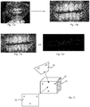
- the figure 11a is an example of a reference image.
- the updated reference model can be prepared from measurements made on the patient's teeth or on a molding of his teeth, for example a plaster cast.
- tooth model For each tooth, from the updated reference model, a model of said tooth, or “tooth model” ( figure 11d ). This operation, known in itself, is called “segmentation" of the updated reference model.
- a tooth model is preferably delimited by a gingival margin that can be decomposed into an interior gingival margin (on the inside of the mouth with respect to the tooth), an outer gingival margin ( facing outward of the mouth relative to the tooth) and two lateral gingival edges.
- One or more tooth attributes are associated with tooth models based on the teeth they model.
- the tooth attribute is preferably selected from a tooth number, a tooth type, a tooth shape parameter, for example a tooth width, in particular a mesio-palatal width, a thickness, a crown height. , a deflection index mesial and distal to the incisal edge, or an abrasion level, a tooth appearance parameter, in particular a translucency index or a color parameter, a parameter relating to the state of the tooth, tooth, for example "abraded", “broken”, “carious” or “paired” (that is, in contact with a dental appliance, preferably orthodontic), an age for the patient, or a combination of these attributes.
- a tooth attribute is preferably an attribute that relates only to the tooth modeled by the tooth model.
- a tooth attribute value can be assigned to each tooth attribute of a particular tooth model.
- the tooth attribute "tooth type” will have the value "incisive”, “canine” or “molar” depending on whether the tooth model is that of an incisor, a canine or a molar, respectively.
- the assignment of the tooth attribute values to the tooth models can be manual or, at least in part, automatic. For example, if the value of a tooth attribute is identical regardless of the tooth model, as for the tooth attribute "patient age", it may be sufficient to assign a value to a tooth model to determine the value of this attribute for other tooth models.
- tooth numbers are conventionally assigned according to a standard rule. It is therefore sufficient to know this rule and the number of a tooth modeled by a tooth model to calculate the numbers of the other tooth models.
- the shape of a particular tooth model is analyzed to define its tooth attribute value, e.g., its number.
- This shape recognition is preferably performed by means of a deep learning device, preferably a neural network.
- a deep learning device preferably a neural network.
- a library of historical tooth models is created, each historical tooth model having a value for the tooth attribute, as described below (step a)), the deep learning device is driven with views historical tooth models of this library, and then analyzing one or more views of the particular tooth model with the driven deep learning device, so as to determine the tooth attribute value of said particular tooth model.
- Step B) is intended for the acquisition of one or preferably several updated images.
- Step B) preferably comprises one or more of the features of step b) of WO 2016 066651 .
- the acquisition of the updated images is carried out by means of an image acquisition apparatus, preferably chosen from a mobile phone, a so-called “connected” camera, an "intelligent” watch, or “smartwatch", a tablet or personal computer, stationary or portable, having an image acquisition system, such as a webcam or a camera.
- an image acquisition apparatus is a mobile phone.
- the image acquisition apparatus is separated from the dental arch by more than 5 cm, more than 8 cm, or even more than 10 cm, which avoids the condensation of water vapor on the optical image acquisition device and facilitates focusing.
- the image acquisition apparatus in particular the mobile telephone, is provided with no specific optics for the acquisition of the updated images, which is possible in particular because of the spacing. of the dental arch during the acquisition.
- an updated image is a photograph or is an image extracted from a film. It is preferably in colors, preferably in real colors.
- the acquisition of the updated image or images is performed by the patient, preferably without the use of a support, resting on the ground and immobilizing the image acquisition apparatus, and in particular without a tripod.
- the triggering of the acquisition is automatic, that is to say without any action of an operator, as soon as the acquisition conditions are approved by the image acquisition apparatus, in particularly when the image acquisition apparatus has determined that it observes a dental arch and / or a retractor and that the observation conditions are satisfactory (sharpness, brightness, or even dimensions of the representation of the dental arch and / or the retractor).
- the time interval between steps A) and B) is as small as possible so that the teeth are not substantially displaced between the realization of the updated model and the acquisition of the updated images. Corresponding reference images with the updated images can then be acquired by observing the updated reference model.
- a dental spacer 10 is used in step B), as shown in FIG. figure 12a .
- the spacer typically includes a support having a rim extending around an opening and arranged so that the patient's lips can rest therein by allowing the patient's teeth to appear through said opening.
- step C the updated reference model is explored to find, for each updated image, a reference image having a maximum concordance with the updated image.
- Step C) may comprise one or more of the features of steps c), d) and e) of WO 2016 066651 , insofar as they concern such an exploration.
- a set of virtual acquisition conditions approximating the actual acquisition conditions during the acquisition of said updated image is preferably determined in a rough manner.
- it is estimated the position of the image acquisition device with respect to the teeth when it took the updated image (position of the acquisition device in the space and orientation of this device) .
- This rough evaluation advantageously makes it possible to limit the number of tests on virtual acquisition conditions during the following operations, and thus makes it possible to accelerate these operations considerably.
- one or more heuristic rules are preferably used. For example, preferably, virtual acquisition conditions that can be tested during the following operations are excluded, conditions that correspond to a position of the image acquisition apparatus behind the teeth or at a distance from the teeth. greater than 1 m.
- registration marks represented on the updated image, and in particular register marks 12 of the spacer are used to determine a region of the substantially conical space delimiting virtual acquisition conditions that can be tested during following operations, or "test cone".
- At least three registration marks 12 that are not aligned on the spacer 10 are preferably arranged, and their relative positions on the spacer are accurately measured.
- the registration marks are then marked on the updated image as previously described. Simple trigonometric calculations are used to roughly determine the direction in which the updated image was taken.
- a reference image is searched with maximum agreement with the updated image.
- This research is preferably carried out using a metaheuristic method, preferably an evolutionist method, preferably by simulated annealing.
- the updated image is analyzed so as to produce an updated map representing, at least partially, a discriminant information.
- the updated map thus represents the discriminant information in the repository of the updated image.
- the discriminant information is preferably selected from the group consisting of contour information, color information, density information, distance information, gloss information, saturation information, glare information, and color information. combinations of this information.
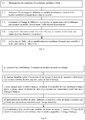
- the figure 12d is an updated map relating to the contour of the teeth obtained from the updated image of the figure 12b .
- step C1) we first determine virtual acquisition conditions to be tested, that is to say a position and a virtual orientation likely to correspond to the actual position and orientation of the device. acquisition during the capture of the updated image, but also, preferably, a virtual calibration likely to correspond to the actual calibration of the acquisition device during the capture of the updated image.
- the first virtual acquisition conditions to be tested are preferably virtual acquisition conditions evaluated roughly, as described above.
- step C2) the image acquisition apparatus is then virtually configured in the virtual acquisition conditions to be tested in order to acquire a reference image of the updated reference model under these virtual acquisition conditions. test.
- the reference image therefore corresponds to the image that would have taken the image acquisition device if it had been placed, compared to the updated reference model, and optionally calibrated, in the virtual acquisition conditions to test.
- the updated image was taken substantially at the same time that the updated reference model was created by a scan of the patient's teeth, the position of the teeth on the updated image is substantially identical to that in the updated reference model. If the virtual acquisition conditions to be tested are exactly the actual acquisition conditions, the reference image is therefore exactly superimposable to the updated image. The differences between the updated image and the reference image result from errors in the evaluation of the virtual acquisition conditions to be tested, if they do not correspond exactly to the actual acquisition conditions.
- step C3) the reference image, like the updated image , is processed so as to produce, from the reference image, a reference map representing the discriminant information ( Figures 11a and 11b ).
- a reference map representing the discriminant information Figures 11a and 11b .
- step C4) the updated and reference maps, both of which relate to the same discriminant information, are compared and the difference or "distance" between these two maps is evaluated by means of a score.
- the discriminant information is the contour of the teeth
- the score may be for example a correlation coefficient.
- the virtual acquisition conditions include the calibration parameters of the acquisition apparatus.
- the score is even higher than the values of the calibration parameters tested are close to the values of the calibration parameters of the acquisition apparatus used when acquiring the updated image.
- the reference image has fuzzy regions and sharp regions that do not match. fuzzy regions and sharp regions of the updated image. If the discriminant information is the contour of the teeth, the updated and reference maps will not represent the same outlines and the score will be low.
- the score is then evaluated using an evaluation function.
- the evaluation function makes it possible to decide whether the cycling on steps C1) to C5) must be continued or stopped.
- the evaluation function may for example be equal to 0 if the cycling must be stopped or equal to 1 if the cycling must continue.
- the value of the evaluation function may depend on the score achieved. For example, it may be decided to continue cycling if the score does not exceed a threshold. For example, if an exact match between the updated and reference images leads to a score of 100%, the threshold may be, for example, 95%. Of course, the higher the threshold, the better the accuracy of the evaluation of the virtual acquisition conditions if the score manages to exceed this threshold.
- the value of the evaluation function may also depend on scores obtained with virtual acquisition conditions previously tested.
- the value of the evaluation function may also depend on random parameters and / or the number of cycles already performed.
- a random parameter in the evaluation function may also allow testing of new virtual acquisition conditions, although the score is satisfactory.
- the evaluation functions conventionally used in metaheuristic optimization methods, preferably evolutionary, especially in simulated annealing processes, can be used for the evaluation function.
- step C5) if the value of the evaluation function indicates that it is decided to continue the cycling, the virtual acquisition conditions to be tested are modified and cycling is repeated on steps C1) to C5) consisting of making a reference image and a reference map, comparing the reference map with the updated map to determine a score, and then making a decision based on that score.
- the modification of the virtual acquisition conditions to be tested corresponds to a virtual displacement in space and / or to a modification of the orientation and / or, preferably, to a modification of the calibration of the acquisition apparatus.
- This modification can be random, preferably so that the new virtual acquisition conditions to be tested always belong to the set determined during the rough evaluation.
- the modification is preferably guided by heuristic rules, for example by favoring the modifications which, according to an analysis of the previous scores obtained, appear the most favorable to increase the score.
- step D The cycling is continued until the value of the evaluation function indicates that it is decided to stop cycling and continue in step D), for example if the score reaches or exceeds said threshold.
- the optimization of the virtual acquisition conditions is preferably carried out using a metaheuristic method, preferably an evolutionary method, preferably a simulated annealing algorithm. Such an algorithm is well known for nonlinear optimization.
- the process can be stopped (failure situation) or a new step C) can be started , with new discriminating information and / or with a new updated image.
- the process can also be continued with the virtual acquisition conditions corresponding to the highest score achieved. A warning may be issued to inform the user of the error on the result.
- the virtual acquisition conditions substantially correspond to the actual acquisition conditions of the updated image.
- the virtual acquisition conditions include the calibration parameters of the acquisition apparatus.
- the method thus makes it possible to evaluate the values of these parameters without it being necessary to know the nature of the acquisition apparatus or its setting.
- the acquisition of the updated images can therefore be performed without any particular precaution, for example by the patient himself by means of his mobile phone.
- the search for the actual calibration is performed by comparing an updated image with views of a reference model under virtual acquisition conditions to be tested.
- it does not require the updated image to show a calibrating calibration gauge, that is to say a gauge whose specific characteristics are known to determine the calibration of the acquisition device.
- Step C) thus leads to the determination of virtual acquisition conditions having a maximum agreement with the actual acquisition conditions.
- the reference image is therefore in maximum agreement with the updated image, that is to say that these two images are substantially superimposable.
- said search for virtual acquisition conditions in step C) is performed using a metaheuristic method, preferably an evolutionary method, preferably a simulated annealing algorithm.
- step D the reference tooth areas are identified on the reference image and transferred to the updated image to define corresponding updated tooth areas.
- the reference image is a view of the updated reference model segmented into tooth models. The limits of the representation of each tooth model on the reference image, or "reference tooth area”, can therefore be identified.
- the superimposition of the updated and reference images then makes it possible to postpone the limits of the reference tooth areas to the updated image, and thus to define the updated tooth zones. Since the reference image is in maximum agreement with the updated image, the updated tooth areas therefore substantially define the limits of the tooth models shown in the reference image.
- step E the tooth attribute value or values of the tooth model corresponding to it are assigned to each updated tooth zone.
- the reference image is a view of the updated reference model in which the tooth models have been assigned respective tooth attribute values for at least one tooth attribute, for example a tooth number.
- Each reference tooth area can inherit the tooth attribute value of the tooth model it represents.
- Each updated tooth area can then inherit the tooth attribute value from the reference tooth area that has defined it.
- step E an updated image and a description of the updated image defining one or more updated tooth zones and, for each of these zones, a tooth attribute value for minus a tooth attribute, for example a tooth number.
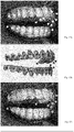
- the figure 17a shows an example of an updated image (acquired during step B)) being analyzed to determine the contours of the teeth.
- the figure 17b shows the reference image having a maximum agreement with the updated image (resulting from step C)).
- the numbers of the teeth are displayed on the corresponding teeth.
- the figure 17c illustrates the reporting of tooth numbers on updated tooth areas (steps D) and E)).
- step F the historical image is added to the learning base.
- Steps A) to F) are preferably performed for more than 1,000, more than 5,000, or more than 10,000 different patients, or "historical patients".
- the invention provides a particularly effective method for creating a learning base.
- the invention also relates to a method of training a deep learning device, preferably a neural network, said method comprising an enrichment of a learning base according to a method comprising steps A) to F) of acquiring a plurality of historical images, and then using said learning base to drive said deep learning device.
- the updated reference model is not necessarily the direct result of a scan of the patient's arch.
- the updated reference model can be in particular a model obtained by deformation of an initial reference model itself resulting directly from such a scan.
- the process then preferably comprises, instead of steps A) to C), the steps A ') to C').
- Step A ') is identical to step A). In step A '), however, the reference model generated is intended to be modified. It is therefore described as an "initial reference model”, and not an “updated reference model”, as in step A).
- the initial reference model may in particular be generated at an initial time preceding active orthodontic treatment, for example less than 6 months, less than 3 months, or less than 1 month before the start of treatment.
- the steps B ') to C') can then be implemented to follow the evolution of the processing between the initial time and the actualized moment of the step B ').
- the initial moment can alternatively be a moment at the end of active orthodontic treatment, for example less than 6 months, less than 3 months, or less than 1 month after the end of the treatment.
- Steps B ') to C') can then be implemented to monitor the occurrence of a possible recurrence.
- Step B ') is identical to step B). In step B '), however, the updated images are also intended to guide the modification of the initial reference model to define the updated reference model in step C').
- the time interval between steps A ') and B') is limited since, as explained below, the initial reference model will be deformed to obtain an updated reference model in maximum agreement with the updated images.
- the time interval between steps A ') and B') can be, for example, greater than 1 week, 2 weeks, 1 month, 2 months or 6 months.
- Step C ') is more complex than step C) since searching for a reference image having a maximum match with a refreshed image is not limited to searching for optimal virtual acquisition conditions. It also includes a search for an updated reference model, that is to say a reference model in which the teeth have substantially the same position as the updated image.
- Step C ') preferably comprises one or more of the characteristics of steps c), d) and e) of WO 2016 066651 , and in particular step e) illustrated on the figure 16 .
- the goal is to modify the initial reference model until an updated reference model is created which has a maximum agreement with the updated image.
- the updated reference model is therefore a model of the arcade from which the updated image could have been taken if this model had been the arcade itself.
- a succession of reference models "to be tested” is tested, the choice of a reference model to be tested preferably being dependent on the level of correspondence of the reference models "to be tested” previously tested with the updated image.
- a first optimization operation is performed for each test of a reference model to be tested during the second optimization operation.
- the first optimization operation and / or the second optimization operation preferably the first optimization operation and the second optimization operation implement a metaheuristic method, preferably an evolutionary method, preferably a simulated annealing. .
- a process comprising steps A ') to C') may advantageously be carried out as part of an active or passive orthodontic treatment, or, more generally, to follow any evolution of the teeth.
- the enrichment of the learning base does not necessarily result from an enrichment process according to the invention.
- the learning base is created by an operator.
- the latter thus analyzes thousands of images of analysis.
- it determines the tooth areas and then assigns them tooth attribute values.
- it assigns image attribute values to each image. It can thus constitute historical images.
- the method of detailed analysis of an "image of analysis" of a dental arch of a patient according to the invention comprises steps 1) to 4).
- the analysis image preferably a photograph or an image extracted from a film, preferably in color, preferably in real colors
- an image acquisition apparatus preferably a telephone mobile, separated from the dental arch more than 5 cm, more than 8 cm or more than 10 cm, and which preferably is provided with no specific optics.
- the analysis image represents several teeth, preferably more than 2, more than 3, more than 4 or more teeth of the patient.
- the figure 12a or the figure 12b could be examples of analysis images.
- the arrangement of the teeth is realistic, that is to say, it corresponds to that observed by the image acquisition apparatus when it acquired the image of analysis.
- the acquisition of the analysis image is performed by the patient, preferably without the use of a support, resting on the ground and immobilizing the image acquisition apparatus, and in particular without a tripod.
- the triggering of the acquisition of the analysis image is automatic, that is to say without operator action, as soon as the acquisition conditions are approved by the device.
- acquisition of images in particular when the image acquisition apparatus has determined that it observes a dental arch and / or a retractor and that the observation conditions are satisfactory (sharpness, brightness, or even dimensions of the image). representation of the dental arch and / or the retractor).
- step 1) a learning base with more than 1,000, preferably more than 5,000, preferably more than 10,000, preferably more than 30,000, preferably more than 50,000, preferably is created. more than 100,000 historical images. The higher the number of historical images, the better the analysis performed by the process.
- an enriched learning base is used according to an enrichment method according to the invention.
- the learning base can however be constituted according to other methods, for example to be created manually.
- an operator preferably an orthodontist, identifies one or more "historic" tooth areas on an image, said then assigns, to each identified historical tooth area, a value for the minus a tooth attribute
- step 2) a deep learning device, preferably a neural network , is driven with the learning base.
- neural network or “artificial neural network” is a set of algorithms well known to those skilled in the art.
- the deep learning device is preferably driven by a learning process called "deep learning".
- deep learning a learning process
- the deep learning device gradually learns to recognize on an image, patterns, in English “ patterns”, and to associate them with tooth areas and tooth attribute values, for example tooth numbers.
- step 3 the image that is to be analyzed, or "analysis image” , is submitted to the deep learning device.
- he is able to determine that there is a 95% chance that a shape of the analysis image represents an incisor.
- the deep learning device analyzes the entire analysis image and determines probabilities for all of the analysis tooth zones that it has identified.
- step 4 the results of the previous step are analyzed to determine the teeth represented on the analysis image.
- step 3 leads to particularly satisfactory results.
- a learning base makes it possible to establish a probability threshold such that if a probability associated with an analysis tooth zone and a tooth attribute value for this analysis tooth zone exceeds said threshold, the scan tooth area effectively represents a tooth having said tooth attribute value.
- Step 4) thus leads to the definition of an analysis image enriched with a description defining the areas of analysis tooth and, for each area of analysis tooth, the values of the attributes of the tooth represented by the tooth area of analysis.
- the method of global analysis of an updated image of a dental arch of a patient comprises steps 1 ') to 3').
- the method is similar to the detailed analysis method described above, with the difference that, according to the overall analysis, it is not necessary to analyze the individual situation of each tooth.
- the analysis is global to the whole image. In other words, the deep learning device determines the value of an "image" attribute without having to previously determine tooth attribute values.
- the deep learning device is capable of analyzing the analysis image and recognizing said patterns. According to these reasons, it can in particular determine a probability relative to the value of the image attribute considered.
- a detailed analysis method according to the invention is particularly useful for modeling a dental arch, in particular for establishing a remote diagnosis.
- US 2009/0291417 describes a method for creating and then modifying three-dimensional models, particularly for the manufacture of orthodontic appliances.
- WO 2016 066651 discloses a method of controlling the positioning and / or shape and / or appearance of a patient's teeth.
- This method comprises a step of creating an initial reference model of the teeth, at an initial time, preferably with a 3D scanner, then, at a later time, or "updated instant", for example six months after the instant initial, the creation of an updated reference model, by deformation of the reference model initial.
- This deformation is performed in such a way that the updated reference model allows observations that are substantially identical to images of the teeth acquired at the instantaneous moment, in particular to photos or images of a video taken by the patient himself, without special precautions, called "updated images”.
- the updated images are therefore used to modify the initial, very precise reference model.
- the updated reference model resulting from the deformation of the initial reference model, guided by the analysis of the updated images, is therefore also very precise.
- An object of the invention is to meet this need.
- the invention thus makes it possible, from a simple analysis image, for example a photograph taken by means of a mobile telephone, to reconstitute, with good reliability, a dental arch in the form of a model. assembled.
- the analysis image can in particular be acquired as described in step 1) above.
- the precision of the assembled model can be increased if several analysis images are processed.
- Steps b) to c) are preferably implemented for several analysis images and, in steps d) and e), optimal tooth models and an assembled model are searched for maximum agreement with respect to the set of analysis images (step f)).
- the patient can therefore very easily ask an orthodontist to check his dental situation, without even having to move, by simply transmitting one or preferably several photos of his teeth.
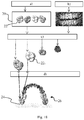
- a historical library 20 is created ( figure 18 ) having more than 1,000, preferably more than 5,000, preferably more than 10,000 historical tooth patterns 22.
- a historical tooth model can be obtained from a model of a dental arch of a "historic" patient obtained with a scanner.
- This arch model can be segmented to isolate the representations of the teeth, as on the figure 11d .
- Each of these representations, having a specific shade of gray on the figure 11d can constitute a model of historical tooth.
- the library is preferably enriched with the tooth models resulting from the implementation of the process described in WO 2016 066651 or step A) or A ') described above.
- tooth attributes are associated with the tooth models.
- a tooth attribute value is assigned to each tooth attribute of a particular tooth model, as previously described (see the description of step A)).
- the historical library thus contains historical tooth models and associated attribute values which facilitate the search in step c).
- the analysis image is acquired, as described above for step B), before analyzing it.
- the analysis image is preferably a photo or an image of a film, preferably with a mobile phone.
- the analysis image can be acquired at any time after step a), for example more than 1 week, more than 1 month or more than 6 months after step a).
- the analysis image is analyzed according to a detailed analysis method according to the invention.
- the optional features of this method are also optional in step b).
- an enriched analysis image is obtained from a description providing, for each analysis tooth zone, a tooth attribute value for at least one tooth attribute, by example a tooth number.
- step c) the historical library, for each analysis tooth area determined in the previous step , is searched for a historical tooth model having a maximum proximity to the analysis tooth area.
- This tooth model is called an "optimal tooth model”.
- Proximity is a measure of one or more differences between the historical tooth model and the tooth area of analysis. These differences may include a difference in shape, but also other differences such as a difference in translucency or color. Maximum proximity can be sought by successively minimizing several differences, or minimizing a combination of these differences, for example a weighted sum of these differences.
- Evaluating the proximity of a historical tooth model with a scan tooth area preferably includes a comparison of at least one value of a tooth attribute of the scan tooth area with the value of this attribute for the historical tooth model. Such an evaluation is advantageously very fast.
- the description of the area of the analysis tooth provides a value for the type or number of the tooth, the thickness of the tooth represented and / or the height of its crown and / or its mesographic width. palatal and / or mesial deflection index and distal to its incisal edge, this value can be compared to the value of the corresponding attribute of each of the historical tooth models.
- a search is made for a historical tooth model having, for at least one tooth attribute, the same value as said analysis tooth area.
- the tooth attribute may in particular be relative to the tooth type or the tooth number.
- the historical tooth models are filtered to examine in more detail only those that are related to the same type of tooth as the tooth represented on the area of the analysis tooth.
- the shape of the tooth represented on the analysis tooth area can be compared to the shape of a historical tooth model to be evaluated, preferably using a metaheuristic method, preferably evolutionary, preferably by simulated annealing.
- each view thus obtained is compared with the analysis image, preferably with the analysis tooth area so as to establish a "distance" between this view and said analysis image or, preferably, said tooth area. analysis. The distance thus measures the difference between the view and the area of the analysis tooth.
- the distance can be determined after a treatment of the view and the analysis image or, preferably, the analysis tooth area, so as to show, on corresponding maps, the same discriminant information, by example, contour information, as described above in step C3) or in WO 2016 066651 .
- Each examined historical tooth model is thus associated with a particular minimum distance, which measures its nearness of shape with the analysis tooth area.
- the optimal historical tooth model is the one that, compared to the comparison (s) made, is considered to be the closest to the area of the analysis tooth.
- the minimum distances obtained for the different tooth models tested are then compared and, to define the optimal tooth model, the one with the smallest minimum distance is used.
- the optimal tooth model therefore has maximum agreement with the analysis image.
- the search for the maximum concordance is preferably carried out by means of a metaheuristic method, preferably an evolutionist method, preferably by simulated annealing.
- a first evaluation of the historical tooth models is performed successively by comparing the values of at least one tooth attribute, for example the tooth number, with the corresponding values of the tooth area of analysis. then a second evaluation by form comparison.
- the first evaluation which is rapid, advantageously makes it possible to filter the historical tooth models in order to submit to the second evaluation, which is slower, than the historical tooth models retained by the first evaluation.
- the first evaluation allows to retain only tooth models modeling teeth # 15.
- the second evaluation we look for the historical tooth model, whose shape is closest to that of the tooth represented, among all the historical tooth models modeling teeth # 15.
- first evaluations are made before performing the second evaluation.
- the first evaluations are used to filter the historical tooth models so as to retain only tooth models that model teeth No. 15 and have a crown height of between 8 and 8.5 mm.
- step c an optimal tooth model has thus been associated with each of the scan tooth zones.
- the historical tooth model 22 1 can be observed so as to strongly resemble an analysis zone identified on the analysis image. It is considered optimal for this area of analysis.
- step d an assembled model is created by arranging the optimal tooth models.
- a first coarse arrangement is created, that is to say that a coarse model is manufactured by assembling the optimal tooth models.
- the optimal tooth models can be oriented so that their optimal viewing directions are all parallel, the optimal direction of observation of a tooth model being the direction in which said tooth model is present. maximum concordance with the analysis image.
- the first coarse arrangement can also be established by considering the tooth attribute values of the optimal tooth models. For example, if the tooth numbers of optimal tooth models are those of the canines and incisors, these tooth models can be arranged following an arc 24 ( figure 18 ) corresponding classically to the region of the arch that carries these types of teeth.
- the shape of this arc can be refined based on other tooth attribute values.
- the order of the optimal tooth models is that of the corresponding scan tooth areas.
- the minimum distance associated with an optimal tooth model results from an observation of the tooth model in an "optimal" observation direction. In other words, it is probably substantially in this direction that the tooth that this model models is also observed in the analysis image. All optimal tooth models are thus preferably oriented so that their respective optimum viewing directions are all parallel.
- the first arrangement of the optimal tooth models is then iteratively modified, so as to have maximum agreement with the analysis image.
- Each view thus obtained is compared with the analysis image so as to establish a "distance" between this view and said analysis image.
- the distance thus measures the difference between the view and the analysis image.
- the distance can be determined after a processing of the view and the analysis image so as to show, on one of the corresponding maps, a discriminant information, for example a contour information, as described above at step C3) or in WO 2016 066651 .
- Each arrangement examined is thus associated with a minimum distance.
- the minimum distances obtained for the different arrangements tested are then compared and, to define the optimal arrangement, the one with the smallest minimum distance is retained.
- the optimal arrangement therefore has maximum agreement with the analysis image.
- the search for the maximum concordance is preferably carried out by means of a metaheuristic method, preferably an evolutionist method, preferably by simulated annealing.
- step d an optimal arrangement of the optimal tooth models, that is to say the assembled model 26, is obtained.
- step e) one or more optimal tooth models are replaced by other tooth models, and then, in step d), it is repeated so as to maximize the match between the assembled model and the image of the tooth. analysis.
- an optimal tooth model in the "optimal” arrangement, no longer has a maximum agreement with the analysis image.
- the tooth model could be observed in an "optimal" direction that provided a view having a minimal distance to the analysis image (reason for which it was considered optimal). But in the optimal arrangement, it is no longer oriented in the optimal direction.
- a new search for an assembled model can therefore be carried out by modifying the tooth models, for example by replacing the optimal tooth models with close tooth models.
- the search for the tooth models to be tested is preferably carried out by means of a metaheuristic method, preferably an evolutionist method, preferably by simulated annealing.
- the method therefore implements a double optimization, on the tooth models and on the arrangement of the tooth models, the assembled model being the arrangement of a set of tooth models which provides the minimum distance to the analysis image, considering all possible tooth models and all possible arrangements.
- step f) the method uses several images of analysis of the patient's arch, preferably more than 3, more than 5, more than 10, more than 50, preferably more than 100 images of analysis.
- the assembled model is thus more complete. More preferably, the method implements an optimization so that the assembled model obtained is optimal with respect to all of the analysis images. In other words, the assembled model is preferably the one that maximizes the concordance with the set of analysis images.
- the method therefore implements a double or, preferably a triple optimization, on the tooth models on the one hand, on the arrangement of the tooth models and / or on a plurality of images.
- the assembled model being the arrangement of a set of tooth models that provides the average minimum distance, over all the analysis images, considering all the possible tooth models and, preferably all possible arrangements.
- a metaheuristic method is used, preferably evolutionary, preferably by simulated annealing.
- the invention thus makes it possible to construct an assembled dental arch model from simple analysis images, for example photographs taken by means of a mobile telephone.
- the accuracy of the assembled model does not reach that of a scan.
- such precision is however not essential.
- the assembled model can therefore be used to analyze the orthodontic situation of the patient, following steps ii) to iv).
- step ii) the assembled model is sent to an orthodontist and / or a computer provided with diagnostic software.
- the assembled model is sent together with a patient completed questionnaire to improve the quality of the analysis in step iv).
- step iii) the orthodontist and / or computer examines the assembled model. Unlike an updated image, the assembled model allows observation from any angle. The analysis is advantageously more precise.
- step iv) the orthodontist and / or the computer informs the patient, for example by transmitting a message on his phone.
- This message may include informing the patient of an adverse situation and inviting him / her to make an appointment with the orthodontist.
- the orthodontist can also compare the assembled model with assembled models previously received for the same patient. Its analysis advantageously makes it possible to evaluate the evolution of the situation.
- the message can thus inform the patient of an unfavorable evolution of his situation, which improves the prevention.
- the assembled model can also be compared with one or more models obtained by scanning the teeth or molding the patient's teeth, or with an updated reference model resulting from the implementation of a method described in WO 2016 066651 .
- An image analysis according to the invention is also useful for guiding the acquisition of an image of a dental arch, in particular for establishing a remote diagnosis.
- WO2016 / 066651 discloses a method in which an initial reference model is deformed so as to obtain an updated reference model for acquiring reference images having maximum agreement with the "updated" images of the arcade acquired at the time of update .
- the reference images are thus views of the updated reference model, observed in virtual acquisition conditions which are as concordant as possible with the actual acquisition conditions used to acquire the updated images of the patient's arch.
- the search for these virtual acquisition conditions is preferably carried out using metaheuristic methods.
- WO2016 / 066651 recommends to make a rough first evaluation of the actual acquisition conditions. For example, conditions which correspond to a position of the acquisition apparatus at a distance of teeth greater than 1 meter are excluded from the search.
- An object of the invention is to respond, at least partially, to this problem.
- step b ' all said scan tooth areas are identified, and at least one tooth attribute value is determined for each scan tooth area, and at least one tooth attribute value is determined for each scan tooth area. step c '), determining the value for the image attribute according to said tooth attribute values.
- the information message is issued by the acquisition apparatus.
- an acquisition method thus makes it possible to verify whether an analysis image respects a setpoint and, if it does not respect the instruction, to guide the operator to acquire a new analysis image.
- the method therefore allows an "onboard control", preferably in the image acquisition apparatus.
- WO2016 / 066651 it may be desired to acquire updated images according to different acquisition direction, for example a front image, a right image and a left image. These updated images, acquired successively, can be classified accordingly.
- the search for Virtual acquisition conditions that match the real acquisition conditions are accelerated.
- the search can start from virtual acquisition conditions in which the virtual acquisition device is opposite, to the left or to the right of the updated reference model, depending on whether the updated image considered is ranked as a front, left or right image, respectively.
- the operator usually the patient, can be mistaken when acquiring updated images. In particular, he may forget to take an updated image, for example the front view, or to reverse two updated images. Typically, the operator can take an image on the right while waiting for a left image.
- This inversion of the updated images can considerably slow down their processing.
- the updated image is supposed to be an image taken on the left but erroneously taken to the right
- said search for optimal virtual acquisition conditions that is to say having a maximum agreement with the actual acquisition conditions will start from a starting point offering a left view of the reference model, while the optimal virtual acquisition conditions correspond to a right view. Research will therefore be considerably slowed down.
- each updated image is an analysis image that can be analyzed and controlled, preferably in real time.
- the acquisition method makes it possible to determine that the updated image has been "taken to the right" and to compare this image attribute value with the instruction that had been given to the operator to take the image. updated left.
- the attribute value of the updated image does not correspond to the set point (to acquire a updated image on the left)
- the acquisition device can warn the operator immediately so that it modifies the direction acquisition.
- step a ' the operator activates the image acquisition apparatus so as to acquire an analysis image.
- the operator triggers the acquisition apparatus to store the analysis image, preferably takes a picture or video of his teeth, preferably by means of a mobile phone equipped with 'a camera.
- Step a ') can be performed as the acquisition of the updated images in step B) described above.
- the analysis image is not stored.
- the analysis image may be the image that, in real time, appears on the screen of the mobile phone of the operator, usually the patient.
- step b ' the analysis image is analyzed according to a detailed analysis method according to the invention.
- This analysis preferably leads to the assignment of a tooth attribute value to each identified scan tooth area, for example to assign a tooth number to each of the scan tooth areas.
- an attribute value of the analysis image is determined based on the tooth attribute values.
- the attribute value of the analysis image can be relative to its general orientation and can for example take one of three values: “right photo”, “left photo” and "front photo”.
- the attribute value of the analysis image can also be the list of numbers of the teeth represented, for example, "16, 17 and 18".
- the attribute value of the analysis image may still be, for example, the "presence” or "absence” of a dental appliance, preferably orthodontic, or the mouth opening condition ( "Open mouth", "closed mouth”).
- a global analysis method is implemented in step b ').
- a global analysis method makes it possible to directly obtain a value for an image attribute, without having to determine values for a tooth attribute. It is therefore advantageously faster.
- the information resulting from a global analysis may, however, be less precise than that resulting from a detailed analysis.
- Steps a ') to c') thus make it possible to characterize the analysis image.
- the characterization of the analysis image makes it possible to guide the operator if the analysis image does not correspond to the expected image, for example because its quality is insufficient or because it does not represent the desired teeth. .
- step d ' the image attribute value of the analysis image is compared with a setpoint.
- step e ' a message is sent to the operator, preferably by the acquisition apparatus.
- the information message relates to the quality of the acquired image and / or the position of the acquisition apparatus with respect to said arcade and / or the setting of the acquisition apparatus and / or at the opening of the mouth and / or wearing a dental appliance, preferably orthodontic.
- the acquisition apparatus may emit light, for example red, and / or ring, and / or generate a voice message, and / or vibrate, and / or display a message on his screen.
- the acquisition device may issue the message "carry your device for this image.”
- the acquisition device may issue the message "open your mouth for this image".
- steps b ') to c') are implemented only if the operator saves the analysis image, that is to say it presses the trigger.
- the message then prompts the operator to acquire a new scan image.
- the acquisition apparatus erases the unsatisfactory analysis image.
- steps b ') to c') are implemented continuously when the acquisition apparatus is running and the analysis image is an image that appears on a screen of the apparatus acquisition.
- the acquisition apparatus can thus, for example, emit a red light as long as the analysis image is unsatisfactory, and emit a green light when the analysis image is satisfactory.
- the acquisition apparatus only stores analysis images that are satisfactory.
- the invention therefore allows an on-board control during the acquisition of analysis images. Applied to updated images of the process of WO2016 / 066651 steps a ') to e') make it possible to ensure that these images are in accordance with the need, and thus considerably speed up the execution of this method.
- Steps d) and e ') are optional.
- the analysis image is only associated with its description, which specifies its image attribute value. This description It also considerably speeds up the execution of the WO2016 / 066651 since, when the analysis image is used as a refreshed image of this process, it makes it possible to approximately determine the actual acquisition conditions of this image, eliminating the risk of a gross error, for example due to a inversion between two images.
- steps d 'and e') are however preferred. For example, they prevent the operator from foregoing an image on the left, or taking two redundant right images.
- the orthodontist determines the positioning of the teeth that he wishes to obtain at a moment of the treatment, called "set-up".
- the set-up can be defined by means of a fingerprint or from a three-dimensional scan of the patient's teeth.
- the orthodontist then makes or manufactures, accordingly, an orthodontic appliance adapted to this treatment.
- the orthodontic appliance may be an orthodontic gutter (" align" in English).
- a gutter is conventionally in the form of a removable monobloc device, conventionally made of a transparent polymer material, which comprises a shaped chute so that several teeth of an arch, generally all the teeth of an arch, can be housed therein.
- the shape of the chute is adapted to maintain the gutter in position on the teeth, while exerting an action of correction of the positioning of certain teeth ( Figures 14 and 15 ).
- the treatment by means of gutters is advantageously not very restrictive for the patient.
- the number of appointments at the orthodontist is limited.
- the pain is lower than with a metal orthodontic arch attached to the teeth.
- the patient moves to the orthodontist for a visual check, in particular to check whether the movement of the teeth is in line with expectations and whether the gutter worn by the patient is still suitable for treatment.
- the orthodontist diagnoses a maladaptation to the treatment, he performs a new tooth impression, or, in a similar way, a new three-dimensional scan of the teeth, then orders a new set of gutters configured accordingly. It is considered that on average, the number of gutters finally manufactured is about 45, instead of the 20 gutters conventionally planned at the beginning of the treatment.
- the number of check-ups at the orthodontist should be limited.
- An object of the invention is to respond, at least partially, to this need.
- an evaluation method according to the invention greatly facilitates the evaluation of the good adequacy of the gutter treatment, while making this assessment particularly reliable.
- the method can be implemented from simple photographs or films, taken without particular precautions, for example by the patient.
- the number of appointments with the orthodontist can be limited.
- step b ") all said scan tooth areas are identified, and the value of said tooth attribute is determined for each scan tooth area, and in step c"), the adequacy of the gutter is determined according to said tooth attribute values.
- said tooth attribute is selected from the group consisting of a maximum gap along the free edge of the tooth, a mean spacing along the free edge of the tooth, and said image attribute is selected from the formed group. by a maximum spacing along all the teeth shown, a mean spacing along the free edges of all the teeth shown, an overall acceptability of the spacing of the teeth shown.
- the attribute of tooth relative to a spacing may in particular be the existence of a spacing, this attribute being able to take the tooth attribute values "yes” or "no"; or a value measuring the magnitude of the spacing, for example a maximum gauge observed or an evaluation relative to a scale.
- step b ") a detailed analysis method according to the invention is preferably used, a tooth attribute of each historical tooth zone of each historical image of the learning base being relative to a spacing. between the tooth represented by the historic tooth zone, and a gutter carried by said tooth and represented on said historical image.
- Steps b "1) to b" 4) may include one or more optionally optional features of steps 1) to 4) described above, respectively.
- step b ") a global analysis method according to the invention is implemented, an image attribute of each historical image of the learning base being relative to a distance between at least one tooth represented on the historical image, and a gutter carried by said tooth and represented on said historical image.
- Steps b "1 ') to b" 3') may include one or more optionally optional features of steps 1 ') to 3') described above, respectively.
- step b " The method is now described when a detailed analysis is carried out in step b ").
- the learning base Prior to step a "), the learning base must be enriched, preferably according to an enrichment method according to the invention, in order to contain historical images whose precise description, for each of the historic tooth areas, a value for the tooth attribute relative to the gap.
- This information can be entered manually.
- an operator preferably an orthodontist, may be presented with an image representing one or more so-called "historic" tooth areas, and asked him to identify these historic tooth areas and to indicate for each historical tooth, if there is a gap or not and / or to assess the amplitude of this gap.
- a historical image may be a picture of a gutter worn by a historical patient.
- a historical image may be the result of a treatment of an image representing a bare dental arch (that is to say without a gutter) and an image representing the same arch carrying the gutter.
- the image representing the bare arcade can in particular be a view of a model of the distorted arch to obtain maximum agreement with the image representing the archway carrying the gutter.
- Such a treatment may be particularly useful for better showing the contour of the teeth and gutter when the teeth are not visible through the gutter.
- step a ") the acquisition of the analysis image can be performed as the acquisition of the images updated in step B) described above.
- At least one reminder informing the patient of the need to create an analysis image is sent to the patient.
- This reminder can be in paper form or, preferably, in electronic form, for example in the form of an email, an automatic alert of a mobile specialized application or SMS.
- Such a reminder may be sent by the cabinet or the orthodontic laboratory or by the dentist or by the specialized mobile application of the patient, for example.
- Step a is carried out at the moment when the evaluation of the shape of a gutter is desired, for example more than 1 week after the beginning of the treatment with the gutter.
- the analysis image is an image representing the gutter carried by the patient's teeth.
- step b ") the analysis image is analyzed according to a detailed analysis method according to the invention.
- the deep learning device has been trained by means of a learning base containing historical images, the description of which specifies, for at least one, preferably each historical tooth area, a value for a tooth attribute relative to a spacing between the tooth represented by the historic tooth zone and the groove carried by said tooth and represented on said historical image.
- the value for this tooth attribute therefore provides information about the shape of the gutter relative to the shape of the patient's teeth.
- the value for this tooth attribute may be a measure of the gap, for example a measure of the maximum gap, or the average gap for the tooth represented by the historical tooth area.
- the deep learning device is thus capable of analyzing the analysis image to determine, preferably for each of the "analysis tooth zones", the existence, or even the importance of a spacing of the gutter of the tooth represented on the area of the analysis tooth.
- step c ") the adequacy of the gutter is evaluated on the basis of the results of the preceding step, for example, whether the spacing of the gutter with at least one tooth exceeds a threshold of acceptability and, in this case, we decide to replace the gutter with a better adapted gutter.
- the adequacy of the gutter can be evaluated in the context of orthodontic treatment (spacing compatible or not with orthodontic treatment), but also in the context of a non-therapeutic treatment, particularly aesthetic. Gutters can indeed be used to move teeth for purely aesthetic purposes, without this displacement does not change the health of the patient.
- the adequacy of the gutter can also be assessed as part of a research program on the effectiveness of the gutter, for example to evaluate a new material for the gutter, on a human being or on another animal.
- step d ") information relating to the evaluation performed in the previous step is sent, in particular to the patient and / or the orthodontist.
- the orthodontist can then use this information, possibly in combination with additional information, for example the age of the patient or the length of time the gutter has been worn, to establish a diagnosis and, if necessary, decide on a treatment. adapted.
- the method comprises, in step b "), an overall analysis according to the invention, the other steps are not modified.
- Analysis of the analysis image and the historical images is then performed globally, without identifying the individual situation of each of the teeth represented and the image attribute is relative to the image as a whole.
- the image attribute relating to the spacing may be relative to the acceptability of a dental situation, because of one or more spacings, or relative to the overall magnitude of the spacing or spacings of the teeth.
- the value of the image attribute may be "globally acceptable” or "globally unacceptable”.
- the value of the image attribute may also be, for example, a measure of the distance, for example a measure of the maximum spacing, or the average spacing between the teeth represented on the analysis image. and the gutter.
- a method according to the invention makes it possible, from simple photos or from a simple film, to determine whether the gutter is abnormally peeled, or even if a detailed analysis has been carried out in step b "), to determine the areas in which the gutter has moved away from the teeth and to assess the extent of this gap.
- the invention also relates to a method of adapting an orthodontic treatment, in which method is implemented a method of evaluating the shape of an orthodontic gutter according to the invention, and then, depending on the result of said evaluation. , a new gutter is manufactured and / or the patient is advised, for example to improve the conditions of use of his orthodontic gutter, in particular the positioning and / or time slots port and / or maintenance of its orthodontic gutter, to optimize the treatment.
- gutters are not limited to therapeutic treatments.
- an evaluation method can be implemented to evaluate a gutter exclusively used for aesthetic purposes.
- the method can also be used to evaluate other dental parts or appliances, in particular orthodontic appliances.
- the patient is not limited to a human being.
- a method according to the invention can be used for another animal.
- the patient may be alive or dead. He is preferably alive.
- the methods according to the invention can be implemented as part of an orthodontic treatment, but also out of any orthodontic treatment, and even out of any therapeutic treatment.
Applications Claiming Priority (1)
| Application Number | Priority Date | Filing Date | Title |
|---|---|---|---|
| FR1756944A FR3069359B1 (fr) | 2017-07-21 | 2017-07-21 | Procede d'analyse d'une image d'une arcade dentaire |
Publications (1)
| Publication Number | Publication Date |
|---|---|
| EP3432312A1 true EP3432312A1 (de) | 2019-01-23 |
Family
ID=60515479
Family Applications (1)
| Application Number | Title | Priority Date | Filing Date |
|---|---|---|---|
| EP18184486.1A Pending EP3432312A1 (de) | 2017-07-21 | 2018-07-19 | Analyseverfahren der darstellung einer zahnreihe |
Country Status (3)
| Country | Link |
|---|---|
| US (2) | US11107218B2 (de) |
| EP (1) | EP3432312A1 (de) |
| FR (1) | FR3069359B1 (de) |
Cited By (3)
| Publication number | Priority date | Publication date | Assignee | Title |
|---|---|---|---|---|
| CN109766877A (zh) * | 2019-03-12 | 2019-05-17 | 北京羽医甘蓝信息技术有限公司 | 基于深度学习的全景片人工牙体识别的方法和装置 |
| WO2019243241A1 (de) * | 2018-06-22 | 2019-12-26 | Sirona Dental Systems Gmbh | Verfahren zur konstruktion eines dentalen bauteils |
| WO2020234411A1 (fr) * | 2019-05-22 | 2020-11-26 | Dental Monitoring | Procede de generation d'un modele d'une arcade dentaire |
Families Citing this family (11)
| Publication number | Priority date | Publication date | Assignee | Title |
|---|---|---|---|---|
| CN110998602B (zh) | 2017-06-30 | 2023-12-01 | 普罗马顿控股有限责任公司 | 使用深度学习方法对3d牙颌面结构的分类和3d建模 |
| EP3462373A1 (de) | 2017-10-02 | 2019-04-03 | Promaton Holding B.V. | Automatisierte klassifizierung und taxonomie von 3d-zahndaten mit tieflernverfahren |
| EP3503038A1 (de) * | 2017-12-22 | 2019-06-26 | Promaton Holding B.V. | Automatisierte 3d-vorhersage einer wurzelform mithilfe von tiefenlernverfahren |
| EP3561778A1 (de) | 2018-04-26 | 2019-10-30 | Promaton Holding B.V. | Automatisierte korrektur von metallbehafteten voxeldarstellungen von röntgendaten mit hilfe von tiefenlerntechniken |
| WO2019217559A1 (en) * | 2018-05-08 | 2019-11-14 | Align Technology, Inc. | Automatic ectopic teeth detection on scan |
| FR3083898B1 (fr) * | 2018-07-13 | 2020-10-09 | Dental Monitoring | Procede de simulation d'une situation dentaire |
| US11961235B2 (en) * | 2019-03-25 | 2024-04-16 | Bonewise Inc. | Apparatus, method and recording medium storing instructions for determining bone age of teeth |
| AU2021227914A1 (en) | 2020-02-26 | 2022-09-22 | Get-Grin Inc. | Systems and methods for remote dental monitoring |
| CN113538437A (zh) * | 2020-04-21 | 2021-10-22 | 宁波深莱医疗科技有限公司 | 牙颌三维数字模型分割结果的检测方法 |
| US11837350B2 (en) | 2021-04-20 | 2023-12-05 | Clear Blue Smiles LLC | System and method of treatment for correcting tooth malocclusions |
| FR3123201A1 (fr) * | 2021-05-25 | 2022-12-02 | Dental Monitoring | Procédé de distribution de gouttières orthodontiques |
Citations (2)
| Publication number | Priority date | Publication date | Assignee | Title |
|---|---|---|---|---|
| US20080172386A1 (en) * | 2007-01-17 | 2008-07-17 | Ammar Hany H | Automated dental identification system |
| FR3027505A1 (fr) * | 2014-10-27 | 2016-04-29 | H 43 | Procede de controle du positionnement de dents |
Family Cites Families (12)
| Publication number | Priority date | Publication date | Assignee | Title |
|---|---|---|---|---|
| US6648640B2 (en) | 1999-11-30 | 2003-11-18 | Ora Metrix, Inc. | Interactive orthodontic care system based on intra-oral scanning of teeth |
| US8874452B2 (en) * | 2004-02-27 | 2014-10-28 | Align Technology, Inc. | Method and system for providing dynamic orthodontic assessment and treatment profiles |
| US8439672B2 (en) | 2008-01-29 | 2013-05-14 | Align Technology, Inc. | Method and system for optimizing dental aligner geometry |
| US9788917B2 (en) | 2010-03-17 | 2017-10-17 | ClearCorrect Holdings, Inc. | Methods and systems for employing artificial intelligence in automated orthodontic diagnosis and treatment planning |
| FR3010629B1 (fr) | 2013-09-19 | 2018-02-16 | Dental Monitoring | Procede de controle du positionnement de dents |
| US9008391B1 (en) | 2013-10-22 | 2015-04-14 | Eyenuk, Inc. | Systems and methods for processing retinal images for screening of diseases or abnormalities |
| FR3027508B1 (fr) | 2014-10-27 | 2016-12-23 | H 42 | Procede de controle de la dentition |
| US9744001B2 (en) * | 2014-11-13 | 2017-08-29 | Align Technology, Inc. | Dental appliance with cavity for an unerupted or erupting tooth |
| US9993217B2 (en) * | 2014-11-17 | 2018-06-12 | Vatech Co., Ltd. | Producing panoramic radiograph |
| US10248883B2 (en) * | 2015-08-20 | 2019-04-02 | Align Technology, Inc. | Photograph-based assessment of dental treatments and procedures |
| FR3050375A1 (fr) | 2016-04-22 | 2017-10-27 | H43 Dev | Procede de controle de la dentition |
| US10695150B2 (en) * | 2016-12-16 | 2020-06-30 | Align Technology, Inc. | Augmented reality enhancements for intraoral scanning |
-
2017
- 2017-07-21 FR FR1756944A patent/FR3069359B1/fr active Active
-
2018
- 2018-07-10 US US16/031,172 patent/US11107218B2/en active Active
- 2018-07-19 EP EP18184486.1A patent/EP3432312A1/de active Pending
-
2021
- 2021-08-02 US US17/391,198 patent/US20210358124A1/en active Pending
Patent Citations (2)
| Publication number | Priority date | Publication date | Assignee | Title |
|---|---|---|---|---|
| US20080172386A1 (en) * | 2007-01-17 | 2008-07-17 | Ammar Hany H | Automated dental identification system |
| FR3027505A1 (fr) * | 2014-10-27 | 2016-04-29 | H 43 | Procede de controle du positionnement de dents |
Cited By (5)
| Publication number | Priority date | Publication date | Assignee | Title |
|---|---|---|---|---|
| WO2019243241A1 (de) * | 2018-06-22 | 2019-12-26 | Sirona Dental Systems Gmbh | Verfahren zur konstruktion eines dentalen bauteils |
| US11903780B2 (en) | 2018-06-22 | 2024-02-20 | Dentsply Sirona Inc. | Method for designing a dental component |
| CN109766877A (zh) * | 2019-03-12 | 2019-05-17 | 北京羽医甘蓝信息技术有限公司 | 基于深度学习的全景片人工牙体识别的方法和装置 |
| WO2020234411A1 (fr) * | 2019-05-22 | 2020-11-26 | Dental Monitoring | Procede de generation d'un modele d'une arcade dentaire |
| FR3096255A1 (fr) * | 2019-05-22 | 2020-11-27 | Dental Monitoring | Procede de generation d’un modele d’une arcade dentaire |
Also Published As
| Publication number | Publication date |
|---|---|
| FR3069359B1 (fr) | 2019-08-23 |
| FR3069359A1 (fr) | 2019-01-25 |
| US20210358124A1 (en) | 2021-11-18 |
| US11107218B2 (en) | 2021-08-31 |
| US20190026894A1 (en) | 2019-01-24 |
Similar Documents
| Publication | Publication Date | Title |
|---|---|---|
| EP3432217B1 (de) | Verfahren zur analyse eines bildes eines zahnbogens | |
| FR3069359B1 (fr) | Procede d'analyse d'une image d'une arcade dentaire | |
| EP3432219A2 (de) | Analyseverfahren der darstellung einer zahnreihe | |
| FR3069361B1 (fr) | Procede d'analyse d'une image d'une arcade dentaire | |
| EP3432267B1 (de) | Verfahren zur analyse eines bildes eines zahnbogens | |
| EP3659545B1 (de) | System zur überwachung der positionierung der zähne eines patienten | |
| EP3445270B1 (de) | System zur herstellung einer orthodontischen vorrichtung | |
| EP3212646B1 (de) | Verfahren zur überwachung des gebisses | |
| EP3212115B1 (de) | Verfahren zur überwachung einer orthodontischen behandlung | |
| EP3517071B1 (de) | System für die anreicherung eines digitalen gebissmodells | |
| EP3821393B1 (de) | Verfahren zur transformation einer ansicht eines 3d-modells eines zahnarcade in eine photorealistische ansicht | |
| US20220215547A1 (en) | Method for analyzing an image of a dental arch | |
| EP4161434A1 (de) | Verfahren zur verfolgung einer zahnbewegung |
Legal Events
| Date | Code | Title | Description |
|---|---|---|---|
| PUAI | Public reference made under article 153(3) epc to a published international application that has entered the european phase |
Free format text: ORIGINAL CODE: 0009012 |
|
| STAA | Information on the status of an ep patent application or granted ep patent |
Free format text: STATUS: THE APPLICATION HAS BEEN PUBLISHED |
|
| AK | Designated contracting states |
Kind code of ref document: A1 Designated state(s): AL AT BE BG CH CY CZ DE DK EE ES FI FR GB GR HR HU IE IS IT LI LT LU LV MC MK MT NL NO PL PT RO RS SE SI SK SM TR |
|
| AX | Request for extension of the european patent |
Extension state: BA ME |
|
| STAA | Information on the status of an ep patent application or granted ep patent |
Free format text: STATUS: REQUEST FOR EXAMINATION WAS MADE |
|
| 17P | Request for examination filed |
Effective date: 20190723 |
|
| RBV | Designated contracting states (corrected) |
Designated state(s): AL AT BE BG CH CY CZ DE DK EE ES FI FR GB GR HR HU IE IS IT LI LT LU LV MC MK MT NL NO PL PT RO RS SE SI SK SM TR |
|
| STAA | Information on the status of an ep patent application or granted ep patent |
Free format text: STATUS: EXAMINATION IS IN PROGRESS |
|
| 17Q | First examination report despatched |
Effective date: 20210303 |
|
| STAA | Information on the status of an ep patent application or granted ep patent |
Free format text: STATUS: EXAMINATION IS IN PROGRESS |
|
| RAP3 | Party data changed (applicant data changed or rights of an application transferred) |
Owner name: DENTAL MONITORING |
|
| P01 | Opt-out of the competence of the unified patent court (upc) registered |
Effective date: 20230529 |
|
| GRAP | Despatch of communication of intention to grant a patent |
Free format text: ORIGINAL CODE: EPIDOSNIGR1 |
|
| STAA | Information on the status of an ep patent application or granted ep patent |
Free format text: STATUS: GRANT OF PATENT IS INTENDED |