EP3432001B1 - Methods for detecting vitamin d metabolites by mass spectrometry - Google Patents
Methods for detecting vitamin d metabolites by mass spectrometry Download PDFInfo
- Publication number
- EP3432001B1 EP3432001B1 EP18194579.1A EP18194579A EP3432001B1 EP 3432001 B1 EP3432001 B1 EP 3432001B1 EP 18194579 A EP18194579 A EP 18194579A EP 3432001 B1 EP3432001 B1 EP 3432001B1
- Authority
- EP
- European Patent Office
- Prior art keywords
- vitamin
- ions
- sample
- metabolites
- 25ohd
- Prior art date
- Legal status (The legal status is an assumption and is not a legal conclusion. Google has not performed a legal analysis and makes no representation as to the accuracy of the status listed.)
- Active
Links
Images


Classifications
-
- G—PHYSICS
- G01—MEASURING; TESTING
- G01N—INVESTIGATING OR ANALYSING MATERIALS BY DETERMINING THEIR CHEMICAL OR PHYSICAL PROPERTIES
- G01N33/00—Investigating or analysing materials by specific methods not covered by groups G01N1/00 - G01N31/00
- G01N33/48—Biological material, e.g. blood, urine; Haemocytometers
- G01N33/50—Chemical analysis of biological material, e.g. blood, urine; Testing involving biospecific ligand binding methods; Immunological testing
- G01N33/82—Chemical analysis of biological material, e.g. blood, urine; Testing involving biospecific ligand binding methods; Immunological testing involving vitamins or their receptors
-
- G—PHYSICS
- G01—MEASURING; TESTING
- G01N—INVESTIGATING OR ANALYSING MATERIALS BY DETERMINING THEIR CHEMICAL OR PHYSICAL PROPERTIES
- G01N30/00—Investigating or analysing materials by separation into components using adsorption, absorption or similar phenomena or using ion-exchange, e.g. chromatography or field flow fractionation
- G01N30/02—Column chromatography
- G01N30/88—Integrated analysis systems specially adapted therefor, not covered by a single one of the groups G01N30/04 - G01N30/86
- G01N2030/8809—Integrated analysis systems specially adapted therefor, not covered by a single one of the groups G01N30/04 - G01N30/86 analysis specially adapted for the sample
- G01N2030/8813—Integrated analysis systems specially adapted therefor, not covered by a single one of the groups G01N30/04 - G01N30/86 analysis specially adapted for the sample biological materials
- G01N2030/8822—Integrated analysis systems specially adapted therefor, not covered by a single one of the groups G01N30/04 - G01N30/86 analysis specially adapted for the sample biological materials involving blood
-
- G—PHYSICS
- G01—MEASURING; TESTING
- G01N—INVESTIGATING OR ANALYSING MATERIALS BY DETERMINING THEIR CHEMICAL OR PHYSICAL PROPERTIES
- G01N30/00—Investigating or analysing materials by separation into components using adsorption, absorption or similar phenomena or using ion-exchange, e.g. chromatography or field flow fractionation
- G01N30/02—Column chromatography
- G01N30/62—Detectors specially adapted therefor
- G01N30/72—Mass spectrometers
-
- H—ELECTRICITY
- H01—ELECTRIC ELEMENTS
- H01J—ELECTRIC DISCHARGE TUBES OR DISCHARGE LAMPS
- H01J49/00—Particle spectrometers or separator tubes
- H01J49/004—Combinations of spectrometers, tandem spectrometers, e.g. MS/MS, MSn
-
- Y—GENERAL TAGGING OF NEW TECHNOLOGICAL DEVELOPMENTS; GENERAL TAGGING OF CROSS-SECTIONAL TECHNOLOGIES SPANNING OVER SEVERAL SECTIONS OF THE IPC; TECHNICAL SUBJECTS COVERED BY FORMER USPC CROSS-REFERENCE ART COLLECTIONS [XRACs] AND DIGESTS
- Y10—TECHNICAL SUBJECTS COVERED BY FORMER USPC
- Y10T—TECHNICAL SUBJECTS COVERED BY FORMER US CLASSIFICATION
- Y10T436/00—Chemistry: analytical and immunological testing
- Y10T436/20—Oxygen containing
- Y10T436/203332—Hydroxyl containing
-
- Y—GENERAL TAGGING OF NEW TECHNOLOGICAL DEVELOPMENTS; GENERAL TAGGING OF CROSS-SECTIONAL TECHNOLOGIES SPANNING OVER SEVERAL SECTIONS OF THE IPC; TECHNICAL SUBJECTS COVERED BY FORMER USPC CROSS-REFERENCE ART COLLECTIONS [XRACs] AND DIGESTS
- Y10—TECHNICAL SUBJECTS COVERED BY FORMER USPC
- Y10T—TECHNICAL SUBJECTS COVERED BY FORMER US CLASSIFICATION
- Y10T436/00—Chemistry: analytical and immunological testing
- Y10T436/24—Nuclear magnetic resonance, electron spin resonance or other spin effects or mass spectrometry
Definitions
- the invention relates to the detection of vitamin D metabolites.
- the invention relates to methods for detecting vitamin D metabolites by mass spectrometry.
- Vitamin D is an essential nutrient with important physiological roles in the positive regulation of calcium (Ca ⁇ 2+>) homeostasis. Vitamin D can be made de novo in the skin by exposure to sunlight or it can be absorbed from the diet. There are two forms of vitamin D; vitamin D 2 (ergocalciferol) and vitamin D 3 (cholecalciferol). Vitamin D 3 is the form synthesized de novo by animals. It is also a common supplement added to milk products and certain food products produced in the United States. Both dietary and intrinsically synthesized vitamin D 3 must undergo metabolic activation to generate bioactive metabolites.
- the initial step of vitamin D 3 activation occurs primarily in the liver and involves hydroxylation to form the intermediate metabolite 25-hydroxyvitamin D 3 (25-hydroxycholecalciferol; calcifediol; 25OHD 3 ).
- Calcifediol is the major form of vitamin D 3 in the circulation. Circulating 25OHD 3 is then converted by the kidney to 1,25-dihydroxyvitamin D 3 (calcitriol; 1,25(OH) 2 D 3 ), which is generally believed to be the metabolite of vitamin D 3 with the highest biological activity.
- Vitamin D 2 is derived from fungal and plant sources. Many over-the-counter dietary supplements contain ergocalciferol (vitamin D 2 ) rather than cholecalciferol (vitamin D 3 ). Drisdol, the only high-potency prescription form of vitamin D available in the United States, is formulated with ergocalciferol. Vitamin D 2 undergoes a similar pathway of metabolic activation in humans as vitamin D 3 , forming the metabolites 25-hydroxyvitamin D 2 (25OHD 2 ) and 1,25-dihydroxyvitamin D 3 (1,25(OH) 2 D 2 ).
- Vitamin D 2 and vitamin D 3 have long been assumed to be biologically equivalent in humans, however recent reports suggest that there may be differences in the bioactivity and bioavailability of these two forms of vitamin D ( Annas et. al., (2004) J. Clin. Endocrinol. Metab. 89:5387-5391 ).
- vitamin D the inactive vitamin D precursor
- 25OHD total 25-hydroxyvitamin D
- 25OHD total 25-hydroxyvitamin D
- 25OHD total 25-hydroxyvitamin D
- the measurement of 25OHD is commonly used in the diagnosis and management of disorders of calcium metabolism.
- low levels of 25OHD are indicative of vitamin D deficiency associated with diseases such as hypocalcemia, hypophosphatemia, secondary hyperparathyroidism, elevated alkaline phosphatase, osteomalacia in adults and rickets in children.
- elevated levels of 25OHD distinguishes this disorder from other disorders that cause hypercalcemia.
- 1,25(OH)2D is also used in clinical settings, however, this metabolite has a more limited diagnostic usefulness than 25OHD.
- Factors that contribute to limitations of the diagnostic values of 1 ,25(OH)2D as an index of vitamin D status include the precision of the endogenous regulation of renal production of the metabolite and its short half-life in circulation.
- certain disease states such as kidney failure can be diagnosed by reduced levels of circulating 1,25(OH)2D and elevated levels of 1,25(OH)2D may be indicative of excess parathyroid hormone or may be indicative of certain diseases such as sarcoidosis or certain types of lymphoma.
- Detection of vitamin D metabolites has been accomplished by radioimmunoassay with antibodies co-specific for 25-hydroxyvitamin D 3 and 25-hydroxyvitamin D 2 . Because the current immunologically-based assays do not separately resolve 25-hydroxyvitamin D 3 and 25-hydroxyvitamin D 2 , the source of a deficiency in vitamin D nutrition cannot be determined without resorting to other tests. More recently, reports have been published that disclose methods for detecting specific vitamin D metabolites using mass spectrometry. For example Yeung B, et al., J Chromatogr. 1993, 645(1):115-23 ; Higashi T, et al., Steroids.
- the present invention provides a method according to claim 1 for determining the amount of a vitamin D metabolite in a sample by tandem mass spectrometry and a method according to claim 6 for determining the amount of two or more vitamin D metabolites in a sample in a single assay.
- the methods of the invention do not include derivatizing the vitamin D metabolites prior to the mass spectrometry analysis.
- the disclosure describes a method for determining the presence or amount of a vitamin D metabolite in a sample.
- the method may include: (a) ionizing the vitamin D metabolite, if present in the sample; and (b) detecting the presence or amount of the ion by mass spectrometry and relating presence or amount of the detected ion to the presence or amount of the vitamin D metabolite in the sample.
- the ionization step (a) may include (i) ionizing the vitamin D metabolite, if present in the sample, to produce an ion of the vitamin D metabolite; (ii) isolating the ion of step (i) by mass spectrometry to provide a precursor ion; and (iii) effecting a collision between the precursor ion and an inert collision gas to produce at least one fragment ion detectable in a mass spectrometer.
- at least one of the fragment ions is specific for the vitamin D metabolite of interest.
- the fragment ions to be detected include at least one fragment ion other than that which results solely by a dehydration or deamination of the precursor ion.
- the precursor ion is a protonated and dehydrated ion of the vitamin D metabolite.
- the vitamin D metabolite is one or more vitamin D metabolites selected from the group consisting of 25-hydroxyvitamin D 3 and 25-hydroxyvitamin D 2 .
- the disclosure describes a method for determining the presence or amount of a vitamin D metabolite in a sample by tandem mass spectrometry.
- the method may involve (a) generating a protonated and dehydrated precursor ion of the vitamin D metabolite if present in the sample; (b) generating one or more fragment ions of the precursor ion; and (c) detecting the presence or amount of one or more of the ions generated in step (a) or (b) or both and relating the detected ions to the presence or amount of the vitamin D metabolite in the sample.
- the invention provides a method for determining the amount of a vitamin D metabolite in a sample by tandem mass spectrometry comprising: (a) purifying a plasma or serum sample by liquid chromatography, wherein the sample is not subjected to gas-chromatography; (b) generating a protonated and dehydrated precursor ion of said vitamin D metabolite; (c) generating from said precursor ion one or more fragment ions having a mass-to-charge ratio of 251.30 ⁇ 0.5, 211.35 ⁇ 0.5, 209.20 ⁇ 0.5, or 179.10 ⁇ 0.5; and (d) detecting the amount of one or more of said ions generated in step (b) or (c) or both and relating the detected ions to the amount of said vitamin D metabolite in said sample.
- the method is used to detect the presence or amount of two or more vitamin D metabolites in a single assay.
- the method does not involve derivatizing the samples or the vitamin D metabolites prior to analysis by mass spectrometry.
- the vitamin D metabolite is one or more vitamin D metabolites selected from the group consisting of 25-hydroxyvitamin D 3 and 25-hydroxyvitamin D 2 .
- the disclosure describes a method for determining the presence or amount of two or more vitamin D metabolites in a sample in a single assay.
- the method includes ionizing the vitamin D metabolites, if present in the sample, to generate ions specific for each of the vitamin D metabolites of interest, detecting the presence or amount of the ions by mass spectrometry, and relating the presence or amount of the ions to the presence or amount of the vitamin D metabolites in the sample.
- the invention further provides a method for determining the amount of two or more vitamin D metabolites in a sample in a single assay, said method comprising: (a) purifying a plasma or serum sample by liquid chromatography, wherein the sample is not subjected to gas-chromatography; (b) ionizing two or more vitamin D metabolites to generate protonated and dehydrated precursor ions specific for each of said two or more vitamin D metabolites; (c) generating from each of said precursor ions one or more fragment ions having a mass-to-charge ratio of 251.30 ⁇ 0.5, 211.35 ⁇ 0.5, 209.20 ⁇ 0.5, or 179.10 ⁇ 0.5; and (d) detecting the amount of one or more of said ions generated in step (b) or (c) or both and relating the detected ions to the amount of said vitamin D metabolites in said sample.
- the mass spectrometry analysis of the method is tandem mass spectrometry.
- vitamin D metabolite refers to any chemical species that may be found in the circulation of an animal which is formed by a biosynthetic or metabolic pathway for vitamin D or a synthetic vitamin D analog.
- Vitamin D metabolites include forms of vitamin D that are generated by a biological organism, such as an animal, or that are generated by biotransformation of a naturally occurring form of vitamin D or a synthetic vitamin D analog.
- a vitamin D metabolite is formed by the biotransformation of vitamin D 2 or vitamin D 3 .
- the vitamin D metabolite is one or more compounds selected from the group consisting of 25-hydroxyvitamin D 3 , 25-hydroxyvitamin D 2 , 1,25-dihydroxyvitamin D 3 and 1,25-dihydroxyvitamin D 2 .
- purification refers to a procedure that enriches the amount of one or more analytes of interest relative to one or more other components of the sample. Purification, as used herein does not require the isolation of an analyte from all others.
- a purification step or procedure can be used to remove one or more interfering substances, e.g., one or more substances that would interfere with the operation of the instruments used in the methods or substances that may interfere with the detection of an analyte ion by mass spectrometry.
- biological sample refers to any sample from a biological source.
- body fluid means any fluid that can be isolated from the body of an individual.
- body fluid may include blood, plasma, serum, bile, saliva, urine, tears, perspiration, and the like.
- derivatizing means reacting two molecules to form a new molecule.
- Derivatizing agents may include isothiocyanate groups, dinitro-fluorophenyl groups, nitrophenoxycarbonyl groups, and/or phthalaldehyde groups.
- chromatography refers to a process in which a chemical mixture carried by a liquid or gas is separated into components as a result of differential distribution of the chemical entities as they flow around or over a stationary liquid or solid phase.
- liquid chromatography means a process of selective retardation of one or more components of a fluid solution as the fluid uniformly percolates through a column of a finely divided substance, or through capillary passageways. The retardation results from the distribution of the components of the mixture between one or more stationary phases and the bulk fluid, (i.e., mobile phase), as this fluid moves relative to the stationary phase(s).
- Liquid chromatography includes reverse phase liquid chromatography (RPLC), high performance liquid chromatography (HPLC) and high turbulence liquid chromatography (HTLC).
- HPLC high performance liquid chromatography
- gas chromatography refers to chromatography in which the sample mixture is vaporized and injected into a stream of carrier gas (as nitrogen or helium) moving through a column containing a stationary phase composed of a liquid or a particulate solid and is separated into its component compounds according to the affinity of the compounds for the stationary phase
- MS mass spectrometry
- MS technology generally includes (1) ionizing the compounds to form charged compounds; and (2) detecting the molecular weight of the charged compound and calculating a mass-to-charge ratio (m/z).
- the compound may be ionized and detected by any suitable means.
- a “mass spectrometer” generally includes an ionizer and an ion detector. See, e.g., U.S. Patent Nos.
- electron ionization refers to methods in which an analyte of interest in a gaseous or vapor phase interacts with a flow of electrons. Impact of the electrons with the analyte produces analyte ions, which may then be subjected to a mass spectrometry technique.
- chemical ionization refers to methods in which a reagent gas (e.g. ammonia) is subjected to electron impact, and analyte ions are formed by the interaction of reagent gas ions and analyte molecules.
- a reagent gas e.g. ammonia
- fast atom bombardment refers to methods in which a beam of high energy atoms (often Xe or Ar) impacts a non-volatile sample, desorbing and ionizing molecules contained in the sample.
- Test samples are dissolved in a viscous liquid matrix such as glycerol, thioglycerol, m-nitrobenzyl alcohol, 18-crown-6 crown ether, 2-nitrophenyloctyl ether, sulfolane, diethanolamine, and triethanolamine.
- field desorption refers to methods in which a non-volatile test sample is placed on an ionization surface, and an intense electric field is used to generate analyte ions.
- ionization refers to the process of generating an analyte ion having a net electrical charge equal to one or more electron units. Negative ions are those having a net negative charge of one or more electron units, while positive ions are those having a net positive charge of one or more electron units.
- operating in negative ion mode refers to those mass spectrometry methods where negative ions are detected.
- operating in positive ion mode refers to those mass spectrometry methods where positive ions are detected.
- desorption refers to the removal of an analyte from a surface and/or the entry of an analyte into a gaseous phase.
- the method involves ionizing the vitamin D metabolite(s), detecting the ion(s) by mass spectrometry, and relating the presence or amount of the ion(s) to the presence or amount of the vitamin D metabolite(s) in the sample.
- the method includes (a) purifying a vitamin D metabolite, if present in the sample, (b) ionizing the purified vitamin D metabolite and (c) detecting the presence or amount of the ion, wherein the presence or amount of the ion is related to the presence or amount of the vitamin D metabolite in the sample.
- the ionizing step (b) includes (i) ionizing a vitamin D metabolite, if present in the sample, to produce an ion; (ii) isolating the vitamin D metabolite ion by mass spectrometry to provide a precursor ion; and (iii) effecting a collision between the isolated precursor ion and an inert collision gas to produce at least one fragment ion detectable in a mass spectrometer.
- the precursor ion is a protonated and dehydrated ion of the vitamin D metabolite.
- the application describes a method for determining the presence or amount of a vitamin D metabolite in a test sample by tandem mass spectrometry.
- the invention provides a method for determining the amount of a vitamin D metabolite in a sample by tandem mass spectrometry.
- the method involves (a) purifying a plasma or serum sample by liquid chromatography, wherein the sample is not subjected to gas-chromatography; (b) generating a protonated and dehydrated precursor ion of said vitamin D metabolite; (c) generating from said precursor ion one or more fragment ions having a mass-to-charge ratio of 251.30 ⁇ 0.5, 211.35 ⁇ 0.5, 209.20 ⁇ 0.5, or 179.10 ⁇ 0.5; and (d) detecting the amount of one or more of said ions generated in step (b) or (c) or both and relating the detected ions to the amount of said vitamin D metabolite in said sample.
- At least one fragment ion as claimed is detected, wherein amount of the precursor and/or at least one fragment ion is related to amount of the vitamin D metabolite in the sample.
- Preferably at least one fragment ion is specific for the vitamin D metabolite of interest.
- a further method of the invention can be used to detect and quantify two or more vitamin D metabolites in a single assay, said method comprising: (a) purifying a plasma or serum sample by liquid chromatography, wherein the sample is not subjected to gas-chromatography; (b) ionizing two or more vitamin D metabolites to generate protonated and dehydrated precursor ions specific for each of said two or more vitamin D metabolites; (c) generating from each of said precursor ions one or more fragment ions having a mass-to-charge ratio of 251.30 ⁇ 0.5, 211.35 ⁇ 0.5, 209.20 ⁇ 0.5, or 179.10 ⁇ 0.5; and (d) detecting the amount of one or more of said ions generated in step (b) or (c) or both and relating the detected ions to the amount of said vitamin D metabolites in said sample.
- the vitamin D metabolite is one or more vitamin D metabolites selected from the group consisting of 25-hydroxyvitamin D 3 and 25-hydroxyvitamin D 2 .
- Suitable samples are plasma and serum. Such samples may be obtained, for example, from a patient seeking diagnosis, prognosis, or treatment of a disease or condition.
- the vitamin D metabolites may be derivatized prior to mass spectrometry, however, in certain preferred embodiments, sample preparation excludes the use of derivitization.
- Samples may be processed or purified to obtain preparations that are suitable for analysis by mass spectrometry.
- the methods according to the invention include a step of purifying a plasma or serum sample. Such purification will usually include chromatography, according to the invention liquid chromatography, and may also often involve an additional purification procedure that is performed prior to chromatography. Various procedures may be used for this purpose depending on the type of sample or the type of chromatography. Examples include filtration, extraction, precipitation, centrifugation, dilution, combinations thereof and the like. Protein precipitation is one preferred method of preparing a liquid biological sample, such as serum or plasma, for chromatography.
- Protein precipitation may be used to remove most of the protein from the sample leaving vitamin D metabolites soluble in the supernatant.
- the samples can be centrifuged to separate the liquid supernatant from the precipitated proteins.
- the resultant supernatant can then be applied to liquid chromatography and subsequent mass spectrometry analysis.
- the protein precipitation involves adding one volume of the liquid sample (e.g. plasma) to about four volumes of methanol.
- the use of protein precipitation obviates the need for high turbulence liquid chromatography ("HTLC”) or on-line extraction prior to HPLC and mass spectrometry. Accordingly in such embodiments, the method involves (1) performing a protein precipitation of the sample of interest; and (2) loading the supernatant directly onto the HPLC-mass spectrometer without using on-line extraction or high turbulence liquid chromatography (“HTLC").
- HTLC high turbulence liquid chromatography
- the purification step according to the invention includes chromatography, i.e. liquid chromatography, preferably high performance liquid chromatography (HPLC).
- the chromatography is not gas chromatography.
- the methods of the invention are performed without subjecting the samples, or the vitamin D metabolites of interest, to gas chromatography prior to mass spectrometric analysis.
- the chromatographic column typically includes a medium (i.e., a packing material) to facilitate separation of chemical moieties (i.e., fractionation).
- the medium may include minute particles.
- the particles include a bonded surface that interacts with the various chemical moieties to facilitate separation of the chemical moieties such as vitamin D metabolites.
- One suitable bonded surface is a hydrophobic bonded surface such as an alkyl bonded surface.
- Alkyl bonded surfaces may include C-4, C-8, or C-18 bonded alkyl groups, preferably C-18 bonded groups.
- the chromatographic column includes an inlet port for receiving a sample and an outlet port for discharging an effluent that includes the fractionated sample.
- the sample (or pre-purified sample) is applied to the column at the inlet port, eluted with a solvent or solvent mixture, and discharged at the outlet port.
- solvent modes may be selected for eluting the analytes of interest.
- liquid chromatography may be performed using a gradient mode, an isocratic mode, or a polytyptic (i.e. mixed) mode.
- HPLC is performed on a multiplexed analytical HPLC system with a C18 solid phase using isocratic separation with 100% methanol as the mobile phase.
- HTLC high turbulence liquid chromatography
- samples may be extracted using an HTLC extraction cartridge which captures the analyte, then eluted and chromatographed on a second HTLC column or onto an analytical HPLC column prior to ionization. Because the steps involved in these chromatography procedures can be linked in an automated fashion, the requirement for operator involvement during the purification of the analyte can be minimized.
- samples are subjected to protein precipitation as described above prior to loading on the HTLC column; in alternative embodiments, the samples may be loaded directly onto the HTLC without being subjected to protein precipitation.
- the invention also provides methods capable of detecting the presence, absence and/or amount of a specific epimer of one or more vitamin D metabolites, preferably vitamin D 3 metabolites, in a sample by (1) separating one or more specific vitamin D metabolites by chiral chromatography, preferably chiral HPLC; and (2) detecting the presence and/or amount of one or more vitamin D metabolites using mass spectrometry methods as described herein.
- chiral chromatography procedures described in Kamao et al. are suitable for the methods of the invention, however, one of ordinary skill in the art understands that there are numerous other chiral chromatography methods that would also be suitable.
- the method includes, separating 25(OH)D 3 from 3-epi-25(OH)D 3 , if present in a sample, using chiral chromatography; and detecting the presence and/or amount of the 25(OH)D 3 and the 3-epi-25(OH)D 3 in the sample using mass spectrometry.
- the method includes separating 1a,25(OH) 2 D 3 from 3-epi-1 ⁇ ,25(OH) 2 D 3 , if present in a sample, using chiral chromatography; and detecting the presence and/or amount of the 1 ⁇ ,25(OH) 2 D 3 and the 3-epi-1 ⁇ ,25(OH) 2 D 3 in the sample using mass spectrometry.
- chiral chromatography is used in conjunction with the HTLC methods described above.
- Mass spectrometry is performed using a mass spectrometer which includes an ion source for ionizing the fractionated sample and creating charged molecules for further analysis.
- ionization of the sample may be performed by electrospray ionization (ESI), atmospheric pressure chemical ionization (APCI), photoinonization, electron ionization, fast atom bombardment (FAB)/liquid secondary ionization (LSIMS), matrix assisted laser desorption ionization (MALDI), field ionization, field desorption, thermospray/plasmaspray ionization, and particle beam ionization.
- ESI electrospray ionization
- APCI atmospheric pressure chemical ionization
- FAB fast atom bombardment
- LIMS liquid secondary ionization
- MALDI matrix assisted laser desorption ionization
- field ionization field desorption
- thermospray/plasmaspray ionization and particle beam ionization.
- the positively charged or negatively charged ions thereby created may be analyzed to determine a mass-to-charge ratio (i.e., m/z).
- the methods according to the invention include a step comprising generating a protonated and dehydrated precursor ion.
- Suitable analyzers for determining mass-to-charge ratios include quadropole analyzers, ion traps analyzers, and time-of-flight analyzers.
- the ions may be detected using several detection modes.
- selected ions may be detected (i.e., using a selective ion monitoring mode (SIM)), or alternatively, ions may be detected using a scanning mode, e.g., multiple reaction monitoring (MRM) or selected reaction monitoring (SRM).
- MRM multiple reaction monitoring
- SRM selected reaction monitoring
- the mass-to-charge ratio is determined using a quadropole analyzer.
- ions in an oscillating radio frequency field experience a force proportional to the DC potential applied between electrodes, the amplitude of the RF signal, and m/z.
- the voltage and amplitude can be selected so that only ions having a particular m/z travel the length of the quadrupole, while all other ions are deflected.
- quadrupole instruments can act as both a "mass filter” and as a “mass detector” for the ions injected into the instrument.
- a precursor ion also called a parent ion
- the precursor ion is subsequently fragmented to yield one or more fragment ions (also called daughter ions or product ions) that are then analyzed in a second MS procedure.
- fragment ions also called daughter ions or product ions
- the MS/MS technique can provide an extremely powerful analytical tool.
- the combination of filtration/fragmentation can be used to eliminate interfering substances, and can be particularly useful in complex samples, such as biological samples.
- MS/MS mass spectrometry
- the mass spectrometer typically provides the user with an ion scan; that is, the relative abundance of each ion with a particular m/z over a given range (e.g., 100 to 1000 amu).
- the results of an analyte assay can be related to the amount of the analyte in the original sample by numerous methods known in the art. For example, given that sampling and analysis parameters are carefully controlled, the relative abundance of a given ion can be compared to a table that converts that relative abundance to an absolute amount of the original molecule. Alternatively, molecular standards can be run with the samples, and a standard curve constructed based on ions generated from those standards.
- an internal standard is used to generate a standard curve for calculating the quantity of the vitamin D metabolite.
- Methods of generating and using such standard curves are well known in the art and one of ordinary skill is capable of selecting an appropriate internal standard.
- an isotope of a vitamin D metabolite may be used as an internal standard, in preferred embodiments the vitamin D metabolite is a deuterated vitamin D metabolite, for example 6 D-25OHD 3 .
- Numerous other methods for relating the presence or amount of an ion to the presence or amount of the original molecule will be well known to those of ordinary skill in the art.
- One or more steps of the methods of the invention can be performed using automated machines.
- one or more purification steps are performed on line, and more preferably all of the purification and mass spectrometry steps may be performed in an on-line fashion.
- vitamin D metabolites are detected and/or quantified using LC-MS/MS as follows.
- the samples are subjected to liquid chromatography, preferably HPLC, the flow of liquid solvent from the chromatographic column enters the heated nebulizer interface of a LC-MS/MS analyzer and the solvent/analyte mixture is converted to vapor in the heated tubing of the interface.
- the analytes i.e. vitamin D metabolites
- contained in the nebulized solvent are ionized by the corona discharge needle of the interface, which applies a large voltage to the nebulized solvent/analyte mixture.
- the ions i.e.
- Quadrupoles 1 and 3 are mass filters, allowing selection of ions (i.e., "precursor” and “fragment” ions) based on their mass to charge ratio (m/z).
- Quadrupole 2 is the collision cell, where ions are fragmented.
- the first quadrupole of the mass spectrometer (Q1) selects for molecules with the mass to charge ratios of the specific vitamin D metabolites to be analyzed.
- Precursor ions with the correct m/z ratios of the precursor ions of specific vitamin D metabolites are allowed to pass into the collision chamber (Q2), while unwanted ions with any other m/z collide with the sides of the quadrupole and are eliminated.
- Precursor ions entering Q2 collide with neutral Argon gas molecules and fragment. This process is called Collision Activated Dissociation (CAD).
- CAD Collision Activated Dissociation
- the fragment ions generated are passed into quadrupole 3 (Q3), where the fragment ions of the desired vitamin D metabolites are selected while other ions are eliminated.
- the methods of the invention may involve MS/MS performed in either positive or negative ion mode.
- MS/MS performed in either positive or negative ion mode.
- one of ordinary skill is capable of identifying one or more fragment ions of a particular precursor ion of a vitamin D metabolite that can be used for selection in quadrupole 3 (Q3).
- at least one fragment ion of the method is specific for the particular vitamin D metabolite of which detection is desired.
- the method according to claim 6 includes a step (b) of ionizing two or more vitamin D metabolites to generate protonated and dehydrated precursor ions specific for each of said two or more vitamin D metabolites.
- a specific fragment ion for a particular vitamin D metabolite is one that will not be formed in significant amounts by other molecules with similar molecular structures.
- a non-specific fragment ion is one that is formed by related molecules other than the desired analyte. Therefore, detection of non-specific fragment ions alone is not reliable for distinguishing the desired vitamin D metabolite from other molecules that form the same or similar fragment ions.
- Specific fragment ions of a particular vitamin D metabolite can be identified by testing various molecular standards (e.g. vitamin D metabolites other than the metabolite to be detected) to determine whether fragment ions formed by the vitamin D metabolite of interest are also formed by other molecules with similar structures or features.
- a specific fragment ion is identified by testing at least one molecular standard that forms a precursor ion with the same m/z as the vitamin D metabolite to be detected.
- fragment ions are commonly formed that represent a dehydration or deamination of the precursor ion, respectfully.
- precursor ions that include an alcohol group such fragment ions formed by dehydration are caused by a loss of one or more water molecules from the precursor ion (i.e., where the difference in m/z between the precursor ion and fragment ion is about 18 for the loss of one water molecule, or about 36 for the loss of two water molecules, etc.).
- precursor ions that include an amine group such fragment ions formed by deamination are caused by a loss of one or more ammonia molecules (i.e.
- precursor ions that include one or more alcohol and amine groups commonly form fragment ions that represent the loss of one or more water molecules and/or one or more ammonia molecules (e.g., where the difference in m/z between the precursor ion and fragment ion is about 35 for the loss of one water molecule and the loss of one ammonia molecule).
- fragment ions that represent dehydrations or deaminations of the precursor ion are not specific fragment ions for a particular analyte.
- MS/MS performed to detect 25OHD 2 by selecting for a precursor ion at 413 m/z (i.e. the protonated and hydrated ion) and detecting a fragment ion of 395 m/z (representing a dehydration of the 413 m/z precursor ion) would not be able to distinguish 25OHD 2 from 1 ⁇ (OH)D 2 which would form the same precursor and fragment ions. Therefore a 395 m/z fragment ion is not a specific fragment ion for 25OHD 2 or 1 ⁇ (OH)D 2 .
- MS/MS is performed such that at least one fragment ion of a vitamin D metabolite is detected that does not represent only a loss of one or more water molecules and/or a loss of one or more ammonia molecules from the precursor ion.
- ions collide with the detector they produce a pulse of electrons that are converted to a digital signal.
- the acquired data is relayed to a computer, which plots counts of the ions collected versus time.
- the resulting mass chromatograms are similar to chromatograms generated in traditional HPLC methods.
- the areas under the peaks corresponding to particular ions, or the amplitude of such peaks, are measured and the area or amplitude is correlated to the amount of the analyte (vitamin D metabolite) of interest.
- the area under the curves, or amplitude of the peaks, for fragment ion(s) and/or precursor ions are measured to determine the amount of a vitamin D metabolite.
- the relative abundance of a given ion can be converted into an absolute amount of the original analyte, i.e., vitamin D metabolite, using calibration standard curves based on peaks of one or more ions of an internal molecular standard, such as 6 D-25OHD 3 .
- the quantity of various ions is determined by measuring the area under the curve or the amplitude of the peak and a ratio of the quantities of the ions is calculated and monitored (i.e. "daughter ion ratio monitoring").
- the ratio(s) of the quantity of a precursor ion and the quantity of one or more fragment ions of a vitamin D metabolite can be calculated and compared to the ratio(s) of a molecular standard of the vitamin D metabolite similarly measured.
- the ratio(s) for different fragment ions may be determined instead of, or in addition to, the ratio of the fragment ion(s) compared to the precursor ion.
- the ratios are monitored, if there is a substantial difference in an ion ratio in the sample as compared to the molecular standard, it is likely that a molecule in the sample is interfering with the results. To the contrary, if the ion ratios in the sample and the molecular standard are similar, then there is increased confidence that there is no interference. Accordingly, monitoring such ratios in the samples and comparing the ratios to those of authentic molecular standards may be used to increase the accuracy of the method.
- MS/MS is performed in positive ion mode with the first quadruple (Q1) tuned to select for precursor ions with a mass charge ratio corresponding to protonated and dehydrated ions of vitamin D metabolites.
- the mass/charge ratio (m/z) for the protonated and dehydrated precursor vitamin D metabolite ions is about 383.16 m/z for 25-hydroxyvitamin D 3 and about 395.30 m/z for 25-hydroxyvitamin D 2 .
- the mass/charge ratio (m/z) of the protonated and dehydrated 6 D-25OHD 3 precursor ion is about 389.20.
- the mass/charge ratio (m/z) for the 25-hydroxyvitamin D 3 precursor ion is about 383.16 and the m/z for at least one 25-hydroxyvitamin D 3 fragment ion is about 211.35.
- the m/z for the 25-hydroxyvitatnin D 2 precursor ion is about 395.30 and the 25-hydroxyvitamin D 2 fragment ion(s) include one or more ions selected from the group consisting of ions with mass/charge ratios (m/z) of about 179.10, about 209.20 and about 251.30.
- the mass/charge ratio (m/z) for the protonated and dehydrated 6 D-25OHD 3 precursor ion is about 389.20 and the fragment ion(s) may include a fragment ion with a m/z of about 211.30.
- MS/MS is performed in positive ion mode with the first quadruple (Q1) tuned to select for precursor ions with a mass charge ratio corresponding to protonated and hydrated ions of vitamin D metabolites.
- the mass/charge ratio (m/z) for the protonated and hydrated precursor vitamin D metabolite ions are about 401 m/z for 25-hydroxyvitamin D 3 and about 413 m/z for 25-hydroxyvitamin D 2 .
- the mass/charge ratio (m/z) for the 25-hydroxyvitamin D 3 precursor ion is about 401 and the 25-hydroxyvitamin D 3 fragment ion(s) include one or more ions selected from the group consisting of ions with mass/charge ratios (m/z) of about 365 and about 383 m/z; more preferably the fragment ions include an ion with a m/z of about 365.
- the m/z for the protonated and hydrated 25-hydroxyvitamin D 2 precursor ion is about 413 m/z
- the 25-hydroxyvitamin D 2 fragment ion(s) include one or more ions with mass/charge ratios (m/z) of about 377 and about 395; more preferably the fragment ions include an ion with a mass charge ratio of about 377.
- the presence or absence or amount of two or more vitamin D metabolites in a sample are detected in a single assay using the above described MS/MS methods.
- Mass spectrometry instruments can vary slightly in determining the mass of a given analyte.
- the term "about” in the context of mass of an ion or the m/z of an ion refers to +/- 0.5 atomic mass unit.
- Example 1 Determination of 25-hydroxyvitamin D 3 and 25-hydroxyvitamin D 2 by LC-MS/MS
- human serum samples were first extracted using a protein precipitation method by adding 42.5 ⁇ l of serum to170 ⁇ l of methanol (1:4 ratio of serum:methanol) in a 96-well plate format.
- the methanol was spiked with hexadeuterated 25OHD 3 ( 6 D-25OHD 3 ) as an internal standard.
- the 96 well plates were centrifuged to remove precipitated protein, leaving the vitamin D metabolites in the supernatant.
- the supernatants were then transferred to an HPLC autosampler for loading to the LC-MS/MS analyzer.
- LC-MS/MS was performed using a Thermo Finnigan LC-MS/MS analyzer (Thermo Finnigan Quantum TSQ (S/N: TQU00655)) with an atmospheric pressure chemical ionization (APCI) source as the detector.
- An autosampler was used to inject 50 ⁇ L of extracted sample supernatant onto an HPLC column.
- Liquid chromatography was performed with a Cohesive Technologies Aria TX-4 (S/N: SJCTX409) LC system with Waters Symmetry C18 5 ⁇ m 4.6x50mm columns using 100% methanol as the mobile phase.
- the flow of liquid solvent exiting the HPLC column entered the heated nebulizer interface of the Thermo Finnigan LC-MS/MS analyzer.
- the solvent/analyte mixture was first converted to vapor in the heated tubing of the interface.
- the analytes, contained in the nebulized solvent were ionized (a positive charge added) by the corona discharge needle of the interface, which applies a large voltage to the nebulized solvent/analyte mixture.
- the ions pass through the orifice of the instrument and enter the first quadrupole.
- Quadrupoles 1 and 3 are mass filters, allowing selection of ions based on their mass to charge ratio (m/z).
- Quadrupole 2 (Q2) is the collision cell, where ions are fragmented.
- the first quadrupole of the mass spectrometer (Q1) selected for molecules with the mass to charge ratios of (protonated and dehydrated) 25OHD 2 , 25OHD 3 and 6 D-25OHD 3 . Ions with these m/z ratios (see table below) were allowed to pass into the collision chamber (Q2), while unwanted ions with any other m/z collide with the sides of the quadrupole and are eliminated. Ions entering Q2 collide with neutral Argon gas molecules and fragment. The fragment ions generated are passed into quadrupole 3 (Q3), where the fragment ions of 25OHD 2 , 25OHD 3 and 6 D-25OHD 3 were selected (see table below) and other ions are eliminated.
- Mass transitions for selected vitamin D metabolites Compound Precursor Ion (m/z) Fragment Ions (m/z) 25OHD 2 395.30 179.10,251.30,209.20 25OHD 3 383.16 211.35 6 D-25OHD 3 389.20 211.30
- ions collide with the detector they produce a pulse of electrons that are converted to a digital signal.
- the acquired data is relayed to a computer, which plots counts of the ions collected versus time.
- the resulting mass chromatograms are similar to chromatograms generated in traditional HPLC methods.
- Example 2 Intra-assay and Inter-assay Precision.
- Example 3 Analytical Sensitivity: Limit of Detection and Limit of Quantitation Studies.
- a MultiProbe automated liquid handler robot independently constructed two standard curves by serially diluting a stock solution containing 128 ng/mL 25OHD 2 and 128 ng/mL 25OHD 3 in 25OHD with a solution of 5% Bovine Serum Albumin Fraction V dissolved in 0.01M PBS.
- the standard curve samples were analyzed using the LC-MS/MS protocols described in Example 1. This process routinely produced standard curves with R 2 values of 0.99 or higher for each analyte for the range of 4-128 ng/mL.
- the stock solutions of 25OHD 2 and 25OHD 3 were quantified based upon the absorbance of the concentrated (10-50 ⁇ g/mL) stock solutions in the ultraviolet spectrum.
- the cis-triene chromophore present in all vitamin D compounds has a peak absorbance of 264 nm, which is dependent upon the analyte concentration.
- the molar extinction coefficient of 18.3 mM -1 cm -1 was determined using purified, dessicated ergocalciferol and cholecalciferol and was used to determine the concentration of stock solutions for 25OHD 2 and 25OHD 3 .
- Example 6 Comparison LC-MS/MS vitamin D metabolite assay and RIA procedures.
- FIG. 4 shows the correlation between detection of total 25-hydroxyvitamin D using the LC-MS/MS assay and a commercially available radioimmunoassay kit; the R 2 value was 0.5082 with a slope of 0.9684.
- Example 7 Selectivity of the LC-MS/MS assay.
Landscapes
- Life Sciences & Earth Sciences (AREA)
- Health & Medical Sciences (AREA)
- Engineering & Computer Science (AREA)
- Molecular Biology (AREA)
- Biomedical Technology (AREA)
- Chemical & Material Sciences (AREA)
- Hematology (AREA)
- Immunology (AREA)
- Urology & Nephrology (AREA)
- Cell Biology (AREA)
- Microbiology (AREA)
- Biotechnology (AREA)
- Food Science & Technology (AREA)
- Medicinal Chemistry (AREA)
- Physics & Mathematics (AREA)
- Analytical Chemistry (AREA)
- Biochemistry (AREA)
- General Health & Medical Sciences (AREA)
- General Physics & Mathematics (AREA)
- Pathology (AREA)
- Other Investigation Or Analysis Of Materials By Electrical Means (AREA)
Description
- The invention relates to the detection of vitamin D metabolites. In a particular aspect, the invention relates to methods for detecting vitamin D metabolites by mass spectrometry.
- Vitamin D is an essential nutrient with important physiological roles in the positive regulation of calcium (Ca<2+>) homeostasis. Vitamin D can be made de novo in the skin by exposure to sunlight or it can be absorbed from the diet. There are two forms of vitamin D; vitamin D2 (ergocalciferol) and vitamin D3 (cholecalciferol). Vitamin D3 is the form synthesized de novo by animals. It is also a common supplement added to milk products and certain food products produced in the United States. Both dietary and intrinsically synthesized vitamin D3 must undergo metabolic activation to generate bioactive metabolites. In humans, the initial step of vitamin D3 activation occurs primarily in the liver and involves hydroxylation to form the intermediate metabolite 25-hydroxyvitamin D3 (25-hydroxycholecalciferol; calcifediol; 25OHD3). Calcifediol is the major form of vitamin D3 in the circulation. Circulating 25OHD3 is then converted by the kidney to 1,25-dihydroxyvitamin D3 (calcitriol; 1,25(OH)2D3), which is generally believed to be the metabolite of vitamin D3 with the highest biological activity.
- Vitamin D2 is derived from fungal and plant sources. Many over-the-counter dietary supplements contain ergocalciferol (vitamin D2) rather than cholecalciferol (vitamin D3). Drisdol, the only high-potency prescription form of vitamin D available in the United States, is formulated with ergocalciferol. Vitamin D2 undergoes a similar pathway of metabolic activation in humans as vitamin D3, forming the metabolites 25-hydroxyvitamin D2 (25OHD2) and 1,25-dihydroxyvitamin D3 (1,25(OH)2D2). Vitamin D2 and vitamin D3 have long been assumed to be biologically equivalent in humans, however recent reports suggest that there may be differences in the bioactivity and bioavailability of these two forms of vitamin D (Annas et. al., (2004) J. Clin. Endocrinol. Metab. 89:5387-5391).
- Measurement of vitamin D, the inactive vitamin D precursor, is rare in clinical settings and has little diagnostic value. Rather, serum levels of 25-hydroxyvitamin D3 and 25-hydroxyvitamin D2 (total 25-hydroxyvitamin D; "25OHD") are a useful index of vitamin D nutritional status and the efficacy of certain vitamin D analogs. Therefore, the measurement of 25OHD is commonly used in the diagnosis and management of disorders of calcium metabolism. In this respect, low levels of 25OHD are indicative of vitamin D deficiency associated with diseases such as hypocalcemia, hypophosphatemia, secondary hyperparathyroidism, elevated alkaline phosphatase, osteomalacia in adults and rickets in children. In patients suspected of vitamin D intoxication, elevated levels of 25OHD distinguishes this disorder from other disorders that cause hypercalcemia.
- Measurement of 1,25(OH)2D is also used in clinical settings, however, this metabolite has a more limited diagnostic usefulness than 25OHD. Factors that contribute to limitations of the diagnostic values of 1 ,25(OH)2D as an index of vitamin D status include the precision of the endogenous regulation of renal production of the metabolite and its short half-life in circulation. However, certain disease states such as kidney failure can be diagnosed by reduced levels of circulating 1,25(OH)2D and elevated levels of 1,25(OH)2D may be indicative of excess parathyroid hormone or may be indicative of certain diseases such as sarcoidosis or certain types of lymphoma.
- Detection of vitamin D metabolites has been accomplished by radioimmunoassay with antibodies co-specific for 25-hydroxyvitamin D3 and 25-hydroxyvitamin D2. Because the current immunologically-based assays do not separately resolve 25-hydroxyvitamin D3 and 25-hydroxyvitamin D2, the source of a deficiency in vitamin D nutrition cannot be determined without resorting to other tests. More recently, reports have been published that disclose methods for detecting specific vitamin D metabolites using mass spectrometry. For example Yeung B, et al., J Chromatogr. 1993, 645(1):115-23; Higashi T, et al., Steroids. 2000, 65(5):281-94; Higashi T, et al., Biol Pharm Bull. 2001, 24(7):738-43; and Higashi T, et al., J Pharm Biomed Anal. 2002, 29(5):947-55 disclose methods for detecting various vitamin D metabolites using liquid chromatography and mass spectrometry. These methods require that the metabolites be derivatized prior to detection by mass-spectrometry. Methods to detect underivatized 1,25(OH)2D3 by liquid chromatography / mass-spectrometry are disclosed in Kissmeyer and Sonne, J Chromatogr A. 2001, 935(1-2):93-103.
- The present invention provides a method according to claim 1 for determining the amount of a vitamin D metabolite in a sample by tandem mass spectrometry and a method according to
claim 6 for determining the amount of two or more vitamin D metabolites in a sample in a single assay. Preferably, the methods of the invention do not include derivatizing the vitamin D metabolites prior to the mass spectrometry analysis. - In one aspect, the disclosure describes a method for determining the presence or amount of a vitamin D metabolite in a sample. The method may include: (a) ionizing the vitamin D metabolite, if present in the sample; and (b) detecting the presence or amount of the ion by mass spectrometry and relating presence or amount of the detected ion to the presence or amount of the vitamin D metabolite in the sample. In some preferred embodiments, the ionization step (a) may include (i) ionizing the vitamin D metabolite, if present in the sample, to produce an ion of the vitamin D metabolite; (ii) isolating the ion of step (i) by mass spectrometry to provide a precursor ion; and (iii) effecting a collision between the precursor ion and an inert collision gas to produce at least one fragment ion detectable in a mass spectrometer. Preferably, at least one of the fragment ions is specific for the vitamin D metabolite of interest. In the invention, the fragment ions to be detected include at least one fragment ion other than that which results solely by a dehydration or deamination of the precursor ion. The precursor ion is a protonated and dehydrated ion of the vitamin D metabolite. In certain embodiments the vitamin D metabolite is one or more vitamin D metabolites selected from the group consisting of 25-hydroxyvitamin D3 and 25-hydroxyvitamin D2.
- In a related aspect, the disclosure describes a method for determining the presence or amount of a vitamin D metabolite in a sample by tandem mass spectrometry. The method may involve (a) generating a protonated and dehydrated precursor ion of the vitamin D metabolite if present in the sample; (b) generating one or more fragment ions of the precursor ion; and (c) detecting the presence or amount of one or more of the ions generated in step (a) or (b) or both and relating the detected ions to the presence or amount of the vitamin D metabolite in the sample. The invention provides a method for determining the amount of a vitamin D metabolite in a sample by tandem mass spectrometry comprising: (a) purifying a plasma or serum sample by liquid chromatography, wherein the sample is not subjected to gas-chromatography; (b) generating a protonated and dehydrated precursor ion of said vitamin D metabolite; (c) generating from said precursor ion one or more fragment ions having a mass-to-charge ratio of 251.30±0.5, 211.35±0.5, 209.20±0.5, or 179.10±0.5; and (d) detecting the amount of one or more of said ions generated in step (b) or (c) or both and relating the detected ions to the amount of said vitamin D metabolite in said sample. In certain embodiments, the method is used to detect the presence or amount of two or more vitamin D metabolites in a single assay. Preferably, the method does not involve derivatizing the samples or the vitamin D metabolites prior to analysis by mass spectrometry. In certain embodiments the vitamin D metabolite is one or more vitamin D metabolites selected from the group consisting of 25-hydroxyvitamin D3 and 25-hydroxyvitamin D2.
- In another aspect the disclosure describes a method for determining the presence or amount of two or more vitamin D metabolites in a sample in a single assay. The method includes ionizing the vitamin D metabolites, if present in the sample, to generate ions specific for each of the vitamin D metabolites of interest, detecting the presence or amount of the ions by mass spectrometry, and relating the presence or amount of the ions to the presence or amount of the vitamin D metabolites in the sample. The invention further provides a method for determining the amount of two or more vitamin D metabolites in a sample in a single assay, said method comprising: (a) purifying a plasma or serum sample by liquid chromatography, wherein the sample is not subjected to gas-chromatography; (b) ionizing two or more vitamin D metabolites to generate protonated and dehydrated precursor ions specific for each of said two or more vitamin D metabolites; (c) generating from each of said precursor ions one or more fragment ions having a mass-to-charge ratio of 251.30±0.5, 211.35±0.5, 209.20±0.5, or 179.10±0.5; and (d) detecting the amount of one or more of said ions generated in step (b) or (c) or both and relating the detected ions to the amount of said vitamin D metabolites in said sample. In certain embodiments the mass spectrometry analysis of the method is tandem mass spectrometry.
- As used herein, the term "vitamin D metabolite" refers to any chemical species that may be found in the circulation of an animal which is formed by a biosynthetic or metabolic pathway for vitamin D or a synthetic vitamin D analog. Vitamin D metabolites include forms of vitamin D that are generated by a biological organism, such as an animal, or that are generated by biotransformation of a naturally occurring form of vitamin D or a synthetic vitamin D analog. In certain preferred embodiments, a vitamin D metabolite is formed by the biotransformation of vitamin D2 or vitamin D3. In particularly preferred embodiments, the vitamin D metabolite is one or more compounds selected from the group consisting of 25-hydroxyvitamin D3, 25-hydroxyvitamin D2, 1,25-dihydroxyvitamin D3 and 1,25-dihydroxyvitamin D2.
- As used herein, the term "purification" refers to a procedure that enriches the amount of one or more analytes of interest relative to one or more other components of the sample. Purification, as used herein does not require the isolation of an analyte from all others. In preferred embodiments, a purification step or procedure can be used to remove one or more interfering substances, e.g., one or more substances that would interfere with the operation of the instruments used in the methods or substances that may interfere with the detection of an analyte ion by mass spectrometry.
- As used herein, "biological sample" refers to any sample from a biological source. As used herein, "body fluid" means any fluid that can be isolated from the body of an individual. For example, "body fluid" may include blood, plasma, serum, bile, saliva, urine, tears, perspiration, and the like.
- As used herein, "derivatizing" means reacting two molecules to form a new molecule. Derivatizing agents may include isothiocyanate groups, dinitro-fluorophenyl groups, nitrophenoxycarbonyl groups, and/or phthalaldehyde groups.
- As used herein, "chromatography" refers to a process in which a chemical mixture carried by a liquid or gas is separated into components as a result of differential distribution of the chemical entities as they flow around or over a stationary liquid or solid phase.
- As used herein, "liquid chromatography" (LC) means a process of selective retardation of one or more components of a fluid solution as the fluid uniformly percolates through a column of a finely divided substance, or through capillary passageways. The retardation results from the distribution of the components of the mixture between one or more stationary phases and the bulk fluid, (i.e., mobile phase), as this fluid moves relative to the stationary phase(s). "Liquid chromatography" includes reverse phase liquid chromatography (RPLC), high performance liquid chromatography (HPLC) and high turbulence liquid chromatography (HTLC).
- As used herein, the term "HPLC" or "high performance liquid chromatography" refers to liquid chromatography in which the degree of separation is increased by forcing the mobile phase under pressure through a stationary phase, typically a densely packed column.
- As used herein, the term "gas chromatography" refers to chromatography in which the sample mixture is vaporized and injected into a stream of carrier gas (as nitrogen or helium) moving through a column containing a stationary phase composed of a liquid or a particulate solid and is separated into its component compounds according to the affinity of the compounds for the stationary phase
- As used herein, "mass spectrometry" (MS) refers to an analytical technique to identify compounds by their mass. MS technology generally includes (1) ionizing the compounds to form charged compounds; and (2) detecting the molecular weight of the charged compound and calculating a mass-to-charge ratio (m/z). The compound may be ionized and detected by any suitable means. A "mass spectrometer" generally includes an ionizer and an ion detector. See, e.g.,
U.S. Patent Nos. 6,204,500 , entitled "Mass Spectrometry From Surfaces;"6,107,623 , entitled "Methods and Apparatus for Tandem Mass Spectrometry;"6,268,144 , entitled "DNA Diagnostics Based On Mass Spectrometry;"6,124,137 , entitled "Surface-Enhanced Photolabile Attachment And Release For Desorption And Detection Of Analytes;" Wright et al., Prostate Cancer and Prostatic Diseases 2:264-76 (1999 ); and Merchant and Weinberger, Electrophoresis 21:1164-67 (2000 ). - The term "electron ionization" as used herein refers to methods in which an analyte of interest in a gaseous or vapor phase interacts with a flow of electrons. Impact of the electrons with the analyte produces analyte ions, which may then be subjected to a mass spectrometry technique.
- The term "chemical ionization" as used herein refers to methods in which a reagent gas (e.g. ammonia) is subjected to electron impact, and analyte ions are formed by the interaction of reagent gas ions and analyte molecules.
- The term "fast atom bombardment" as used herein refers to methods in which a beam of high energy atoms (often Xe or Ar) impacts a non-volatile sample, desorbing and ionizing molecules contained in the sample. Test samples are dissolved in a viscous liquid matrix such as glycerol, thioglycerol, m-nitrobenzyl alcohol, 18-crown-6 crown ether, 2-nitrophenyloctyl ether, sulfolane, diethanolamine, and triethanolamine.
- The term "field desorption" as used herein refers to methods in which a non-volatile test sample is placed on an ionization surface, and an intense electric field is used to generate analyte ions.
- The term "ionization" as used herein refers to the process of generating an analyte ion having a net electrical charge equal to one or more electron units. Negative ions are those having a net negative charge of one or more electron units, while positive ions are those having a net positive charge of one or more electron units.
- The term "operating in negative ion mode" refers to those mass spectrometry methods where negative ions are detected. Similarly, "operating in positive ion mode" refers to those mass spectrometry methods where positive ions are detected.
- The term "desorption" as used herein refers to the removal of an analyte from a surface and/or the entry of an analyte into a gaseous phase.
- The term "about" as used herein in reference to quantitative measurements, refers to the indicated value plus or minus 10%.
-
-
Figure 1 shows the linearity of the quantification of 25OHD2 in serially diluted stock samples using an LC-MS/MS assay. Details are described in Example 4. -
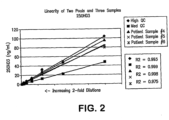
Figure 2 shows the linearity of the quantification of 25OHD3 in serially diluted stock samples using an LC-MS/MS assay. Details are described in Example 4. -
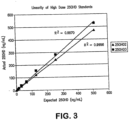
Figure 3 shows the linearity of the quantification by LC-MS/MS of serially diluted samples containing 25OHD2 and 25OHD3 to final concentrations of 512 ng/mL. Details are described in Example 4. -
Figure 4 shows the correlation between detection of total 25-hydroxyvitamin D using an LC-MS/MS assay and a commercially available radioimmunoassay kit. Details are described in Example 6. - Disclosed are methods for detecting the presence or amount of one or more vitamin D metabolites in a sample, wherein the invention claims methods for detecting the amount of one or more vitamin D metabolites in a sample. The method involves ionizing the vitamin D metabolite(s), detecting the ion(s) by mass spectrometry, and relating the presence or amount of the ion(s) to the presence or amount of the vitamin D metabolite(s) in the sample. The method includes (a) purifying a vitamin D metabolite, if present in the sample, (b) ionizing the purified vitamin D metabolite and (c) detecting the presence or amount of the ion, wherein the presence or amount of the ion is related to the presence or amount of the vitamin D metabolite in the sample. The ionizing step (b) includes (i) ionizing a vitamin D metabolite, if present in the sample, to produce an ion; (ii) isolating the vitamin D metabolite ion by mass spectrometry to provide a precursor ion; and (iii) effecting a collision between the isolated precursor ion and an inert collision gas to produce at least one fragment ion detectable in a mass spectrometer. According to the invention, the precursor ion is a protonated and dehydrated ion of the vitamin D metabolite.
- In a related aspect, the application describes a method for determining the presence or amount of a vitamin D metabolite in a test sample by tandem mass spectrometry. The invention provides a method for determining the amount of a vitamin D metabolite in a sample by tandem mass spectrometry. The method involves (a) purifying a plasma or serum sample by liquid chromatography, wherein the sample is not subjected to gas-chromatography; (b) generating a protonated and dehydrated precursor ion of said vitamin D metabolite; (c) generating from said precursor ion one or more fragment ions having a mass-to-charge ratio of 251.30±0.5, 211.35±0.5, 209.20±0.5, or 179.10±0.5; and (d) detecting the amount of one or more of said ions generated in step (b) or (c) or both and relating the detected ions to the amount of said vitamin D metabolite in said sample.
- According to the invention, at least one fragment ion as claimed is detected, wherein amount of the precursor and/or at least one fragment ion is related to amount of the vitamin D metabolite in the sample. Preferably at least one fragment ion is specific for the vitamin D metabolite of interest. A further method of the invention can be used to detect and quantify two or more vitamin D metabolites in a single assay, said method comprising: (a) purifying a plasma or serum sample by liquid chromatography, wherein the sample is not subjected to gas-chromatography; (b) ionizing two or more vitamin D metabolites to generate protonated and dehydrated precursor ions specific for each of said two or more vitamin D metabolites; (c) generating from each of said precursor ions one or more fragment ions having a mass-to-charge ratio of 251.30±0.5, 211.35±0.5, 209.20±0.5, or 179.10±0.5; and (d) detecting the amount of one or more of said ions generated in step (b) or (c) or both and relating the detected ions to the amount of said vitamin D metabolites in said sample. In certain embodiments, the vitamin D metabolite is one or more vitamin D metabolites selected from the group consisting of 25-hydroxyvitamin D3 and 25-hydroxyvitamin D2.
- Suitable samples are plasma and serum. Such samples may be obtained, for example, from a patient seeking diagnosis, prognosis, or treatment of a disease or condition. The vitamin D metabolites may be derivatized prior to mass spectrometry, however, in certain preferred embodiments, sample preparation excludes the use of derivitization.
- Samples may be processed or purified to obtain preparations that are suitable for analysis by mass spectrometry. The methods according to the invention include a step of purifying a plasma or serum sample. Such purification will usually include chromatography, according to the invention liquid chromatography, and may also often involve an additional purification procedure that is performed prior to chromatography. Various procedures may be used for this purpose depending on the type of sample or the type of chromatography. Examples include filtration, extraction, precipitation, centrifugation, dilution, combinations thereof and the like. Protein precipitation is one preferred method of preparing a liquid biological sample, such as serum or plasma, for chromatography. Such protein purification methods are well known in the art, for example, Polson et al., Journal of Chromatography B 785:263-275 (2003), describes protein precipitation methods suitable for use in the methods of the invention. Protein precipitation may be used to remove most of the protein from the sample leaving vitamin D metabolites soluble in the supernatant. The samples can be centrifuged to separate the liquid supernatant from the precipitated proteins. The resultant supernatant can then be applied to liquid chromatography and subsequent mass spectrometry analysis. In one embodiment of the invention, the protein precipitation involves adding one volume of the liquid sample (e.g. plasma) to about four volumes of methanol. In certain embodiments, the use of protein precipitation obviates the need for high turbulence liquid chromatography ("HTLC") or on-line extraction prior to HPLC and mass spectrometry. Accordingly in such embodiments, the method involves (1) performing a protein precipitation of the sample of interest; and (2) loading the supernatant directly onto the HPLC-mass spectrometer without using on-line extraction or high turbulence liquid chromatography ("HTLC").
- The purification step according to the invention includes chromatography, i.e. liquid chromatography, preferably high performance liquid chromatography (HPLC). According to the invention, the chromatography is not gas chromatography. The methods of the invention are performed without subjecting the samples, or the vitamin D metabolites of interest, to gas chromatography prior to mass spectrometric analysis.
- Various methods have been described involving the use of HPLC for sample cleanup prior to mass spectrometry analysis. See, e.g., Taylor et al., Therapeutic Drug Monitoring 22:608-12 (2000) (manual precipitation of blood samples, followed by manual C18 solid phase extraction, injection into an HPLC for chromatography on a C18 analytical column, and MS/MS analysis); and Salm et al., Clin. Therapeutics 22 Supl. B:B71-B85 (2000 ) (manual precipitation of blood samples, followed by manual C 18 solid phase extraction, injection into an HPLC for chromatography on a C18 analytical column, and MS/MS analysis). One of skill in the art can select HPLC instruments and columns that are suitable for use in the invention. The chromatographic column typically includes a medium (i.e., a packing material) to facilitate separation of chemical moieties (i.e., fractionation). The medium may include minute particles. The particles include a bonded surface that interacts with the various chemical moieties to facilitate separation of the chemical moieties such as vitamin D metabolites. One suitable bonded surface is a hydrophobic bonded surface such as an alkyl bonded surface. Alkyl bonded surfaces may include C-4, C-8, or C-18 bonded alkyl groups, preferably C-18 bonded groups. The chromatographic column includes an inlet port for receiving a sample and an outlet port for discharging an effluent that includes the fractionated sample. In the method, the sample (or pre-purified sample) is applied to the column at the inlet port, eluted with a solvent or solvent mixture, and discharged at the outlet port. Different solvent modes may be selected for eluting the analytes of interest. For example, liquid chromatography may be performed using a gradient mode, an isocratic mode, or a polytyptic (i.e. mixed) mode. In preferred embodiments, HPLC is performed on a multiplexed analytical HPLC system with a C18 solid phase using isocratic separation with 100% methanol as the mobile phase.
- Recently, high turbulence liquid chromatography ("HTLC"), also called high throughput liquid chromatography, has been applied for sample preparation prior to analysis by mass spectrometry. See, e.g., Zimmer et al., J. Chromatogr. A 854:23-35
- (1999); see also,
U.S. Patents Nos. 5,968,367 ;5,919,368 ;5,795,469 ; and5,772,874 . Traditional HPLC analysis relies on column packings in which laminar flow of the sample through the column is the basis for separation of the analyte of interest from the sample. The skilled artisan will understand that separation in such columns is a diffusional process. In contrast, it is believed that turbulent flow, such as that provided by HTLC columns and methods, may enhance the rate of mass transfer, improving the separation characteristics provided. In some embodiments, high turbulence liquid chromatography (HTLC), alone or in combination with one or more purification methods, may be used to purify the vitamin D metabolite of interest prior to mass spectrometry. In such embodiments samples may be extracted using an HTLC extraction cartridge which captures the analyte, then eluted and chromatographed on a second HTLC column or onto an analytical HPLC column prior to ionization. Because the steps involved in these chromatography procedures can be linked in an automated fashion, the requirement for operator involvement during the purification of the analyte can be minimized. In certain embodiments of the method, samples are subjected to protein precipitation as described above prior to loading on the HTLC column; in alternative embodiments, the samples may be loaded directly onto the HTLC without being subjected to protein precipitation. - Recently, research has shown that epimerization of the hydroxyl group of the A-ring of vitamin D3 metabolites is an important aspect of vitamin D3 metabolism and bioactivation, and that depending on the cell types involved, 3-C epimers of vitamin D3 metabolites (e.g., 3-epi-25(OH)D3; 3-epi-24,25(OH)2D3; and 3-epi-1,25(OH)2D3) are often major metabolic products. See Kamao et al., J. Biol. Chem., 279:15897-15907 (2004). Kamao et al., further provides methods of separating various vitamin D metabolites, including 3-C epimers, using Chiral HPLC. Accordingly, the invention also provides methods capable of detecting the presence, absence and/or amount of a specific epimer of one or more vitamin D metabolites, preferably vitamin D3 metabolites, in a sample by (1) separating one or more specific vitamin D metabolites by chiral chromatography, preferably chiral HPLC; and (2) detecting the presence and/or amount of one or more vitamin D metabolites using mass spectrometry methods as described herein. The chiral chromatography procedures described in Kamao et al., are suitable for the methods of the invention, however, one of ordinary skill in the art understands that there are numerous other chiral chromatography methods that would also be suitable. In preferred embodiments the method includes, separating 25(OH)D3 from 3-epi-25(OH)D3, if present in a sample, using chiral chromatography; and detecting the presence and/or amount of the 25(OH)D3 and the 3-epi-25(OH)D3 in the sample using mass spectrometry. In related embodiments, the method includes separating 1a,25(OH)2D3 from 3-epi-1α,25(OH)2D3, if present in a sample, using chiral chromatography; and detecting the presence and/or amount of the 1α,25(OH)2D3 and the 3-epi-1α,25(OH)2D3 in the sample using mass spectrometry. In certain embodiments of the invention, chiral chromatography is used in conjunction with the HTLC methods described above.
- Mass spectrometry is performed using a mass spectrometer which includes an ion source for ionizing the fractionated sample and creating charged molecules for further analysis. For example ionization of the sample may be performed by electrospray ionization (ESI), atmospheric pressure chemical ionization (APCI), photoinonization, electron ionization, fast atom bombardment (FAB)/liquid secondary ionization (LSIMS), matrix assisted laser desorption ionization (MALDI), field ionization, field desorption, thermospray/plasmaspray ionization, and particle beam ionization. The skilled artisan will understand that the choice of ionization method can be determined based on the analyte to be measured, type of sample, the type of detector, the choice of positive versus negative mode, etc.
- After the sample has been ionized, the positively charged or negatively charged ions thereby created may be analyzed to determine a mass-to-charge ratio (i.e., m/z). The methods according to the invention include a step comprising generating a protonated and dehydrated precursor ion. Suitable analyzers for determining mass-to-charge ratios include quadropole analyzers, ion traps analyzers, and time-of-flight analyzers. The ions may be detected using several detection modes. For example, selected ions may be detected (i.e., using a selective ion monitoring mode (SIM)), or alternatively, ions may be detected using a scanning mode, e.g., multiple reaction monitoring (MRM) or selected reaction monitoring (SRM). Preferably, the mass-to-charge ratio is determined using a quadropole analyzer. For example, in a "quadrupole" or "quadrupole ion trap" instrument, ions in an oscillating radio frequency field experience a force proportional to the DC potential applied between electrodes, the amplitude of the RF signal, and m/z. The voltage and amplitude can be selected so that only ions having a particular m/z travel the length of the quadrupole, while all other ions are deflected. Thus, quadrupole instruments can act as both a "mass filter" and as a "mass detector" for the ions injected into the instrument.
- One can often enhance the resolution of the MS technique by employing "tandem mass spectrometry," or "MS/MS." In this technique, a precursor ion (also called a parent ion) generated from a molecule of interest can be filtered in an MS instrument, and the precursor ion is subsequently fragmented to yield one or more fragment ions (also called daughter ions or product ions) that are then analyzed in a second MS procedure. By careful selection of precursor ions, only ions produced by certain analytes are passed to the fragmentation chamber, where collision with atoms of an inert gas to produce the daughter ions. Because both the precursor and fragment ions are produced in a reproducible fashion under a given set of ionization/fragmentation conditions, the MS/MS technique can provide an extremely powerful analytical tool. For example, the combination of filtration/fragmentation can be used to eliminate interfering substances, and can be particularly useful in complex samples, such as biological samples.
- Additionally, recent advances in technology, such as matrix-assisted laser desorption ionization coupled with time-of-flight analyzers ("MALDI-TOF") permit the analysis of analytes at femtomole levels in very short ion pulses. Mass spectrometers that combine time-of-flight analyzers with tandem MS are also well known to the artisan. Additionally, multiple mass spectrometry steps can be combined in methods known as "MS/MS"." Various other combinations may be employed, such as MS/MS/TOF, MALDI/MS/MS/TOF, or SELDI/MS/MS/TOF mass spectrometry.
- The mass spectrometer typically provides the user with an ion scan; that is, the relative abundance of each ion with a particular m/z over a given range (e.g., 100 to 1000 amu). The results of an analyte assay, that is, a mass spectrum, can be related to the amount of the analyte in the original sample by numerous methods known in the art. For example, given that sampling and analysis parameters are carefully controlled, the relative abundance of a given ion can be compared to a table that converts that relative abundance to an absolute amount of the original molecule. Alternatively, molecular standards can be run with the samples, and a standard curve constructed based on ions generated from those standards. Using such a standard curve, the relative abundance of a given ion can be converted into an absolute amount of the original molecule. In certain preferred embodiments, an internal standard is used to generate a standard curve for calculating the quantity of the vitamin D metabolite. Methods of generating and using such standard curves are well known in the art and one of ordinary skill is capable of selecting an appropriate internal standard. For example, an isotope of a vitamin D metabolite may be used as an internal standard, in preferred embodiments the vitamin D metabolite is a deuterated vitamin D metabolite, for example 6D-25OHD3. Numerous other methods for relating the presence or amount of an ion to the presence or amount of the original molecule will be well known to those of ordinary skill in the art.
- One or more steps of the methods of the invention can be performed using automated machines. In certain embodiments, one or more purification steps are performed on line, and more preferably all of the purification and mass spectrometry steps may be performed in an on-line fashion.
- In particularly preferred embodiments vitamin D metabolites are detected and/or quantified using LC-MS/MS as follows. The samples are subjected to liquid chromatography, preferably HPLC, the flow of liquid solvent from the chromatographic column enters the heated nebulizer interface of a LC-MS/MS analyzer and the solvent/analyte mixture is converted to vapor in the heated tubing of the interface. The analytes (i.e. vitamin D metabolites), contained in the nebulized solvent, are ionized by the corona discharge needle of the interface, which applies a large voltage to the nebulized solvent/analyte mixture. The ions, i.e. precursor ions, pass through the orifice of the instrument and enter the first quadrupole. Quadrupoles 1 and 3 (Q1 and Q3) are mass filters, allowing selection of ions (i.e., "precursor" and "fragment" ions) based on their mass to charge ratio (m/z). Quadrupole 2 (Q2) is the collision cell, where ions are fragmented. The first quadrupole of the mass spectrometer (Q1) selects for molecules with the mass to charge ratios of the specific vitamin D metabolites to be analyzed. Precursor ions with the correct m/z ratios of the precursor ions of specific vitamin D metabolites are allowed to pass into the collision chamber (Q2), while unwanted ions with any other m/z collide with the sides of the quadrupole and are eliminated. Precursor ions entering Q2 collide with neutral Argon gas molecules and fragment. This process is called Collision Activated Dissociation (CAD). The fragment ions generated are passed into quadrupole 3 (Q3), where the fragment ions of the desired vitamin D metabolites are selected while other ions are eliminated.
- The methods of the invention may involve MS/MS performed in either positive or negative ion mode. Using standard methods well known in the art, one of ordinary skill is capable of identifying one or more fragment ions of a particular precursor ion of a vitamin D metabolite that can be used for selection in quadrupole 3 (Q3). Preferably, at least one fragment ion of the method is specific for the particular vitamin D metabolite of which detection is desired. The method according to
claim 6 includes a step (b) of ionizing two or more vitamin D metabolites to generate protonated and dehydrated precursor ions specific for each of said two or more vitamin D metabolites. A specific fragment ion for a particular vitamin D metabolite is one that will not be formed in significant amounts by other molecules with similar molecular structures. In contrast a non-specific fragment ion is one that is formed by related molecules other than the desired analyte. Therefore, detection of non-specific fragment ions alone is not reliable for distinguishing the desired vitamin D metabolite from other molecules that form the same or similar fragment ions. Specific fragment ions of a particular vitamin D metabolite can be identified by testing various molecular standards (e.g. vitamin D metabolites other than the metabolite to be detected) to determine whether fragment ions formed by the vitamin D metabolite of interest are also formed by other molecules with similar structures or features. In certain particularly preferred embodiments, a specific fragment ion is identified by testing at least one molecular standard that forms a precursor ion with the same m/z as the vitamin D metabolite to be detected. - If the precursor ion of a vitamin D metabolite of interest includes an alcohol or amine group, fragment ions are commonly formed that represent a dehydration or deamination of the precursor ion, respectfully. In the case of precursor ions that include an alcohol group, such fragment ions formed by dehydration are caused by a loss of one or more water molecules from the precursor ion (i.e., where the difference in m/z between the precursor ion and fragment ion is about 18 for the loss of one water molecule, or about 36 for the loss of two water molecules, etc.). In the case of precursor ions that include an amine group, such fragment ions formed by deamination are caused by a loss of one or more ammonia molecules (i.e. where the difference in m/z between the precursor ion and fragment ion is about 17 for the loss of one ammonia molecule, or about 34 for the loss of two ammonia molecules, etc.). Likewise, precursor ions that include one or more alcohol and amine groups commonly form fragment ions that represent the loss of one or more water molecules and/or one or more ammonia molecules (e.g., where the difference in m/z between the precursor ion and fragment ion is about 35 for the loss of one water molecule and the loss of one ammonia molecule). Generally, the fragment ions that represent dehydrations or deaminations of the precursor ion are not specific fragment ions for a particular analyte. For example, MS/MS performed to detect 25OHD2 by selecting for a precursor ion at 413 m/z (i.e. the protonated and hydrated ion) and detecting a fragment ion of 395 m/z (representing a dehydration of the 413 m/z precursor ion) would not be able to distinguish 25OHD2 from 1α(OH)D2 which would form the same precursor and fragment ions. Therefore a 395 m/z fragment ion is not a specific fragment ion for 25OHD2 or 1α(OH)D2. Accordingly, in preferred embodiments of the invention, MS/MS is performed such that at least one fragment ion of a vitamin D metabolite is detected that does not represent only a loss of one or more water molecules and/or a loss of one or more ammonia molecules from the precursor ion.
- As ions collide with the detector they produce a pulse of electrons that are converted to a digital signal. The acquired data is relayed to a computer, which plots counts of the ions collected versus time. The resulting mass chromatograms are similar to chromatograms generated in traditional HPLC methods. The areas under the peaks corresponding to particular ions, or the amplitude of such peaks, are measured and the area or amplitude is correlated to the amount of the analyte (vitamin D metabolite) of interest. In certain embodiments, the area under the curves, or amplitude of the peaks, for fragment ion(s) and/or precursor ions are measured to determine the amount of a vitamin D metabolite. As described above, the relative abundance of a given ion can be converted into an absolute amount of the original analyte, i.e., vitamin D metabolite, using calibration standard curves based on peaks of one or more ions of an internal molecular standard, such as 6D-25OHD3.
- In certain aspects of the invention, the quantity of various ions is determined by measuring the area under the curve or the amplitude of the peak and a ratio of the quantities of the ions is calculated and monitored (i.e. "daughter ion ratio monitoring"). In certain embodiments of the method, the ratio(s) of the quantity of a precursor ion and the quantity of one or more fragment ions of a vitamin D metabolite can be calculated and compared to the ratio(s) of a molecular standard of the vitamin D metabolite similarly measured. In embodiments where more than one fragment ion of a vitamin D metabolite is monitored, the ratio(s) for different fragment ions may be determined instead of, or in addition to, the ratio of the fragment ion(s) compared to the precursor ion. In embodiments where such ratios are monitored, if there is a substantial difference in an ion ratio in the sample as compared to the molecular standard, it is likely that a molecule in the sample is interfering with the results. To the contrary, if the ion ratios in the sample and the molecular standard are similar, then there is increased confidence that there is no interference. Accordingly, monitoring such ratios in the samples and comparing the ratios to those of authentic molecular standards may be used to increase the accuracy of the method.
- In certain aspects of the invention, MS/MS is performed in positive ion mode with the first quadruple (Q1) tuned to select for precursor ions with a mass charge ratio corresponding to protonated and dehydrated ions of vitamin D metabolites. The mass/charge ratio (m/z) for the protonated and dehydrated precursor vitamin D metabolite ions is about 383.16 m/z for 25-hydroxyvitamin D3 and about 395.30 m/z for 25-hydroxyvitamin D2. In embodiments where the samples are spiked with hexadeuterated 25OHD3 (6D-25OHD3) for use as an internal standard, the mass/charge ratio (m/z) of the protonated and dehydrated 6D-25OHD3 precursor ion is about 389.20. In certain preferred embodiments of the invention, the mass/charge ratio (m/z) for the 25-hydroxyvitamin D3 precursor ion is about 383.16 and the m/z for at least one 25-hydroxyvitamin D3 fragment ion is about 211.35. In related embodiments, the m/z for the 25-hydroxyvitatnin D2 precursor ion is about 395.30 and the 25-hydroxyvitamin D2 fragment ion(s) include one or more ions selected from the group consisting of ions with mass/charge ratios (m/z) of about 179.10, about 209.20 and about 251.30. In embodiments where the samples are spiked with 6D-25OHD3 for use as an internal standard the mass/charge ratio (m/z) for the protonated and dehydrated 6D-25OHD3 precursor ion is about 389.20 and the fragment ion(s) may include a fragment ion with a m/z of about 211.30.
- In other aspects, MS/MS is performed in positive ion mode with the first quadruple (Q1) tuned to select for precursor ions with a mass charge ratio corresponding to protonated and hydrated ions of vitamin D metabolites. The mass/charge ratio (m/z) for the protonated and hydrated precursor vitamin D metabolite ions are about 401 m/z for 25-hydroxyvitamin D3 and about 413 m/z for 25-hydroxyvitamin D2. In certain preferred embodiments of the invention, the mass/charge ratio (m/z) for the 25-hydroxyvitamin D3 precursor ion is about 401 and the 25-hydroxyvitamin D3 fragment ion(s) include one or more ions selected from the group consisting of ions with mass/charge ratios (m/z) of about 365 and about 383 m/z; more preferably the fragment ions include an ion with a m/z of about 365. In related embodiments, the m/z for the protonated and hydrated 25-hydroxyvitamin D2 precursor ion is about 413 m/z, and the 25-hydroxyvitamin D2 fragment ion(s) include one or more ions with mass/charge ratios (m/z) of about 377 and about 395; more preferably the fragment ions include an ion with a mass charge ratio of about 377.
- In particularly preferred embodiments of the invention, the presence or absence or amount of two or more vitamin D metabolites in a sample are detected in a single assay using the above described MS/MS methods.
- Mass spectrometry instruments can vary slightly in determining the mass of a given analyte. Thus, the term "about" in the context of mass of an ion or the m/z of an ion refers to +/- 0.5 atomic mass unit.
- The following examples serve to illustrate the invention. These examples are in no way intended to limit the scope of the invention.
- Using a Perkin-Elmer MultiProbe II (S/N 432400) robotic liquid handler, human serum samples were first extracted using a protein precipitation method by adding 42.5 µl of serum to170 µl of methanol (1:4 ratio of serum:methanol) in a 96-well plate format. For validation-related experiments, the methanol was spiked with hexadeuterated 25OHD3 (6D-25OHD3) as an internal standard. The 96 well plates were centrifuged to remove precipitated protein, leaving the vitamin D metabolites in the supernatant. The supernatants were then transferred to an HPLC autosampler for loading to the LC-MS/MS analyzer.
- LC-MS/MS was performed using a Thermo Finnigan LC-MS/MS analyzer (Thermo Finnigan Quantum TSQ (S/N: TQU00655)) with an atmospheric pressure chemical ionization (APCI) source as the detector. An autosampler was used to inject 50 µL of extracted sample supernatant onto an HPLC column. Liquid chromatography was performed with a Cohesive Technologies Aria TX-4 (S/N: SJCTX409) LC system with Waters Symmetry C18 5µm 4.6x50mm columns using 100% methanol as the mobile phase. After the analytes eluted and the detector window completed acquisition, the system was washed with 85% Mobile phase A and then re-equilibrated with Mobile phase B for a run time of 5 minutes. Mobile phase A was 0.1% formic acid in HPLC-grade water and mobile phase B was 100% methanol.
- The flow of liquid solvent exiting the HPLC column entered the heated nebulizer interface of the Thermo Finnigan LC-MS/MS analyzer. The solvent/analyte mixture was first converted to vapor in the heated tubing of the interface. The analytes, contained in the nebulized solvent, were ionized (a positive charge added) by the corona discharge needle of the interface, which applies a large voltage to the nebulized solvent/analyte mixture. The ions pass through the orifice of the instrument and enter the first quadrupole. Quadrupoles 1 and 3 (Q1 and Q3) are mass filters, allowing selection of ions based on their mass to charge ratio (m/z). Quadrupole 2 (Q2) is the collision cell, where ions are fragmented.
- The first quadrupole of the mass spectrometer (Q1) selected for molecules with the mass to charge ratios of (protonated and dehydrated) 25OHD2, 25OHD3 and 6D-25OHD3. Ions with these m/z ratios (see table below) were allowed to pass into the collision chamber (Q2), while unwanted ions with any other m/z collide with the sides of the quadrupole and are eliminated. Ions entering Q2 collide with neutral Argon gas molecules and fragment. The fragment ions generated are passed into quadrupole 3 (Q3), where the fragment ions of 25OHD2, 25OHD3 and 6D-25OHD3 were selected (see table below) and other ions are eliminated. The following mass transitions were used for detection and quantitation during validation:
Table 1. Mass transitions for selected vitamin D metabolites Compound Precursor Ion (m/z) Fragment Ions (m/z) 25OHD2 395.30 179.10,251.30,209.20 25OHD3 383.16 211.35 6D-25OHD3 389.20 211.30 - As ions collide with the detector they produce a pulse of electrons that are converted to a digital signal. The acquired data is relayed to a computer, which plots counts of the ions collected versus time. The resulting mass chromatograms are similar to chromatograms generated in traditional HPLC methods.
- Area ratios of the analyte and internal standard (Hexadeuterated 25-Hydroxyvitamin D3, 6D-25OHD3) peaks were used to construct calibration curves, which were then used to calculate analyte concentrations. Using the calibration curves, the concentrations of 25OHD2 and 25OHD3 were quantitated in the patient samples.
- Stock solutions of 25OHD2 and 25OHD3 were added to pooled serum to produce a Low Pool (20-25 ng/mL of each metabolite), a Medium Pool (45-55 ng/mL of each metabolite) and a High Pool (100-110 ng/mL). Pooled patient serum was used for the Medium and Low Pools, and stripped serum from Golden West Biologicals, Product #SP1070, was used for the Low Pool. Twenty aliquots from each of the Low, Medium and High Pools were analyzed in a single assay using the LC-MS/MS protocols described in Example 1. The following precision values were determined:
Table 2. Intra-Assay Variation: 25-Hvdroxyvitamin D2 (25OHD2) Low 092804-L Medium 092804-M High 092804-H 1 26.5 46.4 103.3 2 23.2 51.1 96.7 3 23.1 52.4 107.8 4 21.6 50.3 104.5 5 26.3 47.5 96.2 6 25.1 54.4 98.5 7 25.9 54.6 100.0 8 21.9 50.1 110.1 9 23.4 50.8 97.6 10 23.5 53.2 105.1 11 22.2 52.9 105.9 12 24.0 54.6 94.5 13 26.2 49.4 93.4 14 24.1 59.0 113.0 15 25.8 52.9 112.4 16 23.9 59.2 113.4 17 29.5 52.4 107.7 18 24.2 50.0 115.5 19 19.8 53.5 114.9 20 26.3 60.2 126.6 Average (ng/mL) 24.3 52.7 105.9 Std Dev 2.2 3.6 8.6 CV (%) 9.0 6.9 8.1 Table 3. Intra-Assay Variation: 25-Hydroxyvitamin D3 (25OHD3) Low 092804-L Medium 092804-M High 092804-H 1 22.7 43.6 99.1 2 22.4 45.3 93.5 3 22.4 50.7 98.2 4 21.0 40.1 95.9 5 21.8 41.5 82.0 6 20.8 42.2 97.4 7 22.9 50.1 96.0 8 19.0 42.0 106.7 9 21.8 44.2 96.6 10 23.4 49.5 94.9 11 21.8 46.5 97.9 12 20.7 49.9 87.1 13 25.4 44.7 85.5 14 24.5 48.0 101.5 15 25.1 45.8 101.5 16 22.5 52.0 104.7 17 29.2 45.9 107.7 18 19.5 49.3 107.6 19 18.1 49.6 109.4 20 24.8 49.3 116.1 Average (ng/mL) 22.5 46.5 99.0 Std Dev 2.5 3.5 8.4 CV (%) 11.2 7.5 8.5 - Each of the Low, Medium and High Pools described above were also analyzed to determine inter-assay precision. Four aliquots from each pool were analyzed over five different assays using the LC-MS/MS protocols described in Example 1. The following precision values were determined:
Table 4. Inter-Assay Variation: 25-Hydroxyvitamin D2 (25OHD2) Low 092804-L medium 092804-M High 092804-H 1 26.5 46.4 103.3 2 23.2 51.1 96.7 3 23.1 52.4 107.8 4 21.6 50.3 104.5 5 26.3 47.5 96.2 6 25.1 54.4 98.5 7 25.9 54.6 100.0 8 21.9 50.1 110.1 9 23.4 50.8 97.6 10 23.5 53.2 105.1 11 22.2 52.9 105.9 12 24.0 54.6 94.5 13 26.2 49.4 93.4 14 24.1 59.0 113.0 15 25.8 52.9 112.4 16 23.9 59.2 113.4 17 29.5 52.4 107.7 18 24.2 50.0 115.5 19 19.8 53.5 114.9 20 26.3 60.2 126.6 Average (ng/mL) 24.3 52.7 105.9 Std Dev 2.2 3.6 8.6 CV (%) 9.0 6.9 8.1 Table 5. Inter-Assay Variation: 25-Hydroxyvitamin D3 (25OHD3) Low 092804-L Medium 092804-M High 092804-H 1 22.7 43.6 99.1 2 22.4 45.3 93.5 3 22.4 50.7 98.2 4 21.0 40.1 95.9 5 21.8 41.5 82.0 6 20.8 42.2 97.4 7 22.9 50.1 96.0 8 19.0 42.0 106.7 9 21.8 44.2 96.6 10 23.4 49.5 94.9 11 21.8 46.5 97.9 12 20.7 49.9 87.1 13 25.4 44.7 85.5 14 24.5 48.0 101.5 15 25.1 45.8 101.5 16 22.5 52.0 104.7 17 29.2 45.9 107.7 18 19.5 49.3 107.6 19 18.1 49.6 109.4 20 24.8 49.3 116.1 Average (ng/mL) 22.5 46.5 99.0 Std Dev 2.5 3.5 8.4 CV (%) 11.2 7.5 8.5 - To determine the limit of detection of the assay, blank diluent was analyzed 17 times within a single run using the LC-MS/MS protocols described in Example 1. The mean and standard deviation were then calculated. The limit of detection was determined as 2 SD above the mean of the blank peak area ratio based on a back calculation of peak area ratio against the calibration curve. The limits of detection were as follows:
- 25OHD2: 3.0 ng/mL
- 25OHD3: 3.5 ng/mL
- To determine the limit of quantitation, stock solutions of 25OHD2 and 25OHD3 were used to generate standard curves with the following concentrations: 0, 2, 4, 8, 16, 32, 64 and 128 ng/mL. The diluted samples of the standard curve were analyzed in quadruplicate over five assays using the LC-MS/MS assay described in Example 1. The results of the study were as follows:
Table 6. Limit of Quantitation Study Results: 25-Hydroxyvitamin D2 (25OHD7) #1 #2 #3 #4 #5 Summary 0 ng/mL -1.2 -1.3 -0.5 -1.2 -1.1 Average (ng/mL) -1.0 -1.1 -1.4 -0.9 -1.3 -0.7 Standard Deviation 0.6 -1.1 -1.5 -0.8 NA -2.6 C of V (%) 59.0 0.4 -1.7 -0.6 -0.5 -0.4 Accuracy (%) N/A 2 ng/mL 3.1 2.3 2.0 2.1 3.3 Average (ng/mL) 1.9 2.1 2.7 2.1 2.4 1.9 Standard Deviation 0.6 1.1 1.9 1.9 0.9 1.3 C of V (%) 33.7 1.2 1.1 2.1 1.8 1.4 Accuracy (%) 103.9 4 ng/mL 3.5 3.9 5.0 4.5 4.8 Average (ng/mL) 3.9 4.0 3.0 3.8 3.8 4.7 Standard Deviation 0.7 3.6 2.9 2.8 3.1 2.0* C of V (%) 17.1 4.1 4.6 4.5 4.4 3.7 Accuracy (%) 101.7 8 ng/mL 10.2 9.1 9.1 8.8 9.8 Average (ng/mL) 8.6 7.8 8.1 7.9 8.4 9.0 Standard Deviation 0.8 8.6 8.3 7.4 8.4 7.5 C of V (%) 9.3 10.2 8.4 8.0 8.1 8.6 Accuracy (%) 93.2 16 ng/mL 16.0 14.8 14.4 16.7 18.3 Average (ng/mL) 16.0 15.5 15.6 15.3 16.7 16.8 Standard Deviation 1.1 16.6 16.7 16.8 15.8 15.2 C of V (%) 7.1 14.1 17.6 16.1 16.7 14.1 Accuracy (%) 100.1 32 ng/m L 31.3 39.9* 29.9 33.2 32.7 Average (ng/mL) 31.8 31.7 30.5 32.4 34.0 32.5 Standard Deviation 1.9 29.5 31.2 30.2 35.7 28.7 C of V (%) 6.0 32.9 34.7 32.4 30.8 29.2 Accuracy (%) 100.7 64 ng/mL 66.5 62.2 68.5 62.6 68.8 Average (ng/mL) 64.6 66.6 67.8 67.3 58.9 61.5 Standard Deviation 3.2 64.4 61.4 63.7 63.5 61.3 C of V (%) 4.9 63.7 60.7 65.4 70.8 65.9 Accuracy (%) 99.1 128 ng/mL 125.1 128.2 123.4 127.8 124.1 Average (ng/mL) 126.9 127.6 134.4 127.3 128.4 132.1 Standard Deviation 3.5 128.9 124.5 128.5 126.5 131.7 C of V (%) 2.8 126.1 119.7 127.9 121.2 125.0 Accuracy (%) 100.8 Table 7. Limit of Quantitation Study Results: 25-Hydroxyvitamin D3 (25OHD3) Day #1 (11/19/04-1) Day #2 (11/19/04-2) Day #3 (11/22/04-1) Day #4 (11/23/04-4) Day #5 (11/23/04-2) Summary 0 ng/mL -0.5 -0.9 -0.3 0.2 -0.6 Average (nq/m L) -0.7 -0.7 -1.3 0.3 -1.1 -1.0 Standard Deviation 0.6 0.0 -1.0 -1.1 -1.3 0.6 C of V (%) 86.4 -0.3 -1.5 -1.1 -0.8 -1.0 Accuracy (%) N/A 2 ng/mL 2.6 1.5 2.4 2.5 1.7 Average (nq/m L) 1.9 1.7 0.9 3.2 2.2 1.9 Standard Deviation 0.6 1.8 1.8 2.0 1.1 2.2 C of V (%) 31.4 1.3 2.4 1.5 1.1 2.5 Accuracy (%) 104.7 4 ng/mL 3.9 3.5 4.8 4.0 3.4 Average (nq/m L) 3.8 3.0 4.1 3.1 3.9 3.8 Standard Deviation 0.8 4.5 3.7 2.9 3.5 2.4 C of V (%) 19.7 4.0 5.2 3.8 3.8 5.5 Accuracy (%) 104.1 8 ng/mL 10.3 9.3 7.2 8.4 8.6 Average (nq/m L) 8.7 10.6 8.5 7.2 10.0 9.6 Standard Deviation 1.1 7.4 10.3 9.0 9.4 8.4 C of V (%) 13.1 9.3 7.7 8.5 7.6 6.8 Accuracy (%) 91.9 16 ng/mL 15.9 15.6 16.2 18.6 17.1 Average (ng/mL) 16.0 13.8 16.3 14.0 17.2 15.3 Standard Deviation 1.4 15.6 16.1 15.1 18.8 16.1 C of V (%) 8.5 14.8 17.2 17.0 14.3 15.3 Accuracy (%) 99.9 32 ng/mL 31.1 35.8 29.6 32.8 28.0 Average (nq/m L) 31.7 30.8 29.9 31.8 33.0 32.8 Standard Deviation 1.9 31.6 30.9 29.5 35.7 31.2 C of V (%) 6.2 31.0 34.2 31.8 30.6 31.7 Accuracy (%) 101.0 64 ng/mL 65.9 64.6 64.4 64.8 67.8 Average (nq/m L) 64.8 67.4 62.9 62.4 60.7 57.2 Standard Deviation 3.1 68.9 64.2 62.1 64.0 64.7 C of V (%) 4.8 63.2 64.2 66.8 67.7 71.1 Accuracy (%) 98.8 128 ng/mL 128.9 124.5 126.4 125.3 122.8 Average (nq/m L) 127.1 129.7 135.7 128.3 125.9 128.7 Standard Deviation 4.7 125.5 123.3 127.2 127.7 135.4 C of V (%) 3.7 121.3 121.8 137.8 121.4 124.1 Accuracy (%) 100.7 - To establish the linearity of the vitamin D metabolite LC-MS/MS assay, a MultiProbe automated liquid handler robot independently constructed two standard curves by serially diluting a stock solution containing 128 ng/mL 25OHD2 and 128 ng/mL 25OHD3 in 25OHD with a solution of 5% Bovine Serum Albumin Fraction V dissolved in 0.01M PBS. The standard curve samples were analyzed using the LC-MS/MS protocols described in Example 1. This process routinely produced standard curves with R2 values of 0.99 or higher for each analyte for the range of 4-128 ng/mL.
- To determine whether patient samples can also be diluted in a linear fashion, a total of eight samples were serially diluted with 25OHD diluent. Two samples were patient pools (Medium and High Control Pools), three were patient samples with high 25OHD2 values and three were patient samples with high 25OHD3 values. All samples were analyzed using the LC-MS/MS protocols described in Example 1. As shown in
figure 1 andfigure 2 , each sample diluted in a linear fashion (R2 > 0.98), demonstrating the linear range of the assay. - Additionally, solutions of 25OHD2 and 25HOD3 at 512 ng/mL in 5% Bovine Serum Albumin Fraction V dissolved in 0.01M PBS were prepared and then serially diluted to 8 ng/mL. Each sample was extracted and run in duplicate using the LC-MS/MS protocols described in Example 1. As shown in
figure 3 , each of these curves was linear (R2 > 0.99). - The stock solutions of 25OHD2 and 25OHD3 were quantified based upon the absorbance of the concentrated (10-50 µg/mL) stock solutions in the ultraviolet spectrum. The cis-triene chromophore present in all vitamin D compounds has a peak absorbance of 264 nm, which is dependent upon the analyte concentration. The molar extinction coefficient of 18.3 mM-1 cm-1 was determined using purified, dessicated ergocalciferol and cholecalciferol and was used to determine the concentration of stock solutions for 25OHD2 and 25OHD3.
- To determine the ability to recover vitamin D metabolites from spiked serum samples, three patient pools with known levels of 25OHD2, 25OHD3 were spiked with two levels of 25OHD2, 25OHD3 and both 25OHD2 and 25OHD3 together. Each sample was extracted and run in duplicate using the LC-MS/MS protocols described in Example 1. The recovery was calculated by dividing the expected result by the observed result.
Table 8. Recovery of 25-hydroxylated vitamin D metabolites from spiked samples. 25OHD2 (ng/mL) 25OHD3 (ng/mL) 25OHD2 (% Recovery) 25OHD3 (% Recovery) Pool #1 58 51 - - Pool#1 + 20 ng/mL 25OHD2 75 52 104 - Pool#1 + 20 ng/mL 25OHD3 52 68 - 105 Pool#1 + 20 ng/mL of both 72 68 109 105 Pool#1 + 50 ng/mL 25OHD2 104 50 105 - Pool#1 + 50 ng/mL 25OHD3 56 109 - 93 Pool#1 + 50 ng/mL of both 104 110 104 92 Pool #2 53 47 - - Pool#2+ 20 ng/mL 25OHD2 76 51 97 - Pool#2+ 20 ng/mL 25OHD3 52 66 - 101 Pool#2+ 20 ng/mL of both 70 65 105 103 Pool#2+ 50 ng/mL 25OHD2 107 53 96 - Pool#2+ 50 ng/mL 25OHD3 57 109 - 88 Pool#2+ 50 ng/mL of both 100 105 103 92 Pool #3 53 47 - - Pool#3+ 20 ng/mL 25OHD2 69 44 106 - Pool#3+ 20 ng/mL 25OHD3 55 75 - 90 Pool#3+ 20 ng/mL of both 74 69 98 97 Pool#3+ 50 ng/mL 25OHD2 96 48 107 - Pool#3+ 50 ng/mL 25OHD3 53 114 - 85 Pool#3+ 50 ng/mL of both 105 113 98 85 - A total of 1,057 patient samples were assayed using the LC-MS/MS methods described in Example 1 and a vitamin D radioimmunoassay commercially available from DiaSorin.
Figure 4 shows the correlation between detection of total 25-hydroxyvitamin D using the LC-MS/MS assay and a commercially available radioimmunoassay kit; the R2 value was 0.5082 with a slope of 0.9684. - Samples containing various commercially available vitamin D metabolites and analogues at a concentration of 100 ng/mL were prepared. The samples were extracted and run in duplicate using the LC-MS/MS methods described in Example 1. None of the tested compounds exhibited detectable cross reactivity in the 25OHD2 and 25OHD3 assays.
Table 9. Cross-Reactivity of the LC-MS/MS method with various vitamin D analogues and metabolites. Compound Mass (Da) Cross-Reactivity (250HD2) Cross-Reactivity (25OHD3) 25-Hydroxyvitamin D2 (25OHD2) 412 (100%) ND 25-Hydroxyvitamin D3 (25OHD3) 400 ND (100%) Internal Standard (6D-25OHD3) 406 ND ND Vitamin D2 (Ergocalciferol) 396 ND ND Vitamin D3 (Cholecalciferol) 384 ND ND 1α,25(OH)2D2 428 ND ND 1α,25(OH)2D3 416 ND ND 25,26(OH)2D3 416 ND ND 1α(OH)D2 (Doxercalciferol) 412 ND ND 1α(OH)D3 (Alfacalcidiol) 400 ND ND - The invention/scope of protection is defined by the set of appended claims. Any subject-matter falling outside the scope of the claims is provided for information purposes only.
Claims (9)
- A method for determining the amount of a vitamin D metabolite in a sample by tandem mass spectrometry, comprising:(a) purifying a plasma or serum sample by liquid chromatography, wherein the sample is not subjected to gas-chromatography;(b generating a protonated and dehydrated precursor ion of said vitamin D metabolite;(c) generating from said precursor ion one or more fragment ions having a mass-to-charge ratio of 251.30±0.5, 211.35±0.5, 209.20±0.5, or 179.10±0.5; and(d) detecting the amount of one or more of said ions generated in step (b) or (c) or both and relating the detected ions to the amount of said vitamin D metabolite in said sample.
- The method of claim 1, wherein said liquid chromatography comprises high performance liquid chromatography (HPLC).
- The method of claim 1, wherein said purification step further comprises protein precipitation prior to liquid chromatography.
- The method of claim 1, wherein said purification step does not include high turbulence liquid chromatography (HTLC).
- The method of claim 1, wherein said vitamin D metabolite is 25-hydroxyvitamin D3 or 25-hydroxyvitamin D2.
- A method for determining the amount of two or more vitamin D metabolites in a sample in a single assay, said method comprising:(a) purifying a plasma or serum sample by liquid chromatography, wherein the sample is not subjected to gas-chromatography;(b) ionizing two or more vitamin D metabolites to generate protonated and dehydrated precursor ions specific for each of said two or more vitamin D metabolites;(c) generating from each of said precursor ions one or more fragment ions having a mass-to-charge ratio of 251.30±0.5, 211.35±0.5, 209.20±0.5, or 179.10±0.5; and(d) detecting the amount of one or more of said ions generated in step (b) or (c) or both and relating the detected ions to the amount of said vitamin D metabolites in said sample.
- The method of claim 6, wherein said liquid chromatography comprises high performance liquid chromatography (HPLC).
- The method of claim 6, wherein said purification step further comprises protein precipitation prior to liquid chromatography and does not include high turbulence liquid chromatography.
- The method of claim 6, wherein said vitamin D metabolites comprise 25-hydroxyvitamin D3 and 25-hydroxyvitamin D2.
Priority Applications (1)
| Application Number | Priority Date | Filing Date | Title |
|---|---|---|---|
| EP24220170.5A EP4502608A3 (en) | 2005-04-06 | 2006-04-05 | Methods for detecting vitamin d metabolites by mass spectrometry |
Applications Claiming Priority (6)
| Application Number | Priority Date | Filing Date | Title |
|---|---|---|---|
| US11/101,166 US7745226B2 (en) | 2005-04-06 | 2005-04-06 | Methods for detecting vitamin D metabolites |
| US11/386,215 US7972867B2 (en) | 2005-04-06 | 2006-03-21 | Methods for detecting vitamin D metabolites by mass spectrometry |
| EP06749272A EP1866633B1 (en) | 2005-04-06 | 2006-04-05 | Methods for detecting vitamin d metabolites by mass spectrometry |
| PCT/US2006/012539 WO2006107992A2 (en) | 2005-04-06 | 2006-04-05 | Methods for detecting vitamin d metabolites by mass spectrometry |
| EP16154623.9A EP3032262B1 (en) | 2005-04-06 | 2006-04-05 | Methods for detecting vitamin d metabolites by mass spectrometry |
| EP11184151.6A EP2405273B1 (en) | 2005-04-06 | 2006-04-05 | Methods for detecting vitamin D metabolites by mass spectrometry |
Related Parent Applications (3)
| Application Number | Title | Priority Date | Filing Date |
|---|---|---|---|
| EP11184151.6A Division EP2405273B1 (en) | 2005-04-06 | 2006-04-05 | Methods for detecting vitamin D metabolites by mass spectrometry |
| EP16154623.9A Division EP3032262B1 (en) | 2005-04-06 | 2006-04-05 | Methods for detecting vitamin d metabolites by mass spectrometry |
| EP06749272A Division EP1866633B1 (en) | 2005-04-06 | 2006-04-05 | Methods for detecting vitamin d metabolites by mass spectrometry |
Related Child Applications (2)
| Application Number | Title | Priority Date | Filing Date |
|---|---|---|---|
| EP24220170.5A Division EP4502608A3 (en) | 2005-04-06 | 2006-04-05 | Methods for detecting vitamin d metabolites by mass spectrometry |
| EP24220170.5A Division-Into EP4502608A3 (en) | 2005-04-06 | 2006-04-05 | Methods for detecting vitamin d metabolites by mass spectrometry |
Publications (2)
| Publication Number | Publication Date |
|---|---|
| EP3432001A1 EP3432001A1 (en) | 2019-01-23 |
| EP3432001B1 true EP3432001B1 (en) | 2025-02-12 |
Family
ID=37074047
Family Applications (4)
| Application Number | Title | Priority Date | Filing Date |
|---|---|---|---|
| EP06749272A Active EP1866633B1 (en) | 2005-04-06 | 2006-04-05 | Methods for detecting vitamin d metabolites by mass spectrometry |
| EP18194579.1A Active EP3432001B1 (en) | 2005-04-06 | 2006-04-05 | Methods for detecting vitamin d metabolites by mass spectrometry |
| EP11184151.6A Active EP2405273B1 (en) | 2005-04-06 | 2006-04-05 | Methods for detecting vitamin D metabolites by mass spectrometry |
| EP16154623.9A Active EP3032262B1 (en) | 2005-04-06 | 2006-04-05 | Methods for detecting vitamin d metabolites by mass spectrometry |
Family Applications Before (1)
| Application Number | Title | Priority Date | Filing Date |
|---|---|---|---|
| EP06749272A Active EP1866633B1 (en) | 2005-04-06 | 2006-04-05 | Methods for detecting vitamin d metabolites by mass spectrometry |
Family Applications After (2)
| Application Number | Title | Priority Date | Filing Date |
|---|---|---|---|
| EP11184151.6A Active EP2405273B1 (en) | 2005-04-06 | 2006-04-05 | Methods for detecting vitamin D metabolites by mass spectrometry |
| EP16154623.9A Active EP3032262B1 (en) | 2005-04-06 | 2006-04-05 | Methods for detecting vitamin d metabolites by mass spectrometry |
Country Status (3)
| Country | Link |
|---|---|
| US (1) | US7972867B2 (en) |
| EP (4) | EP1866633B1 (en) |
| WO (1) | WO2006107992A2 (en) |
Families Citing this family (31)
| Publication number | Priority date | Publication date | Assignee | Title |
|---|---|---|---|---|
| KR20060123270A (en) * | 2003-12-09 | 2006-12-01 | 디에스엠 아이피 어셋츠 비.브이. | Measurement method of 25-hydroxycholecalciferol in feed |
| US7700365B2 (en) * | 2004-10-29 | 2010-04-20 | Mayo Foundation For Medical Education And Research | Vitamin D deficiencies |
| US7745226B2 (en) | 2005-04-06 | 2010-06-29 | Quest Diagnostics Investments Incorporated | Methods for detecting vitamin D metabolites |
| US7972867B2 (en) | 2005-04-06 | 2011-07-05 | Quest Diagnostics Investments Incorporated | Methods for detecting vitamin D metabolites by mass spectrometry |
| US8017403B2 (en) | 2007-06-14 | 2011-09-13 | Quest Diagnostics Investments Incorporated | Mass spectrometry method for measuring vitamin B6 in body fluid |
| US7972868B2 (en) * | 2007-11-28 | 2011-07-05 | Quest Diagnostics Investments Incorporated | Methods for detecting dihydroxyvitamin D metabolites by mass spectrometry |
| WO2009108571A1 (en) * | 2008-02-25 | 2009-09-03 | Waters Technologies Corporation | Methods and kits for the determination of the presence and quantity of vitamin d analogs in samples |
| WO2010014978A1 (en) * | 2008-07-31 | 2010-02-04 | Brown University | Determination of circulating 1alpha, 25 (oh) 2d3 by enzyme electrode |
| US20110212534A1 (en) * | 2008-08-11 | 2011-09-01 | Taylor Robert L | Methods for determining levels of 1,25 dihydroxy vitamin d2 and d3 |
| US8399829B2 (en) | 2008-12-18 | 2013-03-19 | Quest Diagnostics Investments Incorporated | Vitamin B2 detection by mass spectrometry |
| WO2010099108A2 (en) * | 2009-02-24 | 2010-09-02 | Mayo Foundation For Medical Education And Research | Multiplexing derivatized anayltes using mass spectroscopy |
| US20110073810A1 (en) * | 2009-09-28 | 2011-03-31 | Scott White Landvatter | Process for preparing isotopically labeled vitamins suitable for use as analytical reference standards |
| US9201079B2 (en) * | 2009-10-26 | 2015-12-01 | Georgetown University | Methods for quantifying vitamin D metabolites by mass spectrometry |
| US7977117B2 (en) | 2009-12-03 | 2011-07-12 | Quest Diagnostics Investments Incorprated | Vitamin D metabolite determination utilizing mass spectrometry following derivatization |
| WO2011068654A1 (en) * | 2009-12-03 | 2011-06-09 | Quest Diagnostics Investments Incorporated | Vitamin d detection by mass spectrometry with laser diode thermal desorption |
| US8034627B2 (en) | 2009-12-03 | 2011-10-11 | Quest Diagnostics Investments Incorporated | Methods for detecting dihydroxyvitamin D metabolites by mass spectrometry |
| US20120025067A1 (en) | 2009-12-11 | 2012-02-02 | Quest Diagnostics Investments Incorporated | Mass spectrometric determination of non-derivatized, non-metabolized vitamin d |
| EP3693734A3 (en) * | 2009-12-11 | 2020-12-23 | Quest Diagnostics Investments Incorporated | Mass spectrometry of steroidal compounds in multiplex samples |
| US8497079B2 (en) | 2009-12-21 | 2013-07-30 | Quest Diagnostics Investments Incorporated | Glucagon detection and quantitation by mass spectrometry |
| US8383417B2 (en) | 2009-12-22 | 2013-02-26 | Thermo Finnigan, Llc | Assay for monitoring parathyroid hormone (PTH) variants by tandem mass spectrometry |
| GB201005315D0 (en) * | 2010-03-29 | 2010-05-12 | Micromass Ltd | Seperation and quantitation of the C3-epimer of 250HD3 in serum using UPLC/MS/MS |
| GB201011119D0 (en) * | 2010-06-30 | 2010-08-18 | Micromass Ltd | Detection and quantitation of vitamin D metabolites using an alkylamine to form stable adducts for mass spectrometic analysis |
| WO2012136720A1 (en) * | 2011-04-04 | 2012-10-11 | Immundiagnostik Ag | Analysing vitamin d metabolites in dried blood |
| EP2786149A2 (en) | 2011-11-29 | 2014-10-08 | Rijksuniversiteit Groningen | Methods and kits for determining protein-bound biomarkers |
| US9719970B2 (en) * | 2012-11-30 | 2017-08-01 | Waters Technologies Corporation | Methods and apparatus for the analysis of vitamin D metabolites |
| WO2019145929A1 (en) * | 2018-01-29 | 2019-08-01 | Dh Technologies Development Pte. Ltd. | Methods and systems for detection of vitamin d metabolites |
| CN112204396A (en) * | 2018-05-02 | 2021-01-08 | 沃特世科技公司 | Method for co-identification of botanical medicinal materials using chromatographic and mass spectrometric features associated with marker compounds |
| CN110927310A (en) * | 2019-12-02 | 2020-03-27 | 沈阳和合医学检验所有限公司 | Method for simultaneously detecting content of 25 hydroxy-vitamin D3 and 25 hydroxy-vitamin D2 in trace blood |
| CN115728401A (en) * | 2021-08-30 | 2023-03-03 | 绵阳市中心医院 | Detection method of vitamin 25 (OH) D3 isomer |
| CN114910592B (en) * | 2022-06-13 | 2023-07-14 | 苏州帕诺米克生物科技有限公司 | Method and kit for preparing fat-soluble vitamin negative serum |
| CN117247385B (en) * | 2023-11-20 | 2024-01-26 | 天津玉健生物工程有限公司 | Separation and purification method of vitamin B2 |
Family Cites Families (23)
| Publication number | Priority date | Publication date | Assignee | Title |
|---|---|---|---|---|
| US5605798A (en) * | 1993-01-07 | 1997-02-25 | Sequenom, Inc. | DNA diagnostic based on mass spectrometry |
| ATE242485T1 (en) * | 1993-05-28 | 2003-06-15 | Baylor College Medicine | METHOD AND MASS SPECTROMETER FOR THE DESORPTION AND IONIZATION OF ANALYTES |
| AU2622195A (en) | 1994-05-31 | 1995-12-21 | University Of Warwick | Tandem mass spectrometry apparatus |
| DE19531152A1 (en) | 1994-12-15 | 1996-06-20 | Bayer Ag | Substituted N-aryl nitrogen heterocycles |
| US5772874A (en) * | 1995-11-02 | 1998-06-30 | Cohesive Technologies, Inc. | High performance liquid chromatography method and apparatus |
| AU712735B2 (en) * | 1996-01-19 | 1999-11-11 | Cohesive Technologies, Inc. | High performance liquid chromatography method and apparatus |
| GB9717926D0 (en) * | 1997-08-22 | 1997-10-29 | Micromass Ltd | Methods and apparatus for tandem mass spectrometry |
| EP1050065A4 (en) * | 1998-01-23 | 2004-03-31 | Analytica Of Branford Inc | Mass spectrometry from surfaces |
| JP4187929B2 (en) * | 1998-06-25 | 2008-11-26 | イムンディアグノスティック アクチェンゲゼルシャフト | Method for producing functional vitamin D derivative and reagent set for detection of 25-hydroxy and 1α, 25-dihydroxyvitamin D metabolites |
| US7087395B1 (en) * | 2001-01-16 | 2006-08-08 | Quest Diagnostics Investments Incorporated | Vitamin D assay |
| EP1644743B1 (en) | 2003-04-14 | 2014-11-12 | Georgetown University | Thyroid hormone analysis by mass spectrometry |
| EP1613951A1 (en) * | 2003-04-14 | 2006-01-11 | Georgetown University | Steroid hormone analysis by mass spectrometry |
| US6977143B1 (en) * | 2003-09-08 | 2005-12-20 | Quest Diagnostics Investments Incorporated | Determination of testosterone by mass spectrometry |
| EP1717512A4 (en) * | 2003-12-19 | 2007-03-07 | Hitachi Chemical Co Ltd | Backlight device and liquid crystal display |
| CA2480549A1 (en) * | 2004-09-15 | 2006-03-15 | Phytronix Technologies Inc. | Ionization source for mass spectrometer |
| US7700365B2 (en) * | 2004-10-29 | 2010-04-20 | Mayo Foundation For Medical Education And Research | Vitamin D deficiencies |
| US7745226B2 (en) * | 2005-04-06 | 2010-06-29 | Quest Diagnostics Investments Incorporated | Methods for detecting vitamin D metabolites |
| US7972867B2 (en) | 2005-04-06 | 2011-07-05 | Quest Diagnostics Investments Incorporated | Methods for detecting vitamin D metabolites by mass spectrometry |
| WO2007039193A1 (en) | 2005-09-29 | 2007-04-12 | Roche Diagnostics Gmbh | Antibodies against 25-hydroxyvitamin d |
| WO2008097246A2 (en) | 2006-05-24 | 2008-08-14 | Swce | Extraction and detection system and method |
| CA2654319C (en) | 2006-05-26 | 2014-08-12 | Laboratory Corporation Of America Holdings | Liquid chromatography with tandem mass spectrometry of estrone, estradiol and free thyroxine |
| US7906341B2 (en) * | 2006-06-30 | 2011-03-15 | Dh Technologies Development Pte, Ltd. | Methods, mixtures, kits and compositions pertaining to analyte determination |
| US7972868B2 (en) * | 2007-11-28 | 2011-07-05 | Quest Diagnostics Investments Incorporated | Methods for detecting dihydroxyvitamin D metabolites by mass spectrometry |
-
2006
- 2006-03-21 US US11/386,215 patent/US7972867B2/en active Active
- 2006-04-05 EP EP06749272A patent/EP1866633B1/en active Active
- 2006-04-05 EP EP18194579.1A patent/EP3432001B1/en active Active
- 2006-04-05 EP EP11184151.6A patent/EP2405273B1/en active Active
- 2006-04-05 WO PCT/US2006/012539 patent/WO2006107992A2/en not_active Ceased
- 2006-04-05 EP EP16154623.9A patent/EP3032262B1/en active Active
Also Published As
| Publication number | Publication date |
|---|---|
| EP1866633A4 (en) | 2009-03-04 |
| WO2006107992A3 (en) | 2007-03-01 |
| US7972867B2 (en) | 2011-07-05 |
| EP1866633B1 (en) | 2012-05-30 |
| US20060228809A1 (en) | 2006-10-12 |
| EP2405273B1 (en) | 2016-03-16 |
| EP3032262B1 (en) | 2018-10-17 |
| EP3032262A1 (en) | 2016-06-15 |
| EP1866633A2 (en) | 2007-12-19 |
| EP3432001A1 (en) | 2019-01-23 |
| WO2006107992A2 (en) | 2006-10-12 |
| EP2405273A1 (en) | 2012-01-11 |
Similar Documents
| Publication | Publication Date | Title |
|---|---|---|
| US12287346B2 (en) | Methods for detecting vitamin D metabolites by mass spectrometry | |
| EP3432001B1 (en) | Methods for detecting vitamin d metabolites by mass spectrometry | |
| US11852636B2 (en) | Mass spectrometric determination of non-derivatized, non-metabolized vitamin D | |
| EP3524958B1 (en) | Methods for detecting dihydroxyvitamin d metabolites by mass spectrometry | |
| US20200241018A1 (en) | Vitamin d metabolite determination utilizing mass spectrometry following derivatization | |
| HK40123086A (en) | Methods for detecting vitamin d metabolites by mass spectrometry | |
| HK40109081A (en) | Methods for detecting dihydroxyvitamin d metabolites by mass spectrometry |
Legal Events
| Date | Code | Title | Description |
|---|---|---|---|
| PUAI | Public reference made under article 153(3) epc to a published international application that has entered the european phase |
Free format text: ORIGINAL CODE: 0009012 |
|
| STAA | Information on the status of an ep patent application or granted ep patent |
Free format text: STATUS: THE APPLICATION HAS BEEN PUBLISHED |
|
| AC | Divisional application: reference to earlier application |
Ref document number: 3032262 Country of ref document: EP Kind code of ref document: P Ref document number: 1866633 Country of ref document: EP Kind code of ref document: P Ref document number: 2405273 Country of ref document: EP Kind code of ref document: P |
|
| AK | Designated contracting states |
Kind code of ref document: A1 Designated state(s): AT BE BG CH CY CZ DE DK EE ES FI FR GB GR HU IE IS IT LI LT LU LV MC NL PL PT RO SE SI SK TR |
|
| STAA | Information on the status of an ep patent application or granted ep patent |
Free format text: STATUS: REQUEST FOR EXAMINATION WAS MADE |
|
| 17P | Request for examination filed |
Effective date: 20190723 |
|
| RBV | Designated contracting states (corrected) |
Designated state(s): AT BE BG CH CY CZ DE DK EE ES FI FR GB GR HU IE IS IT LI LT LU LV MC NL PL PT RO SE SI SK TR |
|
| STAA | Information on the status of an ep patent application or granted ep patent |
Free format text: STATUS: EXAMINATION IS IN PROGRESS |
|
| 17Q | First examination report despatched |
Effective date: 20210127 |
|
| GRAP | Despatch of communication of intention to grant a patent |
Free format text: ORIGINAL CODE: EPIDOSNIGR1 |
|
| STAA | Information on the status of an ep patent application or granted ep patent |
Free format text: STATUS: GRANT OF PATENT IS INTENDED |
|
| INTG | Intention to grant announced |
Effective date: 20240821 |
|
| RIN1 | Information on inventor provided before grant (corrected) |
Inventor name: REITZ, RICHARD, E. Inventor name: LEE, KWANG-JA Inventor name: HOLMQUIST, BRETT Inventor name: CLARKE, NIGEL |
|
| GRAS | Grant fee paid |
Free format text: ORIGINAL CODE: EPIDOSNIGR3 |
|
| GRAA | (expected) grant |
Free format text: ORIGINAL CODE: 0009210 |
|
| STAA | Information on the status of an ep patent application or granted ep patent |
Free format text: STATUS: THE PATENT HAS BEEN GRANTED |
|
| AC | Divisional application: reference to earlier application |
Ref document number: 1866633 Country of ref document: EP Kind code of ref document: P Ref document number: 2405273 Country of ref document: EP Kind code of ref document: P Ref document number: 3032262 Country of ref document: EP Kind code of ref document: P |
|
| AK | Designated contracting states |
Kind code of ref document: B1 Designated state(s): AT BE BG CH CY CZ DE DK EE ES FI FR GB GR HU IE IS IT LI LT LU LV MC NL PL PT RO SE SI SK TR |
|
| P01 | Opt-out of the competence of the unified patent court (upc) registered |
Free format text: CASE NUMBER: APP_1143/2025 Effective date: 20250108 |
|
| REG | Reference to a national code |
Ref country code: GB Ref legal event code: FG4D |
|
| REG | Reference to a national code |
Ref country code: CH Ref legal event code: EP |
|
| REG | Reference to a national code |
Ref country code: DE Ref legal event code: R096 Ref document number: 602006060644 Country of ref document: DE |
|
| REG | Reference to a national code |
Ref country code: IE Ref legal event code: FG4D |
|
| PGFP | Annual fee paid to national office [announced via postgrant information from national office to epo] |
Ref country code: FR Payment date: 20250310 Year of fee payment: 20 |
|
| PGFP | Annual fee paid to national office [announced via postgrant information from national office to epo] |
Ref country code: GB Payment date: 20250306 Year of fee payment: 20 |
|
| REG | Reference to a national code |
Ref country code: NL Ref legal event code: MP Effective date: 20250212 |
|
| PG25 | Lapsed in a contracting state [announced via postgrant information from national office to epo] |
Ref country code: FI Free format text: LAPSE BECAUSE OF FAILURE TO SUBMIT A TRANSLATION OF THE DESCRIPTION OR TO PAY THE FEE WITHIN THE PRESCRIBED TIME-LIMIT Effective date: 20250212 |
|
| PG25 | Lapsed in a contracting state [announced via postgrant information from national office to epo] |
Ref country code: PL Free format text: LAPSE BECAUSE OF FAILURE TO SUBMIT A TRANSLATION OF THE DESCRIPTION OR TO PAY THE FEE WITHIN THE PRESCRIBED TIME-LIMIT Effective date: 20250212 |
|
| PGFP | Annual fee paid to national office [announced via postgrant information from national office to epo] |
Ref country code: DE Payment date: 20250305 Year of fee payment: 20 |
|
| PG25 | Lapsed in a contracting state [announced via postgrant information from national office to epo] |
Ref country code: ES Free format text: LAPSE BECAUSE OF FAILURE TO SUBMIT A TRANSLATION OF THE DESCRIPTION OR TO PAY THE FEE WITHIN THE PRESCRIBED TIME-LIMIT Effective date: 20250212 |
|
| REG | Reference to a national code |
Ref country code: LT Ref legal event code: MG9D |
|
| PG25 | Lapsed in a contracting state [announced via postgrant information from national office to epo] |
Ref country code: IS Free format text: LAPSE BECAUSE OF FAILURE TO SUBMIT A TRANSLATION OF THE DESCRIPTION OR TO PAY THE FEE WITHIN THE PRESCRIBED TIME-LIMIT Effective date: 20250612 |
|
| PG25 | Lapsed in a contracting state [announced via postgrant information from national office to epo] |
Ref country code: NL Free format text: LAPSE BECAUSE OF FAILURE TO SUBMIT A TRANSLATION OF THE DESCRIPTION OR TO PAY THE FEE WITHIN THE PRESCRIBED TIME-LIMIT Effective date: 20250212 |
|
| PG25 | Lapsed in a contracting state [announced via postgrant information from national office to epo] |
Ref country code: PT Free format text: LAPSE BECAUSE OF FAILURE TO SUBMIT A TRANSLATION OF THE DESCRIPTION OR TO PAY THE FEE WITHIN THE PRESCRIBED TIME-LIMIT Effective date: 20250612 Ref country code: LV Free format text: LAPSE BECAUSE OF FAILURE TO SUBMIT A TRANSLATION OF THE DESCRIPTION OR TO PAY THE FEE WITHIN THE PRESCRIBED TIME-LIMIT Effective date: 20250212 |
|
| PG25 | Lapsed in a contracting state [announced via postgrant information from national office to epo] |
Ref country code: GR Free format text: LAPSE BECAUSE OF FAILURE TO SUBMIT A TRANSLATION OF THE DESCRIPTION OR TO PAY THE FEE WITHIN THE PRESCRIBED TIME-LIMIT Effective date: 20250513 Ref country code: BG Free format text: LAPSE BECAUSE OF FAILURE TO SUBMIT A TRANSLATION OF THE DESCRIPTION OR TO PAY THE FEE WITHIN THE PRESCRIBED TIME-LIMIT Effective date: 20250212 |
|
| REG | Reference to a national code |
Ref country code: AT Ref legal event code: MK05 Ref document number: 1766487 Country of ref document: AT Kind code of ref document: T Effective date: 20250212 |
|
| PG25 | Lapsed in a contracting state [announced via postgrant information from national office to epo] |
Ref country code: SE Free format text: LAPSE BECAUSE OF FAILURE TO SUBMIT A TRANSLATION OF THE DESCRIPTION OR TO PAY THE FEE WITHIN THE PRESCRIBED TIME-LIMIT Effective date: 20250212 |
|
| PG25 | Lapsed in a contracting state [announced via postgrant information from national office to epo] |
Ref country code: DK Free format text: LAPSE BECAUSE OF FAILURE TO SUBMIT A TRANSLATION OF THE DESCRIPTION OR TO PAY THE FEE WITHIN THE PRESCRIBED TIME-LIMIT Effective date: 20250212 |
|
| PG25 | Lapsed in a contracting state [announced via postgrant information from national office to epo] |
Ref country code: IT Free format text: LAPSE BECAUSE OF FAILURE TO SUBMIT A TRANSLATION OF THE DESCRIPTION OR TO PAY THE FEE WITHIN THE PRESCRIBED TIME-LIMIT Effective date: 20250212 |
|
| PG25 | Lapsed in a contracting state [announced via postgrant information from national office to epo] |
Ref country code: AT Free format text: LAPSE BECAUSE OF FAILURE TO SUBMIT A TRANSLATION OF THE DESCRIPTION OR TO PAY THE FEE WITHIN THE PRESCRIBED TIME-LIMIT Effective date: 20250212 |
|
| PG25 | Lapsed in a contracting state [announced via postgrant information from national office to epo] |
Ref country code: CZ Free format text: LAPSE BECAUSE OF FAILURE TO SUBMIT A TRANSLATION OF THE DESCRIPTION OR TO PAY THE FEE WITHIN THE PRESCRIBED TIME-LIMIT Effective date: 20250212 Ref country code: EE Free format text: LAPSE BECAUSE OF FAILURE TO SUBMIT A TRANSLATION OF THE DESCRIPTION OR TO PAY THE FEE WITHIN THE PRESCRIBED TIME-LIMIT Effective date: 20250212 |
|
| PG25 | Lapsed in a contracting state [announced via postgrant information from national office to epo] |
Ref country code: RO Free format text: LAPSE BECAUSE OF FAILURE TO SUBMIT A TRANSLATION OF THE DESCRIPTION OR TO PAY THE FEE WITHIN THE PRESCRIBED TIME-LIMIT Effective date: 20250212 |
|
| PG25 | Lapsed in a contracting state [announced via postgrant information from national office to epo] |
Ref country code: SK Free format text: LAPSE BECAUSE OF FAILURE TO SUBMIT A TRANSLATION OF THE DESCRIPTION OR TO PAY THE FEE WITHIN THE PRESCRIBED TIME-LIMIT Effective date: 20250212 |
|
| REG | Reference to a national code |
Ref country code: CH Ref legal event code: H13 Free format text: ST27 STATUS EVENT CODE: U-0-0-H10-H13 (AS PROVIDED BY THE NATIONAL OFFICE) Effective date: 20251125 |
|
| PG25 | Lapsed in a contracting state [announced via postgrant information from national office to epo] |
Ref country code: LU Free format text: LAPSE BECAUSE OF NON-PAYMENT OF DUE FEES Effective date: 20250405 |