EP3430405B1 - Detection and quantification of akt-mtor pathway proteins - Google Patents
Detection and quantification of akt-mtor pathway proteins Download PDFInfo
- Publication number
- EP3430405B1 EP3430405B1 EP17713827.8A EP17713827A EP3430405B1 EP 3430405 B1 EP3430405 B1 EP 3430405B1 EP 17713827 A EP17713827 A EP 17713827A EP 3430405 B1 EP3430405 B1 EP 3430405B1
- Authority
- EP
- European Patent Office
- Prior art keywords
- akt
- mtor pathway
- antibody
- protein
- peptide
- Prior art date
- Legal status (The legal status is an assumption and is not a legal conclusion. Google has not performed a legal analysis and makes no representation as to the accuracy of the status listed.)
- Active
Links
Images




Classifications
-
- G—PHYSICS
- G01—MEASURING; TESTING
- G01N—INVESTIGATING OR ANALYSING MATERIALS BY DETERMINING THEIR CHEMICAL OR PHYSICAL PROPERTIES
- G01N33/00—Investigating or analysing materials by specific methods not covered by groups G01N1/00 - G01N31/00
- G01N33/48—Biological material, e.g. blood, urine; Haemocytometers
- G01N33/50—Chemical analysis of biological material, e.g. blood, urine; Testing involving biospecific ligand binding methods; Immunological testing
- G01N33/53—Immunoassay; Biospecific binding assay; Materials therefor
- G01N33/575—Immunoassay; Biospecific binding assay; Materials therefor for cancer
- G01N33/5758—Immunoassay; Biospecific binding assay; Materials therefor for cancer involving compounds serving as markers for tumours, cancers or neoplasias, e.g. cellular determinants, receptors, heat shock/stress proteins, A-protein, oligosaccharides or metabolites
-
- G—PHYSICS
- G01—MEASURING; TESTING
- G01N—INVESTIGATING OR ANALYSING MATERIALS BY DETERMINING THEIR CHEMICAL OR PHYSICAL PROPERTIES
- G01N33/00—Investigating or analysing materials by specific methods not covered by groups G01N1/00 - G01N31/00
- G01N33/48—Biological material, e.g. blood, urine; Haemocytometers
- G01N33/50—Chemical analysis of biological material, e.g. blood, urine; Testing involving biospecific ligand binding methods; Immunological testing
- G01N33/53—Immunoassay; Biospecific binding assay; Materials therefor
- G01N33/575—Immunoassay; Biospecific binding assay; Materials therefor for cancer
- G01N33/5758—Immunoassay; Biospecific binding assay; Materials therefor for cancer involving compounds serving as markers for tumours, cancers or neoplasias, e.g. cellular determinants, receptors, heat shock/stress proteins, A-protein, oligosaccharides or metabolites
- G01N33/57595—Immunoassay; Biospecific binding assay; Materials therefor for cancer involving compounds serving as markers for tumours, cancers or neoplasias, e.g. cellular determinants, receptors, heat shock/stress proteins, A-protein, oligosaccharides or metabolites involving intracellular compounds
-
- C—CHEMISTRY; METALLURGY
- C07—ORGANIC CHEMISTRY
- C07K—PEPTIDES
- C07K16/00—Immunoglobulins [IG], e.g. monoclonal or polyclonal antibodies
- C07K16/18—Immunoglobulins [IG], e.g. monoclonal or polyclonal antibodies against material from animals or humans
-
- G—PHYSICS
- G01—MEASURING; TESTING
- G01N—INVESTIGATING OR ANALYSING MATERIALS BY DETERMINING THEIR CHEMICAL OR PHYSICAL PROPERTIES
- G01N27/00—Investigating or analysing materials by the use of electric, electrochemical, or magnetic means
- G01N27/62—Investigating or analysing materials by the use of electric, electrochemical, or magnetic means by investigating the ionisation of gases, e.g. aerosols; by investigating electric discharges, e.g. emission of cathode
-
- G—PHYSICS
- G01—MEASURING; TESTING
- G01N—INVESTIGATING OR ANALYSING MATERIALS BY DETERMINING THEIR CHEMICAL OR PHYSICAL PROPERTIES
- G01N27/00—Investigating or analysing materials by the use of electric, electrochemical, or magnetic means
- G01N27/62—Investigating or analysing materials by the use of electric, electrochemical, or magnetic means by investigating the ionisation of gases, e.g. aerosols; by investigating electric discharges, e.g. emission of cathode
- G01N27/622—Ion mobility spectrometry
-
- G—PHYSICS
- G01—MEASURING; TESTING
- G01N—INVESTIGATING OR ANALYSING MATERIALS BY DETERMINING THEIR CHEMICAL OR PHYSICAL PROPERTIES
- G01N27/00—Investigating or analysing materials by the use of electric, electrochemical, or magnetic means
- G01N27/62—Investigating or analysing materials by the use of electric, electrochemical, or magnetic means by investigating the ionisation of gases, e.g. aerosols; by investigating electric discharges, e.g. emission of cathode
- G01N27/64—Investigating or analysing materials by the use of electric, electrochemical, or magnetic means by investigating the ionisation of gases, e.g. aerosols; by investigating electric discharges, e.g. emission of cathode using wave or particle radiation to ionise a gas, e.g. in an ionisation chamber
-
- G—PHYSICS
- G01—MEASURING; TESTING
- G01N—INVESTIGATING OR ANALYSING MATERIALS BY DETERMINING THEIR CHEMICAL OR PHYSICAL PROPERTIES
- G01N30/00—Investigating or analysing materials by separation into components using adsorption, absorption or similar phenomena or using ion-exchange, e.g. chromatography or field flow fractionation
- G01N30/02—Column chromatography
-
- G—PHYSICS
- G01—MEASURING; TESTING
- G01N—INVESTIGATING OR ANALYSING MATERIALS BY DETERMINING THEIR CHEMICAL OR PHYSICAL PROPERTIES
- G01N30/00—Investigating or analysing materials by separation into components using adsorption, absorption or similar phenomena or using ion-exchange, e.g. chromatography or field flow fractionation
- G01N30/02—Column chromatography
- G01N30/62—Detectors specially adapted therefor
- G01N30/72—Mass spectrometers
-
- G—PHYSICS
- G01—MEASURING; TESTING
- G01N—INVESTIGATING OR ANALYSING MATERIALS BY DETERMINING THEIR CHEMICAL OR PHYSICAL PROPERTIES
- G01N33/00—Investigating or analysing materials by specific methods not covered by groups G01N1/00 - G01N31/00
- G01N33/48—Biological material, e.g. blood, urine; Haemocytometers
- G01N33/50—Chemical analysis of biological material, e.g. blood, urine; Testing involving biospecific ligand binding methods; Immunological testing
- G01N33/53—Immunoassay; Biospecific binding assay; Materials therefor
- G01N33/536—Immunoassay; Biospecific binding assay; Materials therefor with immune complex formed in liquid phase
- G01N33/537—Immunoassay; Biospecific binding assay; Materials therefor with immune complex formed in liquid phase with separation of immune complex from unbound antigen or antibody
- G01N33/539—Immunoassay; Biospecific binding assay; Materials therefor with immune complex formed in liquid phase with separation of immune complex from unbound antigen or antibody involving precipitating reagent, e.g. ammonium sulfate
- G01N33/541—Double or second antibody, i.e. precipitating antibody
-
- G—PHYSICS
- G01—MEASURING; TESTING
- G01N—INVESTIGATING OR ANALYSING MATERIALS BY DETERMINING THEIR CHEMICAL OR PHYSICAL PROPERTIES
- G01N33/00—Investigating or analysing materials by specific methods not covered by groups G01N1/00 - G01N31/00
- G01N33/48—Biological material, e.g. blood, urine; Haemocytometers
- G01N33/50—Chemical analysis of biological material, e.g. blood, urine; Testing involving biospecific ligand binding methods; Immunological testing
- G01N33/53—Immunoassay; Biospecific binding assay; Materials therefor
- G01N33/575—Immunoassay; Biospecific binding assay; Materials therefor for cancer
- G01N33/5758—Immunoassay; Biospecific binding assay; Materials therefor for cancer involving compounds serving as markers for tumours, cancers or neoplasias, e.g. cellular determinants, receptors, heat shock/stress proteins, A-protein, oligosaccharides or metabolites
- G01N33/57585—Immunoassay; Biospecific binding assay; Materials therefor for cancer involving compounds serving as markers for tumours, cancers or neoplasias, e.g. cellular determinants, receptors, heat shock/stress proteins, A-protein, oligosaccharides or metabolites involving compounds identifiable in body fluids
-
- G—PHYSICS
- G01—MEASURING; TESTING
- G01N—INVESTIGATING OR ANALYSING MATERIALS BY DETERMINING THEIR CHEMICAL OR PHYSICAL PROPERTIES
- G01N33/00—Investigating or analysing materials by specific methods not covered by groups G01N1/00 - G01N31/00
- G01N33/48—Biological material, e.g. blood, urine; Haemocytometers
- G01N33/50—Chemical analysis of biological material, e.g. blood, urine; Testing involving biospecific ligand binding methods; Immunological testing
- G01N33/68—Chemical analysis of biological material, e.g. blood, urine; Testing involving biospecific ligand binding methods; Immunological testing involving proteins, peptides or amino acids
- G01N33/6803—General methods of protein analysis not limited to specific proteins or families of proteins
- G01N33/6848—Methods of protein analysis involving mass spectrometry
-
- G—PHYSICS
- G01—MEASURING; TESTING
- G01N—INVESTIGATING OR ANALYSING MATERIALS BY DETERMINING THEIR CHEMICAL OR PHYSICAL PROPERTIES
- G01N33/00—Investigating or analysing materials by specific methods not covered by groups G01N1/00 - G01N31/00
- G01N33/48—Biological material, e.g. blood, urine; Haemocytometers
- G01N33/50—Chemical analysis of biological material, e.g. blood, urine; Testing involving biospecific ligand binding methods; Immunological testing
- G01N33/68—Chemical analysis of biological material, e.g. blood, urine; Testing involving biospecific ligand binding methods; Immunological testing involving proteins, peptides or amino acids
- G01N33/6863—Cytokines, i.e. immune system proteins modifying a biological response such as cell growth proliferation or differentiation, e.g. TNF, CNF, GM-CSF, lymphotoxin, MIF or their receptors
-
- G—PHYSICS
- G01—MEASURING; TESTING
- G01N—INVESTIGATING OR ANALYSING MATERIALS BY DETERMINING THEIR CHEMICAL OR PHYSICAL PROPERTIES
- G01N33/00—Investigating or analysing materials by specific methods not covered by groups G01N1/00 - G01N31/00
- G01N33/48—Biological material, e.g. blood, urine; Haemocytometers
- G01N33/50—Chemical analysis of biological material, e.g. blood, urine; Testing involving biospecific ligand binding methods; Immunological testing
- G01N33/68—Chemical analysis of biological material, e.g. blood, urine; Testing involving biospecific ligand binding methods; Immunological testing involving proteins, peptides or amino acids
- G01N33/6872—Intracellular protein regulatory factors and their receptors, e.g. including ion channels
-
- G—PHYSICS
- G01—MEASURING; TESTING
- G01N—INVESTIGATING OR ANALYSING MATERIALS BY DETERMINING THEIR CHEMICAL OR PHYSICAL PROPERTIES
- G01N2333/00—Assays involving biological materials from specific organisms or of a specific nature
- G01N2333/435—Assays involving biological materials from specific organisms or of a specific nature from animals; from humans
- G01N2333/46—Assays involving biological materials from specific organisms or of a specific nature from animals; from humans from vertebrates
- G01N2333/47—Assays involving proteins of known structure or function as defined in the subgroups
-
- G—PHYSICS
- G01—MEASURING; TESTING
- G01N—INVESTIGATING OR ANALYSING MATERIALS BY DETERMINING THEIR CHEMICAL OR PHYSICAL PROPERTIES
- G01N2440/00—Post-translational modifications [PTMs] in chemical analysis of biological material
- G01N2440/14—Post-translational modifications [PTMs] in chemical analysis of biological material phosphorylation
Definitions
- This disclosure relates to the field of detection and quantification of AKT-mTOR pathway proteins, including phosphorylated proteins, by immunoprecipitation and mass spectrometry.
- the AKT-mTOR pathway plays a central role in tumor progression and anticancer drug resistance.
- the quantitative measurement of protein expression and post-translational modifications of the AKT-mTOR pathway is necessary for precisely characterizing cancer, monitoring cancer progression, and determining treatment responses. See Logue, J. S.; Morrison, D. K.; Genes Dev. Apr 1 2012, 26 (7), 641-50 .
- a major limitation in the detection and quantitation of AKT-mTOR pathway proteins is the lack of rigorously validated methods and reagents.
- Mass spectrometry (MS) is increasingly becoming the detection methodology of choice for assaying protein abundance and post-translational modifications.
- MS has not been successful in quantifying AKT-mTOR pathway proteins, possibly due to their low abundance and significant post-translational modification profiles.
- IP Immunoprecipitation
- IP immunoprecipitation
- MS mass spectrometry
- IP-MS immunoprecipitation followed by mass spectrometry
- the present invention provides a method for detecting AKT-mTOR pathway proteins, a method for determining the ratio of phosphorylated to non-phosphorylated AKT-mTOR pathway proteins and kits therefore as defined in the appended claims.
- Methods for immunoprecipitating an AKT-mTOR pathway protein comprising contacting a biological sample with any one of the antibodies recited in Table 8 and Table 9.
- the methods may comprise washing the contacted biological sample to enrich for antibody-protein conjugates.
- Further methods include detecting the antibody-protein conjugates (the immunoprecipitated target protein) to determine the AKT-mTOR pathway protein in the biological sample.
- the antibody is labelled.
- a detection reagent is provided to the enriched antibody-protein conjugate.
- the label is biotin and the detection reagent is streptavidin.
- the IP is multi-plex.
- the antibodies useful in multiplex IP comprise more than one antibody selected from an antibody of Table 8 and/or Table 9.
- a method for detecting AKT-mTOR pathway proteins via MS comprising isolating proteins from a biological sample, digesting the isolated proteins, assaying the digested proteins via mass spectrometry to determine the presence of a peptide for AKT-mTOR pathway protein(s), and determining the identity of one or more AKT-mTOR pathway protein(s) in the sample.
- the peptide for AKT-mTOR pathway protein(s) consist(s) of a sequence listed in Table 5.
- a method for quantifying AKT-mTOR pathway proteins via MS comprising isolating proteins from a biological sample, digesting the isolated proteins, assaying the digested proteins via mass spectrometry to determine the presence of a peptide for AKT-mTOR pathway protein(s), and determining the quantity of one or more AKT-mTOR pathway protein(s) in the sample.
- the peptide consists of a peptide selected from the peptides shown in Table 5 (SEQ ID Nos: 98, 96, 157, 163, 40, 42, 37, 25, 73, 80, 52, 57, 59, 208, 209, 16, 23, 124, 120, 195, 200, 129, 133, 1, 6, 27, 91, and 204).
- a method for detecting AKT-mTOR pathway proteins via IP-MS comprising treating a biological sample with more than one antibody selected from an antibody of Table 8 and/or Table 9 capable of immunoprecipitating AKT-mTOR target pathway protein(s) from a biological sample, digesting the isolated proteins, assaying the digested proteins via mass spectrometry to determine the presence of a peptide for AKT-mTOR pathway protein(s), and determining the identity of one or more AKT-mTOR pathway protein(s) in the sample.
- the peptide consists of a peptide selected from the peptides shown in Table 5 (SEQ ID Nos: 98, 96, 157, 163, 40, 42, 37, 25, 73, 80, 52, 57, 59, 208, 209, 16, 23, 124, 120, 195, 200, 129, 133, 1, 6, 27, 91, and 204).
- a method for quantifying AKT-mTOR pathway proteins via IP-MS comprising treating a biological sample with more than one antibody selected from an antibody of Table 8 and/or Table 9 capable of immunoprecipitating AKT-mTOR target pathway protein(s) from a biological sample, digesting the isolated proteins, assaying the digested proteins via mass spectrometry to determine the presence of a peptide for AKT-mTOR pathway protein(s), and determining the quantity of one or more AKT-mTOR pathway protein(s) in the sample.
- the peptide consists of a peptide selected from the peptides shown in Table 5 (SEQ ID Nos: 98, 96, 157, 163, 40, 42, 37, 25, 73, 80, 52, 57, 59, 208, 209, 16, 23, 124, 120, 195, 200, 129, 133, 1, 6, 27, 91, and 204).
- the AKT-mTOR pathway target protein is phosphorylated.
- Methods for determining the ratio of phosphorylated to non-phosphorylated AKT-mTOR pathway proteins comprising any of the above IP, MS, or MS-IP methods, wherein a further step of determining the ratio of phosphorylated to non-phosphorylated protein is provided.
- the method is an MS-IP method comprising treating a biological sample with more than one antibody selected from an antibody of Table 9capable of immunoprecipitating one or more phosphorylated AKT-mTOR pathway proteins, and separately treating the same biological sample with more than one antibody capable of immunoprecipitating a non-phosphorylated AKT-mTOR target protein selected from the antibodies recited in Table 8; digesting the immunoprecipitated AKT-mTOR pathway proteins; adding a first and a second detectably labelled internal standard peptide of known amount to the digested proteins, wherein the first internal standard peptide has the same amino acid sequence as a phosphorylated AKT-mTOR pathway peptide used to identify the phosphorylated protein, and the second internal standard peptide has the same amino acid sequence as the non-phosphorylated AKT-mTOR pathway peptide used to identify the non-phosphorylated protein; assaying the digested protein and internal standards via mass spectrometry to determine the presence and amount of phosphorylated and non-
- the biological sample is human. In some embodiments, the biological sample is non-human. In some embodiments, the biological sample is mammalian. In some embodiments, the biological sample is from rat, mouse, guinea pig, hamster, cow, pig, horse, goat, sheep, dog, cat, or non-human primate.
- the peptide may be modified with a detectable label.
- the detectable label may comprise an isotope, such as a heavy isotope, such as those known to those of skill in the art, including 13C, 15N, 2H and 180.
- the multi-plex IP comprises the antibodies of Table 8. In some embodiments, the multi-plex IP comprises the antibodies of Table 9.
- Two or more antibodies are utilized to analyze one biological sample.
- a first antibody is capable of immunoprecipitating a phosphorylated AKT-mTOR pathway protein
- a second antibody is capable of immunoprecipitating a non-phosphorylated version of the AKT-mTOR pathway protein precipitated by the first antibody.
- a single antibody is capable of immunoprecipitating a phosphorylated and non-phosphorylated AKT-mTOR pathway protein.
- the immunoprecipitation comprises treating a sample with a labelled antibody capable of binding to an AKT-mTOR pathway protein to provide a labelled antibody-protein conjugate.
- the method may further comprise contacting the labelled antibody-protein conjugate with a capture agent capable of binding to the labelled antibody to isolate the pathway protein from the sample.
- the label may be biotin and the capture agent may be streptavidin.
- the quantity of an AKT-mTOR pathway protein may be determined by adding an internal standard peptide of known amount to the digested protein prior to mass spectrometry.
- the internal standard peptide has the same amino acid sequence as the AKT-mTOR pathway peptide.
- the internal standard is detectably labeled. The method may further comprises determining the quantity of an AKT-mTOR pathway peptide by comparison to the internal standard.
- the internal standard peptide comprises or consists of a peptide selected from the peptides shown in Table 5 (SEQ ID Nos: 98, 96, 157, 163, 40, 42, 37, 25, 73, 80, 52, 57, 59, 208, 209, 16, 23, 124, 120, 195, 200, 129, 133, 1, 6, 27, 91, and 204).
- quantifying the AKT-mTOR pathway protein comprises comparing an amount of an AKT-mTOR pathway peptide in the sample to an amount of the same AKT-mTOR pathway peptide in a control sample.
- Quantifying an AKT-mTOR pathway protein may comprise comparing an amount of an AKT-mTOR pathway peptide to an internal standard peptide of known amount, wherein both the peptide in the biological sample and the internal standard peptide consists of a peptide selected from the peptides shown in Table 5 (SEQ ID Nos: 98, 96, 157, 163, 40, 42, 37, 25, 73, 80, 52, 57, 59, 208, 209, 16, 23, 124, 120, 195, 200, 129, 133, 1, 6, 27, 91, and 204).
- the mass spectrometry is selected from tandem mass spectrometry and discovery mass spectrometry.
- the targeted mass spectrometry may be selected from multiple reaction monitoring (MRM), selected reaction monitoring (SRM), and parallel reaction monitoring (PRM), or combinations thereof.
- the biological sample is selected from isolated human cells, plasma, serum, whole blood, CSF, urine, sputum, tissue, and tumorous tissue.
- the method further comprises quantifying the relative amount of AKT-mTOR pathway protein. In some embodiments, the method further comprises quantifying the absolute amount of AKT-mTOR pathway protein.
- the digesting comprises a protease or chemical digest.
- the digestion may be single or sequential.
- the protease digestion may comprise trypsin, chymotrypsin, AspN, GluC, LysC, LysN, ArgC, proteinase K, pepsin, clostripain, elastase, GluC biocarb, LysC/P, LysN promise, protein endopeptidase, staph protease or thermolysin.
- the chemical cleavage may comprise CNBr, iodosobenzoate or formic acid.
- the digestion is a protease digest with trypsin.
- the methods further comprise desalting after digestion and prior to mass spectrometry.
- the AKT-mTOR pathway protein may be selected from AKT1 (UniProtKB - P31749), AKT2 (UniProtKB - P31751), IR (also known as INSR) (UniProtKB P06213), IGFIR (UniProtKB - P08069), IRS1 (UniProtKB - P35568), TSC2 (UniProtKB - P49815), mTOR (UniProtKB - P42345), GSK3a (UniProtKB - P49840), GSK3b (UniProtKB - P49841), GSK3a/GSK3b, p70S6K (also known as RPS6KB1) (UniProtKB - P23443), RPS6 (UniProtKB - P62753), PRAS40 (also known as AKT1S1) (UniProtKB - Q96B36), and PTEN (UniProtKB - P60484)
- the AKT-mTOR pathway is a protein that interacts with any of AKT1 (UniProtKB - P31749), AKT2 (UniProtKB - P31751), IR (also known as INSR) (UniProtKB P06213), IGFIR (UniProtKB - P08069), IRS1 (UniProtKB - P35568), TSC2 (UniProtKB - P49815), mTOR (UniProtKB - P42345), GSK3a (UniProtKB - P49840), GSK3b (UniProtKB - P49841), GSK3a/GSK3b, p70S6K (also known as RPS6KB1) (UniProtKB - P23443), RPS6 (UniProtKB - P62753), PRAS40 (also known as AKT1S1) (UniProtKB - Q96B36), and PTEN (UniProtKB
- the AKT-mTOR pathway protein is phosphorylated.
- the concentration of AKT-mTOR protein that may be detected ranges from about 0.08 fmol to about 2000 fmol.
- the lower limit of detection is about 0.05, 0.06, 0.07, 0.08, 0.09, 0.10, 0.11, 0.12, 0.13, 0.14, 0.15, 0.16, 0.17, 0.18, 0.19, 0.20, 0.21, 0.22, 0.23, 0.24, or 0.25 fmol.
- the lower limit of detection may be within the range of about 0.05 - 0.25 fmol.
- the lower limit of quantification is about 0.05, 0.06, 0.07, 0.08, 0.09, 0.10, 0.11, 0.12, 0.13, 0.14, 0.15, 0.16, 0.17, 0.18, 0.19, 0.20, 0.21, 0.22, 0.23, 0.24, 0.25, 0.30, 0.35, 0.40, 0.45, 0.50, 0.55, 0.60, 0.65, 0.70, or 0.75 fmol.
- the lower limit of quantification may be within the range of about 0.05 - 0.75 fmol.
- Kits comprising two or more antibodies capable of immunoprecipitating an AKT-mTOR pathway protein selected from the antibodies recited in Table 8 or Table 9 are disclosed.
- Kits comprising the antibodies capable of immunoprecipitating an AKT-mTOR pathway protein, and reagents useful for performing mass spectrometry to detect an AKT-mTOR pathway protein are also disclosed.
- kits comprising the antibodies capable of immunoprecipitating an AKT-mTOR pathway target protein, and reagents useful for performing mass spectrometry to quantify an AKT-mTOR pathway protein.
- the antibody to be included in the kit are selected from any one or more of the antibodies recited in Table 8 and/or Table 9.
- the antibody is labelled or capable of being labelled.
- the label may be any label known to those of skill in the art including enzymatic and fluorescent labels, such as biotin.
- the kit comprises more than one antibody.
- the kit comprises two or more of the antibodies selected from the antibodies recited in Table 8.
- the kit comprises two or more of the antibodies selected from the antibodies recited in Table 9.
- the kit comprises two or more of the antibodies selected from the antibodies recited in Table 8 and two or more of the antibodies selected from the antibodies recited in Table 9.
- the kit comprises each of the antibodies recited in Table 8, Table 9, or Tables 8 and 9.
- kits may further comprise an AKT-mTOR pathway peptide that consists of a peptide selected from the peptides shown in Table 5 (SEQ ID Nos: 98, 96, 157, 163, 40, 42, 37, 25, 73, 80, 52, 57, 59, 208, 209, 16, 23, 124, 120, 195, 200, 129, 133, 1, 6, 27, 91, and 204).
- AKT-mTOR pathway peptide that consists of a peptide selected from the peptides shown in Table 5 (SEQ ID Nos: 98, 96, 157, 163, 40, 42, 37, 25, 73, 80, 52, 57, 59, 208, 209, 16, 23, 124, 120, 195, 200, 129, 133, 1, 6, 27, 91, and 204).
- the kit may additionallycomprise at least one peptide selected from peptides of SEQ ID NO: 213 - SEQ ID NO: 424, wherein the peptide is less than or equal to 40 amino acids.
- the peptides provided in the kit may be detectably labeled or capable of being modified to be detectably labeled.
- the kit further comprises a protease or chemical agent capable of digesting an immunoprecipitated protein sample.
- the protease agent may be trypsin, chymotrypsin, AspN, GluC, LysC, LysN, ArgC, proteinase K, pepsin, clostripain, elastase, GluC biocarb, LysC/P, LysN promise, protein endopeptidase, Staph protease or thermolysin.
- the chemical agent may be CNBr, iodosobenzoate or formic acid.
- kits may be utilized to detect AKT-mTOR pathway proteins, including AKT1 (UniProtKB - P31749); AKT2 (UniProtKB - P31751), IR (also known as INSR) (UniProtKB P06213), IGFIR (UniProtKB - P08069), IRS1 (UniProtKB - P35568), TSC2 (UniProtKB - P49815), mTOR (UniProtKB - P42345), GSK3a (UniProtKB - P49840), GSK3b (UniProtKB - P49841), GSK3a/GSK3b, p70S6K (also known as RPS6KB1) (UniProtKB - P23443 (KS6B1_HUMAN)), RPS6 (UniProtKB - P62753), PRAS40 (also known as AKT1S1)(UniProtKB - Q96B36), and PTEN
- the AKT-mTOR protein to be detected and quantified by the kits may be phosphorylated.
- antibodies recited in Table 1 for use in immunoprecipitating an AKT-mTOR pathway protein.
- the antibody may be used in methods comprising immunoprecipitating an AKT-mTOR pathway protein prior to analyzing the protein via mass spectrometry.
- AKT-mTOR pathway peptides selected from the peptides of Table 3 are disclosed.
- the AKT-mTOR pathway peptides may be used in methods of detecting and quantifying AKT-mTOR pathway proteins in biological samples.
- the AKT-mTOR pathway peptides may be used in methods comprising immunoprecipitating the AKT-mTOR pathway protein from the biological sample, and analyzing the immunoprecipitated protein via mass spectrometry.
- an "AKT-mTOR pathway protein” includes, but is not limited to, AKT1 (UniProtKB - P31749), AKT2 (UniProtKB - P31751), IR (also known as INSR) (UniProtKB P06213), IGFIR (UniProtKB - P08069), IRS1 (UniProtKB - P35568), TSC2 (UniProtKB - P49815), mTOR (UniProtKB - P42345), GSK3a (UniProtKB - P49840), GSK3b (UniProtKB - P49841), GSK3a/GSK3b, p70S6K (also known as RPS6KB1) (UniProtKB - P23443), RPS6 (UniProtKB - P62753), PRAS40 (also known as AKT1S1) (UniProtKB - Q96B36), and PTEN (Un
- protein protein
- peptide polypeptide
- polypeptide are used interchangeably throughout to mean a chain of amino acids wherein each amino acid is connected to the next by a peptide bond.
- a chain of amino acids consists of about two to forty amino acids
- the term “peptide” is used.
- the term “peptide” should not be considered limiting unless expressly indicated.
- antibody is used in the broadest sense and encompasses various antibody structures, including but not limited to monoclonal antibodies, polyclonal antibodies, multispecific antibodies (such as bispecific antibodies), and antibody fragments so long as they exhibit the desired immunoprecipitating activity.
- antibody includes, but is not limited to, fragments that are capable of binding to an antigen, such as Fv, single-chain Fv (scFv), Fab, Fab', di-scFv, sdAb (single domain antibody) and (Fab') 2 (including a chemically linked F(ab') 2 ).
- antibody also includes, but is not limited to, chimeric antibodies, humanized antibodies, and antibodies of various species such as mouse, goat, horse, sheep, chicken, etc. Furthermore, for all antibody constructs provided herein, variants having the sequences from other organisms are also contemplated, such as CDR-grafted antibodies or chimeric antibodies.
- Antibody fragments also include either orientation of single chain scFvs, tandem di-scFv, diabodies, tandem tri-sdcFv, minibodies, etc.
- Antibody fragments also include nanobodies (sdAb, an antibody having a single, monomeric domain, such as a pair of variable domains of heavy chains, without a light chain).
- An antibody fragment can be referred to as being a specific species in some embodiments (for example, human scFv or a mouse scFv). This denotes the sequences of at least part of the non-CDR regions, rather than the source of the construct.
- the antibodies provided herein are referred to by reference to name and catalog reference.
- the skilled artisan holding this name and catalog information, is capable of determining the sequence of the antibody, and therefore the disclosure encompasses any antibody having at least partial sequence of a reference antibody so long as the antibody maintains its ability to immunoprecipitate its antigen protein.
- the antibodies comprise antibodies having the same CDRs as the antibodies provided in Table 1.
- Mass spectrometry is a primary technique for analysis of proteins on the basis of their mass-to-charge ratio ( m / z ). MS techniques generally include ionization of compounds and optional fragmentation of the resulting ions, as well as detection and analysis of the m / z of the ions and/or fragment ions followed by calculation of corresponding ionic masses.
- a “mass spectrometer” generally includes an ionizer and an ion detector.
- Mass spectrometry “mass spec,” “mass spectroscopy,” and “MS” are used interchangeably throughout.
- Targeted mass spec also referred to herein as “targeted mass spec,” “targeted MS,” and “tMS” increases the speed, sensitivity, and quantitative precision of mass spec analysis.
- Non-targeted mass spectrometry sometimes referred to as “data-dependent scanning,” “discovery MS,” and “dMS” and targeted mass spec are alike in that in each, analytes (proteins, small molecules, or peptides) are infused or eluted from a reversed phase column attached to a liquid chromatography instrument and converted to gas phase ions by electrospray ionization.
- Analytes are fragmented in the mass spec (a process known as tandem MS or MS/MS), and fragment and parent masses are used to establish the identity of the analyte.
- Discovery MS analyzes the entire content of the MS/MS fragmentation spectrum.
- targeted mass spectrometry a reference spectrum is used to guide analysis to only a few selected fragment ions rather than the entire content.
- MRM Multiple reaction monitoring
- SRM selected reaction monitoring
- PRM Parallel Reaction Monitoring
- SRM quadrupole
- PRM allows parallel monitoring in one MS/MS spectrum.
- PRM also allows for the separation of ions with close m / z values (i.e., within a 10 ppm range), and may therefore allow for lower limits of detection and quantification (LOD or LLOD and LOQ or LLOQ).
- the methods disclosed herein may be applied to any type of MS analysis.
- the disclosure is not limited by the specific equipment or analysis used.
- the use of any equipment with the intent of analyzing the m / z of a sample would be included in the definition of mass spectrometry.
- Non-limiting examples of MS analysis and/or equipment that may be used include electrospray ionization, ion mobility, time-of-flight, tandem, ion trap, MRM, SRM, MRM/SRM, PRM, and Orbitrap.
- the disclosure is neither limited by the type of ionizer or detector used in the MS analysis nor by the specific configuration of the MS.
- the disclosure is not limited to use with specific equipment or software.
- methods of immunoprecipitating an AKT-mTOR pathway protein comprising contacting a biological sample with at least one antibody recited in Table 1.
- the immunoprecipitating method may be single-plex or multiplex.
- a "single-plex" IP utilizes one antibody per assay, whereas a "multi-plex” IP utilizes more than one antibody per assay.
- an IP-MS method for detecting and quantifying phosphorylated and non-phosphorylated AKT-mTOR pathway proteins may comprise contacting a biological sample with at least one antibody recited in Table 1, digesting the immunoprecipitated protein(s), and assaying the digested proteins via mass spectrometry.
- the IP and MS may be single-plex or multi-plex.
- a "single-plex" MS refers to monitoring a single peptide in a single MS run, whereas a "mulit-plex" MS refers to monitoring more than one target peptides in a single MS run.
- Table 1 provides a listing of antibodies useful in the IP and IP-MS methods described herein.
- Table 2 provides a listing of antibodies that are known to bind to their antigen AKT-mTOR protein, but were found to be less useful in the IP and IP-MS methods described herein.
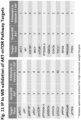
- Figure 11 and Table 3 provide a summary of antibodies useful in IP of AKT-mTOR pathway proteins, as validated by Western Blot.
- Table 1 List of IP to MS validated antibodies for AKT-mTOR Pathway Proteins Antibody Name Company/Catalog Number AKT1 Antibody Millipore/07-416 AKT (pan) Antibody Cell Signaling Technology/2920 AKT2 Antibody Cell Signaling Technology/3063 phospho AKT2 (pSer474) Antibody Thermo Fisher Scientific/PA5-35676 AKT1 Antibody Cell Signaling Technology/2967 phospho AKT (pSer473) Antibody Thermo Fisher Scientific/700392 phospho AKT (pSer473) Antibody Cell Signaling Technology/4060 Anti-phospho-IGF-1R (Tyr1161/Tyr1165/Tyr1166) Antibody Millipore/ABE332 Phospho-IGF1 Rec pTyr1158+1162+1163 Antibody Thermo Fisher Scientific/PA1-26725 Phospho-IGF1R pTyr1161 Antibody Thermo Fisher Scientific/PA5-35769 Phospho-IGF-I Receptor
- the immunoprecipitated AKT-mTOR pathway proteins may be reduced and alkylated prior to fragmentation (e.g., digestion). Samples that have been reduced and alkylated may comprises modifications, such as to cysteine residues (e.g., CAM). Where an AKT-mTOR peptide of SEQ ID NO: 1 - 424 shows modification resulting from, for example, reduction/alkylation, the non-modified peptide is also encompassed. For example, in each instance where an AKT-mTOR pathway peptide of SEQ ID NO: 1 - 424 is referred to, also encompassed are unmodified peptides of SEQ ID NO: 1 - 424.
- the samples may optionally be desalted prior to analysis by mass spectrometry. Both enzymatic and chemical digestion is encompassed. Enzymatic digestion includes, but is not limited to, digestion with a protease such as, for example, trypsin, chymotrypsin, AspN, GluC, LysC, LysN, ArgC, proteinase K, pepsin, Clostripain, Elastase, GluC biocarb, LysC/P, LysN Promise, Protein Endopeptidase, Staph Protease or thermolysin.
- Chemical digestion includes use of, for example, CNBr, iodosobenzoate and formic acid.
- peptide samples are analyzed by mass spectrometry (MS), and the resulting spectra are compared with theoretical spectra from known proteins to determine the peptides and proteins in a sample.
- MS mass spectrometry
- discovery MS is cumbersome and time consuming and is not a viable clinical method. Therefore, the inventors have identified novel peptides that associate with AKT-mTOR pathway proteins for use in the IP-MS methods of the disclosure. Use of these peptides in targeted MS, and IP-targeted MS methods allows quantitation of even low abundant AKT-mTOR proteins. Moreover, use of these peptides in targeted MS, and in IP-targeted MS methods, allows quantitation of phosphorylated AKT-mTOR proteins.
- peptides useful in MS to detect and quantify AKT-mTOR pathway proteins can be designed by use of computer software and the like. However, many of these potential peptide sequences are unsuitable or ineffective for use in MS-based assays, including SRM/MRM and PRM. Because it was not possible to predict the most suitable peptides for MS analysis, it was necessary to experimentally identify modified and unmodified peptides to develop as clinical reagents. To complicate the analysis, it was discovered that certain peptides useful when assaying typical samples were not predictive when assaying samples that had undergone immunoprecipitation.
- targeted MS is performed by quantifying specific unique peptides of the protein.
- known amounts of isotope-labeled (e.g., heavy isotope-labeled) versions of these targeted peptides can be used as internal standards for absolute quantitation.
- proteins of interest are not detectable even after identifying unique peptide standards.
- the combination of specific antibodies with specific target peptides has allowed the inventors to improve the sensitivity of detection of AKT-mTOR pathway proteins by MS, and has allowed for lower levels of detection and lower levels of quantification than ever previously seen. See, e.g., Figure 4 .
- the AKT-mTOR pathway peptides provided in the kits, and useful in the described methods are listed in Table 3.
- SEQ ID Nos: 1-212 are native peptide sequences useful in identifying the AKT-mTOR pathway proteins recited in the "Target ID” column. Certain peptide sequences are phosphorylated at certain residues as shown in parentheses "(PO3H2)" following the modified residue.
- Certain peptides are modified at cysteine residues as shown by "(CAM)” following the modified residue.
- the "CAM” post-translational modification is well known to those of skill in the art to mean carbamidomethylation, resulting from alkylation of the protein/peptide.
- the peptides may be as shown in Table 3, or may be non-modified version of these peptides lacking carbamidomethylation.
- the peptides reagents are recited in Table 5 (SEQ ID Nos: 98, 96, 157, 163, 40, 42, 37, 25, 73, 80, 52, 57, 59, 208, 209, 16, 23, 124, 120, 195, 200, 129, 133, 1, 6, 27, 91, and 204).
- the peptides of Table 5 are useful in multi-plex MS methods.
- protein samples are denatured or solubilized before fragmentation.
- the fragmentation protocol uses chemical cleavage. In some embodiments, the chemical cleavage uses CNBr. In some embodiments, the fragmentation protocol is done using an enzyme. In some embodiments, the fragmentation protocol uses MS-grade commercially available proteases. Examples of proteases that may be used to digest samples include trypsin, endoproteinase GluC, endoproteinase ArgC, pepsin, chymotrypsin, LysN protease, LysC protease, GluC protease, AspN protease, proteinase K, and thermolysin. In some embodiments, a mixture of different proteases are used and the individual results are combined together after the digestion and analysis.
- the digestion is incomplete in order to see larger, overlapping peptides.
- the antibody digestion is performed with IdeS, IdeZ, pepsin, or papain to generate large antibody domains for "middle-down" protein characterization.
- the fragmentation protocol uses trypsin that is modified.
- a protein: protease ratio (w/w) of 10:1, 20:1, 25:1, 50:1, 66:1, or 100:1 may be used.
- the trypsin used is at a concentration of about 100ng/ml-1 mg/ml, or about 100 ng/ml-500 ⁇ g/ml, or about 100 ng/ml-100 ⁇ g/ml, or about 1 ⁇ g/ml-1mg/ml, or about 1 ⁇ g/ml-500 ⁇ g/ml, or about 1 ⁇ g/ml-100 ⁇ g/ml, or about 10 ⁇ g/mg-1mg/ml, or about 10 ⁇ g/mg-500 ⁇ g/ml, or about 10 ⁇ g/mg-100 ⁇ g/ml.
- the digestion step is for 10 minutes to 48 hours, or 30 minutes to 48 hours, or 30 minutes to 24 hours, or 30 minutes to 16 hours, or 1 hour to 48 hours, or 1 hour to 24 hours, or 1 hour to 16 hours, or 1 to 8 hours, or 1 to 6 hours, or 1 to 4 hours.
- the digestion step is incubated at a temperature between 20°C and 45°C, or between 20°C and 40°C, or between 22°C and 40°C, or between 25°C and 37°C.
- the digestion step is incubated at 37°C or 30°C.
- a step is included to end the digestion step.
- the step to end the digestion protocol may be addition of a stop solution or a step of spinning or pelleting of a sample.
- the digestion is followed by guanidation.
- the fragmentation protocol includes use of protein gels. In some embodiments, the fragmentation protocol comprises in-gel digestion.
- An exemplary commercially available kit for performing in-gel digestion is the In-Gel Tryptic Digestion Kit (Thermo Fisher Cat#89871).
- the fragmentation protocol is carried out in solution.
- An exemplary commercially available kit for performing in-solution digestion is the In-Solution Tryptic Digestion and Guanidiation Kit (Thermo Fisher Cat#89895).
- the fragmentation protocol uses beads. In some embodiments, the fragmentation protocol comprises on-bead digestion. In some embodiments, agarose beads or Protein G beads are used. In some embodiments, magnetic beads are used.
- protein samples are separated using liquid chromatography before MS analysis.
- fragmented samples are separated using liquid chromatography before MS analysis.
- IP and IP-MS methods described herein are capable of detecting phosphorylated AKT-mTOR pathway proteins, including those described in Table 4.
- Table 4. List of Total and Phosphorylated AKT-mTOR Pathway Target Proteins Target ID No.
- Target Name Total Phosphorylation Site 1 AKT1 pSer473 2 PTEN pSer380 3 IRS1 pSer312 4 IR pTyr1162/1163 5 IGF-1R pTyr1135/1136 6 GSK3a pSer21 7 GSK3b pSer9 8 RPS6 pSer235/236 9 PRAS40 pThr246 10 mTOR pSer2448 11 p70S6K (S6K1) pThr389 12 TSC2 pSer939
- the AKT-mTOR pathway peptides used in the MS methods described herein have limits of detection considered useful in clinical and research methods. See, e.g, Table 5.
- the AKT-mTOR pathway peptides used in the MS and IP-MS methods comprise or consist of the peptides described in Table 5.
- the peptides of Table 5 are detectably labelled.
- the peptides of SEQ ID NO: 163 may lack the "CAM" modification shown on the fifth amino acid. Table 5.
- methods for detecting phosphorylated AKT-mTOR pathway proteins are encompassed.
- IP, MS, and IP-MS methods to detect phosphorylated AKT-mTOR pathway proteins are conducted separately from methods to detect total (non-phosphorylated) AKT-mTOR pathway proteins.
- the IP and IP-MS methods to detect phosphorylated AKT-mTOR pathway proteins utilize the antibodies of Table 9.
- the IP and IP-MS methods to detect non-phosphorylated AKT-mTOR pathway proteins utilize the antibodies of Table 8.
- the IP-MS methods to detect phosphorylated AKT-mTOR pathway proteins utilize the antibodies of Table 9 and the peptides of Table 5.
- the IP-MS methods to detect non-phosphorylated AKT-mTOR pathway proteins utilize the antibodies of Table 8 and the peptides of Table 5.
- Table 8 List of non-phosho-antibodies for multi-plex IP, single-plex IP (+/- MS).
- AKT-mTOR pathway proteins play central roles in diseases including cancer.
- Phosphorylated AKT-mTOR pathway proteins are particularly important to identify and quantify as a measure of protein activation status, and also as markers for disease progression. As shown in Figure 2 , methods and reagents for detecting AKT-mTOR pathway proteins, including phosphorylated proteins, and their protein interactions, were designed and tested.
- IP-MS Multiplex immunoprecipitation (IP) to MS (mIP-MS) was assessed for the ability to measure total and phosphorylated AKT-mTOR pathway targets. mIP-MS methods were also compared to existing singleplex immunoassay (Western Blot (WB) and ELISA) and multiplex Luminex assays.
- HCT116 ATCC product#CCL-247
- MCF7 ATCC product#HTB-22
- A549 ATCC product#CCL-185 cells were grown in Hamm's F-12K media, McCoy's 5A Media, and MEM Media, respectively, with 10% FBS/1xPenStrep to approximately 70-80% confluency. Cells were starved in 0.1% charcoal stripped FBS for 24 hours before stimulation with 100ng/ml of IGF (CST product#8917SF) for 15 minutes.
- Luminex MagPix instrument was used to acquire and analyze Luminex assay data.
- Thermo Scientific TM Pierce MS-Compatible Magnetic IP Kit (Protein A/G) was used to screen and validate antibodies for 11 total and 10 phosphorylated AKT-mTOR pathway proteins from 500 ⁇ g cell lysate. Validated antibodies were biotinylated with the Thermo Scientific TM Pierce Antibody Biotinylation Kit for IP.
- the Thermo Scientific TM Pierce MS-Compatible Magnetic IP Kit (Streptavidin) was used to perform the single or multiplex IPs for target enrichment. IP samples were processed by an in-solution digestion method where IP eluates were reconstituted in 6M Urea, 50mM TEAB, pH 8.5 followed by reduction, alkylation and trypsin digestion overnight at 37°C. The digested samples were acidified with TFA before MS analysis.
- tryptic digest samples Prior to MS analysis, tryptic digest samples were desalted on-line using the Thermo Scientific TM Acclaim TM PepMap 100 C18 Trap Column. For discovery MS, the samples were analyzed by nanoLC-MS/MS using a Thermo Scientific TM Dionex TM UltiMate TM 3000 RSLCnano System and Thermo Scientific TM Q Exactive TM HF Hybrid Quadrupole-Orbitrap Mass Spectrometer.
- the samples were analyzed using the UltiMate 3000 RSLCnano System and the Thermo Scientific TM TSQ TM Vantage TM Mass Spectrometer (SRM mode) or the Thermo Scientific TM Q Exactive TM HF Hybrid Quadrupole-Orbitrap Mass Spectrometer (PRM mode).
- SRM mode Thermo Scientific TM TSQ TM Vantage TM Mass Spectrometer
- PRM mode Thermo Scientific TM Q Exactive TM HF Hybrid Quadrupole-Orbitrap Mass Spectrometer
- Thermo Scientific TM Proteome Discoverer TM 1.4 to assess percent sequence coverage, unique peptides, MS1 intensities, spectral counts and PTMs.
- the Proteome Discoverer software searches were executed using the Uniprot human protein database. Tryptic peptides with highest MS1 intensity and relevant phosphorylation sites were selected from the discovery data for targeted assay development.
- Thermo Scientific TM Pinpoint software and Skyline software (University of Washington) were used to measure limit of quantitation (LOQ) from the calibration curve and target analyte concentration from unknown samples.
- AKT-mTOR pathway proteins were immunoprecipitated from unstimulated and IGF-stimulated A549 lysate with Thermo Scientific TM Pierce MS-Compatible Magnetic IP Kits (Protein A/G or Streptavidin) for MS analysis.
- Thermo Scientific TM Pierce MS-Compatible Magnetic IP Kits Provided by Thermo Scientific TM Pierce MS-Compatible Magnetic IP Kits (Protein A/G or Streptavidin) for MS analysis.
- a variety of antibodies were screened to determine effectiveness in both ability to IP AKT-mTOR pathway proteins, and also for their usefulness when combined with MS.
- Table 1 (above) provides a list of antibodies validated for use in the IP-MS methods.
- Table 2 (above) provides a list of antibodies tested, but found to be less successful.
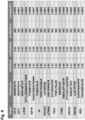
- Limits of detection (LOD) and lower limits of quantification (LLOQ) were analyzed for twelve AKT-mTOR pathway proteins, including AKT2, AKT1, mTOR, IGF1R, IR, PRAS40, p70S6K, TSC2, PTEN, GSK3alpha, GSK3beta, and IRS1. Results are presented in Figure 4 .
- the assay dynamic range representing the concentration range between the lower to upper limits of quantification (LLOQ to ULOQ), is the range where protein concentration is measurable with acceptable levels of accuracy and precision.
- linear signal-to-abundance range (LLOQ and ULOQ) was determined from dilution series experiments spanning concentrations of 500-0.08 fmol on column, spiked into a constant light peptide at 36fmol and 200ng of equimolar concentration of 6 proteins digest.
- Eleven total and ten phosphorylated AKT-mTOR pathway protein targets were enriched simultaneously from unstimulated and IGF stimulated MCF7 lysates with biotinylated antibodies and Thermo Scientific TM Pierce MS-Compatible Magnetic IP Kit (Streptavidin). MCF7 cells were starved in 0.1% charcoal stripped FBS for 24 hours before stimulation with 100ng/ml of IGF for 15 minutes. Validated IP-MS antibodies are biotinylated for 11 total and 10 phosphorylated AKT-mTOR pathway targets using the Thermo Scientific TM Pierce Antibody Biotinylation Kit for IP (PN: 90407) as recommended in instruction manual.
- IP samples were processed by an in-solution digestion method where IP eluates were reconstituted in 6M Urea, 50mM TEAB, pH 8.5 followed by reduction (5mM TCEP for 30 minutes at 35°C), alkylation (20mM Iodoacetamide in dark at room temperature for 30 minutes) and trypsin digestion overnight at 37°C.
- the digested samples were acidified with 3.5 ⁇ L of 10% TFA before discovery MS analysis.
- discovery MS the samples were analyzed by nanoLC-MS/MS using a Thermo Scientific TM Dionex TM UltiMate TM 3000 RSLCnano System and Thermo Scientific TM Q Exactive TM HF Hybrid Quadrupole-Orbitrap Mass Spectrometer.
- the digested samples were cleaned on-line using the C18 trap column (Thermo Fisher Scientific, PN: 164564) followed by reversed-phase separation using the analytical C18 column (75 ⁇ m i.d. x 15 cm, nanoViper, 3 ⁇ m particle size, Thermo Fisher Scientific, PN: ES800) with a 2-30% gradient of Buffer B using Buffer A (0.1% formic acid) and Buffer B (0.1% formic acid/99.9 % acetonitrile) at 0.300 ⁇ L/min.
- Buffer A 0.1% formic acid
- Buffer B (0.1% formic acid/99.9 % acetonitrile
- Figure 5 shows that the IP-nanoLC-MS/MS analysis was able to identify 11 proteins in the multiplex phosphor-assay, and 12 proteins for multiplex total assay.
- MS analysis of multiplex total assay identified interacting proteins (PIK3R1, PIK3R2, PIK3CB, PIK3CA, GSKIP and TSC1) of AKT-mTOR Pathway Targets.
- Tables 8 and 9 provide listings of the antibodies used in this multiplex IP.
- Validated IP-MS antibodies are biotinylated for Total AKT, Phospho AKT, IR, p70S6K, mTOR, and GSK3 ⁇ pathway targets using the Thermo Scientific TM Pierce Antibody Biotinylation Kit for IP (PN: 90407) as recommended in instruction manual. 1 ⁇ g of each biotinylated antibody was added simultaneously to 1000 ⁇ g of control and IGF stimulated A549 and HCT116 cell lysates in duplicate. IP was performed as recommended in the Thermo Scientific TM Pierce MS-Compatible Magnetic IP Kit (Streptavidin) (PN: 90408) with the following modification. 5 microgram of streptavidin magnetic beads per microgram of biotinylated antibody concentration was used for multiplex IP.
- IP samples were processed by an in-solution digestion method where IP eluates were reconstituted in 6M Urea, 50mM TEAB, pH 8.5 followed by reduction (5mM TCEP for 30 minutes at 35°C), alkylation (20mM Iodoacetamide in dark at room temperature for 30 minutes) and trypsin digestion overnight at 37°C.
- the digested samples were acidified with 3.5 ⁇ L of 10% TFA before discovery MS analysis.
- Internal standard peptides were spiked in digested IP samples to make final volume of 6.66fmol/ul.
- the samples were analyzed by nanoLC-PRM/MS using a Thermo Scientific TM Dionex TM UltiMate TM 3000 RSLCnano System and Thermo Scientific TM Q Exactive TM HF Hybrid Quadrupole-Orbitrap Mass Spectrometer. Briefly, the digested samples were cleaned on-line using the C18 trap column (Thermo Fisher Scientific, PN: 164564) followed by reversed-phase separation using the analytical C18 column (75 ⁇ m i.d.
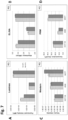
- Figures 7A-7D show quantitation of total AKT.
- Figures 7E-7H show quantitation of phosphorylated IGFIR across all 4 techniques. Lower correlation was observed across techniques. The lower correlation could be due to different antibodies used or each assay and antibody specificity. Up-regulation in phosphorylated IGF1R observed after IGF stimulation in 3 of 4 techniques. Western blot for phosphor IGFIR showed no significant differences in control and IGF stimulated cell lysates.
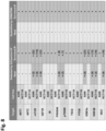
- a summary of AKT-mTOR pathway proteins that were identified and quantified using the IP-MS methods described herein is provided in Figure 8 .
- Most of the AKT-mTOR pathway targets were not identified in discovery MS and quantitated by targeted MS (PRM or SRM) without enrichment by immunoprecipitation.
- IP-MS assay was also more successful than other commercially available non-MS assays.
- IP to MS analysis of total and phosphorylated AKT-mTOR pathway proteins enabled identification of multiple isoforms, relevant protein interactions and phosphorylation sites.
- Total and phosphorylated mIP-tMS assays allowed simultaneous quantitation of 12 total and 11 phosphorylated AKT-mTOR pathway proteins in the low to sub-fmol range from unstimulated and IGF stimulated A549, HCT116 and MCF7 cell lysates.
- the benchmarking of mIP-tMS assays showed moderate correlation for quantitation of total and phosphorylated target relative abundance compared to WB, ELISA and Luminex assays.
- the low concordance for a few targets is possibly due to differences in the specificity of antibodies used for each assay.
- Major advantages of the MS-based assay are high confidence in target identity coupled with simultaneous quantitation of multiple targets, interacting proteins and their phosphophorylated forms.
- Tissue lysis protocol was optimized for IP-MS application. Briefly, 50-100mg of human and murine tissue samples were washed with 5mL 1X cold PBS three times. Tissue samples was minced in 5mL 1x cold PS using scissor followed by homogenization in IP lysis buffer (Thermo Fisher Scientific PN: 87788) and electronic Polytron Handheld Tissue Tearer. Homogenized tissue samples were passed through tissue strainer (Thermo Fisher Scientific PN: 87791) to prepare tissue lysates before IP.
- AKT-mTOR pathway protein targets were enriched simultaneously from normal mouse lung tissue lysate, normal mouse kidney tissue lysate, and normal human lung tissue lysate as per Example 2.
- A549 cell lysate was used as a non-tissue control.
- the IP-MS method described herein is capable of validating AKT-mTOR pathway proteins in murine and human tissue lysate in addition to cell lysate. Seven out of eleven AKT-mTOR pathway protein targets were identified in normal human lung tissue, and nine out of eleven AKT-mTOR pathway protein targets were identified for normal mouse kidney tissue using our IP-MS method.
Landscapes
- Health & Medical Sciences (AREA)
- Life Sciences & Earth Sciences (AREA)
- Engineering & Computer Science (AREA)
- Immunology (AREA)
- Chemical & Material Sciences (AREA)
- Molecular Biology (AREA)
- Biomedical Technology (AREA)
- Hematology (AREA)
- Urology & Nephrology (AREA)
- Physics & Mathematics (AREA)
- General Health & Medical Sciences (AREA)
- Biochemistry (AREA)
- Pathology (AREA)
- General Physics & Mathematics (AREA)
- Analytical Chemistry (AREA)
- Cell Biology (AREA)
- Medicinal Chemistry (AREA)
- Microbiology (AREA)
- Food Science & Technology (AREA)
- Biotechnology (AREA)
- Proteomics, Peptides & Aminoacids (AREA)
- Bioinformatics & Computational Biology (AREA)
- Bioinformatics & Cheminformatics (AREA)
- Biophysics (AREA)
- Organic Chemistry (AREA)
- Spectroscopy & Molecular Physics (AREA)
- Chemical Kinetics & Catalysis (AREA)
- Electrochemistry (AREA)
- Genetics & Genomics (AREA)
- Toxicology (AREA)
- Other Investigation Or Analysis Of Materials By Electrical Means (AREA)
- Peptides Or Proteins (AREA)
- Investigating Or Analysing Biological Materials (AREA)
Description
- This application claims the benefit of priority of
U.S. Provisional Patent Application No. 62/308,051, filed March 14, 2016 - This disclosure relates to the field of detection and quantification of AKT-mTOR pathway proteins, including phosphorylated proteins, by immunoprecipitation and mass spectrometry.
- The AKT-mTOR pathway plays a central role in tumor progression and anticancer drug resistance. The quantitative measurement of protein expression and post-translational modifications of the AKT-mTOR pathway is necessary for precisely characterizing cancer, monitoring cancer progression, and determining treatment responses. See Logue, J. S.; Morrison, D. K.; Genes Dev. Apr 1 2012, 26 (7), 641-50.
- A major limitation in the detection and quantitation of AKT-mTOR pathway proteins is the lack of rigorously validated methods and reagents. Currently, only semiquantitative results from Western blotting, ELISA, and Luminex assays are available. Mass spectrometry (MS) is increasingly becoming the detection methodology of choice for assaying protein abundance and post-translational modifications. However, to date, MS has not been successful in quantifying AKT-mTOR pathway proteins, possibly due to their low abundance and significant post-translational modification profiles.
- Immunoprecipitation (IP) is commonly used upstream of MS as an enrichment tool for low-abundant protein targets. See, Gingras et al., Nat. Rev. Mol. Cell. Biol., Aug 2007, 8 (8), 645-54; and Carr, S. A. et al., Mol. Cell. Proteomics Mar 2014, 13 (3), 907-17. The identification of appropriate antibodies for use in IP upstream of MS is important, as not all antibodies that bind to protein will be effective immunoprecipitation tools, and further, not all antibodies that are effective immunoprecipiation tools will lead to successful identification via MS. In "Quantitative Analysis of AKT/mTOR Pathway using Immunoprecipitation and Targeted Mass Spectrometry" Bhavinkumar Patel et al. disclose a multiplex IP-MS assay for the identification and quantification of AKT mTOR pathway proteins (Poster Abstract 1837, Cancer Research 2015: 75, ). The poster refers to certain AKT mTOR pathway proteins which are also targets of the method of the present invention, namely AKT1, AKT2 and PTEN, however the peptides actually identified by MS differ.
- The present disclosure provides reagents and methods for detecting and quantifying AKT-mTOR pathway proteins via immunoprecipitation (IP), mass spectrometry (MS), and immunoprecipitation followed by mass spectrometry (IP-MS).
- The present invention provides a method for detecting AKT-mTOR pathway proteins, a method for determining the ratio of phosphorylated to non-phosphorylated AKT-mTOR pathway proteins and kits therefore as defined in the appended claims.
- Methods for immunoprecipitating an AKT-mTOR pathway protein (target protein) are provided, comprising contacting a biological sample with any one of the antibodies recited in Table 8 and Table 9. The methods may comprise washing the contacted biological sample to enrich for antibody-protein conjugates. Further methods include detecting the antibody-protein conjugates (the immunoprecipitated target protein) to determine the AKT-mTOR pathway protein in the biological sample. In some embodiments, the antibody is labelled. In some embodiments, a detection reagent is provided to the enriched antibody-protein conjugate. In some embodiments the label is biotin and the detection reagent is streptavidin.
- . In claimed embodiments the IP is multi-plex. The antibodies useful in multiplex IP comprise more than one antibody selected from an antibody of Table 8 and/or Table 9.
- In some aspects a method for detecting AKT-mTOR pathway proteins via MS is provided, comprising isolating proteins from a biological sample, digesting the isolated proteins, assaying the digested proteins via mass spectrometry to determine the presence of a peptide for AKT-mTOR pathway protein(s), and determining the identity of one or more AKT-mTOR pathway protein(s) in the sample. The peptide for AKT-mTOR pathway protein(s) consist(s) of a sequence listed in Table 5.
- In some aspects a method for quantifying AKT-mTOR pathway proteins via MS is provided, comprising isolating proteins from a biological sample, digesting the isolated proteins, assaying the digested proteins via mass spectrometry to determine the presence of a peptide for AKT-mTOR pathway protein(s), and determining the quantity of one or more AKT-mTOR pathway protein(s) in the sample. IThe peptide consists of a peptide selected from the peptides shown in Table 5 (SEQ ID Nos: 98, 96, 157, 163, 40, 42, 37, 25, 73, 80, 52, 57, 59, 208, 209, 16, 23, 124, 120, 195, 200, 129, 133, 1, 6, 27, 91, and 204).
- In some embodiments a method for detecting AKT-mTOR pathway proteins via IP-MS is provided, comprising treating a biological sample with more than one antibody selected from an antibody of Table 8 and/or Table 9 capable of immunoprecipitating AKT-mTOR target pathway protein(s) from a biological sample, digesting the isolated proteins, assaying the digested proteins via mass spectrometry to determine the presence of a peptide for AKT-mTOR pathway protein(s), and determining the identity of one or more AKT-mTOR pathway protein(s) in the sample. The peptide consists of a peptide selected from the peptides shown in Table 5 (SEQ ID Nos: 98, 96, 157, 163, 40, 42, 37, 25, 73, 80, 52, 57, 59, 208, 209, 16, 23, 124, 120, 195, 200, 129, 133, 1, 6, 27, 91, and 204).
- In some embodiments a method for quantifying AKT-mTOR pathway proteins via IP-MS is provided, comprising treating a biological sample with more than one antibody selected from an antibody of Table 8 and/or Table 9 capable of immunoprecipitating AKT-mTOR target pathway protein(s) from a biological sample, digesting the isolated proteins, assaying the digested proteins via mass spectrometry to determine the presence of a peptide for AKT-mTOR pathway protein(s), and determining the quantity of one or more AKT-mTOR pathway protein(s) in the sample. The peptide consists of a peptide selected from the peptides shown in Table 5 (SEQ ID Nos: 98, 96, 157, 163, 40, 42, 37, 25, 73, 80, 52, 57, 59, 208, 209, 16, 23, 124, 120, 195, 200, 129, 133, 1, 6, 27, 91, and 204).
- In some embodiments the AKT-mTOR pathway target protein is phosphorylated.
- Methods for determining the ratio of phosphorylated to non-phosphorylated AKT-mTOR pathway proteins are provided, comprising any of the above IP, MS, or MS-IP methods, wherein a further step of determining the ratio of phosphorylated to non-phosphorylated protein is provided. In some embodiments, the method is an MS-IP method comprising treating a biological sample with more than one antibody selected from an antibody of Table 9capable of immunoprecipitating one or more phosphorylated AKT-mTOR pathway proteins, and separately treating the same biological sample with more than one antibody capable of immunoprecipitating a non-phosphorylated AKT-mTOR target protein selected from the antibodies recited in Table 8; digesting the immunoprecipitated AKT-mTOR pathway proteins; adding a first and a second detectably labelled internal standard peptide of known amount to the digested proteins, wherein the first internal standard peptide has the same amino acid sequence as a phosphorylated AKT-mTOR pathway peptide used to identify the phosphorylated protein, and the second internal standard peptide has the same amino acid sequence as the non-phosphorylated AKT-mTOR pathway peptide used to identify the non-phosphorylated protein; assaying the digested protein and internal standards via mass spectrometry to determine the presence and amount of phosphorylated and non-phosphorylated AKT-mTOR pathway proteins, wherein the AKT-mTOR pathway peptide consists of a peptide selected from the peptides shown in Table 5 (SEQ ID Nos: 98, 96, 157, 163, 40, 42, 37, 25, 73, 80, 52, 57, 59, 208, 209, 16, 23, 124, 120, 195, 200, 129, 133, 1, 6, 27, 91, and 204).
- In some embodiments, the biological sample is human. In some embodiments, the biological sample is non-human. In some embodiments, the biological sample is mammalian. In some embodiments, the biological sample is from rat, mouse, guinea pig, hamster, cow, pig, horse, goat, sheep, dog, cat, or non-human primate.
- In embodiments utilizing an AKT-mTOR pathway peptide, the peptide may be modified with a detectable label. The detectable label may comprise an isotope, such as a heavy isotope, such as those known to those of skill in the art, including 13C, 15N, 2H and 180.
- In some aspects more than one antibody is used in a multi-plex IP. In some embodiments, the multi-plex IP comprises the antibodies of Table 8. In some embodiments, the multi-plex IP comprises the antibodies of Table 9.
- Two or more antibodies are utilized to analyze one biological sample. For example, a first antibody is capable of immunoprecipitating a phosphorylated AKT-mTOR pathway protein, and a second antibody is capable of immunoprecipitating a non-phosphorylated version of the AKT-mTOR pathway protein precipitated by the first antibody. In some embodiments, a single antibody is capable of immunoprecipitating a phosphorylated and non-phosphorylated AKT-mTOR pathway protein.
- In some embodiments, the immunoprecipitation comprises treating a sample with a labelled antibody capable of binding to an AKT-mTOR pathway protein to provide a labelled antibody-protein conjugate. The method may further comprise contacting the labelled antibody-protein conjugate with a capture agent capable of binding to the labelled antibody to isolate the pathway protein from the sample. The label may be biotin and the capture agent may be streptavidin.
- The quantity of an AKT-mTOR pathway protein may be determined by adding an internal standard peptide of known amount to the digested protein prior to mass spectrometry. In some embodiments, the internal standard peptide has the same amino acid sequence as the AKT-mTOR pathway peptide. In some embodiments, the internal standard is detectably labeled. The method may further comprises determining the quantity of an AKT-mTOR pathway peptide by comparison to the internal standard.
- In some aspects, the internal standard peptide comprises or consists of a peptide selected from the peptides shown in Table 5 (SEQ ID Nos: 98, 96, 157, 163, 40, 42, 37, 25, 73, 80, 52, 57, 59, 208, 209, 16, 23, 124, 120, 195, 200, 129, 133, 1, 6, 27, 91, and 204).
- In some embodiments, quantifying the AKT-mTOR pathway protein comprises comparing an amount of an AKT-mTOR pathway peptide in the sample to an amount of the same AKT-mTOR pathway peptide in a control sample.
- Quantifying an AKT-mTOR pathway protein may comprise comparing an amount of an AKT-mTOR pathway peptide to an internal standard peptide of known amount, wherein both the peptide in the biological sample and the internal standard peptide consists of a peptide selected from the peptides shown in Table 5 (SEQ ID Nos: 98, 96, 157, 163, 40, 42, 37, 25, 73, 80, 52, 57, 59, 208, 209, 16, 23, 124, 120, 195, 200, 129, 133, 1, 6, 27, 91, and 204).
- In some embodiments the mass spectrometry is selected from tandem mass spectrometry and discovery mass spectrometry. The targeted mass spectrometry may be selected from multiple reaction monitoring (MRM), selected reaction monitoring (SRM), and parallel reaction monitoring (PRM), or combinations thereof.
- In some embodiments the biological sample is selected from isolated human cells, plasma, serum, whole blood, CSF, urine, sputum, tissue, and tumorous tissue.
- In some embodiments, the method further comprises quantifying the relative amount of AKT-mTOR pathway protein. In some embodiments, the method further comprises quantifying the absolute amount of AKT-mTOR pathway protein.
- In some embodiments, the digesting comprises a protease or chemical digest. In some embodiments the digestion may be single or sequential. The protease digestion may comprise trypsin, chymotrypsin, AspN, GluC, LysC, LysN, ArgC, proteinase K, pepsin, clostripain, elastase, GluC biocarb, LysC/P, LysN promise, protein endopeptidase, staph protease or thermolysin.
- The chemical cleavage may comprise CNBr, iodosobenzoate or formic acid.
- In some embodiments the digestion is a protease digest with trypsin.
- In some embodiments the methods further comprise desalting after digestion and prior to mass spectrometry.
- The AKT-mTOR pathway protein may be selected from AKT1 (UniProtKB - P31749), AKT2 (UniProtKB - P31751), IR (also known as INSR) (UniProtKB P06213), IGFIR (UniProtKB - P08069), IRS1 (UniProtKB - P35568), TSC2 (UniProtKB - P49815), mTOR (UniProtKB - P42345), GSK3a (UniProtKB - P49840), GSK3b (UniProtKB - P49841), GSK3a/GSK3b, p70S6K (also known as RPS6KB1) (UniProtKB - P23443), RPS6 (UniProtKB - P62753), PRAS40 (also known as AKT1S1) (UniProtKB - Q96B36), and PTEN (UniProtKB - P60484).
- In some embodiments, the AKT-mTOR pathway is a protein that interacts with any of AKT1 (UniProtKB - P31749), AKT2 (UniProtKB - P31751), IR (also known as INSR) (UniProtKB P06213), IGFIR (UniProtKB - P08069), IRS1 (UniProtKB - P35568), TSC2 (UniProtKB - P49815), mTOR (UniProtKB - P42345), GSK3a (UniProtKB - P49840), GSK3b (UniProtKB - P49841), GSK3a/GSK3b, p70S6K (also known as RPS6KB1) (UniProtKB - P23443), RPS6 (UniProtKB - P62753), PRAS40 (also known as AKT1S1) (UniProtKB - Q96B36), and PTEN (UniProtKB - P60484).
- In some embodiments, the AKT-mTOR pathway protein is phosphorylated.
- In some embodiments, the concentration of AKT-mTOR protein that may be detected ranges from about 0.08 fmol to about 2000 fmol.
- In some embodiments, the lower limit of detection is about 0.05, 0.06, 0.07, 0.08, 0.09, 0.10, 0.11, 0.12, 0.13, 0.14, 0.15, 0.16, 0.17, 0.18, 0.19, 0.20, 0.21, 0.22, 0.23, 0.24, or 0.25 fmol. The lower limit of detection may be within the range of about 0.05 - 0.25 fmol.
- In some embodiments the lower limit of quantification is about 0.05, 0.06, 0.07, 0.08, 0.09, 0.10, 0.11, 0.12, 0.13, 0.14, 0.15, 0.16, 0.17, 0.18, 0.19, 0.20, 0.21, 0.22, 0.23, 0.24, 0.25, 0.30, 0.35, 0.40, 0.45, 0.50, 0.55, 0.60, 0.65, 0.70, or 0.75 fmol. The lower limit of quantification may be within the range of about 0.05 - 0.75 fmol.
- Kits comprising two or more antibodies capable of immunoprecipitating an AKT-mTOR pathway protein selected from the antibodies recited in Table 8 or Table 9 are disclosed.
- Kits comprising the antibodies capable of immunoprecipitating an AKT-mTOR pathway protein, and reagents useful for performing mass spectrometry to detect an AKT-mTOR pathway protein are also disclosed.
- Also disclosed are kits comprising the antibodies capable of immunoprecipitating an AKT-mTOR pathway target protein, and reagents useful for performing mass spectrometry to quantify an AKT-mTOR pathway protein.
- The antibody to be included in the kit are selected from any one or more of the antibodies recited in Table 8 and/or Table 9. In some embodiments the antibody is labelled or capable of being labelled. The label may be any label known to those of skill in the art including enzymatic and fluorescent labels, such as biotin. In the invention, the kit comprises more than one antibody. In some embodiments, the kit comprises two or more of the antibodies selected from the antibodies recited in Table 8. In some embodiments, the kit comprises two or more of the antibodies selected from the antibodies recited in Table 9. In some embodiments, the kit comprises two or more of the antibodies selected from the antibodies recited in Table 8 and two or more of the antibodies selected from the antibodies recited in Table 9. In some embodiments, the kit comprises each of the antibodies recited in Table 8, Table 9, or Tables 8 and 9.
- The kits may further comprise an AKT-mTOR pathway peptide that consists of a peptide selected from the peptides shown in Table 5 (SEQ ID Nos: 98, 96, 157, 163, 40, 42, 37, 25, 73, 80, 52, 57, 59, 208, 209, 16, 23, 124, 120, 195, 200, 129, 133, 1, 6, 27, 91, and 204).
- The kit may additionallycomprise at least one peptide selected from peptides of SEQ ID NO: 213 - SEQ ID NO: 424, wherein the peptide is less than or equal to 40 amino acids.
- The peptides provided in the kit may be detectably labeled or capable of being modified to be detectably labeled.
- In some embodiments, the kit further comprises a protease or chemical agent capable of digesting an immunoprecipitated protein sample. The protease agent may be trypsin, chymotrypsin, AspN, GluC, LysC, LysN, ArgC, proteinase K, pepsin, clostripain, elastase, GluC biocarb, LysC/P, LysN promise, protein endopeptidase, Staph protease or thermolysin. The chemical agent may be CNBr, iodosobenzoate or formic acid.
- The kits may be utilized to detect AKT-mTOR pathway proteins, including AKT1 (UniProtKB - P31749); AKT2 (UniProtKB - P31751), IR (also known as INSR) (UniProtKB P06213), IGFIR (UniProtKB - P08069), IRS1 (UniProtKB - P35568), TSC2 (UniProtKB - P49815), mTOR (UniProtKB - P42345), GSK3a (UniProtKB - P49840), GSK3b (UniProtKB - P49841), GSK3a/GSK3b, p70S6K (also known as RPS6KB1) (UniProtKB - P23443 (KS6B1_HUMAN)), RPS6 (UniProtKB - P62753), PRAS40 (also known as AKT1S1)(UniProtKB - Q96B36), and PTEN (UniProtKB - P60484).
- The AKT-mTOR protein to be detected and quantified by the kits may be phosphorylated.
- Also disclosed are antibodies recited in Table 1 for use in immunoprecipitating an AKT-mTOR pathway protein. The antibody may be used in methods comprising immunoprecipitating an AKT-mTOR pathway protein prior to analyzing the protein via mass spectrometry.
- AKT-mTOR pathway peptides selected from the peptides of Table 3 are disclosed. The AKT-mTOR pathway peptides may be used in methods of detecting and quantifying AKT-mTOR pathway proteins in biological samples. The AKT-mTOR pathway peptides may be used in methods comprising immunoprecipitating the AKT-mTOR pathway protein from the biological sample, and analyzing the immunoprecipitated protein via mass spectrometry.
-
-
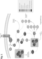
FIG 1 shows a schematic of the AKT-mTOR pathway proteins. -
FIG 2 shows one representative workflow for an immunoprecipitation-enriched mass spectrometry assay to identify AKT-mTOR pathway proteins. -
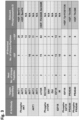
FIG 3 shows results from an experiment to enrich for low abundant AKT-mTOR pathway proteins from A549 cells. -
FIG 4 shows detection and quantitation limits of peptides for 12 AKT-mTOR pathway proteins. -
FIG 5 shows the results of a multiplex immunoprecipitation plus nanoLC-MS/MS assay for 10 phosphorylated and 11 total AKT-mTOR pathway proteins. -
FIG 6 shows representative results from a multiplex immunoprecipitation plus nanoLC-PRM/MS assay AKT-mTOR pathway proteins. Darker gray bars are A549 cells, and lighter gray bars are HCT116 cells. -
FIGS 7A, 7B, 7C, 7D ,7E, 7F, and 7G show a comparison of various methods to detect AKT-mTOR pathway proteins, including Luminex, ELISA, Western Blot, and the IP-Mass spec assay in IGF stimulated (dark gray) and non-stimulated (light gray) cells. -
FIG 8 shows a summary of AKT-mTOR pathway proteins identified and quantified in two different cell lines, with and without immunoprecipitation enrichment, using the mass spec methods. -
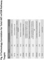
FIG 9 shows technology correlation for total AKT-mTOR pathway targets. -
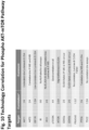
FIG 10 shows technology correlation for phopho-AKT-mTOR pathway targets. -
FIG 11 shows IP to Western Blot validation of AKT-mTOR pathway targets. - This description and exemplary embodiments should not be taken as limiting. For the purposes of this specification and appended claims, unless otherwise indicated, all numbers expressing quantities, percentages, or proportions, and other numerical values used in the specification and claims, are to be understood as being modified in all instances by the term "about," to the extent they are not already so modified. Accordingly, unless indicated to the contrary, the numerical parameters set forth in the following specification and attached claims are approximations that may vary depending upon the desired properties sought to be obtained. At the very least, and not as an attempt to limit the application of the doctrine of equivalents to the scope of the claims, each numerical parameter should at least be construed in light of the number of reported significant digits and by applying ordinary rounding techniques.
- It is noted that, as used in this specification and the appended claims, the singular forms "a," "an," and "the," and any singular use of any word, include plural referents unless expressly and unequivocally limited to one referent. As used herein, the term "include" and its grammatical variants are intended to be non-limiting, such that recitation of items in a list is not to the exclusion of other like items that can be substituted or added to the listed items.
- As used herein, an "AKT-mTOR pathway protein" includes, but is not limited to, AKT1 (UniProtKB - P31749), AKT2 (UniProtKB - P31751), IR (also known as INSR) (UniProtKB P06213), IGFIR (UniProtKB - P08069), IRS1 (UniProtKB - P35568), TSC2 (UniProtKB - P49815), mTOR (UniProtKB - P42345), GSK3a (UniProtKB - P49840), GSK3b (UniProtKB - P49841), GSK3a/GSK3b, p70S6K (also known as RPS6KB1) (UniProtKB - P23443), RPS6 (UniProtKB - P62753), PRAS40 (also known as AKT1S1) (UniProtKB - Q96B36), and PTEN (UniProtKB - P60484).
- As used herein "protein", "peptide", and "polypeptide" are used interchangeably throughout to mean a chain of amino acids wherein each amino acid is connected to the next by a peptide bond. In some embodiments, when a chain of amino acids consists of about two to forty amino acids, the term "peptide" is used. However, the term "peptide" should not be considered limiting unless expressly indicated.
- The term "antibody" is used in the broadest sense and encompasses various antibody structures, including but not limited to monoclonal antibodies, polyclonal antibodies, multispecific antibodies (such as bispecific antibodies), and antibody fragments so long as they exhibit the desired immunoprecipitating activity. As such, the term antibody includes, but is not limited to, fragments that are capable of binding to an antigen, such as Fv, single-chain Fv (scFv), Fab, Fab', di-scFv, sdAb (single domain antibody) and (Fab')2 (including a chemically linked F(ab')2). Papain digestion of antibodies produces two identical antigen-binding fragments, called "Fab" fragments, each with a single antigen-binding site, and a residual "Fc" fragment. Pepsin treatment yields a F(ab')2 fragment that has two antigen-binding sites. The term antibody also includes, but is not limited to, chimeric antibodies, humanized antibodies, and antibodies of various species such as mouse, goat, horse, sheep, chicken, etc. Furthermore, for all antibody constructs provided herein, variants having the sequences from other organisms are also contemplated, such as CDR-grafted antibodies or chimeric antibodies. Antibody fragments also include either orientation of single chain scFvs, tandem di-scFv, diabodies, tandem tri-sdcFv, minibodies, etc. Antibody fragments also include nanobodies (sdAb, an antibody having a single, monomeric domain, such as a pair of variable domains of heavy chains, without a light chain). An antibody fragment can be referred to as being a specific species in some embodiments (for example, human scFv or a mouse scFv). This denotes the sequences of at least part of the non-CDR regions, rather than the source of the construct. The antibodies provided herein are referred to by reference to name and catalog reference. The skilled artisan, holding this name and catalog information, is capable of determining the sequence of the antibody, and therefore the disclosure encompasses any antibody having at least partial sequence of a reference antibody so long as the antibody maintains its ability to immunoprecipitate its antigen protein. In some embodiments, the antibodies comprise antibodies having the same CDRs as the antibodies provided in Table 1.
- Mass spectrometry (MS) is a primary technique for analysis of proteins on the basis of their mass-to-charge ratio (m/z). MS techniques generally include ionization of compounds and optional fragmentation of the resulting ions, as well as detection and analysis of the m/z of the ions and/or fragment ions followed by calculation of corresponding ionic masses. A "mass spectrometer" generally includes an ionizer and an ion detector. "Mass spectrometry," "mass spec," "mass spectroscopy," and "MS" are used interchangeably throughout.
- "Targeted mass spectrometry," also referred to herein as "targeted mass spec," "targeted MS," and "tMS" increases the speed, sensitivity, and quantitative precision of mass spec analysis. Non-targeted mass spectrometry, sometimes referred to as "data-dependent scanning," "discovery MS," and "dMS" and targeted mass spec are alike in that in each, analytes (proteins, small molecules, or peptides) are infused or eluted from a reversed phase column attached to a liquid chromatography instrument and converted to gas phase ions by electrospray ionization. Analytes are fragmented in the mass spec (a process known as tandem MS or MS/MS), and fragment and parent masses are used to establish the identity of the analyte. Discovery MS analyzes the entire content of the MS/MS fragmentation spectrum. In contrast, in targeted mass spectrometry, a reference spectrum is used to guide analysis to only a few selected fragment ions rather than the entire content.
- "Multiple reaction monitoring," "MRM," "selected reaction monitoring," and "SRM" are used interchangeably throughout to refer to a type of targeted mass spectrometry that relies on a unique scanning mode accessible on triple-quadrupole (QQQ) instruments. See, e.g., Chambers et al., Expert Rev. Proteomics, 1-12 (2014).
- "Parallel Reaction Monitoring," and "PRM" are used interchangeably herein to describe another type of targeted mass spec wherein the second mass analyzer used in SRM (quadrupole) is substituted by a high resolution orbitrap mass analyzer in PRM. Unlike SRM, which allows the measuring of one single transition at a given point in time, PRM allows parallel monitoring in one MS/MS spectrum. PRM also allows for the separation of ions with close m/z values (i.e., within a 10 ppm range), and may therefore allow for lower limits of detection and quantification (LOD or LLOD and LOQ or LLOQ).
- The methods disclosed herein may be applied to any type of MS analysis. The disclosure is not limited by the specific equipment or analysis used. The use of any equipment with the intent of analyzing the m/z of a sample would be included in the definition of mass spectrometry. Non-limiting examples of MS analysis and/or equipment that may be used include electrospray ionization, ion mobility, time-of-flight, tandem, ion trap, MRM, SRM, MRM/SRM, PRM, and Orbitrap. The disclosure is neither limited by the type of ionizer or detector used in the MS analysis nor by the specific configuration of the MS. The disclosure is not limited to use with specific equipment or software. The disclosure is not limited to the equipment and software described in the Examples. Embodiments that do not employ more than one antibody selected from an antibody of Table 8 and/or Table 9 or do not include determining the presence of a peptide consisting of a sequence listed in Table 5 are not according to the invention and are present for illustrational purpose only.
- In some embodiments, methods of immunoprecipitating an AKT-mTOR pathway protein are provided, comprising contacting a biological sample with at least one antibody recited in Table 1. The immunoprecipitating method may be single-plex or multiplex. A "single-plex" IP utilizes one antibody per assay, whereas a "multi-plex" IP utilizes more than one antibody per assay.
- In some embodiments, an IP-MS method for detecting and quantifying phosphorylated and non-phosphorylated AKT-mTOR pathway proteins is provided. The methods may comprise contacting a biological sample with at least one antibody recited in Table 1, digesting the immunoprecipitated protein(s), and assaying the digested proteins via mass spectrometry. The IP and MS may be single-plex or multi-plex. A "single-plex" MS refers to monitoring a single peptide in a single MS run, whereas a "mulit-plex" MS refers to monitoring more than one target peptides in a single MS run.
- Table 1 provides a listing of antibodies useful in the IP and IP-MS methods described herein. Table 2 provides a listing of antibodies that are known to bind to their antigen AKT-mTOR protein, but were found to be less useful in the IP and IP-MS methods described herein.
Figure 11 and Table 3 provide a summary of antibodies useful in IP of AKT-mTOR pathway proteins, as validated by Western Blot.Table 1: List of IP to MS validated antibodies for AKT-mTOR Pathway Proteins Antibody Name Company/Catalog Number AKT1 Antibody Millipore/07-416 AKT (pan) Antibody Cell Signaling Technology/2920 AKT2 Antibody Cell Signaling Technology/3063 phospho AKT2 (pSer474) Antibody Thermo Fisher Scientific/PA5-35676 AKT1 Antibody Cell Signaling Technology/2967 phospho AKT (pSer473) Antibody Thermo Fisher Scientific/700392 phospho AKT (pSer473) Antibody Cell Signaling Technology/4060 Anti-phospho-IGF-1R (Tyr1161/Tyr1165/Tyr1166) Antibody Millipore/ABE332 Phospho-IGF1 Rec pTyr1158+1162+1163 Antibody Thermo Fisher Scientific/PA1-26725 Phospho-IGF1R pTyr1161 Antibody Thermo Fisher Scientific/PA5-35769 Phospho-IGF-I Receptor β (Tyr1131)/Insulin Receptor β (Tyr1146) Antibody Cell Signaling Technology/3021 IGF-I Receptor β Antibody Cell Signaling Technology/9750 IGF-I Receptor β Antibody Cell Signaling Technology/3027 Insulin Receptor β Antibody Cell Signaling Technology/3020 INSR / Insulin Receptor Antibody Thermo Fisher Scientific/MA1-10865 Anti-α-Insulin Receptor Antibody, β subunit Antibody Millipore/07-724 INSR / Insulin Receptor alpha Antibody Thermo Fisher Scientific/MA5-13759 Anti-Insulin Receptor (phospho Y972) Antibody abcam/ab5678 IRS-1 Antibody Cell Signaling Technology/2382 IRS-1 Antibody Cell Signaling Technology/3407 IRS-1 Antibody Millipore/06-248 IRS-1 Antibody Millipore/05-784R IRS-1 Antibody Millipore/05-1085 IRS1 (pSer312) polyclonal Antibody Abnova/PAB12627 Anti-phospho-IRS1 (Ser307 mouse/ Ser312 human) Antibody Millipore/05-1087 Phospho-IRS-1 (pSer1101) Antibody Cell Signaling Technology/2385 mTOR Antibody Cell Signaling Technology/2972 mTOR Antibody Millipore/07-1415 mTOR Antibody abcam/ab2732 mTOR Antibody abcam/ab25880 mTOR Antibody Thermo Fisher Scientific/PA1-518 Phospho-mTOR (pSer2448) Antibody Thermo Fisher Scientific/PA5-35652 Phospho-mTOR (pSer2448) Antibody Cell Signaling Technology/5536 Phospho-mTOR (pSer2448) Antibody Abgent/AP50437 S6K Antibody Thermo Fisher Scientific/PA5-12726 S6K Antibody Thermo Fisher Scientific/PA5-12723 S6K1 Antibody Thermo Fisher Scientific/PA1-31167 Anti-S6K1 Antibody abcam/ab9366 S6K Antibody Thermo Fisher Scientific/PA5-27853 Phospho-p70 S6 Kinase (pThr389 / pThr412) Antibody Thermo Fisher Scientific/PA5-35701 Phospho-p70 S6 Kinase (pThr389) Antibody Thermo Fisher Scientific/701064 Phospho-p70 S6 Kinase (pThr421/pSer424) Antibody Cell Signaling Technology/9204 Phospho-p70 S6 Kinase pThr389 Antibody Thermo Fisher Scientific/MA5-15202 Phospho-GSK-3α/β (pSer21/pSer9) Antibody Cell Signaling Technology/9327 GSK3α Antibody Cell Signaling Technology/4337 GSK3α Antibody Cell Signaling Technology/4818 Phospho-GSK-3α (Ser21) Antibody Cell Signaling Technology/8452 Phospho-GSK-3α (Ser21) Antibody Cell Signaling Technology/9316 Phospho-GSK-3α/β (pSer21/pSer9) Antibody Cell Signaling Technology/8566 GSK3β Antibody Thermo Fisher Scientific/MA5-15109 GSK3β Antibody Thermo Fisher Scientific/PA5-29251 GSK3β Antibody Thermo Fisher Scientific/PA5-29265 GSK3β Antibody Cell Signaling Technology/12456 Phospho-GSK-3β (pSer9) Antibody Cell Signaling Technology/5558 Phospho-Tuberin / TSC2 pSer939 Antibody Thermo Fisher Scientific/710395 Phospho-TSC2 pSer939 Antibody Thermo Fisher Scientific/PA5-12845 Phospho-TSC2 pSer939 Antibody abcam/ab59269 Phospho-TSC2 pSer939 Antibody abcam/ab52962 Anti-TSC2 Antibody abcam/ab52936 Anti-TSC2 Antibody Cell Signaling Technology/4308 Anti-TSC2 Antibody Thermo Fisher Scientific/PA5-20132 Anti-TSC2 Antibody Cell Signaling Technology/3990 Anti-TSC2 Antibody Thermo Fisher Scientific/MA5-15004 S6 Ribosomal Protein Antibody Thermo Fisher Scientific/701374 S6 Ribosomal Protein Antibody Thermo Fisher Scientific/710405 S6 Ribosomal Protein Antibody Thermo Fisher Scientific/PA5-11818 Phospho-S6 Ribosomal Protein pSer235+236 Antibody Thermo Fisher Scientific/701363 RPS6 Antibody Thermo Fisher Scientific/PA5-26041 Anti-PRAS40 Antibody abcam/ab72321 Anti-PRAS40 Antibody Thermo Fisher Scientific/PA5-35143 Anti-PRAS40 Antibody R&D System/MAB6408 (R&D) Anti-PRAS40 Antibody Cell Signaling Technology/2691 Phospho-PRAS40 (pThr246) Antibody Cell Signaling Technology/2997 Phospho-PRAS40 (pThr246) Antibody Cell Signaling Technology/13175 Phospho-PRAS40 (pThr246) Antibody R&D System/MAB6890 Phospho-PTEN (pSer380) Antibody Cell Signaling Technology/9551 PTEN Antibody Cell Signaling Technology/9188 AKT pan Antibody Thermo Fisher Scientific/44-609G mTOR Antibody Thermo Fisher Scientific/PA1-188 IRS1 Antibody Thermo Fisher Scientific/710009 IRS1 Antibody Thermo Fisher Scientific/AHO1322 IGFIR Antibody Thermo Fisher Scientific/39-6700 TSC2 Antibody Thermo Fisher Scientific/AHO1422 TSC2 Antibody Thermo Fisher Scientific/730014 PTEN Antibody Thermo Fisher Scientific/51-2400 Table 2: List of IP to MS less successful antibodies for AKT-mTOR Pathway Proteins Antibody Name Company/Catalog Number Phospho-Akt (Ser473) Antibody Cell Signaling Technology/4051 AKT1 Antibody Thermo Fisher Scientific/PA5-23780 AKT2 Antibody Thermo Fisher Scientific/MA1-034 Phospho-AKT1 pSer473 Antibody Thermo Fisher Scientific/MA1-20325 Phospho-IGF-I Receptor β (Tyr1316) Antibody Cell Signaling Technology/6113 Phospho-IGF-I Receptor β (Tyr1131)/Insulin Receptor β (Tyr1146) Antibody Cell Signaling Technology/3021 IGF-I Receptor β Antibody Cell Signaling Technology/3018 IGF-IR / IGF1 Receptor alpha Antibody Thermo Fisher Scientific/MA5-13817 IGF-IR / IGF1 Receptor alpha Antibody Thermo Fisher Scientific/MA5-13799 Anti-IGF1 Receptor (phospho Y1162 + Y1163) Antibody Abcam/ab5680 IGF-IR / IGF1 Receptor alpha Antibody Thermo Fisher Scientific/MA5-13802 IGF-IR / IGF1 Receptor Antibody Thermo Fisher Scientific/MA1-10853 Phospho-IGF-IR beta pTyr1135 / 1136 + IR beta pTyr1150 / 1151 Antibody Thermo Fisher Scientific/MA5-15148 Phospho-IGF-IR + IR pTyr1162+1163 Antibody Thermo Fisher Scientific/700393 Phospho-IGF-IR pTyr1135+1136 Antibody Thermo Fisher Scientific/701067 Phospho-IGF1 Rec. pTyr1162+1163 Antibody Thermo Fisher Scientific/PA1-26724 Anti-phospho-IR/IGF1R (Tyr1158) Antibody Millipore/07-839 Insulin Receptor (β -Subunit) Thermo Fisher Scientific/MS-635-P1 Anti-Insulin Receptor (pTyr1162/1163) Antibody Millipore/407707 Anti-phospho-IR/IGF1R (Tyr1158/Tyr1162/Tyr1163) Antibody Millipore/07-841 Phospho-IRS-1 pSer312 Antibody Thermo Fisher Scientific/PA5-35670 Phospho-IRS-1 (Ser307) Antibody Cell Signaling Technology/2381 Phospho-IRS-1 (Ser1101) Antibody Cell Signaling Technology/2385 Phospho-IRS-1 (Ser318) Antibody Cell Signaling Technology/5610 p70 S6 Kinase Antibody Cell Signaling Technology/9202 Phospho-p70 S6 Kinase (Thr389) Antibody Thermo Fisher Scientific/MA5-15117 Phospho-S6 Ribosomal Protein pSer235+236 Antibody Thermo Fisher Scientific/710394 Phospho-p70 S6 Kinase pThr389 Antibody Thermo Fisher Scientific/710095 p70 S6 Kinase Antibody Thermo Fisher Scientific/701261 Phospho-p70 S6 Kinase pThr389 Antibody Thermo Fisher Scientific/PA1-526 GSK-3 alpha Antibody Novus Biologicals/NB110-87048 GSK3 alpha Antibody Thermo Fisher Scientific/PA5-15400 GSK3 alpha Antibody Thermo Fisher Scientific/PA1-25969 GSK3 beta Antibody Thermo Fisher Scientific/MA1-7621 GSK3B Antibody Thermo Fisher Scientific/PA1-27893 GSK-3 beta Antibody Novus Biologicals/NBP1-04292 GSK3B Antibody Thermo Fisher Scientific/MA5-15597 Anti-Tuberin (phospho S1254) Antibody Abcam/ab133454 TSC2 (phospho S939) Antibody Abnova/PAB16959 Phospho-Tuberin/TSC2 (Thr1462) Antibody Cell Signaling Technology/3611 S6 Ribosomal Protein Antibody Thermo Fisher Scientific/MA5-15123 Phospho-RSK3 pThr356+Ser360 Antibody Thermo Fisher Scientific/PA5-17554 Non-Phospho PTEN (Ser380+Thr382+Thr383) Antibody Thermo Fisher Scientific/PA5-17153 PTEN Antibody ProSci/3515 PTEN Antibody ProSci/3517 - The immunoprecipitated AKT-mTOR pathway proteins may be reduced and alkylated prior to fragmentation (e.g., digestion). Samples that have been reduced and alkylated may comprises modifications, such as to cysteine residues (e.g., CAM). Where an AKT-mTOR peptide of SEQ ID NO: 1 - 424 shows modification resulting from, for example, reduction/alkylation, the non-modified peptide is also encompassed. For example, in each instance where an AKT-mTOR pathway peptide of SEQ ID NO: 1 - 424 is referred to, also encompassed are unmodified peptides of SEQ ID NO: 1 - 424.
- The samples may optionally be desalted prior to analysis by mass spectrometry. Both enzymatic and chemical digestion is encompassed. Enzymatic digestion includes, but is not limited to, digestion with a protease such as, for example, trypsin, chymotrypsin, AspN, GluC, LysC, LysN, ArgC, proteinase K, pepsin, Clostripain, Elastase, GluC biocarb, LysC/P, LysN Promise, Protein Endopeptidase, Staph Protease or thermolysin. Chemical digestion includes use of, for example, CNBr, iodosobenzoate and formic acid.
- In some embodiments, after fragmentation (e.g., digestion), peptide samples are analyzed by mass spectrometry (MS), and the resulting spectra are compared with theoretical spectra from known proteins to determine the peptides and proteins in a sample. For AKT-mTOR pathway proteins, discovery MS is cumbersome and time consuming and is not a viable clinical method. Therefore, the inventors have identified novel peptides that associate with AKT-mTOR pathway proteins for use in the IP-MS methods of the disclosure. Use of these peptides in targeted MS, and IP-targeted MS methods allows quantitation of even low abundant AKT-mTOR proteins. Moreover, use of these peptides in targeted MS, and in IP-targeted MS methods, allows quantitation of phosphorylated AKT-mTOR proteins.
- Theoretically, peptides useful in MS to detect and quantify AKT-mTOR pathway proteins can be designed by use of computer software and the like. However, many of these potential peptide sequences are unsuitable or ineffective for use in MS-based assays, including SRM/MRM and PRM. Because it was not possible to predict the most suitable peptides for MS analysis, it was necessary to experimentally identify modified and unmodified peptides to develop as clinical reagents. To complicate the analysis, it was discovered that certain peptides useful when assaying typical samples were not predictive when assaying samples that had undergone immunoprecipitation.
- Typically, targeted MS is performed by quantifying specific unique peptides of the protein. In some embodiments, known amounts of isotope-labeled (e.g., heavy isotope-labeled) versions of these targeted peptides can be used as internal standards for absolute quantitation. In some instances, proteins of interest are not detectable even after identifying unique peptide standards. The combination of specific antibodies with specific target peptides has allowed the inventors to improve the sensitivity of detection of AKT-mTOR pathway proteins by MS, and has allowed for lower levels of detection and lower levels of quantification than ever previously seen. See, e.g.,
Figure 4 . - In some embodiments, the AKT-mTOR pathway peptides provided in the kits, and useful in the described methods, are listed in Table 3. SEQ ID Nos: 1-212 are native peptide sequences useful in identifying the AKT-mTOR pathway proteins recited in the "Target ID" column. Certain peptide sequences are phosphorylated at certain residues as shown in parentheses "(PO3H2)" following the modified residue.
- Certain peptides are modified at cysteine residues as shown by "(CAM)" following the modified residue. The "CAM" post-translational modification is well known to those of skill in the art to mean carbamidomethylation, resulting from alkylation of the protein/peptide. The peptides may be as shown in Table 3, or may be non-modified version of these peptides lacking carbamidomethylation.
Table 3 List of peptides to quantitate AKT-mTOR Pathway proteins Target ID Native Peptide Sequence SEQ ID NO: Internal Standard Peptide Sequence SEQ ID NO: AKT1_1 NDGTFIGYK 1 NDGTFIGY[K(13C6; 15N2)] 213 AKT1_2 SLLSGLLK 2 SLLSGLL[K(13C6; 15N2)] 214 AKT1_3 EAPLNNFSVAQCQLMK 3 
215 AKT1_4 
4 
216 AKT1_5 RPHFPQFSYSASGTA 5 
217 AKT2_1 SDGSFIGYK 6 SDGSFIGY[K(13C6; 15N2)] 218 AKT2_2 SLLAGLLK 7 SLLAGLL[K(13C6; 15N2)] 219 AKT2_3 
8 
220 AKT2_4 THFPQFSYSASIRE 9 
221 AKT3_1 LVPPFKPQVTSETDTR 10 
222 AKT3_2 SLLSGLLIK 11 SLLSGLLI[K(13C6; 15N2)] 223 IR_1 [C(CAM)]SVAAYVSAR 12 
224 IR_2 CSVAAYVSAR 13 CSVAAYVSA[R(13C6; 15N4)] 225 IR_3 GLKPWTQYAIFVK 14 
226 IR_4 
15 
227 IR_5 TIDSVTSAQELR 16 TIDSVTSAQEL[R(13C6; 15N4)] 228 IR_6 TNCPATVINGQFVER 17 
229 IR_7 
18 
230 IR_8 TNGDQASCENELLK 19 
231 IR_9 
20 
232 IR_10 VCHLLEGEK 21 VCHLLEGE[K(13C6; 15N2)] 233 IR_11 V[C(CAM)]HLLEGEK 22 
234 IR_12 TVNESASLR 23 TVNESASL[R(13C6; 15N4)] 235 IR_13 DIIKGEAETR 24 DIIKGEAET[R(13C6; 15N4)] 236 IR/IGF1 R_1 DIYETDYYR 25 DIYETDYY[R(13C6; 15N4)] 237 IR/IGF1 R_2 DIYETDYYRK 26 DIYETDYYR[K(13C6; 15N2)] 238 IR/IGF1 R_3 DI[Y(PO3H2)]ETDYYR 27 
239 IR/IGF1 R_4 DIYETD[Y(PO3H2)]YR 28 
240 IR/IGF1 R_5 DIYETDY[Y(PO3H2)]R 29 
241 IR/IGF1 R_6 
30 
242 IR/IGF1 R_7 
31 
243 IR/IGF1 R_8 
32 
244 IR/IGF1 R_9 
33 
245 IGF1R_1 AENGPGPGVLVLR 34 
246 IGF1R_2 
35 
247 IGF1R_3 HYYYAGVCVPACPPNTYR 36 
248 IGF1R_4 LG[C(CAM)]SASNFVFAR 37 
249 IGF1R_5 LGCSASNFVFAR 38 LGCSASNFVFA[R(13C6; 15N4)] 250 IGF1R_6 SLRPEMENNPVLAPPSLSK 39 
251 IGF1R_7 TTINNEYNYR 40 TTINNEYNY[R(13C6; 15N4)] 252 IGF1R_8 VAGLESLGDLFPNLTVIR 41 
253 IGF1R_9 YADGTIDIEEVTENPK 42 
254 IGF1R_1 0 
43 
255 IGF1R_1 1 YGSQVEDQRECVSR 44 
256 IGF1R_1 2 IDIHSCNHEAEK 45 IDIHSCNHEAE[K(13C6; 15N2)] 257 IGF1R_1 3 GWKDEPETR 46 GVVKDEPET[R(13C6; 15N4)] 258 IRS1_1 
47 
259 IRS1_2 
48 
260 IRS1_ 3 CGHSENFFFIEVGR 49 
261 IRS1_4 
50 
262 IRS1_5 
51 
263 IRS1_6 HHLNNPPPSQVGLTR 52 
264 IRS1_7 HSSETFSSTPSATR 53 
265 IRS1_8 KGSGDYMPMSPK 54 KGSGDYMPMSP[K(13C6; 15N2)] 266 IRS1_9 
55 
267 IRS1_10 
56 
268 IRS1_11 SVSAPQQIINPIR 57 
269 IRS1_12 
58 
270 IRS1_13 TGIAAEEVSLPR 59 TGIAAEEVSLP[R(13C6; 15N4)] 271 IRS1_14 SYPEEGLEMHPLER 60 
272 IRS1_15 THSAGTSPTITHQK 61 
273 IRS1_16 
62 
274 IRS1_17 
63 
275 IRS1_18 
64 
276 IRS1_19 
65 
277 IRS1_20 
66 
278 IRS1_21 
67 
279 IRS1_22 
68 
280 IRS1_23 
69 
281 TSC2_1 APAQTPAEPTPGYEVGQR 70 
282 TSC2_2 DSFRARSTSLNERPK 71 
283 TSC2_3 EAPAKLESQAGQQVSR 72 
284 TSC2_4 GYTISDSAPSR 73 GYTISDSAPS[R(13C6; 15N4)] 285 TSC2_5 LISSVEDFTEFV 74 LISSVEDFTEF[V(13C5; 15N)] 286 TSC2_6 LVTVTTSVGTGTR 75 
287 TSC2_7 
76 
288 TSC2_8 SVQLLDQIPSYDTHK 77 
289 TSC2_9 
78 
290 TSC2_10 VVSSEGGRPSVDLSFQPSQP LSK 79 
291 TSC2_11 YTEFLTGLGR 80 YTEFLTGLG[R(13C6; 15N4)] 292 TSC2_12 YVFSNFTAVPK 81 YVFSNFTAVP[K(13C6; 15N2)] 293 TSC2_13 SNPTDIYPSK 82 SNPTDIYPS[K(13C6; 15N2)] 294 TSC2_14 FNSCYLDEYIAR 83 FNSCYLDEYIA[R(13C6; 15N4)] 295 TSC2_15 GQPEGPLPSSSPR 84 
296 TSC2_16 
85 
297 TSC2_17 
86 
298 TSC2_18 GY[T(PO3H2)]ISDSAPSR 87 
299 TSC2_19 
88 
300 TSC2_20 
89 
301 TSC2_21 
90 
302 TSC2_22 ST[S(PO3H2)]LNERPK 91 
303 TSC2_23 STSLNERPK 92 STSLNERP[K(13C6; 15N2)] 304 mTOR_1 
93 
305 mTOR_2 AVLALHQDLFSLAQQCIDK 94 
306 mTOR_3 DLELAVPGTYDPNQPIIR 95 
307 mTOR_4 GNNLQDTLR 96 GNNLQDTL[R(13C6; 15N4)] 308 mTOR_5 GPTPAILESLISINNK 97 
309 mTOR_6 GYTLADEEEDPLIYQHR 98 
310 mTOR_7 IHGALLILNELVR 99 
311 mTOR_8 IQSIAPSLQVITSK 100 
312 mTOR_9 LFDAPEAPLPSR 101 LFDAPEAPLPS[R(13C6; 15N4)] 313 mTOR_10 LGEWQLNLQGINESTIPK 102 
314 mTOR_11 LIHQLLTDIGR 103 LIHQLLTDIG[R(13C6; 15N4)] 315 mTOR_12 SPSSEVWFDR 104 SPSSEVWFD[R(13C6; 15N4)] 316 mTOR_13 
105 
317 mTOR_14 TLVLLLGVDPSR 106 TLVLLLGVDPS[R(13C6; 15N4)] 318 mTOR_15 VEVFEHAVNNTAGDDLAK 107 
319 mTOR_16 VLGLLGALDPYK 108 VLGLLGALDPY[K(13C6; 15N2)] 320 mTOR_17 WTLVNDETQAK 109 WTLVNDETQA[K(13C6; 15N2)] 321 mTOR_18 ETSFNQAYGR 110 ETSFNQAYG[R(13C6; 15N4)] 322 mTOR_19 TLDQSPELR 111 TLDQSPEL[R(13C6; 15N4)] 323 mTOR_20 
112 
324 mTOR_21 
113 
325 mTOR_22 
114 
326 GSK3a_1 DIKPQNLLVDPDTAVLK 115 
327 GSK3a_2 
116 
328 GSK3a_3 LSPLEACAHSFFDELR 117 
329 GSK3a_4 SLAYIHSQGV[C(CAM)]HR 118 
330 GSK3a_5 SLAYIHSQGVCHR 119 
331 GSK3a_6 SQEVAYTDIK 120 SQEVAYTDI [K(13C6; 15N2)] 332 GSK3a_7 
121 
333 GSK3a_8 TPPEAIALCSSLLEYTPSSR 122 
334 GSK3a_9 
123 
335 GSK3a_1 0 VTTWATLGQGPER 124 
336 GSK3a_1 1 DSGKVTTWATLGQGPER 125 
337 GSK3a_1 2 YFFYSSGEK 126 YFFYSSGE[K(13C6; 15N2)] 338 GSK3a_1 3 
127 
339 GSK3b_1 DEVYLNLVLDYVPETVYR 128 
340 GSK3b_2 DIKPQNLLLDPDTAVLK 129 
341 GSK3b_3 
130 
342 GSK3b_4 L[C(CAM)]DSGELVAIK 131 
343 GSK3b_5 LCDSGELVAIK 132 LCDSGELVAI[K(13C6; 15N2)] 344 GSK3b_6 LLEYTPTAR 133 LLEYTPTA[R(13C6; 15N4)] 345 GSK3b_7 SLAYIHSFGI[C(CAM)]HR 134 
346 GSK3b_8 SLAYIHSFGICHR 135 
347 GSK3b_9 
136 
348 GSK3b_1 0 
137 
349 GSK3b_1 1 
138 
350 GSK3b_1 2 
139 
351 GSK3b_1 3 
140 VTTWATPGQGPDRPQEVSYTDTK 352 GSK3b_1 4 KLDHCNIVR 141 KLDHCNIV[R(13C6; 15N4)] 353 GSK3b_1 5 DSSGTGHFTSGVR 142 
354 GSK3b_1 6 
143 
355 GSK3b_1 7 
144 
356 GSK3b_1 8 
145 
357 GSK3b_1 9 
146 
358 GSK3a/G SK3b_1 GEPNVSYI[C(CAM)]SR 147 
359 GSK3a/G SK3b_2 GEPNVSYICSR 148 GEPNVSYICS[R(13C6; 15N4)] 360 GSK3a/G SK3b_3 
149 
361 GSK3a/G SK3b_4 GEPNVS[Y(PO3H2)]ICSR 150 
362 GSK3a/G SK3b_5 
151 
363 GSK3a/G SK3b_6 GEPNV[S(PO3H2)]YICSR 152 
364 GSK3a/G SK3b_7 
153 
365 GSK3a/G SK3b_8 
154 
366 GSK3a/G SK3b_9 TPPEAIALCSR 155 TPPEAIALCS[R(13C6; 15N4)] 367 GSK3a/G SK3b_10 TPPEAIAL[C(CAM)]SR 156 
368 p70S6K_ 1 DGFYPAPDFR 157 DGFYPAPDF[R(13C6; 15N4)] 369 p70S6K_ 2 DLKPENIMLNHQGHVK 158 
370 p70S6K_ 3 FEISETSVNR 159 FEISETSVN[R(13C6; 15N4)] 371 p70S6K_ 4 FSPGDFWGR 160 FSPGDFWG[R(13C6; 15N4)] 372 p70S6K_ 5 HINWEELLAR 161 HINWEELLA[R(13C6; 15N4)] 373 p70S6K_ 6 HPFIVDLIYAFQTGGK 162 
374 p70S6K_ 7 IRPE[C(CAM)]FELLR 163 
375 p70S6K_ 8 IRPECFELLR 164 IRPECFELL[R(13C6; 15N4)] 376 p70S6K_ 9 LGAGPGDAGEVQAHPFFR 165 
377 p70S6K_ 10 LNLPPYLTQEAR 166 LNLPPYLTQEA[R(13C6; 15N4)] 378 p70S6K_ 11 LTDFGL[C(CAM)]K 167 
379 p70S6K_ 12 LTDFGLCK 168 LTDFGLC[K(13C6; 15N2)] 380 p70S6K_ 13 
169 
381 p70S6K_ 14 TPVSPVK 170 TPVSPV[K(13C6; 15N2)] 382 p70S6K_ 15 TPVSPVKFSPGDFWGR 171 
383 p70S6K_ 16 
172 
384 p70S6K_ 17 
173 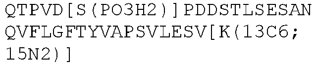
385 p70S6K_ 18 
174 
386 p70S6K_ 19 [T(PO3H2)]PVSPVK 175 
387 p70S6K_ 20 TPV[S(PO3H2)]PVK 176 
388 p70S6K_ 21 
177 
389 p70S6K_ 22 
178 
390 p70S6K_ 23 
179 
391 p70S6K_ 24 
180 
392 RPS6_1 DIPGLTDTTVPR 181 DIPGLTDTTVP[R(13C6; 15N4)] 393 RPS6_2 GHS[C(CAM)]YRPR 182 
394 RPS6_3 GHSCYRPR 183 GHSCYRP[R(13C6; 15N4)] 395 RPS6_4 LNISFPATG[C(CAM)]QK 184 
396 RPS6_5 LNISFPATGCQK 185 LNISFPATGCQ[K(13C6; 15N2)] 397 RPS6_6 MAT EVAADALGEEWK 186 
398 RPS6_7 RRRLSSLRASTSK 187 
399 RPS6_8 
188 
400 RPS6_9 
189 
401 RPS6_10 
190 
402 PRAS40_ 1 
191 
403 PRAS40_ 2 [C(CAM)]LHDIALAHR 192 
404 PRAS40_ 3 CLHDIALAHR 193 CLHDIALAH[R(13C6; 15N4)] 405 PRAS40_ 4 EAEDTQVFGDLPRPR 194 
406 PRAS40_ 5 SLPVSVPVWGFK 195 SLPVSVPVWGF[K(13C6; 15N2)] 407 PRAS40_ 6 SSDEENGPPSSPDLDR 196 
408 PRAS40_ 7 TEARSSDEENGPPSSPDLDR 197 
409 PRAS40_ 8 
198 
410 PRAS40_ 9 
199 
411 PRAS40_ 10 LNTSDFQK 200 LNTSDFQ[K(13C6; 15N2)] 412 PRAS40_ 11 
201 
413 PRAS40_ 12 GALAEAAR 202 GALAEAA[R(13C6; 15N4)] 414 PRAS40_ 13 ASGRPEELWEAWGAAER 203 
415 PRAS40_ 14 LN[T(PO3H2)]SDFQK 204 
416 PRAS40_ 15 
205 
417 PTEN_1 
206 
418 PTEN_2 
207 
419 PTEN_3 NNIDDVVR 208 NNIDDVV[R(13C6; 15N4)] 420 PTEN_4 AQEALDFYGEVR 209 AQEALDFYGEV[R(13C6; 15N4)] 421 PTEN_5 IYSSNSGPTR 210 IYSSNSGPT[R(13C6; 15N4)] 422 PTEN_6 
211 
423 PTEN_7 
212 
424 - In some embodiments, the peptides reagents are recited in Table 5 (SEQ ID Nos: 98, 96, 157, 163, 40, 42, 37, 25, 73, 80, 52, 57, 59, 208, 209, 16, 23, 124, 120, 195, 200, 129, 133, 1, 6, 27, 91, and 204). In some embodiments, the peptides of Table 5 are useful in multi-plex MS methods.
- In some embodiments, protein samples are denatured or solubilized before fragmentation.
- In some embodiments, the fragmentation protocol uses chemical cleavage. In some embodiments, the chemical cleavage uses CNBr. In some embodiments, the fragmentation protocol is done using an enzyme. In some embodiments, the fragmentation protocol uses MS-grade commercially available proteases. Examples of proteases that may be used to digest samples include trypsin, endoproteinase GluC, endoproteinase ArgC, pepsin, chymotrypsin, LysN protease, LysC protease, GluC protease, AspN protease, proteinase K, and thermolysin. In some embodiments, a mixture of different proteases are used and the individual results are combined together after the digestion and analysis. In some embodiments, the digestion is incomplete in order to see larger, overlapping peptides. In some embodiments, the antibody digestion is performed with IdeS, IdeZ, pepsin, or papain to generate large antibody domains for "middle-down" protein characterization. In some embodiments, the fragmentation protocol uses trypsin that is modified. In some embodiments, a protein: protease ratio (w/w) of 10:1, 20:1, 25:1, 50:1, 66:1, or 100:1 may be used. In some embodiments, the trypsin used is at a concentration of about 100ng/ml-1 mg/ml, or about 100 ng/ml-500µg/ml, or about 100 ng/ml-100µg/ml, or about 1µg/ml-1mg/ml, or about 1µg/ml-500µg/ml, or about 1µg/ml-100µg/ml, or about 10µg/mg-1mg/ml, or about 10µg/mg-500µg/ml, or about 10µg/mg-100µg/ml. In some embodiments, the digestion step is for 10 minutes to 48 hours, or 30 minutes to 48 hours, or 30 minutes to 24 hours, or 30 minutes to 16 hours, or 1 hour to 48 hours, or 1 hour to 24 hours, or 1 hour to 16 hours, or 1 to 8 hours, or 1 to 6 hours, or 1 to 4 hours. In some embodiments, the digestion step is incubated at a temperature between 20°C and 45°C, or between 20°C and 40°C, or between 22°C and 40°C, or between 25°C and 37°C. In some embodiments, the digestion step is incubated at 37°C or 30°C. In some embodiments, a step is included to end the digestion step. The step to end the digestion protocol may be addition of a stop solution or a step of spinning or pelleting of a sample. In some embodiments, the digestion is followed by guanidation.
- In some embodiments, the fragmentation protocol includes use of protein gels. In some embodiments, the fragmentation protocol comprises in-gel digestion. An exemplary commercially available kit for performing in-gel digestion is the In-Gel Tryptic Digestion Kit (Thermo Fisher Cat#89871).
- In some embodiments, the fragmentation protocol is carried out in solution. An exemplary commercially available kit for performing in-solution digestion is the In-Solution Tryptic Digestion and Guanidiation Kit (Thermo Fisher Cat#89895).
- In some embodiments, the fragmentation protocol uses beads. In some embodiments, the fragmentation protocol comprises on-bead digestion. In some embodiments, agarose beads or Protein G beads are used. In some embodiments, magnetic beads are used.
- In some embodiments, protein samples are separated using liquid chromatography before MS analysis. In some embodiments, fragmented samples are separated using liquid chromatography before MS analysis.
- The IP and IP-MS methods described herein are capable of detecting phosphorylated AKT-mTOR pathway proteins, including those described in Table 4.
Table 4. List of Total and Phosphorylated AKT-mTOR Pathway Target Proteins Target ID No. Target Name (Total) Phosphorylation Site 1 AKT1 pSer473 2 PTEN pSer380 3 IRS1 pSer312 4 IR pTyr1162/1163 5 IGF-1R pTyr1135/1136 6 GSK3a pSer21 7 GSK3b pSer9 8 RPS6 pSer235/236 9 PRAS40 pThr246 10 mTOR pSer2448 11 p70S6K (S6K1) pThr389 12 TSC2 pSer939 - In some embodiments, the AKT-mTOR pathway peptides used in the MS methods described herein have limits of detection considered useful in clinical and research methods. See, e.g, Table 5. In some embodiments, the AKT-mTOR pathway peptides used in the MS and IP-MS methods comprise or consist of the peptides described in Table 5. In some embodiments, the peptides of Table 5 are detectably labelled. The peptides of SEQ ID NO: 163 may lack the "CAM" modification shown on the fifth amino acid.
Table 5. Lower Limit of Quantitation of Peptides for AKT-mTOR Pathway Proteins Target ID Native Peptide Sequence SEQ ID NO: Lower limit of Quantitation (fmol) mTOR_6 GYTLADEEEDPLIYQHR 98 0.69 mTOR_4 GNNLQDTLR 96 0.08 p70S6K_1 DGFYPAPDFR 157 0.23 p70S6K_7 IRPE[C(CAM)]FELLR 163 6.17 IGF1R_7 TTINNEYNYR 40 0.08 IGF1R_9 YADGTIDIEEVTENPK 42 0.69 IGF1R_4 LG[C(CAM)]SASNFVFAR 37 2.06 IR/ IGF1R_1 DIYETDYYR 25 0.69 TSC2_4 GYTISDSAPSR 73 0.69 TSC2_11 YTEFLTGLGR 80 0.69 IRS1_6 HHLNNPPPSQVGLTR 52 0.69 IRS1_11 SVSAPQQIINPIR 57 0.08 IRS1_13 TGIAAEEVSLPR 59 0.23 PTEN_3 NNIDDVVR 208 0.08 PTEN_4 AQEALDFYGEVR 209 0.08 IR_5 TIDSVTSAQELR 16 0.23 IR_12 TVN ESASLR 23 0.08 GSK3a_10 VTTVVATLGQGPER 124 0.23 GSK3a_6 SQEVAYTDIK 120 0.69 PRAS40_5 SLPVSVPVWGFK 195 6.17 PRAS40_10 LNTSDFQK 200 0.69 GSK3b_2 DIKPQNLLLDPDTAVLK 129 6.17 GSK3b_6 LLEYTPTAR 133 0.23 AKT1_1 NDGTFIGYK 1 0.23 AKT2_1 SDGSFIGYK 6 0.69 IR/IGF1R_3 DI[Y(PO3H2)]ETDYYR 27 0.69 TSC2_22 ST[S(PO3H2)]LNERPK 91 0.23 PRAS40_14 LN[T(PO3H2)]SDFQK 204 0.69 - In some embodiments, methods for detecting phosphorylated AKT-mTOR pathway proteins are encompassed. In some embodiments, IP, MS, and IP-MS methods to detect phosphorylated AKT-mTOR pathway proteins are conducted separately from methods to detect total (non-phosphorylated) AKT-mTOR pathway proteins. In some embodiments, the IP and IP-MS methods to detect phosphorylated AKT-mTOR pathway proteins utilize the antibodies of Table 9. In some embodiments, the IP and IP-MS methods to detect non-phosphorylated AKT-mTOR pathway proteins utilize the antibodies of Table 8. In some embodiments, the IP-MS methods to detect phosphorylated AKT-mTOR pathway proteins utilize the antibodies of Table 9 and the peptides of Table 5. In some embodiments, the IP-MS methods to detect non-phosphorylated AKT-mTOR pathway proteins utilize the antibodies of Table 8 and the peptides of Table 5.
Table 8. List of non-phosho-antibodies for multi-plex IP, single-plex IP (+/- MS). Target Vendor IP Antibody AKT MILLIPORE 07-416 IGF1R CELL SIGNALING TECHNOLOGY 3027 IR MILLIPORE 07-724 IRS1 CELL SIGNALING TECHNOLOGY 2382 mTOR THERMO FISHER SCIENTIFIC PA1-518 P70S6K ABGENT AP3289g GSK3a CELL SIGNALING TECHNOLOGY 4337 GSK3b THERMO FISHER SCIENTIFIC MA1-15109 TSC2 THERMO FISHER SCIENTIFIC MA5-15004 PRAS40 THERMO FISHER SCIENTIFIC PA5-35143 PTEN CELL SIGNALING TECHNOLOGY 9188 Table 9. List of antibodies for multi-plex IP, single-plex IP (+/- MS). Target Vendor IP Antibody phosphoAKT CELL SIGNALING TECHNOLOGY 4060 phosphoIGF1R THERMO FISHER SCIENTIFIC PA5-35769 phosphoIR N/A N/A phosphoIRSI MILLIPORE 05-1087 phosphomTOR CELL SIGNALING TECHNOLOGY 5536 phosphoP70S6K CELL SIGNALING TECHNOLOGY 9204 phosphoGSK3a CELL SIGNALING TECHNOLOGY 9327 phosphoGSK3b CELL SIGNALING TECHNOLOGY 5558 phosphoTSC2 THERMO FISHER SCIENTIFIC PA5-12845 phosphoPRAS40 CELL SIGNALING TECHNOLOGY 2997 phosphoPTEN CELL SIGNALING TECHNOLOGY 9551 - The following examples are provided to illustrate certain disclosed embodiments and are not to be construed as limiting the scope of this disclosure in any way.
- AKT-mTOR pathway proteins play central roles in diseases including cancer. The identification of AKT-mTOR pathway proteins, while desired as a means for monitoring disease progression, and as a tool for scientific research, has been limited in part because of the low abundance of AKT-mTOR pathway proteins, and in part due to a lack of validated methods and reagents. Phosphorylated AKT-mTOR pathway proteins are particularly important to identify and quantify as a measure of protein activation status, and also as markers for disease progression. As shown in
Figure 2 , methods and reagents for detecting AKT-mTOR pathway proteins, including phosphorylated proteins, and their protein interactions, were designed and tested. Multiplex immunoprecipitation (IP) to MS (mIP-MS) was assessed for the ability to measure total and phosphorylated AKT-mTOR pathway targets. mIP-MS methods were also compared to existing singleplex immunoassay (Western Blot (WB) and ELISA) and multiplex Luminex assays. - For all assays, HCT116 (ATCC product#CCL-247), MCF7, (ATCC product#HTB-22) and A549 (ATCC product#CCL-185) cells were grown in Hamm's F-12K media, McCoy's 5A Media, and MEM Media, respectively, with 10% FBS/1xPenStrep to approximately 70-80% confluency. Cells were starved in 0.1% charcoal stripped FBS for 24 hours before stimulation with 100ng/ml of IGF (CST product#8917SF) for 15 minutes.
- Western Blot (WB), ELISA, and Luminex Assays were used as controls to compare to the IP-MS method described herein. The reagents and methods for Western Blots are summarized in Table 6.
Table 6 - List of IP to Western Blot validated antibodies for AKT-mTOR Pathway Targets Target WB Antibody Vendor Dilution Gel Notes AKT 4691 CELL SIGNALING TECHNOLOGY (1:1000) Tris Glycine phosphoAKT 4051 CELL SIGNALING TECHNOLOGY (1:1000) Tris Glycine IGF1R 3027 CELL SIGNALING TECHNOLOGY (1:1000) Tris Acetate phosphoIGF1R Biotinylated AP50303 Abgent (1:10,000) Tris Acetate SA HRP Secondary IR 3020 CELL SIGNALING TECHNOLOGY (1:1000) Tris Acetate phosphoIR 07-841 Millipore (1:1000) Tris Acetate IRS1 2382 CELL SIGNALING TECHNOLOGY (1:1000) Tris Acetate phosphoIRS1 Biotinylated 05-1087 Millipore (1:1000) Tris Acetate SA HRP Secondary mTOR 2983 CELL SIGNALING TECHNOLOGY (1:1000) Tris Acetate phosphomTOR 5536 CELL SIGNALING TECHNOLOGY (1:1000) Tris Acetate P70S6K AP3289g Abgent (1:1000) Tris Glycine phosphoP70S6K 9204 CELL SIGNALING TECHNOLOGY (1:1000) Tris Glycine GSK3a 4337 CELL SIGNALING TECHNOLOGY (1:1000) Tris Glycine Clean Blot phosphoGSK3a 8506 CELL SIGNALING TECHNOLOGY (1:1000) Tris Glycine Clean Blot GSK3b 12456 CELL SIGNALING TECHNOLOGY (1:1000) Tris Glycine Clean Blot phosphoGSK3b 5558 CELL SIGNALING TECHNOLOGY (1:1000) Tris Glycine Clean Blot TSC2 4308 CELL SIGNALING TECHNOLOGY (1:1000) Tris Acetate phosphoTSC2 ab52962 Abeam (1:10,000) Tris Acetate PRAS40 AP14275b Abgent (1:1000) Tris Glycine phosphoPRAS40 2997 CELL SIGNALING TECHNOLOGY (1:1000) Tris Glycine PTEN 9188 CELL SIGNALING TECHNOLOGY (1:1000) Tris Glycine Clean Blot phosphoPTEN 9551 CELL SIGNALING TECHNOLOGY (1:1000) Tris Glycine Clean Blot Secondary Antibodies: Goat Anti-Rabbit Ab (Thermo Fisher Scientific, PN:32460), Goat AntiMouse Ab (Thermo Fisher Scientific, PN:32430), Pierce High Sensitivity Streptavidin-HRP (Thermo Fisher Scientific, PN: 21130)
SDS-PAGE Gels: NuPAGE 3-8% Tris-Acetate Gel (Thermo Fisher Scientific, PN: EA03752BOX), Novex 4-20% Tris-Glycine Midi Gel (Thermo Fisher Scientific, PN: WT4201BX10)
Clean Blot: Thermo Fisher Scientific, PN: 21232 - Reagents for ELISA kits are shown in Table 7.
Table 7. ELISA kits for 11 total and 10 phosphorylated AKT-mTOR Pathway Targets Target Vendor Product# Lot# Total GSK3B Cell Signaling Technology 7265 0004 Phospho GSK3β Cell Signaling Technology 7311 0004 Phospho GSK3β Life Technologies KH00461 16404995B Total IRS1 Cell Signaling Technology 7328 0011 Phospho IRS1 N/A N/A N/A Total PTEN Cell Signaling Technology 7882 0005 Phospho PTEN Cell Signaling Technology 7285 0006 Total PRAS40 Cell Signaling Technology 7331 0003 Phospho PRAS40 Cell Signaling Technology 7327 0004 Total Insulin Receptor Cell Signaling Technology 7069 0006; 0004 Phospho Insulin Receptor Cell Signaling Technology 7258 0016; 0015 Total IGF1R R&D Systems DYC305-2 1324480 Total IGF1R Abeam ab100546 GR212867-1 Phospho IGF1R Cell Signaling Technology 7302 0015 Total GSK3α R&D Systems DYC2157-2 1299193 Phospho GSK3α R&D Systems DYC4125-2 1300987 Total TSC2 Lifespan Biosciences LS-F2369 50 Phospho TSC2 Lifespan Biosciences LS- F1233 49 Total AKT1 Cell Signaling Technology 7170 0048 Phospho AKT1 Cell Signaling Technology 7160 0093 Total mTOR Cell Signaling Technology 7974 0006 Phospho mTOR Cell Signaling Technology 7976 0007 Total p70S6K Cell Signaling Technology 7038 0004 Phospho p70S6K Cell Signaling Technology 7063 0005 - For Luminex Assays, AKT Pathway (total) Magnetic 7-Plex Panel (Thermo Fisher Scientific, PN: LHO0002M), AKT Pathway (phospho) Magnetic 7-Plex Panel (Thermo Fisher Scientific, PN: LHO0001M), Milliplex Map Akt/mTOR Phosphoprotein Magnetic Bead 11-Plex Kit (Millipore, PN: 48-611MAG) and Milliplex Map Total Akt/mTOR Magnetic Bead 11-Plex Kit (Millipore, PN: 48-612MAG) were used as recommended in instruction manuals. Luminex MagPix instrument was used to acquire and analyze Luminex assay data.
- The Thermo Scientific™ Pierce MS-Compatible Magnetic IP Kit (Protein A/G) was used to screen and validate antibodies for 11 total and 10 phosphorylated AKT-mTOR pathway proteins from 500µg cell lysate. Validated antibodies were biotinylated with the Thermo Scientific™ Pierce Antibody Biotinylation Kit for IP. The Thermo Scientific™ Pierce MS-Compatible Magnetic IP Kit (Streptavidin) was used to perform the single or multiplex IPs for target enrichment. IP samples were processed by an in-solution digestion method where IP eluates were reconstituted in 6M Urea, 50mM TEAB, pH 8.5 followed by reduction, alkylation and trypsin digestion overnight at 37°C. The digested samples were acidified with TFA before MS analysis.
- Prior to MS analysis, tryptic digest samples were desalted on-line using the Thermo Scientific™ Acclaim™ PepMap 100 C18 Trap Column. For discovery MS, the samples were analyzed by nanoLC-MS/MS using a Thermo Scientific™ Dionex™ UltiMate™ 3000 RSLCnano System and Thermo Scientific™ Q Exactive™ HF Hybrid Quadrupole-Orbitrap Mass Spectrometer. For targeted MS, the samples were analyzed using the UltiMate 3000 RSLCnano System and the Thermo Scientific™ TSQ™ Vantage™ Mass Spectrometer (SRM mode) or the Thermo Scientific™ Q Exactive™ HF Hybrid Quadrupole-Orbitrap Mass Spectrometer (PRM mode).
- Discovery MS data were analyzed with Thermo Scientific™ Proteome Discoverer™ 1.4 to assess percent sequence coverage, unique peptides, MS1 intensities, spectral counts and PTMs. The Proteome Discoverer software searches were executed using the Uniprot human protein database. Tryptic peptides with highest MS1 intensity and relevant phosphorylation sites were selected from the discovery data for targeted assay development. For targeted MS data analysis, Thermo Scientific™ Pinpoint software and Skyline software (University of Washington) were used to measure limit of quantitation (LOQ) from the calibration curve and target analyte concentration from unknown samples.
- As shown in
Figure 3 , AKT-mTOR pathway proteins were immunoprecipitated from unstimulated and IGF-stimulated A549 lysate with Thermo Scientific™ Pierce MS-Compatible Magnetic IP Kits (Protein A/G or Streptavidin) for MS analysis. A variety of antibodies were screened to determine effectiveness in both ability to IP AKT-mTOR pathway proteins, and also for their usefulness when combined with MS. Table 1 (above) provides a list of antibodies validated for use in the IP-MS methods. Table 2 (above) provides a list of antibodies tested, but found to be less successful. - Higher numbers of unique peptides were identified in IP enriched samples as compared to neat (non-IP-enriched) lysate. See
Figure 3 . Protein isoforms and interacting protein partners were identified for AKT, IGFIR and mTOR targets. Relevant phosphorylation sites were detected for AKT1, AKT2, mTOR, IGFIR and PRAS40. Candidate quantitative peptides were selected for targeted MS assay development. - Limits of detection (LOD) and lower limits of quantification (LLOQ) were analyzed for twelve AKT-mTOR pathway proteins, including AKT2, AKT1, mTOR, IGF1R, IR, PRAS40, p70S6K, TSC2, PTEN, GSK3alpha, GSK3beta, and IRS1. Results are presented in
Figure 4 . The assay dynamic range, representing the concentration range between the lower to upper limits of quantification (LLOQ to ULOQ), is the range where protein concentration is measurable with acceptable levels of accuracy and precision. To ensure linearity of the measurement, for each internal standard peptide the linear signal-to-abundance range (LLOQ and ULOQ) was determined from dilution series experiments spanning concentrations of 500-0.08 fmol on column, spiked into a constant light peptide at 36fmol and 200ng of equimolar concentration of 6 proteins digest. - Eleven total and ten phosphorylated AKT-mTOR pathway protein targets were enriched simultaneously from unstimulated and IGF stimulated MCF7 lysates with biotinylated antibodies and Thermo Scientific™ Pierce MS-Compatible Magnetic IP Kit (Streptavidin). MCF7 cells were starved in 0.1% charcoal stripped FBS for 24 hours before stimulation with 100ng/ml of IGF for 15 minutes. Validated IP-MS antibodies are biotinylated for 11 total and 10 phosphorylated AKT-mTOR pathway targets using the Thermo Scientific™ Pierce Antibody Biotinylation Kit for IP (PN: 90407) as recommended in instruction manual. 1µg of each biotinylated antibody for 11 total targets were added simultaneously to 1000µg of control and IGF stimulated MCF7 cell lysate in duplicate. 1µg of each biotinylated antibody for 10 total targets were added simultaneously to 1000µg of control and IGF stimulated MCF7 cell lysate in duplicate. IP was performed as recommended in the Thermo Scientific™ Pierce MS-Compatible Magnetic IP Kit (Streptavidin) (PN: 90408) with the following modification. 5 microgram of streptavidin magnetic beads per microgram of biotinylated antibody concentration was used for multiplex IP.
- IP samples were processed by an in-solution digestion method where IP eluates were reconstituted in 6M Urea, 50mM TEAB, pH 8.5 followed by reduction (5mM TCEP for 30 minutes at 35°C), alkylation (20mM Iodoacetamide in dark at room temperature for 30 minutes) and trypsin digestion overnight at 37°C. The digested samples were acidified with 3.5µL of 10% TFA before discovery MS analysis. For discovery MS, the samples were analyzed by nanoLC-MS/MS using a Thermo Scientific™ Dionex™ UltiMate™ 3000 RSLCnano System and Thermo Scientific™ Q Exactive™ HF Hybrid Quadrupole-Orbitrap Mass Spectrometer. Briefly, the digested samples were cleaned on-line using the C18 trap column (Thermo Fisher Scientific, PN: 164564) followed by reversed-phase separation using the analytical C18 column (75 µm i.d. x 15 cm, nanoViper, 3µm particle size, Thermo Fisher Scientific, PN: ES800) with a 2-30% gradient of Buffer B using Buffer A (0.1% formic acid) and Buffer B (0.1% formic acid/99.9 % acetonitrile) at 0.300 µL/min.
-
Figure 5 shows that the IP-nanoLC-MS/MS analysis was able to identify 11 proteins in the multiplex phosphor-assay, and 12 proteins for multiplex total assay. MS analysis of multiplex total assay identified interacting proteins (PIK3R1, PIK3R2, PIK3CB, PIK3CA, GSKIP and TSC1) of AKT-mTOR Pathway Targets. Tables 8 and 9 provide listings of the antibodies used in this multiplex IP. - Next the ability of IP-MS to quantify sub-fmol concentrations of AKT-mTOR pathway proteins via the disclosed IP-MS methods was tested. As shown in
Figure 6 , a multiplex IP enrichment of AKT (Total & Phospho), IR, p70S6K, mTOR, and GSK3α was performed from unstimulated and IGF stimulated A549 and HCT116 lysates with Thermo Scientific™ Pierce MS-Compatible Magnetic IP Kit (Streptavidin). A549 and HCT116 cells were starved in 0.1% charcoal stripped FBS for 24 hours before stimulation with 100ng/ml of IGF for 15 minutes. Validated IP-MS antibodies are biotinylated for Total AKT, Phospho AKT, IR, p70S6K, mTOR, and GSK3α pathway targets using the Thermo Scientific™ Pierce Antibody Biotinylation Kit for IP (PN: 90407) as recommended in instruction manual. 1µg of each biotinylated antibody was added simultaneously to 1000µg of control and IGF stimulated A549 and HCT116 cell lysates in duplicate. IP was performed as recommended in the Thermo Scientific™ Pierce MS-Compatible Magnetic IP Kit (Streptavidin) (PN: 90408) with the following modification. 5 microgram of streptavidin magnetic beads per microgram of biotinylated antibody concentration was used for multiplex IP. IP samples were processed by an in-solution digestion method where IP eluates were reconstituted in 6M Urea, 50mM TEAB, pH 8.5 followed by reduction (5mM TCEP for 30 minutes at 35°C), alkylation (20mM Iodoacetamide in dark at room temperature for 30 minutes) and trypsin digestion overnight at 37°C. The digested samples were acidified with 3.5µL of 10% TFA before discovery MS analysis. Internal standard peptides were spiked in digested IP samples to make final volume of 6.66fmol/ul. For targeted MS, the samples were analyzed by nanoLC-PRM/MS using a Thermo Scientific™ Dionex™ UltiMate™ 3000 RSLCnano System and Thermo Scientific™ Q Exactive™ HF Hybrid Quadrupole-Orbitrap Mass Spectrometer. Briefly, the digested samples were cleaned on-line using the C18 trap column (Thermo Fisher Scientific, PN: 164564) followed by reversed-phase separation using the analytical C18 column (75 µm i.d. x 15 cm, nanoViper, 3µm particle size, Thermo Fisher Scientific, PN: ES800) with a 2-30% gradient of Buffer B using Buffer A (0.1% formic acid) and Buffer B (0.1% formic acid/99.9 % acetonitrile) at 0.300 µL/min. Total targets were quantified in low to sub-fmol concentrations by nanoLC-PRM/MS. Up-regulation of phospho AKT was seen after IGF stimulation in both A549 and HCT116 cell lines. The slight decrease in concentrations for total AKT, IR, mTOR, GSK3a and p70S6K targets was observed after IGF stimulation in both A549 and HCT116 cells. - Next, comparison of mIP-tMS assays with current immunoassay techniques to quantitate AKT-mTOR pathway targets from unstimulated and IGF stimulated A549, HCT116 and MCF7 lysates were performed. Western Blot, ELISA, and Luminex assays were performed as described above and according to manufacturer's instructions. mIP-tMS was performed as in Example 2.
-
Figures 7A-7D show quantitation of total AKT.Figures 7E-7H show quantitation of phosphorylated IGFIR across all 4 techniques. Lower correlation was observed across techniques. The lower correlation could be due to different antibodies used or each assay and antibody specificity. Up-regulation in phosphorylated IGF1R observed after IGF stimulation in 3 of 4 techniques. Western blot for phosphor IGFIR showed no significant differences in control and IGF stimulated cell lysates. - A summary of AKT-mTOR pathway proteins that were identified and quantified using the IP-MS methods described herein is provided in
Figure 8 . Most of the AKT-mTOR pathway targets were not identified in discovery MS and quantitated by targeted MS (PRM or SRM) without enrichment by immunoprecipitation. - Immunoprecipitation using particular selected antibodies resulted in a higher yield of AKT-mTOR pathway target proteins and less non-specific binding proteins than MS alone. IP-MS assay was also more successful than other commercially available non-MS assays. Furthermore, IP to MS analysis of total and phosphorylated AKT-mTOR pathway proteins enabled identification of multiple isoforms, relevant protein interactions and phosphorylation sites. Total and phosphorylated mIP-tMS assays allowed simultaneous quantitation of 12 total and 11 phosphorylated AKT-mTOR pathway proteins in the low to sub-fmol range from unstimulated and IGF stimulated A549, HCT116 and MCF7 cell lysates. The benchmarking of mIP-tMS assays showed moderate correlation for quantitation of total and phosphorylated target relative abundance compared to WB, ELISA and Luminex assays. The low concordance for a few targets is possibly due to differences in the specificity of antibodies used for each assay. Major advantages of the MS-based assay are high confidence in target identity coupled with simultaneous quantitation of multiple targets, interacting proteins and their phosphophorylated forms.
- Tissue lysis protocol was optimized for IP-MS application. Briefly, 50-100mg of human and murine tissue samples were washed with 5mL 1X cold PBS three times. Tissue samples was minced in 5mL 1x cold PS using scissor followed by homogenization in IP lysis buffer (Thermo Fisher Scientific PN: 87788) and electronic Polytron Handheld Tissue Tearer. Homogenized tissue samples were passed through tissue strainer (Thermo Fisher Scientific PN: 87791) to prepare tissue lysates before IP. To validate the IP-MS method in murine and human tissue lysate, eleven total and ten phosphorylated AKT- mTOR pathway protein targets were enriched simultaneously from normal mouse lung tissue lysate, normal mouse kidney tissue lysate, and normal human lung tissue lysate as per Example 2. A549 cell lysate was used as a non-tissue control. As shown in Table 10, the IP-MS method described herein is capable of validating AKT-mTOR pathway proteins in murine and human tissue lysate in addition to cell lysate. Seven out of eleven AKT-mTOR pathway protein targets were identified in normal human lung tissue, and nine out of eleven AKT-mTOR pathway protein targets were identified for normal mouse kidney tissue using our IP-MS method.

Claims (17)
- A method for detecting AKT-mTOR pathway proteins, comprisinga. isolating AKT-mTOR pathway proteins from a biological sample;b. digesting the isolated proteins;c. assaying the digested protein(s) via mass spectrometry to determine the presence of a peptide for AKT-mTOR pathway protein(s), wherein the peptide for AKT-mTOR pathway protein(s) consist(s) of a sequence listed in Table 5; andd. detecting one or more AKT-mTOR pathway protein(s) in the sample,wherein the isolation of step a. comprises:
treating a biological sample with more than one antibody selected from an antibody of Table 8 and/or Table 9 wherein the antibody of Table 8 detects non-phosphorylated AKT-mTOR pathway proteins and the antibody of Table 9 detects phosphorylated AKT-mTOR pathway proteins and capable of immunoprecipitating the AKT-mTOR pathway protein(s) from a biological sample; - The method of claim 1, further comprising determining the quantity of AKT-mTOR pathway protein.
- The method of claim 1 or 2, wherein the lower level of detection is within the range of 0.05 - 0.25 fmol and/or the lower limit of quantification is within the range of 0.05 - 0.75 fmol.
- The method of any one of the preceding claims, wherein the AKT-mTOR pathway protein is phosphorylated.
- A method for determining the ratio of phosphorylated to non-phosphorylated AKT-mTOR pathway proteins, comprisinga. treating a biological sample with more than one antibody capable of immunoprecipitating a phosphorylated AKT-mTOR target protein selected from the antibodies recited in Table 9., and separately treating the same biological sample with more than one antibody capable of immunoprecipitating a non-phosphorylated AKT-mTOR target protein selected from the antibodies recited in Table 8;b. digesting the immunoprecipitated AKT-mTOR pathway proteins;c. adding a first and a second detectably labelled internal standard peptide of known amount to the digested proteins, wherein the first internal standard peptide has the same amino acid sequence as a phosphorylated AKT-mTOR pathway peptide used to identify the phosphorylated protein, and the second internal standard peptide has the same amino acid sequence as the non-phosphorylated AKT-mTOR pathway peptide used to identify the non-phosphorylated protein;d. assaying the digested proteins and internal standards via mass spectrometry to determine the presence and amount of phosphorylated and non-phosphorylated AKT-mTOR pathway peptides, wherein the AKT-mTOR pathway peptide consists of a peptide selected from the peptides shown in Table 5 ande. determining the quantity of AKT-mTOR phosphorylated and non-phosphorylated pathway proteins in the sample, and determining the ratio of phosphorylated to non-phosphorylated target proteins.
- The method of any one of the preceding claims, wherein the biological sample is human, is isolated cells, plasma, serum, whole blood, CSF, urine, sputum, tissue, or tumorous tissue.
- The method of any one of the preceding claims, wherein the peptide is modified with a detectable label or an isotope selected from 13C, 15N, 2H and 180.
- The method of any one of claims 1 to 4 or 6 to 7, wherein a first antibody is capable of immunoprecipitating a phosphorylated AKT-mTOR pathway protein, and a second antibody is capable of immunoprecipitating a non-phosphorylated version of the AKT-mTOR pathway protein precipitated by the first antibody.
- The method of any one of claims 1 to 8, wherein step a) comprises treating the sample with a labelled antibody capable of binding to the pathway protein to provide a labelled antibody-protein conjugate; and binding the labelled antibody-protein conjugate with a capture agent capable of binding to the labelled antibody to isolate the target protein from the sample, optionally wherein the label is biotin and the capture agent is streptavidin.
- The method of any one of the preceding claims, wherein the quantity of an AKT-mTOR pathway protein is determined by adding an internal standard peptide of known amount to the digested protein prior to mass spectrometry, wherein the internal standard peptide has the same amino acid sequence as the AKT-mTOR pathway peptide, and is detectably labeled, and determining the quantity of an AKT-mTOR pathway peptide by comparison to the internal standard or wherein the quantity of an AKT-mTOR pathway protein is determined by a method comprising comparing an amount of an AKT-mTOR pathway peptide in the sample to the amount of the same AKT-mTOR pathway peptide in a control sample.
- The method of claim 10, wherein the quantity of an AKT-mTOR pathway protein is determined by a method comprising comparing an amount of the AKT-mTOR pathway peptide to an internal standard peptide of known amount, wherein both the peptide in the biological sample and the internal standard peptide are selected from the group consisting of SEQ ID Nos: 98, 96, 157, 163, 40, 42, 37, 25, 73, 80, 52, 57, 59, 208, 209, 16, 23, 124, 120, 195, 200, 129, 133, 1, 6, 27, 91, and 204,and wherein the standard peptide is detectably labeled.
- The method of any of the preceding claims, wherein the mass spectrometry is tandem mass spectrometry or discovery mass spectrometry or wherein the targeted mass spectrometry is multiple reaction monitoring (MRM), selected reaction monitoring (SRM), parallel reaction monitoring (PRM), or combinations thereof.
- The method of any one of the preceding claims, wherein the digesting comprises a protease or chemical digest.
- The method of claim 13, wherein the protease digestion is with trypsin, chymotrypsin, AspN, GluC, LysC, LysN, ArgC, proteinase K, pepsin, clostripain, elastase, GluC biocarb, LysC/P, LysN promise, protein endopeptidase, staph protease or thermolysin. or wherein the chemical cleavage is with CNBr, iodosobenzoate or formic acid.
- A kit for use in a method of any one of the preceding claims comprising two or more antibodies capable of immunoprecipitating an AKT-mTOR pathway protein selected from the antibodies recited in Table 8 or Table 9 and optionally reagents useful for performing mass spectrometry to detect and optionally quantify the one or more AKT-mTOR pathway protein, and comprising a peptide selected from the peptides recited in Table 5.
- A kit for use in a method of any one oft claims 1 to 14 comprising two or more antibodies capable of immunoprecipitating an AKT-mTOR pathway protein selected from the antibodies recited in Table 8 or Table 9 and optionally reagents useful for performing mass spectrometry to detect and optionally quantify the one or more AKT-mTOR pathway protein, and further comprising a protease or chemical agent capable of digesting an immunoprecipitated protein sample, optionally wherein the protease agent is trypsin, chymotrypsin, AspN, GluC, LysC, LysN, ArgC, proteinase K, pepsin, clostripain, elastase, GluC biocarb, LysC/P, LysN promise, protein endopeptidase, Staph protease or thermolysin, or optionally wherein the chemical agent is CNBr, iodosobenzoate or formic acid.
- The kit of claim 16, further comprising an AKT-mTOR pathway peptide selected from the peptides recited in Table 5.
Applications Claiming Priority (2)
| Application Number | Priority Date | Filing Date | Title |
|---|---|---|---|
| US201662308051P | 2016-03-14 | 2016-03-14 | |
| PCT/US2017/022062 WO2017160698A1 (en) | 2016-03-14 | 2017-03-13 | DETECTION AND QUANTIFICATION OF AKT-mTOR PATHWAY PROTEINS |
Publications (2)
| Publication Number | Publication Date |
|---|---|
| EP3430405A1 EP3430405A1 (en) | 2019-01-23 |
| EP3430405B1 true EP3430405B1 (en) | 2023-08-09 |
Family
ID=58413192
Family Applications (1)
| Application Number | Title | Priority Date | Filing Date |
|---|---|---|---|
| EP17713827.8A Active EP3430405B1 (en) | 2016-03-14 | 2017-03-13 | Detection and quantification of akt-mtor pathway proteins |
Country Status (4)
| Country | Link |
|---|---|
| US (2) | US11561226B2 (en) |
| EP (1) | EP3430405B1 (en) |
| CN (2) | CN116068187A (en) |
| WO (1) | WO2017160698A1 (en) |
Families Citing this family (5)
| Publication number | Priority date | Publication date | Assignee | Title |
|---|---|---|---|---|
| EP3430405B1 (en) * | 2016-03-14 | 2023-08-09 | Pierce Biotechnology Inc. | Detection and quantification of akt-mtor pathway proteins |
| CN110366559B (en) * | 2017-02-28 | 2023-08-01 | 皮尔斯生物技术公司 | Detection and quantification of RAS-RAF-MAPK pathway proteins |
| CN111771127A (en) * | 2018-02-27 | 2020-10-13 | 皮尔斯生物科技有限公司 | Improved immunoaffinity enrichment and mass spectrometry |
| EP4484958A1 (en) * | 2023-06-29 | 2025-01-01 | University Of Fribourg | Targeted proteomics for monitoring autophagy |
| WO2025003457A1 (en) * | 2023-06-29 | 2025-01-02 | University Of Fribourg | Targeted proteomics for monitoring autophagy |
Citations (4)
| Publication number | Priority date | Publication date | Assignee | Title |
|---|---|---|---|---|
| US20120165340A1 (en) * | 2009-02-11 | 2012-06-28 | Ludwing Institute For Cancer Research Ltd. | Pten phosphorylation-driven resistance to cancer treatment and altered patient prognosis |
| US9110074B2 (en) * | 2007-11-06 | 2015-08-18 | Translational Therapeutics, Inc. | Mass spectrometry assay for eIF4E and eIF4E regulon activity |
| US9252003B2 (en) * | 2013-06-07 | 2016-02-02 | Pierce Biotechnology, Inc. | Absolute quantitation of proteins and protein modifications by mass spectrometry with multiplexed internal standards |
| US20160067260A1 (en) * | 2014-09-05 | 2016-03-10 | Arqule, Inc. | Compositions and Methods for Treating Proliferation Disorders |
Family Cites Families (17)
| Publication number | Priority date | Publication date | Assignee | Title |
|---|---|---|---|---|
| US20060141632A1 (en) * | 2000-07-25 | 2006-06-29 | The Procter & Gamble Co. | New methods and kits for sequencing polypeptides |
| PL1891446T3 (en) * | 2005-06-14 | 2013-08-30 | Cellzome Gmbh | Process for the identification of novel enzyme interacting compounds |
| US20090215098A1 (en) * | 2006-04-28 | 2009-08-27 | Ucl Business Plc. | Quantification of enzyme activity by mass spectrometry |
| JP2009050183A (en) * | 2007-08-24 | 2009-03-12 | Japan Found Cancer Res | Method for determining effect of PI3 kinase inhibitor |
| US8604692B2 (en) * | 2007-11-06 | 2013-12-10 | Translational Therapeutics, Inc. | Mass spectrometry assay for eIF4E and eIF4E regulon activity |
| EP2062912A1 (en) * | 2007-11-26 | 2009-05-27 | Koninklijke Philips Electronics N.V. | Selective enrichment of post-translationally modified proteins and/or peptides |
| EP2124060A1 (en) * | 2008-05-23 | 2009-11-25 | ETH Zurich | Method for high throughput peptide/protein assay generation and assays generated therewith |
| CN102510903B (en) * | 2009-04-14 | 2015-07-01 | 系统生物学研究所 | Improvements in mass spectrum-based identification and quantitation of proteins and peptides |
| WO2011036665A2 (en) * | 2009-09-22 | 2011-03-31 | Yissum Research Development Company Of The Hebrew University Of Jerusalem Ltd | Akt/mtorc1 pathway activators for use in increasing liver regeneration |
| EP2517002B1 (en) * | 2009-12-22 | 2017-08-02 | Expression Pathology, Inc. | Insulin receptor substrate 1 (irs1) protein srm/mrm assay |
| US20130252950A1 (en) * | 2010-09-23 | 2013-09-26 | President And Fellows Of Harvard College | Targeting mtor substrates in treating proliferative diseases |
| BR112014003431A2 (en) * | 2011-08-17 | 2017-06-13 | Genentech Inc | antibody, nucleic acid, host cell, method of producing an antibody, immunoconjugate, pharmaceutical formulation, pharmaceutical agent, use of the antibody, method of treating an individual who has cancer, and time-lapse method for tumor recurrence |
| MX364370B (en) * | 2012-07-12 | 2019-04-24 | Persimmune Inc | PERSONALIZED CANCER VACCINES AND ADOPTIVE IMMUNE CELL THERAPIES. |
| US20170370942A1 (en) * | 2014-08-14 | 2017-12-28 | Universitat Zurich | Highly multiplexed absolute quantification of molecules on the single cell level |
| EP3266455B1 (en) * | 2015-03-06 | 2020-12-30 | Korea Advanced Institute of Science and Technology | Composition for prevention or treatment of intractable epilepsy comprising mtor inhibitor |
| WO2017100789A1 (en) * | 2015-12-11 | 2017-06-15 | Expression Pathology, Inc. | Srm/mrm assays |
| EP3430405B1 (en) * | 2016-03-14 | 2023-08-09 | Pierce Biotechnology Inc. | Detection and quantification of akt-mtor pathway proteins |
-
2017
- 2017-03-13 EP EP17713827.8A patent/EP3430405B1/en active Active
- 2017-03-13 WO PCT/US2017/022062 patent/WO2017160698A1/en not_active Ceased
- 2017-03-13 US US16/081,377 patent/US11561226B2/en active Active
- 2017-03-13 CN CN202211385855.1A patent/CN116068187A/en active Pending
- 2017-03-13 CN CN201780017167.0A patent/CN108885215B/en active Active
-
2022
- 2022-12-19 US US18/068,047 patent/US12228576B2/en active Active
Patent Citations (4)
| Publication number | Priority date | Publication date | Assignee | Title |
|---|---|---|---|---|
| US9110074B2 (en) * | 2007-11-06 | 2015-08-18 | Translational Therapeutics, Inc. | Mass spectrometry assay for eIF4E and eIF4E regulon activity |
| US20120165340A1 (en) * | 2009-02-11 | 2012-06-28 | Ludwing Institute For Cancer Research Ltd. | Pten phosphorylation-driven resistance to cancer treatment and altered patient prognosis |
| US9252003B2 (en) * | 2013-06-07 | 2016-02-02 | Pierce Biotechnology, Inc. | Absolute quantitation of proteins and protein modifications by mass spectrometry with multiplexed internal standards |
| US20160067260A1 (en) * | 2014-09-05 | 2016-03-10 | Arqule, Inc. | Compositions and Methods for Treating Proliferation Disorders |
Non-Patent Citations (2)
| Title |
|---|
| BENJAMIN W TURNEY ET AL: "Depletion of the type 1 IGF receptor delays repair of radiation-induced DNA double strand breaks", RADIOTHERAPY AND ONCOLOGY, ELSEVIER, IRELAND, vol. 103, no. 3, 14 March 2012 (2012-03-14), pages 402 - 409, XP028517624, ISSN: 0167-8140, [retrieved on 20120322], DOI: 10.1016/J.RADONC.2012.03.009 * |
| BHAVIN PATEL ET AL: "Quantitative analysis of AKT/mTOR pathway using immunoprecipitation and targeted mass spectrometry", 2015, XP055637364, Retrieved from the Internet <URL:http://tools.thermofisher.com/content/sfs/posters/PN-64456-LC-MS-AKT-mTOR-Pathway-Immunoprecipitation-ASMS2015-PN64456-EN.pdf> [retrieved on 20191030], DOI: 10.1158/1538-7445.AM2015-1837 * |
Also Published As
| Publication number | Publication date |
|---|---|
| US20230213529A1 (en) | 2023-07-06 |
| CN116068187A (en) | 2023-05-05 |
| CN108885215B (en) | 2022-11-25 |
| WO2017160698A9 (en) | 2017-11-09 |
| WO2017160698A1 (en) | 2017-09-21 |
| US12228576B2 (en) | 2025-02-18 |
| US11561226B2 (en) | 2023-01-24 |
| CN108885215A (en) | 2018-11-23 |
| US20190086420A1 (en) | 2019-03-21 |
| EP3430405A1 (en) | 2019-01-23 |
Similar Documents
| Publication | Publication Date | Title |
|---|---|---|
| US12228576B2 (en) | Detection and quantification of AKT-mTOR pathway proteins | |
| Whiteaker et al. | A targeted proteomics–based pipeline for verification of biomarkers in plasma | |
| Whiteaker et al. | An automated and multiplexed method for high throughput peptide immunoaffinity enrichment and multiple reaction monitoring mass spectrometry-based quantification of protein biomarkers | |
| Heudi et al. | Towards absolute quantification of therapeutic monoclonal antibody in serum by LC− MS/MS using isotope-labeled antibody standard and protein cleavage isotope dilution mass spectrometry | |
| Nicol et al. | Use of an immunoaffinity-mass spectrometry-based approach for the quantification of protein biomarkers from serum samples of lung cancer patients | |
| US11467167B2 (en) | SRM methods in Alzheimer's disease and neurological disease assays | |
| TWI551687B (en) | Method for preparing peptide fragment, kit for preparing peptide fragment used in the method and analysing method | |
| US20250224405A1 (en) | Methods for immunoaffinity enrichment and mass spectrometry | |
| US20200264194A1 (en) | Antibody validation using ip-mass spectrometry | |
| Bringans et al. | Comprehensive mass spectrometry based biomarker discovery and validation platform as applied to diabetic kidney disease | |
| CN110366559B (en) | Detection and quantification of RAS-RAF-MAPK pathway proteins | |
| Zhang et al. | Evaluation of sample preparation methods for label-free quantitative profiling of salivary proteome | |
| JP7668411B2 (en) | Automated sample workflow for LC-MS-based HbA1c measurement at the intact protein level | |
| US11585811B2 (en) | Immobilized analytes | |
| US20140051105A1 (en) | Mutant Proteins as Cancer-Specific Biomarkers | |
| JP7668391B2 (en) | Use of LC-MS/MS to quantitate protein biomarkers | |
| WO2024133743A1 (en) | Method for diagnosing and/or classifying the severity of a neurodegenerative disease | |
| WO2024235859A1 (en) | Tau assay | |
| Varela et al. | The Emerging Potential of Advanced Targeted Mass Spectrometry to Become a Routine Tool for Protein Quantification in Biomedical Research | |
| Fung et al. | Targeting an acid labile aspartyl–prolyl amide bond as a viable alternative to trypsin digestion to generate a surrogate peptide for LC–MS/MS analysis | |
| Burdman et al. | Human prorenin determination by hybrid immunocapture liquid chromatography/mass spectrometry: A mixed‐solvent‐triggered digestion utilizing D‐optimal design | |
| CN119654559A (en) | Detection and Quantification Method of Free Light Chain Dimer of Immunoglobulin | |
| Kiernan et al. | Quantitative Mass Spectrometry Evaluation of Human Retinol Binding Protein 4 and Related | |
| Somiari et al. | Research Article A Colorimetric Method for Monitoring Tryptic Digestion Prior to Shotgun Proteomics |
Legal Events
| Date | Code | Title | Description |
|---|---|---|---|
| STAA | Information on the status of an ep patent application or granted ep patent |
Free format text: STATUS: UNKNOWN |
|
| STAA | Information on the status of an ep patent application or granted ep patent |
Free format text: STATUS: THE INTERNATIONAL PUBLICATION HAS BEEN MADE |
|
| PUAI | Public reference made under article 153(3) epc to a published international application that has entered the european phase |
Free format text: ORIGINAL CODE: 0009012 |
|
| STAA | Information on the status of an ep patent application or granted ep patent |
Free format text: STATUS: REQUEST FOR EXAMINATION WAS MADE |
|
| 17P | Request for examination filed |
Effective date: 20181011 |
|
| AK | Designated contracting states |
Kind code of ref document: A1 Designated state(s): AL AT BE BG CH CY CZ DE DK EE ES FI FR GB GR HR HU IE IS IT LI LT LU LV MC MK MT NL NO PL PT RO RS SE SI SK SM TR |
|
| AX | Request for extension of the european patent |
Extension state: BA ME |
|
| DAV | Request for validation of the european patent (deleted) | ||
| DAX | Request for extension of the european patent (deleted) | ||
| STAA | Information on the status of an ep patent application or granted ep patent |
Free format text: STATUS: EXAMINATION IS IN PROGRESS |
|
| 17Q | First examination report despatched |
Effective date: 20191106 |
|
| REG | Reference to a national code |
Ref legal event code: R079 Ipc: C07K0016180000 Ref country code: DE Ref legal event code: R079 Ref document number: 602017072416 Country of ref document: DE Free format text: PREVIOUS MAIN CLASS: G01N0033574000 Ipc: C07K0016180000 |
|
| GRAP | Despatch of communication of intention to grant a patent |
Free format text: ORIGINAL CODE: EPIDOSNIGR1 |
|
| STAA | Information on the status of an ep patent application or granted ep patent |
Free format text: STATUS: GRANT OF PATENT IS INTENDED |
|
| RIC1 | Information provided on ipc code assigned before grant |
Ipc: G01N 33/68 20060101ALI20230203BHEP Ipc: G01N 33/574 20060101ALI20230203BHEP Ipc: C07K 16/18 20060101AFI20230203BHEP |
|
| INTG | Intention to grant announced |
Effective date: 20230302 |
|
| GRAS | Grant fee paid |
Free format text: ORIGINAL CODE: EPIDOSNIGR3 |
|
| GRAA | (expected) grant |
Free format text: ORIGINAL CODE: 0009210 |
|
| STAA | Information on the status of an ep patent application or granted ep patent |
Free format text: STATUS: THE PATENT HAS BEEN GRANTED |
|
| AK | Designated contracting states |
Kind code of ref document: B1 Designated state(s): AL AT BE BG CH CY CZ DE DK EE ES FI FR GB GR HR HU IE IS IT LI LT LU LV MC MK MT NL NO PL PT RO RS SE SI SK SM TR |
|
| REG | Reference to a national code |
Ref country code: GB Ref legal event code: FG4D |
|
| REG | Reference to a national code |
Ref country code: CH Ref legal event code: EP |
|
| REG | Reference to a national code |
Ref country code: DE Ref legal event code: R096 Ref document number: 602017072416 Country of ref document: DE |
|
| REG | Reference to a national code |
Ref country code: IE Ref legal event code: FG4D |
|
| REG | Reference to a national code |
Ref country code: LT Ref legal event code: MG9D |
|
| REG | Reference to a national code |
Ref country code: NL Ref legal event code: MP Effective date: 20230809 |
|
| REG | Reference to a national code |
Ref country code: AT Ref legal event code: MK05 Ref document number: 1597431 Country of ref document: AT Kind code of ref document: T Effective date: 20230809 |
|
| PG25 | Lapsed in a contracting state [announced via postgrant information from national office to epo] |
Ref country code: GR Free format text: LAPSE BECAUSE OF FAILURE TO SUBMIT A TRANSLATION OF THE DESCRIPTION OR TO PAY THE FEE WITHIN THE PRESCRIBED TIME-LIMIT Effective date: 20231110 |
|
| PG25 | Lapsed in a contracting state [announced via postgrant information from national office to epo] |
Ref country code: IS Free format text: LAPSE BECAUSE OF FAILURE TO SUBMIT A TRANSLATION OF THE DESCRIPTION OR TO PAY THE FEE WITHIN THE PRESCRIBED TIME-LIMIT Effective date: 20231209 |
|
| PG25 | Lapsed in a contracting state [announced via postgrant information from national office to epo] |
Ref country code: SE Free format text: LAPSE BECAUSE OF FAILURE TO SUBMIT A TRANSLATION OF THE DESCRIPTION OR TO PAY THE FEE WITHIN THE PRESCRIBED TIME-LIMIT Effective date: 20230809 Ref country code: RS Free format text: LAPSE BECAUSE OF FAILURE TO SUBMIT A TRANSLATION OF THE DESCRIPTION OR TO PAY THE FEE WITHIN THE PRESCRIBED TIME-LIMIT Effective date: 20230809 Ref country code: PT Free format text: LAPSE BECAUSE OF FAILURE TO SUBMIT A TRANSLATION OF THE DESCRIPTION OR TO PAY THE FEE WITHIN THE PRESCRIBED TIME-LIMIT Effective date: 20231211 Ref country code: NO Free format text: LAPSE BECAUSE OF FAILURE TO SUBMIT A TRANSLATION OF THE DESCRIPTION OR TO PAY THE FEE WITHIN THE PRESCRIBED TIME-LIMIT Effective date: 20231109 Ref country code: NL Free format text: LAPSE BECAUSE OF FAILURE TO SUBMIT A TRANSLATION OF THE DESCRIPTION OR TO PAY THE FEE WITHIN THE PRESCRIBED TIME-LIMIT Effective date: 20230809 Ref country code: LV Free format text: LAPSE BECAUSE OF FAILURE TO SUBMIT A TRANSLATION OF THE DESCRIPTION OR TO PAY THE FEE WITHIN THE PRESCRIBED TIME-LIMIT Effective date: 20230809 Ref country code: LT Free format text: LAPSE BECAUSE OF FAILURE TO SUBMIT A TRANSLATION OF THE DESCRIPTION OR TO PAY THE FEE WITHIN THE PRESCRIBED TIME-LIMIT Effective date: 20230809 Ref country code: IS Free format text: LAPSE BECAUSE OF FAILURE TO SUBMIT A TRANSLATION OF THE DESCRIPTION OR TO PAY THE FEE WITHIN THE PRESCRIBED TIME-LIMIT Effective date: 20231209 Ref country code: HR Free format text: LAPSE BECAUSE OF FAILURE TO SUBMIT A TRANSLATION OF THE DESCRIPTION OR TO PAY THE FEE WITHIN THE PRESCRIBED TIME-LIMIT Effective date: 20230809 Ref country code: GR Free format text: LAPSE BECAUSE OF FAILURE TO SUBMIT A TRANSLATION OF THE DESCRIPTION OR TO PAY THE FEE WITHIN THE PRESCRIBED TIME-LIMIT Effective date: 20231110 Ref country code: FI Free format text: LAPSE BECAUSE OF FAILURE TO SUBMIT A TRANSLATION OF THE DESCRIPTION OR TO PAY THE FEE WITHIN THE PRESCRIBED TIME-LIMIT Effective date: 20230809 Ref country code: AT Free format text: LAPSE BECAUSE OF FAILURE TO SUBMIT A TRANSLATION OF THE DESCRIPTION OR TO PAY THE FEE WITHIN THE PRESCRIBED TIME-LIMIT Effective date: 20230809 |
|
| PG25 | Lapsed in a contracting state [announced via postgrant information from national office to epo] |
Ref country code: PL Free format text: LAPSE BECAUSE OF FAILURE TO SUBMIT A TRANSLATION OF THE DESCRIPTION OR TO PAY THE FEE WITHIN THE PRESCRIBED TIME-LIMIT Effective date: 20230809 |
|
| PG25 | Lapsed in a contracting state [announced via postgrant information from national office to epo] |
Ref country code: ES Free format text: LAPSE BECAUSE OF FAILURE TO SUBMIT A TRANSLATION OF THE DESCRIPTION OR TO PAY THE FEE WITHIN THE PRESCRIBED TIME-LIMIT Effective date: 20230809 |
|
| PG25 | Lapsed in a contracting state [announced via postgrant information from national office to epo] |
Ref country code: SM Free format text: LAPSE BECAUSE OF FAILURE TO SUBMIT A TRANSLATION OF THE DESCRIPTION OR TO PAY THE FEE WITHIN THE PRESCRIBED TIME-LIMIT Effective date: 20230809 Ref country code: RO Free format text: LAPSE BECAUSE OF FAILURE TO SUBMIT A TRANSLATION OF THE DESCRIPTION OR TO PAY THE FEE WITHIN THE PRESCRIBED TIME-LIMIT Effective date: 20230809 Ref country code: ES Free format text: LAPSE BECAUSE OF FAILURE TO SUBMIT A TRANSLATION OF THE DESCRIPTION OR TO PAY THE FEE WITHIN THE PRESCRIBED TIME-LIMIT Effective date: 20230809 Ref country code: EE Free format text: LAPSE BECAUSE OF FAILURE TO SUBMIT A TRANSLATION OF THE DESCRIPTION OR TO PAY THE FEE WITHIN THE PRESCRIBED TIME-LIMIT Effective date: 20230809 Ref country code: DK Free format text: LAPSE BECAUSE OF FAILURE TO SUBMIT A TRANSLATION OF THE DESCRIPTION OR TO PAY THE FEE WITHIN THE PRESCRIBED TIME-LIMIT Effective date: 20230809 Ref country code: CZ Free format text: LAPSE BECAUSE OF FAILURE TO SUBMIT A TRANSLATION OF THE DESCRIPTION OR TO PAY THE FEE WITHIN THE PRESCRIBED TIME-LIMIT Effective date: 20230809 Ref country code: SK Free format text: LAPSE BECAUSE OF FAILURE TO SUBMIT A TRANSLATION OF THE DESCRIPTION OR TO PAY THE FEE WITHIN THE PRESCRIBED TIME-LIMIT Effective date: 20230809 |
|
| REG | Reference to a national code |
Ref country code: DE Ref legal event code: R097 Ref document number: 602017072416 Country of ref document: DE |
|
| PG25 | Lapsed in a contracting state [announced via postgrant information from national office to epo] |
Ref country code: IT Free format text: LAPSE BECAUSE OF FAILURE TO SUBMIT A TRANSLATION OF THE DESCRIPTION OR TO PAY THE FEE WITHIN THE PRESCRIBED TIME-LIMIT Effective date: 20230809 |
|
| PLBE | No opposition filed within time limit |
Free format text: ORIGINAL CODE: 0009261 |
|
| STAA | Information on the status of an ep patent application or granted ep patent |
Free format text: STATUS: NO OPPOSITION FILED WITHIN TIME LIMIT |
|
| 26N | No opposition filed |
Effective date: 20240513 |
|
| PG25 | Lapsed in a contracting state [announced via postgrant information from national office to epo] |
Ref country code: SI Free format text: LAPSE BECAUSE OF FAILURE TO SUBMIT A TRANSLATION OF THE DESCRIPTION OR TO PAY THE FEE WITHIN THE PRESCRIBED TIME-LIMIT Effective date: 20230809 |
|
| REG | Reference to a national code |
Ref country code: CH Ref legal event code: PL |
|
| PG25 | Lapsed in a contracting state [announced via postgrant information from national office to epo] |
Ref country code: BG Free format text: LAPSE BECAUSE OF FAILURE TO SUBMIT A TRANSLATION OF THE DESCRIPTION OR TO PAY THE FEE WITHIN THE PRESCRIBED TIME-LIMIT Effective date: 20230809 |
|
| PG25 | Lapsed in a contracting state [announced via postgrant information from national office to epo] |
Ref country code: LU Free format text: LAPSE BECAUSE OF NON-PAYMENT OF DUE FEES Effective date: 20240313 |
|
| PG25 | Lapsed in a contracting state [announced via postgrant information from national office to epo] |
Ref country code: MC Free format text: LAPSE BECAUSE OF FAILURE TO SUBMIT A TRANSLATION OF THE DESCRIPTION OR TO PAY THE FEE WITHIN THE PRESCRIBED TIME-LIMIT Effective date: 20230809 |
|
| PG25 | Lapsed in a contracting state [announced via postgrant information from national office to epo] |
Ref country code: MC Free format text: LAPSE BECAUSE OF FAILURE TO SUBMIT A TRANSLATION OF THE DESCRIPTION OR TO PAY THE FEE WITHIN THE PRESCRIBED TIME-LIMIT Effective date: 20230809 Ref country code: LU Free format text: LAPSE BECAUSE OF NON-PAYMENT OF DUE FEES Effective date: 20240313 Ref country code: BG Free format text: LAPSE BECAUSE OF FAILURE TO SUBMIT A TRANSLATION OF THE DESCRIPTION OR TO PAY THE FEE WITHIN THE PRESCRIBED TIME-LIMIT Effective date: 20230809 |
|
| REG | Reference to a national code |
Ref country code: BE Ref legal event code: MM Effective date: 20240331 |
|
| PG25 | Lapsed in a contracting state [announced via postgrant information from national office to epo] |
Ref country code: BE Free format text: LAPSE BECAUSE OF NON-PAYMENT OF DUE FEES Effective date: 20240331 |
|
| PG25 | Lapsed in a contracting state [announced via postgrant information from national office to epo] |
Ref country code: IE Free format text: LAPSE BECAUSE OF NON-PAYMENT OF DUE FEES Effective date: 20240313 |
|
| PG25 | Lapsed in a contracting state [announced via postgrant information from national office to epo] |
Ref country code: IE Free format text: LAPSE BECAUSE OF NON-PAYMENT OF DUE FEES Effective date: 20240313 Ref country code: BE Free format text: LAPSE BECAUSE OF NON-PAYMENT OF DUE FEES Effective date: 20240331 Ref country code: CH Free format text: LAPSE BECAUSE OF NON-PAYMENT OF DUE FEES Effective date: 20240331 |
|
| PG25 | Lapsed in a contracting state [announced via postgrant information from national office to epo] |
Ref country code: CY Free format text: LAPSE BECAUSE OF FAILURE TO SUBMIT A TRANSLATION OF THE DESCRIPTION OR TO PAY THE FEE WITHIN THE PRESCRIBED TIME-LIMIT; INVALID AB INITIO Effective date: 20170313 |
|
| PG25 | Lapsed in a contracting state [announced via postgrant information from national office to epo] |
Ref country code: HU Free format text: LAPSE BECAUSE OF FAILURE TO SUBMIT A TRANSLATION OF THE DESCRIPTION OR TO PAY THE FEE WITHIN THE PRESCRIBED TIME-LIMIT; INVALID AB INITIO Effective date: 20170313 |
|
| PG25 | Lapsed in a contracting state [announced via postgrant information from national office to epo] |
Ref country code: TR Free format text: LAPSE BECAUSE OF FAILURE TO SUBMIT A TRANSLATION OF THE DESCRIPTION OR TO PAY THE FEE WITHIN THE PRESCRIBED TIME-LIMIT Effective date: 20230809 |
|
| PGFP | Annual fee paid to national office [announced via postgrant information from national office to epo] |
Ref country code: GB Payment date: 20260225 Year of fee payment: 10 |
|
| PGFP | Annual fee paid to national office [announced via postgrant information from national office to epo] |
Ref country code: DE Payment date: 20260217 Year of fee payment: 10 |
|
| PGFP | Annual fee paid to national office [announced via postgrant information from national office to epo] |
Ref country code: FR Payment date: 20260311 Year of fee payment: 10 |