EP3430170B1 - Methods for genome characterization - Google Patents
Methods for genome characterization Download PDFInfo
- Publication number
- EP3430170B1 EP3430170B1 EP17767566.7A EP17767566A EP3430170B1 EP 3430170 B1 EP3430170 B1 EP 3430170B1 EP 17767566 A EP17767566 A EP 17767566A EP 3430170 B1 EP3430170 B1 EP 3430170B1
- Authority
- EP
- European Patent Office
- Prior art keywords
- sequencing
- tumor
- cell
- copy number
- sample
- Prior art date
- Legal status (The legal status is an assumption and is not a legal conclusion. Google has not performed a legal analysis and makes no representation as to the accuracy of the status listed.)
- Active
Links
- 238000000034 method Methods 0.000 title claims description 85
- 238000012512 characterization method Methods 0.000 title description 9
- 206010028980 Neoplasm Diseases 0.000 claims description 273
- 230000004075 alteration Effects 0.000 claims description 95
- 238000012163 sequencing technique Methods 0.000 claims description 92
- 238000007482 whole exome sequencing Methods 0.000 claims description 85
- 239000000523 sample Substances 0.000 claims description 77
- 201000011510 cancer Diseases 0.000 claims description 51
- 210000004027 cell Anatomy 0.000 claims description 46
- 239000012634 fragment Substances 0.000 claims description 42
- 238000012070 whole genome sequencing analysis Methods 0.000 claims description 42
- 239000012472 biological sample Substances 0.000 claims description 33
- 230000002759 chromosomal effect Effects 0.000 claims description 33
- 238000011282 treatment Methods 0.000 claims description 33
- 208000037265 diseases, disorders, signs and symptoms Diseases 0.000 claims description 30
- 210000004369 blood Anatomy 0.000 claims description 25
- 239000008280 blood Substances 0.000 claims description 25
- 201000010099 disease Diseases 0.000 claims description 25
- 208000000236 Prostatic Neoplasms Diseases 0.000 claims description 23
- 238000001514 detection method Methods 0.000 claims description 21
- 206010060862 Prostate cancer Diseases 0.000 claims description 19
- 206010061289 metastatic neoplasm Diseases 0.000 claims description 16
- 208000010658 metastatic prostate carcinoma Diseases 0.000 claims description 14
- 230000009826 neoplastic cell growth Effects 0.000 claims description 14
- 210000002381 plasma Anatomy 0.000 claims description 13
- 210000001519 tissue Anatomy 0.000 claims description 12
- 208000026310 Breast neoplasm Diseases 0.000 claims description 9
- 230000001394 metastastic effect Effects 0.000 claims description 9
- 206010006187 Breast cancer Diseases 0.000 claims description 8
- 238000012544 monitoring process Methods 0.000 claims description 8
- 210000004881 tumor cell Anatomy 0.000 claims description 8
- 208000036878 aneuploidy Diseases 0.000 claims description 6
- 208000029742 colonic neoplasm Diseases 0.000 claims description 6
- 206010009944 Colon cancer Diseases 0.000 claims description 5
- 206010058467 Lung neoplasm malignant Diseases 0.000 claims description 5
- 238000002512 chemotherapy Methods 0.000 claims description 5
- 210000005170 neoplastic cell Anatomy 0.000 claims description 5
- 238000011275 oncology therapy Methods 0.000 claims description 5
- 210000002307 prostate Anatomy 0.000 claims description 5
- 206010062717 Increased upper airway secretion Diseases 0.000 claims description 4
- 208000003721 Triple Negative Breast Neoplasms Diseases 0.000 claims description 4
- 210000001175 cerebrospinal fluid Anatomy 0.000 claims description 4
- 201000005202 lung cancer Diseases 0.000 claims description 4
- 208000020816 lung neoplasm Diseases 0.000 claims description 4
- 208000026435 phlegm Diseases 0.000 claims description 4
- 210000003296 saliva Anatomy 0.000 claims description 4
- 210000002966 serum Anatomy 0.000 claims description 4
- 208000022679 triple-negative breast carcinoma Diseases 0.000 claims description 4
- 210000002700 urine Anatomy 0.000 claims description 4
- 239000012530 fluid Substances 0.000 claims description 3
- 210000004251 human milk Anatomy 0.000 claims description 3
- 235000020256 human milk Nutrition 0.000 claims description 3
- 239000007788 liquid Substances 0.000 claims description 3
- 230000002250 progressing effect Effects 0.000 claims description 3
- 238000001959 radiotherapy Methods 0.000 claims description 3
- 210000000582 semen Anatomy 0.000 claims description 3
- 238000001356 surgical procedure Methods 0.000 claims description 3
- 210000001138 tear Anatomy 0.000 claims description 3
- 238000011319 anticancer therapy Methods 0.000 claims description 2
- 108020004414 DNA Proteins 0.000 description 106
- 238000001574 biopsy Methods 0.000 description 57
- 230000035772 mutation Effects 0.000 description 25
- 206010055113 Breast cancer metastatic Diseases 0.000 description 24
- 108090000623 proteins and genes Proteins 0.000 description 24
- 238000013459 approach Methods 0.000 description 23
- 238000004458 analytical method Methods 0.000 description 21
- 230000004044 response Effects 0.000 description 14
- 238000002560 therapeutic procedure Methods 0.000 description 12
- RTAQQCXQSZGOHL-UHFFFAOYSA-N Titanium Chemical compound [Ti] RTAQQCXQSZGOHL-UHFFFAOYSA-N 0.000 description 11
- 230000035945 sensitivity Effects 0.000 description 11
- 238000005516 engineering process Methods 0.000 description 10
- 238000012217 deletion Methods 0.000 description 9
- 230000037430 deletion Effects 0.000 description 9
- 102100038595 Estrogen receptor Human genes 0.000 description 8
- 101000882584 Homo sapiens Estrogen receptor Proteins 0.000 description 8
- 238000004422 calculation algorithm Methods 0.000 description 8
- 208000037819 metastatic cancer Diseases 0.000 description 8
- 208000011575 metastatic malignant neoplasm Diseases 0.000 description 8
- 230000000392 somatic effect Effects 0.000 description 8
- 230000003321 amplification Effects 0.000 description 7
- 210000000349 chromosome Anatomy 0.000 description 7
- 210000004602 germ cell Anatomy 0.000 description 7
- 239000000203 mixture Substances 0.000 description 7
- 238000003199 nucleic acid amplification method Methods 0.000 description 7
- 102000039446 nucleic acids Human genes 0.000 description 7
- 108020004707 nucleic acids Proteins 0.000 description 7
- 150000007523 nucleic acids Chemical class 0.000 description 7
- 102000004169 proteins and genes Human genes 0.000 description 7
- 102000040430 polynucleotide Human genes 0.000 description 6
- 108091033319 polynucleotide Proteins 0.000 description 6
- 239000002157 polynucleotide Substances 0.000 description 6
- 230000008569 process Effects 0.000 description 6
- 230000007704 transition Effects 0.000 description 6
- 102100025064 Cellular tumor antigen p53 Human genes 0.000 description 5
- 108700024394 Exon Proteins 0.000 description 5
- 210000000481 breast Anatomy 0.000 description 5
- 238000010276 construction Methods 0.000 description 5
- 208000035475 disorder Diseases 0.000 description 5
- 238000009826 distribution Methods 0.000 description 5
- 239000000463 material Substances 0.000 description 5
- 230000000869 mutational effect Effects 0.000 description 5
- 108090000765 processed proteins & peptides Proteins 0.000 description 5
- 206010069754 Acquired gene mutation Diseases 0.000 description 4
- 101000605639 Homo sapiens Phosphatidylinositol 4,5-bisphosphate 3-kinase catalytic subunit alpha isoform Proteins 0.000 description 4
- 102100038332 Phosphatidylinositol 4,5-bisphosphate 3-kinase catalytic subunit alpha isoform Human genes 0.000 description 4
- 108010078814 Tumor Suppressor Protein p53 Proteins 0.000 description 4
- 231100001075 aneuploidy Toxicity 0.000 description 4
- 238000003556 assay Methods 0.000 description 4
- 238000007796 conventional method Methods 0.000 description 4
- 238000003745 diagnosis Methods 0.000 description 4
- 230000000694 effects Effects 0.000 description 4
- 238000000605 extraction Methods 0.000 description 4
- 239000002773 nucleotide Substances 0.000 description 4
- 125000003729 nucleotide group Chemical group 0.000 description 4
- 229920001184 polypeptide Polymers 0.000 description 4
- 102000004196 processed proteins & peptides Human genes 0.000 description 4
- 238000012216 screening Methods 0.000 description 4
- 238000000926 separation method Methods 0.000 description 4
- 230000037439 somatic mutation Effects 0.000 description 4
- 102100034580 AT-rich interactive domain-containing protein 1A Human genes 0.000 description 3
- 206010005003 Bladder cancer Diseases 0.000 description 3
- 238000001712 DNA sequencing Methods 0.000 description 3
- 101000924266 Homo sapiens AT-rich interactive domain-containing protein 1A Proteins 0.000 description 3
- 101001012157 Homo sapiens Receptor tyrosine-protein kinase erbB-2 Proteins 0.000 description 3
- 206010033128 Ovarian cancer Diseases 0.000 description 3
- 206010061535 Ovarian neoplasm Diseases 0.000 description 3
- 102100030086 Receptor tyrosine-protein kinase erbB-2 Human genes 0.000 description 3
- 108020004511 Recombinant DNA Proteins 0.000 description 3
- 201000000582 Retinoblastoma Diseases 0.000 description 3
- 201000001531 bladder carcinoma Diseases 0.000 description 3
- 230000008859 change Effects 0.000 description 3
- 230000002596 correlated effect Effects 0.000 description 3
- 230000003902 lesion Effects 0.000 description 3
- 238000012986 modification Methods 0.000 description 3
- 230000004048 modification Effects 0.000 description 3
- 238000012552 review Methods 0.000 description 3
- 229940125944 selective estrogen receptor degrader Drugs 0.000 description 3
- 208000000587 small cell lung carcinoma Diseases 0.000 description 3
- 238000013179 statistical model Methods 0.000 description 3
- 230000009897 systematic effect Effects 0.000 description 3
- 208000010570 urinary bladder carcinoma Diseases 0.000 description 3
- 208000031261 Acute myeloid leukaemia Diseases 0.000 description 2
- 208000010507 Adenocarcinoma of Lung Diseases 0.000 description 2
- 208000010839 B-cell chronic lymphocytic leukemia Diseases 0.000 description 2
- 108700020463 BRCA1 Proteins 0.000 description 2
- 102000036365 BRCA1 Human genes 0.000 description 2
- 101150072950 BRCA1 gene Proteins 0.000 description 2
- 108700020462 BRCA2 Proteins 0.000 description 2
- 102000052609 BRCA2 Human genes 0.000 description 2
- 101150008921 Brca2 gene Proteins 0.000 description 2
- 201000009030 Carcinoma Diseases 0.000 description 2
- 208000017897 Carcinoma of esophagus Diseases 0.000 description 2
- 102000006311 Cyclin D1 Human genes 0.000 description 2
- 108010058546 Cyclin D1 Proteins 0.000 description 2
- 108091092584 GDNA Proteins 0.000 description 2
- 208000017604 Hodgkin disease Diseases 0.000 description 2
- 208000010747 Hodgkins lymphoma Diseases 0.000 description 2
- 238000000585 Mann–Whitney U test Methods 0.000 description 2
- 208000033776 Myeloid Acute Leukemia Diseases 0.000 description 2
- 206010030155 Oesophageal carcinoma Diseases 0.000 description 2
- 108700020796 Oncogene Proteins 0.000 description 2
- 102000043276 Oncogene Human genes 0.000 description 2
- 239000012661 PARP inhibitor Substances 0.000 description 2
- 102000014160 PTEN Phosphohydrolase Human genes 0.000 description 2
- 108010011536 PTEN Phosphohydrolase Proteins 0.000 description 2
- ZYFVNVRFVHJEIU-UHFFFAOYSA-N PicoGreen Chemical compound CN(C)CCCN(CCCN(C)C)C1=CC(=CC2=[N+](C3=CC=CC=C3S2)C)C2=CC=CC=C2N1C1=CC=CC=C1 ZYFVNVRFVHJEIU-UHFFFAOYSA-N 0.000 description 2
- 206010035226 Plasma cell myeloma Diseases 0.000 description 2
- 229940121906 Poly ADP ribose polymerase inhibitor Drugs 0.000 description 2
- 206010041067 Small cell lung cancer Diseases 0.000 description 2
- 231100000071 abnormal chromosome number Toxicity 0.000 description 2
- 208000009956 adenocarcinoma Diseases 0.000 description 2
- 239000000090 biomarker Substances 0.000 description 2
- 230000015572 biosynthetic process Effects 0.000 description 2
- 238000001369 bisulfite sequencing Methods 0.000 description 2
- 210000001124 body fluid Anatomy 0.000 description 2
- 238000004364 calculation method Methods 0.000 description 2
- 210000002230 centromere Anatomy 0.000 description 2
- 239000003795 chemical substances by application Substances 0.000 description 2
- 201000010897 colon adenocarcinoma Diseases 0.000 description 2
- 239000002299 complementary DNA Substances 0.000 description 2
- 238000012790 confirmation Methods 0.000 description 2
- 239000013068 control sample Substances 0.000 description 2
- 238000012937 correction Methods 0.000 description 2
- 230000000875 corresponding effect Effects 0.000 description 2
- 230000003247 decreasing effect Effects 0.000 description 2
- 230000007812 deficiency Effects 0.000 description 2
- 230000029087 digestion Effects 0.000 description 2
- 239000003814 drug Substances 0.000 description 2
- 201000005619 esophageal carcinoma Diseases 0.000 description 2
- 201000006585 gastric adenocarcinoma Diseases 0.000 description 2
- 230000014509 gene expression Effects 0.000 description 2
- 238000012268 genome sequencing Methods 0.000 description 2
- 230000006801 homologous recombination Effects 0.000 description 2
- 238000002744 homologous recombination Methods 0.000 description 2
- 230000002998 immunogenetic effect Effects 0.000 description 2
- 238000003780 insertion Methods 0.000 description 2
- 230000037431 insertion Effects 0.000 description 2
- 238000011835 investigation Methods 0.000 description 2
- 238000002955 isolation Methods 0.000 description 2
- 201000005249 lung adenocarcinoma Diseases 0.000 description 2
- 230000003211 malignant effect Effects 0.000 description 2
- 239000011159 matrix material Substances 0.000 description 2
- 238000002156 mixing Methods 0.000 description 2
- 230000001613 neoplastic effect Effects 0.000 description 2
- 238000010606 normalization Methods 0.000 description 2
- 201000002094 pancreatic adenocarcinoma Diseases 0.000 description 2
- 238000002360 preparation method Methods 0.000 description 2
- 238000011002 quantification Methods 0.000 description 2
- 230000008707 rearrangement Effects 0.000 description 2
- -1 rucparib Chemical compound 0.000 description 2
- 230000011218 segmentation Effects 0.000 description 2
- 238000004088 simulation Methods 0.000 description 2
- 208000024891 symptom Diseases 0.000 description 2
- 238000003786 synthesis reaction Methods 0.000 description 2
- 230000005945 translocation Effects 0.000 description 2
- 239000013598 vector Substances 0.000 description 2
- DENYZIUJOTUUNY-MRXNPFEDSA-N (2R)-14-fluoro-2-methyl-6,9,10,19-tetrazapentacyclo[14.2.1.02,6.08,18.012,17]nonadeca-1(18),8,12(17),13,15-pentaen-11-one Chemical compound FC=1C=C2C=3C=4C(CN5[C@@](C4NC3C1)(CCC5)C)=NNC2=O DENYZIUJOTUUNY-MRXNPFEDSA-N 0.000 description 1
- JCLDMRPCRYNGDI-GBGRJFDSSA-N (2s)-6-[[[(2r,3s,4r,5r)-5-(6-aminopurin-9-yl)-3,4-dihydroxyoxolan-2-yl]methoxy-hydroxyphosphoryl]amino]-2-[(2-benzamidoacetyl)amino]hexanoic acid Chemical compound N([C@@H](CCCCNP(O)(=O)OC[C@H]1O[C@H]([C@@H]([C@@H]1O)O)N1C=2N=CN=C(C=2N=C1)N)C(O)=O)C(=O)CNC(=O)C1=CC=CC=C1 JCLDMRPCRYNGDI-GBGRJFDSSA-N 0.000 description 1
- MDOJTZQKHMAPBK-UHFFFAOYSA-N 4-iodo-3-nitrobenzamide Chemical compound NC(=O)C1=CC=C(I)C([N+]([O-])=O)=C1 MDOJTZQKHMAPBK-UHFFFAOYSA-N 0.000 description 1
- 102000000872 ATM Human genes 0.000 description 1
- 206010000830 Acute leukaemia Diseases 0.000 description 1
- 208000024893 Acute lymphoblastic leukemia Diseases 0.000 description 1
- 208000014697 Acute lymphocytic leukaemia Diseases 0.000 description 1
- 206010000871 Acute monocytic leukaemia Diseases 0.000 description 1
- 206010000890 Acute myelomonocytic leukaemia Diseases 0.000 description 1
- 208000036762 Acute promyelocytic leukaemia Diseases 0.000 description 1
- 108700028369 Alleles Proteins 0.000 description 1
- 201000003076 Angiosarcoma Diseases 0.000 description 1
- 229940122815 Aromatase inhibitor Drugs 0.000 description 1
- 206010003571 Astrocytoma Diseases 0.000 description 1
- 108010004586 Ataxia Telangiectasia Mutated Proteins Proteins 0.000 description 1
- 208000032791 BCR-ABL1 positive chronic myelogenous leukemia Diseases 0.000 description 1
- 108091007743 BRCA1/2 Proteins 0.000 description 1
- 241000894006 Bacteria Species 0.000 description 1
- 206010004146 Basal cell carcinoma Diseases 0.000 description 1
- 241000283690 Bos taurus Species 0.000 description 1
- 208000003174 Brain Neoplasms Diseases 0.000 description 1
- 241000282465 Canis Species 0.000 description 1
- 206010008342 Cervix carcinoma Diseases 0.000 description 1
- 238000001353 Chip-sequencing Methods 0.000 description 1
- 208000005243 Chondrosarcoma Diseases 0.000 description 1
- 201000009047 Chordoma Diseases 0.000 description 1
- 208000006332 Choriocarcinoma Diseases 0.000 description 1
- 208000036225 Chromothripsis Diseases 0.000 description 1
- 208000010833 Chronic myeloid leukaemia Diseases 0.000 description 1
- 208000030808 Clear cell renal carcinoma Diseases 0.000 description 1
- 208000009798 Craniopharyngioma Diseases 0.000 description 1
- 102000009508 Cyclin-Dependent Kinase Inhibitor p16 Human genes 0.000 description 1
- 108010009392 Cyclin-Dependent Kinase Inhibitor p16 Proteins 0.000 description 1
- 102000053602 DNA Human genes 0.000 description 1
- 238000013382 DNA quantification Methods 0.000 description 1
- 201000009051 Embryonal Carcinoma Diseases 0.000 description 1
- 206010014967 Ependymoma Diseases 0.000 description 1
- 241000283073 Equus caballus Species 0.000 description 1
- 208000031637 Erythroblastic Acute Leukemia Diseases 0.000 description 1
- 208000036566 Erythroleukaemia Diseases 0.000 description 1
- 241000206602 Eukaryota Species 0.000 description 1
- 208000006168 Ewing Sarcoma Diseases 0.000 description 1
- 241000282324 Felis Species 0.000 description 1
- 201000008808 Fibrosarcoma Diseases 0.000 description 1
- 238000000729 Fisher's exact test Methods 0.000 description 1
- 230000010558 Gene Alterations Effects 0.000 description 1
- 206010064571 Gene mutation Diseases 0.000 description 1
- 208000032612 Glial tumor Diseases 0.000 description 1
- 206010018338 Glioma Diseases 0.000 description 1
- 208000001258 Hemangiosarcoma Diseases 0.000 description 1
- 101000721661 Homo sapiens Cellular tumor antigen p53 Proteins 0.000 description 1
- 101000779418 Homo sapiens RAC-alpha serine/threonine-protein kinase Proteins 0.000 description 1
- 101000742859 Homo sapiens Retinoblastoma-associated protein Proteins 0.000 description 1
- 101000802948 Homo sapiens Serine/threonine-protein phosphatase 2A 55 kDa regulatory subunit B alpha isoform Proteins 0.000 description 1
- 101000819111 Homo sapiens Trans-acting T-cell-specific transcription factor GATA-3 Proteins 0.000 description 1
- 208000018142 Leiomyosarcoma Diseases 0.000 description 1
- 208000031422 Lymphocytic Chronic B-Cell Leukemia Diseases 0.000 description 1
- 206010062049 Lymphocytic infiltration Diseases 0.000 description 1
- 206010025323 Lymphomas Diseases 0.000 description 1
- 102000043129 MHC class I family Human genes 0.000 description 1
- 108091054437 MHC class I family Proteins 0.000 description 1
- 241000124008 Mammalia Species 0.000 description 1
- 208000007054 Medullary Carcinoma Diseases 0.000 description 1
- 208000000172 Medulloblastoma Diseases 0.000 description 1
- 206010027406 Mesothelioma Diseases 0.000 description 1
- 241001465754 Metazoa Species 0.000 description 1
- 208000035489 Monocytic Acute Leukemia Diseases 0.000 description 1
- 208000034578 Multiple myelomas Diseases 0.000 description 1
- 208000033761 Myelogenous Chronic BCR-ABL Positive Leukemia Diseases 0.000 description 1
- 208000033835 Myelomonocytic Acute Leukemia Diseases 0.000 description 1
- 206010029260 Neuroblastoma Diseases 0.000 description 1
- 101710163270 Nuclease Proteins 0.000 description 1
- 108091028043 Nucleic acid sequence Proteins 0.000 description 1
- 108091005461 Nucleic proteins Proteins 0.000 description 1
- 206010061902 Pancreatic neoplasm Diseases 0.000 description 1
- 208000007641 Pinealoma Diseases 0.000 description 1
- 208000006664 Precursor Cell Lymphoblastic Leukemia-Lymphoma Diseases 0.000 description 1
- 208000033826 Promyelocytic Acute Leukemia Diseases 0.000 description 1
- 102100033810 RAC-alpha serine/threonine-protein kinase Human genes 0.000 description 1
- 208000006265 Renal cell carcinoma Diseases 0.000 description 1
- 102100038042 Retinoblastoma-associated protein Human genes 0.000 description 1
- 241000283984 Rodentia Species 0.000 description 1
- 206010039491 Sarcoma Diseases 0.000 description 1
- 201000010208 Seminoma Diseases 0.000 description 1
- 102100035728 Serine/threonine-protein phosphatase 2A 55 kDa regulatory subunit B alpha isoform Human genes 0.000 description 1
- UIIMBOGNXHQVGW-UHFFFAOYSA-M Sodium bicarbonate Chemical compound [Na+].OC([O-])=O UIIMBOGNXHQVGW-UHFFFAOYSA-M 0.000 description 1
- 208000024313 Testicular Neoplasms Diseases 0.000 description 1
- 206010057644 Testis cancer Diseases 0.000 description 1
- 102100021386 Trans-acting T-cell-specific transcription factor GATA-3 Human genes 0.000 description 1
- 102000044209 Tumor Suppressor Genes Human genes 0.000 description 1
- 108700025716 Tumor Suppressor Genes Proteins 0.000 description 1
- 208000006105 Uterine Cervical Neoplasms Diseases 0.000 description 1
- 208000002495 Uterine Neoplasms Diseases 0.000 description 1
- 208000014070 Vestibular schwannoma Diseases 0.000 description 1
- 241000700605 Viruses Species 0.000 description 1
- 208000033559 Waldenström macroglobulinemia Diseases 0.000 description 1
- 208000008383 Wilms tumor Diseases 0.000 description 1
- 208000004064 acoustic neuroma Diseases 0.000 description 1
- 208000017733 acquired polycythemia vera Diseases 0.000 description 1
- 230000001154 acute effect Effects 0.000 description 1
- 208000021841 acute erythroid leukemia Diseases 0.000 description 1
- 208000011912 acute myelomonocytic leukemia M4 Diseases 0.000 description 1
- 230000002411 adverse Effects 0.000 description 1
- 230000032683 aging Effects 0.000 description 1
- 150000001413 amino acids Chemical class 0.000 description 1
- 210000004102 animal cell Anatomy 0.000 description 1
- 239000000427 antigen Substances 0.000 description 1
- 108091007433 antigens Proteins 0.000 description 1
- 102000036639 antigens Human genes 0.000 description 1
- 230000006907 apoptotic process Effects 0.000 description 1
- 239000003886 aromatase inhibitor Substances 0.000 description 1
- 230000008901 benefit Effects 0.000 description 1
- 238000003339 best practice Methods 0.000 description 1
- 230000004071 biological effect Effects 0.000 description 1
- 201000010983 breast ductal carcinoma Diseases 0.000 description 1
- 208000003362 bronchogenic carcinoma Diseases 0.000 description 1
- 238000002619 cancer immunotherapy Methods 0.000 description 1
- 238000004113 cell culture Methods 0.000 description 1
- 239000006143 cell culture medium Substances 0.000 description 1
- 230000032823 cell division Effects 0.000 description 1
- 230000004663 cell proliferation Effects 0.000 description 1
- 230000001413 cellular effect Effects 0.000 description 1
- 201000010881 cervical cancer Diseases 0.000 description 1
- HWGQMRYQVZSGDQ-HZPDHXFCSA-N chembl3137320 Chemical compound CN1N=CN=C1[C@H]([C@H](N1)C=2C=CC(F)=CC=2)C2=NNC(=O)C3=C2C1=CC(F)=C3 HWGQMRYQVZSGDQ-HZPDHXFCSA-N 0.000 description 1
- 238000009614 chemical analysis method Methods 0.000 description 1
- 125000003636 chemical group Chemical group 0.000 description 1
- 239000012707 chemical precursor Substances 0.000 description 1
- 238000006243 chemical reaction Methods 0.000 description 1
- 238000002487 chromatin immunoprecipitation Methods 0.000 description 1
- 208000024207 chronic leukemia Diseases 0.000 description 1
- 208000032852 chronic lymphocytic leukemia Diseases 0.000 description 1
- 206010073251 clear cell renal cell carcinoma Diseases 0.000 description 1
- 230000000295 complement effect Effects 0.000 description 1
- 238000011109 contamination Methods 0.000 description 1
- 208000030381 cutaneous melanoma Diseases 0.000 description 1
- 208000002445 cystadenocarcinoma Diseases 0.000 description 1
- 238000013461 design Methods 0.000 description 1
- 238000002405 diagnostic procedure Methods 0.000 description 1
- 229940079593 drug Drugs 0.000 description 1
- 208000037828 epithelial carcinoma Diseases 0.000 description 1
- 201000007281 estrogen-receptor positive breast cancer Diseases 0.000 description 1
- 238000002474 experimental method Methods 0.000 description 1
- 230000005021 gait Effects 0.000 description 1
- 230000037442 genomic alteration Effects 0.000 description 1
- 230000013595 glycosylation Effects 0.000 description 1
- 238000006206 glycosylation reaction Methods 0.000 description 1
- 239000001963 growth medium Substances 0.000 description 1
- 208000025750 heavy chain disease Diseases 0.000 description 1
- 201000002222 hemangioblastoma Diseases 0.000 description 1
- 206010073071 hepatocellular carcinoma Diseases 0.000 description 1
- 238000004128 high performance liquid chromatography Methods 0.000 description 1
- 238000012165 high-throughput sequencing Methods 0.000 description 1
- 238000009169 immunotherapy Methods 0.000 description 1
- 239000012535 impurity Substances 0.000 description 1
- 238000000126 in silico method Methods 0.000 description 1
- 238000000338 in vitro Methods 0.000 description 1
- 229950002133 iniparib Drugs 0.000 description 1
- 238000011221 initial treatment Methods 0.000 description 1
- 150000002500 ions Chemical class 0.000 description 1
- 210000003734 kidney Anatomy 0.000 description 1
- 208000032839 leukemia Diseases 0.000 description 1
- 210000000265 leukocyte Anatomy 0.000 description 1
- 206010024627 liposarcoma Diseases 0.000 description 1
- 238000011551 log transformation method Methods 0.000 description 1
- 238000010234 longitudinal analysis Methods 0.000 description 1
- 201000005296 lung carcinoma Diseases 0.000 description 1
- 210000001165 lymph node Anatomy 0.000 description 1
- 208000037829 lymphangioendotheliosarcoma Diseases 0.000 description 1
- 208000012804 lymphangiosarcoma Diseases 0.000 description 1
- 208000015486 malignant pancreatic neoplasm Diseases 0.000 description 1
- 210000004962 mammalian cell Anatomy 0.000 description 1
- 238000004519 manufacturing process Methods 0.000 description 1
- 239000003550 marker Substances 0.000 description 1
- 230000007246 mechanism Effects 0.000 description 1
- 239000002609 medium Substances 0.000 description 1
- 208000023356 medullary thyroid gland carcinoma Diseases 0.000 description 1
- 201000001441 melanoma Diseases 0.000 description 1
- 206010027191 meningioma Diseases 0.000 description 1
- 238000010369 molecular cloning Methods 0.000 description 1
- 201000000050 myeloid neoplasm Diseases 0.000 description 1
- 208000001611 myxosarcoma Diseases 0.000 description 1
- 238000013188 needle biopsy Methods 0.000 description 1
- 208000007538 neurilemmoma Diseases 0.000 description 1
- 230000007935 neutral effect Effects 0.000 description 1
- 238000007481 next generation sequencing Methods 0.000 description 1
- PCHKPVIQAHNQLW-CQSZACIVSA-N niraparib Chemical compound N1=C2C(C(=O)N)=CC=CC2=CN1C(C=C1)=CC=C1[C@@H]1CCCNC1 PCHKPVIQAHNQLW-CQSZACIVSA-N 0.000 description 1
- 210000004882 non-tumor cell Anatomy 0.000 description 1
- FAQDUNYVKQKNLD-UHFFFAOYSA-N olaparib Chemical compound FC1=CC=C(CC2=C3[CH]C=CC=C3C(=O)N=N2)C=C1C(=O)N(CC1)CCN1C(=O)C1CC1 FAQDUNYVKQKNLD-UHFFFAOYSA-N 0.000 description 1
- 229960000572 olaparib Drugs 0.000 description 1
- 238000002515 oligonucleotide synthesis Methods 0.000 description 1
- 201000008968 osteosarcoma Diseases 0.000 description 1
- 201000002528 pancreatic cancer Diseases 0.000 description 1
- 208000008443 pancreatic carcinoma Diseases 0.000 description 1
- 208000004019 papillary adenocarcinoma Diseases 0.000 description 1
- 201000010198 papillary carcinoma Diseases 0.000 description 1
- 230000037361 pathway Effects 0.000 description 1
- 239000013610 patient sample Substances 0.000 description 1
- 230000026731 phosphorylation Effects 0.000 description 1
- 238000006366 phosphorylation reaction Methods 0.000 description 1
- 208000024724 pineal body neoplasm Diseases 0.000 description 1
- 201000004123 pineal gland cancer Diseases 0.000 description 1
- 239000013612 plasmid Substances 0.000 description 1
- 238000002264 polyacrylamide gel electrophoresis Methods 0.000 description 1
- 208000037244 polycythemia vera Diseases 0.000 description 1
- 238000003752 polymerase chain reaction Methods 0.000 description 1
- 230000002265 prevention Effects 0.000 description 1
- 238000012545 processing Methods 0.000 description 1
- 208000023958 prostate neoplasm Diseases 0.000 description 1
- 238000012175 pyrosequencing Methods 0.000 description 1
- 238000010188 recombinant method Methods 0.000 description 1
- 230000000306 recurrent effect Effects 0.000 description 1
- 230000009467 reduction Effects 0.000 description 1
- 230000003362 replicative effect Effects 0.000 description 1
- 108091008146 restriction endonucleases Proteins 0.000 description 1
- 201000009410 rhabdomyosarcoma Diseases 0.000 description 1
- 206010039667 schwannoma Diseases 0.000 description 1
- 201000008407 sebaceous adenocarcinoma Diseases 0.000 description 1
- 230000037432 silent mutation Effects 0.000 description 1
- 201000003708 skin melanoma Diseases 0.000 description 1
- 239000007787 solid Substances 0.000 description 1
- 238000000527 sonication Methods 0.000 description 1
- 206010041823 squamous cell carcinoma Diseases 0.000 description 1
- 238000007619 statistical method Methods 0.000 description 1
- 239000000126 substance Substances 0.000 description 1
- 239000000758 substrate Substances 0.000 description 1
- 201000010965 sweat gland carcinoma Diseases 0.000 description 1
- 206010042863 synovial sarcoma Diseases 0.000 description 1
- 229950004550 talazoparib Drugs 0.000 description 1
- 201000003120 testicular cancer Diseases 0.000 description 1
- 238000012360 testing method Methods 0.000 description 1
- 230000004797 therapeutic response Effects 0.000 description 1
- 238000012546 transfer Methods 0.000 description 1
- 238000011269 treatment regimen Methods 0.000 description 1
- 230000005851 tumor immunogenicity Effects 0.000 description 1
- 206010046766 uterine cancer Diseases 0.000 description 1
- 238000010200 validation analysis Methods 0.000 description 1
- JNAHVYVRKWKWKQ-CYBMUJFWSA-N veliparib Chemical compound N=1C2=CC=CC(C(N)=O)=C2NC=1[C@@]1(C)CCCN1 JNAHVYVRKWKWKQ-CYBMUJFWSA-N 0.000 description 1
- 229950011257 veliparib Drugs 0.000 description 1
- 108700026220 vif Genes Proteins 0.000 description 1
- 230000003612 virological effect Effects 0.000 description 1
- XLYOFNOQVPJJNP-UHFFFAOYSA-N water Substances O XLYOFNOQVPJJNP-UHFFFAOYSA-N 0.000 description 1
Images


Classifications
-
- C—CHEMISTRY; METALLURGY
- C40—COMBINATORIAL TECHNOLOGY
- C40B—COMBINATORIAL CHEMISTRY; LIBRARIES, e.g. CHEMICAL LIBRARIES
- C40B40/00—Libraries per se, e.g. arrays, mixtures
- C40B40/04—Libraries containing only organic compounds
- C40B40/06—Libraries containing nucleotides or polynucleotides, or derivatives thereof
- C40B40/08—Libraries containing RNA or DNA which encodes proteins, e.g. gene libraries
-
- C—CHEMISTRY; METALLURGY
- C12—BIOCHEMISTRY; BEER; SPIRITS; WINE; VINEGAR; MICROBIOLOGY; ENZYMOLOGY; MUTATION OR GENETIC ENGINEERING
- C12Q—MEASURING OR TESTING PROCESSES INVOLVING ENZYMES, NUCLEIC ACIDS OR MICROORGANISMS; COMPOSITIONS OR TEST PAPERS THEREFOR; PROCESSES OF PREPARING SUCH COMPOSITIONS; CONDITION-RESPONSIVE CONTROL IN MICROBIOLOGICAL OR ENZYMOLOGICAL PROCESSES
- C12Q1/00—Measuring or testing processes involving enzymes, nucleic acids or microorganisms; Compositions therefor; Processes of preparing such compositions
- C12Q1/68—Measuring or testing processes involving enzymes, nucleic acids or microorganisms; Compositions therefor; Processes of preparing such compositions involving nucleic acids
- C12Q1/6876—Nucleic acid products used in the analysis of nucleic acids, e.g. primers or probes
- C12Q1/6883—Nucleic acid products used in the analysis of nucleic acids, e.g. primers or probes for diseases caused by alterations of genetic material
- C12Q1/6886—Nucleic acid products used in the analysis of nucleic acids, e.g. primers or probes for diseases caused by alterations of genetic material for cancer
-
- C—CHEMISTRY; METALLURGY
- C12—BIOCHEMISTRY; BEER; SPIRITS; WINE; VINEGAR; MICROBIOLOGY; ENZYMOLOGY; MUTATION OR GENETIC ENGINEERING
- C12Q—MEASURING OR TESTING PROCESSES INVOLVING ENZYMES, NUCLEIC ACIDS OR MICROORGANISMS; COMPOSITIONS OR TEST PAPERS THEREFOR; PROCESSES OF PREPARING SUCH COMPOSITIONS; CONDITION-RESPONSIVE CONTROL IN MICROBIOLOGICAL OR ENZYMOLOGICAL PROCESSES
- C12Q1/00—Measuring or testing processes involving enzymes, nucleic acids or microorganisms; Compositions therefor; Processes of preparing such compositions
- C12Q1/68—Measuring or testing processes involving enzymes, nucleic acids or microorganisms; Compositions therefor; Processes of preparing such compositions involving nucleic acids
- C12Q1/6806—Preparing nucleic acids for analysis, e.g. for polymerase chain reaction [PCR] assay
-
- C—CHEMISTRY; METALLURGY
- C12—BIOCHEMISTRY; BEER; SPIRITS; WINE; VINEGAR; MICROBIOLOGY; ENZYMOLOGY; MUTATION OR GENETIC ENGINEERING
- C12Q—MEASURING OR TESTING PROCESSES INVOLVING ENZYMES, NUCLEIC ACIDS OR MICROORGANISMS; COMPOSITIONS OR TEST PAPERS THEREFOR; PROCESSES OF PREPARING SUCH COMPOSITIONS; CONDITION-RESPONSIVE CONTROL IN MICROBIOLOGICAL OR ENZYMOLOGICAL PROCESSES
- C12Q1/00—Measuring or testing processes involving enzymes, nucleic acids or microorganisms; Compositions therefor; Processes of preparing such compositions
- C12Q1/68—Measuring or testing processes involving enzymes, nucleic acids or microorganisms; Compositions therefor; Processes of preparing such compositions involving nucleic acids
- C12Q1/6813—Hybridisation assays
- C12Q1/6827—Hybridisation assays for detection of mutation or polymorphism
-
- C—CHEMISTRY; METALLURGY
- C12—BIOCHEMISTRY; BEER; SPIRITS; WINE; VINEGAR; MICROBIOLOGY; ENZYMOLOGY; MUTATION OR GENETIC ENGINEERING
- C12Q—MEASURING OR TESTING PROCESSES INVOLVING ENZYMES, NUCLEIC ACIDS OR MICROORGANISMS; COMPOSITIONS OR TEST PAPERS THEREFOR; PROCESSES OF PREPARING SUCH COMPOSITIONS; CONDITION-RESPONSIVE CONTROL IN MICROBIOLOGICAL OR ENZYMOLOGICAL PROCESSES
- C12Q1/00—Measuring or testing processes involving enzymes, nucleic acids or microorganisms; Compositions therefor; Processes of preparing such compositions
- C12Q1/68—Measuring or testing processes involving enzymes, nucleic acids or microorganisms; Compositions therefor; Processes of preparing such compositions involving nucleic acids
- C12Q1/6844—Nucleic acid amplification reactions
- C12Q1/686—Polymerase chain reaction [PCR]
-
- C—CHEMISTRY; METALLURGY
- C12—BIOCHEMISTRY; BEER; SPIRITS; WINE; VINEGAR; MICROBIOLOGY; ENZYMOLOGY; MUTATION OR GENETIC ENGINEERING
- C12Q—MEASURING OR TESTING PROCESSES INVOLVING ENZYMES, NUCLEIC ACIDS OR MICROORGANISMS; COMPOSITIONS OR TEST PAPERS THEREFOR; PROCESSES OF PREPARING SUCH COMPOSITIONS; CONDITION-RESPONSIVE CONTROL IN MICROBIOLOGICAL OR ENZYMOLOGICAL PROCESSES
- C12Q1/00—Measuring or testing processes involving enzymes, nucleic acids or microorganisms; Compositions therefor; Processes of preparing such compositions
- C12Q1/68—Measuring or testing processes involving enzymes, nucleic acids or microorganisms; Compositions therefor; Processes of preparing such compositions involving nucleic acids
- C12Q1/6869—Methods for sequencing
-
- C—CHEMISTRY; METALLURGY
- C12—BIOCHEMISTRY; BEER; SPIRITS; WINE; VINEGAR; MICROBIOLOGY; ENZYMOLOGY; MUTATION OR GENETIC ENGINEERING
- C12Q—MEASURING OR TESTING PROCESSES INVOLVING ENZYMES, NUCLEIC ACIDS OR MICROORGANISMS; COMPOSITIONS OR TEST PAPERS THEREFOR; PROCESSES OF PREPARING SUCH COMPOSITIONS; CONDITION-RESPONSIVE CONTROL IN MICROBIOLOGICAL OR ENZYMOLOGICAL PROCESSES
- C12Q2600/00—Oligonucleotides characterized by their use
- C12Q2600/112—Disease subtyping, staging or classification
-
- C—CHEMISTRY; METALLURGY
- C12—BIOCHEMISTRY; BEER; SPIRITS; WINE; VINEGAR; MICROBIOLOGY; ENZYMOLOGY; MUTATION OR GENETIC ENGINEERING
- C12Q—MEASURING OR TESTING PROCESSES INVOLVING ENZYMES, NUCLEIC ACIDS OR MICROORGANISMS; COMPOSITIONS OR TEST PAPERS THEREFOR; PROCESSES OF PREPARING SUCH COMPOSITIONS; CONDITION-RESPONSIVE CONTROL IN MICROBIOLOGICAL OR ENZYMOLOGICAL PROCESSES
- C12Q2600/00—Oligonucleotides characterized by their use
- C12Q2600/156—Polymorphic or mutational markers
-
- C—CHEMISTRY; METALLURGY
- C12—BIOCHEMISTRY; BEER; SPIRITS; WINE; VINEGAR; MICROBIOLOGY; ENZYMOLOGY; MUTATION OR GENETIC ENGINEERING
- C12Q—MEASURING OR TESTING PROCESSES INVOLVING ENZYMES, NUCLEIC ACIDS OR MICROORGANISMS; COMPOSITIONS OR TEST PAPERS THEREFOR; PROCESSES OF PREPARING SUCH COMPOSITIONS; CONDITION-RESPONSIVE CONTROL IN MICROBIOLOGICAL OR ENZYMOLOGICAL PROCESSES
- C12Q2600/00—Oligonucleotides characterized by their use
- C12Q2600/16—Primer sets for multiplex assays
Definitions
- WO 2014/014497 A1 relates to a method for determining copy number variations (CNV) of a sequence of interest in a test sample that comprises a mixture of nucleic acids that are known or are suspected to differ in the amount of one or more sequence of interest.
- CNV copy number variations
- Klevebring et al., PLoS ONE, vol. 9, no. 8, 18 August 2014, page e104417 relates to an investigation of the utility of using exome sequencing to monitor circulating tumor DNA levels through the detection of single nucleotide variants in plasma.
- the present invention provides methods of using low coverage sequencing to assess the relative fraction of tumor versus normal DNA in a sample, and to assess copy number alterations present in the sample.
- This approach can be used, for example, to qualify samples for whole exome sequencing, for diagnosis of cancer or other disease states, and to monitor cancer therapy.
- the invention features a method of characterizing DNA in a biological sample containing or suspected of containing tumor-derived DNA, the method involving isolating fragments of cell-free DNA from a biological sample; constructing a cell-free DNA library containing the fragments by direct ligation of adapters; sequencing the library to about 0.1X genome or exome wide sequencing coverage; and detecting chromosomal copy number alterations in the sequence, where detection of chromosomal copy number alterations indicates that at least a portion of the cell-free DNA in the sample was derived from a neoplastic cell and failure to detect such alterations indicates that the cell free DNA present in the sample is not derived from a neoplastic cell.
- a method of characterizing DNA in a biological sample involves isolating fragments of cell free DNA from a biological sample; constructing a library containing the fragments; sequencing the library to about 0.1X genome or exome -wide sequencing coverage; and detecting chromosomal copy number alterations in the sequence.
- a method of characterizing DNA in a biological sample involves isolating fragments of DNA from a biological sample; constructing a library containing the fragments; sequencing the library to at least about 0.01-5X genome or exome-wide sequencing coverage; and detecting focal chromosomal copy number alterations in the sequence.
- the invention features a method of determining the purity of DNA in a sample, the method involving isolating fragments of DNA from a biological sample; constructing a DNA library containing the fragments by direct ligation of adapters; sequencing the library to at least about 0.1X genome or exome-wide sequencing coverage; detecting chromosomal copy number alterations in the sequence; and analyzing the chromosomal copy number alterations present in the sample to determine the percentage of DNA that is derived from a tumor cell, wherein a lower limit of detection of 0.03 tumor fraction of cell-free DNA detects a tumor cell-derived sample.
- the invention features a method of identifying a subject as having a neoplasia, the method involving isolating fragments of cell free DNA from a biological sample derived from the subject; constructing a cell-free DNA library containing the fragments by direct ligation of adapters; sequencing the library to at least about 0.1X exome or genome-wide sequencing coverage; and detecting the presence or absence of chromosomal copy number alterations in the sequence, where the presence of chromosomal copy number alterations identifies the subject as having a neoplasia, and the absence of chromosomal copy number alterations indicates that no neoplasia was detected in the sample from the subject.
- the invention features a method of monitoring the disease state of a subject, the method involving isolating fragments of cell free DNA from two or more biological samples, where the first biological sample is obtained at a first time point and a second or subsequent biological sample is obtained at a later time point; constructing two or more cell-free DNA libraries each containing cell-free DNA fragments from the samples, wherein the libraries are each prepared by direct ligation of adapters; sequencing the libraries to at least about 0.1X exome or genome-wide sequencing coverage; and comparing the chromosomal copy number alterations in the sequence over time, thereby monitoring the disease state of the subject, wherein an increase in chromosomal copy number alterations between the first time point and the later time point indicates that the subject's disease state has progressed; a decrease in chromosomal copy number alterations between a first time point and a later time point indicates that the subject's disease state has stabilized or is not progressing; the first time point is prior to treatment; the second or subsequent time point is during the
- the invention provides a method of characterizing the efficacy of treatment of a subject having a disease characterized by an increase in chromosomal copy number, the method involving isolating fragments of cell free DNA from two or more biological samples derived from a subject undergoing cancer therapy, where the first biological sample is obtained at a first time point and a second or subsequent biological sample is obtained at a later time point; constructing two or more cell-free DNA libraries each containing fragments from the samples, wherein the libraries are each prepared by direct ligation of adapters; sequencing the libraries to at least about 0.1X genome or exome -wide sequencing coverage; and comparing the focal chromosomal copy number alterations in the sequence over time, thereby characterizing the efficacy of treatment, wherein a decrease in chromosomal copy number alterations between the first and second time points indicate that the treatment is effective; a decrease in chromosomal copy number alterations between the first and second time points indicate that the treatment lacks efficacy; or a decrease in chromos
- the disease is cancer.
- the treatment is an anti-cancer therapy that is chemotherapy, radiotherapy, or surgery.
- samples are collected at 5, 15, or 30 minute intervals while a cancer therapy is administered.
- samples are collected at 3, 6, 9, 12, 24, 36, or 72 hour intervals.
- samples are collected at 1, 2, 3, 4, 5, or 6 week intervals.
- the DNA is cell free DNA.
- the exome wide or genome wide sequencing coverage is any one or more of 0.1, 0.5, 1, 2, 3, 4, and 5X.
- detection of an alteration in copy number at the focal point identifies the presence of tumor derived cell free DNA present in the sample.
- failure to detect an alteration in copy number indicates the absence of tumor derived cell free DNA present in the sample.
- sequence coverage correlates with the purity of tumor derived DNA (also termed "tumor fraction"), such that the sequence coverage is about 0.01-5X when the purity is about 50%.
- sequence coverage correlates with the purity of tumor derived DNA, such that the sequence coverage is about 0.05-5X when the purity is about 25%. In other embodiments, sequence coverage correlates with the purity of tumor derived DNA, such that the sequence coverage is about 0.005-5X when the purity is about 75%. In other embodiments, the focal copy number alteration correlates with at least about 3% purity of tumor derived DNA. In other embodiments, the sequencing is whole genome sequencing.
- the focal copy number alteration is between about 1 KB and 100 MB (e.g., of about 1 KB, 3 KB, 5 KB, 10 kb, 50kb, 100 kb, 500 kb, 2 MB, 3 MB, 4 MB, 5 MB, 10 MB, 50 MB, or 100 MB of DNA).
- the biological sample is a tissue sample or a liquid biological sample that is blood, plasma, serum, cerebrospinal fluid, phlegm, saliva, urine, semen, prostate fluid, breast milk, and/or tears.
- the sample is derived from a subject having or suspected of having a neoplasia.
- the sample is a fresh or archival sample derived from a subject having a cancer that is prostate cancer, metastatic prostate cancer, breast cancer, triple negative breast cancer, lung cancer, colon cancer, or any other cancer containing aneuploid cells.
- the cancer is metastatic castration resistant prostate cancer or metastatic breast cancer.
- the patient is being treated for a neoplasia.
- the method is used to screen or otherwise qualify the sample prior to whole exome sequencing or prior to generating a cell line from the sample.
- the method further involves carrying out whole exome sequencing.
- the method is used to qualify the sample prior to producing a cell line from the sample.
- the sequencing or ultra low pass-whole genome sequencing is performed by bisulfite sequencing (e.g., ultra low pass-whole genome bisulfite sequencing (ULP-WGBS)).
- aneuploidy is meant in the context of a cell having an abnormal number of chromosomes relative to a cell of normal ploidy.
- Tumor derived DNA means DNA that is derived from a cancer cell rather than a healthy control cell. Tumor derived DNA often includes structural changes that are indicative of cancer. Such structural changes may be at the level of the chromosome, which includes aneuploidy (abnormal number of chromosomes), duplications, deletions, or inversions, or alterations in sequence. "Tumor fraction” means the fraction of total DNA that is derived from tumor cells.
- alteration is meant a change relative to a reference.
- a change in sequence i.e., insertion, deletion, point mutation, copy number alteration (CNA), or loss in heterozygosity (LOH)
- CNA copy number alteration
- LH loss in heterozygosity
- fragment is meant a portion of a polypeptide or nucleic acid molecule. This portion contains, preferably, at least 10%, 20%, 30%, 40%, 50%, 60%, 70%, 80%, or 90% of the entire length of the reference nucleic acid molecule or polypeptide.
- a fragment may contain 10, 20, 30, 40, 50, 60, 70, 80, 90, or 100, 200, 300, 400, 500, 600, 700, 800, 900, or 1000 nucleotides or amino acids.
- isolated refers to material that is free to varying degrees from components which normally accompany it as found in its native state.
- Isolate denotes a degree of separation from original source or surroundings.
- Purify denotes a degree of separation that is higher than isolation.
- a “purified” or “biologically pure” protein is sufficiently free of other materials such that any impurities do not materially affect the biological properties of the protein or cause other adverse consequences. That is, a nucleic acid or peptide of this invention is purified if it is substantially free of cellular material, viral material, or culture medium when produced by recombinant DNA techniques, or chemical precursors or other chemicals when chemically synthesized.
- Purity and homogeneity are typically determined using analytical chemistry techniques, for example, polyacrylamide gel electrophoresis or high performance liquid chromatography.
- the term "purified" can denote that a nucleic acid or protein gives rise to essentially one band in an electrophoretic gel.
- modifications for example, phosphorylation or glycosylation, different modifications may give rise to different isolated proteins, which can be separately purified.
- isolated polynucleotide is meant a nucleic acid (e.g., a DNA) that is free of the genes which, in the naturally-occurring genome of the organism from which the nucleic acid molecule of this invention is derived, flank the gene.
- the term therefore includes, for example, a recombinant DNA that is incorporated into a vector; into an autonomously replicating plasmid or virus; or into the genomic DNA of a prokaryote or eukaryote; or that exists as a separate molecule (for example, a cDNA or a genomic or cDNA fragment produced by PCR or restriction endonuclease digestion) independent of other sequences.
- the term includes an RNA molecule that is transcribed from a DNA molecule, as well as a recombinant DNA that is part of a hybrid gene encoding additional polypeptide sequence.
- marker any protein or polynucleotide having an alteration in sequence, copy number, expression level or activity that is associated with a disease or disorder.
- cancer is an example of a neoplastic disease.
- cancers include, without limitation, leukemias (e.g., acute leukemia, acute lymphocytic leukemia, acute myelocytic leukemia, acute myeloblastic leukemia, acute promyelocytic leukemia, acute myelomonocytic leukemia, acute monocytic leukemia, acute erythroleukemia, chronic leukemia, chronic myelocytic leukemia, chronic lymphocytic leukemia), polycythemia vera, lymphoma (Hodgkin's disease, non-Hodgkin's disease), Waldenstrom's macroglobulinemia, heavy chain disease, and solid tumors such as sarcomas and carcinomas (e.g., fibrosarcoma, myx
- a "reference genome” is a defined genome used as a basis for genome comparison or for alignment of sequencing reads thereto.
- a reference genome may be a subset of or the entirety of a specified genome; for example, a subset of a genome sequence, such as exome sequence, or the complete genome sequence.
- subject is meant a mammal, including, but not limited to, a human or non-human mammal, such as a bovine, equine, canine, ovine, rodent, or feline.
- Ranges provided herein are understood to be shorthand for all of the values within the range.
- a range of 1 to 50 is understood to include any number, combination of numbers, or sub-range from the group consisting 1, 2, 3, 4, 5, 6, 7, 8, 9, 10, 11, 12, 13, 14, 15, 16, 17, 18, 19, 20, 21, 22, 23, 24, 25, 26, 27, 28, 29, 30, 31, 32, 33, 34, 35, 36, 37, 38, 39, 40, 41, 42, 43, 44, 45, 46, 47, 48, 49, or 50.
- treat refers to reducing or ameliorating a disorder and/or symptoms associated therewith. It will be appreciated that, although not precluded, treating a disorder or condition does not require that the disorder, condition or symptoms associated therewith be completely eliminated.
- the term "about” is understood as within a range of normal tolerance in the art, for example within 2 standard deviations of the mean. About can be understood as within 10%, 9%, 8%, 7%, 6%, 5%, 4%, 3%, 2%, 1%, 0.5%, 0.1%, 0.05%, or 0.01% of the stated value. Unless otherwise clear from context, all numerical values provided herein are modified by the term about.
- compositions or methods provided herein can be combined with one or more of any of the other compositions and methods provided herein.
- the invention generally provides methods of using low coverage sequencing to assess the relative fraction of tumor versus normal DNA in a sample, and to assess copy number alterations present in the sample.
- the invention provides an efficient process to qualify DNA samples for whole-exome and/or whole genome sequencing based on tumor fraction (or tumor content).
- the power to detect a variant at a particular sequencing depth depends on the tumor fraction of a sample.
- the selected samples can then be used to systematically compare the somatic mutations, indels, and copy number alterations detected in whole exome or whole genome sequencing of cfDNA to whole exome or whole genome sequencing of matched tumor biopsies.
- methods of the invention provide for ultra-low-pass sequencing (i.e., about 0.1, 0.5, 1, 2, 3, or 5X genome or exome wide sequencing coverage) for assessing tumor fraction and samples for which sufficient tumor content is estimated, whole exome sequencing of cfDNA can be performed.
- This invention overcomes the challenge of screening large numbers of blood samples to assess how much tumor-derived cell-free DNA is present in blood plasma. This allows estimation of the fraction of tumor DNA in a sample from a trivial amount of sequencing ( ⁇ 0.1x coverage or roughly $20 of sequencing coverage). This also provides for the detection of copy number alterations at a 500kb scale that can be detected in the sample. This serves not only as an efficient way to qualify samples for targeted or whole-exome/whole-genome sequencing for cancer genomics discoveries, but also correlates with therapeutic response and enables the study of copy number alterations in large cohorts.
- the invention provides for characterization of the malignant status of a sample or for diagnosis of cancer in the subject from whom the sample is derived. Based on the detection of somatic, tumor-specific copy number alterations from DNA of a sample, the fraction of tumor DNA is estimated. cfDNA and germline DNA was isolated from blood and analyzed using low coverage sequencing to estimate tumor content based on genome-wide copy number. The CNA events and tumor fraction were simultaneously predicted in a unified Bayesian hidden Markov model (HMM) framework. The estimated error rate of tumor fraction estimation was about 0.03 at genome sequencing coverage of 0.1x, as determined from application to healthy donor samples, which should have an expected 0.00 tumor fraction. Therefore, samples having greater than 0.03 tumor fraction, due to the harboring detectable CNA events, are classified as tumor-derived.
- HMM Bayesian hidden Markov model
- the methods described herein can be used to assess the tumor cellularity (purity) of tumor biopsies of human primary cancers with high stromal contamination and lymphocytic infiltration. Where little tumor DNA is present, methods of the invention can be used to assess whether sequencing is warranted. This would avoid the sequencing of samples that have little tumor content, which would provide significant cost saving. In addition, the methods of the invention could be used to assess the fraction of tumor relative to normal cells present in a tissue sample to be used for cell line generation. This would avoid the propagation of cell lines from human primary tumors that are largely composed of normal cells rather than of tumor cells.
- Whole genome sequencing (also known as “WGS", full genome sequencing, complete genome sequencing, or entire genome sequencing) is a process that determines the complete DNA sequence of an organism's genome.
- WGS Whole genome sequencing
- a common strategy used for WGS is shotgun sequencing, in which DNA is broken up randomly into numerous small segments, which are sequenced. Sequence data obtained from one sequencing reaction is termed a "read.” The reads can be assembled together based on sequence overlap. The genome sequence is obtained by assembling the reads into a reconstructed sequence.
- WES Whole exome sequencing
- exome whole exome sequencing
- exome a technique used to sequence all the expressed genes in a genome (known as the exome). It includes first selecting only the subset of DNA that encodes proteins (exons), and then sequencing the exons using any DNA sequencing technology well known in the art or as described herein. In a human being, there are about 180,000 exons, which constitute about 1% of the human genome, or approximately 30 million base pairs.
- fragments of double-stranded genomic DNA are obtained (e.g., by methods such as sonication, nuclease digestion, or any other appropriate methods).
- Linkers or adapters are then attached to the DNA fragments, which are then hybridized to a library of polynucleotides designed to capture only the exons.
- the hybridized DNA fragments are then selectively isolated and subjected to sequencing using any sequencing method known in the art or described herein.
- the sequencing of a DNA fragment is carried out using commercially available sequencing technology SBS (sequencing by synthesis) by Illumina.
- the sequencing of the DNA fragment is carried out using chain termination method of DNA sequencing.
- the sequencing of the DNA fragment is carried out using one of the commercially available next-generation sequencing technologies, including SMRT ( s ingle- m olecule r eal- t ime) sequencing from Pacific Biosciences, Ion Torrent TM sequencing from ThermoFisher Scientific, Pyrosequencing (454) from Roche, and SOLiD ® technology from Applied Biosystems. Any appropriate sequencing technology may be chosen for sequencing.
- This invention provides methods for qualifying a DNA sample for whole exome sequencing after ultra low pass (ULP)-whole genome sequencing.
- the fraction of cells affected by a disease may be estimated using one or more of the statistical models described herein.
- the qualifying process involves selecting a DNA sample having a tumor fraction greater than a threshold value, e.g. 5%, for whole exome sequencing (WES). Understanding the fraction of tumor-derived DNA in a sample allows one to adjust the depth of sequencing used in WES or other deeper sequencing. For instance, if a sample has a very low purity of tumor-derived DNA, a much greater depth of sequencing may be required to achieve the same sensitivity for detection of somatic alterations ( Cibulskis et al. Nat. Biotechnol. 31, 213-219 (2013 )).
- Ultra low pass sequencing advantageously provides for the accurate characterization of genomic or exomic DNA at a significant savings of cost and time, thereby obviating the need for complete integrative clinical sequencing of the whole-exome, matched germline, and/or transcriptome as practiced, for example, by Robinson et al., Cell 161:1215-1228, 2015 where meant target coverage for tumor exomes was 160x and for matched normal exomes was 100x.
- cover refers to the percentage of genome covered by reads. In one embodiment, low coverage or ultra low pass coverage is less than about 1X. Coverage also refers to, in shotgun sequencing, the average number of reads representing a given nucleotide in the reconstructed sequence. It can be calculated from the length of the original genome (G), the number of reads(N), and the average read length(L) as N ⁇ L / G . Biases in sample preparation, sequencing, and genomic alignment and assembly can result in regions of the genome that lack coverage (that is, gaps) and in regions with much higher coverage than theoretically expected. It is important to assess the uniformity of coverage, and thus data quality, by calculating the variance in sequencing depth across the genome. The term depth may also be used to describe how much of the complexity in a sequencing library has been sampled. All sequencing libraries contain finite pools of distinct DNA fragments. In a sequencing experiment only some of these fragments are sampled.
- Alterations of the genome have been identified in virtually all cancers. These alterations may include, without limitation, gene mutation, loss of heterozygosity (LOH), changes in chromosome number (aneuploidy), deletions, insertions, inversions, translocations, amplifications, and copy number alterations (CNA). Detection of copy number alterations is useful for characterizing the malignant status of a tumor, diagnosis of cancer, assessment of the purity in tumor biopsies of human primary cancers, and qualifying samples for additional characterization, such as whole exome sequencing.
- LHO loss of heterozygosity
- CNA copy number alterations
- This invention provides methods to characterize alterations present in a cancer genome.
- this invention involves isolating cfDNA from a biological sample, sequencing the DNA using ULP-WGS, analyzing the sequence of the DNA using statistical models described herein, and characterizing the copy number alterations present in the cfDNA, for example, by generating a copy number alteration profile.
- the CNA profile is used to determine the tumor fraction of a sample.
- the invention provides a method of diagnosing cancer in a subject by detecting the CNA profile of sample from the subject.
- the invention provides a method of identifying a treatment and/or an agent for treatment and/or prevention of a cancer by characterizing CNAs present in cfDNA of the subject.
- the method involves comparing the CNAs profiles before and after the treatment and/or administration of the agent.
- the methods of the invention are applicable to any disease and/or disorder having copy number alterations.
- TITAN code is available at http://compbio.bccrc.ca/software/titan/ and https://github.com/gavinha/TitanCNA and HMMcopy frameworks and pipelines needed to be adapted.
- This new approach overcomes major challenges that TITAN/HMMcopy, and all other existing tools, were not designed to address: 1) ultra low coverage (0.1x) sequencing (most tools are optimized for WGS of 20x or greater); 2) unavailable matched germline normal sample as control (many tools require heterozygous SNPs determined from normal); 3) very low tumor content in DNA samples such as in cell-free DNA (many tools advertise benchmarks for reasonable performance at 0.15-0.20 tumor fractions).
- the landscape of the alterations of the cancer genome may be inferred from such estimates.
- the samples are biological samples generally derived from a human subject, preferably as a bodily fluid (such as blood, plasma, serum, cerebrospinal fluid, phlegm, saliva, urine, semen, prostate fluid, breast milk, or tears, or tissue sample (e.g. a tissue sample obtained by biopsy).
- a bodily fluid such as blood, plasma, serum, cerebrospinal fluid, phlegm, saliva, urine, semen, prostate fluid, breast milk, or tears
- tissue sample e.g. a tissue sample obtained by biopsy.
- the samples are biological samples derived from an animal, preferably as a bodily fluid (such as blood, cerebrospinal fluid, phlegm, saliva, or urine) or tissue sample (e.g. a tissue sample obtained by biopsy).
- the samples are biological samples from in vitro sources (such as cell culture medium).
- CfDNA attached to a substrate may be first suspended in a liquid medium, such as a buffer or a water, and then subject to sequencing and/or analysis.
- a liquid medium such as a buffer or a water
- the sample contains DNA within a cell, which may be extracted, sequenced and subject to the same analysis for landscape of alterations of genome.
- the disease state or treatment of a patient having a cancer or disease characterized by copy number alterations can be monitored using the methods and compositions of this invention.
- the response of a patient to a treatment can be monitored using the methods and compositions of this invention.
- Such monitoring may be useful, for example, in assessing the efficacy of a particular treatment in a patient.
- Treatments amenable to monitoring using the methods of the invention include, but are not limited to, chemotherapy, radiotherapy, immunotherapy, and surgery. Therapeutics that alter the CNA landscape of cfDNA are taken as particularly useful in this invention.
- methods of the invention are used to monitor PTEN loss or treatment with PARP inhibitors (e.g., iniparib, talazoparib, olaparib, rucparib, veliparib, MK 4827, BGB-290).
- PARP inhibitors e.g., iniparib, talazoparib, olaparib, rucparib, veliparib, MK 4827, BGB-290.
- Neoplastic tissues display alterations in their genome compared to corresponding normal reference tissues. Copy number alterations are correlated with neoplasia. Accordingly, this invention provides methods for detecting, diagnosing, or characterizing a neoplasia in a subject. The present invention provides a number of diagnostic assays that are useful for the identification or characterization of a neoplasia.
- diagnostic methods of the invention are used to detect the CNA in a biological sample relative to a reference (e.g., a reference determined by an algorithm, determined based on known values, determined using a standard curve, determined using statistical modeling, or level present in a control polynucleotide, genome or exome).
- a reference e.g., a reference determined by an algorithm, determined based on known values, determined using a standard curve, determined using statistical modeling, or level present in a control polynucleotide, genome or exome.
- Methods of the invention are useful as clinical or companion diagnostics for therapies or can be used to guide treatment decisions based on clinical response/resistance. In other embodiments, methods of the invention can be used to qualify a sample for whole-exome sequencing.
- a physician may diagnose a subject and the physician thus has the option to recommend and/or refer the subject to seek the confirmation/treatment of the disease.
- the availability of high throughput sequencing technology allows the diagnosis of large number of subjects.
- a low-cost, sample-conserving, and unbiased approach to screen cell-free DNA libraries prior to whole exome sequencing would help to focus whole exome sequencing on libraries with detectable tumor DNA. While many previous approaches to screening for cancer-derived cfDNA have focused on targeted mutation detection, it was hypothesized that using somatic copy number alterations would be more generally applicable as the vast majority of metastatic cancers harbor arm-level somatic SCNAs (citation) whereas different tumor types may have few recurrent somatic mutations. Additionally, copy number detection is an unbiased approach which borrows statistical strength from genome-wide signals while somatic mutation detection relies on high coverage at targeted regions to achieve high sensitivity in low purity samples.
- ichorCNA single nucleotide variants
- SCNAs somatic copy number alterations
- An exemplary process begins with patient blood collection, separation of plasma from blood, extraction of cfDNA from plasma and germline DNA from blood, and construction of cfDNA libraries ( FIG. 1A ).
- ULP-WGS involves separation of plasma from blood, extraction of cfDNA and gDNA, quantification, library construction, ultra-low-pass whole-genome sequencing (ULP-WGS) of cfDNA libraries, and whole exome sequencing of both cfDNA and gDNA libraries.
- ULP-WGS ultra-low-pass whole-genome sequencing
- QiaSymphony was used for automated extraction.
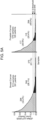
- the size distribution of cell-free DNA was consistent with previous reports. Automated quantification of total cell-free DNA was performed using PicoGreen.
- a library construction protocol was optimized for 5 ng of cfDNA input; 96.3% of cancer patients and 81.4% of healthy donors had ⁇ 5 ng of cfDNA per 4 mL of plasma. Only 1% of each cfDNA sequencing library was then used for ULP-WGS to screen for tumor content. This process resulted in cfDNA and germline DNA libraries suitable for hybrid selection and whole-exome sequencing.
- ichorCNA simultaneously predicted segments of SCNA and estimates of tumor fraction while accounting for subclonality and tumor ploidy.
- ULP-WGS of cfDNA FIGS. 1B , 1C
- Highly concordant megabase-scale copy number (sensitivity > 0.92, FIG. 1D ) was found, including identification of chromothripsis.
- Tumor fraction estimates from ULP-WGS of cfDNA were also concordant with WGS of the same sample.
- a series of benchmarking datasets were generated using in silico mixing of up to 50 cancer patient and 22 healthy donor cfDNA samples to generate.
- the benchmarking datasets demonstrated accurate estimation of tumor fraction (median absolute deviation of error ⁇ 0.014) and detection of SCNAs at 0.1x coverage.
- a lower limit of detection of 0.03 tumor fraction was determined, using only a single arm-level (>100Mb) gain and loss of one copy to detect the presence of tumor.
- Example 2 ULP-WGS offers an efficient way to screen cfDNA libraries for tumor content prior to whole exome sequencing.
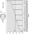
- FIG. 1C indicates the tumor fractions detected in cfDNA from hundreds of patients with different types of metastatic cancer using the ULP-WGS approach described herein.
- FIG. 1E demonstrates the concordance of tumor fraction estimates from ultra-low-pass whole-genome sequencing (ULP-WGS) obtained using methods of the invention with deeper coverage sequencing and calculation using established methods such as ABSOLUTE.
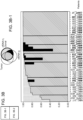
- Example 4 ULP-WGS of cfDNA identified focal copy number alterations (CNAs) with comparable performance to whole exome sequencing of the same library or matched tumor biopsy.
- CNAs focal copy number alterations
- FIGS. 2A-2D Focal copy number aberrations (CNAs) identified based on ULP-WGS of cfDNA were compared to focal CNAs obtained through whole-exome sequencing of the same library ( FIG. 2A , middle panel) and focal CNAs obtained using whole-exome sequencing of a matched tumor biopsy.
- focal CNAs detected using ULP-WGS were concordant with higher depth, whole-exome sequencing of the same library (technical validation) and were also concordant with whole-exome sequencing of a matched tumor biopsy (biological comparison).
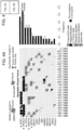
- Example 5 Focal CNAs landscape based on ULP-WGS of cfDNA are similar to the copy number landscapes generated using conventional methods.
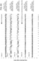
- ULP-WGS was used to map focal copy number aberrations (CNAs) present in cell free DNA isolated from blood samples obtained from 43 patients with metastatic castration resistant prostate cancer. In each patient, the purity of tumor cell-free DNA was greater than 10% and the coverage of the genome was greater than 0.05 X.
- Results obtained using ULP-WGS were compared to copy number landscapes generated using whole-exome sequencing of over a hundred metastatic tumor biopsies ( FIGS. 5A-5D ; Robinson et al. Cell 161, 1215-1228 (2015 )).
- ULP-WGS generated essentially the same copy number profile as was obtained by Robinson et al. but at a significant cost savings.
- CNAs landscape derived from ULP-WGS of cfDNA were used to determine whether a subject with prostate cancer responded to treatment ( FIGS. 6A-6E ).
- ULP-WGS was used to characterize CNAs present in cfDNA isolated from blood samples obtained from prostate cancer patients over time. Serially sampled blood from patients on therapy revealed correlations in tumor fractions with clinical response. This suggests that measuring tumor fractions in cell-free DNA using ULP-WGS offers an early readout of clinical response.
- CfDNA extractions were carried out using commercially available automated DNA sample preparation technology from Qiagen, QiaSymphony ® .
- the extracted DNA was quantified using commercially available DNA quantification assay, PicoGreen ® assay.
- Sequencing libraries were constructed by direct ligation of adapters using Kapa HyperPrep kit from KapaBiosystems. One library was constructed for each patient. A small fraction ( ⁇ 1%) of each barcoded library was pooled and submit for 1 lane of HiSeq2500 per 96 samples. The sequence "barcode" allowed the library to be associated with the patient from which it was derived.
- ULP-WGS ultra-low-pass whole genome sequencing
- ULP-WGS of cfDNA was performed to an average genome-wide fold coverage of 0.1X.
- the depth of coverage in a ULP sample was analyzed to evaluate large-scale copy number alterations (CNAs) and aneuploidies.
- CNAs copy number alterations
- a probabilistic model was developed and a software package implemented called "ichorCNA," which uses concepts from existing algorithms ( Ha et al. Genome Res. 24, 1881- 1893 (2014 ); Ha et al. Genome Res. 22, 1995- 2007 (2012 )) designed for deep coverage WGS/WES data to simultaneously predict regions of CNAs and estimate the fraction of tumor in ULP-WGS.
- the workflow consisted of 3 steps: 1) Computing read coverage, 2) Data normalization, and 3) CNA prediction and estimation of tumor fraction.
- the genome is divided into T non-overlapping windows, or bins, of 1Mb. Aligned reads are counted based on overlap within each bin. This was done using the tools in HMMcopy Suite (http://compbio.bccrc.ca/software/hmmcopy/). Centromeres are filtered based on chromosome gap coordinates obtained from UCSC for hg19, including one 1Mb bin up- and downstream of the gap.
- the short fragment sizes of cfDNA e.g., 166bp
- the read counts are then normalized to correct for GC-content and mappability biases using HMMcopy R package ( Ha et al. Genome Res. 22, 1995- 2007 (2012 )). Briefly, two LOESS regression curve-fitting are performed to the bin-wise 1) GC-fraction and read counts, followed by 2) mappability uniqueness score and read counts. The curvefitting was only applied to autosomes. This generates corrected read counts r t for each bin t ⁇ ⁇ 1, ..., T ⁇ .
- the gender of the patient is determined by inspecting the corrected read counts in chromosome X and Y. There are two criteria to determine if the sample is a male (otherwise the sample is a female):
- ULP-WGS was also performed on cfDNA from 27 healthy donors using the same protocol in order to create a reference dataset. These data help to further normalize the cancer patient cfDNA to correct for systematic biases arising from library construction, sequencing platform, and cfDNA-specific artifacts.
- the healthy donor cfDNA ULP data were processed as above and also corrected for GC-content and mappability biases as above. This generated corrected read counts hit for each bin t ⁇ ⁇ 1, ..., T ⁇ and each donor sample i. Then, the median at each bin was computed across the 27 samples to generate a reference dataset, h 1: T .
- log 2 r t h t .
- ichorCNA Copy number prediction and tumor fraction estimation using a HMM
- the cancer patient cfDNA CNA signals is composed of an admixture between DNA fragments derived from tumor and non-tumor cells.
- a 2-component mixture was used to model this explicitly ( Carter et al. Nat. Biotechnol. 30, 413-421 (2012 ); Ha et al. Genome Res. 24, 1881- 1893 (2014 ); Ha et al. Genome Res.
- CNA 2 n + 1 ⁇ n c
- n is the non-tumor proportion
- (1 - n ) is the tumor proportion
- c is the copy number for a specific alteration (e.g. 1 for deletion, 3 for gain, etc.).
- a third component is used to represent DNA fragments derived from tumor cells not harboring the CNA event ( Carter et al. Nat. Biotechnol. 30, 413-421 (2012 ); Ha et al. Genome Res. 24, 1881- 1893 (2014 ); Yau et al., Genome Biology 11, R92 (2010 )), observed CNA ⁇ 2 n + 2 s 1 ⁇ n + 1 ⁇ s 1 ⁇ n c where s is the proportion of tumor not containing the event with c copy number.
- (1 - s) is similar to the definitions of tumor-cellular-prevalence ( Ha et al. Genome Res. 24, 1881- 1893 (2014 )) or cancer-cell-fraction or tissue tumors ( Carter et al. Nat. Biotechnol. 30, 413-421 (2012 ).
- the copy number states are mapped to hemizygous deletions (HETD, 1), copy neutral ( NEUT, 2), copy gain ( GAIN, 3), amplification ( AMP , 4), and high-level amplification ( HLAMP , 5-7 copies).
- the homozygous deletions state (HOMD, 0 copies) is excluded.
- subclonal hemizygous deletion HETD sc
- GAIN sc subclonal copy gain
- a copy number state is assigned to G t for each bin t and the initial distribution of these copy number states is given by Go ⁇ Mult ( ⁇ ).
- T is modeled using a Student's-t distribution with ⁇ g , ⁇ g , and v g are the mean, precision, and degrees of freedom, conditional on copy number state g ⁇ K at bin t, p l t
- Precision ⁇ g for each g ⁇ K are also model parameters.
- the degrees of freedom v g is a constant (2.1) and is not estimated.
- Transition model A stationary (homogeneous) transition model is used in the HMM. Because all bins have equal sized intervals, with the exception of centromere regions, a non-stationary transition model to account for varying genomic distances between data points was not used.
- Prior model The HMM is implemented as a Bayesian framework with priors for each model parameter: Student's-t parameters ⁇ g , ⁇ g for each g ⁇ K, transition probabilities A, initial state distribution ⁇ , and global parameters n, s, and ⁇ ,
- the model parameters ⁇ ⁇ ⁇ 1:
- EM expectation-maximization
- the parameters ⁇ ( G t g
- D, ⁇ ) are estimated using the maximum a posteriori (MAP) estimate.
- MAP maximum a posteriori
- the complete-data log-likelihood at convergence is denoted F ⁇ .
- Chromosome 19 was excluded during parameter estimation (i.e., EM) due to systematic decrease in log 2 copy ratio values across majority of samples for bins within chr19 after GC-content correction. As a result, the estimation of tumor fraction is not influenced by this systematic bias. However, chr19 was included in the Viterbi algorithm as part of generating a genome-wide solution.
- Model selection In order to avoid the local optimal limitation of EM, multiple restarts are performed by performing EM over a range of initializations for tumor fraction ( n (0) ⁇ ⁇ 0.35, 0.45, 0.50, 0.65, 0.75, 0.85, 0.95 ⁇ ) and tumor ploidy ( ⁇ (0) ⁇ ⁇ 2, 3, 4 ⁇ ) parameters.
- the solution with the maximum complete-data log-likelihood over each initialization pair, ( n (0) , ⁇ (0) ) is chosen.
- Solutions with a total alteration fraction (based on bins) ⁇ 0.05 and having the largest CNA event be ⁇ 50 bins are reassigned a tumor fraction of zero.
- the segment median log ratio values are corrected to account for the tumor fraction of the sample. That is, for lower tumor fraction samples, the log ratio values will be adjusted higher signals.
- the tumor-content-corrected log2 copy ratios r ⁇ t at bin t from the observed log 2 copy ratio l t and estimated normal content (n) or tumor fraction (1-n) is negative, the it is set to 2 ⁇ 10 -9 prior to log transformation.
- the ichorCNA HMM component has O ( KT ) in memory and O ( K 2 T ) in time.
- the run-time of the algorithm for 0.1x coverage is 1 minute for read coverage computation and 1 minute for analysis using the HMM.
Description
- Comprehensive genomic characterization of metastatic cancer stands to uncover novel alterations of clinical significance. A major challenge is that metastatic tumors are infrequently biopsied. Cell-free DNA is shed abundantly into the bloodstream from metastatic tumors, presenting an opportunity for genomic discovery in settings where tumor biopsies are least accessible. Conventional methods for characterizing cancers rely on the sequencing of particular genes, such as BRCA1, BRCA2, and p53, where the presence of mutations is associated with uncontrolled cell proliferation. This approach only interrogates genes previously shown to be associated with cancer. It fails to identify genes that have not yet been shown to be dysregulated in cancer. The advent of next-generation shot gun sequencing provides researchers with the ability to rapidly sequence a whole genome and then use bioinformatics-based approaches to analyze the data. These approaches have made possible the characterization of whole cancer genomes and the discovery of alterations in chromosomal structure, such as amplifications, deletions, translocations, and inversions. Next-generation shotgun sequencing typically requires sequencing every base in a sample several times to reliably determine the base call. Researchers determine the necessary coverage level (i.e., number of times the genome must be sequenced) based on the type of study, gene expression level, size of reference genome, published literature, and best practices defined by the scientific community. Sims D et al., Nature Reviews Genetics, vol. 15, no. 2, 1 February 2014, pages 121 -132, relates to a review of major study designs which include de novo genome sequencing, genome resequencing, transcriptome sequencing and genomic location analyses (for example, chromatin immunoprecipitation followed by sequencing (ChIP-seq) and chromosome conformation capture (3C). Xie C et al., BMC Bioinformatics, vol. 10, no. 1,6 March 2009, page 80, relates to a method to detect copy number variation using shotgun sequencing (CNV-seq). Magi A et al., Bioinformatics, vol. 28, no. 4, 23 December 2011, pages 470-478, relates to an investigation of the ability of read count data to predict the exact number of DNA copy by comparing the performance of the three most widely used sequencing technologies.
WO 2014/014497 A1 relates to a method for determining copy number variations (CNV) of a sequence of interest in a test sample that comprises a mixture of nucleic acids that are known or are suspected to differ in the amount of one or more sequence of interest. Klevebring et al., PLoS ONE, vol. 9, no. 8, 18 August 2014, page e104417, relates to an investigation of the utility of using exome sequencing to monitor circulating tumor DNA levels through the detection of single nucleotide variants in plasma. Most publications require between 10× to 30× depth of coverage for the detection of human genome mutations/SNPs/rearrangements depending on the application and statistical model. A standard whole-genome analysis often requires 30 - 60x coverage. For ChIP-Seq studies where reads map to only a subset of a genome, publications often require coverage around 100×. This level of sequencing coverage is expensive and time consuming. Accordingly, improved methods for characterizing cancer are urgently required. - As described herein below, the present invention provides methods of using low coverage sequencing to assess the relative fraction of tumor versus normal DNA in a sample, and to assess copy number alterations present in the sample. This approach can be used, for example, to qualify samples for whole exome sequencing, for diagnosis of cancer or other disease states, and to monitor cancer therapy.
- In one aspect, the invention features a method of characterizing DNA in a biological sample containing or suspected of containing tumor-derived DNA, the method involving isolating fragments of cell-free DNA from a biological sample; constructing a cell-free DNA library containing the fragments by direct ligation of adapters; sequencing the library to about 0.1X genome or exome wide sequencing coverage; and detecting chromosomal copy number alterations in the sequence, where detection of chromosomal copy number alterations indicates that at least a portion of the cell-free DNA in the sample was derived from a neoplastic cell and failure to detect such alterations indicates that the cell free DNA present in the sample is not derived from a neoplastic cell.
- Further disclosed herein is a method of characterizing DNA in a biological sample, the method involving isolating fragments of cell free DNA from a biological sample; constructing a library containing the fragments; sequencing the library to about 0.1X genome or exome -wide sequencing coverage; and detecting chromosomal copy number alterations in the sequence.
- Further disclosed herein is a method of characterizing DNA in a biological sample, the method involving isolating fragments of DNA from a biological sample; constructing a library containing the fragments; sequencing the library to at least about 0.01-5X genome or exome-wide sequencing coverage; and detecting focal chromosomal copy number alterations in the sequence.
- In another aspect, the invention features a method of determining the purity of DNA in a sample, the method involving isolating fragments of DNA from a biological sample; constructing a DNA library containing the fragments by direct ligation of adapters; sequencing the library to at least about 0.1X genome or exome-wide sequencing coverage; detecting chromosomal copy number alterations in the sequence; and analyzing the chromosomal copy number alterations present in the sample to determine the percentage of DNA that is derived from a tumor cell, wherein a lower limit of detection of 0.03 tumor fraction of cell-free DNA detects a tumor cell-derived sample.
- In another aspect, the invention features a method of identifying a subject as having a neoplasia, the method involving isolating fragments of cell free DNA from a biological sample derived from the subject; constructing a cell-free DNA library containing the fragments by direct ligation of adapters; sequencing the library to at least about 0.1X exome or genome-wide sequencing coverage; and detecting the presence or absence of chromosomal copy number alterations in the sequence, where the presence of chromosomal copy number alterations identifies the subject as having a neoplasia, and the absence of chromosomal copy number alterations indicates that no neoplasia was detected in the sample from the subject.
- In another aspect, the invention features a method of monitoring the disease state of a subject, the method involving isolating fragments of cell free DNA from two or more biological samples, where the first biological sample is obtained at a first time point and a second or subsequent biological sample is obtained at a later time point; constructing two or more cell-free DNA libraries each containing cell-free DNA fragments from the samples, wherein the libraries are each prepared by direct ligation of adapters; sequencing the libraries to at least about 0.1X exome or genome-wide sequencing coverage; and comparing the chromosomal copy number alterations in the sequence over time, thereby monitoring the disease state of the subject, wherein an increase in chromosomal copy number alterations between the first time point and the later time point indicates that the subject's disease state has progressed; a decrease in chromosomal copy number alterations between a first time point and a later time point indicates that the subject's disease state has stabilized or is not progressing; the first time point is prior to treatment; the second or subsequent time point is during the course of treatment. In one embodiment, the disease state is a cancer that is any one of prostate cancer, metastatic prostate cancer, breast cancer, triple negative breast cancer, lung cancer, and colon cancer.
- In another aspect, the invention provides a method of characterizing the efficacy of treatment of a subject having a disease characterized by an increase in chromosomal copy number, the method involving isolating fragments of cell free DNA from two or more biological samples derived from a subject undergoing cancer therapy, where the first biological sample is obtained at a first time point and a second or subsequent biological sample is obtained at a later time point; constructing two or more cell-free DNA libraries each containing fragments from the samples, wherein the libraries are each prepared by direct ligation of adapters; sequencing the libraries to at least about 0.1X genome or exome -wide sequencing coverage; and comparing the focal chromosomal copy number alterations in the sequence over time, thereby characterizing the efficacy of treatment, wherein a decrease in chromosomal copy number alterations between the first and second time points indicate that the treatment is effective; a decrease in chromosomal copy number alterations between the first and second time points indicate that the treatment lacks efficacy; or a decrease in chromosomal copy number alterations between the first and second time points indicates that the disease has acquired resistance to the treatment. In one embodiment, the disease is cancer. In another embodiment, the treatment is an anti-cancer therapy that is chemotherapy, radiotherapy, or surgery. In another embodiment, samples are collected at 5, 15, or 30 minute intervals while a cancer therapy is administered. In another embodiment, samples are collected at 3, 6, 9, 12, 24, 36, or 72 hour intervals. In another embodiment, samples are collected at 1, 2, 3, 4, 5, or 6 week intervals.
- In various embodiments of any of the above aspects or any other aspect of the invention delineated herein, the DNA is cell free DNA. In other embodiments, the exome wide or genome wide sequencing coverage is any one or more of 0.1, 0.5, 1, 2, 3, 4, and 5X. In various embodiments, detection of an alteration in copy number at the focal point identifies the presence of tumor derived cell free DNA present in the sample. In other embodiments, failure to detect an alteration in copy number indicates the absence of tumor derived cell free DNA present in the sample. In other embodiments, sequence coverage correlates with the purity of tumor derived DNA (also termed "tumor fraction"), such that the sequence coverage is about 0.01-5X when the purity is about 50%. In other embodiments, sequence coverage correlates with the purity of tumor derived DNA, such that the sequence coverage is about 0.05-5X when the purity is about 25%. In other embodiments, sequence coverage correlates with the purity of tumor derived DNA, such that the sequence coverage is about 0.005-5X when the purity is about 75%. In other embodiments, the focal copy number alteration correlates with at least about 3% purity of tumor derived DNA. In other embodiments, the sequencing is whole genome sequencing. In other embodiments, the focal copy number alteration is between about 1 KB and 100 MB (e.g., of about 1 KB, 3 KB, 5 KB, 10 kb, 50kb, 100 kb, 500 kb, 2 MB, 3 MB, 4 MB, 5 MB, 10 MB, 50 MB, or 100 MB of DNA). In still other embodiments, the biological sample is a tissue sample or a liquid biological sample that is blood, plasma, serum, cerebrospinal fluid, phlegm, saliva, urine, semen, prostate fluid, breast milk, and/or tears. In still other embodiments, the sample is derived from a subject having or suspected of having a neoplasia. In still other embodiments, the sample is a fresh or archival sample derived from a subject having a cancer that is prostate cancer, metastatic prostate cancer, breast cancer, triple negative breast cancer, lung cancer, colon cancer, or any other cancer containing aneuploid cells. In still other embodiments, the cancer is metastatic castration resistant prostate cancer or metastatic breast cancer. In still other embodiments, the patient is being treated for a neoplasia. In still other embodiments, the method is used to screen or otherwise qualify the sample prior to whole exome sequencing or prior to generating a cell line from the sample. In still other embodiments, the method further involves carrying out whole exome sequencing. In still other embodiments, the method is used to qualify the sample prior to producing a cell line from the sample. In various embodiments, the sequencing or ultra low pass-whole genome sequencing is performed by bisulfite sequencing (e.g., ultra low pass-whole genome bisulfite sequencing (ULP-WGBS)).
- Other features and advantages of this invention will be apparent from the detailed description, and from the claims.
- Unless defined otherwise, all technical and scientific terms used herein have the meaning commonly understood by a person of ordinary skill in the art to which this invention belongs. The following references provide one of skill with a general definition of many of the terms used in this invention: Singleton et al., Dictionary of Microbiology and Molecular Biology (2nd ed. 1994); The Cambridge Dictionary of Science and Technology (Walker ed., 1988); The Glossary of Genetics, 5th Ed., R. Rieger et al. (eds.), Springer Verlag (1991); and Hale & Marham, The Harper Collins Dictionary of Biology (1991). As used herein, the following terms have the meanings ascribed to them below, unless specified otherwise.
- By "aneuploidy" is meant in the context of a cell having an abnormal number of chromosomes relative to a cell of normal ploidy.
- "Tumor derived DNA" means DNA that is derived from a cancer cell rather than a healthy control cell. Tumor derived DNA often includes structural changes that are indicative of cancer. Such structural changes may be at the level of the chromosome, which includes aneuploidy (abnormal number of chromosomes), duplications, deletions, or inversions, or alterations in sequence. "Tumor fraction" means the fraction of total DNA that is derived from tumor cells.
- By "alteration" is meant a change relative to a reference. In one embodiment, a change in sequence (i.e., insertion, deletion, point mutation, copy number alteration (CNA), or loss in heterozygosity (LOH)) is determined relative to a reference sequence, reference exome, and/or reference genome.
- In this disclosure, "comprises," "comprising," "containing" and "having" and the like can have the meaning ascribed to them in U.S. Patent law and can mean " includes," "including," and the like; "consisting essentially of" or "consists essentially" likewise has the meaning ascribed in U.S. Patent law and the term is open-ended, allowing for the presence of more than that which is recited so long as basic or novel characteristics of that which is recited is not changed by the presence of more than that which is recited, but excludes prior art embodiments.
- By "fragment" is meant a portion of a polypeptide or nucleic acid molecule. This portion contains, preferably, at least 10%, 20%, 30%, 40%, 50%, 60%, 70%, 80%, or 90% of the entire length of the reference nucleic acid molecule or polypeptide. A fragment may contain 10, 20, 30, 40, 50, 60, 70, 80, 90, or 100, 200, 300, 400, 500, 600, 700, 800, 900, or 1000 nucleotides or amino acids.
- The terms "isolated," "purified," or "biologically pure" refer to material that is free to varying degrees from components which normally accompany it as found in its native state. "Isolate" denotes a degree of separation from original source or surroundings. "Purify" denotes a degree of separation that is higher than isolation. A "purified" or "biologically pure" protein is sufficiently free of other materials such that any impurities do not materially affect the biological properties of the protein or cause other adverse consequences. That is, a nucleic acid or peptide of this invention is purified if it is substantially free of cellular material, viral material, or culture medium when produced by recombinant DNA techniques, or chemical precursors or other chemicals when chemically synthesized. Purity and homogeneity are typically determined using analytical chemistry techniques, for example, polyacrylamide gel electrophoresis or high performance liquid chromatography. The term "purified" can denote that a nucleic acid or protein gives rise to essentially one band in an electrophoretic gel. For a protein that can be subjected to modifications, for example, phosphorylation or glycosylation, different modifications may give rise to different isolated proteins, which can be separately purified.
- By "isolated polynucleotide" is meant a nucleic acid (e.g., a DNA) that is free of the genes which, in the naturally-occurring genome of the organism from which the nucleic acid molecule of this invention is derived, flank the gene. The term therefore includes, for example, a recombinant DNA that is incorporated into a vector; into an autonomously replicating plasmid or virus; or into the genomic DNA of a prokaryote or eukaryote; or that exists as a separate molecule (for example, a cDNA or a genomic or cDNA fragment produced by PCR or restriction endonuclease digestion) independent of other sequences. In addition, the term includes an RNA molecule that is transcribed from a DNA molecule, as well as a recombinant DNA that is part of a hybrid gene encoding additional polypeptide sequence.
- By "marker" is meant any protein or polynucleotide having an alteration in sequence, copy number, expression level or activity that is associated with a disease or disorder.
- By "neoplasia" is meant a disease that is associated with inappropriately high levels of cell division, inappropriately low levels of apoptosis, or both. For example, cancer is an example of a neoplastic disease. Examples of cancers include, without limitation, leukemias (e.g., acute leukemia, acute lymphocytic leukemia, acute myelocytic leukemia, acute myeloblastic leukemia, acute promyelocytic leukemia, acute myelomonocytic leukemia, acute monocytic leukemia, acute erythroleukemia, chronic leukemia, chronic myelocytic leukemia, chronic lymphocytic leukemia), polycythemia vera, lymphoma (Hodgkin's disease, non-Hodgkin's disease), Waldenstrom's macroglobulinemia, heavy chain disease, and solid tumors such as sarcomas and carcinomas (e.g., fibrosarcoma, myxosarcoma, liposarcoma, chondrosarcoma, osteogenic sarcoma, chordoma, angiosarcoma, endotheliosarcoma, lymphangiosarcoma, lymphangioendotheliosarcoma, synovioma, mesothelioma, Ewing's tumor, leiomyosarcoma, rhabdomyosarcoma, colon carcinoma, pancreatic cancer, breast cancer, ovarian cancer, prostate cancer, squamous cell carcinoma, basal cell carcinoma, adenocarcinoma, sweat gland carcinoma, sebaceous gland carcinoma, papillary carcinoma, papillary adenocarcinomas, cystadenocarcinoma, medullary carcinoma, bronchogenic carcinoma, renal cell carcinoma, hepatoma, nile duct carcinoma, choriocarcinoma, seminoma, embryonal carcinoma, Wilm's tumor, cervical cancer, uterine cancer, testicular cancer, lung carcinoma, small cell lung carcinoma, bladder carcinoma, epithelial carcinoma, glioma, astrocytoma, medulloblastoma, craniopharyngioma, ependymoma, pinealoma, hemangioblastoma, acoustic neuroma, oligodenroglioma, schwannoma, meningioma, melanoma, neuroblastoma, and retinoblastoma).
- A "reference genome" is a defined genome used as a basis for genome comparison or for alignment of sequencing reads thereto. A reference genome may be a subset of or the entirety of a specified genome; for example, a subset of a genome sequence, such as exome sequence, or the complete genome sequence.
- By "subject" is meant a mammal, including, but not limited to, a human or non-human mammal, such as a bovine, equine, canine, ovine, rodent, or feline.
- Ranges provided herein are understood to be shorthand for all of the values within the range. For example, a range of 1 to 50 is understood to include any number, combination of numbers, or sub-range from the group consisting 1, 2, 3, 4, 5, 6, 7, 8, 9, 10, 11, 12, 13, 14, 15, 16, 17, 18, 19, 20, 21, 22, 23, 24, 25, 26, 27, 28, 29, 30, 31, 32, 33, 34, 35, 36, 37, 38, 39, 40, 41, 42, 43, 44, 45, 46, 47, 48, 49, or 50.
- As used herein, the terms "treat," treating," "treatment," and the like refer to reducing or ameliorating a disorder and/or symptoms associated therewith. It will be appreciated that, although not precluded, treating a disorder or condition does not require that the disorder, condition or symptoms associated therewith be completely eliminated.
- Unless specifically stated or obvious from context, as used herein, the term "or" is understood to be inclusive. Unless specifically stated or obvious from context, as used herein, the terms "a", "an", and "the" are understood to be singular or plural.
- Unless specifically stated or obvious from context, as used herein, the term "about" is understood as within a range of normal tolerance in the art, for example within 2 standard deviations of the mean. About can be understood as within 10%, 9%, 8%, 7%, 6%, 5%, 4%, 3%, 2%, 1%, 0.5%, 0.1%, 0.05%, or 0.01% of the stated value. Unless otherwise clear from context, all numerical values provided herein are modified by the term about.
- The recitation of a listing of chemical groups in any definition of a variable herein includes definitions of that variable as any single group or combination of listed groups. The recitation of an embodiment for a variable or aspect herein includes that embodiment as any single embodiment or in combination with any other embodiments or portions thereof.
- Any compositions or methods provided herein can be combined with one or more of any of the other compositions and methods provided herein.
-
-
FIGS 1A-1E depict copy number and tumor fractions from ULP-WGS.FIG. 1A depicts an exemplary workflow for processing cfDNA for whole-exome sequencing.FIG. 1B depicts genome-wide copy number from 0.1x ULP-WGS of cfDNA from a healthy donor (top panel) and genome-wide copy number from 25x WGS and 0.1x WGS of cell-free DNA from a metastatic breast cancer patient (MBC_315), and 1x WGS and WES of matched tumors from this patient (bottom panels). SCNA for tumor WES andcfDNA 25x coverage WGS were predicted using TITAN (Ha et al. Genome Res. 24, 1881- 1893 (2014)).FIG. 1C provides a series of graphs showing that reasonably pure cell-free DNA (cfDNA) was obtained from about 700 blood samples screened. Tumor fraction in cfDNA from patients with pancreas adenocarcinoma (PAAD), brain cancer, multiple myeloma (MM), skin cutaneous melanoma (SKCM), small cell lung cancer (SCLC), colon adenocarcinoma (COAD), bladder carcinoma (BLCA), esophageal carcinoma (ESCA), kidney renal clear cell carcinoma (KIRC), stomach adenocarcinoma (STAD), lung adenocarcinoma (LUAD), chronic lymphocytic leukemia (CLL), and ovarian cancer (OV). The error rate of tumor fraction estimation was about 3%, and therefore indicates a lower limit of detection sensitivity at 0.03 tumor fraction from ULP-WGS of cfDNA. The threshold for determining the presence of tumor content in a cfDNA sample from ULP-WGS was set to 0.03 tumor fraction.FIG. 1D depicts plots providing a comparison of copy ratios between ULP-WGS of cfDNA with deep (>10x) WGS of the same cfDNA sample, WGS (1x) of matched tumors from 22 metastatic breast cancer (MBC) patients, and WES (average mean target coverage 173x) of matched tumors from 41 MBC and prostate cancer (CRPC) patients. Log2 copy ratios were computed as normalized read coverage for each 1Mb (WGS/ULP-WGS) and the mean of overlapping 50kb bins (WES) after adjustment for tumor fraction/purity. The correlation of copy ratios between tumor and cfDNA was computed using Spearman rank correlation (coefficient ρ). F-measure (F1) is the harmonic mean of the CNA positive predictive value (precision) and sensitivity (recall) Performance. Recall is defined as the proportion of SCNA gain/loss in tumor biopsy also observed in ULP-WGS of cfDNA.FIG. 1E is a plot showing a comparison of tumor fractions estimated from ULP-WGS and WES of cfDNA. Samples (n=35) with similar tumor ploidy (difference < 0.75 and ploidy > 1.5) estimated in both ULP-WGS and tumor WES are shown. The correlation between the two data types was calculated using Pearson correlation (coefficient r). Thin gray line denotes y=x. WES tumor fractions were estimated using ABSOLUTE (shown; Carter et al. Nat. Biotechnol. 30, 413-421 (2012)) and TITAN. -
FIGS. 2A-2D show performance of ULP-WGS (0.1x) compared to the deeper sequencing of the same cfDNA sample (10x WGS and WES) and the matched tumor biopsy (1x WGS and WES) for patient MBC_288.FIG. 2A provides copy number profiles for cfDNA WGS (Titan, 1Mb bins), cfDNA ULP-WGS (1Mb bin) cfDNA WES (Titan, 50kb bins), matched tumor biopsy WGS, and matched tumor biopsy WES (Titan, 50kb bins). Performance was computed based on binary classification metrics of precision and recall for comparisons.FIG. 2B is a plot depicting comparison of normalized copy ratios (log2) between cfDNA ULP-WGS and deep WGS of the same cfDNA sample.FIG. 2C is a plot depicting comparison of normalized copy ratios (log2) between cIDNA ULP-WGS and 1x WGS of the tumor biopsy.FIG. 2D is a plot depicting comparison of normalized copy ratios (log2) between cfDNA ULP-WGS and WES of the tumor biopsy. Biological differences in copy number events inchromosome -
FIGS. 3A-3C depict a comparison of whole-exome sequencing of cfDNA to whole-exome sequencing of matched tumor biopsies.FIG. 3A depicts plots showing fraction of clonal (≥ 0.9 cancer cell fraction, CCF) and subclonal (< 0.9 CCF) SSNVs detected by MuTect in WES of tumor biopsies and confirmed (i.e. supported by ≥ 3 variant reads) in WES of cfDNA. Sites with <3 reads that had power <0.9 for mutation calling were not included when computing the fraction of SNVs confirmed.FIG. 3B depicts plots showing fraction of clonal and subclonal SSNVs detected in WES of cfDNA and confirmed in WES of tumor biopsies. For eighteen patients with WES of cfDNA at a second time point t2, SSNVs not detected in the matched tumor biopsy but confirmed at t2 are indicated with black.FIG. 3C depicts plots showing analysis of clonal dynamics in an ER+ breast cancer patient diagnosed with metastatic disease 1.5 years (yrs) prior to biopsy and cfDNA collection (ti, Day 0). Clustering analysis of CCF for SSNVs between matched tumor biopsy and cfDNA (ti) is shown (left panel). The CCF of four mutation clusters are shown, one containing ESR1 L536P (Subclonal Cluster 1, orange) and the other containing ESR1 D538G (Subclonal Cluster 2, light blue), at t1 and t2 (51 days apart) from a patient with ER+ metastatic breast cancer being treated with a SERD (right panel). The lymph node biopsy was taken at the same time ascfDNA 11. Mutations were clustered by the CCFs for each pair of samples using Phylogic (Brastianos et al. Cell 164, 57-68 (2016)). Error bars represent the 95% credible interval of the joint posterior density of the clusters. Mutations, excluding indels, having ≥ 90% estimated power based on coverage in both samples are shown; clusters with fewer than mutations are excluded. The number of mutations in each cluster is indicated in the legend in parentheses. -
FIGS. 4A-4E show that genomic alterations of known biological show significance and broad application to large cohorts of metastatic cancer patients.FIG. 4A provides the alteration status of significantly mutated genes predicted by MutSig2CV (Lawrence et al. Nature 499, 214-218 (2013); Lawrence et al. Nature 505, 495-501 (2014)) and known cancer-associated genes (Van Allen et al. Nat. Med. 20, 682-688 (2014)) are shown for cfDNA and tumor biopsies from 27 metastatic breast cancer (MBC) patients. Mutated genes with MutSig2CV q-value < 0.1 are statistically significant. Mutations that were exclusively detected in one sample may be present at low CCF in the other matched sample but were excluded from the frequency calculation. Mutations were predicted using MuTect.FIG. 4B provides the alteration status of significantly mutated genes predicted by focal SCNAs and known cancer-associated genes (Van Allen et al. Nat. Med. 20, 682-688 (2014)) are shown for cfDNA and tumor biopsies from 27 metastatic breast cancer (MBC) patients. SCNA frequencies were computed for oncogenes (MYC to ERBB2) and tumor suppressors (BRCA1 to ATM) using only amplification and deletion status, respectively. SCNAs were predicted using ReCapSeg and ABSOLUTE.FIG. 4C are plots depicting mutational signatures in whole-exome sequencing of cfDNA and tumor biopsies predicted using a Bayesian non-negative matrix factorization (NMF) approach (Kim et al. Nat. Genet. 48, 600-606 (2016)). Samples with predicted homozygous loss of BRCA1/2 are indicated in light gray and dark gray. Black line denotes y=x; blue line denotes model fit using linear least squares regression.FIG. 4D is a plot depicting neoantigen burden, defined as the number of predicted neoantigen SSNVs, calculated using NetMHCpan (Hoof et al. Immunogenetics 61, 1-13 (2009)). Black line denotes y=x; thin gray line denotes model fit using linear least squares regression.FIG. 4E is a graph showing broad applicability of the invention to many patients with metastatic cancer. Tumor fractions estimated from ULP-WGS of cfDNA from 913 blood samples from 391 patients with metastatic breast cancer and 579 blood samples from 129 patients with metastatic prostate cancers. The maximum tumor fraction across all samples for each patient is shown. Samples with coverage < 0.05x were excluded. -
FIGS. 5A-5D show an analysis of ULP-WGS tumor fraction and large-scale SCNAs.FIG. 5A depicts ULP-WGS tumor fraction estimates for 1492 metastatic breast (MBC) and prostate (CRPC) cancer samples. Samples with ≥ 0.10 (blue), ≥ 0.03 and < 0.10 (black), and < 0.03 (grey) tumor fraction are indicated.FIG. 5B depicts plots showing fraction of patients having ≥ 0.1 tumor fraction, divided into groups containing 1, 2, or ≥ 3 number of plasma samples collected. The total number of samples (n) within each of the three groups are indicated.FIG. 5C depicts gene-based frequencies of copy number alterations among patients with metastatic breast cancer predicted from ULP-WGS of cfDNA.FIG. 5D depicts gene-based frequencies of copy number alterations among patients with prostate cancer predicted from ULP-WGS of cfDNA. Gene alterations were defined based on overlapping SCNA segments predicted from normalized read coverage at 1Mb bins. Only samples with ≥ 0.10 are included. -
FIGS. 6A-6E show the use of ULP-WGS of cfDNA to characterize and monitor a patient's response to therapy.FIG. 6A shows copy number alteration (CNA) profiles over time characterized using ultra-low pass whole genome sequencing of cell free DNA (ULP-WGS of cfDNA) of a metastatic prostate cancer patient. Interestingly, ULP-WGS of cfDNA provides an early readout of response to therapy. Few detectable copy number alteration events were observed and estimate tumor fraction (purity) was 4% atweek 0, the initial time of treatment (top panel). Increased cfDNA yield and estimated tumor fraction was observed atweek 4 during a second cycle of treatment (second panel from top), which may provide early indications of increased tumor burden. Inweek FIG. 6B shows copy number alteration (CNA) profiles characterized over time using ultra-low pass whole genome sequencing of cell free DNA (ULP-WGS of cfDNA) of a metastatic prostate cancer patient. The top panel shows a CNA profile atweek 0 with an increased number of detectable CNAs and high estimated tumor fraction relative toweek 20. This result is consistent with the patient's clinical response to treatment atweek 20.FIG. 6C shows copy number alteration (CNA) profiles characterized over time using ultra-low pass whole genome sequencing of cell free DNA (ULP-WGS of cfDNA) of a metastatic prostate cancer patient. Inweeks week 36, the patient responded and this was reflected in the tumor fraction estimate from ULP-WGS. Subsequent analysis of cfDNA from blood inweek 43 confirmed the decreased tumor fraction which was consistent with the patient's continued response to chemotherapy. Interestingly, the PSA remained relatively stable throughout the entire 43 weeks. These results highlight an example in which ULP-WGS of cfDNA provides a more consistent readout of response to therapy than the established biomarker, PSA.FIG. 6D shows copy number alteration (CNA) profiles characterized over time using ultra-low pass whole genome sequencing of cell free DNA (ULP-WGS of cfDNA) of a metastatic prostate cancer patient. Despite initial treatment atweek 0, no reduction in CNA is observed atweek 10. Estimated tumor fractions (purity) from ULP-WGS increased inweek 17, which was consistent with clinical progression. This supports the use of ULP-WGS of cfDNA as an early readout of response to therapy. FIG. 6E shows copy number alteration (CNA) profiles characterized over time using ultra-low pass whole genome sequencing of cell free DNA (ULP-WGS of cfDNA) of a metastatic prostate cancer patient. The estimated tumor fraction from ULP-WGS decreased atweek 8, which is consistent with the patient's clinical response to therapy. Continued monitoring atweek 16 shows an increase in estimated tumor fraction, PSA, and cfDNA yield. These results may provide an early indication of eventual progression of cancer. Indeed, inweek 20, the patient's cancer was clinically progressing. These results support the use of ULP-WGS of cfDNA as an early readout of response to therapy. - The invention generally provides methods of using low coverage sequencing to assess the relative fraction of tumor versus normal DNA in a sample, and to assess copy number alterations present in the sample.
- The invention provides an efficient process to qualify DNA samples for whole-exome and/or whole genome sequencing based on tumor fraction (or tumor content). Without being bound by theory, the power to detect a variant at a particular sequencing depth depends on the tumor fraction of a sample. The selected samples can then be used to systematically compare the somatic mutations, indels, and copy number alterations detected in whole exome or whole genome sequencing of cfDNA to whole exome or whole genome sequencing of matched tumor biopsies. Advantageously, methods of the invention provide for ultra-low-pass sequencing (i.e., about 0.1, 0.5, 1, 2, 3, or 5X genome or exome wide sequencing coverage) for assessing tumor fraction and samples for which sufficient tumor content is estimated, whole exome sequencing of cfDNA can be performed. Among patients with metastatic breast or prostate cancers, whole exome sequencing of cfDNA was found to uncover 91% of the clonal mutations, 59% of the subclonal mutations, and 86% of the clonal copy number alterations (CNA) and 80% of the subclonal CNAs detected in whole exome sequencing of matched tumor biopsies. The high concordance suggests that comprehensive sequencing of cfDNA may enable genomic discovery in a routine and minimally invasive manner.
- This invention overcomes the challenge of screening large numbers of blood samples to assess how much tumor-derived cell-free DNA is present in blood plasma. This allows estimation of the fraction of tumor DNA in a sample from a trivial amount of sequencing (∼0.1x coverage or roughly $20 of sequencing coverage). This also provides for the detection of copy number alterations at a 500kb scale that can be detected in the sample. This serves not only as an efficient way to qualify samples for targeted or whole-exome/whole-genome sequencing for cancer genomics discoveries, but also correlates with therapeutic response and enables the study of copy number alterations in large cohorts.
- The invention provides for characterization of the malignant status of a sample or for diagnosis of cancer in the subject from whom the sample is derived. Based on the detection of somatic, tumor-specific copy number alterations from DNA of a sample, the fraction of tumor DNA is estimated. cfDNA and germline DNA was isolated from blood and analyzed using low coverage sequencing to estimate tumor content based on genome-wide copy number. The CNA events and tumor fraction were simultaneously predicted in a unified Bayesian hidden Markov model (HMM) framework. The estimated error rate of tumor fraction estimation was about 0.03 at genome sequencing coverage of 0.1x, as determined from application to healthy donor samples, which should have an expected 0.00 tumor fraction. Therefore, samples having greater than 0.03 tumor fraction, due to the harboring detectable CNA events, are classified as tumor-derived.
- In related applications, the methods described herein can be used to assess the tumor cellularity (purity) of tumor biopsies of human primary cancers with high stromal contamination and lymphocytic infiltration. Where little tumor DNA is present, methods of the invention can be used to assess whether sequencing is warranted. This would avoid the sequencing of samples that have little tumor content, which would provide significant cost saving. In addition, the methods of the invention could be used to assess the fraction of tumor relative to normal cells present in a tissue sample to be used for cell line generation. This would avoid the propagation of cell lines from human primary tumors that are largely composed of normal cells rather than of tumor cells.
- Whole genome sequencing (also known as "WGS", full genome sequencing, complete genome sequencing, or entire genome sequencing) is a process that determines the complete DNA sequence of an organism's genome. A common strategy used for WGS is shotgun sequencing, in which DNA is broken up randomly into numerous small segments, which are sequenced. Sequence data obtained from one sequencing reaction is termed a "read." The reads can be assembled together based on sequence overlap. The genome sequence is obtained by assembling the reads into a reconstructed sequence.
- Whole exome sequencing ("WES") is a technique used to sequence all the expressed genes in a genome (known as the exome). It includes first selecting only the subset of DNA that encodes proteins (exons), and then sequencing the exons using any DNA sequencing technology well known in the art or as described herein. In a human being, there are about 180,000 exons, which constitute about 1% of the human genome, or approximately 30 million base pairs. To sequence the exons of a genome, fragments of double-stranded genomic DNA are obtained (e.g., by methods such as sonication, nuclease digestion, or any other appropriate methods). Linkers or adapters are then attached to the DNA fragments, which are then hybridized to a library of polynucleotides designed to capture only the exons. The hybridized DNA fragments are then selectively isolated and subjected to sequencing using any sequencing method known in the art or described herein.
- In one embodiment, the sequencing of a DNA fragment is carried out using commercially available sequencing technology SBS (sequencing by synthesis) by Illumina. In another embodiment, the sequencing of the DNA fragment is carried out using chain termination method of DNA sequencing. In yet another embodiment, the sequencing of the DNA fragment is carried out using one of the commercially available next-generation sequencing technologies, including SMRT ( s ingle- m olecule r eal- t ime) sequencing from Pacific Biosciences, Ion Torrent™ sequencing from ThermoFisher Scientific, Pyrosequencing (454) from Roche, and SOLiD® technology from Applied Biosystems. Any appropriate sequencing technology may be chosen for sequencing.
- This invention provides methods for qualifying a DNA sample for whole exome sequencing after ultra low pass (ULP)-whole genome sequencing. In one embodiment, the fraction of cells affected by a disease, such as cancer, may be estimated using one or more of the statistical models described herein. The qualifying process involves selecting a DNA sample having a tumor fraction greater than a threshold value, e.g. 5%, for whole exome sequencing (WES). Understanding the fraction of tumor-derived DNA in a sample allows one to adjust the depth of sequencing used in WES or other deeper sequencing. For instance, if a sample has a very low purity of tumor-derived DNA, a much greater depth of sequencing may be required to achieve the same sensitivity for detection of somatic alterations (Cibulskis et al. Nat. Biotechnol. 31, 213-219 (2013)).
- Ultra low pass sequencing advantageously provides for the accurate characterization of genomic or exomic DNA at a significant savings of cost and time, thereby obviating the need for complete integrative clinical sequencing of the whole-exome, matched germline, and/or transcriptome as practiced, for example, by Robinson et al., Cell 161:1215-1228, 2015 where meant target coverage for tumor exomes was 160x and for matched normal exomes was 100x.
- As used herein, the term "coverage" refers to the percentage of genome covered by reads. In one embodiment, low coverage or ultra low pass coverage is less than about 1X. Coverage also refers to, in shotgun sequencing, the average number of reads representing a given nucleotide in the reconstructed sequence. It can be calculated from the length of the original genome (G), the number of reads(N), and the average read length(L) as N × L/G. Biases in sample preparation, sequencing, and genomic alignment and assembly can result in regions of the genome that lack coverage (that is, gaps) and in regions with much higher coverage than theoretically expected. It is important to assess the uniformity of coverage, and thus data quality, by calculating the variance in sequencing depth across the genome. The term depth may also be used to describe how much of the complexity in a sequencing library has been sampled. All sequencing libraries contain finite pools of distinct DNA fragments. In a sequencing experiment only some of these fragments are sampled.
- Alterations of the genome have been identified in virtually all cancers. These alterations may include, without limitation, gene mutation, loss of heterozygosity (LOH), changes in chromosome number (aneuploidy), deletions, insertions, inversions, translocations, amplifications, and copy number alterations (CNA). Detection of copy number alterations is useful for characterizing the malignant status of a tumor, diagnosis of cancer, assessment of the purity in tumor biopsies of human primary cancers, and qualifying samples for additional characterization, such as whole exome sequencing.
- This invention provides methods to characterize alterations present in a cancer genome. In one embodiment, this invention involves isolating cfDNA from a biological sample, sequencing the DNA using ULP-WGS, analyzing the sequence of the DNA using statistical models described herein, and characterizing the copy number alterations present in the cfDNA, for example, by generating a copy number alteration profile. In one embodiment, the CNA profile is used to determine the tumor fraction of a sample. In another embodiment, the invention provides a method of diagnosing cancer in a subject by detecting the CNA profile of sample from the subject. In yet another embodiment, the invention provides a method of identifying a treatment and/or an agent for treatment and/or prevention of a cancer by characterizing CNAs present in cfDNA of the subject. In one embodiment, the method involves comparing the CNAs profiles before and after the treatment and/or administration of the agent.
- The methods of the invention are applicable to any disease and/or disorder having copy number alterations.
- To analyze ULP-WGS data, a modified approach from the TITAN (Ha G. et al., Genome Res. 24, 1881-1893 (2014) ("Ha 2014")), reference herewith in its entirety. The approach in this invention employs a hidden Markov model that simultaneously performs segmentation, copy number prediction, and purity (also termed tumor fraction) and ploidy estimation. This approach is optimized for increased sensitivity to detect events from low purity tumor-derived DNA in the absence of a control sample by using fine-tuned Bayesian priors and considering all CNA events in a unified probabilistic model. TITAN code is available at http://compbio.bccrc.ca/software/titan/ and https://github.com/gavinha/TitanCNA and HMMcopy frameworks and pipelines needed to be adapted. This new approach overcomes major challenges that TITAN/HMMcopy, and all other existing tools, were not designed to address: 1) ultra low coverage (0.1x) sequencing (most tools are optimized for WGS of 20x or greater); 2) unavailable matched germline normal sample as control (many tools require heterozygous SNPs determined from normal); 3) very low tumor content in DNA samples such as in cell-free DNA (many tools advertise benchmarks for reasonable performance at 0.15-0.20 tumor fractions). The landscape of the alterations of the cancer genome may be inferred from such estimates.
- This invention provides methods to extract and sequence a polynucleotide present in a sample. In one embodiment, the samples are biological samples generally derived from a human subject, preferably as a bodily fluid (such as blood, plasma, serum, cerebrospinal fluid, phlegm, saliva, urine, semen, prostate fluid, breast milk, or tears, or tissue sample (e.g. a tissue sample obtained by biopsy). In a further embodiment, the samples are biological samples derived from an animal, preferably as a bodily fluid (such as blood, cerebrospinal fluid, phlegm, saliva, or urine) or tissue sample (e.g. a tissue sample obtained by biopsy). In still another embodiment, the samples are biological samples from in vitro sources (such as cell culture medium). CfDNA attached to a substrate may be first suspended in a liquid medium, such as a buffer or a water, and then subject to sequencing and/or analysis. In yet another embodiment, the sample contains DNA within a cell, which may be extracted, sequenced and subject to the same analysis for landscape of alterations of genome.
- The disease state or treatment of a patient having a cancer or disease characterized by copy number alterations can be monitored using the methods and compositions of this invention. In one embodiment, the response of a patient to a treatment can be monitored using the methods and compositions of this invention. Such monitoring may be useful, for example, in assessing the efficacy of a particular treatment in a patient. Treatments amenable to monitoring using the methods of the invention include, but are not limited to, chemotherapy, radiotherapy, immunotherapy, and surgery. Therapeutics that alter the CNA landscape of cfDNA are taken as particularly useful in this invention. In one embodiment, methods of the invention are used to monitor PTEN loss or treatment with PARP inhibitors (e.g., iniparib, talazoparib, olaparib, rucparib, veliparib, MK 4827, BGB-290).
- Neoplastic tissues display alterations in their genome compared to corresponding normal reference tissues. Copy number alterations are correlated with neoplasia. Accordingly, this invention provides methods for detecting, diagnosing, or characterizing a neoplasia in a subject. The present invention provides a number of diagnostic assays that are useful for the identification or characterization of a neoplasia.
- In one approach, diagnostic methods of the invention are used to detect the CNA in a biological sample relative to a reference (e.g., a reference determined by an algorithm, determined based on known values, determined using a standard curve, determined using statistical modeling, or level present in a control polynucleotide, genome or exome).
- Methods of the invention are useful as clinical or companion diagnostics for therapies or can be used to guide treatment decisions based on clinical response/resistance. In other embodiments, methods of the invention can be used to qualify a sample for whole-exome sequencing.
- A physician may diagnose a subject and the physician thus has the option to recommend and/or refer the subject to seek the confirmation/treatment of the disease. The availability of high throughput sequencing technology allows the diagnosis of large number of subjects.
- The practice of the present invention employs, unless otherwise indicated, conventional techniques of molecular biology (including recombinant techniques), microbiology, cell biology, biochemistry and immunology, which are well within the purview of the person of ordinary skill. Such techniques are explained fully in the literature, such as, "Molecular Cloning: A Laboratory Manual", second edition (Sambrook, 1989); "Oligonucleotide Synthesis" (Gait, 1984); "Animal Cell Culture" (Freshney, 1987); "Methods in Enzymology" "Handbook of Experimental Immunology" (Weir, 1996); "Gene Transfer Vectors for Mammalian Cells" (Miller and Calos, 1987); "Current Protocols in Molecular Biology" (Ausubel, 1987); "PCR: The Polymerase Chain Reaction", (Mullis, 1994); "Current Protocols in Immunology" (Coligan, 1991). These techniques are applicable to the production of the polynucleotides and polypeptides of this invention, and, as such, may be considered in making and practicing this invention. Particularly useful techniques for particular embodiments will be discussed in the sections that follow.
- The following examples are put forth so as to provide those of ordinary skill in the art with a complete disclosure and description of how to make and use the assay, screening, and therapeutic methods of this invention, and are not intended to limit the scope of the claims.
- A low-cost, sample-conserving, and unbiased approach to screen cell-free DNA libraries prior to whole exome sequencing would help to focus whole exome sequencing on libraries with detectable tumor DNA. While many previous approaches to screening for cancer-derived cfDNA have focused on targeted mutation detection, it was hypothesized that using somatic copy number alterations would be more generally applicable as the vast majority of metastatic cancers harbor arm-level somatic SCNAs (citation) whereas different tumor types may have few recurrent somatic mutations. Additionally, copy number detection is an unbiased approach which borrows statistical strength from genome-wide signals while somatic mutation detection relies on high coverage at targeted regions to achieve high sensitivity in low purity samples. The question of whether ultra-low-pass whole-genome sequencing (ULP-WGS) could sensitively detect large-scale SCNAs and be used to estimate tumor fractions was explored. This would only require a small fraction of each library (~1%) and conserve enough DNA for hybrid selection.
- An analytical approach termed "ichorCNA" was developed to quantify tumor fraction in cfDNA without prior knowledge of single nucleotide variants (SSNVs) or somatic copy number alterations (SCNAs) in patients' tumors from ULP-WGS (
FIG. 1A , https://github.com/broadinstitute/ichorCNA). ichorCNA was applied to determine which cfDNA samples had sufficient tumor content (>10%) for whole-exome sequencing. Subsequent analysis of whole-exome sequencing of cell-free DNA and matched tumor biopsies from 41 patients demonstrated that cell-free DNA provided a suitable proxy for a tumor biopsy. Further examination of 1,492 blood samples from 520 patients with metastatic breast or prostate cancer using ichorCNA revealed 44% of patients had sufficient tumor fraction for standard depths of whole-exome sequencing of cell-free DNA. - An exemplary process begins with patient blood collection, separation of plasma from blood, extraction of cfDNA from plasma and germline DNA from blood, and construction of cfDNA libraries (
FIG. 1A ). As illustrated inFIG. 1A , ULP-WGS involves separation of plasma from blood, extraction of cfDNA and gDNA, quantification, library construction, ultra-low-pass whole-genome sequencing (ULP-WGS) of cfDNA libraries, and whole exome sequencing of both cfDNA and gDNA libraries. QiaSymphony was used for automated extraction. The size distribution of cell-free DNA was consistent with previous reports. Automated quantification of total cell-free DNA was performed using PicoGreen. - It was found that the size distribution and yields of cfDNA from metastatic cancer patients (median = 7.39, range = 0.20 to 547.82 ng / mL plasma, n=1642) and healthy donors (median = 2.64, range = 0.55 to 21.27 ng / mL plasma, n=27) were consistent with previous reports (Snyder et al. Cell 164, 57-68 (2016); Szpechcinski et al. Br. J. Cancer 113, 476-483 (2015)). Whole-genome libraries were constructed from samples using just 4 mL of plasma-- the amount of plasma in a single tube of blood. A library construction protocol was optimized for 5 ng of cfDNA input; 96.3% of cancer patients and 81.4% of healthy donors had ≥ 5 ng of cfDNA per 4 mL of plasma. Only 1% of each cfDNA sequencing library was then used for ULP-WGS to screen for tumor content. This process resulted in cfDNA and germline DNA libraries suitable for hybrid selection and whole-exome sequencing.
- ichorCNA simultaneously predicted segments of SCNA and estimates of tumor fraction while accounting for subclonality and tumor ploidy. To evaluate the performance of ichorCNA, ULP-WGS of cfDNA (
FIGS. 1B ,1C ) and whole-genome sequencing of cfDNA (10x-48x, n=7) and matched tumor biopsies (1x, n=22) from metastatic breast and prostate cancer patients and healthy donors was used as benchmark datasets (FIGS. 1C ,2A-2D ). Highly concordant megabase-scale copy number (sensitivity > 0.92,FIG. 1D ) was found, including identification of chromothripsis. Tumor fraction estimates from ULP-WGS of cfDNA were also concordant with WGS of the same sample. - To further evaluate how ULP-WGS of cfDNA compares with the metastatic tumor, standard whole-exome sequencing was performed on matched tumor biopsies (average mean target coverage 173x) from 41 patients with metastatic breast and prostate cancers who had a cfDNA sample with ≥ 0.1 tumor fraction. The majority of large-scale (> 1Mb) SCNAs detected by ULP-WGS of cfDNA were present in the metastatic tumors (median sensitivity 0.82, Spearman ρ=0.66,
FIGS. 1D ). - A series of benchmarking datasets were generated using in silico mixing of up to 50 cancer patient and 22 healthy donor cfDNA samples to generate. The benchmarking datasets demonstrated accurate estimation of tumor fraction (median absolute deviation of error ≤ 0.014) and detection of SCNAs at 0.1x coverage. Furthermore, a lower limit of detection of 0.03 tumor fraction was determined, using only a single arm-level (>100Mb) gain and loss of one copy to detect the presence of tumor. These results indicate that the application of ichorCNA to ULP-WGS of cfDNA offers an accurate approach to detect SCNAs that are reflective of tumor biopsies and provides accurate estimates of tumor fractions, potentially even in cancer types with few SCNAs.
- Whole-exome sequencing of cfDNA (average mean target coverage 191x) from the same 41 patients with matched metastatic breast and prostate tumor biopsies was performed and somatic alterations (SSNVs and SCNAs) were detected. First, ULP-WGS and whole-exome sequencing of cfDNA were compared and high concordance of tumor fraction estimates (Pearson's r=0.94,
FIG. 1E ) and predicted SCNAs (median F-measure = 0.95) was found. Furthermore, the predicted number of alterations in cfDNA and metastatic biopsies for non-silent SSNVs (median 50 vs 63) and the genome altered by SCNA (47% vs 44%) were consistent (Wilcoxon rank-sum test p>0.5), which are similar to previous reports for these tumor types (Robinson et al. ); Grasso et al. Nature 487, 239-243 (2012)). Whole-exome sequencing of cfDNA from 12 healthy donors (average mean target coverage 126x) was also performed and a low false positive rate of SSNVs (median 0.03 non-silent SNVs/Mb) and SCNAs (median 4.25 × 10⁻⁵ fraction of genome altered) was observed, confirming high specificity of the algorithms. These data indicate that whole-exome sequencing of cfDNA provide similar SCNA results as ULP-WGS of cfDNA, exhibits very low false positive rates for SSNVs and SCNAs, and uncovers similar mutation rates compared to tumor biopsies. - To validate the ULP-WGS approach for screening of cfDNA libraries from cancer patients, blood and matched tumor biopsies were collected from patients with metastatic breast cancer and patients with metastatic prostate cancer. ULP-WGS of cell-free DNA was performed. Cell-free DNA from cancer patient samples harbored SCNAs above 5Mb, whereas cell-free DNA from healthy donors lacked large-scale copy number alterations events (
FIG. 1B ). ULP-WGS data was analyzed and an estimate of tumor fraction was obtained. The majority of patients with metastatic breast and prostate cancer had detectable tumor fractions of cfDNA (>0.03). Importantly, samples from patients with metastatic breast and prostate cancers, respectively, had a tumor fraction of greater than 0.10 in cell-free DNA. 43 cfDNA libraries were selected with a tumor fraction of at least 0.06 and performed whole exome sequencing of cfDNA. Tumor fraction estimates from ULP-WGS were validated by comparison to tumor fractions calculated using established methods, ABSOLUTE (Carter et al. 2012) and TITAN (Ha et al. 2014), on whole exome sequencing of the same libraries.FIG. 1C indicates the tumor fractions detected in cfDNA from hundreds of patients with different types of metastatic cancer using the ULP-WGS approach described herein. - The ULP-WGS and whole exome sequencing tumor fraction estimates were correlated with statistical significance (Pearson's r=0.86, p<0.01,
FIG. 1C ). The detection of copy number alterations from ULP-WGS also demonstrated high prediction performance from simulations and comparisons to whole exome sequencing across a range of tumor fractions and sequencing coverage.FIG. 1E demonstrates the concordance of tumor fraction estimates from ultra-low-pass whole-genome sequencing (ULP-WGS) obtained using methods of the invention with deeper coverage sequencing and calculation using established methods such as ABSOLUTE. These results indicate that ULP-WGS offers an efficient way to screen cfDNA libraries for tumor content prior to whole exome sequencing. - The overlap of SSNVs and SCNAs between whole-exome sequencing of cfDNA and matched tumor biopsies was examined. Clonal and subclonal events were distinguished by estimating the proportion of an observed somatic event out of the total tumor-derived DNA (cancer cell fraction, hereafter CCF) using ABSOLUTE (Carter et al. Nat. Biotechnol. 30, 413-421 (2012)). On average, 88% of the clonal (CCF ≥ 0.9; range 29-100%) and 47% of the subclonal (CCF<0.9; range 9-100%) SSNVs that were detected in the tumor were confirmed to be present in cfDNA (i.e. supported by ≥ 3 variant reads) (
FIG. 3A ). Similarly, for SSNVs detected in the cfDNA, on average, 88% of the clonal (range 33-100%) and 45% of the subclonal (range 14-88%) SSNVs were confirmed in the tumor (FIG. 3B ). For eighteen patients, blood was collected at a second time point (t2, two to six weeks later) and WES of cfDNA was performed. On average, 56% of the subclonal SSNVs that were detected in the earlier cfDNA sample (t1) were not confirmed in the tumor biopsy (FIG. 3B ). The confirmation of these cfDNA-exclusive events supports the possibility that these alterations may be derived from unprofiled tumor clones that were not captured by the core biopsy of a single lesion. Similar results were observed for SCNA events of various sizes detected in the tumor (average 80% clonal, 77% subclonal confirmed in cfDNA) and detected in cfDNA (average 76% clonal, 70% subclonal confirmed in tumor). Without being bound by theory, these findings indicate that cfDNA offers a suitable proxy for comprehensive genomic characterization of a tumor biopsy and may not derive solely from the single biopsied lesion. - Between cfDNA and the metastatic lesions, a median of 46% (
range 12%-100%) of SSNVs and 78% (range 25-95%) of genes altered by SCNAs were observed to be clonal (CCF ≥ 0.9) in both samples. For seventeen of the patients with a second cfDNA sample, clonal stability was observed, with the majority (>50%) of SSNVs having similar clonality (± 0.1 CCF) between time points. Distinct subclonal patterns of SSNVs was also observed, including evolving clonal dynamics. For instance, in a metastatic breast cancer patient (MBC_284) previously treated with an aromatase inhibitor, multiple mutations were detected in ESR1 (D538G and L536P) in cfDNA at t1 (0.12 and 0.45 CCF) (FIG. 3C ). Interestingly, the clonal fractions of these mutations were inverted at t2 (0.73 and 0.12, respectively) after 51 days of treatment with a selective estrogen receptor degrader (SERD). Without being bound by theory, this indicates that these ESR1 mutations may have different sensitivities to SERDs. An ESR1 mutation (E380Q) was also detected in the tumor biopsy that was confirmed at low clonal fractions in cfDNA. These clonal shifts in resistance-associated mutations suggest that longitudinal analysis of whole-exome sequencing of cfDNA may nominate potential mechanisms of resistance to therapy. - To determine whether the data obtained through ULP-WGS may be used to reveal genomic aberrations in tumor tissue, a comparison study was conducted (
FIGS. 2A-2D ). Focal copy number aberrations (CNAs) identified based on ULP-WGS of cfDNA were compared to focal CNAs obtained through whole-exome sequencing of the same library (FIG. 2A , middle panel) and focal CNAs obtained using whole-exome sequencing of a matched tumor biopsy. Importantly, focal CNAs detected using ULP-WGS were concordant with higher depth, whole-exome sequencing of the same library (technical validation) and were also concordant with whole-exome sequencing of a matched tumor biopsy (biological comparison). - Whether whole-exome sequencing of cfDNA can serve as a proxy for tumor biopsies in multiple applications of cancer exome analyses was assessed. First, known cancer-associated somatic alterations (Van Allen et al. Nat. Med. 20, 682-688 (2014)) between cfDNA and tumor biopsies was compared for 27 metastatic breast and 14 metastatic prostate cancer patients. In breast cancer, similar frequencies of altered genes (Pearson's r=0.97) were observed in both cfDNA and tumor biopsies, including mutations in TP53, ESR1, and PIK3CA, amplification of MFC, CCND1, ERBB2, PIK3CA, and losses of ATM and RB1 (
FIG. 4A ). Similarly, in prostate cancer, frequent amplifications of AR were observed, as well as mutations and LOH of TP53. Next, to discover statistically significant genes recurrently mutated above background rates, MutSig2CV Lawrence et al. Nature 499, 214-218 (2013); Lawrence et al. Nature 505, 495-501 (2014)) was applied independently to cfDNA and tumor biopsies and ESR1, TP53, PIK3CA, ARID1A were identified (FIG. 4A ). Among these mutated genes, a statistically significant enrichment of non-silent mutations was found in ESR1 and ARID1A for both cfDNA and tumor biopsies in 20 ER+/HER2- metastatic cancer patients when compared to 279 primary ER+/HER2- breast carcinomas published previously by The Cancer Genome Atlas (TCGA) (Ciriello et al. Cell 163, 506-519 (2015)) (Bonferroni corrected Fisher's exact test p = 1.46×10⁻⁸ and 2.58×10⁻² respectively). The metastatic breast cancer biopsies in this study are derived from a larger cohort in which ARID1A was found to be significantly mutated and enriched with respect to TCGA (Cohen et al. SABCS 2016). The mutational enrichment was significant in both metastatic biopsies and cfDNA, suggesting that cfDNA exome sequencing can lead to similar biological insights as tumor biopsies and may enable genomic discovery from larger cohorts. - As mutational processes operating in tumors have been associated with potential sensitivity to specific therapies (Alexandrov et al. Nat. Commun. 6, 8683 (2015)) and their detection in cfDNA could be clinically significant, the mutational signatures (Kasar et al. Nat. Commun. 6, 8866 (2015); Kim et al. Nat. Genet. 48, 600-606 (2016)) present in cfDNA and tumor biopsy were analyzed. Three previously (Alexandrov et al. Nature 500, 415-421 (2013)) described mutational signatures associated with aging (C>T mutations at CpG dinucleotides), APOBEC activity (C>T or C>G at a TC[A/T] context), and DNA homologous recombination deficiency (ERCA-like (Alexandrov et al. Nat. Commun. 6, 8683 (2015))) were identified (
FIG. 4C ). The predicted fraction of mutations belonging to each signature was highly concordant between cfDNA and tumor biopsies (adjusted R2=0.92, p<1x10-16,FIG. 4C ). Patients with predicted homozygous loss of ERCA1 or BRCA2 had higher BRCA-like signature activity in both cfDNA and tumor biopsies (Wilcoxon rank-sum test, one-tailed, p<0.01). Without being bound by theory, these results indicated the analysis of cfDNA to be a complementary approach to predict homologous recombination deficiency and could provide information regarding potential sensitivity to drugs, such as PARP inhibitors (Mateo et al. N. Engl. J. Med. 373, 1697-1708 (2015)) that target this pathway. - As cancer immunotherapies have been effective in clinical trials and analysis of neoantigens may influence treatment strategies (Rizvi et al. Science 348, 124-128 (2015)), the number of somatic mutations that were predicted to be neoantigens in cfDNA and matched tumor biopsies were compared. The binding affinity of missense SNVs to patient-specific MHC Class I alleles inferred from germline whole-exome sequencing data (Nielsen et al. PLoS One 2, (2007); Hoof et al. Immunogenetics 61, 1-13 (2009); Shukla et al. Nat. Biotechnol. 33, 1152-1158 (2015)) was predicted. Any mutation with an IC50<500nM was considered a predicted neoantigen. The number of predicted neoantigens was strongly correlated between cfDNA and tumor biopsies (adjusted R2=0.90, p<1×10-16). Without being bound by theory this indicates that whole-exome sequencing of cfDNA could lead to similar prediction of potential tumor immunogenicity as would sequencing of tumor biopsies (
FIG. 4D ). - Finally, these results indicate that many patients with metastatic cancer will have sufficient tumor-derived cfDNA for whole-exome sequencing. ULP-WGS of cfDNA was analyzed from 913 blood samples from 391 patients with metastatic breast cancer and 579 blood samples from 129 patients with metastatic prostate cancer (
FIG. 4E ). Overall, 73% of patients with breast and prostate cancer, had detectable (≥ 0.03) tumor-derived cfDNA. Additionally, 43% and 49% of breast and prostate cancer patients, respectively, had sufficient tumor fraction (Cibulskis et al. Nat. Biotechnol. 31, 213-219 (2013); Carter et al. Nat. Biotechnol. 30, 413-421 (2012)) (≥ 0.1) for standard whole-exome sequencing in at least one blood sample. - Subsequent analysis of SCNAs detected from ULP-WGS of these samples revealed SCNA landscapes that closely reflected those reported (Curtis et al. Nature 486, 346-352 (2012)), including biopsies of metastatic tumors from 150 patients with castration-resistant prostate cancer (Robinson et al. )) (
FIGS. 5A ,5B ,5D ). Frequent alterations of known tumor suppressor genes (e.g. ATM, RB1, TP53, CDKN2A/B, PTEN, PPP2R2A) and oncogenes (e.g., CCND1, AKT1, GATA3, ERBB2, PIK3CA, AR) (Curtis et al. Nature 486, 346-352 (2012); Robinson et al. ); Cancer Genome Atlas Network. Nature 490, 61-70 (2012)) were also identified. These results demonstrate that whole-exome sequencing is possible in a substantial fraction of patients with metastatic breast and prostate cancers. Furthermore, using the estimated tumor fraction can help to calibrate the required sequencing depths for lower tumor content samples. - ULP-WGS was used to map focal copy number aberrations (CNAs) present in cell free DNA isolated from blood samples obtained from 43 patients with metastatic castration resistant prostate cancer. In each patient, the purity of tumor cell-free DNA was greater than 10% and the coverage of the genome was greater than 0.05 X. Results obtained using ULP-WGS were compared to copy number landscapes generated using whole-exome sequencing of over a hundred metastatic tumor biopsies (
FIGS. 5A-5D ; Robinson et al. )). Importantly, ULP-WGS generated essentially the same copy number profile as was obtained by Robinson et al. but at a significant cost savings. ULP-WGS on cfDNA isolated from blood cost just $860 (43 samples x $20 worth of sequencing per sample). In comparison, the methods used by Robinson et al., required a needle biopsy, pathologist review, and 150x whole-exome sequencing. - CNAs landscape derived from ULP-WGS of cfDNA were used to determine whether a subject with prostate cancer responded to treatment (
FIGS. 6A-6E ). ULP-WGS was used to characterize CNAs present in cfDNA isolated from blood samples obtained from prostate cancer patients over time. Serially sampled blood from patients on therapy revealed correlations in tumor fractions with clinical response. This suggests that measuring tumor fractions in cell-free DNA using ULP-WGS offers an early readout of clinical response. - Results described herein above were obtained using the following methods and materials.
- CfDNA extractions were carried out using commercially available automated DNA sample preparation technology from Qiagen, QiaSymphony®. The extracted DNA was quantified using commercially available DNA quantification assay, PicoGreen® assay. Sequencing libraries were constructed by direct ligation of adapters using Kapa HyperPrep kit from KapaBiosystems. One library was constructed for each patient. A small fraction (~1%) of each barcoded library was pooled and submit for 1 lane of HiSeq2500 per 96 samples. The sequence "barcode" allowed the library to be associated with the patient from which it was derived.
- High throughput DNA sequencing of the DNA fragments in each library was carried out using commercially available sequencing technology SBS (sequencing by synthesis) by Illumina on commercially available system, HiSeq 2500 from Illumina.
- Following analysis of tumor fractions, the remainder of the library was used to perform pooled hybrid capture using the Illumina Rapid Capture protocol using Illumina's baits for the whole-exome. Each library was sequenced to standard exome depths. In parallel, whole-exome sequencing of germline DNA extracted from bulk white blood cells was carried out using conventional methods (Illumina Rapid Capture)
- Cell-free DNA samples were initially qualified for whole exome sequencing (WES) using ultra-low-pass whole genome sequencing (ULP-WGS). The ULP-WGS is a low cost approach to nominate samples containing sufficient fraction of tumor-derived DNA for whole exome sequencing.
- Large numbers of cell-free DNA samples were sequenced to an average of 0.1X genome-wide sequencing coverage. Samples with fewer than 1,500,000 reads (0.05X) were excluded due to insufficient coverage for analysis. A statistical approach (available from HMMcopy software) was used to correct for GC-content and mappability (sequence uniqueness) biases in read counts within genomic bins of 1Mb, which substantially improves signal to noise ratio. Next, a modified approach was developed based on the TITAN framework as described by Ha et al. Genome Res. 22, 1995-2007 (2012); and Ha, et al. Genome Res. 24, 1881-1893 (2014). This approach performs segmentation of the count data, copy number prediction, and tumor fraction and ploidy estimation. This approach was optimized for increased sensitivity to detect events from low amounts of tumor-derived DNA in the absence of a control sample. Benchmarking of the tumor fraction estimates was carried out using simulation of tumor-normal mixing and comparison to corresponding whole exome sequencing data, revealing a robust lower estimation level of tumor content.
- In order to access the quality and presence of detectable tumor, ULP-WGS of cfDNA was performed to an average genome-wide fold coverage of 0.1X. The depth of coverage in a ULP sample was analyzed to evaluate large-scale copy number alterations (CNAs) and aneuploidies. A probabilistic model was developed and a software package implemented called "ichorCNA," which uses concepts from existing algorithms (Ha et al. Genome Res. 24, 1881- 1893 (2014); Ha et al. Genome Res. 22, 1995- 2007 (2012)) designed for deep coverage WGS/WES data to simultaneously predict regions of CNAs and estimate the fraction of tumor in ULP-WGS. The workflow consisted of 3 steps: 1) Computing read coverage, 2) Data normalization, and 3) CNA prediction and estimation of tumor fraction.
- The genome is divided into T non-overlapping windows, or bins, of 1Mb. Aligned reads are counted based on overlap within each bin. This was done using the tools in HMMcopy Suite (http://compbio.bccrc.ca/software/hmmcopy/). Centromeres are filtered based on chromosome gap coordinates obtained from UCSC for hg19, including one 1Mb bin up- and downstream of the gap. The short fragment sizes of cfDNA (e.g., 166bp) often contain overlapping paired reads for 100bp read lengths and can lead to two overlapping reads representing a single fragment. Abundance of cfDNA fragments has been shown to exhibit tissue-specific differences along local ~200bp scale regions of the genome (Snyder et al. Cell 164, 57-68 (2016)). For this analysis, because read counts are computed for large bins, the double-counting at ~200bp scale is not likely to have a major effect. To determine copy number alterations and structural rearrangements at 500bp or smaller scales, then switching to counting read coverage of fragments, rather than reads, will be more appropriate.
- The read counts are then normalized to correct for GC-content and mappability biases using HMMcopy R package (Ha et al. Genome Res. 22, 1995- 2007 (2012)). Briefly, two LOESS regression curve-fitting are performed to the bin-wise 1) GC-fraction and read counts, followed by 2) mappability uniqueness score and read counts. The curvefitting was only applied to autosomes. This generates corrected read counts rt for each bin t ∈ {1, ..., T}.
- Next, the gender of the patient is determined by inspecting the corrected read counts in chromosome X and Y. There are two criteria to determine if the sample is a male (otherwise the sample is a female):
- 1. The proportion of uncorrected chrY read counts out of the total number of reads is > 0.001 and
- 2. The median corrected log ratio of chrX is < -0.5.
- If the sample is a male, then the bins in chrX are re-scaled, rt □ chrX/median (rt □ chrX).
- ULP-WGS was also performed on cfDNA from 27 healthy donors using the same protocol in order to create a reference dataset. These data help to further normalize the cancer patient cfDNA to correct for systematic biases arising from library construction, sequencing platform, and cfDNA-specific artifacts. The healthy donor cfDNA ULP data were processed as above and also corrected for GC-content and mappability biases as above. This generated corrected read counts hit for each bin t ∈ {1, ..., T} and each donor sample i. Then, the median at each bin was computed across the 27 samples to generate a reference dataset, h 1:T .
- For a given cancer patient cfDNA sample and each bin t, the log2 copy ratios are computed as

- Representation of tumor-normal clonality admixture The cancer patient cfDNA CNA signals is composed of an admixture between DNA fragments derived from tumor and non-tumor cells. A 2-component mixture was used to model this explicitly (Carter et al. Nat. Biotechnol. 30, 413-421 (2012); Ha et al. Genome Res. 24, 1881- 1893 (2014); Ha et al. Genome Res. 22, 1995- 2007 (2012); Van Loo et al., Proc Natl Acad Sci U S A 107, 1-6 (2010); Yau et al., ))

- For subclonal events, a third component is used to represent DNA fragments derived from tumor cells not harboring the CNA event (Carter et al. Nat. Biotechnol. 30, 413-421 (2012); Ha et al. Genome Res. 24, 1881- 1893 (2014); Yau et al., )),

- State space The number of copy number states is dynamic depending on the initial average tumor ploidy φ ∈ {2, 3, 4}.

- The copy number states are mapped to hemizygous deletions (HETD, 1), copy neutral (NEUT, 2), copy gain (GAIN, 3), amplification (AMP , 4), and high-level amplification (HLAMP , 5-7 copies). The homozygous deletions state (HOMD, 0 copies) is excluded. For the analysis performed in this study, the copy number was fixed to be K = {1, 2, 3, 4, 5} for all ploidy initializations.
- For subclonal events, two additional states are included: subclonal hemizygous deletion (HETDsc ) and subclonal copy gain (GAINsc )

- A copy number state is assigned to Gt for each bin t and the initial distribution of these copy number states is given by Go ∼ Mult (π).
- Emission model The input log copy ratios l 1:T is modeled using a Student's-t distribution with µg , λg , and vg are the mean, precision, and degrees of freedom, conditional on copy number state g ∈ K at bin t,

- Mean µg is defined by the 3-component mixture (Equation 1) for copy number state g with unknown global parameters n and average tumor ploidy φ,

- Precision λg for each g ∈ K are also model parameters. The degrees of freedom vg is a constant (2.1) and is not estimated.
- Transition model A stationary (homogeneous) transition model is used in the HMM. Because all bins have equal sized intervals, with the exception of centromere regions, a non-stationary transition model to account for varying genomic distances between data points was not used. The transition matrix containing the transition probabilities is given by


- Prior model The HMM is implemented as a Bayesian framework with priors for each model parameter: Student's-t parameters µg , λg for each g ∈ K, transition probabilities A, initial state distribution π, and
global parameters n, s, and φ, - n ∼
- Beta (αn, βn)
- s ∼
- Beta (αs, βs)
- φ ∼
- Gamma (αϕ, βϕ)
- λg ∼
- Gamma (αg, βg)
- A ∼
- Dir (δA)
- π ∼
- Dir (δpi)
- Learning and inference The model parameters θ = {µ1: |K|, λ1:|K|, A, π, n, ϕ} are estimated using the expectation-maximization (EM) algorithm given the data D = {l 1: T}. In the E-step, we applied the forwards- backwards algorithm to compute the posterior probabilities, p (Gt = g|D, θ). In the M-step, the parameters θ(n) at EM iteration n are estimated using the maximum a posteriori (MAP) estimate.

- The converged parameters θ^ is determined by the EM convergence criteria such that the change the complete-data log-likelihood (including priors) F (n) = logp (D, Z|θ(n-1)) + logp (θ(n)|ψ) changes less than 0.1% (F (n) - F (n-1) < 0.001). The complete-data log-likelihood at convergence is denoted F^.
- The Viterbi algorithm is then applied to find the optimal copy number state path for all bins,

-
Chromosome 19 was excluded during parameter estimation (i.e., EM) due to systematic decrease in log2 copy ratio values across majority of samples for bins within chr19 after GC-content correction. As a result, the estimation of tumor fraction is not influenced by this systematic bias. However, chr19 was included in the Viterbi algorithm as part of generating a genome-wide solution. - Model selection In order to avoid the local optimal limitation of EM, multiple restarts are performed by performing EM over a range of initializations for tumor fraction (n (0) ∈ {0.35, 0.45, 0.50, 0.65, 0.75, 0.85, 0.95}) and tumor ploidy (ϕ (0) ∈ {2, 3, 4}) parameters. The solution with the maximum complete-data log-likelihood over each initialization pair, (n (0), ϕ (0)) is chosen.
- Due to the problem of identifiability between clonal and subclonal events, which is especially challenging in LTLP sequencing and the absence of allelic information, solutions with > 50% of the genome harboring subclonal CNA or > 70% of CNA calls being subclonal are not selected.
- Solutions with a total alteration fraction (based on bins) < 0.05 and having the largest CNA event be < 50 bins are reassigned a tumor fraction of zero.
- Post-analysis correction For better comparability between samples with varying tumor fractions, the segment median log ratio values are corrected to account for the tumor fraction of the sample. That is, for lower tumor fraction samples, the log ratio values will be adjusted higher signals. Given the previous definition of the emission model (Equation 2), the tumor-content-corrected log2 copy ratios r^t at bin t from the observed log2 copy ratio lt and estimated normal content (n) or tumor fraction (1-n),


- Run-time and complexity The ichorCNA HMM component has O (KT) in memory and O (K2T) in time. The run-time of the algorithm for 0.1x coverage is 1 minute for read coverage computation and 1 minute for analysis using the HMM.
- From the foregoing description, it will be apparent that variations and modifications may be made to this invention described herein to adopt it to various usages and conditions. Such embodiments are also within the scope of the following claims.
- The recitation of a listing of elements in any definition of a variable herein includes definitions of that variable as any single element or combination (or subcombination) of listed elements. The recitation of an embodiment herein includes that embodiment as any single embodiment or in combination with any other embodiments or portions thereof.
- All patents and publications mentioned in this specification are referenced herewith.
Claims (15)
- A method of characterizing DNA in a biological sample comprising or suspected of comprising tumor-derived DNA, the method comprising:(a) isolating fragments of cell-free DNA from a biological sample;(b) constructing a cell-free DNA library comprising said fragments by direct ligation of adapters;(c) sequencing the library to about 0.1X genome- or exome-wide sequencing coverage; and(d) detecting chromosomal copy number alterations in the sequence, wherein detection of chromosomal copy number alterations indicates that at least a portion of the cell-free DNA in the sample was derived from a neoplastic cell and failure to detect such alterations indicates that the cell free DNA present in the sample is not derived from a neoplastic cell.
- The method of claim 1, wherein the method further comprises (e) carrying out whole exome sequencing.
- A method of determining the purity of DNA in a sample, the method comprising:(a) isolating fragments of DNA from a biological sample;(b) constructing a DNA library comprising said fragments by direct ligation of adapters;(c) sequencing the library to at least about 0.1X genome or exome-wide sequencing coverage;(d) detecting chromosomal copy number alterations in the sequence; and(e) analyzing the chromosomal copy number alterations present in the sample to determine the percentage of DNA that is derived from a tumor cell, wherein a lower limit of detection of 0.03 tumor fraction of cell-free DNA detects a tumor cell-derived sample.
- The method of any one of claims 1-3, wherein: the sequencing is whole genome sequencing; the focal copy number alteration is selected from the group consisting of about 1 KB, 3 KB, 5 KB, 10 kb, 50kb, 100 kb, 500 kb, 2 MB, 3 MB, 4 MB, 5 MB, 10 MB, 50 MB, and 100 MB of DNA; or the method is used to screen or otherwise qualify the sample prior to whole exome sequencing or prior to generating a cell line from the sample.
- The method of any one of claims 1-4, wherein the biological sample is:a tissue sample or a liquid biological sample selected from the group consisting of blood, plasma, serum, cerebrospinal fluid, phlegm, saliva, urine, semen, prostate fluid, breast milk, and tears;derived from a subject being treated for a neoplasia, or from a subject who has or is suspected of having a neoplasia; ora fresh or archival sample derived from a subject having a cancer selected from the group consisting of prostate cancer, metastatic prostate cancer, metastatic castration resistant prostate cancer, breast cancer, triple negative breast cancer, lung cancer, colon cancer, or any other cancer comprising aneuploid cells.
- A method of identifying a subject as having a neoplasia, the method comprising:(a) isolating fragments of cell free DNA from a biological sample derived from the subject;(b) constructing a cell-free DNA library comprising said fragments by direct ligation of adapters;(c) sequencing the library to at least about 0.1X exome or genome-wide sequencing coverage; and(d) detecting the presence or absence of chromosomal copy number alterations in the sequence, wherein the presence of chromosomal copy number alterations identifies the subject as having a neoplasia, and the absence of chromosomal copy number alterations indicates that no neoplasia was detected in the sample from the subject.
- A method of monitoring the disease state of a subject, the method comprising:(a) isolating fragments of cell free DNA from two or more biological samples, wherein the first biological sample is obtained at a first time point and a second or subsequent biological sample is obtained at a later time point;(b) constructing two or more cell-free DNA libraries each comprising cell free DNA fragments from said samples, wherein the libraries are each prepared by direct ligation of adapters;(c) sequencing the libraries to at least about 0.1X exome or genome-wide sequencing coverage; and(d) comparing the chromosomal copy number alterations in the sequence over time, thereby monitoring the disease state of the subject;
wherein:an increase in chromosomal copy number alterations between the first time point and the later time point indicates that the subject's disease state has progressed;a decrease in chromosomal copy number alterations between a first time point and a later time point indicates that the subject's disease state has stabilized or is not progressing;the first time point is prior to treatment; orthe second or subsequent time point is during the course of treatment. - The method of claim 7, wherein the disease state is a cancer selected from the group consisting of prostate cancer, metastatic prostate cancer, breast cancer, triple negative breast cancer, lung cancer, and colon cancer.
- A method of characterizing the efficacy of treatment of a subject having a disease characterized by an increase in chromosomal copy number, the method comprising:(a) isolating fragments of cell free DNA from two or more biological samples derived from a subject undergoing cancer therapy, wherein the first biological sample is obtained at a first time point and a second or subsequent biological sample is obtained at a later time point;(b) constructing two or more cell-free DNA libraries each comprising fragments from said samples, wherein the libraries are each prepared by direct ligation of adapters;(c) sequencing the libraries to at least about 0.1X genome or exome-wide sequencing coverage; and(d) comparing the focal chromosomal copy number alterations in the sequence over time, thereby characterizing the efficacy of treatment,wherein:a decrease in chromosomal copy number alterations between the first and second time points indicate that the treatment is effective;a decrease in chromosomal copy number alterations between the first and second time points indicate that the treatment lacks efficacy; ora decrease in chromosomal copy number alterations between the first and second time points indicates that the disease has acquired resistance to the treatment.
- The method of claim 9, wherein the disease is cancer; and/or the treatment is an anti-cancer therapy selected from the group consisting of chemotherapy, radiotherapy, or surgery.
- The method of claim 9, wherein samples are collected at 5, 15, or 30 minute intervals while a cancer therapy is administered;wherein samples are collected at 3, 6, 9, 12, 24, 36, or 72 hour intervals; orwherein samples are collected at 1, 2, 3, 4, 5, or 6 week intervals.
- The method of any one of claims 1-11,wherein detection of an alteration in copy number at the focal point identifies the presence of tumor derived cell free DNA present in the sample; orwherein failure to detect an alteration in copy number indicates the absence of tumor derived cell free DNA present in the sample.
- The method of any one of claims 1-12, wherein step (c) comprises sequencing about 1% of the cell-free DNA library to about 0.1X genome or exome wide sequencing coverage.
- The method of any one of claims 1-13, wherein a lower limit of detection of 0.03 tumor fraction of cell-free DNA detects a neoplastic cell-derived sample.
- The method of any one of claims 1-14, wherein steps (a)-(d) are performed prior to carrying out whole-exome sequencing.
Applications Claiming Priority (3)
| Application Number | Priority Date | Filing Date | Title |
|---|---|---|---|
| US201662309299P | 2016-03-16 | 2016-03-16 | |
| US201662359544P | 2016-07-07 | 2016-07-07 | |
| PCT/US2017/022792 WO2017161175A1 (en) | 2016-03-16 | 2017-03-16 | Methods for genome characterization |
Publications (3)
| Publication Number | Publication Date |
|---|---|
| EP3430170A1 EP3430170A1 (en) | 2019-01-23 |
| EP3430170A4 EP3430170A4 (en) | 2019-11-27 |
| EP3430170B1 true EP3430170B1 (en) | 2023-08-16 |
Family
ID=59851946
Family Applications (1)
| Application Number | Title | Priority Date | Filing Date |
|---|---|---|---|
| EP17767566.7A Active EP3430170B1 (en) | 2016-03-16 | 2017-03-16 | Methods for genome characterization |
Country Status (3)
| Country | Link |
|---|---|
| US (1) | US11479878B2 (en) |
| EP (1) | EP3430170B1 (en) |
| WO (1) | WO2017161175A1 (en) |
Families Citing this family (16)
| Publication number | Priority date | Publication date | Assignee | Title |
|---|---|---|---|---|
| US11788135B2 (en) | 2016-08-05 | 2023-10-17 | The Broad Institute, Inc. | Methods for genome characterization |
| WO2019068761A1 (en) * | 2017-10-03 | 2019-04-11 | Institut De Recerca Biomèdica De Lleida Fundació Dr. Pifarré | Methods for identifying cancer patients at high risk of developing metastasis |
| US11728007B2 (en) * | 2017-11-30 | 2023-08-15 | Grail, Llc | Methods and systems for analyzing nucleic acid sequences using mappability analysis and de novo sequence assembly |
| JP2021519607A (en) | 2018-02-27 | 2021-08-12 | コーネル・ユニバーシティーCornell University | Ultrasound susceptibility detection of circulating tumor DNA by genome-wide integration |
| TWI822789B (en) | 2018-06-01 | 2023-11-21 | 美商格瑞爾有限責任公司 | Convolutional neural network systems and methods for data classification |
| US11581062B2 (en) | 2018-12-10 | 2023-02-14 | Grail, Llc | Systems and methods for classifying patients with respect to multiple cancer classes |
| GB2595193B (en) * | 2019-11-06 | 2022-10-12 | Univ Leland Stanford Junior | Methods and systems for analyzing nucleic acid molecules |
| US11475981B2 (en) | 2020-02-18 | 2022-10-18 | Tempus Labs, Inc. | Methods and systems for dynamic variant thresholding in a liquid biopsy assay |
| US11211147B2 (en) | 2020-02-18 | 2021-12-28 | Tempus Labs, Inc. | Estimation of circulating tumor fraction using off-target reads of targeted-panel sequencing |
| US11211144B2 (en) | 2020-02-18 | 2021-12-28 | Tempus Labs, Inc. | Methods and systems for refining copy number variation in a liquid biopsy assay |
| EP4308733A1 (en) | 2021-03-18 | 2024-01-24 | The Broad Institute, Inc. | Compositions and methods for characterizing lymphoma and related conditions |
| US11783912B2 (en) | 2021-05-05 | 2023-10-10 | The Board Of Trustees Of The Leland Stanford Junior University | Methods and systems for analyzing nucleic acid molecules |
| WO2023081639A1 (en) * | 2021-11-03 | 2023-05-11 | Foundation Medicine, Inc. | System and method for identifying copy number alterations |
| WO2023122723A1 (en) | 2021-12-23 | 2023-06-29 | The Broad Institute, Inc. | Panels and methods for diagnosing and treating lung cancer |
| WO2023200843A1 (en) | 2022-04-12 | 2023-10-19 | The Broad Institute, Inc. | Compositions and methods for screening cis regulatory elements |
| WO2023225175A1 (en) * | 2022-05-19 | 2023-11-23 | Predicine, Inc. | Systems and methods for cancer therapy monitoring |
Citations (1)
| Publication number | Priority date | Publication date | Assignee | Title |
|---|---|---|---|---|
| WO2014014497A1 (en) * | 2012-07-20 | 2014-01-23 | Verinata Health, Inc. | Detecting and classifying copy number variation in a cancer genome |
Family Cites Families (7)
| Publication number | Priority date | Publication date | Assignee | Title |
|---|---|---|---|---|
| WO2005076888A2 (en) | 2004-02-06 | 2005-08-25 | Threshold Pharmaceuticals, Inc. | Anti-cancer therapies |
| KR102028375B1 (en) * | 2012-09-04 | 2019-10-04 | 가던트 헬쓰, 인크. | Systems and methods to detect rare mutations and copy number variation |
| WO2014151117A1 (en) | 2013-03-15 | 2014-09-25 | The Board Of Trustees Of The Leland Stanford Junior University | Identification and use of circulating nucleic acid tumor markers |
| CN106062214B (en) | 2013-12-28 | 2020-06-09 | 夸登特健康公司 | Methods and systems for detecting genetic variations |
| JP6625061B2 (en) | 2014-03-11 | 2019-12-25 | ファディア・アクチボラゲットPhadia AB | Method for detecting solid tumor cancer |
| SG11201701113WA (en) * | 2014-08-22 | 2017-03-30 | Resolution Bioscience Inc | Methods for quantitative genetic analysis of cell free dna |
| US20160053301A1 (en) | 2014-08-22 | 2016-02-25 | Clearfork Bioscience, Inc. | Methods for quantitative genetic analysis of cell free dna |
-
2017
- 2017-03-16 WO PCT/US2017/022792 patent/WO2017161175A1/en active Application Filing
- 2017-03-16 US US16/084,890 patent/US11479878B2/en active Active
- 2017-03-16 EP EP17767566.7A patent/EP3430170B1/en active Active
Patent Citations (1)
| Publication number | Priority date | Publication date | Assignee | Title |
|---|---|---|---|---|
| WO2014014497A1 (en) * | 2012-07-20 | 2014-01-23 | Verinata Health, Inc. | Detecting and classifying copy number variation in a cancer genome |
Non-Patent Citations (2)
| Title |
|---|
| A. MAGI ET AL: "Read count approach for DNA copy number variants detection", BIOINFORMATICS, vol. 28, no. 4, 23 December 2011 (2011-12-23), pages 470 - 478, XP055160553, ISSN: 1367-4803, DOI: 10.1093/bioinformatics/btr707 * |
| DANIEL KLEVEBRING ET AL: "Evaluation of Exome Sequencing to Estimate Tumor Burden in Plasma", PLOS ONE, vol. 9, no. 8, 18 August 2014 (2014-08-18), pages e104417, XP055449510, DOI: 10.1371/journal.pone.0104417 * |
Also Published As
| Publication number | Publication date |
|---|---|
| EP3430170A4 (en) | 2019-11-27 |
| WO2017161175A1 (en) | 2017-09-21 |
| US20190078232A1 (en) | 2019-03-14 |
| EP3430170A1 (en) | 2019-01-23 |
| US11479878B2 (en) | 2022-10-25 |
Similar Documents
| Publication | Publication Date | Title |
|---|---|---|
| EP3430170B1 (en) | Methods for genome characterization | |
| US20200251180A1 (en) | Resolving genome fractions using polymorphism counts | |
| Jiang et al. | An expanded landscape of human long noncoding RNA | |
| Adalsteinsson et al. | Scalable whole-exome sequencing of cell-free DNA reveals high concordance with metastatic tumors | |
| Robins et al. | Genetic control of the human brain proteome | |
| Schwarz et al. | Spatial and temporal heterogeneity in high-grade serous ovarian cancer: a phylogenetic analysis | |
| EP3322816B1 (en) | System and methodology for the analysis of genomic data obtained from a subject | |
| Tran et al. | Cancer genomics: technology, discovery, and translation | |
| US20190362808A1 (en) | Methods of detecting somatic and germline variants in impure tumors | |
| EP3739061B1 (en) | Methylation pattern analysis of haplotypes in tissues in dna mixture | |
| US20240110238A1 (en) | Methods for genome characterization | |
| US20220228215A1 (en) | Method of Determining Disease Causality of Genome Mutations | |
| CN114026647A (en) | Comprehensive detection of unicellular genetic structural variation | |
| Halperin et al. | A method to reduce ancestry related germline false positives in tumor only somatic variant calling | |
| JP2022533137A (en) | Systems and methods for assessing tumor fractions | |
| Shukla et al. | Feasibility of whole genome and transcriptome profiling in pediatric and young adult cancers | |
| CN111587302A (en) | Method and system for detecting somatic cell structural variants | |
| AU2022255198A1 (en) | Cell-free dna sequence data analysis method to examine nucleosome protection and chromatin accessibility | |
| WO2022271159A1 (en) | Systems and methods for evaluating tumor fraction | |
| Eldin et al. | Integrated Whole Exome and Transcriptome Sequencing in Cholesterol Metabolism in Melanoma: Systematic Review | |
| Oldfield | An Integrative DNA Sequencing Panel To Assess Mismatch Repair Deficiency | |
| WO2023060250A1 (en) | Methods and systems for detecting copy number alterations | |
| Hall | Applying Polygenic Models to Disentangle Genotype-Phenotype Associations across Common Human Diseases | |
| WO2023060236A1 (en) | Methods and systems for automated calling of copy number alterations | |
| CN117402974A (en) | Biomarker, kit and method for detecting intestinal cancer microsatellite instability |
Legal Events
| Date | Code | Title | Description |
|---|---|---|---|
| STAA | Information on the status of an ep patent application or granted ep patent |
Free format text: STATUS: THE INTERNATIONAL PUBLICATION HAS BEEN MADE |
|
| PUAI | Public reference made under article 153(3) epc to a published international application that has entered the european phase |
Free format text: ORIGINAL CODE: 0009012 |
|
| STAA | Information on the status of an ep patent application or granted ep patent |
Free format text: STATUS: REQUEST FOR EXAMINATION WAS MADE |
|
| 17P | Request for examination filed |
Effective date: 20181016 |
|
| AK | Designated contracting states |
Kind code of ref document: A1 Designated state(s): AL AT BE BG CH CY CZ DE DK EE ES FI FR GB GR HR HU IE IS IT LI LT LU LV MC MK MT NL NO PL PT RO RS SE SI SK SM TR |
|
| AX | Request for extension of the european patent |
Extension state: BA ME |
|
| DAV | Request for validation of the european patent (deleted) | ||
| DAX | Request for extension of the european patent (deleted) | ||
| RAP1 | Party data changed (applicant data changed or rights of an application transferred) |
Owner name: PRESIDENT AND FELLOWS OF HARVARD COLLEGE Owner name: DANA-FARBER CANCER INSTITUTE, INC. Owner name: THE BROAD INSTITUTE, INC. |
|
| RAP1 | Party data changed (applicant data changed or rights of an application transferred) |
Owner name: PRESIDENT AND FELLOWS OF HARVARD COLLEGE Owner name: DANA-FARBER CANCER INSTITUTE, INC. Owner name: THE BROAD INSTITUTE, INC. |
|
| A4 | Supplementary search report drawn up and despatched |
Effective date: 20191024 |
|
| RIC1 | Information provided on ipc code assigned before grant |
Ipc: C12Q 1/68 20180101AFI20191018BHEP Ipc: G01N 33/50 20060101ALI20191018BHEP Ipc: C12Q 1/6827 20180101ALI20191018BHEP Ipc: C40B 40/08 20060101ALI20191018BHEP |
|
| STAA | Information on the status of an ep patent application or granted ep patent |
Free format text: STATUS: EXAMINATION IS IN PROGRESS |
|
| STAA | Information on the status of an ep patent application or granted ep patent |
Free format text: STATUS: EXAMINATION IS IN PROGRESS |
|
| 17Q | First examination report despatched |
Effective date: 20201111 |
|
| STAA | Information on the status of an ep patent application or granted ep patent |
Free format text: STATUS: EXAMINATION IS IN PROGRESS |
|
| GRAP | Despatch of communication of intention to grant a patent |
Free format text: ORIGINAL CODE: EPIDOSNIGR1 |
|
| STAA | Information on the status of an ep patent application or granted ep patent |
Free format text: STATUS: GRANT OF PATENT IS INTENDED |
|
| INTG | Intention to grant announced |
Effective date: 20230316 |
|
| GRAS | Grant fee paid |
Free format text: ORIGINAL CODE: EPIDOSNIGR3 |
|
| GRAA | (expected) grant |
Free format text: ORIGINAL CODE: 0009210 |
|
| STAA | Information on the status of an ep patent application or granted ep patent |
Free format text: STATUS: THE PATENT HAS BEEN GRANTED |
|
| RAP3 | Party data changed (applicant data changed or rights of an application transferred) |
Owner name: PRESIDENT AND FELLOWS OF HARVARD COLLEGE Owner name: THE BROAD INSTITUTE, INC. Owner name: DANA-FARBER CANCER INSTITUTE, INC. |
|
| RIN1 | Information on inventor provided before grant (corrected) |
Inventor name: FREEMAN, SAMUEL Inventor name: ADALSTEINSSON, VIKTOR Inventor name: HA, GAVIN |
|
| AK | Designated contracting states |
Kind code of ref document: B1 Designated state(s): AL AT BE BG CH CY CZ DE DK EE ES FI FR GB GR HR HU IE IS IT LI LT LU LV MC MK MT NL NO PL PT RO RS SE SI SK SM TR |
|
| REG | Reference to a national code |
Ref country code: GB Ref legal event code: FG4D |
|
| REG | Reference to a national code |
Ref country code: CH Ref legal event code: EP Ref country code: DE Ref legal event code: R096 Ref document number: 602017072801 Country of ref document: DE |
|
| REG | Reference to a national code |
Ref country code: IE Ref legal event code: FG4D |
|
| REG | Reference to a national code |
Ref country code: LT Ref legal event code: MG9D |
|
| REG | Reference to a national code |
Ref country code: NL Ref legal event code: MP Effective date: 20230816 |
|
| REG | Reference to a national code |
Ref country code: AT Ref legal event code: MK05 Ref document number: 1600108 Country of ref document: AT Kind code of ref document: T Effective date: 20230816 |
|
| PG25 | Lapsed in a contracting state [announced via postgrant information from national office to epo] |
Ref country code: GR Free format text: LAPSE BECAUSE OF FAILURE TO SUBMIT A TRANSLATION OF THE DESCRIPTION OR TO PAY THE FEE WITHIN THE PRESCRIBED TIME-LIMIT Effective date: 20231117 |
|
| PG25 | Lapsed in a contracting state [announced via postgrant information from national office to epo] |
Ref country code: IS Free format text: LAPSE BECAUSE OF FAILURE TO SUBMIT A TRANSLATION OF THE DESCRIPTION OR TO PAY THE FEE WITHIN THE PRESCRIBED TIME-LIMIT Effective date: 20231216 |
|
| PG25 | Lapsed in a contracting state [announced via postgrant information from national office to epo] |
Ref country code: SE Free format text: LAPSE BECAUSE OF FAILURE TO SUBMIT A TRANSLATION OF THE DESCRIPTION OR TO PAY THE FEE WITHIN THE PRESCRIBED TIME-LIMIT Effective date: 20230816 Ref country code: RS Free format text: LAPSE BECAUSE OF FAILURE TO SUBMIT A TRANSLATION OF THE DESCRIPTION OR TO PAY THE FEE WITHIN THE PRESCRIBED TIME-LIMIT Effective date: 20230816 Ref country code: PT Free format text: LAPSE BECAUSE OF FAILURE TO SUBMIT A TRANSLATION OF THE DESCRIPTION OR TO PAY THE FEE WITHIN THE PRESCRIBED TIME-LIMIT Effective date: 20231218 Ref country code: NO Free format text: LAPSE BECAUSE OF FAILURE TO SUBMIT A TRANSLATION OF THE DESCRIPTION OR TO PAY THE FEE WITHIN THE PRESCRIBED TIME-LIMIT Effective date: 20231116 Ref country code: NL Free format text: LAPSE BECAUSE OF FAILURE TO SUBMIT A TRANSLATION OF THE DESCRIPTION OR TO PAY THE FEE WITHIN THE PRESCRIBED TIME-LIMIT Effective date: 20230816 Ref country code: LV Free format text: LAPSE BECAUSE OF FAILURE TO SUBMIT A TRANSLATION OF THE DESCRIPTION OR TO PAY THE FEE WITHIN THE PRESCRIBED TIME-LIMIT Effective date: 20230816 Ref country code: LT Free format text: LAPSE BECAUSE OF FAILURE TO SUBMIT A TRANSLATION OF THE DESCRIPTION OR TO PAY THE FEE WITHIN THE PRESCRIBED TIME-LIMIT Effective date: 20230816 Ref country code: IS Free format text: LAPSE BECAUSE OF FAILURE TO SUBMIT A TRANSLATION OF THE DESCRIPTION OR TO PAY THE FEE WITHIN THE PRESCRIBED TIME-LIMIT Effective date: 20231216 Ref country code: HR Free format text: LAPSE BECAUSE OF FAILURE TO SUBMIT A TRANSLATION OF THE DESCRIPTION OR TO PAY THE FEE WITHIN THE PRESCRIBED TIME-LIMIT Effective date: 20230816 Ref country code: GR Free format text: LAPSE BECAUSE OF FAILURE TO SUBMIT A TRANSLATION OF THE DESCRIPTION OR TO PAY THE FEE WITHIN THE PRESCRIBED TIME-LIMIT Effective date: 20231117 Ref country code: FI Free format text: LAPSE BECAUSE OF FAILURE TO SUBMIT A TRANSLATION OF THE DESCRIPTION OR TO PAY THE FEE WITHIN THE PRESCRIBED TIME-LIMIT Effective date: 20230816 Ref country code: AT Free format text: LAPSE BECAUSE OF FAILURE TO SUBMIT A TRANSLATION OF THE DESCRIPTION OR TO PAY THE FEE WITHIN THE PRESCRIBED TIME-LIMIT Effective date: 20230816 |
|
| PG25 | Lapsed in a contracting state [announced via postgrant information from national office to epo] |
Ref country code: PL Free format text: LAPSE BECAUSE OF FAILURE TO SUBMIT A TRANSLATION OF THE DESCRIPTION OR TO PAY THE FEE WITHIN THE PRESCRIBED TIME-LIMIT Effective date: 20230816 |