EP3405201B1 - Stabilized amorphous calcium carbonate for treatment of neurological, muscular and infertility diseases or conditions - Google Patents
Stabilized amorphous calcium carbonate for treatment of neurological, muscular and infertility diseases or conditions Download PDFInfo
- Publication number
- EP3405201B1 EP3405201B1 EP17741180.8A EP17741180A EP3405201B1 EP 3405201 B1 EP3405201 B1 EP 3405201B1 EP 17741180 A EP17741180 A EP 17741180A EP 3405201 B1 EP3405201 B1 EP 3405201B1
- Authority
- EP
- European Patent Office
- Prior art keywords
- acc
- pharmaceutical composition
- acid
- calcium
- cells
- Prior art date
- Legal status (The legal status is an assumption and is not a legal conclusion. Google has not performed a legal analysis and makes no representation as to the accuracy of the status listed.)
- Active
Links
Images



Classifications
-
- C—CHEMISTRY; METALLURGY
- C12—BIOCHEMISTRY; BEER; SPIRITS; WINE; VINEGAR; MICROBIOLOGY; ENZYMOLOGY; MUTATION OR GENETIC ENGINEERING
- C12N—MICROORGANISMS OR ENZYMES; COMPOSITIONS THEREOF; PROPAGATING, PRESERVING, OR MAINTAINING MICROORGANISMS; MUTATION OR GENETIC ENGINEERING; CULTURE MEDIA
- C12N5/00—Undifferentiated human, animal or plant cells, e.g. cell lines; Tissues; Cultivation or maintenance thereof; Culture media therefor
- C12N5/06—Animal cells or tissues; Human cells or tissues
- C12N5/0602—Vertebrate cells
- C12N5/0603—Embryonic cells ; Embryoid bodies
- C12N5/0604—Whole embryos; Culture medium therefor
-
- A—HUMAN NECESSITIES
- A61—MEDICAL OR VETERINARY SCIENCE; HYGIENE
- A61K—PREPARATIONS FOR MEDICAL, DENTAL OR TOILETRY PURPOSES
- A61K33/00—Medicinal preparations containing inorganic active ingredients
- A61K33/06—Aluminium, calcium or magnesium; Compounds thereof, e.g. clay
- A61K33/10—Carbonates; Bicarbonates
-
- A—HUMAN NECESSITIES
- A61—MEDICAL OR VETERINARY SCIENCE; HYGIENE
- A61K—PREPARATIONS FOR MEDICAL, DENTAL OR TOILETRY PURPOSES
- A61K47/00—Medicinal preparations characterised by the non-active ingredients used, e.g. carriers or inert additives; Targeting or modifying agents chemically bound to the active ingredient
- A61K47/02—Inorganic compounds
-
- A—HUMAN NECESSITIES
- A61—MEDICAL OR VETERINARY SCIENCE; HYGIENE
- A61K—PREPARATIONS FOR MEDICAL, DENTAL OR TOILETRY PURPOSES
- A61K47/00—Medicinal preparations characterised by the non-active ingredients used, e.g. carriers or inert additives; Targeting or modifying agents chemically bound to the active ingredient
- A61K47/06—Organic compounds, e.g. natural or synthetic hydrocarbons, polyolefins, mineral oil, petrolatum or ozokerite
- A61K47/08—Organic compounds, e.g. natural or synthetic hydrocarbons, polyolefins, mineral oil, petrolatum or ozokerite containing oxygen, e.g. ethers, acetals, ketones, quinones, aldehydes, peroxides
- A61K47/12—Carboxylic acids; Salts or anhydrides thereof
-
- A—HUMAN NECESSITIES
- A61—MEDICAL OR VETERINARY SCIENCE; HYGIENE
- A61K—PREPARATIONS FOR MEDICAL, DENTAL OR TOILETRY PURPOSES
- A61K47/00—Medicinal preparations characterised by the non-active ingredients used, e.g. carriers or inert additives; Targeting or modifying agents chemically bound to the active ingredient
- A61K47/06—Organic compounds, e.g. natural or synthetic hydrocarbons, polyolefins, mineral oil, petrolatum or ozokerite
- A61K47/24—Organic compounds, e.g. natural or synthetic hydrocarbons, polyolefins, mineral oil, petrolatum or ozokerite containing atoms other than carbon, hydrogen, oxygen, halogen, nitrogen or sulfur, e.g. cyclomethicone or phospholipids
-
- A—HUMAN NECESSITIES
- A61—MEDICAL OR VETERINARY SCIENCE; HYGIENE
- A61K—PREPARATIONS FOR MEDICAL, DENTAL OR TOILETRY PURPOSES
- A61K47/00—Medicinal preparations characterised by the non-active ingredients used, e.g. carriers or inert additives; Targeting or modifying agents chemically bound to the active ingredient
- A61K47/06—Organic compounds, e.g. natural or synthetic hydrocarbons, polyolefins, mineral oil, petrolatum or ozokerite
- A61K47/26—Carbohydrates, e.g. sugar alcohols, amino sugars, nucleic acids, mono-, di- or oligo-saccharides; Derivatives thereof, e.g. polysorbates, sorbitan fatty acid esters or glycyrrhizin
-
- A—HUMAN NECESSITIES
- A61—MEDICAL OR VETERINARY SCIENCE; HYGIENE
- A61K—PREPARATIONS FOR MEDICAL, DENTAL OR TOILETRY PURPOSES
- A61K47/00—Medicinal preparations characterised by the non-active ingredients used, e.g. carriers or inert additives; Targeting or modifying agents chemically bound to the active ingredient
- A61K47/30—Macromolecular organic or inorganic compounds, e.g. inorganic polyphosphates
- A61K47/36—Polysaccharides; Derivatives thereof, e.g. gums, starch, alginate, dextrin, hyaluronic acid, chitosan, inulin, agar or pectin
-
- A—HUMAN NECESSITIES
- A61—MEDICAL OR VETERINARY SCIENCE; HYGIENE
- A61K—PREPARATIONS FOR MEDICAL, DENTAL OR TOILETRY PURPOSES
- A61K47/00—Medicinal preparations characterised by the non-active ingredients used, e.g. carriers or inert additives; Targeting or modifying agents chemically bound to the active ingredient
- A61K47/30—Macromolecular organic or inorganic compounds, e.g. inorganic polyphosphates
- A61K47/42—Proteins; Polypeptides; Degradation products thereof; Derivatives thereof, e.g. albumin, gelatin or zein
-
- A—HUMAN NECESSITIES
- A61—MEDICAL OR VETERINARY SCIENCE; HYGIENE
- A61K—PREPARATIONS FOR MEDICAL, DENTAL OR TOILETRY PURPOSES
- A61K9/00—Medicinal preparations characterised by special physical form
- A61K9/0012—Galenical forms characterised by the site of application
- A61K9/0019—Injectable compositions; Intramuscular, intravenous, arterial, subcutaneous administration; Compositions to be administered through the skin in an invasive manner
-
- A—HUMAN NECESSITIES
- A61—MEDICAL OR VETERINARY SCIENCE; HYGIENE
- A61K—PREPARATIONS FOR MEDICAL, DENTAL OR TOILETRY PURPOSES
- A61K9/00—Medicinal preparations characterised by special physical form
- A61K9/0012—Galenical forms characterised by the site of application
- A61K9/0053—Mouth and digestive tract, i.e. intraoral and peroral administration
-
- A—HUMAN NECESSITIES
- A61—MEDICAL OR VETERINARY SCIENCE; HYGIENE
- A61K—PREPARATIONS FOR MEDICAL, DENTAL OR TOILETRY PURPOSES
- A61K9/00—Medicinal preparations characterised by special physical form
- A61K9/0087—Galenical forms not covered by A61K9/02 - A61K9/7023
- A61K9/0095—Drinks; Beverages; Syrups; Compositions for reconstitution thereof, e.g. powders or tablets to be dispersed in a glass of water; Veterinary drenches
-
- A—HUMAN NECESSITIES
- A61—MEDICAL OR VETERINARY SCIENCE; HYGIENE
- A61K—PREPARATIONS FOR MEDICAL, DENTAL OR TOILETRY PURPOSES
- A61K9/00—Medicinal preparations characterised by special physical form
- A61K9/06—Ointments; Bases therefor; Other semi-solid forms, e.g. creams, sticks, gels
-
- A—HUMAN NECESSITIES
- A61—MEDICAL OR VETERINARY SCIENCE; HYGIENE
- A61K—PREPARATIONS FOR MEDICAL, DENTAL OR TOILETRY PURPOSES
- A61K9/00—Medicinal preparations characterised by special physical form
- A61K9/10—Dispersions; Emulsions
-
- A—HUMAN NECESSITIES
- A61—MEDICAL OR VETERINARY SCIENCE; HYGIENE
- A61P—SPECIFIC THERAPEUTIC ACTIVITY OF CHEMICAL COMPOUNDS OR MEDICINAL PREPARATIONS
- A61P15/00—Drugs for genital or sexual disorders; Contraceptives
- A61P15/08—Drugs for genital or sexual disorders; Contraceptives for gonadal disorders or for enhancing fertility, e.g. inducers of ovulation or of spermatogenesis
-
- A—HUMAN NECESSITIES
- A61—MEDICAL OR VETERINARY SCIENCE; HYGIENE
- A61P—SPECIFIC THERAPEUTIC ACTIVITY OF CHEMICAL COMPOUNDS OR MEDICINAL PREPARATIONS
- A61P21/00—Drugs for disorders of the muscular or neuromuscular system
-
- A—HUMAN NECESSITIES
- A61—MEDICAL OR VETERINARY SCIENCE; HYGIENE
- A61P—SPECIFIC THERAPEUTIC ACTIVITY OF CHEMICAL COMPOUNDS OR MEDICINAL PREPARATIONS
- A61P25/00—Drugs for disorders of the nervous system
-
- A—HUMAN NECESSITIES
- A61—MEDICAL OR VETERINARY SCIENCE; HYGIENE
- A61P—SPECIFIC THERAPEUTIC ACTIVITY OF CHEMICAL COMPOUNDS OR MEDICINAL PREPARATIONS
- A61P25/00—Drugs for disorders of the nervous system
- A61P25/02—Drugs for disorders of the nervous system for peripheral neuropathies
-
- C—CHEMISTRY; METALLURGY
- C12—BIOCHEMISTRY; BEER; SPIRITS; WINE; VINEGAR; MICROBIOLOGY; ENZYMOLOGY; MUTATION OR GENETIC ENGINEERING
- C12N—MICROORGANISMS OR ENZYMES; COMPOSITIONS THEREOF; PROPAGATING, PRESERVING, OR MAINTAINING MICROORGANISMS; MUTATION OR GENETIC ENGINEERING; CULTURE MEDIA
- C12N5/00—Undifferentiated human, animal or plant cells, e.g. cell lines; Tissues; Cultivation or maintenance thereof; Culture media therefor
- C12N5/06—Animal cells or tissues; Human cells or tissues
- C12N5/0602—Vertebrate cells
- C12N5/0608—Germ cells
- C12N5/0609—Oocytes, oogonia
-
- C—CHEMISTRY; METALLURGY
- C12—BIOCHEMISTRY; BEER; SPIRITS; WINE; VINEGAR; MICROBIOLOGY; ENZYMOLOGY; MUTATION OR GENETIC ENGINEERING
- C12N—MICROORGANISMS OR ENZYMES; COMPOSITIONS THEREOF; PROPAGATING, PRESERVING, OR MAINTAINING MICROORGANISMS; MUTATION OR GENETIC ENGINEERING; CULTURE MEDIA
- C12N5/00—Undifferentiated human, animal or plant cells, e.g. cell lines; Tissues; Cultivation or maintenance thereof; Culture media therefor
- C12N5/06—Animal cells or tissues; Human cells or tissues
- C12N5/0602—Vertebrate cells
- C12N5/0608—Germ cells
- C12N5/061—Sperm cells, spermatogonia
-
- C—CHEMISTRY; METALLURGY
- C12—BIOCHEMISTRY; BEER; SPIRITS; WINE; VINEGAR; MICROBIOLOGY; ENZYMOLOGY; MUTATION OR GENETIC ENGINEERING
- C12N—MICROORGANISMS OR ENZYMES; COMPOSITIONS THEREOF; PROPAGATING, PRESERVING, OR MAINTAINING MICROORGANISMS; MUTATION OR GENETIC ENGINEERING; CULTURE MEDIA
- C12N5/00—Undifferentiated human, animal or plant cells, e.g. cell lines; Tissues; Cultivation or maintenance thereof; Culture media therefor
- C12N5/06—Animal cells or tissues; Human cells or tissues
- C12N5/0602—Vertebrate cells
- C12N5/0608—Germ cells
- C12N5/0612—Germ cells sorting of gametes, e.g. according to sex or motility
-
- C—CHEMISTRY; METALLURGY
- C12—BIOCHEMISTRY; BEER; SPIRITS; WINE; VINEGAR; MICROBIOLOGY; ENZYMOLOGY; MUTATION OR GENETIC ENGINEERING
- C12N—MICROORGANISMS OR ENZYMES; COMPOSITIONS THEREOF; PROPAGATING, PRESERVING, OR MAINTAINING MICROORGANISMS; MUTATION OR GENETIC ENGINEERING; CULTURE MEDIA
- C12N2500/00—Specific components of cell culture medium
- C12N2500/05—Inorganic components
- C12N2500/10—Metals; Metal chelators
- C12N2500/12—Light metals, i.e. alkali, alkaline earth, Be, Al, Mg
- C12N2500/14—Calcium; Ca chelators; Calcitonin
-
- C—CHEMISTRY; METALLURGY
- C12—BIOCHEMISTRY; BEER; SPIRITS; WINE; VINEGAR; MICROBIOLOGY; ENZYMOLOGY; MUTATION OR GENETIC ENGINEERING
- C12N—MICROORGANISMS OR ENZYMES; COMPOSITIONS THEREOF; PROPAGATING, PRESERVING, OR MAINTAINING MICROORGANISMS; MUTATION OR GENETIC ENGINEERING; CULTURE MEDIA
- C12N2517/00—Cells related to new breeds of animals
- C12N2517/10—Conditioning of cells for in vitro fecondation or nuclear transfer
Definitions
- the present invention provides stabilized amorphous calcium carbonate (ACC) for treatment of certain muscular, neurological diseases or conditions.
- ACC amorphous calcium carbonate
- WO 2013/088440 discloses amorphous calcium carbonate compositions for use in treatment of calcium malabsorption and malabsorption associated disorders, diseases and conditions, and for increasing bone mineral density in calcium malabsorption and bone metabolism associated disorders.
- WO 2005/115414 describes orally administrable compositions comprising stable ACC as well as method for treating osteoporosis, osteomalacia and related diseases.
- WO 2008/041236 describes formulations containing amorphous or microcrystalline calcium carbonate which are efficient in treating various pathological conditions including proliferative diseases, neurological disorders and muscoloskeletal disorders.
- WO 2009/053967 describes compositions containing amorphous calcium carbonate (ACC), and at least one phosphorylated amino acid or phosphorylated peptide. Said compositions may be used for treatment of various diseases listed therein.
- Nerve injuries are common in clinical practice. There are many examples where damage of peripheral nerve, caused by accident or the like, is unable to be completely restored. There are also many clinical examples where peripheral nerve must be excised as a result of surgical operations in general. While the central nervous system (CNS) has a long and a weak self-repair of nerve fiber, the peripheral nervous system (PNS) has the ability for nerve repair by rapid nerve fiber regeneration. Studies on the recovery of PNS functionality after injury have become a rapidly growing field dedicated to the searching of suitable ways for facilitate and guide axonal regeneration.
- Muscular dystrophy is a group of muscle diseases that weaken the musculoskeletal system and hamper locomotion. Muscular dystrophies are characterized by progressive skeletal muscle weakness, defects in muscle proteins, and the death of muscle cells and tissue.
- DMD Duchenne muscular dystrophy
- Duchenne boys have a limited life expectancy of approximately 20 years. The disorder is caused by mutation in the dystrophin gene; many different mutations have been identified as leading to dysfunction of the protein dystrophin. It is characterized by progressive skeletal muscle wasting and degeneration ( Shin et al., Int J Biochem Cell Biol.
- Bentov et al. show that extracellular organic matrix extracted from gastroliths of the red claw crayfish induce stable ACC formation in vitro.
- the proteinaceous fraction of this organic matrix is highly phosphorylated and is incorporated into the ACC mineral phase during precipitation.
- the authors state that phosphoproteins play a major role in the control of ACC formation and stabilization and their phosphoamino acid moieties are key components in this process.
- WO 2014/122658 A1 discloses methods for accelerating bone growth in a subject having a bone condition, selected from the group consisting of a fracture by external force, pathological fracture, fatigue fracture, distraction osteogenesis, osteotomy, osseointegration and combinations thereof, employing administration of a composition containing stable amorphous calcium carbonate, comprising at least one stabilizer. Further provided are the orally-administrable pharmaceutical compositions for use in accelerating bone growth in said bone conditions.
- amorphous calcium carbonate can positively enhance regeneration, development, maturation and differentiation of cells.
- this invention is based on the unexpected findings that ACC accelerates nerve fiber regeneration and promotes myotube formation.
- the present invention provides a pharmaceutical composition comprising amorphous calcium carbonate (ACC) stabilized by at least one stabilizing agent, for use in treating a disease or a condition selected from a muscular dystrophy and axonal defect.
- ACC amorphous calcium carbonate
- the pharmaceutical composition is for use in treating an axonal defect, e.g. axonal damage.
- the pharmaceutical composition is for use in treating a muscular dystrophy such as Duchenne muscular dystrophy.
- This disclosure also relates to a method for treating a disease or a condition selected from a muscular dystrophy and axonal defect and in a subject in need thereof, comprising administering to said subject a pharmaceutically acceptable composition comprising amorphous calcium carbonate (ACC) stabilized by at least one stabilizing agent.
- a pharmaceutically acceptable composition comprising amorphous calcium carbonate (ACC) stabilized by at least one stabilizing agent.
- ACC amorphous calcium carbonate
- the ACC is stabilized by at least one stabilizing agent.
- the stabilizing agent is selected from polyphosphate, phosphorylated amino acids, organic acids, phosphorylated, phosphonated, sulfated or sulfonated organic compounds, phosphoric or sulfuric esters of hydroxyl-carboxylic acids, bisphosphonate, saccharides and derivatives thereof, proteins, phosphorylated proteins, natural and synthetic biopolymers and derivatives thereof, and any combinations thereof.
- the present invention discloses the unexpected advantages of ACC on cell growth and maturation. These attributes were observed in various systems of cell growth as are exemplified hereinbelow.
- the present invention provides a pharmaceutical composition comprising amorphous calcium carbonate (ACC) stabilized by at least one stabilizing agent, for use in treating a disease or a condition selected from a neuromuscular disease or condition.
- ACC amorphous calcium carbonate
- the disease may be selected from muscular dystrophy and an axonal defect.
- composition and “pharmaceutically acceptable composition” are used herein interchangeably and refer to a composition comprising ACC stabilized by at least one stabilizer, as disclosed herein below, formulated together with one or more pharmaceutically acceptable carriers.
- compositions may contain other active agents providing supplemental, additional, or enhanced therapeutic functions.
- the disease or a condition is an axonal defect
- the present invention provides a pharmaceutical composition comprising ACC stabilized by at least one stabilizing agent, for use in treating an axonal defect.
- treating refers to taking steps to obtain beneficial or desired results, including clinical results.
- beneficial or desired clinical results include, but are not limited to, alleviation or amelioration of one or more symptoms associated with a condition.
- the ACC is stabilized by at least one stabilizing agent.
- amorphous calcium carbonate and “ACC” are used herein interchangeably and refer to non-crystalline amorphous form of calcium carbonate stabilized by at least one stabilizing agent.
- stabilizing agent and “stabilizer” are used herein interchangeably and refer to any substance that contributes to preserving calcium carbonate in the amorphous state during ACC production, formulating storage and/or use.
- the stabilizing agent is a single agent. In other embodiments, use of several stabilizing agents is encompassed.
- stabilized ACC and “ACC stabilized by at least one stabilizer” may be used in some embodiments, interchangeably.
- ACC may be obtained from a natural source or chemically synthesized.
- the terms also include naturally stabilized ACC such as ACC obtained from gastrolith.
- natural ACC refers to any ACC isolated or derived from a natural source.
- natural sources of ACC include gastroliths of freshwater crustaceans.
- synthetic ACC refers to any ACC produced and/or derived by man ex-vivo.
- the stabilizer may comprise a molecule having one or more functional groups selected from, but not limited to, hydroxyl, carboxyl, ester, amine, phosphino, phosphono, phosphate, sulfonyl, sulfate, or sulfino groups.
- the hydroxy bearing compounds, combined with the hydroxide, optionally also bear other functions like carboxyl, etc. but with the hydroxyl not being esterified.
- the stabilizer has low toxicity or no toxicity to mammalian cells or organism, and in particular to a human being. According to other embodiments, the stabilizer is of food, nutraceutical or pharmaceutical grade.
- the ACC stabilizing agent is independently at each occurrence, an organic acid; phosphorylated, phosphonated, sulfated or sulfonated organic compound; phosphoric or sulfuric ester of a hydroxyl carboxylic acid; an organoamine compound; an organic compound comprising a hydroxyl; an organophosphorous compound or a salt thereof; phosphorylated amino acids and derivatives thereof, a bisphosphonate; an organophosphate compound; an organophosphonate compound; organic polyphosphate, an inorganic polyphosphate, an inorganic phosphorous acid, an organic compound having multiple functional groups as defined above; an inorganic phosphate and polyphosphate compound; an organic compound having a polyphosphate chain; an organic surfactant; a bio-essential inorganic ion; saccharides and derivatives thereof, proteins, phosphorylated proteins, natural and synthetic biopolymers and derivatives thereof or any combination thereof.
- the stabilizer may have also a pharmaceutical activity, e
- the stabilizing agent is selected from the group consisting of polyphosphate such as inorganic polyphosphate, organic acids, phosphorylated, phosphonated, sulfated or sulfonated organic compounds, phosphoric or sulfuric esters of hydroxy carboxylic acids, phosphorylated amino acids, bisphosphonate, organic polyphosphate, saccharides and derivatives thereof, proteins, peptides, phosphorylated proteins, phosphorylated peptides, and any combinations thereof.
- polyphosphate such as inorganic polyphosphate, organic acids, phosphorylated, phosphonated, sulfated or sulfonated organic compounds, phosphoric or sulfuric esters of hydroxy carboxylic acids, phosphorylated amino acids, bisphosphonate, organic polyphosphate, saccharides and derivatives thereof, proteins, peptides, phosphorylated proteins, phosphorylated peptides, and any combinations thereof.
- the stabilizing agent is selected from the group consisting of phosphoserine, adenosine triphosphate, adenosine diphosphate, phytic acid, citric acid, etidronic acid, pyrophosphate, polyphosphate, triphosphate, ethanol, hexamethaphosphate, chitin, and any combination thereof.
- the stabilizer is an organic acid.
- the organic acid is selected from ascorbic, citric, lactic or acetic acid, oxalic acid, malonic acid, glutaconic acid, succinic acid, maleic acid, lactic acid, glutamic acid, aconitic acid, and optionally include compounds having at least two carboxylic groups optionally having molecular weight not larger than 250g/mol, such as citric acid, tartaric acid, malic acid, etc.
- the stabilizer is citric acid
- the stabilizer is a phosphoric ester of hydroxyl carboxylic acids such as phosphoenolpyruvate.
- the phosphoric or sulfuric esters of hydroxyl carboxylic acids comprise amino acids. Examples of such esters are phosphoserine, phosphothreonine, sulfoserine, sulfothreonine and phosphocreatine.
- the stabilizer is a saccharide.
- the saccharides is selected from mono-, di- tri-, oligo-, and polysaccharides like sucrose, mannose, glucose, chitosan and chitin.
- Stabilizer may be in some embodiments, a polyol such as glycerol.
- the stabilizer is an amino acid such as serine or threonine.
- Non-limiting Example of natural and synthetic biopolymers and derivatives are polynucleotides and glycoproteins.
- ACC stabilizers that were approved for food consumption, found in natural food or in human beings include phytic acid, citric acid, sodium pyrophosphate dibasic, adenosine 5'-monophosphate (AMP) sodium salt, adenosine 5'-diphosphate (ADP) sodium salt and adenosine 5'-triphosphate (ATP) disodium salt hydrate, phosphoserine, phosphorylated amino acids, food grade surfactants, sodium stearoyl lactylate, and combinations thereof.
- AMP adenosine 5'-monophosphate
- ADP adenosine 5'-diphosphate
- ATP adenosine 5'-triphosphate
- the stabilizer comprises at least one component selected from phosphoric or sulfuric esters of hydroxyl carboxylic acids, such as phosphoenolpyruvate, phosphoserine, phosphothreonine, sulfoserine or sulfothreonine and saccharides, selected from mono-, di-, tri-, oligo- and poly-saccharides, for example, sucrose, mannose, glucose.
- the hydroxyl bearing compound may further comprise at least one alkali hydroxide, such as sodium hydroxide, potassium hydroxide and the like.
- the phosphorylated acids may be present in oligopeptides and polypeptides.
- the stabilizer is an organic acid selected from monocarboxylic acid or multiple carboxylic acid, e.g. dicarboxylic acid or tricarboxylic acid.
- organic acid may be as defined above.
- the ACC stabilizer is selected from phosphorylated amino acids, polyols and combinations thereof.
- the stable ACC comprises a phosphorylated compound as a stabilizer wherein the phosphorylation is performed on the hydroxyl group of an organic compound.
- the stable ACC comprises a stabilizer selected from the group consisting of citric acid, phosphoserine, phosphothreonine and combinations thereof.
- stabilizers containing phosphate, phosphite, phosphonate groups and salts or esters thereof include phytic acid, dimethyl phosphate, trimethyl phosphate, sodium pyrophosphate, tetraethyl pyrophosphate, ribulose bisphosphate, etidronic acid and other medical bisphosphonates, 3-phosphoglyceric acid salt, glyceraldehyde 3-phosphate, 1-deoxy-D-xylulose-5-phosphate sodium salt, diethylene triamine pentakis(methyl-phosphonic acid), nitrilotri(methylphosphonic acid), 5-phospho-D-ribose 1-diphosphate pentasodium salt, adenosine 5'-diphosphate sodium salt, adenosine 5'-triphosphate disodium salt hydrate, ⁇ -D-galactosamine 1-phosphate, 2-phospho-L-ascorbic acid trisodium salt, ⁇ -D
- the stabilizer may further include phosphonate compounds such as, but not limited to bisphosphonates, polyphosphates, such as, but not limited to pyrophosphate or polyphosphonates or organo polyphosphates, such as, but not limited to, adenosine diphosphate (ADP) or adenosine triphosphate (ATP).
- phosphonate compounds such as, but not limited to bisphosphonates, polyphosphates, such as, but not limited to pyrophosphate or polyphosphonates or organo polyphosphates, such as, but not limited to, adenosine diphosphate (ADP) or adenosine triphosphate (ATP).
- ACC is stabilized by a combination of phosphoserine and citric acid.
- the ACC is stabilized by triphosphate and citric acid.
- the ACC may be stabilized by more than one stabilizers, e.g. two stabilizers.
- the first stabilizer and the second stabilizer are similar.
- the first stabilizer and the second stabilizer comprise different stabilizers.
- the first and the second stabilizers may be each independently as defined hereinabove.
- the stable ACC can comprise more than two stabilizers, wherein the stabilizers may be same or different.
- the stable ACC can comprise more than two stabilizers, wherein one or more stabilizers are added to the ACC during the formation and precipitation of the ACC; hence constituting "internal" stabilizers, and another one or more stabilizers are added at the ACC particle surfaces after their formation; hence, constituting "external” stabilizers. Further examples for stable ACC and the preparation thereof may be found in International Patent Applications Nos. WO 2009/053967 , WO 2014/024191 and WO 2016/193982 .
- the stabilizing agent is a protein or a peptide.
- the protein or peptide is a naturally produced and purified protein or peptide.
- the protein is synthetically produced protein.
- the protein is selected from GAP65, GAP22, GAP21 and GAP12 proteins.
- the proteins are selected from CqCDA1, chotinase 2, beta-N-acetylglucosaminidase, GAMP-like, chitin-binding protein, CqCBP, CAP10, GAP 18.2, GAP 02526, CqHc1, CqHc2, CqHc3, CqHc4, CqHc5, CqHc6, CqHc7, cryptocyanin1, cyclophilin, cystatin 1, cycstatin 2, LPS-BP, LEA protein and crystacyanin, optionally said proteins are originated from C. quadricarinatus.
- the proteins are phosphorylated proteins
- the stabilizing agent is selected from polyphosphate, phosphorylated amino acids, organic acids, phosphorylated, phosphonated, sulfated or sulfonated organic compounds, phosphoric or sulfuric esters of hydroxy carboxylic acids, bisphosphonate, saccharides, derivatives thereof, proteins, phosphorylated proteins, natural and synthetic biopolymers and derivatives thereof and any combinations thereof.
- the stabilizing agent is selected from phosphoserine, triphosphate, adenosine triphosphate, adenosine diphosphate, phytic acid, citric acid, etidronic acid, pyrophosphate, ethanol, hexamethaphosphate, chitin, and any combination thereof.
- the stabilizing agent is selected from organic acids, phosphorylated organic acids, phosphoric or sulfuric esters of hydroxy carboxylic acids, phosphorylated amino acids, bisphosphonate, organic polyphosphate, saccharides, derivatives thereof, proteins and any combinations thereof.
- the at least one stabilizer is selected from the group consisting of a polyphosphate, bisphosphonate, phosphorylated amino acid, citric acid, and any combination thereof. In some embodiments, more than one stabilizers, e.g. 2, 3 or 4 stabilizers are added.
- the stabilizer is a polyphosphate or pharmaceutically acceptable salts thereof.
- the polyphosphate is physiologically compatible, water soluble polyphosphate salt selected from the group consisting of sodium, potassium and any other essential cation of polyphosphate.
- the polyphosphate is organic or inorganic polyphosphate.
- the term "polyphosphate" as used herein refers to polymeric esters of PO 4 .
- the polyphosphate is physiologically compatible water soluble polyphosphate salt selected from the group consisting of sodium and potassium polyphosphate.
- the polyphosphate is an inorganic polyphosphate or pharmaceutically acceptable salts thereof. Not-limiting examples of such salt are Na, K, Mg, Mn and Zn.
- the inorganic phosphate comprise 2 to 10 phosphate groups, e.g. 2, 3, 4, 5, 6, 7, 8, 9, or 10 phosphate group.
- the polyphosphate is selected from pyrophosphate, triphosphate, and hexametaphosphate.
- the stabilizer is pyrophosphate or pharmaceutically acceptable salts thereof such as sodium pyrophosphate.
- the stabilizer is triphosphate or pharmaceutically acceptable salts thereof such as sodium triphosphate.
- the term "triphosphate” and “tripolyphosphate” are used herein interchangeably.
- the stabilizer is hexametaphosphate or a pharmaceutically acceptable salt thereof such sodium hexametaphosphate.
- the stabilizer is a bisphosphonate or pharmaceutically acceptable salts thereof.
- the not-limiting examples of salt are Na, K, Mg, Mn and Zn.
- bisphosphonate refers to organic compounds having two phosphonate (PO(OH) 2 ) groups.
- the term further relates to compounds having a backbone of PO3-organic-PO3. Most typical is a series of bisphosphonates that are used as pharmaceuticals for treating osteoporosis.
- the bisphosphonate is selected from the group consisting of etidronic acid, zoledronic acid, medronic acid, alendronic acid and a pharmaceutically acceptable salt thereof.
- the stabilizer is an etidronic acid or a pharmaceutically acceptable salt thereof.
- the stabilizer is a zoledronic acid or a pharmaceutically acceptable salt thereof.
- the stabilizer is a medronic acid or a pharmaceutically acceptable salt thereof.
- the stabilizer is alendronic acid or a pharmaceutically acceptable salt thereof.
- the stabilizer is a phosphorylated amino acid.
- the phosphorylated amino acid is phosphoserine.
- the phosphorylated amino acid is phosphothreonine.
- the ACC composition comprises a combination of the stabilizers disclosed above.
- the stabilizer is polyphosphate or a bisphosphonate as defined hereinabove, and the molar ratio between P atoms of the stabilizer and Ca atoms of the ACC (P:Ca molar ratio) is about 1:90 to 1:1.
- the P:Ca molar ratio is about 1:40 to about 1:1.
- the P:Ca molar ratio is about 1:35 to about 1:2.
- the P:Ca molar ratio is about 1:30 to about 1:3.
- the P:Ca molar ratio is about 1:28 to about 1:3.
- the P:Ca molar ratio is about 1:25 to about 1:4.
- the P:Ca molar ratio is about 1:20 to about 1:5. In another embodiment, the P:Ca molar ratio is about 1:20 to about 1:6. In a particular embodiment, the P:Ca molar ratio is about 1:15 to about 1:5. In another particular embodiment, the P:Ca molar ratio is about 1:25 to about 1:5. According to some embodiments, such polyphosphate is pyrophosphate, triphosphate, hexametaphosphate or a pharmaceutically acceptable salt thereof. According to another embodiments, the bisphosphonate is alendronic acid, etidronic acid, zoledronic acid or medronic acid and the P:Ca molar ratio is as defined hereinabove.
- the calcium content (Ca content) stabilized ACC comprising polyphosphate or bisphosphonate is about 1 wt% to about 39 wt%, about 5 wt% to about 39 wt%, about 10% to about 39 wt%, about 15% to about 39 wt%, about 20 wt% to about 38 wt%, about 25 wt% to about 38 wt%, or about 30 to about 38.
- the terms "Ca content” and “calcium content” is used herein interchangeably and refer to the content of calcium of the ACC in the final composition.
- the P:Ca molar ratio is about 1:40 to about 1:1, and the Ca content is about 20 wt% to about 39 wt%. In some embodiments, the molar ratio is 1:28 to about 1:3, and the Ca content is about 30 wt% to about 38 wt%. In another embodiment, the molar ratio is 1:25 to about 1:5, and the Ca content is about 30 wt% to about 36 wt%.
- the stabilizer is selected from the group consisting of a polyphosphate, phosphorylated amino acid, bisphosphonate, citric acid, tartaric acid and any combination thereof.
- the polyphosphate is selected from the group consisting of triphosphate, pyrophosphate, and hexametaphosphate
- the phosphorylated amino acid is phosphoserine or phosphothreonine
- the bisphosphonate is selected from the group consisting of alendronate, etidronic acid, zoledronic acid and medronic acid.
- the stabilized ACC comprises less than 20 wt%, less than 15 wt%, less than 10 wt%, or less than 5 wt% of the stabilizing agent. In some embodiments, the stabilized ACC comprises up to 5 wt% of the stabilizing agent.
- the average diameter of the stabilized ACC primary particles is about 10 nm to about 5 ⁇ m. According to another embodiment, the average diameter of the ACC primary particles is about 30 nm to about 400 nm. According to yet another embodiment, the average diameter average diameter of the ACC primary particles is about 30 nm to 350 nm. According to certain embodiments, the average diameter of the ACC primary particles is about 35 nm to 300 nm, 40 nm to about 250 nm, about 45 nm to about 200 nm, about 50 nm to about 150 nm or about 60 nm to about 100 nm. According to yet another embodiment, the average diameter of the ACC primary particles is about 30 nm to 00 nm.
- the primary particles of ACC are aggregated and an average diameter of the aggregates is between 0.5 ⁇ m and 300 ⁇ m.
- the diameter of aggregates of the ACC primary particle is about 1 to about 100 ⁇ m, about 10 to about 50 ⁇ m or about 20 to about 40 ⁇ m.
- the average diameter of the aggregates of the ACC primary particle is between 1 ⁇ m and 10 ⁇ m.
- compositions and Routes of administration are provided.
- the pharmaceutical composition of the present invention may be administered by any known route of administration.
- the term "administering" or “administration of" a substance, a compound, an agent or a pharmaceutical composition to a subject can be carried out using one of a variety of methods known to those skilled in the art.
- a compound, an agent or a composition can be administered enterally or parenterally. Enterally refers to administration via the gastrointestinal tract including per os, sublingually or rectally.
- Parenteral administration includes administration intravenously, intradermally, intramuscularly, intraperitoneally, subcutaneously, ocularly, sublingually, intranasally, by inhalation, intraspinally, intracerebrally, and transdermally (by absorption, e.g., through a skin duct).
- a compound or agent can also appropriately be introduced by rechargeable or biodegradable polymeric devices or other devices, e.g., patches and pumps, or formulations, which provide for the extended, slow or controlled release of the compound or agent.
- Administering can also be performed, for example, once, a plurality of times, and/or over one or more extended periods.
- the administration may include both direct administration, including self-administration, and indirect administration, including the act of prescribing a drug or a medical food.
- direct administration including self-administration
- indirect administration including the act of prescribing a drug or a medical food.
- a physician who instructs a patient to self-administer a drug or a medical food, or to have the drug or the medical food administered by another and/or who provides a patient with a prescription for a drug or a medical food is administering the drug or a medical food to the patient.
- pharmaceutical composition is pharmaceutical food or food supplement.
- the pharmaceutical composition comprising stabilized ACC may be administered via a systemic administration.
- stabilized ACC may be administered orally, sublingually or rectally.
- the stabilized ACC may be administered intravenously, intradermally, intramuscularly, intraperitoneally, subcutaneously, ocularly, sublingually, intranasally, by inhalation, intraspinally, intracerebrally, and transdermally.
- the pharmaceutical composition according the present invention may be prepared in any known method.
- the pharmaceutical composition may be formulated using a method known in the art so as to provide rapid, continuous or delayed release of the active ingredient after administration.
- the pharmaceutical composition is formulated as a solid dosage form selected from tablets, capsules, powder or granules.
- the pharmaceutical composition is formulated as a liquid or semi-liquid dosage form selected from an elixir, tincture, suspension, syrup, emulsion or gel.
- the pharmaceutical composition may be formulated as semi-solid formulations such as gum.
- compositions intended for oral use may be prepared according to any method known to the art for the manufacture of pharmaceutical compositions and may further comprise one or more agents selected from sweetening agents, flavoring agents, coloring agents and preserving agents in order to provide pharmaceutically elegant and palatable preparations.
- Tablets contain the active agent in admixture with non-toxic pharmaceutically acceptable excipients, which are suitable for the manufacture of tablets. These excipients may be, e.g., inert diluents such as calcium carbonate, sodium carbonate, lactose, calcium phosphate, or sodium phosphate; granulating and disintegrating agents, e.g., corn starch or alginic acid; binders; and lubricating agents.
- the tablets are optionally coated utilizing known techniques to delay disintegration and absorption in the gastrointestinal tract and thereby provide an extended release of the drug over a longer period.
- Neuromuscular diseases or conditions are characterized by:
- the present invention provides a pharmaceutical composition comprising amorphous calcium carbonate (ACC) stabilized by at least one stabilizing agent, for use in treating an axonal defect.
- ACC amorphous calcium carbonate
- axonal defect refers to any defect, damage or injury to the axonal part of a nerve cell of the peripheral or central nervous system. Nerves can be damaged either through trauma or disease. Traumatic nerve injury, such as carpal tunnel syndrome, is caused by the compression of nerves. Other trauma, such as falls and motor vehicle accidents, may lead to the severance of nerves. Diseases that harm nerves include multiple sclerosis, diabetes, spina bifida, and polio. Multiple sclerosis, for example, causes the breakdown of the insulating myelin surrounding axons. According to some embodiments, of the invention the defect or damage may occur to the brain, spinal cord, afferent and the efferent nerves emerging from the spinal cord and to peripheral nerves.
- Treating axonal defects may comprise treating axonal damage such as injury. Teating axonal damage may also comprise enhancing regeneration and/or recovery of a damaged nerve. Treating axonal damage may still comprise enhancing nerve regeneration. Treating axonal defect may further comprise treating a defect resulted from a disease such as multiple sclerosis.
- the axonal defect is an axonal damage
- the pharmaceutical composition of the present invention is for use in treating an axonal damage, e.g. an axonal injury.
- Treating of axonal damage may comprise enhancing regeneration and/or recovery of a damaged nerve.
- nerve regeneration and “nerve regeneration” as used herein may be used interchangeably and refer to recovery of functions of a damaged nerve. Specifically, it includes recovery of signaling via the nerve by repairing a damaged site, regrowth of axonal and dendritic neuronal fibers of the peripheral or the central nervous system. In some embodiments, nerve regeneration refers to sprouting from damaged neuronal fibers.
- the locally administered pharmaceutical composition is formulated in a liquid or semi-liquid formulation as defined hereinabove.
- the liquid or semi-liquid formulation is selected from a suspension, emulsion, colloid or gel.
- the stabilized ACC may be administered as a suspension.
- the defected of damaged nerve may be a nerve of the peripheral nerve system (PNS) or of the central nerve system (CNS).
- the pharmaceutical composition of the present invention is for treating a damage to the nerves of the central nervous system.
- the pharmaceutical composition of the present invention is for treating a damage to the peripheral nervous system.
- the pharmaceutical composition of the present invention is for treating a damage occurred to the brain, spinal cord, afferent and the efferent nerve emerging from the spinal cord, or to peripheral nerves.
- the pharmaceutical composition may be administered locally.
- the pharmaceutical composition may be administered in proximity to the damaged nerve.
- the pharmaceutical composition may also administered by injection, infusion or via a pump.
- the pharmaceutical composition comprising ACC stabilized by least one stabilizing agent is selected from phosphoserine, triphosphate, adenosine triphosphate, adenosine diphosphate, phytic acid, citric acid, etidronic acid, pyrophosphate, ethanol, hexamethaphosphate, chitin, and any combination thereof, for use in treating an axonal defect such as axonal damage.
- the disease or disorder is a muscular dystrophy.
- the present invention provides a pharmaceutically acceptable composition comprising stabilized ACC, for use in treating a muscular dystrophy.
- muscular dystrophy refers to any one of several degenerative disorders, diseases or conditions characterized by progressive skeletal muscle weakness and fragility. Many of these diseases result from mutations in genes encoding proteins of the dystrophin-glycoprotein complex (DGC).
- DGC dystrophin-glycoprotein complex
- muscular dystrophy refers to a disease identified as Duchenne, Becker, limb-girdle, congenital, facioscapulohumeral, myotonic, oculopharyngeal, distal, or Emery-Dreifuss muscular dystrophy.
- the muscular dystrophy is Duchenne muscular dystrophy (DMD).
- the pharmaceutically acceptable composition of the present invention is for use in treating DMD.
- treating refers to alleviation or amelioration of one or more symptoms associated with muscular dystrophy, delay or slowing of that impairment.
- treating muscular dystrophy comprises promoting myotube formation.
- myotube formation refers to a process in which myoblasts fuse into multi-nucleated fibers, myotube.
- treating muscular dystrophy further comprises also reducing the time to the onset of spontaneous contractile activity of said myotubes. The time to the onset of spontaneous contractile activity is defined as a time needed to myoblasts to fuse and start spontaneously contracting.
- ACC is stabilized by at least one stabilizing agent as described herein above.
- the stabilized ACC is natural ACC obtained from a natural source, e.g. from gastrolith, or chemically synthesized.
- the present invention provides a pharmaceutical composition
- a pharmaceutical composition comprising ACC stabilized by at least one stabilizing agent for use in treating DMD, wherein the stabilizing agent is selected from phosphoserine, triphosphate, adenosine triphosphate, adenosine diphosphate, phytic acid, citric acid, etidronic acid, pyrophosphate, ethanol, hexamethaphosphate, chitin, and any combination thereof.
- the stabilizing agent is selected from phosphoserine, triphosphate, bisphosphonate and combination thereof with citric acid.
- the pharmaceutical composition is administered systemically, e.g. orally.
- the present invention provides use of ACC stabilized by at least one stabilizing agent, for the preparation of a medicament for treatment of a disease or a condition selected from an axonal defect and muscular dystrophy.
- the culture medium was composed of: 90% Dulbecco's modified eagle medium-nutrient mixture F-12(DMEM-F12) calcium depleted (medium without calcium ions, (special preparation), 10% heat-inactivated fetal bovine serum (FBS), 6 g/L D-glucose, 2 mM glutamine, 25 ⁇ g/mL gentamicin and 0.05 ng/mL insulin-like growth factor 1 (IGF-I) (all purchased from Biological-Industries, Israel).
- NVR-Gel as a substrate for neuronal cultivation
- Neural and Vascular Reconstruction Gel (NVR-Gel, NVR Labs proprietary) is composed of two main components: high molecular hyaluronic acid (HA, 3 ⁇ 10 6 Da, BTG, Israel) and laminin (Sigma).
- HA high molecular hyaluronic acid
- laminin Sigma
- the tissue slices were further dissociated with a trypsin-EDTA solution for 30 min, and washed with a culture medium. Subsequently, the dissociated cells were added to a suspension of chitosan powder or gastrolith powder (micro carriers, MCs) and incubated in suspension at 37 °C for 4 days. The formed floating cells/MCs aggregates were then collected and seeded in 12 well-culture plates containing 1 mL of culture medium containing 0.3%-0.5% NVR-gel.
- Ca 2+ source (listed below) at final concentration of 1 or 2 mM was added once to the gel at the seeding stage and then to the nutrient medium at each consecutive feeding.
- Calcium source were as following: ACC-Etidronic Acid (ACC- ET) (fresh suspension); ACC-Phosphoserine (ACC-PS) (fresh suspension); Gastrolith (dissolved with 0.1 M HCl and then neutralized with NaOH 1M); gastrolith powder; CCC - Aqueous suspension of crystalline calcium carbonate (commercial nanoparticles powder); and CaCl 2 solution - control.
- the cultures were monitored by daily phase contrast microscopic observations starting from 24 hours after setting the cultures onward.
- Suspensions of fresh ACC preparations consisted of particles forming a stable suspension.
- the gastrolith is a natural ACC isolated from crabs, and can be purchased only as a dry powder. In this form, its other characteristic components (such as calcium ions and proteins) are not available to the cells.
- the gastrolith powder was dissolved in 0.1 M HCl (which mimics the acidity which exists in the stomach) and then neutralized with 1M NaOH.
- the dorsal root ganglia (DRG) cultures were washed with phosphate buffered salt solution (PBS) and fixed in 4% paraformaldehyde for 15 min, and then washed again with PBS.
- PBS phosphate buffered salt solution
- the fixed cells were permeabilized with 0.1% of Triton X-100 in PBS and then immuno-blocked (to avoid non-specific staining) with a 1 % bovine serum albumin (BSA) in PBS for 1h at room temperature.
- BSA bovine serum albumin
- the specimens were then incubated with rabbit anti-neurofilament antibodies (NF, Novus Biologicals, 1:500) to visualize the neurite outgrowth.
- NF rabbit anti-neurofilament antibodies
- the primary antibodies were diluted in 0.1% BSA and 0.05% Tween-20 in PBS (diluents buffer) and incubated with the specimens overnight at 4°C. After rinsing with 0.05% Tween-20 in PBS (wash buffer), the DRG specimens were incubated for 1 h at room temperature with the secondary antibodies Alexa-Fluor-594-conjugated donkey anti-rabbit IgG (Jackson ImmunoResearch, USA, 1:800 in a diluent buffer). Finally, the samples were rinsed again with wash buffer, and mounted with mounting medium (Immco Diagnostic, USA). All of the images were observed with an Olympus IX70 microscope.
- DMEM/F12 containing 1 mM Ca 2+ )+10% FBS, 25 ⁇ g/mL Gentamycin, and 2 mM L-Glutamine.
- Fusion Medium was changed with Fusion Medium, which was prepared as following: DMEM/F12 (containing 1 mM Ca2+), 2% horse serum (HS), 2 mM L-Glutamine, 25 ⁇ g/mL Gentamycin, and 4 units/100mL Insulin (all purchased from Biological-Industries, Israel).
- the fusion medium was enriched with the various calcium preparations listed in Table 2, at Ca 2+ concentration of 1 mM (since the medium already contained 1 mM of calcium ions, the final concentration of Ca 2+ was 2 mM).
- Control cultures were grown without further added calcium, or cultures enriched with free (soluble) stabilizer. The experiment was blinded by marking the various calcium preparations with arbitrary numerals only.
- the components were added in one of the following ways: (i) aqueous suspensions of dry material; (ii) aqueous suspension of fresh material (before drying), or (iii) dissolved with HCl (to mimic the acidity which exists in the stomach. After dissolving was accomplished, the formed solutions were neutralized with 1M NaOH).
- CK is an indicator of myotubes formation and CK level increases (in tissue culture plates) in direct correlation with the progression of myotubes formation in muscle cultures.
- cells were collected from culture wells (using a rubber policeman) and kept in 1 mL PBS (without Ca 2+ ) at -70°C until analyzed.
- For CK measurement cell samples were thawed and physically lysed using a sonicator, to release the CK from muscle myotubes.
- CK concentration was determined using Creatine Kinase Activity Assay Kit (CK-NAC REAGENT SET, CURTISS, CHEM-INDEX INC, Hialeah, FL, USA).
- Thigh muscles were removed from the posterior legs of newborn (one day old) mdx mice under sterile conditions, and washed in PBS to remove excess of blood cells. The muscles were minced into small fragments. For enzymatic dissociation, the muscle fragments were placed in a bicker containing trypsin-EDTA solution (0.25 mM). To ensure cell separation, the mixture was placed on a stirrer, at room temperature, at gentle stirring for 20 min. The soup was collected and centrifuged at 300 ⁇ g for 5 min. The pellet was re-suspended in DMEM containing FBS. The trypsinization steps were repeated for 3 more cycles. All supernatants were combined into one tube. Cell separation was determined visually (using phase contrast microscopy). Cells density was determined by using hemocytometer.
- the first control or test items administration were defined as "Day 1".
- Body weight measurements were recorded once a week and at study termination, 12 weeks.
- 200 ⁇ l blood were collected into a yellow-cup tube with clotting activator gel.
- the tubes were kept at room temperature for at least 30 minutes for clotting and centrifuged at room temperature for 10 minutes at 4000 RPM.
- the serum samples are stored at 2-4°C until delivered to CK analysis at A.M.L labs.
- Group 5M that received ACC by drinking demonstrated results similar to those of group 3M that received CCC by food (21,164 ⁇ 8,391 and 21,676 ⁇ 14,508 IU/ml, respectively).
- group 4M ACC in food
- 5M ACC by drinking
- the reason for the difference in the results between the groups 4M (ACC in food) and 5M (ACC by drinking) may be due to heterogeneity of the ACC suspension and therefore non uniform administration of the ACC. It is clear though, that administration of ACC significantly alleviated the symptoms of DMD in the mdx mice.
- mice were administrated with stabilized ACC orally (feeding) every day or by IP injections, consecutive, six times a week.
- the control or test Items were administered similarly.
- mice Mdx Strain C57BI 10ScSn/DMD mdx/J Wild type Strain: C57BL/6JOIaHsd.
- Gender & Age Males, 3-5 weeks of age at study initiation
- Body Weight Weight variation of animals at study initiation should not exceed ⁇ 15% % of the mean weight of the gender.
- Group Size see Table 1, study design No. of Groups: 6 (see Table 6 ) Table 6. Study Design Group No.
- Strain n Group Name and Treatment Details Route of Adminis tration Duration 1M C57BL - Rodent 6 Wild type control - commercial diet (Teklad) Feeding 24 weeks 2M MDX - Rodent 8 Mdx control - commercial diet 4M 10 ACC stabilized by PS-Low Calcium Diet (TDK95027, Harlan Inc.) containing 1% elemental calcium from synthetic ACC (5% PS) ad libitum 3M 10 ACC stabilized by TP-Low Calcium Diet (TDK95027, Harlan Inc.) containing 1% elemental calcium from synthetic ACC (6% TP-2% Citric Acid) ad libitum 5M 10 ACC stabilized by TP- 200 ⁇ l containing 0.1% elemental calcium from synthetic ACC (10% TP) Injections are be performed 6 consecutive times a week Intra - peritonea l (IP) injection 12 weeks 6M 6 Control -saline control - 200 ⁇ l containing Saline. Vehicle Injections are performed 6 consecutive times
- the MDX mouse is an animal model for evaluating the Duchenne Muscular Dystrophy (DMD).
- Study end Definition Study is terminated 24 weeks from beginning for groups 1-4 and 12 weeks from beginning for groups 5-6.
- Body weight measurements was recorded once a week, shortly before the four limb hanging test. At study beginning and termination body weight was also measured.
- limb hanging test was used to monitor muscle strength and to indicates on neuromuscular impairment and motor coordination. The test was used to determine the efficacy of ACC.
- Grip Test (four limb hanging test) was performed according to the TREAT-NMD SOP# DMD _M.2.1.005 protocol. Briefly: Mouse (min age - 4 weeks) was placed on a top of a grid (grid square size about 1x1 cm 2 ) and was allowed to accommodate to this environment 3-5 seconds.
- Grid was flipped over such that the mouse is upside down.
- the grid height was placed at least 35 cm above the cage floor.
- Sufficient amount of soft bedding (5-7 cm) was placed under the grid to ensure soft landing. Mice at the age of 4-24 weeks naturally try to stay on grid and avoid falling to the ground. This height is low enough not to injure the animal, and high enough to ensure avoidance of fall.
- the test performed up to a max of 600 sec or repeated 3 times with a rest interval of at least 2 minutes.
- 200 ⁇ l blood is collected into a yellow-cup tube with clotting activator gel.
- the tubes are kept at room temperature for at least 30 minutes for clotting and centrifuged at room temperature for 10 minutes at 4000 RPM.
- the serum samples will be stored at 2-4 0 C until delivered to CK analysis at A.M.L labs.
- MBA-13 cells Two days following thawing, MBA-13 cells (received from Prof. Dov Zipori, Weizmann Institute of Science) were re-suspended in recombinant trypsin solution and seeded on a 96 wells plate, (Day “0") in a concentration of 1 ⁇ 10 4 cells/well using MSC Nutristem ® XF basal medium (Biological Industries, cat# 05-200-1A) supplemented with Mesenchymal stem cells (MSC) supplement mix medium (Biological Industries cat#05-201-06) in a ratio of 50ml:300 ⁇ l.
- MSC Nutristem ® XF basal medium Biological Industries, cat# 05-200-1A
- MSC Mesenchymal stem cells
- Rows A-H of columns 1-4 of the 96 plate were pre-coated with MCS attachment solution diluted in PBS (without Ca 2+ , Mg 2+ ), in a ratio of 1:100 for cells seeding. Rows A-H of Columns 5-8 of the 96 plate were pre-coated with Gelatin 0.1% for 30 minutes at room temperature.
- MSCgo rapid osteogenic medium (cat# 05-442-1B) containing factors that promote osteoblastic differentiation.
- rows A-C were supplemented with additional 1mM calcium (total 2.488 mM calcium) originated from Amorphous Calcium Carbonate (ACC) stabilized by 10%Triphosphate+1%citric acid; rows D-F were supplemented with additional 1mM calcium (total 2.488 mM calcium) originated from calcium chloride; Row G in the plate was treated with MSCgo medium (total 1.488 mM calcium).
- Row H of the plate was treated with MSC NutriStem ® XF nutrient basal medium+ supplement mix (total 1.488 mM calcium).
- a control plate was also seeded with MDX cell lines originated from damaged muscles of MDX mice. The staining of these cell was used to set the background staining of cells intrinsic calcium and also to eliminate the possibility that calcium deposition of the ACC treatment is stained.
- Prior seeding, wells were coated with gelatin, which is used as a standard substrate for MDX cells attachment.
- MDX Cells were seeded on a 24 well plate in a concentration of 3 ⁇ 10 4 cells/well. Seeding day is defined as "Day 0".
- SC Spinal Cord
- the medium comprises 0.6 wt% D-glucose, 2mM L-glutamine, Gentamicin 25 ⁇ g/ml, B27, N 2 , BSA 0.1 mg/ml, Hepes, 10% FBS, DMEM/F12 and IGF-I, 50 ng/ml
- +1mM calcium originated from ACC (total calcium concentration of 2mM)
- columns 3 and 4 were treated with SC medium+1mM originated from CaCl 2
- Columns 5 and 6 were treated with SC medium. Both types of cells (MBA13 and MDX) were cultured up to 10 days.
- Alizarin Red staining demonstrated a very strong signal in osteoblast cell samples that were supplemented with ACC compared to those supplemented with CaCl 2 , which demonstrated a weak signal only, ( Fig. 13 ). In addition to the signal, it can be seen in the figure that large plaques of calcium deposition are stained which are not observed in any of the controls treatments.
- MSC NutriStem ® XF supplements MSC NutriStem ® XF supplements
- MSC NutriStem ® XF sup medium is normally used to induce cells proliferation rather than differentiation. Indeed the number of cells observed is large but no calcium deposition is observed.
- alkaline phosphatase Another independent marker for osteoblast differentiation is the alkaline phosphatase. This enzyme is expressed maximally when the matrix maturation phase of cells occurs. Alkaline phosphatase staining was used as a complementary method to detect osteoblasts differentiation and functionality so as to verify the result obtained by the Alizarin staining.
- alkaline phosphatase staining is shown in black color and the results are presented on Fig. 14 .
- MBA13 cells that were treated with ACC demonstrated an intense signal in comparison to the other treatments.
- Alkaline phosphatase staining support that osteoblasts differentiation is better in cultures treated with ACC.
- Alizarin staining of MDX cells treated with ACC enriched medium 10 days following seeding demonstrated no major differences in the staining among the treatments (see Fig. 15 A-C ). These results support that Alizarin Red staining of osteoblasts is due to calcium deposition by the osteoblast and not due to ACC deposition caused by the treatment itself (i.e. it is not an experimental artifact).
Landscapes
- Health & Medical Sciences (AREA)
- Life Sciences & Earth Sciences (AREA)
- Chemical & Material Sciences (AREA)
- Engineering & Computer Science (AREA)
- General Health & Medical Sciences (AREA)
- Medicinal Chemistry (AREA)
- Veterinary Medicine (AREA)
- Public Health (AREA)
- Animal Behavior & Ethology (AREA)
- Pharmacology & Pharmacy (AREA)
- Biomedical Technology (AREA)
- Organic Chemistry (AREA)
- Bioinformatics & Cheminformatics (AREA)
- Epidemiology (AREA)
- Zoology (AREA)
- Wood Science & Technology (AREA)
- Genetics & Genomics (AREA)
- Biotechnology (AREA)
- Chemical Kinetics & Catalysis (AREA)
- General Chemical & Material Sciences (AREA)
- Reproductive Health (AREA)
- Developmental Biology & Embryology (AREA)
- Biochemistry (AREA)
- Nuclear Medicine, Radiotherapy & Molecular Imaging (AREA)
- Gynecology & Obstetrics (AREA)
- General Engineering & Computer Science (AREA)
- Microbiology (AREA)
- Cell Biology (AREA)
- Inorganic Chemistry (AREA)
- Oil, Petroleum & Natural Gas (AREA)
- Neurology (AREA)
- Molecular Biology (AREA)
- Neurosurgery (AREA)
- Pregnancy & Childbirth (AREA)
- Endocrinology (AREA)
- Biophysics (AREA)
- Physical Education & Sports Medicine (AREA)
- Orthopedic Medicine & Surgery (AREA)
- Physiology (AREA)
- Dermatology (AREA)
Description
- The present invention provides stabilized amorphous calcium carbonate (ACC) for treatment of certain muscular, neurological diseases or conditions.
- It has been shown in pre-clinical and clinical bioavailability models that administration of ACC resulted in an enhancement of calcium bioavailability (Meiron et al., J Bone Miner Res. 2011, 26(2):364-72, Shaltiel et al., Health 5, 2013, 18-29, and Vaisman et al., Journal of Bone and Mineral Research, 2014, 29 (10), pp 2203-2209), an effect which is especially important in relieving calcium malabsorption related conditions and disorders. Oral administration of ACC led to a positive effect on bone parameters, demonstrated by antiresorptive action, anabolic effects and maintenance of bone mechanical strength in an osteoporosis prevention model (Shaltiel et al.).
WO 2013/088440 discloses amorphous calcium carbonate compositions for use in treatment of calcium malabsorption and malabsorption associated disorders, diseases and conditions, and for increasing bone mineral density in calcium malabsorption and bone metabolism associated disorders. -
WO 2005/115414 describes orally administrable compositions comprising stable ACC as well as method for treating osteoporosis, osteomalacia and related diseases.WO 2008/041236 describes formulations containing amorphous or microcrystalline calcium carbonate which are efficient in treating various pathological conditions including proliferative diseases, neurological disorders and muscoloskeletal disorders.WO 2009/053967 describes compositions containing amorphous calcium carbonate (ACC), and at least one phosphorylated amino acid or phosphorylated peptide. Said compositions may be used for treatment of various diseases listed therein. - Nerve injuries are common in clinical practice. There are many examples where damage of peripheral nerve, caused by accident or the like, is unable to be completely restored. There are also many clinical examples where peripheral nerve must be excised as a result of surgical operations in general. While the central nervous system (CNS) has a long and a weak self-repair of nerve fiber, the peripheral nervous system (PNS) has the ability for nerve repair by rapid nerve fiber regeneration. Studies on the recovery of PNS functionality after injury have become a rapidly growing field dedicated to the searching of suitable ways for facilitate and guide axonal regeneration.
- Various approaches have been developed in an attempt to regenerate injured peripheral nerves. One such technique involves the actual suturing of the proximal and distal ends of the severed nerve. The use of various conduits, sutured in between the proximal and distal nerve stumps, for the guidance of the severed regenerated axons has been actively pursued.
- Additional diseases that currently lack sufficient treatment relate to a muscular dystrophy. Muscular dystrophy is a group of muscle diseases that weaken the musculoskeletal system and hamper locomotion. Muscular dystrophies are characterized by progressive skeletal muscle weakness, defects in muscle proteins, and the death of muscle cells and tissue. One of such diseases is Duchenne muscular dystrophy (DMD), a lethal muscle wasting disease affecting approximately one in 3500 boys. Duchenne boys have a limited life expectancy of approximately 20 years. The disorder is caused by mutation in the dystrophin gene; many different mutations have been identified as leading to dysfunction of the protein dystrophin. It is characterized by progressive skeletal muscle wasting and degeneration (Shin et al., Int J Biochem Cell Biol. 2013, 45(10):2266-79), which also involves abnormal calcium homeostasis. Medical management of the muscular dystrophies has included the use of corticosteroids; however, despite their considerable beneficial effects, prolonged treatment with corticosteroids can lead to osteoporosis. Even without corticosteroids Duchenne muscular dystrophy leads to reduced mobility, which is associated per se with an increased chance of fractures and reduced bone mineral density (Nanette et al., Phys MedRehabil Clin N Am. 2012, 23(4):773-99).
- Bentov et al. (Shmuel Bentov at al., Journal of Structural Biology 171 (2010) 207-215) show that extracellular organic matrix extracted from gastroliths of the red claw crayfish induce stable ACC formation in vitro. The proteinaceous fraction of this organic matrix is highly phosphorylated and is incorporated into the ACC mineral phase during precipitation. The authors state that phosphoproteins play a major role in the control of ACC formation and stabilization and their phosphoamino acid moieties are key components in this process.
-
WO 2014/122658 A1 discloses methods for accelerating bone growth in a subject having a bone condition, selected from the group consisting of a fracture by external force, pathological fracture, fatigue fracture, distraction osteogenesis, osteotomy, osseointegration and combinations thereof, employing administration of a composition containing stable amorphous calcium carbonate, comprising at least one stabilizer. Further provided are the orally-administrable pharmaceutical compositions for use in accelerating bone growth in said bone conditions. - Currently, no satisfactory treatment for DMD or to nerve injury is present nowadays, and reducing the severity of symptoms and improving the quality of life patient suffering from these conditions can be considered as an achievement.
- It has been surprisingly found that amorphous calcium carbonate (ACC) can positively enhance regeneration, development, maturation and differentiation of cells. In part this invention is based on the unexpected findings that ACC accelerates nerve fiber regeneration and promotes myotube formation.
- In one aspect, the present invention provides a pharmaceutical composition comprising amorphous calcium carbonate (ACC) stabilized by at least one stabilizing agent, for use in treating a disease or a condition selected from a muscular dystrophy and axonal defect. According to some embodiments, the pharmaceutical composition is for use in treating an axonal defect, e.g. axonal damage. According to another embodiment, the pharmaceutical composition is for use in treating a muscular dystrophy such as Duchenne muscular dystrophy. This disclosure also relates to a method for treating a disease or a condition selected from a muscular dystrophy and axonal defect and in a subject in need thereof, comprising administering to said subject a pharmaceutically acceptable composition comprising amorphous calcium carbonate (ACC) stabilized by at least one stabilizing agent.
- According to any one of the above aspects the ACC is stabilized by at least one stabilizing agent. According to one embodiment, the stabilizing agent is selected from polyphosphate, phosphorylated amino acids, organic acids, phosphorylated, phosphonated, sulfated or sulfonated organic compounds, phosphoric or sulfuric esters of hydroxyl-carboxylic acids, bisphosphonate, saccharides and derivatives thereof, proteins, phosphorylated proteins, natural and synthetic biopolymers and derivatives thereof, and any combinations thereof.
-
-
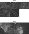
Fig. 1 shows the effect of different calcium sources on neuronal sprouting from cultured spinal cord-dorsal root ganglia (SC-DRG) slices. Immunofluorescent staining (anti neurofilament antibody) of nerve fibers grown from SC-DRG slices exposed to the following calcium compounds [Ca2+ concentration of 2 mM]: (A) ACC-Etidronic Acid; (B) ACC-phosphoserine; (C) gastrolith; (D) crystalline calcium carbonate (CCC); and (E) CaCl2 solution (control). Original magnification ×100. -
Fig. 2 shows the effect of (A) ACC-stabilized by Etidronic Acid and (B) CaCl2 solution (control) on neuronal sprouting from brain cells cultured on chitosan microcarriers (MCs). Immunofluorescent staining of nerve fibers (anti neurofilament antibody) grown from brain cells-chitosan MCs aggregates, after 30 days in culture in the presence of 2 mM of either ACC-Etidronic Acid or CaCl2 is presented. -
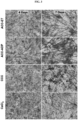
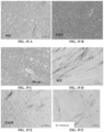
Fig. 3 shows the effect of ACC on formation of myotubes in healthy skeletal muscle cultures. Original magnification x40. Skeletal muscle cultures were exposed to the following calcium compounds (final Ca2+ concentration of 2 mM): ACC-Etidronic Acid; ACC-ADP; Gastrolith; crystalline calcium carbonate (CCC); and CaCl2 solution (control). Cultures were fixed after 4 and 7 days and stained with Giemsa. Enhancement of myotubes formation by skeletal muscle cultures was observed in ACC treated cells. -
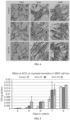
Fig. 4 shows the effect of ACC in the culture medium on early formation of myotubes in mdx cell line cultures. Giemsa staining of the cultures that were exposed to medium containing CaCl2, ACC-ET and ACC-phosphoserine (ACC-PS) is shown. Original magnification x100. -
Fig. 5 shows the creatinine kinase (CK) levels as measured in mdx muscle cell line exposed to two ACC preparations (ACC-ET and ACC-PS) versus CaCl2. -
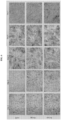
Fig. 6 shows the effect of ACC (ACC-PS, ACC-PP vs. control (CaCl2)) on the formation of myotubes in mdx mice primary cultures (Giemsa staining; original magnification X50). -
Fig. 7 shows the effect of ACC on formation of myotubes in mdx mice primary cultures demonstrated by myosin immunostaining; control (CaCl2); ACC-PS, ACC-polyphosphate (ACC-PP). Original magnification X100. -
Fig. 8 shows the creatinine kinase values of mice (wild type and mdx mice) administrated orally with different types of calcium supplements. -
Fig. 9 shows the effect of administration of stabilized ACC mdx mice on their performance in Four limb hanging test. -
Fig. 10 (not according to the invention) shows the effect of stabilized ACC on mice embryos development in vitro in one step medium with different concentration of ACC. -
Fig. 11 (not according to the invention) shows the effect of stabilized ACC on mice embryos development in vitro in one-step medium. -
Fig. 12 (not according to the invention) shows the effect of stabilized ACC on mice embryos development in vitro in cleavage medium. -
Fig. 13 shows Alizarin red staining of osteoblasts following 10 days in culture as a function of various medium treatments. The media was supplemented with additional 1 mM Ca2+ from: A - ACC, B - CaCl2, or C - control, no Ca2+ addition. -
Fig. 14 shows Alkaline phosphatase staining of osteoblasts following 10 days in culture as a function of various medium treatments. The media was supplemented with additional 1 mM Ca2+ from: A - ACC, B - CaCl2, or C - control, no Ca2+ addition. -
Fig. 15 shows Alizarin red staining (A-C) and Alkaline phosphatase (D-F) of mdx cell lines grown in media with different sources of additional 1 mM Ca2+ added: A and D - ACC, B and E - CaCl2, or C and F - control, no Ca2+ addition. -
Fig. 16 (not according to invention)shows effect of stabilized ACC of in vitro cultured ovaries (A) in which granulosa cells surrounding the oocytes were intact versus control (B) (no ACC in the medium) in which non intact granulosa cells and oocyte at the Germinal Vesicles stage was observed. - The present invention discloses the unexpected advantages of ACC on cell growth and maturation. These attributes were observed in various systems of cell growth as are exemplified hereinbelow.
- According to some particular aspects the present invention provides a pharmaceutical composition comprising amorphous calcium carbonate (ACC) stabilized by at least one stabilizing agent, for use in treating a disease or a condition selected from a neuromuscular disease or condition. In particular embodiments, the disease may be selected from muscular dystrophy and an axonal defect.
- For each aspect of the present invention individually and collectively the following terminology is used and the specific parameters are as defined hereinbelow:
- The term "pharmaceutical composition" and "pharmaceutically acceptable composition" are used herein interchangeably and refer to a composition comprising ACC stabilized by at least one stabilizer, as disclosed herein below, formulated together with one or more pharmaceutically acceptable carriers.
- The terms "pharmaceutically acceptable carrier" or "pharmaceutically acceptable excipient" as used herein refer to any and all solvents, dispersion media, preservatives, antioxidants, coatings, isotonic and absorption delaying agents, surfactants, buffer and the like, that are compatible with pharmaceutical administration. The use of such media and agents for pharmaceutically active substances is well known in the art. The compositions may contain other active agents providing supplemental, additional, or enhanced therapeutic functions.
- According to some embodiments, the disease or a condition is an axonal defect, thus the present invention provides a pharmaceutical composition comprising ACC stabilized by at least one stabilizing agent, for use in treating an axonal defect.
- The term "treating" as used herein refers to taking steps to obtain beneficial or desired results, including clinical results. Beneficial or desired clinical results include, but are not limited to, alleviation or amelioration of one or more symptoms associated with a condition.
- According to any one of the above embodiments, the ACC is stabilized by at least one stabilizing agent. The term "amorphous calcium carbonate" and "ACC" are used herein interchangeably and refer to non-crystalline amorphous form of calcium carbonate stabilized by at least one stabilizing agent. The terms "stabilizing agent" and "stabilizer" are used herein interchangeably and refer to any substance that contributes to preserving calcium carbonate in the amorphous state during ACC production, formulating storage and/or use. In certain embodiments, the stabilizing agent is a single agent. In other embodiments, use of several stabilizing agents is encompassed. The terms "stabilized ACC" and "ACC stabilized by at least one stabilizer" may be used in some embodiments, interchangeably.
- ACC may be obtained from a natural source or chemically synthesized. The terms also include naturally stabilized ACC such as ACC obtained from gastrolith.
- The term "natural ACC" as used herein refers to any ACC isolated or derived from a natural source. Non-limiting examples of natural sources of ACC include gastroliths of freshwater crustaceans.
- The term "synthetic ACC" as used herein refers to any ACC produced and/or derived by man ex-vivo.
- The stabilizer may comprise a molecule having one or more functional groups selected from, but not limited to, hydroxyl, carboxyl, ester, amine, phosphino, phosphono, phosphate, sulfonyl, sulfate, or sulfino groups. The hydroxy bearing compounds, combined with the hydroxide, optionally also bear other functions like carboxyl, etc. but with the hydroxyl not being esterified.
- According to some embodiments, the stabilizer has low toxicity or no toxicity to mammalian cells or organism, and in particular to a human being. According to other embodiments, the stabilizer is of food, nutraceutical or pharmaceutical grade.
- In certain embodiments, the ACC stabilizing agent is independently at each occurrence, an organic acid; phosphorylated, phosphonated, sulfated or sulfonated organic compound; phosphoric or sulfuric ester of a hydroxyl carboxylic acid; an organoamine compound; an organic compound comprising a hydroxyl; an organophosphorous compound or a salt thereof; phosphorylated amino acids and derivatives thereof, a bisphosphonate; an organophosphate compound; an organophosphonate compound; organic polyphosphate, an inorganic polyphosphate, an inorganic phosphorous acid, an organic compound having multiple functional groups as defined above; an inorganic phosphate and polyphosphate compound; an organic compound having a polyphosphate chain; an organic surfactant; a bio-essential inorganic ion; saccharides and derivatives thereof, proteins, phosphorylated proteins, natural and synthetic biopolymers and derivatives thereof or any combination thereof. According to some embodiments, the stabilizer may have also a pharmaceutical activity, e.g. bisphosphonate, or ATP.
- Thus in one embodiment, the stabilizing agent is selected from the group consisting of polyphosphate such as inorganic polyphosphate, organic acids, phosphorylated, phosphonated, sulfated or sulfonated organic compounds, phosphoric or sulfuric esters of hydroxy carboxylic acids, phosphorylated amino acids, bisphosphonate, organic polyphosphate, saccharides and derivatives thereof, proteins, peptides, phosphorylated proteins, phosphorylated peptides, and any combinations thereof. According to another embodiment, the stabilizing agent is selected from the group consisting of phosphoserine, adenosine triphosphate, adenosine diphosphate, phytic acid, citric acid, etidronic acid, pyrophosphate, polyphosphate, triphosphate, ethanol, hexamethaphosphate, chitin, and any combination thereof.
- According to some embodiments, the stabilizer is an organic acid. According to certain embodiments, the organic acid is selected from ascorbic, citric, lactic or acetic acid, oxalic acid, malonic acid, glutaconic acid, succinic acid, maleic acid, lactic acid, glutamic acid, aconitic acid, and optionally include compounds having at least two carboxylic groups optionally having molecular weight not larger than 250g/mol, such as citric acid, tartaric acid, malic acid, etc. According to one particular embodiment, the stabilizer is citric acid
- In another embodiment, the stabilizer is a phosphoric ester of hydroxyl carboxylic acids such as phosphoenolpyruvate. In another embodiment, the phosphoric or sulfuric esters of hydroxyl carboxylic acids comprise amino acids. Examples of such esters are phosphoserine, phosphothreonine, sulfoserine, sulfothreonine and phosphocreatine.
- In another embodiment, the stabilizer is a saccharide. According to one embodiment, the saccharides is selected from mono-, di- tri-, oligo-, and polysaccharides like sucrose, mannose, glucose, chitosan and chitin. Stabilizer may be in some embodiments, a polyol such as glycerol. According to another embodiment, the stabilizer is an amino acid such as serine or threonine. Each possibility represents a separate embodiment, of the present invention.
- Non-limiting Example of natural and synthetic biopolymers and derivatives are polynucleotides and glycoproteins.
- Some specific unlimited examples for such ACC stabilizers that were approved for food consumption, found in natural food or in human beings include phytic acid, citric acid, sodium pyrophosphate dibasic, adenosine 5'-monophosphate (AMP) sodium salt, adenosine 5'-diphosphate (ADP) sodium salt and adenosine 5'-triphosphate (ATP) disodium salt hydrate, phosphoserine, phosphorylated amino acids, food grade surfactants, sodium stearoyl lactylate, and combinations thereof.
- According to some embodiments, the stabilizer comprises at least one component selected from phosphoric or sulfuric esters of hydroxyl carboxylic acids, such as phosphoenolpyruvate, phosphoserine, phosphothreonine, sulfoserine or sulfothreonine and saccharides, selected from mono-, di-, tri-, oligo- and poly-saccharides, for example, sucrose, mannose, glucose. The hydroxyl bearing compound may further comprise at least one alkali hydroxide, such as sodium hydroxide, potassium hydroxide and the like. The phosphorylated acids may be present in oligopeptides and polypeptides. In other embodiments, of the invention, the stabilizer is an organic acid selected from monocarboxylic acid or multiple carboxylic acid, e.g. dicarboxylic acid or tricarboxylic acid. Each possibility represents a separate embodiment, of the invention. The organic acid may be as defined above.
- In some embodiments, of the invention, the ACC stabilizer is selected from phosphorylated amino acids, polyols and combinations thereof. In some embodiments, the stable ACC comprises a phosphorylated compound as a stabilizer wherein the phosphorylation is performed on the hydroxyl group of an organic compound. In some embodiments, the stable ACC comprises a stabilizer selected from the group consisting of citric acid, phosphoserine, phosphothreonine and combinations thereof. The non-limiting examples of stabilizers containing phosphate, phosphite, phosphonate groups and salts or esters thereof include phytic acid, dimethyl phosphate, trimethyl phosphate, sodium pyrophosphate, tetraethyl pyrophosphate, ribulose bisphosphate, etidronic acid and other medical bisphosphonates, 3-phosphoglyceric acid salt, glyceraldehyde 3-phosphate, 1-deoxy-D-xylulose-5-phosphate sodium salt, diethylene triamine pentakis(methyl-phosphonic acid), nitrilotri(methylphosphonic acid), 5-phospho-D-ribose 1-diphosphate pentasodium salt, adenosine 5'-diphosphate sodium salt, adenosine 5'-triphosphate disodium salt hydrate, α-D-galactosamine 1-phosphate, 2-phospho-L-ascorbic acid trisodium salt, α-D-galactose 1-phosphate dipotassium salt pentahydrate, α-D-galactosamine 1-phosphate, O-phosphorylethanolamine, disodium salt hydrate, 2,3-diphospho-D-glyceric acid pentasodium salt, phospho(enol)pyruvic acid monosodium salt hydrate, D-glyceraldehyde 3-phosphate, sn-glycerol 3-phosphate lithium salt, D-(-)-3-phosphoglyceric acid disodium salt, D-glucose 6-phosphate sodium salt, phosphatidic acid, ibandronate sodium salt, phosphonoacetic acid, DL-2-amino-3-phosphonopropionic acid or combinations thereof. The bio-essential inorganic ions may include, inter alia, Na, K, Mg, Zn, Fe, P, S, N; P or S in the phase of oxides; or N as ammonia or nitro groups.
- The stabilizer may further include phosphonate compounds such as, but not limited to bisphosphonates, polyphosphates, such as, but not limited to pyrophosphate or polyphosphonates or organo polyphosphates, such as, but not limited to, adenosine diphosphate (ADP) or adenosine triphosphate (ATP).
- Optionally ACC is stabilized by a combination of phosphoserine and citric acid. In another embodiment, the ACC is stabilized by triphosphate and citric acid.
- The ACC may be stabilized by more than one stabilizers, e.g. two stabilizers. In some embodiments, the first stabilizer and the second stabilizer are similar. In other embodiments, the first stabilizer and the second stabilizer comprise different stabilizers. The first and the second stabilizers may be each independently as defined hereinabove. The stable ACC can comprise more than two stabilizers, wherein the stabilizers may be same or different. The stable ACC can comprise more than two stabilizers, wherein one or more stabilizers are added to the ACC during the formation and precipitation of the ACC; hence constituting "internal" stabilizers, and another one or more stabilizers are added at the ACC particle surfaces after their formation; hence, constituting "external" stabilizers. Further examples for stable ACC and the preparation thereof may be found in International Patent Applications Nos.
WO 2009/053967 ,WO 2014/024191 andWO 2016/193982 . - In some embodiments, the stabilizing agent is a protein or a peptide. In one embodiment, the protein or peptide is a naturally produced and purified protein or peptide. In another embodiment, the protein is synthetically produced protein. In some embodiments, the protein is selected from GAP65, GAP22, GAP21 and GAP12 proteins. In another embodiment, the proteins are selected from CqCDA1, chotinase 2, beta-N-acetylglucosaminidase, GAMP-like, chitin-binding protein, CqCBP, CAP10, GAP 18.2, GAP 02526, CqHc1, CqHc2, CqHc3, CqHc4, CqHc5, CqHc6, CqHc7, cryptocyanin1, cyclophilin,
cystatin 1,cycstatin 2, LPS-BP, LEA protein and crystacyanin, optionally said proteins are originated from C. quadricarinatus. According to certain embodiments, the proteins are phosphorylated proteins - In some embodiments, the stabilizing agent is selected from polyphosphate, phosphorylated amino acids, organic acids, phosphorylated, phosphonated, sulfated or sulfonated organic compounds, phosphoric or sulfuric esters of hydroxy carboxylic acids, bisphosphonate, saccharides, derivatives thereof, proteins, phosphorylated proteins, natural and synthetic biopolymers and derivatives thereof and any combinations thereof. In other embodiments, the stabilizing agent is selected from phosphoserine, triphosphate, adenosine triphosphate, adenosine diphosphate, phytic acid, citric acid, etidronic acid, pyrophosphate, ethanol, hexamethaphosphate, chitin, and any combination thereof.
- In some embodiments, the stabilizing agent is selected from organic acids, phosphorylated organic acids, phosphoric or sulfuric esters of hydroxy carboxylic acids, phosphorylated amino acids, bisphosphonate, organic polyphosphate, saccharides, derivatives thereof, proteins and any combinations thereof.
- According to some embodiments, the at least one stabilizer is selected from the group consisting of a polyphosphate, bisphosphonate, phosphorylated amino acid, citric acid, and any combination thereof. In some embodiments, more than one stabilizers, e.g. 2, 3 or 4 stabilizers are added.
- According to some embodiments, the stabilizer is a polyphosphate or pharmaceutically acceptable salts thereof. According to some embodiments, the polyphosphate is physiologically compatible, water soluble polyphosphate salt selected from the group consisting of sodium, potassium and any other essential cation of polyphosphate. In one embodiment, the polyphosphate is organic or inorganic polyphosphate. The term "polyphosphate" as used herein refers to polymeric esters of PO4. According to some embodiments, the polyphosphate is physiologically compatible water soluble polyphosphate salt selected from the group consisting of sodium and potassium polyphosphate. In some embodiments, the polyphosphate is an inorganic polyphosphate or pharmaceutically acceptable salts thereof. Not-limiting examples of such salt are Na, K, Mg, Mn and Zn. According to some embodiments, the inorganic phosphate comprise 2 to 10 phosphate groups, e.g. 2, 3, 4, 5, 6, 7, 8, 9, or 10 phosphate group. According to some embodiments, the polyphosphate is selected from pyrophosphate, triphosphate, and hexametaphosphate. According to one embodiment, the stabilizer is pyrophosphate or pharmaceutically acceptable salts thereof such as sodium pyrophosphate. According to another embodiment, the stabilizer is triphosphate or pharmaceutically acceptable salts thereof such as sodium triphosphate. The term "triphosphate" and "tripolyphosphate" are used herein interchangeably. According to a further embodiment, the stabilizer is hexametaphosphate or a pharmaceutically acceptable salt thereof such sodium hexametaphosphate.
- According to some embodiments, the stabilizer is a bisphosphonate or pharmaceutically acceptable salts thereof. The not-limiting examples of salt are Na, K, Mg, Mn and Zn.
- The term "bisphosphonate" as used herein refers to organic compounds having two phosphonate (PO(OH)2) groups. The term further relates to compounds having a backbone of PO3-organic-PO3. Most typical is a series of bisphosphonates that are used as pharmaceuticals for treating osteoporosis. According to some embodiments, the bisphosphonate is selected from the group consisting of etidronic acid, zoledronic acid, medronic acid, alendronic acid and a pharmaceutically acceptable salt thereof. According to some embodiments, the stabilizer is an etidronic acid or a pharmaceutically acceptable salt thereof. According to another embodiment, the stabilizer is a zoledronic acid or a pharmaceutically acceptable salt thereof. According to a further embodiment, the stabilizer is a medronic acid or a pharmaceutically acceptable salt thereof. According to certain embodiments, the stabilizer is alendronic acid or a pharmaceutically acceptable salt thereof.
- According to certain embodiments, the stabilizer is a phosphorylated amino acid. According to one embodiment, the phosphorylated amino acid is phosphoserine. According to another embodiment, the phosphorylated amino acid is phosphothreonine.
- According to some embodiments, the ACC composition comprises a combination of the stabilizers disclosed above.
- According to some embodiments, the stabilizer is polyphosphate or a bisphosphonate as defined hereinabove, and the molar ratio between P atoms of the stabilizer and Ca atoms of the ACC (P:Ca molar ratio) is about 1:90 to 1:1. In one embodiment, the P:Ca molar ratio is about 1:40 to about 1:1. In a further embodiment, the P:Ca molar ratio is about 1:35 to about 1:2. In certain embodiments, the P:Ca molar ratio is about 1:30 to about 1:3. In certain embodiments, the P:Ca molar ratio is about 1:28 to about 1:3. In other embodiments, the P:Ca molar ratio is about 1:25 to about 1:4. In further embodiment, the P:Ca molar ratio is about 1:20 to about 1:5. In another embodiment, the P:Ca molar ratio is about 1:20 to about 1:6. In a particular embodiment, the P:Ca molar ratio is about 1:15 to about 1:5. In another particular embodiment, the P:Ca molar ratio is about 1:25 to about 1:5. According to some embodiments, such polyphosphate is pyrophosphate, triphosphate, hexametaphosphate or a pharmaceutically acceptable salt thereof. According to another embodiments, the bisphosphonate is alendronic acid, etidronic acid, zoledronic acid or medronic acid and the P:Ca molar ratio is as defined hereinabove.
- According to some embodiments, the calcium content (Ca content) stabilized ACC comprising polyphosphate or bisphosphonate is about 1 wt% to about 39 wt%, about 5 wt% to about 39 wt%, about 10% to about 39 wt%, about 15% to about 39 wt%, about 20 wt% to about 38 wt%, about 25 wt% to about 38 wt%, or about 30 to about 38. The terms "Ca content" and "calcium content" is used herein interchangeably and refer to the content of calcium of the ACC in the final composition.
- In certain embodiments, the P:Ca molar ratio is about 1:40 to about 1:1, and the Ca content is about 20 wt% to about 39 wt%. In some embodiments, the molar ratio is 1:28 to about 1:3, and the Ca content is about 30 wt% to about 38 wt%. In another embodiment, the molar ratio is 1:25 to about 1:5, and the Ca content is about 30 wt% to about 36 wt%.
- According to some embodiments, the stabilizer is selected from the group consisting of a polyphosphate, phosphorylated amino acid, bisphosphonate, citric acid, tartaric acid and any combination thereof. According to one embodiment, the polyphosphate is selected from the group consisting of triphosphate, pyrophosphate, and hexametaphosphate, the phosphorylated amino acid is phosphoserine or phosphothreonine, and the bisphosphonate is selected from the group consisting of alendronate, etidronic acid, zoledronic acid and medronic acid.
- According to some embodiments, the stabilized ACC comprises less than 20 wt%, less than 15 wt%, less than 10 wt%, or less than 5 wt% of the stabilizing agent. In some embodiments, the stabilized ACC comprises up to 5 wt% of the stabilizing agent.
- According to one embodiment, the average diameter of the stabilized ACC primary particles is about 10 nm to about 5 µm. According to another embodiment, the average diameter of the ACC primary particles is about 30 nm to about 400 nm. According to yet another embodiment, the average diameter average diameter of the ACC primary particles is about 30 nm to 350 nm. According to certain embodiments, the average diameter of the ACC primary particles is about 35 nm to 300 nm, 40 nm to about 250 nm, about 45 nm to about 200 nm, about 50 nm to about 150 nm or about 60 nm to about 100 nm. According to yet another embodiment, the average diameter of the ACC primary particles is about 30 nm to 00 nm. According to still another embodiment, the primary particles of ACC are aggregated and an average diameter of the aggregates is between 0.5 µm and 300 µm. According to one further embodiment, the diameter of aggregates of the ACC primary particle is about 1 to about 100 µm, about 10 to about 50 µm or about 20 to about 40 µm. According to another embodiment, the average diameter of the aggregates of the ACC primary particle is between 1 µm and 10 µm.
- The pharmaceutical composition of the present invention may be administered by any known route of administration. The term "administering" or "administration of" a substance, a compound, an agent or a pharmaceutical composition to a subject can be carried out using one of a variety of methods known to those skilled in the art. For example, a compound, an agent or a composition can be administered enterally or parenterally. Enterally refers to administration via the gastrointestinal tract including per os, sublingually or rectally. Parenteral administration includes administration intravenously, intradermally, intramuscularly, intraperitoneally, subcutaneously, ocularly, sublingually, intranasally, by inhalation, intraspinally, intracerebrally, and transdermally (by absorption, e.g., through a skin duct). A compound or agent can also appropriately be introduced by rechargeable or biodegradable polymeric devices or other devices, e.g., patches and pumps, or formulations, which provide for the extended, slow or controlled release of the compound or agent. Administering can also be performed, for example, once, a plurality of times, and/or over one or more extended periods. The administration may include both direct administration, including self-administration, and indirect administration, including the act of prescribing a drug or a medical food. For example, as used herein, a physician who instructs a patient to self-administer a drug or a medical food, or to have the drug or the medical food administered by another and/or who provides a patient with a prescription for a drug or a medical food is administering the drug or a medical food to the patient. According to some embodiments, pharmaceutical composition is pharmaceutical food or food supplement.
- The pharmaceutical composition comprising stabilized ACC may be administered via a systemic administration. For example stabilized ACC may be administered orally, sublingually or rectally. Alternatively the stabilized ACC may be administered intravenously, intradermally, intramuscularly, intraperitoneally, subcutaneously, ocularly, sublingually, intranasally, by inhalation, intraspinally, intracerebrally, and transdermally.
- The pharmaceutical composition according the present invention may be prepared in any known method. In particular, the pharmaceutical composition may be formulated using a method known in the art so as to provide rapid, continuous or delayed release of the active ingredient after administration. In one particular embodiment, the pharmaceutical composition is formulated as a solid dosage form selected from tablets, capsules, powder or granules. In another embodiment, the pharmaceutical composition is formulated as a liquid or semi-liquid dosage form selected from an elixir, tincture, suspension, syrup, emulsion or gel. The pharmaceutical composition may be formulated as semi-solid formulations such as gum.
- Pharmaceutical compositions intended for oral use may be prepared according to any method known to the art for the manufacture of pharmaceutical compositions and may further comprise one or more agents selected from sweetening agents, flavoring agents, coloring agents and preserving agents in order to provide pharmaceutically elegant and palatable preparations. Tablets contain the active agent in admixture with non-toxic pharmaceutically acceptable excipients, which are suitable for the manufacture of tablets. These excipients may be, e.g., inert diluents such as calcium carbonate, sodium carbonate, lactose, calcium phosphate, or sodium phosphate; granulating and disintegrating agents, e.g., corn starch or alginic acid; binders; and lubricating agents. The tablets are optionally coated utilizing known techniques to delay disintegration and absorption in the gastrointestinal tract and thereby provide an extended release of the drug over a longer period.
- According to one embodiment, the present invention provides a pharmaceutical composition comprising amorphous calcium carbonate (ACC) stabilized by at least one stabilizing agent, for use in treating an axonal defect.
- The term "axonal defect" refers to any defect, damage or injury to the axonal part of a nerve cell of the peripheral or central nervous system. Nerves can be damaged either through trauma or disease. Traumatic nerve injury, such as carpal tunnel syndrome, is caused by the compression of nerves. Other trauma, such as falls and motor vehicle accidents, may lead to the severance of nerves. Diseases that harm nerves include multiple sclerosis, diabetes, spina bifida, and polio. Multiple sclerosis, for example, causes the breakdown of the insulating myelin surrounding axons. According to some embodiments, of the invention the defect or damage may occur to the brain, spinal cord, afferent and the efferent nerves emerging from the spinal cord and to peripheral nerves.
- Treating axonal defects may comprise treating axonal damage such as injury. Teating axonal damage may also comprise enhancing regeneration and/or recovery of a damaged nerve. Treating axonal damage may still comprise enhancing nerve regeneration. Treating axonal defect may further comprise treating a defect resulted from a disease such as multiple sclerosis.
- According to some embodiments, the axonal defect is an axonal damage, thus the pharmaceutical composition of the present invention is for use in treating an axonal damage, e.g. an axonal injury.
- Treating of axonal damage may comprise enhancing regeneration and/or recovery of a damaged nerve.
- The terms "neuronal regeneration" and "nerve regeneration" as used herein may be used interchangeably and refer to recovery of functions of a damaged nerve. Specifically, it includes recovery of signaling via the nerve by repairing a damaged site, regrowth of axonal and dendritic neuronal fibers of the peripheral or the central nervous system. In some embodiments, nerve regeneration refers to sprouting from damaged neuronal fibers.
- According to some embodiments, the locally administered pharmaceutical composition is formulated in a liquid or semi-liquid formulation as defined hereinabove. In one embodiment, the liquid or semi-liquid formulation is selected from a suspension, emulsion, colloid or gel. The stabilized ACC may be administered as a suspension.
- The defected of damaged nerve may be a nerve of the peripheral nerve system (PNS) or of the central nerve system (CNS). Thus in one embodiment, the pharmaceutical composition of the present invention is for treating a damage to the nerves of the central nervous system. According to another embodiment, the pharmaceutical composition of the present invention is for treating a damage to the peripheral nervous system. According to some embodiments, of the invention, the pharmaceutical composition of the present invention is for treating a damage occurred to the brain, spinal cord, afferent and the efferent nerve emerging from the spinal cord, or to peripheral nerves.
- The pharmaceutical composition may be administered locally. The pharmaceutical composition may be administered in proximity to the damaged nerve. The pharmaceutical composition may also administered by injection, infusion or via a pump.
- According to one embodiment, the pharmaceutical composition comprising ACC stabilized by least one stabilizing agent is selected from phosphoserine, triphosphate, adenosine triphosphate, adenosine diphosphate, phytic acid, citric acid, etidronic acid, pyrophosphate, ethanol, hexamethaphosphate, chitin, and any combination thereof, for use in treating an axonal defect such as axonal damage.
- According to some embodiments, the disease or disorder is a muscular dystrophy. Thus in one embodiment, the present invention provides a pharmaceutically acceptable composition comprising stabilized ACC, for use in treating a muscular dystrophy.
- The term "muscular dystrophy" as used herein refers to any one of several degenerative disorders, diseases or conditions characterized by progressive skeletal muscle weakness and fragility. Many of these diseases result from mutations in genes encoding proteins of the dystrophin-glycoprotein complex (DGC). In one embodiment, muscular dystrophy refers to a disease identified as Duchenne, Becker, limb-girdle, congenital, facioscapulohumeral, myotonic, oculopharyngeal, distal, or Emery-Dreifuss muscular dystrophy. In one particular embodiment, the muscular dystrophy is Duchenne muscular dystrophy (DMD). Thus in one embodiment, the pharmaceutically acceptable composition of the present invention is for use in treating DMD.
- In one embodiment, the term treating refers to alleviation or amelioration of one or more symptoms associated with muscular dystrophy, delay or slowing of that impairment. According to some embodiments, treating muscular dystrophy comprises promoting myotube formation. The term "myotube formation" as used herein refers to a process in which myoblasts fuse into multi-nucleated fibers, myotube. According to some embodiments, the term treating muscular dystrophy further comprises also reducing the time to the onset of spontaneous contractile activity of said myotubes. The time to the onset of spontaneous contractile activity is defined as a time needed to myoblasts to fuse and start spontaneously contracting.
- According to the teachings of the present invention, ACC is stabilized by at least one stabilizing agent as described herein above. According to one embodiment, the stabilized ACC is natural ACC obtained from a natural source, e.g. from gastrolith, or chemically synthesized.
- According to one embodiment, the present invention provides a pharmaceutical composition comprising ACC stabilized by at least one stabilizing agent for use in treating DMD, wherein the stabilizing agent is selected from phosphoserine, triphosphate, adenosine triphosphate, adenosine diphosphate, phytic acid, citric acid, etidronic acid, pyrophosphate, ethanol, hexamethaphosphate, chitin, and any combination thereof. According to some embodiments, the stabilizing agent is selected from phosphoserine, triphosphate, bisphosphonate and combination thereof with citric acid. According to one embodiment, the pharmaceutical composition is administered systemically, e.g. orally.
- According to another aspect, the present invention provides use of ACC stabilized by at least one stabilizing agent, for the preparation of a medicament for treatment of a disease or a condition selected from an axonal defect and muscular dystrophy.
- The culture medium was composed of: 90% Dulbecco's modified eagle medium-nutrient mixture F-12(DMEM-F12) calcium depleted (medium without calcium ions, (special preparation), 10% heat-inactivated fetal bovine serum (FBS), 6 g/L D-glucose, 2 mM glutamine, 25 µg/mL gentamicin and 0.05 ng/mL insulin-like growth factor 1 (IGF-I) (all purchased from Biological-Industries, Israel).
- Neural and Vascular Reconstruction Gel (NVR-Gel, NVR Labs proprietary) is composed of two main components: high molecular hyaluronic acid (HA, 3×10⁶ Da, BTG, Israel) and laminin (Sigma). For neuronal cell cultivation, NVR-Gel of 1% was diluted with culture medium to a final concentration of 0.3-0.5%. The gel has the texture of a viscous liquid, it adheres easily and successfully the embedded cells or explants to the plastic or glass substratum, and enabled nerve fiber outgrowth in a 3D pattern.
- All the experiments were carried out and authorized by the local ethics committee recognized by the Israeli authorities for animal experimentation. Stationary organotypic cultures of dorsal root ganglia (DRG) and spinal cord (SC) as well as cultures of dissociated brain cells were prepared from rat fetuses (15 days of gestation, Lewis inbred, Harlan, Israel). Immediately after dissection, the isolated tissues were cut with a Macwain tissue chopper into small slices (of 400 µm thickness). In these studies, two tissue culture strategies were used. In the first method, tissue slices were seeded directly in 12 well-culture plates containing 1 mL culture medium containing 0.3%-0.5% NVR-Gel. In the second method, the tissue slices were further dissociated with a trypsin-EDTA solution for 30 min, and washed with a culture medium. Subsequently, the dissociated cells were added to a suspension of chitosan powder or gastrolith powder (micro carriers, MCs) and incubated in suspension at 37 °C for 4 days. The formed floating cells/MCs aggregates were then collected and seeded in 12 well-culture plates containing 1 mL of culture medium containing 0.3%-0.5% NVR-gel.
- Ca2+ source (listed below) at final concentration of 1 or 2 mM was added once to the gel at the seeding stage and then to the nutrient medium at each consecutive feeding. Calcium source were as following: ACC-Etidronic Acid (ACC- ET) (fresh suspension); ACC-Phosphoserine (ACC-PS) (fresh suspension); Gastrolith (dissolved with 0.1 M HCl and then neutralized with NaOH 1M); gastrolith powder; CCC - Aqueous suspension of crystalline calcium carbonate (commercial nanoparticles powder); and CaCl2 solution - control.
- The cultures were monitored by daily phase contrast microscopic observations starting from 24 hours after setting the cultures onward.
- Suspensions of fresh ACC preparations consisted of particles forming a stable suspension. The gastrolith is a natural ACC isolated from crabs, and can be purchased only as a dry powder. In this form, its other characteristic components (such as calcium ions and proteins) are not available to the cells. In order to increase their bioavailability, the gastrolith powder was dissolved in 0.1 M HCl (which mimics the acidity which exists in the stomach) and then neutralized with 1M NaOH.
- After removal of the culture medium, the dorsal root ganglia (DRG) cultures were washed with phosphate buffered salt solution (PBS) and fixed in 4% paraformaldehyde for 15 min, and then washed again with PBS. The fixed cells were permeabilized with 0.1% of Triton X-100 in PBS and then immuno-blocked (to avoid non-specific staining) with a 1 % bovine serum albumin (BSA) in PBS for 1h at room temperature. The specimens were then incubated with rabbit anti-neurofilament antibodies (NF, Novus Biologicals, 1:500) to visualize the neurite outgrowth. The primary antibodies were diluted in 0.1% BSA and 0.05% Tween-20 in PBS (diluents buffer) and incubated with the specimens overnight at 4°C. After rinsing with 0.05% Tween-20 in PBS (wash buffer), the DRG specimens were incubated for 1 h at room temperature with the secondary antibodies Alexa-Fluor-594-conjugated donkey anti-rabbit IgG (Jackson ImmunoResearch, USA, 1:800 in a diluent buffer). Finally, the samples were rinsed again with wash buffer, and mounted with mounting medium (Immco Diagnostic, USA). All of the images were observed with an Olympus IX70 microscope.
- The effect of various calcium preparations was examined in SC-DRG co-cultures. In general, the cultures contained 400 micron SC slices with attached or separated slices of DRG. It can be said that all of the examined ACC preparations (ACC-ET, ACC-PS, and gastrolith) enhanced significantly neuronal fiber regeneration in comparison to CCC and the CaCl2. Table 1 shows the portion of explants (out of 6) which exhibited nerve fiber sprouting after 4 days of cultivation in the presence of the various calcium preparations (Ca2+ concentration of 2 mM) or in the presence of stabilizers alone (the stabilizer was added at the concentration of 0.05% to each well (from a stock solution of 5%)). It can be seen that the most intensive sprouting was observed in cultures which were exposed to ACC-ET (100% of explants), followed by ACC-PS and Gastrolith (66.6% of explants). The CCC and CaCl2 induced neuronal sprouting only from 50% of the explants, and the stabilizers alone even a lower percentage (0-33%) During the establishment of the cultures (after the first week of cultivation) the regenerated nerve fibers became longer, thicker and ramified, until the formation of neuronal networks, mainly in cultures exposed to the various ACC preparations (
Fig. 1 ).Table 1: The effect of ACC preparations on early nerve fiber sprouting from SC-DRG co-cultures. Type of calcium preparation Cultures with axonal regeneration (%) ACC-ET (fresh suspension) 100 ACC-PS (fresh suspension) 66.6 Gastrolith 66.6 CCC (crystalline calcium carbonate) 50 CaCl2 aqueous solution (control) 50 ET 33.3 PS 0 - The effect of ACC was studied on cultures of brain cells-MCs aggregates seeded in gel after 4 days in suspension (see methods part of Example 1). The results are presented in
Fig. 2 and show that the ACC enhanced nerve fibers regeneration, significantly more than the calcium chloride. This is especially notable when comparing the number and the length of nerve fibers between the two treatments. - Stationary skeletal muscle cultures were prepared from healthy 1 day new born rat ( Sprague Dawley, Harlan, Israel). The muscle tissue was dissected from posterior legs and was thoroughly minced. Digestion was performed with Trypsin-EDTA while gentle trituration. After 30 min the supernatant, containing dissociated cells, was collected and a fresh Trypsin-EDTA was added. This procedure was repeated two more times. Then, all supernatants were pooled, centrifuged, and the cell pellet was re-suspended in Proliferation Medium. Cells were seeded in a Gelatin coated 12 wells culture plates, 1×10⁵ cells/well containing 1 mL of Proliferation Medium. Two days later, the medium was changed to Fusion Medium, which was then changed twice a week. The cultures were monitored by daily phase contrast microscopic observations from 24 hours after setting the cultures and onward. At predetermined days, cultures were fixed in methanol for 20 minutes and then stained with Giemsa in order to evaluate the number of myotubes formed with time in cultures.
- Stock solution of 1% porcine gelatin in water, was sterilized by autoclave. Once cooled, 500 µl of the solution were added to each well of the 12-wells culture plat. After incubation for 20 min at room temperature, the excess solution was removed, and the cells were seeded.
- For the proliferation stage, cells were cultured in: DMEM/F12 (containing 1 mM Ca2+)+10% FBS, 25 µg/mL Gentamycin, and 2 mM L-Glutamine.
- For the fusion stage the Proliferation Medium was changed with Fusion Medium, which was prepared as following: DMEM/F12 (containing 1 mM Ca2+), 2% horse serum (HS), 2 mM L-Glutamine, 25 µg/mL Gentamycin, and 4 units/100mL Insulin (all purchased from Biological-Industries, Israel).
- The fusion medium was enriched with the various calcium preparations listed in Table 2, at Ca2+ concentration of 1 mM (since the medium already contained 1 mM of calcium ions, the final concentration of Ca2+ was 2 mM). Control cultures were grown without further added calcium, or cultures enriched with free (soluble) stabilizer. The experiment was blinded by marking the various calcium preparations with arbitrary numerals only.
- The components were added in one of the following ways: (i) aqueous suspensions of dry material; (ii) aqueous suspension of fresh material (before drying), or (iii) dissolved with HCl (to mimic the acidity which exists in the stomach. After dissolving was accomplished, the formed solutions were neutralized with 1M NaOH).
Table 2 - List of tested materials Added Substance Total calcium concentration (mM) including 1mM Ca2+ ions of the medium ACC-Etidronic Acid (ACC- ET) 2 ACC-Pyrophosphate (ACC-PyP) 2 ACC-Phospho serine (ACC-PS) 2 ACC- Adenosine triphosphate (ACC-ATP) 2 ACC- Adenosine diphosphate (ACC-ADP) 2 ACC- Phytic acid 2 ACC- Citric acid 2 Crystalline calcium carbonate (CCC) (commercial available powder) 2 CaCl2 aqueous solution 2 ET 1 PyP 1 PS 1 ATP 1 ADP 1 Phytic acid 1 Citric acid 1 Control 1 - In the first stage, dry ACC powder was used. The powder (listed in Table 2 according to the stabilizers used in their preparation) were suspended in water, and then added to the culture media in the concentration of 1 mM. Since the medium already contained 1 mM of calcium ions, then the final concentration of Ca2+ was 2 mM. The results, some of which are shown in
Fig. 3 (left hand side), revealed that cultures which were exposed to ACC exhibited early formation of many myotubes already within 4 days of cultivation, with no significant differences between the different ACC preparations. In control cultures, which were exposed to added CCC or CaCl2, myotubes formation was observed later. After 7 days, the cultures that were treated with ACC exhibited numerous long and thick muscle fibers, while in cultures treated with CCC and CaCl2, fewer, thinner and shorter muscle fibers were developed (Fig. 3 ; right hand side). - It is also noted, that in cultures exposed to ACC preparations, muscle contractions were observed already on
day 7 after seeding, while in cultures exposed to added CCC or CaCl2 muscle contractions appeared only after 10 days or more. - It was concluded from the above in vitro results that all ACC preparations enhance myotubes formation and early muscle contractility of the healthy striated muscle cultures.
- The influence of the different calcium preparations was investigated on the mdx cell line (Duchenne muscular dystrophy model), which was kindly provided by Prof. (Emeritus) David Yaffe from the Weizmann Institute of Science, Israel.
- Cells from the Mdx cell line were seeded in a Gelatin coated 12 wells culture plate, 3×10⁴ cells/well containing 1 mL of Proliferation Medium. Two days later (~ 66% confluence), the medium was changed to Fusion Medium which was changed twice a week. The various calcium preparations were separately added to the Fusion Medium according to the treatment described in Table 3. Cultures were enriched with the various calcium preparations, at Ca2+ concentration of 1 mM. Since the medium already contained 1 mM of calcium ions, then the final concentration of Ca2+ was of 2 mM.
Table 3: Calcium sources Treatment Calcium source (2mM) Control CaCl2 ACC-ET Amorphous Calcium Carbonate-Etidronic Acid ACC-PS Amorphous Calcium Carbonate-Phospho Serine - The effects of the tested calcium preparations on cell proliferation, fusion to form myotubes and muscle contraction were monitored by daily phase contrast microscopic observation. At predetermined days, cultures were fixed in methanol for 20 minutes and then stained with Giemsa in order to evaluate the number of myotubes formed with time in cultures.
- In muscle cell cultures, CK is an indicator of myotubes formation and CK level increases (in tissue culture plates) in direct correlation with the progression of myotubes formation in muscle cultures. At predetermined days, cells were collected from culture wells (using a rubber policeman) and kept in 1 mL PBS (without Ca2+) at -70°C until analyzed. For CK measurement cell samples were thawed and physically lysed using a sonicator, to release the CK from muscle myotubes. CK concentration was determined using Creatine Kinase Activity Assay Kit (CK-NAC REAGENT SET, CURTISS, CHEM-INDEX INC, Hialeah, FL, USA).
- The results of the above experiments are presented in
Figs. 4 and 5 . As it can be clearly seen, addition of ACC enhanced cell fusion and formation of myotubes better than the addition of a conventional calcium ion source (CaCl2). This surprising result was demonstrated in both biochemical (CK activity) and morphological (microscopy) analysis (Fig. 4 and 5 ). Moreover, the cultures exposed to ACC preparations, muscle contractions were observed already onday 7 after seeding, while in the controls it appeared only after 10 days or more. Therefore, it is concluded that ACC supplementation has a potential to treat DMD patients. - Thigh muscles were removed from the posterior legs of newborn (one day old) mdx mice under sterile conditions, and washed in PBS to remove excess of blood cells. The muscles were minced into small fragments. For enzymatic dissociation, the muscle fragments were placed in a bicker containing trypsin-EDTA solution (0.25 mM). To ensure cell separation, the mixture was placed on a stirrer, at room temperature, at gentle stirring for 20 min. The soup was collected and centrifuged at 300×g for 5 min. The pellet was re-suspended in DMEM containing FBS. The trypsinization steps were repeated for 3 more cycles. All supernatants were combined into one tube. Cell separation was determined visually (using phase contrast microscopy). Cells density was determined by using hemocytometer.
- Cells were plated in a 12 wells plate in the concentration of 2×10⁵ cells/well. The medium used was DMEM/F12 W/O Ca2+ with the addition of 15% FBS and, 2 mM L-Glutamine Gentamicin (25µg/ml). Calcium was separately added to the medium according to the treatment as describes in Table 4. At day 2 (~ 66% confluence) the medium was changed to fusion medium (DMEM/F12 without Ca2+ with the addition of 10%HS, Insulin (4 units/100ml) and Gentamicin (25µg/ml)). Medium was changed every 3 days.
Table 4. Calcium sources used in primary cells study Treatment Calcium source (2mM) Control CaCl2 ACC-PP Amorphous Calcium Carbonate stabilized by Polyphosphate ACC-PS Amorphous Calcium Carbonate stabilized by Phosphoserine - Cell proliferation and fusion were qualitatively daily monitored. Cultures were fixed at
days - The results are presented on
Fig. 6 and7 . The beneficial effect of ACC formulations was demonstrated by myotubes formation, specifically at early time points,days days - Experimental mice were administrated orally (by feeding or drinking ) with control or test items. The control or test items were repeatedly administered daily until the study termination (after 12 weeks).
Table 5. Study Design Group No. Strain n= Treatment Details 1M C57BL n=3 Rodent commercial diet (Teklad) 2M mdx n =12 Rodent commercial diet (Teklad) 3M n =12 Low Calcium Diet (TDK95027, Harlan Inc) containing 1% elemental calcium from synthetic CCC ad libitum 4M n=12 Low Calcium Diet (TDK95027, Harlan Inc) containing 1% elemental calcium from synthetic ACC (5% PS) ad libitum 5M n =12 Water containing 0.06% elemental Calcium from synthetic ACC (10% PP) ad libitum PS- stabilized with phosphoserine PP- stabilized with polyphosphate - Test system:
- Species: Mice
- Mdx Strain: C57BI 10ScSn/DMD mdx/J
- Wild type Strain: C57BL/6JOIaHsd.
- Gender & Age: Males, 3 weeks of age at study initiation
- Body Weight: Weight variation of animals at study initiation should not exceed ± 5% of the mean weight of the gender.
- Housing: Animals before study initiation were housed in polyethylene cages measuring 42.5 × 26.5 × 18.5 cm, with stainless steel top grill facilitating pelleted food and drinking water in plastic bottle and filled with wood shavings (Harlan Teklad Aspen/ Sani-chips bedding) as bedding material. Bedding material were changed along with the cage at least once a week.
- Identification: Ear tag and cage cards.
- Termination: At the end of the study, animals were euthanized by CO2 asphyxiation.
- Justification: The MDX mice are a common animal model for evaluating the Duchenne Muscular Dystrophy (DMD).
- The first control or test items administration were defined as "
Day 1". - The study was terminated 12 weeks from beginning.
- Animals that were found in a moribund condition and animals showing severe pain or enduring signs of severe distress were humanely euthanized. Animals showing a decrease of body weight larger than 20% from the initial body weight were reported.
- Animals were administrated with ACC by food or by drinking. Animals were administrated with CCC by food. Control was administrated by feeding. The control or test items were repeatedly administered daily until study termination.
- Observations for signs of morbidity and mortality were performed daily at study termination.
- Body weight measurements were recorded once a week and at study termination, 12 weeks.
- At study termination blood sample was drawn from all animals under light anesthesia with Ketmaine/Xylazine, directly from the retro orbital sinus.
- 200µl blood were collected into a yellow-cup tube with clotting activator gel. The tubes were kept at room temperature for at least 30 minutes for clotting and centrifuged at room temperature for 10 minutes at 4000 RPM. The serum samples are stored at 2-4°C until delivered to CK analysis at A.M.L labs.
- The result of Creatinine Kinase test is shown in
Fig. 8 and demonstrate a clear trend. All mdx mice showed elevated values of CK vs. the wild type, which indicates on muscle destruction. However, the elevation was significantly more moderate in the group that received ACC by food (4M group) compared to the group that received normal diet only (2M group; student's t-test, P value= 0.034). While the mice ingroup 3M that received CCC also showed a more moderation elevation in the CK levels in comparison to mice ofgroup 2M (no treatment), the difference was not statistically significant (student's t-test, P value=0.45). -
Group 5M that received ACC by drinking demonstrated results similar to those ofgroup 3M that received CCC by food (21,164±8,391 and 21,676±14,508 IU/ml, respectively). Without wishing to be bound by any theory or mechanism of action it is suggested that the reason for the difference in the results between thegroups 4M (ACC in food) and 5M (ACC by drinking) may be due to heterogeneity of the ACC suspension and therefore non uniform administration of the ACC. It is clear though, that administration of ACC significantly alleviated the symptoms of DMD in the mdx mice. - In the present example, the effect of ACC stabilized by different stabilizers on mdx mice (rodent model of Duchenne Muscular Dystrophy) was tested.
- Mice were administrated with stabilized ACC orally (feeding) every day or by IP injections, consecutive, six times a week. The control or test Items were administered similarly.
-
Species: Mice
Mdx Strain: C57BI 10ScSn/DMD mdx/J
Wild type Strain: C57BL/6JOIaHsd.
Gender & Age: Males, 3-5 weeks of age at study initiation
Body Weight: Weight variation of animals at study initiation should not exceed ± 15% % of the mean weight of the gender.
Group Size: see Table 1, study design
No. of Groups: 6 (see Table 6)Table 6. Study Design Group No. Strain n = Group Name and Treatment Details Route of Adminis tration Duration 1M C57BL - Rodent 6 Wild type control - commercial diet (Teklad) Feeding 24 weeks 2M MDX - Rodent 8 Mdx control - commercial diet 4M 10 ACC stabilized by PS-Low Calcium Diet (TDK95027, Harlan Inc.) containing 1% elemental calcium from synthetic ACC (5% PS) ad libitum 3M 10 ACC stabilized by TP-Low Calcium Diet (TDK95027, Harlan Inc.) containing 1% elemental calcium from synthetic ACC (6% TP-2% Citric Acid) ad libitum 5M 10 ACC stabilized by TP- 200µl containing 0.1% elemental calcium from synthetic ACC (10% TP) Injections are be performed 6 consecutive times a week Intra - peritonea l (IP) injection 12 weeks 6M 6 Control -saline control - 200 µl containing Saline. Vehicle Injections are performed 6 consecutive times a week PS- Phosphoserine; TP- Triphosphate - The MDX mouse is an animal model for evaluating the Duchenne Muscular Dystrophy (DMD).
- Study end Definition: Study is terminated 24 weeks from beginning for groups 1-4 and 12 weeks from beginning for groups 5-6.
- All animals (except for control) were administrated with stabilized ACC either by food (every day) or by IP injections (six consecutive times a week).
- The animals were observed for clinical signs once a week until study termination.
- Observations was performed for any changes in skin, fur, eyes, mucous membranes, respiratory, occurrence of secretions and excretions (e.g. diarrhea). Changes in gait, posture and response to handling, as the presence of bizarre behavior, tremors, convulsions, sleep and coma were also included.
- All observed abnormalities, toxic signs, moribund condition and pre-terminal deaths were recorded.
- Animals that were humanely sacrificed during the test are considered for the interpretation of test results as animals that died during the test.
- Body weight measurements was recorded once a week, shortly before the four limb hanging test. At study beginning and termination body weight was also measured.
- Four limb hanging test was used to monitor muscle strength and to indicates on neuromuscular impairment and motor coordination. The test was used to determine the efficacy of ACC.
- In the four limb hanging test a wire grid system is used to measure the ability of mice to exhibit sustained limb tension oppose their gravitational force. The hanging time is measured in seconds as well as the minimal Holding impulse (Holding impulse = Body mass × Hang time, [N sec], conversion factor - 9.806 × 10³ Newton/gr).
- Grip Test (four limb hanging test) was performed according to the TREAT-NMD SOP# DMD _M.2.1.005 protocol. Briefly:
Mouse (min age - 4 weeks) was placed on a top of a grid (grid square size about 1x1 cm2) and was allowed to accommodate to this environment 3-5 seconds. - Grid was flipped over such that the mouse is upside down. The grid height was placed at least 35 cm above the cage floor. Sufficient amount of soft bedding (5-7 cm) was placed under the grid to ensure soft landing. Mice at the age of 4-24 weeks naturally try to stay on grid and avoid falling to the ground. This height is low enough not to injure the animal, and high enough to ensure avoidance of fall.
- Each hang period must begin with all 4 mouse paws grasping the grid. Hanging time was measured by a stopwatch and recorded.
- The test performed up to a max of 600 sec or repeated 3 times with a rest interval of at least 2 minutes.
- The longest Hanging time was used to assess the Holding impulse.
- Four limb hanging test was done each week during the whole experiment.
- At study termination blood samples are drawn from the all animals under light anesthesia (with Ketmaine/Xylazine ) directly from the retro orbital sinus.
- 200µl blood is collected into a yellow-cup tube with clotting activator gel. The tubes are kept at room temperature for at least 30 minutes for clotting and centrifuged at room temperature for 10 minutes at 4000 RPM. The serum samples will be stored at 2-40C until delivered to CK analysis at A.M.L labs.
- Following terminal bleeding, animals is sacrificed by Carbon dioxide asphyxiation and gross pathology is performed examining the major tissues and organ systems.
- During the necropsy, selected muscles (diaphragm, cardiac and gastrocnemius) from all animals are collected. Muscles from all animals are preserved in 10% neutral buffered formalin (approximately 4% formaldehyde solution) for at least 24 hours for future histopathology.
- Mice in all groups gained weight. No significant difference was found in the average body weight in any of the groups.
- A significant difference was found in the Holding impulses (HI) of both mdx groups that were administrated orally with ACC formulations (stabilizer 1 - phosphoserine, stabilizer 2 - triphosphate) versus the control group that was administrated with standard diet (comprising calcium), see
Fig. 9 . The difference was observed already on the second week of the study where the HI of ACC treated mice was 2 time higher that the HI of the control group. This differences increase as the study progressed and reached a plateau after about 6-8 weeks. It can be seen fromFig. 9 that the HI of ACC treated mice was about X2 - X3.8 times higher than of the control group. - Two days following thawing, MBA-13 cells (received from Prof. Dov Zipori, Weizmann Institute of Science) were re-suspended in recombinant trypsin solution and seeded on a 96 wells plate, (Day "0") in a concentration of 1×10⁴ cells/well using MSC Nutristem® XF basal medium (Biological Industries, cat# 05-200-1A) supplemented with Mesenchymal stem cells (MSC) supplement mix medium (Biological Industries cat#05-201-06) in a ratio of 50ml:300µl. Rows A-H of columns 1-4 of the 96 plate were pre-coated with MCS attachment solution diluted in PBS (without Ca2+, Mg2+), in a ratio of 1:100 for cells seeding. Rows A-H of Columns 5-8 of the 96 plate were pre-coated with Gelatin 0.1% for 30 minutes at room temperature.
- On
day 2, when more than 80% cell confluence was achieved (~48h) the medium was changed to MSCgo rapid osteogenic medium (cat# 05-442-1B) containing factors that promote osteoblastic differentiation. Following medium change, rows A-C were supplemented with additional 1mM calcium (total 2.488 mM calcium) originated from Amorphous Calcium Carbonate (ACC) stabilized by 10%Triphosphate+1%citric acid; rows D-F were supplemented with additional 1mM calcium (total 2.488 mM calcium) originated from calcium chloride; Row G in the plate was treated with MSCgo medium (total 1.488 mM calcium). Row H of the plate was treated with MSC NutriStem® XF nutrient basal medium+ supplement mix (total 1.488 mM calcium). - On
day 4, medium was exchanged with fresh preparations of ACC. - In parallel, a control plate was also seeded with MDX cell lines originated from damaged muscles of MDX mice. The staining of these cell was used to set the background staining of cells intrinsic calcium and also to eliminate the possibility that calcium deposition of the ACC treatment is stained. Prior seeding, wells were coated with gelatin, which is used as a standard substrate for MDX cells attachment. MDX Cells were seeded on a 24 well plate in a concentration of 3×10⁴ cells/well. Seeding day is defined as "
Day 0". - On
day 2, the medium in the wells was replaced with the following: -
Columns columns Columns - Fixation ( with 4% Paraformaldehyde for 20 minutes) of few wells from each type was performed on the 5th, 7th and 10th day following medium exchange, that are
Days - Once the cells were fixated, two types of staining reagents were used to stain extracellular calcium or bone deposition in order to estimate osteoblasts functionality: (i) Alizarin red (Sigma A55333) and (ii) Alkaline Phosphatase (DAKO BCIP/NBT substrate system K0598) as following:
- Alizarin red staining procedure - pH was adjusted to 4.2. Cells were washed twice with PBS and fixed with cold methanol for 5 minutes and then washed again with PBS. Alizarin solution in a concentration of 2% was used for 15-20 minutes. Following staining incubation, the samples were washed with water 2-3 times to remove unspecific staining.
- Alkaline Phosphatase staining - cells were washed 1 or 2 times with PBS and then fixed with 4% paraformaldehyde for 20 min. Then, washed 3 times with PBS and following removal of PBS remnants, 3 drops of BCIP/NBT kit solution was used to cover the cells including one empty well without any cells as control to quantify the assay. Incubation of the Alkaline phosphatase kit was performed for 1 hour and then rinsed with distilled water.
- The results of cells that were cultured for 10 days and stained by both Alizarin and Alkaline phosphatase are provided. Observation performed prior to 10 days hardly detected any calcium deposition in both staining methods.
- Cells condition was observed under a light microscope ( without fixation) at two time points,
days - In Alizarin Red staining, calcium deposits are detected by an orange red color.
- Alizarin Red staining demonstrated a very strong signal in osteoblast cell samples that were supplemented with ACC compared to those supplemented with CaCl2, which demonstrated a weak signal only, (
Fig. 13 ). In addition to the signal, it can be seen in the figure that large plaques of calcium deposition are stained which are not observed in any of the controls treatments. - The Alizarin Red staining of cells grown in MSCgo rapid medium also demonstrated some staining of calcium deposition but the size of the deposition and its amount is significantly lower, and resembles the morphology and amount observed for cells supplemented within CaCl2. Calcium deposition was not seen at cells grown in MSC NutriStem® XF supplements (MSC sup; data not shown). MSC NutriStem® XF sup medium is normally used to induce cells proliferation rather than differentiation. Indeed the number of cells observed is large but no calcium deposition is observed.
- This observation suggests that ACC treatment enhances MBA13 cells differentiation into osteoblasts and raises the cells calcium deposition, i.e. enhances their functionality.
- Another independent marker for osteoblast differentiation is the alkaline phosphatase. This enzyme is expressed maximally when the matrix maturation phase of cells occurs. Alkaline phosphatase staining was used as a complementary method to detect osteoblasts differentiation and functionality so as to verify the result obtained by the Alizarin staining.
- The alkaline phosphatase staining is shown in black color and the results are presented on
Fig. 14 . MBA13 cells that were treated with ACC demonstrated an intense signal in comparison to the other treatments. Indeed, Alkaline phosphatase staining support that osteoblasts differentiation is better in cultures treated with ACC. - Alizarin staining of MDX cells treated with ACC enriched medium 10 days following seeding demonstrated no major differences in the staining among the treatments (see
Fig. 15 A-C ). These results support that Alizarin Red staining of osteoblasts is due to calcium deposition by the osteoblast and not due to ACC deposition caused by the treatment itself (i.e. it is not an experimental artifact). - The staining of Alkaline phosphatase of MDX cell lines (
Fig. 15 D-F ) demonstrated that there was no bone formation occurred. Interestingly, in Alkaline phosphatase stain enhanced myotubes formation was observed in ACC treated cells in comparison to cells grown in CaCl2 supplemented medium or control. - Both independent staining, Alizarin Red and Alkaline phosphatase following the 10 days of seeding at different medium showed substantial stronger staining of the osteoblast cells deposition that were grown in medium supplemented with ACC in comparison to the controls used.
- While large calcium deposition plaques were observed when cells were grown in the presence of ACC, such deposition was not observed for cells grown in a medium supplemented with CaCl2. A stronger signal might indicate on a larger functionality, since the staining demonstrated more plaque deposition. The result obtained by the MDX cells staining suggests that the plaques are not originated from the ACC addition but are a direct result of the osteoblast functionally.
Claims (8)
- A pharmaceutical composition comprising amorphous calcium carbonate (ACC) stabilized by at least one stabilizing agent, for use in treating a disease or a condition selected from a muscular dystrophy and axonal defect.
- The pharmaceutical composition for use of claim 1, wherein said muscular dystrophy is selected from Duchenne, Becker, limb-girdle, congenital, facioscapulohumeral, myotonic, oculopharyngeal, distal, and Emery-Dreifuss muscular dystrophy, preferably wherein said muscular dystrophy is Duchenne muscular dystrophy (DMD).
- The pharmaceutical composition for use of claim 1, wherein the axonal defect is an axonal damage, preferably wherein treating axonal damage comprises enhancing regeneration of a damaged nerve.
- The pharmaceutical composition for use of any one of claims 1 or 3, wherein the defect or damage is to nerves of the central nervous system or to nerves of the peripheral nervous system.
- The pharmaceutical composition for use of any one of claims 1 to 4, wherein said at least one stabilizing agent is selected from a polyphosphate, phosphorylated amino acids, organic acids, phosphorylated, phosphonated, sulfated or sulfonated organic compounds, phosphoric or sulfuric esters of hydroxy carboxylic acids, bisphosphonate, saccharides and derivatives thereof, proteins, peptides, phosphorylated proteins, phosphorylated peptides, natural and synthetic biopolymers and derivatives thereof, and any combinations thereof; preferably wherein the stabilizing agent is selected from phosphoserine, triphosphate, adenosine triphosphate, adenosine diphosphate, phytic acid, citric acid, etidronic acid, pyrophosphate, ethanol, hexamethaphosphate, chitin, and any combination thereof.
- The pharmaceutical composition for use of any one of claims 1 to 5, wherein said composition is administered via a systemic administration,preferably wherein said composition is orally administered;more preferably wherein the pharmaceutical composition is formulated in a solid dosage form selected from tablets, capsules, pills, powder or granules; or in a liquid or semi-liquid dosage form selected from an elixir, tincture, suspension, syrup, emulsion, colloid or gel.
- The pharmaceutical composition for use of any one of claims 1 and 3 to 6, wherein the disease or a condition is an axonal defect and the pharmaceutical composition is locally administered;preferably wherein the ACC is administered in proximity to the defected or injured nerve;more preferably wherein the administration is by injection, infusion or via a pump;even more preferred wherein the pharmaceutical composition is formulated as a liquid or semi-liquid dosage form selected from suspension, emulsion, colloid or gel.
- The pharmaceutical composition for use of any one of claims 1 to 7, wherein the average diameter of the stabilized ACC primary particles is about 10 nm to about 500 nm.
Priority Applications (1)
| Application Number | Priority Date | Filing Date | Title |
|---|---|---|---|
| EP23167697.4A EP4257141A3 (en) | 2016-01-18 | 2017-01-17 | Stabilized amorphous calcium carbonate for treatment of neurological, muscular and infertility diseases or conditions |
Applications Claiming Priority (5)
| Application Number | Priority Date | Filing Date | Title |
|---|---|---|---|
| US201662279844P | 2016-01-18 | 2016-01-18 | |
| US201662279845P | 2016-01-18 | 2016-01-18 | |
| US201662279843P | 2016-01-18 | 2016-01-18 | |
| US201662376428P | 2016-08-18 | 2016-08-18 | |
| PCT/IL2017/050059 WO2017125918A1 (en) | 2016-01-18 | 2017-01-17 | Stabilized amorphous calcium carbonate for treatment of neurological, muscular and infertility diseases or conditions |
Related Child Applications (2)
| Application Number | Title | Priority Date | Filing Date |
|---|---|---|---|
| EP23167697.4A Division-Into EP4257141A3 (en) | 2016-01-18 | 2017-01-17 | Stabilized amorphous calcium carbonate for treatment of neurological, muscular and infertility diseases or conditions |
| EP23167697.4A Division EP4257141A3 (en) | 2016-01-18 | 2017-01-17 | Stabilized amorphous calcium carbonate for treatment of neurological, muscular and infertility diseases or conditions |
Publications (3)
| Publication Number | Publication Date |
|---|---|
| EP3405201A1 EP3405201A1 (en) | 2018-11-28 |
| EP3405201A4 EP3405201A4 (en) | 2019-06-26 |
| EP3405201B1 true EP3405201B1 (en) | 2023-05-17 |
Family
ID=59361712
Family Applications (2)
| Application Number | Title | Priority Date | Filing Date |
|---|---|---|---|
| EP17741180.8A Active EP3405201B1 (en) | 2016-01-18 | 2017-01-17 | Stabilized amorphous calcium carbonate for treatment of neurological, muscular and infertility diseases or conditions |
| EP23167697.4A Pending EP4257141A3 (en) | 2016-01-18 | 2017-01-17 | Stabilized amorphous calcium carbonate for treatment of neurological, muscular and infertility diseases or conditions |
Family Applications After (1)
| Application Number | Title | Priority Date | Filing Date |
|---|---|---|---|
| EP23167697.4A Pending EP4257141A3 (en) | 2016-01-18 | 2017-01-17 | Stabilized amorphous calcium carbonate for treatment of neurological, muscular and infertility diseases or conditions |
Country Status (12)
| Country | Link |
|---|---|
| US (2) | US10758566B2 (en) |
| EP (2) | EP3405201B1 (en) |
| JP (3) | JP6873389B2 (en) |
| KR (2) | KR102828392B1 (en) |
| CN (2) | CN118787660A (en) |
| AU (2) | AU2017208953B2 (en) |
| CA (2) | CA3011667C (en) |
| DK (1) | DK3405201T3 (en) |
| ES (1) | ES2952537T3 (en) |
| IL (1) | IL260548B2 (en) |
| RU (1) | RU2757419C2 (en) |
| WO (1) | WO2017125918A1 (en) |
Families Citing this family (7)
| Publication number | Priority date | Publication date | Assignee | Title |
|---|---|---|---|---|
| WO2016193982A1 (en) * | 2015-06-04 | 2016-12-08 | Amorphical Ltd. | Amorphous calcium carbonate stabilized with polyphosphates or bisphosphonates |
| CN108778295A (en) | 2016-01-18 | 2018-11-09 | 艾玛菲克有限公司 | Stabilized Amorphous Calcium Carbonate as a Cell Culture Media Supplement |
| KR102531595B1 (en) | 2016-10-25 | 2023-05-11 | 아모피컬 리미티드 | Amorphous calcium carbonate for the treatment of leukemia |
| WO2021014448A1 (en) * | 2019-07-23 | 2021-01-28 | Amorphical Ltd | Amorphous calcium carbonate for improving athletic performance |
| EP4117685A4 (en) * | 2020-03-11 | 2024-03-20 | Amorphical Ltd. | AMORPHOUS CALCIUM CARBONATE FOR THE TREATMENT OF ACIDOSIS |
| IT202200010592A1 (en) * | 2022-05-23 | 2023-11-23 | Neilos S R L | “Nutraceutical or pharmaceutical composition for male infertility” |
| IT202200010583A1 (en) * | 2022-05-23 | 2023-11-23 | Neilos S R L | “Nutraceutical or pharmaceutical composition for male infertility” |
Family Cites Families (15)
| Publication number | Priority date | Publication date | Assignee | Title |
|---|---|---|---|---|
| US4757017A (en) | 1984-09-14 | 1988-07-12 | Mcw Research Foundation, Inc. | In vitro cell culture system |
| EP1048724A1 (en) * | 1999-04-27 | 2000-11-02 | Transgene S.A. | Human myoblast cell lines and their uses |
| US7695426B2 (en) | 2003-08-20 | 2010-04-13 | Biological Resources Pty Ltd | Methods for enhancing viability |
| WO2005026324A2 (en) * | 2003-09-08 | 2005-03-24 | The Board Of Regents Of The University Of Texas System | Method and composition for enhancing in vitro embryo development by supplementing culture medium with prostaglandin or a prostaglandin analog |
| JP5614916B2 (en) | 2004-05-26 | 2014-10-29 | ベン−グリオン ユニバーシティー オブ ザ ネゲヴ,リサーチ アンド ディベロップメント オーソリティー | Orally administrable composition comprising stable amorphous calcium carbonate |
| US20080039531A1 (en) * | 2005-06-13 | 2008-02-14 | Alfred Stracher | Compositions, and kits comprising targeted aldehyde or acetal protease inhibitor compounds for treating muscle disorders |
| RU2462458C2 (en) * | 2006-02-10 | 2012-09-27 | Байомарин Ига Лимитед | Therapy of duchenne muscular dystrophy |
| IL178495A (en) | 2006-10-05 | 2014-12-31 | Univ Ben Gurion | Use of crustacean gastrolith components or artificial mixture comprising calcium carbonate, chitin and polypeptide in the preparation of a medicament for treating conditions associated with calcium metabolism or calcium signaling |
| ES2393041T3 (en) * | 2007-10-22 | 2012-12-18 | Amorphical Ltd. | Stable amorphous calcium carbonate comprising phosphorylated amino acids |
| ES2831699T3 (en) * | 2011-12-13 | 2021-06-09 | Amorphical Ltd | A morpho calcium carbonate for the treatment of calcium malabsorption and bone metabolic disorders |
| JP6263181B2 (en) | 2012-08-07 | 2018-01-17 | アモーフィカル リミテッド. | Method for producing stabilized amorphous calcium carbonate |
| WO2014122658A1 (en) * | 2013-02-11 | 2014-08-14 | Amorphical Ltd. | Amorphous calcium carbonate for accelerated bone growth |
| EP3174828B1 (en) | 2014-07-31 | 2026-03-25 | Amorphical Ltd. | Non-aqueous liquid and semi-solid formulations of amorphous calcium carbonate |
| WO2016193982A1 (en) | 2015-06-04 | 2016-12-08 | Amorphical Ltd. | Amorphous calcium carbonate stabilized with polyphosphates or bisphosphonates |
| CN108778295A (en) | 2016-01-18 | 2018-11-09 | 艾玛菲克有限公司 | Stabilized Amorphous Calcium Carbonate as a Cell Culture Media Supplement |
-
2017
- 2017-01-17 WO PCT/IL2017/050059 patent/WO2017125918A1/en not_active Ceased
- 2017-01-17 ES ES17741180T patent/ES2952537T3/en active Active
- 2017-01-17 RU RU2018129457A patent/RU2757419C2/en active
- 2017-01-17 CN CN202410801920.7A patent/CN118787660A/en active Pending
- 2017-01-17 KR KR1020187023637A patent/KR102828392B1/en active Active
- 2017-01-17 JP JP2018536885A patent/JP6873389B2/en active Active
- 2017-01-17 EP EP17741180.8A patent/EP3405201B1/en active Active
- 2017-01-17 CA CA3011667A patent/CA3011667C/en active Active
- 2017-01-17 IL IL260548A patent/IL260548B2/en unknown
- 2017-01-17 CA CA3239809A patent/CA3239809A1/en active Pending
- 2017-01-17 EP EP23167697.4A patent/EP4257141A3/en active Pending
- 2017-01-17 US US16/069,762 patent/US10758566B2/en active Active
- 2017-01-17 CN CN201780018159.8A patent/CN108778296B/en active Active
- 2017-01-17 KR KR1020257021630A patent/KR20250114395A/en active Pending
- 2017-01-17 AU AU2017208953A patent/AU2017208953B2/en active Active
- 2017-01-17 DK DK17741180.8T patent/DK3405201T3/en active
-
2020
- 2020-07-21 US US16/934,689 patent/US11207347B2/en active Active
-
2021
- 2021-04-01 JP JP2021062622A patent/JP7190760B2/en active Active
-
2022
- 2022-10-14 AU AU2022252805A patent/AU2022252805B2/en active Active
- 2022-11-29 JP JP2022190118A patent/JP7507506B2/en active Active
Also Published As
Similar Documents
| Publication | Publication Date | Title |
|---|---|---|
| AU2022252805B2 (en) | Stabilized amorphous calcium carbonate for treatment of neurological, muscular and infertility diseases or conditions | |
| LT99027A (en) | Use of naaladase inhibitor for manufacture of pharmaceutical composition for treating glutamate abnormality and effecting neuronal activity in animals | |
| EP3405200B1 (en) | Stabilized amorphous calcium carbonate as a supplement for cell culture media | |
| US12629390B2 (en) | Stabilized amorphous calcium carbonate for treatment of neurological, muscular and infertility diseases or conditions | |
| HK40102016A (en) | Stabilized amorphous calcium carbonate for treatment of neurological, muscular and infertility diseases or conditions | |
| US20220152094A1 (en) | Stabilized amorphous calcium carbonate for treatment of neurological, muscular and infertility diseases or conditions | |
| RU2846285C1 (en) | Stabilized amorphous calcium carbonate for treatment of neurological, muscular and sterility-related diseases or conditions | |
| BR112018014520B1 (en) | USE OF AN AMORPHOUS CALCIUM CARBONATE (ACC) STABILIZED BY AT LEAST ONE STABILIZING AGENT | |
| BR122022001347B1 (en) | METHOD FOR IN VITRO FERTILIZATION AND IN VITRO METHOD FOR IMPROVING SPERM QUALITY | |
| HK40116506A (en) | Stabilized amorphous calcium carbonate for treatment of neurological, muscular and infertility diseases or conditions | |
| HK40000150A (en) | Stabilized amorphous calcium carbonate for treatment of neurological, muscular and infertility diseases or conditions | |
| HK40000150B (en) | Stabilized amorphous calcium carbonate for treatment of neurological, muscular and infertility diseases or conditions | |
| RU2782476C2 (en) | Stabilized amorphous calcium carbonate for use as additive for cellular cultural media | |
| BR112018014519B1 (en) | Stabilized amorphous calcium carbonate as a supplement for cell culture media. |
Legal Events
| Date | Code | Title | Description |
|---|---|---|---|
| STAA | Information on the status of an ep patent application or granted ep patent |
Free format text: STATUS: THE INTERNATIONAL PUBLICATION HAS BEEN MADE |
|
| PUAI | Public reference made under article 153(3) epc to a published international application that has entered the european phase |
Free format text: ORIGINAL CODE: 0009012 |
|
| STAA | Information on the status of an ep patent application or granted ep patent |
Free format text: STATUS: REQUEST FOR EXAMINATION WAS MADE |
|
| 17P | Request for examination filed |
Effective date: 20180813 |
|
| AK | Designated contracting states |
Kind code of ref document: A1 Designated state(s): AL AT BE BG CH CY CZ DE DK EE ES FI FR GB GR HR HU IE IS IT LI LT LU LV MC MK MT NL NO PL PT RO RS SE SI SK SM TR |
|
| AX | Request for extension of the european patent |
Extension state: BA ME |
|
| DAV | Request for validation of the european patent (deleted) | ||
| DAX | Request for extension of the european patent (deleted) | ||
| A4 | Supplementary search report drawn up and despatched |
Effective date: 20190529 |
|
| RIC1 | Information provided on ipc code assigned before grant |
Ipc: A61K 31/66 20060101ALI20190523BHEP Ipc: A61K 9/00 20060101ALI20190523BHEP Ipc: A61K 33/06 20060101AFI20190523BHEP Ipc: A61K 33/10 20060101ALI20190523BHEP |
|
| STAA | Information on the status of an ep patent application or granted ep patent |
Free format text: STATUS: EXAMINATION IS IN PROGRESS |
|
| 17Q | First examination report despatched |
Effective date: 20210115 |
|
| REG | Reference to a national code |
Ref country code: DE Ref legal event code: R079 Ref document number: 602017068853 Country of ref document: DE Free format text: PREVIOUS MAIN CLASS: A61K0033060000 Ipc: A61K0009060000 |
|
| GRAP | Despatch of communication of intention to grant a patent |
Free format text: ORIGINAL CODE: EPIDOSNIGR1 |
|
| STAA | Information on the status of an ep patent application or granted ep patent |
Free format text: STATUS: GRANT OF PATENT IS INTENDED |
|
| RIC1 | Information provided on ipc code assigned before grant |
Ipc: A61K 33/10 20060101ALI20221115BHEP Ipc: A61P 21/00 20060101ALI20221115BHEP Ipc: A61K 9/10 20060101ALI20221115BHEP Ipc: A61K 9/06 20060101AFI20221115BHEP |
|
| INTG | Intention to grant announced |
Effective date: 20221212 |
|
| GRAS | Grant fee paid |
Free format text: ORIGINAL CODE: EPIDOSNIGR3 |
|
| GRAA | (expected) grant |
Free format text: ORIGINAL CODE: 0009210 |
|
| STAA | Information on the status of an ep patent application or granted ep patent |
Free format text: STATUS: THE PATENT HAS BEEN GRANTED |
|
| AK | Designated contracting states |
Kind code of ref document: B1 Designated state(s): AL AT BE BG CH CY CZ DE DK EE ES FI FR GB GR HR HU IE IS IT LI LT LU LV MC MK MT NL NO PL PT RO RS SE SI SK SM TR |
|
| REG | Reference to a national code |
Ref country code: GB Ref legal event code: FG4D |
|
| REG | Reference to a national code |
Ref country code: CH Ref legal event code: EP |
|
| REG | Reference to a national code |
Ref country code: DE Ref legal event code: R096 Ref document number: 602017068853 Country of ref document: DE |
|
| REG | Reference to a national code |
Ref country code: IE Ref legal event code: FG4D |
|
| REG | Reference to a national code |
Ref country code: AT Ref legal event code: REF Ref document number: 1568144 Country of ref document: AT Kind code of ref document: T Effective date: 20230615 |
|
| P01 | Opt-out of the competence of the unified patent court (upc) registered |
Effective date: 20230601 |
|
| REG | Reference to a national code |
Ref country code: DK Ref legal event code: T3 Effective date: 20230815 |
|
| REG | Reference to a national code |
Ref country code: LT Ref legal event code: MG9D |
|
| REG | Reference to a national code |
Ref country code: NL Ref legal event code: MP Effective date: 20230517 |
|
| REG | Reference to a national code |
Ref country code: AT Ref legal event code: MK05 Ref document number: 1568144 Country of ref document: AT Kind code of ref document: T Effective date: 20230517 |
|
| PG25 | Lapsed in a contracting state [announced via postgrant information from national office to epo] |
Ref country code: SE Free format text: LAPSE BECAUSE OF FAILURE TO SUBMIT A TRANSLATION OF THE DESCRIPTION OR TO PAY THE FEE WITHIN THE PRESCRIBED TIME-LIMIT Effective date: 20230517 Ref country code: PT Free format text: LAPSE BECAUSE OF FAILURE TO SUBMIT A TRANSLATION OF THE DESCRIPTION OR TO PAY THE FEE WITHIN THE PRESCRIBED TIME-LIMIT Effective date: 20230918 Ref country code: NO Free format text: LAPSE BECAUSE OF FAILURE TO SUBMIT A TRANSLATION OF THE DESCRIPTION OR TO PAY THE FEE WITHIN THE PRESCRIBED TIME-LIMIT Effective date: 20230817 Ref country code: NL Free format text: LAPSE BECAUSE OF FAILURE TO SUBMIT A TRANSLATION OF THE DESCRIPTION OR TO PAY THE FEE WITHIN THE PRESCRIBED TIME-LIMIT Effective date: 20230517 Ref country code: AT Free format text: LAPSE BECAUSE OF FAILURE TO SUBMIT A TRANSLATION OF THE DESCRIPTION OR TO PAY THE FEE WITHIN THE PRESCRIBED TIME-LIMIT Effective date: 20230517 |
|
| REG | Reference to a national code |
Ref country code: ES Ref legal event code: FG2A Ref document number: 2952537 Country of ref document: ES Kind code of ref document: T3 Effective date: 20231102 |
|
| PG25 | Lapsed in a contracting state [announced via postgrant information from national office to epo] |
Ref country code: RS Free format text: LAPSE BECAUSE OF FAILURE TO SUBMIT A TRANSLATION OF THE DESCRIPTION OR TO PAY THE FEE WITHIN THE PRESCRIBED TIME-LIMIT Effective date: 20230517 Ref country code: PL Free format text: LAPSE BECAUSE OF FAILURE TO SUBMIT A TRANSLATION OF THE DESCRIPTION OR TO PAY THE FEE WITHIN THE PRESCRIBED TIME-LIMIT Effective date: 20230517 Ref country code: LV Free format text: LAPSE BECAUSE OF FAILURE TO SUBMIT A TRANSLATION OF THE DESCRIPTION OR TO PAY THE FEE WITHIN THE PRESCRIBED TIME-LIMIT Effective date: 20230517 Ref country code: LT Free format text: LAPSE BECAUSE OF FAILURE TO SUBMIT A TRANSLATION OF THE DESCRIPTION OR TO PAY THE FEE WITHIN THE PRESCRIBED TIME-LIMIT Effective date: 20230517 Ref country code: IS Free format text: LAPSE BECAUSE OF FAILURE TO SUBMIT A TRANSLATION OF THE DESCRIPTION OR TO PAY THE FEE WITHIN THE PRESCRIBED TIME-LIMIT Effective date: 20230917 Ref country code: HR Free format text: LAPSE BECAUSE OF FAILURE TO SUBMIT A TRANSLATION OF THE DESCRIPTION OR TO PAY THE FEE WITHIN THE PRESCRIBED TIME-LIMIT Effective date: 20230517 Ref country code: GR Free format text: LAPSE BECAUSE OF FAILURE TO SUBMIT A TRANSLATION OF THE DESCRIPTION OR TO PAY THE FEE WITHIN THE PRESCRIBED TIME-LIMIT Effective date: 20230818 |
|
| PG25 | Lapsed in a contracting state [announced via postgrant information from national office to epo] |
Ref country code: FI Free format text: LAPSE BECAUSE OF FAILURE TO SUBMIT A TRANSLATION OF THE DESCRIPTION OR TO PAY THE FEE WITHIN THE PRESCRIBED TIME-LIMIT Effective date: 20230517 |
|
| PG25 | Lapsed in a contracting state [announced via postgrant information from national office to epo] |
Ref country code: SK Free format text: LAPSE BECAUSE OF FAILURE TO SUBMIT A TRANSLATION OF THE DESCRIPTION OR TO PAY THE FEE WITHIN THE PRESCRIBED TIME-LIMIT Effective date: 20230517 |
|
| PG25 | Lapsed in a contracting state [announced via postgrant information from national office to epo] |
Ref country code: SM Free format text: LAPSE BECAUSE OF FAILURE TO SUBMIT A TRANSLATION OF THE DESCRIPTION OR TO PAY THE FEE WITHIN THE PRESCRIBED TIME-LIMIT Effective date: 20230517 Ref country code: SK Free format text: LAPSE BECAUSE OF FAILURE TO SUBMIT A TRANSLATION OF THE DESCRIPTION OR TO PAY THE FEE WITHIN THE PRESCRIBED TIME-LIMIT Effective date: 20230517 Ref country code: RO Free format text: LAPSE BECAUSE OF FAILURE TO SUBMIT A TRANSLATION OF THE DESCRIPTION OR TO PAY THE FEE WITHIN THE PRESCRIBED TIME-LIMIT Effective date: 20230517 Ref country code: EE Free format text: LAPSE BECAUSE OF FAILURE TO SUBMIT A TRANSLATION OF THE DESCRIPTION OR TO PAY THE FEE WITHIN THE PRESCRIBED TIME-LIMIT Effective date: 20230517 Ref country code: CZ Free format text: LAPSE BECAUSE OF FAILURE TO SUBMIT A TRANSLATION OF THE DESCRIPTION OR TO PAY THE FEE WITHIN THE PRESCRIBED TIME-LIMIT Effective date: 20230517 |
|
| REG | Reference to a national code |
Ref country code: DE Ref legal event code: R097 Ref document number: 602017068853 Country of ref document: DE |
|
| PLBE | No opposition filed within time limit |
Free format text: ORIGINAL CODE: 0009261 |
|
| STAA | Information on the status of an ep patent application or granted ep patent |
Free format text: STATUS: NO OPPOSITION FILED WITHIN TIME LIMIT |
|
| 26N | No opposition filed |
Effective date: 20240220 |
|
| PG25 | Lapsed in a contracting state [announced via postgrant information from national office to epo] |
Ref country code: SI Free format text: LAPSE BECAUSE OF FAILURE TO SUBMIT A TRANSLATION OF THE DESCRIPTION OR TO PAY THE FEE WITHIN THE PRESCRIBED TIME-LIMIT Effective date: 20230517 |
|
| PG25 | Lapsed in a contracting state [announced via postgrant information from national office to epo] |
Ref country code: SI Free format text: LAPSE BECAUSE OF FAILURE TO SUBMIT A TRANSLATION OF THE DESCRIPTION OR TO PAY THE FEE WITHIN THE PRESCRIBED TIME-LIMIT Effective date: 20230517 |
|
| PG25 | Lapsed in a contracting state [announced via postgrant information from national office to epo] |
Ref country code: MC Free format text: LAPSE BECAUSE OF FAILURE TO SUBMIT A TRANSLATION OF THE DESCRIPTION OR TO PAY THE FEE WITHIN THE PRESCRIBED TIME-LIMIT Effective date: 20230517 |
|
| PG25 | Lapsed in a contracting state [announced via postgrant information from national office to epo] |
Ref country code: MC Free format text: LAPSE BECAUSE OF FAILURE TO SUBMIT A TRANSLATION OF THE DESCRIPTION OR TO PAY THE FEE WITHIN THE PRESCRIBED TIME-LIMIT Effective date: 20230517 |
|
| REG | Reference to a national code |
Ref country code: CH Ref legal event code: PL |
|
| PG25 | Lapsed in a contracting state [announced via postgrant information from national office to epo] |
Ref country code: LU Free format text: LAPSE BECAUSE OF NON-PAYMENT OF DUE FEES Effective date: 20240117 |
|
| PG25 | Lapsed in a contracting state [announced via postgrant information from national office to epo] |
Ref country code: LU Free format text: LAPSE BECAUSE OF NON-PAYMENT OF DUE FEES Effective date: 20240117 |
|
| PG25 | Lapsed in a contracting state [announced via postgrant information from national office to epo] |
Ref country code: BE Free format text: LAPSE BECAUSE OF NON-PAYMENT OF DUE FEES Effective date: 20240131 |
|
| PG25 | Lapsed in a contracting state [announced via postgrant information from national office to epo] |
Ref country code: CH Free format text: LAPSE BECAUSE OF NON-PAYMENT OF DUE FEES Effective date: 20240131 |
|
| PG25 | Lapsed in a contracting state [announced via postgrant information from national office to epo] |
Ref country code: CH Free format text: LAPSE BECAUSE OF NON-PAYMENT OF DUE FEES Effective date: 20240131 Ref country code: BE Free format text: LAPSE BECAUSE OF NON-PAYMENT OF DUE FEES Effective date: 20240131 |
|
| REG | Reference to a national code |
Ref country code: BE Ref legal event code: MM Effective date: 20240131 |
|
| PG25 | Lapsed in a contracting state [announced via postgrant information from national office to epo] |
Ref country code: BG Free format text: LAPSE BECAUSE OF FAILURE TO SUBMIT A TRANSLATION OF THE DESCRIPTION OR TO PAY THE FEE WITHIN THE PRESCRIBED TIME-LIMIT Effective date: 20230517 |
|
| PG25 | Lapsed in a contracting state [announced via postgrant information from national office to epo] |
Ref country code: BG Free format text: LAPSE BECAUSE OF FAILURE TO SUBMIT A TRANSLATION OF THE DESCRIPTION OR TO PAY THE FEE WITHIN THE PRESCRIBED TIME-LIMIT Effective date: 20230517 |
|
| PG25 | Lapsed in a contracting state [announced via postgrant information from national office to epo] |
Ref country code: IE Free format text: LAPSE BECAUSE OF NON-PAYMENT OF DUE FEES Effective date: 20240117 |
|
| PG25 | Lapsed in a contracting state [announced via postgrant information from national office to epo] |
Ref country code: IE Free format text: LAPSE BECAUSE OF NON-PAYMENT OF DUE FEES Effective date: 20240117 |
|
| PG25 | Lapsed in a contracting state [announced via postgrant information from national office to epo] |
Ref country code: CY Free format text: LAPSE BECAUSE OF FAILURE TO SUBMIT A TRANSLATION OF THE DESCRIPTION OR TO PAY THE FEE WITHIN THE PRESCRIBED TIME-LIMIT; INVALID AB INITIO Effective date: 20170117 |
|
| PG25 | Lapsed in a contracting state [announced via postgrant information from national office to epo] |
Ref country code: HU Free format text: LAPSE BECAUSE OF FAILURE TO SUBMIT A TRANSLATION OF THE DESCRIPTION OR TO PAY THE FEE WITHIN THE PRESCRIBED TIME-LIMIT; INVALID AB INITIO Effective date: 20170117 |
|
| PG25 | Lapsed in a contracting state [announced via postgrant information from national office to epo] |
Ref country code: TR Free format text: LAPSE BECAUSE OF FAILURE TO SUBMIT A TRANSLATION OF THE DESCRIPTION OR TO PAY THE FEE WITHIN THE PRESCRIBED TIME-LIMIT Effective date: 20230517 |
|
| PGFP | Annual fee paid to national office [announced via postgrant information from national office to epo] |
Ref country code: GB Payment date: 20260123 Year of fee payment: 10 |
|
| PGFP | Annual fee paid to national office [announced via postgrant information from national office to epo] |
Ref country code: ES Payment date: 20260227 Year of fee payment: 10 |
|
| PGFP | Annual fee paid to national office [announced via postgrant information from national office to epo] |
Ref country code: DE Payment date: 20260121 Year of fee payment: 10 Ref country code: DK Payment date: 20260126 Year of fee payment: 10 |
|
| PGFP | Annual fee paid to national office [announced via postgrant information from national office to epo] |
Ref country code: IT Payment date: 20260126 Year of fee payment: 10 |
|
| PGFP | Annual fee paid to national office [announced via postgrant information from national office to epo] |
Ref country code: FR Payment date: 20260123 Year of fee payment: 10 |