EP3395360A1 - Method for preventing or treating diabetic retinopathy - Google Patents
Method for preventing or treating diabetic retinopathy Download PDFInfo
- Publication number
- EP3395360A1 EP3395360A1 EP16874927.3A EP16874927A EP3395360A1 EP 3395360 A1 EP3395360 A1 EP 3395360A1 EP 16874927 A EP16874927 A EP 16874927A EP 3395360 A1 EP3395360 A1 EP 3395360A1
- Authority
- EP
- European Patent Office
- Prior art keywords
- plasminogen
- drugs
- mice
- diabetic
- administered
- Prior art date
- Legal status (The legal status is an assumption and is not a legal conclusion. Google has not performed a legal analysis and makes no representation as to the accuracy of the status listed.)
- Granted
Links
Images




Classifications
-
- A—HUMAN NECESSITIES
- A61—MEDICAL OR VETERINARY SCIENCE; HYGIENE
- A61K—PREPARATIONS FOR MEDICAL, DENTAL OR TOILETRY PURPOSES
- A61K38/00—Medicinal preparations containing peptides
- A61K38/16—Peptides having more than 20 amino acids; Gastrins; Somatostatins; Melanotropins; Derivatives thereof
- A61K38/43—Enzymes; Proenzymes; Derivatives thereof
- A61K38/46—Hydrolases (3)
- A61K38/48—Hydrolases (3) acting on peptide bonds (3.4)
- A61K38/482—Serine endopeptidases (3.4.21)
- A61K38/484—Plasmin (3.4.21.7)
-
- A—HUMAN NECESSITIES
- A61—MEDICAL OR VETERINARY SCIENCE; HYGIENE
- A61K—PREPARATIONS FOR MEDICAL, DENTAL OR TOILETRY PURPOSES
- A61K38/00—Medicinal preparations containing peptides
- A61K38/16—Peptides having more than 20 amino acids; Gastrins; Somatostatins; Melanotropins; Derivatives thereof
- A61K38/43—Enzymes; Proenzymes; Derivatives thereof
- A61K38/46—Hydrolases (3)
- A61K38/48—Hydrolases (3) acting on peptide bonds (3.4)
-
- A—HUMAN NECESSITIES
- A61—MEDICAL OR VETERINARY SCIENCE; HYGIENE
- A61K—PREPARATIONS FOR MEDICAL, DENTAL OR TOILETRY PURPOSES
- A61K45/00—Medicinal preparations containing active ingredients not provided for in groups A61K31/00 - A61K41/00
- A61K45/06—Mixtures of active ingredients without chemical characterisation, e.g. antiphlogistics and cardiaca
-
- A—HUMAN NECESSITIES
- A61—MEDICAL OR VETERINARY SCIENCE; HYGIENE
- A61P—SPECIFIC THERAPEUTIC ACTIVITY OF CHEMICAL COMPOUNDS OR MEDICINAL PREPARATIONS
- A61P27/00—Drugs for disorders of the senses
- A61P27/02—Ophthalmic agents
-
- A—HUMAN NECESSITIES
- A61—MEDICAL OR VETERINARY SCIENCE; HYGIENE
- A61P—SPECIFIC THERAPEUTIC ACTIVITY OF CHEMICAL COMPOUNDS OR MEDICINAL PREPARATIONS
- A61P3/00—Drugs for disorders of the metabolism
- A61P3/08—Drugs for disorders of the metabolism for glucose homeostasis
- A61P3/10—Drugs for disorders of the metabolism for glucose homeostasis for hyperglycaemia, e.g. antidiabetics
-
- C—CHEMISTRY; METALLURGY
- C12—BIOCHEMISTRY; BEER; SPIRITS; WINE; VINEGAR; MICROBIOLOGY; ENZYMOLOGY; MUTATION OR GENETIC ENGINEERING
- C12Y—ENZYMES
- C12Y304/00—Hydrolases acting on peptide bonds, i.e. peptidases (3.4)
- C12Y304/21—Serine endopeptidases (3.4.21)
- C12Y304/21007—Plasmin (3.4.21.7), i.e. fibrinolysin
Definitions
- the present invention relates to the use of plasminogen in the inhibition of injuries of tissue cells of internal organs and the blood vessels thereof, and injuries of nerves and retinal tissue cells caused by diabetes mellitus, and also relates to the use of plasminogen in the prevention or treatment of retinopathy caused by diabetes mellitus, thereby providing a brand new prevention and/or treatment strategy for preventing and/or treating different types of retinopathy caused by diabetes mellitus.
- Diabetes mellitus is a group of endocrine and metabolic syndromes of disordered metabolisms of carbohydrates, proteins, fats, water and electrolytes in the body caused by reduced insulin secretion or defects in insulin function resulting from combined action of a variety of genetic and environmental factors. It is characterized by chronic increase in blood glucose level, and leads to chronic complications of the eyes, kidneys, liver and other organs after a long illness. Diabetes mellitus itself and its complications seriously endanger human health, and the treatment of diabetes mellitus and its complications has become a major global public health problem.
- Diabetic retinopathy is the most common diabetic eye disease, and often leads to diminution of vision or blindness. According to statistics, 50% of diabetics will have this disease in about 10 years of the disease course, and the figure will reach up to 80% in 15 years or more. The more severe the conditi on of diabetes mellitus is, the older the patient is, the higher the incidence of the disease is.
- Plasmin is a key component of the plasminogen activation system (PA system). It is a broad-spectrum protease that can hydrolyze several components of the extracellular matrix (ECM), including fibrin, gelatin, fibronectin, laminin, and proteoglycan [1] . In addition, plasmin can activate some pro-matrix metalloproteinases (pro-MMPs) to form active matrix metalloproteinases (MMPs). Therefore, plasmin is considered to be an important upstream regulator of extracellular proteolysis [2,3] .
- ECM extracellular matrix
- MMPs active matrix metalloproteinases
- Plasmin is formed by proteolysis of plasminogen by two physiological PAs: tissue plasminogen activator (tPA) or urokinase-type plasminogen activator (uPA). Due to the relatively high level of plasminogen in plasma and other body fluids, it is traditionally believed that the regulation of the PA system is primarily achieved through the levels of PA synthesis and activity. The synthesis of PA system components is strictly regulated by different factors, such as hormones, growth factors and cytokines. In addition, there are also specific physiological inhibitors of plasmin and PAs. The main inhibitor of plasmin is 2-antiplasmin. There are uPA-specific cell surface receptors (uPA Rs) that have direct hydrolytic activity on certain cell surfaces [4,5] .
- uPA Rs uPA-specific cell surface receptors
- Plasminogen is a single-stranded glycoprotein with a molecular weight of about 92 kDa [6,7] . Plasminogen is mainly synthesized in the liver and is abundantly present in the extracellular fluid. The content of plasminogen in plasma is about 2 ⁇ M. Therefore, plasminogen is a huge potential source of proteolytic activity in tissues and body fluids [8,9] . Plasminogen exists in two molecular forms: glutamic acid-plasminogen (Glu-plasminogen) and lysine-plasminogen (Lys-plasminogen).
- the naturally secreted and uncleaved forms of plasminogen have an amino-terminal (N-terminal) glutamic acid and are therefore referred to as glutamic acid-plasminogen.
- glutamic acid-plasminogen is hydrolyzed to lysine-plasminogen at Lys76-Lys77.
- lysine-plasminogen has a higher affinity for fibrin and can be activated by PAs at a higher rate.
- the A rg560-V al561 pepti de bond between these two forms of plasminogen can be cleaved by uPA or tPA, resulting in the formation of plasmin as a disulfide-linked double-strand protease [10] .
- the amino-terminal portion of plasminogen contains five homotrimeric rings, i.e., the so-called kringles, and the carboxy-terminal portion contains a protease domain. Some kringles contain lysine-binding sites that mediate the specific interaction of plasminogen with fibrin and its inhibitor 2-AP.
- a newly discovered 38 kDa of plasminogen is a fragment comprising kringles 1-4, is a potent inhibitor of angiogenesis. This fragment is named as angiostatin and can be produced by the proteolysis of plasminogen via several proteases.
- Plasmin also has substrate specificity for several components of ECM, including laminin, fibronectin, proteoglycan and gelatin, indicating that plasmin also plays an important role in ECM remodel i ng [7,12,13] .
- plasmin can also degrade other components of ECM by converting certain protease precursors into active proteases, including MMP-1, MMP-2, MMP-3 and MMP-9. Therefore, it has been proposed that plasmin may be an important upstream regulator of extracellular proteolysis [14] .
- plasmin has the ability to activate certain potential forms of growth factors [15-17] . In vitro, plasmin can also hydrolyze components of the complement system and release chemotactic complement fragments.
- the existing treatment method mainly includes basic treatment, namely, auxiliary treatment which is carried out for eye disorders through regular eye examination on the basis of the blood glucose control.
- basic treatment namely, auxiliary treatment which is carried out for eye disorders through regular eye examination on the basis of the blood glucose control.
- plasminogen and/or plasmin has a good therapeutic effect and high safety in the inhibition of tissue injuries of internal organs, blood vessel s, nerves, and the retina and in the prevention or treatment of diabetic retinopathy. Therefore, plasminogen may become a new strategy for treating diabetes mellitus complications including retinopathy.
- the invention relates to a method for repairing injuries of tissue cells of internal organs caused by diabetes mellitus in a subject, comprising administering plasminogen or plasmin to the subject.
- the present invention also relates to the use of plasminogen or plasmin for repairing injuries of tissue cells of internal organs caused by diabetes mellitus in a subject, comprising administering plasminogen or plasmin to the subject.
- the internal organs include liver, heart, and kidneys.
- the invention also relates to a method for repairing injuries of nerves and retinal tissues caused by diabetes mellitus in a subject, and the use of plasminogen or plasmin for repairing injuries of nerves and retinal tissues caused by diabetes mellitus in a subject, comprising administering plasminogen or plasmin to the subj ect.
- the present invention also relates to a method for preventing and/or treating retinopathy caused by diabetes mellitus in a subject, and the use of plasminogen or plasmin for preventing and/or treating retinopathy caused by diabetes mellitus in a subject, comprising administering plasminogen or plasmin to the subject.
- the retinopathy includes retinal neovascularization, retinal inflammation, retinal atrophy, retinal cell apoptosis, retinal tissue structural injury, and retinal vascular injury caused by diabetes mellitus.
- the plasminogen has at least 80%, 85%, 90%, 95%, 96%, 97%, 98% or 99% sequence identity with SEQ ID No. 2, 6, 8, 10 or 12, and still has the activity of plasminogen.
- the plasminogen is a protein that comprises a plasminogen active fragment and still has plasminogen activity.
- the plasminogen is selected from Glu-plasminogen, Lys-plasminogen, mini-plasminogen, micro-plasminogen, -plasminogen or any combination thereof.
- the plasminogen is administered systemically or locally, including topical, intravenous, intramuscular, subcutaneous, inhalation, intraspinal or rectal administration, local injection administration, and/or local administration on cornea by a gene gun, subconjunctival injection, intracameral injection, administration via an eye drop on the cornea, injection into the anterior chamber via the temporal edge, intrastromal injection, corneal application in combination with electric pulses, intracorneal injection, subretinal injection, intravitreal injection and intraocular injection administration.
- the plasminogen can be administered in combination with one or more other drugs, including antidiabetic drugs, drugs against cardiovascular and cerebrovascular diseases, antithrombotic drugs, anti-hypertensive drugs, antilipemic drugs, anti-infective drugs as well as other conventional drugs for preventing and/or treating concomitant diseases.
- drugs including antidiabetic drugs, drugs against cardiovascular and cerebrovascular diseases, antithrombotic drugs, anti-hypertensive drugs, antilipemic drugs, anti-infective drugs as well as other conventional drugs for preventing and/or treating concomitant diseases.
- the subject is a mammal, preferably human.
- the retinopathy caused by diabetes mellitus is caused by diabetes mellitus-induced microangiopathy.
- the subject has a low level of plasmin or plasminogen.
- the low level is innate, secondary and/or local.
- the plasminogen has at least 80%, 85%, 90%, 95%, 96%, 97%, 98% or 99% sequence identity with SEQ ID No. 2, 6, 8, 10 or 12, and still has the activity of plasminogen.
- the plasminogen is a protein that has 1-100, 1-90, 1-80, 1-70, 1-60, 1-50, 1-45, 1-40, 1-35, 1-30, 1-25, 1-20, 1-15, 1-10, 1-5, 1-4, 1-3, 1-2 or 1 amino acid added, deleted and/or substituted in SEQ ID No.2, 6, 8, 10 or 12, and still has the activity of plasminogen.
- the plasminogen is a protein that comprises a plasminogen active fragment and still has the activity of plasminogen.
- the plasminogen is selected from Glu-plasminogen, Lys-plasminogen, mini-plasminogen, micro-plasminogen, -plasminogen or any combination thereof.
- the plasminogen is a conservatively substituted variant selected from variants of Glu-plasminogen, Lys-plasminogen, mini-plasminogen, -plasminogen or micro-plasminogen.
- the plasminogen is a human natural plasminogen, such as an ortholog of plasminogen shown in SEQ ID No.2, e.g., an ortholog of plasminogen from primates or rodents, for example, an ortholog of plasminogen from gorillas, rhesus monkeys, murine, cows, horses and dogs.
- the amino acid sequence of the plasminogen of the present invention is as shown in SEQ ID No.2, 6, 8, 10 or 12.
- the above-mentioned plasminogen can be administered alone or in combination with other drugs, and can also be administered in combination with therapies other than drug prevention and/or treatment for retinopathy, such as laser treatment.
- the plasminogen is administered in combination with a suitable polypeptide carrier or stabilizer.
- the plasminogen is administered at a dosage of 0.0001-2000 mg/kg, 0.001-800 mg/kg, 0.01-600 rrg/kg, 0.1-400 mg/kg, 1-200 mg/kg, 1-100 mg/kg or 10-100 mg/kg (by per kg of body weight) or 0.0001-2000 mg/cm 2 , 0.001-800 mg/cm 2 , 0.01-600 mg/cm 2 , 0.1-400 mg/cm 2 , 1-200 mg/cm 2 , 1-100 mg/cm 2 or 10-100 mg/cm 2 (by per square centimeter of body surface area) daily, preferably the dosage is repeated at least once, preferably the dosage is administered at least daily. In the case of local administration, the above dosages may also be further adjusted depending on the circumstances.
- the present invention relates to the use of plasminogen or plasmin in the manufacture of a medicament, article, or kit for repairing injuries of internal organs and blood vessel tissues thereof caused by diabetes mellitus in a subject.
- the internal organs include liver, heart, and kidneys.
- the present invention relates to the use of plasminogen or plasmin in manufacture of a medicament, article, or kit for repairing injuries of nerves and retinal tissues caused by diabetes mellitus in a subject.
- the present invention also relates to the use of plasminogen or plasmin in the manufacture of a medicament, article, or kit for preventing and/or treating retinopathy caused by diabetes mellitus in a subject.
- the retinopathy includes retinal neovascularization, retinal inflammation, retinal atrophy, retinal cell apoptosis, retinal tissue structural injury, and retinal vascular apoptosis.
- the plasminogen has at least 80%, 85%, 90%, 95%, 96%, 97%, 98% or 99% sequence identity with SEQ ID No. 2, 6, 8, 10 or 12, and still has the activity of plasminogen.
- the plasminogen is a protein that comprises a plasminogen active fragment and still has plasminogen activity.
- the plasminogen is selected from Glu-plasminogen, Lys-plasminogen, mini-plasminogen, micro-plasminogen, -plasminogen or any combination thereof.
- the plasminogen is administered systemically or locally, including topical, intravenous, intramuscular, subcutaneous, inhalation, intraspinal or rectal administration, local injection administration, and/or local administration on cornea by a gene gun, subconjunctival injection, intracameral injection, administration via an eye drop on the cornea, injection into the anterior chamber via the temporal edge, intrastromal injection, corneal application in combination with electric pulses, intracorneal injection, subretinal injection, intravitreal injection and intraocular injection administration.
- the plasminogen can be administered in combination with one or more other drugs, including antidiabetic drugs, drugs against cardiovascular and cerebrovascular diseases, antithrombotic drugs, anti-hypertensive drugs, antilipemic drugs, anti-infective drugs as well as other conventional drugs for preventing and/or treating concomitant di seases.
- drugs including antidiabetic drugs, drugs against cardiovascular and cerebrovascular diseases, antithrombotic drugs, anti-hypertensive drugs, antilipemic drugs, anti-infective drugs as well as other conventional drugs for preventing and/or treating concomitant di seases.
- the subject is a mammal, preferably human.
- the retinopathy caused by diabetes mellitus is caused by diabetes mellitus-induced microangiopathy.
- the subject has a low level of plasmin or plasminogen.
- the low level is innate, secondary and/or local.
- the plasminogen has at least 80%, 85%, 90%, 95%, 96%, 97%, 98% or 99% sequence identity with SEQ ID No. 2, 6, 8, 10 or 12, and still has the activity of plasminogen.
- the plasminogen is a protein that has 1-100, 1-90, 1-80, 1-70, 1-60, 1-50, 1-45, 1-40, 1-35, 1-30, 1-25, 1-20, 1-15, 1-10, 1-5, 1-4, 1-3, 1-2 or 1 amino acid added, deleted and/or substituted in SEQ ID No.2, 6, 8, 10 or 12, and still has the activity of plasminogen.
- the plasminogen is a protein that comprises a plasminogen active fragment and still has the activity of plasminogen.
- the plasminogen is selected from Glu-plasminogen, Lys-plasminogen, mini-plasminogen, micro-plasminogen, -plasminogen or any combination thereof.
- the plasminogen is a conservatively substituted variant selected from variants of Glu-plasminogen, Lys-plasminogen, mini-plasminogen, -plasminogen or micro-plasminogen.
- the plasminogen is a human natural plasminogen, such as an ortholog of plasminogen shown in SEQ ID No.2, e.g., an ortholog of plasminogen from primates or rodents, for example, an ortholog of plasminogen from gorillas, rhesus monkeys, murine, cows, horses and dogs.
- the amino acid sequence of the plasminogen of the present invention is as shown in SEQ ID No.2, 6, 8, 10 or 12.
- the above-mentioned plasminogen can be administered alone or in combination with other drugs, and can also be administered in combination with therapies other than drug prevention and/or treatment for retinopathy, such as laser treatment.
- the plasminogen is administered in combination with a suitable polypeptide carrier or stabilizer. In one embodiment, the plasminogen is administered at a dosage of 0.0001-2000 mg/kg, 0.001-800 mg/kg, 0.01-600 mg/kg, 0.1-400 mg/kg, 1-200 mg/kg, 1-100 mg/kg or 10-100 mg/kg (by per kg of body weight) or 0.0001-2000 mg/cm 2 , 0.001-800 mg/cm 2 , 0.01-600 mg/cm 2 , 0.1-400 mg/cm 2 , 1-200 mg/cm 2 , 1-100 mg/cm 2 or 10-100 mg/cm 2 (by per square centimeter of body surface area) daily, preferably the dosage is repeated at least once, preferably the dosage is administered at least daily. In the case of local administration, the above dosages may also be further adjusted depending on the circumstances.
- the present invention relates to plasminogen or plasmin for repairing tissue injuries of internal organs and the blood vessels thereof caused by diabetes mellitus in a subject, and a pharmaceutical composition which comprises plasminogen or plasmin and is useful in repairing tissue injuries of internal organs and the blood vessels thereof caused by diabetes mellitus in a subject, or an article or kit containing the composition.
- the internal organs include liver, heart, and kidneys.
- the present invention relates to plasminogen or plasmin for repairing injuries of nerves and retinal tissues caused by diabetes mellitus in a subject, and a pharmaceutical composition which comprises plasminogen or plasmin and is useful in repairing injuries of nerves and retinal tissues caused by diabetes mellitus in a subject, or an article or kit containing the composition.
- the present invention aslo relates to plasminogen or plasmin for preventing and/or treating retinopathy caused by diabetes mellitus in a subject, and a pharmaceutical composition which comprises plasminogen or plasmin and is useful in the prevention and/or treatment of retinopathy caused by diabetes mellitus in a subject, and an article or a kit containing the composition.
- the retinopathy includes retinal neovascularization, retinal inflammation, retinal atrophy, retinal cell apoptosis, retinal tissue structural injury, and retinal vascular apoptosis.
- the plasminogen has at least 80%, 85%, 90%, 95%, 96%, 97%, 98% or 99% sequence identity with SEQ ID No. 2, 6, 8, 10 or 12, and still has the activity of plasminogen.
- the plasminogen is a protein that comprises a plasminogen active fragment and still has plasminogen activity.
- the plasminogen is selected from Glu-plasminogen, Lys-plasminogen, mini-plasminogen, micro-plasminogen, -plasminogen or any combination thereof.
- the plasminogen is administered systemically or locally, including topical, intravenous, intramuscular, subcutaneous, inhalation, intraspinal or rectal administration, local injection administration, and/or local administration on cornea by a gene gun, subconjunctival injection, intracameral injection, administration via an eye drop on the cornea, injection into the anterior chamber via the temporal edge, intrastromal injection, corneal application in combination with electric pulses, intracorneal injection, subretinal injection, intravitreal injection and intraocular injection administration.
- the plasminogen can be administered in combination with one or more other drugs, including antidiabetic drugs, drugs against cardiovascular and cerebrovascular diseases, antithrombotic drugs, anti-hypertensive drugs, antilipemic drugs, anti-infective drugs as well as other conventional drugs for preventing and/or treating concomitant diseases.
- drugs including antidiabetic drugs, drugs against cardiovascular and cerebrovascular diseases, antithrombotic drugs, anti-hypertensive drugs, antilipemic drugs, anti-infective drugs as well as other conventional drugs for preventing and/or treating concomitant diseases.
- the subject is a mammal, preferably human.
- the retinopathy caused by diabetes mellitus is caused by diabetes mellitus-induced microangiopathy.
- the subject has a low level of plasmin or plasminogen.
- the low level is innate, secondary and/or local.
- the plasminogen has at least 80%, 85%, 90%, 95%, 96%, 97%, 98% or 99% sequence identity with SEQ ID No. 2, 6, 8, 10 or 12, and still has the activity of plasminogen.
- the plasminogen is a protein that has 1-100, 1-90, 1-80, 1-70, 1-60, 1-50, 1-45, 1-40, 1-35, 1-30, 1-25, 1-20, 1-15, 1-10, 1-5, 1-4, 1-3, 1-2 or 1 amino acid added, deleted and/or substituted in SEQ ID No.2, 6, 8, 10 or 12, and still has the activity of plasminogen.
- the plasminogen is a protein that comprises a plasminogen active fragment and still has the activity of plasminogen.
- the plasminogen is selected from Glu-plasminogen, Lys-plasminogen, mini-plasminogen, micro-plasminogen, -plasminogen or any combination thereof.
- the plasminogen is a conservatively substituted variant selected from variants of Glu-plasminogen, Lys-plasminogen, mini-plasminogen, -plasminogen or micro-plasminogen.
- the plasminogen is a human natural plasminogen, such as an ortholog of plasminogen shown in SEQ ID No.2, e.g., an ortholog of plasminogen from primates or rodents, for example, an ortholog of plasminogen from gorillas, rhesus monkeys, murine, cows, horses and dogs.
- the amino acid sequence of the plasminogen of the present invention is as shown in SEQ ID No.2, 6, 8, 10 or 12.
- the above-mentioned plasminogen can be administered alone or in combination with other drugs, and can also be administered in combination with therapies other than drug prevention and/or treatment for retinopathy, such as laser treatment.
- the plasminogen is administered in combination with a suitable polypeptide carrier or stabilizer. In one embodiment, the plasminogen is administered at a dosage of 0.0001-2000 mg/kg, 0.001-800 mg/kg, 0.01-600 mg/kg, 0.1-400 mg/kg, 1-200 mg/kg, 1-100 mg/kg or 10-100 mg/kg (by per kg of body weight) or 0.0001-2000 mg/cm 2 , 0.001-800 mg/cm 2 , 0.01-600 mg/cm 2 , 0.1-400 mg/cm 2 , 1-200 mg/cm 2 , 1-100 mg/cm 2 or 10-100 mg/cm 2 (by per square centimeter of body surface area) daily, preferably the dosage is repeated at least once, preferably the dosage is administered at least daily. In the case of local administration, the above dosages may also be further adjusted depending on the circumstances.
- the article or kit comprises a container containing an effective dosage of plasminogen/plasmin.
- the article or kit further comprises a container containing one or more other drugs.
- the article or kit may also include instructions for use, which indicate that the plasminogen or plasmin can be used to prevent and/or treat injuries of internal organs and blood vessel tissues thereof, injuries of nerves and retinal tissues, or the retinal disorder caused by diabetes mellitus, and it may further be stated that the plasminogen or plasmin may be administered before, simultaneously with, and/or after the administration of other drugs or therapies.
- the plasminogen is administered in combination with a suitable polypeptide carrier or stabilizer.
- the plasminogen is administered at a dosage of 0.0001-2000 mg/kg, 0.001-800 mg/kg, 0.01-600 rrg/kg, 0.1-400 mg/kg, 1-200 mg/kg, 1-100 mg/kg or 10-100 mg/kg (by per kg of body weight) or 0.0001-2000 mg/cm 2 , 0.001-800 mg/cm 2 , 0.01-600 mg/cm 2 , 0.1-400 mg/cm 2 , 1-200 mg/cm 2 , 1-100 mg/cm 2 or 10-100 mg/cm 2 (by per square centimeter of body surface area) daily, preferably the dosage is repeated at least once, preferably the dosage is administered at least daily. In the case of local administration, the above dosages may also be further adjusted depending on the circumstances.
- the present invention relates to the use of plasminogen in preparing a medicament, article, or kit for preventing and/or treating diabetic ocular microangiopathy in a subject.
- the present invention also relates to a new method for preventing and/or treating diabetic ocular microangiopathy in a subject, or the use of plasminogen or plasmin for preventing and/or treating diabetic ocular microangiopathy in a subject, comprising administering plasminogen or plasmin to the subject.
- the present invention relates to plasminogen or plasmin for preventing and/or treating diabetic ocular microangiopathy in a subject, or a pharmaceutical composition which comprises plasminogen or plasmin and is useful in the prevention and/or treatment of diabetic ocular microangiopathy in a subject, or an article or kit which comprises plasminogen or plasmin and is useful in the prevention and/or treatment of diabetic ocular microangiopathy in a subject.
- the plasminogen has at least 80%, 85%, 90%, 95%, 96%, 97%, 98% or 99% sequence identity with SEQ ID No. 2, 6, 8, 10 or 12, and still has the activity of plasminogen.
- the plasminogen is a protein that has 1-100, 1-90, 1-80, 1-70, 1-60, 1-50, 1-45, 1-40, 1-35, 1-30, 1-25, 1-20, 1-15, 1-10, 1-5, 1-4, 1-3, 1-2 or 1 amino acid added, deleted and/or substituted in SEQ ID No.2, 6, 8, 10 or 12, and still has the activity of plasminogen.
- the plasminogen is a protein that comprises a plasminogen active fragment and still has the activity of plasminogen.
- the plasminogen is selected from Glu-plasminogen, Lys-plasminogen, mini-plasminogen, micro-plasminogen, -plasminogen or any combination thereof.
- the plasminogen is a conservatively substituted variant selected from variants of Glu-plasminogen, Lys-plasminogen, mini-plasminogen, -plasminogen or micro-plasminogen.
- the plasminogen is a human natural plasminogen, such as an ortholog of plasminogen shown in SEQ ID No.2, e.g., an ortholog of plasminogen from primates or rodents, for example, an ortholog of plasminogen from gorillas, rhesus monkeys, murine, cows, horses and dogs.
- the amino acid sequence of the plasminogen of the present invention is as shown in SEQ ID No.2, 6, 8, 10 or 12.
- the above-mentioned prevention and treatment of diabetic ocular microangiopathy include inhibition of the formation of retinal cell-free capillaries, improvement of retinal atrophy, repair of retinal inflammation, and/or inhibition of apoptosis of retinal cells and vascular cells.
- the plasminogen is administered in combination with a suitable polypeptide carrier or stabilizer.
- the plasminogen is administered at a dosage of 0.0001-2000 mg/kg, 0.001-800 mg/kg, 0.01-600 rrg/kg, 0.1-400 mg/kg, 1-200 mg/kg, 1-100 mg/kg or 10-100 mg/kg (by per kg of body weight) or 0.0001-2000 mg/cm 2 , 0.001-800 mg/cm 2 , 0.01-600 mg/cm 2 , 0.1-400 mg/cm 2 , 1-200 mg/cm 2 , 1-100 mg/cm 2 or 10-100 mg/cm 2 (by per square centimeter of body surface area) daily, preferably the dosage is repeated at least once, preferably the dosage is administered at least daily. In the case of local administration, the above dosages may also be further adjusted depending on the circumstances.
- the present invention relates to the use of plasminogen or plasmin in the manufacture of a medicament, article or kit for preventing and/or treating injury (damage) to body tissues and internal organs caused by diabetes mellitus in a subject.
- the injury (damage) to tissues and internal organs includes injury (damage) to the brain, heart, liver, lungs, kidneys, nerves, retina, skin and gastrointestinal tract.
- the present invention relates to the use of plasminogen in the manufacture of a medicament, article or kit for preventing and/or treating a diabetic complication in a subject.
- the diabetic complication is diabetic encephalopathy, diabetic cardiopathy, diabetic hepatopathy, diabetic nephropathy, diabetic pneumonopathy, diabetic neuropathy, diabetic retinopathy or diabetic dermopathy induced by diabetes mellitus.
- the present invention relates to a method for manufacturing a medicament, comprising preparing a medicament, article or kit for preventing and/or treating injury (damage) to body tissues and internal organs caused by diabetes mellitus in a subject using plasminogen or plasmin and a pharmaceutically acceptable carrier.
- the injury (damage) to tissues and internal organs includes injury (damage) to the brain, heart, liver, lungs, kidneys, nerves, retina, skin and gastrointestinal tract.
- the present invention relates to a method for manufacturing a medicament, comprising preparing a medicament, article or kit for preventing and/or treating a diabetic complication in a subject using plasminogen or plasmin and a pharmaceutically acceptable carrier.
- the diabetic complication is diabetic encephalopathy, diabetic cardiopathy, diabetic hepatopathy, diabetic nephropathy, diabetic pneumonopathy, diabetic neuropathy, diabetic retinopathy or diabetic dermopathy induced by diabetes mellitus.
- the present invention relates to plasminogen or plasmin, and a pharmaceutical composition, article or kit comprising the plasminogen or plasmin, which are useful in the prevention and/or treatment of injury (damage) to body tissues and internal organs caused by diabetes mellitus in a subject.
- the injury (damage) to tissues and internal organs includes injury (damage) to the brain, heart, liver, kidneys, lungs, nerves, retina, gastrointestinal tract and skin.
- the present invention relates to plasminogen, and a pharmaceutical composition, article or kit comprising the plasminogen, which are useful in the prevention and/or treatment of a diabetic complication in a subject.
- the diabetic complication is diabetic encephalopathy, diabetic cardiopathy, diabetic hepatopathy, diabetic pneumonopathy, diabetic nephropathy, diabetic neuropathy, diabetic retinopathy or diabetic dermopathy induced by diabetes mellitus.
- the present invention relates to a method for preventing and/or treating injury (damage) to body tissues and internal organs caused by diabetes mellitus in a subject, comprising administering plasminogen or plasmin or a pharmaceutical composition, article or kit comprising the plasminogen or plasmin to the subject.
- the present invention also relates to the use of plasminogen or plasmin, or a pharmaceutical composition, article or kit comprising the plasminogen or plasmin for preventing and/or treating injury (damage) to body tissues and internal organs caused by diabetes mellitus in a subject.
- the injury (damage) to tissues and internal organs includes injury (damage) to the brain, heart, liver, lungs, kidneys, nerves, retina, gastrointestinal tract and skin.
- the present invention relates to a method for preventing and/or treating a diabetic complication in a subject, comprising administering plasminogen or plasmin, or a pharmaceutical composition, article or kit comprising the plasminogen or plasmin to the subject.
- the present invention also includes the use of plasminogen or plasmin, or a pharmaceutical composition, article or kit comprising the plasminogen or plasmin for preventing and/or treating a diabetic complication in a subject.
- the diabetic complication is diabetic encephalopathy, diabetic cardiopathy, diabetic hepatopathy, diabetic pneumonopathy, diabetic nephropathy, diabetic neuropathy, diabetic retinopathy or diabetic dermopathy induced by diabetes mellitus.
- the subject has a low level of plasmin or plasminogen.
- the low level is innate, secondary and/or local.
- the present invention explicitly encompasses all the combinations of technical features belonging to the embodiments of the present invention, and these combined technical solutions have been explicitly disclosed in the present application, as if the above technical solutions were individually and explicitly disclosed.
- the present invention also explicitly encompasses all the subcombinations of the various embodiments and elements thereof, and these subcombinations have been disclosed herein, as if each of such subcombinations was individually and explicitly disclosed herein.
- 'Diabetes mellitus_ is a series of dysmetabolic syndromes of carbohydrates, proteins, fats, water, electrolytes and the like that are caused by islet hypofunction, insulin resistance and the like resulting from the effects of genetic factors, immune dysfunction, microbial infections and toxins thereof, free radical toxins, mental factors and other various pathogenic factors on the body, and is mainly characterized by hyperglycemia clinically.
- 'Diabetic complications_ are damages to or dysfunctions of other organs or tissues of the body caused by poor blood glucose control during diabetes mellitus, including damages to or dysfunctions of the liver, kidneys, heart, retina, nervous system damage and the like. According to statistics of the World Health Organization, there are up to more than 100 diabetic complications, and diabetes mellitus is a disease currently known to have the most complications.
- 'Diabetic microangiopathy_ refers to microangiopathy caused by varying degrees of abnormalities in the microcirculation of various body organs or tissues of diabetics.
- the process of microangiopathy formation roughly comprises functional changes in microcirculation, endothelial injury, thickening of the basement membrane, increased blood viscosity, aggregation of red blood cells, and adhesion and aggregation of platelets, eventually leading to microthrombosis and/or microvascular occlusion.
- 'Diabetic ocular microangiopathy_ refers to ocular microangiopathy caused by diabetes mellitus.
- the above-mentioned 'diabetic microangiopathy_ causes local vascular injury in tissues or organs, poor blood flow, hypoxia of cells, and formation of blood clots, thrombi, and inflammation, and further affects peri pheral tissues and organ functions, thereby leading to 'diabetic complications_; therefore, the terms 'diabetic microangiopathy_ and 'diabetic complications_ mentioned in the technical solutions of the claims of the present invention all cover thrombosis caused by diabetes mellitus.
- Diabetic retinopathy_ refers to the diabetes mellitus-induced histological and functional changes of the retina being mainly caused by diabetic microangiopathy. Diabetic retinopathy is the most common diabetic eye disease, and often leads to diminution of vision or bl i ndness. According to statistics, 50% of diabetics will have this di sease in about 10 years of the disease course, and the figure will reach up to 80% in 15 years or more. The more severe the conditi on of diabetes mellitus is, the older the patient is, the higher the incidence of the disease is.
- 'Plasmin_ is a very important enzyme that exists in the blood and can hydrolyze fibrin clots into fibrin degradation products and D-dimers.
- 'Plasminogen_ is the zymogenic form of plasmin, and based on the sequence in the swiss prot and calculated from the amino acid sequence (SE Q ID No.4) of the natural human-derived plasminogen containing a signal peptide, is a glycoprotein composed of 810 amino acids, which has a molecular weight of about 92 kD and is synthesized mainly in the liver and capable of circulating in the blood; and the cDNA sequence encoding this amino acid sequence is as shown in SEQ ID No.3.
- Full-length plasminogen contains seven domains: a C-terminal serine protease domain, an N-terminal Pan Apple (PAp) domain and five Kringle domains (Kringles 1-5).
- the signal peptide comprises residues Met1-Gly19
- PAp comprises residues Glu20-V al98
- Kringle 1 comprises residues Cys103-Cys181
- Kringle 2 comprises residues Glu184-Cys262
- K ringle 3 comprises residues Cys275-Cys352
- K ringle 4 comprises residues Cys377-Cys454
- K ringle 5 comprises residues Cys481-Cys560.
- the serine protease domain comprises residues Val581-Arg804.
- Glu-plasminogen is a natural full-length plasminogen and is composed of 791 amino acids (without a signal peptide of 19 amino acids); the cDNA sequence encoding this sequence is as shown in SEQ ID No.1; and the amino acid sequence is as shown in SEQ ID No.2.
- Lys-plasminogen which is formed by hydrolysis of amino acids at positions 76-77 of Glu-plasminogen, is also present, as shown in SEQ ID No.6; and the cDNA sequence encoding this amino acid sequence is as shown in SEQ ID No.5.
- -plasminogen is a fragment of full-length plasminogen that lacks the structure of K ringle 2-K ringle 5 and contains only K ringle 1 and the serine protease domain [18,19] .
- the amino acid sequence (SEQ ID No.8) of -plasminogen has been reported in the literature [19] , and the cDNA sequence encoding this amino acid sequence is as shown in SEQ ID No.7.
- Mini-plasminogen is composed of K ringle 5 and the serine protease domain, and has been reported in the literature to comprise residues Val443-Asn791 (with the Glu residue of the Glu-plasminogen sequence that does not contain a signal peptide as the starting amino acid) [20] ; the amino acid sequence is as shown in SEQ ID No.10; and the cDNA sequence encoding this amino acid sequence is as shown in SEQ ID No.9.
- Micro-plasminogen comprises only the serine protease domain, the amino acid sequence of which has been reported in the literature to comprise residues Ala543-Asn791 (with the Glu residue of the Glu-plasminogen sequence that does not contain a signal peptide as the starting amino acid) [21] , and the sequence of which has been also reported in patent document CN 102154253 A to comprise residues Lys531-Asn791 (with the Glu residue of the Glu-plasminogen sequence that does not contain a signal peptide as the starting amino acid) (the sequence in this patent application refers to the patent document CN 102154253 A ); the amino acid sequence is as shown in SEQ ID No.12; and the cDNA sequence encoding this amino acid sequence is as shown in SEQ ID No.11.
- 'plasmin_ is used interchangeably with 'fibrinolysin_ and 'fibrinoclase_, and the terms have the same meaning; and plasminogen_ is used interchangeably with 'fibrinolytic zymogen_ and 'fibrinoclase zymogen_, and the terms have the same meaning.
- plasminogen In the course of circulation, plasminogen is in a closed, inactive conformation, but when bound to thrombi or cell surfaces, it is converted into an active plasmin in an open conformation under the mediation of a plasminogen activator (PA).
- PA plasminogen activator
- the active plasmin can further hydrolyze the fibrin clots to fibrin degradation products and D-dimers, thereby dissolving the thrombi.
- PAp domain of plasminogen comprises an important determinant that maintains plasminogen in an inactive, closed conformation, and the K R domain is capable of binding to lysine residues present on receptors and substrates.
- plasminogen activators include tissue plasminogen activator (tPA), urokinase plasminogen activator (uPA), kallikrein, coagulation factor XII (Hagmann factor), and the like.
- plasminogen active fragment_ refers to an active fragment in the plasminogen protein that is capable of binding to a target sequence in a substrate and exerting the proteolytic function.
- the technical solutions of the present invention involving plasminogen encompass technical solutions in which plasminogen is replaced with a plasminogen active fragment.
- the plasminogen active fragment of the present invention is a protein comprising a serine protease domain of plasminogen.
- the plasminogen active fragment of the present invention comprises SEQ ID NO: 14, or an amino acid sequence having an amino acid sequence identity of at least 80%, 90%, 95%, 96%, 97%, 98% or 99% with SEQ ID NO: 14. Therefore, plasminogen of the present invention comprises a protein comprising the plasminogen active fragment and still having plasminogen activity.
- methods for determining plasminogen and its activity in blood include: detection of tissue plasminogen activator activity (t-PAA), detection of tissue plasminogen activator antigen (t-PAAg) in plasma, detection of tissue plasminogen activity (plgA) in plasma, detection of tissue plasminogen antigen (plgAg) in plasma, detection of activity of the inhibitor of tissue plasminogen activators in plasma, detection of inhibitor antigens of tissue plasminogen activators in plasma and detection of plasmin-anti-plasmin (PAP) complex in plasma.
- tissue plasminogen activator activity t-PAA
- tissue plasminogen activator antigen plgAg
- plgAg tissue plasminogen antigen
- PAP plasmin-anti-plasmin
- the most commonly used detection method is the chromogenic substrate method: streptokinase (SK) and a chromogenic substrate are added to a test plasma, the PLG in the test plasma is converted into PLM by the action of SK, PLM acts on the chromogenic substrate, and then it is determined that the increase in absorbance is directly proportional to plasminogen activity using a spectrophotometer.
- plasminogen activity in blood can also be determined by immunochemistry, gel electrophoresis, immunonephelometry, radioimmuno-diffusion and the like.
- orthologs or orthologs_ refer to homologs between different species, including both protein homologs and DNA homologs, and are also known as orthologous homologs and vertical homologs. The term specifically refers to proteins or genes that have evolved from the same ancestral gene in different species.
- the plasminogen of the present invention includes human natural plasminogen, and also includes orthologues or orthologs of plasminogen derived from different species and having plasminogen activity.
- 'Conservatively substituted variant_ refers to one in which a given amino acid residue is changed without altering the overall conformation and function of the protein or enzyme, including, but not limited to, replacing an amino acid in the amino acid sequence of the parent protein by an amino acid with similar properties (such as acidity, alkalinity and hydrophobicity).
- Amino acids with similar properties are well known. For example, arginine, histidine and lysine are hydrophilic basic amino acids and are interchangeable.
- isoleucine is a hydrophobic amino acid that can be replaced by leucine, methionine or valine. Therefore, the similarity of two proteins or amino acid sequences with similar functions may be different.
- the similarity is 70%-99% based on the MEGALIGN algorithm.
- 'Conservatively substituted variant_ also includes a polypeptide or enzyme having amino acid identity of 60% or more, preferably 75% or more, more preferably 85% or more, even more preferably 90% or more as determined by the BLAST or FASTA algorithm, and having the same or substantially similar properties or functions as the natural or parent protein or enzyme.
- Isolated_ plasminogen refers to the plasminogen protein that is isolated and/or recovered from its natural environment.
- the plasminogen will be purified (1) to a purity of greater than 90%, greater than 95% or greater than 98% (by weight), as determined by the Lowry method, such as more than 99% (by weight); (2) to a degree sufficiently to obtain at least 15 residues of the N-terminal or internal amino acid sequence using a spinning cup sequenator; or (3) to homogeneity, which is determined by sodium dodecyl sulfate-polyacrylamide gel electrophoresis (SDS-PAGE) under reducing or nonreducing conditions using Coomassie blue or silver staining.
- Isolated plasminogen also includes plasminogen prepared from recombinant cells by bioengineering techniques and separated by at least one purification step.
- polypeptide_, 'peptide_ and 'protein_ are used interchangeably herein and refer to polymeric forms of amino acids of any length, which may include genetically encoded and non-genetically encoded amino acids, chemically or biochemically modified or derivatized amino acids, and polypeptides having modified peptide backbones.
- the term includes fusion proteins, including, but not limited to, fusion proteins having heterologous amino acid sequences, fusions having heterologous and homologous leader sequences (with or without N-terminal methionine residues), and the like.
- the 'percent amino acid sequence identity (%)_ with respect to the reference polypeptide sequence is defined as the percentage of amino acid residues in the candidate sequence identical to the amino acid residues in the reference polypeptide sequence when a gap is introduced as necessary to achieve maximal percent sequence identity and no conservative substitutions are considered as part of sequence identity.
- the comparison for purposes of determining percent amino acid sequence identity can be achieved in a variety of ways within the skill in the art, for example using publicly available computer softwares, such as BLAST, BLAST-2, ALIGN or Megalign (DNASTAR) software. Those skilled in the art can determine appropriate parameters for aligning sequences, including any algorithm needed to achieve the maximum comparison over the full length of the sequences being compared. However, for purposes of the present invention, the percent amino acid sequence identity value is generated using the sequence comparison computer program ALIGN-2.
- the % amino acid sequence identity of a given amino acid sequence A relative to a given amino acid sequence B is calculated as follows: fraction X / Y B 100 wherein X is the number of identically matched amino acid residues scored by the sequence alignment program ALIGN-2 in the alignment of A and B using the program, and wherein Y is the total number of amino acid residues in B.
- the terms 'treatment_ and 'treating_ refer to obtaining a desired pharmacological and/or physiologic effect.
- the effect may be complete or partial prevention of a disease or its symptoms and/or partial or complete cure of the disease and/or its symptoms, and includes: (a) prevention of the disease from developing in a subject that may have a predisposition to the disease but has not been diagnosed as having the disease; (b) suppression of the disease, i.e., blocking its formation; and (c) alleviation of the disease and/or its symptoms, i.e., eliminating the disease and/or its symptoms.
- 'individual_, 'subject_ and 'patient_ are used interchangeably herein and refer to mammals, including, but not limited to, murine (rats and mice), non-human primates, humans, dogs, cats, hoofed animals (e.g., horses, cattle, sheep, pigs, goats) and so on.
- 'Therapeutically effective amount_ or 'effective amount_ refers to an amount of plasminogen sufficient to achieve the prevention and/or treatment of a disease when administered to a mammal or another subject to treat the disease.
- the 'therapeutically effective amount_ will vary depending on the plasminogen used, the severity of the disease and/or its symptoms, as well as the age, body weight of the subject to be treated, and the like.
- Plasminogen can be isolated and purified from nature for further therapeutic uses, and can also be synthesized by standard chemical peptide synthesis techniques. When chemically synthesized, a polypeptide can be subjected to liquid or solid phase synthesis.
- Solid phase polypeptide synthesis is a method suitable for chemical synthesis of plasminogen, in which the C-terminal amino acid of a sequence is attached to an insoluble support, followed by the sequential addition of the remaining amino acids in the sequence.
- SPPS Solid phase polypeptide synthesis
- Fmoc and Boc can be used to synthesize plasminogen.
- Techniques for solid phase synthesis are described in Barany and Solid-Phase Peptide Synthesis; pp. 3-284 in The Peptides: Analysis, Synthesis, Biology.
- Standard recombinant methods can be used to produce the plasminogen of the present invention.
- a nucleic acid encoding plasminogen is inserted into an expression vector, so that it is operably linked to a regulatory sequence in the expression vector.
- Expression regulatory sequence includes, but is not limited to, promoters (e.g., naturally associated or heterologous promoters), signal sequences, enhancer elements and transcription termination sequences.
- Expression regulation can be a eukaryotic promoter system in a vector that is capable of transforming or transfecting eukaryotic host cells (e.g., COS or CHO cells). Once the vector is incorporated into a suitable host, the host is maintained under conditions suitable for high-level expression of the nucleotide sequence and collection and purification of plasminogen.
- a suitable expression vector is usually replicated in a host organism as an episome or as an integral part of the host chromosomal DNA.
- an expression vector contains a selective marker (e.g., ampicillin resistance, hygromycin resistance, tetracycline resistance, kanamycin resistance or neomycin resistance) to facilitate detection of those exogenous cells transformed with a desired DNA sequence.
- a selective marker e.g., ampicillin resistance, hygromycin resistance, tetracycline resistance, kanamycin resistance or neomycin resistance
- Escherichia coli is an example of prokaryotic host cells that can be used to clone a polynucleotide encoding the subject antibody.
- Other microbial hosts suitable for use include Bacillus, for example, Bacillus subtilis and other species of Enterobacteriaceae (such as Salmonella spp. and Serratia spp.), and various Pseudomonas spp.
- expression vectors can also be generated which will typically contain an expression control sequence (e.g., origin of replication) that is compatible with the host cell.
- promoters such as the lactose promoter system, the tryptophan (trp) promoter system, the beta-lactamase promoter system or the promoter system from phage lambda.
- a promoter will usually control expression, and has a ribosome binding site sequence and the like to initiate and complete transcription and translation.
- yeast can also be used for expression.
- Saccharomyces e.g., S. cerevisiae
- Pichia is examples of suitable yeast host cells, in which a suitable vector has an expression control sequence (e.g., promoter), an origin of replication, a termination sequence and the like, as required.
- a typical promoter comprises 3-phosphoglycerate kinase and other glycolytic enzymes.
- Inducible yeast promoters specifically include promoters derived from alcohol dehydrogenase, isocytochrome C, and enzymes responsible for maltose and galactose utilization.
- mammalian cells eg, mammalian cells cultured in in vitro cell culture
- the protein of the invention eg, polynucleotides encoding the subject protein.
- Suitable mammalian host cells include CHO cell lines, various Cos cell lines, HeLa cells, myeloma cell lines and transformed B cells or hybridomas.
- Expression vectors for these cells may comprise an expression control sequence, such as an origin of replication, promoter and enhancer ( Queen et al. Immunol. Rev.
- RNA splice site a ribosome binding site
- polyadenylation site a ribosome binding site
- transcription terminator sequence a ribosome binding site
- suitable expression control sequences are promoters derived from white immunoglobulin gene, SV 40, adenovirus, bovine papilloma virus, cytomegalovirus and the like. See Co et al.J . Immunol. 148:1149 (1992 ).
- the plasminogen of the present invention can be purified according to standard procedures in the art, including ammonium sulfate precipitation, affinity column, column chromatography, high performance liquid chromatography (HPLC), gel electrophoresis and the like.
- the plasminogen is substantially pure, e.g., at least about 80% to 85% pure, at least about 85% to 90% pure, at least about 90% to 95% pure, or 98% to 99% pure or purer, for example free of contaminants such as cell debris, macromolecules other than the plasminogen and the like.
- a therapeutic formulation can be prepared by mixing plasminogen of a desired purity with an optional pharmaceutical carrier, excipient or stabilizer ( Remington's Pharmaceutical Sciences, 16th edition, Osol, A. ed. (1980 )) to form a lyophilized preparation or an aqueous solution.
- Acceptable carriers, excipients and stabilizers are non-toxic to the recipient at the dosages and concentrations employed, and include buffers, such as phosphates, citrates and other organic acids; antioxidants, including ascorbic acid and methionine; preservatives (e.g., octadecyl dimethyl benzyl ammonium chloride; hexane chloride diamine; benzalkonium chloride and benzethonium chloride; phenol, butanol or benzyl alcohol; alkyl p-hydroxybenzoates, such as methyl or propyl p-hydroxybenzoate; catechol; resorcinol; cyclohexanol; 3-pentanol; and m-cresol); low molecular weight polypeptides (less than about 10 residues); proteins, such as serum albumin, gelatin or immunoglobulins; hydrophilic polymers, such as polyvinylpyrrolidone; amino acids, such as gly
- the formulations of the invention may also comprise one or more active compounds requi red for the particular disorder to be treated, preferably those that are complementary in activity and have no side effects with one another, for example anti-hypertensive drugs, anti-arrhythmic drugs, drugs for treating diabetes mellitus, and the like.
- the plasminogen of the present invention may be encapsulated in microcapsules prepared by techniques such as coacervation or interfacial polymerization, for example, it may be incorporated in a colloid drug delivery system (e.g., liposomes, albumin microspheres, microemulsions, nanoparticles and nanocapsules), or incorporated in hydroxymethylcellulose or gel-microcapsules and poly-(methyl methacrylate) microcapsules in macroemulsions.
- a colloid drug delivery system e.g., liposomes, albumin microspheres, microemulsions, nanoparticles and nanocapsules
- hydroxymethylcellulose or gel-microcapsules and poly-(methyl methacrylate) microcapsules in macroemulsions are disclosed in Remington's Pharmaceutical Sciences, 16th edition, Osol, A. Ed. (1980 ).
- the plasminogen of the present invention for in vivo administration must be sterile. This can be easily achieved by filtration through a sterile filtration membrane before or after freeze drying and reconstitution.
- the plasminogen of the present invention can be prepared into a sustained-release preparation.
- sustained-release preparations include solid hydrophobic polymer semi-permeable matrices having a shape and containing glycoproteins, such as films or microcapsules.
- sustained-release matrices include polyesters, hydrogels (e.g., poly(2-hydroxyethylmethacrylate)) ( Langer et al. J. Biomed. Mater. Res., 15: 167-277 (1981 ); and Langer, Chem.
- Polymers such as ethylene-vinyl acetate and lactic acid-glycolic acid, are able to persistently release molecules for 100 days or longer, while some hydrogels release proteins for a shorter period of time.
- a rational strategy for protein stabilization can be designed based on relevant mechanisms. For example, if the aggregation mechanism is discovered to be formation of an intermolecular S-S bond through thio-disulfide interchange, stability is achieved by modifying sulfhydryl residues, lyophilizing from acidic solutions, controlling moisture content, using appropriate additives, and developing specific polymer matrix compositions.
- composition of the present invention can be administered in different ways, for example by intravenous, intraperitoneal, subcutaneous, intracranial, intrathecal, intraarterial (e.g., via carotid), intramuscular, intranasal, intraocular, topical or intradermal administration or spinal cord or brain delivery.
- An aerosol preparation such as a nasal spray preparation, comprises purified aqueous or other solutions of the active agent along with a preservative and isotonic agent.
- preparations need to be adjusted to a pH and isotonic state compatible with the ocular conjunctiva.
- Preparations for parenteral administration include sterile aqueous or non-aqueous solutions, suspensions and emulsions.
- non-aqueous solvents are propylene glycol, polyethylene glycol, vegetable oils such as olive oil, and injectable organic esters such as ethyl oleate.
- Aqueous carriers include water, and alcoholic/aqueous solutions, emulsions or suspensions, including saline and buffered media.
- Parenteral vehicles include sodium chloride solution, Ringer's dextrose, dextrose and sodium chloride, or fixed oils.
- Intravenous vehicles include liquid and nutrient supplements, electrolyte supplements and the like. Preservatives and other additives may also be present, for example, such as antimicrobial agents, antioxidants, chelating agents and inert gases.
- the medical staff will determine the dosage regimen based on various clinical factors. As is well known in the medical field, the dosage of any patient depends on a variety of factors, including the patient's size, body surface area, age, the specific compound to be administered, sex, frequency and route of administration, overall health and other drugs administered simultaneously.
- the dosage range of the pharmaceutical composition comprising plasminogen of the present invention may be, for example, about 0.0001 to 2000 mg/kg, or about 0.001 to 500 mg/kg (such as 0.02 mg/kg, 0.25 mg/kg, 0.5 mg/kg, 0.75 mg/kg, 10 mg/kg and 50 mg/kg) of the subject's body weight daily.
- the dosage may be 1 mg/kg body weight or 50 mg/kg body weight, or in the range of 1 rrg/kg-50 mg/kg, or at least 1 mg/kg. Dosages above or below this exemplary range are also contemplated, especially considering the above factors. The intermediate dosages in the above range are also included in the scope of the present invention.
- a subject may be administered with such dosages daily, every other day, weekly or based on any other schedule determined by empirical analysis.
- An exemplary dosage schedule includes 1-10 mg/kg for consecutive days.
- the therapeutic effect and safety of diabetic retinopathy and its related disorders are required to be assessed real-timely and regularly.
- One embodiment of the present invention relates to the judgment of treatment efficacy and treatment safety after treating a subject with plasminogen/plasmin.
- the methods for judging the treatment efficacy include, but are not limited to: 1) the correction of a subject's vision: after treating the subject with the plasminogen of the present invention, the vision of the patient is expected to be restored or improved, for example, the patient's vision, stereo vision, contrast sensitivity, dark adaptation, color vision, and visual field are expected to be restored or improved; 2) electrophysiological examinations, including for example electroretinogram, electrooculogram, visual evoked potential, etc.; 3) fundus examinations, including direct and indirect funduscopy, slit lamp examination under three mirrors, fundus fluorescein angiography, OCT, etc.; 4) optic nerve examinations, including examination of the subject's neuropathy, extraocular muscle paralysis, dysadaptation, optic atrophy, etc., for expecting that the function of the optic nerve in the subject will be restored or improved after receiving the plasmin
- One embodiment of the invention relates to an article or kit comprising the plasminogen of the invention useful in the treatment of retinopathy and its related disorders caused by diabetes mellitus.
- the article preferably includes a container, label or package insert. Suitable containers include bottles, vials, syringes and the like.
- the container can be made of various materials, such as glass or plastic.
- the container contains a composition that is effective to treat the disease or disorder of the present invention and has a sterile access (for example, the container may be an intravenous solution bag or vial containing a plug that can be pierced by a hypodermic injection needle). At least one active agent in the composition is plasminogen.
- the label on or attached to the container indicates that the composition is used to treat retinopathy and its related disorders caused by diabetes mellitus according to the present invention.
- the article may further comprise a second container containing a pharmaceutically acceptable buffer, such as phosphate buffered saline, Ringer's solution and glucose solution. It may further comprise other substances required from a commercial and user perspective, including other buffers, diluents, filters, needles and syringes.
- the article comprises a package insert with instructions for use, including, for example, instructions to a user of the composition to administer the plasminogen composition and other drugs to treat an accompanying disease to a patient.
- C57BLKS-derived db/db diabetic mice were used, and C57BLKS-derived db/+ heterozygous mice were used as the normal control, which were purchased at Nanjing Biomedical Research Institute. 10-week-old or older mice with blood glucose levels of higher than 15 mM in db/db mice were used for the experiments, and even-aged db/+ heterozygous mice with blood glucose levels of lower than 7.8 mM were used for the control group. The animals were fed in an experimental animal use environment that meets the national standard.
- mice were randomly divided into 2 groups: db/db model group (model group) and db/db plasminogen treatment group (treatment group).
- Retinopathy was developed in the selected db/db mice when they were 24-25 weeks old, thereafter, the mice were injected with plasminogen at a dosage of 2 rrg/0.2 mL/mouse/day via the tail vein for 31 days as an experimental cycle. The day starting to administer plasminogen was recorded as the first day of the experiment. Animals in all groups were subjected to relevant tests on the day before the administration of plasminogen and weekly after starting the experi ment.
- retinal vessels were prepared. The specific process was as follows: the eyeballs of mice were removed and immediately fixed in 4% paraformaldehyde PBS buffer overnight; the retina were isolated from the eyeballs and washed with water overnight at room temperature, and then digested with 3% trypsin (Invitrogen, Grand Island, NY) for 2-3 h at 37 C; the tissues were transferred to purified water, and retinal vessels were separated from the surrounding attachment tissues under a dissecting microscope, fixed on a clean glass slide, naturally dried sufficiently, stained in the PAS solution, rinsed with water, and dehydrated, and the slide was sealed. The prepared retinal vessels were observed under a microscope. PAS staining density was analyzed with Image-Pro Plus 6.0 software.
- the number of cell-free capillaries in the 4-6 fields of view in the middle of the retina was randomly counted.
- a capillary with no cell nucleus only having a capillary wall is called a cell-free capillary [25] .
- Data were represented by the number of cell-free capillaries per 10 mm 2 .
- mice The eyeballs of mice were removed and fixed in 4% paraformaldehyde PBS buffer for 24-48 hours.
- the fixed eyeballs were paraffin-embedded after dehydration with alcohol gradient and permeabilization with xylene. Then paraffin sectioning and HE staining were carried out, and retinopathy was observed under a microscope.
- the eyeballs were subjected to routine paraffin sectioning, and the sections were dewaxed, rehydrated, and repaired at high temperature for 15 minutes.
- the sections were incubated with hydrogen peroxide for 15 minutes.
- the sections were blocked with 10% normal goat serum solution (Vector laboratories, Inc., USA) for 1 hour, incubated with a primary antibody overnight at 4 C, rinsed with TBS, and a secondary antibody was added dropwise. Then the sections were incubated at room temperature for 1 hour and developed. After counterstaining with hematoxylin, gradient dehydration, permeabilization and sealing, the sections were prepared to be observed. Two retinal sections with intact structures were selected for each eyeball. Five fields of view were randomly selected for each section. Image-Pro Plus 6.0 image processing software was used to analyze the average optical density value of the stained regions in each group.
- mice Twenty male db/db mice aged 24-25 weeks were randomly divided into two groups, ten in the control group administered with vehicle PBS and ten in the group administered with plasminogen, respectively. The day when the experiment began was recorded on Day 0, and the mice were weighed and grouped. From the second day of the experiment, plasminogen or PBS was administered to the mice for 31 consecutive days, and the day was recorded as Day 1. Mice in the group administered with plasminogen were injected with plasminogen at a dose of 2 mg/0.2 mL /mouse/day via the tail vein, and an equal volume of PBS was administered to mice in the control group administered with vehicle PBS. The mice were weighted on days 0, 4, 7, 11, 16, 21, 26 and 31.
- mice Twenty male db/db mice aged 24-25 weeks were randomly divided into two groups, ten in the control group administered with vehicle PBS and ten in the group administered with plasminogen, respectively. The day when the experiment began was recorded on Day 0, and the mice were weighed and grouped. From the second day of the experiment, plasminogen or PBS was administered to the mice for 31 consecutive days, and the day was recorded as Day 1. Mice in the group administered with plasminogen were injected with plasminogen at a dose of 2 mg/0.2 mL /mouse/day via the tail vein, and an equal volume of PBS was administered to mice in the control group administered with vehicle PBS.
- mice were sacrificed on day 32, and the left eyeballs were fixed in 4% paraformaldehyde for 24 hours.
- the fixed eyeballs were paraffin-embedded after dehydration with alcohol gradient and permeabilization with xylene.
- the thickness of the tissue sections was 5 ⁇ m.
- the sections were dewaxed and rehydrated, stained with hematoxylin and eosin (HE staining), differentiated with 1% hydrochloric acid in alcohol, and returned to blue with ammonia water.
- the sections were sealed after dehydration with alcohol gradient and observed under a mi croscope at 400 B.
- the thickness parameters of the above four items can be used to determine the situation of retinal injury.
- mice Twenty male db/db mice aged 24-25 weeks were randomly divided into two groups, ten in the control group administered with vehicle PBS and ten in the group administered with plasminogen, respectively. The day when the experiment began was recorded on Day 0, and the mice were weighed and grouped. From the second day of the experiment, plasminogen or PBS was administered to the mice for 31 consecutive days, and the day was recorded as Day 1. Mice in the group administered with plasminogen were injected with plasminogen at a dose of 2 mg/0.2 mL /mouse/day via the tail vein, and an equal volume of PBS was administered to mice in the control group administered with vehicle PBS.
- mice were sacrificed on day 32, and the left eyeballs were fixed in paraformaldehyde fix solution for 24 hours.
- the retina was detached from the fixed eyeballs and placed in a 1 mL EP tube containing 3% pancreatin (Solarbio), and shaken for digestion in a shaker at 37 C for 2-3 h.
- the retina was carefully transferred into an EP tube filled with distilled water and shaken in a shaker at 37 C for 2-3 h to detach excess tissues from the retina.
- the retina was gently pipetted, leaving only the blood vessel layer, and then spread on a glass slide and air dried.
- the retina was stained in periodic acid-Schiff solution (PAS staining), differentiated with 1% hydrochloric acid in alcohol, and returned to blue with ammonia water.
- the slide was sealed after dehydration with alcohol gradient and permeabilization with xylene, and observed under a microscope at 400 B.
- diabetes mellitus can cause retinopathy, resulting in proliferation of retinal vascular endothelial cells, loss of pericytes and formation of cell-free vessels [28,29] .
- mice Twenty male db/db mice aged 24-25 weeks were randomly divided into two groups, ten in the control group administered with vehicle PBS and ten in the group administered with plasminogen, respectively. The day when the experiment began was recorded on Day 0, and the mice were weighed and grouped. From the second day of the experiment, plasminogen or PBS was administered to the mice for 31 consecutive days, and the day was recorded as Day 1. Mice in the group administered with plasminogen were injected with plasminogen at a dose of 2 mg/0.2 mL /mouse/day via the tail vein, and an equal volume of PBS was administered to mice in the control group administered with vehicle PBS. The eyeballs were removed on day 32 and fixed in 4% paraformaldehyde for 24 hours.
- the fixed eyeballs were paraffin-embedded after dehydration with alcohol gradient and permeabilization with xylene.
- the thickness of the tissue sections was 5 ⁇ m.
- the sections were dewaxed and rehydrated and washed with water once.
- the sections were incubated with 3% hydrogen peroxide for 15 minutes and wash with water twice for 5 minutes each time.
- the sections were blocked with 10% normal goat serum solution for 1 hour; and after the time was up, the goat serum solution was discarded, and the tissues were circled with a PAP pen.
- the sections were incubated with rabbit anti-mouse VEGF antibody (A bcam) at 4 C overnight and washed with TBS twice for 5 minutes each time.
- rabbit anti-mouse VEGF antibody A bcam
- the sections were incubated with a secondary antibody, goat anti-rabbit IgG (HRP) antibody (A bcam), for 1 hour at room temperature and washed with TBS twice for 5 minutes each time.
- the sections were developed with a DAB kit (Vector laboratories, Inc., USA). After washing with water three times, the sections were counterstained with hematoxylin for 30 seconds and flushed with running water for 5 minutes. After gradient dehydration, permeabilization and sealing, the sections were observed under a microscope at 400 B.
- VEGF is vascular endothelial growth factor, and its expression is increased in the case of vascular injury in the body [30,31] . Therefore, the expression of VEGF can reflect the condition of vascular injury.
- mice aged 24-25 weeks were randomly divided into two groups, five in the control group administered with vehicle PBS and five in the group administered with plasminogen, respectively. The day when the experiment began was recorded on Day 0, and the mice were weighed and grouped. From the second day of the experiment, plasminogen or PBS was administered to the mice for 15 consecutive days, and the day was recorded as Day 1.
- Mice in the group administered with plasminogen were injected with plasminogen at a dose of 2 mg/0.2 mL /mouse/day via the tail vein, and an equal volume of PBS was administered to mice in the control group administered with vehicle PBS. 24 hours after the last administration, blood was taken from the removed eyeballs, and the whole blood was left standing to obtain serum for detecting the D-dimer content in the blood.
- Plasminogen promotes the expression of apoptosis inhibitory protein in the retina of diabetic mice
- mice Twenty male db/db mice aged 24-25 weeks were randomly divided into two groups, ten in the control group administered with vehicle PBS and ten in the group administered with plasminogen, respectively. The day when the experiment began was recorded on Day 0, and the mice were weighed and grouped. From the second day of the experiment, plasminogen or PBS was administered to the mice for 31 consecutive days, and the day was recorded as Day 1. Mice in the group administered with plasminogen were injected with plasminogen at a dose of 2 mg/0.2 mL /mouse/day via the tail vein, and an equal volume of PBS was administered to mice in the control group administered with vehicle PBS. The eyeballs were removed on day 32 and fixed in 4% paraformaldehyde for 24 hours.
- the fixed eyeballs were paraffin-embedded after dehydration with alcohol gradient and permeabilization with xylene.
- the thickness of the tissue sections was 5 ⁇ m.
- the sections were dewaxed and rehydrated and washed with water once.
- the sections were incubated with 3% hydrogen peroxide for 15 minutes and wash with water twice for 5 minutes each time.
- the sections were blocked with 10% normal goat serum solution for 1 hour; then the goat serum solution was discarded, and the tissues were circled with a PAP pen.
- the sections were incubated with rabbit anti-mouse Bcl-2 antibody (A bcam) at 4 C overnight and washed with TBS twice for 5 minutes each time.
- the sections were incubated with a secondary antibody, goat anti-rabbit IgG (HRP) antibody (A bcam), for 1 hour at room temperature and washed with TBS twice for 5 minutes each time.
- the sections were developed with a DAB kit (Vector laboratories, Inc., USA). After washing with water three times, the sections were counterstained with hematoxylin for 30 seconds and flushed with running water for 5 minutes. After gradient dehydration, permeabilization and seal ing, the sections were observed under a microscope at 400 B.
- Bcl-2 is an apoptosis inhibitory protein, and its expression will be down-regulated under the action of an apoptosis stimulating factor [32, 33] .
- the results showed that the expression of Bcl-2 in the retina in the control group administered with vehicle PBS ( Figure 6A ) was significantly lower than that in the group administered with plasminogen ( Figure 6B ). This indicated that plasminogen can promote the expression of Bcl-2, an apoptosis inhibitory molecule, in the retinal cells of diabetic mice, and thus can inhibit the apoptosis of retinal cells.
- mice aged 24-25 weeks were randomly divided into two groups, twelve in the control group administered with vehicle PBS and sixteen in the group administered with plasminogen, respectively.
- the day when the experiment began was recorded on Day 0, and the mice were weighed and grouped. From the second day of the experiment, plasminogen or PBS was administered to the mice for 31 consecutive days, and the day was recorded as Day 1.
- Mice in the group administered with plasminogen were injected with plasminogen at a dose of 2 mg/0.2 mL /mouse/day via the tail vein, and an equal volume of PBS was administered to mice in the control group administered with vehicle PBS.
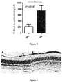
- On day 32 blood was taken from the removed eyeballs and centrifuged at 3500 r/min for 15-20 minutes, and the supernatant was used for detection for determining cardiac troponin I concentration.
- Cardiac troponin I (CTNI) is an important marker of myocardial injury, and its serum concentration can reflect the extent of myocardial injury [34].
- CTNI Cardiac troponin I
- the results showed that the cardiac troponin I concentration in the group administered with plasminogen was significantly lower than that in the control group administered with vehicle PBS, and there was an extremely significant statistical difference ( Figure 7 ). This indicated that plasminogen can extremely significantly promote the repair of myocardial injury of late diabetic mice.
- Plasminogen promotes the expression of Bcl-2, an apoptosis inhibitory factor, in the kidneys of late diabetic mice
- mice Twenty male db/db mice aged 24-25 weeks were randomly divided into two groups, ten in the control group administered with vehicle PBS and ten in the group administered with plasminogen, respectively. The day when the experiment began was recorded on Day 0, and the mice were weighed and grouped. From the second day of the experiment, plasminogen or PBS was administered to the mice for 31 consecutive days, and the day was recorded as Day 1. Mice in the group administered with plasminogen were injected with plasminogen at a dose of 2 mg/0.2 mL /mouse/day via the tail vein, and an equal volume of PBS was administered to mice in the control group administered with vehicle PBS. Mice were sacrificed on day 32, and the kidneys were fixed in 10% neutral formalin fix solution for 24 hours.
- the fixed kidney tissues were paraffin-embedded after dehydration with alcohol gradient and permeabilization with xylene.
- the thickness of the tissue sections was 5 ⁇ m.
- the sections were dewaxed and rehydrated and washed with water once.
- the sections were incubated with 3% hydrogen peroxide for 15 minutes and wash with water twice for 5 minutes each time.
- the sections were blocked with 10% normal goat serum solution (Vector laboratories, Inc., USA) for 1 hour; and after the time was up, the goat serum solution was di scarded, and the tissues were circled with a PAP pen.
- the sections were incubated with rabbit anti-mouse Bcl-2 antibody (A bcam) at 4 C overnight and washed with TBS twice for 5 minutes each time.
- the sections were incubated with a secondary antibody, goat anti-rabbit IgG (HRP) antibody (A bcam), for 1 hour at room temperature and washed with TBS twice for 5 minutes each time.
- the sections were developed with a DAB kit (Vector laboratories, Inc., USA). After washing with water three times, the sections were counterstained with hematoxylin for 30 seconds and flushed with running water for 5 minutes. After gradient dehydration, permeabilization and sealing, the sections were observed under a microscope at 200 B.
- Bcl-2 is an apoptosis inhibitory protein, and its expression will be downregulated under the action of an apoptosis stimulating factor [32,33] .
- the Bcl-2 immunohistochemical results showed that the positive expression staining of tubular epithelial cells in mice in the group administered with plasminogen ( Figure 8B ) was significantly darker than that of tubular epithelial cells in those in the control group administered with vehicle PBS ( Figure 8A ), and the former had a wider range of staining.
- the results of quantitative analysis were consistent with the observations, and there were significant differences (as shown in Figure 8C ). This indicated that plasminogen promotes the expression of Bcl-2, an apoptosis inhibitory molecule, in the kidneys of diabetic mice, and thus can inhibit the apoptosis in the kidney tissues of diabetic mice.
- mice Twenty male db/db mice aged 24-25 weeks were randomly divided into two groups, ten in the control group administered with vehicle PBS and ten in the group administered with plasminogen, respectively. The day when the experiment began was recorded on Day 0, and the mice were weighed and grouped. From the second day of the experiment, plasminogen or PBS was administered to the mice for 31 consecutive days, and the day was recorded as Day 1. Mice in the group administered with plasminogen were injected with plasminogen at a dose of 2 mg/0.2 mL /mouse/day via the tail vein, and an equal volume of PBS was administered to mice in the control group administered with vehicle PBS. Mice were sacrificed on day 32, and the kidneys were fixed in 10% neutral formalin fix solution for 24 hours.
- the fixed kidney tissues were paraffin-embedded after dehydration with alcohol gradient and permeabilization with xylene.
- the thickness of the tissue sections was 5 ⁇ m.
- the sections were dewaxed and rehydrated and washed with water once.
- the sections were incubated with 3% hydrogen peroxide for 15 minutes and wash with water twice for 5 minutes each time.
- the sections were blocked with 10% normal goat serum solution (Vector laboratories, Inc., USA) for 1 hour; and after the time was up, the goat serum solution was discarded, and the tissues were circled with a PAP pen.
- the sections were incubated with rabbit anti-mouse fibrin (fibrinogen) antibody (A bcam) overnight at 4 C and washed with TBS twice for 5 minutes each time.
- rabbit anti-mouse fibrin (fibrinogen) antibody A bcam
- the sections were incubated with a secondary antibody, goat anti-rabbit IgG (HRP) anti body (A bcam), for 1 hour at room temperature and washed with TBS twice for 5 minutes each time.
- the sections were developed with a DAB kit (Vector laboratories, Inc., USA). After washing with water three times, the sections were counterstained with hematoxylin for 30 seconds and flushed with running water for 5 minutes. After gradient dehydration, permeabilization and sealing, the sections were observed under a microscope at 200 B.
- Fibrinogen is the precursor of fibrin, and in the presence of tissue injury, as a stress response to the body's injury, fibrinogen is hydrolyzed into fibrin and deposited at the injury site [35-37] . Therefore, the fibrin level can be used as a sign of the degree of injury.
- mice Nine male db/db mice aged 25-28 weeks were randomly divided into two groups, three in the control group administered with vehicle PBS and six in the group administered with plasminogen, respectively. The day when the experiment began was recorded on Day 0, and the mice were weighed and grouped. From the second day of the experiment, plasminogen or PBS was administered to the mice for 31 consecutive days, and the day was recorded as Day 1. Mice in the group administered with plasminogen were injected with plasminogen at a dose of 2 mg/0.2 mL /mouse/day via the tail vein, and an equal volume of PBS was administered to mice in the control group administered with vehicle PBS. Whole blood was taken from the removed eyeballs 31 days after administration of plasminogen.
- alanine transaminase (ALT) in serum was detected by Reitman-Frankel colorimetry using an alanine transaminase detection kit (Nanjing Jiancheng Biological Engineering Research Institute, Catalog No. C009-2).
- a lanine transaminase is an important index of liver health status [38,39] , and the normal reference value interval of alanine transaminase is 9-50 U/L.
- the detection results showed that the ALT content in serum of mice in the control group administered with vehicle PBS was significantly higher than the normal physiological index, whereas the content in mice in the group administered with plasminogen had returned to normal levels in the body; and the content in mice in the group administered with plasminogen was significantly lower than that in mice in the control group administered with vehicle PBS, and there was a statistical difference ( Figure 10 ). This indicated that injection of plasminogen can effectively repair the liver injury in model mice with late diabetic diabetes.
- mice aged 24-25 weeks were randomly divided into two groups, five in the control group administered with vehicle PBS and five in the group administered with plasminogen, respectively. The day when the experiment began was recorded on Day 0, and the mice were weighed and grouped. From the second day of the experiment, plasminogen or PBS was administered to the mice for 31 consecutive days, and the day was recorded as Day 1. Mice in the group administered with plasminogen were injected with plasminogen at a dose of 2 mg/0.2 mL /mouse/day via the tail vein, and an equal volume of PBS was administered to mice in the control group administered with vehicle PBS.
- mice were sacrificed 31 days after administration of plasminogen, and liver ti ssues were fixed in 10% neutral formalin fix solution for 24 hours.
- the fixed liver tissues were paraffin-embedded after dehydration with alcohol gradient and permeabilization with xylene.
- the thickness of the tissue sections was 5 ⁇ m.
- the sections were dewaxed and rehydrated and washed with water once.
- the sections were incubated with 3% hydrogen peroxide for 15 minutes and wash with water twice for 5 minutes each time.
- the sections were blocked with 10% normal goat serum (Vector laboratories, Inc., USA) for 1 hour, and after the time was up, the serum was thrown away, and the ti ssues were circled with a PAP pen.
- the sections were incubated with a rabbit polyclonal antibody against F4/80 (A bcam) overnight at 4 C and washed with TBS twice for 5 minutes each time.
- the sections were incubated with a secondary antibody, goat anti-rabbit IgG (HRP) antibody (A bcam), for 1 hour at room temperature and washed with TBS twice.
- the sections were developed with a DAB kit (Vector laboratories, Inc., USA). After washing with water three times, the sections were counterstained with hematoxylin for 30 seconds and flushed with running water for 5 minutes. After gradient dehydration, permeabilization and sealing, the sections were observed under a microscope at 400 B.
- F4/80 is a macrophage marker that can indicate the extent and stage of an inflammatory response.
- the results showed that compared with mice in the control group administered with vehicle PBS ( Figure 11A ), the F4/80 positive expression was significantly reduced in those in the group administered with plasminogen ( Figure 11B ), indicating that inflammation of the liver tissues is reduced after administration of plasminogen.
- Figure 11C shows the results of quantitative analysis of F4/80 immunohistochemical positive expression, in which the expression of F4/80 in mice in the group administered with plasminogen was significantly reduced with statistical difference, indicating that plasminogen can significantly reduce the liver inflammation of diabetic mice.
- Example 12 Plasminogen promotes the repair of the ability of late diabetic mice with nerve injury to respond to mechanical allodynia
- mice Ten male db/db mice aged 24-25 weeks were randomly divided into two groups, five in the control group administered with vehicle PBS and five in the group administered with plasminogen, respectively. The day when the experiment began was recorded on Day 0, the mice were weighed and grouped, and the physiological experiment was initiated. From the second day of the experi ment, plasminogen or PBS was admi nistered to the mice for 15 consecutive days, and the day was recorded as Day 1. Mice in the group administered with plasminogen were injected with plasminogen at a dose of 2 mg/0.2 mL/mouse/day via the tail vein, and an equal volume of PBS was administered to mice in the control group administered with vehicle PBS.
- mice Ten male db/db mice aged 24-25 weeks were randomly divided into two groups, five in the control group administered with vehicle PBS and five in the group administered with plasminogen, respectively. The day when the experiment began was recorded on Day 0, the mice were weighed and grouped, and the physiological experiment was initiated. From the second day of the experi ment, plasminogen or PBS was admi nistered to the mice for 15 consecutive days, and the day was recorded as Day 1. Mice in the group administered with plasminogen were injected with plasminogen at a dose of 2 mg/0.2 mL/mouse/day via the tail vein, and an equal volume of PBS was administered to mice in the control group administered with vehicle PBS.
- mice aged 24-25 weeks were randomly divided into two groups, five in the control group administered with vehicle PBS and five in the group administered with plasminogen, respectively. The day when the experiment began was recorded on Day 0, and the mice were weighed and grouped. From the second day of the experiment, plasminogen or PBS was administered to the mice for 15 consecutive days, and the day was recorded as Day 1.
- Mice in the group administered with plasminogen were injected with plasminogen at a dose of 2 mg/0.2 mL /mouse/day via the tail vein, and an equal volume of PBS was administered to mice in the control group administered with vehicle PBS. Mice were sacrificed on day 16, and sciatic nerves were fixed in 10% neutral formalin fix solution for 24 hours.
- the fixed sciatic nerves were paraffin-embedded after dehydration with alcohol gradient and permeabilization with xylene.
- the thickness of the tissue sections was 5 ⁇ m.
- the sections were dewaxed and rehydrated and washed with water once, and then the tissues were circled with a PAP pen.
- the sections were incubated with hydrogen peroxide diluted with 3% TBS for 15 minutes, and washed with water three times.
- the sections were blocked with 10% normal goat serum (Vector laboratories, Inc., USA) for 1 hour, and excess serum was aspirated.
- the sections were incubated with rabbit anti-mouse fibrin (fibrinogen) antibody (A bcam) for 1 hour at room temperature or overnight at 4 C and washed with TBS three times.
- the sections were incubated with a secondary antibody, goat anti-rabbit IgG (HRP) antibody (A bcam), for 1 hour at room temperature and washed with TBS three times.
- the sections were developed with a DAB kit (Vector laboratories, Inc., USA). After washing with water three times, the sections were counterstained with hematoxylin for 30 seconds and flushed with running water for 5 minutes. After gradient dehydration, permeabilization and sealing, the sections were observed under a microscope at 400 B.
- Fibrinogen is the precursor of fibrin, and in the presence of tissue injury, as a stress response to the body's injury, fibrinogen is hydrolyzed into fibrin. Therefore, fibrin levels can be used as a sign of the degree of injury [35-37] . Fibrin is also the main component of thrombosis after tissue is injured. Therefore, fibrin levels can also be used as a marker of thrombi.
- mice Eight male db/db mice aged 24-25 weeks were randomly divided into two groups, four in the control group administered with vehicle PBS and four in the group administered with plasminogen, respectively. The day when the experiment began was recorded on Day 0, and the mice were weighed and grouped. From the second day of the experiment, plasminogen or PBS was administered to the mice for 31 consecutive days, and the day was recorded as Day 1. Mice in the group administered with plasminogen were injected with plasminogen at a dose of 2 mg/0.2 mL /mouse/day via the tail vein, and an equal volume of PBS was administered to mice in the control group administered with vehicle PBS.
- mice were sacrificed, and the kidneys were fixed in 10% neutral formalin fix solution for 24 hours.
- the fixed kidney tissues were paraffin-embedded after dehydration with alcohol gradient and permeabilization with xylene.
- the thickness of the tissue sections was 5 ⁇ m.
- the sections were dewaxed and rehydrated and washed with water once.
- the sections were incubated with 3% hydrogen peroxide for 15 minutes and wash with water twice for 5 minutes each time.
- the sections were incubated with goat anti-mouse IgM (HRP) antibody (A bcam) for 1 hour at room temperature and washed with TBS twice for 5 minutes each time.
- the sections were developed with a DAB kit (Vector laboratories, Inc., USA). After washing with water three times, the sections were counterstained with hematoxylin for 30 seconds and flushed with running water for 5 minutes. After gradient dehydration, permeabilization and sealing, the sections were observed under a microscope at 400 B.
- IgM antibodies play an important role during the clearance of apoptotic and necrotic cells. The more apoptotic and necrotic cells are, the higher the local IgM antibody level is [41-43] . Therefore, local IgM antibody levels can reflect the injury of tissues and organs.
Landscapes
- Health & Medical Sciences (AREA)
- Life Sciences & Earth Sciences (AREA)
- General Health & Medical Sciences (AREA)
- Public Health (AREA)
- Medicinal Chemistry (AREA)
- Pharmacology & Pharmacy (AREA)
- Chemical & Material Sciences (AREA)
- Animal Behavior & Ethology (AREA)
- Veterinary Medicine (AREA)
- Engineering & Computer Science (AREA)
- Bioinformatics & Cheminformatics (AREA)
- Diabetes (AREA)
- Epidemiology (AREA)
- Nuclear Medicine, Radiotherapy & Molecular Imaging (AREA)
- Chemical Kinetics & Catalysis (AREA)
- Organic Chemistry (AREA)
- General Chemical & Material Sciences (AREA)
- Immunology (AREA)
- Proteomics, Peptides & Aminoacids (AREA)
- Gastroenterology & Hepatology (AREA)
- Hematology (AREA)
- Obesity (AREA)
- Endocrinology (AREA)
- Emergency Medicine (AREA)
- Ophthalmology & Optometry (AREA)
- Medicines That Contain Protein Lipid Enzymes And Other Medicines (AREA)
- Enzymes And Modification Thereof (AREA)
Abstract
Description
- The present invention relates to the use of plasminogen in the inhibition of injuries of tissue cells of internal organs and the blood vessels thereof, and injuries of nerves and retinal tissue cells caused by diabetes mellitus, and also relates to the use of plasminogen in the prevention or treatment of retinopathy caused by diabetes mellitus, thereby providing a brand new prevention and/or treatment strategy for preventing and/or treating different types of retinopathy caused by diabetes mellitus.
- Diabetes mellitus is a group of endocrine and metabolic syndromes of disordered metabolisms of carbohydrates, proteins, fats, water and electrolytes in the body caused by reduced insulin secretion or defects in insulin function resulting from combined action of a variety of genetic and environmental factors. It is characterized by chronic increase in blood glucose level, and leads to chronic complications of the eyes, kidneys, liver and other organs after a long illness. Diabetes mellitus itself and its complications seriously endanger human health, and the treatment of diabetes mellitus and its complications has become a major global public health problem.
- Diabetic retinopathy is the most common diabetic eye disease, and often leads to diminution of vision or blindness. According to statistics, 50% of diabetics will have this disease in about 10 years of the disease course, and the figure will reach up to 80% in 15 years or more. The more severe the conditi on of diabetes mellitus is, the older the patient is, the higher the incidence of the disease is.
- Plasmin is a key component of the plasminogen activation system (PA system). It is a broad-spectrum protease that can hydrolyze several components of the extracellular matrix (ECM), including fibrin, gelatin, fibronectin, laminin, and proteoglycan [1]. In addition, plasmin can activate some pro-matrix metalloproteinases (pro-MMPs) to form active matrix metalloproteinases (MMPs). Therefore, plasmin is considered to be an important upstream regulator of extracellular proteolysis [2,3]. Plasmin is formed by proteolysis of plasminogen by two physiological PAs: tissue plasminogen activator (tPA) or urokinase-type plasminogen activator (uPA). Due to the relatively high level of plasminogen in plasma and other body fluids, it is traditionally believed that the regulation of the PA system is primarily achieved through the levels of PA synthesis and activity. The synthesis of PA system components is strictly regulated by different factors, such as hormones, growth factors and cytokines. In addition, there are also specific physiological inhibitors of plasmin and PAs. The main inhibitor of plasmin is 2-antiplasmin. There are uPA-specific cell surface receptors (uPA Rs) that have direct hydrolytic activity on certain cell surfaces [4,5].
- Plasminogen (plg) is a single-stranded glycoprotein with a molecular weight of about 92 kDa [6,7]. Plasminogen is mainly synthesized in the liver and is abundantly present in the extracellular fluid. The content of plasminogen in plasma is about 2 ≈M. Therefore, plasminogen is a huge potential source of proteolytic activity in tissues and body fluids [8,9]. Plasminogen exists in two molecular forms: glutamic acid-plasminogen (Glu-plasminogen) and lysine-plasminogen (Lys-plasminogen). The naturally secreted and uncleaved forms of plasminogen have an amino-terminal (N-terminal) glutamic acid and are therefore referred to as glutamic acid-plasminogen. However, in the presence of plasmin, glutamic acid-plasminogen is hydrolyzed to lysine-plasminogen at Lys76-Lys77. Compared with glutamic acid-plasminogen, lysine-plasminogen has a higher affinity for fibrin and can be activated by PAs at a higher rate. The A rg560-V al561 pepti de bond between these two forms of plasminogen can be cleaved by uPA or tPA, resulting in the formation of plasmin as a disulfide-linked double-strand protease [10]. The amino-terminal portion of plasminogen contains five homotrimeric rings, i.e., the so-called kringles, and the carboxy-terminal portion contains a protease domain. Some kringles contain lysine-binding sites that mediate the specific interaction of plasminogen with fibrin and its inhibitor 2-AP. A newly discovered 38 kDa of plasminogen is a fragment comprising kringles 1-4, is a potent inhibitor of angiogenesis. This fragment is named as angiostatin and can be produced by the proteolysis of plasminogen via several proteases.
- The main substrate of plasmin is fibrin, and the dissolution of fibrin is the key to prevent pathological thrombosis [11]. Plasmin also has substrate specificity for several components of ECM, including laminin, fibronectin, proteoglycan and gelatin, indicating that plasmin also plays an important role in ECM remodel i ng [7,12,13]. Indirectly, plasmin can also degrade other components of ECM by converting certain protease precursors into active proteases, including MMP-1, MMP-2, MMP-3 and MMP-9. Therefore, it has been proposed that plasmin may be an important upstream regulator of extracellular proteolysis [14]. In addition, plasmin has the ability to activate certain potential forms of growth factors [15-17]. In vitro, plasmin can also hydrolyze components of the complement system and release chemotactic complement fragments.
- The existing treatment method mainly includes basic treatment, namely, auxiliary treatment which is carried out for eye disorders through regular eye examination on the basis of the blood glucose control. We have surprisingly found with spontaneous diabetic mice as research objects that plasminogen and/or plasmin has a good therapeutic effect and high safety in the inhibition of tissue injuries of internal organs, blood vessel s, nerves, and the retina and in the prevention or treatment of diabetic retinopathy. Therefore, plasminogen may become a new strategy for treating diabetes mellitus complications including retinopathy.
- In one aspect, the invention relates to a method for repairing injuries of tissue cells of internal organs caused by diabetes mellitus in a subject, comprising administering plasminogen or plasmin to the subject. In one aspect, the present invention also relates to the use of plasminogen or plasmin for repairing injuries of tissue cells of internal organs caused by diabetes mellitus in a subject, comprising administering plasminogen or plasmin to the subject. In one embodiment, the internal organs include liver, heart, and kidneys. Meanwhile, the invention also relates to a method for repairing injuries of nerves and retinal tissues caused by diabetes mellitus in a subject, and the use of plasminogen or plasmin for repairing injuries of nerves and retinal tissues caused by diabetes mellitus in a subject, comprising administering plasminogen or plasmin to the subj ect.
- The present invention also relates to a method for preventing and/or treating retinopathy caused by diabetes mellitus in a subject, and the use of plasminogen or plasmin for preventing and/or treating retinopathy caused by diabetes mellitus in a subject, comprising administering plasminogen or plasmin to the subject.
- In one embodiment, the retinopathy includes retinal neovascularization, retinal inflammation, retinal atrophy, retinal cell apoptosis, retinal tissue structural injury, and retinal vascular injury caused by diabetes mellitus. In one embodiment, the plasminogen has at least 80%, 85%, 90%, 95%, 96%, 97%, 98% or 99% sequence identity with SEQ ID No. 2, 6, 8, 10 or 12, and still has the activity of plasminogen. In one embodiment, the plasminogen is a protein that comprises a plasminogen active fragment and still has plasminogen activity. In one embodiment, the plasminogen is selected from Glu-plasminogen, Lys-plasminogen, mini-plasminogen, micro-plasminogen, -plasminogen or any combination thereof. In one embodiment, the plasminogen is administered systemically or locally, including topical, intravenous, intramuscular, subcutaneous, inhalation, intraspinal or rectal administration, local injection administration, and/or local administration on cornea by a gene gun, subconjunctival injection, intracameral injection, administration via an eye drop on the cornea, injection into the anterior chamber via the temporal edge, intrastromal injection, corneal application in combination with electric pulses, intracorneal injection, subretinal injection, intravitreal injection and intraocular injection administration. In one embodiment, the plasminogen can be administered in combination with one or more other drugs, including antidiabetic drugs, drugs against cardiovascular and cerebrovascular diseases, antithrombotic drugs, anti-hypertensive drugs, antilipemic drugs, anti-infective drugs as well as other conventional drugs for preventing and/or treating concomitant diseases.
- In one embodiment, the subject is a mammal, preferably human.
- In one embodiment, the retinopathy caused by diabetes mellitus is caused by diabetes mellitus-induced microangiopathy.
- In one embodiment, the subject has a low level of plasmin or plasminogen. Specifically, the low level is innate, secondary and/or local.
- In one embodiment, the plasminogen has at least 80%, 85%, 90%, 95%, 96%, 97%, 98% or 99% sequence identity with SEQ ID No. 2, 6, 8, 10 or 12, and still has the activity of plasminogen. In one embodiment, the plasminogen is a protein that has 1-100, 1-90, 1-80, 1-70, 1-60, 1-50, 1-45, 1-40, 1-35, 1-30, 1-25, 1-20, 1-15, 1-10, 1-5, 1-4, 1-3, 1-2 or 1 amino acid added, deleted and/or substituted in SEQ ID No.2, 6, 8, 10 or 12, and still has the activity of plasminogen. In one embodiment, the plasminogen is a protein that comprises a plasminogen active fragment and still has the activity of plasminogen. In one embodiment, the plasminogen is selected from Glu-plasminogen, Lys-plasminogen, mini-plasminogen, micro-plasminogen, -plasminogen or any combination thereof. In one embodiment, the plasminogen is a conservatively substituted variant selected from variants of Glu-plasminogen, Lys-plasminogen, mini-plasminogen, -plasminogen or micro-plasminogen. In one embodiment, the plasminogen is a human natural plasminogen, such as an ortholog of plasminogen shown in SEQ ID No.2, e.g., an ortholog of plasminogen from primates or rodents, for example, an ortholog of plasminogen from gorillas, rhesus monkeys, murine, cows, horses and dogs. Most preferably, the amino acid sequence of the plasminogen of the present invention is as shown in SEQ ID No.2, 6, 8, 10 or 12.
- The above-mentioned plasminogen can be administered alone or in combination with other drugs, and can also be administered in combination with therapies other than drug prevention and/or treatment for retinopathy, such as laser treatment.
- In one embodiment, the plasminogen is administered in combination with a suitable polypeptide carrier or stabilizer. In one embodiment, the plasminogen is administered at a dosage of 0.0001-2000 mg/kg, 0.001-800 mg/kg, 0.01-600 rrg/kg, 0.1-400 mg/kg, 1-200 mg/kg, 1-100 mg/kg or 10-100 mg/kg (by per kg of body weight) or 0.0001-2000 mg/cm2, 0.001-800 mg/cm2, 0.01-600 mg/cm2, 0.1-400 mg/cm2, 1-200 mg/cm2, 1-100 mg/cm2 or 10-100 mg/cm2 (by per square centimeter of body surface area) daily, preferably the dosage is repeated at least once, preferably the dosage is administered at least daily. In the case of local administration, the above dosages may also be further adjusted depending on the circumstances.
- In one aspect, the present invention relates to the use of plasminogen or plasmin in the manufacture of a medicament, article, or kit for repairing injuries of internal organs and blood vessel tissues thereof caused by diabetes mellitus in a subject. In one embodiment, the internal organs include liver, heart, and kidneys. Meanwhile, the present invention relates to the use of plasminogen or plasmin in manufacture of a medicament, article, or kit for repairing injuries of nerves and retinal tissues caused by diabetes mellitus in a subject. The present invention also relates to the use of plasminogen or plasmin in the manufacture of a medicament, article, or kit for preventing and/or treating retinopathy caused by diabetes mellitus in a subject. In one embodiment, the retinopathy includes retinal neovascularization, retinal inflammation, retinal atrophy, retinal cell apoptosis, retinal tissue structural injury, and retinal vascular apoptosis.
- In one embodiment, the plasminogen has at least 80%, 85%, 90%, 95%, 96%, 97%, 98% or 99% sequence identity with SEQ ID No. 2, 6, 8, 10 or 12, and still has the activity of plasminogen. In one embodiment, the plasminogen is a protein that comprises a plasminogen active fragment and still has plasminogen activity. In one embodiment, the plasminogen is selected from Glu-plasminogen, Lys-plasminogen, mini-plasminogen, micro-plasminogen, -plasminogen or any combination thereof. In one embodiment, the plasminogen is administered systemically or locally, including topical, intravenous, intramuscular, subcutaneous, inhalation, intraspinal or rectal administration, local injection administration, and/or local administration on cornea by a gene gun, subconjunctival injection, intracameral injection, administration via an eye drop on the cornea, injection into the anterior chamber via the temporal edge, intrastromal injection, corneal application in combination with electric pulses, intracorneal injection, subretinal injection, intravitreal injection and intraocular injection administration. In one embodiment, the plasminogen can be administered in combination with one or more other drugs, including antidiabetic drugs, drugs against cardiovascular and cerebrovascular diseases, antithrombotic drugs, anti-hypertensive drugs, antilipemic drugs, anti-infective drugs as well as other conventional drugs for preventing and/or treating concomitant di seases.
- In one embodiment, the subject is a mammal, preferably human.
- In one embodiment, the retinopathy caused by diabetes mellitus is caused by diabetes mellitus-induced microangiopathy.
- In one embodiment, the subject has a low level of plasmin or plasminogen. Specifically, the low level is innate, secondary and/or local.
- In one embodiment, the plasminogen has at least 80%, 85%, 90%, 95%, 96%, 97%, 98% or 99% sequence identity with SEQ ID No. 2, 6, 8, 10 or 12, and still has the activity of plasminogen. In one embodiment, the plasminogen is a protein that has 1-100, 1-90, 1-80, 1-70, 1-60, 1-50, 1-45, 1-40, 1-35, 1-30, 1-25, 1-20, 1-15, 1-10, 1-5, 1-4, 1-3, 1-2 or 1 amino acid added, deleted and/or substituted in SEQ ID No.2, 6, 8, 10 or 12, and still has the activity of plasminogen. In one embodiment, the plasminogen is a protein that comprises a plasminogen active fragment and still has the activity of plasminogen. In one embodiment, the plasminogen is selected from Glu-plasminogen, Lys-plasminogen, mini-plasminogen, micro-plasminogen, -plasminogen or any combination thereof. In one embodiment, the plasminogen is a conservatively substituted variant selected from variants of Glu-plasminogen, Lys-plasminogen, mini-plasminogen, -plasminogen or micro-plasminogen. In one embodiment, the plasminogen is a human natural plasminogen, such as an ortholog of plasminogen shown in SEQ ID No.2, e.g., an ortholog of plasminogen from primates or rodents, for example, an ortholog of plasminogen from gorillas, rhesus monkeys, murine, cows, horses and dogs. Most preferably, the amino acid sequence of the plasminogen of the present invention is as shown in SEQ ID No.2, 6, 8, 10 or 12.
- The above-mentioned plasminogen can be administered alone or in combination with other drugs, and can also be administered in combination with therapies other than drug prevention and/or treatment for retinopathy, such as laser treatment.
- In one embodiment, the plasminogen is administered in combination with a suitable polypeptide carrier or stabilizer. In one embodiment, the plasminogen is administered at a dosage of 0.0001-2000 mg/kg, 0.001-800 mg/kg, 0.01-600 mg/kg, 0.1-400 mg/kg, 1-200 mg/kg, 1-100 mg/kg or 10-100 mg/kg (by per kg of body weight) or 0.0001-2000 mg/cm2, 0.001-800 mg/cm2, 0.01-600 mg/cm2, 0.1-400 mg/cm2, 1-200 mg/cm2, 1-100 mg/cm2 or 10-100 mg/cm2 (by per square centimeter of body surface area) daily, preferably the dosage is repeated at least once, preferably the dosage is administered at least daily. In the case of local administration, the above dosages may also be further adjusted depending on the circumstances.
- In another aspect, the present invention relates to plasminogen or plasmin for repairing tissue injuries of internal organs and the blood vessels thereof caused by diabetes mellitus in a subject, and a pharmaceutical composition which comprises plasminogen or plasmin and is useful in repairing tissue injuries of internal organs and the blood vessels thereof caused by diabetes mellitus in a subject, or an article or kit containing the composition. In one embodiment, the internal organs include liver, heart, and kidneys. Meanwhile, the present invention relates to plasminogen or plasmin for repairing injuries of nerves and retinal tissues caused by diabetes mellitus in a subject, and a pharmaceutical composition which comprises plasminogen or plasmin and is useful in repairing injuries of nerves and retinal tissues caused by diabetes mellitus in a subject, or an article or kit containing the composition. The present invention aslo relates to plasminogen or plasmin for preventing and/or treating retinopathy caused by diabetes mellitus in a subject, and a pharmaceutical composition which comprises plasminogen or plasmin and is useful in the prevention and/or treatment of retinopathy caused by diabetes mellitus in a subject, and an article or a kit containing the composition. In one embodiment, the retinopathy includes retinal neovascularization, retinal inflammation, retinal atrophy, retinal cell apoptosis, retinal tissue structural injury, and retinal vascular apoptosis.
- In one embodiment, the plasminogen has at least 80%, 85%, 90%, 95%, 96%, 97%, 98% or 99% sequence identity with SEQ ID No. 2, 6, 8, 10 or 12, and still has the activity of plasminogen. In one embodiment, the plasminogen is a protein that comprises a plasminogen active fragment and still has plasminogen activity. In one embodiment, the plasminogen is selected from Glu-plasminogen, Lys-plasminogen, mini-plasminogen, micro-plasminogen, -plasminogen or any combination thereof. In one embodiment, the plasminogen is administered systemically or locally, including topical, intravenous, intramuscular, subcutaneous, inhalation, intraspinal or rectal administration, local injection administration, and/or local administration on cornea by a gene gun, subconjunctival injection, intracameral injection, administration via an eye drop on the cornea, injection into the anterior chamber via the temporal edge, intrastromal injection, corneal application in combination with electric pulses, intracorneal injection, subretinal injection, intravitreal injection and intraocular injection administration. In one embodiment, the plasminogen can be administered in combination with one or more other drugs, including antidiabetic drugs, drugs against cardiovascular and cerebrovascular diseases, antithrombotic drugs, anti-hypertensive drugs, antilipemic drugs, anti-infective drugs as well as other conventional drugs for preventing and/or treating concomitant diseases.
- In one embodiment, the subject is a mammal, preferably human.
- In one embodiment, the retinopathy caused by diabetes mellitus is caused by diabetes mellitus-induced microangiopathy.
- In one embodiment, the subject has a low level of plasmin or plasminogen. Specifically, the low level is innate, secondary and/or local.
- In one embodiment, the plasminogen has at least 80%, 85%, 90%, 95%, 96%, 97%, 98% or 99% sequence identity with SEQ ID No. 2, 6, 8, 10 or 12, and still has the activity of plasminogen. In one embodiment, the plasminogen is a protein that has 1-100, 1-90, 1-80, 1-70, 1-60, 1-50, 1-45, 1-40, 1-35, 1-30, 1-25, 1-20, 1-15, 1-10, 1-5, 1-4, 1-3, 1-2 or 1 amino acid added, deleted and/or substituted in SEQ ID No.2, 6, 8, 10 or 12, and still has the activity of plasminogen. In one embodiment, the plasminogen is a protein that comprises a plasminogen active fragment and still has the activity of plasminogen. In one embodiment, the plasminogen is selected from Glu-plasminogen, Lys-plasminogen, mini-plasminogen, micro-plasminogen, -plasminogen or any combination thereof. In one embodiment, the plasminogen is a conservatively substituted variant selected from variants of Glu-plasminogen, Lys-plasminogen, mini-plasminogen, -plasminogen or micro-plasminogen. In one embodiment, the plasminogen is a human natural plasminogen, such as an ortholog of plasminogen shown in SEQ ID No.2, e.g., an ortholog of plasminogen from primates or rodents, for example, an ortholog of plasminogen from gorillas, rhesus monkeys, murine, cows, horses and dogs. Most preferably, the amino acid sequence of the plasminogen of the present invention is as shown in SEQ ID No.2, 6, 8, 10 or 12.
- The above-mentioned plasminogen can be administered alone or in combination with other drugs, and can also be administered in combination with therapies other than drug prevention and/or treatment for retinopathy, such as laser treatment.
- In one embodiment, the plasminogen is administered in combination with a suitable polypeptide carrier or stabilizer. In one embodiment, the plasminogen is administered at a dosage of 0.0001-2000 mg/kg, 0.001-800 mg/kg, 0.01-600 mg/kg, 0.1-400 mg/kg, 1-200 mg/kg, 1-100 mg/kg or 10-100 mg/kg (by per kg of body weight) or 0.0001-2000 mg/cm2, 0.001-800 mg/cm2, 0.01-600 mg/cm2, 0.1-400 mg/cm2, 1-200 mg/cm2, 1-100 mg/cm2 or 10-100 mg/cm2 (by per square centimeter of body surface area) daily, preferably the dosage is repeated at least once, preferably the dosage is administered at least daily. In the case of local administration, the above dosages may also be further adjusted depending on the circumstances.
- In one embodiment, the article or kit comprises a container containing an effective dosage of plasminogen/plasmin. Preferably, the article or kit further comprises a container containing one or more other drugs. The article or kit may also include instructions for use, which indicate that the plasminogen or plasmin can be used to prevent and/or treat injuries of internal organs and blood vessel tissues thereof, injuries of nerves and retinal tissues, or the retinal disorder caused by diabetes mellitus, and it may further be stated that the plasminogen or plasmin may be administered before, simultaneously with, and/or after the administration of other drugs or therapies.
- In one embodiment, the plasminogen is administered in combination with a suitable polypeptide carrier or stabilizer. In one embodiment, the plasminogen is administered at a dosage of 0.0001-2000 mg/kg, 0.001-800 mg/kg, 0.01-600 rrg/kg, 0.1-400 mg/kg, 1-200 mg/kg, 1-100 mg/kg or 10-100 mg/kg (by per kg of body weight) or 0.0001-2000 mg/cm2, 0.001-800 mg/cm2, 0.01-600 mg/cm2, 0.1-400 mg/cm2, 1-200 mg/cm2, 1-100 mg/cm2 or 10-100 mg/cm2 (by per square centimeter of body surface area) daily, preferably the dosage is repeated at least once, preferably the dosage is administered at least daily. In the case of local administration, the above dosages may also be further adjusted depending on the circumstances.
- In one aspect, the present invention relates to the use of plasminogen in preparing a medicament, article, or kit for preventing and/or treating diabetic ocular microangiopathy in a subject. The present invention also relates to a new method for preventing and/or treating diabetic ocular microangiopathy in a subject, or the use of plasminogen or plasmin for preventing and/or treating diabetic ocular microangiopathy in a subject, comprising administering plasminogen or plasmin to the subject. In another aspect, the present invention relates to plasminogen or plasmin for preventing and/or treating diabetic ocular microangiopathy in a subject, or a pharmaceutical composition which comprises plasminogen or plasmin and is useful in the prevention and/or treatment of diabetic ocular microangiopathy in a subject, or an article or kit which comprises plasminogen or plasmin and is useful in the prevention and/or treatment of diabetic ocular microangiopathy in a subject.
- In one embodiment, the plasminogen has at least 80%, 85%, 90%, 95%, 96%, 97%, 98% or 99% sequence identity with SEQ ID No. 2, 6, 8, 10 or 12, and still has the activity of plasminogen. In one embodiment, the plasminogen is a protein that has 1-100, 1-90, 1-80, 1-70, 1-60, 1-50, 1-45, 1-40, 1-35, 1-30, 1-25, 1-20, 1-15, 1-10, 1-5, 1-4, 1-3, 1-2 or 1 amino acid added, deleted and/or substituted in SEQ ID No.2, 6, 8, 10 or 12, and still has the activity of plasminogen. In one embodiment, the plasminogen is a protein that comprises a plasminogen active fragment and still has the activity of plasminogen. In one embodiment, the plasminogen is selected from Glu-plasminogen, Lys-plasminogen, mini-plasminogen, micro-plasminogen, -plasminogen or any combination thereof. In one embodiment, the plasminogen is a conservatively substituted variant selected from variants of Glu-plasminogen, Lys-plasminogen, mini-plasminogen, -plasminogen or micro-plasminogen. In one embodiment, the plasminogen is a human natural plasminogen, such as an ortholog of plasminogen shown in SEQ ID No.2, e.g., an ortholog of plasminogen from primates or rodents, for example, an ortholog of plasminogen from gorillas, rhesus monkeys, murine, cows, horses and dogs. Most preferably, the amino acid sequence of the plasminogen of the present invention is as shown in SEQ ID No.2, 6, 8, 10 or 12.
- The above-mentioned prevention and treatment of diabetic ocular microangiopathy include inhibition of the formation of retinal cell-free capillaries, improvement of retinal atrophy, repair of retinal inflammation, and/or inhibition of apoptosis of retinal cells and vascular cells.
- In one embodiment, the plasminogen is administered in combination with a suitable polypeptide carrier or stabilizer. In one embodiment, the plasminogen is administered at a dosage of 0.0001-2000 mg/kg, 0.001-800 mg/kg, 0.01-600 rrg/kg, 0.1-400 mg/kg, 1-200 mg/kg, 1-100 mg/kg or 10-100 mg/kg (by per kg of body weight) or 0.0001-2000 mg/cm2, 0.001-800 mg/cm2, 0.01-600 mg/cm2, 0.1-400 mg/cm2, 1-200 mg/cm2, 1-100 mg/cm2 or 10-100 mg/cm2 (by per square centimeter of body surface area) daily, preferably the dosage is repeated at least once, preferably the dosage is administered at least daily. In the case of local administration, the above dosages may also be further adjusted depending on the circumstances.
- In one aspect, the present invention relates to the use of plasminogen or plasmin in the manufacture of a medicament, article or kit for preventing and/or treating injury (damage) to body tissues and internal organs caused by diabetes mellitus in a subject. In one embodiment, the injury (damage) to tissues and internal organs includes injury (damage) to the brain, heart, liver, lungs, kidneys, nerves, retina, skin and gastrointestinal tract. In one aspect, the present invention relates to the use of plasminogen in the manufacture of a medicament, article or kit for preventing and/or treating a diabetic complication in a subject. In one embodiment, the diabetic complication is diabetic encephalopathy, diabetic cardiopathy, diabetic hepatopathy, diabetic nephropathy, diabetic pneumonopathy, diabetic neuropathy, diabetic retinopathy or diabetic dermopathy induced by diabetes mellitus.
- In one aspect, the present invention relates to a method for manufacturing a medicament, comprising preparing a medicament, article or kit for preventing and/or treating injury (damage) to body tissues and internal organs caused by diabetes mellitus in a subject using plasminogen or plasmin and a pharmaceutically acceptable carrier. In one embodiment, the injury (damage) to tissues and internal organs includes injury (damage) to the brain, heart, liver, lungs, kidneys, nerves, retina, skin and gastrointestinal tract. In one aspect, the present invention relates to a method for manufacturing a medicament, comprising preparing a medicament, article or kit for preventing and/or treating a diabetic complication in a subject using plasminogen or plasmin and a pharmaceutically acceptable carrier. In one embodiment, the diabetic complication is diabetic encephalopathy, diabetic cardiopathy, diabetic hepatopathy, diabetic nephropathy, diabetic pneumonopathy, diabetic neuropathy, diabetic retinopathy or diabetic dermopathy induced by diabetes mellitus.
- In one aspect, the present invention relates to plasminogen or plasmin, and a pharmaceutical composition, article or kit comprising the plasminogen or plasmin, which are useful in the prevention and/or treatment of injury (damage) to body tissues and internal organs caused by diabetes mellitus in a subject. In one embodiment, the injury (damage) to tissues and internal organs includes injury (damage) to the brain, heart, liver, kidneys, lungs, nerves, retina, gastrointestinal tract and skin. In one aspect, the present invention relates to plasminogen, and a pharmaceutical composition, article or kit comprising the plasminogen, which are useful in the prevention and/or treatment of a diabetic complication in a subject. In one embodiment, the diabetic complication is diabetic encephalopathy, diabetic cardiopathy, diabetic hepatopathy, diabetic pneumonopathy, diabetic nephropathy, diabetic neuropathy, diabetic retinopathy or diabetic dermopathy induced by diabetes mellitus.
- In one aspect, the present invention relates to a method for preventing and/or treating injury (damage) to body tissues and internal organs caused by diabetes mellitus in a subject, comprising administering plasminogen or plasmin or a pharmaceutical composition, article or kit comprising the plasminogen or plasmin to the subject. The present invention also relates to the use of plasminogen or plasmin, or a pharmaceutical composition, article or kit comprising the plasminogen or plasmin for preventing and/or treating injury (damage) to body tissues and internal organs caused by diabetes mellitus in a subject. In one embodiment, the injury (damage) to tissues and internal organs includes injury (damage) to the brain, heart, liver, lungs, kidneys, nerves, retina, gastrointestinal tract and skin. In one aspect, the present invention relates to a method for preventing and/or treating a diabetic complication in a subject, comprising administering plasminogen or plasmin, or a pharmaceutical composition, article or kit comprising the plasminogen or plasmin to the subject. The present invention also includes the use of plasminogen or plasmin, or a pharmaceutical composition, article or kit comprising the plasminogen or plasmin for preventing and/or treating a diabetic complication in a subject. In one embodiment, the diabetic complication is diabetic encephalopathy, diabetic cardiopathy, diabetic hepatopathy, diabetic pneumonopathy, diabetic nephropathy, diabetic neuropathy, diabetic retinopathy or diabetic dermopathy induced by diabetes mellitus.
- In one embodiment, the subject has a low level of plasmin or plasminogen. Specifically, the low level is innate, secondary and/or local.
- The present invention explicitly encompasses all the combinations of technical features belonging to the embodiments of the present invention, and these combined technical solutions have been explicitly disclosed in the present application, as if the above technical solutions were individually and explicitly disclosed. In addition, the present invention also explicitly encompasses all the subcombinations of the various embodiments and elements thereof, and these subcombinations have been disclosed herein, as if each of such subcombinations was individually and explicitly disclosed herein.
- 'Diabetes mellitus_ is a series of dysmetabolic syndromes of carbohydrates, proteins, fats, water, electrolytes and the like that are caused by islet hypofunction, insulin resistance and the like resulting from the effects of genetic factors, immune dysfunction, microbial infections and toxins thereof, free radical toxins, mental factors and other various pathogenic factors on the body, and is mainly characterized by hyperglycemia clinically.
- 'Diabetic complications_ are damages to or dysfunctions of other organs or tissues of the body caused by poor blood glucose control during diabetes mellitus, including damages to or dysfunctions of the liver, kidneys, heart, retina, nervous system damage and the like. According to statistics of the World Health Organization, there are up to more than 100 diabetic complications, and diabetes mellitus is a disease currently known to have the most complications.
- 'Diabetic microangiopathy_ refers to microangiopathy caused by varying degrees of abnormalities in the microcirculation of various body organs or tissues of diabetics. The process of microangiopathy formation roughly comprises functional changes in microcirculation, endothelial injury, thickening of the basement membrane, increased blood viscosity, aggregation of red blood cells, and adhesion and aggregation of platelets, eventually leading to microthrombosis and/or microvascular occlusion. 'Diabetic ocular microangiopathy_ refers to ocular microangiopathy caused by diabetes mellitus.
- The above-mentioned 'diabetic microangiopathy_ causes local vascular injury in tissues or organs, poor blood flow, hypoxia of cells, and formation of blood clots, thrombi, and inflammation, and further affects peri pheral tissues and organ functions, thereby leading to 'diabetic complications_; therefore, the terms 'diabetic microangiopathy_ and 'diabetic complications_ mentioned in the technical solutions of the claims of the present invention all cover thrombosis caused by diabetes mellitus.
- 'Diabetic retinopathy_ refers to the diabetes mellitus-induced histological and functional changes of the retina being mainly caused by diabetic microangiopathy. Diabetic retinopathy is the most common diabetic eye disease, and often leads to diminution of vision or bl i ndness. According to statistics, 50% of diabetics will have this di sease in about 10 years of the disease course, and the figure will reach up to 80% in 15 years or more. The more severe the conditi on of diabetes mellitus is, the older the patient is, the higher the incidence of the disease is.
- 'Plasmin_ is a very important enzyme that exists in the blood and can hydrolyze fibrin clots into fibrin degradation products and D-dimers.
- 'Plasminogen_ is the zymogenic form of plasmin, and based on the sequence in the swiss prot and calculated from the amino acid sequence (SE Q ID No.4) of the natural human-derived plasminogen containing a signal peptide, is a glycoprotein composed of 810 amino acids, which has a molecular weight of about 92 kD and is synthesized mainly in the liver and capable of circulating in the blood; and the cDNA sequence encoding this amino acid sequence is as shown in SEQ ID No.3. Full-length plasminogen contains seven domains: a C-terminal serine protease domain, an N-terminal Pan Apple (PAp) domain and five Kringle domains (Kringles 1-5). Referring to the sequence in the swiss prot, the signal peptide comprises residues Met1-Gly19, PAp comprises residues Glu20-V al98, Kringle 1 comprises residues Cys103-Cys181, Kringle 2 comprises residues Glu184-Cys262, K ringle 3 comprises residues Cys275-Cys352,
K ringle 4 comprises residues Cys377-Cys454, and K ringle 5 comprises residues Cys481-Cys560. According to the NCBI data, the serine protease domain comprises residues Val581-Arg804. - Glu-plasminogen is a natural full-length plasminogen and is composed of 791 amino acids (without a signal peptide of 19 amino acids); the cDNA sequence encoding this sequence is as shown in SEQ ID No.1; and the amino acid sequence is as shown in SEQ ID No.2. In vivo, Lys-plasminogen, which is formed by hydrolysis of amino acids at positions 76-77 of Glu-plasminogen, is also present, as shown in SEQ ID No.6; and the cDNA sequence encoding this amino acid sequence is as shown in SEQ ID No.5. -plasminogen is a fragment of full-length plasminogen that lacks the structure of K ringle 2-K ringle 5 and contains only K ringle 1 and the serine protease domain [18,19]. The amino acid sequence (SEQ ID No.8) of -plasminogen has been reported in the literature[19], and the cDNA sequence encoding this amino acid sequence is as shown in SEQ ID No.7. Mini-plasminogen is composed of K ringle 5 and the serine protease domain, and has been reported in the literature to comprise residues Val443-Asn791 (with the Glu residue of the Glu-plasminogen sequence that does not contain a signal peptide as the starting amino acid) [20]; the amino acid sequence is as shown in SEQ ID No.10; and the cDNA sequence encoding this amino acid sequence is as shown in SEQ ID No.9. Micro-plasminogen comprises only the serine protease domain, the amino acid sequence of which has been reported in the literature to comprise residues Ala543-Asn791 (with the Glu residue of the Glu-plasminogen sequence that does not contain a signal peptide as the starting amino acid) [21], and the sequence of which has been also reported in patent document
CN 102154253 A to comprise residues Lys531-Asn791 (with the Glu residue of the Glu-plasminogen sequence that does not contain a signal peptide as the starting amino acid) (the sequence in this patent application refers to the patent documentCN 102154253 A ); the amino acid sequence is as shown in SEQ ID No.12; and the cDNA sequence encoding this amino acid sequence is as shown in SEQ ID No.11. - In the present invention, 'plasmin_ is used interchangeably with 'fibrinolysin_ and 'fibrinoclase_, and the terms have the same meaning; and plasminogen_ is used interchangeably with 'fibrinolytic zymogen_ and 'fibrinoclase zymogen_, and the terms have the same meaning.
- In the course of circulation, plasminogen is in a closed, inactive conformation, but when bound to thrombi or cell surfaces, it is converted into an active plasmin in an open conformation under the mediation of a plasminogen activator (PA). The active plasmin can further hydrolyze the fibrin clots to fibrin degradation products and D-dimers, thereby dissolving the thrombi. The PAp domain of plasminogen comprises an important determinant that maintains plasminogen in an inactive, closed conformation, and the K R domain is capable of binding to lysine residues present on receptors and substrates. A variety of enzymes that can serve as plasminogen activators are known, including: tissue plasminogen activator (tPA), urokinase plasminogen activator (uPA), kallikrein, coagulation factor XII (Hagmann factor), and the like.
- 'Plasminogen active fragment_ refers to an active fragment in the plasminogen protein that is capable of binding to a target sequence in a substrate and exerting the proteolytic function. The technical solutions of the present invention involving plasminogen encompass technical solutions in which plasminogen is replaced with a plasminogen active fragment. The plasminogen active fragment of the present invention is a protein comprising a serine protease domain of plasminogen. Preferably, the plasminogen active fragment of the present invention comprises SEQ ID NO: 14, or an amino acid sequence having an amino acid sequence identity of at least 80%, 90%, 95%, 96%, 97%, 98% or 99% with SEQ ID NO: 14. Therefore, plasminogen of the present invention comprises a protein comprising the plasminogen active fragment and still having plasminogen activity.
- At present, methods for determining plasminogen and its activity in blood include: detection of tissue plasminogen activator activity (t-PAA), detection of tissue plasminogen activator antigen (t-PAAg) in plasma, detection of tissue plasminogen activity (plgA) in plasma, detection of tissue plasminogen antigen (plgAg) in plasma, detection of activity of the inhibitor of tissue plasminogen activators in plasma, detection of inhibitor antigens of tissue plasminogen activators in plasma and detection of plasmin-anti-plasmin (PAP) complex in plasma. The most commonly used detection method is the chromogenic substrate method: streptokinase (SK) and a chromogenic substrate are added to a test plasma, the PLG in the test plasma is converted into PLM by the action of SK, PLM acts on the chromogenic substrate, and then it is determined that the increase in absorbance is directly proportional to plasminogen activity using a spectrophotometer. In addition, plasminogen activity in blood can also be determined by immunochemistry, gel electrophoresis, immunonephelometry, radioimmuno-diffusion and the like.
- 'Orthologues or orthologs_ refer to homologs between different species, including both protein homologs and DNA homologs, and are also known as orthologous homologs and vertical homologs. The term specifically refers to proteins or genes that have evolved from the same ancestral gene in different species. The plasminogen of the present invention includes human natural plasminogen, and also includes orthologues or orthologs of plasminogen derived from different species and having plasminogen activity.
- 'Conservatively substituted variant_ refers to one in which a given amino acid residue is changed without altering the overall conformation and function of the protein or enzyme, including, but not limited to, replacing an amino acid in the amino acid sequence of the parent protein by an amino acid with similar properties (such as acidity, alkalinity and hydrophobicity). Amino acids with similar properties are well known. For example, arginine, histidine and lysine are hydrophilic basic amino acids and are interchangeable. Similarly, isoleucine is a hydrophobic amino acid that can be replaced by leucine, methionine or valine. Therefore, the similarity of two proteins or amino acid sequences with similar functions may be different. For example, the similarity (identity) is 70%-99% based on the MEGALIGN algorithm. 'Conservatively substituted variant_ also includes a polypeptide or enzyme having amino acid identity of 60% or more, preferably 75% or more, more preferably 85% or more, even more preferably 90% or more as determined by the BLAST or FASTA algorithm, and having the same or substantially similar properties or functions as the natural or parent protein or enzyme.
- 'Isolated_ plasminogen refers to the plasminogen protein that is isolated and/or recovered from its natural environment. In some embodiments, the plasminogen will be purified (1) to a purity of greater than 90%, greater than 95% or greater than 98% (by weight), as determined by the Lowry method, such as more than 99% (by weight); (2) to a degree sufficiently to obtain at least 15 residues of the N-terminal or internal amino acid sequence using a spinning cup sequenator; or (3) to homogeneity, which is determined by sodium dodecyl sulfate-polyacrylamide gel electrophoresis (SDS-PAGE) under reducing or nonreducing conditions using Coomassie blue or silver staining. Isolated plasminogen also includes plasminogen prepared from recombinant cells by bioengineering techniques and separated by at least one purification step.
- The terms 'polypeptide_, 'peptide_ and 'protein_ are used interchangeably herein and refer to polymeric forms of amino acids of any length, which may include genetically encoded and non-genetically encoded amino acids, chemically or biochemically modified or derivatized amino acids, and polypeptides having modified peptide backbones. The term includes fusion proteins, including, but not limited to, fusion proteins having heterologous amino acid sequences, fusions having heterologous and homologous leader sequences (with or without N-terminal methionine residues), and the like.
- The 'percent amino acid sequence identity (%)_ with respect to the reference polypeptide sequence is defined as the percentage of amino acid residues in the candidate sequence identical to the amino acid residues in the reference polypeptide sequence when a gap is introduced as necessary to achieve maximal percent sequence identity and no conservative substitutions are considered as part of sequence identity. The comparison for purposes of determining percent amino acid sequence identity can be achieved in a variety of ways within the skill in the art, for example using publicly available computer softwares, such as BLAST, BLAST-2, ALIGN or Megalign (DNASTAR) software. Those skilled in the art can determine appropriate parameters for aligning sequences, including any algorithm needed to achieve the maximum comparison over the full length of the sequences being compared. However, for purposes of the present invention, the percent amino acid sequence identity value is generated using the sequence comparison computer program ALIGN-2.
- In the case of comparing amino acid sequences using ALIGN-2, the % amino acid sequence identity of a given amino acid sequence A relative to a given amino acid sequence B (or may be expressed as a given amino acid sequence A having or containing a certain % amino acid sequence identity relative to, with or for a given amino acid sequence B) is calculated as follows:

- As used herein, the terms 'treatment_ and 'treating_ refer to obtaining a desired pharmacological and/or physiologic effect. The effect may be complete or partial prevention of a disease or its symptoms and/or partial or complete cure of the disease and/or its symptoms, and includes: (a) prevention of the disease from developing in a subject that may have a predisposition to the disease but has not been diagnosed as having the disease; (b) suppression of the disease, i.e., blocking its formation; and (c) alleviation of the disease and/or its symptoms, i.e., eliminating the disease and/or its symptoms.
- The terms 'individual_, 'subject_ and 'patient_ are used interchangeably herein and refer to mammals, including, but not limited to, murine (rats and mice), non-human primates, humans, dogs, cats, hoofed animals (e.g., horses, cattle, sheep, pigs, goats) and so on.
- 'Therapeutically effective amount_ or 'effective amount_ refers to an amount of plasminogen sufficient to achieve the prevention and/or treatment of a disease when administered to a mammal or another subject to treat the disease. The 'therapeutically effective amount_ will vary depending on the plasminogen used, the severity of the disease and/or its symptoms, as well as the age, body weight of the subject to be treated, and the like.
- Plasminogen can be isolated and purified from nature for further therapeutic uses, and can also be synthesized by standard chemical peptide synthesis techniques. When chemically synthesized, a polypeptide can be subjected to liquid or solid phase synthesis. Solid phase polypeptide synthesis (SPPS) is a method suitable for chemical synthesis of plasminogen, in which the C-terminal amino acid of a sequence is attached to an insoluble support, followed by the sequential addition of the remaining amino acids in the sequence. Various forms of SPPS, such as Fmoc and Boc, can be used to synthesize plasminogen. Techniques for solid phase synthesis are described in Barany and Solid-Phase Peptide Synthesis; pp. 3-284 in The Peptides: Analysis, Synthesis, Biology. Vol. 2: Special Methods in Peptide Synthesis, Part A., Merrifield et al. J. Am. Chem. Soc., 85: 2149-2156 (1963); Stewart et al. Solid Phase Peptide Synthesis, 2nd ed. Pierce Chem Co., Rockford, III. (1984); and Ganesan A. 2006 Mini Rev. Med Chem 6:3-10 and Camarero JA et al. 2005 Protein Pept Lett. 12:723-8. Briefly, small insoluble porous beads are treated with a functional unit on which a peptide chain is constructed. After repeated cycles of coupling/deprotection, the attached solid phase free N-terminal amine is coupled to a single N-protected amino acid unit. This unit is then deprotected to expose a new N-terminal amine that can be attached to another amino acid. The peptide remains immobilized on the solid phase before it is cut off.
- Standard recombinant methods can be used to produce the plasminogen of the present invention. For example, a nucleic acid encoding plasminogen is inserted into an expression vector, so that it is operably linked to a regulatory sequence in the expression vector. Expression regulatory sequence includes, but is not limited to, promoters (e.g., naturally associated or heterologous promoters), signal sequences, enhancer elements and transcription termination sequences. Expression regulation can be a eukaryotic promoter system in a vector that is capable of transforming or transfecting eukaryotic host cells (e.g., COS or CHO cells). Once the vector is incorporated into a suitable host, the host is maintained under conditions suitable for high-level expression of the nucleotide sequence and collection and purification of plasminogen.
- A suitable expression vector is usually replicated in a host organism as an episome or as an integral part of the host chromosomal DNA. In general, an expression vector contains a selective marker (e.g., ampicillin resistance, hygromycin resistance, tetracycline resistance, kanamycin resistance or neomycin resistance) to facilitate detection of those exogenous cells transformed with a desired DNA sequence.
- Escherichia coli is an example of prokaryotic host cells that can be used to clone a polynucleotide encoding the subject antibody. Other microbial hosts suitable for use include Bacillus, for example, Bacillus subtilis and other species of Enterobacteriaceae (such as Salmonella spp. and Serratia spp.), and various Pseudomonas spp. In these prokaryotic hosts, expression vectors can also be generated which will typically contain an expression control sequence (e.g., origin of replication) that is compatible with the host cell. In addition, there will be many well-known promoters, such as the lactose promoter system, the tryptophan (trp) promoter system, the beta-lactamase promoter system or the promoter system from phage lambda. Optionally in the case of manipulation of a gene sequence, a promoter will usually control expression, and has a ribosome binding site sequence and the like to initiate and complete transcription and translation.
- Other microorganisms, such as yeast, can also be used for expression. Saccharomyces (e.g., S. cerevisiae) and Pichia are examples of suitable yeast host cells, in which a suitable vector has an expression control sequence (e.g., promoter), an origin of replication, a termination sequence and the like, as required. A typical promoter comprises 3-phosphoglycerate kinase and other glycolytic enzymes. Inducible yeast promoters specifically include promoters derived from alcohol dehydrogenase, isocytochrome C, and enzymes responsible for maltose and galactose utilization.
- In addition to microorganisms, mammalian cells (eg, mammalian cells cultured in in vitro cell culture) can also be used to express and produce the protein of the invention (eg, polynucleotides encoding the subject protein). See Winnacker, From Genes to Clones, VCH Publishers, N.Y., N.Y. (1987). Suitable mammalian host cells include CHO cell lines, various Cos cell lines, HeLa cells, myeloma cell lines and transformed B cells or hybridomas. Expression vectors for these cells may comprise an expression control sequence, such as an origin of replication, promoter and enhancer (Queen et al. Immunol. Rev. 89:49 (1986)), as well as necessary processing information sites, such as a ribosome binding site, RNA splice site, polyadenylation site and transcription terminator sequence. Examples of suitable expression control sequences are promoters derived from white immunoglobulin gene,
SV 40, adenovirus, bovine papilloma virus, cytomegalovirus and the like. See Co et al.J . Immunol. 148:1149 (1992). - Once synthesized (chemically or recombinantly), the plasminogen of the present invention can be purified according to standard procedures in the art, including ammonium sulfate precipitation, affinity column, column chromatography, high performance liquid chromatography (HPLC), gel electrophoresis and the like. The plasminogen is substantially pure, e.g., at least about 80% to 85% pure, at least about 85% to 90% pure, at least about 90% to 95% pure, or 98% to 99% pure or purer, for example free of contaminants such as cell debris, macromolecules other than the plasminogen and the like.
- A therapeutic formulation can be prepared by mixing plasminogen of a desired purity with an optional pharmaceutical carrier, excipient or stabilizer (Remington's Pharmaceutical Sciences, 16th edition, Osol, A. ed. (1980)) to form a lyophilized preparation or an aqueous solution. Acceptable carriers, excipients and stabilizers are non-toxic to the recipient at the dosages and concentrations employed, and include buffers, such as phosphates, citrates and other organic acids; antioxidants, including ascorbic acid and methionine; preservatives (e.g., octadecyl dimethyl benzyl ammonium chloride; hexane chloride diamine; benzalkonium chloride and benzethonium chloride; phenol, butanol or benzyl alcohol; alkyl p-hydroxybenzoates, such as methyl or propyl p-hydroxybenzoate; catechol; resorcinol; cyclohexanol; 3-pentanol; and m-cresol); low molecular weight polypeptides (less than about 10 residues); proteins, such as serum albumin, gelatin or immunoglobulins; hydrophilic polymers, such as polyvinylpyrrolidone; amino acids, such as glycine, glutamine, asparagine, histidine, arginine or lysine; monosaccharides, disaccharides and other carbohydrates, including glucose, mannose or dextri ns; chelating agents, such as EDTA; sugars, such as sucrose, mannitol, fucose or sorbitol; salt-forming counterions, such as sodium; metal complexes (e.g., zinc-protein complexes); and/or non-ionic surfactants, such as TWEENTM, PLURONICSTM or polyethylene glycol (PEG).
- The formulations of the invention may also comprise one or more active compounds requi red for the particular disorder to be treated, preferably those that are complementary in activity and have no side effects with one another, for example anti-hypertensive drugs, anti-arrhythmic drugs, drugs for treating diabetes mellitus, and the like.
- The plasminogen of the present invention may be encapsulated in microcapsules prepared by techniques such as coacervation or interfacial polymerization, for example, it may be incorporated in a colloid drug delivery system (e.g., liposomes, albumin microspheres, microemulsions, nanoparticles and nanocapsules), or incorporated in hydroxymethylcellulose or gel-microcapsules and poly-(methyl methacrylate) microcapsules in macroemulsions. These techniques are disclosed in Remington's Pharmaceutical Sciences, 16th edition, Osol, A. Ed. (1980).
- The plasminogen of the present invention for in vivo administration must be sterile. This can be easily achieved by filtration through a sterile filtration membrane before or after freeze drying and reconstitution.
- The plasminogen of the present invention can be prepared into a sustained-release preparation. Suitable examples of sustained-release preparations include solid hydrophobic polymer semi-permeable matrices having a shape and containing glycoproteins, such as films or microcapsules. Examples of sustained-release matrices include polyesters, hydrogels (e.g., poly(2-hydroxyethylmethacrylate)) (Langer et al. J. Biomed. Mater. Res., 15: 167-277 (1981); and Langer, Chem. Tech., 12:98-105 (1982)), or poly(vinyl alcohol), polylactides (
US Patent 3773919 , andEP 58,481 - The pharmaceutical composition of the present invention can be administered in different ways, for example by intravenous, intraperitoneal, subcutaneous, intracranial, intrathecal, intraarterial (e.g., via carotid), intramuscular, intranasal, intraocular, topical or intradermal administration or spinal cord or brain delivery. An aerosol preparation, such as a nasal spray preparation, comprises purified aqueous or other solutions of the active agent along with a preservative and isotonic agent. For intraocular administration, such preparations need to be adjusted to a pH and isotonic state compatible with the ocular conjunctiva.
- Preparations for parenteral administration include sterile aqueous or non-aqueous solutions, suspensions and emulsions. Examples of non-aqueous solvents are propylene glycol, polyethylene glycol, vegetable oils such as olive oil, and injectable organic esters such as ethyl oleate. Aqueous carriers include water, and alcoholic/aqueous solutions, emulsions or suspensions, including saline and buffered media. Parenteral vehicles include sodium chloride solution, Ringer's dextrose, dextrose and sodium chloride, or fixed oils. Intravenous vehicles include liquid and nutrient supplements, electrolyte supplements and the like. Preservatives and other additives may also be present, for example, such as antimicrobial agents, antioxidants, chelating agents and inert gases.
- The medical staff will determine the dosage regimen based on various clinical factors. As is well known in the medical field, the dosage of any patient depends on a variety of factors, including the patient's size, body surface area, age, the specific compound to be administered, sex, frequency and route of administration, overall health and other drugs administered simultaneously. The dosage range of the pharmaceutical composition comprising plasminogen of the present invention may be, for example, about 0.0001 to 2000 mg/kg, or about 0.001 to 500 mg/kg (such as 0.02 mg/kg, 0.25 mg/kg, 0.5 mg/kg, 0.75 mg/kg, 10 mg/kg and 50 mg/kg) of the subject's body weight daily. For example, the dosage may be 1 mg/kg body weight or 50 mg/kg body weight, or in the range of 1 rrg/kg-50 mg/kg, or at least 1 mg/kg. Dosages above or below this exemplary range are also contemplated, especially considering the above factors. The intermediate dosages in the above range are also included in the scope of the present invention. A subject may be administered with such dosages daily, every other day, weekly or based on any other schedule determined by empirical analysis. An exemplary dosage schedule includes 1-10 mg/kg for consecutive days. During administration of the drug of the present invention, the therapeutic effect and safety of diabetic retinopathy and its related disorders are required to be assessed real-timely and regularly.
- One embodiment of the present invention relates to the judgment of treatment efficacy and treatment safety after treating a subject with plasminogen/plasmin. The methods for judging the treatment efficacy include, but are not limited to: 1) the correction of a subject's vision: after treating the subject with the plasminogen of the present invention, the vision of the patient is expected to be restored or improved, for example, the patient's vision, stereo vision, contrast sensitivity, dark adaptation, color vision, and visual field are expected to be restored or improved; 2) electrophysiological examinations, including for example electroretinogram, electrooculogram, visual evoked potential, etc.; 3) fundus examinations, including direct and indirect funduscopy, slit lamp examination under three mirrors, fundus fluorescein angiography, OCT, etc.; 4) optic nerve examinations, including examination of the subject's neuropathy, extraocular muscle paralysis, dysadaptation, optic atrophy, etc., for expecting that the function of the optic nerve in the subject will be restored or improved after receiving the plasminogen treatment of the present invention; 5) in the case of refractive error, blood glucose elevation can cause decreased osmotic pressure of aqueous humor, infiltration of the aqueous humor into lens, and change of the refractive power, leading to myopia, while the decreased blood glucose causes increased osmotic pressure of the aqueous humor and extravasated water from the lens, forming the opposing hyperopia; it is expected that after the subject receives the plasminogen treatment of the present invention, the function of the optic nerve will be restored or improved; and 6) intraocular pressure examinations for expecting that the subject's intraocular pressure will return to normal or be improved after treatment with the plasminogen of the present invention, such as between 10 mmHg and 21 mmHg. In addition, the present invention also relates to the monitoring and assessment of adverse drug reactions required to be performed during and after treating a subject with plasminogen.
- One embodiment of the invention relates to an article or kit comprising the plasminogen of the invention useful in the treatment of retinopathy and its related disorders caused by diabetes mellitus. The article preferably includes a container, label or package insert. Suitable containers include bottles, vials, syringes and the like. The container can be made of various materials, such as glass or plastic. The container contains a composition that is effective to treat the disease or disorder of the present invention and has a sterile access (for example, the container may be an intravenous solution bag or vial containing a plug that can be pierced by a hypodermic injection needle). At least one active agent in the composition is plasminogen. The label on or attached to the container indicates that the composition is used to treat retinopathy and its related disorders caused by diabetes mellitus according to the present invention. The article may further comprise a second container containing a pharmaceutically acceptable buffer, such as phosphate buffered saline, Ringer's solution and glucose solution. It may further comprise other substances required from a commercial and user perspective, including other buffers, diluents, filters, needles and syringes. In addition, the article comprises a package insert with instructions for use, including, for example, instructions to a user of the composition to administer the plasminogen composition and other drugs to treat an accompanying disease to a patient.
-
-
Figure 1 shows changes in body weight after administration of plasminogen to 24-25-week-old diabetic mice for 31 consecutive days. The result shows that plasminogen has little effect on animal body weight. -
Figure 2 shows the observed results of HE of the retina after administration of plasminogen to 24-25-week-old diabetic mice for 31 consecutive days. -
Figure 3 shows the observed results of PAS staining of the retina after administration of plasminogen to 24-25-week-old diabetic mice for 31 consecutive days. -
Figure 4 shows the results of VEGF immunostaining of the retina after administration of plasminogen to 24-25-week-old diabetic mice for 31 consecutive days. -
Figure 5 shows the detection results of D-dimer content in serum after administration of plasminogen to 24-25-week-old diabetic mice for 15 consecutive days. -
Figure 6 shows the results of Bcl-2 immunostaining of the retina after administration of plasminogen to 24-25-week-old diabetic mice for 31 consecutive days. -
Figure 7 shows the detection results of cardiac troponin I concentration in serum after administration of plasminogen to 24-25-week-old db/db mice for 31 days. -
Figure 8 shows the observed results of Bcl-2 immunostaining of the kidneys after administration of plasminogen to 24-25-week-old db/db mice for 31 consecutive days. -
Figure 9 shows the observed results of fibrin immunostaining of the kidneys after administration of plasminogen to 24-25-week-old db/db mice for 31 consecutive days. -
Figure 10 shows the detection results of ALT in serum after administration of PBS (A) or plasminogen (B) to 24-25-week-old diabetic mice for 31 days. -
Figure 11 shows the observed results of fibrin immunostaining of the liver after administration of plasminogen to 24-25-week-old db/db mice for 31 consecutive days. -
Figure 12 shows the detection results of the ability to respond to mechanical allodynia ondays -
Figure 13 shows the detection results of the ability to respond to cold stimulation ondays -
Figure 14 shows the observed results of fibrin immumohistochemical staining of the sciatic nerve after administration of plasminogen to 24-25-week-old late diabetic mice with nerve injury for 15 consecutive days. -
Figure 15 shows the observed results of IgM immunostaining of the kidneys after administration of plasminogen to 24-25-week-old diabetic mice for 31 days. - C57BLKS-derived db/db diabetic mice were used, and C57BLKS-derived db/+ heterozygous mice were used as the normal control, which were purchased at Nanjing Biomedical Research Institute. 10-week-old or older mice with blood glucose levels of higher than 15 mM in db/db mice were used for the experiments, and even-aged db/+ heterozygous mice with blood glucose levels of lower than 7.8 mM were used for the control group. The animals were fed in an experimental animal use environment that meets the national standard.
- The selected mice were randomly divided into 2 groups: db/db model group (model group) and db/db plasminogen treatment group (treatment group). Retinopathy was developed in the selected db/db mice when they were 24-25 weeks old, thereafter, the mice were injected with plasminogen at a dosage of 2 rrg/0.2 mL/mouse/day via the tail vein for 31 days as an experimental cycle. The day starting to administer plasminogen was recorded as the first day of the experiment. Animals in all groups were subjected to relevant tests on the day before the administration of plasminogen and weekly after starting the experi ment.
- Referencing to the reports of literatures [22-24], retinal vessels were prepared. The specific process was as follows: the eyeballs of mice were removed and immediately fixed in 4% paraformaldehyde PBS buffer overnight; the retina were isolated from the eyeballs and washed with water overnight at room temperature, and then digested with 3% trypsin (Invitrogen, Grand Island, NY) for 2-3 h at 37C; the tissues were transferred to purified water, and retinal vessels were separated from the surrounding attachment tissues under a dissecting microscope, fixed on a clean glass slide, naturally dried sufficiently, stained in the PAS solution, rinsed with water, and dehydrated, and the slide was sealed. The prepared retinal vessels were observed under a microscope. PAS staining density was analyzed with Image-Pro Plus 6.0 software.

- The number of cell-free capillaries in the 4-6 fields of view in the middle of the retina was randomly counted. A capillary with no cell nucleus only having a capillary wall is called a cell-free capillary [25]. Data were represented by the number of cell-free capillaries per 10 mm2.
- The eyeballs of mice were removed and fixed in 4% paraformaldehyde PBS buffer for 24-48 hours. The fixed eyeballs were paraffin-embedded after dehydration with alcohol gradient and permeabilization with xylene. Then paraffin sectioning and HE staining were carried out, and retinopathy was observed under a microscope.
- The eyeballs were subjected to routine paraffin sectioning, and the sections were dewaxed, rehydrated, and repaired at high temperature for 15 minutes. The sections were incubated with hydrogen peroxide for 15 minutes. The sections were blocked with 10% normal goat serum solution (Vector laboratories, Inc., USA) for 1 hour, incubated with a primary antibody overnight at 4C, rinsed with TBS, and a secondary antibody was added dropwise. Then the sections were incubated at room temperature for 1 hour and developed. After counterstaining with hematoxylin, gradient dehydration, permeabilization and sealing, the sections were prepared to be observed. Two retinal sections with intact structures were selected for each eyeball. Five fields of view were randomly selected for each section. Image-Pro Plus 6.0 image processing software was used to analyze the average optical density value of the stained regions in each group.
- Twenty male db/db mice aged 24-25 weeks were randomly divided into two groups, ten in the control group administered with vehicle PBS and ten in the group administered with plasminogen, respectively. The day when the experiment began was recorded on
Day 0, and the mice were weighed and grouped. From the second day of the experiment, plasminogen or PBS was administered to the mice for 31 consecutive days, and the day was recorded as Day 1. Mice in the group administered with plasminogen were injected with plasminogen at a dose of 2 mg/0.2 mL /mouse/day via the tail vein, and an equal volume of PBS was administered to mice in the control group administered with vehicle PBS. The mice were weighted ondays - The results showed that there was no significant difference in body weight between mice in the group administered with plasminogen and those in the control group administered with vehicle PBS on
days Figure 1 ), indicating that plasminogen has little effect on animal body weight. - Twenty male db/db mice aged 24-25 weeks were randomly divided into two groups, ten in the control group administered with vehicle PBS and ten in the group administered with plasminogen, respectively. The day when the experiment began was recorded on
Day 0, and the mice were weighed and grouped. From the second day of the experiment, plasminogen or PBS was administered to the mice for 31 consecutive days, and the day was recorded as Day 1. Mice in the group administered with plasminogen were injected with plasminogen at a dose of 2 mg/0.2 mL /mouse/day via the tail vein, and an equal volume of PBS was administered to mice in the control group administered with vehicle PBS. Mice were sacrificed on day 32, and the left eyeballs were fixed in 4% paraformaldehyde for 24 hours. The fixed eyeballs were paraffin-embedded after dehydration with alcohol gradient and permeabilization with xylene. The thickness of the tissue sections was 5 ≈m. The sections were dewaxed and rehydrated, stained with hematoxylin and eosin (HE staining), differentiated with 1% hydrochloric acid in alcohol, and returned to blue with ammonia water. The sections were sealed after dehydration with alcohol gradient and observed under a mi croscope at 400 B. - In diabetic retinopathy, retinal atrophy would occur, and the outer plexiform layer (OPL), the outer nuclear layer (ONL), the photoreceptor layer (PL), and the entire retina would become thinner[27]. Therefore, the thickness parameters of the above four items can be used to determine the situation of retinal injury.
- The result showed that in the retina of mice in the control group administered with vehicle PBS, the structure of each layer was loose, the cells were arranged irregularly, the arrangement of retinal ganglion cells and inner nuclear layer cells were disordered, and the retinal pigment epithelial cells were proliferated (
Figure 2A ); compared with the control group administered with vehicle PBS, in the group administered with plasminogen, the cells in each layer in the retina were arranged regularly, and it can be observed that the thickness of the retinal OPL, ONL and PL and the total thickness are thicker (Figure 2B ). This indicated that injection of plasminogen can promote the repair of retinal injury of diabetic mice. - Twenty male db/db mice aged 24-25 weeks were randomly divided into two groups, ten in the control group administered with vehicle PBS and ten in the group administered with plasminogen, respectively. The day when the experiment began was recorded on
Day 0, and the mice were weighed and grouped. From the second day of the experiment, plasminogen or PBS was administered to the mice for 31 consecutive days, and the day was recorded as Day 1. Mice in the group administered with plasminogen were injected with plasminogen at a dose of 2 mg/0.2 mL /mouse/day via the tail vein, and an equal volume of PBS was administered to mice in the control group administered with vehicle PBS. Mice were sacrificed on day 32, and the left eyeballs were fixed in paraformaldehyde fix solution for 24 hours. The retina was detached from the fixed eyeballs and placed in a 1 mL EP tube containing 3% pancreatin (Solarbio), and shaken for digestion in a shaker at 37C for 2-3 h. After the retina was softened and detached, the retina was carefully transferred into an EP tube filled with distilled water and shaken in a shaker at 37C for 2-3 h to detach excess tissues from the retina. The retina was gently pipetted, leaving only the blood vessel layer, and then spread on a glass slide and air dried. The retina was stained in periodic acid-Schiff solution (PAS staining), differentiated with 1% hydrochloric acid in alcohol, and returned to blue with ammonia water. The slide was sealed after dehydration with alcohol gradient and permeabilization with xylene, and observed under a microscope at 400 B. - Related studies have shown that diabetes mellitus can cause retinopathy, resulting in proliferation of retinal vascular endothelial cells, loss of pericytes and formation of cell-free vessels [28,29].
- From the experimental results, it can be seen that compared with the plasminogen group (
Figure 3B ), the retinal capillary diameters of the db/db mice in the control group administered with vehicle PBS (Figure 3A ) were different, in which the vascular walls were thickened and darkly stained, the vascular endothelial cells (∀) were proliferated, and the pericytes (†) were decreased remarkably; however, mice in the group administered with plasminogen had remarkably reduced pathological changes. It was found from quantitative analysis that compared with mice in the control group administered with vehicle PBS, those in the group administered with plasminogen had significantly reduced cell-free vascular length (Figure 3C ), and the statistical analysis results showed a significant difference. This indicated that plasminogen can significantly promote the repair of retinal injury of late diabetic mice. - Twenty male db/db mice aged 24-25 weeks were randomly divided into two groups, ten in the control group administered with vehicle PBS and ten in the group administered with plasminogen, respectively. The day when the experiment began was recorded on
Day 0, and the mice were weighed and grouped. From the second day of the experiment, plasminogen or PBS was administered to the mice for 31 consecutive days, and the day was recorded as Day 1. Mice in the group administered with plasminogen were injected with plasminogen at a dose of 2 mg/0.2 mL /mouse/day via the tail vein, and an equal volume of PBS was administered to mice in the control group administered with vehicle PBS. The eyeballs were removed on day 32 and fixed in 4% paraformaldehyde for 24 hours. The fixed eyeballs were paraffin-embedded after dehydration with alcohol gradient and permeabilization with xylene. The thickness of the tissue sections was 5 ≈m. The sections were dewaxed and rehydrated and washed with water once. The sections were incubated with 3% hydrogen peroxide for 15 minutes and wash with water twice for 5 minutes each time. The sections were blocked with 10% normal goat serum solution for 1 hour; and after the time was up, the goat serum solution was discarded, and the tissues were circled with a PAP pen. The sections were incubated with rabbit anti-mouse VEGF antibody (A bcam) at 4C overnight and washed with TBS twice for 5 minutes each time. The sections were incubated with a secondary antibody, goat anti-rabbit IgG (HRP) antibody (A bcam), for 1 hour at room temperature and washed with TBS twice for 5 minutes each time. The sections were developed with a DAB kit (Vector laboratories, Inc., USA). After washing with water three times, the sections were counterstained with hematoxylin for 30 seconds and flushed with running water for 5 minutes. After gradient dehydration, permeabilization and sealing, the sections were observed under a microscope at 400 B. - VEGF is vascular endothelial growth factor, and its expression is increased in the case of vascular injury in the body [30,31]. Therefore, the expression of VEGF can reflect the condition of vascular injury.
- The results showed that the expression of VEGF in each layer of the reti na in the control group administered with vehicle PBS (
Figure 4A ) was significantly higher than that in the group administered with plasminogen (Figure 4B ), indicating that injection of plasminogen inhibits the expression of retinal VEGF and promotes the repair of the retinal injury in diabetic mice. - Ten male db/db mice aged 24-25 weeks were randomly divided into two groups, five in the control group administered with vehicle PBS and five in the group administered with plasminogen, respectively. The day when the experiment began was recorded on
Day 0, and the mice were weighed and grouped. From the second day of the experiment, plasminogen or PBS was administered to the mice for 15 consecutive days, and the day was recorded as Day 1. Mice in the group administered with plasminogen were injected with plasminogen at a dose of 2 mg/0.2 mL /mouse/day via the tail vein, and an equal volume of PBS was administered to mice in the control group administered with vehicle PBS. 24 hours after the last administration, blood was taken from the removed eyeballs, and the whole blood was left standing to obtain serum for detecting the D-dimer content in the blood. - The results showed that the D-dimer content in the serum of mice was significantly increased after 15 days of administration (
Figure 5 ), indicating that after administration of plasminogen, retinal microthrombi caused by diabetes mellitus were significantly dissolved. - Twenty male db/db mice aged 24-25 weeks were randomly divided into two groups, ten in the control group administered with vehicle PBS and ten in the group administered with plasminogen, respectively. The day when the experiment began was recorded on
Day 0, and the mice were weighed and grouped. From the second day of the experiment, plasminogen or PBS was administered to the mice for 31 consecutive days, and the day was recorded as Day 1. Mice in the group administered with plasminogen were injected with plasminogen at a dose of 2 mg/0.2 mL /mouse/day via the tail vein, and an equal volume of PBS was administered to mice in the control group administered with vehicle PBS. The eyeballs were removed on day 32 and fixed in 4% paraformaldehyde for 24 hours. The fixed eyeballs were paraffin-embedded after dehydration with alcohol gradient and permeabilization with xylene. The thickness of the tissue sections was 5 ≈m. The sections were dewaxed and rehydrated and washed with water once. The sections were incubated with 3% hydrogen peroxide for 15 minutes and wash with water twice for 5 minutes each time. The sections were blocked with 10% normal goat serum solution for 1 hour; then the goat serum solution was discarded, and the tissues were circled with a PAP pen. The sections were incubated with rabbit anti-mouse Bcl-2 antibody (A bcam) at 4C overnight and washed with TBS twice for 5 minutes each time. The sections were incubated with a secondary antibody, goat anti-rabbit IgG (HRP) antibody (A bcam), for 1 hour at room temperature and washed with TBS twice for 5 minutes each time. The sections were developed with a DAB kit (Vector laboratories, Inc., USA). After washing with water three times, the sections were counterstained with hematoxylin for 30 seconds and flushed with running water for 5 minutes. After gradient dehydration, permeabilization and seal ing, the sections were observed under a microscope at 400 B. - Bcl-2 is an apoptosis inhibitory protein, and its expression will be down-regulated under the action of an apoptosis stimulating factor [32, 33]. The results showed that the expression of Bcl-2 in the retina in the control group administered with vehicle PBS (
Figure 6A ) was significantly lower than that in the group administered with plasminogen (Figure 6B ). This indicated that plasminogen can promote the expression of Bcl-2, an apoptosis inhibitory molecule, in the retinal cells of diabetic mice, and thus can inhibit the apoptosis of retinal cells. - Twenty-eight male db/db mice aged 24-25 weeks were randomly divided into two groups, twelve in the control group administered with vehicle PBS and sixteen in the group administered with plasminogen, respectively. The day when the experiment began was recorded on
Day 0, and the mice were weighed and grouped. From the second day of the experiment, plasminogen or PBS was administered to the mice for 31 consecutive days, and the day was recorded as Day 1. Mice in the group administered with plasminogen were injected with plasminogen at a dose of 2 mg/0.2 mL /mouse/day via the tail vein, and an equal volume of PBS was administered to mice in the control group administered with vehicle PBS. On day 32, blood was taken from the removed eyeballs and centrifuged at 3500 r/min for 15-20 minutes, and the supernatant was used for detection for determining cardiac troponin I concentration. - Cardiac troponin I (CTNI) is an important marker of myocardial injury, and its serum concentration can reflect the extent of myocardial injury [34]. The results showed that the cardiac troponin I concentration in the group administered with plasminogen was significantly lower than that in the control group administered with vehicle PBS, and there was an extremely significant statistical difference (
Figure 7 ). This indicated that plasminogen can extremely significantly promote the repair of myocardial injury of late diabetic mice. - Twenty male db/db mice aged 24-25 weeks were randomly divided into two groups, ten in the control group administered with vehicle PBS and ten in the group administered with plasminogen, respectively. The day when the experiment began was recorded on
Day 0, and the mice were weighed and grouped. From the second day of the experiment, plasminogen or PBS was administered to the mice for 31 consecutive days, and the day was recorded as Day 1. Mice in the group administered with plasminogen were injected with plasminogen at a dose of 2 mg/0.2 mL /mouse/day via the tail vein, and an equal volume of PBS was administered to mice in the control group administered with vehicle PBS. Mice were sacrificed on day 32, and the kidneys were fixed in 10% neutral formalin fix solution for 24 hours. The fixed kidney tissues were paraffin-embedded after dehydration with alcohol gradient and permeabilization with xylene. The thickness of the tissue sections was 5 ≈m. The sections were dewaxed and rehydrated and washed with water once. The sections were incubated with 3% hydrogen peroxide for 15 minutes and wash with water twice for 5 minutes each time. The sections were blocked with 10% normal goat serum solution (Vector laboratories, Inc., USA) for 1 hour; and after the time was up, the goat serum solution was di scarded, and the tissues were circled with a PAP pen. The sections were incubated with rabbit anti-mouse Bcl-2 antibody (A bcam) at 4C overnight and washed with TBS twice for 5 minutes each time. The sections were incubated with a secondary antibody, goat anti-rabbit IgG (HRP) antibody (A bcam), for 1 hour at room temperature and washed with TBS twice for 5 minutes each time. The sections were developed with a DAB kit (Vector laboratories, Inc., USA). After washing with water three times, the sections were counterstained with hematoxylin for 30 seconds and flushed with running water for 5 minutes. After gradient dehydration, permeabilization and sealing, the sections were observed under a microscope at 200 B. - Bcl-2 is an apoptosis inhibitory protein, and its expression will be downregulated under the action of an apoptosis stimulating factor [32,33]. The Bcl-2 immunohistochemical results showed that the positive expression staining of tubular epithelial cells in mice in the group administered with plasminogen (
Figure 8B ) was significantly darker than that of tubular epithelial cells in those in the control group administered with vehicle PBS (Figure 8A ), and the former had a wider range of staining. The results of quantitative analysis were consistent with the observations, and there were significant differences (as shown inFigure 8C ). This indicated that plasminogen promotes the expression of Bcl-2, an apoptosis inhibitory molecule, in the kidneys of diabetic mice, and thus can inhibit the apoptosis in the kidney tissues of diabetic mice. - Twenty male db/db mice aged 24-25 weeks were randomly divided into two groups, ten in the control group administered with vehicle PBS and ten in the group administered with plasminogen, respectively. The day when the experiment began was recorded on
Day 0, and the mice were weighed and grouped. From the second day of the experiment, plasminogen or PBS was administered to the mice for 31 consecutive days, and the day was recorded as Day 1. Mice in the group administered with plasminogen were injected with plasminogen at a dose of 2 mg/0.2 mL /mouse/day via the tail vein, and an equal volume of PBS was administered to mice in the control group administered with vehicle PBS. Mice were sacrificed on day 32, and the kidneys were fixed in 10% neutral formalin fix solution for 24 hours. The fixed kidney tissues were paraffin-embedded after dehydration with alcohol gradient and permeabilization with xylene. The thickness of the tissue sections was 5 ≈m. The sections were dewaxed and rehydrated and washed with water once. The sections were incubated with 3% hydrogen peroxide for 15 minutes and wash with water twice for 5 minutes each time. The sections were blocked with 10% normal goat serum solution (Vector laboratories, Inc., USA) for 1 hour; and after the time was up, the goat serum solution was discarded, and the tissues were circled with a PAP pen. The sections were incubated with rabbit anti-mouse fibrin (fibrinogen) antibody (A bcam) overnight at 4C and washed with TBS twice for 5 minutes each time. The sections were incubated with a secondary antibody, goat anti-rabbit IgG (HRP) anti body (A bcam), for 1 hour at room temperature and washed with TBS twice for 5 minutes each time. The sections were developed with a DAB kit (Vector laboratories, Inc., USA). After washing with water three times, the sections were counterstained with hematoxylin for 30 seconds and flushed with running water for 5 minutes. After gradient dehydration, permeabilization and sealing, the sections were observed under a microscope at 200 B. - Fibrinogen is the precursor of fibrin, and in the presence of tissue injury, as a stress response to the body's injury, fibrinogen is hydrolyzed into fibrin and deposited at the injury site [35-37]. Therefore, the fibrin level can be used as a sign of the degree of injury.
- The results showed the fibrinogen-positive staining in the group administered with plasminogen (
Figure 9B ) was lighter than that in the control group administered with vehicle PBS (Figure 9A ). This indicated that injection of plasminogen can reduce the deposition of fibrin in diabetic mice, reflecting the repair effect of plasminogen on renal injury in diabetic mice. - Nine male db/db mice aged 25-28 weeks were randomly divided into two groups, three in the control group administered with vehicle PBS and six in the group administered with plasminogen, respectively. The day when the experiment began was recorded on
Day 0, and the mice were weighed and grouped. From the second day of the experiment, plasminogen or PBS was administered to the mice for 31 consecutive days, and the day was recorded as Day 1. Mice in the group administered with plasminogen were injected with plasminogen at a dose of 2 mg/0.2 mL /mouse/day via the tail vein, and an equal volume of PBS was administered to mice in the control group administered with vehicle PBS. Whole blood was taken from the removedeyeballs 31 days after administration of plasminogen. After the serum was precipitated, it was centrifuged at 3500 r/min for 10 minutes at 4C, and the supernatant was taken for detection. In this experiment, the content of alanine transaminase (ALT) in serum was detected by Reitman-Frankel colorimetry using an alanine transaminase detection kit (Nanjing Jiancheng Biological Engineering Research Institute, Catalog No. C009-2). - A lanine transaminase is an important index of liver health status [38,39], and the normal reference value interval of alanine transaminase is 9-50 U/L. The detection results showed that the ALT content in serum of mice in the control group administered with vehicle PBS was significantly higher than the normal physiological index, whereas the content in mice in the group administered with plasminogen had returned to normal levels in the body; and the content in mice in the group administered with plasminogen was significantly lower than that in mice in the control group administered with vehicle PBS, and there was a statistical difference (
Figure 10 ). This indicated that injection of plasminogen can effectively repair the liver injury in model mice with late diabetic diabetes. - Ten male db/db mice aged 24-25 weeks were randomly divided into two groups, five in the control group administered with vehicle PBS and five in the group administered with plasminogen, respectively. The day when the experiment began was recorded on
Day 0, and the mice were weighed and grouped. From the second day of the experiment, plasminogen or PBS was administered to the mice for 31 consecutive days, and the day was recorded as Day 1. Mice in the group administered with plasminogen were injected with plasminogen at a dose of 2 mg/0.2 mL /mouse/day via the tail vein, and an equal volume of PBS was administered to mice in the control group administered with vehicle PBS. Mice were sacrificed 31 days after administration of plasminogen, and liver ti ssues were fixed in 10% neutral formalin fix solution for 24 hours. The fixed liver tissues were paraffin-embedded after dehydration with alcohol gradient and permeabilization with xylene. The thickness of the tissue sections was 5 ≈m. The sections were dewaxed and rehydrated and washed with water once. The sections were incubated with 3% hydrogen peroxide for 15 minutes and wash with water twice for 5 minutes each time. The sections were blocked with 10% normal goat serum (Vector laboratories, Inc., USA) for 1 hour, and after the time was up, the serum was thrown away, and the ti ssues were circled with a PAP pen. The sections were incubated with a rabbit polyclonal antibody against F4/80 (A bcam) overnight at 4C and washed with TBS twice for 5 minutes each time. The sections were incubated with a secondary antibody, goat anti-rabbit IgG (HRP) antibody (A bcam), for 1 hour at room temperature and washed with TBS twice. The sections were developed with a DAB kit (Vector laboratories, Inc., USA). After washing with water three times, the sections were counterstained with hematoxylin for 30 seconds and flushed with running water for 5 minutes. After gradient dehydration, permeabilization and sealing, the sections were observed under a microscope at 400 B. - F4/80 is a macrophage marker that can indicate the extent and stage of an inflammatory response. The results showed that compared with mice in the control group administered with vehicle PBS (
Figure 11A ), the F4/80 positive expression was significantly reduced in those in the group administered with plasminogen (Figure 11B ), indicating that inflammation of the liver tissues is reduced after administration of plasminogen.Figure 11C shows the results of quantitative analysis of F4/80 immunohistochemical positive expression, in which the expression of F4/80 in mice in the group administered with plasminogen was significantly reduced with statistical difference, indicating that plasminogen can significantly reduce the liver inflammation of diabetic mice. - Ten male db/db mice aged 24-25 weeks were randomly divided into two groups, five in the control group administered with vehicle PBS and five in the group administered with plasminogen, respectively. The day when the experiment began was recorded on
Day 0, the mice were weighed and grouped, and the physiological experiment was initiated. From the second day of the experi ment, plasminogen or PBS was admi nistered to the mice for 15 consecutive days, and the day was recorded as Day 1. Mice in the group administered with plasminogen were injected with plasminogen at a dose of 2 mg/0.2 mL/mouse/day via the tail vein, and an equal volume of PBS was administered to mice in the control group administered with vehicle PBS. Ondays - The study found that compared with mice in the control group administered with vehicle PBS, diabetic mice in the group administered with plasminogen showed uniform increase in the response to mechanical allodynia, and an extremely significant difference was found on
day 16 compared with mice in the control group administered with vehicle PBS (Figure 12 ), indicating that plasminogen repairs the ability of late diabetic mice with nerve injury to respond to mechanical allodynia. - Ten male db/db mice aged 24-25 weeks were randomly divided into two groups, five in the control group administered with vehicle PBS and five in the group administered with plasminogen, respectively. The day when the experiment began was recorded on
Day 0, the mice were weighed and grouped, and the physiological experiment was initiated. From the second day of the experi ment, plasminogen or PBS was admi nistered to the mice for 15 consecutive days, and the day was recorded as Day 1. Mice in the group administered with plasminogen were injected with plasminogen at a dose of 2 mg/0.2 mL/mouse/day via the tail vein, and an equal volume of PBS was administered to mice in the control group administered with vehicle PBS. Ondays stimulations B 100%. - The experimental results showed that there was no significant difference in the response to acetone stimulation between mice in the group administered with plasminogen and those in the control group administered with vehicle PBS on
days day 7, and an extremely significant difference was observed onday 16, and the P value was < 0.0001 (Figure 13 ), indicating that after 15 days of administration, diabetic mice almost completely restored response to cold stimulation, suggesting that plasminogen extremely significantly repairs the ability of nerves to response to cold stimulation in late diabetes mellitus. - Ten male db/db mice aged 24-25 weeks were randomly divided into two groups, five in the control group administered with vehicle PBS and five in the group administered with plasminogen, respectively. The day when the experiment began was recorded on
Day 0, and the mice were weighed and grouped. From the second day of the experiment, plasminogen or PBS was administered to the mice for 15 consecutive days, and the day was recorded as Day 1. Mice in the group administered with plasminogen were injected with plasminogen at a dose of 2 mg/0.2 mL /mouse/day via the tail vein, and an equal volume of PBS was administered to mice in the control group administered with vehicle PBS. Mice were sacrificed onday 16, and sciatic nerves were fixed in 10% neutral formalin fix solution for 24 hours. The fixed sciatic nerves were paraffin-embedded after dehydration with alcohol gradient and permeabilization with xylene. The thickness of the tissue sections was 5 ≈m. The sections were dewaxed and rehydrated and washed with water once, and then the tissues were circled with a PAP pen. The sections were incubated with hydrogen peroxide diluted with 3% TBS for 15 minutes, and washed with water three times. The sections were blocked with 10% normal goat serum (Vector laboratories, Inc., USA) for 1 hour, and excess serum was aspirated. The sections were incubated with rabbit anti-mouse fibrin (fibrinogen) antibody (A bcam) for 1 hour at room temperature or overnight at 4C and washed with TBS three times. The sections were incubated with a secondary antibody, goat anti-rabbit IgG (HRP) antibody (A bcam), for 1 hour at room temperature and washed with TBS three times. The sections were developed with a DAB kit (Vector laboratories, Inc., USA). After washing with water three times, the sections were counterstained with hematoxylin for 30 seconds and flushed with running water for 5 minutes. After gradient dehydration, permeabilization and sealing, the sections were observed under a microscope at 400 B. - Fibrinogen is the precursor of fibrin, and in the presence of tissue injury, as a stress response to the body's injury, fibrinogen is hydrolyzed into fibrin. Therefore, fibrin levels can be used as a sign of the degree of injury[35-37]. Fibrin is also the main component of thrombosis after tissue is injured. Therefore, fibrin levels can also be used as a marker of thrombi.
- The study found that compared with mice in the control group administered with vehicle PBS (
Figure 14A ), those in the group administered with plasminogen (Figure 14B ) had a decreased level of fibrin in the sciatic nerve, indicating that plasminogen has the function of degrading fibrin level and the injury has been repaired to a certain degree, and also indicating that plasminogen can promote the dissolution of thrombi around nerve tissues. - Eight male db/db mice aged 24-25 weeks were randomly divided into two groups, four in the control group administered with vehicle PBS and four in the group administered with plasminogen, respectively. The day when the experiment began was recorded on
Day 0, and the mice were weighed and grouped. From the second day of the experiment, plasminogen or PBS was administered to the mice for 31 consecutive days, and the day was recorded as Day 1. Mice in the group administered with plasminogen were injected with plasminogen at a dose of 2 mg/0.2 mL /mouse/day via the tail vein, and an equal volume of PBS was administered to mice in the control group administered with vehicle PBS. Detection of physiological indexes was finished on day 32, mice were sacrificed, and the kidneys were fixed in 10% neutral formalin fix solution for 24 hours. The fixed kidney tissues were paraffin-embedded after dehydration with alcohol gradient and permeabilization with xylene. The thickness of the tissue sections was 5 ≈m. The sections were dewaxed and rehydrated and washed with water once. The sections were incubated with 3% hydrogen peroxide for 15 minutes and wash with water twice for 5 minutes each time. The sections were incubated with goat anti-mouse IgM (HRP) antibody (A bcam) for 1 hour at room temperature and washed with TBS twice for 5 minutes each time. The sections were developed with a DAB kit (Vector laboratories, Inc., USA). After washing with water three times, the sections were counterstained with hematoxylin for 30 seconds and flushed with running water for 5 minutes. After gradient dehydration, permeabilization and sealing, the sections were observed under a microscope at 400 B. - IgM antibodies play an important role during the clearance of apoptotic and necrotic cells. The more apoptotic and necrotic cells are, the higher the local IgM antibody level is[41-43]. Therefore, local IgM antibody levels can reflect the injury of tissues and organs.
- The results showed that the positive staining of glomerular IgMs in mice in the group administered with plasminogen (
Figure 15B ) was lighter than that of glomerular IgMs in mice in the control group administered with vehicle PBS (Figure 15A ), the range was also smaller than the control group, and the statistical analysis results were consistent with the observations (Figure 15C ), indicating that the glomerular injury is remarkably improved after injection of plasminogen, reflecting the significant protection and repair functions of plasminogen on the body's injury of diabetic mice. -
- [1] Alexander CM and Werb,Z. (1991). Extracellular matrix degradation. In Cell Biology of Extracellular Matrix, Hay ED, ed. (New York: Plenum Press), pp. 255-302
- [2] Werb, Z., Mainardi, C.L., Vater, C.A., and Harris,E.D., Jr. (1977). Endogenous activiation of latent collagenase by rheumatoid synovial cells. Evidence for a role of plasminogen activator. N. Engl.J. Med. 296, 1017-1023.
- [3] He,C.S., Wilhelm,S.M., Pentland,A.P., Marmer,B.L., Grant,G.A., Eisen,A.Z., and Goldberg,G.I. (1989). Tissue cooperation in a proteolytic cascade activating human interstitial collagenase. Proc. Natl. Acad. Sci. U. S. A 86, 2632-2636
- [4] Stoppelli, M.P., Corti, A., Soffientini,A., Cassani,G., Blasi,F., and Assoian,R.K. (1985). Differentiation-enhanced binding of the amino-terminal fragment of human urokinase plasminogen activator to a specific receptor on U937 monocytes. Proc. Natl. Acad. Sci. U. S. A 82, 4939-4943.
- [5]Vassalli, J.D., Baccino, D., and Belin, D. (1985). A cellular binding site for the Mr 55,000 form of the human plasminogen activator, urokinase.J. Cell Biol. 100, 86-92.
- [6] Wiman, B. and Wallen, P. (1975). Structural relationship between "glutamic acid" and "lysine" forms of human plasminogen and their interaction with the NH2-terminal activation peptide as studied by affinity chromatography. Eur.J. Biochem. 50, 489-494.
- [7] Saksela, O. and Rifkin, D.B. (1988). Cell-associated plasminogen activation: regulation and physiological functions. Annu. Rev. Cell Biol. 4, 93-126
- [8] Raum, D., Marcus, D., Alper, C.A., Levey, R., Taylor, P.D., and Starzl, T.E. (1980). Synthesis of human plasminogen by the liver. Science 208, 1036-1037
- [9] Wall¶n P (1980). Biochemistry of plasminogen. In Fibrinolysis, Kline DL and Reddy KKN, eds. (Florida: CRC
- [10] Sottrup-Jensen, L., Zajdel, M., Claeys, H., Petersen, T.E., and Magnusson, S. (1975). Amino-acid sequence of activation cleavage site in plasminogen: homology with "pro" part of prothrombin. Proc. Natl. Acad. Sci. U.S.A 72, 2577-2581.
- [11] Collen, D. and Lijnen, H.R. (1991). Basic and clinical aspects of fibrinolysis and thrombolysis. Blood 78, 3114-3124.
- [12] Alexander, C.M. and Werb, Z. (1989). Proteinases and extracellular matrix remodeling. Curr. Opin. Cell Biol. 1, 974-982.
- [13] Mignatti, P. and Rifkin, D. B. (1993). Biology and biochemistry of proteinases in tumor invasion. Physiol Rev. 73,161-195.
- [14] Collen, D. (2001). Ham-Wasserman lecture: role of the plasminogen system in fibrin-homeostasis and tissue remodeling. Hematology. (Am. Soc. Hematol. Educ. Program.) 1-9.
- [15] Rifkin, D.B., Moscatelli, D., Bizik, J., Quarto, N., Blei, F., Dennis, P., Flaumenhaft, R., and Mignatti, P. (1990). Growth factor control of extracellular proteolysis. Cell Differ. Dev. 32, 313-318.
- [16] Andreasen,P.A., Kjoller,L., Christensen,L., and Duffy,M.J. (1997). The urokinase-type plasminogen activator system in cancer metastasis: a review. Int.J. Cancer 72, 1-22.
- [17] Rifkin,D.B., Mazzieri,R., Munger,J.S., Noguera,I., and Sung,J. (1999). Proteolytic control of growth factor availability. A PMIS 107, 80-85.
- [18] Marder V J, Novokhatny V. Direct fibrinolytic agents: biochemical attributes, preclinical foundation and clinical potential[J]. Journal of Thrombosis and Haemostasis, 2010, 8(3): 433-444.
- [19] Hunt J A, Petteway J r S R, Scuderi P, et al. Simplified recombinant plasmin: production and fu-nctional comparison of a novel thrombolytic molecule with plasma-derived plasmin[J]. Thromb Haemost, 2008, 100(3): 413-419.
- [20] Sottrup-J ensen L, Claeys H, Zajdel M, et al. The primary structure of human plasminogen: Isolation of two lysine-binding fragments and one 'mini_-plasminogen (MW, 38,000) by elastase-catalyzed-specific limited proteolysis[J]. Progress in chemical fibrinolysis and thrombolysis, 1978, 3: 191-209.
- [21] Nagai N, Demarsin E, Van Hoef B, et al. Recombinant human microplasmin: production and potential therapeutic properties[J]. Journal of Thrombosis and Haemostasis, 2003, 1(2): 307-313.
- [22] Liu, M., et al., NaoX inTong Inhibits the Development of Diabetic Retinopathy in db/db Mice. Evid Based Complement Alternat Med, 2015. 2015: p. 242517.
- [23] Liu, M., et al., Administration of Danhong Injection to diabetic db/db mice inhibits the development of diabetic retinopathy and nephropathy. Sci Rep, 2015. 5: p. 11219.
- [24] Bell, W.R., W.R. Green, and M.F. Goldberg, Histopathologic and trypsin digestion studies of the retina in incontinentia pigmenti. Ophthalmology, 2008. 115(5): p. 893-7.
- [25] Feit-Leichman, R.A., et al., Vascular damage in a mouse model of diabetic retinopathy: relation to neuronal and glial changes. Invest Ophthalmol Vis Sci, 2005. 46(11): p. 4281-7.
- [26] ,
 VEGF↑↑PEDF
 , in
, in
 2013,
2013, :
: . p. 1-59.
- [27] Mengyang Liu, Quan Pan, Yuanli Chen et al. NaoXinTong Inhibits the Development of Diabetic Retinopathy in / Mice. Evid Based Complement A Iternat Med.2015:242517.
- [28] Cunha-Vaz J, Bernardes R: Nonproliferative retinopathy in diabetes type 2. Initial stages and characterization of phenotypes, Prog Retin Eye Res 2005, 24:355-377.
- [29] Roy S, Sato T, Paryani G, Kao R: Downregulation of fibronectin overex pression reduces basement membrane thickening and vascular lesions in retinas of galactose-fed rats. Diabetes 2003, 52: 1229-1234

- [30] Palmer, Biff F.; Clegg, DeborahJ. (2014). "Oxygen sensing and metabolic homeostasis". Molecular and Cellular Endocrinology. 397: 51-57.
- [31] Cooper, Mark; Dimitria V ranes; Sherif Youssef; Steven A. Stacker; A lison J. Cox; Bishoy Rizkalla; DavidJ. Casley; Leon A. Bach; Darren J. Kelly; Richard E. Gilbert (November 1999). Increased Renal Expression of Vascular Endothelial Growth Factor (VEGF) and Its Receptor VEGFR-2 in Experimental Diabetes". Diabetes. 48(11): 2229-2239.
- [32] Wang L, Chanvorachote P, Toledo D, Stehlik C, Mercer RR, Castranova V, Rojanasakul Y (2008). Peroxide is a key mediator of Bcl-2 down-regulation and apoptosis induction by cisplatinin human lung cancer cells. Mol Pharmacol 73, 119-127.
- [33]MoungjaroenJ, Nimmannit U, Callery PS, Wang L, Azad N, LipipunV, Chanvorachote P, Rojanasakul Y (2006). Reactive oxygen species mediate caspase activation and apoptosis induced by lipoic acid in human lung epithelial cancer cells through Bcl-2 downregulation. J Pharmacol Exp T her 319, 1062-1069.
- [34] R. Langhorn and J.L. Willesen. Cardiac Troponins in Dogs and Cats. J Vet Intern Med 2016;30:36-50.
- [35] Dimitrios Davalos , Katerina Akassoglou. Fibrinogen as a key regulator of inflammation in disease. Seminars in Immunopathology,2012. 34(1):43-62.
- [36] Valvi D, Mannino DM, Mullerova H, et al. Fibrinogen, chronic obstructive pulmonary disease (COPD) and outcomes in two United States cohorts. IntJ Chron Obstruct Pulmon Dis 2012;7:173-82.
- [37] Moungjaroen J, Nimmannit U, Callery PS, Wang L, Azad N, Lipipun V, Chanvorachote P, Rojanasakul Y (2006). Reactive oxygen species mediate caspase activation and apoptosis induced by lipoic acid in human lung epithelial cancer cells through Bcl-2 downregulation. J Pharmacol Exp T her 319, 1062-1069.
- [38] Karmen A, Wroblewski F, Ladue JS (J an 1955). Transaminase activity in human blood. Thejournal of Clinical Investigation. 34(1): 126-31.
- [39] Wang CS, Chang TT, Yao WJ, Wang ST, Chou P (Apr 2012). Impact of increasing alanine aminotransferase levels within normal range on incident diabetes. Journal of the Formosan Medical Association = Taiwan Yi Zhi. 111 (4): 201-8.
- [40] S.R. Chaplan, et al., Quantitative assessment of tactile allodynia in the rat paw, Journal of Neuroscience Methods 53 (1994) 55-63.
- [41] Zwart B, Ciurana C, Rensink I, Manoe R, Hack CE, et al. (2004) Complement activation by apoptotic cells occurs predominantly via IgM and is limited to late apoptotic (secondary necrotic) cells. Autoimmunity 37: 95-102.
- [42] Zhang M, Takahashi K, Alicot EM, Vorup-Jensen T, Kessler B, et al. (2006) Activation of the lectin pathway by natural IgM in a model of ischemia/reperfusion injury. J Immunol 177: 4727-4734.
- [43] Kim SJ, Gershov D, MaX, Brot N, Elkon KB (2002) I-PLA2 Activation during A poptosis Promotes the Exposure of Membrane Lysophosphatidylcholine Leading to Binding by Natural Immunoglobulin M Antibodies and Complement Activation. The journal of Experimental Medicine 196: 655-665.
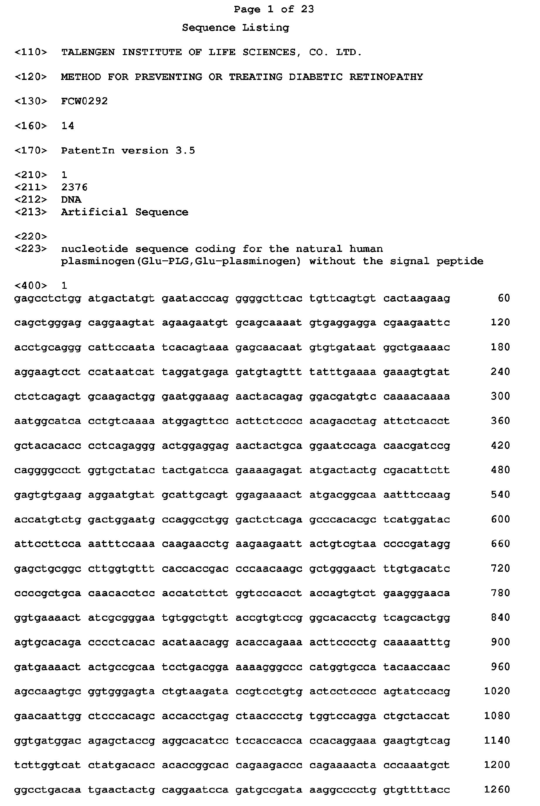


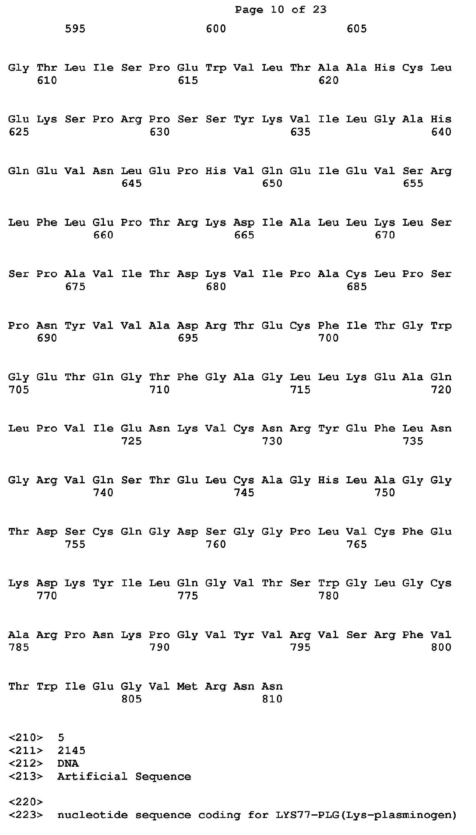


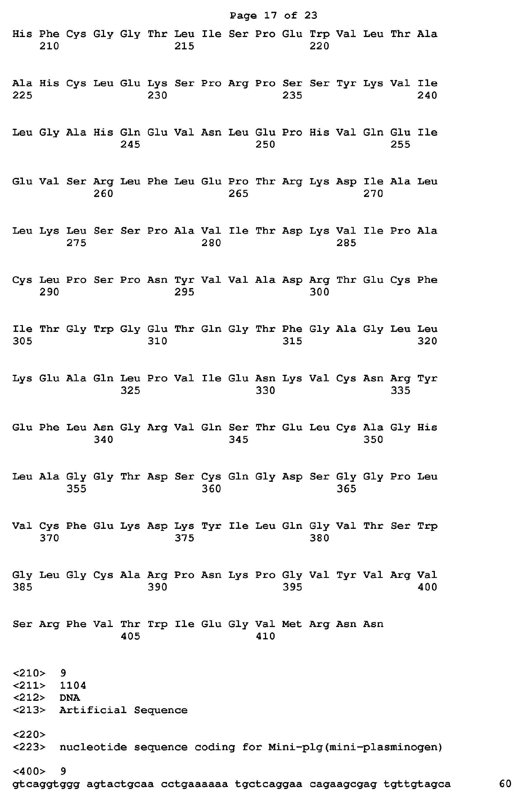


Claims (11)
- A method for preventing and/or treating retinopathy caused by diabetes mellitus in a subject, comprising administering an effective amount of plasminogen to the subject.
- The method of claim 1, wherein the retinopathy comprises retinal neovascularization, retinal inflammation, retinal atrophy, retinal cell apoptosis, and retinal tissue structural injury caused by diabetes mellitus.
- The method according to claims 1 to 2, wherein the plasminogen has at least 80%, 85%, 90%, 95%, 96%, 97%, 98% or 99% sequence identity with SEQ ID No.2, 6, 8, 10 or 12, and still has the plasminogen activity.
- The method according to any one of claims 1 to 3, wherein the plasminogen is a protein that comprises a plasminogen active fragment and still has the plasminogen activity.
- The method according to any one of claims 1 to 4, wherein the plasminogen is selected from Glu-plasminogen, Lys-plasminogen, mini-plasminogen, micro-plasminogen, δ (delta)-plasminogen or any combination thereof.
- The method according to any one of claims 1 to 5, wherein the plasminogen is administered systemically or locally, including topical, intravenous, intramuscular, subcutaneous, inhalation, intraspinal or rectal administration, local injection administration, and/or local administration on cornea by a gene gun, subconjunctival injection, intracameral injection, administration via an eye drop on the cornea, injection into the anterior chamber via the temporal edge, intrastromal injection, corneal application in combination with electric pulses, intracorneal injection, subretinal injection, intravitreal injection and intraocular injection administration.
- The method according to any one of claims 1 to 6, wherein the plasminogen is administered in combination with one or more other drugs, including anti-diabetic drugs, drugs against cardiovascular and cerebrovascular diseases, anti-thrombotic drugs, anti-hypertensive drugs, antilipemic drugs, anti-infective drugs and other conventional drugs for treating concomitant diseases.
- An article for preventing and/or treating retinopathy caused by diabetes mellitus in a subject, comprising a container containing an effective dosage of plasminogen, and instructions for directing the administration of the article to prevent and/or treat retinopathy caused by diabetes mellitus in the subject.
- The article of claim 8, further comprising a container containing one or more other drugs.
- The article of claim 9, wherein the other drugs are anti-diabetic drugs, drugs against cardiovascular and cerebrovascular diseases, anti-thrombotic drugs, anti-hypertensive drugs, antilipemic drugs, anti-infective drugs and other conventional drugs for treating concomitant diseases.
- The article of claim 9 or 10, wherein the instructions further indicate that the plasminogen is administered before, simultaneously with and/or after the administration of the other drugs.
Applications Claiming Priority (2)
| Application Number | Priority Date | Filing Date | Title |
|---|---|---|---|
| CN2015097946 | 2015-12-18 | ||
| PCT/CN2016/110452 WO2017101870A1 (en) | 2015-12-18 | 2016-12-16 | Method for preventing or treating diabetic retinopathy |
Publications (3)
| Publication Number | Publication Date |
|---|---|
| EP3395360A1 true EP3395360A1 (en) | 2018-10-31 |
| EP3395360A4 EP3395360A4 (en) | 2019-06-12 |
| EP3395360B1 EP3395360B1 (en) | 2025-08-20 |
Family
ID=59055781
Family Applications (1)
| Application Number | Title | Priority Date | Filing Date |
|---|---|---|---|
| EP16874927.3A Active EP3395360B1 (en) | 2015-12-18 | 2016-12-16 | Plasminogen preventing or treating diabetic retinopathy |
Country Status (9)
| Country | Link |
|---|---|
| US (1) | US10709771B2 (en) |
| EP (1) | EP3395360B1 (en) |
| JP (2) | JP2019500426A (en) |
| CN (1) | CN108463240A (en) |
| CA (1) | CA3008691A1 (en) |
| DK (1) | DK3395360T3 (en) |
| ES (1) | ES3046932T3 (en) |
| TW (1) | TWI624267B (en) |
| WO (1) | WO2017101870A1 (en) |
Cited By (4)
| Publication number | Priority date | Publication date | Assignee | Title |
|---|---|---|---|---|
| US11090372B2 (en) | 2015-12-18 | 2021-08-17 | Talengen International Limited | Method of treating diabetic nephropathy comprising administering plasminogen |
| US11129880B2 (en) | 2016-12-15 | 2021-09-28 | Talengen International Limited | Method for promoting insulin secretion |
| US11338022B2 (en) | 2015-12-18 | 2022-05-24 | Talengen International Limited | Method for preventing and treating angiocardiopathy |
| US11400142B2 (en) | 2015-12-18 | 2022-08-02 | Talengen International Limited | Treatment of diabetic nerve injury comprising administering plasminogen |
Families Citing this family (8)
| Publication number | Priority date | Publication date | Assignee | Title |
|---|---|---|---|---|
| TWI801331B (en) * | 2015-11-03 | 2023-05-11 | 美商波麥堤克生物治療股份有限公司 | Plasminogen replacement therapy for plasminogen-deficiency |
| JP6793748B2 (en) | 2015-12-18 | 2020-12-02 | タレンゲン インターナショナル リミテッドTalengen International Limited | Methods for preventing and treating radioactive and chemical damage |
| JP6876066B2 (en) | 2015-12-18 | 2021-05-26 | タレンゲン インターナショナル リミテッドTalengen International Limited | Methods for preventing and treating cervical erosion |
| TW201822805A (en) | 2016-12-15 | 2018-07-01 | 深圳瑞健生命科學硏究院有限公司 | Method for preventing and treating tissue injury caused by abnormal deposition of lipid |
| EP4299588A3 (en) * | 2019-09-10 | 2024-03-20 | University of Pittsburgh- Of the Commonwealth System of Higher Education | Treatment methods for eye disorders |
| EP4140498A4 (en) | 2020-05-11 | 2023-11-01 | Talengen International Limited | METHOD AND DRUGS FOR TREATING SPINAL MUSCLE ATROPHY |
| CN111999395A (en) * | 2020-06-15 | 2020-11-27 | 陕西步长制药有限公司 | Fingerprint detection method of medicinal preparation |
| WO2025237294A1 (en) * | 2024-05-14 | 2025-11-20 | Suzhou Ugenex Therapeutics Co., Ltd. | Retinal cell type-specific promoters and uses thereof in the treatment of ocular diseases |
Family Cites Families (37)
| Publication number | Priority date | Publication date | Assignee | Title |
|---|---|---|---|---|
| JP2002510209A (en) | 1997-06-26 | 2002-04-02 | カロリンスカ イノベイションズ アクチボラゲット | Kringle domain 1-5 of plasminogen that can regulate angiogenesis in vivo |
| WO2000018436A1 (en) | 1998-09-29 | 2000-04-06 | Leuven Research & Development Vzw | Use of compounds that reduce alpha2-antiplasmin in vivo for the preparation of a composition for the treatment of ischemic stroke |
| US6899877B2 (en) * | 1999-03-09 | 2005-05-31 | Minu, L.L.C. | Process for generating plasmin in the vitreous of the eye and inducing separation of the posterior hyaloid from the retina |
| US6733750B1 (en) * | 1999-03-09 | 2004-05-11 | Minu, L.L.C. | Process and composition for inducing posterior vitreous detachment |
| CA2385703A1 (en) | 1999-10-07 | 2001-04-12 | Human Genome Sciences, Inc. | Plasminogen-like polynucleotides, polypeptides, and antibodies |
| US7067492B2 (en) * | 2001-09-06 | 2006-06-27 | Omnio Ab | Method of promoting healing of a tympanic membrane perforation |
| US7202066B2 (en) | 2002-01-29 | 2007-04-10 | Carrington Laboratories, Inc. | Combination of a growth factor and a protease enzyme |
| JP2005525798A (en) * | 2002-02-06 | 2005-09-02 | トロムスドルフ ゲーエムベーハー ウント コー.カーゲー アルツナイミッテル | Production method of recombinant protein in microorganism |
| GB0228409D0 (en) | 2002-12-06 | 2003-01-08 | Thromb X Nv | Pharmacological vitreolysis |
| CN1451746A (en) | 2003-03-20 | 2003-10-29 | 广州市启源生物科技有限公司 | Human profibrinolysin kringle 5 deletion mutation recombinant polypeptide |
| US20050250694A1 (en) | 2003-10-10 | 2005-11-10 | Ma Jian-Xing | Compounds useful in inhibiting vascular leakage, inflammation and fibrosis and methods of making and using same |
| CN1946352A (en) * | 2004-04-09 | 2007-04-11 | 狄科特康坦特公司 | Methods and articles for delivering drugs to the eye to treat posterior segment diseases |
| MXPA06012236A (en) * | 2004-04-22 | 2007-01-31 | Talecris Biotherapeutics Inc | Recombinantly modified plasmin. |
| US20060257391A1 (en) * | 2005-05-11 | 2006-11-16 | Bausch & Lomb Incorporated | Non-surgical method for preventing or reducing the rate of the progression of non-proliferative diabetic retinopathy and the treatment of other ocular conditions |
| CN100529085C (en) * | 2005-12-30 | 2009-08-19 | 上海新生源医药研究有限公司 | Preparation method of recombinant human plasminogen Kringle 5(hk5) |
| CN101002888A (en) | 2006-01-16 | 2007-07-25 | 陈维森 | Compounding traditional Chinese medicine-Tangniaokang for treating diabetes |
| US20070196350A1 (en) * | 2006-02-22 | 2007-08-23 | Bartels Stephen P | Compositions and Methods for Effecting Controlled Posterior Vitreous Detachment |
| WO2008026999A2 (en) | 2006-08-28 | 2008-03-06 | Omnio Healer Ab | Candidates against infection |
| RU2010120675A (en) | 2007-10-23 | 2011-11-27 | Инститьют Оф Медисинал Молекьюлар Дизайн, Инк. (Jp) | PAI-1 PRODUCT INHIBITOR |
| JP5539896B2 (en) | 2007-11-29 | 2014-07-02 | グリフオルス・セラピユーテイクス・インコーポレーテツド | Recombinantly modified plasmin |
| JP2009196927A (en) * | 2008-02-21 | 2009-09-03 | Chemo Sero Therapeut Res Inst | Agent for inhibiting progress of or treating retinopathy caused by angiopathy |
| WO2010125148A2 (en) * | 2009-04-30 | 2010-11-04 | Catherine Blondel | Methods for treating ocular conditions |
| CN102250210B (en) * | 2010-05-21 | 2014-05-28 | 厦门大学 | Human plasminogen Kringle 5 short peptide, its pharmaceutical composition, application and polynucleotide encoding the short peptide |
| CN102121023B (en) | 2010-12-22 | 2012-07-04 | 中山大学 | Mutant human plasminogen kringle5, preparation method and application thereof |
| RU2604807C2 (en) | 2011-01-05 | 2016-12-10 | ТромбоДженикс НВ | Plasminogen and plasmin variants |
| CN102154253A (en) | 2011-01-06 | 2011-08-17 | 郑州大学 | Micro plasminogen mutant with function of inhibiting platelet aggregation and preparation method and application thereof |
| JP6000353B2 (en) | 2011-08-12 | 2016-09-28 | スロンボジェニックス・ナムローゼ・フェンノートシャップThromboGenics NV | Plasminogen and plasmin variants |
| WO2014070983A1 (en) | 2012-10-31 | 2014-05-08 | The Regents Of The University Of Michigan | Plasminogen activator-1 inhibitors and methods of use thereof |
| US9079739B2 (en) | 2012-11-02 | 2015-07-14 | The Procter & Gamble Company | Apparatus for controlling the nip force/pressure between two rotating cylinders |
| TW201734054A (en) | 2013-08-13 | 2017-10-01 | 賽諾菲公司 | Antibody to plasminogen activin inhibitor-1 (PAI-1) and use thereof |
| EA033403B1 (en) | 2013-08-13 | 2019-10-31 | Sanofi Sa | ANTIBODIES TO PLASMINOGEN 1 (PAI-1) ACTIVATOR INHIBITOR AND WAYS OF THEIR APPLICATION |
| CN104789544B (en) | 2014-01-16 | 2019-11-26 | 中国科学院福建物质结构研究所 | Recombinant PAI-1 inhibitor, comprising its composition and its for treat and detection applications |
| PY1577621A (en) | 2014-12-19 | 2017-10-02 | Prometic Biotherapeutics Inc | PHARMACEUTICAL COMPOSITION COMPRISING PLASMINOGEN AND ITS USE |
| CN108778307A (en) | 2015-12-18 | 2018-11-09 | 泰伦基国际有限公司 | A method for preventing and treating diabetic nephropathy |
| TWI653981B (en) | 2015-12-18 | 2019-03-21 | 深圳瑞健生命科學硏究院有限公司 | Use of plasminogen for the preparation of a medicament for preventing or treating a condition associated with diabetic nerve injury |
| CN115845037A (en) | 2015-12-18 | 2023-03-28 | 泰伦基国际有限公司 | A method for preventing or treating acute and chronic thrombosis |
| US11129880B2 (en) | 2016-12-15 | 2021-09-28 | Talengen International Limited | Method for promoting insulin secretion |
-
2016
- 2016-12-16 TW TW105141893A patent/TWI624267B/en active
- 2016-12-16 CN CN201680073582.3A patent/CN108463240A/en active Pending
- 2016-12-16 US US16/062,052 patent/US10709771B2/en active Active
- 2016-12-16 ES ES16874927T patent/ES3046932T3/en active Active
- 2016-12-16 EP EP16874927.3A patent/EP3395360B1/en active Active
- 2016-12-16 JP JP2018550637A patent/JP2019500426A/en active Pending
- 2016-12-16 WO PCT/CN2016/110452 patent/WO2017101870A1/en not_active Ceased
- 2016-12-16 CA CA3008691A patent/CA3008691A1/en active Pending
- 2016-12-16 DK DK16874927.3T patent/DK3395360T3/en active
-
2019
- 2019-11-27 JP JP2019214655A patent/JP7117012B2/en active Active
Cited By (7)
| Publication number | Priority date | Publication date | Assignee | Title |
|---|---|---|---|---|
| US11090372B2 (en) | 2015-12-18 | 2021-08-17 | Talengen International Limited | Method of treating diabetic nephropathy comprising administering plasminogen |
| US11338022B2 (en) | 2015-12-18 | 2022-05-24 | Talengen International Limited | Method for preventing and treating angiocardiopathy |
| US11400142B2 (en) | 2015-12-18 | 2022-08-02 | Talengen International Limited | Treatment of diabetic nerve injury comprising administering plasminogen |
| EP3395353B1 (en) * | 2015-12-18 | 2024-03-06 | Talengen International Limited | Plasminogen for use in treating or preventing diabetes mellitus nerve injury |
| EP3395354B1 (en) * | 2015-12-18 | 2024-05-22 | Talengen International Limited | Plasminogen for use in treating diabetic nephropathy |
| US11129880B2 (en) | 2016-12-15 | 2021-09-28 | Talengen International Limited | Method for promoting insulin secretion |
| US11311607B2 (en) | 2016-12-15 | 2022-04-26 | Talengen International Limited | Method for making glucagon and insulin restore normal balance |
Also Published As
| Publication number | Publication date |
|---|---|
| JP2020059730A (en) | 2020-04-16 |
| DK3395360T3 (en) | 2025-09-15 |
| WO2017101870A1 (en) | 2017-06-22 |
| EP3395360A4 (en) | 2019-06-12 |
| CN108463240A (en) | 2018-08-28 |
| TWI624267B (en) | 2018-05-21 |
| JP7117012B2 (en) | 2022-08-12 |
| US20180360930A1 (en) | 2018-12-20 |
| EP3395360B1 (en) | 2025-08-20 |
| US10709771B2 (en) | 2020-07-14 |
| JP2019500426A (en) | 2019-01-10 |
| CA3008691A1 (en) | 2017-06-22 |
| TW201725049A (en) | 2017-07-16 |
| ES3046932T3 (en) | 2025-12-02 |
Similar Documents
| Publication | Publication Date | Title |
|---|---|---|
| EP3395360B1 (en) | Plasminogen preventing or treating diabetic retinopathy | |
| EP3395353B1 (en) | Plasminogen for use in treating or preventing diabetes mellitus nerve injury | |
| US11090372B2 (en) | Method of treating diabetic nephropathy comprising administering plasminogen | |
| EP3391902B1 (en) | Plasminogen for use in the treatment of diabetic angiocardiopathy | |
| TWI752044B (en) | A method for preventing and treating tissue and organ fibrosis | |
| EP3967322B1 (en) | Plasminogen for treating amyotrophic lateral sclerosis | |
| TW202123962A (en) | Method for preventing and treating kidney fibrosis | |
| TWI853151B (en) | A method and medicine for preventing and treating abnormal blood pressure | |
| CA3008186C (en) | Novel method for preventing and treating angiocardiopathy | |
| HK1240129A1 (en) | Method for preventing or treating diabetic retinopathy | |
| HK1240126A1 (en) | Novel method for preventing and treating diabetic heart disease | |
| CN106890319A (en) | A kind of method prevented or treat BDR | |
| HK1257235A1 (en) | Medicine for treating atherosclerosis and complications of atherosclerosis and use of the medicine |
Legal Events
| Date | Code | Title | Description |
|---|---|---|---|
| STAA | Information on the status of an ep patent application or granted ep patent |
Free format text: STATUS: THE INTERNATIONAL PUBLICATION HAS BEEN MADE |
|
| PUAI | Public reference made under article 153(3) epc to a published international application that has entered the european phase |
Free format text: ORIGINAL CODE: 0009012 |
|
| STAA | Information on the status of an ep patent application or granted ep patent |
Free format text: STATUS: REQUEST FOR EXAMINATION WAS MADE |
|
| 17P | Request for examination filed |
Effective date: 20180717 |
|
| AK | Designated contracting states |
Kind code of ref document: A1 Designated state(s): AL AT BE BG CH CY CZ DE DK EE ES FI FR GB GR HR HU IE IS IT LI LT LU LV MC MK MT NL NO PL PT RO RS SE SI SK SM TR |
|
| AX | Request for extension of the european patent |
Extension state: BA ME |
|
| DAV | Request for validation of the european patent (deleted) | ||
| DAX | Request for extension of the european patent (deleted) | ||
| A4 | Supplementary search report drawn up and despatched |
Effective date: 20190515 |
|
| RIC1 | Information provided on ipc code assigned before grant |
Ipc: A61K 38/48 20060101AFI20190509BHEP Ipc: A61P 27/02 20060101ALI20190509BHEP Ipc: A61P 3/10 20060101ALI20190509BHEP |
|
| STAA | Information on the status of an ep patent application or granted ep patent |
Free format text: STATUS: EXAMINATION IS IN PROGRESS |
|
| 17Q | First examination report despatched |
Effective date: 20210216 |
|
| GRAP | Despatch of communication of intention to grant a patent |
Free format text: ORIGINAL CODE: EPIDOSNIGR1 |
|
| STAA | Information on the status of an ep patent application or granted ep patent |
Free format text: STATUS: GRANT OF PATENT IS INTENDED |
|
| INTG | Intention to grant announced |
Effective date: 20250213 |
|
| GRAS | Grant fee paid |
Free format text: ORIGINAL CODE: EPIDOSNIGR3 |
|
| GRAA | (expected) grant |
Free format text: ORIGINAL CODE: 0009210 |
|
| STAA | Information on the status of an ep patent application or granted ep patent |
Free format text: STATUS: THE PATENT HAS BEEN GRANTED |
|
| AK | Designated contracting states |
Kind code of ref document: B1 Designated state(s): AL AT BE BG CH CY CZ DE DK EE ES FI FR GB GR HR HU IE IS IT LI LT LU LV MC MK MT NL NO PL PT RO RS SE SI SK SM TR |
|
| REG | Reference to a national code |
Ref country code: GB Ref legal event code: FG4D |
|
| REG | Reference to a national code |
Ref country code: CH Ref legal event code: EP |
|
| REG | Reference to a national code |
Ref country code: IE Ref legal event code: FG4D |
|
| REG | Reference to a national code |
Ref country code: DE Ref legal event code: R096 Ref document number: 602016093324 Country of ref document: DE |
|
| REG | Reference to a national code |
Ref country code: DK Ref legal event code: T3 Effective date: 20250912 |
|
| P01 | Opt-out of the competence of the unified patent court (upc) registered |
Free format text: CASE NUMBER: UPC_APP_3117_3395360/2025 Effective date: 20250812 |
|
| REG | Reference to a national code |
Ref country code: SE Ref legal event code: TRGR |
|
| REG | Reference to a national code |
Ref country code: ES Ref legal event code: FG2A Ref document number: 3046932 Country of ref document: ES Kind code of ref document: T3 Effective date: 20251202 |
|
| REG | Reference to a national code |
Ref country code: NL Ref legal event code: MP Effective date: 20250820 |
|
| REG | Reference to a national code |
Ref country code: CH Ref legal event code: U11 Free format text: ST27 STATUS EVENT CODE: U-0-0-U10-U11 (AS PROVIDED BY THE NATIONAL OFFICE) Effective date: 20260101 |
|
| PG25 | Lapsed in a contracting state [announced via postgrant information from national office to epo] |
Ref country code: IS Free format text: LAPSE BECAUSE OF FAILURE TO SUBMIT A TRANSLATION OF THE DESCRIPTION OR TO PAY THE FEE WITHIN THE PRESCRIBED TIME-LIMIT Effective date: 20251220 |
|
| PGFP | Annual fee paid to national office [announced via postgrant information from national office to epo] |
Ref country code: GB Payment date: 20251229 Year of fee payment: 10 |
|
| PG25 | Lapsed in a contracting state [announced via postgrant information from national office to epo] |
Ref country code: NO Free format text: LAPSE BECAUSE OF FAILURE TO SUBMIT A TRANSLATION OF THE DESCRIPTION OR TO PAY THE FEE WITHIN THE PRESCRIBED TIME-LIMIT Effective date: 20251120 |
|
| REG | Reference to a national code |
Ref country code: LT Ref legal event code: MG9D |
|
| PG25 | Lapsed in a contracting state [announced via postgrant information from national office to epo] |
Ref country code: PT Free format text: LAPSE BECAUSE OF FAILURE TO SUBMIT A TRANSLATION OF THE DESCRIPTION OR TO PAY THE FEE WITHIN THE PRESCRIBED TIME-LIMIT Effective date: 20251222 |
|
| PG25 | Lapsed in a contracting state [announced via postgrant information from national office to epo] |
Ref country code: FI Free format text: LAPSE BECAUSE OF FAILURE TO SUBMIT A TRANSLATION OF THE DESCRIPTION OR TO PAY THE FEE WITHIN THE PRESCRIBED TIME-LIMIT Effective date: 20250820 |
|
| PGFP | Annual fee paid to national office [announced via postgrant information from national office to epo] |
Ref country code: IT Payment date: 20251210 Year of fee payment: 10 Ref country code: DK Payment date: 20251127 Year of fee payment: 10 |
|
| PG25 | Lapsed in a contracting state [announced via postgrant information from national office to epo] |
Ref country code: HR Free format text: LAPSE BECAUSE OF FAILURE TO SUBMIT A TRANSLATION OF THE DESCRIPTION OR TO PAY THE FEE WITHIN THE PRESCRIBED TIME-LIMIT Effective date: 20250820 Ref country code: NL Free format text: LAPSE BECAUSE OF FAILURE TO SUBMIT A TRANSLATION OF THE DESCRIPTION OR TO PAY THE FEE WITHIN THE PRESCRIBED TIME-LIMIT Effective date: 20250820 |
|
| PGFP | Annual fee paid to national office [announced via postgrant information from national office to epo] |
Ref country code: FR Payment date: 20251230 Year of fee payment: 10 |
|
| PG25 | Lapsed in a contracting state [announced via postgrant information from national office to epo] |
Ref country code: GR Free format text: LAPSE BECAUSE OF FAILURE TO SUBMIT A TRANSLATION OF THE DESCRIPTION OR TO PAY THE FEE WITHIN THE PRESCRIBED TIME-LIMIT Effective date: 20251121 |
|
| PGFP | Annual fee paid to national office [announced via postgrant information from national office to epo] |
Ref country code: SE Payment date: 20251218 Year of fee payment: 10 |
|
| PG25 | Lapsed in a contracting state [announced via postgrant information from national office to epo] |
Ref country code: LV Free format text: LAPSE BECAUSE OF FAILURE TO SUBMIT A TRANSLATION OF THE DESCRIPTION OR TO PAY THE FEE WITHIN THE PRESCRIBED TIME-LIMIT Effective date: 20250820 |
|
| PG25 | Lapsed in a contracting state [announced via postgrant information from national office to epo] |
Ref country code: BG Free format text: LAPSE BECAUSE OF FAILURE TO SUBMIT A TRANSLATION OF THE DESCRIPTION OR TO PAY THE FEE WITHIN THE PRESCRIBED TIME-LIMIT Effective date: 20250820 Ref country code: PL Free format text: LAPSE BECAUSE OF FAILURE TO SUBMIT A TRANSLATION OF THE DESCRIPTION OR TO PAY THE FEE WITHIN THE PRESCRIBED TIME-LIMIT Effective date: 20250820 |
|
| PG25 | Lapsed in a contracting state [announced via postgrant information from national office to epo] |
Ref country code: RS Free format text: LAPSE BECAUSE OF FAILURE TO SUBMIT A TRANSLATION OF THE DESCRIPTION OR TO PAY THE FEE WITHIN THE PRESCRIBED TIME-LIMIT Effective date: 20251120 |
|
| REG | Reference to a national code |
Ref country code: AT Ref legal event code: MK05 Ref document number: 1826609 Country of ref document: AT Kind code of ref document: T Effective date: 20250820 |
|
| PG25 | Lapsed in a contracting state [announced via postgrant information from national office to epo] |
Ref country code: RO Free format text: LAPSE BECAUSE OF FAILURE TO SUBMIT A TRANSLATION OF THE DESCRIPTION OR TO PAY THE FEE WITHIN THE PRESCRIBED TIME-LIMIT Effective date: 20250820 |
|
| PG25 | Lapsed in a contracting state [announced via postgrant information from national office to epo] |
Ref country code: SM Free format text: LAPSE BECAUSE OF FAILURE TO SUBMIT A TRANSLATION OF THE DESCRIPTION OR TO PAY THE FEE WITHIN THE PRESCRIBED TIME-LIMIT Effective date: 20250820 |
|
| PGFP | Annual fee paid to national office [announced via postgrant information from national office to epo] |
Ref country code: ES Payment date: 20260112 Year of fee payment: 10 |
|
| PGFP | Annual fee paid to national office [announced via postgrant information from national office to epo] |
Ref country code: DE Payment date: 20251231 Year of fee payment: 10 |
|
| PG25 | Lapsed in a contracting state [announced via postgrant information from national office to epo] |
Ref country code: AT Free format text: LAPSE BECAUSE OF FAILURE TO SUBMIT A TRANSLATION OF THE DESCRIPTION OR TO PAY THE FEE WITHIN THE PRESCRIBED TIME-LIMIT Effective date: 20250820 |
|
| PG25 | Lapsed in a contracting state [announced via postgrant information from national office to epo] |
Ref country code: CZ Free format text: LAPSE BECAUSE OF FAILURE TO SUBMIT A TRANSLATION OF THE DESCRIPTION OR TO PAY THE FEE WITHIN THE PRESCRIBED TIME-LIMIT Effective date: 20250820 |
|
| PGFP | Annual fee paid to national office [announced via postgrant information from national office to epo] |
Ref country code: CH Payment date: 20260101 Year of fee payment: 10 |
|
| PG25 | Lapsed in a contracting state [announced via postgrant information from national office to epo] |
Ref country code: SK Free format text: LAPSE BECAUSE OF FAILURE TO SUBMIT A TRANSLATION OF THE DESCRIPTION OR TO PAY THE FEE WITHIN THE PRESCRIBED TIME-LIMIT Effective date: 20250820 Ref country code: EE Free format text: LAPSE BECAUSE OF FAILURE TO SUBMIT A TRANSLATION OF THE DESCRIPTION OR TO PAY THE FEE WITHIN THE PRESCRIBED TIME-LIMIT Effective date: 20250820 |