EP3387618B1 - Verarbeitung von optischen kohärenztomografie-scans - Google Patents
Verarbeitung von optischen kohärenztomografie-scans Download PDFInfo
- Publication number
- EP3387618B1 EP3387618B1 EP16825872.1A EP16825872A EP3387618B1 EP 3387618 B1 EP3387618 B1 EP 3387618B1 EP 16825872 A EP16825872 A EP 16825872A EP 3387618 B1 EP3387618 B1 EP 3387618B1
- Authority
- EP
- European Patent Office
- Prior art keywords
- oct
- skin
- subject
- scans
- scan
- Prior art date
- Legal status (The legal status is an assumption and is not a legal conclusion. Google has not performed a legal analysis and makes no representation as to the accuracy of the status listed.)
- Active
Links
Images


Classifications
-
- A—HUMAN NECESSITIES
- A61—MEDICAL OR VETERINARY SCIENCE; HYGIENE
- A61B—DIAGNOSIS; SURGERY; IDENTIFICATION
- A61B5/00—Measuring for diagnostic purposes; Identification of persons
- A61B5/48—Other medical applications
- A61B5/4887—Locating particular structures in or on the body
- A61B5/489—Blood vessels
-
- A—HUMAN NECESSITIES
- A61—MEDICAL OR VETERINARY SCIENCE; HYGIENE
- A61B—DIAGNOSIS; SURGERY; IDENTIFICATION
- A61B5/00—Measuring for diagnostic purposes; Identification of persons
- A61B5/0059—Measuring for diagnostic purposes; Identification of persons using light, e.g. diagnosis by transillumination, diascopy, fluorescence
- A61B5/0062—Arrangements for scanning
- A61B5/0066—Optical coherence imaging
-
- A—HUMAN NECESSITIES
- A61—MEDICAL OR VETERINARY SCIENCE; HYGIENE
- A61B—DIAGNOSIS; SURGERY; IDENTIFICATION
- A61B5/00—Measuring for diagnostic purposes; Identification of persons
- A61B5/103—Measuring devices for testing the shape, pattern, colour, size or movement of the body or parts thereof, for diagnostic purposes
- A61B5/107—Measuring physical dimensions, e.g. size of the entire body or parts thereof
-
- G—PHYSICS
- G06—COMPUTING OR CALCULATING; COUNTING
- G06T—IMAGE DATA PROCESSING OR GENERATION, IN GENERAL
- G06T7/00—Image analysis
- G06T7/0002—Inspection of images, e.g. flaw detection
- G06T7/0012—Biomedical image inspection
-
- G—PHYSICS
- G06—COMPUTING OR CALCULATING; COUNTING
- G06T—IMAGE DATA PROCESSING OR GENERATION, IN GENERAL
- G06T7/00—Image analysis
- G06T7/10—Segmentation; Edge detection
-
- G—PHYSICS
- G06—COMPUTING OR CALCULATING; COUNTING
- G06T—IMAGE DATA PROCESSING OR GENERATION, IN GENERAL
- G06T7/00—Image analysis
- G06T7/60—Analysis of geometric attributes
- G06T7/62—Analysis of geometric attributes of area, perimeter, diameter or volume
-
- A—HUMAN NECESSITIES
- A61—MEDICAL OR VETERINARY SCIENCE; HYGIENE
- A61B—DIAGNOSIS; SURGERY; IDENTIFICATION
- A61B2576/00—Medical imaging apparatus involving image processing or analysis
- A61B2576/02—Medical imaging apparatus involving image processing or analysis specially adapted for a particular organ or body part
-
- G—PHYSICS
- G06—COMPUTING OR CALCULATING; COUNTING
- G06T—IMAGE DATA PROCESSING OR GENERATION, IN GENERAL
- G06T2207/00—Indexing scheme for image analysis or image enhancement
- G06T2207/10—Image acquisition modality
- G06T2207/10072—Tomographic images
- G06T2207/10101—Optical tomography; Optical coherence tomography [OCT]
-
- G—PHYSICS
- G06—COMPUTING OR CALCULATING; COUNTING
- G06T—IMAGE DATA PROCESSING OR GENERATION, IN GENERAL
- G06T2207/00—Indexing scheme for image analysis or image enhancement
- G06T2207/30—Subject of image; Context of image processing
- G06T2207/30004—Biomedical image processing
- G06T2207/30088—Skin; Dermal
-
- G—PHYSICS
- G06—COMPUTING OR CALCULATING; COUNTING
- G06T—IMAGE DATA PROCESSING OR GENERATION, IN GENERAL
- G06T2207/00—Indexing scheme for image analysis or image enhancement
- G06T2207/30—Subject of image; Context of image processing
- G06T2207/30004—Biomedical image processing
- G06T2207/30101—Blood vessel; Artery; Vein; Vascular
-
- G—PHYSICS
- G16—INFORMATION AND COMMUNICATION TECHNOLOGY [ICT] SPECIALLY ADAPTED FOR SPECIFIC APPLICATION FIELDS
- G16H—HEALTHCARE INFORMATICS, i.e. INFORMATION AND COMMUNICATION TECHNOLOGY [ICT] SPECIALLY ADAPTED FOR THE HANDLING OR PROCESSING OF MEDICAL OR HEALTHCARE DATA
- G16H30/00—ICT specially adapted for the handling or processing of medical images
- G16H30/40—ICT specially adapted for the handling or processing of medical images for processing medical images, e.g. editing
Definitions
- This invention relates to a method of processing optical coherence tomography (OCT) scans, and associated apparatus.
- OCT optical coherence tomography
- vascular network in a subject's skin there is a need for rapid non-invasive assessment of the vascular network in a subject's skin, as an aid to planning and guiding treatments for aesthetic and medical conditions affecting patients, for example (but not limited to) port wine stains, hemangiomas, hypertrophic scars, rosacea, and 'spider veins'.
- Typical treatments for these conditions producing temporary or permanent effects on the vascular network in the dermis include systemic and topically applied drugs, and laser treatments. These treatments either cause the blood vessels and capillaries to constrict, or they damage the vessels to reduce or terminate their ability to transport blood.
- laser treatments are designed to deliver a certain amount of energy to the target vessels to damage them; if the energy of the pulse is too high then the surrounding tissue will be damaged, which may result in undesirable scarring and pain; conversely if the energy pulse is too low, then the treatment will be ineffective and will have to be repeated (perhaps multiple times) to achieve the desired impact.
- the laser pulse characteristic can be tuned to achieve a desired energy delivery profile [ RR Anderson, JA Parrish, Selective Photothermolysis: Precise Microsurgery by Selective Absorption of Pulsed Radiation, Science, 220 (4596), 1983, pp524-527 ], but the clinician must know the vessel characteristics in order to set this up for optimal results.
- OCT optical coherence tomography
- Optical Coherence Tomography was invented in 1991 at the Massachusetts Institute of Technology in the United States of America and is commonly used for imaging human tissue of various organs, in particular the eye, and also skin ( J. Welzel, "Optical coherence tomography in dermatology: a review,” Skin Research and Technology, vol. 7, pp. 1-9, 2001 ).
- OCT VivoSight
- Michelson Diagnostics Ltd of Orpington, Kent, United Kingdom, which is designed for use by professional dermatologists in the assessment of skin lesions of patients.
- the VivoSight OCT device scans the skin and presents to the user images of the skin subsurface structure, in a plane perpendicular to the skin surface (By convention, the OCT device is assumed to be positioned vertically above the skin surface).
- the resulting two-dimensional image is commonly known in the art as a "B-scan”, and comprises many lines of vertical pixels commonly known in the art as "A-lines”.
- Each pixel in the image has a value corresponding to the signal obtained from the OCT device resulting from the skin optical properties at the y-position for that pixel.
- the VivoSight device can acquire scans at multiple locations across the skin surface in order to build up a series of B-scans across a lesion of interest. This is known in the art as a multi-slice "stack" and can be viewed by the user in a variety of ways to elicit tissue features of medical interest such as nests of cancer cells.
- the user can view the stack of B-scans in rapid succession to fly through a lesion area.
- the stack of data can be re-sampled so that one or more image slices in the horizontal plane (perpendicular to the B-scans) can be extracted and viewed.
- Horizontal slices are sometimes known in the art as "C-scans".
- C-scans the C-scan at any desired depth can be viewed.
- a C-scan and B-scan from a stack can be viewed simultaneously. All of this is well known to those skilled in the art of OCT and more generally medical imaging.
- OCT optical coherence tomography
- the method may comprise determining the position of the surface of the subject's skin in each scan.
- the method may comprise capturing the scans using an OCT probe, and delivering a pulse of laser energy to a volume of the subject's skin comprising at least one blood vessel using the probe.
- the user may be able to select at least one of the energy, fluence and duration of the pulse depending upon the measurements of blood vessel diameter already made.
- OCT optical coherence tomography
- the image processing apparatus may comprise an OCT apparatus by means of which the OCT scans are captured.
- the image processing apparatus may comprise an OCT probe arranged to generated interferograms, and the processor may be arranged to generate the images from the interferograms.
- the image processor may be arranged to process the images as they are captured.
- the OCT probe may be arranged so as to deliver a pulse of laser energy to a volume of the subject's skin comprising at least one blood vessel.
- the OCT apparatus may comprise a laser pulse source, having at least one control by means of which a user can select at least one of the energy, duration and fluence of the laser pulse; typically, a user would do this in response to the measurements of blood vessel diameter already made.
- the image processing apparatus may be separate from any OCT apparatus and may be arranged to process the images subsequent to their capture.
- the image processing apparatus may comprise data reception means (such as a network connection or media drive) arranged to receive the images for processing.
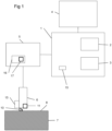
- FIG. 1 of the accompanying drawings An optical coherence tomography (OCT) apparatus in accordance with an embodiment of the invention is shown in Figure 1 of the accompanying drawings.
- This comprises a computer 1, having a processor 2 and storage 3 (such as a mass storage device or random access memory) coupled to the processor 2.
- the storage 3 contains data and processor instructions which cause the processor 2 to act as is described below.
- the computer 1 can be any suitable model; typically a personal computer running an operating system such as Microsoft (RTM) Windows (RTM) or Apple (RTM) Mac OS X (RTM) can be used.
- the computer 1 is also provided with a display 4 controlled by the processor 2 on which any desired graphics can be displayed, and a sound output device 15 such as a buzzer which can sound an alert noise.
- the apparatus further comprises an OCT interferometer 5 and associated probe 6.
- the interferometer 5 interferes light reflected from sample 7 (here, a subject's skin) through probe 6 with light passed along a reference path to generate interferograms. These are detected in the interferometer 5; the measured signal is then passed to the computer 1 for processing.
- suitable OCT apparatus can be found in the PCT patent application published as WO2006/054116 or in the VivoSight (RTM) apparatus available from Michelson Diagnostics of Orpington, Kent, United Kingdom.
- a stand-off 10 can be provided which spaces the probe 6 from the subject's skin 7.
- Such OCT apparatus typically generate multiple B-scans: that is, scans taken perpendicularly through the skin 7.
- the result of analysis of each interferogram is a bitmap in which the width of the image corresponds to a direction generally parallel to the skin surface and the height corresponds to the depth from the sensor into the skin.
- the OCT apparatus takes multiple scans spaced apart in time. Successive images from the same location on a subject's skin can be used to determine the presence of blood flow through blood vessels, by determining areas that have changed between successive images. Such changes can indicate the flow of blood cells through blood vessels.
- Our preferred technique is speckle decorrelation OCT as described in " In vivo imaging of the microcirculation of the volar forearm using correlation mapping optical coherence tomography (cmOCT)", J Enfield, E Jonathan and M Leahy, Biomed Opt Express. May 1, 2011; 2(5): 1184-1193 , but we can also use speckle variance, differencing of intensity or phase, or any other computational technique.
- the time interval between the different times should be carefully selected. If the time interval is too short, then there is not enough time for blood to move through the region to make a detectable change. If the time interval is too long, then gross movements of the probe relative to the skin make it difficult or impossible to ensure that the region in the second image is exactly the same location as the region in the first image, and this results in noise in the image. In human skin, a time interval of 3.5 milliseconds works well.
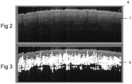
- FIG. 2 An example OCT scan is shown at Figure 2 , which shows a subject's skin 7, which has a surface 8.
- the results of comparing this to a successive image in healthy tissue are shown in Figure 3 , where areas of large differences are shown as white as indicated generally at 9. These areas represent areas where there are blood vessels. Typically, any area where change is determined at above a given threshold is determined to be a changing area.
- the method also determines the skin surface topography, and therefore the depth of any detected sub-surface feature below the skin. In order to do so, the skin surface position must be found using the method shown in Figure 5 .
- the stack of scans referred to above is generated.
- the image data is filtered to reduce or remove random noise inherent of the coherent detection technique ('speckle noise') and other image artefacts particular to the imaging system used to capture the image data.
- the reduction in the effect of noise upon image contrast is achieved using linear and non-linear filtering techniques, intended to preserve edge features while removing speckle.
- filters examples include linear filters, Kalman filters (see, for example, Igor Gurov and Maxim Volynsky “Recurrence signal processing in Fourier-domain optical coherence tomography based on linear Kalman filtering", Proc. SPIE 8792, Optical Methods for Inspection, Characterization, and Imaging of Biomaterials, 879203 (May 23, 2013); doi:10.1117/12.2020615; http://dx.doi.org/10.1117/12.2020615 ) or wavelet filters (see, for example, " Speckle reduction in optical coherence tomography images of human finger skin by wavelet modified BM3D filter", Bo Chong, Yong-Kai Zhu, Optics Communications Volume 291, 15 March 2013, Pages 461-469, http://dx.doi.org/10.1016/j.optcom.2012.10.053 ).
- the unadulterated proximal tissue surface represents a refractive index change from that of air to that of tissue and therefore presents a very strongly backscattering feature and high signal in OCT images.
- the image A-lines are filtered to indicate extrema in the OCT signal; certain of these are rejected based on simple parameters (position, classification and the local environment of the extrema, such as the difference between the positions of the extrema in adjacent images).
- edge-filtering may be employed to refine results and ensure that the estimations of skin surface are minimally affected by remaining speckle or a poorly-backscattering skin surface (in the presence of, for example, media which reduces or minimises the difference in refractive index at the proximal side of the skin surface).
- the result of this stage is a 3D binary array of possible surface positions, with multiple solutions possible in each A-scan of the data array.
- step 24 may typically result in multiple candidates for the proximal skin surface.
- Step 26 is undertaken therefore to assign a score to each candidate in each A-scan according the probability that it does not represent the true skin surface.
- This algorithm may involve multiple independent estimates of probability and combine these in a suitable manner to provide a meaningful metric.
- step 28 the set of multiple skin surface candidates and their weights are then processed to select a set of candidates 30 representing a single surface.
- the algorithm employed to select this surface attempts to minimise the sums of the weights of candidates in the selected surface and also a weight assigned to the Euclidean distance between neighbouring candidates in the selected surface.
- step 32 the surface 30 selected in step 28 is assessed for suitability according to criteria based on a-priori knowledge of skin morphology, the result of this assessment is termed herein "the worthiness" of the surface.
- the criteria may include uniformity, periodicity of variation, surface variance amongst other factors, all being within expected limits from known skin samples. If an insufficient worthiness is attained, according to a prescribed value, the solution is rejected (at step 34) and steps 26, 28 and 32 repeated using an additional weighting factor derived in this step.
- the additional weighting assigned to the multitude of candidates in the repeated step 28 may be calculated based on the position of the surface and its overall worthiness as well as localised contribution to the worthiness of particular candidates.
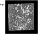
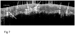
- the data is displayed to the user in either en-face (C-scan, as shown in Figure 6 of the accompanying drawings) or vertical (B-scan, as shown in figure 7 of the accompanying drawings) format, or in a 3D rendering, and the user is provided with means to select any desired vessel by means of a mouse, trackball, trackpad or other device, depicted by the white arrows in Figures 6 and 7 .
- a user might be able to select the depth through the subject's skin and display a C-scan, or a position through the subject's skin and display a B-scan.
- a vessel can then be selected using the mouse and processed to extract the vessel diameter at the pointed-to location.
- the arrowed vessel in Figure 6 is measured to be 170 microns (micrometres) in diameter. This is useful if a user wishes to "hit" a particularly large vessel.
- the system can display the depth below the skin surface at that position (for example, the left-most arrowed vessel in Figure 7 is 0.34mm below the surface).
- the user is provided with a quick and easy means to characterise the properties of vessels that they wish to treat.
- the processing means may divide the scanned volume into a series of slices of pre-selected depth ranges below the surface, for example 0.0 - 0.1 mm, 0.1-0.2 mm and so on, and then analyze the vessels within each slice, so as to provide the user with quantification of the vessels at a particular depth range.
- This analysis may include, for example:
- the system may calculate the median or mean vessel diameter and depth, and then set the laser pulse energy, fluence and duration from a look-up table.
- the OCT probe can be integrated with a laser energy delivery device 15, so that the user can instantly treat the scanned area with a laser pulse of chosen energy, created at laser drive circuit 16 which itself has pulse energy control 17, without having to move the probe.
Landscapes
- Health & Medical Sciences (AREA)
- Engineering & Computer Science (AREA)
- Life Sciences & Earth Sciences (AREA)
- Physics & Mathematics (AREA)
- General Health & Medical Sciences (AREA)
- Medical Informatics (AREA)
- Animal Behavior & Ethology (AREA)
- Surgery (AREA)
- Veterinary Medicine (AREA)
- Public Health (AREA)
- Molecular Biology (AREA)
- Heart & Thoracic Surgery (AREA)
- Biophysics (AREA)
- Pathology (AREA)
- Biomedical Technology (AREA)
- Radiology & Medical Imaging (AREA)
- Nuclear Medicine, Radiotherapy & Molecular Imaging (AREA)
- Theoretical Computer Science (AREA)
- Computer Vision & Pattern Recognition (AREA)
- General Physics & Mathematics (AREA)
- Quality & Reliability (AREA)
- Vascular Medicine (AREA)
- Dentistry (AREA)
- Oral & Maxillofacial Surgery (AREA)
- Geometry (AREA)
- Investigating Or Analysing Materials By Optical Means (AREA)
- Measuring And Recording Apparatus For Diagnosis (AREA)
Claims (9)
- Verfahren zum Bearbeiten von optischen Kohärenztomographie- (OCT) Scans durch die Haut (7) einer Person hindurch, wobei das Verfahren umfasst:• Empfangen mindestens eines OCT-Scans durch die Haut (7) der Person hindurch, wobei jeder Scan ein OCT-Signal in einer Schicht durch die Haut (7) der Person hindurch darstellt;• Bearbeiten jedes OCT-Scans, um das Vorhandensein von Blutgefäßen (9) in jedem Scan zu bestimmen; und• Bestimmen eines Durchmessers der Blutgefäße (9), deren Vorhandensein bestimmt wurde;wobei das Verfahren gekennzeichnet ist durch• Bestimmen einer Tiefe der Blutgefäße durch die Haut (7) der Person hindurch und unter der Oberfläche der Haut; und• Bestimmen eines Tiefenprofils der Blutgefäßdichte für verschiedene Gefäßdurchmesser.
- Verfahren nach Anspruch 1, wobei das Tiefenprofil für eine Vielzahl von Gefäßdurchmessern oder von Bereichen von Gefäßdurchmessern bestimmt wird.
- Verfahren nach einem der vorhergehenden Ansprüche, umfassend das Aufnehmen der Scans unter Verwendung einer OCT-Sonde (6) und das Abgeben eines Laserenergieimpulses an ein Volumen der Haut (7) der Person, das Blutgefäße (9) umfasst, unter Verwendung der Sonde (6).
- Verfahren nach einem der vorhergehenden Ansprüche, ausgeführt von einem OCT-Bildbearbeitungsgerät (1).
- Bildbearbeitungsgerät (1) für die optische Kohärenztomographie (OCT), umfassend einen Prozessor (2), eine Anzeige (4), die mit dem Prozessor (2) gekoppelt ist, und eine Speichervorrichtung (3), die mit dem Prozessor gekoppelt ist, wobei das Gerät (1) dadurch gekennzeichnet ist, dass die Speichervorrichtung (3) Programmanweisungen enthält, die, wenn sie auf dem Prozessor (2) ausgeführt werden, bewirken, dass dieser das Verfahren nach einem der vorhergehenden Ansprüche durchführt.
- Gerät nach Anspruch 5, umfassend ein OCT-Gerät (5), mit dem die OCT-Scans aufgenommen werden.
- Gerät nach Anspruch 6, wobei das Bildbearbeitungsgerät (1) eine OCT-Sonde (6) umfasst, die dazu eingerichtet ist, Interferogramme zu generieren, und der Prozessor (2) dazu eingerichtet ist, die Scans aus den Interferogrammen zu generieren.
- Gerät nach Anspruch 7, wobei die OCT-Sonde (6) dazu eingerichtet ist, einen Laserenergieimpuls an ein Volumen der Haut der Person (7), das Blutgefäße (9) umfasst, abzugeben.
- Gerät nach Anspruch 8, wobei das OCT-Gerät eine Laserimpulsquelle (16) umfasst, die mindestens ein Bedienelement (17) aufweist, mit dem ein Benutzer mindestens eines von Energie, Flussdichte und Dauer des Laserimpulses auswählen kann.
Applications Claiming Priority (2)
| Application Number | Priority Date | Filing Date | Title |
|---|---|---|---|
| GB1521786.2A GB2545407A (en) | 2015-12-10 | 2015-12-10 | Processing optical coherency tomography scans |
| PCT/GB2016/053887 WO2017098262A1 (en) | 2015-12-10 | 2016-12-09 | Processing optical coherency tomography scans |
Publications (4)
| Publication Number | Publication Date |
|---|---|
| EP3387618A1 EP3387618A1 (de) | 2018-10-17 |
| EP3387618C0 EP3387618C0 (de) | 2024-11-13 |
| EP3387618B1 true EP3387618B1 (de) | 2024-11-13 |
| EP3387618B8 EP3387618B8 (de) | 2025-01-22 |
Family
ID=55274513
Family Applications (1)
| Application Number | Title | Priority Date | Filing Date |
|---|---|---|---|
| EP16825872.1A Active EP3387618B8 (de) | 2015-12-10 | 2016-12-09 | Verarbeitung von optischen kohärenztomografie-scans |
Country Status (8)
| Country | Link |
|---|---|
| US (2) | US10758181B2 (de) |
| EP (1) | EP3387618B8 (de) |
| JP (1) | JP2019503736A (de) |
| CN (1) | CN108604377B (de) |
| AU (1) | AU2016366643A1 (de) |
| BR (1) | BR112018011742A2 (de) |
| GB (1) | GB2545407A (de) |
| WO (1) | WO2017098262A1 (de) |
Families Citing this family (4)
| Publication number | Priority date | Publication date | Assignee | Title |
|---|---|---|---|---|
| CN108309244A (zh) * | 2018-02-13 | 2018-07-24 | 天津海仁医疗技术有限公司 | 一种新型压力传感皮肤微血管扫描装置和系统及工作方法 |
| CN113785328A (zh) * | 2019-04-30 | 2021-12-10 | 安盟生技股份有限公司 | 图像配准方法及其系统 |
| GB201908766D0 (en) * | 2019-06-19 | 2019-07-31 | Michelson Diagnostics Ltd | Processing optical coherence tomography scans |
| CN111243087B (zh) * | 2020-01-20 | 2023-11-21 | 上海市第一人民医院 | 眼底血管的三维重建方法、装置及电子设备 |
Family Cites Families (6)
| Publication number | Priority date | Publication date | Assignee | Title |
|---|---|---|---|---|
| JP3978024B2 (ja) * | 2001-12-03 | 2007-09-19 | 株式会社ニデック | 眼科装置及び角膜手術装置 |
| JP5236089B1 (ja) * | 2012-01-26 | 2013-07-17 | キヤノン株式会社 | 光干渉断層撮像装置、光干渉断層撮像装置の制御方法、およびプログラム |
| US20150230708A1 (en) * | 2012-08-23 | 2015-08-20 | University Of Washington Through Its Center For Commercialization | Methods and systems for determining volumetric properties of a tissue |
| WO2014175853A1 (en) * | 2013-03-12 | 2014-10-30 | Lightlab Imaging, Inc. | Vascular data processing and image registration systems, methods, and apparatuses |
| GB201503196D0 (en) * | 2015-02-26 | 2015-04-15 | Michelson Diagnostics Ltd | Processing optical coherence tomography scans |
| GB2540390A (en) * | 2015-07-15 | 2017-01-18 | Michelson Diagnostics Ltd | Processing optical coherency tomography scans |
-
2015
- 2015-12-10 GB GB1521786.2A patent/GB2545407A/en not_active Withdrawn
-
2016
- 2016-12-09 US US16/060,692 patent/US10758181B2/en active Active
- 2016-12-09 AU AU2016366643A patent/AU2016366643A1/en not_active Abandoned
- 2016-12-09 CN CN201680081574.3A patent/CN108604377B/zh active Active
- 2016-12-09 EP EP16825872.1A patent/EP3387618B8/de active Active
- 2016-12-09 BR BR112018011742-0A patent/BR112018011742A2/pt not_active IP Right Cessation
- 2016-12-09 JP JP2018530510A patent/JP2019503736A/ja active Pending
- 2016-12-09 WO PCT/GB2016/053887 patent/WO2017098262A1/en not_active Ceased
-
2020
- 2020-07-28 US US16/940,642 patent/US11160501B2/en active Active
Also Published As
| Publication number | Publication date |
|---|---|
| JP2019503736A (ja) | 2019-02-14 |
| EP3387618C0 (de) | 2024-11-13 |
| BR112018011742A2 (pt) | 2018-11-27 |
| EP3387618A1 (de) | 2018-10-17 |
| GB2545407A (en) | 2017-06-21 |
| US20200352502A1 (en) | 2020-11-12 |
| CN108604377B (zh) | 2022-12-23 |
| US20180360375A1 (en) | 2018-12-20 |
| US10758181B2 (en) | 2020-09-01 |
| CN108604377A (zh) | 2018-09-28 |
| WO2017098262A1 (en) | 2017-06-15 |
| EP3387618B8 (de) | 2025-01-22 |
| AU2016366643A1 (en) | 2018-07-26 |
| GB201521786D0 (en) | 2016-01-27 |
| US11160501B2 (en) | 2021-11-02 |
Similar Documents
| Publication | Publication Date | Title |
|---|---|---|
| US11160501B2 (en) | Processing optical coherency tomography scans | |
| US10489906B2 (en) | Processing optical coherency tomography scans | |
| US9892507B2 (en) | Processing optical coherence tomography scans of a subjects skin | |
| CN102046071B (zh) | 用于从光学相干断层扫描图像获得组织特性的定量方法 | |
| US11238584B2 (en) | Device, method, and program for visualizing network of blood vessels of skin | |
| EP3262608B1 (de) | Verarbeitung von optischen kohärenztomografie-scans | |
| EP4480393A1 (de) | System und verfahren zur bildgebung einer anatomischen struktur | |
| Chu et al. | Accurate estimation of depth of papillary layer beneath the human skin, lips and tongues with spatial domain Fourier transform method | |
| US11992329B2 (en) | Processing optical coherence tomography scans | |
| US11622688B2 (en) | Device, method, and program for visualizing network of blood vessels of skin | |
| Yasuno et al. | Automatic Feature Extraction from Three-Dimensional Optical Coherence Tomography of Human Skin |
Legal Events
| Date | Code | Title | Description |
|---|---|---|---|
| STAA | Information on the status of an ep patent application or granted ep patent |
Free format text: STATUS: UNKNOWN |
|
| STAA | Information on the status of an ep patent application or granted ep patent |
Free format text: STATUS: THE INTERNATIONAL PUBLICATION HAS BEEN MADE |
|
| PUAI | Public reference made under article 153(3) epc to a published international application that has entered the european phase |
Free format text: ORIGINAL CODE: 0009012 |
|
| STAA | Information on the status of an ep patent application or granted ep patent |
Free format text: STATUS: REQUEST FOR EXAMINATION WAS MADE |
|
| 17P | Request for examination filed |
Effective date: 20180706 |
|
| AK | Designated contracting states |
Kind code of ref document: A1 Designated state(s): AL AT BE BG CH CY CZ DE DK EE ES FI FR GB GR HR HU IE IS IT LI LT LU LV MC MK MT NL NO PL PT RO RS SE SI SK SM TR |
|
| AX | Request for extension of the european patent |
Extension state: BA ME |
|
| DAV | Request for validation of the european patent (deleted) | ||
| DAX | Request for extension of the european patent (deleted) | ||
| STAA | Information on the status of an ep patent application or granted ep patent |
Free format text: STATUS: EXAMINATION IS IN PROGRESS |
|
| 17Q | First examination report despatched |
Effective date: 20210114 |
|
| GRAP | Despatch of communication of intention to grant a patent |
Free format text: ORIGINAL CODE: EPIDOSNIGR1 |
|
| STAA | Information on the status of an ep patent application or granted ep patent |
Free format text: STATUS: GRANT OF PATENT IS INTENDED |
|
| INTG | Intention to grant announced |
Effective date: 20240626 |
|
| GRAS | Grant fee paid |
Free format text: ORIGINAL CODE: EPIDOSNIGR3 |
|
| GRAA | (expected) grant |
Free format text: ORIGINAL CODE: 0009210 |
|
| STAA | Information on the status of an ep patent application or granted ep patent |
Free format text: STATUS: THE PATENT HAS BEEN GRANTED |
|
| AK | Designated contracting states |
Kind code of ref document: B1 Designated state(s): AL AT BE BG CH CY CZ DE DK EE ES FI FR GB GR HR HU IE IS IT LI LT LU LV MC MK MT NL NO PL PT RO RS SE SI SK SM TR |
|
| REG | Reference to a national code |
Ref country code: GB Ref legal event code: FG4D |
|
| REG | Reference to a national code |
Ref country code: CH Ref legal event code: EP |
|
| REG | Reference to a national code |
Ref country code: DE Ref legal event code: R096 Ref document number: 602016090253 Country of ref document: DE |
|
| REG | Reference to a national code |
Ref country code: IE Ref legal event code: FG4D |
|
| REG | Reference to a national code |
Ref country code: CH Ref legal event code: PK Free format text: BERICHTIGUNG B8 |
|
| RAP4 | Party data changed (patent owner data changed or rights of a patent transferred) |
Owner name: MICHELSON DIAGNOSTICS LIMITED |
|
| U01 | Request for unitary effect filed |
Effective date: 20241210 |
|
| U07 | Unitary effect registered |
Designated state(s): AT BE BG DE DK EE FI FR IT LT LU LV MT NL PT RO SE SI Effective date: 20241219 |
|
| U20 | Renewal fee for the european patent with unitary effect paid |
Year of fee payment: 9 Effective date: 20241217 |
|
| PG25 | Lapsed in a contracting state [announced via postgrant information from national office to epo] |
Ref country code: HR Free format text: LAPSE BECAUSE OF FAILURE TO SUBMIT A TRANSLATION OF THE DESCRIPTION OR TO PAY THE FEE WITHIN THE PRESCRIBED TIME-LIMIT Effective date: 20241113 Ref country code: IS Free format text: LAPSE BECAUSE OF FAILURE TO SUBMIT A TRANSLATION OF THE DESCRIPTION OR TO PAY THE FEE WITHIN THE PRESCRIBED TIME-LIMIT Effective date: 20250313 |
|
| PG25 | Lapsed in a contracting state [announced via postgrant information from national office to epo] |
Ref country code: ES Free format text: LAPSE BECAUSE OF FAILURE TO SUBMIT A TRANSLATION OF THE DESCRIPTION OR TO PAY THE FEE WITHIN THE PRESCRIBED TIME-LIMIT Effective date: 20241113 |
|
| PG25 | Lapsed in a contracting state [announced via postgrant information from national office to epo] |
Ref country code: NO Free format text: LAPSE BECAUSE OF FAILURE TO SUBMIT A TRANSLATION OF THE DESCRIPTION OR TO PAY THE FEE WITHIN THE PRESCRIBED TIME-LIMIT Effective date: 20250213 |
|
| PG25 | Lapsed in a contracting state [announced via postgrant information from national office to epo] |
Ref country code: GR Free format text: LAPSE BECAUSE OF FAILURE TO SUBMIT A TRANSLATION OF THE DESCRIPTION OR TO PAY THE FEE WITHIN THE PRESCRIBED TIME-LIMIT Effective date: 20250214 |
|
| PG25 | Lapsed in a contracting state [announced via postgrant information from national office to epo] |
Ref country code: PL Free format text: LAPSE BECAUSE OF FAILURE TO SUBMIT A TRANSLATION OF THE DESCRIPTION OR TO PAY THE FEE WITHIN THE PRESCRIBED TIME-LIMIT Effective date: 20241113 |
|
| PG25 | Lapsed in a contracting state [announced via postgrant information from national office to epo] |
Ref country code: RS Free format text: LAPSE BECAUSE OF FAILURE TO SUBMIT A TRANSLATION OF THE DESCRIPTION OR TO PAY THE FEE WITHIN THE PRESCRIBED TIME-LIMIT Effective date: 20250213 |
|
| PG25 | Lapsed in a contracting state [announced via postgrant information from national office to epo] |
Ref country code: SM Free format text: LAPSE BECAUSE OF FAILURE TO SUBMIT A TRANSLATION OF THE DESCRIPTION OR TO PAY THE FEE WITHIN THE PRESCRIBED TIME-LIMIT Effective date: 20241113 |
|
| PG25 | Lapsed in a contracting state [announced via postgrant information from national office to epo] |
Ref country code: SK Free format text: LAPSE BECAUSE OF FAILURE TO SUBMIT A TRANSLATION OF THE DESCRIPTION OR TO PAY THE FEE WITHIN THE PRESCRIBED TIME-LIMIT Effective date: 20241113 |
|
| PG25 | Lapsed in a contracting state [announced via postgrant information from national office to epo] |
Ref country code: CZ Free format text: LAPSE BECAUSE OF FAILURE TO SUBMIT A TRANSLATION OF THE DESCRIPTION OR TO PAY THE FEE WITHIN THE PRESCRIBED TIME-LIMIT Effective date: 20241113 |
|
| REG | Reference to a national code |
Ref country code: CH Ref legal event code: PL |
|
| PG25 | Lapsed in a contracting state [announced via postgrant information from national office to epo] |
Ref country code: MC Free format text: LAPSE BECAUSE OF FAILURE TO SUBMIT A TRANSLATION OF THE DESCRIPTION OR TO PAY THE FEE WITHIN THE PRESCRIBED TIME-LIMIT Effective date: 20241113 |
|
| PLBE | No opposition filed within time limit |
Free format text: ORIGINAL CODE: 0009261 |
|
| STAA | Information on the status of an ep patent application or granted ep patent |
Free format text: STATUS: NO OPPOSITION FILED WITHIN TIME LIMIT |
|
| PG25 | Lapsed in a contracting state [announced via postgrant information from national office to epo] |
Ref country code: CH Free format text: LAPSE BECAUSE OF NON-PAYMENT OF DUE FEES Effective date: 20241231 |
|
| 26N | No opposition filed |
Effective date: 20250814 |
|
| PG25 | Lapsed in a contracting state [announced via postgrant information from national office to epo] |
Ref country code: IE Free format text: LAPSE BECAUSE OF NON-PAYMENT OF DUE FEES Effective date: 20241209 |
|
| GBPC | Gb: european patent ceased through non-payment of renewal fee |
Effective date: 20250213 |
|
| PG25 | Lapsed in a contracting state [announced via postgrant information from national office to epo] |
Ref country code: GB Free format text: LAPSE BECAUSE OF NON-PAYMENT OF DUE FEES Effective date: 20250213 |
|
| U20 | Renewal fee for the european patent with unitary effect paid |
Year of fee payment: 10 Effective date: 20251209 |
|
| PG25 | Lapsed in a contracting state [announced via postgrant information from national office to epo] |
Ref country code: CY Free format text: LAPSE BECAUSE OF FAILURE TO SUBMIT A TRANSLATION OF THE DESCRIPTION OR TO PAY THE FEE WITHIN THE PRESCRIBED TIME-LIMIT Effective date: 20241113 |
|
| PG25 | Lapsed in a contracting state [announced via postgrant information from national office to epo] |
Ref country code: TR Free format text: LAPSE BECAUSE OF FAILURE TO SUBMIT A TRANSLATION OF THE DESCRIPTION OR TO PAY THE FEE WITHIN THE PRESCRIBED TIME-LIMIT Effective date: 20241113 |
|
| PG25 | Lapsed in a contracting state [announced via postgrant information from national office to epo] |
Ref country code: HU Free format text: LAPSE BECAUSE OF FAILURE TO SUBMIT A TRANSLATION OF THE DESCRIPTION OR TO PAY THE FEE WITHIN THE PRESCRIBED TIME-LIMIT; INVALID AB INITIO Effective date: 20161209 |