EP3386392B1 - System and method for assisting in assessing a state of a subject's lung - Google Patents
System and method for assisting in assessing a state of a subject's lung Download PDFInfo
- Publication number
- EP3386392B1 EP3386392B1 EP17822668.4A EP17822668A EP3386392B1 EP 3386392 B1 EP3386392 B1 EP 3386392B1 EP 17822668 A EP17822668 A EP 17822668A EP 3386392 B1 EP3386392 B1 EP 3386392B1
- Authority
- EP
- European Patent Office
- Prior art keywords
- dark field
- image
- lung
- field image
- values
- Prior art date
- Legal status (The legal status is an assumption and is not a legal conclusion. Google has not performed a legal analysis and makes no representation as to the accuracy of the status listed.)
- Active
Links
- 210000004072 lung Anatomy 0.000 title claims description 79
- 238000000034 method Methods 0.000 title claims description 19
- 230000029058 respiratory gaseous exchange Effects 0.000 claims description 69
- 230000004069 differentiation Effects 0.000 claims description 23
- 238000004422 calculation algorithm Methods 0.000 claims description 11
- 238000004590 computer program Methods 0.000 claims description 11
- 230000001419 dependent effect Effects 0.000 claims description 11
- 238000002591 computed tomography Methods 0.000 description 12
- 230000010354 integration Effects 0.000 description 9
- 238000010521 absorption reaction Methods 0.000 description 6
- 210000004872 soft tissue Anatomy 0.000 description 6
- 210000001519 tissue Anatomy 0.000 description 6
- 230000008859 change Effects 0.000 description 5
- 238000003384 imaging method Methods 0.000 description 5
- 208000019693 Lung disease Diseases 0.000 description 3
- 230000035945 sensitivity Effects 0.000 description 3
- 238000013170 computed tomography imaging Methods 0.000 description 2
- 230000009466 transformation Effects 0.000 description 2
- 208000006545 Chronic Obstructive Pulmonary Disease Diseases 0.000 description 1
- 206010035664 Pneumonia Diseases 0.000 description 1
- 230000008901 benefit Effects 0.000 description 1
- 238000004364 calculation method Methods 0.000 description 1
- 230000006835 compression Effects 0.000 description 1
- 238000007906 compression Methods 0.000 description 1
- 230000003287 optical effect Effects 0.000 description 1
- 230000008569 process Effects 0.000 description 1
- 208000005069 pulmonary fibrosis Diseases 0.000 description 1
- 238000002601 radiography Methods 0.000 description 1
- 238000013125 spirometry Methods 0.000 description 1
- 230000002123 temporal effect Effects 0.000 description 1
Images

Classifications
-
- A—HUMAN NECESSITIES
- A61—MEDICAL OR VETERINARY SCIENCE; HYGIENE
- A61B—DIAGNOSIS; SURGERY; IDENTIFICATION
- A61B6/00—Apparatus for radiation diagnosis, e.g. combined with radiation therapy equipment
- A61B6/52—Devices using data or image processing specially adapted for radiation diagnosis
- A61B6/5205—Devices using data or image processing specially adapted for radiation diagnosis involving processing of raw data to produce diagnostic data
-
- A—HUMAN NECESSITIES
- A61—MEDICAL OR VETERINARY SCIENCE; HYGIENE
- A61B—DIAGNOSIS; SURGERY; IDENTIFICATION
- A61B5/00—Measuring for diagnostic purposes; Identification of persons
- A61B5/0033—Features or image-related aspects of imaging apparatus classified in A61B5/00, e.g. for MRI, optical tomography or impedance tomography apparatus; arrangements of imaging apparatus in a room
- A61B5/004—Features or image-related aspects of imaging apparatus classified in A61B5/00, e.g. for MRI, optical tomography or impedance tomography apparatus; arrangements of imaging apparatus in a room adapted for image acquisition of a particular organ or body part
-
- A—HUMAN NECESSITIES
- A61—MEDICAL OR VETERINARY SCIENCE; HYGIENE
- A61B—DIAGNOSIS; SURGERY; IDENTIFICATION
- A61B5/00—Measuring for diagnostic purposes; Identification of persons
- A61B5/08—Detecting, measuring or recording devices for evaluating the respiratory organs
-
- A—HUMAN NECESSITIES
- A61—MEDICAL OR VETERINARY SCIENCE; HYGIENE
- A61B—DIAGNOSIS; SURGERY; IDENTIFICATION
- A61B6/00—Apparatus for radiation diagnosis, e.g. combined with radiation therapy equipment
- A61B6/02—Devices for diagnosis sequentially in different planes; Stereoscopic radiation diagnosis
- A61B6/03—Computerised tomographs
- A61B6/032—Transmission computed tomography [CT]
-
- A—HUMAN NECESSITIES
- A61—MEDICAL OR VETERINARY SCIENCE; HYGIENE
- A61B—DIAGNOSIS; SURGERY; IDENTIFICATION
- A61B6/00—Apparatus for radiation diagnosis, e.g. combined with radiation therapy equipment
- A61B6/48—Diagnostic techniques
- A61B6/484—Diagnostic techniques involving phase contrast X-ray imaging
-
- A—HUMAN NECESSITIES
- A61—MEDICAL OR VETERINARY SCIENCE; HYGIENE
- A61B—DIAGNOSIS; SURGERY; IDENTIFICATION
- A61B6/00—Apparatus for radiation diagnosis, e.g. combined with radiation therapy equipment
- A61B6/50—Clinical applications
-
- G06T5/73—
-
- G—PHYSICS
- G06—COMPUTING; CALCULATING OR COUNTING
- G06T—IMAGE DATA PROCESSING OR GENERATION, IN GENERAL
- G06T7/00—Image analysis
- G06T7/0002—Inspection of images, e.g. flaw detection
- G06T7/0012—Biomedical image inspection
-
- A—HUMAN NECESSITIES
- A61—MEDICAL OR VETERINARY SCIENCE; HYGIENE
- A61B—DIAGNOSIS; SURGERY; IDENTIFICATION
- A61B6/00—Apparatus for radiation diagnosis, e.g. combined with radiation therapy equipment
- A61B6/40—Apparatus for radiation diagnosis, e.g. combined with radiation therapy equipment with arrangements for generating radiation specially adapted for radiation diagnosis
- A61B6/4035—Apparatus for radiation diagnosis, e.g. combined with radiation therapy equipment with arrangements for generating radiation specially adapted for radiation diagnosis the source being combined with a filter or grating
-
- A—HUMAN NECESSITIES
- A61—MEDICAL OR VETERINARY SCIENCE; HYGIENE
- A61B—DIAGNOSIS; SURGERY; IDENTIFICATION
- A61B6/00—Apparatus for radiation diagnosis, e.g. combined with radiation therapy equipment
- A61B6/42—Apparatus for radiation diagnosis, e.g. combined with radiation therapy equipment with arrangements for detecting radiation specially adapted for radiation diagnosis
- A61B6/4291—Apparatus for radiation diagnosis, e.g. combined with radiation therapy equipment with arrangements for detecting radiation specially adapted for radiation diagnosis the detector being combined with a grid or grating
-
- G—PHYSICS
- G06—COMPUTING; CALCULATING OR COUNTING
- G06T—IMAGE DATA PROCESSING OR GENERATION, IN GENERAL
- G06T2207/00—Indexing scheme for image analysis or image enhancement
- G06T2207/20—Special algorithmic details
- G06T2207/20172—Image enhancement details
- G06T2207/20201—Motion blur correction
-
- G—PHYSICS
- G06—COMPUTING; CALCULATING OR COUNTING
- G06T—IMAGE DATA PROCESSING OR GENERATION, IN GENERAL
- G06T2207/00—Indexing scheme for image analysis or image enhancement
- G06T2207/30—Subject of image; Context of image processing
- G06T2207/30004—Biomedical image processing
- G06T2207/30061—Lung
Definitions
- the invention relates to a system, method and computer program for assisting in assessing a state of a subject's lung.
- a system for assisting in assessing a state of a subject's lung is, for instance, an absorption computed tomography imaging system.
- An absorption computed tomography imaging system comprises an x-ray source and an x-ray detector, which are rotatable around a subject's lung to be imaged such that x-rays generated by the x-ray source traverse the lung in different directions.
- the x-ray detector detects the x-rays after having traversed the lung and generates projection data based on the detected x-rays, wherein a reconstruction unit reconstructs an absorption computed tomography image based on the generated projection data.
- a system for assisting in assessing a state of a subject's lung comprising:
- air - soft tissue interfaces of the lung are very well detectable.
- a functional image can be provided, which can be used for detecting changes of air - soft tissue interfaces, which might be caused by a lung disease, with high sensitivity. This allows for an improved assisting in assessing a state of a subject's lung.
- the dark field image providing unit can be a storing unit, in which the dark field image is stored, in order to allow the dark field image providing unit to provide the dark field image.
- the dark field image providing unit can also be a receiving unit for receiving the dark field image from an imaging system being adapted to generate the dark field image.
- the dark field image providing unit can also be the imaging system itself.
- the imaging system can be adapted to generate as the dark field image a projection image and/or a computed tomography image.
- the dark field image providing unit is adapted to provide the dark field image such that the dark field values are provided for different breathing state values being dependent on a difference between a current lung volume and a reference lung volume.
- the reference lung volume can be the lung volume for maximal inhale or maximal exhale.
- the dark field image providing unit is adapted to provide the dark field image such that the dark field values are provided for different breathing state values being dependent on an absolute lung volume.
- the dark field image providing unit can be adapted to provide the dark field image such that the dark field values are provided for different breathing state values being dependent on a time within a breathing cycle. If the dark field image is differentiated with respect to these kinds of breathing state values, the usability of the differentiation image for detecting changes in air - soft tissue interfaces and hence for assisting in assessing a state of a subject's lung can be further improved.
- the image processing unit can be adapted to integrate absolute image values of the differentiation image over the breathing state values and/or over the spatial positions.
- This integration can be performed, for instance, over the entire breathing state values or the entire spatial positions, respectively, or over a spatial region of interest or over an interesting region of breathing state values, respectively.
- the integration over the breathing state values yields an integration image.
- the integration result allows for a further improved assisting in assessing a state of a subject's lung.
- the expression "A and/or B" preferentially includes following options a) A without B, b) B without A, and c) A and B.
- the image processing unit is further adapted to apply a motion correction algorithm to the provided dark field image before determining the differentiation image, wherein the motion correction algorithm is preferentially an elastic motion correction algorithm.
- the motion correction algorithm is preferentially an elastic motion correction algorithm.
- a method for assisting in assessing a state of a subject's lung comprises:
- a computer program for assisting in assessing a state of a subject's lung comprises program code means for causing a system as defined in claim 1 to carry out the method as defined in claim 9, when the computer program is run on the system.
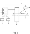
- Fig. 1 shows schematically and exemplarily an embodiment of a system for assisting in assessing a state of a subject's lung.
- the system 1 comprises an acquisition device 4 for acquiring dark field projection data in different acquisition directions.
- an acquisition device 4 for acquiring the dark field projection data
- well known techniques can be used, which employ a grating based x-ray phase contrast interferometer, like the techniques disclosed in the article " Grating-based X-ray dark-field imaging: a new paradigm in radiography" by A. Yaroshenko et al., Current Radiology Reports, 2:57 (2014 ).
- the system 1 further comprises a breathing state determination unit 7 for determining the breathing state of a subject 3 lying on a support means 2 like a patient table.
- the acquisition device 4 is adapted to acquire dark field projection data of the lung of the subject 3, wherein simultaneously breathing state values being indicative of the respective breathing state of the subject 3 are determined by the breathing state determination unit 7 such that the acquired dark field projection data can be assigned to the breathing states, i.e. to the breathing state values.
- the breathing state determination unit 7 can use a chest belt or another means for determining the respective breathing state.
- the breathing state values can be, for instance, times within a breathing cycle, they can be values being indicative of the absolute lung volume, or they can be dependent on a difference between a current lung volume and a reference lung volume which might be the lung volume for maximal inhale or maximal exhale.
- the system 1 further comprises a reconstruction unit 5 for reconstructing a dark field computed tomography image of the lung based on the acquired dark field projection data and the breathing state values such that for different breathing states a respective dark field computed tomography image of the lung is reconstructed.
- the reconstructed dark field image of the lung can be regarded as being an image having two or three spatial dimensions and a further dimension indicating the respective breathing state. Since the acquisition device 4, the breathing state determination unit 7 and the reconstruction unit 5 are adapted to generate and hence provide the dark field image of the lung, these components can be regarded as being components of a dark field image providing unit.
- the system 1 further comprises an image processing unit 6 being adapted to apply a motion correction algorithm to the provided dark field image, wherein in this embodiment the motion correction algorithm is an elastic motion correction algorithm.
- the dark field values for different breathing state values i.e. corresponding spatial images for respective breathing states
- This registration transformation i.e. the determined motion
- additional motion information being indicative of the motion of the lung obtained from a sensor like a spirometer is also used for correcting the motion artifacts.
- the motion correction technique disclosed in the article " Tracking lung tissue motion and expansion/compression with inverse consistent image registration and spirometry" by G. Christensena, Medical Physics, volume 34, number 6 (2007 ) can be used.
- the image processing unit 6 is further adapted to determine a differentiation image by differentiating the provided dark field image with respect to the breathing state values.
- the system 1 further comprises an input unit 8 for allowing a user to provide inputs into the system 1 like commands for starting or stopping a procedure for generating dark field images, determining differentiation images, et cetera.
- the system 1 comprises a display unit 9 for displaying the differentiation image and the provided dark field image.
- the display unit 9 can show a spatial distribution L ( x , b ref ) of the lung structure change for a reference state b ref , i.e. it can show a map illustrating a spatial lung structure change distribution for the reference state b ref , which can be useful for differentiating active and inactive lung areas.
- the display unit 9 can show the function L ( x ref , b ) for a reference spatial position x ref , which illustrates the temporal lung structure change at the reference position x ref .
- the reference position x ref and the reference breathing state value b ref may be selectable by a user via the input unit 8.
- the dark field projection data and the reconstructed dark field computed tomography image provide information about the microstructure of tissue below the spatial resolution of known attenuation projection data and known absorption computed tomography images. This provides valuable information in lung imaging where the air - soft tissue interfaces of the alveoli generate a strong dark field signature. Changes of this structure relative to healthy tissue caused, for instance, by lung diseases can be detected with high sensitivity. In particular, chronic obstructive pulmonary disease (COPD), pneumonia and/or lung fibrosis can be detected.
- COPD chronic obstructive pulmonary disease
- the breathing state of the lung influences the microstructure of the lung, thereby causing a dependency of the dark field signal on the breathing state.
- the system 1 described above with reference to Fig. 1 makes use of this dark field dependency on the breathing state, in order to obtain functional lung information, wherein based on the dark field values g ( x , b ), which could also be regarded as being local dark field values, the functional lung tissue parameter L ( x , b ) is determined, wherein this parameter L ( x , b ) could also be regarded as being a quantity defining the differential lung structure change.
- the local value L ( x , b ) indicates micro structural changes in the lung while breathing.
- the image processing unit 6 can be adapted to generate an integration image by integrating absolute image values of the differentiation image over the breathing state values.
- This integration can be performed over all spatial positions or only over a spatial region of interest. In the latter case the result can indicate micro structural changes in the region of interest while breathing.
- the dark field image providing unit 4, 5, 7 can be adapted to determine the dark field image g ( x , b ) for a set of b 's and to numerically determine the differentiation image from this discrete set in accordance with following equation: L x ⁇ b ⁇ ⁇ ⁇ g x ⁇ b ⁇ ⁇ b , wherein ⁇ g ( x , b ) denotes the difference between a) the dark field value at the spatial position x for the breathing state value b and b) the dark field value at the spatial position x for an adjacent breathing state.
- ⁇ b denotes a difference between the breathing state b and the adjacent breathing state. Since the lung tissue will be deformed during the breathing, a motion correction is preferentially applied before this calculation as described above.
- the variable b can also be defined in another way for indicating, for instance, the absolute lung volume or the time within a breathing cycle.
- Fig. 2 shows a flowchart exemplarily illustrating an embodiment of a method for assisting in assessing a state of a subject's lung.
- a dark field image of the lung is provided by the dark field image providing unit 4, 5, 7, wherein the provided dark field image comprises dark field values for different spatial positions and for different breathing state values being indicative of a breathing state of the lung.
- a motion correction algorithm is applied to the provided dark field image.
- an elastic motion correction algorithm is applied to the provided dark field image.
- a differentiation image is determined by differentiating the provided dark field image with respect to the breathing state values by the image processing unit 6.
- the differentiation image is shown on the display unit 9.
- the provided dark field image is a computed tomography image
- the provided dark field image can also be a projection image, wherein in this case the dark field projection image is differentiated with respect to the breathing state value for determining the differential lung structure change L ( x , b ).
- the dark field computed tomography image has been reconstructed based on dark field projection images
- the dark field computed tomography image can also be directly reconstructed from raw data obtained from the x-ray based phase contrast interferometer, i.e. without generating intermediate dark field projection images.
- a single unit or device may fulfill the functions of several items recited in the claims.
- the mere fact that certain measures are recited in mutually different dependent claims does not indicate that a combination of these measures cannot be used to advantage.
- Operations like the reconstruction of a computed tomography image, the determination of a differentiation image, the motion correction, et cetera performed by one or several units or devices can be performed by any other number of units or devices.
- These operations and/or the control of the system for assisting in assessing a state of a subject's lung in accordance with the method for assisting in assessing a state of a subject's lung can be implemented as program code means of a computer program and/or as dedicated hardware.
- a computer program may be stored/distributed on a suitable medium, such as an optical storage medium or a solid-state medium, supplied together with or as part of other hardware, but may also be distributed in other forms, such as via the Internet or other wired or wireless telecommunication systems.
- a suitable medium such as an optical storage medium or a solid-state medium, supplied together with or as part of other hardware, but may also be distributed in other forms, such as via the Internet or other wired or wireless telecommunication systems.
- the invention relates to a system for assisting in assessing a state of a subject's lung.
- the system is adapted to process a dark field image, which comprises dark field values for different spatial positions and for different breathing state values, such that a differentiation image is generated by differentiating the provided dark field image with respect to the breathing state values.
- This differentiation can lead to a functional image which can be used for detecting changes of air - soft tissue interfaces, which might be caused by a lung disease, with high sensitivity. This allows for an improved assisting in assessing a state of a subject's lung.
Description
- The invention relates to a system, method and computer program for assisting in assessing a state of a subject's lung.
- A system for assisting in assessing a state of a subject's lung is, for instance, an absorption computed tomography imaging system. An absorption computed tomography imaging system comprises an x-ray source and an x-ray detector, which are rotatable around a subject's lung to be imaged such that x-rays generated by the x-ray source traverse the lung in different directions. The x-ray detector detects the x-rays after having traversed the lung and generates projection data based on the detected x-rays, wherein a reconstruction unit reconstructs an absorption computed tomography image based on the generated projection data.
- In the absorption computed tomography image air - soft tissue interfaces are not very well detectable, thereby reducing the usability of the absorption computed tomography image for assisting in assessing a state of a subject's lung.
- It is an object of the present invention to provide a system, method and computer program which allow for an improved assisting in assessing a state of a subject's lung.
- In a first aspect of the present invention a system for assisting in assessing a state of a subject's lung is presented, wherein the system comprises:
- a dark field image providing unit for providing a dark field image of the lung, wherein the provided dark field image comprises dark field values for different spatial positions and for different breathing state values being indicative of a breathing state of the lung,
- an image processing unit for processing the provided dark field image, wherein the image processing unit is adapted to determine a differentiation image by differentiating the provided dark field image with respect to the breathing state values.
- In the dark field image air - soft tissue interfaces of the lung, particularly of the alveoli, are very well detectable. By differentiating this dark field image with respect to the breathing state values a functional image can be provided, which can be used for detecting changes of air - soft tissue interfaces, which might be caused by a lung disease, with high sensitivity. This allows for an improved assisting in assessing a state of a subject's lung.
- The dark field image providing unit can be a storing unit, in which the dark field image is stored, in order to allow the dark field image providing unit to provide the dark field image. The dark field image providing unit can also be a receiving unit for receiving the dark field image from an imaging system being adapted to generate the dark field image. The dark field image providing unit can also be the imaging system itself. The imaging system can be adapted to generate as the dark field image a projection image and/or a computed tomography image.
- In an embodiment the dark field image providing unit is adapted to provide the dark field image such that the dark field values are provided for different breathing state values being dependent on a difference between a current lung volume and a reference lung volume. For instance, the reference lung volume can be the lung volume for maximal inhale or maximal exhale. It is also possible that the dark field image providing unit is adapted to provide the dark field image such that the dark field values are provided for different breathing state values being dependent on an absolute lung volume. Moreover, the dark field image providing unit can be adapted to provide the dark field image such that the dark field values are provided for different breathing state values being dependent on a time within a breathing cycle. If the dark field image is differentiated with respect to these kinds of breathing state values, the usability of the differentiation image for detecting changes in air - soft tissue interfaces and hence for assisting in assessing a state of a subject's lung can be further improved.
- The image processing unit can be adapted to integrate absolute image values of the differentiation image over the breathing state values and/or over the spatial positions. This integration can be performed, for instance, over the entire breathing state values or the entire spatial positions, respectively, or over a spatial region of interest or over an interesting region of breathing state values, respectively. The integration over the breathing state values yields an integration image. The integration result allows for a further improved assisting in assessing a state of a subject's lung. It should be noted that the expression "A and/or B" preferentially includes following options a) A without B, b) B without A, and c) A and B.
- It is preferred that the image processing unit is further adapted to apply a motion correction algorithm to the provided dark field image before determining the differentiation image, wherein the motion correction algorithm is preferentially an elastic motion correction algorithm. By correcting motion artifacts in the provided dark field image before determining the differentiation image, the quality of the differentiation image can be further improved, thereby allowing for a further improved assistance in assessing a state of a subject's lung.
- In another aspect of the present invention a method for assisting in assessing a state of a subject's lung is presented, wherein the method comprises:
- providing a dark field image of the lung by a dark field image providing unit, wherein the provided dark field image comprises dark field values for different spatial positions and for different breathing state values being indicative of a breathing state of the lung,
- processing the provided dark field image by an image processing unit, wherein the image processing unit is adapted to determine a differentiation image by differentiating the provided dark field image with respect to the breathing state values.
- In a further aspect of the present invention a computer program for assisting in assessing a state of a subject's lung is presented, wherein the computer program comprises program code means for causing a system as defined in
claim 1 to carry out the method as defined in claim 9, when the computer program is run on the system. - It shall be understood that the system of
claim 1, the method of claim 9 and the computer program of claim 10, have similar and/or identical preferred embodiments, in particular, as defined in the dependent claims. - It shall be understood that a preferred embodiment of the present invention can also be any combination of the dependent claims or above embodiments with the respective independent claim.
- These and other aspects of the invention will be apparent from and elucidated with reference to the embodiments described hereinafter.
- In the following drawings:
-
Fig. 1 shows schematically and exemplarily an embodiment of a system for assisting in assessing a state of a subject's lung, and -
Fig. 2 shows a flowchart exemplarily illustrating a method for assisting in assessing a state of a subject's lung. -
Fig. 1 shows schematically and exemplarily an embodiment of a system for assisting in assessing a state of a subject's lung. Thesystem 1 comprises anacquisition device 4 for acquiring dark field projection data in different acquisition directions. For acquiring the dark field projection data well known techniques can be used, which employ a grating based x-ray phase contrast interferometer, like the techniques disclosed in the article "Grating-based X-ray dark-field imaging: a new paradigm in radiography" by A. Yaroshenko et al., Current Radiology Reports, 2:57 (2014). - The
system 1 further comprises a breathingstate determination unit 7 for determining the breathing state of asubject 3 lying on a support means 2 like a patient table. Theacquisition device 4 is adapted to acquire dark field projection data of the lung of thesubject 3, wherein simultaneously breathing state values being indicative of the respective breathing state of thesubject 3 are determined by the breathingstate determination unit 7 such that the acquired dark field projection data can be assigned to the breathing states, i.e. to the breathing state values. The breathingstate determination unit 7 can use a chest belt or another means for determining the respective breathing state. The breathing state values can be, for instance, times within a breathing cycle, they can be values being indicative of the absolute lung volume, or they can be dependent on a difference between a current lung volume and a reference lung volume which might be the lung volume for maximal inhale or maximal exhale. - The
system 1 further comprises areconstruction unit 5 for reconstructing a dark field computed tomography image of the lung based on the acquired dark field projection data and the breathing state values such that for different breathing states a respective dark field computed tomography image of the lung is reconstructed. The reconstructed dark field image of the lung can be regarded as being an image having two or three spatial dimensions and a further dimension indicating the respective breathing state. Since theacquisition device 4, the breathingstate determination unit 7 and thereconstruction unit 5 are adapted to generate and hence provide the dark field image of the lung, these components can be regarded as being components of a dark field image providing unit. - The
system 1 further comprises animage processing unit 6 being adapted to apply a motion correction algorithm to the provided dark field image, wherein in this embodiment the motion correction algorithm is an elastic motion correction algorithm. In particular, the dark field values for different breathing state values, i.e. corresponding spatial images for respective breathing states, can be elastically registered to each other, in order to determine a registration transformation being indicative of the motion of the lung tissue. This registration transformation, i.e. the determined motion, can then be used for correcting motion artifacts in the provided dark field image. Optionally, additional motion information being indicative of the motion of the lung obtained from a sensor like a spirometer is also used for correcting the motion artifacts. For instance, the motion correction technique disclosed in the article "Tracking lung tissue motion and expansion/compression with inverse consistent image registration and spirometry" by G. Christensena, Medical Physics, volume 34, number 6 (2007) can be used. - The
image processing unit 6 is further adapted to determine a differentiation image by differentiating the provided dark field image with respect to the breathing state values. This differentiation can be described by following equation:
x ,b) denotes dark field values of the dark field image for different spatial positionsx and for different breathing state values b and wherein L(x ,b) denotes image values of the differentiation image for the different spatial positionsx and the different breathing state values b . - The
system 1 further comprises aninput unit 8 for allowing a user to provide inputs into thesystem 1 like commands for starting or stopping a procedure for generating dark field images, determining differentiation images, et cetera. Moreover, thesystem 1 comprises a display unit 9 for displaying the differentiation image and the provided dark field image. In particular, the display unit 9 can show a spatial distribution L(x , b ref) of the lung structure change for a reference state b ref, i.e. it can show a map illustrating a spatial lung structure change distribution for the reference state b ref, which can be useful for differentiating active and inactive lung areas. Moreover, the display unit 9 can show the function L(x ref , b) for a reference spatial positionx ref, which illustrates the temporal lung structure change at the reference positionx ref. The reference positionx ref and the reference breathing state value b ref may be selectable by a user via theinput unit 8. - The dark field projection data and the reconstructed dark field computed tomography image provide information about the microstructure of tissue below the spatial resolution of known attenuation projection data and known absorption computed tomography images. This provides valuable information in lung imaging where the air - soft tissue interfaces of the alveoli generate a strong dark field signature. Changes of this structure relative to healthy tissue caused, for instance, by lung diseases can be detected with high sensitivity. In particular, chronic obstructive pulmonary disease (COPD), pneumonia and/or lung fibrosis can be detected.
- The breathing state of the lung influences the microstructure of the lung, thereby causing a dependency of the dark field signal on the breathing state. The
system 1 described above with reference toFig. 1 makes use of this dark field dependency on the breathing state, in order to obtain functional lung information, wherein based on the dark field values g(x , b), which could also be regarded as being local dark field values, the functional lung tissue parameter L(x , b) is determined, wherein this parameter L(x , b) could also be regarded as being a quantity defining the differential lung structure change. The local value L(x , b) indicates micro structural changes in the lung while breathing. - The
image processing unit 6 can be adapted to generate an integration image by integrating absolute image values of the differentiation image over the breathing state values. Thus, for instance, an integration image I(x ) may be generated in accordance with following equation:
- In this image lung areas that do not actively contribute to breathing may show small values compared to other lung areas such that the integration image I(
x ) is very well usable for assisting in assessing the state of the lung. It is also possible to generate a breathing state dependent integration value I(b) by integrating over the spatial positions in accordance with following equation:
- This integration can be performed over all spatial positions or only over a spatial region of interest. In the latter case the result can indicate micro structural changes in the region of interest while breathing.
- The dark field
image providing unit x , b) for a set of b 's and to numerically determine the differentiation image from this discrete set in accordance with following equation:
x , b) denotes the difference between a) the dark field value at the spatial positionx for the breathing state value b and b) the dark field value at the spatial positionx for an adjacent breathing state. Correspondingly, Δb denotes a difference between the breathing state b and the adjacent breathing state. Since the lung tissue will be deformed during the breathing, a motion correction is preferentially applied before this calculation as described above. - The breathing state value may be defined in accordance with following equation:

-
Fig. 2 shows a flowchart exemplarily illustrating an embodiment of a method for assisting in assessing a state of a subject's lung. - In step 101 a dark field image of the lung is provided by the dark field
image providing unit image processing unit 6. Instep 104 the differentiation image is shown on the display unit 9. - Although in above described embodiments the provided dark field image is a computed tomography image, the provided dark field image can also be a projection image, wherein in this case the dark field projection image is differentiated with respect to the breathing state value for determining the differential lung structure change L(
x , b). Moreover, although in above described embodiments the dark field computed tomography image has been reconstructed based on dark field projection images, the dark field computed tomography image can also be directly reconstructed from raw data obtained from the x-ray based phase contrast interferometer, i.e. without generating intermediate dark field projection images. For more details of corresponding known dark field techniques reference is made to the above mentioned article by A. Yaroshenko et al. - Other variations to the disclosed embodiments can be understood and effected by those skilled in the art in practicing the claimed invention, from a study of the drawings, the disclosure, and the appended claims.
- In the claims, the word "comprising" does not exclude other elements or steps, and the indefinite article "a" or "an" does not exclude a plurality.
- A single unit or device may fulfill the functions of several items recited in the claims. The mere fact that certain measures are recited in mutually different dependent claims does not indicate that a combination of these measures cannot be used to advantage.
- Operations like the reconstruction of a computed tomography image, the determination of a differentiation image, the motion correction, et cetera performed by one or several units or devices can be performed by any other number of units or devices. These operations and/or the control of the system for assisting in assessing a state of a subject's lung in accordance with the method for assisting in assessing a state of a subject's lung can be implemented as program code means of a computer program and/or as dedicated hardware.
- A computer program may be stored/distributed on a suitable medium, such as an optical storage medium or a solid-state medium, supplied together with or as part of other hardware, but may also be distributed in other forms, such as via the Internet or other wired or wireless telecommunication systems.
- Any reference signs in the claims should not be construed as limiting the scope.
- The invention relates to a system for assisting in assessing a state of a subject's lung. The system is adapted to process a dark field image, which comprises dark field values for different spatial positions and for different breathing state values, such that a differentiation image is generated by differentiating the provided dark field image with respect to the breathing state values. This differentiation can lead to a functional image which can be used for detecting changes of air - soft tissue interfaces, which might be caused by a lung disease, with high sensitivity. This allows for an improved assisting in assessing a state of a subject's lung.
Claims (10)
- A system for assisting in assessing a state of a subject's lung, the system comprising:- a dark field image providing unit (4, 5, 7) for providing a dark field image of the lung, wherein the provided dark field image comprises dark field values for different spatial positions and for different breathing state values being indicative of a breathing state of the lung,- an image processing unit (6) for processing the provided dark field image, wherein the image processing unit (6) is adapted to determine a differentiation image by differentiating the provided dark field image with respect to the breathing state values.
- The system as defined in claim 1, wherein the dark field image providing unit (4, 5, 7) is adapted to provide the dark field image such that the dark field values are provided for different breathing state values being dependent on a difference between a current lung volume and a reference lung volume.
- The system as defined in claim 2, wherein the reference lung volume is the lung volume for maximal inhale or maximal exhale.
- The system as defined in claim 1, wherein the dark field image providing unit (4, 5, 7) is adapted to provide the dark field image such that the dark field values are provided for different breathing state values being dependent on an absolute lung volume.
- The system as defined in claim 1, wherein the dark field image providing unit (4, 5, 7) is adapted to provide the dark field image such that the dark field values are provided for different breathing state values being dependent on a time within a breathing cycle.
- The system as defined in claim 1, wherein the image processing unit (6) is further adapted to integrate absolute image values of the differentiation image over the breathing state values and/or over the spatial positions.
- The system as defined in claim 1, wherein the image processing unit (6) is further adapted to apply a motion correction algorithm to the provided dark field image before determining the differentiation image.
- The system as defined in claim 7, wherein the motion correction algorithm is an elastic motion correction algorithm.
- A method for assisting in assessing a state of a subject's lung, the method comprising:- providing a dark field image of the lung by a dark field image providing unit (4, 5, 7), wherein the provided dark field image comprises dark field values for different spatial positions and for different breathing state values being indicative of a breathing state of the lung,- processing the provided dark field image by an image processing unit (6), wherein the image processing unit (6) is adapted to determine a differentiation image by differentiating the provided dark field image with respect to the breathing state values.
- A computer program for assisting in assessing a state of a subject's lung, the computer program comprising program code means for causing a system as defined in claim 1 to carry out the method as defined in claim 9, when the computer program is run on the system.
Applications Claiming Priority (2)
| Application Number | Priority Date | Filing Date | Title |
|---|---|---|---|
| EP16206673 | 2016-12-23 | ||
| PCT/EP2017/083965 WO2018115215A1 (en) | 2016-12-23 | 2017-12-21 | System and method for assisting in assessing a state of a subject's lung |
Publications (2)
| Publication Number | Publication Date |
|---|---|
| EP3386392A1 EP3386392A1 (en) | 2018-10-17 |
| EP3386392B1 true EP3386392B1 (en) | 2019-06-19 |
Family
ID=57609766
Family Applications (1)
| Application Number | Title | Priority Date | Filing Date |
|---|---|---|---|
| EP17822668.4A Active EP3386392B1 (en) | 2016-12-23 | 2017-12-21 | System and method for assisting in assessing a state of a subject's lung |
Country Status (5)
| Country | Link |
|---|---|
| US (1) | US10610183B2 (en) |
| EP (1) | EP3386392B1 (en) |
| JP (1) | JP6700494B2 (en) |
| CN (1) | CN108471999B (en) |
| WO (1) | WO2018115215A1 (en) |
Families Citing this family (2)
| Publication number | Priority date | Publication date | Assignee | Title |
|---|---|---|---|---|
| EP3622891A1 (en) * | 2018-09-13 | 2020-03-18 | Koninklijke Philips N.V. | Calculation device for determining ventilation defects |
| EP3656308A1 (en) * | 2018-11-20 | 2020-05-27 | Koninklijke Philips N.V. | Apparatus for alveoli based visualization in dark-field or phase-contrast x-ray imaging |
Family Cites Families (8)
| Publication number | Priority date | Publication date | Assignee | Title |
|---|---|---|---|---|
| WO2006137294A1 (en) * | 2005-06-21 | 2006-12-28 | National University Corporation Kanazawa University | X-ray diagnosis supporting apparatus, program, and recording medium |
| US7693256B2 (en) * | 2008-03-19 | 2010-04-06 | C-Rad Innovation Ab | Phase-contrast X-ray imaging |
| WO2012026146A1 (en) * | 2010-08-27 | 2012-03-01 | コニカミノルタエムジー株式会社 | Thoracic diagnosis assistance system and program |
| US9076201B1 (en) * | 2012-03-30 | 2015-07-07 | University Of Louisville Research Foundation, Inc. | Volumetric deformable registration method for thoracic 4-D computed tomography images and method of determining regional lung function |
| WO2013150911A1 (en) * | 2012-04-04 | 2013-10-10 | コニカミノルタ株式会社 | Image generation device and program |
| KR102114415B1 (en) * | 2013-01-29 | 2020-05-22 | 삼성전자주식회사 | Method and Apparatus for medical image registration |
| WO2016058838A1 (en) | 2014-10-13 | 2016-04-21 | Koninklijke Philips N.V. | Grating device for phase contrast and/or dark-field imaging of a movable object |
| CN205019076U (en) | 2015-10-10 | 2016-02-10 | 解泽欣琪 | Phase contrast is like medical clinical application apparatus who reaches dark field image |
-
2017
- 2017-12-21 WO PCT/EP2017/083965 patent/WO2018115215A1/en active Application Filing
- 2017-12-21 EP EP17822668.4A patent/EP3386392B1/en active Active
- 2017-12-21 CN CN201780007626.7A patent/CN108471999B/en active Active
- 2017-12-21 JP JP2019533609A patent/JP6700494B2/en active Active
- 2017-12-21 US US16/070,897 patent/US10610183B2/en active Active
Non-Patent Citations (1)
| Title |
|---|
| None * |
Also Published As
| Publication number | Publication date |
|---|---|
| US10610183B2 (en) | 2020-04-07 |
| EP3386392A1 (en) | 2018-10-17 |
| WO2018115215A1 (en) | 2018-06-28 |
| CN108471999A (en) | 2018-08-31 |
| US20190298292A1 (en) | 2019-10-03 |
| JP2020501818A (en) | 2020-01-23 |
| JP6700494B2 (en) | 2020-05-27 |
| CN108471999B (en) | 2019-07-02 |
Similar Documents
| Publication | Publication Date | Title |
|---|---|---|
| US7454043B2 (en) | Image processing unit and method of associating stored images with current images | |
| JP4717486B2 (en) | Image reconstruction apparatus for X-ray apparatus and local 3D reconstruction method of target range | |
| US10052032B2 (en) | Stenosis therapy planning | |
| US8295911B2 (en) | Motion correction for tomographic medical image data of a patient | |
| JP6198989B2 (en) | Grating device for phase contrast and / or dark field imaging of movable objects | |
| EP2293248A1 (en) | Motion monitoring system for monitoring motion within a region of interest | |
| JP2010517655A (en) | Motion estimation in treatment planning | |
| JP2011161220A (en) | Image processing apparatus, x-ray computed tomography apparatus, and image processing program | |
| EP2867861A1 (en) | Motion parameter estimation | |
| KR101665513B1 (en) | Computer tomography apparatus and method for reconstructing a computer tomography image thereof | |
| EP3386392B1 (en) | System and method for assisting in assessing a state of a subject's lung | |
| US8175684B2 (en) | Method for processing images and associated medical imaging system | |
| US10245001B2 (en) | Generation of a three-dimensional reconstruction of a body part by an X-ray machine | |
| JP6546201B2 (en) | Imaging system for generating an image of an object | |
| RU2727244C2 (en) | Object visualization device | |
| EP3849420B1 (en) | Calculation device for determining ventilation defects | |
| JP6852545B2 (en) | Image display system and image processing equipment | |
| JP2021520911A (en) | Error tracking and calibration of X-ray system | |
| EP4184454A1 (en) | Weight estimation of a patient | |
| TW202137935A (en) | Method for gating in tomographic imaging system |
Legal Events
| Date | Code | Title | Description |
|---|---|---|---|
| STAA | Information on the status of an ep patent application or granted ep patent |
Free format text: STATUS: UNKNOWN |
|
| STAA | Information on the status of an ep patent application or granted ep patent |
Free format text: STATUS: THE INTERNATIONAL PUBLICATION HAS BEEN MADE |
|
| PUAI | Public reference made under article 153(3) epc to a published international application that has entered the european phase |
Free format text: ORIGINAL CODE: 0009012 |
|
| STAA | Information on the status of an ep patent application or granted ep patent |
Free format text: STATUS: REQUEST FOR EXAMINATION WAS MADE |
|
| 17P | Request for examination filed |
Effective date: 20180711 |
|
| AK | Designated contracting states |
Kind code of ref document: A1 Designated state(s): AL AT BE BG CH CY CZ DE DK EE ES FI FR GB GR HR HU IE IS IT LI LT LU LV MC MK MT NL NO PL PT RO RS SE SI SK SM TR |
|
| AX | Request for extension of the european patent |
Extension state: BA ME |
|
| GRAP | Despatch of communication of intention to grant a patent |
Free format text: ORIGINAL CODE: EPIDOSNIGR1 |
|
| STAA | Information on the status of an ep patent application or granted ep patent |
Free format text: STATUS: GRANT OF PATENT IS INTENDED |
|
| INTG | Intention to grant announced |
Effective date: 20190114 |
|
| GRAS | Grant fee paid |
Free format text: ORIGINAL CODE: EPIDOSNIGR3 |
|
| GRAA | (expected) grant |
Free format text: ORIGINAL CODE: 0009210 |
|
| STAA | Information on the status of an ep patent application or granted ep patent |
Free format text: STATUS: THE PATENT HAS BEEN GRANTED |
|
| DAV | Request for validation of the european patent (deleted) | ||
| DAX | Request for extension of the european patent (deleted) | ||
| AK | Designated contracting states |
Kind code of ref document: B1 Designated state(s): AL AT BE BG CH CY CZ DE DK EE ES FI FR GB GR HR HU IE IS IT LI LT LU LV MC MK MT NL NO PL PT RO RS SE SI SK SM TR |
|
| REG | Reference to a national code |
Ref country code: GB Ref legal event code: FG4D |
|
| REG | Reference to a national code |
Ref country code: CH Ref legal event code: EP |
|
| REG | Reference to a national code |
Ref country code: IE Ref legal event code: FG4D |
|
| REG | Reference to a national code |
Ref country code: AT Ref legal event code: REF Ref document number: 1144468 Country of ref document: AT Kind code of ref document: T Effective date: 20190715 |
|
| REG | Reference to a national code |
Ref country code: DE Ref legal event code: R096 Ref document number: 602017004712 Country of ref document: DE |
|
| REG | Reference to a national code |
Ref country code: GB Ref legal event code: 746 Effective date: 20190730 |
|
| REG | Reference to a national code |
Ref country code: DE Ref legal event code: R084 Ref document number: 602017004712 Country of ref document: DE |
|
| REG | Reference to a national code |
Ref country code: NL Ref legal event code: MP Effective date: 20190619 |
|
| PG25 | Lapsed in a contracting state [announced via postgrant information from national office to epo] |
Ref country code: LT Free format text: LAPSE BECAUSE OF FAILURE TO SUBMIT A TRANSLATION OF THE DESCRIPTION OR TO PAY THE FEE WITHIN THE PRESCRIBED TIME-LIMIT Effective date: 20190619 Ref country code: SE Free format text: LAPSE BECAUSE OF FAILURE TO SUBMIT A TRANSLATION OF THE DESCRIPTION OR TO PAY THE FEE WITHIN THE PRESCRIBED TIME-LIMIT Effective date: 20190619 Ref country code: HR Free format text: LAPSE BECAUSE OF FAILURE TO SUBMIT A TRANSLATION OF THE DESCRIPTION OR TO PAY THE FEE WITHIN THE PRESCRIBED TIME-LIMIT Effective date: 20190619 Ref country code: NO Free format text: LAPSE BECAUSE OF FAILURE TO SUBMIT A TRANSLATION OF THE DESCRIPTION OR TO PAY THE FEE WITHIN THE PRESCRIBED TIME-LIMIT Effective date: 20190919 Ref country code: FI Free format text: LAPSE BECAUSE OF FAILURE TO SUBMIT A TRANSLATION OF THE DESCRIPTION OR TO PAY THE FEE WITHIN THE PRESCRIBED TIME-LIMIT Effective date: 20190619 Ref country code: AL Free format text: LAPSE BECAUSE OF FAILURE TO SUBMIT A TRANSLATION OF THE DESCRIPTION OR TO PAY THE FEE WITHIN THE PRESCRIBED TIME-LIMIT Effective date: 20190619 |
|
| REG | Reference to a national code |
Ref country code: LT Ref legal event code: MG4D |
|
| PG25 | Lapsed in a contracting state [announced via postgrant information from national office to epo] |
Ref country code: GR Free format text: LAPSE BECAUSE OF FAILURE TO SUBMIT A TRANSLATION OF THE DESCRIPTION OR TO PAY THE FEE WITHIN THE PRESCRIBED TIME-LIMIT Effective date: 20190920 Ref country code: LV Free format text: LAPSE BECAUSE OF FAILURE TO SUBMIT A TRANSLATION OF THE DESCRIPTION OR TO PAY THE FEE WITHIN THE PRESCRIBED TIME-LIMIT Effective date: 20190619 Ref country code: RS Free format text: LAPSE BECAUSE OF FAILURE TO SUBMIT A TRANSLATION OF THE DESCRIPTION OR TO PAY THE FEE WITHIN THE PRESCRIBED TIME-LIMIT Effective date: 20190619 Ref country code: BG Free format text: LAPSE BECAUSE OF FAILURE TO SUBMIT A TRANSLATION OF THE DESCRIPTION OR TO PAY THE FEE WITHIN THE PRESCRIBED TIME-LIMIT Effective date: 20190919 |
|
| REG | Reference to a national code |
Ref country code: AT Ref legal event code: MK05 Ref document number: 1144468 Country of ref document: AT Kind code of ref document: T Effective date: 20190619 |
|
| PG25 | Lapsed in a contracting state [announced via postgrant information from national office to epo] |
Ref country code: CZ Free format text: LAPSE BECAUSE OF FAILURE TO SUBMIT A TRANSLATION OF THE DESCRIPTION OR TO PAY THE FEE WITHIN THE PRESCRIBED TIME-LIMIT Effective date: 20190619 Ref country code: RO Free format text: LAPSE BECAUSE OF FAILURE TO SUBMIT A TRANSLATION OF THE DESCRIPTION OR TO PAY THE FEE WITHIN THE PRESCRIBED TIME-LIMIT Effective date: 20190619 Ref country code: SK Free format text: LAPSE BECAUSE OF FAILURE TO SUBMIT A TRANSLATION OF THE DESCRIPTION OR TO PAY THE FEE WITHIN THE PRESCRIBED TIME-LIMIT Effective date: 20190619 Ref country code: NL Free format text: LAPSE BECAUSE OF FAILURE TO SUBMIT A TRANSLATION OF THE DESCRIPTION OR TO PAY THE FEE WITHIN THE PRESCRIBED TIME-LIMIT Effective date: 20190619 Ref country code: AT Free format text: LAPSE BECAUSE OF FAILURE TO SUBMIT A TRANSLATION OF THE DESCRIPTION OR TO PAY THE FEE WITHIN THE PRESCRIBED TIME-LIMIT Effective date: 20190619 Ref country code: EE Free format text: LAPSE BECAUSE OF FAILURE TO SUBMIT A TRANSLATION OF THE DESCRIPTION OR TO PAY THE FEE WITHIN THE PRESCRIBED TIME-LIMIT Effective date: 20190619 Ref country code: PT Free format text: LAPSE BECAUSE OF FAILURE TO SUBMIT A TRANSLATION OF THE DESCRIPTION OR TO PAY THE FEE WITHIN THE PRESCRIBED TIME-LIMIT Effective date: 20191021 |
|
| PG25 | Lapsed in a contracting state [announced via postgrant information from national office to epo] |
Ref country code: IT Free format text: LAPSE BECAUSE OF FAILURE TO SUBMIT A TRANSLATION OF THE DESCRIPTION OR TO PAY THE FEE WITHIN THE PRESCRIBED TIME-LIMIT Effective date: 20190619 Ref country code: ES Free format text: LAPSE BECAUSE OF FAILURE TO SUBMIT A TRANSLATION OF THE DESCRIPTION OR TO PAY THE FEE WITHIN THE PRESCRIBED TIME-LIMIT Effective date: 20190619 Ref country code: SM Free format text: LAPSE BECAUSE OF FAILURE TO SUBMIT A TRANSLATION OF THE DESCRIPTION OR TO PAY THE FEE WITHIN THE PRESCRIBED TIME-LIMIT Effective date: 20190619 Ref country code: IS Free format text: LAPSE BECAUSE OF FAILURE TO SUBMIT A TRANSLATION OF THE DESCRIPTION OR TO PAY THE FEE WITHIN THE PRESCRIBED TIME-LIMIT Effective date: 20191019 |
|
| RAP2 | Party data changed (patent owner data changed or rights of a patent transferred) |
Owner name: KONINKLIJKE PHILIPS N.V. |
|
| PG25 | Lapsed in a contracting state [announced via postgrant information from national office to epo] |
Ref country code: TR Free format text: LAPSE BECAUSE OF FAILURE TO SUBMIT A TRANSLATION OF THE DESCRIPTION OR TO PAY THE FEE WITHIN THE PRESCRIBED TIME-LIMIT Effective date: 20190619 |
|
| PG25 | Lapsed in a contracting state [announced via postgrant information from national office to epo] |
Ref country code: DK Free format text: LAPSE BECAUSE OF FAILURE TO SUBMIT A TRANSLATION OF THE DESCRIPTION OR TO PAY THE FEE WITHIN THE PRESCRIBED TIME-LIMIT Effective date: 20190619 Ref country code: PL Free format text: LAPSE BECAUSE OF FAILURE TO SUBMIT A TRANSLATION OF THE DESCRIPTION OR TO PAY THE FEE WITHIN THE PRESCRIBED TIME-LIMIT Effective date: 20190619 |
|
| PG25 | Lapsed in a contracting state [announced via postgrant information from national office to epo] |
Ref country code: IS Free format text: LAPSE BECAUSE OF FAILURE TO SUBMIT A TRANSLATION OF THE DESCRIPTION OR TO PAY THE FEE WITHIN THE PRESCRIBED TIME-LIMIT Effective date: 20200224 |
|
| REG | Reference to a national code |
Ref country code: DE Ref legal event code: R097 Ref document number: 602017004712 Country of ref document: DE |
|
| PLBE | No opposition filed within time limit |
Free format text: ORIGINAL CODE: 0009261 |
|
| STAA | Information on the status of an ep patent application or granted ep patent |
Free format text: STATUS: NO OPPOSITION FILED WITHIN TIME LIMIT |
|
| PG2D | Information on lapse in contracting state deleted |
Ref country code: IS |
|
| 26N | No opposition filed |
Effective date: 20200603 |
|
| REG | Reference to a national code |
Ref country code: BE Ref legal event code: MM Effective date: 20191231 |
|
| PG25 | Lapsed in a contracting state [announced via postgrant information from national office to epo] |
Ref country code: MC Free format text: LAPSE BECAUSE OF FAILURE TO SUBMIT A TRANSLATION OF THE DESCRIPTION OR TO PAY THE FEE WITHIN THE PRESCRIBED TIME-LIMIT Effective date: 20190619 Ref country code: SI Free format text: LAPSE BECAUSE OF FAILURE TO SUBMIT A TRANSLATION OF THE DESCRIPTION OR TO PAY THE FEE WITHIN THE PRESCRIBED TIME-LIMIT Effective date: 20190619 |
|
| PG25 | Lapsed in a contracting state [announced via postgrant information from national office to epo] |
Ref country code: IE Free format text: LAPSE BECAUSE OF NON-PAYMENT OF DUE FEES Effective date: 20191221 Ref country code: LU Free format text: LAPSE BECAUSE OF NON-PAYMENT OF DUE FEES Effective date: 20191221 |
|
| PG25 | Lapsed in a contracting state [announced via postgrant information from national office to epo] |
Ref country code: BE Free format text: LAPSE BECAUSE OF NON-PAYMENT OF DUE FEES Effective date: 20191231 |
|
| PG25 | Lapsed in a contracting state [announced via postgrant information from national office to epo] |
Ref country code: CY Free format text: LAPSE BECAUSE OF FAILURE TO SUBMIT A TRANSLATION OF THE DESCRIPTION OR TO PAY THE FEE WITHIN THE PRESCRIBED TIME-LIMIT Effective date: 20190619 |
|
| PG25 | Lapsed in a contracting state [announced via postgrant information from national office to epo] |
Ref country code: HU Free format text: LAPSE BECAUSE OF FAILURE TO SUBMIT A TRANSLATION OF THE DESCRIPTION OR TO PAY THE FEE WITHIN THE PRESCRIBED TIME-LIMIT; INVALID AB INITIO Effective date: 20171221 Ref country code: MT Free format text: LAPSE BECAUSE OF FAILURE TO SUBMIT A TRANSLATION OF THE DESCRIPTION OR TO PAY THE FEE WITHIN THE PRESCRIBED TIME-LIMIT Effective date: 20190619 |
|
| REG | Reference to a national code |
Ref country code: CH Ref legal event code: PL |
|
| PG25 | Lapsed in a contracting state [announced via postgrant information from national office to epo] |
Ref country code: CH Free format text: LAPSE BECAUSE OF NON-PAYMENT OF DUE FEES Effective date: 20201231 Ref country code: LI Free format text: LAPSE BECAUSE OF NON-PAYMENT OF DUE FEES Effective date: 20201231 |
|
| PG25 | Lapsed in a contracting state [announced via postgrant information from national office to epo] |
Ref country code: MK Free format text: LAPSE BECAUSE OF FAILURE TO SUBMIT A TRANSLATION OF THE DESCRIPTION OR TO PAY THE FEE WITHIN THE PRESCRIBED TIME-LIMIT Effective date: 20190619 |
|
| PGFP | Annual fee paid to national office [announced via postgrant information from national office to epo] |
Ref country code: DE Payment date: 20221227 Year of fee payment: 6 |
|
| PGFP | Annual fee paid to national office [announced via postgrant information from national office to epo] |
Ref country code: GB Payment date: 20231219 Year of fee payment: 7 |
|
| PGFP | Annual fee paid to national office [announced via postgrant information from national office to epo] |
Ref country code: FR Payment date: 20231226 Year of fee payment: 7 |