EP3384010B1 - Use of an integrated microfluidic chip for analysis of cell motility and prediction and prognosis of patient survival - Google Patents
Use of an integrated microfluidic chip for analysis of cell motility and prediction and prognosis of patient survival Download PDFInfo
- Publication number
- EP3384010B1 EP3384010B1 EP16871617.3A EP16871617A EP3384010B1 EP 3384010 B1 EP3384010 B1 EP 3384010B1 EP 16871617 A EP16871617 A EP 16871617A EP 3384010 B1 EP3384010 B1 EP 3384010B1
- Authority
- EP
- European Patent Office
- Prior art keywords
- cells
- migratory
- cell
- channels
- channel
- Prior art date
- Legal status (The legal status is an assumption and is not a legal conclusion. Google has not performed a legal analysis and makes no representation as to the accuracy of the status listed.)
- Active
Links
- 230000004083 survival effect Effects 0.000 title claims description 54
- 238000004458 analytical method Methods 0.000 title description 15
- 238000004393 prognosis Methods 0.000 title description 4
- 230000009087 cell motility Effects 0.000 title 1
- 210000004027 cell Anatomy 0.000 claims description 511
- 230000001617 migratory effect Effects 0.000 claims description 193
- 206010028980 Neoplasm Diseases 0.000 claims description 67
- 238000000034 method Methods 0.000 claims description 65
- 230000005012 migration Effects 0.000 claims description 53
- 238000013508 migration Methods 0.000 claims description 53
- 208000003174 Brain Neoplasms Diseases 0.000 claims description 27
- 239000012530 fluid Substances 0.000 claims description 22
- 238000003384 imaging method Methods 0.000 claims description 21
- 208000005017 glioblastoma Diseases 0.000 claims description 19
- 239000000758 substrate Substances 0.000 claims description 18
- 238000011282 treatment Methods 0.000 claims description 16
- 238000004891 communication Methods 0.000 claims description 11
- 239000003814 drug Substances 0.000 claims description 9
- 210000005056 cell body Anatomy 0.000 claims description 4
- 238000001093 holography Methods 0.000 claims description 4
- 238000004624 confocal microscopy Methods 0.000 claims description 2
- 229940124597 therapeutic agent Drugs 0.000 claims description 2
- FGUUSXIOTUKUDN-IBGZPJMESA-N C1(=CC=CC=C1)N1C2=C(NC([C@H](C1)NC=1OC(=NN=1)C1=CC=CC=C1)=O)C=CC=C2 Chemical compound C1(=CC=CC=C1)N1C2=C(NC([C@H](C1)NC=1OC(=NN=1)C1=CC=CC=C1)=O)C=CC=C2 FGUUSXIOTUKUDN-IBGZPJMESA-N 0.000 claims 2
- 239000000523 sample Substances 0.000 description 39
- 201000011510 cancer Diseases 0.000 description 31
- 239000002609 medium Substances 0.000 description 31
- 230000001394 metastastic effect Effects 0.000 description 25
- 206010061289 metastatic neoplasm Diseases 0.000 description 24
- 230000000683 nonmetastatic effect Effects 0.000 description 16
- 102000010400 1-phosphatidylinositol-3-kinase activity proteins Human genes 0.000 description 14
- 108091007960 PI3Ks Proteins 0.000 description 14
- 239000003795 chemical substances by application Substances 0.000 description 14
- 206010006187 Breast cancer Diseases 0.000 description 13
- 208000026310 Breast neoplasm Diseases 0.000 description 13
- 238000012360 testing method Methods 0.000 description 13
- -1 polydimethylsiloxane Polymers 0.000 description 12
- 206010027476 Metastases Diseases 0.000 description 11
- 239000013543 active substance Substances 0.000 description 10
- 239000011521 glass Substances 0.000 description 10
- 230000001965 increasing effect Effects 0.000 description 10
- 230000003399 chemotactic effect Effects 0.000 description 9
- 239000004205 dimethyl polysiloxane Substances 0.000 description 9
- 238000002474 experimental method Methods 0.000 description 9
- 230000005764 inhibitory process Effects 0.000 description 9
- 238000010899 nucleation Methods 0.000 description 9
- 229920000435 poly(dimethylsiloxane) Polymers 0.000 description 9
- 241000124008 Mammalia Species 0.000 description 8
- 241000699670 Mus sp. Species 0.000 description 8
- 239000002975 chemoattractant Substances 0.000 description 8
- 230000009401 metastasis Effects 0.000 description 8
- 210000004881 tumor cell Anatomy 0.000 description 8
- 229940079593 drug Drugs 0.000 description 7
- 230000000694 effects Effects 0.000 description 7
- 230000035945 sensitivity Effects 0.000 description 7
- 230000012292 cell migration Effects 0.000 description 6
- 230000002596 correlated effect Effects 0.000 description 6
- 239000000975 dye Substances 0.000 description 6
- 230000014509 gene expression Effects 0.000 description 6
- 239000000463 material Substances 0.000 description 6
- 210000000130 stem cell Anatomy 0.000 description 6
- OZFAFGSSMRRTDW-UHFFFAOYSA-N (2,4-dichlorophenyl) benzenesulfonate Chemical compound ClC1=CC(Cl)=CC=C1OS(=O)(=O)C1=CC=CC=C1 OZFAFGSSMRRTDW-UHFFFAOYSA-N 0.000 description 5
- 206010055113 Breast cancer metastatic Diseases 0.000 description 5
- 239000012591 Dulbecco’s Phosphate Buffered Saline Substances 0.000 description 5
- 208000003721 Triple Negative Breast Neoplasms Diseases 0.000 description 5
- 102000004142 Trypsin Human genes 0.000 description 5
- 108090000631 Trypsin Proteins 0.000 description 5
- 239000006285 cell suspension Substances 0.000 description 5
- 230000000973 chemotherapeutic effect Effects 0.000 description 5
- 230000013228 contact guidance Effects 0.000 description 5
- 238000003745 diagnosis Methods 0.000 description 5
- 201000010099 disease Diseases 0.000 description 5
- 208000037265 diseases, disorders, signs and symptoms Diseases 0.000 description 5
- 238000000799 fluorescence microscopy Methods 0.000 description 5
- 238000002135 phase contrast microscopy Methods 0.000 description 5
- 208000022679 triple-negative breast carcinoma Diseases 0.000 description 5
- 239000012588 trypsin Substances 0.000 description 5
- 102000007469 Actins Human genes 0.000 description 4
- 108010085238 Actins Proteins 0.000 description 4
- 201000010915 Glioblastoma multiforme Diseases 0.000 description 4
- VYPSYNLAJGMNEJ-UHFFFAOYSA-N Silicium dioxide Chemical compound O=[Si]=O VYPSYNLAJGMNEJ-UHFFFAOYSA-N 0.000 description 4
- XUIMIQQOPSSXEZ-UHFFFAOYSA-N Silicon Chemical compound [Si] XUIMIQQOPSSXEZ-UHFFFAOYSA-N 0.000 description 4
- 238000003556 assay Methods 0.000 description 4
- 238000002347 injection Methods 0.000 description 4
- 239000007924 injection Substances 0.000 description 4
- 201000001441 melanoma Diseases 0.000 description 4
- 108010035532 Collagen Proteins 0.000 description 3
- 102000008186 Collagen Human genes 0.000 description 3
- 108010052285 Membrane Proteins Proteins 0.000 description 3
- 102000018697 Membrane Proteins Human genes 0.000 description 3
- 210000000577 adipose tissue Anatomy 0.000 description 3
- 238000001574 biopsy Methods 0.000 description 3
- 210000000069 breast epithelial cell Anatomy 0.000 description 3
- 239000002771 cell marker Substances 0.000 description 3
- 229920001436 collagen Polymers 0.000 description 3
- 238000013461 design Methods 0.000 description 3
- 238000000605 extraction Methods 0.000 description 3
- 238000000684 flow cytometry Methods 0.000 description 3
- 230000006870 function Effects 0.000 description 3
- 230000005484 gravity Effects 0.000 description 3
- 239000001963 growth medium Substances 0.000 description 3
- 238000002955 isolation Methods 0.000 description 3
- 238000012417 linear regression Methods 0.000 description 3
- 210000004072 lung Anatomy 0.000 description 3
- 239000003550 marker Substances 0.000 description 3
- 238000012544 monitoring process Methods 0.000 description 3
- 230000004899 motility Effects 0.000 description 3
- 238000000206 photolithography Methods 0.000 description 3
- 230000002265 prevention Effects 0.000 description 3
- 239000000651 prodrug Substances 0.000 description 3
- 229940002612 prodrug Drugs 0.000 description 3
- 230000001105 regulatory effect Effects 0.000 description 3
- 230000011664 signaling Effects 0.000 description 3
- 229910052710 silicon Inorganic materials 0.000 description 3
- 239000010703 silicon Substances 0.000 description 3
- 206010005003 Bladder cancer Diseases 0.000 description 2
- 241000283690 Bos taurus Species 0.000 description 2
- 102100032912 CD44 antigen Human genes 0.000 description 2
- 102000011068 Cdc42 Human genes 0.000 description 2
- 108050001278 Cdc42 Proteins 0.000 description 2
- 102000012422 Collagen Type I Human genes 0.000 description 2
- 108010022452 Collagen Type I Proteins 0.000 description 2
- 206010009944 Colon cancer Diseases 0.000 description 2
- KCXVZYZYPLLWCC-UHFFFAOYSA-N EDTA Chemical compound OC(=O)CN(CC(O)=O)CCN(CC(O)=O)CC(O)=O KCXVZYZYPLLWCC-UHFFFAOYSA-N 0.000 description 2
- 102000004190 Enzymes Human genes 0.000 description 2
- 108090000790 Enzymes Proteins 0.000 description 2
- LFQSCWFLJHTTHZ-UHFFFAOYSA-N Ethanol Chemical compound CCO LFQSCWFLJHTTHZ-UHFFFAOYSA-N 0.000 description 2
- 208000032612 Glial tumor Diseases 0.000 description 2
- 206010018338 Glioma Diseases 0.000 description 2
- 102100035616 Heterogeneous nuclear ribonucleoproteins A2/B1 Human genes 0.000 description 2
- 101710105974 Heterogeneous nuclear ribonucleoproteins A2/B1 Proteins 0.000 description 2
- 101000868273 Homo sapiens CD44 antigen Proteins 0.000 description 2
- 208000008839 Kidney Neoplasms Diseases 0.000 description 2
- 206010058467 Lung neoplasm malignant Diseases 0.000 description 2
- 206010025323 Lymphomas Diseases 0.000 description 2
- 206010033128 Ovarian cancer Diseases 0.000 description 2
- 206010061535 Ovarian neoplasm Diseases 0.000 description 2
- 239000012828 PI3K inhibitor Substances 0.000 description 2
- 206010060862 Prostate cancer Diseases 0.000 description 2
- 208000000236 Prostatic Neoplasms Diseases 0.000 description 2
- 101150058540 RAC1 gene Proteins 0.000 description 2
- 102100022122 Ras-related C3 botulinum toxin substrate 1 Human genes 0.000 description 2
- 206010038389 Renal cancer Diseases 0.000 description 2
- 206010039491 Sarcoma Diseases 0.000 description 2
- PPBRXRYQALVLMV-UHFFFAOYSA-N Styrene Chemical compound C=CC1=CC=CC=C1 PPBRXRYQALVLMV-UHFFFAOYSA-N 0.000 description 2
- 208000007097 Urinary Bladder Neoplasms Diseases 0.000 description 2
- 150000001408 amides Chemical class 0.000 description 2
- 239000003146 anticoagulant agent Substances 0.000 description 2
- 239000004599 antimicrobial Substances 0.000 description 2
- 239000002246 antineoplastic agent Substances 0.000 description 2
- 210000004369 blood Anatomy 0.000 description 2
- 239000008280 blood Substances 0.000 description 2
- 210000001185 bone marrow Anatomy 0.000 description 2
- 239000000812 cholinergic antagonist Substances 0.000 description 2
- 229940096422 collagen type i Drugs 0.000 description 2
- 150000001875 compounds Chemical class 0.000 description 2
- 230000007423 decrease Effects 0.000 description 2
- 230000003247 decreasing effect Effects 0.000 description 2
- 238000001514 detection method Methods 0.000 description 2
- 238000011161 development Methods 0.000 description 2
- 230000007646 directional migration Effects 0.000 description 2
- 238000005516 engineering process Methods 0.000 description 2
- 150000002148 esters Chemical class 0.000 description 2
- 239000003172 expectorant agent Substances 0.000 description 2
- 239000003102 growth factor Substances 0.000 description 2
- 201000010536 head and neck cancer Diseases 0.000 description 2
- 208000014829 head and neck neoplasm Diseases 0.000 description 2
- 239000003112 inhibitor Substances 0.000 description 2
- 201000010982 kidney cancer Diseases 0.000 description 2
- 230000004807 localization Effects 0.000 description 2
- 201000005202 lung cancer Diseases 0.000 description 2
- 208000020816 lung neoplasm Diseases 0.000 description 2
- 108010082117 matrigel Proteins 0.000 description 2
- 239000002207 metabolite Substances 0.000 description 2
- 208000037819 metastatic cancer Diseases 0.000 description 2
- 208000011575 metastatic malignant neoplasm Diseases 0.000 description 2
- 229940043441 phosphoinositide 3-kinase inhibitor Drugs 0.000 description 2
- 229920002120 photoresistant polymer Polymers 0.000 description 2
- 239000004033 plastic Substances 0.000 description 2
- 229920003023 plastic Polymers 0.000 description 2
- 238000002271 resection Methods 0.000 description 2
- 230000004044 response Effects 0.000 description 2
- 102000007268 rho GTP-Binding Proteins Human genes 0.000 description 2
- 108010033674 rho GTP-Binding Proteins Proteins 0.000 description 2
- 150000003839 salts Chemical class 0.000 description 2
- 239000000932 sedative agent Substances 0.000 description 2
- 229940125723 sedative agent Drugs 0.000 description 2
- 239000012679 serum free medium Substances 0.000 description 2
- 239000000377 silicon dioxide Substances 0.000 description 2
- 239000000243 solution Substances 0.000 description 2
- 239000002904 solvent Substances 0.000 description 2
- 230000002269 spontaneous effect Effects 0.000 description 2
- 150000003431 steroids Chemical class 0.000 description 2
- 239000000126 substance Substances 0.000 description 2
- 230000001225 therapeutic effect Effects 0.000 description 2
- 238000012876 topography Methods 0.000 description 2
- 239000012780 transparent material Substances 0.000 description 2
- 238000011269 treatment regimen Methods 0.000 description 2
- 201000005112 urinary bladder cancer Diseases 0.000 description 2
- KIUKXJAPPMFGSW-DNGZLQJQSA-N (2S,3S,4S,5R,6R)-6-[(2S,3R,4R,5S,6R)-3-Acetamido-2-[(2S,3S,4R,5R,6R)-6-[(2R,3R,4R,5S,6R)-3-acetamido-2,5-dihydroxy-6-(hydroxymethyl)oxan-4-yl]oxy-2-carboxy-4,5-dihydroxyoxan-3-yl]oxy-5-hydroxy-6-(hydroxymethyl)oxan-4-yl]oxy-3,4,5-trihydroxyoxane-2-carboxylic acid Chemical compound CC(=O)N[C@H]1[C@H](O)O[C@H](CO)[C@@H](O)[C@@H]1O[C@H]1[C@H](O)[C@@H](O)[C@H](O[C@H]2[C@@H]([C@@H](O[C@H]3[C@@H]([C@@H](O)[C@H](O)[C@H](O3)C(O)=O)O)[C@H](O)[C@@H](CO)O2)NC(C)=O)[C@@H](C(O)=O)O1 KIUKXJAPPMFGSW-DNGZLQJQSA-N 0.000 description 1
- GJCOSYZMQJWQCA-UHFFFAOYSA-N 9H-xanthene Chemical compound C1=CC=C2CC3=CC=CC=C3OC2=C1 GJCOSYZMQJWQCA-UHFFFAOYSA-N 0.000 description 1
- 239000012115 Alexa Fluor 660 Substances 0.000 description 1
- 239000012116 Alexa Fluor 680 Substances 0.000 description 1
- 239000012117 Alexa Fluor 700 Substances 0.000 description 1
- 239000012118 Alexa Fluor 750 Substances 0.000 description 1
- 239000012119 Alexa Fluor 790 Substances 0.000 description 1
- 208000035143 Bacterial infection Diseases 0.000 description 1
- 206010004593 Bile duct cancer Diseases 0.000 description 1
- ZOXJGFHDIHLPTG-UHFFFAOYSA-N Boron Chemical compound [B] ZOXJGFHDIHLPTG-UHFFFAOYSA-N 0.000 description 1
- 241000282465 Canis Species 0.000 description 1
- 241000282472 Canis lupus familiaris Species 0.000 description 1
- OKTJSMMVPCPJKN-UHFFFAOYSA-N Carbon Chemical compound [C] OKTJSMMVPCPJKN-UHFFFAOYSA-N 0.000 description 1
- 201000009030 Carcinoma Diseases 0.000 description 1
- 208000009458 Carcinoma in Situ Diseases 0.000 description 1
- 241001466804 Carnivora Species 0.000 description 1
- 241000282693 Cercopithecidae Species 0.000 description 1
- 206010008342 Cervix carcinoma Diseases 0.000 description 1
- 208000006332 Choriocarcinoma Diseases 0.000 description 1
- 208000005443 Circulating Neoplastic Cells Diseases 0.000 description 1
- 208000001333 Colorectal Neoplasms Diseases 0.000 description 1
- 241000699800 Cricetinae Species 0.000 description 1
- 239000004971 Cross linker Substances 0.000 description 1
- 201000003883 Cystic fibrosis Diseases 0.000 description 1
- 102100024748 E3 ubiquitin-protein ligase UHRF2 Human genes 0.000 description 1
- 101710131422 E3 ubiquitin-protein ligase UHRF2 Proteins 0.000 description 1
- 206010014733 Endometrial cancer Diseases 0.000 description 1
- 206010014759 Endometrial neoplasm Diseases 0.000 description 1
- 241000283086 Equidae Species 0.000 description 1
- 241000283073 Equus caballus Species 0.000 description 1
- 208000000461 Esophageal Neoplasms Diseases 0.000 description 1
- 108050001049 Extracellular proteins Proteins 0.000 description 1
- 241000282324 Felis Species 0.000 description 1
- 241000282326 Felis catus Species 0.000 description 1
- 108010067306 Fibronectins Proteins 0.000 description 1
- 102000016359 Fibronectins Human genes 0.000 description 1
- 108010010803 Gelatin Proteins 0.000 description 1
- 208000034826 Genetic Predisposition to Disease Diseases 0.000 description 1
- 241001272567 Hominoidea Species 0.000 description 1
- 241000282412 Homo Species 0.000 description 1
- 206010061218 Inflammation Diseases 0.000 description 1
- 108010085895 Laminin Proteins 0.000 description 1
- 102000007547 Laminin Human genes 0.000 description 1
- 239000005089 Luciferase Substances 0.000 description 1
- 208000003445 Mouth Neoplasms Diseases 0.000 description 1
- 208000034578 Multiple myelomas Diseases 0.000 description 1
- 241000699666 Mus <mouse, genus> Species 0.000 description 1
- 206010029260 Neuroblastoma Diseases 0.000 description 1
- 239000000020 Nitrocellulose Substances 0.000 description 1
- 239000004677 Nylon Substances 0.000 description 1
- 206010030155 Oesophageal carcinoma Diseases 0.000 description 1
- 241000283973 Oryctolagus cuniculus Species 0.000 description 1
- 206010061902 Pancreatic neoplasm Diseases 0.000 description 1
- 206010035226 Plasma cell myeloma Diseases 0.000 description 1
- 239000004698 Polyethylene Substances 0.000 description 1
- 239000004743 Polypropylene Substances 0.000 description 1
- 239000004793 Polystyrene Substances 0.000 description 1
- 241000288906 Primates Species 0.000 description 1
- 238000003559 RNA-seq method Methods 0.000 description 1
- 238000011529 RT qPCR Methods 0.000 description 1
- 208000015634 Rectal Neoplasms Diseases 0.000 description 1
- 241000283984 Rodentia Species 0.000 description 1
- 208000000453 Skin Neoplasms Diseases 0.000 description 1
- 208000005718 Stomach Neoplasms Diseases 0.000 description 1
- 238000000692 Student's t-test Methods 0.000 description 1
- 241000282887 Suidae Species 0.000 description 1
- 241001493546 Suina Species 0.000 description 1
- 241000282898 Sus scrofa Species 0.000 description 1
- 239000000150 Sympathomimetic Substances 0.000 description 1
- 210000001744 T-lymphocyte Anatomy 0.000 description 1
- 239000004809 Teflon Substances 0.000 description 1
- 229920006362 Teflon® Polymers 0.000 description 1
- 208000024313 Testicular Neoplasms Diseases 0.000 description 1
- 206010057644 Testis cancer Diseases 0.000 description 1
- 208000024770 Thyroid neoplasm Diseases 0.000 description 1
- 206010046431 Urethral cancer Diseases 0.000 description 1
- 206010046458 Urethral neoplasms Diseases 0.000 description 1
- 208000006105 Uterine Cervical Neoplasms Diseases 0.000 description 1
- 208000002495 Uterine Neoplasms Diseases 0.000 description 1
- 108010000134 Vascular Cell Adhesion Molecule-1 Proteins 0.000 description 1
- 102100023543 Vascular cell adhesion protein 1 Human genes 0.000 description 1
- 208000036142 Viral infection Diseases 0.000 description 1
- 230000002159 abnormal effect Effects 0.000 description 1
- 229920006397 acrylic thermoplastic Polymers 0.000 description 1
- 201000005188 adrenal gland cancer Diseases 0.000 description 1
- 208000024447 adrenal gland neoplasm Diseases 0.000 description 1
- 239000000674 adrenergic antagonist Substances 0.000 description 1
- 238000011256 aggressive treatment Methods 0.000 description 1
- 229940124325 anabolic agent Drugs 0.000 description 1
- 239000003263 anabolic agent Substances 0.000 description 1
- 230000001548 androgenic effect Effects 0.000 description 1
- 230000000954 anitussive effect Effects 0.000 description 1
- 229940069428 antacid Drugs 0.000 description 1
- 239000003159 antacid agent Substances 0.000 description 1
- 239000000730 antalgic agent Substances 0.000 description 1
- 239000003242 anti bacterial agent Substances 0.000 description 1
- 239000004004 anti-anginal agent Substances 0.000 description 1
- 230000002484 anti-cholesterolemic effect Effects 0.000 description 1
- 230000001078 anti-cholinergic effect Effects 0.000 description 1
- 230000001142 anti-diarrhea Effects 0.000 description 1
- 230000003474 anti-emetic effect Effects 0.000 description 1
- 239000002260 anti-inflammatory agent Substances 0.000 description 1
- 229940121363 anti-inflammatory agent Drugs 0.000 description 1
- 230000000078 anti-malarial effect Effects 0.000 description 1
- 239000000883 anti-obesity agent Substances 0.000 description 1
- 230000001754 anti-pyretic effect Effects 0.000 description 1
- 229940124345 antianginal agent Drugs 0.000 description 1
- 239000000924 antiasthmatic agent Substances 0.000 description 1
- 229940088710 antibiotic agent Drugs 0.000 description 1
- 229940065524 anticholinergics inhalants for obstructive airway diseases Drugs 0.000 description 1
- 229940127219 anticoagulant drug Drugs 0.000 description 1
- 229940125681 anticonvulsant agent Drugs 0.000 description 1
- 239000001961 anticonvulsive agent Substances 0.000 description 1
- 239000003472 antidiabetic agent Substances 0.000 description 1
- 229940125683 antiemetic agent Drugs 0.000 description 1
- 239000002111 antiemetic agent Substances 0.000 description 1
- 230000000890 antigenic effect Effects 0.000 description 1
- 239000000739 antihistaminic agent Substances 0.000 description 1
- 229940125715 antihistaminic agent Drugs 0.000 description 1
- 239000002220 antihypertensive agent Substances 0.000 description 1
- 229940030600 antihypertensive agent Drugs 0.000 description 1
- 229960005475 antiinfective agent Drugs 0.000 description 1
- 239000003430 antimalarial agent Substances 0.000 description 1
- 229940033495 antimalarials Drugs 0.000 description 1
- 239000000228 antimanic agent Substances 0.000 description 1
- 239000002579 antinauseant Substances 0.000 description 1
- 229940034982 antineoplastic agent Drugs 0.000 description 1
- 229940125710 antiobesity agent Drugs 0.000 description 1
- 239000000939 antiparkinson agent Substances 0.000 description 1
- 239000002221 antipyretic Substances 0.000 description 1
- 229940124575 antispasmodic agent Drugs 0.000 description 1
- 229960004676 antithrombotic agent Drugs 0.000 description 1
- 239000003200 antithyroid agent Substances 0.000 description 1
- 229940043671 antithyroid preparations Drugs 0.000 description 1
- 239000003434 antitussive agent Substances 0.000 description 1
- 229940124584 antitussives Drugs 0.000 description 1
- 239000002249 anxiolytic agent Substances 0.000 description 1
- 239000002830 appetite depressant Substances 0.000 description 1
- 238000013459 approach Methods 0.000 description 1
- 210000003719 b-lymphocyte Anatomy 0.000 description 1
- 208000022362 bacterial infectious disease Diseases 0.000 description 1
- 230000008901 benefit Effects 0.000 description 1
- 229930015421 benzophenanthridine alkaloid Natural products 0.000 description 1
- 150000008622 benzophenanthridines Chemical class 0.000 description 1
- 201000009036 biliary tract cancer Diseases 0.000 description 1
- 208000020790 biliary tract neoplasm Diseases 0.000 description 1
- 239000013060 biological fluid Substances 0.000 description 1
- 239000012472 biological sample Substances 0.000 description 1
- 229960000074 biopharmaceutical Drugs 0.000 description 1
- 230000015572 biosynthetic process Effects 0.000 description 1
- 210000000988 bone and bone Anatomy 0.000 description 1
- 229910052796 boron Inorganic materials 0.000 description 1
- 210000004958 brain cell Anatomy 0.000 description 1
- 210000000481 breast Anatomy 0.000 description 1
- 238000000339 bright-field microscopy Methods 0.000 description 1
- 239000000298 carbocyanine Substances 0.000 description 1
- 229910052799 carbon Inorganic materials 0.000 description 1
- 238000012832 cell culture technique Methods 0.000 description 1
- 230000032823 cell division Effects 0.000 description 1
- 230000002490 cerebral effect Effects 0.000 description 1
- 201000010881 cervical cancer Diseases 0.000 description 1
- 230000008859 change Effects 0.000 description 1
- 238000012512 characterization method Methods 0.000 description 1
- 239000002738 chelating agent Substances 0.000 description 1
- 230000009920 chelation Effects 0.000 description 1
- 210000001612 chondrocyte Anatomy 0.000 description 1
- 239000011248 coating agent Substances 0.000 description 1
- 238000000576 coating method Methods 0.000 description 1
- 208000029742 colonic neoplasm Diseases 0.000 description 1
- 239000013068 control sample Substances 0.000 description 1
- 229920001577 copolymer Polymers 0.000 description 1
- 239000003218 coronary vasodilator agent Substances 0.000 description 1
- ZYGHJZDHTFUPRJ-UHFFFAOYSA-N coumarin Chemical compound C1=CC=C2OC(=O)C=CC2=C1 ZYGHJZDHTFUPRJ-UHFFFAOYSA-N 0.000 description 1
- 230000003436 cytoskeletal effect Effects 0.000 description 1
- 229940127089 cytotoxic agent Drugs 0.000 description 1
- 239000000850 decongestant Substances 0.000 description 1
- 229940124581 decongestants Drugs 0.000 description 1
- 238000007872 degassing Methods 0.000 description 1
- 230000001419 dependent effect Effects 0.000 description 1
- 239000000032 diagnostic agent Substances 0.000 description 1
- 229940039227 diagnostic agent Drugs 0.000 description 1
- 230000009274 differential gene expression Effects 0.000 description 1
- 238000001152 differential interference contrast microscopy Methods 0.000 description 1
- PBTPREHATAFBEN-UHFFFAOYSA-N dipyrromethane Chemical compound C=1C=CNC=1CC1=CC=CN1 PBTPREHATAFBEN-UHFFFAOYSA-N 0.000 description 1
- 238000006073 displacement reaction Methods 0.000 description 1
- 239000002934 diuretic Substances 0.000 description 1
- 229940030606 diuretics Drugs 0.000 description 1
- 238000007877 drug screening Methods 0.000 description 1
- 230000005670 electromagnetic radiation Effects 0.000 description 1
- 210000002889 endothelial cell Anatomy 0.000 description 1
- 230000000913 erythropoietic effect Effects 0.000 description 1
- 201000004101 esophageal cancer Diseases 0.000 description 1
- 239000000262 estrogen Substances 0.000 description 1
- 229940011871 estrogen Drugs 0.000 description 1
- 230000003419 expectorant effect Effects 0.000 description 1
- 229940066493 expectorants Drugs 0.000 description 1
- 201000008819 extrahepatic bile duct carcinoma Diseases 0.000 description 1
- 210000002950 fibroblast Anatomy 0.000 description 1
- GNBHRKFJIUUOQI-UHFFFAOYSA-N fluorescein Chemical compound O1C(=O)C2=CC=CC=C2C21C1=CC=C(O)C=C1OC1=CC(O)=CC=C21 GNBHRKFJIUUOQI-UHFFFAOYSA-N 0.000 description 1
- 206010017758 gastric cancer Diseases 0.000 description 1
- 230000002496 gastric effect Effects 0.000 description 1
- 229920000159 gelatin Polymers 0.000 description 1
- 239000008273 gelatin Substances 0.000 description 1
- 235000019322 gelatine Nutrition 0.000 description 1
- 235000011852 gelatine desserts Nutrition 0.000 description 1
- 238000011331 genomic analysis Methods 0.000 description 1
- 150000004676 glycans Chemical class 0.000 description 1
- 239000005556 hormone Substances 0.000 description 1
- 229940088597 hormone Drugs 0.000 description 1
- 229920002674 hyaluronan Polymers 0.000 description 1
- 229960003160 hyaluronic acid Drugs 0.000 description 1
- 239000000864 hyperglycemic agent Substances 0.000 description 1
- 239000003326 hypnotic agent Substances 0.000 description 1
- 230000000147 hypnotic effect Effects 0.000 description 1
- 229940126904 hypoglycaemic agent Drugs 0.000 description 1
- 208000026278 immune system disease Diseases 0.000 description 1
- 238000001727 in vivo Methods 0.000 description 1
- 238000011065 in-situ storage Methods 0.000 description 1
- 230000001939 inductive effect Effects 0.000 description 1
- 208000015181 infectious disease Diseases 0.000 description 1
- 230000004054 inflammatory process Effects 0.000 description 1
- 230000002401 inhibitory effect Effects 0.000 description 1
- 239000004041 inotropic agent Substances 0.000 description 1
- 230000003834 intracellular effect Effects 0.000 description 1
- 239000003456 ion exchange resin Substances 0.000 description 1
- 229920003303 ion-exchange polymer Polymers 0.000 description 1
- 239000008141 laxative Substances 0.000 description 1
- 229940125722 laxative agent Drugs 0.000 description 1
- 208000032839 leukemia Diseases 0.000 description 1
- QDLAGTHXVHQKRE-UHFFFAOYSA-N lichenxanthone Natural products COC1=CC(O)=C2C(=O)C3=C(C)C=C(OC)C=C3OC2=C1 QDLAGTHXVHQKRE-UHFFFAOYSA-N 0.000 description 1
- 208000012987 lip and oral cavity carcinoma Diseases 0.000 description 1
- 201000007270 liver cancer Diseases 0.000 description 1
- 208000014018 liver neoplasm Diseases 0.000 description 1
- 230000033001 locomotion Effects 0.000 description 1
- 210000004324 lymphatic system Anatomy 0.000 description 1
- 210000002540 macrophage Anatomy 0.000 description 1
- 230000003211 malignant effect Effects 0.000 description 1
- 208000015486 malignant pancreatic neoplasm Diseases 0.000 description 1
- 238000004519 manufacturing process Methods 0.000 description 1
- DZVCFNFOPIZQKX-LTHRDKTGSA-M merocyanine Chemical compound [Na+].O=C1N(CCCC)C(=O)N(CCCC)C(=O)C1=C\C=C\C=C/1N(CCCS([O-])(=O)=O)C2=CC=CC=C2O\1 DZVCFNFOPIZQKX-LTHRDKTGSA-M 0.000 description 1
- 229910052751 metal Inorganic materials 0.000 description 1
- 239000002184 metal Substances 0.000 description 1
- 150000002739 metals Chemical class 0.000 description 1
- 229940029985 mineral supplement Drugs 0.000 description 1
- 235000020786 mineral supplement Nutrition 0.000 description 1
- 230000000394 mitotic effect Effects 0.000 description 1
- 230000000877 morphologic effect Effects 0.000 description 1
- 210000000947 motile cell Anatomy 0.000 description 1
- 229940066491 mucolytics Drugs 0.000 description 1
- 230000002232 neuromuscular Effects 0.000 description 1
- 229920001220 nitrocellulos Polymers 0.000 description 1
- 239000000041 non-steroidal anti-inflammatory agent Substances 0.000 description 1
- 229940021182 non-steroidal anti-inflammatory drug Drugs 0.000 description 1
- 235000016709 nutrition Nutrition 0.000 description 1
- 229920001778 nylon Polymers 0.000 description 1
- 230000003287 optical effect Effects 0.000 description 1
- 229940094443 oxytocics prostaglandins Drugs 0.000 description 1
- 208000008443 pancreatic carcinoma Diseases 0.000 description 1
- 239000013610 patient sample Substances 0.000 description 1
- 230000000737 periodic effect Effects 0.000 description 1
- 239000000810 peripheral vasodilating agent Substances 0.000 description 1
- 229960002116 peripheral vasodilator Drugs 0.000 description 1
- 230000002688 persistence Effects 0.000 description 1
- 238000011338 personalized therapy Methods 0.000 description 1
- 239000008177 pharmaceutical agent Substances 0.000 description 1
- 239000002831 pharmacologic agent Substances 0.000 description 1
- 230000000144 pharmacologic effect Effects 0.000 description 1
- 230000001766 physiological effect Effects 0.000 description 1
- QWYZFXLSWMXLDM-UHFFFAOYSA-M pinacyanol iodide Chemical compound [I-].C1=CC2=CC=CC=C2N(CC)C1=CC=CC1=CC=C(C=CC=C2)C2=[N+]1CC QWYZFXLSWMXLDM-UHFFFAOYSA-M 0.000 description 1
- 238000009832 plasma treatment Methods 0.000 description 1
- 230000010287 polarization Effects 0.000 description 1
- 229920003229 poly(methyl methacrylate) Polymers 0.000 description 1
- 229920001748 polybutylene Polymers 0.000 description 1
- 229920000573 polyethylene Polymers 0.000 description 1
- 229920001155 polypropylene Polymers 0.000 description 1
- 229920001282 polysaccharide Polymers 0.000 description 1
- 239000005017 polysaccharide Substances 0.000 description 1
- 229920002223 polystyrene Polymers 0.000 description 1
- 229920002635 polyurethane Polymers 0.000 description 1
- 239000004814 polyurethane Substances 0.000 description 1
- 239000000583 progesterone congener Substances 0.000 description 1
- 230000001737 promoting effect Effects 0.000 description 1
- 230000000069 prophylactic effect Effects 0.000 description 1
- LLHKCFNBLRBOGN-UHFFFAOYSA-N propylene glycol methyl ether acetate Chemical compound COCC(C)OC(C)=O LLHKCFNBLRBOGN-UHFFFAOYSA-N 0.000 description 1
- 150000003180 prostaglandins Chemical class 0.000 description 1
- 238000000575 proteomic method Methods 0.000 description 1
- 230000003236 psychic effect Effects 0.000 description 1
- 229940001470 psychoactive drug Drugs 0.000 description 1
- 239000004089 psychotropic agent Substances 0.000 description 1
- 230000000506 psychotropic effect Effects 0.000 description 1
- 239000000018 receptor agonist Substances 0.000 description 1
- 229940044601 receptor agonist Drugs 0.000 description 1
- 239000002464 receptor antagonist Substances 0.000 description 1
- 229940044551 receptor antagonist Drugs 0.000 description 1
- 206010038038 rectal cancer Diseases 0.000 description 1
- 201000001275 rectum cancer Diseases 0.000 description 1
- 238000000820 replica moulding Methods 0.000 description 1
- 239000011347 resin Substances 0.000 description 1
- 229920005989 resin Polymers 0.000 description 1
- 206010039073 rheumatoid arthritis Diseases 0.000 description 1
- PYWVYCXTNDRMGF-UHFFFAOYSA-N rhodamine B Chemical compound [Cl-].C=12C=CC(=[N+](CC)CC)C=C2OC2=CC(N(CC)CC)=CC=C2C=1C1=CC=CC=C1C(O)=O PYWVYCXTNDRMGF-UHFFFAOYSA-N 0.000 description 1
- 238000012216 screening Methods 0.000 description 1
- 150000004756 silanes Chemical class 0.000 description 1
- 150000003376 silicon Chemical class 0.000 description 1
- 229920002379 silicone rubber Polymers 0.000 description 1
- 201000000849 skin cancer Diseases 0.000 description 1
- 210000004927 skin cell Anatomy 0.000 description 1
- 201000002314 small intestine cancer Diseases 0.000 description 1
- 239000007787 solid Substances 0.000 description 1
- 238000001228 spectrum Methods 0.000 description 1
- 239000000021 stimulant Substances 0.000 description 1
- 201000011549 stomach cancer Diseases 0.000 description 1
- 238000003860 storage Methods 0.000 description 1
- 238000001356 surgical procedure Methods 0.000 description 1
- 239000000725 suspension Substances 0.000 description 1
- 230000001975 sympathomimetic effect Effects 0.000 description 1
- 229940064707 sympathomimetics Drugs 0.000 description 1
- ISXSCDLOGDJUNJ-UHFFFAOYSA-N tert-butyl prop-2-enoate Chemical compound CC(C)(C)OC(=O)C=C ISXSCDLOGDJUNJ-UHFFFAOYSA-N 0.000 description 1
- 201000003120 testicular cancer Diseases 0.000 description 1
- 201000002510 thyroid cancer Diseases 0.000 description 1
- 210000001685 thyroid gland Anatomy 0.000 description 1
- 210000001519 tissue Anatomy 0.000 description 1
- 239000003204 tranquilizing agent Substances 0.000 description 1
- 230000002936 tranquilizing effect Effects 0.000 description 1
- PISDRBMXQBSCIP-UHFFFAOYSA-N trichloro(3,3,4,4,5,5,6,6,7,7,8,8,8-tridecafluorooctyl)silane Chemical compound FC(F)(F)C(F)(F)C(F)(F)C(F)(F)C(F)(F)C(F)(F)CC[Si](Cl)(Cl)Cl PISDRBMXQBSCIP-UHFFFAOYSA-N 0.000 description 1
- 230000003827 upregulation Effects 0.000 description 1
- 206010046766 uterine cancer Diseases 0.000 description 1
- 230000003612 virological effect Effects 0.000 description 1
- 238000012800 visualization Methods 0.000 description 1
- 239000011782 vitamin Substances 0.000 description 1
- 229940088594 vitamin Drugs 0.000 description 1
- 229930003231 vitamin Natural products 0.000 description 1
- 235000013343 vitamin Nutrition 0.000 description 1
- 238000005406 washing Methods 0.000 description 1
- XLYOFNOQVPJJNP-UHFFFAOYSA-N water Chemical compound O XLYOFNOQVPJJNP-UHFFFAOYSA-N 0.000 description 1
Images


Classifications
-
- G—PHYSICS
- G01—MEASURING; TESTING
- G01N—INVESTIGATING OR ANALYSING MATERIALS BY DETERMINING THEIR CHEMICAL OR PHYSICAL PROPERTIES
- G01N33/00—Investigating or analysing materials by specific methods not covered by groups G01N1/00 - G01N31/00
- G01N33/48—Biological material, e.g. blood, urine; Haemocytometers
- G01N33/50—Chemical analysis of biological material, e.g. blood, urine; Testing involving biospecific ligand binding methods; Immunological testing
- G01N33/53—Immunoassay; Biospecific binding assay; Materials therefor
- G01N33/574—Immunoassay; Biospecific binding assay; Materials therefor for cancer
-
- A—HUMAN NECESSITIES
- A61—MEDICAL OR VETERINARY SCIENCE; HYGIENE
- A61P—SPECIFIC THERAPEUTIC ACTIVITY OF CHEMICAL COMPOUNDS OR MEDICINAL PREPARATIONS
- A61P35/00—Antineoplastic agents
-
- B—PERFORMING OPERATIONS; TRANSPORTING
- B01—PHYSICAL OR CHEMICAL PROCESSES OR APPARATUS IN GENERAL
- B01L—CHEMICAL OR PHYSICAL LABORATORY APPARATUS FOR GENERAL USE
- B01L3/00—Containers or dishes for laboratory use, e.g. laboratory glassware; Droppers
- B01L3/50—Containers for the purpose of retaining a material to be analysed, e.g. test tubes
- B01L3/502—Containers for the purpose of retaining a material to be analysed, e.g. test tubes with fluid transport, e.g. in multi-compartment structures
- B01L3/5027—Containers for the purpose of retaining a material to be analysed, e.g. test tubes with fluid transport, e.g. in multi-compartment structures by integrated microfluidic structures, i.e. dimensions of channels and chambers are such that surface tension forces are important, e.g. lab-on-a-chip
- B01L3/502761—Containers for the purpose of retaining a material to be analysed, e.g. test tubes with fluid transport, e.g. in multi-compartment structures by integrated microfluidic structures, i.e. dimensions of channels and chambers are such that surface tension forces are important, e.g. lab-on-a-chip specially adapted for handling suspended solids or molecules independently from the bulk fluid flow, e.g. for trapping or sorting beads, for physically stretching molecules
-
- G—PHYSICS
- G01—MEASURING; TESTING
- G01N—INVESTIGATING OR ANALYSING MATERIALS BY DETERMINING THEIR CHEMICAL OR PHYSICAL PROPERTIES
- G01N33/00—Investigating or analysing materials by specific methods not covered by groups G01N1/00 - G01N31/00
- G01N33/48—Biological material, e.g. blood, urine; Haemocytometers
- G01N33/50—Chemical analysis of biological material, e.g. blood, urine; Testing involving biospecific ligand binding methods; Immunological testing
- G01N33/5005—Chemical analysis of biological material, e.g. blood, urine; Testing involving biospecific ligand binding methods; Immunological testing involving human or animal cells
- G01N33/5008—Chemical analysis of biological material, e.g. blood, urine; Testing involving biospecific ligand binding methods; Immunological testing involving human or animal cells for testing or evaluating the effect of chemical or biological compounds, e.g. drugs, cosmetics
- G01N33/5011—Chemical analysis of biological material, e.g. blood, urine; Testing involving biospecific ligand binding methods; Immunological testing involving human or animal cells for testing or evaluating the effect of chemical or biological compounds, e.g. drugs, cosmetics for testing antineoplastic activity
-
- G—PHYSICS
- G01—MEASURING; TESTING
- G01N—INVESTIGATING OR ANALYSING MATERIALS BY DETERMINING THEIR CHEMICAL OR PHYSICAL PROPERTIES
- G01N33/00—Investigating or analysing materials by specific methods not covered by groups G01N1/00 - G01N31/00
- G01N33/48—Biological material, e.g. blood, urine; Haemocytometers
- G01N33/50—Chemical analysis of biological material, e.g. blood, urine; Testing involving biospecific ligand binding methods; Immunological testing
- G01N33/5005—Chemical analysis of biological material, e.g. blood, urine; Testing involving biospecific ligand binding methods; Immunological testing involving human or animal cells
- G01N33/5008—Chemical analysis of biological material, e.g. blood, urine; Testing involving biospecific ligand binding methods; Immunological testing involving human or animal cells for testing or evaluating the effect of chemical or biological compounds, e.g. drugs, cosmetics
- G01N33/502—Chemical analysis of biological material, e.g. blood, urine; Testing involving biospecific ligand binding methods; Immunological testing involving human or animal cells for testing or evaluating the effect of chemical or biological compounds, e.g. drugs, cosmetics for testing non-proliferative effects
- G01N33/5029—Chemical analysis of biological material, e.g. blood, urine; Testing involving biospecific ligand binding methods; Immunological testing involving human or animal cells for testing or evaluating the effect of chemical or biological compounds, e.g. drugs, cosmetics for testing non-proliferative effects on cell motility
-
- G—PHYSICS
- G01—MEASURING; TESTING
- G01N—INVESTIGATING OR ANALYSING MATERIALS BY DETERMINING THEIR CHEMICAL OR PHYSICAL PROPERTIES
- G01N33/00—Investigating or analysing materials by specific methods not covered by groups G01N1/00 - G01N31/00
- G01N33/48—Biological material, e.g. blood, urine; Haemocytometers
- G01N33/50—Chemical analysis of biological material, e.g. blood, urine; Testing involving biospecific ligand binding methods; Immunological testing
- G01N33/5005—Chemical analysis of biological material, e.g. blood, urine; Testing involving biospecific ligand binding methods; Immunological testing involving human or animal cells
- G01N33/5091—Chemical analysis of biological material, e.g. blood, urine; Testing involving biospecific ligand binding methods; Immunological testing involving human or animal cells for testing the pathological state of an organism
-
- G—PHYSICS
- G01—MEASURING; TESTING
- G01N—INVESTIGATING OR ANALYSING MATERIALS BY DETERMINING THEIR CHEMICAL OR PHYSICAL PROPERTIES
- G01N33/00—Investigating or analysing materials by specific methods not covered by groups G01N1/00 - G01N31/00
- G01N33/48—Biological material, e.g. blood, urine; Haemocytometers
- G01N33/50—Chemical analysis of biological material, e.g. blood, urine; Testing involving biospecific ligand binding methods; Immunological testing
- G01N33/53—Immunoassay; Biospecific binding assay; Materials therefor
- G01N33/574—Immunoassay; Biospecific binding assay; Materials therefor for cancer
- G01N33/57407—Specifically defined cancers
-
- B—PERFORMING OPERATIONS; TRANSPORTING
- B01—PHYSICAL OR CHEMICAL PROCESSES OR APPARATUS IN GENERAL
- B01L—CHEMICAL OR PHYSICAL LABORATORY APPARATUS FOR GENERAL USE
- B01L2200/00—Solutions for specific problems relating to chemical or physical laboratory apparatus
- B01L2200/06—Fluid handling related problems
- B01L2200/0647—Handling flowable solids, e.g. microscopic beads, cells, particles
- B01L2200/0652—Sorting or classification of particles or molecules
-
- B—PERFORMING OPERATIONS; TRANSPORTING
- B01—PHYSICAL OR CHEMICAL PROCESSES OR APPARATUS IN GENERAL
- B01L—CHEMICAL OR PHYSICAL LABORATORY APPARATUS FOR GENERAL USE
- B01L2300/00—Additional constructional details
- B01L2300/08—Geometry, shape and general structure
- B01L2300/0809—Geometry, shape and general structure rectangular shaped
- B01L2300/0816—Cards, e.g. flat sample carriers usually with flow in two horizontal directions
-
- B—PERFORMING OPERATIONS; TRANSPORTING
- B01—PHYSICAL OR CHEMICAL PROCESSES OR APPARATUS IN GENERAL
- B01L—CHEMICAL OR PHYSICAL LABORATORY APPARATUS FOR GENERAL USE
- B01L2300/00—Additional constructional details
- B01L2300/08—Geometry, shape and general structure
- B01L2300/0861—Configuration of multiple channels and/or chambers in a single devices
- B01L2300/0867—Multiple inlets and one sample wells, e.g. mixing, dilution
-
- G—PHYSICS
- G01—MEASURING; TESTING
- G01N—INVESTIGATING OR ANALYSING MATERIALS BY DETERMINING THEIR CHEMICAL OR PHYSICAL PROPERTIES
- G01N2800/00—Detection or diagnosis of diseases
- G01N2800/70—Mechanisms involved in disease identification
- G01N2800/7023—(Hyper)proliferation
- G01N2800/7028—Cancer
Definitions
- WO 2011/158243 discloses a method of diagnosing a glioma or a breast cancer comprising analyzing an amount or activity of heterogeneous nuclear ribonucleoproteion A2/B1 (hnRNP A2/B1) in a brain cell sample of a subject, wherein an up-regulation in an amount or activity of hnRNP A2/B1 beyond a predetermined threshold with respect to a control sample is indicative of the breast cancer or glioma.
- the method may also be used for staging the cancer and for predicting a patient's prognosis of survival.
- the present invention provides a method for identifying an effective chemotherapeutic treatment regimen in a subject having a tumor, as claimed in claim 10.
- An effective chemotherapeutic treatment regimen is treating a subject with one or more chemotherapeutic agents that will significantly decrease the proportion of migratory cells from the subject, as determined using the apparatus and methods described herein, and therefor inhibit invasiveness of the tumor cells in the subject.
- Multiple devices can be operated in parallel to screen a number of therapeutic agents.
- a method for identifying an agent which inhibits the invasiveness of a cell or population of tumor cells in a sample comprising: a) adding to the integrated microfluidic apparatus a cell or population of cells from the sample and the test agent; b) incubating the cells for a period time; c) imaging the cells in the apparatus for a period of time; d) comparing the images of the cells in the integrated microfluidic apparatus over time; e) identifying a cell or subpopulation of cells in the sample as invasive when the cell or subpopulation of cells migrates past the bifurcation of any of the migratory channels of the apparatus into either of the one or more outlet ends of the migration channels of the integrated microfluidic apparatus and/or exits out of the migration channels; f) comparing the number and/or extent of invasiveness of the cell or subpopulation of cells to the number and/or extent of migration of the cell or subpopulation of cells of e) to the number and/or extent of migration of
- Disclosed herein are methods for identifying a cell or population of cells from a tumor of a subject that are invasive compared to the population of tumor cells as a whole, using an integrated microfluidic apparatus that enables identification of these migratory cells directly from a specimen. These methods and apparatus were first described in International Patent Publication No. WO 2015/009688, filed July 15, 2014 .
- optically transparent materials in some examples allows a wide range of imaging techniques to be used for in situ imaging of migratory and non-migratory cells in the apparatus.
- the apparatus and methods described herein are useful for predicting the invasive propensity of brain tumor cells, predicting survival times and/or risk of recurrence of tumors post-operatively for patients and selecting optimal drugs for personalized therapies.
- the fluid layer of the integrated microfluidic apparatus for use in the present invention comprises at least two channels, each having at least one or more inlets which communicate with a reservoir, and each channel also having at least one or more outlets which communicate with a reservoir.
- the channels can have any dimension within the limits of the depth of the substrate. In some examples, the channels can have dimensions of about 3 ⁇ m to about 15 ⁇ m in height, about 3 ⁇ m to about 50 ⁇ m in width, and about 100 ⁇ m to about 400 ⁇ m in length. There can be any number of channels limited only by the dimensions of the chip used. In some examples, the chip can comprise greater than 200 channels.
- a first channel also termed “a medium channel” which can be filled with any type of biological media or solvent.
- a second channel also termed “a cell channel” which can be filled with any type of biological media or solvent that contains a sample of cells to be assayed.
- the first and second channels are disposed in proximity to each other and are parallel into at least a portion of the two channels in the fluid layer of the substrate.
- the first and second channels have at least one inlet portion which can be the same or have a smaller dimension than the main portion of the first and second channel.
- Each inlet portion is connected to the inlet end of the first and second channel and communicates with the channels.
- Each inlet portion is also in communication with a reservoir wherein media or fluid can be introduced into the inlet of the channel.
- the migratory channel portion is an area where the first channel and second channel are in proximity to each other and are connected by a plurality of migratory channels having at least one inlet and at least one or more outlets.
- the migratory channels are bifurcated at a point distal from the inlet portion of the migratory channel. The bifurcation results in two outlet ends of the migratory channel which communicate with the media channel. These channels are significantly reduced in size, for example, by approximately by a factor of 10, so as to allow one cell body at a time to enter the migratory channel from the cell channel.
- the main portion of the first and second channels has a width of about 400 ⁇ m and a height of about 50 ⁇ m
- the migratory channels have an inlet portion which communicates with the second channel and has a width of about 20 ⁇ m and a height of about 10 ⁇ m.
- the migratory channels can have dimensions of width of about 3 ⁇ m to about 50 ⁇ m, and a height of about 3 ⁇ m to about 15 ⁇ m.
- the one or more outlet portions of the migratory channels can have the same or different widths than the inlet portion of the migratory channel.
- the fluid layer of the substrate is shown generally as (1) and is composed of a polydimethylsiloxane (PDMS) chip molded from a negative replica on a silicon wafer on which photolithography has been used to create a plurality of channels.
- a first channel (2) which has an inlet portion (3) and an outlet portion (4).
- the inlet portion is in communication with three inlets (5), termed “medium inlets” which are channels in the substrate that communicate between the inlet portion (3) and an inlet reservoir (6).
- the first and second channels are between about 10 mm to about 50 mm in length, and have a height/depth of between about 30 ⁇ m to about 100 ⁇ m, and a width of about 100 ⁇ m to about 400 ⁇ m.
- the inlets for the first and second channels have a length of between 2 mm to about 10 mm, a height/depth of between about 30 ⁇ m to about 100 ⁇ m, and a width of about 50 ⁇ m to about 400 ⁇ m.
- the migratory channels have a length of between about 200 to 400 ⁇ m, a height/depth of between about 3 ⁇ m to about 15 ⁇ m, and a width between about 3 ⁇ m to about 50 ⁇ m.
- the apparatus has a coverslip layer (18) which in whole, or in part, is made of an optically transparent substrate. Any optically substrate which is compatible with the fluid layer substrate can be used.
- the coverslip layer is formed of glass. It will be understood by those of ordinary skill that the portion of the coverslip layer that is optically transparent will allow visualization and imaging of the cells in the apparatus in real time.
- the coverslip is bonded using a variety of known means. In an example, the coverslip is bonded to the fluid layer via plasma treatment of about 18 W for a sufficient time, for example, about 2 minutes.
- the fluid layer channels are all treated with a solution of collagen, for example, a solution of about 20 ⁇ g/ml collagen, such as rat tail collagen type I, for about an hour at 37 °C, and then the channels are washed with PBS or similar buffer.
- a solution of collagen for example, a solution of about 20 ⁇ g/ml collagen, such as rat tail collagen type I, for about an hour at 37 °C, and then the channels are washed with PBS or similar buffer.
- extracellular proteins such as laminin, fibronectin, VCAM-1, hyaluronic acid, or gelatin, are used instead of collagen.
- the medium inlet reservoirs (6) and cell inlet reservoir (11) are filled with the same biological medium or buffer.
- other biologically active compounds or molecules can be added to the cell suspension when the cells are introduced into the apparatus, or after the cells have been seeded, to perform a variety of experiments.
- the apparatus and methods described herein include methods for prognostic purposes in which migration of cancer cells to one of the bifurcation branches of the migration channels is associated with a disease state.
- Migration in the device serves as a companion diagnostic with other methods of cancer diagnosis.
- a high migration score indicates that the specific cancer tested has a high propensity to metastasize and indicates that aggressive treatment should be undertaken.
- the inventive methods can be used to predict survivability of a subject with a glioblastoma brain tumor.
- a subject diagnosed with a glioblastoma brain tumor using known means can undergo either a biopsy of the tumor or complete surgical resection of the tumor, or a portion thereof.
- One or more cells from the tumor or biopsy are then suspended in suitable growth media.
- a sample or aliquot of a sufficient number of cells are then added to the microfluidic device as described herein.
- the cells are then imaged over time and the images are observed to determine if any of the cells in the sample have moved from the cell inlet reservoir (11) to the migratory channel (14).
- the term "invasive” refers to a cell or population of cells from a tumor of the subject that display the migratory behavior of migrating into the cell migratory channel (14) of the integrated microfluidic apparatus and migrating past the bifurcation point of the migratory channel (14) or the cells have migrated into the two or more outlet ends (16), or exited out of the outlet ends.
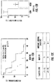
- the inventors have experimental data from brain cancer patients that show that tumors with invasive cells are highly correlated with low survivability and high risk of recurrence ( Fig. 11 ). Without being limited to any particular theory, it is contemplated that these invasive cell subpopulations are tumor or cancer stem cells and highly migratory in vivo.
- a method for identifying the invasiveness of a cancer cell or population of cells in a sample comprising: a) obtaining a cell or population of cancer cells from the sample of a tumor; b) incubating the cells for a period time in a integrative microfluidic apparatus; c) imaging the cells in the apparatus for a period of time; and d) determining whether a cell or subpopulation of cells in the sample are invasive.
- the present invention provides a method for predicting and/or prognosing a brain tumor patient survival and/or recurrence comprising: a) providing a cell or population of cells obtained from the tumor of a patient; b) adding a sufficient sample of the cells from the tumor to the integrated microfluidic apparatus; c) imaging the cells in the integrated microfluidic apparatus for a period of time; d) comparing the images of the cells in the integrated microfluidic apparatus over time; e) determining the number of cells or subpopulation of cells which migrate past the bifurcation of any of the migratory channels of the apparatus into either of the one or more outlet ends of the migration channels of the integrated microfluidic apparatus and/or exits out of the outlet ends of the migration channels; f) identifying the tumor as having low survivability or a high risk of recurrence when the proportion of cells entering through either the wide or narrow outlet ends of the integrated microfluidic apparatus is greater than 5% to about 50%, of all cells
- the migratory or invasive cells migrated into one or the other outlet ends.
- the inventors noted that the proportion of cells which migrated through the narrower of the two outlet ends (3 ⁇ m) also correlated with three-fold less survival time in patients.
- Patients with tumor cell samples having 85% or more wide outlet end migratory cells having greater than two or three-fold survival times over those having less than 85%.
- the apparatus described herein can be used to isolate or separate a cell or subpopulation of cells from a collection of cells in a sample.
- Cells can be separated from the integrated microfluidic apparatus by means of trypsinization or chelation, which allows the cells to detach from the channel walls.
- trypsin or EDTA can be introduced into all of the inlet reservoirs. The cells detach and the flow of the integrated microfluidic apparatus is such that the cells that have migrated through the migratory channels will flow through the first channel and move into the upper outlet reservoir ( Fig. 2A ).
- the migratory cells may be isolated from the integrated microfluidic apparatus and subjected to genomic or proteomic analysis. Such analysis includes, but is not limited to, analysis of gene expression levels using quantitative real-time polymerase chain reaction, RNA sequencing, and surface protein expression levels using flow cytometry.
- biological sample or “biological fluid” includes, but is not limited to, any quantity of cells from a living or formerly living subject. Such cells include, but are not limited to, blood, bone, bone marrow, T-cells, B-cells, fibroblasts, chondrocytes, synovial macrophages, endothelial cells, tumor associated cells, and skin cells.
- the term "subject” refers to any mammal, including, but not limited to, mammals of the order Rodentia, such as mice and hamsters, and mammals of the order Logomorpha, such as rabbits. It is preferred that the mammals are from the order Carnivora, including Felines (cats) and Canines (dogs). It is more preferred that the mammals are from the order Artiodactyla, including Bovines (cows) and Swines (pigs) or of the order Perssodactyla, including Equines (horses). It is most preferred that the mammals are of the order Primates, Ceboids, or Simoids (monkeys) or of the order Anthropoids (humans and apes). An especially preferred mammal is the human.
- mammals of the order Rodentia such as mice and hamsters
- mammals of the order Logomorpha such as rabbits. It is preferred that the mammals are from the order Carnivora, including Felines (cats) and Canines (dogs). It is
- the integrated microfluidic apparatus and methods used may be methods of diagnosis and the migration of cells may be associated with a diseased state, for example, cancer, such as solid tumors.
- cancers such as brain cancers like glioblastoma multiforme, prostate cancer, melanoma, bladder cancer, breast cancer, lymphoma, ovarian cancer, lung cancer, colorectal cancer or head and neck cancer.
- the migration of cells is associated with glioblastoma multiforme.
- migration of cells is associated with an immunological disorder; inflammation; rheumatoid arthritis; cystic fibrosis; or an infection, for example, a viral or bacterial infection.
- the methods of the invention are methods of monitoring prognosis and the migration of cells is associated with the prognosis of glioblastoma multiforme.
- the integrated microfluidic apparatus and methods used are for monitoring drug treatment and the migration of cells is associated with the drug treatment.
- the apparatus and methods used are (e.g., analysis of migration of cells) for the selection of population-oriented drug treatments and/or in prospective studies for selection of dosing, for activity monitoring and/or for determining efficacy endpoints.
- decreased migration upon application of a particular biologically active molecule indicates that that molecule effectively inhibits the movement of migratory or invasive cells.
- the diagnosis can be carried out in a person with or thought to have a disease or condition.
- the diagnosis can also be carried out in a person thought to be at risk for a disease or condition.
- a person at risk is one that has either a genetic predisposition to have the disease or condition or is one that has been exposed to a factor that could increase his/her risk of developing the disease or condition.
- a method for identifying an agent which inhibits the invasiveness of a cell or population of tumor cells in a sample comprising: a) adding to the integrated microfluidic apparatus a cell or population of cells from the sample and the test agent; b) incubating the cells for a period time; c) imaging the cells in the apparatus for a period of time; d) comparing the images of the cells in the integrated microfluidic apparatus over time; e) identifying a cell or subpopulation of cells in the sample as invasive when the cell or subpopulation of cells migrates past the bifurcation of any of the migratory channels of the apparatus into either of the one or more outlet ends of the migration channels of the integrated microfluidic apparatus and/or exits out of the migration channels; f) comparing the number and/or extent of invasiveness of the cell or subpopulation of cells to the number and/or extent of migration of the cell or subpopulation of cells of e) to the number and/or extent of migration of
- Also disclosed, but not claimed, is a method for identifying an agent which inhibits the invasiveness of a cell or population of brain tumor cells in a sample comprising: a) adding to the inlet reservoir of the second channel of the integrated microfluidic apparatus an aliquot of a suspension of a population of cells from the sample and the test agent; b) incubating the cells for a period time to allow the cells to fill the second channel; c) removing any remaining cell suspension from the reservoir of the second channel and washing the inlet of the second channel; d) adding cell media containing the molecule to the one or more reservoirs of the one or more inlets of the first channel; e) imaging the cells in the apparatus for a period of time; f) comparing the images of the cells in the integrated microfluidic apparatus over time and identifying a cell or subpopulation of cells in the sample as invasive when the cell or subpopulation of cells migrates past the bifurcation of any of the migratory channels of the apparatus into either of the one or more outlet ends
- cancer any malignant growth or tumor caused by abnormal and uncontrolled cell division that may spread to other parts of the body through the lymphatic system or the blood stream.
- the cancer can be a metastatic cancer or a non-metastatic (e.g., localized) cancer.
- metastatic cancer refers to a cancer in which cells of the cancer have metastasized, e.g., the cancer is characterized by metastasis of a cancer cells.
- the metastasis can be regional metastasis or distant metastasis, as described herein.
- the apparatus and methods can provide any amount of any level of diagnosis, staging, screening, or other patient management, including treatment or prevention of cancer in a mammal.
- the methods disclosed herein can be used to assess the likelihood of survival and median survival times of patients having brain tumors.
- the brain tumor is a glioblastoma.
- the methods disclosed herein can be used to predict or prognose the survival time of a patient after undergoing a biopsy or resection of the tumor.
- the information is useful to inform the patient as to the status of the cancer. For example, a patient who is diagnosed as having a tumor with a large number of invasive cells, or a greater proportion of cells that enter the wide or narrow exit channels, based on the methods described herein, would be informed of the lower that average survival time expected.
- This information can be used by the patient to determine whether any treatment should be undertaken, or whether a modest or aggressive course of treatment is warranted, depending on the needs of the patient.
- cancers also include but are not limited to adrenal gland cancer, biliary tract cancer; bladder cancer, brain cancer; glioblastoma, breast cancer; cervical cancer; choriocarcinoma; colon cancer; endometrial cancer; esophageal cancer; extrahepatic bile duct cancer; gastric cancer; head and neck cancer; intraepithelial neoplasms; kidney cancer; leukemia; lymphomas; liver cancer; lung cancer (e.g.
- the tumor is a glioblastoma.
- an “active agent or molecule” and a “biologically active agent or molecule” are used interchangeably herein to refer to a chemical or biological compound that induces a desired pharmacological and/or physiological effect, wherein the effect may be prophylactic or therapeutic.
- the terms also encompass pharmaceutically acceptable, pharmacologically active derivatives of those active agents specifically mentioned herein, including, but not limited to, salts, esters, amides, prodrugs, active metabolites, analogs and the like.
- active agent pharmaceutically active agent
- drug drug
- the term includes the active agent per se as well as pharmaceutically acceptable, pharmacologically active salts, esters, amides, prodrugs, metabolites, analogs etc.
- biologically active molecules which can be introduced into the apparatus and used in the methods disclosed herein include, but are not limited to, dyes, including fluorescent, and NIRF dyes, enzymes, and enzyme linked dyes and markers, receptor antagonists or agonists, hormones, growth factors, autogenous bone marrow, antibiotics, antimicrobial agents, and antibodies.
- Non-limiting examples of biologically active agents include following: adrenergic blocking agents, anabolic agents, androgenic steroids, antacids, anti-asthmatic agents, anti-allergenic materials, anti-cholesterolemic and anti-lipid agents, anti-cholinergics and sympathomimetics, anti-coagulants, anticonvulsants, anti-diarrheal, anti-emetics, anti-hypertensive agents, anti-infective agents, anti-inflammatory agents such as steroids, non-steroidal anti-inflammatory agents, anti-malarials, anti-manic agents, anti-nauseants, anti-neoplastic agents, anti-obesity agents, anti-parkinsonian agents, anti-pyretic and analgesic agents, anti-spasmodic agents, antithrombotic agents, anti-uricemic agents, anti-anginal agents, antihistamines, anti-tussives, appetite suppressants, benzophenanthridine alkaloids, biologicals, cardioactive agents, cerebral
- the biologically active molecule is a dye
- the molecule is detected by fluorescence imaging.
- the dyes may be emitters in the visible or near-infrared (NIR) spectrum.
- Known dyes useful in the present invention include carbocyanine, indocarbocyanine, oxacarbocyanine, thüicarbocyanine and merocyanine, polymethine, coumarine, rhodamine, xanthene, fluorescein, boron ⁇ dipyrromethane (BODIPY), Cy5, Cy5.5, Cy7, VivoTag-680, VivoTag-S680, VivoTag-S750, AlexaFluor660, AlexaFluor680, AlexaFluor700, AlexaFluor750, AlexaFluor790, Dy677, Dy676, Dy682, Dy752, Dy780, DyLight547, Dylight647, HiLyte Fluor 647, HiL
- module means that in the presence of the biologically active agent or molecule, the migratory ability of the cell or subpopulation of cells is up regulated or down regulated, such that migration level, or activity is greater than or less than that observed when compared to controls.
- the term “modulate” can mean “inhibit,” but the use of the word “modulate” is not limited to this definition.
- inhibitor means that that in the presence of the biologically active agent or molecule, the migratory ability of the cell or subpopulation of cells is lowered or down regulated when compared to controls.
- the microfluidic device consisted of "Y"-shaped microchannels, with a 20 ⁇ m-wide feeder channel bifurcating to 20 ⁇ m-wide or 3 ⁇ m-wide branches, arrayed between mutually perpendicular cell seeding and cell outlet channels.
- the apparatus was fabricated using multilayer photolithography and replica molding. Photolithography masks were designed using AutoCAD (Autodesk, McLean, VA) and produced by the Photoplot Store (Colorado Springs, CO).
- the master for the device contained a negative mold of the final device and was fabricated using SU-8 3010 positive photoresist (Microchem, Newton, MA).
- SU-8 3010 was spin coated (Single Wafer Spin Processor, Model WS-400A-6NPP-LITE, Laurell Technologies, North Wales, PA) on a cleaned silicon wafer (University Wafer, South Boston, MA) to create a 10 ⁇ m-thick film.
- the film was soft baked on a hot plate and exposed to 170 mJ/cm2 of UV light energy through the chrome-on-glass light field mask using an EVG620 mask aligner (EVG, Austria) to define the microchannels.
- the wafer was baked, post-exposure, to cross link the pattern before development with SU-8 developer.
- a 50 ⁇ m-thick SU-8 3025 film was spun onto the wafer and soft baked.
- a mask defining the medium feed lines was aligned with the channels, and the photoresist was exposed to 250 mJ/cm2 of energy.
- the final master was developed, hard baked, and passivated with a fluorinated silane [(tridecafluoro-1,1,2,2-tetrahydrooctyl)-1-trichlorosilane] (Pfaltz & Bauer, Waterbury, CT) overnight in a vacuum desiccator.
- a fluorinated silane (tridecafluoro-1,1,2,2-tetrahydrooctyl)-1-trichlorosilane] (Pfaltz & Bauer, Waterbury, CT) overnight in a vacuum desiccator.
- PDMS polydimethylsiloxane
- Sylgard® 184 Silicone Elastomer Kit Dow Corning, Midland, MI
- curing at 85 °C for 2 hours.
- Devices were diced, and 6-mm inlet and outlet ports were punched in the PDMS fluid layer.
- the devices and glass coverslips were cleaned with ethanol and DI water and plasma treated for 2 minutes at 18 W (Harrick PDC-32G, Harrick Plasma, Ithaca, NY).
- the device was bonded to the glass slide and coated with 20 ⁇ g/ml rat tail collagen type I (BD, Franklin Lakes, NJ, USA) for 1 hour at 37 °C. Following coating, the channels were washed with DPBS to prepare for cell seeding.
- 20 ⁇ g/ml rat tail collagen type I (BD, Franklin Lakes, NJ, USA) for 1 hour at 37 °C. Following coating, the channels were washed with DPBS to prepare for cell seeding.
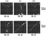
- the apparatus termed a “Microchannel Migration Device,” comprises a plurality of Y-shaped microchannels arrayed between cell seeding (second channel) and medium (first channel) lines ( Figure 1A ).
- the microchannels were designed such that 20 ⁇ m base channels bifurcated to 20 ⁇ m and 3 ⁇ m branch channels at a 45° or 65° (from the horizontal) angle (inset, Figure 1A ).
- experiments can be carried out with or without a chemoattractant gradient. If no gradient is desired, growth medium is placed in all four inlet wells, and the topography of the channels is the only driver of migration.
- medium containing a chemoattractant is placed in the uppermost medium inlet well and medium without chemoattractant is placed in the cell inlet and bottom two medium inlet wells, a gradient is formed within the microchannels to induce migration.
- the particular example of the device was formed by bonding a PDMS mold containing the microchannels and medium channels to a glass coverslip ( Figure 1B ).
- Cells were seeded at the bases of the microchannels following gravity-driven flow of suspended cells from the cell inlet well (shown schematically in Figure 1C ).
- Medium was placed in all inlet wells following cell seeding.
- cell seeding and migration were carried out without the need for external pumps or valves, and all flow was driven by gravity. Seeded cells migrated through the channels over the course of the experiment ( Figure 1D ).
- Cell seeding and live cell migration experiments Cells were grown to confluency, trypsinized, and resuspended in serum-free medium at 2 x10 6 cells/ml. 50 ⁇ l of cell suspension was added to the cell inlet well. Cells were incubated in the device for 5-10 minutes at 37 °C to allow initial cell seeding at the base of "Y" channels. The cell suspension was then removed from the cell inlet port. The device was washed with DPBS before the addition of medium to the inlet ports of the device. In select experiments, PI3K activity was inhibited by the addition of 10 ⁇ M LY294002 in the medium through the entire course of migration.
- the migration chamber was moved to a temperature- and CO 2 -controlled stage-top live cell incubator (Okolab, Italy) mounted on the motorized stage of an inverted Nikon Eclipse Ti microscope (Nikon, Tokyo, Japan) with automated controls (NIS-Elements, Nikon). Migrating cells were imaged with a 10x-magnification phase contrast objective every 10 minutes for up to 16 hours.
- Cell position data were used to calculate cell speed over each 10 minute interval, and these speeds were averaged to get an overall average speed for each cell. Additionally, the chemotactic index, defined as the cell displacement divided by the total distance travelled by the cell, was calculated. Cell shape data were used to calculate cell circularity and fit elliptical angle using the Measure function in ImageJ. Statistical significance was assessed with non-paired Student's t-test.
- Migratory cells were further defined as migratory or non-migratory. Migratory cells were defined as those cells which reached the bifurcation in the Y-shaped microchannel; all other cells were defined as non-migratory. Migratory cells were then classified as contact guided or not contact guided. Cells were defined as contact guided if they continued to the branch channel on the side of the base channel on which they were migrating when the bifurcation was reached. Cells that switched walls in the bifurcation region were classified as not contact guided.
- Migratory Cells Cells that had migrated through and exited the channels were washed with a chelator (versene) prior to the addition of 0.25% trypsin to all inlet wells of the device. Hydrodynamic resistance to flow in the narrow microchannels prevented the backflow of cells that had migrated through the microchannels back into the microchannels. Detached cells flowed to the upper outlet well, were collected in culture medium, and were plated in 96-well plates for expansion. Expanded cells were analyzed for the presence of tumor stem cell markers (for example, CD44 or CD271).
- tumor stem cell markers for example, CD44 or CD271.
- ⁇ 300 migratory cells were collected, suspended in 75 ⁇ l of DPBS, mixed with 75 ⁇ l of Matrigel, and injected to the mammary fat pad of an immunodificient mouse. An equal number of control cells that had not migrated through the microchannels were collected and injected in an identical manner. Mice were sacrificed 8 weeks post-injection, and the lungs were histologically analyzed to detect metastases.
- Bifurcating Channels Allow Identification of Migratory Cells: Cell tracking of all MDA-MB-231 cells within the channels revealed two distinct subpopulations: migratory and non-migratory cells ( Figures 2A,B ). 22 ⁇ 3% of human metastatic MDA-MB-231 breast cancer cells were migratory. Interestingly, this subpopulation correlates with the % of MDA-MB-231 (28%) bearing the CD44 + /CD24 - molecular signature 5 that is used to define breast cancer stem cells. Migratory cells, defined as those cells reaching the branch channels, migrated more than twice as fast as non-migratory cells ( Figure 2C ). Migratory cells were also significantly more directional. The chemotactic index of migratory cells increased to 0.91 in comparison to a chemotactic index of 0.37 for nonmigratory cells.
- Circularity a shape factor that decreases as shapes become less circular, was also significantly different between migratory and non-migratory cells.
- Migratory cells were significantly more elongated as they migrated, with a circularity of 0.37.
- Non-migratory cells had an average circularity of 0.58 ( Figure 2F ).
- the apparatus for use in the present invention was used for analysis of cytoskeletal components and intracellular signals via fluorescence microscopy ( Figure 3 ). This was possible because the device was constructed of transparent materials.
- Migratory cells showed increased localization of F-actin to the cell leading edge ( Figure 3A,D ). Actin localization was not seen in non-migratory cells (rounded cell at base of channel, Figure 3D ).
- Rho GTPases Rac1 and Cdc42 were polarized in migratory cells, particularly when these cells reached the 3 ⁇ m-wide branch channel ( Figure 3B,C,E,F ). Non-migratory cells did not exhibit this polarization (for example, cell at channel base in Figure 3B ).
- PI3K Inhibition Promotes Spontaneous Migration of MDA-MB-231 Cells: There is evidence that PI3K signaling is required to stabilize nascent protrusions. New protrusions away from the wall along which a cell is migrating would discourage contact guidance. Therefore, it was investigated whether inhibiting PI3K could promote contact guidance in 200 ⁇ m-long microchannels.
- Migratory cells are more likely to cause metastasis upon orthotopic injection in immunodeficient mice: MDA-MB-231 cells migrated through the device in the absence of a chemotactic cue. Trypsin was added to the all medium inlet wells of the device and caused the migratory cells to become detached and flow to the upper medium outlet well ( Figure 7A ). Resistance to flow through the narrow microchannels prevented detached cells from flowing back into the microchannels ( Figure 7A ; compare cell positions in microchannels before and after removal of migratory cells). Approximately 300 migratory cells were collected in the upper medium outlet well. Trypsin was then added to the cell inlet well. Cells that had not entered the channels flowed to the lower outlet well and were collected.
- Observations using metastatic MDA-MB-231 breast cancer cells were generalized.
- a panel of cell lines comprised of non-cancerous and non-metastatic (normal) breast epithelial cells, non-metastatic breast cancer cells, and metastatic breast cancer cells was assayed in devices containing 400 ⁇ m-long Y-shaped migration channels ( Figure 8 ).
- an average of 2.6% of migratory cells was observed, with the lowest percentage observed in HMLE Luc 26 cells (0.5 ⁇ 0.8%) and the highest percentage observed in 184A1 cells (5.7 ⁇ 8.0%).
- non-metastatic breast cancer cell lines displayed an average of 20% migratory cells, with a minimum percentage observed in metastatic SUM149 cells (1.2 ⁇ 1.0%) and a maximum observed in metastatic MDA-MB-231 circulating tumor cells (46% ⁇ 18%).
- Examples include: metastatic K-Ras-overexpressing/obscurin-knockdown MCF10A cells (20%), metastatic Bt-549 breast cancer cells (18 ⁇ 3.5%), metastatic MDA-MB-436 breast cancer cells (8.7 ⁇ 0.5%), metastastic MDA-MB-231 breast cancer cells (17 ⁇ 5%), metastatic Hs578t breast cancer cells (16 ⁇ 2%), and metastatic A375 melanoma cells (38 ⁇ 7%).
- the 38 ⁇ 7% of migratory human A375 melanoma cells closely matches the % of A375 cells expressing the cancer stem cell marker CD271.
- the inventors then determined a cutoff range for the percentage of migratory cells observed in a cell line that predicted whether a tested cell line is non-metastatic (if the percentage of migratory cells observed in a cell line was below the cutoff) or metastatic (if the percentage of migratory cells observed in a cell line was above the cutoff).
- a range of cutoff values from 5% to 15% was considered; this range was selected because it was between the average percentage of migratory cells observed in non-metastatic cell lines (2-2.6%) and metastatic cell lines (20%).
- the classification of each cell line predicted by the cutoff value was compared to the known classification of that cell line (listed in Figure 8 ).
- the accuracy (defined as the percentage of classifications correctly predicted by the cutoff compared to those listed in Figure 8 ) was calculated for each cutoff value. For cutoff values of 7-8%, the highest accuracy value was calculated (96%), so this range was chosen as the cutoff between non-migratory and migratory cell lines. For the cutoff range of 7-8%, the sensitivity of the device (defined as the percentage of metastatic cell lines listed in Figure 8 that were predicted to be metastatic by the cutoff) was 90%, and the specificity (defined as the percentage of non-metastatic cell lines listed in Figure 8 that were predicted to be non-metastatic by the cutoff) was 100%.
- Triple negative breast cancer cells display divergent responses to pharmaceutical agents: A panel of triple-negative breast cancer (TNBC) cell lines were assayed in devices containing 400 ⁇ m-long Y-shaped migration channels in the presence or absence of the PI3K inhibitor LY294002 (10 ⁇ M) ( Figure 9 ). PI3K inhibition did not affect the percentage of migratory cells measured for MDA-MB-436 (13 ⁇ 7% migratory for control vs. 18 ⁇ 7% for treated) cells or Hs578t (20 ⁇ 7% migratory for control vs. 15 ⁇ 2% migratory for treated) cells. PI3K inhibition increased migration of MDA-MB-231 (22 ⁇ 3% for control vs. 34 ⁇ 8% for treated) cells. In contrast, PI3K inhibition reduced the migration of Bt549 (32 ⁇ 8% for control vs. 19 ⁇ 1% for treated) cells.
- TNBC triple-negative breast cancer
- Metastatic propensity assay is amenable to a wide range of imaging techniques: Numerous imaging techniques were used to image an example in which a PDMS fluidic layer is bonded to a glass coverslip layer. Cells were imaged using lens-free holography ( Figure 10A ), which has an approximately 5 mm x 5 mm field of view. Cells were clearly visible in the channels upon digitally zooming in (inset, Figure 10A ). Additionally, cells in the device were imaged using phase contrast microscopy ( Figure 10B ) and a combination of phase contrast and fluorescence microscopy ( Figure 10C ; a subset of cells was tagged with a fluorescent marker and appears green in the image).
- migratory i.e., cells that entered the migratory channel and entered one of the two outlet ends during the observational interval
- the percentage of migratory cells in the patient sample was then correlated with patient survival time in months.
- a cutoff of 14.5% migratory cells was established by computing the mean % migratory value and separating the patients above or below this mean value.
- the patients' survival times were compared to the 14.6 month standard average, to arrive at a group with high survival (>14.6 mo.) and a low survival group ( ⁇ 14.6 mo.).
- the data was then correlated to the percentage of cells in their sample that were identified as migratory.
- the inventors found a three-fold difference in median survival times of patients with ⁇ 14.5% migratory cells in their sample vs. >14.5% migratory cells.
- Fig 2 shows the same type of analysis as Fig. 1 , but the correlation was to those migratory cells entering the wide (20 ⁇ m) outlet end.
- Fig. 1 shows the same type of analysis as Fig. 1 , but the correlation was to those migratory cells entering the wide (20 ⁇ m) outlet end.
- Fig. 3 is a normalized graph of the data with a cutoff of 1.8%, the inventors did not observe a significant correlation of migratory cells entering the narrow outlet end with low survivability in these patients.
- the median survival time was about two-fold greater for those patients with ⁇ 1.8% migratory cells entering the narrow outlet end.
- the inventors found a clearly significant correlation between survival time and the percentage of cells entered the wide outlet end ( Fig. 4 ). With an 85.7% cutoff, the mean survival time for patients with less wide outlet end entry cells was almost two-fold greater, and patients with >85.7% narrow outlet end entry had significantly lower than average survival times of 8.9 vs. 18.8 months.
- the inventors then analyzed the normalized narrow outlet end entry data in the same manner as Fig. 4 .
- the inventors divided the groups into two cohorts of >14.28% wide outlet end entry, and ⁇ 14.28% wide outlet end entry.
- the Kaplan-Meier survival graph shows a significant difference between each of the curves. When plotted, the data show that patients with the greatest proportion of narrow outlet end entry cells had the longest median survival times (23.3 months), greater than three-fold over the group with the lowest proportion of narrow outlet end entry cells (8.5 months).
- the cutoff and the amount of time during which cell migration is observed can be optimized to allow the methods described herein to identify metastatic and nonmetastatic cell populations from different types of cancer.
- the accuracy, sensitivity and specificity of the methods are displayed in Figure 17 across a wide range of cutoff values and experiment durations.
- the maximum value for accuracy was 96%, and was observed under three pairs of conditions: 7% cutoff and 13 hours, 8% and 12 hours, and 8% and 13 hours. Under each of these pairs of conditions, the sensitivity is 90% and the specificity is 100%.
Description
- Personalized medical plans aiming to limit metastasis are difficult to develop. The current state of the art requires the expansion of human cancers in immunodeficient mice before the cancers can be subjected to drug screenings. It is known that metastatic subpopulations of cancer cells have heightened motility which is linked to aggressiveness and invasiveness of the cancer. The ability to identify such a subpopulation of cells in a tumor of a patient would be useful in classifying the aggressiveness or metastatic potential of the cancer in the subject, and would also be useful in identifying optimal courses of treatment and determining whether the treatment was effective.
- Until recently, there currently existed no means for determining the motility of a cell or subpopulation of cells in a sample, using assays which are low cost, high throughput, and easy to operate. The present inventors previously created a microfluidic apparatus, which is described in
WO 2015/009688 , which is capable of identifying a cell and/or subpopulation of cells that have a significantly increased motility compared to the cell population as a whole, and which the inventors have shown to be correlated with metastatic potential in a number of solid tumor types. However, there still is an unmet need in the ability of clinicians and investigators to identify patients with brain cancer that is highly invasive and which correlates with lower survival times and high risk of recurrence. -
WO 2011/158243 discloses a method of diagnosing a glioma or a breast cancer comprising analyzing an amount or activity of heterogeneous nuclear ribonucleoproteion A2/B1 (hnRNP A2/B1) in a brain cell sample of a subject, wherein an up-regulation in an amount or activity of hnRNP A2/B1 beyond a predetermined threshold with respect to a control sample is indicative of the breast cancer or glioma. The method may also be used for staging the cancer and for predicting a patient's prognosis of survival. - Disclosed herein, but not claimed, is a method for identifying the invasiveness of a brain cancer cell or population of cells in a sample comprising: a) obtaining a cell or population of cancer cells derived from the sample of a tumor; b) incubating the cells for a period time in an integrative microfluidic apparatus; c) imaging the cells in the apparatus for a period of time; and d) determining whether a cell or subpopulation of cells in the sample are invasive.
- The present invention provides a method for predicting and/or prognosing a glioblastoma brain tumor patient survival and/or recurrence as claimed in claim 1 comprising: a) providing a cell or population of cells from a glioblastoma brain tumor of the patient; b) adding a sufficient sample of the cells from the tumor to an integrated microfluidic apparatus, wherein the integrated microfluidic apparatus comprises a substrate in the form of a chip comprising a fluid layer and a coverslip layer, which are bonded together at final assembly of the apparatus, wherein the fluid layer comprises at least a first channel and a second channel, each having at least one or more inlets which communicate with an inlet reservoir, and each channel also having at least one or more outlets which communicate with an outlet reservoir; the first and second channels are disposed in proximity to each other and at least a portion of the first and second channels are parallel in the fluid layer of the substrate; the first and second channels are connected by a plurality of migratory channels having at least one inlet in communication with the second channel and two or more outlets in communication with the first channel; and the plurality of migratory channels are bifurcated at a bifurcation point distal from the at least one inlet of the migratory channel and are significantly reduced in size so as to allow one cell body at a time to enter the migratory channel from the second channel; c) incubating the cells for a period time in the integrative microfluidic apparatus; d) imaging the cells in the integrated microfluidic apparatus for a period of time; e) determining whether a cell or subpopulation of cells in the sample are invasive wherein the cell or subpopulation of cells are determined as being invasive when the cell or subpopulation of cells migrates through the plurality of migratory channels of the apparatus of b) past the bifurcation point or when the cell or subpopulation of cells migrates through the plurality of migratory channels of the apparatus of b) into the ends of the two or more outlets or when the cell or subpopulation of cells migrates through the plurality of migratory channels of the apparatus of b) and exits out of the ends of the two or more outlets; and f) identifying the tumor of the subject as having low survivability or a high risk of glioblastoma brain tumor recurrence when the proportion of invasive cells is greater than a cutoff range of the migratory cells.
- In accordance with an embodiment the present invention provides a method for identifying an effective chemotherapeutic treatment regimen in a subject having a tumor, as claimed in
claim 10. - An effective chemotherapeutic treatment regimen is treating a subject with one or more chemotherapeutic agents that will significantly decrease the proportion of migratory cells from the subject, as determined using the apparatus and methods described herein, and therefor inhibit invasiveness of the tumor cells in the subject. Multiple devices can be operated in parallel to screen a number of therapeutic agents.
- Disclosed, but not claimed, is a method for identifying an agent which inhibits the invasiveness of a cell or population of tumor cells in a sample comprising: a) adding to the integrated microfluidic apparatus a cell or population of cells from the sample and the test agent; b) incubating the cells for a period time; c) imaging the cells in the apparatus for a period of time; d) comparing the images of the cells in the integrated microfluidic apparatus over time; e) identifying a cell or subpopulation of cells in the sample as invasive when the cell or subpopulation of cells migrates past the bifurcation of any of the migratory channels of the apparatus into either of the one or more outlet ends of the migration channels of the integrated microfluidic apparatus and/or exits out of the migration channels; f) comparing the number and/or extent of invasiveness of the cell or subpopulation of cells to the number and/or extent of migration of the cell or subpopulation of cells of e) to the number and/or extent of migration of a control cell or subpopulation of cells migrating in the absence of the agent.
-
-
Figure 1 illustrates an example of the apparatus used in the methods of the present invention. (1A) A schematic of device, showing overall design of an example used in the methods disclosed herein. Inset shows details of Y-shaped microchannels. (1B) Schematic of completed PDMS device bonded to glass coverslip. (1C) Schematic of cells seeded at channel bases. (1D) Phase contrast image of MDA-MB-231 cells migrating in 200 µm-long Y-shaped microchannels. -
Figure 2 illustrates the migration of migratory and non-migratory MDA-MB-231 cells within the device. (2A) Representative image of migratory cell in 3 µm-wide branch channel. (2B) Representative image of non-migratory cell within base channel of device. Migratory cells moved within the microchannels with significantly greater average speed (2C) and chemotactic index or persistence (2D). (2E) Ellipses fit to the cell outlines had major axes highly aligned with the base channel in 89% of the migratory cells but only 52% of the non-migratory cells. (2F) Migratory cells were significantly more elongated than non-migratory cells, as measured by the circularity of the cell outline. *, p<0.05. -
Figure 3 shows fluorescent on-chip imaging of F-actin and Rho GTPases. MDA-MB-231 ells within the microchannels were fixed and stained for (3 A,D) F-actin, (3 B,E) Rac1, or (3 C,F) Cdc42. Panels 3A-C show cells entering the 20 µm-wide branch channel. Panels 3D-F show cells entering or migrating within the 3 µm-wide branch channel. Non-migratory cells are shown at the 20 µm-wide channel bases in panels B and D (arrows). -
Figure 4 depicts how migratory MDA-MB-231 cells are contact guided at the microchannel bifurcation. (4A) Representative cell tracks of migratory cells. Cells migrate predominantly up one channel wall and continue to follow that wall at the bifurcation as they enter a branch channel. (4B) Percentage of migratory cells that were contact guided to the 3 µm-wide and 20 µm-wide branches. -
Figure 5 illustrates that PI3K inhibition promotes MDA-MB-231 cell migration and contact guidance within the microchannels. Representative tracks of (5A) control and (5B) PI3K inhibited cells migrating within 200 µm-long microchannels. PI3K inhibition with 10 µM LY294002 increased the percentage of cells that were migratory and that were contact guided. (5C) The average speed of control and LY294002-treated cells was the same. (5D) The chemotactic index of LY294002 -treated cells was greater than that of control cells. -
Figure 6 depicts the extraction of migratory A375 cells from a microchannel cell migration device for use in the methods of the present invention. (6A) Trypsinized cells flowed in the upper medium line without entering the microchannels. Arrow indicates cell that has been completely detached from the device. (6B) Detached cells flowed to the upper medium outlet well. Arrow indicates cell entering the well. (C) Extracted A375 cells were expanded using standard cell culture techniques for 20 days and assayed for surface protein expression levels of the cancer stem cell marker CD271 using flow cytometry. A375 cells that had migrated through the device and been expanded (blue) exhibited higher surface protein expression levels of CD271 than the bulk A375 cell population (green). -
Figure 7 depicts the isolation of migration MDA-MB-231 cells from a migration device for use in the present invention. (7A) Migratory cells that exited the microchannels are shown prior to (left panel) and following (right panel) extraction from the device. Note that the position of the nonmigratory cells and cells seeded at the entrances to the channels is the same before and after extraction of migratory cells. (7B) Orthotopic injection of migratory cells but not control cells to the mammary fat pad of immunodeficient mice resulted in the formation of metastases (arrows). -
Figure 8 enumerates the percentage of migratory cells as determined for a panel of breast epithelial or cancer cell lines. Nonmetastatic cell lines displayed low levels of migration in the device (less than or equal to 6.8% of cells entering the migration channels reached the branch channels following the bifurcation). In contrast, metastatic cell lines contained a migratory subpopulation (from about 8.2 to about 46% of the cells from each cell line scored as migratory). -
Figure 9 demonstrates the differential response of triple-negative breast cancer (TNBC) cell lines to an example pharmacological agent. MDA-MB-436, MDA-MB-231, Bt549, and Hs578t cells were treated with 10 µM LY294002, an inhibitor of PI3K, or the appropriate control. Migration of MDA-MB-231 cells increased, while migration of Bt549 cells decreased. A similar percentage of MDA-MB-436 and Hs578t cells were migratory in the presence or absence of the inhibitor. -
Figure 10 depicts a representative sample of imaging techniques that can be used to assess migration in the present invention. (10A) Lens-free holography enables a wide field of view to be captured in a single image. Inset shows digitally zoomed image, with cells clearly visible in migration channels. (10B) Phase contrast microscopy (10x objective) image of migration channels with cells seeded at channel entrances. (10C) Combined phase contrast and fluorescence microscopy of cells within migration channels. A subset of cells was tagged with a fluorescent marker and appears green in the image. -
Figure 11 : Overall Survival - % Migratory: (a) Kaplan-Meier survival of patients with >14.28% vs <14.28% migratory cells. This cutoff was established by computing the mean % migratory value and separating the patients above or below this mean value. Log-Rank (Mantel Cox) test showed significant difference between the curves although. Table lists median survival in months for each of the above-mentioned groups (b) Separating patients as lower than expected survival of 14.6 months (this is taken from the classic StuppR. NEJM paper establishing the current standard of care) and higher than expected. Average percent migratory cells with SEM is represented. T-test demonstrates significant different between the two groups. -
Figure 12 : Overall Survival - % Wide Entry of All Cells: (a) Kaplan-Meier survival of patients with >11.4% vs <11.4% migratory cells which migrated into the wide outlet channel. This cutoff was established by computing the mean % Wide entry of all cells value and separating the patients above or below this mean value. Log-Rank (Mantel Cox) test showed significant difference between the curves although. Table lists median survival in months for each of the above-mentioned groups. (b) Separating patients as lower than expected survival of 14.6 months (using the StuppR. NEJM paper methodology) and higher than expected. Average percent wide entry of all cells with SEM is represented. T-test demonstrates significant different between the two groups. -
Figure 13 : Overall Survival - % Narrow Entry of All Cells: (a) Kaplan-Meier survival of patients with >1.8% vs <1.8% migratory cells which migrated into the narrow outlet channel. This cutoff was established by computing the mean % narrow entry of ALL cells value and separating the patients above or below this mean value. Log-Rank (Mantel Cox) test showed significant difference between the curves although. Table lists median survival in months for each of the above-mentioned groups. (b) Separating patients as lower than expected survival of 14.6 months (using the StuppR. NEJM paper) and higher than expected. Average percent narrow entry of all cells with SEM is represented. T-test demonstrates significant different between the two groups. -
Figure 14 : Overall survival - % Wide entry of Migratory cells: (a) Kaplan-Meier survival of patients with >85.7% vs <85.7% Wide channel entry of migratory cells. This cutoff was established by computing the mean % Wide entry of migratory cells value and separating the patients above or below this mean value. Log-Rank (Mantel Cox) test showed significant difference between the curves although. Table lists median survival in months for each of the above-mentioned groups (b) Separating patients as lower than expected survival of 14.6 months (using the StuppR. NEJM paper methodology) and higher than expected. Average percent wide entry of migratory cells with SEM is represented. T-test demonstrates significant different between the two groups. -
Figure 15 : Overall survival - % Narrow entry of Migratory cells: (a) Kaplan-Meier survival of patients with >14.28% vs <14.28% narrow channel entry of migratory cells. This cutoff was established by computing the mean % narrow entry of migratory cells value and separating the patients above or below this mean value. Log-Rank (Mantel Cox) test showed significant difference between the curves although. Table lists median survival in months for each of the abovementioned groups. b. Separating patients as lower than expected survival of 14.6 months (using the StuppR. NEJM paper methodology) and higher than expected. Average percent narrow entry of migratory cells with SEM is represented. T-test demonstrates significant different between the two groups. -
Figure 16 : Survival correlation - linear regression: (a) Linear regression analysis of % migratory and overall patient survival times (in months). Spearman and Pearson correlation analyses demonstrates significant negative correlation between the two variables. (b) Linear regression analysis of % narrow entry of migratory and overall patient survival times (in months). Spearman and Pearson correlation analyses demonstration significant negative correlation between the two variables. -
Figure 17 depicts the effects that the cutoff (%) and the duration of time that cell migration is observed for on the ability of the methods disclosed herein to predict whether a cell line is metastatic or nonmetastatic. Data is shown for the cell lines described inFigure 8 . (17A) Accuracy is defined as the percentage of classifications correctly predicted by the cutoff compared to those listed inFigure 8 . The highest accuracy value was 96%, and was observed for a cutoff of 7% and a duration of 12 hours, and also for a cutoff of 8% and a duration of either 12 hours or 13 hours. (17B) Sensitivity is defined as the percentage of metastatic cell lines listed inFigure 8 that were predicted to be metastatic by the cutoff. The maximum sensitivity observed was 100%, which occurs for cutoff values of or below 10%, and experiment durations equal to or greater than 19 hours. Specificity is defined as the percentage of non-metastatic cell lines listed inFigure 8 that were predicted to be non-metastatic by the cutoff. The maximum specificity value observed was 100%, which was observed for a wide range of conditions. Importantly, the cutoff and duration values that gave the maximum accuracy coincided with a high sensitivity value of 90% and a maximum specificity value of 100%. - Disclosed herein are methods for identifying a cell or population of cells from a tumor of a subject that are invasive compared to the population of tumor cells as a whole, using an integrated microfluidic apparatus that enables identification of these migratory cells directly from a specimen. These methods and apparatus were first described in International Patent Publication No.
WO 2015/009688, filed July 15, 2014 . - The integrated microfluidic apparatus only requires a small number of cells to perform an assay. The apparatus used in the methods of the present invention includes novel topographic features which can reliably differentiate between migratory and non-migratory cell populations in a sample. Furthermore, in some examples, both the spontaneous and chemotactic migration of cancer cells may be measured to distinguish between subpopulations within a tumor sample. The migratory cells identified using the apparatus and methods described herein may be separated and further analyzed to distinguish factors promoting metastasis within the population. Cells in the apparatus can be treated with chemotherapeutic or other agents to determine drug strategies to most strongly inhibit migration. The use of optically transparent materials in some examples allows a wide range of imaging techniques to be used for in situ imaging of migratory and non-migratory cells in the apparatus. The apparatus and methods described herein are useful for predicting the invasive propensity of brain tumor cells, predicting survival times and/or risk of recurrence of tumors post-operatively for patients and selecting optimal drugs for personalized therapies.
- The integrated microfluidic apparatus comprises a substrate in the form of a chip having a plurality of layers. The chip comprises a fluid layer and a coverslip layer, which are bonded together at final assembly of the apparatus. The fluid layer is composed of a plurality of channels having at least one or more inlets and outlets.
- The fluid layer of the integrated microfluidic apparatus for use in the present invention comprises at least two channels, each having at least one or more inlets which communicate with a reservoir, and each channel also having at least one or more outlets which communicate with a reservoir. The channels can have any dimension within the limits of the depth of the substrate. In some examples, the channels can have dimensions of about 3 µm to about 15 µm in height, about 3 µm to about 50 µm in width, and about 100 µm to about 400 µm in length. There can be any number of channels limited only by the dimensions of the chip used. In some examples, the chip can comprise greater than 200 channels.
- Within the fluid layer of the substrate, there are at least two channels, a first channel, also termed "a medium channel" which can be filled with any type of biological media or solvent. There is also at least a second channel, also termed "a cell channel" which can be filled with any type of biological media or solvent that contains a sample of cells to be assayed. The first and second channels are disposed in proximity to each other and are parallel into at least a portion of the two channels in the fluid layer of the substrate. The first and second channels have at least one inlet portion which can be the same or have a smaller dimension than the main portion of the first and second channel. Each inlet portion is connected to the inlet end of the first and second channel and communicates with the channels. Each inlet portion is also in communication with a reservoir wherein media or fluid can be introduced into the inlet of the channel.
- The first and second channels have at least one outlet portion which is the same dimension as the main portion of the first and second channel. Each outlet portion is connected to the outlet end of the first and second channel and communicates with the channels. Each outlet portion is also in communication with a reservoir wherein media or fluid can be directed to or removed from the channel.
- The migratory channel portion is an area where the first channel and second channel are in proximity to each other and are connected by a plurality of migratory channels having at least one inlet and at least one or more outlets. The migratory channels are bifurcated at a point distal from the inlet portion of the migratory channel. The bifurcation results in two outlet ends of the migratory channel which communicate with the media channel. These channels are significantly reduced in size, for example, by approximately by a factor of 10, so as to allow one cell body at a time to enter the migratory channel from the cell channel. For example, in an example, the main portion of the first and second channels has a width of about 400 µm and a height of about 50 µm, whereas the migratory channels have an inlet portion which communicates with the second channel and has a width of about 20 µm and a height of about 10 µm. In some examples, the migratory channels can have dimensions of width of about 3 µm to about 50 µm, and a height of about 3 µm to about 15 µm. The one or more outlet portions of the migratory channels can have the same or different widths than the inlet portion of the migratory channel. The bifurcation angle of the migratory channels is about 30° to 70° from the horizontal, which is defined as the long axis of the first and second channels. It is in these migratory channels that the cells in the sample are assayed for their ability to transverse the migratory channels and their speed, physical and biochemical characteristics can be measured.
- Referring now to
Fig. 1A which depicts an example of the integrated microfluidic apparatus used in the methods of the present invention, the fluid layer of the substrate is shown generally as (1) and is composed of a polydimethylsiloxane (PDMS) chip molded from a negative replica on a silicon wafer on which photolithography has been used to create a plurality of channels. A first channel (2), which has an inlet portion (3) and an outlet portion (4). The inlet portion is in communication with three inlets (5), termed "medium inlets" which are channels in the substrate that communicate between the inlet portion (3) and an inlet reservoir (6). The outlet portion (4) is in communication with an outlet reservoir (7), termed "upper outlet." On the fluid layer is also disposed a second channel (8), termed "cell channel" which has an inlet portion (9) and an outlet portion (10). The inlet portion (9) of the second channel is in communication with an inlet reservoir (11), termed "cell inlet." The outlet portion (10) of the second channel is in communication with an outlet reservoir (12), termed "lower outlet." In some examples, the inlet and outlet reservoirs are punched into the substrate of the coverslip layer having a circular shape and a diameter of about 6 mm, although that is only limited by the size of the volume required and the space available on the substrate. - On the fluid layer there is a migratory channel portion (13) which is a region between the first channel (2) and second channel (3) that has a plurality of migratory channels (14) which communicate with the first and second channels. As seen on the exploded inset in
Fig. 1A , the migratory channels (14) have an inlet end (15) which communicates with the second channel (3), and two or more outlet ends (16), which are in communication with the first channel (2). The two or more outlet ends (16) are the result of a bifurcation (17) of the migratory channel and a point distal from the inlet end (15) of the migratory channel. In an example, there are about 16 migratory channels (14) which connect the first (2) and second (3) channels in the migratory channel portion (13) of the fluid layer. - In an alternative example, there are about 240 migratory channels (14) which connect the first (2) and second (3) channels in the migratory channel portion (13) of the fluid layer.
- In some examples, the first and second channels are between about 10 mm to about 50 mm in length, and have a height/depth of between about 30 µm to about 100 µm, and a width of about 100 µm to about 400 µm. In some examples, the inlets for the first and second channels have a length of between 2 mm to about 10 mm, a height/depth of between about 30 µm to about 100 µm, and a width of about 50 µm to about 400 µm. In some examples, the migratory channels have a length of between about 200 to 400 µm, a height/depth of between about 3 µm to about 15 µm, and a width between about 3 µm to about 50 µm.
- As seen in
Fig. 1B , the apparatus has a coverslip layer (18) which in whole, or in part, is made of an optically transparent substrate. Any optically substrate which is compatible with the fluid layer substrate can be used. In an example, the coverslip layer is formed of glass. It will be understood by those of ordinary skill that the portion of the coverslip layer that is optically transparent will allow visualization and imaging of the cells in the apparatus in real time. The coverslip is bonded using a variety of known means. In an example, the coverslip is bonded to the fluid layer via plasma treatment of about 18 W for a sufficient time, for example, about 2 minutes. - Before use, the fluid layer channels are all treated with a solution of collagen, for example, a solution of about 20 µg/ml collagen, such as rat tail collagen type I, for about an hour at 37 °C, and then the channels are washed with PBS or similar buffer. In some embodiments, other extracellular proteins, such as laminin, fibronectin, VCAM-1, hyaluronic acid, or gelatin, are used instead of collagen.
- The apparatus can be used for a variety of assays to detect and quantify the micromechanical, morphological, and molecular signatures of migratory and non-migratory cells in the device.
- Generally, operation of the apparatus comprises a first wash of the first channel and media inlet reservoirs (6) with a medium free buffer such as DPBS. This is followed by seeding of cells of interest from a sample. Cells of interest are seeded or introduced into the cell inlet reservoir (11) of the second channel. In some embodiments, the cells are trypsinized and suspended in serum free medium at a concentration of about 1 x 10⁵ to about 5 x 10⁶ cells/ml. In an embodiment, the cells are suspended at a concentration of about 2 x 10⁶ cells/ml. About a 50 µl aliquot of the cell suspension is introduced into the cell inlet reservoir (11), and the cells are incubated a 37 °C for a time sufficient to allow the cells to seed at the base of the migratory channels (14), for example, about 2 to about 30 minutes, preferably between about 5 to about 10 minutes. In an embodiment, about 10 µl to 50 µl of a suitable biological medium or buffer are introduced to the lowermost medium inlet reservoir (6) to prevent convective flow of cells through the migratory channels (14). Any remaining cells in the cell inlet reservoir (11) are then removed. Cell seeding is followed by introduction of a suitable biological medium or buffer into the media channel of the apparatus via the medium inlet reservoirs (6) and the flow is in the direction of the upper outlet (
Fig. 1A ). A suitable biological medium or buffer is also introduced into the cell inlet reservoir (11) of the second channel. The apparatus can be manipulated to either induce a chemoattractant gradient across the migratory channels, or not to have any chemoattractant gradient. When inducing a gradient, biological medium or buffer containing the chemoattractant is introduced into the uppermost medium inlet reservoir (6), with biological medium or buffer without the chemoattractant introduced into the remaining medium inlet reservoirs (6), and into the cell inlet reservoir (11) of the second channel. This creates a chemoattractant gradient across the migratory channels (14). If no gradient is desired, the medium inlet reservoirs (6) and cell inlet reservoir (11) are filled with the same biological medium or buffer. In some embodiments, other biologically active compounds or molecules can be added to the cell suspension when the cells are introduced into the apparatus, or after the cells have been seeded, to perform a variety of experiments. - It will be understood by those of skill in the art that the methods of operation described herein are only exemplary. These methods can be altered to suit the experimental conditions as needed, and the steps can be manipulated or changed accordingly.
- In exemplary operation, the integrated microfluidic apparatus is placed in a temperature and CO2 controlled incubator, imaging chamber or stage type device, to which is mounted an imaging system. In some embodiments, the migrating cells are imaged at 10X magnification using a phase contrast or other optical arrangement and images are taken at periodic intervals for between about 1 hour to about 24 hours, and saved on computer or other electronic storage media. It will be understood by those of ordinary skill that the type of microscopic imaging equipment can vary and can include any known systems or apparatus which can image cells using any type of electromagnetic radiation. Imaging systems include, but are not limited to, phase contrast, brightfield, differential interference contrast, fluorescence, and confocal microscopy and in-line holography.
- As used herein the "period of time" that the cells can be imaged using the apparatus and methods described herein can vary. In some embodiments the cells can be imaged for 1 or more hours, or in some embodiments about 6 or more hours, in some embodiments, 12 or more hours, and in some embodiments between 12 and 24 hours. It is possible to run the methods with device for a longer duration, however, with longer duration there is an increasing likelihood that cells from non-metastatic cell lines will eventually make it into the bifurcation (increasing the likelihood for a false positive result). For the lower limit, a period of time needs to elapse so the cells have time to migrate in the device. Based on the experimental data shown in
Fig. 17 and Example 10, 6 hours is the earliest time point where accuracy begins to increase. - In accordance with one or more embodiments, the operation of the apparatus and methods described herein can be performed without the need of external pumps or valves, and the function is driven by gravity and the topography of the channels in the apparatus. However, it is contemplated that alternative embodiments of the invention could encompass external pumps or valves depending on the function desired, and is well within the ability of the skilled artisan.
- The apparatus and methods described herein include methods for prognostic purposes in which migration of cancer cells to one of the bifurcation branches of the migration channels is associated with a disease state. Migration in the device serves as a companion diagnostic with other methods of cancer diagnosis. A high migration score indicates that the specific cancer tested has a high propensity to metastasize and indicates that aggressive treatment should be undertaken.
- In accordance with an embodiment, the inventive methods can be used to predict survivability of a subject with a glioblastoma brain tumor. For example, a subject diagnosed with a glioblastoma brain tumor using known means can undergo either a biopsy of the tumor or complete surgical resection of the tumor, or a portion thereof. One or more cells from the tumor or biopsy are then suspended in suitable growth media. A sample or aliquot of a sufficient number of cells are then added to the microfluidic device as described herein. The cells are then imaged over time and the images are observed to determine if any of the cells in the sample have moved from the cell inlet reservoir (11) to the migratory channel (14). If any of the cells in a defined period of time have migrated to the bifurcation point of the migratory channel (14) or the cells have migrated into the two or more outlet ends (16), or exited out of the outlet ends, then those cells are defined as being invasive.
- As used herein, the term "invasive" refers to a cell or population of cells from a tumor of the subject that display the migratory behavior of migrating into the cell migratory channel (14) of the integrated microfluidic apparatus and migrating past the bifurcation point of the migratory channel (14) or the cells have migrated into the two or more outlet ends (16), or exited out of the outlet ends. The inventors have experimental data from brain cancer patients that show that tumors with invasive cells are highly correlated with low survivability and high risk of recurrence (
Fig. 11 ). Without being limited to any particular theory, it is contemplated that these invasive cell subpopulations are tumor or cancer stem cells and highly migratory in vivo. - Disclosed herein, but not claimed, is a method for identifying the invasiveness of a cancer cell or population of cells in a sample comprising: a) obtaining a cell or population of cancer cells from the sample of a tumor; b) incubating the cells for a period time in a integrative microfluidic apparatus; c) imaging the cells in the apparatus for a period of time; and d) determining whether a cell or subpopulation of cells in the sample are invasive.
- The present invention provides a method for predicting and/or prognosing the survival and/or recurrence of a patient having a glioblastoma brain tumor as claimed in claim 1, comprising: a) providing a cell or population of cells obtained from the glioblastoma brain tumor of a patient; b) adding a sufficient sample of the cells from the tumor to the integrated microfluidic apparatus; c) incubating the cells for a period time in the integrative microfluidic apparatus; d) imaging the cells in the integrated microfluidic apparatus for a period of time; e) determining whether a cell or subpopulation of cells in the sample are invasive; and; g) identifying the tumor of the subject as having low survivability or a high risk of recurrence when the proportion of invasive cells is greater than a cutoff range of the migratory cells.
- In accordance with an embodiment, the present invention provides a method for predicting and/or prognosing patient survival and/or recurrence for patients having a glioblastoma brain tumor comprising: a) providing a cell or population of cells from the glioblastoma brain tumor of the patient; b) adding a sufficient sample of the cells from the tumor to the integrated microfluidic apparatus; c) incubating the cells for a period time in the integrative microfluidic apparatus; d) imaging the cells in the integrated microfluidic apparatus for a period of time; e) determining whether a cell or subpopulation of cells in the sample are invasive; and; g) identifying the tumor of the subject as having low survivability or a high risk of recurrence when the proportion of invasive cells is greater than 5% to about 50%, or in some embodiments, between about 5% to about 40% of the migratory cells, or in some embodiments, between about 10 % to about 25% of the migratory cells.
- In accordance with another embodiment, the present invention provides a method for predicting and/or prognosing a brain tumor patient survival and/or recurrence comprising: a) providing a cell or population of cells obtained from the tumor of a patient; b) adding a sufficient sample of the cells from the tumor to the integrated microfluidic apparatus; c) imaging the cells in the integrated microfluidic apparatus for a period of time; d) comparing the images of the cells in the integrated microfluidic apparatus over time; e) determining the number of cells or subpopulation of cells which migrate past the bifurcation of any of the migratory channels of the apparatus into either of the one or more outlet ends of the migration channels of the integrated microfluidic apparatus and/or exits out of the outlet ends of the migration channels; f) identifying the tumor as having low survivability or a high risk of recurrence when the proportion of cells entering through either the wide or narrow outlet ends of the integrated microfluidic apparatus is greater than 5% to about 50%, of all cells, or in some embodiments, 5% to about 40% of all cells, or in some embodiments, between about 10 % to about 25% of all cells.
- It will be understood by those of ordinary skill in the art that the exact steps for using the integrated microfluidic apparatus to determine invasiveness of tumor cells from a sample can be varied from the methods described above. Samples can be placed in the apparatus after media has been added, and with or without addition of chemotherapeutic or other biologically active agents. The dimensions of the apparatus can also be altered without changing the overall operation of the microfluidic apparatus.
- In some percentage of the samples from brain cancer patients, the migratory or invasive cells migrated into one or the other outlet ends. The inventors noted that the proportion of cells which migrated through the narrower of the two outlet ends (3 µm) also correlated with three-fold less survival time in patients. Moreover, in populations of cells that were migratory in a sample, there was a highly significant correlation between the proportion of migratory cells that enter the narrow outlet end or wide outlet end and survival times as well. Patients with tumor cell samples having 85% or more wide outlet end migratory cells having greater than two or three-fold survival times over those having less than 85%.
- It is also contemplated that the apparatus described herein can be used to isolate or separate a cell or subpopulation of cells from a collection of cells in a sample. Cells can be separated from the integrated microfluidic apparatus by means of trypsinization or chelation, which allows the cells to detach from the channel walls. For example, trypsin or EDTA can be introduced into all of the inlet reservoirs. The cells detach and the flow of the integrated microfluidic apparatus is such that the cells that have migrated through the migratory channels will flow through the first channel and move into the upper outlet reservoir (
Fig. 2A ). - The migratory cells may be isolated from the integrated microfluidic apparatus and subjected to genomic or proteomic analysis. Such analysis includes, but is not limited to, analysis of gene expression levels using quantitative real-time polymerase chain reaction, RNA sequencing, and surface protein expression levels using flow cytometry.
- It will be understood that the term "biological sample" or "biological fluid" includes, but is not limited to, any quantity of cells from a living or formerly living subject. Such cells include, but are not limited to, blood, bone, bone marrow, T-cells, B-cells, fibroblasts, chondrocytes, synovial macrophages, endothelial cells, tumor associated cells, and skin cells.
- As used herein, the term "subject" refers to any mammal, including, but not limited to, mammals of the order Rodentia, such as mice and hamsters, and mammals of the order Logomorpha, such as rabbits. It is preferred that the mammals are from the order Carnivora, including Felines (cats) and Canines (dogs). It is more preferred that the mammals are from the order Artiodactyla, including Bovines (cows) and Swines (pigs) or of the order Perssodactyla, including Equines (horses). It is most preferred that the mammals are of the order Primates, Ceboids, or Simoids (monkeys) or of the order Anthropoids (humans and apes). An especially preferred mammal is the human.
- The solid substrate used to make the integrated microfluidic apparatus described herein may be any suitable material. Representative examples of substrates include glass and modified or functionalized glass, plastics (including acrylics, polystyrene and copolymers of styrene and other materials, polypropylene, polyethylene, polybutylene, polyurethanes, Teflon, etc.), polysaccharides, nylon or nitrocellulose, resins, silica or silica-based materials including silicon and modified silicon including PDMS, carbon, metals, inorganic glasses and plastics. In a preferred embodiment, the material used in the substrate is modified silicon.
- The integrated microfluidic apparatus and methods used may be methods of diagnosis and the migration of cells may be associated with a diseased state, for example, cancer, such as solid tumors. In particular, cancers such as brain cancers like glioblastoma multiforme, prostate cancer, melanoma, bladder cancer, breast cancer, lymphoma, ovarian cancer, lung cancer, colorectal cancer or head and neck cancer. In the present invention, the migration of cells is associated with glioblastoma multiforme. In other examples disclosed, but not claimed, migration of cells is associated with an immunological disorder; inflammation; rheumatoid arthritis; cystic fibrosis; or an infection, for example, a viral or bacterial infection. The methods of the invention are methods of monitoring prognosis and the migration of cells is associated with the prognosis of glioblastoma multiforme.
- In another embodiment, the integrated microfluidic apparatus and methods used are for monitoring drug treatment and the migration of cells is associated with the drug treatment. In particular, the apparatus and methods used are (e.g., analysis of migration of cells) for the selection of population-oriented drug treatments and/or in prospective studies for selection of dosing, for activity monitoring and/or for determining efficacy endpoints. In this embodiment, decreased migration upon application of a particular biologically active molecule indicates that that molecule effectively inhibits the movement of migratory or invasive cells.
- The diagnosis can be carried out in a person with or thought to have a disease or condition. The diagnosis can also be carried out in a person thought to be at risk for a disease or condition. "A person at risk" is one that has either a genetic predisposition to have the disease or condition or is one that has been exposed to a factor that could increase his/her risk of developing the disease or condition.
- Disclosed, but not claimed, is a method for identifying an agent which inhibits the invasiveness of a cell or population of tumor cells in a sample comprising: a) adding to the integrated microfluidic apparatus a cell or population of cells from the sample and the test agent; b) incubating the cells for a period time; c) imaging the cells in the apparatus for a period of time; d) comparing the images of the cells in the integrated microfluidic apparatus over time; e) identifying a cell or subpopulation of cells in the sample as invasive when the cell or subpopulation of cells migrates past the bifurcation of any of the migratory channels of the apparatus into either of the one or more outlet ends of the migration channels of the integrated microfluidic apparatus and/or exits out of the migration channels; f) comparing the number and/or extent of invasiveness of the cell or subpopulation of cells to the number and/or extent of migration of the cell or subpopulation of cells of e) to the number and/or extent of migration of a control cell or subpopulation of cells migrating in the absence of the agent.
- Also disclosed, but not claimed, is a method for identifying an agent which inhibits the invasiveness of a cell or population of brain tumor cells in a sample comprising: a) adding to the inlet reservoir of the second channel of the integrated microfluidic apparatus an aliquot of a suspension of a population of cells from the sample and the test agent; b) incubating the cells for a period time to allow the cells to fill the second channel; c) removing any remaining cell suspension from the reservoir of the second channel and washing the inlet of the second channel; d) adding cell media containing the molecule to the one or more reservoirs of the one or more inlets of the first channel; e) imaging the cells in the apparatus for a period of time; f) comparing the images of the cells in the integrated microfluidic apparatus over time and identifying a cell or subpopulation of cells in the sample as invasive when the cell or subpopulation of cells migrates past the bifurcation of any of the migratory channels of the apparatus into either of the one or more outlet ends of the migration channels of the integrated microfluidic apparatus and/or exits out of the migration channels; g) comparing the number and/or extent of invasiveness of the cell or subpopulation of cells to the number and/or extent of migration of the cell or subpopulation of cells of f) to the number and/or extent of migration of a control cell or subpopulation of cells migrating in the absence of the agent.
- Detection of cancers at an early stage is crucial for its efficient treatment. Despite advances in diagnostic technologies, many cases of cancer are not diagnosed and treated until the malignant cells have invaded the surrounding tissue or metastasized throughout the body. Although current diagnostic approaches have significantly contributed to the detection of cancer, they still present problems in sensitivity and specificity.
- As used herein, the term "cancer" is meant any malignant growth or tumor caused by abnormal and uncontrolled cell division that may spread to other parts of the body through the lymphatic system or the blood stream.
- The cancer can be a metastatic cancer or a non-metastatic (e.g., localized) cancer. As used herein, the term "metastatic cancer" refers to a cancer in which cells of the cancer have metastasized, e.g., the cancer is characterized by metastasis of a cancer cells. The metastasis can be regional metastasis or distant metastasis, as described herein.
- The terms "treat," and "prevent" as well as words stemming therefrom, as used herein, do not necessarily imply 100% or complete treatment or prevention. Rather, there are varying degrees of treatment or prevention of which one of ordinary skill in the art recognizes as having a potential benefit or therapeutic effect. In this respect, the apparatus and methods can provide any amount of any level of diagnosis, staging, screening, or other patient management, including treatment or prevention of cancer in a mammal.
- In accordance with an embodiment, the methods disclosed herein can be used to assess the likelihood of survival and median survival times of patients having brain tumors. The brain tumor is a glioblastoma. The methods disclosed herein can be used to predict or prognose the survival time of a patient after undergoing a biopsy or resection of the tumor. The information is useful to inform the patient as to the status of the cancer. For example, a patient who is diagnosed as having a tumor with a large number of invasive cells, or a greater proportion of cells that enter the wide or narrow exit channels, based on the methods described herein, would be informed of the lower that average survival time expected. This information can be used by the patient to determine whether any treatment should be undertaken, or whether a modest or aggressive course of treatment is warranted, depending on the needs of the patient.
- It will also be understood by those of skill in the art, that highly invasive cells identified using the apparatus and methods disclosed herein, are expected to be more resistant to treatment and have a higher than average risk of recurrence, even after surgery and chemotherapeutic or other treatments are undertaken.
- In accordance with the integrated microfluidic apparatus and methods, the terms "cancers" or "tumors" also include but are not limited to adrenal gland cancer, biliary tract cancer; bladder cancer, brain cancer; glioblastoma, breast cancer; cervical cancer; choriocarcinoma; colon cancer; endometrial cancer; esophageal cancer; extrahepatic bile duct cancer; gastric cancer; head and neck cancer; intraepithelial neoplasms; kidney cancer; leukemia; lymphomas; liver cancer; lung cancer (e.g. small cell and non-small cell); melanoma; multiple myeloma; neuroblastomas; oral cancer; ovarian cancer; pancreas cancer; prostate cancer; rectal cancer; sarcomas; skin cancer; small intestine cancer; testicular cancer; thyroid cancer; uterine cancer; urethral cancer and renal cancer, as well as other carcinomas and sarcomas. In the method of the invention, the tumor is a glioblastoma.
- An "active agent or molecule" and a "biologically active agent or molecule" are used interchangeably herein to refer to a chemical or biological compound that induces a desired pharmacological and/or physiological effect, wherein the effect may be prophylactic or therapeutic. The terms also encompass pharmaceutically acceptable, pharmacologically active derivatives of those active agents specifically mentioned herein, including, but not limited to, salts, esters, amides, prodrugs, active metabolites, analogs and the like. When the terms "active agent," "pharmacologically active agent" and "drug" are used, then, it is to be understood that the term includes the active agent per se as well as pharmaceutically acceptable, pharmacologically active salts, esters, amides, prodrugs, metabolites, analogs etc.
- As used herein, biologically active molecules which can be introduced into the apparatus and used in the methods disclosed herein include, but are not limited to, dyes, including fluorescent, and NIRF dyes, enzymes, and enzyme linked dyes and markers, receptor antagonists or agonists, hormones, growth factors, autogenous bone marrow, antibiotics, antimicrobial agents, and antibodies. Non-limiting examples of biologically active agents include following: adrenergic blocking agents, anabolic agents, androgenic steroids, antacids, anti-asthmatic agents, anti-allergenic materials, anti-cholesterolemic and anti-lipid agents, anti-cholinergics and sympathomimetics, anti-coagulants, anticonvulsants, anti-diarrheal, anti-emetics, anti-hypertensive agents, anti-infective agents, anti-inflammatory agents such as steroids, non-steroidal anti-inflammatory agents, anti-malarials, anti-manic agents, anti-nauseants, anti-neoplastic agents, anti-obesity agents, anti-parkinsonian agents, anti-pyretic and analgesic agents, anti-spasmodic agents, antithrombotic agents, anti-uricemic agents, anti-anginal agents, antihistamines, anti-tussives, appetite suppressants, benzophenanthridine alkaloids, biologicals, cardioactive agents, cerebral dilators, coronary dilators, decongestants, diuretics, diagnostic agents, erythropoietic agents, estrogens, expectorants, gastrointestinal sedatives, agents, hyperglycemic agents, hypnotics, hypoglycemic agents, ion exchange resins, laxatives, mineral supplements, mitotics, mucolytic agents, growth factors, neuromuscular drugs, nutritional substances, peripheral vasodilators, progestational agents, prostaglandins, psychic energizers, psychotropics, sedatives, stimulants, thyroid and anti-thyroid agents, tranquilizers, uterine relaxants, vitamins, antigenic materials, and prodrugs.
- When the biologically active molecule is a dye, the molecule is detected by fluorescence imaging. The dyes may be emitters in the visible or near-infrared (NIR) spectrum. Known dyes useful in the present invention include carbocyanine, indocarbocyanine, oxacarbocyanine, thüicarbocyanine and merocyanine, polymethine, coumarine, rhodamine, xanthene, fluorescein, boron∼dipyrromethane (BODIPY), Cy5, Cy5.5, Cy7, VivoTag-680, VivoTag-S680, VivoTag-S750, AlexaFluor660, AlexaFluor680, AlexaFluor700, AlexaFluor750, AlexaFluor790, Dy677, Dy676, Dy682, Dy752, Dy780, DyLight547, Dylight647, HiLyte Fluor 647, HiLyte Fluor 680, HiLyte Fluor 750, IRDye 800CW, IRDye 800RS, IRDye 700DX, ADS780WS, ADS830WS, and ADS832WS.
- The term "modulate," as used herein means that in the presence of the biologically active agent or molecule, the migratory ability of the cell or subpopulation of cells is up regulated or down regulated, such that migration level, or activity is greater than or less than that observed when compared to controls. For example, the term "modulate" can mean "inhibit," but the use of the word "modulate" is not limited to this definition.
- The term "inhibit" as used herein, means that that in the presence of the biologically active agent or molecule, the migratory ability of the cell or subpopulation of cells is lowered or down regulated when compared to controls.
- Fabrication of an example of the apparatus for use in the present invention: The microfluidic device consisted of "Y"-shaped microchannels, with a 20 µm-wide feeder channel bifurcating to 20 µm-wide or 3 µm-wide branches, arrayed between mutually perpendicular cell seeding and cell outlet channels. Microchannels were of height Hc = 10 µm and length LC = 200-400 µm and were spaced 50 µm apart.
- The apparatus was fabricated using multilayer photolithography and replica molding. Photolithography masks were designed using AutoCAD (Autodesk, McLean, VA) and produced by the Photoplot Store (Colorado Springs, CO). The master for the device contained a negative mold of the final device and was fabricated using SU-8 3010 positive photoresist (Microchem, Newton, MA). SU-8 3010 was spin coated (Single Wafer Spin Processor, Model WS-400A-6NPP-LITE, Laurell Technologies, North Wales, PA) on a cleaned silicon wafer (University Wafer, South Boston, MA) to create a 10 µm-thick film. The film was soft baked on a hot plate and exposed to 170 mJ/cm2 of UV light energy through the chrome-on-glass light field mask using an EVG620 mask aligner (EVG, Austria) to define the microchannels. The wafer was baked, post-exposure, to cross link the pattern before development with SU-8 developer. Following development, a 50 µm-thick SU-8 3025 film was spun onto the wafer and soft baked. A mask defining the medium feed lines was aligned with the channels, and the photoresist was exposed to 250 mJ/cm2 of energy. The final master was developed, hard baked, and passivated with a fluorinated silane [(tridecafluoro-1,1,2,2-tetrahydrooctyl)-1-trichlorosilane] (Pfaltz & Bauer, Waterbury, CT) overnight in a vacuum desiccator.
- Completed devices were molded from the SU-8 masters by pouring polydimethylsiloxane (PDMS) (Sylgard® 184 Silicone Elastomer Kit, Dow Corning, Midland, MI) at a 10:1 ratio of prepolymer:crosslinker over the master, degassing, and curing at 85 °C for 2 hours. Devices were diced, and 6-mm inlet and outlet ports were punched in the PDMS fluid layer. The devices and glass coverslips were cleaned with ethanol and DI water and plasma treated for 2 minutes at 18 W (Harrick PDC-32G, Harrick Plasma, Ithaca, NY). The device was bonded to the glass slide and coated with 20 µg/ml rat tail collagen type I (BD, Franklin Lakes, NJ, USA) for 1 hour at 37 °C. Following coating, the channels were washed with DPBS to prepare for cell seeding.
- Description of an example of the apparatus used in the examples of the present invention. The apparatus, termed a "Microchannel Migration Device," comprises a plurality of Y-shaped microchannels arrayed between cell seeding (second channel) and medium (first channel) lines (
Figure 1A ). The microchannels were designed such that 20 µm base channels bifurcated to 20 µm and 3 µm branch channels at a 45° or 65° (from the horizontal) angle (inset,Figure 1A ). With this device design, experiments can be carried out with or without a chemoattractant gradient. If no gradient is desired, growth medium is placed in all four inlet wells, and the topography of the channels is the only driver of migration. When medium containing a chemoattractant is placed in the uppermost medium inlet well and medium without chemoattractant is placed in the cell inlet and bottom two medium inlet wells, a gradient is formed within the microchannels to induce migration. - The particular example of the device was formed by bonding a PDMS mold containing the microchannels and medium channels to a glass coverslip (
Figure 1B ). Cells were seeded at the bases of the microchannels following gravity-driven flow of suspended cells from the cell inlet well (shown schematically inFigure 1C ). Medium was placed in all inlet wells following cell seeding. Importantly, cell seeding and migration were carried out without the need for external pumps or valves, and all flow was driven by gravity. Seeded cells migrated through the channels over the course of the experiment (Figure 1D ). - Cell seeding and live cell migration experiments: Cells were grown to confluency, trypsinized, and resuspended in serum-free medium at 2 x10⁶ cells/ml. 50 µl of cell suspension was added to the cell inlet well. Cells were incubated in the device for 5-10 minutes at 37 °C to allow initial cell seeding at the base of "Y" channels. The cell suspension was then removed from the cell inlet port. The device was washed with DPBS before the addition of medium to the inlet ports of the device. In select experiments, PI3K activity was inhibited by the addition of 10 µM LY294002 in the medium through the entire course of migration. The migration chamber was moved to a temperature- and CO2-controlled stage-top live cell incubator (Okolab, Italy) mounted on the motorized stage of an inverted Nikon Eclipse Ti microscope (Nikon, Tokyo, Japan) with automated controls (NIS-Elements, Nikon). Migrating cells were imaged with a 10x-magnification phase contrast objective every 10 minutes for up to 16 hours.
- Analysis of Cell Migration: Video files were exported to ImageJ for analysis. All cells that entered the channel were tracked while fully inside the channel and before reaching either end of the channel using the ImageJ MTrackJ plugin at 10 minute intervals. Cells were also dynamically traced with the ImageJ polygon ROI capability at 30 minute intervals. Dividing cells were not tracked.
- Cell position data were used to calculate cell speed over each 10 minute interval, and these speeds were averaged to get an overall average speed for each cell. Additionally, the chemotactic index, defined as the cell displacement divided by the total distance travelled by the cell, was calculated. Cell shape data were used to calculate cell circularity and fit elliptical angle using the Measure function in ImageJ. Statistical significance was assessed with non-paired Student's t-test.
- Cells were further defined as migratory or non-migratory. Migratory cells were defined as those cells which reached the bifurcation in the Y-shaped microchannel; all other cells were defined as non-migratory. Migratory cells were then classified as contact guided or not contact guided. Cells were defined as contact guided if they continued to the branch channel on the side of the base channel on which they were migrating when the bifurcation was reached. Cells that switched walls in the bifurcation region were classified as not contact guided.
- Isolation of Migratory Cells: Cells that had migrated through and exited the channels were washed with a chelator (versene) prior to the addition of 0.25% trypsin to all inlet wells of the device. Hydrodynamic resistance to flow in the narrow microchannels prevented the backflow of cells that had migrated through the microchannels back into the microchannels. Detached cells flowed to the upper outlet well, were collected in culture medium, and were plated in 96-well plates for expansion. Expanded cells were analyzed for the presence of tumor stem cell markers (for example, CD44 or CD271).
- Alternatively, ∼300 migratory cells were collected, suspended in 75 µl of DPBS, mixed with 75 µl of Matrigel, and injected to the mammary fat pad of an immunodificient mouse. An equal number of control cells that had not migrated through the microchannels were collected and injected in an identical manner. Mice were sacrificed 8 weeks post-injection, and the lungs were histologically analyzed to detect metastases.
- Bifurcating Channels Allow Identification of Migratory Cells: Cell tracking of all MDA-MB-231 cells within the channels revealed two distinct subpopulations: migratory and non-migratory cells (
Figures 2A,B ). 22±3% of human metastatic MDA-MB-231 breast cancer cells were migratory. Interestingly, this subpopulation correlates with the % of MDA-MB-231 (28%) bearing the CD44+/CD24- molecular signature5 that is used to define breast cancer stem cells. Migratory cells, defined as those cells reaching the branch channels, migrated more than twice as fast as non-migratory cells (Figure 2C ). Migratory cells were also significantly more directional. The chemotactic index of migratory cells increased to 0.91 in comparison to a chemotactic index of 0.37 for nonmigratory cells. - Analysis of cell shape indicated that migratory cells were aligned with and elongated along the channel wall. The fit elliptical angle of a cell perfectly aligned along the wall was 90°. Although both migratory and non-migratory cells had an average fit elliptical angle of 90°, 89% of migratory cells had fit elliptical angles within 10° of 90°, while only 52% of non-migratory cells showed this high degree of alignment (
Figure 2E ). This directed migration was confirmed by analysis of migration in the base channel. In that region of the microchannel, migratory cells changed direction an average of 0.6 times, while non-migratory cells averaged 5.6 direction changes. Circularity, a shape factor that decreases as shapes become less circular, was also significantly different between migratory and non-migratory cells. Migratory cells were significantly more elongated as they migrated, with a circularity of 0.37. Non-migratory cells had an average circularity of 0.58 (Figure 2F ).
Additionally, the apparatus for use in the present invention was used for analysis of cytoskeletal components and intracellular signals via fluorescence microscopy (Figure 3 ). This was possible because the device was constructed of transparent materials. Migratory cells showed increased localization of F-actin to the cell leading edge (Figure 3A,D ). Actin localization was not seen in non-migratory cells (rounded cell at base of channel,Figure 3D ). Similarly, the Rho GTPases Rac1 and Cdc42 were polarized in migratory cells, particularly when these cells reached the 3 µm-wide branch channel (Figure 3B,C,E,F ). Non-migratory cells did not exhibit this polarization (for example, cell at channel base inFigure 3B ). - Contact Guidance Overcomes Steric Hindrance for Migratory Cells: Of those cells that were migratory, the vast majority moved preferentially along one wall of the feeder channels and remaining polarized, with a significantly lower number of changes in direction compared to non-migratory cells. Representative cell tracks illustrating this trend are shown in
Figure 4A . Interestingly, migration direction at the bifurcation was not dependent on the width of the resultant branch, even though entering the 3 µm-wide branch required significant deformation of the cell body. Instead, cells continued to be polarized and moved readily into the "branch" channel, regardless of the branch channel width (Figure 4B ). Thus, contact guidance dominated steric hindrance at these channel widths for migratory cells and was likely the driver of directed migration for this subpopulation. - PI3K Inhibition Promotes Spontaneous Migration of MDA-MB-231 Cells: There is evidence that PI3K signaling is required to stabilize nascent protrusions. New protrusions away from the wall along which a cell is migrating would discourage contact guidance. Therefore, it was investigated whether inhibiting PI3K could promote contact guidance in 200 µm-long microchannels.
- Inhibition of PI3K signaling using the PI3K inhibitor LY294002 increased the migratory cell population in 200 µm-long microchannels from 25% to 65% and the ratio of contact guided cells from 66% to 93% (
Figure 5A,B ). PI3K inhibition did not impact overall cell speed, as control and LY294002-treated cells moved at the same average speed (Figure 5C ). However, cells in which PI3K signaling was inhibited moved with greater directionality, as indicated by the higher chemotactic index for these cells vs. control cells (Figure 5D ). This result is consistent with the expected inhibition of nascent protrusions upon LY294002 treatment, as new protrusions would be required for the cell to change direction. - Device Design Allows Isolation of Migratory Cells: Further characterization of migratory and non-migratory cells will provide important information on the nature of these cell populations. For example, we wish to characterize whether migratory cells show stem-like characteristics, retain high migratory potential over several generations, or display differential gene expression in comparison to non-migratory cells. To answer these questions, it will be necessary to isolate migratory cells from the device.
- Proof-of-concept experiments were performed to isolate migratory cells. A375 cells migrated through straight microchannels toward a chemotactic cue. Trypsin was added to all inlet wells of the device and caused the migratory cells to become detached and flow to the upper medium outlet well (
Figure 6A ). Resistance to flow through the narrow microchannels prevented detached cells from flowing back into the microchannels. Migratory cells were collected in the upper medium outlet well (Figure 6B ) and plated in 96-well plates for expansion. Expanded cells were analyzed for expression of the cancer stem cell marker CD271 using flow cytometry. Migratory A375 cells displayed increased expression of this marker compared to cell populations from which the migratory subpopulation had not been extracted (Figure 6C ). - Migratory cells are more likely to cause metastasis upon orthotopic injection in immunodeficient mice: MDA-MB-231 cells migrated through the device in the absence of a chemotactic cue. Trypsin was added to the all medium inlet wells of the device and caused the migratory cells to become detached and flow to the upper medium outlet well (
Figure 7A ). Resistance to flow through the narrow microchannels prevented detached cells from flowing back into the microchannels (Figure 7A ; compare cell positions in microchannels before and after removal of migratory cells). Approximately 300 migratory cells were collected in the upper medium outlet well. Trypsin was then added to the cell inlet well. Cells that had not entered the channels flowed to the lower outlet well and were collected. Approximately 300 of these control cells were collected. Migratory or control cells were suspended in 75 µl DPBS, mixed with 75 µl Matrigel, and injected to the mammary fat pad of immunodeficient mice. Mice were sacrificed at 8 weeks post-injection. Histological analysis of the lungs of these mice revealed that migratory cells caused lung metastasis, whereas control cells did not (Figure 7B ; arrows indicate metastases). - Observations using metastatic MDA-MB-231 breast cancer cells were generalized. A panel of cell lines comprised of non-cancerous and non-metastatic (normal) breast epithelial cells, non-metastatic breast cancer cells, and metastatic breast cancer cells was assayed in devices containing 400 µm-long Y-shaped migration channels (
Figure 8 ). In normal breast epithelial cell lines, an average of 2.6% of migratory cells was observed, with the lowest percentage observed inHMLE Luc 26 cells (0.5±0.8%) and the highest percentage observed in 184A1 cells (5.7±8.0%). Similarly, in non-metastatic breast cancer cell lines, an average of 2.0% migratory cells was observed, with the lowest percentage value observed in four non-metastatic breast cancer cell lines: ZR75-1, MDA-MB-468, SkBr3, and BT-20 (0±0%) and the highest percentage observed in non-metastatic breast cancer MCF7-Luciferase cells (6.5±5.8%). Conversely, larger subpopulations of motile cells were found in metastatic cell lines. Metastatic cell lines displayed an average of 20% migratory cells, with a minimum percentage observed in metastatic SUM149 cells (1.2±1.0%) and a maximum observed in metastatic MDA-MB-231 circulating tumor cells (46%±18%). Examples include: metastatic K-Ras-overexpressing/obscurin-knockdown MCF10A cells (20%), metastatic Bt-549 breast cancer cells (18±3.5%), metastatic MDA-MB-436 breast cancer cells (8.7±0.5%), metastastic MDA-MB-231 breast cancer cells (17±5%), metastatic Hs578t breast cancer cells (16±2%), and metastatic A375 melanoma cells (38±7%). The 38±7% of migratory human A375 melanoma cells closely matches the % of A375 cells expressing the cancer stem cell marker CD271. - The inventors then determined a cutoff range for the percentage of migratory cells observed in a cell line that predicted whether a tested cell line is non-metastatic (if the percentage of migratory cells observed in a cell line was below the cutoff) or metastatic (if the percentage of migratory cells observed in a cell line was above the cutoff). A range of cutoff values from 5% to 15% was considered; this range was selected because it was between the average percentage of migratory cells observed in non-metastatic cell lines (2-2.6%) and metastatic cell lines (20%). For each cutoff value, the classification of each cell line predicted by the cutoff value was compared to the known classification of that cell line (listed in
Figure 8 ). The accuracy (defined as the percentage of classifications correctly predicted by the cutoff compared to those listed inFigure 8 ) was calculated for each cutoff value. For cutoff values of 7-8%, the highest accuracy value was calculated (96%), so this range was chosen as the cutoff between non-migratory and migratory cell lines. For the cutoff range of 7-8%, the sensitivity of the device (defined as the percentage of metastatic cell lines listed inFigure 8 that were predicted to be metastatic by the cutoff) was 90%, and the specificity (defined as the percentage of non-metastatic cell lines listed inFigure 8 that were predicted to be non-metastatic by the cutoff) was 100%. - Triple negative breast cancer cells display divergent responses to pharmaceutical agents: A panel of triple-negative breast cancer (TNBC) cell lines were assayed in devices containing 400 µm-long Y-shaped migration channels in the presence or absence of the PI3K inhibitor LY294002 (10 µM) (
Figure 9 ). PI3K inhibition did not affect the percentage of migratory cells measured for MDA-MB-436 (13±7% migratory for control vs. 18±7% for treated) cells or Hs578t (20±7% migratory for control vs. 15±2% migratory for treated) cells. PI3K inhibition increased migration of MDA-MB-231 (22±3% for control vs. 34±8% for treated) cells. In contrast, PI3K inhibition reduced the migration of Bt549 (32±8% for control vs. 19±1% for treated) cells. - Metastatic propensity assay is amenable to a wide range of imaging techniques: Numerous imaging techniques were used to image an example in which a PDMS fluidic layer is bonded to a glass coverslip layer. Cells were imaged using lens-free holography (
Figure 10A ), which has an approximately 5 mm x 5 mm field of view. Cells were clearly visible in the channels upon digitally zooming in (inset,Figure 10A ). Additionally, cells in the device were imaged using phase contrast microscopy (Figure 10B ) and a combination of phase contrast and fluorescence microscopy (Figure 10C ; a subset of cells was tagged with a fluorescent marker and appears green in the image). - Brain tumors of glioblastoma multiforme from human patients receiving treatment at Johns Hopkins Hospital were resected and samples of cells from the tumors were collected using protocols similar to those described for breast cancer tumors. Sample aliquots of cells from each patient were then placed in the microfluidic apparatus to identify their migratory behavior and assess their invasiveness.
- As shown in
Fig. 1 , cells were identified as migratory (i.e., cells that entered the migratory channel and entered one of the two outlet ends during the observational interval) and the percentage of cells in the sample that were migratory was calculated. The percentage of migratory cells in the patient sample was then correlated with patient survival time in months. A cutoff of 14.5% migratory cells was established by computing the mean % migratory value and separating the patients above or below this mean value. The data was then plotted as a Kaplan-Meier graph. The graph shows a difference in survival with p=0.016. The patients' survival times were compared to the 14.6 month standard average, to arrive at a group with high survival (>14.6 mo.) and a low survival group (<14.6 mo.). The data was then correlated to the percentage of cells in their sample that were identified as migratory. The inventors found a three-fold difference in median survival times of patients with <14.5% migratory cells in their sample vs. >14.5% migratory cells. - The inventors then further analyzed the data based on which outlet ends the migratory cells entered and/or exited in those samples.
Fig 2 . shows the same type of analysis asFig. 1 , but the correlation was to those migratory cells entering the wide (20 µm) outlet end. As one can see, after normalizing the data, there was significant correlation of median survival, with >11.4% wide entering cells correlated with lower survival times than with <11.4% entering the wide outlet end with a p=0.014. - The inventors then analyzed the migratory cells entering the narrow (3 µm) outlet end (
Fig. 3 ). Here, is a normalized graph of the data with a cutoff of 1.8%, the inventors did not observe a significant correlation of migratory cells entering the narrow outlet end with low survivability in these patients. The median survival time was about two-fold greater for those patients with <1.8% migratory cells entering the narrow outlet end. However, when looking at the normalized patient data, the inventors found a clearly significant correlation between survival time and the percentage of cells entered the wide outlet end (Fig. 4 ). With an 85.7% cutoff, the mean survival time for patients with less wide outlet end entry cells was almost two-fold greater, and patients with >85.7% narrow outlet end entry had significantly lower than average survival times of 8.9 vs. 18.8 months. - The inventors then analyzed the normalized narrow outlet end entry data in the same manner as
Fig. 4 . The inventors divided the groups into two cohorts of >14.28% wide outlet end entry, and <14.28% wide outlet end entry. The Kaplan-Meier survival graph shows a significant difference between each of the curves. When plotted, the data show that patients with the greatest proportion of narrow outlet end entry cells had the longest median survival times (23.3 months), greater than three-fold over the group with the lowest proportion of narrow outlet end entry cells (8.5 months). - The cutoff and the amount of time during which cell migration is observed (experiment duration) can be optimized to allow the methods described herein to identify metastatic and nonmetastatic cell populations from different types of cancer. For the breast epithelial cell lines and breast cancer cell lines described in
Figure 8 , the accuracy, sensitivity and specificity of the methods are displayed inFigure 17 across a wide range of cutoff values and experiment durations. The maximum value for accuracy was 96%, and was observed under three pairs of conditions: 7% cutoff and 13 hours, 8% and 12 hours, and 8% and 13 hours. Under each of these pairs of conditions, the sensitivity is 90% and the specificity is 100%. - The use of the terms "a" and "an" and "the" and similar referents in the context of describing the invention (especially in the context of the following claims) are to be construed to cover both the singular and the plural, unless otherwise indicated herein or clearly contradicted by context. The terms "comprising," "having," "including," and "containing" are to be construed as open-ended terms (i.e., meaning "including, but not limited to,") unless otherwise noted. Recitation of ranges of values herein are merely intended to serve as a shorthand method of referring individually to each separate value falling within the range, unless otherwise indicated herein, and each separate value is incorporated into the specification as if it were individually recited herein. All methods described herein can be performed in any suitable order unless otherwise indicated herein or otherwise clearly contradicted by context. The use of any and all examples, or exemplary language (e.g., "such as") provided herein, is intended merely to better illuminate the invention and does not pose a limitation on the scope of the invention unless otherwise claimed. No language in the specification should be construed as indicating any non-claimed element as essential to the practice of the invention.
- Preferred embodiments of this invention are described herein, including the best mode known to the inventors for carrying out the invention. Variations of those preferred embodiments may become apparent to those of ordinary skill in the art upon reading the foregoing description. The inventors expect skilled artisans to employ such variations as appropriate, and the inventors intend for the invention to be practiced otherwise than as specifically described herein.
Claims (10)
- A method for predicting and/or prognosing a glioblastoma brain tumor patient survival and/or recurrence comprising:a) providing a cell or population of cells obtained from a glioblastoma brain tumor of the patient;b) adding a sufficient sample of the cells from the tumor to an integrated microfluidic apparatus, wherein the integrated microfluidic apparatus comprises a substrate in the form of a chip comprising a fluid layer (1) and a coverslip layer (18), which are bonded together at final assembly of the apparatus, wherein:the fluid layer (1) comprises at least a first channel (2) and a second channel (8), each having at least one or more inlets (5, 9) which communicate with an inlet reservoir (6, 11), and each channel also having at least one or more outlets (4, 10) which communicate with an outlet reservoir (7, 12);the first and second channels (2, 8) are disposed in proximity to each other and at least a portion of the first and second channels are parallel in the fluid layer (1) of the substrate;the first and second channels (2, 8) are connected by a plurality of migratory channels (14) having at least one inlet (15) in communication with the second channel (8) and two or more outlets (16) in communication with the first channel (2); andthe plurality of migratory channels (14) are bifurcated at a bifurcation point (17) distal from the at least one inlet (15) of the migratory channel (14) and are significantly reduced in size so as to allow one cell body at a time to enter the migratory channel (14) from the second channel (8);c) incubating the cells for a period of time in the integrated microfluidic apparatus;d) imaging the cells in the integrated microfluidic apparatus for a period of time;e) determining whether a cell or subpopulation of cells in the sample are invasive wherein the cell or subpopulation of cells are determined as being invasive when the cell or subpopulation of cells migrates through the plurality of migratory channels (14) of the apparatus of b) past the bifurcation point (17) or when the cell or subpopulation of cells migrates through the plurality of migratory channels (14) of the apparatus of b) into the ends of the two or more outlets (16) or when the cell or subpopulation of cells migrates through the plurality of migratory channels (14) of the apparatus of b) and exits out of the ends of the two or more outlets (16); andf) identifying the patient as having low survivability or having a high risk of glioblastoma brain tumor recurrence when the proportion of invasive cells is greater than a cutoff range of the migratory cells.
- The method as claimed in claim 1, wherein step f) comprises identifying the patient as having low survivability or a high risk of brain tumor recurrence when the proportion of invasive cells is greater than 5-25 % of the cells in the sample.
- The method as claimed in claim 1, wherein the ends of the two or more outlets (16) of the migratory channels (14) comprise a wide outlet end and a narrow outlet end, wherein in e), the cell or subpopulation of cells are determined as being invasive when the cell or subpopulation of cells migrates past the bifurcation point (17) of any of the migratory channels (14) of the apparatus into the ends of either of the two or more outlets (16) and/or exits out of the ends of the outlets (16) of the migration channels (14), and wherein f) comprises identifying the patient as having low survivability or a high risk of brain tumor recurrence when the proportion of cells entering through either the wide or narrow ends of the two or more outlets (16) of the apparatus is greater than 5% to 50% of all cells.
- The method of claim 3, wherein step f) comprises identifying the patient as having low survivability or a high risk of brain tumor recurrence when the proportion of cells entering through either the wide or narrow ends of the two or more outlets (16) of the apparatus is greater than 10 % to 25% of all cells.
- The method of claim 3, wherein when the proportion of invasive cells entering through the wide ends of the two or more outlets (16) is 85% or more, the patient is identified as have greater than two or three-fold survival times over those having less than 85% of invasive cells entering through the wide ends of the two or more outlets (16).
- The method of any preceding claim, wherein the patient is a human.
- The method of any one of claims 1 to 6, wherein at step d), the cells are imaged for 6 or more hours.
- The method of any one of claims 1 to 6, wherein at step d), the cells are imaged for 12 hours.
- The method of any one of claims 1 to 8, wherein the cells are imaged using a method selected from the group consisting of phase contrast, brightfield, differential interference contrast, fluorescence, confocal microscopy and in-line holography.
- Use of the methods of any of claims 1 to 9 to identify an optimal therapeutic agent for treatment of the patient.
Priority Applications (1)
| Application Number | Priority Date | Filing Date | Title |
|---|---|---|---|
| EP20208309.3A EP3831934A1 (en) | 2015-12-02 | 2016-12-02 | Use of an integrated microfluidic chip for analysis of cell motility and prediction and prognosis of patient survival |
Applications Claiming Priority (2)
| Application Number | Priority Date | Filing Date | Title |
|---|---|---|---|
| US201562262158P | 2015-12-02 | 2015-12-02 | |
| PCT/US2016/064725 WO2017096232A1 (en) | 2015-12-02 | 2016-12-02 | Use of an integrated microfluidic chip for analysis of cell motility and prediction and prognosis of patient survival |
Related Child Applications (1)
| Application Number | Title | Priority Date | Filing Date |
|---|---|---|---|
| EP20208309.3A Division EP3831934A1 (en) | 2015-12-02 | 2016-12-02 | Use of an integrated microfluidic chip for analysis of cell motility and prediction and prognosis of patient survival |
Publications (3)
| Publication Number | Publication Date |
|---|---|
| EP3384010A1 EP3384010A1 (en) | 2018-10-10 |
| EP3384010A4 EP3384010A4 (en) | 2019-04-17 |
| EP3384010B1 true EP3384010B1 (en) | 2020-11-25 |
Family
ID=58797968
Family Applications (2)
| Application Number | Title | Priority Date | Filing Date |
|---|---|---|---|
| EP20208309.3A Pending EP3831934A1 (en) | 2015-12-02 | 2016-12-02 | Use of an integrated microfluidic chip for analysis of cell motility and prediction and prognosis of patient survival |
| EP16871617.3A Active EP3384010B1 (en) | 2015-12-02 | 2016-12-02 | Use of an integrated microfluidic chip for analysis of cell motility and prediction and prognosis of patient survival |
Family Applications Before (1)
| Application Number | Title | Priority Date | Filing Date |
|---|---|---|---|
| EP20208309.3A Pending EP3831934A1 (en) | 2015-12-02 | 2016-12-02 | Use of an integrated microfluidic chip for analysis of cell motility and prediction and prognosis of patient survival |
Country Status (3)
| Country | Link |
|---|---|
| US (2) | US11747338B2 (en) |
| EP (2) | EP3831934A1 (en) |
| WO (1) | WO2017096232A1 (en) |
Families Citing this family (11)
| Publication number | Priority date | Publication date | Assignee | Title |
|---|---|---|---|---|
| KR101987065B1 (en) * | 2017-08-07 | 2019-06-10 | 주식회사 싸이토젠 | A method for analyzing eml4-alk gene variance |
| USD1016324S1 (en) * | 2020-07-08 | 2024-02-27 | NanoCav, LLC | Biological cell processing chip |
| JP1680501S (en) * | 2020-08-21 | 2021-03-08 | ||
| JP1680217S (en) * | 2020-08-21 | 2021-03-01 | ||
| JP1680214S (en) * | 2020-08-21 | 2021-03-01 | ||
| JP1680216S (en) * | 2020-08-21 | 2021-03-01 | ||
| JP1680215S (en) * | 2020-08-21 | 2021-03-01 | ||
| EP4210711A1 (en) * | 2020-09-14 | 2023-07-19 | The Johns Hopkins University | Methods and compositions for prognosing glioblastoma or breast cancer |
| EP4056271A1 (en) | 2021-03-10 | 2022-09-14 | Cline Scientific AB | Quantification of cell migration and metastatic potential of tumor cells |
| USD983405S1 (en) * | 2021-04-15 | 2023-04-11 | Cytoaurora Biotechnologies, Inc. | Microfluidic chip |
| EP4366873A1 (en) * | 2021-07-08 | 2024-05-15 | Ourotech, Inc. | Treatment efficacy prediction systems and methods |
Citations (1)
| Publication number | Priority date | Publication date | Assignee | Title |
|---|---|---|---|---|
| WO2011158243A2 (en) * | 2010-06-16 | 2011-12-22 | Yissum Research Development Company Of The Hebrew University Of Jerusalem Ltd. | Method of diagnosing and treating cancer |
Family Cites Families (1)
| Publication number | Priority date | Publication date | Assignee | Title |
|---|---|---|---|---|
| US10105700B2 (en) * | 2013-07-17 | 2018-10-23 | The Johns Hopkins University | Microfluidic chip for analysis of cell motility and methods for using same |
-
2016
- 2016-12-02 US US15/780,768 patent/US11747338B2/en active Active
- 2016-12-02 EP EP20208309.3A patent/EP3831934A1/en active Pending
- 2016-12-02 WO PCT/US2016/064725 patent/WO2017096232A1/en active Application Filing
- 2016-12-02 EP EP16871617.3A patent/EP3384010B1/en active Active
-
2023
- 2023-08-09 US US18/446,969 patent/US20240044898A1/en active Pending
Patent Citations (1)
| Publication number | Priority date | Publication date | Assignee | Title |
|---|---|---|---|---|
| WO2011158243A2 (en) * | 2010-06-16 | 2011-12-22 | Yissum Research Development Company Of The Hebrew University Of Jerusalem Ltd. | Method of diagnosing and treating cancer |
Also Published As
| Publication number | Publication date |
|---|---|
| EP3384010A4 (en) | 2019-04-17 |
| US20240044898A1 (en) | 2024-02-08 |
| US20200249232A1 (en) | 2020-08-06 |
| US11747338B2 (en) | 2023-09-05 |
| WO2017096232A1 (en) | 2017-06-08 |
| EP3384010A1 (en) | 2018-10-10 |
| EP3831934A1 (en) | 2021-06-09 |
Similar Documents
| Publication | Publication Date | Title |
|---|---|---|
| US20240044898A1 (en) | Use of an integrated microfluidic chip for analysis of cell motility and prediction and prognosis of patient survival | |
| US11559803B2 (en) | Microfluidic chip for analysis of cell motility and methods for using same | |
| Keller et al. | Unravelling tumour heterogeneity by single-cell profiling of circulating tumour cells | |
| Kowalik et al. | Current approaches for avoiding the limitations of circulating tumor cells detection methods—implications for diagnosis and treatment of patients with solid tumors | |
| CN104781007B (en) | The external capture and analysis of circulating tumor cell | |
| Rizvi et al. | Flow induces epithelial-mesenchymal transition, cellular heterogeneity and biomarker modulation in 3D ovarian cancer nodules | |
| US10775380B2 (en) | Rare cell isolation device and method of use thereof | |
| Minn et al. | Distinct organ-specific metastatic potential of individual breast cancer cells and primary tumors | |
| Labib et al. | Tracking the expression of therapeutic protein targets in rare cells by antibody-mediated nanoparticle labelling and magnetic sorting | |
| Lee et al. | Cancer mechanobiology: microenvironmental sensing and metastasis | |
| Tadimety et al. | Liquid biopsy on chip: a paradigm shift towards the understanding of cancer metastasis | |
| ES2959649T3 (en) | Early detection of lung cancer by DNA methylation phenotyping of sputum-derived cells | |
| Zhao et al. | Establishment and characterization of a CTC cell line from peripheral blood of breast cancer patient | |
| Yu et al. | Single cell mechanotype and associated molecular changes in urothelial cell transformation and progression | |
| Gunel | 218; Meningioma Driver Mutations Determine Their Anatomical Site of Origin | |
| WO2017035315A1 (en) | Methods and device for phenotypic classification of cells based on migratory behavior | |
| US20240077490A1 (en) | Use of mechanical properties of cells in cancer detection methods | |
| Datar et al. | Significance of Studying Circulating Tumor Cells | |
| Pereira Veiga | Liquid biopsy: circulating tumor cells utility in precision medicine | |
| US20240058334A1 (en) | Methods for treating small cell lung cancer and other neuroendocrine cancers | |
| Wojtaś et al. | Unsupervised analysis of follicular thyroid tumours transcriptome by oligonucleotide microarray gene expression profiling | |
| Thomas et al. | Tuberous Sclerosis Complex Associated Renal Cell Carcinoma with Leiomyomatous Stroma | |
| Mansur | Early Detection of Metastatic Cancer Using Computational Analysis | |
| Crestani et al. | SP2G: an imaging and analysis pipeline revealing the inter and intra-patient migratory diversity of glioblastoma | |
| Hasanain et al. | Circulating Tumor Cells as Biomarkers in Pancreatic Cancer |
Legal Events
| Date | Code | Title | Description |
|---|---|---|---|
| STAA | Information on the status of an ep patent application or granted ep patent |
Free format text: STATUS: THE INTERNATIONAL PUBLICATION HAS BEEN MADE |
|
| PUAI | Public reference made under article 153(3) epc to a published international application that has entered the european phase |
Free format text: ORIGINAL CODE: 0009012 |
|
| STAA | Information on the status of an ep patent application or granted ep patent |
Free format text: STATUS: REQUEST FOR EXAMINATION WAS MADE |
|
| 17P | Request for examination filed |
Effective date: 20180626 |
|
| AK | Designated contracting states |
Kind code of ref document: A1 Designated state(s): AL AT BE BG CH CY CZ DE DK EE ES FI FR GB GR HR HU IE IS IT LI LT LU LV MC MK MT NL NO PL PT RO RS SE SI SK SM TR |
|
| AX | Request for extension of the european patent |
Extension state: BA ME |
|
| DAV | Request for validation of the european patent (deleted) | ||
| DAX | Request for extension of the european patent (deleted) | ||
| A4 | Supplementary search report drawn up and despatched |
Effective date: 20190320 |
|
| RIC1 | Information provided on ipc code assigned before grant |
Ipc: C12N 5/22 20060101ALI20190314BHEP Ipc: G01N 33/50 20060101ALI20190314BHEP Ipc: C12N 5/09 20100101ALI20190314BHEP Ipc: C12N 5/071 20100101AFI20190314BHEP Ipc: A61P 35/00 20060101ALI20190314BHEP |
|
| STAA | Information on the status of an ep patent application or granted ep patent |
Free format text: STATUS: EXAMINATION IS IN PROGRESS |
|
| 17Q | First examination report despatched |
Effective date: 20191122 |
|
| GRAP | Despatch of communication of intention to grant a patent |
Free format text: ORIGINAL CODE: EPIDOSNIGR1 |
|
| STAA | Information on the status of an ep patent application or granted ep patent |
Free format text: STATUS: GRANT OF PATENT IS INTENDED |
|
| INTG | Intention to grant announced |
Effective date: 20200608 |
|
| RIN1 | Information on inventor provided before grant (corrected) |
Inventor name: QUINONES-HINOJOSA, ALFREDO Inventor name: PAUL, COLIN Inventor name: KONSTANTOPOULOS, KONSTANTINOS |
|
| GRAS | Grant fee paid |
Free format text: ORIGINAL CODE: EPIDOSNIGR3 |
|
| GRAA | (expected) grant |
Free format text: ORIGINAL CODE: 0009210 |
|
| STAA | Information on the status of an ep patent application or granted ep patent |
Free format text: STATUS: THE PATENT HAS BEEN GRANTED |
|
| AK | Designated contracting states |
Kind code of ref document: B1 Designated state(s): AL AT BE BG CH CY CZ DE DK EE ES FI FR GB GR HR HU IE IS IT LI LT LU LV MC MK MT NL NO PL PT RO RS SE SI SK SM TR |
|
| REG | Reference to a national code |
Ref country code: GB Ref legal event code: FG4D |
|
| REG | Reference to a national code |
Ref country code: CH Ref legal event code: EP |
|
| REG | Reference to a national code |
Ref country code: AT Ref legal event code: REF Ref document number: 1338328 Country of ref document: AT Kind code of ref document: T Effective date: 20201215 |
|
| REG | Reference to a national code |
Ref country code: DE Ref legal event code: R096 Ref document number: 602016048815 Country of ref document: DE |
|
| REG | Reference to a national code |
Ref country code: IE Ref legal event code: FG4D |
|
| REG | Reference to a national code |
Ref country code: AT Ref legal event code: MK05 Ref document number: 1338328 Country of ref document: AT Kind code of ref document: T Effective date: 20201125 |
|
| REG | Reference to a national code |
Ref country code: NL Ref legal event code: MP Effective date: 20201125 |
|
| PG25 | Lapsed in a contracting state [announced via postgrant information from national office to epo] |
Ref country code: NO Free format text: LAPSE BECAUSE OF FAILURE TO SUBMIT A TRANSLATION OF THE DESCRIPTION OR TO PAY THE FEE WITHIN THE PRESCRIBED TIME-LIMIT Effective date: 20210225 Ref country code: PT Free format text: LAPSE BECAUSE OF FAILURE TO SUBMIT A TRANSLATION OF THE DESCRIPTION OR TO PAY THE FEE WITHIN THE PRESCRIBED TIME-LIMIT Effective date: 20210325 Ref country code: RS Free format text: LAPSE BECAUSE OF FAILURE TO SUBMIT A TRANSLATION OF THE DESCRIPTION OR TO PAY THE FEE WITHIN THE PRESCRIBED TIME-LIMIT Effective date: 20201125 Ref country code: GR Free format text: LAPSE BECAUSE OF FAILURE TO SUBMIT A TRANSLATION OF THE DESCRIPTION OR TO PAY THE FEE WITHIN THE PRESCRIBED TIME-LIMIT Effective date: 20210226 Ref country code: FI Free format text: LAPSE BECAUSE OF FAILURE TO SUBMIT A TRANSLATION OF THE DESCRIPTION OR TO PAY THE FEE WITHIN THE PRESCRIBED TIME-LIMIT Effective date: 20201125 |
|
| PG25 | Lapsed in a contracting state [announced via postgrant information from national office to epo] |
Ref country code: SE Free format text: LAPSE BECAUSE OF FAILURE TO SUBMIT A TRANSLATION OF THE DESCRIPTION OR TO PAY THE FEE WITHIN THE PRESCRIBED TIME-LIMIT Effective date: 20201125 Ref country code: AT Free format text: LAPSE BECAUSE OF FAILURE TO SUBMIT A TRANSLATION OF THE DESCRIPTION OR TO PAY THE FEE WITHIN THE PRESCRIBED TIME-LIMIT Effective date: 20201125 Ref country code: BG Free format text: LAPSE BECAUSE OF FAILURE TO SUBMIT A TRANSLATION OF THE DESCRIPTION OR TO PAY THE FEE WITHIN THE PRESCRIBED TIME-LIMIT Effective date: 20210225 Ref country code: PL Free format text: LAPSE BECAUSE OF FAILURE TO SUBMIT A TRANSLATION OF THE DESCRIPTION OR TO PAY THE FEE WITHIN THE PRESCRIBED TIME-LIMIT Effective date: 20201125 Ref country code: IS Free format text: LAPSE BECAUSE OF FAILURE TO SUBMIT A TRANSLATION OF THE DESCRIPTION OR TO PAY THE FEE WITHIN THE PRESCRIBED TIME-LIMIT Effective date: 20210325 Ref country code: LV Free format text: LAPSE BECAUSE OF FAILURE TO SUBMIT A TRANSLATION OF THE DESCRIPTION OR TO PAY THE FEE WITHIN THE PRESCRIBED TIME-LIMIT Effective date: 20201125 |
|
| REG | Reference to a national code |
Ref country code: LT Ref legal event code: MG9D |
|
| PG25 | Lapsed in a contracting state [announced via postgrant information from national office to epo] |
Ref country code: HR Free format text: LAPSE BECAUSE OF FAILURE TO SUBMIT A TRANSLATION OF THE DESCRIPTION OR TO PAY THE FEE WITHIN THE PRESCRIBED TIME-LIMIT Effective date: 20201125 |
|
| PG25 | Lapsed in a contracting state [announced via postgrant information from national office to epo] |
Ref country code: SM Free format text: LAPSE BECAUSE OF FAILURE TO SUBMIT A TRANSLATION OF THE DESCRIPTION OR TO PAY THE FEE WITHIN THE PRESCRIBED TIME-LIMIT Effective date: 20201125 Ref country code: LT Free format text: LAPSE BECAUSE OF FAILURE TO SUBMIT A TRANSLATION OF THE DESCRIPTION OR TO PAY THE FEE WITHIN THE PRESCRIBED TIME-LIMIT Effective date: 20201125 Ref country code: EE Free format text: LAPSE BECAUSE OF FAILURE TO SUBMIT A TRANSLATION OF THE DESCRIPTION OR TO PAY THE FEE WITHIN THE PRESCRIBED TIME-LIMIT Effective date: 20201125 Ref country code: CZ Free format text: LAPSE BECAUSE OF FAILURE TO SUBMIT A TRANSLATION OF THE DESCRIPTION OR TO PAY THE FEE WITHIN THE PRESCRIBED TIME-LIMIT Effective date: 20201125 Ref country code: SK Free format text: LAPSE BECAUSE OF FAILURE TO SUBMIT A TRANSLATION OF THE DESCRIPTION OR TO PAY THE FEE WITHIN THE PRESCRIBED TIME-LIMIT Effective date: 20201125 Ref country code: RO Free format text: LAPSE BECAUSE OF FAILURE TO SUBMIT A TRANSLATION OF THE DESCRIPTION OR TO PAY THE FEE WITHIN THE PRESCRIBED TIME-LIMIT Effective date: 20201125 |
|
| REG | Reference to a national code |
Ref country code: CH Ref legal event code: PL |
|
| REG | Reference to a national code |
Ref country code: DE Ref legal event code: R097 Ref document number: 602016048815 Country of ref document: DE |
|
| PG25 | Lapsed in a contracting state [announced via postgrant information from national office to epo] |
Ref country code: MC Free format text: LAPSE BECAUSE OF FAILURE TO SUBMIT A TRANSLATION OF THE DESCRIPTION OR TO PAY THE FEE WITHIN THE PRESCRIBED TIME-LIMIT Effective date: 20201125 Ref country code: DK Free format text: LAPSE BECAUSE OF FAILURE TO SUBMIT A TRANSLATION OF THE DESCRIPTION OR TO PAY THE FEE WITHIN THE PRESCRIBED TIME-LIMIT Effective date: 20201125 |
|
| REG | Reference to a national code |
Ref country code: BE Ref legal event code: MM Effective date: 20201231 |
|
| PLBE | No opposition filed within time limit |
Free format text: ORIGINAL CODE: 0009261 |
|
| STAA | Information on the status of an ep patent application or granted ep patent |
Free format text: STATUS: NO OPPOSITION FILED WITHIN TIME LIMIT |
|
| PG25 | Lapsed in a contracting state [announced via postgrant information from national office to epo] |
Ref country code: AL Free format text: LAPSE BECAUSE OF FAILURE TO SUBMIT A TRANSLATION OF THE DESCRIPTION OR TO PAY THE FEE WITHIN THE PRESCRIBED TIME-LIMIT Effective date: 20201125 Ref country code: IE Free format text: LAPSE BECAUSE OF NON-PAYMENT OF DUE FEES Effective date: 20201202 Ref country code: NL Free format text: LAPSE BECAUSE OF FAILURE TO SUBMIT A TRANSLATION OF THE DESCRIPTION OR TO PAY THE FEE WITHIN THE PRESCRIBED TIME-LIMIT Effective date: 20201125 Ref country code: IT Free format text: LAPSE BECAUSE OF FAILURE TO SUBMIT A TRANSLATION OF THE DESCRIPTION OR TO PAY THE FEE WITHIN THE PRESCRIBED TIME-LIMIT Effective date: 20201125 Ref country code: LU Free format text: LAPSE BECAUSE OF NON-PAYMENT OF DUE FEES Effective date: 20201202 |
|
| 26N | No opposition filed |
Effective date: 20210826 |
|
| PG25 | Lapsed in a contracting state [announced via postgrant information from national office to epo] |
Ref country code: LI Free format text: LAPSE BECAUSE OF NON-PAYMENT OF DUE FEES Effective date: 20201231 Ref country code: CH Free format text: LAPSE BECAUSE OF NON-PAYMENT OF DUE FEES Effective date: 20201231 Ref country code: SI Free format text: LAPSE BECAUSE OF FAILURE TO SUBMIT A TRANSLATION OF THE DESCRIPTION OR TO PAY THE FEE WITHIN THE PRESCRIBED TIME-LIMIT Effective date: 20201125 |
|
| PG25 | Lapsed in a contracting state [announced via postgrant information from national office to epo] |
Ref country code: ES Free format text: LAPSE BECAUSE OF FAILURE TO SUBMIT A TRANSLATION OF THE DESCRIPTION OR TO PAY THE FEE WITHIN THE PRESCRIBED TIME-LIMIT Effective date: 20201125 |
|
| PG25 | Lapsed in a contracting state [announced via postgrant information from national office to epo] |
Ref country code: IS Free format text: LAPSE BECAUSE OF FAILURE TO SUBMIT A TRANSLATION OF THE DESCRIPTION OR TO PAY THE FEE WITHIN THE PRESCRIBED TIME-LIMIT Effective date: 20210325 Ref country code: TR Free format text: LAPSE BECAUSE OF FAILURE TO SUBMIT A TRANSLATION OF THE DESCRIPTION OR TO PAY THE FEE WITHIN THE PRESCRIBED TIME-LIMIT Effective date: 20201125 Ref country code: MT Free format text: LAPSE BECAUSE OF FAILURE TO SUBMIT A TRANSLATION OF THE DESCRIPTION OR TO PAY THE FEE WITHIN THE PRESCRIBED TIME-LIMIT Effective date: 20201125 Ref country code: CY Free format text: LAPSE BECAUSE OF FAILURE TO SUBMIT A TRANSLATION OF THE DESCRIPTION OR TO PAY THE FEE WITHIN THE PRESCRIBED TIME-LIMIT Effective date: 20201125 |
|
| PG25 | Lapsed in a contracting state [announced via postgrant information from national office to epo] |
Ref country code: MK Free format text: LAPSE BECAUSE OF FAILURE TO SUBMIT A TRANSLATION OF THE DESCRIPTION OR TO PAY THE FEE WITHIN THE PRESCRIBED TIME-LIMIT Effective date: 20201125 |
|
| PG25 | Lapsed in a contracting state [announced via postgrant information from national office to epo] |
Ref country code: BE Free format text: LAPSE BECAUSE OF NON-PAYMENT OF DUE FEES Effective date: 20201231 |
|
| PGFP | Annual fee paid to national office [announced via postgrant information from national office to epo] |
Ref country code: DE Payment date: 20221228 Year of fee payment: 7 |
|
| P01 | Opt-out of the competence of the unified patent court (upc) registered |
Effective date: 20230515 |
|
| PGFP | Annual fee paid to national office [announced via postgrant information from national office to epo] |
Ref country code: GB Payment date: 20231227 Year of fee payment: 8 |
|
| PGFP | Annual fee paid to national office [announced via postgrant information from national office to epo] |
Ref country code: FR Payment date: 20231227 Year of fee payment: 8 |
|
| PGFP | Annual fee paid to national office [announced via postgrant information from national office to epo] |
Ref country code: DE Payment date: 20231229 Year of fee payment: 8 |