EP3366695B1 - Aus saposin-a gewonnene peptide und verwendungen davon - Google Patents
Aus saposin-a gewonnene peptide und verwendungen davon Download PDFInfo
- Publication number
- EP3366695B1 EP3366695B1 EP18153550.1A EP18153550A EP3366695B1 EP 3366695 B1 EP3366695 B1 EP 3366695B1 EP 18153550 A EP18153550 A EP 18153550A EP 3366695 B1 EP3366695 B1 EP 3366695B1
- Authority
- EP
- European Patent Office
- Prior art keywords
- peptide
- seq
- psap
- amino acid
- cancer
- Prior art date
- Legal status (The legal status is an assumption and is not a legal conclusion. Google has not performed a legal analysis and makes no representation as to the accuracy of the status listed.)
- Active
Links
- 108090000765 processed proteins & peptides Proteins 0.000 title claims description 499
- 102000004196 processed proteins & peptides Human genes 0.000 title claims description 201
- 206010028980 Neoplasm Diseases 0.000 claims description 309
- 101150002602 Psap gene Proteins 0.000 claims description 303
- 201000011510 cancer Diseases 0.000 claims description 172
- 230000014509 gene expression Effects 0.000 claims description 169
- 238000006467 substitution reaction Methods 0.000 claims description 151
- 125000000539 amino acid group Chemical group 0.000 claims description 125
- 229920001184 polypeptide Polymers 0.000 claims description 124
- 101150060219 tsp-1 gene Proteins 0.000 claims description 123
- 208000037265 diseases, disorders, signs and symptoms Diseases 0.000 claims description 99
- 201000010099 disease Diseases 0.000 claims description 64
- 206010027476 Metastases Diseases 0.000 claims description 62
- 230000009401 metastasis Effects 0.000 claims description 54
- 125000003275 alpha amino acid group Chemical group 0.000 claims description 42
- 230000033115 angiogenesis Effects 0.000 claims description 42
- 239000003795 chemical substances by application Substances 0.000 claims description 42
- 208000035475 disorder Diseases 0.000 claims description 35
- 230000001419 dependent effect Effects 0.000 claims description 33
- 210000002966 serum Anatomy 0.000 claims description 33
- 238000011282 treatment Methods 0.000 claims description 33
- 230000001225 therapeutic effect Effects 0.000 claims description 32
- 230000002401 inhibitory effect Effects 0.000 claims description 25
- 229920000642 polymer Polymers 0.000 claims description 25
- 208000024827 Alzheimer disease Diseases 0.000 claims description 18
- 206010039073 rheumatoid arthritis Diseases 0.000 claims description 17
- 206010064930 age-related macular degeneration Diseases 0.000 claims description 16
- 239000002870 angiogenesis inducing agent Substances 0.000 claims description 16
- 230000003213 activating effect Effects 0.000 claims description 15
- 230000001939 inductive effect Effects 0.000 claims description 15
- 208000002780 macular degeneration Diseases 0.000 claims description 15
- 230000007420 reactivation Effects 0.000 claims description 14
- 230000003527 anti-angiogenesis Effects 0.000 claims description 12
- 201000009273 Endometriosis Diseases 0.000 claims description 11
- 201000011066 hemangioma Diseases 0.000 claims description 10
- 238000001959 radiotherapy Methods 0.000 claims description 10
- 208000008589 Obesity Diseases 0.000 claims description 9
- 239000000824 cytostatic agent Substances 0.000 claims description 9
- 235000020824 obesity Nutrition 0.000 claims description 9
- 201000004681 Psoriasis Diseases 0.000 claims description 8
- 238000002512 chemotherapy Methods 0.000 claims description 8
- 206010012601 diabetes mellitus Diseases 0.000 claims description 6
- CWERGRDVMFNCDR-UHFFFAOYSA-N thioglycolic acid Chemical compound OC(=O)CS CWERGRDVMFNCDR-UHFFFAOYSA-N 0.000 claims description 6
- 201000001320 Atherosclerosis Diseases 0.000 claims description 5
- 206010055665 Corneal neovascularisation Diseases 0.000 claims description 5
- 208000010412 Glaucoma Diseases 0.000 claims description 5
- 206010018498 Goitre Diseases 0.000 claims description 5
- 208000002151 Pleural effusion Diseases 0.000 claims description 5
- 206010038923 Retinopathy Diseases 0.000 claims description 5
- 201000000159 corneal neovascularization Diseases 0.000 claims description 5
- 201000003142 neovascular glaucoma Diseases 0.000 claims description 5
- 201000008482 osteoarthritis Diseases 0.000 claims description 5
- 201000011461 pre-eclampsia Diseases 0.000 claims description 5
- 125000004122 cyclic group Chemical group 0.000 claims description 4
- 239000013638 trimer Substances 0.000 claims description 4
- 239000000539 dimer Substances 0.000 claims description 3
- 238000000034 method Methods 0.000 description 220
- 210000004027 cell Anatomy 0.000 description 118
- 102100025064 Cellular tumor antigen p53 Human genes 0.000 description 114
- 101000721661 Homo sapiens Cellular tumor antigen p53 Proteins 0.000 description 109
- 108090000623 proteins and genes Proteins 0.000 description 107
- 239000000203 mixture Substances 0.000 description 103
- 239000012634 fragment Substances 0.000 description 87
- 235000001014 amino acid Nutrition 0.000 description 86
- 102000004169 proteins and genes Human genes 0.000 description 83
- 235000018102 proteins Nutrition 0.000 description 71
- 102000017852 Saposin Human genes 0.000 description 68
- 108050007079 Saposin Proteins 0.000 description 68
- 239000000523 sample Substances 0.000 description 68
- 239000013598 vector Substances 0.000 description 61
- 229940024606 amino acid Drugs 0.000 description 58
- 239000003937 drug carrier Substances 0.000 description 57
- 150000001413 amino acids Chemical class 0.000 description 56
- 238000001262 western blot Methods 0.000 description 51
- 210000001519 tissue Anatomy 0.000 description 48
- 210000002950 fibroblast Anatomy 0.000 description 47
- 206010061289 metastatic neoplasm Diseases 0.000 description 47
- 108010085238 Actins Proteins 0.000 description 45
- 239000000427 antigen Substances 0.000 description 44
- 108091007433 antigens Proteins 0.000 description 43
- 102000036639 antigens Human genes 0.000 description 43
- 102100022900 Actin, cytoplasmic 1 Human genes 0.000 description 42
- 241000282414 Homo sapiens Species 0.000 description 40
- 238000011161 development Methods 0.000 description 37
- 210000001772 blood platelet Anatomy 0.000 description 36
- 230000018109 developmental process Effects 0.000 description 36
- 101710152403 Prosaposin Proteins 0.000 description 33
- 102100036197 Prosaposin Human genes 0.000 description 33
- 239000003636 conditioned culture medium Substances 0.000 description 33
- 210000002381 plasma Anatomy 0.000 description 31
- 230000001394 metastastic effect Effects 0.000 description 29
- 102000004190 Enzymes Human genes 0.000 description 27
- 108090000790 Enzymes Proteins 0.000 description 27
- 229940088598 enzyme Drugs 0.000 description 27
- 125000005647 linker group Chemical group 0.000 description 27
- 210000004072 lung Anatomy 0.000 description 27
- 239000002299 complementary DNA Substances 0.000 description 26
- 102000039446 nucleic acids Human genes 0.000 description 26
- 108020004707 nucleic acids Proteins 0.000 description 26
- 150000007523 nucleic acids Chemical class 0.000 description 26
- 210000002307 prostate Anatomy 0.000 description 26
- 210000004369 blood Anatomy 0.000 description 23
- 239000008280 blood Substances 0.000 description 23
- 230000000694 effects Effects 0.000 description 22
- -1 but not limited to Proteins 0.000 description 21
- 239000003814 drug Substances 0.000 description 20
- 108020004999 messenger RNA Proteins 0.000 description 20
- 210000004881 tumor cell Anatomy 0.000 description 20
- 239000002245 particle Substances 0.000 description 19
- 230000012010 growth Effects 0.000 description 18
- 238000004393 prognosis Methods 0.000 description 18
- 210000004204 blood vessel Anatomy 0.000 description 17
- DHMQDGOQFOQNFH-UHFFFAOYSA-N Glycine Chemical compound NCC(O)=O DHMQDGOQFOQNFH-UHFFFAOYSA-N 0.000 description 16
- 102000005789 Vascular Endothelial Growth Factors Human genes 0.000 description 16
- 108010019530 Vascular Endothelial Growth Factors Proteins 0.000 description 16
- 230000001772 anti-angiogenic effect Effects 0.000 description 16
- 238000003752 polymerase chain reaction Methods 0.000 description 16
- 208000023958 prostate neoplasm Diseases 0.000 description 16
- 230000027455 binding Effects 0.000 description 15
- 230000015572 biosynthetic process Effects 0.000 description 15
- 238000000746 purification Methods 0.000 description 15
- 239000013603 viral vector Substances 0.000 description 15
- 230000004913 activation Effects 0.000 description 14
- 238000012217 deletion Methods 0.000 description 14
- 230000037430 deletion Effects 0.000 description 14
- 239000013604 expression vector Substances 0.000 description 14
- 229920001223 polyethylene glycol Polymers 0.000 description 14
- 239000011347 resin Substances 0.000 description 14
- 229920005989 resin Polymers 0.000 description 14
- 239000000126 substance Substances 0.000 description 14
- 108020004414 DNA Proteins 0.000 description 13
- 239000002202 Polyethylene glycol Substances 0.000 description 13
- 108091027967 Small hairpin RNA Proteins 0.000 description 13
- 230000035772 mutation Effects 0.000 description 13
- 230000001613 neoplastic effect Effects 0.000 description 13
- 239000008194 pharmaceutical composition Substances 0.000 description 13
- 239000004055 small Interfering RNA Substances 0.000 description 13
- 239000000243 solution Substances 0.000 description 13
- 241000699670 Mus sp. Species 0.000 description 12
- 238000004458 analytical method Methods 0.000 description 12
- 238000005119 centrifugation Methods 0.000 description 12
- 229940079593 drug Drugs 0.000 description 12
- 230000006870 function Effects 0.000 description 12
- 239000007790 solid phase Substances 0.000 description 12
- 230000004936 stimulating effect Effects 0.000 description 12
- 235000018417 cysteine Nutrition 0.000 description 11
- 230000001965 increasing effect Effects 0.000 description 11
- 238000002360 preparation method Methods 0.000 description 11
- 239000000758 substrate Substances 0.000 description 11
- 230000004614 tumor growth Effects 0.000 description 11
- 201000009030 Carcinoma Diseases 0.000 description 10
- 108010029485 Protein Isoforms Proteins 0.000 description 10
- 102000001708 Protein Isoforms Human genes 0.000 description 10
- 241000700605 Viruses Species 0.000 description 10
- 230000003321 amplification Effects 0.000 description 10
- 238000003556 assay Methods 0.000 description 10
- 150000001875 compounds Chemical class 0.000 description 10
- XUJNEKJLAYXESH-UHFFFAOYSA-N cysteine Natural products SCC(N)C(O)=O XUJNEKJLAYXESH-UHFFFAOYSA-N 0.000 description 10
- 108020001507 fusion proteins Proteins 0.000 description 10
- 102000037865 fusion proteins Human genes 0.000 description 10
- 238000001415 gene therapy Methods 0.000 description 10
- 238000003199 nucleic acid amplification method Methods 0.000 description 10
- 230000035755 proliferation Effects 0.000 description 10
- 230000002829 reductive effect Effects 0.000 description 10
- 241000701161 unidentified adenovirus Species 0.000 description 10
- 206010006187 Breast cancer Diseases 0.000 description 9
- 108091026890 Coding region Proteins 0.000 description 9
- 239000000306 component Substances 0.000 description 9
- 238000001514 detection method Methods 0.000 description 9
- 210000002889 endothelial cell Anatomy 0.000 description 9
- 238000011156 evaluation Methods 0.000 description 9
- 230000004927 fusion Effects 0.000 description 9
- 230000005764 inhibitory process Effects 0.000 description 9
- 230000001404 mediated effect Effects 0.000 description 9
- 208000037819 metastatic cancer Diseases 0.000 description 9
- 208000011575 metastatic malignant neoplasm Diseases 0.000 description 9
- 208000011581 secondary neoplasm Diseases 0.000 description 9
- 238000002741 site-directed mutagenesis Methods 0.000 description 9
- 108091032973 (ribonucleotides)n+m Proteins 0.000 description 8
- 208000026310 Breast neoplasm Diseases 0.000 description 8
- 239000004471 Glycine Substances 0.000 description 8
- HNDVDQJCIGZPNO-YFKPBYRVSA-N L-histidine Chemical compound OC(=O)[C@@H](N)CC1=CN=CN1 HNDVDQJCIGZPNO-YFKPBYRVSA-N 0.000 description 8
- OUYCCCASQSFEME-QMMMGPOBSA-N L-tyrosine Chemical compound OC(=O)[C@@H](N)CC1=CC=C(O)C=C1 OUYCCCASQSFEME-QMMMGPOBSA-N 0.000 description 8
- FAPWRFPIFSIZLT-UHFFFAOYSA-M Sodium chloride Chemical compound [Na+].[Cl-] FAPWRFPIFSIZLT-UHFFFAOYSA-M 0.000 description 8
- 230000002491 angiogenic effect Effects 0.000 description 8
- 210000004556 brain Anatomy 0.000 description 8
- 238000009472 formulation Methods 0.000 description 8
- 239000003112 inhibitor Substances 0.000 description 8
- 210000000056 organ Anatomy 0.000 description 8
- 238000003786 synthesis reaction Methods 0.000 description 8
- OUYCCCASQSFEME-UHFFFAOYSA-N tyrosine Natural products OC(=O)C(N)CC1=CC=C(O)C=C1 OUYCCCASQSFEME-UHFFFAOYSA-N 0.000 description 8
- 235000002374 tyrosine Nutrition 0.000 description 8
- MLDQJTXFUGDVEO-UHFFFAOYSA-N BAY-43-9006 Chemical compound C1=NC(C(=O)NC)=CC(OC=2C=CC(NC(=O)NC=3C=C(C(Cl)=CC=3)C(F)(F)F)=CC=2)=C1 MLDQJTXFUGDVEO-UHFFFAOYSA-N 0.000 description 7
- 241000713666 Lentivirus Species 0.000 description 7
- 206010060862 Prostate cancer Diseases 0.000 description 7
- 208000000236 Prostatic Neoplasms Diseases 0.000 description 7
- HNDVDQJCIGZPNO-UHFFFAOYSA-N histidine Natural products OC(=O)C(N)CC1=CN=CN1 HNDVDQJCIGZPNO-UHFFFAOYSA-N 0.000 description 7
- 239000007924 injection Substances 0.000 description 7
- 238000002347 injection Methods 0.000 description 7
- 210000000265 leukocyte Anatomy 0.000 description 7
- 150000002632 lipids Chemical class 0.000 description 7
- 230000036210 malignancy Effects 0.000 description 7
- 230000003211 malignant effect Effects 0.000 description 7
- 210000004962 mammalian cell Anatomy 0.000 description 7
- 238000004519 manufacturing process Methods 0.000 description 7
- 238000002703 mutagenesis Methods 0.000 description 7
- 231100000350 mutagenesis Toxicity 0.000 description 7
- 229940021182 non-steroidal anti-inflammatory drug Drugs 0.000 description 7
- 230000000683 nonmetastatic effect Effects 0.000 description 7
- 238000010647 peptide synthesis reaction Methods 0.000 description 7
- 238000010837 poor prognosis Methods 0.000 description 7
- 239000002243 precursor Substances 0.000 description 7
- 230000002265 prevention Effects 0.000 description 7
- 238000003127 radioimmunoassay Methods 0.000 description 7
- 238000003753 real-time PCR Methods 0.000 description 7
- 238000010188 recombinant method Methods 0.000 description 7
- UEJJHQNACJXSKW-UHFFFAOYSA-N 2-(2,6-dioxopiperidin-3-yl)-1H-isoindole-1,3(2H)-dione Chemical compound O=C1C2=CC=CC=C2C(=O)N1C1CCC(=O)NC1=O UEJJHQNACJXSKW-UHFFFAOYSA-N 0.000 description 6
- 201000003076 Angiosarcoma Diseases 0.000 description 6
- 102000012199 E3 ubiquitin-protein ligase Mdm2 Human genes 0.000 description 6
- 108050002772 E3 ubiquitin-protein ligase Mdm2 Proteins 0.000 description 6
- 102400001047 Endostatin Human genes 0.000 description 6
- 108010079505 Endostatins Proteins 0.000 description 6
- 239000005511 L01XE05 - Sorafenib Substances 0.000 description 6
- KDXKERNSBIXSRK-UHFFFAOYSA-N Lysine Natural products NCCCCC(N)C(O)=O KDXKERNSBIXSRK-UHFFFAOYSA-N 0.000 description 6
- 241000124008 Mammalia Species 0.000 description 6
- 102100038895 Myc proto-oncogene protein Human genes 0.000 description 6
- 101710135898 Myc proto-oncogene protein Proteins 0.000 description 6
- 101710150448 Transcriptional regulator Myc Proteins 0.000 description 6
- 230000002159 abnormal effect Effects 0.000 description 6
- 230000004071 biological effect Effects 0.000 description 6
- 210000000988 bone and bone Anatomy 0.000 description 6
- 238000006243 chemical reaction Methods 0.000 description 6
- 210000003763 chloroplast Anatomy 0.000 description 6
- 238000003776 cleavage reaction Methods 0.000 description 6
- 239000002552 dosage form Substances 0.000 description 6
- 210000003743 erythrocyte Anatomy 0.000 description 6
- 210000001508 eye Anatomy 0.000 description 6
- 239000007850 fluorescent dye Substances 0.000 description 6
- 238000003018 immunoassay Methods 0.000 description 6
- 210000004185 liver Anatomy 0.000 description 6
- 108700025694 p53 Genes Proteins 0.000 description 6
- 239000013612 plasmid Substances 0.000 description 6
- 230000003389 potentiating effect Effects 0.000 description 6
- 210000003491 skin Anatomy 0.000 description 6
- 230000000638 stimulation Effects 0.000 description 6
- 230000004083 survival effect Effects 0.000 description 6
- CIWBSHSKHKDKBQ-JLAZNSOCSA-N Ascorbic acid Chemical compound OC[C@H](O)[C@H]1OC(=O)C(O)=C1O CIWBSHSKHKDKBQ-JLAZNSOCSA-N 0.000 description 5
- 206010009944 Colon cancer Diseases 0.000 description 5
- 230000004568 DNA-binding Effects 0.000 description 5
- RTZKZFJDLAIYFH-UHFFFAOYSA-N Diethyl ether Chemical class CCOCC RTZKZFJDLAIYFH-UHFFFAOYSA-N 0.000 description 5
- KCXVZYZYPLLWCC-UHFFFAOYSA-N EDTA Chemical compound OC(=O)CN(CC(O)=O)CCN(CC(O)=O)CC(O)=O KCXVZYZYPLLWCC-UHFFFAOYSA-N 0.000 description 5
- WHUUTDBJXJRKMK-UHFFFAOYSA-N Glutamic acid Natural products OC(=O)C(N)CCC(O)=O WHUUTDBJXJRKMK-UHFFFAOYSA-N 0.000 description 5
- 101001043562 Homo sapiens Low-density lipoprotein receptor-related protein 2 Proteins 0.000 description 5
- 101000896414 Homo sapiens Nuclear nucleic acid-binding protein C1D Proteins 0.000 description 5
- 101001043564 Homo sapiens Prolow-density lipoprotein receptor-related protein 1 Proteins 0.000 description 5
- 101001092930 Homo sapiens Prosaposin Proteins 0.000 description 5
- XUJNEKJLAYXESH-REOHCLBHSA-N L-Cysteine Chemical compound SC[C@H](N)C(O)=O XUJNEKJLAYXESH-REOHCLBHSA-N 0.000 description 5
- CKLJMWTZIZZHCS-REOHCLBHSA-N L-aspartic acid Chemical compound OC(=O)[C@@H](N)CC(O)=O CKLJMWTZIZZHCS-REOHCLBHSA-N 0.000 description 5
- AYFVYJQAPQTCCC-GBXIJSLDSA-N L-threonine Chemical compound C[C@@H](O)[C@H](N)C(O)=O AYFVYJQAPQTCCC-GBXIJSLDSA-N 0.000 description 5
- 102100021922 Low-density lipoprotein receptor-related protein 2 Human genes 0.000 description 5
- 206010058467 Lung neoplasm malignant Diseases 0.000 description 5
- 239000004472 Lysine Substances 0.000 description 5
- 206010029113 Neovascularisation Diseases 0.000 description 5
- 102000035195 Peptidases Human genes 0.000 description 5
- 108091005804 Peptidases Proteins 0.000 description 5
- 102100021923 Prolow-density lipoprotein receptor-related protein 1 Human genes 0.000 description 5
- 206010039491 Sarcoma Diseases 0.000 description 5
- AYFVYJQAPQTCCC-UHFFFAOYSA-N Threonine Natural products CC(O)C(N)C(O)=O AYFVYJQAPQTCCC-UHFFFAOYSA-N 0.000 description 5
- 239000004473 Threonine Substances 0.000 description 5
- 235000003704 aspartic acid Nutrition 0.000 description 5
- OQFSQFPPLPISGP-UHFFFAOYSA-N beta-carboxyaspartic acid Natural products OC(=O)C(N)C(C(O)=O)C(O)=O OQFSQFPPLPISGP-UHFFFAOYSA-N 0.000 description 5
- 239000012472 biological sample Substances 0.000 description 5
- 238000001574 biopsy Methods 0.000 description 5
- 230000000903 blocking effect Effects 0.000 description 5
- 210000000481 breast Anatomy 0.000 description 5
- 230000004663 cell proliferation Effects 0.000 description 5
- 229920002678 cellulose Polymers 0.000 description 5
- 239000001913 cellulose Substances 0.000 description 5
- 235000010980 cellulose Nutrition 0.000 description 5
- 239000003153 chemical reaction reagent Substances 0.000 description 5
- 238000003795 desorption Methods 0.000 description 5
- 238000003745 diagnosis Methods 0.000 description 5
- 235000013922 glutamic acid Nutrition 0.000 description 5
- 230000013595 glycosylation Effects 0.000 description 5
- 238000006206 glycosylation reaction Methods 0.000 description 5
- 239000003102 growth factor Substances 0.000 description 5
- 229960002897 heparin Drugs 0.000 description 5
- 229920000669 heparin Polymers 0.000 description 5
- 239000007788 liquid Substances 0.000 description 5
- 201000005202 lung cancer Diseases 0.000 description 5
- 208000020816 lung neoplasm Diseases 0.000 description 5
- 210000001165 lymph node Anatomy 0.000 description 5
- 235000018977 lysine Nutrition 0.000 description 5
- 238000004949 mass spectrometry Methods 0.000 description 5
- 239000012528 membrane Substances 0.000 description 5
- 238000001543 one-way ANOVA Methods 0.000 description 5
- 239000008188 pellet Substances 0.000 description 5
- 239000000816 peptidomimetic Substances 0.000 description 5
- 230000001023 pro-angiogenic effect Effects 0.000 description 5
- 230000009467 reduction Effects 0.000 description 5
- 230000007017 scission Effects 0.000 description 5
- 230000019491 signal transduction Effects 0.000 description 5
- 235000002639 sodium chloride Nutrition 0.000 description 5
- 229960003787 sorafenib Drugs 0.000 description 5
- 238000012360 testing method Methods 0.000 description 5
- 229960003433 thalidomide Drugs 0.000 description 5
- 125000003396 thiol group Chemical group [H]S* 0.000 description 5
- 238000012546 transfer Methods 0.000 description 5
- 210000005166 vasculature Anatomy 0.000 description 5
- 230000003612 virological effect Effects 0.000 description 5
- YBJHBAHKTGYVGT-ZKWXMUAHSA-N (+)-Biotin Chemical compound N1C(=O)N[C@@H]2[C@H](CCCCC(=O)O)SC[C@@H]21 YBJHBAHKTGYVGT-ZKWXMUAHSA-N 0.000 description 4
- 239000013607 AAV vector Substances 0.000 description 4
- 108020004774 Alkaline Phosphatase Proteins 0.000 description 4
- 102000002260 Alkaline Phosphatase Human genes 0.000 description 4
- QGZKDVFQNNGYKY-UHFFFAOYSA-N Ammonia Chemical compound N QGZKDVFQNNGYKY-UHFFFAOYSA-N 0.000 description 4
- 102400000068 Angiostatin Human genes 0.000 description 4
- 108010079709 Angiostatins Proteins 0.000 description 4
- 201000004569 Blindness Diseases 0.000 description 4
- 108010069514 Cyclic Peptides Proteins 0.000 description 4
- 102000001189 Cyclic Peptides Human genes 0.000 description 4
- 241000702421 Dependoparvovirus Species 0.000 description 4
- 206010012689 Diabetic retinopathy Diseases 0.000 description 4
- AOJJSUZBOXZQNB-TZSSRYMLSA-N Doxorubicin Chemical compound O([C@H]1C[C@@](O)(CC=2C(O)=C3C(=O)C=4C=CC=C(C=4C(=O)C3=C(O)C=21)OC)C(=O)CO)[C@H]1C[C@H](N)[C@H](O)[C@H](C)O1 AOJJSUZBOXZQNB-TZSSRYMLSA-N 0.000 description 4
- 241000196324 Embryophyta Species 0.000 description 4
- 208000032612 Glial tumor Diseases 0.000 description 4
- 206010018338 Glioma Diseases 0.000 description 4
- 208000001258 Hemangiosarcoma Diseases 0.000 description 4
- 241000238631 Hexapoda Species 0.000 description 4
- 108010001336 Horseradish Peroxidase Proteins 0.000 description 4
- 108060003951 Immunoglobulin Proteins 0.000 description 4
- WHUUTDBJXJRKMK-VKHMYHEASA-N L-glutamic acid Chemical compound OC(=O)[C@@H](N)CCC(O)=O WHUUTDBJXJRKMK-VKHMYHEASA-N 0.000 description 4
- AGPKZVBTJJNPAG-WHFBIAKZSA-N L-isoleucine Chemical compound CC[C@H](C)[C@H](N)C(O)=O AGPKZVBTJJNPAG-WHFBIAKZSA-N 0.000 description 4
- KDXKERNSBIXSRK-YFKPBYRVSA-N L-lysine Chemical compound NCCCC[C@H](N)C(O)=O KDXKERNSBIXSRK-YFKPBYRVSA-N 0.000 description 4
- COLNVLDHVKWLRT-QMMMGPOBSA-N L-phenylalanine Chemical compound OC(=O)[C@@H](N)CC1=CC=CC=C1 COLNVLDHVKWLRT-QMMMGPOBSA-N 0.000 description 4
- QIVBCDIJIAJPQS-VIFPVBQESA-N L-tryptophane Chemical compound C1=CC=C2C(C[C@H](N)C(O)=O)=CNC2=C1 QIVBCDIJIAJPQS-VIFPVBQESA-N 0.000 description 4
- KZSNJWFQEVHDMF-BYPYZUCNSA-N L-valine Chemical compound CC(C)[C@H](N)C(O)=O KZSNJWFQEVHDMF-BYPYZUCNSA-N 0.000 description 4
- BAVYZALUXZFZLV-UHFFFAOYSA-N Methylamine Chemical compound NC BAVYZALUXZFZLV-UHFFFAOYSA-N 0.000 description 4
- 101000686934 Mus musculus Prolactin-7D1 Proteins 0.000 description 4
- 101100202924 Neurospora crassa (strain ATCC 24698 / 74-OR23-1A / CBS 708.71 / DSM 1257 / FGSC 987) tsp-2 gene Proteins 0.000 description 4
- 240000004808 Saccharomyces cerevisiae Species 0.000 description 4
- QIVBCDIJIAJPQS-UHFFFAOYSA-N Tryptophan Natural products C1=CC=C2C(CC(N)C(O)=O)=CNC2=C1 QIVBCDIJIAJPQS-UHFFFAOYSA-N 0.000 description 4
- KZSNJWFQEVHDMF-UHFFFAOYSA-N Valine Natural products CC(C)C(N)C(O)=O KZSNJWFQEVHDMF-UHFFFAOYSA-N 0.000 description 4
- 239000005557 antagonist Substances 0.000 description 4
- 230000006907 apoptotic process Effects 0.000 description 4
- FZCSTZYAHCUGEM-UHFFFAOYSA-N aspergillomarasmine B Natural products OC(=O)CNC(C(O)=O)CNC(C(O)=O)CC(O)=O FZCSTZYAHCUGEM-UHFFFAOYSA-N 0.000 description 4
- QVGXLLKOCUKJST-UHFFFAOYSA-N atomic oxygen Chemical compound [O] QVGXLLKOCUKJST-UHFFFAOYSA-N 0.000 description 4
- 229960000397 bevacizumab Drugs 0.000 description 4
- 239000000090 biomarker Substances 0.000 description 4
- 230000036770 blood supply Effects 0.000 description 4
- 230000003915 cell function Effects 0.000 description 4
- 238000010382 chemical cross-linking Methods 0.000 description 4
- 230000007423 decrease Effects 0.000 description 4
- 239000000839 emulsion Substances 0.000 description 4
- 210000004696 endometrium Anatomy 0.000 description 4
- 239000011521 glass Substances 0.000 description 4
- 102000018358 immunoglobulin Human genes 0.000 description 4
- 230000006698 induction Effects 0.000 description 4
- 229960000310 isoleucine Drugs 0.000 description 4
- AGPKZVBTJJNPAG-UHFFFAOYSA-N isoleucine Natural products CCC(C)C(N)C(O)=O AGPKZVBTJJNPAG-UHFFFAOYSA-N 0.000 description 4
- 238000009533 lab test Methods 0.000 description 4
- 230000003902 lesion Effects 0.000 description 4
- 239000002502 liposome Substances 0.000 description 4
- 201000009020 malignant peripheral nerve sheath tumor Diseases 0.000 description 4
- 239000011159 matrix material Substances 0.000 description 4
- 238000000816 matrix-assisted laser desorption--ionisation Methods 0.000 description 4
- 238000005259 measurement Methods 0.000 description 4
- 201000001441 melanoma Diseases 0.000 description 4
- 210000004379 membrane Anatomy 0.000 description 4
- 230000004048 modification Effects 0.000 description 4
- 238000012986 modification Methods 0.000 description 4
- 238000012544 monitoring process Methods 0.000 description 4
- 208000029974 neurofibrosarcoma Diseases 0.000 description 4
- 239000001301 oxygen Substances 0.000 description 4
- 229910052760 oxygen Inorganic materials 0.000 description 4
- 210000000496 pancreas Anatomy 0.000 description 4
- 230000036961 partial effect Effects 0.000 description 4
- 230000006320 pegylation Effects 0.000 description 4
- DHHVAGZRUROJKS-UHFFFAOYSA-N phentermine Chemical compound CC(C)(N)CC1=CC=CC=C1 DHHVAGZRUROJKS-UHFFFAOYSA-N 0.000 description 4
- COLNVLDHVKWLRT-UHFFFAOYSA-N phenylalanine Natural products OC(=O)C(N)CC1=CC=CC=C1 COLNVLDHVKWLRT-UHFFFAOYSA-N 0.000 description 4
- 230000026731 phosphorylation Effects 0.000 description 4
- 239000001267 polyvinylpyrrolidone Substances 0.000 description 4
- 229920000036 polyvinylpyrrolidone Polymers 0.000 description 4
- 235000013855 polyvinylpyrrolidone Nutrition 0.000 description 4
- 239000000047 product Substances 0.000 description 4
- 125000006239 protecting group Chemical group 0.000 description 4
- 230000002285 radioactive effect Effects 0.000 description 4
- 238000011160 research Methods 0.000 description 4
- 210000001525 retina Anatomy 0.000 description 4
- 230000028327 secretion Effects 0.000 description 4
- 239000011780 sodium chloride Substances 0.000 description 4
- 239000007787 solid Substances 0.000 description 4
- 238000010561 standard procedure Methods 0.000 description 4
- WINHZLLDWRZWRT-ATVHPVEESA-N sunitinib Chemical compound CCN(CC)CCNC(=O)C1=C(C)NC(\C=C/2C3=CC(F)=CC=C3NC\2=O)=C1C WINHZLLDWRZWRT-ATVHPVEESA-N 0.000 description 4
- 238000000672 surface-enhanced laser desorption--ionisation Methods 0.000 description 4
- 238000001356 surgical procedure Methods 0.000 description 4
- 239000003826 tablet Substances 0.000 description 4
- 238000011191 terminal modification Methods 0.000 description 4
- ZFXYFBGIUFBOJW-UHFFFAOYSA-N theophylline Chemical compound O=C1N(C)C(=O)N(C)C2=C1NC=N2 ZFXYFBGIUFBOJW-UHFFFAOYSA-N 0.000 description 4
- 238000002560 therapeutic procedure Methods 0.000 description 4
- 230000000699 topical effect Effects 0.000 description 4
- 239000004474 valine Substances 0.000 description 4
- 230000002792 vascular Effects 0.000 description 4
- YCOYDOIWSSHVCK-UHFFFAOYSA-N vatalanib Chemical compound C1=CC(Cl)=CC=C1NC(C1=CC=CC=C11)=NN=C1CC1=CC=NC=C1 YCOYDOIWSSHVCK-UHFFFAOYSA-N 0.000 description 4
- 239000003981 vehicle Substances 0.000 description 4
- MTCFGRXMJLQNBG-REOHCLBHSA-N (2S)-2-Amino-3-hydroxypropansäure Chemical compound OC[C@H](N)C(O)=O MTCFGRXMJLQNBG-REOHCLBHSA-N 0.000 description 3
- 125000003088 (fluoren-9-ylmethoxy)carbonyl group Chemical group 0.000 description 3
- DODQJNMQWMSYGS-QPLCGJKRSA-N 4-[(z)-1-[4-[2-(dimethylamino)ethoxy]phenyl]-1-phenylbut-1-en-2-yl]phenol Chemical compound C=1C=C(O)C=CC=1C(/CC)=C(C=1C=CC(OCCN(C)C)=CC=1)/C1=CC=CC=C1 DODQJNMQWMSYGS-QPLCGJKRSA-N 0.000 description 3
- WLCZTRVUXYALDD-IBGZPJMESA-N 7-[[(2s)-2,6-bis(2-methoxyethoxycarbonylamino)hexanoyl]amino]heptoxy-methylphosphinic acid Chemical compound COCCOC(=O)NCCCC[C@H](NC(=O)OCCOC)C(=O)NCCCCCCCOP(C)(O)=O WLCZTRVUXYALDD-IBGZPJMESA-N 0.000 description 3
- 102000007469 Actins Human genes 0.000 description 3
- 241000702423 Adeno-associated virus - 2 Species 0.000 description 3
- 102000009027 Albumins Human genes 0.000 description 3
- 108010088751 Albumins Proteins 0.000 description 3
- 239000004475 Arginine Substances 0.000 description 3
- 108010022366 Carcinoembryonic Antigen Proteins 0.000 description 3
- 102100025475 Carcinoembryonic antigen-related cell adhesion molecule 5 Human genes 0.000 description 3
- 101710150820 Cellular tumor antigen p53 Proteins 0.000 description 3
- YMWUJEATGCHHMB-UHFFFAOYSA-N Dichloromethane Chemical compound ClCCl YMWUJEATGCHHMB-UHFFFAOYSA-N 0.000 description 3
- 206010061818 Disease progression Diseases 0.000 description 3
- 201000008808 Fibrosarcoma Diseases 0.000 description 3
- HTTJABKRGRZYRN-UHFFFAOYSA-N Heparin Chemical compound OC1C(NC(=O)C)C(O)OC(COS(O)(=O)=O)C1OC1C(OS(O)(=O)=O)C(O)C(OC2C(C(OS(O)(=O)=O)C(OC3C(C(O)C(O)C(O3)C(O)=O)OS(O)(=O)=O)C(CO)O2)NS(O)(=O)=O)C(C(O)=O)O1 HTTJABKRGRZYRN-UHFFFAOYSA-N 0.000 description 3
- ONIBWKKTOPOVIA-BYPYZUCNSA-N L-Proline Chemical compound OC(=O)[C@@H]1CCCN1 ONIBWKKTOPOVIA-BYPYZUCNSA-N 0.000 description 3
- ODKSFYDXXFIFQN-BYPYZUCNSA-P L-argininium(2+) Chemical compound NC(=[NH2+])NCCC[C@H]([NH3+])C(O)=O ODKSFYDXXFIFQN-BYPYZUCNSA-P 0.000 description 3
- 239000005551 L01XE03 - Erlotinib Substances 0.000 description 3
- 239000002147 L01XE04 - Sunitinib Substances 0.000 description 3
- 208000006552 Lewis Lung Carcinoma Diseases 0.000 description 3
- 241001529936 Murinae Species 0.000 description 3
- 208000003788 Neoplasm Micrometastasis Diseases 0.000 description 3
- 108091028043 Nucleic acid sequence Proteins 0.000 description 3
- 241001111421 Pannus Species 0.000 description 3
- 108010033276 Peptide Fragments Proteins 0.000 description 3
- 102000007079 Peptide Fragments Human genes 0.000 description 3
- ONIBWKKTOPOVIA-UHFFFAOYSA-N Proline Natural products OC(=O)C1CCCN1 ONIBWKKTOPOVIA-UHFFFAOYSA-N 0.000 description 3
- DNIAPMSPPWPWGF-UHFFFAOYSA-N Propylene glycol Chemical compound CC(O)CO DNIAPMSPPWPWGF-UHFFFAOYSA-N 0.000 description 3
- 239000004365 Protease Substances 0.000 description 3
- 102000007056 Recombinant Fusion Proteins Human genes 0.000 description 3
- 108010008281 Recombinant Fusion Proteins Proteins 0.000 description 3
- 235000014680 Saccharomyces cerevisiae Nutrition 0.000 description 3
- MTCFGRXMJLQNBG-UHFFFAOYSA-N Serine Natural products OCC(N)C(O)=O MTCFGRXMJLQNBG-UHFFFAOYSA-N 0.000 description 3
- 108020004459 Small interfering RNA Proteins 0.000 description 3
- 102100036407 Thioredoxin Human genes 0.000 description 3
- 102000002938 Thrombospondin Human genes 0.000 description 3
- 108060008245 Thrombospondin Proteins 0.000 description 3
- 108010091356 Tumor Protein p73 Proteins 0.000 description 3
- 102000018252 Tumor Protein p73 Human genes 0.000 description 3
- 230000021736 acetylation Effects 0.000 description 3
- 238000006640 acetylation reaction Methods 0.000 description 3
- 102000013529 alpha-Fetoproteins Human genes 0.000 description 3
- 108010026331 alpha-Fetoproteins Proteins 0.000 description 3
- 150000001412 amines Chemical class 0.000 description 3
- 239000012491 analyte Substances 0.000 description 3
- 239000004037 angiogenesis inhibitor Substances 0.000 description 3
- 229940121369 angiogenesis inhibitor Drugs 0.000 description 3
- 239000002246 antineoplastic agent Substances 0.000 description 3
- 238000013459 approach Methods 0.000 description 3
- ODKSFYDXXFIFQN-UHFFFAOYSA-N arginine Natural products OC(=O)C(N)CCCNC(N)=N ODKSFYDXXFIFQN-UHFFFAOYSA-N 0.000 description 3
- 235000009697 arginine Nutrition 0.000 description 3
- 239000000872 buffer Substances 0.000 description 3
- BPKIGYQJPYCAOW-FFJTTWKXSA-I calcium;potassium;disodium;(2s)-2-hydroxypropanoate;dichloride;dihydroxide;hydrate Chemical compound O.[OH-].[OH-].[Na+].[Na+].[Cl-].[Cl-].[K+].[Ca+2].C[C@H](O)C([O-])=O BPKIGYQJPYCAOW-FFJTTWKXSA-I 0.000 description 3
- 239000002775 capsule Substances 0.000 description 3
- 125000003178 carboxy group Chemical group [H]OC(*)=O 0.000 description 3
- 239000000969 carrier Substances 0.000 description 3
- 238000001516 cell proliferation assay Methods 0.000 description 3
- 210000001072 colon Anatomy 0.000 description 3
- 239000013068 control sample Substances 0.000 description 3
- 230000000875 corresponding effect Effects 0.000 description 3
- 238000013461 design Methods 0.000 description 3
- 230000005750 disease progression Effects 0.000 description 3
- GDLPAGOVHZLZEK-JBUFHSOLSA-L disodium;(4s)-4-amino-5-[[(1s)-1-carboxylato-2-(1h-indol-3-yl)ethyl]amino]-5-oxopentanoate Chemical compound [Na+].[Na+].C1=CC=C2C(C[C@H](NC(=O)[C@H](CCC([O-])=O)N)C([O-])=O)=CNC2=C1 GDLPAGOVHZLZEK-JBUFHSOLSA-L 0.000 description 3
- 238000005516 engineering process Methods 0.000 description 3
- AAKJLRGGTJKAMG-UHFFFAOYSA-N erlotinib Chemical compound C=12C=C(OCCOC)C(OCCOC)=CC2=NC=NC=1NC1=CC=CC(C#C)=C1 AAKJLRGGTJKAMG-UHFFFAOYSA-N 0.000 description 3
- 239000012530 fluid Substances 0.000 description 3
- NGGMYCMLYOUNGM-CSDLUJIJSA-N fumagillin Chemical compound C([C@H]([C@H]([C@@H]1[C@]2(C)[C@H](O2)CC=C(C)C)OC)OC(=O)\C=C\C=C\C=C\C=C\C(O)=O)C[C@@]21CO2 NGGMYCMLYOUNGM-CSDLUJIJSA-N 0.000 description 3
- 229960000936 fumagillin Drugs 0.000 description 3
- 239000000499 gel Substances 0.000 description 3
- 239000004220 glutamic acid Substances 0.000 description 3
- 206010073071 hepatocellular carcinoma Diseases 0.000 description 3
- 231100000844 hepatocellular carcinoma Toxicity 0.000 description 3
- 230000007062 hydrolysis Effects 0.000 description 3
- 238000006460 hydrolysis reaction Methods 0.000 description 3
- RAXXELZNTBOGNW-UHFFFAOYSA-N imidazole Natural products C1=CNC=N1 RAXXELZNTBOGNW-UHFFFAOYSA-N 0.000 description 3
- 238000003364 immunohistochemistry Methods 0.000 description 3
- 238000001727 in vivo Methods 0.000 description 3
- 230000009545 invasion Effects 0.000 description 3
- 238000002372 labelling Methods 0.000 description 3
- 208000032839 leukemia Diseases 0.000 description 3
- 230000007246 mechanism Effects 0.000 description 3
- 229910052751 metal Inorganic materials 0.000 description 3
- 239000002184 metal Substances 0.000 description 3
- 238000002493 microarray Methods 0.000 description 3
- 210000004882 non-tumor cell Anatomy 0.000 description 3
- 201000008968 osteosarcoma Diseases 0.000 description 3
- 210000001672 ovary Anatomy 0.000 description 3
- 238000004806 packaging method and process Methods 0.000 description 3
- 230000001575 pathological effect Effects 0.000 description 3
- 238000003566 phosphorylation assay Methods 0.000 description 3
- 238000006366 phosphorylation reaction Methods 0.000 description 3
- 229920003023 plastic Polymers 0.000 description 3
- 239000004033 plastic Substances 0.000 description 3
- 210000004623 platelet-rich plasma Anatomy 0.000 description 3
- 239000003755 preservative agent Substances 0.000 description 3
- 125000002924 primary amino group Chemical group [H]N([H])* 0.000 description 3
- 230000008569 process Effects 0.000 description 3
- 235000013930 proline Nutrition 0.000 description 3
- 102000005962 receptors Human genes 0.000 description 3
- 108020003175 receptors Proteins 0.000 description 3
- 210000000664 rectum Anatomy 0.000 description 3
- 108091008146 restriction endonucleases Proteins 0.000 description 3
- 230000002441 reversible effect Effects 0.000 description 3
- 238000005070 sampling Methods 0.000 description 3
- 210000000813 small intestine Anatomy 0.000 description 3
- 239000002904 solvent Substances 0.000 description 3
- 239000006228 supernatant Substances 0.000 description 3
- 239000004094 surface-active agent Substances 0.000 description 3
- 208000024891 symptom Diseases 0.000 description 3
- 210000005222 synovial tissue Anatomy 0.000 description 3
- 230000009885 systemic effect Effects 0.000 description 3
- 238000004885 tandem mass spectrometry Methods 0.000 description 3
- 125000005931 tert-butyloxycarbonyl group Chemical group [H]C([H])([H])C(OC(*)=O)(C([H])([H])[H])C([H])([H])[H] 0.000 description 3
- 210000001550 testis Anatomy 0.000 description 3
- 108060008226 thioredoxin Proteins 0.000 description 3
- 229940094937 thioredoxin Drugs 0.000 description 3
- 239000012049 topical pharmaceutical composition Substances 0.000 description 3
- 238000013518 transcription Methods 0.000 description 3
- 230000035897 transcription Effects 0.000 description 3
- 229940121358 tyrosine kinase inhibitor Drugs 0.000 description 3
- 239000005483 tyrosine kinase inhibitor Substances 0.000 description 3
- 210000003932 urinary bladder Anatomy 0.000 description 3
- 210000004291 uterus Anatomy 0.000 description 3
- 239000002525 vasculotropin inhibitor Substances 0.000 description 3
- 229950000578 vatalanib Drugs 0.000 description 3
- XLYOFNOQVPJJNP-UHFFFAOYSA-N water Substances O XLYOFNOQVPJJNP-UHFFFAOYSA-N 0.000 description 3
- NGGMYCMLYOUNGM-UHFFFAOYSA-N (-)-fumagillin Natural products O1C(CC=C(C)C)C1(C)C1C(OC)C(OC(=O)C=CC=CC=CC=CC(O)=O)CCC21CO2 NGGMYCMLYOUNGM-UHFFFAOYSA-N 0.000 description 2
- VEEGZPWAAPPXRB-BJMVGYQFSA-N (3e)-3-(1h-imidazol-5-ylmethylidene)-1h-indol-2-one Chemical compound O=C1NC2=CC=CC=C2\C1=C/C1=CN=CN1 VEEGZPWAAPPXRB-BJMVGYQFSA-N 0.000 description 2
- GVJHHUAWPYXKBD-UHFFFAOYSA-N (±)-α-Tocopherol Chemical compound OC1=C(C)C(C)=C2OC(CCCC(C)CCCC(C)CCCC(C)C)(C)CCC2=C1C GVJHHUAWPYXKBD-UHFFFAOYSA-N 0.000 description 2
- GOJUJUVQIVIZAV-UHFFFAOYSA-N 2-amino-4,6-dichloropyrimidine-5-carbaldehyde Chemical group NC1=NC(Cl)=C(C=O)C(Cl)=N1 GOJUJUVQIVIZAV-UHFFFAOYSA-N 0.000 description 2
- OZDAOHVKBFBBMZ-UHFFFAOYSA-N 2-aminopentanedioic acid;hydrate Chemical compound O.OC(=O)C(N)CCC(O)=O OZDAOHVKBFBBMZ-UHFFFAOYSA-N 0.000 description 2
- HXHAJRMTJXHJJZ-UHFFFAOYSA-N 3-[(4-bromo-2,6-difluorophenyl)methoxy]-5-(4-pyrrolidin-1-ylbutylcarbamoylamino)-1,2-thiazole-4-carboxamide Chemical compound S1N=C(OCC=2C(=CC(Br)=CC=2F)F)C(C(=O)N)=C1NC(=O)NCCCCN1CCCC1 HXHAJRMTJXHJJZ-UHFFFAOYSA-N 0.000 description 2
- NHFDRBXTEDBWCZ-ZROIWOOFSA-N 3-[2,4-dimethyl-5-[(z)-(2-oxo-1h-indol-3-ylidene)methyl]-1h-pyrrol-3-yl]propanoic acid Chemical compound OC(=O)CCC1=C(C)NC(\C=C/2C3=CC=CC=C3NC\2=O)=C1C NHFDRBXTEDBWCZ-ZROIWOOFSA-N 0.000 description 2
- VOUAQYXWVJDEQY-QENPJCQMSA-N 33017-11-7 Chemical compound OC(=O)CC[C@H](N)C(=O)N[C@@H](C)C(=O)N[C@@H](CCC(O)=O)C(=O)N[C@@H](CC(O)=O)C(=O)N[C@@H](CC(C)C)C(=O)N[C@@H](CCC(N)=O)C(=O)N[C@@H](C(C)C)C(=O)NCC(=O)N[C@@H](CCC(N)=O)C(=O)N[C@@H](C(C)C)C(=O)N[C@@H](CCC(O)=O)C(=O)N[C@@H](CC(C)C)C(=O)NCC(=O)NCC(=O)NCC(=O)N1CCC[C@H]1C(=O)NCC(=O)N[C@@H](C)C(=O)NCC(=O)N[C@@H](CO)C(=O)N[C@@H](CC(C)C)C(=O)N[C@@H](CCC(N)=O)C(=O)N1[C@H](C(=O)N[C@@H](CC(C)C)C(=O)N[C@@H](C)C(=O)N[C@@H](CC(C)C)C(=O)N[C@@H](CCC(O)=O)C(=O)NCC(=O)N[C@@H](CO)C(=O)N[C@@H](CC(C)C)C(=O)N[C@@H](CCC(N)=O)C(O)=O)CCC1 VOUAQYXWVJDEQY-QENPJCQMSA-N 0.000 description 2
- XXJWYDDUDKYVKI-UHFFFAOYSA-N 4-[(4-fluoro-2-methyl-1H-indol-5-yl)oxy]-6-methoxy-7-[3-(1-pyrrolidinyl)propoxy]quinazoline Chemical compound COC1=CC2=C(OC=3C(=C4C=C(C)NC4=CC=3)F)N=CN=C2C=C1OCCCN1CCCC1 XXJWYDDUDKYVKI-UHFFFAOYSA-N 0.000 description 2
- XJGFWWJLMVZSIG-UHFFFAOYSA-N 9-aminoacridine Chemical compound C1=CC=C2C(N)=C(C=CC=C3)C3=NC2=C1 XJGFWWJLMVZSIG-UHFFFAOYSA-N 0.000 description 2
- OONFNUWBHFSNBT-HXUWFJFHSA-N AEE788 Chemical compound C1CN(CC)CCN1CC1=CC=C(C=2NC3=NC=NC(N[C@H](C)C=4C=CC=CC=4)=C3C=2)C=C1 OONFNUWBHFSNBT-HXUWFJFHSA-N 0.000 description 2
- 208000003200 Adenoma Diseases 0.000 description 2
- 206010001233 Adenoma benign Diseases 0.000 description 2
- XKJMBINCVNINCA-UHFFFAOYSA-N Alfalone Chemical compound CON(C)C(=O)NC1=CC=C(Cl)C(Cl)=C1 XKJMBINCVNINCA-UHFFFAOYSA-N 0.000 description 2
- 208000037540 Alveolar soft tissue sarcoma Diseases 0.000 description 2
- 208000000058 Anaplasia Diseases 0.000 description 2
- 108090000644 Angiozyme Proteins 0.000 description 2
- 102000004411 Antithrombin III Human genes 0.000 description 2
- 108090000935 Antithrombin III Proteins 0.000 description 2
- 206010003445 Ascites Diseases 0.000 description 2
- 208000017925 Askin tumor Diseases 0.000 description 2
- DCXYFEDJOCDNAF-UHFFFAOYSA-N Asparagine Natural products OC(=O)C(N)CC(N)=O DCXYFEDJOCDNAF-UHFFFAOYSA-N 0.000 description 2
- 206010004146 Basal cell carcinoma Diseases 0.000 description 2
- 102100026189 Beta-galactosidase Human genes 0.000 description 2
- 229940122361 Bisphosphonate Drugs 0.000 description 2
- 208000003174 Brain Neoplasms Diseases 0.000 description 2
- 108010075254 C-Peptide Proteins 0.000 description 2
- 108090000994 Catalytic RNA Proteins 0.000 description 2
- 102000053642 Catalytic RNA Human genes 0.000 description 2
- 201000005262 Chondroma Diseases 0.000 description 2
- 208000005243 Chondrosarcoma Diseases 0.000 description 2
- 208000006332 Choriocarcinoma Diseases 0.000 description 2
- 108010062540 Chorionic Gonadotropin Proteins 0.000 description 2
- 102000011022 Chorionic Gonadotropin Human genes 0.000 description 2
- 108020004705 Codon Proteins 0.000 description 2
- 206010012289 Dementia Diseases 0.000 description 2
- 208000008334 Dermatofibrosarcoma Diseases 0.000 description 2
- 206010057070 Dermatofibrosarcoma protuberans Diseases 0.000 description 2
- 206010059352 Desmoid tumour Diseases 0.000 description 2
- 208000008743 Desmoplastic Small Round Cell Tumor Diseases 0.000 description 2
- 206010064581 Desmoplastic small round cell tumour Diseases 0.000 description 2
- 241000588724 Escherichia coli Species 0.000 description 2
- 208000006168 Ewing Sarcoma Diseases 0.000 description 2
- 201000003364 Extraskeletal myxoid chondrosarcoma Diseases 0.000 description 2
- 206010015848 Extraskeletal osteosarcomas Diseases 0.000 description 2
- 241000282326 Felis catus Species 0.000 description 2
- 102000004862 Gastrin releasing peptide Human genes 0.000 description 2
- 108090001053 Gastrin releasing peptide Proteins 0.000 description 2
- BCCRXDTUTZHDEU-VKHMYHEASA-N Gly-Ser Chemical compound NCC(=O)N[C@@H](CO)C(O)=O BCCRXDTUTZHDEU-VKHMYHEASA-N 0.000 description 2
- 102100031181 Glyceraldehyde-3-phosphate dehydrogenase Human genes 0.000 description 2
- HVLSXIKZNLPZJJ-TXZCQADKSA-N HA peptide Chemical compound C([C@@H](C(=O)N[C@@H](CC(O)=O)C(=O)N[C@@H](C(C)C)C(=O)N1[C@@H](CCC1)C(=O)N[C@@H](CC(O)=O)C(=O)N[C@@H](CC=1C=CC(O)=CC=1)C(=O)N[C@@H](C)C(O)=O)NC(=O)[C@H]1N(CCC1)C(=O)[C@@H](N)CC=1C=CC(O)=CC=1)C1=CC=C(O)C=C1 HVLSXIKZNLPZJJ-TXZCQADKSA-N 0.000 description 2
- 229940121710 HMGCoA reductase inhibitor Drugs 0.000 description 2
- 208000006050 Hemangiopericytoma Diseases 0.000 description 2
- 241000701806 Human papillomavirus Species 0.000 description 2
- 206010061218 Inflammation Diseases 0.000 description 2
- 208000007766 Kaposi sarcoma Diseases 0.000 description 2
- QNAYBMKLOCPYGJ-REOHCLBHSA-N L-alanine Chemical compound C[C@H](N)C(O)=O QNAYBMKLOCPYGJ-REOHCLBHSA-N 0.000 description 2
- DCXYFEDJOCDNAF-REOHCLBHSA-N L-asparagine Chemical compound OC(=O)[C@@H](N)CC(N)=O DCXYFEDJOCDNAF-REOHCLBHSA-N 0.000 description 2
- ZDXPYRJPNDTMRX-VKHMYHEASA-N L-glutamine Chemical compound OC(=O)[C@@H](N)CCC(N)=O ZDXPYRJPNDTMRX-VKHMYHEASA-N 0.000 description 2
- ROHFNLRQFUQHCH-YFKPBYRVSA-N L-leucine Chemical compound CC(C)C[C@H](N)C(O)=O ROHFNLRQFUQHCH-YFKPBYRVSA-N 0.000 description 2
- FFEARJCKVFRZRR-BYPYZUCNSA-N L-methionine Chemical compound CSCC[C@H](N)C(O)=O FFEARJCKVFRZRR-BYPYZUCNSA-N 0.000 description 2
- FBOZXECLQNJBKD-ZDUSSCGKSA-N L-methotrexate Chemical compound C=1N=C2N=C(N)N=C(N)C2=NC=1CN(C)C1=CC=C(C(=O)N[C@@H](CCC(O)=O)C(O)=O)C=C1 FBOZXECLQNJBKD-ZDUSSCGKSA-N 0.000 description 2
- 208000018142 Leiomyosarcoma Diseases 0.000 description 2
- ROHFNLRQFUQHCH-UHFFFAOYSA-N Leucine Natural products CC(C)CC(N)C(O)=O ROHFNLRQFUQHCH-UHFFFAOYSA-N 0.000 description 2
- 206010024612 Lipoma Diseases 0.000 description 2
- 108060001084 Luciferase Proteins 0.000 description 2
- 239000005089 Luciferase Substances 0.000 description 2
- 206010025323 Lymphomas Diseases 0.000 description 2
- 208000006644 Malignant Fibrous Histiocytoma Diseases 0.000 description 2
- 206010027406 Mesothelioma Diseases 0.000 description 2
- NWIBSHFKIJFRCO-WUDYKRTCSA-N Mytomycin Chemical compound C1N2C(C(C(C)=C(N)C3=O)=O)=C3[C@@H](COC(N)=O)[C@@]2(OC)[C@@H]2[C@H]1N2 NWIBSHFKIJFRCO-WUDYKRTCSA-N 0.000 description 2
- QPCDCPDFJACHGM-UHFFFAOYSA-N N,N-bis{2-[bis(carboxymethyl)amino]ethyl}glycine Chemical compound OC(=O)CN(CC(O)=O)CCN(CC(=O)O)CCN(CC(O)=O)CC(O)=O QPCDCPDFJACHGM-UHFFFAOYSA-N 0.000 description 2
- 206010029260 Neuroblastoma Diseases 0.000 description 2
- 239000000020 Nitrocellulose Substances 0.000 description 2
- 208000015914 Non-Hodgkin lymphomas Diseases 0.000 description 2
- MSHZHSPISPJWHW-UHFFFAOYSA-N O-(chloroacetylcarbamoyl)fumagillol Chemical compound O1C(CC=C(C)C)C1(C)C1C(OC)C(OC(=O)NC(=O)CCl)CCC21CO2 MSHZHSPISPJWHW-UHFFFAOYSA-N 0.000 description 2
- 229910019142 PO4 Inorganic materials 0.000 description 2
- 206010073144 Peripheral primitive neuroectodermal tumour of soft tissue Diseases 0.000 description 2
- ISWSIDIOOBJBQZ-UHFFFAOYSA-N Phenol Chemical compound OC1=CC=CC=C1 ISWSIDIOOBJBQZ-UHFFFAOYSA-N 0.000 description 2
- NQRYJNQNLNOLGT-UHFFFAOYSA-N Piperidine Chemical compound C1CCNCC1 NQRYJNQNLNOLGT-UHFFFAOYSA-N 0.000 description 2
- 102000013566 Plasminogen Human genes 0.000 description 2
- 108010051456 Plasminogen Proteins 0.000 description 2
- 102100037265 Podoplanin Human genes 0.000 description 2
- 101710118150 Podoplanin Proteins 0.000 description 2
- 108010071690 Prealbumin Proteins 0.000 description 2
- 102000007584 Prealbumin Human genes 0.000 description 2
- 206010057846 Primitive neuroectodermal tumour Diseases 0.000 description 2
- 108010072866 Prostate-Specific Antigen Proteins 0.000 description 2
- 102100038358 Prostate-specific antigen Human genes 0.000 description 2
- 238000011529 RT qPCR Methods 0.000 description 2
- 208000005678 Rhabdomyoma Diseases 0.000 description 2
- 108010083644 Ribonucleases Proteins 0.000 description 2
- 102000006382 Ribonucleases Human genes 0.000 description 2
- 108010071390 Serum Albumin Proteins 0.000 description 2
- 102000007562 Serum Albumin Human genes 0.000 description 2
- 208000021712 Soft tissue sarcoma Diseases 0.000 description 2
- 101800001707 Spacer peptide Proteins 0.000 description 2
- 101000857870 Squalus acanthias Gonadoliberin Proteins 0.000 description 2
- 108010065917 TOR Serine-Threonine Kinases Proteins 0.000 description 2
- 102000013530 TOR Serine-Threonine Kinases Human genes 0.000 description 2
- 206010043276 Teratoma Diseases 0.000 description 2
- MUMGGOZAMZWBJJ-DYKIIFRCSA-N Testostosterone Chemical compound O=C1CC[C@]2(C)[C@H]3CC[C@](C)([C@H](CC4)O)[C@@H]4[C@@H]3CCC2=C1 MUMGGOZAMZWBJJ-DYKIIFRCSA-N 0.000 description 2
- 108090001012 Transforming Growth Factor beta Proteins 0.000 description 2
- 102000004887 Transforming Growth Factor beta Human genes 0.000 description 2
- DTQVDTLACAAQTR-UHFFFAOYSA-N Trifluoroacetic acid Chemical compound OC(=O)C(F)(F)F DTQVDTLACAAQTR-UHFFFAOYSA-N 0.000 description 2
- 208000015778 Undifferentiated pleomorphic sarcoma Diseases 0.000 description 2
- 108010046334 Urease Proteins 0.000 description 2
- 108091008605 VEGF receptors Proteins 0.000 description 2
- 102000009484 Vascular Endothelial Growth Factor Receptors Human genes 0.000 description 2
- 208000012018 Yolk sac tumor Diseases 0.000 description 2
- 239000002253 acid Substances 0.000 description 2
- 230000002378 acidificating effect Effects 0.000 description 2
- 230000009471 action Effects 0.000 description 2
- 239000012190 activator Substances 0.000 description 2
- 230000010933 acylation Effects 0.000 description 2
- 238000005917 acylation reaction Methods 0.000 description 2
- 208000009956 adenocarcinoma Diseases 0.000 description 2
- 210000000577 adipose tissue Anatomy 0.000 description 2
- 235000004279 alanine Nutrition 0.000 description 2
- 150000001298 alcohols Chemical class 0.000 description 2
- 235000008206 alpha-amino acids Nutrition 0.000 description 2
- 230000004075 alteration Effects 0.000 description 2
- 208000008524 alveolar soft part sarcoma Diseases 0.000 description 2
- 230000009435 amidation Effects 0.000 description 2
- 238000007112 amidation reaction Methods 0.000 description 2
- 125000003277 amino group Chemical group 0.000 description 2
- 229960001441 aminoacridine Drugs 0.000 description 2
- 229910021529 ammonia Inorganic materials 0.000 description 2
- 230000003941 amyloidogenesis Effects 0.000 description 2
- 238000011319 anticancer therapy Methods 0.000 description 2
- 239000003146 anticoagulant agent Substances 0.000 description 2
- 229940127219 anticoagulant drug Drugs 0.000 description 2
- 239000003430 antimalarial agent Substances 0.000 description 2
- 229940041181 antineoplastic drug Drugs 0.000 description 2
- 229960005348 antithrombin iii Drugs 0.000 description 2
- 210000000436 anus Anatomy 0.000 description 2
- 238000003491 array Methods 0.000 description 2
- 125000003118 aryl group Chemical group 0.000 description 2
- 235000010323 ascorbic acid Nutrition 0.000 description 2
- 229960005070 ascorbic acid Drugs 0.000 description 2
- 239000011668 ascorbic acid Substances 0.000 description 2
- 235000009582 asparagine Nutrition 0.000 description 2
- 229960001230 asparagine Drugs 0.000 description 2
- 238000000376 autoradiography Methods 0.000 description 2
- 229940120638 avastin Drugs 0.000 description 2
- RITAVMQDGBJQJZ-FMIVXFBMSA-N axitinib Chemical compound CNC(=O)C1=CC=CC=C1SC1=CC=C(C(\C=C\C=2N=CC=CC=2)=NN2)C2=C1 RITAVMQDGBJQJZ-FMIVXFBMSA-N 0.000 description 2
- 108010005774 beta-Galactosidase Proteins 0.000 description 2
- 210000000013 bile duct Anatomy 0.000 description 2
- 239000013060 biological fluid Substances 0.000 description 2
- 230000033228 biological regulation Effects 0.000 description 2
- 229960002685 biotin Drugs 0.000 description 2
- 235000020958 biotin Nutrition 0.000 description 2
- 239000011616 biotin Substances 0.000 description 2
- 210000004271 bone marrow stromal cell Anatomy 0.000 description 2
- GXJABQQUPOEUTA-RDJZCZTQSA-N bortezomib Chemical compound C([C@@H](C(=O)N[C@@H](CC(C)C)B(O)O)NC(=O)C=1N=CC=NC=1)C1=CC=CC=C1 GXJABQQUPOEUTA-RDJZCZTQSA-N 0.000 description 2
- AIYUHDOJVYHVIT-UHFFFAOYSA-M caesium chloride Chemical compound [Cl-].[Cs+] AIYUHDOJVYHVIT-UHFFFAOYSA-M 0.000 description 2
- 239000001768 carboxy methyl cellulose Substances 0.000 description 2
- 210000000845 cartilage Anatomy 0.000 description 2
- 230000015556 catabolic process Effects 0.000 description 2
- 125000002091 cationic group Chemical group 0.000 description 2
- 229960002412 cediranib Drugs 0.000 description 2
- 230000020411 cell activation Effects 0.000 description 2
- 210000003169 central nervous system Anatomy 0.000 description 2
- 210000003679 cervix uteri Anatomy 0.000 description 2
- 210000000038 chest Anatomy 0.000 description 2
- 208000029742 colonic neoplasm Diseases 0.000 description 2
- 230000021615 conjugation Effects 0.000 description 2
- 238000005859 coupling reaction Methods 0.000 description 2
- 230000009849 deactivation Effects 0.000 description 2
- 230000003111 delayed effect Effects 0.000 description 2
- 239000007933 dermal patch Substances 0.000 description 2
- 201000006827 desmoid tumor Diseases 0.000 description 2
- 230000029087 digestion Effects 0.000 description 2
- MWRBNPKJOOWZPW-CLFAGFIQSA-N dioleoyl phosphatidylethanolamine Chemical compound CCCCCCCC\C=C/CCCCCCCC(=O)OCC(COP(O)(=O)OCCN)OC(=O)CCCCCCC\C=C/CCCCCCCC MWRBNPKJOOWZPW-CLFAGFIQSA-N 0.000 description 2
- ADEBPBSSDYVVLD-UHFFFAOYSA-N donepezil Chemical compound O=C1C=2C=C(OC)C(OC)=CC=2CC1CC(CC1)CCN1CC1=CC=CC=C1 ADEBPBSSDYVVLD-UHFFFAOYSA-N 0.000 description 2
- 229960004679 doxorubicin Drugs 0.000 description 2
- 239000006196 drop Substances 0.000 description 2
- 230000009977 dual effect Effects 0.000 description 2
- 230000002500 effect on skin Effects 0.000 description 2
- 230000013931 endocrine signaling Effects 0.000 description 2
- 208000001991 endodermal sinus tumor Diseases 0.000 description 2
- 210000003038 endothelium Anatomy 0.000 description 2
- 229960001433 erlotinib Drugs 0.000 description 2
- 210000003238 esophagus Anatomy 0.000 description 2
- 229940011871 estrogen Drugs 0.000 description 2
- 239000000262 estrogen Substances 0.000 description 2
- 238000002474 experimental method Methods 0.000 description 2
- 201000008815 extraosseous osteosarcoma Diseases 0.000 description 2
- 206010016629 fibroma Diseases 0.000 description 2
- ASUTZQLVASHGKV-JDFRZJQESA-N galanthamine Chemical compound O1C(=C23)C(OC)=CC=C2CN(C)CC[C@]23[C@@H]1C[C@@H](O)C=C2 ASUTZQLVASHGKV-JDFRZJQESA-N 0.000 description 2
- 210000000232 gallbladder Anatomy 0.000 description 2
- PUBCCFNQJQKCNC-XKNFJVFFSA-N gastrin-releasingpeptide Chemical compound C([C@@H](C(=O)N[C@@H](CC(C)C)C(=O)N[C@@H](CCSC)C(N)=O)NC(=O)CNC(=O)[C@@H](NC(=O)[C@H](C)NC(=O)[C@H](CC=1C2=CC=CC=C2NC=1)NC(=O)[C@H](CC=1N=CNC=1)NC(=O)[C@H](CC(N)=O)NC(=O)CNC(=O)[C@H](CCCN=C(N)N)NC(=O)[C@H]1N(CCC1)C(=O)[C@H](CC=1C=CC(O)=CC=1)NC(=O)[C@H](CCSC)NC(=O)[C@H](CCCCN)NC(=O)[C@@H](NC(=O)[C@H](CC(C)C)NC(=O)[C@@H](NC(=O)[C@@H](NC(=O)CNC(=O)CNC(=O)CNC(=O)[C@H](C)NC(=O)[C@H]1N(CCC1)C(=O)[C@H](CC(C)C)NC(=O)[C@H]1N(CCC1)C(=O)[C@@H](N)C(C)C)[C@@H](C)O)C(C)C)[C@@H](C)O)C(C)C)C1=CNC=N1 PUBCCFNQJQKCNC-XKNFJVFFSA-N 0.000 description 2
- 230000002068 genetic effect Effects 0.000 description 2
- ZDXPYRJPNDTMRX-UHFFFAOYSA-N glutamine Natural products OC(=O)C(N)CCC(N)=O ZDXPYRJPNDTMRX-UHFFFAOYSA-N 0.000 description 2
- 150000004676 glycans Chemical class 0.000 description 2
- 108020004445 glyceraldehyde-3-phosphate dehydrogenase Proteins 0.000 description 2
- 150000002339 glycosphingolipids Chemical class 0.000 description 2
- PCHJSUWPFVWCPO-UHFFFAOYSA-N gold Chemical compound [Au] PCHJSUWPFVWCPO-UHFFFAOYSA-N 0.000 description 2
- 239000010931 gold Substances 0.000 description 2
- 229910052737 gold Inorganic materials 0.000 description 2
- 239000008187 granular material Substances 0.000 description 2
- 210000003128 head Anatomy 0.000 description 2
- 238000004128 high performance liquid chromatography Methods 0.000 description 2
- 238000000589 high-performance liquid chromatography-mass spectrometry Methods 0.000 description 2
- 238000002744 homologous recombination Methods 0.000 description 2
- 230000006801 homologous recombination Effects 0.000 description 2
- 229940084986 human chorionic gonadotropin Drugs 0.000 description 2
- 238000009396 hybridization Methods 0.000 description 2
- 230000002209 hydrophobic effect Effects 0.000 description 2
- 239000002471 hydroxymethylglutaryl coenzyme A reductase inhibitor Substances 0.000 description 2
- 230000028993 immune response Effects 0.000 description 2
- 238000000338 in vitro Methods 0.000 description 2
- 238000011534 incubation Methods 0.000 description 2
- 230000002757 inflammatory effect Effects 0.000 description 2
- 230000004054 inflammatory process Effects 0.000 description 2
- 239000004615 ingredient Substances 0.000 description 2
- 238000001361 intraarterial administration Methods 0.000 description 2
- 238000007912 intraperitoneal administration Methods 0.000 description 2
- 238000007913 intrathecal administration Methods 0.000 description 2
- 230000002601 intratumoral effect Effects 0.000 description 2
- 150000002500 ions Chemical class 0.000 description 2
- FZWBNHMXJMCXLU-BLAUPYHCSA-N isomaltotriose Chemical compound O[C@@H]1[C@@H](O)[C@H](O)[C@@H](CO)O[C@@H]1OC[C@@H]1[C@@H](O)[C@H](O)[C@@H](O)[C@@H](OC[C@@H](O)[C@@H](O)[C@H](O)[C@@H](O)C=O)O1 FZWBNHMXJMCXLU-BLAUPYHCSA-N 0.000 description 2
- 238000005304 joining Methods 0.000 description 2
- UHEBDUAFKQHUBV-UHFFFAOYSA-N jspy-st000261 Chemical compound C1=CC=C2C3=C(C(=O)NC4)C4=C(C=4C(=CC=C(C=4)COC(C)C)N4CCCOC(=O)CN(C)C)C4=C3CC2=C1 UHEBDUAFKQHUBV-UHFFFAOYSA-N 0.000 description 2
- 210000003734 kidney Anatomy 0.000 description 2
- 210000000867 larynx Anatomy 0.000 description 2
- 201000010260 leiomyoma Diseases 0.000 description 2
- GOTYRUGSSMKFNF-UHFFFAOYSA-N lenalidomide Chemical compound C1C=2C(N)=CC=CC=2C(=O)N1C1CCC(=O)NC1=O GOTYRUGSSMKFNF-UHFFFAOYSA-N 0.000 description 2
- 229960004942 lenalidomide Drugs 0.000 description 2
- 206010024627 liposarcoma Diseases 0.000 description 2
- 238000011068 loading method Methods 0.000 description 2
- 230000007774 longterm Effects 0.000 description 2
- 239000000314 lubricant Substances 0.000 description 2
- 208000012804 lymphangiosarcoma Diseases 0.000 description 2
- 230000001926 lymphatic effect Effects 0.000 description 2
- 210000005073 lymphatic endothelial cell Anatomy 0.000 description 2
- 208000025036 lymphosarcoma Diseases 0.000 description 2
- 239000006166 lysate Substances 0.000 description 2
- 230000002132 lysosomal effect Effects 0.000 description 2
- 238000012423 maintenance Methods 0.000 description 2
- 210000001370 mediastinum Anatomy 0.000 description 2
- 238000002483 medication Methods 0.000 description 2
- 150000002739 metals Chemical class 0.000 description 2
- 229930182817 methionine Natural products 0.000 description 2
- 229960000485 methotrexate Drugs 0.000 description 2
- 229920000609 methyl cellulose Polymers 0.000 description 2
- 239000001923 methylcellulose Substances 0.000 description 2
- 235000010981 methylcellulose Nutrition 0.000 description 2
- BMGQWWVMWDBQGC-IIFHNQTCSA-N midostaurin Chemical compound CN([C@H]1[C@H]([C@]2(C)O[C@@H](N3C4=CC=CC=C4C4=C5C(=O)NCC5=C5C6=CC=CC=C6N2C5=C43)C1)OC)C(=O)C1=CC=CC=C1 BMGQWWVMWDBQGC-IIFHNQTCSA-N 0.000 description 2
- 229950010895 midostaurin Drugs 0.000 description 2
- 238000013508 migration Methods 0.000 description 2
- 239000003068 molecular probe Substances 0.000 description 2
- 210000000214 mouth Anatomy 0.000 description 2
- MNPIYFNULMJMIM-UHFFFAOYSA-N n,2-dimethyl-6-[2-(1-methylimidazol-2-yl)thieno[3,2-b]pyridin-7-yl]oxy-1-benzothiophene-3-carboxamide Chemical compound C=1C=C2C(C(=O)NC)=C(C)SC2=CC=1OC(C=1S2)=CC=NC=1C=C2C1=NC=CN1C MNPIYFNULMJMIM-UHFFFAOYSA-N 0.000 description 2
- YBTGTVGEKMZEQX-UHFFFAOYSA-N n-(4-bromo-2-fluorophenyl)-6-methoxy-7-[2-(triazol-1-yl)ethoxy]quinazolin-4-amine Chemical compound N1=CN=C2C=C(OCCN3N=NC=C3)C(OC)=CC2=C1NC1=CC=C(Br)C=C1F YBTGTVGEKMZEQX-UHFFFAOYSA-N 0.000 description 2
- 210000003739 neck Anatomy 0.000 description 2
- 230000014511 neuron projection development Effects 0.000 description 2
- 230000000508 neurotrophic effect Effects 0.000 description 2
- 230000007935 neutral effect Effects 0.000 description 2
- 229940080607 nexavar Drugs 0.000 description 2
- 229920001220 nitrocellulos Polymers 0.000 description 2
- 231100000252 nontoxic Toxicity 0.000 description 2
- 230000003000 nontoxic effect Effects 0.000 description 2
- 235000015097 nutrients Nutrition 0.000 description 2
- 208000008798 osteoma Diseases 0.000 description 2
- 230000002018 overexpression Effects 0.000 description 2
- 208000003154 papilloma Diseases 0.000 description 2
- 230000014306 paracrine signaling Effects 0.000 description 2
- 210000004738 parenchymal cell Anatomy 0.000 description 2
- 230000008506 pathogenesis Effects 0.000 description 2
- 229960003407 pegaptanib Drugs 0.000 description 2
- 210000003899 penis Anatomy 0.000 description 2
- 229960003330 pentetic acid Drugs 0.000 description 2
- 239000000137 peptide hydrolase inhibitor Substances 0.000 description 2
- 210000001428 peripheral nervous system Anatomy 0.000 description 2
- 229960003562 phentermine Drugs 0.000 description 2
- 210000002826 placenta Anatomy 0.000 description 2
- 229920001515 polyalkylene glycol Polymers 0.000 description 2
- 229920001282 polysaccharide Polymers 0.000 description 2
- 239000005017 polysaccharide Substances 0.000 description 2
- 239000013641 positive control Substances 0.000 description 2
- 230000023603 positive regulation of transcription initiation, DNA-dependent Effects 0.000 description 2
- 239000000843 powder Substances 0.000 description 2
- 230000001376 precipitating effect Effects 0.000 description 2
- XOFYZVNMUHMLCC-ZPOLXVRWSA-N prednisone Chemical compound O=C1C=C[C@]2(C)[C@H]3C(=O)C[C@](C)([C@@](CC4)(O)C(=O)CO)[C@@H]4[C@@H]3CCC2=C1 XOFYZVNMUHMLCC-ZPOLXVRWSA-N 0.000 description 2
- 229960004618 prednisone Drugs 0.000 description 2
- 208000029340 primitive neuroectodermal tumor Diseases 0.000 description 2
- 239000000583 progesterone congener Substances 0.000 description 2
- VYXXMAGSIYIYGD-NWAYQTQBSA-N propan-2-yl 2-[[[(2R)-1-(6-aminopurin-9-yl)propan-2-yl]oxymethyl-(pyrimidine-4-carbonylamino)phosphoryl]amino]-2-methylpropanoate Chemical compound CC(C)OC(=O)C(C)(C)NP(=O)(CO[C@H](C)Cn1cnc2c(N)ncnc12)NC(=O)c1ccncn1 VYXXMAGSIYIYGD-NWAYQTQBSA-N 0.000 description 2
- 235000019833 protease Nutrition 0.000 description 2
- 230000002797 proteolythic effect Effects 0.000 description 2
- 238000010791 quenching Methods 0.000 description 2
- 230000000171 quenching effect Effects 0.000 description 2
- 239000013608 rAAV vector Substances 0.000 description 2
- 238000011472 radical prostatectomy Methods 0.000 description 2
- 239000000376 reactant Substances 0.000 description 2
- 230000004044 response Effects 0.000 description 2
- 230000002207 retinal effect Effects 0.000 description 2
- 230000001177 retroviral effect Effects 0.000 description 2
- 201000009410 rhabdomyosarcoma Diseases 0.000 description 2
- 108091092562 ribozyme Proteins 0.000 description 2
- 102220080600 rs797046116 Human genes 0.000 description 2
- 238000012216 screening Methods 0.000 description 2
- 229950003647 semaxanib Drugs 0.000 description 2
- WUWDLXZGHZSWQZ-WQLSENKSSA-N semaxanib Chemical compound N1C(C)=CC(C)=C1\C=C/1C2=CC=CC=C2NC\1=O WUWDLXZGHZSWQZ-WQLSENKSSA-N 0.000 description 2
- 238000000926 separation method Methods 0.000 description 2
- 238000003196 serial analysis of gene expression Methods 0.000 description 2
- 208000007183 sinonasal undifferentiated carcinoma Diseases 0.000 description 2
- 208000000649 small cell carcinoma Diseases 0.000 description 2
- 150000003384 small molecules Chemical class 0.000 description 2
- 125000006850 spacer group Chemical group 0.000 description 2
- 239000007921 spray Substances 0.000 description 2
- 206010041823 squamous cell carcinoma Diseases 0.000 description 2
- 210000002784 stomach Anatomy 0.000 description 2
- UCSJYZPVAKXKNQ-HZYVHMACSA-N streptomycin Chemical compound CN[C@H]1[C@H](O)[C@@H](O)[C@H](CO)O[C@H]1O[C@@H]1[C@](C=O)(O)[C@H](C)O[C@H]1O[C@@H]1[C@@H](NC(N)=N)[C@H](O)[C@@H](NC(N)=N)[C@H](O)[C@H]1O UCSJYZPVAKXKNQ-HZYVHMACSA-N 0.000 description 2
- 210000002536 stromal cell Anatomy 0.000 description 2
- 229960001796 sunitinib Drugs 0.000 description 2
- 239000000829 suppository Substances 0.000 description 2
- 239000000725 suspension Substances 0.000 description 2
- 230000008961 swelling Effects 0.000 description 2
- 206010042863 synovial sarcoma Diseases 0.000 description 2
- ZRKFYGHZFMAOKI-QMGMOQQFSA-N tgfbeta Chemical compound C([C@H](NC(=O)[C@H](C(C)C)NC(=O)CNC(=O)[C@H](CCC(O)=O)NC(=O)[C@H](CCCNC(N)=N)NC(=O)[C@H](CC(N)=O)NC(=O)[C@H](CC(C)C)NC(=O)[C@H]([C@@H](C)O)NC(=O)[C@H](CCC(O)=O)NC(=O)[C@H]([C@@H](C)O)NC(=O)[C@H](CC(C)C)NC(=O)CNC(=O)[C@H](C)NC(=O)[C@H](CO)NC(=O)[C@H](CCC(N)=O)NC(=O)[C@@H](NC(=O)[C@H](C)NC(=O)[C@H](C)NC(=O)[C@@H](NC(=O)[C@H](CC(C)C)NC(=O)[C@@H](N)CCSC)C(C)C)[C@@H](C)CC)C(=O)N[C@@H]([C@@H](C)O)C(=O)N[C@@H](C(C)C)C(=O)N[C@@H](CC=1C=CC=CC=1)C(=O)N[C@@H](C)C(=O)N1[C@@H](CCC1)C(=O)N[C@@H]([C@@H](C)O)C(=O)N[C@@H](CC(N)=O)C(=O)N[C@@H](CCC(O)=O)C(=O)N[C@@H](C)C(=O)N[C@@H](CC=1C=CC=CC=1)C(=O)N[C@@H](CCCNC(N)=N)C(=O)N[C@@H](C)C(=O)N[C@@H](CC(C)C)C(=O)N1[C@@H](CCC1)C(=O)N1[C@@H](CCC1)C(=O)N[C@@H](CCCNC(N)=N)C(=O)N[C@@H](CCC(O)=O)C(=O)N[C@@H](CCCNC(N)=N)C(=O)N[C@@H](CO)C(=O)N[C@@H](CCCNC(N)=N)C(=O)N[C@@H](CC(C)C)C(=O)N[C@@H](CC(C)C)C(O)=O)C1=CC=C(O)C=C1 ZRKFYGHZFMAOKI-QMGMOQQFSA-N 0.000 description 2
- 229960000278 theophylline Drugs 0.000 description 2
- 210000001541 thymus gland Anatomy 0.000 description 2
- 210000001685 thyroid gland Anatomy 0.000 description 2
- 238000010361 transduction Methods 0.000 description 2
- 230000026683 transduction Effects 0.000 description 2
- 238000011277 treatment modality Methods 0.000 description 2
- 230000005760 tumorsuppression Effects 0.000 description 2
- 238000002604 ultrasonography Methods 0.000 description 2
- 241001430294 unidentified retrovirus Species 0.000 description 2
- 238000002255 vaccination Methods 0.000 description 2
- 229960000241 vandetanib Drugs 0.000 description 2
- UHTHHESEBZOYNR-UHFFFAOYSA-N vandetanib Chemical compound COC1=CC(C(/N=CN2)=N/C=3C(=CC(Br)=CC=3)F)=C2C=C1OCC1CCN(C)CC1 UHTHHESEBZOYNR-UHFFFAOYSA-N 0.000 description 2
- 230000007998 vessel formation Effects 0.000 description 2
- 230000004393 visual impairment Effects 0.000 description 2
- 239000000080 wetting agent Substances 0.000 description 2
- WZUVPPKBWHMQCE-XJKSGUPXSA-N (+)-haematoxylin Chemical compound C12=CC(O)=C(O)C=C2C[C@]2(O)[C@H]1C1=CC=C(O)C(O)=C1OC2 WZUVPPKBWHMQCE-XJKSGUPXSA-N 0.000 description 1
- DBGIVFWFUFKIQN-UHFFFAOYSA-N (+-)-Fenfluramine Chemical compound CCNC(C)CC1=CC=CC(C(F)(F)F)=C1 DBGIVFWFUFKIQN-UHFFFAOYSA-N 0.000 description 1
- YXEWPGYLMHXLPS-UHFFFAOYSA-N (2,5-dioxopyrrol-1-yl)methyl propanoate Chemical compound CCC(=O)OCN1C(=O)C=CC1=O YXEWPGYLMHXLPS-UHFFFAOYSA-N 0.000 description 1
- FUXUMEVLBNCHHR-NGJCQDCLSA-N (2S)-N-[(2S)-1-[[(2S)-1-[[(2S)-1-[[2-[[(2S)-1-[[(2S)-1-[(4R)-4-carbamoyl-1,3-thiazolidin-3-yl]-4-methylpentan-2-yl]amino]-3-(1H-imidazol-5-yl)-1-oxopropan-2-yl]amino]-2-oxoethyl]amino]-3-methyl-1-oxobutan-2-yl]amino]-1-oxopropan-2-yl]amino]-3-(1H-indol-3-yl)-1-oxopropan-2-yl]-2-(3-phenylpropanoylamino)pentanediamide Chemical compound CC(C)C[C@@H](CN1CSC[C@H]1C(N)=O)NC(=O)[C@H](Cc1cnc[nH]1)NC(=O)CNC(=O)[C@@H](NC(=O)[C@H](C)NC(=O)[C@H](Cc1c[nH]c2ccccc12)NC(=O)[C@H](CCC(N)=O)NC(=O)CCc1ccccc1)C(C)C FUXUMEVLBNCHHR-NGJCQDCLSA-N 0.000 description 1
- XSYUPRQVAHJETO-WPMUBMLPSA-N (2s)-2-[[(2s)-2-[[(2s)-2-[[(2s)-2-[[(2s)-2-[[(2s)-2-amino-3-(1h-imidazol-5-yl)propanoyl]amino]-3-(1h-imidazol-5-yl)propanoyl]amino]-3-(1h-imidazol-5-yl)propanoyl]amino]-3-(1h-imidazol-5-yl)propanoyl]amino]-3-(1h-imidazol-5-yl)propanoyl]amino]-3-(1h-imidaz Chemical compound C([C@H](N)C(=O)N[C@@H](CC=1NC=NC=1)C(=O)N[C@@H](CC=1NC=NC=1)C(=O)N[C@@H](CC=1NC=NC=1)C(=O)N[C@@H](CC=1NC=NC=1)C(=O)N[C@@H](CC=1NC=NC=1)C(O)=O)C1=CN=CN1 XSYUPRQVAHJETO-WPMUBMLPSA-N 0.000 description 1
- IUYCRRDHLJIJBB-XBSVZSNVSA-N (2s)-n-[(2s)-1-[[1-[[(2s)-1-[[2-[[(2s)-1-[[1-[[(2s)-1-amino-4-methyl-1-oxopentan-2-yl]amino]-4-methylpentan-2-yl]amino]-3-(1h-imidazol-5-yl)-1-oxopropan-2-yl]amino]-2-oxoethyl]amino]-3-methyl-1-oxobutan-2-yl]amino]-1-oxopropan-2-yl]amino]-3-(1h-indol-3-yl Chemical compound C([C@@H](C(=O)NC(CN[C@@H](CC(C)C)C(N)=O)CC(C)C)NC(=O)CNC(=O)[C@@H](NC(=O)C(C)NC(=O)[C@H](CC=1C2=CC=CC=C2NC=1)NC(=O)[C@H](CCC(N)=O)NC(=O)[C@@H]1NCC2=C(C3=CC=CC=C3N2)C1)C(C)C)C1=CN=CN1 IUYCRRDHLJIJBB-XBSVZSNVSA-N 0.000 description 1
- LJRDOKAZOAKLDU-UDXJMMFXSA-N (2s,3s,4r,5r,6r)-5-amino-2-(aminomethyl)-6-[(2r,3s,4r,5s)-5-[(1r,2r,3s,5r,6s)-3,5-diamino-2-[(2s,3r,4r,5s,6r)-3-amino-4,5-dihydroxy-6-(hydroxymethyl)oxan-2-yl]oxy-6-hydroxycyclohexyl]oxy-4-hydroxy-2-(hydroxymethyl)oxolan-3-yl]oxyoxane-3,4-diol;sulfuric ac Chemical compound OS(O)(=O)=O.N[C@@H]1[C@@H](O)[C@H](O)[C@H](CN)O[C@@H]1O[C@H]1[C@@H](O)[C@H](O[C@H]2[C@@H]([C@@H](N)C[C@@H](N)[C@@H]2O)O[C@@H]2[C@@H]([C@@H](O)[C@H](O)[C@@H](CO)O2)N)O[C@@H]1CO LJRDOKAZOAKLDU-UDXJMMFXSA-N 0.000 description 1
- NGGMYCMLYOUNGM-DEHUIYDQSA-N (2z,4e,6e,8e)-10-[[5-methoxy-4-[2-methyl-3-(3-methylbut-2-enyl)oxiran-2-yl]-1-oxaspiro[2.5]octan-6-yl]oxy]-10-oxodeca-2,4,6,8-tetraenoic acid Chemical compound O1C(CC=C(C)C)C1(C)C1C(OC)C(OC(=O)\C=C\C=C\C=C\C=C/C(O)=O)CCC21CO2 NGGMYCMLYOUNGM-DEHUIYDQSA-N 0.000 description 1
- KMPLYESDOZJASB-PAHRJMAXSA-N (6s,8r,9s,10r,13s,14s,17r)-17-acetyl-17-hydroxy-6-methoxy-10,13-dimethyl-2,6,7,8,9,11,12,14,15,16-decahydro-1h-cyclopenta[a]phenanthren-3-one;(z)-n-carbamoyl-2-ethylbut-2-enamide;6-ethoxy-1,3-benzothiazole-2-sulfonamide Chemical compound CC\C(=C\C)C(=O)NC(N)=O.CCOC1=CC=C2N=C(S(N)(=O)=O)SC2=C1.C([C@@]12C)CC(=O)C=C1[C@@H](OC)C[C@@H]1[C@@H]2CC[C@]2(C)[C@@](O)(C(C)=O)CC[C@H]21 KMPLYESDOZJASB-PAHRJMAXSA-N 0.000 description 1
- WRIDQFICGBMAFQ-UHFFFAOYSA-N (E)-8-Octadecenoic acid Natural products CCCCCCCCCC=CCCCCCCC(O)=O WRIDQFICGBMAFQ-UHFFFAOYSA-N 0.000 description 1
- IIZPXYDJLKNOIY-JXPKJXOSSA-N 1-palmitoyl-2-arachidonoyl-sn-glycero-3-phosphocholine Chemical compound CCCCCCCCCCCCCCCC(=O)OC[C@H](COP([O-])(=O)OCC[N+](C)(C)C)OC(=O)CCC\C=C/C\C=C/C\C=C/C\C=C/CCCCC IIZPXYDJLKNOIY-JXPKJXOSSA-N 0.000 description 1
- FUFLCEKSBBHCMO-UHFFFAOYSA-N 11-dehydrocorticosterone Natural products O=C1CCC2(C)C3C(=O)CC(C)(C(CC4)C(=O)CO)C4C3CCC2=C1 FUFLCEKSBBHCMO-UHFFFAOYSA-N 0.000 description 1
- RFBVBRVVOPAAFS-UHFFFAOYSA-N 2,2-bis(hydroxymethyl)-1-azabicyclo[2.2.2]octan-3-one Chemical compound C1CC2CCN1C(CO)(CO)C2=O RFBVBRVVOPAAFS-UHFFFAOYSA-N 0.000 description 1
- KSXTUUUQYQYKCR-LQDDAWAPSA-M 2,3-bis[[(z)-octadec-9-enoyl]oxy]propyl-trimethylazanium;chloride Chemical compound [Cl-].CCCCCCCC\C=C/CCCCCCCC(=O)OCC(C[N+](C)(C)C)OC(=O)CCCCCCC\C=C/CCCCCCCC KSXTUUUQYQYKCR-LQDDAWAPSA-M 0.000 description 1
- CHHHXKFHOYLYRE-UHFFFAOYSA-M 2,4-Hexadienoic acid, potassium salt (1:1), (2E,4E)- Chemical compound [K+].CC=CC=CC([O-])=O CHHHXKFHOYLYRE-UHFFFAOYSA-M 0.000 description 1
- QKNYBSVHEMOAJP-UHFFFAOYSA-N 2-amino-2-(hydroxymethyl)propane-1,3-diol;hydron;chloride Chemical compound Cl.OCC(N)(CO)CO QKNYBSVHEMOAJP-UHFFFAOYSA-N 0.000 description 1
- 125000000954 2-hydroxyethyl group Chemical group [H]C([*])([H])C([H])([H])O[H] 0.000 description 1
- CQOQDQWUFQDJMK-SSTWWWIQSA-N 2-methoxy-17beta-estradiol Chemical compound C([C@@H]12)C[C@]3(C)[C@@H](O)CC[C@H]3[C@@H]1CCC1=C2C=C(OC)C(O)=C1 CQOQDQWUFQDJMK-SSTWWWIQSA-N 0.000 description 1
- LQJBNNIYVWPHFW-UHFFFAOYSA-N 20:1omega9c fatty acid Natural products CCCCCCCCCCC=CCCCCCCCC(O)=O LQJBNNIYVWPHFW-UHFFFAOYSA-N 0.000 description 1
- NDMPLJNOPCLANR-UHFFFAOYSA-N 3,4-dihydroxy-15-(4-hydroxy-18-methoxycarbonyl-5,18-seco-ibogamin-18-yl)-16-methoxy-1-methyl-6,7-didehydro-aspidospermidine-3-carboxylic acid methyl ester Natural products C1C(CC)(O)CC(CC2(C(=O)OC)C=3C(=CC4=C(C56C(C(C(O)C7(CC)C=CCN(C67)CC5)(O)C(=O)OC)N4C)C=3)OC)CN1CCC1=C2NC2=CC=CC=C12 NDMPLJNOPCLANR-UHFFFAOYSA-N 0.000 description 1
- NNPJZJJMQDXYGN-UHFFFAOYSA-N 3-[(4-bromo-2,6-difluorophenyl)methoxy]-5-(4-pyrrolidin-1-ylbutylcarbamoylamino)-1,2-thiazole-4-carboxamide;hydrochloride Chemical compound Cl.S1N=C(OCC=2C(=CC(Br)=CC=2F)F)C(C(=O)N)=C1NC(=O)NCCCCN1CCCC1 NNPJZJJMQDXYGN-UHFFFAOYSA-N 0.000 description 1
- QXZBMSIDSOZZHK-DOPDSADYSA-N 31362-50-2 Chemical compound C([C@@H](C(=O)N[C@@H](CC(C)C)C(=O)N[C@@H](CCSC)C(N)=O)NC(=O)CNC(=O)[C@@H](NC(=O)[C@H](C)NC(=O)[C@H](CC=1C2=CC=CC=C2NC=1)NC(=O)[C@H](CCC(N)=O)NC(=O)[C@H](CC(N)=O)NC(=O)CNC(=O)[C@H](CC(C)C)NC(=O)[C@H](CCCNC(N)=N)NC(=O)[C@H](CCC(N)=O)NC(=O)[C@H]1NC(=O)CC1)C(C)C)C1=CNC=N1 QXZBMSIDSOZZHK-DOPDSADYSA-N 0.000 description 1
- HUDPLKWXRLNSPC-UHFFFAOYSA-N 4-aminophthalhydrazide Chemical compound O=C1NNC(=O)C=2C1=CC(N)=CC=2 HUDPLKWXRLNSPC-UHFFFAOYSA-N 0.000 description 1
- FHIDNBAQOFJWCA-UAKXSSHOSA-N 5-fluorouridine Chemical compound O[C@@H]1[C@H](O)[C@@H](CO)O[C@H]1N1C(=O)NC(=O)C(F)=C1 FHIDNBAQOFJWCA-UAKXSSHOSA-N 0.000 description 1
- STQGQHZAVUOBTE-UHFFFAOYSA-N 7-Cyan-hept-2t-en-4,6-diinsaeure Natural products C1=2C(O)=C3C(=O)C=4C(OC)=CC=CC=4C(=O)C3=C(O)C=2CC(O)(C(C)=O)CC1OC1CC(N)C(O)C(C)O1 STQGQHZAVUOBTE-UHFFFAOYSA-N 0.000 description 1
- QSBYPNXLFMSGKH-UHFFFAOYSA-N 9-Heptadecensaeure Natural products CCCCCCCC=CCCCCCCCC(O)=O QSBYPNXLFMSGKH-UHFFFAOYSA-N 0.000 description 1
- 102000012440 Acetylcholinesterase Human genes 0.000 description 1
- 108010022752 Acetylcholinesterase Proteins 0.000 description 1
- 230000007730 Akt signaling Effects 0.000 description 1
- 102000007698 Alcohol dehydrogenase Human genes 0.000 description 1
- 108010021809 Alcohol dehydrogenase Proteins 0.000 description 1
- 208000037259 Amyloid Plaque Diseases 0.000 description 1
- 102000013455 Amyloid beta-Peptides Human genes 0.000 description 1
- 108010090849 Amyloid beta-Peptides Proteins 0.000 description 1
- 102100025665 Angiopoietin-related protein 1 Human genes 0.000 description 1
- 108010032595 Antibody Binding Sites Proteins 0.000 description 1
- 108020005544 Antisense RNA Proteins 0.000 description 1
- 108010024976 Asparaginase Proteins 0.000 description 1
- 102000015790 Asparaginase Human genes 0.000 description 1
- 241001225321 Aspergillus fumigatus Species 0.000 description 1
- BSYNRYMUTXBXSQ-UHFFFAOYSA-N Aspirin Chemical compound CC(=O)OC1=CC=CC=C1C(O)=O BSYNRYMUTXBXSQ-UHFFFAOYSA-N 0.000 description 1
- 206010003571 Astrocytoma Diseases 0.000 description 1
- 208000034048 Asymptomatic disease Diseases 0.000 description 1
- 108700020463 BRCA1 Proteins 0.000 description 1
- 102000036365 BRCA1 Human genes 0.000 description 1
- 101150072950 BRCA1 gene Proteins 0.000 description 1
- 108700020462 BRCA2 Proteins 0.000 description 1
- 102000052609 BRCA2 Human genes 0.000 description 1
- 241000894006 Bacteria Species 0.000 description 1
- 101800003265 Beta-thromboglobulin Proteins 0.000 description 1
- 102000004506 Blood Proteins Human genes 0.000 description 1
- 108010017384 Blood Proteins Proteins 0.000 description 1
- 108010051479 Bombesin Proteins 0.000 description 1
- 241000283690 Bos taurus Species 0.000 description 1
- 101150008921 Brca2 gene Proteins 0.000 description 1
- 108010037003 Buserelin Proteins 0.000 description 1
- 102100039398 C-X-C motif chemokine 2 Human genes 0.000 description 1
- 108010008629 CA-125 Antigen Proteins 0.000 description 1
- 102000007269 CA-125 Antigen Human genes 0.000 description 1
- 102100022002 CD59 glycoprotein Human genes 0.000 description 1
- 108050006947 CXC Chemokine Proteins 0.000 description 1
- 102000019388 CXC chemokine Human genes 0.000 description 1
- 229940127291 Calcium channel antagonist Drugs 0.000 description 1
- 101000795849 Calliactis parasitica Delta-hormotoxin-Cpt1b Proteins 0.000 description 1
- 102100029968 Calreticulin Human genes 0.000 description 1
- 108090000549 Calreticulin Proteins 0.000 description 1
- 229920002134 Carboxymethyl cellulose Polymers 0.000 description 1
- 102000008122 Casein Kinase I Human genes 0.000 description 1
- 108010049812 Casein Kinase I Proteins 0.000 description 1
- 108010053835 Catalase Proteins 0.000 description 1
- 102100035882 Catalase Human genes 0.000 description 1
- 102000003908 Cathepsin D Human genes 0.000 description 1
- 108090000258 Cathepsin D Proteins 0.000 description 1
- 108010067225 Cell Adhesion Molecules Proteins 0.000 description 1
- 102000016289 Cell Adhesion Molecules Human genes 0.000 description 1
- 206010057248 Cell death Diseases 0.000 description 1
- 206010008342 Cervix carcinoma Diseases 0.000 description 1
- 108091006146 Channels Proteins 0.000 description 1
- 241001227713 Chiron Species 0.000 description 1
- 241000195597 Chlamydomonas reinhardtii Species 0.000 description 1
- 102100031186 Chromogranin-A Human genes 0.000 description 1
- KRKNYBCHXYNGOX-UHFFFAOYSA-K Citrate Chemical compound [O-]C(=O)CC(O)(CC([O-])=O)C([O-])=O KRKNYBCHXYNGOX-UHFFFAOYSA-K 0.000 description 1
- 108700010070 Codon Usage Proteins 0.000 description 1
- 108010001463 Collagen Type XVIII Proteins 0.000 description 1
- 102000047200 Collagen Type XVIII Human genes 0.000 description 1
- 208000001333 Colorectal Neoplasms Diseases 0.000 description 1
- 108020004635 Complementary DNA Proteins 0.000 description 1
- MFYSYFVPBJMHGN-ZPOLXVRWSA-N Cortisone Chemical compound O=C1CC[C@]2(C)[C@H]3C(=O)C[C@](C)([C@@](CC4)(O)C(=O)CO)[C@@H]4[C@@H]3CCC2=C1 MFYSYFVPBJMHGN-ZPOLXVRWSA-N 0.000 description 1
- MFYSYFVPBJMHGN-UHFFFAOYSA-N Cortisone Natural products O=C1CCC2(C)C3C(=O)CC(C)(C(CC4)(O)C(=O)CO)C4C3CCC2=C1 MFYSYFVPBJMHGN-UHFFFAOYSA-N 0.000 description 1
- 108010051219 Cre recombinase Proteins 0.000 description 1
- 239000004971 Cross linker Substances 0.000 description 1
- 241000195493 Cryptophyta Species 0.000 description 1
- CMSMOCZEIVJLDB-UHFFFAOYSA-N Cyclophosphamide Chemical compound ClCCN(CCCl)P1(=O)NCCCO1 CMSMOCZEIVJLDB-UHFFFAOYSA-N 0.000 description 1
- PMATZTZNYRCHOR-CGLBZJNRSA-N Cyclosporin A Chemical compound CC[C@@H]1NC(=O)[C@H]([C@H](O)[C@H](C)C\C=C\C)N(C)C(=O)[C@H](C(C)C)N(C)C(=O)[C@H](CC(C)C)N(C)C(=O)[C@H](CC(C)C)N(C)C(=O)[C@@H](C)NC(=O)[C@H](C)NC(=O)[C@H](CC(C)C)N(C)C(=O)[C@H](C(C)C)NC(=O)[C@H](CC(C)C)N(C)C(=O)CN(C)C1=O PMATZTZNYRCHOR-CGLBZJNRSA-N 0.000 description 1
- 108010036949 Cyclosporine Proteins 0.000 description 1
- 102000004127 Cytokines Human genes 0.000 description 1
- 108090000695 Cytokines Proteins 0.000 description 1
- FBPFZTCFMRRESA-FSIIMWSLSA-N D-Glucitol Natural products OC[C@H](O)[C@H](O)[C@@H](O)[C@H](O)CO FBPFZTCFMRRESA-FSIIMWSLSA-N 0.000 description 1
- IGXWBGJHJZYPQS-SSDOTTSWSA-N D-Luciferin Chemical compound OC(=O)[C@H]1CSC(C=2SC3=CC=C(O)C=C3N=2)=N1 IGXWBGJHJZYPQS-SSDOTTSWSA-N 0.000 description 1
- FBPFZTCFMRRESA-KVTDHHQDSA-N D-Mannitol Chemical compound OC[C@@H](O)[C@@H](O)[C@H](O)[C@H](O)CO FBPFZTCFMRRESA-KVTDHHQDSA-N 0.000 description 1
- FBPFZTCFMRRESA-JGWLITMVSA-N D-glucitol Chemical compound OC[C@H](O)[C@@H](O)[C@H](O)[C@H](O)CO FBPFZTCFMRRESA-JGWLITMVSA-N 0.000 description 1
- WQZGKKKJIJFFOK-QTVWNMPRSA-N D-mannopyranose Chemical compound OC[C@H]1OC(O)[C@@H](O)[C@@H](O)[C@@H]1O WQZGKKKJIJFFOK-QTVWNMPRSA-N 0.000 description 1
- 238000011238 DNA vaccination Methods 0.000 description 1
- CYCGRDQQIOGCKX-UHFFFAOYSA-N Dehydro-luciferin Natural products OC(=O)C1=CSC(C=2SC3=CC(O)=CC=C3N=2)=N1 CYCGRDQQIOGCKX-UHFFFAOYSA-N 0.000 description 1
- 101710088194 Dehydrogenase Proteins 0.000 description 1
- 229920002307 Dextran Polymers 0.000 description 1
- 239000004375 Dextrin Substances 0.000 description 1
- 229920001353 Dextrin Polymers 0.000 description 1
- 208000010837 Diabetic eye disease Diseases 0.000 description 1
- SHIBSTMRCDJXLN-UHFFFAOYSA-N Digoxigenin Natural products C1CC(C2C(C3(C)CCC(O)CC3CC2)CC2O)(O)C2(C)C1C1=CC(=O)OC1 SHIBSTMRCDJXLN-UHFFFAOYSA-N 0.000 description 1
- IAZDPXIOMUYVGZ-UHFFFAOYSA-N Dimethylsulphoxide Chemical compound CS(C)=O IAZDPXIOMUYVGZ-UHFFFAOYSA-N 0.000 description 1
- 241000255581 Drosophila <fruit fly, genus> Species 0.000 description 1
- 238000002965 ELISA Methods 0.000 description 1
- 206010014733 Endometrial cancer Diseases 0.000 description 1
- 206010014759 Endometrial neoplasm Diseases 0.000 description 1
- 102100029727 Enteropeptidase Human genes 0.000 description 1
- 108010013369 Enteropeptidase Proteins 0.000 description 1
- 108700024394 Exon Proteins 0.000 description 1
- 206010015866 Extravasation Diseases 0.000 description 1
- 108010020195 FLAG peptide Proteins 0.000 description 1
- XZWYTXMRWQJBGX-VXBMVYAYSA-N FLAG peptide Chemical compound NCCCC[C@@H](C(O)=O)NC(=O)[C@H](CC(O)=O)NC(=O)[C@H](CC(O)=O)NC(=O)[C@H](CC(O)=O)NC(=O)[C@H](CC(O)=O)NC(=O)[C@H](CCCCN)NC(=O)[C@@H](NC(=O)[C@@H](N)CC(O)=O)CC1=CC=C(O)C=C1 XZWYTXMRWQJBGX-VXBMVYAYSA-N 0.000 description 1
- 102100037362 Fibronectin Human genes 0.000 description 1
- 108010067306 Fibronectins Proteins 0.000 description 1
- 239000004606 Fillers/Extenders Substances 0.000 description 1
- BJGNCJDXODQBOB-UHFFFAOYSA-N Fivefly Luciferin Natural products OC(=O)C1CSC(C=2SC3=CC(O)=CC=C3N=2)=N1 BJGNCJDXODQBOB-UHFFFAOYSA-N 0.000 description 1
- GHASVSINZRGABV-UHFFFAOYSA-N Fluorouracil Chemical compound FC1=CNC(=O)NC1=O GHASVSINZRGABV-UHFFFAOYSA-N 0.000 description 1
- 229940126656 GS-4224 Drugs 0.000 description 1
- 101000766307 Gallus gallus Ovotransferrin Proteins 0.000 description 1
- 102100036519 Gastrin-releasing peptide Human genes 0.000 description 1
- 108010010803 Gelatin Proteins 0.000 description 1
- 206010064571 Gene mutation Diseases 0.000 description 1
- 208000036391 Genetic obesity Diseases 0.000 description 1
- 244000194101 Ginkgo biloba Species 0.000 description 1
- 201000010915 Glioblastoma multiforme Diseases 0.000 description 1
- 108010073178 Glucan 1,4-alpha-Glucosidase Proteins 0.000 description 1
- 102100022624 Glucoamylase Human genes 0.000 description 1
- WQZGKKKJIJFFOK-GASJEMHNSA-N Glucose Natural products OC[C@H]1OC(O)[C@H](O)[C@@H](O)[C@@H]1O WQZGKKKJIJFFOK-GASJEMHNSA-N 0.000 description 1
- 108010015776 Glucose oxidase Proteins 0.000 description 1
- 239000004366 Glucose oxidase Substances 0.000 description 1
- SXRSQZLOMIGNAQ-UHFFFAOYSA-N Glutaraldehyde Chemical compound O=CCCCC=O SXRSQZLOMIGNAQ-UHFFFAOYSA-N 0.000 description 1
- 102000000587 Glycerolphosphate Dehydrogenase Human genes 0.000 description 1
- 108010041921 Glycerolphosphate Dehydrogenase Proteins 0.000 description 1
- 102000003886 Glycoproteins Human genes 0.000 description 1
- 108090000288 Glycoproteins Proteins 0.000 description 1
- NMJREATYWWNIKX-UHFFFAOYSA-N GnRH Chemical compound C1CCC(C(=O)NCC(N)=O)N1C(=O)C(CC(C)C)NC(=O)C(CC=1C2=CC=CC=C2NC=1)NC(=O)CNC(=O)C(NC(=O)C(CO)NC(=O)C(CC=1C2=CC=CC=C2NC=1)NC(=O)C(CC=1NC=NC=1)NC(=O)C1NC(=O)CC1)CC1=CC=C(O)C=C1 NMJREATYWWNIKX-UHFFFAOYSA-N 0.000 description 1
- 239000000579 Gonadotropin-Releasing Hormone Substances 0.000 description 1
- BLCLNMBMMGCOAS-URPVMXJPSA-N Goserelin Chemical compound C([C@@H](C(=O)N[C@H](COC(C)(C)C)C(=O)N[C@@H](CC(C)C)C(=O)N[C@@H](CCCN=C(N)N)C(=O)N1[C@@H](CCC1)C(=O)NNC(N)=O)NC(=O)[C@H](CO)NC(=O)[C@H](CC=1C2=CC=CC=C2NC=1)NC(=O)[C@H](CC=1NC=NC=1)NC(=O)[C@H]1NC(=O)CC1)C1=CC=C(O)C=C1 BLCLNMBMMGCOAS-URPVMXJPSA-N 0.000 description 1
- 108010069236 Goserelin Proteins 0.000 description 1
- 108010009202 Growth Factor Receptors Proteins 0.000 description 1
- 102000009465 Growth Factor Receptors Human genes 0.000 description 1
- WZUVPPKBWHMQCE-UHFFFAOYSA-N Haematoxylin Natural products C12=CC(O)=C(O)C=C2CC2(O)C1C1=CC=C(O)C(O)=C1OC2 WZUVPPKBWHMQCE-UHFFFAOYSA-N 0.000 description 1
- 239000012981 Hank's balanced salt solution Substances 0.000 description 1
- 108700017667 Hca(6)-Leu(13)-psi(CH2N)-Tac(14)- bombesin(6-14) Proteins 0.000 description 1
- 108010022901 Heparin Lyase Proteins 0.000 description 1
- 208000005176 Hepatitis C Diseases 0.000 description 1
- 108010000487 High-Molecular-Weight Kininogen Proteins 0.000 description 1
- 102000003710 Histamine H2 Receptors Human genes 0.000 description 1
- 108090000050 Histamine H2 Receptors Proteins 0.000 description 1
- 229940122957 Histamine H2 receptor antagonist Drugs 0.000 description 1
- 101000693093 Homo sapiens Angiopoietin-related protein 1 Proteins 0.000 description 1
- 101000889128 Homo sapiens C-X-C motif chemokine 2 Proteins 0.000 description 1
- 101000897400 Homo sapiens CD59 glycoprotein Proteins 0.000 description 1
- 101000623901 Homo sapiens Mucin-16 Proteins 0.000 description 1
- 101001012157 Homo sapiens Receptor tyrosine-protein kinase erbB-2 Proteins 0.000 description 1
- 101000851018 Homo sapiens Vascular endothelial growth factor receptor 1 Proteins 0.000 description 1
- 101000851007 Homo sapiens Vascular endothelial growth factor receptor 2 Proteins 0.000 description 1
- 108091006905 Human Serum Albumin Proteins 0.000 description 1
- 102000008100 Human Serum Albumin Human genes 0.000 description 1
- 241000713772 Human immunodeficiency virus 1 Species 0.000 description 1
- 206010021143 Hypoxia Diseases 0.000 description 1
- HEFNNWSXXWATRW-UHFFFAOYSA-N Ibuprofen Chemical compound CC(C)CC1=CC=C(C(C)C(O)=O)C=C1 HEFNNWSXXWATRW-UHFFFAOYSA-N 0.000 description 1
- XDXDZDZNSLXDNA-UHFFFAOYSA-N Idarubicin Natural products C1C(N)C(O)C(C)OC1OC1C2=C(O)C(C(=O)C3=CC=CC=C3C3=O)=C3C(O)=C2CC(O)(C(C)=O)C1 XDXDZDZNSLXDNA-UHFFFAOYSA-N 0.000 description 1
- 206010061598 Immunodeficiency Diseases 0.000 description 1
- 102000006992 Interferon-alpha Human genes 0.000 description 1
- 108010047761 Interferon-alpha Proteins 0.000 description 1
- 102000003996 Interferon-beta Human genes 0.000 description 1
- 108090000467 Interferon-beta Proteins 0.000 description 1
- 102000014150 Interferons Human genes 0.000 description 1
- 108010050904 Interferons Proteins 0.000 description 1
- 229940118432 Interleukin receptor antagonist Drugs 0.000 description 1
- 102000013462 Interleukin-12 Human genes 0.000 description 1
- 108010065805 Interleukin-12 Proteins 0.000 description 1
- 108091092195 Intron Proteins 0.000 description 1
- 102000004195 Isomerases Human genes 0.000 description 1
- 108090000769 Isomerases Proteins 0.000 description 1
- UETNIIAIRMUTSM-UHFFFAOYSA-N Jacareubin Natural products CC1(C)OC2=CC3Oc4c(O)c(O)ccc4C(=O)C3C(=C2C=C1)O UETNIIAIRMUTSM-UHFFFAOYSA-N 0.000 description 1
- 206010023230 Joint stiffness Diseases 0.000 description 1
- 206010023232 Joint swelling Diseases 0.000 description 1
- 102100035792 Kininogen-1 Human genes 0.000 description 1
- 241000235058 Komagataella pastoris Species 0.000 description 1
- 108090001090 Lectins Proteins 0.000 description 1
- 102000004856 Lectins Human genes 0.000 description 1
- 108010000817 Leuprolide Proteins 0.000 description 1
- 102000004882 Lipase Human genes 0.000 description 1
- 108090001060 Lipase Proteins 0.000 description 1
- 239000004367 Lipase Substances 0.000 description 1
- DDWFXDSYGUXRAY-UHFFFAOYSA-N Luciferin Natural products CCc1c(C)c(CC2NC(=O)C(=C2C=C)C)[nH]c1Cc3[nH]c4C(=C5/NC(CC(=O)O)C(C)C5CC(=O)O)CC(=O)c4c3C DDWFXDSYGUXRAY-UHFFFAOYSA-N 0.000 description 1
- 208000007433 Lymphatic Metastasis Diseases 0.000 description 1
- 239000012819 MDM2-Inhibitor Substances 0.000 description 1
- 206010025421 Macule Diseases 0.000 description 1
- 102000013460 Malate Dehydrogenase Human genes 0.000 description 1
- 108010026217 Malate Dehydrogenase Proteins 0.000 description 1
- 229930195725 Mannitol Natural products 0.000 description 1
- 102000002274 Matrix Metalloproteinases Human genes 0.000 description 1
- 108010000684 Matrix Metalloproteinases Proteins 0.000 description 1
- ZPXSCAKFGYXMGA-UHFFFAOYSA-N Mazindol Chemical compound N12CCN=C2C2=CC=CC=C2C1(O)C1=CC=C(Cl)C=C1 ZPXSCAKFGYXMGA-UHFFFAOYSA-N 0.000 description 1
- 208000000172 Medulloblastoma Diseases 0.000 description 1
- 102000005741 Metalloproteases Human genes 0.000 description 1
- 108010006035 Metalloproteases Proteins 0.000 description 1
- 206010027459 Metastases to lymph nodes Diseases 0.000 description 1
- 108010059724 Micrococcal Nuclease Proteins 0.000 description 1
- 229920000881 Modified starch Polymers 0.000 description 1
- 241000713869 Moloney murine leukemia virus Species 0.000 description 1
- 102100023123 Mucin-16 Human genes 0.000 description 1
- 241000699666 Mus <mouse, genus> Species 0.000 description 1
- 101001026869 Mus musculus F-box/LRR-repeat protein 3 Proteins 0.000 description 1
- FXHOOIRPVKKKFG-UHFFFAOYSA-N N,N-Dimethylacetamide Chemical compound CN(C)C(C)=O FXHOOIRPVKKKFG-UHFFFAOYSA-N 0.000 description 1
- SECXISVLQFMRJM-UHFFFAOYSA-N N-Methylpyrrolidone Chemical compound CN1CCCC1=O SECXISVLQFMRJM-UHFFFAOYSA-N 0.000 description 1
- 108010057466 NF-kappa B Proteins 0.000 description 1
- 102000003945 NF-kappa B Human genes 0.000 description 1
- 238000005481 NMR spectroscopy Methods 0.000 description 1
- 108010021717 Nafarelin Proteins 0.000 description 1
- CMWTZPSULFXXJA-UHFFFAOYSA-N Naproxen Natural products C1=C(C(C)C(O)=O)C=CC2=CC(OC)=CC=C21 CMWTZPSULFXXJA-UHFFFAOYSA-N 0.000 description 1
- 102000048850 Neoplasm Genes Human genes 0.000 description 1
- 108700019961 Neoplasm Genes Proteins 0.000 description 1
- DFPAKSUCGFBDDF-UHFFFAOYSA-N Nicotinamide Chemical group NC(=O)C1=CC=CN=C1 DFPAKSUCGFBDDF-UHFFFAOYSA-N 0.000 description 1
- 238000000636 Northern blotting Methods 0.000 description 1
- 101710163270 Nuclease Proteins 0.000 description 1
- 108091005461 Nucleic proteins Proteins 0.000 description 1
- 239000004677 Nylon Substances 0.000 description 1
- 241001452677 Ogataea methanolica Species 0.000 description 1
- 239000005642 Oleic acid Substances 0.000 description 1
- ZQPPMHVWECSIRJ-UHFFFAOYSA-N Oleic acid Natural products CCCCCCCCC=CCCCCCCCC(O)=O ZQPPMHVWECSIRJ-UHFFFAOYSA-N 0.000 description 1
- 108091034117 Oligonucleotide Proteins 0.000 description 1
- 108020005187 Oligonucleotide Probes Proteins 0.000 description 1
- 241000283973 Oryctolagus cuniculus Species 0.000 description 1
- 206010033128 Ovarian cancer Diseases 0.000 description 1
- 206010061535 Ovarian neoplasm Diseases 0.000 description 1
- 229930012538 Paclitaxel Natural products 0.000 description 1
- MFOCDFTXLCYLKU-CMPLNLGQSA-N Phendimetrazine Chemical compound O1CCN(C)[C@@H](C)[C@@H]1C1=CC=CC=C1 MFOCDFTXLCYLKU-CMPLNLGQSA-N 0.000 description 1
- FQYUMYWMJTYZTK-UHFFFAOYSA-N Phenyl glycidyl ether Chemical compound C1OC1COC1=CC=CC=C1 FQYUMYWMJTYZTK-UHFFFAOYSA-N 0.000 description 1
- 108010053210 Phycocyanin Proteins 0.000 description 1
- 241000235648 Pichia Species 0.000 description 1
- 102000010752 Plasminogen Inactivators Human genes 0.000 description 1
- 108010077971 Plasminogen Inactivators Proteins 0.000 description 1
- 239000004698 Polyethylene Substances 0.000 description 1
- 239000004793 Polystyrene Substances 0.000 description 1
- 239000004372 Polyvinyl alcohol Substances 0.000 description 1
- RJKFOVLPORLFTN-LEKSSAKUSA-N Progesterone Chemical class C1CC2=CC(=O)CC[C@]2(C)[C@@H]2[C@@H]1[C@@H]1CC[C@H](C(=O)C)[C@@]1(C)CC2 RJKFOVLPORLFTN-LEKSSAKUSA-N 0.000 description 1
- 108010057464 Prolactin Proteins 0.000 description 1
- 102000003946 Prolactin Human genes 0.000 description 1
- 102000007327 Protamines Human genes 0.000 description 1
- 108010007568 Protamines Proteins 0.000 description 1
- 229940124158 Protease/peptidase inhibitor Drugs 0.000 description 1
- 108090000315 Protein Kinase C Proteins 0.000 description 1
- 102000003923 Protein Kinase C Human genes 0.000 description 1
- 108010076504 Protein Sorting Signals Proteins 0.000 description 1
- 229940123924 Protein kinase C inhibitor Drugs 0.000 description 1
- 208000003251 Pruritus Diseases 0.000 description 1
- 108091030071 RNAI Proteins 0.000 description 1
- 101000637617 Rattus norvegicus Prosaposin Proteins 0.000 description 1
- 102100030086 Receptor tyrosine-protein kinase erbB-2 Human genes 0.000 description 1
- 208000015634 Rectal Neoplasms Diseases 0.000 description 1
- 108091027981 Response element Proteins 0.000 description 1
- 102100037968 Ribonuclease inhibitor Human genes 0.000 description 1
- XSVMFMHYUFZWBK-NSHDSACASA-N Rivastigmine Chemical compound CCN(C)C(=O)OC1=CC=CC([C@H](C)N(C)C)=C1 XSVMFMHYUFZWBK-NSHDSACASA-N 0.000 description 1
- 241000220317 Rosa Species 0.000 description 1
- 239000006146 Roswell Park Memorial Institute medium Substances 0.000 description 1
- 238000011579 SCID mouse model Methods 0.000 description 1
- 102100027751 Semaphorin-3F Human genes 0.000 description 1
- 101710199445 Semaphorin-3F Proteins 0.000 description 1
- 229920002684 Sepharose Polymers 0.000 description 1
- 102000012479 Serine Proteases Human genes 0.000 description 1
- 108010022999 Serine Proteases Proteins 0.000 description 1
- 102000008847 Serpin Human genes 0.000 description 1
- 108050000761 Serpin Proteins 0.000 description 1
- VYPSYNLAJGMNEJ-UHFFFAOYSA-N Silicium dioxide Chemical compound O=[Si]=O VYPSYNLAJGMNEJ-UHFFFAOYSA-N 0.000 description 1
- 208000000453 Skin Neoplasms Diseases 0.000 description 1
- 229920002472 Starch Polymers 0.000 description 1
- 108010090804 Streptavidin Proteins 0.000 description 1
- CBPNZQVSJQDFBE-FUXHJELOSA-N Temsirolimus Chemical compound C1C[C@@H](OC(=O)C(C)(CO)CO)[C@H](OC)C[C@@H]1C[C@@H](C)[C@H]1OC(=O)[C@@H]2CCCCN2C(=O)C(=O)[C@](O)(O2)[C@H](C)CC[C@H]2C[C@H](OC)/C(C)=C/C=C/C=C/[C@@H](C)C[C@@H](C)C(=O)[C@H](OC)[C@H](O)/C(C)=C/[C@@H](C)C(=O)C1 CBPNZQVSJQDFBE-FUXHJELOSA-N 0.000 description 1
- 108090000190 Thrombin Proteins 0.000 description 1
- 208000007536 Thrombosis Diseases 0.000 description 1
- 108010046722 Thrombospondin 1 Proteins 0.000 description 1
- 102100036034 Thrombospondin-1 Human genes 0.000 description 1
- 102100029529 Thrombospondin-2 Human genes 0.000 description 1
- 108700042623 Tpi(6)-Leu(13)-psi(CH2NH)-Leu(14)- bombesin (6-14) Proteins 0.000 description 1
- 108091023040 Transcription factor Proteins 0.000 description 1
- 102000040945 Transcription factor Human genes 0.000 description 1
- 102000004357 Transferases Human genes 0.000 description 1
- 108090000992 Transferases Proteins 0.000 description 1
- 102000004338 Transferrin Human genes 0.000 description 1
- 108090000901 Transferrin Proteins 0.000 description 1
- 102400000716 Transforming growth factor beta-1 Human genes 0.000 description 1
- 101800002279 Transforming growth factor beta-1 Proteins 0.000 description 1
- 108700019146 Transgenes Proteins 0.000 description 1
- 102000005924 Triose-Phosphate Isomerase Human genes 0.000 description 1
- 108700015934 Triose-phosphate isomerases Proteins 0.000 description 1
- 108010078814 Tumor Suppressor Protein p53 Proteins 0.000 description 1
- 102000015098 Tumor Suppressor Protein p53 Human genes 0.000 description 1
- 208000006105 Uterine Cervical Neoplasms Diseases 0.000 description 1
- 102000009524 Vascular Endothelial Growth Factor A Human genes 0.000 description 1
- 108010073929 Vascular Endothelial Growth Factor A Proteins 0.000 description 1
- 102100033178 Vascular endothelial growth factor receptor 1 Human genes 0.000 description 1
- 102100033177 Vascular endothelial growth factor receptor 2 Human genes 0.000 description 1
- JXLYSJRDGCGARV-WWYNWVTFSA-N Vinblastine Natural products O=C(O[C@H]1[C@](O)(C(=O)OC)[C@@H]2N(C)c3c(cc(c(OC)c3)[C@]3(C(=O)OC)c4[nH]c5c(c4CCN4C[C@](O)(CC)C[C@H](C3)C4)cccc5)[C@@]32[C@H]2[C@@]1(CC)C=CCN2CC3)C JXLYSJRDGCGARV-WWYNWVTFSA-N 0.000 description 1
- 108700005077 Viral Genes Proteins 0.000 description 1
- 229930003427 Vitamin E Natural products 0.000 description 1
- 239000003875 Wang resin Substances 0.000 description 1
- 208000000208 Wet Macular Degeneration Diseases 0.000 description 1
- JLCPHMBAVCMARE-UHFFFAOYSA-N [3-[[3-[[3-[[3-[[3-[[3-[[3-[[3-[[3-[[3-[[3-[[5-(2-amino-6-oxo-1H-purin-9-yl)-3-[[3-[[3-[[3-[[3-[[3-[[5-(2-amino-6-oxo-1H-purin-9-yl)-3-[[5-(2-amino-6-oxo-1H-purin-9-yl)-3-hydroxyoxolan-2-yl]methoxy-hydroxyphosphoryl]oxyoxolan-2-yl]methoxy-hydroxyphosphoryl]oxy-5-(5-methyl-2,4-dioxopyrimidin-1-yl)oxolan-2-yl]methoxy-hydroxyphosphoryl]oxy-5-(6-aminopurin-9-yl)oxolan-2-yl]methoxy-hydroxyphosphoryl]oxy-5-(6-aminopurin-9-yl)oxolan-2-yl]methoxy-hydroxyphosphoryl]oxy-5-(6-aminopurin-9-yl)oxolan-2-yl]methoxy-hydroxyphosphoryl]oxy-5-(6-aminopurin-9-yl)oxolan-2-yl]methoxy-hydroxyphosphoryl]oxyoxolan-2-yl]methoxy-hydroxyphosphoryl]oxy-5-(5-methyl-2,4-dioxopyrimidin-1-yl)oxolan-2-yl]methoxy-hydroxyphosphoryl]oxy-5-(4-amino-2-oxopyrimidin-1-yl)oxolan-2-yl]methoxy-hydroxyphosphoryl]oxy-5-(5-methyl-2,4-dioxopyrimidin-1-yl)oxolan-2-yl]methoxy-hydroxyphosphoryl]oxy-5-(5-methyl-2,4-dioxopyrimidin-1-yl)oxolan-2-yl]methoxy-hydroxyphosphoryl]oxy-5-(6-aminopurin-9-yl)oxolan-2-yl]methoxy-hydroxyphosphoryl]oxy-5-(6-aminopurin-9-yl)oxolan-2-yl]methoxy-hydroxyphosphoryl]oxy-5-(4-amino-2-oxopyrimidin-1-yl)oxolan-2-yl]methoxy-hydroxyphosphoryl]oxy-5-(4-amino-2-oxopyrimidin-1-yl)oxolan-2-yl]methoxy-hydroxyphosphoryl]oxy-5-(4-amino-2-oxopyrimidin-1-yl)oxolan-2-yl]methoxy-hydroxyphosphoryl]oxy-5-(6-aminopurin-9-yl)oxolan-2-yl]methoxy-hydroxyphosphoryl]oxy-5-(4-amino-2-oxopyrimidin-1-yl)oxolan-2-yl]methyl [5-(6-aminopurin-9-yl)-2-(hydroxymethyl)oxolan-3-yl] hydrogen phosphate Polymers Cc1cn(C2CC(OP(O)(=O)OCC3OC(CC3OP(O)(=O)OCC3OC(CC3O)n3cnc4c3nc(N)[nH]c4=O)n3cnc4c3nc(N)[nH]c4=O)C(COP(O)(=O)OC3CC(OC3COP(O)(=O)OC3CC(OC3COP(O)(=O)OC3CC(OC3COP(O)(=O)OC3CC(OC3COP(O)(=O)OC3CC(OC3COP(O)(=O)OC3CC(OC3COP(O)(=O)OC3CC(OC3COP(O)(=O)OC3CC(OC3COP(O)(=O)OC3CC(OC3COP(O)(=O)OC3CC(OC3COP(O)(=O)OC3CC(OC3COP(O)(=O)OC3CC(OC3COP(O)(=O)OC3CC(OC3COP(O)(=O)OC3CC(OC3COP(O)(=O)OC3CC(OC3COP(O)(=O)OC3CC(OC3COP(O)(=O)OC3CC(OC3CO)n3cnc4c(N)ncnc34)n3ccc(N)nc3=O)n3cnc4c(N)ncnc34)n3ccc(N)nc3=O)n3ccc(N)nc3=O)n3ccc(N)nc3=O)n3cnc4c(N)ncnc34)n3cnc4c(N)ncnc34)n3cc(C)c(=O)[nH]c3=O)n3cc(C)c(=O)[nH]c3=O)n3ccc(N)nc3=O)n3cc(C)c(=O)[nH]c3=O)n3cnc4c3nc(N)[nH]c4=O)n3cnc4c(N)ncnc34)n3cnc4c(N)ncnc34)n3cnc4c(N)ncnc34)n3cnc4c(N)ncnc34)O2)c(=O)[nH]c1=O JLCPHMBAVCMARE-UHFFFAOYSA-N 0.000 description 1
- NERFNHBZJXXFGY-UHFFFAOYSA-N [4-[(4-methylphenyl)methoxy]phenyl]methanol Chemical compound C1=CC(C)=CC=C1COC1=CC=C(CO)C=C1 NERFNHBZJXXFGY-UHFFFAOYSA-N 0.000 description 1
- KZENBFUSKMWCJF-UHFFFAOYSA-N [5-[5-[5-(hydroxymethyl)-2-thiophenyl]-2-furanyl]-2-thiophenyl]methanol Chemical compound S1C(CO)=CC=C1C1=CC=C(C=2SC(CO)=CC=2)O1 KZENBFUSKMWCJF-UHFFFAOYSA-N 0.000 description 1
- 101150067314 aadA gene Proteins 0.000 description 1
- 230000001594 aberrant effect Effects 0.000 description 1
- 239000011358 absorbing material Substances 0.000 description 1
- 238000009825 accumulation Methods 0.000 description 1
- DPXJVFZANSGRMM-UHFFFAOYSA-N acetic acid;2,3,4,5,6-pentahydroxyhexanal;sodium Chemical compound [Na].CC(O)=O.OCC(O)C(O)C(O)C(O)C=O DPXJVFZANSGRMM-UHFFFAOYSA-N 0.000 description 1
- 229940022698 acetylcholinesterase Drugs 0.000 description 1
- 229960001138 acetylsalicylic acid Drugs 0.000 description 1
- 150000007513 acids Chemical class 0.000 description 1
- DZBUGLKDJFMEHC-UHFFFAOYSA-N acridine Chemical class C1=CC=CC2=CC3=CC=CC=C3N=C21 DZBUGLKDJFMEHC-UHFFFAOYSA-N 0.000 description 1
- 239000004480 active ingredient Substances 0.000 description 1
- 108091005764 adaptor proteins Proteins 0.000 description 1
- 102000035181 adaptor proteins Human genes 0.000 description 1
- 230000002730 additional effect Effects 0.000 description 1
- 239000000654 additive Substances 0.000 description 1
- GFFGJBXGBJISGV-UHFFFAOYSA-N adenyl group Chemical group N1=CN=C2N=CNC2=C1N GFFGJBXGBJISGV-UHFFFAOYSA-N 0.000 description 1
- 239000003463 adsorbent Substances 0.000 description 1
- 239000000443 aerosol Substances 0.000 description 1
- 239000011543 agarose gel Substances 0.000 description 1
- 238000011256 aggressive treatment Methods 0.000 description 1
- 239000000556 agonist Substances 0.000 description 1
- 229930013930 alkaloid Natural products 0.000 description 1
- 150000003973 alkyl amines Chemical class 0.000 description 1
- 239000002168 alkylating agent Substances 0.000 description 1
- 229940100198 alkylating agent Drugs 0.000 description 1
- 108010004469 allophycocyanin Proteins 0.000 description 1
- 102000003801 alpha-2-Antiplasmin Human genes 0.000 description 1
- 108090000183 alpha-2-Antiplasmin Proteins 0.000 description 1
- 150000001370 alpha-amino acid derivatives Chemical class 0.000 description 1
- PNEYBMLMFCGWSK-UHFFFAOYSA-N aluminium oxide Inorganic materials [O-2].[O-2].[O-2].[Al+3].[Al+3] PNEYBMLMFCGWSK-UHFFFAOYSA-N 0.000 description 1
- CEGOLXSVJUTHNZ-UHFFFAOYSA-K aluminium tristearate Chemical compound [Al+3].CCCCCCCCCCCCCCCCCC([O-])=O.CCCCCCCCCCCCCCCCCC([O-])=O.CCCCCCCCCCCCCCCCCC([O-])=O CEGOLXSVJUTHNZ-UHFFFAOYSA-K 0.000 description 1
- 229940063655 aluminum stearate Drugs 0.000 description 1
- 150000001408 amides Chemical class 0.000 description 1
- 229940126575 aminoglycoside Drugs 0.000 description 1
- 125000000129 anionic group Chemical group 0.000 description 1
- 229940124650 anti-cancer therapies Drugs 0.000 description 1
- 230000003110 anti-inflammatory effect Effects 0.000 description 1
- 230000000078 anti-malarial effect Effects 0.000 description 1
- 230000000692 anti-sense effect Effects 0.000 description 1
- 230000000890 antigenic effect Effects 0.000 description 1
- 239000003963 antioxidant agent Substances 0.000 description 1
- 235000006708 antioxidants Nutrition 0.000 description 1
- 239000007864 aqueous solution Substances 0.000 description 1
- 229960003272 asparaginase Drugs 0.000 description 1
- DCXYFEDJOCDNAF-UHFFFAOYSA-M asparaginate Chemical compound [O-]C(=O)C(N)CC(N)=O DCXYFEDJOCDNAF-UHFFFAOYSA-M 0.000 description 1
- 229940009098 aspartate Drugs 0.000 description 1
- 210000001130 astrocyte Anatomy 0.000 description 1
- 230000004888 barrier function Effects 0.000 description 1
- 239000011324 bead Substances 0.000 description 1
- 230000003542 behavioural effect Effects 0.000 description 1
- 230000009286 beneficial effect Effects 0.000 description 1
- 230000008901 benefit Effects 0.000 description 1
- 229960002837 benzphetamine Drugs 0.000 description 1
- YXKTVDFXDRQTKV-HNNXBMFYSA-N benzphetamine Chemical compound C([C@H](C)N(C)CC=1C=CC=CC=1)C1=CC=CC=C1 YXKTVDFXDRQTKV-HNNXBMFYSA-N 0.000 description 1
- WQZGKKKJIJFFOK-VFUOTHLCSA-N beta-D-glucose Chemical compound OC[C@H]1O[C@@H](O)[C@H](O)[C@@H](O)[C@@H]1O WQZGKKKJIJFFOK-VFUOTHLCSA-N 0.000 description 1
- 102000007329 beta-Thromboglobulin Human genes 0.000 description 1
- 239000003833 bile salt Substances 0.000 description 1
- 229940093761 bile salts Drugs 0.000 description 1
- 239000011230 binding agent Substances 0.000 description 1
- 238000002306 biochemical method Methods 0.000 description 1
- 229960000074 biopharmaceutical Drugs 0.000 description 1
- 229920001222 biopolymer Polymers 0.000 description 1
- 150000004663 bisphosphonates Chemical class 0.000 description 1
- 239000002981 blocking agent Substances 0.000 description 1
- 210000000601 blood cell Anatomy 0.000 description 1
- 239000012503 blood component Substances 0.000 description 1
- 229960001467 bortezomib Drugs 0.000 description 1
- 201000008274 breast adenocarcinoma Diseases 0.000 description 1
- CUWODFFVMXJOKD-UVLQAERKSA-N buserelin Chemical compound CCNC(=O)[C@@H]1CCCN1C(=O)[C@H](CCCN=C(N)N)NC(=O)[C@H](CC(C)C)NC(=O)[C@@H](COC(C)(C)C)NC(=O)[C@@H](NC(=O)[C@H](CO)NC(=O)[C@H](CC=1C2=CC=CC=C2NC=1)NC(=O)[C@H](CC=1NC=NC=1)NC(=O)[C@H]1NC(=O)CC1)CC1=CC=C(O)C=C1 CUWODFFVMXJOKD-UVLQAERKSA-N 0.000 description 1
- 229960002719 buserelin Drugs 0.000 description 1
- 239000000480 calcium channel blocker Substances 0.000 description 1
- 230000005907 cancer growth Effects 0.000 description 1
- 239000012830 cancer therapeutic Substances 0.000 description 1
- 230000005773 cancer-related death Effects 0.000 description 1
- 238000000738 capillary electrophoresis-mass spectrometry Methods 0.000 description 1
- 150000001720 carbohydrates Chemical class 0.000 description 1
- 235000014633 carbohydrates Nutrition 0.000 description 1
- 235000010948 carboxy methyl cellulose Nutrition 0.000 description 1
- 125000002843 carboxylic acid group Chemical group 0.000 description 1
- 239000008112 carboxymethyl-cellulose Substances 0.000 description 1
- 239000002327 cardiovascular agent Substances 0.000 description 1
- 229940125692 cardiovascular agent Drugs 0.000 description 1
- 230000000453 cell autonomous effect Effects 0.000 description 1
- 238000004113 cell culture Methods 0.000 description 1
- 230000030833 cell death Effects 0.000 description 1
- 230000032823 cell division Effects 0.000 description 1
- 230000010261 cell growth Effects 0.000 description 1
- 230000012292 cell migration Effects 0.000 description 1
- 230000005101 cell tropism Effects 0.000 description 1
- 230000002490 cerebral effect Effects 0.000 description 1
- 210000001175 cerebrospinal fluid Anatomy 0.000 description 1
- 201000010881 cervical cancer Diseases 0.000 description 1
- 229960005395 cetuximab Drugs 0.000 description 1
- 230000008859 change Effects 0.000 description 1
- 239000002738 chelating agent Substances 0.000 description 1
- 239000007795 chemical reaction product Substances 0.000 description 1
- 229960004630 chlorambucil Drugs 0.000 description 1
- JCKYGMPEJWAADB-UHFFFAOYSA-N chlorambucil Chemical compound OC(=O)CCCC1=CC=C(N(CCCl)CCCl)C=C1 JCKYGMPEJWAADB-UHFFFAOYSA-N 0.000 description 1
- 150000001805 chlorine compounds Chemical class 0.000 description 1
- 210000003711 chorioallantoic membrane Anatomy 0.000 description 1
- 210000003161 choroid Anatomy 0.000 description 1
- 229960001265 ciclosporin Drugs 0.000 description 1
- 238000002983 circular dichroism Methods 0.000 description 1
- 238000012411 cloning technique Methods 0.000 description 1
- 239000013599 cloning vector Substances 0.000 description 1
- 239000011248 coating agent Substances 0.000 description 1
- 238000000576 coating method Methods 0.000 description 1
- 239000008119 colloidal silica Substances 0.000 description 1
- 230000009137 competitive binding Effects 0.000 description 1
- 230000002860 competitive effect Effects 0.000 description 1
- 230000000295 complement effect Effects 0.000 description 1
- 239000003184 complementary RNA Substances 0.000 description 1
- 238000012790 confirmation Methods 0.000 description 1
- 210000002808 connective tissue Anatomy 0.000 description 1
- 238000007796 conventional method Methods 0.000 description 1
- 229920001577 copolymer Polymers 0.000 description 1
- 230000002596 correlated effect Effects 0.000 description 1
- 210000003618 cortical neuron Anatomy 0.000 description 1
- 239000003246 corticosteroid Substances 0.000 description 1
- 229960001334 corticosteroids Drugs 0.000 description 1
- 229960004544 cortisone Drugs 0.000 description 1
- 239000002537 cosmetic Substances 0.000 description 1
- 238000009223 counseling Methods 0.000 description 1
- 230000008878 coupling Effects 0.000 description 1
- 238000010168 coupling process Methods 0.000 description 1
- 239000006071 cream Substances 0.000 description 1
- 238000004132 cross linking Methods 0.000 description 1
- 239000003431 cross linking reagent Substances 0.000 description 1
- 239000013078 crystal Substances 0.000 description 1
- 229960004397 cyclophosphamide Drugs 0.000 description 1
- 229930182912 cyclosporin Natural products 0.000 description 1
- 125000000151 cysteine group Chemical group N[C@@H](CS)C(=O)* 0.000 description 1
- 150000001945 cysteines Chemical class 0.000 description 1
- 230000006378 damage Effects 0.000 description 1
- POZRVZJJTULAOH-LHZXLZLDSA-N danazol Chemical compound C1[C@]2(C)[C@H]3CC[C@](C)([C@](CC4)(O)C#C)[C@@H]4[C@@H]3CCC2=CC2=C1C=NO2 POZRVZJJTULAOH-LHZXLZLDSA-N 0.000 description 1
- 229960000766 danazol Drugs 0.000 description 1
- STQGQHZAVUOBTE-VGBVRHCVSA-N daunorubicin Chemical compound O([C@H]1C[C@@](O)(CC=2C(O)=C3C(=O)C=4C=CC=C(C=4C(=O)C3=C(O)C=21)OC)C(C)=O)[C@H]1C[C@H](N)[C@H](O)[C@H](C)O1 STQGQHZAVUOBTE-VGBVRHCVSA-N 0.000 description 1
- 229960000975 daunorubicin Drugs 0.000 description 1
- 230000003247 decreasing effect Effects 0.000 description 1
- 230000007850 degeneration Effects 0.000 description 1
- 238000006731 degradation reaction Methods 0.000 description 1
- 239000003405 delayed action preparation Substances 0.000 description 1
- 238000004925 denaturation Methods 0.000 description 1
- 230000036425 denaturation Effects 0.000 description 1
- 238000000326 densiometry Methods 0.000 description 1
- 230000003831 deregulation Effects 0.000 description 1
- 239000003599 detergent Substances 0.000 description 1
- 229960002086 dextran Drugs 0.000 description 1
- 229960000633 dextran sulfate Drugs 0.000 description 1
- 235000019425 dextrin Nutrition 0.000 description 1
- 238000002405 diagnostic procedure Methods 0.000 description 1
- 235000005911 diet Nutrition 0.000 description 1
- 230000037213 diet Effects 0.000 description 1
- 235000014113 dietary fatty acids Nutrition 0.000 description 1
- 229960004890 diethylpropion Drugs 0.000 description 1
- XXEPPPIWZFICOJ-UHFFFAOYSA-N diethylpropion Chemical compound CCN(CC)C(C)C(=O)C1=CC=CC=C1 XXEPPPIWZFICOJ-UHFFFAOYSA-N 0.000 description 1
- 235000001916 dieting Nutrition 0.000 description 1
- 230000037228 dieting effect Effects 0.000 description 1
- 238000009792 diffusion process Methods 0.000 description 1
- QONQRTHLHBTMGP-UHFFFAOYSA-N digitoxigenin Natural products CC12CCC(C3(CCC(O)CC3CC3)C)C3C11OC1CC2C1=CC(=O)OC1 QONQRTHLHBTMGP-UHFFFAOYSA-N 0.000 description 1
- SHIBSTMRCDJXLN-KCZCNTNESA-N digoxigenin Chemical compound C1([C@@H]2[C@@]3([C@@](CC2)(O)[C@H]2[C@@H]([C@@]4(C)CC[C@H](O)C[C@H]4CC2)C[C@H]3O)C)=CC(=O)OC1 SHIBSTMRCDJXLN-KCZCNTNESA-N 0.000 description 1
- 125000005442 diisocyanate group Chemical group 0.000 description 1
- 239000003085 diluting agent Substances 0.000 description 1
- 238000010790 dilution Methods 0.000 description 1
- 239000012895 dilution Substances 0.000 description 1
- GXGAKHNRMVGRPK-UHFFFAOYSA-N dimagnesium;dioxido-bis[[oxido(oxo)silyl]oxy]silane Chemical compound [Mg+2].[Mg+2].[O-][Si](=O)O[Si]([O-])([O-])O[Si]([O-])=O GXGAKHNRMVGRPK-UHFFFAOYSA-N 0.000 description 1
- 229940113088 dimethylacetamide Drugs 0.000 description 1
- 230000003292 diminished effect Effects 0.000 description 1
- XEYBRNLFEZDVAW-ARSRFYASSA-N dinoprostone Chemical compound CCCCC[C@H](O)\C=C\[C@H]1[C@H](O)CC(=O)[C@@H]1C\C=C/CCCC(O)=O XEYBRNLFEZDVAW-ARSRFYASSA-N 0.000 description 1
- 229960002986 dinoprostone Drugs 0.000 description 1
- ZPWVASYFFYYZEW-UHFFFAOYSA-L dipotassium hydrogen phosphate Chemical compound [K+].[K+].OP([O-])([O-])=O ZPWVASYFFYYZEW-UHFFFAOYSA-L 0.000 description 1
- 229910000396 dipotassium phosphate Inorganic materials 0.000 description 1
- 235000019797 dipotassium phosphate Nutrition 0.000 description 1
- 150000002016 disaccharides Chemical class 0.000 description 1
- 208000037765 diseases and disorders Diseases 0.000 description 1
- 239000007884 disintegrant Substances 0.000 description 1
- BNIILDVGGAEEIG-UHFFFAOYSA-L disodium hydrogen phosphate Chemical compound [Na+].[Na+].OP([O-])([O-])=O BNIILDVGGAEEIG-UHFFFAOYSA-L 0.000 description 1
- 238000006073 displacement reaction Methods 0.000 description 1
- 238000009826 distribution Methods 0.000 description 1
- 150000002019 disulfides Chemical class 0.000 description 1
- 229960003530 donepezil Drugs 0.000 description 1
- JBIWCJUYHHGXTC-AKNGSSGZSA-N doxycycline Chemical compound O=C1C2=C(O)C=CC=C2[C@H](C)[C@@H]2C1=C(O)[C@]1(O)C(=O)C(C(N)=O)=C(O)[C@@H](N(C)C)[C@@H]1[C@H]2O JBIWCJUYHHGXTC-AKNGSSGZSA-N 0.000 description 1
- 238000007876 drug discovery Methods 0.000 description 1
- 239000000975 dye Substances 0.000 description 1
- 230000005014 ectopic expression Effects 0.000 description 1
- 239000003792 electrolyte Substances 0.000 description 1
- 238000002101 electrospray ionisation tandem mass spectrometry Methods 0.000 description 1
- 230000002357 endometrial effect Effects 0.000 description 1
- 210000002472 endoplasmic reticulum Anatomy 0.000 description 1
- 230000004528 endothelial cell apoptotic process Effects 0.000 description 1
- 239000003623 enhancer Substances 0.000 description 1
- 230000002708 enhancing effect Effects 0.000 description 1
- 230000006862 enzymatic digestion Effects 0.000 description 1
- 230000002255 enzymatic effect Effects 0.000 description 1
- 238000001976 enzyme digestion Methods 0.000 description 1
- 229940082789 erbitux Drugs 0.000 description 1
- DQJJMWZRDSGUJP-UHFFFAOYSA-N ethenoxyethene;furan-2,5-dione Chemical compound C=COC=C.O=C1OC(=O)C=C1 DQJJMWZRDSGUJP-UHFFFAOYSA-N 0.000 description 1
- BEFDCLMNVWHSGT-UHFFFAOYSA-N ethenylcyclopentane Chemical compound C=CC1CCCC1 BEFDCLMNVWHSGT-UHFFFAOYSA-N 0.000 description 1
- 210000003722 extracellular fluid Anatomy 0.000 description 1
- 210000001723 extracellular space Anatomy 0.000 description 1
- 239000000284 extract Substances 0.000 description 1
- 230000036251 extravasation Effects 0.000 description 1
- 239000003889 eye drop Substances 0.000 description 1
- 239000000194 fatty acid Substances 0.000 description 1
- 229930195729 fatty acid Natural products 0.000 description 1
- 150000004665 fatty acids Chemical class 0.000 description 1
- 229960001582 fenfluramine Drugs 0.000 description 1
- 239000000945 filler Substances 0.000 description 1
- 238000001914 filtration Methods 0.000 description 1
- ZFKJVJIDPQDDFY-UHFFFAOYSA-N fluorescamine Chemical compound C12=CC=CC=C2C(=O)OC1(C1=O)OC=C1C1=CC=CC=C1 ZFKJVJIDPQDDFY-UHFFFAOYSA-N 0.000 description 1
- MHMNJMPURVTYEJ-UHFFFAOYSA-N fluorescein-5-isothiocyanate Chemical compound O1C(=O)C2=CC(N=C=S)=CC=C2C21C1=CC=C(O)C=C1OC1=CC(O)=CC=C21 MHMNJMPURVTYEJ-UHFFFAOYSA-N 0.000 description 1
- 238000001215 fluorescent labelling Methods 0.000 description 1
- 229960002949 fluorouracil Drugs 0.000 description 1
- 230000005714 functional activity Effects 0.000 description 1
- 125000000524 functional group Chemical group 0.000 description 1
- IECPWNUMDGFDKC-MZJAQBGESA-N fusidic acid Chemical class O[C@@H]([C@@H]12)C[C@H]3\C(=C(/CCC=C(C)C)C(O)=O)[C@@H](OC(C)=O)C[C@]3(C)[C@@]2(C)CC[C@@H]2[C@]1(C)CC[C@@H](O)[C@H]2C IECPWNUMDGFDKC-MZJAQBGESA-N 0.000 description 1
- 230000000799 fusogenic effect Effects 0.000 description 1
- 229960003980 galantamine Drugs 0.000 description 1
- ASUTZQLVASHGKV-UHFFFAOYSA-N galanthamine hydrochloride Natural products O1C(=C23)C(OC)=CC=C2CN(C)CCC23C1CC(O)C=C2 ASUTZQLVASHGKV-UHFFFAOYSA-N 0.000 description 1
- WIGCFUFOHFEKBI-UHFFFAOYSA-N gamma-tocopherol Natural products CC(C)CCCC(C)CCCC(C)CCCC1CCC2C(C)C(O)C(C)C(C)C2O1 WIGCFUFOHFEKBI-UHFFFAOYSA-N 0.000 description 1
- 239000007789 gas Substances 0.000 description 1
- 238000002290 gas chromatography-mass spectrometry Methods 0.000 description 1
- 239000008273 gelatin Substances 0.000 description 1
- 229920000159 gelatin Polymers 0.000 description 1
- 235000019322 gelatine Nutrition 0.000 description 1
- 235000011852 gelatine desserts Nutrition 0.000 description 1
- 230000009368 gene silencing by RNA Effects 0.000 description 1
- 210000001654 germ layer Anatomy 0.000 description 1
- 208000005017 glioblastoma Diseases 0.000 description 1
- 239000008103 glucose Substances 0.000 description 1
- 229940116332 glucose oxidase Drugs 0.000 description 1
- 235000019420 glucose oxidase Nutrition 0.000 description 1
- 229930195712 glutamate Natural products 0.000 description 1
- 125000005456 glyceride group Chemical group 0.000 description 1
- 229960002449 glycine Drugs 0.000 description 1
- 150000002334 glycols Chemical class 0.000 description 1
- 229940076085 gold Drugs 0.000 description 1
- XLXSAKCOAKORKW-AQJXLSMYSA-N gonadorelin Chemical compound C([C@@H](C(=O)NCC(=O)N[C@@H](CC(C)C)C(=O)N[C@@H](CCCNC(N)=N)C(=O)N1[C@@H](CCC1)C(=O)NCC(N)=O)NC(=O)[C@H](CO)NC(=O)[C@H](CC=1C2=CC=CC=C2NC=1)NC(=O)[C@H](CC=1N=CNC=1)NC(=O)[C@H]1NC(=O)CC1)C1=CC=C(O)C=C1 XLXSAKCOAKORKW-AQJXLSMYSA-N 0.000 description 1
- 229940035638 gonadotropin-releasing hormone Drugs 0.000 description 1
- 229960002913 goserelin Drugs 0.000 description 1
- 238000003306 harvesting Methods 0.000 description 1
- 229910001385 heavy metal Inorganic materials 0.000 description 1
- 239000002634 heparin fragment Substances 0.000 description 1
- 208000002672 hepatitis B Diseases 0.000 description 1
- 229940022353 herceptin Drugs 0.000 description 1
- 125000000487 histidyl group Chemical group [H]N([H])C(C(=O)O*)C([H])([H])C1=C([H])N([H])C([H])=N1 0.000 description 1
- 230000001744 histochemical effect Effects 0.000 description 1
- 239000003688 hormone derivative Substances 0.000 description 1
- 210000005260 human cell Anatomy 0.000 description 1
- 229920001477 hydrophilic polymer Polymers 0.000 description 1
- XXSMGPRMXLTPCZ-UHFFFAOYSA-N hydroxychloroquine Chemical compound ClC1=CC=C2C(NC(C)CCCN(CCO)CC)=CC=NC2=C1 XXSMGPRMXLTPCZ-UHFFFAOYSA-N 0.000 description 1
- 229960004171 hydroxychloroquine Drugs 0.000 description 1
- 230000007954 hypoxia Effects 0.000 description 1
- 229960001680 ibuprofen Drugs 0.000 description 1
- 229960000908 idarubicin Drugs 0.000 description 1
- 238000003384 imaging method Methods 0.000 description 1
- 150000002463 imidates Chemical class 0.000 description 1
- 150000002462 imidazolines Chemical class 0.000 description 1
- 210000002865 immune cell Anatomy 0.000 description 1
- 210000000987 immune system Anatomy 0.000 description 1
- 238000003365 immunocytochemistry Methods 0.000 description 1
- 230000005847 immunogenicity Effects 0.000 description 1
- 230000016784 immunoglobulin production Effects 0.000 description 1
- 229940072221 immunoglobulins Drugs 0.000 description 1
- 238000002991 immunohistochemical analysis Methods 0.000 description 1
- 239000003018 immunosuppressive agent Substances 0.000 description 1
- 229940124589 immunosuppressive drug Drugs 0.000 description 1
- 238000009169 immunotherapy Methods 0.000 description 1
- 239000007943 implant Substances 0.000 description 1
- 230000001976 improved effect Effects 0.000 description 1
- 238000007901 in situ hybridization Methods 0.000 description 1
- 238000000099 in vitro assay Methods 0.000 description 1
- 230000036512 infertility Effects 0.000 description 1
- 238000001802 infusion Methods 0.000 description 1
- 208000037493 inherited obesity Diseases 0.000 description 1
- 102000006495 integrins Human genes 0.000 description 1
- 108010044426 integrins Proteins 0.000 description 1
- 230000003993 interaction Effects 0.000 description 1
- 229940079322 interferon Drugs 0.000 description 1
- 229940117681 interleukin-12 Drugs 0.000 description 1
- 230000003834 intracellular effect Effects 0.000 description 1
- 238000007917 intracranial administration Methods 0.000 description 1
- 238000007918 intramuscular administration Methods 0.000 description 1
- 239000007927 intramuscular injection Substances 0.000 description 1
- 238000010255 intramuscular injection Methods 0.000 description 1
- 238000007919 intrasynovial administration Methods 0.000 description 1
- 238000001990 intravenous administration Methods 0.000 description 1
- 229960004768 irinotecan Drugs 0.000 description 1
- GURKHSYORGJETM-WAQYZQTGSA-N irinotecan hydrochloride (anhydrous) Chemical compound Cl.C1=C2C(CC)=C3CN(C(C4=C([C@@](C(=O)OC4)(O)CC)C=4)=O)C=4C3=NC2=CC=C1OC(=O)N(CC1)CCC1N1CCCCC1 GURKHSYORGJETM-WAQYZQTGSA-N 0.000 description 1
- QXJSBBXBKPUZAA-UHFFFAOYSA-N isooleic acid Natural products CCCCCCCC=CCCCCCCCCC(O)=O QXJSBBXBKPUZAA-UHFFFAOYSA-N 0.000 description 1
- 210000001503 joint Anatomy 0.000 description 1
- SBUJHOSQTJFQJX-NOAMYHISSA-N kanamycin Chemical compound O[C@@H]1[C@@H](O)[C@H](O)[C@@H](CN)O[C@@H]1O[C@H]1[C@H](O)[C@@H](O[C@@H]2[C@@H]([C@@H](N)[C@H](O)[C@@H](CO)O2)O)[C@H](N)C[C@@H]1N SBUJHOSQTJFQJX-NOAMYHISSA-N 0.000 description 1
- 229930027917 kanamycin Natural products 0.000 description 1
- 229960000318 kanamycin Drugs 0.000 description 1
- 229930182823 kanamycin A Natural products 0.000 description 1
- 210000003292 kidney cell Anatomy 0.000 description 1
- 238000011005 laboratory method Methods 0.000 description 1
- 101150066555 lacZ gene Proteins 0.000 description 1
- 229910052747 lanthanoid Inorganic materials 0.000 description 1
- 150000002602 lanthanoids Chemical class 0.000 description 1
- 238000000370 laser capture micro-dissection Methods 0.000 description 1
- 238000004989 laser desorption mass spectroscopy Methods 0.000 description 1
- 239000000787 lecithin Substances 0.000 description 1
- 235000010445 lecithin Nutrition 0.000 description 1
- 229940067606 lecithin Drugs 0.000 description 1
- 239000002523 lectin Substances 0.000 description 1
- 231100000518 lethal Toxicity 0.000 description 1
- 230000001665 lethal effect Effects 0.000 description 1
- 231100000225 lethality Toxicity 0.000 description 1
- GFIJNRVAKGFPGQ-LIJARHBVSA-N leuprolide Chemical compound CCNC(=O)[C@@H]1CCCN1C(=O)[C@H](CCCNC(N)=N)NC(=O)[C@H](CC(C)C)NC(=O)[C@@H](CC(C)C)NC(=O)[C@@H](NC(=O)[C@H](CO)NC(=O)[C@H](CC=1C2=CC=CC=C2NC=1)NC(=O)[C@H](CC=1N=CNC=1)NC(=O)[C@H]1NC(=O)CC1)CC1=CC=C(O)C=C1 GFIJNRVAKGFPGQ-LIJARHBVSA-N 0.000 description 1
- 229960004338 leuprorelin Drugs 0.000 description 1
- 239000003446 ligand Substances 0.000 description 1
- 230000000670 limiting effect Effects 0.000 description 1
- 235000019421 lipase Nutrition 0.000 description 1
- 238000007443 liposuction Methods 0.000 description 1
- 238000004895 liquid chromatography mass spectrometry Methods 0.000 description 1
- 208000018769 loss of vision Diseases 0.000 description 1
- 231100000864 loss of vision Toxicity 0.000 description 1
- 239000006210 lotion Substances 0.000 description 1
- 238000004020 luminiscence type Methods 0.000 description 1
- HWYHZTIRURJOHG-UHFFFAOYSA-N luminol Chemical compound O=C1NNC(=O)C2=C1C(N)=CC=C2 HWYHZTIRURJOHG-UHFFFAOYSA-N 0.000 description 1
- 201000005249 lung adenocarcinoma Diseases 0.000 description 1
- 210000004324 lymphatic system Anatomy 0.000 description 1
- 210000001365 lymphatic vessel Anatomy 0.000 description 1
- 230000002934 lysing effect Effects 0.000 description 1
- 210000003712 lysosome Anatomy 0.000 description 1
- 230000001868 lysosomic effect Effects 0.000 description 1
- 229920002521 macromolecule Polymers 0.000 description 1
- 229940092110 macugen Drugs 0.000 description 1
- 239000000391 magnesium silicate Substances 0.000 description 1
- 235000019793 magnesium trisilicate Nutrition 0.000 description 1
- 229940099273 magnesium trisilicate Drugs 0.000 description 1
- 229910000386 magnesium trisilicate Inorganic materials 0.000 description 1
- 239000000594 mannitol Substances 0.000 description 1
- 235000010355 mannitol Nutrition 0.000 description 1
- OCSMOTCMPXTDND-OUAUKWLOSA-N marimastat Chemical compound CNC(=O)[C@H](C(C)(C)C)NC(=O)[C@H](CC(C)C)[C@H](O)C(=O)NO OCSMOTCMPXTDND-OUAUKWLOSA-N 0.000 description 1
- 229950008959 marimastat Drugs 0.000 description 1
- 239000003550 marker Substances 0.000 description 1
- 238000001819 mass spectrum Methods 0.000 description 1
- 239000000463 material Substances 0.000 description 1
- 229960000299 mazindol Drugs 0.000 description 1
- 229960004616 medroxyprogesterone Drugs 0.000 description 1
- FRQMUZJSZHZSGN-HBNHAYAOSA-N medroxyprogesterone Chemical compound C([C@@]12C)CC(=O)C=C1[C@@H](C)C[C@@H]1[C@@H]2CC[C@]2(C)[C@@](O)(C(C)=O)CC[C@H]21 FRQMUZJSZHZSGN-HBNHAYAOSA-N 0.000 description 1
- SGDBTWWWUNNDEQ-LBPRGKRZSA-N melphalan Chemical compound OC(=O)[C@@H](N)CC1=CC=C(N(CCCl)CCCl)C=C1 SGDBTWWWUNNDEQ-LBPRGKRZSA-N 0.000 description 1
- 229960001924 melphalan Drugs 0.000 description 1
- 229960000901 mepacrine Drugs 0.000 description 1
- 239000002207 metabolite Substances 0.000 description 1
- 239000003475 metalloproteinase inhibitor Substances 0.000 description 1
- 244000005700 microbiome Species 0.000 description 1
- 239000003094 microcapsule Substances 0.000 description 1
- 239000013081 microcrystal Substances 0.000 description 1
- 238000000386 microscopy Methods 0.000 description 1
- 230000005012 migration Effects 0.000 description 1
- 235000013336 milk Nutrition 0.000 description 1
- 239000008267 milk Substances 0.000 description 1
- 210000004080 milk Anatomy 0.000 description 1
- 230000003278 mimic effect Effects 0.000 description 1
- DYKFCLLONBREIL-KVUCHLLUSA-N minocycline Chemical compound C([C@H]1C2)C3=C(N(C)C)C=CC(O)=C3C(=O)C1=C(O)[C@@]1(O)[C@@H]2[C@H](N(C)C)C(O)=C(C(N)=O)C1=O DYKFCLLONBREIL-KVUCHLLUSA-N 0.000 description 1
- 229960004023 minocycline Drugs 0.000 description 1
- 210000003470 mitochondria Anatomy 0.000 description 1
- 229960004857 mitomycin Drugs 0.000 description 1
- 235000019426 modified starch Nutrition 0.000 description 1
- 230000003020 moisturizing effect Effects 0.000 description 1
- 238000002625 monoclonal antibody therapy Methods 0.000 description 1
- 150000002772 monosaccharides Chemical class 0.000 description 1
- 238000010172 mouse model Methods 0.000 description 1
- 230000023105 myelination Effects 0.000 description 1
- 210000004165 myocardium Anatomy 0.000 description 1
- OKPYIWASQZGASP-UHFFFAOYSA-N n-(2-hydroxypropyl)-2-methylprop-2-enamide Chemical compound CC(O)CNC(=O)C(C)=C OKPYIWASQZGASP-UHFFFAOYSA-N 0.000 description 1
- LBWFXVZLPYTWQI-IPOVEDGCSA-N n-[2-(diethylamino)ethyl]-5-[(z)-(5-fluoro-2-oxo-1h-indol-3-ylidene)methyl]-2,4-dimethyl-1h-pyrrole-3-carboxamide;(2s)-2-hydroxybutanedioic acid Chemical compound OC(=O)[C@@H](O)CC(O)=O.CCN(CC)CCNC(=O)C1=C(C)NC(\C=C/2C3=CC(F)=CC=C3NC\2=O)=C1C LBWFXVZLPYTWQI-IPOVEDGCSA-N 0.000 description 1
- NIHSNFSFDGHHRG-SDNWHVSQSA-N n-[2-[(e)-2-(4-methoxyphenyl)ethenyl]quinazolin-4-yl]-n',n'-dimethylpropane-1,3-diamine Chemical compound C1=CC(OC)=CC=C1\C=C\C1=NC(NCCCN(C)C)=C(C=CC=C2)C2=N1 NIHSNFSFDGHHRG-SDNWHVSQSA-N 0.000 description 1
- UMLARORAEPLXTC-UHFFFAOYSA-N n-ethyl-1-[3-(trifluoromethyl)phenyl]propan-2-amine;2-methyl-1-phenylpropan-2-amine Chemical compound CC(C)(N)CC1=CC=CC=C1.CCNC(C)CC1=CC=CC(C(F)(F)F)=C1 UMLARORAEPLXTC-UHFFFAOYSA-N 0.000 description 1
- RWHUEXWOYVBUCI-ITQXDASVSA-N nafarelin Chemical compound C([C@@H](C(=O)N[C@H](CC=1C=C2C=CC=CC2=CC=1)C(=O)N[C@@H](CC(C)C)C(=O)N[C@@H](CCCN=C(N)N)C(=O)N1[C@@H](CCC1)C(=O)NCC(N)=O)NC(=O)[C@H](CO)NC(=O)[C@H](CC=1C2=CC=CC=C2NC=1)NC(=O)[C@H](CC=1NC=NC=1)NC(=O)[C@H]1NC(=O)CC1)C1=CC=C(O)C=C1 RWHUEXWOYVBUCI-ITQXDASVSA-N 0.000 description 1
- 229960002333 nafarelin Drugs 0.000 description 1
- 239000002088 nanocapsule Substances 0.000 description 1
- 229960002009 naproxen Drugs 0.000 description 1
- CMWTZPSULFXXJA-VIFPVBQESA-N naproxen Chemical compound C1=C([C@H](C)C(O)=O)C=CC2=CC(OC)=CC=C21 CMWTZPSULFXXJA-VIFPVBQESA-N 0.000 description 1
- 239000007922 nasal spray Substances 0.000 description 1
- 210000005036 nerve Anatomy 0.000 description 1
- 208000007538 neurilemmoma Diseases 0.000 description 1
- 210000002569 neuron Anatomy 0.000 description 1
- 230000007959 normoxia Effects 0.000 description 1
- 239000002773 nucleotide Substances 0.000 description 1
- 125000003729 nucleotide group Chemical group 0.000 description 1
- 210000004940 nucleus Anatomy 0.000 description 1
- 235000016709 nutrition Nutrition 0.000 description 1
- 229920001778 nylon Polymers 0.000 description 1
- 229920002113 octoxynol Polymers 0.000 description 1
- 239000002674 ointment Substances 0.000 description 1
- ZQPPMHVWECSIRJ-KTKRTIGZSA-N oleic acid Chemical compound CCCCCCCC\C=C/CCCCCCCC(O)=O ZQPPMHVWECSIRJ-KTKRTIGZSA-N 0.000 description 1
- 238000006384 oligomerization reaction Methods 0.000 description 1
- 239000002751 oligonucleotide probe Substances 0.000 description 1
- 102000027450 oncoproteins Human genes 0.000 description 1
- 108091008819 oncoproteins Proteins 0.000 description 1
- 229940127234 oral contraceptive Drugs 0.000 description 1
- 239000003539 oral contraceptive agent Substances 0.000 description 1
- 239000003960 organic solvent Substances 0.000 description 1
- 229960001243 orlistat Drugs 0.000 description 1
- AHLBNYSZXLDEJQ-FWEHEUNISA-N orlistat Chemical compound CCCCCCCCCCC[C@H](OC(=O)[C@H](CC(C)C)NC=O)C[C@@H]1OC(=O)[C@H]1CCCCCC AHLBNYSZXLDEJQ-FWEHEUNISA-N 0.000 description 1
- 230000002611 ovarian Effects 0.000 description 1
- 230000001590 oxidative effect Effects 0.000 description 1
- 229960001592 paclitaxel Drugs 0.000 description 1
- WRUUGTRCQOWXEG-UHFFFAOYSA-N pamidronate Chemical compound NCCC(O)(P(O)(O)=O)P(O)(O)=O WRUUGTRCQOWXEG-UHFFFAOYSA-N 0.000 description 1
- 229940046231 pamidronate Drugs 0.000 description 1
- 229960001972 panitumumab Drugs 0.000 description 1
- 239000012188 paraffin wax Substances 0.000 description 1
- 238000007911 parenteral administration Methods 0.000 description 1
- 230000001717 pathogenic effect Effects 0.000 description 1
- 230000008529 pathological progression Effects 0.000 description 1
- 230000037361 pathway Effects 0.000 description 1
- 230000035515 penetration Effects 0.000 description 1
- 229960001639 penicillamine Drugs 0.000 description 1
- 230000000737 periodic effect Effects 0.000 description 1
- 230000002093 peripheral effect Effects 0.000 description 1
- 238000002823 phage display Methods 0.000 description 1
- 239000000546 pharmaceutical excipient Substances 0.000 description 1
- 239000000825 pharmaceutical preparation Substances 0.000 description 1
- 230000000144 pharmacologic effect Effects 0.000 description 1
- 239000012071 phase Substances 0.000 description 1
- 229960000436 phendimetrazine Drugs 0.000 description 1
- RXNXLAHQOVLMIE-UHFFFAOYSA-N phenyl 10-methylacridin-10-ium-9-carboxylate Chemical compound C12=CC=CC=C2[N+](C)=C2C=CC=CC2=C1C(=O)OC1=CC=CC=C1 RXNXLAHQOVLMIE-UHFFFAOYSA-N 0.000 description 1
- 125000000843 phenylene group Chemical group C1(=C(C=CC=C1)*)* 0.000 description 1
- 239000010452 phosphate Substances 0.000 description 1
- 235000021317 phosphate Nutrition 0.000 description 1
- 150000003013 phosphoric acid derivatives Chemical class 0.000 description 1
- ZWLUXSQADUDCSB-UHFFFAOYSA-N phthalaldehyde Chemical compound O=CC1=CC=CC=C1C=O ZWLUXSQADUDCSB-UHFFFAOYSA-N 0.000 description 1
- 238000000554 physical therapy Methods 0.000 description 1
- 108010024226 placental ribonuclease inhibitor Proteins 0.000 description 1
- 238000013326 plasmid cotransfection Methods 0.000 description 1
- 239000002797 plasminogen activator inhibitor Substances 0.000 description 1
- 229910052697 platinum Inorganic materials 0.000 description 1
- BASFCYQUMIYNBI-UHFFFAOYSA-N platinum Substances [Pt] BASFCYQUMIYNBI-UHFFFAOYSA-N 0.000 description 1
- 210000004910 pleural fluid Anatomy 0.000 description 1
- 229920000724 poly(L-arginine) polymer Polymers 0.000 description 1
- 229920001308 poly(aminoacid) Polymers 0.000 description 1
- 229920001583 poly(oxyethylated polyols) Polymers 0.000 description 1
- 229920000058 polyacrylate Polymers 0.000 description 1
- 108010011110 polyarginine Proteins 0.000 description 1
- 229920000573 polyethylene Polymers 0.000 description 1
- 229920001451 polypropylene glycol Polymers 0.000 description 1
- 229920002223 polystyrene Polymers 0.000 description 1
- 229920002451 polyvinyl alcohol Polymers 0.000 description 1
- 239000004800 polyvinyl chloride Substances 0.000 description 1
- 229920000915 polyvinyl chloride Polymers 0.000 description 1
- 235000010241 potassium sorbate Nutrition 0.000 description 1
- 239000004302 potassium sorbate Substances 0.000 description 1
- 229940069338 potassium sorbate Drugs 0.000 description 1
- 238000001556 precipitation Methods 0.000 description 1
- 238000012545 processing Methods 0.000 description 1
- 229940097325 prolactin Drugs 0.000 description 1
- 230000002062 proliferating effect Effects 0.000 description 1
- 230000001737 promoting effect Effects 0.000 description 1
- 239000003380 propellant Substances 0.000 description 1
- 230000000069 prophylactic effect Effects 0.000 description 1
- 125000001436 propyl group Chemical group [H]C([*])([H])C([H])([H])C([H])([H])[H] 0.000 description 1
- RUOJZAUFBMNUDX-UHFFFAOYSA-N propylene carbonate Chemical compound CC1COC(=O)O1 RUOJZAUFBMNUDX-UHFFFAOYSA-N 0.000 description 1
- XEYBRNLFEZDVAW-UHFFFAOYSA-N prostaglandin E2 Natural products CCCCCC(O)C=CC1C(O)CC(=O)C1CC=CCCCC(O)=O XEYBRNLFEZDVAW-UHFFFAOYSA-N 0.000 description 1
- 229950008679 protamine sulfate Drugs 0.000 description 1
- 235000019419 proteases Nutrition 0.000 description 1
- 239000003881 protein kinase C inhibitor Substances 0.000 description 1
- 238000001742 protein purification Methods 0.000 description 1
- 238000001243 protein synthesis Methods 0.000 description 1
- 230000004850 protein–protein interaction Effects 0.000 description 1
- 230000017854 proteolysis Effects 0.000 description 1
- 230000004063 proteosomal degradation Effects 0.000 description 1
- 238000001671 psychotherapy Methods 0.000 description 1
- HNJBEVLQSNELDL-UHFFFAOYSA-N pyrrolidin-2-one Chemical compound O=C1CCCN1 HNJBEVLQSNELDL-UHFFFAOYSA-N 0.000 description 1
- 238000011002 quantification Methods 0.000 description 1
- 238000011158 quantitative evaluation Methods 0.000 description 1
- GPKJTRJOBQGKQK-UHFFFAOYSA-N quinacrine Chemical compound C1=C(OC)C=C2C(NC(C)CCCN(CC)CC)=C(C=CC(Cl)=C3)C3=NC2=C1 GPKJTRJOBQGKQK-UHFFFAOYSA-N 0.000 description 1
- 229960003876 ranibizumab Drugs 0.000 description 1
- 238000009790 rate-determining step (RDS) Methods 0.000 description 1
- 230000006798 recombination Effects 0.000 description 1
- 238000005215 recombination Methods 0.000 description 1
- 206010038038 rectal cancer Diseases 0.000 description 1
- 201000001275 rectum cancer Diseases 0.000 description 1
- 230000008929 regeneration Effects 0.000 description 1
- 238000011069 regeneration method Methods 0.000 description 1
- 230000001105 regulatory effect Effects 0.000 description 1
- 230000010076 replication Effects 0.000 description 1
- PYWVYCXTNDRMGF-UHFFFAOYSA-N rhodamine B Chemical compound [Cl-].C=12C=CC(=[N+](CC)CC)C=C2OC2=CC(N(CC)CC)=CC=C2C=1C1=CC=CC=C1C(O)=O PYWVYCXTNDRMGF-UHFFFAOYSA-N 0.000 description 1
- 229960004136 rivastigmine Drugs 0.000 description 1
- 150000003839 salts Chemical class 0.000 description 1
- 238000003118 sandwich ELISA Methods 0.000 description 1
- BBIKPGRUAMIMIA-UHFFFAOYSA-N sapb Chemical compound C1SCC(C(=O)NC(CC(N)=O)C(O)=O)NC(=O)C(C(C)O)NC(=O)C(C(C)O)NC(=O)C(C(C)CC)NC(=O)C(=C)NC(=O)C(CC(C)C)NC(=O)C(CO)NC(=O)C1NC(=O)C(CC(O)=O)NC(=O)CNC(=O)C1NC(=O)C(CC(C)C)NC(=O)C(CC(C)C)NC(=O)C(CC(C)C)NC(=O)C(=C)NC(=O)C(C)NC(=O)C(CCCNC(N)=N)NC(=O)C(NC(=O)CNC(=O)C(N)C(C)O)CSC1 BBIKPGRUAMIMIA-UHFFFAOYSA-N 0.000 description 1
- 229920006395 saturated elastomer Polymers 0.000 description 1
- 231100000241 scar Toxicity 0.000 description 1
- 206010039667 schwannoma Diseases 0.000 description 1
- 238000010187 selection method Methods 0.000 description 1
- 210000000582 semen Anatomy 0.000 description 1
- 210000001625 seminal vesicle Anatomy 0.000 description 1
- 238000002864 sequence alignment Methods 0.000 description 1
- 238000012163 sequencing technique Methods 0.000 description 1
- 239000003001 serine protease inhibitor Substances 0.000 description 1
- 239000013605 shuttle vector Substances 0.000 description 1
- 229960004425 sibutramine Drugs 0.000 description 1
- UNAANXDKBXWMLN-UHFFFAOYSA-N sibutramine Chemical compound C=1C=C(Cl)C=CC=1C1(C(N(C)C)CC(C)C)CCC1 UNAANXDKBXWMLN-UHFFFAOYSA-N 0.000 description 1
- 230000011664 signaling Effects 0.000 description 1
- 230000007781 signaling event Effects 0.000 description 1
- 210000002027 skeletal muscle Anatomy 0.000 description 1
- 201000000849 skin cancer Diseases 0.000 description 1
- 210000002460 smooth muscle Anatomy 0.000 description 1
- 235000019812 sodium carboxymethyl cellulose Nutrition 0.000 description 1
- 229920001027 sodium carboxymethylcellulose Polymers 0.000 description 1
- 238000002415 sodium dodecyl sulfate polyacrylamide gel electrophoresis Methods 0.000 description 1
- 238000000527 sonication Methods 0.000 description 1
- 235000010199 sorbic acid Nutrition 0.000 description 1
- 229940075582 sorbic acid Drugs 0.000 description 1
- 239000004334 sorbic acid Substances 0.000 description 1
- 239000000600 sorbitol Substances 0.000 description 1
- UNFWWIHTNXNPBV-WXKVUWSESA-N spectinomycin Chemical compound O([C@@H]1[C@@H](NC)[C@@H](O)[C@H]([C@@H]([C@H]1O1)O)NC)[C@]2(O)[C@H]1O[C@H](C)CC2=O UNFWWIHTNXNPBV-WXKVUWSESA-N 0.000 description 1
- 229960000268 spectinomycin Drugs 0.000 description 1
- 238000001228 spectrum Methods 0.000 description 1
- 150000003408 sphingolipids Chemical class 0.000 description 1
- 210000000952 spleen Anatomy 0.000 description 1
- 239000003381 stabilizer Substances 0.000 description 1
- 238000010186 staining Methods 0.000 description 1
- 238000007447 staining method Methods 0.000 description 1
- 238000011272 standard treatment Methods 0.000 description 1
- 239000008107 starch Substances 0.000 description 1
- 235000019698 starch Nutrition 0.000 description 1
- 238000011146 sterile filtration Methods 0.000 description 1
- 150000003431 steroids Chemical class 0.000 description 1
- 229960005322 streptomycin Drugs 0.000 description 1
- 239000006190 sub-lingual tablet Substances 0.000 description 1
- 238000007920 subcutaneous administration Methods 0.000 description 1
- 235000000346 sugar Nutrition 0.000 description 1
- 150000005846 sugar alcohols Chemical class 0.000 description 1
- 229960001940 sulfasalazine Drugs 0.000 description 1
- NCEXYHBECQHGNR-QZQOTICOSA-N sulfasalazine Chemical compound C1=C(O)C(C(=O)O)=CC(\N=N\C=2C=CC(=CC=2)S(=O)(=O)NC=2N=CC=CC=2)=C1 NCEXYHBECQHGNR-QZQOTICOSA-N 0.000 description 1
- NCEXYHBECQHGNR-UHFFFAOYSA-N sulfasalazine Natural products C1=C(O)C(C(=O)O)=CC(N=NC=2C=CC(=CC=2)S(=O)(=O)NC=2N=CC=CC=2)=C1 NCEXYHBECQHGNR-UHFFFAOYSA-N 0.000 description 1
- 150000003462 sulfoxides Chemical class 0.000 description 1
- 230000008093 supporting effect Effects 0.000 description 1
- 230000001629 suppression Effects 0.000 description 1
- 238000013268 sustained release Methods 0.000 description 1
- 239000012730 sustained-release form Substances 0.000 description 1
- 229940034785 sutent Drugs 0.000 description 1
- 210000001258 synovial membrane Anatomy 0.000 description 1
- 230000002194 synthesizing effect Effects 0.000 description 1
- 238000007910 systemic administration Methods 0.000 description 1
- 231100000057 systemic toxicity Toxicity 0.000 description 1
- 229960001685 tacrine Drugs 0.000 description 1
- YLJREFDVOIBQDA-UHFFFAOYSA-N tacrine Chemical compound C1=CC=C2C(N)=C(CCCC3)C3=NC2=C1 YLJREFDVOIBQDA-UHFFFAOYSA-N 0.000 description 1
- 229940120982 tarceva Drugs 0.000 description 1
- RCINICONZNJXQF-MZXODVADSA-N taxol Chemical compound O([C@@H]1[C@@]2(C[C@@H](C(C)=C(C2(C)C)[C@H](C([C@]2(C)[C@@H](O)C[C@H]3OC[C@]3([C@H]21)OC(C)=O)=O)OC(=O)C)OC(=O)[C@H](O)[C@@H](NC(=O)C=1C=CC=CC=1)C=1C=CC=CC=1)O)C(=O)C1=CC=CC=C1 RCINICONZNJXQF-MZXODVADSA-N 0.000 description 1
- 229960000235 temsirolimus Drugs 0.000 description 1
- QFJCIRLUMZQUOT-UHFFFAOYSA-N temsirolimus Natural products C1CC(O)C(OC)CC1CC(C)C1OC(=O)C2CCCCN2C(=O)C(=O)C(O)(O2)C(C)CCC2CC(OC)C(C)=CC=CC=CC(C)CC(C)C(=O)C(OC)C(O)C(C)=CC(C)C(=O)C1 QFJCIRLUMZQUOT-UHFFFAOYSA-N 0.000 description 1
- 229960003604 testosterone Drugs 0.000 description 1
- WROMPOXWARCANT-UHFFFAOYSA-N tfa trifluoroacetic acid Chemical compound OC(=O)C(F)(F)F.OC(=O)C(F)(F)F WROMPOXWARCANT-UHFFFAOYSA-N 0.000 description 1
- 229940034915 thalomid Drugs 0.000 description 1
- 229940126585 therapeutic drug Drugs 0.000 description 1
- 230000004797 therapeutic response Effects 0.000 description 1
- 230000034005 thiol-disulfide exchange Effects 0.000 description 1
- 229960004072 thrombin Drugs 0.000 description 1
- 108010060887 thrombospondin 2 Proteins 0.000 description 1
- 239000003104 tissue culture media Substances 0.000 description 1
- 235000015961 tonic Nutrition 0.000 description 1
- 230000001256 tonic effect Effects 0.000 description 1
- 229960000716 tonics Drugs 0.000 description 1
- 238000011200 topical administration Methods 0.000 description 1
- 231100000419 toxicity Toxicity 0.000 description 1
- 230000001988 toxicity Effects 0.000 description 1
- 239000003053 toxin Substances 0.000 description 1
- 231100000765 toxin Toxicity 0.000 description 1
- 108091006106 transcriptional activators Proteins 0.000 description 1
- 230000037317 transdermal delivery Effects 0.000 description 1
- 230000002463 transducing effect Effects 0.000 description 1
- 238000001890 transfection Methods 0.000 description 1
- 239000012581 transferrin Substances 0.000 description 1
- 230000009466 transformation Effects 0.000 description 1
- 230000007704 transition Effects 0.000 description 1
- 230000014616 translation Effects 0.000 description 1
- 230000032258 transport Effects 0.000 description 1
- 229960000575 trastuzumab Drugs 0.000 description 1
- 230000001960 triggered effect Effects 0.000 description 1
- 239000000107 tumor biomarker Substances 0.000 description 1
- 230000002100 tumorsuppressive effect Effects 0.000 description 1
- 238000010798 ubiquitination Methods 0.000 description 1
- 230000034512 ubiquitination Effects 0.000 description 1
- 210000003606 umbilical vein Anatomy 0.000 description 1
- 241000701447 unidentified baculovirus Species 0.000 description 1
- 241001515965 unidentified phage Species 0.000 description 1
- 238000011144 upstream manufacturing Methods 0.000 description 1
- 210000002229 urogenital system Anatomy 0.000 description 1
- 108010060757 vasostatin Proteins 0.000 description 1
- 235000013311 vegetables Nutrition 0.000 description 1
- 229940099039 velcade Drugs 0.000 description 1
- 201000010653 vesiculitis Diseases 0.000 description 1
- 229960003048 vinblastine Drugs 0.000 description 1
- JXLYSJRDGCGARV-XQKSVPLYSA-N vincaleukoblastine Chemical compound C([C@@H](C[C@]1(C(=O)OC)C=2C(=CC3=C([C@]45[C@H]([C@@]([C@H](OC(C)=O)[C@]6(CC)C=CCN([C@H]56)CC4)(O)C(=O)OC)N3C)C=2)OC)C[C@@](C2)(O)CC)N2CCC2=C1NC1=CC=CC=C21 JXLYSJRDGCGARV-XQKSVPLYSA-N 0.000 description 1
- UGGWPQSBPIFKDZ-KOTLKJBCSA-N vindesine Chemical compound C([C@@H](C[C@]1(C(=O)OC)C=2C(=CC3=C([C@]45[C@H]([C@@]([C@H](O)[C@]6(CC)C=CCN([C@H]56)CC4)(O)C(N)=O)N3C)C=2)OC)C[C@@](C2)(O)CC)N2CCC2=C1N=C1[C]2C=CC=C1 UGGWPQSBPIFKDZ-KOTLKJBCSA-N 0.000 description 1
- 229960004355 vindesine Drugs 0.000 description 1
- 229920002554 vinyl polymer Polymers 0.000 description 1
- 210000001835 viscera Anatomy 0.000 description 1
- 238000001429 visible spectrum Methods 0.000 description 1
- 230000000007 visual effect Effects 0.000 description 1
- 238000012800 visualization Methods 0.000 description 1
- 235000019165 vitamin E Nutrition 0.000 description 1
- 229940046009 vitamin E Drugs 0.000 description 1
- 239000011709 vitamin E Substances 0.000 description 1
- 238000005406 washing Methods 0.000 description 1
- 239000001993 wax Substances 0.000 description 1
- 239000002023 wood Substances 0.000 description 1
- 150000003751 zinc Chemical class 0.000 description 1
- XRASPMIURGNCCH-UHFFFAOYSA-N zoledronic acid Chemical compound OP(=O)(O)C(P(O)(O)=O)(O)CN1C=CN=C1 XRASPMIURGNCCH-UHFFFAOYSA-N 0.000 description 1
- 229960004276 zoledronic acid Drugs 0.000 description 1
- CGTADGCBEXYWNE-JUKNQOCSSA-N zotarolimus Chemical compound N1([C@H]2CC[C@@H](C[C@@H](C)[C@H]3OC(=O)[C@@H]4CCCCN4C(=O)C(=O)[C@@]4(O)[C@H](C)CC[C@H](O4)C[C@@H](/C(C)=C/C=C/C=C/[C@@H](C)C[C@@H](C)C(=O)[C@H](OC)[C@H](O)/C(C)=C/[C@@H](C)C(=O)C3)OC)C[C@H]2OC)C=NN=N1 CGTADGCBEXYWNE-JUKNQOCSSA-N 0.000 description 1
- 229950009819 zotarolimus Drugs 0.000 description 1
Images


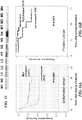


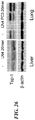
Classifications
-
- C—CHEMISTRY; METALLURGY
- C07—ORGANIC CHEMISTRY
- C07K—PEPTIDES
- C07K14/00—Peptides having more than 20 amino acids; Gastrins; Somatostatins; Melanotropins; Derivatives thereof
- C07K14/435—Peptides having more than 20 amino acids; Gastrins; Somatostatins; Melanotropins; Derivatives thereof from animals; from humans
- C07K14/475—Growth factors; Growth regulators
-
- A—HUMAN NECESSITIES
- A61—MEDICAL OR VETERINARY SCIENCE; HYGIENE
- A61P—SPECIFIC THERAPEUTIC ACTIVITY OF CHEMICAL COMPOUNDS OR MEDICINAL PREPARATIONS
- A61P35/00—Antineoplastic agents
-
- A—HUMAN NECESSITIES
- A61—MEDICAL OR VETERINARY SCIENCE; HYGIENE
- A61K—PREPARATIONS FOR MEDICAL, DENTAL OR TOILETRY PURPOSES
- A61K38/00—Medicinal preparations containing peptides
-
- C—CHEMISTRY; METALLURGY
- C07—ORGANIC CHEMISTRY
- C07K—PEPTIDES
- C07K2319/00—Fusion polypeptide
- C07K2319/31—Fusion polypeptide fusions, other than Fc, for prolonged plasma life, e.g. albumin
Definitions
- the present disclosure relates to methods for treating of tumor metastasis, as well as methods for preventing, inhibiting, and predicting tumor metastasis.
- the dislosure further relates to treating angiogenesis-dependent diseases and disorders, screening methods for tumor cell derived anti-angiogenic factors and methods for cancer prognosis evaluation.
- metastasis The spread of cancer cells from a primary tumor site to distant organs is known as metastasis.
- the progression of human cancer to metastatic disease is the major contributing factor to its lethality. Metastasis has been considered one of the most interesting aspects of the pathogenesis of cancer.
- Cancer tumor metastasis, or otherwise known as metastatic disease is responsible for most therapeutic failures in treating the disease, as patients succumb to the multiple tumor growth, accounting for more than 90% of human cancer related deaths. See, for example, Cancer, A Comprehensive Treatise, F. F. Becker (editor), Volume 4, Chapter 3, Plenum Press, New York, 1975 .
- a tumor In order for a tumor to form lethal metastases it must acquire the ability to carry out a complex series of steps. These steps include: gaining access to the vasculature or lymphatic system (intravasation), surviving during transit, exiting the vascular or lymphatic channels (extravasation), and proliferating at the metastatic site.
- steps include: gaining access to the vasculature or lymphatic system (intravasation), surviving during transit, exiting the vascular or lymphatic channels (extravasation), and proliferating at the metastatic site.
- angiogenesis not only allows tumors to grow beyond the size limitation imposed by the diffusion limit of oxygen, but also provides a conduit through which the tumor cells can travel and colonize distant organs (Brown et al., 1999; MacDougall and Matrisian, 1995). Once the tumor cells arrive at the metastatic site they must also induce neovascularization in order to grow beyond a microscopic size. It has been documented, however, that metastatic colonies can remain in a microscopic or dormant state and not progress beyond this size for months or years following the initial colonization (Fidler, 2003).
- tumor growth and proliferation is not governed solely by cell-autonomous processes and that the conditions present in the microenvironment that permitted proliferation at the primary site can not exist at the metastatic site.
- the ability of a tumor to communicate with the surrounding stroma, composed of fibroblasts, immune cells and endothelium must be reestablished upon arrival at the metastatic site.
- One way in which heterotypic tumor-stromal signaling could affect tumor growth is through the regulation of the production and secretion of pro- and anti-angiogenic proteins by the surrounding stromal fibroblasts and endothelial cells.
- tumor cells can stimulate the expression of the pro-angiogenic protein VEGF in the surrounding stroma (Dong et al., 2004; Fukumura et al., 1998).
- VEGF pro-angiogenic protein
- Tsp-1 Thrombospondin
- Embodiments of the present invention are based in part on the discovery that a peptide fragment from the protein Saposin A stimulates the expression of thrombospondin (Tsp-1) in the surrounding environment of the tumor cells, namely the stroma comprised of fibroblasts and endothelial cells. Tsp-1 is also activated in distant environments such as lymph nodes.
- Saposin A is made as a precursor to polypeptide prosaposin (Psap).
- Tsp-1 is a potent endogenous anti-angiogenic factor, and its activation by the tumor-derived protein is via the activation of the tumor suppressor p53.
- P53 is a transcriptional activator of Tsp-1.
- saposin A can potently stimulate and induce Tsp-1 expression (see Example 15, Figs. 21 and 22 ) and reduced angiogenesis. These include a 20-mer LEKTCDWLPKPNMSASCKEI (SEQ. ID. No. 29) and a 13-mer CDWLPKPNMSASC (SEQ. ID. No. 37).
- the disclosure provides for an isolated peptide consisting essentially of at least 10 consecutive amino acid residues from the sequence LEKTCDWLPKPNMSASCKEI (SEQ. ID. No. 29) or a conservative substitution variant thereof.
- the isolated peptide consists essentially of at least 11, at least 12, at least 13, at least 14, at least 15, at least 16, at least 17, at least 18, or at least 19 consecutive amino acid residues from SEQ. ID. No. 29 or a conservative substitution variant thereof.
- the isolated peptide is flanked by 0, 1, 2, 3, or 4 amino acid residues on either terminus or both termini.
- the isolated peptide consists of the sequence CDWLPKPNMSASC (SEQ. ID. No. 37), LEKTCDWLPKPNMSASC KEI (SEQ. ID. No. 29) or a conservative substitution variant thereof, wherein the peptide is flanked by 0 or 1 amino acid residues on either terminus or both termini., wherein the isolated peptide described herein is capable of activating p53 and/or inducing Tsp-1 expression.
- the isolated peptide described herein is fused or conjugated to a therapeutic molecule.
- the isolated peptide having conservative substitutions described herein have at least 1, 2, 3, 4, or 5 conservative amino acid substitutions. In another embodiment, the isolated peptides having conservative substitutions described herein have no more than 1, 2, 3, 4, or 5 conservative amino acid substitutions. In another embodiment, the isolated peptides described herein have no conservative amino acid substitutions.
- an isolated chimeric polypeptide comprising a first portion and a second portion, wherein the first portion is a peptide consisting of the sequence CDWLPKPNMSASC (SEQ. ID. No. 37) or a conservative substitution variant thereof, wherein the peptide is flanked by 0 or 1 amino acid residues on either terminus or both termini, and wherein the second portion of the chimeric polypeptide is not a Psap protein.
- the peptide making up first portion of the chimeric polypeptide consists essentially of at least 11, at least 12, at least 13, at least 14, at least 15, at least 16, at least 17, at least 18, or at least 19 consecutive amino acid residues from SEQ. ID. No. 29 or a conservative substitution variant thereof.
- the second portion comprises an amino acid sequence or a polymer that enhances the serum half life of the first portion of the chimeric polypeptide, e.g., but not limited to, albumin, transthyretin, Fc of IgGs.
- the second portion is a therapeutic molecule, such as an anti-angiogenesis factor, an anti-VEGF agent or an anti-cancer drug.
- composition comprising a peptide or a chimeric polypeptide, and a pharmaceutically acceptable carrier, wherein the peptide consists essentially of the sequence CDWLPKPNMSASC (SEQ. ID. No. 37) or a conservative substitution variant thereof, wherein the peptide is flanked by 0 or 1 amino acid residues on either terminus or both termini, wherein the chimeric polypeptide comprises a first portion and a second portion, wherein the first portion is a peptide consisting of the sequence CDWLPKPNMSASC (SEQ. ID. No.
- the peptide is flanked by 0 or 1 amino acid residues on either terminus or both termini, and wherein the second portion of the chimeric polypeptide is not a Psap protein or fragment.
- the peptide consists essentially of at least 11, at least 12, at least 13, at least 14, at least 15, at least 16, at least 17, at least 18, or at least 19 consecutive amino acid residues from SEQ. ID. No. 29 or a conservative substitution variant thereof.
- Described herein is a method of treating an angiogenesis-dependent disease or disorder, comprising administering to a subject in need thereof, a therapeutically effective amount of a composition comprising of a peptide or a chimeric polypeptide, and a pharmaceutically acceptable carrier, wherein the peptide consists essentially of the sequence CDWLPKPNMSASC (SEQ. ID. No. 37) LEKTCDWLPKPNMSASCKEI (SEQ. ID. No. 29) or a conservative substitution variant thereof, or the peptide consists essentially of at least ten consecutive amino acid residues of the sequence LEKTCDWLPKPNMSASCKEI (SEQ. ID. No.
- the chimeric polypeptide comprises a first portion and a second portion, wherein the first portion is a peptide consisting essentially of the sequence CDWLPKPNMSASC (SEQ. ID. No. 37), LEKTCDWLPKPNMSASCKEI (SEQ. ID. No. 29) or a conservative substitution variant thereof, or is a peptide consisting essentially of at least ten consecutive amino acid residues of the sequence LEKTCDWLPKPNMSASCKEI (SEQ. ID. No.
- Described herein is a method of inhibiting the recurrence of an angiogenesis-dependent disease or disorder, the method comprising administering to a subject in need thereof, a therapeutically effective amount of a composition comprising of a peptide or a chimeric polypeptide, and a pharmaceutically acceptable carrier, wherein the peptide consists essentially of the sequence CDWLPKPNMSASC (SEQ. ID. No. 37) LEKTCDWLPKPNMSASCKEI (SEQ. ID. No. 29) or a conservative substitution variant thereof, or the peptide consists essentially of at least ten consecutive amino acid residues of the sequence LEKTCDWLPKPNMSASCKEI (SEQ. ID. No.
- the chimeric polypeptide comprises a first portion and a second portion, wherein the first portion is a peptide consisting essentially of the sequence CDWLPKPNMSASC (SEQ. ID. No. 37), LEKTCDWLPKPNMSASCKEI (SEQ. ID. No. 29) or a conservative substitution variant thereof, or is a peptide consisting essentially of at least ten consecutive amino acid residues of the sequence LEKTCDWLPKPNMSASCKEI (SEQ. ID. No.
- Described herein is a method of inhibiting metastasis of cancer in a subject diagnosed with cancer, the method comprising administering to a subject in need thereof, a therapeutically effective amount of a composition comprising of a peptide or a chimeric polypeptide, and a pharmaceutically acceptable carrier, wherein the peptide consists essentially of the sequence CDWLPKPNMSASC (SEQ. ID. No. 37) LEKTCDWLPKPNMSASCKEI (SEQ. ID. No. 29) or a conservative substitution variant thereof, or the peptide consists essentially of at least ten consecutive amino acid residues of the sequence LEKTCDWLPKPNMSASCKEI (SEQ. ID. No.
- the chimeric polypeptide comprises a first portion and a second portion, wherein the first portion is a peptide consisting essentially of the sequence CDWLPKPNMSASC (SEQ. ID. No. 37), LEKTCDWLPKPNMSASCKEI (SEQ. ID. No. 29) or a conservative substitution variant thereof, or is a peptide consisting essentially of at least ten consecutive amino acid residues of the sequence LEKTCDWLPKPNMSASCKEI (SEQ. ID. No.
- Described herein is a method of treating an individual diagnosed with cancer comprising: (a) determining a level of Psap in a tumor sample from said individual; (b) comparing the Psap level determined in (a) with a reference Psap level; and wherein when the Psap level determined in (a) is lower than 95% of said reference Psap level, administering a therapeutically effective amount of a composition comprising of a peptide or a chimeric polypeptide, and a pharmaceutically acceptable carrier, wherein the peptide consists essentially of the sequence CDWLPKPNMSASC (SEQ. ID. No. 37) LEKTCDWLPKPNMSASCKEI (SEQ. ID. No.
- the peptide consists essentially of at least ten consecutive amino acid residues of the sequence LEKTCDWLPKPNMSASCKEI (SEQ. ID. No. 29) or a conservative substitution variant thereof, wherein the peptide is flanked by 0, 1, 2, 3 , or 4 amino acid residues on either terminus or both termini, wherein the chimeric polypeptide comprises a first portion and a second portion, wherein the first portion is a peptide consisting essentially of the sequence CDWLPKPNMSASC (SEQ. ID. No. 37), LEKTCDWLPKPNMSASCKEI (SEQ. ID. No.
- the peptide making up the composition or the first portion of the chimeric polypeptide making up the composition consists essentially of at least 11, at least 12, at least 13, at least 14, at least 15, at least 16, at least 17, at least 18, or at least 19 consecutive amino acid residues from SEQ. ID. No. 29 or a conservative substitution variant thereof.
- the angiogenesis-dependent disease or disorder is selected from a group consisting of cancer, psoriasis, age-related macular degeneration, thyroid hyperplasia, preeclampsia, rheumatoid arthritis and osteoarthritis, Alzheimer's disease, obesity, pleural effusion, atherosclerosis, endometriosis, diabetic/other retinopathies, neovascular glaucoma, age-related macular degeneration, hemangiomas, and corneal neovascularization.
- the subject is diagnosed with a benign or malignant tumor.
- the treatment is administered in conjunction with chemotherapy, radiation therapy, a cytostatic agent, an anti-VEGF agent, an anti-angiogenesis factor, and/or a p53 reactivation agent.
- Embodiments of the present invention are based on the discovery that non- or weakly metastatic tumor cells secrete a protein that stimulates the expression of thrombospondin (Tsp-1) in the surrounding environment of the tumor cells, namely the stroma comprised of fibroblasts and endothelial cells. While not wishing to be bound by theory, the increase in expression of Tsp-1 in the stroma keeps the tumor cells from metastasizing.
- Tsp-1 is a potent endogenous anti-angiogenic factor, and the stimulation of Tsp-1 expression by the tumor-derived protein is via the activation of the tumor suppressor p53.
- the tumor suppressor p53 is a transcription activator of Tsp-1 expression.
- This tumor-associated protein secreted by non- or weakly metastatic tumor cells is prosaposin (Psap).
- Psap This tumor-associated protein secreted by non- or weakly metastatic tumor cells.
- the inventors have found that two peptide fragments from Saposin A, Saposin A being a cleavage product of Psap.
- These two peptide fragments CDWLPKPNMSASC (SEQ. ID. No. 37) and LEKTCDWLPKPNMSASCKEI (SEQ. ID. No. 29) were also capable of stimulating Tsp-1 expression in vitro and in vivo.
- metastatic tumor cells express a high amount of Psap and Tsp-1, in addition to stimulating p53 and Tsp-1 expression in the surrounding tumor stroma.
- metastatic tumor cells express low amounts of Psap and Tsp-1, and metastatic tumor cells also repress the expression of p53 and Tsp-1 in the tumor stroma.
- metastasis Psap and Tsp-1 expression in the tumor cells
- Psap and Tsp-1 expression in the tumor stroma there is also a strong correlation between metastasis and the Psap level in the plasma and/or platelets of patients with metastatic cancers.
- Psap While not wishing to be bound by theory, high expression of Psap from a dormant primary tumor prevents the tumor from metastasis and also prevents the establishment secondary tumors at sites away from the primary tumor site. Conversely, low or no expression Psap in a primary tumor allows the tumor to metastasize and establish secondary tumors at sites far away from the primary tumor site.
- the Psap secreted by a tumor affects its local and distant environment via paracrine and endocrine signaling mechanisms, affecting whether a tumor cell can grow bigger and/or implant and grow at a different and distant location from the primary tumor site.
- Psap functions as a repressor of both lymphatic and vascular metastasis by inducing p53 and consequently Tsp-1 expression in stromal fibroblasts via both paracrine and endocrine signaling mechanisms.
- Prosaposin is the saposin precursor protein made up of approximately 524-527 amino acids which includes a 16 amino acids signal peptide.
- the full-length precursor 53-kDa polypeptide undergoes co-translational glycosylation and modification in the endoplasmic reticulum and Golgi system to yield a 70-72 kDa precursor protein.
- cathepsin D participates in its proteolytic processing to yield intermediate molecular forms of 35 to 53 kDa and then to a 13-kDa glycoprotein and finally to the mature 8-11 kDa partially glycosylated forms of individual saposin molecules ( O'Brien J.
- Psap and the individual saposin proteins are expressed by a wide variety of cells types originating from ectodermal, mesodermal, and endodermal germ layers including but not limited to lung, skin, fibroblast, stromal cells, bone, smooth muscle, skeletal muscle, cardiac muscle, placenta, red and white blood cells, pancreas, placenta, lymphoreticular system (spleen, thymus, liver), micro and macrovascular system, genitourinary system (e.g., prostate, testes, seminal vesicle), central and peripheral nervous system.
- lung skin
- fibroblast stromal cells
- bone smooth muscle
- skeletal muscle cardiac muscle
- placenta red and white blood cells
- pancreas, placenta lymphoreticular system (spleen, thymus, liver), micro and macrovascular system
- genitourinary system e.g., prostate, testes, seminal vesicle
- Prosaposin and saposins are also present as soluble proteins in extracellular space/fluid including pleural fluid, cerebrospinal fluid, seminal fluid, milk, and serum (Campana WM., et al., 1999; Kishimoto Y. et al., 1992).
- Psap is overexpressed in breast adenocarcinoma cell lines, non small-cell lung adenocarcinoma, neuroblastoma, and schwannoma cell lines, glioma cell lines, adult and pediatric brain tumors (e.g., medulloblastoma-, astrocytoma-, glioblastoma multiforme-cell lines), fibrosarcoma, osteosarcoma, and prostate cancer cell lines, different types of tumors (brain, colon, lung, pancreas, rectum, ovary, parotid, skin, bladder, small intestine, thymus, and uterus), including human prostate cancer cell lines.
- the overall the expression and biofunctional significances of prosaposin and saposins in cancer are largely unknown (Koochekpour S. March 2006; Koochekpour S. September 2006).
- prosaposin is a dual function molecule; as the precursor of intracellular lysosomal saposin proteins involved in sphingolipid hydrolysis activity and as a secreted soluble protein with neurotrophic activities, including growth, development, and maintenance of the peripheral and central nervous system, nerve regeneration and plasticity, stimulation of neurite outgrowth, stimulation of neuroblastoma cells proliferation, protection from cell-death or apoptosis, and activation of MAPK- and PI3K/Akt-signaling pathways (Morales and Badran, 2003; Misasi R, et al., 2001; Campana WM, et al., 1998; Hiraiwa M, et al., 1997; Hiraiwa M, et al., 1997; Kotani Y., et al., 1996; Campana WM., et al., 1996; O'Brien JS., et al., 1995; O'Brien JS., et al.
- stroma or "tumor stroma” refers to the connective tissue framework and non-tumor cells of a tumor. Examples of some non-tumor cells found in a tumor stroma are fibroblasts and endothelial cells.
- tumor means a mass of transformed cells that are characterized, at least in part, by containing angiogenic vasculature.
- the transformed cells are characterized by neoplastic uncontrolled cell multiplication which is rapid and continues even after the stimuli that initiated the new growth has ceased.
- the term “tumor” is used broadly to include the tumor parenchymal cells as well as the supporting stroma, including the angiogenic blood vessels that infiltrate the tumor parenchymal cell mass.
- a tumor generally is a malignant tumor, i.e., a cancer having the ability to metastasize (i.e., a metastatic tumor)
- a tumor also can be nonmalignant (i.e., non-metastatic tumor).
- Tumors are hallmarks of cancer, a neoplastic disease the natural course of which is fatal. Cancer cells exhibit the properties of invasion and metastasis and are highly anaplastic.
- the term “metastases” or “metastatic tumor refers to a secondary tumor that grows separately elsewhere in the body from the primary tumor and has arisen from detached, transported cells, wherein the primary tumor is a solid tumor.
- the primary tumor refers to a tumor that originated in the location or organ in which it is present and did not metastasize to that location from another location.
- a "malignant tumor” is one having the properties of invasion and metastasis and showing a high degree of anaplasia. Anaplasia is the reversion of cells to an immature or a less differentiated form, and it occurs in most malignant tumors.
- recurrence of an angiogenic disease or disorder refers to the re-manifestation/re-development of known symptoms associated with the angiogenic disease or disorder after previous successful treatment of the angiogenic disease or disorder.
- a "recurrence" of a tumor refers to the enlargement of an existing tumor whose growth had stopped or reduced during an anti-cancer therapy, or the emergence of a tumor at the original (primary) site of tumor discovery after the original tumor had been excised or reduced in size.
- the recurrence of a tumor can also mean new tumor growth(s) of the same tumor type as the original tumor at a site different from the original site of tumor discovery.
- a recurrence of rheumatoid arthritis can include localized swelling/ pain/ joint stiffness, and elevated leukocyte ingression after a period of disease remission and symptom free.
- the term "inhibit” or “inhibition” means the reduction or prevention of tumor growth and /or tumor metastasis in cancers. Inhibition includes slowing the rate of tumor growth and metastasis. The tumor growth rate can be reduced by about 20%, about 30%, about 40%, about 50%, about 60%, about 70%, about 80%, about 90%, about 100%, about 125%, about 150% or more compared to a control, untreated tumor of the same type. Inhibition also means a reduction in the size of the tumor of at least 10%, at least 20%, at least 30%, at least 40%, at least 50%, at least 60%, at least 70%, at least 80%, at least 90%, at least 100% or more compared to a control, untreated tumor of the same type.
- the prevention of tumor growth and /or metastasis means no further increase in the size of the tumors from the time of start of treatment administration. Prevention also means status quo of no new metastatic tumors detected (i.e., no further spread of cancer) and/or an increase amount of tumor markers detected by methods known in the art.
- the term "therapeutically effective amount” refers to the amount that is safe and sufficient to prevent or delay the development and further spread of metastases in cancer patients. The amount can also cure or cause the cancer to go into remission, slow the course of cancer progression, slow or inhibit tumor growth, slow or inhibit tumor metastasis, slow or inhibit the establishment of secondary tumors at metastatic sites, or inhibit the formation of new tumor metastasis.
- treat or “treatment” refer to both therapeutic treatment and prophylactic or preventative measures, wherein the object is to prevent, slow down, and/or halt the development or spread of cancer.
- beneficial or desired clinical results include, but are not limited to, alleviation of symptoms, diminishment of extent of disease, stabilized (i.e., not worsening) state of disease, delay or slowing of disease progression, amelioration or palliation of the disease state, and remission (whether partial or total), whether detectable or undetectable.
- Treatment can also mean prolonging survival as compared to expected survival if not receiving treatment.
- Those in need of treatment include those already diagnosed with cancer as well as those likely to develop secondary tumors due to metastasis.
- angiogenesis refers to the sprouting of new blood vessels from pre-existing blood vessels, characterized by endothelial cell proliferation and migration triggered by certain pathological conditions, such as the growth of solid tumors and metastasis.
- angiogenesis-dependent disease or disorder refers to diseases or disorders that are dependent on a rich blood supply and blood vessel proliferation for the disease pathological progression (e.g., metastatic tumors) or diseases or disorders that are the direct result of aberrant blood vessel proliferation (e.g., diabetic retinopathy and hemangiomas).
- Examples include abnormal vascular proliferation, ascites formation, psoriasis, age-related macular degeneration, thyroid hyperplasia, preeclampsia, rheumatoid arthritis and osteoarthritis, Alzheimer's disease, obesity, pleural effusion, atherosclerosis, endometriosis, diabetic/other retinopathies, ocular neovascularizations such as neovascular glaucoma and corneal neovascularization.
- nucleic acid refers to DNA or RNA.
- the term encompasses sequences that include any of the known base analogs of DNA and RNA.
- vector refers to a nucleic acid construct comprising the complete or partial cDNA of Psap (SEQ. ID. No. 2, 4, or 6) (Genbank Accession No. NM_002778, NM_001042466, or NM_001042465), wherein the nucleic acid construct is designed for delivery to a host cell, transfer between different host cells, or for the expression of Psap or functional fragments or variants thereof, in cells.
- a vector can be viral or non-viral.
- viral vector refers to a nucleic acid vector construct that includes at least one element of viral origin and includes elements sufficient for or permissive of packaging into a viral vector particle.
- a viral vector can contain the coding sequence for a Psap protein in place of non-essential viral genes.
- the vector and/or particle can be utilized for the purpose of transferring DNA, RNA or other nucleic acids into cells either in vitro or in vivo. Numerous forms of viral vectors are known in the art.
- prognosis encompasses predictions and likelihood analysis of disease progression, particularly tumor recurrence, metastatic spread, and disease relapses.
- the prognosis method described herein is intended for clinical use in making decision concerning treatment modalities, including therapeutic interventions, diagnostic criteria such as disease staging, and disease monitoring and surveillance for metastasis or recurrence of neoplastic disease.
- tissue sample refers to a portion, piece, part, segment, or fraction of a tissue which is obtained or removed from an intact tissue or organ of a subject, preferably a human subject.
- a "subject” refers to a mammal, preferably a human.
- the term “individual”, “subject”, and “patient” are used interchangeably.
- tumor sample refers to a portion, piece, part, segment, or fraction of a tumor, for example, a tumor which is obtained or removed from a subject (e.g., removed or extracted from a tissue of a subject), preferably a human subject.
- the term “functional” refers to the fragments and variants of Psap protein having cellular functions substantially similar to that of the parent full-length Psap. At the minimum, “functional” refers to the capability of stimulating Tsp-1 and/or p53 expressions. In other embodiments, other cellular functions including capability of being glycosylated, of being proteolytically processed to give the smaller saposins which are required for the hydrolysis of glycosphingolipids by lysosomal lysozmes, ability to bind to membrane lipids, and have neurotrophic activities.
- the term "substantially similar” refers to no change to the course of direction of biological effects resulting from the actions of the fragments and variants of Psap in the cell.
- the fragment and variant forms of Psap are still capable of stimulating Tsp-1 and p53 expression and activation, and they can still be processed proteolytically to give saposins, and these saposins can be used by the cell in the hydrolysis of glycosphingolipids.
- a "substantially similar" functional fragment of saposin A has an amino acid sequence differing from SEQ. ID. No. 13 by having one or more conservative amino acid substitution and/or modification but is still capable of stimulating Tsp-1 and p53 expressions and activation, the methods of assaying are described herein and are well known in the art.
- variant refers the splice variant of Psap protein (also known as isoforms) encoded by the nucleic acids of Psap (Genbank Accession No. NM_002778, SEQ. ID. No. 2), NM_001042465 (SEQ. ID. No. 4), or NM_001042466 (SEQ. ID. No. 6)).
- variant also refers to Psap protein or molecule modified at one or more base pairs, codons, introns, exons, or amino acids, respectively, yet still retain the biological activity and cellular functions of a Psap protein, e.g., a conservative substitution variant where one or more of the amino acids have been substituted with their respective conservative amino acid but the variant still retain the ability to stimulate Tsp-1 expression.
- a conservative substitution variant where one or more of the amino acids have been substituted with their respective conservative amino acid but the variant still retain the ability to stimulate Tsp-1 expression.
- the polypeptide sequence of the variant Psap protein is slightly different from that prescribed by the Psap coding nucleic acid (SEQ. ID. No. 2, 4, and 6).
- amino acid mutations in the Psap protein There are one or more amino acid mutations in the Psap protein. Conservative amino acid substitution can produced variant Psap proteins.
- the amino acid serine can be substituted for threonine and the amino acid aspartate can be substituted for glutamate.
- the variant Psap proteins have comparable or greater Tsp-1 and p53 expression stimulation activity than the parent Psap protein.
- the variant Psap protein can have at least 80%, at least 90 %, at least 100%, at least 110%, at least 120%, at least 130%, at least 140%, or at least 150% of the Tsp-1 and p53 expression stimulation activity of the parent Psap protein.
- Variants can be produced by a number of means including methods such as, for example, error-prone PCR, shuffling, oligonucleotide-directed mutagenesis, recursive ensemble mutagenesis, exponential ensemble mutagenesis, site-specific mutagenesis, gene reassembly, GSSM and any combination thereof.
- the term "conservative amino acid substitution” is one in which the amino acid residue is replaced with an amino acid residue having a side chain with a similar charge.
- Families of amino acid residues having side chains with similar charges have been defined in the art. These families include amino acids with basic side chains (e.g., lysine, arginine, histidine), acidic side chains (e.g., aspartic acid, glutamic acid), uncharged polar side chains (e.g., glycine, asparagine, glutamine, serine, threonine, tyrosine, cysteine), nonpolar side chains (e.g., alanine, valine, leucine, isoleucine, proline, phenylalanine, methionine, tryptophan), beta-branched side chains (e.g., threonine, valine, isoleucine) and aromatic side chains (e.g., tyrosine, phenylalanine, tryp
- fragment refers to an amino acid sequence which is shorter than the original polypeptide encoded by the cDNA of Psap (Genbank Accession No. NM_002778, SEQ. ID. No. 2), NM_001042465.1 (SEQ. ID. No. 4) or NM_001042466.1 (SEQ. ID. No. 6) thus presenting an incomplete Psap protein.
- the Psap protein is shortened or truncated.
- the term "functional fragment” as used herein refers to the truncated Psap protein that has cellular functions including the stimulation of Tsp-1 and p53 expression. Fragments are at least 10 amino acids but are not the full-length Psap protein.
- fragments are 10 amino acids or more but are not the full-length Psap protein.
- fragments include fragments consisting of amino acids 1-300, amino acids 1-150, and amino acids 1-490 of the original full length Psap protein. These fragments contain the Saposin A- and Saposin B-domains and can be cleaved to give the smaller saposins or smaller fragments thereof.
- Saposin A (protein)
- polypeptide and “peptide” are used interchangeably herein to refer to a polymer of amino acids. These terms do not connote a specific length of a polymer of amino acids. Thus, for example, the term includes oligomeric peptides, made up of two or more physically linked peptides, whether produced using recombinant techniques, chemical or enzymatic synthesis, or naturally occurring. This term also includes polypeptides that have been modified or derivatized, such as by glycosylation, acetylation, phosphorylation, and the like.
- Psap protein refers to the splice variant full-length human prosaposin isoform A preproprotein (Genbank Accession Nos.: NM_002778 (SEQ. ID. No. 2), NP_002769.1 (SEQ. ID. No. 1)); UniProtKB/Swiss-Prot P07602, UniProtKB/TrEMBL Q53Y86), the splice variant full-length human prosaposin isoform B preproprotein (Genbank Accession Nos.: NM_001042465.1 (SEQ. ID. No. 4), NP_001035930.1 (SEQ. ID. No.
- GenPept/UniProtKB/TrEMBL O75905, P07602.2, Q53FJ5, Q59EN5, Q5BJH1, Q5JQ36, and Q5JQ37; the secreted forms of these splice variants, functional fragments and conservative substitution variants thereof that are greater or equal to 10 amino acid residues, the functional fragments and variants of the isoforms that are greater or equal to 10 amino acid residues, differentially glycosylated forms of the full-length splice variant Psap protein, secreted differentially glycosylated forms of the Psap protein, differentially glycosylated functional fragments with less than 524 amino acids and that are greater or equal to 10 amino acid residues, and/or differentially glycosylated functional variants thereof.
- Psap protein includes substantially similar Psap proteins, saposin A and functional fragments thereof that are greater or equal to 10 amino acid residues.
- the term “differentially glycosylated” refers to differences in glycosylation at the available glycosylation sites of full-length Psap. There are five glycosylation sites on the full-length protein. Accordingly, a full-length Psap can have anywhere from zero and up to five glycosylated groups. In addition, the term also refers to the presence of different sugar groups on the polypeptide.
- cancer refers to any of various malignant neoplasms characterized by the proliferation of anaplastic cells that tend to invade surrounding tissue and metastasize to new body sites as well as to the pathological conditions characterized by such malignant neoplastic growths.
- cancer also refers to cells and tissue with neoplasms characteristics of anaplastic proliferation that are not invasive of surrounding tissue, i.e., anaplastic cells that are benign.
- promptness refers to any time within one month of positive laboratory test results confirming presence of cancer cells.
- development of cancer or “cancer development” refers to the development of primary or initial cancer, the development of metastasis from benign and/or malignant tumors, and/or the development of malignancy from benign tumors.
- fusion protein refers to a protein created by joining two heterologous genes or two heterologous proteins / peptides or portions thereof together.
- heterologous in reference to genes and proteins means the genes or proteins are two different and not similar entities.
- two heterologous genes encode for two different and not similar proteins respectively.
- a “fusion protein” or “fusion polypeptide” is a chimeric protein, made of at least two different types of proteins or portions thereof.
- “fusion protein” or “fusion polypeptide” is achieved through the creation of a fusion gene which is done through the removal of the stop codon from a DNA sequence of the first protein and then attaching the DNA sequence of the second protein in frame.
- the resulting DNA sequence can then be expressed by a cell as a single protein.
- the two heterologous proteins can be joined together with a linker or spacer peptide added between the two proteins.
- This linker or spacer peptide often contains protease cleavage site(s) to facilitate the separation of the two different proteins after expression and purification.
- the making of fusion protein as a technique is commonly used for the identification and purification of proteins through the fusion of a GST protein, FLAG peptide or a hexa-His peptide.
- a peptide linker is a short sequence of amino acids that is not part of the sequence of either of the two peptides being joined to form a fusion protein or fusion polypeptide.
- a peptide linker is attached on its amino-terminal end to one polypeptide or polypeptide domain and on its carboxyl-terminal end to another polypeptide or polypeptide domain.
- the peptide linker can be a flexible linker, in that the peptide sequence does not adopt any secondary structures known in proteins, e.g., alpha helices. Such flexible linkers are predominantly made of non-charged, apolar amino acid residues and are hydrophobic. Secondary protein structures can be determined by methods known in the art, for example, circular dichroism.
- An example of a flexible peptide linker is LGGGGSGGGGSA (SEQ. ID. No. 41).
- the peptide linker can take the form a monomeric hydrophilic ⁇ -helix, for example, AEAAAKEAAAKEA (SEQ. ID. No. 42).
- PEGylated is meant the covalent attachment of at least one molecule of polyethylene glycol to a biologically active molecule.
- the average molecular weight of the reactant PEG is preferably between about 3,000 and about 50,000 daltons, more preferably between about 10,000 and about 40,000 daltons, and most preferably between about 15,000 and about 30,000 daltons. Particularly preferred are PEGs having nominal average sizes of about 20,000 and about 25,000 daltons.
- the method of attachment is not critical, but preferably does not alter, or only minimally alters, the activity of the biologically active molecule. Preferably the increase in half-life is greater than any decrease in biological activity.
- a preferred method of attachment is via N-terminal linkage to a polypeptide or peptide.
- compositions and methods refers to respective component(s) thereof, as essential to the invention, yet open to the inclusion of unspecified elements, essential or not.
- compositions, methods, and respective component thereof are limited to those that do not materially affect the basic and novel characteristic(s) of the invention. This is referred to using the term "consisting essentially of'. This applies equally to steps within a described method as well as compositions, peptides and components therein. In other embodiments, the inventions, peptides, compositions, methods, and respective components thereof, described herein are intended to be exclusive of any element not recited with respect to that composition, element, component or method. This is referred to using the term "consisting of'.
- essential in reference to compositions and methods refers to the "essential" component in the composition or method being the peptide or protein sequence that, at the minimum, has the ability to stimulate Tsp-1 expression in the assays described herein, preferably also stimulate p53 expression.
- Non essential component in the composition or method would be heterologous protein that is not Psap, fusion portion of the fusion protein that is not Psap, PEG, polymer, immunoglobulin Fc region or conjugates etc.
- reducing the likelihood in reference to the development of certain conditions refers to a reduction by at least 20% compared to when no treatment or administration of a therapeutically effective amount of a Psap protein or a vector described herein.
- the reduction can also be at least 30%, at least 40%, at least 50%, at least 60%, at least 70%, at least 80%, at least 90%, at least 100%, including all the percent between 20% and 100%.
- a method of treating an angiogenesis-dependent disease or disorder comprising administering to a subject in need thereof a therapeutically effective amount of a Psap protein or a vector comprising the nucleic acid encoding a Psap protein, and a pharmaceutically acceptable carrier.
- Described is a method of inhibiting the recurrence of an angiogenesis-dependent disease or disorder, the method comprising administering to a subject in need thereof, a therapeutically effective amount of a Psap protein or a vector comprising the nucleic acid encoding a Psap protein, and a pharmaceutically acceptable carrier.
- the angiogenesis-dependent disease or disorder is selected from, but is not limited to, a group consisting of cancer, ascites formation, psoriasis, age-related macular degeneration, thyroid hyperplasia, preeclampsia, rheumatoid arthritis and osteoarthritis, Alzheimer's disease, obesity, pleural effusion, atherosclerosis, endometriosis, diabetic/other retinopathies, neovascular glaucoma, age-related macular degeneration (ARMD), hemangiomas, and corneal neovascularization.
- cancer ascites formation, psoriasis, age-related macular degeneration, thyroid hyperplasia, preeclampsia, rheumatoid arthritis and osteoarthritis, Alzheimer's disease, obesity, pleural effusion, atherosclerosis, endometriosis, diabetic/other retinopathies, neovascular glaucoma, age-related macular
- the angiogenesis-dependent disease or disorder is cancer, where the rapidly dividing neoplastic cancer cells require an efficient blood supply to sustain their continual growth of the tumor.
- cancer refers to any of various malignant neoplasms characterized by the proliferation of anaplastic cells that tend to invade surrounding tissue and metastasize to new body sites and also refers to the pathological condition characterized by such malignant neoplastic growths.
- the blood vessels provide conduits to metastasize and spread elsewhere in the body. Upon arrival at the metastatic site, the cancer cells then work on establishing a new blood supply network.
- embodiments of the invention serve to halt, prevent and limit the progression of the disease. Any solid tumor that requires an efficient blood supply to keep growing is a candidate target.
- candidates for the treatment described herein include carcinomas and sarcomas found in the anus, bladder, bile duct, bone, brain, breast, cervix, colon/rectum, endometrium, esophagus, eye, gallbladder, head and neck, liver, kidney, larynx, lung, mediastinum (chest), mouth, ovaries, pancreas, penis, prostate, skin, small intestine, stomach, spinal marrow, tailbone, testicles, thyroid and uterus.
- carcinomas include papilloma/carcinoma, choriocarcinoma, endodermal sinus tumor, teratoma, adenoma/adenocarcinoma, melanoma, fibroma, lipoma, leiomyoma, rhabdomyoma, mesothelioma, angioma, osteoma, chondroma, glioma, lymphoma/leukemia, squamous cell carcinoma, small cell carcinoma, large cell undifferentiated carcinomas, basal cell carcinoma and sinonasal undifferentiated carcinoma.
- sarcomas include soft tissue sarcoma such as alveolar soft part sarcoma, angiosarcoma, dermatofibrosarcoma, desmoid tumor, desmoplastic small round cell tumor, extraskeletal chondrosarcoma, extraskeletal osteosarcoma, fibrosarcoma, hemangiopericytoma, hemangiosarcoma, Kaposi's sarcoma, leiomyosarcoma, liposarcoma, lymphangiosarcoma, lymphosarcoma, malignant fibrous histiocytoma, neurofibrosarcoma, rhabdomyosarcoma, synovial sarcoma, and Askin's tumor, Ewing's sarcoma (primitive neuroectodermal tumor), malignant hemangioendothelioma, malignant schwannoma, osteosarcoma, and chondrosarcoma.
- the angiogenesis-dependent disease or disorder is age-related macular degeneration.
- VEGF contributes to abnormal blood vessel growth from the choroid layer of the eye into the retina, similar to what occurs during the wet or neovascular form of age-related macular degeneration.
- Macular degeneration often called AMD or ARMD (age-related macular degeneration)
- AMD age-related macular degeneration
- New blood vessels grow (neovascularization) beneath the retina and leak blood and fluid. This leakage causes permanent damage to light-sensitive retinal cells, which die off and create blind spots in central vision or the macula.
- the angiogenic disease or disorder is diabetic retinopathy- abnormal blood vessel growth associated with diabetic eye diseases.
- the activation of Tsp-1 via prosaposin serves to antagonize VEGF, a substance naturally produced in the body that promotes blood vessel formation. Released by the retina (light-sensitive tissue in back of the eye) when normal blood vessels are damaged by tiny blood clots due to diabetes, VEGF turns on its receptor, igniting a chain reaction that culminates in new blood vessel growth.
- the backup blood vessels are faulty; they leak, bleed and encourage scar tissue that detaches the retina, resulting in severe loss of vision.
- Such growth is the hallmark of diabetic retinopathy, the leading cause of blindness among young people in developed countries.
- the subject in need of treatment can be a mammal, such as a dog or a cat, preferably a human.
- the angiogenesis-dependent disease or disorder is rheumatoid arthritis.
- Rheumatoid arthritis is characterized by synovial tissue swelling, leucocyte ingress and angiogenesis, or new blood vessel growth. The disease is thought to occur as an immunological response to an as yet unidentified antigen.
- the expansion of the synovial lining of joints in rheumatoid arthritis (RA) and the subsequent invasion by the pannus of underlying cartilage and bone necessitate an increase in the vascular supply to the synovium, to cope with the increased requirement for oxygen and nutrients.
- Angiogenesis is now recognised as a key event in the formation and maintenance of the pannus in RA (Paleolog, E. M., 2002).
- a mononuclear infiltrate characterizes the synovial tissue along with a luxuriant vasculature.
- Angiogenesis is integral to formation of the inflammatory pannus and without angiogenesis; leukocyte ingress could not occur (Koch, A. E., 2000). Disruption of the formation of new blood vessels would not only prevent delivery of nutrients to the inflammatory site, it could also reduce joint swelling due to the additional activity of VEGF, a potent pro-angiogenic factor in RA, as a vascular permeability factor.
- the angiogenesis-dependent disease or disorder is Alzheimer's disease.
- AD Alzheimer's disease
- AD is the most common cause of dementia worldwide.
- AD is characterized by an excessive cerebral amyloid deposition leading to degeneration of neurons and eventually to dementia.
- the exact cause of AD is still unknown. It has been shown by epidemiological studies that long-term use of non-steroidal anti-inflammatory drugs, statins, histamine H2-receptor blockers, or calcium-channel blockers, all of which are cardiovascular drugs with anti-angiogenic effects, seem to prevent Alzheimer's disease and/or influence the outcome of AD patients. Therefore, it has been speculated that in AD angiogenesis in the brain vasculature may play an important role in AD.
- the brain endothelium secretes the precursor substrate for the beta-amyloid plaque and a neurotoxic peptide that selectively kills cortical neurons.
- amyloid deposition in the vasculature leads to endothelial cell apoptosis and endothelial cell activation which leads to neovascularization.
- Vessel formation could be blocked by the VEGF antagonist SU 4312 as well as by statins, indicating that anti-angiogenesis strategies can interfere with endothelial cell activation in AD (Schultheiss C., el. al., 2006; Grammas P., et al., 1999) and can be used for preventing and/or treating AD.
- the angiogenesis-dependent disease or disorder is obesity. It has been shown that the angiogenesis inhibitor, TNP-470 was able to prevent diet-induced and genetic obesity in mice ( Ebba Br ⁇ kenhielm et al., Circulation Research, 2004, 94:1579 ). TNP-470 reduced vascularity in the adipose tissue, thereby inhibiting the rate of growth of the adipose tissue and obesity development.
- the angiogenesis-dependent disease or disorder is endometriosis.
- Excessive endometrial angiogenesis is proposed as an important mechanism in the pathogenesis of endometriosis (Healy, DL., et al., 1998).
- the endometrium of patients with endometriosis shows enhanced endothelial cell proliferation.
- Strategies that inhibit angiogenesis can be used to treat endometriosis.
- the method of treating cancer is applicable to all carcinomas and sarcomas.
- the method is applicable to cancers selected from the group consisting of papilloma/carcinoma, choriocarcinoma, endodermal sinus tumor, teratoma, adenoma/adenocarcinoma, melanoma, fibroma, lipoma, leiomyoma, rhabdomyoma, mesothelioma, angioma, osteoma, chondroma, glioma, lymphoma/leukemia, squamous cell carcinoma, small cell carcinoma, large cell undifferentiated carcinomas, basal cell carcinoma, sinonasal undifferentiated carcinoma, soft tissue sarcoma such as alveolar soft part sarcoma, angiosarcoma, dermatofibrosarcoma, desmoid tumor, desmoplastic small round cell tumor, extraskeletal chondrosarcoma,
- the method of treating cancer is administered promptly after the detection of cancer.
- promptness refers to any time within one month of positive laboratory test results confirming presence of cancer cells. Diagnosis and detection of cancer cells are well known to one skilled in the art. Laboratory tests can be in the form of histology and/or biomarkers that are known in the art but are not limited to these examples.
- cancer biomarkers such as cancer antigen (CA) 15-3, carcinoembryonic antigen (CEA) and HER-2 for breast cancer, human papillomavirus (HPV) E6 and E7 oncoproteins for cervical cancer, alpha-fetoprotein (AFP), AFP fractions L3, P4/5, and the +II band, and ultrasonography for hepatocellular carcinoma (HCC), prostate-specific antigen (PSA) for prostate cancer, and serum CA-125 for ovarian and HCC.
- Tissue biopsy and histology are usually performed for confirmation and tissue typing of the original of cancer cell type.
- Described is a method of inhibiting metastasis of cancer in a subject diagnosed with cancer comprising administering to a subject in need thereof, a therapeutically effective amount of a Psap protein or a vector comprising the nucleic acid encoding a Psap protein, and a pharmaceutically acceptable carrier.
- the subject can be diagnosed with a benign or malignant cancer.
- Psap protein can be administered to inhibit the establishment of secondary tumor from the initially discovered benign or malignant tumor.
- the subject is a mammal, such as a dog or a cat, preferably a human, who has previously been diagnosed with cancer.
- the cancer can be benign or malignant, and it may or may not have metastasized.
- individual and subject are used interchangeably.
- the method of treatment is administered promptly after the diagnosis of cancer.
- Described is a method of inhibiting recurrence of cancer in a subject diagnosed with cancer comprises administering to a subject in need thereof, a therapeutically effective amount of a Psap protein or a vector comprising the nucleic acid encoding a Psap protein, and a pharmaceutically acceptable carrier.
- the subject can be diagnosed with a benign or malignant cancer.
- Psap protein can be administered to inhibit the re-growth of the primary tumor, development of tumors not related to the primary tumor, and/or establishment of secondary tumors from the initially discovered benign or malignant tumor.
- Described is a method for reducing the likelihood of cancer development in a subject comprising administering to a subject in need thereof, a therapeutically effective amount of a Psap protein or a vector comprising the nucleic acid encoding a Psap protein, and a pharmaceutically acceptable carrier.
- Psap protein can be administered to prevent the development of cancer, the development of metastasis, and/or the development of malignancy.
- Psap can be administered to the subject for preventing cancer development in this subject.
- the benign tumor can be removed by surgery.
- Psap can be administered to the subject for preventing any remaining existing benign tumor cells from developing into a malignant cancer as well as to prevent the development of metastasis.
- Psap can be administered to the subject for preventing the malignant tumor from further metastasis.
- Described is a method for reducing the likelihood of the cancer development in a subject at risk of development of cancer, the method comprises administering to a subject in need thereof, a therapeutically effective amount of a Psap protein or a vector comprising the nucleic acid encoding a Psap protein, and a pharmaceutically acceptable carrier.
- Described is provides a method for reducing the likelihood of the development of cancer malignancy in a subject previously diagnosed with cancer, the method comprises administering to a subject in need thereof, a therapeutically effective amount of a Psap protein or a vector comprising the nucleic acid encoding a Psap protein, and a pharmaceutically acceptable carrier.
- Described is provides a method for reducing the likelihood of cancer metastasis in a subject previously diagnosed with cancer, the method comprises administering to a subject in need thereof, a therapeutically effective amount of a Psap protein or a vector comprising the nucleic acid encoding a Psap protein, and a pharmaceutically acceptable carrier.
- the administration is in conjunction with a p53 reactivation agent.
- the administration is in conjunction with chemotherapy, radiation therapy, and/or a cytostatic agent.
- the administration is in conjunction with an anti-VEGF agent or an anti-angiogenesis factor.
- the Psap protein can be the full-length human prosaposin isoform A preproprotein, isoform B preproprotein, isoform C preproprotein, the secreted forms of these splice variants, functional fragments and variants thereof that are greater than or equal to 10 amino acid residues, the functional fragments and variants that are greater than or equal to 10 amino acid residues of the isoforms, differentially glycosylated forms of the full-length splice variant Psap protein, secreted differentially glycosylated forms of the Psap protein, differentially glycosylated functional fragments with less than 524 amino acids and are greater than or equal to 10 amino acid residues, and/or differentially glycosylated functional variants thereof are greater than or equal to 10 amino acid residues.
- Examples of functional fragments of Psap include Saposin A:
- the Psap protein is saposin A: SLPCDICKDVVTAAGDMLKDNATEEEILVYLEKTCDWLPKPNMSASCKEIVDSYLPVILDIIKGE MSRPGEVCSALNLCES (SEQ. ID. No. 13).
- the Psap protein is a functional fragment of saposin A that is less than 81 amino acid residues.
- the functional fragment of saposin A is any of SEQ. ID. No. 18-31.Conservative amino acid substitution of saposin A and of functional fragments thereof is also specifically contemplated.
- the functional fragments of saposin A and substantially similar fragments of saposin A induce p53 activation and/or Tsp-1 expression.
- the Psap protein is a functional fragment of saposin A that comprises at least 10 amino acid residues.
- the Psap protein is a peptidomimetic of a functional fragment of saposin A.
- compositions comprising the peptide consisting essentially of either SEQ. ID. No. 37, 29, or a conservative substitution variant thereof and mixtures of the two peptides (SEQ. ID. Nos. 37 and 29) or their respective conservative substitution variant thereof are also contemplated.
- an isolated peptide consisting essentially of at least ten consecutive amino acid residues of the sequence LEKTCDWLPKPNMSASCKEI (SEQ. ID. No. 29) or a conservative substitution variant thereof, wherein the peptide is flanked by 0, 1, 2, 3, or 4 amino acid residues on either terminus or both termini.
- this at least ten consecutive amino acid residues should be derived from between the 31 st to the 50 th amino acid residues of saposin A (SEQ. ID. No. 13).
- the isolated peptide consists essentially of at least 11, at least 12, at least 13, at least 14, at least 15, at least 16, at least 17, at least 18, or at least 19 consecutive amino acid residues from SEQ. ID. No. 29 or a conservative substitution variant thereof.
- an isolated peptide consisting of at least ten consecutive amino acid residues of the sequence LEKTCDWLPKPNMSASCKEI (SEQ. ID. No. 29) or a conservative substitution variant thereof, wherein the peptide is flanked by 0-4 amino acid residues on either terminus or both termini.
- the isolated peptide consists of at least 11, at least 12, at least 13, at least 14, at least 15, at least 16, at least 17, at least 18, or at least 19 consecutive amino acid residues from SEQ. ID. No. 29 or a conservative substitution variant thereof.
- Examples of isolated peptides consisting of at least ten consecutive amino acid residues of SEQ. ID. No. 13 include but are not limited to LEKTCDWLPK (SEQ. ID. No. 44), LEKTCDWLPKP (SEQ. ID. No. 45), LEKTCDWLPKPN (SEQ. ID. No. 46), LEKTCDWLPKPNM (SEQ. ID. No. 47), LEKTCDWLPKPNMS (SEQ. ID. No. 48), LEKTCDWLPKPNMSA (SEQ. ID. No. 21), LEKTCDWLPKPNMSAS (SEQ. ID. No. 49), LEKTCDWLPKPNMSASC (SEQ. ID. No. 43), LEKTCDWLPKPNMSASCK (SEQ. ID. No.
- LEKTCDWLPKPNMSASCKE SEQ. ID. No. 51
- EKTCDWLPKPNMSASCKEI SEQ. ID. No. 52
- KTCDWLPKPNMSASCKEI SEQ. ID. No. 53
- TCDWLPKPNMSASCKEI SEQ. ID. No. 54
- CDWLPKPNMSASCKEI SEQ. ID. No. 55
- DWLPKPNMSASCKEI SEQ. ID. No. 56
- WLPKPNMSASCKEI SEQ. ID. No. 57
- LPKPNMSASCKEI SEQ. ID. No. 58
- PKPNMSASCKEI SEQ. ID. No. 59
- KPNMSASCKEI SEQ.
- PNMSASCKEI SEQ. ID. No. 61
- KTCDWLPKPNMSASC SEQ. ID. No. 62
- TCDWLPKPNMSA SEQ. ID. No. 63
- CDWLPKPNMSASCK SEQ. ID. No. 64
- an isolated peptide consisting of the sequence CDWLPKPNMSASC (SEQ. ID. No. 37) or a conservative substitution variant thereof, wherein the peptide is flanked by 0 or 1 amino acid residues on either terminus or both termini, and wherein the isolated is capable of activating p53 and/or inducing Tsp-1 expression.
- an isolated peptide consisting of the sequence CDWLPKPNMSASC (SEQ. ID. No. 37) or a conservative substitution variant thereof, wherein the peptide is flanked by 0-4 amino acid residues on either terminus or both termini.
- an isolated peptide consisting essentially of the sequence LEKTCDWLPKPNMSASCKEI (SEQ. ID. No. 29) or a conservative substitution variant thereof, wherein the peptide is flanked by 0, 1, 2, 3, or 4 amino acid residues on either terminus or both termini.
- an isolated peptide consisting of the sequence LEKTCDWLPKPNMSASCKEI (SEQ. ID. No. 29) or a conservative substitution variant thereof, wherein the peptide is flanked by 0, 1, 2, 3, or 4 amino acid residues on either terminus or both termini.
- any of the known 20 amino acid residues can flank the peptides SEQ. ID. No. 37 and 29.
- Examples of some conservative substitution variants include but are not limited to L D KTCDWLPKPNMSASCKDI (SEQ. ID. No. 65), LEKTCDWIPKPNMSASCK DI (SEQ. ID. No. 66), LEKTCDWLPKPNMSASC R EI (SEQ. ID. No. 67), LE R TCDW I PKPNMSASCK D I (SEQ. ID. No. 68), CDW I P R PNMSASC (SEQ. ID. No. 69), C E WLP R PNMSASC (SEQ. ID. No. 70), CDW I PKPNMSASC (SEQ. ID.
- the isolated peptides are capable of activating p53 and inducing Tsp-1 expression.
- the isolated peptide is fused or conjugated to a therapeutic molecule described herein.
- any of the amino acid residues in the peptides SEQ. ID. No. 37 and 29 can be conservatively substituted.
- the isolated conservative substitution variant peptide has at least 1, 2, 3, 4, or 5 conservative amino acid substitutions. In other embodiments, the isolated conservative substitution variant peptide has no more than 1, 2, 3, 4, or 5 conservative amino acid substitutions. In another embodiment, the isolated conservative substitution variant peptide does not have any conservative amino acid substitution.
- an isolated chimeric polypeptide comprising a first portion and a second portion, wherein the first portion is a peptide consisting of the sequence CDWLPKPNMSASC (SEQ. ID. No. 37), or a conservative substitution variant thereof, wherein the peptide is flanked by 0 or 1 amino acid residues on either terminus or both termini, and wherein the peptide is capable of activating p53 and/or inducing Tsp-1 expression, and wherein the second portion is not a Psap protein or fragment.
- the peptide consists essentially of at least 11, at least 12, at least 13, at least 14, at least 15, at least 16, at least 17, at least 18, or at least 19 consecutive amino acid residues from SEQ. ID. No. 29 or a conservative substitution variant thereof.
- an isolated chimeric polypeptide comprising a first portion and a second portion, wherein the first portion is an isolated peptide consisting of the sequence CDWLPKPNMSASC (SEQ. ID. No. 37), or a conservative substitution variant thereof, wherein the peptide is flanked by 0 or 1 amino acid residues on either terminus or both termini, wherein the peptide is capable of activating p53 and/or inducing TSP-1 expression, and wherein the second portion is not a Psap protein.
- the peptide consists of at least 11, at least 12, at least 13, at least 14, at least 15, at least 16, at least 17, at least 18, or at least 19 consecutive amino acid residues from SEQ. ID. No. 29 or a conservative substitution variant thereof.
- the second portion comprises an amino acid sequence or a polymer that enhances the serum half life of the first portion.
- the second portion is a therapeutic molecule.
- composition comprising a peptide consisting of the sequence CDWLPKPNMSASC (SEQ. ID. No. 37) or a conservative substitution variant thereof, and a pharmaceutically acceptable carrier.
- the invention also provides a composition comprising a peptide consisting of the sequence CDWLPKPNMSASC (SEQ. ID. No. 37) or a conservative substitution variant thereof and a pharmaceutically acceptable carrier.
- the peptide is derived from saposin A.
- the essential sequence sufficient to stimulate Tsp-1 expression is SEQ. ID. No. 37.
- composition comprising a peptide consisting of the sequence LEKTCDWLPKPNMSASCKEI (SEQ. ID. No. 29) or a conservative substitution variant thereof, and a pharmaceutically acceptable carrier.
- the invention also provides a composition comprising a peptide consisting essentially of the sequence LEKTCDWLPKPNMSASCKEI (SEQ. ID. No. 29) or a conservative substitution variant thereof and a pharmaceutically acceptable carrier.
- the peptide can be derived from saposin A.
- the peptide consists of at least 10 amino acid residues derived form SEQ. ID. Nos. 37 and 29, e.g., DWLPKPNMSA (SEQ. ID. No. 38), CDWLPKPNMS (SEQ. ID. No. 39) or WLPKPNMSAS (SEQ. ID. No. 40) but is not saposin A (SEQ. ID. No. 13).
- the peptides described herein have conservative amino acid substitution for one, two, three, four or up to five amino acid residues.
- composition comprising a peptide consisting of the sequence CDWLPKPNMSASC (SEQ. ID. No. 37), LEKTCDWLPKPNMSASC KEI (SEQ. ID. No. 29) or a conservative substitution variant thereof, and a pharmaceutically acceptable carrier, wherein the peptide is flanked by 0, 1, 2, 3, or 4 amino acid residues on either terminus or both termini.
- composition comprising a peptide and a pharmaceutically acceptable carrier, wherein the peptide consists essentially of the sequence CDWLPKPNMSASC (SEQ. ID. No. 37), LEKTCDWLPKPNMSASC KEI (SEQ. ID. No. 29) or a conservative substitution variant thereof, or the peptide consists essentially of at least ten consecutive amino acid residues of the sequence LEKTCDWLPKPNMSASC KEI (SEQ. ID. No. 29) or a conservative substitution variant thereof, wherein the peptide is flanked by 0, 1, 2, 3, or 4 amino acid residues on either terminus or both termini.
- composition comprising a peptide and a pharmaceutically acceptable carrier, wherein the peptide consists of the sequence CDWLPKPNMSASC (SEQ. ID. No. 37), LEKTCDWLPKPNMSASCKEI (SEQ. ID. No. 29) or a conservative substitution variant thereof, or the peptide consists of at least ten consecutive amino acid residues of the sequence LEKTCDWLPKPNMSASCKEI (SEQ. ID. No. 29) or a conservative substitution variant thereof, wherein the peptide is flanked by 0, 1, 2, 3, or 4 amino acid residues on either terminus or both termini.
- composition comprising a chimeric polypeptide, and a pharmaceutically acceptable carrier, wherein the chimeric polypeptide comprises a first portion and a second portion, wherein the first portion comprises a peptide consisting essentially of the sequence CDWLPKPNMSASC (SEQ. ID. No. 37), LEKTCDWLPKPNMSASCKEI (SEQ. ID. No. 29) or a conservative substitution variant thereof, or the first portion of the chimeric polypeptide consists essentially of at least ten consecutive amino acid residues of the sequence LEKTCDWLPKPNMSASCKEI (SEQ. ID. No.
- composition comprising a chimeric polypeptide and a pharmaceutically acceptable carrier, wherein the chimeric polypeptide comprises a first portion and a second portion, wherein the first portion of the chimeric polypeptide is a peptide consisting of the sequence CDWLPKPNMSASC (SEQ. ID. No. 37), LEKTCDWLPKPNMSASC KEI (SEQ. ID. No. 29) or a conservative substitution variant thereof, or the first portion of the chimeric polypeptide consists of at least ten consecutive amino acid residues of the sequence LEKTCDWLPKPNMSASC KEI (SEQ. ID. No.
- the peptide consists essentially of at least 11, at least 12, at least 13, at least 14, at least 15, at least 16, at least 17, at least 18, or at least 19 consecutive amino acid residues from SEQ. ID. No. 29 or a conservative substitution variant thereof.
- the peptide consists of at least 11, at least 12, at least 13, at least 14, at least 15, at least 16, at least 17, at least 18, or at least 19 consecutive amino acid residues from SEQ. ID. No. 29 or a conservative substitution variant thereof.
- compositions comprise a plurality of peptides or fragments, wherein the peptides are not identical.
- the plurality of peptides is derived from saposin A and the peptides are greater that or equal to 10 amino acid residues long but is not saposin A (SEQ. ID. No. 13).
- a composition can comprise of CDWLPKPNMSASC (SEQ. ID. No. 37) and WLPKPNMSAS (SEQ. ID. No. 40); CDWLPKPNMSASC (SEQ. ID. No. 37) and LEKTCDWLPKPNMSA (SEQ. ID. No. 21).
- compositions comprise a multimer of peptides or fragments, wherein the peptides are identical and wherein the plurality of peptides is derived from saposin A.
- a plurality of CDWLPKPNMSASC SEQ. ID. No. 37
- LEKTCDWLPKPNMSASCKEI SEQ. ID. No. 29
- the multimer of peptides are concatamerically linked.
- the physical joining of a plurality of peptides by a molecular linker results in an oligomer of peptides.
- the composition can comprise an oligomeric peptide that is a dimer of two peptides, a trimer of three peptides, a tetramer of four peptides, or a pentamer of five peptides.
- the oligomeric peptide is a dimer of two peptides and/or a trimer of three peptides.
- the oligomeric peptide is a homo-oligomeric peptide, comprising identical peptides according to the invention disclosed herein. Hetero-oligomeric peptides comprising different peptides, fragments, and/or variants thereof that are greater than or equal to 10 amino acid residues are also contemplated.
- the molecular linker that joins the peptides to form an oligomeric peptide can be a peptide linker molecule or a chemical linker.
- the peptide linker molecule can comprise e.g., at least 2, 3, 4, 5, 6, 7, 8, 9, 10 or more amino acids residues and preferably less that 50 amino acids residues.
- the composition can also include the monomeric peptide along with oligomeric peptide. It is contemplated that all possible combinations of monomeric, dimeric, trimeric, tetrameric, and pentameric peptides, and homo-oligomeric peptides as well as hetero-oligomeric peptides can be included in the compositions described herein.
- the molecular linker used for forming the oligomeric polypeptides is a peptide linker molecule.
- the peptide linking molecule comprises at least one amino acid residue which links at least two peptides according to the invention.
- the peptide linker comprises, e.g., at least 2, 3, 4, 5, 6, 7, 8, 9, 10 or more amino acids residues and preferably less that 50 amino acids residues.
- the peptide linking molecule can couple polypeptides or proteins covalently or non-covalently. Typical amino acid residues used for linking are glycine, tyrosine, cysteine, lysine, glutamic and aspartic acid, or the like.
- a peptide linker is attached on its amino-terminal end to one peptide, polypeptide or polypeptide domain (e.g., a C-peptide) and on its carboxyl-terminal end to another peptide, polypeptide or polypeptide domain (again, e.g., a C-peptide).
- G glycine polymers
- (Gly4Ser)n glycine-alanine polymers
- Other examples of peptide linker molecules are described in US Patent No. 5,856,456 .
- the molecular linker is a chemical linker such as linkages by disulfide bonds between cysteine amino acid residues or by chemical bridges formed by amine crosslinkers, for example, glutaraldehyde, bis(imido ester), bis(succinimidyl esters), diisocyanates and diacid chlorides.
- amine crosslinkers for example, glutaraldehyde, bis(imido ester), bis(succinimidyl esters), diisocyanates and diacid chlorides.
- the oligomeric peptide can be made by linking individual isolated peptides.
- the individual peptides can be made by chemical methods known in the art or by recombinant methods also known in the art.
- the DNA coding sequence of a peptide can be made by amplification using the polymerase chain reaction (PCR).
- PCR polymerase chain reaction
- Specially designed PCR primers that incorporate restriction enzyme digestion sites and/or extra spacer or tag amino acid residues can be used to facilitate DNA ligation, recombinant protein expression, and protein purification.
- additional amino acid residues can be added, by way of the DNA coding sequence, to the peptides.
- the thiol-group containing amino acid cysteine and the amine-group containing amino acid lysine can be added.
- the thiol-group and the amine group provide reactive groups useful for cross-kinking reactions.
- the additional amino acids are added at the ends of the peptides.
- the extra amino acids can be engineered into the coding sequence using standard recombinant molecular biology methods that are known in the art.
- extra amino acids that constitute a tag can be added to facilitate peptide expression and purifications. Examples of such tags include the thioredoxin first 105 amino acids, the tandem six histidine-tag, HA-tag, and the flag-tag. An example of such a peptide with terminally added cysteine groups and histidine (6X) purification tag.
- the DNA coding sequences of the different individual peptides can be ligated into expression vectors which are then transfected into appropriate expression host cells and induced to express the recombinant peptide. Subsequently, the expressed recombinant peptide can be purified and then used in cross-linking to form the dimeric, trimer, tetrameric, or pentameric oligomeric peptide compositions described herein by methods known in the art.
- thiol-groups are available for introducing thiol-groups into proteins and peptides, including but not limited to the reduction of intrinsic disulfides, as well as the conversion of amine or carboxylic acid groups to thiol group.
- Such methods are known to one skilled in the art and there are many commercial kits for that purpose, such as from Molecular Probes division of Invitrogen Inc. and Pierce Biotechnology.
- the oligomeric peptide can be made by recombinant methods without the need for linking individual isolated peptides by chemical cross linking.
- Recombinant methods can be use to synthesize a single coding DNA sequence that comprises the several coding sequences of a peptide. For example, two and up to five peptide coding sequences are ligated in tandem. Additional amino acid coding sequences, coding for, e.g., 2-10 amino acids, can be added between each pair of adjoining peptides as spacer sequences.
- the expressed polypeptide can contain tandem repeats of peptides, each separated by, e.g., 2-10 extra amino acids.
- Typical amino acid residues used for spacing sequences are glycine, tyrosine, cysteine, lysine, proline, glutamic and aspartic acid, or the like.
- the oligomeric peptide is expressed in an amino-carboxyl-amino-carboxyl tandem configuration.
- the oligomeric peptide synthesized can include a tag amino acid sequence for facilitating oligomeric peptide expression, identification and purifications. Such recombinant methods are well known to one skilled in the art.
- the complex of oligomeric peptides or monomeric peptides are modified by NH 2 -terminal acylation, e.g., acetylation, or thioglycolic acid amidation, by terminal-carboxylamidation, e.g., with ammonia, methylamine, and the like terminal modifications that are known in the art. Terminal modifications are useful to reduce susceptibility by proteinase digestion, and therefore serve to prolong half life of the peptides in solutions, particularly biological fluids where proteases may be present.
- the peptide described herein e.g., CDWLPKPNMSASC (SEQ. ID. No. 37) or a conservative substitution variant thereof, is linked or fused to an amino acid sequence or a polymer that enhances the serum half life.
- the peptide described herein e.g., CDWLPKPNMSASC (SEQ. ID. No. 37) or a conservative substitution variant thereof, is linked or fused to an amino acid sequence that facilitates protein expression and/or purification of the first portion.
- the peptide described herein e.g., CDWLPKPNMSASC (SEQ. ID. No. 37) or a conservative substitution variant thereof, is linked or fused to a therapeutic molecule.
- Methods of linking a peptide to an amino acid sequence or a polymer can be by chemical cross-linking or by recombinant methods which are well known in the art and are described herein.
- the peptide described herein is a cyclic peptide.
- Cyclic peptides are polypeptide chains whose amino and carboxyl termini are they linked together with a peptide bond or other covalent bond, forming a circular chain.
- the peptide contains amino and carboxyl terminal cysteine amino acid residues. Cysteines facilitate S-S disulphide bond formation.
- the peptide contains additional cysteine amino acid residues, wherein the cysteine amino acid residues are near the termini but not necessarily at the very end.
- the cysteine amino acid residues are within the five amino acid residues at the termini of the peptide. e.g., LEKTCDWLPKPNMSACA (SEQ. ID. No. 43) or a conservative substitution variant thereof.
- Methods of design and synthesis of cyclic peptides are well known in the art, e.g., as described in U. S. Patent Nos. 5.596.078 ; 5.990.273 ; 7,589,170 and U. S. Patent Application No. 20080287649 .
- a skilled artisan would be readily able to modify and apply the methods and techniques for the synthesis of a cyclic saposin A peptide described herein.
- the peptide described herein is PEGylated.
- PEGylation is the process of covalent attachment of Polyethylene glycol polymer chains to another molecule, normally a drug or therapeutic protein. PEGylation is routinely achieved by incubation of a reactive derivative of PEG with the target macromolecule.
- the covalent attachment of PEG to a drug or therapeutic protein can "mask" the agent from the host's immune system (reduced immunogenicity and antigenicity), and increase the hydrodynamic size (size in solution) of the agent which prolongs its circulatory time by reducing renal clearance.
- PEGylation can also provide water solubility to hydrophobic drugs and proteins.
- PEGylation by increasing the molecular weight of a molecule, can impart several significant pharmacological advantages over the unmodified form, such as: improved drug solubility, reduced dosage frequency, without diminished efficacy with potentially reduced toxicity, extended circulating life, increased drug stability, and enhanced protection from proteolytic degradation.
- PEGylated drugs are have wider opportunities for new delivery formats and dosing regimens.
- Methods of PEGylating molecules, proteins and peptides are well known in the art, e.g., as described in U. S. Patent No. 5,766,897 ; 7,610,156 ; 7,256,258 and the International Application No. WO/1998/032466 .
- composition comprising the various peptides described herein are useful for the following: (1) the treatment of an angiogenesis-dependent disease or disorder; (2) the treatment of cancer; (3) the inhibition of the recurrence of an angiogenesis-dependent disease or disorder; (4) the inhibition of the recurrence of cancer; (5) the inhibition of metastasis of cancer in a subject diagnosed with cancer; (6) the inhibition of recurrence of cancer in a subject diagnosed with cancer; (7) the prevention of cancer development in a subject at risk of development of cancer; (8) the prevention of cancer metastasis in a subject previously diagnosed with cancer; and (9) the prevention of the development of cancer malignancy in a subject previously diagnosed with cancer.
- angiogenesis-dependent disease or disorder comprising administering to a subject in need thereof a therapeutically effective amount of a composition comprising a peptide consisting essentially of the sequence CDWLPKPNMSASC (SEQ. ID. No. 37) or a conservative substitution variant thereof and a pharmaceutically acceptable carrier.
- Also described herein is a method of treating an angiogenesis-dependent disease or disorder comprising administering to a subject in need thereof a therapeutically effective amount of a composition comprising a peptide consisting of the sequence CDWLPKPNMSASC (SEQ. ID. No. 37) or a conservative substitution variant thereof and a pharmaceutically acceptable carrier.
- Also described herein is a method of treating an angiogenesis-dependent disease or disorder comprising administering to a subject in need thereof a therapeutically effective amount of a composition comprising a peptide or a chimeric polypeptide, and a pharmaceutically acceptable carrier, wherein the peptide consists essentially of the sequence CDWLPKPNMSASC (SEQ. ID. No. 37), LEKTCDWLPKPNMSASCKEI (SEQ. ID. No. 29) or a conservative substitution variant thereof, or the peptide consists essentially of at least ten consecutive amino acid residues of the sequence LEKTCDWLPKPNMSASCKEI (SEQ. ID. No.
- the chimeric polypeptide comprises a first portion and a second portion, wherein the first portion of the chimeric polypeptide is a peptide consisting essentially of the sequence CDWLPKPNMSASC (SEQ. ID. No. 37), LEKTCDWLPKPNMSASCKEI (SEQ. ID. No. 29) or a conservative substitution variant thereof, or the peptide consisting essentially of at least ten consecutive amino acid residues of the sequence LEKTCDWLPKPNMSASCKEI (SEQ. ID. No.
- Described herein is a method of treating psoriasis, the method comprises administering to a subject in need thereof a therapeutically effective amount of a composition comprising a peptide consisting essentially of the sequence CDWLPKPNMSASC (SEQ. ID. No. 37) or a conservative substitution variant thereof, and a pharmaceutically acceptable carrier.
- a composition comprising a peptide consisting essentially of the sequence CDWLPKPNMSASC (SEQ. ID. No. 37) or a conservative substitution variant thereof, and a pharmaceutically acceptable carrier.
- Also described herein is a method of treating psoriasis, the method comprises administering to a subject in need thereof a therapeutically effective amount of a composition comprising a peptide consisting of the sequence CDWLPKPNMSASC (SEQ. ID. No. 37) or a conservative substitution variant thereof, and a pharmaceutically acceptable carrier.
- a composition comprising a peptide consisting of the sequence CDWLPKPNMSASC (SEQ. ID. No. 37) or a conservative substitution variant thereof, and a pharmaceutically acceptable carrier.
- Also described herein is a method of treating psoriasis, the method comprises administering to a subject in need thereof a therapeutically effective amount of a composition comprising a peptide or a chimeric polypeptide, and a pharmaceutically acceptable carrier, wherein the peptide consists essentially of the sequence CDWLPKPNMSASC (SEQ. ID. No. 37), LEKTCDWLPKPNMSASCKEI (SEQ. ID. No.
- the chimeric polypeptide comprises a first portion and a second portion, wherein the first portion of the chimeric polypeptide is a peptide consisting essentially of the sequence CDWLPKPNMSASC (SEQ. ID. No. 37) LEKTCDWLPKPNMSASCKEI (SEQ. ID. No. 29) or a conservative substitution variant thereof, wherein the peptide is flanked by 0, 1, 2, 3, or 4 amino acid residues on either terminus or both termini, and wherein the second portion of the chimeric polypeptide is not a Psap protein.
- Also described herein is a method of inhibiting the recurrence of an angiogenesis-dependent disease or disorder, the method comprising administering to a subject in need thereof a therapeutically effective amount of a composition comprising a peptide consisting essentially of the sequence CDWLPKPNMSASC (SEQ. ID. No. 37) or a conservative substitution variant thereof, and a pharmaceutically acceptable carrier.
- a composition comprising a peptide consisting essentially of the sequence CDWLPKPNMSASC (SEQ. ID. No. 37) or a conservative substitution variant thereof, and a pharmaceutically acceptable carrier.
- Also described herein is a method of inhibiting the recurrence of an angiogenesis-dependent disease or disorder, the method comprising administering to a subject in need thereof a therapeutically effective amount of a composition comprising a peptide consisting of the sequence CDWLPKPNMSASC (SEQ. ID. No. 37) or a conservative substitution variant thereof, and a pharmaceutically acceptable carrier.
- a composition comprising a peptide consisting of the sequence CDWLPKPNMSASC (SEQ. ID. No. 37) or a conservative substitution variant thereof, and a pharmaceutically acceptable carrier.
- Also described herein is a method of inhibiting the recurrence of an angiogenesis-dependent disease or disorder, the method comprising administering to a subject in need thereof a therapeutically effective amount of a composition comprising a peptide or a chimeric polypeptide, and a pharmaceutically acceptable carrier, wherein the peptide consists essentially of the sequence CDWLPKPNMSASC (SEQ. ID. No. 37), LEKTCDWLPKPNMSASCKEI (SEQ. ID. No.
- the chimeric polypeptide comprises a first portion and a second portion, wherein the first portion of the chimeric polypeptide is a peptide consisting essentially of the sequence CDWLPKPNMSASC (SEQ. ID. No. 37), LEKTCDWLPKPNMSASCKEI (SEQ. ID. No. 29) or a conservative substitution variant thereof, wherein the peptide is flanked by 0, 1, 2, 3, or 4 amino acid residues on either terminus or both termini, and wherein the second portion of the chimeric polypeptide is not a Psap protein.
- Also described herein is a method of inhibiting metastasis of cancer in a subject diagnosed with cancer, the method comprising administering to the subject in need thereof a therapeutically effective amount of a composition comprising a peptide consisting essentially of the sequence CDWLPKPNMSASC (SEQ. ID. No. 37) or a conservative substitution variant thereof, and a pharmaceutically acceptable carrier.
- a composition comprising a peptide consisting essentially of the sequence CDWLPKPNMSASC (SEQ. ID. No. 37) or a conservative substitution variant thereof, and a pharmaceutically acceptable carrier.
- Also described herein is a method of inhibiting metastasis of cancer in a subject diagnosed with cancer, the method comprising administering to the subject in need thereof a therapeutically effective amount of a composition comprising a peptide consisting of the sequence CDWLPKPNMSASC (SEQ. ID. No. 37) and a pharmaceutically acceptable carrier.
- Also described herein is a method of inhibiting metastasis of cancer in a subject diagnosed with cancer, the method comprising administering to the subject in need thereof a therapeutically effective amount of a composition comprising a peptide or a chimeric polypeptide, and a pharmaceutically acceptable carrier, wherein the peptide consists essentially of the sequence CDWLPKPNMSASC (SEQ. ID. No. 37), LEKTCDWLPKPNMSASCKEI (SEQ. ID. No.
- the chimeric polypeptide comprises a first portion and a second portion, the first portion of the chimeric polypeptide is a peptide consisting essentially of the sequence CDWLPKPNMSASC (SEQ. ID. No. 37), LEKTCDWLPKPNMSASCKEI (SEQ. ID. No. 29) or a conservative substitution variant thereof, wherein the peptide is flanked by 0-4 amino acid residues on either terminus or both termini, and wherein the second portion of the chimeric polypeptide is not a Psap protein.
- Also described herein is a method of inhibiting recurrence of cancer in a subject diagnosed with cancer, the method comprises administering to the subject in need thereof a therapeutically effective amount of a composition comprising a peptide consisting of the sequence CDWLPKPNMSASC (SEQ. ID. No. 37) or a conservative substitution variant thereof, and a pharmaceutically acceptable carrier.
- a composition comprising a peptide consisting of the sequence CDWLPKPNMSASC (SEQ. ID. No. 37) or a conservative substitution variant thereof, and a pharmaceutically acceptable carrier.
- Also described herein is a method of inhibiting recurrence of cancer in a subject diagnosed with cancer, the method comprises administering to a subject in need thereof a therapeutically effective amount of a composition comprising a peptide or a chimeric polypeptide, and a pharmaceutically acceptable carrier, wherein the peptide consists essentially of the sequence CDWLPKPNMSASC (SEQ. ID. No. 37), LEKTCDWLPKPNMSASC KEI (SEQ. ID. No.
- the chimeric polypeptide comprises a first portion and a second portion, wherein the first portion of the chimeric polypeptide is a peptide consisting essentially of the sequence CDWLPKPNMSASC (SEQ. ID. No. 37), LEKTCDWLPKPNMSASCKEI (SEQ. ID. No. 29) or a conservative substitution variant thereof, wherein the peptide is flanked by 0, 1, 2, 3, or 4 amino acid residues on either terminus or both termini, and wherein the second portion of the chimeric polypeptide is not a Psap protein.
- Also described herein is a method for reducing the likelihood of cancer development in a subject at risk of development of cancer, the method comprising administering to a subject in need thereof a therapeutically effective amount of a composition comprising a peptide consisting essentially of the sequence CDWLPKPNMSASC (SEQ. ID. No. 37) or a conservative substitution variant thereof, and a pharmaceutically acceptable carrier.
- a composition comprising a peptide consisting essentially of the sequence CDWLPKPNMSASC (SEQ. ID. No. 37) or a conservative substitution variant thereof, and a pharmaceutically acceptable carrier.
- Also described herein is a method for reducing the likelihood of cancer development in a subject at risk of development of cancer, the method comprising administering to a subject in need thereof a therapeutically effective amount of a composition comprising a peptide consisting of the sequence CDWLPKPNMSASC (SEQ. ID. No. 37) or a conservative substitution variant thereof, and a pharmaceutically acceptable carrier.
- a composition comprising a peptide consisting of the sequence CDWLPKPNMSASC (SEQ. ID. No. 37) or a conservative substitution variant thereof, and a pharmaceutically acceptable carrier.
- Also described herein is a method for reducing the likelihood of cancer development in a subject at risk of development of cancer, the method comprising administering to a subject in need thereof a therapeutically effective amount of a composition comprising a peptide or a chimeric polypeptide, and a pharmaceutically acceptable carrier, wherein the peptide consists essentially of the sequence CDWLPKPNMSASC (SEQ. ID. No. 37), LEKTCDWLPKPNMSASCKEI (SEQ. ID. No.
- the chimeric polypeptide comprises a first portion and a second portion, wherein the first portion of the chimeric polypeptide is a peptide consisting essentially of the sequence CDWLPKPNMSASC (SEQ. ID. No. 37), LEKTCDWLPKPNMSASCKEI (SEQ. ID. No. 29) or a conservative substitution variant thereof, wherein the peptide is flanked by 0, 1, 2, 3, or 4 amino acid residues on either terminus or both termini, and wherein the second portion of the chimeric polypeptide is not a Psap protein.
- Also described herein is a method for reducing the likelihood of cancer metastasis in a subject previously diagnosed with cancer, the method comprising administering to a subject in need thereof a therapeutically effective amount of a composition comprising a peptide consisting essentially of the sequence CDWLPKPNMSASC (SEQ. ID. No. 37) or a conservative substitution variant thereof, and a pharmaceutically acceptable carrier.
- a composition comprising a peptide consisting essentially of the sequence CDWLPKPNMSASC (SEQ. ID. No. 37) or a conservative substitution variant thereof, and a pharmaceutically acceptable carrier.
- Also described herein is a method for reducing the likelihood of cancer metastasis in a subject previously diagnosed with cancer, the method comprising administering to a subject in need thereof a therapeutically effective amount of a composition comprising a peptide consisting of the sequence CDWLPKPNMSASC (SEQ. ID. No. 37) or a conservative substitution variant thereof, and a pharmaceutically acceptable carrier.
- a composition comprising a peptide consisting of the sequence CDWLPKPNMSASC (SEQ. ID. No. 37) or a conservative substitution variant thereof, and a pharmaceutically acceptable carrier.
- Also described herein is a method for reducing the likelihood of cancer metastasis in a subject previously diagnosed with cancer, the method comprising administering to a subject in need thereof a therapeutically effective amount of a composition comprising a peptide or a chimeric polypeptide, and a pharmaceutically acceptable carrier, wherein the peptide consists essentially of the sequence CDWLPKPNMSASC (SEQ. ID. No. 37), LEKTCDWLPKPNMSASCKEI (SEQ. ID. No.
- the chimeric polypeptide comprises a first portion and a second portion, wherein the first portion of the chimeric polypeptide is a peptide consisting essentially of the sequence CDWLPKPNMSASC (SEQ. ID. No. 37), LEKTCDWLPKPNMSASCKEI (SEQ. ID. No. 29) or a conservative substitution variant thereof, wherein the peptide is flanked by 0, 1, 2, 3, or 4 amino acid residues on either terminus or both termini, and wherein the second portion of the chimeric polypeptide is not a Psap protein.
- Also described herein is a method for reducing the likelihood of the development of cancer malignancy in a subject previously diagnosed with cancer, the method comprising administering to a subject in need thereof a therapeutically effective amount of a composition comprising a peptide consisting essentially of the sequence CDWLPKPNMSASC (SEQ. ID. No. 37) or a conservative substitution variant thereof, and a pharmaceutically acceptable carrier.
- a composition comprising a peptide consisting essentially of the sequence CDWLPKPNMSASC (SEQ. ID. No. 37) or a conservative substitution variant thereof, and a pharmaceutically acceptable carrier.
- Also described herein is a method for reducing the likelihood of the development of cancer malignancy in a subject previously diagnosed with cancer, the method comprising administering to a subject in need thereof a therapeutically effective amount of a composition comprising a peptide consisting of the sequence CDWLPKPNMSASC (SEQ. ID. No. 37) or a conservative substitution variant thereof, and a pharmaceutically acceptable carrier.
- a composition comprising a peptide consisting of the sequence CDWLPKPNMSASC (SEQ. ID. No. 37) or a conservative substitution variant thereof, and a pharmaceutically acceptable carrier.
- Also described herein is a method for reducing the likelihood of the development of cancer malignancy in a subject previously diagnosed with cancer, the method comprising administering to a subject in need thereof a therapeutically effective amount of a composition comprising a peptide or a chimeric polypeptide, and a pharmaceutically acceptable carrier, wherein the peptide consists essentially of the sequence CDWLPKPNMSASC (SEQ. ID. No. 37), LEKTCDWLPKPNMSASCKEI (SEQ. ID. No.
- the chimeric polypeptide comprises a first portion and a second portion, wherein the first portion of the chimeric polypeptide is a peptide consisting essentially of the sequence CDWLPKPNMSASC (SEQ. ID. No. 37), LEKTCDWLPKPNMSASC KEI (SEQ. ID. No. 29) or a conservative substitution variant thereof, wherein the peptide is flanked by 0, 1, 2, 3, or 4 amino acid residues on either terminus or both termini, and wherein the second portion of the chimeric polypeptide is not a Psap protein.
- the composition comprises of a peptide or a chimeric polypeptide, and a pharmaceutically acceptable carrier, wherein the peptide consists of the sequence CDWLPKPNMSASC (SEQ. ID. No. 37), LEKTCDWLPKPNMSASCKEI (SEQ. ID. No. 29) or a conservative substitution variant thereof, wherein the peptide is flanked by 0, 1, 2, 3, or 4 amino acid residues on either terminus or both termini, wherein the chimeric polypeptide comprises a first portion and a second portion, wherein the first portion of the chimeric polypeptide is a peptide consisting of the sequence CDWLPKPNMSASC (SEQ. ID. No.
- LEKTCDWLPKPNMSASCKEI SEQ. ID. No. 29
- the peptide is flanked by 0, 1, 2, 3, or 4 amino acid residues on either terminus or both termini, and wherein the second portion of the chimeric polypeptide is not a Psap protein.
- the peptide LEKTCDWLPKPNMSASCKEI (SEQ. ID. No. 29) or a conservative substitution variant thereof, is also specifically contemplated for the therapeutic methods described herein.
- the composition comprises of a peptide consisting of the sequence LEKTCDWLPKPNMSASCKEI (SEQ. ID. No. 29) or a conservative substitution variant thereof, and a pharmaceutically acceptable carrier.
- the composition comprises of a peptide consisting essentially of the sequence LEKTCDWLPKPNMSASCKEI (SEQ. ID. No. 29) or a conservative substitution variant thereof, and a pharmaceutically acceptable carrier.
- the peptide making up the composition or the first portion of the chimeric polypeptide making up the composition consists essentially of at least 11, at least 12, at least 13, at least 14, at least 15, at least 16, at least 17, at least 18, or at least 19 consecutive amino acid residues from SEQ. ID. No. 29 or a conservative substitution variant thereof.
- the peptide making up the composition or the first portion of the chimeric polypeptide making up the composition consists of at least 11, at least 12, at least 13, at least 14, at least 15, at least 16, at least 17, at least 18, or at least 19 consecutive amino acid residues from SEQ. ID. No. 29 or a conservative substitution variant thereof.
- the composition comprises of a plurality of peptides or fragments, wherein the peptides are not identical and wherein the peptides are derived from saposin A, are greater than or equal to 10 amino acids long and are not saposin A (SEQ. ID. No. 13).
- the composition comprises a multimer of peptides or fragments, wherein the peptides are identical and wherein the peptides are derived from saposin A.
- the composition comprises of a plurality of peptides that are concatamerically linked.
- the composition comprises of a peptide that linked or fused to an amino acid sequence or a polymer that enhances the serum half life.
- the composition comprises of a peptide that linked or fused to an amino acid sequence that facilitates protein expression and/or purification
- the composition comprises of a peptide that is linked or fused to a therapeutic molecule.
- the composition comprises of a peptide that is PEGylated.
- the administration is in conjunction with a p53 reactivation agent.
- the administration is in conjunction with chemotherapy, radiation therapy, and/or a cytostatic agent.
- the administration is in conjunction with an anti-VEGF agent or an anti-angiogenesis factor.
- the disclosure provides an isolated chimeric polypeptide comprising a first portion and a second portion, wherein the first portion is saposin A (SEQ. ID. No. 13) or a functional fragment thereof, and the second portion comprises an amino acid sequence or a polymer that enhances the serum half life of the first portion.
- the second portion is not a Psap protein, and the first portion, at the minimum, is capable of activating p53 and inducing Tsp-1 expression.
- the first portion is a conservative amino acid substitution variant of saposin A, a functional fragment of saposin A, or a functional peptide mimetic of a functional fragment of saposin A.
- Examples of the second portion are serum transferrin or portions thereof, albumin, transthyretin, Fc of IgG (See G. M. Subramanian, (2007), Nature Biotechnology 25, 1411 - 141 ), and polymers such as polyethylene glycol for the purpose of enhancing the serum half life.
- the suitable polymers include, for example, polyethylene glycol (PEG), polyvinyl pyrrolidone, polyvinyl alcohol, polyamino acids, divinylether maleic anhydride, N-(2-Hydroxypropyl)-methacrylamide, dextran, dextran derivatives including dextran sulfate, polypropylene glycol, polyoxyethylated polyol, heparin, heparin fragments, polysaccharides, cellulose and cellulose derivatives, including methylcellulose and carboxymethyl cellulose, starch and starch derivatives, polyalkylene glycol and derivatives thereof, copolymers of polyalkylene glycols and derivatives thereof, polyvinyl ethyl ethers, and ⁇ , ⁇ -Poly[(2-hydroxyethyl)-DL-aspartamide, and the like, or mixtures thereof.
- PEG polyethylene glycol
- polyvinyl pyrrolidone polyvinyl alcohol
- a polymer may or may not have its own biological activity.
- the polymers can be covalently or non-covalently conjugated to the first portion. Methods of conjugation for increasing serum half life and for radiotherapy are known in the art, for example, in U. S. Pat. Nos.: 5,180,816 , 6,423,685 , 6,884,780 , and 7,022,673 .
- the disclosure provides an isolated chimeric polypeptide comprising a first portion and a second portion, wherein the first portion is saposin A (SEQ. ID. No. 13) or a functional fragment thereof, and the second portion comprises an amino acid sequence that facilitates protein expression and/or purification of the first portion.
- the second portion is not a Psap protein, and the first portion is capable of activating p53 and inducing Tsp-1 expression.
- a short peptide of saposin A, a peptidomimetic thereof or conservative amino acid substitution variant thereof can be fused with other proteins or short amino acid residues for the purposes of facilitating protein expression and purification, e.g., thioredoxin and six histidine tags.
- the conservative substitution variant of the peptides described herein are functional peptide variants, that is these peptides at the minimum stimulate the expression of Tsp-1 in fibroblasts an in vitro assay as described herein.
- an isolated chimeric polypeptide comprising a first portion and a second portion, wherein the first portion is CDWLPKPNMSASC (SEQ. ID. No. 37) or a conservative substitution variant and the second portion is a therapeutic molecule.
- an isolated chimeric polypeptide comprising a first portion and a second portion, wherein the first portion is a peptide consisting essentially of at least ten consecutive amino acid residues of the sequence LEKTCDWLPKPNMSASCKEI (SEQ. ID. No. 29) or conservative substitution variant thereof, wherein the peptide is flanked by 0, 1, 2, 3, or 4 amino acid residues on either terminus or both termini, and the second portion is a therapeutic molecule.
- the second portion is not a Psap protein, and the first portion is capable of activating p53 and inducing Tsp-1 expression.
- the first portion is conjugated to a therapeutic molecule.
- the therapeutic molecule is an anti-angiogenic therapeutic molecule, e.g., angiostatin and endostatin. Numerous anti-angiogenic therapeutic molecules are known in the art, including but not limited to bevacizumab sunitinib, thalidomide, lenalidomide and sorafenib.
- the therapeutic molecule is an anti-VEGF agent.
- the therapeutic molecule can be a toxin, a radiotherapy molecule or anti-cancer drug such as thalidomide and lenalidomide.
- a radiotherapy molecule or anti-cancer drug such as thalidomide and lenalidomide.
- anti-angiogenic therapeutic molecules are known in the art.
- Functional fragments of Psap for example, a short peptide of saposin A, a peptidomimetic thereof, or conservative amino acid substitution variant thereof can be fused with other anti-angiogenic factors and/or anti-VEGF agent, e.g., angiostatin or endostatin to enhance anti-angiogenic potency. Fusions or conjugates of such Psap peptides have dual functions: activate p53 and induce Tsp-1 expression as well as anti-angiogenic activity.
- the Psap proteins, peptides, chimeric polypeptides, fusion protein of saposin A or conservative amino acid substitution variant thereof include modification within the sequence, such as, modification by terminal-NH 2 acylation, e.g., acetylation, or thioglycolic acid amidation, by terminal-carboxylamidation, e.g., with ammonia, methylamine, and the like terminal modifications. Terminal modifications are useful, and are well known, to reduce susceptibility to proteinase digestion, and therefore serve to prolong half life of the polypeptides in solutions, particularly biological fluids where proteases may be present.
- the methods described herein can be used in combination with other treatment options available for the angiogenesis-dependent disease or disorder.
- the treatment methods described herein can be administered in conjunction with chemotherapy, radiation therapy, and/or a cytostatic agent.
- the treatment methods described herein are administered in conjunction with anti-VEGF or anti-angiogenic factor, and/or p53 reactivation agent.
- cancer chemotherapeutic agents include, but are not limited to, irinotecan (CPT-11); erlotinib; oxalipatin; anthracyclins- idarubicin and daunorubicin; doxorubicin; alkylating agents such as melphalan and chlorambucil; cis-platinum, methotrexate, and alkaloids such as vindesine and vinblastine.
- a cytostatic agent is any agent capable of inhibiting or suppressing cellular growth and multiplication. Examples of cytostatic agents used in the treatment of cancer are paclitaxel, 5-fluorouracil, 5-fluorouridine, mitomycin-C, doxorubicin, and zotarolimus.
- Other cancer therapeutics includes inhibitors of matrix metalloproteinases such as marimastat, growth factor antagonists, signal transduction inhibitors and protein kinase C inhibitors.
- anti-VEGF agents include bevacizumab(AvastinTM), VEGF Trap, CP-547,632, AG13736, AG28262, SU5416, SU11248, SU6668, ZD-6474, ZD4190, CEP-7055, PKC 412, AEE788, AZD-2171, sorafenib, vatalanib, pegaptanib octasodium, IM862, DC101, angiozyme, Sirna-027, caplostatin, neovastat, ranibizumab, thalidomide, and AGA-1470, a synthetic analog of fumagillin (alternate names: Amebacilin, Fugillin, Fumadil B, Fumadil) (A. G. Scientific, catalog #F1028), an angio-inhibitory compound secreted by Aspergillus fumigates
- anti-VEGF agent refers to any compound or agent that produces a direct effect on the signaling pathways that promote growth, proliferation and survival of a cell by inhibiting the function of the VEGF protein, including inhibiting the function of VEGF receptor proteins.
- agent or “compound” as used herein means any organic or inorganic molecule, including modified and unmodified nucleic acids such as antisense nucleic acids, RNAi agents such as siRNA or shRNA, peptides, peptidomimetics, receptors, ligands, and antibodies.
- Preferred VEGF inhibitors include for example, AVASTIN® (bevacizumab), an anti-VEGF monoclonal antibody of Genentech, Inc.
- VEGF Trap (Regeneron / Aventis). Additional VEGF inhibitors include CP-547,632 (3-(4-Bromo-2,6-difluoro- benzyloxy)-5-[3-(4-pyrrolidin 1-yl- butyl)-ureido]-isothiazole-4- carboxylic acid amide hydrochloride; Pfizer Inc.
- VEGFR2-selective monoclonal antibody DC101 ImClone Systems, Inc.
- angiozyme a synthetic ribozyme from Ribozyme (Boulder, Colorado) and Chiron (Emeryville, California)
- Sirna-027 an siRNA-based VEGFR1 inhibitor, Sirna Therapeutics, San Francisco, CA
- Neovastat ⁇ terna Zentaris Inc; Quebec City, CA
- Anti-angiogenesis factors or therapeutics include any agent that directly or indirectly inhibits, prevents, and stops angiogenesis and/or neovascularization.
- Anti-angiogenesis factors include anti-VEGF agent.
- Other anti-angiogenesis factors include, but are not limited to angiostatin, endostatin and cleaved antithrombin III, alpha-2 antiplasmin (fragment), angiostatin (plasminogen fragment), antiangiogenic antithrombin III, cartilage-derived inhibitor (CDI), CD59 complement fragment, endostatin (collagen XVIII fragment), fibronectin fragment, gro-beta (a C-X-C chemokine), heparinases heparin hexasaccharide fragment, human chorionic gonadotropin (hCG), interferon alpha/beta/gamma, interferon inducible protein (IP-10), interleukin-12, kringle 5 (plasminogen fragment), beta-thromboglobulin, E
- bombesin/gastrin-releasing peptide (GRP) antagonists such as RC-3095 and RC-3940-II ( Bajo1 AM, et al., British Journal of Cancer (2004) 90, 245-252 ), monoclonal antibody therapies directed against specific pro-angiogenic growth factors and/or their receptors: example: bevacizumab (AVASTIN®), cetuximab (ERBITUX®), panitumumab (VECTIBIXTM), and trastuzumab (HERCEPTIN®); small molecule tyrosine kinase inhibitors (TKIs) of multiple pro-angiogenic growth factor receptors.
- GRP bombesin/gastrin-releasing peptide
- TKIs that are currently approved as anti-cancer therapies are erlotinib (TARCEVA®), sorafenib (NEXAVAR®), and sunitinib (SUTENT®); and inhibitors of mTOR (mammalian target of rapamycin) such as temsirolimus (TORICELTM) bortezomib (VELCADE®), thalidomide (THALOMID®), and doxycyclin.
- mTOR mimmalian target of rapamycin
- TORICELTM temsirolimus
- VELCADE® bortezomib
- TAALOMID® thalidomide
- doxycyclin doxycyclin
- the human umbilical vein endothelial cell phosphorylation assay and the VEGF-induced proliferation assay as described by Holash et al., 2002, in Proc. Natl. Acad. Sci. USA, 99:11393-98 can be used to determine the anti-VEGF inhibitory activity of an anti-VEGF agent .
- the human VEGF 165 can be used as the positive control in the cell phosphorylation and proliferation assays.
- the cell phosphorylation assay detects tyrosine phosphorylation which is an indicator of the activation of the VEGF signaling pathway.
- the proliferation assay detects cell proliferation induced by the activation of the VEGF signaling pathway.
- An anti-VEGF agent that blocks the activation of the VEGF signaling pathway will give reduced tyrosine phosphorylation and reduced cell proliferation in these assays compared to the results when the human VEGF 165 is used as a positive control.
- the methods described herein are administered in conjunction with a p53 reactivation agent.
- a p53 reactivation agent Around half of all human tumors carry p53 mutations, mostly point mutations that abrogate p53's specific DNA binding and transactivation activity. p53 mutation is associated with poor therapeutic response and prognosis. Tumors that carry wild type p53 often have other alterations in the p53 pathway that ablate the p53 tumor suppression response.
- Several strategies have been designed to restore p53 function in human tumors, including p53 gene therapy, reactivation of mutant p53, and activation of wild type p53 by inhibition of the p53 antagonist MDM2. In all cases, the aim is to eliminate the tumor through induction of massive apoptosis (Bykov VJ and Wiman KG. 2003).
- a p53 reactivation agent is any organic or inorganic chemical, compound, including protein and nucleic acid molecule that can restore the p53 response of a tumor cell.
- the p53 reactivation agent can be a gene therapy agent, such as a vector, carrying a wild-type p53 gene for reconstitution into tumor cells with deletions in the p53 gene, that is, introduction of an intact cDNA copy of the p53 gene using a suitable viral vector, typically one based on adenovirus (Adp53) (Wiman, 2006) or ADVEXIN (Introgen Inc.).
- Adp53 adenovirus
- Adp53 Adp53
- ADVEXIN Introgen Inc.
- P53 reactivation agents that activate p53 by blocking the p53/MDM2 and the p53/HDM-2 protein-protein interactions to prevent p53 degradation are MDM-2 inhibitors and HDM-2 inhibitors.
- Some examples include a group of imidazoline compounds dubbed Nutlins (Vassilev LT et al., 2004) which fit neatly into the small pocket where MDM-2 contacts p53 and prevent the interaction between the two proteins.
- Mutant p53 proteins have point mutations that abrogate p53's specific DNA binding and transactivation activity. These mutant p53 often fold abnormally and thus lose the ability to regulate their target genes. New small molecules that help these mutant p53 proteins fold more normally have been successful in reactivating the mutant p53 protein. Examples are the novel compounds RITA (Issaeva N., et al., 2004; Espinoza-Fonseca LM. 2005), the related PRIMA-1 and MIRA-1 (Rehman, A. 2005), and CP-31398 (Tanner S and Barberis A., 2004; Ho CK and Li G., 2005).
- a p53 reactivation agent can be one that facilitates DNA binding of the mutant p53 thus enabling the mutant p53 to function again as an activator of transcription.
- An example of such a p53 reactivation agent is described in Roth, J. et al., 2003, where a chimeric adaptor protein made of the DNA-binding and tetramerizing portions of the p53-homologue p73 (i.e., having tumor suppressive effects) fused to the oligomerization domain of p53 enables the mutant p53 to bind to its respective p53 response elements and initiate apoptosis.
- drugs that mimic p53's effects in activating gene transcription are also contemplated.
- agents that increase the production, expression, and/or stability of p73, the p53 homologue can also be used in combination with the methods described herein. The increase of p73 production, expression, and/or stability in tumor cells serves to promote apoptosis.
- the methods described herein are administered in conjunction with therapeutics, physiotherapy and/or behavioral psychotherapy used in the treatment of rheumatoid arthritis, obesity, endometriosis, and Alzheimer's disease.
- NSAIDS non-steroidal anti-inflammatory drugs
- corticosteroids such as prednisone or cortisone
- anti-malarial medications such as hydroxychloroquine
- gold such as hydroxychloroquine
- sulfasalazine gold
- penicillamine a member of the group consisting of hydroxychloroquine
- cyclophosphamide a member of the group consisting of hydroxychloroquine
- cyclosporine a member of the group consisting of hydroxychloroquine
- minocycline include interleukin receptor antagonist and anti-Il2 antibodies.
- Treatment for Alzhemier's disease include, but are not be limited to, nonsteroidal anti-inflammatory drugs (NSAIDs), estrogen, steroids such as prednisone, vitamin E, menantine, donepezil, rivastigmine, tacrine, and galantamine.
- NSAIDs nonsteroidal anti-inflammatory drugs
- steroids such as prednisone, vitamin E, menantine, donepezil, rivastigmine, tacrine, and galantamine.
- Holistic medicine include example such as gingko nuts extracts.
- Treatment of endometrosis include, but should not be construed as limited to, a combination oral contraceptives (estrogen plus a progestin), progestins (such as medroxyprogesterone, danazol (a synthetic hormone related to testosterone, gonadotropin-releasing hormone agonists (GnRH agonists-such as buserelin, goserelin, leuprolide and nafarelin), and nonsteroidal anti-inflammatory drugs (NSAIDs) for pain control.
- a combination oral contraceptives estrogen plus a progestin
- progestins such as medroxyprogesterone, danazol (a synthetic hormone related to testosterone, gonadotropin-releasing hormone agonists (GnRH agonists-such as buserelin, goserelin, leuprolide and nafarelin
- NSAIDs nonsteroidal anti-inflammatory drugs
- Examples of treatment options for obesity include dieting and nutritional counseling, exercise regime, gastric-bypass surgery, and drugs such as a combination of fenfluramine and phentermine (often called fen-phen), orlistat, sibutramine, phentermine, benzphetamine, diethylpropion, mazindol, and phendimetrazine.
- Functional peptides of saposin A can be chemically synthesized and purified by biochemical methods that are well known in the art such as solid phase peptide synthesis using t-Boc (tert-butyloxycarbonyl) or FMOC (9-flourenylmethloxycarbonyl) protection group described in " Peptide synthesis and applications” in Methods in molecular biology Vol. 298, Ed. by John Howl and “ Chemistry of Peptide Synthesis” by N. Leo Benoiton, 2005, CRC Press, (ISBN-13: 978-1574444544 ) and " Chemical Approaches to the Synthesis of Peptides and Proteins" by P.
- Organic linkers attached to the polymer support activate the resin sites and strengthen the bond between the (-amino acid and the polymer support. Chloromethyl linkers, which were developed first, have been found to be unsatisfactory for longer peptides due to a decrease in step yields.
- the PAM (phenylacetamidomethyl) resin because of the electron withdrawing power of the acid amide group on the phenylene ring, provides a much more stable bond than the classical resin.
- Another alternative resin for peptides under typical peptide synthesis conditions is the Wang resin. This resin is generally used with the FMOC labile protecting group.
- a labile group protects the alpha-amino group of the amino acid. This group should be easily removed after each coupling reaction so that the next alpha-amino protected amino acid may be added.
- Typical labile protecting groups include t-Boc and FMOC t-Boc is a very satisfactory labile group which is stable at room temperature and easily removed with dilute solutions of trifluoroacetic acid (TFA) and dichloromethane.
- FMOC is a base labile protecting group which is easily removed by concentrated solutions of amines (usually 20-55% piperidine in N-methylpyrrolidone).
- an acid labile (or base stable) resin such as an ether resin, is desired.
- the stable blocking group protects the reactive functional group of an amino acid and prevents formation of complicated secondary chains. This blocking group must remain attached throughout the synthesis and may be removed after completion of synthesis. When choosing a stable blocking group, the labile protecting group and the cleavage procedure to be used should be considered.
- the stable blocknig groups are removed and the peptide is cleaved from the resin to produce a "free" peptide.
- the stable blocking groups and organic linkers are labile to strong acids such as TFA.
- the resin is washed away and the peptide is extracted with ether to remove unwanted materials such as the scavengers used in the cleavage reaction.
- the peptide is then frozen and lyophilized to produce the solid peptide. This is then characterized by HPLC and MALDI before being used.
- the peptide should be purified by HPLC to higher purity before use.
- peptide synthesizing machines are available for solid phase peptide synthesis.
- the Advanced Chemtech Model 396 Multiple Peptide Synthesizer and an Applied Biosystems Model 432A Peptide synthesizer There are commercial companies that make custom synthetic peptide to order, e.g., Abbiotec, Abgent, AnaSpec Global Peptide Services, LLC., INVITROGENTM and rPeptide, LLC.
- Functional fragments of Psap or saposin A, functional variants and functional peptide mimetics thereof and fusion proteins thereof can also be synthesized and purified by molecular methods that are well known in the art.
- molecular biology methods and recombinant heterologous protein expression systems can be used.
- recombinant protein may be expressed in bacteria, mammal, insects, yeast, or plant cells.
- the Psap proteins can be synthesized and purified by protein and molecular methods that are well known in the art. Preferably molecular biology methods and recombinant heterologous eukaryotic protein expression systems are used.
- An example of expression and purification of the human prosaposin is described in Gopalakrishnan, M. M., et al., 2004 and in US Pat. No. 5,700,909 .
- the purification of rat prosaposin is described in Morales, CR., 1998.
- the approach can be applied to the purification of human Psap proteins by one skilled in the art.
- the variants encode less than 50 amino acid substitutions, less than 40 amino acid substitutions, less than 30 amino acid substitutions, less than 25 amino acid substitutions, less than 20 amino acid substitutions, less than 15 amino acid substitutions, less than 10 amino acid substitutions, less than 5 amino acid substitutions, less than 4 amino acid substitutions, less than 3 amino acid substitutions, or less than 2 amino acid substitutions relative to the prosaposin protein.
- a “conservative amino acid substitution” is one in which the amino acid residue is replaced with an amino acid residue having a side chain with a similar charge.
- Families of amino acid residues having side chains with similar charges have been defined in the art. These families include amino acids with basic side chains (e.g., lysine, arginine, histidine), acidic side chains (e.g., aspartic acid, glutamic acid), uncharged polar side chains (e.g., glycine, asparagine, glutamine, serine, threonine, tyrosine, cysteine), nonpolar side chains (e.g., alanine, valine, leucine, isoleucine, proline, phenylalanine, methionine, tryptophan), beta-branched side chains (e.g., threonine, valine, isoleucine) and aromatic side chains (e.g., tyrosine, phenylalanine, tryptophan,
- mutations can be introduced randomly along all or part of the coding sequence, such as by saturation mutagenesis, error-prone PCR, shuffling, oligonucleotide-directed mutagenesis, recursive ensemble mutagenesis, exponential ensemble mutagenesis, site-specific mutagenesis, gene reassembly, GSSM and any combination thereof.
- the resultant mutants can be screened for Tsp-1 and p53 expression stimulating activity by the assays described herein to identify mutants that retain or have enhanced Tsp-1 and p53 expression stimulating activity.
- Functional fragments of Psap are incomplete proteins of Psap and will therefore have less than the 524 amino acids in the polypeptide.
- the full-length polypeptide can be truncated at the amino terminus or the carboxyl terminus or at both ends.
- the polypeptide can also have an internal deletion of the amino acids such as the deletion of the SapB or SapA coding regions.
- the functional fragments has less than 50 amino acid deletion, less than 40 amino acid deletion, less than 30 amino acid deletion, less than 25 amino acid deletion, less than 20 amino acid deletion, less than 15 amino acid deletion, less than 10 amino acid deletion, less than 5 amino acid deletion, less than 4 amino acid deletion, less than 3 amino acid deletion, or less than 2 amino acid deletion, relative to the parent Psap protein.
- the Tsp-1 and p53 expression stimulating activity refers to Psap protein's ability to induce an increase in the expression levels of Tsp-1 and p53 in surrounding tumor stroma or fibroblast cells.
- the stimulating activity also includes the effects on tumor and non-tumor cells.
- the introduced mutations can be silent or neutral missense mutations, i.e., have no, or little, effect on Psap protein's Tsp-1 and p53 expression stimulating activity. These types of mutations can be useful to optimize codon usage, or improve recombinant Psap protein expression and production. Alternatively, non-neutral missense mutations can alter Psap protein's ability to stimulate Tsp-1 and p53 expression.
- One of skill in the art would be able to design and test mutant molecules for desired properties such as no alteration of Psap protein's ability to stimulate Tsp-1 and p53 expression.
- the encoded protein can routinely be expressed and the functional and/or biological activity of the encoded protein, (e.g., ability to stimulate Tsp-1 and p53 in tumor-derived fibroblasts) can be determined using techniques described herein or by routinely modifying techniques known in the art.
- Embodied in the invention is a vector carrying a cDNA encoding prosaposin, or coding cDNA fragments of prosaposin.
- Conventional polymerase chain reaction (PCR) cloning techniques can be used to generate the complete cDNA sequence, using, e.g., the PCR primers: 5'-CGGGCTACGTAATGTACGCCCTCTTCCTCCTGG (SEQ. ID. No. 7) and 3'-GGCGGGGTCGACCTAGTTCCACACATGGCG (SEQ. ID. No. 8).
- the amplified cDNA of Psap is: 5'ATGTACGCCCTCTTCCTCCTGGCCAGCCTCCTGGGCGCGGCTCTAGCCGGCCCGGTCCTTGGACTGAAAGAATGCACCAGGGGCTCGGCAGTGTGGTGCCAGAATGTGAAGACGGCGTCCGACTGCGGGGCAGTGAAGCACTGCCTGCAGACCGTTTGGAACAAGCCAACAGTGAAATCCCTTCCCTGCGACATATGCAAAGACGTTGTCACCGCAGCTGGTGATATGCTGAAGGACAATGCCACTGAGGAGGAGATCCTTGTTTACTTGGAGAAGACCTGTGACTGGCTTCCGAAACCGAACATGTCTGCTTCATGCAAGGAGATAGTGGACTCCTACCTCCCTGTCATCCTGGACATCATTAAAGGAGAAATGAGCCGTCCTGGGGAGGTGTGCTCTGCTCTCAACCTCTGCGAGTCTCCAGAAGCACCTAGCAGAGCTGGAGTCCAATAAGATCCCAGAGCTGCTGGGGAGGTGTGCTCTGCTCTCAACC
- cDNAs can be cloned into a general purpose cloning vector such as pUC19, pBR322, pBluescript vectors (Stratagene Inc.) or pCR TOPO® from Invitrogen Inc. In the example below, the cDNA is subcloned into the vector pDNR-dual.
- the resultant recombinant vector carrying cDNA sequence encoding prosaposin can then be used for further molecular biological manipulations such as site-directed mutagenesis to enhance Tsp-1 and/or p53 expression stimulating activity, or can be subcloned into protein expression vectors or viral vectors for protein synthesis in a variety of protein expression systems using host cells selected from the group consisting of mammalian cell lines, insect cell lines, yeast, and plant cells.
- Cre recombinase to move the cDNA's into pCMVneo for expression.
- Examples of other expression vectors and host cells are the pET vectors (Novagen), pGEX vectors (Amersham Pharmacia), and pMAL vectors (New England labs. Inc.) for protein expression in E. coli host cell such as BL21, BL21(DE3) and AD494(DE3)pLysS, Rosetta (DE3), and Origami(DE3) (Novagen); the strong CMV promoter-based pcDNA3.1 (Invitrogen) and pCIneo vectors (Promega) for expression in mammalian cell lines such as CHO, COS, HEK-293, Jurkat, and MCF-7; replication incompetent adenoviral vector vectors pAdeno X, pAd5F35, pLP-Adeno-X-CMV (Clontech), pAd/CMV/V5-DEST, pAd-DEST vector (Invitrogen) for adenovirus-mediated gene transfer and expression
- the chloroplast expression vector p64 carrying the most versatile chloroplast selectable marker aminoglycoside adenyl transferase (aadA), which confer resistance to spectinomycin or streptomycin, can be used to express foreign protein in the chloroplast.
- Biolistic gene gun method is used to introduce the vector in the algae. Upon its entry into chloroplasts, the foreign DNA is released from the gene gun particles and integrates into the chloroplast genome through homologous recombination.
- Site-directed mutagenesis of Psap cDNA sequence in a vector can be used to create specific amino acid mutations and substitutions.
- Site-directed mutagenesis can be carried out using the QUIKCHANGE® site-directed mutagenesis kit from Stratagene according to manufacture's instructions or any method known in the art.
- expression vectors carrying the Psap cDNA that encodes prosaposin or fragments, derivatives, or variants thereof for the expression and purification of the recombinant Psap proteins produced from a eukaryotic protein expression system using host cells selected from the group consisting of mammal, insects, yeast, or plant cells.
- Psap protein can be fused to transferrin, IgG, or albumin, to name a few, to enhance serum half life and pharmacokinetics in the individual being treated.
- Psap protein can also be fused to a tag protein such as tandem histidine residues (6xHis), GST, myc, thioredoxin first 105 amino acids or HA tag for the purification and/or enhance solubility of the expressed recombinant protein in heterologous system.
- Enzymatic digestion with serine proteases such as thrombin and enterokinase cleave and release the Psap protein from the histidine or myc tag, releasing the recombinant Psap protein from the affinity resin while the histidine-tags and myc-tags are left attached to the affinity resin.
- serine proteases such as thrombin and enterokinase
- Other reasons for tagging the Psap protein include monitoring the distribution of the protein over time in the individual, since the tagged Psap is distinguishable from the native Psap protein.
- the recombinant vector that expresses prosaposin is a viral vector.
- the viral vector can be any viral vector known in the art including but not limited to those derived from adenovirus, adeno-associated virus (AAV), retrovirus, and lentivirus. Recombinant viruses provide a versatile system for gene expression studies and therapeutic applications.
- a simplified system for generating recombinant adenoviruses is presented by He TC. et al., Proc. Natl. Acad. Sci. USA 95:2509-2514, 1998 .
- the gene of interest is first cloned into a shuttle vector, e.g., pAdTrack-CMV.
- the resultant plasmid is linearized by digesting with restriction endonuclease Pme I, and subsequently co-transformed into E. coli.
- Recombinant adenovirus vectors are selected for kanamycin resistance, and recombination confirmed by restriction endonuclease analyses. Finally, the linearized recombinant plasmid is transfected into adenovirus packaging cell lines, for example HEK 293 cells(E1-transformed human embryonic kidney cells) or 911 (E1-transformed human embryonic retinal cells) ( Human Gene Therapy 7:215-222, 1996 ). Recombinant adenoviruses are generated within the HEK 293 cells.
- a recombinant lentivirus for the delivery and expression of prosaposin protein in either dividing or non-dividing mammalian cells.
- the HIV-1 based lentivirus can effectively transduce a broader host range than the Moloney Leukemia Virus (MoMLV)-base retroviral systems.
- Preparation of the recombinant lentivirus can be achieved using the pLenti4/V5-DESTTM, pLenti6/V5-DESTTM or pLenti vectors together with ViraPowerTM Lentiviral Expression systems from Invitrogen.
- the invention provides a recombinant adeno-associated virus (rAAV) vector for the expression of a prosaposin protein.
- the rAAV vector encoding a prosaposin protein is administered to slow, inhibit, or prevent the growth of cancer and tumors such as glioma.
- genes can be delivered into a wide range of host cells including many different human and non-human cell lines or tissues. Because AAV is non-pathogenic and does not elicit an immune response, a multitude of pre-clinical studies have reported excellent safety profiles.
- rAAVs are capable of transducing a broad range of cell types, and transduction is not dependent on active host cell division. High titers, > 10 8 viral particles/ml, are easily obtained in the supernatant and 10 11 -10 12 viral particles/ml can be obtained with further concentration.
- the transgene is integrated into the host genome so expression is long term and stable.
- AAV vectors Large scale preparation of AAV vectors is made by a three-plasmid cotransfection of a packaging cell line: AAV vector carrying the Psap DNA coding sequence, AAV RC vector containing AAV rep and cap genes, and adenovirus helper plasmid pDF6, into 50 x 150 mm plates of subconfluent 293 cells. Cells are harvested three days after transfection, and viruses are released by three freeze-thaw cycles or by sonication.
- AAV vectors are then purified by two different methods depending on the serotype of the vector.
- AAV2 vector is purified by the single-step gravity-flow column purification method based on its affinity for heparin ( Auricchio, A., et al., 2001, Human Gene therapy 12:71-6 ; Summerford, C. and R. Samulski, 1998, J. Virol. 72:1438-45 ; Summerford, C. and R. Samulski, 1999, Nat. Med. 5: 587-88 ).
- AAV2/1 and AAV2/5 vectors are currently purified by three sequential CsCl gradients.
- the invention provides for a pharmaceutical composition comprising prosaposin, functional fragments or variants thereof and a pharmaceutically acceptable carrier.
- the invention also provides for a pharmaceutical composition comprising the expression vector carrying the cDNA that encodes prosaposin, functional fragments or variants thereof and a pharmaceutically acceptable carrier.
- pharmaceutical composition refers to the Psap proteins in combination with a pharmaceutically acceptable carrier.
- the pharmaceutical composition does not include tissue culture media, water, and serum.
- the therapeutic composition of the invention can be administered in any convenient vehicle that is physiologically acceptable.
- the compounds can be formulated for a variety of modes of administration, including systemic, topical or localized administration. Techniques and formulations generally can be found in Remington's Pharmaceutical Sciences, Mack Publishing Co., Easton, Pa., latest editi on.
- a therapeutically effective amount of Psap, functional fragments or variants, or vector comprising the cDNA encoding Psap or fragments or variants thereof is administered in order to prevent or inhibit the progression of the angiogenesis-dependent disease or disorder.
- the Psap, functional fragments or variants, or vector comprising a cDNA encoding Psap or fragments or variants thereof are generally combined with a carrier such as a diluent or excipient which can include fillers, extenders, binding, wetting agents, disintegrants, surface-active agents, or lubricants, depending on the nature of the mode of administration and dosage forms.
- a carrier such as a diluent or excipient which can include fillers, extenders, binding, wetting agents, disintegrants, surface-active agents, or lubricants, depending on the nature of the mode of administration and dosage forms.
- Typical dosage forms include tablets, powders, liquid preparations including suspensions, emulsions and solutions, granules, capsules and suppositories, as well as liquid preparations for injections.
- gene therapy virus for angiogenesis-dependent diseases or disorders that are accessible externally on the skin, such as dermal hemangiomas and skin cancer lesions (melanoma), gene therapy virus, expression vectors, or Psap, fragments or variants can be preferably applied topically to the hemangioma or cancer lesion site in a therapeutically effective amount in admixture with pharmaceutical carriers, in the form of topical pharmaceutical compositions.
- the gene therapy virus can be in the form of an adenovirus, adeno-associated virus or lentivirus.
- Such compositions include solutions, suspensions, lotions, gels, creams, ointments, emulsions, skin patches, etc. All of these dosage forms, along with methods for their preparation, are well known in the pharmaceutical and cosmetic art.
- topical formulations typically contain the active ingredient in a concentration range of 0. 1 to 100 mg/ml, in admixture with suitable vehicles.
- the dosage ranges from 10 6 to 10 14 particle per application.
- Other desirable ingredients for use in such preparations include preservatives, co-solvents, viscosity building agents, carriers, etc.
- the carrier itself or a component dissolved in the carrier can have palliative or therapeutic properties of its own, including moisturizing, cleansing, or anti-inflammatory/anti-itching properties.
- Penetration enhancers can, for example, be surface active agents; certain organic solvents, such as di-methylsulfoxide and other sulfoxides, dimethyl-acetamide and pyrrolidone; certain amides of heterocyclic amines, glycols (e.g., propylene glycol); propylene carbonate; oleic acid; alkyl amines and derivatives; various cationic, anionic, nonionic, and amphoteric surface active agents; and the like.
- organic solvents such as di-methylsulfoxide and other sulfoxides, dimethyl-acetamide and pyrrolidone
- certain amides of heterocyclic amines such as glycols (e.g., propylene glycol); propylene carbonate; oleic acid; alkyl amines and derivatives; various cationic, anionic, nonionic, and amphoteric surface active agents; and the like.
- Topical administration of a pharmacologically effective amount can utilize transdermal delivery systems well known in the art.
- An example is a dermal patch.
- the biolistic gene gun method of delivery can be used.
- the gene gun is a device for injecting cells with genetic information, originally designed for plant transformation.
- the payload is an elemental particle of a heavy metal coated with plasmid DNA. This technique is often simply referred to as biolistics.
- Another instrument that uses biolistics technology is the PDS-1000/He particle delivery system.
- the Psap, functional fragments, or variants, expression vector, and/or gene therapy virus can be coated on minute gold particles, and these coated particles are "shot" into biological tissues such as hemangiomas and melanoma under high pressure.
- An example of gene gun-based method is described for DNA based vaccination of cattle by Loehr B. I. et al., J. Virol. 2000, 74:6077-86 .
- compositions described herein can be administered directly by intratumoral injection. If the solid tumors and hemangiomas are accessible by injection, the Psap, functional fragments, or variants, expression vector, and/or viral vector can be administered by injection directly to the tumor mass as a pharmaceutical formulation.
- the preferred formulation is also sterile saline or Lactated Ringer's solution. Lactated Ringer's solution is a solution that is isotonic with blood and intended for intravenous administration.
- pharmaceutical formulation of the present invention can be applied to the eye by intra-vitral or intraocular injection.
- the invention can be formulated as an eye drop solution for direct application on the eyes.
- compositions described herein can also be administered systemically as a pharmaceutical formulation.
- Systemic routes include but are not limited to oral, parenteral, nasal inhalation, intratracheal, intrathecal, intracranial, and intrarectal.
- the pharmaceutical formulation is a liquid, preferably in sterile saline, lactated Ringer's or Hank's solution.
- the pharmaceutical formulation can be in solid forms and re-dissolved or suspended immediately prior to use. Lyophilized forms are also included.
- compositions described herein are administered to a mammal, preferably a human, in a pharmaceutically acceptable dosage form, including those that can be administered to a human intravenously as a bolus or by continuous infusion over a period of time, by intramuscular, intraperitoneal, intracerebrospinal, subcutaneous, intra-arterial, intrasynovial, intrathecal, oral, topical, or inhalation routes.
- the pharmaceutical formulation can be infused upstream from the site of the cells whose activity is to be modulated.
- Implantable drug pumps as for example, INFUSAID® pumps (Infusaid, Inc.), are useful for delayed-release intraarterial administration.
- the preferred embodiment is the intramuscular injection of AAV viral vectors encoding the cDNA of Psap, functional fragments or variants thereof.
- compositions described herein are also suitably administered by intratumoral, peritumoral, intralesional or perilesional routes, to exert local as well as systemic effects.
- the intraperitoneal route is expected to be particularly useful, for example, in the treatment of ovarian tumors.
- additional conventional pharmaceutical preparations such as tablets, granules, powders, capsules, and sprays can be preferentially required.
- further conventional additives such as binding-agents, wetting agents, propellants, lubricants, and stabilizers can also be required.
- the composition described herein takes the form of a cationic liposome formulation such as those described for intratracheal gene therapy treatment of early lung cancer treatment ( Zou Y. et al., Cancer Gene Ther. 2000, 7(5):683-96 ).
- the liposome formulations are especially suitable for aerosol use in lung cancer patients.
- the pharmaceutical formulation can be contained within a lipid particle or vesicle, such as a liposome or microcrystal, which is also suitable for parenteral administration.
- the particles can be of any suitable structure, such as unilamellar or plurilamellar, so long as Psap, fragments, variants thereof, or vector carrying the cDNA of Psap fragments, variants thereof are contained therein.
- Vector DNA and/or virus can be entrapped in 'stabilized plasmid-lipid particles' (SPLP) containing the fusogenic lipid dioleoylphosphatidylethanolamine (DOPE), low levels (5-10 mol%) of cationic lipid, and stabilized by a polyethyleneglycol (PEG) coating ( Zhang Y. P. et al., Gene Ther. 1999, 6:1438-47 ).
- Positively charged lipids such as N-[1-(2,3-dioleoyloxi)propyl]-N,N,N-trimethyl-amoniummethylsulfate, or "DOTAP,” are particularly preferred for such particles and vesicles.
- DOTAP DOTAP
- Non-toxic lipid based vehicle components can likewise be utilized to facilitate uptake of the vector carrying the cDNA encoding Psap, fragments, or variant thereof by the cell.
- the dosage for viral vectors is 10 6 to 1 x 10 14 viral vector particles per application per patient.
- Systemic administration can also be by transmucosal or transdermal means, or the compounds can be administered orally.
- penetrants appropriate to the barrier to be permeated are used in the formulation.
- penetrants are generally known in the art, and include, for example, bile salts and fusidic acid derivatives for transmucosal administration.
- detergents can be used to facilitate permeation.
- Transmucosal administration can be through use of nasal sprays, for example, as well as formulations suitable for administration by inhalation, or suppositories.
- the Psap proteins or vector are formulated into conventional as well as delayed release oral administration forms such as capsules, tablets, and tonics.
- Topical formulations can be administered up to four-times a day.
- dosage forms of the compositions described herein include pharmaceutically acceptable carriers that are inherently non-toxic and non-therapeutic.
- carriers include ion exchangers, alumina, aluminum stearate, lecithin, serum proteins, such as human serum albumin, buffer substances such as phosphates, glycine, sorbic acid, potassium sorbate, partial glyceride mixtures of saturated vegetable fatty acids, water, salts, or electrolytes such as protamine sulfate, disodium hydrogen phosphate, potassium hydrogen phosphate, sodium chloride, zinc salts, colloidal silica, magnesium trisilicate, polyvinyl pyrrolidone, cellulose-based substances, and polyethylene glycol.
- Carriers for topical or gel-based forms of Psap proteins include polysaccharides such as sodium carboxymethylcellulose or methylcellulose, polyvinylpyrrolidone, polyacrylates, polyoxyethylene-polyoxypropylene-block polymers, polyethylene glycol and wood wax alcohols.
- conventional depot forms are suitably used.
- Such forms include, for example, microcapsules, nano-capsules, liposomes, plasters, inhalation forms, nose sprays, sublingual tablets, and sustained release preparations.
- sustained release compositions see U.S. Pat. No. 3,773,919 and 3,887,699 , EP 58,481A and EP 158,277A , Canadian Patent No. 1176565 , U.
- the Psap proteins will usually be formulated in such vehicles at a concentration of about 0.1 mg/ml to 100 mg/ml and the vector should be in the range of 10 6 to 1 x 10 14 viral vector particles per application per patient.
- ingredients can be added to the pharmaceutical formulations as described herein, such as anti-oxidants, e.g., ascorbic acid; low molecular weight (less than about ten residues) polypeptides, e.g., polyarginine or tripeptides; proteins, such as serum albumin, gelatin, or immunoglobulins; hydrophilic polymers such as polyvinylpyrrolidone; amino acids, such as glycine, glutamic acid, aspartic acid, or arginine; monosaccharides, disaccharides, and other carbohydrates including cellulose or its derivatives, glucose, mannose, or dextrins; chelating agents such as EDTA; and sugar alcohols such as mannitol or sorbitol.
- anti-oxidants e.g., ascorbic acid
- polypeptides e.g., polyarginine or tripeptides
- proteins such as serum albumin, gelatin, or immunoglobulins
- hydrophilic polymers such as
- the pharmaceutical formulation used for therapeutic administration must be sterile. Sterility is readily accomplished by filtration through sterile filtration membranes (e.g., 0.2 micron membranes).
- preservatives can be used to prevent the growth or action of microorganisms.
- Various preservatives are well known and include, for example, phenol and ascorbic acid.
- the Psap proteins ordinarily will be stored in lyophilized form or as an aqueous solution if it is highly stable to thermal and oxidative denaturation.
- the pH of the Psap proteins preparations typically will be about from 6 to 8, although higher or lower pH values can also be appropriate in certain instances.
- the localized concentration or amount administered to a subject can be determined empirically and will depend upon the purpose of the administration, the area to be treated, the effectiveness of the composition, and the manner of administration.
- the localized concentration at the site of the targeted cells will desirably be in the range of about 0.05 to 50 ⁇ M, or more particularly 0.2 to 5 ⁇ M, although higher or lower dosages can be employed as appropriate.
- a dosage of from about 0.01, 0.1, or 1 mg/kg up to 50, 100, or 150 mg/kg or more can typically be employed.
- the methods described herein provide a method of treating an individual diagnosed with cancer comprising making a prognosis evaluation based on the levels of Psap in the tumor sample and tumor stroma, and administering a therapeutically effective amount of Psap protein or a vector comprising a nucleic acid encoding Psap protein and a pharmaceutically acceptable carrier if the Psap level is lower than 95% of a reference Psap level and the prognosis is poor.
- the methods described herein provide a method of treating an individual diagnosed with cancer, the method comprising making a prognosis evaluation based on the levels of Psap in the tumor sample and tumor stroma, and if the Psap level is lower than 95% of a reference Psap level and the prognosis is poor, administering a therapeutically effective amount of a composition comprising an isolated peptide and a pharmaceutically acceptable carrier, wherein the isolated peptide consists essentially of the sequence CDWLPKPNMSASC (SEQ. ID. No. 37) or LEKTCDWLPKPNMSASCKEI (SEQ. ID. No. 29), wherein the peptide is flanked by 0, 1, 2, 3, or 4 amino acid residues on either terminus or both termini.
- a method of treating an individual diagnosed with cancer comprising: (a) determining a level of Psap in a tumor sample from the individual; (b) comparing the Psap level determined in (a) with a reference Psap level; and when the Psap level determined in (a) is lower than 95% of said reference Psap level, administering a therapeutically effective amount of a composition comprising an isolated peptide and a pharmaceutically acceptable carrier, wherein the isolated peptide consists essentially of the sequence CDWLPKPNMSASC (SEQ. ID. No. 37) LEKTCDWLPKPNMSASCKEI (SEQ. ID. No.
- the isolated peptide consists essentially of at least 11, at least 12, at least 13, at least 14, at least 15, at least 16, at least 17, at least 18, or at least 19 consecutive amino acid residues from SEQ. ID. No. 29 or a conservative substitution variant thereof.
- the composition comprises a chimeric polypeptide comprising a first portion and a second portion, wherein the first portion of the chimeric polypeptide is a peptide consisting essentially of the sequence CDWLPKPNMSASC (SEQ. ID. No. 37), LEKTCDWLPKPNMSASCKEI (SEQ. ID. No. 29) or conservative substitution variant thereof, or is a peptide consisting essentially of at least ten consecutive amino acid residues of the sequence LEKTCDWLPKPNMSASCKEI (SEQ. ID. No.
- the peptide is flanked by 0, 1, 2, 3, or 4 amino acid residues on either terminus or both termini, and wherein the second portion is not a Psap protein.
- the peptide consists essentially of at least 11, at least 12, at least 13, at least 14, at least 15, at least 16, at least 17, at least 18, or at least 19 consecutive amino acid residues from SEQ. ID. No. 29 or a conservative substitution variant thereof.
- a reference level of Psap is, for example, that obtained from a control sample of non-tumor, healthy cells in the same tissue type or organ type from which a tumor sample was excised.
- the reference Psap level is normalized to 100%.
- the reference Psap level is the average of the Psap levels obtained from a population of healthy individuals and the reference Psap level is normalized to 100%.
- the average Psap level from a population of healthy individuals is for a specific tissue type or organ type, e.g., the liver or lungs.
- the average Psap level is from obtained from the liver Psap levels of a population of healthy individuals.
- the reference Psap level is normalized to 100%.
- prognosis is intended to encompass predictions and likelihood analyses of disease progression, particularly tumor recurrence, metastatic spread and disease relapse.
- the prognostic methods of the disclosure are intended to be used clinically in making decisions concerning treatment modalities, including therapeutic intervention, diagnostic criteria such as disease staging, and disease monitoring and surveillance for metastasis or recurrence of neoplastic disease.
- the method for prognosis evaluation is carried out on tissue samples removed from a subject in a surgical procedure, for example, in a biopsy.
- the method is carried out using human cancer patient tumor samples, or samples from human patients suspected of having cancer or having abnormal growth or lesions.
- Various methods of harvesting a tissue sample are known to those skilled in the art and include, for example, fine needle aspiration, image-guided needle core aspiration, liposuction, laser capture microdissection, and ultrasound guided needle core aspiration, to name a few.
- the samples are preserved, for example, in paraffin, and prepared for histological and immunohistochemical analysis.
- the samples can be prepared for other methods of determining and quantifying protein expression levels that are well known in the art.
- Tissues samples are often dissolved in TRIZOLTM reagent to prevent the breakdown and to preserve the integrity of the nucleic acids and proteins.
- Nucleic acid molecules can then be extracted and isolated from the TRIZOLTM dissolved sample using any of a number of procedures, which are well-known in the art. For example, the most common approach is the alcohol salt precipitation of nucleic acids.
- the individual is diagnosed with a benign or malignant tumor.
- Methods of determining whether a tumor or cancer is metastatic or benign are well known to one skilled in the art, e.g., measurement of biomarkers such as metalloproteinase pump-1 ( U. S. Pat. No. 5,726,015 ), CA125, or CEA.
- the sample from the individual need not be a tumor sample.
- the sample from the individual is a biopsy tissue sample or a fluid sample such as a blood sample.
- the prognosis evaluation method described herein is not restricted to the analyses of Psap. Analysis of the levels of c-Myc and Tsp-1 are also contemplated. The levels of a variety of angiogenic growth factors and angiogenesis inhibitors are also contemplated as being relevant to prognosis, and methods for evaluating them are known to one skilled in the art.
- the angiogenesis-dependent disease or disorder is selected from a group consisting of cancer, psoriasis, age-related macular degeneration, thyroid hyperplasia, preeclampsia, rheumatoid arthritis and osteoarthritis, Alzheimer's disease, obesity, pleural effusion, atherosclerosis, endometriosis, diabetic/other retinopathies, neovascular glaucoma, age-related macular degeneration, hemangiomas, and corneal neovascularization.
- the treatment is administered in conjunction with chemotherapy, radiation therapy, a cytostatic agent, an anti-VEGF agent, an anti-angiogenesis factor, and/or a p53 reactivation agent.
- the method for diagnosing metastasis an individual diagnosed with cancer comprises determining the level of Psap expression in a sample from an individual diagnosed with cancer, wherein when the level of Psap in the sample is the same or lower than a reference Psap level, there is an increased likelihood of cancer metastasis and/or recurrence of neoplastic disease, and thus a poor prognosis.
- the sample can be blood, preferably platelet, serum or plasma. Methods of collecting and isolating platelets, serum or plasma are well known in the art.
- the reference Psap level is the average Psap level in the corresponding platelets, serum or plasma of normal healthy individuals not diagnosed with any cancer. The reference Psap levels are normalized to 100%.
- the Psap levels in the platelets, serum or plasma of patients having non-metastatic cancer are higher than the reference Psap levels, e.g., at least 5% higher.
- the Psap levels in platelets, serum or plasma of patients having metastatic cancer tend to be comparable, and can even be lower than the reference Psap levels.
- a sample from an individual recently diagnosed with cancer has a slightly lower Psap level in the plasma compared to the reference Psap level, there is an increased likelihood that individual's cancer has already metastasized.
- the method for prognostic evaluation of an individual diagnosed with cancer comprises determining the level of Psap expression in a tumor sample from an individual diagnosed with cancer, wherein when the level of Psap in the tumor sample is lower than a reference Psap level, there is an increased likelihood of cancer metastasis and/or recurrence of neoplastic disease, and thus a poor prognosis.
- the method for prognostic evaluation in an individual diagnosed with cancer comprises: (a) determining the level of Psap expression in a sample from an individual diagnosed with cancer at a first time point; (b) determining the level of Psap expression in a sample from an individual diagnosed with cancer at a second time point, the first time point being before the second time point; and (c) comparing the levels of Psap from the time points with a reference Psap level; wherein when the level of Psap at the second time point becomes lower than the reference Psap level, the cancer has likely metastased.
- the sample is blood, platelets, serum or plasma.
- the method described herein makes a prediction on the likelihood of cancer metastasis, recurrence, and relapse of neoplastic disease in a subject diagnosed with cancer by comparing the level of Psap in the tumor to a reference level of Psap.
- a reference level of Psap can be that obtained from a control sample of non-tumor, healthy cells in the same tissue type or organ type from which a tumor sample was excised.
- the reference Psap level is normalized to 100%.
- a lower level of Psap determined in a tumor sample compared to a reference Psap level is about 95% to 0% of the reference Psap level, including all percentages between 95% and 0%, i.e., about 95%, 80%, 70% «, 20%, Vietnamese,10 % , etc5%, etc2%....0% of the reference Psap level.
- the reference Psap level is determined using healthy breast tissue from a female subject. This reference breast Psap level is compared with a level of Psap determined in a breast cancer tissue sample.
- the prognosis is an increased likelihood of cancer metastasis and/or recurrence of neoplastic disease, and thus a poor prognosis.
- the method for prognostic evaluation of an individual diagnosed with cancer further comprises: (a) determining the level of Psap expression in the tumor stroma; and (b) determining the level of Tsp-1 expression in the tumor stroma, wherein when the levels of Psap and Tsp-1 in the tumor stroma are lower than a reference Psap level and a reference Tsp-1 level respectively, there is an increased likelihood of cancer metastasis and/or recurrence of neoplastic disease, and thus a poor prognosis.
- the method described herein makes a prediction on the likelihood of cancer metastasis, recurrence, and relapse of neoplastic disease in a subject diagnosed with cancer by comparing the levels of Psap and Tsp-1 in the tumor stroma with reference levels of Psap and Tsp-1.
- Reference levels of Psap and Tsp-1 are those obtained from a control sample of non-tumor, healthy cells in the same tissue type or organ type from which a tumor sample was excised.
- the reference Psap andTsp-1 levels are normalized to 100%.
- Lower levels of Psap and Tsp-1 in a tumor sample compared to the reference Psap and Tsp-1 levels are about 95% to 0% of the reference Psap level, including all percentages between 95% and 0%, i.e., about 95%, 80%, 70% «, 20%, Vietnamese,10 % , etc5%, etc2%....0% of the reference Psap or Tsp-1 levels.
- the reference Psap and Tsp-1 levels are determined using healthy lung tissue from a male subject. These reference lung Psap and Tsp-1 levels are then compared with levels of Psap and Tsp-1 determined in a lung cancer tissue sample.
- the prognosis is an increased likelihood of cancer metastasis and/or recurrence of neoplastic disease, and thus a poor prognosis.
- the treatment plan can include administering isolated peptide consisting essentially of the sequence CDWLPKPNMSASC (SEQ. ID. No.
- LEKTCDWLPKPNMSASCKEI SEQ. ID. No. 29
- a conservative substitution variant thereof wherein the peptide is flanked by 0-4 amino acid residues on either terminus or both termini and/or chimeric polypeptides described herein, in conjunction with surgical removal of tumors or tissue with cancerous lesions, chemotherapy, radiation therapy, a cytostatic agent, an anti-VEGF agent, an anti-angiogenesis factor and/or a p53 reactivation agent.
- Administering a composition comprising isolated peptide consistisg essentially of the sequence CDWLPKPNMSASC (SEQ. ID. No. 37), LEKTCDWLPKPNMSASCKEI (SEQ. ID. No.
- compositions comprising of isolated peptide consisting essentially of the sequence CDWLPKPNMSASC (SEQ. ID. No. 37) or LEKTCDWLPKPNMSASCKEI (SEQ. ID. No. 29), wherein the peptide is flanked by 0-4 amino acid residues on either terminus or both termini and/or chimeric polypeptides described herein can also be injected intratumorly.
- the method for monitoring or surveillance for the development of metastasis in an individual diagnosed with cancer comprises determining the level of Psap expression in a sample from an individual at a first time point, determining the level of Psap expression in a sample from the individual at a second time point, the first time point being before the second time point; comparing the levels of Psap from the time points with a reference Psap level, wherein when the levels of Psap at the second time point are lower than the reference Psap level, e.g., 95% or less, the cancer is deemed likely to have developed into a metastasic cancer and thus evidences a poor prognosis.
- the sample can be blood, preferably platelets, serum or plasma.
- the reference Psap level is the average Psap level in the corresponding platelets, serum or plasma of normal healthy individuals not diagnosed with any cancer.
- the reference Psap levels are normalized to 100%.
- the Psap levels in the platelets, serum or plasma of patients diagnosed having non-metastatic cancer is higher than the reference Psap levels, at least 5% higher.
- the Psap levels in platelet, serum or plasma of patients having metastatic cancer can be the same, and/or lower than the reference Psap levels.
- the Psap level in the sample can be used as a biomarker for the progression of the disease into the metastatic form. For example, in a patient who has been newly diagnosed with breast cancer. A single tumor mass was found and excised.
- a sample of her plasma is collected at this initial diagnosis and the Psap level in her plasma is determined and compared to the reference Psap level. Over the next few years, periodic sampling of her plasma Psap level can be performed, e.g., every three months initially for the first two years, then every six months for the next five years thereafter if she remains cancer free in the first two years. These samplings of plasma Psap level can be compared to the reference Psap level and charted over time. When there is a drop in her plasma Psap level compared to the reference Psap level, at least 5%, this is an indication that possibly the cancer has recurred and is of the metastatic form. Her physician can then perform a thorough screening for the cancer recurrence.
- the method described herein provides a method of prognosis evaluation in an individual diagnosed with cancer.
- the patient's blood can be drawn directly into anti-coagulants containing citrate, EDTA, PGE, and theophylline.
- the whole blood should be separated into the plasma portion, the cells, and platelets portion by refrigerated centrifugation at 3500 g, 2 minutes. Since platelets have a tendency to adhere to glass, it is preferred that the collection tube be siliconized. After centrifugation, the supernatant is the plasma.
- the plasma is filtered though a 0.2 ⁇ m filter to remove residual platelets and is kept at - 20°C before measurements are performed.
- the serum can be collected from the whole blood. Collect the blood in a hard plastic or glass tube; blood will not clot in soft plastic. Draw 15 mL of whole blood for 6 mL of serum. The whole blood is allowed to stand at room temperature for 30 minutes to 2 hours until a clot has formed. Carefully separate clot from the sides of the container using a glass rod or wooden applicator stick and leave overnight at 4°C. After which, decant serum, centrifuge, and/or using a Pasteur pipette, remove serum into a clean tube. Clarify the serum by centrifugation at 2000-3000 rpm for 10 minutes. The serum is stored at -20° or -80°C measurement is performed. Detailed described of obtaining serum using collection tubes can be found in U. S. Patent No. 3,837,376 and is incorporated by reference. Blood collection tubes can also be purchased from BD Diagnostic Systems, Greiner Bio-One, and Kendall Company.
- Platelets can be separated from whole blood.
- whole blood is centrifuged as described herein to separate the blood cells from the plasma, a pellet is formed at the end of the centrifugation, with the plasma above it.
- Centrifugation separates out the blood components (red blood cells, white blood cells, and platelets) by their various densities.
- the red blood cells (RBCs) are denser and will be the first to move to the bottom of the collection/centrifugation tube, followed by the smaller white blood cells, and finally the platelets.
- the plasma fraction is the least dense and is found on top of the pellet.
- the "buffy coat” which contains the majority of platelets will be sandwiched between the plasma and above the RBCs. Centrifugation of whole blood (with anti-coagulant, PGE and theophylline) can produce an isolated a platelet rich "buffy coat” that lies just above the buoy.
- the "buffy” coat contains the concentrated platelets and white blood cells.
- Platelets can be separated from blood according to methods described in United States Patent No. 4,656,035 using lectin to agglutinate the platelets in whole blood.
- the methods and apparatus described in United States Patent No. 7,223,346 can be used involving a platelet collection device comprising a centrifugal spin-separator container with a cavity having a longitudinal inner surface in order to collect the "buffy coat" enriched with platelets after centrifugation.
- the methods and apparatus as described in WO/2001/066172 can be used.
- Platelets can be isolated by the two methods described in A. L. Copley and R. B. Houlihan, Blood, 1947, 2:170-181 . Both methods are based on the principle that the platelet layer can be obtained by repeated fractional centrifugation.
- the whole blood can be first separated into platelet-rich plasma and cells (white and red blood cells).
- Platelet rich plasma (PRP) can be isolated from the blood centrifugation of citrated whole blood at 200xG for 20 minutes. The platelet rich plasma is then transferred to a fresh polyethylene tube. This PRP is then centrifuged at 800 X G to pellet the platelets and the supernatant (platelet poor plasma [PPP]) can be saved for analysis by ELIZA at a later stage. Platelets can be then gently re-suspended in a buffer such as Tyrodes buffer containing 1U/ml PGE2 and pelleted by centrifugation again.
- a buffer such as Tyrodes buffer containing 1U/ml PGE2
- the wash can be repeated twice in this manner before removing the membrane fraction of platelets by centrifugation with Triton X, and lysing the pellet of platelet for Psap analyses.
- Platelets can be lysed using 50 mM Tris HCL, 100-120 mM NaCl, 5 mM EDTA, 1% Igepal and Protease Inhibitor Tablet (complete TM mixture, Boehringer Manheim, Indianopolis, IN).
- the pellet of platelets can be dissolved in TRIZOL® immediately after separation from the plasma.
- Real time PCR is an amplification technique that can be used to determine levels of mRNA expression.
- mRNA is extracted from a biological sample, e.g., a tumor and normal tissue, and cDNA is prepared using standard techniques.
- Real-time PCR can be performed, for example, using a Perkin Elmer/Applied Biosystems (Foster City, Calif.) 7700 Prism instrument.
- Matching primers and fluorescent probes can be designed for genes of interest using, for example, the primer express program provided by Perkin Elmer/Applied Biosystems (Foster City, Calif.). Optimal concentrations of primers and probes can be initially determined by those of ordinary skill in the art, and control (for example, beta-actin) primers and probes can be obtained commercially from, for example, Perkin Elmer/Applied Biosystems (Foster City, Calif.). To quantitate the amount of the specific nucleic acid of interest in a sample, a standard curve is generated using a control. Standard curves can be generated using the Ct values determined in the real-time PCR, which are related to the initial concentration of the nucleic acid of interest used in the assay.
- Standard dilutions ranging from 10 1 -10 6 copies of the gene of interest are generally sufficient.
- a standard curve is generated for the control sequence. This permits standardization of initial content of the nucleic acid of interest in a tissue sample to the amount of control for comparison purposes.
- the TaqMan based assays use a fluorogenic oligonucleotide probe that contains a 5' fluorescent dye and a 3' quenching agent.
- the probe hybridizes to a PCR product, but cannot itself be extended due to a blocking agent at the 3' end.
- the 5' nuclease activity of the polymerase for example, AmpliTaq
- RNA transcripts can be achieved by Northern blotting, wherein a preparation of RNA is run on a denaturing agarose gel, and transferred to a suitable support, such as activated cellulose, nitrocellulose or glass or nylon membranes. Labeled (e.g., radiolabeled) cDNA or RNA is then hybridized to the preparation, washed and analyzed by methods such as autoradiography.
- a suitable support such as activated cellulose, nitrocellulose or glass or nylon membranes.
- Labeled (e.g., radiolabeled) cDNA or RNA is then hybridized to the preparation, washed and analyzed by methods such as autoradiography.
- RNA transcripts can further be accomplished using known amplification methods. For example, it is within the scope of the present invention to reverse transcribe mRNA into cDNA followed by polymerase chain reaction (RT-PCR); or, to use a single enzyme for both steps as described in U.S. Pat. No. 5,322,770 , or reverse transcribe mRNA into cDNA followed by symmetric gap lipase chain reaction (RT-AGLCR) as described by R. L. Marshall, et al., PCR Methods and Applications 4: 80-84 (1994 ).
- RT-PCR polymerase chain reaction
- RT-AGLCR symmetric gap lipase chain reaction
- amplification methods which can be utilized herein include but are not limited to the so-called "NASBA” or “3SR” technique described in PNAS USA 87: 1874-1878 (1990 ) and also described in Nature 350 (No. 6313): 91-92 (1991 ); Q-beta amplification as described in published European Patent Application (EPA) No. 4544610 ; strand displacement amplification (as described in G. T. Walker et al., Clin. Chem. 42: 9-13 (1996 ) and European Patent Application No. 684315 ; and target mediated amplification, as described by PCT Publication WO 9322461 .
- In situ hybridization visualization can also be employed, wherein a radioactively labeled antisense RNA probe is hybridized with a thin section of a biopsy sample, washed, cleaved with RNase and exposed to a sensitive emulsion for autoradiography.
- the samples can be stained with haematoxylin to demonstrate the histological composition of the sample, and dark field imaging with a suitable light filter shows the developed emulsion.
- Non-radioactive labels such as digoxigenin can also be used.
- mRNA expression can be detected on a DNA array, chip or a microarray.
- Oligonucleotides corresponding to enzyme are immobilized on a chip which is then hybridized with labeled nucleic acids of a test sample obtained from a patient. Positive hybridization signal is obtained with the sample containing enzyme transcripts.
- Methods of preparing DNA arrays and their use are well known in the art. (See, for example U.S. Patent Nos: 6,618,6796 ; 6,379,897 ; 6,664,377 ; 6,451,536 ; 548,257 ; U.S. Patent Application No.
- mRNA is extracted from the tissue sample to be tested, reverse transcribed, and fluorescent-labeled cDNA probes are generated.
- the microarrays capable of hybridizing to enzyme cDNA are then probed with the labeled cDNA probes, the slides scanned and fluorescence intensity measured. This intensity correlates with the hybridization intensity and expression levels.
- Quantitative amplification involves simultaneously co-amplifying a known quantity of a control sequence using the same primers. This provides an internal standard that can be used to calibrate the PCR reaction. Detailed protocols for quantitative PCR are provided, for example, in Innis et al., (1990) PCR Protocols, A Guide to Methods and Applications, Academic Press, Inc. N.Y .
- the levels of Psap and Tsp-1 proteins are measured by contacting the tissue sample with an antibody-based binding moiety that specifically binds to Psap or Tsp-1, or to a fragment of Psap or Tsp-1. Formation of the antibody- protein complex is then detected by a variety of methods known in the art.
- antibody-based binding moiety or “antibody” includes immunoglobulin molecules and immunologically active determinants of immunoglobulin molecules, e.g., molecules that contain an antigen binding site which specifically binds (immunoreacts with) to the Psap proteins.
- antibody-based binding moiety is intended to include whole antibodies, e.g., of any isotype (IgG, IgA, IgM, IgE, etc), and includes fragments thereof which are also specifically reactive with the Psap proteins. Antibodies can be fragmented using conventional techniques.
- the term includes segments of proteolytically-cleaved or recombinantly-prepared portions of an antibody molecule that are capable of selectively reacting with a certain protein.
- proteolytic and/or recombinant fragments include Fab, F(ab')2, Fab' , Fv, dAbs and single chain antibodies (scFv) containing a VL and VH domain joined by a peptide linker.
- the scFv's can be covalently or non-covalently linked to form antibodies having two or more binding sites.
- antibody-base binding moiety includes polyclonal, monoclonal, or other purified preparations of antibodies and recombinant antibodies.
- antibody-base binding moiety is further intended to include humanized antibodies, bispecific antibodies, and chimeric molecules having at least one antigen binding determinant derived from an antibody molecule.
- the antibody-based binding moiety detectably labeled.
- Labeled antibody includes antibodies that are labeled by a detectable means and include, but are not limited to, antibodies that are enzymatically, radioactively, fluorescently, and chemiluminescently labeled. Antibodies can also be labeled with a detectable tag, such as c-Myc, HA, VSV-G, HSV, FLAG, V5, or HIS. The detection and quantification of Psap or Tsp-1 present in the tissuel samples correlate to the intensity of the signal emitted from the detectably labeled antibody.
- a detectable tag such as c-Myc, HA, VSV-G, HSV, FLAG, V5, or HIS.
- the antibody-based binding moiety is detectably labeled by linking the antibody to an enzyme.
- the enzyme when exposed to its substrate, will react with the substrate in such a manner as to produce a chemical moiety which can be detected, for example, by spectrophotometric, fluorometric or by visual means.
- Enzymes which can be used to detectably label the antibodies of the present invention include, but are not limited to, malate dehydrogenase, staphylococcal nuclease, delta-V-steroid isomerase, yeast alcohol dehydrogenase, alpha-glycerophosphate dehydrogenase, triose phosphate isomerase, horseradish peroxidase, alkaline phosphatase, asparaginase, glucose oxidase, beta-galactosidase, ribonuclease, urease, catalase, glucose-VI-phosphate dehydrogenase, glucoamylase and acetylcholinesterase.
- Detection can also be accomplished using any of a variety of other immunoassays.
- radioactively labeling an antibody it is possible to detect the antibody through the use of radioimmune assays.
- the radioactive isotope can be detected by such means as the use of a gamma counter or a scintillation counter or by audioradiography.
- Isotopes which are particularly useful for the purpose of the present invention are 3 H, 131 I, 35 S , 14 C, and preferably 125 I.
- fluorescent labeling compounds include CYE dyes, fluorescein isothiocyanate, rhodamine, phycoerytherin, phycocyanin, allophycocyanin, o-phthaldehyde and fluorescamine.
- An antibody can also be detectably labeled using fluorescence emitting metals such as 152Eu, or others of the lanthanide series. These metals can be attached to the antibody using such metal chelating groups as diethylenetriaminepentaacetic acid (DTPA) or ethylenediaminetetraacetic acid (EDTA).
- DTPA diethylenetriaminepentaacetic acid
- EDTA ethylenediaminetetraacetic acid
- An antibody also can be detectably labeled by coupling it to a chemiluminescent compound. The presence of the chemiluminescent-antibody is then determined by detecting the presence of luminescence that arises during the course of a chemical reaction.
- chemiluminescent labeling compounds are luminol, luciferin, isoluminol, theromatic acridinium ester, imidazole, acridinium salt and oxalate ester.
- levels of enzyme protein can be detected by immunoassays, such as enzyme linked immunoabsorbant assay (ELISA), radioimmunoassay (RIA), Immunoradiometric assay (IRMA), Western blotting, immunocytochemistry or immunohistochemistry, each of which are described in more detail below.
- Immunoassays such as ELISA or RIA, which can be extremely rapid, are more generally preferred.
- Antibody arrays or protein chips can also be employed, see for example U.S. Patent Application Nos: 20030013208A1 ; 20020155493A1 ; 20030017515 and U.S. Patent Nos: 6,329,209 ; 6,365,418 .
- ELISA Enzyme-Linked Immunosorbent Assay
- an antibody e.g., anti-enzyme
- a solid phase i.e., a microtiter plate
- antigen e.g., enzyme
- a labeled antibody e.g., enzyme linked
- enzymes that can be linked to the antibody are alkaline phosphatase, horseradish peroxidase, luciferase, urease, and B-galactosidase.
- the enzyme linked antibody reacts with a substrate to generate a colored reaction product that can be measured.
- antibody is incubated with a sample containing antigen (i.e., enzyme).
- antigen i.e., enzyme
- the antigen-antibody mixture is then contacted with a solid phase (e.g., a microtiter plate) that is coated with antigen (i.e., enzyme).
- a solid phase e.g., a microtiter plate
- antigen i.e., enzyme
- a labeled (e.g., enzyme linked) secondary antibody is then added to the solid phase to determine the amount of primary antibody bound to the solid phase.
- an "immunohistochemistry assay” a section of tissue is tested for specific proteins by exposing the tissue to antibodies that are specific for the protein that is being assayed.
- the antibodies are then visualized by any of a number of methods to determine the presence and amount of the protein present. Examples of methods used to visualize antibodies are, for example, through enzymes linked to the antibodies (e.g., luciferase, alkaline phosphatase, horseradish peroxidase, or beta-galactosidase), or chemical methods (e.g., DAB/Substrate chromagen).
- the sample is then analysed microscopically, most preferably by light microscopy of a sample stained with a stain that is detected in the visible spectrum, using any of a variety of such staining methods and reagents known to those skilled in the art.
- Radioimmunoassays can be employed.
- a radioimmunoassay is a technique for detecting and measuring the concentration of an antigen using a labeled (e.g., radioactively or fluorescently labeled) form of the antigen.
- radioactive labels for antigens include 3H, 14C, and 1251.
- the concentration of antigen enzyme in a biological sample is measured by having the antigen in the biological sample compete with the labeled (e.g., radioactively) antigen for binding to an antibody to the antigen.
- the labeled antigen is present in a concentration sufficient to saturate the binding sites of the antibody. The higher the concentration of antigen in the sample, the lower the concentration of labeled antigen that will bind to the antibody.
- the antigen-antibody complex In a radioimmunoassay, to determine the concentration of labeled antigen bound to antibody, the antigen-antibody complex must be separated from the free antigen.
- One method for separating the antigen-antibody complex from the free antigen is by precipitating the antigen-antibody complex with an anti-isotype antiserum.
- Another method for separating the antigen-antibody complex from the free antigen is by precipitating the antigen-antibody complex with formalin-killed S. aureus.
- Yet another method for separating the antigen-antibody complex from the free antigen is by performing a "solid-phase radioimmunoassay" where the antibody is linked (e.g., covalently) to Sepharose beads, polystyrene wells, polyvinylchloride wells, or microtiter wells.
- a solid-phase radioimmunoassay where the antibody is linked (e.g., covalently) to Sepharose beads, polystyrene wells, polyvinylchloride wells, or microtiter wells.
- An “Immunoradiometric assay” is an immunoassay in which the antibody reagent is radioactively labeled.
- An IRMA requires the production of a multivalent antigen conjugate; by techniques such as conjugation to a protein e.g., rabbit serum albumin (RSA).
- the multivalent antigen conjugate must have at least 2 antigen residues per molecule and the antigen residues must be of sufficient distance apart to allow binding by at least two antibodies to the antigen.
- the multivalent antigen conjugate can be attached to a solid surface such as a plastic sphere.
- sample antigen and antibody to antigen which is radioactively labeled are added to a test tube containing the multivalent antigen conjugate coated sphere.
- the antigen in the sample competes with the multivalent antigen conjugate for antigen antibody binding sites.
- the unbound reactants are removed by washing and the amount of radioactivity on the solid phase is determined.
- the amount of bound radioactive antibody is inversely proportional to the concentration of antigen in the sample.
- Detectably labeled anti- enzyme antibodies can then be used to assess enzyme levels, where the intensity of the signal from the detectable label corresponds to the amount of enzyme present. Levels can be quantified, for example by densitometry.
- Psap and Tsp-1, and /or their mRNA levels in the tissue sample can be determined by mass spectrometry such as MALDI/TOF (time-of-flight), SELDI/TOF, liquid chromatography-mass spectrometry (LC-MS), gas chromatography-mass spectrometry (GC-MS), high performance liquid chromatography-mass spectrometry (HPLC-MS), capillary electrophoresis-mass spectrometry, nuclear magnetic resonance spectrometry, or tandem mass spectrometry (e.g., MS/MS, MS/MS/MS, ESI-MS/MS, etc.).
- mass spectrometry such as MALDI/TOF (time-of-flight), SELDI/TOF, liquid chromatography-mass spectrometry (LC-MS), gas chromatography-mass spectrometry (GC-MS), high performance liquid chromatography-mass spectrometry (HPLC-MS), capillary electrophoresis-
- Mass spectrometry methods are well known in the art and have been used to quantify and/or identify biomolecules, such as proteins (see, e.g., Li et al., (2000) Tibtech 18:151-160 ; Rowley et al., (2000) Methods 20: 383-397 ; and Kuster and Mann (1998) Curr. Opin. Structural Biol. 8: 393-400 ). Further, mass spectrometric techniques have been developed that permit at least partial de novo sequencing of isolated proteins. Chait et al., Science 262:89-92 (1993 ); Keough et al., Proc. Natl. Acad. Sci. USA. 96:7131-6 (1999 ); reviewed in Bergman, EXS 88:133-44 (2000 ).
- a gas phase ion spectrophotometer is used.
- laser-desorption/ionization mass spectrometry is used to analyze the sample.
- Modern laser desorption/ionization mass spectrometry (“LDI-MS”) can be practiced in two main variations: matrix assisted laser desorption/ionization (“MALDI”) mass spectrometry and surface-enhanced laser desorption/ionization (“SELDI”).
- MALDI matrix assisted laser desorption/ionization
- SELDI surface-enhanced laser desorption/ionization
- MALDI matrix assisted laser desorption/ionization
- the analyte is mixed with a solution containing a matrix, and a drop of the liquid is placed on the surface of a substrate.
- the matrix solution then co-crystallizes with the biological molecules.
- the substrate is inserted into the mass spectrometer. Laser energy is directed to the substrate surface where it desorbs and ionizes the biological molecules without significantly fragmenting them. See, e
- the substrate surface is modified so that it is an active participant in the desorption process.
- the surface is derivatized with adsorbent and/or capture reagents that selectively bind the protein of interest.
- the surface is derivatized with energy absorbing molecules that are not desorbed when struck with the laser.
- the surface is derivatized with molecules that bind the protein of interest and that contain a photolytic bond that is broken upon application of the laser.
- the derivatizing agent generally is localized to a specific location on the substrate surface where the sample is applied. See, e.g., U.S. Pat. No. 5,719,060 and WO 98/59361 .
- the two methods can be combined by, for example, using a SELDI affinity surface to capture an analyte and adding matrix-containing liquid to the captured analyte to provide the energy absorbing material.
- Detection of the presence of Psap or Tsp-1 mRNA or protein will typically depend on the detection of signal intensity. This, in turn, can reflect the quantity and character of a polypeptide bound to the substrate. For example, in certain examples the signal strength of peak values from spectra of a first sample and a second sample can be compared (e.g., visually, by computer analysis etc.), to determine the relative amounts of particular biomolecules.
- Software programs such as the Biomarker Wizard program (Ciphergen Biosystems, Inc., Fremont, Calif.) can be used to aid in analyzing mass spectra. The mass spectrometers and their techniques are well known to those of skill in the art.
- the diagnostic method of the disclosure uses antibodies or anti-sera for determining the expression levels of Psap and Tsp-1.
- the antibodies for use in the present invention can be obtained from a commercial source such as Novus Biologicals (anti-prosaposin, Clone 1D1-C12, catalog #H00005660-M01), Santa Cruz Biotechnology (Anti-saposin B (E-15), catalog #sc-27014; anti-Tsp-1, Clone CSI 002-65, catalog # sc-59888), and Labvision (anti-Tsp-1, clone Ab-2, catalog #MS-419-B) .
- the antibodies can be polyclonal or monoclonal antibodies.
- antibodies can be raised against Psap protein (Genbank Accession No. NM_002778) or Tsp-1 (Genbank Accession No. NM_003246).
- Methods for the production of enzyme antibodies are disclosed in PCT publication WO 97/40072 or U.S. Application. No. 2002/0182702 ,
- Antibodies for use in the present disclosure an be produced using standard methods to produce antibodies, for example, by monoclonal antibody production ( Campbell, A.M., Monoclonal Antibodies Technology: Laboratory Techniques in Biochemistry and Molecular Biology, Elsevier Science Publishers, Amsterdam, the Netherlands (1984 ); St. Groth et al., J. Immunology, (1990) 35: 1-21 ; and Kozbor et al., Immunology Today (1983) 4:72 ).
- Antibodies can also be readily obtained by using antigenic portions of the protein to screen an antibody library, such as a phage display library by methods well known in the art.
- U.S. patent 5,702,892 and WO 01/18058 disclose bacteriophage display libraries and selection methods for producing antibody binding domain fragments.
- Detection of Psap and Tsp-1 antibodies can be achieved by direct labeling of the antibodies themselves, with labels including a radioactive label such as 3 H, 14 C, 35 S, 125 I, or 131 I, a fluorescent label, a hapten label such as biotin, or an enzyme such as horse radish peroxidase or alkaline phosphatase.
- a radioactive label such as 3 H, 14 C, 35 S, 125 I, or 131 I
- a fluorescent label such as 3 H, 14 C, 35 S, 125 I, or 131 I
- a fluorescent label such as 3 H, 14 C, 35 S, 125 I, or 131 I
- a fluorescent label such as 3 H, 14 C, 35 S, 125 I, or 131 I
- a fluorescent label such as 3 H, 14 C, 35 S, 125 I, or 131 I
- a fluorescent label such as 3 H, 14 C, 35 S, 125 I, or 131 I
- the cell lines PC3 was obtained from the American Type Culture Collection (ATCC, Manassas, VA).
- PC3M-LN4 was the generous gift of Dr. Judith Fidler (MD Anderson Cancer Center, Houston, TX).
- the prostate cancer cell lines were grown in RPMI medium containing 10 % fetal bovine serum (FBS).
- the prostate fibroblasts were grown in DMEM medium with 10% FBS.
- the WI 38 and MRC 5 lung fibroblasts were also obtained from the ATCC and were grown in MEM medium with 10 % FBS.
- mice Male SCID mice 6-8 weeks old were injected with 2 x 10 6 viable cells in the prostate gland. The cells were washed and harvested in PBS prior to injection into the prostate glands of anaesthetized mice (2% avertin, 0.5 ml per mouse) in a volume of 0.8 ml. Endpoint assays were conducted at 5 weeks after injection unless significant morbidity required that the mouse be euthanized earlier.
- conditioned media assay 1.5x10 6 tumor cells and fibroblasts cells were grown in the tumor cell media containing 0.1% FBS for 12 hours at which point the conditioned media from the tumor cells was centrifuged to remove any cells or cell debris and transferred to the fibroblasts subsequent to removal of the low serum fibroblast growth media. All assays were performed a minimum of five times and representative samples depicted. The fibroblasts and media were harvested after 12 to 16 hours after addition of the tumor cell conditioned media and lysed for the western blot analysis while the conditioned media was collected for ELISA analysis.
- 1x10 6 fibroblasts were seeded in the bottom chamber of transwell tissue culture plates (Corning Inc., Corning, NY) and 1x10 6 tumor cells were seeded on the membrane in the top chamber of the plates.
- the two cell types were co-cultured for 40 to 44 hours in tumor cell media containing 0.1% FBS before harvesting and lysing the fibroblasts for western blot analysis and collecting the media for ELISA analysis.
- the membranes were then washed in PBS + 0.1 % Tween-20 and incubated with either HRP-conjugated goat anti-mouse or anti-rabbit secondary antibody (Jackson Immunoresearch Laboratories, West Grove, PA) followed by another wash.
- the membrane was then developed with ECL reagent (Pierce, Rockford, IL) and exposed on film. Protein volume intensity was determined using a Bio-Rad Chemi-Doc XRS system (Bio-Rad, CA).
- VEGF levels were normalized against total protein from the cells used in the assay.
- Xenograft tumors and tissue samples were homogenized and suspended in PBS and murine VEGF levels were determined using an ELISA kit that was specific for murine VEGF (R & D, Minneapolis, MN) results were normalized against the mass of the tumors. All error bars included in the graphical depiction of ELISA data represent SEM (Standard Error of Mean).
- TSP-Ab-4 mouse monoclonal thrombospondin antibody
- p53 sections were stained with a mouse monoclonal antibody (p53-PAB1802; Santa Cruz Biotechnology, CA, USA), incubated overnight (4°C), dilution 1:10, after heat induced microwave epitope retrieval in TRS (pH 6.0) for 15 minutes.
- p53 only nuclear staining was considered, and staining intensity was recorded as either negative/weak or moderate/strong.
- immunostaining of tissue sections was performed on the DAKO Autostainer with the DAKO ARK (animal research kit) peroxidase (Dako Cytomation, Copenhagen, Denmark) as detection system. Antigen localization was achieved using the DAB diaminobenzidine peroxidase reaction, counterstained with hematoxylin.
- the column was subsequently washed with 50mL 10mM NaPO 4 , 2M NaCl, pH 7.4; 50mL 10mM NaPO 4 , 150mM NaCl, pH 7.4; 50mM 10mM NaPO 4 , 150mL NaCl, 10mM imidazole, pH 7.4, and finally 50mL 10mM NaPO 4 , 150mM NaCl, pH 7.4.
- the remaining bound proteins were eluted at 1mL/min in 10mM NaPO 4 , 10mM imidazole, pH 7.4 with a linear gradient of NaCl ranging from 0.3M to 2M with fractions collected in 0.05M NaCl increments.
- the fractions were then dialyzed against 10mM NaPO 4 , 150mL NaCl, pH 7.4 to remove excess NaCl and imidazole.
- the fractions were then tested for activity by adding 1mL to serum starved prostate or lung fibroblasts in 9mL of DMEM containing 0.1% FBS for 12 hours. The fibroblasts were then prepped for western blot analysis as described above. Fractions containing Tsp-1 stimulating activity were concentrated using centriplus-10 columns (Millipore, MA, USA) and submitted to the Children's Hospital Proteomics Center for tandem liquid chromatography/mass spectroscopy analysis. The lists of proteins present in all the active fractions was then cross compared to identify proteins present in all active fractions.
- Prosaposin was cloned into pLNCX acceptor vector (Clontech) including a C-terminal 6X-HN tag.
- PC3M-LN4 cells were then retrovirally transduced with this construct and subsequent to selection using G418 were serum starved cells for 12 hours and collected 500mL of conditioned media.
- the conditioned media was then incubated with 5mL Talon metal affinity resin (Clontech) for 1hr at 4°C. The solution was then centrifuged at 2,000xG in an SS-34 rotor for 10 minutes and the supernatant removed.
- the beads were then resuspended in 10mM NaPO 4 , 150mM NaCl, pH8.0 and loaded into a 10mL Econo-column (Bio-Rad).
- the column was then washed first with 100mL 10mM NaPO 4 , 150mM NaCl, pH8.0 and subsequently with 10mM NaPO 4 , 150mM NaCl, 25mM imidazole pH8.0.
- the remaining bound proteins were then eluted from the column with a step gradient of 50-500mM imidazole in 10mM NaPO 4 , 150mM NaCl, pH 7.4.
- Fractions were analyzed for Prosaposin expression and purity by western blot and SYPRO Ruby staining, respectively.
- Prosaposin containing fractions were then dialyzed against 10mM NaPO 4 , 150mM NaCl, pH 7.4 and concentrated as described above.
- Wild-type and Tsp-1-/- C57BL/6J mice were pretreated with 500 ⁇ L of serum-free conditioned media from PC3 or PC3shPsap cells or serum-free RPMI media for 10 days via intraperitoneal (i.p.) injection. On the 10th day mice were injected via tail vein, with 1x10 6 Lewis Lung Carcinoma cells. Subsequently, i.p. injections of serum-free tumor cell conditioned media or control RPMI were performed for 19 additional days, at which point the animals were sacrificed. The lungs were photographed and the number of visible metastatic nodules was counted by eye.
- Tsp-1 expression is inversely related to metastatic potential
- the initial step of metastasis is dependent on access to the vasculature or lymphatic system.
- Metastatic human tumors may differ in the relative of expression of pro- and anti-angiogenic proteins compared to non-metastatic tumors.
- the level of VEGF secretion by the weakly metastatic prostate cancer cell line PC3 and a metastatic derivative PC3M-LN4 as well as by the breast cancer cell line MDA-MB-231 and a bone-specific metastatic derivative MDA-MET were measured.
- PC3M-LN4 cells have been documented to metastasize to multiple organs, including lymph node, liver, lung and bone, while MDA-MET was derived to metastasize only to bone via intra-cardiac injection (Bendre et al., 2002; Pettaway et al., 1996).
- Tsp-1 an endogenous inhibitor of angiogenesis, Tsp-1, was determined to ascertain whether metastatic tumor cells express lower levels of Tsp-1 than their non- metastatic counterparts.
- Tsp-1 levels decreased as the metastatic potential of the cell lines increased ( Figure 1B ).
- the non-metastatic PC3 cells expressed high levels of Tsp-1 while their metastatic PC3M-LN4 derivatives expressed no detectable Tsp-1.
- This analysis was extended to the breast cancer cell lines MDA-MB-231 and MDA-MET. Consistent with the expression levels in the prostate cancer cells, the weakly metastatic MDA-MB-231 breast cancer cell line also expressed higher levels of Tsp-1 than its bone-specific metastatic derivative MDA-MET ( Figure 1B ). This indicated an inverse relationship between metastatic potential and Tsp-1 expression.
- c-myc is often amplified or overexpressed in several types of human cancer, including prostate and breast cancer (Escot et al., 1986; Nag and Smith, 1989). Furthermore, c-Myc represses the expression of Tsp-1 (Janz et al., 2000; Ngo et al., 2000; Tikhonenko et al., 1996; Watnick et al., 2003). Therefore, the levels of c-Myc were analyzed to determine whether the levels of c-Myc increase as tumors progress to the metastatic phenotype. The levels of c-Myc increase with the metastatic potential of both prostate and breast cancer cells were examined.
- Protein expression of c-Myc was significantly increased in the highly metastatic PC3M-LN4 and MDA-MET cells as compared to the parental PC3 and MDA-MB-231 cells ( Figure 1B ). Furthermore, levels of phosphorylated c-Myc, which have been shown to be directly involved in Tsp-1 repression (Watnick et al., 2003), were also significantly increased in the metastatic cell lines ( Figure 1C ). Hence, the levels of Tsp-1 could be attributable to the effects of Myc protein expression.
- PC3M-LN4 cells In order to determine whether levels of Tsp-1 observed in in vitro culture conditions were representative of their expression levels in vivo, 2x10⁶ PC3 and their metastatic derivatives, PC3M-LN4 cells, were injected orthotopically into the prostate glands of SCID mice. Five weeks after injection the tumors and surrounding tissue were analyzed for Tsp-1 expression via western blot analysis and immunohistochemistry.
- the prostate tumors formed by both PC3 and PC3M-LN4 cells were histologically characterized by significant nuclear atypia and a diffuse infiltrating growth pattern, i.e., poorly differentiated, often surrounding benign prostate glands.
- VEGF secreted by the host stroma in these tumors was analyzed using a murine specific VEGF ELISA that does not recognize the human form of the protein secreted by the tumor cells themselves.
- a murine specific VEGF ELISA that does not recognize the human form of the protein secreted by the tumor cells themselves.
- 4 out of 5 PC3 tumors sampled contained higher levels of stromal (murine) VEGF than the 4 sampled tumors formed by the PC3M-LN4 cells ( Figure 1E ).
- the one metastatic PC3 tumor that did not express higher levels of VEGF also expressed low levels of Tsp-1 as determined by immunohistochemistry and western blot ( Figure 1D ), thus indicating that repression of Tsp-1 obviates the requirements for high levels of VEGF in tumor metastasis.
- Tsp-1 in distant tissue sites was determined to ascertain if similar reduction of expression of Tsp-1 in distant tissue sites was correlated with the growth of metastases.
- the lungs from mice injected orthotopically with PC3 and PC3M-LN4 cells were analyzed.
- the lung metastases were distributed in both central and peripheral lung tissues, and in a few cases as small subpleural tumor cell clusters.
- the lung metastases harbored only a minimal amount of stroma compared with corresponding prostate tumors, but scattered stromal cells were identified within the metastases.
- stromal Tsp-1 The regulation of stromal Tsp-1 by human tumor cells could be the result of a unidirectional paracrine signaling event or a reciprocal signaling system involving both tumor cells and nearby stromal fibroblasts.
- stromal Tsp-1 The regulation of stromal Tsp-1 by human tumor cells could be the result of a unidirectional paracrine signaling event or a reciprocal signaling system involving both tumor cells and nearby stromal fibroblasts.
- in vitro tissue culture system that recapitulates the in vivo observations.
- human prostate (PrF) and mammary fibroblasts (a gift of Dr. Kornelia Polyak, Dana Farber Cancer Institute, Boston, MA) was treated with conditioned media from metastatic and non-metastatic prostate and breast cancer cell lines, respectively.
- the conditioned media from MDA-MET cells stimulated Tsp-1 expression in mammary fibroblasts. Furthermore, when these tumor cells were co-cultured with mammary fibroblasts in a transwell apparatus, the level of Tsp-1 expression in the fibroblasts was still not repressed by MDA-MET cells ( Figure 2B ). While the MDA-MET cells are portrayed as being metastatic, in fact they do not metastasize from orthotopic sites of injection and instead do so only following intracardiac injection. Thus, in consonance with the fact that MDA-MET cells do not metastasize when injected into the mammary fat pad, these cells are unable to repress Tsp-1 expression in mammary fibroblasts.
- Tsp-1 expression was also suppressed in sites of pulmonary metastasis formation.
- the ability of metastasizing cancer cells to also repress Tsp-1 production in the fibroblasts forming the stroma was determined in two favored sites of metastasis - the lungs and the bone marrow.
- Human lung and bone marrow derived fibroblasts were treated with conditioned media from PC3, PC3M-LN4, MDA-MB-231 and MDA-MET cells.
- the Tsp-1-inducing fractions were concentrated and submitted them for tandem liquid chromatography/mass spectrometry (LC/MS) analysis.
- the LC/MS analysis revealed that only two proteins were present in all of the active fractions, Prosaposin and Fetuin A (Table 3).
- Western blot analysis of cell lysates and conditioned media from both PC3 and PC3M-LN4 cells revealed that Prosaposin (Psap), was expressed at ⁇ 10-fold higher levels in PC3 cells than in PC3M-LN4 cells ( Figure 3C ), while there was no significant difference between the two cell populations in Fetuin A expression (data not shown).
- Psap Prosaposin
- PC3 cells were transduced with lentiviral constructs specifying five different shRNAs targeted to Psap. It was confirmed via western blot analysis that four of these hairpin sequences suppressed the expression of Psap expressed by PC3 cells ( Figure 3E , #1, 2, 4, and 5). Significantly, suppression of Psap expression had no effect on the expression of Tsp-1 by PC3 cells themselves ( Figure 3E ).
- Normal human prostate and WI-38 lung fibroblasts were subsequently treated with the conditioned media from the five cell populations expressing the various Psap shRNA sequences and assessed Tsp-1 expression.
- the tumor suppressor p53 was studied. It has been demonstrated that the tumor suppressor p53 is a transcriptional activator of Tsp-1 in human fibroblasts (Dameron et al., 1994). Thus, the relative p53 expression levels in the stroma of PC3 and PC3M-LN4 tumors were determined via western blot analysis. Both PC3 and PC3M-LN4 contain deletion mutations in the p53 gene (Isaacs et al., 1991), resulting in the absence of detectable p53 protein in both cell types. The immunohistochemical analysis of p53 tumor expression is therefore simplified by the lack of contribution from the tumor cells themselves.
- the p53 expression was silenced in both prostate and lung fibroblasts via lentiviral transduction of a short-hairpin RNA (shRNA) sequence targeted to p53 (Brummelkamp et al., 2002).
- shRNA short-hairpin RNA
- treatment of these cells with conditioned media from PC3 cells failed to stimulate p53 ( Figure 4D and E .
- More significantly conditioned media from PC3 cells failed to stimulate Tsp-1 expression in fibroblasts in which p53 had been silenced ( Figure 4D and E ).
- Prosaposin also function as a p53-stimulating protein?
- Prostate and lung fibroblasts were treated with conditioned media from the five populations of PC3shPsap cells described above.
- silencing of Psap also reversed the ability of PC3 cells to stimulate p53 expression ( Figure 4F and G ).
- the one shRNA sequence that did not suppress Psap in PC3 cells (#3) still stimulated p53 in both prostate and lung fibroblasts ( Figure 4F and G ).
- the metastatic PC3M-LN4 cells express lower levels of Psap than the non-metastatic parental PC3 cells.
- the PC3 cells express lower levels of Myc than their metastatic derivative PC3M-LN4, indicating that Psap expression might be negatively regulated by Myc.
- PC3 cells were transduced with a retroviral construct specifying a Myc-Estrogen Receptor (ER) fusion protein, which is activated upon administration of 4-hydroxy-tamoxifen (4-HT) (Littlewood et al., 1995).
- ER Myc-Estrogen Receptor
- conditioned media from the 4-HT-treated PC3MycER cells failed to stimulate the expression of either p53 or Tsp-1 in lung or prostate fibroblasts, while conditioned media from untreated PC3MycER cells stimulated both p53 and Tsp-1 expression in these two cell types ( Figure 5B ).
- Myc expression in PC3M-LN4 cells were silenced via lentiviral transduction of an shRNA construct that specifically antagonizes c-Myc expression.
- Two shRNA sequences directed against Myc that were able to achieve significant knockdown of Myc protein expression were identified (4- and 8-fold, respectively) ( Figure 5C ). Indeed, these two cell lines expressed higher levels (>3- and 6-fold, respectively) of Psap than PC3M-LN4 cells, as determined via western blot analysis ( Figure 5D ).
- PC3shPsap cells that demonstrated the greatest knockdown of Psap expression ( Figure 3E : #4) and PC3pLKO control cells were injected, independently, into the prostate glands of SCID mice. It was observed that six of seven tumors formed by PC3shPsap cells gave rise to large lymph node metastases whereas none of the eight tumors generated from PC3pLKO vector controls cells gave rise to metastases (data not shown).
- Tsp-1-/- mice (Lawler et al., 1998) were used to answer this question. Earlier observations have shwon that Prosaposin could act systemically to influence the levels of both Tsp-1 and p53 in distant organs ( Fig 6C and D ). Both wild-type C57BL/6J and Tsp-1-/- mice from the same genetic background were pretreated with RPMI media or conditioned media from PC3pLKO or PC3shPsap cells for 10 days; these serum-free media were injected at daily intervals into the peritoneal space of these animals.
- LLC Lewis Lung Carcinoma
- Prosaposin is differentially expressed in metastatic and localized human prostate cancer.
- a microarray gene expression data set was gathered from 55 patient samples consisting of normal prostate, benign prostatic hyperplasia (BPH), localized primary prostate tumors and metastatic prostate tumors (Dhanasekaran et al., 2001) and the data were analyzed. Consistent with the xenograft experiments, it was discovered that relative psap mRNA expression (normalized to normal, benign prostate tissue) was ⁇ 40% lower, on average, in the metastatic tumors as compared with localized primary tumors with a p value ⁇ 0.0001 ( Figure 7B ).
- Truncation mutants of Prosaposin comprised of SaposinA, SaposinAB, and SaposinABC downstream of the native signal sequence of Prosaposin that mediates secretion of the protein. These mutants were created by PCR of Prosaposin using the following primers: Saposin A: 5' Primer: 5'-ggcggcTCAGTCGACGGTACCGG-3' (SEQ. ID. No.9) which primes at the 5' region of the pDNR-Dual MCS, the vector where the cDNA of Psap is subcloned into, 3' Primer:5'-ggcgcctctagaAGAGACTCGCAGAGGTTGAG-3' (SEQ. ID. No.10).
- Saposin AB 5' Primer: 5'-ggcggcTCAGTCGACGGTACCGG-3'(SEQ. ID. No. 9) and 3' Primer:5'-ggcgcctctagaACCTCATCACAGAACCC-3'(SEQ. ID. No.11).
- Saposin ABC 5' Primer: 5'-ggcggcTCAGTCGACGGTACCGG-3' (SEQ. ID. No.9) and 3' Primer:5'-ggcgcctctagaGCCAGAGCAGAGGTGCAGC-3' (SEQ. ID. No.12).
- the resulting PCR products were cloned into the SalI and XbaI sites of pDNR-dual.
- the Saposin constructs were then transferred via Cre-recombinase mediated cloning into pCMVneo for transient expression.
- These pCMVneoSaposin constructs were transiently transfected into the PC3M-LN4 prostate cancer cells. After 48 hours the conditioned media from these cells as well as cells infected with pCMVneo alone were transferred to prostate and lung fibroblasts. After 12 hours the treated fibroblasts were harvested and lysed and Tsp-1 expression assessed by western blot analysis.
- Psap inhibits metastasis via stimulation of Tsp-1 expression and this stimulation is via the tumor suppressor p53.
- constructs were created expressing truncation mutants specifying Saposin A, Saposin AB, and Saposin ABC.
- Saposin A is the minimal domain that is functionally required for the stimulation of Tsp-1 in prostate fibroblasts ( Fig. 12 ).
- the Psap in platelets and plasma were determined for patients with either tumor type. There was also a strong correlation between metastasis and the Psap level in the plasma and/or platelets of patients with non-metastatic and metastatic cancers. Both plasma and platelets of patients with non-metastatic cancers contained elevated levels of Psap compared to normal individuals with no diagnosed cancers. Conversely, the plasma and platelets of patients with metastatic cancers contain Psap levels that are comparable to normal individuals with no diagnosed cancers ( Fig. 13 ).
- tissue microarray Representative tumor areas from a tissue microarray were identified on HE slides, and three tissue cylinders (diameter of 0.6 mm) were punched from the donor block and mounted into a recipient paraffin block. Sections were stained for expression of Prosaposin using the antibody from Santa Cruz Biotechnology described above.
- the tissue microarray was assembled from a consecutive series of 104 men treated by radical prostatectomy for clinically localized prostate cancer during 1988 - 1994, with long and complete follow-up, was included.
- Clinical stage T1/T2 disease, negative bone scan and generally good health were the prerequisites for radical retropubic prostatectomy. The majority of cancers in this series is clinical stage T2 and presented before the PSA era started in Norway in the mid-1990s.
- s-PSA posteriorly, s-PSA, loco-regional tumor recurrences, distant metastases, and patient survival were recorded.
- Time from surgery until biochemical failure (defined as persistent or rising s-PSA level of > 0.5 ng/ml in two consecutive blood samples) was noted.
- the last time of follow-up was December 2001.
- Median follow-up time was 95 months (7.9 yrs). No patients were lost because of insufficient data. 67 patients experienced biochemical failure, 31 patients had clinical recurrence, and 9 patients died of prostate cancer.
- Tsp-1 expressed by the epithelial tumor cells
- Metastatic human tumor cells should be more angiogenic than their non/weakly metastatic counterparts in the primary site, so as to have better access to their conduit, the vasculature and lymphatics, as well as at metastatic sites to allow their growth beyond the microscopic size. Described herein 14 of 17 tumors formed by the weakly metastatic PC3 cell line expressed high levels of Tsp-1. Of the three PC3 tumors that expressed low levels of Tsp-1 two formed lung metastases.
- Described herein is a novel suppressor of tumor metastasis. It was demonstrated that secretion of Psap by tumor cells inhibits metastasis by stimulating the expression of p53 and, consequently, Tsp-1 in stromal fibroblasts. Significantly, Psap, secreted by the primary tumor, also was able to stimulate the expression of p53 in distal tissues, such as lymph node and lung. Furthermore, the Psap-mediated stimulation of Tsp-1 was a direct function of p53 activation as shRNA knockdown of p53 abolishes this stimulation. Additionally, it was determined that expression of Psap was not only repressed in metastatic prostate cancer cells, but in several independently derived metastatic versions of the MDA-MB-231 cell line.
- MDA-MET cells which were selected to metastasize solely to the bone (Bendre et al., 2002), are only able to repress Tsp-1 in bone marrow-derived stromal cells.
- escape from the primary site and growth at the metastatic site is not only a function of tumor-secreted Tsp-1 but also of the level of Tsp-1 secreted by the tumor-associated stromal fibroblasts.
- the investigators analyzed the prosaposin protein levels in the serum of normal subjects and colon cancer patients of varying stages.
- the prosaposin levels increased with grade T1-T4 and then decreased back to normal levels in patients with metastasis ( Fig. 15 ).
- patient H3 was originally diagnosed as stage T4 with no mets (N0M0) yet subsequently developed both lymph node and lung metastases and was reclassified as N2M1, which is consistent with the hypothesis that prosaposin levels inversely correlate with metastatic progression.
- colon cancer is not a hormone responsive cancer, like breast and prostate, strongly indicates that repression of prosaposin is a more widespread event in metastatic disease.
- Example 14 Prosaposin stimulates Tsp-1 and p53 via LRP-1 and PKC mediated signaling mechanisms
- LRP Low Density Lipoprotein Receptor Related Protein
- LRP1 and LRP2 were then silenced, independently in WI-38 lung fibroblasts, to determine the requirement for LRP1 in mediating prosaposin functions and whether the closely related protein LRP2 could substitute. As shown in Fig. 18 , over 95% suppression of LRP1 and LRP2 mRNA with 2 different shRNA sequences were obtained and, importantly, knockdown of one did not affect expression of the other.
- Example 15 A peptide within Saposin A is sufficient to stimulate Tsp-1
- PC3M-LN4 cells were transduced with retroviral vectors expressing truncated mutants of prosaposin containing saposin A, saposin AB, saposin ABC, or saposin BCD.
- the lung fibroblasts were then treated with the CM from these cells and it was observed that all of the constructs containing saposin A, including the construct that only expressed saposin A, were sufficient to stimulate Tsp-1 expression in lung fibroblasts, while the construct specifying saposin BCD was unable to stimulate Tsp-1 ( Fig. 20 ).
- saposin A is sufficient to stimulate Tsp-1 in fibroblasts.
- a cyclic 13 amino acid peptide was synthesized and tested.
- the cyclic 13-mer comprises amino acid residues 35-47 of saposin A and is flanked at the N- and C-terminus by cysteines which form a disulfide bond (CDWLPKPNMSASC, SEQ. ID. No. 37). It was determined that this peptide was sufficient to stimulate Tsp-1 and p53 ( Fig. 22 ). It has been demonstrated that PKC can phosphorylate ck1 (casein kinase 1) and that ck1 can regulate the interaction of p53 with MDM2 by phosphorylating MDM2.
- WI-38 fibroblasts were treated with the cyclic 13-mer in the presence and absence of the casein kinase inhibitor D4476.
- Western blot analysis revealed that the 13-mer induced the stimulation of p53 and Tsp-1 and the concomitant down regulation of MDM2, and inhibition of CKI by D4476 resulted in the reduction of Tsp-1 and p53 and increase in MDM2 protein levels ( Fig. 22 ).
- the data indicate that saposin A binds to LRP1 through this 13 amino acid motif resulting in activation of a signal transduction pathway leading from PKC to CKI culminating in a reduction of MDM2 levels and resultant increase in p53 and Tsp-1 protein levels.
- Example 16 Prosaposin inhibits lymphatic endothelial cells migration
- lymphatic endothelial cells LEC
- PC3M-LN4 PC3M-LN4-Psap cells
- PC3 cells inhibited the migration of LECs by 2-fold, while silencing of Psap in these cells resulted in a 1.5-fold increase in LEC migration.
- PC3M-LN4 cells induced the migration of 2-fold more LECs/field, while ectopic expression of Prosaposin in these cells virtually abolished the stimulation of migration in a p53-dependent manner ( Fig. 23 ).
- prosaposin stimulates the expression of semaphorin 3F, a transcriptional target of p53 and an inhibitor of metastasis and lymphangiogenesis, at the mRNA level ( Fig. 25 ).
- Example 17 A 20-merpeptide derived from Saposin A stimulates Tsp-1 production in vivo.
- mice Treatment of mice with peptides:
- the 20-mer LEKTCDWLPKPNMSASCKEI (SEQ. ID. No. 29) was tested in vivo in mice for their ability to induce Tsp-1 expression.
- Four mice were treated with the 20-amino acid peptide (residues 31-50 of saposin A) at a dose of 30mg/kg/day in combination with 200uL of conditioned media (CM) from the metastatic prostate cancer cell line PC3M-LN4.
- CM conditioned media
- the peptide was co-treated with CM to mimic a tumor bearing mice, since PC3M-LN4 tumors have been shown to secrete a factor that represses Tsp-1 in the lungs of mice.
- mice Another four mice were treated with CM from PC3M-LN4 alone, and a control group of four mice with treated with vehicle (PBS) alone.
- PBS vehicle
- mice As a positive control, mice were also treated with conditioned media from the non-metastatic prostate cancer cell line PC3, which has been shown to express high levels of the full-length prosaposin protein.
- Mice were treated for 9 days after which they were sacrificed and the lungs and livers were harvested and lysed. Tissue lysates were then run on a 4-12% gradient bis-tris polyacrylamide gel and transferred to a nitrocellulose membrane for western blotting with antibodies specific for thrombospondin-1 (Tsp-1) and ⁇ -actin.
- Tsp-1 thrombospondin-1
- Fig. 26 shows that the 20-mer stimulated Tsp-1 expression over control in the liver and lung tissues.
- Treatment with PC3M-LN4 CM reduced the expression of Tsp-1 in both the lungs of mice but had no effect on the Tsp-1 expression in the liver.
- treatment with the 20-amino acid peptide stimulated the expression of Tsp-1 in the lung and liver by 6- and 4- fold, respectively compared to PC3M-LN4 CM.
- the level of stimulation was greater than that of the weakly metastatic pancreatic cancer cell line PC3.
Claims (15)
- Ein isoliertes Peptid bestehend aus der Sequenz CDWLPKPNMSASC (SEQ ID NO. 37) oder eine konservativ substitutive Variante davon zur Verwendung bei der Behandlung einer Angiogenese-abhängigen Erkrankung oder Störung in einem Subjekt, das daran Bedarf hat, wobei das Peptid von 0-1 Aminosäurerest(en) auf einem von beiden Termini oder beiden Termini flankiert ist, und wobei das isolierte Peptid in der Lage ist, p53 zu aktivieren und/ oder die Expression von Tsp-1 zu induzieren.
- Ein isoliertes Peptid bestehend aus der Sequenz CDWLPKPNMSASC (SEQ ID NO. 37) oder eine konservativ substitutive Variante davon zur Verwendung bei der Inhibierung von Krebsmetastasierung in einem Subjekt, bei dem Krebs diagnostiziert wurde, wobei das Peptid mit 0-1 Aminosäurerest(en) auf einem der beiden Termini oder beiden Termini flankiert ist, und wobei das isolierte Peptid in der Lage ist, p53 zu aktivieren und/ oder die Expression von Tsp-1 zu induzieren.
- Das isolierte Peptid zur Verwendung nach Anspruch 1 oder 2, bestehend aus einer konservativ substitutiven Variante von SEQ ID NO. 37.
- Das isolierte Peptid zur Verwendung nach einem der Ansprüche 1-3, bestehend aus SEQ ID NO. 37.
- Das isolierte Peptid zur Verwendung nach einem der Ansprüche 1-4, wobei das Peptid an ein therapeutisches Molekül fusioniert oder konjugiert ist.
- Das isolierte Peptid zur Verwendung nach Anspruch 1 oder 2, wobei das Peptid 0-5 konservative Aminosäuresubstitutionen hat.
- Ein isoliertes chimäres Polypeptid zur Verwendung bei der Behandlung einer Angiogenese-abhängigen Erkrankung oder Störung, wobei das Peptid einen ersten Teil und einen zweiten Teil umfasst, wobei jener erste Teil ein Peptid nach einem der Ansprüche 1-6 ist.
- Das isolierte chimäre Polypeptid zur Verwendung nach Anspruch 7, wobei jener zweite Teil kein Psap Protein ist.
- Das isolierte chimäre Polypeptid zur Verwendung nach Anspruch 7 oder 8, wobei:(i) jener zweite Teil eine Aminosäuresequenz oder ein Polymer umfasst, die oder das die Serumhalbwertszeit jenes ersten Teils erhöht; oder(ii) jener zweite Teil ein therapeutisches Molekül ist.
- Ein isoliertes chimäres Polypeptid zur Verwendung bei der Inhibierung von Krebsmetastasierung in einem Subjekt, bei dem Krebs diagnostiziert wurde, wobei das Peptid einen ersten Teil und einen zweiten Teil umfasst, wobei jener erste Teil ein Peptid nach einem der Ansprüche 1-6 ist.
- Das isolierte chimäre Polypeptid zur Verwendung nach Anspruch 10, wobei jener zweite Teil kein Psap Protein ist.
- Das isolierte chimäre Polypeptid zur Verwendung nach Anspruch 10 oder 11, wobei:(i) jener zweite Teil eine Aminosäuresequenz oder ein Polymer umfasst, die oder das die Serumhalbwertszeit jenes ersten Teils erhöht; oder(ii) jener zweite Teil ein therapeutisches Molekül ist.
- Das isolierte Peptid zur Verwendung nach einem der Ansprüche 1-6, oder das isolierte chimäre Polypeptid zur Verwendung nach einem der Ansprüche 7-12, wobei das Peptid oder das Polypeptid ist:a) PEGyliert;b) acetyliert oder thioglycolsäureamidiert;c) terminal carboxylamidiert;d) ein Dimer, Trimer, Tetramer oder Pentamer; odere) cyclisch.
- Das isolierte Peptid zur Verwendung nach Anspruch 1 oder das isolierte chimäre Polypeptid zur Verwendung nach einem der Ansprüche 7-9, wobei die Angiogeneseabhängige Erkrankung oder Störung ausgewählt ist aus der Gruppe bestehend aus Krebs, Psoriasis, altersbedingte Makuladegeneration, Thyroide Hyperplasie, Präeklampsie, Rheumatoide Arthritis und Osteoarthritis, Alzheimer, Adipositas, Pleurale Effusion, Arteriosklerose, Endometriose, diabetische/ andere Retinopathien, Neovaskuläres Glaukom, altersbedingte Makuladegeneration, Hämangiome und korneale Neovaskularisation.
- Das isolierte Peptid zur Verwendung nach einem der Ansprüche 1-6, 13 oder 14 oder das isolierte chimäre Polypeptid zur Verwendung nach einem der Ansprüche 7-12, wobei das Behandeln einer Angiogenese-abhängigen Erkrankung oder Störung oder die Inhibierung von Krebsmetastasierung die Verabreichung des isolierten Peptids oder des isolierten chimären Polypeptids in Verbindung mit Chemotherapie, Bestrahlungstherapie, einem cytostatischen Agens, einem anti-VEGF Agens, einem anti-Angiogenese Faktor und / oder einem p53 Reaktivierungsagens umfasst.
Priority Applications (1)
| Application Number | Priority Date | Filing Date | Title |
|---|---|---|---|
| EP21170894.6A EP3925670A1 (de) | 2009-12-17 | 2010-12-17 | Aus saposin-a gewonnene peptide und verwendungen davon |
Applications Claiming Priority (3)
| Application Number | Priority Date | Filing Date | Title |
|---|---|---|---|
| US12/640,788 US20100144603A1 (en) | 2007-06-22 | 2009-12-17 | Methods and uses thereof of prosaposin |
| PCT/US2010/061007 WO2011084685A2 (en) | 2009-12-17 | 2010-12-17 | Saposin-a derived peptides and uses thereof |
| EP10842644.6A EP2513137B1 (de) | 2009-12-17 | 2010-12-17 | Aus saposin-a gewonnene peptide und verwendungen davon |
Related Parent Applications (2)
| Application Number | Title | Priority Date | Filing Date |
|---|---|---|---|
| EP10842644.6A Division EP2513137B1 (de) | 2009-12-17 | 2010-12-17 | Aus saposin-a gewonnene peptide und verwendungen davon |
| EP10842644.6A Division-Into EP2513137B1 (de) | 2009-12-17 | 2010-12-17 | Aus saposin-a gewonnene peptide und verwendungen davon |
Related Child Applications (2)
| Application Number | Title | Priority Date | Filing Date |
|---|---|---|---|
| EP21170894.6A Division-Into EP3925670A1 (de) | 2009-12-17 | 2010-12-17 | Aus saposin-a gewonnene peptide und verwendungen davon |
| EP21170894.6A Division EP3925670A1 (de) | 2009-12-17 | 2010-12-17 | Aus saposin-a gewonnene peptide und verwendungen davon |
Publications (2)
| Publication Number | Publication Date |
|---|---|
| EP3366695A1 EP3366695A1 (de) | 2018-08-29 |
| EP3366695B1 true EP3366695B1 (de) | 2021-07-07 |
Family
ID=44307723
Family Applications (3)
| Application Number | Title | Priority Date | Filing Date |
|---|---|---|---|
| EP10842644.6A Active EP2513137B1 (de) | 2009-12-17 | 2010-12-17 | Aus saposin-a gewonnene peptide und verwendungen davon |
| EP18153550.1A Active EP3366695B1 (de) | 2009-12-17 | 2010-12-17 | Aus saposin-a gewonnene peptide und verwendungen davon |
| EP21170894.6A Pending EP3925670A1 (de) | 2009-12-17 | 2010-12-17 | Aus saposin-a gewonnene peptide und verwendungen davon |
Family Applications Before (1)
| Application Number | Title | Priority Date | Filing Date |
|---|---|---|---|
| EP10842644.6A Active EP2513137B1 (de) | 2009-12-17 | 2010-12-17 | Aus saposin-a gewonnene peptide und verwendungen davon |
Family Applications After (1)
| Application Number | Title | Priority Date | Filing Date |
|---|---|---|---|
| EP21170894.6A Pending EP3925670A1 (de) | 2009-12-17 | 2010-12-17 | Aus saposin-a gewonnene peptide und verwendungen davon |
Country Status (4)
| Country | Link |
|---|---|
| EP (3) | EP2513137B1 (de) |
| AU (6) | AU2010339794B2 (de) |
| CA (1) | CA2822026C (de) |
| WO (1) | WO2011084685A2 (de) |
Families Citing this family (9)
| Publication number | Priority date | Publication date | Assignee | Title |
|---|---|---|---|---|
| EP3666284A1 (de) | 2007-06-22 | 2020-06-17 | Children's Medical Center, Corp. | Verfahren und verwendungen von einem fragment von saposin a |
| CA2822026C (en) | 2009-12-17 | 2018-06-12 | Children's Medical Center Corporation | Saposin-a derived peptides and uses thereof |
| AU2012358269B2 (en) * | 2011-12-22 | 2017-11-02 | Children's Medical Center Corporation | Saposin-A derived peptides and uses thereof |
| EP2971166B1 (de) * | 2013-03-14 | 2021-05-19 | Children's Medical Center Corporation | Psap peptide zur behandlung von cd36-exprimierenden krebs |
| CA2943802A1 (en) | 2014-03-26 | 2015-10-01 | Children's Medical Center Corporation | Cyclic prosaposin peptides and uses thereof |
| KR102346489B1 (ko) * | 2014-04-18 | 2021-12-31 | 교토후고리츠다이가쿠호진 | 골아 세포의 조제 방법 및 골아 세포 유도제 |
| JP2020511517A (ja) | 2017-03-22 | 2020-04-16 | ザ チルドレンズ メディカル センター コーポレーション | Prss2阻害 |
| US11466080B2 (en) | 2017-03-22 | 2022-10-11 | Children's Medical Center Corporation | Methods of treating cancer using anti-LRP1 polyclonal antibodies |
| AU2018346105C1 (en) * | 2017-10-03 | 2023-10-12 | Prevail Therapeutics, Inc. | Gene therapies for lysosomal disorders |
Family Cites Families (62)
| Publication number | Priority date | Publication date | Assignee | Title |
|---|---|---|---|---|
| US548257A (en) | 1895-10-22 | Hay rake and loader | ||
| US3887699A (en) | 1969-03-24 | 1975-06-03 | Seymour Yolles | Biodegradable polymeric article for dispensing drugs |
| US3773919A (en) | 1969-10-23 | 1973-11-20 | Du Pont | Polylactide-drug mixtures |
| US3837376A (en) | 1971-06-01 | 1974-09-24 | Metropolitan Pathology Labor I | Blood serum collection tube and method |
| JPS5635735A (en) | 1979-08-29 | 1981-04-08 | Sumitomo Chem Co Ltd | Heat resistant spring |
| PH19942A (en) | 1980-11-18 | 1986-08-14 | Sintex Inc | Microencapsulation of water soluble polypeptides |
| IE52535B1 (en) | 1981-02-16 | 1987-12-09 | Ici Plc | Continuous release pharmaceutical compositions |
| DE3413608A1 (de) | 1984-04-11 | 1985-10-24 | Hoechst Ag, 6230 Frankfurt | Implantierbare zubereitungen von regulatorischen peptiden mit gesteuerter freisetzung und verfahren zu ihrer herstellung |
| US4880635B1 (en) | 1984-08-08 | 1996-07-02 | Liposome Company | Dehydrated liposomes |
| US4656035A (en) | 1984-12-21 | 1987-04-07 | University Of Tennessee Research Corporation | Method of separating blood platelets |
| US4921757A (en) | 1985-04-26 | 1990-05-01 | Massachusetts Institute Of Technology | System for delayed and pulsed release of biologically active substances |
| US5322770A (en) | 1989-12-22 | 1994-06-21 | Hoffman-Laroche Inc. | Reverse transcription with thermostable DNA polymerases - high temperature reverse transcription |
| US4920016A (en) | 1986-12-24 | 1990-04-24 | Linear Technology, Inc. | Liposomes with enhanced circulation time |
| JPH0825869B2 (ja) | 1987-02-09 | 1996-03-13 | 株式会社ビタミン研究所 | 抗腫瘍剤包埋リポソ−ム製剤 |
| US4911928A (en) | 1987-03-13 | 1990-03-27 | Micro-Pak, Inc. | Paucilamellar lipid vesicles |
| US4917951A (en) | 1987-07-28 | 1990-04-17 | Micro-Pak, Inc. | Lipid vesicles formed of surfactants and steroids |
| US5180816A (en) | 1988-08-24 | 1993-01-19 | Centocor | One vial method for labeling protein/linker conjugates with technetium-99M |
| GB2236186B (en) | 1989-08-22 | 1994-01-05 | Finnigan Mat Gmbh | Process and device for laser desorption of analyte molecular ions, especially of biomolecules |
| US5045694A (en) | 1989-09-27 | 1991-09-03 | The Rockefeller University | Instrument and method for the laser desorption of ions in mass spectrometry |
| CA2040503A1 (en) | 1990-04-25 | 1991-10-26 | James E. Stefano | Selective amplification system using q beta replicase |
| US5766897A (en) | 1990-06-21 | 1998-06-16 | Incyte Pharmaceuticals, Inc. | Cysteine-pegylated proteins |
| DE69132843T2 (de) | 1990-12-06 | 2002-09-12 | Affymetrix Inc N D Ges D Staat | Identifizierung von Nukleinsäuren in Proben |
| WO1992020820A1 (en) | 1991-05-15 | 1992-11-26 | Vanderbilt University | Method to determine metastatic potential of tumor cells |
| CA2135073C (en) | 1992-05-06 | 2002-11-19 | Daniel L. Kacian | Nucleic acid sequence amplification method, composition and kit |
| AU5670194A (en) | 1992-11-20 | 1994-06-22 | Enzon, Inc. | Linker for linked fusion polypeptides |
| NZ267842A (en) | 1993-05-28 | 1997-09-22 | Baylor College Medicine | Apparatus for measuring molecular mass (by mass spectrometry) in which the sample is ionised on the sample holder and desorbed therefrom by laser pulses |
| DK0710243T3 (da) | 1993-06-29 | 2000-10-16 | Ferring Bv | Syntese af cykliske peptider |
| US5571787A (en) * | 1993-07-30 | 1996-11-05 | Myelos Corporation | Prosaposin as a neurotrophic factor |
| US5700909A (en) | 1993-07-30 | 1997-12-23 | The Regents Of The University Of California | Prosaposin and cytokine-derived peptides |
| US5648211A (en) | 1994-04-18 | 1997-07-15 | Becton, Dickinson And Company | Strand displacement amplification using thermophilic enzymes |
| US6379897B1 (en) | 2000-11-09 | 2002-04-30 | Nanogen, Inc. | Methods for gene expression monitoring on electronic microarrays |
| US5702892A (en) | 1995-05-09 | 1997-12-30 | The United States Of America As Represented By The Department Of Health And Human Services | Phage-display of immunoglobulin heavy chain libraries |
| GB9608130D0 (en) | 1996-04-19 | 1996-06-26 | Univ Sheffield | Adam proteins and uses thereof |
| ATE200030T1 (de) | 1997-01-29 | 2001-04-15 | Polymasc Pharmaceuticals Plc | Pegylationsverfahren |
| US6465611B1 (en) | 1997-02-25 | 2002-10-15 | Corixa Corporation | Compounds for immunotherapy of prostate cancer and methods for their use |
| NZ516848A (en) | 1997-06-20 | 2004-03-26 | Ciphergen Biosystems Inc | Retentate chromatography apparatus with applications in biology and medicine |
| ATE279430T1 (de) | 1998-03-05 | 2004-10-15 | Chiron Corp | Verfahren zur verbesserung der serum halbwertszeit von biologisch aktiven molekülen |
| AU772778B2 (en) * | 1998-07-13 | 2004-05-06 | University Of Southern California | Novel inhibitors of angiogenesis and tumor growth |
| US6406921B1 (en) | 1998-07-14 | 2002-06-18 | Zyomyx, Incorporated | Protein arrays for high-throughput screening |
| AUPP616498A0 (en) | 1998-09-25 | 1998-10-15 | University Of Queensland, The | Synthesis of cyclic peptides |
| CA2255430C (en) | 1998-12-10 | 2003-08-26 | P. Wedge Co. Ltd. | A swivel device for a windcone tower assembly |
| US20020182702A1 (en) | 1999-05-27 | 2002-12-05 | Ruben Steven M. | ADAM polynucleotides, polypeptides, and antibodies |
| EP1214352A2 (de) | 1999-09-07 | 2002-06-19 | Viventia Biotech Inc. | Bibliotheke mit erhöhter phage display von humanen vh fragmenten und verfahren zur deren herstellung |
| US7429466B2 (en) | 2000-01-24 | 2008-09-30 | Hypromatrix, Inc | Methods and arrays for detecting biological molecules |
| EP1257664A4 (de) | 2000-01-28 | 2006-04-05 | Althea Technologies Inc | Verfahren zur analyse der genexpression |
| WO2001066172A2 (en) | 2000-03-09 | 2001-09-13 | Gambro, Inc. | Extracorporeal blood processing method and apparatus |
| AU2001295589B2 (en) | 2000-10-05 | 2005-10-13 | Ares Trading S.A. | Regioselective liquid phase pegylation |
| US20030077616A1 (en) | 2001-04-19 | 2003-04-24 | Ciphergen Biosystems, Inc. | Biomolecule characterization using mass spectrometry and affinity tags |
| JP2005508141A (ja) | 2001-05-25 | 2005-03-31 | セローノ ジェネティクス インスティテュート ソシエテ アニニム | ヒトcDNAおよびタンパク質、ならびにそれらの使用 |
| US7112408B2 (en) | 2001-06-08 | 2006-09-26 | The Brigham And Women's Hospital, Inc. | Detection of ovarian cancer based upon alpha-haptoglobin levels |
| US20030013208A1 (en) | 2001-07-13 | 2003-01-16 | Milagen, Inc. | Information enhanced antibody arrays |
| EP1425695A2 (de) | 2001-08-13 | 2004-06-09 | Beyong Genomics, Inc. | Verfahren und system zum profilieren von biologischen systemen |
| US20030215858A1 (en) | 2002-04-08 | 2003-11-20 | Baylor College Of Medicine | Enhanced gene expression system |
| US20030199001A1 (en) | 2002-04-23 | 2003-10-23 | Pitt Aldo M. | Sample preparation of biological fluids for proteomic applications |
| US20030205538A1 (en) | 2002-05-03 | 2003-11-06 | Randel Dorian | Methods and apparatus for isolating platelets from blood |
| US20060275274A1 (en) * | 2003-03-26 | 2006-12-07 | Daria Onichtchouk | Use of saposin-related proteins for preventing and treating obesity, diabetes and/or metabolic syndrome |
| US7610156B2 (en) | 2003-03-31 | 2009-10-27 | Xencor, Inc. | Methods for rational pegylation of proteins |
| US7834147B2 (en) * | 2003-04-28 | 2010-11-16 | Childrens Hospital Medical Center | Saposin C-DOPS: a novel anti-tumor agent |
| US20070099251A1 (en) * | 2005-10-17 | 2007-05-03 | Institute For Systems Biology | Tissue-and serum-derived glycoproteins and methods of their use |
| JP2010514728A (ja) | 2006-12-29 | 2010-05-06 | エフ.ホフマン−ラ ロシュ アーゲー | 環状ペプチドの合成方法 |
| EP3666284A1 (de) * | 2007-06-22 | 2020-06-17 | Children's Medical Center, Corp. | Verfahren und verwendungen von einem fragment von saposin a |
| CA2822026C (en) | 2009-12-17 | 2018-06-12 | Children's Medical Center Corporation | Saposin-a derived peptides and uses thereof |
-
2010
- 2010-12-17 CA CA2822026A patent/CA2822026C/en active Active
- 2010-12-17 EP EP10842644.6A patent/EP2513137B1/de active Active
- 2010-12-17 AU AU2010339794A patent/AU2010339794B2/en active Active
- 2010-12-17 WO PCT/US2010/061007 patent/WO2011084685A2/en active Application Filing
- 2010-12-17 EP EP18153550.1A patent/EP3366695B1/de active Active
- 2010-12-17 EP EP21170894.6A patent/EP3925670A1/de active Pending
-
2016
- 2016-08-30 AU AU2016222333A patent/AU2016222333B2/en active Active
-
2018
- 2018-03-07 AU AU2018201649A patent/AU2018201649B2/en active Active
-
2020
- 2020-01-09 AU AU2020200165A patent/AU2020200165B2/en active Active
-
2021
- 2021-11-05 AU AU2021261975A patent/AU2021261975B2/en active Active
-
2023
- 2023-08-24 AU AU2023219929A patent/AU2023219929A1/en active Pending
Non-Patent Citations (1)
| Title |
|---|
| None * |
Also Published As
| Publication number | Publication date |
|---|---|
| WO2011084685A2 (en) | 2011-07-14 |
| AU2020200165B2 (en) | 2021-11-25 |
| CA2822026A1 (en) | 2011-07-14 |
| EP2513137A2 (de) | 2012-10-24 |
| AU2020200165A1 (en) | 2020-01-30 |
| EP3925670A1 (de) | 2021-12-22 |
| AU2021261975B2 (en) | 2023-09-21 |
| AU2016222333A1 (en) | 2016-09-15 |
| EP3366695A1 (de) | 2018-08-29 |
| AU2023219929A1 (en) | 2023-09-14 |
| EP2513137A4 (de) | 2013-07-24 |
| AU2021261975A1 (en) | 2021-12-02 |
| AU2010339794B2 (en) | 2016-09-22 |
| AU2018201649A1 (en) | 2018-03-29 |
| CA2822026C (en) | 2018-06-12 |
| AU2018201649B2 (en) | 2020-01-23 |
| WO2011084685A3 (en) | 2011-10-20 |
| AU2010339794A1 (en) | 2012-08-02 |
| EP2513137B1 (de) | 2018-02-28 |
| AU2016222333B2 (en) | 2018-04-12 |
Similar Documents
| Publication | Publication Date | Title |
|---|---|---|
| US10670600B2 (en) | Saposin-A derived peptides and uses thereof | |
| AU2021261975B2 (en) | Saposin-a derived peptides and uses thereof | |
| US11590196B2 (en) | Saposin-A derived peptides and uses thereof | |
| AU2017201638B2 (en) | Methods and uses thereof of prosaposin | |
| Class et al. | Patent application title: SAPOSIN-A DERIVED PEPTIDES AND USES THEREOF Inventors: Randolph Watnick (Newton, MA, US) Assignees: CHILDREN'S MEDICAL CENTER CORPORATION |
Legal Events
| Date | Code | Title | Description |
|---|---|---|---|
| PUAI | Public reference made under article 153(3) epc to a published international application that has entered the european phase |
Free format text: ORIGINAL CODE: 0009012 |
|
| STAA | Information on the status of an ep patent application or granted ep patent |
Free format text: STATUS: THE APPLICATION HAS BEEN PUBLISHED |
|
| AC | Divisional application: reference to earlier application |
Ref document number: 2513137 Country of ref document: EP Kind code of ref document: P |
|
| AK | Designated contracting states |
Kind code of ref document: A1 Designated state(s): AL AT BE BG CH CY CZ DE DK EE ES FI FR GB GR HR HU IE IS IT LI LT LU LV MC MK MT NL NO PL PT RO RS SE SI SK SM TR |
|
| STAA | Information on the status of an ep patent application or granted ep patent |
Free format text: STATUS: REQUEST FOR EXAMINATION WAS MADE |
|
| 17P | Request for examination filed |
Effective date: 20190227 |
|
| RBV | Designated contracting states (corrected) |
Designated state(s): AL AT BE BG CH CY CZ DE DK EE ES FI FR GB GR HR HU IE IS IT LI LT LU LV MC MK MT NL NO PL PT RO RS SE SI SK SM TR |
|
| STAA | Information on the status of an ep patent application or granted ep patent |
Free format text: STATUS: EXAMINATION IS IN PROGRESS |
|
| 17Q | First examination report despatched |
Effective date: 20190718 |
|
| REG | Reference to a national code |
Ref country code: HK Ref legal event code: DE Ref document number: 1260904 Country of ref document: HK |
|
| STAA | Information on the status of an ep patent application or granted ep patent |
Free format text: STATUS: EXAMINATION IS IN PROGRESS |
|
| GRAP | Despatch of communication of intention to grant a patent |
Free format text: ORIGINAL CODE: EPIDOSNIGR1 |
|
| STAA | Information on the status of an ep patent application or granted ep patent |
Free format text: STATUS: GRANT OF PATENT IS INTENDED |
|
| INTG | Intention to grant announced |
Effective date: 20210226 |
|
| GRAS | Grant fee paid |
Free format text: ORIGINAL CODE: EPIDOSNIGR3 |
|
| GRAA | (expected) grant |
Free format text: ORIGINAL CODE: 0009210 |
|
| STAA | Information on the status of an ep patent application or granted ep patent |
Free format text: STATUS: THE PATENT HAS BEEN GRANTED |
|
| AC | Divisional application: reference to earlier application |
Ref document number: 2513137 Country of ref document: EP Kind code of ref document: P |
|
| AK | Designated contracting states |
Kind code of ref document: B1 Designated state(s): AL AT BE BG CH CY CZ DE DK EE ES FI FR GB GR HR HU IE IS IT LI LT LU LV MC MK MT NL NO PL PT RO RS SE SI SK SM TR |
|
| RAP3 | Party data changed (applicant data changed or rights of an application transferred) |
Owner name: CHILDREN'S MEDICAL CENTER CORPORATION |
|
| REG | Reference to a national code |
Ref country code: GB Ref legal event code: FG4D |
|
| REG | Reference to a national code |
Ref country code: AT Ref legal event code: REF Ref document number: 1408535 Country of ref document: AT Kind code of ref document: T Effective date: 20210715 |
|
| REG | Reference to a national code |
Ref country code: DE Ref legal event code: R096 Ref document number: 602010067252 Country of ref document: DE |
|
| REG | Reference to a national code |
Ref country code: IE Ref legal event code: FG4D |
|
| REG | Reference to a national code |
Ref country code: LT Ref legal event code: MG9D |
|
| REG | Reference to a national code |
Ref country code: NL Ref legal event code: MP Effective date: 20210707 |
|
| REG | Reference to a national code |
Ref country code: AT Ref legal event code: MK05 Ref document number: 1408535 Country of ref document: AT Kind code of ref document: T Effective date: 20210707 |
|
| PG25 | Lapsed in a contracting state [announced via postgrant information from national office to epo] |
Ref country code: HR Free format text: LAPSE BECAUSE OF FAILURE TO SUBMIT A TRANSLATION OF THE DESCRIPTION OR TO PAY THE FEE WITHIN THE PRESCRIBED TIME-LIMIT Effective date: 20210707 Ref country code: ES Free format text: LAPSE BECAUSE OF FAILURE TO SUBMIT A TRANSLATION OF THE DESCRIPTION OR TO PAY THE FEE WITHIN THE PRESCRIBED TIME-LIMIT Effective date: 20210707 Ref country code: FI Free format text: LAPSE BECAUSE OF FAILURE TO SUBMIT A TRANSLATION OF THE DESCRIPTION OR TO PAY THE FEE WITHIN THE PRESCRIBED TIME-LIMIT Effective date: 20210707 Ref country code: SE Free format text: LAPSE BECAUSE OF FAILURE TO SUBMIT A TRANSLATION OF THE DESCRIPTION OR TO PAY THE FEE WITHIN THE PRESCRIBED TIME-LIMIT Effective date: 20210707 Ref country code: RS Free format text: LAPSE BECAUSE OF FAILURE TO SUBMIT A TRANSLATION OF THE DESCRIPTION OR TO PAY THE FEE WITHIN THE PRESCRIBED TIME-LIMIT Effective date: 20210707 Ref country code: LT Free format text: LAPSE BECAUSE OF FAILURE TO SUBMIT A TRANSLATION OF THE DESCRIPTION OR TO PAY THE FEE WITHIN THE PRESCRIBED TIME-LIMIT Effective date: 20210707 Ref country code: BG Free format text: LAPSE BECAUSE OF FAILURE TO SUBMIT A TRANSLATION OF THE DESCRIPTION OR TO PAY THE FEE WITHIN THE PRESCRIBED TIME-LIMIT Effective date: 20211007 Ref country code: AT Free format text: LAPSE BECAUSE OF FAILURE TO SUBMIT A TRANSLATION OF THE DESCRIPTION OR TO PAY THE FEE WITHIN THE PRESCRIBED TIME-LIMIT Effective date: 20210707 Ref country code: PT Free format text: LAPSE BECAUSE OF FAILURE TO SUBMIT A TRANSLATION OF THE DESCRIPTION OR TO PAY THE FEE WITHIN THE PRESCRIBED TIME-LIMIT Effective date: 20211108 Ref country code: NL Free format text: LAPSE BECAUSE OF FAILURE TO SUBMIT A TRANSLATION OF THE DESCRIPTION OR TO PAY THE FEE WITHIN THE PRESCRIBED TIME-LIMIT Effective date: 20210707 Ref country code: NO Free format text: LAPSE BECAUSE OF FAILURE TO SUBMIT A TRANSLATION OF THE DESCRIPTION OR TO PAY THE FEE WITHIN THE PRESCRIBED TIME-LIMIT Effective date: 20211007 |
|
| PG25 | Lapsed in a contracting state [announced via postgrant information from national office to epo] |
Ref country code: PL Free format text: LAPSE BECAUSE OF FAILURE TO SUBMIT A TRANSLATION OF THE DESCRIPTION OR TO PAY THE FEE WITHIN THE PRESCRIBED TIME-LIMIT Effective date: 20210707 Ref country code: LV Free format text: LAPSE BECAUSE OF FAILURE TO SUBMIT A TRANSLATION OF THE DESCRIPTION OR TO PAY THE FEE WITHIN THE PRESCRIBED TIME-LIMIT Effective date: 20210707 Ref country code: GR Free format text: LAPSE BECAUSE OF FAILURE TO SUBMIT A TRANSLATION OF THE DESCRIPTION OR TO PAY THE FEE WITHIN THE PRESCRIBED TIME-LIMIT Effective date: 20211008 |
|
| REG | Reference to a national code |
Ref country code: DE Ref legal event code: R097 Ref document number: 602010067252 Country of ref document: DE |
|
| PG25 | Lapsed in a contracting state [announced via postgrant information from national office to epo] |
Ref country code: DK Free format text: LAPSE BECAUSE OF FAILURE TO SUBMIT A TRANSLATION OF THE DESCRIPTION OR TO PAY THE FEE WITHIN THE PRESCRIBED TIME-LIMIT Effective date: 20210707 |
|
| PLBE | No opposition filed within time limit |
Free format text: ORIGINAL CODE: 0009261 |
|
| STAA | Information on the status of an ep patent application or granted ep patent |
Free format text: STATUS: NO OPPOSITION FILED WITHIN TIME LIMIT |
|
| PG25 | Lapsed in a contracting state [announced via postgrant information from national office to epo] |
Ref country code: SM Free format text: LAPSE BECAUSE OF FAILURE TO SUBMIT A TRANSLATION OF THE DESCRIPTION OR TO PAY THE FEE WITHIN THE PRESCRIBED TIME-LIMIT Effective date: 20210707 Ref country code: SK Free format text: LAPSE BECAUSE OF FAILURE TO SUBMIT A TRANSLATION OF THE DESCRIPTION OR TO PAY THE FEE WITHIN THE PRESCRIBED TIME-LIMIT Effective date: 20210707 Ref country code: RO Free format text: LAPSE BECAUSE OF FAILURE TO SUBMIT A TRANSLATION OF THE DESCRIPTION OR TO PAY THE FEE WITHIN THE PRESCRIBED TIME-LIMIT Effective date: 20210707 Ref country code: EE Free format text: LAPSE BECAUSE OF FAILURE TO SUBMIT A TRANSLATION OF THE DESCRIPTION OR TO PAY THE FEE WITHIN THE PRESCRIBED TIME-LIMIT Effective date: 20210707 Ref country code: CZ Free format text: LAPSE BECAUSE OF FAILURE TO SUBMIT A TRANSLATION OF THE DESCRIPTION OR TO PAY THE FEE WITHIN THE PRESCRIBED TIME-LIMIT Effective date: 20210707 Ref country code: AL Free format text: LAPSE BECAUSE OF FAILURE TO SUBMIT A TRANSLATION OF THE DESCRIPTION OR TO PAY THE FEE WITHIN THE PRESCRIBED TIME-LIMIT Effective date: 20210707 |
|
| 26N | No opposition filed |
Effective date: 20220408 |
|
| PG25 | Lapsed in a contracting state [announced via postgrant information from national office to epo] |
Ref country code: MC Free format text: LAPSE BECAUSE OF FAILURE TO SUBMIT A TRANSLATION OF THE DESCRIPTION OR TO PAY THE FEE WITHIN THE PRESCRIBED TIME-LIMIT Effective date: 20210707 Ref country code: IT Free format text: LAPSE BECAUSE OF FAILURE TO SUBMIT A TRANSLATION OF THE DESCRIPTION OR TO PAY THE FEE WITHIN THE PRESCRIBED TIME-LIMIT Effective date: 20210707 |
|
| REG | Reference to a national code |
Ref country code: BE Ref legal event code: MM Effective date: 20211231 |
|
| PG25 | Lapsed in a contracting state [announced via postgrant information from national office to epo] |
Ref country code: LU Free format text: LAPSE BECAUSE OF NON-PAYMENT OF DUE FEES Effective date: 20211217 Ref country code: IE Free format text: LAPSE BECAUSE OF NON-PAYMENT OF DUE FEES Effective date: 20211217 |
|
| PG25 | Lapsed in a contracting state [announced via postgrant information from national office to epo] |
Ref country code: BE Free format text: LAPSE BECAUSE OF NON-PAYMENT OF DUE FEES Effective date: 20211231 |
|
| PGFP | Annual fee paid to national office [announced via postgrant information from national office to epo] |
Ref country code: CH Payment date: 20230109 Year of fee payment: 13 |
|
| PG25 | Lapsed in a contracting state [announced via postgrant information from national office to epo] |
Ref country code: HU Free format text: LAPSE BECAUSE OF FAILURE TO SUBMIT A TRANSLATION OF THE DESCRIPTION OR TO PAY THE FEE WITHIN THE PRESCRIBED TIME-LIMIT; INVALID AB INITIO Effective date: 20101217 Ref country code: CY Free format text: LAPSE BECAUSE OF FAILURE TO SUBMIT A TRANSLATION OF THE DESCRIPTION OR TO PAY THE FEE WITHIN THE PRESCRIBED TIME-LIMIT Effective date: 20210707 |
|
| PGFP | Annual fee paid to national office [announced via postgrant information from national office to epo] |
Ref country code: DE Payment date: 20221228 Year of fee payment: 13 |
|
| PGFP | Annual fee paid to national office [announced via postgrant information from national office to epo] |
Ref country code: GB Payment date: 20231227 Year of fee payment: 14 |
|
| PGFP | Annual fee paid to national office [announced via postgrant information from national office to epo] |
Ref country code: FR Payment date: 20231227 Year of fee payment: 14 |