EP3353213B1 - Anti-mesothelin antibody and composition comprising the same - Google Patents
Anti-mesothelin antibody and composition comprising the same Download PDFInfo
- Publication number
- EP3353213B1 EP3353213B1 EP16848966.4A EP16848966A EP3353213B1 EP 3353213 B1 EP3353213 B1 EP 3353213B1 EP 16848966 A EP16848966 A EP 16848966A EP 3353213 B1 EP3353213 B1 EP 3353213B1
- Authority
- EP
- European Patent Office
- Prior art keywords
- clone
- antibody
- seq
- light chain
- heavy chain
- Prior art date
- Legal status (The legal status is an assumption and is not a legal conclusion. Google has not performed a legal analysis and makes no representation as to the accuracy of the status listed.)
- Active
Links
Images


Classifications
-
- A—HUMAN NECESSITIES
- A61—MEDICAL OR VETERINARY SCIENCE; HYGIENE
- A61K—PREPARATIONS FOR MEDICAL, DENTAL OR TOILETRY PURPOSES
- A61K39/00—Medicinal preparations containing antigens or antibodies
- A61K39/395—Antibodies; Immunoglobulins; Immune serum, e.g. antilymphocytic serum
-
- A—HUMAN NECESSITIES
- A61—MEDICAL OR VETERINARY SCIENCE; HYGIENE
- A61P—SPECIFIC THERAPEUTIC ACTIVITY OF CHEMICAL COMPOUNDS OR MEDICINAL PREPARATIONS
- A61P35/00—Antineoplastic agents
-
- C—CHEMISTRY; METALLURGY
- C07—ORGANIC CHEMISTRY
- C07K—PEPTIDES
- C07K16/00—Immunoglobulins [IG], e.g. monoclonal or polyclonal antibodies
- C07K16/18—Immunoglobulins [IG], e.g. monoclonal or polyclonal antibodies against material from animals or humans
-
- C—CHEMISTRY; METALLURGY
- C07—ORGANIC CHEMISTRY
- C07K—PEPTIDES
- C07K16/00—Immunoglobulins [IG], e.g. monoclonal or polyclonal antibodies
- C07K16/18—Immunoglobulins [IG], e.g. monoclonal or polyclonal antibodies against material from animals or humans
- C07K16/28—Immunoglobulins [IG], e.g. monoclonal or polyclonal antibodies against material from animals or humans against receptors, cell surface antigens or cell surface determinants
-
- C—CHEMISTRY; METALLURGY
- C07—ORGANIC CHEMISTRY
- C07K—PEPTIDES
- C07K16/00—Immunoglobulins [IG], e.g. monoclonal or polyclonal antibodies
- C07K16/18—Immunoglobulins [IG], e.g. monoclonal or polyclonal antibodies against material from animals or humans
- C07K16/28—Immunoglobulins [IG], e.g. monoclonal or polyclonal antibodies against material from animals or humans against receptors, cell surface antigens or cell surface determinants
- C07K16/30—Immunoglobulins [IG], e.g. monoclonal or polyclonal antibodies against material from animals or humans against receptors, cell surface antigens or cell surface determinants from tumour cells
-
- C—CHEMISTRY; METALLURGY
- C12—BIOCHEMISTRY; BEER; SPIRITS; WINE; VINEGAR; MICROBIOLOGY; ENZYMOLOGY; MUTATION OR GENETIC ENGINEERING
- C12N—MICROORGANISMS OR ENZYMES; COMPOSITIONS THEREOF; PROPAGATING, PRESERVING, OR MAINTAINING MICROORGANISMS; MUTATION OR GENETIC ENGINEERING; CULTURE MEDIA
- C12N15/00—Mutation or genetic engineering; DNA or RNA concerning genetic engineering, vectors, e.g. plasmids, or their isolation, preparation or purification; Use of hosts therefor
- C12N15/09—Recombinant DNA-technology
- C12N15/63—Introduction of foreign genetic material using vectors; Vectors; Use of hosts therefor; Regulation of expression
- C12N15/79—Vectors or expression systems specially adapted for eukaryotic hosts
- C12N15/85—Vectors or expression systems specially adapted for eukaryotic hosts for animal cells
-
- A—HUMAN NECESSITIES
- A61—MEDICAL OR VETERINARY SCIENCE; HYGIENE
- A61K—PREPARATIONS FOR MEDICAL, DENTAL OR TOILETRY PURPOSES
- A61K39/00—Medicinal preparations containing antigens or antibodies
- A61K2039/505—Medicinal preparations containing antigens or antibodies comprising antibodies
-
- C—CHEMISTRY; METALLURGY
- C07—ORGANIC CHEMISTRY
- C07K—PEPTIDES
- C07K2317/00—Immunoglobulins specific features
- C07K2317/20—Immunoglobulins specific features characterized by taxonomic origin
- C07K2317/21—Immunoglobulins specific features characterized by taxonomic origin from primates, e.g. man
-
- C—CHEMISTRY; METALLURGY
- C07—ORGANIC CHEMISTRY
- C07K—PEPTIDES
- C07K2317/00—Immunoglobulins specific features
- C07K2317/50—Immunoglobulins specific features characterized by immunoglobulin fragments
- C07K2317/56—Immunoglobulins specific features characterized by immunoglobulin fragments variable (Fv) region, i.e. VH and/or VL
-
- C—CHEMISTRY; METALLURGY
- C07—ORGANIC CHEMISTRY
- C07K—PEPTIDES
- C07K2317/00—Immunoglobulins specific features
- C07K2317/50—Immunoglobulins specific features characterized by immunoglobulin fragments
- C07K2317/56—Immunoglobulins specific features characterized by immunoglobulin fragments variable (Fv) region, i.e. VH and/or VL
- C07K2317/565—Complementarity determining region [CDR]
-
- C—CHEMISTRY; METALLURGY
- C07—ORGANIC CHEMISTRY
- C07K—PEPTIDES
- C07K2317/00—Immunoglobulins specific features
- C07K2317/90—Immunoglobulins specific features characterized by (pharmaco)kinetic aspects or by stability of the immunoglobulin
- C07K2317/92—Affinity (KD), association rate (Ka), dissociation rate (Kd) or EC50 value
-
- C—CHEMISTRY; METALLURGY
- C12—BIOCHEMISTRY; BEER; SPIRITS; WINE; VINEGAR; MICROBIOLOGY; ENZYMOLOGY; MUTATION OR GENETIC ENGINEERING
- C12N—MICROORGANISMS OR ENZYMES; COMPOSITIONS THEREOF; PROPAGATING, PRESERVING, OR MAINTAINING MICROORGANISMS; MUTATION OR GENETIC ENGINEERING; CULTURE MEDIA
- C12N2800/00—Nucleic acids vectors
- C12N2800/10—Plasmid DNA
- C12N2800/106—Plasmid DNA for vertebrates
- C12N2800/107—Plasmid DNA for vertebrates for mammalian
Definitions
- the present invention relates to an antibody specifically bound to mesothelin (MSLN), a nucleic acid encoding the antibody, a vector and a host cell including the nucleic acid, a method for producing the antibody, and a pharmaceutical composition for use in the treatment of cancer or a tumor including the antibody as an active ingredient.
- MSLN mesothelin
- An antibody is highly effective for treating various cancers or tumors including solid tumors.
- Herceptin has been used successfully in the treatment of breast cancer
- Avastin has been used successfully in the treatment of colon cancer.
- the core of the development of cancer or tumor treatment with the antibody is to develop an antibody against a membrane surface protein predominantly expressed (over-expression) in tumor cells.
- MSLN Mesothelin
- GPI glycophosphatidylinositol
- the mesothelin was named a megakaryocyte potentiating factor (MPF) since it had been purified from a human pancreatic cell line HPC-Y5, and observed to have a megakaryocyte-potentiating activity ( Yamaguchi N. et al., J. Biol. Chem. 269:805-808, 1994 ).
- MPF megakaryocyte potentiating factor
- the mesothelin is a glycoprotein present on a cell surface of a mesothelial lining of peritoneal, pleural and pericardial coeloms.
- the mesothelin is predominantly expressed (over-expressed) in mesothelioma which is cancer/tumor cell, ovarian cancer, pancreatic cancer, stomach cancer, lung cancer and endometrial cancer.
- mesothelial cell which may be an ideal target of tumor treatment ( Argani, P. et al., Clin. Cancer Res., 7(12):3862-8, 2001 ; Hassan, R., et al., Clin. Cancer Res., 10(12 Pt 1):3937, 2004 ).
- the mesothelin specifically reacts (interacts) to CA125 (MUC-16) that is a mucin-like glycoprotein present on a surface of the tumor cell confirmed as an antigen of ovarian cancer.
- CA125 MUC-16
- the binding of CA125 to the membrane bound mesothelin is able to mediate heterotype cell adhesion and metastasis, and the CA125 and the mesothelin are co-expressed in an advanced ovarian adenocarcinoma ( Rump, A. et al., J. Biol. Chem., 279(10):9190-8, 2004 ).
- the expression of the mesothelin in an endothelium of a peritoneal cavity is correlated with a preferred part for forming metastasis of the ovarian cancer, and the mesothelin-CA125 binding facilitates peritoneal metastasis of ovarian tumor ( Gubbels, J.A. et al., Mol. Cancer, 5(1):50, 2006 ) .
- mAb K1 produced by immunization of the mouse has been developed as a primary antibody against a membrane-bound mesothelin polypeptide ( Chang, K., et al., Int. J. Cancer, 50(3):373, 1992 ).
- an immunotoxin consisting of mAb K1 linked to a truncation type of chemically modified Pseudomonas exotoxin A is not suitable for clinical development ( Hassan, R., et al., J.
- the mesothelin is a target appropriate for immunotherapy of multiple cancers.
- the SS1-(dsFv)-PE38 has immunogenicity in clinical trials, such that a second administration thereof has been discontinued in most patients, and the SS1-(dsFv)-PE38 tends to be rapidly removed from the blood, and thus, there is an attempt to induce pegylation of the SS1-(dsFv)-PE38 into a form of fusion protein, thereby increasing antibody persistence in vivo ( Filpula, D., et al., Bioconjugate Chem., 18(3):773, 2007 ).
- US 2014/004121 A1 discloses an isolated anti-mesothelin human antibody or antigen binding protein or fragment thereof, wherein said antibody has at least one of the following properties: a. binds to a different epitope than the MORAb-009 antibody; b. competes for binding with Ab237; c. specifically binds to human, cyno, rat and murine mesothelin; d. does not internalize upon binding to mesothelin; e. when bound to mesothelin, does not inhibit CA125 interaction with mesothelin; f. has a binding affinity KD of at least 5.0 pM against human mesothelin; g. has enhanced effector function; h. binds to membrane-bound mesothelin preferentially over soluble mesothelin; and i. reduces the growth of mesothelin expressing tumor cells in vivo.
- the clinical trial of the immunotoxin cancer therapy having xenograft rodent as a cancer model is often limited by deficiency of cross-reactivity between a treatment antibody and a rodent homologue thereof.
- a neutralizing anti-mouse Fv antibody formed from a patient treated with a rodent-derived antibody or chimeric antibody may cause dose limiting toxicity or may reduce therapeutic efficacy. Therefore, in order to increase efficacy of the cancer treatment, a targeting antibody combined with increased affinity, a reduced dissociation rate, and rodent cross-reactivity with regard to a mesothelin antigen is required.
- MSLN novel anti-mesothelin
- the mesothelin is a highly variable protein, and is subjected to glycosylation as well as proteolysis after translation in multiple parts ( Hassan, R., et al., Clin Cancer Res., 10(12 Pt 1):3937, 2004 ).
- an effective anti-endothelin antibody includes variability in the glycosylation pattern that expresses different forms of mesothelins, but it is required to be unchangeably bound to a mesothelin epitope expressed on cancer or tumor cell surfaces derived from different patients, which is independent from individual variability.
- the present inventors made an effort to produce a novel antibody specifically bound to MSLN, and as a result, invented the novel antibody having high affinity with regard to the MSLN over-expressed in cancer cells, and found a potential of the antibody according to the present invention as an effective anti-cancer therapeutic agent, and completed the present invention.
- An object of the present invention is to provide a novel antibody specifically bound to mesothelin (MSLN), a nucleic acid encoding the antibody, a vector and a host cell including the nucleic acid, a method for producing the same, and a pharmaceutical composition for treating cancer or tumor including the antibody as an active ingredient.
- MSLN mesothelin
- Another object of the present invention is to provide a novel antibody specifically bound to the mesothelin (MSLN) .
- the present invention provides a mesothelin-specific antibody according to claims 1 to 6.
- a heavy chain variable region including a heavy chain CDR1 having an amino acid sequence of SEQ ID NO: 9, 15, 21, 27, or 59; a heavy chain CDR2 having an amino acid sequence of SEQ ID NO: 10, 16, 22, 28, 60, 65, 71, 75, 80, 84, 121, 122, 123 or 125; a heavy chain CDR3 having an amino acid sequence of SEQ ID NO: 11, 17, 23, 29, 61, 66, 72, 76, 81, 85, 124 or 126.
- Embodiments as well as reference examples are provided, including: a light chain variable region including a light chain CDR1 having an amino acid sequence of SEQ ID NO: 12, 18, 24, 30, 62, 67, 70, 77, 86 or 117; a light chain CDR2 having an amino acid sequence of SEQ ID NO: 13, 19, 25, 63, 68, 73, 78 or 82; a light chain CDR3 having an amino acid sequence of SEQ ID NO: 14, 20, 26, 64, 69, 74, 79, 83, 87, 118, 119 or 120.
- Embodiments as well as reference examples are provided, including: a heavy chain variable region including an amino acid sequence of SEQ ID NO: 1, 3, 5, 7, 46, 48, 51, 53, 55, 57, 112, 113, 114, 115 or 116 and a light chain variable region including an amino acid sequence of SEQ ID NO: 2, 4, 6, 8, 47, 52, 54, 56, 58, 109, 110 or 111.
- the present invention provides a nucleic acid encoding the MSLN-specific antibody; and a vector containing the nucleic acid; and a cell into which the vector is introduced.
- the present invention provides a pharmaceutical composition for use in the treatment of cancer or a tumor including the anti-MSLN antibody as an active ingredient.
- MSLN meothelin
- MSLN refers to any variants, isoforms and species homologs of human MSLN that is naturally expressed by cells.
- human mesothelin refers to a human sequence mesothelin such as a complete amino acid sequence of human mesothelin having Genbank Accession No. NP_005814.
- monoclonal antibodies that are structurally characterized to be specifically bound to the mesothelin represented by SEQ ID NO: 127 and separated, such as “clone MI323", “clone MI329” as the embodiments of the present invention; “clone MI403” and “clone MI407” as reference examples; “clone MS501”, “clone MS502”, “clone MS503” as the embodiments of the present invention; “clone MS504", “clone MS505" and “clone MS506” as reference examples; “clone C2G1", “clone C2G4" as the embodiments of the present invention; “clone C3C8", “clone 54", “clone 56", “clone 2-30”, “clone 2-73” as reference examples; “clone 2-78” as the embodiment of the present invention; “clone 56-C2G4", “clone 2-30-C2G4", “clone
- an anti-MSLN antibody may include an amino acid sequence of a heavy chain variable region and a light chain variable region or a sequence having homology thereto.
- an expression amount of the MSLN is measured in a cancer cell line, and an antibody binding to each cell is confirmed by FACS test.
- H28, MiaPaCa-2, BxPC-3, Capan-1 cell lines are MSLN-negative, and H226, H2452(H2052), AsPC-1 are MSLN-positive by measuring whether there are the MSLN having 70kDa precursor form and 40 to 50kDa mature form from each cancer cell line.
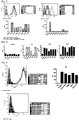
- the MI323 candidate antibody having the excellent binding aspect with regard to the MSLN MS502 candidate antibody having a different pattern of Biacore K D (Koff/Kon) value, and a heavy chain variable region mutation 2-78-C2G4 candidate antibody produced from the MS502 candidate antibody are selectively bound to MSLN-expressing tumor cells, in MiaPaCa-MSLN #2 cell that over-expresses the MSLN and MiaPaCa-2 that does not over-express the MSLN, as illustrated in FIG. 9 , the MI323, MS502, and 2-78-C2G4 candidate antibodies have excellent binding aspect in the MiaPaCa-MSLN #2 cell that over-expresses the MSLN as compared to the MiaPaCa-2.
- the present invention relates to an antibody specifically bound to mesothelin (MSLN), preferably, an antibody specifically bound to mesothelin represented by SEQ ID NO: 127.
- MSLN mesothelin
- the amino acid sequence of an antibody may be substituted by conservative substitution.
- the "conservative substitution” refers to modification of polypeptide including substitution of at least one amino acid with an amino acid having similar biochemical properties to corresponding polypeptide without causing loss of biological or biochemical function.
- Consservative amino acid substitution refers to a substitution in which an amino acid residue is replaced with an amino acid residue having similar side chains. Classes of the amino acid residues having similar side chains are defined in the art.
- amino acids having basic side chains e.g., lysine, arginine, histidine
- amino acids having acidic side chains e.g., aspartic acid, glutamic acid
- amino acids having uncharged polar side chains e.g., glycine, asparagine, glutamine, serine, threonine, tyrosine, cysteine
- amino acids having non-polar side chains e.g., alanine, valine, leucine, isoleucine, proline, phenylalanine, methionine, tryptophan
- amino acids having beta-branched side chains e.g., threonine, valine, isoleucine
- amino acids having aromatic side chains e.g., tyrosine, phenylalanine, tryptophan, histidine
- nucleic acids and polypeptides having appropriate nucleotide or amino acid insertion or deletion have at least about 80% identity to the nucleotide or the amino acid, generally, have at least about 85%, preferably about 90%, 91%, 92%, 93%, 94% or 95%, and more preferably at least about 96%, 97%, 98%, 99%, 99.1%, 99.2%, 99.3%, 99.4% or 99.5% to the nucleotide or the amino acid.
- nucleic acid sequence when a fragment is hybridized with a complementary strand thereof under selective hybridization conditions, there is substantial homology to the nucleic acid.
- the present disclosure includes a nucleic acid sequence and a polypeptide sequence having substantial homology with regard to the above-described specific nucleic acid sequence and amino acid sequence.
- the heavy chain (V H ) CDR1, 2 and 3 sequences and the light chain (V L ) CDR1, 2 and 3 sequences shown in Table 2, Table 5, Table 12, and Table 16 may be formed by mixing structurally similar heavy chain (V H ) and light chain (V L ) sequences to be arranged in the CDR1, 2 and 3 of the heavy chain (V H ) / light chain (V L ) pairs.
- antibody refers to a preparation of antibody molecules having single molecule composition.
- a monoclonal antibody composition represents single binding specificity and affinity for a specific epitope.
- term “human monoclonal antibody” refers to an antibody having a variable region and a constant region derived from a human wiring immunoglobulin sequence, and representing single binding specificity.
- a human antibody of the present invention may include amino acid residue that is not encoded by the human wiring immunoglobulin sequence (for example, mutants introduced by in vitro random or site-specific mutagenesis, or by in vivo somatic mutation).
- the "antibody” used herein is an immunoglobulin molecule which is immunologically reactive to a specific antigen, and means a protein molecule acting as a receptor that specifically recognizes an antigen, and may include all of a polyclonal antibody, a monoclonal antibody (single clone antibody), a whole antibody, and an antibody fragment. Further, the antibody may include a chimeric antibody (e.g., humanized murine antibody) and a bivalent or bispecific molecule (e.g., bispecific antibody), a diabody, a triabody, and a tetrabody.
- chimeric antibody e.g., humanized murine antibody
- bivalent or bispecific molecule e.g., bispecific antibody
- the whole antibody has a structure having two full length light chains and two full length heavy chains, and each light chain may be linked to a heavy chain via a disulfide bond.
- the whole antibody includes IgA, IgD, IgE, IgM, and IgG, and the IgG is a subtype, and includes IgG1, IgG2, IgG3, and IgG4.
- the antibody fragment means a fragment retaining an antigen-binding function, and includes Fab, Fab', F(ab')2, scFv, and Fv, etc.
- the Fab has a structure of variable regions of a light chain and a heavy chain and a constant region of the light chain and a first constant region (CH1 domain) of the heavy chain, and has one antigen-binding site.
- the Fab' is different from the Fab in that the Fab' has a hinge region including one or more cysteine residues at C terminal of a heavy chain CH1 domain.
- the F(ab')2 antibody is produced by achieving the disulfide bonding of the cysteine residue in the hinge region of the Fab'.
- the Fv refers to the minimum antibody fragment only having the heavy chain variable region and the light chain variable region.
- doublestranded Fv(dsFv) the heavy chain variable region and the light chain variable region are linked by the disulfide bond.
- single chain Fv(scFv) the heavy chain variable region and the light chain variable region generally are linked by a covalent bond using a peptide linker.
- These antibody fragment may be obtained by using a proteolytic enzyme (for example, the Fab may be obtained by restriction-cutting the whole antibody with papain, and F(ab')2 fragment may be obtained by cutting with pepsin), and may be constructed by a recombinant DNA technology (for example, amplification by PCR (Polymerase Chain Reaction) method using DNA encoding the heavy chain of the antibody or the variable region thereof and DNA encoding the light chain or the variable region thereof as a template and using a primer pair, and amplification with combination of the DNA encoding the peptide linker of the primer pair allowing both ends thereof to link to the heavy chain or the variable region thereof and the light chain or the variable region thereof, respectively).
- a proteolytic enzyme for example, the Fab may be obtained by restriction-cutting the whole antibody with papain, and F(ab')2 fragment may be obtained by cutting with pepsin
- a recombinant DNA technology for example, amplification by PCR (Polymerase Chain
- the immunoglobulin has heavy chains and light chains, wherein the respective heavy chains and light chains include a constant region and a variable region (these regions are also known as domain).
- the light chain variable region and the heavy chain variable region include 3 multi-available regions called complementarity-determining region (hereinafter, referred to as "CDR"), and four framework regions.
- CDR complementarity-determining region
- the CDR mainly acts to bind to an epitope of the antigen.
- the CDRs of the respective chains are sequentially called CDR1, CDR2, and CDR3 generally starting from N-terminal, and also identified by the chains in which specific CDRs are located.
- the monoclonal antibody (single clone antibody) used herein means an antibody molecule of single molecular composition substantially obtained in the same antibody population, and may have single binding specificity and affinity for a specific epitope.
- the monoclonal antibody (single clone antibody) used herein is a molecule derived from a human immunoglobulin, and all of the amino acid sequences configuring the antibody including a complementarity-determining region, a structure region are configured of human immunoglobulin amino acid sequences.
- the human antibody is typically used in the treatment of human diseases, which is advantageous in that i) it more favorably interacts with the human immune system, which more effectively destroys target cells by complement-dependent cytotoxicity (CDC) or antibody-dependent cell mediated cytotoxicity (ADCC), ii) the human immune system does not recognize the antibody as a foreign material, and iii) even when a smaller amount of drug is administered less frequently, a half-life in a human circulatory system is similar to that of a naturally occurring antibody.
- CDC complement-dependent cytotoxicity
- ADCC antibody-dependent cell mediated cytotoxicity
- the "clone MI323”, “clone MI329”, “clone MI403” and “clone MI407” are antibodies obtained by immunizing a mouse with recombinant human MSLN
- the clone MS501", “clone MS502”, “clone MS503”, “clone MS504", “clone MS505" and “clone MS506” are antibodies obtained from a phage display from a scFV library
- "clone C2G1", “clone C2G4", “clone C3C8", “clone 54", "clone 56", “clone 2-30”, “clone 2-73” and “clone 2-78” are antibodies obtained by introducing mutation into the "clone MS502" as shown in Table 9, and the "clone 56-C2G4", “clone 2-30-C2G4", “clone 2-73-C2G4" and “clone 2-78-C2
- K D equilibrium dissociation constant
- genes of the heavy chain variable region and the light chain variable region of a mouse derived from an immune library bound to human MSLN are identified, the heavy chain variable region gene is linked to a human immunoglobulin type 1 of heavy chain constant region (IgG1 heavy chain constant region) gene, and the light chain variable region gene is linked to a human kappa light chain constant region, and these genes are inserted into protein expression vectors for animal cell, respectively, to produce vectors, followed by transfection in the Expi293FTM cell lines and culturing to produce the antibody, and the produced antibody is purified by protein A to produce the antibody (Example 1-3).
- IgG1 heavy chain constant region immunoglobulin type 1 of heavy chain constant region
- genes of the heavy chain variable region and the light chain variable region derived from a synthetic scFV library bound to human MSLN are identified, the heavy chain variable region gene is linked to a human immunoglobulin type 1 of heavy chain constant region (IgG1 heavy chain constant region) gene, and the light chain variable region gene is linked to a human kappa light chain constant region, and these genes are inserted into protein expression vectors for animal cell, respectively, to produce vectors, followed by transfection in the Expi293FTM cell lines and culturing to produce the antibody, and the antibody is purified by protein A to produce the antibody (Examples 2-4, 3-10, 3-12, and 3-13).
- IgG1 heavy chain constant region immunoglobulin type 1 of heavy chain constant region
- the present invention provides a nucleic acid encoding the antibody of the invention.
- the nucleic acid used herein may be present in a cell, a cell lysate, or may also be present in a partially purified form or a substantially pure form.
- the nucleic acid is "isolated” or “is substantially pure” when it is purified from other cell components or other contaminants, for example, other cell nucleic acid or protein by standard techniques including alkaline/SDS treatment, CsCl banding, column chromatography, agarose gel electrophoresis, and other techniques well-known in the art.
- the nucleic acid of the present invention may be, for example, DNA or RNA, and may include an intron sequence, or may not include the intron sequence.
- the present invention provides a vector including the nucleic acid of the invention.
- DNA encoding the light chain and the heavy chain having a partial length or a full length may be obtained by standard molecular biology techniques (for example, PCR amplification or cDNA cloning using a hybridoma that expresses a target antibody), and the DNA may be "operably bound" to transcription and translation control sequences to be inserted into the expression vector.
- Term “operably bound” used herein may indicate that an antibody gene is ligated into the vector so that the transcription and translation control sequences in the vector have an intended function to control transcription and translation of the antibody gene.
- the expression vector and an expression control sequence are selected so as to have compatibility with a host cell for expression to be used.
- the light chain gene of the antibody and the heavy chain gene of the antibody are inserted into a separate vector, or both genes are inserted into the same expression vector.
- the antibody is inserted into the expression vector by a standard method (for example, ligation of an antibody gene fragment and a complementary restriction enzyme site on a vector or when the restriction enzyme site is not present at all, blunt end ligation).
- the recombinant expression vector may encode a signal peptide that facilitates secretion of the antibody chain from the host cell.
- the antibody chain gene may be cloned into the vector so that the signal peptide is bound to an amino terminal of the antibody chain genes according to a frame.
- the signal peptide may be an immunoglobulin signal peptide or a heterologous signal peptide (i.e. signal peptide derived from proteins except for immunoglobulin).
- the recombinant expression vector has a regulatory sequence that controls the expression of the antibody chain genes in the host cell.
- the "regulatory sequence” may include a promoter, an enhancer and other expression control element (for example, polyadenylation signal) controlling the transcription or translation of the antibody chain gene.
- a promoter for example, an enhancer and other expression control element controlling the transcription or translation of the antibody chain gene.
- other expression control element for example, polyadenylation signal
- design of the expression vector may vary by changing the regulatory sequences according to factors such as selection of the host cell to be transformed, an expression level of the protein, etc.
- the present invention provides a host cell including the vector of the invention.
- the nucleic acid or the vector is transfected into the host cell.
- various kinds of generally used techniques such as electrophoresis, calcium phosphate precipitation, DEAE-dextran transfection, lipofection, etc., may be used to introduce an exogenous nucleic acid (DNA or RNA) into a prokaryotic host cell or an eukaryotic host cell.
- the antibody according to the present invention may be expressed in an eukaryotic cell, preferably, in a mammalian host cell, in consideration of applicability into a mammalian cell.
- the mammalian host cells suitable for expression of the antibody may include a Chinese hamster ovary (CHO) cell (for example, including a dhfr- CHO cell used together with a DHFR selection marker), an NSO myeloma cell, a COS cell, or a SP2 cell, etc., as examples.
- CHO Chinese hamster ovary
- the present invention provides a method for producing the antibody of the invention, including culturing the host cell of the invention to express the antibody.
- the antibody may be produced by culturing the host cell for a sufficient period of time so that the antibody is expressed in the host cell, or more preferably, for a sufficient period of time so that the antibody is secreted into a culture medium in which the host cell is cultured.
- the expressed antibody may be separated from the host cell and purified for uniformity.
- the separation or the purification of the antibody may be performed by a separation method, a purification method generally used for protein, for example, chromatography.
- the chromatography may include, for example, affinity chromatography, ion exchange chromatography or hydrophobic chromatography including protein A column and protein G column.
- the antibody may be separated and purified by additionally combining with filtration, ultrafiltration, salting out, dialysis, etc.
- the present invention provides a pharmaceutical composition for use in the treatment of cancer or a tumor including the antibody of the invention as an active ingredient.
- cancer typically refers to or describes a physiological condition of mammals characterized by cell growth/proliferation that is not controlled.
- examples of the cancer include carcinoma, lymphoma (e.g., Hodgkin's and non-Hodgkin's lymphoma), blastoma, sarcoma and leukemia, but are not limited thereto.
- the cancer include squamous cell cancer, small-cell lung cancer, non-small cell lung cancer, lung adenocarcinoma, lung squamous cell carcinoma, peritoneal cancer, hepatocellular cancer, gastrointestinal cancer, pancreatic cancer, glioma, cervical cancer, ovarian cancer, liver cancer, bladder cancer, hepatocellular cancer, breast cancer, colon cancer, colorectal cancer, endometrial or uterine carcinoma, salivary gland carcinoma, kidney cancer, liver cancer, prostate cancer, vulvar cancer, thyroid cancer, liver carcinoma, leukemia and other lymphoproliferative disorders, and various types of head and neck cancer.
- the cancer in the present invention is preferably mesothelin-positive cancer, and is selected from the group consisting of pancreatic cancer, ovarian cancer, lung cancer, stomach cancer, endometrial cancer, and mesothelioma.
- the present disclosure provides a pharmaceutical composition including a therapeutically effective amount of anti-MSLN antibody and a pharmaceutically acceptable carrier.
- the "pharmaceutically acceptable carrier” is a material that is able to be added in the active ingredient to help fo0rmulation or stabilization of the preparation, and it does not cause significant adverse toxicological effects to patients.
- the carrier refers to a carrier or diluent that does not inhibit biological activity and properties of an administered compound without stimulating the patients.
- the pharmaceutically acceptable carrier in the composition to be formulated as a liquid solution is sterilized and is suitable for a living body. Saline, sterile water, Ringer's solution, buffered saline, albumin injection solution, dextrose solution, maltodextrin solution, glycerol, ethanol may be used as the carrier, or at least one component thereof may be mixed to be used, and other conventional additives such as an antioxidant, buffer, a bacteriostatic agent, etc., may be added as needed.
- compositions for injection such as an aqueous solution, suspension, emulsion, etc., pill, a capsule, a granule or a tablet by further adding diluent, dispersant, surfactant, binder and lubricant thereto.
- Other carriers are described in, for example, [Remington's Pharmaceutical Sciences (E. W. Martin)].
- the composition may contain the therapeutically effective amount of at least one anti-MSLN antibody.
- the pharmaceutically acceptable carrier includes sterile aqueous solution or dispersion and sterile powder for preparing extemporaneous sterile injectable solution or dispersion.
- the use of such media and agents for pharmaceutical active materials is known in the art.
- the composition is preferably formulated for parenteral injection.
- the composition may be formulated as a solution, a micro-emulsion, a liposome, or other ordered structures suitable for high drug concentration.
- the carrier may be, for example, a solvent or dispersion medium containing water, ethanol, polyol (for example, glycerol, propylene glycol and liquid polyethylene glycol, etc.,) and suitable mixtures thereof.
- the composition may include, isotonic agent, for example, sugar, polyalcohols such as mannitol, sorbitol, or sodium chloride.
- the sterile injectable solution may be prepared by incorporating a required amount of active compound into an appropriate solvent with one kind of the above-described components or a combination thereof, followed by sterile micro filtration as needed.
- the dispersion is prepared by incorporating the active compound into a sterile vehicle containing basic dispersion medium and other required components from the above-described components.
- the sterile powder for preparing the sterile injectable solution is obtained by vacuum drying and freeze-drying (lyophilization) active ingredient powder and any additional desirable component powder from previously sterile-filtered solution.
- the pharmaceutical composition may be administered orally or parenterally in the dosage and frequency that may vary depending on severity of suffering patients.
- the composition may be administered to a patient as a bolus or by continuous infusion as needed.
- the bolus administration of the antibody of the present invention which is presented as a Fab fragment may have an amount of 0.0025 to 100mg/kg body weight, 0.025 to 0.25mg/kg, 0.010 to 0.10mg/kg or 0.10 to 0.50mg/kg.
- the antibody of the present invention which is presented as the Fab fragment may be administered at 0.001 to 100 mg/kg kg/min, 0.0125 to 1.25mg/kg/min, 0.010 to 0.75mg/kg/min, 0.010 to 1.0mg/kg/min or 0.10 to 0.50mg/kg/min for 1 to 24 hours, 1 to 12 hours, 2 to 12 hours, 6 to 12 hours, 2 to 8 hours, or 1 to 2 hours.
- an administration amount may be about 1 to 10 mg/kg body weight, 2 to 8 mg/kg, or 5 to 6 mg/kg.
- the full-length antibody is typically administered via injection that lasts for 30 minutes to 35 minutes.
- An administration frequency depends on the severity of the condition. The frequency may be 3 times every week to once in a week or in two weeks.
- composition may be administered to a patient via a subcutaneous injection.
- the anti-MSLN antibody having an administration amount of 10 to 100 mg may be weekly, biweekly, or monthly administered to a patient through subcutaneous injection.
- the "therapeutically effective amount” means an amount sufficient to treat diseases at a reasonable benefit/risk ratio applicable for medical treatment, and an amount of a combination of the anti-MSLN antibody.
- the exact amount may vary depending on a number of factors that include components and physical characteristics of a therapeutic composition, intended patient population, individual patient considerations, etc., but are not limited thereto, and may be easily determined by those skilled in the art. When completely considering these factors, it is important to administer the minimum amount sufficient to obtain the maximum effect without the side effect, and this dosage may be easily determined by an expert in the field.
- the dosage of the pharmaceutical composition of the present invention is not specifically limited, but is changed according to various factors including a health state and weight, severity of the disease of a patient, and a drug type, an administration route, and administration time.
- the composition may be administered in routes that are typically allowed in mammals including rat, mouse, cattle, human, etc., for example, orally, rectally, intravenously, subcutaneously, intrauterinely or intracerebrovascularly in a single dose amount or multidose per day.
- Example 1 Method for producing anti-MSLN antibody from immune library
- MSLN mesothelin
- a mouse was immunized with a recombinant human MSLN and a spleen was removed to extract B lymphocyte. Total RNA was separated from the B lymphocyte and cDNA was synthesized. Various antibody genes of the mouse were cloned from the synthesized cDNA using polymerase chain reaction (PCR), and inserted into pComb3X phagemid to produce an antibody library displaying antibody fragments of various sequences.
- PCR polymerase chain reaction
- pComb3X phagemid inserted into pComb3X phagemid to produce an antibody library displaying antibody fragments of various sequences.
- magnetic beads having the MSLN fixed thereto were mixed with the antibody library, and clones bound to a target antigen were separated and cultured.
- the clones (MI323, MI329, MI403, and MI407) specifically bound to the target antigen were individually identified through an enzyme-linked immunosorbent assay (ELISA), and antibody gene sequences and amino acid sequence thereof were identified through base sequence analysis.
- ELISA enzyme-linked immunosorbent assay
- Table 2 shows CDR amino acid sequences of the clone antibodies of Table 1 on the basis of Kabat numbering.
- Table 1 Clone Variable Region Amino Acid Sequence SEQ ID NO: MI323 heavy chain 1 light chain 2 MI329 heavy chain 3 light chain 4 MI403 heavy chain 5 light chain 6 MI407 heavy chain 7 light chain 8
- Clone Variable Region CDR1 CDR2 CDR3 MI323 heavy chain SYFIQ SEQ ID NO: 9) SGGYQYYFDY (SEQ ID NO: 11) light chain KASQDVSTAVA (SEQ ID NO: 12) SASYRYP (SEQ ID NO: 13) QQHYSTPWT (SEQ ID NO: 14) MI329 heavy chain SYAMS (SEQ ID NO: 15) WGENWYFDV (SEQ ID NO: 17) light chain KVSNRFS (SEQ ID NO: 19) SQSTHFPRT (SEQ ID NO: 20) MI403 heavy chain SYDMS (SEQ ID NO: 21) light
- the pComb3X phagemid containing the genes encoding the heavy chain variable regions of the MI323, MI329, MI403, and MI407 clone antibodies was extracted, and used as a template for PCR with a forward primer containing NotI (Table 3: SEQ ID NOs: 31 to 34) and a reverse primer containing Apal (Table 3: SEQ ID NO: 35) by using Accupower Pfu PCR premix (Bioneer).
- the PCR was performed by repeating exposure at 94°C for 10 minutes, and then exposure at 94°C for 15 seconds, at 56°C for 30 seconds, and at 72°C for 90 seconds 30 times, and reacting at 72°C for 10 minutes.
- DNA bands having an expected size were confirmed on 1% agarose gel, and were separated using a gel extraction kit, respectively. Then, the separated genes reacted with NotI, ApaI restriction enzymes at 37°C for 12 hours or more, and the genes reacted with the restriction enzymes were separated on 1% agarose gel again.
- a pcIW plasmid vector containing human immunoglobulin type 1 of heavy chain constant region (IgG1 heavy chain constant region) gene was also cut by the same method as above and separated on agarose gel.
- the separated MI323, MI329, MI403, and MI407 heavy chain variable region genes were inserted into NotI, ApaI sites of a linear pcIW vector containing the human heavy chain constant region by using a T4 DNA ligase (Cat.No.M0203S, New England BioLabs (NEB)).
- the ligation reaction materials were transformed into XL1-Blue bacteria (Electroporation-Competent Cells; Cat.No.200228, Stratagene), plated on an LB plate (Cat.No.LN004CA, NaraeBiotech) containing carbenicillin, and cultured at 37°C for 12 hours or more. Then single colonies were chosen and cultured, and plasmids were separated by using a plasmid mini kit (Cat.No.27405, QIAGEN), and confirmed by DNA sequencing.
- the pComb3X phagemid containing the genes encoding the light chain variable regions of the MI323, MI329, MI403, and MI407 clone antibodies was extracted, and used as a template for PCR of the light chain variable regions of the MI323, MI329, MI403, and MI407 clone antibodies, wherein the PCR was performed with a forward primer containing NotI (Table 3: SEQ ID NO: 36, 38, 40, 42) and a reverse primer (Table 3: SEQ ID NO: 37, 39, 41, 43) by using Accupower Pfu PCR premix.
- the human antibody kappa light chain constant region was subjected to PCR with a forward primer (Table 3: SEQ ID NO: 44) and a reverse primer containing HindIII (Table 3: SEQ ID NO: 45).
- the PCR was performed by repeating exposure at 94°C for 10 minutes, and then exposure at 94°C for 15 seconds, at 56°C for 30 seconds, and at 72°C for 90 seconds 30 times, and reacting at 72°C for 10 minutes.
- DNA bands having a predicted size were confirmed on 1% agarose gel, and were separated using a gel extraction kit, respectively.
- the respective light chain variable regions and light chain constant regions were mixed, followed by overlapping PCR, such that the genes expressing the light chain region were cloned.
- the PCR was performed by repeating exposure at 94°C for 10 minutes, and then exposure at 94°C for 15 seconds, at 56°C for 30 seconds, and at 72°C for 90 seconds 30 times, and reacting at 72°C for 10 minutes.
- DNA bands having an expected size were confirmed on 1% agarose gel, and were separated using a gel extraction kit, respectively. Then, the separated genes reacted with NotI, HindIII restriction enzymes at 37°C for 12 hours or more, and the genes reacted with the restriction enzymes were separated on 1% agarose gel again.
- the pcIW plasmid vector was also cut by the same method as above and separated on agarose gel.
- the separated MI323, MI329, MI403, and MI407 light chain region genes were inserted into NotI, HindIII sites of a linear pcIW vector by using a T4 DNA ligase (Cat.No.M0203S, New England BioLabs (NEB)).
- the ligation reaction materials were transformed into XL1-Blue bacteria (Electroporation-Competent Cells; Cat.No.200228, Stratagene), plated on an LB plate (Cat.No.LN004CA, NaraeBiotech) containing carbenicillin, and cultured at 37°C for 12 hours or more.
- MabSelect Xtra (Cat.No.17-5269-02, GE Healthcare) which is protein A resin having 100 ⁇ L was prepared for each 30 mL of the culture fluid, followed by centrifugation at 1000 rpm for 2 minutes to remove a storage solution, and the obtained product was washed with 400 ⁇ L of protein A binding buffer (Cat.No.21007, Pierce) 3 times. The protein A resin was added to the prepared culture fluid and rotation-reacted at room temperature for 30 minutes.
- the mixture of the culture fluid and the resin was put into a pierce spin column snap-cap (Cat.No.69725, Thermo), and then, only the resin was left in the column using QIAvac 24 Plus (Cat.No.19413, QIAGEN) vacuum manifold.
- 5 mL of protein A binding buffer was added to wash the resin, 200 ⁇ L of a protein A elution buffer (Cat.No.21009, Pierce) was added thereto.
- the resultant material was reacted by resuspension at room temperature for 2 minutes, and centrifuged at 1000 rpm for 1 minute, and eluted. Each eluate was neutralized by adding 2.5 ⁇ L of 1.5M Tris-HCl (pH 9.0).
- the elution was performed 4 to 6 times, and each fraction was quantified by using Nanodrop 200C (Thermo Scientific).
- the fractions in which protein is detected were collected, and exchanged with a PBS (Phosphate-Buffered Saline) buffer using Zeba Spin Desalting Columns, 7K MWCO, 5 mL (Cat.No.0089892, Pierce).
- PBS Phosphate-Buffered Saline
- Zeba Spin Desalting Columns 7K MWCO, 5 mL (Cat.No.0089892, Pierce).
- SDS-PAGE protein electrophoresis
- Example 2 Method for producing anti-MSLN antibody from phage display synthetic scFv library
- the library stock obtained after reaction at 37°C for 2 hours was washed with 5ml of 0.05% Tween 20/PBS four times, and from a secondary panning, the number of times of washing with 0.05% Tween 20/PBS increased according to an increase in the number of times of panning. Then, the resultant material was cultured with 1%BSA/0.1M Glycine pH 2.0 at room temperature for 10 minutes to purify the phagemid. The purified phagemid was transferred to a 50 mL tube and neutralized with 70 ⁇ L of 2M Tris.
- the bacteria were treated with 1 mL of VCS M13 helper phage (10 11 pfu), and suspension-cultured at 37°C and at 220 rpm for 1 hour, and treated with 80 mL of SB, 100 ⁇ L of kanamycin, and 100 ⁇ L of carbenicillin, and cultured at 37°C and at 220 rpm for 12 hours or more.
- the bacteria cultured over 12 hours were centrifuged at 3500 rpm and at 4°C for 10 minutes, and the supernatant was transferred to a new tube. 20 mL of 20% PEG/15% NaCl was added thereto, well-mixed, and reacted in ice for 30 minutes.
- pellets were collected and re-suspended with 2 mL of 1% BSA/PBS at 8000 rpm, and at 4°C for 30 minutes, and centrifuged at 15000 rpm and 4°C for 10 minutes.
- the collected pellets were discarded and 1 mL of the supernatant (2 mL) was stored at -20°C and the remainder (1 mL) was used in the following order panning.
- Single colonies of a phage display synthetic scFv library final amplified population were collected, and cultured with 1.5 mL of SB/carbenicillin to an OD 600 of 0.8 to 1.0 at 37°C and at 220 rpm, and then cultured with 1 mM IPTG at 30°C and at 200 rpm for 12 hours or more.
- the reaction materials were centrifuged at 5500 rpm for 5 minutes, and only each supernatant was added to ELISA plates containing underlying MSLN antigen, and reacted at room temperature for 2 hours.
- Table 5 shows CDR amino acid sequences of the clone antibodies of Table 4 on the basis of Kabat numbering.
- Clone variable region amino acid sequence SEQ ID NO: MS501 heavy chain 46 light chain 47 MS502 heavy chain 48 light chain 49 MS502-1 heavy chain 48 light chain 50 MS503 heavy chain 51 light chain 52 MS504 heavy chain 53 light chain 54 MS505 heavy chain 55 light chain 56 MS506 heavy chain 57 light chain 58
- Clone variable region CDR1 CDR2 CDR3 MS501 heavy chain NYAMS (SEQ ID NO: 59) NIYTFDY (SEQ ID NO: 61) light chain YNNQRPS (SEQ ID NO: 63) GSWDSSLSGYV (SEQ ID NO: 64) MS502 heavy chain NYAMS (SEQ ID NO: 59) NMLSFDY (SEQ ID NO: 66) light chain YNSKRPS (SEQ ID NO: 68) GSWDSSLNGYV (SEQ ID NO: 69) MS502-1 heavy chain NYAMS
- the pComb3X phagemid containing the genes encoding the light chain variable regions of the secured MS501, MS502, MS503, MS504, MS505, and MS506 clone antibodies was extracted, and used as a template for PCR of the light chain variable regions of the MS501, MS502, MS503, MS504, MS505, and MS506 clone antibodies, wherein the PCR was performed with a forward primer containing NotI (SEQ ID NO: 86) and a reverse primer (SEQ ID NO: 87) by using Accupower Pfu PCR premix.
- human antibody kappa light chain constant region was subjected to PCR with a forward primer (Table 6: SEQ ID NO: 88) and a reverse primer (Table 6: SEQ ID NO: 89).
- the PCR was performed by repeating exposure at 94°C for 10 minutes, and then exposure at 94°C for 15 seconds, at 56°C for 30 seconds, and at 72°C for 90 seconds 30 times, and reacting at 72°C for 10 minutes.
- DNA bands having an expected size were confirmed on 1% agarose gel, and were separated using a gel extraction kit, respectively. Then, the respective light chain variable regions and light chain constant regions were mixed, followed by overlapping PCR, such that the genes expressing the light chain region were cloned.
- the PCR was performed by repeating exposure at 94°C for 10 minutes, and then exposure at 94°C for 15 seconds, at 56°C for 30 seconds, and at 72°C for 90 seconds 30 times, and reacting at 72°C for 10 minutes.
- DNA bands having an expected size were confirmed on 1% agarose gel, and were separated using a gel extraction kit, respectively. Then, the separated genes reacted with NotI, HindIII restriction enzymes at 37°C for 12 hours or more, and the genes reacted with the restriction enzymes were separated on 1% agarose gel again.
- the pcIW plasmid vector was also cut by the same method as above and separated on agarose gel.
- the separated MS501, MS502, MS503, MS504, MS505, MS506 light chain region genes were inserted into NotI, HindIII sites of the linear pcIW vector by using a T4 DNA ligase (Cat.No.M0203S, New England BioLabs (NEB)).
- the ligation reaction materials were transformed into XL1-Blue bacteria (Electroporation-Competent Cells; Cat.No.200228, Stratagene), plated on an LB plate (Cat.No.LN004CA, NaraeBiotech) containing carbenicillin, and cultured at 37°C for 12 hours or more. Then single colonies were chosen and cultured, and plasmids were separated by using a plasmid mini kit (Cat.No.27405, QIAGEN), and confirmed by DNA sequencing.
- the pComb3X phagemid containing the genes encoding the heavy chain variable regions of the MS501, MS502, MS503, MS504, MS505, and MS506 clone antibodies was extracted, and used as a template for PCR with a forward primer containing NotI (Table 6: SEQ ID NO: 90) and a reverse primer containing Apal (Table 6: SEQ ID NO: 91) by using Accupower Pfu PCR premix (Bioneer).
- the PCR was performed by repeating exposure at 94°C for 10 minutes, and then exposure at 94°C for 15 seconds, at 56°C for 30 seconds, and at 72°C for 90 seconds 30 times, and reacting at 72°C for 10 minutes.
- DNA bands having an expected size were confirmed on 1% agarose gel, and were separated using a gel extraction kit, respectively. Then, the separated genes reacted with NotI, ApaI restriction enzymes at 37°C for 12 hours or more, and the genes reacted with the restriction enzymes were separated on 1% agarose gel again.
- a pcIW plasmid vector containing human immunoglobulin type 1 of heavy chain constant region (IgG1 heavy chain constant region) gene was also cut by the same method as above and separated on agarose gel.
- the separated MS501, MS502, MS503, MS504, MS505, and MS506 heavy chain variable region genes were inserted into NotI, ApaI sites of the linear pcIW vector containing the human heavy chain constant region by using a T4 DNA ligase (Cat.No.M0203S, New England BioLabs (NEB)).
- the ligation reaction materials were transformed into XL1-Blue bacteria (Electroporation-Competent Cells; Cat.No.200228, Stratagene), plated on an LB plate (Cat.No.LN004CA, NaraeBiotech) containing carbenicillin, and cultured at 37°C for 12 hours or more.
- DNA (pcIw-MS502 heavy chain variable region: 15 ⁇ g, pcIw-anti-Mesothelin light chain variable region: 15 ⁇ g) was diluted in an OptiProTM SEM medium (Cat.No.12309019, Gibco) so as to have a total volume of 1.5 mL, and reacted at room temperature for 5 minutes.
- 1.5 mL of the OptiProTM SEM medium (Cat.No.12309019, Gibco) was mixed with 80 ⁇ L of an ExpiFectamineTM 293 reagent (Cat.No.A14524, Gibco) so that a total volume is 1.5 mL, and reacted at room temperature for 5 minutes.
- MabSelect Xtra (Cat.No.17-5269-02, GE Healthcare) which is protein A resin having 100 ⁇ L was prepared for each 30 mL of the culture fluid, followed by centrifugation at 1000 rpm for 2 minutes to remove a storage solution, and the product was washed with 400 ⁇ L of protein A binding buffer (Cat.No.21007, Pierce) 3 times. The protein A resin was added to the prepared culture fluid and rotation-reacted at room temperature for 30 minutes. The mixture of the culture fluid and the resin was put into a pierce spin column snap-cap (Cat.No.69725, Thermo), and then, only the resin was left in the column using QIAvac 24 Plus (Cat.No.19413, QIAGEN) vacuum manifold.
- QIAvac 24 Plus (Cat.No.19413, QIAGEN) vacuum manifold.
- fractions in which protein is detected were collected, and exchanged with a PBS (Phosphate-Buffered Saline) buffer using Zeba Spin Desalting Columns, 7K MWCO, 5 mL (Cat.No.0089892, Pierce). Then, protein electrophoresis (SDS-PAGE) was performed under reduction and non-reduction condition to finally verify the concentration quantification and the antibody state, and the antibody was kept at 4°C.
- PBS Phosphate-Buffered Saline
- Quantitative binding force (affinity) of the purified anti-MSLN antibodies i.e., MI323, MI329, MI403, MS502 clone antibodies with regard to recombinant human mesothelin (MSLN) was measured by using a Biacore T-200 (GE Healthcare, U.S.A.) biosensor.
- the MSLN (Cat.No.3265-MS, R&D systems) purified from the HEK293 cells was fixed to a CM5 chip (GE Healthcare, U.S.A.) so as to satisfy 200 Rmax by using an amine-carboxylic reaction.
- the clone C2G1 antibody, the clone C2G4 antibody or the clone C3C8 antibody serially diluted with HBS-EP buffer (10mM HEPES, pH7.4, 150mM NaCl, 3mM EDTA, 0.005% surfactant P20) was allowed to flow at a concentration range of 0.078nM to 5nM and at a flow rate of 30 ⁇ L/min for association of 120 seconds and dissociation of 1800 seconds.
- the dissociation of the antibody bound to the MSLN was induced by flowing 10 mM Glycine-HCl pH 1.5 at a flow rate of 30 ⁇ L/min for 30 seconds (Table 7).
- Example 3 Method for producing MS502 clone affinity maturation antibody
- the amino acid sequence of the anti-mesothelin MS502 clone was entered to the Swiss model homepage (http://swissmodel.expasy.org/) to find a template sequence. 50 sequences were found on the basis of homologous sequences, and modeling was performed by designating each sequence as the template. 50 sequences were listed by priority on the basis of QMEAN4 values (C ⁇ , all atom, solvation, torsion), and the template was selected in consideration of sequence identity and resolution in addition to the QMEAN4 values. 3g6a.1.B was selected for the heavy chain variable region, and 3qhz.1.B (LCDR1,2) and 4o5l.1.A (LCDR3) were selected for the light chain variable region.
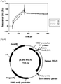
- the MS502 structure obtained on the basis of the selected template was analyzed by pymol program, and paratope was selected on the basis of the protruding amino acid of the CDR (see FIG. 1 ).
- VL CDR2 Y49, Y50, K53 were excluded from the mutation candidate amino acids even though it was anticipated that they are paratopes since Tyr and Lys are amino acids generally having a positive effect on bonding.
- the introduction of mutation was designed by 50% preserving the existing sequence, increasing Tyr, Ser, Gly rates, and controlling the nucleotide sequence using a handmix primer (IDT, U.S.A.) so that hydrophilic, hydrophobic, positive charge, and negative charge rates were uniformly included (Table 9).
- the heavy chain variable region was divided into each of three fragments since the mutation was introduced into all of CDR1, 2, and 3, and they were subjected to fragment PCR and overlapping PCR to construct a library, and the light chain variable region was divided into each of two fragments since the mutation was introduced into CDR1 and 3 only, and they were subjected to fragment PCR and overlapping PCR to construct a library.
- a theoretical size of the library according to the introduction of the mutation in the case of the heavy chain variable region was 5.83 X 10 10
- the theoretical size of the library according to the introduction of the mutation in the case of the light chain variable region was 2.16 X 10 4 .
- the light chain variable region fragment No. 1 had a light chain variable region gene sequence of anti-mesothelin MS502 clone as the template, and a forward primer (SEQ ID NO: 93) and a reverse primer (SEQ ID NO: 94) were added thereto, and the light chain variable region fragment No.
- DNA bands having an expected size were confirmed on 1% agarose gel, and were separated using a gel extraction kit (QIAquick Gel Extraction Kit, CAT. NO. 28706, QIAGEN), respectively.
- the light chain constant region had a light chain lambda region as a template, and a forward primer (Table 10: SEQ ID NO: 97), and a reverse primer (Table 10: SEQ ID NO: 98) were added thereto, and each fragment was subjected to PCR using a Primestar polymerase premix.
- the PCR was performed by repeating exposure at 98°C for 2 minutes, and then exposure at 98°C for 10 seconds, at 60°C for 15 seconds, and at 72°C for 30 seconds 30 times, and reacting at 72°C for 10 minutes.
- DNA bands having an expected size were confirmed on 1% agarose gel, and were separated using a gel extraction kit, respectively.
- the secured light chain variable region fragments 1 and 2, and light chain constant region at a molar ratio of 1:1:1 were used as a template, and a forward primer (SEQ ID NO: 92) and a reverse primer (SEQ ID NO: 98) were added thereto, and each fragment was subjected to PCR using a Primestar polymerase premix.
- the PCR was performed by repeating exposure at 98°C for 2 minutes, and then exposure at 98°C for 20 seconds, at 60°C for 30 seconds, and at 72°C for 60 seconds 30 times, and reacting at 72°C for 10 minutes.
- DNA bands having an expected size were confirmed on 1% agarose gel, and were separated using a gel extraction kit to secure light chain variable-constant region affinity maturation gene.
- the secured gene was reacted with NruI and XbaI (NEB) restriction enzyme at 37°C for 4 hours.
- the genes reacted with the restriction enzyme were separated on 1% Agarose gel again.
- the separated gene was inserted into the NruI, XbaI site of the linear pComb3x vector containing the MS502 heavy chain variable-constant region by using a T4 DNA ligase (Cat.No.M0203S, NEB).
- the ligation reaction material was transformed into XL1-Blue bacteria (Electroporation-competent cells; Cat.No.200228, Stratagene), and cultured in 300 mL of LB medium at 37°C and at 220 rpm for 1 hour. Then, the resultant material was treated with 150 ⁇ L of carbenicillin and 300 ⁇ L of tetracycline, and was cultured with shaking at 37°C and at 220 rpm for 1 hour.
- the resultant material was treated with VCS M13 helper phage 4.5 mL (10 11 pfu) and cultured with shaking at 37°C and at 220 rpm for 1 hour, and treated with 300 ⁇ L of kanamycin and 300 ⁇ L of carbenicillin, and cultured overnight at 37°C and at 220 rpm. The next day, the cultured cells were centrifuged at 4000 rpm for 20 minutes, and the supernatant was transferred to a new vessel. In order to precipitate the phage, 5X PEG/NaCl was used to add 1X PEG/NaCl to the supernatant, and the obtained product allowed to stand on ice over 30 minutes.
- the precipitate phage was centrifuged at 8000 rpm for 30 minutes. The supernatant was discarded and the precipitated phage was re-suspended with 10 mL of PBS. In order to remove cell debris, the phage dissolved in 10 mL of PBS was centrifuged at 14,000 rpm for 10 minutes to separate the supernatant, and stored at 4°C.
- the library size was confirmed by taking 100 ⁇ L of culture fluid after 1 hour of the transformation, plating the culture fluid on the LB plate (Cat.No.LN004CA, NaraeBiotech) containing carbenicillin in serial dilution manner, culturing at 37°C for 12 hours or more, and counting the colonies.
- the heavy chain variable region fragment No. 1 had a heavy chain variable region gene sequence of anti-mesothelin MS502 clone as the template, and a forward primer (Table 10: SEQ ID NO: 99) and a reverse primer (Table 10: SEQ ID NO: 100) were added thereto, and the heavy chain variable region fragment No. 2 had the heavy chain variable region gene sequence of anti-mesothelin MS502 clone as the template, and a forward primer (Table 10: SEQ ID NO: 101) and a reverse primer (Table 10: SEQ ID NO: 102) were added thereto, and the heavy chain variable region fragment No.
- the secured heavy chain variable region fragments 1, 2, and 3 at a molar ratio of 1:1:1 were used as a template, and a forward primer (Table 10: SEQ ID NO: 99) and a reverse primer (Table 10: SEQ ID NO: 106) were added thereto, and each fragment was subjected to PCR using a Primestar polymerase premix.
- the PCR was performed by repeating exposure at 98°C for 2 minutes, and then exposure at 98°C for 20 seconds, at 60°C for 30 seconds, and at 72°C for 60 seconds 30 times, and reacting at 72°C for 10 minutes.
- DNA bands having an expected size were confirmed on 1% agarose gel, and were separated using a gel extraction kit to secure heavy chain variable region affinity maturation gene.
- the secured gene was reacted with XhoI and ApaI (NEB) restriction enzyme at 37°C for 4 hours.
- the genes reacted with the restriction enzyme were separated on 1% Agarose gel again.
- the separated gene was inserted into the XhoI, ApaI site of the linear pComb3x vector containing the MS502 light chain variable-constant region by using a T4 DNA ligase (Cat.No.M0203S, NEB).
- the ligation reaction material was transformed into XL1-Blue bacteria (Electroporation-competent cells; Cat.No.200228, Stratagene), and cultured in 300 mL of LB medium at 37°C and at 220 rpm for 1 hour.
- the resultant material was treated with 150 ⁇ L of carbenicillin and 300 ⁇ L of tetracycline, and was suspension-cultured at 37°C and at 220 rpm for 1 hour.
- the resultant material was treated with VCS M13 helper phage 4.5mL (10 11 pfu) and cultured with shaking at 37°C and at 220 rpm for 1 hour, and treated with 300 ⁇ L of kanamycin and 300 ⁇ L of carbenicillin, and cultured overnight at 37°C and at 220 rpm.
- the cultured cells were centrifuged at 4000 rpm for 20 minutes, and the supernatant was transferred to a new vessel.
- 5X PEG/NaCl was used to add 1X PEG/NaCl to the supernatant, and the obtained product allowed to stand on ice over 30 minutes.
- the precipitated phage was centrifuged at 8000 rpm for 30 minutes.
- the supernatant was discarded and the precipitated phage was re-suspended with 10 mL of PBS.
- the phage dissolved in 10 mL of PBS was centrifuged at 14,000 rpm for 10 minutes to separate the supernatant, and stored at 4°C.
- the library size was confirmed by taking 100 ⁇ L of culture fluid after 1 hour of the transformation, plating the culture fluid on the LB plate (Cat.No.LN004CA, NaraeBiotech) containing carbenicillin in serial dilution manner, culturing at 37°C for 12 hours or more, and counting the colonies.
- X codon is a degenerative codon in which each ACGT is controlled at a specific rate, and is able to control a rate of the amino acids to be translated.
- N is encoded by A, C, G, T with a random ratio
- X1 is encoded at 10% of A, 10% of C, 70% of G, and 10% of T
- X2 is encoded at 10% of A, 70% of C, 10% of G, and 10% of T.
- the bacteria were treated with 80 mL of SB, 100 ⁇ L of kanamycin, and 100 ⁇ L of carbenicillin (100 mg/mL), and cultured overnight (200 rpm) at 37°C.
- the overnight cultured library was centrifuged at 4000 rpm for 15 minutes to separate the supernatant only, and 5X PEG/NaCl was used to add 1X PEG/NaCl to the supernatant, and the obtained product allowed to stand on ice over 30 minutes.
- the supernatant was removed by centrifugation at 8000 rpm for 30 minutes, and pellets were re-suspended with 2 ml of 1% BSA/PBS and centrifuged at 12000 rpm for 10 minutes. Then, only the supernatant was taken and used in the following order panning. This process was repeated four times.
- the supernatant was removed with 50 ⁇ L of streptavidin microbead (Miltenyl biotec 130-048-101), and 1 mL of PBS was added thereto to perform washing three times. 1 mL of PBS containing 1% BSA was added to the bead, and the bead was rotated at room temperature for 2 hours, followed by blocking. 50 nM MSLN was put in 500 ⁇ L of PBS, and mixed with 500 ⁇ L of MS502 VH rational library phage and rotation-reacted at room temperature for 1 hour. The blocking buffer was removed from the bead, and the bead was treated with a solution containing the MSLN and the phage, and reacted at room temperature for 15 minutes.
- the obtained product was washed with 1 mL of PBST six times.
- the bead was treated with 1 mL of glycine (pH 2.0) elution buffer, and reacted at room temperature for 10 minutes to obtain the supernatant.
- 100 ⁇ L of 1.5M Tris-Cl (pH 8.8) was added to the phage and neutralized.
- 10 mL of XL1-Blue bacteria (electroporation-competent cells; Cat.No.200228, Stratagene) cultured for about 2 to 2.5 hours (OD 600 0.8 to 1.0) were treated with the neutralized phage.
- the bacteria were treated with 80 mL of SB, 100 ⁇ L of kanamycin, and 100 ⁇ L of carbenicillin (100 mg/mL), and cultured overnight (200 rpm) at 37°C.
- the overnight cultured library was centrifuged at 4000 rpm for 15 minutes to separate the supernatant only, and 5X PEG/NaCl buffer was used to add 1X PEG/NaCl to the supernatant, and the obtained product allowed to stand on ice over 30 minutes.
- the supernatant was removed by centrifugation at 8000 rpm for 30 minutes, and pellets were re-suspended with 2m of 1% BSA/PBS and centrifuged at 12000 rpm for 10 minutes.
- Single colonies of a final amplified population of each library of light chain/heavy chain variable regions were collected, and cultured with 1.5 mL of SB/carbenicillin up to an OD 600 of 0.8 to 1.0 at 37°C and at 220 rpm, and then cultured with 1 mM IPTG at 30°C and at 200 rpm for 12 hours or more.
- the reaction materials were centrifuged at 5500 rpm for 5 minutes, and only each supernatant was added to ELISA plates containing underlying MSLN antigen, and reacted at room temperature for 2 hours.
- Table 12 shows CDR amino acid sequences of the clone antibodies of Table 11 on the basis of Kabat numbering.
- Table 11 clone variable region amino acid sequence SEQ ID NO: C2G1 heavy chain 48 light chain 109 C2G4 heavy chain 48 light chain 110 C3C8 heavy chain 48 light chain 111 54 heavy chain 112 light chain 49 56 heavy chain 113 light chain 49 2-30 heavy chain 114 light chain 49 2-73 heavy chain 115 light chain 49 2-78 heavy chain 116 light chain 49
- clone variable region CDR1 CDR2 CDR3 C2G1 heavy chain NYAMS (SEQ ID NO: 59) NMLSFDY (SEQ ID NO: 66) light chain YNSKRPS (SEQ ID NO: 68) GSWDSSLSGYV (SEQ ID NO: 118) C2G4 heavy chain NYAMS (SEQ ID NO: 59) NMLSFDY (SEQ ID NO: 66) light chain YNSKRPS (SEQ ID NO: 68) GSWDPSLNG
- Example 3-3 In order to compare and measure the binding force with culture fluid of individual clones secured from confirmation of the individual clones according to ELISA method conducted in Example 3-3, first, the human MSLN on the biacore series S CM5 chip (GE healthcare) was dissolved in pH 4.0 acetate buffer to be 1 ⁇ g/ml, and allowed to flow at a flow rate of 10 ⁇ L/min and fixed to 1000 Ru.
- the Fab supernatant expressed in Example 3-6 was diluted by 1/10 with pH 7.4 HBS-EP buffer, and was allowed to flow at a flow rate of 30 ⁇ L/min for association of 120 seconds, and dissociation of 180 seconds to conduct binding analysis. The regeneration was performed with 10 mM glycine-HCl pH 1.5 buffer for 30 seconds.
- the ELISA and the SPR binding data were compared and analyzed to select the final clones of the light chain variable region and the heavy chain variable region.
- the binding force of the light chain variable region mutations and the heavy chain variable region mutations were relatively compared, and C2G1, C2G4, and C3C8 were selected for the final clones of the light chain variable region and 56, 2-30, 2-58, 2-73, and 2-78 were selected for the final clones of the heavy chain variable region.
- Each of the secured light chain variable region C2G1, C2G4, and C3C8 genes as the template was subjected to PCR by using PrimeSTAR HS DNA polymerase (Cat.No.R010B; Takara) and the forward primer containing NotI (Table 6: SEQ ID NO: 86) and the reverse primer (Table 6: SEQ ID NO: 87). Further, human antibody kappa light chain constant region was subjected to PCR with the forward primer (Table 6: SEQ ID NO: 88) and the reverse primer (Table 6: SEQ ID NO: 89).
- PrimeSTAR HS DNA polymerase Cat.No.R010B; Takara
- human antibody kappa light chain constant region was subjected to PCR with the forward primer (Table 6: SEQ ID NO: 88) and the reverse primer (Table 6: SEQ ID NO: 89).
- the PCR was performed by repeating exposure at 94°C for 10 minutes, and then exposure at 94°C for 15 seconds, at 56°C for 30 seconds, and at 72°C for 90 seconds 30 times, and reacting at 72°C for 10 minutes.
- DNA bands having an expected size were confirmed on 1% agarose gel, and were separated using a gel extraction kit, respectively. Then, the respective light chain variable regions and light chain constant regions were mixed, followed by overlapping PCR, such that the genes expressing the light chain region were cloned.
- the PCR was performed by repeating exposure at 94°C for 10 minutes, and then exposure at 94°C for 15 seconds, at 56°C for 30 seconds, and at 72°C for 90 seconds 30 times, and reacting at 72°C for 10 minutes.
- DNA bands having an expected size were confirmed on 1% agarose gel, and were separated using a gel extraction kit, respectively. Then, the separated genes reacted with NotI, HindIII restriction enzymes at 37°C for 12 hours or more, and the genes reacted with the restriction enzymes were separated on 1% agarose gel again.
- the pcIW plasmid vector was also cut by the same method as above and separated on agarose gel.
- the separated C2G1, C2G4, and C3C8 light chain region genes were inserted into NotI, HindIII sites of the linear pcIW vector by using a T4 DNA ligase (Cat.No.M0203S, New England BioLabs (NEB)).
- the ligation reaction materials were transformed into XL1-Blue bacteria (Electroporation-Competent Cells; Cat.No.200228, Stratagene), plated on an LB plate (Cat.No.LN004CA, NaraeBiotech) containing carbenicillin, and cultured at 37°C for 12 hours or more. Then single colonies were chosen and cultured, and plasmids were separated by using a plasmid mini kit (Cat.No.27405, QIAGEN), and confirmed by DNA sequencing.
- Each of the heavy chain variable region 56, 2-30, 2-58, 2-73, and 2-78 genes as the template was subjected to PCR by using PrimeSTAR HS DNA polymerase (Cat.No.R010B; Takara) and the forward primer containing NotI (Table 6: SEQ ID NO: 90) and the reverse primer containing Apal (Table 6: SEQ ID NO: 91).
- the PCR was performed by repeating exposure at 98°C for 2 minutes, and then exposure at 98°C for 10 seconds, at 58°C for 10 seconds, and at 72°C for 30 seconds 30 times, and reacting at 72°C for 5 minutes.
- DNA bands having an expected size were confirmed on 1% agarose gel, and were separated using a gel extraction kit, respectively.
- the three kinds of separated genes were reacted with KpnI and ApaI restriction enzyme at 37°C for 4 hours.
- the genes reacted with the restriction enzyme were separated on 1% Agarose gel again.
- the pcIW plasmid vector was also cut by the same method as above and separated on agarose gel.
- the separated genes were inserted into the NotI, ApaI sites of the linear pclw vector containing the human heavy chain constant region by using a T4 DNA ligase.
- the ligation reaction materials were transformed into XL1-Blue bacteria (Electroporation-Competent Cells; Cat.No.200228, Stratagene), plated on an LB plate (Cat.No.LN004CA, NaraeBiotech) containing carbenicillin, and cultured at 37°C for 12 hours or more. Then single colonies were chosen and cultured, and plasmids were separated by using a plasmid mini kit (Cat.No.27405, QIAGEN), and confirmed by DNA sequencing.
- MabSelect Xtra (Cat.No.17-5269-02, GE Healthcare) which is protein A resin having 100 ⁇ L was prepared for each 30 mL of the culture fluid, followed by centrifugation at 1000 rpm for 2 minutes to remove a storage solution, and the obtained product was washed with 400 ⁇ L of protein A binding buffer (Cat.No.21007, Pierce) 3 times. The protein A resin was added to the prepared culture fluid and rotation-reacted at room temperature for 30 minutes.
- the mixture of the culture fluid and the resin was put into a pierce spin column snap-cap (Cat.No.69725, Thermo), and then, only the resin was left in the column using QIAvac 24 Plus(Cat.No.19413, QIAGEN) vacuum manifold.
- 5 mL of protein A binding buffer was added to wash the resin, and 200 ⁇ L of a protein A elution buffer (Cat.No.21009, Pierce) was added thereto.
- the resultant material was reacted by resuspension at room temperature for 2 minutes, and centrifuged at 1000 rpm for 1 minute, and eluted. Each eluate was neutralized by adding 2.5 ⁇ L of 1.5M Tris-HCl (pH 9.0).
- the elution was performed 4 to 6 times, and each fraction was quantified by using Nanodrop 200C (Thermo scientific).
- the fractions in which protein is detected were collected, and exchanged with a PBS (Phosphate-Buffered Saline) buffer using Zeba Spin Desalting Columns, 7K MWCO, 5 mL (Cat.No.0089892, Pierce).
- PBS Phosphate-Buffered Saline
- Zeba Spin Desalting Columns 7K MWCO, 5 mL (Cat.No.0089892, Pierce).
- SDS-PAGE protein electrophoresis
- Quantitative binding force (affinity) of the purified anti-MSLN antibodies i.e., the MS502 clone light chain variable region mutation antibodies C2G1, C2G4, and C3C8 with regard to the recombinant human mesothelin (MSLN) was measured by using a Biacore T-200 (GE Healthcare, U.S.A.) biosensor.
- the MSLN (Cat.No.3265-MS, R&D systems, U.S.A.) purified from the HEK293 cells was fixed to a CM5 chip (GE Healthcare, CAT. No. BR-1005-30) so as to satisfy 200 Rmax by using an amine-carboxylic reaction.
- the clone C2G1 antibody, the clone C2G4 antibody or the clone C3C8 antibody serially diluted with HBS-EP buffer (10mM HEPES, pH7.4, 150mM NaCl, 3mM EDTA, 0.005% surfactant P20) was allowed to flow at a concentration range of 0.078 nM to 5 nM and at a flow rate of 30 ⁇ L/min for association of 120 seconds and dissociation of 1800 seconds.
- the dissociation of the antibody bound to the MSLN was induced by flowing 10 mM Glycine-HCl pH 1.5 at a flow rate of 30 ⁇ L/min for 30 seconds (Table 13).
- MabSelect Xtra (Cat.No.17-5269-02, GE Healthcare) which is protein A resin having 100 ⁇ L was prepared for each 30 mL of the culture fluid, followed by centrifugation at 1000 rpm for 2 minutes to remove a storage solution, and the obtained product was washed with 400 ⁇ L of protein A binding buffer (Cat.No.21007, Pierce) 3 times. The protein A resin was added to the prepared culture fluid and rotation-reacted at room temperature for 30 minutes.
- the mixture of the culture fluid and the resin was put into a pierce spin column snap-cap (Cat.No.69725, Thermo), and then, only the resin was left in the column using QIAvac 24 Plus(Cat.No.19413, QIAGEN) vacuum manifold.
- 5 mL of protein A binding buffer was added to wash the resin, 200 ⁇ L of a protein A elution buffer (Cat.No.21009, Pierce) was added thereto.
- the resultant material was reacted by resuspension at room temperature for 2 minutes, and centrifuged at 1000 rpm for 1 minute, and eluted. Each eluate was neutralized by adding 2.5 ⁇ L of 1.5M Tris-HCl (pH 9.0).
- the elution was performed 4 to 6 times, and each fraction was quantified by using Nanodrop 200C (Thermo scientific).
- the fractions in which protein is detected were collected, and exchanged with a PBS (Phosphate-Buffered Saline) buffer using Zeba Spin Desalting Columns, 7K MWCO, 5 mL (Cat.No.0089892, Pierce).
- PBS Phosphate-Buffered Saline
- Zeba Spin Desalting Columns 7K MWCO, 5 mL (Cat.No.0089892, Pierce).
- SDS-PAGE protein electrophoresis
- MabSelect Xtra (Cat.No.17-5269-02, GE Healthcare) which is protein A resin having 100 ⁇ L was prepared for each 30 mL of the culture fluid, followed by centrifugation at 1000 rpm for 2 minutes to remove a storage solution, and the obtained product was washed with 400 ⁇ L of protein A binding buffer (Cat.No.21007, Pierce) 3 times. The protein A resin was added to the prepared culture fluid and rotation-reacted at room temperature for 30 minutes.
- the mixture of the culture fluid and the resin was put into a pierce spin column snap-cap (Cat.No.69725, Thermo), and then, only the resin was left in the column using QIAvac 24 Plus (Cat.No.19413, QIAGEN) vacuum manifold.
- 5 mL of protein A binding buffer was added to wash the resin, 200 ⁇ L of a protein A elution buffer (Cat.No.21009, Pierce) was added thereto.
- the resultant material was reacted by resuspension at room temperature for 2 minutes, and centrifuged at 1000 rpm for 1 minute, and eluted. Each eluate was neutralized by adding 2.5 ⁇ L of 1.5M Tris-HCl (pH 9.0).
- the elution was performed 4 to 6 times, and each fraction was quantified by using Nanodrop 200C (Thermo scientific).
- the fractions in which protein is detected were collected, and exchanged with a PBS (Phosphate-Buffered Saline) buffer using Zeba Spin Desalting Columns, 7K MWCO, 5 mL (Cat.No.0089892, Pierce).
- PBS Phosphate-Buffered Saline
- Zeba Spin Desalting Columns 7K MWCO, 5 mL (Cat.No.0089892, Pierce).
- SDS-PAGE protein electrophoresis
- the quantitative binding force (affinity) of each of the purified anti-MSLN antibodies i.e., MS502 clone heavy chain variable region mutation antibodies 56, 2-30, 2-73, 2-78, and the combination antibodies of the C2G4 light chain variable region and the heavy chain variable region mutation antibodies including 56-C2G4, 2-30-C2G4, 2-73-C2G4, 2-78-C2G4 with regard to recombinant human mesothelin (MSLN) was measured by using a Biacore T-200 (GE Healthcare, U.S.A.) biosensor.
- Biacore T-200 GE Healthcare, U.S.A.
- the MSLN (Cat.No.3265-MS, R&D systems, U.S.A.) purified from the HEK293 cells was fixed to a CM5 chip (GE Healthcare, CAT. No. BR-1005-30) so as to satisfy 200 Rmax by using an amine-carboxylic reaction.
- CM5 chip GE Healthcare, CAT. No. BR-1005-30
- the clone 56 antibody, the clone 2-30 antibody, the clone 2-73 antibody, the clone 2-78 antibody, the clone 56-C2G4 antibody, the clone 2-30-C2G4 antibody, the clone 2-73-C2G4 antibody, and the clone 2-78-C2G4 antibody serially diluted with HBS-EP buffer (10mM HEPES, pH7.4, 150mM NaCl, 3mM EDTA, 0.005% surfactant P20) were allowed to flow at a concentration range of 0.078 nM to 5 nM and at a flow rate of 30 ⁇ L/min for association of 120 seconds and dissociation of 1800 seconds.
- HBS-EP buffer 10mM HEPES, pH7.4, 150mM NaCl, 3mM EDTA, 0.005% surfactant P20
- Example 4 FACS analysis of anti-MSLN antibody binding to MSLN-expressing cancer cell
- an expression amount of the MSLN was measured in a cancer cell line, and the antibody binding each cell was confirmed by FACS test.
- the plasmid (pCMV/MSLN) containing the MSLN expressing unit and hygromycin resistant gene was delivered into MiaPaCa-2 pancreatic cancer cell confirmed as an MSLN negative cell line, by using a jetPEI(polyethyleneimine) transfection system (Polyplus, 101-40) ( FIG. 4 ). After 48 hours, the cell culture fluid was replaced with a culture fluid containing hygromycin B (200 ⁇ g/mL). 10 colonies having resistance against the hygromycin were obtained while exchanging the culture fluid every 3 days, to confirm each MSLN expression amount by a Western blotting method.
- the MSLN expression amount in the cells was confirmed by using the anti-MSLN antibody (# 133489, Abcam) through the Western blotting method.
- the culturing cells were separated by adding a Tryple Express solution and stored in a 15 mL tube, followed by centrifugation at 2000 rpm at room temperature for 3 minutes to decant the culture fluid, and the obtained product was suspended with 100 ⁇ L 1xSDS-PAGE sample buffer (50mM Tris(pH6.8), 2% SDS, 100mM DTT (dithiothreitol), 0.1% BPB(bromophenol blue), 10% glycerol) and heated for 5 minutes.
- 1xSDS-PAGE sample buffer 50mM Tris(pH6.8), 2% SDS, 100mM DTT (dithiothreitol), 0.1% BPB(bromophenol blue), 10% glycerol
- the obtained product was centrifuged to collect the supernatant, followed by electrophoresis in 4-12% SDS-PAGE, at 20 mA for about 2 hours, and a transfer unit was used to transfer the separated protein to a PVDF membrane, and then, electrophoresis was performed at 300mA with tris-glycine buffer (39mM glycine, 48mM tris, 0.037% SDS, 20% methanol) for about 90 minutes.
- the PVDF membrane in which the protein was transferred was subjected to blocking at room temperature for 1 hour by using a TBS blocking solution.
- the anti-MSLN antibody (# 133489, Abcam) as the primary antibody was diluted by 1:2,000 with 5% skim milk/1xTBST buffer, and reacted at room temperature for about 1 hour, and washed with 1xTBST buffer 6 times every 5 minutes.
- the anti-mouse HRP (KPL, MA, U.S.A.) as the secondary antibody was diluted by 1:20,000 with 5% skim milk/1xTBST buffer, and reacted for 30 minutes, and washed with 1xTBST buffer 6 times every 5 minutes. Then, the MSLN protein band was verified by color development reaction solution (ECL, Amersham, UK).
- H28, MiaPaCa-2, BxPC-3, Capan-1 cell lines are MSLN-negative, and H226, H2452(H2052), AsPC-1 are MSLN-positive by measuring whether there are the MSLN having 70kDa precursor form and 40 to 50kDa mature form from each cancer cell line.
- FACS test was performed on the MSLN present in the cell surface with regard to MiaPaCa-MSLN which is a cell line in which the MSLN was artificially expressed.
- Cells to be analyzed that were grown in a culture dish were separated by adding a Tryple Express solution and stored in a 50 mL tube, followed by centrifugation at 2000 rpm at room temperature for 3 minutes to decant the culture fluid, and the obtained product was washed once with PBS.
- the cells were suspended with FACS buffer, and transferred to a round bottom tube and centrifuged at 2000 rpm at room temperature for 3 minutes. The supernatant was discarded and the cells were well-loosened with the FACS buffer so as to have a cell density of 4 x 10 5 /mL.
- the FACS test was performed to evaluate whether the anti-MSLN antibody was selectively bound to the MSLN-overexpressing cell line (MiaPaCa-MSLN #2).
- the anti-MSLN candidate antibody was labeled by the method described in Example 4-2 above, and the cells were measured by FACS calibur ( FIG. 7 ).
- the FACS test was performed to evaluate whether the MI323, MI329, MI403, and MS502 candidate antibodies having excellent binding force were well-bound even to the cell line MSLN of mesothelioma (H226, H2052) and pancreatic cancer (AsPC-1), and results were compared with MFI values.
- MI323, MI329, MI403, and MS502 candidate antibodies with regard to the MSLN of mesothelioma and pancreatic cancer cell lines had significant binding force even though there is a slight difference in binding degree.
- the MI323 candidate antibody had an excellent binding aspect.
- MI323 candidate antibody having the excellent binding aspect with regard to the MSLN MS502 candidate antibody having a different pattern of Biacore K D (K off /K on ) value, and the heavy chain variable region mutation 2-78-C2G4 candidate antibody produced from the MS502 candidate antibody were selectively bound to MSLN-expressing tumor cells, was evaluated in MiaPaCa-MSLN #2 cell that over-expresses the MSLN and MiaPaCa-2 that does not over-express the MSLN.
- the MI323, MS502, and 2-78-C2G4 candidate antibodies had excellent binding aspect in the MSLN-overexpressing MiaPaCa-MSLN #2 cell as compared to the MiaPaCa-2.
- the antibody specifically bound to the mesothelin according to the present invention has high affinity and specificity to an antigen to be effectively usable for treatment or diagnosis of cancer or tumor diseases.
Landscapes
- Health & Medical Sciences (AREA)
- Chemical & Material Sciences (AREA)
- Organic Chemistry (AREA)
- Life Sciences & Earth Sciences (AREA)
- Immunology (AREA)
- Genetics & Genomics (AREA)
- General Health & Medical Sciences (AREA)
- Medicinal Chemistry (AREA)
- Biochemistry (AREA)
- Molecular Biology (AREA)
- Biophysics (AREA)
- Proteomics, Peptides & Aminoacids (AREA)
- Engineering & Computer Science (AREA)
- Bioinformatics & Cheminformatics (AREA)
- Cell Biology (AREA)
- Animal Behavior & Ethology (AREA)
- Veterinary Medicine (AREA)
- Public Health (AREA)
- Pharmacology & Pharmacy (AREA)
- Zoology (AREA)
- Wood Science & Technology (AREA)
- Biomedical Technology (AREA)
- Biotechnology (AREA)
- General Engineering & Computer Science (AREA)
- Microbiology (AREA)
- Chemical Kinetics & Catalysis (AREA)
- General Chemical & Material Sciences (AREA)
- Nuclear Medicine, Radiotherapy & Molecular Imaging (AREA)
- Plant Pathology (AREA)
- Physics & Mathematics (AREA)
- Mycology (AREA)
- Epidemiology (AREA)
- Peptides Or Proteins (AREA)
- Medicines Containing Antibodies Or Antigens For Use As Internal Diagnostic Agents (AREA)
- Preparation Of Compounds By Using Micro-Organisms (AREA)
- Micro-Organisms Or Cultivation Processes Thereof (AREA)
Description
- The present invention relates to an antibody specifically bound to mesothelin (MSLN), a nucleic acid encoding the antibody, a vector and a host cell including the nucleic acid, a method for producing the antibody, and a pharmaceutical composition for use in the treatment of cancer or a tumor including the antibody as an active ingredient.
- An antibody is highly effective for treating various cancers or tumors including solid tumors. For example, Herceptin has been used successfully in the treatment of breast cancer, and Avastin has been used successfully in the treatment of colon cancer. The core of the development of cancer or tumor treatment with the antibody is to develop an antibody against a membrane surface protein predominantly expressed (over-expression) in tumor cells.
- Mesothelin (MSLN) is a glycoprotein as 69 to 71kDa precursor polypeptide, and is expressed as a precursor form of glycophosphatidylinositol (GPI)-bound protein on a cell surface. The precursor is separated from a furin site (RPRFRR) in the precursor, and forms a 32kDa megakaryocyte potentiating factor (MPF) that is N-terminal polypeptide released from the cell with a GPI-bound mesothelin membrane protein that is 40kDa C-terminal polypeptide (Hassan R. et al., Clin. Cancer Res., 10(12 Pt 1):3937-3942, 2004; Chang, K. et al., Proc. Natl. Acad. Sci. USA, 93(1):136-40, 1996).
- The mesothelin was named a megakaryocyte potentiating factor (MPF) since it had been purified from a human pancreatic cell line HPC-Y5, and observed to have a megakaryocyte-potentiating activity (Yamaguchi N. et al., J. Biol. Chem. 269:805-808, 1994).
- Function of the mesothelin has not been clearly found yet. Moreover, fatal reproductive, hematological or anatomical abnormality has not been observed when producing a mesothelin gene expression-deficient mouse (Bera, T.K. et al., Mol. Cell. Biol. 20(8):2902-2906, 2000).
- The mesothelin is a glycoprotein present on a cell surface of a mesothelial lining of peritoneal, pleural and pericardial coeloms. The mesothelin is predominantly expressed (over-expressed) in mesothelioma which is cancer/tumor cell, ovarian cancer, pancreatic cancer, stomach cancer, lung cancer and endometrial cancer. On the contrary, the expression thereof is limited in a normal cell, for example, a mesothelial cell, which may be an ideal target of tumor treatment (Argani, P. et al., Clin. Cancer Res., 7(12):3862-8, 2001; Hassan, R., et al., Clin. Cancer Res., 10(12 Pt 1):3937, 2004).
- Further, the mesothelin specifically reacts (interacts) to CA125 (MUC-16) that is a mucin-like glycoprotein present on a surface of the tumor cell confirmed as an antigen of ovarian cancer. Specifically, it appears that the binding of CA125 to the membrane bound mesothelin is able to mediate heterotype cell adhesion and metastasis, and the CA125 and the mesothelin are co-expressed in an advanced ovarian adenocarcinoma (Rump, A. et al., J. Biol. Chem., 279(10):9190-8, 2004). The expression of the mesothelin in an endothelium of a peritoneal cavity is correlated with a preferred part for forming metastasis of the ovarian cancer, and the mesothelin-CA125 binding facilitates peritoneal metastasis of ovarian tumor (Gubbels, J.A. et al., Mol. Cancer, 5(1):50, 2006) .
- In recent years, an antibody-based targeted treatment targeting the lung cancer, the ovarian cancer, and the pancreatic cancer that express the mesothelin has been developed. As an example, mAb K1 produced by immunization of the mouse has been developed as a primary antibody against a membrane-bound mesothelin polypeptide (Chang, K., et al., Int. J. Cancer, 50(3):373, 1992). However, due to low affinity of the mAb K1 antibody and poor internalization rate, an immunotoxin consisting of mAb K1 linked to a truncation type of chemically modified Pseudomonas exotoxin A is not suitable for clinical development (Hassan, R., et al., J. Immunother., 23(4):473, 2000; Hassan, R., et al., Clin. Cancer Res., 10(12 Pt 1):3937, 2004). Then, single-chain antibodies having a higher affinity including SS1-(dsFv)-PE38 have been developed, which have an ability to kill tumor cells in vitro (Hassan, R., et al., Clin. Cancer Res., 8(11):3520, 2002), and an efficacy in rodent models of human mesothelin-expression tumors (Fan, D., et al., Mol. Cancer Ther., 1(8):595, 2002). It may be appreciated from the above results that the mesothelin is a target appropriate for immunotherapy of multiple cancers. However, it was observed that the SS1-(dsFv)-PE38 has immunogenicity in clinical trials, such that a second administration thereof has been discontinued in most patients, and the SS1-(dsFv)-PE38 tends to be rapidly removed from the blood, and thus, there is an attempt to induce pegylation of the SS1-(dsFv)-PE38 into a form of fusion protein, thereby increasing antibody persistence in vivo (Filpula, D., et al., Bioconjugate Chem., 18(3):773, 2007).
-
US 2014/004121 A1 discloses an isolated anti-mesothelin human antibody or antigen binding protein or fragment thereof, wherein said antibody has at least one of the following properties: a. binds to a different epitope than the MORAb-009 antibody; b. competes for binding with Ab237; c. specifically binds to human, cyno, rat and murine mesothelin; d. does not internalize upon binding to mesothelin; e. when bound to mesothelin, does not inhibit CA125 interaction with mesothelin; f. has a binding affinity KD of at least 5.0 pM against human mesothelin; g. has enhanced effector function; h. binds to membrane-bound mesothelin preferentially over soluble mesothelin; and i. reduces the growth of mesothelin expressing tumor cells in vivo. - The clinical trial of the immunotoxin cancer therapy having xenograft rodent as a cancer model is often limited by deficiency of cross-reactivity between a treatment antibody and a rodent homologue thereof. In addition, a neutralizing anti-mouse Fv antibody formed from a patient treated with a rodent-derived antibody or chimeric antibody may cause dose limiting toxicity or may reduce therapeutic efficacy. Therefore, in order to increase efficacy of the cancer treatment, a targeting antibody combined with increased affinity, a reduced dissociation rate, and rodent cross-reactivity with regard to a mesothelin antigen is required.
- In addition, as an additional property of the novel anti-mesothelin (MSLN) antibody, it needs to maintain affinity with regard to the mesothelin expressed on the cell surface of different cancer or tumor cells. The mesothelin is a highly variable protein, and is subjected to glycosylation as well as proteolysis after translation in multiple parts (Hassan, R., et al., Clin Cancer Res., 10(12 Pt 1):3937, 2004). It appears that a transcript variant 1 (Genbank NM_005823) represents the major species shown in tumor cell lines tested to date, but since three different splicing variants were detected, variability is extended to a transcription level (Muminova, Z.E., et al., BMC Cancer, 4:19, 2004; Hellstrom, I., et al., Cancer Epidemiol. Biomarkers Prev. 15(5):1014, 2006). Accordingly, an effective anti-endothelin antibody includes variability in the glycosylation pattern that expresses different forms of mesothelins, but it is required to be unchangeably bound to a mesothelin epitope expressed on cancer or tumor cell surfaces derived from different patients, which is independent from individual variability.
- Therefore, the present inventors made an effort to produce a novel antibody specifically bound to MSLN, and as a result, invented the novel antibody having high affinity with regard to the MSLN over-expressed in cancer cells, and found a potential of the antibody according to the present invention as an effective anti-cancer therapeutic agent, and completed the present invention.
- An object of the present invention is to provide a novel antibody specifically bound to mesothelin (MSLN), a nucleic acid encoding the antibody, a vector and a host cell including the nucleic acid, a method for producing the same, and a pharmaceutical composition for treating cancer or tumor including the antibody as an active ingredient.
- Another object of the present invention is to provide a novel antibody specifically bound to the mesothelin (MSLN) .
- The embodiments of the present invention are reflected in
claims 1 to 6. Antibody variants other than those defined in the claim set are reference examples. In order to achieve the foregoing objects, the present invention provides a mesothelin-specific antibody according toclaims 1 to 6. Embodiments as well as reference examples are provided, including: a heavy chain variable region including a heavy chain CDR1 having an amino acid sequence of SEQ ID NO: 9, 15, 21, 27, or 59; a heavy chain CDR2 having an amino acid sequence of SEQ ID NO: 10, 16, 22, 28, 60, 65, 71, 75, 80, 84, 121, 122, 123 or 125; a heavy chain CDR3 having an amino acid sequence of SEQ ID NO: 11, 17, 23, 29, 61, 66, 72, 76, 81, 85, 124 or 126. - Embodiments as well as reference examples are provided, including: a light chain variable region including a light chain CDR1 having an amino acid sequence of SEQ ID NO: 12, 18, 24, 30, 62, 67, 70, 77, 86 or 117; a light chain CDR2 having an amino acid sequence of SEQ ID NO: 13, 19, 25, 63, 68, 73, 78 or 82; a light chain CDR3 having an amino acid sequence of SEQ ID NO: 14, 20, 26, 64, 69, 74, 79, 83, 87, 118, 119 or 120.
- Further, Embodiments as well as reference examples are provided, including: a heavy chain variable region including an amino acid sequence of SEQ ID NO: 1, 3, 5, 7, 46, 48, 51, 53, 55, 57, 112, 113, 114, 115 or 116 and a light chain variable region including an amino acid sequence of SEQ ID NO: 2, 4, 6, 8, 47, 52, 54, 56, 58, 109, 110 or 111.
- In addition, the present invention provides a nucleic acid encoding the MSLN-specific antibody; and a vector containing the nucleic acid; and a cell into which the vector is introduced.
- Further, the present invention provides a pharmaceutical composition for use in the treatment of cancer or a tumor including the anti-MSLN antibody as an active ingredient.
-
-
FIG. 1 illustrates prediction structures of a light chain variable region (VL) and a heavy chain variable region (VH) of clone MS502 among anti-mesothelin (MSLN) antibodies. -
FIG. 2 illustrates relative comparison of light chain variable region mutants in view of binding force. -
FIG. 3 illustrates relative comparison of heavy chain variable region mutants in view of binding force. -
FIG. 4 illustrates a vector for expressing the MSLN in a cell line. -
FIG. 5 illustrates a precursor form and a mature form of the MSLN on SDS-PAGE. -
FIG. 6 illustrates analysis of an MSLN expression amount of cell lines and tumor cell lines expressing the MSLN. -
FIG. 7 illustrates selective binding analysis with regard to the cell lines expressing the MSLN by using an anti-mesothelin (MSLN) antibody of the present invention. -
FIG. 8 illustrates MFI values obtained by confirming whether the anti-MSLN antibody is bound to a cell membrane of mesothelioma (H226, H2052) and pancreatic cancers (AsPC-1) through FACS. -
FIG. 9 illustrates results obtained by confirming whether the anti-MSLN antibody of the present invention is selectively bound to tumor cells expressing the MSLN. - As used herein, term "mesothelin" or "MSLN" refers to any variants, isoforms and species homologs of human MSLN that is naturally expressed by cells.
- Term "human mesothelin" refers to a human sequence mesothelin such as a complete amino acid sequence of human mesothelin having Genbank Accession No. NP_005814.
- In an embodiment of the present invention, monoclonal antibodies that are structurally characterized to be specifically bound to the mesothelin represented by SEQ ID NO: 127 and separated, such as "clone MI323", "clone MI329" as the embodiments of the present invention; "clone MI403" and "clone MI407" as reference examples; "clone MS501", "clone MS502", "clone MS503" as the embodiments of the present invention; "clone MS504", "clone MS505" and "clone MS506" as reference examples; "clone C2G1", "clone C2G4" as the embodiments of the present invention; "clone C3C8", "clone 54", "clone 56", "clone 2-30", "clone 2-73" as reference examples; "clone 2-78" as the embodiment of the present invention; "clone 56-C2G4", "clone 2-30-C2G4", "clone 2-73-C2G4" as reference examples; and "clone 2-78-C2G4" as the embodiment of the present invention were produced.
- Amino acid sequences with regard to a heavy chain CDR and a light chain CDR of each antibody are shown in Tables 2, 5, 12, and 16 below. As shown in Tables 1, 4, 11, and 15, an anti-MSLN antibody may include an amino acid sequence of a heavy chain variable region and a light chain variable region or a sequence having homology thereto.
- In the present disclosure, individual antibody clones of purified antibodies, i.e., "clone MI323", "clone MI329", "clone MI403", "clone MS502", and "clone C2G1", "clone C2G4", "clone C3C8", and "clone 56-C2G4", "clone 2-30-C2G4", "clone 2-73-C2G4" or "clone 2-78-C2G4" with regard to a recombinant human MSLN, were selected by using an enzyme linked immunosorbent assay (ELISA) (data not shown), and quantitative binding force was measured by using a Biacore T-200 (GE Healthcare, U.S.A.) biosensor (Example 2-5, Example 3-11, Examples 3-14). As a result, as shown in Tables 8, 14, and 18 below, all of the produced clone antibodies have affinity to the mesothelin even though there is a slight difference.
- In the present disclosure, in order to evaluate whether the anti-MSLN antibody derived from the immune and synthetic library is selectively bound to a MSLN-expressing cell, an expression amount of the MSLN is measured in a cancer cell line, and an antibody binding to each cell is confirmed by FACS test. As a result, as illustrated in
FIG. 5 , it was confirmed that H28, MiaPaCa-2, BxPC-3, Capan-1 cell lines are MSLN-negative, and H226, H2452(H2052), AsPC-1 are MSLN-positive by measuring whether there are the MSLN having 70kDa precursor form and 40 to 50kDa mature form from each cancer cell line. - In addition, as a result obtained by performing selective binding analysis of anti-MSLN candidate antibodies with regard to the MSLN-expressing cell lines (H226, H2452(H2052), AsPC-1), as illustrated in
FIG. 8 and Table 19, all of the MI323, MI329, MI403, MS502 candidate antibodies with regard to the MSLN of mesothelioma and pancreatic cancer cell lines have significant binding force even though there is a slight difference in binding degree. In particular, the MI323 candidate antibody has an excellent binding aspect. - Further, as a result obtained by evaluating whether the MI323 candidate antibody having the excellent binding aspect with regard to the MSLN, MS502 candidate antibody having a different pattern of Biacore KD(Koff/Kon) value, and a heavy chain variable region mutation 2-78-C2G4 candidate antibody produced from the MS502 candidate antibody are selectively bound to MSLN-expressing tumor cells, in MiaPaCa-
MSLN # 2 cell that over-expresses the MSLN and MiaPaCa-2 that does not over-express the MSLN, as illustrated inFIG. 9 , the MI323, MS502, and 2-78-C2G4 candidate antibodies have excellent binding aspect in the MiaPaCa-MSLN # 2 cell that over-expresses the MSLN as compared to the MiaPaCa-2. - Therefore, the present invention relates to an antibody specifically bound to mesothelin (MSLN), preferably, an antibody specifically bound to mesothelin represented by SEQ ID NO: 127.
- The amino acid sequence of an antibody may be substituted by conservative substitution. The "conservative substitution" refers to modification of polypeptide including substitution of at least one amino acid with an amino acid having similar biochemical properties to corresponding polypeptide without causing loss of biological or biochemical function. "Conservative amino acid substitution" refers to a substitution in which an amino acid residue is replaced with an amino acid residue having similar side chains. Classes of the amino acid residues having similar side chains are defined in the art. These classes include amino acids having basic side chains (e.g., lysine, arginine, histidine), amino acids having acidic side chains (e.g., aspartic acid, glutamic acid), amino acids having uncharged polar side chains (e.g., glycine, asparagine, glutamine, serine, threonine, tyrosine, cysteine), amino acids having non-polar side chains (e.g., alanine, valine, leucine, isoleucine, proline, phenylalanine, methionine, tryptophan), amino acids having beta-branched side chains (e.g., threonine, valine, isoleucine), and amino acids having aromatic side chains (e.g., tyrosine, phenylalanine, tryptophan, histidine). It is anticipated that an antibody of the present disclosure is able to still retain an activity while having the conservative amino acid substitution.
- Term "substantial homology" refers that when two kinds of nucleic acids or two kinds of polypeptides or designated sequences thereof are optimally aligned and compared, the nucleic acids and polypeptides having appropriate nucleotide or amino acid insertion or deletion have at least about 80% identity to the nucleotide or the amino acid, generally, have at least about 85%, preferably about 90%, 91%, 92%, 93%, 94% or 95%, and more preferably at least about 96%, 97%, 98%, 99%, 99.1%, 99.2%, 99.3%, 99.4% or 99.5% to the nucleotide or the amino acid. Alternatively, when a fragment is hybridized with a complementary strand thereof under selective hybridization conditions, there is substantial homology to the nucleic acid. The present disclosure includes a nucleic acid sequence and a polypeptide sequence having substantial homology with regard to the above-described specific nucleic acid sequence and amino acid sequence.
- In the antibody according to the present disclosure, for example, the heavy chain (VH) CDR1, 2 and 3 sequences and the light chain (VL) CDR1, 2 and 3 sequences shown in Table 2, Table 5, Table 12, and Table 16 may be formed by mixing structurally similar heavy chain (VH) and light chain (VL) sequences to be arranged in the CDR1, 2 and 3 of the heavy chain (VH) / light chain (VL) pairs.
- As used herein, term "antibody" or "antibody composition" refers to a preparation of antibody molecules having single molecule composition. Here, a monoclonal antibody composition represents single binding specificity and affinity for a specific epitope. Accordingly, term "human monoclonal antibody" refers to an antibody having a variable region and a constant region derived from a human wiring immunoglobulin sequence, and representing single binding specificity. A human antibody of the present invention may include amino acid residue that is not encoded by the human wiring immunoglobulin sequence (for example, mutants introduced by in vitro random or site-specific mutagenesis, or by in vivo somatic mutation).
- The "antibody" used herein is an immunoglobulin molecule which is immunologically reactive to a specific antigen, and means a protein molecule acting as a receptor that specifically recognizes an antigen, and may include all of a polyclonal antibody, a monoclonal antibody (single clone antibody), a whole antibody, and an antibody fragment. Further, the antibody may include a chimeric antibody (e.g., humanized murine antibody) and a bivalent or bispecific molecule (e.g., bispecific antibody), a diabody, a triabody, and a tetrabody.
- The whole antibody has a structure having two full length light chains and two full length heavy chains, and each light chain may be linked to a heavy chain via a disulfide bond. The whole antibody includes IgA, IgD, IgE, IgM, and IgG, and the IgG is a subtype, and includes IgG1, IgG2, IgG3, and IgG4. The antibody fragment means a fragment retaining an antigen-binding function, and includes Fab, Fab', F(ab')2, scFv, and Fv, etc.
- The Fab has a structure of variable regions of a light chain and a heavy chain and a constant region of the light chain and a first constant region (CH1 domain) of the heavy chain, and has one antigen-binding site. The Fab' is different from the Fab in that the Fab' has a hinge region including one or more cysteine residues at C terminal of a heavy chain CH1 domain. The F(ab')2 antibody is produced by achieving the disulfide bonding of the cysteine residue in the hinge region of the Fab'.
- The Fv (variable fragment) refers to the minimum antibody fragment only having the heavy chain variable region and the light chain variable region. In doublestranded Fv(dsFv), the heavy chain variable region and the light chain variable region are linked by the disulfide bond. In the single chain Fv(scFv), the heavy chain variable region and the light chain variable region generally are linked by a covalent bond using a peptide linker. These antibody fragment may be obtained by using a proteolytic enzyme (for example, the Fab may be obtained by restriction-cutting the whole antibody with papain, and F(ab')2 fragment may be obtained by cutting with pepsin), and may be constructed by a recombinant DNA technology (for example, amplification by PCR (Polymerase Chain Reaction) method using DNA encoding the heavy chain of the antibody or the variable region thereof and DNA encoding the light chain or the variable region thereof as a template and using a primer pair, and amplification with combination of the DNA encoding the peptide linker of the primer pair allowing both ends thereof to link to the heavy chain or the variable region thereof and the light chain or the variable region thereof, respectively).
- The immunoglobulin has heavy chains and light chains, wherein the respective heavy chains and light chains include a constant region and a variable region (these regions are also known as domain). The light chain variable region and the heavy chain variable region include 3 multi-available regions called complementarity-determining region (hereinafter, referred to as "CDR"), and four framework regions. The CDR mainly acts to bind to an epitope of the antigen. The CDRs of the respective chains are sequentially called CDR1, CDR2, and CDR3 generally starting from N-terminal, and also identified by the chains in which specific CDRs are located.
- The monoclonal antibody (single clone antibody) used herein means an antibody molecule of single molecular composition substantially obtained in the same antibody population, and may have single binding specificity and affinity for a specific epitope.
- The monoclonal antibody (single clone antibody) used herein is a molecule derived from a human immunoglobulin, and all of the amino acid sequences configuring the antibody including a complementarity-determining region, a structure region are configured of human immunoglobulin amino acid sequences. The human antibody is typically used in the treatment of human diseases, which is advantageous in that i) it more favorably interacts with the human immune system, which more effectively destroys target cells by complement-dependent cytotoxicity (CDC) or antibody-dependent cell mediated cytotoxicity (ADCC), ii) the human immune system does not recognize the antibody as a foreign material, and iii) even when a smaller amount of drug is administered less frequently, a half-life in a human circulatory system is similar to that of a naturally occurring antibody.
- Terms "clone MI323", "clone MI329", "clone MI403" and "clone MI407" and "clone MS501", "clone MS502", "clone MS503", "clone MS504", "clone MS505" and "clone MS506" and "clone C2G1", "clone C2G4", "clone C3C8", "clone 54", "clone 56", "clone 2-30", "clone 2-73" and "clone 2-78" and "clone 56-C2G4", "clone 2-30-C2G4", "clone 2-73-C2G4" and "clone 2-78-C2G4" that are antibodies specifically bound to MSLN used herein mean antibodies bound to the MSLN and causing inhibition of biological activity of the MSLN, and may be used by mixing an anti-MSLN antibody.
- Here, the "clone MI323", "clone MI329", "clone MI403" and "clone MI407" are antibodies obtained by immunizing a mouse with recombinant human MSLN, and the clone MS501", "clone MS502", "clone MS503", "clone MS504", "clone MS505" and "clone MS506" are antibodies obtained from a phage display from a scFV library, and "clone C2G1", "clone C2G4", "clone C3C8", "clone 54", "clone 56", "clone 2-30", "clone 2-73" and "clone 2-78" are antibodies obtained by introducing mutation into the "clone MS502" as shown in Table 9, and the "clone 56-C2G4", "clone 2-30-C2G4", "clone 2-73-C2G4" and "clone 2-78-C2G4" are antibodies produced by combination between the introduced mutation antibodies.
- KD (equilibrium dissociation constant) of the antibody to the MSLN may be exemplified as follows.
- (1) the clone MI323 may have an equilibrium dissociation constant (KD) of 1.8 x 10⁻⁸M or less, preferably, 1.8 x 10⁻⁹M or less, and more preferably, 1.8 x 10⁻¹⁰M or less,
- (2) the clone MI329 may have an equilibrium dissociation constant (KD) of 3.5 x 10⁻⁹M or less, preferably, 3.5 x 10⁻¹⁰M or less, and more preferably, 3.5 x 10⁻¹¹M or less,
- (3) the clone MI403 may have an equilibrium dissociation constant (KD) of 4.5 x 10⁻⁸M or less, preferably, 4.5 x 10⁻⁹M or less, and more preferably, 4.5 x 10⁻¹⁰M or less,
- (4) the clone MS502 may have an equilibrium dissociation constant (KD) of 2.3 x 10⁻⁸M or less, preferably, 2.3 x 10⁻⁹M or less, and more preferably, 2.3 x 10⁻¹⁰M or less (see Table 8),
- (5) the clone C2G1 may have an equilibrium dissociation constant (KD) of 9.39 x 10⁻⁹M or less, preferably, 9.39 x 10⁻¹⁰M or less, and more preferably, 9.39 x 10⁻¹¹M or less,
- (6) the clone C2G4 may have an equilibrium dissociation constant (KD) of 4.32 x 10⁻⁹M or less, preferably, 4.32 x 10⁻¹⁰M or less, and more preferably, 4.32 x 10⁻¹¹M or less,
- (7) the clone C3C8 may have an equilibrium dissociation constant (KD) of 1.22 x 10⁻⁸M or less, preferably, 1.22 x 10⁻⁹M or less, and more preferably, 1.22 x 10⁻¹⁰M or less (see Table 14),
- (8) the clone 56 may have an equilibrium dissociation constant (KD) of 1.25 x 10⁻⁸M or less, preferably, 1.25 x 10⁻⁹M or less, and more preferably, 1.25 x 10⁻¹⁰M or less,
- (9) the clone 2-30 may have an equilibrium dissociation constant (KD) of 1.66 x 10⁻⁸M or less, preferably, 1.66 x 10⁻⁹M or less, and more preferably, 1.66 x 10⁻¹⁰M or less,
- (10) the clone 2-78 may have an equilibrium dissociation constant (KD) of 1.63 x 10⁻⁹M or less, preferably, 1.63 x 10⁻¹⁰M or less, and more preferably, 1.63 x 10⁻¹¹M or less,
- (11) the clone 56-C2G4 may have an equilibrium dissociation constant (KD) of 1.63 x 10⁻⁸M or less, preferably, 1.63 x 10⁻⁹M or less, and more preferably, 1.63 x 10⁻¹⁰M or less,
- (12) the clone 2-30-C2G4 may have an equilibrium dissociation constant (KD) of 2.34 x 10⁻⁸M or less, preferably, 2.34 x 10⁻⁹M or less, and more preferably, 2.34 x 10⁻¹⁰M or less,
- (13) the clone 2-73-C2G4 may have an equilibrium dissociation constant (KD) of 1.65 x 10⁻⁸M or less, preferably, 1.65 x 10⁻⁹M or less, and more preferably, 1.65 x 10⁻¹⁰M or less, and
- (14) the clone 2-78-C2G4 may have an equilibrium dissociation constant (KD) of 3.72 x 10⁻⁹M or less, preferably, 3.72 x 10⁻¹⁰M or less, and more preferably, 3.72 x 10⁻¹¹M or less (see Table 18).
- In another embodiment of the present disclosure, genes of the heavy chain variable region and the light chain variable region of a mouse derived from an immune library bound to human MSLN are identified, the heavy chain variable region gene is linked to a
human immunoglobulin type 1 of heavy chain constant region (IgG1 heavy chain constant region) gene, and the light chain variable region gene is linked to a human kappa light chain constant region, and these genes are inserted into protein expression vectors for animal cell, respectively, to produce vectors, followed by transfection in the Expi293F™ cell lines and culturing to produce the antibody, and the produced antibody is purified by protein A to produce the antibody (Example 1-3). - In still another embodiment of the present disclosure, genes of the heavy chain variable region and the light chain variable region derived from a synthetic scFV library bound to human MSLN are identified, the heavy chain variable region gene is linked to a
human immunoglobulin type 1 of heavy chain constant region (IgG1 heavy chain constant region) gene, and the light chain variable region gene is linked to a human kappa light chain constant region, and these genes are inserted into protein expression vectors for animal cell, respectively, to produce vectors, followed by transfection in the Expi293F™ cell lines and culturing to produce the antibody, and the antibody is purified by protein A to produce the antibody (Examples 2-4, 3-10, 3-12, and 3-13). - Therefore, in another aspect of the present invention, the present invention provides a nucleic acid encoding the antibody of the invention. The nucleic acid used herein may be present in a cell, a cell lysate, or may also be present in a partially purified form or a substantially pure form. The nucleic acid is "isolated" or "is substantially pure" when it is purified from other cell components or other contaminants, for example, other cell nucleic acid or protein by standard techniques including alkaline/SDS treatment, CsCl banding, column chromatography, agarose gel electrophoresis, and other techniques well-known in the art. The nucleic acid of the present invention may be, for example, DNA or RNA, and may include an intron sequence, or may not include the intron sequence.
- In still another aspect of the present invention, the present invention provides a vector including the nucleic acid of the invention. For expression of the antibody or antibody fragments thereof, DNA encoding the light chain and the heavy chain having a partial length or a full length may be obtained by standard molecular biology techniques (for example, PCR amplification or cDNA cloning using a hybridoma that expresses a target antibody), and the DNA may be "operably bound" to transcription and translation control sequences to be inserted into the expression vector.
- Term "operably bound" used herein may indicate that an antibody gene is ligated into the vector so that the transcription and translation control sequences in the vector have an intended function to control transcription and translation of the antibody gene. The expression vector and an expression control sequence are selected so as to have compatibility with a host cell for expression to be used. The light chain gene of the antibody and the heavy chain gene of the antibody are inserted into a separate vector, or both genes are inserted into the same expression vector. The antibody is inserted into the expression vector by a standard method (for example, ligation of an antibody gene fragment and a complementary restriction enzyme site on a vector or when the restriction enzyme site is not present at all, blunt end ligation). In some cases, the recombinant expression vector may encode a signal peptide that facilitates secretion of the antibody chain from the host cell. The antibody chain gene may be cloned into the vector so that the signal peptide is bound to an amino terminal of the antibody chain genes according to a frame. The signal peptide may be an immunoglobulin signal peptide or a heterologous signal peptide (i.e. signal peptide derived from proteins except for immunoglobulin). In addition, the recombinant expression vector has a regulatory sequence that controls the expression of the antibody chain genes in the host cell. The "regulatory sequence" may include a promoter, an enhancer and other expression control element (for example, polyadenylation signal) controlling the transcription or translation of the antibody chain gene. Those skilled in the art is able to recognize that design of the expression vector may vary by changing the regulatory sequences according to factors such as selection of the host cell to be transformed, an expression level of the protein, etc.
- In still another aspect of the present invention, the present invention provides a host cell including the vector of the invention. The nucleic acid or the vector is transfected into the host cell. For the "transfection", various kinds of generally used techniques such as electrophoresis, calcium phosphate precipitation, DEAE-dextran transfection, lipofection, etc., may be used to introduce an exogenous nucleic acid (DNA or RNA) into a prokaryotic host cell or an eukaryotic host cell. The antibody according to the present invention may be expressed in an eukaryotic cell, preferably, in a mammalian host cell, in consideration of applicability into a mammalian cell. The mammalian host cells suitable for expression of the antibody may include a Chinese hamster ovary (CHO) cell (for example, including a dhfr- CHO cell used together with a DHFR selection marker), an NSO myeloma cell, a COS cell, or a SP2 cell, etc., as examples.
- In still another aspect of the present invention, the present invention provides a method for producing the antibody of the invention, including culturing the host cell of the invention to express the antibody. When the recombinant expression vector encoding the antibody gene is introduced into the mammalian host cell, the antibody may be produced by culturing the host cell for a sufficient period of time so that the antibody is expressed in the host cell, or more preferably, for a sufficient period of time so that the antibody is secreted into a culture medium in which the host cell is cultured.
- In some cases, the expressed antibody may be separated from the host cell and purified for uniformity. The separation or the purification of the antibody may be performed by a separation method, a purification method generally used for protein, for example, chromatography. The chromatography may include, for example, affinity chromatography, ion exchange chromatography or hydrophobic chromatography including protein A column and protein G column. In addition to the chromatography, the antibody may be separated and purified by additionally combining with filtration, ultrafiltration, salting out, dialysis, etc.
- In still another aspect of the present invention, the present invention provides a pharmaceutical composition for use in the treatment of cancer or a tumor including the antibody of the invention as an active ingredient.
- Term "cancer" or "tumor" typically refers to or describes a physiological condition of mammals characterized by cell growth/proliferation that is not controlled. Examples of the cancer include carcinoma, lymphoma (e.g., Hodgkin's and non-Hodgkin's lymphoma), blastoma, sarcoma and leukemia, but are not limited thereto. More specific examples of the cancer include squamous cell cancer, small-cell lung cancer, non-small cell lung cancer, lung adenocarcinoma, lung squamous cell carcinoma, peritoneal cancer, hepatocellular cancer, gastrointestinal cancer, pancreatic cancer, glioma, cervical cancer, ovarian cancer, liver cancer, bladder cancer, hepatocellular cancer, breast cancer, colon cancer, colorectal cancer, endometrial or uterine carcinoma, salivary gland carcinoma, kidney cancer, liver cancer, prostate cancer, vulvar cancer, thyroid cancer, liver carcinoma, leukemia and other lymphoproliferative disorders, and various types of head and neck cancer. The cancer in the present invention is preferably mesothelin-positive cancer, and is selected from the group consisting of pancreatic cancer, ovarian cancer, lung cancer, stomach cancer, endometrial cancer, and mesothelioma.
- The present disclosure provides a pharmaceutical composition including a therapeutically effective amount of anti-MSLN antibody and a pharmaceutically acceptable carrier. The "pharmaceutically acceptable carrier" is a material that is able to be added in the active ingredient to help fo0rmulation or stabilization of the preparation, and it does not cause significant adverse toxicological effects to patients.
- The carrier refers to a carrier or diluent that does not inhibit biological activity and properties of an administered compound without stimulating the patients. The pharmaceutically acceptable carrier in the composition to be formulated as a liquid solution is sterilized and is suitable for a living body. Saline, sterile water, Ringer's solution, buffered saline, albumin injection solution, dextrose solution, maltodextrin solution, glycerol, ethanol may be used as the carrier, or at least one component thereof may be mixed to be used, and other conventional additives such as an antioxidant, buffer, a bacteriostatic agent, etc., may be added as needed. In addition, the composition may be prepared into formulations for injection, such as an aqueous solution, suspension, emulsion, etc., pill, a capsule, a granule or a tablet by further adding diluent, dispersant, surfactant, binder and lubricant thereto. Other carriers are described in, for example, [Remington's Pharmaceutical Sciences (E. W. Martin)]. The composition may contain the therapeutically effective amount of at least one anti-MSLN antibody.
- The pharmaceutically acceptable carrier includes sterile aqueous solution or dispersion and sterile powder for preparing extemporaneous sterile injectable solution or dispersion. The use of such media and agents for pharmaceutical active materials is known in the art. The composition is preferably formulated for parenteral injection. The composition may be formulated as a solution, a micro-emulsion, a liposome, or other ordered structures suitable for high drug concentration. The carrier may be, for example, a solvent or dispersion medium containing water, ethanol, polyol (for example, glycerol, propylene glycol and liquid polyethylene glycol, etc.,) and suitable mixtures thereof. In some cases, the composition may include, isotonic agent, for example, sugar, polyalcohols such as mannitol, sorbitol, or sodium chloride. The sterile injectable solution may be prepared by incorporating a required amount of active compound into an appropriate solvent with one kind of the above-described components or a combination thereof, followed by sterile micro filtration as needed. In general, the dispersion is prepared by incorporating the active compound into a sterile vehicle containing basic dispersion medium and other required components from the above-described components. The sterile powder for preparing the sterile injectable solution is obtained by vacuum drying and freeze-drying (lyophilization) active ingredient powder and any additional desirable component powder from previously sterile-filtered solution.
- The pharmaceutical composition may be administered orally or parenterally in the dosage and frequency that may vary depending on severity of suffering patients. The composition may be administered to a patient as a bolus or by continuous infusion as needed. For example, the bolus administration of the antibody of the present invention which is presented as a Fab fragment may have an amount of 0.0025 to 100mg/kg body weight, 0.025 to 0.25mg/kg, 0.010 to 0.10mg/kg or 0.10 to 0.50mg/kg. For the continuous infusion, the antibody of the present invention which is presented as the Fab fragment may be administered at 0.001 to 100 mg/kg kg/min, 0.0125 to 1.25mg/kg/min, 0.010 to 0.75mg/kg/min, 0.010 to 1.0mg/kg/min or 0.10 to 0.50mg/kg/min for 1 to 24 hours, 1 to 12 hours, 2 to 12 hours, 6 to 12 hours, 2 to 8 hours, or 1 to 2 hours. When the antibody of the present invention which is presented as a full-length antibody (having a complete constant region is administered, an administration amount may be about 1 to 10 mg/kg body weight, 2 to 8 mg/kg, or 5 to 6 mg/kg. The full-length antibody is typically administered via injection that lasts for 30 minutes to 35 minutes. An administration frequency depends on the severity of the condition. The frequency may be 3 times every week to once in a week or in two weeks.
- In addition, the composition may be administered to a patient via a subcutaneous injection. For example, the anti-MSLN antibody having an administration amount of 10 to 100 mg may be weekly, biweekly, or monthly administered to a patient through subcutaneous injection.
- As used herein, the "therapeutically effective amount" means an amount sufficient to treat diseases at a reasonable benefit/risk ratio applicable for medical treatment, and an amount of a combination of the anti-MSLN antibody. The exact amount may vary depending on a number of factors that include components and physical characteristics of a therapeutic composition, intended patient population, individual patient considerations, etc., but are not limited thereto, and may be easily determined by those skilled in the art. When completely considering these factors, it is important to administer the minimum amount sufficient to obtain the maximum effect without the side effect, and this dosage may be easily determined by an expert in the field.
- The dosage of the pharmaceutical composition of the present invention is not specifically limited, but is changed according to various factors including a health state and weight, severity of the disease of a patient, and a drug type, an administration route, and administration time. The composition may be administered in routes that are typically allowed in mammals including rat, mouse, cattle, human, etc., for example, orally, rectally, intravenously, subcutaneously, intrauterinely or intracerebrovascularly in a single dose amount or multidose per day.
- Hereinafter, the present invention will be described in more detail with reference to the following Examples. However, the following examples are only for exemplifying the present invention, and it will be obvious to those skilled in the art that the scope of the present invention is not construed to be limited to these examples.
- It is intended to produce an anti-cancer antibody therapeutic agent using an antibody against mesothelin (MSLN) over-expressed on a cancer cell surface.
- A mouse was immunized with a recombinant human MSLN and a spleen was removed to extract B lymphocyte. Total RNA was separated from the B lymphocyte and cDNA was synthesized. Various antibody genes of the mouse were cloned from the synthesized cDNA using polymerase chain reaction (PCR), and inserted into pComb3X phagemid to produce an antibody library displaying antibody fragments of various sequences. In order to find the antibody specifically bound to human MSLN from the antibody library, magnetic beads having the MSLN fixed thereto were mixed with the antibody library, and clones bound to a target antigen were separated and cultured. Then the clones (MI323, MI329, MI403, and MI407) specifically bound to the target antigen (human MSLN) were individually identified through an enzyme-linked immunosorbent assay (ELISA), and antibody gene sequences and amino acid sequence thereof were identified through base sequence analysis.
- As a result, as shown in Table 1, the clones specifically bound to human MSLN could be selected, and amino acid sequences thereof were identified.
- Table 2 shows CDR amino acid sequences of the clone antibodies of Table 1 on the basis of Kabat numbering.
[Table 1] Clone Variable Region Amino Acid Sequence SEQ ID NO: MI323 heavy chain 
1 light chain 
2 MI329 heavy chain 
3 light chain 
4 MI403 heavy chain 
5 light chain 
6 
MI407 heavy chain 
7 light chain 
8 [Table 2] Clone Variable Region CDR1 CDR2 CDR3 MI323 heavy chain SYFIQ (SEQ ID NO: 9) 
SGGYQYYFDY (SEQ ID NO: 11) light chain KASQDVSTAVA (SEQ ID NO: 12) SASYRYP (SEQ ID NO: 13) QQHYSTPWT (SEQ ID NO: 14) MI329 heavy chain SYAMS (SEQ ID NO: 15) 
WGENWYFDV (SEQ ID NO: 17) light chain 
KVSNRFS (SEQ ID NO: 19) SQSTHFPRT (SEQ ID NO: 20) 
MI403 heavy chain SYDMS (SEQ ID NO: 21) 
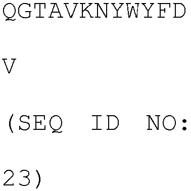
light chain 
YASNLES (SEQ ID NO: 25) QHSWEIPFT (SEQ ID NO: 26) MI407 heavy chain NYDMS (SEQ ID NO: 27) 

light chain SVSTSSSSYIH (SEQ ID NO: 30) YASNLES (SEQ ID NO: 25) QHSWEIPFT (SEQ ID NO: 26) - The pComb3X phagemid containing the genes encoding the heavy chain variable regions of the MI323, MI329, MI403, and MI407 clone antibodies was extracted, and used as a template for PCR with a forward primer containing NotI (Table 3: SEQ ID NOs: 31 to 34) and a reverse primer containing Apal (Table 3: SEQ ID NO: 35) by using Accupower Pfu PCR premix (Bioneer). The PCR was performed by repeating exposure at 94°C for 10 minutes, and then exposure at 94°C for 15 seconds, at 56°C for 30 seconds, and at 72°C for 90
seconds 30 times, and reacting at 72°C for 10 minutes. In the amplified genes, DNA bands having an expected size were confirmed on 1% agarose gel, and were separated using a gel extraction kit, respectively. Then, the separated genes reacted with NotI, ApaI restriction enzymes at 37°C for 12 hours or more, and the genes reacted with the restriction enzymes were separated on 1% agarose gel again. A pcIW plasmid vector containinghuman immunoglobulin type 1 of heavy chain constant region (IgG1 heavy chain constant region) gene was also cut by the same method as above and separated on agarose gel. The separated MI323, MI329, MI403, and MI407 heavy chain variable region genes were inserted into NotI, ApaI sites of a linear pcIW vector containing the human heavy chain constant region by using a T4 DNA ligase (Cat.No.M0203S, New England BioLabs (NEB)). The ligation reaction materials were transformed into XL1-Blue bacteria (Electroporation-Competent Cells; Cat.No.200228, Stratagene), plated on an LB plate (Cat.No.LN004CA, NaraeBiotech) containing carbenicillin, and cultured at 37°C for 12 hours or more. Then single colonies were chosen and cultured, and plasmids were separated by using a plasmid mini kit (Cat.No.27405, QIAGEN), and confirmed by DNA sequencing. - The pComb3X phagemid containing the genes encoding the light chain variable regions of the MI323, MI329, MI403, and MI407 clone antibodies was extracted, and used as a template for PCR of the light chain variable regions of the MI323, MI329, MI403, and MI407 clone antibodies, wherein the PCR was performed with a forward primer containing NotI (Table 3: SEQ ID NO: 36, 38, 40, 42) and a reverse primer (Table 3: SEQ ID NO: 37, 39, 41, 43) by using Accupower Pfu PCR premix. Further, the human antibody kappa light chain constant region was subjected to PCR with a forward primer (Table 3: SEQ ID NO: 44) and a reverse primer containing HindIII (Table 3: SEQ ID NO: 45). The PCR was performed by repeating exposure at 94°C for 10 minutes, and then exposure at 94°C for 15 seconds, at 56°C for 30 seconds, and at 72°C for 90
seconds 30 times, and reacting at 72°C for 10 minutes. In the amplified genes, DNA bands having a predicted size were confirmed on 1% agarose gel, and were separated using a gel extraction kit, respectively. Then, the respective light chain variable regions and light chain constant regions were mixed, followed by overlapping PCR, such that the genes expressing the light chain region were cloned. The PCR was performed by repeating exposure at 94°C for 10 minutes, and then exposure at 94°C for 15 seconds, at 56°C for 30 seconds, and at 72°C for 90seconds 30 times, and reacting at 72°C for 10 minutes. In the amplified genes, DNA bands having an expected size were confirmed on 1% agarose gel, and were separated using a gel extraction kit, respectively. Then, the separated genes reacted with NotI, HindIII restriction enzymes at 37°C for 12 hours or more, and the genes reacted with the restriction enzymes were separated on 1% agarose gel again. The pcIW plasmid vector was also cut by the same method as above and separated on agarose gel. The separated MI323, MI329, MI403, and MI407 light chain region genes were inserted into NotI, HindIII sites of a linear pcIW vector by using a T4 DNA ligase (Cat.No.M0203S, New England BioLabs (NEB)). The ligation reaction materials were transformed into XL1-Blue bacteria (Electroporation-Competent Cells; Cat.No.200228, Stratagene), plated on an LB plate (Cat.No.LN004CA, NaraeBiotech) containing carbenicillin, and cultured at 37°C for 12 hours or more. Then single colonies were chosen and cultured, and plasmids were separated by using a plasmid mini kit (Cat.No.27405, QIAGEN), and confirmed by DNA sequencing.[Table 3] Name DNA nucleotide sequence SEQ ID NO: MI323VH-F 
31 MI329VH-F 
32 MI403VH-F 
33 MI407VH-F 
34 VHApaI-R 
35 MI323VL-F 
36 MI323VLCL-R ACACTAGGAGCGGCCACGGTTCGTTTGATTTCCAGTTTGGTCCCT 37 MI329VL-F 
38 MI329VLCL-R ACACTAGGAGCGGCCACGGTTCGTTTCAGCTCCAGCTTGGTC 39 MI403VL- F 
40 MI403VLCL-R 
41 MI407VL-F 
42 MI407VLCL-R 
43 Ck-F ACCGTGGCCGCTCCTAGTGT 44 CkSHB-R NNNNGGATCCAAGCTTACTAGCACTCCCC 45 - In order to produce and purify the anti-MSLN antibody MI323, MI329, MI403, and MI407 clones obtained by a mouse immune response, Expi293F™ cells were inoculated at a concentration of 2.0 X 10⁶ cell/mL the day before transfection. After incubation (37°C, 8% CO2, 125 rpm) for 24 hours, Expi293™ expression medium (Cat.No.A1435101, Gibco) was added to prepare a product of 30 mL having a concentration of 2.5 X 10⁶ cell/mL (viability = 95%). 30 µg of DNA (pcIW-anti-MSLN heavy chain: 15 µg, pcIW-anti-MSLN light chain: 15 µg) was diluted in an OptiPro™ SEM medium (Cat.No.12309019, Gibco) so as to have a total volume of 1.5 mL, and reacted at room temperature for 5 minutes. 1.5 mL of the OptiPro™ SEM medium (Cat.No.12309019, Gibco) was mixed with 80 µL of an ExpiFectamine™ 293 reagent (Cat.No.A14524, Gibco) so that a total volume is 1.5mL, and reacted at room temperature for 5 minutes. After the reaction for 5 minutes, 1.5 mL of diluted DNA and 1.5 mL of diluted ExpiFectamine™ 293 reagent were well-mixed with each other, and reacted at room temperature for 20 to 30 minutes. 3 mL of the mixture of DNA and ExpiFectamine™ 293 reagent was treated in the Expi293F™ cells. After suspension-culture (37 °C, 8% CO2, 125 rpm) for 16 to 18 hours, 150 µL of ExpiFectamine™ 293 Enhancer 1 (Cat.No.A14524, Gibco) and 1.5 mL of ExpiFectamine™ 293 Enhancer 2 (Cat.No.A14524, Gibco) were added thereto, followed by suspension-culturing for 5 days. After the culturing, cell debris was removed by centrifugation at 4000rpm for 20 minutes, and the supernatant passed through 0.22 µm filter to be prepared. MabSelect Xtra (Cat.No.17-5269-02, GE Healthcare) which is protein A resin having 100 µL was prepared for each 30 mL of the culture fluid, followed by centrifugation at 1000 rpm for 2 minutes to remove a storage solution, and the obtained product was washed with 400 µL of protein A binding buffer (Cat.No.21007, Pierce) 3 times. The protein A resin was added to the prepared culture fluid and rotation-reacted at room temperature for 30 minutes. The mixture of the culture fluid and the resin was put into a pierce spin column snap-cap (Cat.No.69725, Thermo), and then, only the resin was left in the column using QIAvac 24 Plus (Cat.No.19413, QIAGEN) vacuum manifold. 5 mL of protein A binding buffer was added to wash the resin, 200 µL of a protein A elution buffer (Cat.No.21009, Pierce) was added thereto. The resultant material was reacted by resuspension at room temperature for 2 minutes, and centrifuged at 1000 rpm for 1 minute, and eluted. Each eluate was neutralized by adding 2.5 µL of 1.5M Tris-HCl (pH 9.0). The elution was performed 4 to 6 times, and each fraction was quantified by using Nanodrop 200C (Thermo Scientific). The fractions in which protein is detected were collected, and exchanged with a PBS (Phosphate-Buffered Saline) buffer using Zeba Spin Desalting Columns, 7K MWCO, 5 mL (Cat.No.0089892, Pierce). Then, protein electrophoresis (SDS-PAGE) was performed under reduction and non-reduction condition to finally verify the concentration quantification and the antibody state, and the antibody was kept at 4°C.
- For a primary panning, 1 mL of 1013 or more library stock was reacted in a solid phase polystyrene tube (Cat.No.444202, Nunc) coated with MSLN at 37 °C for 2 hours. At the same time, 10 µL of XL1-Blue bacteria (electroporation-competent cells; Cat.No.200228, Stratagene) were inoculated with 10 µL of SB 10ml/tetracycline and grown to an OD600 of 0.8 to 1.0. The library stock obtained after reaction at 37°C for 2 hours was washed with 5ml of 0.05
% Tween 20/PBS four times, and from a secondary panning, the number of times of washing with 0.05% Tween 20/PBS increased according to an increase in the number of times of panning. Then, the resultant material was cultured with 1%BSA/0.1M Glycine pH 2.0 at room temperature for 10 minutes to purify the phagemid. The purified phagemid was transferred to a 50 mL tube and neutralized with 70 µL of 2M Tris. 9 mL of XL1-Blue bacteria (electroporation-competent cells; Cat.No.200228, Stratagene) were treated, and 1 mL of the bacteria were treated in a washed tube. The bacteria were infected at room temperature for 30 minutes, and 10 mL of SB, 20 µL of tetracycline, 10 µL of carbenicillin were added thereto, followed by suspension-culturing at 37°C and 220 rpm for 1 hour. Then, the bacteria were treated with 1 mL of VCS M13 helper phage (1011 pfu), and suspension-cultured at 37°C and at 220 rpm for 1 hour, and treated with 80 mL of SB, 100 µL of kanamycin, and 100 µL of carbenicillin, and cultured at 37°C and at 220 rpm for 12 hours or more. The bacteria cultured over 12 hours were centrifuged at 3500 rpm and at 4°C for 10 minutes, and the supernatant was transferred to a new tube. 20 mL of 20% PEG/15% NaCl was added thereto, well-mixed, and reacted in ice for 30 minutes. Then, the supernatant was discarded, and pellets were collected and re-suspended with 2 mL of 1% BSA/PBS at 8000 rpm, and at 4°C for 30 minutes, and centrifuged at 15000 rpm and 4°C for 10 minutes. Here, the collected pellets were discarded and 1 mL of the supernatant (2 mL) was stored at -20°C and the remainder (1 mL) was used in the following order panning. - Single colonies of a phage display synthetic scFv library final amplified population were collected, and cultured with 1.5 mL of SB/carbenicillin to an
OD 600 of 0.8 to 1.0 at 37°C and at 220 rpm, and then cultured with 1 mM IPTG at 30°C and at 200 rpm for 12 hours or more. The reaction materials were centrifuged at 5500 rpm for 5 minutes, and only each supernatant was added to ELISA plates containing underlying MSLN antigen, and reacted at room temperature for 2 hours. Then, the resultant materials were washed with PBST (1XPBS, 0.05% tween 20) four times, and HRP/Anti-hFab-HRP conjugate diluted by 1/5000 with 1% BSA/1XPBS was added thereto, and reacted at room temperature for 1 hour, and washed with PBST (1XPBS, 0.05% tween 20) 4 times. Then, a TMB solution was added thereto and reacted for 5 to 10 minutes, and a TMB stop solution was added thereto. Next, O.D values were measured at a measurement wavelength of 450 nm using a TECAN sunrise, and clones having high O.D. value were secured as individual clones. - As a result, as shown in Table 4, the clones specifically bound to the human MSLN were able to be selected, and amino acid sequences thereof were identified.
- Table 5 shows CDR amino acid sequences of the clone antibodies of Table 4 on the basis of Kabat numbering.
[Table 4] Clone variable region amino acid sequence SEQ ID NO: MS501 heavy chain 
46 light chain 
47 
MS502 heavy chain 
48 light chain 
49 MS502-1 heavy chain 
48 light chain 
50 MS503 heavy chain 
51 light chain 
52 MS504 heavy chain 
53 
light chain 
54 MS505 heavy chain 
55 light chain 
56 MS506 heavy chain 
57 light chain 
58 [Table 5] Clone variable region CDR1 CDR2 CDR3 MS501 heavy chain NYAMS (SEQ ID NO: 59) 
NIYTFDY (SEQ ID NO: 61) light chain 
YNNQRPS (SEQ ID NO: 63) GSWDSSLSGYV (SEQ ID NO: 64) MS502 heavy chain NYAMS (SEQ ID NO: 59) 
NMLSFDY (SEQ ID NO: 66) light chain 
YNSKRPS (SEQ ID NO: 68) GSWDSSLNGYV (SEQ ID NO: 69) MS502-1 heavy chain NYAMS (SEQ ID NO: 59) 
NMLSFDY (SEQ ID NO: 66) light chain 
YNSKRPS (SEQ ID NO: 68) GSWDSSLNGYV (SEQ ID NO: 69) MS503 heavy chain NYAMS (SEQ ID NO: 59) 
NAFTFDY (SEQ ID NO: 72) 
light chain 
YNSHRPS (SEQ ID NO: 73) GTWDSSLSGYV (SEQ ID NO: 74) MS504 heavy chain NYAMS (SEQ ID NO: 59) 
NLLTFDY (SEQ ID NO: 76) light chain 
YDSHRPS (SEQ ID NO: 78) GAWDDSLNAYV (SEQ ID NO: 79) MS505 heavy chain NYAMS (SEQ ID NO: 59) 
NAYTFDY (SEQ ID NO: 81) light chain 
YNSQRPS (SEQ ID NO: 82) GSWDSSLNGYV (SEQ ID NO: 83) MS506 heavy chain NYAMS (SEQ ID NO: 59) 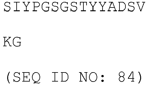
NLYTFDY (SEQ ID NO: 85) light chain 
YDSHRPS (SEQ ID NO: 78) GAWDSSLSAYV (SEQ ID NO: 87) 
- The pComb3X phagemid containing the genes encoding the light chain variable regions of the secured MS501, MS502, MS503, MS504, MS505, and MS506 clone antibodies was extracted, and used as a template for PCR of the light chain variable regions of the MS501, MS502, MS503, MS504, MS505, and MS506 clone antibodies, wherein the PCR was performed with a forward primer containing NotI (SEQ ID NO: 86) and a reverse primer (SEQ ID NO: 87) by using Accupower Pfu PCR premix. Further, human antibody kappa light chain constant region was subjected to PCR with a forward primer (Table 6: SEQ ID NO: 88) and a reverse primer (Table 6: SEQ ID NO: 89). The PCR was performed by repeating exposure at 94°C for 10 minutes, and then exposure at 94°C for 15 seconds, at 56°C for 30 seconds, and at 72°C for 90
seconds 30 times, and reacting at 72°C for 10 minutes. In the amplified genes, DNA bands having an expected size were confirmed on 1% agarose gel, and were separated using a gel extraction kit, respectively. Then, the respective light chain variable regions and light chain constant regions were mixed, followed by overlapping PCR, such that the genes expressing the light chain region were cloned. The PCR was performed by repeating exposure at 94°C for 10 minutes, and then exposure at 94°C for 15 seconds, at 56°C for 30 seconds, and at 72°C for 90seconds 30 times, and reacting at 72°C for 10 minutes. In the amplified genes, DNA bands having an expected size were confirmed on 1% agarose gel, and were separated using a gel extraction kit, respectively. Then, the separated genes reacted with NotI, HindIII restriction enzymes at 37°C for 12 hours or more, and the genes reacted with the restriction enzymes were separated on 1% agarose gel again. The pcIW plasmid vector was also cut by the same method as above and separated on agarose gel. The separated MS501, MS502, MS503, MS504, MS505, MS506 light chain region genes were inserted into NotI, HindIII sites of the linear pcIW vector by using a T4 DNA ligase (Cat.No.M0203S, New England BioLabs (NEB)). The ligation reaction materials were transformed into XL1-Blue bacteria (Electroporation-Competent Cells; Cat.No.200228, Stratagene), plated on an LB plate (Cat.No.LN004CA, NaraeBiotech) containing carbenicillin, and cultured at 37°C for 12 hours or more. Then single colonies were chosen and cultured, and plasmids were separated by using a plasmid mini kit (Cat.No.27405, QIAGEN), and confirmed by DNA sequencing. - The pComb3X phagemid containing the genes encoding the heavy chain variable regions of the MS501, MS502, MS503, MS504, MS505, and MS506 clone antibodies was extracted, and used as a template for PCR with a forward primer containing NotI (Table 6: SEQ ID NO: 90) and a reverse primer containing Apal (Table 6: SEQ ID NO: 91) by using Accupower Pfu PCR premix (Bioneer). The PCR was performed by repeating exposure at 94°C for 10 minutes, and then exposure at 94°C for 15 seconds, at 56°C for 30 seconds, and at 72°C for 90
seconds 30 times, and reacting at 72°C for 10 minutes. In the amplified genes, DNA bands having an expected size were confirmed on 1% agarose gel, and were separated using a gel extraction kit, respectively. Then, the separated genes reacted with NotI, ApaI restriction enzymes at 37°C for 12 hours or more, and the genes reacted with the restriction enzymes were separated on 1% agarose gel again. A pcIW plasmid vector containinghuman immunoglobulin type 1 of heavy chain constant region (IgG1 heavy chain constant region) gene was also cut by the same method as above and separated on agarose gel. The separated MS501, MS502, MS503, MS504, MS505, and MS506 heavy chain variable region genes were inserted into NotI, ApaI sites of the linear pcIW vector containing the human heavy chain constant region by using a T4 DNA ligase (Cat.No.M0203S, New England BioLabs (NEB)). The ligation reaction materials were transformed into XL1-Blue bacteria (Electroporation-Competent Cells; Cat.No.200228, Stratagene), plated on an LB plate (Cat.No.LN004CA, NaraeBiotech) containing carbenicillin, and cultured at 37°C for 12 hours or more. Then single colonies were chosen and cultured, and plasmids were separated by using a plasmid mini kit (Cat.No.27405, QIAGEN), and confirmed by DNA sequencing.[Table 6] Name Nucleotide sequence SEQ ID NO: NotI-Leader-VL-F 
88 VL-R GGGGTTGGCCTTGGGCTGGCCTAGGACCGTCAGCTTGGT 89 VL-CL-F CAGCCCAAGGCCAACCCC 90 HindIII-VL-R 
91 HD-Heavy-F 
92 HD-Heavy-R 
93 - In order to produce and purify the MS501, MS502, MS503, MS504, MS505, MS506 clone antibodies obtained from the phage display scFv library, Expi293F™ cells were inoculated at a concentration of 2.5 X 10⁶ cell/mL the day before transfection. After incubation (37°C, 8% CO2, 125 rpm) for 24 hours, Expi293™ expression medium (Cat.No.A1435101, Gibco) was added to prepare a product of 30 mL having a concentration of 2.5 X 10⁶ cell/mL (viability = 95%). 30µg of DNA (pcIw-MS502 heavy chain variable region: 15 µg, pcIw-anti-Mesothelin light chain variable region: 15 µg) was diluted in an OptiPro™ SEM medium (Cat.No.12309019, Gibco) so as to have a total volume of 1.5 mL, and reacted at room temperature for 5 minutes. 1.5 mL of the OptiPro™ SEM medium (Cat.No.12309019, Gibco) was mixed with 80 µL of an ExpiFectamine™ 293 reagent (Cat.No.A14524, Gibco) so that a total volume is 1.5 mL, and reacted at room temperature for 5 minutes. After the reaction for 5 minutes, 1.5 mL of diluted DNA and 1.5 mL of diluted ExpiFectamine™ 293 reagent were well-mixed with each other, and reacted at room temperature for 20 to 30 minutes. 3 mL of the mixture of DNA and ExpiFectamine™ 293 reagent was treated in the Expi293F™ cells. After suspension-culture (37 °C, 8% CO2, 125 rpm) for 16 to 18 hours, 150 µL of ExpiFectamine™ 293 Enhancer 1(Cat.No.A14524, Gibco) and 1.5 mL of ExpiFectamine™ 293 Enhancer2 (Cat.No.A14524, Gibco) were added thereto, followed by suspension-culturing for 5 days. After the culturing, cell debris was removed by centrifugation at 4000rpm for 20 minutes, and the supernatant passed through 0.22 µm filter to be prepared. MabSelect Xtra (Cat.No.17-5269-02, GE Healthcare) which is protein A resin having 100 µL was prepared for each 30 mL of the culture fluid, followed by centrifugation at 1000 rpm for 2 minutes to remove a storage solution, and the product was washed with 400 µL of protein A binding buffer (Cat.No.21007, Pierce) 3 times. The protein A resin was added to the prepared culture fluid and rotation-reacted at room temperature for 30 minutes. The mixture of the culture fluid and the resin was put into a pierce spin column snap-cap (Cat.No.69725, Thermo), and then, only the resin was left in the column using QIAvac 24 Plus (Cat.No.19413, QIAGEN) vacuum manifold. 5 mL of protein A binding buffer was added to wash the resin, 200 µL of a protein A elution buffer (Cat.No.21009, Pierce) was added thereto. The resultant material was reacted by resuspension at room temperature for 2 minutes, and centrifuged at 1000 rpm for 1 minute, and eluted. Each eluate was neutralized by adding 2.5 µL of 1.5M Tris-HCl (pH 9.0). The elution was performed 4 to 6 times, and each fraction was quantified by using Nanodrop 200C (Thermo scientific). The fractions in which protein is detected were collected, and exchanged with a PBS (Phosphate-Buffered Saline) buffer using Zeba Spin Desalting Columns, 7K MWCO, 5 mL (Cat.No.0089892, Pierce). Then, protein electrophoresis (SDS-PAGE) was performed under reduction and non-reduction condition to finally verify the concentration quantification and the antibody state, and the antibody was kept at 4°C.
- Quantitative binding force (affinity) of the purified anti-MSLN antibodies, i.e., MI323, MI329, MI403, MS502 clone antibodies with regard to recombinant human mesothelin (MSLN) was measured by using a Biacore T-200 (GE Healthcare, U.S.A.) biosensor. The MSLN (Cat.No.3265-MS, R&D systems) purified from the HEK293 cells was fixed to a CM5 chip (GE Healthcare, U.S.A.) so as to satisfy 200 Rmax by using an amine-carboxylic reaction. Then, the clone C2G1 antibody, the clone C2G4 antibody or the clone C3C8 antibody serially diluted with HBS-EP buffer (10mM HEPES, pH7.4, 150mM NaCl, 3mM EDTA, 0.005% surfactant P20) was allowed to flow at a concentration range of 0.078nM to 5nM and at a flow rate of 30µL/min for association of 120 seconds and dissociation of 1800 seconds. The dissociation of the antibody bound to the MSLN was induced by flowing 10 mM Glycine-HCl pH 1.5 at a flow rate of 30 µL/min for 30 seconds (Table 7). The affinity was obtained as movement speed constants (Kon and Koff) and an equilibrium dissociation constant (KD) by using a Biacore T-200 evaluation software (Table 8).
[Table 7] SPR Biacore T200 Chip CM5 Running Buffer HBS-EP pH7.4 Flow rate 30µL/min Association / dissociation time 120 sec / 600 sec IgG Conc. 0.078∼5nM, 1/2 serial dilution Regeneration 10mM Glycine-HCl pH1.5, 30 sec [Table 8] Kon Koff KD MI323 2.7X10⁵ 4.8X10⁻⁵ 1.8X10⁻¹⁰ MI329 1.4X10⁶ 5.1X10⁻⁵ 3.5X10⁻¹¹ MI403 8.9X10⁴ 4. 0X10-5 4.5X10⁻¹⁰ MS502 1.9X10⁷ 4.3X10⁻³ 2.3X10⁻¹⁰ - The amino acid sequence of the anti-mesothelin MS502 clone was entered to the Swiss model homepage (http://swissmodel.expasy.org/) to find a template sequence. 50 sequences were found on the basis of homologous sequences, and modeling was performed by designating each sequence as the template. 50 sequences were listed by priority on the basis of QMEAN4 values (Cβ, all atom, solvation, torsion), and the template was selected in consideration of sequence identity and resolution in addition to the QMEAN4 values. 3g6a.1.B was selected for the heavy chain variable region, and 3qhz.1.B (LCDR1,2) and 4o5l.1.A (LCDR3) were selected for the light chain variable region. The MS502 structure obtained on the basis of the selected template was analyzed by pymol program, and paratope was selected on the basis of the protruding amino acid of the CDR (see
FIG. 1 ). Among them, VL CDR2 Y49, Y50, K53 were excluded from the mutation candidate amino acids even though it was anticipated that they are paratopes since Tyr and Lys are amino acids generally having a positive effect on bonding. The introduction of mutation was designed by 50% preserving the existing sequence, increasing Tyr, Ser, Gly rates, and controlling the nucleotide sequence using a handmix primer (IDT, U.S.A.) so that hydrophilic, hydrophobic, positive charge, and negative charge rates were uniformly included (Table 9). The heavy chain variable region was divided into each of three fragments since the mutation was introduced into all of CDR1, 2, and 3, and they were subjected to fragment PCR and overlapping PCR to construct a library, and the light chain variable region was divided into each of two fragments since the mutation was introduced into CDR1 and 3 only, and they were subjected to fragment PCR and overlapping PCR to construct a library. A theoretical size of the library according to the introduction of the mutation in the case of the heavy chain variable region was 5.83 X 10¹⁰, and the theoretical size of the library according to the introduction of the mutation in the case of the light chain variable region was 2.16 X 10⁴.

- In order to introduce the mutation into the library, first, the light chain variable region was divided into 2 parts, and they were subjected to fragment PCR. The light chain variable region fragment No. 1 had a light chain variable region gene sequence of anti-mesothelin MS502 clone as the template, and a forward primer (SEQ ID NO: 93) and a reverse primer (SEQ ID NO: 94) were added thereto, and the light chain variable region fragment No. 2 had the light chain variable region gene sequence of anti-mesothelin MS502 clone as the template, and a forward primer (Table 10: SEQ ID NO: 95) and a reverse primer (Table 10: SEQ ID NO: 96) were added thereto, and then, each fragment was subjected to PCR using a Primestar polymerase premix (Takara). The PCR was performed by repeating exposure at 98°C for 2 minutes, and then exposure at 98°C for 10 seconds, at 60°C for 15 seconds, and at 72°C for 20
seconds 30 times, and reacting at 72°C for 10 minutes. In the amplified genes, DNA bands having an expected size were confirmed on 1% agarose gel, and were separated using a gel extraction kit (QIAquick Gel Extraction Kit, CAT. NO. 28706, QIAGEN), respectively. The light chain constant region had a light chain lambda region as a template, and a forward primer (Table 10: SEQ ID NO: 97), and a reverse primer (Table 10: SEQ ID NO: 98) were added thereto, and each fragment was subjected to PCR using a Primestar polymerase premix. The PCR was performed by repeating exposure at 98°C for 2 minutes, and then exposure at 98°C for 10 seconds, at 60°C for 15 seconds, and at 72°C for 30seconds 30 times, and reacting at 72°C for 10 minutes. In the amplified genes, DNA bands having an expected size were confirmed on 1% agarose gel, and were separated using a gel extraction kit, respectively. The secured light chain variable region fragments 1 and 2, and light chain constant region at a molar ratio of 1:1:1 were used as a template, and a forward primer (SEQ ID NO: 92) and a reverse primer (SEQ ID NO: 98) were added thereto, and each fragment was subjected to PCR using a Primestar polymerase premix. The PCR was performed by repeating exposure at 98°C for 2 minutes, and then exposure at 98°C for 20 seconds, at 60°C for 30 seconds, and at 72°C for 60seconds 30 times, and reacting at 72°C for 10 minutes. In the amplified genes, DNA bands having an expected size were confirmed on 1% agarose gel, and were separated using a gel extraction kit to secure light chain variable-constant region affinity maturation gene. The secured gene was reacted with NruI and XbaI (NEB) restriction enzyme at 37°C for 4 hours. The genes reacted with the restriction enzyme were separated on 1% Agarose gel again. The separated gene was inserted into the NruI, XbaI site of the linear pComb3x vector containing the MS502 heavy chain variable-constant region by using a T4 DNA ligase (Cat.No.M0203S, NEB). The ligation reaction material was transformed into XL1-Blue bacteria (Electroporation-competent cells; Cat.No.200228, Stratagene), and cultured in 300 mL of LB medium at 37°C and at 220 rpm for 1 hour. Then, the resultant material was treated with 150 µL of carbenicillin and 300 µL of tetracycline, and was cultured with shaking at 37°C and at 220 rpm for 1 hour. The resultant material was treated with VCS M13 helper phage 4.5 mL (1011pfu) and cultured with shaking at 37°C and at 220 rpm for 1 hour, and treated with 300 µL of kanamycin and 300 µL of carbenicillin, and cultured overnight at 37°C and at 220 rpm. The next day, the cultured cells were centrifuged at 4000 rpm for 20 minutes, and the supernatant was transferred to a new vessel. In order to precipitate the phage, 5X PEG/NaCl was used to add 1X PEG/NaCl to the supernatant, and the obtained product allowed to stand on ice over 30 minutes. The precipitate phage was centrifuged at 8000 rpm for 30 minutes. The supernatant was discarded and the precipitated phage was re-suspended with 10 mL of PBS. In order to remove cell debris, the phage dissolved in 10 mL of PBS was centrifuged at 14,000 rpm for 10 minutes to separate the supernatant, and stored at 4°C. The library size was confirmed by taking 100 µL of culture fluid after 1 hour of the transformation, plating the culture fluid on the LB plate (Cat.No.LN004CA, NaraeBiotech) containing carbenicillin in serial dilution manner, culturing at 37°C for 12 hours or more, and counting the colonies. - In order to introduce the mutation into the library, first, the heavy chain variable region was divided into 3 parts, and they were subjected to fragment PCR. The heavy chain variable region fragment No. 1 had a heavy chain variable region gene sequence of anti-mesothelin MS502 clone as the template, and a forward primer (Table 10: SEQ ID NO: 99) and a reverse primer (Table 10: SEQ ID NO: 100) were added thereto, and the heavy chain variable region fragment No. 2 had the heavy chain variable region gene sequence of anti-mesothelin MS502 clone as the template, and a forward primer (Table 10: SEQ ID NO: 101) and a reverse primer (Table 10: SEQ ID NO: 102) were added thereto, and the heavy chain variable region fragment No. 3 had the heavy chain variable region gene sequence of anti-mesothelin MS502 clone as the template, and a forward primer (Table 10: SEQ ID NO: 103) and a reverse primer (Table 10: SEQ ID NO: 104) were added thereto, and then, each fragment was subjected to PCR using a Primestar polymerase premix (Takara). The PCR was performed by repeating exposure at 98°C for 2 minutes, and then exposure at 98°C for 10 seconds, at 60°C for 15 seconds, and at 72°C for 20
seconds 30 times, and reacting at 72°C for 10 minutes. In the amplified genes, DNA bands having an expected size were confirmed on 1% agarose gel, and were separated using a gel extraction kit, respectively. The secured heavy chain variable region fragments 1, 2, and 3 at a molar ratio of 1:1:1 were used as a template, and a forward primer (Table 10: SEQ ID NO: 99) and a reverse primer (Table 10: SEQ ID NO: 106) were added thereto, and each fragment was subjected to PCR using a Primestar polymerase premix. The PCR was performed by repeating exposure at 98°C for 2 minutes, and then exposure at 98°C for 20 seconds, at 60°C for 30 seconds, and at 72°C for 60seconds 30 times, and reacting at 72°C for 10 minutes. In the amplified genes, DNA bands having an expected size were confirmed on 1% agarose gel, and were separated using a gel extraction kit to secure heavy chain variable region affinity maturation gene. The secured gene was reacted with XhoI and ApaI (NEB) restriction enzyme at 37°C for 4 hours. The genes reacted with the restriction enzyme were separated on 1% Agarose gel again. The separated gene was inserted into the XhoI, ApaI site of the linear pComb3x vector containing the MS502 light chain variable-constant region by using a T4 DNA ligase (Cat.No.M0203S, NEB). The ligation reaction material was transformed into XL1-Blue bacteria (Electroporation-competent cells; Cat.No.200228, Stratagene), and cultured in 300 mL of LB medium at 37°C and at 220 rpm for 1 hour. Then, the resultant material was treated with 150 µL of carbenicillin and 300 µL of tetracycline, and was suspension-cultured at 37°C and at 220 rpm for 1 hour. The resultant material was treated with VCS M13 helper phage 4.5mL (1011pfu) and cultured with shaking at 37°C and at 220 rpm for 1 hour, and treated with 300 µL of kanamycin and 300 µL of carbenicillin, and cultured overnight at 37°C and at 220 rpm. Next day, the cultured cells were centrifuged at 4000 rpm for 20 minutes, and the supernatant was transferred to a new vessel. In order to precipitate the phage, 5X PEG/NaCl was used to add 1X PEG/NaCl to the supernatant, and the obtained product allowed to stand on ice over 30 minutes. The precipitated phage was centrifuged at 8000 rpm for 30 minutes. The supernatant was discarded and the precipitated phage was re-suspended with 10 mL of PBS. In order to remove cell debris, the phage dissolved in 10 mL of PBS was centrifuged at 14,000 rpm for 10 minutes to separate the supernatant, and stored at 4°C. The library size was confirmed by taking 100 µL of culture fluid after 1 hour of the transformation, plating the culture fluid on the LB plate (Cat.No.LN004CA, NaraeBiotech) containing carbenicillin in serial dilution manner, culturing at 37°C for 12 hours or more, and counting the colonies.[Table 10] Name Nucleotide sequence SEQ ID NO: MS502 VL FR1 Fo NruI 
94 MS502 VL FR1 Fo CAG TCT GTG CTG ACT CAG CCA CCC TCA 95 MS502 VL FR1 Re 
96 MS502 VL FR2 Fo GCC AAT ATT AGA TGA AGA GCC AGT ACA AGA 97 MS502 VL FR2 Re 
98 MS502 CL Fo 
99 
MS502 CL Re 
100 MS502 VH FR1 Fo NcoI 
101 MS502 VH FR1 Re 
102 MS502 VH FR2 Fo atgagctgggtccgccaggct 103 MS502 VH FR2 Re 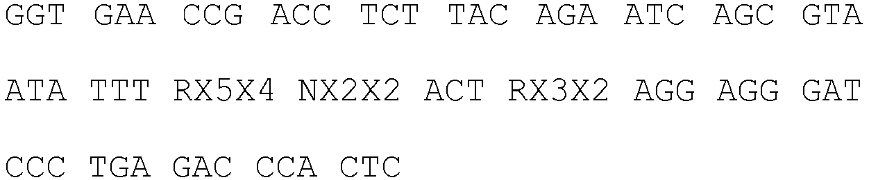
104 MS502 VH FR3 Fo 
105 MS502 VH FR3 Re 
106 MS502 VH Re TGA GCT CAC CGT GAC CAG TGT ACC CTG 107 MS502 VH Re ApaI 
108 - In Table 10, X codon is a degenerative codon in which each ACGT is controlled at a specific rate, and is able to control a rate of the amino acids to be translated. As an example, for the NX1X2 codon of SEQ ID NO: 102, N is encoded by A, C, G, T with a random ratio, X1 is encoded at 10% of A, 10% of C, 70% of G, and 10% of T, and X2 is encoded at 10% of A, 70% of C, 10% of G, and 10% of T. This is designed for the reverse primer, and thus, if it is converted to the forward direction, it is the X2X1N codon, wherein X2 is encoded at 10% of A, 70% of C, 10% of G, and 10% of T, and X1 is encoded at 10% of A, 10% of C, 70% of G, and 10% of T. As a result, Kabat No. 31 of the heavy chain variable region CDR1 is translated into amino acids,
Tyr 7%, Ser 8%,Gly 1%, His 7%,Asp 7%,Phe 1%, Thr 7%, Asn 49%,Ala 1%,Val 1%,Leu 1%,Ile 7%,Pro 1%, Cys 1%. - 1 mL of recombinant human protein MSLN having a concentration of 1 µg/mL was put in a solid phase polystyrene tube (Cat.No.444202, Nunc), and the tube coated at 4°C for 12 hours or more was washed with 5 mL of 0.05% PBST three times. 5 mL of 1%BSA/PBS was put in the MSLN-coated immuno tube, followed by blocking at room temperature for 2 hours. A blocking buffer was removed from the immuno tube, and then, the light chain variable region phage library was treated in the tube and reacted at room temperature for 2 hours. Then, the obtained product was washed with 5 mL of PBST four times. The immuno tube was treated with 1 mL of glycine (pH 2.0) elution buffer, and reacted at room temperature for 10 minutes to obtain the supernatant. After elution, 100 µL of 1.5M Tris-Cl (pH 8.8) was added to the phage and neutralized. 10 mL of XL1-Blue bacteria (electroporation-competent cells; Cat.No.200228, Stratagene) cultured for about 2 hours (OD600 = 0.8 to 1.0) were treated with the neutralized phage. After infection at room temperature for 30 minutes, 10 mL of SB, 20 µL of tetracycline (50 mg/ml), and 10 µL of carbenicillin (100 mg/mL) were added to 10 mL of the infected XL1-Blue bacteria (electroporation-competent cells; Cat.No.200228, Stratagene), and cultured with shaking (200rpm) at 37°C for 1 hour. The bacteria were treated with 1 mL of VCSM13 helper phage (> 1011 pfu/ml), and cultured with shaking (200 rpm) at 37°C for 1 hour. After 1 hour incubation, the bacteria were treated with 80 mL of SB, 100 µL of kanamycin, and 100 µL of carbenicillin (100 mg/mL), and cultured overnight (200 rpm) at 37°C. The overnight cultured library was centrifuged at 4000 rpm for 15 minutes to separate the supernatant only, and 5X PEG/NaCl was used to add 1X PEG/NaCl to the supernatant, and the obtained product allowed to stand on ice over 30 minutes. The supernatant was removed by centrifugation at 8000 rpm for 30 minutes, and pellets were re-suspended with 2 ml of 1% BSA/PBS and centrifuged at 12000 rpm for 10 minutes. Then, only the supernatant was taken and used in the following order panning. This process was repeated four times.
- The supernatant was removed with 50 µL of streptavidin microbead (Miltenyl biotec 130-048-101), and 1 mL of PBS was added thereto to perform washing three times. 1 mL of PBS containing 1% BSA was added to the bead, and the bead was rotated at room temperature for 2 hours, followed by blocking. 50 nM MSLN was put in 500 µL of PBS, and mixed with 500 µL of MS502 VH rational library phage and rotation-reacted at room temperature for 1 hour. The blocking buffer was removed from the bead, and the bead was treated with a solution containing the MSLN and the phage, and reacted at room temperature for 15 minutes. Then, the obtained product was washed with 1 mL of PBST six times. The bead was treated with 1 mL of glycine (pH 2.0) elution buffer, and reacted at room temperature for 10 minutes to obtain the supernatant. After elution, 100 µL of 1.5M Tris-Cl (pH 8.8) was added to the phage and neutralized. 10 mL of XL1-Blue bacteria (electroporation-competent cells; Cat.No.200228, Stratagene) cultured for about 2 to 2.5 hours (OD600 = 0.8 to 1.0) were treated with the neutralized phage. After infection at room temperature for 30 minutes, 10 mL of SB, 20 µL of tetracycline (50 mg/ml), and 10 µL of carbenicillin (100 mg/mL) were added to 10 mL of the infected XL1-Blue bacteria (electroporation-competent cells; Cat.No.200228, Stratagene), and cultured with shaking (200rpm) at 37°C for 1 hour. The bacteria were treated with 1 mL of VCSM13 helper phage(> 1011 pfu/ml), and suspension-cultured (200 rpm) at 37°C for 1 hour. After 1 hour incubation, the bacteria were treated with 80 mL of SB, 100 µL of kanamycin, and 100 µL of carbenicillin (100 mg/mL), and cultured overnight (200 rpm) at 37°C. The overnight cultured library was centrifuged at 4000 rpm for 15 minutes to separate the supernatant only, and 5X PEG/NaCl buffer was used to add 1X PEG/NaCl to the supernatant, and the obtained product allowed to stand on ice over 30 minutes. The supernatant was removed by centrifugation at 8000 rpm for 30 minutes, and pellets were re-suspended with 2m of 1% BSA/PBS and centrifuged at 12000 rpm for 10 minutes. Then, only the supernatant was taken and used in the following order panning. A total of primary, secondary, and tertiary bead panning with regard to the MSLN were performed by using MS502 VH rational library phage. As a result of the secondary and tertiary pannings, it was confirmed that output titers were increased, which showed that the antibodies against the MSLN were amplified.
- Single colonies of a final amplified population of each library of light chain/heavy chain variable regions were collected, and cultured with 1.5 mL of SB/carbenicillin up to an
OD 600 of 0.8 to 1.0 at 37°C and at 220 rpm, and then cultured with 1 mM IPTG at 30°C and at 200 rpm for 12 hours or more. The reaction materials were centrifuged at 5500 rpm for 5 minutes, and only each supernatant was added to ELISA plates containing underlying MSLN antigen, and reacted at room temperature for 2 hours. Then, the resultant materials were washed with PBST (1XPBS, 0.05% tween 20) four times, and HRP/Anti-hFab-HRP conjugate diluted by 1/5000 with 1% BSA/1XPBS was added thereto, and reacted at room temperature for 1 hour, and washed with PBST (1XPBS, 0.05% tween 20) 4 times. Then, a TMB solution was added and allowed to stand for 5 to 10 minutes, and a TMB stop solution was added thereto. Next, O.D values were measured at a measurement wavelength of 450 nm using a TECAN sunrise, and clones having high O.D value were secured as individual clones. - As a result, as shown in Table 11, the clones specifically bound to the human MSLN were able to be selected, and amino acid sequences thereof were identified.
- Table 12 shows CDR amino acid sequences of the clone antibodies of Table 11 on the basis of Kabat numbering.
[Table 11] clone variable region amino acid sequence SEQ ID NO: C2G1 heavy chain 
48 light chain 
109 C2G4 heavy chain 
48 light chain 
110 C3C8 heavy chain 
48 light chain 
111 
54 heavy chain 
112 light chain 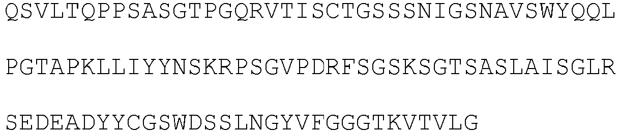
49 56 heavy chain 
113 light chain 
49 2-30 heavy chain 
114 light chain 
49 2-73 heavy chain 
115 light chain 
49 
2-78 heavy chain 
116 light chain 
49 [Table 12] clone variable region CDR1 CDR2 CDR3 C2G1 heavy chain NYAMS (SEQ ID NO: 59) 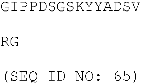
NMLSFDY (SEQ ID NO: 66) light chain 
YNSKRPS (SEQ ID NO: 68) GSWDSSLSGYV (SEQ ID NO: 118) C2G4 heavy chain NYAMS (SEQ ID NO: 59) 
NMLSFDY (SEQ ID NO: 66) light chain 
YNSKRPS (SEQ ID NO: 68) GSWDPSLNGYV (SEQ ID NO: 119) 
C3C8 heavy chain NYAMS (SEQ ID NO: 59) 
NMLSFDY (SEQ ID NO: 66) light chain 
YNSKRPS (SEQ ID NO: 68) GSWDSDLRGYV (SEQ ID NO: 120) 54 heavy chain NYAMS (SEQ ID NO: 59) 
NMLSFDY (SEQ ID NO: 66) light chain 
YNSKRPS (SEQ ID NO: 68) GSWDSSLNGYV (SEQ ID NO: 69) 56 heavy chain NYAMS (SEQ ID NO: 59) 
NMLSFDY (SEQ ID NO: 66) light chain 
YNSKRPS (SEQ ID NO: 68) GSWDSSLNGYV (SEQ ID NO: 69) (SEQ ID NO: 67) 2-30 heavy chain NYAMS (SEQ ID NO: 59) 
NMRTFDY (SEQ ID NO: 124) light chain 
YNSKRPS (SEQ ID NO: 68) GSWDSSLNGYV (SEQ ID NO: 69) 2-73 heavy chain NYAMS (SEQ ID NO: 59) 
NMLSFDY (SEQ ID NO: 66) light chain 
YNSKRPS (SEQ ID NO: 68) GSWDSSLNGYV (SEQ ID NO: 69) 2-78 heavy chain NYAMS (SEQ ID NO: 59) 
NMFSFDY (SEQ ID NO: 126) light chain 
YNSKRPS (SEQ ID NO: 68) GSWDSSLNGYV (SEQ ID NO: 69) 
- In order to compare and measure the binding force with culture fluid of individual clones secured from confirmation of the individual clones according to ELISA method conducted in Example 3-3, first, the human MSLN on the biacore series S CM5 chip (GE healthcare) was dissolved in pH 4.0 acetate buffer to be 1 µg/ml, and allowed to flow at a flow rate of 10 µL/min and fixed to 1000 Ru. The Fab supernatant expressed in Example 3-6 was diluted by 1/10 with pH 7.4 HBS-EP buffer, and was allowed to flow at a flow rate of 30 µL/min for association of 120 seconds, and dissociation of 180 seconds to conduct binding analysis. The regeneration was performed with 10 mM glycine-HCl pH 1.5 buffer for 30 seconds. The ELISA and the SPR binding data were compared and analyzed to select the final clones of the light chain variable region and the heavy chain variable region.
- As a result, as illustrated in
FIGS. 2 and3 , the binding force of the light chain variable region mutations and the heavy chain variable region mutations were relatively compared, and C2G1, C2G4, and C3C8 were selected for the final clones of the light chain variable region and 56, 2-30, 2-58, 2-73, and 2-78 were selected for the final clones of the heavy chain variable region. - Each of the secured light chain variable region C2G1, C2G4, and C3C8 genes as the template was subjected to PCR by using PrimeSTAR HS DNA polymerase (Cat.No.R010B; Takara) and the forward primer containing NotI (Table 6: SEQ ID NO: 86) and the reverse primer (Table 6: SEQ ID NO: 87). Further, human antibody kappa light chain constant region was subjected to PCR with the forward primer (Table 6: SEQ ID NO: 88) and the reverse primer (Table 6: SEQ ID NO: 89). The PCR was performed by repeating exposure at 94°C for 10 minutes, and then exposure at 94°C for 15 seconds, at 56°C for 30 seconds, and at 72°C for 90
seconds 30 times, and reacting at 72°C for 10 minutes. In the amplified genes, DNA bands having an expected size were confirmed on 1% agarose gel, and were separated using a gel extraction kit, respectively. Then, the respective light chain variable regions and light chain constant regions were mixed, followed by overlapping PCR, such that the genes expressing the light chain region were cloned. The PCR was performed by repeating exposure at 94°C for 10 minutes, and then exposure at 94°C for 15 seconds, at 56°C for 30 seconds, and at 72°C for 90seconds 30 times, and reacting at 72°C for 10 minutes. In the amplified genes, DNA bands having an expected size were confirmed on 1% agarose gel, and were separated using a gel extraction kit, respectively. Then, the separated genes reacted with NotI, HindIII restriction enzymes at 37°C for 12 hours or more, and the genes reacted with the restriction enzymes were separated on 1% agarose gel again. The pcIW plasmid vector was also cut by the same method as above and separated on agarose gel. The separated C2G1, C2G4, and C3C8 light chain region genes were inserted into NotI, HindIII sites of the linear pcIW vector by using a T4 DNA ligase (Cat.No.M0203S, New England BioLabs (NEB)). The ligation reaction materials were transformed into XL1-Blue bacteria (Electroporation-Competent Cells; Cat.No.200228, Stratagene), plated on an LB plate (Cat.No.LN004CA, NaraeBiotech) containing carbenicillin, and cultured at 37°C for 12 hours or more. Then single colonies were chosen and cultured, and plasmids were separated by using a plasmid mini kit (Cat.No.27405, QIAGEN), and confirmed by DNA sequencing. - Each of the heavy chain variable region 56, 2-30, 2-58, 2-73, and 2-78 genes as the template was subjected to PCR by using PrimeSTAR HS DNA polymerase (Cat.No.R010B; Takara) and the forward primer containing NotI (Table 6: SEQ ID NO: 90) and the reverse primer containing Apal (Table 6: SEQ ID NO: 91). The PCR was performed by repeating exposure at 98°C for 2 minutes, and then exposure at 98°C for 10 seconds, at 58°C for 10 seconds, and at 72°C for 30
seconds 30 times, and reacting at 72°C for 5 minutes. In the amplified genes, DNA bands having an expected size were confirmed on 1% agarose gel, and were separated using a gel extraction kit, respectively. Then, the three kinds of separated genes were reacted with KpnI and ApaI restriction enzyme at 37°C for 4 hours. The genes reacted with the restriction enzyme were separated on 1% Agarose gel again. The pcIW plasmid vector was also cut by the same method as above and separated on agarose gel. The separated genes were inserted into the NotI, ApaI sites of the linear pclw vector containing the human heavy chain constant region by using a T4 DNA ligase. The ligation reaction materials were transformed into XL1-Blue bacteria (Electroporation-Competent Cells; Cat.No.200228, Stratagene), plated on an LB plate (Cat.No.LN004CA, NaraeBiotech) containing carbenicillin, and cultured at 37°C for 12 hours or more. Then single colonies were chosen and cultured, and plasmids were separated by using a plasmid mini kit (Cat.No.27405, QIAGEN), and confirmed by DNA sequencing. - In order to produce and purify the light chain variable region mutation antibodies C2G1, C2G4, and C3C8, Expi293F™ cells were inoculated at a concentration of 2.5 X 10⁶ cell/mL the day before transfection. After incubation (37°C, 8% CO2, 125 rpm) for 24 hours, Expi293™ expression medium (Cat.No.A1435101, Gibco) was added to prepare a product of 30 mL having a concentration of 2.5 X 10⁶ cell/mL (viability = 95%). 30 µg of DNA (pcIw-MS502 heavy chain variable region: 15 µg, pcIw-anti-Mesothelin light chain variable region mutant: 15 µg) was diluted in an OptiPro™ SEM medium (Cat.No.12309019, Gibco) so as to have a total volume of 1.5 mL, and reacted at room temperature for 5 minutes. 1.5 mL of the OptiPro™ SEM medium (Cat.No.12309019, Gibco) was mixed with 80 µL of an ExpiFectamine™ 293 reagent (Cat.No.A14524, Gibco) so that a total volume is 1.5 mL, and reacted at room temperature for 5 minutes. After the reaction for 5 minutes, 1.5 mL of diluted DNA and 1.5 mL of diluted ExpiFectamine™ 293 reagent were well-mixed with each other, and reacted at room temperature for 20 to 30 minutes. 3 mL of the mixture of DNA and ExpiFectamine™ 293 reagent was treated in the Expi293F™ cells. After suspension-culture (37 °C, 8% CO2, 125 rpm) for 16 to 18 hours, 150 µL of ExpiFectamine™ 293 Enhancer 1(Cat.No.A14524, Gibco) and 1.5 mL of ExpiFectamine™ 293 Enhancer2 (Cat.No.A14524, Gibco) were added thereto, followed by suspension-culturing for 5 days. After the culturing, cell debris was removed by centrifugation at 4000rpm for 20 minutes, and the supernatant passed through 0.22 µm filter to be prepared. MabSelect Xtra (Cat.No.17-5269-02, GE Healthcare) which is protein A resin having 100 µL was prepared for each 30 mL of the culture fluid, followed by centrifugation at 1000 rpm for 2 minutes to remove a storage solution, and the obtained product was washed with 400 µL of protein A binding buffer (Cat.No.21007, Pierce) 3 times. The protein A resin was added to the prepared culture fluid and rotation-reacted at room temperature for 30 minutes. The mixture of the culture fluid and the resin was put into a pierce spin column snap-cap (Cat.No.69725, Thermo), and then, only the resin was left in the column using QIAvac 24 Plus(Cat.No.19413, QIAGEN) vacuum manifold. 5 mL of protein A binding buffer was added to wash the resin, and 200 µL of a protein A elution buffer (Cat.No.21009, Pierce) was added thereto. The resultant material was reacted by resuspension at room temperature for 2 minutes, and centrifuged at 1000 rpm for 1 minute, and eluted. Each eluate was neutralized by adding 2.5 µL of 1.5M Tris-HCl (pH 9.0). The elution was performed 4 to 6 times, and each fraction was quantified by using Nanodrop 200C (Thermo scientific). The fractions in which protein is detected were collected, and exchanged with a PBS (Phosphate-Buffered Saline) buffer using Zeba Spin Desalting Columns, 7K MWCO, 5 mL (Cat.No.0089892, Pierce). Then, protein electrophoresis (SDS-PAGE) was performed under reduction and non-reduction condition to finally verify the concentration quantification and the antibody state, and the antibody was kept at 4°C.
- Quantitative binding force (affinity) of the purified anti-MSLN antibodies, i.e., the MS502 clone light chain variable region mutation antibodies C2G1, C2G4, and C3C8 with regard to the recombinant human mesothelin (MSLN) was measured by using a Biacore T-200 (GE Healthcare, U.S.A.) biosensor. The MSLN (Cat.No.3265-MS, R&D systems, U.S.A.) purified from the HEK293 cells was fixed to a CM5 chip (GE Healthcare, CAT. No. BR-1005-30) so as to satisfy 200 Rmax by using an amine-carboxylic reaction. Then, the clone C2G1 antibody, the clone C2G4 antibody or the clone C3C8 antibody serially diluted with HBS-EP buffer (10mM HEPES, pH7.4, 150mM NaCl, 3mM EDTA, 0.005% surfactant P20) was allowed to flow at a concentration range of 0.078 nM to 5 nM and at a flow rate of 30 µL/min for association of 120 seconds and dissociation of 1800 seconds. The dissociation of the antibody bound to the MSLN was induced by flowing 10 mM Glycine-HCl pH 1.5 at a flow rate of 30 µL/min for 30 seconds (Table 13). The affinity was obtained as movement speed constants (Kon and Koff) and an equilibrium dissociation constant (KD) by using a Biacore T-200 evaluation software (Table 14).
[Table 13] SPR Biacore T200 Chip CM5 Running Buffer HBS-EP pH7.4 Flow rate 30µL/min Association / dissociation time 120 sec / 600 sec IgG Conc. 0.078∼5nM, 1/2 serial dilution Regeneration 10mM Glycine-HCl pH1.5, 30 sec [Table 14] Kon Koff KD C2G1 7.20X10⁷ 6.76X10⁻³ 9.39X10⁻¹¹ C2G4 1.40X10⁸ 6.05X10⁻³ 4.32X10⁻¹¹ C3C8 5.84X10⁷ 7.11X10⁻³ 1.22X10⁻¹⁰ - In order to produce and purify the heavy chain variable region mutation antibodies 56, 2-30, 2-58, 2-73, 2-78, Expi293F™ cells were inoculated at a concentration of 2.5 X 10⁶ cell/mL the day before transfection. After incubation (37°C, 8% CO2, 125 rpm) for 24 hours, Expi293™ expression medium (Cat.No.A1435101, Gibco) was added to prepare a product of 30 mL having a concentration of 2.5 X 10⁶ cell/mL (viability = 95%). 30 µg of DNA (pcIw-MS502 heavy chain variable region mutation: 15 µg, pcIw-MS502 light chain variable region: 15 µg) was diluted in an OptiPro™ SEM medium (Cat.No.12309019, Gibco) so as to have a total volume of 1.5 mL, and reacted at room temperature for 5 minutes. 1.5 mL of the OptiPro™ SEM medium (Cat.No.12309019, Gibco) was mixed with 80 µL of an ExpiFectamine™ 293 reagent (Cat.No.A14524, Gibco) so that a total volume is 1.5 mL, and reacted at room temperature for 5 minutes. After the reaction for 5 minutes, 1.5 mL of diluted DNA and 1.5 mL of diluted ExpiFectamine™ 293 reagent were well-mixed with each other, and reacted at room temperature for 20 to 30 minutes. 3 mL of the mixture of DNA and ExpiFectamine™ 293 reagent was treated in the Expi293F™ cells. After suspension-culture (37 °C, 8% CO2, 125 rpm) for 16 to 18 hours, 150 µL of ExpiFectamine™ 293 Enhancer 1 (Cat.No.A14524, Gibco) and 1.5 mL of ExpiFectamine™ 293 Enhancer2 (Cat.No.A14524, Gibco) were added thereto, followed by suspension-culturing for 5 days. After the culturing, cell debris was removed by centrifugation at 4000rpm for 20 minutes, and the supernatant passed through 0.22 µm filter to be prepared. MabSelect Xtra (Cat.No.17-5269-02, GE Healthcare) which is protein A resin having 100 µL was prepared for each 30 mL of the culture fluid, followed by centrifugation at 1000 rpm for 2 minutes to remove a storage solution, and the obtained product was washed with 400 µL of protein A binding buffer (Cat.No.21007, Pierce) 3 times. The protein A resin was added to the prepared culture fluid and rotation-reacted at room temperature for 30 minutes. The mixture of the culture fluid and the resin was put into a pierce spin column snap-cap (Cat.No.69725, Thermo), and then, only the resin was left in the column using QIAvac 24 Plus(Cat.No.19413, QIAGEN) vacuum manifold. 5 mL of protein A binding buffer was added to wash the resin, 200 µL of a protein A elution buffer (Cat.No.21009, Pierce) was added thereto. The resultant material was reacted by resuspension at room temperature for 2 minutes, and centrifuged at 1000 rpm for 1 minute, and eluted. Each eluate was neutralized by adding 2.5 µL of 1.5M Tris-HCl (pH 9.0). The elution was performed 4 to 6 times, and each fraction was quantified by using Nanodrop 200C (Thermo scientific). The fractions in which protein is detected were collected, and exchanged with a PBS (Phosphate-Buffered Saline) buffer using Zeba Spin Desalting Columns, 7K MWCO, 5 mL (Cat.No.0089892, Pierce). Then, protein electrophoresis (SDS-PAGE) was performed under reduction and non-reduction condition to finally verify the concentration quantification and the antibody state, and the antibody was kept at 4°C.
- The C2G4 clone having the most excellent value in the measurement of affinity among the light chain variable region mutations was selected and fixed as the final clone of the light chain variable region. Then, in order to combine, produce and purify the heavy chain variable region mutation antibodies 56, 2-30, 2-58, 2-73, and 2-78, Expi293F™ cells were inoculated at a concentration of 2.5 X 10⁶ cell/mL the day before transfection. After incubation (37°C, 8% CO2, 125 rpm) for 24 hours, Expi293™ expression medium (Cat.No.A1435101, Gibco) was added to prepare a product of 30 mL having a concentration of 2.5 X 10⁶ cell/mL (viability = 95%). 30 µg of DNA (pcIw-MS502 heavy chain variable region mutation antibody: 15 µg, pcIw-light chain variable region mutation C2G4: 15 µg) was diluted in an OptiPro™ SEM medium (Cat.No.12309019, Gibco) so as to have a total volume of 1.5 mL, and reacted at room temperature for 5 minutes. 1.5 mL of the OptiPro™ SEM medium (Cat.No.12309019, Gibco) was mixed with 80 µL of an ExpiFectamine™ 293 reagent (Cat.No.A14524, Gibco) so that a total volume is 1.5 mL, and reacted at room temperature for 5 minutes. After the reaction for 5 minutes, 1.5 mL of diluted DNA and 1.5 mL of diluted ExpiFectamine™ 293 reagent were well-mixed with each other, and reacted at room temperature for 20 to 30 minutes. 3 mL of the mixture of DNA and ExpiFectamine™ 293 reagent was treated in the Expi293F™ cells. After suspension-culture (37 °C, 8% CO2, 125 rpm) for 16 to 18 hours, 150 µL of ExpiFectamine™ 293 Enhancer 1 (Cat.No.A14524, Gibco) and 1.5 mL of ExpiFectamine™ 293 Enhancer2 (Cat.No.A14524, Gibco) were added thereto, followed by suspension-culturing for 5 days. After the culturing, cell debris was removed by centrifugation at 4000rpm for 20 minutes, and the supernatant passed through 0.22 µm filter to be prepared. MabSelect Xtra (Cat.No.17-5269-02, GE Healthcare) which is protein A resin having 100 µL was prepared for each 30 mL of the culture fluid, followed by centrifugation at 1000 rpm for 2 minutes to remove a storage solution, and the obtained product was washed with 400 µL of protein A binding buffer (Cat.No.21007, Pierce) 3 times. The protein A resin was added to the prepared culture fluid and rotation-reacted at room temperature for 30 minutes. The mixture of the culture fluid and the resin was put into a pierce spin column snap-cap (Cat.No.69725, Thermo), and then, only the resin was left in the column using QIAvac 24 Plus (Cat.No.19413, QIAGEN) vacuum manifold. 5 mL of protein A binding buffer was added to wash the resin, 200 µL of a protein A elution buffer (Cat.No.21009, Pierce) was added thereto. The resultant material was reacted by resuspension at room temperature for 2 minutes, and centrifuged at 1000 rpm for 1 minute, and eluted. Each eluate was neutralized by adding 2.5 µL of 1.5M Tris-HCl (pH 9.0). The elution was performed 4 to 6 times, and each fraction was quantified by using Nanodrop 200C (Thermo scientific). The fractions in which protein is detected were collected, and exchanged with a PBS (Phosphate-Buffered Saline) buffer using Zeba Spin Desalting Columns, 7K MWCO, 5 mL (Cat.No.0089892, Pierce). Then, protein electrophoresis (SDS-PAGE) was performed under reduction and non-reduction condition to finally verify the concentration quantification and the antibody state, and the antibody was kept at 4°C.
[Table 15] Clone vari able region amino acid sequence SEQ ID NO: 56-C2G4 heavy chain 
113 light chain 
110 2-30-C2G4 heavy chain 
114 light chain 
110 2-73-C2G4 heavy chain 
115 light chain 
110 2-78-C2G4 heavy chain 
116 light chain 
110 [Table 16] Clone variable region CDR1 CDR2 CDR3 56-C2G4 heavy chain NYAMS (SEQ ID NO: 59) 
NMLSFDY (SEQ ID NO: 66) 
light chain 
YNSKRPS (SEQ ID NO: 68) GSWDPSLNGYV (SEQ ID NO: 119) 2-30-C2G4 heavy chain NYAMS (SEQ ID NO: 59) 
NMRTFDY (SEQ ID NO: 124) light chain 
YNSKRPS (SEQ ID NO: 68) GSWDPSLNGYV (SEQ ID NO: 119) 2-73-C2G4 heavy chain NYAMS (SEQ ID NO: 59) 
NMLSFDY (SEQ ID NO: 66) light chain 
YNSKRPS (SEQ ID NO: 68) GSWDPSLNGYV (SEQ ID NO: 119) 2-78-C2G4 heavy chain NYAMS (SEQ ID NO: 59) 
NMFSFDY (SEQ ID NO: 126) 
light chain 
YNSKRPS (SEQ ID NO: 68) GSWDPSLNGYV (SEQ ID NO: 119) - The quantitative binding force (affinity) of each of the purified anti-MSLN antibodies, i.e., MS502 clone heavy chain variable region mutation antibodies 56, 2-30, 2-73, 2-78, and the combination antibodies of the C2G4 light chain variable region and the heavy chain variable region mutation antibodies including 56-C2G4, 2-30-C2G4, 2-73-C2G4, 2-78-C2G4 with regard to recombinant human mesothelin (MSLN) was measured by using a Biacore T-200 (GE Healthcare, U.S.A.) biosensor. The MSLN (Cat.No.3265-MS, R&D systems, U.S.A.) purified from the HEK293 cells was fixed to a CM5 chip (GE Healthcare, CAT. No. BR-1005-30) so as to satisfy 200 Rmax by using an amine-carboxylic reaction. Then, the clone 56 antibody, the clone 2-30 antibody, the clone 2-73 antibody, the clone 2-78 antibody, the clone 56-C2G4 antibody, the clone 2-30-C2G4 antibody, the clone 2-73-C2G4 antibody, and the clone 2-78-C2G4 antibody serially diluted with HBS-EP buffer (10mM HEPES, pH7.4, 150mM NaCl, 3mM EDTA, 0.005% surfactant P20) were allowed to flow at a concentration range of 0.078 nM to 5 nM and at a flow rate of 30µL/min for association of 120 seconds and dissociation of 1800 seconds. The dissociation of the antibody bound to the MSLN was induced by flowing 10 mM Glycine-HCl pH 1.5 at a flow rate of 30 µL/min for 30 seconds (Table 17). The affinity was obtained as movement speed constants (Kon and Koff) and an equilibrium dissociation constant (KD) by using a Biacore T-200 evaluation software (Table 18).
[Table 17] SPR Biacore T200 Chip CM5 Running Buffer HBS-EP pH7.4 Flow rate 30µL/min Association / dissociation time 120 sec / 600 sec IgG Conc. 0.078∼5nM, 1/2 serial dilution Regeneration 10mM Glycine-HCl pH1.5, 30 sec [Table 18] Kon Koff KD 56 1.26X10⁷ 1.57X10⁻³ 1.25X10⁻¹⁰ 2-30 1.92X10⁸ 3.19X10⁻² 1.66X10⁻¹⁰ 2-78 1.50X10⁸ 2.44X10⁻³ 1.63X10⁻¹¹ 56 - C2G4 6.61X10⁷ 1.08X10⁻² 1.63X10⁻¹⁰ 2-30 - C2G4 1.15X10⁸ 2.70X10⁻² 2.34X10⁻¹⁰ 2-73 - C2G4 8.98X10⁷ 1.48X10⁻² 1.65X10⁻¹⁰ 2-78 - C2G4 2.10X10⁸ 7.82X10⁻³ 3.72X10⁻¹¹ - In order to evaluate whether the anti-MSLN antibody derived from the immune and synthetic library is selectively bound to the MSLN-expressing cell, an expression amount of the MSLN was measured in a cancer cell line, and the antibody binding each cell was confirmed by FACS test.
- The plasmid (pCMV/MSLN) containing the MSLN expressing unit and hygromycin resistant gene was delivered into MiaPaCa-2 pancreatic cancer cell confirmed as an MSLN negative cell line, by using a jetPEI(polyethyleneimine) transfection system (Polyplus, 101-40) (
FIG. 4 ). After 48 hours, the cell culture fluid was replaced with a culture fluid containing hygromycin B (200 µg/mL). 10 colonies having resistance against the hygromycin were obtained while exchanging the culture fluid every 3 days, to confirm each MSLN expression amount by a Western blotting method. With respect to four kinds of pancreatic cancer cell lines (MiaPaCa-2, BxPC-3, Capan-1, AsPC-1) and two kinds of mesothelioma cell lines (H28, H2452), the MSLN expression amount in the cells was confirmed by using the anti-MSLN antibody (# 133489, Abcam) through the Western blotting method. The culturing cells were separated by adding a Tryple Express solution and stored in a 15 mL tube, followed by centrifugation at 2000 rpm at room temperature for 3 minutes to decant the culture fluid, and the obtained product was suspended with 100 µL 1xSDS-PAGE sample buffer (50mM Tris(pH6.8), 2% SDS, 100mM DTT (dithiothreitol), 0.1% BPB(bromophenol blue), 10% glycerol) and heated for 5 minutes. The obtained product was centrifuged to collect the supernatant, followed by electrophoresis in 4-12% SDS-PAGE, at 20 mA for about 2 hours, and a transfer unit was used to transfer the separated protein to a PVDF membrane, and then, electrophoresis was performed at 300mA with tris-glycine buffer (39mM glycine, 48mM tris, 0.037% SDS, 20% methanol) for about 90 minutes. The PVDF membrane in which the protein was transferred was subjected to blocking at room temperature for 1 hour by using a TBS blocking solution. The anti-MSLN antibody (# 133489, Abcam) as the primary antibody was diluted by 1:2,000 with 5% skim milk/1xTBST buffer, and reacted at room temperature for about 1 hour, and washed with1xTBST buffer 6 times every 5 minutes. The anti-mouse HRP (KPL, MA, U.S.A.) as the secondary antibody was diluted by 1:20,000 with 5% skim milk/1xTBST buffer, and reacted for 30 minutes, and washed with1xTBST buffer 6 times every 5 minutes. Then, the MSLN protein band was verified by color development reaction solution (ECL, Amersham, UK). - As a result, as illustrated in
FIG. 5 , it was confirmed that H28, MiaPaCa-2, BxPC-3, Capan-1 cell lines are MSLN-negative, and H226, H2452(H2052), AsPC-1 are MSLN-positive by measuring whether there are the MSLN having 70kDa precursor form and 40 to 50kDa mature form from each cancer cell line. - FACS test was performed on the MSLN present in the cell surface with regard to MiaPaCa-MSLN which is a cell line in which the MSLN was artificially expressed. Cells to be analyzed that were grown in a culture dish were separated by adding a Tryple Express solution and stored in a 50 mL tube, followed by centrifugation at 2000 rpm at room temperature for 3 minutes to decant the culture fluid, and the obtained product was washed once with PBS. The cells were suspended with FACS buffer, and transferred to a round bottom tube and centrifuged at 2000 rpm at room temperature for 3 minutes. The supernatant was discarded and the cells were well-loosened with the FACS buffer so as to have a cell density of 4 x 10⁵/mL. Then, 1 µg of candidate antibody was added thereto at 4°C. After 1 hour, the resultant material was washed with the FACS buffer twice, and goat-derived anti-human IgG antibody (FITC junction) was added in an amount of 1 µL for each sample to be combined at 4°C for 30 minutes. The cells were collected by centrifugation at 2000 rpm for 3 minutes, and 500 µL of fixation buffer was added to resuspend the cells, and the cells were measured by FACS calibur (
FIG. 6 ). - The FACS test was performed to evaluate whether the anti-MSLN antibody was selectively bound to the MSLN-overexpressing cell line (MiaPaCa-MSLN #2). The anti-MSLN candidate antibody was labeled by the method described in Example 4-2 above, and the cells were measured by FACS calibur (
FIG. 7 ). - The FACS test was performed to evaluate whether the MI323, MI329, MI403, and MS502 candidate antibodies having excellent binding force were well-bound even to the cell line MSLN of mesothelioma (H226, H2052) and pancreatic cancer (AsPC-1), and results were compared with MFI values.
[Table 19] MSLN Ab Miapaca2 H226 ASPC-1 H2052 MSLN test - + + ++ MS502 - ++ ++ ++ MI403 - + ++ ++ MI323 - +++ ++ ++ MI329 - +++ ++ ++ Morab - +++ ++ ++ BAY94-9343 - +++ ++ ++ - As a result, as shown in
FIG. 8 and Table 19, all of the MI323, MI329, MI403, and MS502 candidate antibodies with regard to the MSLN of mesothelioma and pancreatic cancer cell lines had significant binding force even though there is a slight difference in binding degree. In particular, the MI323 candidate antibody had an excellent binding aspect. - Further, whether the MI323 candidate antibody having the excellent binding aspect with regard to the MSLN, MS502 candidate antibody having a different pattern of Biacore KD (Koff/Kon) value, and the heavy chain variable region mutation 2-78-C2G4 candidate antibody produced from the MS502 candidate antibody were selectively bound to MSLN-expressing tumor cells, was evaluated in MiaPaCa-
MSLN # 2 cell that over-expresses the MSLN and MiaPaCa-2 that does not over-express the MSLN. - As a result, as illustrated in
FIG. 9 , the MI323, MS502, and 2-78-C2G4 candidate antibodies had excellent binding aspect in the MSLN-overexpressing MiaPaCa-MSLN # 2 cell as compared to the MiaPaCa-2. - The antibody specifically bound to the mesothelin according to the present invention has high affinity and specificity to an antigen to be effectively usable for treatment or diagnosis of cancer or tumor diseases.
- <110> MOGAM INSTITUTE FOR BIOMEDICAL RESEARCH
- <120> Novel Antibody Binding to Mesothelin(MSLN), and Composition Comprising the Same
- <130> PP-B1782
- <150>
KR 10-2015-0135755
<151> 2015-09-24 - <160> 127
- <170> KopatentIn 2.0
- <210> 1
<211> 119
<212> PRT
<213> Artificial Sequence - <220>
<223> clone MI323_Variable heavy chain - <400> 1

- <210> 2
<211> 108
<212> PRT
<213> Artificial Sequence - <220>
<223> clone MI323_Variable light chain - <400> 2


- <210> 3
<211> 118
<212> PRT
<213> Artificial Sequence - <220>
<223> clone MI329_Variable heavy chain - <400> 3

- <210> 4
<211> 113
<212> PRT
<213> Artificial Sequence - <220>
<223> clone MI329_Variable light chain - <400> 4

- <210> 5
<211> 122
<212> PRT
<213> Artificial Sequence - <220>
<223> clone MI403_Variable heavy chain - <400> 5

- <210> 6
<211> 112
<212> PRT
<213> Artificial Sequence - <220>
<223> clone MI403_Variable light chain - <400> 6

- <210> 7
<211> 122
<212> PRT
<213> Artificial Sequence - <220>
<223> clone MI407_Variable heavy chain - <400> 7
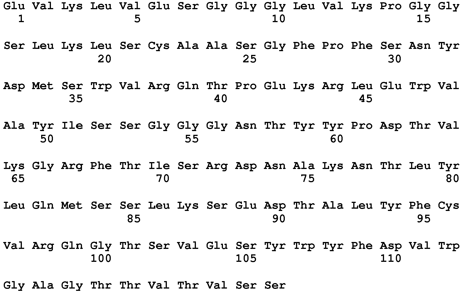

- <210> 8
<211> 112
<212> PRT
<213> Artificial Sequence - <220>
<223> clone MI407_Variable light chain - <400> 8

- <210> 9
<211> 5
<212> PRT
<213> Artificial Sequence - <220>
<223> clone MI323_Variable heavy chain CDR1 - <400> 9

- <210> 10
<211> 17
<212> PRT
<213> Artificial Sequence - <220>
<223> clone MI323_Variable heavy chain CDR2 - <400> 10

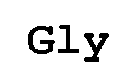
- <210> 11
<211> 10
<212> PRT
<213> Artificial Sequence - <220>
<223> clone MI323_Variable heavy chain CDR3 - <400> 11

- <210> 12
<211> 11
<212> PRT
<213> Artificial Sequence - <220>
<223> clone MI323_Variable light chain CDR1 - <400> 12

- <210> 13
<211> 7
<212> PRT
<213> Artificial Sequence - <220>
<223> clone MI323_Variable light chain CDR2 - <400> 13

- <210> 14
<211> 9
<212> PRT
<213> Artificial Sequence - <220>
<223> clone MI323_Variable light chain CDR3 - <400> 14

- <210> 15
<211> 5
<212> PRT
<213> Artificial Sequence - <220>
<223> clone MI329_Variable heavy chain CDR1 - <400> 15

- <210> 16
<211> 17
<212> PRT
<213> Artificial Sequence - <220>
<223> clone MI329_Variable heavy chain CDR2 - <400> 16

- <210> 17
<211> 9
<212> PRT
<213> Artificial Sequence - <220>
<223> clone MI329_Variable heavy chain CDR3 - <400> 17

- <210> 18
<211> 16
<212> PRT
<213> Artificial Sequence - <220>
<223> clone MI329_Variable light chain CDR1 - <400> 18

- <210> 19
<211> 7
<212> PRT
<213> Artificial Sequence - <220>
<223> clone MI329_Variable light chain CDR2 - <400> 19

- <210> 20
<211> 9
<212> PRT
<213> Artificial Sequence - <220>
<223> clone MI329_Variable light chain CDR3 - <400> 20

- <210> 21
<211> 5
<212> PRT
<213> Artificial Sequence - <220>
<223> clone MI403_Variable heavy chain CDR1 - <400> 21

- <210> 22
<211> 17
<212> PRT
<213> Artificial Sequence - <220>
<223> clone MI403_Variable heavy chain CDR2 - <400> 22

- <210> 23
<211> 13
<212> PRT
<213> Artificial Sequence - <220>
<223> clone MI403_Variable heavy chain CDR3 - <400> 23

- <210> 24
<211> 15
<212> PRT
<213> Artificial Sequence - <220>
<223> clone MI403_Variable light chain CDR1 - <400> 24

- <210> 25
<211> 7
<212> PRT
<213> Artificial Sequence - <220>
<223> clone MI403_Variable light chain CDR2 - <400> 25

- <210> 26
<211> 9
<212> PRT
<213> Artificial Sequence - <220>
<223> clone MI403_Variable light chain CDR3 - <400> 26

- <210> 27
<211> 5
<212> PRT
<213> Artificial Sequence - <220>
<223> clone MI407_Variable heavy chain CDR1 - <400> 27

- <210> 28
<211> 17
<212> PRT
<213> Artificial Sequence - <220>
<223> clone MI407_Variable heavy chain CDR2 - <400> 28

- <210> 29
<211> 13
<212> PRT
<213> Artificial Sequence - <220>
<223> clone MI407_Variable heavy chain CDR3 - <400> 29

- <210> 30
<211> 11
<212> PRT
<213> Artificial Sequence - <220>
<223> clone MI407_Variable light chain CDR1 - <400> 30

- <210> 31
<211> 87
<212> DNA
<213> Artificial Sequence - <220>
<223> MI323VH-F - <400> 31

- <210> 32
<211> 87
<212> DNA
<213> Artificial Sequence - <220>
<223> MI329VH-F - <400> 32

- <210> 33
<211> 87
<212> DNA
<213> Artificial Sequence - <220>
<223> MI403VH-F - <400> 33

- <210> 34
<211> 87
<212> DNA
<213> Artificial Sequence - <220>
<223> MI407VH-F - <400> 34

- <210> 35
<211> 20
<212> DNA
<213> Artificial Sequence - <220>
<223> VHApaI-R - <400> 35
accgatgggc ccttggtgga 20 - <210> 36
<211> 96
<212> DNA
<213> Artificial Sequence - <220>
<223> MI323VL-F - <400> 36

- <210> 37
<211> 45
<212> DNA
<213> Artificial Sequence - <220>
<223> MI323VLCL-R - <400> 37
acactaggag cggccacggt tcgtttgatt tccagtttgg tccct 45 - <210> 38
<211> 96
<212> DNA
<213> Artificial Sequence - <220>
<223> MI329VL-F - <400> 38

- <210> 39
<211> 42
<212> DNA
<213> Artificial Sequence - <220>
<223> MI329VLCL-R - <400> 39
acactaggag cggccacggt tcgtttcagc tccagcttgg tc 42 - <210> 40
<211> 96
<212> DNA
<213> Artificial Sequence - <220>
<223> MI403VL-F - <400> 40

- <210> 41
<211> 47
<212> DNA
<213> Artificial Sequence - <220>
<223> MI403VLCL-R - <400> 41
acactaggag cggccacggt tcgttttatt tccaactttg tccccga 47 - <210> 42
<211> 96
<212> DNA
<213> Artificial Sequence - <220>
<223> M407VL-F - <400> 42

- <210> 43
<211> 46
<212> DNA
<213> Artificial Sequence - <220>
<223> M407VLCL-R - <400> 43
acactaggag cggccacggt tcgttttatt tccaactctg tccccg 46 - <210> 44
<211> 20
<212> DNA
<213> Artificial Sequence - <220>
<223> Ck-F - <400> 44
accgtggccg ctcctagtgt 20 - <210> 45
<211> 29
<212> DNA
<213> Artificial Sequence - <220>
<223> CkSHB-R - <400> 45
nnnnggatcc aagcttacta gcactcccc 29 - <210> 46
<211> 116
<212> PRT
<213> Artificial Sequence - <220>
<223> clone MS501_Variable heavy chain - <400> 46

- <210> 47
<211> 111
<212> PRT
<213> Artificial Sequence - <220>
<223> clone MS501_Variable light chain - <400> 47


- <210> 48
<211> 116
<212> PRT
<213> Artificial Sequence - <220>
<223> clone MS502_Variable heavy chain - <400> 48

- <210> 49
<211> 111
<212> PRT
<213> Artificial Sequence - <220>
<223> clone MS502_Variable light chain - <400> 49


- <210> 50
<211> 111
<212> PRT
<213> Artificial Sequence - <220>
<223> clone MS502-1_Variable light chain - <400> 50

- <210> 51
<211> 116
<212> PRT
<213> Artificial Sequence - <220>
<223> clone MS503_Variable heavy chain - <400> 51


- <210> 52
<211> 111
<212> PRT
<213> Artificial Sequence - <220>
<223> clone MS503_Variable light chain - <400> 52

- <210> 53
<211> 116
<212> PRT
<213> Artificial Sequence - <220>
<223> clone MS504_Variable heavy chain - <400> 53

- <210> 54
<211> 111
<212> PRT
<213> Artificial Sequence - <220>
<223> clone MS504_Variable light chain - <400> 54

- <210> 55
<211> 116
<212> PRT
<213> Artificial Sequence - <220>
<223> clone MS505_Variable heavy chain - <400> 55

- <210> 56
<211> 111
<212> PRT
<213> Artificial Sequence - <220>
<223> clone MS505_Variable light chain - <400> 56

- <210> 57
<211> 116
<212> PRT
<213> Artificial Sequence - <220>
<223> clone MS506_Variable heavy chain - <400> 57

- <210> 58
<211> 111
<212> PRT
<213> Artificial Sequence - <220>
<223> clone MS506_Variable light chain - <400> 58


- <210> 59
<211> 5
<212> PRT
<213> Artificial Sequence - <220>
<223> clone MS501_Variable heavy chain CDR1 - <400> 59

- <210> 60
<211> 17
<212> PRT
<213> Artificial Sequence - <220>
<223> clone MS501_Variable heavy chain CDR2 - <400> 60

- <210> 61
<211> 7
<212> PRT
<213> Artificial Sequence - <220>
<223> clone MS501 Variable heavy chain CDR3 - <400> 61

- <210> 62
<211> 13
<212> PRT
<213> Artificial Sequence - <220>
<223> clone MS501_Variable light chain CDR1 - <400> 62

- <210> 63
<211> 7
<212> PRT
<213> Artificial Sequence - <220>
<223> clone MS501_Variable light chain CDR2 - <400> 63

- <210> 64
<211> 11
<212> PRT
<213> Artificial Sequence - <220>
<223> clone MS501_Variable light chain CDR3 - <400> 64

- <210> 65
<211> 17
<212> PRT
<213> Artificial Sequence - <220>
<223> clone MS502_Variable heavy chain CDR2 - <400> 65

- <210> 66
<211> 7
<212> PRT
<213> Artificial Sequence - <220>
<223> clone MS502 Variable heavy chain CDR3 - <400> 66

- <210> 67
<211> 13
<212> PRT
<213> Artificial Sequence - <220>
<223> clone MS502_Variable light chain CDR1 - <400> 67

- <210> 68
<211> 7
<212> PRT
<213> Artificial Sequence - <220>
<223> clone MS502_Variable light chain CDR2 - <400> 68

- <210> 69
<211> 11
<212> PRT
<213> Artificial Sequence - <220>
<223> clone MS502_Variable light chain CDR3 - <400> 69

- <210> 70
<211> 13
<212> PRT
<213> Artificial Sequence - <220>
<223> clone MS502-1_Variable light chain CDR1 - <400> 70

- <210> 71
<211> 17
<212> PRT
<213> Artificial Sequence - <220>
<223> clone MS503_Variable heavy chain CDR2 - <400> 71

- <210> 72
<211> 7
<212> PRT
<213> Artificial Sequence - <220>
<223> clone MS503_Variable heavy chain CDR3 - <400> 72

- <210> 73
<211> 7
<212> PRT
<213> Artificial Sequence - <220>
<223> clone MS503_Variable light chain CDR2 - <400> 73

- <210> 74
<211> 11
<212> PRT
<213> Artificial Sequence - <220>
<223> clone MS503_Variable light chain CDR3 - <400> 74

- <210> 75
<211> 17
<212> PRT
<213> Artificial Sequence - <220>
<223> clone MS504_Variable heavy chain CDR2 - <400> 75

- <210> 76
<211> 7
<212> PRT
<213> Artificial Sequence - <220>
<223> clone MS504_Variable heavy chain CDR3 - <400> 76

- <210> 77
<211> 13
<212> PRT
<213> Artificial Sequence - <220>
<223> clone MS504_Variable light chain CDR1 - <400> 77

- <210> 78
<211> 7
<212> PRT
<213> Artificial Sequence - <220>
<223> clone MS504_Variable light chain CDR2 - <400> 78

- <210> 79
<211> 11
<212> PRT
<213> Artificial Sequence - <220>
<223> clone MS504_Variable light chain CDR3 - <400> 79

- <210> 80
<211> 17
<212> PRT
<213> Artificial Sequence - <220>
<223> clone MS505_Variable heavy chain CDR2 - <400> 80

- <210> 81
<211> 7
<212> PRT
<213> Artificial Sequence - <220>
<223> clone MS505_Variable heavy chain CDR3 - <400> 81

- <210> 82
<211> 7
<212> PRT
<213> Artificial Sequence - <220>
<223> clone MS505_Variable light chain CDR2 - <400> 82

- <210> 83
<211> 11
<212> PRT
<213> Artificial Sequence - <220>
<223> clone MS505_Variable light chain CDR3 - <400> 83

- <210> 84
<211> 17
<212> PRT
<213> Artificial Sequence - <220>
<223> clone MS506_Variable heavy chain CDR2 - <400> 84

- <210> 85
<211> 7
<212> PRT
<213> Artificial Sequence - <220>
<223> clone MS506_Variable heavy chain CDR3 - <400> 85

- <210> 86
<211> 13
<212> PRT
<213> Artificial Sequence - <220>
<223> clone MS506_Variable light chain CDR1 - <400> 86

- <210> 87
<211> 11
<212> PRT
<213> Artificial Sequence - <220>
<223> clone MS506_Variable light chain CDR3 - <400> 87

- <210> 88
<211> 94
<212> DNA
<213> Artificial Sequence - <220>
<223> NotI-Leader- VL-F - <400> 88

- <210> 89
<211> 39
<212> DNA
<213> Artificial Sequence - <220>
<223> VL-R - <400> 89
ggggttggcc ttgggctggc ctaggaccgt cagcttggt 39 - <210> 90
<211> 18
<212> DNA
<213> Artificial Sequence - <220>
<223> VL-CL-F - <400> 90
cagcccaagg ccaacccc 18 - <210> 91
<211> 42
<212> DNA
<213> Artificial Sequence - <220>
<223> HindIII-VL-R - <400> 91
nnnnggatcc aagcttacta acattctgta ggggccactg tc 42 - <210> 92
<211> 97
<212> DNA
<213> Artificial Sequence - <220>
<223> HD-Heavy-F - <400> 92

- <210> 93
<211> 52
<212> DNA
<213> Artificial Sequence - <220>
<223> HD-Heavy-R - <400> 93
gggggaagac cgatgggccc ttggtggagg ctgagctcac ggtgaccagt gt 52 - <210> 94
<211> 79
<212> DNA
<213> Artificial Sequence - <220>
<223> MS502 VL FR1 Fo NruI - <400> 94

- <210> 95
<211> 27
<212> DNA
<213> Artificial Sequence - <220>
<223> MS502 VL FR1 Fo - <400> 95
cagtctgtgc tgactcagcc accctca 27 - <210> 96
<211> 60
<212> DNA
<213> Artificial Sequence - <220>
<223> MS502 VL FR1 Re - <400> 96

- <210> 97
<211> 30
<212> DNA
<213> Artificial Sequence - <220>
<223> MS502 VL FR2 Fo - <400> 97
gccaatatta gatgaagagc cagtacaaga 30 - <210> 98
<211> 93
<212> DNA
<213> Artificial Sequence - <220>
<223> MS502 VL FR2 Re - <400> 98

- <210> 99
<211> 48
<212> DNA
<213> Artificial Sequence - <220>
<223> MS502 CL Fo - <400> 99
ggcaccaagg tgaccgtcct aggtcagccc aaggccaacc ccactgtc 48 - <210> 100
<211> 35
<212> DNA
<213> Artificial Sequence - <220>
<223> MS502 CL Re - <400> 100
gctctagaac attctgtagg ggccactgtc ttctc 35 - <210> 101
<211> 34
<212> DNA
<213> Artificial Sequence - <220>
<223> MS502 VH FR1 Fo NcoI - <400> 101
gcccatggcc gaggtgcagc tgttggagtc tggg 34 - <210> 102
<211> 57
<212> DNA
<213> Artificial Sequence - <220>
<223> MS502 VH FR1 Re - <400> 102
agcctggcgg acccagctca tntgrtgrtg gctaaaggtg aatccagagg ccgcaca 57 - <210> 103
<211> 21
<212> DNA
<213> Artificial Sequence - <220>
<223> MS502 VH FR2 Fo - <400> 103
atgagctggg tccgccaggct 21 - <210> 104
<211> 72
<212> DNA
<213> Artificial Sequence - <220>
<223> MS502 VH FR2 Re - <400> 104

- <210> 105
<211> 36
<212> DNA
<213> Artificial Sequence - <220>
<223> MS502 VH FR3 Fo - <400> 105
aaatattacg ctgattctgt aagaggtcgg ttcacc 36 - <210> 106
<211> 78
<212> DNA
<213> Artificial Sequence - <220>
<223> MS502 VH FR3 Re - <400> 106

- <210> 107
<211> 27
<212> DNA
<213> Artificial Sequence - <220>
<223> MS502 VH Re - <400> 107
tgagctcacc gtgaccagtg taccctg 27 - <210> 108
<211> 46
<212> DNA
<213> Artificial Sequence - <220>
<223> MS502 VH Re ApaI - <400> 108
gcgggccctt ggtggaggct gagctcaccg tgaccagtgt accctg 46 - <210> 109
<211> 111
<212> PRT
<213> Artificial Sequence - <220>
<223> clone C2G1_Variable light chain - <400> 109

- <210> 110
<211> 111
<212> PRT
<213> Artificial Sequence - <220>
<223> clone C2G4_Variable light chain - <400> 110

- <210> 111
<211> 111
<212> PRT
<213> Artificial Sequence - <220>
<223> clone C3C8_Variable light chain - <400> 111

- <210> 112
<211> 116
<212> PRT
<213> Artificial Sequence - <220>
<223> clone 54_Variable heavy chain - <400> 112

- <210> 113
<211> 116
<212> PRT
<213> Artificial Sequence - <220>
<223> clone 56_Variable heavy chain - <400> 113


- <210> 114
<211> 116
<212> PRT
<213> Artificial Sequence - <220>
<223> clone 2-30_Variable heavy chain - <400> 114

- <210> 115
<211> 116
<212> PRT
<213> Artificial Sequence - <220>
<223> clone 2-73_Variable heavy chain - <400> 115


- <210> 116
<211> 116
<212> PRT
<213> Artificial Sequence - <220>
<223> clone 2-78_Variable heavy chain - <400> 116

- <210> 117
<211> 13
<212> PRT
<213> Artificial Sequence - <220>
<223> clone C2G1_Variable light chain CDR1 - <400> 117

- <210> 118
<211> 11
<212> PRT
<213> Artificial Sequence - <220>
<223> clone C2G1_Variable light chain CDR3 - <400> 118

- <210> 119
<211> 11
<212> PRT
<213> Artificial Sequence - <220>
<223> clone C2G4_Variable light chain CDR3 - <400> 119

- <210> 120
<211> 11
<212> PRT
<213> Artificial Sequence - <220>
<223> clone C3C8_Variable light chain CDR3 - <400> 120

- <210> 121
<211> 17
<212> PRT
<213> Artificial Sequence - <220>
<223> clone 54_Variable heavy chain CDR2 - <400> 121

- <210> 122
<211> 17
<212> PRT
<213> Artificial Sequence - <220>
<223> clone 56_Variable heavy chain CDR2 - <400> 122

- <210> 123
<211> 17
<212> PRT
<213> Artificial Sequence - <220>
<223> clone 2-30_Variable heavy chain CDR2 - <400> 123

- <210> 124
<211> 7
<212> PRT
<213> Artificial Sequence - <220>
<223> clone 2-30_Variable heavy chain CDR3 - <400> 124

- <210> 125
<211> 17
<212> PRT
<213> Artificial Sequence - <220>
<223> clone 2-73_Variable heavy chain CDR2 - <400> 125

- <210> 126
<211> 7
<212> PRT
<213> Artificial Sequence - <220>
<223> clone 2-78_Variable heavy chain CDR3 - <400> 126

- <210> 127
<211> 622
<212> PRT
<213> Artificial Sequence - <220>
<223> Mesothelin - <400> 127


Claims (6)
- An antibody binding to mesothelin comprising:a heavy chain variable region comprising an amino acid sequence of SEQ ID NO: 1 and a light chain variable region comprising an amino acid sequence of SEQ ID NO: 2; ora heavy chain variable region comprising an amino acid sequence of SEQ ID NO: 3 and a light chain variable region comprising an amino acid sequence of SEQ ID NO: 4; ora heavy chain variable region comprising an amino acid sequence of SEQ ID NO: 46 and a light chain variable region comprising an amino acid sequence of SEQ ID NO: 47; ora heavy chain variable region comprising an amino acid sequence of SEQ ID NO: 48 and a light chain variable region comprising an amino acid sequence of SEQ ID NO: 49; ora heavy chain variable region comprising an amino acid sequence of SEQ ID NO: 51 and a light chain variable region comprising an amino acid sequence of SEQ ID NO: 52; ora heavy chain variable region comprising an amino acid sequence of SEQ ID NO: 48 and a light chain variable region comprising an amino acid sequence of SEQ ID NO: 109; ora heavy chain variable region comprising an amino acid sequence of SEQ ID NO: 48 and a light chain variable region comprising an amino acid sequence of SEQ ID NO: 110; ora heavy chain variable region comprising an amino acid sequence of SEQ ID NO: 116 and a light chain variable region comprises an amino acid sequence of SEQ ID NO: 49; ora heavy chain variable region comprising an amino acid sequence of SEQ ID NO: 116 and a light chain variable region comprising an amino acid sequence of SEQ ID NO: 110.
- A nucleic acid encoding the antibody according to claim 1.
- A vector comprising the nucleic acid according to claim 2.
- A host cell comprising the vector according to claim 3.
- A method of preparing the antibody according to claim 1 comprising expressing the antibody by culturing host cell according to claim 4.
- A pharmaceutical composition for use in the treatment of cancer or a tumor comprising the antibody according to claim 1 as an active ingredient.
Applications Claiming Priority (2)
| Application Number | Priority Date | Filing Date | Title |
|---|---|---|---|
| KR1020150135755A KR101782487B1 (en) | 2015-09-24 | 2015-09-24 | Novel Antibody Binding to Mesothelin(MSLN), and Composition Comprising the Same |
| PCT/KR2016/010604 WO2017052241A1 (en) | 2015-09-24 | 2016-09-23 | Novel anti-mesothelin antibody and composition comprising the same |
Publications (3)
| Publication Number | Publication Date |
|---|---|
| EP3353213A1 EP3353213A1 (en) | 2018-08-01 |
| EP3353213A4 EP3353213A4 (en) | 2019-05-29 |
| EP3353213B1 true EP3353213B1 (en) | 2020-12-02 |
Family
ID=58386441
Family Applications (1)
| Application Number | Title | Priority Date | Filing Date |
|---|---|---|---|
| EP16848966.4A Active EP3353213B1 (en) | 2015-09-24 | 2016-09-23 | Anti-mesothelin antibody and composition comprising the same |
Country Status (12)
| Country | Link |
|---|---|
| US (1) | US10851175B2 (en) |
| EP (1) | EP3353213B1 (en) |
| JP (1) | JP6738893B2 (en) |
| KR (1) | KR101782487B1 (en) |
| CN (3) | CN108350084B (en) |
| AU (1) | AU2016328204B2 (en) |
| CA (1) | CA2999237C (en) |
| EA (1) | EA038654B1 (en) |
| ES (1) | ES2857500T3 (en) |
| IL (1) | IL258215B (en) |
| MX (1) | MX2018003470A (en) |
| WO (1) | WO2017052241A1 (en) |
Families Citing this family (29)
| Publication number | Priority date | Publication date | Assignee | Title |
|---|---|---|---|---|
| EP3858858A1 (en) | 2016-06-20 | 2021-08-04 | F-Star Delta Limited | Binding molecules binding pd-l1 and lag-3 |
| BR112018076525A2 (en) | 2016-06-20 | 2019-04-02 | F-Star Beta Limited | lag-3 binding members |
| GB201612520D0 (en) | 2016-07-19 | 2016-08-31 | F-Star Beta Ltd | Binding molecules |
| WO2019005208A1 (en) * | 2017-06-30 | 2019-01-03 | The United States Of America, As Represented By The Secretary, Department Of Health And Human Services | Human mesothelin antibodies and uses in cancer therapy |
| KR101966362B1 (en) | 2017-10-20 | 2019-04-05 | 주식회사 녹십자 | Antibody against msln and pharmaceutical composition for treating cancer comprising the same |
| JP7489316B2 (en) | 2017-12-19 | 2024-05-23 | エフ-スター セラピューティクス リミテッド | FC-binding fragment having PD-LI antigen-binding site |
| GB201811415D0 (en) * | 2018-07-12 | 2018-08-29 | F Star Beta Ltd | Anti-Mesothelin Anti bodies |
| GB201811403D0 (en) | 2018-07-12 | 2018-08-29 | F Star Beta Ltd | Antibody molecules |
| GB201811450D0 (en) | 2018-07-12 | 2018-08-29 | F Star Delta Ltd | Mesothelin and CD137 binding molecules |
| PL3820569T3 (en) | 2018-07-12 | 2025-07-14 | Invox Pharma Limited | Antibody molecules that bind pd-l1 and cd137 |
| GB201811408D0 (en) | 2018-07-12 | 2018-08-29 | F Star Beta Ltd | CD137 Binding Molecules |
| GB201811410D0 (en) | 2018-07-12 | 2018-08-29 | F Star Beta Ltd | OX40 Binding molecules |
| AU2019301206B2 (en) | 2018-07-12 | 2025-10-30 | Invox Pharma Limited | Antibody molecules that bind CD137 and OX40 |
| GB201811404D0 (en) | 2018-07-12 | 2018-08-29 | F Star Beta Ltd | Anti-CD137 Antibodies |
| KR102070016B1 (en) * | 2019-01-21 | 2020-01-29 | (주)녹십자셀 | Mesothelin-specific chimeric antigen receptor and T lymphocyte expressing this chimeric antigen receptor |
| CA3134680C (en) | 2019-03-29 | 2024-02-06 | Green Cross Corporation | Fusion protein comprising anti-mesothelin antibody, anti-cd3 antibody or anti-egfr antibody, bispecific or trispecific antibody comprising same, and uses thereof |
| KR102216225B1 (en) * | 2019-06-27 | 2021-02-17 | (주)녹십자셀 | Mesothelin-specific chimeric antigen receptor and T lymphocyte expressing this chimeric antigen receptor |
| CA3133678C (en) | 2020-08-04 | 2023-04-11 | Cellengene Inc. | Anti-mesothelin chimeric antigen receptor specifically binding to mesothelin |
| WO2022030730A1 (en) * | 2020-08-04 | 2022-02-10 | 주식회사 셀렌진 | Anti-mesothelin chimeric antigen receptor specifically binding to mesothelin |
| WO2022250450A1 (en) * | 2021-05-26 | 2022-12-01 | 주식회사 셀렌진 | Chimeric antigen receptor comprising anti-mesothelin scfv, and use thereof |
| KR102860436B1 (en) * | 2021-06-30 | 2025-09-19 | (주)이노베이션바이오 | Antibody specific for mesothelin and uses thereof |
| CN113788896A (en) * | 2021-08-25 | 2021-12-14 | 南京蓝盾生物科技有限公司 | Anti-MSLN monoclonal internalizing antibody and preparation method and application thereof |
| TW202321296A (en) | 2021-10-06 | 2023-06-01 | 美商鏈接免疫療法公司 | Anti-mesothelin antigen-binding molecules and uses thereof |
| WO2023115528A1 (en) * | 2021-12-24 | 2023-06-29 | Zhejiang Shimai Pharmaceutical Co., Ltd. | Antibodies against mesothelin and uses thereof |
| KR20240043161A (en) * | 2022-09-23 | 2024-04-03 | 국민대학교산학협력단 | Antibody drug conjugate comprising GRP specific antibody or antigen-binding fragment thereof and drug |
| WO2024102954A1 (en) | 2022-11-10 | 2024-05-16 | Massachusetts Institute Of Technology | Activation induced clipping system (aics) |
| KR20250083161A (en) * | 2023-11-27 | 2025-06-09 | 건국대학교 글로컬산학협력단 | Novel anti-methothelin antibody and uses thereof |
| KR20250103909A (en) | 2023-12-28 | 2025-07-08 | 서울대학교산학협력단 | Antibodies specific for Mesothelin and uses thereof |
| WO2025235862A1 (en) | 2024-05-10 | 2025-11-13 | Inndura Therapeutics Inc. | A modified immune cell receptor comprising a target-binding domain and the extracellular domain of cd16a |
Family Cites Families (9)
| Publication number | Priority date | Publication date | Assignee | Title |
|---|---|---|---|---|
| EP2322560B1 (en) * | 2005-03-10 | 2013-09-04 | Morphotek, Inc. | Anti-mesothelin antibodies |
| SI2195017T1 (en) * | 2007-10-01 | 2015-01-30 | Bristol-Myers Squibb Company | Human antibodies that bind mesothelin, and uses thereof |
| WO2009054957A2 (en) | 2007-10-22 | 2009-04-30 | Cfs Research Llc | Methods for diagnosis and treatment of chronic fatigue syndrome |
| HUE027358T2 (en) * | 2007-11-26 | 2016-09-28 | Bayer Ip Gmbh | Anti-mesothelin antibodies and uses therefor |
| AU2009228361A1 (en) * | 2008-03-27 | 2009-10-01 | The Government Of The United States Of America As Represented By The Secretary Of The Department Of Health And Human Services | Human anti-mesothelin monoclonal antibodies |
| PE20170917A1 (en) * | 2010-12-20 | 2017-07-12 | Genentech Inc | ANTI-MESOTHELIN ANTIBODIES AND IMMUNOCONJUGATES |
| WO2014004549A2 (en) * | 2012-06-27 | 2014-01-03 | Amgen Inc. | Anti-mesothelin binding proteins |
| WO2014031476A1 (en) * | 2012-08-21 | 2014-02-27 | The United States Of America, As Represented By The Secretary, Department Of Health And Human Services | Mesothelin domain-specific monoclonal antibodies and use thereof |
| KR20150029457A (en) * | 2013-09-10 | 2015-03-18 | 삼성전자주식회사 | Polypeptide binding to Annexin A1 and use thereof |
-
2015
- 2015-09-24 KR KR1020150135755A patent/KR101782487B1/en active Active
-
2016
- 2016-09-23 ES ES16848966T patent/ES2857500T3/en active Active
- 2016-09-23 IL IL258215A patent/IL258215B/en unknown
- 2016-09-23 JP JP2018515766A patent/JP6738893B2/en active Active
- 2016-09-23 AU AU2016328204A patent/AU2016328204B2/en active Active
- 2016-09-23 CN CN201680062531.0A patent/CN108350084B/en active Active
- 2016-09-23 CN CN202110751129.6A patent/CN113603785B/en active Active
- 2016-09-23 CN CN202311837858.9A patent/CN118085094A/en active Pending
- 2016-09-23 EA EA201890796A patent/EA038654B1/en unknown
- 2016-09-23 EP EP16848966.4A patent/EP3353213B1/en active Active
- 2016-09-23 CA CA2999237A patent/CA2999237C/en active Active
- 2016-09-23 MX MX2018003470A patent/MX2018003470A/en unknown
- 2016-09-23 WO PCT/KR2016/010604 patent/WO2017052241A1/en not_active Ceased
- 2016-09-23 US US15/762,885 patent/US10851175B2/en active Active
Non-Patent Citations (1)
| Title |
|---|
| None * |
Also Published As
| Publication number | Publication date |
|---|---|
| EP3353213A4 (en) | 2019-05-29 |
| US10851175B2 (en) | 2020-12-01 |
| AU2016328204A1 (en) | 2018-04-19 |
| AU2016328204B2 (en) | 2019-03-21 |
| MX2018003470A (en) | 2019-10-30 |
| CA2999237A1 (en) | 2017-03-30 |
| CN113603785B (en) | 2024-01-05 |
| KR20170036503A (en) | 2017-04-03 |
| CN113603785A (en) | 2021-11-05 |
| JP6738893B2 (en) | 2020-08-12 |
| ES2857500T3 (en) | 2021-09-29 |
| EP3353213A1 (en) | 2018-08-01 |
| US20180346588A1 (en) | 2018-12-06 |
| JP2018532401A (en) | 2018-11-08 |
| CN108350084B (en) | 2021-09-10 |
| CN118085094A (en) | 2024-05-28 |
| CN108350084A (en) | 2018-07-31 |
| IL258215B (en) | 2022-07-01 |
| WO2017052241A1 (en) | 2017-03-30 |
| BR112018005837A2 (en) | 2018-10-09 |
| IL258215A (en) | 2018-05-31 |
| CA2999237C (en) | 2021-07-06 |
| EA201890796A1 (en) | 2018-10-31 |
| EA038654B1 (en) | 2021-09-29 |
| KR101782487B1 (en) | 2017-09-27 |
Similar Documents
| Publication | Publication Date | Title |
|---|---|---|
| US10851175B2 (en) | Anti-mesothelin antibody and composition comprising the same | |
| CN113480656B (en) | anti-ROR 1 antibodies | |
| US20160194402A1 (en) | Cd70-binding peptides and method, process and use relating thereto | |
| JP2016116522A (en) | Methods for making heteromultimeric molecules | |
| JP7828286B2 (en) | Anti-MUC1-SEA antibody | |
| US10836833B2 (en) | Cell engaging binding molecules | |
| WO2022187275A1 (en) | Antibodies against claudin-6 and uses thereof | |
| WO2022222992A1 (en) | Antibodies binding trop2 and uses thereof | |
| WO2023109888A1 (en) | Anti-ang2-vegf bispecific antibody and use thereof | |
| US20240228658A1 (en) | Anti-5t4 antibodies and uses thereof | |
| HK40105696A (en) | Novel mesothelin antibody and composition comprising the same | |
| HK40054516A (en) | Novel mesothelin antibody and composition comprising the same | |
| HK40054516B (en) | Novel mesothelin antibody and composition comprising the same | |
| WO2025228351A1 (en) | Antibody, antibody-drug conjugate, and use | |
| HK40061499A (en) | Anti-ror1 antibodies | |
| CN117597362A (en) | Antibodies against Claudin-6 and their uses | |
| CN121591904A (en) | Bispecific antibodies targeting Trop2 and HER2, their preparation methods and applications | |
| CN120813606A (en) | Anti-CLDN 6 antibodies and methods of use | |
| CN121057749A (en) | Anti-CD3 multispecific antibodies and their usage | |
| HK40107987A (en) | Antibodies against claudin-6 and uses thereof | |
| BR112018005837B1 (en) | ANTI-MESOTHELIN ANTIBODY, NUCLEIC ACID, VECTOR, HOST CELL, METHOD OF ANTIBODY PREPARATION AND PHARMACEUTICAL COMPOSITION |
Legal Events
| Date | Code | Title | Description |
|---|---|---|---|
| STAA | Information on the status of an ep patent application or granted ep patent |
Free format text: STATUS: THE INTERNATIONAL PUBLICATION HAS BEEN MADE |
|
| PUAI | Public reference made under article 153(3) epc to a published international application that has entered the european phase |
Free format text: ORIGINAL CODE: 0009012 |
|
| STAA | Information on the status of an ep patent application or granted ep patent |
Free format text: STATUS: REQUEST FOR EXAMINATION WAS MADE |
|
| 17P | Request for examination filed |
Effective date: 20180423 |
|
| AK | Designated contracting states |
Kind code of ref document: A1 Designated state(s): AL AT BE BG CH CY CZ DE DK EE ES FI FR GB GR HR HU IE IS IT LI LT LU LV MC MK MT NL NO PL PT RO RS SE SI SK SM TR |
|
| AX | Request for extension of the european patent |
Extension state: BA ME |
|
| DAV | Request for validation of the european patent (deleted) | ||
| DAX | Request for extension of the european patent (deleted) | ||
| A4 | Supplementary search report drawn up and despatched |
Effective date: 20190429 |
|
| RIC1 | Information provided on ipc code assigned before grant |
Ipc: A61K 39/395 20060101ALI20190423BHEP Ipc: C07K 16/30 20060101AFI20190423BHEP Ipc: C07K 16/28 20060101ALI20190423BHEP |
|
| GRAP | Despatch of communication of intention to grant a patent |
Free format text: ORIGINAL CODE: EPIDOSNIGR1 |
|
| STAA | Information on the status of an ep patent application or granted ep patent |
Free format text: STATUS: GRANT OF PATENT IS INTENDED |
|
| RIC1 | Information provided on ipc code assigned before grant |
Ipc: A61K 39/395 20060101ALI20200702BHEP Ipc: C07K 16/30 20060101AFI20200702BHEP Ipc: C07K 16/28 20060101ALI20200702BHEP |
|
| INTG | Intention to grant announced |
Effective date: 20200723 |
|
| GRAS | Grant fee paid |
Free format text: ORIGINAL CODE: EPIDOSNIGR3 |
|
| GRAA | (expected) grant |
Free format text: ORIGINAL CODE: 0009210 |
|
| STAA | Information on the status of an ep patent application or granted ep patent |
Free format text: STATUS: THE PATENT HAS BEEN GRANTED |
|
| AK | Designated contracting states |
Kind code of ref document: B1 Designated state(s): AL AT BE BG CH CY CZ DE DK EE ES FI FR GB GR HR HU IE IS IT LI LT LU LV MC MK MT NL NO PL PT RO RS SE SI SK SM TR |
|
| REG | Reference to a national code |
Ref country code: GB Ref legal event code: FG4D |
|
| REG | Reference to a national code |
Ref country code: AT Ref legal event code: REF Ref document number: 1340842 Country of ref document: AT Kind code of ref document: T Effective date: 20201215 Ref country code: CH Ref legal event code: EP |
|
| REG | Reference to a national code |
Ref country code: IE Ref legal event code: FG4D |
|
| REG | Reference to a national code |
Ref country code: DE Ref legal event code: R096 Ref document number: 602016049192 Country of ref document: DE |
|
| REG | Reference to a national code |
Ref country code: CH Ref legal event code: NV Representative=s name: PATENTANWAELTE SCHAAD, BALASS, MENZL AND PARTN, CH |
|
| REG | Reference to a national code |
Ref country code: CH Ref legal event code: PCAR Free format text: NEW ADDRESS: DUFOURSTRASSE 101, 8034 ZUERICH (CH) |
|
| REG | Reference to a national code |
Ref country code: NL Ref legal event code: FP |
|
| PG25 | Lapsed in a contracting state [announced via postgrant information from national office to epo] |
Ref country code: GR Free format text: LAPSE BECAUSE OF FAILURE TO SUBMIT A TRANSLATION OF THE DESCRIPTION OR TO PAY THE FEE WITHIN THE PRESCRIBED TIME-LIMIT Effective date: 20210303 Ref country code: FI Free format text: LAPSE BECAUSE OF FAILURE TO SUBMIT A TRANSLATION OF THE DESCRIPTION OR TO PAY THE FEE WITHIN THE PRESCRIBED TIME-LIMIT Effective date: 20201202 Ref country code: NO Free format text: LAPSE BECAUSE OF FAILURE TO SUBMIT A TRANSLATION OF THE DESCRIPTION OR TO PAY THE FEE WITHIN THE PRESCRIBED TIME-LIMIT Effective date: 20210302 Ref country code: RS Free format text: LAPSE BECAUSE OF FAILURE TO SUBMIT A TRANSLATION OF THE DESCRIPTION OR TO PAY THE FEE WITHIN THE PRESCRIBED TIME-LIMIT Effective date: 20201202 |
|
| REG | Reference to a national code |
Ref country code: AT Ref legal event code: MK05 Ref document number: 1340842 Country of ref document: AT Kind code of ref document: T Effective date: 20201202 |
|
| PG25 | Lapsed in a contracting state [announced via postgrant information from national office to epo] |
Ref country code: SE Free format text: LAPSE BECAUSE OF FAILURE TO SUBMIT A TRANSLATION OF THE DESCRIPTION OR TO PAY THE FEE WITHIN THE PRESCRIBED TIME-LIMIT Effective date: 20201202 Ref country code: BG Free format text: LAPSE BECAUSE OF FAILURE TO SUBMIT A TRANSLATION OF THE DESCRIPTION OR TO PAY THE FEE WITHIN THE PRESCRIBED TIME-LIMIT Effective date: 20210302 Ref country code: LV Free format text: LAPSE BECAUSE OF FAILURE TO SUBMIT A TRANSLATION OF THE DESCRIPTION OR TO PAY THE FEE WITHIN THE PRESCRIBED TIME-LIMIT Effective date: 20201202 Ref country code: PL Free format text: LAPSE BECAUSE OF FAILURE TO SUBMIT A TRANSLATION OF THE DESCRIPTION OR TO PAY THE FEE WITHIN THE PRESCRIBED TIME-LIMIT Effective date: 20201202 |
|
| PG25 | Lapsed in a contracting state [announced via postgrant information from national office to epo] |
Ref country code: HR Free format text: LAPSE BECAUSE OF FAILURE TO SUBMIT A TRANSLATION OF THE DESCRIPTION OR TO PAY THE FEE WITHIN THE PRESCRIBED TIME-LIMIT Effective date: 20201202 |
|
| REG | Reference to a national code |
Ref country code: LT Ref legal event code: MG9D |
|
| PG25 | Lapsed in a contracting state [announced via postgrant information from national office to epo] |
Ref country code: PT Free format text: LAPSE BECAUSE OF FAILURE TO SUBMIT A TRANSLATION OF THE DESCRIPTION OR TO PAY THE FEE WITHIN THE PRESCRIBED TIME-LIMIT Effective date: 20210405 Ref country code: RO Free format text: LAPSE BECAUSE OF FAILURE TO SUBMIT A TRANSLATION OF THE DESCRIPTION OR TO PAY THE FEE WITHIN THE PRESCRIBED TIME-LIMIT Effective date: 20201202 Ref country code: SK Free format text: LAPSE BECAUSE OF FAILURE TO SUBMIT A TRANSLATION OF THE DESCRIPTION OR TO PAY THE FEE WITHIN THE PRESCRIBED TIME-LIMIT Effective date: 20201202 Ref country code: EE Free format text: LAPSE BECAUSE OF FAILURE TO SUBMIT A TRANSLATION OF THE DESCRIPTION OR TO PAY THE FEE WITHIN THE PRESCRIBED TIME-LIMIT Effective date: 20201202 Ref country code: CZ Free format text: LAPSE BECAUSE OF FAILURE TO SUBMIT A TRANSLATION OF THE DESCRIPTION OR TO PAY THE FEE WITHIN THE PRESCRIBED TIME-LIMIT Effective date: 20201202 Ref country code: SM Free format text: LAPSE BECAUSE OF FAILURE TO SUBMIT A TRANSLATION OF THE DESCRIPTION OR TO PAY THE FEE WITHIN THE PRESCRIBED TIME-LIMIT Effective date: 20201202 Ref country code: LT Free format text: LAPSE BECAUSE OF FAILURE TO SUBMIT A TRANSLATION OF THE DESCRIPTION OR TO PAY THE FEE WITHIN THE PRESCRIBED TIME-LIMIT Effective date: 20201202 |
|
| PG25 | Lapsed in a contracting state [announced via postgrant information from national office to epo] |
Ref country code: AT Free format text: LAPSE BECAUSE OF FAILURE TO SUBMIT A TRANSLATION OF THE DESCRIPTION OR TO PAY THE FEE WITHIN THE PRESCRIBED TIME-LIMIT Effective date: 20201202 |
|
| REG | Reference to a national code |
Ref country code: DE Ref legal event code: R097 Ref document number: 602016049192 Country of ref document: DE |
|
| REG | Reference to a national code |
Ref country code: ES Ref legal event code: FG2A Ref document number: 2857500 Country of ref document: ES Kind code of ref document: T3 Effective date: 20210929 |
|
| PG25 | Lapsed in a contracting state [announced via postgrant information from national office to epo] |
Ref country code: IS Free format text: LAPSE BECAUSE OF FAILURE TO SUBMIT A TRANSLATION OF THE DESCRIPTION OR TO PAY THE FEE WITHIN THE PRESCRIBED TIME-LIMIT Effective date: 20210402 |
|
| PLBE | No opposition filed within time limit |
Free format text: ORIGINAL CODE: 0009261 |
|
| STAA | Information on the status of an ep patent application or granted ep patent |
Free format text: STATUS: NO OPPOSITION FILED WITHIN TIME LIMIT |
|
| PG25 | Lapsed in a contracting state [announced via postgrant information from national office to epo] |
Ref country code: AL Free format text: LAPSE BECAUSE OF FAILURE TO SUBMIT A TRANSLATION OF THE DESCRIPTION OR TO PAY THE FEE WITHIN THE PRESCRIBED TIME-LIMIT Effective date: 20201202 |
|
| 26N | No opposition filed |
Effective date: 20210903 |
|
| PG25 | Lapsed in a contracting state [announced via postgrant information from national office to epo] |
Ref country code: DK Free format text: LAPSE BECAUSE OF FAILURE TO SUBMIT A TRANSLATION OF THE DESCRIPTION OR TO PAY THE FEE WITHIN THE PRESCRIBED TIME-LIMIT Effective date: 20201202 Ref country code: SI Free format text: LAPSE BECAUSE OF FAILURE TO SUBMIT A TRANSLATION OF THE DESCRIPTION OR TO PAY THE FEE WITHIN THE PRESCRIBED TIME-LIMIT Effective date: 20201202 |
|
| REG | Reference to a national code |
Ref country code: BE Ref legal event code: MM Effective date: 20210930 |
|
| PG25 | Lapsed in a contracting state [announced via postgrant information from national office to epo] |
Ref country code: IS Free format text: LAPSE BECAUSE OF FAILURE TO SUBMIT A TRANSLATION OF THE DESCRIPTION OR TO PAY THE FEE WITHIN THE PRESCRIBED TIME-LIMIT Effective date: 20210402 Ref country code: MC Free format text: LAPSE BECAUSE OF FAILURE TO SUBMIT A TRANSLATION OF THE DESCRIPTION OR TO PAY THE FEE WITHIN THE PRESCRIBED TIME-LIMIT Effective date: 20201202 |
|
| PG25 | Lapsed in a contracting state [announced via postgrant information from national office to epo] |
Ref country code: LU Free format text: LAPSE BECAUSE OF NON-PAYMENT OF DUE FEES Effective date: 20210923 Ref country code: IE Free format text: LAPSE BECAUSE OF NON-PAYMENT OF DUE FEES Effective date: 20210923 Ref country code: BE Free format text: LAPSE BECAUSE OF NON-PAYMENT OF DUE FEES Effective date: 20210930 |
|
| PG25 | Lapsed in a contracting state [announced via postgrant information from national office to epo] |
Ref country code: HU Free format text: LAPSE BECAUSE OF FAILURE TO SUBMIT A TRANSLATION OF THE DESCRIPTION OR TO PAY THE FEE WITHIN THE PRESCRIBED TIME-LIMIT; INVALID AB INITIO Effective date: 20160923 |
|
| P01 | Opt-out of the competence of the unified patent court (upc) registered |
Effective date: 20230523 |
|
| PG25 | Lapsed in a contracting state [announced via postgrant information from national office to epo] |
Ref country code: CY Free format text: LAPSE BECAUSE OF FAILURE TO SUBMIT A TRANSLATION OF THE DESCRIPTION OR TO PAY THE FEE WITHIN THE PRESCRIBED TIME-LIMIT Effective date: 20201202 |
|
| REG | Reference to a national code |
Ref country code: DE Ref legal event code: R081 Ref document number: 602016049192 Country of ref document: DE Owner name: MOGAM INSTITUTE FOR BIOMEDICAL RESEARCH, KR Free format text: FORMER OWNERS: GREEN CROSS CORPORATION, YONGIN-SI, GYEONGGI-DO, KR; MOGAM INSTITUTE FOR BIOMEDICAL RESEARCH, YONGIN-SI, GYEONGGI-DO, KR Ref country code: DE Ref legal event code: R081 Ref document number: 602016049192 Country of ref document: DE Owner name: GREEN CROSS CORPORATION, YONGIN-SI, KR Free format text: FORMER OWNERS: GREEN CROSS CORPORATION, YONGIN-SI, GYEONGGI-DO, KR; MOGAM INSTITUTE FOR BIOMEDICAL RESEARCH, YONGIN-SI, GYEONGGI-DO, KR |
|
| PG25 | Lapsed in a contracting state [announced via postgrant information from national office to epo] |
Ref country code: MK Free format text: LAPSE BECAUSE OF FAILURE TO SUBMIT A TRANSLATION OF THE DESCRIPTION OR TO PAY THE FEE WITHIN THE PRESCRIBED TIME-LIMIT Effective date: 20201202 |
|
| PG25 | Lapsed in a contracting state [announced via postgrant information from national office to epo] |
Ref country code: MT Free format text: LAPSE BECAUSE OF FAILURE TO SUBMIT A TRANSLATION OF THE DESCRIPTION OR TO PAY THE FEE WITHIN THE PRESCRIBED TIME-LIMIT Effective date: 20201202 |
|
| PGFP | Annual fee paid to national office [announced via postgrant information from national office to epo] |
Ref country code: NL Payment date: 20250609 Year of fee payment: 10 |
|
| PGFP | Annual fee paid to national office [announced via postgrant information from national office to epo] |
Ref country code: FR Payment date: 20250611 Year of fee payment: 10 |
|
| PGFP | Annual fee paid to national office [announced via postgrant information from national office to epo] |
Ref country code: TR Payment date: 20250611 Year of fee payment: 10 |
|
| REG | Reference to a national code |
Ref country code: CH Ref legal event code: U11 Free format text: ST27 STATUS EVENT CODE: U-0-0-U10-U11 (AS PROVIDED BY THE NATIONAL OFFICE) Effective date: 20251001 |
|
| PGFP | Annual fee paid to national office [announced via postgrant information from national office to epo] |
Ref country code: DE Payment date: 20250609 Year of fee payment: 10 |
|
| PGFP | Annual fee paid to national office [announced via postgrant information from national office to epo] |
Ref country code: IT Payment date: 20250610 Year of fee payment: 10 |
|
| PGFP | Annual fee paid to national office [announced via postgrant information from national office to epo] |
Ref country code: GB Payment date: 20250707 Year of fee payment: 10 |
|
| PGFP | Annual fee paid to national office [announced via postgrant information from national office to epo] |
Ref country code: CH Payment date: 20251001 Year of fee payment: 10 |
|
| PGFP | Annual fee paid to national office [announced via postgrant information from national office to epo] |
Ref country code: ES Payment date: 20251007 Year of fee payment: 10 |