EP3350596B1 - Method for determining kinetic profiles in drug discovery - Google Patents
Method for determining kinetic profiles in drug discovery Download PDFInfo
- Publication number
- EP3350596B1 EP3350596B1 EP16775105.6A EP16775105A EP3350596B1 EP 3350596 B1 EP3350596 B1 EP 3350596B1 EP 16775105 A EP16775105 A EP 16775105A EP 3350596 B1 EP3350596 B1 EP 3350596B1
- Authority
- EP
- European Patent Office
- Prior art keywords
- polyprotein
- target protein
- kinetic
- time
- compound
- Prior art date
- Legal status (The legal status is an assumption and is not a legal conclusion. Google has not performed a legal analysis and makes no representation as to the accuracy of the status listed.)
- Active
Links
Images


Classifications
-
- G—PHYSICS
- G01—MEASURING; TESTING
- G01N—INVESTIGATING OR ANALYSING MATERIALS BY DETERMINING THEIR CHEMICAL OR PHYSICAL PROPERTIES
- G01N33/00—Investigating or analysing materials by specific methods not covered by groups G01N1/00 - G01N31/00
- G01N33/48—Biological material, e.g. blood, urine; Haemocytometers
- G01N33/50—Chemical analysis of biological material, e.g. blood, urine; Testing involving biospecific ligand binding methods; Immunological testing
- G01N33/53—Immunoassay; Biospecific binding assay; Materials therefor
- G01N33/557—Immunoassay; Biospecific binding assay; Materials therefor using kinetic measurement, i.e. time rate of progress of an antigen-antibody interaction
-
- C—CHEMISTRY; METALLURGY
- C09—DYES; PAINTS; POLISHES; NATURAL RESINS; ADHESIVES; COMPOSITIONS NOT OTHERWISE PROVIDED FOR; APPLICATIONS OF MATERIALS NOT OTHERWISE PROVIDED FOR
- C09K—MATERIALS FOR MISCELLANEOUS APPLICATIONS, NOT PROVIDED FOR ELSEWHERE
- C09K11/00—Luminescent materials, e.g. electroluminescent or chemiluminescent
- C09K11/08—Luminescent materials, e.g. electroluminescent or chemiluminescent containing inorganic luminescent materials
- C09K11/77—Luminescent materials, e.g. electroluminescent or chemiluminescent containing inorganic luminescent materials containing rare earth metals
- C09K11/7728—Luminescent materials, e.g. electroluminescent or chemiluminescent containing inorganic luminescent materials containing rare earth metals containing europium
-
- C—CHEMISTRY; METALLURGY
- C09—DYES; PAINTS; POLISHES; NATURAL RESINS; ADHESIVES; COMPOSITIONS NOT OTHERWISE PROVIDED FOR; APPLICATIONS OF MATERIALS NOT OTHERWISE PROVIDED FOR
- C09K—MATERIALS FOR MISCELLANEOUS APPLICATIONS, NOT PROVIDED FOR ELSEWHERE
- C09K11/00—Luminescent materials, e.g. electroluminescent or chemiluminescent
- C09K11/08—Luminescent materials, e.g. electroluminescent or chemiluminescent containing inorganic luminescent materials
- C09K11/77—Luminescent materials, e.g. electroluminescent or chemiluminescent containing inorganic luminescent materials containing rare earth metals
- C09K11/7743—Luminescent materials, e.g. electroluminescent or chemiluminescent containing inorganic luminescent materials containing rare earth metals containing terbium
-
- C—CHEMISTRY; METALLURGY
- C09—DYES; PAINTS; POLISHES; NATURAL RESINS; ADHESIVES; COMPOSITIONS NOT OTHERWISE PROVIDED FOR; APPLICATIONS OF MATERIALS NOT OTHERWISE PROVIDED FOR
- C09K—MATERIALS FOR MISCELLANEOUS APPLICATIONS, NOT PROVIDED FOR ELSEWHERE
- C09K11/00—Luminescent materials, e.g. electroluminescent or chemiluminescent
- C09K11/08—Luminescent materials, e.g. electroluminescent or chemiluminescent containing inorganic luminescent materials
- C09K11/77—Luminescent materials, e.g. electroluminescent or chemiluminescent containing inorganic luminescent materials containing rare earth metals
- C09K11/7759—Luminescent materials, e.g. electroluminescent or chemiluminescent containing inorganic luminescent materials containing rare earth metals containing samarium
-
- G—PHYSICS
- G01—MEASURING; TESTING
- G01N—INVESTIGATING OR ANALYSING MATERIALS BY DETERMINING THEIR CHEMICAL OR PHYSICAL PROPERTIES
- G01N33/00—Investigating or analysing materials by specific methods not covered by groups G01N1/00 - G01N31/00
- G01N33/48—Biological material, e.g. blood, urine; Haemocytometers
- G01N33/50—Chemical analysis of biological material, e.g. blood, urine; Testing involving biospecific ligand binding methods; Immunological testing
- G01N33/5005—Chemical analysis of biological material, e.g. blood, urine; Testing involving biospecific ligand binding methods; Immunological testing involving human or animal cells
- G01N33/5008—Chemical analysis of biological material, e.g. blood, urine; Testing involving biospecific ligand binding methods; Immunological testing involving human or animal cells for testing or evaluating the effect of chemical or biological compounds, e.g. drugs, cosmetics
-
- G—PHYSICS
- G01—MEASURING; TESTING
- G01N—INVESTIGATING OR ANALYSING MATERIALS BY DETERMINING THEIR CHEMICAL OR PHYSICAL PROPERTIES
- G01N33/00—Investigating or analysing materials by specific methods not covered by groups G01N1/00 - G01N31/00
- G01N33/48—Biological material, e.g. blood, urine; Haemocytometers
- G01N33/50—Chemical analysis of biological material, e.g. blood, urine; Testing involving biospecific ligand binding methods; Immunological testing
- G01N33/53—Immunoassay; Biospecific binding assay; Materials therefor
- G01N33/531—Production of immunochemical test materials
- G01N33/532—Production of labelled immunochemicals
- G01N33/533—Production of labelled immunochemicals with fluorescent label
-
- G—PHYSICS
- G01—MEASURING; TESTING
- G01N—INVESTIGATING OR ANALYSING MATERIALS BY DETERMINING THEIR CHEMICAL OR PHYSICAL PROPERTIES
- G01N33/00—Investigating or analysing materials by specific methods not covered by groups G01N1/00 - G01N31/00
- G01N33/48—Biological material, e.g. blood, urine; Haemocytometers
- G01N33/50—Chemical analysis of biological material, e.g. blood, urine; Testing involving biospecific ligand binding methods; Immunological testing
- G01N33/53—Immunoassay; Biospecific binding assay; Materials therefor
- G01N33/536—Immunoassay; Biospecific binding assay; Materials therefor with immune complex formed in liquid phase
- G01N33/542—Immunoassay; Biospecific binding assay; Materials therefor with immune complex formed in liquid phase with steric inhibition or signal modification, e.g. fluorescent quenching
-
- G—PHYSICS
- G01—MEASURING; TESTING
- G01N—INVESTIGATING OR ANALYSING MATERIALS BY DETERMINING THEIR CHEMICAL OR PHYSICAL PROPERTIES
- G01N33/00—Investigating or analysing materials by specific methods not covered by groups G01N1/00 - G01N31/00
- G01N33/48—Biological material, e.g. blood, urine; Haemocytometers
- G01N33/50—Chemical analysis of biological material, e.g. blood, urine; Testing involving biospecific ligand binding methods; Immunological testing
- G01N33/68—Chemical analysis of biological material, e.g. blood, urine; Testing involving biospecific ligand binding methods; Immunological testing involving proteins, peptides or amino acids
- G01N33/6803—General methods of protein analysis not limited to specific proteins or families of proteins
- G01N33/6845—Methods of identifying protein-protein interactions in protein mixtures
-
- G—PHYSICS
- G16—INFORMATION AND COMMUNICATION TECHNOLOGY [ICT] SPECIALLY ADAPTED FOR SPECIFIC APPLICATION FIELDS
- G16B—BIOINFORMATICS, i.e. INFORMATION AND COMMUNICATION TECHNOLOGY [ICT] SPECIALLY ADAPTED FOR GENETIC OR PROTEIN-RELATED DATA PROCESSING IN COMPUTATIONAL MOLECULAR BIOLOGY
- G16B20/00—ICT specially adapted for functional genomics or proteomics, e.g. genotype-phenotype associations
-
- G—PHYSICS
- G01—MEASURING; TESTING
- G01N—INVESTIGATING OR ANALYSING MATERIALS BY DETERMINING THEIR CHEMICAL OR PHYSICAL PROPERTIES
- G01N2500/00—Screening for compounds of potential therapeutic value
- G01N2500/02—Screening involving studying the effect of compounds C on the interaction between interacting molecules A and B (e.g. A = enzyme and B = substrate for A, or A = receptor and B = ligand for the receptor)
Definitions
- the invention relates to the field of pharmaceutical area and drug discovery. It is based on the development of a method for determining simply and rapidly the kinetic profile of new compounds in development and their potential targets in the pharmaceutical area. It is intended for use in pharmaceutical and biotech companies focused on rational drug discovery projects.
- Target-based drug discovery approaches have traditionally used steady-state affinity as the main parameter to asses a compound activity and predict its performance in relevant biological models.
- the majority of drug discovery projects rely upon estimates of compound affinity to a target protein to guide medicinal chemistry in early stages.
- K on on-
- K off off-rates
- Many experts recognize kinetic binding data as a decisive element in drug discovery that directly impact drug efficacy and safety (Copeland 2006; Swinney 2009; Mosnma 2010).
- TR-FRET binding methods have been adapted to measure off-rates (K off ), reporting binding events in real time and using the classical large dilution method (LanthaScreenTM_binding and TranscreenerTM). Nevertheless, these methods rely on previous knowledge of the affinity of the drug target interaction, which must be previously determined in classical titration experiments with different concentration of inhibitors. Then, every molecule must be pre-incubated with the target at a concentration between 10 to 40 times its K i , assuring that all binding sites are occupied by the inhibitor (>90%). As the affinity of a molecule against its target is an intrinsic feature of each molecule, the affinities of different molecules vary in a broad range.
- the target immobilization is a critical step in the development of reliable SPR assays and although a variety of sensor surfaces are available and a broad range of techniques can be used for ligand immobilization, SPR is still restricted to membrane proteins, since these proteins are not robust enough to endure immobilization on a chip surface. Moreover, the immobilized targets are not in their native way, and some binding sites may be not accessible, eluding the binding of the ligand. In addition, SPR involves expensive laboratory equipment and demands highly trained users, which may difficult to be a general method to asses kinetic profiling in a high throughput format.
- the method of the invention combines the versatility of radio-ligand binding assays with the advantages of new homogeneous assays based on fluorescent probes, thus saving time and costs, and also protecting the environment.
- Emami-Nemini A et al. 2013 ( Emami-Nemini A et al., Time-resolved fluorescence ligand binding for G protein-coupled receptors. Nat Protoc. 2013; 8(7):1307-20 ) discloses two FRET-based formats (TR-FRET/SNAP; TR-FRET/Ab) for studying kinetic profiles of ligand binding to GPCRs.
- Emami-Nemini A et al. 2013 does not disclose a method for calculating a kinetic profile of a compound of interest against a target protein or polyprotein, which comprises calculating the kinetic profile of each compound of interest against a target protein or polyprotein from the corrected emission rates (ER*) by fitting said corrected emission rates (ER*) to a kinetic competitive binding model, wherein the kinetic profile of said compound of interest against said target protein or polyprotein is defined by: the affinity constant (K d ), the association rate constant (k on ), the dissociation rate constant (k off ) and the residence time (t 1/2 ) of said compound of interest against said target protein or polyprotein, with the proviso that the value of the inhibitor constant (K i ) of the compound of interest against the target protein or polyprotein does not need to be predetermined.
- Emami-Nemini A et al. 2013 does not disclose the calculation of a kinetic profile and instead discloses the calculation of K i for the binding of an unlabeled ligand to a target protein or polyprotein.
- Emami-Nemini A et al. 2013 requires multiple experiments to be performed in order to calculate each variable of the kinetic profile.
- the method of the invention affords not only the affinity constant (K d or K i ), but also the association rate constant (k on ), the dissociation rate constant (k off ) and the residence time (t 1/2 ) and simultaneously determines the kinetic profile in a single experiment in a high-throughput screening (HTS) system and using a global fit analysis of the data.
- affinity constant K d or K i
- association rate constant k on
- dissociation rate constant k off
- t 1/2 residence time
- the problem to be solved can be defined as the provision of a simplified method for determining the kinetic profile of a compound of interest against a target protein or polyprotein in a high-throughput screening (HTS) system.
- HTS high-throughput screening
- the present invention relates to a reliable, robust and sensitive method aimed to analyze the massive kinetic profile of new molecules against its main target (Binding Kinetics Profiling) and also against other potential targets (Kinetic Selectivity Profiling).
- the present invention relates to a method for calculating the kinetic profile of a compound of interest against a target protein or polyprotein, which comprises the following steps:
- the present invention relates to a reliable, robust and sensitive method aimed to analyze the kinetic profiles en masse of new molecules or compounds of interest against their main target proteins or polyproteins and against potential such target proteins or polyproteins.
- the method is for calculating the kinetic profile of a compound of interest against a target protein or polyprotein, which comprises the following steps:
- the object of the invention is to provide a method for determining kinetic profiles of new molecules or compounds of interest against potential targets proteins or polyproteins by a competitive binding assay.
- the target protein or polyprotein is bonded to between 0.5 - 5 nM of an antibody labeled with a second fluorescent molecule, and wherein the emission intensities are measured for all wells of said microplate at within five minutes of each specific point in time.
- the method for calculating the kinetic profile of each compound of interest against a target protein or polyprotein is performed in a High Throughput System (HTS).
- HTS High Throughput System
- a High throughput system refers to an automated method which uses robotics, data processing and control software, liquid handling devices, and sensitive detectors, to quickly conduct millions of chemical, genetic, or pharmacological tests.
- the target protein or polyprotein comprises at least one enzyme, G protein-coupled receptor, ion channel, hormone receptor, structural protein and/or membrane transport protein.
- the donor fluorophore comprises a lanthanide selected from Europium (Eu), Dysprosium (Dy), Samarium (Sm) or Terbium (Tb).
- n is at least 3.
- n is a number between 4 and 8.
- the emission intensity of the fluorescence signal measured in step c) is performed by fluorescence polarization (FP) or by Time Resolved Fluorescence Resonance Energy Transfer (TR-FRET). Most preferably the measurement is performed by TR-FRET.
- the microplate reader In a preferred embodiment of the method of the invention, the microplate reader:
- calculation of the kinetic profile of said compound of interest against said target protein or polyprotein is performed with a software application adjusted to a competitive binding model.
- the kinetic profiles of multiple compounds of interest against one single target protein or polyprotein are measured in a single microplate.
- the kinetic profiles of one single compound of interest against multiple target proteins or polyproteins are measured in a single microplate.
- the target protein or polyprotein in each of steps a. and b. is added to a composition comprising the other molecules, or a composition comprising the other molecules is added to the target protein or polyprotein, before mixing.
- the affinity constant (K d ), the association rate constant (k on ), the dissociation rate constant (k off ) and the residence time (t 1/2 ) of said compound of interest against said target protein or polyprotein are output simultaneously.
- the term “comprise” or “comprising”, throughout the present patent description, includes, specifically, the term “consisting” or “consisting of”, when referred to the method of determine the kinetic profiles in drug discovery.
- target protein or polyprotein in the present invention comprises at least one enzyme, G protein-coupled receptor, ion channel, hormone receptor, structural protein and/or membrane transport protein
- the terms "compound of interest”, and “first molecule” each refer to any molecule able to bind to the specific binding sites in the target protein or polyprotein to which the third molecule also binds, such as inhibitors, agonists, antagonists, drugs, effectors, metabolites among others.
- an “antibody” in the present invention refers to an antibody which recognizes the target protein or polyprotein of the assay.
- This antibody is tagged with a donor fluorophore.
- the antibody is tagged with a donor luciferase which catalyzes production of a luminescent molecule that is a donor luminophore.
- the donor fluorophore comprises a lanthanide selected from: Europium (Eu), Samarium (Sm), Terbium (Tb) or Dysprosium (Dy).
- the luciferase is a luciferase having at least 80% sequence identity with a luciferase selected from the genus Photinus (such as firefly luciferase derived from Photinus pyralis), Vibrio, Renilla (such as renilla or RLuc luciferase derived from Renilla reniformis ), Metridia, Photorhabdus, Oplophorus (such as NanoLuc® luciferase (Promega) derived from Oplophorus gracilirostris ) or from dinoflagellates.
- a luciferase selected from the genus Photinus (such as firefly luciferase derived from Photinus pyralis), Vibrio, Renilla (such as renilla or RLuc luciferase derived from Renilla reniformis ), Metridia, Photorhabdus, Oplophorus (such as NanoLuc®
- the donor fluorophore comprises a lanthanide selected from: Europium (Eu) or Terbium (Tb).
- the luciferase is a luciferase having at least 90% sequence identity with a luciferase selected from the genus Photinus or Oplophorus, even more preferably at least 95% sequence identity with a luciferase from Photinus pyralis such as firefly luciferase or Oplophorus gracilirostris such as NanoLuc® luciferase (Promega).
- the term "labeled with” means "bonded to”.
- the method of the invention takes place in a microplate, microtitre plate, microwell plate or multiwall, a rectangular flat plate, usually disposable and made of plastic that features a grid of small, open divots called wells.
- modern microplates for HTS have either 384, 1536, or 3456 wells.
- a first molecule is a molecule labeled with a fluorophore, a fluorescent chemical compound that can emit light upon excitation, which is able to bind to the target protein or polyprotein of the assay.
- Said fluorophore can be selected among, without limiting, Alexa FluorTM Dyes and BODIPYTM (Life Technnologies), DyLightTM Fluor (Thermo Scientific, Pierce), Atto and Tracy Dyes (Sigma Aldrich). FluoProbesTM (Interchim), etc. This fluorophore works as an acceptor fluorophore in the method of the invention.
- the binding of the tracer to the target protein or polyprotein results in a high degree of FRET (when a second fluorescent molecule is used), whereas displacement of the tracer from the target protein or polyprotein with a target protein or polyprotein inhibitor (third molecule or compound of interest) results in a loss of FRET (when a second fluorescent molecule is used).
- the binding of the tracer to the target protein or polyprotein results in a high degree of BRET, whereas displacement of the tracer from the target protein or polyprotein with a target protein or polyprotein inhibitor (third molecule or compound of interest) results in a loss of BRET.
- the third molecule at a saturation concentration for the target protein or polyprotein refers to a molecule at a concentration wherein all receptors of the target are effectively occupied.
- the mixture in a well of the microplate of the first molecule ("tracer"), the target protein or polyprotein and the third molecule, will show possible unspecific binding between the target and the tracer, due to the fact that any fluorescence signal detected from this mixture will be product of said unspecific binding, because all the receptors of the target are occupied by the third molecule and therefore the tracer would be bond to an unspecific binding site or receptor in the target molecule.
- tracer would be bond to an unspecific binding site or receptor in the target molecule.
- the target protein or polyprotein is added to a composition comprising the other molecules disclosed in said respective steps, or a composition comprising the other molecules disclosed in said respective steps is added to the target protein or polyprotein, before mixing.
- said mixing is conducted in steps a. and b. within 5 minutes of each other, more preferably within 1 minute of each other, even more preferably simultaneously.
- the compound of interest is represented by n different concentrations.
- An nth concentration refers to the concentration of said compound which comes from a serial dilution.
- a serial dilution refers to a stepwise dilution of a substance in solution, in a series of proportional amounts.
- n is at least 3.
- n is a number between 4 and 8.
- Microplate reader, plate reader, or microplate photometers are instruments which are used to detect biological chemical or physical events of samples in microplates. They are widely used in research, drug discovery, bioassay validation, quality control and manufacturing processes in the pharmaceutical and biotechnological industry and academic organizations. Common detection modes for microplate assays are absorbance, fluorescence, intensity, luminescence, FRET (Fluorescence Resonance Energy Transfer), time-resolved fluorescence (TR-FRET), fluorescence polarization (FP) and Bioluminescence Resonance Energy Transfer (BRET).
- FRET Fluorescence Resonance Energy Transfer
- TR-FRET time-resolved fluorescence
- FP fluorescence polarization
- BRET Bioluminescence Resonance Energy Transfer
- the affinity of a reversible inhibitor or ligand is measured by its binding capacity for the target molecule, and this is typically quantified by measuring the dissociation constant for the target-inhibitor complex or target-ligand (K d ).
- the dissociation constant is sometimes referred as the inhibitor constant (K i ).
- This value can be related with the concentration of inhibitor (or ligand) which is required to decrease the maximal rate of the reaction (e.g. binding) by half (IC 50 ), using different equations depending on the inhibitory modality.
- the method of the invention is based on the kinetic competitive binding model and therefore, it is not needed to pre-incubate the target with a saturating concentration of each molecule to be analyzed before starting the assay.
- the method of the invention involves an important reduction in time and costs associated to the analysis, which are great advantages of interest for the pharmaceutical industry.
- the present invention tremendously simplifies the experimental design and easily allows full automation of the whole process.
- affinity or dissociation constant refers to the affinity between a ligand (such as a drug) and a protein (the target). The smaller the dissociation constant, the more tightly bound the ligand is, or the higher the affinity between ligand and protein.
- association rate constant k on or on-rate
- dissociation rate constant k off or off-rate
- residence time is quantified by relaxation constant ( ⁇ ), which is the reciprocal of the dissociation rate constant (1/k off ).
- the kinetic profile of said compound of interest against said target protein or polyprotein is defined by: the affinity constant (K d ), the association rate constant (k on ), the dissociation rate constant (k off ), and the relaxation constant ( ⁇ ) and the residence time (t 1/2 ) of said compound of interest against said target protein or polyprotein.
- the method of the invention is based on fluorescent.
- luminescent methods are used. Fluorescent and other luminescent methods have been extensively used by the pharmaceutical industry for the development of many biological assays aimed to screen compound in drug discovery projects. Many factors contribute to this fact, such as its high sensitivity and their broad dynamic range. Moreover, in contrasts to absorbance or radioactivity methods, that are quickly limited by their sensitivity when miniaturized, fluorescence signals are strictly proportional to fluorophore concentration, whatever the volume, allowing to miniaturization. In addition, the majority of fluorescent methods are homogeneous and can be run in a single step, avoiding washing and incubation phases in classical assays.
- FP fluorescence polarization
- TR-FRET Time Resolved Fluorescence Resonance Energy Transfer
- FP fluorescence polarization
- TR-FRET Time Resolved Fluorescence Resonance Energy Transfer
- Both methods enable the development of highly sensitive and homogeneous assays by using a far red tracer (fluorophore) to minimize compound interference and provide a robust readout.
- the method of the invention uses the TR-FRET assay as an example but it is possible to generalize to the methods for FP due to their similarity for a skilled person in the art.
- the FRET principle is based on the transfer of energy between two fluorophores, a donor and an acceptor.
- the emission intensity of the fluorescence signals emitted in the method of the invention is measured by fluorescence polarization (FP) or Time Resolved Fluorescence Resonance Energy Transfer (TR-FRET).
- FP fluorescence polarization
- TR-FRET Time Resolved Fluorescence Resonance Energy Transfer
- the emission intensity of luminescence signals emitted is measured using Bioluminescence Resonance Energy Transfer (BRET) which enables the development of highly sensitive and homogeneous assays by using a blue-green tracer (luminophore produced by luciferase catalysis).
- BRET Bioluminescence Resonance Energy Transfer
- the intensity of the fluorescence signal is measured by Time Resolved Fluorescence Resonance Energy Transfer (TR-FRET).
- the intensity of the luminescence signal is measured by Bioluminescence Resonance Energy Transfer (BRET).
- the donor fluorophore comprises a lanthanide selected from Europium (Eu), Dysprosium (Dy), Samarium (Sm) or Terbium (Tb).
- the donor luminophore comprises a luminescent molecule that is produced by luciferase-catalyzed oxidation of a luciferin.
- said luciferin is selected from (4S)-2-(6-hydroxy-1,3-benzothiazol-2-yl)-4,5-dihydrothiazole-4-carboxylic acid, (E)-2-methyl-4-(2,6,6-trimethyl-1-cyclohex-1-yl)-1-buten-1-ol formate, 6-(4-hydroxyphenyl)-2-[(4-hydroxyphenyl)methyl]-8-(phenylmethyl)-7H-imidazo[3,2-a]pyrazin-3-one (coelenterazine), 2-[3-[2-[(2S)-butan-2-yl]-6-(1H-indol-3-yl)-3-oxo-7H-imidazo[2,1-c]pyrazin-8-yl]propyl]guanidine, 2-furanylmethyl-deoxy-coelenterazine, bacterial luciferin and dinoflagellate luciferin.
- the methodology of the invention combines the high sensitivity and processivity of the TR-FRET technology with the advantages of the kinetic competitive binding approach described by Motulski and Mahan in the eighties, which has been extensively used with radioactive label ligands for kinetic analysis.
- the method of the invention minimizes adverse environmental impacts avoiding radioactivity and maximizes economic benefits, reducing times, costs and volumes needed to perform kinetic profile characterization of new molecules.
- the microplate reader measures the emission intensity of each fluorescence signal (donor and acceptor fluorophores) and generates csv files (comma-separated values, are files which stores tabular data, numbers and text, in plain-text form) with all the measurements determined.
- the microplate reader measures the emission intensity of each luminescence and fluorescence signal (donor luminophore and acceptor fluorophore, respectively) at two different wavelengths (which will depend on the fluorophore used in each assay.
- the method of the invention measures the kinetic profiles, preferably with a High throughput system (HTS) and the results are managed with the Kinetic data Management E.0 software application, a visual-basic software application developed by the inventors which import the data from csv files obtained in the microplate reader, identifying the assay in each well of the microplate according to a predefined layout.
- a predefined layout in the present invention refers to a specific organization of the samples in the microplate, set in the microplate reader and specifically selected prior to starting measuring the fluorescence.
- the Kinetic data Management E.0 software application calculate the Emission Ratio of each well of the microplate and generate an output file with the recorded data.
- Emission Ratio refers in the present invention to, for example, a TR-FRET emission ratio calculated by dividing the acceptor fluorescence emission by the respective donor fluorescence emission of each well.
- the emission intensity of the fluorescence signal measured in step b) is performed by fluorescence polarization (FP) or by Time Resolved Fluorescence Resonance Energy Transfer (TR-FRET), more preferably the measurement is performed by TR-FRET.
- the emission intensity of the fluorescence signal measured in step b) is performed by Bioluminescence Resonance Energy Transfer (BRET).
- the microplate reader measures the emission intensity of each fluorescence signal at two wavelengths, and generates a comma-separated value file with all the measurements determined in step a). In an aspect of the disclosure, the microplate reader measures the emission intensity of a fluorescence signal and a luminescence signal at different wavelengths, and generates a comma-separated value file with all the measurements determined in step a).
- the emission intensity of the fluorescence signals emitted by the donor fluorophore and acceptor fluorophore in each mixture obtained in steps a) and b) is measured with a microplate reader at specific points in time from 0 up to 15 hours, wherein said emission intensities are measured for all wells of said microplate at within five minutes of each specific point in time.
- said emission intensities are measured for all wells of said microplate at within two minutes of each specific point in time, more preferably within one and a half minutes of each specific point in time.
- the kinetic profile of a compound of interest against the target protein or polyprotein is defined in total or in part by the affinity constant (K d ), the association rate constant (k on ), the dissociation rate constant (k off ), the relaxation constant ( ⁇ ) and the residence time (t 1/2 ) of said compound of interest against said target protein or prolyprotein.
- calculation of the kinetic profile of said compound of interest against said target protein or polyprotein is performed with a software application adjusted to a competitive binding model, preferably a kinetic competitive binding model.
- the kinetic parameters of each compound of interest are performed with a software application adjusted to a competitive model. These kinetic parameters are determined by introducing the output file with the data generated by the Kinetic data Management E.0 software application in a software for analyzing molecular kinetics, for example GraphPad, Prism 6.0 (GraphPad Software, Inc., La Jolla CA, USA), and adjusting the values to a "kinetic model of competitive binding". Said adjustment involves mathematically or statistically fitting the corrected Emission Ratio (ER*) data to said kinetic model of competitive binding. Preferably, said fitting involves regression of the corrected Emission Ratio (ER*) to said kinetic model of competitive binding. In a particular embodiment, all kinetic parameters (K d , k on , k off , ⁇ and t 1/2 ) are obtained at the same time for each ligand-target binding.
- the method of the invention can be used in two different ways depending on the needs: Binding Kinetic Profiling and Selectivity Kinetic Profiling.
- the Binding Kinetic Profiling allows screening multiple compounds against a unique target protein or polyprotein, obtaining the particular kinetic profile of each compound. This information is of particular interest to understand how different chemical structures interact in a different way with the target. These compounds may not be discriminated if evaluated by equilibrium binding affinity and such kinetic differences may indicate a novel protein conformation or a significant internal strain in the bound compound. This kinetic information will really drive medicinal chemist to select those molecules with better predicted efficiency. The importance of drug-target residence time (and dissociative half-life of the drug target binary-complex) is emphasized by for its potential impact on duration effect and target selectivity. Thus, in a preferred embodiment of the invention, the kinetic profiles of multiple compounds of interest are measured against one single target protein or polyprotein in a single microplate.
- Kinetic Selectivity Profiling allows screening one selected compound against multiple targets proteins or polyproteins, obtaining information of how this particular molecule interact not only with its main target but also with other potential targets in a dynamic context. This information is also very valuable, since many drugs fail to reach the market due to safety issues.
- the kinetic profiles of one single compound of interest against multiple target proteins or polyproteins are measured in a single microplate.
- this kinetic analysis can complement the classical Quantitative Structure Activity Relationship studies (QSAR) in the Structure Kinetic Relationship (SKR).
- QSAR Quantitative Structure Activity Relationship studies
- SSR Structure Kinetic Relationship
- the method of the invention allows the selection of compounds with a clinical profile that is important to patients: efficacy, safety, duration of action, greater tolerability, indication and therapeutic differentiation.
- the method of the invention therefore, allows kinetic data to be obtained in the earlier preclinical phases of drug development, providing competitive advantage when it comes to identifying and improving novel therapeutic agents.
- Another advantage of the method of the invention against the classical large dilution methods is its simplicity.
- the methods of the prior art are much more complicated and cannot be easily converted into a high-throughput format, as the present invention does.
- global fitting of the kinetics of competitive binding model to the ER* data for any given compound of interest and target protein or polyprotein allows the affinity constant (K d ), the association rate constant (k on ), the dissociation rate constant (k off ), the relaxation constant ( ⁇ ) and the residence time (t 1/2 ) of said compound of interest to be output from a single method against said target protein or polyprotein, whereby multiple such kinetic profiles for multiple interactions of compounds of interest against target proteins or polyproteins may be obtained in a single method, as per the present invention.
- the method maintains an optimal assay window (>2) and a good Z' factor (>0.4) up to 15 hours by using target concentrations in the low nanomolar to picomolar range.
- This is a critical aspect to accurately determine the kinetic parameters of very potent molecules with non-classical behavior (tight- binding and/or slow-binding inhibitors).
- the prior art does not disclose kinetic data show such a high sensitivity and thus fail to estimate accurately the real kinetic parameters.
- the target protein or polyprotein comprises at least one enzyme, G protein-coupled receptor, ion channel, hormone receptor, structural protein and/or membrane transport protein.
- the target protein or polyprotein is selected from enzymes with kinase activity.
- the target protein or polyprotein concentration in the method of the invention is from 0.05-50 nM.
- the concentration of the target protein or polyprotein varies in the pico-molar range. The preferred pico-molar range of the target protein or polyprotein concentration is critical to accurately determine the kinetic parameters of very potent molecules with non-classical behavior (tight- binding and/or slow binding inhibitors).
- the target protein or polyprotein is labeled with a fluorescent molecule, or is bound to an antibody labeled with a fluorescent molecule.
- the target protein or polyprotein is labeled with a luciferase of is bound to an antibody labeled with a luciferase.
- the fluorescent molecule comprises a lanthanide. The concentration range of the labeled antibody in the present invention is from 0.5 to 5 nM.
- the lanthanide is selected between Europium (Eu), Samarium (Sm), Terbium (Tb) or Dysprosium (Dy).
- the label of the target or of the antibody recognizing the target is Europium (Eu) or Terbium (Tb).
- the luciferase is a luciferase having at least 80% sequence identity with a luciferase selected from the genus Photinus, Vibrio, Renilla, Metridia, Photorhabdus, Oplophorus or from dinoflagellates.
- n refers to the number of tests performed in the same microplate for a compound of interest. Each test refers to a different concentration of said compound of interest.
- the n concentrations of the compound of interest are obtained by serial dilution from the most concentrated compound of interest.
- n is at least 3 different concentrations of the compound of interest.
- n is a number between 4 and 8. Therefore, in said even more preferred embodiment, up to eight data from each compound of interest will be obtained (a data for each concentration of the compound of interest).
- the concentration range of the compound of interest to be tested against the target protein or polyprotein is from 100 femtomolar to 100 micromolar, preferably 10 picomolar to 50 micromolar.
- the first molecule a labeled ligand, conjugated with a fluorescent dye (for purposes of the present invention also referred as "tracer") recognizes the target protein or polyprotein of the assay.
- this tracer is mixed simultaneously in a well of the microplate with the target protein or polyprotein and with a known ligand of the target protein or polyprotein at a saturating concentration.
- the target protein or polyprotein is added to a composition comprising the other molecules disclosed in said respective steps, or a composition comprising the other molecules disclosed in said respective steps is added to the target protein or polyprotein, before mixing, preferably wherein said mixing is conducted in steps a. and b.
- the ligand at saturating concentration will occupy all the binding sites of the target protein or polyprotein, avoiding the tracer binding the same target protein or polyprotein. This is the negative control of the invention, due to the fact that any fluorescence signal detected from the acceptor fluorophore in this well will be the result of unspecific bond of this molecule to the target protein or polyprotein.
- the tracer is mixed simultaneously in a well of a microplate with the target protein or polyprotein and with the compound or compounds of interest of the method.
- the compound or compounds of interest are not labeled.
- the tracer competes with the compound of interest for the same binding sites in each target molecule.
- this binding competition between the compound of interest and the tracer constitutes "an assay".
- the kinetic constants k on , k off and the concentration of the tracer of the present invention are known prior to perform each assay.
- the concentration of the tracer is in the range of 1-500 nM.
- the mixtures of the method of the invention can be performed, by a robotic arm without the interference of any person.
- the measurement of the intensity of the fluorescence signals is performed by a microplate reader from 0 up to 15 hours at real time and the maximum volume of each sample is 15 ⁇ l.
- the measurement of the intensity of the luminescence signals is performed by a microplate reader from 0 up to 15 hours at real time and the maximum volume of each sample is 15 ⁇ l.
- real time refers to the measurement of the intensity refers to measures every each fixed periods of time up to e.g. 10 hours.
- the total volume of each sample is 5-10 ⁇ l.
- 384 or 1536-well microplates can be used to perform the method of the invention.
- 384 well plates are used.
- the microplate reader records the fluorescence signals of each well according to the software provided by the manufacturer of the microplate reader, and it generates csv (coma separated values) files with the data of the intensity fluorescence acquisition. Measuring the fluorescence signal at 0 hours means that the microplate reader measures said signal immediately after adding all the components of the mixture.
- the microplate reader records the luminescence signals of each well according to the software provided by the manufacturer of the microplate reader, and it generates csv (coma separated values) files with the data of the intensity luminescence acquisition. Measuring the luminescence signal at 0 hours means that the microplate reader measures said signal immediately after adding all the components of the mixture.
- the layout of the microplate for the method of the invention is selected in a visual basic application prior to the fluorescence data acquisition by the microplate reader.
- the layout of the microplate is selected in a visual basic application prior to the luminescence data acquisition by the microplate reader
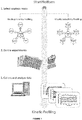
- the layout of the microplate defines the specific position of each assay in the microplate. The position in the microplate depends on the type of kinetic assay which will be performed within the method of the invention: Binding Kinetic Profiling or Selectivity Kinetic Profiling ( Figure 1 ).
- the layout of the Binding Kinetic Profiling allows the analysis of a maximum of 46 kinetic analyses of 46 different compounds against a unique target in a 384-well microplate.
- the layout of the Selectivity Kinetic Profiling allows the analysis of a maximum of 32 kinetic analyses of a unique compound against 32 different targets in a 384-well microplate.
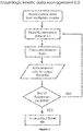
- the fluorescence data acquired by the microplate reader is managed by a software application, which imports the fluorescence data from the csv files recorded by the microplate reader, to a spreadsheet output file ( Figure 2 ).
- the luminescence data acquired by the microplate reader is managed by a software application, which imports the luminescence data from the csv files recorded by the microplate reader, to a spreadsheet output file ( Figure 2 ).
- the data obtained from the csv files before executing this software application shows the fluorescence values for each well of the plate, wherein fluorescence measures are acquired every fixed time (e.g. 6 minutes) for a determined period of time (e.g. 10 hours) at real time.
- the data obtained from the csv files before executing this software application shows the luminescence values for each well of the plate, wherein luminescence measures are acquired every fixed time (e.g. 6 minutes) for a determined period of time (e.g. 10 hours) at real time.
- luminescence measures are acquired every fixed time (e.g. 6 minutes) for a determined period of time (e.g. 10 hours) at real time.
- emission intensities are measured for all wells of a 384-well microplate at within five minutes of each specific point in time, more preferably simultaneously.
- the luminescence emitted at one wavelength by the donor molecule (the luciferase, or the antibody conjugated with the luciferase, which binds to the target of the assay) and the fluorescence emitted at another wavelength by the acceptor molecule (labeled ligand).
- the csv file obtained after the data acquisition by the microplate reader, is the "input file" for the software application.
- the data presented in the input file are: “Plate number” (the number the microplate in the method), “Plate repetition” (the reading of each well at a time point), “Well” (position of the sample in the well of the microplate), “Meas Time” (time at which a measurement is acquired, from 0 minutes up to 10 hours), “Signal” (fluorescence value), “Flashes/time” (number of readings of a fluorescence value measured in a well).
- the data presented in the input file are: “Signal” (luminescence value), “Flashes/time” (number of readings of a luminescence value measured in a well).
- the readings of the fluorescence value of each well of the microplate by the microplate reader are from 30 to 300 times at real time, more preferably 100 to 300 times at real time.
- the readings of the luminescence value of each well of the microplate by the microplate reader are from 30 to 300 times at real time, more preferably 100 to 300 times at real time.
- up to 240.000 data per plate are obtained in less than 2 minutes in a 384-well microplate.
- the Emission Ratio (ER) of each well is calculated from the input file, an ER for each assay in each time point for each compound concentration.
- the ER is calculated from the ratio of the fluorescence data acquired from the donor and the acceptor and may be multiplied by 10.000.
- the ER is calculated from the ratio of the luminescence data acquired from the donor and the acceptor and may be multiplied by 10.000.
- the specific-union data, or corrected emission ratio (ER*) is obtained for a given concentration of the compound of interest at a given time, by subtracting the ER of the negative control at said given time, defined as the wells which contain a known inhibitor (first molecule) at a saturating concentration from the ER of said compound of interest at said given concentration and said given time.
- dfEI b refers to the intensity of the fluorescence of the donor luminophore of the sample which contains the compound of interest at said given concentration and at said given time.
- afEI a refers to the intensity of the fluorescence of the acceptor fluorophore of the sample which contains the negative control at said given time.
- dfEI a refers to the intensity of the fluorescence of the donor fluorophore of the sample which contains the negative control at said given time.
- dfEI a refers to the intensity of the fluorescence of the donor luminophore of the sample which contains the negative control at said given time.
- software is used to generate an output file (a spreadsheet) with each ER* value from the input data adjusted (fitted) to the predefined layout, in an XY table for each assay in the microplate, wherein the X values refer to the compound or unlabeled ligand concentration in the assay and the Y values refer to the time measures.
- a maximum of 46 XY tables are generated in a Binding Kinetic Profiling assay when a 384-well microplate is used.
- a maximum of 32 XY tables are generated in a Selectivity Kinetic Profiling assay when a 384-well microplate is used.
- the determination method in the invention can be performed by fluorescence polarization (FP) or time resolved Time-resolved Fluorescence Resonance Energy Transfer (TR-FRET).
- FP fluorescence polarization
- TR-FRET time resolved Time-resolved Fluorescence Resonance Energy Transfer
- the fluorescence determination is performed by TR-FRET.
- BRET Bioluminescence Resonance Energy Transfer
- the quality control acceptance criteria of the results is evaluated by statistical parameters.
- the negative and positive controls of the microplate are used to calculate the statistical parameters S/B and Z' factors which indicate the robustness and sensitivity of the assay performed with the method of the invention.
- the acceptance criteria for these factors are S/B>2 and Z'>0.4, although these factors may change depending on the target.
- the data acquisition in the present invention is done at a real time and consequently, those statistical parameters must be analyzed in a real time mode. It is mandatory in the invention that the Z' factor and assay window fit the established acceptance limits during the entire time of the assay, which in some cases can be extended up to 15 hours.
- the kinetic parameters of each compound of interest in the method of the invention are determined by fitting the data obtained from the software application to a kinetic competitive binding model. Fitting is a mathematical or statistical adjustment. Another aspect disclosed in the present invention relates to that the method of the invention does not predetermine the K i value of the target protein or polyprotein before starting the method of the invention.
- the kinetic parameters of each compound (K on , K off , K d and t 1/2 ) are calculated by a software application known for a skilled person in the art.
- the kinetic competitive binding model describes the kinetic behavior of two compound when compete for the binding to the same target. There is a known ligand (previously characterized kinetically, e. g.
- the labeled ligand concentration and the values from the labeled ligand K on (K 1 ) and K off (K 2 ) must be introduced as "contrains" when unknown compounds are analyzed.
- the unknown ligands/compounds/new molecules are included to precisely determine the kinetic parameters of the interaction with the target. Moreover, it is mandatory to fit some constrains in the model, to obtain accurate data. Thus rate constants of the first molecule or labeled ligand (tracer) must be constrained to constant values determined from previous experiments.
- the method allows continuous implementation with new targets. For each particular target, a specific binding assay must be optimized and validated.
- titration experiments with different labeled ligand (tracer) concentrations are performed and fluorescence signals are monitored in a real time mode (by "continuously" obtaining data for all samples at discrete intervals over time).
- titration experiments with different labeled ligand (tracer) concentrations are performed and luminescence signals are monitored in a real time mode (by "continuously” obtaining data for all samples at discrete intervals over time).
- the fluorescence signals are from TR-FRET.
- the bioluminescence signals are from BRET.
- TR-FRET data are mathematically or statistically fitted to the "Association kinetics model" and the corresponding K on , K off and K d from the labeled ligand (tracer) are obtained. These values are included in the kinetic competitive model as constrain values, and influence the kinetic parameters of unknown compounds, thus it is very important to accurately determine these values.
- the method disclosed in the present invention complements the classical QSAR (Quantitative Structure Activity Relationship) studies, commonly used during the lead optimization process, with relevant kinetic information data to discriminate compounds with the same affinities.
- QSAR Quantitative Structure Activity Relationship
- many compounds show extremely high potency values (pico-molar range) and thus it is necessary a highly sensitive assay to do not sub-estimate their potency.
- This aspect is of crucial interest to select those compounds with the clinical profile that is important to patients: efficacy, safety, duration of action, greater tolerability, indication and therapeutic differentiation.
- the present invention combines the high sensitivity and throughput of the fluorescent methods with the advantages of the kinetic competitive binding approach. As a consequence, this method minimizes adverse environmental impacts avoiding radioactivity and maximizes economic benefits, reducing times and volumes needed to perform kinetic profile characterization of new molecules.
- the present example illustrates the kinetic characterization of a selected tracer for a particular/representative kinase.
- the kinase of the example is the phosphoinositide 3-kinase delta, which belongs to the lipid kinase family (PI3Kinase-delta, PI3K delta ).
- the labeled ligand (tracer) selected is a small molecule conjugated with the fluorescent dye AlexaTMFluor 647 (Tracer 314 , Life Technologies). This molecule is an ATP-competitive analogue which binds to the PI3K delta and is directed by an europium labeled antibody, also bound to the kinase of the present assay.
- the present example shows a titration experiment with different concentrations of Tracer 314 .
- This solution was prepared in kinase buffer A (50 mM HEPES pH 7.5, 1 mM EGTA, 0.01% Brij-35, 10 mM MgCl 2 , from Life Technologies) containing 1% DMSO. Additionally, it was prepared another solution containing a saturating concentration of a specific inhibitor of PI3K delta (PI-103 at 10 micromolar) instead of DMSO in order to account the unspecific binding (low control sample or negative control).
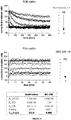
- the Enzymlogic Kinetic management data E.0 application is then used as explained in the detailed description of the present invention to generate a XY table plotting the corrected emission ratio (ER*) according to the different tracer concentrations (X-axis) and the time (Y-axis) ( Figure 3 ). These data are fit to the One-Phase association equation by using the GraphPad PrismTM software as previously described and the corresponding K on , K off and K d from the labeled ligand (Tracer 314 ) to PI3Kdelta are obtained.
- Figure 3 includes a plot summarizing the corrected emission rate over the time at each Tracer 314 concentration (20 to 0 nM).
- a table with the kinetic parameters obtained is also included summarizing the kinetic data of the Tracer 314 -PI3K delta interaction.
- the present example illustrates the kinetic characterization of two different inhibitors against the same target by using the method of the invention in the Binding Kinetic Profiling mode.
- the kinase PI3K delta has been selected to illustrate this example.
- the characterized Tracer 314 (Life Technologies) has been used as labeled ligand (tracer).
- the reference compounds chosen for the present example are Wortmanine and BEZ-235 which are known for inhibiting the selected target.
- the affinity of these two reference compounds have been previously described by many authors at equilibrium, with K d values in the nanomolar range. Nevertheless, any kinetic parameter has been reported to date for these interactions.
- experimental data were fitted to the "Kinetics of competitive binding". This model describes the kinetic behavior of two compounds when compete for the binding to the same target.
- ligand Tracer 314 in this example
- Example 1 The experiment is performed as described in Example 1 for the tracer characterization.
- the samples are prepared in the kinase buffer A at the same concentrations described in example 1 (PI3K delta at 0.5 nM and Eu-antiGST antibody at 2 nM).
- Control wells in the microplate with PI-103 inhibitor at 10 ⁇ M are also included to account for unspecific binding.
- Tracer 314 is added to all wells at a constant concentration (around its K d , in the present example, 5 nM) and eight different concentrations of each unknown compound (Wortmanine and BEZ235) are included in the wells of the microplate.
- the eight different concentrations are the result of 4-fold serial dilutions of the unknown compounds, ranging from 250 nM to 0. These serial dilutions were prepared in a mother plate (at 2 times the assay concentration) and quickly dispensed to the analysis plate, being the last one disposed in the well the kinase PI3K delta .
- the TR-FRET signals corresponding to the competitive binding between the Tracer 314 and the test compounds were recorded over time as described above. The present example is illustrated in Figure 4 .
- K 1 is the association rate constant of the Tracer 314 in M -1 min -1 (4.49E+08) and K 2 is its dissociation rate constant in units of min -1 (3,217).
- the tracer concentration (L) is also constrained to the tracer concentration employed in the assay, i.e., 5 nM in the present example.
- I is constrained to be a column constant whose value comes from the column titles. There are as many I values as different concentrations used in the experiment, expressed in nM. In this example, 8 values (i.e. 8 columns for each compound) ranging from 250 nM to 0.
- K 3 and K 4 the dissociation and association rate constants of the two reference compounds were determined (K 3 and K 4 respectively).
- K d values for each reference inhibitor can be derived from the kinetic analysis as the ratio of K 4 to K 3 .
- t 1/2 is calculated as -ln[0.5] divided by the K 4 value.
- Wortmanine and BEZ235 exhibit a very different kinetic profile when bound to the PI3K delta .
- BEZ235 dissociate very fast from the target (mili-seconds), while the dissociative half-life estimated for the complex Wortmanine-PI3K delta is close to 300 min.
- their affinity values are also very different, ranging from around 10 nanomolar in the case of BEZ235, to around 300 picomolar for Wortmanine.
- the example described above illustrate how very different compounds can be analyzed in the same way, using the same target and compound concentrations and without the need to previously determine their K d values in classical dose-response experiments.
- the method of the invention can be easily used in high throughput format (HTS) to kinetically characterize a broad range of molecules.
- HTS high throughput format
- Example 3 illustrates how the method of the invention works in the Kinetic Selectivity Profiling mode.
- This example includes two reference compounds, Sorafenib and Staurosporine, against 3 related cyclin dependent kinases (CDK7/Cyclin H MNAT1, CDK8/Cyclin C and CDK9/Cyclin T1).
- the same labeled molecule (tracer) is used in all the experiments (Tracer 236 , Life Technologies).
- the tracer used in the assay should be kinetically characterized prior to the experiment.
- the association (K on ), dissociation (K off ) and K d of Tracer 236 against each targeted CDK were calculated. These values were introduced as constrains for the kinetic competitive model, as explained in Example 2.
- the experiments were performed similarly as described for PI3K delta in examples 1 and 2.
- the kinase concentrations used in the present example are: 2 nM for CDK8/Cyclin C and CDK9/CyclinT1 and 5 nM for CDK7/CyclinH MNAT1.
- the Tracer 236 concentration is 10 nM, 30 nM and 150 nM respectively for each target.
- An Eu-antiHis antibody was used at 2 nM in all three cases.
- eight concentrations of each reference inhibitor (Sorafenib and Staurosporine) were used in every case, ranging from 1000 nM to 0 nM, result of 4-fold serial dilutions of the compounds.
- the TR-FRET signals corresponding to the competitive binding between the Tracer 236 and the test compounds for each target were recorded over time as described above.
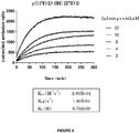
- Figure 5 illustrate a representative plot with the corrected emission ratios (ER*) obtained from the CDK8-Sorafenib interaction over the time.
- the data are transformed using the Kinetic Management data E.0 software application and finally fit to the kinetic competitive model, by using the GraphPad PrismTM software.
- the data were also fit to the equation described in the present patent application, constraining the rate constants K 1 (K on ) and K 2 (K off ) of the labeled ligand (Tracer 236 ) to constant values determined from previous experiment.
- the dissociation and association rate constants of the two reference compounds for each target were determined as described in Example 2.
- both inhibitors show a very different profile depending on the target molecule.
- Sorafenib is a specific inhibitor of CDK8/Cyclin C
- Staurosporine is a pan inhibitor of the 3 CDKs, showing similar K d values for the 3 targets (between 3.2 to 8.5 nM).
- the kinetic behavior of Staurosporine is very different among the three CDKs of the example, and the interaction with CDK8 is extended for 20 min, while the interaction with CDK7 is only maintained less than 1 min.
- binding kinetics gives valuable information on the binding mechanism of a molecule against different targets that may influence in their potential efficacy and/or safety profile when it was used in clinical trials.
- the present example illustrates the kinetic characterization of a selected ligand of a representative G protein-coupled receptor (GPCR).
- GPCR G protein-coupled receptor
- the GPCR of the example is the human mu-opioid receptor, which belongs to the opioid receptor family (hMOR).
- the labeled ligand (tracer) is a naltrexone derivative labeled with a red emitting HTRF fluorescent probe (L0005RED, Cisbio). This molecule is an opioid antagonist that binds to the hMOR.
- Human embryonic kidney cells (HEK) express the mu-opioid receptor directly labeled with terbium cryptate. When the tracer is bound directly to the receptor there is a high TR-FRET signal, whereas displacement of the tracer with a GPCR agonist or antagonist result in a loss of TR-FRET.
- the present example shows a titration experiment with different concentrations of the opioid antagonist, ranging from 32 nM to 0 nM (serial two fold dilutions).
- a solution containing the HEK cells expressing the hMOR was prepared at three times the desired concentration used in the assay. Additionally, it was prepared another solution containing a saturating concentration of a specific antagonist of hMOR (Naloxone 50 micromolar, Sigma) instead of DMSO in order to account the unspecific binding (low control sample or negative control). All solutions were prepared in TAGlite buffer (LABMED, CisBio). The experiment was performed similarly to examples 1 to 3, wherein the hMOR (target) was the last one disposed in the wells.
- the negative and positive controls in the experiments are used to calculate the statistical parameters S/B and Z' factors, which indicates if an assay is robust and sensitive. Although these factors may change depending on the target, in general terms, the acceptance criteria for these factors are S/B>2 and Z'>0.4.
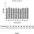
- Figure 7 illustrates the statistical analysis of S/B and Z' over the time course of a selected experiment performed with CDK8/Cyclin C during 10 hours. The values are calculated in a continuous mode, although are plotted at 60 minutes intervals. The experiment was performed under the same experimental conditions described in the Example 3 of this invention (2 nM CDK8/Cyclin C, 2 nM Eu-antiHis antibody and 10 nM Tracer 236 ).
Landscapes
- Health & Medical Sciences (AREA)
- Chemical & Material Sciences (AREA)
- Life Sciences & Earth Sciences (AREA)
- Engineering & Computer Science (AREA)
- Immunology (AREA)
- Molecular Biology (AREA)
- Biomedical Technology (AREA)
- Hematology (AREA)
- Urology & Nephrology (AREA)
- Physics & Mathematics (AREA)
- Biotechnology (AREA)
- General Health & Medical Sciences (AREA)
- Analytical Chemistry (AREA)
- General Physics & Mathematics (AREA)
- Food Science & Technology (AREA)
- Biochemistry (AREA)
- Microbiology (AREA)
- Cell Biology (AREA)
- Pathology (AREA)
- Medicinal Chemistry (AREA)
- Inorganic Chemistry (AREA)
- Organic Chemistry (AREA)
- Materials Engineering (AREA)
- Bioinformatics & Cheminformatics (AREA)
- Chemical Kinetics & Catalysis (AREA)
- Bioinformatics & Computational Biology (AREA)
- Biophysics (AREA)
- Proteomics, Peptides & Aminoacids (AREA)
- Tropical Medicine & Parasitology (AREA)
- Toxicology (AREA)
- Evolutionary Biology (AREA)
- Genetics & Genomics (AREA)
- Medical Informatics (AREA)
- Spectroscopy & Molecular Physics (AREA)
- Theoretical Computer Science (AREA)
- Investigating, Analyzing Materials By Fluorescence Or Luminescence (AREA)
- Investigating Or Analysing Biological Materials (AREA)
- Measuring Or Testing Involving Enzymes Or Micro-Organisms (AREA)
Priority Applications (1)
| Application Number | Priority Date | Filing Date | Title |
|---|---|---|---|
| PL16775105T PL3350596T3 (pl) | 2015-09-16 | 2016-09-15 | Sposób określania profili kinetycznych w opracowywaniu leków |
Applications Claiming Priority (2)
| Application Number | Priority Date | Filing Date | Title |
|---|---|---|---|
| EP15185422 | 2015-09-16 | ||
| PCT/EP2016/071901 WO2017046284A1 (en) | 2015-09-16 | 2016-09-15 | Method for determining kinetic profiles in drug discovery |
Publications (2)
| Publication Number | Publication Date |
|---|---|
| EP3350596A1 EP3350596A1 (en) | 2018-07-25 |
| EP3350596B1 true EP3350596B1 (en) | 2020-01-08 |
Family
ID=54147084
Family Applications (1)
| Application Number | Title | Priority Date | Filing Date |
|---|---|---|---|
| EP16775105.6A Active EP3350596B1 (en) | 2015-09-16 | 2016-09-15 | Method for determining kinetic profiles in drug discovery |
Country Status (6)
| Country | Link |
|---|---|
| US (1) | US10261079B2 (da) |
| EP (1) | EP3350596B1 (da) |
| DK (1) | DK3350596T3 (da) |
| ES (1) | ES2781837T3 (da) |
| PL (1) | PL3350596T3 (da) |
| WO (1) | WO2017046284A1 (da) |
Families Citing this family (2)
| Publication number | Priority date | Publication date | Assignee | Title |
|---|---|---|---|---|
| WO2020260277A1 (en) * | 2019-06-25 | 2020-12-30 | Albert-Ludwig-Universität Freiburg | Method and kit for measuring of analytes in bi-component systems and uses thereof |
| CN118050337B (zh) * | 2024-01-31 | 2024-11-15 | 中国海洋大学 | 一种基于时间分辨荧光共振能量转移的蛋白-小分子配体相互作用分析方法 |
Family Cites Families (1)
| Publication number | Priority date | Publication date | Assignee | Title |
|---|---|---|---|---|
| EP2241619A1 (en) | 2009-04-17 | 2010-10-20 | Max-Planck-Gesellschaft zur Förderung der Wissenschaften e.V. | Development of fluorescently P-loop labeled kinases for screening of inhibitors |
-
2016
- 2016-09-15 PL PL16775105T patent/PL3350596T3/pl unknown
- 2016-09-15 DK DK16775105.6T patent/DK3350596T3/da active
- 2016-09-15 EP EP16775105.6A patent/EP3350596B1/en active Active
- 2016-09-15 ES ES16775105T patent/ES2781837T3/es active Active
- 2016-09-15 WO PCT/EP2016/071901 patent/WO2017046284A1/en not_active Ceased
- 2016-09-15 US US15/760,070 patent/US10261079B2/en active Active
Non-Patent Citations (1)
| Title |
|---|
| None * |
Also Published As
| Publication number | Publication date |
|---|---|
| EP3350596A1 (en) | 2018-07-25 |
| US20190049441A1 (en) | 2019-02-14 |
| ES2781837T3 (es) | 2020-09-08 |
| WO2017046284A1 (en) | 2017-03-23 |
| US10261079B2 (en) | 2019-04-16 |
| DK3350596T3 (da) | 2020-04-06 |
| PL3350596T3 (pl) | 2020-06-15 |
Similar Documents
| Publication | Publication Date | Title |
|---|---|---|
| Acker et al. | Considerations for the design and reporting of enzyme assays in high-throughput screening applications | |
| Inglese et al. | High-throughput screening assays for the identification of chemical probes | |
| Janzen | Screening technologies for small molecule discovery: the state of the art | |
| Olive | Quantitative methods for the analysis of protein phosphorylation in drug development | |
| Von Ahsen et al. | High‐throughput screening for kinase inhibitors | |
| Shukla et al. | The future of toxicity testing: a focus on in vitro methods using a quantitative high-throughput screening platform | |
| Schiele et al. | A universal homogeneous assay for high-throughput determination of binding kinetics | |
| Jia et al. | Current in vitro kinase assay technologies: the quest for a universal format | |
| Glickman | Assay development for protein kinase enzymes | |
| Cronk et al. | High-throughput screening | |
| Falconer et al. | High-throughput screening for ion channel modulators | |
| Nowak et al. | Microscale thermophoresis (MST) and spectral shift (SpS) in drug discovery | |
| Tenney et al. | A mitochondrial-targeted activity-based sensing probe for ratiometric imaging of formaldehyde reveals key regulators of the mitochondrial one-carbon pool | |
| Klumpp et al. | Readout technologies for highly miniaturized kinase assays applicable to high-throughput screening in a 1536-well format | |
| EP3350596B1 (en) | Method for determining kinetic profiles in drug discovery | |
| Kestranek et al. | Chemiluminescent nitrogen detection (CLND) to measure kinetic aqueous solubility | |
| Degorce | HTRF®: Pioneering technology for high-throughput screening | |
| Gomez-Hens et al. | Modern analytical approaches to high-throughput drug discovery | |
| Hubert et al. | Data concordance from a comparison between filter binding and fluorescence polarization assay formats for identification of ROCK-II inhibitors | |
| Sportsman et al. | Fluorescence polarization | |
| Hunter et al. | High-throughput investigation of macromolecular interactions for drug development using spectral shift technology | |
| CA2542774A1 (en) | Direct observation of molecular modifications in biological test systems by measuring fluorescence lifetime | |
| Li et al. | Kinetic assay for characterization of spleen tyrosine kinase activity and inhibition with recombinant kinase and crude cell lysates | |
| Sykes et al. | Kinetic Profiling of Ligands and Fragments Binding to GPCRs by TR-FRET | |
| EP4049034B1 (en) | High throughput, fluorescence-based estimation of monoconal antibody aggregation with disodium 3,3'-{[(e)-1,2-diphenyl-1,2-ethenediyl]bis(4,1-phenyleneoxy)}di(1-propanesulfonate) dye for superior sensitivity and specificity |
Legal Events
| Date | Code | Title | Description |
|---|---|---|---|
| STAA | Information on the status of an ep patent application or granted ep patent |
Free format text: STATUS: UNKNOWN |
|
| STAA | Information on the status of an ep patent application or granted ep patent |
Free format text: STATUS: THE INTERNATIONAL PUBLICATION HAS BEEN MADE |
|
| PUAI | Public reference made under article 153(3) epc to a published international application that has entered the european phase |
Free format text: ORIGINAL CODE: 0009012 |
|
| STAA | Information on the status of an ep patent application or granted ep patent |
Free format text: STATUS: REQUEST FOR EXAMINATION WAS MADE |
|
| 17P | Request for examination filed |
Effective date: 20180411 |
|
| AK | Designated contracting states |
Kind code of ref document: A1 Designated state(s): AL AT BE BG CH CY CZ DE DK EE ES FI FR GB GR HR HU IE IS IT LI LT LU LV MC MK MT NL NO PL PT RO RS SE SI SK SM TR |
|
| AX | Request for extension of the european patent |
Extension state: BA ME |
|
| STAA | Information on the status of an ep patent application or granted ep patent |
Free format text: STATUS: EXAMINATION IS IN PROGRESS |
|
| DAV | Request for validation of the european patent (deleted) | ||
| DAX | Request for extension of the european patent (deleted) | ||
| 17Q | First examination report despatched |
Effective date: 20181220 |
|
| GRAP | Despatch of communication of intention to grant a patent |
Free format text: ORIGINAL CODE: EPIDOSNIGR1 |
|
| STAA | Information on the status of an ep patent application or granted ep patent |
Free format text: STATUS: GRANT OF PATENT IS INTENDED |
|
| INTG | Intention to grant announced |
Effective date: 20190917 |
|
| GRAS | Grant fee paid |
Free format text: ORIGINAL CODE: EPIDOSNIGR3 |
|
| GRAA | (expected) grant |
Free format text: ORIGINAL CODE: 0009210 |
|
| STAA | Information on the status of an ep patent application or granted ep patent |
Free format text: STATUS: THE PATENT HAS BEEN GRANTED |
|
| AK | Designated contracting states |
Kind code of ref document: B1 Designated state(s): AL AT BE BG CH CY CZ DE DK EE ES FI FR GB GR HR HU IE IS IT LI LT LU LV MC MK MT NL NO PL PT RO RS SE SI SK SM TR |
|
| REG | Reference to a national code |
Ref country code: GB Ref legal event code: FG4D |
|
| REG | Reference to a national code |
Ref country code: CH Ref legal event code: EP |
|
| REG | Reference to a national code |
Ref country code: DE Ref legal event code: R096 Ref document number: 602016027915 Country of ref document: DE |
|
| REG | Reference to a national code |
Ref country code: IE Ref legal event code: FG4D |
|
| REG | Reference to a national code |
Ref country code: AT Ref legal event code: REF Ref document number: 1223335 Country of ref document: AT Kind code of ref document: T Effective date: 20200215 |
|
| REG | Reference to a national code |
Ref country code: DK Ref legal event code: T3 Effective date: 20200330 |
|
| REG | Reference to a national code |
Ref country code: NL Ref legal event code: FP |
|
| REG | Reference to a national code |
Ref country code: CH Ref legal event code: NV Representative=s name: VOSSIUS AND PARTNER PATENTANWAELTE RECHTSANWAE, CH |
|
| REG | Reference to a national code |
Ref country code: SE Ref legal event code: TRGR |
|
| REG | Reference to a national code |
Ref country code: LT Ref legal event code: MG4D |
|
| PG25 | Lapsed in a contracting state [announced via postgrant information from national office to epo] |
Ref country code: NO Free format text: LAPSE BECAUSE OF FAILURE TO SUBMIT A TRANSLATION OF THE DESCRIPTION OR TO PAY THE FEE WITHIN THE PRESCRIBED TIME-LIMIT Effective date: 20200408 Ref country code: RS Free format text: LAPSE BECAUSE OF FAILURE TO SUBMIT A TRANSLATION OF THE DESCRIPTION OR TO PAY THE FEE WITHIN THE PRESCRIBED TIME-LIMIT Effective date: 20200108 Ref country code: LT Free format text: LAPSE BECAUSE OF FAILURE TO SUBMIT A TRANSLATION OF THE DESCRIPTION OR TO PAY THE FEE WITHIN THE PRESCRIBED TIME-LIMIT Effective date: 20200108 Ref country code: FI Free format text: LAPSE BECAUSE OF FAILURE TO SUBMIT A TRANSLATION OF THE DESCRIPTION OR TO PAY THE FEE WITHIN THE PRESCRIBED TIME-LIMIT Effective date: 20200108 Ref country code: PT Free format text: LAPSE BECAUSE OF FAILURE TO SUBMIT A TRANSLATION OF THE DESCRIPTION OR TO PAY THE FEE WITHIN THE PRESCRIBED TIME-LIMIT Effective date: 20200531 |
|
| PG25 | Lapsed in a contracting state [announced via postgrant information from national office to epo] |
Ref country code: IS Free format text: LAPSE BECAUSE OF FAILURE TO SUBMIT A TRANSLATION OF THE DESCRIPTION OR TO PAY THE FEE WITHIN THE PRESCRIBED TIME-LIMIT Effective date: 20200508 Ref country code: HR Free format text: LAPSE BECAUSE OF FAILURE TO SUBMIT A TRANSLATION OF THE DESCRIPTION OR TO PAY THE FEE WITHIN THE PRESCRIBED TIME-LIMIT Effective date: 20200108 Ref country code: LV Free format text: LAPSE BECAUSE OF FAILURE TO SUBMIT A TRANSLATION OF THE DESCRIPTION OR TO PAY THE FEE WITHIN THE PRESCRIBED TIME-LIMIT Effective date: 20200108 Ref country code: BG Free format text: LAPSE BECAUSE OF FAILURE TO SUBMIT A TRANSLATION OF THE DESCRIPTION OR TO PAY THE FEE WITHIN THE PRESCRIBED TIME-LIMIT Effective date: 20200408 |
|
| REG | Reference to a national code |
Ref country code: ES Ref legal event code: FG2A Ref document number: 2781837 Country of ref document: ES Kind code of ref document: T3 Effective date: 20200908 |
|
| REG | Reference to a national code |
Ref country code: DE Ref legal event code: R097 Ref document number: 602016027915 Country of ref document: DE |
|
| PG25 | Lapsed in a contracting state [announced via postgrant information from national office to epo] |
Ref country code: EE Free format text: LAPSE BECAUSE OF FAILURE TO SUBMIT A TRANSLATION OF THE DESCRIPTION OR TO PAY THE FEE WITHIN THE PRESCRIBED TIME-LIMIT Effective date: 20200108 Ref country code: RO Free format text: LAPSE BECAUSE OF FAILURE TO SUBMIT A TRANSLATION OF THE DESCRIPTION OR TO PAY THE FEE WITHIN THE PRESCRIBED TIME-LIMIT Effective date: 20200108 Ref country code: CZ Free format text: LAPSE BECAUSE OF FAILURE TO SUBMIT A TRANSLATION OF THE DESCRIPTION OR TO PAY THE FEE WITHIN THE PRESCRIBED TIME-LIMIT Effective date: 20200108 Ref country code: SK Free format text: LAPSE BECAUSE OF FAILURE TO SUBMIT A TRANSLATION OF THE DESCRIPTION OR TO PAY THE FEE WITHIN THE PRESCRIBED TIME-LIMIT Effective date: 20200108 Ref country code: SM Free format text: LAPSE BECAUSE OF FAILURE TO SUBMIT A TRANSLATION OF THE DESCRIPTION OR TO PAY THE FEE WITHIN THE PRESCRIBED TIME-LIMIT Effective date: 20200108 |
|
| PLBE | No opposition filed within time limit |
Free format text: ORIGINAL CODE: 0009261 |
|
| STAA | Information on the status of an ep patent application or granted ep patent |
Free format text: STATUS: NO OPPOSITION FILED WITHIN TIME LIMIT |
|
| REG | Reference to a national code |
Ref country code: AT Ref legal event code: MK05 Ref document number: 1223335 Country of ref document: AT Kind code of ref document: T Effective date: 20200108 |
|
| 26N | No opposition filed |
Effective date: 20201009 |
|
| PG25 | Lapsed in a contracting state [announced via postgrant information from national office to epo] |
Ref country code: AT Free format text: LAPSE BECAUSE OF FAILURE TO SUBMIT A TRANSLATION OF THE DESCRIPTION OR TO PAY THE FEE WITHIN THE PRESCRIBED TIME-LIMIT Effective date: 20200108 |
|
| PG25 | Lapsed in a contracting state [announced via postgrant information from national office to epo] |
Ref country code: SI Free format text: LAPSE BECAUSE OF FAILURE TO SUBMIT A TRANSLATION OF THE DESCRIPTION OR TO PAY THE FEE WITHIN THE PRESCRIBED TIME-LIMIT Effective date: 20200108 |
|
| PG25 | Lapsed in a contracting state [announced via postgrant information from national office to epo] |
Ref country code: LU Free format text: LAPSE BECAUSE OF NON-PAYMENT OF DUE FEES Effective date: 20200915 |
|
| PG25 | Lapsed in a contracting state [announced via postgrant information from national office to epo] |
Ref country code: IE Free format text: LAPSE BECAUSE OF NON-PAYMENT OF DUE FEES Effective date: 20200915 |
|
| PG25 | Lapsed in a contracting state [announced via postgrant information from national office to epo] |
Ref country code: TR Free format text: LAPSE BECAUSE OF FAILURE TO SUBMIT A TRANSLATION OF THE DESCRIPTION OR TO PAY THE FEE WITHIN THE PRESCRIBED TIME-LIMIT Effective date: 20200108 Ref country code: MT Free format text: LAPSE BECAUSE OF FAILURE TO SUBMIT A TRANSLATION OF THE DESCRIPTION OR TO PAY THE FEE WITHIN THE PRESCRIBED TIME-LIMIT Effective date: 20200108 Ref country code: CY Free format text: LAPSE BECAUSE OF FAILURE TO SUBMIT A TRANSLATION OF THE DESCRIPTION OR TO PAY THE FEE WITHIN THE PRESCRIBED TIME-LIMIT Effective date: 20200108 |
|
| PG25 | Lapsed in a contracting state [announced via postgrant information from national office to epo] |
Ref country code: MK Free format text: LAPSE BECAUSE OF FAILURE TO SUBMIT A TRANSLATION OF THE DESCRIPTION OR TO PAY THE FEE WITHIN THE PRESCRIBED TIME-LIMIT Effective date: 20200108 Ref country code: MC Free format text: LAPSE BECAUSE OF FAILURE TO SUBMIT A TRANSLATION OF THE DESCRIPTION OR TO PAY THE FEE WITHIN THE PRESCRIBED TIME-LIMIT Effective date: 20200108 Ref country code: AL Free format text: LAPSE BECAUSE OF FAILURE TO SUBMIT A TRANSLATION OF THE DESCRIPTION OR TO PAY THE FEE WITHIN THE PRESCRIBED TIME-LIMIT Effective date: 20200108 |
|
| PG25 | Lapsed in a contracting state [announced via postgrant information from national office to epo] |
Ref country code: GR Free format text: LAPSE BECAUSE OF FAILURE TO SUBMIT A TRANSLATION OF THE DESCRIPTION OR TO PAY THE FEE WITHIN THE PRESCRIBED TIME-LIMIT Effective date: 20200108 |
|
| P01 | Opt-out of the competence of the unified patent court (upc) registered |
Effective date: 20230516 |
|
| REG | Reference to a national code |
Ref country code: CH Ref legal event code: U11 Free format text: ST27 STATUS EVENT CODE: U-0-0-U10-U11 (AS PROVIDED BY THE NATIONAL OFFICE) Effective date: 20251001 |
|
| PGFP | Annual fee paid to national office [announced via postgrant information from national office to epo] |
Ref country code: DK Payment date: 20250924 Year of fee payment: 10 Ref country code: DE Payment date: 20250926 Year of fee payment: 10 |
|
| PGFP | Annual fee paid to national office [announced via postgrant information from national office to epo] |
Ref country code: NL Payment date: 20250925 Year of fee payment: 10 Ref country code: PL Payment date: 20250905 Year of fee payment: 10 Ref country code: IT Payment date: 20250919 Year of fee payment: 10 |
|
| PGFP | Annual fee paid to national office [announced via postgrant information from national office to epo] |
Ref country code: BE Payment date: 20250925 Year of fee payment: 10 Ref country code: GB Payment date: 20250923 Year of fee payment: 10 |
|
| PGFP | Annual fee paid to national office [announced via postgrant information from national office to epo] |
Ref country code: FR Payment date: 20250924 Year of fee payment: 10 |
|
| PGFP | Annual fee paid to national office [announced via postgrant information from national office to epo] |
Ref country code: SE Payment date: 20250924 Year of fee payment: 10 |
|
| PGFP | Annual fee paid to national office [announced via postgrant information from national office to epo] |
Ref country code: CH Payment date: 20251001 Year of fee payment: 10 |
|
| PGFP | Annual fee paid to national office [announced via postgrant information from national office to epo] |
Ref country code: ES Payment date: 20251001 Year of fee payment: 10 |