EP3345929B1 - Fusion polypeptide in which anti-inflammatory polypeptide and ferritin monomer fragment are bound and pharmaceutical composition for preventing and treating inflammatory diseases, containing same as active ingredient - Google Patents
Fusion polypeptide in which anti-inflammatory polypeptide and ferritin monomer fragment are bound and pharmaceutical composition for preventing and treating inflammatory diseases, containing same as active ingredient Download PDFInfo
- Publication number
- EP3345929B1 EP3345929B1 EP16842349.9A EP16842349A EP3345929B1 EP 3345929 B1 EP3345929 B1 EP 3345929B1 EP 16842349 A EP16842349 A EP 16842349A EP 3345929 B1 EP3345929 B1 EP 3345929B1
- Authority
- EP
- European Patent Office
- Prior art keywords
- tfmg
- inflammatory
- gla
- tfg
- fusion polypeptide
- Prior art date
- Legal status (The legal status is an assumption and is not a legal conclusion. Google has not performed a legal analysis and makes no representation as to the accuracy of the status listed.)
- Active
Links
Images






Classifications
-
- C—CHEMISTRY; METALLURGY
- C07—ORGANIC CHEMISTRY
- C07K—PEPTIDES
- C07K14/00—Peptides having more than 20 amino acids; Gastrins; Somatostatins; Melanotropins; Derivatives thereof
- C07K14/435—Peptides having more than 20 amino acids; Gastrins; Somatostatins; Melanotropins; Derivatives thereof from animals; from humans
- C07K14/46—Peptides having more than 20 amino acids; Gastrins; Somatostatins; Melanotropins; Derivatives thereof from animals; from humans from vertebrates
- C07K14/47—Peptides having more than 20 amino acids; Gastrins; Somatostatins; Melanotropins; Derivatives thereof from animals; from humans from vertebrates from mammals
-
- A—HUMAN NECESSITIES
- A61—MEDICAL OR VETERINARY SCIENCE; HYGIENE
- A61P—SPECIFIC THERAPEUTIC ACTIVITY OF CHEMICAL COMPOUNDS OR MEDICINAL PREPARATIONS
- A61P29/00—Non-central analgesic, antipyretic or antiinflammatory agents, e.g. antirheumatic agents; Non-steroidal antiinflammatory drugs [NSAID]
-
- A—HUMAN NECESSITIES
- A61—MEDICAL OR VETERINARY SCIENCE; HYGIENE
- A61K—PREPARATIONS FOR MEDICAL, DENTAL OR TOILETRY PURPOSES
- A61K38/00—Medicinal preparations containing peptides
- A61K38/16—Peptides having more than 20 amino acids; Gastrins; Somatostatins; Melanotropins; Derivatives thereof
-
- A—HUMAN NECESSITIES
- A61—MEDICAL OR VETERINARY SCIENCE; HYGIENE
- A61K—PREPARATIONS FOR MEDICAL, DENTAL OR TOILETRY PURPOSES
- A61K38/00—Medicinal preparations containing peptides
- A61K38/16—Peptides having more than 20 amino acids; Gastrins; Somatostatins; Melanotropins; Derivatives thereof
- A61K38/17—Peptides having more than 20 amino acids; Gastrins; Somatostatins; Melanotropins; Derivatives thereof from animals; from humans
-
- A—HUMAN NECESSITIES
- A61—MEDICAL OR VETERINARY SCIENCE; HYGIENE
- A61K—PREPARATIONS FOR MEDICAL, DENTAL OR TOILETRY PURPOSES
- A61K47/00—Medicinal preparations characterised by the non-active ingredients used, e.g. carriers or inert additives; Targeting or modifying agents chemically bound to the active ingredient
- A61K47/50—Medicinal preparations characterised by the non-active ingredients used, e.g. carriers or inert additives; Targeting or modifying agents chemically bound to the active ingredient the non-active ingredient being chemically bound to the active ingredient, e.g. polymer-drug conjugates
- A61K47/51—Medicinal preparations characterised by the non-active ingredients used, e.g. carriers or inert additives; Targeting or modifying agents chemically bound to the active ingredient the non-active ingredient being chemically bound to the active ingredient, e.g. polymer-drug conjugates the non-active ingredient being a modifying agent
- A61K47/62—Medicinal preparations characterised by the non-active ingredients used, e.g. carriers or inert additives; Targeting or modifying agents chemically bound to the active ingredient the non-active ingredient being chemically bound to the active ingredient, e.g. polymer-drug conjugates the non-active ingredient being a modifying agent the modifying agent being a protein, peptide or polyamino acid
- A61K47/64—Drug-peptide, drug-protein or drug-polyamino acid conjugates, i.e. the modifying agent being a peptide, protein or polyamino acid which is covalently bonded or complexed to a therapeutically active agent
- A61K47/644—Transferrin, e.g. a lactoferrin or ovotransferrin
-
- C—CHEMISTRY; METALLURGY
- C07—ORGANIC CHEMISTRY
- C07K—PEPTIDES
- C07K14/00—Peptides having more than 20 amino acids; Gastrins; Somatostatins; Melanotropins; Derivatives thereof
- C07K14/435—Peptides having more than 20 amino acids; Gastrins; Somatostatins; Melanotropins; Derivatives thereof from animals; from humans
- C07K14/52—Cytokines; Lymphokines; Interferons
- C07K14/54—Interleukins [IL]
-
- C—CHEMISTRY; METALLURGY
- C07—ORGANIC CHEMISTRY
- C07K—PEPTIDES
- C07K14/00—Peptides having more than 20 amino acids; Gastrins; Somatostatins; Melanotropins; Derivatives thereof
- C07K14/435—Peptides having more than 20 amino acids; Gastrins; Somatostatins; Melanotropins; Derivatives thereof from animals; from humans
- C07K14/52—Cytokines; Lymphokines; Interferons
- C07K14/54—Interleukins [IL]
- C07K14/5406—IL-4
-
- C—CHEMISTRY; METALLURGY
- C07—ORGANIC CHEMISTRY
- C07K—PEPTIDES
- C07K14/00—Peptides having more than 20 amino acids; Gastrins; Somatostatins; Melanotropins; Derivatives thereof
- C07K14/435—Peptides having more than 20 amino acids; Gastrins; Somatostatins; Melanotropins; Derivatives thereof from animals; from humans
- C07K14/52—Cytokines; Lymphokines; Interferons
- C07K14/54—Interleukins [IL]
- C07K14/5431—IL-11
-
- C—CHEMISTRY; METALLURGY
- C07—ORGANIC CHEMISTRY
- C07K—PEPTIDES
- C07K14/00—Peptides having more than 20 amino acids; Gastrins; Somatostatins; Melanotropins; Derivatives thereof
- C07K14/435—Peptides having more than 20 amino acids; Gastrins; Somatostatins; Melanotropins; Derivatives thereof from animals; from humans
- C07K14/52—Cytokines; Lymphokines; Interferons
- C07K14/54—Interleukins [IL]
- C07K14/5437—IL-13
-
- C—CHEMISTRY; METALLURGY
- C07—ORGANIC CHEMISTRY
- C07K—PEPTIDES
- C07K14/00—Peptides having more than 20 amino acids; Gastrins; Somatostatins; Melanotropins; Derivatives thereof
- C07K14/435—Peptides having more than 20 amino acids; Gastrins; Somatostatins; Melanotropins; Derivatives thereof from animals; from humans
- C07K14/79—Transferrins, e.g. lactoferrins, ovotransferrins
-
- C—CHEMISTRY; METALLURGY
- C07—ORGANIC CHEMISTRY
- C07K—PEPTIDES
- C07K19/00—Hybrid peptides, i.e. peptides covalently bound to nucleic acids, or non-covalently bound protein-protein complexes
-
- C—CHEMISTRY; METALLURGY
- C12—BIOCHEMISTRY; BEER; SPIRITS; WINE; VINEGAR; MICROBIOLOGY; ENZYMOLOGY; MUTATION OR GENETIC ENGINEERING
- C12N—MICROORGANISMS OR ENZYMES; COMPOSITIONS THEREOF; PROPAGATING, PRESERVING, OR MAINTAINING MICROORGANISMS; MUTATION OR GENETIC ENGINEERING; CULTURE MEDIA
- C12N15/00—Mutation or genetic engineering; DNA or RNA concerning genetic engineering, vectors, e.g. plasmids, or their isolation, preparation or purification; Use of hosts therefor
- C12N15/09—Recombinant DNA-technology
- C12N15/11—DNA or RNA fragments; Modified forms thereof; Non-coding nucleic acids having a biological activity
- C12N15/62—DNA sequences coding for fusion proteins
-
- A—HUMAN NECESSITIES
- A61—MEDICAL OR VETERINARY SCIENCE; HYGIENE
- A61K—PREPARATIONS FOR MEDICAL, DENTAL OR TOILETRY PURPOSES
- A61K38/00—Medicinal preparations containing peptides
- A61K38/16—Peptides having more than 20 amino acids; Gastrins; Somatostatins; Melanotropins; Derivatives thereof
- A61K38/17—Peptides having more than 20 amino acids; Gastrins; Somatostatins; Melanotropins; Derivatives thereof from animals; from humans
- A61K38/19—Cytokines; Lymphokines; Interferons
- A61K38/20—Interleukins [IL]
- A61K38/2006—IL-1
-
- A—HUMAN NECESSITIES
- A61—MEDICAL OR VETERINARY SCIENCE; HYGIENE
- A61K—PREPARATIONS FOR MEDICAL, DENTAL OR TOILETRY PURPOSES
- A61K38/00—Medicinal preparations containing peptides
- A61K38/16—Peptides having more than 20 amino acids; Gastrins; Somatostatins; Melanotropins; Derivatives thereof
- A61K38/17—Peptides having more than 20 amino acids; Gastrins; Somatostatins; Melanotropins; Derivatives thereof from animals; from humans
- A61K38/19—Cytokines; Lymphokines; Interferons
- A61K38/20—Interleukins [IL]
- A61K38/2026—IL-4
-
- A—HUMAN NECESSITIES
- A61—MEDICAL OR VETERINARY SCIENCE; HYGIENE
- A61K—PREPARATIONS FOR MEDICAL, DENTAL OR TOILETRY PURPOSES
- A61K38/00—Medicinal preparations containing peptides
- A61K38/16—Peptides having more than 20 amino acids; Gastrins; Somatostatins; Melanotropins; Derivatives thereof
- A61K38/17—Peptides having more than 20 amino acids; Gastrins; Somatostatins; Melanotropins; Derivatives thereof from animals; from humans
- A61K38/19—Cytokines; Lymphokines; Interferons
- A61K38/20—Interleukins [IL]
- A61K38/2073—IL-11
-
- A—HUMAN NECESSITIES
- A61—MEDICAL OR VETERINARY SCIENCE; HYGIENE
- A61K—PREPARATIONS FOR MEDICAL, DENTAL OR TOILETRY PURPOSES
- A61K38/00—Medicinal preparations containing peptides
- A61K38/16—Peptides having more than 20 amino acids; Gastrins; Somatostatins; Melanotropins; Derivatives thereof
- A61K38/17—Peptides having more than 20 amino acids; Gastrins; Somatostatins; Melanotropins; Derivatives thereof from animals; from humans
- A61K38/19—Cytokines; Lymphokines; Interferons
- A61K38/20—Interleukins [IL]
- A61K38/2086—IL-13 to IL-16
-
- A—HUMAN NECESSITIES
- A61—MEDICAL OR VETERINARY SCIENCE; HYGIENE
- A61K—PREPARATIONS FOR MEDICAL, DENTAL OR TOILETRY PURPOSES
- A61K38/00—Medicinal preparations containing peptides
- A61K38/16—Peptides having more than 20 amino acids; Gastrins; Somatostatins; Melanotropins; Derivatives thereof
- A61K38/17—Peptides having more than 20 amino acids; Gastrins; Somatostatins; Melanotropins; Derivatives thereof from animals; from humans
- A61K38/40—Transferrins, e.g. lactoferrins, ovotransferrins
-
- Y—GENERAL TAGGING OF NEW TECHNOLOGICAL DEVELOPMENTS; GENERAL TAGGING OF CROSS-SECTIONAL TECHNOLOGIES SPANNING OVER SEVERAL SECTIONS OF THE IPC; TECHNICAL SUBJECTS COVERED BY FORMER USPC CROSS-REFERENCE ART COLLECTIONS [XRACs] AND DIGESTS
- Y02—TECHNOLOGIES OR APPLICATIONS FOR MITIGATION OR ADAPTATION AGAINST CLIMATE CHANGE
- Y02A—TECHNOLOGIES FOR ADAPTATION TO CLIMATE CHANGE
- Y02A50/00—TECHNOLOGIES FOR ADAPTATION TO CLIMATE CHANGE in human health protection, e.g. against extreme weather
- Y02A50/30—Against vector-borne diseases, e.g. mosquito-borne, fly-borne, tick-borne or waterborne diseases whose impact is exacerbated by climate change
Definitions
- the isolated transformed cell of the present invention is characterized by being transformed with the expression vector of the present invention. Transformation with the above expression vector can be carried out by transformation techniques known to a person skilled in the art. Preferably, microprojectile bombardment, electroporation, calcium phosphate (CaPO 4 ) precipitation, calcium chloride (CaCl 2 ) precipitation, PEG-mediated fusion, microinjection, and a liposome-mediated method may be used.
- the isolated transformed cell may be Escherichia coli, Bacillus subtilis, Streptomyces, Pseudomonas, Proteus mirabilis, Staphylococcus, and Agrobacterium tumefaciens, but are not limited thereto.
- Still another embodiment of the present invention provides use of the fusion polypeptide for preparing an agent for use in treating an inflammatory disease.
- MALDI-ToF MS matrix assisted laser desorption ionization time of flight
- MALDI-ToF MS was carried out using a Bruker Daltonics Microflex MALDI-ToF mass spectrometer (Bremen, Germany) with a 337 nm nitrogen laser. Mass spectra were obtained in the linear and positive-ion mode with an acceleration voltage of 20 kV.
- CHCA matrix was chosen for analysis of fragments after enzyme digestion or sinapic acid for intact proteins.
- mice were anesthetized with 2% isoflurane (JW Pharmaceutical, Republic of Korea) in oxygen delivered via a small rodent gas anesthesia machine (RC2, Vetequip, Desion, CA), first in a breathing chamber and then via a facemask. They were allowed to breathe spontaneously during this procedure.
- the CLP-induced inflammation model was prepared as previously described. In brief, a 2-cm midline incision was made to expose the cecum and adjoining intestine. The cecum was then tightly ligated with a 3.0-silk suture at 5.0 mm from the cecal tip and punctured once using a 22-gauge needle for the induction of high grade inflammation.
- a direct cell-binding assay was performed on HUVECs and in vivo using fluorescence labeled-PC-Gla, TFG according to the present invention, and TFMG (comparative).
- the assay was performed with PC-Gla, TFG, or TFMG treated on HUVECs, intravenously injected mice, and isolated mouse endothelial cells.
- the fluorescence value of the HUVECs or endothelial cells were measured with tightly bound PC-Gla, TFG, and TFMG, respectively.
- the concentrations of PC-Gla, TFG, and TFMG were measured by using the nanoparticle ratio of fluorescent dye per protein.
- VCAM-1 vascular cell adhesion molecule-1
- ICAM-1 intercellular adhesion molecule-1
- E-selectin E-selectin
- a solid-phase ELISA was used to measure the binding affinity of TFG or TFMG toward the PC-Gla receptor, EPCR.
- EPCR soluble EPCR
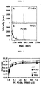
- FIG. 9 wild type PC bound to sEPCR with a K d(app) of approximately 31.3 nM, which is similar to previously reported binding affinity for PC to sEPCR (29 nM) in an aqueous solution.
- K d(app) of 32.0, 31.5 and 30.7 nM were obtainded for the interaction between sEPCR and PC-Gla peptide, TFG and TFMG, respectively.
Landscapes
- Health & Medical Sciences (AREA)
- Chemical & Material Sciences (AREA)
- Life Sciences & Earth Sciences (AREA)
- Organic Chemistry (AREA)
- Genetics & Genomics (AREA)
- General Health & Medical Sciences (AREA)
- Medicinal Chemistry (AREA)
- Proteomics, Peptides & Aminoacids (AREA)
- Molecular Biology (AREA)
- Biochemistry (AREA)
- Zoology (AREA)
- Gastroenterology & Hepatology (AREA)
- Biophysics (AREA)
- Engineering & Computer Science (AREA)
- Toxicology (AREA)
- Bioinformatics & Cheminformatics (AREA)
- Pharmacology & Pharmacy (AREA)
- Animal Behavior & Ethology (AREA)
- Public Health (AREA)
- Veterinary Medicine (AREA)
- Biomedical Technology (AREA)
- Epidemiology (AREA)
- Immunology (AREA)
- General Engineering & Computer Science (AREA)
- Biotechnology (AREA)
- Wood Science & Technology (AREA)
- Nuclear Medicine, Radiotherapy & Molecular Imaging (AREA)
- Physics & Mathematics (AREA)
- General Chemical & Material Sciences (AREA)
- Plant Pathology (AREA)
- Chemical Kinetics & Catalysis (AREA)
- Microbiology (AREA)
- Rheumatology (AREA)
- Pain & Pain Management (AREA)
- Medicines That Contain Protein Lipid Enzymes And Other Medicines (AREA)
- Peptides Or Proteins (AREA)
Description
- The present invention relates to: a fusion polypeptide in which an anti-inflammatory polypeptide and a ferritin monomer fragment are bound, and a use thereof. More specifically, the present invention relates to a fusion polypeptide in which an anti-inflammatory polypeptide is fused to a N-terminus and/or C-terminus of a human-derived ferritin monomer fragment in which a portion of a fourth loop and a fifth helix a human-derived ferritin monomer are removed, and a use thereof for treating inflammatory diseases.
- Inflammation reaction is collectively referred to as a defensive reaction of a living body to restore the structure and function of its tissues damaged by infection, trauma and the like. Mobilization of leukocyte cells to the site of inflammation is important for the rapid resolution of the infection and the repair of tissue damages resulting from various traumas. However, erroneous or persistent inflammatory reactions may cause damage and disease to the tissues of the body. For example, inflammatory diseases may result from infections caused by bacteria or viruses such as cerebrospinal meningitis, enteritis, dermatitis, uveitis, encephalitis, adult respiratory distress syndrome, or non-infective factors such as trauma, autoimmune diseases and organ transplant rejection. Inflammatory diseases are classified into acute and chronic inflammatory diseases which have different symptoms and pathological features, respectively. Local symptoms of acute inflammation such as allergies, bacterial and viral infections include changes in blood flow and blood vessel size, changes in vascular permeability and leukocyte infiltration. On the other hand, the main pathological features of chronic inflammation such as rheumatoid arthritis, atherosclerosis, chronic nephritis and liver cirrhosis are that inflammatory factors are not removed and thus monocytes, neutrophils, lymphocytes and plasma cells continuously infiltrate into inflammation sites, resulting in rendering the inflammation reaction chronic.
- Inflammatory mediators expressed in inflammatory sites such as cytokines, chemokines, reactive oxygen intermediates, cyclooxygenase-2 (COX-2), 5-lipoxygenase (5-LOX), and matrix metalloproteinase (MMP) play an important role in the generation and maintenance of inflammatory reactions. Expression of these inflammatory mediators is mediated by transcription factors such as NF-KB (nuclear factor κB), STAT3 (signal transducer and activator of transcription 3), AP-1 (activator protein 1), and HIF-1a (hypoxia-inducible factor 1a).
- Sepsis, on the other hand, is a systemic inflammatory reaction caused by an abnormal defense of the body against an infected microorganism. The activation of macrophages is associated with excessive production of inflammatory factors, leading to a severe inflammatory response in the whole body. When there are shown at least two symptoms of a fever with body temperature rising 38 °C or above, hypothermia with body temperature falling to 36 °C or below, a respiratory rate of at least 24 breaths per minute (tachypnea), a pulse rate of at least 90 beats per minute (tachycardia), and a blood test result showing an increase or a marked decrease in leukocyte count, it is called a systemic inflammatory response syndrome (SIRS). it is called sepsis when the systemic inflammatory response syndrome is caused by microbial infection. The sepsis can potentially lead to a septic shock. When sepsis gets worse, the function of various organs (heart, kidney, liver, brain, lungs, etc.) of the body deteriorates. If it gets much worse, it may lead to a shock state. The sepsis may be caused by various types of pathogens. Its highest incidence is induced by bacteria, while being also caused by viruses or fungi. There are pneumonia causing an infection in the lungs, urinary tract infection causing an infection in the bladder and kidneys, cellulitis occurring in skin, appendicitis occurring in the abdomen, or meningitis occurring in the brain, and the like. For example, if a patient with pneumonia has sepsis, his/her brain, heart, liver, lungs, or kidneys can be damaged, while about 20-50% of patients die from septic shock if severe progression occurs. In addition, the sepsis may occur by a post-operative infection. 40 to 90% of patients may die in case a sepsis occurs as a hyperacute inflammatory response due to infection or a postoperative infection.
- It is understood that the sepsis occurs as a result of complex interactions between causative organisms and host immune, inflammation and coagulation systems. Both the response of the host and the characteristics of the causative organisms have a significant impact on the prognosis of sepsis. Organ failure observed in sepsis occurs when the host inadequately reacts to causative organisms. If the host's response to the causative organisms is over-amplified, it can lead to organ damage in the host itself. Based on this concept, antagonistic substances against proinflammatory cytokines such as TNF-α, IL-1β and IL-6, which play a leading role in host inflammation, have been applied as a treatment for sepsis, but most of them found unsuccessful. Further, mechanical ventilation, the administration of activated protein C (C), and glucocorticoid treatment have been also tried, but various limitations have been revealed.
- JAE OG JEON et al. discloses the provision of ferritin nanocages with an anti-inflammatory effect and discloses fusing of interleukin-4 receptor (IL4-R)-targeting peptide AP-1 peptide as anti-inflammatory protein with ferritin L-chain (FTL) forming AP-1 containing nanocages proteins (AP1-PBNCS) ("Designed Nanocage Displaying Ligand-Specific Peptide Bunches for High Affinity and Biological Activity", ACS ANO, vol.7, no.9, pages 7462-7471).
US 9 920 105 B2 - The disclosure of JONG-SUP BAE et al. relates to the the pathway in cytokine-stimulated vascular endothelial cells, showing that thrombin receptor agonist peptide (TRAP) and activated protein C (APC) reduce alone or in combination the pro-inflammatorx reaction ("Thrombin inhibits nuclear factor", THROMBOSIS AND HAEMOSTASIS, vol.101, no.3, paged 513-520).
-
US 2006/116343 A1 discloses a TRAP sequence. -
US 2011/287033A1 discloses a ferritin sequence. -
EP 2 157 176 A1 - DIDI HE et al. summarises in a review of ferritin family proteins that fusions of entire protein domains to the surface of ferritin are complicated by the requirement to match the subunit organisation of the protein to be displayed on its surface with the inherent symmetry of the ferritin cage itself ("Ferritin family proteins and their use in bionanotechnology", NEW BIOTECHNOLOGY, vol.32, no.6, pages 651-657) .
- WONHWA LEE et al. "A Double-Chambered Protein Nanocage Loaded with Thrombin Receptor Agonist Peptide (TRAP) and [gamma]-Carboxyglutamic Acid of Protein C (PC-Gla) for Sepsis Treatment", ADVANCED MATERIALS, vol. 27, no.42, pages 6637-6643 is the corresponding scientific publication to the present invention.
-
WO 2013/028740 A1 and the corresponding scientific publication J.-S. Bae et al. disclose the chimeric protein protein C - meizothrombin (PC-gla/MeizoTh and its cleavage of of HMGB1, thereby resulting in an anti-inflammatory effect. Further disclosed is that activated protein C (APC) is the only approved drug for sepsis at the time point of disclosure. Also disclosed is the use of polypeptides for treating patients with severe sepsis ("Activated protein C inhibits high ). - Therefore, there is a need for a new therapeutic agent for use in preventing or for use in treating sepsis and septic shock with a high mortality rate, for which a definite therapeutic agent has yet to be developed.
- Accordingly, the present inventors have completed the present invention after they constructed a fusion polypeptide in which an anti-inflammatory polypeptide is fused to a N-terminus and/or a C-terminus of a human-derived ferritin monomer fragment (short ferritin, sFt) in which a portion of a fourth loop and a fifth helix of a human-derived ferritin monomer are removed, finding that the fusion polypeptide is capable of fusing different polypeptide medicinal agents at its N-terminus or C-terminus, and forming a nano-cage via self-assembling even after the fusion to effectively deliver the medicinal agents.
- An aspect of the present invention is to provide a fusion polypeptide comprising the amino acid sequence of SEQ ID NO: 14 or 15.
- Another aspect of the present invention is to provide a polynucleotide encoding the fusion polypeptide.
- Still another aspect of the present invention is to provide an expression vector comprising the polynucleotide.
- Still another aspect of the present invention is to provide an isolated transformed cell transformed with the expression vector.
- Further another aspect of the present invention is to provide a protein cage comprising the fusion polypeptide, wherein an anti-inflammatory polypeptide protrudes outside the protein cage.
- Still another aspect of the present invention is to provide a pharmaceutical composition for use in treating an inflammatory disease, the composition comprising the fusion polypeptide as an active ingredient.
- Still another aspect of the present invention is to provide use of the fusion polypeptide for preparing an agent for use in treating an inflammatory disease.
- The present invention provides a fusion polypeptide comprising the amino acid sequence of SEQ ID NO: 14 or 15.
- Another embodiment according to an aspect of the present invention provides a polynucleotide encoding the fusion polypeptide.
- Still another embodiment according to an aspect of the present invention provides an expression vector comprising the polynucleotide.
- Still another embodiment according to an aspect of the present invention provides an isolated transformed cell transformed with the expression vector.
- An embodiment according to another aspect of the present invention provides a protein cage comprising the fusion polypeptide, wherein an anti-inflammatory polypeptide protrudes outside the protein cage.
- Another embodiment according to an aspect of the present invention provides a pharmaceutical composition for use in treating an inflammatory disease, the composition comprising the fusion polypeptide as an active ingredient.
- Still another embodiment according to an aspect of the present invention provides a pharmaceutical composition for use in treating an inflammatory disease, the composition consisting of the fusion polypeptide.
- Still another embodiment according to an aspect of the present invention provides a pharmaceutical composition for use in treating an inflammatory disease, the composition consisting essentially of the fusion polypeptide.
- An embodiment according to still another aspect of the present invention provides a use of the fusion polypeptide for preparing an agent for use in treating an inflammatory disease.
- Hereinafter, the present invention will be described in detail.
- Ferritin is an intracellular protein which stores and releases iron. Ferritin exists generally in the form of a hollow spherical cage in vivo, wherein the cage is composed of 24 ferritin monomers which are classified into heavy chain and light chain depending on their structure.
- In the present invention, the anti-inflammatory fusion polypeptide having a remarkably improved therapeutic effect was prepared by fusing a polypeptide showing anti-inflammatory activity not only at the N-terminus but also at the C-terminus of the ferritin monomer fragment.
- In an embodiment according to the present invention, the fusion polypeptide may be prepared by a method known to those skilled in the art. Such fusion polypeptides may be produced in prokaryotic or eukaryotic cells by expressing polynucleotides encoding the fusion polypeptide sequences of the present invention, often as a part of larger polypeptides. Alternatively, such fusion polypeptides may be synthesized by chemical methods. Methods for expression of heterologous proteins in recombinant hosts, chemical synthesis of polypeptides and in vitro transcription are well known in the art and are further described in the literatures (Reference: Maniatis et al., Molecular Cloning: A Laboratory Manual (1989), 2nd Ed., Cold Sprin Harbor, N.Y.; Berger and Kimmel, Methods in Enzymology, Volume 152, Guide to Molecular Cloning Techniques (1987), Academic Press, Inc., San Diego, Calif.; Merrifield, J. (1969) J. Am. Chem. Soc. 91:501; Chaiken I.M. (1981) CRC Crit. Rev. Biochem. 11: 255; Kaiser et al. (1989) Ann. Rev. Biochem. 57:957; and Offord, R.E. (1980) Semisynthetic Proteins, Wiley Publishing).
- The present invention provides a fusion polypeptide which has an amino acid sequence represented by SEQ ID NO: 14 or 15.
- The amino acid sequences of SEQ ID NOS: 14 and 15 are as follows, respectively:


- An embodiment of the present invention provides a polynucleotide encoding the fusion polypeptide. The polynucleotide according to the present invention may be any base sequences which encode the fusion polypeptide according to the present invention.
- An embodiment of the present invention also provides an expression vector comprising the polynucleotide according to the present invention.
- As used herein, the expression vector is characterized by comprising the polynucleotide of the present invention, while its includes, but is not limited to, a plasmid vector, a cosmid vector, a bacteriophage vector, and a viral vector. The expression vector of the present invention may be a conventional expression vector. The expression vector may contain an expression regulatory sequence such as a promoter, an operator, an initiation codon, a termination codon, a polyadenylation signal and an enhancer (promoter gene), as well as a signal sequence or leader sequence for membrane targeting or secretion, while it may be variously prepared according to its purpose. The promoter of the expression vector may be constitutive or inducible. The vector also comprises a selection marker for selecting a host cell containing the vector, while it contains the origin of replication if it is a replicable vector.
- Another embodiment of the present invention also provides an isolated transformed cell transformed with the expression vector according to the present invention.
- The isolated transformed cell of the present invention is characterized by being transformed with the expression vector of the present invention. Transformation with the above expression vector can be carried out by transformation techniques known to a person skilled in the art. Preferably, microprojectile bombardment, electroporation, calcium phosphate (CaPO4) precipitation, calcium chloride (CaCl2) precipitation, PEG-mediated fusion, microinjection, and a liposome-mediated method may be used. The isolated transformed cell may be Escherichia coli, Bacillus subtilis, Streptomyces, Pseudomonas, Proteus mirabilis, Staphylococcus, and Agrobacterium tumefaciens, but are not limited thereto.
- Meanwhile, another embodiment of the present invention provides a protein cage comprising the fusion polypeptide, wherein the anti-inflammatory polypeptide protrudes outside the protein cage.
- Still another embodiment of the present invention provides a protein cage consisting of the fusion polypeptide, wherein the anti-inflammatory polypeptide protrudes outside the protein cage.
- Still another embodiment of the present invention provides a protein cage consisting essentially of the fusion polypeptide, wherein the anti-inflammatory polypeptide protrudes outside the protein cage.
- As used herein, the protein cage is a cage composed of protein which is formed by the precise self-assembling of low molecular weight monomers and possesses an internal space. It includes viral capsid protein, ferritin, heat shock protein, and Dps protein. The protein cage according to the present invention is characterized in that it comprises the fusion polypeptide of the present invention as a monomer constituting the protein cage. As used herein, the term 'self-assembling' refers to the property of a certain molecule with which the molecule forms a specific nanostructure by itself without any external stimulation or artificial induction.
- The protein cage of the present invention is prepared by binding of the fusion polypeptide of the present invention and is generally in the form of a spherical cage in vivo.
- The protein cage of the present invention may be a complex protein in which the fusion polypeptide of the present invention is regularly arranged as a unit. More preferably, the protein cage may be formed by regularly arranging 24 fusion polypeptides of the present invention three-dimensionally. Meanwhile, when the fusion polypeptide of the present invention forms a protein cage by self-assembling, the anti-inflammatory polypeptide fused to the N-terminus and/or the C-terminus of the ferritin monomer fragment may protrude through the outer surface of the cage protein and exhibit a physiological activity by easily binding to a target receptor or protein. Alternatively, the linker may be cleaved by MMP in a living tissue showing an inflammatory disease so that the anti-inflammatory polypeptide is dissociated from the protein cage to exhibit its own physiological activity.
- An embodiment of the present invention also provides a pharmaceutical composition for use in treating an inflammatory disease, the composition comprising the fusion polypeptide as an active ingredient.
- Another embodiment of the present invention provides a pharmaceutical composition for use in treating an inflammatory disease, the composition consisting of the fusion polypeptide as an active ingredient.
- Still another embodiment of the present invention provides a pharmaceutical composition for use in treating an inflammatory disease, the composition consisting essentially of the fusion polypeptide as an active ingredient.
- The pharmaceutical composition according to the present invention may be formulated into a suitable form by comprising the fusion peptide alone or in combination with a pharmaceutically acceptable carrier, and may further contain an excipient or a diluent. The term "pharmaceutically acceptable" as used herein refers to a non-toxic composition that is physiologically acceptable and does not cause allergic reactions such as gastrointestinal disorder or dizziness, or a similar reaction when administered to humans.
- The pharmaceutically acceptable carrier may further include, for example, a carrier for oral administration or for parenteral administration. The carrier for oral administration may include lactose, starch, cellulose derivatives, magnesium stearate and stearic acid. In addition, it may include various drug delivery materials used for oral administration of peptide agents. In addition, the carrier for parenteral administration may include water, suitable oil, a saline solution, an aqueous glucose and a glycol, and may further contain a stabilizer and a preservative. Suitable stabilizers include antioxidants such as sodium hydrogen sulfite, sodium sulfite and ascorbic acid. Suitable preservatives include benzalkonium chloride, methyl- or propyl-paraben and chlorobutanol. The pharmaceutical composition of the present invention may further include a lubricant, a wetting agent, a sweetener, a flavoring agent, an emulsifying agent, and a suspending agent, in addition to the above components. Regarding other pharmaceutically acceptable carriers and preparations, the following literature may be referred (Remington's Pharmaceutical Sciences, 19th ed., Mack Publishing Company, Easton, Pa., 1995).
- The composition of the present invention may be administered to mammals including humans by any method. For example, it may be administered orally or parenterally. Parenteral administration methods include, but are not limited to, intravenous, intramuscular, intra-arterial, intramedullary, intradural, intracardiac, transdermal, subcutaneous, intraperitoneal, intranasal, intestinal, topical, sublingual or rectal administration.
- The pharmaceutical composition of the present invention may be formulated into oral or parenteral preparations according to the route of administration as described above.
- In the case of oral preparations, the composition of the present invention may be formulated into powder, granules, tablets, pills, sugar-coated tablets, capsules, liquids, gels, syrups, slurries, suspensions or the like by using methods known in the art. For example, oral preparations may be obtained as tablets or sugar-coated tablets by combining the active ingredient with a solid excipient, pulverizing it, adding suitable auxiliaries, and then processing the mixture into a granular mixture. Examples of suitable excipients include sugars including lactose, dextrose, sucrose, sorbitol, mannitol, xylitol, erythritol and maltitol; starchs including corn starch, wheat starch, rice starch and potato starch; celluloses including cellulose, methylcellulose, sodium carboxymethyl-cellulose and hydroxypropylmethyl-cellulose; and fillers including gelatin and polyvinylpyrrolidone. In addition, optionally, crosslinked polyvinylpyrrolidone, agar, alginic acid, or sodium alginate may be added as a disintegrant. In addition, the pharmaceutical composition of the present invention may further comprise anti-aggregating agents, lubricants, wetting agents, flavoring agents, emulsifying agents, and preservatives.
- The preparation for parenteral administration may be formulated into the form of injections, creams, lotions, external ointments, oils, moisturizers, gels, aerosols and nasal inhalers by methods known in the art. These formulations are described in the literature (Remington's Pharmaceutical Science, 19th ed., Mack Publishing Company, Easton, PA,1995), which is a commonly known formulary for the entire fields of pharmaceutical chemistry.
- The total effective amount of the composition of the present invention may be administered to a patient in a single dose and may be administered by a fractionated treatment protocol for a long term with multiple doses. The pharmaceutical composition of the present invention may vary in the content of the active ingredient depending on the severity of the disease. Preferably, the preferred total dosage of the pharmaceutical composition of the present invention is from about 0.01 µg to about 10,000 mg, most preferably from 0.1 µg to 500 mg (
TFG 100 nM = 134.33 µg/kg,TFMG 100 nM = 137.015 µg/kg) per 1 kg patient weight per day. However, regarding the dosage of the pharmaceutical composition, the effective dosage for each patient is determined upon considering various factors such as formulation method, administration route and frequency of treatment, as well as the patient's age, weight, health condition, sex, severity of disease, diet and excretion rate. Therefore, one of ordinary skill in the art will be able to determine the appropriate effective dose of the composition of the present invention. The pharmaceutical composition according to the present invention is not particularly limited to the type of formulation, administration route and administration method as long as the effect of the present invention is exhibited. - As used herein, the inflammatory disease is selected from the group consisting of inflammatory bowel disease, diabetic eye disease, peritonitis, osteomyelitis, cellulitis, meningitis, encephalitis, pancreatitis, traumatic shock, bronchial asthma, rhinitis, sinusitis, otitis, pneumonia, gastritis, enteritis, cystic fibrosis, apoplexy. bronchitis, bronchiolitis, hepatitis, nephritis, arthritis, gout, spondylitis, Reiter's syndrome, polyarteritis nodosa, irritable vasculitis, Lou Gehrig's granulomatosis, Polymyalgia rheumatica, arthritic arteritis, calcium crystal arthropathies, pseudogout, non-articular rheumatism, bursitis, tenosynovitis, epicondylitis (tennis elbow), neuropathic joint disease (Charcot's joint), hemarthrosis, Henoch-Schonlein purpura, hypertrophic osteoarthropathy, multicentric reticulohistiocytoma, surcoilosis, hemochromatosis, sickle cell disease and other hemochromatosis, hyperlipoproteinemia, hypogammaglobulinemia, hyperparathyroidism, acromegaly, familial Mediterranean fever, Behcet's disease, systemic lupus erythematosus, relapsing fever, psoriasis, multiple sclerosis, sepsis, septic shock, acute respiratory distress syndrome, multiple organ dysfunction syndrome, chronic obstructive pulmonary disease, acute lung injury and broncho-pulmonary dysplasia. Preferably, the inflammatory disease may be sepsis.
- The inventors have experimentally confirmed the effectiveness of the fusion polypeptide according to the present invention for use in the treatment of the above described inflammatory diseases, particularly sepsis.
- Activated protein C (APC) used as a therapeutic agent for sepsis has characteristics of the prevention of blood clotting, anti-inflammation, protective barrier, and fibrous properties, and was approved by the FDA in 2001 and the EMA in 2002 for treating septic shock and severe asthma. In October 2011, APC was withdrawn from the market due to its lack of favorable effects of 28-day death and adverse side effects in the PROWESS and PROWESS-SHOCK tests. The most common side effects associated with APC are hemorrhage caused by degradation of procoagulant elements Va and VIIIa, which is consistent with the antithrombotic activity of APC. Therefore, there is currently no effective preventive or therapeutic method for severe sepsis.
- Meanwhile, the activity of APC is caused by the interaction between the endothelial protein C receptor (EPCR) and the γ-carboxyglutamic acid (Gla) domain of PC/APC. The γ-carboxyglutamic acid (Gla) domain (PC-Gla) of PC/APC does not degrade the clotting factor, but may provide a protease capable of binding to protease-activated receptor-1 (PAR-1). When PC-Gla binds to EPCR, PAR-1 is cleaved. As a result, cell protective signaling responses such as barrier protection and anti-inflammation can be triggered.
- Thrombin may bind to PAR-1 in at least three ways with a higher efficient scale of catalyst than APCs. The activity of PAR-1 by the thrombin receptor agonist peptide (TRAP) mimics the effect of thrombin in human endothelial cells. Conventional studies show that when PC binds to EPCR, PAR-1 dependent signaling by thrombin or TRAP is converted to a cytoprotective response from the pro-inflammatory signal of endothelial cells (Blood 2007, 110, 3909,
Thromb Haemost 2008, 100, 101 - In sum, the present inventors hypothesized that it would be possible to maximize a therapeutic effect for sepsis without causing problems such as bleeding if EPCR and PAR-1 can be simultaneously targeted by fusing a ligand capable of binding specifically to EPCR and PAR-1 through a single drug delivery system. TRAP, a peptide that activates PAR-1, was fused to the N-terminus of the human-derived ferritin monomer fragment. At the same time, a fusion polypeptide (TFG) in which PC-Gla, a peptide targeting EPCR, was fused to the C-terminus of the ferritin monomer was prepared so that the TRAP and the PC-Gla protruded outside the ferritin cage (See Example 1).
- In order to prevent the effect of mutual interference between TRAP and PC-Gla fused to the N-terminus or C-terminus of the human ferritin monomer on their physiological activities, respectively, a fusion polypeptide (TFMG) was prepared in which a linker having an amino acid sequence capable of being cleaved by MMP-2 was bound between the C-terminus of the human ferritin monomer fragment and PC-Gla. The linker was cleaved at the MMP-activated pathological site, resulting in the release of PC-Gla from the ferritin cage (See Example 1).
- According to another Example, it was confirmed that MMP-2 was secreted from HUVEC cells by LPS or CLP, and that the linker in TFMG was cleaved by MMP-2 and PC-Gla was continuously released (See Example 2). Thus, it was confirmed that, when an anti-inflammatory polypeptide is fused through a linker containing an amino acid sequence capable of serving as a substrate for MMP at the C-terminus of the monomer fragment of human ferritin, the linker is exposed to MMP even after the ferritin cage is formed and thus can be cleaved by MMP to release the fused polypeptide into the pathological site.
- In another Example, the degree of binding affinity between the TFG or TFMG fusion polypeptide with EPCR was evaluated. As a result, it was observed that TFG and TFMG bind to EPCR with the same degree of binding affinity as that of PC-Gla, respectively, verifying that PC-Gla may easily bind to EPCR and exhibit its physiological activity even after PC-Gla is fused to the monomer fragment of human ferritin (See Example 3).
- Further, in still another Example, it was evaluated whether the TFG or TFMG fusion polypeptide may activate PAR-1. As a result, it was found that the TFG and TFMG fusion polypeptide activate PAR-1 to the same extent as TRAP peptide does, confirming that TRAP maintains its physiological activity of activating PAR-1 even after it was fused to the monomer fragment of human ferritin (See Example 3).
- In another Example, therapeutic effects for sepsis were evaluated after administering TFG, TFMG, or a combination of PC-GLA and TRAP to an animal model of sepsis induced by CLP, respectively. As a result, it was confirmed that TFMG- or TFG-treated groups showed remarkably excellent effects for use in preventing and for use in treating sepsis in comparison with the co-administration of PC-GLA and TRAP-treated group, in terms of such evaluation factors as animal mortality, the degree of penetration of inflammatory cells into tissues, the degree of lung tissue necrosis, liver toxicity, kidney toxicity, LDH levels as an indicator of tissue damage, the secretion level of inflammatory cytokines, and the expression level of the adhesion factors of endothelial cells which promote the collapse of blood vessel barriers and the migration of leukocytes (See Example 4).
- Another embodiment of the present invention provides a pharmaceutical composition comprising the fusion polypeptide which is effective in inhibiting the production of inflammation inducing mediators including TNF-α, IL-6 and IL-10.
- Still another embodiment of the present invention provides use of the fusion polypeptide for preparing an agent for use in treating an inflammatory disease.
- As used herein, the "effective amount" of the present invention refers to an amount that, when administered to a subject, elicits an improvement, treatment, prevention, detection, or diagnostic effect of an inflammatory disease. Preferably, the term "subject" may be an animal including a mammal, particularly a human, and may include an animal-derived cells, tissues, and organs. The subject may be a patient requiring treatment.
- The term "for use in treatment" or "for use in treating" of the present invention broadly refers to ameliorating an inflammatory disease or the symptoms of an inflammatory disease, including curing, substantially preventing and improving the conditions of such a disease. It includes, but is not limited to, ameliorating, curing or preventing one or most of the symptoms resulting from an inflammatory disease.
- As used herein, the term "comprising" is used synonymously with the terms "containing" and "characterized by" and does not exclude additional components or method steps which are not mentioned in a composition or method. The term "consisting of" means to exclude additional elements, steps or components which are not mentioned. The term "consisting essentially of" means to include a material or step that does not substantially affect the basic characteristics of a composition or method, as well as the mentioned material or step.
- As described above, there is provided a fusion polypeptide comprising the amino acid sequence of SEQ ID NO: 14 or 15, which may fuse two types of anti-inflammatory polypeptides which act through different mechanisms, respectively, into a nanocage for administration, thus the fusion polypeptide exhibiting an excellent effect in the use for treatment of an inflammatory disease including sepsis.
-
-
FIG. 1 is a schematic diagram showing the fusion of TFG and TFMG fusion polypeptides (A: Fusion schematic diagram of TFG and TFMG, B: 3D schematic diagram of fusion polypeptide and ferritin nanocage) -
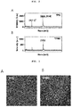
FIG. 2 shows the results of measurement of the molecular weights of TFG and TFMG with a MALDI-ToF mass spectrometer (A: TFG, B: TFMG). -
FIG. 3 is the result of TEM imaging observing that TFG and TFMG form a ferritin cage by self-assembling (A: TFG, B: TFMG). -
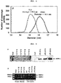
FIG. 4 shows the results of measuring the diameter of the ferritin cage formed by TFG and TFMG. -
FIG. 5 shows the results of secretion of MMP-2 by CLP or LPS stimulation in HUVEC cells (A: results of Western-blot, B: results of zymography). -
FIG. 6 shows the result of observing whether PC-Gla is released by cleavage of the linker in TFMG by AMPA-activated MMP-2. -
FIG. 7 shows the HPLC analysis results of confirming the released PC-Gla after the linker of TFMG was cleaved by MMP-2. -
FIG. 8 shows the MALDI-ToF results of confirming the released PC-Gla after the linker of TFMG was cleaved by MMP-2. -
FIG. 9 shows the solid-phase ELISA results of evaluating the binding affinity of Protein C (PC), PC-Gla domain, TFG and TFMG to soluble EPCR. -
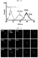
FIG. 10 shows the results of evaluating the binding affinity of PC-Gla, TFG and TFMG to HUVEC cells. -
FIG. 11 shows the in vivo immunohistological staining results of confirming the binding affinity of PC-Gla, TFG and TFME, respectively. -
FIG. 12 shows the results of evaluating the degree of binding affinity of PC-Gla, TFG and TFMG to endothelial cells in vitro (HUVEC) or in vivo (mouse animal model) (A: in vitro, B: in vivo), respectively. -
FIG. 13 shows the results of evaluating the degree of activation of PAR-1 by TRAP peptide, TFG and TFMG, respectively. -
FIG. 14 shows the results of observing the survival rate of animals after TFG administration, TFMG administration, and co-administration of TRAP and PC-Gla to an animal model of CLP-induced sepsis, respectively. -
FIG. 15 shows the results of observing the infiltration of inflammatory cells into tissues and the damage of the lung tissues of the animal after TFG administration, TFMG administration, and co-administration of TRAP and PC-Gla to an animal model of CLP-induced sepsis, respectively. -
FIG. 16 shows the results of scoring the degree of lung damage in animals after TFG administration, TFMG administration, and co-administration of TRAP and PC-Gla to an animal model of CLP-induced sepsis, respectively. -
FIG. 17 shows the results of the evaluating ALT or AST (A), creatinine (B), BUN (C) and LDH levels (D) after TFG administration, TFMG administration and co-administration of TRAP and PC-Gla to an animal model of CLP-induced sepsis, respectively. -
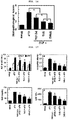
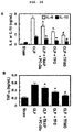
FIG. 18 shows the results of evaluating the levels of inflammatory cytokines in blood after TFG administration, TFMG administration, and co-administration of TRAP and PC-Gla to an animal model of CLP-induced sepsis (A: IL-6, IL-10; B: TNF-a). -
FIG. 19 shows the results of measuring the permeability after TFG administration, TFMG administration and co-administration of TRAP and PC-Gla to LPS-stimulated HUVEC cells, respectively. -
FIG. 20 shows the result of evaluating vascular permeability after TFG administration, TFMG administration and co-administration of TRAP and PC-Gla to an animal model of CLP-induced sepsis, respectively. -
FIG. 21 shows the results of evaluating the expression level (A) of vascular cell adhesion factor-1 (VCAM-1) and the adhesion amount of leukocyte after TFG administration, TFMG administration and co-administration of TRAP and PC-Gla to LPS-stimulated HUVEC cells, respectively. -
FIG. 22 shows the results of evaluating the amount of leukocyte movement after TFG administration, TFMG administration, and co-administration of TRAP and PC-Gla to an animal model with CLP-induced sepsis, respectively. - Hereinafter, the present invention will be described in detail.
- However, the following Examples are merely illustrative of the present invention, while the scope of the present invention is not limited to the following Examples.
- PC-Gla and TRAP were synthesized by Peptron Inc. (Daejeon, Republic of Korea) and Anygen Inc. (Gwangju, Republic of Korea). Peptides were labeled with FNG-456 NHS or FNI-675 NHS ester fluorescent dyes (Bioacts Inc., Incheon, Republic of Korea). Bacterial lipopolysaccharide (LPS, serotype: 0111:B4, L5293), antibiotics (penicillin G and streptomycin), α-cyano-4-hydroxycinnamic acid (CHCA), sinapic acid, and p-aminophenylmercuric acetate were purchased from Sigma (St. Louis, MO). Anti-MMP-2 antibody (MAB13434) was purchased from Millipore, anti-mouse CD31 (553369) from BD Falcon, and anti-EPCR antibody (FL-238, sc-28978) from Santa Cruz.
- A plasmid was constructed for the expression of short ferritin (sFn) by deleting the short E-heilx from ferritin light chain. The DNA plasmids of TFG and TFMG were constructed by introducing TRAP (TFLLRN) peptide sequence into the N-teruminus of sFn with restriction sites, SpeI and XhoI, at 5- and 3'-ends. In addition, the PC-Gla domain (ANSFLEELRHSSLERECIEEICDFEEAKEIFQNVDDTLAFWSKHV) sequence or MMP-2 cleavage site (GPLGLAG) were constructed in front of the PC-Gla domain to the C-terminus of sFn at 5'- and 3'-ends with restriction sites, SpeI and XhoI at 5'- and 3'-ends. Primers were designed as follows: (+) 5' CAC TTT TCT TCT TCG GAA CG 3' and (-) 5' CTA GCG TTC CGA AGA AGA AAA GTG GTA C 3' for TRAP; (+) 5' GAA ACT AGT GCC AAC TCC TTC CTG GAG G 3' and (-) 5' GAA CTC GAG GAC GTG CTT GGA CCA G 3' for PC-Gla domain; (+) 5' GAA ACT AGT GGT CCT CTA GGT CTA GCC GGT GCC AAC TCC TTC CTG G 3' and (-) 5' GAA CTC GAG GAC GTG CTT GGA CCA G 3' for the MMP-2 cleavage site in front of the PC-Gla domain. TFG and TMFG plasmids were transformed into Escherichia coli (E. coli) expression strain BL21 (DE3). Cells were grown at 37 °C in LB medium containing 50 µg/ml kanamycin until OD600 reached 0.5, and the expression of protein was induced by 0.1M IPTG treatment at 37 °C for 5 hours. After induction, cells were harvested by centrifugation, and the pellets were suspended in lysis buffer (20 mM Tris-HCl pH 8.0, 100 mM NaCl, 1% Triton X-100, 1 mM PMSF, 1 mM DTT 1:000 dilution protease inhibitor cocktail) and homogenized with an ultrasonic processor. The inclusion bodies from cell lysates were solubilized by incubating in binding buffer (20 mM Tris-HCl pH 8.0, 300 NaCl, 10 mM imidazole) containing 8 M urea at room temperature for 1 hour. Subsequently, the denatured protein was loaded onto a nickel ion chelate affinity column rinsed with a washing buffer (20 mM Tris-HCl pH 8.0, 500 mM NaCl, 30 mM imidazole) containing 8 M urea. The protein was eluted with elution buffer (20 mM Tris-HCl pH 8.0, 100 mM NaCl, 300 mM NaCl, 10 mM imidazole) containing 8 M urea at room temperature for 1 hour. Subsequently, the denatured protein was loaded onto a nickel ion chelate affinity column rinsed with a washing buffer (20 mM Tris-HCl pH 8.0, 500 mM NaCl, 30 mM imidazole) containing 8 M urea. The protein was eluted with elution buffer (20 mM Tris-HCl pH 8.0, 100 mM NaCl, 300 mM imidazole) and refolded by dialysis with a gradient of urea.
- The mass spectrum of each construct monomer was confirmed by matrix assisted laser desorption ionization time of flight (MALDI-ToF). MALDI-ToF MS was carried out using a Bruker Daltonics Microflex MALDI-ToF mass spectrometer (Bremen, Germany) with a 337 nm nitrogen laser. Mass spectra were obtained in the linear and positive-ion mode with an acceleration voltage of 20 kV. A saturated solution of cyano-4-hydroxycinnamic acid (CHCA) or sinapic acid in 50% acetonitrile, containing a final concentration of 0.1% trifluoroacetic acid, was used as the matrix solution. A CHCA matrix was chosen for analysis of fragments after enzyme digestion or sinapic acid for intact proteins. The analyte-matrix solution was prepared at a ratio of 1:2 (analyte:matrix, v/v). Each mixture was thoroughly mixed, and 1 µL of the analyte-matrix solution was deposited onto the sample plate and dried by vacuum evaporation. The spectrometer was calibrated using bradykinin; cytochrome C and bovine serum albumin were run as close external standards. Transmission electron microscopy (TEM) images were recorded using an FEI Tecnai (Korea Basic Science Institute, KBSI). The size of nanocaged TFG and TFMG was measured using a DelsaMax Pro light scattering analyzer (Beckman Coulter).
- The primary HUVECs were obtained from Cambrex BioScience (Charles City, IA) and maintained as previously described. All experiments were performed using HUVECs at passage 3-5. Human neutrophils were freshly isolated from whole blood (15 ml) obtained by venous venipuncture from five healthy volunteers, and maintained as previously described.
- Male C57BL/6 mice (6-7 weeks of age, 18-20 g) were purchased from Orient Biotech (Seongnam, Gyeonggi Province, Republic of Korea) and used after 12 days of acclimation. Five mice per cage were housed under the conditions of a controlled temperature (20-25 °C), humidity (40-45%), and 12:12h day/night cycle, while being fed with a normal rodent pellet and water ad libitum. All animals were treated according to the Guidelines for the Care and Use of Laboratory Animals issued by Kyungpook National University.
- To induce inflammation, male mice were anesthetized with 2% isoflurane (JW Pharmaceutical, Republic of Korea) in oxygen delivered via a small rodent gas anesthesia machine (RC2, Vetequip, Pleasanton, CA), first in a breathing chamber and then via a facemask. They were allowed to breathe spontaneously during this procedure. The CLP-induced inflammation model was prepared as previously described. In brief, a 2-cm midline incision was made to expose the cecum and adjoining intestine. The cecum was then tightly ligated with a 3.0-silk suture at 5.0 mm from the cecal tip and punctured once using a 22-gauge needle for the induction of high grade inflammation. It was then squeezed gently to extrude a small amount of feces from the perforation site and returned to the peritoneal cavity. The laparotomy site was then sutured with 4.0-silk. In sham control animals, the cecum was exposed but not ligated or punctured and then returned to the abdominal cavity. This protocol was approved by the Animal Care Committee at Kyungpook National University prior to the conduct of the study (IRP No, KNU 2012-13).
- The activity of MMP-2 and MMP-9 enzymes in medium and plasma was determined by SDS-PAGE gelatin zymography. Gelatinases present in the plasma degrade the gelatin matrix, leaving a clear band after staining the gel for protein. Briefly, LPS time-dependently treated HUVECs media and albumin-derived septic mice plasma (normalized to an equal amount of protein [20 µg]) were denatured in the absence of a reducing agent and electrophoresed using 10% SDS-PAGE containing 0.1% (w/v) gelatin. Gels were incubated in the presence of 2.5% Triton X-100 at room temperature for 2 h and subsequently at 37°C overnight in a buffer containing 10mM CaCl2, 0.15M NaCl, and 50mM Tris (pH 7.5). Thereafter, gels were stained with 0.25% Coomassie Blue, and proteolysis was detected as a white band against a blue background.
- To evaluate whether TFMG could be selectively cleaved by MMP2, TFMG was incubated with APMA-mediated activated MMP-2 in PBS at 37°C for 3 h. The cleaved fragments of TFMG were detected by MALDI-ToF.
- To evaluate the interaction of the wild-type PC, PC-Gla peptides, TFG, and TFMG with EPCR, 96-well flat microtiter plates were coated with soluble EPCR in 20 mM carbonate-bicarbonate buffer (pH 9.6) containing 0.02% sodium azide, overnight at 4 °C. After the plates were washed three times in TBS buffer (0.1 M NaCl, 0.02 M Tris-HCl, pH 7.4) containing 0.05
% Tween 20, the plates were incubated with wild-type PC, PC-Gla peptides, TFG, and TFMG (7-1000 nM) diluted in the buffer for 1 h. After the plates were rinsed again, they were incubated with a goat anti-protein C polyclonal antibody (1:1000) for 1 h. Then, the plates were washed and incubated with rabbit anti-goat IgG (KPL, MD, 1:1000) for 1 h. After washing, the plates were incubated with 2,2'-azino-di(3-ethylbenzthiazoline-6-sulfonate) (ABTS; KPL, Gaithersburg, MD). Colorimetric analysis was performed by measuring absorbance values at 405 nm. - The endothelial cells were isolated according to the manufacturer's (Dynal Biotec, Lake Success, NY) instructions, using Dynabeads coupled to anti-CD31 antibody and the Dynal Magnetic holder. Briefly, for endothelial cell isolation, four to six mice (6-10 weeks old) were anesthetized, followed by exposure of the peritoneal cavity. Excised lungs and hearts were put into RPMI media, followed by removing other tissues from the heart and lungs, and then rinsing once in PBS. The lungs and heart were incubated with 1.0 mg/mL of collagenase A in a 50 mL tube for 1h at around 37°
C. Every 5 min during this incubation, the tube was gently agitated for a few seconds, and then the suspension was transferred into a new 50 mL tube by passing it through the 70 um tissue sieve (BD Falcon). The filtered cell suspension was centrifuged for 10 min at 1000 rpm. After removal of the supernatant, the cell pellet was washed once with cold PBS in a new 15-mL tube. To prepare the Dynabead-coupled anti-mouse CD31 antibody, Dynabeads (60 µl) were washed with MACS buffer (PBS, 0.5% BSA, 2mM EDTA) on a magnetic holder (Invitrogen). The Dynabeads were resuspended with MACS buffer (600 µl), anti-mouse CD31 (5 µg of per 10 µl of beads) was added, and the mixture was incubated for 12 h at 4°C. Cells were incubated with Dynabead-coupled anti-mouse CD31 antibody for 10 min at room temperature and then placed in a magnetic holder. Cell suspension was slowly added to a 15-mL tube by placing the pipette on the wall of the tube. After incubation for 5 min, PBS was carefully removed by aspiration. The Dynabead-coupled anti-mouse CD31 antibodies were washed three times in cold PBS, the pellet was resuspended in EBM-2 growth medium, and then harvested and lysed in RIPA buffer containing protease inhibitor cocktail on ice. - PC-Gla, TFG, and TFMG were labeled with FNG-456 NHS ester for in vitro assays or FNI-675 NHS ester for in vivo assays at a molar ratio of 1:3. Briefly, each molecule (10 µM) was dissolved in PBS (1.5 mL), and FNG-456 NHS ester (30 µM) or FNI-675 NHS ester (30 µM) was dissolved in DMSO (0.2 mL). Each molecule and fluorescent dye was reacted at room temperature for 3 h. The reaction product was passed through a 0.2-µm filtering unit, and the unreacted dye was separated on a PD midiTrap™ G-25 (GE Healthcare, UK) that had been pre-equilibrated in PBS with 2 mM sodium azide. This process yielded more than 2.17 µM of each nanoparticle with more than 1.5 ratio of dye per protein.
- A direct cell-binding assay was performed on HUVECs and in vivo using fluorescence labeled-PC-Gla, TFG according to the present invention, and TFMG (comparative). The assay was performed with PC-Gla, TFG, or TFMG treated on HUVECs, intravenously injected mice, and isolated mouse endothelial cells. The fluorescence value of the HUVECs or endothelial cells were measured with tightly bound PC-Gla, TFG, and TFMG, respectively. The concentrations of PC-Gla, TFG, and TFMG were measured by using the nanoparticle ratio of fluorescent dye per protein.
- HUVECs at 90% confluence in 24-well plates were transiently transfected with pRc/RSV containing ALP-PAR-1-TF cDNA in antibiotic-free Opti-MEM medium using Lipofectamine (Invitrogen) according to the manufacturer's instruction. On the following day, cells were washed and incubated in serum-free medium for 5 h. Cells were then incubated for an additional hour with thrombin, TRAP, TFG according to the present invention, or TFMG (comparative). Conditioned medium was collected and centrifuged to remove cellular debris. Supernatant was collected, and ALP (alkaline phosphatase) activity was measured using EnzoLyteTM p-nitrophenyl phosphate alkaline phosphatase assay kit (AnaSpec, San Jose, CA) according to the manufacturer's instructions.
- Male C57BL/6 mice underwent CLP and were administered PC-Gla with TRAP, TFG according to the present invention, or TFMG (200 nM) (comparative) intravenously at 6 h after CLP (n = 5). Mice were euthanized 96 h after CLP. To analyze the phenotypic change of the lungs in mice, lung samples were removed from each mouse, washed tree times in PBS (pH 7.4) to remove remaining blood, fixed in 4% formaldehyde solution (Junsei, Tokyo, Japan) in PBS, pH 7.4 for 20 h at 4°C. After fixation, the samples were dehydrated through ethanol series, embedded in paraffin, sectioned into 4-µm sections, and placed on a slide. The slides were de-paraffinized in a 60°C oven, rehydrated, and stained with hematoxylin (Sigma). To remove over-staining, the slides were quick dipped three times in 0.3% acid alcohol, and counterstained with eosin (Sigma). They are then washed in ethanol series and xylene, and then coverslipped. Light microscopic analysis of lung specimens was performed by blinded observation to evaluate pulmonary architecture, tissue edema, and infiltration of the inflammatory cells. The results were classified into four grades where
Grade 1 represented normal histopathology;Grade 2 represented minimal neutrophil leukocyte infiltration;Grade 3 represented moderate neutrophil leukocyte infiltration, perivascular edema formation, and partial destruction of pulmonary architecture; andGrade 4 included dense neutrophil leukocyte infiltration, abscess formation, and complete destruction of pulmonary architecture. - HUVECs were grown to confluence on glass cover slips coated with 0.05% poly-L-lysine in complete media containing 10% FBS and maintained for 48 h. Cells were then stimulated with LPS (100 µg/ml) for 6 h, followed by treatment with PC-Gla with TRAP, TFG according to the present invention, or TFMG (comparative) for 6 h. For cytoskeletal staining, the cells were fixed in 4% formaldehyde in TBS (v/v) for 15 min at room temperature, permeabilized in 0.05% Triton X-100 in TBS for 15 min, and blocked in blocking buffer (5% bovine serum albumin (BSA) in TBS) overnight at 4°C. Then, the cells were incubated with a rabbit anti-EPCR polyclonal antibody (Santa Cruz, CA). EPCR was visualized using an Alexa Fluor® 647-conjugated secondary antibody (Molecular Probes, donkey anti-rabbit IgG) and observed by confocal microscopy at a magnification of 630× (TCS-Sp5, Leica Microsystems, Germany).
- Twenty-four hours prior to CLP surgery, fluorescence labeled-PC-Gla, TFG according to the present invention, and TFMG (comparative) (200 nM/mouse) was intravenously injected into the mice, respectively. After 24 h, mouse vena cava was enucleated and fixed in visikol for 24 h. Subsequently, vena cava was embedded in optimum cutting temperature (OCT) compound (Tissue Tek) at -80°C. Consecutive sections were incubated with anti-EPCR antibody (Santa Cruz, CA), anti-rabbit Alexa 488 (green), anti-CD31 antibody, and anti-rabbit Alexa 350 (blue), and observed by confocal microscopy at 63× magnification (TCS-SP5, Leica microsystem, Germany).
- Fresh serum was used for assaying aspartate transaminase (AST), alanine transaminase (ALT), blood urea nitrogen (BUN), creatinine, and LDH using biochemical kits (Mybiosource). To determine the concentrations of IL-6, IL-10, and TNF-α, commercially available ELISA kits were used according to the manufacturer's protocol (R&D Systems). Values were measured using an ELISA plate reader (Tecan, Austria GmbH, Austria).
- For spectrophotometric quantification of endothelial cell permeabilities in response to increasing concentrations of each molecule, the flux of Evans blue-bound albumin across functional cell monolayers was measured using a modified 2-compartment chamber model, as previously described. HUVECs were plated (5 × 10⁴/well) in 12-mm diameter Transwells with a pore size of 3 µm for 3 days. Confluent monolayers of HUVECs were exposed to LPS (100 ng/mL) for 4 h before being subjected to PC-Gla with TRAP, TFG, or TFMG (up to 100 nM). Transwell inserts were then washed with TBS (pH 7.4), followed by the addition of Evans blue (0.5 mL; 0.67 mg/mL) diluted in a growth medium containing 4% BSA. Fresh growth medium was then added to the lower chamber, and the medium in the upper chamber was replaced with Evans blue/BSA. Ten minutes later, the optical density of the sample in the lower chamber was measured at 650 nm.
- CLP-operated mice were injected with PC-Gla with TRAP, TFG according to the present invention, or TFMG (comparative) intravenously. After 6 h, 1% Evans blue dye solution in normal saline was injected intravenously into each mouse. Thirty minutes later, the mice were sacrificed, and the peritoneal exudates were collected after being washed with normal saline (5 mL) and centrifuged at 200 × g for 10 min. The absorbance of the supernatant was read at 650 nm. The vascular permeability was expressed in terms of dye (mg/mouse), which leaked into the peritoneal cavity according to a standard curve of Evans blue dye, as previously described.
- For assessment of total leukocyte migration, CLP operated mice were treated with each nanoparticle (100 nM) 6 h after CLP surgery. The mice were then sacrificed and the peritoneal cavities were washed with 5 mL of normal saline. Peritoneal fluid (20 µL) was mixed with Turk's solution (0.38 mL; 0.01% crystal violet in 3% acetic acid) and the number of leukocytes was counted under an optical microscope. The results were expressed as neutrophils × 106 per peritoneal cavity.
- The expression of vascular cell adhesion molecule-1 (VCAM-1), intercellular adhesion molecule-1 (ICAM-1), and E-selectin on HUVECs were determined by a whole-cell ELISA as described. Briefly, confluent monolayers of HUVECs were treated with PC-Gla with TRAP, TFG according to the present invention, or TFMG (comparative) for 6 h followed by LPS (100 ng/mL) for 16 h (VCAM-1 and ICAM-1) or 24 h (E-Selectin). After washing, mouse anti-human monoclonal VCAM-1 (100 µM; clone; 6C7.1), ICAM-1 (clone; P2A4) and E-selectin (clone; P3H3) antibodies (Millipore Corporation, 1:50 each) were added. After 1 h (37°C, 5% CO2), the cells were washed three times and then 1:2000 peroxidase-conjugated anti-mouse IgG antibody (100 µℓ; Sigma) was added for 1 h. The cells were washed again three times and developed using the o-phenylenediamine substrate (Sigma). Colorimetric analysis was performed by measuring absorbance at 490 nm. All measurements were performed in triplicate wells.
- Adherence of monocytes to endothelial cells was evaluated by fluorescent labeling of monocytes, as previously described. Briefly, monocytes were labeled with 5 µM Vybrant DiD for 20 min at 37°C in phenol red-free RPMI containing 5% FBS. After washing, the cells (1.5 × 10⁶ cells/mL, 200 µL/well) were resuspended in adhesion medium (RPMI containing 2% fetal bovine serum and 20 mM HEPES). The cells were then added to confluent monolayers of HUVECs in 96-well plates. Prior to the addition of cells, HUVECs were treated PC-Gla with TRAP, TFG according to the present invention, or TFMG (comparative) for 6 h, followed by treatment with LPS (100 ng/mL, 4 h). Quantification of cell adhesion was determined as previously described.
- All experiments were performed independently at least three times. Values are expressed as means ± SEM. The statistical significance of differences between test groups was evaluated using SPSS for Windows, version 16.0 (SPSS, Chicago, IL). Statistical relevance was determined by one-way analysis of variance (ANOVA) and Tukey's post-test. P values less than 0.05 were considered to indicate a statistical significance. Survival analysis of CLP-induced sepsis was performed using Kaplan-Meier analysis.
- A short ferriitin (sFn) was constructed by deleting the short helix E and loop from the ferritin light chain to make genetic modification more amenable. To prepare TRAP-ferritin-PC-Gla (TFG) protein according to the present invention, the sFn was genetically engineered, inserting the EPCR ligand at the C-terminus and PAR-1 activator (TRAP peptide) at the N-terminus (
FIG. 1A & FIG. 1B ). To prevent free PC-Gla from interfering with the remaining TRAP-ferritin during simultaneous double binding to each receptor, the matrix metalloproteinase (MMP)-2 binding site was inserted between the sFn and the PC-Gla domain (TFMG) so that PC-Gla was able to be released from nanocages when they reached MMP-activating sites (comparative). - The molecular weights of purified TFG and TFMG monomers were 26,866 Da and 27,403.6 Da, respectively, as determined by the matrix-associated laser desorption ionization time-of-flight (MALDI-ToF) mass spectrometry (
FIG. 2A & FIG. 2B ). The lack of large aggregates in TEM images of negatively stained TFG and TFMG verifies this cage structure (FIG. 3 ). The mean outer diameters of the TFG and TFMG nanocages were 15.5 nm and 16.3 nm, respectively (FIG. 4 ). The size distribution of each construct increased with the addition of MMP-2 binding sites between the C-terminus of sFn and the PC-Gla region. - It was verified that LPS- or CLP-induced MMP-2 was released from activated HUVECs using Western blotting and zymography (
FIG. 5 ). To evaluate whether PC-Gla could be selectively cleaved from TFMG by MMP-2 and whether the release of PC-Gla from TFMG was regulated, TFMG was incubated with p-aminophenylmercuric acetate (AMPA)-activated MMP-2. It was found that TFMG continuously released PC-Gla (FIG. 6 ), TFMG was cleaved by MMP-2 to release PC-Gla in agreement with the calculated mass (5,518.6 Da by MALDI-ToF) (FIG. 7 &FIG. 8 ). - In the context of the binding properties of TFG according to the present invention or TFMG (comparative) to EPCR and PAR-1, the present inventors sought to determine whether the attachment of multiple ligands to the ferritin scaffold affects the binding dynamics in vitro or in vivo.
- First, a solid-phase ELISA was used to measure the binding affinity of TFG or TFMG toward the PC-Gla receptor, EPCR. In this assay, the efficiency of their binding to soluble EPCR (sEPCR) was compared. As shown in
FIG. 9 , wild type PC bound to sEPCR with a Kd(app) of approximately 31.3 nM, which is similar to previously reported binding affinity for PC to sEPCR (29 nM) in an aqueous solution. Kd(app) of 32.0, 31.5 and 30.7 nM were obtainded for the interaction between sEPCR and PC-Gla peptide, TFG and TFMG, respectively. These data suggest that each of TFG and TFMG construct could bind to EPCR as strongly as wild-type PC. - Subsequently, the present inventors evaluated the degree of binding of PC-Gla, TFG and TFMG to HUVECs, respectively, and found that TFG and TFMG exhibited strong binding affinities which are equivalent to that of PC-Gla binding to HUVECs (
FIG. 10 ). This suggests that binding to HUVEC was enhanced by TRAP which binds to PAR-1. - The in vivo binding activities of TFG and TFMG were verified by immunohistochemistry, showing that injected TFG and TFMG were colocalized with EPCR and CD31 in the mouse vena cava. In addition, their binding seemed to be dominant over that of PC-Gla (
FIG. 11 ). - Next, the present inventors compared in vitro and in vivo binding of TFG and TFMG to endothelial cells with that of PC-Gla. As a result, as shown in
FIG. 12 , it was confirmed that TFG and TFMG bound more strongly to, and stayed longer on, endothelial cells than PC-Gla did in both in vivo and in vitro. - The present inventors also assessed the ability of TRAP peptides, TFG and TFMG to activate PAR-1. As shown in
FIG. 13 , the data showed strong similarities in the PAR-1 cleavage activities of TRAP peptides, TFG, and TFMG. - Based on the above described experimental results, the present inventors expected that TFG and TFMG would exhibit an excellent efficacy in an animal model of CLP-induced sepsis, and that TFG and TFMG would exhibit a better efficacy than the co-administration of PC-Gla and TRAP.
- As a result of TFG administration according to the present invention, TFMG administration or co-administration of PC-Gla and TRAP (comparative) to an animal model of CLP-induced sepsis, as shown in
FIG. 14 , the survival rate was significantly improved in the TFG or TFMG administration groups, in comparison with the PC-Gla and TRAP co-administration group. - Meanwhile, as shown in
FIG. 15 , in an animal model of CLP-induced sepsis, interstitial edema with massive infiltration of inflammatory cells into the interstitium and alveolar spaces were observed and the pulmonary architecture was severely damaged. However, these pathologic conditions were significantly alleviated by TFG administration according to the present invention, TFMG administration or co-administration of PC-Gla and TRAP (comparative). Especially, TFG administration or TFMG administration was found to be more effective than the co-administration of PC-Gla and TRAP. In addition, the TFG administration or TFMG administration group showed a significantly superior effect in terms of a histopathologic score of pulmonary injury, in comparison with the co-administration of PC-Gla and TRAP (FIG. 16 ). - Systemic inflammation during sepsis frequently causes multiple organ failure, in which the liver and kidneys are major target organs.
- As seen in
FIG. 17 , it was observed in the animal model of CLP-induced sepsis that the levels of ALT and AST (markers of hepatic injury) were greatly increased, while those of creatinine and BUN levels (markers of renal injury) were also significantly increased. In addition, the level of LDH, which is another marker of general organ injury, increased significantly in the sepsis animal model. It was found that the toxicity indicators for liver, kidneys, and general organs were considerably improved by TFG administration according to the present invention, TFMG administration or co-administration of PC-Gla and TRAP (comparative). In particular, it was confirmed that such an effect was particularly prominent in the TFG administration group and the TFMG administration group, respectively. - The present inventors evaluated the levels of IL-6, IL-10 and TNF-α, inflammatory cytokines secreted during the course of sepsis. The IL-10 and TNF-α appear to be essential mediators of sepsis-induced vascular inflammation. IL-6 blockade with neutralizing antibodies has been shown to be an important protective mechanism against sepsis mortality.
- As shown in
FIG. 18 , the levels of IL-6, IL-10 and TNF-α were significantly increased in the CLP-induced sepsis animal model, which such a condition was mitigated by the TFG administration according to the present invention, TFMG administration or co-administration of PC-Gla and TRAP (comparative), respectively. Especially, it was confirmed that such an effect was remarkably excellent in the TFG- and TFMG-treated groups. - Taken together, the TFG- and TFMG-administered groups were found to be significantly effective in sepsis treatment in comparison with the PC-Gla and TRAP co-administered group, while TFMG was more effective antiseptic than TFG.
- The above results suggest that the linker in TFMG was cleaved by MMP-2 at the inflammation pathological site to release PC-Gla and the released PC-Gla then independently exhibits its physiological activity, while TFMG following the release of PC-Gla was able to exhibit TRAP activity superior to that of TFG due to a reduction in the steric hindrance.
- During severe vascular inflammatory reactions, overexpression of inflammatory cytokines/chemokines may irreversibly damage vascular integrity, and cause excessive circulatory fluid loss. This may lead to prolonged tissue hypoperfusion, organ dysfunction, and ultimately death. Therefore, vascular permeability plays a pivotal role in severe vascular inflammatory diseases.
- Results of evaluating in vitro and in vivo endothelial cell permeability by the TFG administration according to the present invention, TFMG administration or co-administration of PC-Gla and TRAP (comparative) are shown in
FIG. 19 and FIG. 20 . It was found that LPS or CLP induction significantly increased the permeability in HUVECs and mice, and that this phenomenon was alleviated by the TFG administration according to the present invention, TFMG administration or co-administration of PC-Gla and TRAP (comparative), respectively. - The vascular inflammatory responses are known to be mediated by the increased expression of CAMs such as ICAM-1, VCAM-1 and E-selection on the surfaces of endothelial cells, thereby promoting the adhesion and migration of leukocytes across the endothelium to the sites of inflammation. Transendothelial migration of circulating leukocytes to the vascular endothelium is a fundamental step during the pathogenesis of vascular inflammatory diseases.
- It was observed that TFMG (comparative) and TFG according to the present invention inhibited the LPS-mediated upregulation of CAMs (
FIG. 21A ), adhesion of leukocytes (FIG. 21B ), and CLP-mediated migration of leukocytes (FIG. 22 ). The above results demonstrate the potential of TFMG and TFG for use in treating vascular inflammatory diseases. - As described above, there is provided a fusion polypeptide comprising the amino acid sequence of SEQ ID NO: 14 or 15, which fuses two types of anti-inflammatory polypeptides which act through different mechanisms, respectively, into a nanocage for administration, thus the fusion polypeptide exhibiting an excellent effect for use in the treatment of an inflammatory disease including sepsis to be highly industrially applicable.
- <110> Korea Advanced Institute of Science and Technology Kyungpook National University Industry-Academic Cooperation Foundation
- <120> Fusion-polypeptide of anti-inflammatory polypeptide and ferritin monomer fragment and Pharmaceutical Composition comprising the same
- <130> 0P16-0056/PCT
- <150>
PCT/KR 10-2015-0124472
<151> 2015-09-02 - <160> 15
- <170> KopatentIn 2.0
- <210> 1
<211> 161
<212> PRT
<213> Artificial Sequence - <220>
<223> Human ferritin heavy chain monomer fragment - <400> 1

- <210> 2
<211> 183
<212> PRT
<213> Artificial Sequence - <220>
<223> Human ferritin heavy chain monomer
- <210> 3
<211> 6
<212> PRT
<213> Artificial Sequence - <220>
<223> Thrombin receptor agonist peptide (TRAP) - <400> 3

- <210> 4
<211> 45
<212> PRT
<213> Artificial Sequence - <220>
<223> Protein C-Gla domain - <400> 4


- <210> 5
<211> 67
<212> PRT
<213> Artificial Sequence - <220>
<223> Human beta defensin-3 - <400> 5

- <210> 6
<211> 177
<212> PRT
<213> Artificial Sequence - <220>
<223> Interleukin-1 receptor antagonist - <400> 6


- <210> 7
<211> 137
<212> PRT
<213> Artificial Sequence - <220>
<223> Interleukin-4 - <400> 7

- <210> 8
<211> 199
<212> PRT
<213> Artificial Sequence - <220>
<223> Interleukin-11 - <400> R


- <210> 9
<211> 131
<212> PRT
<213> Artificial Sequence - <220>
<223> Interleukin-13 - <400> 9

- <210> 10
<211> 277
<212> PRT
<213> Artificial Sequence - <220>
<223> TNF-alpha stimulatedgene 6 protein - <400> 10

- <210> 11
<211> 461
<212> PRT
<213> Artificial Sequence - <220>
<223> Activated protein C - <400> 11


- <210> 12
<211> 249
<212> PRT
<213> Artificial Sequence - <220>
<223> Parotid secreted protein - <400> 12


- <210> 13
<211> 7
<212> PRT
<213> Artificial Sequence - <220>
<223> Linker comprising MMP-2 cleavage site - <400> 13

- <210> 14
<211> 237
<212> PRT
<213> Artificial Sequence - <220>
<223> Fusion polypeptide (1) - <400> 14


- <210> 15
<211> 244
<212> PRT
<213> Artificial Sequence - <220>
<223> Fusion polypeptide (2) - <400> 15

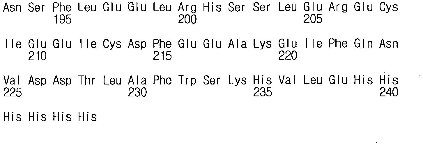
Claims (7)
- A fusion polypeptide comprising the amino acid sequence of SEQ ID NO: 14 or 15.
- A polynucleotide encoding the fusion polypeptide of any one of claim 1.
- An expression vector comprising the polynucleotide of claim 2.
- An isolated transformed cell transformed with the expression vector of claim 3.
- A protein cage comprising the fusion polypeptide of claim 1, wherein an anti-inflammatory polypeptide protrudes outside the protein cage.
- A pharmaceutical composition for use in treating an inflammatory disease, the composition comprising the fusion polypeptide of claim 1 as an active ingredient.
- The pharmaceutical composition for use according to claim 6, wherein the inflammatory disease is selected from the group consisting of inflammatory bowel disease, diabetic eye disease, peritonitis, osteomyelitis, cellulitis, meningitis, encephalitis, pancreatitis, traumatic shock, bronchial asthma, rhinitis, sinusitis, otitis, pneumonia, gastritis, enteritis, cystic fibrosis, apoplexy. bronchitis, bronchiolitis, hepatitis, nephritis, arthritis, gout, spondylitis, Reiter's syndrome, polyarteritis nodosa, irritable vasculitis, Lou Gehrig's granulomatosis, Polymyalgia rheumatica, arthritic arteritis, calcium crystal arthropathies, pseudogout, non-articular rheumatism, bursitis, tenosynovitis, epicondylitis (tennis elbow), neuropathic joint disease (Charcot's joint), hemarthrosis, Henoch-Schonlein purpura, hypertrophic osteoarthropathy, multicentric reticulohistiocytoma, surcoilosis, hemochromatosis, sickle cell disease and other hemochromatosis, hyperlipoproteinemia, hypogammaglobulinemia, hyperparathyroidism, acromegaly, familial Mediterranean fever, Behcet's disease, systemic lupus erythematosus, relapsing fever, psoriasis, multiple sclerosis, sepsis, septic shock, acute respiratory distress syndrome, multiple organ dysfunction syndrome, chronic obstructive pulmonary disease, acute lung injury and broncho-pulmonary dysplasia.
Applications Claiming Priority (2)
| Application Number | Priority Date | Filing Date | Title |
|---|---|---|---|
| KR1020150124472A KR101811050B1 (en) | 2015-09-02 | 2015-09-02 | Fusion-polypeptide of anti-inflammatory polypeptide and ferritin monomer fragment and Pharmaceutical Composition comprising the same |
| PCT/KR2016/009845 WO2017039383A1 (en) | 2015-09-02 | 2016-09-02 | Fusion polypeptide in which anti-inflammatory polypeptide and ferritin monomer fragment are bound and pharmaceutical composition for preventing and treating inflammatory diseases, containing same as active ingredient |
Publications (3)
| Publication Number | Publication Date |
|---|---|
| EP3345929A1 EP3345929A1 (en) | 2018-07-11 |
| EP3345929A4 EP3345929A4 (en) | 2019-09-04 |
| EP3345929B1 true EP3345929B1 (en) | 2021-08-04 |
Family
ID=58188998
Family Applications (1)
| Application Number | Title | Priority Date | Filing Date |
|---|---|---|---|
| EP16842349.9A Active EP3345929B1 (en) | 2015-09-02 | 2016-09-02 | Fusion polypeptide in which anti-inflammatory polypeptide and ferritin monomer fragment are bound and pharmaceutical composition for preventing and treating inflammatory diseases, containing same as active ingredient |
Country Status (4)
| Country | Link |
|---|---|
| US (1) | US10513545B2 (en) |
| EP (1) | EP3345929B1 (en) |
| KR (1) | KR101811050B1 (en) |
| WO (1) | WO2017039383A1 (en) |
Families Citing this family (10)
| Publication number | Priority date | Publication date | Assignee | Title |
|---|---|---|---|---|
| CN110144326A (en) * | 2018-02-12 | 2019-08-20 | 深圳宾德生物技术有限公司 | A kind of antitumor T cell of targeting and its preparation method and application |
| KR102182850B1 (en) * | 2018-11-19 | 2020-11-25 | 경북대학교 산학협력단 | Composition for treating of cancer or inhibiting of cancer metastasis comprising TFG or TFMG nanoparticle |
| KR102348838B1 (en) | 2019-03-07 | 2022-01-11 | 경북대학교 산학협력단 | Active TRAIL trimer and tumor targeting peptide multi-displayed on ferritin nanocage and use in anti-cancer agent thereof |
| EP4181957A4 (en) | 2020-07-20 | 2024-08-07 | Deka Biosciences, Inc. | IL-10 CONTAINING DUAL CYTOKINE FFUSIONS PROTEINS |
| KR102346499B1 (en) | 2020-08-26 | 2022-01-03 | (주)터틀바이오 | Recombinant Vector for Mass Production of Ferritin Protein and Mass Production Method of Ferritin Protein using the Recombinant Vector |
| KR102569463B1 (en) * | 2020-08-31 | 2023-08-23 | 경북대학교 산학협력단 | Composition for preventing or treating inflammatory diseases comprising protein fused with SET domain of SETD6 |
| KR102658825B1 (en) | 2020-10-23 | 2024-04-19 | 경북대학교 산학협력단 | The immunotherapeutic nanocage displaying PD-L1 binding peptide 1 and use in anti-cancer immunotherapeutic agent thereof |
| KR102726136B1 (en) | 2020-12-18 | 2024-11-06 | 경북대학교 산학협력단 | The immunotherapeutic nanocage displaying PD-L1 binding peptide 2 and use in anti-cancer immunotherapeutic agent thereof |
| KR20240156653A (en) * | 2021-12-13 | 2024-10-30 | 데카 바이오사이언시즈, 인크. | Dual cytokine fusion protein containing multi-subunit cytokines |
| CN120815043A (en) * | 2025-08-25 | 2025-10-21 | 天津市天津医院 | Responsive self-assembling polypeptide hydrogel microsphere system and its preparation method and application |
Family Cites Families (9)
| Publication number | Priority date | Publication date | Assignee | Title |
|---|---|---|---|---|
| WO2006062701A2 (en) * | 2004-11-16 | 2006-06-15 | Janssen Pharmaceutica N.V. | Sphingosine kinase-1 mediates gene expression regulation of a monocyte chemoattractant protein-1 gene |
| US8778878B2 (en) * | 2007-01-29 | 2014-07-15 | Chyna, LLC | Use of ferritin to treat iron disorders |
| EP2157176A1 (en) * | 2008-08-20 | 2010-02-24 | The Provost, Fellows And Scholars Of The College Of The Holy And Undivided Trinity Of Queen Elizabeth Near Dublin | Protein C Variants |
| KR101189192B1 (en) * | 2010-06-24 | 2012-10-09 | 한국과학기술연구원 | Fusion protein comprising ferritin and GALA peptide, cage protein formed thereby, and novel use thereof |
| US20130071375A1 (en) * | 2011-08-22 | 2013-03-21 | Saint Louis University | Compositions and methods for treating inflammation |
| KR101477123B1 (en) | 2011-10-12 | 2014-12-29 | 한국생명공학연구원 | Antibody Binding Peptide-Ferritin Fusion Protein and Use Thereof |
| KR20130062168A (en) * | 2011-12-02 | 2013-06-12 | 고려대학교 산학협력단 | Recombinant fluorescent nanoparticles |
| WO2014123399A1 (en) * | 2013-02-08 | 2014-08-14 | 경북대학교 산학협력단 | Human ferritin-derived fusion polypeptide |
| KR101630858B1 (en) | 2014-01-24 | 2016-06-15 | 울산과학기술원 | Antigen peptide-ferritin fused protein and vaccine composition comprising the same |
-
2015
- 2015-09-02 KR KR1020150124472A patent/KR101811050B1/en active Active
-
2016
- 2016-09-02 WO PCT/KR2016/009845 patent/WO2017039383A1/en not_active Ceased
- 2016-09-02 EP EP16842349.9A patent/EP3345929B1/en active Active
- 2016-09-02 US US15/757,580 patent/US10513545B2/en active Active
Non-Patent Citations (1)
| Title |
|---|
| None * |
Also Published As
| Publication number | Publication date |
|---|---|
| KR101811050B1 (en) | 2017-12-21 |
| KR20170027619A (en) | 2017-03-10 |
| EP3345929A1 (en) | 2018-07-11 |
| US10513545B2 (en) | 2019-12-24 |
| EP3345929A4 (en) | 2019-09-04 |
| WO2017039383A1 (en) | 2017-03-09 |
| US20190016764A1 (en) | 2019-01-17 |
Similar Documents
| Publication | Publication Date | Title |
|---|---|---|
| US10513545B2 (en) | Fusion polypeptide in which anti-inflammatory polypeptide and ferritin monomer fragment are bound and pharmaceutical composition for preventing and treating inflammatory diseases, containing same as active ingredient | |
| JP2016505826A (en) | Screening method | |
| ES2974506T3 (en) | Peptides derived from saposin-A and uses thereof | |
| CA2984769A1 (en) | Peptide treatment for inflammation and fibrosis | |
| WO2020036987A1 (en) | Peptides and compositions for targeted treatment and imaging | |
| US20100008900A1 (en) | Annexin ii compositions for treating or monitoring inflammation or immune-mediated disorders | |
| CN107406513B (en) | Double-stranded molecules (BIPARTITEs) and their use for treating abnormal protein aggregation | |
| KR20000053277A (en) | Soluble polypeptides | |
| CN116635056A (en) | long-acting deoxyribonuclease | |
| WO2025088160A1 (en) | Transmembrane serine protease and neutrophil elastase inhibitors | |
| US20210179680A1 (en) | Polypeptide, derivatives thereof, and application thereof in preparation of drugs having resistance to pulmonary fibrosis | |
| CN113748123B (en) | Composition for treating lung inflammation | |
| CN116003524A (en) | Short peptide targeting cGAS, fusion polypeptide and application | |
| WO2022266467A2 (en) | Recombinant histone polypeptide and uses thereof | |
| EP4351618A2 (en) | Trem-2/dap-12 inhibitors for treating lung disease and injury and combinations thereof | |
| KR101459833B1 (en) | Polymannitol-based osmotically gene transpoter and gene therapy using the same as a gene carrier | |
| JP2006067968A (en) | Substance that inhibits invasion and metastasis of human cancer cells and screening method thereof | |
| US20250325684A1 (en) | Potent anti-cancer cyclotides | |
| US20250352654A1 (en) | Pharmaceutical agent conjugates to modulate macrophage and inflammatory functions and uses thereof | |
| WO2021007800A1 (en) | Anti-infection effects of hnrnpa2b1 and use thereof | |
| KR20100003823A (en) | Peptides for binding specifically to membrane embedded with aquaporin-2 and uses thereof | |
| JP7764502B2 (en) | Polypeptides Translated by Circular RNA Circ-ACE2 and Uses Thereof | |
| WO2019057120A1 (en) | Lgr4 and r-spondin binding inhibitor and use thereof in tumor therapy | |
| WO2024245445A1 (en) | Malt1 tumor inhibitor, and drug containing same and use thereof | |
| US20220259265A1 (en) | Inhibitors and Use Thereof in Cancer Treatment |
Legal Events
| Date | Code | Title | Description |
|---|---|---|---|
| STAA | Information on the status of an ep patent application or granted ep patent |
Free format text: STATUS: THE INTERNATIONAL PUBLICATION HAS BEEN MADE |
|
| PUAI | Public reference made under article 153(3) epc to a published international application that has entered the european phase |
Free format text: ORIGINAL CODE: 0009012 |
|
| STAA | Information on the status of an ep patent application or granted ep patent |
Free format text: STATUS: REQUEST FOR EXAMINATION WAS MADE |
|
| 17P | Request for examination filed |
Effective date: 20180403 |
|
| AK | Designated contracting states |
Kind code of ref document: A1 Designated state(s): AL AT BE BG CH CY CZ DE DK EE ES FI FR GB GR HR HU IE IS IT LI LT LU LV MC MK MT NL NO PL PT RO RS SE SI SK SM TR |
|
| AX | Request for extension of the european patent |
Extension state: BA ME |
|
| DAV | Request for validation of the european patent (deleted) | ||
| DAX | Request for extension of the european patent (deleted) | ||
| RIC1 | Information provided on ipc code assigned before grant |
Ipc: C07K 14/79 20060101ALI20190523BHEP Ipc: A61K 38/16 20060101ALI20190523BHEP Ipc: A61K 38/17 20060101ALI20190523BHEP Ipc: C07K 14/47 20060101ALI20190523BHEP Ipc: C07K 14/54 20060101ALI20190523BHEP Ipc: C07K 19/00 20060101AFI20190523BHEP |
|
| A4 | Supplementary search report drawn up and despatched |
Effective date: 20190802 |
|
| RIC1 | Information provided on ipc code assigned before grant |
Ipc: A61K 38/17 20060101ALI20190729BHEP Ipc: C07K 14/47 20060101ALI20190729BHEP Ipc: A61K 38/16 20060101ALI20190729BHEP Ipc: C07K 14/54 20060101ALI20190729BHEP Ipc: C07K 19/00 20060101AFI20190729BHEP Ipc: C07K 14/79 20060101ALI20190729BHEP |
|
| STAA | Information on the status of an ep patent application or granted ep patent |
Free format text: STATUS: EXAMINATION IS IN PROGRESS |
|
| 17Q | First examination report despatched |
Effective date: 20200415 |
|
| GRAP | Despatch of communication of intention to grant a patent |
Free format text: ORIGINAL CODE: EPIDOSNIGR1 |
|
| STAA | Information on the status of an ep patent application or granted ep patent |
Free format text: STATUS: GRANT OF PATENT IS INTENDED |
|
| INTG | Intention to grant announced |
Effective date: 20210329 |
|
| GRAS | Grant fee paid |
Free format text: ORIGINAL CODE: EPIDOSNIGR3 |
|
| GRAA | (expected) grant |
Free format text: ORIGINAL CODE: 0009210 |
|
| STAA | Information on the status of an ep patent application or granted ep patent |
Free format text: STATUS: THE PATENT HAS BEEN GRANTED |
|
| AK | Designated contracting states |
Kind code of ref document: B1 Designated state(s): AL AT BE BG CH CY CZ DE DK EE ES FI FR GB GR HR HU IE IS IT LI LT LU LV MC MK MT NL NO PL PT RO RS SE SI SK SM TR |
|
| REG | Reference to a national code |
Ref country code: GB Ref legal event code: FG4D |
|
| REG | Reference to a national code |
Ref country code: AT Ref legal event code: REF Ref document number: 1416925 Country of ref document: AT Kind code of ref document: T Effective date: 20210815 |
|
| REG | Reference to a national code |
Ref country code: CH Ref legal event code: EP |
|
| REG | Reference to a national code |
Ref country code: DE Ref legal event code: R096 Ref document number: 602016061742 Country of ref document: DE |
|
| REG | Reference to a national code |
Ref country code: IE Ref legal event code: FG4D |
|
| REG | Reference to a national code |
Ref country code: LT Ref legal event code: MG9D |
|
| REG | Reference to a national code |
Ref country code: NL Ref legal event code: MP Effective date: 20210804 |
|
| REG | Reference to a national code |
Ref country code: AT Ref legal event code: MK05 Ref document number: 1416925 Country of ref document: AT Kind code of ref document: T Effective date: 20210804 |
|
| PG25 | Lapsed in a contracting state [announced via postgrant information from national office to epo] |
Ref country code: RS Free format text: LAPSE BECAUSE OF FAILURE TO SUBMIT A TRANSLATION OF THE DESCRIPTION OR TO PAY THE FEE WITHIN THE PRESCRIBED TIME-LIMIT Effective date: 20210804 Ref country code: SE Free format text: LAPSE BECAUSE OF FAILURE TO SUBMIT A TRANSLATION OF THE DESCRIPTION OR TO PAY THE FEE WITHIN THE PRESCRIBED TIME-LIMIT Effective date: 20210804 Ref country code: BG Free format text: LAPSE BECAUSE OF FAILURE TO SUBMIT A TRANSLATION OF THE DESCRIPTION OR TO PAY THE FEE WITHIN THE PRESCRIBED TIME-LIMIT Effective date: 20211104 Ref country code: AT Free format text: LAPSE BECAUSE OF FAILURE TO SUBMIT A TRANSLATION OF THE DESCRIPTION OR TO PAY THE FEE WITHIN THE PRESCRIBED TIME-LIMIT Effective date: 20210804 Ref country code: LT Free format text: LAPSE BECAUSE OF FAILURE TO SUBMIT A TRANSLATION OF THE DESCRIPTION OR TO PAY THE FEE WITHIN THE PRESCRIBED TIME-LIMIT Effective date: 20210804 Ref country code: NO Free format text: LAPSE BECAUSE OF FAILURE TO SUBMIT A TRANSLATION OF THE DESCRIPTION OR TO PAY THE FEE WITHIN THE PRESCRIBED TIME-LIMIT Effective date: 20211104 Ref country code: PT Free format text: LAPSE BECAUSE OF FAILURE TO SUBMIT A TRANSLATION OF THE DESCRIPTION OR TO PAY THE FEE WITHIN THE PRESCRIBED TIME-LIMIT Effective date: 20211206 Ref country code: HR Free format text: LAPSE BECAUSE OF FAILURE TO SUBMIT A TRANSLATION OF THE DESCRIPTION OR TO PAY THE FEE WITHIN THE PRESCRIBED TIME-LIMIT Effective date: 20210804 Ref country code: ES Free format text: LAPSE BECAUSE OF FAILURE TO SUBMIT A TRANSLATION OF THE DESCRIPTION OR TO PAY THE FEE WITHIN THE PRESCRIBED TIME-LIMIT Effective date: 20210804 Ref country code: FI Free format text: LAPSE BECAUSE OF FAILURE TO SUBMIT A TRANSLATION OF THE DESCRIPTION OR TO PAY THE FEE WITHIN THE PRESCRIBED TIME-LIMIT Effective date: 20210804 |
|
| PG25 | Lapsed in a contracting state [announced via postgrant information from national office to epo] |
Ref country code: PL Free format text: LAPSE BECAUSE OF FAILURE TO SUBMIT A TRANSLATION OF THE DESCRIPTION OR TO PAY THE FEE WITHIN THE PRESCRIBED TIME-LIMIT Effective date: 20210804 Ref country code: LV Free format text: LAPSE BECAUSE OF FAILURE TO SUBMIT A TRANSLATION OF THE DESCRIPTION OR TO PAY THE FEE WITHIN THE PRESCRIBED TIME-LIMIT Effective date: 20210804 Ref country code: GR Free format text: LAPSE BECAUSE OF FAILURE TO SUBMIT A TRANSLATION OF THE DESCRIPTION OR TO PAY THE FEE WITHIN THE PRESCRIBED TIME-LIMIT Effective date: 20211105 |
|
| PG25 | Lapsed in a contracting state [announced via postgrant information from national office to epo] |
Ref country code: NL Free format text: LAPSE BECAUSE OF FAILURE TO SUBMIT A TRANSLATION OF THE DESCRIPTION OR TO PAY THE FEE WITHIN THE PRESCRIBED TIME-LIMIT Effective date: 20210804 |
|
| PG25 | Lapsed in a contracting state [announced via postgrant information from national office to epo] |
Ref country code: DK Free format text: LAPSE BECAUSE OF FAILURE TO SUBMIT A TRANSLATION OF THE DESCRIPTION OR TO PAY THE FEE WITHIN THE PRESCRIBED TIME-LIMIT Effective date: 20210804 |
|
| REG | Reference to a national code |
Ref country code: DE Ref legal event code: R097 Ref document number: 602016061742 Country of ref document: DE |
|
| REG | Reference to a national code |
Ref country code: CH Ref legal event code: PL |
|
| REG | Reference to a national code |
Ref country code: BE Ref legal event code: MM Effective date: 20210930 |
|
| PG25 | Lapsed in a contracting state [announced via postgrant information from national office to epo] |
Ref country code: SM Free format text: LAPSE BECAUSE OF FAILURE TO SUBMIT A TRANSLATION OF THE DESCRIPTION OR TO PAY THE FEE WITHIN THE PRESCRIBED TIME-LIMIT Effective date: 20210804 Ref country code: SK Free format text: LAPSE BECAUSE OF FAILURE TO SUBMIT A TRANSLATION OF THE DESCRIPTION OR TO PAY THE FEE WITHIN THE PRESCRIBED TIME-LIMIT Effective date: 20210804 Ref country code: RO Free format text: LAPSE BECAUSE OF FAILURE TO SUBMIT A TRANSLATION OF THE DESCRIPTION OR TO PAY THE FEE WITHIN THE PRESCRIBED TIME-LIMIT Effective date: 20210804 Ref country code: MC Free format text: LAPSE BECAUSE OF FAILURE TO SUBMIT A TRANSLATION OF THE DESCRIPTION OR TO PAY THE FEE WITHIN THE PRESCRIBED TIME-LIMIT Effective date: 20210804 Ref country code: EE Free format text: LAPSE BECAUSE OF FAILURE TO SUBMIT A TRANSLATION OF THE DESCRIPTION OR TO PAY THE FEE WITHIN THE PRESCRIBED TIME-LIMIT Effective date: 20210804 Ref country code: CZ Free format text: LAPSE BECAUSE OF FAILURE TO SUBMIT A TRANSLATION OF THE DESCRIPTION OR TO PAY THE FEE WITHIN THE PRESCRIBED TIME-LIMIT Effective date: 20210804 Ref country code: AL Free format text: LAPSE BECAUSE OF FAILURE TO SUBMIT A TRANSLATION OF THE DESCRIPTION OR TO PAY THE FEE WITHIN THE PRESCRIBED TIME-LIMIT Effective date: 20210804 |
|
| PLBE | No opposition filed within time limit |
Free format text: ORIGINAL CODE: 0009261 |
|
| STAA | Information on the status of an ep patent application or granted ep patent |
Free format text: STATUS: NO OPPOSITION FILED WITHIN TIME LIMIT |
|
| 26N | No opposition filed |
Effective date: 20220506 |
|
| PG25 | Lapsed in a contracting state [announced via postgrant information from national office to epo] |
Ref country code: LU Free format text: LAPSE BECAUSE OF NON-PAYMENT OF DUE FEES Effective date: 20210902 Ref country code: IT Free format text: LAPSE BECAUSE OF FAILURE TO SUBMIT A TRANSLATION OF THE DESCRIPTION OR TO PAY THE FEE WITHIN THE PRESCRIBED TIME-LIMIT Effective date: 20210804 Ref country code: IE Free format text: LAPSE BECAUSE OF NON-PAYMENT OF DUE FEES Effective date: 20210902 Ref country code: BE Free format text: LAPSE BECAUSE OF NON-PAYMENT OF DUE FEES Effective date: 20210930 |
|
| PG25 | Lapsed in a contracting state [announced via postgrant information from national office to epo] |
Ref country code: SI Free format text: LAPSE BECAUSE OF FAILURE TO SUBMIT A TRANSLATION OF THE DESCRIPTION OR TO PAY THE FEE WITHIN THE PRESCRIBED TIME-LIMIT Effective date: 20210804 Ref country code: LI Free format text: LAPSE BECAUSE OF NON-PAYMENT OF DUE FEES Effective date: 20210930 Ref country code: CH Free format text: LAPSE BECAUSE OF NON-PAYMENT OF DUE FEES Effective date: 20210930 |
|
| PG25 | Lapsed in a contracting state [announced via postgrant information from national office to epo] |
Ref country code: HU Free format text: LAPSE BECAUSE OF FAILURE TO SUBMIT A TRANSLATION OF THE DESCRIPTION OR TO PAY THE FEE WITHIN THE PRESCRIBED TIME-LIMIT; INVALID AB INITIO Effective date: 20160902 |
|
| PG25 | Lapsed in a contracting state [announced via postgrant information from national office to epo] |
Ref country code: CY Free format text: LAPSE BECAUSE OF FAILURE TO SUBMIT A TRANSLATION OF THE DESCRIPTION OR TO PAY THE FEE WITHIN THE PRESCRIBED TIME-LIMIT Effective date: 20210804 |
|
| P01 | Opt-out of the competence of the unified patent court (upc) registered |
Effective date: 20230607 |
|
| PG25 | Lapsed in a contracting state [announced via postgrant information from national office to epo] |
Ref country code: MK Free format text: LAPSE BECAUSE OF FAILURE TO SUBMIT A TRANSLATION OF THE DESCRIPTION OR TO PAY THE FEE WITHIN THE PRESCRIBED TIME-LIMIT Effective date: 20210804 |
|
| PG25 | Lapsed in a contracting state [announced via postgrant information from national office to epo] |
Ref country code: TR Free format text: LAPSE BECAUSE OF FAILURE TO SUBMIT A TRANSLATION OF THE DESCRIPTION OR TO PAY THE FEE WITHIN THE PRESCRIBED TIME-LIMIT Effective date: 20210804 |
|
| PG25 | Lapsed in a contracting state [announced via postgrant information from national office to epo] |
Ref country code: MT Free format text: LAPSE BECAUSE OF FAILURE TO SUBMIT A TRANSLATION OF THE DESCRIPTION OR TO PAY THE FEE WITHIN THE PRESCRIBED TIME-LIMIT Effective date: 20210804 |
|
| PGFP | Annual fee paid to national office [announced via postgrant information from national office to epo] |
Ref country code: DE Payment date: 20250702 Year of fee payment: 10 |
|
| PGFP | Annual fee paid to national office [announced via postgrant information from national office to epo] |
Ref country code: GB Payment date: 20250703 Year of fee payment: 10 |
|
| PGFP | Annual fee paid to national office [announced via postgrant information from national office to epo] |
Ref country code: FR Payment date: 20250703 Year of fee payment: 10 |