EP3344658B1 - Antibodies specific to human t-cell immunoglobulin and itim domain (tigit) - Google Patents
Antibodies specific to human t-cell immunoglobulin and itim domain (tigit) Download PDFInfo
- Publication number
- EP3344658B1 EP3344658B1 EP16775866.3A EP16775866A EP3344658B1 EP 3344658 B1 EP3344658 B1 EP 3344658B1 EP 16775866 A EP16775866 A EP 16775866A EP 3344658 B1 EP3344658 B1 EP 3344658B1
- Authority
- EP
- European Patent Office
- Prior art keywords
- antibody
- seq
- tigit
- human
- cells
- Prior art date
- Legal status (The legal status is an assumption and is not a legal conclusion. Google has not performed a legal analysis and makes no representation as to the accuracy of the status listed.)
- Active
Links
Images


Classifications
-
- C—CHEMISTRY; METALLURGY
- C07—ORGANIC CHEMISTRY
- C07K—PEPTIDES
- C07K16/00—Immunoglobulins [IG], e.g. monoclonal or polyclonal antibodies
- C07K16/18—Immunoglobulins [IG], e.g. monoclonal or polyclonal antibodies against material from animals or humans
- C07K16/28—Immunoglobulins [IG], e.g. monoclonal or polyclonal antibodies against material from animals or humans against receptors, cell surface antigens or cell surface determinants
-
- A—HUMAN NECESSITIES
- A61—MEDICAL OR VETERINARY SCIENCE; HYGIENE
- A61K—PREPARATIONS FOR MEDICAL, DENTAL OR TOILETRY PURPOSES
- A61K39/00—Medicinal preparations containing antigens or antibodies
- A61K39/395—Antibodies; Immunoglobulins; Immune serum, e.g. antilymphocytic serum
- A61K39/39533—Antibodies; Immunoglobulins; Immune serum, e.g. antilymphocytic serum against materials from animals
- A61K39/39558—Antibodies; Immunoglobulins; Immune serum, e.g. antilymphocytic serum against materials from animals against tumor tissues, cells, antigens
-
- A—HUMAN NECESSITIES
- A61—MEDICAL OR VETERINARY SCIENCE; HYGIENE
- A61P—SPECIFIC THERAPEUTIC ACTIVITY OF CHEMICAL COMPOUNDS OR MEDICINAL PREPARATIONS
- A61P35/00—Antineoplastic agents
-
- C—CHEMISTRY; METALLURGY
- C07—ORGANIC CHEMISTRY
- C07K—PEPTIDES
- C07K16/00—Immunoglobulins [IG], e.g. monoclonal or polyclonal antibodies
- C07K16/18—Immunoglobulins [IG], e.g. monoclonal or polyclonal antibodies against material from animals or humans
- C07K16/28—Immunoglobulins [IG], e.g. monoclonal or polyclonal antibodies against material from animals or humans against receptors, cell surface antigens or cell surface determinants
- C07K16/2803—Immunoglobulins [IG], e.g. monoclonal or polyclonal antibodies against material from animals or humans against receptors, cell surface antigens or cell surface determinants against the immunoglobulin superfamily
-
- G—PHYSICS
- G01—MEASURING; TESTING
- G01N—INVESTIGATING OR ANALYSING MATERIALS BY DETERMINING THEIR CHEMICAL OR PHYSICAL PROPERTIES
- G01N33/00—Investigating or analysing materials by specific methods not covered by groups G01N1/00 - G01N31/00
- G01N33/48—Biological material, e.g. blood, urine; Haemocytometers
- G01N33/50—Chemical analysis of biological material, e.g. blood, urine; Testing involving biospecific ligand binding methods; Immunological testing
- G01N33/53—Immunoassay; Biospecific binding assay; Materials therefor
- G01N33/577—Immunoassay; Biospecific binding assay; Materials therefor involving monoclonal antibodies binding reaction mechanisms characterised by the use of monoclonal antibodies
-
- A—HUMAN NECESSITIES
- A61—MEDICAL OR VETERINARY SCIENCE; HYGIENE
- A61K—PREPARATIONS FOR MEDICAL, DENTAL OR TOILETRY PURPOSES
- A61K39/00—Medicinal preparations containing antigens or antibodies
- A61K2039/505—Medicinal preparations containing antigens or antibodies comprising antibodies
-
- C—CHEMISTRY; METALLURGY
- C07—ORGANIC CHEMISTRY
- C07K—PEPTIDES
- C07K2317/00—Immunoglobulins specific features
- C07K2317/20—Immunoglobulins specific features characterized by taxonomic origin
- C07K2317/24—Immunoglobulins specific features characterized by taxonomic origin containing regions, domains or residues from different species, e.g. chimeric, humanized or veneered
-
- C—CHEMISTRY; METALLURGY
- C07—ORGANIC CHEMISTRY
- C07K—PEPTIDES
- C07K2317/00—Immunoglobulins specific features
- C07K2317/70—Immunoglobulins specific features characterized by effect upon binding to a cell or to an antigen
- C07K2317/76—Antagonist effect on antigen, e.g. neutralization or inhibition of binding
-
- C—CHEMISTRY; METALLURGY
- C07—ORGANIC CHEMISTRY
- C07K—PEPTIDES
- C07K2317/00—Immunoglobulins specific features
- C07K2317/90—Immunoglobulins specific features characterized by (pharmaco)kinetic aspects or by stability of the immunoglobulin
- C07K2317/92—Affinity (KD), association rate (Ka), dissociation rate (Kd) or EC50 value
-
- C—CHEMISTRY; METALLURGY
- C12—BIOCHEMISTRY; BEER; SPIRITS; WINE; VINEGAR; MICROBIOLOGY; ENZYMOLOGY; MUTATION OR GENETIC ENGINEERING
- C12N—MICROORGANISMS OR ENZYMES; COMPOSITIONS THEREOF; PROPAGATING, PRESERVING, OR MAINTAINING MICROORGANISMS; MUTATION OR GENETIC ENGINEERING; CULTURE MEDIA
- C12N15/00—Mutation or genetic engineering; DNA or RNA concerning genetic engineering, vectors, e.g. plasmids, or their isolation, preparation or purification; Use of hosts therefor
-
- C—CHEMISTRY; METALLURGY
- C12—BIOCHEMISTRY; BEER; SPIRITS; WINE; VINEGAR; MICROBIOLOGY; ENZYMOLOGY; MUTATION OR GENETIC ENGINEERING
- C12N—MICROORGANISMS OR ENZYMES; COMPOSITIONS THEREOF; PROPAGATING, PRESERVING, OR MAINTAINING MICROORGANISMS; MUTATION OR GENETIC ENGINEERING; CULTURE MEDIA
- C12N15/00—Mutation or genetic engineering; DNA or RNA concerning genetic engineering, vectors, e.g. plasmids, or their isolation, preparation or purification; Use of hosts therefor
- C12N15/09—Recombinant DNA-technology
- C12N15/63—Introduction of foreign genetic material using vectors; Vectors; Use of hosts therefor; Regulation of expression
-
- C—CHEMISTRY; METALLURGY
- C12—BIOCHEMISTRY; BEER; SPIRITS; WINE; VINEGAR; MICROBIOLOGY; ENZYMOLOGY; MUTATION OR GENETIC ENGINEERING
- C12N—MICROORGANISMS OR ENZYMES; COMPOSITIONS THEREOF; PROPAGATING, PRESERVING, OR MAINTAINING MICROORGANISMS; MUTATION OR GENETIC ENGINEERING; CULTURE MEDIA
- C12N5/00—Undifferentiated human, animal or plant cells, e.g. cell lines; Tissues; Cultivation or maintenance thereof; Culture media therefor
- C12N5/10—Cells modified by introduction of foreign genetic material
-
- Y—GENERAL TAGGING OF NEW TECHNOLOGICAL DEVELOPMENTS; GENERAL TAGGING OF CROSS-SECTIONAL TECHNOLOGIES SPANNING OVER SEVERAL SECTIONS OF THE IPC; TECHNICAL SUBJECTS COVERED BY FORMER USPC CROSS-REFERENCE ART COLLECTIONS [XRACs] AND DIGESTS
- Y02—TECHNOLOGIES OR APPLICATIONS FOR MITIGATION OR ADAPTATION AGAINST CLIMATE CHANGE
- Y02A—TECHNOLOGIES FOR ADAPTATION TO CLIMATE CHANGE
- Y02A50/00—TECHNOLOGIES FOR ADAPTATION TO CLIMATE CHANGE in human health protection, e.g. against extreme weather
- Y02A50/30—Against vector-borne diseases, e.g. mosquito-borne, fly-borne, tick-borne or waterborne diseases whose impact is exacerbated by climate change
Definitions
- the invention is in the field of immunotherapy and relates to monoclonal antibodies and fragments thereof which bind to the protein human TIGIT, to polynucleotide sequences encoding these antibodies and to hybridoma cells producing these antibodies.
- the invention further relates to therapeutic compositions comprising these antibodies that may be useful in treating and diagnosing diseases, particularly cancer, using these monoclonal antibodies.
- Cancer immunotherapy is utilized for generating and augmenting an anti-tumor immune response, e.g., by treatment with antibodies specific to antigens on tumor cells, with cancer cells fused with antigen presenting cells (APC), or by specific activation of anti-tumor T cells.
- APC antigen presenting cells
- NK cells of the innate immune system play an important role in immune surveillance of tumors ( Smyth et al., Nat Immunol. 2001. Apr;2(4):293-9 ). NK cells kill MHC class I-deficient cells, tumors, viruses, parasites and bacteria directly and indirectly. NK cell activity is controlled by a balance of signals, delivered by inhibitory and activating NK cell receptors. There are several activating NK cell receptors that recognize various ligands, which can be stress-induced, self-molecules, viral components or tumor proteins ( Koch et al., Trends in immunology 2013, 34, 182-191 .; Seidel et al., Cellular and molecular life sciences, 2012 ).
- NK cells express a vast repertoire of inhibitory receptors ( Gardiner C.M., International journal of immunogenetics 2008, 35, 1-8 .; Gonen-Gross et al., PloS one 2010, 5, e8941 ; Lankry et al., Methods in molecular biology 2010, 612, 249-273 ;).
- KIR Killer Inhibitory Receptors
- MHC major histocompatibility complex
- the human TIGIT protein is expressed on all NK cells, as well as on other immune cells such as T regulatory (Treg) CD8+ cells and Tumor infiltrating lymphocytes ( Stanietsky et al., PNAS. 2009, 106, 17858-17863 ). It recognizes two very well defined ligands: poliovirus receptor (PVR, CD155) and Nectin2 (PVRL2/CD112) that are expressed on normal epithelia as well as over expressed on various tumor cells.
- poliovirus receptor PVR, CD155
- Nectin2 PVRL2/CD112
- TIGIT immunoreceptor tail tyrosine
- ITIM immunodominant tyrosine based inhibitory
- TIGIT expression on NK cells also serves as the receptor that binds the Fap2 protein of the anaerobic Gram-negative bacterium Fusobacterium nucleatum ( F . nucleatum ) .
- F. nucleatum The interaction between F. Necleatum and TIGIT leads to reduced NK cytotoxic activity. Fusobacteria are often enriched in patients with intestinal inflammation and cancer. It was suggested that F. nucleatum binding to TIGIT facilitates tumor evasion from NK associated cytotoxicity ( Gur et al., Immunity. 2015 February 17; 42(2): 344-355 ), providing an explanation on how bacteria found within tumors, in particular, F. nucleatum, promote tumor proliferation and enhance tumor progression ( Jobin, Cancer discovery. 2013; 3:384-387 ; Sears and Garrett, Cell Host Microbe. 2014; 15:317-328 ).
- TIGIT and PD-1 impair tumor antigen-specific CD8+ T cells in melanoma patients ( Chauvin el al. J Clin Invest. 2015;125(5):2046-2058 ).
- TIGIT expression by CD8+ tumor-infiltrating lymphocytes (TILs) has been reported using gene expression analyses in a number of mouse and human solid tumors including lung, colon, breast, uterine, and renal cancers. Elevated TIGIT expression appears to correlate with CD8 and PD-1 expression.
- TIGIT expression on CD8+ TILs was observed in mouse tumors and in 3 human tumor samples, including non-small-cell lung and colon cancers ( Johnston RJ, et al. Cancer Cell. 2014;26(6):923-937 ).
- TIGIT contributes to functional T-cell impairment and associates with poor clinical outcome in acute myeloid leukemia (AML).
- AML acute myeloid leukemia
- WO 2004/024068 describes agonists and antagonists to the molecule PRO52254, later identified as TIGIT, for treatment of autoimmune diseases and cancer without disclosing actual antibodies.
- WO 2006/124667 discloses modulation of the protein zB7R1 (TIGIT) by monoclonal antibodies that block TIGIT binding to its ligand PVR. No binding affinities are provided.
- WO 2009/126688 discloses TIGIT, and its ligand PVR, as targets for modulation of immune responses and suggests agonists and antagonists of these proteins for diagnosis and treatment of immune-related and inflammatory diseases.
- WO 2015/009856 discloses combinations of programmed death 1 polypeptide (PD-1) antagonists and anti TIGIT antibodies for treatment of cancer and chronic infection.
- PD-1 programmed death 1 polypeptide
- WO 2016/028656 discloses anti-TIGIT antibodies, as well as use of these antibodies in the treatment of diseases such as cancer and infectious disease.
- CN103073644 A discloses an anti-mouse TIGIT (T cell Ig and ITIM domain), and a hybridoma cell strain mTIGIT-mAb-13G6 for producing the monoclonal antibody, and further relates to a preparation and application method of the monoclonal antibody.
- the monoclonal antibody can efficiently prevent combination of mTIGIT and mPVR (poliovirus receptor), and can be used for Western blot, ELISA and Flow Cytometry for mTIGIT molecule detection.
- T-Cell immunoglobulin and ITIM domain TAGIT
- NK natural killer cells and T-cells.
- TIGIT immune cell inhibitory receptor
- NK natural killer cells and T-cells.
- TIGIT immune cell inhibitory receptor
- the monoclonal antibodies (mAbs) described herein exhibit absolute specificity to human TIGIT, as compared to mouse TIGIT for example, and affinity to the human TIGIT protein, which is higher than the affinity of the natural ligand, PVR (CD155) to human TIGIT. This makes these superior mAbs valuable candidates for use in cancer immune-therapy, enabling administration of lower doses with fewer side effects.
- the monoclonal antibodies described herein are capable not only to interfere with the binding of human TIGIT to its main ligand PVR (CD155) but also to inhibit, at least to some extent, the binding of TIGIT to at least one of its other ligands, such as CD112 and CD113.
- anti-TIGIT monoclonal antibodies described herein have synergistic effect when combined with additional anti-cancer agents, such as other immunomodulatory proteins or receptor inhibitors.
- additional anti-cancer agents such as other immunomodulatory proteins or receptor inhibitors.
- Non-limiting examples are mAbs specific to cytotoxic T-lymphocyte-associated protein 4 (CTLA-4, also known as CD152), and epidermal growth factor receptor inhibitors (EGFR).
- CTL-4 cytotoxic T-lymphocyte-associated protein 4
- EGFR epidermal growth factor receptor inhibitors
- the present invention provides, according to one aspect, an isolated antibody, or a antigen-binding fragment thereof wherein the antibody of fragment binds to human T-cell immunoglobulin and ITIM domain (TIGIT) and is capable of inhibiting the binding of TIGIT to at least PVR (CD155), wherein said antibody or antigen-binding fragment thereof comprises
- the isolated monoclonal antibody or fragment thereof comprises a heavy chain variable region having the sequence: QVQLQESGAELARPGASVKLSCKASGYTFTSYGISWVKQRTGQGLEWIGEIYPRSGNTY YNEKFKGKATLTADKSSSTAYMELSSLTSEDSAVYFCARKGPYYTKNEDYWGQGTILT VSS (SEQ ID NO:7), and a light chain variable region having the sequence:
- the antibody or antibody fragment is capable of recognizing TIGIT protein expressed on T-cells.
- the antibody or antibody fragment is capable of recognizing human TIGIT protein expressed on dendritic or NK cells.
- the antibody or antibody fragment is capable of recognizing human TIGIT protein expressed on T-regulatory cells (Treg).
- the antibody or antibody fragment is capable of recognizing human TIGIT protein expressed on CD8+ tumor infiltrating lymphocytes (TILs).
- TILs tumor infiltrating lymphocytes
- tThe antibody or antibody fragment is capable of inhibiting human TIGIT binding to a ligand expressed on T-cells.
- the antibody or antibody fragment is capable of inhibiting human TIGIT binding to ligand expressed on dendritic or NK cells.
- the antibody or antibody fragment is capable of inhibiting human TIGIT binding to ligand expressed on tumor cells.
- the isolated antibody or fragment thereof may bind to human TIGIT protein with a dissociation constant (Kd) of at least about 50 nM.
- the isolated antibody or fragment thereof maybind to human TIGIT protein with a binding affinity of at least 1 nM.
- the antibody or antibody may have an affinity to human TIGIT of at least about 5 ⁇ 10 -8 M.
- An antibody or antibody fragment may bind with an affinity of 10 -8 M, 5 ⁇ 10 -9 M, 10 -9 M, 5x10 -10 M, 10 -10 M, 5 ⁇ 10 -11 M, 10 -11 M or even higher to human TIGIT.
- the mAb may be selected from the group consisting of: non-human antibody, humanized antibody, human antibody, chimeric antibody, bispecific antibody and an antibody fragment comprising at least the antigen-binding portion of an antibody.
- the antibody fragment is selected from the group consisting of: Fab, Fab', F(ab') 2 , Fd, Fd', Fv, dAb, single chain antibody (scab) and "diabodies".
- the antibody is a bispecific antibody or bispecific antibody fragment, capable of binding to two different epitopes or antigens.
- a bispecific mAb or bispecific mAb fragment may comprise two different hyper variable regions (HVR), each comprising a different set of CDR sequences.
- HVR hyper variable regions
- the bispecific mAb or fragment may comprise the binding domains of two different anti-TIGIT antibodies.
- Each HVR of a bispecific mAb or fragment according to these embodiments is cable of binding to a different epitope of the human TIGIT protein.
- One HVR of a bispecific mAb or fragment thereof may comprise the CDRs: SEQ ID NO:1 or SEQ ID NO:11, SEQ ID NO:2, SEQ ID NO:3, SEQ ID NO:4, SEQ ID NO:5 and SEQ ID NO:6; and the second HVR comprises the CDRs: SEQ ID NO:12, SEQ ID NO:13, SEQ ID NO:14, SEQ ID NO:15, SEQ ID NO:16 and SEQ ID NO:17.
- One HVR of a bispecific antibody or bispecific antibody fragment may bind to human TIGIT and the second HVR may bind to another protein, such as a protein involved in immune regulation or a tumor antigen.
- the second HVR of the bispecific antibody may bind a checkpoint molecule.
- the antibody or antibody fragment may comprise a framework sequence selected from the group consisting of: mouse IgG2a, mouse IgG2b, mouse IgG3, human IgG1, human IgG2, human IgG3, and human IgG4.
- the antibody is a humanized antibody or the antibody fragment is a fragment of a humanized antibody.
- the humanized antibody or antibody fragment may comprise a framework sequence selected from the group consisting of: human IgG1, human IgG2, human IgG3, and human IgG4.
- an antibody conjugate comprising at least one antibody or antibody fragment that recognizes TIGIT and inhibits binding to its ligand
- said antibody comprises the complementarity determining regions (CDRs) sequences: (i) heavy chain CDR1 having a sequence comprising: GYTFTSYGIS (SEQ ID NO:1)or TSYGIS (SEQ ID NO:11), heavy chain CDR2 having the sequence: EIYPRSGNTYYNEKFKG (SEQ ID NO:2) and heavy chain CDR3 having the sequence: KGPYYTKNEDY (SEQ ID NO:3) and light chain CDR1 having the sequence: RASEHIYYSLA (SEQ ID NO:4), light chain CDR2 having the sequence: NANSLED (SEQ ID NO:5) and light chain CDR3 having the sequence: KQAYDVPRT (SEQ ID NO:6).
- CDRs complementarity determining regions
- the conjugate may comprise a carrier protein.
- polynucleotide sequences encoding monoclonal antibodies, having high affinity and specificity for TIGIT, as well as vectors carrying these polynucleotide sequences are provided according to another aspect of the present invention, wherein the polynucleotide sequence encodes an antibody capable of binding to an epitope within the human TIGIT protein, comprising: the six CDR sequences: (i) heavy chain CDR1 sequence selected from: GYTFTSYGIS (SEQ ID NO:1) orTSYGIS (SEQ ID NO:11), heavy chain CDR2 having the sequence: EIYPRSGNTYYNEKFKG (SEQ ID NO:2), heavy chain CDR3 having the sequence: KGPYYTKNEDY (SEQ ID NO:3), light chain CDR1 having the sequence: RASEHIYYSLA (SEQ ID NO:4), light chain CDR2 having the sequence: NANSLED (SEQ ID NO:5), and light chain CDR3 having the sequence: KQAYDVP
- the present invention provides a hybridoma cell capable of producing a monoclonal antibody comprising the six complementarity determining regions (CDRs) sequences: (i) heavy chain CDR1 sequence selected from GYTFTSYGIS (SEQ ID NO:1) or TSYGIS (SEQ ID NO:11), heavy chain CDR2 having the sequence: EIYPRSGNTYYNEKFKG (SEQ ID NO:2), heavy chain CDR3 having the sequence: KGPYYTKNEDY (SEQ ID NO:3), light chain CDR1 having the sequence: RASEHIYYSLA (SEQ ID NO:4), light chain CDR2 having the sequence: NANSLED (SEQ ID NO:5), and light chain CDR3 having the sequence: KQAYDVPRT (SEQ ID NO:6).
- CDRs complementarity determining regions
- Antibodies or fragments thereof according to the present invention may be attached to a cytotoxic moiety, a radioactive moiety, or an identifiable moiety.
- the present invention provides, according to another aspect, a pharmaceutical composition
- a pharmaceutical composition comprising as an active ingredient, at least one antibody or an antibody fragment thereof as described above, that recognizes TIGIT with high affinity and specificity and inhibits its interaction with one of its ligands, and at least one pharmaceutical acceptable excipient, diluent, salt or carrier.
- the present disclosure also relates to methods useful for enhancing immune response in a subject in need thereof comprising administering to said subject a therapeutically effective amount of a monoclonal antibody, antibody fragment or antibody conjugate defined above.
- the present invention further provides the pharmaceutical composition as described above to be used in the treatment of cancer.
- the cancer may be selected from the group consisting of: lung, thyroid, breast, colon, melanoma, prostate, hepatic, bladder, renal, cervical, pancreatic, leukemia, lymphoma, myeloid, ovarian, uterus, sarcoma, biliary, non-small-cell lung and endometrial cells cancer.
- the cancer may also be selected from the group consisting of: melanoma, breast cancer, non-small-cell lung and colon and hepatic (liver) cancer. Each possibility represents a separate embodiment of the invention.
- the cancer may be melanoma.
- tThe cancer may be a solid cancer.
- the solid cancer may selected from the group consisting of melanoma (skin), lung, colon, breast, uterine, and renal cancer.
- the cancer may be leukemia.
- the cancer may be acute myeloid leukemia (AML).
- AML acute myeloid leukemia
- the monoclonal antibody in the pharmaceutical composition administered comprises: heavy chain CDR1 having the sequence: GYTFTSYGIS (SEQ ID NO:1), heavy chain CDR2 having the sequence: EIYPRSGNTYYNEKFKG (SEQ ID NO:2), heavy chain CDR3 having the sequence: KGPYYTKNEDY (SEQ ID NO:3), light chain CDR1 having the sequence: RASEHIYYSLA (SEQ ID NO:4), light chain CDR2 having the sequence: NANSLED (SEQ ID NO:5), and light chain CDR3 having the sequence: KQAYDVPRT (SEQ ID NO:6).
- the antibody in the pharmaceutical composition administered is bispecific and further comprises: heavy chain CDR1 having the sequence IYCIH (SEQ ID NO:12), heavy chain CDR2 having the sequence: EISPSNGRTIYNEKFKN (SEQ ID NO:13), heavy chain CDR3 having the sequence: SDGYDGYYFDY (SEQ ID NO:14), light chain CDR1 having the sequence: RASQEISGYLN (SEQ ID NO:15), light chain CDR2 having the sequence: AASTLDS (SEQ ID NO:16), and light chain CDR3 having the sequence: LQYASYPRT (SEQ ID NO: 17).
- the method of treating cancer comprises administering or performing at least one additional anti cancer therapy.
- the additional anticancer therapy may be surgery, chemotherapy, radiotherapy, or immunotherapy.
- the method of treating cancer may comprise administration of a monoclonal antibody that recognizes human TIGIT with high affinity and specificity and an additional anti-cancer agent.
- the additional anti-cancer agent may be selected from the group consisting of: immune-modulator, activated lymphocyte cell, kinase inhibitor and chemotherapeutic agent.
- the additional immune modulator is an antibody against an immune checkpoint molecule selected from the group consisting of human programmed cell death protein 1 (PD-1), PD-L1 and PD-L2, carcinoembryonic antigen-related cell adhesion molecule 1 (CEACAM1), lymphocyte activation gene 3 (LAG3), CD137, OX40 (also referred to as CD134), killer cell immunoglobulin-like receptors (KIR), and any combination thereof.
- PD-1 human programmed cell death protein 1
- CEACAM1 carcinoembryonic antigen-related cell adhesion molecule 1
- LAG3 lymphocyte activation gene 3
- CD137 also referred to as CD134
- KIR killer cell immunoglobulin-like receptors
- the method of treating cancer may comprise administration of a pharmaceutical composition comprising a monoclonal antibody that recognizes human TIGIT according to the invention and an anti-PD-1 antibody.
- the method of treating cancer may comprise administration of a pharmaceutical composition comprising a monoclonal antibody that recognizes human TIGIT according to the invention and an anti-CTLA-4 antibody.
- the method of treating cancer may comprise administering the pharmaceutical composition as part of a treatment regimen comprising administration of at least one additional anti-cancer agent.
- the anti-cancer agent may be selected from the group consisting of: Erbitux, cytarabine, fludarabine, fluorouracil, mercaptopurine, methotrexate, thioguanine, gemcitabine, vincristine, vinblastine, vinorelbine, carmustine, lomustine, chlorambucil, cyclophosphamide, cisplatin, carboplatin, ifosamide, mechlorethamine, melphalan, thiotepa, dacarbazine, bleomycin, dactinomycin, daunorubicin, doxorubicin, idarubicin, mitomycin, mitoxantrone, plicamycin, etoposide, teniposide and combinations thereof.
- Erbitux Erbitux, cytarabine, fludarabine, fluorouracil, mercaptopurine, methotrexate, thioguanine
- the anti-cancer agent is epidermal growth factor receptor (EGFR) inhibitor.
- EGFR inhibitor may be selected from the group consisting of: Cetuximab (Erbitux ® ), Panitumumab (Vectibix ® ), and necitumumab (Portrazza ® ).
- the EGFR inhibitor is Cetuximab (Erbitux ® ).
- the present invention further comprises, according to another aspect, a method of determining or quantifying the expression of TIGIT, the method comprising contacting a biological sample with an antibody or antibody fragment, and measuring the level of complex formation, wherein the antibody or antibody fragment comprises the complementarity determining regions (CDRs) selected from the group consisting of: (i) heavy chain CDR1 having the sequence: GYTFTSYGIS (SEQ ID NO:1), heavy chain CDR2 having the sequence: EIYPRSGNTYYNEKFKG (SEQ ID NO:2), heavy chain CDR3 having the sequence: KGPYYTKNEDYV (SEQ ID NO:3), light chain CDR1 having the sequence: RASEHIYYSLA (SEQ ID NO:4), light chain CDR2 having the sequence: NANSLED (SEQ ID NO:5), and light chain CDR3 having the sequence: KQAYDVPRT (SEQ ID NO:6); and (ii) heavy chain CDR1 having the sequence IYCIH (SEQ ID NO:12),
- Determining and quantifying methods may be performed in-vitro or ex-vivo or may be used in diagnosing conditions associated with expression of TIGIT.
- the antibodies according to the present invention also may be used to configure screening methods.
- an enzyme-linked immunosorbent assay (ELISA), or a radioimmuno assay (RIA) can be constructed for measuring levels of secreted or cell-associated polypeptide using the antibodies and methods known in the art.
- the method for detecting or quantifying the presence of TIGIT may comprise the steps of:
- the method may further comprise the steps of:
- the method may be performed in-vitro or ex-vivo.
- a kit for measuring the expression of TIGIT in biological sample comprising at least one antibody or antibody fragment comprising the complementarity determining regions (CDRs) consisting of: (i) heavy chain CDR1 having the sequence: GYTFTSYGIS (SEQ ID NO:1), heavy chain CDR2 having the sequence: EIYPRSGNTYYNEKFKG (SEQ ID NO:2), heavy chain CDR3 having the sequence: KGPYYTKNEDY (SEQ ID NO:3), light chain CDR1 having the sequence: RASEHIYYSLA (SEQ ID NO:4), light chain CDR2 having the sequence: NANSLED (SEQ ID NO:5), and light chain CDR3 having the sequence: KQAYDVPRT (SEQ ID NO:6); and (ii) heavy chain CDR1 having the sequence IYCIH (SEQ ID NO:12), heavy chain CDR2 having the sequence: EISPSNGRTIYNEKFKN (SEQ ID NO:13), heavy chain CDR3 having the sequence: SEQ ID
- Antibodies, antibody fragments or conjugates thereof, according to the present disclosure may be used in any diagnostic, therapeutic or prophylactic method that utilizes binding to the human protein TIGIT, as long as they are capable of specifically binding to said protein and inhibiting it's binding to at least one ligand.
- the present invention provides monoclonal antibodies as defined in the appended claims, which are specific to the human protein TIGIT, some of the mAbs having exceptional high affinity to this protein and some have dual specificity, and can bind different domains of the protein.
- the invention also provides and the use of the mAbs as therapeutic agents.
- the mAbs of the present invention may be used, alone or in combination with other agents, for restoring and augmenting anti-tumor killing activity of NK and other cells, and as diagnostic reagents.
- antigen refers to a molecule or a portion of a molecule capable of eliciting antibody formation and being specifically bound by an antibody.
- An antigen may have one or more than one epitope.
- the specific binding referred to above is meant to indicate that the antigen will react, in a highly selective manner, with its corresponding antibody and not with the multitude of other antibodies which may be evoked by other antigens.
- An antigen according to some embodiments of the present invention is a TIGIT protein, having an accession number selected from the group consisting of: NP_776160.2; Q495A1.1; AAI01290.1; AAI01291.1; AAI01292.1; ACD74757.1; EAW79602.1; and AIC53385.1; or a fragment of any of said TIGIT proteins.
- the monoclonal antibodies of the present invention are specific to human TIGIT.
- the mAbs according to the invention bind human TIGIT and at least one TIGIT from other species, such as mouse, monkey, dog or others. According to some specific embodiments, mAbs bind to human TIGIT and to at least one TIGIT monkey species.
- antigenic determinant or "epitope” as used herein refers to the region of an antigen molecule that specifically reacts with a particular antibody.
- Peptide sequences derived from an epitope can be used, alone or in conjunction with a carrier moiety, applying methods known in the art, to immunize animals and to produce additional polyclonal or monoclonal antibodies. Isolated peptides derived from an epitope may be used in diagnostic methods to detect antibodies.
- Antibodies, or immunoglobulins comprise two heavy chains linked together by disulfide bonds and two light chains, each light chain being linked to a respective heavy chain by disulfide bonds in a "Y" shaped configuration.
- Proteolytic digestion of an antibody yields Fv (Fragment variable) and Fc (Fragment crystalline) domains.
- the antigen binding domains, Fab include regions where the polypeptide sequence varies.
- the term F(ab') 2 represents two Fab' arms linked together by disulfide bonds.
- the central axis of the antibody is termed the Fc fragment.
- Each heavy chain has at one end a variable domain (V H ) followed by a number of constant domains (C H ).
- Each light chain has a variable domain (V L ) at one end and a constant domain (C L ) at its other end, the light chain variable domain being aligned with the variable domain of the heavy chain and the light chain constant domain being aligned with the first constant domain of the heavy chain (CHI).
- the variable domains of each pair of light and heavy chains form the antigen-binding site.
- the domains on the light and heavy chains have the same general structure and each domain comprises four framework regions, whose sequences are relatively conserved, joined by three hyper-variable domains known as complementarity determining regions (CDRs 1-3). These domains contribute specificity and affinity of the antigen-binding site.
- CDR determination - CDR identification from a given heavy or light chain variable sequence is typically made using one of few methods known in the art. For example, such determination is made according to the Kabat ( Wu T.T and Kabat E.A., J Exp Med, 1970; 132:211-50 ) and IMGT ( Lefranc M-P, et al., Dev Comp Immunol, 2003, 27:55-77 ).
- CDR having a sequence or a similar term is used, it includes options wherein the CDR comprises the specified sequences and also options wherein the CDR consists of the specified sequence.
- the antigen specificity of an antibody is based on the hypervariable regions, namely the unique CDR sequences of both light and heavy chains that together form the antigen-binding site.
- the isotype of the heavy chain determines immunoglobulin class (IgG, IgA, IgD, IgE or IgM, respectively).
- the light chain is either of two isotypes (kappa, ⁇ or lambda, ⁇ ) found in all antibody classes.
- antibody is used in the broadest sense and includes monoclonal antibodies (including full length or intact monoclonal antibodies), polyclonal antibodies, multivalent antibodies, multi-specific antibodies (e.g., bi-specific antibodies), and antibody fragments long enough to exhibit the desired biological activity, namely binding to human TIGIT.
- Antibody or antibodies according to the invention include intact antibodies, such as polyclonal antibodies or monoclonal antibodies (mAbs), as well as proteolytic fragments thereof, such as the Fab or F(ab') 2 fragments. Single chain antibodies also fall within the scope of the present invention.
- Antibody fragments comprise only a portion of an intact antibody, generally including an antigen binding site of the intact antibody and thus retaining the ability to bind antigen.
- Examples of antibody fragments encompassed by the present definition include: (i) the Fab fragment, having VL, CL, VH and CHI domains; (ii) the Fab' fragment, which is a Fab fragment having one or more cysteine residues at the C-terminus of the CHI domain; (iii) the Fd fragment having VH and CHI domains; (iv) the Fd' fragment having VH and CHI domains and one or more cysteine residues at the C-terminus of the CHI domain; (v) the Fv fragment having the VL and VH domains of a single arm of an antibody; (vi) the dAb fragment ( Ward et al., Nature 1989, 341, 544-546 ) which consists of a VH domain; (vii) F(ab') 2 fragments, a bivalent fragment including two Fab' fragment
- antibody fragments Traditionally, these fragments were derived via proteolytic digestion of intact antibodies (see, e.g., Morimoto et al., Journal of Biochemical and Biophysical Methods 24:107-117 (1992 ) and Brennan et al., Science, 229:81 (1985 )). However, these fragments can now be produced directly by recombinant host cells. For example, the antibody fragments can be isolated from the antibody phage libraries discussed above. Alternatively, Fab'-SH fragments can be directly recovered from E. coli and chemically coupled to form F(ab') 2 fragments ( Carter et al., Bio/Technology 10:163-167 (1992 )).
- F(ab') 2 fragments can be isolated directly from recombinant host cell culture.
- Other techniques for the production of antibody fragments will be apparent to the skilled practitioner.
- the antibody of choice is a single chain Fv fragment (scFv).
- the term "monoclonal antibody” as used herein refers to an antibody obtained from a population of substantially homogeneous antibodies, i.e., the individual antibodies comprising the population are identical except for possible naturally occurring mutations that may be present in minor amounts. Monoclonal antibodies are highly specific, being directed against a single antigen. Furthermore, in contrast to polyclonal antibody preparations that typically include different antibodies directed against different determinants (epitopes), each monoclonal antibody is directed against a single determinant on the antigen.
- the modifier "monoclonal” is not to be construed as requiring production of the antibody by any particular method. mAbs may be obtained by methods known to those skilled in the art.
- the monoclonal antibodies to be used in accordance with the present invention may be made by the hybridoma method first described by Kohler et al., Nature 1975, 256, 495 , or may be made by recombinant DNA methods (see, e.g., U.S. Pat. No. 4,816,567 ).
- Monoclonal antibodies may also be isolated from phage antibody libraries using the techniques described, for example, in Clackson et al., Nature 1991, 352, 624-628 or Marks et al., J. Mol. Biol., 1991, 222:581-597 .
- VH variable heavy
- VL variable light
- the mAbs of the present invention may be of any immunoglobulin class including IgG, IgM, IgE, IgA.
- a hybridoma producing a mAb may be cultivated in-vitro or in-vivo.
- High titers of mAbs can be obtained by in-vivo production where cells from the individual hybridomas are injected intra-peritoneally into pristine-primed Balb/c mice to produce ascites fluid containing high concentrations of the desired mAbs.
- mAbs of isotype IgM or IgG may be purified from such ascites fluids, or from culture supernatants, using column chromatography methods well known to those of skill in the art.
- antibodies can be generated in vitro using phage display technology.
- Such a production of recombinant antibodies is much faster compared to conventional antibody production and they can be generated against an enormous number of antigens.
- many antigens prove to be non-immunogenic or extremely toxic, and therefore cannot be used to generate antibodies in animals.
- affinity maturation i.e., increasing the affinity and specificity
- large numbers of different antibodies against a specific antigen can be generated in one selection procedure.
- To generate recombinant mAbs one can use various methods all based on display libraries to generate a large pool of antibodies with different antigen recognition sites.
- Such a library can be made in several ways: One can generate a synthetic repertoire by cloning synthetic CDR regions in a pool of H chain germline genes and thus generating a large antibody repertoire, from which recombinant antibody fragments with various specificities can be selected.
- Non-human antibodies may be humanized by any methods known in the art.
- the non-human CDRs are inserted into a human antibody or consensus antibody FR sequence. Further changes can then be introduced into the antibody framework to modulate affinity or immunogenicity.
- US Patent No. 5,585,089 of Queen et al. discloses a humanized immunoglobulin and methods of preparing same, wherein the humanized immunoglobulin comprises CDRs from a donor immunoglobulin and V H and V L region FRs from human acceptor immunoglobulin H and L chains, wherein said humanized immunoglobulin comprises amino acids from the donor immunoglobulin FR outside the Kabat and Chothia CDRs, and wherein the donor amino acids replace corresponding amino acids in the acceptor immunoglobulin H or L chain frameworks.
- transgenic mice or other organisms such as other mammals, can be used to express humanized antibodies.
- US Patent No. 5,225,539, of Winter also discloses an altered antibody or antigen-binding fragment thereof and methods of preparing same, wherein a V domain of the antibody or antigen-binding fragment has the FRs of a first immunoglobulin H or L chain V domain and the CDRs of a second immunoglobulin V H or V L domain, wherein said second immunoglobulin V H or V L domain is different from said first immunoglobulin V H or V L domain in antigen binding specificity, antigen binding affinity, stability, species, class or subclass.
- Anti-idiotype antibodies specifically immunoreactive with the hypervariable regions of an antibody of the invention are also comprehended.
- phage display technology can be utilized to select antibody genes with binding activities towards human TIGIT either from repertoires of PCR amplified v-genes of lymphocytes from humans screened for possessing anti-VEGF or from libraries ( McCafferty, et al., (1990), Nature 348, 552-554 ; Marks, et al., (1992) Biotechnology 10, 779-783 ).
- the affinity of these antibodies can also be improved by, for example, chain shuffling ( Clackson et al., (1991) Nature 352:628 ).
- the above-described antibodies can be employed to isolate or to identify clones expressing the polypeptides to purify the polypeptides by, for example, affinity chromatography.
- the invention provides a monoclonal antibody or an antibody fragment comprising an antigen binding domain (ABD) which is characterized by the three CDRs of its light chain and the three CDRs of its heavy chain as defined in claim 1, wherein said ABD has at least 90% sequence identity or similarity with an ABD of a monoclonal mouse antibody comprising: (i) a heavy variable chain comprising the amino acid sequence SEQ ID NO:7 and a light variable chain comprising the amino acid sequence SEQ ID NO:8 (herein identified as VSIG9#1).
- ABD antigen binding domain
- Such antibody may have an ABD domain having at least 93%, at least 94%, at least 95%, at least 96, at least 97, at least 98, at least 99% sequence identity or similarity or 100% sequence identity with corresponding ABD of VSIG9#1
- Sequence identity is the amount of amino acids or nucleotides which match exactly between two different sequences. Sequence similarity permits conservative substitution of amino acids to be determined as identical amino acids.
- the invention also provides conservative amino acid variants of the antibody molecules according to the invention, provided that the variants retain the six CDRs that characterize the antibody of the invention as defined in the claims.
- Variants according to the invention also may be made that conserve the overall molecular structure of the encoded proteins. Given the properties of the individual amino acids comprising the disclosed protein products, some rational substitutions will be recognized by the skilled worker. Amino acid substitutions, i.e. "conservative substitutions,” may be made, for instance, on the basis of similarity in polarity, charge, solubility, hydrophobicity, hydrophilicity, and/or the amphipathic nature of the residues involved.
- antibody variant refers to any molecule comprising the antibody of the present invention.
- fusion proteins in which the antibody or an antigen-binding-fragment thereof is linked to another chemical entity is considered an antibody variant.
- human antibody refers to an antibody which possesses an amino acid sequence which corresponds to that of an antibody produced by a human and/or has been made using any of the techniques for making human antibodies as disclosed herein. This definition of a human antibody specifically excludes a humanized antibody comprising non-human antigen-binding residues. Human antibodies can be produced using various techniques known in the art.
- molecule having the antigen-binding portion of an antibody and "antigen-binding-fragments” as used herein is intended to include not only intact immunoglobulin molecules of any isotype and generated by any animal cell line or microorganism, but also the antigen-binding reactive fraction thereof, including, but not limited to, the Fab fragment, the Fab' fragment, the F(ab') 2 fragment, the variable portion of the heavy and/or light chains thereof, Fab mini-antibodies (see e.g., WO 93/15210 , US patent application 08/256,790 , WO 96/13583 , US patent application 08/817,788 , WO 96/37621 , US patent application 08/999,554 ), dimeric bispecific mini-antibodies (see Muller et al., FEBS Lett.
- Such molecules may be provided by any known technique, including, but not limited to, enzymatic cleavage, peptide synthesis or recombinant techniques.
- non-fully-humanized monoclonal antibody refers to a monoclonal antibody, having a heavy chain and/or a light chain variable domains in which the amino-acid sequences flanking and/or immediately adjacent to the CDRs are not fully human, i.e. are not identical to any known homologous or corresponding sequences taken from natural human antibodies.
- a humanized antibody typically has a human FR grafted with non-human CDRs.
- a humanized antibody has one or more amino acid sequence introduced into it from a source which is non-human. These non-human amino acid residues are often referred to as "import" residues, which are typically taken from an “import” variable domain.
- Humanization can be essentially performed following the method of Winter and co-workers ( Jones et al., Nature, 321:522-525 (1986 ); Riechmann et al., Nature, 332:323-327 (1988 ); Verhoeyen et al., Science, 239:1534-1536 (1988 )), by substituting rodent CDRs or CDR sequences for the corresponding sequences of a human antibody.
- rodent CDRs or CDR sequences for the corresponding sequences of a human antibody.
- such "humanized” antibodies are chimeric antibodies ( U.S. Pat. No. 4,816,567 ) wherein substantially less than an intact human V domain has been substituted by the corresponding sequence from a non-human species.
- humanized antibodies are typically human antibodies in which some CDR residues and possibly some FR residues are substituted by residues from analogous sites in rodent antibodies.
- V H and V L domains are very important for reducing immunogenicity.
- the sequence of the V domain of a rodent antibody is screened against the entire library of known human -domain sequences.
- the human sequence which is closest to that of the rodent is then accepted as the human FR for the humanized antibody ( Sims et al., J. Immunol., 151:2296 (1993 ); Chothia et al., J. Mol. Biol., 196:901 (1987 )).
- Another method uses a particular FR derived from the consensus sequence of all human antibodies of a particular subgroup of H or L chains.
- the same FR may be used for several different humanized antibodies ( Carter et al., Proc. Natl. Acad. Sci. USA, 89:4285 (1992 ); Presta et al., J. Immunol., 151:2623 (1993 )).
- humanized antibodies are prepared by a process of analysis of the parental sequences and various conceptual humanized products using three-dimensional models of the parental and humanized sequences.
- Three-dimensional immunoglobulin models are commonly available and are familiar to those skilled in the art.
- Computer programs are available which illustrate and display probable three-dimensional conformational structures of selected candidate immunoglobulin sequences. Inspection of these displays permits analysis of the likely role of the residues in the functioning of the candidate immunoglobulin sequence, i.e., the analysis of residues that influence the ability of the candidate immunoglobulin to bind its antigen.
- FR residues can be selected and combined from the recipient and import sequences so that the desired antibody characteristic, such as increased affinity for the target antigen(s), is achieved.
- the CDR residues are directly and most substantially involved in influencing antigen binding.
- transgenic animals e.g., mice
- transgenic animals e.g., mice
- J H antibody heavy-chain joining region
- Human antibodies can also be derived from phage-display libraries ( Hoogenboom et al., J. Mol. Biol., 227:381 (1991 ); Marks et al., J. Mol. Biol., 222:581-597 (1991 ); Vaughan et al. Nature Biotech 14:309 (1996 )).
- the active agent is preferably utilized together with one or more pharmaceutically acceptable carrier(s) and optionally any other therapeutic ingredients.
- the carrier(s) must be pharmaceutically acceptable in the sense of being compatible with the other ingredients of the formulation and not unduly deleterious to the recipient thereof.
- the active agent is provided in an amount effective to achieve the desired pharmacological effect, as described above, and in a quantity appropriate to achieve the desired daily dose.
- the antibodies and fragments and conjugates thereof of the present invention comprising the antigen binding portion of an antibody or comprising another polypeptide including a peptide-mimetic will be suspended in a sterile saline solution for therapeutic uses.
- the pharmaceutical compositions may alternatively be formulated to control release of active ingredient (molecule comprising the antigen binding portion of an antibody) or to prolong its presence in a patient's system.
- suitable drug delivery systems include, e.g., implantable drug release systems, hydrogels, hydroxymethylcellulose, microcapsules, liposomes, microemulsions, microspheres, and the like. Controlled release preparations can be prepared through the use of polymers to complex or adsorb the molecule according to the present invention.
- biocompatible polymers include matrices of poly(ethylene-co-vinyl acetate) and matrices of a polyanhydride copolymer of a stearic acid dimer and sebaric acid.
- the rate of release of the molecule according to the present invention, i.e., of an antibody or antibody fragment, from such a matrix depends upon the molecular weight of the molecule, the amount of the molecule within the matrix, and the size of dispersed particles.
- composition of this invention may be administered by any suitable means, such as orally, topically, intranasally, subcutaneously, intramuscularly, intravenously, intra-arterially, intraarticulary, intralesionally or parenterally.
- intravenous (i.v.) administration is used for delivering antibodies.
- the therapeutically effective amount of the molecule according to the present invention will depend, inter alia upon the administration schedule, the unit dose of molecule administered, whether the molecule is administered in combination with other therapeutic agents, the immune status and health of the patient, the therapeutic activity of the molecule administered and the judgment of the treating physician.
- a "therapeutically effective amount” refers to the amount of a molecule required to alleviate one or more symptoms associated with a disorder being treated over a period of time.
- the term "therapeutically effective amount” refers to an amount of a drug effective to treat a disease or disorder in a mammal.
- the therapeutically effective amount of the drug may reduce the number of cancer cells; reduce the tumor size; inhibit (i.e., slow to some extent and preferably stop) cancer cell infiltration into peripheral organs; inhibit (i.e., slow to some extent and preferably stop) tumor metastasis; inhibit, to some extent, tumor growth; and/or relieve to some extent one or more of the symptoms associated with the disorder.
- the drug may prevent growth and/or kill existing cancer cells, it may be cytostatic and/or cytotoxic.
- efficacy in vivo can, for example, be measured by assessing the duration of survival, time to disease progression (TTP), the response rates (RR), duration of response, and/or quality of life.
- the cancer amendable for treatment may include, but is not limited to: carcinoma, lymphoma, blastoma, sarcoma, and leukemia or lymphoid malignancies. More particular examples of such cancers include squamous cell cancer, lung cancer (including small-cell lung cancer, non-small cell lung cancer, adenocarcinoma of the lung, and squamous carcinoma of the lung), cancer of the peritoneum, hepatocellular cancer, gastric or stomach cancer (including gastrointestinal cancer), pancreatic cancer, glioblastoma, cervical cancer, ovarian cancer, liver cancer, bladder cancer, hepatoma, breast cancer, colon cancer, colorectal cancer, endometrial or uterine carcinoma, salivary gland carcinoma, kidney or renal cancer, liver cancer, prostate cancer, vulval cancer, thyroid cancer, hepatic carcinoma and various types of head and neck cancer, as well as B-cell lymphoma (including low grade/follicular non-Hodgkin's lymphoma (NHL
- the cancer is selected from the group consisting of breast cancer, colorectal cancer, rectal cancer, non-small cell lung cancer, non-Hodgkins lymphoma (NHL), renal cell cancer, prostate cancer, liver cancer, pancreatic cancer, soft-tissue sarcoma, Kaposi's sarcoma, carcinoid carcinoma, head and neck cancer, melanoma, ovarian cancer, mesothelioma, and multiple myeloma.
- the cancerous conditions amendable for treatment of the invention include metastatic cancers.
- the molecules of the present invention as active ingredients are dissolved, dispersed or admixed in an excipient that is pharmaceutically acceptable and compatible with the active ingredient as is well known.
- excipients are, for example, water, saline, phosphate buffered saline (PBS), dextrose, glycerol, ethanol, or the like and combinations thereof.
- PBS phosphate buffered saline
- dextrose glycerol
- ethanol ethanol
- suitable carriers are well known to those skilled in the art.
- the composition can contain minor amounts of auxiliary substances such as wetting or emulsifying agents, pH buffering agents.
- the pharmaceutical composition according to the present invention may be administered together with an anti-neoplastic composition.
- Treatment refers to both therapeutic treatment and prophylactic or preventative measures. Those in need of treatment include those already with the disorder as well as those in which the disorder is to be prevented.
- cancer refers to or describe the physiological condition in mammals that is typically characterized by unregulated cell growth.
- examples of cancer include but are not limited to, carcinoma, lymphoma, blastoma, sarcoma, and leukemia. More particular examples of such cancers include melanoma, lung, thyroid, breast, colon, prostate, hepatic, bladder, renal, cervical, pancreatic, leukemia, lymphoma, myeloid, ovarian, uterus, sarcoma, biliary, or endometrial cancer.
- the method of treating cancer comprises administering the pharmaceutical composition as part of a treatment regimen comprising administration of at least one additional anti-cancer agent.
- the anti-cancer agent may be selected from the group consisting of an antimetabolite, a mitotic inhibitor, a taxane, a topoisomerase inhibitor, a topoisomerase II inhibitor, an asparaginase, an alkylating agent, an antitumor antibiotic, and combinations thereof. Each possibility represents a separate embodiment of the invention.
- the antimetabolite imay be selected from the group consisting of cytarabine, gludarabine, fluorouracil, mercaptopurine, methotrexate, thioguanine, gemcitabine, and hydroxyurea.
- the mitotic inhibitor may be selected from the group consisting of vincristine, vinblastine, and vinorelbine.
- the topoisomerase inhibitor may be selected from the group consisting of topotecan and irenotecan.
- the alkylating agent may be selected from the group consisting of busulfan, carmustine, lomustine, chlorambucil, cyclophosphamide, cisplatin, carboplatin, ifosamide, mechlorethamine, melphalan, thiotepa, dacarbazine, and procarbazine.
- the antitumor antibiotic may be selected from the group consisting of bleomycin, dactinomycin, daunorubicin, doxorubicin, idarubicin, mitomycin, mitoxantrone, and plicamycin.
- the topoisomerase II is selected from the group consisting of etoposide and teniposide.
- the anti-cancer agent may be selected from the group consisting of bevacizumab, carboplatin, cyclophosphamide, doxorubicin hydrochloride, gemcitabine hydrochloride, topotecan hydrochloride, thiotepa, and combinations thereof.
- Monoclonal antibodies according to the present invention may be used as part of combined therapy with at least one anti-cancer agent.
- the additional anti-cancer agent may be an immuno-modulator, an activated lymphocyte cell, a kinase inhibitor or a chemotherapeutic agent.
- the anti-cancer agent may be an immuno-modulator, whether agonist or antagonist, such as antibody against a checkpoint molecule.
- Immune checkpoint pathways consist of a range of co-stimulatory and inhibitory molecules which work in concert in order to maintain self-tolerance and protect tissues from damage by the immune system under physiological conditions. Tumors take advantage of certain checkpoint pathways in order to evade the immune system. Therefore, the inhibition of such pathways has emerged as a promising anti-cancer treatment strategy.
- the anti-cytotoxic T lymphocyte 4 (CTLA-4) antibody ipilimumab was the first immunotherapeutic agent that showed a benefit for the treatment of cancer patients.
- the antibody interferes with inhibitory signals during antigen presentation to T cells.
- Anti-programmed cell death 1 (PD-1) antibody pembrolizumab (approved in 2014) blocks negative immune regulatory signaling of the PD-1 receptor expressed by T cells.
- An additional anti-PD-1 agent was filed for regulatory approval in 2014 for the treatment of non-small cell lung cancer (NSCLC).
- Active research is currently exploring many other immune checkpoints, among them: CEACAM1, lymphocyte activation gene 3 (LAG3), CD137, OX40 (also referred to as CD134), and killer cell immunoglobulin-like receptors (KIR).
- the immuno-modulator is selected from the group consisting of: an antibody inhibiting CTLA-4, an anti-human programmed cell death protein 1 (PD-1), PD-L1 and PD-L2 antibody, an activated cytotoxic lymphocyte cell, a lymphocyte activating agent, an antibody against CEACAM, and a RAF/MEK pathway inhibitor.
- PD-1 anti-human programmed cell death protein 1
- PD-L1 and PD-L2 antibody an activated cytotoxic lymphocyte cell
- a lymphocyte activating agent an antibody against CEACAM
- RAF/MEK pathway inhibitor RAF/MEK pathway inhibitor.
- the additional immuno-modulator is selected from mAb to PD-1, mAb to PD-L1, mAb to PD-L2, mAb to CEACAM1, mAb to CTLA-4, Interleukin 2 (IL-2) or lymphokine-activated killer (LAK) cell.
- IL-2 Interleukin 2
- LAK lymphokine-activated killer
- the anti-cancer agent is a chemotherapeutic agent.
- the chemotherapy agent which could be administered together with the antibody according to the present invention, or separately, may comprise any such agent known in the art exhibiting anticancer activity, including but not limited to: mitoxantrone, topoisomerase inhibitors, spindle poison vincas: vinblastine, vincristine, vinorelbine (taxol), paclitaxel, docetaxel; alkylating agents: mechlorethamine, chlorambucil, cyclophosphamide, melphalan, ifosfamide; methotrexate; 6-mercaptopurine; 5-fluorouracil, cytarabine, gemcitabin; podophyllotoxins: etoposide, irinotecan, topotecan, dacarbazin; antibiotics: doxorubicin (adriamycin), bleomycin, mitomycin; nitrosoureas: carmustine
- the chemotherapeutic agent may be selected from alkylating agents, antimetabolites, folic acid analogs, pyrimidine analogs, purine analogs and related inhibitors, vinca alkaloids, epipodophyllotoxins, antibiotics, L-asparaginase, topoisomerase inhibitor, interferons, platinum coordination complexes, anthracenedione substituted urea, methyl hydrazine derivatives, adrenocortical suppressant, adrenocorticosteroides, progestins, estrogens, antiestrogen, androgens, antiandrogen, and gonadotropin-releasing hormone analog.
- the chemotherapeutic agent may be selected from the group consisting of 5-fluorouracil (5-FU), leucovorin (LV), irenotecan, oxaliplatin, capecitabine, paclitaxel and doxetaxel.
- 5-fluorouracil 5-FU
- LV leucovorin
- irenotecan oxaliplatin
- capecitabine paclitaxel
- doxetaxel doxetaxel.
- One or more chemotherapeutic agents can be used.
- the pharmaceutical composition according to the present invention is for use in treating cancer.
- the cancer may be selected from lung, thyroid, breast, colon, melanoma, prostate, hepatic, bladder, renal, cervical, pancreatic, leukemia, lymphoma, myeloid, ovarian, uterus, sarcoma, biliary, and endometrial cells cancer.
- Each possibility represents a separate embodiment of the invention.
- the pharmaceutical composition comprising at least one antibody or fragment thereof according to the present invention, and a pharmaceutical composition, comprising an additional immuno-modulator or a kinase inhibitor, may be used in treatment of cancer by separate administration.
- the present disclosure provides a method useful in the treatment of cancer in a subject in need thereof comprising administering to said subject a therapeutically effective amount of a monoclonal antibody or antibody fragment according to the present invention.
- treating refers to taking steps to obtain beneficial or desired results, including clinical results.
- beneficial or desired clinical results include, but are not limited to, alleviation or amelioration of one or more symptoms associated muscular dystrophy, delay or slowing of that impairment, amelioration, palliation or stabilization of that impairment, and other beneficial results.
- the term "effective amount” as used herein refers to a sufficient amount of the monoclonal antibody of the antibody fragment that, when administered to a subject will have the intended therapeutic effect.
- the effective amount required to achieve the therapeutic end result may depend on a number of factors including, for example, the specific type of the tumor and the severity of the patient's condition, and whether the combination is further co-administered with radiation.
- the effective amount (dose) of the active agents, in the context of the present invention should be sufficient to effect a beneficial therapeutic response in the subject over time, including but not limited to inhibition of tumor growth, reduction in the rate of tumor growth, prevention of tumor and metastasis growth and enhanced survival.
- Toxicity and therapeutic efficacy of the compositions described herein can be determined by standard pharmaceutical procedures in cell cultures or experimental animals, e.g., by determining the IC50 (the concentration which provides 50% inhibition) and the maximal tolerated dose for a subject compound.
- the data obtained from these cell culture assays and animal studies can be used in formulating a range of dosage for use in human.
- the dosage may vary depending inter alia upon the dosage form employed, the dosing regimen chosen, the composition of the agents used for the treatment and the route of administration utilized among other relevant factors.
- the exact formulation, route of administration and dosage can be chosen by the individual physician in view of the patient's condition.
- dosing can also be a single administration of a slow release composition, with course of treatment lasting from several days to several weeks or until cure is effected or diminution of the disease state is achieved.
- the amount of a composition to be administered will, of course, be dependent on the subject being treated, the severity of the affliction, the manner of administration, the judgment of the prescribing physician, and all other relevant factors.
- administering or "administration of' a substance, a compound or an agent to a subject can be carried out using one of a variety of methods known to those skilled in the art.
- a compound or an agent can be administered enterally or parenterally.
- Enterally refers to administration via the gastrointestinal tract including per os, sublingually or rectally.
- Parenteral administration includes adminstration intravenously, intradermally, intramuscularly, intraperitoneally, subcutaneously, ocularly, sublingually, intranasally, by inhalation, intraspinally, intracerebrally, and transdermally (by absorption, e.g., through a skin duct).
- a compound or agent can also appropriately be introduced by rechargeable or biodegradable polymeric devices or other devices, e.g., patches and pumps, or formulations, which provide for the extended, slow or controlled release of the compound or agent.
- Administering can also be performed, for example, once, a plurality of times, and/or over one or more extended periods.
- the administration includes both direct administration, including self-administration, and indirect administration, including the act of prescribing a drug.
- a physician who instructs a patient to self-administer a drug, or to have the drug administered by another and/or who provides a patient with a prescription for a drug is administering the drug to the patient.
- Antibodies are generally administered in the range of about 0.1 to about 20 mg/kg of patient weight, commonly about 0.5 to about 10 mg/kg, and often about 1 to about 5 mg/kg. In this regard, it is preferred to use antibodies having a circulating half-life of at least 12 hours, preferably at least 4 days, more preferably up to 21 days. Chimeric and humanized antibodies are expected to have circulatory half-lives of up to four and up to 14-21 days, respectively. In some cases it may be advantageous to administer a large loading dose followed by periodic (e.g., weekly) maintenance doses over the treatment period. Antibodies can also be delivered by slow-release delivery systems, pumps, and other known delivery systems for continuous infusion.
- NK cell cytotoxicity of various tumors is inhibited due to ligand binding to the TIGIT protein present on all human NK cells and on various T cells.
- Anti-hTIGIT mAbs were generated and tested for their ability to antagonize killing inhibition imposed by ligand interaction with hTIGIT.
- the following cell lines were used: the human EBV transformed 721.221 cells, the human NK tumor cell line YTS ECO, MEL562 melanoma cells, MDA-MB-231 breast cancer cells and HepG2 human hepatocellular cells.
- target cells were grown overnight in the presence of 35 S-Methionine added to a methionine-free media (Sigma). Prior to incubation with the effectors cells (NK cells), cells were washed, counted, and 5000 cells/well were plated. 0.5 ⁇ g of mAb of blocking antibody was used. For each target, the spontaneous 35 S release was calculated using cells, which were not incubated with effector cells, and maximum [ 35 S]-release was calculated by applying 100 ⁇ l 0.1 M NaOH to the target cells. The amount of [ 35 S]-release was measured after 5 hours of incubation with effectors (at 37°C) by a ⁇ -counter MicroBeta 2 (PerkinElmer).
- Monoclonal antibodies against human TIGIT were generated according to one example, by immunizing with TIGIT-Fc fusion protein.
- the coding sequence of human TIGIT was produced by cloning as a fusion to the Fc fragment of IgG1.
- the recombinant fusion protein generated was injected to mice and hybridoma supernatants were tested for specific recognition of YTS NK cell line transfectants expressing TIGIT.
- RACE rapid amplification of cDNA ends
- Colony PCR screening was performed to identify clones with inserts of correct sizes. No less than five single colonies with inserts of correct sizes were sequenced for each antibody fragment.
- Two exemplary monoclonal antibodies specific to human TIGIT produced were termed #4 (or 258-cs1#4) and VSIG9#1 (or Vsig9.01). These mAbs recognizes YTS cells transfected with human TIGIT as demonstrated in Figure 1A .
- RNA marker Marker III (TIAGEN, Cat No.: MD103) on a 1.5% agarose/GelRed TM gel.
- PCR products of each of sample were run alongside the DNA Marker III on 1.5% agarose/GelRed TM gel.
- the PCR products were purified and stored at -20° C until further use.
- VH, VL of different clones were sequenced.
- the sequences of the variable regions, listed below, are of the antibodies produced by two hybridoma clones termed VSIG9#1 and 258-cs1.04.
- the CDR sequences in each amino acid chain are underlined.
- mIgG was used as a control.
- the VSIG9#1 was examined on activated NK cells from two healthy donors. As shown in Figure 1B , the mAb recognized the NK cells (right curve in the FACS histogram plots). mIgG was used as a control (left curve).
- VSIG9#1 was examined on two CD8+ T cell populations, obtained from melanoma patients (TIL-I and TIL-II). As shown in Figure 1C , the mAb recognized the T-cells (right curve in the FACS histogram plots). mIgG was used as control (left curve). Overall, figures 1A-1C shows broad expression of TIGIT on immune cells and demonstrate that the VSIG9#1 mAb can bind to immune cells.
- Anti-TIGIT VSIG9#1 blocks TIGIT-Fc binding to tumor cells.
- Example 4 VSIG9#1 mAb enhances killing activity.
- YTS-TIGIT NK cells were incubated with 35 S labeled 721.221-PVR cells in the presence of 2.5 microgram/ml of control mIgG (left bar) or anti-TIGIT VSIG9#1 (right bar)( ⁇ p ⁇ 0.02).
- the specific killing was calculated as previously described (Stanietsky et.al ibid ) .
- the killing percentage was examined after 5 hours.
- anti TIGIT mAb VSIG9#1 enhances killing activity.
- ADCC antibody-dependent cell-mediated cytotoxicity
- the mAbs prevent the inhibitory effect of the TIGIT receptor and are therefore suitable for use as anti-cancer agents.
- Antibodies #4 and VSIG9#1 block TIGIT binding to PVR.
- mAb VSIG-9#1 is specific to human TIGIT and does not bind mouse TIGIT.
- Antibody VSIG9#1 exhibits higher affinity to TIGIT as compared to the commercial antibody MBSA43.
- TIGIT expressing YTS cells were stained with the two antibodies at serial dilution concentrations of 250nM to 65fM, following determination by FACS.
- VSIG#9 stained the cells at a concentration lower as 62.5 fM
- MBSA43 stained the cells only above 1950 fM (1.95 pM) and did not show staining at the last 4 dilutions Clear staining differences between the two antibodies are seen up to a concentration of 250 pM.
- the superiority of the VSIG9#1 antibody compared to the MBSA43 antibody is also depicted in Figure 6B .
- Example 8 The blocking activity of VSIG9#1 is higher than the one of the commercial antibody MBSA43.
- a staining assay of TIGIT-expressing YTS cells with PVR- Fc was performed. 75 ⁇ 10 3 YTS-TIGIT cells were incubated with 2.5 pmole PVR-Fc in the presence of VSIG9#1 or MBSA43 at a concentration range of from 27 to 0.014 pmole in a series of two fold dilutions.
- Examples 7 and 8 demonstrate that VSIG9#1 has significant higher affinity to human TIGIT, as compared to MBSA43 and that it is significantly better at preventing the binding of the high affinity ligand (PVR) across a range of concentrations.
- mAb VSIG9#1 Vsig9.01
- PBMCs from healthy donors were labeled with 5(6)-Carboxyfluorescein N-hydroxysuccinimidyl ester (CFSE) and activated with anti-CD3 antibodies followed by incubation for 5-9 days with MDA-MB-231 cells over-expressing hCD80 in the presence of 4 ⁇ g/ml of the mAb VSIG9#1, anti-PD-1 (Keytruda) and anti-CTLA4 (pilimumab), alone or in combination. Proliferation was measured by CFSE dilutions.
- CFSE 5(6)-Carboxyfluorescein N-hydroxysuccinimidyl ester
- the anti-tumor efficacy of the antibodies is studied in vivo.
- the antibody is studied in a model combining both tumors and lymphocytes of human origin.
- Severe combined immune-deficient mice (SCID) is engrafted with hPBL to restore immune-competence.
- Mice are challenged with human cancer cells and treated with increasing concentrations of the anti human TIGIT antibody, administered in single- or multi-intravascular dose several days post tumor challenge.
- the modified antibody is studied in a model combining both tumors and lymphocytes of human origin.
- Severe combined immune-deficient mice (SCID) is engrafted with hPBL to restore immune-competence.
- Mice are challenged with human melanoma cells (SK-28) and treated with increasing concentrations of the antibody, administered in a single i.v. dose on day 11 post tumor inoculation.
- LIM6 and HM7 are two sub-clones of the human CRC cell line LS174T that were selected for their high mucin synthesis and metastatic potential.
- the tumor cells are injected into the exposed spleen of anesthetized nude mice. After several minutes, the spleens are removed and the excisions closed.
- Low doses of anti-TIGIT antibodies of the invention are administered 10 days later and mice are sacrificed 35 days post tumor inoculation. The livers are weighed, the number of metastatic nodules counted, and liver tissue is processed for histology and Immunohistochemistry study.
- Bone marrow aspirate was obtained from an AML patient, following Ficoll separation the immune cells and the blasts were co-cultured with various antibodies (at 4 ⁇ g/ml) for 12 days after 12 days the amount of T cells was established.
Landscapes
- Health & Medical Sciences (AREA)
- Chemical & Material Sciences (AREA)
- Immunology (AREA)
- Life Sciences & Earth Sciences (AREA)
- Medicinal Chemistry (AREA)
- Organic Chemistry (AREA)
- General Health & Medical Sciences (AREA)
- Engineering & Computer Science (AREA)
- Molecular Biology (AREA)
- Biochemistry (AREA)
- Biomedical Technology (AREA)
- Animal Behavior & Ethology (AREA)
- Chemical Kinetics & Catalysis (AREA)
- Veterinary Medicine (AREA)
- Public Health (AREA)
- Pharmacology & Pharmacy (AREA)
- Proteomics, Peptides & Aminoacids (AREA)
- Genetics & Genomics (AREA)
- Biophysics (AREA)
- Microbiology (AREA)
- Urology & Nephrology (AREA)
- Hematology (AREA)
- General Chemical & Material Sciences (AREA)
- Nuclear Medicine, Radiotherapy & Molecular Imaging (AREA)
- General Physics & Mathematics (AREA)
- Mycology (AREA)
- Food Science & Technology (AREA)
- Physics & Mathematics (AREA)
- Analytical Chemistry (AREA)
- Biotechnology (AREA)
- Pathology (AREA)
- Oncology (AREA)
- Bioinformatics & Cheminformatics (AREA)
- Cell Biology (AREA)
- Epidemiology (AREA)
- Peptides Or Proteins (AREA)
- Medicines Containing Antibodies Or Antigens For Use As Internal Diagnostic Agents (AREA)
- Micro-Organisms Or Cultivation Processes Thereof (AREA)
- Medicines That Contain Protein Lipid Enzymes And Other Medicines (AREA)
- Medicines Containing Material From Animals Or Micro-Organisms (AREA)
Description
- The invention is in the field of immunotherapy and relates to monoclonal antibodies and fragments thereof which bind to the protein human TIGIT, to polynucleotide sequences encoding these antibodies and to hybridoma cells producing these antibodies. The invention further relates to therapeutic compositions comprising these antibodies that may be useful in treating and diagnosing diseases, particularly cancer, using these monoclonal antibodies.
- Cancer immunotherapy is utilized for generating and augmenting an anti-tumor immune response, e.g., by treatment with antibodies specific to antigens on tumor cells, with cancer cells fused with antigen presenting cells (APC), or by specific activation of anti-tumor T cells.
- Natural killer (NK) cells of the innate immune system play an important role in immune surveillance of tumors (Smyth et al., Nat Immunol. 2001. Apr;2(4):293-9). NK cells kill MHC class I-deficient cells, tumors, viruses, parasites and bacteria directly and indirectly. NK cell activity is controlled by a balance of signals, delivered by inhibitory and activating NK cell receptors. There are several activating NK cell receptors that recognize various ligands, which can be stress-induced, self-molecules, viral components or tumor proteins (Koch et al., Trends in .; Seidel et al., Cellular and molecular life sciences, 2012). However, the exact mechanisms by which NK cells recognize and eliminate tumors via activating receptors are not well-understood, in part, because the tumor ligands of several activating NK cell receptors are unknown. In contrast, the identity of the ligands recognized by inhibitory NK cell receptors is well defined. NK cells express a vast repertoire of inhibitory receptors (Gardiner C.M., International journal of immunogenetics 2008, 35, 1-8.; Gonen-Gross et al., PloS one 2010, 5, e8941; Lankry et al., Methods in molecular biology 2010, 612, 249-273;). Most of these inhibitory receptors belong to the KIR (Killer Inhibitory Receptors) family, recognizing both classical and non-classical major histocompatibility complex (MHC) class I proteins. KIRs are stochastically expressed on the NK cell surface, thus NK cells in a given individual express selected KIRs. NK cells also express additional inhibitory receptors that do not recognize MHC class I proteins, such as CEACAM1, CD300a and TIGIT (T-cell Immunoglobulin and ITIM Domain).
- The human TIGIT protein is expressed on all NK cells, as well as on other immune cells such as T regulatory (Treg) CD8+ cells and Tumor infiltrating lymphocytes (Stanietsky et al., PNAS. 2009, 106, 17858-17863). It recognizes two very well defined ligands: poliovirus receptor (PVR, CD155) and Nectin2 (PVRL2/CD112) that are expressed on normal epithelia as well as over expressed on various tumor cells. The recognition of these ligands leads to the delivery of an inhibitory signal mediated by two motifs present in the cytoplasmatic tail of TIGIT: the immunoreceptor tail tyrosine (ITT)-like and the immunodominant tyrosine based inhibitory (ITIM) motifs (Liu et al., Cell death and differentiation 2013., 20, 456-464; Stanietsky et al., European journal of immunology, 2013. 43, 2138-2150). TIGIT, through its ITIM domain, inhibit NK cytotoxicity leading to immune evasion mechanism of tumor cells.
- TIGIT expression on NK cells also serves as the receptor that binds the Fap2 protein of the anaerobic Gram-negative bacterium Fusobacterium nucleatum (F. nucleatum). The interaction between F. Necleatum and TIGIT leads to reduced NK cytotoxic activity. Fusobacteria are often enriched in patients with intestinal inflammation and cancer. It was suggested that F. nucleatum binding to TIGIT facilitates tumor evasion from NK associated cytotoxicity (Gur et al., Immunity. 2015 February 17; 42(2): 344-355), providing an explanation on how bacteria found within tumors, in particular, F. nucleatum, promote tumor proliferation and enhance tumor progression (Jobin, Cancer discovery. 2013; 3:384-387; Sears and Garrett, Cell Host Microbe. 2014; 15:317-328).
- Recently, it was shown that TIGIT and PD-1 impair tumor antigen-specific CD8+ T cells in melanoma patients (Chauvin el al. J Clin Invest. 2015;125(5):2046-2058). In addition, TIGIT expression by CD8+ tumor-infiltrating lymphocytes (TILs) has been reported using gene expression analyses in a number of mouse and human solid tumors including lung, colon, breast, uterine, and renal cancers. Elevated TIGIT expression appears to correlate with CD8 and PD-1 expression. TIGIT expression on CD8+ TILs was observed in mouse tumors and in 3 human tumor samples, including non-small-cell lung and colon cancers (Johnston RJ, et al. Cancer Cell. 2014;26(6):923-937).
- It was suggested that TIGIT contributes to functional T-cell impairment and associates with poor clinical outcome in acute myeloid leukemia (AML). A study recently published by Kong et al. (Clin Cancer Res; 22(12); 3057-66), suggests that blockade of TIGIT to restore T-cell function and antitumor immunity may represent a novel effective leukemia therapeutic.
-
WO 2004/024068 describes agonists and antagonists to the molecule PRO52254, later identified as TIGIT, for treatment of autoimmune diseases and cancer without disclosing actual antibodies. -
WO 2006/124667 discloses modulation of the protein zB7R1 (TIGIT) by monoclonal antibodies that block TIGIT binding to its ligand PVR. No binding affinities are provided. -
WO 2009/126688 discloses TIGIT, and its ligand PVR, as targets for modulation of immune responses and suggests agonists and antagonists of these proteins for diagnosis and treatment of immune-related and inflammatory diseases. -
WO 2015/009856 discloses combinations of programmeddeath 1 polypeptide (PD-1) antagonists and anti TIGIT antibodies for treatment of cancer and chronic infection. -
WO 2016/028656 discloses anti-TIGIT antibodies, as well as use of these antibodies in the treatment of diseases such as cancer and infectious disease. -
CN103073644 A discloses an anti-mouse TIGIT (T cell Ig and ITIM domain), and a hybridoma cell strain mTIGIT-mAb-13G6 for producing the monoclonal antibody, and further relates to a preparation and application method of the monoclonal antibody. The monoclonal antibody can efficiently prevent combination of mTIGIT and mPVR (poliovirus receptor), and can be used for Western blot, ELISA and Flow Cytometry for mTIGIT molecule detection. - There is an unmet need to provide additional and more effective, specific, safe and/or stable agents that alone or in combination with other agents, allow cells of the immune system to attack tumor cells by inhibiting the suppresser activity of human TIGIT.
- The present disclosure describes agents that recognize the immune cell inhibitory receptor "T-Cell immunoglobulin and ITIM domain" (TIGIT) and inhibit its suppressive activity on lymphocytes such as natural killer (NK) cells and T-cells. These agents are antibodies and fragment thereof, characterized by having unique sets of CDR sequences, exceptional high affinity and high specificity to human TIGIT and are useful in cancer immunotherapy for combating tumor immune evasion, as stand-alone therapy and in combination with other anti TIGIT antibodies and/or other anti-cancer agents.
- The monoclonal antibodies (mAbs) described herein exhibit absolute specificity to human TIGIT, as compared to mouse TIGIT for example, and affinity to the human TIGIT protein, which is higher than the affinity of the natural ligand, PVR (CD155) to human TIGIT. This makes these superior mAbs valuable candidates for use in cancer immune-therapy, enabling administration of lower doses with fewer side effects.
- The monoclonal antibodies described herein are capable not only to interfere with the binding of human TIGIT to its main ligand PVR (CD155) but also to inhibit, at least to some extent, the binding of TIGIT to at least one of its other ligands, such as CD112 and CD113.
- The anti-TIGIT monoclonal antibodies described herein have synergistic effect when combined with additional anti-cancer agents, such as other immunomodulatory proteins or receptor inhibitors. Non-limiting examples are mAbs specific to cytotoxic T-lymphocyte-associated protein 4 (CTLA-4, also known as CD152), and epidermal growth factor receptor inhibitors (EGFR).
- The present invention provides, according to one aspect, an isolated antibody, or a antigen-binding fragment thereof wherein the antibody of fragment binds to human T-cell immunoglobulin and ITIM domain (TIGIT) and is capable of inhibiting the binding of TIGIT to at least PVR (CD155), wherein said antibody or antigen-binding fragment thereof comprises
- (i) a heavy chain comprising a HCDR1 as set forth in SEQ ID NO: 1 or 11; HCDR2 as set forth in SEQ ID NO: 2; HCDR3 as set forth in SEQ ID NO: 3; and
- (ii) a light chain comprising a LCDR1 as set forth in SEQ ID NO: 4; LCDR2 as set forth in SEQ ID NO:5; and LCDR3 as set forth in SEQ ID NO: 6.
- According to a specific embodiment, the isolated monoclonal antibody or fragment thereof comprises a heavy chain variable region having the sequence: QVQLQESGAELARPGASVKLSCKASGYTFTSYGISWVKQRTGQGLEWIGEIYPRSGNTY YNEKFKGKATLTADKSSSTAYMELSSLTSEDSAVYFCARKGPYYTKNEDYWGQGTILT VSS (SEQ ID NO:7), and a light chain variable region having the sequence:


- According to the invention, the antibody or antibody fragment is capable of recognizing TIGIT protein expressed on T-cells.
- According to some embodiments, the antibody or antibody fragment is capable of recognizing human TIGIT protein expressed on dendritic or NK cells.
- According to some embodiments, the antibody or antibody fragment is capable of recognizing human TIGIT protein expressed on T-regulatory cells (Treg).
- According to some embodiments, the antibody or antibody fragment is capable of recognizing human TIGIT protein expressed on CD8+ tumor infiltrating lymphocytes (TILs).
- According to some embodiments, tThe antibody or antibody fragment is capable of inhibiting human TIGIT binding to a ligand expressed on T-cells.
- According to some embodiments, the antibody or antibody fragment is capable of inhibiting human TIGIT binding to ligand expressed on dendritic or NK cells.
- According to yet other embodiments, the antibody or antibody fragment is capable of inhibiting human TIGIT binding to ligand expressed on tumor cells.
- The isolated antibody or fragment thereof may bind to human TIGIT protein with a dissociation constant (Kd) of at least about 50 nM. The isolated antibody or fragment thereof maybind to human TIGIT protein with a binding affinity of at least 1 nM. Thus, the antibody or antibody may have an affinity to human TIGIT of at least about 5×10⁻⁸ M. An antibody or antibody fragment may bind with an affinity of 10-8M, 5×10⁻⁹M, 10-9M, 5x10⁻¹⁰M, 10-10M, 5×10-11M, 10-11M or even higher to human TIGIT. The mAb may be selected from the group consisting of: non-human antibody, humanized antibody, human antibody, chimeric antibody, bispecific antibody and an antibody fragment comprising at least the antigen-binding portion of an antibody. According to the invention, the antibody fragment is selected from the group consisting of: Fab, Fab', F(ab')2, Fd, Fd', Fv, dAb, single chain antibody (scab) and "diabodies".
- According to some embodiments, the antibody is a bispecific antibody or bispecific antibody fragment, capable of binding to two different epitopes or antigens.
- A bispecific mAb or bispecific mAb fragment may comprise two different hyper variable regions (HVR), each comprising a different set of CDR sequences.
- The bispecific mAb or fragment may comprise the binding domains of two different anti-TIGIT antibodies. Each HVR of a bispecific mAb or fragment according to these embodiments is cable of binding to a different epitope of the human TIGIT protein.
- One HVR of a bispecific mAb or fragment thereof may comprise the CDRs: SEQ ID NO:1 or SEQ ID NO:11, SEQ ID NO:2, SEQ ID NO:3, SEQ ID NO:4, SEQ ID NO:5 and SEQ ID NO:6; and the second HVR comprises the CDRs: SEQ ID NO:12, SEQ ID NO:13, SEQ ID NO:14, SEQ ID NO:15, SEQ ID NO:16 and SEQ ID NO:17.
- One HVR of a bispecific antibody or bispecific antibody fragment may bind to human TIGIT and the second HVR may bind to another protein, such as a protein involved in immune regulation or a tumor antigen. The second HVR of the bispecific antibody may bind a checkpoint molecule.
- The antibody or antibody fragment may comprise a framework sequence selected from the group consisting of: mouse IgG2a, mouse IgG2b, mouse IgG3, human IgG1, human IgG2, human IgG3, and human IgG4.
- According to other embodiments, the antibody is a humanized antibody or the antibody fragment is a fragment of a humanized antibody.
- The humanized antibody or antibody fragment may comprise a framework sequence selected from the group consisting of: human IgG1, human IgG2, human IgG3, and human IgG4.
- According to yet other embodiments, an antibody conjugate comprising at least one antibody or antibody fragment that recognizes TIGIT and inhibits binding to its ligand is disclosed wherein said antibody comprises the complementarity determining regions (CDRs) sequences: (i) heavy chain CDR1 having a sequence comprising: GYTFTSYGIS (SEQ ID NO:1)or TSYGIS (SEQ ID NO:11), heavy chain CDR2 having the sequence: EIYPRSGNTYYNEKFKG (SEQ ID NO:2) and heavy chain CDR3 having the sequence: KGPYYTKNEDY (SEQ ID NO:3) and light chain CDR1 having the sequence: RASEHIYYSLA (SEQ ID NO:4), light chain CDR2 having the sequence: NANSLED (SEQ ID NO:5) and light chain CDR3 having the sequence: KQAYDVPRT (SEQ ID NO:6).
- The conjugate may comprise a carrier protein.
- According to some embodiments, polynucleotide sequences encoding monoclonal antibodies, having high affinity and specificity for TIGIT, as well as vectors carrying these polynucleotide sequences, are provided according to another aspect of the present invention, wherein the polynucleotide sequence encodes an antibody capable of binding to an epitope within the human TIGIT protein, comprising: the six CDR sequences: (i) heavy chain CDR1 sequence selected from: GYTFTSYGIS (SEQ ID NO:1) orTSYGIS (SEQ ID NO:11), heavy chain CDR2 having the sequence: EIYPRSGNTYYNEKFKG (SEQ ID NO:2), heavy chain CDR3 having the sequence: KGPYYTKNEDY (SEQ ID NO:3), light chain CDR1 having the sequence: RASEHIYYSLA (SEQ ID NO:4), light chain CDR2 having the sequence: NANSLED (SEQ ID NO:5), and light chain CDR3 having the sequence: KQAYDVPRT (SEQ ID NO:6), wherein the polynucleotide sequence comprises the sequences set forth in SEQ ID NO:9,: CAGGTGCAGCTGCAGGAGTCTGGAGCTGAGCTGGCGAGGCCTGGGGCTTCAGTGAA GCTGTCCTGCAAGGCTTCTGGCTACACCTTCACAAGCTATGGTATAAGCTGGGTGAA GCAGAGAACTGGACAGGGCCTTGAGTGGATTGGAGAGATTTATCCCAGAAGTGGTA ATACTTACTACAATGAGAAGTTCAAGGGCAAGGCCACACTGACTGCAGACAAATCC TCCAGCACAGCGTACATGGAGCTCAGCAGCCTGACATCTGAGGACTCTGCGGTCTAT TTCTGTGCAAGAAAGGGACCCTACTATACTAAGAACGAGGACTACTGGGGCCAAGG CACCATTCTCACAGTCTCCTCA (SEQ ID NO:9), and
SEQ ID NO:10,:
- In still another aspect the present invention provides a hybridoma cell capable of producing a monoclonal antibody comprising the six complementarity determining regions (CDRs) sequences: (i) heavy chain CDR1 sequence selected from GYTFTSYGIS (SEQ ID NO:1) or TSYGIS (SEQ ID NO:11), heavy chain CDR2 having the sequence: EIYPRSGNTYYNEKFKG (SEQ ID NO:2), heavy chain CDR3 having the sequence: KGPYYTKNEDY (SEQ ID NO:3), light chain CDR1 having the sequence: RASEHIYYSLA (SEQ ID NO:4), light chain CDR2 having the sequence: NANSLED (SEQ ID NO:5), and light chain CDR3 having the sequence: KQAYDVPRT (SEQ ID NO:6).
- Antibodies or fragments thereof according to the present invention may be attached to a cytotoxic moiety, a radioactive moiety, or an identifiable moiety.
- The present invention provides, according to another aspect, a pharmaceutical composition comprising as an active ingredient, at least one antibody or an antibody fragment thereof as described above, that recognizes TIGIT with high affinity and specificity and inhibits its interaction with one of its ligands, and at least one pharmaceutical acceptable excipient, diluent, salt or carrier.
- Also disclosed is a method that may be used for inhibiting binding of human TIGIT to at least one ligand by using a monoclonal antibody or antibody fragment defined above.
- The present disclosure also relates to methods useful for enhancing immune response in a subject in need thereof comprising administering to said subject a therapeutically effective amount of a monoclonal antibody, antibody fragment or antibody conjugate defined above.
- According to a specific embodiment, the present invention further provides the pharmaceutical composition as described above to be used in the treatment of cancer.
- The cancer may be selected from the group consisting of: lung, thyroid, breast, colon, melanoma, prostate, hepatic, bladder, renal, cervical, pancreatic, leukemia, lymphoma, myeloid, ovarian, uterus, sarcoma, biliary, non-small-cell lung and endometrial cells cancer.
- The cancer may also be selected from the group consisting of: melanoma, breast cancer, non-small-cell lung and colon and hepatic (liver) cancer. Each possibility represents a separate embodiment of the invention.
- The cancer may be melanoma.
- According to some embodiments, tThe cancer may be a solid cancer. The solid cancer may selected from the group consisting of melanoma (skin), lung, colon, breast, uterine, and renal cancer.
- The cancer may be leukemia. The cancer may be acute myeloid leukemia (AML).
- According to some specific embodiments, the monoclonal antibody in the pharmaceutical composition administered comprises: heavy chain CDR1 having the sequence: GYTFTSYGIS (SEQ ID NO:1), heavy chain CDR2 having the sequence: EIYPRSGNTYYNEKFKG (SEQ ID NO:2), heavy chain CDR3 having the sequence: KGPYYTKNEDY (SEQ ID NO:3), light chain CDR1 having the sequence: RASEHIYYSLA (SEQ ID NO:4), light chain CDR2 having the sequence: NANSLED (SEQ ID NO:5), and light chain CDR3 having the sequence: KQAYDVPRT (SEQ ID NO:6).
- According to other specific embodiments, the antibody in the pharmaceutical composition administered is bispecific and further comprises: heavy chain CDR1 having the sequence IYCIH (SEQ ID NO:12), heavy chain CDR2 having the sequence: EISPSNGRTIYNEKFKN (SEQ ID NO:13), heavy chain CDR3 having the sequence: SDGYDGYYFDY (SEQ ID NO:14), light chain CDR1 having the sequence: RASQEISGYLN (SEQ ID NO:15), light chain CDR2 having the sequence: AASTLDS (SEQ ID NO:16), and light chain CDR3 having the sequence: LQYASYPRT (SEQ ID NO: 17).
- According to the present disclosure the method of treating cancer comprises administering or performing at least one additional anti cancer therapy. The additional anticancer therapy may be surgery, chemotherapy, radiotherapy, or immunotherapy.
- The method of treating cancer may comprise administration of a monoclonal antibody that recognizes human TIGIT with high affinity and specificity and an additional anti-cancer agent. The additional anti-cancer agent may be selected from the group consisting of: immune-modulator, activated lymphocyte cell, kinase inhibitor and chemotherapeutic agent.
- According to some embodiments, the additional immune modulator is an antibody against an immune checkpoint molecule selected from the group consisting of human programmed cell death protein 1 (PD-1), PD-L1 and PD-L2, carcinoembryonic antigen-related cell adhesion molecule 1 (CEACAM1), lymphocyte activation gene 3 (LAG3), CD137, OX40 (also referred to as CD134), killer cell immunoglobulin-like receptors (KIR), and any combination thereof. Each possibility represents a separate embodiment of the invention.
- The method of treating cancer may comprise administration of a pharmaceutical composition comprising a monoclonal antibody that recognizes human TIGIT according to the invention and an anti-PD-1 antibody.
- The method of treating cancer may comprise administration of a pharmaceutical composition comprising a monoclonal antibody that recognizes human TIGIT according to the invention and an anti-CTLA-4 antibody.
- The method of treating cancer may comprise administering the pharmaceutical composition as part of a treatment regimen comprising administration of at least one additional anti-cancer agent.
- The anti-cancer agent may be selected from the group consisting of: Erbitux, cytarabine, fludarabine, fluorouracil, mercaptopurine, methotrexate, thioguanine, gemcitabine, vincristine, vinblastine, vinorelbine, carmustine, lomustine, chlorambucil, cyclophosphamide, cisplatin, carboplatin, ifosamide, mechlorethamine, melphalan, thiotepa, dacarbazine, bleomycin, dactinomycin, daunorubicin, doxorubicin, idarubicin, mitomycin, mitoxantrone, plicamycin, etoposide, teniposide and combinations thereof. Each possibility represents a separate embodiment of the invention.
- According to some embodiments, the anti-cancer agent is epidermal growth factor receptor (EGFR) inhibitor. The EGFR inhibitor may be selected from the group consisting of: Cetuximab (Erbitux®), Panitumumab (Vectibix®), and necitumumab (Portrazza®). According to some embodiments, the EGFR inhibitor is Cetuximab (Erbitux®).
- The present invention further comprises, according to another aspect, a method of determining or quantifying the expression of TIGIT, the method comprising contacting a biological sample with an antibody or antibody fragment, and measuring the level of complex formation, wherein the antibody or antibody fragment comprises the complementarity determining regions (CDRs) selected from the group consisting of: (i) heavy chain CDR1 having the sequence: GYTFTSYGIS (SEQ ID NO:1), heavy chain CDR2 having the sequence: EIYPRSGNTYYNEKFKG (SEQ ID NO:2), heavy chain CDR3 having the sequence: KGPYYTKNEDYV (SEQ ID NO:3), light chain CDR1 having the sequence: RASEHIYYSLA (SEQ ID NO:4), light chain CDR2 having the sequence: NANSLED (SEQ ID NO:5), and light chain CDR3 having the sequence: KQAYDVPRT (SEQ ID NO:6); and (ii) heavy chain CDR1 having the sequence IYCIH (SEQ ID NO:12), heavy chain CDR2 having the sequence: EISPSNGRTIYNEKFKN (SEQ ID NO:13), heavy chain CDR3 having the sequence: SDGYDGYYFDY (SEQ ID NO:14), light chain CDR1 having the sequence: RASQEISGYLN (SEQ ID NO: 15), light chain CDR2 having the sequence: AASTLDS (SEQ ID NO: 16), and light chain CDR3 having the sequence: LQYASYPRT (SEQ ID NO: 17).
- Determining and quantifying methods may be performed in-vitro or ex-vivo or may be used in diagnosing conditions associated with expression of TIGIT. The antibodies according to the present invention also may be used to configure screening methods. For example, an enzyme-linked immunosorbent assay (ELISA), or a radioimmuno assay (RIA) can be constructed for measuring levels of secreted or cell-associated polypeptide using the antibodies and methods known in the art.
- The method for detecting or quantifying the presence of TIGIT may comprise the steps of:
- i. incubating a sample with an antibody specific to TIGIT or an antibody fragment thereof comprising at least an antigen-binding portion;
- ii. detecting the bound TIGIT using a detectable probe.
- According to some embodiments, the method may further comprise the steps of:
- iii. comparing the amount of (ii) to a standard curve obtained from a reference sample containing a known amount of TIGIT; and
- iv. calculating the amount of the TIGIT in the sample from the standard curve. According to some particular embodiments the sample may be a body fluid.
- The method may be performed in-vitro or ex-vivo.
- A kit for measuring the expression of TIGIT in biological sample is also provided comprising at least one antibody or antibody fragment comprising the complementarity determining regions (CDRs) consisting of: (i) heavy chain CDR1 having the sequence: GYTFTSYGIS (SEQ ID NO:1), heavy chain CDR2 having the sequence: EIYPRSGNTYYNEKFKG (SEQ ID NO:2), heavy chain CDR3 having the sequence: KGPYYTKNEDY (SEQ ID NO:3), light chain CDR1 having the sequence: RASEHIYYSLA (SEQ ID NO:4), light chain CDR2 having the sequence: NANSLED (SEQ ID NO:5), and light chain CDR3 having the sequence: KQAYDVPRT (SEQ ID NO:6); and (ii) heavy chain CDR1 having the sequence IYCIH (SEQ ID NO:12), heavy chain CDR2 having the sequence: EISPSNGRTIYNEKFKN (SEQ ID NO:13), heavy chain CDR3 having the sequence: SDGYDGYYFDY (SEQ ID NO:14), light chain CDR1 having the sequence: RASQEISGYLN (SEQ ID NO: 15), light chain CDR2 having the sequence: AASTLDS (SEQ ID NO: 16), and light chain CDR3 having the sequence: LQYASYPRT (SEQ ID NO: 17).
- Antibodies, antibody fragments or conjugates thereof, according to the present disclosure may be used in any diagnostic, therapeutic or prophylactic method that utilizes binding to the human protein TIGIT, as long as they are capable of specifically binding to said protein and inhibiting it's binding to at least one ligand.
-
-
Figures 1A-1C . Expression of TIGIT on effector immune cells.-
Figure 1A . FACS histogram plot of 2∗105 YTS cells (Left) or YTS cells over-expressing TIGIT (right). Left gray filled curves represent the staining with control mouse IgG (mIgG). - Right empty curves represent the staining with two anti human TIGIT mAbs as indicated (
VSIG9# 1 and 258-CS1#4). -
Figure 1B . FACS histogram plots of TIGIT expression on NK cells of two healthy donors (I and II as indicated). Left gray filled curves represent the staining with control mIgG. Right empty curve represents the staining with anti TIGIT (Clone VSIG9#1). -
Figure 1C . FACS histogram plots of TIGIT expression on CD8+ TILs cells from two melanoma patients (TIL-I and TIL-II). Left gray filled curve represents the staining with control mIgG. Right empty curve represents the staining with anti TIGIT (Clone VSIG9#1).
-
-
Figures 2A-2B . Inhibition of TIGIT binding by mAb VSIG9#1:-
Figure 2A . FACS histogram plot of HepG2 cells (that express high levels of PVR, Nectin-2 and Nectin-3) that were incubated with hTIGIT-Fc (1), hTIGIT-Fc with control mIgG (2) or hTIGIT-Fc with anti-TIGITmAb VSIG9# 1 at the indicated concentrations (3 and 4). -
Figure 2B . FACS histogram plot of TIGIT ligands (empty histograms) expressed by HepG2 cells indicated at the individual plots. Grey filled histograms represent staining with control mIgG only.
-
-
Figures 3A-3D . Blocking of TIGIT leads to enhanced killing activity.-
Figure 3A . Specific killing activity was measured for 35S labeled 721.221-PVR cells that were incubated with YTS-TIGIT cells (at ratio of 1:10) and with either control mIgG, (left bar) or anti TIGIT-VSIG9#1 (right bar). ∗ p< 0.005. -
Figure 3B . Specific killing activity was measured for 35S labeled MDA-MB-231 breast cancer cells that were incubated with NK cells from healthy donor (at ratio of 1:10) and with control mIgG (left bar) or anti TIGIT mAb VSIG9#1 (right bar). ∗∗ p< 0.002. -
Figure 3C . Specific killing activity was measured for 35S labeledMel 562 cells that were incubated with NK cells from healthy donor (at ratio of 1:10) and with control mIgG (right bar), anti-TIGIT VSIG9#1 (left bar), or with anti TIGIT 258-CS1#4 (central bar). ∗∗ p< 0.05. -
Figure 3D . Antibody-dependent cell-mediated cytotoxicity (ADCC) activity was measured for 35S labeled HepG2 cells that were incubated with anti-EGFR mAb (Erbitux®) and added to NK cells pre-incubated with control mAb (left bar) or with anti-VSIG9#1 (right bar) (at ratio of 10:1, Effectors: Target cells, respectively). ∗∗p<0.0007.
-
-
Figures 4A-4B . Antibodies 258-CS1# 4 andVSIG9# 1 block TIGIT binding to PVR. HepG2 cells that express the TIGIT ligand PVR were incubated with TIGIT-Fc or TIGIT-Fc following pre-incubation with mAb VSIG9#1 (Figure 4A ), or with mAb 258-CS1#4 (Figure 4B ) and binding was measured using FACS. -
Figures 5A-5B . Binding kinetic analyses of the mAbs 258-cs1#4 (#4) (Figure 5A ) and VSIG9#1 (Figure 5B ) to biosensor loaded with human TIGIT from two commercial sources, using Biacore. -
Figures 6A-6B .Antibody VSIG9# 1 binds to TIGIT cells significantly stronger than commercial antibody MBSA43 (eBioscience Cat# 12-9500-42).-
Figure 6A . FACS histogram plots ofVSIG9# 1 and the commercial antibody MBSA43 binding to TIGIT. YTS-TIGIT cells (75∗103) were stained with anti-TIGIT antibodies VSIG9#1 (black open histogram) or MBSA43 (grey open histogram) at a concentration range of from 250nM to 65fM in serial dilutions. Background - staining of mIgG (grey filled histograms). Each panel represents a specific concentration of the antibodies as indicated. Y-axis of each panel is cell counts and X-axis is fluorescence intensity (FL1-H). -
Figure 6B . Graphical analysis of the binding described inFigure 6A . Half of the maximal intensity is achieved at the indicated points (MBSA43 grey dashed,mAb VSIG# 9#1 black dashed).
-
-
Figures 7A and7B . TheVSIG# 9 antibody is significantly more potent in preventing PVR-TIGIT interactions than the commercial antibody MBSA43 as measured by FACS analysis.-
Figure 7A . Histogram plots of YTS-TIGIT cells staining. 75∗103 YTS-TIGIT cells were incubated with 2.5 pmole PVR-Fc (Grey open histograms) in the presence of VSIG9#1 (Black open histograms) or MBSA43 (grey dashed histograms) at a range of antibody concentrations from 27 to 0.014 pmole in a series of two fold dilutions. The bound PVR was detected by anti Alexa Fluor® 488 Mouse anti human IgG (BioLegend). -
Figure 7B . Histogram plots of YTS-TIGIT cells staining. 75∗103 YTS-TIGIT cells were incubated with 2.5 pmole PVR-Fc in the presence of VSIG9#1 (black open histograms) or MBSA43 (Grey dashed histograms) at a range of concentrations from 27 to 0.014 pmole in a series of two fold dilutions. The bound anti TIGIT was detected by Alexa Fluor® 647 Goat anti-mouse IgG (BioLegend).
Figures 7A and7B represents a specific concentration of the antibodies as indicated. Y-axis of each panel is cell counts and X-axis is fluorescence intensity (FL1-H/FL-4). The numbers in the panel indicates Mean Fluorescent Intensity (MFI). -
-
Figure 8 : Blocking of TIGIT by mAb VSIG9#1 (Vsig9.01) induces T cell proliferation alone or in combination with anti-PD-1 and anti-CTLA4. PBMCs from healthy donors were labeled with 5(6)-Carboxyfluorescein N-hydroxysuccinimidyl ester (CFSE) and activated by anti-CD3 antibodies followed by incubation with MDA-MB-231 cells over expressing hCD80 in presence of 4 µg/ml of the indicated mAbs for 5-9 days. Proliferation was measured by CFSE dilutions. Representative data from at least 5 different experiments is shown. ∗p< 0.04 ∗∗< 0.015 ∗∗∗<0.0004. -
Figure 9 :Anti-TIGIT VSIG9# 1 mAb synergize with anti PD-1 and anti CTLA-4 in increasing CD8 T cells in AML cells. Immune cells separated from bone marrow aspirate obtained from an AML patient were co-cultured with 4 µg/ml of anti TIGIT, anti PD-1 and/or anti CTLA-4 antibodies for 12 day and the amount of CD8 T cells were then determined. - The present invention provides monoclonal antibodies as defined in the appended claims, which are specific to the human protein TIGIT, some of the mAbs having exceptional high affinity to this protein and some have dual specificity, and can bind different domains of the protein. The invention also provides and the use of the mAbs as therapeutic agents. In particular, the mAbs of the present invention may be used, alone or in combination with other agents, for restoring and augmenting anti-tumor killing activity of NK and other cells, and as diagnostic reagents.
- The term "antigen" as used herein refers to a molecule or a portion of a molecule capable of eliciting antibody formation and being specifically bound by an antibody. An antigen may have one or more than one epitope. The specific binding referred to above is meant to indicate that the antigen will react, in a highly selective manner, with its corresponding antibody and not with the multitude of other antibodies which may be evoked by other antigens. An antigen according to some embodiments of the present invention is a TIGIT protein, having an accession number selected from the group consisting of: NP_776160.2; Q495A1.1; AAI01290.1; AAI01291.1; AAI01292.1; ACD74757.1; EAW79602.1; and AIC53385.1; or a fragment of any of said TIGIT proteins.
- The monoclonal antibodies of the present invention are specific to human TIGIT. The mAbs according to the invention bind human TIGIT and at least one TIGIT from other species, such as mouse, monkey, dog or others. According to some specific embodiments, mAbs bind to human TIGIT and to at least one TIGIT monkey species.
- The term "antigenic determinant" or "epitope" as used herein refers to the region of an antigen molecule that specifically reacts with a particular antibody. Peptide sequences derived from an epitope can be used, alone or in conjunction with a carrier moiety, applying methods known in the art, to immunize animals and to produce additional polyclonal or monoclonal antibodies. Isolated peptides derived from an epitope may be used in diagnostic methods to detect antibodies.
- Antibodies, or immunoglobulins, comprise two heavy chains linked together by disulfide bonds and two light chains, each light chain being linked to a respective heavy chain by disulfide bonds in a "Y" shaped configuration. Proteolytic digestion of an antibody yields Fv (Fragment variable) and Fc (Fragment crystalline) domains. The antigen binding domains, Fab, include regions where the polypeptide sequence varies. The term F(ab')2 represents two Fab' arms linked together by disulfide bonds. The central axis of the antibody is termed the Fc fragment. Each heavy chain has at one end a variable domain (VH) followed by a number of constant domains (CH). Each light chain has a variable domain (VL) at one end and a constant domain (CL) at its other end, the light chain variable domain being aligned with the variable domain of the heavy chain and the light chain constant domain being aligned with the first constant domain of the heavy chain (CHI). The variable domains of each pair of light and heavy chains form the antigen-binding site. The domains on the light and heavy chains have the same general structure and each domain comprises four framework regions, whose sequences are relatively conserved, joined by three hyper-variable domains known as complementarity determining regions (CDRs 1-3). These domains contribute specificity and affinity of the antigen-binding site.
- CDR determination - CDR identification from a given heavy or light chain variable sequence, is typically made using one of few methods known in the art. For example, such determination is made according to the Kabat (Wu T.T and Kabat E.A., J Exp Med, 1970; 132:211-50) and IMGT (Lefranc M-P, et al., Dev Comp Immunol, 2003, 27:55-77).
- When the term "CDR having a sequence", or a similar term is used, it includes options wherein the CDR comprises the specified sequences and also options wherein the CDR consists of the specified sequence.
- The antigen specificity of an antibody is based on the hypervariable regions, namely the unique CDR sequences of both light and heavy chains that together form the antigen-binding site.
- The isotype of the heavy chain (gamma, alpha, delta, epsilon or mu) determines immunoglobulin class (IgG, IgA, IgD, IgE or IgM, respectively). The light chain is either of two isotypes (kappa, κ or lambda, λ) found in all antibody classes.
- The term "antibody" is used in the broadest sense and includes monoclonal antibodies (including full length or intact monoclonal antibodies), polyclonal antibodies, multivalent antibodies, multi-specific antibodies (e.g., bi-specific antibodies), and antibody fragments long enough to exhibit the desired biological activity, namely binding to human TIGIT.
- Antibody or antibodies according to the invention include intact antibodies, such as polyclonal antibodies or monoclonal antibodies (mAbs), as well as proteolytic fragments thereof, such as the Fab or F(ab')2 fragments. Single chain antibodies also fall within the scope of the present invention.
- "Antibody fragments" comprise only a portion of an intact antibody, generally including an antigen binding site of the intact antibody and thus retaining the ability to bind antigen. Examples of antibody fragments encompassed by the present definition include: (i) the Fab fragment, having VL, CL, VH and CHI domains; (ii) the Fab' fragment, which is a Fab fragment having one or more cysteine residues at the C-terminus of the CHI domain; (iii) the Fd fragment having VH and CHI domains; (iv) the Fd' fragment having VH and CHI domains and one or more cysteine residues at the C-terminus of the CHI domain; (v) the Fv fragment having the VL and VH domains of a single arm of an antibody; (vi) the dAb fragment (Ward et al., Nature 1989, 341, 544-546) which consists of a VH domain; (vii) F(ab')2 fragments, a bivalent fragment including two Fab' fragments linked by a disulphide bridge at the hinge region; (viii) single chain antibody molecules (e.g. single chain Fv; scFv) (Bird et al., Science 1988, 242, 423-426; and Huston et al., PNAS (USA) 1988, 85,5879-5883); (ix) "diabodies" with two antigen binding sites, comprising a heavy chain variable domain (VH) connected to a light chain variable domain (VL) in the same polypeptide chain (see, e.g.,
EP 404,097 WO 93/11161 - Various techniques have been developed for the production of antibody fragments. Traditionally, these fragments were derived via proteolytic digestion of intact antibodies (see, e.g., Morimoto et al., Journal of Biochemical and Biophysical Methods 24:107-117 (1992) and Brennan et al., Science, 229:81 (1985)). However, these fragments can now be produced directly by recombinant host cells. For example, the antibody fragments can be isolated from the antibody phage libraries discussed above. Alternatively, Fab'-SH fragments can be directly recovered from E. coli and chemically coupled to form F(ab')2 fragments (Carter et al., Bio/Technology 10:163-167 (1992)). According to another approach, F(ab')2 fragments can be isolated directly from recombinant host cell culture. Other techniques for the production of antibody fragments will be apparent to the skilled practitioner. In other embodiments, the antibody of choice is a single chain Fv fragment (scFv).
- The term "monoclonal antibody" as used herein refers to an antibody obtained from a population of substantially homogeneous antibodies, i.e., the individual antibodies comprising the population are identical except for possible naturally occurring mutations that may be present in minor amounts. Monoclonal antibodies are highly specific, being directed against a single antigen. Furthermore, in contrast to polyclonal antibody preparations that typically include different antibodies directed against different determinants (epitopes), each monoclonal antibody is directed against a single determinant on the antigen. The modifier "monoclonal" is not to be construed as requiring production of the antibody by any particular method. mAbs may be obtained by methods known to those skilled in the art. For example, the monoclonal antibodies to be used in accordance with the present invention may be made by the hybridoma method first described by Kohler et al., Nature 1975, 256, 495, or may be made by recombinant DNA methods (see, e.g.,
U.S. Pat. No. 4,816,567 ). Monoclonal antibodies may also be isolated from phage antibody libraries using the techniques described, for example, in Clackson et al., Nature 1991, 352, 624-628 or Marks et al., J. Mol. Biol., 1991, 222:581-597. - The design and development of recombinant monovalent antigen-binding molecules derived from monoclonal antibodies through rapid identification and cloning of the functional variable heavy (VH) and variable light (VL) genes and the design and cloning of a synthetic DNA sequence optimized for expression in recombinant bacteria are described in Fields et at. 2013, 8(6): 1125-48.
- The mAbs of the present invention may be of any immunoglobulin class including IgG, IgM, IgE, IgA. A hybridoma producing a mAb may be cultivated in-vitro or in-vivo. High titers of mAbs can be obtained by in-vivo production where cells from the individual hybridomas are injected intra-peritoneally into pristine-primed Balb/c mice to produce ascites fluid containing high concentrations of the desired mAbs. mAbs of isotype IgM or IgG may be purified from such ascites fluids, or from culture supernatants, using column chromatography methods well known to those of skill in the art.
- Besides the conventional method of raising antibodies in vivo, antibodies can be generated in vitro using phage display technology. Such a production of recombinant antibodies is much faster compared to conventional antibody production and they can be generated against an enormous number of antigens. Furthermore, when using the conventional method, many antigens prove to be non-immunogenic or extremely toxic, and therefore cannot be used to generate antibodies in animals. Moreover, affinity maturation (i.e., increasing the affinity and specificity) of recombinant antibodies is very simple and relatively fast. Finally, large numbers of different antibodies against a specific antigen can be generated in one selection procedure. To generate recombinant mAbs one can use various methods all based on display libraries to generate a large pool of antibodies with different antigen recognition sites. Such a library can be made in several ways: One can generate a synthetic repertoire by cloning synthetic CDR regions in a pool of H chain germline genes and thus generating a large antibody repertoire, from which recombinant antibody fragments with various specificities can be selected. One can use the lymphocyte pool of humans as starting material for the construction of an antibody library. It is possible to construct naive repertoires of human IgM antibodies and thus create a human library of large diversity. This method has been widely used successfully to select a large number of antibodies against different antigens. Protocols for bacteriophage library construction and selection of recombinant antibodies are provided, for example in Current Protocols in Immunology, Colligan et al (Eds.), John Wiley & Sons, Inc. (1992-2000), Chapter 17, Section 17.1.
- Non-human antibodies may be humanized by any methods known in the art. In one method, the non-human CDRs are inserted into a human antibody or consensus antibody FR sequence. Further changes can then be introduced into the antibody framework to modulate affinity or immunogenicity.
- For example,
US Patent No. 5,585,089 of Queen et al. discloses a humanized immunoglobulin and methods of preparing same, wherein the humanized immunoglobulin comprises CDRs from a donor immunoglobulin and VH and VL region FRs from human acceptor immunoglobulin H and L chains, wherein said humanized immunoglobulin comprises amino acids from the donor immunoglobulin FR outside the Kabat and Chothia CDRs, and wherein the donor amino acids replace corresponding amino acids in the acceptor immunoglobulin H or L chain frameworks. - Also, transgenic mice, or other organisms such as other mammals, can be used to express humanized antibodies.
-
US Patent No. 5,225,539, of Winter , also discloses an altered antibody or antigen-binding fragment thereof and methods of preparing same, wherein a V domain of the antibody or antigen-binding fragment has the FRs of a first immunoglobulin H or L chain V domain and the CDRs of a second immunoglobulin VH or VL domain, wherein said second immunoglobulin VH or VL domain is different from said first immunoglobulin VH or VL domain in antigen binding specificity, antigen binding affinity, stability, species, class or subclass. - Anti-idiotype antibodies specifically immunoreactive with the hypervariable regions of an antibody of the invention are also comprehended.
- Alternatively, phage display technology can be utilized to select antibody genes with binding activities towards human TIGIT either from repertoires of PCR amplified v-genes of lymphocytes from humans screened for possessing anti-VEGF or from libraries (McCafferty, et al., (1990), Nature 348, 552-554; Marks, et al., (1992) ). The affinity of these antibodies can also be improved by, for example, chain shuffling (Clackson et al., (1991) Nature 352:628).
- The above-described antibodies can be employed to isolate or to identify clones expressing the polypeptides to purify the polypeptides by, for example, affinity chromatography.
- The invention provides a monoclonal antibody or an antibody fragment comprising an antigen binding domain (ABD) which is characterized by the three CDRs of its light chain and the three CDRs of its heavy chain as defined in
claim 1, wherein said ABD has at least 90% sequence identity or similarity with an ABD of a monoclonal mouse antibody comprising: (i) a heavy variable chain comprising the amino acid sequence SEQ ID NO:7 and a light variable chain comprising the amino acid sequence SEQ ID NO:8 (herein identified as VSIG9#1). Such antibody may have an ABD domain having at least 93%, at least 94%, at least 95%, at least 96, at least 97, at least 98, at least 99% sequence identity or similarity or 100% sequence identity with corresponding ABD ofVSIG9# 1 - Sequence identity is the amount of amino acids or nucleotides which match exactly between two different sequences. Sequence similarity permits conservative substitution of amino acids to be determined as identical amino acids.
- The invention also provides conservative amino acid variants of the antibody molecules according to the invention, provided that the variants retain the six CDRs that characterize the antibody of the invention as defined in the claims.
- Variants according to the invention also may be made that conserve the overall molecular structure of the encoded proteins. Given the properties of the individual amino acids comprising the disclosed protein products, some rational substitutions will be recognized by the skilled worker. Amino acid substitutions, i.e. "conservative substitutions," may be made, for instance, on the basis of similarity in polarity, charge, solubility, hydrophobicity, hydrophilicity, and/or the amphipathic nature of the residues involved.
- The term "antibody variant" as used herein refers to any molecule comprising the antibody of the present invention. For example, fusion proteins in which the antibody or an antigen-binding-fragment thereof is linked to another chemical entity is considered an antibody variant.
- The term "human antibody" as used herein refers to an antibody which possesses an amino acid sequence which corresponds to that of an antibody produced by a human and/or has been made using any of the techniques for making human antibodies as disclosed herein. This definition of a human antibody specifically excludes a humanized antibody comprising non-human antigen-binding residues. Human antibodies can be produced using various techniques known in the art.
- The terms "molecule having the antigen-binding portion of an antibody" and "antigen-binding-fragments" as used herein is intended to include not only intact immunoglobulin molecules of any isotype and generated by any animal cell line or microorganism, but also the antigen-binding reactive fraction thereof, including, but not limited to, the Fab fragment, the Fab' fragment, the F(ab')2 fragment, the variable portion of the heavy and/or light chains thereof, Fab mini-antibodies (see e.g.,
WO 93/15210 US patent application 08/256,790WO 96/13583 US patent application 08/817,788WO 96/37621 US patent application 08/999,554 - The term "non-fully-humanized monoclonal antibody" as used herein refers to a monoclonal antibody, having a heavy chain and/or a light chain variable domains in which the amino-acid sequences flanking and/or immediately adjacent to the CDRs are not fully human, i.e. are not identical to any known homologous or corresponding sequences taken from natural human antibodies.
- A humanized antibody, typically has a human FR grafted with non-human CDRs. Thus, a humanized antibody has one or more amino acid sequence introduced into it from a source which is non-human. These non-human amino acid residues are often referred to as "import" residues, which are typically taken from an "import" variable domain. Humanization can be essentially performed following the method of Winter and co-workers (Jones et al., Nature, 321:522-525 (1986); Riechmann et al., Nature, 332:323-327 (1988); Verhoeyen et al., Science, 239:1534-1536 (1988)), by substituting rodent CDRs or CDR sequences for the corresponding sequences of a human antibody. Accordingly, such "humanized" antibodies are chimeric antibodies (
U.S. Pat. No. 4,816,567 ) wherein substantially less than an intact human V domain has been substituted by the corresponding sequence from a non-human species. In practice, humanized antibodies are typically human antibodies in which some CDR residues and possibly some FR residues are substituted by residues from analogous sites in rodent antibodies. - The choice of human VH and VL domains to be used in making the humanized antibodies is very important for reducing immunogenicity. According to the so-called "best-fit" method, the sequence of the V domain of a rodent antibody is screened against the entire library of known human -domain sequences. The human sequence which is closest to that of the rodent is then accepted as the human FR for the humanized antibody (Sims et al., J. Immunol., 151:2296 (1993); Chothia et al., J. Mol. Biol., 196:901 (1987)). Another method uses a particular FR derived from the consensus sequence of all human antibodies of a particular subgroup of H or L chains. The same FR may be used for several different humanized antibodies (Carter et al., Proc. Natl. Acad. Sci. USA, 89:4285 (1992); Presta et al., J. Immunol., 151:2623 (1993)).
- It is further important that antibodies be humanized with retention of high specificity and affinity for the antigen and other favorable biological properties. To achieve this goal, according to a preferred method, humanized antibodies are prepared by a process of analysis of the parental sequences and various conceptual humanized products using three-dimensional models of the parental and humanized sequences. Three-dimensional immunoglobulin models are commonly available and are familiar to those skilled in the art. Computer programs are available which illustrate and display probable three-dimensional conformational structures of selected candidate immunoglobulin sequences. Inspection of these displays permits analysis of the likely role of the residues in the functioning of the candidate immunoglobulin sequence, i.e., the analysis of residues that influence the ability of the candidate immunoglobulin to bind its antigen. In this way, FR residues can be selected and combined from the recipient and import sequences so that the desired antibody characteristic, such as increased affinity for the target antigen(s), is achieved. In general, the CDR residues are directly and most substantially involved in influencing antigen binding.
- Alternatively, it is now possible to produce transgenic animals (e.g., mice) that are capable, upon immunization, of producing a full repertoire of human antibodies in the absence of endogenous immunoglobulin production. For example, it has been described that the homozygous deletion of the antibody heavy-chain joining region (JH) gene in chimeric and germ-line mutant mice results in complete inhibition of endogenous antibody production. Transfer of the human germ-line immunoglobulin gene array in such germ-line mutant mice will result in the production of human antibodies upon antigen challenge. See, e.g., Jakobovits et al., Proc. Natl. Acad. Sci. USA, 90:2551 (1993); Jakobovits et al., Nature, 362:255-258 (1993); Bruggermann et al., Year in Immuno., 7:33 (1993); and Duchosal et al. Nature 355:258 (1992). Human antibodies can also be derived from phage-display libraries (Hoogenboom et al., J. Mol. Biol., 227:381 (1991); Marks et al., J. Mol. Biol., 222:581-597 (1991); Vaughan et al. Nature Biotech 14:309 (1996)).
- In pharmaceutical and medicament formulations, the active agent is preferably utilized together with one or more pharmaceutically acceptable carrier(s) and optionally any other therapeutic ingredients. The carrier(s) must be pharmaceutically acceptable in the sense of being compatible with the other ingredients of the formulation and not unduly deleterious to the recipient thereof. The active agent is provided in an amount effective to achieve the desired pharmacological effect, as described above, and in a quantity appropriate to achieve the desired daily dose.
- Typically, the antibodies and fragments and conjugates thereof of the present invention comprising the antigen binding portion of an antibody or comprising another polypeptide including a peptide-mimetic will be suspended in a sterile saline solution for therapeutic uses. The pharmaceutical compositions may alternatively be formulated to control release of active ingredient (molecule comprising the antigen binding portion of an antibody) or to prolong its presence in a patient's system. Numerous suitable drug delivery systems are known and include, e.g., implantable drug release systems, hydrogels, hydroxymethylcellulose, microcapsules, liposomes, microemulsions, microspheres, and the like. Controlled release preparations can be prepared through the use of polymers to complex or adsorb the molecule according to the present invention. For example, biocompatible polymers include matrices of poly(ethylene-co-vinyl acetate) and matrices of a polyanhydride copolymer of a stearic acid dimer and sebaric acid. The rate of release of the molecule according to the present invention, i.e., of an antibody or antibody fragment, from such a matrix depends upon the molecular weight of the molecule, the amount of the molecule within the matrix, and the size of dispersed particles.
- The pharmaceutical composition of this invention may be administered by any suitable means, such as orally, topically, intranasally, subcutaneously, intramuscularly, intravenously, intra-arterially, intraarticulary, intralesionally or parenterally. Ordinarily, intravenous (i.v.) administration is used for delivering antibodies.
- It will be apparent to those of ordinary skill in the art that the therapeutically effective amount of the molecule according to the present invention will depend, inter alia upon the administration schedule, the unit dose of molecule administered, whether the molecule is administered in combination with other therapeutic agents, the immune status and health of the patient, the therapeutic activity of the molecule administered and the judgment of the treating physician. As used herein, a "therapeutically effective amount" refers to the amount of a molecule required to alleviate one or more symptoms associated with a disorder being treated over a period of time.
- The term "therapeutically effective amount" refers to an amount of a drug effective to treat a disease or disorder in a mammal. In the case of cancer, the therapeutically effective amount of the drug may reduce the number of cancer cells; reduce the tumor size; inhibit (i.e., slow to some extent and preferably stop) cancer cell infiltration into peripheral organs; inhibit (i.e., slow to some extent and preferably stop) tumor metastasis; inhibit, to some extent, tumor growth; and/or relieve to some extent one or more of the symptoms associated with the disorder. To the extent the drug may prevent growth and/or kill existing cancer cells, it may be cytostatic and/or cytotoxic. For cancer therapy, efficacy in vivo can, for example, be measured by assessing the duration of survival, time to disease progression (TTP), the response rates (RR), duration of response, and/or quality of life.
- The cancer amendable for treatment may include, but is not limited to: carcinoma, lymphoma, blastoma, sarcoma, and leukemia or lymphoid malignancies. More particular examples of such cancers include squamous cell cancer, lung cancer (including small-cell lung cancer, non-small cell lung cancer, adenocarcinoma of the lung, and squamous carcinoma of the lung), cancer of the peritoneum, hepatocellular cancer, gastric or stomach cancer (including gastrointestinal cancer), pancreatic cancer, glioblastoma, cervical cancer, ovarian cancer, liver cancer, bladder cancer, hepatoma, breast cancer, colon cancer, colorectal cancer, endometrial or uterine carcinoma, salivary gland carcinoma, kidney or renal cancer, liver cancer, prostate cancer, vulval cancer, thyroid cancer, hepatic carcinoma and various types of head and neck cancer, as well as B-cell lymphoma (including low grade/follicular non-Hodgkin's lymphoma (NHL); small lymphocytic (SL) NHL; intermediate grade/follicular NHL; intermediate grade diffuse NHL; high-grade immunoblastic NHL; high-grade lymphoblastic NHL; high-grade small non-cleaved cell NHL; bulky disease NHL; mantle cell lymphoma; AIDS-related lymphoma; and Waldenstrom's Macroglobulinemia); chronic lymphocytic leukemia (CLL); acute lymphoblastic leukemia (ALL); Hairy cell leukemia; chronic myeloblastic leukemia; and post-transplant lymphoproliferative disorder (PTLD), as well as abnormal vascular proliferation associated with phakomatoses, edema (such as that associated with brain tumors), and Meigs' syndrome. Preferably, the cancer is selected from the group consisting of breast cancer, colorectal cancer, rectal cancer, non-small cell lung cancer, non-Hodgkins lymphoma (NHL), renal cell cancer, prostate cancer, liver cancer, pancreatic cancer, soft-tissue sarcoma, Kaposi's sarcoma, carcinoid carcinoma, head and neck cancer, melanoma, ovarian cancer, mesothelioma, and multiple myeloma. The cancerous conditions amendable for treatment of the invention include metastatic cancers.
- The molecules of the present invention as active ingredients are dissolved, dispersed or admixed in an excipient that is pharmaceutically acceptable and compatible with the active ingredient as is well known. Suitable excipients are, for example, water, saline, phosphate buffered saline (PBS), dextrose, glycerol, ethanol, or the like and combinations thereof. Other suitable carriers are well known to those skilled in the art. In addition, if desired, the composition can contain minor amounts of auxiliary substances such as wetting or emulsifying agents, pH buffering agents.
- The pharmaceutical composition according to the present invention may be administered together with an anti-neoplastic composition.
- The term "Treatment" as used herein refers to both therapeutic treatment and prophylactic or preventative measures. Those in need of treatment include those already with the disorder as well as those in which the disorder is to be prevented.
- The terms "cancer" and "cancerous" refer to or describe the physiological condition in mammals that is typically characterized by unregulated cell growth. Examples of cancer include but are not limited to, carcinoma, lymphoma, blastoma, sarcoma, and leukemia. More particular examples of such cancers include melanoma, lung, thyroid, breast, colon, prostate, hepatic, bladder, renal, cervical, pancreatic, leukemia, lymphoma, myeloid, ovarian, uterus, sarcoma, biliary, or endometrial cancer.
- The method of treating cancer comprises administering the pharmaceutical composition as part of a treatment regimen comprising administration of at least one additional anti-cancer agent.
- The anti-cancer agent may be selected from the group consisting of an antimetabolite, a mitotic inhibitor, a taxane, a topoisomerase inhibitor, a topoisomerase II inhibitor, an asparaginase, an alkylating agent, an antitumor antibiotic, and combinations thereof. Each possibility represents a separate embodiment of the invention.
- The antimetabolite imay be selected from the group consisting of cytarabine, gludarabine, fluorouracil, mercaptopurine, methotrexate, thioguanine, gemcitabine, and hydroxyurea. The mitotic inhibitor may be selected from the group consisting of vincristine, vinblastine, and vinorelbine. The topoisomerase inhibitor may be selected from the group consisting of topotecan and irenotecan. The alkylating agent may be selected from the group consisting of busulfan, carmustine, lomustine, chlorambucil, cyclophosphamide, cisplatin, carboplatin, ifosamide, mechlorethamine, melphalan, thiotepa, dacarbazine, and procarbazine. The antitumor antibiotic may be selected from the group consisting of bleomycin, dactinomycin, daunorubicin, doxorubicin, idarubicin, mitomycin, mitoxantrone, and plicamycin. According to some embodiments, the topoisomerase II is selected from the group consisting of etoposide and teniposide.
- The anti-cancer agent may be selected from the group consisting of bevacizumab, carboplatin, cyclophosphamide, doxorubicin hydrochloride, gemcitabine hydrochloride, topotecan hydrochloride, thiotepa, and combinations thereof.
- Monoclonal antibodies according to the present invention may be used as part of combined therapy with at least one anti-cancer agent. The additional anti-cancer agent may be an immuno-modulator, an activated lymphocyte cell, a kinase inhibitor or a chemotherapeutic agent.
- The anti-cancer agent may be an immuno-modulator, whether agonist or antagonist, such as antibody against a checkpoint molecule.
- Checkpoint immunotherapy blockade has proven to be an exciting new venue of cancer treatment. Immune checkpoint pathways consist of a range of co-stimulatory and inhibitory molecules which work in concert in order to maintain self-tolerance and protect tissues from damage by the immune system under physiological conditions. Tumors take advantage of certain checkpoint pathways in order to evade the immune system. Therefore, the inhibition of such pathways has emerged as a promising anti-cancer treatment strategy.
- The anti-cytotoxic T lymphocyte 4 (CTLA-4) antibody ipilimumab (approved in 2011) was the first immunotherapeutic agent that showed a benefit for the treatment of cancer patients. The antibody interferes with inhibitory signals during antigen presentation to T cells. Anti-programmed cell death 1 (PD-1) antibody pembrolizumab (approved in 2014) blocks negative immune regulatory signaling of the PD-1 receptor expressed by T cells. An additional anti-PD-1 agent was filed for regulatory approval in 2014 for the treatment of non-small cell lung cancer (NSCLC). Active research is currently exploring many other immune checkpoints, among them: CEACAM1, lymphocyte activation gene 3 (LAG3), CD137, OX40 (also referred to as CD134), and killer cell immunoglobulin-like receptors (KIR).
- According to some specific embodiments, the immuno-modulator is selected from the group consisting of: an antibody inhibiting CTLA-4, an anti-human programmed cell death protein 1 (PD-1), PD-L1 and PD-L2 antibody, an activated cytotoxic lymphocyte cell, a lymphocyte activating agent, an antibody against CEACAM, and a RAF/MEK pathway inhibitor. Each possibility represents a separate embodiment of the present invention. According to some specific embodiments, the additional immuno-modulator is selected from mAb to PD-1, mAb to PD-L1, mAb to PD-L2, mAb to CEACAM1, mAb to CTLA-4, Interleukin 2 (IL-2) or lymphokine-activated killer (LAK) cell.
- According to other embodiments the anti-cancer agent is a chemotherapeutic agent. The chemotherapy agent, which could be administered together with the antibody according to the present invention, or separately, may comprise any such agent known in the art exhibiting anticancer activity, including but not limited to: mitoxantrone, topoisomerase inhibitors, spindle poison vincas: vinblastine, vincristine, vinorelbine (taxol), paclitaxel, docetaxel; alkylating agents: mechlorethamine, chlorambucil, cyclophosphamide, melphalan, ifosfamide; methotrexate; 6-mercaptopurine; 5-fluorouracil, cytarabine, gemcitabin; podophyllotoxins: etoposide, irinotecan, topotecan, dacarbazin; antibiotics: doxorubicin (adriamycin), bleomycin, mitomycin; nitrosoureas: carmustine (BCNU), lomustine, epirubicin, idarubicin, daunorubicin; inorganic ions: cisplatin, carboplatin; interferon, asparaginase; hormones: tamoxifen, leuprolide, flutamide, and megestrol acetate.
- The chemotherapeutic agent may be selected from alkylating agents, antimetabolites, folic acid analogs, pyrimidine analogs, purine analogs and related inhibitors, vinca alkaloids, epipodophyllotoxins, antibiotics, L-asparaginase, topoisomerase inhibitor, interferons, platinum coordination complexes, anthracenedione substituted urea, methyl hydrazine derivatives, adrenocortical suppressant, adrenocorticosteroides, progestins, estrogens, antiestrogen, androgens, antiandrogen, and gonadotropin-releasing hormone analog. The chemotherapeutic agent may be selected from the group consisting of 5-fluorouracil (5-FU), leucovorin (LV), irenotecan, oxaliplatin, capecitabine, paclitaxel and doxetaxel. One or more chemotherapeutic agents can be used.
- In some embodiments the pharmaceutical composition according to the present invention is for use in treating cancer.
the cancer may be selected from lung, thyroid, breast, colon, melanoma, prostate, hepatic, bladder, renal, cervical, pancreatic, leukemia, lymphoma, myeloid, ovarian, uterus, sarcoma, biliary, and endometrial cells cancer. Each possibility represents a separate embodiment of the invention. - The pharmaceutical composition, comprising at least one antibody or fragment thereof according to the present invention, and a pharmaceutical composition, comprising an additional immuno-modulator or a kinase inhibitor, may be used in treatment of cancer by separate administration.
- The present disclosure provides a method useful in the treatment of cancer in a subject in need thereof comprising administering to said subject a therapeutically effective amount of a monoclonal antibody or antibody fragment according to the present invention.
- The term "treating" refers to taking steps to obtain beneficial or desired results, including clinical results. Beneficial or desired clinical results include, but are not limited to, alleviation or amelioration of one or more symptoms associated muscular dystrophy, delay or slowing of that impairment, amelioration, palliation or stabilization of that impairment, and other beneficial results.
- The term "effective amount" as used herein refers to a sufficient amount of the monoclonal antibody of the antibody fragment that, when administered to a subject will have the intended therapeutic effect. The effective amount required to achieve the therapeutic end result may depend on a number of factors including, for example, the specific type of the tumor and the severity of the patient's condition, and whether the combination is further co-administered with radiation. The effective amount (dose) of the active agents, in the context of the present invention should be sufficient to effect a beneficial therapeutic response in the subject over time, including but not limited to inhibition of tumor growth, reduction in the rate of tumor growth, prevention of tumor and metastasis growth and enhanced survival.
- Toxicity and therapeutic efficacy of the compositions described herein can be determined by standard pharmaceutical procedures in cell cultures or experimental animals, e.g., by determining the IC50 (the concentration which provides 50% inhibition) and the maximal tolerated dose for a subject compound. The data obtained from these cell culture assays and animal studies can be used in formulating a range of dosage for use in human. The dosage may vary depending inter alia upon the dosage form employed, the dosing regimen chosen, the composition of the agents used for the treatment and the route of administration utilized among other relevant factors. The exact formulation, route of administration and dosage can be chosen by the individual physician in view of the patient's condition. Depending on the severity and responsiveness of the condition to be treated, dosing can also be a single administration of a slow release composition, with course of treatment lasting from several days to several weeks or until cure is effected or diminution of the disease state is achieved. The amount of a composition to be administered will, of course, be dependent on the subject being treated, the severity of the affliction, the manner of administration, the judgment of the prescribing physician, and all other relevant factors.
- The term "administering" or "administration of' a substance, a compound or an agent to a subject can be carried out using one of a variety of methods known to those skilled in the art. For example, a compound or an agent can be administered enterally or parenterally. Enterally refers to administration via the gastrointestinal tract including per os, sublingually or rectally. Parenteral administration includes adminstration intravenously, intradermally, intramuscularly, intraperitoneally, subcutaneously, ocularly, sublingually, intranasally, by inhalation, intraspinally, intracerebrally, and transdermally (by absorption, e.g., through a skin duct). A compound or agent can also appropriately be introduced by rechargeable or biodegradable polymeric devices or other devices, e.g., patches and pumps, or formulations, which provide for the extended, slow or controlled release of the compound or agent. Administering can also be performed, for example, once, a plurality of times, and/or over one or more extended periods. In some embodiments, the administration includes both direct administration, including self-administration, and indirect administration, including the act of prescribing a drug. For example, as used herein, a physician who instructs a patient to self-administer a drug, or to have the drug administered by another and/or who provides a patient with a prescription for a drug is administering the drug to the patient.
- Antibodies are generally administered in the range of about 0.1 to about 20 mg/kg of patient weight, commonly about 0.5 to about 10 mg/kg, and often about 1 to about 5 mg/kg. In this regard, it is preferred to use antibodies having a circulating half-life of at least 12 hours, preferably at least 4 days, more preferably up to 21 days. Chimeric and humanized antibodies are expected to have circulatory half-lives of up to four and up to 14-21 days, respectively. In some cases it may be advantageous to administer a large loading dose followed by periodic (e.g., weekly) maintenance doses over the treatment period. Antibodies can also be delivered by slow-release delivery systems, pumps, and other known delivery systems for continuous infusion.
- The term "about" means that an acceptable error range, e.g., up to 5% or 10%, for the particular value should be assumed.
- The following examples are presented in order to more fully illustrate some embodiments of the invention. They should, in no way be construed, however, as limiting the broad scope of the invention.
- NK cell cytotoxicity of various tumors is inhibited due to ligand binding to the TIGIT protein present on all human NK cells and on various T cells. Anti-hTIGIT mAbs were generated and tested for their ability to antagonize killing inhibition imposed by ligand interaction with hTIGIT.
- The following cell lines were used: the human EBV transformed 721.221 cells, the human NK tumor cell line YTS ECO, MEL562 melanoma cells, MDA-MB-231 breast cancer cells and HepG2 human hepatocellular cells. The generation of the various YTS ECO transfectants: YTS hTIGIT was described previously (Stanietsky et al., 2009). All cells were grown in RPMI medium supplemented with 10% FCS.
- For killing assays, target cells were grown overnight in the presence of 35S-Methionine added to a methionine-free media (Sigma). Prior to incubation with the effectors cells (NK cells), cells were washed, counted, and 5000 cells/well were plated. 0.5 µg of mAb of blocking antibody was used. For each target, the spontaneous 35S release was calculated using cells, which were not incubated with effector cells, and maximum [35S]-release was calculated by applying 100 µl 0.1 M NaOH to the target cells. The amount of [35S]-release was measured after 5 hours of incubation with effectors (at 37°C) by a β-counter MicroBeta 2 (PerkinElmer).
- Surface plasmon resonance (SPR) Biosensor Biacore™ T100 (GE Healthcare) was used to determine Koff, Kon and KD between the antibodies and TIGIT.
- Monoclonal antibodies against human TIGIT were generated according to one example, by immunizing with TIGIT-Fc fusion protein. The coding sequence of human TIGIT was produced by cloning as a fusion to the Fc fragment of IgG1. The recombinant fusion protein generated was injected to mice and hybridoma supernatants were tested for specific recognition of YTS NK cell line transfectants expressing TIGIT.
- Total RNA was extracted from results hybridoma cells following the technical manual of TRIzol® Reagent (Ambion, Cat. No. : 15596-026) and analyzed by agarose gel electrophoresis. Total RNA was reverse transcribed into cDNA using isotype-specific anti-sense primers or universal primers following the technical manual of PrimeScriptTM 1st Strand cDNA Synthesis Kit (Takara, Cat. No. : 6110A). The antibody fragments of VH, VL, CH and CL were amplified according to a standard operating procedure of rapid amplification of cDNA ends (RACE). Amplified antibody fragments were separately cloned into a standard cloning vector using standard molecular cloning procedures.
- Colony PCR screening was performed to identify clones with inserts of correct sizes. No less than five single colonies with inserts of correct sizes were sequenced for each antibody fragment.
- Two exemplary monoclonal antibodies specific to human TIGIT produced were termed #4 (or 258-cs1#4) and VSIG9#1 (or Vsig9.01). These mAbs recognizes YTS cells transfected with human TIGIT as demonstrated in
Figure 1A . - The isolated total RNA of the sample was run alongside a DNA marker Marker III (TIAGEN, Cat No.: MD103) on a 1.5% agarose/GelRed™ gel.
- Four microliters of PCR products of each of sample were run alongside the DNA Marker III on 1.5% agarose/GelRed™ gel. The PCR products were purified and stored at -20° C until further use.
- The VH, VL of different clones were sequenced. The sequences of the variable regions, listed below, are of the antibodies produced by two hybridoma clones termed
VSIG9# 1 and 258-cs1.04. The CDR sequences in each amino acid chain are underlined. -
Antibody VSIG9# 1 Heavy chain: DNA sequence
-
Antibody VSIG9# 1 Heavy chain: Amino acids sequence
-
Antibody VSIG9# 1 Light chain: DNA sequence
-
Antibody VSIG9# 1 Light chain: Amino acids sequence(SEQ ID NO:8)
- Antibody 258-cs1.04 (also denoted #4)
- Antibody 258-cs1.04 Heavy chain: DNA sequence

- Antibody 258-cs1.04_Heavy chain: Amino acids sequence

- Antibody 258-cs1.04_Light chain: DNA sequence
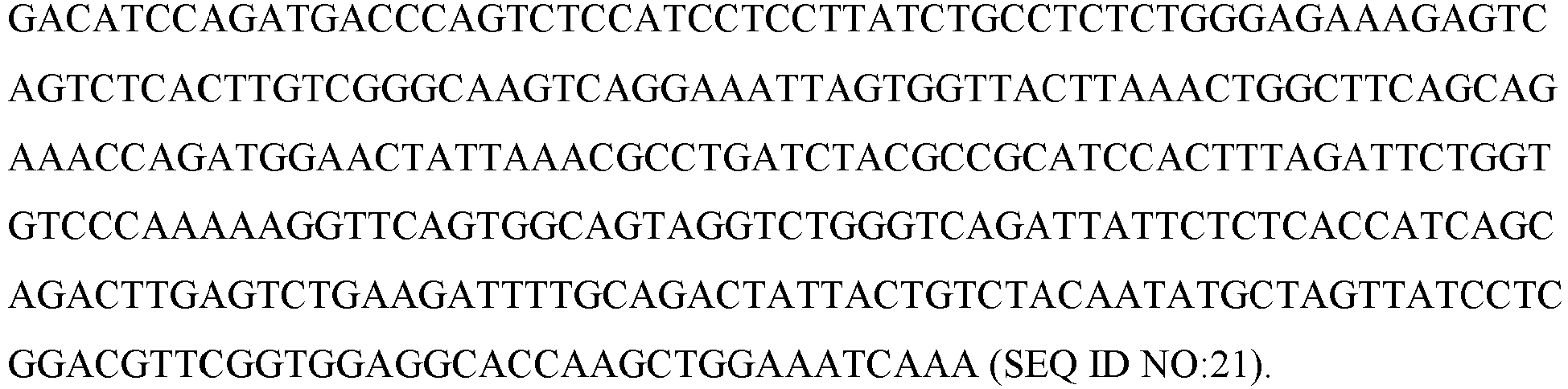
- Antibody 258-cs1.04_Light chain: Amino acids sequence

- To examine the recognition of TIGIT by the mAbs, 2∗105 YTS-TIGIT over expressing cells (as previously described by Stanietsky et al., ibid) were incubated with 0.2 microgram of anti TIGIT mAb
clone VSIG9# 1 or #4 on ice for 30 min. After two rounds of wash in FACS buffer, goat-anti mouse IgG (H+L) Secondary Antibody Alexa Fluor® 647 conjugate (BioLegend) was added for additional 30 min on ice. As shown inFigure 1A , the mAbs recognize TIGIT proteins on YTS-TIGIT over expressing cells (right curve), but not on YTS cells. mIgG was used as a control. Next, theVSIG9# 1 was examined on activated NK cells from two healthy donors. As shown inFigure 1B , the mAb recognized the NK cells (right curve in the FACS histogram plots). mIgG was used as a control (left curve). - Next, the
VSIG9# 1 was examined on two CD8+ T cell populations, obtained from melanoma patients (TIL-I and TIL-II). As shown inFigure 1C , the mAb recognized the T-cells (right curve in the FACS histogram plots). mIgG was used as control (left curve). Overall,figures 1A-1C shows broad expression of TIGIT on immune cells and demonstrate that theVSIG9# 1 mAb can bind to immune cells. - To examine the effect of
VSIG9# 1 on TIGIT-Fc binding to tumor cells, 2.5∗10 5 HepG2 cells (that express high levels of PVR, Nectin-2 and Nectin-3) were incubated with 25 microgram/well hTIGIT-Fc with no mAb (Figure 2A , arrow 1) with mIgG (Figure 2A , arrow 2) or with anti-TIGIT-VSIG9# 1 at the indicated concentrations (Figure 2A ,arrows 3 and 4) for 30 min on ice, the detection was done using Alexa Fluor® 647 anti-human IgG (BioLegend). The expression of TIGIT ligands, PVR, Nectin-2 and Nectin-3 on the HepG2 cells is demonstrated inFigure 2B . - The results demonstrate that the mAb can prevent TIGIT binding to tumor cells, suggesting that the antibody is capable of preventing the inhibition of the immune response.
- To examine the effect of anti-TIGIT mAbs on the killing activity of effector cells, YTS-TIGIT NK cells were incubated with 35S labeled 721.221-PVR cells in the presence of 2.5 microgram/ml of control mIgG (left bar) or anti-TIGIT VSIG9#1 (right bar)(∗p< 0.02). The specific killing was calculated as previously described (Stanietsky et.al ibid). The killing percentage was examined after 5 hours. As shown in
Figure 3A , anti TIGITmAb VSIG9# 1 enhances killing activity. Next, the specific killing activity was examined for MDA-MB-231 human breast tumor cells that were labeled with 35S and incubated with NK cells from healthy donors in the presence of control mIgG (left bar) or VSIG9#1 (right bars). Killing percentage was measured after 5 hours (∗∗p<0.002). As shown inFigure 3B ,VSIG9# 1 enhances the killing effect. The specific killing activity of two antibodies was also examined with MEL562 melanoma cells. As shown inFigure 3C , the two mAbs, 258-CS1#4 (central bar) and VSIG9#1 (left bar) significantly enhances (∗∗p<0.05) the killing effect of these cells compared to control antibody mIgG (right bar). Finally, antibody-dependent cell-mediated cytotoxicity (ADCC) ofVSIG9# 1 was examined on human hepatocellular HepG2 cells labeled with 35S that were incubated with anti-EGFR mAb (Erbitux®) and added to NK cells that were pre-incubated with control mAb (left bar) or with VSIG9#1 (right bar) at a ratio of 10:1 Effector cells:Target cells, respectively. (∗∗∗p<0.0007). A shown inFigure 3D ,VSIG9# 1 is capable of enhancing the killing effect. - The results demonstrate that the tested anti human
mAbs VSIG9# 1 and 258-CS1# 4 are capable of enhancing the killing effect on different target cells and in a variety of conditions. The mAbs prevent the inhibitory effect of the TIGIT receptor and are therefore suitable for use as anti-cancer agents. - To examine the ability of mAbs 258-
CS1# 4 andVSIG9# 1 to block the interaction of TIGIT which its ligands , HepG2 cells that express PVR, Nectin-2 and Nectin-3 were incubated with TIGIT-Fc or TIGIT-Fc following pre-incubation with mAb VSIG-9#1 (Figure 4A ), or with mAb 258-CS1#4 (Figure 4B ). The results showed that pre-incubation of TIGIT-Fc with VSIG-9 #1 completely blocked TIGIT-Fc binding (Figure 4A ), while pre-incubation with mAB 258-CS1# 4 partially blocked TIGIT-Fc binding (Figure 4B ). - Full binding kinetic analyses of the mAbs to human TIGIT from two sources were carried out using Biacore. The results shown in
Figures 5A and5B indicate that the two mAbs exhibit high affinity to human TIGIT. While the mAb termed 258-CS1#4 (#4) had an average Kd of about 1×10⁻⁷ M (average of 9.96×10⁻⁸ M and 1.07×10⁻⁷ M), mAb VSIG-9#1 binds human TIGIT with an extremely high affinity, having a Kd of 4.5×10⁻¹⁰ M (average of 5.13-10 M and 3.87-10 M). - It was further demonstrated that mAb VSIG-9#1 is specific to human TIGIT and does not bind mouse TIGIT.
- The binding affinity of
antibodies VSIG9# 1 and MBSA43 to TIGIT was compared. 75×103 TIGIT expressing YTS cells were stained with the two antibodies at serial dilution concentrations of 250nM to 65fM, following determination by FACS. As shown inFigures 6A and6B ,VSIG# 9 stained the cells at a concentration lower as 62.5 fM, while MBSA43 stained the cells only above 1950 fM (1.95 pM) and did not show staining at the last 4 dilutions Clear staining differences between the two antibodies are seen up to a concentration of 250 pM. The superiority of theVSIG9# 1 antibody compared to the MBSA43 antibody is also depicted inFigure 6B . - To examine the anti-TIGIT antibodies blocking effect on PVR-TIGIT binding, a staining assay of TIGIT-expressing YTS cells with PVR- Fc was performed. 75×103 YTS-TIGIT cells were incubated with 2.5 pmole PVR-Fc in the presence of
VSIG9# 1 or MBSA43 at a concentration range of from 27 to 0.014 pmole in a series of two fold dilutions. - As shown in
Figure 7A (detection of bound PVR) and 7B (detection of bound anti TIGIT mAb), the PVR-TIGIT blocking activity ofVSIG9# 1 was significantly higher as compared to MBSA43. - The results of Examples 7 and 8 demonstrate that
VSIG9# 1 has significant higher affinity to human TIGIT, as compared to MBSA43 and that it is significantly better at preventing the binding of the high affinity ligand (PVR) across a range of concentrations. - The efficacy of mAb VSIG9#1 (Vsig9.01), alone or in combination with other immunemodulators, on induction of T cell proliferation by blocking of TIGIT, was examined. PBMCs from healthy donors were labeled with 5(6)-Carboxyfluorescein N-hydroxysuccinimidyl ester (CFSE) and activated with anti-CD3 antibodies followed by incubation for 5-9 days with MDA-MB-231 cells over-expressing hCD80 in the presence of 4 µg/ml of the
mAb VSIG9# 1, anti-PD-1 (Keytruda) and anti-CTLA4 (pilimumab), alone or in combination. Proliferation was measured by CFSE dilutions. - The results shown in
Figure 8 , collected from at least 5 different experiments, indicate a synergism between the anti TIGIT mAb and anti PD-1 or anti CTLA4, demonstrated by significant increased T-cell proliferation for the combination ofVSIG9# 1 with the other checkpoint antibodies (∗p< 0.04 ∗∗< 0.015 ∗∗∗<0.0004). - The anti-tumor efficacy of the antibodies is studied in vivo. To estimate the efficacy of the antibodies described herein in inhibition of human cancer, the antibody is studied in a model combining both tumors and lymphocytes of human origin. Severe combined immune-deficient mice (SCID) is engrafted with hPBL to restore immune-competence. Mice are challenged with human cancer cells and treated with increasing concentrations of the anti human TIGIT antibody, administered in single- or multi-intravascular dose several days post tumor challenge.
- A similar model with tumor lines in SCID mice is performed according to Paine-Murrieta GD, Cancer Chemother Pharmacol. 1997;40(3):209-14.
- To estimate the efficacy of anti-TIGIT antibodies in inhibition of human cancer, the modified antibody is studied in a model combining both tumors and lymphocytes of human origin. Severe combined immune-deficient mice (SCID) is engrafted with hPBL to restore immune-competence. Mice are challenged with human melanoma cells (SK-28) and treated with increasing concentrations of the antibody, administered in a single i.v. dose on day 11 post tumor inoculation.
- Similarly, a model described by Hardy el al., Proc Natl Acad Sci U S A. 1997 May 27; 94(11): 5756-5760 is employed.
- LIM6 and HM7 are two sub-clones of the human CRC cell line LS174T that were selected for their high mucin synthesis and metastatic potential. The tumor cells are injected into the exposed spleen of anesthetized nude mice. After several minutes, the spleens are removed and the excisions closed. Low doses of anti-TIGIT antibodies of the invention are administered 10 days later and mice are sacrificed 35 days post tumor inoculation. The livers are weighed, the number of metastatic nodules counted, and liver tissue is processed for histology and Immunohistochemistry study.
- Additional metastasis models that can be used to test the antibodies and fragments of the present invention were described by Yung at al., Ocul Oncol Pathol. 2015 Apr; 1(3): 151-160.
- Bone marrow aspirate was obtained from an AML patient, following Ficoll separation the immune cells and the blasts were co-cultured with various antibodies (at 4 µg/ml) for 12 days after 12 days the amount of T cells was established.
- As demonstrated in
Figure 9 , significant increase in the amount of CD8 T cells was observed in presence ofanti-TIGIT VSIG9# 1 mAb. Interestingly blocking of TIGIT had synergistic effect with blocking of PD-1 and CTLA-4. -
- <110> Yissum Research Development Company of the Hebrew University of Jerusalem Ltd. University of Rijeka Faculty of Medicine
- <120> ANTIBODIES SPECIFIC TO HUMAN T-CELL IMMUNOGLOBULIN AND ITIM DOMAIN (TIGIT)
- <130> YISSUM/0124 PCT
- <150>
62/213140 - <150>
62/314427 - <160> 21
- <170> PatentIn version 3.5
- <210> 1
<211> 10
<212> PRT
<213> Mus musculus - <400> 1

- <210> 2
<211> 17
<212> PRT
<213> Mus musculus - <400> 2
Gly

- <210> 3
<211> 11
<212> PRT
<213> Mus musculus - <400> 3

- <210> 4
<211> 11
<212> PRT
<213> Mus musculus - <400> 4

- <210> 5
<211> 7
<212> PRT
<213> Mus musculus - <400> 5

- <210> 6
<211> 9
<212> PRT
<213> Mus musculus - <400> 6

- <210> 7
<211> 120
<212> PRT
<213> Mus musculus - <400> 7


- <210> 8
<211> 116
<212> PRT
<213> Mus musculus - <400> 8

- <210> 9
<211> 360
<212> DNA
<213> Mus Musculus - <400> 9

- <210> 10
<211> 348
<212> DNA
<213> Mus musculus - <400> 10

- <210> 11
<211> 6
<212> PRT
<213> Mus musculus - <400> 11

- <210> 12
<211> 5
<212> PRT
<213> Mus musculus - <400> 12

- <210> 13
<211> 17
<212> PRT
<213> Mus musculus - <400> 13
Asn

- <210> 14
<211> 11
<212> PRT
<213> Mus musculus - <400> 14


- <210> 15
<211> 11
<212> PRT
<213> Mus musculus - <400> 15

- <210> 16
<211> 7
<212> PRT
<213> Mus musculus - <400> 16

- <210> 17
<211> 9
<212> PRT
<213> Mus musculus - <400> 17

- <210> 18
<211> 120
<212> PRT
<213> Mus musculus - <400> 18
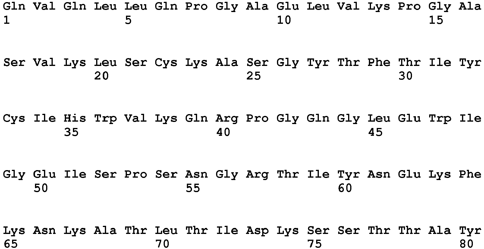

- <210> 19
<211> 107
<212> PRT
<213> Mus musculus - <400> 19

- <210> 20
<211> 360
<212> DNA
<213> Mus musculus - <400> 20


- <210> 21
<211> 321
<212> DNA
<213> Mus musculus - <400> 21

| | VSIG9# | 1 | SEQ ID No. | 258-cs1.04 | SEQ ID No. |
| | GYTFTSYGIS | 1 | | 12 | |
| | EIYPRSGNTYYNEKFKG | 2 | | 13 | |
| | KGPYYTKNEDY | 3 | SDGYDGYYFDY | 14 | |
| | RASEHIYYSLA | 4 | | 15 | |
| LCDR2 | NANSLED | 5 | | 16 | |
| LCDR3 | KQAYDVPRT | 6 | LQYASYPRT | 17 | |
| VH | Amino acid sequence | 7 | | 18 | |
| | Polynucleotide sequence | 9 | | 20 | |
| VL | Amino acid sequence | 8 | Amino acid sequence | 19 | |
| | Polynucleotide sequence | 10 | Polynucleotide sequence | 21 |
Claims (15)
- An isolated monoclonal antibody, or an antigen-binding fragment thereof, wherein the antibody or fragment binds to human T-cell immunoglobulin and ITIM domain (TIGIT) and is capable of inhibiting the binding of TIGIT to at least PVR (CD 155), wherein said antibody or antigen-binding fragment thereof comprises:(i) a heavy chain comprising a HCDR1 as set forth in SEQ ID NO: 1 or 11; HCDR2 as set forth in SEQ ID NO: 2; HCDR3 as set forth in SEQ ID NO: 3; and(ii) a light chain comprising a LCDR1 as set forth in SEQ ID NO: 4; LCDR2 as set forth in SEQ ID NO:5; and LCDR3 as set forth in SEQ ID NO: 6.
- The antibody of claim 1, wherein the heavy chain comprises SEQ ID NO: 7 and the light chain comprises SEQ ID NO: 8.
- The antibody of claim 1 or claim 2, wherein the antibody is humanized.
- A bispecific antibody comprising two sets of CDRs, wherein one of the sets comprises the CDRs as define in claim 1.
- The bispecific antibody of claim 4, wherein the second set of CDRs comprises HCDR1 as set forth in SEQ ID NO:12; HCDR2 as set forth in SEQ ID NO:13, HCDR3 as set forth in SEQ ID NO:14; LCDR1 as set forth in SEQ ID NO: 15; LCDR2 as set forth in SEQ ID NO: 16; and LCDR3 as set forth in SEQ ID NO:17.
- An antibody conjugate comprising the antibody of any one of claims 1-3.
- The conjugate of claim 6, wherein the antibody is conjugated to a cytotoxic, a radioactive or an identifiable moiety.
- A polynucleotide sequence encoding a monoclonal antibody according to claim 1, wherein said polynucleotide comprises SEQ ID NO: 9 and SEQ ID NO: 10.
- A vector comprising a polynucleotide sequence according to claim 8.
- A hybridoma cell capable of producing a monoclonal antibody according to claim 1.
- A pharmaceutical composition comprising as an active ingredient, at least one isolated antibody or fragment thereof according to any one of claims 1 to 7, and a pharmaceutical acceptable excipient, diluent, salt or carrier.
- A monoclonal antibody according to any of the claims 1 to 7 or a pharmaceutical composition according to claim 11 for use in the treatment of cancer.
- The pharmaceutical composition for use of claim 12,further comprising an additional anti-cancer therapy selected from surgery, chemotherapy, radiotherapy, and immunotherapy; orfurther comprising administering an immune checkpoint molecule selected from the group consisting of PD-1, CTLA-4, PDL-1, CEACAM1, lymphocyte activation gene 3 (LAG3), CD137, OX40 (also referred to as CD134), killer cell immunoglobulin-like receptors (KIR), and any combination thereof, preferably wherein the anti-cancer therapy comprises administration of epidermal growth factor receptor (EGFR) inhibitor.
- A method of determining or quantifying the expression of TIGIT, the method comprising contacting a biological sample with an antibody or antibody fragment according to any one of claims 1 to 7, and measuring the level of complex formation.
- A kit for measuring the expression of TIGIT in biological sample comprising at least one antibody or antibody fragment according to any one of claims 1 to 7.
Applications Claiming Priority (3)
| Application Number | Priority Date | Filing Date | Title |
|---|---|---|---|
| US201562213140P | 2015-09-02 | 2015-09-02 | |
| US201662314427P | 2016-03-29 | 2016-03-29 | |
| PCT/IL2016/050952 WO2017037707A1 (en) | 2015-09-02 | 2016-09-01 | Antibodies specific to human t-cell immunoglobulin and itim domain (tigit) |
Publications (2)
| Publication Number | Publication Date |
|---|---|
| EP3344658A1 EP3344658A1 (en) | 2018-07-11 |
| EP3344658B1 true EP3344658B1 (en) | 2022-05-11 |
Family
ID=57068160
Family Applications (1)
| Application Number | Title | Priority Date | Filing Date |
|---|---|---|---|
| EP16775866.3A Active EP3344658B1 (en) | 2015-09-02 | 2016-09-01 | Antibodies specific to human t-cell immunoglobulin and itim domain (tigit) |
Country Status (6)
| Country | Link |
|---|---|
| US (2) | US10946095B2 (en) |
| EP (1) | EP3344658B1 (en) |
| JP (1) | JP6861418B2 (en) |
| CN (1) | CN108137691B (en) |
| ES (1) | ES2924071T3 (en) |
| WO (1) | WO2017037707A1 (en) |
Families Citing this family (66)
| Publication number | Priority date | Publication date | Assignee | Title |
|---|---|---|---|---|
| GB201403775D0 (en) | 2014-03-04 | 2014-04-16 | Kymab Ltd | Antibodies, uses & methods |
| US9139653B1 (en) | 2015-04-30 | 2015-09-22 | Kymab Limited | Anti-human OX40L antibodies and methods of treatment |
| TWI715587B (en) | 2015-05-28 | 2021-01-11 | 美商安可美德藥物股份有限公司 | Tigit-binding agents and uses thereof |
| UA125062C2 (en) | 2015-10-01 | 2022-01-05 | Потенза Терапеутікс, Інк. | ANTI-TIGIT ANTIGEN-BINDING PROTEINS AND METHODS OF THEIR APPLICATION |
| JP7461741B2 (en) | 2016-06-20 | 2024-04-04 | カイマブ・リミテッド | Anti-PD-L1 and IL-2 Cytokines |
| US9567399B1 (en) | 2016-06-20 | 2017-02-14 | Kymab Limited | Antibodies and immunocytokines |
| CA3032897A1 (en) | 2016-08-09 | 2018-02-15 | Kymab Limited | Anti-icos antibodies |
| US11779604B2 (en) | 2016-11-03 | 2023-10-10 | Kymab Limited | Antibodies, combinations comprising antibodies, biomarkers, uses and methods |
| CA3044664C (en) | 2016-11-30 | 2022-11-22 | Oncomed Pharmaceuticals, Inc. | Methods for treatment of cancer comprising tigit-binding agents |
| JOP20190203A1 (en) * | 2017-03-30 | 2019-09-03 | Potenza Therapeutics Inc | Anti-tigit antigen-binding proteins and methods of use thereof |
| EP3615070A1 (en) | 2017-04-26 | 2020-03-04 | Bristol-Myers Squibb Company | Methods of antibody production that minimize disulfide bond reduction |
| RS64576B1 (en) | 2017-05-01 | 2023-10-31 | Agenus Inc | Anti-tigit antibodies and methods of use thereof |
| MX2019014265A (en) | 2017-06-01 | 2020-08-03 | Compugen Ltd | TRIPLE JOINT TREATMENTS WITH ANTIBODIES. |
| WO2018229163A1 (en) | 2017-06-14 | 2018-12-20 | King's College London | Methods of activating v delta 2 negative gamma delta t cells |
| GB201709808D0 (en) * | 2017-06-20 | 2017-08-02 | Kymab Ltd | Antibodies |
| US11512129B2 (en) | 2017-09-29 | 2022-11-29 | Jiangsu Hengrui Medicine Co., Ltd. | TIGIT antibody, antigen-binding fragment thereof, and medical use thereof |
| GB201721338D0 (en) | 2017-12-19 | 2018-01-31 | Kymab Ltd | Anti-icos Antibodies |
| EP3728314A1 (en) | 2017-12-19 | 2020-10-28 | Kymab Limited | Bispecific antibody for icos and pd-l1 |
| TW201930358A (en) * | 2017-12-28 | 2019-08-01 | 大陸商南京傳奇生物科技有限公司 | Single-domain antibodies and variants thereof against TIGIT |
| KR102839330B1 (en) | 2018-01-15 | 2025-07-30 | 난징 레전드 바이오테크 씨오., 엘티디. | Single-domain antibodies to PD-1 and variants thereof |
| CN110352200B (en) | 2018-02-06 | 2020-12-11 | 天境生物科技(上海)有限公司 | Antibody against T cell immune receptor (TIGIT) with Ig and ITIM domains and its application |
| WO2019215728A1 (en) | 2018-05-09 | 2019-11-14 | Yissum Research Development Company Of The Hebrew University Of Jerusalem Ltd. | Antibodies specific to human nectin4 |
| CN110737397B (en) * | 2018-07-20 | 2023-08-11 | 伊姆西Ip控股有限责任公司 | Method, apparatus and computer program product for managing a storage system |
| BR112021001201A2 (en) * | 2018-07-25 | 2021-04-27 | Innovent Biologics (Suzhou) Co., Ltd. | anti-tigit antibody and use of it |
| BR112021003156A2 (en) | 2018-08-23 | 2021-05-11 | Seagen, Inc. | composition, antibody that binds to human tigi, pharmaceutical formulation, isolated polynucleotide, vector, host cell, methods for producing an antibody that binds to human tigi and fucosylated antibodies that bind to tigi, and for the treatment of cancer , and, kit. |
| CN113614109A (en) | 2018-12-21 | 2021-11-05 | Ose免疫疗法公司 | Bifunctional anti-PD-1/IL-7 molecules |
| JP7606706B2 (en) | 2019-01-13 | 2024-12-26 | イッサム リサーチ デベロップメント カンパニー オブ ザ ヘブリュー ユニバーシティー オブ エルサレム エルティーディー. | Antibody specific to human nectin-2 |
| WO2020165374A1 (en) | 2019-02-14 | 2020-08-20 | Ose Immunotherapeutics | Bifunctional molecule comprising il-15ra |
| CN111744013B (en) * | 2019-03-29 | 2022-07-26 | 江苏恒瑞医药股份有限公司 | Methods and pharmaceutical combinations for treating diseases using anti-TIGIT antibodies in combination with PD-1 inhibitors |
| WO2021008523A1 (en) * | 2019-07-15 | 2021-01-21 | 上海君实生物医药科技股份有限公司 | Anti-tigit antibodies and application thereof |
| TW202124452A (en) * | 2019-11-14 | 2021-07-01 | 中國大陸商上海藥明生物技術有限公司 | A novel antibody against tigit |
| AU2020406083A1 (en) | 2019-12-17 | 2022-06-16 | Boehringer Ingelheim International Gmbh | Bifunctional molecules comprising an IL-7 variant |
| CN110818795B (en) * | 2020-01-10 | 2020-04-24 | 上海复宏汉霖生物技术股份有限公司 | anti-TIGIT antibodies and methods of use |
| CA3176497A1 (en) | 2020-05-07 | 2021-11-11 | Fatima MECHTA-GRIGORIOU | Antxr1 as a biomarker of immunosuppressive fibroblast populations and its use for predicting response to immunotherapy |
| CN114507284B (en) * | 2020-05-09 | 2023-05-26 | 华博生物医药技术(上海)有限公司 | Antibodies against TIGIT, methods of preparation and uses thereof |
| CN115916348A (en) | 2020-06-18 | 2023-04-04 | 基因泰克公司 | Treatment with Anti-TIGIT Antibodies and PD-1 Axis Binding Antagonists |
| TW202216778A (en) | 2020-07-15 | 2022-05-01 | 美商安進公司 | Tigit and cd112r blockade |
| JP7847579B2 (en) * | 2020-08-14 | 2026-04-17 | ガンマデルタ セラピューティクス リミテッド | Multispecific anti-TCR delta variable 1 antibody |
| WO2022044010A1 (en) | 2020-08-26 | 2022-03-03 | Yissum Research Development Company Of The Hebrew University Of Jerusalem Ltd. | Anti-t-cell immunoglobulin and itim domain (tigit) antibodies for the treatment of fungal infections |
| US20230374128A1 (en) * | 2020-10-09 | 2023-11-23 | Keio University | Therapeutic agent for immune/inflammatory disease |
| EP4238991A4 (en) * | 2020-11-23 | 2024-05-15 | Guangdong Fapon Biopharma Inc. | Anti-tigit antibody or antigen-binding fragment thereof |
| WO2022112198A1 (en) | 2020-11-24 | 2022-06-02 | Worldwide Innovative Network | Method to select the optimal immune checkpoint therapies |
| WO2022148781A1 (en) | 2021-01-05 | 2022-07-14 | Institut Curie | Combination of mcoln activators and immune checkpoint inhibitors |
| JP2024507124A (en) | 2021-02-11 | 2024-02-16 | ネクチン セラピューティクス リミテッド | Antibodies against CD112R and uses thereof |
| US20240182572A1 (en) | 2021-04-09 | 2024-06-06 | Ose Immunotherapeutics | Scaffold for bifunctional molecules comprising pd-1 or cd28 and sirp binding domains |
| WO2022214653A1 (en) | 2021-04-09 | 2022-10-13 | Ose Immunotherapeutics | New scaffold for bifunctional molecules with improved properties |
| TW202304965A (en) | 2021-05-04 | 2023-02-01 | 美商艾吉納斯公司 | Anti-tigit antibodies, anti-cd96 antibodies, and methods of use thereof |
| AU2022272586A1 (en) | 2021-05-10 | 2024-01-04 | Medimabbio Inc. | Anti-tigit antibodies and use thereof |
| TW202320848A (en) | 2021-07-28 | 2023-06-01 | 美商建南德克公司 | Methods and compositions for treating cancer |
| WO2023056403A1 (en) | 2021-09-30 | 2023-04-06 | Genentech, Inc. | Methods for treatment of hematologic cancers using anti-tigit antibodies, anti-cd38 antibodies, and pd-1 axis binding antagonists |
| JP2024537188A (en) * | 2021-10-08 | 2024-10-10 | アーベル リミテッド | Compositions and methods for detecting cadherin-17 protein |
| CN119451983A (en) | 2022-02-28 | 2025-02-14 | 耐科廷疗法有限公司 | Humanized antibodies and drug conjugates targeting connexin 2 |
| AU2023236289A1 (en) | 2022-03-15 | 2024-08-15 | Compugen Ltd. | Il-18bp antagonist antibodies and their use in monotherapy and combination therapy in the treatment of cancer |
| IL317449A (en) | 2022-06-07 | 2025-02-01 | Genentech Inc | Method for determining the efficacy of a lung cancer treatment comprising an anti-pd-l1 antagonist and an anti-tigit antagonist antibody |
| WO2024003360A1 (en) | 2022-07-01 | 2024-01-04 | Institut Curie | Biomarkers and uses thereof for the treatment of neuroblastoma |
| EP4565610A1 (en) | 2022-08-02 | 2025-06-11 | Ose Immunotherapeutics | Multifunctional molecule directed against cd28 |
| EP4687990A1 (en) | 2023-03-30 | 2026-02-11 | Ose Immunotherapeutics | Method of synthesis of targeted lipid nanoparticle and uses thereof |
| JP2026512425A (en) | 2023-03-30 | 2026-04-16 | オーエスイー・イミュノセラピューティクス | Lipid-based nanoparticles targeted to activated immune cells for the expression of immune cell-enhancing molecules and their use |
| TW202515608A (en) | 2023-06-26 | 2025-04-16 | 以色列商坎布根有限公司 | Il-18bp antagonist antibodies and their use in monotherapy and combination therapy in the treatment of cancer |
| WO2025068461A1 (en) | 2023-09-29 | 2025-04-03 | Negio Therapeutics | Guanfacine derivatives and their use in treating cancer |
| WO2025068452A1 (en) | 2023-09-29 | 2025-04-03 | Negio Therapeutics | Guanfacine derivatives and their use in treating cancer |
| WO2025132831A1 (en) | 2023-12-19 | 2025-06-26 | Universite D'aix-Marseille | N-heteroaryl derivatives and uses thereof for treating cancer |
| WO2025242836A1 (en) | 2024-05-22 | 2025-11-27 | Ose Immunotherapeutics | Molecules comprising masking linkers and uses thereof for the treatment of auto-immune or inflammatory diseases and disorders |
| WO2025242835A1 (en) | 2024-05-22 | 2025-11-27 | Ose Immunotherapeutics | Molecules comprising masking linkers and uses thereof for the treatment of cancer |
| WO2025262250A1 (en) | 2024-06-20 | 2025-12-26 | Negio Therapeutics | Guanfacine derivatives and their uses |
| WO2026068705A1 (en) | 2024-09-26 | 2026-04-02 | Ose Immunotherapeutics | Lipid-based nanoparticles comprising non-glycosylated fc domains and uses thereof |
Citations (1)
| Publication number | Priority date | Publication date | Assignee | Title |
|---|---|---|---|---|
| CN103073644A (en) * | 2012-12-31 | 2013-05-01 | 中国科学技术大学 | Specific anti-mouse TIGIT monoclonal antibody and preparation method, identification and application thereof |
Family Cites Families (42)
| Publication number | Priority date | Publication date | Assignee | Title |
|---|---|---|---|---|
| US4816567A (en) | 1983-04-08 | 1989-03-28 | Genentech, Inc. | Recombinant immunoglobin preparations |
| US5225539A (en) | 1986-03-27 | 1993-07-06 | Medical Research Council | Recombinant altered antibodies and methods of making altered antibodies |
| US4946778A (en) | 1987-09-21 | 1990-08-07 | Genex Corporation | Single polypeptide chain binding molecules |
| US5530101A (en) | 1988-12-28 | 1996-06-25 | Protein Design Labs, Inc. | Humanized immunoglobulins |
| DE3920358A1 (en) | 1989-06-22 | 1991-01-17 | Behringwerke Ag | BISPECIFIC AND OLIGO-SPECIFIC, MONO- AND OLIGOVALENT ANTI-BODY CONSTRUCTS, THEIR PRODUCTION AND USE |
| DE69233528T2 (en) | 1991-11-25 | 2006-03-16 | Enzon, Inc. | Process for the preparation of multivalent antigen-binding proteins |
| ES2102007T3 (en) | 1992-01-23 | 1997-07-16 | Merck Patent Gmbh | MONOMER AND DIMER ANTIBODY FRAGMENT FUSION PROTEINS. |
| US5869337A (en) | 1993-02-12 | 1999-02-09 | President And Fellows Of Harvard College | Regulated transcription of targeted genes and other biological events |
| JP3659261B2 (en) | 1994-10-20 | 2005-06-15 | モルフォシス・アクチェンゲゼルシャフト | Targeted heterojunction of a recombinant protein to a multifunctional complex |
| GB9421405D0 (en) | 1994-10-21 | 1994-12-07 | Dow Chemical Co | Low voc laminating formulations |
| US5641870A (en) | 1995-04-20 | 1997-06-24 | Genentech, Inc. | Low pH hydrophobic interaction chromatography for antibody purification |
| ATE274064T1 (en) | 1995-05-23 | 2004-09-15 | Morphosys Ag | MULTIMER PROTEINS |
| DK0941344T3 (en) | 1996-11-27 | 2004-09-27 | Genentech Inc | Humanized anti-CD11a antibodies |
| WO1999063063A1 (en) | 1998-06-03 | 1999-12-09 | Northwestern University | Cellular proteins which mediate herpesvirus entry |
| US7193069B2 (en) | 2002-03-22 | 2007-03-20 | Research Association For Biotechnology | Full-length cDNA |
| EP2277532A1 (en) | 2002-09-11 | 2011-01-26 | Genentech, Inc. | Novel composition and methods for the treatment of immune related diseases |
| WO2004024072A2 (en) | 2002-09-11 | 2004-03-25 | Genentech, Inc. | Novel compositions and methods for the treatment of immune related diseases |
| AU2003289899A1 (en) | 2002-12-05 | 2004-06-23 | Ludwig-Maximilians-Uni Versitat | Genetic switches for the detection of fusion proteins |
| EP1481993A1 (en) | 2003-05-28 | 2004-12-01 | Xerion Pharmaceuticals AG | Modulation of the poliovirus receptor function |
| PE20050928A1 (en) | 2003-11-21 | 2005-11-08 | Schering Corp | THERAPEUTIC COMBINATIONS OF ANTI-IGFR1 ANTIBODY |
| GB2408508A (en) | 2003-11-28 | 2005-06-01 | Astrazeneca Ab | Apolipoprotein antibodies |
| SV2006002258A (en) | 2004-10-08 | 2006-09-19 | Wyeth Corp | IMMUNOTHERAPY OF AUTOIMMUNE DISORDERS |
| JP2008544746A (en) | 2005-05-12 | 2008-12-11 | ザイモジェネティクス, インコーポレイテッド | Compositions and methods for modulating immune responses |
| MX2008004094A (en) | 2005-09-27 | 2009-02-27 | Efrat Biopolymers Ltd | Gelling hydrophobic injectable polymer compositions. |
| NZ601022A (en) | 2006-10-12 | 2014-09-26 | Chugai Pharmaceutical Co Ltd | Diagnosis and treatment of cancer using anti-ereg antibody |
| JP5142265B2 (en) | 2007-05-28 | 2013-02-13 | 独立行政法人産業技術総合研究所 | Paratopes and epitopes of anti-mortalin antibodies |
| WO2008146854A1 (en) * | 2007-05-28 | 2008-12-04 | National Institute Of Advanced Industrial Science And Technology | Paratope and epitope of anti-mortalin antibody |
| EP2527366B1 (en) | 2007-06-12 | 2017-08-16 | AC Immune S.A. | Monoclonal anti beta amyloid antibody |
| WO2009088105A1 (en) * | 2008-01-11 | 2009-07-16 | Gene Techno Science Co., Ltd. | HUMANIZED ANTI-α9 INTEGRIN ANTIBODIES AND THE USES THEREOF |
| EP3208612B1 (en) * | 2008-04-09 | 2019-09-18 | Genentech, Inc. | Compositions and methods for the treatment of immune related diseases |
| JP2010014691A (en) | 2008-06-20 | 2010-01-21 | Igaku Seibutsugaku Kenkyusho:Kk | Methods, kits, reagents, and devices for detecting mesothelin and/or megakaryocyte enhancer in ascites |
| US8518405B2 (en) * | 2009-10-08 | 2013-08-27 | The University Of North Carolina At Charlotte | Tumor specific antibodies and uses therefor |
| US8256790B2 (en) | 2010-05-26 | 2012-09-04 | William Olen Fortner | Adjustable receiver hitch system |
| KR101502501B1 (en) | 2011-12-27 | 2015-03-13 | 주식회사 엘지화학 | Lithium secondary battery and Method for preparation of the same |
| GB201300049D0 (en) | 2013-01-03 | 2013-02-20 | Transimmune Ag | Method for obtaining immuno-stimulatory dendritic cells |
| GB201300052D0 (en) | 2013-01-03 | 2013-02-20 | Transimmune Ag | Method for obtaining immuno-suppressive dendritic cells |
| TWI888366B (en) | 2013-07-16 | 2025-07-01 | 美商建南德克公司 | Methods of treating cancer using pd-1 axis binding antagonists and tigit inhibitors |
| CR20170060A (en) | 2014-08-19 | 2017-04-18 | Merck Sharp & Dohme | ANTI TIGIT ANTIBODIES |
| WO2016036319A1 (en) | 2014-09-04 | 2016-03-10 | Agency For Science, Technology And Research | A method to up-regulate cancer stem cell markers for the generation of antigen specific cytotoxic effector t cells |
| US11254914B2 (en) | 2015-03-12 | 2022-02-22 | Health Research, Inc. | Enrichment of CD16+ monocytes to improve dendritic cell vaccine quality |
| TWI715587B (en) | 2015-05-28 | 2021-01-11 | 美商安可美德藥物股份有限公司 | Tigit-binding agents and uses thereof |
| IL243461A0 (en) | 2016-01-05 | 2016-02-29 | Amir Avraham | Surgical sutures for skin filling |
-
2016
- 2016-09-01 EP EP16775866.3A patent/EP3344658B1/en active Active
- 2016-09-01 WO PCT/IL2016/050952 patent/WO2017037707A1/en not_active Ceased
- 2016-09-01 ES ES16775866T patent/ES2924071T3/en active Active
- 2016-09-01 CN CN201680051019.6A patent/CN108137691B/en active Active
- 2016-09-01 US US15/756,606 patent/US10946095B2/en active Active
- 2016-09-01 JP JP2018511264A patent/JP6861418B2/en active Active
-
2021
- 2021-02-05 US US17/168,451 patent/US12121579B2/en active Active
Patent Citations (1)
| Publication number | Priority date | Publication date | Assignee | Title |
|---|---|---|---|---|
| CN103073644A (en) * | 2012-12-31 | 2013-05-01 | 中国科学技术大学 | Specific anti-mouse TIGIT monoclonal antibody and preparation method, identification and application thereof |
Also Published As
| Publication number | Publication date |
|---|---|
| JP6861418B2 (en) | 2021-04-28 |
| JP2018532383A (en) | 2018-11-08 |
| US12121579B2 (en) | 2024-10-22 |
| CN108137691A (en) | 2018-06-08 |
| US20210220472A1 (en) | 2021-07-22 |
| US10946095B2 (en) | 2021-03-16 |
| ES2924071T3 (en) | 2022-10-04 |
| CA2997130A1 (en) | 2017-03-09 |
| US20180185480A1 (en) | 2018-07-05 |
| CN108137691B (en) | 2021-10-19 |
| WO2017037707A1 (en) | 2017-03-09 |
| EP3344658A1 (en) | 2018-07-11 |
Similar Documents
| Publication | Publication Date | Title |
|---|---|---|
| US12121579B2 (en) | Antibodies specific to human t-cell immunoglobulin and ITIM domain (TIGIT) | |
| US20240018257A1 (en) | Antibodies specific to human poliovirus receptor (pvr) | |
| KR102892725B1 (en) | Antibody specific for human nectin 4 | |
| US20260116971A1 (en) | Antibodies specific to human nectin-2 | |
| JP2026026083A (en) | Multispecific antibodies for the treatment of cancer | |
| US20240270840A1 (en) | Antibodies against cd112r and uses thereof | |
| CA2997130C (en) | Antibodies specific to human t-cell immunoglobulin and itim domain (tigit) | |
| HK40079353A (en) | Antibodies specific to human poliovirus receptor (pvr) | |
| HK40001896B (en) | Antibodies specific to human poliovirus receptor (pvr) | |
| HK40001896A (en) | Antibodies specific to human poliovirus receptor (pvr) |
Legal Events
| Date | Code | Title | Description |
|---|---|---|---|
| STAA | Information on the status of an ep patent application or granted ep patent |
Free format text: STATUS: UNKNOWN |
|
| STAA | Information on the status of an ep patent application or granted ep patent |
Free format text: STATUS: THE INTERNATIONAL PUBLICATION HAS BEEN MADE |
|
| PUAI | Public reference made under article 153(3) epc to a published international application that has entered the european phase |
Free format text: ORIGINAL CODE: 0009012 |
|
| STAA | Information on the status of an ep patent application or granted ep patent |
Free format text: STATUS: REQUEST FOR EXAMINATION WAS MADE |
|
| 17P | Request for examination filed |
Effective date: 20180221 |
|
| AK | Designated contracting states |
Kind code of ref document: A1 Designated state(s): AL AT BE BG CH CY CZ DE DK EE ES FI FR GB GR HR HU IE IS IT LI LT LU LV MC MK MT NL NO PL PT RO RS SE SI SK SM TR |
|
| AX | Request for extension of the european patent |
Extension state: BA ME |
|
| DAV | Request for validation of the european patent (deleted) | ||
| DAX | Request for extension of the european patent (deleted) | ||
| STAA | Information on the status of an ep patent application or granted ep patent |
Free format text: STATUS: EXAMINATION IS IN PROGRESS |
|
| 17Q | First examination report despatched |
Effective date: 20190307 |
|
| GRAP | Despatch of communication of intention to grant a patent |
Free format text: ORIGINAL CODE: EPIDOSNIGR1 |
|
| STAA | Information on the status of an ep patent application or granted ep patent |
Free format text: STATUS: GRANT OF PATENT IS INTENDED |
|
| RIC1 | Information provided on ipc code assigned before grant |
Ipc: A61K 39/00 20060101ALI20220111BHEP Ipc: A61P 35/00 20060101ALI20220111BHEP Ipc: C07K 16/28 20060101AFI20220111BHEP |
|
| INTG | Intention to grant announced |
Effective date: 20220202 |
|
| GRAS | Grant fee paid |
Free format text: ORIGINAL CODE: EPIDOSNIGR3 |
|
| GRAA | (expected) grant |
Free format text: ORIGINAL CODE: 0009210 |
|
| STAA | Information on the status of an ep patent application or granted ep patent |
Free format text: STATUS: THE PATENT HAS BEEN GRANTED |
|
| AK | Designated contracting states |
Kind code of ref document: B1 Designated state(s): AL AT BE BG CH CY CZ DE DK EE ES FI FR GB GR HR HU IE IS IT LI LT LU LV MC MK MT NL NO PL PT RO RS SE SI SK SM TR |
|
| REG | Reference to a national code |
Ref country code: GB Ref legal event code: FG4D |
|
| REG | Reference to a national code |
Ref country code: CH Ref legal event code: EP |
|
| REG | Reference to a national code |
Ref country code: AT Ref legal event code: REF Ref document number: 1491367 Country of ref document: AT Kind code of ref document: T Effective date: 20220515 |
|
| REG | Reference to a national code |
Ref country code: DE Ref legal event code: R096 Ref document number: 602016072052 Country of ref document: DE |
|
| REG | Reference to a national code |
Ref country code: IE Ref legal event code: FG4D |
|
| REG | Reference to a national code |
Ref country code: LT Ref legal event code: MG9D |
|
| REG | Reference to a national code |
Ref country code: NL Ref legal event code: MP Effective date: 20220511 |
|
| REG | Reference to a national code |
Ref country code: ES Ref legal event code: FG2A Ref document number: 2924071 Country of ref document: ES Kind code of ref document: T3 Effective date: 20221004 |
|
| REG | Reference to a national code |
Ref country code: AT Ref legal event code: MK05 Ref document number: 1491367 Country of ref document: AT Kind code of ref document: T Effective date: 20220511 |
|
| PG25 | Lapsed in a contracting state [announced via postgrant information from national office to epo] |
Ref country code: SE Free format text: LAPSE BECAUSE OF FAILURE TO SUBMIT A TRANSLATION OF THE DESCRIPTION OR TO PAY THE FEE WITHIN THE PRESCRIBED TIME-LIMIT Effective date: 20220511 Ref country code: PT Free format text: LAPSE BECAUSE OF FAILURE TO SUBMIT A TRANSLATION OF THE DESCRIPTION OR TO PAY THE FEE WITHIN THE PRESCRIBED TIME-LIMIT Effective date: 20220912 Ref country code: NO Free format text: LAPSE BECAUSE OF FAILURE TO SUBMIT A TRANSLATION OF THE DESCRIPTION OR TO PAY THE FEE WITHIN THE PRESCRIBED TIME-LIMIT Effective date: 20220811 Ref country code: NL Free format text: LAPSE BECAUSE OF FAILURE TO SUBMIT A TRANSLATION OF THE DESCRIPTION OR TO PAY THE FEE WITHIN THE PRESCRIBED TIME-LIMIT Effective date: 20220511 Ref country code: LT Free format text: LAPSE BECAUSE OF FAILURE TO SUBMIT A TRANSLATION OF THE DESCRIPTION OR TO PAY THE FEE WITHIN THE PRESCRIBED TIME-LIMIT Effective date: 20220511 Ref country code: HR Free format text: LAPSE BECAUSE OF FAILURE TO SUBMIT A TRANSLATION OF THE DESCRIPTION OR TO PAY THE FEE WITHIN THE PRESCRIBED TIME-LIMIT Effective date: 20220511 Ref country code: GR Free format text: LAPSE BECAUSE OF FAILURE TO SUBMIT A TRANSLATION OF THE DESCRIPTION OR TO PAY THE FEE WITHIN THE PRESCRIBED TIME-LIMIT Effective date: 20220812 Ref country code: FI Free format text: LAPSE BECAUSE OF FAILURE TO SUBMIT A TRANSLATION OF THE DESCRIPTION OR TO PAY THE FEE WITHIN THE PRESCRIBED TIME-LIMIT Effective date: 20220511 Ref country code: BG Free format text: LAPSE BECAUSE OF FAILURE TO SUBMIT A TRANSLATION OF THE DESCRIPTION OR TO PAY THE FEE WITHIN THE PRESCRIBED TIME-LIMIT Effective date: 20220811 Ref country code: AT Free format text: LAPSE BECAUSE OF FAILURE TO SUBMIT A TRANSLATION OF THE DESCRIPTION OR TO PAY THE FEE WITHIN THE PRESCRIBED TIME-LIMIT Effective date: 20220511 |
|
| PG25 | Lapsed in a contracting state [announced via postgrant information from national office to epo] |
Ref country code: RS Free format text: LAPSE BECAUSE OF FAILURE TO SUBMIT A TRANSLATION OF THE DESCRIPTION OR TO PAY THE FEE WITHIN THE PRESCRIBED TIME-LIMIT Effective date: 20220511 Ref country code: PL Free format text: LAPSE BECAUSE OF FAILURE TO SUBMIT A TRANSLATION OF THE DESCRIPTION OR TO PAY THE FEE WITHIN THE PRESCRIBED TIME-LIMIT Effective date: 20220511 Ref country code: LV Free format text: LAPSE BECAUSE OF FAILURE TO SUBMIT A TRANSLATION OF THE DESCRIPTION OR TO PAY THE FEE WITHIN THE PRESCRIBED TIME-LIMIT Effective date: 20220511 Ref country code: IS Free format text: LAPSE BECAUSE OF FAILURE TO SUBMIT A TRANSLATION OF THE DESCRIPTION OR TO PAY THE FEE WITHIN THE PRESCRIBED TIME-LIMIT Effective date: 20220911 |
|
| PG25 | Lapsed in a contracting state [announced via postgrant information from national office to epo] |
Ref country code: SM Free format text: LAPSE BECAUSE OF FAILURE TO SUBMIT A TRANSLATION OF THE DESCRIPTION OR TO PAY THE FEE WITHIN THE PRESCRIBED TIME-LIMIT Effective date: 20220511 Ref country code: SK Free format text: LAPSE BECAUSE OF FAILURE TO SUBMIT A TRANSLATION OF THE DESCRIPTION OR TO PAY THE FEE WITHIN THE PRESCRIBED TIME-LIMIT Effective date: 20220511 Ref country code: RO Free format text: LAPSE BECAUSE OF FAILURE TO SUBMIT A TRANSLATION OF THE DESCRIPTION OR TO PAY THE FEE WITHIN THE PRESCRIBED TIME-LIMIT Effective date: 20220511 Ref country code: EE Free format text: LAPSE BECAUSE OF FAILURE TO SUBMIT A TRANSLATION OF THE DESCRIPTION OR TO PAY THE FEE WITHIN THE PRESCRIBED TIME-LIMIT Effective date: 20220511 Ref country code: DK Free format text: LAPSE BECAUSE OF FAILURE TO SUBMIT A TRANSLATION OF THE DESCRIPTION OR TO PAY THE FEE WITHIN THE PRESCRIBED TIME-LIMIT Effective date: 20220511 Ref country code: CZ Free format text: LAPSE BECAUSE OF FAILURE TO SUBMIT A TRANSLATION OF THE DESCRIPTION OR TO PAY THE FEE WITHIN THE PRESCRIBED TIME-LIMIT Effective date: 20220511 |
|
| REG | Reference to a national code |
Ref country code: DE Ref legal event code: R097 Ref document number: 602016072052 Country of ref document: DE |
|
| PLBE | No opposition filed within time limit |
Free format text: ORIGINAL CODE: 0009261 |
|
| STAA | Information on the status of an ep patent application or granted ep patent |
Free format text: STATUS: NO OPPOSITION FILED WITHIN TIME LIMIT |
|
| PG25 | Lapsed in a contracting state [announced via postgrant information from national office to epo] |
Ref country code: AL Free format text: LAPSE BECAUSE OF FAILURE TO SUBMIT A TRANSLATION OF THE DESCRIPTION OR TO PAY THE FEE WITHIN THE PRESCRIBED TIME-LIMIT Effective date: 20220511 |
|
| 26N | No opposition filed |
Effective date: 20230214 |
|
| PG25 | Lapsed in a contracting state [announced via postgrant information from national office to epo] |
Ref country code: MC Free format text: LAPSE BECAUSE OF FAILURE TO SUBMIT A TRANSLATION OF THE DESCRIPTION OR TO PAY THE FEE WITHIN THE PRESCRIBED TIME-LIMIT Effective date: 20220511 |
|
| REG | Reference to a national code |
Ref country code: BE Ref legal event code: MM Effective date: 20220930 |
|
| PG25 | Lapsed in a contracting state [announced via postgrant information from national office to epo] |
Ref country code: SI Free format text: LAPSE BECAUSE OF FAILURE TO SUBMIT A TRANSLATION OF THE DESCRIPTION OR TO PAY THE FEE WITHIN THE PRESCRIBED TIME-LIMIT Effective date: 20220511 |
|
| P01 | Opt-out of the competence of the unified patent court (upc) registered |
Effective date: 20230514 |
|
| PG25 | Lapsed in a contracting state [announced via postgrant information from national office to epo] |
Ref country code: LU Free format text: LAPSE BECAUSE OF NON-PAYMENT OF DUE FEES Effective date: 20220901 |
|
| PG25 | Lapsed in a contracting state [announced via postgrant information from national office to epo] |
Ref country code: IE Free format text: LAPSE BECAUSE OF NON-PAYMENT OF DUE FEES Effective date: 20220901 |
|
| PG25 | Lapsed in a contracting state [announced via postgrant information from national office to epo] |
Ref country code: BE Free format text: LAPSE BECAUSE OF NON-PAYMENT OF DUE FEES Effective date: 20220930 |
|
| PG25 | Lapsed in a contracting state [announced via postgrant information from national office to epo] |
Ref country code: HU Free format text: LAPSE BECAUSE OF FAILURE TO SUBMIT A TRANSLATION OF THE DESCRIPTION OR TO PAY THE FEE WITHIN THE PRESCRIBED TIME-LIMIT; INVALID AB INITIO Effective date: 20160901 |
|
| PG25 | Lapsed in a contracting state [announced via postgrant information from national office to epo] |
Ref country code: CY Free format text: LAPSE BECAUSE OF FAILURE TO SUBMIT A TRANSLATION OF THE DESCRIPTION OR TO PAY THE FEE WITHIN THE PRESCRIBED TIME-LIMIT Effective date: 20220511 |
|
| PG25 | Lapsed in a contracting state [announced via postgrant information from national office to epo] |
Ref country code: MK Free format text: LAPSE BECAUSE OF FAILURE TO SUBMIT A TRANSLATION OF THE DESCRIPTION OR TO PAY THE FEE WITHIN THE PRESCRIBED TIME-LIMIT Effective date: 20220511 |
|
| REG | Reference to a national code |
Ref country code: DE Ref legal event code: R082 Ref document number: 602016072052 Country of ref document: DE Representative=s name: STRAUS, ALEXANDER, DIPL.-CHEM.UNIV. DR.PHIL., DE |
|
| PG25 | Lapsed in a contracting state [announced via postgrant information from national office to epo] |
Ref country code: MT Free format text: LAPSE BECAUSE OF FAILURE TO SUBMIT A TRANSLATION OF THE DESCRIPTION OR TO PAY THE FEE WITHIN THE PRESCRIBED TIME-LIMIT Effective date: 20220511 |
|
| PGFP | Annual fee paid to national office [announced via postgrant information from national office to epo] |
Ref country code: DE Payment date: 20240918 Year of fee payment: 9 |
|
| PGFP | Annual fee paid to national office [announced via postgrant information from national office to epo] |
Ref country code: GB Payment date: 20240920 Year of fee payment: 9 |
|
| PGFP | Annual fee paid to national office [announced via postgrant information from national office to epo] |
Ref country code: FR Payment date: 20240925 Year of fee payment: 9 |
|
| PGFP | Annual fee paid to national office [announced via postgrant information from national office to epo] |
Ref country code: IT Payment date: 20240924 Year of fee payment: 9 |
|
| PG25 | Lapsed in a contracting state [announced via postgrant information from national office to epo] |
Ref country code: BG Free format text: LAPSE BECAUSE OF FAILURE TO SUBMIT A TRANSLATION OF THE DESCRIPTION OR TO PAY THE FEE WITHIN THE PRESCRIBED TIME-LIMIT Effective date: 20220511 |
|
| PG25 | Lapsed in a contracting state [announced via postgrant information from national office to epo] |
Ref country code: BG Free format text: LAPSE BECAUSE OF FAILURE TO SUBMIT A TRANSLATION OF THE DESCRIPTION OR TO PAY THE FEE WITHIN THE PRESCRIBED TIME-LIMIT Effective date: 20220511 |
|
| PGFP | Annual fee paid to national office [announced via postgrant information from national office to epo] |
Ref country code: ES Payment date: 20241028 Year of fee payment: 9 |
|
| PGFP | Annual fee paid to national office [announced via postgrant information from national office to epo] |
Ref country code: CH Payment date: 20241001 Year of fee payment: 9 |
|
| PG25 | Lapsed in a contracting state [announced via postgrant information from national office to epo] |
Ref country code: TR Free format text: LAPSE BECAUSE OF FAILURE TO SUBMIT A TRANSLATION OF THE DESCRIPTION OR TO PAY THE FEE WITHIN THE PRESCRIBED TIME-LIMIT Effective date: 20220511 |
|
| REG | Reference to a national code |
Ref country code: CH Ref legal event code: H13 Free format text: ST27 STATUS EVENT CODE: U-0-0-H10-H13 (AS PROVIDED BY THE NATIONAL OFFICE) Effective date: 20260504 |