EP3333255A1 - Transinfected lymphocytes for anti-tumor therapy - Google Patents
Transinfected lymphocytes for anti-tumor therapy Download PDFInfo
- Publication number
- EP3333255A1 EP3333255A1 EP16766331.9A EP16766331A EP3333255A1 EP 3333255 A1 EP3333255 A1 EP 3333255A1 EP 16766331 A EP16766331 A EP 16766331A EP 3333255 A1 EP3333255 A1 EP 3333255A1
- Authority
- EP
- European Patent Office
- Prior art keywords
- cells
- lymphocyte
- cell
- tumor
- antigen
- Prior art date
- Legal status (The legal status is an assumption and is not a legal conclusion. Google has not performed a legal analysis and makes no representation as to the accuracy of the status listed.)
- Withdrawn
Links
Images


Classifications
-
- A—HUMAN NECESSITIES
- A61—MEDICAL OR VETERINARY SCIENCE; HYGIENE
- A61K—PREPARATIONS FOR MEDICAL, DENTAL OR TOILETRY PURPOSES
- A61K35/00—Medicinal preparations containing materials or reaction products thereof with undetermined constitution
- A61K35/12—Materials from mammals; Compositions comprising non-specified tissues or cells; Compositions comprising non-embryonic stem cells; Genetically modified cells
- A61K35/13—Tumour cells, irrespective of tissue of origin
-
- A—HUMAN NECESSITIES
- A61—MEDICAL OR VETERINARY SCIENCE; HYGIENE
- A61K—PREPARATIONS FOR MEDICAL, DENTAL OR TOILETRY PURPOSES
- A61K35/00—Medicinal preparations containing materials or reaction products thereof with undetermined constitution
- A61K35/66—Microorganisms or materials therefrom
- A61K35/74—Bacteria
-
- A—HUMAN NECESSITIES
- A61—MEDICAL OR VETERINARY SCIENCE; HYGIENE
- A61K—PREPARATIONS FOR MEDICAL, DENTAL OR TOILETRY PURPOSES
- A61K40/00—Cellular immunotherapy
- A61K40/10—Cellular immunotherapy characterised by the cell type used
- A61K40/11—T-cells, e.g. tumour infiltrating lymphocytes [TIL] or regulatory T [Treg] cells; Lymphokine-activated killer [LAK] cells
-
- A—HUMAN NECESSITIES
- A61—MEDICAL OR VETERINARY SCIENCE; HYGIENE
- A61K—PREPARATIONS FOR MEDICAL, DENTAL OR TOILETRY PURPOSES
- A61K40/00—Cellular immunotherapy
- A61K40/40—Cellular immunotherapy characterised by antigens that are targeted or presented by cells of the immune system
- A61K40/41—Vertebrate antigens
- A61K40/42—Cancer antigens
-
- A—HUMAN NECESSITIES
- A61—MEDICAL OR VETERINARY SCIENCE; HYGIENE
- A61K—PREPARATIONS FOR MEDICAL, DENTAL OR TOILETRY PURPOSES
- A61K40/00—Cellular immunotherapy
- A61K40/40—Cellular immunotherapy characterised by antigens that are targeted or presented by cells of the immune system
- A61K40/45—Bacterial antigens
-
- A—HUMAN NECESSITIES
- A61—MEDICAL OR VETERINARY SCIENCE; HYGIENE
- A61P—SPECIFIC THERAPEUTIC ACTIVITY OF CHEMICAL COMPOUNDS OR MEDICINAL PREPARATIONS
- A61P35/00—Antineoplastic agents
-
- A—HUMAN NECESSITIES
- A61—MEDICAL OR VETERINARY SCIENCE; HYGIENE
- A61P—SPECIFIC THERAPEUTIC ACTIVITY OF CHEMICAL COMPOUNDS OR MEDICINAL PREPARATIONS
- A61P35/00—Antineoplastic agents
- A61P35/02—Antineoplastic agents specific for leukemia
-
- A—HUMAN NECESSITIES
- A61—MEDICAL OR VETERINARY SCIENCE; HYGIENE
- A61P—SPECIFIC THERAPEUTIC ACTIVITY OF CHEMICAL COMPOUNDS OR MEDICINAL PREPARATIONS
- A61P37/00—Drugs for immunological or allergic disorders
- A61P37/02—Immunomodulators
- A61P37/04—Immunostimulants
-
- C—CHEMISTRY; METALLURGY
- C12—BIOCHEMISTRY; BEER; SPIRITS; WINE; VINEGAR; MICROBIOLOGY; ENZYMOLOGY; MUTATION OR GENETIC ENGINEERING
- C12N—MICROORGANISMS OR ENZYMES; COMPOSITIONS THEREOF; PROPAGATING, PRESERVING, OR MAINTAINING MICROORGANISMS; MUTATION OR GENETIC ENGINEERING; CULTURE MEDIA
- C12N5/00—Undifferentiated human, animal or plant cells, e.g. cell lines; Tissues; Cultivation or maintenance thereof; Culture media therefor
- C12N5/06—Animal cells or tissues; Human cells or tissues
- C12N5/0602—Vertebrate cells
- C12N5/0634—Cells from the blood or the immune system
- C12N5/0636—T lymphocytes
-
- C—CHEMISTRY; METALLURGY
- C12—BIOCHEMISTRY; BEER; SPIRITS; WINE; VINEGAR; MICROBIOLOGY; ENZYMOLOGY; MUTATION OR GENETIC ENGINEERING
- C12N—MICROORGANISMS OR ENZYMES; COMPOSITIONS THEREOF; PROPAGATING, PRESERVING, OR MAINTAINING MICROORGANISMS; MUTATION OR GENETIC ENGINEERING; CULTURE MEDIA
- C12N5/00—Undifferentiated human, animal or plant cells, e.g. cell lines; Tissues; Cultivation or maintenance thereof; Culture media therefor
- C12N5/06—Animal cells or tissues; Human cells or tissues
- C12N5/0602—Vertebrate cells
- C12N5/0634—Cells from the blood or the immune system
- C12N5/0639—Dendritic cells, e.g. Langherhans cells in the epidermis
-
- A—HUMAN NECESSITIES
- A61—MEDICAL OR VETERINARY SCIENCE; HYGIENE
- A61K—PREPARATIONS FOR MEDICAL, DENTAL OR TOILETRY PURPOSES
- A61K2239/00—Indexing codes associated with cellular immunotherapy of group A61K40/00
- A61K2239/38—Indexing codes associated with cellular immunotherapy of group A61K40/00 characterised by the dose, timing or administration schedule
-
- A—HUMAN NECESSITIES
- A61—MEDICAL OR VETERINARY SCIENCE; HYGIENE
- A61K—PREPARATIONS FOR MEDICAL, DENTAL OR TOILETRY PURPOSES
- A61K2239/00—Indexing codes associated with cellular immunotherapy of group A61K40/00
- A61K2239/46—Indexing codes associated with cellular immunotherapy of group A61K40/00 characterised by the cancer treated
- A61K2239/57—Skin; melanoma
-
- C—CHEMISTRY; METALLURGY
- C12—BIOCHEMISTRY; BEER; SPIRITS; WINE; VINEGAR; MICROBIOLOGY; ENZYMOLOGY; MUTATION OR GENETIC ENGINEERING
- C12N—MICROORGANISMS OR ENZYMES; COMPOSITIONS THEREOF; PROPAGATING, PRESERVING, OR MAINTAINING MICROORGANISMS; MUTATION OR GENETIC ENGINEERING; CULTURE MEDIA
- C12N2510/00—Genetically modified cells
Definitions
- the present invention relates to lymphocytes, preferably CD4 + T cells that have been transinfected from dendritic cells with a bacterium, preferably Listeria monocytogenes, wherein said bacterium comprises a tumor antigen.
- a bacterium preferably Listeria monocytogenes
- the present invention could be framed in the field of medicine.
- CD8 + T effector cells need to be activated by exogenous antigens exposed on the major histocompatibility complex I (MHC-I) at the surface of professional APCs. This process is known as cross-presentation (Joffre et al., 2012). Until now it was thought that the cross-presentation was conducted exclusively by professional phagocytes of the innate immunity, mostly by DCs, considered the most efficient APCs.
- DCs dendritic cells
- APCs antigen-presenting cells
- Antigen presentation occurs during formation of the immune synapse (IS), a structure formed by the intimate contact of a T cell (which will be activated) with an APC loaded with antigen recognizing this T cell (Saito and Batista, 2010).
- IS immune synapse
- APC APC loaded with antigen recognizing this T cell
- CD4 + T cells are able to capture bacteria, during the formation of the IS, which infect DCs in a process we called transinfection (Cruz-Adalia et al., 2014).
- This transinfection through IS is various orders of magnitude more efficient than direct infection.
- transinfected T cells tiT kill engulfed bacteria more efficiently that professional phagocytes and also secrete large amounts of proinflammatory cytokines such as IL-6, interferon-y and TNF- ⁇ ; these properties confer protection in mice against infection by Listeria monocytogenes.
- DCs which are considered the best antigen presenting cells, must capture, process and cross-present tumor antigens on their MHC-I to activate the CD8 + T cells that will finally eliminate malignant cells. Therefore, DCs have also been used as a vaccine against cancer (by adoptive transfer) because they are able to generate anti-tumor immune response in vivo.
- leukemia patients resistant to chemotherapy that have presented durable and complete clinical responses injecting cells by adoptive transfer (Kalos and June, 2013), showing that it is possible to achieve tumor immunotherapy sustainable in the long term.
- tumors express antigens, these are not removed by the host immune system. It could be because the antigens are not presented efficiently and therefore do not cause a sufficiently strong immune response or that a continuous selection in the cancer patient, so that tumor cells can evade immune recognition. In antitumor therapy becomes necessary therefore the appearance of a new tool to improve response against tumors activating CD8 + T cells against specific tumor antigens.
- lymphocytes transinfected with bacteria used as proof of concept L. monocytogenes
- L. monocytogenes L. monocytogenes
- tumor antigens in tumor therapy.
- B-16 OVA an experimental model of aggressive melanoma
- vaccination of mice with CD4 + T cells transinfected with bacteria expressing exogenous antigens, also present in the tumor confer protection against tumorigenesis.
- transinfected CD4 + lymphocytes activate CD8 + cytotoxic lymphocytes directly as APCs, ie by cross-presentation via MHC-I, and they are surprisingly much better presenters that any APCs known to date.
- tiT cells are capable of presenting antigens. It is demonstrated that CD4 + T transinfected with Listeria-OVA (Listeria monocytogenes expressing ovalbumin (Pope et al., 2001)) are capable of activating strongly CD8 + naive cells from mice OT-I (transgenic mice where all T cell receptors (TCR) recognize a peptide of OVA (257-264; SIINFEKL) in the context of H-2K b (Clarke et al., 2000)) ( Figure 1 ), and therefore bonafide APCs.
- Listeria-OVA Listeria monocytogenes expressing ovalbumin (Pope et al., 2001)
- OT-I transgenic mice where all T cell receptors (TCR) recognize a peptide of OVA (257-264; SIINFEKL) in the context of H-2K b (Clarke et al., 2000)
- tiT cells are able to internally process the bacteria captured, and antigen presentation is independent of the DC, in other words, that antigens do not come from the donor DCs but by cross-presentation by the tiT cells.
- the present invention relates to a transinfected lymphocyte with a bacterium comprising a tumor antigen.
- a transinfected lymphocyte with a bacterium comprising a tumor antigen hereafter referred to as the "lymphocyte of the invention”.
- the lymphocyte is a CD4 + T lymphocyte or a B lymphocyte
- CD4 + T cells those lymphocytes expressing T cell receptor (TCR), plus CD3 and coreceptor CD4. They are also called T helper lymphocytes.
- B cell those lymphocytes expressing B cell receptor; BCR.
- Other molecules used as B cell markers are CD19 and CD20.
- Bacteria described in the present invention can be both Gram + and Gram -, and also bacteria may be pathogenic or nonpathogenic.
- the bacterium is selected from the list consisting of: L. monocytogenes, Salmonella enterica, bacteria of the Mycobacterium tuberculosis complex, Staphylococcus aureus and Escherichia coli; preferably is L. monocytogenes.
- M. tuberculosis complex are included M . tuberculosis, M. bovis (BCG), M . africanum, M. caprae, M. pinnipedii, M. canetti and M . microti.
- M . monocytogenes is included in a particular embodiment. monocytogenes.
- cancer in the present invention includes solid tumors and hematological tumors, such as leukemias, lymphomas, and myelodysplastic syndromes.
- the lymphocyte of the first aspect of the invention is transinfected from a dendritic cell.
- the lymphocyte is from the same patient as the dendritic cell used for transinfection of said lymphocyte both cells are preferably originating from a patient with a tumor (tumor wherein said tumor comprises the antigenic peptide).
- Transinfection means the process by which a lymphocyte (T or B) capture bacteria from APCs (preferably DC) previously infected.
- APCs preferably DC
- APC (or antigen-presenting cell) is understood as those cells of the immune system whose function is to capture, process, and, as its name suggests, presenting antigens to the cells of the innate immune system through the histocompatibility complexes.
- the APC is preferably a dendritic cell.
- APC a dendritic cell, whose origin can be both myeloid and lymphoid, specialized in presenting antigens. It expresses CD11c (in mice) and there are several subtypes, DC from thymus, conventional or plasmacytoid, and each has subtype specific markers.
- DC are derived from bone marrow grown in the presence of GM-CSF (granulocyte and macrophages colony-stimulating factor). Markers of these cells were CD11c+, GR-, CD3-, CD19-, MHC-II +.
- the preferred DC are derived from peripheral blood after treatment with GM-CSF and IL-4.
- the bacterium comprises a tumor antigen that may be any antigen that is expressed in a specific manner in malignant cells of any tumor, as is highlighted in recent studies that propose how to identify tumor antigens (Schumacher and Schreiber, 2015; Yadav et al., 2014); for example antigens identified in peptides from melanoma, lymphoma, chronic lymphocytic leukemia, myeloma, breast, ovary, uterus, cervix, testis, prostate, colon, colorectal, pancreatic , stomach and gastrointestinal tumors, gastric cancer, liver tumor, kidney (comprised clear-cell renal-cell carcinoma), bladder, mouth cancer, pharynx, larynx, esophagus, lung (small and non-small cell), thyroid, glioblastoma, glioma, sarcoma, encephalon, brain, neuroblastoma and marrow, head and neck blastoma, bone
- the tumor can be at any stage and may also be metastatic. Antigens derived from these tumors are known by skilled persons (Schumacher and Schreiber, 2015; Yadav et al., 2014). Preferably the tumor has neo-antigens (antigens resulting from a mutation in tumor cells and therefore absent in other cells of the body). They can also show fetal antigens, absent in cells of the adult organism.
- antigenic proteins derived from melanoma include without limitation: tyrosinase, gp75, gp100, Melan A/MART-1, TRP-2, MAGE family proteins (melanoma-associated antigen): GAGE and BAGE, NY-ESO-1, mutations in CDk4 and, ⁇ -catenin, MUM-1, p15.
- tumor antigen as that one specific of cancer, which can be of any size and is absent in healthy body cells.
- a specific antigen of a particular tumor for example a "specific antigen of melanoma”
- it encompasses antigens of any size that are frequently associated with the tumor eg melanoma
- the lymphocyte is heterologous or autologous.
- the APC preferably the dendritic cell
- the dendritic cell is autologous or heterologous and can be from the same or different individual than the transinfected lymphocyte.
- lymphocyte when referring to a lymphocyte we also refer to fragments of the lymphocyte and derivatives thereof, such as any modification that encourages contact between tiT and TIL (T lymphocytes infiltrating the tumor, and therefore with tropism against tumor antigens).
- the lymphocyte of the invention is a mammal, preferably from a human of any race, age or sex.
- lymphocyte we also refer to a population of lymphocytes.
- a second aspect of the invention relates to a cell population comprising the lymphocyte of the first aspect of the invention.
- cell population refers to a mixture of cells including lymphocytes of the invention, preferably a mixture of blood cells.
- a third aspect of the invention relates to a pharmaceutical composition
- a pharmaceutical composition comprising the lymphocyte of the first aspect of the invention or the cell population of the second aspect of the invention.
- the pharmaceutical composition is preferably a vaccine.
- pharmaceutical composition refers to any substance used for prevention, diagnosis, alleviation, treatment or cure of diseases in humans or animals.
- the pharmaceutical composition of the invention can be used either alone or in combination with other pharmaceutical compositions.
- pharmaceutical and drug composition are used in this invention interchangeably.
- the pharmaceutical composition or medicament is characterized by comprising the lymphocyte of the invention in a therapeutically effective amount, so that the lymphocyte exercises its function in the tissue/cell target.
- the term "therapeutically effective amount” refers to the amount of lymphocytes calculated to produce the desired effect and, in general, will be determined, in the case of a therapeutic composition, by the compounds characteristics, the route, manner and frequency of administration thereof, and other factors, including age, condition of the patient and the severity of the alteration or disorder.
- carrier refers to a substance used in the pharmaceutical composition or medicament to dilute any component of the present invention included therein to a specific volume or weight. Vehicle function is to facilitate the incorporation of other elements, allowing a better dosage and administration or give consistency and form to the composition.
- the pharmacologically acceptable carrier is the diluent.
- Pharmaceutically acceptable carriers that may be used in the pharmaceutical composition of the present invention are known to those skilled in the art.
- said pharmaceutical composition is prepared in solid form or aqueous suspension, in a pharmaceutically acceptable diluent.
- the therapeutic composition provided by this invention can be administered by any appropriate route of administration for which said composition is formulated in the suitable pharmaceutical form for the chosen administration route.
- administration of the therapeutic composition provided by this invention is administered parenterally, for example, intra-arterially, intravenously, orally, intraperitoneally or subcutaneously, preferably intravenously.
- composition may be administered in one or several doses during the duration of treatment in order to optimize the therapeutic effect. Note that a single administration of the tiT has more effect than repeated injections of DC.
- a fourth aspect of the invention relates to the use of lymphocyte of the first aspect of the invention or of the cell population of the second aspect of the invention for the manufacture of a medicament, preferably for the prevention or treatment of tumor and/or stimulation of the immune response against a tumor antigen.
- lymphocyte of the first aspect of the invention or of the cell population of the second aspect of the invention for use as a medicament, preferably a medicament for the prevention or treatment of tumor and/or stimulation of the immune response against a tumor antigen.
- prevention is to avoid or reduce the appearance of a tumor.
- treatment involves cure, mitigate, reduce or combat damage caused by a tumor, downsize, reduce its aggressiveness, reduce or avoid its dispersal, or to stabilize the status of individuals.
- the tumor to be treated or prevented is any tumor, for example from the following list: melanoma, lymphoma, chronic lymphocytic leukemia, myeloma, breast, ovary, uterus, cervix, testis, prostate, colon, colorectal, pancreatic , stomach and gastrointestinal tumors, gastric cancer, liver tumor, kidney (comprised clear-cell renal-cell carcinoma), bladder, mouth cancer, pharynx, larynx, esophagus, lung (small and non-small cell), thyroid, glioblastoma, glioma, sarcoma, encephalon, brain, neuroblastoma and marrow, head and neck blastoma, bone and connective tissue.
- melanoma is melanoma.
- stimulating the immune response against a tumor antigen comprises stimulating CD8 + lymphocytes.
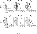
- This response can be measured by assays known to the skilled person, for example ELISPOT (Enzyme-Lynked ImmunoSpot), by intracellular cytokine staining assay, by tetramer assay, by detecting antigen-specific response, by tracking cell multiplication assays, and the presence of markers of cellular activation (CD25, CD69) detected by flow cytometry.
- ELISPOT Enzyme-Lynked ImmunoSpot
- the cytotxic ability of CD8 + T cells activated with tiT lymphocytes can also be quantified .
- lymphocyte of the present invention can be used in combination with other treatments known to those skilled in the art, for example treatment with antibodies against CTLA-4, PD-1, PDL-1 as well as radiotherapy or chemotherapy.
- a fifth aspect of the present invention relates to a kit or device comprising the lymphocyte of the first aspect of the invention or the cell population of the second aspect of the invention.
- a sixth aspect of the invention relates to the use of the kit or device of the fifth aspect of the invention for the prevention or treatment of tumor and/or stimulating the immune response against a tumor antigen of any tumor, for example, but not limited to this list: melanoma, lymphoma, chronic lymphocytic leukemia, myeloma, breast, ovary, uterus, cervix, testis, prostate, colon, colorectal, pancreatic , stomach and gastrointestinal tumors, gastric cancer, liver tumor, kidney (comprised clear-cell renal-cell carcinoma), bladder, mouth cancer, pharynx, larynx, esophagus, lung (small and non-small cell), thyroid, glioblastoma, glioma, sarcoma, encephalon, brain, neuroblastoma and marrow, head and neck blastoma, bone and connective tissue.
- melanoma lymphoma, chronic lymphocytic leukemia, myel
- the kit of the invention comprises the elements necessary to carry out the methods described herein.
- the kit may also contain the elements necessary to assess the immune response generated by the lymphocyte of the invention.
- This kit may include, without any limitation, buffers, enzymes, agents to prevent contamination, etc. It can also include supports and containers required for commissioning and optimization. Also it may further contain positive or negative controls. It may also include instructions to carry out their use.
- a seventh aspect of the invention relates to an in vitro method to transinfect a lymphocyte comprising the following steps:
- In another particular embodiment further comprises a step (e) isolation of transinfected lymphocytes.
- the bacteria can be both Gram + and Gram -, and also the bacteria can be pathogenic and nonpathogenic.
- the bacterium is selected from the list consisting of: L. monocytogenes, Salmonella enterica, bacteria from the Mycobacterium tuberculosis complex, Staphylococcus aureus and Escherichia coli ; preferably L. monocytogenes.
- the bacteria from the Mycobacterium tuberculosis complex can be M . tuberculosis, M. bovis (BCG), M. africanum, M. caprae, M. pinnipedii, M. canetti y M. microti.
- L. monocytogenes Preferably, L. monocytogenes.
- the bacterium comprises a tumor antigen.
- the tumor antigen may be a tumor which is selected from the list consisting of: melanoma, lymphoma, chronic lymphocytic leukemia, myeloma, breast, ovary, uterus, cervix, testis, prostate, colon, colorectal, pancreatic , stomach and gastrointestinal tumors, gastric cancer, liver tumor, kidney (comprised clear-cell renal-cell carcinoma), bladder, mouth cancer, pharynx, larynx, esophagus, lung (small and non-small cell), thyroid, glioblastoma, glioma, sarcoma, encephalon, brain, neuroblastoma and marrow, head and neck blastoma, bone and connective tissue
- the lymphocyte is a heterologous or autologous cell.
- in vitro refers to the method of the invention that is carried out outside of the subject's body. That is, it is done in a biological sample from a subject.
- biological sample herein refers to any sample comprising lymphocytes (CD4 + T lymphocytes and/or B lymphocytes) and includes, without being limited to, biological fluids or tissues from an individual, obtained by any method known by one skilled in the art that serves for this purpose.
- the biological sample may be, for example, but not limited, a fluid sample such as blood or serum.
- the biological sample herein may be fresh or frozen.
- tumor antigen and "tumor peptide” is used interchangeably.
- Another embodiment of the present invention relates to a method for the prevention and/or treatment of a tumor and/or stimulating the immune response against a tumor antigen characterized by comprising the administration of a therapeutically effective amount of the pharmaceutical composition of the invention.
- the tumor is selected from the list consisting of: melanoma, lymphoma, chronic lymphocytic leukemia, myeloma, breast, ovary, uterus, cervix, testis, prostate, colon, colorectal, pancreatic , stomach and gastrointestinal tumors, gastric cancer, liver tumor, kidney (comprised clear-cell renal-cell carcinoma), bladder, mouth cancer, pharynx, larynx, esophagus, lung (small and non-small cell), thyroid, glioblastoma, glioma, sarcoma, encephalon, brain, neuroblastoma and marrow, head and neck blastoma, bone and connective tissue.
- the tumor has neo-antigens.
- antigens that stimulate the immune system for example are known, but not exclusively to tyrosinase, gp75, gp100, Melan A/MART-1, TRP-2, MAGE family proteins (melanoma-associated antigen): GAGE and BAGE, NY-ESO-1, mutations in CDk4 and, ⁇ -catenin, MUM-1, p15.
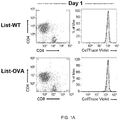
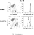
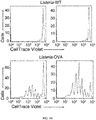
- FIG. 1 Transinfected CD4 + T cells (tiCD4 + T) present bacterial antigens via MHC-I to CD8 + T cells "na ⁇ ve" in vitro.
- CD4 + tiT transinfected with Listeria-WT (wild type strain; top panels) or Listeria-OVA (ovalbumin expressing strain; lower panels) were incubated with CD8 + naive T cells OT-I mice (which recognize a peptide of ovalbumin; OVAp-I 257-264; SIINFEKL (SEQ ID NO: 1) in the context of H-2K b ).
- the histograms represent fluorescence of CD8 + T cells labeled with "CellTraceTM Violet".
- Histograms show the proliferation of CD8 + T cells activated with: anti-CD3 and CD28 antibodies, DCs loaded with soluble pOVA, DCs infected Listeria-OVA or tiCD4 + T cells transinfected with Listeria -OVA to day 2 or day 3. Note that the activation of CD8+ T cells is much greater when activated with tiCD4 + T transinfected with Listeria -OVA that when activated with DCs infected with Listeria-OVA. Histograms filled with gray show fluorescence of nonactivated CD8 + T cells. d, Histograms represent fluorescence (CellTraceTM Violet) of CD8 + T cells from C57BL/6 WT mice or OT-I mice after conjugation with tiCD4 + transinfected with Listeria-OVA.
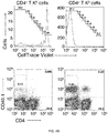
- tiCD4 + T cells process bacterial antigens.
- a Proliferation of CD8 + T cells from mice OT-I, 2 or 3 days after incubation with CD4 + tiT transinfected with Listeria -WT or Listeria-OVA cells, which were captured from DCs loaded with the OVA peptide (OVAp-I).
- b Proliferation of CD8 + T cells from mice OT-I incubated with CD4 tiT + cells (H-2K b- or H-2K b+ ) transinfected with Listeria-OVA captured from H-2K b+ DCs, or incubated with tiCD4 + T cells transinfected with Listeria -WT (gray).
- CD8 + T cells can only be activated when antigens are presented in H2K b (b+) haplotype of MHC-I.
- c as in b, except transinfection, which is made from DCs that were H-2K b- (H-2K k/k ), and the conjugates were allowed to form during 48h.
- d Expression of H-2K b /OVA (detected using a specific antibody) by tiCD4 + T cells (H-2K b+ ,black line, H-2K b- , gray) transinfected with Listeria-OVA that were captured by DCs from H-2K b- .
- e, f Western blots showing the expression of Tap1 in naive CD4 + T cells, activated with DC loaded with soluble OVAp, or with tiCD4 + T cells transinfected with Listeria-OVA for 24 (E) or 48 h (F) after activation.
- ERK2 and laminB were used as controls.
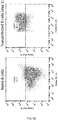
- FIG. 3 tiCD4 + cells form immune synapses with na ⁇ ve CD8 + T cells.
- a Histogram shows H-2K b expression in CD4 + T cells before (black line) and after transinfection (gray line). Filled gray histogram shows the fluorescence of the negative control.
- b Expression of CD86 on CD4 + T cells before (black line) and after transinfection (gray line). Filled gray histogram shows the fluorescence of the negative control.
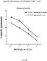
- CTLs CD8 + T effector cells activated by tiCD4 + T transinfected with Listeria-OVA (solid line) or splenocytes loaded with pOVA (dotted line).
- the specific cytotoxicity was measured using different ratios of EL-4 (target cells): CTLs (0.5: 1, 1: 1, 2: 1, 5: 1).
- FIG. 4 tiCD4 + cells activate cytotoxic effector T cells in vivo.
- a Histograms show fluorescence (CellTrace TM Violet) of CD8 + CD45.1 + T cells injected in receptor mice (CD45.2), which were inoculated with tiT CD4 + cells transinfected with Listeria -WT or Listeria-OVA.
- 2 x 10 6 tiCD4 + cells (left panels) or 5 x 10 6 tiCD4 + cells (right panels).
- CD8 + CD45.1 + T Cells from OT-I mice, stained with CellTrace Violet injected in wild type C57BL/6 recipients (whose bone marrows have been reconstituted after irradiation with cells from H-2K k mice).
- CD8 + CD45.1 + T cells from OT-I mice (4 x 10 6 cells/mouse) were transferred along with CD4 + T cells expressing H-2K b (right panels) or H-2K k (panels left) from AND mice (4 x 10 6 cells/mouse) and MCC peptide (15 ⁇ g/mouse). Mice also were infected with Listeria-OVA (10 3 bacteria/mouse).
- mice/group mice/group were inoculated with CD8 + naive T cells from OT-I mice (10 3 /mouse) together with phosphate buffered saline (PBS) in Group 1; 5x10 5 tiCD4 + cells transinfected with Listeria WT, in Group 2; 5x10 5 tiCD4 + transinfected with Listeria-OVA in group 3.
- PBS phosphate buffered saline
- the size of the tumors was monitored every two days (from day 5 to 13) and every 3 days thereafter.
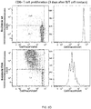
- FIG. 5 B cells capture bacteria by transinfection and cross-present bacterial antigens.
- a Murine DC decorated + (or not -) with Hen Egg Lysozyme (HEL) were infected with L. monocytogenes, and incubated with B cells from MD4 transgenic mice, whose B cells have B cell receptors (BCR) recognizing HEL, under conditions that allow the DC-B cell interactions (for 30 min), or in the presence of a polycarbonate barrier (transwell) which prevents such interactions.
- BCR B cell receptors
- CD4 + T cells were re-isolated and conducted a classic gentamicin survival assay.
- CFU colony forming unit; intracellular bacteria
- ⁇ is direct infection of bacteria on B cells.
- b DC infected with L. monocytogenes and decorated with HEL were incubated with B cells from MD4 mice. After formation of conjugates, B cells were re-isolated and it was quantified the survival (at different times) of intracellular bacteria by gentamicin resistance assays. Data were normalized to time 0. As control of bacterial fitness HeLa cells were infected in parallel.
- c Expression of CD86 on B cells before (left panel; naive B cells) and one day after transinfection with Listeria-OVA (right panel). In both panels is shown IgM expression (FL-1 channel) vs expression of CD86 (channel FL-2).
- d Proliferation of naive CD8 + T cells from OT-I mice labeled with CellTrace Violet, after 3 days of contact with tiB cells that have captured by transinfection Listeria -WT (top panels) or Listeria-OVA (below). Left panels show CellTrace Violet fluorescence vs CD25 expression (a marker of T cell activation).
- CTL Specific relative cytotoxicity of CD8 + T cells (CTL) activated by tiB cells transinfected with Listeria-OVA (top line) or splenocytes decorated with OVAp-I peptide (bottom line). Different ratios of EL-4(target cells):CTL were measured.
- T cells can capture bacteria from DCs infected through a process called transfection (Cruz-Adalia et al., 2014), prompted us to ask whether it was possible that transinfected T cells (ti) could act as true APCs. This was a risky question because, if true, it will break a dogma of immunology, the strict separation of roles between innate and adaptive immunity, and would be a huge advance in the basic understanding of how the immune system works.
- tiCD4 + T CD4 + T cells transinfected
- Listeria-OVA L. monocytogenes expressing ovalbumin
- Listeria- WT wild isogenic strain not expressing OVA
- CD8 + T cells recognize a peptide of ovalbumin (OVAp-I 257-264; SIINFEKL, SEQ ID NO: 1) in the H-2K b context.
- the flow cytometric analysis of CD8 + T cells stained with "CellTrace TM Violet” shows a potent proliferation of CD8 + T cells that begins two days after contact with the tiCD4 + T cells (a movement to the left observed of the "CellTrace TM Violet” fluorescence), but only in those CD8 + T cells that were in contact with CD4 + T cells transinfected with Listeria-OVA ( Fig 1a ).
- This highly proliferative population expresses high levels of CD8 (CD8 + high ) ( Fig. 1a ).
- tiCD4 + T cells transinfected with Listeria-OVA induced the proliferation of CD8 + T cells in a more potent way than the induced by BM-DCs loaded with soluble OVA peptide, or even the proliferation induced by polyclonal activation with CD3/CD28 antibodies, and it was far more stronger than the produced by BM-DCs infected with Listeria-OVA ( Fig. 1c ).
- tiCD4 + T cells transinfected with Listeria-OVA on other hand are not able to induce proliferation of CD8 + T cells from wild mice, which do not recognize any OVA antigen ( Fig. 1d ), whereas they induce very potent proliferation of CD8 + T cells from OT-I mice ( Fig. 1a , c , d ), indicating that the observed effects are antigen-specific.
- tiCD4 + T cells are able to cross-present bacterial antigens to naive CD8 + T cells.
- tiCD4 + T cells could recapitulate different stages of the cross-presentation given in professional APC: endogenous antigen processing, antigen presentation via MHC-I, expression of co-stimulatory molecules, and interaction with target T cells through the formation of canonical immune synapses.
- Antigen presentation of tiCD4 + T cells could be due to the capture of the MHC/antigen complexes from the surface of the DCs or by endogenous processing of the tiCD4 + cells themselves.
- CD4+ T cells were transinfected with Listeria-WT or Listeria-OVA from BM-DCs, previously loaded with the OVA peptide (OVAp/OT-I; cells that recognize CD8 + T cells from OT-I mice); later this tiCD4 + T cells (re-purified) were used to stimulate naive CD8 + T cells. In this configuration, antigen capturing from DCs cells would lead to a comparable activation of CD8 + T cells in both experimental conditions.
- tiCD4 + cells transinfected with Listeria-OVA but not with Listeria -WT induce a very strong activation of CD8 + T cells ( Fig. 2a ).
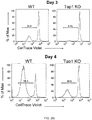
- tiCD4 + T cells isolated from Tap1 KO mice showed very reduced capacity as cross-presenting cells after bacterial capture ( Fig. 2g ), ie, the proliferation of CD8 + T cells was greatly reduced when antigen presenting cells were tiCD4 + from mice Tap1 KO compared to that observed when presenting cells were tiCD4 + from WT mice.
- tiCD4 + T cells increased the expression of MHC-I (H-2K b ), H-2K b coupled to OVA antigen (of bacterial origin) and ligands of co-stimulatory molecules, such as CD86 ( Fig. 3a-c ), which supports a canonical antigen presentation on MHC-I.
- Activation of CD8 + T cells involved the formation of CD4 + /CD8 + cell conjugates Fig. 1c ), indicative of the formation of the immune synapse (IS), mark of T cell activation by APCs.
- the structure of a mature IS is composed of multimolecular concentric rings called supramolecular activation complexes (SMACs).
- cSMAC Central SMACs
- pSMAC peripheral zone
- CD4 + /CD8 + cell conjugates contain CD3 molecules, part of the TCR complex, recruited in cSMAC, and show a massive accumulation of actin in the pSMAC; we found no evidence of the formation of these structures when using tiCD4 + T cells transinfected with Listeria-WT (in the few cell contacts that are detected) ( Fig. 3d ). Together, these data demonstrate that tiCD4 + T cells are professional APCs and activate CD8 + T cells by canonical cross-presentation.
- tiCD4 + T cells Activation of CD8 + T cells by tiCD4+ T cells transinfected with Listeria-OVA gives them cytotoxic capacity because they can eliminate EL-4 target cells expressing OVAp/OT-I ( Fig. 3f ).
- tiCD4 + T cells were isolated from OT-I/CD45.1 mice, stained ex vivo with "CellTrace TM Violet", transferred to a BL6 / CD45.2 mouse, and tiCD4 + T cells were injected to elicit a response.
- CD4 + T cells from AND mice H-2K b or H-2K k were transferred in recipient mice together with CD45.1 + CD8 + T cells from OT-I mice, and MCC peptide (to favor transinfection (Cruz-Adalia et al., 2014)).
- the recipient mice were also intravenously infected with Listeria-OVA. Only mice that received CD4 + T cells H-2K b were able to activate OT-I CD8 + CD45.1 + T cells ( Fig. 4b ), confirming the capacity for presenting antigens of CD4 + T cells during the course of a bacterial infection.
- tiCD4 + T cells were transferred in a single injection together with: Group 1: PBS; Group 2: tiCD4 + cells transinfected with Listeria -WT; and Group 3: tiCD4 + cells transinfected with Listeria-OVA.
- mice in the control groups vehicle treated cells or tiCD4 + T cells transinfected with Listeria -WT, developed tumors during the first 11 days after injection of B16-OVA cells ( Fig. 4c ).
- Treatment with tiCD4 + T cells transinfected with Listeria- OVA prevented tumor formation in 6 out of 9 mice and delayed tumor appearance in another mice, ie vaccination (single inoculation) with tiCD4 + cells working as antigen presenting cells conferred protection against melanoma in 7 of 9 cases ( Fig. 4c ).
- B cells are also capable of capturing bacteria by transinfection.
- the process of bacterial capture by B cells from infected DC was quantitated on reisolating B cells after conjugate formation with infected DC, followed by classic gentamicin survival assays.
- This method allows us to analyze a large number of conjugates.
- the requirement of cell-cell contact was tested using a physical barrier (polycarbonate filters) that prevented DC-B cell contacts , and the role of antigen recognition by using DC decorated with hen egg lysozyme (HEL) (note that we use B cells isolated from MD4 transgenic mice, whose B cells have B cell receptors (BCR) t recognizing HEL).
- HEL hen egg lysozyme
- B cells monocytogenes and conjugated with B cells from MD4 mice.
- B cells were reisolated and CFU (intracellular bacteria in B cells) were analyzed by gentamicin survival assays, where B cells were collected at different times.
- CFU intracellular bacteria in B cells
- B cells killed very quickly (and efficiently) the bacteria captured ( Fig. 5b ).
- Fig. 5b In the first hours after the formation of conjugates, over 90% of the internalized bacteria were destroyed.
- control of bacterial fitness we measured the CFU after infecting HeLa cells ( Fig. 5b ).
- activation marker CD25 was detected in CD8 + T cells incubated with tiB transinfected with Listeria-OVA, but not when used tiB transinfected with Listeria-WT ( Fig. 5d ).
- the tiB cells transinfected with Listeria -OVA induce proliferation of CD8 + T cells more potently than even the bone-marrow derived DCs (BM-DC) loaded with soluble peptide OVAp-I, infected with Listeria-OVA, or the polyclonal activation of CD8 + T cells themselves with anti CD3/CD28 antibodies.
- BM-DC bone-marrow derived DCs
- Bacteria Listeria monocytogenes-OVA (pPL2-LLO-OVA), a Listeria strain that expresses OVA protein, and its WT isogenic strain L. monocytogenes 10403S.
- mice (1) Wild-type C57BL/6 mice, (2) C57BL/6-Tg (TcraTcrb)425Cbn/J OT-II mice expressing a T cell receptor (TCR) specific for OVA peptide 323-339 in the context of MHC class II (I-Ab), (3) C57BL/6-Tg(TcraTcrb)1100Mjb/J OT-I mice expressing TCR specific for OVA peptide 257-264 in the context of H-2K b (4) AND-TCR transgenic mice recognizing moth cytochrome c 88-103 (ANERADLIAYLKQATK) (MCCp) on I-E k , expressing H-2K b , H-2K k or both haplotypes.
- TCR T cell receptor
- I-Ab MHC class II
- C57BL/6-Tg(TcraTcrb)1100Mjb/J OT-I mice expressing TCR specific for OVA peptide 257-264
- mice C57BL/6-Tg(IgheIMD4)4Ccg/J, (MD4) transgenic mice with more than 99% of their B cells expressing a B cell Receptor (BCR) specific for hen egg lysozyme (HEL).
- BCR B cell Receptor
- HEL hen egg lysozyme
- Dendritic cells were generated as described in Inaba et al., 1992. Briefly, cells from the bone marrow (from the tibias and femurs of C57BL/6 mice 8-20 weeks) were grown in the presence of GM-CSF, at day 10 cells were tested by flow cytometry for CD11c, IA/IE and Gr1 to ensure that they had differentiated properly. They were matured with LPS day before use.
- CD4 + T from OT-II mice and CD8 + T from OT-I mice were obtained from lymph nodes and spleen by negative magnetic selection, as is described in Cruz-Adalia et al., 2014.
- the EL-4 lymphoma line was maintained in RPMI 1640 (Fisher Scientific), supplemented with 10% FCS, 0.1 U/ml penicillin, 0.1 mg/ml streptomycin (Lonza) and 0.05 mM 2-mercaptoethanol.
- Melanoma B16 OVA line was maintained in RPMI 1640 with 0.4 mg/ml geneticin. Antibiotics were washed 48h before inoculation.
- Antibodies used were anti-CD69, -CD25, -CD4, -CD8, -CD11c, -IA/IE, -Gr1 (BD and Immunostep), biotinylated antibodies against CD45.1, CD3, CD4, CD8, CD28, IgM , B220, CD19, MHC class II (I-Ab), CD11b, CD11c DX5, CD25 and CD16/CD32 (BD and Immunostep), and anti-tubulin FITC-conjugated (Santa Cruz).
- the monoclonal antibody 25-D1.16 specific for SIINFEKL/H-2K b (SEQ ID NO: 1/H-2K b ) and labeled with allophycocyanin (APC) was purchased from eBioscience.
- Anti-TAP-1 (M-18), - ERK-2 and -lamin B were purchased from Santa Cruz.
- Secondary antibody duck anti-mouse and goat anti-hamster conjugated with AlexaFluor488, 647, or 568 were purchased from Life Technologies; secondary antibodies peroxidase conjugated with goat anti-mouse IgG and goat anti-rabbit IgG were purchased from Thermo Scientific.
- OVAp/OT-II peptide OVA 323-339; ISQAVHAAHAEINEAGR, SEQ ID NO: 3
- OVAp/OT-I OVA 257-264, SIINFEKL (SEQ ID NO: 1)
- CBMSO-CSIC Center for Molecular Biology Severo Ochoa
- Mouse GM-CSF was from Peprotech.
- the peptide (MCCp) 88-103 ANERADLIAYLKQATK; SEQ ID NO: 2) was purchased from GenScript.
- LPS Sigma-Aldrich
- microparticles coupled to streptavidin MiltenyiBiotec
- streptavidin-PerCP BD
- Alexa Fluor 568-Phalloidin Life Technologies
- Poly-L-Lysine Sigma-Aldrich
- Cell Trace TM Violet (Life Technologies) and 7-AAD Viability staining solution of eBiosciences.
- CD4 + T cells from mice OT-II were transinfected with Listeria-OVA or Listeria-WT as described in Cruz-Adalia et al. 2014. Briefly, BM-DC infected and loaded with OVAp (OT-II) (to increase transinfection) formed conjugates with CD4 + T from OT-II mice.
- OVAp OVAp
- the BM-DC and CD4 + T cells were isolated from AND mice expressing MHC-I haplotypes: H-2K b/k , or H-2K b negative, H-2K k/k . In these experiments the BM-DC were loaded with MCCp to enhance transinfection.
- the purified population of tiCD4 + cells was incubated with naive CD8 + T cells isolated from OT-I mice. These CD8 + T cells previously stained with CellTrace TM Violet to quantify their proliferation by flow cytometry (FACSAria; BD). In each cell division fluorescence is lost in the proliferating population, and it was observed as a leftward shift in the histogram. Only the living cells, which do not capture the dye 7AAD, were analyzed.
- CD8 + T cells To analyze the proliferation of CD8 + T cells in vivo, 5x10 6 naive CD8 + T cells (from mice CD45.1 + OT-I), stained with CellTrace TM Violet, were injected intravenously into recipient mice (CD45.2 + C57BL/6). After 24h, the tiCD4 + cells were transferred to mice. Spleens were isolated after three days to analyze the proliferation of CD8 + T cells by flow cytometry.
- tiCD4 + and naive CD8 + T cells were allowed to form conjugates for 1 h, then fixed with 4% paraformaldehyde in PBS.
- CD8+ T cells were previously stained with CellTrace TM Violet.
- Samples were permeabilized with 0.1% Triton TM X-100 in PBS prior to staining with the indicated antibodies. F-actin was detected using phalloidin coupled to a fluorophore. Samples were visualized by confocal microscopy using a Leica TCS SP5 equipped with 63x-lenses and controlled by Leica LAS AF. Images were analyzed using ImageJ (NIH Bethesda, MD).
- CD8 + T cells both effector and cytotoxic (CTLs) were prepared from na ⁇ ve CD8 + cells cells (OT-I) activated with tiCD4 + transinfected with Listeria-OVA. As positive controls were used CD8 + T cells from OT-I activated with splenocytes loaded with OVAp.
- CTLs cytotoxic
- EL-4 cells were incubated with 0.5 ⁇ M OVAp/OT-I for 1 h (or not, as control). After PBS washes, EL-4 cells loaded with OVAp were stained with CellTrace TM Violet (5 ⁇ M). On the other hand, EL-4 cells without loading OVAp were stained with CellTrace TM Violet (0.5 ⁇ M). After washing with culture medium both populations were mixed and incubated with different ratios of CTLs (5:1, 2:1, 1:1, 0.5:1) (EL4/CTLs). After 4h, samples were analyzed by flow cytometry.
- CD4 + T cells transinfected with Listeria-WT (as negative control) or Listeria-OVA were prepared as described in previous sections. 24h after transinfection, tiTCD4 + are re-isolated cell sorting and resuspended in PBS. naive CD8 + T cells from OT-I were purified by magnetic columns as previously described (Cruz-Adalia et al., 2014) and resuspended in PBS. 5 x 10 5 B16-OVA cells were injected subcutaneously into the right flank of recipient mice (C57BL/6).
- mice were divided into three groups and transferred intravenously (iv) in a single injection: Group 1: PBS; Group 2: 5x10 5 tiCD4 + cells transinfected with Listeria-WT; and Group 3: tiCD4 + cells transinfected with Listeria-OVA. All mice in all groups were also injected, simultaneously to tiCD4 + or PBS vehicle, with 10 3 CD8 + T cells from OT-I (also i.v.). Tumor growth was monitored every 2-3 days using a caliper-dial, and the areas were determined by multiplying the length and width. The experimental groups were assigned randomly and the experiment was conducted in a double blind (people who measured the tumors did not know to which group each mouse belonged). Mice were sacrificed when tumors reached 300 mm 2 according to the criteria endpoint as directed by the European Union and FELASA for experimental animals, and in accordance with the current legislation in Spain.
Landscapes
- Health & Medical Sciences (AREA)
- Life Sciences & Earth Sciences (AREA)
- Engineering & Computer Science (AREA)
- General Health & Medical Sciences (AREA)
- Biomedical Technology (AREA)
- Chemical & Material Sciences (AREA)
- Immunology (AREA)
- Public Health (AREA)
- Veterinary Medicine (AREA)
- Animal Behavior & Ethology (AREA)
- Organic Chemistry (AREA)
- Zoology (AREA)
- Biotechnology (AREA)
- Epidemiology (AREA)
- Bioinformatics & Cheminformatics (AREA)
- Cell Biology (AREA)
- Genetics & Genomics (AREA)
- Wood Science & Technology (AREA)
- Pharmacology & Pharmacy (AREA)
- Medicinal Chemistry (AREA)
- Microbiology (AREA)
- Hematology (AREA)
- Virology (AREA)
- Biochemistry (AREA)
- General Engineering & Computer Science (AREA)
- General Chemical & Material Sciences (AREA)
- Nuclear Medicine, Radiotherapy & Molecular Imaging (AREA)
- Chemical Kinetics & Catalysis (AREA)
- Developmental Biology & Embryology (AREA)
- Mycology (AREA)
- Molecular Biology (AREA)
- Oncology (AREA)
- Medicines Containing Material From Animals Or Micro-Organisms (AREA)
- Micro-Organisms Or Cultivation Processes Thereof (AREA)
- Medicines That Contain Protein Lipid Enzymes And Other Medicines (AREA)
Applications Claiming Priority (2)
| Application Number | Priority Date | Filing Date | Title |
|---|---|---|---|
| ES201531177 | 2015-08-07 | ||
| PCT/ES2016/070597 WO2017025657A1 (es) | 2015-08-07 | 2016-08-08 | Linfocitos transinfectados para terapia anti-tumoral |
Publications (1)
| Publication Number | Publication Date |
|---|---|
| EP3333255A1 true EP3333255A1 (en) | 2018-06-13 |
Family
ID=56936436
Family Applications (1)
| Application Number | Title | Priority Date | Filing Date |
|---|---|---|---|
| EP16766331.9A Withdrawn EP3333255A1 (en) | 2015-08-07 | 2016-08-08 | Transinfected lymphocytes for anti-tumor therapy |
Country Status (6)
| Country | Link |
|---|---|
| US (1) | US20180228840A1 (enExample) |
| EP (1) | EP3333255A1 (enExample) |
| JP (1) | JP2018522594A (enExample) |
| AU (1) | AU2016307370A1 (enExample) |
| CA (1) | CA2995021A1 (enExample) |
| WO (1) | WO2017025657A1 (enExample) |
Families Citing this family (1)
| Publication number | Priority date | Publication date | Assignee | Title |
|---|---|---|---|---|
| US12087428B2 (en) * | 2020-12-29 | 2024-09-10 | Kpn Innovations Llc | Systems and methods for generating a body degradation reduction program |
-
2016
- 2016-08-08 AU AU2016307370A patent/AU2016307370A1/en not_active Abandoned
- 2016-08-08 US US15/750,787 patent/US20180228840A1/en not_active Abandoned
- 2016-08-08 WO PCT/ES2016/070597 patent/WO2017025657A1/es not_active Ceased
- 2016-08-08 CA CA2995021A patent/CA2995021A1/en not_active Abandoned
- 2016-08-08 EP EP16766331.9A patent/EP3333255A1/en not_active Withdrawn
- 2016-08-08 JP JP2018525816A patent/JP2018522594A/ja active Pending
Also Published As
| Publication number | Publication date |
|---|---|
| CA2995021A1 (en) | 2017-02-16 |
| AU2016307370A1 (en) | 2018-03-01 |
| US20180228840A1 (en) | 2018-08-16 |
| JP2018522594A (ja) | 2018-08-16 |
| WO2017025657A1 (es) | 2017-02-16 |
Similar Documents
| Publication | Publication Date | Title |
|---|---|---|
| Van der Most et al. | Tumor eradication after cyclophosphamide depends on concurrent depletion of regulatory T cells: a role for cycling TNFR2-expressing effector-suppressor T cells in limiting effective chemotherapy | |
| JP6207783B2 (ja) | 抗原特異的t細胞の増殖のための方法 | |
| Palucka et al. | Dendritic cells and immunity against cancer | |
| Harimoto et al. | Inactivation of tumor‐specific CD8+ CTLs by tumor‐infiltrating tolerogenic dendritic cells | |
| Ballestrero et al. | Immunotherapy with dendritic cells for cancer | |
| US8475785B2 (en) | Allogeneic cancer cell-based immunotherapy | |
| Cruz-Adalia et al. | Conventional CD4+ T cells present bacterial antigens to induce cytotoxic and memory CD8+ T cell responses | |
| KR20170121178A (ko) | 보편적인 살해 t-세포 | |
| Florek et al. | Autologous apoptotic cells preceding transplantation enhance survival in lethal murine graft-versus-host models | |
| Newman et al. | Heat shock protein vaccination and directed IL-2 therapy amplify tumor immunity rapidly following bone marrow transplantation in mice | |
| EP3333255A1 (en) | Transinfected lymphocytes for anti-tumor therapy | |
| Keshavarz-Fathi et al. | Cancer immunology | |
| JP2008523067A (ja) | 癌ワクチンアジュバントとしてのαサイモシンペプチド | |
| US20240076616A1 (en) | Method for t-cell expansion and related medical applications | |
| Veiga et al. | Lymphocytes transfectes pour therapie antitumorale | |
| US11850279B2 (en) | Platforms and methods for optimizing host antigen presentation and host antitumor and antipathogen immunity | |
| Asada et al. | Combination vaccine of dendritic cells (DCs) and T cells effectively suppressed preestablished malignant melanoma in mice | |
| Minina et al. | CAR cells beyond classical CAR T cells: functional properties and prospects of application | |
| Prophylactic | Autologous apoptotic cells preceding transplantation enhance survival in lethal murine graft-versus-host models | |
| Li | Adoptive Cell Therapy using CD4 T Helper 1-like and CD8 Cytotoxic T Lymphocytes in a Mouse Model of Melanoma | |
| Jæhger | Preclinical Evaluation of Novel Drug Delivery Platforms for the Improvement of Adoptive T cell Therapy | |
| Colombo | Reversal of Tumor-induced Dendritic Cell Paralysis by CpG Immunostimulatory Oligonucleotide and Anti-Interleukin 10 Receptor Antibody | |
| Seid et al. | Reviews on the Role of Dendritic Cells in Induction and Regulation of Immunity | |
| Bolton et al. | Transplantation immunology | |
| Zhang | Tolerogenic CD4⁻ 8⁻ Dendritic Cells and their Conversion into Immunogenic Ones via TLR9 Signaling |
Legal Events
| Date | Code | Title | Description |
|---|---|---|---|
| STAA | Information on the status of an ep patent application or granted ep patent |
Free format text: STATUS: THE INTERNATIONAL PUBLICATION HAS BEEN MADE |
|
| PUAI | Public reference made under article 153(3) epc to a published international application that has entered the european phase |
Free format text: ORIGINAL CODE: 0009012 |
|
| STAA | Information on the status of an ep patent application or granted ep patent |
Free format text: STATUS: REQUEST FOR EXAMINATION WAS MADE |
|
| 17P | Request for examination filed |
Effective date: 20180307 |
|
| AK | Designated contracting states |
Kind code of ref document: A1 Designated state(s): AL AT BE BG CH CY CZ DE DK EE ES FI FR GB GR HR HU IE IS IT LI LT LU LV MC MK MT NL NO PL PT RO RS SE SI SK SM TR |
|
| AX | Request for extension of the european patent |
Extension state: BA ME |
|
| DAV | Request for validation of the european patent (deleted) | ||
| DAX | Request for extension of the european patent (deleted) | ||
| STAA | Information on the status of an ep patent application or granted ep patent |
Free format text: STATUS: EXAMINATION IS IN PROGRESS |
|
| 17Q | First examination report despatched |
Effective date: 20200120 |
|
| STAA | Information on the status of an ep patent application or granted ep patent |
Free format text: STATUS: THE APPLICATION IS DEEMED TO BE WITHDRAWN |
|
| 18D | Application deemed to be withdrawn |
Effective date: 20211216 |