EP3333190A1 - Dual antigen-induced bipartite functional complementation - Google Patents
Dual antigen-induced bipartite functional complementation Download PDFInfo
- Publication number
- EP3333190A1 EP3333190A1 EP17193746.9A EP17193746A EP3333190A1 EP 3333190 A1 EP3333190 A1 EP 3333190A1 EP 17193746 A EP17193746 A EP 17193746A EP 3333190 A1 EP3333190 A1 EP 3333190A1
- Authority
- EP
- European Patent Office
- Prior art keywords
- domain
- cells
- cell
- antigen
- antibody
- Prior art date
- Legal status (The legal status is an assumption and is not a legal conclusion. Google has not performed a legal analysis and makes no representation as to the accuracy of the status listed.)
- Ceased
Links
- 239000000427 antigen Substances 0.000 title claims abstract description 521
- 102000036639 antigens Human genes 0.000 title claims abstract description 519
- 108091007433 antigens Proteins 0.000 title claims abstract description 519
- 230000009977 dual effect Effects 0.000 title description 3
- 108090000765 processed proteins & peptides Proteins 0.000 claims abstract description 469
- 229920001184 polypeptide Polymers 0.000 claims abstract description 462
- 102000004196 processed proteins & peptides Human genes 0.000 claims abstract description 462
- 239000012634 fragment Substances 0.000 claims abstract description 245
- 230000008685 targeting Effects 0.000 claims abstract description 131
- 239000000758 substrate Substances 0.000 claims abstract description 47
- 150000007523 nucleic acids Chemical class 0.000 claims abstract description 41
- 108020004707 nucleic acids Proteins 0.000 claims abstract description 40
- 102000039446 nucleic acids Human genes 0.000 claims abstract description 40
- 238000006471 dimerization reaction Methods 0.000 claims abstract description 29
- 239000000539 dimer Substances 0.000 claims abstract description 18
- 239000008194 pharmaceutical composition Substances 0.000 claims abstract description 8
- 239000002773 nucleotide Substances 0.000 claims abstract description 7
- 125000003729 nucleotide group Chemical group 0.000 claims abstract description 7
- 210000004027 cell Anatomy 0.000 claims description 655
- 206010028980 Neoplasm Diseases 0.000 claims description 143
- 108090000623 proteins and genes Proteins 0.000 claims description 121
- 102000017420 CD3 protein, epsilon/gamma/delta subunit Human genes 0.000 claims description 84
- 108050005493 CD3 protein, epsilon/gamma/delta subunit Proteins 0.000 claims description 84
- 201000011510 cancer Diseases 0.000 claims description 66
- 108060003951 Immunoglobulin Proteins 0.000 claims description 63
- 102000018358 immunoglobulin Human genes 0.000 claims description 63
- 102000004169 proteins and genes Human genes 0.000 claims description 54
- 102100037422 Receptor-type tyrosine-protein phosphatase C Human genes 0.000 claims description 51
- 101000738771 Homo sapiens Receptor-type tyrosine-protein phosphatase C Proteins 0.000 claims description 50
- 108010074032 HLA-A2 Antigen Proteins 0.000 claims description 49
- 102000025850 HLA-A2 Antigen Human genes 0.000 claims description 49
- 150000001413 amino acids Chemical group 0.000 claims description 36
- 102100031940 Epithelial cell adhesion molecule Human genes 0.000 claims description 31
- 210000003719 b-lymphocyte Anatomy 0.000 claims description 29
- 102100024222 B-lymphocyte antigen CD19 Human genes 0.000 claims description 26
- 101000980825 Homo sapiens B-lymphocyte antigen CD19 Proteins 0.000 claims description 26
- 239000013566 allergen Substances 0.000 claims description 25
- 239000003550 marker Substances 0.000 claims description 25
- 108010066687 Epithelial Cell Adhesion Molecule Proteins 0.000 claims description 24
- 238000011282 treatment Methods 0.000 claims description 24
- 102000001301 EGF receptor Human genes 0.000 claims description 23
- 101000934338 Homo sapiens Myeloid cell surface antigen CD33 Proteins 0.000 claims description 23
- 102100025243 Myeloid cell surface antigen CD33 Human genes 0.000 claims description 23
- -1 GD3 ganglioside Chemical class 0.000 claims description 20
- 101000874179 Homo sapiens Syndecan-1 Proteins 0.000 claims description 19
- 102100035721 Syndecan-1 Human genes 0.000 claims description 19
- 239000002243 precursor Substances 0.000 claims description 19
- 208000037265 diseases, disorders, signs and symptoms Diseases 0.000 claims description 17
- 230000029918 bioluminescence Effects 0.000 claims description 16
- 238000005415 bioluminescence Methods 0.000 claims description 16
- 101001012157 Homo sapiens Receptor tyrosine-protein kinase erbB-2 Proteins 0.000 claims description 14
- 102100030086 Receptor tyrosine-protein kinase erbB-2 Human genes 0.000 claims description 14
- 102100025475 Carcinoembryonic antigen-related cell adhesion molecule 5 Human genes 0.000 claims description 12
- 235000002198 Annona diversifolia Nutrition 0.000 claims description 11
- 241000282836 Camelus dromedarius Species 0.000 claims description 11
- 241000251730 Chondrichthyes Species 0.000 claims description 11
- 101100421450 Drosophila melanogaster Shark gene Proteins 0.000 claims description 11
- 101000581981 Homo sapiens Neural cell adhesion molecule 1 Proteins 0.000 claims description 11
- 102100034256 Mucin-1 Human genes 0.000 claims description 11
- 102100027347 Neural cell adhesion molecule 1 Human genes 0.000 claims description 11
- 101150029707 ERBB2 gene Proteins 0.000 claims description 10
- 101001008874 Homo sapiens Mast/stem cell growth factor receptor Kit Proteins 0.000 claims description 10
- 101000934346 Homo sapiens T-cell surface antigen CD2 Proteins 0.000 claims description 10
- 102100027754 Mast/stem cell growth factor receptor Kit Human genes 0.000 claims description 10
- 102100025237 T-cell surface antigen CD2 Human genes 0.000 claims description 10
- 102100021260 Galactosylgalactosylxylosylprotein 3-beta-glucuronosyltransferase 1 Human genes 0.000 claims description 9
- 101000894906 Homo sapiens Galactosylgalactosylxylosylprotein 3-beta-glucuronosyltransferase 1 Proteins 0.000 claims description 9
- 239000003446 ligand Substances 0.000 claims description 9
- 102100031585 ADP-ribosyl cyclase/cyclic ADP-ribose hydrolase 1 Human genes 0.000 claims description 8
- 102100035248 Alpha-(1,3)-fucosyltransferase 4 Human genes 0.000 claims description 8
- 102100022005 B-lymphocyte antigen CD20 Human genes 0.000 claims description 8
- 108010059045 HLA-C*06 antigen Proteins 0.000 claims description 8
- 102100031573 Hematopoietic progenitor cell antigen CD34 Human genes 0.000 claims description 8
- 101000777636 Homo sapiens ADP-ribosyl cyclase/cyclic ADP-ribose hydrolase 1 Proteins 0.000 claims description 8
- 101001022185 Homo sapiens Alpha-(1,3)-fucosyltransferase 4 Proteins 0.000 claims description 8
- 101000897405 Homo sapiens B-lymphocyte antigen CD20 Proteins 0.000 claims description 8
- 101000914324 Homo sapiens Carcinoembryonic antigen-related cell adhesion molecule 5 Proteins 0.000 claims description 8
- 101000920667 Homo sapiens Epithelial cell adhesion molecule Proteins 0.000 claims description 8
- 101000777663 Homo sapiens Hematopoietic progenitor cell antigen CD34 Proteins 0.000 claims description 8
- 102000004868 N-Methyl-D-Aspartate Receptors Human genes 0.000 claims description 8
- 108090001041 N-Methyl-D-Aspartate Receptors Proteins 0.000 claims description 8
- 201000001441 melanoma Diseases 0.000 claims description 8
- 102100027208 T-cell antigen CD7 Human genes 0.000 claims description 7
- 201000010099 disease Diseases 0.000 claims description 7
- 102100024423 Carbonic anhydrase 9 Human genes 0.000 claims description 6
- 101000914321 Homo sapiens Carcinoembryonic antigen-related cell adhesion molecule 7 Proteins 0.000 claims description 6
- 101000617725 Homo sapiens Pregnancy-specific beta-1-glycoprotein 2 Proteins 0.000 claims description 6
- 108010008707 Mucin-1 Proteins 0.000 claims description 6
- 238000010494 dissociation reaction Methods 0.000 claims description 6
- 230000005593 dissociations Effects 0.000 claims description 6
- 150000002270 gangliosides Chemical class 0.000 claims description 6
- 108091023037 Aptamer Proteins 0.000 claims description 5
- 108700012439 CA9 Proteins 0.000 claims description 5
- 102100032912 CD44 antigen Human genes 0.000 claims description 5
- 102100041003 Glutamate carboxypeptidase 2 Human genes 0.000 claims description 5
- 101000868273 Homo sapiens CD44 antigen Proteins 0.000 claims description 5
- 101000916489 Homo sapiens Chondroitin sulfate proteoglycan 4 Proteins 0.000 claims description 5
- 101000892862 Homo sapiens Glutamate carboxypeptidase 2 Proteins 0.000 claims description 5
- 101001133056 Homo sapiens Mucin-1 Proteins 0.000 claims description 5
- 101000610551 Homo sapiens Prominin-1 Proteins 0.000 claims description 5
- 101000884271 Homo sapiens Signal transducer CD24 Proteins 0.000 claims description 5
- 102100026547 Platelet-derived growth factor receptor beta Human genes 0.000 claims description 5
- 102100040120 Prominin-1 Human genes 0.000 claims description 5
- 102100038081 Signal transducer CD24 Human genes 0.000 claims description 5
- 102100022749 Aminopeptidase N Human genes 0.000 claims description 4
- 102000011899 Aquaporin 2 Human genes 0.000 claims description 4
- 108010036221 Aquaporin 2 Proteins 0.000 claims description 4
- 102100023126 Cell surface glycoprotein MUC18 Human genes 0.000 claims description 4
- 108010089211 HLA-A*25 antigen Proteins 0.000 claims description 4
- 108010035452 HLA-A1 Antigen Proteins 0.000 claims description 4
- 108010086377 HLA-A3 Antigen Proteins 0.000 claims description 4
- 108010014597 HLA-B44 Antigen Proteins 0.000 claims description 4
- 108010070768 HLA-C*03 antigen Proteins 0.000 claims description 4
- 108010069149 HLA-C*04 antigen Proteins 0.000 claims description 4
- 108010007655 HLA-C*70 antigen Proteins 0.000 claims description 4
- 101000757160 Homo sapiens Aminopeptidase N Proteins 0.000 claims description 4
- 101000623903 Homo sapiens Cell surface glycoprotein MUC18 Proteins 0.000 claims description 4
- 101000608935 Homo sapiens Leukosialin Proteins 0.000 claims description 4
- 101000878605 Homo sapiens Low affinity immunoglobulin epsilon Fc receptor Proteins 0.000 claims description 4
- 101000934372 Homo sapiens Macrosialin Proteins 0.000 claims description 4
- 102100039564 Leukosialin Human genes 0.000 claims description 4
- 102100038007 Low affinity immunoglobulin epsilon Fc receptor Human genes 0.000 claims description 4
- 102100025136 Macrosialin Human genes 0.000 claims description 4
- HOKKHZGPKSLGJE-GSVOUGTGSA-N N-Methyl-D-aspartic acid Chemical compound CN[C@@H](C(O)=O)CC(O)=O HOKKHZGPKSLGJE-GSVOUGTGSA-N 0.000 claims description 4
- 102000019315 Nicotinic acetylcholine receptors Human genes 0.000 claims description 4
- 108050006807 Nicotinic acetylcholine receptors Proteins 0.000 claims description 4
- 108091008606 PDGF receptors Proteins 0.000 claims description 4
- 102100024616 Platelet endothelial cell adhesion molecule Human genes 0.000 claims description 4
- 102000011653 Platelet-Derived Growth Factor Receptors Human genes 0.000 claims description 4
- 102000004874 Synaptophysin Human genes 0.000 claims description 4
- 108090001076 Synaptophysin Proteins 0.000 claims description 4
- 102100036011 T-cell surface glycoprotein CD4 Human genes 0.000 claims description 4
- 102000003734 Voltage-Gated Potassium Channels Human genes 0.000 claims description 4
- 108090000013 Voltage-Gated Potassium Channels Proteins 0.000 claims description 4
- 108090000686 amphiphysin Proteins 0.000 claims description 4
- 102000004111 amphiphysin Human genes 0.000 claims description 4
- 210000003205 muscle Anatomy 0.000 claims description 4
- 102000038650 voltage-gated calcium channel activity Human genes 0.000 claims description 4
- 108091023044 voltage-gated calcium channel activity Proteins 0.000 claims description 4
- 102000000905 Cadherin Human genes 0.000 claims description 3
- 108050007957 Cadherin Proteins 0.000 claims description 3
- 101100314454 Caenorhabditis elegans tra-1 gene Proteins 0.000 claims description 3
- 102100035716 Glycophorin-A Human genes 0.000 claims description 3
- 108091005250 Glycophorins Proteins 0.000 claims description 3
- 108010004141 HLA-B35 Antigen Proteins 0.000 claims description 3
- 108010091938 HLA-B7 Antigen Proteins 0.000 claims description 3
- 108010039075 HLA-B8 Antigen Proteins 0.000 claims description 3
- 101000994365 Homo sapiens Integrin alpha-6 Proteins 0.000 claims description 3
- 101000935043 Homo sapiens Integrin beta-1 Proteins 0.000 claims description 3
- 101000623901 Homo sapiens Mucin-16 Proteins 0.000 claims description 3
- 101000692455 Homo sapiens Platelet-derived growth factor receptor beta Proteins 0.000 claims description 3
- 101000851376 Homo sapiens Tumor necrosis factor receptor superfamily member 8 Proteins 0.000 claims description 3
- 102100032816 Integrin alpha-6 Human genes 0.000 claims description 3
- 102100025304 Integrin beta-1 Human genes 0.000 claims description 3
- 102100023123 Mucin-16 Human genes 0.000 claims description 3
- 102100034922 T-cell surface glycoprotein CD8 alpha chain Human genes 0.000 claims description 3
- 102100036857 Tumor necrosis factor receptor superfamily member 8 Human genes 0.000 claims description 3
- 239000003937 drug carrier Substances 0.000 claims description 3
- 239000004094 surface-active agent Substances 0.000 claims description 3
- FWMNVWWHGCHHJJ-SKKKGAJSSA-N 4-amino-1-[(2r)-6-amino-2-[[(2r)-2-[[(2r)-2-[[(2r)-2-amino-3-phenylpropanoyl]amino]-3-phenylpropanoyl]amino]-4-methylpentanoyl]amino]hexanoyl]piperidine-4-carboxylic acid Chemical compound C([C@H](C(=O)N[C@H](CC(C)C)C(=O)N[C@H](CCCCN)C(=O)N1CCC(N)(CC1)C(O)=O)NC(=O)[C@H](N)CC=1C=CC=CC=1)C1=CC=CC=C1 FWMNVWWHGCHHJJ-SKKKGAJSSA-N 0.000 claims description 2
- 108010010995 MART-1 Antigen Proteins 0.000 claims description 2
- 102100028389 Melanoma antigen recognized by T-cells 1 Human genes 0.000 claims description 2
- 238000003745 diagnosis Methods 0.000 claims description 2
- 239000012530 fluid Substances 0.000 claims description 2
- 125000005630 sialyl group Chemical group 0.000 claims description 2
- 101150047061 tag-72 gene Proteins 0.000 claims description 2
- 102000052116 epidermal growth factor receptor activity proteins Human genes 0.000 claims 6
- 108700015053 epidermal growth factor receptor activity proteins Proteins 0.000 claims 6
- YOHYSYJDKVYCJI-UHFFFAOYSA-N n-[3-[[6-[3-(trifluoromethyl)anilino]pyrimidin-4-yl]amino]phenyl]cyclopropanecarboxamide Chemical compound FC(F)(F)C1=CC=CC(NC=2N=CN=C(NC=3C=C(NC(=O)C4CC4)C=CC=3)C=2)=C1 YOHYSYJDKVYCJI-UHFFFAOYSA-N 0.000 claims 6
- 102000018651 Epithelial Cell Adhesion Molecule Human genes 0.000 claims 5
- 102000006354 HLA-DR Antigens Human genes 0.000 claims 2
- 108010058597 HLA-DR Antigens Proteins 0.000 claims 2
- 101000914496 Homo sapiens T-cell antigen CD7 Proteins 0.000 claims 2
- 241000282842 Lama glama Species 0.000 claims 2
- 102000003729 Neprilysin Human genes 0.000 claims 2
- 108090000028 Neprilysin Proteins 0.000 claims 2
- 229920001481 poly(stearyl methacrylate) Polymers 0.000 claims 2
- 101000829725 Homo sapiens Phospholipid hydroperoxide glutathione peroxidase Proteins 0.000 claims 1
- 101001056234 Homo sapiens Sperm mitochondrial-associated cysteine-rich protein Proteins 0.000 claims 1
- 102100022019 Pregnancy-specific beta-1-glycoprotein 2 Human genes 0.000 claims 1
- 102100026503 Sperm mitochondrial-associated cysteine-rich protein Human genes 0.000 claims 1
- 230000027455 binding Effects 0.000 abstract description 102
- 239000013598 vector Substances 0.000 abstract description 10
- 210000001744 T-lymphocyte Anatomy 0.000 description 100
- 150000001875 compounds Chemical class 0.000 description 64
- 210000004881 tumor cell Anatomy 0.000 description 61
- 230000003211 malignant effect Effects 0.000 description 57
- 235000018102 proteins Nutrition 0.000 description 49
- 230000014509 gene expression Effects 0.000 description 48
- 210000001519 tissue Anatomy 0.000 description 48
- 108010029485 Protein Isoforms Proteins 0.000 description 41
- 102000001708 Protein Isoforms Human genes 0.000 description 41
- 239000003053 toxin Substances 0.000 description 37
- 238000000034 method Methods 0.000 description 35
- 230000006870 function Effects 0.000 description 34
- 231100000765 toxin Toxicity 0.000 description 34
- 108700012359 toxins Proteins 0.000 description 34
- 241000282414 Homo sapiens Species 0.000 description 31
- 230000001225 therapeutic effect Effects 0.000 description 30
- 238000002054 transplantation Methods 0.000 description 28
- 108060001084 Luciferase Proteins 0.000 description 26
- 108010043121 Green Fluorescent Proteins Proteins 0.000 description 25
- 102000004144 Green Fluorescent Proteins Human genes 0.000 description 25
- 239000012636 effector Substances 0.000 description 25
- 239000005090 green fluorescent protein Substances 0.000 description 25
- 239000005089 Luciferase Substances 0.000 description 24
- 229940024606 amino acid Drugs 0.000 description 24
- 230000006378 damage Effects 0.000 description 23
- 108700018351 Major Histocompatibility Complex Proteins 0.000 description 19
- 230000020382 suppression by virus of host antigen processing and presentation of peptide antigen via MHC class I Effects 0.000 description 19
- 108060006698 EGF receptor Proteins 0.000 description 18
- 101000946860 Homo sapiens T-cell surface glycoprotein CD3 epsilon chain Proteins 0.000 description 18
- 102100035794 T-cell surface glycoprotein CD3 epsilon chain Human genes 0.000 description 18
- 238000004519 manufacturing process Methods 0.000 description 18
- 230000000694 effects Effects 0.000 description 17
- 210000004524 haematopoietic cell Anatomy 0.000 description 17
- 210000002540 macrophage Anatomy 0.000 description 17
- 108020004999 messenger RNA Proteins 0.000 description 17
- 210000001616 monocyte Anatomy 0.000 description 17
- 210000003819 peripheral blood mononuclear cell Anatomy 0.000 description 17
- 230000002285 radioactive effect Effects 0.000 description 16
- 241000963438 Gaussia <copepod> Species 0.000 description 15
- 230000004913 activation Effects 0.000 description 15
- 230000008030 elimination Effects 0.000 description 15
- 238000003379 elimination reaction Methods 0.000 description 15
- 230000003993 interaction Effects 0.000 description 14
- 210000000440 neutrophil Anatomy 0.000 description 14
- 210000000170 cell membrane Anatomy 0.000 description 13
- 108010082025 cyan fluorescent protein Proteins 0.000 description 13
- 230000003394 haemopoietic effect Effects 0.000 description 13
- 108091005957 yellow fluorescent proteins Proteins 0.000 description 13
- 210000000822 natural killer cell Anatomy 0.000 description 12
- 210000000130 stem cell Anatomy 0.000 description 12
- 206010009944 Colon cancer Diseases 0.000 description 11
- SHIBSTMRCDJXLN-UHFFFAOYSA-N Digoxigenin Natural products C1CC(C2C(C3(C)CCC(O)CC3CC2)CC2O)(O)C2(C)C1C1=CC(=O)OC1 SHIBSTMRCDJXLN-UHFFFAOYSA-N 0.000 description 11
- YHIPILPTUVMWQT-UHFFFAOYSA-N Oplophorus luciferin Chemical compound C1=CC(O)=CC=C1CC(C(N1C=C(N2)C=3C=CC(O)=CC=3)=O)=NC1=C2CC1=CC=CC=C1 YHIPILPTUVMWQT-UHFFFAOYSA-N 0.000 description 11
- 238000013459 approach Methods 0.000 description 11
- QONQRTHLHBTMGP-UHFFFAOYSA-N digitoxigenin Natural products CC12CCC(C3(CCC(O)CC3CC3)C)C3C11OC1CC2C1=CC(=O)OC1 QONQRTHLHBTMGP-UHFFFAOYSA-N 0.000 description 11
- SHIBSTMRCDJXLN-KCZCNTNESA-N digoxigenin Chemical compound C1([C@@H]2[C@@]3([C@@](CC2)(O)[C@H]2[C@@H]([C@@]4(C)CC[C@H](O)C[C@H]4CC2)C[C@H]3O)C)=CC(=O)OC1 SHIBSTMRCDJXLN-KCZCNTNESA-N 0.000 description 11
- 201000000050 myeloid neoplasm Diseases 0.000 description 11
- 102000005962 receptors Human genes 0.000 description 11
- 108020003175 receptors Proteins 0.000 description 11
- 241000193738 Bacillus anthracis Species 0.000 description 10
- 238000004458 analytical method Methods 0.000 description 10
- 208000035475 disorder Diseases 0.000 description 10
- 244000303258 Annona diversifolia Species 0.000 description 9
- 208000023275 Autoimmune disease Diseases 0.000 description 9
- 206010006187 Breast cancer Diseases 0.000 description 9
- 208000026310 Breast neoplasm Diseases 0.000 description 9
- 206010035226 Plasma cell myeloma Diseases 0.000 description 9
- 108091005487 SCARB1 Proteins 0.000 description 9
- 102100037118 Scavenger receptor class B member 1 Human genes 0.000 description 9
- 230000000735 allogeneic effect Effects 0.000 description 9
- 238000001514 detection method Methods 0.000 description 9
- 239000003814 drug Substances 0.000 description 9
- 239000000975 dye Substances 0.000 description 9
- RAXXELZNTBOGNW-UHFFFAOYSA-N imidazole Natural products C1=CNC=N1 RAXXELZNTBOGNW-UHFFFAOYSA-N 0.000 description 9
- 230000001976 improved effect Effects 0.000 description 9
- 210000004379 membrane Anatomy 0.000 description 9
- 239000012528 membrane Substances 0.000 description 9
- 230000004936 stimulating effect Effects 0.000 description 9
- 101710145634 Antigen 1 Proteins 0.000 description 8
- 102000010637 Aquaporins Human genes 0.000 description 8
- 108010063290 Aquaporins Proteins 0.000 description 8
- 208000006265 Renal cell carcinoma Diseases 0.000 description 8
- 230000001594 aberrant effect Effects 0.000 description 8
- 230000008827 biological function Effects 0.000 description 8
- 210000005260 human cell Anatomy 0.000 description 8
- 210000000987 immune system Anatomy 0.000 description 8
- 238000003364 immunohistochemistry Methods 0.000 description 8
- 102100026122 High affinity immunoglobulin gamma Fc receptor I Human genes 0.000 description 7
- 101000913074 Homo sapiens High affinity immunoglobulin gamma Fc receptor I Proteins 0.000 description 7
- 101000878602 Homo sapiens Immunoglobulin alpha Fc receptor Proteins 0.000 description 7
- 206010020751 Hypersensitivity Diseases 0.000 description 7
- 108010021625 Immunoglobulin Fragments Proteins 0.000 description 7
- 102000008394 Immunoglobulin Fragments Human genes 0.000 description 7
- 102100038005 Immunoglobulin alpha Fc receptor Human genes 0.000 description 7
- 206010033128 Ovarian cancer Diseases 0.000 description 7
- 206010061535 Ovarian neoplasm Diseases 0.000 description 7
- 238000002512 chemotherapy Methods 0.000 description 7
- 230000000295 complement effect Effects 0.000 description 7
- 229940079593 drug Drugs 0.000 description 7
- 102000054766 genetic haplotypes Human genes 0.000 description 7
- 210000003714 granulocyte Anatomy 0.000 description 7
- 210000003494 hepatocyte Anatomy 0.000 description 7
- 238000005734 heterodimerization reaction Methods 0.000 description 7
- 108020004414 DNA Proteins 0.000 description 6
- 102100028972 HLA class I histocompatibility antigen, A alpha chain Human genes 0.000 description 6
- 108010075704 HLA-A Antigens Proteins 0.000 description 6
- 241000282412 Homo Species 0.000 description 6
- 208000026935 allergic disease Diseases 0.000 description 6
- 230000008901 benefit Effects 0.000 description 6
- 238000010378 bimolecular fluorescence complementation Methods 0.000 description 6
- 238000012512 characterization method Methods 0.000 description 6
- 239000003795 chemical substances by application Substances 0.000 description 6
- MHMNJMPURVTYEJ-UHFFFAOYSA-N fluorescein-5-isothiocyanate Chemical compound O1C(=O)C2=CC(N=C=S)=CC=C2C21C1=CC=C(O)C=C1OC1=CC(O)=CC=C21 MHMNJMPURVTYEJ-UHFFFAOYSA-N 0.000 description 6
- 238000011223 gene expression profiling Methods 0.000 description 6
- 238000001727 in vivo Methods 0.000 description 6
- 210000003292 kidney cell Anatomy 0.000 description 6
- 208000032839 leukemia Diseases 0.000 description 6
- 230000000670 limiting effect Effects 0.000 description 6
- 210000000066 myeloid cell Anatomy 0.000 description 6
- 238000001959 radiotherapy Methods 0.000 description 6
- 101000917858 Homo sapiens Low affinity immunoglobulin gamma Fc region receptor III-A Proteins 0.000 description 5
- 101000934341 Homo sapiens T-cell surface glycoprotein CD5 Proteins 0.000 description 5
- 108010001336 Horseradish Peroxidase Proteins 0.000 description 5
- 108091028043 Nucleic acid sequence Proteins 0.000 description 5
- 108091008874 T cell receptors Proteins 0.000 description 5
- 102000016266 T-Cell Antigen Receptors Human genes 0.000 description 5
- 238000003556 assay Methods 0.000 description 5
- 230000015572 biosynthetic process Effects 0.000 description 5
- 210000000601 blood cell Anatomy 0.000 description 5
- 238000006243 chemical reaction Methods 0.000 description 5
- 238000003501 co-culture Methods 0.000 description 5
- 208000029742 colonic neoplasm Diseases 0.000 description 5
- 230000009089 cytolysis Effects 0.000 description 5
- LOKCTEFSRHRXRJ-UHFFFAOYSA-I dipotassium trisodium dihydrogen phosphate hydrogen phosphate dichloride Chemical compound P(=O)(O)(O)[O-].[K+].P(=O)(O)([O-])[O-].[Na+].[Na+].[Cl-].[K+].[Cl-].[Na+] LOKCTEFSRHRXRJ-UHFFFAOYSA-I 0.000 description 5
- 210000003743 erythrocyte Anatomy 0.000 description 5
- 238000002474 experimental method Methods 0.000 description 5
- 230000009610 hypersensitivity Effects 0.000 description 5
- 238000003384 imaging method Methods 0.000 description 5
- 238000001597 immobilized metal affinity chromatography Methods 0.000 description 5
- 238000011503 in vivo imaging Methods 0.000 description 5
- 231100000518 lethal Toxicity 0.000 description 5
- 230000001665 lethal effect Effects 0.000 description 5
- 210000000265 leukocyte Anatomy 0.000 description 5
- 230000001404 mediated effect Effects 0.000 description 5
- 230000009826 neoplastic cell growth Effects 0.000 description 5
- 231100000252 nontoxic Toxicity 0.000 description 5
- 230000003000 nontoxic effect Effects 0.000 description 5
- 208000008443 pancreatic carcinoma Diseases 0.000 description 5
- 239000002953 phosphate buffered saline Substances 0.000 description 5
- 239000013641 positive control Substances 0.000 description 5
- 230000007115 recruitment Effects 0.000 description 5
- 210000002966 serum Anatomy 0.000 description 5
- 238000002560 therapeutic procedure Methods 0.000 description 5
- 230000002588 toxic effect Effects 0.000 description 5
- YMHOBZXQZVXHBM-UHFFFAOYSA-N 2,5-dimethoxy-4-bromophenethylamine Chemical compound COC1=CC(CCN)=C(OC)C=C1Br YMHOBZXQZVXHBM-UHFFFAOYSA-N 0.000 description 4
- 208000014697 Acute lymphocytic leukaemia Diseases 0.000 description 4
- 108091005944 Cerulean Proteins 0.000 description 4
- 102100028757 Chondroitin sulfate proteoglycan 4 Human genes 0.000 description 4
- 241000193163 Clostridioides difficile Species 0.000 description 4
- 241000193155 Clostridium botulinum Species 0.000 description 4
- 241000193468 Clostridium perfringens Species 0.000 description 4
- 208000009329 Graft vs Host Disease Diseases 0.000 description 4
- 108010002350 Interleukin-2 Proteins 0.000 description 4
- 102000000588 Interleukin-2 Human genes 0.000 description 4
- 102100029193 Low affinity immunoglobulin gamma Fc region receptor III-A Human genes 0.000 description 4
- 206010025323 Lymphomas Diseases 0.000 description 4
- 102000018697 Membrane Proteins Human genes 0.000 description 4
- 108010052285 Membrane Proteins Proteins 0.000 description 4
- 102000014842 Multidrug resistance proteins Human genes 0.000 description 4
- 108050005144 Multidrug resistance proteins Proteins 0.000 description 4
- 206010030113 Oedema Diseases 0.000 description 4
- 206010061902 Pancreatic neoplasm Diseases 0.000 description 4
- 108010047620 Phytohemagglutinins Proteins 0.000 description 4
- 108010079723 Shiga Toxin Proteins 0.000 description 4
- 241000545067 Venus Species 0.000 description 4
- 238000011316 allogeneic transplantation Methods 0.000 description 4
- 230000001580 bacterial effect Effects 0.000 description 4
- 230000000903 blocking effect Effects 0.000 description 4
- 210000004369 blood Anatomy 0.000 description 4
- 239000008280 blood Substances 0.000 description 4
- 150000001720 carbohydrates Chemical class 0.000 description 4
- 235000014633 carbohydrates Nutrition 0.000 description 4
- 230000006037 cell lysis Effects 0.000 description 4
- 231100000433 cytotoxic Toxicity 0.000 description 4
- 230000001472 cytotoxic effect Effects 0.000 description 4
- 238000013461 design Methods 0.000 description 4
- 230000004069 differentiation Effects 0.000 description 4
- 238000005516 engineering process Methods 0.000 description 4
- 238000000684 flow cytometry Methods 0.000 description 4
- 208000024908 graft versus host disease Diseases 0.000 description 4
- 206010073071 hepatocellular carcinoma Diseases 0.000 description 4
- HNDVDQJCIGZPNO-UHFFFAOYSA-N histidine Natural products OC(=O)C(N)CC1=CN=CN1 HNDVDQJCIGZPNO-UHFFFAOYSA-N 0.000 description 4
- 210000004408 hybridoma Anatomy 0.000 description 4
- 230000002637 immunotoxin Effects 0.000 description 4
- 239000002596 immunotoxin Substances 0.000 description 4
- 231100000608 immunotoxin Toxicity 0.000 description 4
- 229940051026 immunotoxin Drugs 0.000 description 4
- 208000015486 malignant pancreatic neoplasm Diseases 0.000 description 4
- 230000004048 modification Effects 0.000 description 4
- 238000012986 modification Methods 0.000 description 4
- 230000009437 off-target effect Effects 0.000 description 4
- 230000000149 penetrating effect Effects 0.000 description 4
- 230000001885 phytohemagglutinin Effects 0.000 description 4
- 210000004180 plasmocyte Anatomy 0.000 description 4
- 238000000746 purification Methods 0.000 description 4
- 230000002829 reductive effect Effects 0.000 description 4
- 238000012552 review Methods 0.000 description 4
- 230000000638 stimulation Effects 0.000 description 4
- 238000002198 surface plasmon resonance spectroscopy Methods 0.000 description 4
- 239000012103 Alexa Fluor 488 Substances 0.000 description 3
- 239000012099 Alexa Fluor family Substances 0.000 description 3
- 201000009030 Carcinoma Diseases 0.000 description 3
- 241000186227 Corynebacterium diphtheriae Species 0.000 description 3
- 241001343649 Gaussia princeps (T. Scott, 1894) Species 0.000 description 3
- 108010070675 Glutathione transferase Proteins 0.000 description 3
- 102000005720 Glutathione transferase Human genes 0.000 description 3
- PEDCQBHIVMGVHV-UHFFFAOYSA-N Glycerine Chemical compound OCC(O)CO PEDCQBHIVMGVHV-UHFFFAOYSA-N 0.000 description 3
- HVLSXIKZNLPZJJ-TXZCQADKSA-N HA peptide Chemical compound C([C@@H](C(=O)N[C@@H](CC(O)=O)C(=O)N[C@@H](C(C)C)C(=O)N1[C@@H](CCC1)C(=O)N[C@@H](CC(O)=O)C(=O)N[C@@H](CC=1C=CC(O)=CC=1)C(=O)N[C@@H](C)C(O)=O)NC(=O)[C@H]1N(CCC1)C(=O)[C@@H](N)CC=1C=CC(O)=CC=1)C1=CC=C(O)C=C1 HVLSXIKZNLPZJJ-TXZCQADKSA-N 0.000 description 3
- 102100028976 HLA class I histocompatibility antigen, B alpha chain Human genes 0.000 description 3
- 102100028971 HLA class I histocompatibility antigen, C alpha chain Human genes 0.000 description 3
- 108010058607 HLA-B Antigens Proteins 0.000 description 3
- 108010052199 HLA-C Antigens Proteins 0.000 description 3
- 101000917839 Homo sapiens Low affinity immunoglobulin gamma Fc region receptor III-B Proteins 0.000 description 3
- 101000716102 Homo sapiens T-cell surface glycoprotein CD4 Proteins 0.000 description 3
- 229920001612 Hydroxyethyl starch Polymers 0.000 description 3
- 102000018071 Immunoglobulin Fc Fragments Human genes 0.000 description 3
- 108010091135 Immunoglobulin Fc Fragments Proteins 0.000 description 3
- 229920001202 Inulin Polymers 0.000 description 3
- 102100029185 Low affinity immunoglobulin gamma Fc region receptor III-B Human genes 0.000 description 3
- 102100033468 Lysozyme C Human genes 0.000 description 3
- 101710175625 Maltose/maltodextrin-binding periplasmic protein Proteins 0.000 description 3
- 108091000080 Phosphotransferase Proteins 0.000 description 3
- 108010004729 Phycoerythrin Proteins 0.000 description 3
- 108010008281 Recombinant Fusion Proteins Proteins 0.000 description 3
- 102000007056 Recombinant Fusion Proteins Human genes 0.000 description 3
- 108010039491 Ricin Proteins 0.000 description 3
- OKSSKVHGKYJNLL-LJRZAWCWSA-N [(3as,4r,9s,10as)-2,6-diamino-10,10-dihydroxy-9-sulfooxy-3a,4,8,9-tetrahydro-1h-pyrrolo[1,2-c]purin-4-yl]methoxycarbonylsulfamic acid Chemical compound OS(=O)(=O)NC(=O)OC[C@@H]1N=C(N)N2C[C@H](OS(O)(=O)=O)C(O)(O)[C@@]22N=C(N)N[C@H]21 OKSSKVHGKYJNLL-LJRZAWCWSA-N 0.000 description 3
- 230000003213 activating effect Effects 0.000 description 3
- 229960003008 blinatumomab Drugs 0.000 description 3
- 210000001185 bone marrow Anatomy 0.000 description 3
- 201000008275 breast carcinoma Diseases 0.000 description 3
- 239000008366 buffered solution Substances 0.000 description 3
- 238000010367 cloning Methods 0.000 description 3
- 230000004186 co-expression Effects 0.000 description 3
- 230000002860 competitive effect Effects 0.000 description 3
- 230000009260 cross reactivity Effects 0.000 description 3
- 210000001151 cytotoxic T lymphocyte Anatomy 0.000 description 3
- 210000002889 endothelial cell Anatomy 0.000 description 3
- 210000002919 epithelial cell Anatomy 0.000 description 3
- 239000007850 fluorescent dye Substances 0.000 description 3
- 210000001035 gastrointestinal tract Anatomy 0.000 description 3
- 238000010353 genetic engineering Methods 0.000 description 3
- 210000003958 hematopoietic stem cell Anatomy 0.000 description 3
- 231100000844 hepatocellular carcinoma Toxicity 0.000 description 3
- 229940050526 hydroxyethylstarch Drugs 0.000 description 3
- 210000002865 immune cell Anatomy 0.000 description 3
- 238000002991 immunohistochemical analysis Methods 0.000 description 3
- 238000000338 in vitro Methods 0.000 description 3
- 230000000977 initiatory effect Effects 0.000 description 3
- 230000004073 interleukin-2 production Effects 0.000 description 3
- JYJIGFIDKWBXDU-MNNPPOADSA-N inulin Chemical compound O[C@H]1[C@H](O)[C@@H](CO)O[C@@]1(CO)OC[C@]1(OC[C@]2(OC[C@]3(OC[C@]4(OC[C@]5(OC[C@]6(OC[C@]7(OC[C@]8(OC[C@]9(OC[C@]%10(OC[C@]%11(OC[C@]%12(OC[C@]%13(OC[C@]%14(OC[C@]%15(OC[C@]%16(OC[C@]%17(OC[C@]%18(OC[C@]%19(OC[C@]%20(OC[C@]%21(OC[C@]%22(OC[C@]%23(OC[C@]%24(OC[C@]%25(OC[C@]%26(OC[C@]%27(OC[C@]%28(OC[C@]%29(OC[C@]%30(OC[C@]%31(OC[C@]%32(OC[C@]%33(OC[C@]%34(OC[C@]%35(OC[C@]%36(O[C@@H]%37[C@@H]([C@@H](O)[C@H](O)[C@@H](CO)O%37)O)[C@H]([C@H](O)[C@@H](CO)O%36)O)[C@H]([C@H](O)[C@@H](CO)O%35)O)[C@H]([C@H](O)[C@@H](CO)O%34)O)[C@H]([C@H](O)[C@@H](CO)O%33)O)[C@H]([C@H](O)[C@@H](CO)O%32)O)[C@H]([C@H](O)[C@@H](CO)O%31)O)[C@H]([C@H](O)[C@@H](CO)O%30)O)[C@H]([C@H](O)[C@@H](CO)O%29)O)[C@H]([C@H](O)[C@@H](CO)O%28)O)[C@H]([C@H](O)[C@@H](CO)O%27)O)[C@H]([C@H](O)[C@@H](CO)O%26)O)[C@H]([C@H](O)[C@@H](CO)O%25)O)[C@H]([C@H](O)[C@@H](CO)O%24)O)[C@H]([C@H](O)[C@@H](CO)O%23)O)[C@H]([C@H](O)[C@@H](CO)O%22)O)[C@H]([C@H](O)[C@@H](CO)O%21)O)[C@H]([C@H](O)[C@@H](CO)O%20)O)[C@H]([C@H](O)[C@@H](CO)O%19)O)[C@H]([C@H](O)[C@@H](CO)O%18)O)[C@H]([C@H](O)[C@@H](CO)O%17)O)[C@H]([C@H](O)[C@@H](CO)O%16)O)[C@H]([C@H](O)[C@@H](CO)O%15)O)[C@H]([C@H](O)[C@@H](CO)O%14)O)[C@H]([C@H](O)[C@@H](CO)O%13)O)[C@H]([C@H](O)[C@@H](CO)O%12)O)[C@H]([C@H](O)[C@@H](CO)O%11)O)[C@H]([C@H](O)[C@@H](CO)O%10)O)[C@H]([C@H](O)[C@@H](CO)O9)O)[C@H]([C@H](O)[C@@H](CO)O8)O)[C@H]([C@H](O)[C@@H](CO)O7)O)[C@H]([C@H](O)[C@@H](CO)O6)O)[C@H]([C@H](O)[C@@H](CO)O5)O)[C@H]([C@H](O)[C@@H](CO)O4)O)[C@H]([C@H](O)[C@@H](CO)O3)O)[C@H]([C@H](O)[C@@H](CO)O2)O)[C@@H](O)[C@H](O)[C@@H](CO)O1 JYJIGFIDKWBXDU-MNNPPOADSA-N 0.000 description 3
- 229940029339 inulin Drugs 0.000 description 3
- 230000036210 malignancy Effects 0.000 description 3
- 238000002493 microarray Methods 0.000 description 3
- 210000000663 muscle cell Anatomy 0.000 description 3
- 201000002528 pancreatic cancer Diseases 0.000 description 3
- 210000001322 periplasm Anatomy 0.000 description 3
- 102000020233 phosphotransferase Human genes 0.000 description 3
- 239000013573 pollen allergen Substances 0.000 description 3
- 229920002704 polyhistidine Polymers 0.000 description 3
- 230000008569 process Effects 0.000 description 3
- 238000002415 sodium dodecyl sulfate polyacrylamide gel electrophoresis Methods 0.000 description 3
- 239000001488 sodium phosphate Substances 0.000 description 3
- 229910000162 sodium phosphate Inorganic materials 0.000 description 3
- 239000000243 solution Substances 0.000 description 3
- 239000003381 stabilizer Substances 0.000 description 3
- 239000000126 substance Substances 0.000 description 3
- 238000006467 substitution reaction Methods 0.000 description 3
- 230000004083 survival effect Effects 0.000 description 3
- ANRHNWWPFJCPAZ-UHFFFAOYSA-M thionine Chemical compound [Cl-].C1=CC(N)=CC2=[S+]C3=CC(N)=CC=C3N=C21 ANRHNWWPFJCPAZ-UHFFFAOYSA-M 0.000 description 3
- 231100000331 toxic Toxicity 0.000 description 3
- RYFMWSXOAZQYPI-UHFFFAOYSA-K trisodium phosphate Chemical compound [Na+].[Na+].[Na+].[O-]P([O-])([O-])=O RYFMWSXOAZQYPI-UHFFFAOYSA-K 0.000 description 3
- 108091032973 (ribonucleotides)n+m Proteins 0.000 description 2
- FMYBFLOWKQRBST-UHFFFAOYSA-N 2-[bis(carboxymethyl)amino]acetic acid;nickel Chemical compound [Ni].OC(=O)CN(CC(O)=O)CC(O)=O FMYBFLOWKQRBST-UHFFFAOYSA-N 0.000 description 2
- 108010049290 ADP Ribose Transferases Proteins 0.000 description 2
- 102000009062 ADP Ribose Transferases Human genes 0.000 description 2
- 206010000830 Acute leukaemia Diseases 0.000 description 2
- 208000031261 Acute myeloid leukaemia Diseases 0.000 description 2
- 229920000936 Agarose Polymers 0.000 description 2
- 108030001740 Anthrax lethal factor endopeptidases Proteins 0.000 description 2
- 101100136076 Aspergillus oryzae (strain ATCC 42149 / RIB 40) pel1 gene Proteins 0.000 description 2
- 235000018185 Betula X alpestris Nutrition 0.000 description 2
- 235000018212 Betula X uliginosa Nutrition 0.000 description 2
- 108091003079 Bovine Serum Albumin Proteins 0.000 description 2
- 238000009010 Bradford assay Methods 0.000 description 2
- 102000003952 Caspase 3 Human genes 0.000 description 2
- 108090000397 Caspase 3 Proteins 0.000 description 2
- 241001470650 Clostridium perfringens str. 13 Species 0.000 description 2
- 108010065693 Clostridium perfringens theta-toxin Proteins 0.000 description 2
- 108020004705 Codon Proteins 0.000 description 2
- 208000001333 Colorectal Neoplasms Diseases 0.000 description 2
- 208000035473 Communicable disease Diseases 0.000 description 2
- 101710160847 Conglutin-7 Proteins 0.000 description 2
- 235000019750 Crude protein Nutrition 0.000 description 2
- 241000196324 Embryophyta Species 0.000 description 2
- 102000004190 Enzymes Human genes 0.000 description 2
- 108090000790 Enzymes Proteins 0.000 description 2
- 108010055196 EphA2 Receptor Proteins 0.000 description 2
- 102100030340 Ephrin type-A receptor 2 Human genes 0.000 description 2
- 241000588724 Escherichia coli Species 0.000 description 2
- 102220566479 GDNF family receptor alpha-1_S72A_mutation Human genes 0.000 description 2
- 108091005973 GFP derivatives Proteins 0.000 description 2
- 239000001828 Gelatine Substances 0.000 description 2
- 102000001398 Granzyme Human genes 0.000 description 2
- 108060005986 Granzyme Proteins 0.000 description 2
- 102000015779 HDL Lipoproteins Human genes 0.000 description 2
- 108010010234 HDL Lipoproteins Proteins 0.000 description 2
- 108010010378 HLA-DP Antigens Proteins 0.000 description 2
- 102000015789 HLA-DP Antigens Human genes 0.000 description 2
- 108010062347 HLA-DQ Antigens Proteins 0.000 description 2
- 208000002250 Hematologic Neoplasms Diseases 0.000 description 2
- 108010093488 His-His-His-His-His-His Proteins 0.000 description 2
- 102000008949 Histocompatibility Antigens Class I Human genes 0.000 description 2
- 108010088652 Histocompatibility Antigens Class I Proteins 0.000 description 2
- 101000967216 Homo sapiens Eosinophil cationic protein Proteins 0.000 description 2
- 101000851181 Homo sapiens Epidermal growth factor receptor Proteins 0.000 description 2
- 101000583175 Homo sapiens Prolactin-inducible protein Proteins 0.000 description 2
- 101000946843 Homo sapiens T-cell surface glycoprotein CD8 alpha chain Proteins 0.000 description 2
- 101000914514 Homo sapiens T-cell-specific surface glycoprotein CD28 Proteins 0.000 description 2
- 102000006496 Immunoglobulin Heavy Chains Human genes 0.000 description 2
- 108010019476 Immunoglobulin Heavy Chains Proteins 0.000 description 2
- 102000013463 Immunoglobulin Light Chains Human genes 0.000 description 2
- 108010065825 Immunoglobulin Light Chains Proteins 0.000 description 2
- 102000016844 Immunoglobulin-like domains Human genes 0.000 description 2
- 108050006430 Immunoglobulin-like domains Proteins 0.000 description 2
- 108010013709 Leukocyte Common Antigens Proteins 0.000 description 2
- 102000017095 Leukocyte Common Antigens Human genes 0.000 description 2
- 206010058467 Lung neoplasm malignant Diseases 0.000 description 2
- CSNNHWWHGAXBCP-UHFFFAOYSA-L Magnesium sulfate Chemical compound [Mg+2].[O-][S+2]([O-])([O-])[O-] CSNNHWWHGAXBCP-UHFFFAOYSA-L 0.000 description 2
- 206010027476 Metastases Diseases 0.000 description 2
- 108010014251 Muramidase Proteins 0.000 description 2
- 241000699670 Mus sp. Species 0.000 description 2
- 201000003793 Myelodysplastic syndrome Diseases 0.000 description 2
- 208000033776 Myeloid Acute Leukemia Diseases 0.000 description 2
- 108010062010 N-Acetylmuramoyl-L-alanine Amidase Proteins 0.000 description 2
- 238000012408 PCR amplification Methods 0.000 description 2
- 108020002230 Pancreatic Ribonuclease Proteins 0.000 description 2
- 102000005891 Pancreatic ribonuclease Human genes 0.000 description 2
- 101710164680 Platelet-derived growth factor receptor beta Proteins 0.000 description 2
- 102100030350 Prolactin-inducible protein Human genes 0.000 description 2
- 208000033759 Prolymphocytic T-Cell Leukemia Diseases 0.000 description 2
- 101710120463 Prostate stem cell antigen Proteins 0.000 description 2
- 102100036735 Prostate stem cell antigen Human genes 0.000 description 2
- 101710138747 Receptor-type tyrosine-protein phosphatase C Proteins 0.000 description 2
- 108090000829 Ribosome Inactivating Proteins Proteins 0.000 description 2
- 206010039491 Sarcoma Diseases 0.000 description 2
- 230000005867 T cell response Effects 0.000 description 2
- 208000026651 T-cell prolymphocytic leukemia Diseases 0.000 description 2
- 102100025244 T-cell surface glycoprotein CD5 Human genes 0.000 description 2
- 102100027213 T-cell-specific surface glycoprotein CD28 Human genes 0.000 description 2
- 102000011923 Thyrotropin Human genes 0.000 description 2
- 108010061174 Thyrotropin Proteins 0.000 description 2
- 206010052779 Transplant rejections Diseases 0.000 description 2
- 206010053613 Type IV hypersensitivity reaction Diseases 0.000 description 2
- 108091008605 VEGF receptors Proteins 0.000 description 2
- 102000009484 Vascular Endothelial Growth Factor Receptors Human genes 0.000 description 2
- 230000001154 acute effect Effects 0.000 description 2
- 208000009956 adenocarcinoma Diseases 0.000 description 2
- 230000000172 allergic effect Effects 0.000 description 2
- 210000003484 anatomy Anatomy 0.000 description 2
- 230000002494 anti-cea effect Effects 0.000 description 2
- 230000000259 anti-tumor effect Effects 0.000 description 2
- 230000010056 antibody-dependent cellular cytotoxicity Effects 0.000 description 2
- 230000001363 autoimmune Effects 0.000 description 2
- 108091005948 blue fluorescent proteins Proteins 0.000 description 2
- 229940098773 bovine serum albumin Drugs 0.000 description 2
- 230000003915 cell function Effects 0.000 description 2
- 230000004540 complement-dependent cytotoxicity Effects 0.000 description 2
- 239000002299 complementary DNA Substances 0.000 description 2
- 230000034994 death Effects 0.000 description 2
- 108010017271 denileukin diftitox Proteins 0.000 description 2
- 230000001419 dependent effect Effects 0.000 description 2
- 238000011161 development Methods 0.000 description 2
- 230000018109 developmental process Effects 0.000 description 2
- 238000009826 distribution Methods 0.000 description 2
- 238000007876 drug discovery Methods 0.000 description 2
- 229940088598 enzyme Drugs 0.000 description 2
- 238000002060 fluorescence correlation spectroscopy Methods 0.000 description 2
- 238000002866 fluorescence resonance energy transfer Methods 0.000 description 2
- 238000001943 fluorescence-activated cell sorting Methods 0.000 description 2
- 229920000159 gelatin Polymers 0.000 description 2
- 235000019322 gelatine Nutrition 0.000 description 2
- 210000004602 germ cell Anatomy 0.000 description 2
- 239000000833 heterodimer Substances 0.000 description 2
- 102000043345 human RNASE3 Human genes 0.000 description 2
- 230000008105 immune reaction Effects 0.000 description 2
- 230000028993 immune response Effects 0.000 description 2
- 238000003018 immunoassay Methods 0.000 description 2
- 230000005847 immunogenicity Effects 0.000 description 2
- 230000002055 immunohistochemical effect Effects 0.000 description 2
- 238000011534 incubation Methods 0.000 description 2
- 208000015181 infectious disease Diseases 0.000 description 2
- 239000003112 inhibitor Substances 0.000 description 2
- 238000010253 intravenous injection Methods 0.000 description 2
- 238000002334 isothermal calorimetry Methods 0.000 description 2
- 230000002147 killing effect Effects 0.000 description 2
- 231100001231 less toxic Toxicity 0.000 description 2
- 201000005202 lung cancer Diseases 0.000 description 2
- 208000020816 lung neoplasm Diseases 0.000 description 2
- 229960000274 lysozyme Drugs 0.000 description 2
- 235000010335 lysozyme Nutrition 0.000 description 2
- 239000004325 lysozyme Substances 0.000 description 2
- 230000014759 maintenance of location Effects 0.000 description 2
- 238000004949 mass spectrometry Methods 0.000 description 2
- 230000035800 maturation Effects 0.000 description 2
- 230000001394 metastastic effect Effects 0.000 description 2
- 206010061289 metastatic neoplasm Diseases 0.000 description 2
- 238000010369 molecular cloning Methods 0.000 description 2
- 230000035772 mutation Effects 0.000 description 2
- 210000004412 neuroendocrine cell Anatomy 0.000 description 2
- 210000002569 neuron Anatomy 0.000 description 2
- 230000001717 pathogenic effect Effects 0.000 description 2
- 230000007170 pathology Effects 0.000 description 2
- 101150040383 pel2 gene Proteins 0.000 description 2
- 101150050446 pelB gene Proteins 0.000 description 2
- 230000035515 penetration Effects 0.000 description 2
- 101150003612 pfo gene Proteins 0.000 description 2
- 238000002823 phage display Methods 0.000 description 2
- 239000012071 phase Substances 0.000 description 2
- 108700028325 pokeweed antiviral Proteins 0.000 description 2
- 230000002035 prolonged effect Effects 0.000 description 2
- 230000009467 reduction Effects 0.000 description 2
- 210000001350 reed-sternberg cell Anatomy 0.000 description 2
- 238000011160 research Methods 0.000 description 2
- 239000000523 sample Substances 0.000 description 2
- 238000011895 specific detection Methods 0.000 description 2
- 238000010561 standard procedure Methods 0.000 description 2
- 238000011476 stem cell transplantation Methods 0.000 description 2
- 150000003431 steroids Chemical class 0.000 description 2
- 210000002536 stromal cell Anatomy 0.000 description 2
- 238000010254 subcutaneous injection Methods 0.000 description 2
- 239000007929 subcutaneous injection Substances 0.000 description 2
- 238000003786 synthesis reaction Methods 0.000 description 2
- 238000002626 targeted therapy Methods 0.000 description 2
- 208000008732 thymoma Diseases 0.000 description 2
- 231100000167 toxic agent Toxicity 0.000 description 2
- 239000003440 toxic substance Substances 0.000 description 2
- 208000025883 type III hypersensitivity disease Diseases 0.000 description 2
- 230000024161 type IIb hypersensitivity Effects 0.000 description 2
- 208000027930 type IV hypersensitivity disease Diseases 0.000 description 2
- 238000010200 validation analysis Methods 0.000 description 2
- 229940124676 vascular endothelial growth factor receptor Drugs 0.000 description 2
- 230000003612 virological effect Effects 0.000 description 2
- 238000005406 washing Methods 0.000 description 2
- 238000001262 western blot Methods 0.000 description 2
- MZOFCQQQCNRIBI-VMXHOPILSA-N (3s)-4-[[(2s)-1-[[(2s)-1-[[(1s)-1-carboxy-2-hydroxyethyl]amino]-4-methyl-1-oxopentan-2-yl]amino]-5-(diaminomethylideneamino)-1-oxopentan-2-yl]amino]-3-[[2-[[(2s)-2,6-diaminohexanoyl]amino]acetyl]amino]-4-oxobutanoic acid Chemical compound OC[C@@H](C(O)=O)NC(=O)[C@H](CC(C)C)NC(=O)[C@H](CCCN=C(N)N)NC(=O)[C@H](CC(O)=O)NC(=O)CNC(=O)[C@@H](N)CCCCN MZOFCQQQCNRIBI-VMXHOPILSA-N 0.000 description 1
- INZOTETZQBPBCE-NYLDSJSYSA-N 3-sialyl lewis Chemical compound O[C@H]1[C@H](O)[C@H](O)[C@H](C)O[C@H]1O[C@H]([C@H](O)CO)[C@@H]([C@@H](NC(C)=O)C=O)O[C@H]1[C@H](O)[C@@H](O[C@]2(O[C@H]([C@H](NC(C)=O)[C@@H](O)C2)[C@H](O)[C@H](O)CO)C(O)=O)[C@@H](O)[C@@H](CO)O1 INZOTETZQBPBCE-NYLDSJSYSA-N 0.000 description 1
- QFVHZQCOUORWEI-UHFFFAOYSA-N 4-[(4-anilino-5-sulfonaphthalen-1-yl)diazenyl]-5-hydroxynaphthalene-2,7-disulfonic acid Chemical compound C=12C(O)=CC(S(O)(=O)=O)=CC2=CC(S(O)(=O)=O)=CC=1N=NC(C1=CC=CC(=C11)S(O)(=O)=O)=CC=C1NC1=CC=CC=C1 QFVHZQCOUORWEI-UHFFFAOYSA-N 0.000 description 1
- 108010066676 Abrin Proteins 0.000 description 1
- 208000024893 Acute lymphoblastic leukemia Diseases 0.000 description 1
- 208000026872 Addison Disease Diseases 0.000 description 1
- 241000242764 Aequorea victoria Species 0.000 description 1
- 102000009027 Albumins Human genes 0.000 description 1
- 108010088751 Albumins Proteins 0.000 description 1
- 102100038920 Alpha-S1-casein Human genes 0.000 description 1
- 108050000244 Alpha-s1 casein Proteins 0.000 description 1
- 101710102315 Alpha/beta-gliadin Proteins 0.000 description 1
- 206010061424 Anal cancer Diseases 0.000 description 1
- 208000007860 Anus Neoplasms Diseases 0.000 description 1
- 206010002961 Aplasia Diseases 0.000 description 1
- 235000017060 Arachis glabrata Nutrition 0.000 description 1
- 244000105624 Arachis hypogaea Species 0.000 description 1
- 235000010777 Arachis hypogaea Nutrition 0.000 description 1
- 235000018262 Arachis monticola Nutrition 0.000 description 1
- 241000271566 Aves Species 0.000 description 1
- 108091008875 B cell receptors Proteins 0.000 description 1
- 101710095183 B-cell antigen receptor complex-associated protein alpha chain Proteins 0.000 description 1
- 101710166261 B-cell antigen receptor complex-associated protein beta chain Proteins 0.000 description 1
- 208000010839 B-cell chronic lymphocytic leukemia Diseases 0.000 description 1
- 102100038080 B-cell receptor CD22 Human genes 0.000 description 1
- 108700020463 BRCA1 Proteins 0.000 description 1
- 102000036365 BRCA1 Human genes 0.000 description 1
- 101150072950 BRCA1 gene Proteins 0.000 description 1
- 101000585552 Bacillus anthracis Protective antigen Proteins 0.000 description 1
- 241000894006 Bacteria Species 0.000 description 1
- 206010004593 Bile duct cancer Diseases 0.000 description 1
- 206010005003 Bladder cancer Diseases 0.000 description 1
- 241000238657 Blattella germanica Species 0.000 description 1
- 206010005949 Bone cancer Diseases 0.000 description 1
- 208000018084 Bone neoplasm Diseases 0.000 description 1
- 208000003174 Brain Neoplasms Diseases 0.000 description 1
- 102100035875 C-C chemokine receptor type 5 Human genes 0.000 description 1
- 101710149870 C-C chemokine receptor type 5 Proteins 0.000 description 1
- 102100036305 C-C chemokine receptor type 8 Human genes 0.000 description 1
- 108010058905 CD44v6 antigen Proteins 0.000 description 1
- 102100025222 CD63 antigen Human genes 0.000 description 1
- 210000001266 CD8-positive T-lymphocyte Anatomy 0.000 description 1
- 101100027969 Caenorhabditis elegans old-1 gene Proteins 0.000 description 1
- 101100537311 Caenorhabditis elegans tkr-1 gene Proteins 0.000 description 1
- 244000025254 Cannabis sativa Species 0.000 description 1
- 206010007275 Carcinoid tumour Diseases 0.000 description 1
- 208000017897 Carcinoma of esophagus Diseases 0.000 description 1
- 208000010667 Carcinoma of liver and intrahepatic biliary tract Diseases 0.000 description 1
- 108010009685 Cholinergic Receptors Proteins 0.000 description 1
- 108010021408 Clostridium perfringens iota toxin Proteins 0.000 description 1
- 108010047041 Complementarity Determining Regions Proteins 0.000 description 1
- 241000689227 Cora <basidiomycete fungus> Species 0.000 description 1
- 208000011231 Crohn disease Diseases 0.000 description 1
- IGXWBGJHJZYPQS-SSDOTTSWSA-N D-Luciferin Chemical compound OC(=O)[C@H]1CSC(C=2SC3=CC=C(O)C=C3N=2)=N1 IGXWBGJHJZYPQS-SSDOTTSWSA-N 0.000 description 1
- 108091008102 DNA aptamers Proteins 0.000 description 1
- 238000000018 DNA microarray Methods 0.000 description 1
- 101710200523 DNA polymerase 3 Proteins 0.000 description 1
- 239000003155 DNA primer Substances 0.000 description 1
- CYCGRDQQIOGCKX-UHFFFAOYSA-N Dehydro-luciferin Natural products OC(=O)C1=CSC(C=2SC3=CC(O)=CC=C3N=2)=N1 CYCGRDQQIOGCKX-UHFFFAOYSA-N 0.000 description 1
- 108010082995 Dermatophagoides farinae antigen f 2 Proteins 0.000 description 1
- 102100034573 Desmoglein-4 Human genes 0.000 description 1
- 101710183213 Desmoglein-4 Proteins 0.000 description 1
- 108010053187 Diphtheria Toxin Proteins 0.000 description 1
- 102000016607 Diphtheria Toxin Human genes 0.000 description 1
- 238000008157 ELISA kit Methods 0.000 description 1
- 101150084967 EPCAM gene Proteins 0.000 description 1
- 206010014733 Endometrial cancer Diseases 0.000 description 1
- 206010014759 Endometrial neoplasm Diseases 0.000 description 1
- 102100038083 Endosialin Human genes 0.000 description 1
- 101710144543 Endosialin Proteins 0.000 description 1
- 108010069621 Epstein-Barr virus EBV-associated membrane antigen Proteins 0.000 description 1
- 208000000461 Esophageal Neoplasms Diseases 0.000 description 1
- 241000282326 Felis catus Species 0.000 description 1
- 108090000331 Firefly luciferases Proteins 0.000 description 1
- BJGNCJDXODQBOB-UHFFFAOYSA-N Fivefly Luciferin Natural products OC(=O)C1CSC(C=2SC3=CC(O)=CC=C3N=2)=N1 BJGNCJDXODQBOB-UHFFFAOYSA-N 0.000 description 1
- 102000010451 Folate receptor alpha Human genes 0.000 description 1
- 108050001931 Folate receptor alpha Proteins 0.000 description 1
- 241000233866 Fungi Species 0.000 description 1
- 206010051066 Gastrointestinal stromal tumour Diseases 0.000 description 1
- 108700004714 Gelonium multiflorum GEL Proteins 0.000 description 1
- WQZGKKKJIJFFOK-GASJEMHNSA-N Glucose Natural products OC[C@H]1OC(O)[C@H](O)[C@@H](O)[C@@H]1O WQZGKKKJIJFFOK-GASJEMHNSA-N 0.000 description 1
- 102000003886 Glycoproteins Human genes 0.000 description 1
- 108090000288 Glycoproteins Proteins 0.000 description 1
- 102100021186 Granulysin Human genes 0.000 description 1
- 101710168479 Granulysin Proteins 0.000 description 1
- 208000023661 Haematological disease Diseases 0.000 description 1
- 108090000031 Hedgehog Proteins Proteins 0.000 description 1
- 102000003693 Hedgehog Proteins Human genes 0.000 description 1
- 206010073069 Hepatic cancer Diseases 0.000 description 1
- 108700008783 Hepatitis C virus E1 Proteins 0.000 description 1
- 101000884305 Homo sapiens B-cell receptor CD22 Proteins 0.000 description 1
- 101000716063 Homo sapiens C-C chemokine receptor type 8 Proteins 0.000 description 1
- 101000934368 Homo sapiens CD63 antigen Proteins 0.000 description 1
- 101001057504 Homo sapiens Interferon-stimulated gene 20 kDa protein Proteins 0.000 description 1
- 101001055144 Homo sapiens Interleukin-2 receptor subunit alpha Proteins 0.000 description 1
- 101000628547 Homo sapiens Metalloreductase STEAP1 Proteins 0.000 description 1
- 101001133081 Homo sapiens Mucin-2 Proteins 0.000 description 1
- 101000972284 Homo sapiens Mucin-3A Proteins 0.000 description 1
- 101000972286 Homo sapiens Mucin-4 Proteins 0.000 description 1
- 101000972282 Homo sapiens Mucin-5AC Proteins 0.000 description 1
- 101000972276 Homo sapiens Mucin-5B Proteins 0.000 description 1
- 101000972273 Homo sapiens Mucin-7 Proteins 0.000 description 1
- 101000772267 Homo sapiens Thyrotropin receptor Proteins 0.000 description 1
- 241000620147 Human mastadenovirus C Species 0.000 description 1
- 206010021042 Hypopharyngeal cancer Diseases 0.000 description 1
- 206010056305 Hypopharyngeal neoplasm Diseases 0.000 description 1
- 206010021245 Idiopathic thrombocytopenic purpura Diseases 0.000 description 1
- 208000024781 Immune Complex disease Diseases 0.000 description 1
- 108700005091 Immunoglobulin Genes Proteins 0.000 description 1
- 102100026878 Interleukin-2 receptor subunit alpha Human genes 0.000 description 1
- 102000049556 Jagged-1 Human genes 0.000 description 1
- 108700003486 Jagged-1 Proteins 0.000 description 1
- OUYCCCASQSFEME-QMMMGPOBSA-N L-tyrosine Chemical compound OC(=O)[C@@H](N)CC1=CC=C(O)C=C1 OUYCCCASQSFEME-QMMMGPOBSA-N 0.000 description 1
- 102000004407 Lactalbumin Human genes 0.000 description 1
- 108090000942 Lactalbumin Proteins 0.000 description 1
- GUBGYTABKSRVRQ-QKKXKWKRSA-N Lactose Natural products OC[C@H]1O[C@@H](O[C@H]2[C@H](O)[C@@H](O)C(O)O[C@@H]2CO)[C@H](O)[C@@H](O)[C@H]1O GUBGYTABKSRVRQ-QKKXKWKRSA-N 0.000 description 1
- 241000254158 Lampyridae Species 0.000 description 1
- 206010023825 Laryngeal cancer Diseases 0.000 description 1
- 101710085995 Latherin Proteins 0.000 description 1
- 108090001030 Lipoproteins Proteins 0.000 description 1
- 102000004895 Lipoproteins Human genes 0.000 description 1
- 101710164436 Listeriolysin O Proteins 0.000 description 1
- DDWFXDSYGUXRAY-UHFFFAOYSA-N Luciferin Natural products CCc1c(C)c(CC2NC(=O)C(=C2C=C)C)[nH]c1Cc3[nH]c4C(=C5/NC(CC(=O)O)C(C)C5CC(=O)O)CC(=O)c4c3C DDWFXDSYGUXRAY-UHFFFAOYSA-N 0.000 description 1
- 208000031422 Lymphocytic Chronic B-Cell Leukemia Diseases 0.000 description 1
- 108050000633 Lysozyme C Proteins 0.000 description 1
- 102000043129 MHC class I family Human genes 0.000 description 1
- 108091054437 MHC class I family Proteins 0.000 description 1
- 102000043131 MHC class II family Human genes 0.000 description 1
- 108091054438 MHC class II family Proteins 0.000 description 1
- 206010025538 Malignant ascites Diseases 0.000 description 1
- 241000124008 Mammalia Species 0.000 description 1
- 108090000015 Mesothelin Proteins 0.000 description 1
- 102000003735 Mesothelin Human genes 0.000 description 1
- 206010027406 Mesothelioma Diseases 0.000 description 1
- 102100026712 Metalloreductase STEAP1 Human genes 0.000 description 1
- 206010027455 Metastases to kidney Diseases 0.000 description 1
- 206010050513 Metastatic renal cell carcinoma Diseases 0.000 description 1
- 241001465754 Metazoa Species 0.000 description 1
- 108091007161 Metzincins Proteins 0.000 description 1
- 102000036436 Metzincins Human genes 0.000 description 1
- 102100034263 Mucin-2 Human genes 0.000 description 1
- 102100022497 Mucin-3A Human genes 0.000 description 1
- 102100022693 Mucin-4 Human genes 0.000 description 1
- 102100022496 Mucin-5AC Human genes 0.000 description 1
- 102100022494 Mucin-5B Human genes 0.000 description 1
- 102100022492 Mucin-7 Human genes 0.000 description 1
- 108010063954 Mucins Proteins 0.000 description 1
- 102000015728 Mucins Human genes 0.000 description 1
- 108010085220 Multiprotein Complexes Proteins 0.000 description 1
- 102000007474 Multiprotein Complexes Human genes 0.000 description 1
- 241001529936 Murinae Species 0.000 description 1
- 241000699666 Mus <mouse, genus> Species 0.000 description 1
- 101001096236 Mus musculus Prolactin-inducible protein homolog Proteins 0.000 description 1
- 108010038540 NAD-cysteine ADP-ribosyltransferase Proteins 0.000 description 1
- 206010052399 Neuroendocrine tumour Diseases 0.000 description 1
- KUIFHYPNNRVEKZ-VIJRYAKMSA-N O-(N-acetyl-alpha-D-galactosaminyl)-L-threonine Chemical compound OC(=O)[C@@H](N)[C@@H](C)O[C@H]1O[C@H](CO)[C@H](O)[C@H](O)[C@H]1NC(C)=O KUIFHYPNNRVEKZ-VIJRYAKMSA-N 0.000 description 1
- 108091034117 Oligonucleotide Proteins 0.000 description 1
- 108700020796 Oncogene Proteins 0.000 description 1
- 108010058846 Ovalbumin Proteins 0.000 description 1
- 102100035593 POU domain, class 2, transcription factor 1 Human genes 0.000 description 1
- 101710084414 POU domain, class 2, transcription factor 1 Proteins 0.000 description 1
- 102000035195 Peptidases Human genes 0.000 description 1
- 108091005804 Peptidases Proteins 0.000 description 1
- 102000004503 Perforin Human genes 0.000 description 1
- 108010056995 Perforin Proteins 0.000 description 1
- KHGNFPUMBJSZSM-UHFFFAOYSA-N Perforine Natural products COC1=C2CCC(O)C(CCC(C)(C)O)(OC)C2=NC2=C1C=CO2 KHGNFPUMBJSZSM-UHFFFAOYSA-N 0.000 description 1
- 108010090127 Periplasmic Proteins Proteins 0.000 description 1
- 108010010677 Phosphodiesterase I Proteins 0.000 description 1
- 208000006664 Precursor Cell Lymphoblastic Leukemia-Lymphoma Diseases 0.000 description 1
- 102000011195 Profilin Human genes 0.000 description 1
- 108050001408 Profilin Proteins 0.000 description 1
- 206010060862 Prostate cancer Diseases 0.000 description 1
- 208000000236 Prostatic Neoplasms Diseases 0.000 description 1
- 239000004365 Protease Substances 0.000 description 1
- 101710194807 Protective antigen Proteins 0.000 description 1
- 108010014608 Proto-Oncogene Proteins c-kit Proteins 0.000 description 1
- 102000016971 Proto-Oncogene Proteins c-kit Human genes 0.000 description 1
- 201000004681 Psoriasis Diseases 0.000 description 1
- 108091008103 RNA aptamers Proteins 0.000 description 1
- PLXBWHJQWKZRKG-UHFFFAOYSA-N Resazurin Chemical compound C1=CC(=O)C=C2OC3=CC(O)=CC=C3[N+]([O-])=C21 PLXBWHJQWKZRKG-UHFFFAOYSA-N 0.000 description 1
- 208000007660 Residual Neoplasm Diseases 0.000 description 1
- 108010083644 Ribonucleases Proteins 0.000 description 1
- 102000006382 Ribonucleases Human genes 0.000 description 1
- 208000021386 Sjogren Syndrome Diseases 0.000 description 1
- 208000000453 Skin Neoplasms Diseases 0.000 description 1
- 206010054184 Small intestine carcinoma Diseases 0.000 description 1
- 208000032383 Soft tissue cancer Diseases 0.000 description 1
- 230000006044 T cell activation Effects 0.000 description 1
- 206010042971 T-cell lymphoma Diseases 0.000 description 1
- 101150057140 TACSTD1 gene Proteins 0.000 description 1
- 208000031981 Thrombocytopenic Idiopathic Purpura Diseases 0.000 description 1
- 201000009365 Thymic carcinoma Diseases 0.000 description 1
- 208000024770 Thyroid neoplasm Diseases 0.000 description 1
- 102100029337 Thyrotropin receptor Human genes 0.000 description 1
- 101710182223 Toxin B Proteins 0.000 description 1
- 241000209140 Triticum Species 0.000 description 1
- 235000021307 Triticum Nutrition 0.000 description 1
- 108060008682 Tumor Necrosis Factor Proteins 0.000 description 1
- 102000000852 Tumor Necrosis Factor-alpha Human genes 0.000 description 1
- 206010054000 Type II hypersensitivity Diseases 0.000 description 1
- 206010053614 Type III immune complex mediated reaction Diseases 0.000 description 1
- 208000007097 Urinary Bladder Neoplasms Diseases 0.000 description 1
- 208000006105 Uterine Cervical Neoplasms Diseases 0.000 description 1
- 208000002495 Uterine Neoplasms Diseases 0.000 description 1
- 206010047115 Vasculitis Diseases 0.000 description 1
- 241000863480 Vinca Species 0.000 description 1
- 241000700605 Viruses Species 0.000 description 1
- 238000002835 absorbance Methods 0.000 description 1
- 238000009825 accumulation Methods 0.000 description 1
- 102000034337 acetylcholine receptors Human genes 0.000 description 1
- 239000002253 acid Substances 0.000 description 1
- 208000021841 acute erythroid leukemia Diseases 0.000 description 1
- 230000006978 adaptation Effects 0.000 description 1
- 230000001919 adrenal effect Effects 0.000 description 1
- 210000004100 adrenal gland Anatomy 0.000 description 1
- 230000009824 affinity maturation Effects 0.000 description 1
- 230000007815 allergy Effects 0.000 description 1
- SRHNADOZAAWYLV-XLMUYGLTSA-N alpha-L-Fucp-(1->2)-beta-D-Galp-(1->4)-[alpha-L-Fucp-(1->3)]-beta-D-GlcpNAc Chemical compound O[C@H]1[C@H](O)[C@H](O)[C@H](C)O[C@H]1O[C@H]1[C@H](O[C@H]2[C@@H]([C@@H](NC(C)=O)[C@H](O)O[C@@H]2CO)O[C@H]2[C@H]([C@H](O)[C@H](O)[C@H](C)O2)O)O[C@H](CO)[C@H](O)[C@@H]1O SRHNADOZAAWYLV-XLMUYGLTSA-N 0.000 description 1
- 230000004075 alteration Effects 0.000 description 1
- AVKUERGKIZMTKX-NJBDSQKTSA-N ampicillin Chemical compound C1([C@@H](N)C(=O)N[C@H]2[C@H]3SC([C@@H](N3C2=O)C(O)=O)(C)C)=CC=CC=C1 AVKUERGKIZMTKX-NJBDSQKTSA-N 0.000 description 1
- 229960000723 ampicillin Drugs 0.000 description 1
- 206010002022 amyloidosis Diseases 0.000 description 1
- 208000003455 anaphylaxis Diseases 0.000 description 1
- 229940045799 anthracyclines and related substance Drugs 0.000 description 1
- 230000003432 anti-folate effect Effects 0.000 description 1
- 238000010148 antibody-based staining method Methods 0.000 description 1
- 229940127074 antifolate Drugs 0.000 description 1
- 230000030741 antigen processing and presentation Effects 0.000 description 1
- 230000000890 antigenic effect Effects 0.000 description 1
- 201000011165 anus cancer Diseases 0.000 description 1
- 230000006907 apoptotic process Effects 0.000 description 1
- 208000010216 atopic IgE responsiveness Diseases 0.000 description 1
- 208000010668 atopic eczema Diseases 0.000 description 1
- 108010044540 auristatin Proteins 0.000 description 1
- 201000003710 autoimmune thrombocytopenic purpura Diseases 0.000 description 1
- 229940065181 bacillus anthracis Drugs 0.000 description 1
- 244000052616 bacterial pathogen Species 0.000 description 1
- 239000011324 bead Substances 0.000 description 1
- 230000006399 behavior Effects 0.000 description 1
- 230000009286 beneficial effect Effects 0.000 description 1
- 210000000013 bile duct Anatomy 0.000 description 1
- 201000007180 bile duct carcinoma Diseases 0.000 description 1
- 230000004071 biological effect Effects 0.000 description 1
- 239000012496 blank sample Substances 0.000 description 1
- 201000000053 blastoma Diseases 0.000 description 1
- 210000002798 bone marrow cell Anatomy 0.000 description 1
- 238000010322 bone marrow transplantation Methods 0.000 description 1
- 108010049223 bryodin Proteins 0.000 description 1
- 239000000872 buffer Substances 0.000 description 1
- 230000000981 bystander Effects 0.000 description 1
- 229930195731 calicheamicin Natural products 0.000 description 1
- HXCHCVDVKSCDHU-LULTVBGHSA-N calicheamicin Chemical compound C1[C@H](OC)[C@@H](NCC)CO[C@H]1O[C@H]1[C@H](O[C@@H]2C\3=C(NC(=O)OC)C(=O)C[C@](C/3=C/CSSSC)(O)C#C\C=C/C#C2)O[C@H](C)[C@@H](NO[C@@H]2O[C@H](C)[C@@H](SC(=O)C=3C(=C(OC)C(O[C@H]4[C@@H]([C@H](OC)[C@@H](O)[C@H](C)O4)O)=C(I)C=3C)OC)[C@@H](O)C2)[C@@H]1O HXCHCVDVKSCDHU-LULTVBGHSA-N 0.000 description 1
- 238000002619 cancer immunotherapy Methods 0.000 description 1
- 238000001386 capillary affinity electrophoresis Methods 0.000 description 1
- 230000020411 cell activation Effects 0.000 description 1
- 238000004113 cell culture Methods 0.000 description 1
- 238000012832 cell culture technique Methods 0.000 description 1
- 230000030833 cell death Effects 0.000 description 1
- 230000003833 cell viability Effects 0.000 description 1
- 230000005754 cellular signaling Effects 0.000 description 1
- 210000003850 cellular structure Anatomy 0.000 description 1
- 238000005119 centrifugation Methods 0.000 description 1
- 201000010881 cervical cancer Diseases 0.000 description 1
- 238000007385 chemical modification Methods 0.000 description 1
- 239000003153 chemical reaction reagent Substances 0.000 description 1
- 210000000991 chicken egg Anatomy 0.000 description 1
- YDDGKXBLOXEEMN-IABMMNSOSA-N chicoric acid Chemical compound O([C@@H](C(=O)O)[C@@H](OC(=O)\C=C\C=1C=C(O)C(O)=CC=1)C(O)=O)C(=O)\C=C\C1=CC=C(O)C(O)=C1 YDDGKXBLOXEEMN-IABMMNSOSA-N 0.000 description 1
- 208000011654 childhood malignant neoplasm Diseases 0.000 description 1
- 238000004587 chromatography analysis Methods 0.000 description 1
- 230000002759 chromosomal effect Effects 0.000 description 1
- 210000000349 chromosome Anatomy 0.000 description 1
- 230000001684 chronic effect Effects 0.000 description 1
- 208000032852 chronic lymphocytic leukemia Diseases 0.000 description 1
- 208000025302 chronic primary adrenal insufficiency Diseases 0.000 description 1
- 238000002983 circular dichroism Methods 0.000 description 1
- 230000007012 clinical effect Effects 0.000 description 1
- 210000001072 colon Anatomy 0.000 description 1
- 239000003086 colorant Substances 0.000 description 1
- 230000003750 conditioning effect Effects 0.000 description 1
- 201000001416 congenital diarrhea 5 with tufting enteropathy Diseases 0.000 description 1
- 230000021615 conjugation Effects 0.000 description 1
- 239000002872 contrast media Substances 0.000 description 1
- 230000001054 cortical effect Effects 0.000 description 1
- 230000004940 costimulation Effects 0.000 description 1
- 235000020247 cow milk Nutrition 0.000 description 1
- 238000004132 cross linking Methods 0.000 description 1
- 239000003431 cross linking reagent Substances 0.000 description 1
- 230000001086 cytosolic effect Effects 0.000 description 1
- 230000007547 defect Effects 0.000 description 1
- 229960002923 denileukin diftitox Drugs 0.000 description 1
- 238000010217 densitometric analysis Methods 0.000 description 1
- 206010012601 diabetes mellitus Diseases 0.000 description 1
- 238000002059 diagnostic imaging Methods 0.000 description 1
- 238000000502 dialysis Methods 0.000 description 1
- 230000029087 digestion Effects 0.000 description 1
- 238000010828 elution Methods 0.000 description 1
- 201000008184 embryoma Diseases 0.000 description 1
- 230000002124 endocrine Effects 0.000 description 1
- 239000002158 endotoxin Substances 0.000 description 1
- 108010048367 enhanced green fluorescent protein Proteins 0.000 description 1
- 230000002255 enzymatic effect Effects 0.000 description 1
- 108010087914 epidermal growth factor receptor VIII Proteins 0.000 description 1
- 230000008029 eradication Effects 0.000 description 1
- 210000002375 erythropoietic cell Anatomy 0.000 description 1
- 210000003527 eukaryotic cell Anatomy 0.000 description 1
- 230000005284 excitation Effects 0.000 description 1
- 208000021045 exocrine pancreatic carcinoma Diseases 0.000 description 1
- 231100000776 exotoxin Toxicity 0.000 description 1
- 239000002095 exotoxin Substances 0.000 description 1
- 239000013604 expression vector Substances 0.000 description 1
- 230000012953 feeding on blood of other organism Effects 0.000 description 1
- 210000004700 fetal blood Anatomy 0.000 description 1
- 230000001605 fetal effect Effects 0.000 description 1
- 210000002950 fibroblast Anatomy 0.000 description 1
- 238000000799 fluorescence microscopy Methods 0.000 description 1
- 238000002875 fluorescence polarization Methods 0.000 description 1
- 239000004052 folic acid antagonist Substances 0.000 description 1
- 125000002446 fucosyl group Chemical group C1([C@@H](O)[C@H](O)[C@H](O)[C@@H](O1)C)* 0.000 description 1
- 125000000524 functional group Chemical group 0.000 description 1
- 230000004927 fusion Effects 0.000 description 1
- 102000037865 fusion proteins Human genes 0.000 description 1
- 108020001507 fusion proteins Proteins 0.000 description 1
- 201000010175 gallbladder cancer Diseases 0.000 description 1
- 201000007487 gallbladder carcinoma Diseases 0.000 description 1
- QPJBWNIQKHGLAU-IQZHVAEDSA-N ganglioside GM1 Chemical compound O[C@@H]1[C@@H](O)[C@H](OC[C@H](NC(=O)CCCCCCCCCCCCCCCCC)[C@H](O)\C=C\CCCCCCCCCCCCC)O[C@H](CO)[C@H]1O[C@H]1[C@H](O)[C@@H](O[C@]2(O[C@H]([C@H](NC(C)=O)[C@@H](O)C2)[C@H](O)[C@H](O)CO)C(O)=O)[C@@H](O[C@H]2[C@@H]([C@@H](O[C@H]3[C@@H]([C@@H](O)[C@@H](O)[C@@H](CO)O3)O)[C@@H](O)[C@@H](CO)O2)NC(C)=O)[C@@H](CO)O1 QPJBWNIQKHGLAU-IQZHVAEDSA-N 0.000 description 1
- 206010017758 gastric cancer Diseases 0.000 description 1
- 208000010749 gastric carcinoma Diseases 0.000 description 1
- 230000002496 gastric effect Effects 0.000 description 1
- 201000011243 gastrointestinal stromal tumor Diseases 0.000 description 1
- 238000007429 general method Methods 0.000 description 1
- 210000004907 gland Anatomy 0.000 description 1
- 230000001434 glomerular Effects 0.000 description 1
- 239000008103 glucose Substances 0.000 description 1
- 230000013595 glycosylation Effects 0.000 description 1
- 238000006206 glycosylation reaction Methods 0.000 description 1
- 210000000224 granular leucocyte Anatomy 0.000 description 1
- 239000008187 granular material Substances 0.000 description 1
- 239000001963 growth medium Substances 0.000 description 1
- 201000010536 head and neck cancer Diseases 0.000 description 1
- 208000014829 head and neck neoplasm Diseases 0.000 description 1
- 230000036541 health Effects 0.000 description 1
- 210000002443 helper t lymphocyte Anatomy 0.000 description 1
- 230000011132 hemopoiesis Effects 0.000 description 1
- 201000001599 hereditary nonpolyposis colorectal cancer type 8 Diseases 0.000 description 1
- 210000003630 histaminocyte Anatomy 0.000 description 1
- 210000003701 histiocyte Anatomy 0.000 description 1
- 230000002209 hydrophobic effect Effects 0.000 description 1
- 208000024326 hypersensitivity reaction type III disease Diseases 0.000 description 1
- 201000006866 hypopharynx cancer Diseases 0.000 description 1
- 239000012216 imaging agent Substances 0.000 description 1
- 230000008004 immune attack Effects 0.000 description 1
- 230000001900 immune effect Effects 0.000 description 1
- 239000012642 immune effector Substances 0.000 description 1
- 229940124622 immune-modulator drug Drugs 0.000 description 1
- 229940072221 immunoglobulins Drugs 0.000 description 1
- 238000012151 immunohistochemical method Methods 0.000 description 1
- 238000012309 immunohistochemistry technique Methods 0.000 description 1
- 229940121354 immunomodulator Drugs 0.000 description 1
- 238000013394 immunophenotyping Methods 0.000 description 1
- 230000001506 immunosuppresive effect Effects 0.000 description 1
- 238000009169 immunotherapy Methods 0.000 description 1
- 210000003000 inclusion body Anatomy 0.000 description 1
- 230000001939 inductive effect Effects 0.000 description 1
- 238000001802 infusion Methods 0.000 description 1
- 230000002401 inhibitory effect Effects 0.000 description 1
- 230000005764 inhibitory process Effects 0.000 description 1
- 239000007928 intraperitoneal injection Substances 0.000 description 1
- 201000002529 islet cell tumor Diseases 0.000 description 1
- BPHPUYQFMNQIOC-NXRLNHOXSA-N isopropyl beta-D-thiogalactopyranoside Chemical compound CC(C)S[C@@H]1O[C@H](CO)[C@H](O)[C@H](O)[C@H]1O BPHPUYQFMNQIOC-NXRLNHOXSA-N 0.000 description 1
- 238000005304 joining Methods 0.000 description 1
- 210000003734 kidney Anatomy 0.000 description 1
- 239000008101 lactose Substances 0.000 description 1
- 210000001821 langerhans cell Anatomy 0.000 description 1
- 206010023841 laryngeal neoplasm Diseases 0.000 description 1
- 230000003902 lesion Effects 0.000 description 1
- 201000002250 liver carcinoma Diseases 0.000 description 1
- 210000005229 liver cell Anatomy 0.000 description 1
- 230000005923 long-lasting effect Effects 0.000 description 1
- 210000004072 lung Anatomy 0.000 description 1
- 210000005265 lung cell Anatomy 0.000 description 1
- 210000004698 lymphocyte Anatomy 0.000 description 1
- 230000002101 lytic effect Effects 0.000 description 1
- 229910052943 magnesium sulfate Inorganic materials 0.000 description 1
- 210000004962 mammalian cell Anatomy 0.000 description 1
- 210000004216 mammary stem cell Anatomy 0.000 description 1
- 230000007246 mechanism Effects 0.000 description 1
- 239000002609 medium Substances 0.000 description 1
- 230000004060 metabolic process Effects 0.000 description 1
- 230000009401 metastasis Effects 0.000 description 1
- 238000000386 microscopy Methods 0.000 description 1
- 230000003278 mimic effect Effects 0.000 description 1
- 239000000203 mixture Substances 0.000 description 1
- 108010010621 modeccin Proteins 0.000 description 1
- 230000004001 molecular interaction Effects 0.000 description 1
- 239000003068 molecular probe Substances 0.000 description 1
- 238000012544 monitoring process Methods 0.000 description 1
- 201000006417 multiple sclerosis Diseases 0.000 description 1
- 238000002703 mutagenesis Methods 0.000 description 1
- 231100000350 mutagenesis Toxicity 0.000 description 1
- 208000025113 myeloid leukemia Diseases 0.000 description 1
- 210000003643 myeloid progenitor cell Anatomy 0.000 description 1
- 230000002071 myeloproliferative effect Effects 0.000 description 1
- QCQYVCMYGCHVMR-AAZUGDAUSA-N n-[(2r,3r,4s,5r)-4,5,6-trihydroxy-1-oxo-3-[(2r,3r,4s,5r,6r)-3,4,5-trihydroxy-6-(hydroxymethyl)oxan-2-yl]oxyhexan-2-yl]acetamide Chemical compound CC(=O)N[C@@H](C=O)[C@H]([C@@H](O)[C@H](O)CO)O[C@@H]1O[C@H](CO)[C@H](O)[C@H](O)[C@H]1O QCQYVCMYGCHVMR-AAZUGDAUSA-N 0.000 description 1
- 210000005170 neoplastic cell Anatomy 0.000 description 1
- 238000007481 next generation sequencing Methods 0.000 description 1
- 210000004967 non-hematopoietic stem cell Anatomy 0.000 description 1
- 238000011275 oncology therapy Methods 0.000 description 1
- 229940100027 ontak Drugs 0.000 description 1
- 230000005868 ontogenesis Effects 0.000 description 1
- 210000000056 organ Anatomy 0.000 description 1
- 230000008520 organization Effects 0.000 description 1
- 229940092253 ovalbumin Drugs 0.000 description 1
- 230000003647 oxidation Effects 0.000 description 1
- 238000007254 oxidation reaction Methods 0.000 description 1
- 210000000496 pancreas Anatomy 0.000 description 1
- 208000022102 pancreatic neuroendocrine neoplasm Diseases 0.000 description 1
- 244000052769 pathogen Species 0.000 description 1
- 235000020232 peanut Nutrition 0.000 description 1
- 238000010647 peptide synthesis reaction Methods 0.000 description 1
- 229930192851 perforin Natural products 0.000 description 1
- 210000005259 peripheral blood Anatomy 0.000 description 1
- 239000011886 peripheral blood Substances 0.000 description 1
- 210000004976 peripheral blood cell Anatomy 0.000 description 1
- 238000003752 polymerase chain reaction Methods 0.000 description 1
- 239000011148 porous material Substances 0.000 description 1
- 230000001323 posttranslational effect Effects 0.000 description 1
- 230000003389 potentiating effect Effects 0.000 description 1
- 229960005205 prednisolone Drugs 0.000 description 1
- OIGNJSKKLXVSLS-VWUMJDOOSA-N prednisolone Chemical compound O=C1C=C[C@]2(C)[C@H]3[C@@H](O)C[C@](C)([C@@](CC4)(O)C(=O)CO)[C@@H]4[C@@H]3CCC2=C1 OIGNJSKKLXVSLS-VWUMJDOOSA-N 0.000 description 1
- 229960004618 prednisone Drugs 0.000 description 1
- XOFYZVNMUHMLCC-ZPOLXVRWSA-N prednisone Chemical compound O=C1C=C[C@]2(C)[C@H]3C(=O)C[C@](C)([C@@](CC4)(O)C(=O)CO)[C@@H]4[C@@H]3CCC2=C1 XOFYZVNMUHMLCC-ZPOLXVRWSA-N 0.000 description 1
- 238000002360 preparation method Methods 0.000 description 1
- 230000002265 prevention Effects 0.000 description 1
- 210000004986 primary T-cell Anatomy 0.000 description 1
- 239000013615 primer Substances 0.000 description 1
- 239000000047 product Substances 0.000 description 1
- 230000009465 prokaryotic expression Effects 0.000 description 1
- 230000035755 proliferation Effects 0.000 description 1
- 108020001580 protein domains Proteins 0.000 description 1
- 230000006916 protein interaction Effects 0.000 description 1
- 230000009822 protein phosphorylation Effects 0.000 description 1
- 238000002005 protein protein interaction detection Methods 0.000 description 1
- 238000002762 protein-protein interaction assay Methods 0.000 description 1
- 230000004850 protein–protein interaction Effects 0.000 description 1
- 230000002797 proteolythic effect Effects 0.000 description 1
- 238000000575 proteomic method Methods 0.000 description 1
- 238000011002 quantification Methods 0.000 description 1
- 239000001397 quillaja saponaria molina bark Substances 0.000 description 1
- 230000005855 radiation Effects 0.000 description 1
- 239000000941 radioactive substance Substances 0.000 description 1
- 206010038038 rectal cancer Diseases 0.000 description 1
- 208000020615 rectal carcinoma Diseases 0.000 description 1
- 230000004044 response Effects 0.000 description 1
- 206010039073 rheumatoid arthritis Diseases 0.000 description 1
- 229960004641 rituximab Drugs 0.000 description 1
- 229930182490 saponin Natural products 0.000 description 1
- 150000007949 saponins Chemical class 0.000 description 1
- 230000028327 secretion Effects 0.000 description 1
- 238000004062 sedimentation Methods 0.000 description 1
- 238000001338 self-assembly Methods 0.000 description 1
- 230000035945 sensitivity Effects 0.000 description 1
- 238000012163 sequencing technique Methods 0.000 description 1
- 238000002741 site-directed mutagenesis Methods 0.000 description 1
- 201000000849 skin cancer Diseases 0.000 description 1
- 150000003384 small molecules Chemical class 0.000 description 1
- KSAVQLQVUXSOCR-UHFFFAOYSA-M sodium lauroyl sarcosinate Chemical compound [Na+].CCCCCCCCCCCC(=O)N(C)CC([O-])=O KSAVQLQVUXSOCR-UHFFFAOYSA-M 0.000 description 1
- 238000010532 solid phase synthesis reaction Methods 0.000 description 1
- 230000009870 specific binding Effects 0.000 description 1
- 238000004611 spectroscopical analysis Methods 0.000 description 1
- 201000000498 stomach carcinoma Diseases 0.000 description 1
- 239000012536 storage buffer Substances 0.000 description 1
- 239000006228 supernatant Substances 0.000 description 1
- 208000011580 syndromic disease Diseases 0.000 description 1
- 230000009897 systematic effect Effects 0.000 description 1
- 201000000596 systemic lupus erythematosus Diseases 0.000 description 1
- 231100000057 systemic toxicity Toxicity 0.000 description 1
- 230000002992 thymic effect Effects 0.000 description 1
- 201000002510 thyroid cancer Diseases 0.000 description 1
- 238000011222 transcriptome analysis Methods 0.000 description 1
- 230000032258 transport Effects 0.000 description 1
- 230000001960 triggered effect Effects 0.000 description 1
- 238000010396 two-hybrid screening Methods 0.000 description 1
- 239000007160 ty medium Substances 0.000 description 1
- 230000008026 type II hypersensitivity Effects 0.000 description 1
- 230000005951 type IV hypersensitivity Effects 0.000 description 1
- 201000005112 urinary bladder cancer Diseases 0.000 description 1
- 206010046766 uterine cancer Diseases 0.000 description 1
- 238000012795 verification Methods 0.000 description 1
- 230000035899 viability Effects 0.000 description 1
- 230000001018 virulence Effects 0.000 description 1
- 238000012800 visualization Methods 0.000 description 1
- 230000006516 vital cellular process Effects 0.000 description 1
- 239000011534 wash buffer Substances 0.000 description 1
- 238000002424 x-ray crystallography Methods 0.000 description 1
- 235000021241 α-lactalbumin Nutrition 0.000 description 1
Images
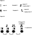
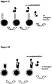
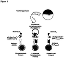
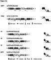

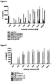
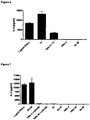
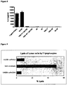

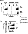

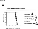
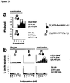
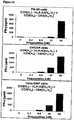
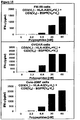
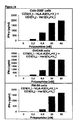
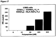

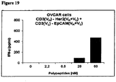
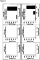
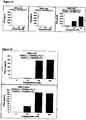
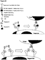
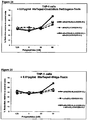


Classifications
-
- A—HUMAN NECESSITIES
- A61—MEDICAL OR VETERINARY SCIENCE; HYGIENE
- A61P—SPECIFIC THERAPEUTIC ACTIVITY OF CHEMICAL COMPOUNDS OR MEDICINAL PREPARATIONS
- A61P1/00—Drugs for disorders of the alimentary tract or the digestive system
- A61P1/04—Drugs for disorders of the alimentary tract or the digestive system for ulcers, gastritis or reflux esophagitis, e.g. antacids, inhibitors of acid secretion, mucosal protectants
-
- A—HUMAN NECESSITIES
- A61—MEDICAL OR VETERINARY SCIENCE; HYGIENE
- A61P—SPECIFIC THERAPEUTIC ACTIVITY OF CHEMICAL COMPOUNDS OR MEDICINAL PREPARATIONS
- A61P17/00—Drugs for dermatological disorders
- A61P17/06—Antipsoriatics
-
- A—HUMAN NECESSITIES
- A61—MEDICAL OR VETERINARY SCIENCE; HYGIENE
- A61P—SPECIFIC THERAPEUTIC ACTIVITY OF CHEMICAL COMPOUNDS OR MEDICINAL PREPARATIONS
- A61P19/00—Drugs for skeletal disorders
- A61P19/02—Drugs for skeletal disorders for joint disorders, e.g. arthritis, arthrosis
-
- A—HUMAN NECESSITIES
- A61—MEDICAL OR VETERINARY SCIENCE; HYGIENE
- A61P—SPECIFIC THERAPEUTIC ACTIVITY OF CHEMICAL COMPOUNDS OR MEDICINAL PREPARATIONS
- A61P25/00—Drugs for disorders of the nervous system
-
- A—HUMAN NECESSITIES
- A61—MEDICAL OR VETERINARY SCIENCE; HYGIENE
- A61P—SPECIFIC THERAPEUTIC ACTIVITY OF CHEMICAL COMPOUNDS OR MEDICINAL PREPARATIONS
- A61P29/00—Non-central analgesic, antipyretic or antiinflammatory agents, e.g. antirheumatic agents; Non-steroidal antiinflammatory drugs [NSAID]
-
- A—HUMAN NECESSITIES
- A61—MEDICAL OR VETERINARY SCIENCE; HYGIENE
- A61P—SPECIFIC THERAPEUTIC ACTIVITY OF CHEMICAL COMPOUNDS OR MEDICINAL PREPARATIONS
- A61P3/00—Drugs for disorders of the metabolism
- A61P3/08—Drugs for disorders of the metabolism for glucose homeostasis
- A61P3/10—Drugs for disorders of the metabolism for glucose homeostasis for hyperglycaemia, e.g. antidiabetics
-
- A—HUMAN NECESSITIES
- A61—MEDICAL OR VETERINARY SCIENCE; HYGIENE
- A61P—SPECIFIC THERAPEUTIC ACTIVITY OF CHEMICAL COMPOUNDS OR MEDICINAL PREPARATIONS
- A61P31/00—Antiinfectives, i.e. antibiotics, antiseptics, chemotherapeutics
-
- A—HUMAN NECESSITIES
- A61—MEDICAL OR VETERINARY SCIENCE; HYGIENE
- A61P—SPECIFIC THERAPEUTIC ACTIVITY OF CHEMICAL COMPOUNDS OR MEDICINAL PREPARATIONS
- A61P35/00—Antineoplastic agents
-
- A—HUMAN NECESSITIES
- A61—MEDICAL OR VETERINARY SCIENCE; HYGIENE
- A61P—SPECIFIC THERAPEUTIC ACTIVITY OF CHEMICAL COMPOUNDS OR MEDICINAL PREPARATIONS
- A61P37/00—Drugs for immunological or allergic disorders
- A61P37/02—Immunomodulators
-
- A—HUMAN NECESSITIES
- A61—MEDICAL OR VETERINARY SCIENCE; HYGIENE
- A61P—SPECIFIC THERAPEUTIC ACTIVITY OF CHEMICAL COMPOUNDS OR MEDICINAL PREPARATIONS
- A61P37/00—Drugs for immunological or allergic disorders
- A61P37/02—Immunomodulators
- A61P37/06—Immunosuppressants, e.g. drugs for graft rejection
-
- A—HUMAN NECESSITIES
- A61—MEDICAL OR VETERINARY SCIENCE; HYGIENE
- A61P—SPECIFIC THERAPEUTIC ACTIVITY OF CHEMICAL COMPOUNDS OR MEDICINAL PREPARATIONS
- A61P37/00—Drugs for immunological or allergic disorders
- A61P37/08—Antiallergic agents
-
- A—HUMAN NECESSITIES
- A61—MEDICAL OR VETERINARY SCIENCE; HYGIENE
- A61P—SPECIFIC THERAPEUTIC ACTIVITY OF CHEMICAL COMPOUNDS OR MEDICINAL PREPARATIONS
- A61P43/00—Drugs for specific purposes, not provided for in groups A61P1/00-A61P41/00
-
- A—HUMAN NECESSITIES
- A61—MEDICAL OR VETERINARY SCIENCE; HYGIENE
- A61P—SPECIFIC THERAPEUTIC ACTIVITY OF CHEMICAL COMPOUNDS OR MEDICINAL PREPARATIONS
- A61P7/00—Drugs for disorders of the blood or the extracellular fluid
- A61P7/04—Antihaemorrhagics; Procoagulants; Haemostatic agents; Antifibrinolytic agents
-
- A—HUMAN NECESSITIES
- A61—MEDICAL OR VETERINARY SCIENCE; HYGIENE
- A61P—SPECIFIC THERAPEUTIC ACTIVITY OF CHEMICAL COMPOUNDS OR MEDICINAL PREPARATIONS
- A61P9/00—Drugs for disorders of the cardiovascular system
-
- C—CHEMISTRY; METALLURGY
- C07—ORGANIC CHEMISTRY
- C07K—PEPTIDES
- C07K16/00—Immunoglobulins [IGs], e.g. monoclonal or polyclonal antibodies
- C07K16/18—Immunoglobulins [IGs], e.g. monoclonal or polyclonal antibodies against material from animals or humans
- C07K16/28—Immunoglobulins [IGs], e.g. monoclonal or polyclonal antibodies against material from animals or humans against receptors, cell surface antigens or cell surface determinants
- C07K16/2803—Immunoglobulins [IGs], e.g. monoclonal or polyclonal antibodies against material from animals or humans against receptors, cell surface antigens or cell surface determinants against the immunoglobulin superfamily
- C07K16/2809—Immunoglobulins [IGs], e.g. monoclonal or polyclonal antibodies against material from animals or humans against receptors, cell surface antigens or cell surface determinants against the immunoglobulin superfamily against the T-cell receptor (TcR)-CD3 complex
-
- C—CHEMISTRY; METALLURGY
- C07—ORGANIC CHEMISTRY
- C07K—PEPTIDES
- C07K16/00—Immunoglobulins [IGs], e.g. monoclonal or polyclonal antibodies
- C07K16/18—Immunoglobulins [IGs], e.g. monoclonal or polyclonal antibodies against material from animals or humans
- C07K16/28—Immunoglobulins [IGs], e.g. monoclonal or polyclonal antibodies against material from animals or humans against receptors, cell surface antigens or cell surface determinants
- C07K16/2803—Immunoglobulins [IGs], e.g. monoclonal or polyclonal antibodies against material from animals or humans against receptors, cell surface antigens or cell surface determinants against the immunoglobulin superfamily
- C07K16/2827—Immunoglobulins [IGs], e.g. monoclonal or polyclonal antibodies against material from animals or humans against receptors, cell surface antigens or cell surface determinants against the immunoglobulin superfamily against B7 molecules, e.g. CD80, CD86
-
- C—CHEMISTRY; METALLURGY
- C07—ORGANIC CHEMISTRY
- C07K—PEPTIDES
- C07K16/00—Immunoglobulins [IGs], e.g. monoclonal or polyclonal antibodies
- C07K16/18—Immunoglobulins [IGs], e.g. monoclonal or polyclonal antibodies against material from animals or humans
- C07K16/28—Immunoglobulins [IGs], e.g. monoclonal or polyclonal antibodies against material from animals or humans against receptors, cell surface antigens or cell surface determinants
- C07K16/2803—Immunoglobulins [IGs], e.g. monoclonal or polyclonal antibodies against material from animals or humans against receptors, cell surface antigens or cell surface determinants against the immunoglobulin superfamily
- C07K16/2833—Immunoglobulins [IGs], e.g. monoclonal or polyclonal antibodies against material from animals or humans against receptors, cell surface antigens or cell surface determinants against the immunoglobulin superfamily against MHC-molecules, e.g. HLA-molecules
-
- C—CHEMISTRY; METALLURGY
- C07—ORGANIC CHEMISTRY
- C07K—PEPTIDES
- C07K16/00—Immunoglobulins [IGs], e.g. monoclonal or polyclonal antibodies
- C07K16/18—Immunoglobulins [IGs], e.g. monoclonal or polyclonal antibodies against material from animals or humans
- C07K16/28—Immunoglobulins [IGs], e.g. monoclonal or polyclonal antibodies against material from animals or humans against receptors, cell surface antigens or cell surface determinants
- C07K16/2863—Immunoglobulins [IGs], e.g. monoclonal or polyclonal antibodies against material from animals or humans against receptors, cell surface antigens or cell surface determinants against receptors for growth factors, growth regulators
-
- C—CHEMISTRY; METALLURGY
- C07—ORGANIC CHEMISTRY
- C07K—PEPTIDES
- C07K16/00—Immunoglobulins [IGs], e.g. monoclonal or polyclonal antibodies
- C07K16/18—Immunoglobulins [IGs], e.g. monoclonal or polyclonal antibodies against material from animals or humans
- C07K16/28—Immunoglobulins [IGs], e.g. monoclonal or polyclonal antibodies against material from animals or humans against receptors, cell surface antigens or cell surface determinants
- C07K16/289—Immunoglobulins [IGs], e.g. monoclonal or polyclonal antibodies against material from animals or humans against receptors, cell surface antigens or cell surface determinants against CD45
-
- C—CHEMISTRY; METALLURGY
- C07—ORGANIC CHEMISTRY
- C07K—PEPTIDES
- C07K16/00—Immunoglobulins [IGs], e.g. monoclonal or polyclonal antibodies
- C07K16/18—Immunoglobulins [IGs], e.g. monoclonal or polyclonal antibodies against material from animals or humans
- C07K16/28—Immunoglobulins [IGs], e.g. monoclonal or polyclonal antibodies against material from animals or humans against receptors, cell surface antigens or cell surface determinants
- C07K16/2896—Immunoglobulins [IGs], e.g. monoclonal or polyclonal antibodies against material from animals or humans against receptors, cell surface antigens or cell surface determinants against molecules with a "CD"-designation, not provided for elsewhere
-
- C—CHEMISTRY; METALLURGY
- C07—ORGANIC CHEMISTRY
- C07K—PEPTIDES
- C07K16/00—Immunoglobulins [IGs], e.g. monoclonal or polyclonal antibodies
- C07K16/18—Immunoglobulins [IGs], e.g. monoclonal or polyclonal antibodies against material from animals or humans
- C07K16/28—Immunoglobulins [IGs], e.g. monoclonal or polyclonal antibodies against material from animals or humans against receptors, cell surface antigens or cell surface determinants
- C07K16/30—Immunoglobulins [IGs], e.g. monoclonal or polyclonal antibodies against material from animals or humans against receptors, cell surface antigens or cell surface determinants from tumour cells
- C07K16/3007—Carcino-embryonic Antigens
-
- C—CHEMISTRY; METALLURGY
- C07—ORGANIC CHEMISTRY
- C07K—PEPTIDES
- C07K16/00—Immunoglobulins [IGs], e.g. monoclonal or polyclonal antibodies
- C07K16/18—Immunoglobulins [IGs], e.g. monoclonal or polyclonal antibodies against material from animals or humans
- C07K16/28—Immunoglobulins [IGs], e.g. monoclonal or polyclonal antibodies against material from animals or humans against receptors, cell surface antigens or cell surface determinants
- C07K16/30—Immunoglobulins [IGs], e.g. monoclonal or polyclonal antibodies against material from animals or humans against receptors, cell surface antigens or cell surface determinants from tumour cells
- C07K16/3061—Blood cells
-
- C—CHEMISTRY; METALLURGY
- C07—ORGANIC CHEMISTRY
- C07K—PEPTIDES
- C07K16/00—Immunoglobulins [IGs], e.g. monoclonal or polyclonal antibodies
- C07K16/18—Immunoglobulins [IGs], e.g. monoclonal or polyclonal antibodies against material from animals or humans
- C07K16/32—Immunoglobulins [IGs], e.g. monoclonal or polyclonal antibodies against material from animals or humans against translation products of oncogenes
-
- C—CHEMISTRY; METALLURGY
- C07—ORGANIC CHEMISTRY
- C07K—PEPTIDES
- C07K16/00—Immunoglobulins [IGs], e.g. monoclonal or polyclonal antibodies
- C07K16/46—Hybrid immunoglobulins
- C07K16/468—Immunoglobulins having two or more different antigen binding sites, e.g. multifunctional antibodies
-
- G—PHYSICS
- G01—MEASURING; TESTING
- G01N—INVESTIGATING OR ANALYSING MATERIALS BY DETERMINING THEIR CHEMICAL OR PHYSICAL PROPERTIES
- G01N33/00—Investigating or analysing materials by specific methods not covered by groups G01N1/00 - G01N31/00
- G01N33/48—Biological material, e.g. blood, urine; Haemocytometers
- G01N33/50—Chemical analysis of biological material, e.g. blood, urine; Testing involving biospecific ligand binding methods; Immunological testing
- G01N33/53—Immunoassay; Biospecific binding assay; Materials therefor
- G01N33/574—Immunoassay; Biospecific binding assay; Materials therefor for cancer
- G01N33/57484—Immunoassay; Biospecific binding assay; Materials therefor for cancer involving compounds serving as markers for tumor, cancer, neoplasia, e.g. cellular determinants, receptors, heat shock/stress proteins, A-protein, oligosaccharides, metabolites
-
- A—HUMAN NECESSITIES
- A61—MEDICAL OR VETERINARY SCIENCE; HYGIENE
- A61K—PREPARATIONS FOR MEDICAL, DENTAL OR TOILETRY PURPOSES
- A61K39/00—Medicinal preparations containing antigens or antibodies
- A61K2039/505—Medicinal preparations containing antigens or antibodies comprising antibodies
-
- C—CHEMISTRY; METALLURGY
- C07—ORGANIC CHEMISTRY
- C07K—PEPTIDES
- C07K2317/00—Immunoglobulins specific features
- C07K2317/30—Immunoglobulins specific features characterized by aspects of specificity or valency
- C07K2317/31—Immunoglobulins specific features characterized by aspects of specificity or valency multispecific
-
- C—CHEMISTRY; METALLURGY
- C07—ORGANIC CHEMISTRY
- C07K—PEPTIDES
- C07K2317/00—Immunoglobulins specific features
- C07K2317/30—Immunoglobulins specific features characterized by aspects of specificity or valency
- C07K2317/34—Identification of a linear epitope shorter than 20 amino acid residues or of a conformational epitope defined by amino acid residues
-
- C—CHEMISTRY; METALLURGY
- C07—ORGANIC CHEMISTRY
- C07K—PEPTIDES
- C07K2317/00—Immunoglobulins specific features
- C07K2317/50—Immunoglobulins specific features characterized by immunoglobulin fragments
- C07K2317/56—Immunoglobulins specific features characterized by immunoglobulin fragments variable (Fv) region, i.e. VH and/or VL
-
- C—CHEMISTRY; METALLURGY
- C07—ORGANIC CHEMISTRY
- C07K—PEPTIDES
- C07K2317/00—Immunoglobulins specific features
- C07K2317/60—Immunoglobulins specific features characterized by non-natural combinations of immunoglobulin fragments
- C07K2317/62—Immunoglobulins specific features characterized by non-natural combinations of immunoglobulin fragments comprising only variable region components
- C07K2317/622—Single chain antibody (scFv)
-
- C—CHEMISTRY; METALLURGY
- C07—ORGANIC CHEMISTRY
- C07K—PEPTIDES
- C07K2317/00—Immunoglobulins specific features
- C07K2317/70—Immunoglobulins specific features characterized by effect upon binding to a cell or to an antigen
- C07K2317/73—Inducing cell death, e.g. apoptosis, necrosis or inhibition of cell proliferation
-
- G—PHYSICS
- G01—MEASURING; TESTING
- G01N—INVESTIGATING OR ANALYSING MATERIALS BY DETERMINING THEIR CHEMICAL OR PHYSICAL PROPERTIES
- G01N2800/00—Detection or diagnosis of diseases
- G01N2800/24—Immunology or allergic disorders
- G01N2800/245—Transplantation related diseases, e.g. graft versus host disease
Definitions
- the present invention relates to a set of polypeptides and its uses.
- the present invention relates to a set of polypeptides whereby this set comprises two polypeptides each of which comprises a targeting moiety "T" binding to an antigen "A” and a fragment of "F” of a functional domain, wherein said two polypeptides are not associated with each other in absence of a substrate that has "A” at (on) its surface and wherein, upon dimerization of "F", the resulting dimer becomes functional.
- medical and diagnostic uses of said set are described.
- the present invention relates to nucleic acid molecule(s) encoding said set of polypeptides.
- the present invention also relates to a vector comprising the nucleotide sequence of nucleic acid molecule(s) encoding said set of polypeptides. Furthermore, the present invention relates to pharmaceutical compositions comprising said set of polypeptides. Moreover, the present invention relates to a kit comprising said set of polypeptides.
- Antibody-based cancer immune strategies are highly promising therapeutic options due to their excellent sensitivity and specificity towards target structures.
- Antibody- based fusion proteins can be generated with novel biological and/or pharmaceutical properties. There are promising efforts to modify the capability of the Fc domain to elicit ADCC (antibody dependent cell mediated cytotoxicity) and CDC (complement-dependent cytotoxicity) by mutagenesis, dependent on the intended application, either to reduce side effects (inhibitory mutations) or to enhance therapeutic efficacy (activating mutations). New applications that become possible by genetic engineering are even more variate when the antigen binding domain of antibodies is considered.
- ADCC antibody dependent cell mediated cytotoxicity
- CDC complement-dependent cytotoxicity
- the antigen recognizing variable domains of the heavy (V H ) and light chain (V L ) of an antibody can be joined by a peptide linker via genetic engineering while preserving the antigen binding capability.
- Such antigen binding single chain variable fragments (scFvs) can be used as small antibody surrogates with high tissue penetrating capability and low serum retention time for clinical imaging procedures and radiotherapy and other applications.
- these scFv moieties can be easily employed as antigen specific modules in the development of novel recombinant therapeutics.
- bispecific antibodies recognise two antigens, one of which is expressed by the tumour, whereas the other is usually found on an immune cell.
- Most bispecific antibodies in anti-tumour therapy target a tumour-associated lineage marker on the one hand and CD3 ⁇ , an invariant molecule of the T-cell receptor/CD3 complex on the other hand, thus recruiting T cells to destroy the tumour [ Müller and Kontermann, Bispecific antibodies for cancer immunotherapy: Current perspectives. BioDrugs 2010, 24(2):89-98 ].
- the therapeutic efficacy of such antibody-based reagents is limited by the nature of the addressed antigen, the accessibility of the antigen in tumour and tumour-associated tissues and the aptitude of the antibody to elicit or mediate the desired cell death inducing function.
- bispecific antibodies Since therapeutic concepts based on the use of bispecific antibodies usually rely on the recruitment of effector cells, it appears that the more effective the tool (bispecific construct), the more likely side effects do occur, and even minute expression of antigen on non-transformed tissue can cause uncontrollable off-target effects.
- Stability of the formats can be optimized by adding immunoglobulin-like domains in order to mimic naturally occurring antibodies and to simultaneously enhance pharmacokinetic properties like prolonged half life in serum and protection from proteolytic digestion by proteases. Moreover, stability of the formats can be enhanced by optimizing the production. Since linker sequences which are utilized to covalently join scFv domains often leads to aggregates, production lines have been established that first produce two or three polypeptides that can be easily reassembled in order to generate a functional drug. Such techniques utilize directed disulphid-bridges or crosslinking reagents to covalently join two different polypeptides.
- bi/tri-specific and bi-or multivalent constructs described in the art have disadvantages.
- the more potent the bispecific antibody format the more severe are collateral damages, because the target antigens addressed so far are differentiation antigens shared by tumours and non-maligant cells.
- bi- or tri-specific formats of the prior art cannot discriminate malignant from non-malignant cells.
- tri-specific constructs, developed for high avidity binding to target cells may turn out to confer a high degree of off-target effects because binding of one target molecule in general suffice to recruit immune cells for destruction of a cell which express either target molecule.
- a patient suffering from relapsed or refractory leukaemia or another haematological disease may be treated by chemotherapy/irradiation (to eliminate the malignant haematopoietic cells) in combination with a transplantation of healthy haematopoietic cells from a donor. If elimination of malignant cells is incomplete, the tumour may grow back from the surviving malignant recipient cells despite the presence of healthy cells provided by the transplantation. As a result, survival rates among patients undergoing tumour treatment and allogeneic transplantation are significantly reduced.
- the graft (allogenic stem cells), given shortly after the conditioning therapy (radiation/chemotherapy) can replace and reconstitute hematopoiesis.
- the graft is harvested from either bone marrow or from stimulated peripheral blood cells and contains about one percent of hematopoetic stem cells which are the source of newly built blood cells.
- the graft normally contains a huge number of immune cells, especially T lymphocytes, that are part of the adoptive immune system and thet can be very beneficial in cases where these T cells mount an immune attack against leukemic cells. This situation is well described and known as graft versus leukemia effect. On the other side, an errant immune response which directs T cells against the patient, known as graft versus host disease, is also frequently observed.
- grafts are usually selected on the basis of HLA (human leukocyte antigen) or MHC (major histocompatibility complex). The closer the antigens between donor and recipient match the lower is the probability of severe graft versus host disease. However, for many patients, a full matched graft cannot be found. In these cases, a bone marrow or peripheral blood stem cells are utilized that differ in one or even more HLA molecules. These clinical situation requires a strict immunosuppressive regimen after transplantation to keep the T cell system strictly under control.
- HLA human leukocyte antigen
- MHC major histocompatibility complex
- a certain origin such as, in the situation of a tissue or cell transplantation, cells originating from the recipient or from the donor
- a specific cell type or cell lineage such as haematopoietic cells
- the present invention further refers to the following items:
- said antigen A1 is a cell surface molecule.
- said antigen A2 is a cell surface molecule.
- said antigen A1 is specific for the malignant state of a cell.
- said antigen A2 is specific for a certain cell type or cell lineage or for the malignant state of a cell.
- said antigen A1 is specific for a malignant cell type.
- said antigen A2 is specific for a malignant cell type.
- the present invention relates to the set of polypeptides as defined and described herein, wherein, however, the antigen A1 is the same as the antigen A2.
- the F1 fragment may be linked to the targeting moiety T1 and the F2 fragment may be linked to the targeting moiety T2, whereas both T1 and T2 specifically bind to the same antigen.
- the epitope on antigen A1, to which the targeting moiety T1 binds may be the same or a different epitope as the epitope on the antigen A2, to which the targeting moiety T2 binds.
- polypeptide P1 may comprise a targeting moiety which is identical to the targeting moiety comprised in P2. Also this aspect of the invention is based on the advantage that the set of polypeptides P1 and P2 with the disrupted F domain displays no off target effects (for example no pre-activation of CD3-displaying T cells and, hence, less toxic properties and/or side effects, for example as compared to conventional bispecific antibodies).
- said fragment F1 and said fragment F2 together are said functional domain F.
- said polypeptide P1 and said polypeptide P2 are not covalently linked to each other in the absence of a substrate that has both antigens A1 and A2 at its surface, more specifically a cell that carries both antigens A1 and A2 at its cell surface.
- said polypeptide P1 and said polypeptide P2 are not covalently linked to each other.
- polypeptide P1 and polypeptide P2 and/or, in particular, said fragment F1 and fragment F2 as comprised therein, more particular the V H and V L which may be comprised therein, are not associated with each other, in particular when administered to a subject in need of medical intervention, i.e. in need of therapy and/or diagnosis.
- the pharmaceutical or diagnostic means provided herein comprise the two polypeptides P1 and P2 as comprised in the herein defined "set of polypeptides" in non-associated form. The association of said two polypeptides take place in vivo under the presence of said substrate or cell.
- the association of said two polypeptides may be (further) stabilized by a stabilizing agent (for example an antigen, like, for example, CD3, HIS or DIG as described herein).
- a stabilizing agent for example an antigen, like, for example, CD3, HIS or DIG as described herein.
- they are not associated with each other in the absence of said substrate or cell and/or do not dimerizise in the absence of said substrate or cell. More preferably, they are not associated with each other in the absence of said substrate or cell and/or do not dimerizise in the absence of said substrate or cell even if an agent is present which stabilizes association and/or dimerization of polypeptide P1 and polypeptide P2 and/or, in particular, fragment F1 and fragment F2, i.e.
- polypeptide P1 and polypeptide P2 and/or, in particular, said fragment F1 and fragment F2 is present in an stabilizing agent/P1(F1)/P2(F2)-trimeric complex (for example in an antigen/VH/VL-trimeric complex).
- said polypeptide P1 and polypeptide P2 and/or, in particular, said fragment F1 and fragment F2 as comprised therein, more particular the V H and V L which may be comprised therein, are associated with each other and/or dimerizise into a three-part-complex-formation, preferably by an interaction mediated by an agent which stabilizes association and/or dimerization of polypeptide P1 and polypeptide P2 and/or, in particular, fragment F1 and fragment F2 (for example by an antigen-mediated interaction). Most preferably, however, this association and/or dimerization only occurs in the presence of said substrate or cell.
- the affinity strength with which, for example, leucine-zippers and/or constant domains, like immunoglobulin CH3 or Fc fragments, hetero- and homodimerize is estimated to be at a dissociation constant K D in the range of ⁇ 10 -8 to 10 -11 M (see, for example, Zhu (1997) Protein Sci. 6, 781-8 ; Plückthun (1997) Immunotech. 3, 83-105 ).
- K D dissociation constant
- This K D range is clearly below the K D with which, in the absence of said substrate or cell, association and/or dimerization of said polypeptides P1 and P2, in particular of said fragments F1 and F2, of this invention might occur.
- polypeptide P1 and polypeptide P2 and/or, in particular, fragment F1 and fragment F2 as comprised therein, more particular the V H and V L which may be comprised therein associate with each other and/or dimerizise in the absence of said substrate or cell only with a K D which is above the K D of, for example, hetero- and homodimerization of leucine-zippers and/or constant domains, like immunoglobulin CH3 or Fc fragments.
- polypeptide P1 and polypeptide P2 and/or, in particular, fragment F1 and fragment F2 as comprised therein, more particular the V H and V L which may be comprised therein associate with each other and/or dimerizise with a K D which is in the range of the K D of, for example, hetero- and homodimerization of leucine-zippers and/or constant domains, like immunoglobulin CH3 or Fc fragments, or even below this range.
- the interaction strength of, for example, isolated VH and VL domains in general is of low affinity.
- dissociation constants K D of 10 -9 to 10 -6 M have been determined (see, for example, Wörn JMB (2001) 305, 989-1010 ; Plückthun (1992) Immunological Reviews No 130 ).
- surface plasmon resonance techniques SPR biosensor BIAcore or BIAcore 2000, Pharmacia
- an anti HEL-Antibody system anti-hen egg lysozyme antibody HyHEL-10
- the K D with which, in the absence of said substrate or cell, association and/or dimerization of said polypeptides P1 and P2, in particular of said fragments F1 and F2, of this invention might occur is only at, or even above, the K D or range of K D of isolated VH and VL domains, for example as has been estimated in the context of Worn (loc. cit.), Plückthun (1992; loc. cit.), Ueda (loc. cit.) and Ohmuro-Matsuyama (loc. cit.), in particular above the K D or range of K D of the antigen/VH/VL-trimeric complex as has been estimated in the context of Wörn (loc.
- polypeptide P1 and polypeptide P2 and/or, in particular, fragment F1 and fragment F2 as comprised therein, more particular the V H and V L which may be comprised therein, associate with each other and/or dimerizise with a K D which is (far) below the K D or range of K D of isolated VH and VL domains, for example as has been estimated in the context of Wörn (loc. cit.), Plückthun (1992;loc. cit.), Ueda (loc.
- polypeptide P1 and polypeptide P2 and/or, in particular, fragment F1 and fragment F2 as comprised therein, more particular the V H and V L which may be comprised therein are not associated in the absence of said substrate or cell and/or do not dimerizise in the absence of said substrate or cell. If at all, they associate with each other and/or dimerizise in the absence of said substrate or cell only with a K D above 10 -8 M, preferably above 10 -6 M, more preferably above 10 -5 M and more preferably above 10 -4 M.
- polypeptide P1 and polypeptide P2 and/or, in particular, fragment F1 and fragment F2 as comprised therein, more particular the V H and V L which may be comprised therein, are associated in the presence of said substrate or cell and/or dimerizise in the presence of said substrate or cell.
- each other and /or dimerizise in the presence of said substrate or cell with a K D below 10 -6 M, preferably below 10 -7 M, more preferably below 10 -8 M and more preferably below 10 -9 M. They may also associate with each other and/or may dimerizise in the presence of said substrate or cell with a K D in the range of 10 -11 M to 10 -6 M, more preferably 10 -11 M to 10 -7 M and even more preferably 10 -11 M to 10 -8 M.
- an agent which stabilizes association and/or dimerization of polypeptide P1 and polypeptide P2 and/or, in particular, fragment F1 and fragment F2.
- an stabilizing agent in accordance with this invention may be an antigen, like, for example, CD3, HIS or DIG as described herein, capable to bind to the domain F which, for example, may comprise a V H and a V L of an antibody (F1 and F2, respectively, or F2 and F3, respectively).
- said agent and/or said substrate or cell and/or said antigens A1 and A2) particularly means being present at a concentration below the above ranges or below 1 mM, 500 ⁇ M, 300 ⁇ M, 100 ⁇ M, 10 ⁇ M, 1 ⁇ M, 0.1 ⁇ M, 0.01 ⁇ M, 0.001 ⁇ M or 1 nM wherein the lower values are preferred.
- K D of dimerization in particular, of P1 and P2, more particular of F1 and F2 as comprised therein, more particular of the V H and V L which may be comprised therein.
- Examples of respective measuring methods are x-ray crystallography; nuclear magnet resonance (NMR); isothermal calorimetry (ITC); cryo-electro microscopy (CEM); mass spectrometry (MS); surface Plasmon resonance (SPR).
- FIG. 1 A preferred method to measure the K D in accordance with this invention is Fluorescence Correlation Spectroscopy (FCS). This method is, for example, described in Douglas Magde (Physical Review Letters 29, 11, 1972, S. 705-708 ).
- the K D s referred to herein (i) apply to, (ii) are at or (iii) are to be measured at a temperature of 4 to 38 °C, preferably 4 to 20 °C (for example 10°C) or 20 to 38 °C (for example 30°C), and/or a pH of 4,5 to 8 (for example a pH of 7),
- "Not associated" in the context of the present invention particularly means not functionally associated with respect of the function of the domain F, i.e. not allowing F1 and F2 to form a functional F.
- P1 and P2 may be bound to each other (for example covalently) as far as no functional domain F is formed by F1 and F2. It is, however, preferred that P1 and P2 are separated.
- said antigen A1 and/or said antigen A2 is a molecule.
- said antigen A1 and/or said antigen A2 is proteinaceous.
- said antigen A1 and/or said antigen A2 is non-proteinaceous.
- said targeting moiety T1 binds non-covalently to said antigen A1.
- said targeting moiety T2 binds non-covalently to said antigen A2.
- a substrate having both antigens A1 and A2 at its surface induces dimerization of the fragment F1 of said polypeptide P1 with the fragment F2 of said polypeptide P2, whereas a substrate which does not have both antigens A1 and A2 at its cell surface does not induce dimerization of the fragment F1 of said polypeptide P1 with the fragment F2 of said polypeptide P2.
- a cell carrying both antigens A1 and A2 at its cell surface induces dimerization of the fragment F1 of said polypeptide P1 with the fragment F2 of said polypeptide P2, whereas a cell which does not carry both antigens A1 and A2 at its cell surface does not induce dimerization of the fragment F1 of said polypeptide P1 with the fragment F2 of said polypeptide P2.
- inces dimerization particularly means “allows juxtaposition and subsequent dimerization”.
- said targeting moiety T1 comprises an immunoglobulin module and/or said targeting moiety T2 comprises an immunoglobulin module.
- said targeting moiety T1 comprises an immunoglobulin module I1 which comprises a V L domain linked to a V H domain, preferably an immunoglobulin module II that comprises a scFv (single-chain variant fragment) of an antibody, a Fab or a F(ab') 2 (for example with additional parts of, for example, an Fc domain) of an antibody or a complete antibody.
- an immunoglobulin module I1 which comprises a V L domain linked to a V H domain, preferably an immunoglobulin module II that comprises a scFv (single-chain variant fragment) of an antibody, a Fab or a F(ab') 2 (for example with additional parts of, for example, an Fc domain) of an antibody or a complete antibody.
- said targeting moiety T2 comprises an immunoglobulin module 12 which comprises a V L domain linked to a V H domain, preferably an immunoglobulin module 12 that comprises a scFv (single-chain variant fragment) of an antibody a Fab or a F(ab') 2 (for example with additional parts of, for example, an Fc domain) of an antibody or a complete antibody.
- an immunoglobulin module 12 which comprises a V L domain linked to a V H domain, preferably an immunoglobulin module 12 that comprises a scFv (single-chain variant fragment) of an antibody a Fab or a F(ab') 2 (for example with additional parts of, for example, an Fc domain) of an antibody or a complete antibody.
- said targeting moiety T1 and/or said targeting moiety T2 comprises an immunoglobulin module which comprises a variable domain V H H of a llama antibody, a camel antibody, or a shark antibody.
- said targeting moiety T1 and/or said targeting moiety T2 is an aptamer, or a natural ligand of said antigen A1 or antigen A2, respectively.
- said targeting moiety T1 and/or said targeting moiety T2 comprises a Fv or scFv ((single-chain) variant fragment) of an antibody.
- the immunoglobulin module comprised in the targeting moiety T1 and T2 comprises a V domain selected from the group consisting of:
- the immunoglobulin module comprised in the targeting moiety T1 and/or T2 comprises a V domain selected from the group consisting of:
- the immunoglobulin module comprised in the targeting moiety T1 and/or T2 comprises a V domain comprising any one of SEQ ID NOS: 67-74 and 154.
- polypeptide P1 has the general structure F1-T1 and/or polypeptide P2 has the general structure F2-T2.
- the F fragment and T moieties may be separated by a linker (e.g. F1-linker-T1 and/or F2-linker-T2) and/or flanked by (an) additional amino acid stretche(s) 1 and/or 2 (stretch-F1-(linker)-T1-stretch2 and/or stretch 1-F2-(linker)-T2-stretch2).
- the above general structure is from the N terminus to the C terminus of the polypeptides, i.e. N-F1-T1-C and/or N-F2-T2-C, N-F1-linker-T1-C and/or N-F2-linker-T2-C and N-stretch1-F1-(linker)-T1-stretch2-C and/or N-stretch1-F2-(linker)-T2-stretch2-C.
- polypeptide P1 may have the general structure F1-VH1-VL1 and/or polypeptide P2 may have the general structure F2-VH2-VL2 or polypeptide P1 may have the general structure F1-VL1-VH1 and/or polypeptide P2 may have the general structure F2-VL2-VH2.
- F fragment and T moieties may be separated by a linker (e.g.
- F1-linker-VH/VL1-VL/VH1 and/or F2-linker-VH/VL2-VL/VH2) and/or flanked by (an) additional amino acid stretche(s) 1 and/or 2 (stretch1 F1-(linker)-VHNL1-VL/VH1-stretch2 and/or stretch1-F2-(linker)-VH/VL2-VL/VH2-stretch2).
- the above general structure is from the N terminus to the C terminus of the polypeptides, i.e.
- the above described linker in particular the between the V domains, may comprise 1 to 25 amino acids, preferably 12 to 20 amino acids, preferably 12 to 16 or 15 to 20 amino acids.
- the above described linker may comprise one or more (G 3 S) and/or (G 4 S) motives, in particular 1, 2, 3, 4, 5 or 6 (G 3 S) and/or (G 4 S) motives, preferably 3 or 4 (G 3 S) and/or (G 4 S) motives, more preferably 3 or 4 (G 4 S) motives.
- said immunoglobulin module I1 and said fragment F1 are separated by a linker comprising 1 to 12, preferably 3 to 12, amino acids, and/or said immunoglobulin module 12 and said fragment F2 are separated by a linker comprising 1 to 12, preferably 3 to 12, amino acids.
- the V L domain of I1 is linked to the V H domain of I1 by a linker comprising 12 to 25 amino acids, preferably a linker with the sequence (G 3 S) 3 or (G 3 S) 4 or (G 4 S) 3 or (G 4 S) 4 and/or the V L domain of 12 is linked to the V H domain of 12 by a linker comprising 12 to 25 amino acids, preferably a linker with the sequence (G 3 S) 3 or (G 3 S) 4 or (G 4 S) 3 or (G 4 S) 4 .
- linker as describe above may comprise (G 3 S) and/or (G 4 S) motives.
- Alternative linkers may consist of or comprise the GEGTSTGSGGSGGSGGAD motive.
- the person skilled in the art can without further ado find and use further (peptide) linker known in the art.
- the said additional amino acid stretches 1 and/or 2 may consist of or comprise 1 to 200, 1 to 100, 1 to 70, 1 to 65, 1 to 50, 1 to 25 or 1 to 20 amino acids.
- said antigen A1 and/or said antigen A2 is an antigen expressed on the surface of cells of a tumour or on the surface of progenitor/precursor cells of a tumour, preferably an antigen expressed on the surface of cells of a haematologic tumour, more preferably an antigen expressed on the surface of cells selected from the group consisting of acute myeloic leukemia cells, chronic myeloic leukemia cells, acute lymphatic leukemia cells, chronic lymphatic leukemia cells, lymphoma cells, myeloproliferative syndrome cells, myelodysplastic cells, more preferably myeloma cells, or said antigen A1 and/or said antigen A2 is an antigen expressed on the surface of cells of a non-haematologic tumour, preferably a cell selected from the group consisting of renal cell carcinoma cells, bladder cancer cells, lung cancer cells, mesothelioma cells, prostate cancer cells, brain cancer cells, bone cancer cells, sarcoma cells, soft tissue cancer cells
- the combination of antigen A1 and antigen A2 is only found on blood cells or precursor cells of blood cells, preferably on only one type of blood cells.
- the combination of antigen A1 and antigen A2 is only found on target, in particular, cancerous cells, and not (or only to a negligible extent) on cells that are not target cells, in particular, that are not cancerous.
- the combination of antigen A1 and antigen A2 is specific for cancerous cells of a certain type of cancer.
- the combination of antigen A1 and antigen A2 distinguishes a certain kind of cells, preferably a certain type of cancer cells, from any other cells.
- Cartain type of cancer in this context may mean type of cancer characterized by the same organ in which the cancer is formed or, preferred, type cancer characterized by the same pair of (aberrant) antigens A1 and A2.
- the combination of antigen A1 and antigen A2 is found on progenitor/precursor cells that are progenitor/precursor cells of a tumour and not on progenitor/precursor cells that are not progenitor/precursor cells of a tumour.
- said antigen A1 is an antigen that is specific for the malignant state of a cell and said antigen A2 is an antigen that is specific for the cell type or cell lineage of said cell.
- Her2 ERBB2 v-erb-b2 erythroblastic leukemia viral oncogene homolog 2, neuro/glioblastoma derived oncogene homolog (avian) [Homo sapiens]; also known asNEU; NGL; HER2; TKR1; CD340; HER-2; MLN 19; HER-2/neu; gene ID: 2064, updated on 13-Jan-2013
- mRNA transcript variant 1 VERSION NM_004448.2 GI:54792095, PRI 06-JAN-2013
- mRNA transcript variant 2 VERSION NM_001005862.1 GI:54792097, PRI 06-JAN-2013
- said antigen A1 and/or said antigen A2 is an MHC antigen, preferably an allelic variant of any of HLA-A, HLA-B, HLA-C, HLA-DQ, HLA-DR, or HLA-DM, more preferably an allelic variant of an MHC class I molecule, more preferably an allelic variant selected from the group consisting of HLA-A1, HLA-A2, HLA-A3, HLA-A25, HLA-B7, HLA-B8, HLA-B35, HLA-B44, HLA-Cw3, HLA-Cw4, HLA-Cw6, and HLA-Cw7.
- MHC antigen preferably an allelic variant of any of HLA-A, HLA-B, HLA-C, HLA-DQ, HLA-DR, or HLA-DM, more preferably an allelic variant of an MHC class I molecule, more preferably an allelic variant selected from the group consisting of HLA-A1, HLA-A2, HLA-A3, H
- said antigen A1 is HLA-A2.
- said antigen A1 and/or said antigen A2 is selected from the group consisting of CD45, aquaporin, preferably aquaporin-2, scavenger receptor class B member 1 (SCARB1), CD34, CD33, CD138, CD15, CD1a, CD2, CD3, CD4, CD5, CD8, CD20, CD23, CD31, CD43, CD56, CD57, CD68, CD79a, CD146, synaptophysin, CD56, CD57, nicotinic acetylcholine receptor, muscle-specific kinase (MUSK), voltage-gated calcium channel (P/Q-type), voltage-gated potassium channel (VGKC), N-methyl-D-aspartate receptor (NMDA), TSH (thyroid stimulating hormone) receptor, amphiphysin, HepPar-1, ganglioside GQ1B, ganglioside GD3, ganglioside GM1 and glycophorin-A.
- SCARB1 scavenger receptor class B member
- said antigen A1 is an MHC antigen and said antigen A2 is an antigen that is specific for a certain cell type or cell lineage.
- said functional domain F is an immunoglobulin module, preferably a scFv (single-chain variant fragment) of an antibody more preferably a Fv (variant fragment) of an antibody, or a fluorescent molecule, preferably a bimolecular fluorescence complementation molecule, more preferably GFP or a GFP variant, or a molecule capable of mediating bioluminescence, preferably a luciferase molecule, more preferably Gaussia luciferase.
- said functional domain F is a Fv (variant fragment) of an antibody.
- said functional domain F specifically binds or is capable of specifically binding to an antigen.
- said antigen may be an antigen that is present on cells of the human immune system.
- said binding activates said cells of the human immune system.
- said functional domain F is a T cell engaging domain, preferably a T cell engaging domain specifically binding to CD2, CD3, CD5, T cell receptor or CD28, more preferably a T cell engaging domain specifically binding to CD3 ⁇ , an NK cell (natural killer cell) engaging domain, preferably a NK cell engaging domain specifically binding to CD1a, CD16a or CD56, a domain engaging macrophage cells, preferably a domain engaging macrophage cells specifically binding to CD16a, CD32a, CD32b, CD89 or CD64, a monocyte engaging domain, preferably a monocyte engaging domain specifically binding to CD32a, CD32b, CD64 or CD89, a granulocyte engaging domain, preferably a granulocyte engaging domain specifically binding to CD16b, CD32a, CD32b, CD64, or CD89, a domain engaging neutrophil granulocytes, preferably a domain engaging neutrophil granulocytes that specifically binds to CD89 (Fc ⁇ RI), or a domain engaging activated neutrophil granulocytes,
- said functional domain F is a domain that specifically binds to an antigen linked to a diagnostic or therapeutic compound.
- said functional domain F is a domain that specifically binds to a carrier molecule or an affinity tag.
- said carrier molecule is linked to a diagnostic or therapeutic compound.
- said affinity tag is linked to a diagnostic or therapeutic compound.
- said affinity tag is selected from the group consisting of a FLAG-tag, a myc-tag, a glutathione-S-transferase(GST)-tag, a hemagglutinin(HA)-tag, a polyhistidine(His)-tag, a digoxigenin (DIG)-tag and a maltose binding protein(MBP)-tag.
- said carrier molecule is a peptide or a carbohydrate molecule.
- said functional domain F is a domain that specifically binds to a carrier molecule, preferably a carrier molecule linked to a diagnostic or therapeutic compound, wherein said carrier molecule is selected from the group consisting of gelatine, inulin, dextrane and hydroxyethyl starch.
- said therapeutic compound is a radioactive compound, preferably a radioactive compound comprising 90 Y, 177 Lu, 131 I, 32 P, 10 B, or 213 Bi.
- said therapeutic compound is a toxin.
- said toxin is selected from the group consisting of B. anthracis edema factor, B. anthracis lethal factor, C. perfringens iota toxin, C . botulinum C2 toxin, C. difficile ADP-ribosyltransferase, C. diphtheriae diphteria toxin fragment A, Burgholderia sp. shiga toxin (subunit A), Clostridium perfringens str.
- a further non-limiting example of a toxin in accordance with this invention is a toxin being or comprising an amino acid sequence selected from the group consisting of SEQ ID NOS 160 to 168.
- said diagnostic compound is a radioactive compound, preferably a radioactive compound comprising 99m Tc, 111 In, 82 Rb or 201 Tl.
- said diagnostic compound is a fluorescent compound, preferably GFP, a GFP variant, or a fluorescent small-molecule compound such as FITC (fluorescein isothiocyanate), PE (phycoerythrin), an alexa fluor dye (such as AlexaFluor488 or related dyes) or a cyanine dye (such as Cy3 (Indocarbocyanine) or Cy5 (Indodicarbocyanine) or related dyes),
- said diagnostic compound is a molecule capable of mediating bioluminescence, preferably a luciferase molecule, more preferably Gaussia luciferase.
- said fragment F1 comprises a V L domain of an antibody and said fragment F2 comprises a V H domain of the same antibody, wherein, preferably, said antibody is an anti-CD3 antibody, more preferably an anti-CD3 ⁇ antibody, or an anti-His or anti-DIG antibody or said fragment F1 comprises a V H domain of an antibody and said fragment F2 comprises a V L domain of the same antibody, wherein, preferably, said antibody is an anti-CD3 antibody, more preferably an anti-CD3 ⁇ antibody, or an anti-His or anti-DIG antibody.
- V L and V H domains as comprised in the F1 and F2 fragment, respectively, or in the F2 and F1 fragment, respectively may also of two different antibodies, either specific for the same Antigen (and for the same or a different epitope) or for different Antigen. This is, for example, envisaged to be employed where new specifications are to be created (for example in phage-display approaches).
- the immunoglobulin module comprised in the F domain comprises a V domain selected from the group consisting of:
- the immunoglobulin module comprised in the F domain comprises a V domain selected from the group consisting of:
- said functional domain F is a domain that specifically binds to a toxin molecule, preferably a toxin molecule that by itself is not capable of penetrating through the cell membrane of a human cell and that, preferably, is internalized into a human cell upon association with the cell membrane of said cell, wherein, preferably, said association with the cell membrane of said cell is mediated by specifically binding to a heterodimer formed from two molecules, preferably two molecules associated with said cell membrane, wherein, preferably, said two molecules are the polypeptides P1 and P2 as described herein.
- said functional domain F is a domain that specifically binds to the A-component (active component) of a bacterial two-component A-B toxin.
- said functional domain F is a domain that specifically binds to a toxin selected from the group consisting of B. anthracis edema factor, B. anthracis lethal factor, C . perfringens iota toxin, C . botulinum C2 toxin, C . difficile ADP-ribosyltransferase, C . diphtheriae diphteria toxin fragment A, Burgholderia sp. shiga toxin (subunit A), Clostridium perfringens str.
- a further non-limiting example of a toxin in accordance with this invention is a toxin being or comprising an amino acid sequence selected from the group consisting of SEQ ID NOS 160 to 168.
- said functional domain F is a domain that specifically binds to a fluorescent molecule, preferably a fluorescent molecule that by itself is not capable of penetrating through the cell membrane of a human cell.
- said fluorescent molecule is GFP or a GFP variant or a molecule that is or comprises a fluorescent small-molecule compound such as FITC (fluorescein isothiocyanate), PE (phycoerythrin), an alexa fluor dye (such as AlexaFluor488 or related dyes) or a cyanine dye (such as Cy3 (Indocarbocyanine) or Cy5 (Indodicarbocyanine) or related dyes).
- FITC fluorescein isothiocyanate
- PE phycoerythrin
- an alexa fluor dye such as AlexaFluor488 or related dyes
- a cyanine dye such as Cy3 (Indocarbocyanine) or Cy5 (Indodicarbocyanine) or related dyes
- said functional domain F is a domain that specifically binds to a molecule capable of mediating bioluminescence, preferably to a luciferase molecule, more preferably to Gaussia luciferase.
- said functional domain F is a fluorescent molecule, preferably a bimolecular fluorescence complementation molecule, more preferably GFP or a GFP variant, such as YFP, CFP, Venus, or Cerulean.
- polypeptides P1 or P2 comprised in the set of polypeptides according to this invention are polypeptides comprising an amino acid sequence selected from the group consisting of SEQ ID NOS: 114-129 and 197.
- the present invention relates to the treatment or elimination of any undesired cell population and the treatment or prevention of any disorder or disease which comes along with this undesired cell population.
- the set of polypeptides of this invention is to be used.
- said set of polypeptides is a set of polypeptides for use in the treatment of a patient who is suffering from a tumour or cancer or for diagnostic use in a patient who is suffering from a tumour or cancer, preferably for use in the treatment of a patient who is suffering from a tumour or cancer and undergoing allogeneic tissue or cell transplantation or meant to undergo such transplantation, or for diagnostic use in a patient who is suffering from a tumour or cancer and undergoing or meant to undergo allogeneic tissue or cell transplantation, wherein, preferably, said set of polypeptides is administered to said patient.
- tumours to be treated or diagnosed are those for which the tumour or cancer cells are described herein above with respect to the antigens A1 and/or A2.
- said treatment involves the elimination of recipient tissue/cells of a certain cell type, preferably a cancerous cell type, or recipient precursor cells giving rise to a certain cell type, preferably to a cancerous cell type, optionally after or in parallel to transplantation to the recipient of donor tissue/cells of said same cell type or donor precursor cells giving rise to said same cell type.
- the set of polypeptides of the invention is for use in an allogeneic transplantation setting for haematopoietic neoplasias, for example, with mismatched HLA antigens, in particular for use in therapeutically exploiting this mismatch situation.
- the dual information of recipient HLA haplotype (HLA patient ) and haematopoietic lineage origin (CD45) is displayed exclusively on leukemic blasts and other haematopoietic cells of the patient. All other cells of recipient tude express the recipient haplotype but not the hematopoietic lineage antigen CD45 (e.g. recipient non-hematopoietic cells are positive for HLA-A2 but negative for CD45).
- the present invention also relates to bimolecular and complementing single-chain antibody constructs directed against HLA-A2, in cases where the patient but not the donor is HLA-A2 positive, and a second construct specific for the haematopoietic lineage marker CD45 to specifically target all hematopoietic cells of the patient including all hematologic neoplasms.
- the first polypeptide P1 may comprise a single-chain variable fragment antibody construct directed against the HLA of the patient (targeting moiety T1) fused to the V L fragment of F1 antiCD3 (for example, fragment Fl).
- the second polypeptide P2 may comprise a single-chain variable fragment construct specific for a haematopoietic lineage marker (for example, CD45; targeting moiety T2), fused to the V H split-fragment of F2 anti CD3-Fv (fragment F2).
- said elimination involves the destroying of said recipient tissue/cells or said recipient precursor cells by cells of the immune system, by a toxin or by a radioactive compound.
- said set of polypeptides is a set of polypeptides for diagnostic use in a patient undergoing allogeneic tissue or cell transplantation, wherein, preferably, said patient is a patient suffering from a tumour.
- said diagnostic use involves the specific detection of recipient cells of a certain cell type or cell lineage among recipient cells of different cell type or cell lineage and donor cells of the same or different type or cell lineage.
- said diagnostic use involves the specific detection of recipient cells that are malignant cells among recipient cells that are not malignant and among donor cells.
- said set of polypeptides is administered to a patient.
- said patient is a mammal, more preferably a human being.
- said administration occurs by bolus administration or by continuous administration.
- polypeptides P1 and P2 of said set of polypeptides are administered in parallel. In another embodiment, the polypeptides P1 and P2 of said set of polypeptides are administered sequentially.
- one of the polypeptides P1 or P2 of said set of polypeptides is administered by bolus administration, whereas the other one is administered by continuous administration.
- the amount of polypeptide administered is in the range of from 0.5 ⁇ g/m 2 per day to 500 ⁇ g/m 2 per day for the polypeptide P1 or for the polypeptide P2 or for each of the polypeptides P1 and P2, preferably in the range of from 5 ⁇ g/m 2 per day to 200 ⁇ g/m 2 per day for the polypeptide P1 or for the polypeptide P2 or for each of the polypeptides P1 and P2, more preferably in the range of from 10 ⁇ g/m 2 per day to 80 ⁇ g/m 2 per day for the polypeptide P1 or for the polypeptide P2 or for each of the polypeptides P1 and P2.
- the amount of polypeptide administered is in the range of from 0.05 ⁇ g/m 2 per day to 0.5 ⁇ g/m 2 per day for the polypeptide P1 or for the polypeptide P2 or for each of the polypeptides P1 and P2.
- the amount of polypeptide P1 administered is different from the amount of polypeptide P2 administered.
- the amount of polypeptide administered is in the range of from 0.5 ⁇ g/m 2 per day to 50 ⁇ g/m 2 per day for the polypeptide P1 or for the polypeptide P2 or for each of the polypeptides P1 and P2. In one embodiment, the amount of polypeptide administered is in the range of from 50 ⁇ g/m 2 per day to 100 ⁇ g/m 2 per day for the polypeptide P1 or for the polypeptide P2 or for each of the polypeptides P1 and P2.
- the amount of polypeptide administered is in the range of from 100 ⁇ g/m 2 per day to 200 ⁇ g/m 2 per day for the polypeptide P1 or for the polypeptide P2 or for each of the polypeptides P1 and P2. In one embodiment, the amount of polypeptide administered is in the range of from 200 ⁇ g/m 2 per day to 300 ⁇ g/m 2 per day for the polypeptide P1 or for the polypeptide P2 or for each of the polypeptides P1 and P2.
- the amount of polypeptide administered is in the range of from 300 ⁇ g/m 2 per day to 400 ⁇ g/m 2 per day for the polypeptide P1 or for the polypeptide P2 or for each of the polypeptides P1 and P2. In one embodiment, the amount of polypeptide administered is in the range of from 400 ⁇ g/m 2 per day to 500 ⁇ g/m 2 per day for the polypeptide P1 or for the polypeptide P2 or for each of the polypeptides P1 and P2. In one embodiment, the amount of polypeptide administered is in the range of from 500 ⁇ g/m 2 per day to 1 mg/m 2 per day for the polypeptide P1 or for the polypeptide P2 or for each of the polypeptides P1 and P2.
- said administration occurs continuously for at least 12 hours or for at least 1 day or for at least 2 days or for at least 3 days or for at least 4 days or for at least 5 days or for at least 6 days or for at least 7 days or for at least 8 days or for at least 9 days or for at least 10 days or for at least 11 days or for at least 12 days or for at least 13 days or for at least 14 days or for at least 15 days or for at least 16 days or for at least 17 days or for at least 18 days or for at least 19 days or for at least 20 days or for at least 21 days or for at least 22 days or for at least 23 days or for at least 24 days or for at least 25 days or for at least 26 days or for at least 27 days or for at least 28 days or for at least 29 days or for at least 30 days or for at least 5 weeks or for at least 6 weeks.
- said administration of said set of polypeptides or of one of the polypeptides of said set of polypeptides occurs intravenously, preferably by intravenous injection.
- said administration of said set of polypeptides or of one of the polypeptides of said set of polypeptides occurs subcutaneously, preferably by subcutaneous injection.
- said set of polypeptides is administered in combination with one or more drugs selected from the group consisting of an immunomodulatory drug, and/or a steroid, preferably prednisolone or prednisone.
- said set of polypeptides is administered in combination with a radioactive compound, preferably a radioactive compound linked to an antigen, a carrier molecule or an affinity tag, wherein said radioactive compound, said antigen, said carrier molecule or said affinity tag is specifically bound by said functional domain F.
- a radioactive compound preferably a radioactive compound linked to an antigen, a carrier molecule or an affinity tag, wherein said radioactive compound, said antigen, said carrier molecule or said affinity tag is specifically bound by said functional domain F.
- said set of polypeptides is administered in combination with a toxin, preferably a toxin linked to an antigen, a carrier molecule or an affinity tag, wherein said toxin, said antigen, said carrier molecule or said affinity tag is specifically bound by said functional domain F.
- said set of polypeptides is administered in combination with a fluorescent molecule, preferably a fluorescent molecule linked to an antigen, a carrier molecule or an affinity tag, wherein said fluorophore, said antigen, said carrier molecule or said affinity tag is specifically bound by said functional domain F.
- a fluorescent molecule preferably a fluorescent molecule linked to an antigen, a carrier molecule or an affinity tag, wherein said fluorophore, said antigen, said carrier molecule or said affinity tag is specifically bound by said functional domain F.
- said functional domain F is a domain that specifically binds to an antigen which is not recognized as foreign by the immune system of said patient to whom said set of polypeptides is administered.
- two sets of polypeptides as described above are administered simultaneously or sequentially.
- said first set of polypeptides has different fragments F1 and F2 than said second set of polypeptides.
- said first set of polypeptides has the same fragments F1 and F2 as said second set of polypeptides.
- the targeting moieties T1 and T2 of said first set of polypeptides bind to the same antigens as the targeting moieties T1 and T2, respectively, of said second set of polypeptides.
- the targeting moieties T1 and T2 of said first set of polypeptides bind to different antigens than the targeting moieties T1 and T2 of said second set of polypeptides.
- said patient has undergone cancer treatment before treatment with said set of polypeptides, said cancer treatment preferably being chemotherapy, radiation therapy or operative removal of the tumour, or undergoes cancer treatment parallel to treatment with said set of polypeptides, said cancer treatment preferably being chemotherapy, radiation therapy or operative removal of the tumour.
- said set of polypeptides or one of the polypeptides of said set of polypeptides has been produced by means of a prokaryotic or eukaryotic expression system or by de novo peptide synthesis.
- said set of polypeptides or one of the polypeptides of said set of polypeptides is generated inside said patient by protein expression from a nucleic acid introduced into said patient.
- a clonal B cell population produce an errant antibody that reacts with antigens expressed by the patients' tissues or complex with an allergen, causing anaphylactic reactions. In both cases, it is desirable to specifically eliminate the errant B cell clone.
- PI or P2 in particular T1 or T2
- the other arm P2 or PI, in particular T2 or T1 respectively
- the allergen or the substrate bound by the antibody that causes the autoimmune disease e.g. CD19, CD20, CD38 or CD138
- the attached anti-CD3 VH and VL can interact and reconstitute the CD3 binding site exactly on the B cell. This allergen-specific or antigen-specific assembly will ultimately result in the clonal depletion of the Target B cells.
- any of said antigens A1 and A2 may also be a clonotypic antibody on the surface of a B cell, in particular a B cell that causes an autoimmune disorder.
- one of said antigens A1 and A2 may be CD19 and the other one may be a clonotypic antibody on the surface of a B cell, in particular a B cell that causes an autoimmune disorder.
- any one of said targeting moiety T1 and T2 may comprise an allergen or substrate which binds to the clonotypic antibody on the surface of the B cell and/or which is, upon binding to the clonotypic antibody, capable to cause an autoimmune disorder.
- an allergen comprised in any one of said targeting moiety T1 and T2 are hair allergens, like, for example, dog-hair, cat-hair (e.g. Fel d 1, Feld d1A, Feld d1B) or guinea-pig-hair allergens, or pollen allergens, like, for example, birch, grass, pollen allergens.
- mite allergens for example Tyr p 2, Der P1, Der f 2
- cat allergens for example Fel d 1, Feld dlA, Feld d1B
- peanut allergens for example Conglutin-7
- rot fungus allergens for example Alt a 1
- dog allergens for example Can f 1
- sprue wheat allergens for example Alpha/beta-gliadin
- german cockroach allergens for example Bla g 1.02 variant allergen
- birch tree or (major) pollen allergens for example Cyn d 1, Pha a 1, Dac g 3, Ph1 p 2, Ph1 p 1, Profilein, Bet v 1-L, Bet v 1-A
- major apple allergens for example Mal d 1
- cow's milk allergens for example alpha-lactalbumin, alpha-S1-casein
- chicken egg allergens for example lysozyme C, ovalbumin
- Horse allergens for example Tyr p 2, Der P
- a further non-limiting and preferred example of an allergen comprised in any one of said targeting moiety T1 and T2 is the antigen for human myeloma cell line U266 antibody IgE-ND.
- a further non-limiting and preferred example of an allergen comprised in any one of said targeting moiety T1 and T2 is an allergen being or comprising an amino acid sequence selected from the group consisting of SEQ ID NOS 169 to 195.
- the inventions also relates to the set of polypeptides as described herein, and, in particular in the above aspect, for use in treating or preventing a disorder selected from the group consisting of
- Non-limiting examples of an autoimmune disorder to be treated or prevented in accordance with this invention are selected from the group consisting of
- Non-limiting examples of a hypersensitivity disorder to be treated or prevented in accordance with this invention are selected from the group consisting of allergies (type I hypersensitivity reaction according to Coombs and Gell classification), an antibody dependent cytotoxic reaction (type II hypersensitivity reaction), a immune complex disease (type III hypersensitivity reaction), delayed type hypersensitivity (type IV hypersensitivity reaction) and a receptor mediated autoimmune disease (type V hypersensitivity reaction).
- allergies type I hypersensitivity reaction according to Coombs and Gell classification
- an antibody dependent cytotoxic reaction type II hypersensitivity reaction
- a immune complex disease type III hypersensitivity reaction
- delayed type hypersensitivity type IV hypersensitivity reaction
- a receptor mediated autoimmune disease type V hypersensitivity reaction
- said autoimmune or hypersensitivity disorder comes along with or is triggered by allogenic stem cell transplantation (i.e. any of type I to type V hypersensitivity disorder according to the Coombs and Gell classification).
- any of said antigens A1 and A2 may also be such a pathogen-encoded protein, like, for example, a HIV, EBV or CMV protein on the surface of a cell.
- the inventions also relates to the set of polypeptides as described herein for use in treating or preventing an infectious disease, for example a viral infectious disease.
- pathogen-encoded proteins can be derived from http://www.uniprot.org/uniprot/ and are HIV gp120 (Q78706); EBV LMP-2 (P13285); CMV gB (P06473); HBV HBS (Q9JG36); HCV E1 (C4B751); HCV E2 (Q6TRB1); Human adenovirus C serotype 2 HAdV-2 (P03276).
- nucleic acid molecule or a set of nucleic acid molecules encoding the set of polypeptides or one of the polypeptides of the set of polypeptides as defined in the embodiments above, wherein, preferably, said nucleic acid molecule or the nucleic acid molecules of said set of nucleic acid molecules comprises an export signal that mediates secretion of the encoded polypeptide(s) by a bacterial or eukaryotic cell.
- a non-limiting example of the nucleic acid molecule or set of nucleic acid molecules according to this invention comprises one or more of the nucleotide sequences as depicted in anyone of SEQ ID NOS: 135-150 and 196.
- the objects of the present invention are also solved by a vector comprising the nucleotide sequence of the nucleic acid molecule as defined above or the sequence of one of the nucleic acid molecules of the set of nucleic acid molecules as defined above.
- the objects of the present invention are also solved by a cell comprising said nucleic acid/set of nucleic acids or said vector.
- a pharmaceutical composition comprising either the set of polypeptides as defined above or the nucleic acid molecule/set of nucleic acid molecules as defined above or the vector as defined above, wherein, preferably, said pharmaceutical composition further comprises a pharmaceutically acceptable carrier.
- kits comprising the set of polypeptides as defined above and/or the nucleic acid molecule or the set of nucleic acid molecules according the invention and/or the vector according the invention.
- polypeptides of said set of polypeptides comprised by said kit are contained in a single vial.
- polypeptides of said set of polypeptides comprised by said kit are contained in separate vials.
- one or more of the polypeptides of said set of polypeptides comprised by said kit are freeze-dried.
- one or more of the polypeptides of said set of polypeptides comprised by said kit are in solution.
- the objects of the present invention are also solved by a method for treatment of a patient who is suffering from a
- said set of polypeptides is as defined in the embodiments above.
- the objects of the present invention are also solved by a method of using the set of polypeptides as described above for treatment of a patient undergoing cell or tissue transplantation.
- the objects of the present invention are also solved by the use of a set of proteins as defined in the embodiments above for the manufacture of a medicament for the treatment of a patient suffering from the above defined and described diseases a disorder or, for example, a patient suffering from cancer and/or undergoing cell or tissue transplantation.
- polypeptide refers to a linear molecular chain of amino acids containing more than 30 amino acids.
- a polypeptide may include one or more disulfide bonds or be chemically modified.
- a non-proteinaceous element such as a fluorophore, RNA-aptamer, DNA-aptamer, or small molecule
- a polypeptide can be produced by any known method.
- the polypeptide can for example be generated by expression from a nucleic acid coding for said polypeptide, or can be synthesized by solid phase synthesis methods, or be produced by conjugation or linkage of existing molecules, e.g., by chemical linkage.
- polypeptide P1 is used to refer to a polypeptide comprising (i) a targeting moiety, wherein said targeting moiety specifically binds to an antigen, and (ii) a fragment of a functional domain, wherein neither said fragment by itself nor said polypeptide P1 by itself is functional with respect to the function of said functional domain.
- polypeptide P2 is used to refer to a polypeptide comprising (i) a targeting moiety, wherein said targeting moiety specifically binds to an antigen, and (ii) a fragment of a functional domain, wherein neither said fragment by itself nor said polypeptide P2 by itself is functional with respect to the function of said functional domain.
- domain refers to a linear molecular chain of amino acids that includes the amino acid sequence of an entire polypeptide or a portion of a polypeptide.
- a domain may include one or more disulfide bonds or be chemically modified.
- a domain may comprise a non-proteinaceous element (such as a fluorophore). In one embodiment, however, the term “domain” does not comprise compounds that are chemically modified or comprise non-proteinaceous element(s).
- a “functional domain”, as used herein, is a domain that is capable of fulfilling a certain function, such as specific binding to a certain binding partner or antigen, specific activation of a certain receptor, mediation of toxic effects, or fluorescence upon excitation with light of an appropriate wavelength.
- the term “functional domain F” is preferably meant to also include compounds that are non-proteinaceous. In one embodiment, however, it refers to a proteinaceous compound or a functional part thereof.
- a fragment of a domain refers to a linear molecular chain of amino acids that corresponds to a part of a domain, but not the entire domain.
- a fragment of a domain may include one or more disulfide bonds or be chemically modified.
- a domain may comprise a non-proteinaceous element or part of such a non-proteinaceous element.
- fragment F1 is used to refer to a fragment of a functional domain.
- fragment F2 is used to refer to a fragment of a functional domain.
- P1, P2; T1, T2; F1, F2; A1, A2; and I1, I2, as used herein, are meant to designate different polypeptides, targeting moieties, fragments, antigens, and immunoglobulin modules, respectively. They are synonymous to first polypeptide, second polypeptide; first targeting moiety, second targeting moiety; first fragment, second fragment; first antigen, second antigen; and first immunoglobulin module, second immunoglobulin module, respectively.
- moiety refers to a linear molecular chain of amino acids that includes the amino acid sequence of an entire polypeptide or a portion of a polypeptide.
- a moiety may include one or more disulfide bonds or be chemically modified.
- a moiety may comprise a non-proteinaceous element (such as an oligonucleotide). In one embodiment, however, the term “moiety” does not comprise compounds that are chemically modified or comprise non-proteinaceous element(s).
- targeting moiety T1 is used to refer to a moiety that specifically binds to an antigen, for example antigen A1.
- targeting moiety T2 is used to refer to a moiety that specifically binds to an antigen, for example antigen A2.
- a "linker” is a sequence of amino acids within a polypeptide that connects two parts of said polypeptide or two domains comprised by said polypeptide.
- nucleic acid molecule defines a linear molecular chain consisting of more than 30 nucleotides.
- the term includes DNA, such as cDNA or genomic DNA, and RNA.
- construct refers to a nucleic acid molecule comprising one or more recombinant nucleotide sequences.
- the term also includes polypeptides that are expressed from a recombinant nucleotide sequence or that are artificially made or recombinant molecules that comprise two or more amino acid sequences that are not naturally found within the same protein.
- targeting moiety such as targeting moiety T1 or T2 specifically binding to an antigen (such as antigen A1 or A2)
- antigen such as antigen A1 or A2
- specifically binds to is meant to refer to a situation where either said targeting moiety is capable of specifically binding to said antigen, or where it actually binds thereto.
- a T cell engaging domain an NK cell engaging domain, domain engaging macrophage cells, a monocyte engaging domain, a granulocyte engaging domain, a domain engaging neutrophil granulocytes, or a domain engaging activated neutrophil granulocytes, monocytes and/or macrophages
- the term "specifically binding to" an antigen or molecule or “specifically binds to” an antigen or molecule is meant to refer to a situation where either the respective domain is capable of specifically binding to said antigen or molecule, or where it actually binds thereto.
- a functional domain being a domain that "specifically binds to" an antigen, a molecule, a compound, a carrier molecule or an affinity tag
- the term “specifically binds to” is meant to refer to a situation where either said functional domain is capable of specifically binding to said antigen, molecule, compound, carrier molecule or affinity tag, or where it actually binds thereto.
- fluorophore, antigen, carrier molecule or affinity tag being “specifically bound by" a functional domain, this is meant to refer to a situation where either said functional domain is capable of specifically binding to said toxin, fluorophore, antigen, carrier molecule or affinity tag, or where it actually binds thereto.
- a molecule or antigen is "specific for a certain cell type or cell lineage” if it is expressed by said cell type/cells of said cell lineage, but not (or only to a negligible extent) by other cell types or cells of other cell lineage.
- a molecule or antigen is "specific for a certain cell type or cell lineage" if it is expressed by said cell type/cells of said cell lineage, and not more than a few other cell types or cells of other cell lineage besides said cell type/cells of said cell lineage express said antigen as well, while most other cell types or cells of other cell lineage besides said cell type/cells of said cell lineage do not express said antigen (or only to a negligible extent).
- the term "specific for a certain cell type or cell lineage” may also mean that said molecule or antigen is expressed by said cell type/cells of said cell lineage at a higher rate or at a higher proportion or amount than by other cell types/cells of other cell lineages, in the sense that there may be a small but detectable expression of said molecule also in other cell types/cells of other cell lineages.
- the term "marker”, as used herein in the context of a marker for a certain cell type or cell lineage, can refer to a molecule or antigen that is specific for a cell type or cells of a cell lineage, respectively, as described above.
- aptamer refers to a small compound composed of oligonucleic acid (such as RNA or DNA) or peptidic or non-peptidic molecule that binds to a specific target molecule with high affinity.
- carrier molecule refers to a molecule or part of a molecule that is not recognized as foreign by the immune system of a patient to whom the set of polypeptides according to the invention is administered or that causes no or only a weak immune reaction by a patient to whom the set of polypeptides according to the invention is administered.
- a “carrier molecule” is being bound by or capable of being bound by another molecule, such as an antibody.
- a "carrier molecule” is a molecule or part of a molecule that In certain embodiments, the carrier molecule is attached covalently or non-covalently to a second molecule or part of a second molecule, for example a fluorophore or toxin.
- MHC refers to the Major Histocompatibility Complex, which is a set of genes encoding a group of molecules comprising cell-surface molecules that are required for antigen presentation to T-cells and that are also responsible for rapid graft rejections.
- the MHC includes the genes HLA-A, HLA-B, HLA-C, HLA-DP, HLA-HQ, and HLA-DR.
- HLA refers to Human Leukocyte Antigens.
- HLA is the human form of "MHC”.
- allelic variant denotes any of two or more alternative forms of a gene occupying the same chromosomal locus.
- HLA-A1, HLA-A2, and HLA-A3 are three of the allelic variants of HLA-A.
- allelic variant is also used herein to denote a protein encoded by an allelic variant of a gene.
- antigen refers to a molecule known to be specifically bound by or capable of being specifically bound by an antibody or the antigen-binding part of an antibody.
- antigen A1 refers to an antigen as defined above.
- antigen A2 refers to an antigen as defined above.
- the designations "antigen A1” and “antigen A2” have been chosen in order to allow for distinction between “antigen A1” and “antigen A2”.
- An "MHC antigen” is an antigen that is also a molecule belonging to the major histocompatibility complex.
- MHC antigens include MHC class I antigens (in humans, the antigens HLA-A, -B, and -C) and MHC class II antigens (in humans, the antigens HLA-DP, -DQ, and -DR).
- a cell "carries an antigen” or "carries an antigen at its cell surface” is meant to refer to a situation where a cell expresses an antigen that is present at the cell surface of said cell and accessible for an antibody from outside said cell.
- a substrate “has an antigen at its surface” is meant to refer to a situation where said antigen is present at the surface of said substrate and accessible for an antibody applied to said substrate.
- an antigen that is specific for the malignant state of a cell refers to an antigen that a malignant cell of a certain cell type (such as a malignant B-cell tumour cell) carries at its cell surface, but that a cell of the same cell type that is not malignant (such as a non malignant B-cell) does not (or only to a negligible extent) carry at its cell surface.
- an antigen/molecule that is specific for a malignant cell type refers to an antigen/molecule that a malignant cell of a certain cell type (such as malignant B-cell tumour cell) carries at its cell surface, but that a cell of the same cell type that is not malignant (such as a non malignant B-cell) or cells of other cell types (such as T-cells or hepatocytes) do not (or only to a negligible extent) carry at their cell surface.
- an antigen/molecule that is specific for a malignant cell type refers to an antigen/molecule that a malignant cell of a certain cell type (such as malignant B-cell tumour cell) carries at its cell surface, but that a cell of the same cell type that is not malignant (such as a non malignant B-cell) does not (or only to a negligible extent) carry at its cell surface, and that only cells of a few other cell types besides that certain cell type carry at their cell surface, while cells of most other cell types do not (or only to a negligible extent).
- a malignant cell of a certain cell type such as malignant B-cell tumour cell
- an antigen/molecule that is specific for a malignant cell type may also mean that said antigen/molecule is expressed by said malignant cell of a certain cell type at a higher rate or at a higher proportion or amount than by a cell of the same cell type that is not malignant, in the sense that there may be a small but detectable expression of said molecule also in a cell of the same cell type that is not malignant.
- the term "marker”, as used herein in the context of a marker for the malignant state of a certain cell or for a malignant cell type, can refer to a molecule or antigen that is specific for the malignant state of a certain cell or for a malignant cell type, respectively, as described above.
- immunoglobulin domain refers to a domain that essentially consists of a globular region of an antibody chain. Immunoglobulin domains are characterized in that they retain the immunoglobulin fold characteristic of antibody molecules. Immunoglobulins, such as IgG, IgE, or IgM, are composed of a varying number of heavy and light chains. Each heavy and light chain contains a constant region and a variable region. Each light chain variable region (V L ) and each heavy chain variable region (V H ) contains three hypervariable regions, also called “complementarity-determining regions" or "CDRs". The CDRs are primarily responsible for binding of the immunoglobulin to an antigen.
- V H or "V H domain” are used interchangeably and refer to the variable region of an immunoglobulin heavy chain of an antibody.
- V L or “V L domain” are used interchangeably and refer to the variable region of an immunoglobulin light chain of an antibody.
- immunoglobulin module refers to a molecule, part of a molecule or molecular assembly which comprises one or more, preferably two or more, immunoglobulin domains and which is capable of binding to an antigen.
- an “immunoglobulin module” comprises a linear molecular chain of amino acids that includes the amino acid sequence of one or more, preferably two or more, immunoglobulin domains.
- an “immunoglobulin module” comprises one ore more, preferably two or more, disulfide bonds. Included in the term “immunoglobulin module” are molecules or parts of a molecule that comprise or consist of a "single-chain variant fragment" of an antibody. Included in the term “immunoglobulin module” are also molecules or parts of a molecule that comprise or consist of a V H H domain of a llama antibody, a camel antibody, or a shark antibody.
- the term “immunoglobulin module I1" is used to refer to an immunoglobulin module comprising a V L domain linked to a V H domain.
- said V L domain and said V H domain of said immunoglobulin module I1 are derived from the same antibody.
- said V L domain and said V H domain of said immunoglobulin module I1 form a dimer.
- said dimer is capable of specifically binding to an antigen.
- Said antigen may be, for example, the antigen A1.
- said "immunoglobulin module I1" comprises a "single-chain variant fragment" of an antibody that is capable of specifically binding to an antigen, for example the antigen A1.
- the term “immunoglobulin module 12" is used to refer to an immunoglobulin module comprising a V L domain linked to a V H domain.
- said V L domain and said V H domain of said immunoglobulin module 12 are derived from the same antibody.
- said V L domain and said V H domain of said immunoglobulin module I2 form a dimer.
- said dimer is capable of specifically binding to an antigen.
- Said antigen may be, for example, the antigen A2.
- said "immunoglobulin module 12" comprises a "single-chain variant fragment" of an antibody that is capable of specifically binding to an antigen, for example the antigen A2.
- the V L domain may be positioned N- or C-terminally of the corresponding V H domain. The skilled person is able to determine which arrangement of the V H and V L domains is more suitable for a specific single-chain variant fragment domain.
- Fv and variant fragment refers to a fragment of an antibody that is the minimum antibody fragment which contains a complete antigen recognition and binding site. This region consists of a dimer of one heavy and one light chain variable region in a tight, non-covalent association (V H -V L dimer). In this configuration, the V H and V L domain together define an antigen binding site with antigen binding specificity on the surface of the V H -V L dimer.
- scFv single chain Fv
- single-chain variant fragment are used interchangeably and are meant to designate an antibody or portion of an antibody in which the variable region of the heavy chain (V H ) and the variable region of the light chain (V L ) of a traditional two chain antibody have been joined to form one chain.
- a linker is inserted between the two chains to allow for proper folding and creation of an active binding site.
- llama antibody refers to an antibody or part of an antibody derived from llama.
- camel antibody refers to an antibody or part of an antibody derived from camel.
- shk antibody refers to an antibody or part of an antibody derived from shark. Llama, camel and shark antibodies have an antigen binding moiety that is built up by one single domain, V H H, (rather than a V H and a V L chain).
- T cell engaging domain is meant to refer to a domain that specifically binds to an antigen that is present on the cell surface of T cells. Preferably, binding of said T cell engaging domain to said antigen activates said T cell.
- NK cell engaging domain refers to a domain that specifically binds to an antigen that is present on the cell surface of Natural Killer cells. Preferably, binding of said NK cell engaging domain to said antigen activates said Natural Killer cells.
- domain engaging macrophage cells refers to a domain that specifically binds to an antigen that is present on the cell surface of macrophage cells. Preferably, binding of said domain engaging macrophage cells to said antigen activates said macrophage cells.
- the expression "monocyte engaging domain” refers to a domain that specifically binds to an antigen that is present on the cell surface of monocytes. Preferably, binding of said monocyte engaging domain to said antigen activates said monocytes.
- the expression "granulocyte engaging domain” refers to a domain that specifically binds to an antigen that is present on the cell surface of granulocytes. Preferably, binding of said granulocyte engaging domain to said antigen activates said granuloctyes.
- domain engaging neutrophil granulocytes refers to a domain that specifically binds to an antigen that is present on the cell surface of neutrophil granulocytes.
- domain engaging activated neutrophil granulocytes, monocytes and/or macrophages refers to a domain that specifically binds to an antigen that is present on the cell surface of activated neutrophil granulocytes, monocytes and/or macrophages.
- binding of said domain engaging activated neutrophil granulocytes, monocytes and/or macrophages to said antigen activates said monocytes and/or macrophages.
- molecule capable of mediating bioluminescence refers to a molecule (or functional part of a molecule) that has an enzymatic activity which in the presence of the appropriate substrate(s) catalyzes a reaction that causes bioluminescence.
- the term includes luciferases, such as the luciferases of firefly or Gaussia .
- GFP variant refers to a molecule that has an amino acid sequence derived from the amino acid sequence of green fluorescent protein from Aequorea victoria by introducing alterations resulting in greater fluorescence or fluoresce in different colors.
- the term is meant to include, among others, YFP (yellow fluorescent protein), CFP (cyan fluorescent protein), Venus ( Nagai T et al., A variant of yellow fluorescent protein with fast and efficient maturation for cell-biological applications. Nat Biotechnol. 2002 Jan;20(1):87-90 ), Cerulean (Enhanced CFP with S72A, Y145A and H148D substitutions).
- Enhanced GFP (and, analogously, “enhanced YFP”, “enhanced CFP”) refers to a GFP (YFP, CFP) which has been "humanized”, as reported in Kain et al. (1995) Biotechniques 19(4):650-55 .
- “Humanized” refers to changes made to the GFP (YFP, CFP) nucleic acid sequence to optimize the codons for expression of the protein in human cells.
- bimolecular fluorescence complementation molecule refers to a fluorescent molecule that can be provided as two fragments which by themselves are not fluorescent, but which upon heterodimerization between the two fragments form a dimer that is capable of fluorescence.
- a therapeutic compound refers to a compound suited for preventing, treating, alleviating or curing a disease or disease state.
- a “therapeutic compound” is a compound that, upon entry into a cell, is capable of causing the death of that cell.
- a therapeutic compound can be a chemical or radioactive compound that damages vital cellular structures or interrupts vital cellular processes.
- diagnostic compound refers to a compound that can be detected by common detection methods, such as methods used in the clinic or in biochemical or medical diagnostic laboratories, for example a fluorescent compound, a radioactive compound, or a molecule mediating bioluminescence.
- progenitor/precursor cells is meant to refer to immature, undifferentiated or partially differentiated cells that are typically found in post-natal animals/humans and have the potential to differentiate into a specific cell type or into specific cell types.
- progenitor/precursor cells of a tumour designates progenitor/precursor cells with altered properties (e.g. regarding their proliferation behaviour or gene expression pattern) that give rise to tumour cells. Examples for such progenitor/precursor cells of a tumour are e.g. leukemic precursor or progenitor cells.
- cancer refers to a malignant cell, group of cells, or malignant neoplasia.
- the term is meant to comprise carcinomas, sarcomas, lymphomas, leukemias, germ cell tumours, and blastomas.
- a "cancerous cell” is a cell that is part of or derived from a cancer.
- tumor is used interchangeably with the term “cancer”.
- haematologic tumour refers to a cancer of the blood or blood building system (such as bone marrow cells, blood-building cells, and precursor cells of mature blood cells).
- haematologic tumour refers to a haematologic neoplasia.
- non-haematologic tumour refers to a tumour that is not a haematologic tumour.
- a patient who is undergoing allogeneic tissue or cell transplantation refers to a situation where a patient receives or has received transplanted cells or transplanted tissue that has/have been obtained from another person.
- a preferred situation as to this aspect is the situation with mismatched HLA antigens.
- the unit " ⁇ g/m 2 ", as used herein in the context of an amount of a polypeptide administered, refers to a certain amount of polypeptide per square meter of body surface of the patient to whom said polypeptide is administered (the peptide may be administered by any adequate route of administration, such as by intravenous or subcutaneous injection).
- the expression "The amount of polypeptide administered is 50 ⁇ g/m 2 per day for the polypeptide P1.” is meant to refer to a situation where the amount of polypeptide P1 administered per day is 50 ⁇ g per square meter of body surface of the patient to whom the polypeptide P1 is administered. In the case of a patient having a body surface of 2 m 2 this would mean that 100 ⁇ g of polypeptide P1 are administered per day.
- the present inventors have surprisingly found that with a set of polypeptides according to the invention the above-indicated problems of the prior art can be overcome and the above-described objects can be accomplished. Moreover, the present inventors have surprisingly found that with a set of polypeptides according to the invention, cells with a specific combination of two antigens can be identified and/or eliminated with high specificity and reduced side-effects.
- the combinatorial strategy of the invention has less toxic effects, for example as compared to normal bispecific antibody strategies.
- This is also evidenced by the appended examples, in particular by the in vivo model for allogeneic transplantation, where HLA-A2 positive mice did not suffer any clinical effects after infusion of HLA-A2 reactive constructs.
- two single-chain polypeptides were designed as parts of the final bipartite (bi-molecular) construct (bi-moleculer/trispecific antibody construct), each composed of an antigen-binding single-chain variable fragment (scFv) and either the variable light (VL) or variable heavy chain (VH) domain of an antibody.
- scFv antigen-binding single-chain variable fragment
- VL variable light
- VH variable heavy chain
- VH and HL interactions are of low affinity.
- VH/VL interaction can be stabilized after binding to the specific antigen.
- VH/VL interaction in accordance with this invention takes place only in situations after both fragments have previously bound to their cognate target antigens, for example on the surface of a target cell.
- the constructs are brought into close proximity so that they can form a trimeric complex with the antigen.
- the thus on-target complemented trispecific heterodimer of the invention is functional with respect to the function of the domain F, for example, engages and stimulates T cells for tumor cell destruction if anti CD3 is reconstituted.
- the set of polypeptides according to this invention in particular the polypeptides P1 and P2 comprised therein, have the further advantage to be more stable and/ or have an improved shelf life (in particular at 4°C) as compared to conventional bispecific constructs like BiTE constructs. These conventional bispecific constructs tend to aggregate (in particular at 4°C).
- polypeptides of this invention P1 and P2 are capable to bind albumin.
- the set of polypeptides according to the present invention comprises a first polypeptide P1 and a second polypeptide P2.
- the first polypeptide P1 comprises a first targeting moiety T1 (which is capable of specifically binding to an antigen A1) fused to a first fragment F1 of a functional domain F (see Figure 1A , top).
- the second polypeptide P2 comprises a second targeting moiety T2 (which is capable of specifically binding to an antigen A2) linked to a second fragment F2 of the functional domain F (see Figure 1A , bottom).
- the fragments F1 resp. F2 of the functional domain are non-toxic by their own and unable to exert any biological function unless there is partnering between the two polypeptides P1 and P2.
- both polypeptides P1 and P2 simultaneously bind to their antigens on the surface of a single cell that expresses both antigens A1 and A2, the fragments F1 and F2 of the functional domain F are brought together in close proximity, they hetero-dimerize and thus complement the desired biological function (see Figure 1B ).
- a cell that expresses either only antigen A1 ( Figure 1C ) or only antigen A2 ( Figure 1D ) or none of the antigens does not cause complementation of the biological function.
- the biological function is achieved with high specificity only in the presence of cells having both antigens A1 and A2 at their cell surface upon simultaneous binding of both polypeptides P1 and P2 to such a cell.
- different objects such as specific identification/detection or elimination of cells that express both antigens A1 and A2, can be accomplished.
- this inventive principle is applied for the specific elimination of tumour cells:
- tumour cells can be detected and distinguished from their non-transformed counterparts not by single surface markers but by the expression of aberrant antigen combinations/profiles, as is known for haematopoietic neoplasias and cancer and cancer stem cells of various other provenience.
- a single antigen may not be sufficient to specifically identify a certain tumour cell, a specific combination of two antigens may allow discriminating the tumour cell from any other type of cell.
- the set of polypeptides according to the invention may be used to specifically eliminate cancer cells characterized by the simultaneous expression of the antigens CD33 and CD19 at their cell surface.
- This combination of antigens is found on certain types of acute leukemia cells and distinguishes these cells from any other cells (such as non-malignant cells), which may carry either CD33 or CD19 at their cell surface, but do not carry both CD33 and CD19 at their cell surface (Ossenkoppele et al., Review of the relevance of aberrant antigen expression by flow cytometry in myeloid neoplasms. Br J Haematol 2011, 153(4):421-36 ).
- the first targeting moiety T1 of the first polypeptide P1 may be a single chain variable Fragment (scFv) specific for CD33.
- the light chain variable domain V L of an anti CD3 antibody may be chosen.
- the second targeting moiety T2 of the second polypeptide P2 may be a scFv specific for CD19.
- the fragment F2 of the functional domain F the heavy chain variable domain V H of that anti CD3 antibody may be chosen.
- the light chain variable domain V L and the heavy chain variable domain V H of the anti CD3 antibody are each non-toxic by their own. They are also unable to exert their biological function (i.e. to effectively bind the CD3 antigen) unless there is partnering between the polypeptides P1 and P2.
- both polypeptides P1 and P2 simultaneously bind to that cell.
- the fragments F1 and F2 of the functional group F i.e. the heavy and light chain of the Fv anti CD3 variable domain of that anti-CD3 antibody
- CD3 is a cell surface molecule that is present on the surface of T cells.
- the molecule is part of the T cell signaling complex, and cross- linking of CD3 molecules on the surface of a T cell after binding of a CD3-specific antibody leads to activation of the T cell.
- heterodimers of polypeptides P1 and P2 are capable of recruiting T cells and activating them.
- typical effector mechanisms of a cytotoxic T cell response are elicited, leading to cell lysis: release of lytic granules containing the cytotoxic proteins perforin, granzymes, and granulysin.
- Perforin forms pores into the membrane of the target cell through which the granzymes can enter and induce apoptosis.
- a conventional bispecific construct that engages T cells and has specificity for cells expressing CD33 would mediate the destruction of all CD33 positive cells. Since CD33 is myeloid lineage marker which is expressed on many myeloid cells and myeloid progenitor cells, the destruction of these cells would result in long lasting aplasia and probably death of the patient.
- a conventional bispecific construct that engages T cells and has specificity for CD19 positive cells would lead to the elimination of all cells carrying the CD19 antigen at their cell surface. CD19 is expressed on a significant subset of B-lymphocytes. Destruction of these cells would lead to a severe defect of the immune system.
- the application of conventional bispecific antibodies with specificity for CD33 and CD19 would lead to elimination of myeloid cells and a substantial subset of B-lymphocytes.
- effector activation according to the present invention requires the simultaneous recognition of two specific antigens on the surface of the cell to be identified/eliminated. In consequence, the present invention achieves significantly improved specificity and reduced side effects.
- the approach described in the above exemplary embodiment can easily be adapted for the identification/elimination of other types of tumour cells besides CD33 and CD19 positive acute leukemia cells simply by choosing appropriate targeting moieties T1 and T2 that specifically bind to antigens A1 and A2, respectively, that are present simultaneously on the cells to be identified/eliminated, but not present simultaneously on other cell types.
- T1 and T2 that specifically bind to antigens A1 and A2, respectively, that are present simultaneously on the cells to be identified/eliminated, but not present simultaneously on other cell types.
- many if not all cancer cells (but also progenitor/precursor cells of cancer cells) express a number of cell surface molecules which per se are widely expressed on normal tissues, but are indicative for the malignant phenotype if expressed in a non-physiological combination.
- CD34 is a marker for haematopoietic stem cells and CD7 can be detected on a subset of lymphoid cells.
- the combination of CD34 and CD7, however, is strongly associated with malignancy, and aberrant co-expression of the two antigens can be detected on a substantial proportion of acute myelogenous leukemias ( Ossenkoppele et al., Review of the relevance of aberrant antigen expression by flow cytometry in myeloid neoplasms. Br J Haematol 2011, 153(4):421-36 .).
- CD44 and CD117 have been described for ovarian cancer stem cells, CD44 and CD24 for pancreas cancer initiating cells und the combination of EpCAM and CD44 in colon and breast cancer stem cells ( Natasha Y. Frank, Tobias Schatton, Markus H. Frank; The therapeutic promise of the cancer stem cell concept. J Clin Invest. 2010; 120:41-50 ).
- Expression of CD24 and CD29, as well as CD24 and CD49f has been found to be specific for breast carcinoma ( Vassilopoulos A et al. Identification and characterization of cancer initiating cells from BRCA1 related mammary tumours using markers for normal mammary stem cells. Int J Biol Sci 2008; 4:133-142 ).
- combinations with highly expressed antigen levels are indicative for a number of malignancies, like CD38 and CD138 for myeloma.
- the skilled person may arrive at an antigen combination that is specific for a certain cancer by combining an antigen that is specific for the malignant state of the respective cell type with an appropriate cell type marker or cell lineage marker.
- an antigen that is specific for the malignant state of the respective cell type For example, carbonic anhydrase IX is a marker strongly associated with renal cell carcinoma and metastases of renal cell carcinoma and thus represents a marker for the malignant state of renal cells. This membrane located marker, however, is also expressed on normal cells of the intestinal tract.
- the resulting combination of two antigens is specific for renal cell carcinoma cells and cells resulting from metastasis of renal cell carcinoma, while neither non-malignant kidney cells (which do not express carbonic anhydrase IX) nor cells from the intestinal tract (which do not express aquaporins) are characterized by the selected pair of antigens.
- markers for the malignant state of various cell types and on markers for numerous cell types or cell lineages is available from the literature and web-based resources (see below for details) or can be obtained by straight-forward experimentation (see below).
- markers for the malignant state of a cell include: E-cadherin for epithelial cells and ductal-type breast carcinoma cells; Ca-125 for Epitheloid malignancies and ovary cancer cells, adenocarcinoma cells and breast cancer cells; Her-2/neu for breast cancer cells; gross cystic disease fluid protein (BRST-2 protein) for breast cancer cells; BCA-225 (breast carcinoma associated glycoprotein) for lung and breast cancer cells; CA 19-9 (carbohydrate antigen 19-9) for pancreas, bile duct and intestinal tract cancer cells; CEA for colorectal cancer cells; CD117 (c-kit) for gist (gastrointestinal stromal tumour) cells (and myeloid and mast cells); CD30 for Reed-Sternberg cells (and Ki-1 activated T-cells and B-cells); Epithelial antigen (BER-EP4), Epithelial membrane antigen, and Epithelial Related Antigen (MOC-31) for epithelial cancer cells; Epidermal growth factor receptor (
- markers for a malignant state of a cell/cells include: EpCAM, CD19, HER-2, HER-3, HER-4, PSMA, MUC-1 (mucin), MUC2, MUC3, MUC4, MUC5AC, MUC5B, MUC7, Lewis-Y, CD20, CD33, CD44v6, Wue-1, Plasma Cell Antigen, (membrane-bound) IgE, Melanoma Chondroitin Sulfate Proteoglycan (MCSP), STEAP, mesothelin, Prostate Stem Cell Antigen (PSCA), sTn (sialylated Tn antigen), FAP (fibroblast activation antigen), EGFRvIII, Ig ⁇ , Ig ⁇ , MT-MMPs, Cora antigen, EphA2, L6 and CO-29, CCR5, ⁇ HCG, ganglioside GD3, 9-O-Acetyl-GD3, GM2, Globo H, fucosyl GM1,
- Examples for antigens that are specific for a certain cell type/cell lineage or for a few cell types/cell lineages include: CD45 for hematopoietic cells; CD34 for endothelial cells, stem cells, and stromal cells; CD33 for myeloid cells; CD138 for plasma cells and a subset of epithelial cells; CD15 for epithelial, myeloid, and Reed-Sternberg cells; CD1a for cortical thymocyctes and Langerhans cells; CD2 for thymic cells, T-cells, and Natural Killer (NK) cells; CD3 for T-cells; CD4 for helper T-cells; CD5 for T-cells, a subset of B-cells, and thymic carcinoma cells; CD8 for cytotoxic T-cells; CD20 for B-cells; CD23 for activated B-cells; CD31 for endothelial cells; CD43 for T-cells, my
- an antigen with a less than perfect specificity for the cell type or cell lineage of interest For example in situations where no antigen is known that is found exclusively on the cell type/cell lineage of interest and not on any other cell types/lineages or in situations where it is not possible to confirm the exclusive specificity of an antigen, also antigens that are present on one or more other cell types/cell lineages besides the cell type/cell lineage of interest may be considered. Similar consideration apply for markers for the malignant state of a cell, or even for the specificity of the combination of two antigens. Thus, there are for example situations where for the purposes of the present invention a combination of two antigens is selected that is specific not only for the cells of interest, but also for one or more (a few) other cell types/cell lineages/kinds of malignant cells.
- the skilled person may arrive at an antigen combination that is specific for a certain cancer by straightforward experimentation. This may comprise the steps of (1) determining the surface antigens on the tumour cells to be eliminated and (2) identifying among these tumour cell surface antigens two antigens that are not present simultaneously on other cell types (or, in some embodiments, present on only a few other cell types).
- the Cancer Genome Anatomy Project building a new information and technology platform for cancer research.
- Molecular Pathology of Early Cancer 1999, (Srivastava, S., Henson, D.E., Gazdar, A., eds. IOS Press), pp. 365-370 ).
- the resources generated by the CGAP initiative are freely available (http://cgap.nci.nih.gov/) and include access to all CGAP data and the necessary analysis tools.
- the Cancer Genome Characterization Initiative (CGCI) of the National Cancer Institute focuses on tools for characterizing the genomic changes involved in different tumours, for example genomic characterization methods including exome and transcriptome analysis using second generation sequencing.
- CGCI The data generated by CGCI is available through a publicly accessible database (http://cgap.nci.nih.gov/cgci.html).
- a publicly accessible database http://cgap.nci.nih.gov/cgci.html.
- information about the presence or absence of various known cell surface proteins on the tumour cells of interest can be derived by simply checking these publicly accessible databases. If desired, this information may then be verified in a second step by immunocytochemical/immunohistochemical analysis of tumour cells/tissue according to the methods described below.
- the skilled person can carry out a characterization of the antigens on the tumour cells/tissue by immunocytochemical/immunohistochemical methods with a panel of antibodies (see, e.g., " Handbook of Practical Immunohistochemistry: Frequently Asked Questions” by F Lin and J Prichard, Springer New York, 1st edition (2011 ); or " Using Antibodies: A Laboratory Manual” by E Harlow and D Lane, Cold Spring Harbor Laboratory Press (1998 )).
- a histological preparation or cells isolated from the tumour are incubated with a first antibody directed at a potential surface antigen and, after a washing step, incubation of a second antibody directed against the Fc domain of the first antibody.
- This second antibody is labelled with a fluorophore or an enzyme like HRP (horse radish peroxidase), in order to visualize expression of the targeted antigen.
- Panels of antibodies that can be used for high throughput antigen profiling purposes of cell surface antigens are commercially available from numerous manufacturers.
- BD FACSTM CAP Combinational Antibody Profile
- the immunocytochemical/immunohistochemical/proteomic analysis described above may be preceded (or, in some cases, replaced) by genome-wide gene expression profiling of tumour cells or by mass spectrometric analysis of the proteins expressed by the tumour cells/tissue of interest.
- genome-wide gene expression profiling of tumour cells can be carried out to check for the expression of various cell surface molecules, and the presence of such antigens on the cell surface of the tumour cells may then be confirmed through antibody-based staining methods as described above.
- Further information about approaches to characterize the surface antigens of (cancer) cells is available in the relevant scientific literature (e.g. Zhou J, Belov L, Huang PY, Shin JS, Solomon MJ, Chapuis PH, Bokey L, Chan C, Clarke C, Clarke SJ, Christopherson RI. Surface antigen profiling of colorectal cancer using antibody microarrays with fluorescence multiplexing. J Immunol Methods. 2010;355:40-51 ; or Carter P, Smith L, Ryan M. Identification and validation of cell surface antigens for antibody targeting in oncology. Endocr Relat Cancer. 2004;11:659-87 ).
- the skilled person may identify among the cell surface antigens of the tumour cells a combination of two antigens which is not expressed simultaneously on other cell types.
- a person skilled in the art can easily validate the suitability of the antigen combination for further development of P1 and P2-polypeptide constructs.
- Such validation that the identified combination of two antigens is indeed not expressed simultaneously on other cell types besides the tumour cells can be carried out by immunohistochemical/immunocytochemical analysis of a (optimally large) collection of assorted cell types and/or tissues with antibodies against the two antigens.
- Cells and tissues of any kind can be obtained from ATCC (American Type Culture Collection), from pathology departments and from tissue banks associated with universities and research institutions.
- a suitable antigen combination is defined as a pair of antibodies that stains exclusively the tumour cells, but not healthy tissues or healthy cells (i.e. both antibodies of the pair stain the tumour cells, but no other tissues/cells are stained by both antibodies).
- the highest degree of specificity (preferably absolute specificity) is of course desirable, there are situations where a lower degree of specificity is acceptable.
- some degree of crossreactivity with other cell types or tissues may be acceptable (especially in the case of solid tumours, since the additional positional information helps to distinguish tumour cells from crossreacting cells).
- some degree of crossreactivity with other cell types or tissues may also be acceptable, depending on the severity the disease in a treated patient and on the cell types/tissues affected by the crossreactivity.
- Other situations where a lower degree of specificity may be acceptable may arise in the context of a transplantation setting (see below).
- polypeptide P1 and polypeptide P2 can be engineered by standard protein engineering techniques and methods of molecular biology (see, e.g. G Howard and M Kaser, Making and Using Antibodies: A Practical Handbook, CRC Press, 1st edition (2006 ); Sambrook et al., Molecular Cloning: A Laboratory Manual, Cold Spring Harbor Laboratory Press, New York (2001 )).
- DNA or mRNA coding for the variable domains of the monoclonal antibodies of interest can be obtained from hybridomas by PCR amplification or cloning (Orlandi R, Gussow PT, Jones: Cloning immunoglobulin variable domains for expression by the polymerase chain reaction. Proc Natl Acad Sci U S A 1989, 86(10):3833-3837 ; Wang Z, Raifu M, Howard M, Smith L, Hansen D, Goldsby R, Ratner D: Universal PCR amplification of mouse immunoglobulin gene variable regions: the design of degenerate primers and an assessment of the effect of DNA polymerase 3' to 5'exonuclease activity.
- the sequence coding for the variable fragment Fv of an antibody specific for the first antigen of the identified pair of antigens is used for the first targeting moiety (T1) and linked via a suitable linker (coding, e.g., for less than 12 aa) to a sequence coding for the first fragment F1 of a functional domain (e.g. the V L domain of an anti CD3 antibody).
- polypeptide P2 the sequence coding for the variable fragment Fv of an antibody specific for the second antigen of the identified pair of antigens (or, optionally, the sequence of a single chain variable fragment derived from that sequence) is used for the second targeting moiety (T2) and linked via a suitable linker to a sequence coding for the second fragment F2 of that functional domain (e.g. the V H domain of an anti CD3 antibody).
- any construct of a polypeptide P1 or P2 according to the invention modifications to the construct or to the sequences used for forming the construct are considered in order to adapt the construct to specific needs.
- a construct can be modified in a way that reduces or abolishes its immunogenicity in humans.
- modifications to the sequence can be carried out that result in a reduced immunogenicity in humans while retaining or substantially retaining the antigen-binding properties of the parent antibody (known to the skilled person as "humanizing" an antibody/construct).
- T1 and T2 specifically bind to
- T1 and/or T2 instead of single chain variant fragments (scFv) as targeting moiety T1 and/or T2 other types of monovalent antibodies or antibody-like structures can be employed.
- an antibody/antibody-like structure derived from a llama, camel or shark antibody can be used. Since llama, camel and shark antibodies have an antigen binding moiety that is built up by one single domain (rather than a V H and a V L chain), the resulting polypeptide P1 or P2 is much smaller and may thus better penetrate into tumour tissues.
- the single chain Fv of targeting moiety T1 and/or T2 can be replaced by the natural or artificial ligand of such a cell surface-bound receptor. Like antibodies, these natural or artificial ligands confer excellent specificity towards the target receptor.
- the targeting moiety T1 and/or T2 can be an aptamer.
- the targeting moiety in order to enhance binding affinity of a targeting moiety towards the antigen, can be multimerized and/or altered by glycosylation or other types of posttranslational or chemical modification or be optimized through site directed mutagenesis or a phage display selection process.
- fragments F1 and F2 i.e. the V L and V H fragments of anti CD3 Fv in the above-described exemplary embodiment
- fragments F1 and F2 can be replaced by fragments of a different functional domain F, resulting in a different biological effect upon complementation of the two fragments.
- fragments of anti CD56, anti CD1a, or anti CD16a natural killer cells can be recruited and activated.
- fragments of anti CD16 natural killer cells, neutrophil polymorphonuclear leukocytes, monocytes and macrophages can be recruited and activated.
- fragments of anti CD32a, anti CD32b, anti CD89, anti CD16a, or anti CD64 macrophages can be recruited and activated.
- monocytes By using fragments of anti CD32a, anti CD32b, anti CD64, or anti CD89, monocytes can be recruited and activated.
- fragments of anti CD16b, anti CD89, anti CD32a, anti CD32b, or anti CD64 granulocytes can be recruited and activated.
- T cells can also be recruited and activated by using fragments of anti CD2, anti CD5, anti CD28, or anti TCR (T cell receptor).
- first set of polypeptides is then used (e.g. administered to a patient) in combination with a second set of polypeptides with fragments of a functional domain F that upon complementation binds to a second, different antigen on the same effector cell, but wherein again binding to this antigen of the effector cell does not cause activation of the effector cell.
- the antigens to which the first and the second sets of polypeptides bind are chosen in a way that, while binding of only one of the two antigens on the effector cells does not result in activation of the effector cell, binding of both antigens on the effector cell simultaneously leads to activation of the effector cell.
- antigens on effector cells can be used that do not function individually, but require costimulation of a second antigen
- the number of different antigens that dictates the specificity with which a certain cell (such as a cancer cell) is differentiated from other cells can be increased from two (if the first and second set of polypeptides have the same targeting moieties T1 and T2, respectively) to up to four different antigens (if the first and second set of polypeptides have no targeting moiety in common).
- each set of polypeptides by itself upon simultaneous administration to a patient) is not capable of activating the effector cell (due to its low concentration), while the combination of both sets of polypeptides is (because the effects of the two sets of polypeptides act synergistically and thus the sum of the effects caused by the two sets of polypeptides is sufficient to activate the effector cell).
- a therapeutic or diagnostic compound coupled to said antigen, carrier molecule or affinity tag is administered. Only cells which carry both the antigens A1 and A2 at their surface are bound by both polypeptide P1 and polypeptide P2. Consequently, only at these cells functional complementation leads to generation of a binding site capable of recruiting the therapeutic or diagnostic compound through said antigen, carrier molecule or affinity tag.
- This approach allows exclusive addressing of target cells combined with the possibility of precise administration and dosing of therapeutic compounds like toxins or radioactive substances or diagnostic compounds, while cells that do not express the antigens or do express only one of the antigens are not affected.
- a suitable carrier molecule may for example be a peptide or a carbohydrate molecule.
- the carrier molecule may be gelatine, dextrane, or hydroxyethyl starch, which are common plasma expanders that are metabolically inert, remain in the blood and are, if they are small enough, renally eliminated.
- the carrier molecule may be inulin, a metabolically inert molecule that is used routinely in the clinic for determination of glomerular clearance (and, in addition, antibodies exist that specifically recognize inulin).
- a suitable affinity tag may be, for example, a Flag-tag, a myc-tag, a glutathione-S-transferase(GST)-tag, a hemagglutinin(HA)-Tag, a polyhistidine(His)-tag, or a maltose binding protein(MBP)-tag, a digoxigenin(DIG)-tag.
- the therapeutic compound coupled to the antigen, carrier molecule or affinity tag may for example be a radioactive compound or a toxin. Suitable radioactive compounds are for example compounds comprising 90 Y, 177 Lu, 131 I, 32 P, 10 B, or 213 Bi. Recruitment of the antigen, carrier molecule or affinity tag linked to the radioactive compound to cells that express both the first and the second antigen leads to accumulation of radioactivity onto the tumour site, resulting in specific destruction of tumour cells/tissue.
- the therapeutic compound coupled to the antigen, carrier molecule or affinity tag may for example be a toxic compound that is not able to cross the cell membrane without prior binding to the cell surface.
- AB-toxins are two-component protein complexes that interfere with internal cell functions.
- the A component is the "active” component (i.e. it kills a cell upon membrane penetration), but is not able to cross the cell membrane on its own.
- the B component is the "binding" component that by itself is non-toxic, but is essential for uptake and membrane penetration of component A.
- Bacillus anthracis protective antigen is a classical toxin B component which mediates the uptake of the actual anthrax exotoxins edema factor and lethal factor (LF).
- LF without the PA-component is non-toxic since LF by its own does not penetrate membranes and thus cannot execute its pathogenic capabilities ( Pezard C, Berche P, Mock M. "Contribution of individual toxin components to virulence of Bacillus anthracis" 1991 Infect. Immun. 59 (10): 3472 ).
- LF is internalised and highly toxic to the cell.
- the function of the functional domain F is reconstituted.
- the toxin is recruited to the cell membrane of the target cells, incorporated into the cells and kills the cells.
- This principle is easily adapted to the purposes of the invention by the skilled person, since it is already widely used in so called immunotoxins, where a targeting moiety, mostly an antibody-like domain or natural ligand, is coupled to the toxin component (see, e.g., Immunotoxins for targeted cancer therapy. Kreitman RJ, AAPS J. 2006 Aug 18;8(3):E532-51 ).
- Examples include immunotoxins based on diphtheria toxin (such as Denileukin diftitox (U.S.
- Suitable A components of AB-toxins may for example be B. anthracis edema factor, B. anthracis lethal factor, C. perfringens iota toxin, C . botulinum C2 toxin, C . difficile ADP-ribosyltransferase C . diphtheriae diphteria toxin fragment A.
- the therapeutic compound may for example be a cytotoxic compound that is toxic upon entry into a cell and that is capable of crossing the cell membrane by itself without prior binding to the cell surface.
- the antigen, carrier molecule or affinity tag that the therapeutic compound is coupled to is selected such that it prevents the resulting conjugate (i.e. the therapeutic compound linked to the antigen/carrier molecule/affinity tag) from crossing cell membranes and entering cells without prior binding of the conjugate to the cell surface (a suitable carrier molecule may for example be a hydroxyethyl starch carrier).
- a suitable carrier molecule may for example be a hydroxyethyl starch carrier.
- the conjugate does not bind to cells, unless it is recruited in the presence of the inventive set of polypeptides to cells that simultaneously express both antigens A1 and A2 at their cell surface.
- Such cells bind and recruit both polypeptides P1 and P2, and the reconstituted functional domain specifically binds to and recruits the antigen/carrier molecule/affinity tag which, in turn, results in internalization of the therapeutic compound.
- a specific killing of cells that carry both antigens A1 and A2 at their cell surface is accomplished.
- Cytotoxic compounds that may be used in this context include e.g.
- auristatin ricin, saponin, bryodin 1, bouganin, gelonin, pokeweed antiviral protein (PAP), antifolates, vinca alkaloides, anthracyclines, calicheamicin, ribonuclease, abrin, modeccin, or Listeriolysin O.
- PAP pokeweed antiviral protein
- the diagnostic compound coupled to the antigen, carrier molecule or affinity tag may for example be a radioactive compound, a fluorophore, or a compound capable of mediating bioluminescence.
- Suitable radioactive compounds are for example compounds comprising 99m Tc, 111 In, 82 Rb or 201 Tl. Such compounds are detected by well-known medical imaging procedures in the clinic.
- a fluorescent compound may be used as diagnostic compound, such as GFP (green fluorescent protein) or a GFP variant (e.g.
- BFP blue fluorescent protein
- CFP cyan fluorescent protein
- YFP yellow fluorescent protein
- a fluorescent small-molecule compound like FITC (fluorescein isothiocyanate) or PE (phycoerythrin), alexa fluor dyes (such as AlexaFluor488 and related dyes sold by Molecular Probes, e.g.) or cyanine dyes (such as Cy3 (Indocarbocyanine) or Cy5 (Indodicarbocyanine) or related dyes).
- a compound capable of mediating bioluminescence may be used as diagnostic compound, such as a luciferase, for example Gaussia luciferase ( Chopra A.
- Gaussia princeps luciferase In: Molecular Imaging and Contrast Agent Database (MICAD) [database online]. Bethesda (MD): National Library of Medicine (US), NCBI; 2004-2012. Available from: http://micad.nih.gov .).
- MICAD Molecular Imaging and Contrast Agent Database
- MD National Library of Medicine
- NCBI NCBI
- 2004-2012 Available from: http://micad.nih.gov .
- the employment of Gaussia luciferase for in vivo imaging is well established (see, e.g., Santos EB et al. Sensitive in vivo imaging of T cells using a membrane-bound Gaussia princeps luciferase. Nat Med. 2009 Mar;15(3):338-44 . Epub 2009 Feb 15; or Inoue Y et al.
- fragments F1 and F2 i.e. the V L and V H fragments of anti CD3 Fv in the above-described exemplary embodiment
- V L and V H fragments of an antibody that is specific for a therapeutic or diagnostic compound i.e. in this case the functional domain F is capable of directly binding to the therapeutic or diagnostic compound.
- the same therapeutic and diagnostic compounds as described above in the context of the "pretargeting" approach may be considered.
- fragments F1 and F2 i.e. the V L and V H fragments of anti CD3 Fv in the above-described exemplary embodiment
- fragments F1 and F2 can be replaced by fragments of a fluorescent or bioluminescent compound that are biologically inactive on their own, but regain their function (i.e. their ability to mediate fluorescence or bioluminescence) upon association of the two fragments and functional complementation, thus allowing for specific identification of cells that carry both the antigens A1 and A2.
- a number of fluorescent molecules that may be used in this context are well known and characterized in the art including, but are not limited to, GFP (green fluorescent protein), GFP derivatives (like YFP (yellow fluorescent protein) and CFP (cyan fluorescent protein), Venus ( Nagai T et al., A variant of yellow fluorescent protein with fast and efficient maturation for cell-biological applications. Nat Biotechnol. 2002 Jan;20(1):87-90 ), or Cerulean (Enhanced CFP with S72A, Y145A and H148D substitutions)).
- BiFC bimolecular fluorescence complementation
- GFP, CFP, Venus, Venus with a M153T substitution, or Cerulean may be split after amino acid 155 (i.e., for example, fragment F1 may comprise amino acids 1-155 of GFP, while fragment F2 may comprise amino acids 156-245 of GFP, or vice versa).
- YFP or Venus may be split after amino acid 173.
- split GFP and split GFP variants can be found in Kerppola TK., Visualization of molecular interactions using bimolecular fluorescence complementation analysis: characteristics of protein fragment complementation. Chem Soc Rev. 2009;38:2876-86 .
- An example for a molecule that mediates bioluminescence and that can be used in this context is split luciferase.
- luciferase of Gaussia princeps which requires no cofactors to be active and catalyzes the oxidation of the substrate coelenterate luciferin (coelenterazine) in a reaction that emits blue light, or derivatives of Gaussia luciferase ( Remy I and Michnick S, A highly sensitive protein-protein interaction assay based on Gaussia luciferase. Nature Methods - 3, 977 - 979 (2006 )).
- fragment F1 may comprise a fragment from the N-terminus of Gaussia luciferase to Gly-93
- fragment F2 may comprise a fragment from Glu-94 to the C-terminus of Gaussia luciferase, or vice versa
- Application of the Gaussia split luciferase system in vivo has been established ( Luker et al., In vivo imaging of ligand receptor binding with Gaussia luciferase complementation. Nature Medicine 2011, doi:10.1038/nm.2590 ), allowing for straightforward adaptation to the purposes of the present invention by a skilled person.
- Intravital imaging of tumour lesions is of eminent importance in cases, where cancer cells infiltrate tissues and the complete elimination of all transformed cells is prerequisite for cure.
- a surgeon searching for disseminated cancer cells in the operation site may use split GFP or split GFP derivatives fused to the targeting moieties and a laser assisted Multispectral fluorescence camera system for detection of cells aberrantly expressing an addressed antigen profile, similar to the intraoperative use of fluorescence or bioluminescence that is already exploited in some clinical settings ( van Dam GM et al., Intraoperative tumor-specific fluorescence imaging in ovarian cancer by folate receptor- ⁇ targeting: first in-human results. Nat Med.
- the inventive principle is applied in the context of a patient who suffers from a haematopoietic tumour and who received a transplantation of healthy haematopoietic cells from another person (the donor).
- the set of polypeptides according to the invention can be used for the specific elimination (or detection) of remaining malignant haematopoietic cells of the recipient after transplantation of healthy haematopoietic cells from the donor.
- the patient may be subjected to chemotherapy and/or radiation therapy. Subsequently, the patient receives a transplantation of healthy haematopoietic cells from a donor.
- transplantation of tissue/cells e.g. bone marrow
- tissue/cells e.g. bone marrow
- MHC major histocompatibility complex
- transplantation grafts with one or two mismatches in the set of MHC variants, unrelated cord blood with up to three mismatches, or haploidentical transplantations are increasingly employed. Accordingly, it is common that there is at least one distinctive difference between the set of MHC molecules expressed by the cells of the recipient and the cells of the donor.
- donor cells are used that are distinct from the recipient cells with respect to at least one of their HLA variants.
- the distinguishing antigen may be HLA-A2, if the patient (i.e. the recipient) is HLA-A2 positive, while the donor is HLA-A2 negative.
- individual malignant haematopoietic cells of the recipient may have escaped eradication. Since the surviving malignant haematopoietic cells are recipient cells, they carry the distinguishing antigen that differentiates recipient cells from donor cells. At the same time, they are cells of haematopoietic lineage origin and thus have markers of this cell lineage, such as CD45, at their cell surface.
- Leukemic blasts and other haematopoietic cells of the patient are the only cells that simultaneously display the distinguishing antigen (here HLA-A2) and markers of haematopoietic cell lineage (here CD45).
- HLA-A2 distinguishing antigen
- CD45 markers of haematopoietic cell lineage
- the first targeting moiety T1 of the first polypeptide P1 may be a scFv specific for the distinguishing antigen which is present only on recipient cells (here HLA-A2).
- the variable region of the light chain (V L ) of a CD3 ⁇ -specific antibody may be chosen.
- the second targeting moiety T2 of the second polypeptide P2 may be a single chain variable Fragment (scFv) specific for CD45.
- scFv single chain variable Fragment
- the variable region of the heavy chain (V H ) of said CD3 ⁇ -specific antibody may be chosen.
- variable region of the heavy chain (V H ) of a CD3 ⁇ -specific antibody as fragment F1 and the variable region of the light chain (V L ) of said CD3 ⁇ -specific antibody as fragment F2.
- V H variable region of the heavy chain
- V L variable region of the light chain
- this is a general principle, and it is generally possible to switch the fragments used for fragment F1 and fragment F2.
- V L of the CD3 ⁇ -specific antibody capable of engaging CD3 ⁇ in the absence of V H nor is V H of the CD3 ⁇ -specific antibody capable of engaging CD3 ⁇ in the absence of V L . Accordingly, neither P1 nor P2 is by itself capable of binding to CD3 ⁇ .
- T cells are recruited and activated through CD3 ⁇ , and the cell carrying both HLA-A2 and CD45 at its cell surface is specifically eliminated by a cytotoxic T cell response.
- the scFv fragment recognising the haematopoietic cell lineage marker CD45 can be replaced by a scFv fragment recognising a marker of a different cell lineage or cell type, i.e. the targeting moiety T2 may be a domain that specifically binds an antigen that is specific for a cell lineage other than the haematopoietic cell lineage or for a certain cell type (for a detailed list of various cell lineage markers and cell type markers that may be used in this context see David J.
- a person skilled in the art might consult the above-cited databases for information on cell surface proteins with restricted expression to kidney cells. Among many other molecules, he will learn that expression of certain members of the aquaporin family is confined to kidney cells and erythrocytes. Having obtained this information, a person skilled in the art will construct a polypeptide P2 recognising an aquaporin family member that is confined to kidney cells and erythrocytes fused to the variable region of the heavy chain (V H ) of a CD3 ⁇ -specific antibody.
- V H variable region of the heavy chain
- the clinician treating the patient may utilise the two constructs (anti-aquaporin fused to anti-CD3(V H ) and anti-HLA A2 fused to the light chain (V L ) of said CD3 ⁇ -specific antibody).
- all cells simultaneously expressing said aquaporin and HLA A2 will be tagged for lysis by T cells which are renal cell carcinoma cells and metastatic tissues. Kidney cells donated by the healthy donor are HLA A2 negative and will not be attacked.
- erythrocytes loose HLA expression along the process of ontogeny and thus do not carry HLA molecules on their surfaces, they will be spared despite expressing large amounts of aquoporins.
- a conventional, non-complementing bispecific antibody addressing aquaporin would mediate killing of all kidney cells from donor and recipient as well as erythrocytes.
- a bispecific antibody addressing HLA A2 in a HLA A2 positive patient most likely would be fatal, since every recipient cell except erythrocytes express HLA A2 and can be attacked by the retargeted T cells.
- Hepatocellular carcinoma Another example is hepatocellular carcinoma (HCC). Hepatocytes are largely involved in a number of metabolic processes including the trafficking of lipoproteins. To this end, hepatocytes express receptors for high density lipoproteins (HDL) on their surfaces (scavenger receptor class B member 1, SCARB1).
- HDL high density lipoproteins
- Treatment of an HLA A2 positive patient suffering HCC which expresses SCARB1 on the surface of tumor cells and metastases can be accomplished by a Polypeptide P2 construct comprising a scFv domain addressing SCARB1 fused to the variable region of the heavy chain (V H ) of said CD3 ⁇ -specific antibody and a Polypeptide P1 (anti-HLA A2 scFv fused to the light chain (V L ) of said CD3 ⁇ -specific antibody) and transplantation of liver cells from a healthy, HLA A2 negative donor.
- V H variable region of the heavy chain
- V L variable chain
- all hepatocytes and hepatocyte-derived malignant cells expressing both, SCARB1 and HLA A2 will be tagged for lysis by T lymphocytes.
- Hepatocytes of the donor lacking HLA A2 will be spared as well as normal SCARB1 negative donor cells expressing HLA A2. Since SCARB1 expression is also reported for cells participating in steroid synthesis in the adrenal gland, these cells most likely will also be destroyed by redirected T cells, resulting in Addison's disease.
- markers that are specific for certain cell types or cell lineages or a few cell types/lineages are known (for a list of examples, see above). More information on lineage markers, differentiation antigens and tissue markers as well as their tissue distribution are easily accessible from published sources (see, e.g. David J. Dabbs, Diagnostic immunohistochemistry, Churchill Livingstone, 3rd edition (2010 ); or F Lin and J Prichard, Handbook of Practical Immunohistochemistry: Frequently Asked Questions, Springer, New York, 1st edition (2011 )) and public databases (such as the Gene Expression Atlas of the European Bioinformatics Institute (EBI), http://www.ebi.ac.uk/gxa/; or the Gene Expression Omnibus (GEO) platform, see above). Moreover, such markers can be identified and/or verified in a straightforward manner by a skilled person using similar methods as described above for the identification of tumour-specific combinations of antigens.
- EBI European Bioinformatics Institute
- GEO Gene Expression Omnibus
- an antigen with less than perfect specificity for a certain cell type or cell lineage is used (i.e. an antigen is used that is present on more than one, but preferably only a few, cell types or cell lineages).
- an antigen is used that is expressed by said cell type/cell lineage at a higher rate or at a higher proportion or amount than by other cell types/cell lineages, in the sense that there may be a small but detectable expression of said antigen also in other cell types/cell lineages.
- HLA antigens include, among others, HLA A1, HLA A2, HLA A3, HLA A25, HLA B7, HLA B8, HLA B35, HLA B44 and HLA Cw3, HLA Cw4, HLA Cw6, HLA Cw7.
- V L domain and V H domain that upon assembly form a domain capable of binding to CD3 ⁇ (i.e. fragment F1 and fragment F2 of polypeptides P1 and P2, respectively)
- the V L domain and V H domain can be replaced with domains/fragments that upon assembly confer a different function to the resulting dimer.
- all the variations described above for the exemplary embodiment relating to the elimination/detection of tumour cells identified by a specific combination of two cell surface antigens are equally applicable.
- the complemented functional domain may mediate binding/activation of other effector cells than T cells, may be adapted to a "pretargeting" approach, may bind a therapeutic or diagnostic compound, or may form a fluorescent molecule/molecule capable of mediating bioluminescence.
- inventive principle described above can not only be used for the highly specific identification/elimination of tumour cells or of remaining malignant recipient cells after a cell transplantation, but also for the identification/elimination of any other type of cell carrying a specific combination of two antigens that distinguishes it from other types of cells.
- DNA sequences derived from hybridoma cells and coding for the variable domains of anti-CD3, anti-CD45 and anti-HLA A2 antibodies, respectively, were used to generate the antibody constructs depicted in Figure 3 by standard methods of molecular biology (see, e.g. Sambrook et al., Molecular Cloning: A Laboratory Manual, Cold Spring Harbor Laboratory Press, New York (2001 )).
- the constructs were designed to carry different affinity tags to facilitate identification and purification upon expression of recombinant proteins (Myc-, Flag, His-Tag). For details on domain arrangement, affinity tags and linkers of the constructs, see Figure 3 .
- pelB Leader codes for an amino acid sequence that directs a protein expressed in bacteria to the bacterial periplasm.
- the leader sequence is cleaved by bacterial enzymes and the protein can be isolated.
- Recombinant antibody constructs were expressed in the periplasm of E. coli strain TGI using an appropriate prokaryotic expression vector. Two litres of 2 ⁇ TY medium including 0.1 % glucose and 100 ⁇ g/ml ampicillin were inoculated with 20 ml of an overnight culture of transformed TGI and grown to exponential phase (OD600 0.8 - 0.9) at 37°C. Since the antibody fragments are under control of the lactose promotor, protein expression was induced by addition of 1 mM IPTG followed by incubation at RT (room temperature) with shaking for additional 3 h. Cells were harvested by centrifugation for 10 min at 2,750 ⁇ g and 4°C and were resuspended in 100 ml or an appropriate buffer.
- Cell lysis was performed by adding 50 ⁇ g/ml freshly dissolved lysozyme [Roche Diagnostics] and incubating for 25 min on ice. Following, 10 mM MgSO 4 were added to stabilise spheroblasts, and cells were centrifuged for 10 min at 6,200 ⁇ g and 4°C. Finally, the supernatant obtained, containing the periplasmic protein, was dialysed against PBS overnight at 4°C and was centrifuged again for 15 min as stated above. Afterwards, recombinant proteins were purified by Ni-NTA-IMAC (Nickel Nitrilo-triacetic acid Immobilised Metal Affinity Chromatography).
- Ni-NTA-IMAC Nickel Nitrilo-triacetic acid Immobilised Metal Affinity Chromatography
- IMAC Immobilised-Metal Affinity Chromatography
- an IMAC was performed by means of immobilised nickel-nitrilotriacetic acid (NTA) agarose beads [Qiagen].
- NTA nickel-nitrilotriacetic acid
- a column of 1 ml Ni-NTA agarose needed to be equilibrated with approximately 10 ml of sterile PBS or a sodium phosphate buffered solution with 20 mM imidazole.
- crude protein either precipitated from cytoplasmic expression or dialysed from periplasmic expression, was gradually applied to the column.
- IMAC wash buffer sodium phosphate buffered solution containing 20 - 35 mM imidazole
- the DNA was codon optimized for E.coli expression (vector E3), expression optimized, grown in 2 litres standard LB-medium, protein was obtained from inclusion bodies or periplasm (pelB leader) in one step by Ni-HiTrap column.
- Bacterial endotoxins were removed by dialysis against 5 litres 1x phosphate buffered saline (PBS). The concentration was measured by Bradford protein assay with bovine serum albumin (BSA) as standard. The purity was estimated by densitometric analysis of a Coomassie Blue-stained SDS-PAGE gel. Aliquots were stored at -80°C or +4°C. Storage buffer was used 1xPBS, 5% Glycerol, 0.5% sodium lauroyl sarcosine, pH 7.4.
- Mammalian cells were cultivated in T75 tissue culture flasks in 20 ml of the appropriate culture medium at 37°C with 5 % CO 2 . Cells were split every 2 - 3 days. Adherent cells first needed to be detached with 1 ⁇ trypsin-EDTA. Cells were counted using a vital stain, eosin or trypan blue. For storage, cells of 60 - 80 % confluence were harvested by centrifugation for 5 min at 450 ⁇ g, resuspended in FCS with 10 % DMSO, aliquoted in cryovials, and gradually frozen to a temperature of -80°C.
- PBMC Peripheral Blood Mononuclear Cells
- PBMC lymphocytes and monocytes
- LSM 1077 Ficoll based lymphocyte separation solution
- PBMC concentrated in the interphase were transferred into a fresh tube, resuspended in 30 ml of medium, and centrifuged for 5 min at 450 ⁇ g.
- Monocytes were removed by cultivating PBMC in a ⁇ 10 cm tissue culture plate overnight, allowing adherence of monocytes to the plate. Finally, PBMC, remaining in solution, were harvested.
- Example 3 Primary human cancer cells from a patient with metastatic pancreatic cancer were extracted from the ascites bags of the patient ( Figure 29 ). 4 litres with fresh collected malignant ascites were stored in 2 litres glass bottles at 4°C over night. The next day the cell pellet from the glass bottom was washed in 1xPBS and resuspended in culture medium (DMED supplemented with 200 ⁇ M 1-glutamine, 10% heat inactivated FBS, penicillin (200 U/mL), streptomycin (200 ⁇ g/mL) and sodium pyruvate (ImM) (Gibco®)). Adherend cells were cultured in incubator 36°C, 5%CO 2 , 90% humidity.
- DMED culture medium
- ImM sodium pyruvate
- T-PLL T-cell-prolymphocytic leukemia
- CMV cytomegalievirus
- DC dendritic cells
- GM-CSF/IL4-containing DC medium Cellgenix
- DC were matured in medium containing IL4(100ng/ml), GM-CSF(800IU/ml), LPS (10ng/ml) and IFN ⁇ (100U/ml) plus 2.5ug/ml CMV pp65 derived peptide TPRVTGGG.
- DC were irradiated (30Gy) and co-incubated with CD45RO - , CD57 - na ⁇ ve CD8 + T-cells at a 1:4 ratio in medium containing 5% AB serum and IL21 (10ng/ml).
- Fresh medium, IL7 and IL15 was added on days 3, 5 and 7 of culture, before evaluation on day 10-12.
- Cells were cultured in Cellgenix DC medium.
- Human AB serum was used from PAA.
- IL4, IL7, IL15, IL21 were either purchased from Peprotech or Cellgenix (with identical results).
- GM-CSF was purchased from Gentaur.
- LPS E.coli O:15 was purchased from Sigma.
- the HLA-B0702-restricted CMV-specific peptide TPRVTGGG was purchased from jpt.
- CMV-specific T-cells were further purified using APC-labelled MHC-multimers (Immudex). MHC multimer staining was performed at room temperature, followed by isolation of MHC-multimer+ T-cells with anti-APC-beads (Miltenyi).
- Binding of antibody fusion proteins to antigen-presenting tumour cells and/or T lymphocytes was tested by flow cytometry.
- 2.5 - 5 ⁇ 10 5 cells were incubated with 10 ⁇ g/ml of scFv or 0.004 - 4 ⁇ g/ml of titrated fusion proteins in 100 ⁇ l of a suitable buffer solution (such as PBS + bovine serum albumin, or other acceptable buffer solution) per well on a 96-well V-shaped plate at 4°C for 2 h.
- a suitable buffer solution such as PBS + bovine serum albumin, or other acceptable buffer solution
- cells were incubated with FITC-conjugated anti-His 6 tag or anti-Flag Tag or anti-myc Tag antibody at RT for 30 min and washed again two times. For gating and testing for background staining, additionally two samples of each cell type were prepared, one of unstained cells and one stained with FITC-conjugated anti-His 6 tag antibody without any protein. Finally, cells were resuspended in 500 ⁇ l of a suitable buffer solution, transferred into FACS tubes, and analysed by flow cytometry.
- T-cell activation mediated by bispecific antibodies and "tridomain constructs" was determined by measuring PBMC stimulation in terms of the IL-2 release induced.
- CD45 pos/HLA A2 myeloma cell line U266 were seeded in a flat-bottomed 96-well cell culture plate at a density of 105 cells per well in 100 ⁇ l of culture medium. Titrated stimulatory proteins were added as indicated in 100 ⁇ l medium per well and were preincubated for 1 h at 37°C and 5 % CO2 to ensure sufficient binding. Unstimulated PBMC, thawed and isolated the day before, were then added at indicated density and incubated for 24 h at 37°C and 5 % CO 2 . Finally, plates were centrifuged for 5 min at 450 ⁇ g to harvest cell-free supernatants for IL-2 quantification in ELISA.
- T-cell activation induced by bispecific antibodies was measured in terms of the IL-2 release.
- concentration of secreted IL-2 in the supernatant was determined by an IL-2 sandwich ELISA.
- a 96-well ELISA plate was coated with 400 ng/100 ⁇ l per well of mouse anti-human IL-2 antibody overnight at 4°C, followed by saturation of nonspecific binding sites with a suitable blocking buffer for 2 h at RT.
- serial 1 : 2 dilutions of an IL-2 standard were prepared in duplicate in reagent diluent starting with a maximum IL-2 concentration of 1,000 pg/ml.
- supernatants containing IL-2 were 1 : 3 diluted in RPMI 1640 medium containing 10 % FCS and Pen-Strep (Penicillin-Streptomycine). Both diluted supernatants and standards were transferred into the ELISA plate and incubated for 2 h at RT.
- IL-2 was detected by incubation with 17.5 ng/100 ⁇ l per well of biotinylated goat anti-human IL-2 antibody for 2 h at RT. Finally, 100 ⁇ l of HRP-conjugated streptavidin, 1 : 200 diluted in reagent diluent, was added per well and incubated for 20 min at RT. Each plate was developed using a TMB substrate solution. In order to achieve a background signal, at least 2 wells on each plate were incubated with reagent diluent or medium only and the detecting antibody plus TMB. Between each incubation step, the plate was washed three times with PBS containing 0.05 % Tween-20 and once with PBS only.
- a seven point standard curve was created by plotting the absorbance signals of each standard sample against the IL-2 concentration.
- the amount of IL-2 of each supernatant could be determined by interpolation of the standard curve fitted with the nonlinear regression equation for one phase exponential association using GraphPad Prism®.
- IFN- ⁇ ELISA (alternative of Example 4):
- the IFN- ⁇ concentration was measured using the human IFN- ⁇ ELISA Kit (InvitrogenTM) after manufacturer's protocol. Briefly 50 ⁇ L of Incubation Buffer was added to each well of a precoated 96-well plat. 50 ⁇ L of the Standard Diluent Buffer to zero wells. 50 ⁇ L of standards and samples to each well. 50 ⁇ L of biotinylated Hu IFN- ⁇ Biotin Conjugate solution into each well. Taped gently on the side of the plate to mix. Covered plate with plate cover and incubate for 1 hour and 30 minutes at room temperature. Thoroughly aspirated solution from wells and discarded the liquid. Washed wells 4 times.
- the HLA-A2/CD45 positive cell line U266 or myeloma cell line U266 was labelled with 10 ⁇ M CFSE (Invitrogen Vybrant CFDA SE Cell Tracer Kit) in 350 ⁇ l PBS for 10 min at room temperature (RT) in the dark.
- the labelling reaction was stopped by the addition of 5 ml fetal calf serum (FCS), followed by a 1-minute incubation at RT.
- PBMC Peripheral Blood Mononuclear Cells
- T-cells tumor cells: T-cells ratio 2:1
- surface staining for HLA-A2 and CD45 was performed first, followed by fixation and permeabilization (Fix+Perm, BD Biosciences).
- Activated Caspase-3 antibody was then added for 30 min. (BD Biosciences).
- Cells were washed with 1xPBS +5% human serum (HS, PAA Laboratories) and analyzed on a BD-FACS Canto-II.
- % specific apoptosis was calculated as (% experimental value - % spontaneous release)/(100% - % spontaneous release)*100.
- the alamarBlue® assay (Abd Serotec) was used to measure proliferation and viability of cells after exposure to toxins. Briefly, cells were grown in 100 ⁇ l cell culture medium per well (96 well plate). For analysis 10 ⁇ l alamarBlue was added per well and incubated in the incubator for 30-120 minutes. The absorbance was read with a BioRad plate reader at 570nM and 600nM. For blank media only was used. The percent difference in reduction of cell proliferation between the different polypeptide groups was calculated as indicated by the manufacturer, using cells growing in culture without toxin as control.
- mice The HLA.A2 transgenic, immunodeficient mice (NodScid IL-2rg -/- HLA.A2/B2m tg; Stock number 14570, The Jackson Laboratory, Bar Harbor, Maine, USA) for the in vivo experiment ( Figure 12A ) were maintained in our certified animal facility (ZEMM, Center for experimental molecular medicine, University hospital Würzburg) in accordance with European guidelines.
- mice were monitored by daily inspection. A second injection of 1.16x10 5 CMV-specific CD8+ T-cells/mouse was given at day 13 and injections of the polypeptides were repeated every three days a week. The animals were sacrificed when the increase in body weight was greater 80% or if they appeared moribund according to institutional guidelines.
- HLA A2 positive myeloma target cell line U266 was co-incubated with HLA A2 negative T cells (monocyte depleted PBMCs (peripheral blood mononuclear cells) from a healthy donor and varying amounts of HLA A2 and CD3 bispecific antibody constructs as indicated (Numbers 85, 82, 75 and 71).
- PHA-L phytohemagglutinin, a lectin that causes unspecific stimulation of T cells; 1 ⁇ g/ml final concentration
- IL2 Interleukin-2 production by T cells was measured by ELISA techniques. No IL2 production was found in experimental situations without any constructs. Data obtained is depicted in Figure 4 .
- the CD45 and HLA A2 positive myeloma target cell line U266 was co-incubated with HLA A2 negative T cells (monocyte depleted PBMCs) from a healthy donor and varying amounts of "tridomain constructs" added either separately (Numbers 42, 45, 55; numbers referring to constructs as depicted in Figure 3 ) or in combinations (42 + 45 or 42 +55).
- PHA-L and single chain scFv constructs with specificity for CD45 were given as controls.
- IL2 production by T cells was measured by ELISA techniques. No IL production was found in experimental situations without any constructs. Data obtained is depicted in Figure 5 .
- HLA A2 positive myeloma target cell line U266 was co-incubated with HLA A2 negative T cells (monocyte depleted PBMCs) from a healthy donor and the HLA A2 and CD3 bispecific antibody construct alone (number 71, 27 nM) or in combination with single chain scFv constructs that block the antigenic epitopes on HLA A2 (Number 4, hundredfold excess compared to the concentration of construct 71, i.e. 2700 nM) or CD3 (Number 36, ninefold excess compared to the concentration of construct 71, i.e. 243 nM).
- IL2 production by T cells was measured by ELISA techniques and PHA-L is given as control. Data obtained is depicted in Figure 6 .
- the CD45 and HLA A2 positive myeloma target cell line U266 was co-incubated with HLA A2 negative T cells (monocyte depleted PBMCs) from a healthy donor and the combination of constructs 42 and 45.
- T cell stimulatory function was blocked by single chain constructs specific for HLA A2 (number 4) or CD45 (number 46).
- Complementation of T cell stimulatory function was tested by assaying constructs 42 and 45 separately or the single chain scFv construct directed against CD3 (number 36).
- IL2 production by T cells was measured by ELISA techniques and PHA-L is given as control. Concentration of constructs was 27 nM, unless indicated otherwise. ("9x" indicates a concentration of 243 nM, "100x" a concentration of 2700 nM.) Data obtained is depicted in Figure 7 .
- the CD45 and HLA A2 positive myeloma target cell line U266 was co-incubated with HLA A2 negative T cells (monocyte depleted PBMCs) from a healthy donor and the combination of constructs 42 and 55.
- T cell stimulatory function was blocked by single chain constructs specific for HLA A2 (number 4) or CD45 (number 46).
- Complementation of T cell stimulatory function was tested by assaying constructs 42 and 55 separately or the single chain scFv construct directed against CD3 (number 36).
- IL2 production by T cells was measured by ELISA techniques and PHA-L is given as control. Concentration of constructs was 27 nM, unless indicated otherwise. ("9x" indicates a concentration of 243 nM, "100x" a concentration of 2700 nM.) Data obtained is depicted in Figure 8 .
- Lysis of the CD45 and HLA A2 positive myeloma target cell line U266 by HLA A2 negative T cells (monocyte depleted PBMCs) in the presence of V L CD3-scFvHLA A2 (27 nMol) or V H -scFvCD45 (27 nMol) or the combination of both of these constructs (27 nMol each) was determined using flow cytometry based techniques. Percent lysis was calculated by apoptotic U266 cells divided through total U266 cells and background apoptosis was subtracted. Data obtained is depicted in Figure 9 .
- two polypeptides were designed, each composed of an antigen-binding single-chain variable fragment (scFv) and either the variable light (V L ) or variable heavy chain (V H ) domain of a T cell-activating anti-CD3 antibody ( Figure 10 ).
- scFv antigen-binding single-chain variable fragment
- V L variable light
- V H variable heavy chain domain
- the now trispecific heterodimer potently stimulates T cells to secrete interleukin-2 (IL-2) ( Figure 11a ) and to lyse the labeled tumor cells at nanomolar concentration ( Figure 11b ), the cytotoxic efficacy being quite similar to that of a bispecific T cell-activating antibody, which was employed as a positive control ( Figure 11A , left panel), Mack, 1995, Proc Natl Acad Sci 92, 7021-7025 . When the polypeptides were added separately from each other, they did not induce T lymphocytes to lyse target cells.
- IL-2 interleukin-2
- Figure 11b the labeled tumor cells at nanomolar concentration
- THP-1 cells were injected intraperitoneally with or without CD8 T lymphocytes from an HLA-A2-negative donor, which had been selected for specificity to cytomegalovirus (CMV) to avoid human anti-murine immune reactivity.
- CMV cytomegalovirus
- Intraperitoneal tumors developed rapidly in mice that did not receive the polypeptides, and in mice treated either with single molecule types or with the combination of both polypeptides but without T cells. In all instances, fatal disseminated disease developed within 3 to 4 weeks ( Fig. 12A ). In stark contrast, all tumor-bearing mice treated with T cells and repeated injections of both polypeptides survived the end of the experiment on day 31, albeit with palpable tumors at the injection site.
- THP-1 cells and HLA-A2-negative but CD45-positive monocytes representing the healthy bystander compartment.
- Figure 12B we observed caspase-3 activation in THP-1 cells but not in monocytes treated in the same well with the combination of the polypeptides or the bispecific positive control and donor T cells.
- THP-1 cells cultured with T cells and individual polypeptides were unaffected.
- EpCAM epithelial cell adhesion molecule
- EGFR epidermal growth factor receptor
- the two polypeptides trigger the release of interferon-y (IFN ⁇ ) from co-incubated donor lymphocytes and mediate apoptosis of the double-positive cancer cell line COLO-206F at nanomolar concentrations ( Fig. 13a, b ), but only when given in combination and not with either part alone.
- IFN ⁇ interferon-y
- the melanoma cell line FM-55 lacks EpCAM, and therefore was completely resistant to the polypeptides ( Fig. 13a, b ).
- EGFR and EpCAM overlaps broadly on proliferating carcinoma cells
- non-proliferating epithelial cells e.g., of liver and pancreas solely expressing EGFR or EpCAM antigens, respectively
- should be less susceptible to or protected from the two-pronged attack e.g., hepatic and pancreatic toxicities have been dose-limiting for high-affinity monoclonal EpCAM antibodies in clinical trials (for review see, Munz, 2010, Cancer Cell Int 10:44 ).
- HLA A2 positive human tumor cell lines FM-55 myeloma
- Colo-206F colon cancer
- OVCAR ovarian cancer
- HLA-A2 negative PBMCs from a healthy donor
- polypeptide against HLA-A2 CD3(V L ) - HLA-A2(V H -V L )
- a second polypeptide targeting either CEA CD3(V H ) - CEA(V H -V L )
- EGFR CD3(VH) - EGFR(VH-VL)
- Her2 Her2 (CD3(V H ) - Her2(V H -V L )).
- IL2 or IFN- ⁇ production by lymphocytes was measured by ELISA techniques.
- a specific combination of antigens, an antigen signature can be expressed on carcinomas of various origin (skin, neuroepithelial, gut and ovary tissue),
- the antigen signature is approachable with our bipartite functional complementation strategy using a set of polypeptides specific for the antigen signature .
- Data obtained are depicted in Figures 14 , 15 and 16 .
- the fragments F1 and F2 of a set of polypeptides were exchanged with each other, retaining their specific complementation ability for on target restoration of their original antibody domain to engage T cells. Therefore the set of polypeptides against the CD45 and HLA-A2 target antigen was used.
- the polypeptide against CD45 had CD3(V L ) as fragment F1 and the polypeptide against HLA-A2 had CD3(V H ) as fragment F2.
- the CD45 and HLA-A2 positive myeloma cell line U266 was co-incubated with HLA-A2 negative T cells from a healthy donor and polypeptides against CD45 (CD3(V L ) - CD45(V H -V L )) and HLA-A2 (CD3(V H ) - HLA-A2(V H -V L )) in varying amounts. T cell engagement was assessed by reactive IFN ⁇ production, measured by ELISA techniques. No IFN ⁇ production was found in experimental situations without any polypeptides. Data obtained is depicted in Figure 17 .
- the bipartite functional complementation strategy was further tested by targeting a set of antigens, already used as targets for antibody therapy of cancer (EGFR, EpCAM and Her2) (Her2 is a target for Trastuzumab in breast cancer, EGFR is a target for Cetuximab in colorectal cancer and EpCAM is a target for Catumazumab for the treatment of neoplastic ascites).
- the EGFR, EpCAM and Her2 positive cells were co-incubated with PBMCs from a healthy donor and the combination of polypeptides against EGFR (CD3(V H ) - EGFR(V H +V L )), EpCAM (CD3(V L ) - EpCAM(V H +V L )) and Her2 (CD3(V H ) - Her2(V H +V L )).
- EGFR CD3(V H ) - EGFR(V H +V L )
- EpCAM CD3(V L ) - EpCAM(V H +V L )
- Her2 CD3(V H ) - Her2(V H +V L )
- the combination CD45 and CD138 was used to target human multiple myeloma (MM) cells.
- MM multiple myeloma
- the majority of human MM cells are positive for CD45 and CD138.
- a T cell recruiting bispecific antibodies against CD45 would kill all hematopoetic cells of a patient and against CD138 would cause severe side effects because of its expression on various normal tissues (epithelial cells, endothelia, trophoblastic cells and glandular cells of the GI tract, The Human Protein Atlas, Version: 10.0, Atlas updated: 2012-09-12 ).
- the combination of CD45 and CD138 is found exclusively on plasma cells and MM cells and is therefore a good antigen signature for the targeted therapy approach.
- the CD45 and CD138 positive human multiple myeloma cell line AMO-1 was co-incubated with PBMCs from a healthy donor and the combination of polypeptides against CD45 (CD3(V L ) - CD45(V H +V L )) and CD138 (CD3(V H ) - CD138(V H +V L )).
- Complementation of lymphocyte stimulatory function was assessed by reactive IFN ⁇ production, measured by ELISA techniques. No IFN ⁇ production was found in experimental situations with single polypeptides or without any polypeptides. Data obtained is depicted in Figure 20 .
- a further application of the bipartite functional complementation strategy is to target single antigens on the cell surface and to kill single antigen positive tumor cells.
- One major drawback for T cell recruiting bispecific antibodies with functional antiCD3 binding sides are severe side effects caused by unspecific T-cell activation and cytokine release ( Linke, R. et al. Catumaxomab: clinical development and future directions. MAbs 2, 129-136 (2010 )).
- the advantage of this bipartite functional complementation strategy is the fact, antibodies that the T-cell activating antiCD3 functional domain is exclusively restored on the target cell. Without the target cell, no T-cell activating domain is present.
- CD45 and CD138 positive human multiple myeloma cells AMO-1 and U266 were co-incubated with PBMCs from a healthy donor and the combination of polypeptides against a single target antigen, either CD138 (CD3(V H ) - CD138(V H +V L ) + CD3(V L ) - CD138(V H +V L )) or CD45 (CD3(V H ) - CD45(V H +V L ) + CD3(V L ) - CD45(V H +V L )).
- Complementation of lymphocyte stimulatory function was assessed by reactive IFN ⁇ production, measured by ELISA techniques. No IFN ⁇ production was found in experimental situations with single polypeptides or without any polypeptides. Data obtained are depicted in Figure 21 and 22 .
- Figure 23 the single antigen approach is illustrated, by using a set of polypeptides targeting two different epitopes (upper part) or the same epitope (lower part) on the target anti
- a hexa-histidine binding site is complemented on target that binds histidine labeled payloads with high high affinity.
- the payload be a HIS-tagged toxin as given in this example here.
- the CD45 and HLA-A2 positive cells THP-1 were co-incubated with a histidine(His)-tagged Clostridium perfringens Iota toxin component Ia ( Figure 24 ) or a histidine(His)-tagged Shiga toxin subunit A ( Figures 25 , 26 ) in combination with polypeptides against CD45 (antiHis(V L )-CD45(V H -V L )) and HLA-A2 (antiHis(V H )-HLA-A2(V H -V L )).
- Complementation of his-tagged toxin binding and subsequent target cell killing was assessed by measuring cell viability using an alamarBlue® assay. At the highest concentration of polypeptides used (80nM), a clear difference in target cell killing, measured as reduction in cell viability, was found in experimental situations with a combination of both polypeptides compared to single polypeptides.
- V H and V L fragments of an anti-Digoxigenin antibody were used to identify and mark double antigen positive cells with Digoxigenin-labeled HRP (horse raddish peroxidase).
- HRP Digoxigenin-labeled HRP (horse raddish peroxidase).
- EGFR and EpCAM positive Colo-206F cells were co-incubated with polypeptides against EGFR (antiDig(V H ) - EGFR(V H +V L )) and EpCAM (antiDig(V L ) - EpCAM(V H +V L )).
- HLA-Cw6 positive primary patient PBMCs were co-incubated with HLA-Cw6 negative PBMCs from a healthy donor, polypeptide against CD45 (CD3(V L ) - CD45(V H -V L )) and HLA-Cw6 (CD3(V H ) - HLA-Cw6(V H -V L )).
- IFN ⁇ production by lymphocytes was measured by ELISA techniques.
- CD45 and HLA-A2 positive U266 and THP-1 cells were co-incubated with cytomegalievirus (CMV) specific T-cells from a HLA-A2 negative healthy donor and polypeptides against CD45 (CD3(V H ) - CD45(V H -V L )) and HLA-A2 (CD3(V L ) - HLA-A2(V H -V L )) in varying amounts.
- CMV cytomegalievirus
- the bispecific tandem scFv (CD3(V H -V L ) x HLA-A2(V H -V L ))-antibody was used as a positive control.
- T cell engagement was assessed by reactive IFN ⁇ production, measured by ELISA techniques. No IFN ⁇ production was found in experimental situations with single polypeptides or without any polypeptides. Data obtained are depicted in Figure 30 .
- This illustration depicts the potential to target allergen/ autoimmune specific B-cell clones with the bipartite functional complementation strategy.
- the allergen linked polypeptide will bind specifically to its clonotypic B-cell receptor expressed on the surface of the allergen specific B-cell clone.
- the second arm of the bipartite strategy will use a B-cell specific polypeptide (CD19, CD20, CD38, CD138), restricting the followed complementation of the effector domain with subsequent target cell killing to the allergen specific B-cell clone.
- the ultimate goal of this strategy is to eliminate the B cell clone that causes and allergic or autoimmune disease (upper part of Figure 31 ) whilst sparing B cells with other specificities or cells other than B cells (eg. mast cells or basophilic cells) which bind the antibody responsible for the disease via Fc-receptors (lower part of Figure 31 ).
- B cells with other specificities or cells other than B cells (eg. mast cells or basophilic cells) which bind the antibody responsible for the disease via Fc-receptors (lower part of Figure 31 ).
- the present invention relates to the following items:
Abstract
Description
- The present invention relates to a set of polypeptides and its uses. In particular, the present invention relates to a set of polypeptides whereby this set comprises two polypeptides each of which comprises a targeting moiety "T" binding to an antigen "A" and a fragment of "F" of a functional domain, wherein said two polypeptides are not associated with each other in absence of a substrate that has "A" at (on) its surface and wherein, upon dimerization of "F", the resulting dimer becomes functional. Furthermore, medical and diagnostic uses of said set are described. Moreover, the present invention relates to nucleic acid molecule(s) encoding said set of polypeptides. The present invention also relates to a vector comprising the nucleotide sequence of nucleic acid molecule(s) encoding said set of polypeptides. Furthermore, the present invention relates to pharmaceutical compositions comprising said set of polypeptides. Moreover, the present invention relates to a kit comprising said set of polypeptides.
- The last years have seen a number of landmark papers reporting outstanding efficacy of bispecific antibody constructs for immune therapy of tumours in vitro and in pre-clinical and early clinical trials. Today, a substantial number of different bispecific constructs are available that differ in size, composition, pharmacokinetics and ability to directly eliminate neoplastic cells or to engage immune effector cells for tumour cell lysis.
- Antibody-based cancer immune strategies are highly promising therapeutic options due to their excellent sensitivity and specificity towards target structures.
- The modular structural and functional organisation of antibodies allows extensive manipulation by genetic engineering. Different immunoglobulin-like domains can be separated and/or joined without losing specific domain-associated functional features. Moreover, they can be combined and linked with heterologous protein domains but also with non-peptidic moieties. It is therefore possible to develop fusion constructs in a rational way devoid of the natural limitations of conventional antibodies.
- Antibody- based fusion proteins can be generated with novel biological and/or pharmaceutical properties. There are promising efforts to modify the capability of the Fc domain to elicit ADCC (antibody dependent cell mediated cytotoxicity) and CDC (complement-dependent cytotoxicity) by mutagenesis, dependent on the intended application, either to reduce side effects (inhibitory mutations) or to enhance therapeutic efficacy (activating mutations). New applications that become possible by genetic engineering are even more variate when the antigen binding domain of antibodies is considered.
- The antigen recognizing variable domains of the heavy (VH) and light chain (VL) of an antibody can be joined by a peptide linker via genetic engineering while preserving the antigen binding capability. Such antigen binding single chain variable fragments (scFvs) can be used as small antibody surrogates with high tissue penetrating capability and low serum retention time for clinical imaging procedures and radiotherapy and other applications. Importantly, these scFv moieties can be easily employed as antigen specific modules in the development of novel recombinant therapeutics.
- Recent reports indicate a tremendous potential of recombinant bispecific antibodies in anti-tumour therapy. Such bispecific antibodies recognise two antigens, one of which is expressed by the tumour, whereas the other is usually found on an immune cell. Most bispecific antibodies in anti-tumour therapy target a tumour-associated lineage marker on the one hand and CD3ε, an invariant molecule of the T-cell receptor/CD3 complex on the other hand, thus recruiting T cells to destroy the tumour [Müller and Kontermann, Bispecific antibodies for cancer immunotherapy: Current perspectives. BioDrugs 2010, 24(2):89-98].
- Despite the extensive options for manipulating antibody structure and function, the therapeutic efficacy of such antibody-based reagents is limited by the nature of the addressed antigen, the accessibility of the antigen in tumour and tumour-associated tissues and the aptitude of the antibody to elicit or mediate the desired cell death inducing function.
- For example, when patients are treated with bispecific constructs directed against antigens also expressed on tissues with vital functions, severe side effects are observed. This is a severe problem, since, with the exception of an unknown number of individually mutated cell surface molecules and the monoclonal B- or T- cell receptor in case of lymphomas, tumour specific antigens that discriminate a transformed cell from its healthy progenitor are not available.
- Since therapeutic concepts based on the use of bispecific antibodies usually rely on the recruitment of effector cells, it appears that the more effective the tool (bispecific construct), the more likely side effects do occur, and even minute expression of antigen on non-transformed tissue can cause uncontrollable off-target effects.
- In 2008, SCIENCE published the first report on the clinical efficacy of the single-chain bispecific T cell engaging (BiTE) antibody MT103/blinatumomab; it induces remissions in about 80% of lymphoma patients relapsed or refractory to standard immune-chemotherapy at serum levels about 5 orders of magnitude lower than serum levels reported for the monoclonal antibody rituximab (Bargou, R. et al Science 321, 974-977, 2008). This publication and subsequent reports on confirmatory phase II trials in acute lymphatic leukemia (ALL) ushered in a new era of bispecific antibodies, until then in grave demise for almost two decades due to systemic toxicity and little or no therapeutic activity. Mainly in the wake of that SCIENCE paper, bispecific antibodies became a burgeoning field again in which more than 35 different formats were counted (Reichert, Drug Discov Today. 17 (2012) 954-963). These formats differ in size and are optimized for affinity to the antigen, stability, ability to recruit effector cells (mostly T cells) and pharmacokinetics. Affinity or avidity of the constructs are manipulated by affinity maturation using diverse techniques or simply by joining multiple scFv domains in line in order to create a multivalent construct. Even trispecific antibodies are reported that are designed to display enhance binding capabilities by addressing two instead of one target molecule. Stability of the formats can be optimized by adding immunoglobulin-like domains in order to mimic naturally occurring antibodies and to simultaneously enhance pharmacokinetic properties like prolonged half life in serum and protection from proteolytic digestion by proteases. Moreover, stability of the formats can be enhanced by optimizing the production. Since linker sequences which are utilized to covalently join scFv domains often leads to aggregates, production lines have been established that first produce two or three polypeptides that can be easily reassembled in order to generate a functional drug. Such techniques utilize directed disulphid-bridges or crosslinking reagents to covalently join two different polypeptides. Other techniques make use of hetero- or homo-dimerization domains like leucine-zipper domains, Fc-domains and others like knob into hole technologies (see, for example,
WO 2007/062466 ). Moreover, VH and VL interactions, which can be stabilized by the binding of the antigen, have been used in so called open-sandwich immunoassays for the detection of the antigen (Ueda, Nature Biotechnology 14 (1996), 1714-1718; Ohmuro-Matsuyama (2012) Detection of Protein Phosphorylation by Open-Sandwich Immunoassay, Integrative Proteomics, Dr. Hon-Chiu Leung (Ed.), ISBN: 978-953-51-0070-6;WO 2004/016782/ EP-A1 1536005 .) - However, bi/tri-specific and bi-or multivalent constructs described in the art have disadvantages. First, the absence of truly specific tumor antigens that can be addressed as target molecule. In fact, the more potent the bispecific antibody format, the more severe are collateral damages, because the target antigens addressed so far are differentiation antigens shared by tumours and non-maligant cells. In consequence, bi- or tri-specific formats of the prior art cannot discriminate malignant from non-malignant cells. In this respect, tri-specific constructs, developed for high avidity binding to target cells, may turn out to confer a high degree of off-target effects because binding of one target molecule in general suffice to recruit immune cells for destruction of a cell which express either target molecule. Thus, tri-specific construct enhance avidity on the cost of specificity. Recent multi-parameter analyses indicate that tumor cells can be distinguished from their respective non-transformed tissues of origin because of the expression of aberrant antigen signatures. Today, these findings constitute an integral part of the World Health Organization (WHO) classification system of hematopoietic neoplasms, and also hold true for cancer and cancer stem or cancer initiating cells of other provenance. Thus, it would be advantageous to target cells that simultaneously express a combination of antigens that together signify a malignant state. None of the antibodies disclosed by prior art is able to discriminate between cells that express a combination of target antigens from single antigen positive cells. Second, a major problem of bi-specific antibody technologies using, for example, complete CD3 modules (e.g. a anti Cd3 scFv) is the inherent ability of these proteins to stimulate or pre-stimulate T cells irrespective of binding to the target antigen on target cells and many side effects observed so far appear to be associated with errant T cell function.
- Thus, there is a need in the art for more specific treatment options in cancer treatment, in particular there is a need for improved ways to identify and/or eliminate cancer cells with higher specificity and reduce side-effects.
- Similar needs exist in the field of allogeneic stem cell transplantation, i.e. the transplantation of stem cells obtained from another person to a patient. A patient suffering from relapsed or refractory leukaemia or another haematological disease may be treated by chemotherapy/irradiation (to eliminate the malignant haematopoietic cells) in combination with a transplantation of healthy haematopoietic cells from a donor. If elimination of malignant cells is incomplete, the tumour may grow back from the surviving malignant recipient cells despite the presence of healthy cells provided by the transplantation. As a result, survival rates among patients undergoing tumour treatment and allogeneic transplantation are significantly reduced.
- However, it is difficult to eliminate (and, similarly, to identify) the surviving malignant cells with high specificity, and thus despite various attempts, good solutions to this problem have not been found. Accordingly, there exists a need in the art to provide improved ways to specifically identify and/or eliminate such malignant recipient cells with minimal side effects on other cells.
- The graft (allogenic stem cells), given shortly after the conditioning therapy (radiation/chemotherapy) can replace and reconstitute hematopoiesis. The graft is harvested from either bone marrow or from stimulated peripheral blood cells and contains about one percent of hematopoetic stem cells which are the source of newly built blood cells. In addition, the graft normally contains a huge number of immune cells, especially T lymphocytes, that are part of the adoptive immune system and thet can be very beneficial in cases where these T cells mount an immune attack against leukemic cells. This situation is well described and known as graft versus leukemia effect. On the other side, an errant immune response which directs T cells against the patient, known as graft versus host disease, is also frequently observed.
- To minimize graft versus host disease, grafts are usually selected on the basis of HLA (human leukocyte antigen) or MHC (major histocompatibility complex). The closer the antigens between donor and recipient match the lower is the probability of severe graft versus host disease. However, for many patients, a full matched graft cannot be found. In these cases, a bone marrow or peripheral blood stem cells are utilized that differ in one or even more HLA molecules. These clinical situation requires a strict immunosuppressive regimen after transplantation to keep the T cell system strictly under control.
- It is therefore one object of the present invention to provide for improved ways to specifically identify and/or eliminate specific kinds of cells. Moreover, it is an object of the present invention to provide for improved ways to specifically identify and/or eliminate cells that have a specific combination of two specific antigens at their cell surface. Furthermore, it is an object of the present invention to provide for improved ways to specifically identify and/or eliminate cancerous cells. Furthermore, it is an object of the present invention to provide for improved ways to specifically identify and/or eliminate cells that (1) are of a certain origin (such as, in the situation of a tissue or cell transplantation, cells originating from the recipient or from the donor) and that (2) belong to a specific cell type or cell lineage (such as haematopoietic cells).
- The objects of the present invention are solved by a set of polypeptides comprising:
- a first polypeptide P1 comprising
- (i) a targeting moiety T1,
wherein said targeting moiety T1 specifically binds to an antigen A1, and - (ii) a fragment F1 of a functional domain F,
- (i) a targeting moiety T1,
- a second polypeptide P2 comprising
- (i) a targeting moiety T2,
wherein said targeting moiety T2 specifically binds to an antigen A2, and - (ii) a fragment F2 of said functional domain F,
- (i) a targeting moiety T2,
- The objects of the present invention are also solved by a set of polypeptides comprising:
- a first polypeptide P1 comprising
- (i) a targeting moiety T1,
wherein said targeting moiety T1 specifically binds to an antigen A1, and - (ii) a fragment F1 of a functional domain F,
and - (i) a targeting moiety T1,
- a second polypeptide P2 comprising
- (i) a targeting moiety T2,
wherein said targeting moiety T2 specifically binds to an antigen A2, and - (ii) a fragment F2 of said functional domain F,
wherein said antigen A1 is different from said antigen A2,
wherein- (a) said fragment F1 comprises a VL domain of an antibody and said fragment F2 comprises a VH domain of the same antibody; or wherein said fragment F1 comprises a VH domain of an antibody and said fragment F2 comprises a VL domain of the same antibody; or
- (b) said fragment F1 comprises a VL domain of an antibody and said fragment F2 comprises a VH domain of the same antibody; or wherein said fragment F1 comprises a VH domain of an antibody and said fragment F2 comprises a VL domain of the same antibody; and
wherein said polypeptide P1 and said polypeptide P2 are not associated with each other in the absence of a substrate that has both antigens A1 and A2 at or on its surface, more specifically a cell that carries both antigens A1 and A2 at or on its cell surface, and
- (i) a targeting moiety T2,
- The objects of the present invention are also solved by a set of polypeptides comprising:
- a first polypeptide P1 comprising
- (i) a targeting moiety T1, wherein said targeting moiety T1 specifically binds to an antigen A1, and
- (ii) a fragment F1 of a functional domain F,
and - a second polypeptide P2 comprising
- (i) a targeting moiety T2,
wherein said targeting moiety T2 specifically binds to an antigen A2, and - (ii) a fragment F2 of said functional domain F,
wherein said antigen A1 is different from said antigen A2,
wherein- (c) said fragment F1 comprises a VL domain of an antibody and said fragment F2 comprises a VH domain of the same antibody; or wherein said fragment F1 comprises a VH domain of an antibody and said fragment F2 comprises a VL domain of the same antibody;
- (d) said polypeptide P1 and said polypeptide P2 are not associated with each other in the absence of a substrate that has both antigens A1 and A2 at its surface, more specifically a cell that carries both antigens A1 and A2 at its cell surface; or
- (e) said fragment F1 comprises a VL domain of an antibody and said fragment F2 comprises a VH domain of the same antibody; or wherein said fragment F1 comprises a VH domain of an antibody and said fragment F2 comprises a VL domain of the same antibody; and
wherein said polypeptide P1 and said polypeptide P2 are not associated with each other in the absence of a substrate that has both antigens A1 and A2 at its surface, more specifically a cell that carries both antigens A1 and A2 at its cell surface, and
- (i) a targeting moiety T2,
- The present invention further refers to the following items:
- 1. A set of polypeptides comprising:
- a first polypeptide P1 comprising
- (i) a targeting moiety T1,
wherein said targeting moiety T1 specifically binds to an antigen A1, and - (ii) a fragment F1 of a functional domain F,
and - (i) a targeting moiety T1,
- a second polypeptide P2 comprising
- (i) a targeting moiety T2,
wherein said targeting moiety T2 specifically binds to an antigen A2, and - (ii) a fragment F2 of said functional domain F,
- (i) a targeting moiety T2,
wherein said polypeptide P1 and said polypeptide P2 are not associated with each other in the absence of a substrate that has both antigens A1 and A2 at its surface, more specifically a cell that carries both antigens A1 and A2 at its cell surface, and
wherein, upon dimerization of said fragment F1 of said polypeptide P1 with said fragment F2 of said polypeptide P2, the resulting dimer is functional with respect to the function of said domain F. - a first polypeptide P1 comprising
- 2. The set of polypeptides according to
item 1, wherein a cell carrying both antigens A1 and A2 at its cell surface induces dimerization of the fragment F1 of said polypeptide P1 with the fragment F2 of said polypeptide P2, whereas a cell which does not carry both antigens A1 and A2 at its cell surface does not induce dimerization of the fragment F1 of said polypeptide P1 with the fragment F2 of said polypeptide P2. - 3. The set of polypeptides according to
item
and/or said targeting moiety T2 comprises an immunoglobulin module, preferably animmunoglobulin module 12 comprising a VL domain linked to a VH domain, more preferably animmunoglobulin module 12 that comprises a scFv (single-chain variant fragment) of an antibody, or an immunoglobulin module comprising a variable domain VHH of a llama antibody, camel antibody or shark antibody,
or wherein said targeting moiety T1 and/or said targeting moiety T2 comprises an aptamer or a natural ligand of said antigen A1 or antigen A2, respectively - 4. The set of polypeptides according to any of the preceding items, wherein said antigen A1 and/or said antigen A2 is an antigen expressed on the surface of cells of a tumour or on the surface of progenitor/precursor cells of a tumour, preferably an antigen expressed on the surface of cells of a haematologic tumour or an antigen expressed on the surface of cells of a non-haematologic tumour.
- 5. The set of polypeptides according to any of the preceding items, wherein the combination of antigen A1 and antigen A2 is only found on cancerous cells, and not on cells that are not cancerous, and wherein, preferably, the combination of antigen A1 and antigen A2 is specific for cancerous cells of a certain type of cancer.
- 6. The set of polypeptides according to any of the preceding items, wherein said antigen A1 is an MHC antigen, preferably an allelic variant of any of HLA-A, HLA-B, HLA-C, HLA-DQ, HLA-DR, or HLA-DM, more preferably an allelic variant of an MHC class I molecule, more preferably an allelic variant selected from the group consisting of HLA-A1, HLA-A2, HLA-A3, HLA-A25, HLA-B7, HLA-B8, HLA-B35, HLA-B44, HLA-Cw3, HLA-Cw4, and HLA-Cw7, and/or said antigen A2 is an antigen that is specific for a certain cell type or cell lineage.
- 7. The set of polypeptides according to any of the preceding items, wherein said functional domain F is an immunoglobulin module, preferably a scFv (single-chain variant fragment) of an antibody, or a fluorescent molecule, preferably GFP or a GFP variant, or a molecule capable of mediating bioluminescence, preferably Gaussia luciferase.
- 8. The set of polypeptides according to any of the preceding items, wherein said functional domain F is a domain that specifically binds to a carrier molecule, preferably a carrier molecule that is a peptide or a carbohydrate molecule, or an affinity tag, preferably an affinity tag selected from the group consisting of a FLAG-tag, a myc-tag, a glutathione-S-transferase(GST)-tag, a hemagglutinin(HA)-tag, a polyhistidine(His)-tag and a maltose binding protein(MBP)-tag.
- 9. The set of polypeptides according to any of the preceding items, wherein said functional domain F is a domain that specifically binds to a radioactive compound, a domain that specifically binds to a toxin molecule that by itself is not capable of penetrating through the cell membrane of a human cell and that is internalized into a human cell upon association with the cell membrane of said cell, a domain that specifically binds to a fluorescent molecule, or a domain that specifically binds to a molecule capable of mediating bioluminescence.
- 10. The set of polypeptides according to any of the preceding items, wherein said fragment F1 comprises a VL domain of an antibody and said fragment F2 comprises a VH domain of the same antibody, wherein, preferably, said antibody is an anti-CD3 antibody, or wherein said fragment F1 comprises a VH domain of an antibody and said fragment F2 comprises a VL domain of the same antibody, wherein, preferably, said antibody is an anti-CD3 antibody.
- 11. The set of polypeptides according to any of the preceding items for use in the treatment of a patient who is suffering from a tumour or for diagnostic use in a patient who is suffering from a tumour, preferably for use in the treatment of a patient who is suffering from a tumour and undergoing allogeneic tissue or cell transplantation or meant to undergo such transplantation or for diagnostic use in a patient who is suffering from a tumour and undergoing or meant to undergo allogeneic tissue or cell transplantation, wherein, preferably, said set of polypeptides is administered to said patient.
- 12. A nucleic acid molecule or a set of nucleic acid molecules encoding the set of polypeptides or one of the polypeptides of the set of polypeptides according to any of the preceding items.
- 13. A vector comprising the nucleotide sequence of the nucleic acid molecule according to
item 12 or the sequence of one of the nucleic acid molecules of the set of nucleic acid molecules according toitem 12. - 14. A pharmaceutical composition comprising either the set of polypeptides according to any of
items 1 to 11 or the nucleic acid molecule/set of nucleic acid molecules according toitem 12 or the vector according to item 13, wherein, preferably, said pharmaceutical composition further comprises a pharmaceutically acceptable carrier. - 15. A kit comprising the set of polypeptides according to any of items 1-11.
- Preferably, said antigen A1 is a cell surface molecule. Preferably, said antigen A2 is a cell surface molecule. Preferably, said antigen A1 is specific for the malignant state of a cell. Preferably, said antigen A2 is specific for a certain cell type or cell lineage or for the malignant state of a cell. Preferably, said antigen A1 is specific for a malignant cell type. Preferably, said antigen A2 is specific for a malignant cell type.
- In one aspect, the present invention relates to the set of polypeptides as defined and described herein, wherein, however, the antigen A1 is the same as the antigen A2. Hence, in such a set of polypeptides P1 and P2, the F1 fragment may be linked to the targeting moiety T1 and the F2 fragment may be linked to the targeting moiety T2, whereas both T1 and T2 specifically bind to the same antigen. In this context, the epitope on antigen A1, to which the targeting moiety T1 binds, may be the same or a different epitope as the epitope on the antigen A2, to which the targeting moiety T2 binds. In case the epitope on antigen A1 is the same as the epitope on the antigen A2, polypeptide P1 may comprise a targeting moiety which is identical to the targeting moiety comprised in P2. Also this aspect of the invention is based on the advantage that the set of polypeptides P1 and P2 with the disrupted F domain displays no off target effects (for example no pre-activation of CD3-displaying T cells and, hence, less toxic properties and/or side effects, for example as compared to conventional bispecific antibodies).
- In one embodiment, said fragment F1 and said fragment F2 together are said functional domain F.
- In one embodiment, said polypeptide P1 and said polypeptide P2 are not covalently linked to each other in the absence of a substrate that has both antigens A1 and A2 at its surface, more specifically a cell that carries both antigens A1 and A2 at its cell surface.
- In one embodiment, said polypeptide P1 and said polypeptide P2 are not covalently linked to each other.
- Said polypeptide P1 and polypeptide P2 and/or, in particular, said fragment F1 and fragment F2 as comprised therein, more particular the VH and VL which may be comprised therein, are not associated with each other, in particular when administered to a subject in need of medical intervention, i.e. in need of therapy and/or diagnosis. Accordingly, the pharmaceutical or diagnostic means provided herein comprise the two polypeptides P1 and P2 as comprised in the herein defined "set of polypeptides" in non-associated form. The association of said two polypeptides take place in vivo under the presence of said substrate or cell. Under the presence of said substrate or cell, the association of said two polypeptides may be (further) stabilized by a stabilizing agent (for example an antigen, like, for example, CD3, HIS or DIG as described herein). Preferably, they are not associated with each other in the absence of said substrate or cell and/or do not dimerizise in the absence of said substrate or cell. More preferably, they are not associated with each other in the absence of said substrate or cell and/or do not dimerizise in the absence of said substrate or cell even if an agent is present which stabilizes association and/or dimerization of polypeptide P1 and polypeptide P2 and/or, in particular, fragment F1 and fragment F2, i.e. even if said polypeptide P1 and polypeptide P2 and/or, in particular, said fragment F1 and fragment F2 is present in an stabilizing agent/P1(F1)/P2(F2)-trimeric complex (for example in an antigen/VH/VL-trimeric complex).
- In one specific embodiment, said polypeptide P1 and polypeptide P2 and/or, in particular, said fragment F1 and fragment F2 as comprised therein, more particular the VH and VL which may be comprised therein, are associated with each other and/or dimerizise into a three-part-complex-formation, preferably by an interaction mediated by an agent which stabilizes association and/or dimerization of polypeptide P1 and polypeptide P2 and/or, in particular, fragment F1 and fragment F2 (for example by an antigen-mediated interaction). Most preferably, however, this association and/or dimerization only occurs in the presence of said substrate or cell.
- The affinity strength with which, for example, leucine-zippers and/or constant domains, like immunoglobulin CH3 or Fc fragments, hetero- and homodimerize is estimated to be at a dissociation constant KD in the range of ∼ 10-8 to 10-11 M (see, for example, Zhu (1997) Protein Sci. 6, 781-8; Plückthun (1997) Immunotech. 3, 83-105). This KD range is clearly below the KD with which, in the absence of said substrate or cell, association and/or dimerization of said polypeptides P1 and P2, in particular of said fragments F1 and F2, of this invention might occur. Hence, in one embodiment, polypeptide P1 and polypeptide P2 and/or, in particular, fragment F1 and fragment F2 as comprised therein, more particular the VH and VL which may be comprised therein, associate with each other and/or dimerizise in the absence of said substrate or cell only with a KD which is above the KD of, for example, hetero- and homodimerization of leucine-zippers and/or constant domains, like immunoglobulin CH3 or Fc fragments. In the presence of said substrate or cell, it is envisaged that polypeptide P1 and polypeptide P2 and/or, in particular, fragment F1 and fragment F2 as comprised therein, more particular the VH and VL which may be comprised therein, associate with each other and/or dimerizise with a KD which is in the range of the KD of, for example, hetero- and homodimerization of leucine-zippers and/or constant domains, like immunoglobulin CH3 or Fc fragments, or even below this range.
- The interaction strength of, for example, isolated VH and VL domains in general is of low affinity. Using calorimetric, fluorometric or ultraviolet difference spectroscopy and/or circular dichroisma techniques, dissociation constants KD of 10-9 to 10-6 M have been determined (see, for example, Wörn JMB (2001) 305, 989-1010; Plückthun (1992) Immunological Reviews No 130). Using surface plasmon resonance techniques (SPR biosensor BIAcore or
BIAcore 2000, Pharmacia) and an anti HEL-Antibody system (anti-hen egg lysozyme antibody HyHEL-10), Ueda (loc. cit.) and Ohmuro-Matsuyama (loc. cit.) found that isolated VH and VL domains do not dimerize at all (Ka < 105/M, below detection limit). However, association of the VH and VL peptides was significantly enhanced in the presence of cognate antigens (Ka ∼ 109/M) with a remarkable reduction of the dissociation rate of the antigen/VH/VL-trimeric complex with a calculated Kd ∼ 2.73 x 10⁻⁵ + 1.43 x 10⁻⁶ /s at 1.4 µM of the antigen. Hence, it is particularly envisaged in the context of this invention that the KD with which, in the absence of said substrate or cell, association and/or dimerization of said polypeptides P1 and P2, in particular of said fragments F1 and F2, of this invention might occur is only at, or even above, the KD or range of KD of isolated VH and VL domains, for example as has been estimated in the context of Worn (loc. cit.), Plückthun (1992; loc. cit.), Ueda (loc. cit.) and Ohmuro-Matsuyama (loc. cit.), in particular above the KD or range of KD of the antigen/VH/VL-trimeric complex as has been estimated in the context of Wörn (loc. cit.), Plückthun (1992;loc. cit.), Ueda (loc. cit.) and Ohmuro-Matsuyama (loc. cit.). In the presence of said substrate or cell, it is envisaged that polypeptide P1 and polypeptide P2 and/or, in particular, fragment F1 and fragment F2 as comprised therein, more particular the VH and VL which may be comprised therein, associate with each other and/or dimerizise with a KD which is (far) below the KD or range of KD of isolated VH and VL domains, for example as has been estimated in the context of Wörn (loc. cit.), Plückthun (1992;loc. cit.), Ueda (loc. cit.) and Ohmuro-Matsuyama (loc. cit.), preferably at, or even below, the KD or range of KD of the antigen/VH/VL-trimeric complex as has been estimated in the context of Plückthun (loc. cit.), Ueda (loc. cit.) and Ohmuro-Matsuyama (loc. cit.) - In one aspect, polypeptide P1 and polypeptide P2 and/or, in particular, fragment F1 and fragment F2 as comprised therein, more particular the VH and VL which may be comprised therein, are not associated in the absence of said substrate or cell and/or do not dimerizise in the absence of said substrate or cell. If at all, they associate with each other and/or dimerizise in the absence of said substrate or cell only with a KD above 10-8 M, preferably above 10-6 M, more preferably above 10-5 M and more preferably above 10-4M. In another aspect, if at all, they associate with each other and /or dimerizise in the absence of said substrate or cell only with a KD in the range of 10-8 M to 10-2 M, preferably 10-7 M to 10-3 M, more preferably 10-6 M to 10-3 M and even more preferably 10-5 M to 10-3 M. I another aspect, polypeptide P1 and polypeptide P2 and/or, in particular, fragment F1 and fragment F2 as comprised therein, more particular the VH and VL which may be comprised therein, are associated in the presence of said substrate or cell and/or dimerizise in the presence of said substrate or cell. In particular, they associate with each other and /or dimerizise in the presence of said substrate or cell with a KD below 10-6 M, preferably below 10-7 M, more preferably below 10-8 M and more preferably below 10-9M. They may also associate with each other and/or may dimerizise in the presence of said substrate or cell with a KD in the range of 10-11 M to 10-6 M, more preferably 10-11 M to 10-7 M and even more preferably 10-11 M to 10-8 M.
- In a preferred embodiment, the above even applies in case an agent is present which stabilizes association and/or dimerization of polypeptide P1 and polypeptide P2 and/or, in particular, fragment F1 and fragment F2. For example, such an stabilizing agent in accordance with this invention may be an antigen, like, for example, CD3, HIS or DIG as described herein, capable to bind to the domain F which, for example, may comprise a VH and a VL of an antibody (F1 and F2, respectively, or F2 and F3, respectively).
- Being "present", in the context of this invention and, in particular, in the context of the above (i.e. with respect to said agent and/or said substrate or cell and/or said antigens A1 and A2), particularly means being present at a concentration in a range of 0.01 µM to 1 mM, in a range of 0.1 to 500 µM, in a range of 0.1 to 300 µM, in a range of 0.1 to 100 µM, in a range of 1 to 500 µM, in a range of 10 to 500 µM. Being "absent", in the context of this invention and, in particular, in the context of above (i.e. with respect to said agent and/or said substrate or cell and/or said antigens A1 and A2), particularly means being present at a concentration below the above ranges or below 1 mM, 500 µM, 300 µM, 100 µM, 10 µM, 1 µM, 0.1 µM, 0.01 µM, 0.001 µM or 1 nM wherein the lower values are preferred.
- The person skilled in the art is readily in the position to measure the KD of dimerization, in particular, of P1 and P2, more particular of F1 and F2 as comprised therein, more particular of the VH and VL which may be comprised therein. Examples of respective measuring methods are x-ray crystallography; nuclear magnet resonance (NMR); isothermal calorimetry (ITC); cryo-electro microscopy (CEM); mass spectrometry (MS); surface Plasmon resonance (SPR). Such methods are, for example, described in Protein Surface Recognition: Approaches for Drug Discovery: Approaches for the Inhibition of Protein-Protein Interactions for Drug Discovery (Eds: Ernest Giralt, Mark Peczuh, Xavier Salvatella John Wiley & Sons; 12. November 2010). Further examples of respective measuring methods are circular Dichroism Analysis; small Zone Gel Filtratoion Chromatography; Fluorescence Gel Retardation; Sedimentation Equilibrium; Fluorescence Polarization Assay; Blot Overlay or Far Western Blot Analysis; Affinity Capillary Electrophoresis Analysis; Fluorescence Resonance Energy Transfer (FRET); such methods are, for example described in Protein'Protein Interactions: Methods and Applications: 261 (Methods in Molecular Biology); Haian Fu (Editor); Humana Press; 1 (23. März 2004). A preferred method to measure the KD in accordance with this invention is Fluorescence Correlation Spectroscopy (FCS). This method is, for example, described in Douglas Magde (Physical Review Letters 29, 11, 1972, S. 705-708).
- In one particular aspect, the KDs referred to herein (i) apply to, (ii) are at or (iii) are to be measured at a temperature of 4 to 38 °C, preferably 4 to 20 °C (for example 10°C) or 20 to 38 °C (for example 30°C), and/or a pH of 4,5 to 8 (for example a pH of 7), "Not associated" in the context of the present invention particularly means not functionally associated with respect of the function of the domain F, i.e. not allowing F1 and F2 to form a functional F. Hence, in one aspect of the invention, P1 and P2 may be bound to each other (for example covalently) as far as no functional domain F is formed by F1 and F2. It is, however, preferred that P1 and P2 are separated.
- In one embodiment, said antigen A1 and/or said antigen A2 is a molecule.
- In one embodiment, said antigen A1 and/or said antigen A2 is proteinaceous.
- In one embodiment, said antigen A1 and/or said antigen A2 is non-proteinaceous.
- In one embodiment, said targeting moiety T1 binds non-covalently to said antigen A1.
- In one embodiment, said targeting moiety T2 binds non-covalently to said antigen A2.
- In one embodiment, a substrate having both antigens A1 and A2 at its surface induces dimerization of the fragment F1 of said polypeptide P1 with the fragment F2 of said polypeptide P2, whereas a substrate which does not have both antigens A1 and A2 at its cell surface does not induce dimerization of the fragment F1 of said polypeptide P1 with the fragment F2 of said polypeptide P2.
- In one embodiment, a cell carrying both antigens A1 and A2 at its cell surface induces dimerization of the fragment F1 of said polypeptide P1 with the fragment F2 of said polypeptide P2, whereas a cell which does not carry both antigens A1 and A2 at its cell surface does not induce dimerization of the fragment F1 of said polypeptide P1 with the fragment F2 of said polypeptide P2. In this context "induces dimerization" particularly means "allows juxtaposition and subsequent dimerization".
- In one embodiment, said targeting moiety T1 comprises an immunoglobulin module and/or said targeting moiety T2 comprises an immunoglobulin module.
- In one embodiment, said targeting moiety T1 comprises an immunoglobulin module I1 which comprises a VL domain linked to a VH domain, preferably an immunoglobulin module II that comprises a scFv (single-chain variant fragment) of an antibody, a Fab or a F(ab')2 (for example with additional parts of, for example, an Fc domain) of an antibody or a complete antibody. and/or said targeting moiety T2 comprises an
immunoglobulin module 12 which comprises a VL domain linked to a VH domain, preferably animmunoglobulin module 12 that comprises a scFv (single-chain variant fragment) of an antibody a Fab or a F(ab')2 (for example with additional parts of, for example, an Fc domain) of an antibody or a complete antibody. - In one embodiment, said targeting moiety T1 and/or said targeting moiety T2 comprises an immunoglobulin module which comprises a variable domain VHH of a llama antibody, a camel antibody, or a shark antibody.
- In one embodiment, said targeting moiety T1 and/or said targeting moiety T2 is an aptamer, or a natural ligand of said antigen A1 or antigen A2, respectively.
- In one embodiment, said targeting moiety T1 and/or said targeting moiety T2 comprises a Fv or scFv ((single-chain) variant fragment) of an antibody.
- In one embodiment, the immunoglobulin module comprised in the targeting moiety T1 and T2 comprises a V domain selected from the group consisting of:
- (i) a V domain of an anti-HLA-A2 antibody comprising a VL domain comprising SEQ ID NOS: 78 and 79 (
CDRs 1 and 3) and DAS (CDR 2) and/or a VH domain comprising SEQ ID NOS: 75-77 (CDRs 1-3); - (ii) a V domain of an anti-HLA-Cw6 antibody comprising a VL domain comprising SEQ ID NOS: 83 and 84 (
CDRs 1 and 3) and DDS (CDR 2) and/or a VH domain comprising SEQ ID NOS: 80-82 (CDRs 1-3); - (iii) a V domain of an anti-EpCAM antibody comprising a VL domain comprising SEQ ID NOS: 88 and 89 (
CDRs 1 and 3) and WAS (CDR 2) and/or a VH domain comprising SEQ ID NOS: 85-87 (CDRs 1-3); - (iv) a V domain of an anti-Her2 antibody comprising a VL domain comprising SEQ ID NOS: 93 and 94 (
CDRs 1 and 3) and SAS (CDR 2) and/or a VH domain comprising SEQ ID NOS: 90-92 (CDRs 1-3); - (v) a V domain of an anti-EGFR1 antibody comprising a VL domain comprising SEQ ID NOS: 98 and 99 (
CDRs 1 and 3) and DAS (CDR 2) and/or a VH domain comprising SEQ ID NOS: 95-97 (CDRs 1-3); - (vi) a V domain of an anti-CEA antibody comprising a VL domain comprising SEQ ID NOS: 103 and 104 (
CDRs 1 and 3) and SAS (CDR 2) and/or a VH domain comprising SEQ ID NOS: 100-102 (CDRs 1-3); - (vii) a V domain of an anti-CD45 antibody comprising a VL domain comprising SEQ ID NOS: 107 and 108 (
CDRs 1 and 3) and LAS (CDR 2) and/or a VH domain comprising SEQ ID NOS: 105 and 106 (CDRs 1 and 2) and CDR3 or SEQ ID NOS:132-134 (CDRs 1-3); - (viii) a V domain of an anti-CD138 antibody comprising a VL domain comprising SEQ ID NOS: 112 and 113 (CDRs and 1 and 3) and YTS (CDR 2) and/or a VH domain comprising SEQ ID NOS: 109-111 (CDRs 1-3); and
- (ix) a V domain of an anti-CD 19 antibody comprising a VL domain comprising SEQ ID NOS: 158 and 159 (
CDRs 1 and 3) and DAS (CDR 2) and/or a VH domain comprising SEQ ID NOS: 155-157 (CDRs 1-3). - In a further, preferred, embodiment, the immunoglobulin module comprised in the targeting moiety T1 and/or T2 comprises a V domain selected from the group consisting of:
- (i) a V domain of an anti-HLA-A2 antibody comprising a VL domain comprising SEQ ID NO: 52 and/or a VH domain comprising SEQ ID NO: 51;
- (ii) a V domain of an anti-HLA-Cw6 antibody comprising a VL domain comprising SEQ ID NO: 54 and/or a VH domain comprising SEQ ID NO: 53;
- (iii) a V domain of an anti-EpCAM antibody comprising a VL domain comprising SEQ ID NO: 56 and/or a VH domain comprising SEQ ID NO: 55;
- (iv) a V domain of an anti-Her2 antibody comprising a VL domain comprising SEQ ID NO: 58 and/or a VH domain comprising SEQ ID NO: 57;
- (v) a V domain of an anti-EGFRl antibody comprising a VL domain comprising SEQ ID NO: 60 and/or a VH domain comprising SEQ ID NO: 59;
- (vi) a V domain of an anti-CEA antibody comprising a VL domain comprising SEQ ID NO: 62 and/or a VH domain comprising SEQ ID NO: 61;
- (vii) a V domain of an anti-CD45 antibody comprising a VL domain comprising SEQ ID NO: 64 and/or a VH domain comprising SEQ ID NO: 63; and
- (viii) a V domain of an anti-CD138 antibody comprising a VL domain comprising SEQ ID NO: 66 and/or a VH domain comprising SEQ ID NOS: 65;
- (ix) a V domain of an anti-CD19 antibody comprising a VL domain comprising SEQ ID NO: 153 and/or a VH domain comprising SEQ ID NO: 152.
- In a further, preferred, embodiment, the immunoglobulin module comprised in the targeting moiety T1 and/or T2 comprises a V domain comprising any one of SEQ ID NOS: 67-74 and 154.
In one embodiment, polypeptide P1 has the general structure F1-T1 and/or polypeptide P2 has the general structure F2-T2. The F fragment and T moieties may be separated by a linker (e.g. F1-linker-T1 and/or F2-linker-T2) and/or flanked by (an) additional amino acid stretche(s) 1 and/or 2 (stretch-F1-(linker)-T1-stretch2 and/or stretch 1-F2-(linker)-T2-stretch2). It is preferred that the above general structure is from the N terminus to the C terminus of the polypeptides, i.e. N-F1-T1-C and/or N-F2-T2-C, N-F1-linker-T1-C and/or N-F2-linker-T2-C and N-stretch1-F1-(linker)-T1-stretch2-C and/or N-stretch1-F2-(linker)-T2-stretch2-C. In case the targeting moiety is or comprises an immunoglobulin module I, like an Fv or scFv, polypeptide P1 may have the general structure F1-VH1-VL1 and/or polypeptide P2 may have the general structure F2-VH2-VL2 or polypeptide P1 may have the general structure F1-VL1-VH1 and/or polypeptide P2 may have the general structure F2-VL2-VH2. Also in these cases the F fragment and T moieties may be separated by a linker (e.g. F1-linker-VH/VL1-VL/VH1 and/or F2-linker-VH/VL2-VL/VH2) and/or flanked by (an) additional amino acid stretche(s) 1 and/or 2 (stretch1=F1-(linker)-VHNL1-VL/VH1-stretch2 and/or stretch1-F2-(linker)-VH/VL2-VL/VH2-stretch2). Also in this case, it is preferred that the above general structure is from the N terminus to the C terminus of the polypeptides, i.e. N-F1-VH/VL1-VLNH1-C and/or N-F2-VH/VL2-VL/VH2-C, N-F1-linker-VH/VL1-VL/VH1-C and/or N-F2-linker-VH/VL2-VL/VH2-C and N-stretch1-F1-(linker)-VHNL1-VL/VH1-stretch2-C and/or N-stretch1-F2-(linker)-VH/VL2-VL/VH2-stretch2-C. There may also a linker be present between VH and VL or VL and VH. - The above described linker, in particular the between the V domains, may comprise 1 to 25 amino acids, preferably 12 to 20 amino acids, preferably 12 to 16 or 15 to 20 amino acids. The above described linker may comprise one or more (G3S) and/or (G4S) motives, in particular 1, 2, 3, 4, 5 or 6 (G3S) and/or (G4S) motives, preferably 3 or 4 (G3S) and/or (G4S) motives, more preferably 3 or 4 (G4S) motives.
- In one embodiment, said immunoglobulin module I1 and said fragment F1 are separated by a linker comprising 1 to 12, preferably 3 to 12, amino acids, and/or said
immunoglobulin module 12 and said fragment F2 are separated by a linker comprising 1 to 12, preferably 3 to 12, amino acids. - In one embodiment, the VL domain of I1 is linked to the VH domain of I1 by a linker comprising 12 to 25 amino acids, preferably a linker with the sequence (G3S)3 or (G3S)4 or (G4S)3 or (G4S)4 and/or the VL domain of 12 is linked to the VH domain of 12 by a linker comprising 12 to 25 amino acids, preferably a linker with the sequence (G3S)3 or (G3S)4 or (G4S)3 or (G4S)4.
- As mentioned, the linker as describe above may comprise (G3S) and/or (G4S) motives. Alternative linkers may consist of or comprise the GEGTSTGSGGSGGSGGAD motive. The person skilled in the art can without further ado find and use further (peptide) linker known in the art.
- The said additional amino acid stretches 1 and/or 2 may consist of or comprise 1 to 200, 1 to 100, 1 to 70, 1 to 65, 1 to 50, 1 to 25 or 1 to 20 amino acids.
- In one embodiment, said antigen A1 and/or said antigen A2 is an antigen expressed on the surface of cells of a tumour or on the surface of progenitor/precursor cells of a tumour, preferably an antigen expressed on the surface of cells of a haematologic tumour, more preferably an antigen expressed on the surface of cells selected from the group consisting of acute myeloic leukemia cells, chronic myeloic leukemia cells, acute lymphatic leukemia cells, chronic lymphatic leukemia cells, lymphoma cells, myeloproliferative syndrome cells, myelodysplastic cells, more preferably myeloma cells, or said antigen A1 and/or said antigen A2 is an antigen expressed on the surface of cells of a non-haematologic tumour, preferably a cell selected from the group consisting of renal cell carcinoma cells, bladder cancer cells, lung cancer cells, mesothelioma cells, prostate cancer cells, brain cancer cells, bone cancer cells, sarcoma cells, soft tissue cancer cells, ovarian cancer cells, cervix cancer cells, breast cancer cells, endometrial cancer cells, uterine cancer cells, germ cell tumour cells, anal cancer cells, rectal carcinoma cells, colon carcinoma cells, small intestine carcinoma cells, gastric carcinoma cells, gastrointestinal stroma tumour cells, liver carcinoma cells, pancreas carcinoma cells, bile duct carcinoma cells, gall bladder carcinoma cells, head and neck cancer cells, hypopharyngeal cancer cells, laryngeal cancer cells, cells of a cancer of the esophagus, skin cancer cells, preferably melanoma cells, cells of a childhood cancer, cells of an endocrine tumour, cells of a carcinoid tumour, thymoma cells, thyroid cancer cells, cells of an islet cell tumour, cells of an adrenal cell tumour, cells of a neuroendocrine tumour and cells of a cancer of unknown primary (cancer of unknown primary origin). Detailed information on such cancers can be found in the relevant literature, such as "Cancer Medicine", JF Holland, E Frei (editors), Mcgraw-Hill Professional, 8th edition (2010) and references cited therein.
- In one embodiment, the combination of antigen A1 and antigen A2 is only found on blood cells or precursor cells of blood cells, preferably on only one type of blood cells.
- In one embodiment, the combination of antigen A1 and antigen A2 is only found on target, in particular, cancerous cells, and not (or only to a negligible extent) on cells that are not target cells, in particular, that are not cancerous. In a preferred embodiment, the combination of antigen A1 and antigen A2 is specific for cancerous cells of a certain type of cancer.
- In one embodiment, the combination of antigen A1 and antigen A2 distinguishes a certain kind of cells, preferably a certain type of cancer cells, from any other cells.
- "Certain type of cancer" in this context may mean type of cancer characterized by the same organ in which the cancer is formed or, preferred, type cancer characterized by the same pair of (aberrant) antigens A1 and A2.
- In one embodiment, the combination of antigen A1 and antigen A2 is found on progenitor/precursor cells that are progenitor/precursor cells of a tumour and not on progenitor/precursor cells that are not progenitor/precursor cells of a tumour.
- In one embodiment, said antigen A1 is an antigen that is specific for the malignant state of a cell and said antigen A2 is an antigen that is specific for the cell type or cell lineage of said cell.
- In one embodiment,
- a) antigen A1 is EpCAM (epithelial cell adhesion molecule) and antigen A2 is CD10 (cluster of differentiation 10), HER2/neu (human epidermal growth factor receptor 2), VEGF-R (vascular endothelial growth factor receptor), EGFR (epidermal growth factor receptor; also called HER1 (human epidermal growth factor receptor 1) or ErbB1) or MDR (multidrug resistance protein), or
- b) antigen A1 is MCSP (melanoma-associated chondroitin sulfate proteoglycan) and antigen A2 is melanoferrin or EpCAM, or
- c) antigen A1 is CA125 (cancer antigen 125/carbohydrate antigen 125) and antigen A2 is CD227 (PEM (polymorphic epithelial mucin) or MUC1 (mucin-1)), or
- d) antigen A1 is CD56 and antigen A2 is CD140b (PDGFRβ (platelet-derived growth factor receptor beta)) or GD3 ganglioside, or
- e) antigen A1 is EGFR and
antigen 2 is HER2, or - f) antigen A1 is PSMA (prostate-specific membrane antigen) and
antigen 2 is HER2, or - g)
antigen 1 is Sialyl Lewis andantigen 2 is EGFR, or - h)
antigen 1 is CD44 andantigen 2 is ESA (epithelial surface antigen) (CD326, EpCAM), CD24, CD133, MDR (multidrug resistance protein) or CD117, or - i)
antigen 1 is CD34 andantigen 2 is CD19, CD79a, CD2, CD7, HLA-DR (human leukocyte antigen DR), CD13, CD117, CD33 or CD15, or - j)
antigen 1 is CD33 andantigen 2 is CD19, CD79a, CD2, CD7, HLA-DR (human leukocyte antigen DR), CD13, CD117 or CD15, or - k)
antigen 1 is MUC1 andantigen 2 is CD10, CEA or CD57, or - l)
antigen 1 is CD38 andantigen 2 is CD138, or - m)
antigen 1 is CD 24 andantigen 2 is CD29 or CD49f, or - n)
antigen 1 is carbonic anhydrase IX andantigen 2 is aquaporin, preferably aquaporin-2. - In one embodiment, said antigen A1 and/or said antigen A2 is selected from the group consisting of HLA-A (HLA-A major histocompatibility complex, class I, A [Homo sapiens]; Gene ID: 3105 updated on 13-Jan-2013; DAQB-90C11.16-002; Chromosome: 6; NC_000006.11 (29910247..29913661); for HLA-A2: 1. mRNA = LOCUS NM_001242758 = Version NM_001242758.1 GI:337752169 = GenBank: AY191309.1 PRI 13-JAN-2013; 2. Protein = P79495 [UniParc]. Last modified May 1, 1997. Version 1.; for HLA-Cw6: mRNA = LOCUS HUMMHCCW6A = GenBank: VERSION M28160.1 GI:531197PRI (18-AUG-1994); Protein = Q29963 [UniParc]. Last modified August 22, 2003.
Version 2.); EpCAM (EPCAM epithelial cell adhesion molecule [Homo sapiens ]; also known as ESA; KSA; M4S1; MK-1; DIAR5; EGP-2; EGP40; KS1/4; MIC18; TROP1; EGP314; HNPCC8; TACSTD1.; Gene ID: 4072, updated on 6-Jan-2013; mRNA = VERSION NM_002354.2 GI:218505669PRI 06-JAN-2013; Protein = P16422 [UniParc]. last modified November 13, 2007.Version 2.); CD45 (PTPRC protein tyrosine phosphatase, receptor type, C [Homo sapiens ]; also known asLCA; LY5; B220; CD45; L-CA; T200; CD45R; GP180; Gene ID: 5788, updated on 13-Jan-2013; mRNA = VERSION NM_002838.4 GI:392307006 PRI 13-JAN-2013; Protein = P08575-1 =Isoform 1, Last modified July 19, 2003. Version 2.; Protein = P08575-2 = Isoform 2); Her2 (ERBB2 v-erb-b2 erythroblastic leukemiaviral oncogene homolog 2, neuro/glioblastoma derived oncogene homolog (avian) [Homo sapiens]; also known asNEU; NGL; HER2; TKR1; CD340; HER-2; MLN 19; HER-2/neu; gene ID: 2064, updated on 13-Jan-2013;mRNA transcript variant 1 = VERSION NM_004448.2 GI:54792095, PRI 06-JAN-2013;mRNA transcript variant 2 = VERSION NM_001005862.1 GI:54792097, PRI 06-JAN-2013; Protein = P04626-1 =Isoform 1 , Last modified August 13, 1987. Version 1.; Protein = P04626-2=Isoform 2; Protein = P04626-3=Isoform 3; Protein = P04626-4= Isoform 4); EGFR (EGFR epidermal growth factor receptor [Homo sapiens ]; also known asERBB; HER1; mENA; ERBB1; PIG61; Gene ID: 1956, updated on 13-Jan-2013;mRNA transcript variant 1 = VERSION NM_005228.3 GI:41327737, PRI 13-JAN-2013;mRNA transcript variant 2 = VERSION NM_201282.1 GI:41327731, PRI 13-JAN-2013;mRNA transcript variant 3 = VERSION NM_201283.1 GI:41327733, PRI 13-JAN-2013;mRNA transcript variant 4 = VERSION NM_201284.1 GI:41327735, PRI 13-JAN-2013; Protein = P00533-1 =Isoform 1, Last modified November 1, 1997. Version 2.; Protein = P00533-2 =Isoform 2; Protein = P00533-3 =Isoform 3; Protein = P00533-4 = Isoform 4); CD138 (SDC1 syndecan 1 [ Homo sapiens ]; Gene ID: 6382, updated on 6-Jan-2013;mRNA transcript variant 1 = VERSION NM_001006946.1 GI:55749479, PRI 06-JAN-2013;mRNA transcript variant 2 = VERSION NM_002997.4 GI:55925657, PRI 06-JAN-2013; Protein = P18827 [UniParc]. Last modified May 5, 2009.Version 3.); CEA (CEACAM5 carcinoembryonic antigen-related cell adhesion molecule 5 [Homo sapiens]; also known asCEA; CD66e; Gene ID: 1048, updated on 13-Jan-2013; mRNA = VERSION NM_004363.2 GI:98986444, PRI 13-JAN-2013; P06731, Last modified January 11, 2011.Version 3.); and CD19 (CD19 CD19 molecule [Homo sapiens]; also known asB4; CVID3; Gene ID: 930, updated on 5-Jan-2013;mRNA transcript 1 = VERSION NM_001178098.1 GI:296010920, PRI 06-JAN-2013;mRNA transcript 2 = VERSION NM_001770.5 GI:296010919, PRI 06-JAN-2013; Protein = P15391 [UniParc]. Last modified November 13, 2007. Version 6). - In one embodiment, said antigen A1 and/or said antigen A2 is an MHC antigen, preferably an allelic variant of any of HLA-A, HLA-B, HLA-C, HLA-DQ, HLA-DR, or HLA-DM, more preferably an allelic variant of an MHC class I molecule, more preferably an allelic variant selected from the group consisting of HLA-A1, HLA-A2, HLA-A3, HLA-A25, HLA-B7, HLA-B8, HLA-B35, HLA-B44, HLA-Cw3, HLA-Cw4, HLA-Cw6, and HLA-Cw7.
- In one embodiment, said antigen A1 is HLA-A2.
- In one embodiment, said antigen A1 and/or said antigen A2 is selected from the group consisting of CD45, aquaporin, preferably aquaporin-2, scavenger receptor class B member 1 (SCARB1), CD34, CD33, CD138, CD15, CD1a, CD2, CD3, CD4, CD5, CD8, CD20, CD23, CD31, CD43, CD56, CD57, CD68, CD79a, CD146, synaptophysin, CD56, CD57, nicotinic acetylcholine receptor, muscle-specific kinase (MUSK), voltage-gated calcium channel (P/Q-type), voltage-gated potassium channel (VGKC), N-methyl-D-aspartate receptor (NMDA), TSH (thyroid stimulating hormone) receptor, amphiphysin, HepPar-1, ganglioside GQ1B, ganglioside GD3, ganglioside GM1 and glycophorin-A.
- In a preferred embodiment, said antigen A1 is an MHC antigen and said antigen A2 is an antigen that is specific for a certain cell type or cell lineage.
- In one embodiment, said functional domain F is an immunoglobulin module, preferably a scFv (single-chain variant fragment) of an antibody more preferably a Fv (variant fragment) of an antibody, or a fluorescent molecule, preferably a bimolecular fluorescence complementation molecule, more preferably GFP or a GFP variant, or a molecule capable of mediating bioluminescence, preferably a luciferase molecule, more preferably Gaussia luciferase.
- In one embodiment, said functional domain F is a Fv (variant fragment) of an antibody.
- In one embodiment, said functional domain F specifically binds or is capable of specifically binding to an antigen. In a specific aspect, said antigen may be an antigen that is present on cells of the human immune system. In a preferred embodiment, said binding activates said cells of the human immune system.
- In one embodiment, said functional domain F is a T cell engaging domain, preferably a T cell engaging domain specifically binding to CD2, CD3, CD5, T cell receptor or CD28, more preferably a T cell engaging domain specifically binding to CD3ε, an NK cell (natural killer cell) engaging domain, preferably a NK cell engaging domain specifically binding to CD1a, CD16a or CD56, a domain engaging macrophage cells, preferably a domain engaging macrophage cells specifically binding to CD16a, CD32a, CD32b, CD89 or CD64, a monocyte engaging domain, preferably a monocyte engaging domain specifically binding to CD32a, CD32b, CD64 or CD89, a granulocyte engaging domain, preferably a granulocyte engaging domain specifically binding to CD16b, CD32a, CD32b, CD64, or CD89, a domain engaging neutrophil granulocytes, preferably a domain engaging neutrophil granulocytes that specifically binds to CD89 (FcαRI), or a domain engaging activated neutrophil granulocytes, monocytes and/or macrophages, preferably a domain engaging activated neutrophil granulocytes, monocytes and/or macrophages that specifically binds to CD64 (FcyRI).
- In one embodiment, said functional domain F is a domain that specifically binds to an antigen linked to a diagnostic or therapeutic compound.
- In one embodiment, said functional domain F is a domain that specifically binds to a carrier molecule or an affinity tag. Preferably, said carrier molecule is linked to a diagnostic or therapeutic compound. Preferably, said affinity tag is linked to a diagnostic or therapeutic compound.
- Preferably, said affinity tag is selected from the group consisting of a FLAG-tag, a myc-tag, a glutathione-S-transferase(GST)-tag, a hemagglutinin(HA)-tag, a polyhistidine(His)-tag, a digoxigenin (DIG)-tag and a maltose binding protein(MBP)-tag.
- Preferably, said carrier molecule is a peptide or a carbohydrate molecule. In a preferred embodiment, said functional domain F is a domain that specifically binds to a carrier molecule, preferably a carrier molecule linked to a diagnostic or therapeutic compound, wherein said carrier molecule is selected from the group consisting of gelatine, inulin, dextrane and hydroxyethyl starch.
- In one embodiment, said therapeutic compound is a radioactive compound, preferably a radioactive compound comprising 90Y, 177Lu, 131I, 32P, 10B, or 213Bi. In one embodiment, said therapeutic compound is a toxin. Preferably, said toxin is selected from the group consisting of B. anthracis edema factor, B. anthracis lethal factor, C. perfringens iota toxin, C. botulinum C2 toxin, C. difficile ADP-ribosyltransferase, C. diphtheriae diphteria toxin fragment A, Burgholderia sp. shiga toxin (subunit A), Clostridium perfringens str. 13 toxin pfoA perfringolysin O, Ricin A chain, plant RIP bouganin, Human RNASE3 ribonuclease (RNase A family, 3) and anthrax lethal factor endopeptidase. A further non-limiting example of a toxin in accordance with this invention is a toxin being or comprising an amino acid sequence selected from the group consisting of
SEQ ID NOS 160 to 168. - In one embodiment, said diagnostic compound is a radioactive compound, preferably a radioactive compound comprising 99mTc, 111In, 82Rb or 201Tl. In one embodiment, said diagnostic compound is a fluorescent compound, preferably GFP, a GFP variant, or a fluorescent small-molecule compound such as FITC (fluorescein isothiocyanate), PE (phycoerythrin), an alexa fluor dye (such as AlexaFluor488 or related dyes) or a cyanine dye (such as Cy3 (Indocarbocyanine) or Cy5 (Indodicarbocyanine) or related dyes), In one embodiment, said diagnostic compound is a molecule capable of mediating bioluminescence, preferably a luciferase molecule, more preferably Gaussia luciferase.
- In one embodiment, said fragment F1 comprises a VL domain of an antibody and said fragment F2 comprises a VH domain of the same antibody, wherein, preferably, said antibody is an anti-CD3 antibody, more preferably an anti-CD3ε antibody, or an anti-His or anti-DIG antibody or said fragment F1 comprises a VH domain of an antibody and said fragment F2 comprises a VL domain of the same antibody, wherein, preferably, said antibody is an anti-CD3 antibody, more preferably an anti-CD3ε antibody, or an anti-His or anti-DIG antibody.
- In another embodiment, the VL and VH domains as comprised in the F1 and F2 fragment, respectively, or in the F2 and F1 fragment, respectively may also of two different antibodies, either specific for the same Antigen (and for the same or a different epitope) or for different Antigen. This is, for example, envisaged to be employed where new specifications are to be created (for example in phage-display approaches).
- In another embodiment, the immunoglobulin module comprised in the F domain comprises a V domain selected from the group consisting of:
- (i) a V domain of an anti-CD3 antibody comprising a VL domain comprising SEQ ID NOS: 18-20 (CDRs 1-3) and/or a VH domain comprising SEQ ID NOS: 15-17 (CDRs 1-3);
- (ii) a V domain of an anti-CD3 antibody comprising a VL domain comprising SEQ ID NOS: 24-26 (CDRs 1-3) and/or a VH domain comprising SEQ ID NOS: 21-23 (CDRs 1-3);
- (iii) a V domain of an anti-CD3 antibody comprising a VL domain comprising SEQ ID NOS: 30-32 (CDRs 1-3) and/or a VH domain comprising SEQ ID NOS: 27-29 (CDRs 1-3);
- (iv) a V domain of an anti-CD3 antibody comprising a VL domain comprising SEQ ID NOS: 36 and 37 (
CDRs 1 and 3) and DTS (CDR 2) and/or a VH domain comprising SEQ ID NOS: 33-35 (CDRs 1-3); - (v) a V domain of an anti-CD3 antibody comprising a VL domain comprising SEQ ID NOS: 41 and 42 (
CDRs 1 and 3) and YTN (CDR 2) and/or a VH domain comprising SEQ ID NOS: 38-40 (CDRs1-3); and - (vi) a V domain of an anti-His antibody comprising a VL domain comprising SEQ ID NOS: 46 and 47 (
CDRs 1 and 3) and KVS (CDR 2) and/or a VH domain comprising SEQ ID NOS: 43-45 (CDRs 1-3); - (vii) a V domain of an anti-DIG antibody comprising a VL domain comprising SEQ ID NOS: 50 and 131 (
CDRs 1 and 3) and YSS (CDR 2) and/or a VH domain comprising SEQ ID NOS: 48 and 49 (CDRs 1 and 2) and A (CDR 3). - In another, preferred embodiment, the immunoglobulin module comprised in the F domain comprises a V domain selected from the group consisting of:
- (i) a V domain of an anti-CD3 antibody comprising a VL domain comprising SEQ ID NO: 2 and/or a VH domain comprising SEQ ID NO: 1;
- (ii) a V domain of an anti-CD3 antibody comprising a VL domain comprising SEQ ID NO: 4 and/or a VH domain comprising SEQ ID NO: 3;
- (iii) a V domain of an anti-CD3 antibody comprising a VL domain comprising SEQ ID NO: 6 and/or a VH domain comprising SEQ ID NO: 5;
- (iv) a V domain of an anti-CD3 antibody comprising a VL domain comprising SEQ ID NO: 8 and/or a VH domain comprising SEQ ID NO: 7;
- (v) a V domain of an anti-CD3 antibody comprising a VL domain comprising SEQ ID NO: 10 and/or a VH domain comprising SEQ ID NO: 9; and
- (vi) a V domain of an anti-His antibody comprising a VL domain comprising SEQ ID NO: 12 and/or a VH domain comprising SEQ ID NO: 11;
- (vii) a V domain of an anti-DIG antibody comprising a VL domain comprising SEQ ID NO: 14 and/or a VH domain comprising SEQ ID NO: 30.
- In one embodiment, said functional domain F is a domain that specifically binds to a toxin molecule, preferably a toxin molecule that by itself is not capable of penetrating through the cell membrane of a human cell and that, preferably, is internalized into a human cell upon association with the cell membrane of said cell, wherein, preferably, said association with the cell membrane of said cell is mediated by specifically binding to a heterodimer formed from two molecules, preferably two molecules associated with said cell membrane, wherein, preferably, said two molecules are the polypeptides P1 and P2 as described herein.. In one embodiment, said functional domain F is a domain that specifically binds to the A-component (active component) of a bacterial two-component A-B toxin. In one embodiment said functional domain F is a domain that specifically binds to a toxin selected from the group consisting of B. anthracis edema factor, B. anthracis lethal factor, C. perfringens iota toxin, C. botulinum C2 toxin, C. difficile ADP-ribosyltransferase, C. diphtheriae diphteria toxin fragment A, Burgholderia sp. shiga toxin (subunit A), Clostridium perfringens str. 13 toxin pfoA perfringolysin O, Ricin A chain, plant RIP bouganin, Human RNASE3 ribonuclease (RNase A family, 3) and anthrax lethal factor endopeptidase. A further non-limiting example of a toxin in accordance with this invention is a toxin being or comprising an amino acid sequence selected from the group consisting of
SEQ ID NOS 160 to 168. - In one embodiment, said functional domain F is a domain that specifically binds to a fluorescent molecule, preferably a fluorescent molecule that by itself is not capable of penetrating through the cell membrane of a human cell. Preferably, said fluorescent molecule is GFP or a GFP variant or a molecule that is or comprises a fluorescent small-molecule compound such as FITC (fluorescein isothiocyanate), PE (phycoerythrin), an alexa fluor dye (such as AlexaFluor488 or related dyes) or a cyanine dye (such as Cy3 (Indocarbocyanine) or Cy5 (Indodicarbocyanine) or related dyes).
- In one embodiment, said functional domain F is a domain that specifically binds to a molecule capable of mediating bioluminescence, preferably to a luciferase molecule, more preferably to Gaussia luciferase.
- In one embodiment, said functional domain F is a fluorescent molecule, preferably a bimolecular fluorescence complementation molecule, more preferably GFP or a GFP variant, such as YFP, CFP, Venus, or Cerulean.
- Examples of particular polypeptides P1 or P2 comprised in the set of polypeptides according to this invention are polypeptides comprising an amino acid sequence selected from the group consisting of SEQ ID NOS: 114-129 and 197.
- In general, the present invention relates to the treatment or elimination of any undesired cell population and the treatment or prevention of any disorder or disease which comes along with this undesired cell population. For this purpose, the set of polypeptides of this invention is to be used.
- In one embodiment, said set of polypeptides is a set of polypeptides for use in the treatment of a patient who is suffering from a tumour or cancer or for diagnostic use in a patient who is suffering from a tumour or cancer, preferably for use in the treatment of a patient who is suffering from a tumour or cancer and undergoing allogeneic tissue or cell transplantation or meant to undergo such transplantation, or for diagnostic use in a patient who is suffering from a tumour or cancer and undergoing or meant to undergo allogeneic tissue or cell transplantation, wherein, preferably, said set of polypeptides is administered to said patient.
- Examples of tumours to be treated or diagnosed are those for which the tumour or cancer cells are described herein above with respect to the antigens A1 and/or A2.
- In one embodiment, said treatment involves the elimination of recipient tissue/cells of a certain cell type, preferably a cancerous cell type, or recipient precursor cells giving rise to a certain cell type, preferably to a cancerous cell type, optionally after or in parallel to transplantation to the recipient of donor tissue/cells of said same cell type or donor precursor cells giving rise to said same cell type.
- In one embodiment, the set of polypeptides of the invention is for use in an allogeneic transplantation setting for haematopoietic neoplasias, for example, with mismatched HLA antigens, in particular for use in therapeutically exploiting this mismatch situation. In this exemplary situation, the dual information of recipient HLA haplotype (HLApatient) and haematopoietic lineage origin (CD45) is displayed exclusively on leukemic blasts and other haematopoietic cells of the patient. All other cells of recipient origine express the recipient haplotype but not the hematopoietic lineage antigen CD45 (e.g. recipient non-hematopoietic cells are positive for HLA-A2 but negative for CD45). Likewise, all donor hematopoietic cells express donor HLA haplotype molecules that means that they are CD45 positive but HLA-A2 negative in the situation a mismatch transplantation where the patient but not the donor is positive for HLA-A2. Consequently, the present invention also relates to bimolecular and complementing single-chain antibody constructs directed against HLA-A2, in cases where the patient but not the donor is HLA-A2 positive, and a second construct specific for the haematopoietic lineage marker CD45 to specifically target all hematopoietic cells of the patient including all hematologic neoplasms. Hence, the first polypeptide P1 may comprise a single-chain variable fragment antibody construct directed against the HLA of the patient (targeting moiety T1) fused to the VL fragment of F1 antiCD3 (for example, fragment Fl). The second polypeptide P2 may comprise a single-chain variable fragment construct specific for a haematopoietic lineage marker (for example, CD45; targeting moiety T2), fused to the VH split-fragment of F2 anti CD3-Fv (fragment F2).
- In one embodiment, said elimination involves the destroying of said recipient tissue/cells or said recipient precursor cells by cells of the immune system, by a toxin or by a radioactive compound.
- In one embodiment, said set of polypeptides is a set of polypeptides for diagnostic use in a patient undergoing allogeneic tissue or cell transplantation, wherein, preferably, said patient is a patient suffering from a tumour.
- In one embodiment, said diagnostic use involves the specific detection of recipient cells of a certain cell type or cell lineage among recipient cells of different cell type or cell lineage and donor cells of the same or different type or cell lineage.
- In one embodiment, said diagnostic use involves the specific detection of recipient cells that are malignant cells among recipient cells that are not malignant and among donor cells. In one embodiment, said set of polypeptides is administered to a patient.
- Preferably, said patient is a mammal, more preferably a human being.
- In one embodiment, said administration occurs by bolus administration or by continuous administration.
- In one embodiment, the polypeptides P1 and P2 of said set of polypeptides are administered in parallel. In another embodiment, the polypeptides P1 and P2 of said set of polypeptides are administered sequentially.
- In one embodiment, one of the polypeptides P1 or P2 of said set of polypeptides is administered by bolus administration, whereas the other one is administered by continuous administration.
- In one embodiment, the amount of polypeptide administered is in the range of from 0.5 µg/m2 per day to 500 µg/m2 per day for the polypeptide P1 or for the polypeptide P2 or for each of the polypeptides P1 and P2, preferably in the range of from 5 µg/m2 per day to 200 µg/m2 per day for the polypeptide P1 or for the polypeptide P2 or for each of the polypeptides P1 and P2, more preferably in the range of from 10 µg/m2 per day to 80 µg/m2 per day for the polypeptide P1 or for the polypeptide P2 or for each of the polypeptides P1 and P2.
- In one embodiment, the amount of polypeptide administered is in the range of from 0.05 µg/m2 per day to 0.5 µg/m2 per day for the polypeptide P1 or for the polypeptide P2 or for each of the polypeptides P1 and P2.
- In one embodiment, the amount of polypeptide P1 administered is different from the amount of polypeptide P2 administered.
- In one embodiment, the amount of polypeptide administered is in the range of from 0.5 µg/m2 per day to 50 µg/m2 per day for the polypeptide P1 or for the polypeptide P2 or for each of the polypeptides P1 and P2. In one embodiment, the amount of polypeptide administered is in the range of from 50 µg/m2 per day to 100 µg/m2 per day for the polypeptide P1 or for the polypeptide P2 or for each of the polypeptides P1 and P2. In one embodiment, the amount of polypeptide administered is in the range of from 100 µg/m2 per day to 200 µg/m2 per day for the polypeptide P1 or for the polypeptide P2 or for each of the polypeptides P1 and P2. In one embodiment, the amount of polypeptide administered is in the range of from 200 µg/m2 per day to 300 µg/m2 per day for the polypeptide P1 or for the polypeptide P2 or for each of the polypeptides P1 and P2. In one embodiment, the amount of polypeptide administered is in the range of from 300 µg/m2 per day to 400 µg/m2 per day for the polypeptide P1 or for the polypeptide P2 or for each of the polypeptides P1 and P2. In one embodiment, the amount of polypeptide administered is in the range of from 400 µg/m2 per day to 500 µg/m2 per day for the polypeptide P1 or for the polypeptide P2 or for each of the polypeptides P1 and P2. In one embodiment, the amount of polypeptide administered is in the range of from 500 µg/m2 per day to 1 mg/m2 per day for the polypeptide P1 or for the polypeptide P2 or for each of the polypeptides P1 and P2.
- Further reference points for deriving the amounts of the polypeptides P1 and P2 to be administered can also be obtained by consulting studies carried out with bispecific antibody constructs (e.g. Bargou R et al., Tumor regression in cancer patients by very low doses of a T cell-engaging antibody. Science. 2008; 321 (5891):974-7; and Topp MS et al. Targeted therapy with the T-cell-engaging antibody blinatumomab of chemotherapy-refractory minimal residual disease in B-lineage acute lymphoblastic leukemia patients results in high response rate and prolonged leukemia-free survival. J Clin Oncol. 2011, 29:2493-8).
- In one embodiment, said administration occurs continuously for at least 12 hours or for at least 1 day or for at least 2 days or for at least 3 days or for at least 4 days or for at least 5 days or for at least 6 days or for at least 7 days or for at least 8 days or for at least 9 days or for at least 10 days or for at least 11 days or for at least 12 days or for at least 13 days or for at least 14 days or for at least 15 days or for at least 16 days or for at least 17 days or for at least 18 days or for at least 19 days or for at least 20 days or for at least 21 days or for at least 22 days or for at least 23 days or for at least 24 days or for at least 25 days or for at least 26 days or for at least 27 days or for at least 28 days or for at least 29 days or for at least 30 days or for at least 5 weeks or for at least 6 weeks.
- In one embodiment, said administration of said set of polypeptides or of one of the polypeptides of said set of polypeptides occurs intravenously, preferably by intravenous injection.
- In one embodiment, said administration of said set of polypeptides or of one of the polypeptides of said set of polypeptides occurs subcutaneously, preferably by subcutaneous injection.
- In one embodiment, said set of polypeptides is administered in combination with one or more drugs selected from the group consisting of an immunomodulatory drug, and/or a steroid, preferably prednisolone or prednisone.
- In one embodiment, said set of polypeptides is administered in combination with a radioactive compound, preferably a radioactive compound linked to an antigen, a carrier molecule or an affinity tag, wherein said radioactive compound, said antigen, said carrier molecule or said affinity tag is specifically bound by said functional domain F.
- In one embodiment, said set of polypeptides is administered in combination with a toxin, preferably a toxin linked to an antigen, a carrier molecule or an affinity tag, wherein said toxin, said antigen, said carrier molecule or said affinity tag is specifically bound by said functional domain F.
- In one embodiment, said set of polypeptides is administered in combination with a fluorescent molecule, preferably a fluorescent molecule linked to an antigen, a carrier molecule or an affinity tag, wherein said fluorophore, said antigen, said carrier molecule or said affinity tag is specifically bound by said functional domain F.
- In one embodiment, said functional domain F is a domain that specifically binds to an antigen which is not recognized as foreign by the immune system of said patient to whom said set of polypeptides is administered.
- In one embodiment two sets of polypeptides as described above (a first set of polypeptides and a second set of polypeptides) are administered simultaneously or sequentially. In one preferred embodiment, said first set of polypeptides has different fragments F1 and F2 than said second set of polypeptides. In one preferred embodiment, said first set of polypeptides has the same fragments F1 and F2 as said second set of polypeptides. In one preferred embodiment, the targeting moieties T1 and T2 of said first set of polypeptides bind to the same antigens as the targeting moieties T1 and T2, respectively, of said second set of polypeptides. In one preferred embodiment, the targeting moieties T1 and T2 of said first set of polypeptides bind to different antigens than the targeting moieties T1 and T2 of said second set of polypeptides.
- In one embodiment, said patient has undergone cancer treatment before treatment with said set of polypeptides, said cancer treatment preferably being chemotherapy, radiation therapy or operative removal of the tumour, or undergoes cancer treatment parallel to treatment with said set of polypeptides, said cancer treatment preferably being chemotherapy, radiation therapy or operative removal of the tumour.
- In one embodiment, said set of polypeptides or one of the polypeptides of said set of polypeptides has been produced by means of a prokaryotic or eukaryotic expression system or by de novo peptide synthesis.
- In one embodiment, said set of polypeptides or one of the polypeptides of said set of polypeptides is generated inside said patient by protein expression from a nucleic acid introduced into said patient.
- Many patients suffer from allergic or auto-immune diseases. In many of these cases, a clonal B cell population produce an errant antibody that reacts with antigens expressed by the patients' tissues or complex with an allergen, causing anaphylactic reactions. In both cases, it is desirable to specifically eliminate the errant B cell clone.
- To this end, one may modify the combinatorial system in a way so that one arm (PI or P2, in particular T1 or T2) recognizes a B cell associated antigen (e.g. CD19, CD20, CD38 or CD138) and the other arm (P2 or PI, in particular T2 or T1, respectively) is the allergen or the substrate bound by the antibody that causes the autoimmune disease. When these two constructs bind to a B cell that is CD19 (CD20, CD38 or CD138) positive and simultaneously displays the clonotypic antibody on the surface, the attached anti-CD3 VH and VL can interact and reconstitute the CD3 binding site exactly on the B cell. This allergen-specific or antigen-specific assembly will ultimately result in the clonal depletion of the Target B cells.
- Hence, in accordance with this invention, any of said antigens A1 and A2 may also be a clonotypic antibody on the surface of a B cell, in particular a B cell that causes an autoimmune disorder.
- In this context, for example, one of said antigens A1 and A2 may be CD19 and the other one may be a clonotypic antibody on the surface of a B cell, in particular a B cell that causes an autoimmune disorder.
- In accordance with this aspect of the invention, any one of said targeting moiety T1 and T2 may comprise an allergen or substrate which binds to the clonotypic antibody on the surface of the B cell and/or which is, upon binding to the clonotypic antibody, capable to cause an autoimmune disorder. Non-limiting examples of an allergen comprised in any one of said targeting moiety T1 and T2 are hair allergens, like, for example, dog-hair, cat-hair (e.g.
Fel d 1, Feld d1A, Feld d1B) or guinea-pig-hair allergens, or pollen allergens, like, for example, birch, grass, pollen allergens. Further non-limiting examples are mite allergens (forexample Tyr p 2, Der P1, Der f 2), cat allergens (forexample Fel d 1, Feld dlA, Feld d1B), peanut allergens (for example Conglutin-7), rot fungus allergens (for example Alt a 1), dog allergens (for example Can f 1), sprue wheat allergens (for example Alpha/beta-gliadin), german cockroach allergens (for example Bla g 1.02 variant allergen), birch tree or (major) pollen allergens (forexample Cyn d 1, Pha a 1,Dac g 3,Ph1 p 2,Ph1 p 1, Profilin, Bet v 1-L, Bet v 1-A), major apple allergens (for example Mal d 1), cow's milk allergens (for example alpha-lactalbumin, alpha-S1-casein), chicken egg allergens (for example lysozyme C, ovalbumin) and Horse allergens (for example latherin, Equ c 1), and the like. A further non-limiting and preferred example of an allergen comprised in any one of said targeting moiety T1 and T2 is the antigen for human myeloma cell line U266 antibody IgE-ND. A further non-limiting and preferred example of an allergen comprised in any one of said targeting moiety T1 and T2 is an allergen being or comprising an amino acid sequence selected from the group consisting of SEQ ID NOS 169 to 195. - In this context, the inventions also relates to the set of polypeptides as described herein, and, in particular in the above aspect, for use in treating or preventing a disorder selected from the group consisting of
- (i) an autoimmune disorder; and
- (ii) a hypersensitivity disorder.
- Non-limiting examples of an autoimmune disorder to be treated or prevented in accordance with this invention are selected from the group consisting of
- (i) allergic disorders;
- (ii) Multiple Sclerosis;
- (iii) Psoriasis;
- (iv) Systemic Lupus Erythematosus;
- (v) Sjögren's syndrome;
- (vi) Rheumatoid Arthritis;
- (vii) Idiopathic Thrombocytopenic Purpura;
- (viii) Diabetes;
- (xi) Vasculitis;
- (x) Crohn's disease; and
- (xi) Amyloidosis.
- Non-limiting examples of a hypersensitivity disorder to be treated or prevented in accordance with this invention are selected from the group consisting of allergies (type I hypersensitivity reaction according to Coombs and Gell classification), an antibody dependent cytotoxic reaction (type II hypersensitivity reaction), a immune complex disease (type III hypersensitivity reaction), delayed type hypersensitivity (type IV hypersensitivity reaction) and a receptor mediated autoimmune disease (type V hypersensitivity reaction).
- In a preferred embodiment, said autoimmune or hypersensitivity disorder comes along with or is triggered by allogenic stem cell transplantation (i.e. any of type I to type V hypersensitivity disorder according to the Coombs and Gell classification).
- Many cells which are infected by a pathogen (for example a virus, like, for example, HIV, EBV, CMV) express pathogen-encoded proteins on their cell surface. Hence, in accordance with this invention, any of said antigens A1 and A2 may also be such a pathogen-encoded protein, like, for example, a HIV, EBV or CMV protein on the surface of a cell. In this context, the inventions also relates to the set of polypeptides as described herein for use in treating or preventing an infectious disease, for example a viral infectious disease. Particular examples of pathogen-encoded proteins can be derived from http://www.uniprot.org/uniprot/ and are HIV gp120 (Q78706); EBV LMP-2 (P13285); CMV gB (P06473); HBV HBS (Q9JG36); HCV E1 (C4B751); HCV E2 (Q6TRB1); Human
adenovirus C serotype 2 HAdV-2 (P03276). - The objects of the present invention are also solved by a nucleic acid molecule or a set of nucleic acid molecules encoding the set of polypeptides or one of the polypeptides of the set of polypeptides as defined in the embodiments above, wherein, preferably, said nucleic acid molecule or the nucleic acid molecules of said set of nucleic acid molecules comprises an export signal that mediates secretion of the encoded polypeptide(s) by a bacterial or eukaryotic cell.
- A non-limiting example of the nucleic acid molecule or set of nucleic acid molecules according to this invention comprises one or more of the nucleotide sequences as depicted in anyone of SEQ ID NOS: 135-150 and 196.
- The objects of the present invention are also solved by a vector comprising the nucleotide sequence of the nucleic acid molecule as defined above or the sequence of one of the nucleic acid molecules of the set of nucleic acid molecules as defined above.
- The objects of the present invention are also solved by a cell comprising said nucleic acid/set of nucleic acids or said vector.
- The objects of the present invention are also solved by a pharmaceutical composition comprising either the set of polypeptides as defined above or the nucleic acid molecule/set of nucleic acid molecules as defined above or the vector as defined above, wherein, preferably, said pharmaceutical composition further comprises a pharmaceutically acceptable carrier.
- The objects of the present invention are also solved by a kit comprising the set of polypeptides as defined above and/or the nucleic acid molecule or the set of nucleic acid molecules according the invention and/or the vector according the invention.
- In one embodiment, the polypeptides of said set of polypeptides comprised by said kit are contained in a single vial.
- In one preferred embodiment, the polypeptides of said set of polypeptides comprised by said kit are contained in separate vials.
- In one embodiment, one or more of the polypeptides of said set of polypeptides comprised by said kit are freeze-dried.
- In one embodiment, one or more of the polypeptides of said set of polypeptides comprised by said kit are in solution.
- The objects of the present invention are also solved by a method for treatment of a patient who is suffering from a
- (i) tumour or cancer and/or who is undergoing allogeneic cell or tissue transplantation;
- (ii) an autoimmune disorder; or
- (iii) a hypersensitivity disorder.
- obtaining a set of polypeptides, said set of polypeptides comprising
- a first polypeptide P1 comprising
- (i) a targeting moiety T1,
wherein said targeting moiety T1 specifically binds to an antigen A1, and - (ii) a fragment F1 of a functional domain F,
and - (i) a targeting moiety T1,
- a second polypeptide P2 comprising
- (i) a targeting moiety T2,
- wherein said targeting moiety T2 specifically binds to an antigen A2, said antigen A2 being a cell surface molecule that is specific for a certain cell type or cell lineage, and
- (ii) a fragment F2 of said functional domain F,
- (i) a targeting moiety T2,
wherein said polypeptide P1 and said polypeptide P2 are not associated with each other in the absence of a substrate that has both antigens A1 and A2 at its surface, more specifically a cell that carries both antigens A1 and A2 at its cell surface, and
wherein, upon dimerization of said fragment F1 of said polypeptide P1 with said fragment F2 of said polypeptide P2, the resulting dimer is functional with respect to the function of said domain F, - a first polypeptide P1 comprising
- administering said set of polypeptides to said patient.
- In such method of treatment, said set of polypeptides is as defined in the embodiments above.
- The objects of the present invention are also solved by a method of using the set of polypeptides as described above for treatment of a patient undergoing cell or tissue transplantation.
- The objects of the present invention are also solved by the use of a set of proteins as defined in the embodiments above for the manufacture of a medicament for the treatment of a patient suffering from the above defined and described diseases a disorder or, for example, a patient suffering from cancer and/or undergoing cell or tissue transplantation.
- As used herein, the term "polypeptide" refers to a linear molecular chain of amino acids containing more than 30 amino acids. Optionally, a polypeptide may include one or more disulfide bonds or be chemically modified. Moreover, optionally a non-proteinaceous element (such as a fluorophore, RNA-aptamer, DNA-aptamer, or small molecule) may be attached to said linear molecular chain of amino acids. Such polypeptides can be produced by any known method. The polypeptide can for example be generated by expression from a nucleic acid coding for said polypeptide, or can be synthesized by solid phase synthesis methods, or be produced by conjugation or linkage of existing molecules, e.g., by chemical linkage.
- The term "polypeptide P1" is used to refer to a polypeptide comprising (i) a targeting moiety, wherein said targeting moiety specifically binds to an antigen, and (ii) a fragment of a functional domain, wherein neither said fragment by itself nor said polypeptide P1 by itself is functional with respect to the function of said functional domain. The term "polypeptide P2" is used to refer to a polypeptide comprising (i) a targeting moiety, wherein said targeting moiety specifically binds to an antigen, and (ii) a fragment of a functional domain, wherein neither said fragment by itself nor said polypeptide P2 by itself is functional with respect to the function of said functional domain.
- The term "domain", as used herein, refers to a linear molecular chain of amino acids that includes the amino acid sequence of an entire polypeptide or a portion of a polypeptide. Optionally, a domain may include one or more disulfide bonds or be chemically modified. Moreover, optionally a domain may comprise a non-proteinaceous element (such as a fluorophore). In one embodiment, however, the term "domain" does not comprise compounds that are chemically modified or comprise non-proteinaceous element(s).
- A "functional domain", as used herein, is a domain that is capable of fulfilling a certain function, such as specific binding to a certain binding partner or antigen, specific activation of a certain receptor, mediation of toxic effects, or fluorescence upon excitation with light of an appropriate wavelength.
The term "functional domain F" is preferably meant to also include compounds that are non-proteinaceous. In one embodiment, however, it refers to a proteinaceous compound or a functional part thereof. - The term "a fragment of a domain", as used herein, refers to a linear molecular chain of amino acids that corresponds to a part of a domain, but not the entire domain. Optionally, a fragment of a domain may include one or more disulfide bonds or be chemically modified. Moreover, optionally a domain may comprise a non-proteinaceous element or part of such a non-proteinaceous element.
The term "fragment F1" is used to refer to a fragment of a functional domain. The term "fragment F2" is used to refer to a fragment of a functional domain. - The pairwise abbreviations P1, P2; T1, T2; F1, F2; A1, A2; and I1, I2, as used herein, are meant to designate different polypeptides, targeting moieties, fragments, antigens, and immunoglobulin modules, respectively. They are synonymous to first polypeptide, second polypeptide; first targeting moiety, second targeting moiety; first fragment, second fragment; first antigen, second antigen; and first immunoglobulin module, second immunoglobulin module, respectively.
- The term "moiety", as used herein, refers to a linear molecular chain of amino acids that includes the amino acid sequence of an entire polypeptide or a portion of a polypeptide. Optionally, a moiety may include one or more disulfide bonds or be chemically modified. Moreover, optionally a moiety may comprise a non-proteinaceous element (such as an oligonucleotide). In one embodiment, however, the term "moiety" does not comprise compounds that are chemically modified or comprise non-proteinaceous element(s).
The term "targeting moiety T1" is used to refer to a moiety that specifically binds to an antigen, for example antigen A1. The term "targeting moiety T2" is used to refer to a moiety that specifically binds to an antigen, for example antigen A2. - As used herein, a "linker" is a sequence of amino acids within a polypeptide that connects two parts of said polypeptide or two domains comprised by said polypeptide.
- The term "nucleic acid molecule", as used by the present invention, defines a linear molecular chain consisting of more than 30 nucleotides. The term includes DNA, such as cDNA or genomic DNA, and RNA.
- The term "construct", as used herein, refers to a nucleic acid molecule comprising one or more recombinant nucleotide sequences. The term also includes polypeptides that are expressed from a recombinant nucleotide sequence or that are artificially made or recombinant molecules that comprise two or more amino acid sequences that are not naturally found within the same protein.
- The term "specifically binds to" or "specifically binds", as used by the present invention in the context of a molecule or domain that specifically binds to an interaction partner or antigen or that specifically binds an interaction partner or antigen, means that a molecule or domain binds to said interaction partner or antigen, preferably by non-covalent binding, or is capable of binding said interaction partner or antigen, preferably by non-covalent binding, and does not or essentially not cross-react with any other interaction partner or antigen with a structure similar to that of the interaction partner or antigen.
In the context of a targeting moiety (such as targeting moiety T1 or T2) specifically binding to an antigen (such as antigen A1 or A2), the term "specifically binds to" is meant to refer to a situation where either said targeting moiety is capable of specifically binding to said antigen, or where it actually binds thereto.
In the context of a T cell engaging domain, an NK cell engaging domain, domain engaging macrophage cells, a monocyte engaging domain, a granulocyte engaging domain, a domain engaging neutrophil granulocytes, or a domain engaging activated neutrophil granulocytes, monocytes and/or macrophages, the term "specifically binding to" an antigen or molecule or "specifically binds to" an antigen or molecule is meant to refer to a situation where either the respective domain is capable of specifically binding to said antigen or molecule, or where it actually binds thereto.
In the context of a functional domain being a domain that "specifically binds to" an antigen, a molecule, a compound, a carrier molecule or an affinity tag, the term "specifically binds to" is meant to refer to a situation where either said functional domain is capable of specifically binding to said antigen, molecule, compound, carrier molecule or affinity tag, or where it actually binds thereto.
In the context of a toxin, fluorophore, antigen, carrier molecule or affinity tag being "specifically bound by" a functional domain, this is meant to refer to a situation where either said functional domain is capable of specifically binding to said toxin, fluorophore, antigen, carrier molecule or affinity tag, or where it actually binds thereto. - As used in the present application, a molecule or antigen is "specific for a certain cell type or cell lineage" if it is expressed by said cell type/cells of said cell lineage, but not (or only to a negligible extent) by other cell types or cells of other cell lineage. In some embodiments, a molecule or antigen is "specific for a certain cell type or cell lineage" if it is expressed by said cell type/cells of said cell lineage, and not more than a few other cell types or cells of other cell lineage besides said cell type/cells of said cell lineage express said antigen as well, while most other cell types or cells of other cell lineage besides said cell type/cells of said cell lineage do not express said antigen (or only to a negligible extent). The term "specific for a certain cell type or cell lineage" may also mean that said molecule or antigen is expressed by said cell type/cells of said cell lineage at a higher rate or at a higher proportion or amount than by other cell types/cells of other cell lineages, in the sense that there may be a small but detectable expression of said molecule also in other cell types/cells of other cell lineages. The term "marker", as used herein in the context of a marker for a certain cell type or cell lineage, can refer to a molecule or antigen that is specific for a cell type or cells of a cell lineage, respectively, as described above.
- As used herein, the term "aptamer" refers to a small compound composed of oligonucleic acid (such as RNA or DNA) or peptidic or non-peptidic molecule that binds to a specific target molecule with high affinity.
- As used herein, the term "carrier molecule" refers to a molecule or part of a molecule that is not recognized as foreign by the immune system of a patient to whom the set of polypeptides according to the invention is administered or that causes no or only a weak immune reaction by a patient to whom the set of polypeptides according to the invention is administered. Preferably, such a "carrier molecule" is being bound by or capable of being bound by another molecule, such as an antibody. In some embodiments, a "carrier molecule" is a molecule or part of a molecule that In certain embodiments, the carrier molecule is attached covalently or non-covalently to a second molecule or part of a second molecule, for example a fluorophore or toxin.
- The term "MHC" refers to the Major Histocompatibility Complex, which is a set of genes encoding a group of molecules comprising cell-surface molecules that are required for antigen presentation to T-cells and that are also responsible for rapid graft rejections. In humans, the MHC includes the genes HLA-A, HLA-B, HLA-C, HLA-DP, HLA-HQ, and HLA-DR. In the present application, the term is used to refer to the genes of the Major Histocompatibility Complex as well to the gene products encoded by these genes. The term "HLA" refers to Human Leukocyte Antigens. As used herein, "HLA" is the human form of "MHC".
- The term "allelic variant", as used herein, denotes any of two or more alternative forms of a gene occupying the same chromosomal locus. For example, HLA-A1, HLA-A2, and HLA-A3 are three of the allelic variants of HLA-A. The term allelic variant is also used herein to denote a protein encoded by an allelic variant of a gene.
- The term "antigen", as used herein, refers to a molecule known to be specifically bound by or capable of being specifically bound by an antibody or the antigen-binding part of an antibody. In its broadest meaning, "antigen A1" refers to an antigen as defined above. In its broadest meaning "antigen A2" refers to an antigen as defined above. The designations "antigen A1" and "antigen A2" have been chosen in order to allow for distinction between "antigen A1" and "antigen A2". An "MHC antigen" is an antigen that is also a molecule belonging to the major histocompatibility complex. MHC antigens include MHC class I antigens (in humans, the antigens HLA-A, -B, and -C) and MHC class II antigens (in humans, the antigens HLA-DP, -DQ, and -DR). The phrase that a cell "carries an antigen" or "carries an antigen at its cell surface" is meant to refer to a situation where a cell expresses an antigen that is present at the cell surface of said cell and accessible for an antibody from outside said cell. The phrase that a substrate "has an antigen at its surface" is meant to refer to a situation where said antigen is present at the surface of said substrate and accessible for an antibody applied to said substrate.
The term "an antigen that is specific for the malignant state of a cell", as used herein, refers to an antigen that a malignant cell of a certain cell type (such as a malignant B-cell tumour cell) carries at its cell surface, but that a cell of the same cell type that is not malignant (such as a non malignant B-cell) does not (or only to a negligible extent) carry at its cell surface. The term "an antigen/molecule that is specific for a malignant cell type", as used herein, refers to an antigen/molecule that a malignant cell of a certain cell type (such as malignant B-cell tumour cell) carries at its cell surface, but that a cell of the same cell type that is not malignant (such as a non malignant B-cell) or cells of other cell types (such as T-cells or hepatocytes) do not (or only to a negligible extent) carry at their cell surface. In some embodiments, the term "an antigen/molecule that is specific for a malignant cell type" refers to an antigen/molecule that a malignant cell of a certain cell type (such as malignant B-cell tumour cell) carries at its cell surface, but that a cell of the same cell type that is not malignant (such as a non malignant B-cell) does not (or only to a negligible extent) carry at its cell surface, and that only cells of a few other cell types besides that certain cell type carry at their cell surface, while cells of most other cell types do not (or only to a negligible extent). The term "an antigen/molecule that is specific for a malignant cell type" may also mean that said antigen/molecule is expressed by said malignant cell of a certain cell type at a higher rate or at a higher proportion or amount than by a cell of the same cell type that is not malignant, in the sense that there may be a small but detectable expression of said molecule also in a cell of the same cell type that is not malignant. The term "marker", as used herein in the context of a marker for the malignant state of a certain cell or for a malignant cell type, can refer to a molecule or antigen that is specific for the malignant state of a certain cell or for a malignant cell type, respectively, as described above. - The term "immunoglobulin domain", as used herein, refers to a domain that essentially consists of a globular region of an antibody chain. Immunoglobulin domains are characterized in that they retain the immunoglobulin fold characteristic of antibody molecules. Immunoglobulins, such as IgG, IgE, or IgM, are composed of a varying number of heavy and light chains. Each heavy and light chain contains a constant region and a variable region. Each light chain variable region (VL) and each heavy chain variable region (VH) contains three hypervariable regions, also called "complementarity-determining regions" or "CDRs". The CDRs are primarily responsible for binding of the immunoglobulin to an antigen.
The terms "VH" or "VH domain" are used interchangeably and refer to the variable region of an immunoglobulin heavy chain of an antibody. The terms "VL" or "VL domain" are used interchangeably and refer to the variable region of an immunoglobulin light chain of an antibody.
The term "immunoglobulin module", as used herein, refers to a molecule, part of a molecule or molecular assembly which comprises one or more, preferably two or more, immunoglobulin domains and which is capable of binding to an antigen. Preferably, an "immunoglobulin module" comprises a linear molecular chain of amino acids that includes the amino acid sequence of one or more, preferably two or more, immunoglobulin domains. Optionally, an "immunoglobulin module" comprises one ore more, preferably two or more, disulfide bonds. Included in the term "immunoglobulin module" are molecules or parts of a molecule that comprise or consist of a "single-chain variant fragment" of an antibody. Included in the term "immunoglobulin module" are also molecules or parts of a molecule that comprise or consist of a VHH domain of a llama antibody, a camel antibody, or a shark antibody.
The term "immunoglobulin module I1" is used to refer to an immunoglobulin module comprising a VL domain linked to a VH domain. Preferably, said VL domain and said VH domain of said immunoglobulin module I1 are derived from the same antibody. Preferably, said VL domain and said VH domain of said immunoglobulin module I1 form a dimer. Preferably, said dimer is capable of specifically binding to an antigen. Said antigen may be, for example, the antigen A1. In one embodiment, said "immunoglobulin module I1" comprises a "single-chain variant fragment" of an antibody that is capable of specifically binding to an antigen, for example the antigen A1.
The term "immunoglobulin module 12" is used to refer to an immunoglobulin module comprising a VL domain linked to a VH domain. Preferably, said VL domain and said VH domain of saidimmunoglobulin module 12 are derived from the same antibody. Preferably, said VL domain and said VH domain of said immunoglobulin module I2 form a dimer. Preferably, said dimer is capable of specifically binding to an antigen. Said antigen may be, for example, the antigen A2. In one embodiment, said "immunoglobulin module 12" comprises a "single-chain variant fragment" of an antibody that is capable of specifically binding to an antigen, for example the antigen A2.
Within a construct of an immunoglobulin module comprising a VL domain linked to a VH domain, the VL domain may be positioned N- or C-terminally of the corresponding VH domain. The skilled person is able to determine which arrangement of the VH and VL domains is more suitable for a specific single-chain variant fragment domain. - The terms "Fv" and "variant fragment", as used herein, refers to a fragment of an antibody that is the minimum antibody fragment which contains a complete antigen recognition and binding site. This region consists of a dimer of one heavy and one light chain variable region in a tight, non-covalent association (VH-VL dimer). In this configuration, the VH and VL domain together define an antigen binding site with antigen binding specificity on the surface of the VH-VL dimer.
- The terms "scFv", "single chain Fv", and "single-chain variant fragment" are used interchangeably and are meant to designate an antibody or portion of an antibody in which the variable region of the heavy chain (VH) and the variable region of the light chain (VL) of a traditional two chain antibody have been joined to form one chain. Typically, a linker is inserted between the two chains to allow for proper folding and creation of an active binding site.
- The term "llama antibody", as used herein, refers to an antibody or part of an antibody derived from llama. The term "camel antibody", as used herein, refers to an antibody or part of an antibody derived from camel. The term "shark antibody", as used herein, refers to an antibody or part of an antibody derived from shark. Llama, camel and shark antibodies have an antigen binding moiety that is built up by one single domain, VHH, (rather than a VH and a VL chain).
- The expression "T cell engaging domain", as used herein, is meant to refer to a domain that specifically binds to an antigen that is present on the cell surface of T cells. Preferably, binding of said T cell engaging domain to said antigen activates said T cell. Similarly, the expression "NK cell engaging domain" refers to a domain that specifically binds to an antigen that is present on the cell surface of Natural Killer cells. Preferably, binding of said NK cell engaging domain to said antigen activates said Natural Killer cells. The expression "domain engaging macrophage cells" refers to a domain that specifically binds to an antigen that is present on the cell surface of macrophage cells. Preferably, binding of said domain engaging macrophage cells to said antigen activates said macrophage cells. The expression "monocyte engaging domain" refers to a domain that specifically binds to an antigen that is present on the cell surface of monocytes. Preferably, binding of said monocyte engaging domain to said antigen activates said monocytes. The expression "granulocyte engaging domain" refers to a domain that specifically binds to an antigen that is present on the cell surface of granulocytes. Preferably, binding of said granulocyte engaging domain to said antigen activates said granuloctyes. The expression "domain engaging neutrophil granulocytes" refers to a domain that specifically binds to an antigen that is present on the cell surface of neutrophil granulocytes. Preferably, binding of said domain engaging neutrophil granulocytes to said antigen activates said neutrophil granuloctyes. The expression "domain engaging activated neutrophil granulocytes, monocytes and/or macrophages" refers to a domain that specifically binds to an antigen that is present on the cell surface of activated neutrophil granulocytes, monocytes and/or macrophages. Preferably, binding of said domain engaging activated neutrophil granulocytes, monocytes and/or macrophages to said antigen activates said monocytes and/or macrophages.
- The term "molecule capable of mediating bioluminescence", as used herein, refers to a molecule (or functional part of a molecule) that has an enzymatic activity which in the presence of the appropriate substrate(s) catalyzes a reaction that causes bioluminescence. The term includes luciferases, such as the luciferases of firefly or Gaussia.
- The term "GFP variant", as used herein, refers to a molecule that has an amino acid sequence derived from the amino acid sequence of green fluorescent protein from Aequorea victoria by introducing alterations resulting in greater fluorescence or fluoresce in different colors. The term is meant to include, among others, YFP (yellow fluorescent protein), CFP (cyan fluorescent protein), Venus (Nagai T et al., A variant of yellow fluorescent protein with fast and efficient maturation for cell-biological applications. Nat Biotechnol. 2002 Jan;20(1):87-90), Cerulean (Enhanced CFP with S72A, Y145A and H148D substitutions).
"Enhanced GFP" (and, analogously, "enhanced YFP", "enhanced CFP") refers to a GFP (YFP, CFP) which has been "humanized", as reported in Kain et al. (1995) Biotechniques 19(4):650-55. "Humanized" refers to changes made to the GFP (YFP, CFP) nucleic acid sequence to optimize the codons for expression of the protein in human cells. - The term "bimolecular fluorescence complementation molecule", as used herein, refers to a fluorescent molecule that can be provided as two fragments which by themselves are not fluorescent, but which upon heterodimerization between the two fragments form a dimer that is capable of fluorescence.
- The term "therapeutic compound", as used herein, refers to a compound suited for preventing, treating, alleviating or curing a disease or disease state. Preferably, a "therapeutic compound" is a compound that, upon entry into a cell, is capable of causing the death of that cell. In some embodiments, a therapeutic compound can be a chemical or radioactive compound that damages vital cellular structures or interrupts vital cellular processes.
- The term "diagnostic compound", as used herein, refers to a compound that can be detected by common detection methods, such as methods used in the clinic or in biochemical or medical diagnostic laboratories, for example a fluorescent compound, a radioactive compound, or a molecule mediating bioluminescence.
- The term "progenitor/precursor cells" is meant to refer to immature, undifferentiated or partially differentiated cells that are typically found in post-natal animals/humans and have the potential to differentiate into a specific cell type or into specific cell types. The term "progenitor/precursor cells of a tumour" designates progenitor/precursor cells with altered properties (e.g. regarding their proliferation behaviour or gene expression pattern) that give rise to tumour cells. Examples for such progenitor/precursor cells of a tumour are e.g. leukemic precursor or progenitor cells.
- The term "cancer", as used herein, refers to a malignant cell, group of cells, or malignant neoplasia. The term is meant to comprise carcinomas, sarcomas, lymphomas, leukemias, germ cell tumours, and blastomas. A "cancerous cell" is a cell that is part of or derived from a cancer. The term "tumour" is used interchangeably with the term "cancer".
- As used herein, the term "haematologic tumour" refers to a cancer of the blood or blood building system (such as bone marrow cells, blood-building cells, and precursor cells of mature blood cells). In some embodiments, the term "haematologic tumour" refers to a haematologic neoplasia. As used herein, the term "non-haematologic tumour" refers to a tumour that is not a haematologic tumour.
- The term "a patient who is undergoing allogeneic tissue or cell transplantation", as used herein, refers to a situation where a patient receives or has received transplanted cells or transplanted tissue that has/have been obtained from another person. A preferred situation as to this aspect is the situation with mismatched HLA antigens. The unit "µg/m2", as used herein in the context of an amount of a polypeptide administered, refers to a certain amount of polypeptide per square meter of body surface of the patient to whom said polypeptide is administered (the peptide may be administered by any adequate route of administration, such as by intravenous or subcutaneous injection). For example, the expression "The amount of polypeptide administered is 50 µg/m2 per day for the polypeptide P1." is meant to refer to a situation where the amount of polypeptide P1 administered per day is 50 µg per square meter of body surface of the patient to whom the polypeptide P1 is administered. In the case of a patient having a body surface of 2 m2 this would mean that 100 µg of polypeptide P1 are administered per day.
- The present inventors have surprisingly found that with a set of polypeptides according to the invention the above-indicated problems of the prior art can be overcome and the above-described objects can be accomplished. Moreover, the present inventors have surprisingly found that with a set of polypeptides according to the invention, cells with a specific combination of two antigens can be identified and/or eliminated with high specificity and reduced side-effects.
- It is one advantage of the combinatorial strategy of the invention that no preformed F units (for example anti-CD3 units) are used. The F1 and CD3 VH and VL) do not heterodimerize per se, not even in the presence of an agent which stabilizes their dimerization (for example an antigen capable to bind to the domain F, like for example, CD3, HIS or DIG), and thus do not result in a functional F domain (for example do not stimulate T cells). Exclusively in situations where both complementary constructs P1 and P2 simultaneously bind on the surface of a given cell, the two components F1 and F2 reconstitute the F domain (for example, the CD3 binding site). Thus, function of the F domain (for example T cell activation) takes place precisely where needed but not systemically. Hence, it can be assumed that the combinatorial strategy of the invention has less toxic effects, for example as compared to normal bispecific antibody strategies. This is also evidenced by the appended examples, in particular by the in vivo model for allogeneic transplantation, where HLA-A2 positive mice did not suffer any clinical effects after infusion of HLA-A2 reactive constructs.
- In particular, to tag cells that express a predefined antigen signature, two single-chain polypeptides were designed as parts of the final bipartite (bi-molecular) construct (bi-moleculer/trispecific antibody construct), each composed of an antigen-binding single-chain variable fragment (scFv) and either the variable light (VL) or variable heavy chain (VH) domain of an antibody. When these two hybrid fragments bind their respective antigens on the surface of a single cell, the VL and VH domains interact with each other to reconstitute the original antigen binding site and thus fulfill the desired requirements.
- As mentioned, it is one advantage of the set of polypeptides of the invention that binding of both target antigens on the cell surface is requisite for functional heterodimerization. Self-assembly of the two complementary parts and subsequent T cell stimulation after binding of only one arm to its antigen can be ruled out, thus corroborating published data showing that VH or VL binding by itself is of low affinity and that VH/VL heterodimers tend to dissociate rapidly in the absence of antigen (Colman, 1987, Nature 326, 358-363; Amit, 1986, Science 233, 747-753; Law, 2002, ; Ueda, 1996, ).
- In contrast to the homo- or hetero-dimerization domains well known in the art (leucine-zipper, Fc-domains, knob in the hole etc), VH and HL interactions are of low affinity. However, it has been shown that VH/VL interaction can be stabilized after binding to the specific antigen. Without being bound by theory, VH/VL interaction in accordance with this invention takes place only in situations after both fragments have previously bound to their cognate target antigens, for example on the surface of a target cell. Also without being bound by theory, after simultaneous on-target binding, the constructs are brought into close proximity so that they can form a trimeric complex with the antigen. The thus on-target complemented trispecific heterodimer of the invention is functional with respect to the function of the domain F, for example, engages and stimulates T cells for tumor cell destruction if anti CD3 is reconstituted.
- Beside one advantage of the constructs of the invention P1 and P2, e.g. the combinatorial nature of the immune response elicited, it was surprisingly found in the context of this invention that the bi-molecular construct with the disrupted F domain, for example scFv-anti CD3, displays no off target effects.
- The set of polypeptides according to this invention, in particular the polypeptides P1 and P2 comprised therein, have the further advantage to be more stable and/ or have an improved shelf life (in particular at 4°C) as compared to conventional bispecific constructs like BiTE constructs. These conventional bispecific constructs tend to aggregate (in particular at 4°C).
- It is envisaged that the polypeptides of this invention P1 and P2, more particular of F1 and F2 as comprised therein, more particular of the VH and VL which may be comprised therein, due to their hydrophobic interface, are capable to bind albumin. This leads to an improved retention time; i.e. longer bioavailability in vivo but also in vitro, like, for example, in serum or blood samples.
- The set of polypeptides according to the present invention comprises a first polypeptide P1 and a second polypeptide P2. The first polypeptide P1 comprises a first targeting moiety T1 (which is capable of specifically binding to an antigen A1) fused to a first fragment F1 of a functional domain F (see
Figure 1A , top). The second polypeptide P2 comprises a second targeting moiety T2 (which is capable of specifically binding to an antigen A2) linked to a second fragment F2 of the functional domain F (seeFigure 1A , bottom). Importantly, the fragments F1 resp. F2 of the functional domain are non-toxic by their own and unable to exert any biological function unless there is partnering between the two polypeptides P1 and P2. When both polypeptides P1 and P2 simultaneously bind to their antigens on the surface of a single cell that expresses both antigens A1 and A2, the fragments F1 and F2 of the functional domain F are brought together in close proximity, they hetero-dimerize and thus complement the desired biological function (seeFigure 1B ). On the other hand, a cell that expresses either only antigen A1 (Figure 1C ) or only antigen A2 (Figure 1D ) or none of the antigens does not cause complementation of the biological function. Thus, the biological function is achieved with high specificity only in the presence of cells having both antigens A1 and A2 at their cell surface upon simultaneous binding of both polypeptides P1 and P2 to such a cell. Depending on the nature of the functional domain F, different objects, such as specific identification/detection or elimination of cells that express both antigens A1 and A2, can be accomplished. - In one exemplary embodiment, this inventive principle is applied for the specific elimination of tumour cells:
- Novel histopathological and flow cytometry analyses have revealed that tumour cells can be detected and distinguished from their non-transformed counterparts not by single surface markers but by the expression of aberrant antigen combinations/profiles, as is known for haematopoietic neoplasias and cancer and cancer stem cells of various other provenience. Thus, while a single antigen may not be sufficient to specifically identify a certain tumour cell, a specific combination of two antigens may allow discriminating the tumour cell from any other type of cell.
- For example, the set of polypeptides according to the invention may be used to specifically eliminate cancer cells characterized by the simultaneous expression of the antigens CD33 and CD19 at their cell surface. This combination of antigens is found on certain types of acute leukemia cells and distinguishes these cells from any other cells (such as non-malignant cells), which may carry either CD33 or CD19 at their cell surface, but do not carry both CD33 and CD19 at their cell surface (Ossenkoppele et al., Review of the relevance of aberrant antigen expression by flow cytometry in myeloid neoplasms. Br J Haematol 2011, 153(4):421-36).
- To specifically eliminate these leukemic cells carrying both CD33 and CD19 at their cell surface, the first targeting moiety T1 of the first polypeptide P1 may be a single chain variable Fragment (scFv) specific for CD33. As fragment F1 of the functional domain F, the light chain variable domain VL of an anti CD3 antibody may be chosen. The second targeting moiety T2 of the second polypeptide P2 may be a scFv specific for CD19. As the fragment F2 of the functional domain F the heavy chain variable domain VH of that anti CD3 antibody may be chosen. The light chain variable domain VL and the heavy chain variable domain VH of the anti CD3 antibody are each non-toxic by their own. They are also unable to exert their biological function (i.e. to effectively bind the CD3 antigen) unless there is partnering between the polypeptides P1 and P2.
- In the presence of a leukemic cell having both CD33 and CD19 at its cell surface, both polypeptides P1 and P2 simultaneously bind to that cell. As a consequence, the fragments F1 and F2 of the functional group F (i.e. the heavy and light chain of the Fv anti CD3 variable domain of that anti-CD3 antibody) are brought together in close proximity, they hetero-dimerize and thus complement the desired biological function, enabling the dimer of P1 and P2 to specifically bind to CD3.
- CD3 is a cell surface molecule that is present on the surface of T cells. The molecule is part of the T cell signaling complex, and cross- linking of CD3 molecules on the surface of a T cell after binding of a CD3-specific antibody leads to activation of the T cell. By engaging CD3 antigens on the surface of T cells, heterodimers of polypeptides P1 and P2 are capable of recruiting T cells and activating them. As a result, typical effector mechanisms of a cytotoxic T cell response are elicited, leading to cell lysis: release of lytic granules containing the cytotoxic proteins perforin, granzymes, and granulysin. Perforin forms pores into the membrane of the target cell through which the granzymes can enter and induce apoptosis. These effects lead to specific destruction of leukemic cells that carry both CD33 and the CD 19 antigen at their cell surface.
- Other cells than the leukemic cells do not have both the CD33 and CD19 antigen at their cell surface. Therefore, they cannot recruit both polypeptides P1 and P2, and no complementation of the CD3 binding capability and engagement of CD3 positive T lymphocytes is achieved. Consequently, other cells besides the leukemic cells are unaffected, and destruction of the malignant cells with exquisite specificity is achieved.
- This is in stark contrast to conventional bispecific antibodies. A conventional bispecific construct that engages T cells and has specificity for cells expressing CD33 would mediate the destruction of all CD33 positive cells. Since CD33 is myeloid lineage marker which is expressed on many myeloid cells and myeloid progenitor cells, the destruction of these cells would result in long lasting aplasia and probably death of the patient. A conventional bispecific construct that engages T cells and has specificity for CD19 positive cells would lead to the elimination of all cells carrying the CD19 antigen at their cell surface. CD19 is expressed on a significant subset of B-lymphocytes. Destruction of these cells would lead to a severe defect of the immune system. Thus, besides eliminating leukemic cells that simultaneously express CD33 and CD19 on the surface, the application of conventional bispecific antibodies with specificity for CD33 and CD19 would lead to elimination of myeloid cells and a substantial subset of B-lymphocytes.
- Thus, while conventional bispecific antibodies recognize only one antigen on the cell to be eliminated, effector activation according to the present invention requires the simultaneous recognition of two specific antigens on the surface of the cell to be identified/eliminated. In consequence, the present invention achieves significantly improved specificity and reduced side effects.
- It is clear to a person of skill in the art that, within the principle of the present invention, diverse variations to the exemplary embodiment described above are possible.
- For example, the approach described in the above exemplary embodiment can easily be adapted for the identification/elimination of other types of tumour cells besides CD33 and CD19 positive acute leukemia cells simply by choosing appropriate targeting moieties T1 and T2 that specifically bind to antigens A1 and A2, respectively, that are present simultaneously on the cells to be identified/eliminated, but not present simultaneously on other cell types. As quoted above, many if not all cancer cells (but also progenitor/precursor cells of cancer cells) express a number of cell surface molecules which per se are widely expressed on normal tissues, but are indicative for the malignant phenotype if expressed in a non-physiological combination. For example, CD34 is a marker for haematopoietic stem cells and CD7 can be detected on a subset of lymphoid cells. The combination of CD34 and CD7, however, is strongly associated with malignancy, and aberrant co-expression of the two antigens can be detected on a substantial proportion of acute myelogenous leukemias (Ossenkoppele et al., Review of the relevance of aberrant antigen expression by flow cytometry in myeloid neoplasms. Br J Haematol 2011, 153(4):421-36.). Similarly, aberrant co-expression of CD44 and CD117 has been described for ovarian cancer stem cells, CD44 and CD24 for pancreas cancer initiating cells und the combination of EpCAM and CD44 in colon and breast cancer stem cells (Natasha Y. Frank, Tobias Schatton, Markus H. Frank; The therapeutic promise of the cancer stem cell concept. J Clin Invest. 2010; 120:41-50). Expression of CD24 and CD29, as well as CD24 and CD49f has been found to be specific for breast carcinoma (Vassilopoulos A et al. Identification and characterization of cancer initiating cells from BRCA1 related mammary tumours using markers for normal mammary stem cells. Int J Biol Sci 2008; 4:133-142). Moreover, combinations with highly expressed antigen levels are indicative for a number of malignancies, like CD38 and CD138 for myeloma.
- In addition to the cancer-specific antigen combinations listed above and those known from the scientific literature, additional combinations of two antigens that are expressed simultaneously on specific tumour cells but not on other cells can be derived in a straight-forward manner by the person of skill in the art.
- Firstly, the skilled person may arrive at an antigen combination that is specific for a certain cancer by combining an antigen that is specific for the malignant state of the respective cell type with an appropriate cell type marker or cell lineage marker. For example, carbonic anhydrase IX is a marker strongly associated with renal cell carcinoma and metastases of renal cell carcinoma and thus represents a marker for the malignant state of renal cells. This membrane located marker, however, is also expressed on normal cells of the intestinal tract. By selecting as second antigen a renal lineage marker like aquaporin, the resulting combination of two antigens is specific for renal cell carcinoma cells and cells resulting from metastasis of renal cell carcinoma, while neither non-malignant kidney cells (which do not express carbonic anhydrase IX) nor cells from the intestinal tract (which do not express aquaporins) are characterized by the selected pair of antigens.
- Detailed information on markers for the malignant state of various cell types and on markers for numerous cell types or cell lineages is available from the literature and web-based resources (see below for details) or can be obtained by straight-forward experimentation (see below).
- Examples for markers for the malignant state of a cell include: E-cadherin for epithelial cells and ductal-type breast carcinoma cells; Ca-125 for Epitheloid malignancies and ovary cancer cells, adenocarcinoma cells and breast cancer cells; Her-2/neu for breast cancer cells; gross cystic disease fluid protein (BRST-2 protein) for breast cancer cells; BCA-225 (breast carcinoma associated glycoprotein) for lung and breast cancer cells; CA 19-9 (carbohydrate antigen 19-9) for pancreas, bile duct and intestinal tract cancer cells; CEA for colorectal cancer cells; CD117 (c-kit) for gist (gastrointestinal stromal tumour) cells (and myeloid and mast cells); CD30 for Reed-Sternberg cells (and Ki-1 activated T-cells and B-cells); Epithelial antigen (BER-EP4), Epithelial membrane antigen, and Epithelial Related Antigen (MOC-31) for epithelial cancer cells; Epidermal growth factor receptor (HER1) for cells of various cancers; Platelet derived growth factor receptor (PDGFR) alpha for cells of various cancers; Melanoma associated marker/
Mart 1/Melan-A for melanoma cells; CD133 for cancer stem cell populations and others; TAG 72 (tumour associated gp 72) for adenocarcinoma cells. - Further examples for markers for a malignant state of a cell/cells include: EpCAM, CD19, HER-2, HER-3, HER-4, PSMA, MUC-1 (mucin), MUC2, MUC3, MUC4, MUC5AC, MUC5B, MUC7, Lewis-Y, CD20, CD33, CD44v6, Wue-1, Plasma Cell Antigen, (membrane-bound) IgE, Melanoma Chondroitin Sulfate Proteoglycan (MCSP), STEAP, mesothelin, Prostate Stem Cell Antigen (PSCA), sTn (sialylated Tn antigen), FAP (fibroblast activation antigen), EGFRvIII, Igα, Igβ, MT-MMPs, Cora antigen, EphA2, L6 and CO-29, CCR5, βHCG, ganglioside GD3, 9-O-Acetyl-GD3, GM2, Globo H, fucosyl GM1, Poly SA, GD2, Carboanhydrase IX (MN/CA IX), Sonic Hedgehog (Shh), CCR8, TNF-alpha precursor, A33 Antigen, Ly-6,
desmoglein 4, E-cadherin neoepitope, Fetal Acetylcholine Receptor, CD25, Muellerian inhibitor Substance (MIS) Receptor type II, endosialin, SAS, CD63, TF-antigen, CD7, CD22, Igα(CD79a), Igβ (CD79b), G250, gp100, F19-antigen and EphA2. - Examples for antigens that are specific for a certain cell type/cell lineage or for a few cell types/cell lineages (cell type markers/cell lineage markers) include: CD45 for hematopoietic cells; CD34 for endothelial cells, stem cells, and stromal cells; CD33 for myeloid cells; CD138 for plasma cells and a subset of epithelial cells; CD15 for epithelial, myeloid, and Reed-Sternberg cells; CD1a for cortical thymocyctes and Langerhans cells; CD2 for thymic cells, T-cells, and Natural Killer (NK) cells; CD3 for T-cells; CD4 for helper T-cells; CD5 for T-cells, a subset of B-cells, and thymic carcinoma cells; CD8 for cytotoxic T-cells; CD20 for B-cells; CD23 for activated B-cells; CD31 for endothelial cells; CD43 for T-cells, myeloid cells, a subset of B-cells, histiocytes, and plasma cells; CD56 for NK cells; CD57 for neuroendocrine cells, and NK cells; CD68 for macrophages; CD79a for B-cells and plasma cells; CD146 for the endothelial cell lineage; surfactant proteins for lung cells; synaptophysin, CD56 or CD57 for neuroendocrine cells; nicotinic acetylcholine receptor or muscle-specific kinase (MUSK) for muscle cells; voltage-gated calcium channel (P/Q-type) or voltage-gated potassium channel (VGKC) or N-methyl-D-aspartate receptor (NMDA) for muscle cells and neurons; TSH (thyroid stimulating hormone) receptor for thyreoid gland; amphiphysin for muscle cells; HepPar-1 for hepatocytes; ganglioside GQ1B, GD3 or GM1 for neuronal cells; and glycophorin-A for cells of the erythropoietic cell lineage.
- It should be noted that there are situations where it may be advantageous to rely for the purposes of the present invention on an antigen with a less than perfect specificity for the cell type or cell lineage of interest. For example in situations where no antigen is known that is found exclusively on the cell type/cell lineage of interest and not on any other cell types/lineages or in situations where it is not possible to confirm the exclusive specificity of an antigen, also antigens that are present on one or more other cell types/cell lineages besides the cell type/cell lineage of interest may be considered. Similar consideration apply for markers for the malignant state of a cell, or even for the specificity of the combination of two antigens. Thus, there are for example situations where for the purposes of the present invention a combination of two antigens is selected that is specific not only for the cells of interest, but also for one or more (a few) other cell types/cell lineages/kinds of malignant cells.
- Secondly, the skilled person may arrive at an antigen combination that is specific for a certain cancer by straightforward experimentation. This may comprise the steps of (1) determining the surface antigens on the tumour cells to be eliminated and (2) identifying among these tumour cell surface antigens two antigens that are not present simultaneously on other cell types (or, in some embodiments, present on only a few other cell types).
- Often, experimentation may not be necessary to determine the surface antigens on tumour cells to be eliminated, because such information may already be available for the respective type of cancer from the printed literature (see, e.g. David J. Dabbs, Diagnostic immunohistochemistry, Churchill Livingstone, 3rd edition (2010); or F Lin and J Prichard, Handbook of Practical Immunohistochemistry: Frequently Asked Questions, Springer, New York, 1st edition (2011)). Even more extensive information is available through web-based resources. For example, the Cancer Genome Anatomy Project (CGAP) of the U.S. National Cancer Institute (NCI) has systematically determined the gene expression profiles of various normal, precancer, and cancer cells (Strausberg RL. The Cancer Genome Anatomy Project: building a new information and technology platform for cancer research. In: Molecular Pathology of Early Cancer, 1999, (Srivastava, S., Henson, D.E., Gazdar, A., eds. IOS Press), pp. 365-370). The resources generated by the CGAP initiative are freely available (http://cgap.nci.nih.gov/) and include access to all CGAP data and the necessary analysis tools. Similarly, the Cancer Genome Characterization Initiative (CGCI) of the National Cancer Institute focuses on tools for characterizing the genomic changes involved in different tumours, for example genomic characterization methods including exome and transcriptome analysis using second generation sequencing. The data generated by CGCI is available through a publicly accessible database (http://cgap.nci.nih.gov/cgci.html). Thus, in many cases information about the presence or absence of various known cell surface proteins on the tumour cells of interest can be derived by simply checking these publicly accessible databases. If desired, this information may then be verified in a second step by immunocytochemical/immunohistochemical analysis of tumour cells/tissue according to the methods described below.
- If there is no information available on the proteins expressed by the tumour cells/tissue of interest, the skilled person can carry out a characterization of the antigens on the tumour cells/tissue by immunocytochemical/immunohistochemical methods with a panel of antibodies (see, e.g., "Handbook of Practical Immunohistochemistry: Frequently Asked Questions" by F Lin and J Prichard, Springer New York, 1st edition (2011); or "Using Antibodies: A Laboratory Manual" by E Harlow and D Lane, Cold Spring Harbor Laboratory Press (1998)). In brief, a histological preparation or cells isolated from the tumour are incubated with a first antibody directed at a potential surface antigen and, after a washing step, incubation of a second antibody directed against the Fc domain of the first antibody. This second antibody is labelled with a fluorophore or an enzyme like HRP (horse radish peroxidase), in order to visualize expression of the targeted antigen. Panels of antibodies that can be used for high throughput antigen profiling purposes of cell surface antigens are commercially available from numerous manufacturers.
In addition, tools specifically dedicated to high throughput proteomic cell characterization to identify and analyze cell surface protein expression are commercially available, such as the FACS (Fluorescence-activated cell sorting)-based high throughput array technology BD FACS™ CAP (Combinational Antibody Profile) of Becton, Dickinson & Company.
The immunocytochemical/immunohistochemical/proteomic analysis described above may be preceded (or, in some cases, replaced) by genome-wide gene expression profiling of tumour cells or by mass spectrometric analysis of the proteins expressed by the tumour cells/tissue of interest. For example, genome-wide gene expression profiling of tumour cells can be carried out to check for the expression of various cell surface molecules, and the presence of such antigens on the cell surface of the tumour cells may then be confirmed through antibody-based staining methods as described above.
Further information about approaches to characterize the surface antigens of (cancer) cells is available in the relevant scientific literature (e.g. Zhou J, Belov L, Huang PY, Shin JS, Solomon MJ, Chapuis PH, Bokey L, Chan C, Clarke C, Clarke SJ, Christopherson RI. Surface antigen profiling of colorectal cancer using antibody microarrays with fluorescence multiplexing. J Immunol Methods. 2010;355:40-51; or Carter P, Smith L, Ryan M. Identification and validation of cell surface antigens for antibody targeting in oncology. Endocr Relat Cancer. 2004;11:659-87). - In a next step, the skilled person may identify among the cell surface antigens of the tumour cells a combination of two antigens which is not expressed simultaneously on other cell types.
- Often, already the literature or publicly available databases may provide detailed information about the presence or absence of antigens from other cell types:
- The expression of various cell surface molecules on diverse cell types has been studied systematically by researchers in the past decades by immunophenotyping and gene expression profiling of almost any cell type of the body. For example, detailed information on the expression of more than 360 "cluster of differentiation" antigens (or CD antigens) is available in print (e.g. "Leukocyte and Stromal Cell Molecules: The CD Markers" by Zola H, Swart B, Nicholson I, and Voss E; John Wiley & Sons, 1st ed. (2007)) and in online depositories (e.g. www.hcdm.org/MoleculeInformation/tabid/54/Default.aspx), and includes information on tissue distribution and expression levels of antigens, as well as information about antigen reactive antibodies and the epitopes these antibodies bind to.
- Once a pair of two antigens has been identified through these resources that appears to be absent from other cell types besides the tumour cells of interest, a person skilled in the art can easily validate the suitability of the antigen combination for further development of P1 and P2-polypeptide constructs. Such validation that the identified combination of two antigens is indeed not expressed simultaneously on other cell types besides the tumour cells can be carried out by immunohistochemical/immunocytochemical analysis of a (optimally large) collection of assorted cell types and/or tissues with antibodies against the two antigens. Cells and tissues of any kind can be obtained from ATCC (American Type Culture Collection), from pathology departments and from tissue banks associated with universities and research institutions. A suitable antigen combination is defined as a pair of antibodies that stains exclusively the tumour cells, but not healthy tissues or healthy cells (i.e. both antibodies of the pair stain the tumour cells, but no other tissues/cells are stained by both antibodies).
- It should be noted that, while in many situations the highest degree of specificity (preferably absolute specificity) is of course desirable, there are situations where a lower degree of specificity is acceptable. For example, if the set of polypeptides is used for diagnostic purposes, some degree of crossreactivity with other cell types or tissues may be acceptable (especially in the case of solid tumours, since the additional positional information helps to distinguish tumour cells from crossreacting cells). Moreover, if the set of polypeptides is used for therapeutic purposes, some degree of crossreactivity with other cell types or tissues may also be acceptable, depending on the severity the disease in a treated patient and on the cell types/tissues affected by the crossreactivity. Other situations where a lower degree of specificity may be acceptable may arise in the context of a transplantation setting (see below).
- In cases where no hint about a suitable antigen combination can be derived from the literature or public databases, the presence/absence of the cell surface antigens of the tumour cells from other cell types can be checked by straightforward experimentation. To this end, a variety of cell types and/or tissues obtainable from the sources indicated above may be subjected to proteomic cell characterization, immunocytochemical/immunohistochemical analysis and/or gene expression profiling. (It should be noted that such analysis of non-tumour cells/tissues has to be carried out only once in order to obtain data that can be used for the design of various constructs according to the invention that may be adapted to diverse different therapeutic or diagnostic situations.) Upon comparison of the obtained results with the information about cell surface antigens of the tumour cells of interest, a combination of two antigens that is not present on any other cells besides the tumour cells of interest can be easily identified.
- A similar systematic approach to identify a pair of two antigens that is specific for tumour cells is also described in a recent publication by Balagurunathan, which relies on genome-wide gene expression profiling followed by immunohistochemistry (Yoganand Balagurunathan, Gene expression profiling-based identification of cell-surface targets for developing multimeric ligands in pancreatic cancer. Mol Cancer Ther 2008;7. 3071-3080). Using DNA microarrays, the authors of that manuscript generated databases of mRNA gene expression profiles for a substantial number of pancreatic cancer specimens and normal tissue samples. The expression data for genes encoding cell-surface molecules were analyzed by a multivariate rule-based computational approach in order to identify gene combinations that are preferentially expressed on tumour cells but not in normal tissues. Aberrant co-expression of antigens constituting a tumour-specific antigen combination was then confirmed using standard immunohistochemistry techniques on pancreatic tumour tissue and normal tissue microarrays.
- Having identified and validated such a combination of antigens that is specific for the tumour cells of interest, the constructs of polypeptide P1 and polypeptide P2 can be engineered by standard protein engineering techniques and methods of molecular biology (see, e.g. G Howard and M Kaser, Making and Using Antibodies: A Practical Handbook, CRC Press, 1st edition (2006); Sambrook et al., Molecular Cloning: A Laboratory Manual, Cold Spring Harbor Laboratory Press, New York (2001)).
- For many cell surface molecules, specific monoclonal antibodies are characterized and therefore readily available. Thus, in many cases the skilled person may have access to hybridoma cells of monoclonal antibodies that are specific for the antigens of the identified combination of antigens. Having the option to choose from a panel of antibodies specific for a given antigen, a person skilled in the art may choose a reactive antibody which binds an epitope close to the membrane, in order to minimize the distance of the antigen expressing cell from the effector cell (Bluemel C, Hausmann S, Fluhr P, Sriskandarajah M, Stallcup WB, Baeuerle PA, Kufer P. Epitope distance to the target cell membrane and antigen size determine the potency of T cell-mediated lysis by BiTE antibodies specific for a large melanoma surface antigen. Cancer Immunol Immunother. 2010 Aug;59(8):1197-209). If no such antibody is available against one or both antigens of the identified combination of antigens, monoclonal antibodies against the antigens can be generated by standard techniques (e.g. G Howard and M Kaser, Making and Using Antibodies: A Practical Handbook, CRC Press, 1st edition (2006)). Moreover, various companies offer full services for the generation of custom-made monoclonal antibodies and hybridoma cells.
- DNA or mRNA coding for the variable domains of the monoclonal antibodies of interest can be obtained from hybridomas by PCR amplification or cloning (Orlandi R, Gussow PT, Jones: Cloning immunoglobulin variable domains for expression by the polymerase chain reaction. Proc Natl Acad Sci U S A 1989, 86(10):3833-3837; Wang Z, Raifu M, Howard M, Smith L, Hansen D, Goldsby R, Ratner D: Universal PCR amplification of mouse immunoglobulin gene variable regions: the design of degenerate primers and an assessment of the effect of DNA polymerase 3' to 5'exonuclease activity. ; Essono S, Frobert Y, Grassi J, Cremino C, Boquet D: A general method allowing the design of oligonucleotide primers to amplify the variable regions from immunoglobulin cDNA. J Immunol Methods 2003, 279:251-266; G Howard and M Kaser, Making and Using Antibodies: A Practical Handbook, CRC Press, 1st edition (2006)) or from already established vectors comprising the DNA sequence of the variable fragment of the respective antibody. Often, the sequence can be extracted from public databases, where many sequences are deposited, and then the construct may even be generated by gene synthesis as it is offered by various commercial service providers (e.g. Creative Biolabs, Shirley, USA).
- To form the construct of polypeptide P1, the sequence coding for the variable fragment Fv of an antibody specific for the first antigen of the identified pair of antigens (or, optionally, the sequence of a single chain variable fragment derived from that sequence) is used for the first targeting moiety (T1) and linked via a suitable linker (coding, e.g., for less than 12 aa) to a sequence coding for the first fragment F1 of a functional domain (e.g. the VL domain of an anti CD3 antibody). Likewise, to form the construct of polypeptide P2 the sequence coding for the variable fragment Fv of an antibody specific for the second antigen of the identified pair of antigens (or, optionally, the sequence of a single chain variable fragment derived from that sequence) is used for the second targeting moiety (T2) and linked via a suitable linker to a sequence coding for the second fragment F2 of that functional domain (e.g. the VH domain of an anti CD3 antibody).
- For any construct of a polypeptide P1 or P2 according to the invention, modifications to the construct or to the sequences used for forming the construct are considered in order to adapt the construct to specific needs. For example, a construct can be modified in a way that reduces or abolishes its immunogenicity in humans. In case a sequence is derived from a non-human parent antibody, such as a murine antibody, modifications to the sequence can be carried out that result in a reduced immunogenicity in humans while retaining or substantially retaining the antigen-binding properties of the parent antibody (known to the skilled person as "humanizing" an antibody/construct).
- Various modifications of the above-described procedure and adaptions in order to accommodate the embodiments and variations described in this application are evident to the person of skill in the art.
- In addition to variations with respect to the antigens that the targeting moieties T1 and T2 specifically bind to, various other modifications are possible. For example, instead of single chain variant fragments (scFv) as targeting moiety T1 and/or T2 other types of monovalent antibodies or antibody-like structures can be employed. For example, an antibody/antibody-like structure derived from a llama, camel or shark antibody can be used. Since llama, camel and shark antibodies have an antigen binding moiety that is built up by one single domain (rather than a VH and a VL chain), the resulting polypeptide P1 or P2 is much smaller and may thus better penetrate into tumour tissues.
- Furthermore, since many tumour-relevant antigens are cell surface-bound receptors, the single chain Fv of targeting moiety T1 and/or T2 can be replaced by the natural or artificial ligand of such a cell surface-bound receptor. Like antibodies, these natural or artificial ligands confer excellent specificity towards the target receptor. Alternatively, the targeting moiety T1 and/or T2 can be an aptamer.
- Moreover, in order to enhance binding affinity of a targeting moiety towards the antigen, the targeting moiety can be multimerized and/or altered by glycosylation or other types of posttranslational or chemical modification or be optimized through site directed mutagenesis or a phage display selection process.
- Moreover, the fragments F1 and F2 (i.e. the VL and VH fragments of anti CD3 Fv in the above-described exemplary embodiment) can be replaced by fragments of a different functional domain F, resulting in a different biological effect upon complementation of the two fragments. By using fragments of anti CD56, anti CD1a, or anti CD16a, natural killer cells can be recruited and activated. By using fragments of anti CD16, natural killer cells, neutrophil polymorphonuclear leukocytes, monocytes and macrophages can be recruited and activated. By using fragments of anti CD32a, anti CD32b, anti CD89, anti CD16a, or anti CD64, macrophages can be recruited and activated. By using fragments of anti CD32a, anti CD32b, anti CD64, or anti CD89, monocytes can be recruited and activated. By using fragments of anti CD16b, anti CD89, anti CD32a, anti CD32b, or anti CD64, granulocytes can be recruited and activated. Moreover, alternatively to anti CD3, T cells can also be recruited and activated by using fragments of anti CD2, anti CD5, anti CD28, or anti TCR (T cell receptor). Further information or additional options regarding the recruitment and activation of effector cells through antibody binding are available from the published literature, e.g. "Bispecific Antibodies" by Roland E. Kontermann (editor), Springer Berlin Heidelberg; 1st Edition. (2011).
- An additional option is to use a set of polypeptides P1 and P2 with fragments F1 and F2 of a functional domain F that binds an antigen on an effector cell upon complementation of the two fragments, but wherein binding to this antigen of the effector cell does not cause activation of said effector cell. This set of polypeptides ("first set of polypeptides") is then used (e.g. administered to a patient) in combination with a second set of polypeptides with fragments of a functional domain F that upon complementation binds to a second, different antigen on the same effector cell, but wherein again binding to this antigen of the effector cell does not cause activation of the effector cell. The antigens to which the first and the second sets of polypeptides bind are chosen in a way that, while binding of only one of the two antigens on the effector cells does not result in activation of the effector cell, binding of both antigens on the effector cell simultaneously leads to activation of the effector cell. This has the advantages that (1) antigens on effector cells can be used that do not function individually, but require costimulation of a second antigen, and (2) the number of different antigens that dictates the specificity with which a certain cell (such as a cancer cell) is differentiated from other cells can be increased from two (if the first and second set of polypeptides have the same targeting moieties T1 and T2, respectively) to up to four different antigens (if the first and second set of polypeptides have no targeting moiety in common).
Similar effects may be achieved with two sets of polypeptides with different targeting moieties, but the same functional domain: These sets of polypeptides are designed to have a functional domain directed against an effector cell antigen that normally allows each set of polypeptides by itself to activate the effector cell. However, both sets of polypeptides are used in a concentration that is just too low to cause efficient effector cell activation. If both sets of polypeptides are present simultaneously (e.g. upon simultaneous administration to a patient) each set of polypeptides by itself is not capable of activating the effector cell (due to its low concentration), while the combination of both sets of polypeptides is (because the effects of the two sets of polypeptides act synergistically and thus the sum of the effects caused by the two sets of polypeptides is sufficient to activate the effector cell). - As another alternative to recruitment/activation of effector cells, a "pretargeting" approach can be pursued, as it is well established for bispecific antibody constructs (Cancer Imaging and Therapy with Bispecific Antibody Pretargeting. Goldenberg DM, Chatal JF, Barbet J, Boerman O, Sharkey RM. Update Cancer Ther. 2007 Mar;2(1):19-31). To this end, F1 and F2 are substituted by VH and VL fragments of an antibody specific for an antigen, a carrier molecule (i.e. a molecule/part of a molecule that is not recognized as foreign by the immune system of the patient to whom said set of polypeptides is administered or a molecule that causes no or only a weak immune reaction by a patient to whom it is administered) or an affinity tag. Subsequently (or simultaneously) to administering the polypeptides P1 and P2, a therapeutic or diagnostic compound coupled to said antigen, carrier molecule or affinity tag is administered. Only cells which carry both the antigens A1 and A2 at their surface are bound by both polypeptide P1 and polypeptide P2. Consequently, only at these cells functional complementation leads to generation of a binding site capable of recruiting the therapeutic or diagnostic compound through said antigen, carrier molecule or affinity tag. This approach allows exclusive addressing of target cells combined with the possibility of precise administration and dosing of therapeutic compounds like toxins or radioactive substances or diagnostic compounds, while cells that do not express the antigens or do express only one of the antigens are not affected.
- A suitable carrier molecule may for example be a peptide or a carbohydrate molecule. Preferably, the carrier molecule may be gelatine, dextrane, or hydroxyethyl starch, which are common plasma expanders that are metabolically inert, remain in the blood and are, if they are small enough, renally eliminated. Alternatively, the carrier molecule may be inulin, a metabolically inert molecule that is used routinely in the clinic for determination of glomerular clearance (and, in addition, antibodies exist that specifically recognize inulin).
A suitable affinity tag may be, for example, a Flag-tag, a myc-tag, a glutathione-S-transferase(GST)-tag, a hemagglutinin(HA)-Tag, a polyhistidine(His)-tag, or a maltose binding protein(MBP)-tag, a digoxigenin(DIG)-tag. - The therapeutic compound coupled to the antigen, carrier molecule or affinity tag may for example be a radioactive compound or a toxin.
Suitable radioactive compounds are for example compounds comprising 90Y, 177Lu, 131I, 32P, 10B, or 213Bi. Recruitment of the antigen, carrier molecule or affinity tag linked to the radioactive compound to cells that express both the first and the second antigen leads to accumulation of radioactivity onto the tumour site, resulting in specific destruction of tumour cells/tissue.
Alternatively, the therapeutic compound coupled to the antigen, carrier molecule or affinity tag may for example be a toxic compound that is not able to cross the cell membrane without prior binding to the cell surface.
This prerequisite is fulfilled by the A components of classical AB-toxins derived from a number of pathogenic bacteria like Clostridium perfringens, C. botulinum, C. difficile, B. anthracis and others. AB-toxins are two-component protein complexes that interfere with internal cell functions. The A component is the "active" component (i.e. it kills a cell upon membrane penetration), but is not able to cross the cell membrane on its own. The B component is the "binding" component that by itself is non-toxic, but is essential for uptake and membrane penetration of component A.
For example, Bacillus anthracis protective antigen (PA) is a classical toxin B component which mediates the uptake of the actual anthrax exotoxins edema factor and lethal factor (LF). LF without the PA-component is non-toxic since LF by its own does not penetrate membranes and thus cannot execute its pathogenic capabilities (Pezard C, Berche P, Mock M. "Contribution of individual toxin components to virulence of Bacillus anthracis" 1991 Infect. Immun. 59 (10): 3472). However, when bound to cell surface molecules, LF is internalised and highly toxic to the cell.
Upon dimerization of the polypeptides P1 and P2, the function of the functional domain F is reconstituted. Through interaction of the reconstituted functional domain with the antigen, carrier molecule or affinity tag coupled to the toxin, the toxin is recruited to the cell membrane of the target cells, incorporated into the cells and kills the cells.
This principle is easily adapted to the purposes of the invention by the skilled person, since it is already widely used in so called immunotoxins, where a targeting moiety, mostly an antibody-like domain or natural ligand, is coupled to the toxin component (see, e.g., Immunotoxins for targeted cancer therapy. Kreitman RJ, AAPS J. 2006 Aug 18;8(3):E532-51). Examples include immunotoxins based on diphtheria toxin (such as Denileukin diftitox (U.S. trade name Ontak) which has been approved by FDA for the treatment of some T cell lymphomas) or based on B. anthracis Lethal Factor (Pastan I, Hassan R, FitzGerald DJ, Kreitman RJ (2007). "Immunotoxin treatment of cancer". Annu. Rev. Med. 58: 221-37). Suitable A components of AB-toxins may for example be B. anthracis edema factor, B. anthracis lethal factor, C. perfringens iota toxin, C. botulinum C2 toxin, C. difficile ADP-ribosyltransferase C. diphtheriae diphteria toxin fragment A.
Alternatively, the therapeutic compound may for example be a cytotoxic compound that is toxic upon entry into a cell and that is capable of crossing the cell membrane by itself without prior binding to the cell surface. In this case, the antigen, carrier molecule or affinity tag that the therapeutic compound is coupled to is selected such that it prevents the resulting conjugate (i.e. the therapeutic compound linked to the antigen/carrier molecule/affinity tag) from crossing cell membranes and entering cells without prior binding of the conjugate to the cell surface (a suitable carrier molecule may for example be a hydroxyethyl starch carrier). Thus, such a conjugate does not enter cells without prior binding to their cell surface; once such a conjugate binds to the cell surface, however, it is internalized into the cell and the toxic compound kills the cell. The conjugate does not bind to cells, unless it is recruited in the presence of the inventive set of polypeptides to cells that simultaneously express both antigens A1 and A2 at their cell surface. Such cells bind and recruit both polypeptides P1 and P2, and the reconstituted functional domain specifically binds to and recruits the antigen/carrier molecule/affinity tag which, in turn, results in internalization of the therapeutic compound. In consequence, a specific killing of cells that carry both antigens A1 and A2 at their cell surface is accomplished. Cytotoxic compounds that may be used in this context include e.g. auristatin, ricin, saponin,bryodin 1, bouganin, gelonin, pokeweed antiviral protein (PAP), antifolates, vinca alkaloides, anthracyclines, calicheamicin, ribonuclease, abrin, modeccin, or Listeriolysin O. - The diagnostic compound coupled to the antigen, carrier molecule or affinity tag may for example be a radioactive compound, a fluorophore, or a compound capable of mediating bioluminescence.
Suitable radioactive compounds are for example compounds comprising 99mTc, 111In, 82Rb or 201Tl. Such compounds are detected by well-known medical imaging procedures in the clinic.
Alternatively, a fluorescent compound may be used as diagnostic compound, such as GFP (green fluorescent protein) or a GFP variant (e.g. BFP (blue fluorescent protein), CFP (cyan fluorescent protein), or YFP (yellow fluorescent protein)), or a fluorescent small-molecule compound like FITC (fluorescein isothiocyanate) or PE (phycoerythrin), alexa fluor dyes (such as AlexaFluor488 and related dyes sold by Molecular Probes, e.g.) or cyanine dyes (such as Cy3 (Indocarbocyanine) or Cy5 (Indodicarbocyanine) or related dyes). Alternatively, a compound capable of mediating bioluminescence may be used as diagnostic compound, such as a luciferase, for example Gaussia luciferase (Chopra A. Gaussia princeps luciferase. In: Molecular Imaging and Contrast Agent Database (MICAD) [database online]. Bethesda (MD): National Library of Medicine (US), NCBI; 2004-2012. Available from: http://micad.nih.gov.). The employment of Gaussia luciferase for in vivo imaging is well established (see, e.g., Santos EB et al. Sensitive in vivo imaging of T cells using a membrane-bound Gaussia princeps luciferase. Nat Med. 2009 Mar;15(3):338-44. Epub 2009Feb 15; or Inoue Y et al. Gaussia luciferase for bioluminescence tumor monitoring in comparison with firefly luciferase. Mol Imaging. 2011 . doi: 10.2310/7290.2010.00057. Epub 2011 Apr 26; see also below for additional details). - Moreover, the fragments F1 and F2 (i.e. the VL and VH fragments of anti CD3 Fv in the above-described exemplary embodiment) can be replaced by VL and VH fragments of an antibody that is specific for a therapeutic or diagnostic compound (i.e. in this case the functional domain F is capable of directly binding to the therapeutic or diagnostic compound). Here, the same therapeutic and diagnostic compounds as described above in the context of the "pretargeting" approach may be considered.
- Furthermore, the fragments F1 and F2 (i.e. the VL and VH fragments of anti CD3 Fv in the above-described exemplary embodiment) can be replaced by fragments of a fluorescent or bioluminescent compound that are biologically inactive on their own, but regain their function (i.e. their ability to mediate fluorescence or bioluminescence) upon association of the two fragments and functional complementation, thus allowing for specific identification of cells that carry both the antigens A1 and A2.
A number of fluorescent molecules that may be used in this context are well known and characterized in the art including, but are not limited to, GFP (green fluorescent protein), GFP derivatives (like YFP (yellow fluorescent protein) and CFP (cyan fluorescent protein), Venus (Nagai T et al., A variant of yellow fluorescent protein with fast and efficient maturation for cell-biological applications. Nat Biotechnol. 2002 Jan;20(1):87-90), or Cerulean (Enhanced CFP with S72A, Y145A and H148D substitutions)). For these molecules, split fragments are described that self-assemble in the situation of close proximity in a process called bimolecular fluorescence complementation (BiFC).
For example, GFP, CFP, Venus, Venus with a M153T substitution, or Cerulean may be split after amino acid 155 (i.e., for example, fragment F1 may comprise amino acids 1-155 of GFP, while fragment F2 may comprise amino acids 156-245 of GFP, or vice versa). Alternatively, YFP or Venus may be split after amino acid 173. Further details on split GFP and split GFP variants can be found in Kerppola TK., Visualization of molecular interactions using bimolecular fluorescence complementation analysis: characteristics of protein fragment complementation. Chem Soc Rev. 2009;38:2876-86.
An example for a molecule that mediates bioluminescence and that can be used in this context is split luciferase. Particularly suited is the luciferase of Gaussia princeps, which requires no cofactors to be active and catalyzes the oxidation of the substrate coelenterate luciferin (coelenterazine) in a reaction that emits blue light, or derivatives of Gaussia luciferase (Remy I and Michnick S, A highly sensitive protein-protein interaction assay based on Gaussia luciferase. Nature Methods - 3, 977 - 979 (2006)). For example, fragment F1 may comprise a fragment from the N-terminus of Gaussia luciferase to Gly-93, while fragment F2 may comprise a fragment from Glu-94 to the C-terminus of Gaussia luciferase, or vice versa (see Remy I and Michnick S, Nature Methods, 2006 for details). Application of the Gaussia split luciferase system in vivo has been established (Luker et al., In vivo imaging of ligand receptor binding with Gaussia luciferase complementation. Nature Medicine 2011, doi:10.1038/nm.2590), allowing for straightforward adaptation to the purposes of the present invention by a skilled person.
Intravital imaging of tumour lesions is of eminent importance in cases, where cancer cells infiltrate tissues and the complete elimination of all transformed cells is prerequisite for cure. A surgeon searching for disseminated cancer cells in the operation site may use split GFP or split GFP derivatives fused to the targeting moieties and a laser assisted Multispectral fluorescence camera system for detection of cells aberrantly expressing an addressed antigen profile, similar to the intraoperative use of fluorescence or bioluminescence that is already exploited in some clinical settings (van Dam GM et al., Intraoperative tumor-specific fluorescence imaging in ovarian cancer by folate receptor-α targeting: first in-human results. Nat Med. 2011 Sep 18;17(10):1315-9; Luker et al., In vivo imaging of ligand receptor binding with Gaussia luciferase complementation. Nature Medicine 2011, doi:10.1038/nm.2590).
For detection of complemented split luciferase, the application of a substrate for luciferase, which can be luciferin or coelenterazine, is mandatory. Coelenterazine is preferred because coelenterazine emits light independent of ATP and is well established for in vivo imaging and in vivo applications. A surgeon will be able to visualize cancer cells after having tagged the tumor with polypeptide P1 and P2 and injected a non-toxic amount of coelenterazine intravenously. - In another exemplary embodiment, the inventive principle is applied in the context of a patient who suffers from a haematopoietic tumour and who received a transplantation of healthy haematopoietic cells from another person (the donor). Here, the set of polypeptides according to the invention can be used for the specific elimination (or detection) of remaining malignant haematopoietic cells of the recipient after transplantation of healthy haematopoietic cells from the donor.
- To destroy the malignant haematopoietic cells in a patient suffering from a haematopoietic tumour, the patient may be subjected to chemotherapy and/or radiation therapy. Subsequently, the patient receives a transplantation of healthy haematopoietic cells from a donor.
- To minimize the risk of transplant rejection or graft versus host disease, transplantation of tissue/cells (e.g. bone marrow) from a donor who has the same set of MHC (major histocompatibility complex) molecules is usually preferred. However, often no donor with the same set of MHC molecules ("HLA-identical donor") can be identified. Therefore, transplant grafts with one or two mismatches in the set of MHC variants, unrelated cord blood with up to three mismatches, or haploidentical transplantations are increasingly employed. Accordingly, it is common that there is at least one distinctive difference between the set of MHC molecules expressed by the cells of the recipient and the cells of the donor.
- In the transplantation according to this exemplary embodiment of the invention, donor cells are used that are distinct from the recipient cells with respect to at least one of their HLA variants. This means that there is at least one "distinguishing antigen" that is present at the cell surface of the recipient cells, but not at the cell surface of the donor cells. For example, the distinguishing antigen may be HLA-A2, if the patient (i.e. the recipient) is HLA-A2 positive, while the donor is HLA-A2 negative.
- Despite chemotherapy/radiation therapy, individual malignant haematopoietic cells of the recipient may have escaped eradication. Since the surviving malignant haematopoietic cells are recipient cells, they carry the distinguishing antigen that differentiates recipient cells from donor cells. At the same time, they are cells of haematopoietic lineage origin and thus have markers of this cell lineage, such as CD45, at their cell surface. Leukemic blasts and other haematopoietic cells of the patient are the only cells that simultaneously display the distinguishing antigen (here HLA-A2) and markers of haematopoietic cell lineage (here CD45). The set of polypeptides according to the invention exploits this fact to specifically eliminate these cells.
- To this end, the first targeting moiety T1 of the first polypeptide P1 may be a scFv specific for the distinguishing antigen which is present only on recipient cells (here HLA-A2). As fragment F1 of the functional domain F, the variable region of the light chain (VL) of a CD3ε-specific antibody may be chosen. The second targeting moiety T2 of the second polypeptide P2 may be a single chain variable Fragment (scFv) specific for CD45. As fragment F2 of the functional domain F, the variable region of the heavy chain (VH) of said CD3ε-specific antibody may be chosen. (Naturally, it is equally possible to use the variable region of the heavy chain (VH) of a CD3ε-specific antibody as fragment F1 and the variable region of the light chain (VL) of said CD3ε-specific antibody as fragment F2. As is evident for a person of skill in the art, this is a general principle, and it is generally possible to switch the fragments used for fragment F1 and fragment F2.) Neither is VL of the CD3ε-specific antibody capable of engaging CD3ε in the absence of VH, nor is VH of the CD3ε-specific antibody capable of engaging CD3ε in the absence of VL. Accordingly, neither P1 nor P2 is by itself capable of binding to CD3ε.
- However, if both the distinguishing antigen (e.g. HLA-A2) and the CD45 antigen are present on one single cell, binding to their respective antigens brings the two polypeptides P1 and P2 into close proximity. As a consequence, the unpaired VH and VL domains assemble, resulting in heterodimerization of the polypeptides P1 and P2 and in the formation of a functional variable antibody fragment Fv from the VH and VL domains that is capable of binding to CD3ε (see
Figure 2 ). - As a result, T cells are recruited and activated through CD3ε, and the cell carrying both HLA-A2 and CD45 at its cell surface is specifically eliminated by a cytotoxic T cell response.
- A person of skill in the art understands that, within the principle of the present invention, diverse variations to this exemplary embodiment are possible.
- For example, in polypeptide P2 the scFv fragment recognising the haematopoietic cell lineage marker CD45 can be replaced by a scFv fragment recognising a marker of a different cell lineage or cell type, i.e. the targeting moiety T2 may be a domain that specifically binds an antigen that is specific for a cell lineage other than the haematopoietic cell lineage or for a certain cell type (for a detailed list of various cell lineage markers and cell type markers that may be used in this context see David J. Dabbs, Diagnostic immunohistochemistry, Churchill Livingstone, 3rd edition (2010); or F Lin and J Prichard, Handbook of Practical Immunohistochemistry: Frequently Asked Questions, Springer, New York, 1st edition (2011)). To adapt the set of polypeptides to an alternative cell lineage marker/cell type marker, it is sufficient to replace the targeting moiety T2 of polypeptide P2 with a targeting moiety that has binding specificity for the desired alternative cell lineage marker/cell type marker.
- For example, in the situation of metastatic renal cell carcinoma (RCC), a person skilled in the art might consult the above-cited databases for information on cell surface proteins with restricted expression to kidney cells. Among many other molecules, he will learn that expression of certain members of the aquaporin family is confined to kidney cells and erythrocytes. Having obtained this information, a person skilled in the art will construct a polypeptide P2 recognising an aquaporin family member that is confined to kidney cells and erythrocytes fused to the variable region of the heavy chain (VH) of a CD3ε-specific antibody. In case that the patient suffering renal cell carcinoma is HLA A2 positive and a kidney transplant from a healthy donor is HLA A2 negative, the clinician treating the patient may utilise the two constructs (anti-aquaporin fused to anti-CD3(VH) and anti-HLA A2 fused to the light chain (VL) of said CD3ε-specific antibody). In this case, all cells simultaneously expressing said aquaporin and HLA A2 will be tagged for lysis by T cells which are renal cell carcinoma cells and metastatic tissues. Kidney cells donated by the healthy donor are HLA A2 negative and will not be attacked. Since erythrocytes loose HLA expression along the process of ontogeny and thus do not carry HLA molecules on their surfaces, they will be spared despite expressing large amounts of aquoporins. Again, a conventional, non-complementing bispecific antibody addressing aquaporin would mediate killing of all kidney cells from donor and recipient as well as erythrocytes. A bispecific antibody addressing HLA A2 in a HLA A2 positive patient most likely would be fatal, since every recipient cell except erythrocytes express HLA A2 and can be attacked by the retargeted T cells.
- Another example is hepatocellular carcinoma (HCC). Hepatocytes are largely involved in a number of metabolic processes including the trafficking of lipoproteins. To this end, hepatocytes express receptors for high density lipoproteins (HDL) on their surfaces (scavenger receptor
class B member 1, SCARB1). Treatment of an HLA A2 positive patient suffering HCC which expresses SCARB1 on the surface of tumor cells and metastases can be accomplished by a Polypeptide P2 construct comprising a scFv domain addressing SCARB1 fused to the variable region of the heavy chain (VH) of said CD3ε-specific antibody and a Polypeptide P1 (anti-HLA A2 scFv fused to the light chain (VL) of said CD3ε-specific antibody) and transplantation of liver cells from a healthy, HLA A2 negative donor. In this case, all hepatocytes and hepatocyte-derived malignant cells expressing both, SCARB1 and HLA A2 will be tagged for lysis by T lymphocytes. Hepatocytes of the donor lacking HLA A2 will be spared as well as normal SCARB1 negative donor cells expressing HLA A2. Since SCARB1 expression is also reported for cells participating in steroid synthesis in the adrenal gland, these cells most likely will also be destroyed by redirected T cells, resulting in Addison's disease. - Various markers that are specific for certain cell types or cell lineages or a few cell types/lineages are known (for a list of examples, see above). More information on lineage markers, differentiation antigens and tissue markers as well as their tissue distribution are easily accessible from published sources (see, e.g. David J. Dabbs, Diagnostic immunohistochemistry, Churchill Livingstone, 3rd edition (2010); or F Lin and J Prichard, Handbook of Practical Immunohistochemistry: Frequently Asked Questions, Springer, New York, 1st edition (2011)) and public databases (such as the Gene Expression Atlas of the European Bioinformatics Institute (EBI), http://www.ebi.ac.uk/gxa/; or the Gene Expression Omnibus (GEO) platform, see above). Moreover, such markers can be identified and/or verified in a straightforward manner by a skilled person using similar methods as described above for the identification of tumour-specific combinations of antigens.
- In certain preferred embodiments, an antigen with less than perfect specificity for a certain cell type or cell lineage is used (i.e. an antigen is used that is present on more than one, but preferably only a few, cell types or cell lineages). In some embodiments, an antigen is used that is expressed by said cell type/cell lineage at a higher rate or at a higher proportion or amount than by other cell types/cell lineages, in the sense that there may be a small but detectable expression of said antigen also in other cell types/cell lineages.
- The concept can further be adapted to any other HLA haplotype besides HLA-A2 used in the exemplary embodiment above, as long as the recipient cells are positive for this HLA antigen and the donor cells are negative for it. Possible HLA antigens include, among others, HLA A1, HLA A2, HLA A3, HLA A25, HLA B7, HLA B8, HLA B35, HLA B44 and HLA Cw3, HLA Cw4, HLA Cw6, HLA Cw7. To adapt the set of polypeptides to an alternative HLA antigen, it is sufficient to replace the targeting moiety T1 of polypeptide P1 with a targeting moiety that has binding specificity for the desired alternative HLA antigen. By an appropriate choice of the targeting moiety T1, it is of course also possible to specifically eliminate donor cells.
- Moreover, instead of a VL domain and a VH domain that upon assembly form a domain capable of binding to CD3ε (i.e. fragment F1 and fragment F2 of polypeptides P1 and P2, respectively), the VL domain and VH domain can be replaced with domains/fragments that upon assembly confer a different function to the resulting dimer. In this respect, all the variations described above for the exemplary embodiment relating to the elimination/detection of tumour cells identified by a specific combination of two cell surface antigens are equally applicable. For example, upon assembly the complemented functional domain may mediate binding/activation of other effector cells than T cells, may be adapted to a "pretargeting" approach, may bind a therapeutic or diagnostic compound, or may form a fluorescent molecule/molecule capable of mediating bioluminescence.
- The diverse options for the choice of the fragments F1 and F2 and for the choice of the targeting moieties T1 or T2 described above in the exemplary embodiment relating to application of the inventive principle for the specific elimination of tumour cells may of course be considered, as well.
- From the described exemplary embodiments and variations, it will be clear to a person of skill in the art that the inventive principle described above can not only be used for the highly specific identification/elimination of tumour cells or of remaining malignant recipient cells after a cell transplantation, but also for the identification/elimination of any other type of cell carrying a specific combination of two antigens that distinguishes it from other types of cells.
- In the following, reference is made to the figures:
-
Figure 1 shows the principle of the invention.Figure 1A : Antigens and design of polypeptides P1 and P2.Figure 1B : If a cell expresses bothantigens Figure 1C ) or antigen A2 (Figure 1D ) is present on the cell surface. -
Figure 2 shows an exemplary embodiment of the invention in an allogeneic transplantation setting for haematopoietic neoplasias with mismatched HLA antigens. In this situation, the dual information of recipient HLA haplotype (HLApatient) and haematopoietic lineage origin (CD45) is displayed exclusively on leukemic blasts and other haematopoietic cells of the patient. The first polypeptide P1 comprises a single-chain variable fragment antibody construct directed against the HLA of the patient (targeting moiety T1) fused to the VL fragment of anti CD3 (fragment F1). The second polypeptide P2 comprises a single-chain variable fragment construct specific for the haematopoietic lineage marker CD45 (targeting moiety T2), fused to the VH split-fragment of anti CD3 Fv (fragment F2). CD45: antigen specific for haematopoietic cells. HLApatient: HLA-antigen specific for patient cells, i.e. an allelic variant of the human MHC that is present on the surface of patient cells (= cells of the recipient of cell transplantation), but absent from the surface of donor cells. αCD45 scFv: scFv with binding specificity for CD45. αHLApatient scFv: scFV with binding specificity for HLApatient. CD3(VH): variable region of an immunoglobulin heavy chain of an antibody with binding specificity for CD3. CD3(VL): variable region of an immunoglobulin light chain of an antibody with binding specificity for CD3. Upon binding of the two constructs through their αCD45 scFv and αHLApatient scFv, respectively, to a cell carrying both the CD45 and the HLApatient antigen, assembly of CD3(VH) with CD3(VL) leads to functional complementation of the antibody with binding specificity for CD3, thus allowing for specific recruitment and activation of T cells through the CD3 molecules at their cell surface. -
Figure 3 shows the constructs used in the experiments depicted inFigures 4-9 . (Construct 85 differs fromconstruct 71 by the fact thatconstruct 85 has a Flag tag whileconstruct 71 has a myc tag.Construct 75 differs from construct 82 by the fact thatconstruct 75 has a Flag tag while construct 82 has a myc tag.) VHCD3: variable region of the heavy chain of an anti-CD3 antibody; VLCD3: variable region of the light chain of an anti-CD3 antibody; VHA2: variable region of the heavy chain of an anti-HLA-A2 antibody; VLA2: variable region of the light chain of an anti-HLA-A2 antibody; VL45: variable region of the heavy chain of an anti-CD45 antibody; VH45: variable region of the light chain of an anti-CD45 antibody; L18, L7, L15, L6, L19: linker of 18, 7, 15, 6, 19 amino acids, respectively. -
Figure 4 shows conventional tandem bispecific single chain scFv constructs used to control the assay system. Briefly, bispecific antibody constructs with specificity for CD3 and HLA A2 were titrated as indicated to a co-culture of U266, a HLA A2 positive, CD45 positive myeloma cell line, and HLA A2 negative T cells (monocyte depleted peripheral blood mononuclear cells), and production ofinterleukin 2 by T cells was determined. Substantial T cell stimulatory capacity was detected for the two FvCD3-HLA-A2 constructs 85 and 71, which differ by their respective Flag or Myc-Tags (For domain structure of constructs seeFigure 3 .). Bispecific tandem Fv constructs in HLA-A2-CD3 configuration were less efficient and single chain constructs addressing either HLA-A2 or CD3 did not stimulate T cells at all. Positive control is conducted using unspecific PHA-L (phytohemagglutinin) stimulation. -
Figure 5 shows exquisite and highly specific T cell stimulatory capacity if a pair of complementing constructs according to the invention is used, but not if only one of the two constructs of a pair is used individually. Briefly, VLCD3-scFvHLA-A2 (construct 42), VHCD3-scFvCD45(VL-VH) (construct 45) and VHCD3-scFvCD45(VH-VL) (construct 55) were titrated separately or in the combinations ofconstructs -
Figure 6 shows a first of three competitive blocking experiments. The bispecific tandem construct FvCD3-HLA-A2 (construct 71) was given to co-cultures of U266 and T cells as described and stimulatory function was determined through induced IL2 production by T lymphocytes. The T cell stimulating function was blocked by single chain constructs that occupy the targeted epitope on the HLA A2 molecule (construct 4, concentration *100). Intrinsic stimulation of T cells by the HLA A2 or CD3 specific single chain constructs (construct 4 (concentration *100) or construct 36 (concentration *9)) was ruled out. PHA-L was used as positive control. -
Figure 7 shows that "tridomain constructs" (i.e. constructs according to the invention) first have to bind on the surface of a single cell to dimerize and complement T cell engaging functions the competitive epitope blocking experiments. Briefly, constructs 42 and 45 were given to co-cultures of U266 cells and HLA-A2 negative T lymphocytes and stimulatory capacity was determined by IL2 production of T cells. In experimental situations where the epitopes on HLA A2 or CD45 molecules were competitively blocked byconstructs 4 or 46 (both concentrations *100), T cell stimulatory function was abrogated. These results clearly indicate that the two respective "tridomain constructs" have to bind simultaneously onto the surface of a cell in order to restore or to complement T cell engaging function. Intrinsic stimulatory activity of either construct (42, 45, 4, 46 and 36) was ruled out using different concentrations. -
Figure 8 shows the analogous experiment toFigure 7 for the combination ofconstructs construct 42 and the VH-VH-VL configuration ofconstruct 55 impede homo- or hetero-dimerization or self-assembling of the constructs without prior binding to a substrate expressing both, HLA A2 and CD45 antigens. -
Figure 9 shows lysis of U266 cells by HLA A2 negative T cells in a sample comprising both VLCD3-scFvHLA-A2 and VHCD3-scFvCD45(VH-VL) constructs ("both constructs"). No significant lysis was observed in control samples comprising only one of the two constructs. -
Figure 10 shows the On-target restoration of the polypeptides. Binding of two separate polypeptides (PI and P2) to their respective antigens on a target cell, each consisting of a specific single-chain variable antibody fragment (scFv, VH-VL) fused to the variable light (VL) or variable heavy chain domain (VH) of a CD3-specific antibody (Fragment F1 and F2), enables VH/VL heterodimerization and the formation of a functional CD3 binding site to engage T cells. -
Figure 11 shows that CD3 VH/VL dimerization engages T cells and is dual-antigen-restricted. U266 myeloma, primary T cell pro-lymphocytic leukemia (T-PLL), and THP-1 acute myeloid leukemia cells, all HLA-A2-positive and CD45-positive, were probed with HLA-A2-negative donor peripheral blood mononuclear cells (PBMC) and the polypeptides as indicated. T-cell engagement was assessed by reactive interleukin-2 (IL-2) production (A) and target cell lysis (B). The bispecific tandem scFv (CD3(VH-VL) - HLA-A2(VH-VL) antibody was used as a positive control. (C), Binding of the polypeptides on THP-1 cells is competitively blocked by an excess of scFvCD45 (left) and scFvHLA-A2 (right) inhibitors (blocking the individual antigen epitopes on the target cell), as indicated, and reactive IL2 production by donor PBMCs was investigated. (D), The single or double antigen negative cell lines RAJI and KMS-12-BM were probed with the polypeptides. PHA-L was used as a nonspecific stimulus control for PBMCs. -
Figure 12 shows targeted therapy by conditional CD3VH/VL complementation in vivo. (A), Survival of mice (n = 6 per group) after intraperitoneal injection of 5×10⁶ THP-1 acute leukemic cells together with 1.25 × 10⁵ CMV-specific, HLA-A2-negative donor T cells and the polypeptides (0.5 µg) as indicated (tumor cells: T-cell ratio = 40/1). (B),Caspase 3 activation was assessed in vitro by flow cytometry in HLA-A2/CD45 double-positive THP-1 and CD45-positive but HLA-A2-negative bystander cells after co-culture with donor T cells and the polypeptides (3 nM) as indicated. The bispecific tandem scFv (CD3(VH-VL) - HLA-A2(VH-VL)) antibody was used as a positive control. -
Figure 13 shows that EGFR- and EpCAM-directed polypeptides engage T cells for carcinoma cell destruction. EGFR and EpCAM double-positive human colon cancer cell line COLO-206F and melanoma cell line FM-55 (EGFR-positive but EpCAM-negative) were probed with PBMCs in the presence of polypeptides specific for EGFR (CD3(VH)-EGFR(VH-VL)) and EpCAM (CD3(VL)-EpCAM(VH-VL)) as indicated. T cell engagement was assessed by reactive interferon-y (IFNγ) production (A) and activation ofcaspase 3 in target cells (B). -
Figure 14 shows that HLA-A2 and CEA directed polypeptides redirect T cells for tumor cell destruction. Human colon cancer cell line COLO-206F, melanoma cell line FM-55 and ovarian cancer cell line OVCAR were probed with PBMCs in the presence of polypeptides specific for HLA-A2 (CD3(VL)-HLA-A2(VH-VL)) and CEA (CD3(VH)-CEA(VH-VL)) as indicated. T cell engagement was assessed by reactive IFNγ production. Samples were run and analyzed as duplicates. -
Figure 15 shows that HLA-A2 and EGFR directed polypeptides redirect T cells for tumor cell destruction. Human cell lines COLO-206F, FM-55 and OVCAR were probed with PBMCs in the presence of polypeptides specific for HLA-A2 (CD3(VL)-HLA-A2(VH-VL)) and EGFR (CD3(VH)-EGFR(VH-VL)) as indicated. T cell engagement was assessed by reactive IFNγ production. Samples were run and analyzed as duplicates. -
Figure 16 shows that HLA-A2 and Her2 directed polypeptides redirect T cells for tumor cell destruction. Human cell lines COLO-206F, FM-55 and OVCAR were probed with PBMCs in the presence of polypeptides specific for HLA-A2 (CD3(VL)-HLA-A2(VH-VL)) and Her2 (CD3(VH)-Her2(VH-VL)) as indicated. T cell engagement was assessed by reactive IFNγ production. Samples were run and analyzed as duplicates. -
Figure 17 shows that CD45 and HLA-A2 directed polypeptides redirect T cells for tumor cell destruction. In this experiment the split antiCD3 fragments (CD3(VH) and CD3(VL)) for the anti-CD45 and anti-HLA-A2 targeting moieties were exchanged, compared to the CD45 and HLA-A2 polypeptides used inFig. 5 ,7-9 ,11 ,12 ,14-16 . Human myeloma cell line U266 was probed with PBMCs in the presence of polypeptides specific for CD45 (CD3(VL)-CD45(VH-VL)) and HLA-A2 (CD3(VH)-HLA-A2(VH-VL)) as indicated. T cell engagement was assessed by reactive IFNγ production. Samples were run and analyzed as duplicates. -
Figure 18 shows that EGFR and EpCAM directed polypeptides redirect T cells for tumor cell destruction. Human colon cancer cell lines COLO-206F and CX-1 and ovarian cancer cell line OVCAR were probed with PBMCs in the presence of polypeptides specific for EpCAM (CD3(VL)-EpCAM(VH-VL)) and EGFR (CD3(VH)-EGFR(VH-VL)) as indicated. T cell engagement was assessed by reactive IFNγ production. Samples were run and analyzed as duplicates. -
Figure 19 shows that Her2 and EpCAM directed polypeptides redirect T cells for tumor cell destruction. Human ovarian cancer cell line OVCAR were probed with PBMCs in the presence of polypeptides specific for EpCAM (CD3(VL)-EpCAM(VH-VL)) and Her2 (CD3(VH)-Her2(VH-VL)) as indicated. T cell engagement was assessed by reactive IFNγ production. Samples were run and analyzed as duplicates. -
Figure 20 shows that CD45 and CD138 directed polypeptides redirect T cells for tumor cell destruction. Human myeloma cell line AMO-1 was probed with PBMCs in the presence of polypeptides specific for CD45 (CD3(VL)-CD45(VH-VL) upper panel, CD3(VH)-CD45(VH-VL) lower panel) and CD138 (CD3(VH)-CD138(VH-VL) upper panel, CD3(VL)-CD138(VH-VL) lower panel) as indicated. T cell engagement was assessed by reactive IFNγ production. Samples were run and analyzed as duplicates. -
Figure 21 shows that targeting a single antigen (CD138) with CD138 directed polypeptides redirect T cells for tumor cell destruction. Human myeloma cell line AMO-1 was probed with PBMCs in the presence of polypeptides specific for CD138 (CD3(VL)-CD138(VH-VL) and (CD3(VH)-CD138(VH-VL)) as indicated. T cell engagement was assessed by reactive IFNγ production. Samples were run and analyzed as duplicates. -
Figure 22 shows that targeting a single antigen (CD45) with CD45 directed polypeptides redirect T cells for tumor cell destruction. Human myeloma cell lines AMO-1 and U266 were probed with PBMCs in the presence of polypeptides specific for CD45 (CD3(VL)-CD45(VH-VL) and (CD3(VH)-CD45(VH-VL)) as indicated. T cell engagement was assessed by reactive IFNγ production. Samples were run and analyzed as duplicates. -
Figure 23 shows the On-target restoration of two polypeptides directed against a single antigen on the cell surface, targeting two different epitopes (upper part) or the same epitope (lower part) on the antigen. Binding of two separate polypeptides (PI and P2) to their respective epitope, on the same antigen, on a target cell. For targeting two different epitopes, the targeting moiety of each polypeptide consists of a specific single-chain variable antibody fragment (scFv). For targeting the same epitope, the targeting moiety of each polypeptide consists of the same single-chain variable antibody fragment (scFv). The targeting moieties are fused via peptide linkers to the variable light (VL) or variable heavy chain domain (VH) of a CD3-specific antibody (Fragment F1 and F2), enables VH/VL heterodimerization and the formation of a functional CD3 binding site (functional domain) to engage T cells. -
Figure 24 shows the possibility to use different effector ways to kill a target cell with a kit of polypeptide parts. To this end, the anti-CD3 module (F1 and F2) is replaced by an anti-HIS (hexa-histidine) module which, after simultaneous binding ofpolypeptide -
Figure 25 shows that HLA-A2 and CD45 directed polypeptides, comprising of a split antibody against a His-tag, kill tumor cells using a histidine (His) tagged Shiga toxin subunit A at a concentration of 0.01 µg/ml. The same experimental setup was used as in figure F24. -
Figure 26 shows that HLA-A2 and CD45 directed polypeptides, comprising of a split antibody against a His-tag, kill tumor cells using a histidine (His) tagged Shiga toxin subunit A at a concentration of 0.1µg/ml. The same experimental setup was used as in figure F24/25. -
Figure 27 shows that EGFR and EpCAM directed polypeptides, comprising of a functional domain F with F1 and F2 are VH and HL of a antibody specific for digoxigenin (aDig), mark tumor cells using a digoxigenin labeled horse radish peroxidase (HRP) molecule. The targeting moiety T1 (VH-VL) of polypeptide P1 specifically binds to EGFR, the targeting moiety T2 (VH-VL) of polypeptide P2 specifically binds to EpCAM. The fragment F1 of polypeptide P1 comprises of a VH domain of an antibody against digoxigenin and fragment F2 of polypeptide P2 comprises a VL domain of the same antibody. Human colon cancer cell line Colo-206F was first probed with indicated polypeptides followed by probing with digoxigenin labeled HRP. The samples were analyzed using the (Invitrogen™, ELISA Kit) and the absorbance was read with a BioRAD-micro plate reader. For analysis the chromogen blank sample (no Digoxigenin-HRP) was set to 0. Samples were run and analyzed as duplicates. -
Figure 28 shows that CD45 and HLA-CW6 directed polypeptides redirect T cells for patient cell destruction. Primary patient cells with known HLA-haplotypes were used. A51 = cells of a patient with MDS (myelodysplastic syndrom), homozygous for the HLA-Cw6 haplotype. A49 = cells of a patient after allogeneic bone marrow transplantation, heterozygous for the HLA-Cw6 haplotype. Patient cells were incubated with healthy PBMCs for 30 hours, in the presence of polypeptides specific for CD45 (CD3(VL)-CD45(VH-VL) and HLA-Cw6 (CD3(VH)-HLA-CW6(VH-VL)) as indicated. T cell engagement was assessed by reactive IFNγ production. Samples were run and analyzed as duplicates. -
Figure 29 shows that EGFR and EpCAM directed polypeptides redirect T cells for primary cancer patient cell destruction. A44 tumor cells were collected from the malignant ascites of a 48 years old male patient with metastatic pancreatic cancer. Patient tumor cells were incubated with patients own PBMCs (collected by phlebotomy) for 30 hours, in the presence of polypeptides specific for EpCAM (CD3(VL)-EpCAM(VH-VL) and EGFR (CD3(VH)-EGFR(VH-VL)) as indicated. T cell engagement was assessed by reactive IFNγ production. Samples were run and analyzed as duplicates. -
Figure 30 shows that CD45 and HLA-A2 directed polypeptides redirect CMV restricted CD8+ T cells for tumor cell destruction. Human tumor cells THP-1 and U266 were incubated with CMV restricted T-cells from a HLA-A2 negative healthy donor for 30 hours, in the presence of polypeptides specific for HLA-A2 (CD3(VL)-HLA-A2(VH-VL) and CD45 (CD3(VH)-CD45(VH-VL)) as indicated. The bispecific tandem scFv (CD3(VH-VL) x HLA-A2(VH-VL))-antibody was used as a positive control. T cell engagement was assessed by reactive IFNγ production. Samples were run and analyzed as duplicates. -
Figure 31 shows the principle idea to eliminate autoimmune or hypersensitivity disorder causing B-cell clones with a kit of polypeptide parts, consisting of an allergen specific polypeptide and a cell type specific polypeptide. The first polypeptide P1 has at its targeting moiety an allergen (eg. Betv-1A, Der-f2, Conglutin-7, Can-fl, Feld-d1). The second polypeptide P2 has at its targeting moiety a specific single-chain variable antibody fragment (scFv, VH-VL) targeting a cell surface protein (eg. CD19, CD138, CD38). Both targeting moieties are fused to either the variable light (VL) or variable heavy chain domain (VH) of a CD3-specific antibody (Fragment F1 and F2). - In the following, reference is made to certain (human) genes or proteins also referred to in the specification, the appended examples and figures as well as (partially) in the claims. Herein below, corresponding (exemplary) gene accession numbers are provided. Further accession numbers are also provided in the specification elsewhere herein as well as the appended examples.
- CD45: Gene ID: 5788, updated on 13-Jan-2013, 3. Protein = P08575-1 =
Isoform 1, Last modified July 19, 2003.Version 2 - CD34: Protein: P28906-1/2 Last modified July 15, 1998.
Version 2. - CD33: Gene ID: 945, updated on 30-Dec-2012: Protein: P20138 [UniParc]. Last modified October 17, 2006.
Version 2. Checksum: 1C73E588240FBAD8 - CD138: Gene ID: 6382, updated on 6-Jan-2013, 4. Protein = P18827 [UniParc]. Last modified May 5, 2009.
Version 3. - CD15: Gene ID: 2526, updated on 5-Jan-2013
- CD1a: Gene ID: 909, updated on 30-Dec-2012, P06126 [UniParc]. Last modified February 9, 2010.
Version 4. Checksum: C575C3C538F0AA29 - CD2: Gene ID: 914, updated on 5-Jan-2013; P06729 [UniParc]. Last modified October 23, 2007.
Version 2. Checksum: A03D853C3B618917 - CD3e: Gene ID: 916, updated on 5-Jan-2013, P07766 [UniParc]. Last modified February 1, 1996.
Version 2. Checksum: A1603D01CE9957D7 - CD4: Gene ID: 920, updated on 13-Jan-2013; P01730 [UniParc]. Last modified November 1, 1988.
Version 1. Checksum: 20ED893F9E56D236 - CD5: Gene ID: 921, updated on 30-Dec-2012; P06127 [UniParc]. Last modified November 30,2010.
Version 2. Checksum: 9131AEC9683EE1D3 - CD8a: Gene ID: 925, updated on 30-Dec-2012;
Isoform 1/2 (membrane) P01732-1/2 (mCD8alpha) [UniParc]. Last modified July 21, 1986.Version 1. Checksum: FCCA29BAA73726BB - CD20: Gene ID: 931, updated on 6-Jan-2013; P11836 [UniParc]. Last modified October 1, 1989.
Version 1. Checksum: AC5420F8B626BDD1 - CD23: Gene ID: 2208, updated on 4-Jan-2013; P06734 [UniParc]. Last modified January 1, 1988.
Version 1. Checksum: F86708COE6515B87 - CD31: Gene ID: 5175, updated on 13-Jan-2013; Isoform Long [UniParc]. Last modified April 1, 1990.
Version 1. Checksum: C57BBFA200A407A6, P16284-1/2/3/4/5/6 = Isoforms 1-6 - CD43: Gene ID: 6693, updated on 30-Dec-2012; P16150 [UniParc]. Last modified April 1, 1990.
Version 1. Checksum: C9C9AB8435D5E1FE - CD56: Gene ID: 4684, updated on 30-Dec-2012; Isoform 1 [UniParc]. Last modified July 22, 2008.
Version 3. Checksum: FD3B9DE80D802554, P13591-2/1/3/4/4/6, Isoforms 1-6 - CD57: Gene ID: 27087, updated on 5-Jan-2013
- CD68: Gene ID: 968, updated on 6-Jan-2013; Isoform Long (CD68.1) [UniParc]. Last modified May 15, 2007.
Version 2. Checksum: 69E68D69EDE8EFB0, P34810-1/2,Isoform 1/2 - CD79a: Gene ID: 973, updated on 5-Jan-2013; Isoform 1 (Long) [UniParc]. Last modified June 1, 1994.
Version 2. , Checksum: 6E5B837409969292, P11912-1/2,Isoform 1/2 - CD146: Gene ID: 4162, updated on 30-Dec-2012; Isoform 1 [UniParc]. Last modified January 10, 2006.
Version 2. Checksum: E46CB8AC7BA0738E, P43121-1/2,Isoform 1/2. - surfactant proteins (A and B):
- Gene ID: 6440, updated on 30-Dec-2012 and Gene ID: 6439, updated on 30-Dec-2012, P07988 [UniParc]. Last modified May 1, 1992.
Version 3. Checksum: 9FD7F66678A35153, and Isoform 1 [UniParc]. Last modified April 1, 1990.Version 2. Checksum: C26A21E33C60AA78, P11686-1/2,Isoform 1/2
- Gene ID: 6440, updated on 30-Dec-2012 and Gene ID: 6439, updated on 30-Dec-2012, P07988 [UniParc]. Last modified May 1, 1992.
- synaptophysin:
- Gene ID: 6855, updated on 30-Dec-2012, P08247 [UniParc]. Last modified August 1, 1991.
Version 3. Checksum: 592289C43B12EFA7
- Gene ID: 6855, updated on 30-Dec-2012, P08247 [UniParc]. Last modified August 1, 1991.
- nicotinic acetylcholine receptors:
- Gene ID: 1138, updated on 30-Dec-2012, Gene ID: 1136, updated on 6-Jan-2013, Gene ID: 1139, updated on 13-Jan-2013, Gene ID: 1137, updated on 30-Dec-2012, Gene ID: 1141, updated on 5-Jan-2013
- muscle-specific kinase MUSK:
- Gene ID: 4593, updated on 8-Jan-2013, Isoform 1 [UniParc]. Last modified January 1, 1998.
Version 1. Checksum: 3DDC20E179FA010C, O15146-1/2,Isoform 1/2
- Gene ID: 4593, updated on 8-Jan-2013, Isoform 1 [UniParc]. Last modified January 1, 1998.
- voltage-gated calcium channel (P/Q-type):
- Gene ID: 773, updated on 5-Jan-2013; Isoform 1 (1A-1) (BI-1-GGCAG) [UniParc]. Last modified July 15, 1999.
Version 2. Checksum: 2F2F378ACE02FD56, O00555-1/2/3/4/5/6/7, Isoforms 1-7, Gene ID: 25398, updated on 11-Jan-2013, J3KP41 [UniParc]. Last modified October 3, 2012.Version 1. Checksum: AEDF4D2A5E49263F
- Gene ID: 773, updated on 5-Jan-2013; Isoform 1 (1A-1) (BI-1-GGCAG) [UniParc]. Last modified July 15, 1999.
- voltage-gated potassium channel (VGKC):
- Gene ID: 3737, updated on 30-Dec-2012, Gene ID: 3736, updated on 8-Jan-2013, Gene ID: 3742, updated on 8-Jan-2013
- N-methyl-D-aspartate receptor (NMDA):
- Gene ID: 2904, updated on 5-Jan-2013, Q13224 [UniParc]. Last modified June 20, 2001.
Version 3. Checksum: 40AEB12BE6E50CEF; Gene ID: 2902, updated on 30-Dec-2012, Isoform 3 (Long) (NR1-3) [UniParc]. Last modified June 1, 1994.Version 1. Checksum: CDF5402769E530AB, Q05586-1/2/3/4/5, Isoforms 1-5
- Gene ID: 2904, updated on 5-Jan-2013, Q13224 [UniParc]. Last modified June 20, 2001.
- TSHR: Gene ID: 7253, updated on 4-Jan-2013, Isoform Long [UniParc]. Last modified March 29, 2005.
Version 2. Checksum: D2EE9CEBFD64A65F, P16473-1/2/3, Isoforms 1-3 - Amphiphysin:
- Gene ID: 273, updated on 8-Jan-2013, Isoform 1 (128 kDa) [UniParc].
Last modified February 1, 1996. Version 1., Checksum: 78B4F75AB75BA357, P49418-1/2, Isoform 1-2
- Gene ID: 273, updated on 8-Jan-2013, Isoform 1 (128 kDa) [UniParc].
- ganglioside GQ1B: Gene ID: 29906, updated on 30-Dec-2012
- GD3: Gene ID: 117189, updated on 22-Jun-2012
- Ca-125: Gene ID: 94025, updated on 30-Dec-2012, Q8WXI7 [UniParc]. Last modified March 1, 2003.
Version 2. Checksum: B3E7BDF19997A440 - Her-2/neu: Gene ID: 2064, updated on 13-Jan-2013, 4. Protein = P04626-1/2/3/4 = Isoform 1-4, Last modified August 13, 1987.
Version 1.
gross cysticdisease fluid protein 15; Gene ID: 5304, updated on 30-Dec-2012 - CD117: Gene ID: 3815, updated on 6-Jan-2013
- CD30: Gene ID: 943, updated on 6-Jan-2013; Isoform Long [UniParc]. Last modified December 1, 1992.
Version 1. Checksum: 7A407CC78A6E0BC8, P28908-1/2,Isoform 1/2 - Platelet derived growth factor receptor PDGFR alpha:
- Gene ID: 5159, updated on 13-Jan-2013, Gene ID: 5156, updated on 13-Jan-2013, Isoform 1 [UniParc]. Last modified April 1, 1990.
Version 1. Checksum: 5E3FB9940ACD1BE8, P16234-1/2/3, Isoforms 1-3; P09619 [UniParc]. Last modified July 1, 1989.Version 1. Checksum: 038C15E531D6E89D
- Gene ID: 5159, updated on 13-Jan-2013, Gene ID: 5156, updated on 13-Jan-2013, Isoform 1 [UniParc]. Last modified April 1, 1990.
- Melanoma associated marker/Mart 1:
- Gene ID: 2315, updated on 30-Dec-2012; Q16655 [UniParc]. Last modified November 1, 1996.
Version 1. Checksum: B755BFF39CFCB16E
- Gene ID: 2315, updated on 30-Dec-2012; Q16655 [UniParc]. Last modified November 1, 1996.
- CD133: Gene ID: 8842, updated on 13-Jan-2013; Isoform 1 (AC133-1) (S2) [UniParc].
Last modified June 1, 1998.Version 1. Checksum: D21CBC05ADB2DEDF, O43490-1/2/3/4/5/6/7, Isoforms 1-7 - In the following, reference is made to the examples which are given to illustrate, not to limit the present invention.
- DNA sequences derived from hybridoma cells and coding for the variable domains of anti-CD3, anti-CD45 and anti-HLA A2 antibodies, respectively, were used to generate the antibody constructs depicted in
Figure 3 by standard methods of molecular biology (see, e.g. Sambrook et al., Molecular Cloning: A Laboratory Manual, Cold Spring Harbor Laboratory Press, New York (2001)). The constructs were designed to carry different affinity tags to facilitate identification and purification upon expression of recombinant proteins (Myc-, Flag, His-Tag). For details on domain arrangement, affinity tags and linkers of the constructs, seeFigure 3 . - pelB Leader codes for an amino acid sequence that directs a protein expressed in bacteria to the bacterial periplasm. The leader sequence is cleaved by bacterial enzymes and the protein can be isolated.
- Recombinant antibody constructs were expressed in the periplasm of E. coli strain TGI using an appropriate prokaryotic expression vector. Two litres of 2 × TY medium including 0.1 % glucose and 100 µg/ml ampicillin were inoculated with 20 ml of an overnight culture of transformed TGI and grown to exponential phase (OD600 0.8 - 0.9) at 37°C. Since the antibody fragments are under control of the lactose promotor, protein expression was induced by addition of 1 mM IPTG followed by incubation at RT (room temperature) with shaking for additional 3 h. Cells were harvested by centrifugation for 10 min at 2,750 × g and 4°C and were resuspended in 100 ml or an appropriate buffer. Cell lysis was performed by adding 50 µg/ml freshly dissolved lysozyme [Roche Diagnostics] and incubating for 25 min on ice. Following, 10 mM MgSO4 were added to stabilise spheroblasts, and cells were centrifuged for 10 min at 6,200 × g and 4°C. Finally, the supernatant obtained, containing the periplasmic protein, was dialysed against PBS overnight at 4°C and was centrifuged again for 15 min as stated above. Afterwards, recombinant proteins were purified by Ni-NTA-IMAC (Nickel Nitrilo-triacetic acid Immobilised Metal Affinity Chromatography).
- For purification of recombinant proteins with a His6 tag, an IMAC was performed by means of immobilised nickel-nitrilotriacetic acid (NTA) agarose beads [Qiagen]. First, a column of 1 ml Ni-NTA agarose needed to be equilibrated with approximately 10 ml of sterile PBS or a sodium phosphate buffered solution with 20 mM imidazole. Then, crude protein, either precipitated from cytoplasmic expression or dialysed from periplasmic expression, was gradually applied to the column. After washing with about 20 ml of an appropriate IMAC wash buffer (sodium phosphate buffered solution containing 20 - 35 mM imidazole) until no more protein was detectable in the flow, bound protein was eluted from the column in 500 µl fractions with a sodium phosphate-buffered solution including 250 mM imidazole.
- All collected wash and elution fractions were tested for presence of protein by a qualitative Bradford assay by adding 10 µl of each sample to 90 µl of 1 × Bradford solution. Verification of the purification process was performed by an SDS-PAGE analysis. For this purpose, eluted fractions were run in parallel with crude protein, flow, and wash fraction under reducing conditions. Finally, positive fractions determined by the colorimetric reaction were pooled into peak and minor fractions and dialysed against PBS overnight at 4°C. For usage in stimulation assays, purified proteins needed to be sterile filtrated, and their concentration has been determined. In addition, after protein quantification, 2 µg of further used fractions were also analysed by SDS-PAGE and Western blotting under reducing and non-reducing conditions.
- In an alternative of Example 2, DNA coding for (VH)CD3-EGFR(VH-VL), (VH)CD3-CEA(VH-VL), (VH)CD3-Her2(VH-VL), (VH)CD3-HLA-A2(VH-VL), (VH)CD3-HLA-CW6(VH-VL) (VH)CD3-CD138(VH-VL), (VH)antiDig-EGFR(VH-VL), (VH)antiHis-HLA-A2(VH-VL), (VL)CD3-CEA(VH-VL), (VL)CD3-EpCAM(VH-VL), (VL)antiDig-EpCAM(VH-VL), (VL)antiHis-CD45(VH-VL), (VL)CD3-CD45(VH-VL) were synthesised and proteins were produced and isolated by GenScript (Piscataway, NJ, USA). The DNA was codon optimized for E.coli expression (vector E3), expression optimized, grown in 2 litres standard LB-medium, protein was obtained from inclusion bodies or periplasm (pelB leader) in one step by Ni-HiTrap column. Bacterial endotoxins were removed by dialysis against 5 litres 1x phosphate buffered saline (PBS). The concentration was measured by Bradford protein assay with bovine serum albumin (BSA) as standard. The purity was estimated by densitometric analysis of a Coomassie Blue-stained SDS-PAGE gel. Aliquots were stored at -80°C or +4°C. Storage buffer was used 1xPBS, 5% Glycerol, 0.5% sodium lauroyl sarcosine, pH 7.4.
- Mammalian cells were cultivated in T75 tissue culture flasks in 20 ml of the appropriate culture medium at 37°C with 5 % CO2. Cells were split every 2 - 3 days. Adherent cells first needed to be detached with 1 × trypsin-EDTA. Cells were counted using a vital stain, eosin or trypan blue. For storage, cells of 60 - 80 % confluence were harvested by centrifugation for 5 min at 450 × g, resuspended in FCS with 10 % DMSO, aliquoted in cryovials, and gradually frozen to a temperature of -80°C. Cells were thawed quickly at 37°C in a water bath and cautiously added to 5 ml medium. In order to remove DMSO, cells were centrifuged again, resuspended in fresh medium and transferred into a tissue culture flask.
- PBMC, comprising lymphocytes and monocytes, were previously isolated from the buffy coat of a healthy human donor by density centrifugation using the Ficoll based lymphocyte separation solution LSM 1077 (PAA Laboratories, Pasching, Austria). Since, during usage, these PBMC nevertheless appeared as an inhomogeneous cell population, the separation from remaining erythrocytes, granulocytes, and thrombocytes was repeated as follows. Thawed PBMC, resuspended in 30 ml RPMI 1640 medium containing 10 % FCS and Pen-Strep, were cautiously layered onto 10 ml of LSM 1077 and centrifuged for 5 min at 800 × g without braking. After discarding the upper phase, PBMC concentrated in the interphase were transferred into a fresh tube, resuspended in 30 ml of medium, and centrifuged for 5 min at 450 × g. Monocytes were removed by cultivating PBMC in a
Ø 10 cm tissue culture plate overnight, allowing adherence of monocytes to the plate. Finally, PBMC, remaining in solution, were harvested. - In an alternative of Example 3, Primary human cancer cells from a patient with metastatic pancreatic cancer were extracted from the ascites bags of the patient (
Figure 29 ). 4 litres with fresh collected malignant ascites were stored in 2 litres glass bottles at 4°C over night. The next day the cell pellet from the glass bottom was washed in 1xPBS and resuspended in culture medium (DMED supplemented with 200 µM 1-glutamine, 10% heat inactivated FBS, penicillin (200 U/mL), streptomycin (200 µg/mL) and sodium pyruvate (ImM) (Gibco®)). Adherend cells were cultured inincubator 36°C, 5%CO2, 90% humidity. The same day the ascites was collected from the patient, 20ml peripheral blood for PBMC extraction was collected. Primary leukemic cells were obtained from a 71 year old male patient with T-cell-prolymphocytic leukemia (T-PLL) (Figure 11A ) relapsing 32 days after matched allogeneic stem cell transplantation. The leukemic T-PLL cells were extracted as PBMCs from the peripheral blood of the patients. At the time the sample was drawn the patient had >90% leukemic blast in his blood count in routine clinic diagnostic. From all patients an informed consent, approved by the University hospital of Würzburg ethical committee, was signed. - In an alternative of Example 3, generation of cytomegalievirus (CMV)-specific human T-cells: Briefly, dendritic cells (DC) were generated from plastic adherent monocytes from PBMC of HLA-A0201 negative, B0702+ donor. After 72h of culture in GM-CSF/IL4-containing DC medium (Cellgenix), DC were matured in medium containing IL4(100ng/ml), GM-CSF(800IU/ml), LPS (10ng/ml) and IFNγ (100U/ml) plus 2.5ug/ml CMV pp65 derived peptide TPRVTGGG. After 16h, DC were irradiated (30Gy) and co-incubated with CD45RO-, CD57- naïve CD8+ T-cells at a 1:4 ratio in medium containing 5% AB serum and IL21 (10ng/ml). Fresh medium, IL7 and IL15 was added on
days - Binding of antibody fusion proteins to antigen-presenting tumour cells and/or T lymphocytes was tested by flow cytometry. For this purpose, 2.5 - 5 × 10⁵ cells were incubated with 10 µg/ml of scFv or 0.004 - 4 µg/ml of titrated fusion proteins in 100 µl of a suitable buffer solution (such as PBS + bovine serum albumin, or other acceptable buffer solution) per well on a 96-well V-shaped plate at 4°C for 2 h. After washing three times with 150 µl of a suitable buffer solution, cells were incubated with FITC-conjugated anti-His6 tag or anti-Flag Tag or anti-myc Tag antibody at RT for 30 min and washed again two times. For gating and testing for background staining, additionally two samples of each cell type were prepared, one of unstained cells and one stained with FITC-conjugated anti-His6 tag antibody without any protein. Finally, cells were resuspended in 500 µl of a suitable buffer solution, transferred into FACS tubes, and analysed by flow cytometry.
- Stimulatory properties of recombinant proteins were tested in a cell-based stimulation assay. Thereby, T-cell activation mediated by bispecific antibodies and "tridomain constructs" was determined by measuring PBMC stimulation in terms of the IL-2 release induced.
- CD45 pos/HLA A2 myeloma cell line U266 were seeded in a flat-bottomed 96-well cell culture plate at a density of 105 cells per well in 100 µl of culture medium. Titrated stimulatory proteins were added as indicated in 100 µl medium per well and were preincubated for 1 h at 37°C and 5 % CO2 to ensure sufficient binding. Unstimulated PBMC, thawed and isolated the day before, were then added at indicated density and incubated for 24 h at 37°C and 5 % CO2. Finally, plates were centrifuged for 5 min at 450 × g to harvest cell-free supernatants for IL-2 quantification in ELISA.
- As an indicator for the stimulatory activity, T-cell activation induced by bispecific antibodies was measured in terms of the IL-2 release. Upon PBMC stimulation, concentration of secreted IL-2 in the supernatant was determined by an IL-2 sandwich ELISA.
- First, a 96-well ELISA plate was coated with 400 ng/100 µl per well of mouse anti-human IL-2 antibody overnight at 4°C, followed by saturation of nonspecific binding sites with a suitable blocking buffer for 2 h at RT. In the meantime, serial 1 : 2 dilutions of an IL-2 standard were prepared in duplicate in reagent diluent starting with a maximum IL-2 concentration of 1,000 pg/ml. Then, supernatants containing IL-2 were 1 : 3 diluted in RPMI 1640 medium containing 10 % FCS and Pen-Strep (Penicillin-Streptomycine). Both diluted supernatants and standards were transferred into the ELISA plate and incubated for 2 h at RT. Following, IL-2 was detected by incubation with 17.5 ng/100 µl per well of biotinylated goat anti-human IL-2 antibody for 2 h at RT. Finally, 100 µl of HRP-conjugated streptavidin, 1 : 200 diluted in reagent diluent, was added per well and incubated for 20 min at RT. Each plate was developed using a TMB substrate solution. In order to achieve a background signal, at least 2 wells on each plate were incubated with reagent diluent or medium only and the detecting antibody plus TMB. Between each incubation step, the plate was washed three times with PBS containing 0.05 % Tween-20 and once with PBS only.
- A seven point standard curve was created by plotting the absorbance signals of each standard sample against the IL-2 concentration. Thus, the amount of IL-2 of each supernatant could be determined by interpolation of the standard curve fitted with the nonlinear regression equation for one phase exponential association using GraphPad Prism®.
- In 100µl cell culture supernatant the IFN-γ concentration was measured using the human IFN-γ ELISA Kit (Invitrogen™) after manufacturer's protocol. Briefly 50 µL of Incubation Buffer was added to each well of a precoated 96-well plat. 50 µL of the Standard Diluent Buffer to zero wells. 50 µL of standards and samples to each well. 50 µL of biotinylated Hu IFN-γ Biotin Conjugate solution into each well. Taped gently on the side of the plate to mix. Covered plate with plate cover and incubate for 1 hour and 30 minutes at room temperature. Thoroughly aspirated solution from wells and discarded the liquid.
Washed wells 4 times. Added 100 µL Streptavidin-HRP Working Solution to each well. Covered plate with the plate cover and incubated for 45 minutes at room temperature. Thoroughly aspirated solution from wells and discarded the liquid. Added 100 µL of Stabilized Chromogen to each well. The liquid in wells turned blue. We incubated for 15-30 minutes at room temperature and in the dark. Added 100 µL of Stop Solution to each well. Taped side of plate gently to mix. The solution in the wells changed from blue to yellow. The absorbance of each well was read with a BioRad plate reader at 450 nm. - The HLA-A2/CD45 positive cell line U266 or myeloma cell line U266 was labelled with 10 µM CFSE (Invitrogen Vybrant CFDA SE Cell Tracer Kit) in 350 µl PBS for 10 min at room temperature (RT) in the dark. The labelling reaction was stopped by the addition of 5 ml fetal calf serum (FCS), followed by a 1-minute incubation at RT. After 2 washes, the CFSE-labelled target cells were resuspended in assay medium and co-incubated with Peripheral Blood Mononuclear Cells (PBMC) from a HLA-A2 negative healthy donor at a ration of 1:10 (5*105 U266 and 5*106 PBMCs in 2 ml) and 27 nM of antibody constructs as indicated. A sample treated with Triton was used as positive control (100% lysis) and a sample without antibody construct as negative control (0% lysis). After 24h, apoptotic cells were visualized by 7AAD stain (Biozol, 10 min at RT) and % specific Lysis of CFSE labelled U266 cells was calculated employing flow cytometry techniques.
- Staining was performed after co-incubating of the target cells with T-cells (tumor cells: T-cells ratio 2:1) with or without the specific polypeptides for 4h. Surface staining for HLA-A2 and CD45 was performed first, followed by fixation and permeabilization (Fix+Perm, BD Biosciences). Activated Caspase-3 antibody was then added for 30 min. (BD Biosciences). Cells were washed with 1xPBS +5% human serum (HS, PAA Laboratories) and analyzed on a BD-FACS Canto-II. % specific apoptosis was calculated as (% experimental value - % spontaneous release)/(100% - % spontaneous release)*100.
- The alamarBlue® assay (Abd Serotec) was used to measure proliferation and viability of cells after exposure to toxins. Briefly, cells were grown in 100µl cell culture medium per well (96 well plate). For analysis 10µl alamarBlue was added per well and incubated in the incubator for 30-120 minutes. The absorbance was read with a BioRad plate reader at 570nM and 600nM. For blank media only was used. The percent difference in reduction of cell proliferation between the different polypeptide groups was calculated as indicated by the manufacturer, using cells growing in culture without toxin as control.
- First peroxidise from horseradish (HRP, Sigma-Aldrich Chemie gmbH) was labelled with digoxigenin NHS-ester (Sigma-Aldrich Chemie gmbH) in a 1/3 molar ratio. Dig-HRP was cleaned up with micro Bio-Spin™ chromatography columns (BioRad and and stored at 4°C in the dark. Colo-206F cells were first incubated with indicated polypeptides at various concentrations for 90 minutes. Cells were washed with PBS and resuspended in cell culture medium with Dig-HRP and incubated for 30 minutes. Afterward cells were washed twice with PBS and resuspended in 50µl PBS. 50µL of Stabilized Chromogen (Invitrogen™) was added for 15-30 minutes at room temperature in the dark. 50 µL of Stop Solution was added and the absorbance was read with a BioRad plate reader at 450 nm.
- The HLA.A2 transgenic, immunodeficient mice (NodScid IL-2rg -/- HLA.A2/B2m tg; Stock number 14570, The Jackson Laboratory, Bar Harbor, Maine, USA) for the in vivo experiment (
Figure 12A ) were maintained in our certified animal facility (ZEMM, Center for experimental molecular medicine, University hospital Würzburg) in accordance with European guidelines. Female Mice, 6-10 weeks old, were divided into five groups, six mice per group (n=30). 5x10⁶ THP-1 cells, 1,25x10⁵CMV specific CD8+ T-cells (tumour cell: T-cell ratio 40/1) and the 0.5µg of the polypeptides were injected intraperitoneally (i.p.) as indicated. After injection, mice were monitored by daily inspection. A second injection of 1.16x10⁵ CMV-specific CD8+ T-cells/mouse was given at day 13 and injections of the polypeptides were repeated every three days a week. The animals were sacrificed when the increase in body weight was greater 80% or if they appeared moribund according to institutional guidelines. - Domain structure, affinity tags and linkers of the constructs or poypeptides used in Examples 5-9 or
Figures 4-11 are shown inFigure 3 . These constructs and all constructs or poypeptides used inFigures 4-30 were prepared as described in Examples 1 and 2. Cell culture and functional assays in Examples 5-9 and culture, functional assays and in vivo work as toFigures 4-30 were carried out as described in Examples 3 and 4. - The CD45 and HLA A2 positive myeloma target cell line U266 was co-incubated with HLA A2 negative T cells (monocyte depleted PBMCs (peripheral blood mononuclear cells) from a healthy donor and varying amounts of HLA A2 and CD3 bispecific antibody constructs as indicated (
Numbers Figure 4 . - The CD45 and HLA A2 positive myeloma target cell line U266 was co-incubated with HLA A2 negative T cells (monocyte depleted PBMCs) from a healthy donor and varying amounts of "tridomain constructs" added either separately (
Numbers Figure 3 ) or in combinations (42 + 45 or 42 +55). PHA-L and single chain scFv constructs with specificity for CD45 (Numbers 46 and 17) were given as controls. IL2 production by T cells was measured by ELISA techniques. No IL production was found in experimental situations without any constructs. Data obtained is depicted inFigure 5 . - The CD45 and HLA A2 positive myeloma target cell line U266 was co-incubated with HLA A2 negative T cells (monocyte depleted PBMCs) from a healthy donor and the HLA A2 and CD3 bispecific antibody construct alone (
number Number 4, hundredfold excess compared to the concentration ofconstruct 71, i.e. 2700 nM) or CD3 (Number 36, ninefold excess compared to the concentration ofconstruct 71, i.e. 243 nM). IL2 production by T cells was measured by ELISA techniques and PHA-L is given as control. Data obtained is depicted inFigure 6 . - The CD45 and HLA A2 positive myeloma target cell line U266 was co-incubated with HLA A2 negative T cells (monocyte depleted PBMCs) from a healthy donor and the combination of
constructs constructs Figure 7 . - The CD45 and HLA A2 positive myeloma target cell line U266 was co-incubated with HLA A2 negative T cells (monocyte depleted PBMCs) from a healthy donor and the combination of
constructs constructs Figure 8 . - The results of the preceding Examples clearly demonstrate that two constructs (42+45) or (42+55) first have to bind their ligands on the surface of a single cell in order to subsequently complement T cell engaging function.
- Lysis of the CD45 and HLA A2 positive myeloma target cell line U266 by HLA A2 negative T cells (monocyte depleted PBMCs) in the presence of VLCD3-scFvHLA A2 (27 nMol) or VH-scFvCD45 (27 nMol) or the combination of both of these constructs (27 nMol each) was determined using flow cytometry based techniques. Percent lysis was calculated by apoptotic U266 cells divided through total U266 cells and background apoptosis was subtracted. Data obtained is depicted in
Figure 9 . - As parts of the final bipartite construct, two polypeptides were designed, each composed of an antigen-binding single-chain variable fragment (scFv) and either the variable light (VL) or variable heavy chain (VH) domain of a T cell-activating anti-CD3 antibody (
Figure 10 ). When these two polypeptides bind their respective antigens on the surface of a single cell, the VL and VH domains interact with each other to reconstitute the original anti-CD3 binding site. The thus on-target formed trispecific heterodimer engages and stimulates T cells for tumor cell destruction. - This scenario is fully validated in vitro when T lymphocytes are confronted with target cells that have been incubated with the two different polypeptides. As proof of principle, major histocompatibility antigen HLA-A2 and the hematopoetic lineage marker CD45 were targeted as first and second antigens, which both are expressed on U266 myeloma cells, primary cells from a patient with pro-lymphocytic leukemia of the T cell lineage (T-PLL), and THP-1 acute myeloid leukemic blasts (
Figure 11 ). Due to the described VL/VH interaction, the now trispecific heterodimer potently stimulates T cells to secrete interleukin-2 (IL-2) (Figure 11a ) and to lyse the labeled tumor cells at nanomolar concentration (Figure 11b ), the cytotoxic efficacy being quite similar to that of a bispecific T cell-activating antibody, which was employed as a positive control (Figure 11A , left panel), Mack, 1995, Proc Natl Acad Sci 92, 7021-7025. When the polypeptides were added separately from each other, they did not induce T lymphocytes to lyse target cells. These results are in line with structural data indicating that both, VH and VL domains are required to confer sufficient affinity to the target antigen (Figure 11A, B ), Colman, 1987, Nature 326, 358-363; Amit, 1986, Science 233, 747-753. Moreover, the results reveal that possible homodimerization of either VH or VL arms results in a negligible measurable biological effect. - To demonstrate that the two molecules must first bind their antigens on the surface of the target cell for VH/VL heterodimerization to occur, single-chain variable fragments specific for HLA-A2 and CD45 were used to block the respective epitopes on the target. As shown in
Figure 11c , when present in great excess, these inhibitors prevented the two polypeptides from triggering T cells in a dose-dependent manner. Furthermore, T cells were not stimulated when the target cells were omitted (data not shown) or when target cells were probed that express CD45 only (RAJI cells,Fig. 11D ) or neither target molecule (KMS-12-BM,Fig. 11D ). - For in vivo proof of concept, a model of allogeneic mismatch stem cell transplantation was resorted in which a patient's residual leukemic and hematopoietic cells, all HLA-A2 and CD45-positive, must be eliminated to give the allogeneic donor stem cells (HLA-A2-negative, CD45-positive) a chance to engraft and to reconstitute hematopoesis (see
Figure 2 ). To put the specificity of the bipartite construct to the test, immunodeficient mice expressing the human HLA-A2 transgene on virtually all nucleated cells were used, the question being whether HLA-A2-positive but CD45-negative murine tissues would suffer collateral damage. THP-1 cells were injected intraperitoneally with or without CD8 T lymphocytes from an HLA-A2-negative donor, which had been selected for specificity to cytomegalovirus (CMV) to avoid human anti-murine immune reactivity. Intraperitoneal tumors developed rapidly in mice that did not receive the polypeptides, and in mice treated either with single molecule types or with the combination of both polypeptides but without T cells. In all instances, fatal disseminated disease developed within 3 to 4 weeks (Fig. 12A ). In stark contrast, all tumor-bearing mice treated with T cells and repeated injections of both polypeptides survived the end of the experiment on day 31, albeit with palpable tumors at the injection site. These results clearly show that the bipartite construct truly redirects T cells irrespective of their specificity at tumor cells that simultaneously express both target molecules (HLA-A2 and CD45) in vivo. As an aside, a T cell recruiting bispecific antibodies against HLA-A2 would wreak havoc by redirecting T cells against all HLA-A2 positive murine tissues. Likewise, a CD45-binding bispecific antibody would have mediated lysis of all hematopoietic cells, including THP-1 leukemic blasts and T cells from the donor. In our set-up, however, injection of HLA-A2-specific polypeptide into the HLA-A2 transgenic animals caused no apparent toxicity. - To further examine possible toxicity to bystanders, we employed a highly sensitive apoptosis assay on THP-1 cells and HLA-A2-negative but CD45-positive monocytes, the latter representing the healthy bystander compartment. As depicted in
Figure 12B , we observed caspase-3 activation in THP-1 cells but not in monocytes treated in the same well with the combination of the polypeptides or the bispecific positive control and donor T cells. THP-1 cells cultured with T cells and individual polypeptides were unaffected. These observations again clearly show initiation of apoptosis exclusively in the double antigen positive target population, while the HLA-A2-negative bystander cells are spared. These experiments model quite accurately the dire clinical situation of leukemia patients with a HLA-mismatched stem cell transplant. The combinatorial approach of using a distinctive HLA molecule and CD45 aims at enhancing the desired graft versus leukemia effects by retargeting the donor's T cells against leukemic blasts of both, myeloid and lymphoid origin. - To venture into solid tumors, we targeted the combinatorial approach to epithelial cell adhesion molecule (EpCAM) and epidermal growth factor receptor (EGFR) antigens. Both antigens are over-expressed in various carcinomas and have been extensively studied in clinical phase II and III trials. The expression of EGFR is closely associated with cell proliferation, while EpCAM is present at the basolateral surface of virtually all simple epithelia and was recently found to act like a signaling protein in the Wnt pathway, Maetzel, 2009, Nat Cell Biol 11, 162-171. As
Figure 13a illustrates, the two polypeptides trigger the release of interferon-y (IFNγ) from co-incubated donor lymphocytes and mediate apoptosis of the double-positive cancer cell line COLO-206F at nanomolar concentrations (Fig. 13a, b ), but only when given in combination and not with either part alone. As a descendant of neuroepithelial tissue, the melanoma cell line FM-55 lacks EpCAM, and therefore was completely resistant to the polypeptides (Fig. 13a, b ). Though the expression of EGFR and EpCAM overlaps broadly on proliferating carcinoma cells, non-proliferating epithelial cells, e.g., of liver and pancreas solely expressing EGFR or EpCAM antigens, respectively, should be less susceptible to or protected from the two-pronged attack. Notably, hepatic and pancreatic toxicities have been dose-limiting for high-affinity monoclonal EpCAM antibodies in clinical trials (for review see, Munz, 2010, Cancer Cell Int 10:44). - The further validation of the bipartite functional complementation strategy was performed by extensive in vitro experiments, using a combination of different polypeptides, targeting various cell surface antigens on different human cell lines.
- The HLA A2 positive human tumor cell lines FM-55 (myeloma), Colo-206F (colon cancer) and OVCAR (ovarian cancer) were co-incubated with HLA-A2 negative PBMCs from a healthy donor, polypeptide against HLA-A2 (CD3(VL) - HLA-A2(VH-VL)) and with a second polypeptide targeting either CEA (CD3(VH) - CEA(VH-VL)), EGFR (CD3(VH) - EGFR(VH-VL)) or Her2 (CD3(VH) - Her2(VH-VL)). IL2 or IFN-γ production by lymphocytes was measured by ELISA techniques. These data demonstrate that (i) a specific combination of antigens, an antigen signature, can be expressed on carcinomas of various origin (skin, neuroepithelial, gut and ovary tissue), (ii) the antigen signature is approachable with our bipartite functional complementation strategy using a set of polypeptides specific for the antigen signature . Data obtained are depicted in
Figures 14 ,15 and16 . - To demonstrate the exchangeability of the functional domain, the fragments F1 and F2 of a set of polypeptides were exchanged with each other, retaining their specific complementation ability for on target restoration of their original antibody domain to engage T cells. Therefore the set of polypeptides against the CD45 and HLA-A2 target antigen was used. The polypeptide against CD45 had CD3(VL) as fragment F1 and the polypeptide against HLA-A2 had CD3(VH) as fragment F2. The CD45 and HLA-A2 positive myeloma cell line U266 was co-incubated with HLA-A2 negative T cells from a healthy donor and polypeptides against CD45 (CD3(VL) - CD45(VH-VL)) and HLA-A2 (CD3(VH) - HLA-A2(VH-VL)) in varying amounts. T cell engagement was assessed by reactive IFNγ production, measured by ELISA techniques. No IFNγ production was found in experimental situations without any polypeptides. Data obtained is depicted in
Figure 17 . - The bipartite functional complementation strategy was further tested by targeting a set of antigens, already used as targets for antibody therapy of cancer (EGFR, EpCAM and Her2) (Her2 is a target for Trastuzumab in breast cancer, EGFR is a target for Cetuximab in colorectal cancer and EpCAM is a target for Catumazumab for the treatment of neoplastic ascites). The EGFR, EpCAM and Her2 positive cells (Colo-206F, CX-1 and OVCAR) were co-incubated with PBMCs from a healthy donor and the combination of polypeptides against EGFR (CD3(VH) - EGFR(VH+VL)), EpCAM (CD3(VL) - EpCAM(VH+VL)) and Her2 (CD3(VH) - Her2(VH+VL)). Complementation of lymphocyte stimulatory function was assessed by reactive IFNγ production, measured by ELISA techniques. No IFNγ production was found in experimental situations without any polypeptides. Data obtained is depicted in
Figures 18 and19 . - To test an antigen combination with close clinical correlation, the combination CD45 and CD138 was used to target human multiple myeloma (MM) cells. The majority of human MM cells are positive for CD45 and CD138. A T cell recruiting bispecific antibodies against CD45 would kill all hematopoetic cells of a patient and against CD138 would cause severe side effects because of its expression on various normal tissues (epithelial cells, endothelia, trophoblastic cells and glandular cells of the GI tract, The Human Protein Atlas, Version: 10.0, Atlas updated: 2012-09-12). In contrast the combination of CD45 and CD138 is found exclusively on plasma cells and MM cells and is therefore a good antigen signature for the targeted therapy approach. The CD45 and CD138 positive human multiple myeloma cell line AMO-1 was co-incubated with PBMCs from a healthy donor and the combination of polypeptides against CD45 (CD3(VL) - CD45(VH+VL)) and CD138 (CD3(VH) - CD138(VH+VL)). Complementation of lymphocyte stimulatory function was assessed by reactive IFNγ production, measured by ELISA techniques. No IFNγ production was found in experimental situations with single polypeptides or without any polypeptides. Data obtained is depicted in
Figure 20 . - A further application of the bipartite functional complementation strategy is to target single antigens on the cell surface and to kill single antigen positive tumor cells. One major drawback for T cell recruiting bispecific antibodies with functional antiCD3 binding sides are severe side effects caused by unspecific T-cell activation and cytokine release (Linke, R. et al. Catumaxomab: clinical development and future directions. )). The advantage of this bipartite functional complementation strategy is the fact, antibodies that the T-cell activating antiCD3 functional domain is exclusively restored on the target cell. Without the target cell, no T-cell activating domain is present. The CD45 and CD138 positive human multiple myeloma cells AMO-1 and U266 were co-incubated with PBMCs from a healthy donor and the combination of polypeptides against a single target antigen, either CD138 (CD3(VH) - CD138(VH+VL) + CD3(VL) - CD138(VH+VL)) or CD45 (CD3(VH) - CD45(VH+VL) + CD3(VL) - CD45(VH+VL)). Complementation of lymphocyte stimulatory function was assessed by reactive IFNγ production, measured by ELISA techniques. No IFNγ production was found in experimental situations with single polypeptides or without any polypeptides. Data obtained are depicted in
Figure 21 and 22 . InFigure 23 the single antigen approach is illustrated, by using a set of polypeptides targeting two different epitopes (upper part) or the same epitope (lower part) on the target antigen A1. - This is an example to demonstrate that the functional complementation strategy can be further elaborated for targeted payload delivery and that different effector ways are possible to kill a target cell. By complementing the F1 and F2 fragments of a set of bound polypeptides on target, the newly formed antibody binding site can bind any molecule it is specific for. In order to direct a HIS-tagged payload precisely to a target cell, the VH and VL fragments of an anti-HIS(hexa-histidine)-antibody were used. After simultaneous binding of polypeptide 1 (antiHis(VL)-CD45(VH-VL) and polypeptide 2 (antiHis(VH)-HLA-A2(VH-VL) to their specific target antigens CD45 and HLA-A2, a hexa-histidine binding site is complemented on target that binds histidine labeled payloads with high high affinity. The payload be a HIS-tagged toxin as given in this example here. The CD45 and HLA-A2 positive cells THP-1 were co-incubated with a histidine(His)-tagged Clostridium perfringens Iota toxin component Ia (
Figure 24 ) or a histidine(His)-tagged Shiga toxin subunit A (Figures 25 ,26 ) in combination with polypeptides against CD45 (antiHis(VL)-CD45(VH-VL)) and HLA-A2 (antiHis(VH)-HLA-A2(VH-VL)). Complementation of his-tagged toxin binding and subsequent target cell killing was assessed by measuring cell viability using an alamarBlue® assay. At the highest concentration of polypeptides used (80nM), a clear difference in target cell killing, measured as reduction in cell viability, was found in experimental situations with a combination of both polypeptides compared to single polypeptides. - To further demonstrate the versatility, flexibility and the exchangeability of the bipartite functional complementation strategy, the VH and VL fragments of an anti-Digoxigenin antibody were used to identify and mark double antigen positive cells with Digoxigenin-labeled HRP (horse raddish peroxidase). EGFR and EpCAM positive Colo-206F cells were co-incubated with polypeptides against EGFR (antiDig(VH) - EGFR(VH+VL)) and EpCAM (antiDig(VL) - EpCAM(VH+VL)). On target complementation of the functional domain anti-Digoxigenin , indicated by Digoxigenin-HRP labelling of Colo-206F cells, was assessed by measuring the peroxidase activity, using a standard ELISA Kit (Invitrogen™). A clear difference in Dig-HRP labeled target cells was found in experimental situation with a combination of both polypeptides compared to single polypeptides. Data obtained are depicted in
Figure 27 . - Using the human leucocytic antigens (HLA) as one arm for dual antigen restricted bipartite functional complementation, this haplotype strategy was further validated by exchanging the functional domains of the polypeptides with VH and VL fragments of an anti-HLA-Cw6 antibody. HLA-Cw6 positive primary patient PBMCs were co-incubated with HLA-Cw6 negative PBMCs from a healthy donor, polypeptide against CD45 (CD3(VL) - CD45(VH-VL)) and HLA-Cw6 (CD3(VH) - HLA-Cw6(VH-VL)). IFNγ production by lymphocytes was measured by ELISA techniques. These data demonstrate that hematopoietic cells of patients with other haplotypes than HLA-A2 can be targeted simply by exchanging one targeting domain (anti HLA-A2,
Figure 5 ,7-9 ,11-12 ) by another (anti HLA-Cw6). Data obtained are depicted inFigures 28 . - The dual antigen-induced bipartite functional complementation strategy was further validated in an in vitro patient assay, using freshly isolated primary patient cancer cells and antigen targets already used for cancer therapy in clinic or clinical trials (EGFR, EpCAM, CEA and Her2). Malignant cells of a 48 years old male patient with metastatic pancreatic cancer were co-incubated with the patients own peripheral blood lymphocytes and the combination of polypeptides against EGFR (CD3(VH) - EGFR(VH+VL)), EpCAM (CD3(VL) - EpCAM(VH+VL)), Her2 (CD3(VH) - Her2(VH+VL)), CEA (CD3(VH) - CEA(VH-VL)) and HLA-A2 (CD3(VL) - HLA-A2(VH-VL)). Complementation of lymphocyte stimulatory function was assessed by reactive IFNγ production, measured by ELISA techniques. No IFNγ production was found in experimental situations without any polypeptides. These data demonstrate the potential of this strategy to use patients own immune cells to target and kill his malignant transformed cells. Data obtained are depicted in
Figures 29 . - A highly enriched CD3/CD8 positive CMV restricted T-cell population was used to show that any T cell, irrespective of its specificity, can serve as effector cell an kill double antigen positive tumor cells by this complementation strategy. The CD45 and HLA-A2 positive U266 and THP-1 cells were co-incubated with cytomegalievirus (CMV) specific T-cells from a HLA-A2 negative healthy donor and polypeptides against CD45 (CD3(VH) - CD45(VH-VL)) and HLA-A2 (CD3(VL) - HLA-A2(VH-VL)) in varying amounts. The bispecific tandem scFv (CD3(VH-VL) x HLA-A2(VH-VL))-antibody was used as a positive control. T cell engagement was assessed by reactive IFNγ production, measured by ELISA techniques. No IFNγ production was found in experimental situations with single polypeptides or without any polypeptides. Data obtained are depicted in
Figure 30 . Cells from the same frozen aliquot batch, CMV specific T-cells and THP-1 cells, were used for the in vivo murine model (Figure 12A ). - This illustration depicts the potential to target allergen/ autoimmune specific B-cell clones with the bipartite functional complementation strategy. By using a synthetic allergen as targeting moiety, the allergen linked polypeptide will bind specifically to its clonotypic B-cell receptor expressed on the surface of the allergen specific B-cell clone. The second arm of the bipartite strategy will use a B-cell specific polypeptide (CD19, CD20, CD38, CD138), restricting the followed complementation of the effector domain with subsequent target cell killing to the allergen specific B-cell clone. The ultimate goal of this strategy is to eliminate the B cell clone that causes and allergic or autoimmune disease (upper part of
Figure 31 ) whilst sparing B cells with other specificities or cells other than B cells (eg. mast cells or basophilic cells) which bind the antibody responsible for the disease via Fc-receptors (lower part ofFigure 31 ). - The features of the present invention disclosed in the specification, the claims, and/or in the accompanying drawings may, both separately and in any combination thereof, be material for realizing the invention in various forms thereof.
- Furthermore, the present invention relates to the following items:
- 1. A set of polypeptides comprising:
- a first polypeptide P1 comprising
- (i) a targeting moiety T1,
wherein said targeting moiety T1 specifically binds to an antigen A1, and - (ii) a fragment F1 of a functional domain F,
and - (i) a targeting moiety T1,
- a second polypeptide P2 comprising
- (i) a targeting moiety T2,
wherein said targeting moiety T2 specifically binds to an antigen A2, and - (ii) a fragment F2 of said functional domain F,
wherein said antigen A1 is different from said antigen A2,
wherein said polypeptide P1 and said polypeptide P2 are not associated with each other in the absence of a substrate that has both antigens A1 and A2 at its surface, and
wherein, upon dimerization of said fragment F1 of said polypeptide P1 with said fragment F2 of said polypeptide P2, the resulting dimer is functional with respect to the function of said domain F. - (i) a targeting moiety T2,
- a first polypeptide P1 comprising
- 2. The set of polypeptides according to
item 1, wherein said polypeptide P1 and said polypeptide P2 are not associated with each other in the absence of a cell that carries both antigens A1 and A2 at its cell surface. - 3. The set of polypeptides according to
item - 4. The set of polypeptides according to any one of
items 1 to 3, wherein said polypeptides P1 and P2 have, in the absence of said substrate or cell, with each other a dissociation constant KD in the range of 10-8 M to 10-2 M, in the range of 10-7 M to 10-3 M or in the range of 10-6 M to 10-3 M; and /or said polypeptides P1 and P2 have, in the presence of said substrate or cell, with each other a dissociation constant KD below 10-6 M, below 10-7 M below 10-8 M or below 10-9 M. - 5. The set of polypeptides according to any one of
items 1 to 4, wherein said antigen A1 and/or said antigen A2 is an antigen expressed on the surface of cells of a tumour or on the surface of progenitor/precursor cells of a tumour. - 6. The set of polypeptides according to
item 5, wherein said antigen is expressed on the surface of cells of a haematologic tumour or on the surface of cells of a non-haematologic tumour. - 7. The set of polypeptides according to any one of
items 1 to 6, wherein the combination of antigen A1 and antigen A2 is only found on cancerous cells, and not on cells that are not cancerous. - 8. The set of polypeptides according to
item 7, wherein the combination of antigen A1 and antigen A2 is specific for cancerous cells of a certain type of cancer. - 9. The set of polypeptides according to any one of
items 1 to 8, wherein said antigen A1 is an MHC antigen;
and/or said antigen A2 is an antigen that is specific for a certain cell type or cell lineage. - 10. The set of polypeptides according to
item 9, wherein said MHC antigen is an allelic variant of any of HLA-A, HLA-B, HLA-C, HLA-DQ, HLA-DR, or HLA-DM. - 11. The set of polypeptides according to
item - 12. The set of polypeptides according to any one of
items 9 to 11, wherein said MHC antigen is an allelic variant selected from the group consisting of HLA-A2, HLA-Cw6, HLA-A1, HLA-A3, HLA-A25, HLA-B7, HLA-B8, HLA-B35, HLA-B44, HLA-Cw3, HLA-Cw4, and HLA-Cw7. - 13. The set of polypeptides of any one of
items 9 to 12, wherein said antigen that is specific for a certain cell type or cell lineage is selected from the group consisting of: CD45; CD34; CD33; CD138; CD15; CD1a; CD2; CD3; CD4; CD5; CD8; CD20; CD23; CD31; CD43; CD56; CD57; CD68; CD79a; CD146; surfactant proteins; synaptophysin; CD56; CD57; nicotinic acetylcholine receptor; muscle-specific kinase MUSK; voltage-gated calcium channel (P/Q-type); voltage-gated potassium channel (VGKC); N-methyl-D-aspartate receptor (NMDA); TSH; amphiphysin; HepPar-1; ganglioside GQ1B, GD3 or GM1; and glycophorin-A. - 14. The set of polypeptides according to any one of
items 1 to 13, wherein any of said antigens A1 and A2 is selected from the group consisting of: HLA-A2; HLA-Cw6; EpCAM; CD45; Her2; EGFR; CD138; CEA; CD19; E-cadherin; Ca-125; Her-2/neu; gross cystic disease fluid protein; BCA-225; CA 19-9; CD117; CD30; Epithelial antigen BER-EP4, Epithelial membrane antigen and Epithelial Related Antigen MOC-31; Epidermal growth factor receptor HER1; Platelet derived growth factor receptor PDGFR alpha; Melanoma associated marker/Mart 1/Melan-A; CD133; TAG 72; and a clonotypic antibody on the surface of a B cell. - 15. The set of polypeptides according to any one of
items 1 to 14, wherein- (i) one of said antigens A1 and A2 is EpCAM and the other one is EGFR, HER2/neu, CD10, VEGF-R or MDR;
- (ii) one of said antigens A1 and A2 is MCSP and the other one is melanoferrin or EpCAM;
- (iii) one of said antigens A1 and A2 is CA125 and the other one CD227
- (iv) one of said antigens A1 and A2 is CD56 and the other one is CD140b or GD3 ganglioside;
- (v) one of said antigens A1 and A2 is EGFR and the other one is HER2;
- (vi) one of said antigens A1 and A2 is PSMA and the other one is HER2;
- (vii) one of said antigens A1 and A2 is Sialyl Lewis and the other one is EGFR;
- (viii) one of said antigens A1 and A2 is CD44 and the other one is ESA, CD24, CD133, MDR or CD117;
- (ix) one of said antigens A1 and A2 is CD34 and the other one is CD19, CD79a, CD2, CD7, HLA-DR, CD13, CD117, CD33 or CD15;
- (x) one of said antigens A1 and A2 is CD33 and the other one is CD19, CD79a, CD2, CD7, HLA-DR, CD13, CD117 or CD15;
- (xi) one of said antigens A1 and A2 is MUC1 and the other one is CD10, CEA or CD57;
- (xii) one of said antigens A1 and A2 is CD38 and the other one is CD138;
- (xiii) one of said antigens A1 and A2 is CD 24 and the other one is CD29 or CD49f;
- (xiv) one of said antigens A1 and A2 is carbonic anhydrase IX and the other one is aquaporin-2;
- (xv) one of said antigens A1 and A2 is HLA-A2 and the other one is EpCAM;
- (xvi) one of said antigens A1 and A2 is HLA-A2 and the other one is CD45;
- (xvii) one of said antigens A1 and A2 is HLA-A2 and the other one is EGFR;
- (xviii) one of said antigens A1 and A2 is HLA-A2 and the other one is Her2;
- (xix) one of said antigens A1 and A2 is HLA-A2 and the other one is CEA;
- (xx) one of said antigens A1 and A2 is EpCAM and the other one is CEA;
- (xxi) one of said antigens A1 and A2 is CD45 and the other one is CD138;
- (xxii) one of said antigens A1 and A2 is EGFR and the other one is CEA;
- (xxiii) one of said antigens A1 and A2 is Her2 and the other one is CEA; or
- (xxiv) one of said antigens A1 and A2 is CD19 and the other one is a clonotypic antibody on the surface of a B cell.
- 16. The set of polypeptides according to any one of
items 1 to 15, wherein said targeting moiety T1 and/or T2 comprises an immunoglobulin module; or
wherein said targeting moiety T1 and/or T2 comprises an aptamer or a natural ligand of said antigen A1 or antigen A2, respectively. - 17. The set of polypeptides according to item 16, wherein said targeting moiety T1 comprises an immunoglobulin module I1 comprising a VL domain linked to a VH domain or comprising a variable domain VHH of a llama antibody, camel antibody or shark antibody; and/or
said targeting moiety T2 comprises animmunoglobulin module 12 comprising a VL domain linked to a VH domain or comprising a variable domain VHH of a llama antibody, camel antibody or shark antibody. - 18. The set of polypeptides according to
item 17, wherein said immunoglobulin module I1 comprises a scFv (single-chain variant fragment), a Fab or a F(ab')2 of an antibody or a complete antibody; and/or
saidimmunoglobulin module 12 comprises a scFv (single-chain variant fragment), a Fab or a F(ab')2 of an antibody or a complete antibody. - 19. The set of polypeptides according to any one of items 16 to 18, wherein said immunoglobulin module comprises a V domain selected from the group consisting of:
- (i) a V domain of an anti-HLA-A2 antibody comprising a VL domain comprising SEQ ID NOS: 78 and 79 (
CDRs 1 and 3) and DAS (CDR 2) and/or a VH domain comprising SEQ ID NOS: 75-77 (CDRs 1-3); - (ii) a V domain of an anti-HLA-Cw6 antibody comprising a VL domain comprising SEQ ID NOS: 83 and 84 (
CDRs 1 and 3) and DDS (CDR 2) and/or a VH domain comprising SEQ ID NOS: 80-82 (CDRs 1-3); - (iii) a V domain of an anti-EpCAM antibody comprising a VL domain comprising SEQ ID NOS: 88 and 89 (
CDRs 1 and 3) and WAS (CDR 2) and/or a VH domain comprising SEQ ID NOS: 85-87 (CDRs 1-3); - (iv) a V domain of an anti-Her2 antibody comprising a VL domain comprising SEQ ID NOS: 93 and 94 (
CDRs 1 and 3) and SAS (CDR 2) and/or a VH domain comprising SEQ ID NOS: 90-92 (CDRs 1-3); - (v) a V domain of an anti-EGFR1 antibody comprising a VL domain comprising SEQ ID NOS: 98 and 99 (
CDRs 1 and 3) and DAS (CDR 2) and/or a VH domain comprising SEQ ID NOS: 95-97 (CDRs 1-3); - (vi) a V domain of an anti-CEA antibody comprising a VL domain comprising SEQ ID NOS: 103 and 104 (
CDRs 1 and 3) and SAS (CDR 2) and/or a VH domain comprising SEQ ID NOS:100-102 (CDRs 1-3); - (vii) a V domain of an anti-CD45 antibody comprising a VL domain comprising SEQ ID NOS: 107 and 108 (
CDRs 1 and 3) and LAS (CDR 2) and/or a VH domain comprising SEQ ID NOS: 105 and 106 (CDRs 1 and 2) and CDR3 or SEQ ID NOS:132-134 (CDRs 1-3); - (viii) a V domain of an anti-CD138 antibody comprising a VL domain comprising SEQ ID NOS: 112 and 113 (CDRs and 1 and 3) and YTS (CDR 2) and/or a VH domain comprising SEQ ID NOS: 109-111 (CDRs 1-3); and
- (ix) a V domain of an anti-CD19 antibody comprising a VL domain comprising SEQ ID NOS: 158 and 159 (
CDRs 1 and 3) and DAS (CDR 2) and/or a VH domain comprising SEQ ID NOS: 155-157 (CDRs 1-3).
- (i) a V domain of an anti-HLA-A2 antibody comprising a VL domain comprising SEQ ID NOS: 78 and 79 (
- 20. The set of polypeptides according to any one of items 16 to 19, wherein said immunoglobulin module comprises a V domain selected from the group consisting of:
- (i) a V domain of an anti-HLA-A2 antibody comprising a VL domain comprising SEQ ID NO: 52 and/or a VH domain comprising SEQ ID NO: 51;
- (ii) a V domain of an anti-HLA-Cw6 antibody comprising a VL domain comprising SEQ ID NO: 54 and/or a VH domain comprising SEQ ID NO: 53;
- (iii) a V domain of an anti-EpCAM antibody comprising a VL domain comprising SEQ ID NO: 56 and/or a VH domain comprising SEQ ID NO: 55;
- (iv) a V domain of an anti-Her2 antibody comprising a VL domain comprising SEQ ID NO: 58 and/or a VH domain comprising SEQ ID NO: 57;
- (v) a V domain of an anti-EGFR1 antibody comprising a VL domain comprising SEQ ID NO: 60 and/or a VH domain comprising SEQ ID NO: 59;
- (vi) a V domain of an anti-CEA antibody comprising a VL domain comprising SEQ ID NO: 62 and/or a VH domain comprising SEQ ID NO: 61;
- (vii) a V domain of an anti-CD45 antibody comprising a VL domain comprising SEQ ID NO: 64 and/or a VH domain comprising SEQ ID NO: 63; and
- (viii) a V domain of an anti-CD138 antibody comprising a VL domain comprising SEQ ID NO: 66 and/or a VH domain comprising SEQ ID NOS: 65;
- (ix) a V domain of an anti-CD19 antibody comprising a VL domain comprising SEQ ID NO: 153 and/or a VH domain comprising SEQ ID NO: 152.
- 21. The set of polypeptides according to any one of items 16 to 20, wherein said immunoglobulin module comprises a V domain comprising any one of SEQ ID NOS: 67-74 and 154.
- 22. The set of polypeptides according to any one of
items 1 to 16, wherein any one of said targeting moiety T1 and T2 comprises an allergen or substrate which binds to a clonotypic antibody on the surface of a B cell. - 23. The set of polypeptides according to any one of
items 1 to 22, wherein said functional domain F is or comprises an immunoglobulin module, or a fluorescent molecule, or a molecule capable of mediating bioluminescence. - 24. The set of polypeptides according to item 23, wherein said functional domain F is a Fv (variant fragment) or a scFv (single-chain variant fragment) of an antibody.
- 25. The set of polypeptides according to any one of
items 1 to 24, wherein said functional domain F is a domain that specifically binds to a carrier molecule or an affinity tag. - 26. The set of polypeptides according to
item 25, wherein said carrier molecule is a peptide or a carbohydrate molecule. - 27. The set of polypeptides according to item 26, wherein said affinity tag is selected from the group consisting of a FLAG-tag, a myc-tag, a glutathione-S-transferase(GST)-tag, a hemagglutinin(HA)-tag, a polyhistidine(His)-tag, a digoxigenin (DIG)-tag and a maltose binding protein(MBP)-tag.
- 28. The set of polypeptides according to any one of
items 1 to 27, wherein said functional domain F is a domain that specifically binds to a radioactive compound, a domain that specifically binds to a toxin molecule that by itself is not capable of penetrating through the cell membrane of a human cell and that is internalized into a human cell upon association with the cell membrane of said cell, a domain that specifically binds to a fluorescent molecule, or a domain that specifically binds to a molecule capable of mediating bioluminescence. - 29. The set of polypeptides according to any one of
items 1 to 28, wherein said fragment F1 comprises a VL domain of an antibody and said fragment F2 comprises a VH domain of the same antibody; or wherein said fragment F1 comprises a VH domain of an antibody and said fragment F2 comprises a VL domain of the same antibody. - 30. The set of polypeptides according to any one of
items 1 to 29, wherein said fragment F1 comprises a VL domain of an anti-CD3, anti-His or anti-DIG antibody and said fragment F2 comprises a VH domain of the same antibody, or wherein said fragment F1 comprises a VH domain of an anti-CD3 anti-His or anti-DIG antibody and said fragment F2 comprises a VL domain of the same antibody. - 31. The set of polypeptides according to any one of items 23 to 30, wherein said immunoglobulin module comprises a V domain selected from the group consisting of:
- (i) a V domain of an anti-CD3 antibody comprising a VL domain comprising SEQ ID NOS: 18-20 (CDRs 1-3) and/or a VH domain comprising SEQ ID NOS: 15-17 (CDRs 1-3);
- (ii) a V domain of an anti-CD3 antibody comprising a VL domain comprising SEQ ID NOS: 24-26 (CDRs 1-3) and/or a VH domain comprising SEQ ID NOS: 21-23 (CDRs 1-3);
- (iii) a V domain of an anti-CD3 antibody comprising a VL domain comprising SEQ ID NOS: 30-32 (CDRs 1-3) and/or a VH domain comprising SEQ ID NOS: 27-29 (CDRs 1-3);
- (iv) a V domain of an anti-CD3 antibody comprising a VL domain comprising SEQ ID NOS: 36 and 37 (
CDRs 1 and 3) and DTS (CDR 2) and/or a VH domain comprising SEQ ID NOS: 33-35 (CDRs 1-3); - (v) a V domain of an anti-CD3 antibody comprising a VL domain comprising SEQ ID NOS: 41 and 42 (
CDRs 1 and 3) and YTN (CDR 2) and/or a VH domain comprising SEQ ID NOS: 38-40 (CDRs1-3); and - (vi) a V domain of an anti-His antibody comprising a VL domain comprising SEQ ID NOS: 46 and 47 (
CDRs 1 and 3) and KVS (CDR 2) and/or a VH domain comprising SEQ ID NOS: 43-45 (CDRs 1-3); - (vii) a V domain of an anti-DIG antibody comprising a VL domain comprising SEQ ID NOS: 50 and 131 (
CDRs 1 and 3) and YSS (CDR 2) and/or a VH domain comprising SEQ ID NOS: 48 and 49 (CDRs 1 and 2) and A (CDR 3).
- 32. The set of polypeptides according to any one of items 23 to 31, wherein said immunoglobulin module comprises a V domain selected from the group consisting of:
- (i) a V domain of an anti-CD3 antibody comprising a VL domain comprising SEQ ID NO: 2 and/or a VH domain comprising SEQ ID NO: 1;
- (ii) a V domain of an anti-CD3 antibody comprising a VL domain comprising SEQ ID NO: 4 and/or a VH domain comprising SEQ ID NO: 3;
- (iii) a V domain of an anti-CD3 antibody comprising a VL domain comprising SEQ ID NO: 6 and/or a VH domain comprising SEQ ID NO: 5;
- (iv) a V domain of an anti-CD3 antibody comprising a VL domain comprising SEQ ID NO: 8 and/or a VH domain comprising SEQ ID NO: 7;
- (v) a V domain of an anti-CD3 antibody comprising a VL domain comprising SEQ ID NO: 10 and/or a VH domain comprising SEQ ID NO: 9; and
- (vi) a V domain of an anti-His antibody comprising a VL domain comprising SEQ ID NO: 12 and/or a VH domain comprising SEQ ID NO: 11;
- (vii) a V domain of an anti-DIG antibody comprising a VL domain comprising SEQ ID NO: 14 and/or a VH domain comprising SEQ ID NO: 30.
- 33. The set of polypeptides according to any one of
items 1 to 32, wherein any of the polypeptides P1 and P2 is or comprises an amino acid sequence selected from the group consisting of SEQ ID NOS: 114-129 and 197. - 34. The set of polypeptides according to any one of
items 1 to 33 for use in the treatment of a patient who is suffering from cancer and/or a tumour or for diagnostic use in a patient who is suffering from cancer and/or a tumour. - 35. The set of polypeptides according to item 34, for use in the treatment of a patient who is suffering from cancer and/or a tumour and undergoing allogeneic tissue or cell transplantation or meant to undergo such transplantation or for diagnostic use in a patient who is suffering from cancer and/or a tumour and undergoing or meant to undergo allogeneic tissue or cell transplantation.
- 36. The set of polypeptides according to item 34 or 35, wherein said set of polypeptides is to be administered to said patient.
- 37. The set of polypeptides according to any one of
item 1 to 33, for use in treating or preventing a disorder selected from the group consisting of- (i) an autoimmune disorder; and
- (ii) a hypersensitivity disorder.
- 38. The set of polypeptides according to item 37, wherein said autoimmune disorder is selected from the group consisting of
- (i) an allergic disorder
- (ii) Multiple Sclerosis;
- (iii) Psoriasis;
- (iv) Systemic Lupus Erythematosus;
- (v) Sjögren's syndrome;
- (vi) Rheumatoid Arthritis;
- (vii) Idiopathic Thrombocytopenic Purpura;
- (viii) Diabetes;
- (xi) Vasculitis;
- (x) Crohn's disease; and
- (xi) Amyloidosis.
- 39. The set of polypeptides according to item 37, wherein said hypersensitivity disorder is an allergy (type I hypersensitivity reaction according to Coombs and Gell classification), an antibody dependent cytotoxic reaction (type II hypersensitivity reaction), a immune complex disease (type III hypersensitivity reaction), delayed type hypersensitivity (type IV hypersensitivity reaction) or a receptor mediated autoimmune disease (type V hypersensitivity reaction).
- 40. The set of polypeptides according to item 39, wherein said autoimmune or hypersensitivity disorder comes along with or is triggered by allogenic stem cell transplantation.
- 41. The set of polypeptides according to any one of
items 1 to 33, for use in treating or preventing an infectious disease. - 42. A nucleic acid molecule or a set of nucleic acid molecules encoding the set of polypeptides or one of the polypeptides of the set of polypeptides according to any one of
items 1 to 33. - 43. The nucleic acid molecule or set of nucleic acid molecules according to
item 42 comprising a nucleotide sequence as depicted in any one of SEQ ID NOS: 135-150 and 196. - 44. A vector comprising the nucleotide sequence of the nucleic acid molecule according to
item 42 or 43 or the sequence of one of the nucleic acid molecules of the set of nucleic acid molecules according toitem 42 or 43. - 45. A pharmaceutical composition comprising either the set of polypeptides according to any of
items 1 to 33 or the nucleic acid molecule/set of nucleic acid molecules according toitem 42 or 43 or the vector according toitem 44, wherein, preferably, said pharmaceutical composition further comprises a pharmaceutically acceptable carrier. - 46. A kit comprising the set of polypeptides according to any of items 1-33, the nucleic acid molecule or the set of nucleic acid molecules according to
item 42 or 43 or the vector according toitem 44.
wherein said polypeptide P1 and said polypeptide P2 are not associated with each other in the absence of a substrate that has both antigens A1 and A2 at or on its surface, more specifically a cell that carries both antigens A1 and A2 at or on its cell surface, and wherein, upon dimerization of said fragment F1 of said polypeptide P1 with said fragment F2 of said polypeptide P2, the resulting dimer is functional with respect to the function of said domain F.


Claims (25)
- A set of polypeptides comprising:a first polypeptide P1 comprising(i) a targeting moiety T1,
wherein said targeting moiety T1 specifically binds to an antigen A1, and(ii) a fragment F1 of a functional domain F,wherein neither said fragment F1 by itself nor said polypeptide P1 by itself is functional with respect to the function of said domain F,
anda second polypeptide P2 comprising(i) a targeting moiety T2,
wherein said targeting moiety T2 specifically binds to an antigen A2, and(ii) a fragment F2 of said functional domain F,wherein neither said fragment F2 by itself nor said polypeptide P2 by itself is functional with respect to the function of said domain F,
wherein said antigen A1 is different from said antigen A2,
wherein said polypeptide P1 and said polypeptide P2 are not associated with each other in the absence of a substrate that has both antigens A1 and A2 at its surface, and
wherein, upon dimerization of said fragment F1 of said polypeptide P1 with said fragment F2 of said polypeptide P2, the resulting dimer is functional with respect to the function of said domain F. - The set of polypeptides according to claim 1, wherein said polypeptide P1 and said polypeptide P2 are not associated with each other in the absence of a cell that carries both antigens A1 and A2 at its cell surface.
- The set of polypeptides according to claim 1 or 2, wherein a cell carrying both antigens A1 and A2 at its cell surface induces dimerization of the fragment F1 of said polypeptide P1 with the fragment F2 of said polypeptide P2, whereas a cell which does not carry both antigens A1 and A2 at its cell surface does not induce dimerization of the fragment F1 of said polypeptide P1 with the fragment F2 of said polypeptide P2.
- The set of polypeptides according to any one of claims 1 to 3, wherein said polypeptides P1 and P2 have, in the absence of said substrate or cell, with each other a dissociation constant KD in the range of 10-8 M to 10-2 M, in the range of 10-7 M to 10-3 M or in the range of 10-6 M to 10-3 M; and /or said polypeptides P1 and P2 have, in the presence of said substrate or cell, with each other a dissociation constant KD below 10-6 M, below 10-7 M below 10-8 M or below 10-9 M.
- The set of polypeptides according to any one of claims 1 to 4, wherein said antigen A1 and/or said antigen A2 is an antigen expressed on the surface of cells of a tumour or on the surface of progenitor/precursor cells of a tumour.
- The set of polypeptides according to any one of claims 1 to 5, wherein the combination of antigen A1 and antigen A2 is only found on cancerous cells, and not on cells that are not cancerous.
- The set of polypeptides according to claim 6, wherein the combination of antigen A1 and antigen A2 is specific for cancerous cells of a certain type of cancer.
- The set of polypeptides according to any one of claims 1 to 7, wherein said antigen A1 is an MHC antigen being an allelic variant selected from the group consisting of:HLA-A2, HLA-Cw6, HLA-A1, HLA-A3, HLA-A25, HLA-B7, HLA-B8, HLA-B35, HLA-B44, HLA-Cw3, HLA-Cw4, and HLA-Cw7; and/orsaid antigen A2 is an antigen that is specific for a certain cell type or cell lineage selected from the group consisting of:CD45; CD34; CD33; CD138; CD15; CD1a; CD2; CD3; CD4; CD5; CD8; CD20; CD23; CD31; CD43; CD56; CD57; CD68; CD79a; CD146; surfactant proteins; synaptophysin; CD56; CD57; nicotinic acetylcholine receptor; muscle-specific kinase MUSK; voltage-gated calcium channel (P/Q-type); voltage-gated potassium channel (VGKC); N-methyl-D-aspartate receptor (NMDA); TSH; amphiphysin; HepPar-1; ganglioside GQ1B, GD3 or GM1; and glycophorin-A.
- The set of polypeptides according to any one of claims 1 to 8, wherein any of said antigens A1 and A2 is selected from the group consisting of:HLA-A2; HLA-Cw6; EpCAM; CD20; CD33; CD38; CD45; Her2; EGFR; CD138; CEA; CD19; PSMA; E-cadherin; Ca-125; Her-2/neu; gross cystic disease fluid protein; BCA-225; CA 19-9; CD117; CD30; Epithelial antigen BER-EP4, Epithelial membrane antigen and Epithelial Related Antigen MOC-31; Epidermal growth factor receptor HER1; Platelet derived growth factor receptor PDGFR alpha; Melanoma associated marker/Mart 1/Melan-A; CD133; TAG 72; aquaporin-2 and a clonotypic antibody on the surface of a B cell.
- The set of polypeptides according to any one of claims 1 to 9, wherein(i) one of said antigens A1 and A2 is EpCAM and the other one is EGFR, HER2/neu, CD10, VEGF-R or MDR;(ii) one of said antigens A1 and A2 is MCSP and the other one is melanoferrin or EpCAM;(iii) one of said antigens A1 and A2 is CA125 and the other one CD227(iv) one of said antigens A1 and A2 is CD56 and the other one is CD140b or GD3 ganglioside;(v) one of said antigens A1 and A2 is EGFR and the other one is HER2;(vi) one of said antigens A1 and A2 is PSMA and the other one is HER2;(vii) one of said antigens A1 and A2 is Sialyl Lewis and the other one is EGFR;(viii) one of said antigens A1 and A2 is CD44 and the other one is ESA, CD24, CD133, MDR or CD117;(ix) one of said antigens A1 and A2 is CD34 and the other one is CD19, CD79a, CD2, CD7, HLA-DR, CD13, CD117, CD33 or CD15;(x) one of said antigens A1 and A2 is CD33 and the other one is CD19, CD79a, CD2, CD7, HLA-DR, CD13, CD117 or CD15;(xi) one of said antigens A1 and A2 is MUC1 and the other one is CD10, CEA or CD57;(xii) one of said antigens A1 and A2 is CD38 and the other one is CD138;(xiii) one of said antigens A1 and A2 is CD 24 and the other one is CD29 or CD49f;(xiv) one of said antigens A1 and A2 is carbonic anhydrase IX and the other one is aquaporin-2;(xv) one of said antigens A1 and A2 is HLA-A2 and the other one is EpCAM;(xvi) one of said antigens A1 and A2 is HLA-A2 and the other one is CD45;(xvii) one of said antigens A1 and A2 is HLA-A2 and the other one is EGFR;(xviii) one of said antigens A1 and A2 is HLA-A2 and the other one is Her2;(xix) one of said antigens A1 and A2 is HLA-A2 and the other one is CEA;(xx) one of said antigens A1 and A2 is EpCAM and the other one is CEA;(xxi) one of said antigens A1 and A2 is CD45 or CD38 and the other one is CD138;(xxii) one of said antigens A1 and A2 is EGFR and the other one is CEA;(xxiii) one of said antigens A1 and A2 is Her2 and the other one is CEA; or(xxiv) one of said antigens A1 and A2 is CD19 and the other one is a clonotypic antibody on the surface of a B cell.
- The set of polypeptides according to any one of claims 1 to 10, wherein said targeting moiety T1 and/or T2 comprises an immunoglobulin module; or
wherein said targeting moiety T1 and/or T2 comprises an aptamer or a natural ligand of said antigen A1 or antigen A2, respectively. - The set of polypeptides according to claim 11, wherein said targeting moiety T1 comprises an immunoglobulin module I1 comprising a VL domain linked to a VH domain or comprising a variable domain VHH of a llama antibody, camel antibody or shark antibody; and/or
said targeting moiety T2 comprises an immunoglobulin module 12 comprising a VL domain linked to a VH domain or comprising a variable domain VHH of a llama antibody, camel antibody or shark antibody. - The set of polypeptides according to claim 12, wherein said immunoglobulin module I1 comprises a scFv (single-chain variant fragment), a Fab or a F(ab')2 of an antibody or a complete antibody; and/or
said immunoglobulin module I2 comprises a scFv (single-chain variant fragment), a Fab or a F(ab')2 of an antibody or a complete antibody. - The set of polypeptides according to any one of claims 1 to 4 and 7 to 13, wherein any one of said targeting moiety T1 and T2 comprises an allergen or substrate which binds to a clonotypic antibody on the surface of a B cell.
- The set of polypeptides according to any one of claims 1 to 14, wherein said functional domain F is or comprises an immunoglobulin module, or a fluorescent molecule, or a molecule capable of mediating bioluminescence.
- The set of polypeptides according to claim 15, wherein said functional domain F is a Fv (variant fragment) or a scFv (single-chain variant fragment) of an antibody.
- The set of polypeptides according to any one of claims 1 to 16, wherein said fragment F1 comprises a VL domain of an antibody and said fragment F2 comprises a VH domain of the same antibody; or wherein said fragment F1 comprises a VH domain of an antibody and said fragment F2 comprises a VL domain of the same antibody.
- The set of polypeptides according to any one of claims 1 to 17, wherein said fragment F1 comprises a VL domain of an anti-CD3, anti-His or anti-DIG antibody and said fragment F2 comprises a VH domain of the same antibody, or wherein said fragment F1 comprises a VH domain of an anti-CD3 anti-His or anti-DIG antibody and said fragment F2 comprises a VL domain of the same antibody.
- The set of polypeptides according to any one of claims 15 to 18, wherein said immunoglobulin module comprises a V domain selected from the group consisting of:(i) a V domain of an anti-CD3 antibody comprising a VL domain comprising SEQ ID NO: 2 and/or a VH domain comprising SEQ ID NO: 1;(ii) a V domain of an anti-CD3 antibody comprising a VL domain comprising SEQ ID NO: 4 and/or a VH domain comprising SEQ ID NO: 3;(iii) a V domain of an anti-CD3 antibody comprising a VL domain comprising SEQ ID NO: 6 and/or a VH domain comprising SEQ ID NO: 5;(iv) a V domain of an anti-CD3 antibody comprising a VL domain comprising SEQ ID NO: 8 and/or a VH domain comprising SEQ ID NO: 7;(v) a V domain of an anti-CD3 antibody comprising a VL domain comprising SEQ ID NO: 10 and/or a VH domain comprising SEQ ID NO: 9; and(vi) a V domain of an anti-His antibody comprising a VL domain comprising SEQ ID NO: 12 and/or a VH domain comprising SEQ ID NO: 11;(vii) a V domain of an anti-DIG antibody comprising a VL domain comprising SEQ ID NO: 14 and/or a VH domain comprising SEQ ID NO: 30.
- The set of polypeptides according to any one of claims 1 to 19, wherein any of the polypeptides P1 and P2 is or comprises an amino acid sequence selected from the group consisting of SEQ ID NOS: 114-129 and 197.
- The set of polypeptides according to any one of claims 1 to 20 for use in the treatment of a patient who is suffering from cancer and/or a tumour or for use in diagnosis in a patient who is suffering from cancer and/or a tumour.
- A nucleic acid molecule or a set of nucleic acid molecules encoding the set of polypeptides or one of the polypeptides of the set of polypeptides according to any one of claims 1 to 20.
- The nucleic acid molecule or set of nucleic acid molecules according to claim 22 comprising a nucleotide sequence as depicted in any one of SEQ ID NOS: 135-150 and 196.
- A pharmaceutical composition comprising either the set of polypeptides according to any of claims 1 to 20 or the nucleic acid molecule/set of nucleic acid molecules according to claim 22 or 23, said pharmaceutical composition further comprises a pharmaceutically acceptable carrier.
- A kit comprising the set of polypeptides according to any of claims 1-20 or the nucleic acid molecule or the set of nucleic acid molecules according to claim 22 or 23.
Priority Applications (1)
| Application Number | Priority Date | Filing Date | Title |
|---|---|---|---|
| EP21174675.5A EP3907241A1 (en) | 2012-01-13 | 2013-01-14 | Dual antigen-induced bipartite functional complementation |
Applications Claiming Priority (2)
| Application Number | Priority Date | Filing Date | Title |
|---|---|---|---|
| EP12151125 | 2012-01-13 | ||
| EP13704369.1A EP2802607B1 (en) | 2012-01-13 | 2013-01-14 | Dual antigen-induced bipartite functional complementation |
Related Parent Applications (1)
| Application Number | Title | Priority Date | Filing Date |
|---|---|---|---|
| EP13704369.1A Division EP2802607B1 (en) | 2012-01-13 | 2013-01-14 | Dual antigen-induced bipartite functional complementation |
Related Child Applications (1)
| Application Number | Title | Priority Date | Filing Date |
|---|---|---|---|
| EP21174675.5A Division EP3907241A1 (en) | 2012-01-13 | 2013-01-14 | Dual antigen-induced bipartite functional complementation |
Publications (1)
| Publication Number | Publication Date |
|---|---|
| EP3333190A1 true EP3333190A1 (en) | 2018-06-13 |
Family
ID=47715984
Family Applications (3)
| Application Number | Title | Priority Date | Filing Date |
|---|---|---|---|
| EP17193746.9A Ceased EP3333190A1 (en) | 2012-01-13 | 2013-01-14 | Dual antigen-induced bipartite functional complementation |
| EP21174675.5A Pending EP3907241A1 (en) | 2012-01-13 | 2013-01-14 | Dual antigen-induced bipartite functional complementation |
| EP13704369.1A Revoked EP2802607B1 (en) | 2012-01-13 | 2013-01-14 | Dual antigen-induced bipartite functional complementation |
Family Applications After (2)
| Application Number | Title | Priority Date | Filing Date |
|---|---|---|---|
| EP21174675.5A Pending EP3907241A1 (en) | 2012-01-13 | 2013-01-14 | Dual antigen-induced bipartite functional complementation |
| EP13704369.1A Revoked EP2802607B1 (en) | 2012-01-13 | 2013-01-14 | Dual antigen-induced bipartite functional complementation |
Country Status (25)
| Country | Link |
|---|---|
| US (3) | US20150079093A1 (en) |
| EP (3) | EP3333190A1 (en) |
| JP (1) | JP6408915B2 (en) |
| KR (2) | KR102100817B1 (en) |
| CN (2) | CN104159923B (en) |
| AU (1) | AU2013208895B2 (en) |
| CA (1) | CA2861003C (en) |
| CY (1) | CY1119928T1 (en) |
| DK (1) | DK2802607T3 (en) |
| EA (1) | EA033947B1 (en) |
| ES (1) | ES2653287T3 (en) |
| HK (1) | HK1200175A1 (en) |
| HR (1) | HRP20171912T1 (en) |
| HU (1) | HUE035207T2 (en) |
| IL (1) | IL233566B (en) |
| LT (1) | LT2802607T (en) |
| MX (1) | MX359411B (en) |
| NO (1) | NO2802607T3 (en) |
| PL (1) | PL2802607T3 (en) |
| PT (1) | PT2802607T (en) |
| RS (1) | RS56773B1 (en) |
| SG (1) | SG11201403997SA (en) |
| SI (1) | SI2802607T1 (en) |
| WO (1) | WO2013104804A2 (en) |
| ZA (1) | ZA201405658B (en) |
Families Citing this family (51)
| Publication number | Priority date | Publication date | Assignee | Title |
|---|---|---|---|---|
| SG194510A1 (en) | 2011-04-22 | 2013-12-30 | Emergent Product Dev Seattle | Prostate-specific membrane antigen binding proteins and related compositionsand methods |
| CA2946289C (en) * | 2014-03-25 | 2018-08-07 | Five3 Genomics, Llc | Systems and methods for rna analysis in functional confirmation of cancer mutations |
| EP2985294A1 (en) | 2014-08-14 | 2016-02-17 | Deutsches Krebsforschungszentrum | Recombinant antibody molecule and its use for target cell restricted T cell activation |
| CA2970924A1 (en) * | 2014-10-31 | 2016-05-06 | H. Lee Moffitt Cancer Center And Research Institute, Inc. | Tetravalent tlr9 bispecific antibody |
| CN104592391B (en) * | 2015-01-21 | 2020-08-21 | 武汉友芝友生物制药有限公司 | Construction and application of bispecific antibody EpCAM multiplied by CD3 |
| KR101997241B1 (en) | 2015-05-21 | 2019-07-09 | 하푼 테라퓨틱스, 인크. | Trispecific binding proteins and methods of use |
| UA124615C2 (en) | 2015-06-16 | 2021-10-20 | Дженентек, Інк. | Humanized and affinity matured antibodies to fcrh5 and methods of use |
| EP3112375A1 (en) * | 2015-07-03 | 2017-01-04 | Ludwig-Maximilians-Universität München | Antigenic peptides for the diagnosis, therapy monitoring and/or treatment of psoriasis vulgaris |
| JP7002446B2 (en) | 2015-09-21 | 2022-03-04 | アプティーボ リサーチ アンド デベロップメント エルエルシー | CD3 binding polypeptide |
| CA3003304A1 (en) * | 2015-10-12 | 2017-04-20 | Nantomics, Llc | Viral neoepitopes and uses thereof |
| EP3903818A1 (en) | 2015-11-19 | 2021-11-03 | Revitope Limited | Functional antibody fragment complementation for a two-components system for redirected killing of unwanted cells |
| CN106810610A (en) * | 2015-11-30 | 2017-06-09 | 中国科学院深圳先进技术研究院 | Anti-EpCAM and the double targeting antibodies of CD3 specificity and its preparation method and application, the minicircle dna containing this pair of targeting antibodies expression cassette and application |
| CN109195993B (en) * | 2016-02-06 | 2022-05-10 | 岸迈生物科技有限公司 | Tandem FAB immunoglobulins and uses thereof |
| WO2017189751A1 (en) | 2016-04-26 | 2017-11-02 | The University Of Utah Research Foundation | Target-binding activated split reporter systems for analyte detection and related components and methods |
| BR112018073739A2 (en) | 2016-05-20 | 2019-02-26 | Harpoon Therapeutics, Inc. | single domain serum albumin binding protein |
| JP7042467B2 (en) | 2016-05-20 | 2022-03-28 | ハープーン セラピューティクス,インク. | Single-chain variable fragment CD3 binding protein |
| US11623958B2 (en) | 2016-05-20 | 2023-04-11 | Harpoon Therapeutics, Inc. | Single chain variable fragment CD3 binding proteins |
| CN105920618A (en) * | 2016-06-13 | 2016-09-07 | 上海交通大学医学院附属新华医院 | Biological targeting nano-gene material and manufacturing method thereof |
| EP3714944A1 (en) * | 2019-03-29 | 2020-09-30 | Medizinische Hochschule Hannover | Car for use in the treatment of hvg disease |
| CN108085333B (en) * | 2016-11-14 | 2021-06-29 | 中国科学院分子植物科学卓越创新中心 | Method for delaying physiological deterioration of potato plants |
| KR102275008B1 (en) | 2016-11-23 | 2021-07-13 | 하푼 테라퓨틱스, 인크. | prostate specific membrane antigen binding protein |
| MX2019006045A (en) | 2016-11-23 | 2019-11-11 | Harpoon Therapeutics Inc | Psma targeting trispecific proteins and methods of use. |
| WO2018136725A1 (en) * | 2017-01-19 | 2018-07-26 | Harpoon Therapeutics, Inc. | Innate immune cell inducible binding proteins and methods of use |
| EP3589662A4 (en) | 2017-02-28 | 2020-12-30 | Harpoon Therapeutics, Inc. | Inducible monovalent antigen binding protein |
| AU2018246143A1 (en) * | 2017-03-28 | 2019-10-31 | The Trustees Of The University Of Pennsylvania | Methods to protect transplanted tissue from rejection |
| US10543271B2 (en) | 2017-05-12 | 2020-01-28 | Harpoon Therapeutics, Inc. | Mesothelin binding proteins |
| EP3621648A4 (en) | 2017-05-12 | 2021-01-20 | Harpoon Therapeutics, Inc. | Msln targeting trispecific proteins and methods of use |
| BR112020004543A2 (en) | 2017-09-08 | 2020-09-08 | Maverick Therapeutics, Inc. | conditionally restricted activated binding proteins |
| US11136403B2 (en) | 2017-10-13 | 2021-10-05 | Harpoon Therapeutics, Inc. | Trispecific proteins and methods of use |
| EP3694871A4 (en) | 2017-10-13 | 2021-11-10 | Harpoon Therapeutics, Inc. | B cell maturation antigen binding proteins |
| GB201719646D0 (en) * | 2017-11-27 | 2018-01-10 | Bivictrix Therapeutics Ltd | Therapy |
| EP3759135A1 (en) | 2017-12-21 | 2021-01-06 | Gernot Stuhler | Specific dosage regimen for hemibody therapy |
| EP3796977A4 (en) * | 2018-05-17 | 2022-03-02 | The Board of Trustees of the Leland Stanford Junior University | Receptor inhibition by phosphatase recruitment |
| US20210269547A1 (en) * | 2018-07-02 | 2021-09-02 | The General Hospital Corporation | Antibody tumor-targeting assembly complexes |
| JP7425049B2 (en) | 2018-09-25 | 2024-01-30 | ハープーン セラピューティクス,インク. | DLL3 binding protein and method of use |
| JP2022514262A (en) | 2018-12-17 | 2022-02-10 | レビトープ リミテッド | Twin Immune Cell Engager |
| WO2020181140A1 (en) | 2019-03-05 | 2020-09-10 | Maverick Therapeutics, Inc. | Constrained conditionally activated binding proteins |
| AU2020267131A1 (en) | 2019-05-02 | 2021-11-25 | Revitope Limited | TEAC and ATTAC immunooncology compositions and methods |
| KR102503349B1 (en) | 2019-05-14 | 2023-02-23 | 프로벤션 바이오, 인코포레이티드 | Methods and compositions for preventing type 1 diabetes |
| AR119382A1 (en) * | 2019-07-12 | 2021-12-15 | Hoffmann La Roche | PRE-TARGETING ANTIBODIES AND METHODS OF USE |
| WO2021041300A2 (en) * | 2019-08-23 | 2021-03-04 | Ab Therapeutics, Inc. | Bispecific antibodies and uses thereof |
| BR112022014568A2 (en) * | 2020-01-23 | 2022-09-27 | Exuma Biotech Corp | CHIMERIC ANTIGEN RECEPTORS AGAINST HER2 AND METHODS OF USE THEREOF |
| CN114945596A (en) * | 2020-01-29 | 2022-08-26 | 美勒斯公司 | Means and methods for modulating immune cell engagement effects |
| EP4106806A1 (en) | 2020-02-21 | 2022-12-28 | Harpoon Therapeutics, Inc. | Flt3 binding proteins and methods of use |
| US20230272116A1 (en) * | 2020-07-10 | 2023-08-31 | Hoffmann-La Roche Inc. | Antibodies which bind to cancer cells and target radionuclides to said cells |
| JP2023542209A (en) | 2020-09-24 | 2023-10-05 | モルフォシス・アーゲー | Novel human antibody that binds to human CD3 epsilon |
| CN112724236B (en) * | 2021-01-15 | 2022-03-04 | 武汉大学 | Antigen peptide of amphoterin 1, antibody and application thereof |
| WO2023006809A1 (en) | 2021-07-27 | 2023-02-02 | Morphosys Ag | Combinations of antigen binding molecules |
| CN113846115B (en) * | 2021-09-24 | 2023-10-17 | 广州医科大学 | Recombinant protein of house dust mite I allergen pro-Der p1, and preparation method and application thereof |
| WO2023183927A2 (en) * | 2022-03-24 | 2023-09-28 | Actinium Pharmaceuticals, Inc. | Humanized anti-cd45 antibodies |
| CN114773452A (en) * | 2022-04-21 | 2022-07-22 | 谭宏凯 | IgE binding epitopes of the major allergen alpha-lactalbumin from bovine whey |
Citations (7)
| Publication number | Priority date | Publication date | Assignee | Title |
|---|---|---|---|---|
| WO1993015210A1 (en) * | 1992-01-23 | 1993-08-05 | Merck Patent Gmbh | Monomeric and dimeric antibody-fragment fusion proteins |
| WO2004016782A1 (en) | 2002-08-16 | 2004-02-26 | The University Of Tokyo | Method of assaying interaction between proteins |
| WO2004042404A1 (en) * | 2002-11-07 | 2004-05-21 | Erasmus Universiteit Rotterdam | Fret probes and methods for detecting interacting molecules |
| EP1561759A1 (en) * | 2002-10-11 | 2005-08-10 | Chugai Seiyaku Kabushiki Kaisha | Cell death-inducing agent |
| WO2007062466A1 (en) | 2005-11-29 | 2007-06-07 | The University Of Sydney | Demibodies: dimerisation-activated therapeutic agents |
| EP2133093A1 (en) * | 2007-03-12 | 2009-12-16 | Chugai Seiyaku Kabushiki Kaisha | Remedy for chemotherapy-resistant cancer containing hla class i-recognizing antibody as the active ingredient and use of the same |
| WO2010022225A1 (en) * | 2008-08-20 | 2010-02-25 | Ibc Pharmaceuticals, Inc. | Dock-and-lock (dnl) vaccines for cancer therapy |
Family Cites Families (6)
| Publication number | Priority date | Publication date | Assignee | Title |
|---|---|---|---|---|
| US6054561A (en) | 1984-02-08 | 2000-04-25 | Chiron Corporation | Antigen-binding sites of antibody molecules specific for cancer antigens |
| US5091513A (en) | 1987-05-21 | 1992-02-25 | Creative Biomolecules, Inc. | Biosynthetic antibody binding sites |
| GB9221657D0 (en) * | 1992-10-15 | 1992-11-25 | Scotgen Ltd | Recombinant bispecific antibodies |
| EP1673398B1 (en) | 2003-10-16 | 2010-12-29 | Micromet AG | Multispecific deimmunized cd3-binders |
| GB0421836D0 (en) * | 2004-10-01 | 2004-11-03 | Avidex Ltd | T cell receptors containing a non-native disulfide interchain bond linked to immunomodulatory agents |
| EA201201435A1 (en) * | 2010-04-20 | 2013-04-30 | Генмаб А/С | HETERODIMERNY ANTIBODY-Fc-CONTAINING PROTEINS AND METHODS FOR THEIR RECEIVING |
-
2013
- 2013-01-14 KR KR1020147022652A patent/KR102100817B1/en active IP Right Grant
- 2013-01-14 MX MX2014008468A patent/MX359411B/en active IP Right Grant
- 2013-01-14 EP EP17193746.9A patent/EP3333190A1/en not_active Ceased
- 2013-01-14 DK DK13704369.1T patent/DK2802607T3/en active
- 2013-01-14 JP JP2014551643A patent/JP6408915B2/en active Active
- 2013-01-14 CN CN201380012188.5A patent/CN104159923B/en active Active
- 2013-01-14 LT LTEP13704369.1T patent/LT2802607T/en unknown
- 2013-01-14 PT PT137043691T patent/PT2802607T/en unknown
- 2013-01-14 EP EP21174675.5A patent/EP3907241A1/en active Pending
- 2013-01-14 HU HUE13704369A patent/HUE035207T2/en unknown
- 2013-01-14 PL PL13704369T patent/PL2802607T3/en unknown
- 2013-01-14 ES ES13704369.1T patent/ES2653287T3/en active Active
- 2013-01-14 AU AU2013208895A patent/AU2013208895B2/en active Active
- 2013-01-14 SG SG11201403997SA patent/SG11201403997SA/en unknown
- 2013-01-14 EA EA201400811A patent/EA033947B1/en not_active IP Right Cessation
- 2013-01-14 WO PCT/EP2013/050603 patent/WO2013104804A2/en active Application Filing
- 2013-01-14 RS RS20171357A patent/RS56773B1/en unknown
- 2013-01-14 US US14/371,910 patent/US20150079093A1/en not_active Abandoned
- 2013-01-14 CA CA2861003A patent/CA2861003C/en active Active
- 2013-01-14 NO NO13704369A patent/NO2802607T3/no unknown
- 2013-01-14 SI SI201330893T patent/SI2802607T1/en unknown
- 2013-01-14 KR KR1020207009766A patent/KR20200040893A/en not_active IP Right Cessation
- 2013-01-14 EP EP13704369.1A patent/EP2802607B1/en not_active Revoked
- 2013-01-14 CN CN201711383463.0A patent/CN108034006A/en active Pending
-
2014
- 2014-07-09 IL IL233566A patent/IL233566B/en active IP Right Grant
- 2014-07-30 ZA ZA2014/05658A patent/ZA201405658B/en unknown
-
2015
- 2015-01-20 HK HK15100628.3A patent/HK1200175A1/en not_active IP Right Cessation
-
2017
- 2017-12-11 HR HRP20171912TT patent/HRP20171912T1/en unknown
- 2017-12-21 CY CY20171101342T patent/CY1119928T1/en unknown
-
2019
- 2019-03-01 US US16/289,798 patent/US11427644B2/en active Active
-
2022
- 2022-06-17 US US17/842,915 patent/US20220340681A1/en active Pending
Patent Citations (8)
| Publication number | Priority date | Publication date | Assignee | Title |
|---|---|---|---|---|
| WO1993015210A1 (en) * | 1992-01-23 | 1993-08-05 | Merck Patent Gmbh | Monomeric and dimeric antibody-fragment fusion proteins |
| WO2004016782A1 (en) | 2002-08-16 | 2004-02-26 | The University Of Tokyo | Method of assaying interaction between proteins |
| EP1536005A1 (en) | 2002-08-16 | 2005-06-01 | The University of Tokyo | Method of assaying interaction between proteins |
| EP1561759A1 (en) * | 2002-10-11 | 2005-08-10 | Chugai Seiyaku Kabushiki Kaisha | Cell death-inducing agent |
| WO2004042404A1 (en) * | 2002-11-07 | 2004-05-21 | Erasmus Universiteit Rotterdam | Fret probes and methods for detecting interacting molecules |
| WO2007062466A1 (en) | 2005-11-29 | 2007-06-07 | The University Of Sydney | Demibodies: dimerisation-activated therapeutic agents |
| EP2133093A1 (en) * | 2007-03-12 | 2009-12-16 | Chugai Seiyaku Kabushiki Kaisha | Remedy for chemotherapy-resistant cancer containing hla class i-recognizing antibody as the active ingredient and use of the same |
| WO2010022225A1 (en) * | 2008-08-20 | 2010-02-25 | Ibc Pharmaceuticals, Inc. | Dock-and-lock (dnl) vaccines for cancer therapy |
Non-Patent Citations (69)
| Title |
|---|
| "Bispecific Antibodies", 2011, SPRINGER |
| "Cancer Medicine", 2010, MCGRAW-HILL PROFESSIONAL |
| "GenBank", Database accession no. AY191309.1 |
| "GenBank", Database accession no. M28160.1 |
| "Methods in Molecular Biology", vol. 261, 23 March 2004, HUMANA PRESS, article "Protein'Protein Interactions: Methods and Applications", pages: 1 |
| "Protein Surface Recognition: Approaches for Drug Discovery: Approaches for the Inhibition of Protein-Protein Interactions for Drug Discovery", 12 November 2010, JOHN WILEY & SONS |
| AMIT, SCIENCE, vol. 233, 1986, pages 747 - 753 |
| BARGOU R ET AL.: "Tumor regression in cancer patients by very low doses of a T cell-engaging antibody", SCIENCE, vol. 321, no. 5891, 2008, pages 974 - 7, XP002615656, DOI: doi:10.1126/SCIENCE.1158545 |
| BARGOU, R. ET AL., SCIENCE, vol. 321, 2008, pages 974 - 977 |
| BARRETT T ET AL.: "NCBI GEO: archive for functional genomics data sets--10 years on", NUCLEIC ACIDS RES., vol. 39, 2011, pages D1005 - 10, XP055067018, DOI: doi:10.1093/nar/gkq1184 |
| BIOLINK PARTNERS LTD: "Demibodies: Dimerization-activated therapeutic antibodies", INTERNET CITATION, 2007, XP003013805, Retrieved from the Internet <URL:http://www.biolink.org.au/library/File/Demibodies.pdf> [retrieved on 20070101] * |
| BLUEMEL C; HAUSMANN S; FLUHR P; SRISKANDARAJAH M; STALLCUP WB; BAEUERLE PA; KUFER P: "Epitope distance to the target cell membrane and antigen size determine the potency of T cell-mediated lysis by BiTE antibodies specific for a large melanoma surface antigen", CANCER IMMUNOL IMMUNOTHER, vol. 59, no. 8, August 2010 (2010-08-01), pages 1197 - 209, XP019842190 |
| CARTER P; SMITH L; RYAN M: "Identification and validation of cell surface antigens for antibody targeting in oncology", ENDOCR RELAT CANCER, vol. 11, 2004, pages 659 - 87, XP009078100, DOI: doi:10.1677/erc.1.00766 |
| COLMAN, NATURE, vol. 326, 1987, pages 358 - 363 |
| DAVID J. DABBS: "Diagnostic immunohistochemistry", 2010, CHURCHILL LIVINGSTONE |
| DOUGLAS MAGDE, PHYSICAL REVIEW LETTERS, vol. 29, no. 11, 1972, pages 705 - 708 |
| E HARLOW; D LANE: "Using Antibodies: A Laboratory Manual", 1998, COLD SPRING HARBOR LABORATORY PRESS |
| ESSONO S; FROBERT Y; GRASSI J; CREMINO C; BOQUET D: "A general method allowing the design of oligonucleotide primers to amplify the variable regions from immunoglobulin cDNA", J IMMUNOL METHODS, vol. 279, 2003, pages 251 - 266, XP004455324, DOI: doi:10.1016/S0022-1759(03)00242-4 |
| F LIN; J PRICHARD: "Handbook of Practical Immunohistochemistry: Frequently Asked Questions", 2011, SPRINGER |
| G HOWARD; M KASER: "Making and Using Antibodies: A Practical Handbook", 2006, CRC PRESS |
| GOLDENBERG DM; CHATAL JF; BARBET J; BOERMAN O; SHARKEY RM: "Cancer Imaging and Therapy with Bispecific Antibody Pretargeting", UPDATE CANCER THER., vol. 2, no. 1, March 2007 (2007-03-01), pages 19 - 31, XP022153216, DOI: doi:10.1016/j.uct.2007.04.003 |
| HEUSER C ET AL: "An anti-MUC1-antibody-interleukin-2 fusion protein that activates resting NK cells to lysis of MUC1-positive tumour cells", BRITISH JOURNAL OF CANCER, HARCOURT PUBLISHERS, vol. 89, no. 6, 1 January 2003 (2003-01-01), pages 1130 - 1139, XP003013806, ISSN: 0007-0920, DOI: 10.1038/SJ.BJC.6601267 * |
| INOUE Y ET AL.: "Gaussia luciferase for bioluminescence tumor monitoring in comparison with firefly luciferase", MOL IMAGING., vol. 10, no. 5, 1 October 2011 (2011-10-01), pages 377 - 85 |
| KAIN ET AL., BIOTECHNIQUES, vol. 19, no. 4, 1995, pages 650 - 55 |
| KAWASHIMA REI ET AL: "EpCAM- and EGFR-targeted selective gene therapy for biliary cancers using Z33-fiber-modified adenovirus.", INTERNATIONAL JOURNAL OF CANCER. JOURNAL INTERNATIONAL DU CANCER 1 SEP 2011, vol. 129, no. 5, 1 September 2011 (2011-09-01), pages 1244 - 1253, XP002699347, ISSN: 1097-0215 * |
| KERPPOLA TK.: "Visualization of molecular interactions using bimolecular fluorescence complementation analysis: characteristics of protein fragment complementation", CHEM SOC REV., vol. 38, 2009, pages 2876 - 86 |
| KIMURA N ET AL: "2D7 diabody bound to the alpha2 domain of HLA class I efficiently induces caspase-independent cell death against malignant and activated lymphoid cells", BIOCHEMICAL AND BIOPHYSICAL RESEARCH COMMUNICATIONS, ACADEMIC PRESS INC. ORLANDO, FL, US, vol. 325, no. 4, 24 December 2004 (2004-12-24), pages 1201 - 1209, XP004649732, ISSN: 0006-291X, DOI: 10.1016/J.BBRC.2004.10.163 * |
| KIPRIYANOV S M ET AL: "Generation and production of engineered antibodies", MOLECULAR BIOTECHNOLOGY, HUMANA PRESS, INC, US, vol. 26, no. 1, 1 January 2004 (2004-01-01), pages 39 - 60, XP009044299, ISSN: 1073-6085, DOI: 10.1385/MB:26:1:39 * |
| KREITMAN RJ: "Immunotoxins for targeted cancer therapy", AAPS J., vol. 8, no. 3, 18 August 2006 (2006-08-18), pages E532 - 51, XP002423213, DOI: doi:10.1208/aapsj080363 |
| LAW, INT IMMUNOL, vol. 14, 2002, pages 389 - 400 |
| LINKE, R. ET AL.: "Catumaxomab: clinical development and future directions", MABS, vol. 2, 2010, pages 129 - 136 |
| LUKER ET AL.: "In vivo imaging of ligand receptor binding with Gaussia luciferase complementation", NATURE MEDICINE, 2011 |
| MACK, PROC NATL ACAD SCI, vol. 92, 1995, pages 7021 - 7025 |
| MAETZEL, NAT CELL BIOL, vol. 11, 2009, pages 162 - 171 |
| MILLER KATHY ET AL: "Design, construction, and in vitro analyses of multivalent antibodies.", JOURNAL OF IMMUNOLOGY (BALTIMORE, MD. : 1950) 1 MAY 2003 LNKD- PUBMED:12728922, vol. 170, no. 9, 1 May 2003 (2003-05-01), pages 4854 - 4861, XP002675910, ISSN: 0022-1767 * |
| MULLER; KONTERMANN: "Bispecific antibodies for cancer immunotherapy: Current perspectives", BIODRUGS, vol. 24, no. 2, 2010, pages 89 - 98, XP009164507, DOI: doi:10.2165/11530960-000000000-00000 |
| MUNZ, CANCER CELL INT, vol. 10, 2010, pages 44 |
| NAGAI T ET AL.: "A variant of yellow fluorescent protein with fast and efficient maturation for cell-biological applications", NAT BIOTECHNOL., vol. 20, no. 1, January 2002 (2002-01-01), pages 87 - 90, XP002218913, DOI: doi:10.1038/nbt0102-87 |
| NATASHA Y. FRANK; TOBIAS SCHATTON; MARKUS H. FRANK: "The therapeutic promise of the cancer stem cell concept", J CLIN INVEST., vol. 120, 2010, pages 41 - 50, XP055212054, DOI: doi:10.1172/JCI41004 |
| OHIRO YOSHIYUKI ET AL: "A homogeneous and noncompetitive immunoassay based on the enhanced fluorescence resonance energy transfer by leucine zipper interaction", ANALYTICAL CHEMISTRY, AMERICAN CHEMICAL SOCIETY, US, vol. 74, no. 22, 15 November 2002 (2002-11-15), pages 5786 - 5792, XP002605915, ISSN: 0003-2700, [retrieved on 20021019], DOI: 10.1021/AC0203387 * |
| OHMURO-MATSUYAMA: "Detection of Protein Phosphorylation byOpen-Sandwich Immunoassay, Integrative Proteomics", 2012 |
| ORLANDI R; GUSSOW PT: "Jones: Cloning immunoglobulin variable domains for expression by the polymerase chain reaction", PROC NATL ACAD SCI U S A, vol. 86, no. 10, 1989, pages 3833 - 3837, XP000026475, DOI: doi:10.1073/pnas.86.10.3833 |
| OSSENKOPPELE ET AL.: "Review of the relevance of aberrant antigen expression by flow cytometry in myeloid neoplasms", BR J HAEMATOL, vol. 153, no. 4, 2011, pages 421 - 36 |
| PASTAN I; HASSAN R; FITZGERALD DJ; KREITMAN RJ: "Immunotoxin treatment of cancer", ANNU. REV. MED., vol. 58, 2007, pages 221 - 37, XP009134786 |
| PEZARD C; BERCHE P; MOCK M.: "Contribution of individual toxin components to virulence of Bacillus anthracis", INFECT. IMMUN., vol. 59, no. 10, 1991, pages 3472, XP000650053 |
| PLIICKTHUN, IMMUNOTECH., vol. 3, 1997, pages 83 - 105 |
| PLÜCKTHUN, IMMUNOLOGICAL REVIEWS, 1992 |
| REICHERT, DRUG DISCOV TODAY, vol. 17, 2012, pages 954 - 963 |
| REMY I; MICHNICK S, NATURE METHODS, 2006 |
| REMY I; MICHNICK S: "A highly sensitive protein-protein interaction assay based on Gaussia luciferase", NATURE METHODS, vol. 3, 2006, pages 977 - 979, XP009102125, DOI: doi:10.1038/nmeth979 |
| RHEINNECKER M ET AL: "Multivalent antibody fragments with high functional affinity for a tumor-associated carbohydrate antigen.", JOURNAL OF IMMUNOLOGY (BALTIMORE, MD. : 1950) 1 OCT 1996 LNKD- PUBMED:8816407, vol. 157, no. 7, 1 October 1996 (1996-10-01), pages 2989 - 2997, XP002675911, ISSN: 0022-1767 * |
| SAMBROOK ET AL.: "Molecular Cloning: A Laboratory Manual", 2001, COLD SPRING HARBOR LABORATORY PRESS |
| SANTOS EB ET AL.: "Sensitive in vivo imaging of T cells using a membrane-bound Gaussia princeps luciferase", NAT MED., vol. 15, no. 3, March 2009 (2009-03-01), pages 338 - 44 |
| SEIFERT OLIVER ET AL: "The IgM CH2 domain as covalently linked homodimerization module for the generation of fusion proteins with dual specificity.", PROTEIN ENGINEERING, DESIGN & SELECTION : PEDS OCT 2012, vol. 25, no. 10, October 2012 (2012-10-01), pages 603 - 612, XP002699346, ISSN: 1741-0134 * |
| STRAUSBERG RL.: "In: Molecular Pathology of Early Cancer", 1999, IOS PRESS, article "The Cancer Genome Anatomy Project: building a new information and technology platform for cancer research", pages: 365 - 370 |
| TOPP MS ET AL.: "Targeted therapy with the T-cell-engaging antibody blinatumomab of chemotherapy-refractory minimal residual disease in B-lineage acute lymphoblastic leukemia patients results in high response rate and prolonged leukemia-free survival", J CLIN ONCOL., vol. 29, 2011, pages 2493 - 8, XP055112754, DOI: doi:10.1200/JCO.2010.32.7270 |
| UEDA, NAT BIOTECHNOL, vol. 14, 1996, pages 1714 - 1718 |
| UEDA, NATURE BIOTECHNOLOGY, vol. 14, 1996, pages 1714 - 1718 |
| VAN BEUSECHEM VICTOR W ET AL: "Efficient and selective gene transfer into primary human brain tumors by using single-chain antibody-targeted adenoviral vectors with native tropism abolished.", JOURNAL OF VIROLOGY MAR 2002, vol. 76, no. 6, March 2002 (2002-03-01), pages 2753 - 2762, XP002699348, ISSN: 0022-538X * |
| VAN DAM GM ET AL.: "Intraoperative tumor-specific fluorescence imaging in ovarian cancer by folate receptor-a targeting: first in-human results", NAT MED., vol. 17, no. 10, 18 September 2011 (2011-09-18), pages 1315 - 9, XP055299051, DOI: doi:10.1038/nm.2472 |
| VASSILOPOULOS A ET AL.: "Identification and characterization of cancer initiating cells from BRCA1 related mammary tumours using markers for normal mammary stem cells", INT J BIOL SCI, vol. 4, 2008, pages 133 - 142 |
| WANG Z; RAIFU M; HOWARD M; SMITH L; HANSEN D; GOLDSBY R; RATNER D: "Universal PCR amplification of mouse immunoglobulin gene variable regions: the design of degenerate primers and an assessment of the effect of DNA polymerase 3' to 5'exonuclease activity", J IMMUNOL METHODS, vol. 233, no. 1-2, 2000, pages 167 - 177, XP004188255, DOI: doi:10.1016/S0022-1759(99)00184-2 |
| WORN JMB, vol. 305, 2001, pages 989 - 1010 |
| XIE Z ET AL: "A new format of bispecific antibody: highly efficient heterodimerization, expression and tumor cell lysis", JOURNAL OF IMMUNOLOGICAL METHODS, ELSEVIER SCIENCE PUBLISHERS B.V.,AMSTERDAM, NL, vol. 296, no. 1-2, 1 January 2005 (2005-01-01), pages 95 - 101, XP004738464, ISSN: 0022-1759, DOI: 10.1016/J.JIM.2004.11.005 * |
| YOGANAND BALAGURUNATHAN: "Gene expression profiling-based identification of cell-surface targets for developing multimeric ligands in pancreatic cancer", MOL CANCER THER, vol. 7, 2008, pages 3071 - 3080 |
| ZHAO ET AL: "Therapeutic applications of superantibodies", DRUG DISCOVERY TODAY, ELSEVIER, RAHWAY, NJ, US, vol. 10, no. 18, 15 September 2005 (2005-09-15), pages 1231 - 1236, XP005103828, ISSN: 1359-6446, DOI: 10.1016/S1359-6446(05)03530-0 * |
| ZHOU J; BELOV L; HUANG PY; SHIN JS; SOLOMON MJ; CHAPUIS PH; BOKEY L; CHAN C; CLARKE C; CLARKE SJ: "Surface antigen profiling of colorectal cancer using antibody microarrays with fluorescence multiplexing", J IMMUNOL METHODS, vol. 355, 2010, pages 40 - 51, XP055054063, DOI: doi:10.1016/j.jim.2010.01.015 |
| ZHU, PROTEIN SCI., vol. 6, 1997, pages 781 - 8 |
| ZOLA H; SWART B; NICHOLSON I; VOSS E: "Leukocyte and Stromal Cell Molecules: The CD Markers", 2007, JOHN WILEY & SONS |
Also Published As
Similar Documents
| Publication | Publication Date | Title |
|---|---|---|
| US11427644B2 (en) | Dual antigen-induced bipartite functional complementation | |
| Reusch et al. | Characterization of CD33/CD3 tetravalent bispecific tandem diabodies (TandAbs) for the treatment of acute myeloid leukemia | |
| Zhao et al. | Affinity maturation of T-cell receptor-like antibodies for Wilms tumor 1 peptide greatly enhances therapeutic potential | |
| JP6557657B2 (en) | molecule | |
| Willemsen et al. | A phage display selected fab fragment with MHC class I-restricted specificity for MAGE-A1 allows for retargeting of primary human T lymphocytes | |
| Gubbels et al. | Ab-IL2 fusion proteins mediate NK cell immune synapse formation by polarizing CD25 to the target cell-effector cell interface | |
| Willemsen et al. | T cell retargeting with MHC class I-restricted antibodies: the CD28 costimulatory domain enhances antigen-specific cytotoxicity and cytokine production | |
| Schiller et al. | CD19-specific triplebody SPM-1 engages NK and γδ T cells for rapid and efficient lysis of malignant B-lymphoid cells | |
| US11046764B2 (en) | Native human antibodies for immune checkpoint modulation targets TIM-3 and B7-H3 | |
| JP2024023228A (en) | MANA body and usage | |
| US20230303701A1 (en) | Antibodies targeting a complex comprising non-classical hla-i and neoantigen and their methods of use | |
| KR20200046045A (en) | Methods and compositions for making genetically engineered cells | |
| CN112442126A (en) | Monoclonal antibody of anti-human CS1 antigen and CAR-T cell thereof | |
| BR112014017182B1 (en) | SET OF POLYPEPTIDES, NUCLEIC ACID MOLECULE OR A SET OF NUCLEIC ACID MOLECULES, PHARMACEUTICAL COMPOSITION AND KIT | |
| WO2024012585A1 (en) | Method and drug for treating cancer by joint use of nkg2d chimeric antigen receptor and pd1 inhibitor | |
| WO2023078431A1 (en) | Synthetic t cell receptor and antigen receptor specifically binding to mesothelin and use thereof | |
| Oldham | Development and Testing of Novel Cancer Immunotherapies for Hematological Malignancies | |
| Banaszek | Dual Antigen-Restricted Complementation of a Two-Part Trispecific Antibody for Targeted Immunotherapy of Blood Cancer | |
| JP2018038364A (en) | Production method of human anti-hla monoclonal antibody |
Legal Events
| Date | Code | Title | Description |
|---|---|---|---|
| PUAI | Public reference made under article 153(3) epc to a published international application that has entered the european phase |
Free format text: ORIGINAL CODE: 0009012 |
|
| STAA | Information on the status of an ep patent application or granted ep patent |
Free format text: STATUS: THE APPLICATION HAS BEEN PUBLISHED |
|
| AC | Divisional application: reference to earlier application |
Ref document number: 2802607 Country of ref document: EP Kind code of ref document: P |
|
| AK | Designated contracting states |
Kind code of ref document: A1 Designated state(s): AL AT BE BG CH CY CZ DE DK EE ES FI FR GB GR HR HU IE IS IT LI LT LU LV MC MK MT NL NO PL PT RO RS SE SI SK SM TR |
|
| STAA | Information on the status of an ep patent application or granted ep patent |
Free format text: STATUS: REQUEST FOR EXAMINATION WAS MADE |
|
| 17P | Request for examination filed |
Effective date: 20181213 |
|
| RBV | Designated contracting states (corrected) |
Designated state(s): AL AT BE BG CH CY CZ DE DK EE ES FI FR GB GR HR HU IE IS IT LI LT LU LV MC MK MT NL NO PL PT RO RS SE SI SK SM TR |
|
| STAA | Information on the status of an ep patent application or granted ep patent |
Free format text: STATUS: EXAMINATION IS IN PROGRESS |
|
| 17Q | First examination report despatched |
Effective date: 20190715 |
|
| TPAC | Observations filed by third parties |
Free format text: ORIGINAL CODE: EPIDOSNTIPA |
|
| STAA | Information on the status of an ep patent application or granted ep patent |
Free format text: STATUS: EXAMINATION IS IN PROGRESS |
|
| APBK | Appeal reference recorded |
Free format text: ORIGINAL CODE: EPIDOSNREFNE |
|
| APBN | Date of receipt of notice of appeal recorded |
Free format text: ORIGINAL CODE: EPIDOSNNOA2E |
|
| APBR | Date of receipt of statement of grounds of appeal recorded |
Free format text: ORIGINAL CODE: EPIDOSNNOA3E |
|
| APAF | Appeal reference modified |
Free format text: ORIGINAL CODE: EPIDOSCREFNE |
|
| APBT | Appeal procedure closed |
Free format text: ORIGINAL CODE: EPIDOSNNOA9E |
|
| STAA | Information on the status of an ep patent application or granted ep patent |
Free format text: STATUS: THE APPLICATION HAS BEEN REFUSED |
|
| 18R | Application refused |
Effective date: 20240221 |