EP3326689B1 - Neuromodulation device - Google Patents
Neuromodulation device Download PDFInfo
- Publication number
- EP3326689B1 EP3326689B1 EP17207513.7A EP17207513A EP3326689B1 EP 3326689 B1 EP3326689 B1 EP 3326689B1 EP 17207513 A EP17207513 A EP 17207513A EP 3326689 B1 EP3326689 B1 EP 3326689B1
- Authority
- EP
- European Patent Office
- Prior art keywords
- subject
- signal
- csn
- neural activity
- nerve
- Prior art date
- Legal status (The legal status is an assumption and is not a legal conclusion. Google has not performed a legal analysis and makes no representation as to the accuracy of the status listed.)
- Active
Links
- 230000004007 neuromodulation Effects 0.000 title description 53
- 230000001537 neural effect Effects 0.000 claims description 186
- 210000005036 nerve Anatomy 0.000 claims description 155
- WQZGKKKJIJFFOK-GASJEMHNSA-N Glucose Natural products OC[C@H]1OC(O)[C@H](O)[C@@H](O)[C@@H]1O WQZGKKKJIJFFOK-GASJEMHNSA-N 0.000 claims description 95
- 239000008103 glucose Substances 0.000 claims description 95
- 210000001011 carotid body Anatomy 0.000 claims description 71
- 206010022489 Insulin Resistance Diseases 0.000 claims description 56
- 210000001326 carotid sinus Anatomy 0.000 claims description 53
- NOESYZHRGYRDHS-UHFFFAOYSA-N insulin Chemical compound N1C(=O)C(NC(=O)C(CCC(N)=O)NC(=O)C(CCC(O)=O)NC(=O)C(C(C)C)NC(=O)C(NC(=O)CN)C(C)CC)CSSCC(C(NC(CO)C(=O)NC(CC(C)C)C(=O)NC(CC=2C=CC(O)=CC=2)C(=O)NC(CCC(N)=O)C(=O)NC(CC(C)C)C(=O)NC(CCC(O)=O)C(=O)NC(CC(N)=O)C(=O)NC(CC=2C=CC(O)=CC=2)C(=O)NC(CSSCC(NC(=O)C(C(C)C)NC(=O)C(CC(C)C)NC(=O)C(CC=2C=CC(O)=CC=2)NC(=O)C(CC(C)C)NC(=O)C(C)NC(=O)C(CCC(O)=O)NC(=O)C(C(C)C)NC(=O)C(CC(C)C)NC(=O)C(CC=2NC=NC=2)NC(=O)C(CO)NC(=O)CNC2=O)C(=O)NCC(=O)NC(CCC(O)=O)C(=O)NC(CCCNC(N)=N)C(=O)NCC(=O)NC(CC=3C=CC=CC=3)C(=O)NC(CC=3C=CC=CC=3)C(=O)NC(CC=3C=CC(O)=CC=3)C(=O)NC(C(C)O)C(=O)N3C(CCC3)C(=O)NC(CCCCN)C(=O)NC(C)C(O)=O)C(=O)NC(CC(N)=O)C(O)=O)=O)NC(=O)C(C(C)CC)NC(=O)C(CO)NC(=O)C(C(C)O)NC(=O)C1CSSCC2NC(=O)C(CC(C)C)NC(=O)C(NC(=O)C(CCC(N)=O)NC(=O)C(CC(N)=O)NC(=O)C(NC(=O)C(N)CC=1C=CC=CC=1)C(C)C)CC1=CN=CN1 NOESYZHRGYRDHS-UHFFFAOYSA-N 0.000 claims description 52
- 230000009467 reduction Effects 0.000 claims description 31
- 102000004877 Insulin Human genes 0.000 claims description 26
- 108090001061 Insulin Proteins 0.000 claims description 26
- 229940125396 insulin Drugs 0.000 claims description 26
- 230000002889 sympathetic effect Effects 0.000 claims description 25
- 150000003943 catecholamines Chemical class 0.000 claims description 21
- 208000008589 Obesity Diseases 0.000 claims description 16
- 238000002513 implantation Methods 0.000 claims description 14
- 235000020824 obesity Nutrition 0.000 claims description 14
- 230000036961 partial effect Effects 0.000 claims description 14
- 230000006461 physiological response Effects 0.000 claims description 14
- 210000001519 tissue Anatomy 0.000 claims description 14
- 230000002401 inhibitory effect Effects 0.000 claims description 12
- 230000007423 decrease Effects 0.000 claims description 8
- 210000004003 subcutaneous fat Anatomy 0.000 claims description 8
- 230000001419 dependent effect Effects 0.000 claims description 2
- 238000000034 method Methods 0.000 description 73
- 230000036982 action potential Effects 0.000 description 61
- 230000000694 effects Effects 0.000 description 57
- 235000005911 diet Nutrition 0.000 description 52
- 230000037213 diet Effects 0.000 description 52
- 241001465754 Metazoa Species 0.000 description 47
- 208000001072 type 2 diabetes mellitus Diseases 0.000 description 47
- 241000700159 Rattus Species 0.000 description 46
- 230000004044 response Effects 0.000 description 39
- 230000000903 blocking effect Effects 0.000 description 28
- 230000001771 impaired effect Effects 0.000 description 28
- 206010018429 Glucose tolerance impaired Diseases 0.000 description 27
- 230000005764 inhibitory process Effects 0.000 description 27
- 230000002638 denervation Effects 0.000 description 24
- 206010012601 diabetes mellitus Diseases 0.000 description 23
- 208000037265 diseases, disorders, signs and symptoms Diseases 0.000 description 23
- 238000002271 resection Methods 0.000 description 23
- 201000010099 disease Diseases 0.000 description 21
- 206010021143 Hypoxia Diseases 0.000 description 19
- 230000002146 bilateral effect Effects 0.000 description 18
- 230000002829 reductive effect Effects 0.000 description 18
- 230000007954 hypoxia Effects 0.000 description 17
- 201000009104 prediabetes syndrome Diseases 0.000 description 16
- 230000011664 signaling Effects 0.000 description 16
- 238000011282 treatment Methods 0.000 description 16
- 208000001280 Prediabetic State Diseases 0.000 description 15
- 230000001965 increasing effect Effects 0.000 description 15
- 238000012360 testing method Methods 0.000 description 14
- 230000008859 change Effects 0.000 description 13
- 108091008690 chemoreceptors Proteins 0.000 description 13
- 230000006872 improvement Effects 0.000 description 13
- 210000004369 blood Anatomy 0.000 description 12
- 239000008280 blood Substances 0.000 description 12
- 238000007410 oral glucose tolerance test Methods 0.000 description 12
- 230000000638 stimulation Effects 0.000 description 12
- 210000001596 intra-abdominal fat Anatomy 0.000 description 11
- 238000001356 surgical procedure Methods 0.000 description 10
- 230000002159 abnormal effect Effects 0.000 description 9
- 238000002474 experimental method Methods 0.000 description 8
- 230000002503 metabolic effect Effects 0.000 description 8
- 230000007372 neural signaling Effects 0.000 description 8
- 230000029058 respiratory gaseous exchange Effects 0.000 description 8
- 230000004584 weight gain Effects 0.000 description 8
- 235000019786 weight gain Nutrition 0.000 description 8
- 206010020772 Hypertension Diseases 0.000 description 7
- 238000010171 animal model Methods 0.000 description 7
- 230000036772 blood pressure Effects 0.000 description 7
- 238000004891 communication Methods 0.000 description 7
- 210000001932 glossopharyngeal nerve Anatomy 0.000 description 7
- WEXRUCMBJFQVBZ-UHFFFAOYSA-N pentobarbital Chemical compound CCCC(C)C1(CC)C(=O)NC(=O)NC1=O WEXRUCMBJFQVBZ-UHFFFAOYSA-N 0.000 description 7
- 230000002085 persistent effect Effects 0.000 description 7
- 230000002441 reversible effect Effects 0.000 description 7
- UFTFJSFQGQCHQW-UHFFFAOYSA-N triformin Chemical compound O=COCC(OC=O)COC=O UFTFJSFQGQCHQW-UHFFFAOYSA-N 0.000 description 7
- JWJCTZKFYGDABJ-UHFFFAOYSA-N Metanephrine Chemical compound CNCC(O)C1=CC=C(O)C(OC)=C1 JWJCTZKFYGDABJ-UHFFFAOYSA-N 0.000 description 6
- 241000700157 Rattus norvegicus Species 0.000 description 6
- 229930006000 Sucrose Natural products 0.000 description 6
- CZMRCDWAGMRECN-UGDNZRGBSA-N Sucrose Chemical compound O[C@H]1[C@H](O)[C@@H](CO)O[C@@]1(CO)O[C@@H]1[C@H](O)[C@@H](O)[C@H](O)[C@@H](CO)O1 CZMRCDWAGMRECN-UGDNZRGBSA-N 0.000 description 6
- WQZGKKKJIJFFOK-VFUOTHLCSA-N beta-D-glucose Chemical compound OC[C@H]1O[C@@H](O)[C@H](O)[C@@H](O)[C@@H]1O WQZGKKKJIJFFOK-VFUOTHLCSA-N 0.000 description 6
- 230000000723 chemosensory effect Effects 0.000 description 6
- 230000001066 destructive effect Effects 0.000 description 6
- 230000037406 food intake Effects 0.000 description 6
- 235000021233 hypercaloric diet Nutrition 0.000 description 6
- 239000005720 sucrose Substances 0.000 description 6
- SFLSHLFXELFNJZ-QMMMGPOBSA-N (-)-norepinephrine Chemical compound NC[C@H](O)C1=CC=C(O)C(O)=C1 SFLSHLFXELFNJZ-QMMMGPOBSA-N 0.000 description 5
- 230000037396 body weight Effects 0.000 description 5
- 230000000747 cardiac effect Effects 0.000 description 5
- 230000008034 disappearance Effects 0.000 description 5
- 230000001747 exhibiting effect Effects 0.000 description 5
- 238000005259 measurement Methods 0.000 description 5
- 208000030159 metabolic disease Diseases 0.000 description 5
- 229960002748 norepinephrine Drugs 0.000 description 5
- SFLSHLFXELFNJZ-UHFFFAOYSA-N norepinephrine Natural products NCC(O)C1=CC=C(O)C(O)=C1 SFLSHLFXELFNJZ-UHFFFAOYSA-N 0.000 description 5
- 238000001543 one-way ANOVA Methods 0.000 description 5
- 229960001412 pentobarbital Drugs 0.000 description 5
- 102000015779 HDL Lipoproteins Human genes 0.000 description 4
- 108010010234 HDL Lipoproteins Proteins 0.000 description 4
- 102000007330 LDL Lipoproteins Human genes 0.000 description 4
- 108010007622 LDL Lipoproteins Proteins 0.000 description 4
- TWRXJAOTZQYOKJ-UHFFFAOYSA-L Magnesium chloride Chemical compound [Mg+2].[Cl-].[Cl-] TWRXJAOTZQYOKJ-UHFFFAOYSA-L 0.000 description 4
- FAPWRFPIFSIZLT-UHFFFAOYSA-M Sodium chloride Chemical compound [Na+].[Cl-] FAPWRFPIFSIZLT-UHFFFAOYSA-M 0.000 description 4
- 230000004075 alteration Effects 0.000 description 4
- 108091008698 baroreceptors Proteins 0.000 description 4
- 230000033228 biological regulation Effects 0.000 description 4
- 210000004556 brain Anatomy 0.000 description 4
- 210000000133 brain stem Anatomy 0.000 description 4
- HVYWMOMLDIMFJA-DPAQBDIFSA-N cholesterol Chemical compound C1C=C2C[C@@H](O)CC[C@]2(C)[C@@H]2[C@@H]1[C@@H]1CC[C@H]([C@H](C)CCCC(C)C)[C@@]1(C)CC2 HVYWMOMLDIMFJA-DPAQBDIFSA-N 0.000 description 4
- VYFYYTLLBUKUHU-UHFFFAOYSA-N dopamine Chemical compound NCCC1=CC=C(O)C(O)=C1 VYFYYTLLBUKUHU-UHFFFAOYSA-N 0.000 description 4
- 230000006870 function Effects 0.000 description 4
- 235000009200 high fat diet Nutrition 0.000 description 4
- 150000002632 lipids Chemical class 0.000 description 4
- 235000021590 normal diet Nutrition 0.000 description 4
- 238000002360 preparation method Methods 0.000 description 4
- 210000001774 pressoreceptor Anatomy 0.000 description 4
- 230000036387 respiratory rate Effects 0.000 description 4
- 210000003497 sciatic nerve Anatomy 0.000 description 4
- 230000035945 sensitivity Effects 0.000 description 4
- 230000004936 stimulating effect Effects 0.000 description 4
- 150000003626 triacylglycerols Chemical class 0.000 description 4
- 230000002485 urinary effect Effects 0.000 description 4
- 230000001515 vagal effect Effects 0.000 description 4
- UCTWMZQNUQWSLP-VIFPVBQESA-N (R)-adrenaline Chemical compound CNC[C@H](O)C1=CC=C(O)C(O)=C1 UCTWMZQNUQWSLP-VIFPVBQESA-N 0.000 description 3
- 229930182837 (R)-adrenaline Natural products 0.000 description 3
- 208000032928 Dyslipidaemia Diseases 0.000 description 3
- 108010080379 Fibrin Tissue Adhesive Proteins 0.000 description 3
- 241000282412 Homo Species 0.000 description 3
- YQEZLKZALYSWHR-UHFFFAOYSA-N Ketamine Chemical compound C=1C=CC=C(Cl)C=1C1(NC)CCCCC1=O YQEZLKZALYSWHR-UHFFFAOYSA-N 0.000 description 3
- 208000001145 Metabolic Syndrome Diseases 0.000 description 3
- YNYAYWLBAHXHLL-UHFFFAOYSA-N Normetanephrine Chemical compound COC1=CC(C(O)CN)=CC=C1O YNYAYWLBAHXHLL-UHFFFAOYSA-N 0.000 description 3
- YNYAYWLBAHXHLL-MRVPVSSYSA-N Normetanephrine Natural products COC1=CC([C@H](O)CN)=CC=C1O YNYAYWLBAHXHLL-MRVPVSSYSA-N 0.000 description 3
- 201000000690 abdominal obesity-metabolic syndrome Diseases 0.000 description 3
- 210000001943 adrenal medulla Anatomy 0.000 description 3
- 210000002376 aorta thoracic Anatomy 0.000 description 3
- 150000001720 carbohydrates Chemical class 0.000 description 3
- 235000014633 carbohydrates Nutrition 0.000 description 3
- 210000001168 carotid artery common Anatomy 0.000 description 3
- 230000001684 chronic effect Effects 0.000 description 3
- 238000001816 cooling Methods 0.000 description 3
- 238000011161 development Methods 0.000 description 3
- 229960005139 epinephrine Drugs 0.000 description 3
- 230000000763 evoking effect Effects 0.000 description 3
- 235000013305 food Nutrition 0.000 description 3
- 235000021588 free fatty acids Nutrition 0.000 description 3
- 238000007446 glucose tolerance test Methods 0.000 description 3
- 238000010438 heat treatment Methods 0.000 description 3
- 230000003451 hyperinsulinaemic effect Effects 0.000 description 3
- 230000000870 hyperventilation Effects 0.000 description 3
- 208000000122 hyperventilation Diseases 0.000 description 3
- 229960003299 ketamine Drugs 0.000 description 3
- 210000003205 muscle Anatomy 0.000 description 3
- 230000002093 peripheral effect Effects 0.000 description 3
- 230000001766 physiological effect Effects 0.000 description 3
- 230000008569 process Effects 0.000 description 3
- 102000004169 proteins and genes Human genes 0.000 description 3
- 108090000623 proteins and genes Proteins 0.000 description 3
- 238000011002 quantification Methods 0.000 description 3
- 238000011552 rat model Methods 0.000 description 3
- 238000011084 recovery Methods 0.000 description 3
- 230000020874 response to hypoxia Effects 0.000 description 3
- 230000001953 sensory effect Effects 0.000 description 3
- 201000002859 sleep apnea Diseases 0.000 description 3
- 210000002820 sympathetic nervous system Anatomy 0.000 description 3
- 208000024891 symptom Diseases 0.000 description 3
- 238000002560 therapeutic procedure Methods 0.000 description 3
- 238000012546 transfer Methods 0.000 description 3
- 210000001186 vagus nerve Anatomy 0.000 description 3
- 230000004580 weight loss Effects 0.000 description 3
- 206010002091 Anaesthesia Diseases 0.000 description 2
- IJGRMHOSHXDMSA-UHFFFAOYSA-N Atomic nitrogen Chemical compound N#N IJGRMHOSHXDMSA-UHFFFAOYSA-N 0.000 description 2
- UXVMQQNJUSDDNG-UHFFFAOYSA-L Calcium chloride Chemical compound [Cl-].[Cl-].[Ca+2] UXVMQQNJUSDDNG-UHFFFAOYSA-L 0.000 description 2
- OMFXVFTZEKFJBZ-UHFFFAOYSA-N Corticosterone Natural products O=C1CCC2(C)C3C(O)CC(C)(C(CC4)C(=O)CO)C4C3CCC2=C1 OMFXVFTZEKFJBZ-UHFFFAOYSA-N 0.000 description 2
- 238000002965 ELISA Methods 0.000 description 2
- 208000002705 Glucose Intolerance Diseases 0.000 description 2
- 206010060378 Hyperinsulinaemia Diseases 0.000 description 2
- UIIMBOGNXHQVGW-UHFFFAOYSA-M Sodium bicarbonate Chemical compound [Na+].OC([O-])=O UIIMBOGNXHQVGW-UHFFFAOYSA-M 0.000 description 2
- 230000003187 abdominal effect Effects 0.000 description 2
- 238000009825 accumulation Methods 0.000 description 2
- 230000003213 activating effect Effects 0.000 description 2
- 230000004913 activation Effects 0.000 description 2
- 210000003626 afferent pathway Anatomy 0.000 description 2
- 230000037005 anaesthesia Effects 0.000 description 2
- 210000003050 axon Anatomy 0.000 description 2
- 239000001110 calcium chloride Substances 0.000 description 2
- 235000011148 calcium chloride Nutrition 0.000 description 2
- 229910001628 calcium chloride Inorganic materials 0.000 description 2
- 230000002802 cardiorespiratory effect Effects 0.000 description 2
- 235000012000 cholesterol Nutrition 0.000 description 2
- 208000012696 congenital leptin deficiency Diseases 0.000 description 2
- 235000020940 control diet Nutrition 0.000 description 2
- 230000001276 controlling effect Effects 0.000 description 2
- OMFXVFTZEKFJBZ-HJTSIMOOSA-N corticosterone Chemical compound O=C1CC[C@]2(C)[C@H]3[C@@H](O)C[C@](C)([C@H](CC4)C(=O)CO)[C@@H]4[C@@H]3CCC2=C1 OMFXVFTZEKFJBZ-HJTSIMOOSA-N 0.000 description 2
- 230000006378 damage Effects 0.000 description 2
- 230000006735 deficit Effects 0.000 description 2
- 238000001514 detection method Methods 0.000 description 2
- 208000035475 disorder Diseases 0.000 description 2
- 238000002224 dissection Methods 0.000 description 2
- 229960003638 dopamine Drugs 0.000 description 2
- 231100000673 dose–response relationship Toxicity 0.000 description 2
- 238000000157 electrochemical-induced impedance spectroscopy Methods 0.000 description 2
- 230000019439 energy homeostasis Effects 0.000 description 2
- 238000010304 firing Methods 0.000 description 2
- 239000007789 gas Substances 0.000 description 2
- 210000001035 gastrointestinal tract Anatomy 0.000 description 2
- 244000144993 groups of animals Species 0.000 description 2
- 230000000004 hemodynamic effect Effects 0.000 description 2
- 201000001421 hyperglycemia Diseases 0.000 description 2
- 201000008980 hyperinsulinism Diseases 0.000 description 2
- 230000001146 hypoxic effect Effects 0.000 description 2
- 210000003734 kidney Anatomy 0.000 description 2
- 210000004185 liver Anatomy 0.000 description 2
- 229910001629 magnesium chloride Inorganic materials 0.000 description 2
- HRLIOXLXPOHXTA-UHFFFAOYSA-N medetomidine Chemical compound C=1C=CC(C)=C(C)C=1C(C)C1=CN=C[N]1 HRLIOXLXPOHXTA-UHFFFAOYSA-N 0.000 description 2
- 229960002140 medetomidine Drugs 0.000 description 2
- 208000001022 morbid obesity Diseases 0.000 description 2
- 230000008035 nerve activity Effects 0.000 description 2
- 230000007830 nerve conduction Effects 0.000 description 2
- 230000003227 neuromodulating effect Effects 0.000 description 2
- 230000007959 normoxia Effects 0.000 description 2
- 230000003287 optical effect Effects 0.000 description 2
- 230000001575 pathological effect Effects 0.000 description 2
- 210000000578 peripheral nerve Anatomy 0.000 description 2
- 210000004345 peroneal nerve Anatomy 0.000 description 2
- 230000001105 regulatory effect Effects 0.000 description 2
- 206010039073 rheumatoid arthritis Diseases 0.000 description 2
- 210000002966 serum Anatomy 0.000 description 2
- 239000011780 sodium chloride Substances 0.000 description 2
- 239000000243 solution Substances 0.000 description 2
- 238000007920 subcutaneous administration Methods 0.000 description 2
- 238000002604 ultrasonography Methods 0.000 description 2
- 210000002700 urine Anatomy 0.000 description 2
- 210000003462 vein Anatomy 0.000 description 2
- JKMHFZQWWAIEOD-UHFFFAOYSA-N 2-[4-(2-hydroxyethyl)piperazin-1-yl]ethanesulfonic acid Chemical compound OCC[NH+]1CCN(CCS([O-])(=O)=O)CC1 JKMHFZQWWAIEOD-UHFFFAOYSA-N 0.000 description 1
- YPMOAQISONSSNL-UHFFFAOYSA-N 8-hydroxyoctyl 2-methylprop-2-enoate Chemical compound CC(=C)C(=O)OCCCCCCCCO YPMOAQISONSSNL-UHFFFAOYSA-N 0.000 description 1
- 208000010444 Acidosis Diseases 0.000 description 1
- 206010000599 Acromegaly Diseases 0.000 description 1
- BVKZGUZCCUSVTD-UHFFFAOYSA-M Bicarbonate Chemical compound OC([O-])=O BVKZGUZCCUSVTD-UHFFFAOYSA-M 0.000 description 1
- 208000020446 Cardiac disease Diseases 0.000 description 1
- 206010007559 Cardiac failure congestive Diseases 0.000 description 1
- 102000029816 Collagenase Human genes 0.000 description 1
- 108060005980 Collagenase Proteins 0.000 description 1
- 206010010904 Convulsion Diseases 0.000 description 1
- 206010061818 Disease progression Diseases 0.000 description 1
- 241000445129 Eremomyces bilateralis Species 0.000 description 1
- JOYRKODLDBILNP-UHFFFAOYSA-N Ethyl urethane Chemical compound CCOC(N)=O JOYRKODLDBILNP-UHFFFAOYSA-N 0.000 description 1
- 239000007995 HEPES buffer Substances 0.000 description 1
- 208000005176 Hepatitis C Diseases 0.000 description 1
- 206010020591 Hypercapnia Diseases 0.000 description 1
- 206010058490 Hyperoxia Diseases 0.000 description 1
- 206010020751 Hypersensitivity Diseases 0.000 description 1
- 206010020853 Hypertonic bladder Diseases 0.000 description 1
- 229920005479 Lucite® Polymers 0.000 description 1
- 208000019693 Lung disease Diseases 0.000 description 1
- 241000124008 Mammalia Species 0.000 description 1
- 241000699670 Mus sp. Species 0.000 description 1
- 206010028347 Muscle twitching Diseases 0.000 description 1
- 208000009722 Overactive Urinary Bladder Diseases 0.000 description 1
- 102100026450 POU domain, class 3, transcription factor 4 Human genes 0.000 description 1
- 101710133389 POU domain, class 3, transcription factor 4 Proteins 0.000 description 1
- 206010067584 Type 1 diabetes mellitus Diseases 0.000 description 1
- 241000251539 Vertebrata <Metazoa> Species 0.000 description 1
- 230000001594 aberrant effect Effects 0.000 description 1
- 238000002679 ablation Methods 0.000 description 1
- 230000007950 acidosis Effects 0.000 description 1
- 208000026545 acidosis disease Diseases 0.000 description 1
- 230000002411 adverse Effects 0.000 description 1
- 208000026935 allergic disease Diseases 0.000 description 1
- 230000036592 analgesia Effects 0.000 description 1
- 238000004458 analytical method Methods 0.000 description 1
- 230000003110 anti-inflammatory effect Effects 0.000 description 1
- 230000004872 arterial blood pressure Effects 0.000 description 1
- 206010003246 arthritis Diseases 0.000 description 1
- 238000003556 assay Methods 0.000 description 1
- QVGXLLKOCUKJST-UHFFFAOYSA-N atomic oxygen Chemical compound [O] QVGXLLKOCUKJST-UHFFFAOYSA-N 0.000 description 1
- 230000035581 baroreflex Effects 0.000 description 1
- 238000010009 beating Methods 0.000 description 1
- 230000003542 behavioural effect Effects 0.000 description 1
- 230000009286 beneficial effect Effects 0.000 description 1
- 239000000090 biomarker Substances 0.000 description 1
- 239000007975 buffered saline Substances 0.000 description 1
- 230000002612 cardiopulmonary effect Effects 0.000 description 1
- 210000001715 carotid artery Anatomy 0.000 description 1
- 210000003451 celiac plexus Anatomy 0.000 description 1
- 210000004027 cell Anatomy 0.000 description 1
- 230000003915 cell function Effects 0.000 description 1
- 230000001713 cholinergic effect Effects 0.000 description 1
- 229960002424 collagenase Drugs 0.000 description 1
- 238000007398 colorimetric assay Methods 0.000 description 1
- 230000001447 compensatory effect Effects 0.000 description 1
- 150000001875 compounds Chemical class 0.000 description 1
- 230000006835 compression Effects 0.000 description 1
- 238000007906 compression Methods 0.000 description 1
- 238000010276 construction Methods 0.000 description 1
- 230000036461 convulsion Effects 0.000 description 1
- 230000005574 cross-species transmission Effects 0.000 description 1
- 230000003247 decreasing effect Effects 0.000 description 1
- 230000003111 delayed effect Effects 0.000 description 1
- 230000008021 deposition Effects 0.000 description 1
- 230000014541 detection of hypoxia Effects 0.000 description 1
- 230000003292 diminished effect Effects 0.000 description 1
- 208000016097 disease of metabolism Diseases 0.000 description 1
- 230000005750 disease progression Effects 0.000 description 1
- 230000002222 downregulating effect Effects 0.000 description 1
- 239000003651 drinking water Substances 0.000 description 1
- 235000020188 drinking water Nutrition 0.000 description 1
- 229940079593 drug Drugs 0.000 description 1
- 239000003814 drug Substances 0.000 description 1
- 238000005516 engineering process Methods 0.000 description 1
- 230000008579 epileptogenesis Effects 0.000 description 1
- 238000011156 evaluation Methods 0.000 description 1
- 210000001105 femoral artery Anatomy 0.000 description 1
- 239000000835 fiber Substances 0.000 description 1
- 230000014101 glucose homeostasis Effects 0.000 description 1
- 239000003292 glue Substances 0.000 description 1
- 230000002641 glycemic effect Effects 0.000 description 1
- 230000001435 haemodynamic effect Effects 0.000 description 1
- 208000019622 heart disease Diseases 0.000 description 1
- 208000010710 hepatitis C virus infection Diseases 0.000 description 1
- 235000021070 high sugar diet Nutrition 0.000 description 1
- 208000013403 hyperactivity Diseases 0.000 description 1
- 238000002639 hyperbaric oxygen therapy Methods 0.000 description 1
- 230000000910 hyperinsulinemic effect Effects 0.000 description 1
- 230000000222 hyperoxic effect Effects 0.000 description 1
- 230000009610 hypersensitivity Effects 0.000 description 1
- 238000003018 immunoassay Methods 0.000 description 1
- 238000002847 impedance measurement Methods 0.000 description 1
- 239000007943 implant Substances 0.000 description 1
- 230000000977 initiatory effect Effects 0.000 description 1
- 238000012528 insulin ELISA Methods 0.000 description 1
- 230000006362 insulin response pathway Effects 0.000 description 1
- 238000001990 intravenous administration Methods 0.000 description 1
- 230000001788 irregular Effects 0.000 description 1
- 230000000302 ischemic effect Effects 0.000 description 1
- 238000002350 laparotomy Methods 0.000 description 1
- 239000003446 ligand Substances 0.000 description 1
- 239000007788 liquid Substances 0.000 description 1
- 239000000463 material Substances 0.000 description 1
- 230000007246 mechanism Effects 0.000 description 1
- 230000001404 mediated effect Effects 0.000 description 1
- 230000025350 membrane depolarization involved in regulation of action potential Effects 0.000 description 1
- 239000000203 mixture Substances 0.000 description 1
- 230000003387 muscular Effects 0.000 description 1
- 230000007383 nerve stimulation Effects 0.000 description 1
- 210000000944 nerve tissue Anatomy 0.000 description 1
- 208000004296 neuralgia Diseases 0.000 description 1
- 210000002569 neuron Anatomy 0.000 description 1
- 239000002858 neurotransmitter agent Substances 0.000 description 1
- 229910052757 nitrogen Inorganic materials 0.000 description 1
- 238000010606 normalization Methods 0.000 description 1
- 230000000414 obstructive effect Effects 0.000 description 1
- 238000003305 oral gavage Methods 0.000 description 1
- 210000000056 organ Anatomy 0.000 description 1
- 208000020629 overactive bladder Diseases 0.000 description 1
- 239000001301 oxygen Substances 0.000 description 1
- 229910052760 oxygen Inorganic materials 0.000 description 1
- 230000008506 pathogenesis Effects 0.000 description 1
- 230000007170 pathology Effects 0.000 description 1
- 230000007310 pathophysiology Effects 0.000 description 1
- 230000037361 pathway Effects 0.000 description 1
- 231100000435 percutaneous penetration Toxicity 0.000 description 1
- 230000035479 physiological effects, processes and functions Effects 0.000 description 1
- 230000035790 physiological processes and functions Effects 0.000 description 1
- HWLDNSXPUQTBOD-UHFFFAOYSA-N platinum-iridium alloy Chemical compound [Ir].[Pt] HWLDNSXPUQTBOD-UHFFFAOYSA-N 0.000 description 1
- 239000004926 polymethyl methacrylate Substances 0.000 description 1
- 230000002980 postoperative effect Effects 0.000 description 1
- 230000000291 postprandial effect Effects 0.000 description 1
- 238000002203 pretreatment Methods 0.000 description 1
- 230000002035 prolonged effect Effects 0.000 description 1
- 230000005855 radiation Effects 0.000 description 1
- 230000011514 reflex Effects 0.000 description 1
- 230000029865 regulation of blood pressure Effects 0.000 description 1
- 210000002254 renal artery Anatomy 0.000 description 1
- 230000000241 respiratory effect Effects 0.000 description 1
- 102000034285 signal transducing proteins Human genes 0.000 description 1
- 108091006024 signal transducing proteins Proteins 0.000 description 1
- 210000003625 skull Anatomy 0.000 description 1
- 235000017557 sodium bicarbonate Nutrition 0.000 description 1
- 229910000030 sodium bicarbonate Inorganic materials 0.000 description 1
- 210000001032 spinal nerve Anatomy 0.000 description 1
- 210000002466 splanchnic nerve Anatomy 0.000 description 1
- 230000002269 spontaneous effect Effects 0.000 description 1
- 238000011699 spontaneously hypertensive rat Methods 0.000 description 1
- 239000013589 supplement Substances 0.000 description 1
- 230000001629 suppression Effects 0.000 description 1
- 230000008700 sympathetic activation Effects 0.000 description 1
- 230000002782 sympathoadrenal effect Effects 0.000 description 1
- 230000009885 systemic effect Effects 0.000 description 1
- 230000002123 temporal effect Effects 0.000 description 1
- 230000001225 therapeutic effect Effects 0.000 description 1
- 238000004448 titration Methods 0.000 description 1
- 230000001052 transient effect Effects 0.000 description 1
- 238000007492 two-way ANOVA Methods 0.000 description 1
- 238000009423 ventilation Methods 0.000 description 1
- XLYOFNOQVPJJNP-UHFFFAOYSA-N water Substances O XLYOFNOQVPJJNP-UHFFFAOYSA-N 0.000 description 1
- BPICBUSOMSTKRF-UHFFFAOYSA-N xylazine Chemical compound CC1=CC=CC(C)=C1NC1=NCCCS1 BPICBUSOMSTKRF-UHFFFAOYSA-N 0.000 description 1
- 229960001600 xylazine Drugs 0.000 description 1
Images


Classifications
-
- A—HUMAN NECESSITIES
- A61—MEDICAL OR VETERINARY SCIENCE; HYGIENE
- A61N—ELECTROTHERAPY; MAGNETOTHERAPY; RADIATION THERAPY; ULTRASOUND THERAPY
- A61N1/00—Electrotherapy; Circuits therefor
- A61N1/18—Applying electric currents by contact electrodes
- A61N1/32—Applying electric currents by contact electrodes alternating or intermittent currents
- A61N1/36—Applying electric currents by contact electrodes alternating or intermittent currents for stimulation
- A61N1/36014—External stimulators, e.g. with patch electrodes
- A61N1/3603—Control systems
- A61N1/36034—Control systems specified by the stimulation parameters
-
- A—HUMAN NECESSITIES
- A61—MEDICAL OR VETERINARY SCIENCE; HYGIENE
- A61N—ELECTROTHERAPY; MAGNETOTHERAPY; RADIATION THERAPY; ULTRASOUND THERAPY
- A61N1/00—Electrotherapy; Circuits therefor
- A61N1/18—Applying electric currents by contact electrodes
- A61N1/32—Applying electric currents by contact electrodes alternating or intermittent currents
- A61N1/36—Applying electric currents by contact electrodes alternating or intermittent currents for stimulation
- A61N1/3605—Implantable neurostimulators for stimulating central or peripheral nerve system
- A61N1/3606—Implantable neurostimulators for stimulating central or peripheral nerve system adapted for a particular treatment
- A61N1/36082—Cognitive or psychiatric applications, e.g. dementia or Alzheimer's disease
- A61N1/36085—Eating disorders or obesity
-
- A—HUMAN NECESSITIES
- A61—MEDICAL OR VETERINARY SCIENCE; HYGIENE
- A61N—ELECTROTHERAPY; MAGNETOTHERAPY; RADIATION THERAPY; ULTRASOUND THERAPY
- A61N1/00—Electrotherapy; Circuits therefor
- A61N1/18—Applying electric currents by contact electrodes
- A61N1/32—Applying electric currents by contact electrodes alternating or intermittent currents
- A61N1/36—Applying electric currents by contact electrodes alternating or intermittent currents for stimulation
- A61N1/3605—Implantable neurostimulators for stimulating central or peripheral nerve system
- A61N1/36128—Control systems
- A61N1/36146—Control systems specified by the stimulation parameters
- A61N1/36167—Timing, e.g. stimulation onset
- A61N1/36171—Frequency
-
- A—HUMAN NECESSITIES
- A61—MEDICAL OR VETERINARY SCIENCE; HYGIENE
- A61N—ELECTROTHERAPY; MAGNETOTHERAPY; RADIATION THERAPY; ULTRASOUND THERAPY
- A61N1/00—Electrotherapy; Circuits therefor
- A61N1/02—Details
- A61N1/04—Electrodes
- A61N1/05—Electrodes for implantation or insertion into the body, e.g. heart electrode
-
- A—HUMAN NECESSITIES
- A61—MEDICAL OR VETERINARY SCIENCE; HYGIENE
- A61N—ELECTROTHERAPY; MAGNETOTHERAPY; RADIATION THERAPY; ULTRASOUND THERAPY
- A61N1/00—Electrotherapy; Circuits therefor
- A61N1/18—Applying electric currents by contact electrodes
- A61N1/32—Applying electric currents by contact electrodes alternating or intermittent currents
- A61N1/36—Applying electric currents by contact electrodes alternating or intermittent currents for stimulation
- A61N1/36014—External stimulators, e.g. with patch electrodes
- A61N1/36017—External stimulators, e.g. with patch electrodes with leads or electrodes penetrating the skin
-
- A—HUMAN NECESSITIES
- A61—MEDICAL OR VETERINARY SCIENCE; HYGIENE
- A61N—ELECTROTHERAPY; MAGNETOTHERAPY; RADIATION THERAPY; ULTRASOUND THERAPY
- A61N1/00—Electrotherapy; Circuits therefor
- A61N1/18—Applying electric currents by contact electrodes
- A61N1/32—Applying electric currents by contact electrodes alternating or intermittent currents
- A61N1/36—Applying electric currents by contact electrodes alternating or intermittent currents for stimulation
- A61N1/3605—Implantable neurostimulators for stimulating central or peripheral nerve system
- A61N1/36128—Control systems
- A61N1/36146—Control systems specified by the stimulation parameters
- A61N1/3615—Intensity
- A61N1/36157—Current
Definitions
- This invention relates to medical devices and, more particularly to medical devices that deliver neuromodulating therapy.
- T2D type 2 diabetes mellitus
- T2DM type 2 diabetes mellitus
- impaired glucose tolerance where patients go on to develop T2DM if left untreated
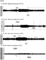
- the carotid bodies are peripheral chemoreceptors that sense changes in arterial blood O 2 , CO 2 and pH levels. Hypoxia, hypercapnia and acidosis are known to activate the CB. Upon sensing changes, the CB modulates the neural activity (i.e. the action potential pattern and frequency) in their sensory nerve, the carotid sinus nerve (CSN). CSN activity is interpreted by the elements of the brain stem that control efferent reflexes including normalization of blood gases via hyperventilation, and the regulation of blood pressure and cardiac performance via sympathetic nervous system (SNS) activation.
- SNS sympathetic nervous system
- CB de-afferentation through carotid sinus nerve denervation reduces the overactive sympathetic activity in spontaneously hypertensive rats ( McBryde et al, Nat Commun. 2013; 4:2395 ).
- US 2014/067003 A1 discloses a system for regulating blood pressure by stimulating an afferent pathway to the brain which produces an efferent output in kidneys.
- the system includes an electrode device adapted for implantation in the cervical region, a stimulator generator, a cable connecting the electrode device and the stimulator generator, wherein the cervical region is generally located between a pair of common carotid arteries, above an aortic arch and in front of cervical vertebrae C2 and C3.
- modulation of neural activity in the CSN can treat conditions associated with impaired glucose control.
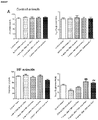
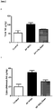
- modulating CSN neural activity restores insulin sensitivity, and also reduces the rate of weight gain and fat accumulation ( Figures 5-7 for T2D and Table 1 for prediabetes) .
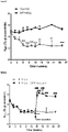
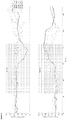
- inhibiting CSN neural activity improves glucose tolerance and insulin sensitivity back towards normal levels ( Figures 8-10 and Figures 20 and 21 ).
- the neural activity in the CSN is notably different to the neural activity inhealthy animals both at baseline and upon sensory changes, particularly the frequency andamplitude of aggregate action potentials ( Figure 11 ).
- This abnormal neural activity associated with the disease state can therefore be modulated in order to provide an effective treatment for the conditions associated with impaired glucose control and/or insulin resistance.
- abnormal neural activity can be a measure of the disease state and may be used in closed loop to control the modulation - for example, detection of abnormal neural activity in the CSN can indicate a disease state, and thereby determine the type and level of modulation of CSN neural activity to treat that disease state.
- Modulation of the neural activity will provide a subtle and versatile mode of treatment without necessarily requiring removal of the CSN. For example, it will allow the titration of treatment in response to disease progression and treatment response. The modulation could also achieve atherapeutic effect whilst maintaining function for other physiological aspects ofthe CSN and carotid body, such as the ability to detect changes in blood gases andthereby ensuring an adequate physiological response to exercise. It is clear that adversely affecting such aspects is not desired in an effective treatment paradigm of metabolic disorders.
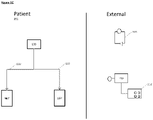
- an apparatus comprising a device for inhibiting the neural activity of a carotid sinus nerve (CSN) or carotid bodyof a subject, the device comprising one or more transducers configured to apply an electrical signal to the CSN or associated carotid body of the subject, whereineach transducer is a cuff electrode, wherein the device optionally comprises at least two such transducers; and a controller coupled to the one or more transducers, the controller controlling the electrical signal to be applied by the one or more transducers, such that the electrical signal inhibits the neural activity of the CSN or carotid body to produce a physiological response in the subject, wherein the physiological response is one or more of the group consisting of: an increase in insulin sensitivity in the subject, an increase in glucose tolerance in the subject, a decrease in plasma glucose concentration in the subject, optionally fasting plasma glucose concentration in the subject, a reduction in subcutaneous fat content in the subject, and a reduction in obesity in the subject, wherein theelectrical signal comprises
- US2014067003A1 relates to a system for regulating blood pressure by stimulating an afferent pathway to the brain which produces an efferent output in kidneys that includes an electrode device adapted for implantation in the cervical region, a stimulator generator, a cable connecting the electrode device and the stimulator generator, wherein the cervical region is generally located between a pair of common carotid arteries, above an aortic arch and in front of cervical vertebrae C2 and C3.
- a method of implantation includes placing the electrode device in the cervical region, selectively energizing the device in accordance with a stimulation scheme, assessing any changes in the patient's blood pressure, selectively energizing the device in accordance with another stimulation scheme, and assessing any changes in the patient's blood, for determining an optical stimulation scheme, wherein stimulation scheme involves parameters including, for example, position, placement and configuration of the electrode device in relation to surrounding tissue and/or organs, selection of electrodes energized, width, frequency and amplitude of stimulation current.
- US2012296389A1 relates to a method and apparatus that resulted in blocking an action potential in a nerve.
- US2013303876A1 relates to planning for and/or assessment of an ablation procedure on one or both carotid bodies or carotid body chemoreceptors or carotid body nerves to treat patients having a sympathetically mediated cardiac, metabolic, and pulmonary disease (e.g. hypertension, CHF, diabetes, sleep disordered breathing) resulting from peripheral chemoreceptor hypersensitivity, carotid body hyperactivity, high carotid body afferent nerve signaling or heightened sympathetic activation.
- a sympathetically mediated cardiac, metabolic, and pulmonary disease e.g. hypertension, CHF, diabetes, sleep disordered breathing
- US2010241188A1 relates to devices, systems and methods for applying electrical impulse(s) to one or more selected nerves in or around the carotid sheath.
- An electrode assembly is introduced through a percutaneous penetration in a patient to a target location adjacent to, or in close proximity with, the carotid sheath. Once in position, one or more electrical impulses are applied through the electrode assembly to one or more selected nerves to stimulate, block or otherwise modulate the nerve(s) and acutely treat the patient's condition.
- US2013237948A1 relates to a method and apparatus for treating a condition associated with impaired blood pressure and/or heart rate in a subject comprising applying an electrical treatment signal, wherein the electrical treatment signal is selected to at least partially block nerve impulses, or in some embodiments, to augment nerve impulses.
- the apparatus provides a first therapy program to provide a downregulating signal to one or more nerves including renal artery, renal nerve, vagus nerve, celiac plexus, a splanchnic nerve, cardiac sympathetic nerves, spinal nerves originating between T10 to L5.
- the apparatus provides a third therapy program to provide an upregulating signal to one or more nerves including a glossopharyngeal nerve and/or a tissue containing baroreceptors.
- WO2015109015A1 relates to an example of a system that may include at least one electrode for placement on tissue in a carotid sinus region and a stimulator.
- the stimulator may be configured to use the at least one electrode to deliver neural stimulation to a baroreceptor region or at least one nerve innervating the baroreceptor region in the carotid sinus region to elicit a baroreflex response, and to deliver a blocking stimulation to a carotid body or at least one nerve innervating the carotid body in the carotid sinus region to inhibit a chemoreceptor response, the stimulator configured to simultaneously deliver the neural stimulation and the blocking stimulation.
- application of a signal may equate to the transfer of energy in a suitable form to carry out the intended effect of the signal. That is, application of a signal to a carotid sinus nerve or carotid body may equate to the transfer of energy to (or from) the carotid sinus nerve or carotid body (as appropriate) to carry out the intended effect. In the present invention, the energy transferred is electrical. It is noted that application of a signal as used herein does not include a pharmaceutical intervention.
- a non-destructive signal is a signal as defined above that, when applied, does not irreversibly damage the underlying neural signal conduction ability of the nerve. That is, application of a non-destructive signal maintains the ability of the CSN (or fibres thereof, or other nerve tissue to which the signal is applied) to conduct action potentials when application of the signal ceases, even if that conduction is in practice inhibited or blocked as a result of application of the non-destructive signal.
- an “impaired glucose control” is taken to mean an inability to maintain blood glucose levels at a normal level (i.e. within normal limits for a healthy individual). As will be appreciated by the skilled person, this will vary based on the type of subject and can be determined by a number of methods well known in the art, for example a glucose tolerance test (GTT) . For example, in humans undergoing an oral glucose tolerance test, a glucose level at 2 hours of less than or equal to 7.8 mmol/L is considered normal. A glucose level at 2 hours of more than 7.8 mmol/L is indicative of impaired glucose control.
- GTT glucose tolerance test
- insulin resistance is given its normal meaning in the art - i.e. in subject or patient exhibiting insulin resistance, the physiological response to insulin in the subject or patient is refractory, such that a higher level of insulin is required in order to control blood glucose levels, compared to the insulin level required in a healthy individual.
- Insulin sensitivity is used herein as the reciprocal to insulin resistance - that is, an increase in insulin sensitivity equates to a decrease in insulin resistance, and vice versa. Insulin resistance may be determined using any method known in the art, for example a GTT, a hyperinsulinaemic clamp or an insulin suppression test.
- Conditions associated with impaired glucose control include those conditions thought to cause the impairment (for example insulin resistance, obesity, metabolic syndrome, Type I diabetes, Hepatitis C infection, acromegaly) and conditions resulting from the impairment (for example obesity, sleep apnoea syndrome, dyslipidaemia, hypertension, Type II diabetes). It will be appreciated that some conditions can be both a cause of and caused by impaired glucose control. Other conditions associated with impaired with glucose control would be appreciated by the skilled person. It will also be appreciated that these conditions may also be associated with insulin resistance.
- the carotid sinus nerve is taken to mean the afferent branch of the glossopharyngeal nerve carrying neural signals from the carotid body to the brain. It includes both the chemoreceptor branch and the baroreceptor branch of the CSN, as well as the trunk of the nerve that carries the nerve fibres from the two aforementioned branches (the carotid sinus nerve is also known as the nerve of Hering or Hering's nerve).
- nerve activity of a nerve is taken to mean the signalling activity of the nerve, for example the amplitude, frequency and/or pattern of action potentials in the nerve.
- pattern as used herein in the context of action potentials in the nerve, is intended to include one or more of: local field potential(s), compound action potential(s), aggregate action potential(s), and also magnitudes, frequencies, areas under the curve and other patterns of action potentials in the nerve or sub-groups (e.g. fascicules) of neurons therein.
- Modulation of neural activity is taken to mean that the signalling activity of the nerve is altered from the baseline neural activity - that is, the signalling activity of the nerve in the subject prior to any intervention. Such modulation may inhibit, block, or otherwise change the neural activity compared to baseline activity.
- such inhibition may be partial inhibition. Partial inhibition may be such that the total signalling activity of the whole nerve is partially reduced, or that the total signalling activity of a subset of nerve fibres of the nerve is fully reduced (i.e. there is no neural activity in that subset of fibres of the nerve), or that the total signalling of a subset of nerve fibres of the nerve is partially reduced compared to baseline neural activity in that subset of fibres of the nerve. Where the modulation of neural activity is inhibition of neural activity, this also encompasses full inhibition of neural activity in the nerve - that is, there is no neural activity in the whole nerve.
- the inhibition of neural activity may be a block of neural activity.
- modulation of neural activity is a block on neural activity
- such blocking may be a partial block, for example a reduction in neural activity of 5%, 10%, 15%, 20%, 25%, 30%, 35%, 40%, 45%, 40%, 50%, 60%, 70%, 80%, 90% or 95%, or blocking of neural activity in a subset of nerve fibres of the nerve.
- blocking may be a full block - i.e. blocking of neural activity in the whole nerve.
- a block on neural activity is understood to be blocking neural activity from continuing past the point of the block. That is, when the block is applied, action potentials may travel along the nerve or subset of nerve fibres to the point of the block, but not beyond the point of the block.
- Modulation of neural activity may also be an alteration in the pattern of action potentials. It will be appreciated that the pattern of action potentials can be modulated without necessarily changing the overall frequency or amplitude. For example, modulation of the neural activity may be such that the pattern of action potentials is altered to more closely resemble a healthy state rather than a disease state.
- Modulation of neural activity may comprise altering the neural activity in various other ways, for example increasing or inhibiting a particular part of the neural activity and/or stimulating new elements of activity, for example in particular intervals of time, in particular frequency bands, according to particular patterns and so forth. Such altering of neural activity may for example represent both increases and/or decreases with respect to the baseline activity.
- Modulation of the neural activity may be temporary.
- temporary is used interchangeably with “reversible”, each being taken to mean that the modulated neural activity (whether that is an inhibition, block or other modulation of neural activity or change in pattern versus baseline activity) is not permanent. That is, upon cessation of the signal, neural activity in the nerve returns substantially towards baseline neural activity within 1-60 seconds, or within 1-60 minutes, or within 1-24 hours, optionally 1-12 hours, optionally 1-6 hours, optionally 1-4 hours, optionally 1-2 hours, or within 1-7 days, optionally 1-4 days, optionally 1-2 days,. In some instances of temporary modulation, the neural activity returns substantially fully to baseline neural activity. That is, the neural activity following cessation of the signal is substantially the same as the neural activity prior to the signal being applied - i.e. prior to modulation.
- Modulation of the neural activity may be persistent.
- “persistent” is taken to mean that the modulated neural activity (whether that is an inhibition, block or other modulation of neural activity or change in pattern versus baseline activity) has a prolonged effect. That is, upon cessation of the signal, neural activity in the nerve remains substantially the same as when the signal was being applied - i.e. the neural activity during and following modulation is substantially the same
- Modulation of the neural activity may be corrective.
- "corrective” is taken to mean that the modulated neural activity (whether that is an inhibition, block or other modulation of neural activity or change in pattern versus baseline activity) alters the neural activity towards the pattern of neaural activity in a healthy individual. That is, upon cessation of the signal, neural activity in the nerve more closely resembles the pattern of action potentials in the CSN observed in a healthy subject than prior to modulation, preferably substantially fully resembles the pattern of action potentials in the CSN observed in a healthy subject.
- Such corrective modulation caused by the signal can be any modulation as defined herein.
- application of the signal may result in a block on neural activity, and upon cessation of the signal, the pattern of action potentials in the nerve resembles the pattern of action potentials observed in a healthy subject.
- application of the signal may result in modulation such that the neural activity resembles the pattern of action potentials observed in a healthy subject, and upon cessation of the signal, the pattern of action potentials in the nerve remains the pattern of action potentials observed in a healthy subject.
- an "improvement in a measurable physiological parameter” is taken to mean that for any given physiological parameter, an improvement is a change in the value of that parameter in the subject towards the normal value or normal range for that value - i.e. towards the expected value in a healthy individual.
- an improvement in a measurable parameter may be: a reduction in sympathetic tone, an increase in insulin sensitivity, an increase in glucose tolerance, a reduction in total fat mass, a reduction in visceral fat mass, a reduction in subcutaneous fat mass, reduction in plasma catecholamines, reduction in urinary metanephrines, and a reduction in glycated haemoglobin (HbA1c), a reduction in circulating triglycerides, assuming the subject is exhibiting abnormal values for the respective parameter.
- the physiological effect may be temporary. That is, upon cessation of the signal, the measured physiological parameter in which an improvement was induced by the signal returns substantially towards baseline neural activity within 1-60 seconds, or within 1-60 minutes, or within 1-24 hours, optionally 1-12 hours, optionally 1-6 hours, optionally 1-4 hours, optionally 1-2 hours, or within 1-7 days, optionally 1-4 days, optionally 1-2 days,. In some instances, the physiological parameter returns substantially fully to baseline neural activity. That is, the value of the physiological parameter following cessation of the signal is substantially the same as the value for the physiological parameter prior to the signal being applied - i.e. prior to modulation.
- the physiological effect may be persistent. That is, upon cessation of the signal, the value of the measureable physiological parameter remains substantially the same as when the signal was being applied - i.e. the value for the physiological parameter during and following modulation is substantially the same
- the physiological effect may be corrective. That is, upon cessation of the signal, the value of the measureable physiological parameter more closely resembles the value for that parameter observed in a healthy subject than prior to modulation, preferably substantially fully resembles the value for that parameter observed in a healthy subject.
- a physiological parameter is not affected by modulation of the neural activity if the parameter does not change as a result of the modulation from the average value of that parameter exhibited by the subject or subject when no intervention has been performed - i.e. it does not depart from the baseline value for that parameter.
- the baseline for any neural activity or physiological parameter in an individual need not be a fixed or specific value, but rather can fluctuate within a normal range or may be an average value with associated error and confidence intervals. Suitable methods for determining baseline values would be well known to the skilled person.
- a measurable physiological parameter is detected in a subject when the value for that parameter exhibited by the subject at the time of detection is determined.
- a detector is any element able to make such a determination.
- a “predefined threshold value” for a physiological parameter is the minimum (or maximum) value for that parameter that must be exhibited by a subject or subject before the specified intervention is applied.
- the threshold value may be defined as a value indicative of a pathological state or a disease state (e.g. sympathetic tone (neural, hemodynamic (e.g. heart rate, blood pressure, heart rate variability) or circulating plasma/urine biomarkers) greater than a threshold sympathetic tone, or greater than a sympathetic tone in a healthy individual, blood insulin levels greater than healthy levels, CSN signalling exhibiting a certain activity level or pattern).
- a pathological state or a disease state e.g. sympathetic tone (neural, hemodynamic (e.g. heart rate, blood pressure, heart rate variability) or circulating plasma/urine biomarkers) greater than a threshold sympathetic tone, or greater than a sympathetic tone in a healthy individual, blood insulin levels greater than healthy levels, CSN signalling exhibiting a certain activity level or pattern).
- the threshold value may be defined as a value indicative of a physiological state of the subject (that the subject is, for example, asleep, post-prandial, or exercising). Appropriate values for any given parameter would be simply determined by the skilled person (for example, with reference to medical standards of practice). Such a threshold value for a given physiological parameter is exceeded if the value exhibited by the subject is beyond the threshold value - that is, the exhibited value is a greater departure from the normal or healthy value for that parameter than the predefined threshold value.
- a “neuromodulation device” or “neuromodulation apparatus” as used herein is a device configured to modulate the neural activity of a nerve.
- Neuromodulation devices or apparatuses as described herein can be comprised of one or more parts.
- the neuromodulation devices or apparatuses comprise at least one transducer capable of effectively applying a signal to a nerve.
- the elements of the device that are to be implanted in the subject are constructed such that they are suitable for such implantation. Such suitable constructions would be well known to the skilled person.
- exemplary fully implantable neuromodulation devices are currently available, such as the vagus nerve stimulator of SetPoint Medical, in clinical development for the treatment of rheumatoid arthritis ( Arthritis & Rheumatism , Volume 64, No. 10 (Supplement), page S195 (Abstract No. 451), October 2012.
- Suitable neuromodulation devices can be fabricated with characteristics as described herein, for example for implantation within the nerve (e.g. intrafascicularly), for partially or wholly surrounding the nerve (e.g. a cuff interface with the nerve).
- implanted is taken to mean positioned within the subject's body. Partial implantation means that only part of the device is implanted - i.e. only part of the device is positioned within the subject's body, with other elements of the device external to the subject's body. For example, the transducer and controller of the device may be wholly implanted within the subject, and an input element may be external to the subject's body. Wholly implanted means that the entire of the device is positioned within the subject's body.
- charge-balanced in relation to a DC current is taken to mean that the positive or negative charge introduced into any system (e.g. a nerve) as a result of a DC current being applied is balanced by the introduction of the opposite charge in order to achieve overall (net) neutrality.
- the carotid bodies are peripheral chemoreceptors that classically respond to hypoxia by increasing chemosensory activity in the carotid sinus nerve (CSN), causing hyperventilation and activation of the sympathoadrenal system. Besides its role in the control of ventilation, the CB has been proposed as a metabolic sensor implicated in the control of energy homeostasis. Recently, the inventors have described that the carotid bodies may also be involved in the etiology of insulin resistance, core metabolic and haemodynamic disturbances of highly prevalent diseases like prediabetes, type 2 diabetes, and obstructive sleep apnoea (Ribeiro et al., 2013) .
- CSN resection in healthy rats prevented the development of insulin resistance and hypertension induced by subsequent hypercaloric diets.
- CSN resection prior to hypercaloric diet also reduced weight gain and avoided visceral fat deposition in this model.
- CB overactivation and increased CSN signalling is associated with the pathogenesis of metabolic and hemodynamic disturbances.
- carotid sinus nerve (CSN) activity is increased in animal models of insulin resistance ( Figure 11 ). Therefore modulation of the neural activity in the CSN will result in treatment of conditions associated with such an impaired glucose control in a subject.
- the apparatus comprises a device for inhibiting the neural activity of a carotid sinus nerve (CSN) of a subject, the device comprising: one or more transducers configured to apply an electrical signal to the CSN or associated carotid body of the subject, wherein each transducer is a cuff electrode, wherein the device optionally comprises at least two such transducers; and a controller coupled to the one or more transducers, the controller controlling the electrical signal to be applied by the one or more transducers, such that the electrical signal inhibits the neural activity of the CSN or carotid body to produce a physiological response in the subject, wherein the physiological response is one or more of the group consisting of: an increase in insulin sensitivity in the subject, an increase in glucose tolerance in the subject, a decrease in plasma glucose concentration in the subject, optionally fasting plasma glucose concentration in the subject, a reduction in subcutaneous fat content in the subject, and a reduction in obesity in the subject, wherein the electrical signal comprises an alternating current (AC) waveform
- AC alternating current
- the signal applied by the one or more transducers is a non-destructive signal.
- each of the one or more transducers may be comprised of one or more electrodes.
- the signal applied by the one or more transducers is an electrical signal, for example a voltage or current.
- the signal applied comprises an alternating current (AC) waveform.
- the electrical signal applied by the one or more transducers has a frequency of 30-50kHz. It is shown herein that electrical signals having a frequency of more than 20kHz are particularly effective at inhibiting (in particular, blocking) neural activity of the CSN. Therefore, in certain embodiments the signal has a frequency of 30kHz, 40kHz or 50kHz.
- the AC waveform may be a square, sinusoidal, triangular or complex waveform.
- the electrical signal is an AC sinusoidal waveform.
- the current amplitude of an applied electrical signal necessary to achieve the intended neuromodulation will depend upon the positioning of the electrode and the associated electrophysiological characteristics (e.g. impedance). It is within the ability of the skilled person to determine the appropriate current amplitude for achieving the intended neuromodulation in a given subject. For example, the skilled person is aware of methods suitable to monitor the neural activity profile induced by neuromodulation.
- the electrical signal has a current of 0.5-5mA, optionally 0.5mA-2mA, optionally 0.5-1.5mA, optionally 1mA or 2mA.
- the signal is an electrical signal comprising an AC sinusoidal waveform having a frequency of 30-50kHz.
- the signal is an electrical signal comprising an AC sinusoidal waveform having a frequency of 30-50kHz having a current of 1mA or 2mA.
- All the transducers are electrodes configured to apply an electrical signal, optionally the same electrical signal.
- the electrode may be a bipolar electrode, or a tripolar electrode.
- the electrode is a cuff electrode.
- the physiological response may be temporary. That is, upon cessation of the signal, the measured physiological parameter in which an improvement was induced by the signal returns substantially towards baseline neural activity within 1-60 seconds, or within 1-60 minutes, or within 1-24 hours, optionally 1-12 hours, optionally 1-6 hours, optionally 1-4 hours, optionally 1-2 hours, or within 1-7 days, optionally 1-4 days, optionally 1-2 days,. In some instances, the physiological parameter returns substantially fully to baseline neural activity. That is, the value of the physiological parameter following cessation of the signal is substantially the same as the value for the physiological parameter prior to the signal being applied - i.e. prior to modulation.
- the physiological response may be persistent. That is, upon cessation of the signal, the value of the measureable physiological parameter remains substantially the same as when the signal was being applied - i.e. the value for the physiological parameter during and following modulation is substantially the same
- the physiological response may be corrective. That is, upon cessation of the signal, the value of the measureable physiological parameter more closely resembles the value for that parameter observed in a healthy subject than prior to modulation, preferably substantially fully resembles the value for that parameter observed in a healthy subject.
- the device further comprises means to detect one or more physiological parameters in the subject.
- a means may be one or more detectors configured to detect the one or more physiological parameters. That is, each detector may detect more than one physiological parameter, for example all the detected physiological parameters. Alternatively, each detector is configured to detect a separate parameter of the one or more physiological parameters detected.
- the controller is coupled to the means to detect one or more physiological parameters, and causes the transducer or transducers to apply the signal when the physiological parameter is detected to be meeting or exceeding a predefined threshold value.
- the one or more detected physiological parameters comprise one or more of the group consisting of: sympathetic tone, plasma insulin concentration, plasma glucose concentration, plasma catecholamine concentration (i.e. one or more of epinephrine, norepinephrine, metanephrine, normetanephrine and dopamine) concentration, tissue catecholamine concentration, plasma HbAlc concentration or plasma triglyceride concentration.
- the one or more detected physiological parameters comprises an action potential or pattern of action potentials in a nerve of the subject, wherein the action potential or pattern of action potentials is associated with the condition associated with an impaired response to glucose that is to be treated.
- the nerve is a sympathetic nerve.
- the nerve is a splanchnic sympathetic nerve.
- the nerve is the peroneal nerve, the sciatic nerve (or one or more branches thereof), or muscle sympathetic nerve terminals.
- the nerve is an afferent nerve involved in metabolic regulation, for example afferent nerves from the liver or from the GI tract.
- the nerve is the CSN.
- the detected pattern of action potentials may be associated with impaired response to glucose or insulin.
- the controller is coupled to a detector or detectors configured to detect the pattern of action potentials in the CSN at the same time as glucose tolerance in the subject.
- the modulation in neural activity as a result of applying the signal is inhibition of neural activity in the CSN. That is, in such embodiments, application of the signal results in the neural activity in at least part of the CSN being reduced compared to the baseline neural activity in that part of the nerve. Such a reduction in activity could equally be across the whole nerve, in which case neural activity would be reduced across the whole nerve. Therefore, in certain such embodiments, a result of applying the signal is at least partial inhibition of neural activity in the CSN. In certain such embodiments a result of applying the signal is at least partial inhibition of neural activity in the chemoreceptor branch of the CSN. In certain such embodiments, a result of applying the signal is full inhibition of neural activity in the chemoreceptor branch of the CSN. In certain embodiments, a result of applying the signal is full inhibition of neural activity in the CSN.
- the modulation in neural activity as a result of applying the signal is a block on neural activity in the CSN. That is, in such embodiments, the application of the signal blocks action potentials from travelling beyond the point of the block in at least a part of the CSN.
- the modulation is a partial block. In certain alternative embodiments, the modulation is a full block. In a preferred embodiment, the modulation is a partial or full block of neural activity in the CSN.
- the modulation in neural activity as a result of applying the signal is an alteration to the pattern of action potentials in the CSN.
- the neural activity is modulated such that the resultant pattern of action potentials in the CSN resembles the pattern of action potentials in the CSN observed in a healthy subject.
- Modulation of neural activity may comprise altering the neural activity in various other ways, for example increasing or inhibiting a particular part of the activity and stimulating new elements of activity, for example in particular intervals of time, in particular frequency bands, according to particular patterns and so forth. Such altering of neural activity may for example represent both increases and/or decreases with respect to the baseline activity.
- the signal is applied intermittently.
- the signal is applied continuously for at least 5 days, optionally at least 7 days, before ceasing. That is, for such intermittent application of the signal, the signal is applied continuously for at least 5 days (optionally 7 days), then application ceases for a period (e.g. 1 day, 2 days, 3 days, 1 week, 2 weeks, 1 month) before the signal is again applied continuously for at least 5 days (optionally 7 days).
- the signal is applied intermittently
- the signal is applied for a first time period, then stopped for a second time period, then reapplied for a third time period, then stopped for a fourth time period.
- the first, second, third and fourth periods run sequentially and consecutively.
- the series of first, second, third and fourth periods amounts to one application cycle.
- multiple application cycles can run consecutively such that the signal is applied in phases, between which phases no signal is applied.
- the duration of the first, second, third and fourth time periods is independently selected. That is, the duration of each time period may be the same or different to any of the other time periods.
- the duration of each of the first, second, third and fourth time periods may be any time from 1 second (s) to 10 days (d), 2s to 7d, 3s to 4d, 5s to 24 hours (24h), 30s to 12 h, 1 min to 12 h, 5 min to 8 h, 5 min to 6 h, 10 min to 6 h, 10 min to 4 h, 30 min to 4 h, 1 h to 4 h.
- the duration of each of the first, second, third and fourth time periods is 5s, 10s, 30s, 60s, 2 min, 5 min, 10 min, 20 min, 30 min, 40 min, 50 min, 60 min, 90 min, 2 h, 3 h, 4 h, 5 h, 6 h, 7 h, 8 h, 9 h, 10 h, 11 h, 12 h, 13 h, 14 h, 15 h, 16 h, 17 h, 18 h, 19 h, 20 h, 21 h, 22 h, 23 h, 24 h, 2d, 3d, 4d, 5d, 6d, 7d.
- the signal is applied for a specific amount of time per day.
- the signal is applied for 10 min, 20 min, 30 min, 40 min, 50 min, 60 min, 90 min, 2 h, 3 h, 4 h, 5 h, 6 h, 7 h, 8 h, 9 h, 10 h, 11 h, 12 h, 13 h, 14 h, 15 h, 16 h, 17 h, 18 h, 19 h, 20 h, 21 h, 22 h, 23 h per day.
- the signal is applied continuously for the specified amount of time.
- the signal may be applied discontinuously across the day, provided the total time of application amounts to the specified time.
- the controller causes the signal to be applied intermittently, the signal is applied only when the subject is in a specific state. In certain such embodiments, the controller causes the signal to be applied only when the subject is awake. In certain alternative embodiments, the controller causes the signal to be applied only when the subject is asleep. In certain embodiments, the controller causes the signal to be applied prior to and/or after the ingestion of food. In certain embodiments, the controller causes the signal to be applied prior to and/or after the subject undertakes exercise.
- the device further comprises an input means.
- the status of the subject i.e. whether the subject is awake, asleep, pre- or post-eating, or pre- or post-taking exercise
- the device further comprises a detector configured to detect the status of the subject, wherein the signal is applied only when the detector detects that the subject is in the specific state.
- the controller causes the signal to be continuously applied to the CSN and/or carotid body. It will be appreciated that in embodiments wherein the signal is a series of pulses, gaps between pulses do not mean the signal is not continuously applied. Such continuous application may continue indefinitely, e.g. permanently. Alternatively, the continuous application may be for a minimum period, for example the signal may be continuously applied for at least 5 days, or at least 7 days.
- the inhibition in neural activity caused by the application of the signal is temporary/reversible. That is, upon cessation of the signal, neural activity in the nerve returns substantially towards baseline neural activity within 1-60 seconds, or within 1-60 minutes, or within 1-24 hours, optionally 1-12 hours, optionally 1-6 hours, optionally 1-4 hours, optionally 1-2 hours, or within 1-7 days, optionally 1-4 days, optionally 1-2 days,. In certain such embodiments, the neural activity returns substantially fully to baseline neural activity. That is, the neural activity following cessation of the signal is substantially the same as the neural activity prior to the signal being applied - i.e. prior to modulation.
- the inhibition in neural activity caused by the application of the signal is substantially persistent. That is, upon cessation of the signal, neural activity in the nerve remains substantially the same as when the signal was being applied - i.e. the neural activity during and following modulation is substantially the same.
- the inhibition in neural activity caused by the application of the signal is partially corrective, preferably substantially corrective. That is, upon cessation of the signal, neural activity in the nerve more closely resembles the pattern of action potentials in the CSN observed in a healthy subject than prior to modulation, preferably substantially fully resembles the pattern of action potentials in the CSN observed in a healthy subject.
- the modulation caused by the signal can be any modulation as defined herein.
- application of the signal may result in a block on neural activity, and upon cessation of the signal, the pattern of action potentials in the nerve resembles the pattern of action potentials observed in a healthy subject.
- application of the signal may result modulation such that the neural activity resembles the pattern of action potentials observed in a healthy subject, and upon cessation of the signal, the pattern of action potentials in the nerve resembles the pattern of action potentials observed in a healthy subject. It is hypothesised that such a corrective effect is the result of a positive feedback loop - that is, the underlying disease state is treated as result of the claimed methods, and therefore the chemosensory signals along the CSN are not abnormal, and therefore the disease state is not perpetuated by the abnormal CSN neural activity.
- the device is suitable for at least partial implantation into the subject such that at least a portion of the device sits within the body, preferably in proximity to the CSN or carotid body to which the signal is to be applied.
- parts of the device for example the transducer and the controller, may be suitable to be wholly implanted in the subject such that the signal can be applied to the CSN or carotid body, and other parts of the device may be external to the body, for example an input element or remote charging element.
- the device is suitable to be wholly implanted in the subject.
- the device further comprises one or more of a power supply element, for example a battery, and/or one or more communication elements.
- a power supply element for example a battery
- communication elements for example a Wi-Fi connection
- Disclosed herein is a method for treating a condition associated with impaired glucose control in a subject, the method comprising implanting a device according to the first aspect, positioning at least one transducer of the device in signalling contact with a CSN and/or carotid body of the subject, and activating the device.
- Also disclosed herein is a method of inhibiting neural signalling in the CSN of a subject comprising implanting in the subject a device according to the first aspect, positioning at least one transducer of the apparatus in signalling contact with a CSN or carotid body of the subject and activating the apparatus.
- the inhibition of neural signalling in the CSN improves glucose control in the subject.
- the transducer is in signalling contact with the CSN or carotid body when it is positioned such that the signal can be effectively applied to the CSN or carotid body.
- the device is activated when the device is in an operating state such that the signal will be applied as determined by the controller.
- the method is applied bilaterally. That is, in such embodiments, a first transducer is positioned in signalling contact with the left carotid sinus nerve (CSN) and/or left carotid body of said subject to modulate the neural activity of the left CSN in the subject, and a second transducer is positioned in signalling contact with the right carotid sinus nerve (CSN) and/or right carotid body of said subject to modulate the neural activity of the right CSN in the subject.
- the first and second transducers are part of one device according to the third method disclosed herein. In alternative such embodiments, the first and second transducers are part of separate devices according to the third method disclosed herein.
- Figures 1A-1C show how the invention may be put into effect using one or more neuromodulation devices which are implanted in, located on, or otherwise disposed with respect to a subject 200 in order to carry out any of the various methods described herein.
- one or more neuromodulation devices can be used to treat a condition associated with impaired glucose control in a subject, by modulating carotid sinus nerve afferent neural activity.
- a separate neuromodulation device 100', 100" is provided in respect of each of the left and right carotid sinus nerves, although as discussed above a device could be provided or used in respect of only one of the left and right nerves.
- Each such neuromodulation device may be fully or partially implanted in the subject, or otherwise located, so as to provide neuromodulation of the respective carotid sinus nerve, carotid sinus body, or both.
- Figure 1A also shows schematically components of one of the neuromodulation devices 100, in which the device comprises several elements, components or functions grouped together in a single unit and implanted in the subject 200.
- a first such element is a transducer 102 which is shown in proximity to a carotid sinus nerve 90 of the subject.
- the transducer 102 may be operated by a controller element 104.
- the device may comprise one or more further elements such as a communication element 106, a detector element 108, a power supply element 110 and so forth.
- Each of the left and right neuromodulation devices 100', 100" ' may operate independently, or may operate in communication with each other, for example using respective communication elements 106.
- Each neuromodulation device 100', 100" may carry out the required neuromodulation independently, or in response to one or more control signals.
- a control signal may be provided by the controller 104 according to an algorithm, in response to output of one or more detector elements 108, and/or in response to communications from one or more external sources received using the communications element.
- the detector element(s) could be responsive to a variety of different physiological parameters.
- Figure 1B illustrates some ways in which the device of Figure 1A may be differently distributed.
- the neuromodulation devices 100', 100" comprise transducers 102 implanted proximally to the carotid sinus nerves 90 or bodies, but other elements such as a controller 104, a communication element 106 and a power supply 110 are implemented in a separate control unit 30 which may also be implanted in, or carried by the subject.
- the control unit 130 then controls the transducers in both of the neuromodulation devices via connections 132 which may for example comprise electrical wires and/or optical fibres for delivering signals and/or power to the transducers.
- one or more detectors 108 are located separately from the control unit, although one or more such detectors could also or instead be located within the control unit 130 and/or in one or both of the neuromodulation devices 100', 100".
- the detectors may be used to detect one or more physiological parameters of the subject, and the controller element or control unit then causes the transducers to apply the signal in response to the detected parameter(s), for example only when a detected physiological parameter meets or exceeds a predefined threshold value.
- Physiological parameters which could be detected for such purposes include sympathetic tone, plasma insulin concentration, insulin sensitivity, plasma glucose concentration, glucose tolerance, plasma catecholamine concentration, tissue catecholamine concentration, plasma HbAlc concentration and plasma triglyceride concentration.
- a detected physiological parameter could be an action potential or pattern of action potentials in a nerve of the subject, for example an efferent or more particularly a sympathetic nerve, wherein the action potential or pattern of action potentials is associated with the condition to be treated.
- neural activity in the CSN is increased in animals in a prediabetic state, and thus, in one embodiment, the or each detector 108 may be located on or proximal to the CSN, such as to detect the action potential or pattern of action potentials in the CSN, as indicative of a disease state.
- detector 108 may be implanted unilaterally on or proximal to one (i.e.
- the CSN or, analogously, the CB, or the branch of the glossopharyngeal nerve to the CSN/CB, or the chemosensory branch of the CSN
- the or each transducer 102 may be implanted on or proximally to the other of the CSN (or analogous nerves).
- Figure 1C illustrates some ways in which some functionality of the device of Figures 1A or 1B is provided not implanted in the subject.
- an external power supply 140 is provided which can provide power to implanted elements of the device in ways familiar to the skilled person, and an external controller 150 provides part or all of the functionality of the controller 104, and/or provides other aspects of control of the device, and/or provides data readout from the device, and/or provides a data input facility 152.
- the data input facility could be used by a subject or other operator in various ways, for example to input data relating to the subject's current or expected activities such as sleep, eating, or physical exertion.
- Each neuromodulation device may be adapted to carry out the neuromodulation required using one or more physical modes of operation which typically involve applying a signal to a carotid body or sinus nerve, such a signal typically involving a transfer of energy to (or from) the body or nerve.
- modes may comprise modulating the carotid sinus nerve or body using an electrical signal.
- Such a signal may be a non-destructive signal.
- Such modulation may comprise increasing, inhibiting, or otherwise changing the pattern of neural activity in the nerve or body.
- the transducer 90 illustrated in Figure 1A could be comprised of one or more electrodes arranged to put the required neuromodulation into effect.
- the neural modulation device(s) or apparatus may be arranged to inhibit neural activity of a carotid sinus nerve or carotid body by using the transducer(s) to apply an electrical signal, for example a voltage or current, for example an AC waveform
- an electrical signal for example a voltage or current, for example an AC waveform
- the transducers configured to apply the electrical signal are electrodes.
- the electrical signal applied by the one or more transducers has a frequency of 30-50kHz. In certain embodiments the signal has a frequency of 30kHz, 40kHz or 50kHz.
- the AC waveform may be a square, sinusoidal, triangular or complex waveform.
- the electrical signal is an AC sinusoidal waveform.
- the current amplitude of an applied electrical signal necessary to achieve the intended neuromodulation will depend upon the positioning of the electrode and the associated electrophysiological characteristics (e.g. impedance). It is within the ability of the skilled person to determine the appropriate current amplitude for achieving the intended neuromodulation in a given subject. For example, the skilled person is aware of methods suitable to monitor the neural activity profile induced by neuromodulation.
- the electrical signal has a current of 0.1-10mA, optionally 0.5-5mA, optionally 1mA-2mA, optionally 1mA or 2mA.
- the signal is an electrical signal comprising an AC sinusoidal waveform having a frequency of 30-50kHz.
- the electrode may be a bipolar electrode, or a tripolar electrode.
- the electrode is a cuff electrode.
- Thermal methods of neuromodulation typically manipulate the temperature of a nerve to inhibit signal propagation.
- Patberg et al. Blocking of impulse conduction in peripheral nerves by local cooling as a routine in animal experimentation. Journal of Neuroscience Methods 1984;10:267-75 ) discuss how cooling a nerve blocks signal conduction without an onset response, the block being both reversible and fast acting, with onsets of up to tens of seconds.
- Heating the nerve can also be used to block conduction, and is generally easier to implement in a small implantable or localised transducer or device, for example using infrared radiation from laser diode or a thermal heat source such as an electrically resistive element, which can be used to provide a fast, reversible, and spatially very localised heating effect (see for example Duke et al. J Neural Eng. 2012 Jun;9(3):036003 . Spatial and temporal variability in response to hybrid electro-optical stimulation). Either heating, or cooling, or both could be provided using a Peltier element.
- Optogenetics is a technique that genetically modifies cells to express photosensitive features, which can then be activated with light to modulate cell function. Many different optogenetic tools have been developed that can be used to inhibit neural firing. A list of optogenetic tools to suppress neural activity has been compiled (Epilepsia. 2014 Oct 9. doi: 10.1111/epi.12804. WONOEP appraisal: Optogenetic tools to suppress seizures and explore the mechanisms of epileptogenesis. Ritter LM et al.). Acrylamine-azobenzene-quaternary ammonium (AAQ) is a photochromic ligand that blocks many types of K+ channels and in the cis configuration, the relief of K+ channel block inhibits firing ( Nat Neurosci. 2013 Jul;16(7):816-23. doi: 10.1038/nn.3424 . Optogenetic pharmacology for control of native neuronal signaling proteins. Kramer RHet al.).
- Mechanical forms of neuromodulation can include the use of ultrasound which may conveniently be implemented using external instead of implanted ultrasound transducers.
- Other forms of mechanical neuromodulation include the use of pressure (for example see “ The effects of compression upon conduction in myelinated axons of the isolated frog sciatic nerve” by Robert Fern and P. J. Harrison Br.j. Anaesth. (1975), 47, 1123 ).
- Some other electrical forms of neuromodulation may use alternating current (AC) waveforms applied to a nerve using one or more electrodes.
- AC alternating current
- Some other AC techniques include HFAC or KHFAC (high-frequency or kilohertz frequency) to provide a reversible block (for example see Kilgore and Badra, 2004, Medical and Biological Engineering and Computing, May;42(3):394-406 . Nerve conduction block utilising high-frequency alternating current. Kilgore KL, Bhadra N.). In the work of Kilgore and Bhadra, a proposed waveform was sinusoidal or rectangular at 3-5 kHz, and typical signal amplitudes that produced block were 3 - 5 Volts or 0.5 to 2.0 milli Amperes peak to peak.
- HFAC may typically be applied at a frequency of between 1 and 50 kHz at a duty cycle of 100% ( Bhadra, N. et al., Journal of Computational Neuroscience, 2007, 22(3), pp 313-326 ).
- Methods for selectively blocking activity of a nerve by application of a waveform having a frequency of 5 - 10 kHz are described in US 7,389,145 .
- US 8,731,676 describes a method of ameliorating sensory nerve pain by applying a 5-50 kHz frequency waveform to a nerve.
- Some commercially available nerve blocking systems include the Maestro (RTM) system available from Enteromedics Inc. of Minnesota, USA. Similar neuromodulation devices are more generally discussed in US2014/214129 and elsewhere.
- Also disclosed herein is a method of treating a condition associated with impaired glucose control in a subject, the method comprising applying a signal to a carotid sinus nerve (CSN) and/or a carotid body of said subject to modulate the neural activity of a CSN in the subject.
- CSN carotid sinus nerve
- this can be accomplished by the signal being applied by a neuromodulation device comprising one or more transducers for applying the signal.
- the neuromodulation device is at least partially implanted in the subject.
- the neuromodulation device is wholly implanted in the subject.
- each individual mammalian subject has a left and a right carotid body, each carotid body having an associated CSN.
- the CSN carries afferent nerve signals from the carotid body to the brain. Therefore, in methods according to the disclosure, the signal can be applied directly to a part of or all of one or both CSNs to modulate the neural activity in that or those CSNs.
- the signal can be applied to a part of or all of one or both carotid bodies associated with the CSNs in order to modulate the afferent nerve signals carried from the carotid body or bodies to the glossopharyngeal nerve and the brain stem.
- the signal can be applied to a part or all of both the carotid body and the associated CSN, unilaterally or bilaterally.
- the condition associated with impaired glucose control is a condition associated with insulin resistance.
- conditions associated with impaired glucose control include metabolic syndrome, type 2 diabetes, obesity, hypertension, dyslipidaemia, sleep apnoea syndrome and other metabolic disorders.
- the condition treated by the methods is at least one of the group consisting of metabolic syndrome, type 2 diabetes, obesity, and dyslipidaemia. The skilled person will appreciate that any one subject can exhibit one or more conditions associated with impaired glucose control and that the method can be used to treat one or more or all of those conditions.
- treatment of the condition is indicated by an improvement in a measurable physiological parameter, for example reduction in sympathetic tone, increase in insulin sensitivity, increase in glucose tolerance, a reduction in total fat mass, a reduction in visceral fat mass, a reduction in subcutaneous fat mass, reduction in plasma catecholamines, reduction in tissue catecholamines, reduction in urinary metanephrines, a reduction in glycated haemoglobin (HbA1c ) or a reduction in circulating triglyceride concentration.
- a measurable physiological parameter for example reduction in sympathetic tone, increase in insulin sensitivity, increase in glucose tolerance, a reduction in total fat mass, a reduction in visceral fat mass, a reduction in subcutaneous fat mass, reduction in plasma catecholamines, reduction in tissue catecholamines, reduction in urinary metanephrines, a reduction in glycated haemoglobin (HbA1c ) or a reduction in circulating triglyceride concentration.
- the measurable physiological parameter is at least one of the group consisting of: sympathetic tone, insulin sensitivity, glucose sensitivity, total fat mass, visceral fat mass, subcutaneous fat mass plasma catecholamines content, tissue catecholamines content urinary metanephrines content, and levels of glycated haemoglobin (HbA1c).
- sympathetic tone is understood to be the neural activity in sympathetic nerves and/or associated sympathetic neurotransmitter measured in systemic or local tissue compartments in the sympathetic nervous system.
- Suitable methods for determining the value for any given parameter would be appreciated by the skilled person.
- an increase in heart rate and/or blood pressure for a period at least 24hrs is typically indicative of an increased sympathetic tone, as is aberrant heart rate variablility, cardiac or renal norepinephrine spillover, skin or muscle microneurography and plasma/urine norepinephrine
- insulin sensitivity can be measured by the HOMA index or by a hyperinsulinemic clamp.
- total fat mass may be determined by bioimpedence.
- visceral fat can be indirectly determined by measuring abdominal perimeter. Further suitable methods for determining the value for any given parameter would be appreciated by the skilled person.
- treatment of the condition is indicated by an improvement in the profile of neural activity in the CSN. That is, treatment of the condition is indicated by the neural activity in the CSN approaching the neural activity in a healthy individual.
- the physiological response may be temporary. That is, upon cessation of the signal, the measured physiological parameter in which an improvement was induced by the signal returns substantially towards baseline neural activity within 1-60 seconds, or within 1-60 minutes, or within 1-24 hours, optionally 1-12 hours, optionally 1-6 hours, optionally 1-4 hours, optionally 1-2 hours, or within 1-7 days, optionally 1-4 days, optionally 1-2 days,. In some instances, the physiological parameter returns substantially fully to baseline neural activity. That is, the value of the physiological parameter following cessation of the signal is substantially the same as the value for the physiological parameter prior to the signal being applied - i.e. prior to modulation.
- the physiological response may be persistent. That is, upon cessation of the signal, the value of the measureable physiological parameter remains substantially the same as when the signal was being applied - i.e. the value for the physiological parameter during and following modulation is substantially the same
- the physiological response may be corrective. That is, upon cessation of the signal, the value of the measureable physiological parameter more closely resembles the value for that parameter observed in a healthy subject than prior to modulation, preferably substantially fully resembles the value for that parameter observed in a healthy subject.
- the method does not affect the cardiopulmonary regulation function of the carotid body and CSN.
- the method does not affect one or more physiological parameters in the subject selected from the group consisting of: pO2, pCO2, blood pressure, oxygen demand and cardiorespiratory responses to exercise and altitude. Suitable methods for determining the value for any given parameter would be appreciated by the skilled person.
- a result of applying the signal is at least partial inhibition of neural activity in the CSN.
- a result of applying the signal is at least partial inhibition of neural activity in the chemoreceptor branch of the CSN.
- a result of applying the signal is full inhibition of neural activity in the chemoreceptor branch of the CSN.
- a result of applying the signal is full inhibition of neural activity in the CSN.
- the modulation in neural activity as a result of applying the signal is inhibition of neural activity from the carotid sinus/carotid body to the glossopharyngeal nerve and the brain stem, such that neural activity which is associated with the CSN/CB in the CSN, the glossopharyngeal nerve or the brain stem is reduced compared to pre-treatment neural activity associated with the carotid sinus/carotid body in that part of the nerve.
- the modulation in neural activity as a result of applying the signal is a block on neural activity in the CSN. That is, in such embodiments, the application of the signal blocks action potentials from travelling beyond the point of the block in at least a part of the CSN. In certain such embodiments, the modulation is a partial block. In certain alternative embodiments, the modulation is a full block.
- the modulation in neural activity as a result of applying the signal is an alteration to the pattern of action potentials in the CSN.
- the neural activity is modulated such that the resultant pattern of action potentials in the CSN resembles the pattern of action potentials in the CSN observed in a healthy subject.
- the signal is applied intermittently.
- the signal is applied continuously for at least 5 days, optionally at least 7 days, before ceasing. That is, for such intermittent application of the signal, the signal is applied continuously for at least 5 days (optionally 7 days), then application ceases for a period (e.g. 1 day, 2 days, 3 days, 1 week, 2 weeks, 1 month) before the signal is again applied continuously for at least 5 days (optionally 7 days).
- the signal is applied intermittently
- the signal is applied for a first time period, then stopped for a second time period, then reapplied for a third time period, then stopped for a fourth time period.
- the first, second, third and fourth periods run sequentially and consecutively.
- the series of first, second, third and fourth periods amounts to one application cycle.
- multiple application cycles can run consecutively such that the signal is applied in phases, between which phases no signal is applied.
- the duration of the first, second, third and fourth time periods is independently selected. That is, the duration of each time period may be the same or different to any of the other time periods.
- the duration of each of the first, second, third and fourth time periods may be any time from 1 second (s) to 10 days (d), 2s to 7d, 3s to 4d, 5s to 24 hours (24h), 30s to 12 h, 1 min to 12 h, 5 min to 8 h, 5 min to 6 h, 10 min to 6 h, 10 min to 4 h, 30 min to 4 h, 1 h to 4 h.
- the duration of each of the first, second, third and fourth time periods is 5s, 10s, 30s, 60s, 2 min, 5 min, 10 min, 20 min, 30 min, 40 min, 50 min, 60 min, 90 min, 2 h, 3 h, 4 h, 5 h, 6 h, 7 h, 8 h, 9 h, 10 h, 11 h, 12 h, 13 h, 14 h, 15 h, 16 h, 17 h, 18 h, 19 h, 20 h, 21 h, 22 h, 23 h, 24 h, 2d, 3d, 4d, 5d, 6d, 7d.
- the signal is applied for a specific amount of time per day.
- the signal is applied for 10 min, 20 min, 30 min, 40 min, 50 min, 60 min, 90 min, 2 h, 3 h, 4 h, 5 h, 6 h, 7 h, 8 h, 9 h, 10 h, 11 h, 12 h, 13 h, 14 h, 15 h, 16 h, 17 h, 18 h, 19 h, 20 h, 21 h, 22 h, 23 h per day.
- the signal is applied continuously for the specified amount of time.
- the signal may be applied discontinuously across the day, provided the total time of application amounts to the specified time.
- the signal is applied intermittently, the signal is applied only when the subject is in a specific state. In certain such embodiments, the signal is applied only when the subject is awake. In certain alternative embodiments, the signal is applied only when the subject is asleep. In certain embodiments, the signal is applied prior to and/or after the ingestion of food. In certain embodiments, the signal is applied prior to and/or after the subject undertakes exercise. In such embodiments, the status of the subject (i.e. whether the subject is awake, asleep, pre- or post-eating, or pre- or post-taking exercise) can be indicated by the subject. In alternative such embodiments, the status of the subject can be detected independently from any input from the subject. In certain embodiments in which the signal is applied by a neuromodulation device, the device further comprises a detector configured to detect the status of the subject, wherein the signal is applied only when the detector detects that the subject is in the specific state.
- the controller causes the signal to be continuously applied to the CSN and/or carotid body. It will be appreciated that in embodiments wherein the signal is a series of pulses, gaps between pulses do not mean the signal is not continuously applied. Such continuous application may continue indefinitely, e.g. permanently. Alternatively, the continuous application may be for a minimum period, for example the signal may be continuously applied for at least 5 days, or at least 7 days.
- the modulation in neural activity caused by the application of the signal is temporary/reversible. That is, upon cessation of the signal, neural activity in the nerve returns substantially towards baseline neural activity within 1-60 seconds, or within 1-60 minutes, or within 1-7 days, optionally 1-4 days, optionally 1-2 days, or within 1-24 hours, optionally 1-12 hours, optionally 1-6 hours, optionally 1-4 hours, optionally 1-2 hours. In certain such embodiments, the neural activity returns substantially fully to baseline neural activity. That is, the neural activity following cessation of the signal is substantially the same as the neural activity prior to the signal being applied - i.e. prior to modulation.
- the modulation in neural activity caused by the application of the signal is substantially persistent. That is, upon cessation of the signal, neural activity in the nerve remains substantially the same as when the signal was being applied - i.e. the neural activity during and following modulation is substantially the same.
- the modulation in neural activity caused by the application of the signal is partially corrective, preferably substantially corrective. That is, upon cessation of the signal, neural activity in the nerve more closely resembles the pattern of action potentials in the CSN observed in a healthy subject than prior to modulation, preferably substantially fully resembles the pattern of action potentials in the CSN observed in a healthy subject.
- the modulation caused by the signal can be any modulation as defined herein.
- application of the signal may result in a block on neural activity, and upon cessation of the signal, the pattern of action potentials in the nerve resembles the pattern of action potentials observed in a healthy subject.
- application of the signal may result modulation such that the neural activity resembles the pattern of action potentials observed in a healthy subject, and upon cessation of the signal, the pattern of action potentials in the nerve resembles the pattern of action potentials observed in a healthy subject. It is hypothesised that such a corrective effect is the result of a positive feedback loop - that is, the underlying disease state is treated as result of the claimed methods, and therefore the chemosensory signals along the CSN are not abnormal, and therefore the disease state is not perpetuated by the abnormal CSN neural activity.
- the method further comprises the step of detecting one or more physiological parameters of the subject, wherein the signal is applied only when the detected physiological parameter meets or exceeds a predefined threshold value.
- the signal may be applied when any one of the detected parameters meets or exceeds its threshold value, alternatively only when all of the detected parameters meet or exceed their threshold values.
- the device further comprises at least one detector configured to detect the one or more physiological parameters.
- the one or more detected physiological parameters are one or more of the group consisting of: sympathetic tone, plasma insulin concentration, insulin sensitivity, plasma glucose concentration, glucose tolerance, total fat mass, visceral fat mass, plasma catecholamines (i.e. one or more of epinephrine, norepinephrine, metanephrine, normetanephrine and dopamine) content, tissue catecholamines content urinary metanephrines content, plasma HbAlc content and a reduction in circulating triglyceride concentration.
- sympathetic tone i.epinephrine, norepinephrine, metanephrine, normetanephrine and dopamine
- a typical HbAlc content in a healthy human subject would be between 20-42 mmol/mol (4-6% of total Hb).
- An HbAlc content exceeding 42mmol/mol may be indicative of a diabetic state.
- the one or more detected physiological parameters are one or more of the group consisting of: sympathetic tone, plasma insulin concentration, plasma glucose concentration, plasma catecholamines (i.e. one or more of epinephrine, norepinephrine, metanephrine, normetanephrine) concentration, tissue catecholamines, and plasma HbAlc content.
- the detected physiological parameter is an action potential or pattern of action potentials in a nerve of the subject, wherein the action potential or pattern of action potentials is associated with the condition associated with an impaired response to glucose that is to be treated.
- the nerve is a sympathetic nerve.
- the nerve is an afferent sympathetic nerve.
- the nerve is the CSN.
- the detected pattern of action potentials may be associated with impaired response to glucose.
- the nerve is an afferent nerve involved in metabolic regulation, for example afferent nerves from the liver or from the GI tract.
- the nerve is an efferent sympathetic nerve, optionally the peroneal nerve, the sciatic nerve (or one or more branches thereof), or muscle sympathetic nerve terminals.
- the nerve is the sciatic nerve.
- the nerve is the renal nerve.
- any two or more of the indicated physiological parameters may be detected in parallel or consecutively.
- the pattern of action potentials in the CSN can be detected at the same time as glucose tolerance.
- the signal may be applied intermittently or permanently, as described in the embodiments above.
- a signal is applied to a carotid sinus nerve (CSN) and/or a carotid body of said subject to modulate the neural activity of a CSN in the subject. That is, the signal is applied at least unilaterally to achieve at least unilateral (i.e. right or left side) modulation of neural activity in the respective CSN.
- CSN carotid sinus nerve
- Figure 9 it is clear that stopping neural signalling in both CSNs (i.e. bilateral modulation) produces a greater effect that is longer-lasting.
- Figures 20 and 21 show that bilateral block of CSN neural activity using an electrical signal is effective at restoring glucose tolerance and insulin sensitivity. A greater improvement is observed the longer the block is applied ( Figure 20 ).
- the neural activity in both CSNs is modulated (i.e. the modulation is bilateral) . That is, in certain preferred embodiments, a signal is applied to the left carotid sinus nerve (CSN) and/or left carotid body of the subject and a signal is applied to the right carotid sinus nerve (CSN) and/or right carotid body of the subject to modulate the neural activity of the left CSN and right CSN of the subject.
- the signal applied to each CSN and/or carotid body, and therefore the type and extent of modulation is independently selected from that applied to the opposite CSN and/or carotid body.
- the signal applied to the right CSN and/or carotid body is the same as the signal applied to the left CSN and/or carotid body. In certain alternative embodiments the signal applied to the right CSN and/or carotid body is different to the signal applied to the left CSN and/or carotid body.
- a first signal is applied to the left carotid sinus nerve (CSN) and/or left carotid body of the subject to modulate the neural activity of the left CSN in the subject by a neuromodulation device comprising one or more transducers for applying the signal
- a second signal is applied to the right carotid sinus nerve (CSN) and/or right carotid body of said subject to modulate the neural activity of the right CSN in the subject by a neuromodulation device comprising one or more transducers for applying the signal.
- the first signal and the second signal are applied by the same neuromodulation device, that device have at least two transducers, one to apply the first signal and one to apply the second signal.
- the first and second signals are applied by separate neuromodulation devices.
- the signal applied is a non-destructive signal.
- the transducer may be comprised of one or more electrodes.
- the signal is an electrical signal, for example a voltage or a current, and comprises one or more AC waveforms.
- the electrical signal is an AC waveform having a frequency of 30-50 kHz.
- electrical signals having a frequency of more than 20kHz are particularly effective at inhibiting (in particular, blocking) neural activity of the CSN. Therefore, in certain embodiments the signal has a frequency of 30kHz, 40kHz or 50kHz.
- the AC waveform may be a square, sinusoidal, triangular or complex waveform.
- the electrical signal is an AC sinusoidal waveform.
- the current amplitude of an applied electrical signal necessary to achieve the intended neuromodulation will depend upon the positioning of the electrode and the associated electrophysiological characteristics (e.g. impedance). It is within the ability of the skilled person to determine the appropriate current amplitude for achieving the intended neuromodulation in a given subject. For example, the skilled person is aware of methods suitable to monitor the neural activity profile induced by neuromodulation.
- the electrical signal has a current of 0.5-5mA, optionally 0.5mA-2mA, optionally 0.5-1.5mA, optionally 1mA or 2mA.
- the signal is an electrical signal comprising an AC sinusoidal waveform having a frequency of 30-50kHz.
- the signal is an electrical signal comprising an AC sinusoidal waveform having a frequency of 30-50kHz having a current of 1mA or 2mA.
- each transducer configured to apply the signal is an electrode configured to apply the electrical signal.
- all the transducers are electrodes configured to apply an electrical signal, optionally the same electrical signal.
- the electrode may be a bipolar electrode, or a tripolar electrode.
- the electrode is a cuff electrode.
- a neuromodulatory electrical waveform for use in treating insulin resistance in a subject, wherein the waveform is a kiloHertz alternating current (AC) waveform having a frequency of 30-50kHz, such that, when applied to a carotid sinus nerve (CSN) of the subject, the waveform inhibits neural signalling in the CSN.
- AC kiloHertz alternating current
- the waveform when applied to the CSN, improves the subject's response to insulin.
- a neuromodulation device for treating a condition associated with impaired glucose control in a subject such as insulin resistance, by modulating afferent neural activity in a carotid sinus nerve of the subject.
- the disclosure provides a method of treating obesity in a subject, the method comprising applying a signal to a carotid sinus nerve (CSN) and/or a carotid body of said subject to modulate the neural activity of a CSN in the subject. Further still, the disclosure provides a method of treating obesity, the method comprising applying a signal to a carotid sinus nerve (CSN) and/or a carotid body of said subject to modulate the neural activity of a CSN in the subject, wherein the signal is applied by a neuromodulation device comprising one or more transducers for applying the signal.
- a neuromodulation device comprising one or more transducers for applying the signal.
- the subject or patient is a mammal, more preferably a human. In certain embodiments, the subject or patient is suffering from a disease or disorder associated with impaired glucose control.
- mice were performed in male Wistar rats (200-380g), aged 8-9 weeks, obtained from the animal house of the NOVA Medical School, NOVA University of Lisbon. The animals were kept under temperature and humidity control (21 ⁇ 1°C; 55 ⁇ 10% humidity) with a 12h light-12h dark cycle. On the day before the experimental procedures, rats were fasted overnight and allowed free access to water. Three different groups of animals were used throughout the experiments: a control group, a prediabetes group and a type 2 diabetes group. The control group was fed a sham diet (7.4% fat plus 75% carbohydrate [4% sugar] plus 17% protein; SDS diets RM1; Probiologica, Portugal) .
- This prediabetes model resembles prediabetes in humans as it is characterized by hyperinsulinemia, insulin resistance, and normoglycemia ( Figure 4 ).
- the type 2 diabetes model was fed with a 60% lipid rich diet (60% fat plus 17% carbohydrate plus 23% protein; Mucedola, Milan, Italy) plus 35% of sucrose in drinking water at least 14 weeks - a high fat and high sugar (HF+Hsu) diet.
- This model resembles an initial phase of type 2 diabetes in humans and it is characterized by combined hyperglycemia, insulin resistance, glucose intolerance and hyperinsulinemia (la Fleur et al., 2011) ( Figures 2 , 5 and 8 ).
- Wistar rats were submitted to HF diet for at least 3 weeks to induce prediabetes and to HF+Hsu diets for at least 14 weeks in order to induce type 2 diabetes and were then submitted to bilateral carotid sinus nerve resection under ketamine (30mg/kg)/xylazine (4mg/kg) anesthesia and brupenorphine (10 ⁇ g/kg) analgesia as described in Ribeiro et al., 2013.
- the control groups were submitted to a sham procedure. Food and liquid intake were monitored during the treatments in all groups of animals.
- Insulin sensitivity was determined by the insulin tolerance test (ITT).
- ITT insulin tolerance test
- the ITT provides an estimate of overall insulin sensitivity, correlating well with the 'gold standard' hyperinsulinemic-euglycemic clamp (Monzillo and Hamdy 2003). It involves the administration of an intravenous insulin bolus of 0.1u/kg body weight in the tail vein, after an overnight fast, followed by the measurement of the decline in plasma glucose concentration over 15 minutes.
- the constant rate for glucose disappearance (KITT) was calculated using the formula 0.693/t 1/2 (Monzillo and Hamdy 2003; Guarino et al., 2013; Ribeiro et al., 2013).
- Glucose half-time was calculated from the slope of the least square analysis of plasma glucose concentrations during the linear decay phase. Blood samples were collected by tail tipping and glucose levels were measured with a glucometer (Precision Xtra Meter, Abbott Diabetes Care, Portugal) and test strips (Abbott Diabetes Care, Portugal).
- Figure 2 shows the effect of the HF+Hsu diet on insulin sensitivity. Type 2 diabetic rats exposed to the HF+Hsu diet exhibit a significantly diminished rate for the clearance of glucose compared to control rats fed a normal diet, thereby indicating a reduction in sensitivity to insulin (i.e. insulin resistance or tolerance) as a result of the HF+Hsu diet.
- Figure 4 shows that prediabetic mice exposed to the HF diet also exhibit a reduction in sensitivity to insulin.
- the ability of CSN resection to restore sensitivity to insulin in both type 2 diabetic and prediabetic models is shown in Figure 3 and Figure 4 , respectively, where insulin resistant rats from both models that have undergone bilateral CSN resection exhibit insulin sensitivity comparable to control rats fed a normal diet.
- Figure 12 shows the effect of bilateral (A) and unilateral (B) CSN denervation on insulin sensitivity in prediabetic rats.
- Figure 12B shows that insulin sensitivity is restored one week after unilateral denervation (i.e. only one CSN is resected). This effect is lost by 2 weeks post-denervation, likely due to compensation via increased activity in the other CSN. However, it is expected that a partial or temporary unilateral modulation of CSN neural activity (i.e. of only one CSN) would not result in such compensation.
- Figure 12A shows that bilateral denervation effectively restores insulin sensitivity. This effect is maintained until at least 3 weeks post-denervation.
- Glucose tolerance was evaluated by the oral glucose tolerance test (OGTT).
- OGTT oral glucose tolerance test
- a glucose solution (2g/kg body weight) was administered intragastrically by oral gavage after an overnight fast (Carrascosa et al., 2001).
- Blood samples were collected by tail tipping and glucose levels were measured with a glucometer (Precision Xtra Meter, Abbott Diabetes Care, Portugal) and test strips (Abbott Diabetes Care, Portugal) at 0 (before the glucose load), 15, 30, 60, 120 and 180 min after glucose administration.
- the evaluation of the glycaemia response is performed by calculating the total area under the serum glucose curve using the minimum squares method or the trapezoidal method (Matthews et al., 1990).
- OGTT oral glucose tolerance test
- Serum samples were centrifuged in a microfuge (Eppendorf, Madrid, Spain) at 12,000 ⁇ g for 10 min.
- Figures 8 and 9 clearly show that a HF+Hsu diet greatly increases fasting glucose glycaemia in type 2 diabetic rats, both after 14 weeks and 25 weeks of the diet ( Figure 8 ), but that this hyperglycaemia is reduced when CSN neural activity is prevented ( Figure 9 ).
- Figure 4 shows that a HF diet does not affect glycaemia in prediabetic rats, in accordance with the model of prediabetes.
- FIG. 10A shows that the total area under the glucose response curve for these type 2 diabetic rats is much higher than for controls, both at 14 and 25 weeks, indicating an impaired response to glucose.
- Figure 10B it is shown that for rats which have undergone CSN denervation after 14 weeks of a HF+Hsu diet, the area under the response curve is smaller. This demonstrates that it is possible to improve the glucose response by preventing CSN neural activity in type 2 diabetics.
- Body weight of type 2 diabetic rats was assessed twice per week ( Figure 6 ). These animals (fed a HF+Hsu diet) exhibited reduced weight gain following CSN denervation compared to those animals on a HF+Hsu diet without CSN denervation ( Figure 6 ) .
- Type 2 diabetic rats fed a HF+Hsu diet for 25 weeks
- Type 2 diabetic rats that had undergone CSN denervation at 14 weeks exhibited reduced fat gain compared to those that had not undergone denervation ( Figure 7 ).
- rats in which CSN neural activity was prevented exhibited a significant reduction in the level of subcutaneous fat accumulated ( Figure 7B ).
- HF-diet rats As shown in Table 1, prediabetic rats (fed a HF diet for 6 weeks) that had undergone CSN resection after 3 weeks of HF diet exhibited significantly reduced weight gain, had reduced total fat mass and had reduced visceral fat mass compared to prediabetic animals that had not undergone CSN denervation (Table 1). Denervated HF-diet rats also had lower levels of low density lipoprotein (LDL) and triglycerides compared to HF-diet rats which had not been denervated, as well as higher levels of high density lipoprotein (HDL).
- LDL low density lipoprotein
- HDL high density lipoprotein
- Insulin concentrations are quantified with an enzyme-linked immunosorbent assay (ELISA) kit (Mercodia Ultrasensitive Rat Insulin ELISA Kit, Mercodia AB, Uppsala, Sweden), HbAlc is assessed using a RANDOX kit (RANDOX, Irlandox, Porto, Portugal), free fatty acids with a colorimetric assay (Zenbio, North Carolina, USA) and corticosterone determination performed with a DetectX corticosterone Immunoassay kit (Arbor Assays, Madrid, Spain).
- ELISA enzyme-linked immunosorbent assay
- catecholamine quantification in plasma 400 mL plasma samples are purified and catecholamines extracted and quantified as described in Conde et al., 2012a.
- catecholamine content in adrenal medulla adrenal medullas previously frozen are homogenized in 0.6 N PCA, and their endogenous catecholamine content was quantified as described in Gallego-Martin et al., 2012.
- Rats from control and prediabetes groups were anaesthetized with sodium pentobarbital (Sigma, Madrid, Spain) (60 mg/kg i.p.), tracheostomized and the carotid arteries were dissected past the carotid bifurcation.
- the preparation CB-CSN was identified under a dissecting microscope and a block of tissue, including the carotid bifurcation and the glossopharyngeal nerve, was removed and placed in a Lucite chamber in ice-cold/100% O2-equilibrated Tyrode (in mM: NaCl 140; KCl 5; CaCl2 2; MgCl2 1.1; HEPES 10; glucose 5.5; pH 7.40) for the further dissection. Once surgically cleaned of unwanted surrounding tissue, the preparation CB-CSN was digested during 3-5 min in collagenase type I (1 mg/ ml) solution to loosen the perineurium (Conde et al. 2012b).
- the CB-CSN preparation was maintained in ice-cold 100% O2-equilibrated Tyrode until it was transferred to the recording chamber. In all instances animals were killed by intracardiac overdoses of sodium pentobarbital until the beating of their hearts ceased.
- the CB-CSN preparation was transferred to a recording chamber mounted on a dissection microscope (Nikon Corporation, Tokyo, Japan) and superfused (37°C) with bicarbonate/CO2-buffered saline (in mM: NaCl 120; NaHCO3 24; KCl 3; CaCl2 2; MgCl2 1.1; glucose 10; pH 7.40). Recordings of either a single or a few fibers of CSN were made using a suction electrode.
- the pipette potential was obtained with a 5K gain, filtered at low frequency (5 Hz) and high frequency (5 kHz), and recorded at a sample frequency of 200 ⁇ sec (Digidata 1550, pClamp; Axon Instruments, Molecular Devices, Wokingham, UK) and stored on a computer.
- Chemoreceptor activity was identified (as the spontaneous generation of action potentials at irregular intervals) and confirmed by its increase in response to hypoxia (normoxia: 20% O2 + 5% 02 + 75% N2; hypoxia: 5% 02 + 5% CO2 + balanced N2 or 0% 02 + 5% CO2 + balanced N2).
- CSN unit activity was converted to logic pulses, which were summed every second and converted in a voltage proportional to the sum ( Figure 11 ).
- FIG 11 clearly shows that prediabetic rats (i.e. those that have been fed the HF diet for 3 weeks) exhibit a different pattern of action potentials in the CSN in response to stimuli. This indicates that the pathological symptoms of insulin resistance, weight gain and impaired response to glucose are associated with a change in neural activity in the CSN. Moreover, it is clear from the data described above that preventing this abnormal neural activity in the CSN results in an improvement in the diabetes-associated symptoms in these rats. It is therefore possible to treat conditions associated with an impaired response to glucose and with insulin sensitivity (e.g. type 2 diabetes) by modulating the neural activity in the CSN, for example by blocking or inhibiting the signalling, or by altering the signalling pattern to more closely resemble that of a healthy subject. Such modulation can be performed by a neuromodulation device.
- insulin sensitivity e.g. type 2 diabetes
- Figure 13 shows a flow chart indicating the process followed for the experiments described below.
- Rats fed a normal diet were anaesthetised with urethane (I.P. 1.5g/kg) .
- EMG electrodes were implanted subcutaneously across the diaphragm.
- the CSN was bilaterally exposed and dissected from underlying tissue.
- CorTec cuff electrodes (1mm x 1mm, bipolar platinum iridium electrodes, AirRay microcuff sling technology) were implanted bilaterally on each CSN.
- Tisseel Fibrin glue was used to both secure and insulate the area. This glue prevents excess current spread and off target muscular twitches and jaw clenching.
- hypoxia was used as a surrogate model, as detection of hypoxia by the carotid body leads to neural activity in the CSN. Changes in heart rate and breathing rate were measured as indicative of a response to detected hypoxia. Effective block of neural activity was indicated by a reduction in the relative changes of heart and breathing rate in response to hypoxia.
- a dose response to high frequency alternating current (HFAC) was used to determine the effective parameter of block.
- the tested blocking signals were 20KHz, 30KHz, 40KHz and 50KHz, each at 1mA.
- Rats were then subjected to 10% O 2 in N 2 to induce hypoxia and baseline responses to hypoxia were recorded. Data was recorded by EMG and ECG from the surface electrodes and analysed in MatLab to quantify the heart rate and breathing rate response. Hypoxia led to a delayed (by approximately 30 seconds) increase in respiratory rate and a reduced heart rate ( Figure 14 - no block). After baseline responses to hypoxia were collected, a dose response to HFAC was used to determine the effective parameter of block. The blocking signals were 20KHz, 30KHz, 40KHz, each at 1mA, and 50KHz at 1 and 2mA current ( Figure 14 ).
- Rats fed a normal diet were anaesthetised with ketamine/medetomidine (I.P.). Head caps were implanted on the skull, attached to the CorTec cuff electrodes, and trocarred behind the ear and connected bilaterally to the CSNs, and insulated in place by Fibrin glue. Immediately after implantation animals were stimulated briefly (2 seconds) with 300uA at 5Hz to determine correct electrode placement - indicated by increase in breathing rate (this was observed in all animals). Animals were allowed to recover for 10 days prior to tethered blocking.
- ketamine/medetomidine I.P.
- Animals were attached to tethers to allow acclimatisation for 2 days prior to randomisation. Animals were then randomised into two groups: active intervention group (electrical blocking of the CSN using 50KHz (2mA)) or sham intervention group (no electrical block of the CSN). ITT was performed on days 5-7 and OGTT performed on days 7-9.
- OGTT was performed on 2 animals from the active intervention group. 7 days post randomisation, OGTT was performed on all animals from the sham intervention group and the remaining 3 animals from the active intervention group.
Description
- This invention relates to medical devices and, more particularly to medical devices that deliver neuromodulating therapy.
- The rapid increase in prevalence of metabolic disorders such as
type 2 diabetes mellitus (T2D, or T2DM), obesity, impaired glucose tolerance (where patients go on to develop T2DM if left untreated) constitutes a severe unmet medical need. Currently available treatments for these disorders are insufficient to control the disease in a significant number of patients, and often produce unwanted side effects. - The carotid bodies (CB) are peripheral chemoreceptors that sense changes in arterial blood O2, CO2 and pH levels. Hypoxia, hypercapnia and acidosis are known to activate the CB. Upon sensing changes, the CB modulates the neural activity (i.e. the action potential pattern and frequency) in their sensory nerve, the carotid sinus nerve (CSN). CSN activity is interpreted by the elements of the brain stem that control efferent reflexes including normalization of blood gases via hyperventilation, and the regulation of blood pressure and cardiac performance via sympathetic nervous system (SNS) activation. Consistent with this notion, CB de-afferentation through carotid sinus nerve denervation reduces the overactive sympathetic activity in spontaneously hypertensive rats (McBryde et al, Nat Commun. 2013; 4:2395).
- Recently, the carotid body have been implicated in the control of energy homeostasis and regulation of whole body insulin sensitivity (Riberio et al. (2013) Diabetes. 62: 2905-16, Limberg Med Hypotheses. 2014 Jun;82(6):730-5). Riberio et al. (supra) demonstrated that healthy animals fed a high fat or high sugar diet develop insulin resistance and hypertension, but that if healthy rats undergo carotid sinus nerve (CSN) resection prior to beginning the diet, the development of insulin resistance and hypertension is prevented. However these procedures were performed on otherwise healthy animal that do not carry any of the associated symptoms or pathologies of metabolic disorders known to affect the metabolic system and perpetuate disease. No data are available from animals more representative of an active disease state.
-
US 2014/067003 A1 discloses a system for regulating blood pressure by stimulating an afferent pathway to the brain which produces an efferent output in kidneys. The system includes an electrode device adapted for implantation in the cervical region, a stimulator generator, a cable connecting the electrode device and the stimulator generator, wherein the cervical region is generally located between a pair of common carotid arteries, above an aortic arch and in front of cervical vertebrae C2 and C3. - Using animal models representative of both established
type 2 diabetes and developingtype 2 diabetes ("prediabetes"), each characterised by insulin resistance and an impaired response to glucose, it is demonstrated herein that modulation of neural activity in the CSN can treat conditions associated with impaired glucose control. In particular, in rats exhibiting a disease state comparable totype 2 diabetes as well as in those exhibiting a disease state comparable to prediabetes, modulating CSN neural activity restores insulin sensitivity, and also reduces the rate of weight gain and fat accumulation (Figures 5-7 for T2D and Table 1 for prediabetes) . In the model of T2D, inhibiting CSN neural activity improves glucose tolerance and insulin sensitivity back towards normal levels (Figures 8-10 andFigures 20 and21 ). These effects in turn will have beneficial effects on other conditions associated with impaired control of glucose and responses to insulin, as well as those conditions associated with increased weight and fat levels, for example obesity and hypertension. - It is further demonstrated herein that in animals in a prediabetic state, the neural activity in the CSN is notably different to the neural activity inhealthy animals both at baseline and upon sensory changes, particularly the frequency andamplitude of aggregate action potentials (
Figure 11 ). This indicates that atype 2 diabetes-like disease state is closely associated with a change in neural activity in the CSN. This abnormal neural activity associated with the disease state can therefore be modulated in order to provide an effective treatment for the conditions associated with impaired glucose control and/or insulin resistance. Further, abnormal neural activity can be a measure of the disease state and may be used in closed loop to control the modulation - for example, detection of abnormal neural activity in the CSN can indicate a disease state, and thereby determine the type and level of modulation of CSN neural activity to treat that disease state. Modulation of the neural activity will provide a subtle and versatile mode of treatment without necessarily requiring removal of the CSN. For example, it will allow the titration of treatment in response to disease progression and treatment response. The modulation could also achieve atherapeutic effect whilst maintaining function for other physiological aspects ofthe CSN and carotid body, such as the ability to detect changes in blood gases andthereby ensuring an adequate physiological response to exercise. It is clear that adversely affecting such aspects is not desired in an effective treatment paradigm of metabolic disorders. - Therefore, in a first aspect there is provided an apparatus comprising a device for inhibiting the neural activity of a carotid sinus nerve (CSN) or carotid bodyof a subject, the device comprising one or more transducers configured to apply an electrical signal to the CSN or associated carotid body of the subject, whereineach transducer is a cuff electrode, wherein the device optionally comprises at least two such transducers; and a controller coupled to the one or more transducers, the controller controlling the electrical signal to be applied by the one or more transducers, such that the electrical signal inhibits the neural activity of the CSN or carotid body to produce a physiological response in the subject, wherein the physiological response is one or more of the group consisting of: an increase in insulin sensitivity in the subject, an increase in glucose tolerance in the subject, a decrease in plasma glucose concentration in the subject, optionally fasting plasma glucose concentration in the subject, a reduction in subcutaneous fat content in the subject, and a reduction in obesity in the subject, wherein theelectrical signal comprises an alternating current (AC) waveform of at least 30 kHz frequency.
-
US2014067003A1 relates to a system for regulating blood pressure by stimulating an afferent pathway to the brain which produces an efferent output in kidneys that includes an electrode device adapted for implantation in the cervical region, a stimulator generator, a cable connecting the electrode device and the stimulator generator, wherein the cervical region is generally located between a pair of common carotid arteries, above an aortic arch and in front of cervical vertebrae C2 and C3. A method of implantation includes placing the electrode device in the cervical region, selectively energizing the device in accordance with a stimulation scheme, assessing any changes in the patient's blood pressure, selectively energizing the device in accordance with another stimulation scheme, and assessing any changes in the patient's blood, for determining an optical stimulation scheme, wherein stimulation scheme involves parameters including, for example, position, placement and configuration of the electrode device in relation to surrounding tissue and/or organs, selection of electrodes energized, width, frequency and amplitude of stimulation current. -
US2012296389A1 relates to a method and apparatus that resulted in blocking an action potential in a nerve. -
US2013303876A1 relates to planning for and/or assessment of an ablation procedure on one or both carotid bodies or carotid body chemoreceptors or carotid body nerves to treat patients having a sympathetically mediated cardiac, metabolic, and pulmonary disease (e.g. hypertension, CHF, diabetes, sleep disordered breathing) resulting from peripheral chemoreceptor hypersensitivity, carotid body hyperactivity, high carotid body afferent nerve signaling or heightened sympathetic activation. - Michael G. Sarr ET AL: "The EMPOWER Study: Randomized, Prospective, Double-Blind, Multicenter Trial of Vagal Blockade to Induce Weight Loss in Morbid Obesity", OBESITY SURGERY, vol. 22, no. 11, 8 September 2012 (2012-09-08), pages 1771-1782 relates to a study in which it is said that intermittent, reversible intraabdominal vagal blockade (VBLOC® Therapy) to treat morbid obesity was safe, but weight loss was not greater in treated compared to controls.
- Michael Camilleri ET AL: "Selection of electrical algorithms to treat obesity with intermittent vagal block using an implantable medical device", SURGERY FOR OBESITY AND RELATED DISEASES, vol. 5, no. 2, 1 March 2009 (2009-03-01), pages 224-229 relates to a second generation vagal blocking device which is said to have resulted in a clinically acceptable safety profile and significantly greater excess weight loss compared with the first-generation device delivering a wider range of therapy algorithm durations.
-
US2010241188A1 relates to devices, systems and methods for applying electrical impulse(s) to one or more selected nerves in or around the carotid sheath. An electrode assembly is introduced through a percutaneous penetration in a patient to a target location adjacent to, or in close proximity with, the carotid sheath. Once in position, one or more electrical impulses are applied through the electrode assembly to one or more selected nerves to stimulate, block or otherwise modulate the nerve(s) and acutely treat the patient's condition. -
US2013237948A1 relates to a method and apparatus for treating a condition associated with impaired blood pressure and/or heart rate in a subject comprising applying an electrical treatment signal, wherein the electrical treatment signal is selected to at least partially block nerve impulses, or in some embodiments, to augment nerve impulses. In embodiments, the apparatus provides a first therapy program to provide a downregulating signal to one or more nerves including renal artery, renal nerve, vagus nerve, celiac plexus, a splanchnic nerve, cardiac sympathetic nerves, spinal nerves originating between T10 to L5. In embodiments, the apparatus provides a third therapy program to provide an upregulating signal to one or more nerves including a glossopharyngeal nerve and/or a tissue containing baroreceptors. -
WO2015109015A1 relates to an example of a system that may include at least one electrode for placement on tissue in a carotid sinus region and a stimulator. The stimulator may be configured to use the at least one electrode to deliver neural stimulation to a baroreceptor region or at least one nerve innervating the baroreceptor region in the carotid sinus region to elicit a baroreflex response, and to deliver a blocking stimulation to a carotid body or at least one nerve innervating the carotid body in the carotid sinus region to inhibit a chemoreceptor response, the stimulator configured to simultaneously deliver the neural stimulation and the blocking stimulation. - The invention is defined in the independent claim. Embodiments of the invention are defined in the dependent claims.
-
- Figure 1:
- Example implementations of device including one or more neuromodulation devices for carrying out the invention.
- Figure 2:
- Effect of hypercaloric diet ingestion (High-fat (HF) + High sucrose (HSu)) during 25 weeks diet on insulin sensitivity as used in the type II diabetic rat model, determined by the insulin tolerance test, expressed as the constant rate for glucose disappearance (KITT). One-way ANOVA with Dunnett's multiple comparison test. *p<0.05, **p<0.01, ***p<0.001 vs values in animals at 0 weeks of diet. Data represent means ± SEM.
- Figure 3:
- Effect of chronic carotid sinus nerve (CSN) bilateral denervation on insulin sensitivity determined by the insulin tolerance test, expressed as the constant rate for glucose disappearance (KITT). CSN resection was performed after 14 weeks of HF+HSu diet (type II diabetic rat model). One-way ANOVA with Dunnett's multiple comparison test. ***p<0.001, vs values in animals at 0 weeks of diet. ###p<0.001, vs values in animals at 14 weeks of diet (before CSN resection). Data represent means ± SEM.
- Figure 4:
- Effect of carotid sinus nerve (CSN) denervation on fasting plasma glucose (left panels) and insulin sensitivity (right panels) determined by the insulin tolerance test, expressed as the constant rate for glucose disappearance (KITT) in control rats (A) and high-fat (HF) diet (prediabetic) rats (B). White bars represent values of fasting plasma glucose and insulin sensitivity in control animals without (CSN) denervation. Colour bars represent values of plasma glucose and insulin sensitivity in rats before and after CSN denervation. Rats were submitted to 3 weeks of HF diet, denervated at the 3rd week and maintained during more 3 weeks under the respective diets.
- Figure 5:
- Effect of hypercaloric diet ingestion (High-fat (HF) + High sucrose (HSu)) during 25 weeks diet on weight gain, as used in the type II diabetic rat model. Data represent means ± SEM.
- Figure 6:
- Effect of chronic carotid sinus nerve (CSN) bilateral denervation on body-weight increment between 0-14 weeks and between 14-25 weeks in control animals and in animals submitted to hypercaloric diets (High-fat (HF) + high sucrose (HSu)). CSN resection was performed after 14 weeks of HF+HSu diet. Data represent means ± SEM.
- Figure 7:
- Effect of bilateral carotid sinus nerve denervation on (A) total, (B) subcutaneous and (C) visceral fat in animal model of type II diabetes (Wistar rats submitted to 14 and 25 weeks of HF+Hsu diet, with bilateral CSN denervation at 14 weeks). Data represent means ± SEM.
- Figure 8:
- Effect of hypercaloric diet ingestion (High-fat (HF) + High sucrose (HSu)) during 25 weeks on fasting plasma glucose. One-way ANOVA with Dunnett multiple comparison test. **p<0.01, ***p<0.001 comparing values with animals at 0 weeks of diet. Data are presented as means ± SEM.
- Figure 9:
- Effect of chronic carotid sinus nerve (CSN) bilateral denervation on fasting plasma glucose in hypercaloric (High-fat (HF) + High sucrose (HSu)) animals. CSN resection was performed after 14 weeks of HF+HSu diet. One-way ANOVA with Dunnett's multiple comparison test. **p<0.01 vs values in animals before initiating HF+HSu diet. Data represent means ± SEM.
- Figure 10:
- A: Oral glucose tolerance in an animal model of type II diabetes (Wistar rats submitted to 14 and 25 weeks of HF+Hsu diet). B: Effect of bilateral carotid sinus nerve denervation at 14 weeks on glucose tolerance in animal model of type II diabetes (Wistar rats submitted to 14 and 25 weeks of HF+Hsu diet, with bilateral CSN denervation at 14 weeks (14 week readings taken prior to denervation)).
- Figure 11:
- Representative (of 6 animals) recordings of carotid sinus nerve activity in basal conditions and evoked by hypoxia in an animal model of prediabetes (rat fed HF diet for 3 weeks) and in a control rat. A) and B): recordings of CSN chemosensory activity in basal conditions and evoked by 0%O2 in a HF rat (A) and control rat (B); C) and D): recordings of CSN chemosensory activity in basal conditions and evoked by 5%O2 in a HF (A) and control (B) rat.
- Figure 12:
- Effect of bilateral (A) and unilateral (B) carotid sinus nerve (CSN) resection on insulin sensitivity in an animal model of prediabetes (Wistar rats submitted to 6 weeks of HF diet, with bilateral CSN denervation at 3 weeks). Insulin sensitivity was assessed, through an insulin tolerance test (ITT) and expressed by the constant of ITT (KITT), before, one, two and 3 weeks after CSN resection. CSN resection was performed after 3 weeks of HF diet.
- Figure 13:
- Process flow for terminal studies of bilateral CSN blocking, and process for determining efficacious blocking parameters.
- Figure 14:
- Responses through time of cardiorespiratory function to hypoxia with and without HFAC nerve conduction block
- Figure 15:
- A. Example raw data traces of EMG and ECG. B Onset responses due to HFAC. C. Quantitative respiratory rate change to hypoxia with and without HFAC.
- Figure 16:
- Quantification of heart and breathing rate changes induced by HFAC blocks under normoxia.
- Figure 17:
- Data traces of EMG and
ECG 20 minutes and 1 week post cessation of continuous bilateral carotid sinus nerve block 50Hz, 2mA stimulus. - Figure 18:
- Description of study flow for surgical implantation and recovery in disease model, to evaluate insulin and glucose tolerance
- Figure 19:
- Electrode functional impedance measurements at 10 days implantation show that the implants are appropriate and stable; both acutely and at 10 days post implantation. Results are shown on a logarithmic y-axis
- Figure 20:
- KITT of animals from sham (A) and active intervention (B) groups. *p<0.05, **p<0.01, for comparison with before diet baseline; #p<0.05, for comparison between 13 weeks of diet and 7 days after blocking
- Figure 21:
- OGTT of animals in sham and active intervention groups. A shows OGTT between the sham group and the intervention group. B (block) and C (sham) show OGTT for each group at each stage of the procedure. *p<0.05 , **p<0.01, ***p<0.001 for comparison with before diet baseline; ##p<0.01, ###p<0.001 for comparison between 13 weeks of diet and 7 days after blocking
- The terms as used herein are given their conventional definition in the art as understood by the skilled person, unless otherwise defined below. In the case of any inconsistency or doubt, the definition as provided herein should take precedence.
- As used herein, application of a signal may equate to the transfer of energy in a suitable form to carry out the intended effect of the signal. That is, application of a signal to a carotid sinus nerve or carotid body may equate to the transfer of energy to (or from) the carotid sinus nerve or carotid body (as appropriate) to carry out the intended effect. In the present invention, the energy transferred is electrical. It is noted that application of a signal as used herein does not include a pharmaceutical intervention.
- As used herein, a "non-destructive signal" is a signal as defined above that, when applied, does not irreversibly damage the underlying neural signal conduction ability of the nerve. That is, application of a non-destructive signal maintains the ability of the CSN (or fibres thereof, or other nerve tissue to which the signal is applied) to conduct action potentials when application of the signal ceases, even if that conduction is in practice inhibited or blocked as a result of application of the non-destructive signal.
- As used herein, an "impaired glucose control" is taken to mean an inability to maintain blood glucose levels at a normal level (i.e. within normal limits for a healthy individual). As will be appreciated by the skilled person, this will vary based on the type of subject and can be determined by a number of methods well known in the art, for example a glucose tolerance test (GTT) . For example, in humans undergoing an oral glucose tolerance test, a glucose level at 2 hours of less than or equal to 7.8 mmol/L is considered normal. A glucose level at 2 hours of more than 7.8 mmol/L is indicative of impaired glucose control.
- As used herein, "insulin resistance" is given its normal meaning in the art - i.e. in subject or patient exhibiting insulin resistance, the physiological response to insulin in the subject or patient is refractory, such that a higher level of insulin is required in order to control blood glucose levels, compared to the insulin level required in a healthy individual. Insulin sensitivity is used herein as the reciprocal to insulin resistance - that is, an increase in insulin sensitivity equates to a decrease in insulin resistance, and vice versa. Insulin resistance may be determined using any method known in the art, for example a GTT, a hyperinsulinaemic clamp or an insulin suppression test.
- Conditions associated with impaired glucose control include those conditions thought to cause the impairment (for example insulin resistance, obesity, metabolic syndrome, Type I diabetes, Hepatitis C infection, acromegaly) and conditions resulting from the impairment (for example obesity, sleep apnoea syndrome, dyslipidaemia, hypertension, Type II diabetes). It will be appreciated that some conditions can be both a cause of and caused by impaired glucose control. Other conditions associated with impaired with glucose control would be appreciated by the skilled person. It will also be appreciated that these conditions may also be associated with insulin resistance.
- As used herein, the carotid sinus nerve (CSN) is taken to mean the afferent branch of the glossopharyngeal nerve carrying neural signals from the carotid body to the brain. It includes both the chemoreceptor branch and the baroreceptor branch of the CSN, as well as the trunk of the nerve that carries the nerve fibres from the two aforementioned branches (the carotid sinus nerve is also known as the nerve of Hering or Hering's nerve).
- As used herein, "neural activity" of a nerve is taken to mean the signalling activity of the nerve, for example the amplitude, frequency and/or pattern of action potentials in the nerve. The term "pattern", as used herein in the context of action potentials in the nerve, is intended to include one or more of: local field potential(s), compound action potential(s), aggregate action potential(s), and also magnitudes, frequencies, areas under the curve and other patterns of action potentials in the nerve or sub-groups (e.g. fascicules) of neurons therein.
- Modulation of neural activity, as used herein, is taken to mean that the signalling activity of the nerve is altered from the baseline neural activity - that is, the signalling activity of the nerve in the subject prior to any intervention. Such modulation may inhibit, block, or otherwise change the neural activity compared to baseline activity.
- Where the modulation of neural activity is inhibition of neural activity, such inhibition may be partial inhibition. Partial inhibition may be such that the total signalling activity of the whole nerve is partially reduced, or that the total signalling activity of a subset of nerve fibres of the nerve is fully reduced (i.e. there is no neural activity in that subset of fibres of the nerve), or that the total signalling of a subset of nerve fibres of the nerve is partially reduced compared to baseline neural activity in that subset of fibres of the nerve. Where the modulation of neural activity is inhibition of neural activity, this also encompasses full inhibition of neural activity in the nerve - that is, there is no neural activity in the whole nerve.
- In some cases, the inhibition of neural activity may be a block of neural activity. Where modulation of neural activity is a block on neural activity, such blocking may be a partial block, for example a reduction in neural activity of 5%, 10%, 15%, 20%, 25%, 30%, 35%, 40%, 45%, 40%, 50%, 60%, 70%, 80%, 90% or 95%, or blocking of neural activity in a subset of nerve fibres of the nerve. Alternatively, such blocking may be a full block - i.e. blocking of neural activity in the whole nerve. A block on neural activity is understood to be blocking neural activity from continuing past the point of the block. That is, when the block is applied, action potentials may travel along the nerve or subset of nerve fibres to the point of the block, but not beyond the point of the block.
- Modulation of neural activity may also be an alteration in the pattern of action potentials. It will be appreciated that the pattern of action potentials can be modulated without necessarily changing the overall frequency or amplitude. For example, modulation of the neural activity may be such that the pattern of action potentials is altered to more closely resemble a healthy state rather than a disease state.
- Modulation of neural activity may comprise altering the neural activity in various other ways, for example increasing or inhibiting a particular part of the neural activity and/or stimulating new elements of activity, for example in particular intervals of time, in particular frequency bands, according to particular patterns and so forth. Such altering of neural activity may for example represent both increases and/or decreases with respect to the baseline activity.
- Modulation of the neural activity may be temporary. As used herein, "temporary" is used interchangeably with "reversible", each being taken to mean that the modulated neural activity (whether that is an inhibition, block or other modulation of neural activity or change in pattern versus baseline activity) is not permanent. That is, upon cessation of the signal, neural activity in the nerve returns substantially towards baseline neural activity within 1-60 seconds, or within 1-60 minutes, or within 1-24 hours, optionally 1-12 hours, optionally 1-6 hours, optionally 1-4 hours, optionally 1-2 hours, or within 1-7 days, optionally 1-4 days, optionally 1-2 days,. In some instances of temporary modulation, the neural activity returns substantially fully to baseline neural activity. That is, the neural activity following cessation of the signal is substantially the same as the neural activity prior to the signal being applied - i.e. prior to modulation.
- Modulation of the neural activity may be persistent. As used herein, "persistent" is taken to mean that the modulated neural activity (whether that is an inhibition, block or other modulation of neural activity or change in pattern versus baseline activity) has a prolonged effect. That is, upon cessation of the signal, neural activity in the nerve remains substantially the same as when the signal was being applied - i.e. the neural activity during and following modulation is substantially the same
- Modulation of the neural activity may be corrective. As used herein, "corrective" is taken to mean that the modulated neural activity (whether that is an inhibition, block or other modulation of neural activity or change in pattern versus baseline activity) alters the neural activity towards the pattern of neaural activity in a healthy individual. That is, upon cessation of the signal, neural activity in the nerve more closely resembles the pattern of action potentials in the CSN observed in a healthy subject than prior to modulation, preferably substantially fully resembles the pattern of action potentials in the CSN observed in a healthy subject. Such corrective modulation caused by the signal can be any modulation as defined herein. For example, application of the signal may result in a block on neural activity, and upon cessation of the signal, the pattern of action potentials in the nerve resembles the pattern of action potentials observed in a healthy subject. By way of further example, application of the signal may result in modulation such that the neural activity resembles the pattern of action potentials observed in a healthy subject, and upon cessation of the signal, the pattern of action potentials in the nerve remains the pattern of action potentials observed in a healthy subject.
- As used herein, an "improvement in a measurable physiological parameter" is taken to mean that for any given physiological parameter, an improvement is a change in the value of that parameter in the subject towards the normal value or normal range for that value - i.e. towards the expected value in a healthy individual.
- For an example, in a subject having a condition associated with impaired glucose control, or having insulin resistance, an improvement in a measurable parameter may be: a reduction in sympathetic tone, an increase in insulin sensitivity, an increase in glucose tolerance, a reduction in total fat mass, a reduction in visceral fat mass, a reduction in subcutaneous fat mass, reduction in plasma catecholamines, reduction in urinary metanephrines, and a reduction in glycated haemoglobin (HbA1c), a reduction in circulating triglycerides, assuming the subject is exhibiting abnormal values for the respective parameter.
- The physiological effect may be temporary. That is, upon cessation of the signal, the measured physiological parameter in which an improvement was induced by the signal returns substantially towards baseline neural activity within 1-60 seconds, or within 1-60 minutes, or within 1-24 hours, optionally 1-12 hours, optionally 1-6 hours, optionally 1-4 hours, optionally 1-2 hours, or within 1-7 days, optionally 1-4 days, optionally 1-2 days,. In some instances, the physiological parameter returns substantially fully to baseline neural activity. That is, the value of the physiological parameter following cessation of the signal is substantially the same as the value for the physiological parameter prior to the signal being applied - i.e. prior to modulation.
- The physiological effect may be persistent. That is, upon cessation of the signal, the value of the measureable physiological parameter remains substantially the same as when the signal was being applied - i.e. the value for the physiological parameter during and following modulation is substantially the same
- The physiological effect may be corrective. That is, upon cessation of the signal, the value of the measureable physiological parameter more closely resembles the value for that parameter observed in a healthy subject than prior to modulation, preferably substantially fully resembles the value for that parameter observed in a healthy subject.
- As used herein, a physiological parameter is not affected by modulation of the neural activity if the parameter does not change as a result of the modulation from the average value of that parameter exhibited by the subject or subject when no intervention has been performed - i.e. it does not depart from the baseline value for that parameter.
- The skilled person will appreciate that the baseline for any neural activity or physiological parameter in an individual need not be a fixed or specific value, but rather can fluctuate within a normal range or may be an average value with associated error and confidence intervals. Suitable methods for determining baseline values would be well known to the skilled person.
- As used herein, a measurable physiological parameter is detected in a subject when the value for that parameter exhibited by the subject at the time of detection is determined. A detector is any element able to make such a determination.
- A "predefined threshold value" for a physiological parameter is the minimum (or maximum) value for that parameter that must be exhibited by a subject or subject before the specified intervention is applied. For any given parameter, the threshold value may be defined as a value indicative of a pathological state or a disease state (e.g. sympathetic tone (neural, hemodynamic (e.g. heart rate, blood pressure, heart rate variability) or circulating plasma/urine biomarkers) greater than a threshold sympathetic tone, or greater than a sympathetic tone in a healthy individual, blood insulin levels greater than healthy levels, CSN signalling exhibiting a certain activity level or pattern). Alternatively, the threshold value may be defined as a value indicative of a physiological state of the subject (that the subject is, for example, asleep, post-prandial, or exercising). Appropriate values for any given parameter would be simply determined by the skilled person (for example, with reference to medical standards of practice). Such a threshold value for a given physiological parameter is exceeded if the value exhibited by the subject is beyond the threshold value - that is, the exhibited value is a greater departure from the normal or healthy value for that parameter than the predefined threshold value.
- A "neuromodulation device" or "neuromodulation apparatus" as used herein is a device configured to modulate the neural activity of a nerve. Neuromodulation devices or apparatuses as described herein can be comprised of one or more parts. The neuromodulation devices or apparatuses comprise at least one transducer capable of effectively applying a signal to a nerve. In those embodiments in which the neuromodulation device is at least partially implanted in the subject, the elements of the device that are to be implanted in the subject are constructed such that they are suitable for such implantation. Such suitable constructions would be well known to the skilled person.
- Various exemplary fully implantable neuromodulation devices are currently available, such as the vagus nerve stimulator of SetPoint Medical, in clinical development for the treatment of rheumatoid arthritis (Arthritis & Rheumatism, Volume 64, No. 10 (Supplement), page S195 (Abstract No. 451), October 2012. "Pilot Study of Stimulation of the Cholinergic Anti-Inflammatory Pathway with an Implantable Vagus Nerve Stimulation Device in Patients with Rheumatoid Arthritis", Frieda A. Koopman et al), and the INTERSTIM™ device (Medtronic, Inc.), a fully implantable device utilised for sacral nerve modulation in the treatment of overactive bladder.
- Suitable neuromodulation devices can be fabricated with characteristics as described herein, for example for implantation within the nerve (e.g. intrafascicularly), for partially or wholly surrounding the nerve (e.g. a cuff interface with the nerve).
- As used herein, "implanted" is taken to mean positioned within the subject's body. Partial implantation means that only part of the device is implanted - i.e. only part of the device is positioned within the subject's body, with other elements
of the device external to the subject's body. For example, the transducer and controller of the device may be wholly implanted within the subject, and an input element may be external to the subject's body. Wholly implanted means that the entire of the device is positioned within the subject's body. - As used herein, "charge-balanced" in relation to a DC current is taken to mean that the positive or negative charge introduced into any system (e.g. a nerve) as a result of a DC current being applied is balanced by the introduction of the opposite charge in order to achieve overall (net) neutrality.
- The carotid bodies (CB) are peripheral chemoreceptors that classically respond to hypoxia by increasing chemosensory activity in the carotid sinus nerve (CSN), causing hyperventilation and activation of the sympathoadrenal system. Besides its role in the control of ventilation, the CB has been proposed as a metabolic sensor implicated in the control of energy homeostasis. Recently, the inventors have described that the carotid bodies may also be involved in the etiology of insulin resistance, core metabolic and haemodynamic disturbances of highly prevalent diseases like prediabetes,
type 2 diabetes, and obstructive sleep apnoea (Ribeiro et al., 2013) . In this study, CSN resection in healthy rats prevented the development of insulin resistance and hypertension induced by subsequent hypercaloric diets. CSN resection prior to hypercaloric diet also reduced weight gain and avoided visceral fat deposition in this model. Herein it is demonstrated
that CB overactivation and increased CSN signalling is associated with the pathogenesis of metabolic and hemodynamic disturbances. As demonstrated in the present application, carotid sinus nerve (CSN) activity is increased in animal models of insulin resistance (Figure 11 ). Therefore modulation of the neural activity in the CSN will result in treatment of conditions associated with such an impaired glucose control in a subject. Further, abolishment of CB activity by hyperoxia ameliorates glucose tolerance intype 2 diabetes patients (Vera-Cruz, Guerreiro, Ribeiro, Guarino and Conde [in print], Advances in Experimental Medicine and Biology: Arterial Chemoreceptors in Physiology and Pathophysiology: Hyperbaric oxygen therapy improves glucose homeostasis intype 2 diabetes subjects: a likely involvement of the carotid bodies). - Therefore, in accordance with the invention the apparatus comprises a device for inhibiting the neural activity of a carotid sinus nerve (CSN) of a subject, the device comprising: one or more transducers configured to apply an electrical signal to the CSN or associated carotid body of the subject, wherein each transducer is a cuff electrode, wherein the device optionally comprises at least two such transducers; and a controller coupled to the one or more transducers, the controller controlling the electrical signal to be applied by the one or more transducers, such that the electrical signal inhibits the neural activity of the CSN or carotid body to produce a physiological response in the subject, wherein the physiological response is one or more of the group consisting of: an increase in insulin sensitivity in the subject, an increase in glucose tolerance in the subject, a decrease in plasma glucose concentration in the subject, optionally fasting plasma glucose concentration in the subject, a reduction in subcutaneous fat content in the subject, and a reduction in obesity in the subject, wherein the electrical signal comprises an alternating current (AC) waveform of at least 30 kHz frequency.
- In certain embodiments, the signal applied by the one or more transducers is a non-destructive signal.
- In certain embodiments, each of the one or more transducers may be comprised of one or more electrodes.
- The signal applied by the one or more transducers is an electrical signal, for example a voltage or current. The signal applied comprises an alternating current (AC) waveform.
- In certain embodiments the electrical signal applied by the one or more transducers has a frequency of 30-50kHz. It is shown herein that electrical signals having a frequency of more than 20kHz are particularly effective at inhibiting (in particular, blocking) neural activity of the CSN. Therefore, in certain embodiments the signal has a frequency of 30kHz, 40kHz or 50kHz.
- In certain embodiments the AC waveform may be a square, sinusoidal, triangular or complex waveform. In certain embodiments the electrical signal is an AC sinusoidal waveform.
- It will be appreciated by the skilled person that the current amplitude of an applied electrical signal necessary to achieve the intended neuromodulation will depend upon the positioning of the electrode and the associated electrophysiological characteristics (e.g. impedance). It is within the ability of the skilled person to determine the appropriate current amplitude for achieving the intended neuromodulation in a given subject. For example, the skilled person is aware of methods suitable to monitor the neural activity profile induced by neuromodulation.
- In certain embodiments, the electrical signal has a current of 0.5-5mA, optionally 0.5mA-2mA, optionally 0.5-1.5mA, optionally 1mA or 2mA.
- In certain embodiments, the signal is an electrical signal comprising an AC sinusoidal waveform having a frequency of 30-50kHz.
- In certain such embodiments, the signal is an electrical signal comprising an AC sinusoidal waveform having a frequency of 30-50kHz having a current of 1mA or 2mA.
- All the transducers are electrodes configured to apply an electrical signal, optionally the same electrical signal.
- In those embodiments wherein the signal is an electrical signal and each transducer configured to apply the signal is an electrode, the electrode may be a bipolar electrode, or a tripolar electrode. The electrode is a cuff electrode.
- In certain embodiments, the physiological response may be temporary. That is, upon cessation of the signal, the measured physiological parameter in which an improvement was induced by the signal returns substantially towards baseline neural activity within 1-60 seconds, or within 1-60 minutes, or within 1-24 hours, optionally 1-12 hours, optionally 1-6 hours, optionally 1-4 hours, optionally 1-2 hours, or within 1-7 days, optionally 1-4 days, optionally 1-2 days,. In some instances, the physiological parameter returns substantially fully to baseline
neural activity. That is, the value of the physiological parameter following cessation of the signal is substantially the same as the value for the physiological parameter prior to the signal being applied - i.e. prior to modulation. - In certain embodiments, the physiological response may be persistent. That is, upon cessation of the signal, the value of the measureable physiological parameter remains substantially the same as when the signal was being applied - i.e. the value for the physiological parameter during and following modulation is substantially the same
- In certain embodiments, the physiological response may be corrective. That is, upon cessation of the signal, the value of the measureable physiological parameter more closely resembles the value for that parameter observed in a healthy subject than prior to modulation, preferably substantially fully resembles the value for that parameter observed in a healthy subject.
- The device further comprises means to detect one or more physiological parameters in the subject. Such a means may be one or more detectors configured to detect the one or more physiological parameters. That is, each detector may detect more than one physiological parameter, for example all the detected physiological parameters. Alternatively, each detector is configured to detect a separate parameter of the one or more physiological parameters detected.
- In such certain embodiments, the controller is coupled to the means to detect one or more physiological parameters, and causes the transducer or transducers to apply the signal when the physiological parameter is detected to be meeting or exceeding a predefined threshold value.
- In certain embodiments, the one or more detected physiological parameters comprise one or more of the group consisting of: sympathetic tone, plasma insulin concentration, plasma glucose concentration, plasma catecholamine concentration (i.e. one or more of epinephrine, norepinephrine, metanephrine, normetanephrine and dopamine) concentration, tissue catecholamine concentration, plasma HbAlc concentration or plasma triglyceride concentration.
- The one or more detected physiological parameters comprises an action potential or pattern of action potentials in a nerve of the subject, wherein the action potential or pattern of action potentials is associated with the condition associated with an impaired response to glucose that is to be treated. In certain embodiments, the nerve is a sympathetic nerve. In certain embodiments, the nerve is a splanchnic sympathetic nerve. In certain embodiments, the nerve is the peroneal nerve, the sciatic nerve (or one or more branches thereof), or muscle sympathetic nerve terminals. In certain alternative embodiments, the nerve is an afferent nerve involved in metabolic regulation, for example afferent nerves from the liver or from the GI tract. In one desirable embodiment, the nerve is the CSN. The detected pattern of action potentials may be associated with impaired response to glucose or insulin.
- It will be appreciated that any two or more of the indicated physiological parameters may be detected in parallel or consecutively. For example, in certain embodiments, the controller is coupled to a detector or detectors configured to detect the pattern of action potentials in the CSN at the same time as glucose tolerance in the subject.
- The modulation in neural activity as a result of applying the signal is inhibition of neural activity in the CSN. That is, in such embodiments, application of the signal results in the neural activity in at least part of the CSN being reduced compared to the baseline neural activity in that part of the nerve. Such a reduction in activity could equally be across the whole nerve, in which case neural activity would be reduced across the whole nerve. Therefore, in certain such embodiments, a result of applying the signal is at least partial inhibition of neural activity in the CSN. In certain such embodiments a result of applying the signal is at least partial inhibition of neural activity in the chemoreceptor branch of the CSN. In certain such embodiments, a result of applying the signal is full inhibition of neural activity in the chemoreceptor branch of the CSN. In certain embodiments, a result of applying the signal is full inhibition of neural activity in the CSN.
- In certain embodiments, the modulation in neural activity as a result of applying the signal is a block on neural activity in the CSN. That is, in such embodiments, the application of the signal blocks action potentials from travelling beyond the point of the block in at least a part of the CSN. In certain such embodiments, the modulation is a partial block. In certain alternative embodiments, the modulation is a full block. In a preferred embodiment, the modulation is a partial or full block of neural activity in the CSN.
- In certain embodiments, the modulation in neural activity as a result of applying the signal is an alteration to the pattern of action potentials in the CSN. In certain such embodiments, the neural activity is modulated such that the resultant pattern of action potentials in the CSN resembles the pattern of action potentials in the CSN observed in a healthy subject.
- Modulation of neural activity may comprise altering the neural activity in various other ways, for example increasing or inhibiting a particular part of the activity and stimulating new elements of activity, for example in particular intervals of time, in particular frequency bands, according to particular patterns and so forth. Such altering of neural activity may for example represent both increases and/or decreases with respect to the baseline activity.
- In certain embodiments of the method, the signal is applied intermittently. In certain such embodiments, the signal is applied continuously for at least 5 days, optionally at least 7 days, before ceasing. That is, for such intermittent application of the signal, the signal is applied continuously for at least 5 days (optionally 7 days), then application ceases for a period (e.g. 1 day, 2 days, 3 days, 1 week, 2 weeks, 1 month) before the signal is again applied continuously for at least 5 days (optionally 7 days).
- In certain such embodiments wherein the signal is applied intermittently, the signal is applied for a first time period, then stopped for a second time period, then reapplied for a third time period, then stopped for a fourth time period. In such an embodiment, the first, second, third and fourth periods run sequentially and consecutively. The series of first, second, third and fourth periods amounts to one application cycle. In certain such embodiments, multiple application cycles can run consecutively such that the signal is applied in phases, between which phases no signal is applied.
- In such embodiments, the duration of the first, second, third and fourth time periods is independently selected. That is, the duration of each time period may be the same or different to any of the other time periods. In certain such embodiments, the duration of each of the first, second, third and fourth time periods may be any time from 1 second (s) to 10 days (d), 2s to 7d, 3s to 4d, 5s to 24 hours (24h), 30s to 12 h, 1 min to 12 h, 5 min to 8 h, 5 min to 6 h, 10 min to 6 h, 10 min to 4 h, 30 min to 4 h, 1 h to 4 h. In certain embodiments, the duration of each of the first, second, third and fourth time periods is 5s, 10s, 30s, 60s, 2 min, 5 min, 10 min, 20 min, 30 min, 40 min, 50 min, 60 min, 90 min, 2 h, 3 h, 4 h, 5 h, 6 h, 7 h, 8 h, 9 h, 10 h, 11 h, 12 h, 13 h, 14 h, 15 h, 16 h, 17 h, 18 h, 19 h, 20 h, 21 h, 22 h, 23 h, 24 h, 2d, 3d, 4d, 5d, 6d, 7d.
- In certain embodiments wherein the controller causes the signal to be applied intermittently, the signal is applied for a specific amount of time per day. In certain such embodiments, the signal is applied for 10 min, 20 min, 30 min, 40 min, 50 min, 60 min, 90 min, 2 h, 3 h, 4 h, 5 h, 6 h, 7 h, 8 h, 9 h, 10 h, 11 h, 12 h, 13 h, 14 h, 15 h, 16 h, 17 h, 18 h, 19 h, 20 h, 21 h, 22 h, 23 h per day. In certain such embodiments, the signal is applied continuously for the specified amount of time. In certain alternative such embodiments, the signal may be applied discontinuously across the day, provided the total time of application amounts to the specified time.
- In certain embodiments wherein the controller causes the signal to be applied intermittently, the signal is applied only when the subject is in a specific state. In certain such embodiments, the controller causes the signal to be applied only when the subject is awake. In certain alternative embodiments, the controller
causes the signal to be applied only when the subject is asleep. In certain embodiments, the controller causes the signal to be applied prior to and/or after the ingestion of food. In certain embodiments, the controller causes the signal to be applied prior to and/or after the subject undertakes exercise. - In certain such embodiments, the device further comprises an input means. In such embodiments, the status of the subject (i.e. whether the subject is awake, asleep, pre- or post-eating, or pre- or post-taking exercise) can be input into the device by the subject or by a physician. In alternative embodiments, the device further comprises a detector configured to detect the status of the subject, wherein the signal is applied only when the detector detects that the subject is in the specific state.
- In certain alternative embodiments, the controller causes the signal to be continuously applied to the CSN and/or carotid body. It will be appreciated that in embodiments wherein the signal is a series of pulses, gaps between pulses do not mean the signal is not continuously applied. Such continuous application may continue indefinitely, e.g. permanently. Alternatively, the continuous application may be for a minimum period, for example the signal may be continuously applied for at least 5 days, or at least 7 days.
- In certain embodiments of the device, the inhibition in neural activity caused by the application of the signal (whether that is an inhibition, block or other modulation of neural activity) is temporary/reversible. That is, upon cessation of the signal, neural activity in the nerve returns substantially towards baseline neural activity within 1-60 seconds, or within 1-60 minutes, or within 1-24 hours, optionally 1-12 hours, optionally 1-6 hours, optionally 1-4 hours, optionally 1-2 hours, or within 1-7 days, optionally 1-4 days, optionally 1-2 days,. In certain such embodiments, the neural activity returns substantially fully to baseline neural activity. That is, the neural activity following cessation of the signal is substantially the same as the neural activity prior to the signal being applied - i.e. prior to modulation.
- In certain alternative embodiments, the inhibition in neural activity caused by the application of the signal is substantially persistent. That is, upon cessation of the signal, neural activity in the nerve remains substantially the same as when the signal was being applied - i.e. the neural activity during and following modulation is substantially the same.
- In certain embodiments, the inhibition in neural activity caused by the application of the signal is partially corrective, preferably substantially corrective. That is, upon cessation of the signal, neural activity in the nerve more closely resembles the pattern of action potentials in the CSN observed in a healthy subject than prior to modulation, preferably substantially fully resembles the pattern of action potentials in the CSN observed in a healthy subject. In such embodiments, the modulation caused by the signal can be any modulation as defined herein. For example, application of the signal may result in a block on neural activity, and upon cessation of the signal, the pattern of action potentials in the nerve resembles the pattern of action potentials observed in a healthy subject. By way of further example, application of the signal may result modulation such that the neural activity resembles the pattern of action potentials observed in a healthy subject, and upon cessation of the signal, the pattern of action potentials in the nerve resembles the pattern of action potentials observed in a healthy subject. It is hypothesised that such a corrective effect is the result of a positive feedback loop - that is, the underlying disease state is treated as result of the claimed methods, and therefore the chemosensory signals along the CSN are not abnormal, and therefore the disease state is not perpetuated by the abnormal CSN neural activity.
- In certain embodiments, the device is suitable for at least partial implantation into the subject such that at least a portion of the device sits within the body, preferably in proximity to the CSN or carotid body to which the signal is to be applied. In such embodiments, parts of the device, for example the transducer and the controller, may be suitable to be wholly implanted in the subject such that the signal can be applied to the CSN or carotid body, and other parts of the device
may be external to the body, for example an input element or remote charging element. In certain embodiments, the device is suitable to be wholly implanted in the subject. - In certain embodiments, the device further comprises one or more of a power supply element, for example a battery, and/or one or more communication elements.
- Disclosed herein is a method for treating a condition associated with impaired glucose control in a subject, the method comprising implanting a device according to the first aspect, positioning at least one transducer of the device in signalling contact with a CSN and/or carotid body of the subject, and activating the device.
- Also disclosed herein is a method of inhibiting neural signalling in the CSN of a subject comprising implanting in the subject a device according to the first aspect, positioning at least one transducer of the apparatus in signalling contact with a CSN or carotid body of the subject and activating the apparatus. In certain embodiments of this method, the inhibition of neural signalling in the CSN improves glucose control in the subject.
- In such embodiments, the transducer is in signalling contact with the CSN or carotid body when it is positioned such that the signal can be effectively applied to the CSN or carotid body. The device is activated when the device is in an operating state such that the signal will be applied as determined by the controller.
- In certain embodiments of these methods, the method is applied bilaterally. That is, in such embodiments, a first transducer is positioned in signalling contact with the left carotid sinus nerve (CSN) and/or left carotid body of said subject
to modulate the neural activity of the left CSN in the subject, and a second transducer is positioned in signalling contact with the right carotid sinus nerve (CSN) and/or right carotid body of said subject to modulate the neural activity of the right CSN in the subject. In certain such embodiments, the first and second transducers are part of one device according to the third method disclosed herein. In alternative such embodiments, the first and second transducers are part of separate devices according to the third method disclosed herein. - Implementation of the invention (as discussed both above and below) will be further appreciated by reference to
Figures 1A-1C . -
Figures 1A-1C show how the invention may be put into effect using one or more neuromodulation devices which are implanted in, located on, or otherwise disposed with respect to a subject 200 in order to carry out any of the various methods described herein. In this way, one or more neuromodulation devices can be used to treat a condition associated with impaired glucose control in a subject, by modulating carotid sinus nerve afferent neural activity. - In each of the
Figures 1A-1C aseparate neuromodulation device 100', 100" is provided in respect of each of the left and right carotid sinus nerves, although as discussed above a device could be provided or used in respect of only one of the left and right nerves. Each such neuromodulation device may be fully or partially implanted in the subject, or otherwise located, so as to provide neuromodulation of the respective carotid sinus nerve, carotid sinus body, or both.Figure 1A also shows schematically components of one of theneuromodulation devices 100, in which the device comprises several elements, components or functions grouped together in a single unit and implanted in the subject 200. A first such element is atransducer 102 which is shown in proximity to acarotid sinus nerve 90 of the subject. Thetransducer 102 may be operated by acontroller element 104. The device may comprise one or more further elements such as acommunication element 106, adetector element 108, apower supply element 110 and so forth. Each of the left andright neuromodulation devices 100', 100" ' may operate independently, or may operate in communication with each other, for example usingrespective communication elements 106. - Each
neuromodulation device 100', 100" may carry out the required neuromodulation independently, or in response to one or more control signals. Such a control signal may be provided by thecontroller 104 according to an algorithm, in response to output of one ormore detector elements 108, and/or in response to communications from one or more external sources received using the communications element. As discussed herein, the detector element(s) could be responsive to a variety of different physiological parameters. -
Figure 1B illustrates some ways in which the device ofFigure 1A may be differently distributed. For example, inFigure 1B theneuromodulation devices 100', 100" comprisetransducers 102 implanted proximally to thecarotid sinus nerves 90 or bodies, but other elements such as acontroller 104, acommunication element 106 and apower supply 110 are implemented in aseparate control unit 30 which may also be implanted in, or carried by the subject. Thecontrol unit 130 then controls the transducers in both of the neuromodulation devices viaconnections 132 which may for example comprise electrical wires and/or optical fibres for delivering signals and/or power to the transducers. - In the arrangement of
Figure 1B one ormore detectors 108 are located separately from the control unit, although one or more such detectors could also or instead be located within thecontrol unit 130 and/or in one or both of theneuromodulation devices 100', 100". The detectors may be used to detect one or more physiological parameters of the subject, and the controller element or control unit then causes the transducers to apply the signal in response to the detected parameter(s), for example only when a detected physiological parameter meets or exceeds a predefined threshold value. Physiological parameters which could be detected for such purposes include sympathetic tone, plasma insulin concentration, insulin sensitivity, plasma glucose concentration, glucose tolerance, plasma catecholamine concentration, tissue catecholamine concentration, plasma HbAlc concentration and plasma triglyceride concentration. Similarly, a detected physiological parameter could be an action potential or pattern of action potentials in a nerve of the subject, for example an efferent or more particularly a sympathetic nerve, wherein the action potential or pattern of action potentials is associated with the condition to be treated. It is demonstrated herein that neural activity in the CSN is increased in animals in a prediabetic state, and thus, in one embodiment, the or eachdetector 108 may be located on or proximal to the CSN, such as to detect the action potential or pattern of action potentials in the CSN, as indicative of a disease state. In one embodiment,detector 108 may be implanted unilaterally on or proximal to one (i.e. left or right) of the CSN (or, analogously, the CB, or the branch of the glossopharyngeal nerve to the CSN/CB, or the chemosensory branch of the CSN), and the or eachtransducer 102 may be implanted on or proximally to the other of the CSN (or analogous nerves). - A variety of other ways in which the various functional elements could be located and grouped into the neuromodulation devices, a
control unit 130 and elsewhere are of course possible. For example, one or more sensors ofFigure 1B could be used in the arrangement ofFigures 1A or1C or other arrangements. -
Figure 1C illustrates some ways in which some functionality of the device ofFigures 1A or 1B is provided not implanted in the subject. For example, inFigure 1C anexternal power supply 140 is provided which can provide power to implanted elements of the device in ways familiar to the skilled person, and anexternal controller 150 provides part or all of the functionality of thecontroller 104, and/or provides other aspects of control of the device, and/or provides data readout from the device, and/or provides adata input facility 152. The data input facility could be used by a subject or other operator in various ways, for example to input data relating to the subject's current or expected activities such as sleep, eating, or physical exertion. - Each neuromodulation device may be adapted to carry out the neuromodulation required using one or more physical modes of operation which typically involve
applying a signal to a carotid body or sinus nerve, such a signal typically involving a transfer of energy to (or from) the body or nerve. As already discussed, such modes may comprise modulating the carotid sinus nerve or body using an electrical signal. Such a signal may be a non-destructive signal. - Such modulation may comprise increasing, inhibiting, or otherwise changing the pattern of neural activity in the nerve or body. To this end, the
transducer 90 illustrated inFigure 1A could be comprised of one or more electrodes arranged to put the required neuromodulation into effect. - The neural modulation device(s) or apparatus may be arranged to inhibit neural activity of a carotid sinus nerve or carotid body by using the transducer(s) to apply an electrical signal, for example a voltage or current, for example an AC waveformThe transducers configured to apply the electrical signal are electrodes.
- In certain embodiments, the electrical signal applied by the one or more transducers has a frequency of 30-50kHz. In certain embodiments the signal has a frequency of 30kHz, 40kHz or 50kHz.
- In certain embodiments the AC waveform may be a square, sinusoidal, triangular or complex waveform. In certain embodiments the electrical signal is an AC sinusoidal waveform.
- It will be appreciated by the skilled person that the current amplitude of an applied electrical signal necessary to achieve the intended neuromodulation will depend upon the positioning of the electrode and the associated electrophysiological characteristics (e.g. impedance). It is within the ability of the skilled person to determine the appropriate current amplitude for achieving the intended neuromodulation in a given subject. For example, the skilled person is aware of methods suitable to monitor the neural activity profile induced by neuromodulation.
- In certain embodiments, the electrical signal has a current of 0.1-10mA, optionally 0.5-5mA, optionally 1mA-2mA, optionally 1mA or 2mA.
- In certain embodiments, the signal is an electrical signal comprising an AC sinusoidal waveform having a frequency of 30-50kHz.
- In those embodiments wherein one or more transducers are electrodes, the electrode may be a bipolar electrode, or a tripolar electrode. The electrode is a cuff electrode.
- Thermal methods of neuromodulation typically manipulate the temperature of a nerve to inhibit signal propagation. For example, Patberg et al. (Blocking of impulse conduction in peripheral nerves by local cooling as a routine in animal experimentation. Journal of Neuroscience Methods 1984;10:267-75) discuss how cooling a nerve blocks signal conduction without an onset response, the block being both reversible and fast acting, with onsets of up to tens of seconds. Heating the nerve can also be used to block conduction, and is generally easier to implement in a small implantable or localised transducer or device, for example using infrared radiation from laser diode or a thermal heat source such as an electrically resistive element, which can be used to provide a fast, reversible, and spatially very localised heating effect (see for example Duke et al. J Neural Eng. 2012 Jun;9(3):036003. Spatial and temporal variability in response to hybrid electro-optical stimulation). Either heating, or cooling, or both could be provided using a Peltier element.
- Optogenetics is a technique that genetically modifies cells to express photosensitive features, which can then be activated with light to modulate cell function. Many different optogenetic tools have been developed that can be used to inhibit neural firing. A list of optogenetic tools to suppress neural activity has been compiled (Epilepsia. 2014
Oct 9. doi: 10.1111/epi.12804. WONOEP appraisal: Optogenetic tools to suppress seizures and explore the mechanisms of epileptogenesis. Ritter LM et al.). Acrylamine-azobenzene-quaternary ammonium (AAQ) is a photochromic ligand that blocks many types of K+ channels and in the cis configuration, the relief of K+ channel block inhibits firing (Nat Neurosci. 2013 Jul;16(7):816-23. doi: 10.1038/nn.3424. Optogenetic pharmacology for control of native neuronal signaling proteins. Kramer RHet al.). - Mechanical forms of neuromodulation can include the use of ultrasound which may conveniently be implemented using external instead of implanted ultrasound transducers. Other forms of mechanical neuromodulation include the use of pressure (for example see "The effects of compression upon conduction in myelinated axons of the isolated frog sciatic nerve" by Robert Fern and P. J. Harrison Br.j. Anaesth. (1975), 47, 1123).
- Some other electrical forms of neuromodulation may use alternating current (AC) waveforms applied to a nerve using one or more electrodes.
- Some other AC techniques include HFAC or KHFAC (high-frequency or kilohertz frequency) to provide a reversible block (for example see Kilgore and Badra, 2004, Medical and Biological Engineering and Computing, May;42(3):394-406. Nerve conduction block utilising high-frequency alternating current. Kilgore KL, Bhadra N.). In the work of Kilgore and Bhadra, a proposed waveform was sinusoidal or rectangular at 3-5 kHz, and typical signal amplitudes that produced block were 3 - 5 Volts or 0.5 to 2.0 milli Amperes peak to peak.
- HFAC may typically be applied at a frequency of between 1 and 50 kHz at a duty cycle of 100% (Bhadra, N. et al., Journal of Computational Neuroscience, 2007, 22(3), pp 313-326). Methods for selectively blocking activity of a nerve by application of a waveform having a frequency of 5 - 10 kHz are described in
US 7,389,145 . Similarly,US 8,731,676 describes a method of ameliorating sensory nerve pain by applying a 5-50 kHz frequency waveform to a nerve. - Some commercially available nerve blocking systems include the Maestro (RTM) system available from Enteromedics Inc. of Minnesota, USA. Similar neuromodulation devices are more generally discussed in
US2014/214129 and elsewhere. - Also disclosed herein is a method of treating a condition associated with impaired glucose control in a subject, the method comprising applying a signal to a carotid sinus nerve (CSN) and/or a carotid body of said subject to modulate the neural activity of a CSN in the subject. In certain embodiments, this can be accomplished by the signal being applied by a neuromodulation device comprising one or more transducers for applying the signal. In certain preferred embodiments the neuromodulation device is at least partially implanted in the subject. In certain preferred embodiments, the neuromodulation device is wholly implanted in the subject.
- As is known by the skilled person, each individual mammalian subject has a left and a right carotid body, each carotid body having an associated CSN. The CSN carries afferent nerve signals from the carotid body to the brain. Therefore, in methods according to the disclosure, the signal can be applied directly to a part of or all of one or both CSNs to modulate the neural activity in that or those CSNs. Alternatively, the signal can be applied to a part of or all of one or both carotid bodies associated with the CSNs in order to modulate the afferent nerve signals carried from the carotid body or bodies to the glossopharyngeal nerve and the brain stem. Alternatively, the signal can be applied to a part or all of both the carotid body and the associated CSN, unilaterally or bilaterally.
- In certain embodiments of the method, the condition associated with impaired glucose control is a condition associated with insulin resistance. Examples of conditions associated with impaired glucose control include metabolic syndrome,
type 2 diabetes, obesity, hypertension, dyslipidaemia, sleep apnoea syndrome and other metabolic disorders. In certain embodiments, the condition treated by the methods is at least one of the group consisting of metabolic syndrome,type 2 diabetes, obesity, and dyslipidaemia. The skilled person will appreciate that any one subject can exhibit one or more conditions associated with impaired glucose control and that the method can be used to treat one or more or all of those conditions. - In certain embodiments of the method, treatment of the condition is indicated by an improvement in a measurable physiological parameter, for example reduction in sympathetic tone, increase in insulin sensitivity, increase in glucose tolerance, a reduction in total fat mass, a reduction in visceral fat mass, a reduction in subcutaneous fat mass, reduction in plasma catecholamines, reduction in tissue catecholamines, reduction in urinary metanephrines, a reduction in glycated haemoglobin (HbA1c ) or a reduction in circulating triglyceride concentration. In certain such embodiments, the measurable physiological parameter is at least one of the group consisting of: sympathetic tone, insulin sensitivity, glucose sensitivity, total fat mass, visceral fat mass, subcutaneous fat mass plasma catecholamines content, tissue catecholamines content urinary metanephrines content, and levels of glycated haemoglobin (HbA1c). In such embodiments, sympathetic tone is understood to be the neural activity in sympathetic nerves and/or associated sympathetic neurotransmitter measured in systemic or local tissue compartments in the sympathetic nervous system.
- Suitable methods for determining the value for any given parameter would be appreciated by the skilled person. By way of example, an increase in heart rate and/or blood pressure for a period at least 24hrs is typically indicative of an increased sympathetic tone, as is aberrant heart rate variablility, cardiac or renal norepinephrine spillover, skin or muscle microneurography and plasma/urine norepinephrine By way of further example, insulin sensitivity can be measured by the HOMA index or by a hyperinsulinemic clamp. By way of further example, total fat mass may be determined by bioimpedence. By way of further example, visceral fat can be indirectly determined by measuring abdominal perimeter. Further suitable methods for determining the value for any given parameter would be appreciated by the skilled person.
- In certain embodiments of the method, treatment of the condition is indicated by an improvement in the profile of neural activity in the CSN. That is, treatment of the condition is indicated by the neural activity in the CSN approaching the neural activity in a healthy individual.
- In certain embodiments, the physiological response may be temporary. That is, upon cessation of the signal, the measured physiological parameter in which an improvement was induced by the signal returns substantially towards baseline neural activity within 1-60 seconds, or within 1-60 minutes, or within 1-24 hours, optionally 1-12 hours, optionally 1-6 hours, optionally 1-4 hours, optionally 1-2 hours, or within 1-7 days, optionally 1-4 days, optionally 1-2 days,. In some instances, the physiological parameter returns substantially fully to baseline neural activity. That is, the value of the physiological parameter following cessation of the signal is substantially the same as the value for the physiological parameter prior to the signal being applied - i.e. prior to modulation.
- In certain embodiments, the physiological response may be persistent. That is, upon cessation of the signal, the value of the measureable physiological parameter remains substantially the same as when the signal was being applied - i.e. the value for the physiological parameter during and following modulation is substantially the same
- In certain embodiments, the physiological response may be corrective. That is, upon cessation of the signal, the value of the measureable physiological parameter more closely resembles the value for that parameter observed in a healthy subject than prior to modulation, preferably substantially fully resembles the value for that parameter observed in a healthy subject.
- In certain embodiments of the method, the method does not affect the cardiopulmonary regulation function of the carotid body and CSN. In particular embodiments, the method does not affect one or more physiological parameters in the subject selected from the group consisting of: pO2, pCO2, blood pressure, oxygen demand and cardiorespiratory responses to exercise and altitude. Suitable methods for determining the value for any given parameter would be appreciated by the skilled person.
- According to the method of the disclosure, application of the signal results in the neural activity in at least part of the CSN being reduced compared to the baseline neural activity in that part of the nerve. Such a reduction in activity could equally be across the whole nerve, in which case neural activity would be reduced across the whole nerve. Therefore, in certain such embodiments, a result of applying the signal is at least partial inhibition of neural activity in the CSN. In certain such embodiments a result of applying the signal is at least partial inhibition of neural activity in the chemoreceptor branch of the CSN. In certain such embodiments, a result of applying the signal is full inhibition of neural activity in the chemoreceptor branch of the CSN. In certain embodiments, a result of applying the signal is full inhibition of neural activity in the CSN. Analogously, in certain embodiments the modulation in neural activity as a result of applying the signal is inhibition of neural activity from the carotid sinus/carotid body to the glossopharyngeal nerve and the brain stem, such that neural activity which is associated with the CSN/CB in the CSN, the glossopharyngeal nerve or the brain stem is reduced compared to pre-treatment neural activity associated with the carotid sinus/carotid body in that part of the nerve.
- In certain embodiments of the method, the modulation in neural activity as a result of applying the signal is a block on neural activity in the CSN. That is, in such embodiments, the application of the signal blocks action potentials from travelling beyond the point of the block in at least a part of the CSN. In certain such embodiments, the modulation is a partial block. In certain alternative embodiments, the modulation is a full block.
- In certain embodiments of the method, the modulation in neural activity as a result of applying the signal is an alteration to the pattern of action potentials in the CSN. In certain such embodiments, the neural activity is modulated such that the resultant pattern of action potentials in the CSN resembles the pattern of action potentials in the CSN observed in a healthy subject.
- In certain embodiments of the method, the signal is applied intermittently. In certain such embodiments, the signal is applied continuously for at least 5 days, optionally at least 7 days, before ceasing. That is, for such intermittent application of the signal, the signal is applied continuously for at least 5 days (optionally 7 days), then application ceases for a period (e.g. 1 day, 2 days, 3 days, 1 week, 2 weeks, 1 month) before the signal is again applied continuously for at least 5 days (optionally 7 days).
- In certain such embodiments wherein the signal is applied intermittently, the signal is applied for a first time period, then stopped for a second time period, then reapplied for a third time period, then stopped for a fourth time period. In such an embodiment, the first, second, third and fourth periods run sequentially and consecutively. The series of first, second, third and fourth periods amounts to one application cycle. In certain such embodiments, multiple application cycles can run consecutively such that the signal is applied in phases, between which phases no signal is applied.
- In such embodiments, the duration of the first, second, third and fourth time periods is independently selected. That is, the duration of each time period may be the same or different to any of the other time periods. In certain such embodiments, the duration of each of the first, second, third and fourth time periods may be any time from 1 second (s) to 10 days (d), 2s to 7d, 3s to 4d, 5s to 24 hours (24h), 30s to 12 h, 1 min to 12 h, 5 min to 8 h, 5 min to 6 h, 10 min to 6 h, 10 min to 4 h, 30 min to 4 h, 1 h to 4 h. In certain embodiments, the duration of each of the first, second, third and fourth time periods is 5s, 10s, 30s, 60s, 2 min, 5 min, 10 min, 20 min, 30 min, 40 min, 50 min, 60 min, 90 min, 2 h, 3 h, 4 h, 5 h, 6 h, 7 h, 8 h, 9 h, 10 h, 11 h, 12 h, 13 h, 14 h, 15 h, 16 h, 17 h, 18 h, 19 h, 20 h, 21 h, 22 h, 23 h, 24 h, 2d, 3d, 4d, 5d, 6d, 7d.
- In certain embodiments wherein the signal is applied intermittently, the signal is applied for a specific amount of time per day. In certain such embodiments, the signal is applied for 10 min, 20 min, 30 min, 40 min, 50 min, 60 min, 90 min, 2 h, 3 h, 4 h, 5 h, 6 h, 7 h, 8 h, 9 h, 10 h, 11 h, 12 h, 13 h, 14 h, 15 h, 16 h, 17 h, 18 h, 19 h, 20 h, 21 h, 22 h, 23 h per day. In certain such embodiments, the signal is applied continuously for the specified amount of time. In certain alternative such embodiments, the signal may be applied discontinuously across the day, provided the total time of application amounts to the specified time.
- In certain embodiments wherein the signal is applied intermittently, the signal is applied only when the subject is in a specific state. In certain such embodiments, the signal is applied only when the subject is awake. In certain alternative embodiments, the signal is applied only when the subject is asleep. In certain embodiments, the signal is applied prior to and/or after the ingestion of food. In certain embodiments, the signal is applied prior to and/or after the subject undertakes exercise. In such embodiments, the status of the subject (i.e. whether the subject is awake, asleep, pre- or post-eating, or pre- or post-taking exercise) can be indicated by the subject. In alternative such embodiments, the status of the subject can be detected independently from any input from the subject. In certain embodiments in which the signal is applied by a neuromodulation device, the device further comprises a detector configured to detect the status of the subject, wherein the signal is applied only when the detector detects that the subject is in the specific state.
- In certain alternative embodiments, the controller causes the signal to be continuously applied to the CSN and/or carotid body. It will be appreciated that in embodiments wherein the signal is a series of pulses, gaps between pulses do not mean the signal is not continuously applied. Such continuous application may continue indefinitely, e.g. permanently. Alternatively, the continuous application may be for a minimum period, for example the signal may be continuously applied for at least 5 days, or at least 7 days.
- In certain embodiments of the method, the modulation in neural activity caused by the application of the signal (whether that is an inhibition, block or other modulation of neural activity) is temporary/reversible. That is, upon cessation of the signal, neural activity in the nerve returns substantially towards baseline neural activity within 1-60 seconds, or within 1-60 minutes, or within 1-7 days, optionally 1-4 days, optionally 1-2 days, or within 1-24 hours, optionally 1-12 hours, optionally 1-6 hours, optionally 1-4 hours, optionally 1-2 hours. In certain such embodiments, the neural activity returns substantially fully to baseline neural activity. That is, the neural activity following cessation of the signal is substantially the same as the neural activity prior to the signal being applied - i.e. prior to modulation.
- In certain alternative embodiments of the method, the modulation in neural activity caused by the application of the signal is substantially persistent. That is, upon cessation of the signal, neural activity in the nerve remains substantially the same as when the signal was being applied - i.e. the neural activity during and following modulation is substantially the same.
- In certain embodiments of the method, the modulation in neural activity caused by the application of the signal is partially corrective, preferably substantially corrective. That is, upon cessation of the signal, neural activity in the nerve more closely resembles the pattern of action potentials in the CSN observed in a healthy subject than prior to modulation, preferably substantially fully resembles the pattern of action potentials in the CSN observed in a healthy subject. In such embodiments, the modulation caused by the signal can be any modulation as defined herein. For example, application of the signal may result in a block on neural activity, and upon cessation of the signal, the pattern of action potentials in the nerve resembles the pattern of action potentials observed in a healthy subject. By way of further example, application of the signal may result modulation such that the neural activity resembles the pattern of action potentials observed in a healthy subject, and upon cessation of the signal, the pattern of action potentials in the nerve resembles the pattern of action potentials observed in a healthy subject. It is hypothesised that such a corrective effect is the result of a positive feedback loop - that is, the underlying disease state is treated as result of the claimed methods, and therefore the chemosensory signals along the CSN are not abnormal, and therefore the disease state is not perpetuated by the abnormal CSN neural activity.
- In certain embodiments of method according to the disclosure, the method further comprises the step of detecting one or more physiological parameters of the subject, wherein the signal is applied only when the detected physiological parameter meets or exceeds a predefined threshold value. In such embodiments wherein more than one physiological parameter is detected, the signal may be applied when any one of the detected parameters meets or exceeds its threshold value, alternatively only when all of the detected parameters meet or exceed their threshold values. In certain embodiments wherein the signal is applied by a neuromodulation device, the device further comprises at least one detector configured to detect the one or more physiological parameters.
- In certain embodiments of the method, the one or more detected physiological parameters are one or more of the group consisting of: sympathetic tone, plasma insulin concentration, insulin sensitivity, plasma glucose concentration, glucose tolerance, total fat mass, visceral fat mass, plasma catecholamines (i.e. one or more of epinephrine, norepinephrine, metanephrine, normetanephrine and dopamine) content, tissue catecholamines content urinary metanephrines content, plasma HbAlc content and a reduction in circulating triglyceride concentration.
- By way of example, a typical HbAlc content in a healthy human subject would be between 20-42 mmol/mol (4-6% of total Hb). An HbAlc content exceeding 42mmol/mol may be indicative of a diabetic state.
- In certain embodiments, the one or more detected physiological parameters are one or more of the group consisting of: sympathetic tone, plasma insulin concentration, plasma glucose concentration, plasma catecholamines (i.e. one or more of epinephrine, norepinephrine, metanephrine, normetanephrine) concentration, tissue catecholamines, and plasma HbAlc content.
- In certain embodiments, the detected physiological parameter is an action potential or pattern of action potentials in a nerve of the subject, wherein the action potential or pattern of action potentials is associated with the condition associated with an impaired response to glucose that is to be treated. In certain such embodiments, the nerve is a sympathetic nerve.
- In certain such embodiments, the nerve is an afferent sympathetic nerve. In certain such embodiments, the nerve is the CSN. In this embodiment, the detected pattern of action potentials may be associated with impaired response to glucose. In certain alternative such embodiments, the nerve is an afferent nerve involved in metabolic regulation, for example afferent nerves from the liver or from the GI tract.
- In certain alternative such embodiments, the nerve is an efferent sympathetic nerve, optionally the peroneal nerve, the sciatic nerve (or one or more branches thereof), or muscle sympathetic nerve terminals. In certain such embodiments, the nerve is the sciatic nerve. In certain such embodiments, the nerve is the renal nerve.
- It will be appreciated that any two or more of the indicated physiological parameters may be detected in parallel or consecutively. For example, in certain embodiments, the pattern of action potentials in the CSN can be detected at the same time as glucose tolerance.
- In certain such embodiments, once first applied, the signal may be applied intermittently or permanently, as described in the embodiments above.
- As demonstrated herein, stopping or inhibiting neural signalling in one CSN (i.e. unilateral modulation) is sufficient to at least partially restore insulin sensitivity (
Figure 10 ), although this effect may be transient. Therefore in certain embodiments, a signal is applied to a carotid sinus nerve (CSN) and/or a carotid body of said subject to modulate the neural activity of a CSN in the subject. That is, the signal is applied at least unilaterally to achieve at least unilateral (i.e. right or left side) modulation of neural activity in the respective CSN. InFigure 9 , it is clear that stopping neural signalling in both CSNs (i.e. bilateral modulation) produces a greater effect that is longer-lasting. Similarly,Figures 20 and21 show that bilateral block of CSN neural activity using an electrical signal is effective at restoring glucose tolerance and insulin sensitivity. A greater improvement is observed the longer the block is applied (Figure 20 ). - Therefore, in certain preferred embodiments of the method, the neural activity in both CSNs is modulated (i.e. the modulation is bilateral) . That is, in certain preferred embodiments, a signal is applied to the left carotid sinus nerve (CSN) and/or left carotid body of the subject and a signal is applied to the right carotid sinus nerve (CSN) and/or right carotid body of the subject to modulate the neural activity of the left CSN and right CSN of the subject. In such embodiments, the signal applied to each CSN and/or carotid body, and therefore the type and extent of modulation, is independently selected from that applied to the opposite CSN and/or carotid body. In certain embodiments the signal applied to the right CSN and/or carotid body is the same as the signal applied to the left CSN and/or carotid body. In certain alternative embodiments the signal applied to the right CSN and/or carotid body is different to the signal applied to the left CSN and/or carotid body.
- In certain such embodiments of the method, a first signal is applied to the left carotid sinus nerve (CSN) and/or left carotid body of the subject to modulate the neural activity of the left CSN in the subject by a neuromodulation device comprising one or more transducers for applying the signal, and a second signal is applied to the right carotid sinus nerve (CSN) and/or right carotid body of said subject to modulate the neural activity of the right CSN in the subject by a neuromodulation device comprising one or more transducers for applying the signal. In certain such embodiments, the first signal and the second signal are applied by the same neuromodulation device, that device have at least two transducers, one to apply the first signal and one to apply the second signal. In certain alternative such embodiments, the first and second signals are applied by separate neuromodulation devices.
- It should be noted that the temporary nature of the therapeutic effect when CSN activity is stopped unilaterally (
Figure 10 ) may be due to the total and ongoing block of one CSN (i.e. by resection) being compensated for by the remaining CSN. It may be that intermittent or temporary unilateral modulation would not exhibit the reduced effect, as the remaining CSN would not be caused to compensate. - In certain embodiments of the method, the signal applied is a non-destructive signal.
- In certain embodiments of the method in which the signal is applied by a neuromodulation device comprising at least one transducer, the transducer may be comprised of one or more electrodes.
- In certain embodiments of the method the signal is an electrical signal, for example a voltage or a current, and comprises one or more AC waveforms. In certain embodiments, the electrical signal is an AC waveform having a frequency of 30-50 kHz.
- It is shown herein that electrical signals having a frequency of more than 20kHz are particularly effective at inhibiting (in particular, blocking) neural activity of the CSN. Therefore, in certain embodiments the signal has a frequency of 30kHz, 40kHz or 50kHz.
- In certain embodiments the AC waveform may be a square, sinusoidal, triangular or complex waveform. In certain embodiments the electrical signal is an AC sinusoidal waveform.
- It will be appreciated by the skilled person that the current amplitude of an applied electrical signal necessary to achieve the intended neuromodulation will depend upon the positioning of the electrode and the associated electrophysiological characteristics (e.g. impedance). It is within the ability of the skilled person to determine the appropriate current amplitude for achieving the intended neuromodulation in a given subject. For example, the skilled person is aware of methods suitable to monitor the neural activity profile induced by neuromodulation.
- In certain embodiments, the electrical signal has a current of 0.5-5mA, optionally 0.5mA-2mA, optionally 0.5-1.5mA, optionally 1mA or 2mA.
- In certain embodiments, the signal is an electrical signal comprising an AC sinusoidal waveform having a frequency of 30-50kHz.
- In certain such embodiments, the signal is an electrical signal comprising an AC sinusoidal waveform having a frequency of 30-50kHz having a current of 1mA or 2mA.
- In those embodiments in which the electrical signal is applied by a neuromodulation device comprising one or more electrodes, each transducer configured to apply the signal is an electrode configured to apply the electrical signal. In certain such embodiments, all the transducers are electrodes configured to apply an electrical signal, optionally the same electrical signal.In certain such embodiments, the electrode may be a bipolar electrode, or a tripolar electrode. The electrode is a cuff electrode.
- Disclosed herein is a neuromodulatory electrical waveform for use in treating insulin resistance in a subject, wherein the waveform is a kiloHertz alternating current (AC) waveform having a frequency of 30-50kHz, such that, when applied to a carotid sinus nerve (CSN) of the subject, the waveform inhibits neural signalling in the CSN. In certain embodiments, the waveform, when applied to the CSN, improves the subject's response to insulin.
- Disclosed herein is use of a neuromodulation device for treating a condition associated with impaired glucose control in a subject such as insulin resistance, by modulating afferent neural activity in a carotid sinus nerve of the subject.
- Based on the observations presented herein, in particular the observation that CSN denervation results in lowering of the circulating plasma triglyceride concentration, it will be appreciated that the disclosure provides a method of treating obesity in a subject, the method comprising applying a signal to a carotid sinus nerve (CSN) and/or a carotid body of said subject to modulate the neural activity of a CSN in the subject. Further still, the disclosure provides a method of treating obesity, the method comprising applying a signal to a carotid sinus nerve (CSN) and/or a carotid body of said subject to modulate the neural activity of a CSN in the subject, wherein the signal is applied by a neuromodulation device comprising one or more transducers for applying the signal. The embodiments presented above with respect to the third and fourth methods of the disclosure will be applicable, mutatis mutandis, to these methods of the disclosure.
- In a preferred embodiment of all aspects of the invention, the subject or patient is a mammal, more preferably a human. In certain embodiments, the subject or patient is suffering from a disease or disorder associated with impaired glucose control.
- The foregoing detailed description has been provided by way of explanation and illustration, and is not intended to limit the scope of the appended claims. Many variations in the presently preferred embodiments illustrated herein will be apparent to one of ordinary skill in the art, and remain within the scope of the appended claims and their equivalents.
- Experiments were performed in male Wistar rats (200-380g), aged 8-9 weeks, obtained from the animal house of the NOVA Medical School, NOVA University of Lisbon. The animals were kept under temperature and humidity control (21±1°C; 55±10% humidity) with a 12h light-12h dark cycle. On the day before the experimental procedures, rats were fasted overnight and allowed free access to water. Three different groups of animals were used throughout the experiments: a control group, a prediabetes group and a
type 2 diabetes group. The control group was fed a sham diet (7.4% fat plus 75% carbohydrate [4% sugar] plus 17% protein; SDS diets RM1; Probiologica, Lisbon, Portugal) . The prediabetes group fed a 60% lipid rich diet during at least 3 weeks (60% fat plus 17% carbohydrate plus 23% protein; Mucedola, Milan, Italy) - a high fat (HF) diet. This prediabetes model resembles prediabetes in humans as it is characterized by hyperinsulinemia, insulin resistance, and normoglycemia (Figure 4 ). Thetype 2 diabetes model was fed with a 60% lipid rich diet (60% fat plus 17% carbohydrate plus 23% protein; Mucedola, Milan, Italy) plus 35% of sucrose in drinking water at least 14 weeks - a high fat and high sugar (HF+Hsu) diet. This model resembles an initial phase oftype 2 diabetes in humans and it is characterized by combined hyperglycemia, insulin resistance, glucose intolerance and hyperinsulinemia (la Fleur et al., 2011) (Figures 2 ,5 and8 ). - Wistar rats were submitted to HF diet for at least 3 weeks to induce prediabetes and to HF+Hsu diets for at least 14 weeks in order to induce
type 2 diabetes and were then submitted to bilateral carotid sinus nerve resection under ketamine (30mg/kg)/xylazine (4mg/kg) anesthesia and brupenorphine (10µg/kg) analgesia as described in Ribeiro et al., 2013. The control groups were submitted to a sham procedure. Food and liquid intake were monitored during the treatments in all groups of animals. - After the surgical procedure the animals were kept under the respective diets to maintain an increased caloric ingestion during the recovery period and the remaining experimental period (3 weeks for the prediabetes model and 11 weeks for the
type 2 diabetes model). Fasting glucose (Figures 8 and9 fortype 2 diabetic rats,Figure 4 for prediabetic rats), glucose tolerance (Figure 10 fortype 2 diabetic rats) and insulin sensitivity (Figures 2 and 3 fortype 2 diabetic rats andFigure 4 for diabetic rats) were evaluated before and after the carotid sinus nerve denervation. CSN resection was confirmed through the absence of ischemic hypoxia-induced hyperventilation assessed as occlusion of common carotid artery (Ribeiro et al., 2013). Principles of laboratory care were followed in accordance with the European Union Directive for Protection of Vertebrates Used for Experimental and Other Scientific Ends (2010/63/EU). Experimental protocols were approved by the ethics committee of the NOVA Medical School, NOVA University of Lisbon. - Insulin sensitivity was determined by the insulin tolerance test (ITT). The ITT provides an estimate of overall insulin sensitivity, correlating well with the 'gold standard' hyperinsulinemic-euglycemic clamp (Monzillo and Hamdy 2003). It involves the administration of an intravenous insulin bolus of 0.1u/kg body weight in the tail vein, after an overnight fast, followed by the measurement of the decline in plasma glucose concentration over 15 minutes. The constant rate for glucose disappearance (KITT) was calculated using the formula 0.693/t1/2 (Monzillo and Hamdy 2003; Guarino et al., 2013; Ribeiro et al., 2013). Glucose half-time (t1/2) was calculated from the slope of the least square analysis of plasma glucose concentrations during the linear decay phase. Blood samples were collected by tail tipping and glucose levels were measured with a glucometer (Precision Xtra Meter, Abbott Diabetes Care, Portugal) and test strips (Abbott Diabetes Care, Portugal).
Figure 2 shows the effect of the HF+Hsu diet on insulin sensitivity.Type 2 diabetic rats exposed to the HF+Hsu diet exhibit a significantly diminished rate for the clearance of glucose compared to control rats fed a normal diet, thereby indicating a reduction in sensitivity to insulin (i.e. insulin resistance or tolerance) as a result of the HF+Hsu diet. Similarly,Figure 4 shows that prediabetic mice exposed to the HF diet also exhibit a reduction in sensitivity to insulin. The ability of CSN resection to restore sensitivity to insulin in bothtype 2 diabetic and prediabetic models is shown inFigure 3 andFigure 4 , respectively, where insulin resistant rats from both models that have undergone bilateral CSN resection exhibit insulin sensitivity comparable to control rats fed a normal diet. -
Figure 12 shows the effect of bilateral (A) and unilateral (B) CSN denervation on insulin sensitivity in prediabetic rats.Figure 12B shows that insulin sensitivity is restored one week after unilateral denervation (i.e. only one CSN is resected). This effect is lost by 2 weeks post-denervation, likely due to compensation via increased activity in the other CSN. However, it is expected that a partial or temporary unilateral modulation of CSN neural activity (i.e. of only one CSN) would not result in such compensation.Figure 12A shows that bilateral denervation effectively restores insulin sensitivity. This effect is maintained until at least 3 weeks post-denervation. - Glucose tolerance was evaluated by the oral glucose tolerance test (OGTT). A glucose solution (2g/kg body weight) was administered intragastrically by oral gavage after an overnight fast (Carrascosa et al., 2001). Blood samples were collected by tail tipping and glucose levels were measured with a glucometer (Precision Xtra Meter, Abbott Diabetes Care, Portugal) and test strips (Abbott Diabetes Care, Portugal) at 0 (before the glucose load), 15, 30, 60, 120 and 180 min after glucose administration. The evaluation of the glycaemia response is performed by calculating the total area under the serum glucose curve using the minimum squares method or the trapezoidal method (Matthews et al., 1990). At the end of the OGTT blood was collected by the tail vein to eppendorfs. Serum samples were centrifuged in a microfuge (Eppendorf, Madrid, Spain) at 12,000×g for 10 min.
-
Figures 8 and9 clearly show that a HF+Hsu diet greatly increases fasting glucose glycaemia intype 2 diabetic rats, both after 14 weeks and 25 weeks of the diet (Figure 8 ), but that this hyperglycaemia is reduced when CSN neural activity is prevented (Figure 9 ).Figure 4 shows that a HF diet does not affect glycaemia in prediabetic rats, in accordance with the model of prediabetes. - Overall tolerance of glucose is also impaired in
type 2 diabetic rats fed a HF+Hsu diet.Figure 10A shows that the total area under the glucose response curve for thesetype 2 diabetic rats is much higher than for controls, both at 14 and 25 weeks, indicating an impaired response to glucose. InFigure 10B it is shown that for rats which have undergone CSN denervation after 14 weeks of a HF+Hsu diet, the area under the response curve is smaller. This demonstrates that it is possible to improve the glucose response by preventing CSN neural activity intype 2 diabetics. - At the end of the experiments, mean arterial pressure was monitored in the femoral artery (Conde et al., 2012; Guarino et al., 2013). After, the rats were killed by an intracardiac overdose of pentobarbital, except when heart puncture was performed to collect blood. This terminal experiment was performed with animals under sodium pentobarbital (60 mg/kg i.p.) anesthesia, since pentobarbital was shown not to alter the metabolic parameters tested herein (constant rate for glucose disappearance [KITT], fasting glycemia, insulinemia, and free fatty acids) in comparison with conscious animals (Guarino et al., 2013) or insulin responses to glucose (Davidson, 1971).
- Body weight of
type 2 diabetic rats was assessed twice per week (Figure 6 ). These animals (fed a HF+Hsu diet) exhibited reduced weight gain following CSN denervation compared to those animals on a HF+Hsu diet without CSN denervation (Figure 6 ) . - Total, subcutaneous and visceral fat were collected after an abdominal laparotomy and weighed (
Figure 7 ).Type 2 diabetic rats (fed a HF+Hsu diet for 25 weeks) exhibited an increase in total fat compared to control animals.Type 2 diabetic rats that had undergone CSN denervation at 14 weeks exhibited reduced fat gain compared to those that had not undergone denervation (Figure 7 ). In particular, rats in which CSN neural activity was prevented exhibited a significant reduction in the level of subcutaneous fat accumulated (Figure 7B ). - As shown in Table 1, prediabetic rats (fed a HF diet for 6 weeks) that had undergone CSN resection after 3 weeks of HF diet exhibited significantly reduced weight gain, had reduced total fat mass and had reduced visceral fat mass compared to prediabetic animals that had not undergone CSN denervation (Table 1). Denervated HF-diet rats also had lower levels of low density lipoprotein (LDL) and triglycerides compared to HF-diet rats which had not been denervated, as well as higher levels of high density lipoprotein (HDL).
Table 1 Treatments Control High-Fat Diet Without CSN resection With CSN resection Without CSN resection With CSN resection Weight gain (g/day) 0.68±0.2 0.38±0.2 1.43±0.3* 0.07±0.3### Total fat (g/kg) 51.7±2.4 47.4±6.3 70.1±3.6** 64.7±6.3 Visceral fat (g/kg) 9.9±0.4 9.5±0.6 13.8±0.8*** 11.8+0.9# Total cholesterol (mg/dl) 71.9±3.9 68.6±5.0 67.6±2.9 69.6±2.9 LDL (mg/dl) 4.9±0.3 5.3±0.6 4.2±0.5 3.8+0.8 HDL (mg/dl) 28.0±1.2 23.9±1.2 20.3±1.2*** 25.0+1.9# Triglycerides (mg/dl) 31.1±3.5 20.4±2.7 45.4±3.4* 19.8±5.3## - Effect of carotid sinus nerve resection on weight, total fat, visceral fat and lipid profile (total cholesterol, triglycerides, HDL and LDL) in control and high fat diet rats. Data with and without carotid sinus resection are means of 8-9 and 8-10 values, respectively. One and Two-Way ANOVA with Dunnett's and Bonferroni multicomparison tests, respectively; *p<0.05 and *** p<0.001 vs control; ##p<0.01 and ###p<0.001 comparing values with and without CSN resection.
- This shows that by preventing CSN neural activity, it is possible to reduce the rate of weight gain, accumulation of fat mass and also improve the blood lipid profile in models of prediabetes. Notably, the circulating triglyceride content was reduced in resected rats versus those which had CSN intact in both the control and high-fat diet groups. This suggests that the device and methods of the invention could be utilized to reduce circulating triglyceride content.
- Insulin concentrations are quantified with an enzyme-linked immunosorbent assay (ELISA) kit (Mercodia Ultrasensitive Rat Insulin ELISA Kit, Mercodia AB, Uppsala, Sweden), HbAlc is assessed using a RANDOX kit (RANDOX, Irlandox, Porto, Portugal), free fatty acids with a colorimetric assay (Zenbio, North Carolina, USA) and corticosterone determination performed with a DetectX corticosterone Immunoassay kit (Arbor Assays, Madrid, Spain).
- For catecholamine quantification in plasma, 400 mL plasma samples are purified and catecholamines extracted and quantified as described in Conde et al., 2012a. For quantification of catecholamine content in adrenal medulla, adrenal medullas previously frozen are homogenized in 0.6 N PCA, and their endogenous catecholamine content was quantified as described in Gallego-Martin et al., 2012.
- Rats from control and prediabetes groups were anaesthetized with sodium pentobarbital (Sigma, Madrid, Spain) (60 mg/kg i.p.), tracheostomized and the carotid arteries were dissected past the carotid bifurcation. The preparation CB-CSN was identified under a dissecting microscope and a block of tissue, including the carotid bifurcation and the glossopharyngeal nerve, was removed and placed in a Lucite chamber in ice-cold/100% O2-equilibrated Tyrode (in mM:
NaCl 140;KCl 5;CaCl2 2; MgCl2 1.1;HEPES 10; glucose 5.5; pH 7.40) for the further dissection. Once surgically cleaned of unwanted surrounding tissue, the preparation CB-CSN was digested during 3-5 min in collagenase type I (1 mg/ ml) solution to loosen the perineurium (Conde et al. 2012b). The CB-CSN preparation was maintained in ice-cold 100% O2-equilibrated Tyrode until it was transferred to the recording chamber. In all instances animals were killed by intracardiac overdoses of sodium pentobarbital until the beating of their hearts ceased. - The CB-CSN preparation was transferred to a recording chamber mounted on a dissection microscope (Nikon Corporation, Tokyo, Japan) and superfused (37°C) with bicarbonate/CO2-buffered saline (in mM:
NaCl 120;NaHCO3 24;KCl 3;CaCl2 2; MgCl2 1.1;glucose 10; pH 7.40). Recordings of either a single or a few fibers of CSN were made using a suction electrode. The pipette potential was obtained with a 5K gain, filtered at low frequency (5 Hz) and high frequency (5 kHz), and recorded at a sample frequency of 200 µsec (Digidata 1550, pClamp; Axon Instruments, Molecular Devices, Wokingham, UK) and stored on a computer. Chemoreceptor activity was identified (as the spontaneous generation of action potentials at irregular intervals) and confirmed by its increase in response to hypoxia (normoxia: 20% O2 + 5% 02 + 75% N2; hypoxia: 5% 02 + 5% CO2 + balanced N2 or 0% 02 + 5% CO2 + balanced N2). CSN unit activity was converted to logic pulses, which were summed every second and converted in a voltage proportional to the sum (Figure 11 ). -
Figure 11 clearly shows that prediabetic rats (i.e. those that have been fed the HF diet for 3 weeks) exhibit a different pattern of action potentials in the CSN in response to stimuli. This indicates that the pathological symptoms of insulin resistance, weight gain and impaired response to glucose are associated with a change in neural activity in the CSN. Moreover, it is clear from the data described above that preventing this abnormal neural activity in the CSN results in an improvement in the diabetes-associated symptoms in these rats. It is therefore possible to treat conditions associated with an impaired response to glucose and with insulin sensitivity (e.g. type 2 diabetes) by modulating the neural activity in the CSN, for example by blocking or inhibiting the signalling, or by altering the signalling pattern to more closely resemble that of a healthy subject. Such modulation can be performed by a neuromodulation device. -
Figure 13 shows a flow chart indicating the process followed for the experiments described below. - Rats fed a normal diet were anaesthetised with urethane (I.P. 1.5g/kg) . EMG electrodes were implanted subcutaneously across the diaphragm. The CSN was bilaterally exposed and dissected from underlying tissue. CorTec cuff electrodes (1mm x 1mm, bipolar platinum iridium electrodes, AirRay microcuff sling technology) were implanted bilaterally on each CSN. Tisseel Fibrin glue was used to both secure and insulate the area. This glue prevents excess current spread and off target muscular twitches and jaw clenching.
- In order to confirm effective block of CSN neural signalling, hypoxia was used as a surrogate model, as detection of hypoxia by the carotid body leads to neural activity in the CSN. Changes in heart rate and breathing rate were measured as indicative of a response to detected hypoxia. Effective block of neural activity was indicated by a reduction in the relative changes of heart and breathing rate in response to hypoxia.
- A dose response to high frequency alternating current (HFAC) was used to determine the effective parameter of block. The tested blocking signals were 20KHz, 30KHz, 40KHz and 50KHz, each at 1mA.
- Rats were then subjected to 10% O2 in N2 to induce hypoxia and baseline responses to hypoxia were recorded. Data was recorded by EMG and ECG from the surface electrodes and analysed in MatLab to quantify the heart rate and breathing rate response. Hypoxia led to a delayed (by approximately 30 seconds) increase in respiratory rate and a reduced heart rate (
Figure 14 - no block). After baseline responses to hypoxia were collected, a dose response to HFAC was used to determine the effective parameter of block. The blocking signals were 20KHz, 30KHz, 40KHz, each at 1mA, and 50KHz at 1 and 2mA current (Figure 14 ). - When quantified (
Figure 15B ), none of the stimulation parameters tested produced 100% inhibition of the hypoxia respose, likely due to the compensatory effect of the central and aortic arch chemoreceptors. An onset response indicated by an increase in breathing rate was observed with a signal of 20KHz 1mA (Figure 15B ). No or minimal onset response was observed for the other blocking signals (Figure 15B ). During exposure to hypoxia, 30KHz 1mA, 40KHz 1mA and 50KHz 1mA and 50 KHz 2mA all resulted in an effective block of CSN activity, indicated by reduce relative changes in breathing and heart rate in response to hypoxic conditions (Figure 15C ) . - Reversibility of the block induced by each blocking signal was observed by reassessing responses to hypoxia 1minute post-block compared to baseline responses (
Figure 15A ). - No effect of HFAC between 20-50KHz was found on baseline respiratory and cardiac responses in the normoxic setting (
Figures 16 ). - Rats fed a normal diet were anaesthetised with ketamine/medetomidine (I.P.). Head caps were implanted on the skull, attached to the CorTec cuff electrodes, and trocarred behind the ear and connected bilaterally to the CSNs, and insulated in place by Fibrin glue. Immediately after implantation animals were stimulated briefly (2 seconds) with 300uA at 5Hz to determine correct electrode placement - indicated by increase in breathing rate (this was observed in all animals). Animals were allowed to recover for 10 days prior to tethered blocking.
- At 10 days animals were tethered (and acclimatised for 2 days) and then exposed to a blocking signal of 50KHz 2mA sinusoid HFAC continuously for 7 days. No behavioural alterations were shown.
- Following cessation of electrical signal, animals were exposed to a hypoxic stimulus and respiratory rate was evaluated twice; at 20 mins post-signal and after one week. At 20 minutes following cessation of electrical signal, the animals did not respond to a hypoxia (10% 02 +90% nitrogen) challenge, demonstrating that a functional block was maintained (
Figure 17 ). One week after the electrical stimulus was stopped, the response to hypoxia had returned to baseline levels (increase in respiratory rate of -20%), indicating the reversibility of the functional electrical block (Figure 17 ). The reversibility of the functional block confirms that the functional block produced by electrical signal was not due to damage to the CSN. - Rats underwent ITT and GTT challenges at the baseline, (prior to Hf+Hsu diet) prior to surgery, post-surgery, prior to randomisation and after sham or active intervention (see
Figure 18 for flow series of events). Animals were anaesthetised with ketamine/medetomidine (I.P.) and head caps were implanted and attached to the CorTec cuff electrodes and trocarred behind the ear and connected bilaterally to the CSNs, and insulated in place by Fibrin glue. Electrical impedance spectroscopy was conducted ondays - The electrical impedance spectroscopy results at 10 days post cuff electrode implantation confirmed that the electrodes were positioned correctly upon the nerves, and functioning as expected for their geometry and material composition (average - 8.8Kohms [+/-1.5Kohms] at 1KHz) (
Figure 19 ). - Post-surgical KITT, calculated from the ITT taken on post-operative days 14-15 confirmed that that surgery had minimal impact upon KITT levels (
Figure 20 ). AUC of OGTT performed on days 16-17 confirmed that glycaemic response to a stimulus was not impacted by surgery.Table 2 Oral glucose challenge test: Area under the curve (AUC) of glycaemia(mg/dl) Before diet 13 weeks of diet Post surgery (pre- block) AUC sham intervention group 21437 24914 23960 AUC electrical block, active intervention group 22395 24464 24354 - Animals were attached to tethers to allow acclimatisation for 2 days prior to randomisation. Animals were then randomised into two groups: active intervention group (electrical blocking of the CSN using 50KHz (2mA)) or sham intervention group (no electrical block of the CSN). ITT was performed on days 5-7 and OGTT performed on days 7-9.
- 7-9 days post randomisation, ITT was performed on all animals from both groups and KITT calculated (
Figure 20 ). The mean KITT of animals in the sham intervention group increased by 18.46% during the intervention period. This increase was not statistically significant (p=0.4448). The KITT of animals in the active intervention group increased by 47.38% during the intervention period. This increase was statistically significant (p=0.0128). A trend of increasing KITT with longer duration of block was observed (Figure 20 ) - 5 days post randomisation, OGTT was performed on 2 animals from the active intervention group. 7 days post randomisation, OGTT was performed on all animals from the sham intervention group and the remaining 3 animals from the active intervention group. The mean glycaemia, area under the curve (AUC) of animals in sham intervention group increased by 1.12% during the intervention period. This increase was not statistically significant (p=0.6197). The AUC of animals in the active intervention group decreased by 21.41% during the intervention period. This decrease was statistically significant (p=0.0063).
- This demonstrates a statistically significant improvement in glucose tolerance in the active intervention group, with glucose tolerance achieving greater than pre-diet levels (
Figure 21 ). - Thus, block of neural signalling in the carotid sinus nerve, in particular block using an electrical signal, results in statistically significant improvements in glucose tolerance and statistically significant improvements in insulin sensitivity.
Table 3 KITT (% glucose/min) for active and sham intervention groups KITT Sham intervention, no electrical block group Electrical block, active intervention group N=5 N=5 Before blocking 2.592 2.874 7 days 2.996 4.106 Change (95% CI) 0.40 (-0.92:1.73) 1.232 (0.434:2.030) Adjusted % change (95% CI) 18.46 (-28.51: 65.44) 47.38 (5.829:88.94) P value 0.4448 0.0128 Table 4 Oral glucose tolerance test (OGTT) area under the curve (AUC) for glycaemia following randomisation Sham intervention, no electrical block group Electrical block, active intervention group N=5 N=5 Post surgical, pre-intervention AUC (mg/dl) 23960 24354 Post intervention glycaemia AUC (mg/dl) 5-7 days 23690 19139 Change from baseline (95% (CI)) -270(-1666:1126) -5215(-8132:-2503) % change from baseline (95% CI) -1.12% (-7.063:4.951) -21.41% (-32.66:-10.53) P value 0.6197 0.0063
Claims (7)
- An apparatus comprising:
a device for inhibiting the neural activity of a carotid sinus nerve (CSN) or carotid body of a subject, the device comprising:one or more transducers configured to apply an electrical signal to the CSN or associated carotid body of the subject, wherein each transducer is a cuff electrode, wherein the device optionally comprises at least two such transducers; anda controller coupled to the one or more transducers, the controller controlling the electrical signal to be applied by the one or more transducers, such that the electrical signal inhibits the neural activity of the CSN or carotid body to produce a physiological response in the subject, wherein the physiological response is one or more of the group consisting of: an increase in insulin sensitivity in the subject, an increase in glucose tolerance in the subject, a decrease in plasma glucose concentration in the subject, optionally fasting plasma glucose concentration in the subject, a reduction in subcutaneous fat content in the subject, and a reduction in obesity in the subject,wherein the electrical signal comprises an alternating current (AC) waveform of at least 30kHz frequency. - An apparatus according to claim 1,wherein the cuff electrode is a bipolar cuff electrode, and/orwherein the signal comprises an alternating current (AC) waveform of 30-50kHz.
- An apparatus according to claim 2, wherein the AC waveform is a sinusoidal waveform.
- An apparatus according to claim 2 or 3, wherein the electrical signal has a current of 0.5-5mA.
- An apparatus according to any one of claims 1-4, wherein the apparatus further comprises means to detect one or more physiological parameters in the subject, optionally whereinthe controller is coupled to said means to detect, and causes said transducer or transducers to apply said electrical signal when the physiological parameter is detected to be meeting or exceeding a predefined threshold value, and/orwherein the one or more detected physiological parameters comprise one or more of the group consisting of sympathetic tone, insulin concentration, glucose concentration, plasma catecholamines concentration, tissue catecholamine concentration, and plasma HbAlc concentration.
- An apparatus according to any one of claims 1-5, wherein the apparatus is suitable for at least partial implantation into the subject, optionally suitable to be wholly implanted into the subject.
- An apparatus according to claim 5 or claim 6 when dependent from claim 5, wherein the device comprises the means to detect.
Priority Applications (1)
| Application Number | Priority Date | Filing Date | Title |
|---|---|---|---|
| EP22164787.8A EP4085970A1 (en) | 2014-11-03 | 2015-11-03 | Neuromodulation device |
Applications Claiming Priority (3)
| Application Number | Priority Date | Filing Date | Title |
|---|---|---|---|
| US201462074136P | 2014-11-03 | 2014-11-03 | |
| PCT/PT2015/000047 WO2016072875A1 (en) | 2014-11-03 | 2015-11-03 | Neuromodulation device |
| EP15816263.6A EP3215218B1 (en) | 2014-11-03 | 2015-11-03 | Neuromodulation device |
Related Parent Applications (2)
| Application Number | Title | Priority Date | Filing Date |
|---|---|---|---|
| EP15816263.6A Division EP3215218B1 (en) | 2014-11-03 | 2015-11-03 | Neuromodulation device |
| EP15816263.6A Division-Into EP3215218B1 (en) | 2014-11-03 | 2015-11-03 | Neuromodulation device |
Related Child Applications (1)
| Application Number | Title | Priority Date | Filing Date |
|---|---|---|---|
| EP22164787.8A Division EP4085970A1 (en) | 2014-11-03 | 2015-11-03 | Neuromodulation device |
Publications (2)
| Publication Number | Publication Date |
|---|---|
| EP3326689A1 EP3326689A1 (en) | 2018-05-30 |
| EP3326689B1 true EP3326689B1 (en) | 2022-03-30 |
Family
ID=55022658
Family Applications (3)
| Application Number | Title | Priority Date | Filing Date |
|---|---|---|---|
| EP17207513.7A Active EP3326689B1 (en) | 2014-11-03 | 2015-11-03 | Neuromodulation device |
| EP15816263.6A Active EP3215218B1 (en) | 2014-11-03 | 2015-11-03 | Neuromodulation device |
| EP22164787.8A Pending EP4085970A1 (en) | 2014-11-03 | 2015-11-03 | Neuromodulation device |
Family Applications After (2)
| Application Number | Title | Priority Date | Filing Date |
|---|---|---|---|
| EP15816263.6A Active EP3215218B1 (en) | 2014-11-03 | 2015-11-03 | Neuromodulation device |
| EP22164787.8A Pending EP4085970A1 (en) | 2014-11-03 | 2015-11-03 | Neuromodulation device |
Country Status (12)
| Country | Link |
|---|---|
| US (2) | US11577080B2 (en) |
| EP (3) | EP3326689B1 (en) |
| JP (1) | JP6824889B2 (en) |
| CN (1) | CN107427678A (en) |
| AU (1) | AU2015343816A1 (en) |
| BR (1) | BR112017009053A2 (en) |
| CA (1) | CA2965780A1 (en) |
| DE (1) | DE112015004990T5 (en) |
| ES (2) | ES2734194T3 (en) |
| GB (1) | GB2546713A (en) |
| PT (1) | PT3215218T (en) |
| WO (1) | WO2016072875A1 (en) |
Families Citing this family (14)
| Publication number | Priority date | Publication date | Assignee | Title |
|---|---|---|---|---|
| US10201706B2 (en) | 2015-05-15 | 2019-02-12 | Ohio State Innovation Foundation | Systems and methods of improving an inflammatory disorder |
| US20170100604A1 (en) * | 2015-05-15 | 2017-04-13 | Ohio State Innovation Foundation | Systems and methods of improving metabolic syndrome |
| AU2016364743B2 (en) * | 2015-12-03 | 2021-08-12 | University Of Pittsburgh - Of The Commonwealth System Of Higher Education | Device and method for nerve block by local cooling to room temperature |
| US11103159B2 (en) * | 2016-03-04 | 2021-08-31 | United States Of America As Represented By The Secretary Of The Air Force | Exhaled breath hypoxia biomarkers |
| CN109562264A (en) * | 2016-05-20 | 2019-04-02 | 加尔瓦尼生物电子有限公司 | The treatment of the patient's condition relevant to impaired glycemic control |
| WO2018118861A1 (en) * | 2016-12-19 | 2018-06-28 | Ohio State Innovation Foundation | Systems and methods of improving cancer symptoms by neuromodulation of immune function |
| EP3558447B1 (en) | 2016-12-20 | 2022-07-06 | Galvani Bioelectronics Limited | Neuromodulation device |
| US11446502B2 (en) * | 2017-12-20 | 2022-09-20 | Galvani Bioelectronics Limited | Stimulation of a nerve supplying the spleen |
| CN112218678A (en) * | 2018-03-29 | 2021-01-12 | 内弗洛公司 | Therapeutic modulation to treat glycemic abnormalities (including type 2 diabetes) and/or reduce the level of HBA1C and related systems and methods |
| US11318330B2 (en) * | 2018-09-26 | 2022-05-03 | General Electric Company | Neuromodulation techniques |
| JP2022514289A (en) * | 2018-12-20 | 2022-02-10 | ガルバニ バイオエレクトロニクス リミテッド | Nerve stimulation system |
| CN113557058A (en) * | 2019-03-12 | 2021-10-26 | 通用电气公司 | Neuromodulation targeting glucose transporters and/or incretin pathways |
| US11426610B2 (en) * | 2019-04-30 | 2022-08-30 | General Electric Company | Neuromodulation techniques for perturbation of physiological systems |
| US20220405518A1 (en) * | 2021-06-18 | 2022-12-22 | Honeywell International Inc. | System for sensing an operator's capacitive state |
Citations (3)
| Publication number | Priority date | Publication date | Assignee | Title |
|---|---|---|---|---|
| US20100241188A1 (en) * | 2009-03-20 | 2010-09-23 | Electrocore, Inc. | Percutaneous Electrical Treatment Of Tissue |
| US20130237948A1 (en) * | 2012-03-07 | 2013-09-12 | Enteromedics, Inc. | Devices for regulation of blood pressure and heart rate |
| WO2015109015A1 (en) * | 2014-01-17 | 2015-07-23 | Cardiac Pacemakers, Inc. | Systems and methods for selective stimulation of nerve fibers in carotid sinus |
Family Cites Families (21)
| Publication number | Priority date | Publication date | Assignee | Title |
|---|---|---|---|---|
| US5299569A (en) * | 1991-05-03 | 1994-04-05 | Cyberonics, Inc. | Treatment of neuropsychiatric disorders by nerve stimulation |
| US5231988A (en) * | 1991-08-09 | 1993-08-03 | Cyberonics, Inc. | Treatment of endocrine disorders by nerve stimulation |
| US5540734A (en) * | 1994-09-28 | 1996-07-30 | Zabara; Jacob | Cranial nerve stimulation treatments using neurocybernetic prosthesis |
| WO2002026194A2 (en) * | 2000-09-28 | 2002-04-04 | Non-Invasive Monitoring Systems, Inc. | External addition of pulses to fluid channels of body to release or suppress endothelial mediators and to determine effectiveness of such intervention |
| CA2438541A1 (en) | 2001-02-20 | 2002-08-29 | Case Western Reserve University | Systems and methods for reversibly blocking nerve activity |
| US6721603B2 (en) * | 2002-01-25 | 2004-04-13 | Cyberonics, Inc. | Nerve stimulation as a treatment for pain |
| US8103341B2 (en) * | 2006-08-25 | 2012-01-24 | Cardiac Pacemakers, Inc. | System for abating neural stimulation side effects |
| US20080306355A1 (en) * | 2006-11-20 | 2008-12-11 | Smithkline Beecham Corporation | Method and System for Monitoring Gastrointestinal Function and Physiological Characteristics |
| WO2008157180A1 (en) * | 2007-06-13 | 2008-12-24 | E-Pacing, Inc. | Implantable devices and methods for stimulation of cardiac and other tissues |
| US7949398B1 (en) | 2007-12-27 | 2011-05-24 | Pacesetter, Inc. | Acquiring nerve activity from carotid body and/or sinus |
| EP3047874B1 (en) * | 2008-04-04 | 2022-03-09 | ReShape Lifesciences Inc. | Systems for glucose regulation and methods of making such systems |
| WO2010019481A1 (en) * | 2008-08-11 | 2010-02-18 | Conceptx Medical, Inc. | Systems and methods for treating dyspnea, including via electrical afferent signal blocking |
| US8825164B2 (en) * | 2010-06-11 | 2014-09-02 | Enteromedics Inc. | Neural modulation devices and methods |
| AU2011343512A1 (en) * | 2010-12-17 | 2013-06-13 | Neural Diabetes Llc | Method, system and apparatus for control of pancreatic beta cell function to improve glucose homeostatis and insulin production |
| US20120172680A1 (en) * | 2010-12-28 | 2012-07-05 | G & L Consulting, Llc | Method for sympathetic rebalancing of patient |
| AU2012255073B2 (en) | 2011-05-19 | 2016-06-30 | Neuros Medical, Inc. | High-frequency electrical nerve block |
| DE112013001765T5 (en) * | 2012-03-28 | 2015-02-19 | Cibiem, Inc. | Planning and determination of the modulation of the Glomus Caroticum |
| CN104755010A (en) * | 2012-07-04 | 2015-07-01 | 西比姆公司 | Devices and systems for carotid body ablation |
| US20140067003A1 (en) | 2012-07-31 | 2014-03-06 | Abhi Vase | System and method for autonomic blood pressure regulation |
| WO2014116938A1 (en) | 2013-01-28 | 2014-07-31 | Enteromedics Inc. | Energy efficient neuromodulation |
| US10201709B2 (en) * | 2014-01-17 | 2019-02-12 | Cardiac Pacemakers, Inc. | Depletion block to block nerve communication |
-
2015
- 2015-11-03 CA CA2965780A patent/CA2965780A1/en not_active Abandoned
- 2015-11-03 EP EP17207513.7A patent/EP3326689B1/en active Active
- 2015-11-03 EP EP15816263.6A patent/EP3215218B1/en active Active
- 2015-11-03 DE DE112015004990.8T patent/DE112015004990T5/en not_active Withdrawn
- 2015-11-03 US US15/523,914 patent/US11577080B2/en active Active
- 2015-11-03 AU AU2015343816A patent/AU2015343816A1/en not_active Abandoned
- 2015-11-03 PT PT15816263T patent/PT3215218T/en unknown
- 2015-11-03 BR BR112017009053-8A patent/BR112017009053A2/en not_active Application Discontinuation
- 2015-11-03 GB GB1708754.5A patent/GB2546713A/en not_active Withdrawn
- 2015-11-03 CN CN201580071651.2A patent/CN107427678A/en active Pending
- 2015-11-03 WO PCT/PT2015/000047 patent/WO2016072875A1/en active Application Filing
- 2015-11-03 ES ES15816263T patent/ES2734194T3/en active Active
- 2015-11-03 ES ES17207513T patent/ES2919373T3/en active Active
- 2015-11-03 JP JP2017543699A patent/JP6824889B2/en active Active
- 2015-11-03 EP EP22164787.8A patent/EP4085970A1/en active Pending
-
2023
- 2023-02-01 US US18/162,999 patent/US20240066299A1/en active Pending
Patent Citations (3)
| Publication number | Priority date | Publication date | Assignee | Title |
|---|---|---|---|---|
| US20100241188A1 (en) * | 2009-03-20 | 2010-09-23 | Electrocore, Inc. | Percutaneous Electrical Treatment Of Tissue |
| US20130237948A1 (en) * | 2012-03-07 | 2013-09-12 | Enteromedics, Inc. | Devices for regulation of blood pressure and heart rate |
| WO2015109015A1 (en) * | 2014-01-17 | 2015-07-23 | Cardiac Pacemakers, Inc. | Systems and methods for selective stimulation of nerve fibers in carotid sinus |
Non-Patent Citations (2)
| Title |
|---|
| MICHAEL CAMILLERI ET AL: "Selection of electrical algorithms to treat obesity with intermittent vagal block using an implantable medical device", SURGERY FOR OBESITY AND RELATED DISEASES, vol. 5, no. 2, 1 March 2009 (2009-03-01), NL, pages 224 - 229, XP055567122, ISSN: 1550-7289, DOI: 10.1016/j.soard.2008.09.006 * |
| MICHAEL G. SARR ET AL: "The EMPOWER Study: Randomized, Prospective, Double-Blind, Multicenter Trial of Vagal Blockade to Induce Weight Loss in Morbid Obesity", OBESITY SURGERY, vol. 22, no. 11, 8 September 2012 (2012-09-08), GB, pages 1771 - 1782, XP055567119, ISSN: 0960-8923, DOI: 10.1007/s11695-012-0751-8 * |
Also Published As
| Publication number | Publication date |
|---|---|
| EP3215218A1 (en) | 2017-09-13 |
| ES2734194T3 (en) | 2019-12-04 |
| US20170333708A1 (en) | 2017-11-23 |
| EP3326689A1 (en) | 2018-05-30 |
| DE112015004990T5 (en) | 2017-07-13 |
| WO2016072875A1 (en) | 2016-05-12 |
| CA2965780A1 (en) | 2016-05-12 |
| JP6824889B2 (en) | 2021-02-03 |
| ES2919373T3 (en) | 2022-07-26 |
| PT3215218T (en) | 2019-08-07 |
| BR112017009053A2 (en) | 2018-01-30 |
| GB201708754D0 (en) | 2017-07-19 |
| US11577080B2 (en) | 2023-02-14 |
| US20240066299A1 (en) | 2024-02-29 |
| AU2015343816A1 (en) | 2017-05-18 |
| CN107427678A (en) | 2017-12-01 |
| EP4085970A1 (en) | 2022-11-09 |
| GB2546713A (en) | 2017-07-26 |
| JP2018500143A (en) | 2018-01-11 |
| EP3215218B1 (en) | 2019-05-15 |
Similar Documents
| Publication | Publication Date | Title |
|---|---|---|
| US20240066299A1 (en) | Neuromodulation device | |
| US20230017399A1 (en) | Neuromodulation Device | |
| US20200009384A1 (en) | Neuromodulation device | |
| EP3558447B1 (en) | Neuromodulation device | |
| US10065037B2 (en) | Systems for treating post-traumatic stress disorder | |
| JP7096167B2 (en) | Treatment of conditions associated with glucose control disorders | |
| EP3337556A1 (en) | Neuromodulation device | |
| Parker | Spinal Cord Stimulation for Pain Control |
Legal Events
| Date | Code | Title | Description |
|---|---|---|---|
| PUAI | Public reference made under article 153(3) epc to a published international application that has entered the european phase |
Free format text: ORIGINAL CODE: 0009012 |
|
| STAA | Information on the status of an ep patent application or granted ep patent |
Free format text: STATUS: THE APPLICATION HAS BEEN PUBLISHED |
|
| AC | Divisional application: reference to earlier application |
Ref document number: 3215218 Country of ref document: EP Kind code of ref document: P |
|
| AK | Designated contracting states |
Kind code of ref document: A1 Designated state(s): AL AT BE BG CH CY CZ DE DK EE ES FI FR GB GR HR HU IE IS IT LI LT LU LV MC MK MT NL NO PL PT RO RS SE SI SK SM TR |
|
| STAA | Information on the status of an ep patent application or granted ep patent |
Free format text: STATUS: REQUEST FOR EXAMINATION WAS MADE |
|
| 17P | Request for examination filed |
Effective date: 20181130 |
|
| RBV | Designated contracting states (corrected) |
Designated state(s): AL AT BE BG CH CY CZ DE DK EE ES FI FR GB GR HR HU IE IS IT LI LT LU LV MC MK MT NL NO PL PT RO RS SE SI SK SM TR |
|
| STAA | Information on the status of an ep patent application or granted ep patent |
Free format text: STATUS: EXAMINATION IS IN PROGRESS |
|
| 17Q | First examination report despatched |
Effective date: 20190318 |
|
| STAA | Information on the status of an ep patent application or granted ep patent |
Free format text: STATUS: EXAMINATION IS IN PROGRESS |
|
| GRAP | Despatch of communication of intention to grant a patent |
Free format text: ORIGINAL CODE: EPIDOSNIGR1 |
|
| STAA | Information on the status of an ep patent application or granted ep patent |
Free format text: STATUS: GRANT OF PATENT IS INTENDED |
|
| RIC1 | Information provided on ipc code assigned before grant |
Ipc: A61N 1/36 20060101AFI20210525BHEP Ipc: A61N 1/05 20060101ALN20210525BHEP Ipc: A61B 18/12 20060101ALN20210525BHEP |
|
| INTG | Intention to grant announced |
Effective date: 20210610 |
|
| GRAJ | Information related to disapproval of communication of intention to grant by the applicant or resumption of examination proceedings by the epo deleted |
Free format text: ORIGINAL CODE: EPIDOSDIGR1 |
|
| STAA | Information on the status of an ep patent application or granted ep patent |
Free format text: STATUS: EXAMINATION IS IN PROGRESS |
|
| GRAP | Despatch of communication of intention to grant a patent |
Free format text: ORIGINAL CODE: EPIDOSNIGR1 |
|
| STAA | Information on the status of an ep patent application or granted ep patent |
Free format text: STATUS: GRANT OF PATENT IS INTENDED |
|
| INTC | Intention to grant announced (deleted) | ||
| RIC1 | Information provided on ipc code assigned before grant |
Ipc: A61B 18/12 20060101ALN20211011BHEP Ipc: A61N 1/05 20060101ALN20211011BHEP Ipc: A61N 1/36 20060101AFI20211011BHEP |
|
| INTG | Intention to grant announced |
Effective date: 20211025 |
|
| GRAS | Grant fee paid |
Free format text: ORIGINAL CODE: EPIDOSNIGR3 |
|
| GRAA | (expected) grant |
Free format text: ORIGINAL CODE: 0009210 |
|
| STAA | Information on the status of an ep patent application or granted ep patent |
Free format text: STATUS: THE PATENT HAS BEEN GRANTED |
|
| RAP3 | Party data changed (applicant data changed or rights of an application transferred) |
Owner name: FACULDADE DE CIENCIAS MEDICAS DA UNIVERSIDADA NOVA DE LISBOA Owner name: GALVANI BIOELECTRONICS LIMITED |
|
| RAP1 | Party data changed (applicant data changed or rights of an application transferred) |
Owner name: FACULDADE DE CIENCIAS MEDICAS DA UNIVERSIDADA NOVA DE LISBOA Owner name: PRESIDIO MEDICAL, INC. |
|
| AC | Divisional application: reference to earlier application |
Ref document number: 3215218 Country of ref document: EP Kind code of ref document: P |
|
| AK | Designated contracting states |
Kind code of ref document: B1 Designated state(s): AL AT BE BG CH CY CZ DE DK EE ES FI FR GB GR HR HU IE IS IT LI LT LU LV MC MK MT NL NO PL PT RO RS SE SI SK SM TR |
|
| REG | Reference to a national code |
Ref country code: GB Ref legal event code: FG4D |
|
| REG | Reference to a national code |
Ref country code: CH Ref legal event code: EP |
|
| REG | Reference to a national code |
Ref country code: DE Ref legal event code: R096 Ref document number: 602015077958 Country of ref document: DE |
|
| REG | Reference to a national code |
Ref country code: AT Ref legal event code: REF Ref document number: 1478612 Country of ref document: AT Kind code of ref document: T Effective date: 20220415 |
|
| REG | Reference to a national code |
Ref country code: IE Ref legal event code: FG4D |
|
| REG | Reference to a national code |
Ref country code: NL Ref legal event code: FP |
|
| REG | Reference to a national code |
Ref country code: LT Ref legal event code: MG9D |
|
| REG | Reference to a national code |
Ref country code: ES Ref legal event code: FG2A Ref document number: 2919373 Country of ref document: ES Kind code of ref document: T3 Effective date: 20220726 |
|
| PG25 | Lapsed in a contracting state [announced via postgrant information from national office to epo] |
Ref country code: SE Free format text: LAPSE BECAUSE OF FAILURE TO SUBMIT A TRANSLATION OF THE DESCRIPTION OR TO PAY THE FEE WITHIN THE PRESCRIBED TIME-LIMIT Effective date: 20220330 Ref country code: RS Free format text: LAPSE BECAUSE OF FAILURE TO SUBMIT A TRANSLATION OF THE DESCRIPTION OR TO PAY THE FEE WITHIN THE PRESCRIBED TIME-LIMIT Effective date: 20220330 Ref country code: NO Free format text: LAPSE BECAUSE OF FAILURE TO SUBMIT A TRANSLATION OF THE DESCRIPTION OR TO PAY THE FEE WITHIN THE PRESCRIBED TIME-LIMIT Effective date: 20220630 Ref country code: LT Free format text: LAPSE BECAUSE OF FAILURE TO SUBMIT A TRANSLATION OF THE DESCRIPTION OR TO PAY THE FEE WITHIN THE PRESCRIBED TIME-LIMIT Effective date: 20220330 Ref country code: HR Free format text: LAPSE BECAUSE OF FAILURE TO SUBMIT A TRANSLATION OF THE DESCRIPTION OR TO PAY THE FEE WITHIN THE PRESCRIBED TIME-LIMIT Effective date: 20220330 Ref country code: BG Free format text: LAPSE BECAUSE OF FAILURE TO SUBMIT A TRANSLATION OF THE DESCRIPTION OR TO PAY THE FEE WITHIN THE PRESCRIBED TIME-LIMIT Effective date: 20220630 |
|
| REG | Reference to a national code |
Ref country code: AT Ref legal event code: MK05 Ref document number: 1478612 Country of ref document: AT Kind code of ref document: T Effective date: 20220330 |
|
| PG25 | Lapsed in a contracting state [announced via postgrant information from national office to epo] |
Ref country code: LV Free format text: LAPSE BECAUSE OF FAILURE TO SUBMIT A TRANSLATION OF THE DESCRIPTION OR TO PAY THE FEE WITHIN THE PRESCRIBED TIME-LIMIT Effective date: 20220330 Ref country code: GR Free format text: LAPSE BECAUSE OF FAILURE TO SUBMIT A TRANSLATION OF THE DESCRIPTION OR TO PAY THE FEE WITHIN THE PRESCRIBED TIME-LIMIT Effective date: 20220701 Ref country code: FI Free format text: LAPSE BECAUSE OF FAILURE TO SUBMIT A TRANSLATION OF THE DESCRIPTION OR TO PAY THE FEE WITHIN THE PRESCRIBED TIME-LIMIT Effective date: 20220330 |
|
| PG25 | Lapsed in a contracting state [announced via postgrant information from national office to epo] |
Ref country code: SM Free format text: LAPSE BECAUSE OF FAILURE TO SUBMIT A TRANSLATION OF THE DESCRIPTION OR TO PAY THE FEE WITHIN THE PRESCRIBED TIME-LIMIT Effective date: 20220330 Ref country code: SK Free format text: LAPSE BECAUSE OF FAILURE TO SUBMIT A TRANSLATION OF THE DESCRIPTION OR TO PAY THE FEE WITHIN THE PRESCRIBED TIME-LIMIT Effective date: 20220330 Ref country code: RO Free format text: LAPSE BECAUSE OF FAILURE TO SUBMIT A TRANSLATION OF THE DESCRIPTION OR TO PAY THE FEE WITHIN THE PRESCRIBED TIME-LIMIT Effective date: 20220330 Ref country code: PT Free format text: LAPSE BECAUSE OF FAILURE TO SUBMIT A TRANSLATION OF THE DESCRIPTION OR TO PAY THE FEE WITHIN THE PRESCRIBED TIME-LIMIT Effective date: 20220801 Ref country code: EE Free format text: LAPSE BECAUSE OF FAILURE TO SUBMIT A TRANSLATION OF THE DESCRIPTION OR TO PAY THE FEE WITHIN THE PRESCRIBED TIME-LIMIT Effective date: 20220330 Ref country code: CZ Free format text: LAPSE BECAUSE OF FAILURE TO SUBMIT A TRANSLATION OF THE DESCRIPTION OR TO PAY THE FEE WITHIN THE PRESCRIBED TIME-LIMIT Effective date: 20220330 Ref country code: AT Free format text: LAPSE BECAUSE OF FAILURE TO SUBMIT A TRANSLATION OF THE DESCRIPTION OR TO PAY THE FEE WITHIN THE PRESCRIBED TIME-LIMIT Effective date: 20220330 |
|
| PG25 | Lapsed in a contracting state [announced via postgrant information from national office to epo] |
Ref country code: PL Free format text: LAPSE BECAUSE OF FAILURE TO SUBMIT A TRANSLATION OF THE DESCRIPTION OR TO PAY THE FEE WITHIN THE PRESCRIBED TIME-LIMIT Effective date: 20220330 Ref country code: IS Free format text: LAPSE BECAUSE OF FAILURE TO SUBMIT A TRANSLATION OF THE DESCRIPTION OR TO PAY THE FEE WITHIN THE PRESCRIBED TIME-LIMIT Effective date: 20220730 Ref country code: AL Free format text: LAPSE BECAUSE OF FAILURE TO SUBMIT A TRANSLATION OF THE DESCRIPTION OR TO PAY THE FEE WITHIN THE PRESCRIBED TIME-LIMIT Effective date: 20220330 |
|
| REG | Reference to a national code |
Ref country code: DE Ref legal event code: R097 Ref document number: 602015077958 Country of ref document: DE |
|
| PG25 | Lapsed in a contracting state [announced via postgrant information from national office to epo] |
Ref country code: DK Free format text: LAPSE BECAUSE OF FAILURE TO SUBMIT A TRANSLATION OF THE DESCRIPTION OR TO PAY THE FEE WITHIN THE PRESCRIBED TIME-LIMIT Effective date: 20220330 |
|
| PLBE | No opposition filed within time limit |
Free format text: ORIGINAL CODE: 0009261 |
|
| STAA | Information on the status of an ep patent application or granted ep patent |
Free format text: STATUS: NO OPPOSITION FILED WITHIN TIME LIMIT |
|
| PGFP | Annual fee paid to national office [announced via postgrant information from national office to epo] |
Ref country code: BE Payment date: 20221013 Year of fee payment: 8 |
|
| 26N | No opposition filed |
Effective date: 20230103 |
|
| PG25 | Lapsed in a contracting state [announced via postgrant information from national office to epo] |
Ref country code: SI Free format text: LAPSE BECAUSE OF FAILURE TO SUBMIT A TRANSLATION OF THE DESCRIPTION OR TO PAY THE FEE WITHIN THE PRESCRIBED TIME-LIMIT Effective date: 20220330 |
|
| PG25 | Lapsed in a contracting state [announced via postgrant information from national office to epo] |
Ref country code: MC Free format text: LAPSE BECAUSE OF FAILURE TO SUBMIT A TRANSLATION OF THE DESCRIPTION OR TO PAY THE FEE WITHIN THE PRESCRIBED TIME-LIMIT Effective date: 20220330 |
|
| REG | Reference to a national code |
Ref country code: CH Ref legal event code: PL |
|
| PG25 | Lapsed in a contracting state [announced via postgrant information from national office to epo] |
Ref country code: LI Free format text: LAPSE BECAUSE OF NON-PAYMENT OF DUE FEES Effective date: 20221130 Ref country code: CH Free format text: LAPSE BECAUSE OF NON-PAYMENT OF DUE FEES Effective date: 20221130 |
|
| PG25 | Lapsed in a contracting state [announced via postgrant information from national office to epo] |
Ref country code: IE Free format text: LAPSE BECAUSE OF NON-PAYMENT OF DUE FEES Effective date: 20221103 |
|
| PGFP | Annual fee paid to national office [announced via postgrant information from national office to epo] |
Ref country code: IT Payment date: 20230913 Year of fee payment: 9 |
|
| PGFP | Annual fee paid to national office [announced via postgrant information from national office to epo] |
Ref country code: NL Payment date: 20231016 Year of fee payment: 9 |
|
| PGFP | Annual fee paid to national office [announced via postgrant information from national office to epo] |
Ref country code: LU Payment date: 20231010 Year of fee payment: 9 |
|
| PGFP | Annual fee paid to national office [announced via postgrant information from national office to epo] |
Ref country code: GB Payment date: 20231010 Year of fee payment: 9 |
|
| PGFP | Annual fee paid to national office [announced via postgrant information from national office to epo] |
Ref country code: ES Payment date: 20231212 Year of fee payment: 9 |
|
| PGFP | Annual fee paid to national office [announced via postgrant information from national office to epo] |
Ref country code: FR Payment date: 20231011 Year of fee payment: 9 Ref country code: DE Payment date: 20231010 Year of fee payment: 9 |
|
| PGFP | Annual fee paid to national office [announced via postgrant information from national office to epo] |
Ref country code: BE Payment date: 20231010 Year of fee payment: 9 |
|
| PG25 | Lapsed in a contracting state [announced via postgrant information from national office to epo] |
Ref country code: HU Free format text: LAPSE BECAUSE OF FAILURE TO SUBMIT A TRANSLATION OF THE DESCRIPTION OR TO PAY THE FEE WITHIN THE PRESCRIBED TIME-LIMIT; INVALID AB INITIO Effective date: 20151103 |