EP3308700B1 - Brain-activity estimation device - Google Patents
Brain-activity estimation device Download PDFInfo
- Publication number
- EP3308700B1 EP3308700B1 EP16807643.8A EP16807643A EP3308700B1 EP 3308700 B1 EP3308700 B1 EP 3308700B1 EP 16807643 A EP16807643 A EP 16807643A EP 3308700 B1 EP3308700 B1 EP 3308700B1
- Authority
- EP
- European Patent Office
- Prior art keywords
- component
- circulation
- blood
- brain activity
- image data
- Prior art date
- Legal status (The legal status is an assumption and is not a legal conclusion. Google has not performed a legal analysis and makes no representation as to the accuracy of the status listed.)
- Active
Links
- 230000007177 brain activity Effects 0.000 title claims description 191
- 230000001815 facial effect Effects 0.000 claims description 176
- 238000004458 analytical method Methods 0.000 claims description 70
- 238000010586 diagram Methods 0.000 claims description 59
- 230000008859 change Effects 0.000 claims description 45
- 238000006243 chemical reaction Methods 0.000 claims description 41
- 238000000354 decomposition reaction Methods 0.000 claims description 33
- 238000012545 processing Methods 0.000 claims description 33
- 238000009826 distribution Methods 0.000 claims description 32
- 210000003695 paranasal sinus Anatomy 0.000 claims description 24
- 230000002093 peripheral effect Effects 0.000 claims description 24
- 210000001061 forehead Anatomy 0.000 claims description 21
- 238000012880 independent component analysis Methods 0.000 claims description 11
- 239000000284 extract Substances 0.000 claims description 10
- 238000000513 principal component analysis Methods 0.000 claims description 5
- 238000012360 testing method Methods 0.000 description 98
- 210000004556 brain Anatomy 0.000 description 86
- 230000004913 activation Effects 0.000 description 29
- 230000003925 brain function Effects 0.000 description 26
- 230000000284 resting effect Effects 0.000 description 21
- 238000000034 method Methods 0.000 description 15
- 238000001931 thermography Methods 0.000 description 12
- 238000004497 NIR spectroscopy Methods 0.000 description 11
- 238000013459 approach Methods 0.000 description 11
- 238000001514 detection method Methods 0.000 description 11
- 238000002599 functional magnetic resonance imaging Methods 0.000 description 11
- 238000000537 electroencephalography Methods 0.000 description 10
- 230000001747 exhibiting effect Effects 0.000 description 10
- 239000000523 sample Substances 0.000 description 10
- 238000005259 measurement Methods 0.000 description 9
- 238000000605 extraction Methods 0.000 description 8
- 206010015150 Erythema Diseases 0.000 description 7
- 230000033001 locomotion Effects 0.000 description 7
- 238000007781 pre-processing Methods 0.000 description 7
- 238000001816 cooling Methods 0.000 description 6
- 231100000321 erythema Toxicity 0.000 description 6
- 230000000694 effects Effects 0.000 description 5
- 238000004519 manufacturing process Methods 0.000 description 5
- 238000002474 experimental method Methods 0.000 description 4
- 230000036760 body temperature Effects 0.000 description 3
- 238000011156 evaluation Methods 0.000 description 3
- 230000007246 mechanism Effects 0.000 description 3
- 238000012847 principal component analysis method Methods 0.000 description 3
- 238000003860 storage Methods 0.000 description 3
- 210000003128 head Anatomy 0.000 description 2
- 238000002595 magnetic resonance imaging Methods 0.000 description 2
- 238000011160 research Methods 0.000 description 2
- 210000004761 scalp Anatomy 0.000 description 2
- 238000004088 simulation Methods 0.000 description 2
- 102000001554 Hemoglobins Human genes 0.000 description 1
- 108010054147 Hemoglobins Proteins 0.000 description 1
- 210000000467 autonomic pathway Anatomy 0.000 description 1
- 230000017531 blood circulation Effects 0.000 description 1
- 210000004958 brain cell Anatomy 0.000 description 1
- 238000004364 calculation method Methods 0.000 description 1
- 230000001149 cognitive effect Effects 0.000 description 1
- 238000010219 correlation analysis Methods 0.000 description 1
- 230000001419 dependent effect Effects 0.000 description 1
- 230000005670 electromagnetic radiation Effects 0.000 description 1
- 230000002996 emotional effect Effects 0.000 description 1
- 230000004424 eye movement Effects 0.000 description 1
- 239000004973 liquid crystal related substance Substances 0.000 description 1
- 239000011159 matrix material Substances 0.000 description 1
- 230000003340 mental effect Effects 0.000 description 1
- 239000002184 metal Substances 0.000 description 1
- 238000002156 mixing Methods 0.000 description 1
- 238000012986 modification Methods 0.000 description 1
- 230000004048 modification Effects 0.000 description 1
- 230000008035 nerve activity Effects 0.000 description 1
- 238000003825 pressing Methods 0.000 description 1
- 230000008569 process Effects 0.000 description 1
- 230000009467 reduction Effects 0.000 description 1
- 230000002889 sympathetic effect Effects 0.000 description 1
- 238000011179 visual inspection Methods 0.000 description 1
- 230000002618 waking effect Effects 0.000 description 1
Images


Classifications
-
- A—HUMAN NECESSITIES
- A61—MEDICAL OR VETERINARY SCIENCE; HYGIENE
- A61B—DIAGNOSIS; SURGERY; IDENTIFICATION
- A61B5/00—Measuring for diagnostic purposes; Identification of persons
- A61B5/0033—Features or image-related aspects of imaging apparatus classified in A61B5/00, e.g. for MRI, optical tomography or impedance tomography apparatus; arrangements of imaging apparatus in a room
- A61B5/004—Features or image-related aspects of imaging apparatus classified in A61B5/00, e.g. for MRI, optical tomography or impedance tomography apparatus; arrangements of imaging apparatus in a room adapted for image acquisition of a particular organ or body part
- A61B5/0042—Features or image-related aspects of imaging apparatus classified in A61B5/00, e.g. for MRI, optical tomography or impedance tomography apparatus; arrangements of imaging apparatus in a room adapted for image acquisition of a particular organ or body part for the brain
-
- A—HUMAN NECESSITIES
- A61—MEDICAL OR VETERINARY SCIENCE; HYGIENE
- A61B—DIAGNOSIS; SURGERY; IDENTIFICATION
- A61B5/00—Measuring for diagnostic purposes; Identification of persons
- A61B5/0059—Measuring for diagnostic purposes; Identification of persons using light, e.g. diagnosis by transillumination, diascopy, fluorescence
- A61B5/0075—Measuring for diagnostic purposes; Identification of persons using light, e.g. diagnosis by transillumination, diascopy, fluorescence by spectroscopy, i.e. measuring spectra, e.g. Raman spectroscopy, infrared absorption spectroscopy
-
- A—HUMAN NECESSITIES
- A61—MEDICAL OR VETERINARY SCIENCE; HYGIENE
- A61B—DIAGNOSIS; SURGERY; IDENTIFICATION
- A61B5/00—Measuring for diagnostic purposes; Identification of persons
-
- A—HUMAN NECESSITIES
- A61—MEDICAL OR VETERINARY SCIENCE; HYGIENE
- A61B—DIAGNOSIS; SURGERY; IDENTIFICATION
- A61B5/00—Measuring for diagnostic purposes; Identification of persons
- A61B5/0059—Measuring for diagnostic purposes; Identification of persons using light, e.g. diagnosis by transillumination, diascopy, fluorescence
- A61B5/0062—Arrangements for scanning
- A61B5/0064—Body surface scanning
-
- A—HUMAN NECESSITIES
- A61—MEDICAL OR VETERINARY SCIENCE; HYGIENE
- A61B—DIAGNOSIS; SURGERY; IDENTIFICATION
- A61B5/00—Measuring for diagnostic purposes; Identification of persons
- A61B5/01—Measuring temperature of body parts ; Diagnostic temperature sensing, e.g. for malignant or inflamed tissue
- A61B5/015—By temperature mapping of body part
-
- A—HUMAN NECESSITIES
- A61—MEDICAL OR VETERINARY SCIENCE; HYGIENE
- A61B—DIAGNOSIS; SURGERY; IDENTIFICATION
- A61B5/00—Measuring for diagnostic purposes; Identification of persons
- A61B5/40—Detecting, measuring or recording for evaluating the nervous system
- A61B5/4058—Detecting, measuring or recording for evaluating the nervous system for evaluating the central nervous system
- A61B5/4064—Evaluating the brain
-
- A—HUMAN NECESSITIES
- A61—MEDICAL OR VETERINARY SCIENCE; HYGIENE
- A61B—DIAGNOSIS; SURGERY; IDENTIFICATION
- A61B5/00—Measuring for diagnostic purposes; Identification of persons
- A61B5/72—Signal processing specially adapted for physiological signals or for diagnostic purposes
- A61B5/7235—Details of waveform analysis
- A61B5/7246—Details of waveform analysis using correlation, e.g. template matching or determination of similarity
-
- A—HUMAN NECESSITIES
- A61—MEDICAL OR VETERINARY SCIENCE; HYGIENE
- A61B—DIAGNOSIS; SURGERY; IDENTIFICATION
- A61B5/00—Measuring for diagnostic purposes; Identification of persons
- A61B5/05—Detecting, measuring or recording for diagnosis by means of electric currents or magnetic fields; Measuring using microwaves or radio waves
- A61B5/055—Detecting, measuring or recording for diagnosis by means of electric currents or magnetic fields; Measuring using microwaves or radio waves involving electronic [EMR] or nuclear [NMR] magnetic resonance, e.g. magnetic resonance imaging
-
- A—HUMAN NECESSITIES
- A61—MEDICAL OR VETERINARY SCIENCE; HYGIENE
- A61B—DIAGNOSIS; SURGERY; IDENTIFICATION
- A61B5/00—Measuring for diagnostic purposes; Identification of persons
- A61B5/24—Detecting, measuring or recording bioelectric or biomagnetic signals of the body or parts thereof
- A61B5/316—Modalities, i.e. specific diagnostic methods
- A61B5/369—Electroencephalography [EEG]
Definitions
- the present invention relates to a brain activity estimation device for estimating human brain activity.
- US 2011/251493 A1 discloses brain activity estimation device comprising a brain activity estimation means, the brain activity estimation means comprising: a blood-circulation-amount calculating unit configured to calculate time-series blood-circulation-amount data on a facial surface of a human based on RGB data of photographed image data on the facial surface acquired in time series, the photographed image data including an image of at least the forehead and/or paranasal sinus peripheral region of the facial surface, the RGB data being obtained by conducting RGB processing on the photographed image data, the RGB processing being to decompose the photographed image data into three color components composed of an R component, a G component and a B component; an analysis unit configured to decompose the blood-circulation-amount data by independent component analysis to obtain a plurality of components, and plot a time distribution and a space distribution of each of the components in graphs to make a blood-circulation-amount distribution diagram of the component; and an estimation unit configured to determine brain activity of the human.
- WO 2014/068436 A1 discloses extracting physiological information from remotely detected electromagnetic radiation emitted or reflected by a face of a human, based on a plurality of components obtained by decomposing time-series RGB data by independent component analysis, which is the photographed image data including an image of the face of the human.
- US 2014/121540 A1 discloses determining a trend of physiological characteristics of a human such as the heart beats based on a plurality of components obtained by decomposing time-series RGB data by independent component analysis, which is the photographed image data including the face of the human, and mentions that the above determination may be made during a certain activity.
- US 2009/080730 A1 discloses detecting peripheral sympathetic responses of a human by performing wavelets analysis into the thermal imaging signal data of the face of the human, and mentions that the thermal imaging signal data may be obtained in a period of time during which the auditory startle is being delivered to the human.
- a brain activity estimation device includes a brain activity estimation means that has a blood-circulation-amount calculating unit and an estimation unit.
- the blood-circulation-amount calculating unit calculates time-series blood-circulation-amount data on a facial surface of a human based on RGB data of photographed image data on the facial surface acquired in time series.
- the RGB data is obtained by conducting RGB processing on the photographed image data.
- the RGB processing is to decompose the photographed image data into three color components composed of an R component, a G component and a B component.
- the estimation unit estimates brain activity of the human based on a plurality of components obtained by decomposing the blood-circulation-amount data by singular value decomposition, principal component analysis, or independent component analysis.
- the human brain activity can be estimated based on the time-series photographed image data on the facial surface. Therefore, the human brain activity can be easily estimated, compared to a case where a conventional detection method is used, such as the electroencephalography, the functional magnetic resonance imaging, and the near infrared spectroscopy.
- a conventional detection method such as the electroencephalography, the functional magnetic resonance imaging, and the near infrared spectroscopy.
- a brain activity estimation device is the brain activity estimation device according to the first reference example wherein the brain activity estimation means extracts a component as a determination component from the plurality of components.
- the determination component has a component waveform with an amplitude that has a correlation with changes of the brain at a brain resting time and a brain activated time.
- the brain activity estimation means estimates the brain activity of the human based on the determination component.
- the component that has the correlation with rest/activation of the brain is extracted as the determination component for estimating the human brain activity, from the plurality of components.
- the brain activity can be estimated from the component that is expected to have a high relevance with the human brain activity.
- a brain activity estimation device is the brain activity estimation device according to the second reference example wherein the photographed image data includes data in a period of time during which a brain function activation task is being given to the human.
- the brain activity estimation means evaluates whether the plurality of components has the correlation, with the brain resting time being a period of time during which no brain function activation task is given to the human and with the brain activated time being a period of time during which the brain function activation task is given to the human. Further, the brain activity estimation means extracts the component that is evaluated to have the correlation, as the determination component from the plurality of components.
- the brain activity estimation device the presence or absence of the brain function activation task actually given to a human brings the human brain into the activated state or the resting state. Based on this, the correlation is evaluated and the determination component is extracted.
- the probability of extraction of the component which is less related to the human brain activity, as an extraction component from the plurality of components, can be reduced.
- a brain activity estimation device is the brain activity estimation device according to any one of the first to third reference examples wherein the blood-circulation-amount data is acquired from a paranasal sinus peripheral region and/or a forehead of the facial surface of the human.
- the brain has a mechanism called "Selective Brain Cooling System” to cool the brain independently of the body temperature.
- the selective brain cooling system is known to discharge heat generated by the brain activity using the peripheral region of the paranasal sinus and the forehead.
- the human brain activity is estimated based on the time-series blood-circulation-amount data at the paranasal sinus and/or the forehead where the brain activity is expected to be reflected. Since the facial skin temperature is considered to be proportional to the blood-circulation-amount of the facial surface, the brain activity estimation device can estimate the human brain activity with high accuracy.
- a brain activity estimation device is the brain activity estimation device according to any one of the first to fourth reference examples wherein the brain activity estimation means includes a conversion unit.
- the conversion unit converts the RGB data obtained from the acquired photographed image data every predetermined time into relative RGB data.
- the blood-circulation-amount calculating unit calculates time-series blood-circulation-amount data on the facial surface based on the relative RGB data.
- the photographed image data is obtained by photographing the human facial surface using the photographing device, such as a camera, for example, if sunlight or the like hits the face during photographing, the light is reflected by the face.
- the reflected light occasionally enters a lens of the photographing device in some cases.
- the photographed image data would have the reflected light recorded therein.
- the RGB data a change in brightness based on the blood-circulation-amount of the face is smaller than a change in brightness based on the reflected light. Because of this, if the blood-circulation-amount is calculated based on the RGB data obtained from the photographed image data with the reflected light recorded therein, the blood-circulation-amount might be determined erroneously.
- the time-series blood-circulation-amount data is calculated based on the relative RGB data obtained from the acquired photographed image data at every predetermined time. Because of this, the relative change in the blood-circulation-amount of the facial surface can be captured every predetermined time. Thus, the erroneous determination of the blood-circulation-amount due to the external factor unrelated to the brain activity can be reduced.
- a brain activity estimation device is the brain activity estimation device according to any one of the first to fifth reference examples wherein the brain activity estimation device further includes image data acquisition means and an RGB processing unit.
- the image data acquisition means acquires the photographed image data on the facial surface of the human in time series.
- the RGB processing unit conducts the RGB processing on the photographed image data: the RGB processing decomposes the photographed image data into three color components composed of the R component, the G component, and the B component.
- the human brain activity is estimated based on the time-series photographed image data on the human facial surface acquired by the image data acquisition means.
- the human brain activity can be estimated based on the time-series photographed image data on the facial surface without attaching any sensors that requires preprocessing before attachment, such as brain wave electrodes and probes. Therefore, the human brain activity can be easily estimated, compared to the case where the conventional detection method is used, such as the electroencephalography, the functional magnetic resonance imaging, and the near infrared spectroscopy.
- a brain activity estimation device is the brain activity estimation device according to any one of the first to sixth reference examples wherein the photographed image data is acquired by a camera that photographs an image in a visible light region.
- the brain activity estimation device can acquire the photographed image data by a common camera, which can simplify the device. Thus, the human brain activity can be estimated even more easily.
- a brain activity estimation device is the brain activity estimation device according to any one of the first to seventh reference examples wherein the blood-circulation-amount calculating unit calculates the blood-circulation-amount data on the facial surface by mainly using the R component of each of pixels included in the RGB data.
- the brain activity estimation device mainly uses the R components, thereby making it possible to satisfactorily calculate the blood-circulation-amount.
- a brain activity estimation device is the brain activity estimation device according to any one of the first to sixth reference examples wherein the photographed image data is acquired by an infrared camera. Therefore, the brain activity estimation device can obtain the photographed image data regardless of the brightness of external environment.
- the human brain activity can be easily estimated.
- the brain activity can be estimated from the component that is expected to have a high relevance with the human brain activity.
- the probability of extraction of the component which is less related to the human brain activity as the extraction component from the plurality of components can be reduced.
- the human brain activity can be estimated with high accuracy.
- the erroneous determination of the blood-circulation-amount due to the external factor not related to the brain activity can be reduced.
- the human brain activity can be easily estimated.
- the photographed image data can be acquired by the common camera, thereby making it possible to easily estimate the human brain activity.
- the R component is mainly used, thereby making it possible to satisfactorily calculate the blood-circulation-amount.
- the photographed image data is acquired by the infrared camera, thereby making it possible to estimate the human brain activity regardless of the brightness of external environment.
- the electroencephalography is adopted as a detection method
- the functional magnetic resonance imaging is adopted, there are restrictions on measurement conditions, such as the impossibility of measurement at any location other than an MRI room and the prohibition of bringing metal to a measurement room.
- the near infrared spectroscopy is adopted, a probe needs to be attached to the test subject.
- facial skin temperature data including a human face skin temperature is acquired in time series using a measurement device capable of measuring temperature data and detection-part position data (coordinate data) such as a thermography device.
- the acquired data is decomposed into a plurality of components by a singular value decomposition method, a principal component analysis method, or an independent component analysis method. Then, the plurality of decomposed components is analyzed.
- the measurement device employed in the electroencephalography method costs several million Japanese yen; the equipment used in the functional magnetic resonance imaging significantly costs a huge scale of several hundred million Japanese yen; and the measurement device employed in the near infrared spectroscopy also costs several tens of millions of Japanese yen. It is said that even when the human brain activity is estimated based on the facial skin temperature data acquired by using the thermography device, the thermography device generally costs several tens of thousands of Japanese yen. For this reason, it is desirable to develop an approach that enables estimation of the human brain activity at a lower cost.
- the inventors have thought that if the human brain activity can be estimated based on the facial skin temperature data acquired by measuring the human's facial skin temperature, the human brain activity can be estimated based on a blood-circulation-amount of the facial surface, because the blood-circulation-amount is considered to be proportional to the facial skin temperature.
- a blood-circulation-state of the facial surface i.e., the blood-circulation-amount of the facial surface can be estimated from RGB data obtained by using the photographed image data on the human's facial surface.
- the photographed image data on the human's facial surface can be acquired in time series at a relatively low cost without attaching any sensors that require preprocessing.
- the human facial skin temperature varies due to the influence of various factors, such as the outside air temperature and/or autonomic nerve activities.
- various factors such as the outside air temperature and/or autonomic nerve activities.
- the brain activity based on the blood-circulation-amount of the facial surface, which is considered to be proportional to the facial skin temperature, it is thought to be very difficult to determine whether or not the acquired facial skin temperature reflects only the brain activity.
- the inventors of the present application have found that it is possible to identify a component exhibiting a change in the blood-circulation-amount of the facial surface that reflects the brain activity, i.e., a change in the RGB data on the facial surface in an approach in which time-series data on the blood-circulation-amount of the facial surface that is calculated based on the RGB data obtained from time-series photographed image data on the facial surface is decomposed into the plurality of components using the singular value decomposition method, the principal component analysis method, or the independent component analysis method, and then, the plurality of decomposed components is analyzed.
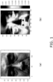
- FIG. 1(a) is a diagram showing an example of the photographed image data on the paranasal sinus peripheral region at the facial surface of a test subject photographed by a photographing device.
- FIG. 1(b) is a diagram showing an example of a blood-circulation-amount distribution diagram (image map).
- the photographed image data was acquired from the facial surfaces of six test subjects. Specifically, these test subjects were seated on chairs in an artificial weather room that was kept at a room temperature of 25°C. Then, the photographed image data on the paranasal sinus peripheral region from the entire facial surface of each test subject was acquired in time series by using the photographing device capable of acquiring images in time series.
- brain has a mechanism called “Selective Brain Cooling System” to cool the brain independently of the body temperature.
- the selective brain cooling system is known to discharge heat generated by the brain activity using forehead or paranasal sinus peripheral region (including a part between eyes). Based on this fact, it is thought that a change in the blood-circulation-amount of the facial surface, which is considered to be proportional to the facial skin temperature that changes accompanied with the brain activity, appears at the forehead and/or the paranasal sinus peripheral region. From this viewpoint, the inventors considered that the brain activity can be estimated with high accuracy as long as a change in the blood-circulation-amount in at least the forehead and/or the paranasal sinus peripheral region of the facial surface can be captured. Accordingly, in the present test, the photographed image data on the paranasal sinus peripheral region at the facial surface of each test subject was acquired in time series.
- the photographing device on the liquid crystal display side of an iPad Air (registered trademark) manufactured by Apple Inc. was used as the photographing device to obtain color moving image data as the time-series photographed image data.
- the photographing device was placed at a position located in front of the test subjects and spaced apart from the test subjects by 1.0 m. Then, the moving image data on the facial surface was obtained by continuously capturing the photographed image data with the photographing device for 30 minutes in photographing cycles of 30 frames/sec along the time axis.
- a brain function activation task was given to the test subject while the moving image data on the facial surface was being acquired.
- the moving image data on the facial surface at a brain resting time, as well as the moving image data on the facial surface at a brain activated time were acquired.
- the brain function activation tasks include psychological works done by the test subject based on a picture displayed on a display device or the like: such as calculation, recognition of numerical values, shape, and color, and memorization of marks, characters, and languages.
- "mental arithmetic of multiplication" was adopted as the brain function activation task to make the test subjects calculate numerical characters displayed on the display device in longhand and input the answers on a keyboard.
- the brain function activation task was continuously given to the test subjects for ten minutes after five minutes have elapsed since the start of acquiring moving the image data on the facial surface.
- the blood-circulation-amount data was calculated based on the RGB data acquired from the moving image data on the photographed facial surface, and then the calculated time-series blood-circulation-amount data was subjected to a singular value decomposition by using the Singular Value Decomposition (SVD) of MATLAB (registered trademark) as an analysis tool.
- Singular Value Decomposition Singular Value Decomposition
- a* an erythema index that correlates with skin redness and hemoglobin amount was computed and determined from the RGB data on the image, in accordance with the CIE-L*a*b* color system.
- This erythema index was defined as the "blood-circulation-amount data.”
- the blood-circulation-amount data here, the erythema indexes
- the erythema indexes based on the RGB data acquired from all moving image data (data for 30 minutes) which was acquired in time series was defined as the target
- the factor was defined as time data acquired every 30 seconds (60 time points for 30 minutes)
- the measure was defined as the erythema index computed from the RGB data for the period of time (every 30 seconds) (erythema index that was computed by taking out frame data for one second every 30 seconds and acquiring an average of RGB values from the respective frame data: 240 ⁇ 320 pixels).
- the thus-made component waveform diagram and blood-circulation-amount distribution diagram of each component were analyzed to identify a component which exhibited a change in the blood-circulation-amount of the facial surface that reflected the brain activity, i.e., an RGB change of the facial surface.
- the brain resting time was defined as a period of time with no brain function activation task given to the test subjects.
- the brain resting time was a period of five minutes from the start of data acquisition and a period of 15 minutes from when 15 minutes had elapsed since the start of data acquisition to the end of data acquisition.
- the brain activated time was defined as a period of time with the brain function activation task given to the test subjects.
- the brain activated time was a period of ten minutes from the time when five minutes had elapsed since the start of data acquisition to the time when ten minutes had elapsed since then.
- evaluation was performed on the presence or absence of the correlation between the amplitude of each component shown in the component waveform diagram and each of the brain resting time and the brain activated time. It is noted that the presence or absence of the correlation was determined by a statistical correlation analysis: when a significance level ( ⁇ ) was 0.01 or less, it was determined that there was the correlation.
- the blood-circulation-amount distribution diagram was made by arranging the space distributions U calculated every pixel, at the respective positions of the pixels. In the blood-circulation-amount distribution diagram for each component made in this way, it was evaluated whether or not there was any change in the blood-circulation-amount at the paranasal sinus peripheral region and the forehead.
- the presence or absence of the change in the blood-circulation-amount at the paranasal sinus peripheral region and the forehead in the blood-circulation-amount distribution diagram was determined on the basis of the presence or absence of the change in the blood-circulation-amount that was observed through visual inspection, or the fact that the value of the blood-circulation-amount at the paranasal sinus peripheral region and the forehead shown in FIG. 1(b) was not "0.000.”
- a polarity (plus or minus) of the blood-circulation-amount data X is determined depending on the relationship between the values of the space distribution U, the singular value S, and the time distribution V. Because of this, the polarity appears to be inversed in the component waveform diagram and the blood-circulation-amount distribution diagram of each component in some cases. Thus, in the evaluation of the component waveform diagram and the blood-circulation-amount distribution diagram, the polarity was not set as an evaluation target.
- the photographed image data on the facial surface was acquired from the six test subjects in time-series.
- the facial skin temperature data was also acquired in time-series by an infrared thermography device.
- the acquired facial skin temperature data was also subjected to the singular value decomposition by using the SVD of MATLAB (registered trademark) as the analysis tool and the component waveform diagram for each component was made according to the singular values S.
- the diagram was analyzed to determine the presence or absence of the correlation between the amplitude of the component waveform and each of the brain resting time and the brain activated time.
- the infrared thermography device used for the above processing is a device capable of detecting infrared radiant energy emitted from the target with an infrared camera, converting the detected infrared radiant energy into the temperature of a surface of the target (here, temperature in Celsius), and then displaying and storing the temperature distribution of converted temperatures as the facial skin temperature data (for example, image data representing the temperature distribution).
- the infrared thermography device in use was an infrared camera R300 manufactured by NEC Avio Infrared Technologies Co., Ltd. The infrared camera was placed in front of the test subjects and spaced apart from the test subjects by 1.5 m.
- the photographed image data on the facial surface is acquired by using the photographing device
- the light is reflected by the face.
- the reflected light occasionally enters a lens of the photographing device in some cases.
- the photographed image data on the photographed facial surface would have the reflected light recorded therein.
- a change in brightness based on the blood-circulation-amount of the facial surface is smaller than a change in brightness based on the reflected light.
- the thus-made blood-circulation-amount data was also subjected to the singular value decomposition by using the SVD of MATLAB (registered trademark) as the analysis tool and the component waveform diagram and the blood-circulation-amount distribution diagram for each component was made according to the singular values S. Then, the diagrams were analyzed to identify the component which exhibited the RGB change of the facial surface that reflects the brain activity.
- MATLAB registered trademark
- the relative blood-circulation-amount data based on the relative RGB data obtained by setting an average of all RGB data taken every predetermined time (every 30 seconds in the present test) at "0" is referred to as “relative-conversion blood-circulation-amount data”
- the blood-circulation-amount data based on the RGB data provided before the conversion into the relative RGB data is simply referred to as “blood-circulation-amount data.”
- the brain wave of each test subject was measured by connecting electrodes to the scalp of the test subject to also evaluate the correlation between the amplitude of ⁇ wave (brain wave at a frequency of 13 to 30 Hz) known as the waveform appearing when brain cells were active, such as during waking, and the amplitude shown in the component waveform diagram.
- the electrodes were placed on 19 scalp locations (Fp1, Fp2, F3, F4, C3, C4, P3, P4, O1, 02, F7, F8, T3, T4, T5, T6, Fz, Cz, and Pz) based on the International 10-20 system.
- the brain function activation task is given to the test subject, it is considered that the head of the test subject can move vertically. Consequently, the position of the facial surface of the test subject relative to the photographing device will change. For this reason, to verify whether or not a change in the position of the facial surface affects the RGB change of the facial surface, a contrast test was conducted on one test subject.
- the contrast test the time-series photographed image data on the facial surface of the test subject was acquired by using the photographing device as in the above-mentioned test.
- no brain function activation task was given (i.e., at the brain resting time)
- the test subject was assigned to do a work of pressing a keyboard at random timings.
- the singular value decomposition was also conducted by using the SVD of MATLAB (registered trademark) as the analysis tool, and the component waveform diagram for each component was made according to the singular values S. Then, analysis was conducted to determine the presence or absence of the correlation between the amplitude of its component waveform and each of the brain resting time and the brain activated time. Furthermore, analysis was conducted to determine the presence or absence of the correlation between the amplitude of each component waveform and an actual movement of the facial surface.
- MATLAB registered trademark
- the actual movement of the facial surface was evaluated by acquiring a two-dimensional coordinate of a point corresponding to an actual point at the face, from the photographed image data and calculating a movement distance of the facial surface every 30 seconds during photographing on the basis of the photographed image data at the start of the contrast test. Further, the presence or absence of the correlation between the amplitude of each component waveform and the number of inputs to the keyboard during photographing was also analyzed. The number of inputs on the keyboard during photographing was evaluated by calculating a simple moving average every 30 seconds of the time-series photographed image data.
- FIGS. 2 to 7 are diagrams showing some of the results of analysis of the component waveform diagrams based on the photographed image data on the facial surface (blood-circulation-amount data) or the facial skin temperature data.
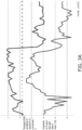
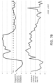
- FIG. 2A is a diagram showing the amplitude of a component waveform of a component 2 based on photographed image data concerning a test subject 1 and the amplitude of a ⁇ wave of the measured brain waves of the test subject 1.
- FIG. 2B is a diagram showing the amplitude of a component waveform of the component 2 based on facial skin temperature data concerning the test subject 1 and the amplitude of the ⁇ wave of the measured brain waves of the test subject 1.
- FIG. 2A is a diagram showing the amplitude of a component waveform of a component 2 based on photographed image data concerning a test subject 1 and the amplitude of a ⁇ wave of the measured brain waves of the test subject 1.
- FIG. 2B is a diagram showing the amplitude of a component waveform of the component 2 based on
- FIG. 3A is a diagram showing the amplitude of a component waveform of the component 2 based on photographed image data concerning a test subject 2 and the amplitude of a ⁇ wave of the measured brain waves of the test subject 2.
- FIG. 3B is a diagram showing the amplitude of a component waveform of the component 2 based on facial skin temperature data concerning the test subject 2 and the amplitude of the ⁇ wave of the measured brain waves of the test subject 2.
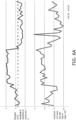
- FIG. 4A is a diagram showing the amplitude of a component waveform of a component 4 based on photographed image data concerning a test subject 3 and the amplitude of a ⁇ wave of the measured brain waves of the test subject 3.
- FIG. 4B is a diagram showing the amplitude of a component waveform of a component 3 based on facial skin temperature data concerning the test subject 3 and the amplitude of the ⁇ wave of the measured brain waves of the test subject 3.
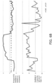
- FIG. 5A is a diagram showing the amplitude of a component waveform of the component 3 based on photographed image data concerning a test subject 4 and the amplitude of a ⁇ wave of the measured brain waves of the test subject 4.
- FIG. 5B is a diagram showing the amplitude of a component waveform of the component 2 based on facial skin temperature data concerning the test subject 4 and the amplitude of the ⁇ wave of the measured brain waves of the test subject 4.
- FIG. 6A is a diagram showing the amplitude of a component waveform of the component 2 based on photographed image data concerning a test subject 5 and the amplitude of a ⁇ wave of the measured brain waves of the test subject 5.
- FIG. 6B is a diagram showing the amplitude of a component waveform of the component 2 based on facial skin temperature data concerning the test subject 5 and the amplitude of the ⁇ wave of the measured brain waves of the test subject 5.
- FIG. 7A is a diagram showing the amplitude of a component waveform of the component 4 based on photographed image data concerning a test subject 6 and the amplitude of a ⁇ wave of the measured brain waves of the test subject 6.
- FIG. 7B is a diagram showing the amplitude of a component waveform of the component 3 based on facial skin temperature data concerning the test subject 6 and the amplitude of the ⁇ wave of the measured brain waves of the test subject 6.
- Table 1 below shows the results of analysis of the photographed image data on the facial surface for each test subject.
- Table 1 Test subject Correlation in blood-circulation-amount data Correlation in relative-conversion blood-circulation-amount data Component waveform Distribution of blood-circulation-amount Component waveform Distribution of blood-circulation-amount Test subject 1 Component 2 0.72 Component 1 0.59 Component 2 0.85 Test subject 2 Component 1 0.82 Component 1 0.62 Component 2 0.82 Component 2 0.60 Test subject 3 Component 2 0.33 Component 2 0.45 Component 3 0.56 Component 3 0.31 Component 4 0.56 Test subject 4 Component 1 0.57 Component 1 0.66 Component 3 0.71 Component 3 0.65 Test subject 5 Component 1 0.56 Component 1 0.51 Component 2 0.72 Component 2 0.83 Test subject 6 Component 2 0.38 Component 2 0.45 Component 3 0.51 Component 4 0.68 Component 5 0.36
- Table 2 shows the results of the contrast test.
- Table 2 Component having a correlation with brain resting time/brain activated time Component 1, Component 2 Component having a correlation with movement distance of facial surface Component 1, Component 3, Component 4 Component having a correlation with the number of inputs to keyboard Component 8
- the component 2 among components had a significant correlation between the amplitude of its component waveform and each of the brain resting time and the brain activated time. However, the component 2 was not found to have any significant correlation between each of the movement distance and the number of inputs to the keyboard.
- the blood-circulation-amount data obtained from the RGB data on the facial surface which was based on the time-series photographed image data on the facial surface acquired from the test subject, was decomposed into a plurality of components by the singular value decomposition, and the respective decomposed components were analyzed.
- the components 1, 2, 3, 4, and 5 were found to be those related to the brain activity, among the plurality of components.
- the brain activity estimation device 10 is not limited to the following embodiments, and various modifications can be made without departing from the scope of the appended claims as appropriate.
- FIG. 8 is a schematic diagram of the brain activity estimation device 10 according to the embodiment of the present invention.
- FIG. 9 is a flowchart showing an example of the flow of processing conducted when the brain activity estimation device 10 identifies a component exhibiting the RGB change of a facial surface that reflects a brain function.
- the brain activity estimation device 10 is a device for estimating the brain activity of a person (test subject) from the photographed image data on the person's facial surface. As shown in FIG. 8 , the brain activity estimation device 10 includes image data acquisition means 20 and brain activity estimation means 30.
- the image data acquisition means 20 obtains photographed image data of at least a part of the person's facial surface in time series (step S1). It is noted that the image data acquisition means 20 is not particularly limited as long as it includes at least a photographing device. Examples of the image data acquisition means 20 include a portable terminal with a built-in photographing device, such as a smartphone and a tablet (e.g., iPad: registered trademark).
- the image data acquisition means 20 includes a camera 21 serving as a photographing device and a storage unit 22.
- the camera 21 is to acquire the photographed image data of a person's facial surface in time series. In this embodiment, the camera 21 photographs a moving image of a person's entire facial surface to acquire photographed moving image data.
- the storage unit 22 stores therein the time-series photographed image data captured by a photographing device.
- the storage unit 22 stores moving image data acquired by the camera 21.
- the moving image of an entire facial surface is photographed by the camera 21, but is not limited thereto.
- a moving image including an image of at least the forehead and/or paranasal sinus peripheral region of a facial surface may be photographed.
- a brain function activation task has been given to a person for a certain period of time while the time-series photographed image data on the facial surface is acquired by the image data acquisition means 20. That is, the photographed image data acquired by the image data acquisition means 20 includes data concerning the period of time during which the brain function activation task is being given to the person. It is noted that the brain function activation task given to a person is not limited particularly as long as it can be expected to make the brain activated. For example, the content of the brain function activation task may be determined as appropriate, according to the purpose of use of the brain activity estimation device 10.
- the brain activity estimation means 30 estimates the human brain activity based on the time-series photographed image data on the facial surface that is acquired by the image data acquisition means 20.
- the brain activity estimation means 30 includes an RGB processing unit 31, a conversion unit 32, a blood-circulation-amount calculating unit 33, an analysis unit 34, and an estimation unit 35.
- FIG. 8 shows an embodiment in which the brain activity estimation means 30 exists as one device that includes the RGB processing unit 31, the conversion unit 32, the blood-circulation-amount calculating unit 33, the analysis unit 34, and the estimation unit 35.
- the present invention is not limited thereto, and parts or each of the RGB processor unit 31, the conversion unit 32, the blood-circulation-amount calculating unit 33, the analysis unit 34, and the estimation unit 35 may be independent from one another.
- the RGB processing unit 31 conducts the RGB processing on the photographed image data acquired by the image data acquisition means 20 to decompose the data into three color components, namely, an R component, a G component, and a B component (step S2).
- the RGB processing may be conducted on the photographed image data on the entire facial surface.
- data on the forehead and/or the paranasal sinus peripheral region is extracted from the photographed image data, so that the RGB processing is conducted only on the extracted data.
- the conversion unit 32 converts the RGB data of the photographed image data obtained by the RGB processing into relative RGB data (step S3). Specifically, the conversion unit 32 converts the RGB data into the relative RGB data by setting the average of the RGB data obtained from the acquired photographed image data every predetermined time (e.g., 30 seconds) as a reference.
- predetermined time e.g. 30 seconds
- the blood-circulation-amount calculating unit 33 calculates time-series blood-circulation-amount data on the facial surface, based on the RGB data of the photographed image data obtained by the RGB processing (step S4).
- the analysis unit 34 decomposes the time-series relative-conversion blood-circulation-amount data into a plurality of components by a singular value decomposition, a principal component analysis, or an independent component analysis (step S5).
- the analysis unit 34 conducts the singular value decomposition on the relative-conversion blood-circulation-amount data by using the SVD of MATLAB (registered trademark) as an analysis tool.
- the singular value decomposition is performed, in which the time-series relative-conversion blood-circulation-amount data is defined as the target, the factor is defined as time data acquired at every predetermined time interval (for example, 30 seconds), and the measure is defined as the relative-conversion blood-circulation-amount data for each pixel that is computed from the relative RGB data at every predetermined time interval.
- the time-series relative-conversion blood-circulation-amount data is decomposed into a plurality of components by the singular value decomposition to calculate a time distribution, a space distribution, and a singular value indicative of the size of each component.
- the analysis unit 34 determines whether or not each component satisfies a predetermined condition, in order to identify a component that exhibits the RGB change of the facial surface reflecting the brain activity, from the plurality of components decomposed by the singular value decomposition (step S6).
- the predetermined condition include a condition in which the amplitude of the component waveform of the component decomposed by the singular value decomposition has correlations with the changes of the brain at the brain resting time and a brain activated time (hereinafter referred to as a first condition), and a condition in which there is a change in the blood circulation amount at a predetermined part of the human facial surface with regard to the component decomposed by the singular value decomposition (hereinafter referred to as a second condition).
- the predetermined condition based on which the determination is made by the analysis unit 34 may include one or more set conditions.
- the first condition is set as the predetermined condition.
- the analysis unit 34 extracts, as a determination component, a component satisfying the predetermined condition, among the plurality of components. Further, the analysis unit 34 identifies a component satisfying all of the conditions included in the predetermined condition among the extracted determination components, as a component exhibiting the RGB change of the facial surface that reflects the brain activity (step S7). Meanwhile, the analysis unit 34 determines that the component determined not to satisfy at least one requirement included in the predetermined condition among the plurality of components is not a component exhibiting the RGB change of the facial surface that reflects the brain activity (step S8).
- only one condition is set as the predetermined condition as mentioned above, and there is a certain period of time during which the brain function activation task is given to the person while the time-series photographed image data on the facial surface is being acquired.
- the analysis unit 34 conducts an analysis by comparing the component waveform of each component with the period of time during which the brain function activation task is given to the person and the period of time during which the task is not given to the person.
- the period of time during which no brain function activation task is given is defined as a brain resting time, whereas the period of time during which the brain function activation task is given is defined as a brain activated time.
- the analysis unit 34 evaluates whether or not the component waveform of each component has any correlation with the brain resting time and the brain activated time. Then, the analysis unit 34 extracts a component evaluated to have the correlation, from among the plurality of components, as a determination component satisfying the predetermined condition, and identifies the component as the component exhibiting the RGB change of the facial surface that reflects the brain activity. Meanwhile, the analysis unit 34 determines that the components evaluated not to have any correlation among the plurality of components do not satisfy the predetermined condition and are not the components that exhibit the RGB change of the facial surface reflecting the human brain activity.
- the brain function activation task is given to the person for a certain period of time when the time-series photographed image data on the facial surface is acquired.
- the analysis unit 34 extracts the determination component, but the content of the first condition, i.e., the extraction means of the determination component in the analysis unit 34 is not limited thereto.
- the analysis unit 34 extracts the specified component as the determination component from the plurality of components.
- the analysis unit 34 may extract a determination component from the plurality of components, by analyzing and evaluating through comparison between the detected result and the component waveform of each component. Note that the reference for the analysis unit 34 to determine whether or not the first condition is satisfied is appropriately determined by simulation, an experiment, working on paper, or the like, according to the purpose of use or the like of the brain activity estimation device 10.
- the analysis unit 34 extracts the determination component based on the presence or absence of a change in the blood-circulation-amount of the facial surface at a predetermined part of the human facial surface. Specifically, the analysis unit 34 determines whether or not a change in the blood-circulation-amount occurs at the paranasal sinus peripheral region and/or forehead, based on the blood-circulation-amount distribution diagram, depending on the plurality of components decomposed by the singular value decomposition. When the change in the blood-circulation-amount occurs, the analysis unit 34 determines the component of interest satisfies the second condition.
- the analysis unit 34 determines that the component of interest does not satisfy the second condition.
- the reference for the analysis unit 34 to determine whether or not the second condition is satisfied is appropriately determined by simulation, an experiment, working on paper, or the like, according to the purpose of use or the like of the brain activity estimation device 10.
- the analysis unit 34 determines whether or not the above-mentioned first condition and/or second condition are satisfied, and extracts a determination component.
- the estimation unit 35 estimates the human brain activity based on the component identified in the analysis unit 34 as the component that exhibits the RGB change of the facial surface reflecting the human brain activity. Specifically, the estimation unit 35 estimates whether the person's brain is in an active state or in an inactive state when the photographed image data of the facial surface is acquired, based on the component waveform data of the component identified by the analysis unit 34.
- the brain activity estimation device 10 can estimate human brain activity based on the time-series photographed image data on the facial surface.
- the estimated result by the estimation unit 35 is displayed on display means (not shown) such as a display, so that whether the person's brain is in the active state or in the inactive state can be notified.
- the brain activity estimation device 10 may decompose the acquired photographed image data on the facial surface into a plurality of components by the singular value decomposition, and analyze only the identified component to estimate whether or not the person's brain was in the active state or in the inactive state when the photographed image data on the facial surface was acquired.
- a brain activity estimation device 10 can be used to control equipment and devices such as air conditioners to create an interior environment appropriate for the person.
- the human brain activity is estimated based on the time-series photographed image data on the facial surface acquired by the image data acquisition means 20. Because of this, the human brain activity can be estimated without attaching any sensors requiring the preprocessing before attachment such as brain wave electrodes and probes. Therefore, the human brain activity can be easily estimated, compared to the case using a conventional detection method, such as electroencephalography, functional magnetic resonance imaging, and near infrared spectroscopy.
- the present embodiment only needs to acquire the image data on at least a part of the facial surface and thereby can reduce manufacturing cost, compared to a brain activity estimation device equipped with the device used by the conventional detection method.
- the human brain activity can be also estimated based on the human's facial skin temperature data that can be acquired by using the thermography device.
- the thermography device generally costs about several tens of thousands of Japanese yen. For this reason, the brain activity estimation device that achieves more reduction in manufacturing cost than the use of the thermography device is expected.
- the low-cost photographing device is employed as the image data acquisition means 20, so that the manufacturing cost can be reduced more than when the thermography device is employed.
- the inventors have conceived of a component analysis approach in which the time-series facial skin temperature data is decomposed into the plurality of components by a singular value decomposition, a principal component analysis, or an independent component analysis, and a component related to the brain activity is identified from the decomposed plurality of components.
- the component analysis approach all temperature data is decomposed, thereby making it possible to remove any component including noise.
- this component analysis approach can precisely estimate the brain activity, compared to the average value approach.
- the inventors have considered that the component analysis approach could be also effective even when the brain activity is estimated from the time-series data on the blood-circulation-amount of the facial surface which is proportional to the facial skin temperature. For this reason, the inventors have adopted the component analysis approach in which the time-series blood-circulation-amount data based on the RGB data obtained from the time-series facial surface image data is decomposed into the plurality of components by a singular value decomposition, a principal component analysis, or an independent component analysis, and a component related to the brain activity is identified from the decomposed plurality of components.
- the time-series blood-circulation-amount data based on the RGB data obtained from the time-series facial surface image data is decomposed into the plurality of components by the singular value decomposition, and the brain activity is estimated from the decomposed components.
- the component including noise can be removed, whereby the brain activity can be precisely estimated.
- the brain function activation task is given to the person for a certain period of time while the time-series image data on the facial surface is acquired by the image data acquisition means 20. That is, in this embodiment, the presence or absence of the brain function activation tasks actually given to a person creates a situation which brings the human brain into the activated state or the resting state.
- the time-series blood-circulation-amount data based on the RGB data obtained from the image data acquired in this way is decomposed into a plurality of components by the singular value decomposition, and the correlation between its component waveform and each of the brain activated time and the brain resting time is evaluated for each component. Then, the component having the correlation is extracted from the plurality of components as the determination component.
- brain has the mechanism called “Selective Brain Cooling System” to cool the brain independently of the body temperature.
- the selective brain cooling system is known to discharge heat generated by the brain activity using the forehead and the paranasal sinus peripheral region. This means, a change in the blood-circulation-amount of the facial surface, which has a correlation with the facial skin temperature according to a brain activity, appears at the forehead and/or the paranasal sinus peripheral region.
- the blood-circulation-amount data based on the RGB data on the forehead and/or the paranasal sinus peripheral region is analyzed to extract the determination component.
- the component related to the human brain activity can be extracted with high accuracy.
- an area for conducting the RGB processing and for acquiring the blood-circulation-amount data is limited to the forehead and/or the paranasal sinus peripheral region.
- the present embodiment can reduce the amount of computation processing, compared to the case the photographed image data on the entire facial surface is subjected to the RGB processing or the blood-circulation-amount data is calculated based on the data on the entire facial surface.
- the RGB data of the photographed image data obtained by the RGB processing is converted into the relative RGB data, to thereby calculate the time-series relative-conversion blood-circulation-amount data based on the relative RGB data. Due to the relative-conversion blood-circulation-amount data calculated in this way, a relative change in the RGB data on the facial surface can be captured every predetermined time. Thus, the RGB change of the facial surface due to the external factor not related to the brain activity can be detected.
- the time-series relative-conversion blood-circulation-amount data is decomposed into a plurality of components by the singular value decomposition, and each of the components is analyzed.
- the component including the RGB change of the facial surface due to the external factor not related to the brain activity can be removed as a noise component.
- the component related to the human brain activity can be identified with high accu racy.
- that kind of component may not be extracted as the one having a significant correlation when the relative-conversion blood-circulation-amount data obtained based on the relative RGB data is analyzed.
- the RGB data provided before the conversion into the relative RGB data is influenced by external factors, such as light from the outside.
- the above-mentioned difference regarding the extracted components is considered to be related to the influence of the external factor. That is, it can be said that the relative-conversion blood-circulation-amount data obtained based on the relative RGB data is more important and more valid than the blood-circulation-amount data obtained based on the RGB data provided before the conversion into the relative RGB data.
- the components related to the human brain activity can be identified with higher accuracy than in the case where only the blood-circulation-amount data obtained based on the RGB data provided before the conversion into the relative RGB data is analyzed. Further, this configuration can reduce the amount of computation processing, compared to the case where both of the blood-circulation-amount data obtained based on the RGB data provided before the conversion into the relative RGB data and the relative-conversion blood-circulation-amount data obtained based on the relative RGB data are analyzed.
- a portable terminal with a built-in photographing device such as a smartphone and a tablet (e.g., iPad: registered trademark) etc. can be utilized as the camera 21. That is, the above-mentioned photographed image data in use can be the one generated by photographing an image in the visible light region.
- the R components among the respective pixels in the RGB data may be mainly used to calculate the blood-circulation-amount data on the facial surface.
- the blood-circulation-amount data is not necessarily limited to the erythema index as long as the blood-circulation-amount data can be calculated based on the RGB data.
- step S4 the time-series blood-circulation-amount data on the facial surface is calculated based on the RGB data of the photographed image data obtained by the RGB processing.
- the blood-circulation-amount calculating unit 33 in the above-mentioned embodiment calculates the relative-conversion blood-circulation-amount data based on the relative RGB data converted by the conversion unit 32.
- the blood-circulation-amount data may be calculated based on the RGB data provided before the conversion into the relative RGB data.
- the blood-circulation-amount data calculated based on the RGB data provided before the conversion into the relative RGB data is more likely to generate (or has a higher capability to verify) a component having a correlation with the brain activity.
- the blood-circulation-amount data calculated based on the RGB data provided before the conversion into the relative RGB data may be analyzed prior to the relative-conversion blood-circulation-amount data calculated based on the relative RGB data.
- the blood-circulation-amount data may be analyzed to extract the components having the significant correlation, and regarding the relative-conversion blood-circulation-amount data, only the components corresponding to the extracted components may be analyzed, whereby the amount of the computation processing can be reduced.
- the above-mentioned camera 21 is a normal camera used in visible light region as a precondition, but can be an infrared camera.
- infrared light is irradiated, and its reflected light is used to produce a photographed image by the infrared camera. Consequently, the photographed image data concerning changes of the facial surface of a target person or the like can be obtained.
- the inventors of the present invention have confirmed that there is a correlation between the blood-circulation-amount data calculated from the photographed image data obtained by infrared reflection and the blood-circulation-amount data calculated by mainly using the R component of each pixel included in the RGB data produced by photographing in the visible light region. Therefore, through the use of the photographed image data obtained from the infrared reflection also, the human brain activity can be estimated.
- the brain activity estimation device 10 includes the image data acquisition means 20 and the brain activity estimation means 30, the brain activity estimation device according to the present embodiment is not limited to such a form. That is, the brain activity estimation device according to the present embodiment may take any form for the other configuration as long as it includes the blood-circulation-amount calculating unit 33, the analysis unit 34, and the estimation unit 35. Specifically, the brain activity estimation device according to the present embodiment may take a form, including not only a form in which the device itself generates the image data by photographing, but also a form in which photographed image data is received from an external device to analyze it therein.
- the present invention can easily estimate human brain activity, and thus is effective for application to devices that require the estimation of human brain activity.
Landscapes
- Health & Medical Sciences (AREA)
- Life Sciences & Earth Sciences (AREA)
- Engineering & Computer Science (AREA)
- Physics & Mathematics (AREA)
- Public Health (AREA)
- Surgery (AREA)
- Biophysics (AREA)
- Biomedical Technology (AREA)
- Heart & Thoracic Surgery (AREA)
- Medical Informatics (AREA)
- Molecular Biology (AREA)
- Veterinary Medicine (AREA)
- Animal Behavior & Ethology (AREA)
- General Health & Medical Sciences (AREA)
- Pathology (AREA)
- Neurology (AREA)
- Physiology (AREA)
- Radiology & Medical Imaging (AREA)
- Nuclear Medicine, Radiotherapy & Molecular Imaging (AREA)
- Computer Vision & Pattern Recognition (AREA)
- Artificial Intelligence (AREA)
- Spectroscopy & Molecular Physics (AREA)
- Psychiatry (AREA)
- Signal Processing (AREA)
- Neurosurgery (AREA)
- Psychology (AREA)
- Measuring And Recording Apparatus For Diagnosis (AREA)
- Measuring Pulse, Heart Rate, Blood Pressure Or Blood Flow (AREA)
- Measurement Of The Respiration, Hearing Ability, Form, And Blood Characteristics Of Living Organisms (AREA)
Applications Claiming Priority (2)
| Application Number | Priority Date | Filing Date | Title |
|---|---|---|---|
| JP2015119350 | 2015-06-12 | ||
| PCT/JP2016/067578 WO2016199940A1 (ja) | 2015-06-12 | 2016-06-13 | 脳活動推定装置 |
Publications (3)
| Publication Number | Publication Date |
|---|---|
| EP3308700A1 EP3308700A1 (en) | 2018-04-18 |
| EP3308700A4 EP3308700A4 (en) | 2019-02-20 |
| EP3308700B1 true EP3308700B1 (en) | 2024-01-24 |
Family
ID=57503300
Family Applications (1)
| Application Number | Title | Priority Date | Filing Date |
|---|---|---|---|
| EP16807643.8A Active EP3308700B1 (en) | 2015-06-12 | 2016-06-13 | Brain-activity estimation device |
Country Status (5)
| Country | Link |
|---|---|
| US (1) | US11253155B2 (ja) |
| EP (1) | EP3308700B1 (ja) |
| JP (2) | JP6189486B2 (ja) |
| CN (1) | CN107847134B (ja) |
| WO (1) | WO2016199940A1 (ja) |
Families Citing this family (4)
| Publication number | Priority date | Publication date | Assignee | Title |
|---|---|---|---|---|
| EP3308700B1 (en) | 2015-06-12 | 2024-01-24 | Daikin Industries, Ltd. | Brain-activity estimation device |
| JP6749278B2 (ja) * | 2017-04-14 | 2020-09-02 | ダイキン工業株式会社 | 生理状態判定装置 |
| JP7276841B2 (ja) * | 2019-07-23 | 2023-05-18 | 学校法人東京電機大学 | 血流評価装置。 |
| WO2023090429A1 (ja) * | 2021-11-19 | 2023-05-25 | 国立大学法人電気通信大学 | 生体検出システム、生体検出方法及びプログラム |
Family Cites Families (26)
| Publication number | Priority date | Publication date | Assignee | Title |
|---|---|---|---|---|
| JP4474145B2 (ja) * | 2003-11-12 | 2010-06-02 | 株式会社日立メディコ | 光計測装置 |
| JP4656304B2 (ja) * | 2005-03-30 | 2011-03-23 | 花王株式会社 | 肌の酸素飽和度計測システム |
| US8905932B2 (en) * | 2006-08-17 | 2014-12-09 | Jan Medical Inc. | Non-invasive characterization of human vasculature |
| JP2008284165A (ja) * | 2007-05-17 | 2008-11-27 | Auto Network Gijutsu Kenkyusho:Kk | 生体情報取得装置 |
| US8401261B2 (en) | 2007-09-25 | 2013-03-19 | University Of Houston System | Imaging facial signs of neuro-physiological responses |
| JP4518189B2 (ja) * | 2008-05-28 | 2010-08-04 | ソニー株式会社 | 情報処理装置および方法、プログラム、並びに記録媒体 |
| US20110251493A1 (en) | 2010-03-22 | 2011-10-13 | Massachusetts Institute Of Technology | Method and system for measurement of physiological parameters |
| JP2013176406A (ja) | 2010-05-27 | 2013-09-09 | Hitachi Ltd | 脳機能計測装置 |
| US10289898B2 (en) * | 2010-06-07 | 2019-05-14 | Affectiva, Inc. | Video recommendation via affect |
| US9204836B2 (en) * | 2010-06-07 | 2015-12-08 | Affectiva, Inc. | Sporadic collection of mobile affect data |
| JP5665025B2 (ja) * | 2010-08-06 | 2015-02-04 | 国立大学法人東京農工大学 | 精神疾患判定装置、方法、及びプログラム |
| WO2012093358A1 (en) * | 2011-01-05 | 2012-07-12 | Koninklijke Philips Electronics N.V. | Device and method for extracting information from characteristic signals |
| CN102178515B (zh) * | 2011-03-17 | 2012-10-03 | 何宗彦 | 脑病变监测仪 |
| JP5672144B2 (ja) * | 2011-05-20 | 2015-02-18 | 富士通株式会社 | 心拍数・呼吸数検出装置,方法およびプログラム |
| JP5917841B2 (ja) * | 2011-06-15 | 2016-05-18 | 日産自動車株式会社 | 気分判定装置及び気分判定装置の作動方法 |
| US8838209B2 (en) * | 2012-02-21 | 2014-09-16 | Xerox Corporation | Deriving arterial pulse transit time from a source video image |
| AU2013256179A1 (en) * | 2012-05-02 | 2014-11-27 | Aliphcom | Physiological characteristic detection based on reflected components of light |
| US20140121540A1 (en) | 2012-05-09 | 2014-05-01 | Aliphcom | System and method for monitoring the health of a user |
| RU2651070C2 (ru) * | 2012-11-02 | 2018-04-18 | Конинклейке Филипс Н.В. | Устройство и способ для извлечения физиологической информации |
| US9640218B2 (en) * | 2012-12-07 | 2017-05-02 | Intel Corporation | Physiological cue processing |
| WO2014136310A1 (ja) * | 2013-03-08 | 2014-09-12 | 富士フイルム株式会社 | 脈波伝播速度の測定方法及びシステム並びに撮像装置 |
| WO2014145204A1 (en) | 2013-03-15 | 2014-09-18 | Affectiva, Inc. | Mental state analysis using heart rate collection based video imagery |
| JP2015022537A (ja) * | 2013-07-19 | 2015-02-02 | 日産自動車株式会社 | 車両用情報提示装置 |
| JP6349075B2 (ja) * | 2013-11-22 | 2018-06-27 | 三星電子株式会社Samsung Electronics Co.,Ltd. | 心拍数測定装置及び心拍数測定方法 |
| CN104545864B (zh) * | 2014-12-25 | 2017-08-11 | 中国科学院深圳先进技术研究院 | 心理调节方法和装置 |
| EP3308700B1 (en) | 2015-06-12 | 2024-01-24 | Daikin Industries, Ltd. | Brain-activity estimation device |
-
2016
- 2016-06-13 EP EP16807643.8A patent/EP3308700B1/en active Active
- 2016-06-13 US US15/735,104 patent/US11253155B2/en active Active
- 2016-06-13 CN CN201680034098.XA patent/CN107847134B/zh active Active
- 2016-06-13 JP JP2016116905A patent/JP6189486B2/ja active Active
- 2016-06-13 WO PCT/JP2016/067578 patent/WO2016199940A1/ja active Application Filing
-
2017
- 2017-08-02 JP JP2017150008A patent/JP7111450B2/ja active Active
Also Published As
| Publication number | Publication date |
|---|---|
| JP7111450B2 (ja) | 2022-08-02 |
| JP6189486B2 (ja) | 2017-08-30 |
| EP3308700A1 (en) | 2018-04-18 |
| US20180168451A1 (en) | 2018-06-21 |
| US11253155B2 (en) | 2022-02-22 |
| CN107847134A (zh) | 2018-03-27 |
| EP3308700A4 (en) | 2019-02-20 |
| JP2017209516A (ja) | 2017-11-30 |
| WO2016199940A1 (ja) | 2016-12-15 |
| JP2017000773A (ja) | 2017-01-05 |
| CN107847134B (zh) | 2023-09-01 |
Similar Documents
| Publication | Publication Date | Title |
|---|---|---|
| US10667738B2 (en) | Brain activity estimation device | |
| EP3552555A1 (en) | Mental disorder determination device | |
| EP3610793B1 (en) | Physiological state assessment apparatus | |
| CN108135491B (zh) | 生理状态判定装置及生理状态判定方法 | |
| EP3363352B1 (en) | Useful information presentation device | |
| EP3424425A1 (en) | Determination result output device, determination result provision device, and determination result output system | |
| EP3308700B1 (en) | Brain-activity estimation device | |
| EP3424408B1 (en) | Fatigue state determination device and fatigue state determination method | |
| JP6829363B2 (ja) | 評価装置、マーケット調査装置、及び学習評価装置 | |
| JP6158887B2 (ja) | 有用情報提示装置 | |
| JP6093422B1 (ja) | 脳年齢提示装置 | |
| JP6096857B1 (ja) | 感情判定装置 | |
| JP2017086992A (ja) | 感情判定装置 |
Legal Events
| Date | Code | Title | Description |
|---|---|---|---|
| STAA | Information on the status of an ep patent application or granted ep patent |
Free format text: STATUS: THE INTERNATIONAL PUBLICATION HAS BEEN MADE |
|
| PUAI | Public reference made under article 153(3) epc to a published international application that has entered the european phase |
Free format text: ORIGINAL CODE: 0009012 |
|
| STAA | Information on the status of an ep patent application or granted ep patent |
Free format text: STATUS: REQUEST FOR EXAMINATION WAS MADE |
|
| 17P | Request for examination filed |
Effective date: 20180103 |
|
| AK | Designated contracting states |
Kind code of ref document: A1 Designated state(s): AL AT BE BG CH CY CZ DE DK EE ES FI FR GB GR HR HU IE IS IT LI LT LU LV MC MK MT NL NO PL PT RO RS SE SI SK SM TR |
|
| AX | Request for extension of the european patent |
Extension state: BA ME |
|
| DAV | Request for validation of the european patent (deleted) | ||
| DAX | Request for extension of the european patent (deleted) | ||
| A4 | Supplementary search report drawn up and despatched |
Effective date: 20190118 |
|
| RIC1 | Information provided on ipc code assigned before grant |
Ipc: A61B 5/00 20060101AFI20190114BHEP Ipc: A61B 5/0476 20060101ALI20190114BHEP |
|
| STAA | Information on the status of an ep patent application or granted ep patent |
Free format text: STATUS: EXAMINATION IS IN PROGRESS |
|
| STAA | Information on the status of an ep patent application or granted ep patent |
Free format text: STATUS: EXAMINATION IS IN PROGRESS |
|
| 17Q | First examination report despatched |
Effective date: 20201127 |
|
| STAA | Information on the status of an ep patent application or granted ep patent |
Free format text: STATUS: EXAMINATION IS IN PROGRESS |
|
| RAP3 | Party data changed (applicant data changed or rights of an application transferred) |
Owner name: TOKYO INSTITUTE OF TECHNOLOGY Owner name: DAIKIN INDUSTRIES, LTD. |
|
| P01 | Opt-out of the competence of the unified patent court (upc) registered |
Effective date: 20230525 |
|
| REG | Reference to a national code |
Ref country code: DE Ref legal event code: R079 Ref document number: 602016085493 Country of ref document: DE Free format text: PREVIOUS MAIN CLASS: A61B0005000000 Ipc: A61B0005010000 Ref country code: DE Ref legal event code: R079 Free format text: PREVIOUS MAIN CLASS: A61B0005000000 Ipc: A61B0005010000 |
|
| GRAP | Despatch of communication of intention to grant a patent |
Free format text: ORIGINAL CODE: EPIDOSNIGR1 |
|
| STAA | Information on the status of an ep patent application or granted ep patent |
Free format text: STATUS: GRANT OF PATENT IS INTENDED |
|
| RIC1 | Information provided on ipc code assigned before grant |
Ipc: A61B 5/01 20060101AFI20230821BHEP |
|
| INTG | Intention to grant announced |
Effective date: 20230908 |
|
| GRAS | Grant fee paid |
Free format text: ORIGINAL CODE: EPIDOSNIGR3 |
|
| GRAA | (expected) grant |
Free format text: ORIGINAL CODE: 0009210 |
|
| STAA | Information on the status of an ep patent application or granted ep patent |
Free format text: STATUS: THE PATENT HAS BEEN GRANTED |
|
| AK | Designated contracting states |
Kind code of ref document: B1 Designated state(s): AL AT BE BG CH CY CZ DE DK EE ES FI FR GB GR HR HU IE IS IT LI LT LU LV MC MK MT NL NO PL PT RO RS SE SI SK SM TR |
|
| REG | Reference to a national code |
Ref country code: GB Ref legal event code: FG4D |
|
| REG | Reference to a national code |
Ref country code: CH Ref legal event code: EP |
|
| REG | Reference to a national code |
Ref country code: IE Ref legal event code: FG4D |
|
| REG | Reference to a national code |
Ref country code: DE Ref legal event code: R096 Ref document number: 602016085493 Country of ref document: DE |
|
| REG | Reference to a national code |
Ref country code: LT Ref legal event code: MG9D |