EP3305807A1 - Interaction of moraxella catarrhalis with epithelial cells, extracellular matrix proteins and the complement system - Google Patents
Interaction of moraxella catarrhalis with epithelial cells, extracellular matrix proteins and the complement system Download PDFInfo
- Publication number
- EP3305807A1 EP3305807A1 EP17196588.2A EP17196588A EP3305807A1 EP 3305807 A1 EP3305807 A1 EP 3305807A1 EP 17196588 A EP17196588 A EP 17196588A EP 3305807 A1 EP3305807 A1 EP 3305807A1
- Authority
- EP
- European Patent Office
- Prior art keywords
- uspa1
- catarrhalis
- uspa2
- binding
- fibronectin
- Prior art date
- Legal status (The legal status is an assumption and is not a legal conclusion. Google has not performed a legal analysis and makes no representation as to the accuracy of the status listed.)
- Pending
Links
- 241000588655 Moraxella catarrhalis Species 0.000 title abstract description 184
- 210000002919 epithelial cell Anatomy 0.000 title abstract description 28
- 230000004154 complement system Effects 0.000 title abstract description 13
- 102000010834 Extracellular Matrix Proteins Human genes 0.000 title abstract description 7
- 108010037362 Extracellular Matrix Proteins Proteins 0.000 title abstract description 7
- 230000003993 interaction Effects 0.000 title description 45
- 108010067306 Fibronectins Proteins 0.000 claims abstract description 161
- 102000016359 Fibronectins Human genes 0.000 claims abstract description 161
- 108090000765 processed proteins & peptides Proteins 0.000 claims abstract description 79
- 102000004196 processed proteins & peptides Human genes 0.000 claims abstract description 50
- 230000027455 binding Effects 0.000 claims description 179
- 229920001184 polypeptide Polymers 0.000 claims description 39
- 239000003446 ligand Substances 0.000 claims description 35
- 230000013595 glycosylation Effects 0.000 claims description 26
- 238000006206 glycosylation reaction Methods 0.000 claims description 26
- 230000033444 hydroxylation Effects 0.000 claims description 26
- 238000005805 hydroxylation reaction Methods 0.000 claims description 26
- 239000002671 adjuvant Substances 0.000 claims description 8
- 239000000546 pharmaceutical excipient Substances 0.000 claims description 7
- 239000011230 binding agent Substances 0.000 claims description 6
- 102000037865 fusion proteins Human genes 0.000 claims description 6
- 108020001507 fusion proteins Proteins 0.000 claims description 6
- 239000000969 carrier Substances 0.000 claims description 5
- 239000000203 mixture Substances 0.000 claims description 5
- 239000003755 preservative agent Substances 0.000 claims description 5
- 239000003981 vehicle Substances 0.000 claims description 5
- 230000002163 immunogen Effects 0.000 claims description 3
- 102000007547 Laminin Human genes 0.000 abstract description 76
- 108010085895 Laminin Proteins 0.000 abstract description 76
- 229960005486 vaccine Drugs 0.000 abstract description 18
- 108050001049 Extracellular proteins Proteins 0.000 abstract description 2
- 238000002360 preparation method Methods 0.000 abstract description 2
- 108090000623 proteins and genes Proteins 0.000 description 73
- 239000012634 fragment Substances 0.000 description 69
- 102000004169 proteins and genes Human genes 0.000 description 68
- 241000894006 Bacteria Species 0.000 description 60
- 239000000047 product Substances 0.000 description 45
- 230000037361 pathway Effects 0.000 description 39
- 150000001413 amino acids Chemical class 0.000 description 36
- 210000002966 serum Anatomy 0.000 description 34
- 238000002474 experimental method Methods 0.000 description 33
- 241001276229 Moraxella catarrhalis RH4 Species 0.000 description 29
- 238000000684 flow cytometry Methods 0.000 description 29
- 210000004027 cell Anatomy 0.000 description 25
- 241000283973 Oryctolagus cuniculus Species 0.000 description 24
- 238000011534 incubation Methods 0.000 description 24
- 230000014509 gene expression Effects 0.000 description 23
- 102000007056 Recombinant Fusion Proteins Human genes 0.000 description 22
- 108010008281 Recombinant Fusion Proteins Proteins 0.000 description 22
- 238000012545 processing Methods 0.000 description 22
- 230000002950 deficient Effects 0.000 description 21
- KCXVZYZYPLLWCC-UHFFFAOYSA-N EDTA Chemical compound OC(=O)CN(CC(O)=O)CCN(CC(O)=O)CC(O)=O KCXVZYZYPLLWCC-UHFFFAOYSA-N 0.000 description 20
- 108010001336 Horseradish Peroxidase Proteins 0.000 description 20
- 230000001580 bacterial effect Effects 0.000 description 20
- 102100022133 Complement C3 Human genes 0.000 description 19
- 101000901154 Homo sapiens Complement C3 Proteins 0.000 description 19
- 125000000539 amino acid group Chemical group 0.000 description 19
- BAVYZALUXZFZLV-UHFFFAOYSA-N Methylamine Chemical compound NC BAVYZALUXZFZLV-UHFFFAOYSA-N 0.000 description 18
- 230000006870 function Effects 0.000 description 17
- 208000015181 infectious disease Diseases 0.000 description 17
- 230000005764 inhibitory process Effects 0.000 description 17
- 238000002965 ELISA Methods 0.000 description 16
- 102000006912 Complement C4b-Binding Protein Human genes 0.000 description 15
- 108010047548 Complement C4b-Binding Protein Proteins 0.000 description 15
- 230000024203 complement activation Effects 0.000 description 15
- 238000005406 washing Methods 0.000 description 15
- 241000400731 Moraxella catarrhalis BBH18 Species 0.000 description 14
- FAPWRFPIFSIZLT-UHFFFAOYSA-M Sodium chloride Chemical compound [Na+].[Cl-] FAPWRFPIFSIZLT-UHFFFAOYSA-M 0.000 description 14
- 230000000875 corresponding effect Effects 0.000 description 14
- 108010040473 pneumococcal surface protein A Proteins 0.000 description 14
- MHMNJMPURVTYEJ-UHFFFAOYSA-N fluorescein-5-isothiocyanate Chemical compound O1C(=O)C2=CC(N=C=S)=CC=C2C21C1=CC=C(O)C=C1OC1=CC(O)=CC=C21 MHMNJMPURVTYEJ-UHFFFAOYSA-N 0.000 description 13
- 108091003079 Bovine Serum Albumin Proteins 0.000 description 12
- 230000000295 complement effect Effects 0.000 description 12
- 101710116435 Outer membrane protein Proteins 0.000 description 11
- 229940098773 bovine serum albumin Drugs 0.000 description 11
- 230000001419 dependent effect Effects 0.000 description 11
- 238000000034 method Methods 0.000 description 11
- 244000052769 pathogen Species 0.000 description 11
- 239000002953 phosphate buffered saline Substances 0.000 description 11
- 241000894007 species Species 0.000 description 11
- 210000003743 erythrocyte Anatomy 0.000 description 10
- 239000011534 wash buffer Substances 0.000 description 10
- 241000222122 Candida albicans Species 0.000 description 9
- 108010010803 Gelatin Proteins 0.000 description 9
- 241000588621 Moraxella Species 0.000 description 9
- 230000008021 deposition Effects 0.000 description 9
- 239000008273 gelatin Substances 0.000 description 9
- 229920000159 gelatin Polymers 0.000 description 9
- 235000019322 gelatine Nutrition 0.000 description 9
- 235000011852 gelatine desserts Nutrition 0.000 description 9
- 102000005962 receptors Human genes 0.000 description 9
- 108020003175 receptors Proteins 0.000 description 9
- 208000006545 Chronic Obstructive Pulmonary Disease Diseases 0.000 description 8
- TWRXJAOTZQYOKJ-UHFFFAOYSA-L Magnesium chloride Chemical compound [Mg+2].[Cl-].[Cl-] TWRXJAOTZQYOKJ-UHFFFAOYSA-L 0.000 description 8
- 206010033078 Otitis media Diseases 0.000 description 8
- 231100000673 dose–response relationship Toxicity 0.000 description 8
- 239000003814 drug Substances 0.000 description 8
- 102000036072 fibronectin binding proteins Human genes 0.000 description 8
- 108091010988 fibronectin binding proteins Proteins 0.000 description 8
- 210000000224 granular leucocyte Anatomy 0.000 description 8
- 241000251468 Actinopterygii Species 0.000 description 7
- 241000283707 Capra Species 0.000 description 7
- 102000018697 Membrane Proteins Human genes 0.000 description 7
- 108010052285 Membrane Proteins Proteins 0.000 description 7
- 210000004369 blood Anatomy 0.000 description 7
- 239000008280 blood Substances 0.000 description 7
- 239000000872 buffer Substances 0.000 description 7
- 238000001514 detection method Methods 0.000 description 7
- 201000010099 disease Diseases 0.000 description 7
- 208000037265 diseases, disorders, signs and symptoms Diseases 0.000 description 7
- 239000011521 glass Substances 0.000 description 7
- 239000012528 membrane Substances 0.000 description 7
- 239000011780 sodium chloride Substances 0.000 description 7
- CDBYLPFSWZWCQE-UHFFFAOYSA-L Sodium Carbonate Chemical compound [Na+].[Na+].[O-]C([O-])=O CDBYLPFSWZWCQE-UHFFFAOYSA-L 0.000 description 6
- 241000193998 Streptococcus pneumoniae Species 0.000 description 6
- 230000003247 decreasing effect Effects 0.000 description 6
- 230000000694 effects Effects 0.000 description 6
- 238000001727 in vivo Methods 0.000 description 6
- 230000007246 mechanism Effects 0.000 description 6
- 239000013642 negative control Substances 0.000 description 6
- 229940031000 streptococcus pneumoniae Drugs 0.000 description 6
- 210000001519 tissue Anatomy 0.000 description 6
- 238000011282 treatment Methods 0.000 description 6
- QKNYBSVHEMOAJP-UHFFFAOYSA-N 2-amino-2-(hydroxymethyl)propane-1,3-diol;hydron;chloride Chemical compound Cl.OCC(N)(CO)CO QKNYBSVHEMOAJP-UHFFFAOYSA-N 0.000 description 5
- 229920001817 Agar Polymers 0.000 description 5
- 108010078015 Complement C3b Proteins 0.000 description 5
- 101100263204 Coxiella burnetii (strain RSA 493 / Nine Mile phase I) uspA1 gene Proteins 0.000 description 5
- 241000588724 Escherichia coli Species 0.000 description 5
- 241000606768 Haemophilus influenzae Species 0.000 description 5
- 206010024971 Lower respiratory tract infections Diseases 0.000 description 5
- 206010057249 Phagocytosis Diseases 0.000 description 5
- 229920001213 Polysorbate 20 Polymers 0.000 description 5
- 206010036790 Productive cough Diseases 0.000 description 5
- 241000191967 Staphylococcus aureus Species 0.000 description 5
- 241000193996 Streptococcus pyogenes Species 0.000 description 5
- 230000001070 adhesive effect Effects 0.000 description 5
- 239000008272 agar Substances 0.000 description 5
- 238000004458 analytical method Methods 0.000 description 5
- 238000003556 assay Methods 0.000 description 5
- DEFVIWRASFVYLL-UHFFFAOYSA-N ethylene glycol bis(2-aminoethyl)tetraacetic acid Chemical compound OC(=O)CN(CC(O)=O)CCOCCOCCN(CC(O)=O)CC(O)=O DEFVIWRASFVYLL-UHFFFAOYSA-N 0.000 description 5
- 210000000987 immune system Anatomy 0.000 description 5
- 229910052740 iodine Inorganic materials 0.000 description 5
- 210000004379 membrane Anatomy 0.000 description 5
- 230000001717 pathogenic effect Effects 0.000 description 5
- 230000008782 phagocytosis Effects 0.000 description 5
- 239000000256 polyoxyethylene sorbitan monolaurate Substances 0.000 description 5
- 235000010486 polyoxyethylene sorbitan monolaurate Nutrition 0.000 description 5
- 238000011533 pre-incubation Methods 0.000 description 5
- 210000002345 respiratory system Anatomy 0.000 description 5
- 210000003802 sputum Anatomy 0.000 description 5
- 208000024794 sputum Diseases 0.000 description 5
- 230000004083 survival effect Effects 0.000 description 5
- 239000000725 suspension Substances 0.000 description 5
- 150000007970 thio esters Chemical class 0.000 description 5
- FTOAOBMCPZCFFF-UHFFFAOYSA-N 5,5-diethylbarbituric acid Chemical compound CCC1(CC)C(=O)NC(=O)NC1=O FTOAOBMCPZCFFF-UHFFFAOYSA-N 0.000 description 4
- ZCYVEMRRCGMTRW-UHFFFAOYSA-N 7553-56-2 Chemical compound [I] ZCYVEMRRCGMTRW-UHFFFAOYSA-N 0.000 description 4
- 102000014914 Carrier Proteins Human genes 0.000 description 4
- 241000699666 Mus <mouse, genus> Species 0.000 description 4
- 241000588652 Neisseria gonorrhoeae Species 0.000 description 4
- 241000282898 Sus scrofa Species 0.000 description 4
- 108010084455 Zeocin Proteins 0.000 description 4
- 230000004913 activation Effects 0.000 description 4
- 239000000853 adhesive Substances 0.000 description 4
- 230000000844 anti-bacterial effect Effects 0.000 description 4
- 210000002469 basement membrane Anatomy 0.000 description 4
- 108091008324 binding proteins Proteins 0.000 description 4
- 230000000903 blocking effect Effects 0.000 description 4
- 229960005091 chloramphenicol Drugs 0.000 description 4
- WIIZWVCIJKGZOK-RKDXNWHRSA-N chloramphenicol Chemical compound ClC(Cl)C(=O)N[C@H](CO)[C@H](O)C1=CC=C([N+]([O-])=O)C=C1 WIIZWVCIJKGZOK-RKDXNWHRSA-N 0.000 description 4
- 108091036078 conserved sequence Proteins 0.000 description 4
- 238000010790 dilution Methods 0.000 description 4
- 239000012895 dilution Substances 0.000 description 4
- 238000005516 engineering process Methods 0.000 description 4
- 210000002744 extracellular matrix Anatomy 0.000 description 4
- 239000012530 fluid Substances 0.000 description 4
- 229940047650 haemophilus influenzae Drugs 0.000 description 4
- 210000003128 head Anatomy 0.000 description 4
- 244000052637 human pathogen Species 0.000 description 4
- 239000011630 iodine Substances 0.000 description 4
- 230000002147 killing effect Effects 0.000 description 4
- 229910001629 magnesium chloride Inorganic materials 0.000 description 4
- 238000004519 manufacturing process Methods 0.000 description 4
- 230000001404 mediated effect Effects 0.000 description 4
- 230000000813 microbial effect Effects 0.000 description 4
- 208000005923 otitis media with effusion Diseases 0.000 description 4
- 239000012071 phase Substances 0.000 description 4
- CWCMIVBLVUHDHK-ZSNHEYEWSA-N phleomycin D1 Chemical compound N([C@H](C(=O)N[C@H](C)[C@@H](O)[C@H](C)C(=O)N[C@@H]([C@H](O)C)C(=O)NCCC=1SC[C@@H](N=1)C=1SC=C(N=1)C(=O)NCCCCNC(N)=N)[C@@H](O[C@H]1[C@H]([C@@H](O)[C@H](O)[C@H](CO)O1)O[C@@H]1[C@H]([C@@H](OC(N)=O)[C@H](O)[C@@H](CO)O1)O)C=1N=CNC=1)C(=O)C1=NC([C@H](CC(N)=O)NC[C@H](N)C(N)=O)=NC(N)=C1C CWCMIVBLVUHDHK-ZSNHEYEWSA-N 0.000 description 4
- 238000011321 prophylaxis Methods 0.000 description 4
- 238000003127 radioimmunoassay Methods 0.000 description 4
- 201000009890 sinusitis Diseases 0.000 description 4
- 238000002198 surface plasmon resonance spectroscopy Methods 0.000 description 4
- 101000978703 Escherichia virus Qbeta Maturation protein A2 Proteins 0.000 description 3
- 206010018910 Haemolysis Diseases 0.000 description 3
- 101001015673 Haemophilus influenzae (strain ATCC 51907 / DSM 11121 / KW20 / Rd) Glycerophosphodiester phosphodiesterase Proteins 0.000 description 3
- 101000599852 Homo sapiens Intercellular adhesion molecule 1 Proteins 0.000 description 3
- 102100037877 Intercellular adhesion molecule 1 Human genes 0.000 description 3
- 108090001090 Lectins Proteins 0.000 description 3
- 102000004856 Lectins Human genes 0.000 description 3
- 241000699670 Mus sp. Species 0.000 description 3
- 239000000020 Nitrocellulose Substances 0.000 description 3
- 108700022034 Opsonin Proteins Proteins 0.000 description 3
- 241000589517 Pseudomonas aeruginosa Species 0.000 description 3
- 239000012980 RPMI-1640 medium Substances 0.000 description 3
- 230000010065 bacterial adhesion Effects 0.000 description 3
- 210000004556 brain Anatomy 0.000 description 3
- 229940095731 candida albicans Drugs 0.000 description 3
- 230000001332 colony forming effect Effects 0.000 description 3
- 230000002596 correlated effect Effects 0.000 description 3
- 230000007423 decrease Effects 0.000 description 3
- 230000008588 hemolysis Effects 0.000 description 3
- 230000002949 hemolytic effect Effects 0.000 description 3
- 230000002209 hydrophobic effect Effects 0.000 description 3
- 238000001802 infusion Methods 0.000 description 3
- 239000002523 lectin Substances 0.000 description 3
- 239000011159 matrix material Substances 0.000 description 3
- 229920001220 nitrocellulos Polymers 0.000 description 3
- 230000008506 pathogenesis Effects 0.000 description 3
- 238000011160 research Methods 0.000 description 3
- 229910000029 sodium carbonate Inorganic materials 0.000 description 3
- 101150015701 uspA1 gene Proteins 0.000 description 3
- 108010077805 Bacterial Proteins Proteins 0.000 description 2
- 108010053085 Complement Factor H Proteins 0.000 description 2
- 102000016550 Complement Factor H Human genes 0.000 description 2
- 102000000989 Complement System Proteins Human genes 0.000 description 2
- 108010069112 Complement System Proteins Proteins 0.000 description 2
- 102100032768 Complement receptor type 2 Human genes 0.000 description 2
- 101100263205 Coxiella burnetii (strain RSA 493 / Nine Mile phase I) uspA2 gene Proteins 0.000 description 2
- 108020004414 DNA Proteins 0.000 description 2
- 241000283074 Equus asinus Species 0.000 description 2
- WQZGKKKJIJFFOK-GASJEMHNSA-N Glucose Natural products OC[C@H]1OC(O)[C@H](O)[C@@H](O)[C@@H]1O WQZGKKKJIJFFOK-GASJEMHNSA-N 0.000 description 2
- 101000941929 Homo sapiens Complement receptor type 2 Proteins 0.000 description 2
- 108060003951 Immunoglobulin Proteins 0.000 description 2
- 241000589242 Legionella pneumophila Species 0.000 description 2
- 108700005371 Moraxella catarrhalis UspA Proteins 0.000 description 2
- PXHVJJICTQNCMI-UHFFFAOYSA-N Nickel Chemical compound [Ni] PXHVJJICTQNCMI-UHFFFAOYSA-N 0.000 description 2
- 108091028043 Nucleic acid sequence Proteins 0.000 description 2
- 206010035664 Pneumonia Diseases 0.000 description 2
- 108010005642 Properdin Proteins 0.000 description 2
- 102100038567 Properdin Human genes 0.000 description 2
- 240000004808 Saccharomyces cerevisiae Species 0.000 description 2
- 238000010521 absorption reaction Methods 0.000 description 2
- 239000000427 antigen Substances 0.000 description 2
- 102000036639 antigens Human genes 0.000 description 2
- 108091007433 antigens Proteins 0.000 description 2
- 229960002319 barbital Drugs 0.000 description 2
- 230000015572 biosynthetic process Effects 0.000 description 2
- 150000001720 carbohydrates Chemical group 0.000 description 2
- VDQQXEISLMTGAB-UHFFFAOYSA-N chloramine T Chemical compound [Na+].CC1=CC=C(S(=O)(=O)[N-]Cl)C=C1 VDQQXEISLMTGAB-UHFFFAOYSA-N 0.000 description 2
- 238000010276 construction Methods 0.000 description 2
- 239000008121 dextrose Substances 0.000 description 2
- LOKCTEFSRHRXRJ-UHFFFAOYSA-I dipotassium trisodium dihydrogen phosphate hydrogen phosphate dichloride Chemical compound P(=O)(O)(O)[O-].[K+].P(=O)(O)([O-])[O-].[Na+].[Na+].[Cl-].[K+].[Cl-].[Na+] LOKCTEFSRHRXRJ-UHFFFAOYSA-I 0.000 description 2
- 229940079593 drug Drugs 0.000 description 2
- 230000005713 exacerbation Effects 0.000 description 2
- 239000001963 growth medium Substances 0.000 description 2
- 230000028993 immune response Effects 0.000 description 2
- 102000018358 immunoglobulin Human genes 0.000 description 2
- 230000002458 infectious effect Effects 0.000 description 2
- 208000022760 infectious otitis media Diseases 0.000 description 2
- 230000002401 inhibitory effect Effects 0.000 description 2
- 230000000977 initiatory effect Effects 0.000 description 2
- 210000005007 innate immune system Anatomy 0.000 description 2
- 238000002372 labelling Methods 0.000 description 2
- 229940115932 legionella pneumophila Drugs 0.000 description 2
- 239000007788 liquid Substances 0.000 description 2
- 235000011475 lollipops Nutrition 0.000 description 2
- 210000004072 lung Anatomy 0.000 description 2
- 239000000463 material Substances 0.000 description 2
- 239000002609 medium Substances 0.000 description 2
- 239000008267 milk Substances 0.000 description 2
- 210000004080 milk Anatomy 0.000 description 2
- 235000013336 milk Nutrition 0.000 description 2
- 230000004048 modification Effects 0.000 description 2
- 238000012986 modification Methods 0.000 description 2
- 108091005601 modified peptides Proteins 0.000 description 2
- 238000010172 mouse model Methods 0.000 description 2
- 210000001989 nasopharynx Anatomy 0.000 description 2
- 150000007523 nucleic acids Chemical group 0.000 description 2
- 230000014207 opsonization Effects 0.000 description 2
- 230000036961 partial effect Effects 0.000 description 2
- 231100000915 pathological change Toxicity 0.000 description 2
- 230000036285 pathological change Effects 0.000 description 2
- 210000003800 pharynx Anatomy 0.000 description 2
- 102000054765 polymorphisms of proteins Human genes 0.000 description 2
- 239000000843 powder Substances 0.000 description 2
- 230000002265 prevention Effects 0.000 description 2
- 125000002924 primary amino group Chemical group [H]N([H])* 0.000 description 2
- 230000002685 pulmonary effect Effects 0.000 description 2
- 238000000746 purification Methods 0.000 description 2
- 230000009467 reduction Effects 0.000 description 2
- 230000002829 reductive effect Effects 0.000 description 2
- 230000000717 retained effect Effects 0.000 description 2
- 238000002415 sodium dodecyl sulfate polyacrylamide gel electrophoresis Methods 0.000 description 2
- 239000007787 solid Substances 0.000 description 2
- 238000002798 spectrophotometry method Methods 0.000 description 2
- 238000012360 testing method Methods 0.000 description 2
- 101150087047 uspA2 gene Proteins 0.000 description 2
- 230000001018 virulence Effects 0.000 description 2
- FZWBNHMXJMCXLU-UHFFFAOYSA-N 2,3,4,5-tetrahydroxy-6-[3,4,5-trihydroxy-6-[[3,4,5-trihydroxy-6-(hydroxymethyl)oxan-2-yl]oxymethyl]oxan-2-yl]oxyhexanal Chemical compound OC1C(O)C(O)C(CO)OC1OCC1C(O)C(O)C(O)C(OCC(O)C(O)C(O)C(O)C=O)O1 FZWBNHMXJMCXLU-UHFFFAOYSA-N 0.000 description 1
- FWMNVWWHGCHHJJ-SKKKGAJSSA-N 4-amino-1-[(2r)-6-amino-2-[[(2r)-2-[[(2r)-2-[[(2r)-2-amino-3-phenylpropanoyl]amino]-3-phenylpropanoyl]amino]-4-methylpentanoyl]amino]hexanoyl]piperidine-4-carboxylic acid Chemical compound C([C@H](C(=O)N[C@H](CC(C)C)C(=O)N[C@H](CCCCN)C(=O)N1CCC(N)(CC1)C(O)=O)NC(=O)[C@H](N)CC=1C=CC=CC=1)C1=CC=CC=C1 FWMNVWWHGCHHJJ-SKKKGAJSSA-N 0.000 description 1
- 101150109698 A2 gene Proteins 0.000 description 1
- 108010089414 Anaphylatoxins Proteins 0.000 description 1
- 208000031729 Bacteremia Diseases 0.000 description 1
- 241000588832 Bordetella pertussis Species 0.000 description 1
- 108010062802 CD66 antigens Proteins 0.000 description 1
- UXVMQQNJUSDDNG-UHFFFAOYSA-L Calcium chloride Chemical compound [Cl-].[Cl-].[Ca+2] UXVMQQNJUSDDNG-UHFFFAOYSA-L 0.000 description 1
- 241000222120 Candida <Saccharomycetales> Species 0.000 description 1
- 108010022366 Carcinoembryonic Antigen Proteins 0.000 description 1
- 102100024533 Carcinoembryonic antigen-related cell adhesion molecule 1 Human genes 0.000 description 1
- 102100025475 Carcinoembryonic antigen-related cell adhesion molecule 5 Human genes 0.000 description 1
- 108010067225 Cell Adhesion Molecules Proteins 0.000 description 1
- 102000016289 Cell Adhesion Molecules Human genes 0.000 description 1
- 241000700112 Chinchilla Species 0.000 description 1
- 101710164918 Choline-binding protein Proteins 0.000 description 1
- 102100023661 Coiled-coil domain-containing protein 115 Human genes 0.000 description 1
- 101710155594 Coiled-coil domain-containing protein 115 Proteins 0.000 description 1
- 108010023729 Complement 3d Receptors Proteins 0.000 description 1
- 102000011412 Complement 3d Receptors Human genes 0.000 description 1
- 108010034753 Complement Membrane Attack Complex Proteins 0.000 description 1
- 108090000056 Complement factor B Proteins 0.000 description 1
- 102000003712 Complement factor B Human genes 0.000 description 1
- 229920002307 Dextran Polymers 0.000 description 1
- 241000194032 Enterococcus faecalis Species 0.000 description 1
- 241000672609 Escherichia coli BL21 Species 0.000 description 1
- 241000617590 Escherichia coli K1 Species 0.000 description 1
- 101710128530 Fibrinogen-binding protein Proteins 0.000 description 1
- 241000192125 Firmicutes Species 0.000 description 1
- 229930182566 Gentamicin Natural products 0.000 description 1
- CEAZRRDELHUEMR-URQXQFDESA-N Gentamicin Chemical compound O1[C@H](C(C)NC)CC[C@@H](N)[C@H]1O[C@H]1[C@H](O)[C@@H](O[C@@H]2[C@@H]([C@@H](NC)[C@@](C)(O)CO2)O)[C@H](N)C[C@@H]1N CEAZRRDELHUEMR-URQXQFDESA-N 0.000 description 1
- 238000003794 Gram staining Methods 0.000 description 1
- 241000590002 Helicobacter pylori Species 0.000 description 1
- 102000001554 Hemoglobins Human genes 0.000 description 1
- 108010054147 Hemoglobins Proteins 0.000 description 1
- 102100024023 Histone PARylation factor 1 Human genes 0.000 description 1
- 101001047783 Homo sapiens Histone PARylation factor 1 Proteins 0.000 description 1
- 101001046686 Homo sapiens Integrin alpha-M Proteins 0.000 description 1
- 206010061218 Inflammation Diseases 0.000 description 1
- 102100022338 Integrin alpha-M Human genes 0.000 description 1
- ZDXPYRJPNDTMRX-VKHMYHEASA-N L-glutamine Chemical compound OC(=O)[C@@H](N)CCC(N)=O ZDXPYRJPNDTMRX-VKHMYHEASA-N 0.000 description 1
- 229930182816 L-glutamine Natural products 0.000 description 1
- 241000186779 Listeria monocytogenes Species 0.000 description 1
- 201000009906 Meningitis Diseases 0.000 description 1
- 241001465754 Metazoa Species 0.000 description 1
- 206010062545 Middle ear effusion Diseases 0.000 description 1
- 241000588622 Moraxella bovis Species 0.000 description 1
- 241001478292 Moraxella caviae Species 0.000 description 1
- 206010062204 Moraxella infection Diseases 0.000 description 1
- 241001478294 Moraxella osloensis Species 0.000 description 1
- 241001148188 Moraxella ovis Species 0.000 description 1
- 241001478320 Neisseria flava Species 0.000 description 1
- 241000588659 Neisseria mucosa Species 0.000 description 1
- 241001148190 Neisseria pharyngis Species 0.000 description 1
- 241000588645 Neisseria sicca Species 0.000 description 1
- 241001136170 Neisseria subflava Species 0.000 description 1
- 241000293009 Oligella ureolytica Species 0.000 description 1
- 108010058846 Ovalbumin Proteins 0.000 description 1
- 102000017033 Porins Human genes 0.000 description 1
- 108010013381 Porins Proteins 0.000 description 1
- 206010057190 Respiratory tract infections Diseases 0.000 description 1
- 206010039491 Sarcoma Diseases 0.000 description 1
- 229920002684 Sepharose Polymers 0.000 description 1
- 241000295644 Staphylococcaceae Species 0.000 description 1
- 241000194017 Streptococcus Species 0.000 description 1
- 241000775777 Streptococcus agalactiae ATCC 13813 Species 0.000 description 1
- 238000000692 Student's t-test Methods 0.000 description 1
- 102000004142 Trypsin Human genes 0.000 description 1
- 108090000631 Trypsin Proteins 0.000 description 1
- 241000607734 Yersinia <bacteria> Species 0.000 description 1
- 241000607447 Yersinia enterocolitica Species 0.000 description 1
- 239000012190 activator Substances 0.000 description 1
- 230000001464 adherent effect Effects 0.000 description 1
- 230000037006 agalactosis Effects 0.000 description 1
- 230000003321 amplification Effects 0.000 description 1
- 239000003242 anti bacterial agent Substances 0.000 description 1
- 229940088710 antibiotic agent Drugs 0.000 description 1
- 230000001174 ascending effect Effects 0.000 description 1
- 239000012298 atmosphere Substances 0.000 description 1
- 210000003719 b-lymphocyte Anatomy 0.000 description 1
- 230000029586 bacterial cell surface binding Effects 0.000 description 1
- 244000000007 bacterial human pathogen Species 0.000 description 1
- 230000007924 bacterial virulence factor Effects 0.000 description 1
- 230000009286 beneficial effect Effects 0.000 description 1
- 230000033228 biological regulation Effects 0.000 description 1
- 239000001110 calcium chloride Substances 0.000 description 1
- 229910001628 calcium chloride Inorganic materials 0.000 description 1
- 125000002915 carbonyl group Chemical group [*:2]C([*:1])=O 0.000 description 1
- 230000015556 catabolic process Effects 0.000 description 1
- 150000001768 cations Chemical class 0.000 description 1
- 210000002421 cell wall Anatomy 0.000 description 1
- 230000001413 cellular effect Effects 0.000 description 1
- 238000005119 centrifugation Methods 0.000 description 1
- 230000008859 change Effects 0.000 description 1
- 239000002738 chelating agent Substances 0.000 description 1
- 238000006243 chemical reaction Methods 0.000 description 1
- 239000003795 chemical substances by application Substances 0.000 description 1
- 239000012887 cigarette smoke extract Substances 0.000 description 1
- 229940001442 combination vaccine Drugs 0.000 description 1
- 239000004074 complement inhibitor Substances 0.000 description 1
- 150000001875 compounds Chemical class 0.000 description 1
- 238000012790 confirmation Methods 0.000 description 1
- 230000008878 coupling Effects 0.000 description 1
- 238000010168 coupling process Methods 0.000 description 1
- 238000005859 coupling reaction Methods 0.000 description 1
- 239000012228 culture supernatant Substances 0.000 description 1
- 230000009089 cytolysis Effects 0.000 description 1
- 230000007850 degeneration Effects 0.000 description 1
- 238000012217 deletion Methods 0.000 description 1
- 230000037430 deletion Effects 0.000 description 1
- 238000011161 development Methods 0.000 description 1
- 238000000502 dialysis Methods 0.000 description 1
- 235000021186 dishes Nutrition 0.000 description 1
- 238000010494 dissociation reaction Methods 0.000 description 1
- 230000005593 dissociations Effects 0.000 description 1
- 210000000959 ear middle Anatomy 0.000 description 1
- 229940032049 enterococcus faecalis Drugs 0.000 description 1
- 230000029578 entry into host Effects 0.000 description 1
- 239000012894 fetal calf serum Substances 0.000 description 1
- 102000025748 fibrinogen binding proteins Human genes 0.000 description 1
- 210000002950 fibroblast Anatomy 0.000 description 1
- 230000008014 freezing Effects 0.000 description 1
- 238000007710 freezing Methods 0.000 description 1
- 230000004927 fusion Effects 0.000 description 1
- 229960002518 gentamicin Drugs 0.000 description 1
- 150000004676 glycans Chemical class 0.000 description 1
- 229940037467 helicobacter pylori Drugs 0.000 description 1
- 108010037896 heparin-binding hemagglutinin Proteins 0.000 description 1
- 229910052739 hydrogen Inorganic materials 0.000 description 1
- 230000003053 immunization Effects 0.000 description 1
- 238000002649 immunization Methods 0.000 description 1
- 229940072221 immunoglobulins Drugs 0.000 description 1
- 238000000338 in vitro Methods 0.000 description 1
- 230000000415 inactivating effect Effects 0.000 description 1
- 230000004054 inflammatory process Effects 0.000 description 1
- 239000003112 inhibitor Substances 0.000 description 1
- 238000004255 ion exchange chromatography Methods 0.000 description 1
- 238000002955 isolation Methods 0.000 description 1
- 229960000318 kanamycin Drugs 0.000 description 1
- 229930027917 kanamycin Natural products 0.000 description 1
- SBUJHOSQTJFQJX-NOAMYHISSA-N kanamycin Chemical compound O[C@@H]1[C@@H](O)[C@H](O)[C@@H](CN)O[C@@H]1O[C@H]1[C@H](O)[C@@H](O[C@@H]2[C@@H]([C@@H](N)[C@H](O)[C@@H](CO)O2)O)[C@H](N)C[C@@H]1N SBUJHOSQTJFQJX-NOAMYHISSA-N 0.000 description 1
- 229930182823 kanamycin A Natural products 0.000 description 1
- 230000000670 limiting effect Effects 0.000 description 1
- 230000004807 localization Effects 0.000 description 1
- 239000006166 lysate Substances 0.000 description 1
- 244000000010 microbial pathogen Species 0.000 description 1
- 238000001823 molecular biology technique Methods 0.000 description 1
- 230000009456 molecular mechanism Effects 0.000 description 1
- 230000008881 mucosal defense Effects 0.000 description 1
- 229910052759 nickel Inorganic materials 0.000 description 1
- 238000003199 nucleic acid amplification method Methods 0.000 description 1
- 229920001542 oligosaccharide Polymers 0.000 description 1
- 150000002482 oligosaccharides Chemical class 0.000 description 1
- 230000003287 optical effect Effects 0.000 description 1
- 230000008520 organization Effects 0.000 description 1
- 229940092253 ovalbumin Drugs 0.000 description 1
- 229920003023 plastic Polymers 0.000 description 1
- 239000004033 plastic Substances 0.000 description 1
- 229960001973 pneumococcal vaccines Drugs 0.000 description 1
- 229920001282 polysaccharide Polymers 0.000 description 1
- 239000005017 polysaccharide Substances 0.000 description 1
- 239000013641 positive control Substances 0.000 description 1
- 230000008569 process Effects 0.000 description 1
- 230000001681 protective effect Effects 0.000 description 1
- 238000000159 protein binding assay Methods 0.000 description 1
- 230000006916 protein interaction Effects 0.000 description 1
- 230000006337 proteolytic cleavage Effects 0.000 description 1
- 238000011002 quantification Methods 0.000 description 1
- 230000002285 radioactive effect Effects 0.000 description 1
- 230000009257 reactivity Effects 0.000 description 1
- 230000001105 regulatory effect Effects 0.000 description 1
- 239000011347 resin Substances 0.000 description 1
- 229920005989 resin Polymers 0.000 description 1
- 230000000241 respiratory effect Effects 0.000 description 1
- 230000004044 response Effects 0.000 description 1
- 238000012552 review Methods 0.000 description 1
- 238000007790 scraping Methods 0.000 description 1
- 238000012163 sequencing technique Methods 0.000 description 1
- 239000007790 solid phase Substances 0.000 description 1
- 239000000243 solution Substances 0.000 description 1
- 238000010561 standard procedure Methods 0.000 description 1
- 239000000126 substance Substances 0.000 description 1
- 239000006228 supernatant Substances 0.000 description 1
- 238000003786 synthesis reaction Methods 0.000 description 1
- 238000010257 thawing Methods 0.000 description 1
- 125000003396 thiol group Chemical group [H]S* 0.000 description 1
- 239000012588 trypsin Substances 0.000 description 1
- 229940125575 vaccine candidate Drugs 0.000 description 1
- 230000007923 virulence factor Effects 0.000 description 1
- 239000000304 virulence factor Substances 0.000 description 1
- 238000001262 western blot Methods 0.000 description 1
- 229940098232 yersinia enterocolitica Drugs 0.000 description 1
Images


Classifications
-
- A—HUMAN NECESSITIES
- A61—MEDICAL OR VETERINARY SCIENCE; HYGIENE
- A61K—PREPARATIONS FOR MEDICAL, DENTAL OR TOILETRY PURPOSES
- A61K39/00—Medicinal preparations containing antigens or antibodies
- A61K39/02—Bacterial antigens
- A61K39/104—Pseudomonadales, e.g. Pseudomonas
- A61K39/1045—Moraxella
-
- C—CHEMISTRY; METALLURGY
- C07—ORGANIC CHEMISTRY
- C07K—PEPTIDES
- C07K14/00—Peptides having more than 20 amino acids; Gastrins; Somatostatins; Melanotropins; Derivatives thereof
- C07K14/195—Peptides having more than 20 amino acids; Gastrins; Somatostatins; Melanotropins; Derivatives thereof from bacteria
- C07K14/21—Peptides having more than 20 amino acids; Gastrins; Somatostatins; Melanotropins; Derivatives thereof from bacteria from Pseudomonadaceae (F)
-
- A—HUMAN NECESSITIES
- A61—MEDICAL OR VETERINARY SCIENCE; HYGIENE
- A61K—PREPARATIONS FOR MEDICAL, DENTAL OR TOILETRY PURPOSES
- A61K39/00—Medicinal preparations containing antigens or antibodies
- A61K39/02—Bacterial antigens
- A61K39/104—Pseudomonadales, e.g. Pseudomonas
-
- A—HUMAN NECESSITIES
- A61—MEDICAL OR VETERINARY SCIENCE; HYGIENE
- A61P—SPECIFIC THERAPEUTIC ACTIVITY OF CHEMICAL COMPOUNDS OR MEDICINAL PREPARATIONS
- A61P31/00—Antiinfectives, i.e. antibiotics, antiseptics, chemotherapeutics
- A61P31/04—Antibacterial agents
-
- A—HUMAN NECESSITIES
- A61—MEDICAL OR VETERINARY SCIENCE; HYGIENE
- A61P—SPECIFIC THERAPEUTIC ACTIVITY OF CHEMICAL COMPOUNDS OR MEDICINAL PREPARATIONS
- A61P37/00—Drugs for immunological or allergic disorders
- A61P37/02—Immunomodulators
- A61P37/04—Immunostimulants
-
- C—CHEMISTRY; METALLURGY
- C07—ORGANIC CHEMISTRY
- C07K—PEPTIDES
- C07K14/00—Peptides having more than 20 amino acids; Gastrins; Somatostatins; Melanotropins; Derivatives thereof
- C07K14/195—Peptides having more than 20 amino acids; Gastrins; Somatostatins; Melanotropins; Derivatives thereof from bacteria
- C07K14/21—Peptides having more than 20 amino acids; Gastrins; Somatostatins; Melanotropins; Derivatives thereof from bacteria from Pseudomonadaceae (F)
- C07K14/212—Moraxellaceae, e.g. Acinetobacter, Moraxella, Oligella, Psychrobacter
-
- C—CHEMISTRY; METALLURGY
- C07—ORGANIC CHEMISTRY
- C07K—PEPTIDES
- C07K14/00—Peptides having more than 20 amino acids; Gastrins; Somatostatins; Melanotropins; Derivatives thereof
- C07K14/435—Peptides having more than 20 amino acids; Gastrins; Somatostatins; Melanotropins; Derivatives thereof from animals; from humans
- C07K14/78—Connective tissue peptides, e.g. collagen, elastin, laminin, fibronectin, vitronectin, cold insoluble globulin [CIG]
-
- C—CHEMISTRY; METALLURGY
- C12—BIOCHEMISTRY; BEER; SPIRITS; WINE; VINEGAR; MICROBIOLOGY; ENZYMOLOGY; MUTATION OR GENETIC ENGINEERING
- C12N—MICROORGANISMS OR ENZYMES; COMPOSITIONS THEREOF; PROPAGATING, PRESERVING, OR MAINTAINING MICROORGANISMS; MUTATION OR GENETIC ENGINEERING; CULTURE MEDIA
- C12N15/00—Mutation or genetic engineering; DNA or RNA concerning genetic engineering, vectors, e.g. plasmids, or their isolation, preparation or purification; Use of hosts therefor
- C12N15/09—Recombinant DNA-technology
- C12N15/10—Processes for the isolation, preparation or purification of DNA or RNA
- C12N15/1003—Extracting or separating nucleic acids from biological samples, e.g. pure separation or isolation methods; Conditions, buffers or apparatuses therefor
- C12N15/1006—Extracting or separating nucleic acids from biological samples, e.g. pure separation or isolation methods; Conditions, buffers or apparatuses therefor by means of a solid support carrier, e.g. particles, polymers
- C12N15/101—Extracting or separating nucleic acids from biological samples, e.g. pure separation or isolation methods; Conditions, buffers or apparatuses therefor by means of a solid support carrier, e.g. particles, polymers by chromatography, e.g. electrophoresis, ion-exchange, reverse phase
-
- G—PHYSICS
- G01—MEASURING; TESTING
- G01N—INVESTIGATING OR ANALYSING MATERIALS BY DETERMINING THEIR CHEMICAL OR PHYSICAL PROPERTIES
- G01N30/00—Investigating or analysing materials by separation into components using adsorption, absorption or similar phenomena or using ion-exchange, e.g. chromatography or field flow fractionation
- G01N30/02—Column chromatography
-
- A—HUMAN NECESSITIES
- A61—MEDICAL OR VETERINARY SCIENCE; HYGIENE
- A61K—PREPARATIONS FOR MEDICAL, DENTAL OR TOILETRY PURPOSES
- A61K39/00—Medicinal preparations containing antigens or antibodies
- A61K39/02—Bacterial antigens
-
- C—CHEMISTRY; METALLURGY
- C07—ORGANIC CHEMISTRY
- C07K—PEPTIDES
- C07K19/00—Hybrid peptides, i.e. peptides covalently bound to nucleic acids, or non-covalently bound protein-protein complexes
-
- C—CHEMISTRY; METALLURGY
- C12—BIOCHEMISTRY; BEER; SPIRITS; WINE; VINEGAR; MICROBIOLOGY; ENZYMOLOGY; MUTATION OR GENETIC ENGINEERING
- C12N—MICROORGANISMS OR ENZYMES; COMPOSITIONS THEREOF; PROPAGATING, PRESERVING, OR MAINTAINING MICROORGANISMS; MUTATION OR GENETIC ENGINEERING; CULTURE MEDIA
- C12N15/00—Mutation or genetic engineering; DNA or RNA concerning genetic engineering, vectors, e.g. plasmids, or their isolation, preparation or purification; Use of hosts therefor
- C12N15/09—Recombinant DNA-technology
Definitions
- the present invention relates to Moraxella catarrhalis and their ability to interact with epithelial cells via extracellular matrix proteins such as fibronectin and laminin, and also to their ability to inhibit the complement system.
- extracellular matrix proteins such as fibronectin and laminin
- the interaction with these extracellular proteins is useful in the preparation of vaccines.
- FnBP fibronectin binding proteins
- MSCRAMMs Adhesive Matrix Molecules
- the important mucosal pathogen Moraxella catarrhalis is the third leading bacterial cause of acute otitis media in children after Streptococcus pneumoniae and Haemophilus influenzae.[14, 40, 55] M. catarrhalis is also one of the most common inhabitants of the pharynx of healthy children.
- M. catarrhalis is also a common cause of sinusitis and lower respiratory tract infections in adults with chronic obstructive pulmonary disease (COPD).
- COPD chronic obstructive pulmonary disease
- the aim of the present invention has therefore been to find out in which way M. catarrhalis interacts with epithelial cells in the body and affects the immune system. In this way, substances that can act as vaccines against M . catarrhalis can be developed.
- M. catarrhalis mutants derived from clinical isolates
- the inventors have been able to show that both UspA1 and A2 bind fibronectin and laminin.
- M . catarrhalis interfere with the classical pathway of the complement system, and also to elucidate in which way they interfere.
- M. catarrhalis is a common cause of infectious exacerbations in patients with COPD.
- the success of this species in patients with COPD is probably related in part to its large repertoire of adhesins.
- pathological changes such as loss of epithelial integrity with exposure of basement membrane where the laminin layer itself is thickened in smokers.
- Some pathogens have been shown to be able to bind laminin and this may contribute to their ability to adhere to such damaged and denuded mucosal surfaces. These include pathogens known to cause significant disease in the airways such as S. aureus and P. aeruginosa amongst others.
- M. catarrhalis ubiquitous surface protein (Usp) A1 and A2 also bind to laminin.
- Laminin binding domains of UspA1 and A2 were, amongst others, found within the N-terminal halves of UspA1 50-491 and UspA2 30-351 . These domains are also containing the fibronectin binding domains.
- the smallest fragments that bound fibronectin, UspA1 299-452 and UspA2 165-318 did not bind laminin to any appreciable extent.
- the complement system is one of the first lines of innate defence against pathogenic microorganisms, and activation of this system leads to a cascade of protein deposition on the bacterial surface resulting in formation of the membrane attack complex or opsonization of the pathogen followed by phagocytosis.
- C3 One of the most important complement proteins is C3, which is present in the circulation in a concentration similar to some immunoglobulins (1-1.2 mg/ml).
- C3 does not only play a crucial role as an opsonin, but also is the common link between the classical, lectin and alternative pathways of the complement activation.
- the alternative pathway functions as amplification loop for the classical and lectin pathways and can also be spontaneously activated by covalent attachment of C3 to the surface of a microbe in the absence of complement inhibitors.
- C3 deposition requires the presence of an internal thioester bond, formed in the native protein by the proximity of a sulfhydryl group (Cys 1010 ) and a glutamyl carbonyl (Gln 1012 ) on the C3 ⁇ -chain.
- UspA1 50-770 and UspA2 30-539 have been found to bind to C3 and C3met.
- the C3-binding region for UspA2 was found to mainly be localised in UspA2 200-458 .
- UspA1 has however been found to have a minor role in the interactions.
- the biologically active sites within UspA1 50-770 and UspA2 30-539 are suggested as potential candidates to be included in a vaccine against M . catarrhalis.
- the UspA family consists of UspA1 (molecular weight 88 kDa), UspA2 (62 kDa), and the hybrid protein UspA2H (92 kDa).[2, 43] These proteins migrate as high molecular mass complexes in SDS-PAGE, are relatively conserved and hence important vaccine candidates.
- the amino acid sequences of UspA1 and A2 are 43 % identical and have 140 amino acid residues that are 93 % identical.
- uspA1 and uspA2 genes were detected in 107 (99 %) and 108 (100 %) of the isolates, respectively. Twenty-one percent were identified as having the hybrid variant gene uspA2H. [50] Moreover, it is known that naturally acquired antibodies to UspA1 and A2 are bactericidal.[15]
- UspA1 Several functions have been attributed to the UspA family of proteins. UspA1 expression is essential for the attachment of M. catarrhalis to Chang conjunctival epithelial cells and Hep-2 laryngeal epithelial cells.[43, 49] In a more recent study, UspA1 was shown to bind carcinoem-bryonic antigen related cell adhesion molecules (CEACAM) expressed in the lung epithelial cell line A549.[31] Purified UspA1 has also been shown to bind fibronectin in dot blot experiments while purified UspA2 did not. [49] Both UspA1 and A2 may play important roles for M. catarrhalis serum resistance. [1, 5, 58, 60]
- CEACAM carcinoem-bryonic antigen related cell adhesion molecules
- the present invention demonstrates that both UspA1 and A2 are determinants for M. catarrhalis binding to fibronectin and laminin in the clinical isolates M. catarrhalis BBH18 and RH4.
- recombinant UspA1 and A2 derived from M. catarrhalis Bc5 both bound fibronectin to the same extent.
- the binding domains for fibronectin were found within amino acid residues 299 to 452 of UspA1 and 165 to 318 of UspA2. These two domains share 31 amino acid residues sequence identity.
- truncated protein fragments containing these residues in UspA1 and UspA2 were able to inhibit M.
- the present invention provides a peptide having sequence ID no. 1, and fragments, homologues, functional equivalents, derivatives, degenerate or hydroxylation, sulphonation or glycosylation products and other secondary processing products thereof.
- the present invention provides a peptide having sequence ID no. 2, and fragments, homologues, functional equivalents, derivatives, degenerate or hydroxylation, sulphonation or glycosylation products and other secondary processing products thereof.
- the present invention provides a peptide having sequence ID no. 3, and fragments, homologues, functional equivalents, derivatives, degenerate or hydroxylation, sulphonation or glycosylation products and other secondary processing products thereof.
- the present invention provides a peptide having sequence ID no. 4, and fragments, homologues, functional equivalents, derivatives, degenerate or hydroxylation, sulphonation or glycosylation products and other secondary processing products thereof.
- the present invention provides a peptide having sequence ID no. 5, and fragments, homologues, functional equivalents, derivatives, degenerate or hydroxylation, sulphonation or glycosylation products and other secondary processing products thereof.
- the present invention provides a peptide having sequence ID no. 6, and fragments, homologues, functional equivalents, derivatives, degenerate or hydroxylation, sulphonation or glycosylation products and other secondary processing products thereof.
- the present invention provides a peptide having sequence ID no. 7, and fragments, homologues, functional equivalents, derivatives, degenerate or hydroxylation, sulphonation or glycosylation products and other secondary processing products thereof.
- the present invention provides a peptide having sequence ID no. 8, and fragments, homologues, functional equivalents, derivatives, degenerate or hydroxylation, sulphonation or glycosylation products and other secondary processing products thereof.
- the present invention provides a peptide having sequence ID no. 9, and fragments, homologues, functional equivalents, derivatives, degenerate or hydroxylation, sulphonation or glycosylation products and other secondary processing products thereof.
- the present invention provides a peptide having sequence ID no. 10, and fragments, homologues, functional equivalents, derivatives, degenerate or hydroxylation, sulphonation or glycosylation products and other secondary processing products thereof.
- the present invention provides use of at least one peptide according to the invention for the production of a medicament for the treatment or prophylaxis of an infection, preferably an infection caused by M. catarrhalis, in particular caused by carriage of M. catarrhalis on mucosal surfaces.
- the invention further provides a ligand comprising a fibronectin binding domain, said ligand consisting of an amino acid sequence selected from the group consisting of Sequence ID No. 1, Sequence ID No. 2 and Sequence ID No. 3, and fragments, homologues, functional equivalents, derivatives, degenerate or hydroxylation, sulphonation or glycosylation products and other secondary processing products thereof.
- the invention further provides a ligand comprising a laminin binding domain, said ligand consisting of an amino acid sequence selected from the group consisting of Sequence ID No. 4 to Sequence ID No. 8, and fragments, homologues, functional equivalents, derivatives, degenerate or hydroxylation, sulphonation or glycosylation products and other secondary processing products thereof.
- the present invention provides a ligand comprising a C3 or C3met binding domain, said ligand consisting of an amino acid sequence selected from the group consisting of Sequence ID No. 4, Sequence ID No. 6, Sequence ID No. 9 and Sequence ID No. 10, and fragments, homologues, functional equivalents, derivatives, degenerate or hydroxylation, sulphonation or glycosylation products and other secondary processing products thereof.
- the present invention provides a medicament comprising one or more ligands according to the invention and one or more pharmaceutically acceptable adjuvants, vehicles, excipients, binders, carriers, or preservatives.
- the present invention further provides a vaccine comprising one or more ligands according to the present invention and one or more pharmaceutically acceptable adjuvants, vehicles, excipients, binders, carriers, or preservatives.
- the present invention also provides a method of treating or preventing an infection in an individual, preferably an infection caused by M. catarrhalis, in particular caused by carriage of M. catarrhalis on mucosal surfaces, comprising administering a pharmaceutically effective amount of a medicament or vaccine according to the present invention.
- the present invention also provides a nucleic acid sequence encoding a ligand, protein or peptide of the present invention, as well as homologues, polymorphisms, degenerates and splice variants thereof.
- ligand as it is used herein is intended to denote both the whole molecule which binds to the receptor and any part thereof which includes the receptor binding domain such that it retains the receptor binding property.
- Ligands comprising equivalent receptor binding domains are also included in the present invention.
- the expressions fragment, homologue, functional equivalent and derivative relate to variants, modifications and/or parts of the peptides and protein fragments according to the invention which retain the desired fibronectin, laminin, C3 or C3met binding properties.
- a homologue of UspA1 according to the present invention is defined as a sequence having at least 72% sequence identity, as can be seen from table 1 below.
- a fragment according to the present invention is defined as any of the homologue sequences which are truncated or extended by 1, 2, 5, 10, 15, 20 amino acids at the N-terminus and/or truncated or extended by 1, 2, 5, 10, 15, 20 amino acids at the C-terminus.
- degenerate, hydroxylation, sulphonation and glycosylation products or other secondary processing products relate to variants and/or modifications of the peptides and protein fragments according to the invention which have been altered compared to the original peptide or protein fragment by degeneration, hydroxylation, sulphonation or glycosylation but which retain the desired fibronectin, laminin, C3 or C3met binding properties.
- the present invention concerns especially infections caused by Moraxella catarrhalis.
- a peptide according to the present invention can be used for the treatment or prophylaxis of otitis media, sinusitis or lower respiratory tract infections.
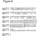
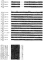
- Table 1 Multiple alignment of full length UspA1 protein sequences, associated identity percentages O12E O35E O46E P44 TTA24 TTA37 V1171 ATCC25238 81 75 83 83 84 79 84 O12E 74 77 83 76 72 75 O35E 72 74 83 73 78 O46E 81 81 82 80 P44 81 75 77 TTA24 76 84 TTA37 78
- Table 2 UspA2 Pileup Analysis - Strains and sequences used acc Strain des sl TREMBL:O54407_MORCA 054407 035E Ubiquitous surface protein A 2.
- 616 TREMBL:Q8GH86_MORCA Q8GH86 P44 UspA2.
- the present invention provides a ligand isolated from Moraxella catarrhalis outer membrane protein which has laminin and/or fibronectin and/or C3-binding, wherein said ligand is a polypeptide comprising or consisting of an amino acid sequence selected from the group consisting of SEQ ID NO: 1-10 which are derived from the full-length Moraxella catarrhalis BC5 UspA1 & UspA2 sequences shown below, or a fragment, homologue, functional equivalent, derivative, degenerate or hydroxylation, sulphonation or glycosylation product or other secondary processing product thereof.
- the ligand is a polypeptide [or polypeptide truncate compared with a wild-type polypeptide] comprising or consisting of an amino acid sequence selected from the group consisting of SEQ ID NO: 1-10, or a fragment, homologue, functional equivalent, derivative, degenerate or hydroxylation, sulphonation or glycosylation product or other secondary processing product thereof.
- ligand is used herein to denote both the whole molecule which binds to laminin and/or fibronectin and/or C3 and any part thereof which includes a laminin and/or fibronectin and/or C3-binding domain such that it retains the respective binding property.
- ligand encompasses molecules which consist only of the laminin and/or fibronectin and/or C3-binding domain i.e. the peptide region or regions required for binding.
- laminin, fibronectin or C3-binding properties of a polypeptide can be ascertained as follows:
- polypeptide [or polypeptide truncate compared with a wild-type polypeptide] comprises or consists of at least one of the conserved sequences from within SEQ ID NO: 1-10 which are identified in the alignment shown herein.
- polypeptide [or polypeptide truncate compared with a wild-type polypeptide] comprises of consists of at least one of:
- polypeptide ligands of the invention can comprise a laminin and/or fibronectin and/or C3-binding domain of sequence recited herein which is modified by the addition or deletion of amino acid residues to or from the sequences recited herein at either or both the N or C termini, which modified peptides retain the ability to bind laminin and/or fibronectin and/or C3, respectively.
- the invention further provides a ligand comprising or consisting of a polypeptide in which 50, 40, 30, 20, 10, 5, 3 or 1 amino acid residues have been added to or deleted from an amino acid sequence recited herein at either or both the N or C termini, wherein said modified polypeptide retains the ability to bind laminin and/or fibronectin and/or C3; and/or elicit an immune response against the non-modified peptide.
- a ligand comprising or consisting of a polypeptide in which 50, 40, 30, 20, 10, 5, 3 or 1 amino acid residues have been added to or deleted from an amino acid sequence recited herein at either or both the N or C termini, wherein said modified polypeptide retains the ability to bind laminin and/or fibronectin and/or C3; and/or elicit an immune response against the non-modified peptide.
- extension it is meant lengthening the sequence using the context of the peptide from the full-length amino acid sequence from which it
- any size fragment may be used in the invention (based on the homologue sequences/conserved regions/functional domatins discussed herein) provided that the fragment retains the ability to bind laminin and/or fibronectin and/or C3. It may be desirable to isolate a minimal peptide which contains only those regions required for receptor binding.
- Polypeptide ligands according to the invention may be derived from known Moraxella catarrhalis UspA1 or UspA2 proteins by truncation at either or both of the N- and C-termini. Truncates are not the full-length native UspA1 or A2 molecules.
- the invention further provides a wild-type UspA1 sequence lacking at least (or exactly) 20, 30, 40, 50, 60, 70, 80, 100, 120, 140, 160 etc to 298 amino acids from the N-terminus, and/or lacking at least (or exactly) 20, 30, 40, 50, 60, 70, 80, 100, 120, 140, 160, 180, 200 etc to 450 amino acids from the C-terminus.
- the truncate retains fibronectin binding function (optionally also laminin and/or C3-binding).
- the invention further provides a wild-type UspA2 sequence lacking at least (or exactly) 20, 30, 40, 50, 60, 70, 80, 100, 120, 140, 160, 164 amino acids from the N-terminus, and/or lacking at least (or exactly) 20, 30, 40, 50, 60, 70, 80, 100, 120, 140, 180, 200 etc to 312 amino acids from the C-terminus.
- the truncate retains fibronectin binding function (optionally also laminin and/or C3-binding). Possible truncates may be selected from those shown in the following table, all of which are within the scope of the invention.
- the invention further provides a wild-type UspA2 sequence lacking at least (or exactly) 5, 10, 15, 20, 25 or 29 amino acids from the N-terminus, and/or lacking at least (or exactly) 20, 30, 40, 50, 60, 70, 80, 100, 120, 140, 160, 180, 200 etc to 453 amino acids from the C-terminus.
- the truncate retains laminin binding function (optionally also fibronectin and/or C3-binding). Possible truncates may be selected from those shown in the following table, all of which are within the scope of the invention. Table 5. Possible combinations of truncations to the N- and C- termini of wild-type UspA2 protein No.
- the invention further provides a wild-type UspA2 sequence lacking (or exactly) 20, 30, 40, 50, 60, 70, 80, 100, 120, 140, 160 etc. to 301 amino acids from the N-terminus, and/or lacking at least (or exactly) 20, 30, 40, 50, 60, 70, 80, 100, 120, 140, 160 or 172 amino acids from the C-terminus.
- the truncate retains C3 binding function (optionally also fibronectin and/or laminin binding). Possible truncates may be selected from those shown in the following table, all of which are within the scope of the invention. Table 6. Possible combinations of truncations to the N- and C- termini of wild-type UspA2 protein No.
- Known wild-type UspA1 sequences that may be truncated in this way are those of strains ATCC25238 (MX2; GenBank accession no. AAD43465), P44 (AAN84895), 035E (AAB96359), TTA37 (AAF40122), O12E (AAF40118), O46E (AAF36416), V1171 (AAD43469), TTA24 (AAD43467) (see Table 1/ Figure 19 ); or BC5 (see above).
- Known wild-type UspA2 sequences that may be truncated in this way are those of strains 035E (GenBank accession no. 04407), MC317 (GenBank accession no.
- the UspA1 or UspA2 truncate of this embodiment comprises or consists of an amino acid sequence selected from the group consisting of SEQ ID NO: 1-10 or a fragment, homologue, functional equivalent, derivative, degenerate or hydroxylation, sulphonation or glycosylation product or other secondary processing product thereof; or comprises or consists of at least one of the conserved sequences from within these regions which are identified in the alignment shown in herein, for example:
- the invention provides fusion proteins comprising polypeptide ligands according to the invention.
- a fusion protein according to this embodiment is less than 50% identical to any known fully length sequence over its entire length.
- Such fusions can constitute a derivative of the polypeptides of the invention.
- Further derivatives can be the use of the polypeptides of the invention to as a carrier to covalently couple peptide or saccharide moieties.
- They may be coupled for instance to pneumococcal capsular oligosaccharides or polysaccharides, or Moraxella catarrhalis lipooligosaccaharides, or non-typeable Haemophilus influenzae lipooligosaccaharides.
- Homologous peptides of the invention may be identified by sequence comparison.
- Homologous peptides are preferably at least 60% identical, more preferably at least 70%, 80%, 90%, 95% or 99% identical in ascending order of preference to the peptide sequence disclosed herein or fragments thereof or truncates of the invention over their entire length.
- the homologous peptide retains the ability to bind fibronectin and/or laminin and/or C3; and/or elicit an immune response against the peptide sequences disclosed herein or fragment thereof.
- Figures 19 and 20 show an alignment of peptide sequences of UspA1 and UspA2 of different origin which indicates regions of sequence that are capable of being modified to form homologous sequences whilst retained function (i.e. fibronectin and/or laminin and/or C3 binding ability).
- Homologous peptides to the BC5 SEQ ID NO: 1-10 peptides are for instance those sequences corresponding to the BC5 sequence from other strains in Figures 19 and 20 .
- polypeptides /peptides /functional domains /homologues /fragments /truncates /derivatives of the invention should ideally be formulated as a vaccine comprising an effective amount of said component(s) and a pharmaceutically acceptable excipient.
- the vaccines of the invention can be used for administration to a patient for the prevention or treatment of Moraxella catarrhalis infection or otitis media or sinusitis or lower respiratory tract infections. They may be administered in any known way, including intramuscularly, parenternally, mucosally and intranasally.
- the vaccines of the present invention may be combined with other Moraxella catarrhalis antigens for prevention or treatment of the aforementioned diseases.
- Moraxella catarrhalis has at least 2 means of hampering the host immune system from attacking the organism.
- M. catarrhalis has a strong affinity for soluble and membrane bound human IgD through protein MID (also known as OMP106).
- Moraxella-dependent IgD-binding to B lymphocytes results in a polyclonal immunoglobulin synthesis which may prohibit production of specific monoclonal anti-moraxella antibodies.
- M. catarrhalis hampers the human immune system in several ways might explain why M. catarrhalis is such a common inhabitant of the respiratory tract.
- MID IgD-binding function
- UspA1 and/or UspA2 C3-binding function
- a further aspect of the invention is therefore a vaccine composition
- a vaccine composition comprising an effective amount of UspA1 and/or UspA2 (particularly the latter) (for instance full-length polypeptides or polypeptides /peptides /functional domains /homologues /fragments /truncates /derivatives of the invention as described herein, preferably which retains a C3-binding function) in combination with an effective amount of protein MID (for instance full-length polypeptides or polypeptides /peptides /functional domains /homologues /fragments /truncates /derivatives thereof, preferably which retain a human IgD-binding function), and a pharmaceutically acceptable excipient.
- UspA1 and/or UspA2 particularly the latter
- MID for instance full-length polypeptides or polypeptides /peptides /functional domains /homologues /fragments /truncates
- Protein MID, and IgD-binding homologous/fragments/truncates thereof is described in WO 03/004651 (incorporated by reference herein).
- Particularly suitable fragments for this purpose is a polypeptide comprising (or consisting of) the F2 fragment described in WO 03/004651 , or sequences with at least 60, 70, 80, 90, 95, 99% identity thereto which preferably retain human IgD-binding activity.
- the MID and UspA components of this combination vaccine may be separate from each other, or may be conveniently fused together by known molecular biology techniques.
- M. catarrhalis BBH18 and RH4 mutants were constructed as previously described.[23, 58]
- the M. catarrhalis strains were routinely cultured in brain heart infusion (BHI) liquid broth or on BHI agar plates at 37 °C.
- BHI brain heart infusion
- the UspA1-deficient mutants were cultured in BHI supplemented with 1.5 ⁇ g/ml chloramphenicol (Sigma, St. Louis, MO), and UspA2-deficient mutants were incubated with 7 ⁇ g/ml zeocin (Invitrogen, Carlsbad, CA). Both chloramphenicol and zeocin were used for growth of the double mutants.
- Table 7 7 ⁇ g/ml zeocin
- pAb Rabbit anti-UspA1/A2 polyclonal antibodies
- the other antibodies used were rabbit anti-human fibronectin pAb, swine FITC-conjugated anti-rabbit pAb, swine horseradish peroxidase (HRP) conjugated anti-rabbit pAb and finally a mouse anti-human CD54 (ICAM1) monoclonal antibody (mAb).
- Antibodies were from Dakopatts (Glostrup, Denmark).
- M. catarrhalis wild type strains and UspA1/A2-deficient mutants were grown overnight and washed twice in phosphate buffered saline containing 3 % fish gelatin (PBS-gelatin). The bacteria (10 8 ) were then incubated with the anti-UspA1/A2 antiserum or 5 ⁇ g fibronectin (Sigma, St Louis, MO).
- FITC-conjugated anti-rabbit pAb diluted according to the manufacturer's instructions
- rabbit anti-human fibronectin pAb if fibronectin was first added
- the bacteria were analyzed by flow cytometry (EPICS, XL-MCL, Coulter, Hialeah, FL). All incubations were kept in a final volume of 100 ⁇ l PBS-gelatin and the washings were done with the same buffer.
- Anti-fibronectin pAb and FITC-conjugated anti-rabbit pAb were added separately as a negative control for each strain analyzed.
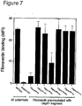
- Fibronectin inhibition studies were carried out by pre-incubating 0.25 ⁇ moles of UspA fragments for 1 h with 2 ⁇ g of fibronectin before incubation with M . catarrhalis bacteria (10 8 ). The residual free amount of fibronectin that bound to M. catarrhalis was determined by flow cytometry as outlined above.
- Fibronectin was 125 Iodine labeled (Amersham, Buckinghamshire, England) to a high specific activity (0.05 mol iodine per mol protein) with the Chloramine T method.
- M. catarrhalis strains BBH18 and RH4 together with their corresponding mutants were grown overnight on solid medium and were washed in PBS with 2 % bovine serum albumin (BSA).
- Bacteria (10 8 ) were incubated for 1 h at 37°C with 125 I-labeled fibronectin (1600 kcpm/sample) in PBS containing 2 % BSA. After three washings with PBS 2 % BSA, 125 I-labeled fibronectin bound to bacteria was measured in a gamma counter (Wallac, Espoo, Finland).
- Microtiter plates (Nunc-Immuno Module; Roskilde, Denmark) were coated with 40 nM of purified recombinant UspA1 50-770 and UspA2 30-539 proteins in 75 mM sodium carbonate, pH 9.6 at 4 °C overnight. Plates were washed four times with washing buffer (50 mM Tris-HCl, 0.15 M NaCl, and 0.1 % Tween 20, pH 7.5) and blocked for 2 h at RT with washing buffer containing 3 % fish gelatin. After four additional washings, the wells were incubated for 1 h at RT with fibronectin (120 ⁇ g/ml) diluted in three-fold step in 1.5 % fish gelatin (in wash buffer).
- washing buffer 50 mM Tris-HCl, 0.15 M NaCl, and 0.1 % Tween 20, pH 7.5
- Chang conjunctival cells (ATCC CCL 20.2) were cultured in RPMI 1640 medium (Gibco BRL, Life Technologies, Paisley, Scotland) supplemented with 10 % fetal calf serum, 2 mM L-glutamine, and 12 ⁇ g of gentamicin/ ml.
- RPMI 1640 medium Gibco BRL, Life Technologies, Paisley, Scotland
- 10 % fetal calf serum 10 % fetal calf serum
- 2 mM L-glutamine mM L-glutamine
- 12 ⁇ g gentamicin/ ml.
- cells were harvested, washed twice in gentamicin-free RPMI 1640, and added to 96 well tissue culture plates (Nunc) at a final concentration of 10 4 cells/ well in 200 ⁇ l of gentamicin-free culture medium. Thereafter, cells were incubated overnight at 37 °C in a humidified atmosphere of 5 % CO 2 and 95 %
- M. catarrhalis RH4 (10 6 ) in PBS-gelatin was inoculated onto the confluent monolayers.
- tissue culture plates were centrifuged at 3,000 x g for 5 min and incubated at 37 °C in 5 % CO 2 .
- infected monolayers were rinsed several times with PBS-gelatin to remove non-adherent bacteria and were then treated with trypsin-EDTA (0.05 % trypsin and 0.5 mM EDTA) to release the Chang cells from the plastic support. Thereafter, the resulting cell/ bacterium suspension was seeded in dilution onto agar plates containing BHI and incubated overnight at 37 °C in 5 % CO 2 .
- Chang conjunctival epithelial cells were harvested by scraping followed by re-suspension in PBS-gelatin.
- Cells (1 x 10 6 /ml) were labeled with rabbit anti-human fibronectin pAb followed by washing and incubation with a FITC-conjugated anti-rabbit pAb. After three additional washes, the cells were analyzed by flow cytometry as outlined above.
- M. catarrhalis strains BBH18 and RH4 and their corresponding mutants were previously described.[58] Both strains have a relatively higher expression of UspA2 compared to UspA1. [58] The mutants expressed equal amount of M. catarrhalis immunoglobulin D-binding protein (MID) when compared to wild type strains. Bacteria were routinely cultured in brain heart infusion (BHI) broth or on BHI agar plates at 37 °C. The UspA1-deficient, UspA2-deficient and double mutants were cultured in BHI supplemented with antibiotics as described.[58]
- Rabbit anti-UspA1/A2 and anti-MID polyclonal antibodies were used.[22, 58] Rabbit anti-laminin pAb was from Sigma (St Louis, MO, USA). Swine horseradish peroxidase (HRP)-conjugated anti-rabbit pAb was from Dakopatts (Glostrup, Denmark).
- HRP horseradish peroxidase
- Microtiter plates (Nunc-Immuno Module; Roskilde, Denmark) were coated with Engelbreth-Holm-Swarm mouse sarcoma laminin (Sigma, Saint Louis, USA) or bovine serum albumin (BSA) (30 ⁇ g/ml) in Tris-HCL, pH 9.0 at 4°C overnight.
- the plates were washed with phosphate buffered saline and 0.05% Tween 20, pH 7.2 (PBS-Tween) and subsequently blocked with 2 % BSA in PBS + 0.1 % Tween 20, pH 7.2.
- M. catarrhalis RH4 and BBH18 (10 8 ) in 100 ⁇ l were then added followed by incubation for 1 h.
- Unbound bacteria were removed by washing 3 times with PBS-Tween. Residual bound bacteria were detected by means of an anti-MID pAb, followed by detection with HRP-conjugated anti-rabbit pAb. The plates were developed and measured at OD 450 according to a standard protoccol.
- Microtiter plates (Nunc-Immuno Module) were coated with 40 nM of purified recombinant UspA1 50-770 and UspA2 30-539 proteins in 75 mM sodium carbonate, pH 9.6 at 4°C. Plates were washed four times with washing buffer (50 mM Tris-HCl, 0.15 M NaCl, and 0.1 % Tween 20, pH 7.5) and blocked at RT with washing buffer containing 3 % fish gelatin. After additional washings, the wells were incubated for 1 h at RT with laminin at different dilutions as indicated in 1.5 % fish gelatin (in wash buffer). Thereafter, the plates were washed and incubated with rabbit anti-laminin pAb.
- washing buffer 50 mM Tris-HCl, 0.15 M NaCl, and 0.1 % Tween 20, pH 7.5
- washing buffer 50 mM Tris-HCl, 0.15 M NaCl, and 0.1 % Tween 20,
- HRP-conjugated anti-rabbit pAb was added and incubated at RT. Both the anti-laminin and HRP-conjugated anti-rabbit pAb were diluted 1:1,000 in washing buffer containing 1.5 % fish gelatin. The wells were washed and the plates were developed and measured at OD 450 . Uncoated wells incubated with identical dilutions of laminin were used as background controls. ELISAs with truncated proteins spanning UspA 50-770 and UspA2 30-539 were performed with fixed doses of laminin (20 ⁇ g/ml).
- Type strains were from the Culture Collection, University of Gothenburg (CCUG; Department of Clinical Bacteriology, Sahlgrenska Hospital, Gothenburg, Sweden), or the American Type Culture Collection (ATCC; Manassas, Va); Neisseria gonorrheae CCUG 15821, Streptococcus pyogenes CCUG 25570 and 25571, Streptococcus agalactiae CCUG 4208, Streptococcus pneumoniae ATCC 49619, Legionella pneumophila ATCC 33152, Pseudomonas aeruginosa ATCC 10145, Staphylococcus aureus ATCC 29213, and finally Staphylococcus aureus ATCC 25923.
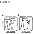
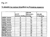
- catarrhalis is a unique C3/ C3met binding bacterium. Related moraxella subspecies and other common human pathogens do not bind C3/ C3met (mfi ⁇ 2.0). After incubation with EDTA-treated NHS or C3met, bacteria were analysed by flow cytometry using a rabbit anti-C3d pAb and a FITC-conjugated goat anti-rabbit pAb. Species NHS-EDTA (mfi) C3met (mfi) Moraxella catarrhalis RH4 8.7 22.1 M.
- M . catarrhalis strains were routinely cultured in brain heart infusion (BHI) liquid broth or on BHI agar plates at 37°C.
- M . catarrhalis BBH18 and RH4 mutants were manufactured as previously described.[22, 23, 58]
- the MID-deficient mutants were grown in BHI containing 50 ⁇ g/ml kanamycin.
- the UspA1-deficient mutants were cultured in BHI supplemented with 1.5 ⁇ g/ml chloramphenicol (Sigma, St.
- the rabbit anti-human C3d pAb and the FITC-conjugated swine anti-rabbit pAb were purchased from Dakopatts (Glostrup, Denmark), and the goat anti-human C3 were from Advanced Research Technologies (San Diego, CA).
- the horseradish peroxidase (HRP)-conjugated donkey anti-goat pAb was obtained from Serotec (Oxford, UK).
- bacteria were incubated with anti-human C3d pAb for 30 min on ice, followed by washings and incubation for another 30 min on ice with FITC-conjugated goat anti-rabbit pAb. After three additional washes, bacteria were analyzed by flow cytometry (EPICS, XL-MCL, Coulter, Hialeah, FL). All incubations were kept in a final volume of 100 ⁇ l PBS-BSA and the washings were done with the same buffer. The anti-human C3d pAb and FITC-conjugated anti-rabbit pAb were added separately as a negative control for each strain analyzed.
- Normal human serum was obtained from five healthy volunteers. The blood was allowed to clot for 30 min at room temperature and thereafter incubated on ice for 60 min. After centrifugation, sera were pooled, aliquoted and stored at -70°C. To inactivate both the classical and alternative pathways, 10 mM EDTA was added. In contrast, Mg-EGTA was included to inactivate the classical pathway.
- Human serum deficient in the C4BP was prepared by passing fresh serum through a HiTrap column (Amersham Biosciences) coupled with mAb 104, a mouse mAb directed against CCP1 of the ⁇ -chain of C4BP.[41] The flow through was collected and the depleted serum was stored in aliquots at -70°C.
- Serum depleted of C1q was obtained via the first step of C1q purification [79] using Biorex 70 ion exchange chromatography (Bio-Rad, Hercules, CA). The resulting sera displayed normal haemolytic activity. The factor D and properdin deficient serum was kindly provided by Dr. Anders Sjöholm (Department of Medical Microbiology, Lund University, Lund, Sweden). M. catarrhalis strains were diluted in 2.5 mM Veronal buffer, pH 7.3 containing 0.1 % (wt/vol) gelatin, 1 mM MgCl 2 , 0.15 mM CaCl 2 , and 2.5 % dextrose (DGVB ++ ).
- Bacteria (10 3 cfu) were incubated together with 10 % NHS and EDTA or Mg-EGTA in a final volume of 100 ⁇ l.
- the bacteria/ NHS was incubated at 37°C and at various time points, 10 ⁇ l aliquots were removed and spread onto BHI agar plates.
- 10 % serum was incubated with 100 nM of the recombinant UspA1 50-770 and UspA2 30-S39 proteins for 30 min at 37°C before bacteria were added.
- Microtiter plates (Nunc-Immuno Module; Roskilde, Denmark) were coated with triplets of purified recombinant UspA1 50-770 , UspA2 30-539 , or the truncated UspA1 and UspA2 fragments (40 nM in 75 mM sodium carbonate, pH 9.6) at 4°C overnight. Plates were washed four times with washing buffer (PBS with 0.1 % Tween 20, pH 7.2) and blocked for 2 hrs at room temperature with washing buffer supplied with 1.5 % ovalbumin (blocking buffer). After washings, the wells were incubated overnight at 4°C with 0.25 ⁇ g C3met in blocking buffer.
- washing buffer PBS with 0.1 % Tween 20, pH 7.2
- Rabbit erythrocytes were washed three times with ice-cold 2.5 mM Veronal buffer, pH 7.3 containing 0.1 % (wt/vol) gelatin, 7 mM MgCl 2 , 10 mM EGTA, and 2.5 % dextrose (Mg ++ EGTA), and resuspended at a concentration of 0.5 x 10 9 cells/ml.
- Erythrocytes were incubated with various concentrations (0 to 4 %) of serum diluted in Mg ++ EGTA. After 1 h at 37°C, erythrocytes were centrifuged and the amount of lysed erythrocytes was determined by spectrophotometric measurement of released hemoglobin at 405 nm.
- PMN Human polymorphonuclear leukocytes
- M. catarrhalis devoid of UspA1 and A2 does not bind soluble or immobilized fibronectin.
- M . catarrhalis BBH18 strongly bound fibronectin with a mean fluorescence intensity (MFI) of 96.1 ( figure 1 F ).
- MFI mean fluorescence intensity
- BBH18 ⁇ uspA1 showed a decreased fibronectin binding with an MFI of 68.6 ( figure 1G ).
- Fibronectin binding to BBH18 ⁇ uspA2 and the double mutant BBH18 ⁇ uspA1 / A2 revealed an MFI of only 10.7 and 11.5, respectively ( figure 1 H , 1 I ). Similar results were obtained with UspA1/A2mutants of the clinical strain M . catarrhalis RH4. Taken together, these results suggest that UspA1 and A2 bound fibronectin and that the ability of the bacteria to bind fibronectin strongly depended on UspA1/A2 expression.
- M . catarrhalis RH4 and its corresponding ⁇ uspA1 / A2 mutants were applied onto fibronectin coated glass slides. After 2 h of incubation, slides were washed, and subsequently Gram stained. M . catarrhalis wild type and the ⁇ uspA1 mutant were found to strongly adhere to the fibronectin coated glass slides ( figure 3A and 3B ). In contrast, M . catarrhalis ⁇ uspA2 and ⁇ uspA1 / A2 double mutants weakly adhered to the fibronectin coated glass slide with only a few bacteria left after washing ( figure 3 C and 3 D , respectively). Experiments with another M . catarrhalis clinical isolate (BBH18) and its derived mutants showed a similar pattern indicating that UspA2 was of major importance for M . catarrhalis binding to immobilized fibronectin.
- the fibronectin binding domains include amino acid residues located between 299 and 452 of UspA1 and between 165 and 318 of UspA2
- truncated UspA1 50-770 and UspA2 30-539 were recombinantly produced in E. coli, coated on microtiter plates and incubated with increasing concentrations of fibronectin. Bound fibronectin was detected with an anti-human fibronectin pAb followed by incubation with a horseradish peroxidase conjugated anti-rabbit pAb. Both recombinant UspA1 50-770 and UspA2 30-539 bound soluble fibronectin and the interactions were dose-dependent ( figure 4 ).
- fibronectin-binding domain of UspA1 To define the fibronectin-binding domain of UspA1, recombinant proteins spanning the entire molecule of UspA1 50-770 were manufactured. Fibronectin was incubated with the immobilized UspA1 proteins fragments and the interactions were quantified by ELISA. UspA1 50-491 bound fibronectin almost as efficiently as UspA1 50-770 suggesting that the binding domain was within this part of the protein. Among the other truncated fragments, UspA1 299-452 efficiently bound fibronectin ( figure 5 A ). In parallel, the interactions between fibronectin and several recombinant UspA2 fragments including amino acids UspA2 30-539 were analyzed.
- UspA1 50-491 and UspA1 299-4s2 fragments competitively inhibit M. catarrhalis fibronectin binding
- fibronectin binding domains of UspA1 and A2 block the interactions between fibronectin and M. catarrhalis.
- UspA1 299-452 and UspA2 165-318 inhibit M. catarrhalis adherence to Chang epithelial cells
- Epithelial cells are known to express fibronectin and many bacteria attach to epithelial cells via cell-associated fibronectin.[46, 54, 69, 77] Previous studies have shown that M. catarrhalis adhere to epithelial cells.[43, 49] We analyzed Chan conjunctival cells, which have frequently been used in adhesion experiments with respiratory pathogens. Chang cells strongly expressed fibronectin as revealed by flow cytometry analysis ( figure 8A ).
- Chang epithelial cells were pre-incubated with anti-human fibronectin pAb, or the recombinant proteins UspA1 299-452 and UspA2 165-318 . Thereafter, M. catarrhalis RH4 was added and bacterial adhesion analyzed.
- the relative adherence (measured by the number of colony forming units) after pre-incubation with 0.4 ⁇ moles per 200 ⁇ l of UspA1 299-452 , UspA2 165-318 , or an anti-human fibronectin pAb were 36 %, 35 % and 32%, respectively.
- fibronectin on Chang epithelial cells may function as a receptor for M. catarrhalis and the amino acid residues 299-452 of UspA1 and 165-318 of UspA2 contain the ligand responsible for the interactions.
- M. catarrhalis RH4 strongly bound to immobilized laminin.( figure 9A ).
- M. catarrhalis RH4 uspA1 mutant (RH4 ⁇ uspA1 ) showed a laminin binding of 89.9% of the wild type.
- M. catarrhalis RH4 uspA2 mutant (RH4 ⁇ uspA2 ) and the double mutant RH4 ⁇ uspA1 / A2 15.2% and 18.1% binding capacity of the wild type, respectively.
- M. catarrhalis Interaction between M. catarrhalis and C3 and C3met M. catarrhalis outer membrane proteins UspA1 and UspA2 inhibit both the classical and the alternative pathway of the complement cascade
- UspA2 surface expression is crucial for M. catarrhalis survival in normal human serum (NHS) [1, 58], i.e., moraxella UspA2 deficient mutants are rapidly killed when exposed to NHS.
- NHS human serum
- both UspA1 and A2 bind C4BP and thus might inhibit the classical pathway of complement activation [58].
- Mg-EGTA MgCl 2
- EDTA EDTA
- EDTA inhibits all complement pathways by absorbing divalent cations (Mg 2+ and Ca 2+ ).
- M. catarrhalis RH4 wild type survived after 30 min of incubation, whereas RH4 ⁇ uspA1 / A2 double mutant was killed by intact NHS after 10 min ( Fig. 12 ).
- the classical pathway was inhibited (NHS + Mg-EGTA)
- the RH4 ⁇ uspA1 / A2 mutant survived for a significantly longer period of time as compared to NHS without any chelators, but not as long as the wild type bacterium.
- M. catarrhalis RH4 ⁇ uspA1 / A2 survived.
- M. catarrhalis a pathogen that frequently colonizes the human respiratory tract, does not only counteract the classical pathway but also the alternative pathway of the complement system by the outer membrane proteins UspA1 and A2.
- C3b covalently binds to the surface of a microbe and hence induces the alternative pathway ( Fig. 11B ).
- our RH4 wild type strain was incubated with NHS or NHS treated with EDTA. Binding or deposition (via covalent link) of C3/ C3b at the bacterial surface of M. catarrhalis RH4 was detected by flow cytometry analysis with a polyclonal antibody (pAb) directed against C3d recognizing both C3 and C3b.
- pAb polyclonal antibody directed against C3d recognizing both C3 and C3b.
- Incubation of bacteria with NHS containing intact complement led to deposition of C3 ( Fig. 13 ).
- the complement cascade was inactivated in the presence of EDTA, the M.
- catarrhalis RH4 still bound C3 ( Fig. 13A ). Streptococcus pneumoniae that was included for comparison did not absorb C3 from the EDTA-treated serum ( Fig. 13B ). In contrast to pneumococci, M. catarrhalis thus bound C3 irrespectively of complement activation. The internal thioester of C3 is spontaneously hydrolysed in fluid phase to C3(H 2 O). Thus, intact C3 or C3(H 2 O) was the most likely forms of C3 interacting with M. catarrhalis. Since M. catarrhalis also binds C4BP [58], we wanted to exclude that C4BP was involved in the C3 binding and for that purpose we used C4BP depleted serum. M. catarrhalis absorbed C3 from the C4BP depleted serum to the same extent as to NHS (not shown).
- M. catarrhalis is a unique C3 and C3met binding bacterium
- M. catarrhalis was the only bacterium binding C3 in complement-inactivated serum (Table 9). All related moraxella strains as well as the other human pathogens were also analyzed for binding of C3met. In parallel with the C3 binding, M. catarrhalis was the only species that bound C3met. Taken together, M. catarrhalis has a unique feature to strongly bind C3 and C3met in a non-covalent manner.
- M. catarrhalis binds C3met via the outer membrane proteins UspA1 and UspA2
- M. catarrhalis protein responsible for the C3 binding we tested a series of bacterial mutants devoid of the outer membrane proteins MID, UspA1 and/ or UspA2 [22, 58]. Interestingly, the binding of C3met was significantly correlated with Usp expression ( Fig. 15 ). M. catarrhalis RH4 ⁇ mid bound C3met to the same degree as the wild type counterpart ( Fig. 15 A-B ). The RH4 ⁇ uspA1 mutant showed only a slightly decreased binding, whereas the RH4 ⁇ uspA2 was a weaker binder as compared to the wild type counterpart ( Fig. 15 C-D ).
- a C3 binding domain is located between amino acid residues 200 and 458 of UspA2.
- UspA2 30-539 was most efficient in inhibiting bacterial killing as compared to UspA1 50-770 .
- both recombinant proteins were supplemented together, no additional inhibition of the alternative pathway was detected.
- Ten % NHS correspond to approximately 600 nM C3.
- UspA1 50-770 and/ or UspA2 30-539 up to 600 nM was added. However, higher concentrations of the recombinant proteins did not further increase the inhibition (not shown).
- C3b and iC3b target microbes for removal in the process of opsonophagocytosis.
- C3 or C3met that was non-covalently bound at the surface of M. catarrhalis could still function as an opsonin, a series of phagocytosis experiments was performed. M. catarrhalis was preincubated with C3met, NHS or NHS treated with EDTA followed by addition of polymorphonuclear leukocytes. Interestingly, M. catarrhalis was not engulfed in the presence of C3met, whereas NHS strongly promoted phagocytosis (data not shown).
- UspA1 299-452 and UspA2 165-318 from the clinical M. catarrhalis strain Bc5 were the shortest fragments that still bound fibronectin.
- longer fragments encompassing the amino acid sequence found within UspA1 299-452 and UspA2 165-318 displayed a more efficient binding to fibronectin ( figure 5 A and B ). This may mean that these two regions represent partial binding domains or that the binding site is highly dependent on a specific molecular structure.
- UspA1 299-452 and UspA2 165-318 share a sequence of 31 identical amino acid residues including the 23 residues "NNINNIYELAQQQDQHSSDIKTL" (SEQ ID NO: 85) (NNINNIY (SEQ ID NO: 86) sequence).
- This sequence contains the epitope for the protective monoclonal antibody (mAb) 17C7 for which there is universal reactivity.[2, 50, 30]
- mAb 17C7 In a mouse model, passive immunization with mAb 17C7 provided protection and improved pulmonary clearance of M. catarrhalis.
- the fibronectin binding M. catarrhalis BBH18 and RH4 used in our experiments also carry the 31 amino acid residues in their UspA1/A2 protein. Most M. catarrhalis have a part of this sequence (i.e., the NNINNIY (SEQ ID NO: 86) sequence). However, strains like the O35E which has the NNINNIY (SEQ ID NO: 86) sequence in their UspA2 gene do not express a fibronectin binding UspA2 protein.[49] A likely explanation would be that the variations in the flanking regions might affect the interaction with fibronectin.
- M. catarrhalis is a common cause of infectious exacerbations in patients with COPD.
- the success of this species in patients with COPD is probably related in part to its large repertoire of adhesins.
- pathological changes such as loss of epithelial integrity with exposure of basement membrane where the laminin layer itself is thickened in smokers.
- Some pathogens have been shown to be able to bind to laminin and thus may contribute to their ability to adhere to such damaged and denuded mucosal surfaces. These include pathogens known to cause significant disease in the airways such as S. aureus and P. aeruginosa amongst others.
- S. aureus and P. aeruginosa amongst others.
- MSCRAMMS extracellular matrix recognizing adhesive matrix molecules
- Complement resistance is one of the most important bacterial virulence factors.
- M. catarrhalis isolates from patients with lower respiratory tract infections are resistant to complement-mediated killing.
- M. catarrhalis UspA1 and A2 are crucial for bacterial survival in human serum in vivo [1, 15], and we have shown that these two outer membrane proteins bind to the complement fluid phase regulator of the classical pathway, C4BP.[58]
- C3 complement fluid phase regulator of the classical pathway
- M. catarrhalis is equally resistant to both the classical and alternative pathways ( Fig. 12B ).
- the bacterium binds C4BP that inhibits the classical pathway [58] and in this paper we demonstrate an interaction with the alternative pathway through binding of C3.
- serum resistance in various in vivo situations is difficult.
- the importance of the classical pathway will strongly depend on history of infections with M. catarrhalis and ability to generate complement-activating antibodies.
- every mechanism providing protection from the complement is certainly beneficial for a pathogen. Since C3 is a key molecule in the complement system, the binding of C3 most likely results in regulation of all three activation pathways and may contribute the most to serum resistance.
- PspA Pneumococcal Surface Protein A
- UspA1 and A2 absorb C3 from serum and hereby most likely inhibit the complement activation.
- PspA Pneumococcal Surface Protein A
- PspA is an important virulence factor for S. pneumoniae.
- PspA-deficient pneumococcal strains are readily cleared from the blood, whereas the PspA-expressing strains survive.[82]
- PspA-deficient pneumococci have a significantly reduced virulence compared with pneumococci that express PspA.
- Efb extracellular fibrinogen-binding protein
- CbpA pneumococcal choline-binding protein
- CbpA is a component of the pneumococcal cell wall, but may only bind C3 when the CbpA is secreted.
- eleven different pneumococcal isolates for C3 binding methylamine-treated C3 or NHS-EDTA
- flow cytometry Fig. 12B and Table 9
- No bound C3 could be detected on the surface of S. pneumoniae.
- lysates of S. pneumoniae and culture supernatants were analyzed on Western blots using methylamine-treated C3 followed by an anti-human C3 pAb, we confirmed the results by Cheng and collaborators [16] (not shown).
- the Gram-negative M. catarrhalis is a unique species with membrane anchored proteins that bind C3 and inhibit the alternative pathway at the surface of the bacterium.
- the yeast Candida albicans has been shown to bind C3b, iC3b and C3d. However, C3b is bound at a considerably lower affinity than iC3b and C3d.[29] We found a large difference between C3 binding to M. catarrhalis and C. albicans (not shown); despite that candida bound C3met (56 % positive cells), the mean fluorescence intensity (mfi) was only ⁇ 2.0 as compared to mfi 36.9 for M. catarrhalis. Furthermore, no detectable binding was seen when C. albicans was incubated with EDTA-treated serum. Two C3d-binding proteins have been isolated from C.
- albicans and the most characterized protein is a 60 kDa mannoprotein that initially was recognized by an antibody directed against human complement receptor 2 (CD21).[13] However, M. catarrhalis UspA1 and A2 were not recognized by a polyclonal antibody directed against CD21 (not shown). In parallel with staphylococci and pneumococci [52, 64], a secreted C3d-binding protein from C. albicans also exists.[72] Finally, a C. albicans iC3b receptor has been isolated and is structurally similar to human CR3 (CD11b).[3] The mechanisms by which these receptors may participate in pathogenesis are not fully known.
- C3 binding pathogens are notably different from M. catarrhalis in that these species often are blood stream isolates.
- M. catarrhalis is mucosal pathogen with rare instances of bacteremic infections. Hence, the binding and inactivating C3 most likely occur at the mucosal surface.
- MEEs middle-ear effusions
- C3 products can also be found.
- complement factors in MEEs fluid have been shown to be important in the bactericidal activity against other mucosal agents such as non-typable H. influenzae.
- M. catarrhalis is a strict human pathogen. It does not cause diseases such as otitis media or pneumonia in animals.
- M. catarrhalis hampers the human immune system in several ways might explain why M. catarrhalis is such a common inhabitant of the respiratory tract [73].
- M. catarrhalis has developed sophisticated ways of combating both the humoral and innate immune systems.
- the present data show that M. catarrhalis has a unique C3-binding capacity at the bacterial cell surface that cannot be found in other bacterial species.
Abstract
Description
- The present invention relates to Moraxella catarrhalis and their ability to interact with epithelial cells via extracellular matrix proteins such as fibronectin and laminin, and also to their ability to inhibit the complement system. The interaction with these extracellular proteins is useful in the preparation of vaccines.
- The ability to bind epithelial cells is of great importance for several bacterial species. For example, Staphylococcus aureus and Streptococcus pyogenes possess fibronectin binding proteins (FnBP) with related sequence organization. These FnBP are known as Microbial Surface Components Recognizing Adhesive Matrix Molecules (MSCRAMMs). They exploit the modular structure of fibronectin forming extended tandem beta-zippers in its binding to fibronectin. [27, 39, 47, 73] The function is to mediate bacterial adhesion and invasion of host cells.
- The important mucosal pathogen Moraxella catarrhalis is the third leading bacterial cause of acute otitis media in children after Streptococcus pneumoniae and Haemophilus influenzae.[14, 40, 55] M. catarrhalis is also one of the most common inhabitants of the pharynx of healthy children.
- Furthermore, M. catarrhalis is also a common cause of sinusitis and lower respiratory tract infections in adults with chronic obstructive pulmonary disease (COPD). [74] The success of this species in patients with COPD is probably related in part to its large repertoire of adhesins.
- Recent years focus of research has been on the outer membrane proteins and their interactions with the human host. [6, 48, 56] Some of these outer membrane proteins appear to have adhesive functions including amongst others, M. catarrhalis IgD binding protein (MID, also designated Hag), protein CD, M. catarrhalis adherence protein (McaP) and the ubiquitous surface proteins (Usp). [1, 22, 33, 48, 61, 81, 84]
- In view of the fact that M. catarrhalis has been found to be such a leading cause of infections in the upper and lower airways, there is a current need to develop vaccines which can be used against M. catarrhalis.
- The aim of the present invention has therefore been to find out in which way M. catarrhalis interacts with epithelial cells in the body and affects the immune system. In this way, substances that can act as vaccines against M. catarrhalis can be developed.
- In this study, using M. catarrhalis mutants
derived from clinical isolates, the inventors have been able to show that both UspA1 and A2 bind fibronectin and laminin. Furthermore, the inventors have been able to show that M. catarrhalis interfere with the classical pathway of the complement system, and also to elucidate in which way they interfere. - Many bacteria adhere to epithelial cells via fibronectin binding MSCRAMMS.[54, 77] Pseudomonas aeruginosa has a FnBP that binds to cellular associated fibronectin on nasal epithelial cells.[69] Blocking the bacteria-fibronectin protein interactions may help the host tissue to overcome the infection. In fact, it has been shown that antibodies against a S. aureus FnBP resulted in rapid clearance of the bacteria in infected mice.[71]
- Recombinant truncated UspA1/A2 proteins together with smaller fragments spanning the entire molecule have been tested according to the present invention for fibronectin binding. Both UspA1 and A2 bound fibronectin and the fibronectin binding domains were found to be located within UspA1299-452. and UspA2165-318. These two truncated proteins both inhibited binding of M. catarrhalis to Chang conjunctival epithelial cells to a similar extent as anti-fibronectin antibodies. The observations made show that both M. catarrhalis UspA1 and A2 are involved in the adherence to epithelial cells via cell-associated fibronectin. The biologically active sites within UspA1299-452 and UspA2165-318 are therefore suggested as potential candidates to be included in a vaccine against M. catarrhalis.
- Further, the inventors have studied and characterized binding of M. catarrhalis to laminin. M. catarrhalis is a common cause of infectious exacerbations in patients with COPD. The success of this species in patients with COPD is probably related in part to its large repertoire of adhesins. In addition, there are pathological changes such as loss of epithelial integrity with exposure of basement membrane where the laminin layer itself is thickened in smokers. [4] Some pathogens have been shown to be able to bind laminin and this may contribute to their ability to adhere to such damaged and denuded mucosal surfaces. These include pathogens known to cause significant disease in the airways such as S. aureus and P. aeruginosa amongst others. [7, 63] The present inventors have been able to show that M. catarrhalis ubiquitous surface protein (Usp) A1 and A2 also bind to laminin. Laminin binding domains of UspA1 and A2 were, amongst others, found within the N-terminal halves of UspA150-491 and UspA230-351. These domains are also containing the fibronectin binding domains. However, the smallest fragments that bound fibronectin, UspA1299-452 and UspA2165-318, did not bind laminin to any appreciable extent. Fragments smaller than the N-terminal half of UspA1 (UspA150-491) lose all its laminin binding ability, whereas with UspA2, only UspA230-170 bound laminin albeit at a lower level than the whole recombinant protein (UspA230-539) . These findings suggest that different parts of the molecule might have different functional roles. UspA150-770 was also found to have laminin binding properties.
- Comparing the smallest laminin binding regions of UspA1 and A2, we find that there is, however, little similarity by way of amino acid homology between UspA230-170 and UspA150-491 (data not shown). This is not surprising as it is a known fact that both proteins have a 'lollipop'-shaped globular head structure despite having only 22% identity in both N terminal halves.[2, 32]
The biologically active sites within UspA150-770 and UspA230-039 are suggested as potential candidates to be included in a vaccine against M. catarrhalis. - Finally, the inventors have studied the interaction between M. catarrhalis ubiquitous surface proteins A1 and A2 and the innate immune system, and have found that M. catarrhalis interferes with the complement system. The complement system is one of the first lines of innate defence against pathogenic microorganisms, and activation of this system leads to a cascade of protein deposition on the bacterial surface resulting in formation of the membrane attack complex or opsonization of the pathogen followed by phagocytosis. [85, 86] One of the most important complement proteins is C3, which is present in the circulation in a concentration similar to some immunoglobulins (1-1.2 mg/ml). C3 does not only play a crucial role as an opsonin, but also is the common link between the classical, lectin and alternative pathways of the complement activation. The alternative pathway functions as amplification loop for the classical and lectin pathways and can also be spontaneously activated by covalent attachment of C3 to the surface of a microbe in the absence of complement inhibitors. C3 deposition requires the presence of an internal thioester bond, formed in the native protein by the proximity of a sulfhydryl group (Cys1010) and a glutamyl carbonyl (Gln1012) on the C3 α-chain. [76] Proteolytic cleavage of a 77-residue peptide from the amino terminus of the C3 α-chain generates C3a (anaphylatoxin) and C3b. Attachment of C3b is then accomplished through a covalent link between the carbonyl group of the metastable thioester and either -NH2 or -OH groups of proteins or carbohydrate structures on the activator surface. [36, 37] M. catarrhalis UspA1 and A2 have been found to non-covalently and in a dose dependent manner bind both the third component of complement (C3) from EDTA-treated serum and methylamine treated C3 (C3met). UspA150-770 and UspA230-539 have been found to bind to C3 and C3met. The C3-binding region for UspA2 was found to mainly be localised in UspA2200-458. UspA1 has however been found to have a minor role in the interactions. The biologically active sites within UspA150-770 and UspA230-539 are suggested as potential candidates to be included in a vaccine against M. catarrhalis.
- The UspA family consists of UspA1 (molecular weight 88 kDa), UspA2 (62 kDa), and the hybrid protein UspA2H (92 kDa).[2, 43] These proteins migrate as high molecular mass complexes in SDS-PAGE, are relatively conserved and hence important vaccine candidates. The amino acid sequences of UspA1 and A2 are 43 % identical and have 140 amino acid residues that are 93 % identical. [2] In a series of 108 M. catarrhalis nasopharyngeal isolates from young children with otitis media, uspA1 and uspA2 genes were detected in 107 (99 %) and 108 (100 %) of the isolates, respectively. Twenty-one percent were identified as having the hybrid variant gene uspA2H.[50] Moreover, it is known that naturally acquired antibodies to UspA1 and A2 are bactericidal.[15]
- Several functions have been attributed to the UspA family of proteins. UspA1 expression is essential for the attachment of M. catarrhalis to Chang conjunctival epithelial cells and Hep-2 laryngeal epithelial cells.[43, 49] In a more recent study, UspA1 was shown to bind carcinoem-bryonic antigen related cell adhesion molecules (CEACAM) expressed in the lung epithelial cell line A549.[31] Purified UspA1 has also been shown to bind fibronectin in dot blot experiments while purified UspA2 did not. [49] Both UspA1 and A2 may play important roles for M. catarrhalis serum resistance. [1, 5, 58, 60]
- The present invention demonstrates that both UspA1 and A2 are determinants for M. catarrhalis binding to fibronectin and laminin in the clinical isolates M. catarrhalis BBH18 and RH4. Interestingly, recombinant UspA1 and A2 derived from M. catarrhalis Bc5 both bound fibronectin to the same extent. The binding domains for fibronectin were found within
amino acid residues 299 to 452 of UspA1 and 165 to 318 of UspA2. These two domains share 31 amino acid residues sequence identity. Importantly, truncated protein fragments containing these residues in UspA1 and UspA2 were able to inhibit M. catarrhalis binding to Chang epithelial cells suggesting that the interactions with these cells were via cell-associated fibronectin.
The binding domains for laminin were found within the
amino acid residues mentioned above. Binding assays with recombinant proteins revealed that the major binding regions were localized in the N-terminal parts, where both proteins form a globular head.
Bacterial factors mediating adherence to tissue and extracellular matrix (ECM) components are grouped together in a single family named microbial surface components recognizing adhesive matrix molecules" (MSCRAMMS). Since UspA1/A2both bind fibronectin and laminin, these proteins can be designated MSCRAMMS. - According to one aspect the present invention provides a peptide having sequence ID no. 1, and fragments, homologues, functional equivalents, derivatives, degenerate or hydroxylation, sulphonation or glycosylation products and other secondary processing products thereof.
- According to another aspect the present invention provides a peptide having sequence ID no. 2, and fragments, homologues, functional equivalents, derivatives, degenerate or hydroxylation, sulphonation or glycosylation products and other secondary processing products thereof.
- According to a further aspect the present invention provides a peptide having sequence ID no. 3, and fragments, homologues, functional equivalents, derivatives, degenerate or hydroxylation, sulphonation or glycosylation products and other secondary processing products thereof.
- According to another aspect the present invention provides a peptide having sequence ID no. 4, and fragments, homologues, functional equivalents, derivatives, degenerate or hydroxylation, sulphonation or glycosylation products and other secondary processing products thereof.
- According to a further aspect the present invention provides a peptide having sequence ID no. 5, and fragments, homologues, functional equivalents, derivatives, degenerate or hydroxylation, sulphonation or glycosylation products and other secondary processing products thereof.
- According to a further aspect the present invention provides a peptide having sequence ID no. 6, and fragments, homologues, functional equivalents, derivatives, degenerate or hydroxylation, sulphonation or glycosylation products and other secondary processing products thereof.
- According to another aspect the present invention provides a peptide having sequence ID no. 7, and fragments, homologues, functional equivalents, derivatives, degenerate or hydroxylation, sulphonation or glycosylation products and other secondary processing products thereof.
- According to another aspect the present invention provides a peptide having sequence ID no. 8, and fragments, homologues, functional equivalents, derivatives, degenerate or hydroxylation, sulphonation or glycosylation products and other secondary processing products thereof.
- According to another aspect the present invention provides a peptide having sequence ID no. 9, and fragments, homologues, functional equivalents, derivatives, degenerate or hydroxylation, sulphonation or glycosylation products and other secondary processing products thereof.
- According to another aspect the present invention provides a peptide having sequence ID no. 10, and fragments, homologues, functional equivalents, derivatives, degenerate or hydroxylation, sulphonation or glycosylation products and other secondary processing products thereof.
- According to another aspect, the present invention provides use of at least one peptide according to the invention for the production of a medicament for the treatment or prophylaxis of an infection, preferably an infection caused by M. catarrhalis, in particular caused by carriage of M. catarrhalis on mucosal surfaces.
- According to another aspect, the invention further provides a ligand comprising a fibronectin binding domain, said ligand consisting of an amino acid sequence selected from the group consisting of Sequence ID No. 1, Sequence ID No. 2 and Sequence ID No. 3, and fragments, homologues, functional equivalents, derivatives, degenerate or hydroxylation, sulphonation or glycosylation products and other secondary processing products thereof.
The invention further provides a ligand comprising a laminin binding domain, said ligand consisting of an amino acid sequence selected from the group consisting of Sequence ID No. 4 to Sequence ID No. 8, and fragments, homologues, functional equivalents, derivatives, degenerate or hydroxylation, sulphonation or glycosylation products and other secondary processing products thereof. - Further, the present invention provides a ligand comprising a C3 or C3met binding domain, said ligand consisting of an amino acid sequence selected from the group consisting of Sequence ID No. 4, Sequence ID No. 6, Sequence ID No. 9 and Sequence ID No. 10, and fragments, homologues, functional equivalents, derivatives, degenerate or hydroxylation, sulphonation or glycosylation products and other secondary processing products thereof.
- Further, the present invention provides a medicament comprising one or more ligands according to the invention and one or more pharmaceutically acceptable adjuvants, vehicles, excipients, binders, carriers, or preservatives. The present invention further provides a vaccine comprising one or more ligands according to the present invention and one or more pharmaceutically acceptable adjuvants, vehicles, excipients, binders, carriers, or preservatives.
The present invention also provides a method of
treating or preventing an infection in an individual, preferably an infection caused by M. catarrhalis, in particular caused by carriage of M. catarrhalis on mucosal surfaces, comprising administering a pharmaceutically effective amount of a medicament or vaccine according to the present invention. - Finally, the present invention also provides a nucleic acid sequence encoding a ligand, protein or peptide of the present invention, as well as homologues, polymorphisms, degenerates and splice variants thereof.
- Further disclosure of the objects, problems, solutions and features of the present invention will be apparent from the following detailed description of the invention with reference to the drawings and the appended claims.
- The expression ligand as it is used herein is intended to denote both the whole molecule which binds to the receptor and any part thereof which includes the receptor binding domain such that it retains the receptor binding property. Ligands comprising equivalent receptor binding domains are also included in the present invention.
- The expressions fragment, homologue, functional equivalent and derivative relate to variants, modifications and/or parts of the peptides and protein fragments according to the invention which retain the desired fibronectin, laminin, C3 or C3met binding properties.
- A homologue of UspA1 according to the present invention is defined as a sequence having at least 72% sequence identity, as can be seen from table 1 below.
A fragment according to the present invention is
defined as any of the homologue sequences which are truncated or extended by 1, 2, 5, 10, 15, 20 amino acids at the N-terminus and/or truncated or extended by 1, 2, 5, 10, 15, 20 amino acids at the C-terminus.
The expressions degenerate, hydroxylation, sulphonation
and glycosylation products or other secondary processing products relate to variants and/or modifications of the peptides and protein fragments according to the invention which have been altered compared to the original peptide or protein fragment by degeneration, hydroxylation, sulphonation or glycosylation but which retain the desired fibronectin, laminin, C3 or C3met binding properties. - The present invention concerns especially infections caused by Moraxella catarrhalis. A peptide according to the present invention can be used for the treatment or prophylaxis of otitis media, sinusitis or lower respiratory tract infections.
Table 1: Multiple alignment of full length UspA1 protein sequences, associated identity percentages O12E O35E O46E P44 TTA24 TTA37 V1171 ATCC25238 81 75 83 83 84 79 84 O12E 74 77 83 76 72 75 O35E 72 74 83 73 78 O46E 81 81 82 80 P44 81 75 77 TTA24 76 84 TTA37 78 Table 2: UspA2 Pileup Analysis - Strains and sequences used acc Strain des sl TREMBL:
O54407_MORCA 054407 035E Ubiquitous surface protein A 2.576 TREMBL:Q58XP4_MORCA
Q58XP4 MC317 UspA2. 650 TREMBL:Q848S1_MORCA

Q848S1 E22 Ubiquitous surface protein A2H. 877 TREMBL:Q848S2_MORCA
Q848S2 V1122 Ubiquitous surface protein A2. 616 TREMBL:Q8GH86_MORCA
Q8GH86 P44 UspA2. 668 TREMBL:Q9L961_MORCA
Q9L961 TTA37 USPA2H. 889 TREMBL:Q9L962_MORCA
Q9L962 O46E USPA2H. 894 TREMBL:Q9L963_MORCA
Q9L963 O12E USPA2 (Ubiquitous surface protein A2). 684 TREMBL:Q9XD51_MORCA
Q9XD51 V1171 UspA2. 674 TREMBL:Q9XD53_MORCA
Q9XD53 TTA24 UspA2. 613 TREMBL:Q8RTB2_MORCA Q8RTB2 SP12-5 UspA2 686 TREMBL:Q9XD55_MORCA
Q9XD55 ATCC25238 UspA2. 630 Forsgren_UspA2 UspA2. 630 - Accordingly, the present invention provides a ligand isolated from Moraxella catarrhalis outer membrane protein which has laminin and/or fibronectin and/or C3-binding, wherein said ligand is a polypeptide comprising or consisting of an amino acid sequence selected from the group consisting of SEQ ID NO: 1-10 which are derived from the full-length Moraxella catarrhalis BC5 UspA1 & UspA2 sequences shown below, or a fragment, homologue, functional equivalent, derivative, degenerate or hydroxylation, sulphonation or glycosylation product or other secondary processing product thereof.
- Full-length UspA1 from Moraxella catarrhalis strain BC5 (SEQ ID NO: 32):

- Full-length UspA2 from Moraxella catarrhalis strain BC5 (SEQ ID NO:33) :


- In a preferred embodiment, the ligand is a polypeptide [or polypeptide truncate compared with a wild-type polypeptide] comprising or consisting of an amino acid sequence selected from the group consisting of SEQ ID NO: 1-10, or a fragment, homologue, functional equivalent, derivative, degenerate or hydroxylation, sulphonation or glycosylation product or other secondary processing product thereof.
- The term ligand is used herein to denote both the whole molecule which binds to laminin and/or fibronectin and/or C3 and any part thereof which includes a laminin and/or fibronectin and/or C3-binding domain such that it retains the respective binding property. Thus "ligand" encompasses molecules which consist only of the laminin and/or fibronectin and/or C3-binding domain i.e. the peptide region or regions required for binding.
- For the purposes of this invention laminin, fibronectin or C3-binding properties of a polypeptide can be ascertained as follows:
- For the purposes of this invention laminin, fibronectin or C3-binding properties of a polypeptide can be ascertained as follows: Polypeptides can be labelled with 125Iodine or other radioactive compounds and tested for binding in radio immunoassays (RIA) as fluid or solid phase (e.g., dot blots). Moreover, polypeptides can be analysed for binding with enzyme-linked immunosorbent assays (ELISA) or flow cytometry using appropriate antibodies and detection systems. Interactions between polypeptides and laminin, fibronectin, or C3 can further be examined by surface plasmon resonance (Biacore). Examples of methods are exemplified in detail in the Material and Methods section.
- In another preferred embodiment, the polypeptide [or polypeptide truncate compared with a wild-type polypeptide] comprises or consists of at least one of the conserved sequences from within SEQ ID NO: 1-10 which are identified in the alignment shown herein. Hence, in this embodiment, the polypeptide [or polypeptide truncate compared with a wild-type polypeptide] comprises of consists of at least one of:
- From UspA1 (conserved fragments from the fibronectin binding domain - '/' separating alternative choices of an amino acid at a position)
S T D A V N G S Q L (SEQ ID NO: 35)


- From UspA2 (conserved fragments from the fibronectin binding domain - '/' separating alternative choices of an amino acid at a position)
K A D I D N N I N N/H I Y E L A Q Q Q D Q H S S D (SEQ ID NO: 39)
- From UspA2 (conserved fragments from the C3-binding domain - '/' separating alternative choices of an amino acid at a position)D L A A Y N E L Q D A Y A K Q Q (SEQ ID NO: 42)

E A I D A L N K A S S E N T Q N I A K N Q A D I A N N I (SEQ ID NO: 43) - It will be understood that the polypeptide ligands of the invention can comprise a laminin and/or fibronectin and/or C3-binding domain of sequence recited herein which is modified by the addition or deletion of amino acid residues to or from the sequences recited herein at either or both the N or C termini, which modified peptides retain the ability to bind laminin and/or fibronectin and/or C3, respectively. Accordingly, the invention further provides a ligand comprising or consisting of a polypeptide in which 50, 40, 30, 20, 10, 5, 3 or 1 amino acid residues have been added to or deleted from an amino acid sequence recited herein at either or both the N or C termini, wherein said modified polypeptide retains the ability to bind laminin and/or fibronectin and/or C3; and/or elicit an immune response against the non-modified peptide. By extension it is meant lengthening the sequence using the context of the peptide from the full-length amino acid sequence from which it is derived.
- As regards fragments of the polypeptides of the invention, any size fragment may be used in the invention (based on the homologue sequences/conserved regions/functional domatins discussed herein) provided that the fragment retains the ability to bind laminin and/or fibronectin and/or C3. It may be desirable to isolate a minimal peptide which contains only those regions required for receptor binding.
Polypeptide ligands according to the invention may be derived from known Moraxella catarrhalis UspA1 or UspA2 proteins by truncation at either or both of the N- and C-termini. Truncates are not the full-length native UspA1 or A2 molecules. Accordingly, the invention further provides a wild-type UspA1 sequence lacking at least (or exactly) 20, 30, 40, 50, 60, 70, 80, 100, 120, 140, 160 etc to 298 amino acids from the N-terminus, and/or lacking at least (or exactly) 20, 30, 40, 50, 60, 70, 80, 100, 120, 140, 160, 180, 200 etc to 450 amino acids from the C-terminus. Preferably, the truncate retains fibronectin binding function (optionally also laminin and/or C3-binding).
- Accordingly the invention further provides a wild-type UspA2 sequence lacking at least (or exactly) 20, 30, 40, 50, 60, 70, 80, 100, 120, 140, 160, 164 amino acids from the N-terminus, and/or lacking at least (or exactly) 20, 30, 40, 50, 60, 70, 80, 100, 120, 140, 180, 200 etc to 312 amino acids from the C-terminus. Preferably, the truncate retains fibronectin binding function (optionally also laminin and/or C3-binding). Possible truncates may be selected from those shown in the following table, all of which are within the scope of the invention.

- Accordingly the invention further provides a wild-type UspA2 sequence lacking at least (or exactly) 5, 10, 15, 20, 25 or 29 amino acids from the N-terminus, and/or lacking at least (or exactly) 20, 30, 40, 50, 60, 70, 80, 100, 120, 140, 160, 180, 200 etc to 453 amino acids from the C-terminus. Preferably, the truncate retains laminin binding function (optionally also fibronectin and/or C3-binding). Possible truncates may be selected from those shown in the following table, all of which are within the scope of the invention.
Table 5. Possible combinations of truncations to the N- and C- termini of wild-type UspA2 protein No. of amino acids lacking, at least or exactly: From the C-terminus From the N-terminus 0 X 5 10 15 20 25 29 20 0 5 10 15 20 25 29 30 0 5 10 15 20 25 29 40 0 5 10 15 20 25 29 50 0 5 10 15 20 25 29 60 0 5 10 15 20 25 29 70 0 5 10 15 20 25 29 80 0 5 10 15 20 25 29 100 0 5 10 15 20 25 29 120 0 5 10 15 20 25 29 140 0 5 10 15 20 25 29 160 0 5 10 15 20 25 29 180 0 5 10 15 20 25 29 200 0 5 10 15 20 25 29 220 0 5 10 15 20 25 29 240 0 5 10 15 20 25 29 260 0 5 10 15 20 25 29 280 0 5 10 15 20 25 29 300 0 5 10 15 20 25 29 320 0 5 10 15 20 25 29 340 0 5 10 15 20 25 29 360 0 5 10 15 20 25 29 380 0 5 10 15 20 25 29 400 0 5 10 15 20 25 29 420 0 5 10 15 20 25 29 440 0 5 10 15 20 25 29 453 0 5 10 15 20 25 29 - Accordingly the invention further provides a wild-type UspA2 sequence lacking (or exactly) 20, 30, 40, 50, 60, 70, 80, 100, 120, 140, 160 etc. to 301 amino acids from the N-terminus, and/or lacking at least (or exactly) 20, 30, 40, 50, 60, 70, 80, 100, 120, 140, 160 or 172 amino acids from the C-terminus. Preferably, the truncate retains C3 binding function (optionally also fibronectin and/or laminin binding). Possible truncates may be selected from those shown in the following table, all of which are within the scope of the invention.
Table 6. Possible combinations of truncations to the N- and C- termini of wild-type UspA2 protein No. of amino acids lacking, at least or exactly: From the N-terminus From the C-terminus 0 X 20 30 40 50 60 70 80 100 120 140 160 172 20 0 20 30 40 50 60 70 80 100 120 140 160 172 30 0 20 30 40 50 60 70 80 100 120 140 160 172 40 0 20 30 40 50 60 70 80 100 120 140 160 172 50 0 20 30 40 50 60 70 80 100 120 140 160 172 60 0 20 30 40 50 60 70 80 100 120 140 160 172 70 0 20 30 40 50 60 70 80 100 120 140 160 172 80 0 20 30 40 50 60 70 80 100 120 140 160 172 100 0 20 30 40 50 60 70 80 100 120 140 160 172 120 0 20 30 40 50 60 70 80 100 120 140 160 172 140 0 20 30 40 50 60 70 80 100 120 140 160 172 160 0 20 30 40 50 60 70 80 100 120 140 160 172 180 0 20 30 40 50 60 70 80 100 120 140 160 172 200 0 20 30 40 50 60 70 80 100 120 140 160 172 220 0 20 30 40 50 60 70 80 100 120 140 160 172 240 0 20 30 40 50 60 70 80 100 120 140 160 172 260 0 20 30 40 50 60 70 80 100 120 140 160 172 280 0 20 30 40 50 60 70 80 100 120 140 160 172 290 0 20 30 40 50 60 70 80 100 120 140 160 172 301 0 20 30 40 50 60 70 80 100 120 140 160 172 - Known wild-type UspA1 sequences that may be truncated in this way are those of strains ATCC25238 (MX2; GenBank accession no. AAD43465), P44 (AAN84895), 035E (AAB96359), TTA37 (AAF40122), O12E (AAF40118), O46E (AAF36416), V1171 (AAD43469), TTA24 (AAD43467) (see Table 1/
Figure 19 ); or BC5 (see above). Known wild-type UspA2 sequences that may be truncated in this way are those of strains 035E (GenBank accession no. 04407), MC317 (GenBank accession no. Q58XP4), E22 (GenBank accession no. Q848S1), V1122 (GenBank accession no. Q848S2), P44 (GenBank accession no. Q8GH86), TTA37 (GenBank accession no. Q9L961), O46E (GenBank accession no. Q9L962), O12E (GenBank accession no. Q9L963), V1171 (GenBank accession no. Q9XD51), TTA24 (GenBank accession no. Q9XD53), SP12-5 (GenBank accession no. Q8RTB2), ATCC25238 (GenBank accession no. Q9XD55) (see Table 2/Figure 20 ); or BC5 [Forsgren_UspA2] (see above). - Ideally the UspA1 or UspA2 truncate of this embodiment comprises or consists of an amino acid sequence selected from the group consisting of SEQ ID NO: 1-10 or a fragment, homologue, functional equivalent, derivative, degenerate or hydroxylation, sulphonation or glycosylation product or other secondary processing product thereof; or comprises or consists of at least one of the conserved sequences from within these regions which are identified in the alignment shown in herein, for example:
- From UspA1 (conserved fragments from the fibronectin binding domain - `/' separating alternative choices of an amino acid at a position)
S T D A V N G S Q L (SEQ ID NO: 45)
 D Q K A D I D N N I N (SEQ ID NO: 47)
L A Q Q Q D Q H S S D I K T L K (SEQ ID NO: 48) - From UspA2 (conserved fragments from the fibronectin binding domain - '/' separating alternative choices of an amino acid at a position)
K A D I D N N I N N/H I Y E L A Q Q Q D Q H S S D (SEQ ID NO: 49)
- From UspA2 (conserved fragments from the C3-binding domain - '/' separating alternative choices of an amino acid at a position)

- It may be convenient to produce fusion proteins containing polypeptide ligands as described herein. Accordingly, in a further embodiment, the invention provides fusion proteins comprising polypeptide ligands according to the invention. Preferably a fusion protein according to this embodiment is less than 50% identical to any known fully length sequence over its entire length. Such fusions can constitute a derivative of the polypeptides of the invention. Further derivatives can be the use of the polypeptides of the invention to as a carrier to covalently couple peptide or saccharide moieties. They may be coupled for instance to pneumococcal capsular oligosaccharides or polysaccharides, or Moraxella catarrhalis lipooligosaccaharides, or non-typeable Haemophilus influenzae lipooligosaccaharides.
- Homologous peptides of the invention may be identified by sequence comparison. Homologous peptides are preferably at least 60% identical, more preferably at least 70%, 80%, 90%, 95% or 99% identical in ascending order of preference to the peptide sequence disclosed herein or fragments thereof or truncates of the invention over their entire length. Preferably the homologous peptide retains the ability to bind fibronectin and/or laminin and/or C3; and/or elicit an immune response against the peptide sequences disclosed herein or fragment thereof.
-
Figures 19 and20 show an alignment of peptide sequences of UspA1 and UspA2 of different origin which indicates regions of sequence that are capable of being modified to form homologous sequences whilst retained function (i.e. fibronectin and/or laminin and/or C3 binding ability). Homologous peptides to the BC5 SEQ ID NO: 1-10 peptides are for instance those sequences corresponding to the BC5 sequence from other strains inFigures 19 and20 . - The polypeptides /peptides /functional domains /homologues /fragments /truncates /derivatives of the invention should ideally be formulated as a vaccine comprising an effective amount of said component(s) and a pharmaceutically acceptable excipient.
- The vaccines of the invention can be used for administration to a patient for the prevention or treatment of Moraxella catarrhalis infection or otitis media or sinusitis or lower respiratory tract infections. They may be administered in any known way, including intramuscularly, parenternally, mucosally and intranasally.
- The vaccines of the present invention may be combined with other Moraxella catarrhalis antigens for prevention or treatment of the aforementioned diseases.
- The present inventors have found in particular that Moraxella catarrhalis has at least 2 means of hampering the host immune system from attacking the organism. In addition to the interaction with C3 (and C4BP) mentioned in the Examples below, M. catarrhalis has a strong affinity for soluble and membrane bound human IgD through protein MID (also known as OMP106). Moraxella-dependent IgD-binding to B lymphocytes results in a polyclonal immunoglobulin synthesis which may prohibit production of specific monoclonal anti-moraxella antibodies. The fact that M. catarrhalis hampers the human immune system in several ways might explain why M. catarrhalis is such a common inhabitant of the respiratory tract.
- The inventors believe that the combination of antigens involved in the IgD-binding function (MID) and C3-binding function (UspA1 and/or UspA2) can provide an immunogenic composition giving the host enhanced defensive capabilities against Moraxella's hampering of the human immune system thus providing an enhanced decrease in M. catarrhalis carriage on mucosal surfaces.
- A further aspect of the invention is therefore a vaccine composition comprising an effective amount of UspA1 and/or UspA2 (particularly the latter) (for instance full-length polypeptides or polypeptides /peptides /functional domains /homologues /fragments /truncates /derivatives of the invention as described herein, preferably which retains a C3-binding function) in combination with an effective amount of protein MID (for instance full-length polypeptides or polypeptides /peptides /functional domains /homologues /fragments /truncates /derivatives thereof, preferably which retain a human IgD-binding function), and a pharmaceutically acceptable excipient.
- Protein MID, and IgD-binding homologous/fragments/truncates thereof is described in
WO 03/004651 WO 03/004651 - The MID and UspA components of this combination vaccine may be separate from each other, or may be conveniently fused together by known molecular biology techniques.
-
-
Figure 1 shows thirteen M. catarrhalis strains tested for fibronectin binding (A). Strong fibronectin binding correlated to UspA1/A2 expression as detected by anti-UspA1/A2 pAb (B-1). Flow cytometry profiles of M. catarrhalis BBH18 wild type and UspA1/A2 deficient mutants show an UspA1/A2-dependent binding to soluble fibronectin. The profiles of wild type clinical isolate (B and F) and corresponding mutants devoid of UspA1 (C and G), or UspA2 (D and H), and double mutants (E and I) lacking both UspA1 and UspA2 are shown. Bacteria were incubated with rabbit anti-UspA1/A2 or fibronectin followed by an anti-fibronectin pAb. FITC-conjugated rabbit pAb was subsequently added followed by flow cytometry analysis. A typical experiment out of three with the mean fluorescence intensity (MFI) for each profile is shown. -
Figure 2 shows that M. catarrhalis RH4 UspA2 deficient mutants do not bind 125I-labeled fibronectin. E. coli BL21 was included as a negative control not binding fibronectin. Bacteria were incubated with 125I-labeled fibronectin followed by several washes and analyzed in a gamma counter. Fibronectin binding to the RH4 wild type expressing both UspA1 and A2 was set as 100 %. The mean values of three independent experiments are shown. Error bars represent standard deviations (SD). Similar results were obtained with M. catarrhalis BBH18. -
Figure 3 shows pictures that verify that M. catarrhalis mutants devoid of UspA1 and UspA2 do not bind to immobilized fibronectin. M. catarrhalis wild type was able to adhere at a high density on fibronectin coated glass slides (A). M. catarrhalis ΔuspA1 mutant was also retained at a high density (B), whereas M. catarrhalis ΔuspA2 and ΔuspA1/A2 double mutants adhered poorly (C and D). Glass slides were coated with fibronectin and incubated with M. catarrhalis RH4 and its corresponding UspA1/A2 mutants. After several washes, bacteria were Gram stained. -
Figure 4 is a graph showing that recombinant UspA1 and A2 bind to fibronectin in a dose-dependent manner. Specific fibronectin binding is shown for UspA150-770 and UspA230-539. Both UspA proteins (40 nM) were coated on microtiter plates and incubated with increasing concentrations of fibronectin followed by detection with rabbit anti-human fibronectin pAb and HRP-conjugated anti-rabbit pAb. Mean values of three separate experiments are shown and error bars indicate SD. -
Figure 5 . The active fibronectin binding domains for UspA1 and UspA2 are located betweenamino acids 299 to 452 and 165 to 318, respectively. Truncated proteins derived from UspA1 (A) and UspA2 (B) are shown. All fragments were tested for fibronectin binding in ELISA. Forty nM of each truncated fragment was coated on microtiter plates and incubated with 80 µg/ml and 120 µg/ml fibronectin for UspA1 and UspA2, respectively. Bound fibronectin was detected with rabbit anti-fibronectin pAb followed by HRP-conjugated anti-rabbit pAb. Results are representative for three sets of experiments. Error bars represent SD. -
Figure 6 shows the sequence according to sequence ID No. 1, and the sequence homology between UspA1299-452 (SEQ ID NO: 2) and UspA2165-328 (SEQ ID NO: 3). The 31 identical amino acid residues are within brackets. -
Figure 7 shows that truncated UspA150-491 and UspA1299-452 fragments competitively inhibit M. catarrhalis UspA-dependent fibronectin binding. M. catarrhalis ΔuspA1/A2 double mutants, which do not bind fibronectin, were included as negative controls. UspA1 recombinant proteins were pre-incubated with 2 mg/100 ml fibronectin before incubation with M. catarrhalis. The mean fluorescence values (MFI) of M. catarrhalis with bound fibronectin detected by FITC conjugated anti-fibronectin pAb in flow cytometry are shown. UspA150-491 and UspA1299-452 resulted in 95 % and 63 % inhibition respectively. Error bars represent mean ± SD of three independent experiments. -
Figure 8 shows that UspA1299-452 and UspA2165-318 inhibit M. catarrhalis adherence to Chang conjunctival cells via cell-associated fibronectin. Chang epithelial cells expressed fibronectin on the surface as revealed by an anti-fibronectin pAb and flow cytometry (A). Preincubation with the fibronectin binding proteins UspA1299-452, UspA2165-318, or anti-fibronectin pAb resulted in significantly reduced binding by M. catarrhalis RH4 as compared to control recombinant proteins (UspA1433-580 and UspA230-177) and a control antibody (anti-ICAM1 mAb) (B). P<0.05 by two-tailed paired Student's t test. Mean values of three separate experiments are shown and error bars indicate SD. -
Fig. 9A shows binding of M. catarrhalis RH4 to laminin via UspA1 and A2. M. catarrhalis RH4 wild type (wt) strongly bound to immobilized laminin with a mean OD of 1.27. RH4ΔuspA1 showed mean OD of 1.14 (89.8 % of the wild type). RH4ΔuspA2 and the double mutant RH4ΔuspA1/A2 had a mean OD of 0.19 and 0.23 respectively (15.0% and 18.1% of the wild type). This was not significantly different from the residual adhesion to bovine serum albumin coated plates. Thirty µg/ml of laminin or bovine serum albumin were coated on microtiter plates. They were blocked followed by incubation with bacteria suspension and finally washed. Bound bacteria was detected with anti-MID pAb and HRP-conjugated anti-rabbit pAb. The mean results of 3 representative experiments are shown. Error bars represent standard deviations (SD). -
Fig. 9B shows the binding of recombinant UspA1 and A2 laminin in a dose-dependent manner. Specific laminin binding is shown for UspA150-770 and UspA230-539. Both UspA proteins (40 nM) were coated on microtiter plates and incubated with increasing concentrations of laminin followed by detection with rabbit anti-laminin pAb and HRP-conjugated anti-rabbit pAb. Mean values of three separate experiments are shown and error bars indicate SD. -
Fig. 10A and B show that the active laminin binding domains for UspA150-770 (A) and UspA230-539 (B) are located in the N-terminal halves. Forty nM of recombinant UspA150-770 and UspA230-539 together with the truncated proteins were coated on microtiter plates and incubated with 20 µg/ml of laminin followed by detection with rabbit anti-laminin pAb and HRP-conjugated anti-rabbit pAb. Mean values of three separate experiments are shown and error bars indicate SD. -
Fig. 11 is a schematic illustration of C3, covalent bound C3b and C3met. (A) The C3-molecule in serum consists of one α-chain and one β-chain. (B) The α-chain contains an internal thioester site that after activation can attach covalently to a microbial surface. (C) The C3 has been treated with methylamine, which becomes covalently attached to the thioester. -
Fig. 12 illustrates that M. catarrhalis counteracts the classical and alternative pathways of the complement system by the outer membrane proteins UspA1 and A2. (A) M. catarrhalis RH4 wild-type (wt), the ΔuspA1, the ΔuspA2 or the ΔuspA1/A2 mutants were incubated in the presence of 10 % NHS. (B) The ΔuspA1/A2 mutant was incubated with 10 % NHS supplemented with either EDTA or Mg-EGTA. Bacteria were collected at the indicated time points. After overnight incubation, colony forming units (cfu) were counted. The number of bacteria at the initiation of the experiments was defined as 100 %. Mean values of three separate experiments are shown and error bars indicate S.D. (A) The mean values after 5 min for the ΔuspA1, the ΔuspA2 or the ΔuspA1/A2 mutants were significantly different from the wild-type (P < 0.05). (B) The mean values after 5 min for the ΔuspA1/A2 mutant and after 10 min for the ΔuspA1/A2 mutant incubated Mg-EGTA were significantly different from the wild-type (P < 0.05). -
Fig. 13 illustrates that Moraxella catarrhalis binds C3 in serum independently of complement activation. Flow cytometry profiles showing C3 binding to (A) M. catarrhalis RH4 or (B) Streptococcus pneumoniae. Bacteria were incubated with NHS or NHS pretreated with EDTA. Thereafter, a rabbit anti-human C3d pAb and as a second-dary layer a FITC-conjugated goat anti-rabbit pAb were added followed by flow cytometry analysis. Bacteria in the absence of NHS, but in the presence of both pAb, were defined as background fluorescence. One representative experiment out of three is shown. -
Fig. 14 illustrates that M. catarrhalis non-covalently binds purified methylamine-treated C3 in a dose-dependent manner, and that the binding is based on ionic interactions. Flow cytometry profiles showing (A) binding with increasing concentrations of C3met. (B) The mean fluorescence intensity (mfi) of each profile in panel (A) is shown. (C) C3met binding of RH4 decreases with increasing concentrations of NaCl. Bacteria were incubated with C3met with or without NaCl as indicated. C3met binding was measured by flow cytometry as described inFigure 3 . Error bars indicate SD. * P ≤ 0.05, ** P ≤ 0.01, *** P ≤ 0.001. -
Fig. 15 illustrates that flow cytometry profiles of M. catarrhalis RH4 wild type and UspA1/A2 deficient mutants show a UspA1/UspA2-dependent C3met/ C3 binding. The profiles of a wild type clinical isolate (A, F, K) and corresponding mutants devoid of protein MID (B, G, L), UspA1 (C, H, M), UspA2 (D, I, N), or both UspA1 and UspA2 (E, J, O) are shown. Bacteria were incubated with C3met (A-E), NHS-EDTA (F-J) or NHS (K-O) and detected as outlined inFigure 3 . One typical experiment out of three with the mean fluorescence intensity (mfi) for each profile is shown. -
Fig. 16 illustrates that C3met binds to purified recombinant UspA230-539, whereas only a weak C3met binding to UspA50-770 is observed. Furthermore, the C3met binding region of UspA2 was determined to be located between theamino acid residues 200 to 458. (A) The recombinant UspA50-770 and UspA230-539 were immobilized on a nitrocellulose membrane. The membrane was incubated with [125I]-labelled C3met overnight and bound protein was visualized with a Personal FX (Bio-Rad) using intensifying screens. The recombinant protein MID962-1200 was included as a negative control. (B) UspA150-770, UspA230-S39 and a series of truncated UspA2 proteins were coated on microtiter plates and incubated with C3met, followed by incubation with goat anti-human C3 pAb and HRP-conjugated anti-goat pAb. The mean values out of three experiments are shown. The background binding was subtracted from all samples. Error bars correspond to S.D. * P ≤ 0.05, ** P ≤ 0.01, *** P ≤ 0.001. -
Fig. 17 illustrates that addition of recombinant UspA150-770 and UspA230-539 to serum inhibit C3b deposition and killing of M. catarrhalis via the alternative pathway. Flow cytometry profiles show C3b-deposition on RH4Δusp1/A2 after incubation with (A) NHS or NHS preincubated with recombinant (rec.) UspA150-770 and UspA2 30-539, or (B) NHS-Mg-EGTA or NHS-Mg-EGTA preincubated with UspA150-770 and UspA230-539. After addition of the various NHS combinations, bacteria were analyzed as described inFigure 13 . (C) RH4ΔuspA1/A2 was incubated with 10 % NHS or NHS-Mg-EGTA. For inhibition, the NHS-Mg-EGTA was incubated with 100 nM UspA150-770 and/ or UspA230-539 before addition of bacteria. Bacteria were collected at the indicated time points. The number of bacteria at the initiation of the experiments was defined as 100 %. Mean values of three separate experiments are shown and error bars indicate S.D. The time points 10, 20 and 30 min for the ΔuspA1/A2 mutant preincubated with recombinant proteins were significantly different from the ΔuspA1/A2 mutant incubated with Mg-EGTA alone (P < 0.05). -
Fig. 18 illustrates that recombinant UspA150-770 and UspA230-539 decrease haemolysis of rabbit erythrocytes by inhibition of the alternative pathway. NHS was incubated with or without 100 nM UspA150-770 and/ or UspA230-533 at 37 °C for 30 min. NHS at the indicated concentrations was thereafter added to rabbit erythrocytes. After incubation for 30 min, the suspensions were centrifuged and the supernatants were measured by spectrophotometry. Maximum haemolysis in each experiment was defined as 100 %. Mean values of three separate experiments are shown and error bars correspond to S.D. The results obtained with NHS + UspA230-S39 and NHS + UspA150-770 / UspA230-539 at NHS concentrations of 2, 3 and 4% were significantly different from the NHS control (P < 0.05). -
Fig. 19 illustrates a pileup-analysis of UspA1 for eight different strains, to show the homology of different parts of UspA1 (SEQ ID NOs 11-18 are disclosed respectively in order of appearance). -
Fig. 20 illustrates a pileup-analysis of UspA2 for thirteen different strains to show the homology of different parts of UspA2 (SEQ ID NOs 19-31 are disclosed respectively in order of appearance). -
Fig. 21 illustrates %identity in regions identified on Forsgren sequence computed as the ratio between the number of exact matches and the length of the region alignment, where the region alignment is that part of the above total alignment containing the Forsgren region. - The sources of the clinical M. catarrhalis strains are listed in table 7. M. catarrhalis BBH18 and RH4 mutants were constructed as previously described.[23, 58] The M. catarrhalis strains were routinely cultured in brain heart infusion (BHI) liquid broth or on BHI agar plates at 37 °C. The UspA1-deficient mutants were cultured in BHI supplemented with 1.5 µg/ml chloramphenicol (Sigma, St. Louis, MO), and UspA2-deficient mutants were incubated with 7 µg/ml zeocin (Invitrogen, Carlsbad, CA). Both chloramphenicol and zeocin were used for growth of the double mutants.
Table 7. Clinical strains of M. catarrhalis used in the present study Strain Clinical Source Reference BBH18 Sputum [53] D1 Sputum [53] Ri49 Sputum [53] C10 Sputum [10] F16 Sputum [10] Bro2 Respiratory tract [53] Z14 Pharynx [10] S6-688 Nasopharynx [23] Bc5 Nasopharynx [20] RH4 Blood [53] RH6 Blood [53] R14 Unknown [10] R4 Unknown [10] SÖ-1914 Tympanic cavity aspirate [23] Note: The strains C10, R4 did not have the uspA1 gene, whereas F16, R14, Z14 lacked the uspA2 gene.[10] The remaining strains contained both uspA1 and A2 genes (data not shown). - To detect the presence uspA1, A2, and A2H genes in those strains which this was unknown, primers and PCR conditions as described by Meier et al. was used.[50] Partial sequencing was also carried out with the UspA1299-452 and UspA2165-318 5' and 3' primers of the respective uspA1 and uspA2 gene of RH4 and BBH18. Confirmation of the presence of the amino acid residues "DQKADIDNNINNIYELAQQQDQHSSDIKTLK" (SEQ ID NO: 1) was also performed by PCR with a primer (5'-CAAAGCTGACATCCAAGCACTTG-3') (SEQ ID NO: 54) designed from the 5' end of this sequence and 3' primers for uspA1 and A2 as described by Meier at al.[50]
- Recombinant UspA150-770 and UspA231-539, which are devoid of their hydrophobic C-termini, has recently been described.[58] The genomic DNA was extracted from M. catarrhalis Bc5 using a DNeasy tissue kit (Qiagen, Hilden, Germany). In addition, recombinant proteins corresponding to multiple regions spanning UspA150-770 and UspA230-539 were also constructed by the same method. The primers used are listed in table 8. All constructs were sequenced according to standard methods. Expression and purification of the recombinant proteins were done as described previously.[59] Proteins were purified using columns containing a nickel resin (Novagen) according to the manufacturer's instructions
for native conditions. The recombinant proteins were analyzed on SDS-PAGE as described.[21]Table 8. Primers used in this present study (5'primers are disclosed as SEQ ID NOs 55-69, respectively, in order of appearance; 3'primers are disclosed as SEQ ID NOs 70-84, respectively, in order of appearance) Protein 5' primer 3' primer UspA150-770 gcgtctgcggatccagtaggcaaggcaacc ccctgaagctttagtgcataacctaattg UspA150-491 gcgtctgcggatccagtaggcaaggcaacc ttgagcaagcttagcttggtttttagcg UspA150-197 gcgtctgcggatccagtaggcaaggcaacc acctgtggcaagcttcttcctgcc UspA150-321 gcgtctgcggatccagtaggcaaggcaacc ggtgtcactaagcttacctgcaccaacatgaac UspA1299-452 ggatttgcaggtgcatcggatcctggtaatggtact gtcttttgtaagatcaagcttttgatcaat UspA1433-580 catagctctgatatggatccacttaaaaac catgctgagaagcttacctagattgg UspA1557-704 gccaaagcacaagcggatccaaataaagac ggtcttattggtagtaagcttagcttggttttg UspA1680-770 gttgagcaaaaggatcccatcaatcaagag ccctgaagctttagtgcataacctaattg USpA230-539 cgaatgcggatcctaaaaatgatataactttagagg cattaagcttggtgtctaatgcagttac UspA230-177 cgaatgcggatcctaaaaatgatataactttagagg ctcatgaccaaaatcaagcttatcttcgatagactc UspA2 101-240 gatattgcggatccggaagatgatgttgaaac gatcaataagcttaccgcttagattgaatagttcttc UspA2101-318 gatattgcggatccggaagatgatgttgaaac gtcaatcgcttcaagcttcttttgagcatactg UspA2165-318 gagattgagaaggatccagatgctattgct gtcaatcgcttcaagcttcttttgagcatactg UspA2302-458 gctcaaaaccaagcggatccccaagatctg ggtgagcgtttcaagctttgcatcagcatcggc UspA2446-539 gcaagtgctgcggatcctgatcgtattgct cattaagcttggtgtctaatgcagttac - Rabbit anti-UspA1/A2 polyclonal antibodies (pAb) were recently described in detail.[58] The other antibodies used were rabbit anti-human fibronectin pAb, swine FITC-conjugated anti-rabbit pAb, swine horseradish peroxidase (HRP) conjugated anti-rabbit pAb and finally a mouse anti-human CD54 (ICAM1) monoclonal antibody (mAb). Antibodies were from Dakopatts (Glostrup, Denmark).
- The UspA1/A2-protein expression and the capacity of M. catarrhalis to bind fibronectin were analyzed by flow cytometry. M. catarrhalis wild type strains and UspA1/A2-deficient mutants were grown overnight and washed twice in phosphate buffered saline containing 3 % fish gelatin (PBS-gelatin). The bacteria (108) were then incubated with the anti-UspA1/A2 antiserum or 5 µg fibronectin (Sigma, St Louis, MO). They were then washed and incubated for 30 min at room temperature (RT) with FITC-conjugated anti-rabbit pAb (diluted according to the manufacturer's instructions) or with 1/100 dilution of rabbit anti-human fibronectin pAb (if fibronectin was first added) for 30 min at RT before incubation with the FITC-conjugated anti-rabbit pAb. After three additional washes, the bacteria were analyzed by flow cytometry (EPICS, XL-MCL, Coulter, Hialeah, FL). All incubations were kept in a final volume of 100 µl PBS-gelatin and the washings were done with the same buffer. Anti-fibronectin pAb and FITC-conjugated anti-rabbit pAb were added separately as a negative control for each strain analyzed. Fibronectin inhibition studies were carried out by pre-incubating 0.25 µmoles of UspA fragments for 1 h with 2 µg of fibronectin before incubation with M. catarrhalis bacteria (108). The residual free amount of fibronectin that bound to M. catarrhalis was determined by flow cytometry as outlined above.
- Glass slides were coated with 30 µl aliquots of fibronectin (1 mg/ml) and air dried at RT. After washing once with PBS, the slides were incubated in Petri dishes with pre-chilled bacteria at late exponential phase (optical density (OD) at 600 nm = 0.9). After 2 h at RT, glass slides were washed once with PBS followed by Gram staining.
- Fibronectin was 125Iodine labeled (Amersham, Buckinghamshire, England) to a high specific activity (0.05 mol iodine per mol protein) with the Chloramine T method. [21] M. catarrhalis strains BBH18 and RH4 together with their corresponding mutants were grown overnight on solid medium and were washed in PBS with 2 % bovine serum albumin (BSA). Bacteria (108) were incubated for 1 h at 37°C with 125I-labeled fibronectin (1600 kcpm/sample) in PBS containing 2 % BSA. After three washings with
PBS 2 % BSA, 125I-labeled fibronectin bound to bacteria was measured in a gamma counter (Wallac, Espoo, Finland). - Microtiter plates (Nunc-Immuno Module; Roskilde, Denmark) were coated with 40 nM of purified recombinant UspA150-770 and UspA230-539 proteins in 75 mM sodium carbonate, pH 9.6 at 4 °C overnight. Plates were washed four times with washing buffer (50 mM Tris-HCl, 0.15 M NaCl, and 0.1
% Tween 20, pH 7.5) and blocked for 2 h at RT with washing buffer containing 3 % fish gelatin. After four additional washings, the wells were incubated for 1 h at RT with fibronectin (120 µg/ml) diluted in three-fold step in 1.5 % fish gelatin (in wash buffer). Thereafter, the plates were washed and incubated with rabbit anti-human fibronectin pAb for 1 h. After additional washings, HRP-conjugated anti-rabbit pAb was added and incubated for 1 h at RT. Both the antihuman fibronectin and HRP-conjugated anti-rabbit pAb were diluted 1:1,000 in washing buffer containing 1.5 % fish gelatin. The wells were washed four times and the plates were developed and measured at OD450. ELISAs with truncated proteins spanning UspA150-770 and UspA230-539 were performed with fixed doses of fibronectin at 80 µg/ml and 120 µg/ml, respectively. - Chang conjunctival cells (ATCC CCL 20.2) were cultured in RPMI 1640 medium (Gibco BRL, Life Technologies, Paisley, Scotland) supplemented with 10 % fetal calf serum, 2 mM L-glutamine, and 12 µg of gentamicin/ ml. On the day before adherence inhibition experiments, cells were harvested, washed twice in gentamicin-free RPMI 1640, and added to 96 well tissue culture plates (Nunc) at a final concentration of 104 cells/ well in 200 µl of gentamicin-free culture medium. Thereafter, cells were incubated overnight at 37 °C in a humidified atmosphere of 5 % CO2 and 95 % air. On the day of experiments, inhibition of M. catarrhalis adhesion was carried out by pre-incubating increasing concentration of recombinant UspA1/A2 truncated proteins containing the fibronectin binding domains (UspA1299-452 and UspA2165-318) or rabbit anti-human fibronectin pAb (diluted 1:50) for 1 h. Nonfibronectin binding recombinant proteins (UspA1433-580 and UspA230-177) were used as controls. Chang epithelial cells are known to express ICAM1.[18] Hence an anti-ICAM1 antibody was used to differentiate if the inhibitory effect of the anti-fibronectin antibody was secondary to steric hindrance. Subsequently, M. catarrhalis RH4 (106) in PBS-gelatin was inoculated onto the confluent monolayers. In all experiments, tissue culture plates were centrifuged at 3,000 x g for 5 min and incubated at 37 °C in 5 % CO2. After 30 min, infected monolayers were rinsed several times with PBS-gelatin to remove non-adherent bacteria and were then treated with trypsin-EDTA (0.05 % trypsin and 0.5 mM EDTA) to release the Chang cells from the plastic support. Thereafter, the resulting cell/ bacterium suspension was seeded in dilution onto agar plates containing BHI and incubated overnight at 37 °C in 5 % CO2.
- Chang conjunctival epithelial cells were harvested by scraping followed by re-suspension in PBS-gelatin. Cells (1 x 10⁶/ml) were labeled with rabbit anti-human fibronectin pAb followed by washing and incubation with a FITC-conjugated anti-rabbit pAb. After three additional washes, the cells were analyzed by flow cytometry as outlined above.
- The clinical M. catarrhalis strains BBH18 and RH4 and their corresponding mutants were previously described.[58] Both strains have a relatively higher expression of UspA2 compared to UspA1. [58] The mutants expressed equal amount of M. catarrhalis immunoglobulin D-binding protein (MID) when compared to wild type strains. Bacteria were routinely cultured in brain heart infusion (BHI) broth or on BHI agar plates at 37 °C. The UspA1-deficient, UspA2-deficient and double mutants were cultured in BHI supplemented with antibiotics as described.[58]
- Recombinant UspA150-770 and UspA230-539, which are devoid of their hydrophobic C-termini, were manufactured.[58] In addition, recombinant proteins corresponding to multiple regions spanning UspA150-770 and UspA230-539 were used. [78]
- Rabbit anti-UspA1/A2 and anti-MID polyclonal antibodies (pAb) were used.[22, 58] Rabbit anti-laminin pAb was from Sigma (St Louis, MO, USA). Swine horseradish peroxidase (HRP)-conjugated anti-rabbit pAb was from Dakopatts (Glostrup, Denmark).
- Microtiter plates (Nunc-Immuno Module; Roskilde, Denmark) were coated with Engelbreth-Holm-Swarm mouse sarcoma laminin (Sigma, Saint Louis, USA) or bovine serum albumin (BSA) (30 µg/ml) in Tris-HCL, pH 9.0 at 4°C overnight. The plates were washed with phosphate buffered saline and 0.05
% Tween 20, pH 7.2 (PBS-Tween) and subsequently blocked with 2 % BSA in PBS + 0.1% Tween 20, pH 7.2. M. catarrhalis RH4 and BBH18 (108) in 100 µl were then added followed by incubation for 1 h. Unbound bacteria were removed by washing 3 times with PBS-Tween. Residual bound bacteria were detected by means of an anti-MID pAb, followed by detection with HRP-conjugated anti-rabbit pAb. The plates were developed and measured at OD450 according to a standard protoccol. - Microtiter plates (Nunc-Immuno Module) were coated with 40 nM of purified recombinant UspA150-770 and UspA2 30-539 proteins in 75 mM sodium carbonate, pH 9.6 at 4°C. Plates were washed four times with washing buffer (50 mM Tris-HCl, 0.15 M NaCl, and 0.1
% Tween 20, pH 7.5) and blocked at RT with washing buffer containing 3 % fish gelatin. After additional washings, the wells were incubated for 1 h at RT with laminin at different dilutions as indicated in 1.5 % fish gelatin (in wash buffer). Thereafter, the plates were washed and incubated with rabbit anti-laminin pAb. After additional washings, HRP-conjugated anti-rabbit pAb was added and incubated at RT. Both the anti-laminin and HRP-conjugated anti-rabbit pAb were diluted 1:1,000 in washing buffer containing 1.5 % fish gelatin. The wells were washed and the plates were developed and measured at OD450. Uncoated wells incubated with identical dilutions of laminin were used as background controls. ELISAs with truncated proteins spanning UspA50-770 and UspA230-539 were performed with fixed doses of laminin (20 µg/ml). - The clinical M. catarrhalis isolates and related subspecies have recently been described in detail.[21, 53] Type strains were from the Culture Collection, University of Gothenburg (CCUG; Department of Clinical Bacteriology, Sahlgrenska Hospital, Gothenburg, Sweden), or the American Type Culture Collection (ATCC; Manassas, Va); Neisseria gonorrheae CCUG 15821, Streptococcus pyogenes CCUG 25570 and 25571, Streptococcus agalactiae CCUG 4208, Streptococcus pneumoniae ATCC 49619, Legionella pneumophila ATCC 33152, Pseudomonas aeruginosa ATCC 10145, Staphylococcus aureus ATCC 29213, and finally Staphylococcus aureus ATCC 25923. The remaining strains in Table 9 were clinical isolates from Medical Microbiology, Department of Laboratory Medicine, Malmö University Hospital, Lund University, Sweden.
Table 9. M. catarrhalis is a unique C3/ C3met binding bacterium. Related moraxella subspecies and other common human pathogens do not bind C3/ C3met (mfi < 2.0). After incubation with EDTA-treated NHS or C3met, bacteria were analysed by flow cytometry using a rabbit anti-C3d pAb and a FITC-conjugated goat anti-rabbit pAb. Species NHS-EDTA (mfi) C3met (mfi) Moraxella catarrhalis RH4 8.7 22.1 M. osloensis < 2.0 < 2.0 M. bovis < 2.0 < 2.0 M. caniculi < 2.0 < 2.0 M. nonliquefacie < 2.0 < 2.0 N. pharyngis < 2.0 < 2.0 N. sicca < 2.0 < 2.0 N. flava < 2.0 < 2.0 N. subflava < 2.0 < 2.0 Oligella ureolytica (n=2) < 2.0 < 2.0 Haemophilus influenzae (n=7) < 2.0 < 2.0 Streptococcus pneumoniae (n=11) < 2.0 < 2.0 Legionella pneumophila (n=2) < 2.0 < 2.0 Pseudomonas aeruginosa (n=2) < 2.0 < 2.0 Listeria monocytogenes < 2.0 < 2.0 Yersinia entercolitica < 2.0 < 2.0 Staphylococcus aureus (n=3) < 2.0 < 2.0 Streptococcus pyogenes (n=2) < 2.0 < 2.0 Streptococcus agalactia < 2.0 < 2.0 Enterococcus faecalis < 2.0 < 2.0 Helicobacter pylori < 2.0 < 2.0 Escherichia coli (n=2) < 2.0 < 2.0 M. ovis < 2.0 < 2.0 M. caviae < 2.0 < 2.0 Neisseria gonorrheae < 2.0 < 2.0 N. meningtidis < 2.0 < 2.0 N. mucosa < 2.0 < 2.0 - The different non-moraxella species were grown on appropriate standard culture media. M. catarrhalis strains were routinely cultured in brain heart infusion (BHI) liquid broth or on BHI agar plates at 37°C. M. catarrhalis BBH18 and RH4 mutants were manufactured as previously described.[22, 23, 58] The MID-deficient mutants were grown in BHI containing 50 µg/ml kanamycin. The UspA1-deficient mutants were cultured in BHI supplemented with 1.5 µg/ml chloramphenicol (Sigma, St. Louis, MO), and UspA2-deficient mutants were incubated with 7 µg/ml zeocin (Invitrogen, Carlsbad, CA). Both chloramphenicol and zeocin were used for growth of the UspA1/ A2 double mutants.
- Rabbits were immunized intramuscularly with 200 µg recombinant full-length UspA1 emulsified in complete Freunds adjuvant (Difco, Becton Dickinson, Heidelberg, Germany), and boosted on
days - The manufacture of recombinant UspA50-770 and UspA230-539, which are devoid of their hydrophobic C-termini, has recently been described.[23] The truncated UspA1 and UspA2 proteins were manufactured as described in detail by Tan et al. [78] C3b was purchased from Advanced Research Technologies. C3(H2O) was obtained by freezing and thawing of purified C3. The C3b-like molecule (C3met) was made by incubation of purified C3 with 100 mM methylamine (pH 8.0) for 2 h at 37°C, and subsequent dialysis against 100 mM Tris-HCl (pH 7.5), 150 mM NaCl. For binding studies, C3met was labelled with 0.05 mol 125I (Amersham, Buckinghamshire, England) per mol protein, using the Chloramine T method. [25]
- Binding of C3 to M. catarrhalis and other species was analyzed by flow cytometry. Bacteria were grown on solid medium overnight and washed twice in PBS containing 2 % BSA (Sigma) (PBS-BSA). Thereafter, bacteria (108 colony forming units; cfu) were incubated with C3met, C3b, C3(H2O) or 10 % NHS with or without 10 mM EDTA or 4 mM MgCl2 and 10 mM EGTA (Mg-EGTA) in PBS-BSA for 30 min at 37°C. After washings, the bacteria were incubated with anti-human C3d pAb for 30 min on ice, followed by washings and incubation for another 30 min on ice with FITC-conjugated goat anti-rabbit pAb. After three additional washes, bacteria were analyzed by flow cytometry (EPICS, XL-MCL, Coulter, Hialeah, FL). All incubations were kept in a final volume of 100 µl PBS-BSA and the washings were done with the same buffer. The anti-human C3d pAb and FITC-conjugated anti-rabbit pAb were added separately as a negative control for each strain analyzed. In the inhibition studies, serum was preincubated with 100 nM of the recombinant UspA150-770 and UspA230-539 proteins for 30 min at 37°C. To analyze the characteristics of the M. catarrhalis and C3 interaction, increasing concentrations of NaCl (0 - 1.0 M) was added to bacteria and C3met. To analyze UspA1/ A2 expression, bacteria (108 cfu) were incubated with the anti-UspA1/ A2 pAb and washed as described above. A FITC-conjugated goat anti-rabbit pAb diluted according to the manufacturers instructions was used for detection. To assure that EDTA did not disrupt the outer membrane proteins UspA1 and UspA2, M. catarrhalis was incubated with or without EDTA followed by detection of UspA1/ A2 expression. EDTA, at the concentrations used in the NHS-EDTA experiments, did not change the density of UspA1/A2.
- Normal human serum (NHS) was obtained from five healthy volunteers. The blood was allowed to clot for 30 min at room temperature and thereafter incubated on ice for 60 min. After centrifugation, sera were pooled, aliquoted and stored at -70°C. To inactivate both the classical and alternative pathways, 10 mM EDTA was added. In contrast, Mg-EGTA was included to inactivate the classical pathway. Human serum deficient in the C4BP was prepared by passing fresh serum through a HiTrap column (Amersham Biosciences) coupled with mAb 104, a mouse mAb directed against CCP1 of the α-chain of C4BP.[41] The flow through was collected and the depleted serum was stored in aliquots at -70°C. Serum depleted of C1q was obtained via the first step of C1q purification [79] using Biorex 70 ion exchange chromatography (Bio-Rad, Hercules, CA). The resulting sera displayed normal haemolytic activity. The factor D and properdin deficient serum was kindly provided by Dr. Anders Sjöholm (Department of Medical Microbiology, Lund University, Lund, Sweden). M. catarrhalis strains were diluted in 2.5 mM Veronal buffer, pH 7.3 containing 0.1 % (wt/vol) gelatin, 1 mM MgCl2, 0.15 mM CaCl2, and 2.5 % dextrose (DGVB++). Bacteria (103 cfu) were incubated together with 10 % NHS and EDTA or Mg-EGTA in a final volume of 100 µl. The bacteria/ NHS was incubated at 37°C and at various time points, 10 µl aliquots were removed and spread onto BHI agar plates. In inhibition studies, 10 % serum was incubated with 100 nM of the recombinant UspA150-770 and UspA230-S39 proteins for 30 min at 37°C before bacteria were added.
- Purified recombinant UspA150-770 and UspA230-539 diluted in three-fold steps (1.9 - 150 nM) in 100 µl of 0.1 M Tris-HCl, pH 9.0 were applied to nitrocellulose membranes (Schleicher & Schüll, Dassel, Germany) using a dot blot device. After saturation, the membranes were incubated for 2 h with PBS-Tween containing 5 % milk powder at room temperature and washed four times with PBS-Tween. Thereafter, 5 kcpm [125I]-labelled C3met in PBS-Tween with 2 % milk powder was added overnight at 4°C. The bound protein was visualized with a Personal FX (Bio-Rad) using intensifying screens.
- The interaction between UspA150-770 or UspA230-539 and C3 was further analysed using surface plasmon resonance (Biacore 2000; Biacore, Uppsala, Sweden) as recently described for the UspA1/2-C4BP interaction. [58] The KD (the equilibrium dissociation constant) was calculated from a binding curve showing response at equilibrium plotted against the concentration using steady state affinity model supplied by Biaevaluation software (Biacore).
- Microtiter plates (Nunc-Immuno Module; Roskilde, Denmark) were coated with triplets of purified recombinant UspA150-770, UspA230-539, or the truncated UspA1 and UspA2 fragments (40 nM in 75 mM sodium carbonate, pH 9.6) at 4°C overnight. Plates were washed four times with washing buffer (PBS with 0.1
% Tween 20, pH 7.2) and blocked for 2 hrs at room temperature with washing buffer supplied with 1.5 % ovalbumin (blocking buffer). After washings, the wells were incubated overnight at 4°C with 0.25 µg C3met in blocking buffer. Thereafter, the plates were washed and incubated with goat anti-human C3 in blocking buffer for 1 h at RT. After additional washings, HRP-conjugated donkey anti-goat pAbs was added for another 1 h at RT. The wells were washed four times and the plates were developed and measured at OD450. - Rabbit erythrocytes were washed three times with ice-cold 2.5 mM Veronal buffer, pH 7.3 containing 0.1 % (wt/vol) gelatin, 7 mM MgCl2, 10 mM EGTA, and 2.5 % dextrose (Mg++EGTA), and resuspended at a concentration of 0.5 x 10⁹ cells/ml. Erythrocytes were incubated with various concentrations (0 to 4 %) of serum diluted in Mg++EGTA. After 1 h at 37°C, erythrocytes were centrifuged and the amount of lysed erythrocytes was determined by spectrophotometric measurement of released hemoglobin at 405 nm. For inhibition with UspA1 and UspA2, 10 % serum was preincubated with 100 nM of recombinant UspAl50-770 and/ or UspA230-539 proteins for 30 min at 37°C, and thereafter added to the erythrocytes at 0 to 4 %.
- Human polymorphonuclear leukocytes (PMN) were isolated from fresh blood of healthy volunteers using macrodex (Pharmalink AB, Upplands Väsby, Sweden). The PMN were centrifuged for 10 min at 300g, washed in PBS and resuspended in RPMI 1640 medium (Life Technologies, Paisley, Scotland). The bacterial suspension (0.5 x 10⁸) was opsonized with 3 % of either NHS or NHS-EDTA, or 20 µg of purified C3met for 15 min at 37°C. After washes, bacteria were mixed with PMN (1 x 10⁷ cells/ml) at a bacteria/PMN ratio of 10:1 followed by incubation at 37°C with end-over-end rotation. Surviving bacteria after 0, 30, 60, and 120 min of incubation was determined by viable counts. The number of engulfed NHS-treated bacteria was compared with bacteria phagocytosed in the absence of NHS. S. aureus opsonized with NHS was used as positive control.
- We selected a random series of M. catarrhalis clinical strains (n=13) (table 7) and tested them for fibronectin binding in relation to their UspA1/A2 expression by flow cytometry analysis. High UspA1/A2 expression as determined by high mean fluorescence intensity (MFI) was correlated to UspA1/A2 expression (Pearson correlation coefficient 0.77, P<0.05) (
figure 1 A). However, to discriminate between UspA1 and A2 expression was not possible with our anti-UspA1/A2 pAb. Moreover, the presence of UspA2H protein contributing to the binding was unlikely as the uspA2H gene was not found in the strains used in this study (data not shown). - Two M. catarrhalis isolates (BBH18 and RH4) and their specific mutants lacking UspA1, UspA2 or both proteins were also analyzed by flow cytometry. M. catarrhalis BBH18 strongly bound fibronectin with a mean fluorescence intensity (MFI) of 96.1 (
figure 1 F). In contrast, BBH18ΔuspA1 showed a decreased fibronectin binding with an MFI of 68.6 (figure 1G ). Fibronectin binding to BBH18ΔuspA2 and the double mutant BBH18ΔuspA1/A2 revealed an MFI of only 10.7 and 11.5, respectively (figure 1 H, 1I). Similar results were obtained with UspA1/A2mutants of the clinical strain M. catarrhalis RH4. Taken together, these results suggest that UspA1 and A2 bound fibronectin and that the ability of the bacteria to bind fibronectin strongly depended on UspA1/A2 expression. - To further analyze the interaction between fibronectin and M. catarrhalis, 125I-labeled fibronectin was incubated with two clinical M. catarrhalis isolates (BBH18 and RH4) and their respective mutants. The wild type M. catarrhalis RH4 strongly bound 125I-fibronectin while the corresponding ΔuspA1 mutant showed 80 % binding of the wild type. In contrast, the ΔuspA2 and double mutant bound 125I-fibronectin at 14 % and 12 %, respectively, which was just above the background levels (5.0 to 10 %) (
figure 2 ). Similar results were obtained with M. catarrhalis BBH18 and the corresponding UspA1/A2 mutants. Thus, our results suggest that both UspA1 and A2 are required for the maximal binding of soluble fibronectin by M. catarrhalis. - To investigate the bacterial attachment to immobilized fibronectin, M. catarrhalis RH4 and its corresponding ΔuspA1/A2 mutants were applied onto fibronectin coated glass slides. After 2 h of incubation, slides were washed, and subsequently Gram stained. M. catarrhalis wild type and the ΔuspA1 mutant were found to strongly adhere to the fibronectin coated glass slides (
figure 3A and 3B ). In contrast, M. catarrhalis ΔuspA2 and ΔuspA1/A2 double mutants weakly adhered to the fibronectin coated glass slide with only a few bacteria left after washing (figure 3 C and 3D, respectively). Experiments with another M. catarrhalis clinical isolate (BBH18) and its derived mutants showed a similar pattern indicating that UspA2 was of major importance for M. catarrhalis binding to immobilized fibronectin. - To further analyze the interactions of UspA1 and A2 with fibronectin, truncated UspA150-770 and UspA230-539 were recombinantly produced in E. coli, coated on microtiter plates and incubated with increasing concentrations of fibronectin. Bound fibronectin was detected with an anti-human fibronectin pAb followed by incubation with a horseradish peroxidase conjugated anti-rabbit pAb. Both recombinant UspA150-770 and UspA230-539 bound soluble fibronectin and the interactions were dose-dependent (
figure 4 ). - To define the fibronectin-binding domain of UspA1, recombinant proteins spanning the entire molecule of UspA150-770 were manufactured. Fibronectin was incubated with the immobilized UspA1 proteins fragments and the interactions were quantified by ELISA. UspA150-491 bound fibronectin almost as efficiently as UspA150-770 suggesting that the binding domain was within this part of the protein. Among the other truncated fragments, UspA1299-452 efficiently bound fibronectin (
figure 5 A). In parallel, the interactions between fibronectin and several recombinant UspA2 fragments including amino acids UspA230-539 were analyzed. The two fragments UspA2101-318 and UspA2165-318 strongly bound fibronectin (figure 5 B). Our findings provide significant evidence that the binding domains include residues found within UspA1229-452 and UspA2165-318. A sequence comparison between these two binding fragments revealed that the 31 amino acid residues "DQKADIDNNINNIYELAQQQDQHSSDIKTLK" (SEQ ID NO: 1) were identical for UspA1 and A2 (figure 6 ). Moreover, this repeat sequence was also found in the uspA1 and A2 gene of M. catarrhalis BBH18 and RH4 (data not shown). - To further validate our findings on the UspA1/A2 fibronectin binding domains, recombinant truncated UspA1 proteins were tested for their capacity to block fibronectin binding to M. catarrhalis. Fibronectin (2 µg) was pre-incubated with 0.25 µmoles of recombinant UspA1 fragments and subsequently incubated with M. catarrhalis. Finally, M. catarrhalis UspA-dependent fibronectin binding was measured by flow cytometry. Pre-incubation with UspA150-491 and UspA1299-452 resulted in decreased fibronectin binding with a 95 % reduction for UspA150-491 and a 63 % reduction for UspA1299-452 (
figure 7 ). When fibronectin was pre-incubated with the truncated UspA2101-318, an inhibition of 50% was obtained. - Thus, the fibronectin binding domains of UspA1 and A2 block the interactions between fibronectin and M. catarrhalis.
- Epithelial cells are known to express fibronectin and many bacteria attach to epithelial cells via cell-associated fibronectin.[46, 54, 69, 77] Previous studies have shown that M. catarrhalis adhere to epithelial cells.[43, 49] We analyzed Chan conjunctival cells, which have frequently been used in adhesion experiments with respiratory pathogens. Chang cells strongly expressed fibronectin as revealed by flow cytometry analysis (
figure 8A ). - To analyze whether the UspA-dependent fibronectin binding was important for bacterial adhesion, Chang epithelial cells were pre-incubated with anti-human fibronectin pAb, or the recombinant proteins UspA1299-452 and UspA2165-318. Thereafter, M. catarrhalis RH4 was added and bacterial adhesion analyzed. The relative adherence (measured by the number of colony forming units) after pre-incubation with 0.4 µmoles per 200 µl of UspA1299-452, UspA2165-318, or an anti-human fibronectin pAb were 36 %, 35 % and 32%, respectively. Higher concentrations of recombinant peptides did not result in further inhibition. In contrast, the non-fibronectin binding fragments UspA1433-580 and UspA230-177 did not inhibit the interactions between M. catarrhalis and the Chang epithelial cells (
figure 8B ). Thus, fibronectin on Chang epithelial cells may function as a receptor for M. catarrhalis and the amino acid residues 299-452 of UspA1 and 165-318 of UspA2 contain the ligand responsible for the interactions. - Two clinical M. catarrhalis isolates (BBH18 and RH4) and their specific mutants lacking UspA1, UspA2 or both proteins were analyzed by a whole-cell ELISA. M. catarrhalis RH4 strongly bound to immobilized laminin.(
figure 9A ). In contrast, M. catarrhalis RH4 uspA1 mutant (RH4ΔuspA1) showed a laminin binding of 89.9% of the wild type. M. catarrhalis RH4 uspA2 mutant (RH4ΔuspA2) and the double mutant RH4ΔuspA1/A2 15.2% and 18.1% binding capacity of the wild type, respectively. This was not significantly different from the residual adhesion to BSA coated plates. Similar results were obtained with UspA1/A2 mutants originating from the clinical strain M. catarrhalis BBH18. In these two strains (BBH18 and RH4), UspA2 is the predominant protein expressed as compared to UspA1, explaining the minimal difference in binding between the wild type and RH4ΔnuspA1. Taken together, these results show that UspA1 and A2 bound laminin. - To further analyze the binding between UspA1/A2 and laminin, truncated UspA150-770 and UspA230-539 were produced in E. coli. Recombinant proteins were coated on microtiter plates and incubated with increasing concentrations of laminin. Bound laminin was detected with a rabbit anti-laminin pAb followed by incubation with an HRP-conjugated anti-rabbit pAb. Both recombinant UspA150-770 and UspA230-539 strongly bound soluble laminin and the binding was dose-dependent and saturable (
figure 9B ). - To define the laminin binding domains, recombinant UspA1 and A2 spanning the entire molecules were manufactured. Laminin was incubated with immobilized truncated UspA1 and A2 fragments and followed by quantification by ELISA. UspA150-491 bound to laminin almost as efficiently as UspA150-770 suggesting that the binding domain was within this part of the protein. However, among the other truncated fragments spanning this region, no other fragment appeared to bind laminin. The N-terminal part, UspA230-351, was able to retain 44.7 % binding capacity as compared to the full length protein. The shorter protein UspA230-177 showed a 43.7 % binding capacity. (
figure 10B ). These results show that the binding domains include residues found within the N-terminals of both UspA1 and UspA2. - UspA2 surface expression is crucial for M. catarrhalis survival in normal human serum (NHS) [1, 58], i.e., moraxella UspA2 deficient mutants are rapidly killed when exposed to NHS. We have recently shown that both UspA1 and A2 bind C4BP and thus might inhibit the classical pathway of complement activation [58]. To further shed light on M. catarrhalis interactions with the complement system, survival of UspA1/A2 double mutants was studied in serum treated with either EGTA with addition of MgCl2 (Mg-EGTA) or EDTA. Mg-EGTA inhibits the classical and lectin pathways and thus allows separate analysis of the alternative pathway. In contrast, EDTA inhibits all complement pathways by absorbing divalent cations (Mg2+ and Ca2+). The M. catarrhalis RH4 wild type survived after 30 min of incubation, whereas RH4ΔuspA1/A2 double mutant was killed by intact NHS after 10 min (
Fig. 12 ). When the classical pathway was inhibited (NHS + Mg-EGTA), the RH4ΔuspA1/A2 mutant survived for a significantly longer period of time as compared to NHS without any chelators, but not as long as the wild type bacterium. Furthermore, when both the classical and alternative pathways were blocked with EDTA, M. catarrhalis RH4ΔuspA1/A2 survived. A similar pattern was obtained with the M. catarrhalis BBH18 isolate and the corresponding BBH18 ΔuspA1/A2 mutants (not shown). In parallel, experiments with C1q and factor D/ properdin deficient sera demonstrated that both the classical and the alternative pathways were inhibited by M. catarrhalis (not shown). Thus, M. catarrhalis, a pathogen that frequently colonizes the human respiratory tract, does not only counteract the classical pathway but also the alternative pathway of the complement system by the outer membrane proteins UspA1 and A2. - C3b covalently binds to the surface of a microbe and hence induces the alternative pathway (
Fig. 11B ). To analyze whether M. catarrhalis can interact with C3, our RH4 wild type strain was incubated with NHS or NHS treated with EDTA. Binding or deposition (via covalent link) of C3/ C3b at the bacterial surface of M. catarrhalis RH4 was detected by flow cytometry analysis with a polyclonal antibody (pAb) directed against C3d recognizing both C3 and C3b. Incubation of bacteria with NHS containing intact complement led to deposition of C3 (Fig. 13 ). Interestingly, when the complement cascade was inactivated in the presence of EDTA, the M. catarrhalis RH4 still bound C3 (Fig. 13A ). Streptococcus pneumoniae that was included for comparison did not absorb C3 from the EDTA-treated serum (Fig. 13B ). In contrast to pneumococci, M. catarrhalis thus bound C3 irrespectively of complement activation. The internal thioester of C3 is spontaneously hydrolysed in fluid phase to C3(H2O). Thus, intact C3 or C3(H2O) was the most likely forms of C3 interacting with M. catarrhalis. Since M. catarrhalis also binds C4BP [58], we wanted to exclude that C4BP was involved in the C3 binding and for that purpose we used C4BP depleted serum. M. catarrhalis absorbed C3 from the C4BP depleted serum to the same extent as to NHS (not shown). - Our experiments implied that C3 bound to the surface of M. catarrhalis irrespectively of complement activation. Therefore, we analyzed whether converted C3, which is non-functional, could bind to the bacteria. Native C3 was purified from human serum and treated with methylamine, which converts C3 to a C3met molecule equivalent to C3b without the capacity to covalently bind to microbes (
Fig. 11C ). Flow cytometry analysis revealed that the M. catarrhalis RH4 wild type strain efficiently bound C3met in a dose-dependent and saturable manner (Fig. 14A and B ). This interaction was not mediated by the C3a part of the C3 molecule since C3b and C3(H2O) also bound M. catarrhalis (not shown). The binding between M. catarrhalis RH4 and C3met was based to a large extent on ionic interactions as increasing concentrations of NaCl inhibited the interaction (Fig. 14C ). Similar results were obtained with the M. catarrhalis BBH18 wild type strain (not shown). - To determine whether the binding of C3 is a general feature of all M. catarrhalis strains, we selected a random series of clinical isolates (n=13) and analyzed their capacity to bind C3met. All M. catarrhalis strains bound C3met as revealed by a flow cytometry analysis with an anti-C3d pAb. The mfi values varied from 4 to 39. However, S. pneumoniae and E. coli that were included for comparison did not bind C3met.
- To extend our analysis of bacterial C3 absorption from NHS, related moraxella subspecies (n=13) as well as common human pathogens (n=13) were incubated in the presence of NHS-EDTA. Interestingly, among all the bacterial species tested, M. catarrhalis was the only bacterium binding C3 in complement-inactivated serum (Table 9). All related moraxella strains as well as the other human pathogens were also analyzed for binding of C3met. In parallel with the C3 binding, M. catarrhalis was the only species that bound C3met. Taken together, M. catarrhalis has a unique feature to strongly bind C3 and C3met in a non-covalent manner.
- To determine the M. catarrhalis protein responsible for the C3 binding, we tested a series of bacterial mutants devoid of the outer membrane proteins MID, UspA1 and/ or UspA2 [22, 58]. Interestingly, the binding of C3met was significantly correlated with Usp expression (
Fig. 15 ). M. catarrhalis RH4Δmid bound C3met to the same degree as the wild type counterpart (Fig. 15A-B ). The RH4ΔuspA1 mutant showed only a slightly decreased binding, whereas the RH4ΔuspA2 was a weaker binder as compared to the wild type counterpart (Fig. 15C-D ). In parallel, C3met binding to the double RH4ΔuspA1/A2 mutant was completely abolished (Fig. 15 E). Furthermore, when the same experiments were performed using NHS-EDTA, the same pattern was seen (Fig. 15F-J ). When normal human serum was used, all mutants showed similar amount of C3 on their surface since it was a mixture of covalent deposition and binding of C3 (Fig. 15 K-O). Similar results were obtained with the M. catarrhalis□BBH18 isolate and the corresponding BBH18 mutants. To further analyze the interaction between C3 and UspA1/A2, UspA150-770 and UspA230-539 were produced in E. coli and purified. The recombinant proteins were dot blotted onto a nitrocellulose membrane followed by incubation with iodine-labelled C3met. Recombinant MID962-1200 , which is derived from the M. catarrhalis outer membrane protein MID [59], was included as a negative control. A weak binding to UspA150-770 was detected, whereas [125I]-C3met strongly bound to UspA230-539 (Fig. 16 A). These findings were further strengthened using surface plasmon resonance (i.e., Biacore). UspA150-770 and UspA230-539 were immobilized on the surface of a CM5 chip using amino coupling and C3met was injected until saturation was reached. The KD for the interaction between C3met and UspA230-539 or UspA150-770 was 3 and 14 µM, respectively. In conclusion, we found that UspA2 was the major C3met-binding protein of M. catarrhalis, whereas UspA1 contributed to the binding to a lower degree. - To define the C3 binding domain of UspA2, recombinant proteins spanning the entire UspA230-539 molecule were manufactured. C3met was incubated with the immobilized full length UspA150-770, UspA230-539 and a series of truncated UspA2 proteins. Thereafter, the interactions were quantified by ELISA. In agreement with the dot blot experiments (
Fig. 16A ), UspA150-770 bound C3met to a much lower extent compared to UspA230-539 in the ELISA (Fig. 16B ). Among the truncated protein fragments, UspA2165-318, UspA2200-539 and UspA2302-458 efficiently bound C3met, suggesting that a binding domain was within theamino acid residues 200 and 458. - In order to in detail examine the role of UspA1/A2-dependent inhibition of the alternative pathway, a series of flow cytometry experiments was performed with bacteria incubated with 10 % NHS or serum that had been preincubated with 100 nM recombinant UspA150-770 and UspA230-539. Interestingly, a significantly decreased C3 deposition/ binding at the surface of M. catarrhalis RH4ΔuspA1/A2 was observed when NHS was pretreated with UspA150-770 and UspA230-539 (
Fig. 17A ). When the classical pathway was shut down with Mg-EGTA, similar results were obtained (Fig. 17B ). Thus, the recombinant proteins UspA150-770 and UspA230-539 absorbed C3 from NHS and inhibited deposition/ binding of C3. - To determine whether absorption of C3 by recombinant UspA150-770 and UspA230-539 increased bacterial survival, the double mutant M. catarrhalis RH4ΔuspA1/A2 was incubated with serum supplemented with UspA150-770 and UspA230-539 followed by determination of the number of surviving bacteria. Mg-EGTA was included in the reactions in order to inhibit the classical pathway. Interestingly, addition of recombinant UspA150-770 and UspA230-539 to NHS prevented killing of the UspA1/A2 deficient M. catarrhalis (
Fig. 17C ). UspA230-539 was most efficient in inhibiting bacterial killing as compared to UspA150-770. When both recombinant proteins were supplemented together, no additional inhibition of the alternative pathway was detected. Ten % NHS correspond to approximately 600 nM C3. To investigate whether more UspA1 molecules could neutralize the C3 activity, UspA150-770 and/ or UspA230-539 up to 600 nM was added. However, higher concentrations of the recombinant proteins did not further increase the inhibition (not shown). - We also included an alternative pathway haemolytic assay consisting of rabbit erythrocytes and NHS in order to establish the role of UspA1 and A2 as inhibitors of the alternative pathway. NHS was preincubated with recombinant UspA150-770, UspA230-539, or both proteins together followed by addition to the erythrocytes. After 1 h incubation, the amount of erythrocyte lysis was determined. Interestingly, a significantly decreased haemolysis was observed when NHS was preincubated with UspA150-770 or UspA230-539 as compared to untreated NHS (
Fig. 18 ). In parallel with the increased survival of bacteria in the presence of UspA230-539 or UspA150-770 (Fig. 17C ), preincubation with UspA230-539 alone resulted in a more efficient inhibition of the alternative pathway as compared to when NHS was preincubated with UspA150-770 In conclusion, recombinant UspA150-770 or UspA230-539 interfered with the activity of the alternative pathway due to their ability to capture C3. - In addition of being a key molecule in the complement cascade, deposited C3b and iC3b (inactivated C3b) target microbes for removal in the process of opsonophagocytosis. To investigate whether C3 or C3met that was non-covalently bound at the surface of M. catarrhalis could still function as an opsonin, a series of phagocytosis experiments was performed. M. catarrhalis was preincubated with C3met, NHS or NHS treated with EDTA followed by addition of polymorphonuclear leukocytes. Interestingly, M. catarrhalis was not engulfed in the presence of C3met, whereas NHS strongly promoted phagocytosis (data not shown). However, when NHS was pretreated with EDTA, M. catarrhalis was not phagocytosed by polymorphonuclear leukocytes. Thus, C3/C3met was inactive at the M. catarrhalis cell surface and did not function as an opsonin.
- UspA1299-452 and UspA2165-318 from the clinical M. catarrhalis strain Bc5 were the shortest fragments that still bound fibronectin. Interestingly, longer fragments encompassing the amino acid sequence found within UspA1299-452 and UspA2165-318 displayed a more efficient binding to fibronectin (
figure 5 A and B). This may mean that these two regions represent partial binding domains or that the binding site is highly dependent on a specific molecular structure. UspA1299-452 and UspA2165-318 share a sequence of 31 identical amino acid residues including the 23 residues "NNINNIYELAQQQDQHSSDIKTL" (SEQ ID NO: 85) (NNINNIY (SEQ ID NO: 86) sequence). This sequence contains the epitope for the protective monoclonal antibody (mAb) 17C7 for which there is universal reactivity.[2, 50, 30] In a mouse model, passive immunization with mAb 17C7 provided protection and improved pulmonary clearance of M. catarrhalis. [30] It is hence most interesting that UspA1/A2 fibronectin binding domains contain these residues and argues for the importance of this region in the pathogenesis of M. catarrhalis respiratory tract infection. - The fibronectin binding M. catarrhalis BBH18 and RH4 used in our experiments also carry the 31 amino acid residues in their UspA1/A2 protein. Most M. catarrhalis have a part of this sequence (i.e., the NNINNIY (SEQ ID NO: 86) sequence). However, strains like the O35E which has the NNINNIY (SEQ ID NO: 86) sequence in their UspA2 gene do not express a fibronectin binding UspA2 protein.[49] A likely explanation would be that the variations in the flanking regions might affect the interaction with fibronectin. Also, the conserved NNINNIY (SEQ ID NO: 86) sequence itself can have minor single amino acid base changes.[28] It is thus likely that fibronectin binding would depend not just on UspA1/A2 expression, but also on the individual makeup of each UspA protein. Interestingly, an almost identical amino acid sequence can be found in the hybrid UspA2H protein with adhesive properties (M. catarrhalis TTA37 and O46E).[43] This give support to our findings that the 31 amino acid sequence is important in adhesion.
- In our last set of experiments, we tested whether the adherence of M. catarrhalis to Chang conjunctival cells could be inhibited by the fibronectin binding fragments (UspA1299-452 and UspA2165-318) (
figure 8 B). Preincubation with UspA1299-452, UspA2165-318 or an anti-fibronectin pAb resulted in decreased binding to Chang epithelial cells. These results confirm the importance of these binding domains in the interactions of UspA1/A2 with Chang epithelial cells and further suggest that fibronectin is an important receptor for UspA. In addition, it is known that FnBP facilitate the adherence of bacteria to undifferentiated and injured airways.[54, 69] Fibronectin expression by lung fibroblasts is also increased by cigarette smoke extract.[87] The role of M. catarrhalis UspA1/A2 binding to ECM fibronectin or epithelial cell-associated fibronectin is thus of great importance in patients with COPD and may explain the common occurrence of M. catarrhalis infection in this group of patients.[40] - In conclusion, we have shown that UspA1/A2 of M. catarrhalis BBH18, RH4 and Bc5 are crucial FnBP. Both recombinant UspA1 and A2 derived from Bc5 bind fibronectin with a binding domain sharing identical amino acid residues including the conserved NNINNIY (SEQ ID NO: 86) sequence. Furthermore, an interaction of M. catarrhalis UspA1/A2 with epithelial cells is via cell-associated fibronectin. The definition of these fibronectin binding domains is therefore an important step forward in the development of a vaccine against M. catarrhalis.
- M. catarrhalis is a common cause of infectious exacerbations in patients with COPD. The success of this species in patients with COPD is probably related in part to its large repertoire of adhesins. In addition, there are pathological changes such as loss of epithelial integrity with exposure of basement membrane where the laminin layer itself is thickened in smokers. [4] Some pathogens have been shown to be able to bind to laminin and thus may contribute to their ability to adhere to such damaged and denuded mucosal surfaces. These include pathogens known to cause significant disease in the airways such as S. aureus and P. aeruginosa amongst others. [7, 63]
- We recently showed that both UspA1 and A2 bind fibronectin. [78] The fibronectin binding domains were located within UspA1299-452 and UspA2165-318. In this study, the N-terminal halves UspA150-491 and UspA230-351 (containing the fibronectin domains) also bound laminin. However, the smallest fragments that bound fibronectin, UspA1299-452 and UspA2165-318 did not bind laminin to any appreciable extent. In fact, fragments smaller than the N-terminal half of UspA1 (UspA150-491) losses all its laminin binding ability whereas with UspA2, only UspA230-170 bound laminin albeit at a lower level then the whole recombinant protein (UspA230-539). These findings suggest that perhaps different parts of the molecules might have different functional roles.
- Comparing the smallest laminin binding regions of UspA1 and A2, we find that there is, however, little similarity by way of amino acid homology between UspA230-170 and UspA150-491 (data not shown). This is not surprising as it is a known fact that both proteins have a 'lollipop'-shaped globular head structure despite having only 22% identity in both N-terminal halves.[2, 32] We postulate that a tertiary structure is likely responsible for the interactions with laminin in the head region in vivo. The localization of the binding domains at the N-terminal end would be logical as this would be most exposed and in contact with the human basement membrane in vivo.
- Bacterial factors mediating adherence to tissue and extracellular matrix (ECM) components are grouped together in a single family named "microbial surface components recognizing adhesive matrix molecules" (MSCRAMMS). Since UspA1/A2 bind both fibronectin and laminin, these proteins can be designated MSCRAMMS. Our results suggest that UspA1 and A2 are multifunctional adhesins with different domains interacting with different ligands in the respiratory tract. Similar broad-spectrum binding profiles have been reported for other bacterial proteins such as YadA of Yersinia enterocolitica for which UspA1 and A2 bear a structural relationship. [45, 70] YadA too binds both fibronectin and laminin. [32]
- In summary we have shown that UspA1/A2 are crucial to M. catarrhalis interaction with the basement membrane glycoprotein laminin and this will play an important role in the pathogenesis of infections in patients with COPD. [74]
- Complement resistance is one of the most important bacterial virulence factors. [66] The majority (89 %) of M. catarrhalis isolates from patients with lower respiratory tract infections are resistant to complement-mediated killing.[34] M. catarrhalis UspA1 and A2 are crucial for bacterial survival in human serum in vivo [1, 15], and we have shown that these two outer membrane proteins bind to the complement fluid phase regulator of the classical pathway, C4BP.[58] In the present study, we demonstrate that M. catarrhalis can inhibit the alternative pathway by non-covalently binding of C3 (
Figs. 17 and18 ). The binding of C3 most likely also inhibits the classical pathway. This could, however, not be analysed in detail since M. catarrhalis also binds C4BP. Interestingly, the M. catarrhalis-dependent C3-binding is unique as several related moraxella subspecies as well as common human pathogenic bacteria do not bind C3 (Table 9). The interactions with C3 and methylamine-treated C3 are mediated mainly by UspA2, whereas UspA1 has a minor role (Figs. 15 and16 ). The C3-binding region of UspA2 was localized between theamino acid residues 200 to 458. This region contains a stretch of 140 amino acid residues that is 93 % identical to a region in UspA1. [2] However, despite this sequence similarity, UspA1 binds C3 to a much lower extent. This might be due to a specific difference in conformation between the proteins. The discrepancy in the C3 binding of UspA1 and UspA2 stands in contrast to the UspA1/A2 interaction with C4BP. [58] - M. catarrhalis is equally resistant to both the classical and alternative pathways (
Fig. 12B ). The bacterium binds C4BP that inhibits the classical pathway [58] and in this paper we demonstrate an interaction with the alternative pathway through binding of C3. To determine which of these mechanisms that is of most importance for the M. catarrhalis serum resistance in various in vivo situations is difficult. For example, the importance of the classical pathway will strongly depend on history of infections with M. catarrhalis and ability to generate complement-activating antibodies. However, every mechanism providing protection from the complement is certainly beneficial for a pathogen. Since C3 is a key molecule in the complement system, the binding of C3 most likely results in regulation of all three activation pathways and may contribute the most to serum resistance. - The importance of the complement system as a primary defence mechanism is mirrored by the fact that microbes have developed various strategies to interfere with and/ or neutralize components of the complement system.[42, 35, 88] In addition to M. catarrhalis, S. pyogenes, Bordetella pertussis, E. coli K1, Candida albicans, and N. gonorrhoeae express specific surface molecules that bind C4BP and as a consequence protect the bacteria against the classical complement pathway.[8, 9, 52, 58, 64, 65, 80] In addition to inhibition of the classical pathway, several bacteria (e.g., C. albicans, N. meningitides, S. pyogenes, and S. pneumoniae; for reviews see [68, 89] bind factor H and factor H-like molecule and hence are partially protected against the alternative complement pathway.
- UspA1 and A2 absorb C3 from serum and hereby most likely inhibit the complement activation. Similarly, the Pneumococcal Surface Protein A (PspA) appears to inhibit the alternative pathway both in vitro and in vivo. PspA is an important virulence factor for S. pneumoniae. PspA-deficient pneumococcal strains are readily cleared from the blood, whereas the PspA-expressing strains survive.[82] Furthermore, in a murine model of bacteremia, PspA-deficient pneumococci have a significantly reduced virulence compared with pneumococci that express PspA. [11] It has been demonstrated that more C3b is deposited on PspA-negative pneumococci than on PspA-positive. [67, 82] Thus, expression of PspA reduces the complement-mediated clearance and phagocytosis of S. pneumoniae by limiting opsonization by C3b.[12, 67] PspA-deficient pneumococci that are not virulent in normal mice become virulent in C3-deficient and factor B-deficient mice.[82]
- To our knowledge, there are only two examples of bacterial proteins that non-covalently bind C3 and thereby interfere with complement function. The first one is the extracellular fibrinogen-binding protein (Efb) of Staphylococcus aureus, which was found to bind C3b.[44] Efb inhibits both the classical and alternative pathways independently of the thioester conformation, i.e., the binding to C3b is non-covalent. The second example is the pneumococcal choline-binding protein (CbpA), which has been shown to bind methylamine-treated C3, suggesting a non-covalent interaction that is not dependent on complement activation. [16] CbpA is a component of the pneumococcal cell wall, but may only bind C3 when the CbpA is secreted. In order to test this hypothesis, which is not firmly established in the literature, we analyzed eleven different pneumococcal isolates for C3 binding (methylamine-treated C3 or NHS-EDTA) by flow cytometry (
Fig. 12B and Table 9). No bound C3 could be detected on the surface of S. pneumoniae. When lysates of S. pneumoniae and culture supernatants were analyzed on Western blots using methylamine-treated C3 followed by an anti-human C3 pAb, we confirmed the results by Cheng and collaborators [16] (not shown). In the light of Efb and CbpA, which both are C3-binding proteins secreted by two Gram-positive bacteria, the Gram-negative M. catarrhalis is a unique species with membrane anchored proteins that bind C3 and inhibit the alternative pathway at the surface of the bacterium. - The yeast Candida albicans has been shown to bind C3b, iC3b and C3d. However, C3b is bound at a considerably lower affinity than iC3b and C3d.[29] We found a large difference between C3 binding to M. catarrhalis and C. albicans (not shown); despite that candida bound C3met (56 % positive cells), the mean fluorescence intensity (mfi) was only < 2.0 as compared to mfi 36.9 for M. catarrhalis. Furthermore, no detectable binding was seen when C. albicans was incubated with EDTA-treated serum. Two C3d-binding proteins have been isolated from C. albicans and the most characterized protein is a 60 kDa mannoprotein that initially was recognized by an antibody directed against human complement receptor 2 (CD21).[13] However, M. catarrhalis UspA1 and A2 were not recognized by a polyclonal antibody directed against CD21 (not shown). In parallel with staphylococci and pneumococci [52, 64], a secreted C3d-binding protein from C. albicans also exists.[72] Finally, a C. albicans iC3b receptor has been isolated and is structurally similar to human CR3 (CD11b).[3] The mechanisms by which these receptors may participate in pathogenesis are not fully known.
- The above examples of C3 binding pathogens are notably different from M. catarrhalis in that these species often are blood stream isolates. M. catarrhalis is mucosal pathogen with rare instances of bacteremic infections. Hence, the binding and inactivating C3 most likely occur at the mucosal surface. This is supported by the fact that there is strong ongoing complement activation and consequent inflammation in disease state such as acute otitis media.[57] The complement proteins are believed to be transported to the mucosal surface due to exudation of plasma.[26, 62] In middle-ear effusions (MEEs) from children for example, strongly elevated concentrations of C3 products can also be found.[51] In addition, complement factors in MEEs fluid have been shown to be important in the bactericidal activity against other mucosal agents such as non-typable H. influenzae.[75] M. catarrhalis is a strict human pathogen. It does not cause diseases such as otitis media or pneumonia in animals. A mouse pulmonary clearance model and an otitis media model with chinchilla has been used at several occasions. However, neither otitis media nor pneumonia develops and bacteria are rapidly cleared .[19, 83] It is thus difficult to test the biological significance of bacterial C3 binding in vivo. Since UspA1 and A2 are multifunctional proteins [1, 15, 31, 43, 58, 78], it would be impossible to relate any differences in the clearance of M. catarrhalis to C3 binding. In particular the fact that UspA1 is an important adhesin of M. catarrhalis and binds both CEACAM1 and fibronectin [31, 78] would most likely affect the clearance. Nevertheless, due to the strong complement activation in disease states such as otitis media, moraxella-dependent binding of C3 may represent an important way of combating the mucosal defense.
- The fact that M. catarrhalis hampers the human immune system in several ways might explain why M. catarrhalis is such a common inhabitant of the respiratory tract [73]. In conclusion, M. catarrhalis has developed sophisticated ways of combating both the humoral and innate immune systems. The present data show that M. catarrhalis has a unique C3-binding capacity at the bacterial cell surface that cannot be found in other bacterial species.
- The present invention may be defined according to following clauses:
- Clause 1: Peptide consisting of Sequence ID No. 4, or a fragment, homologue, functional equivalent, derivative,
- degenerate or hydroxylation, sulphonation or glycosylation product or other secondary processing product thereof.
- Clause 2: Peptide consisting of Sequence ID No. 6, or a fragment, homologue, functional equivalent, derivative,
- degenerate or hydroxylation, sulphonation or glycosylation product or other secondary processing product thereof.
- Clause 3: Peptide consisting of Sequence ID No. 9, or a fragment, homologue, functional equivalent, derivative,
- degenerate or hydroxylation, sulphonation or glycosylation product or other secondary processing product thereof.
- Clause 4: Peptide consisting of Sequence ID No. 10, or a fragment, homologue, functional equivalent, derivative,
- degenerate or hydroxylation, sulphonation or glycosylation product or other secondary processing product thereof.
- Clause 5: Use of at least one peptide according to any one of
clauses 1 to 4 for the production of a medicament for the treatment or prophylaxis of an infection. - Clause 6: Use according to clause 5, wherein the infection is caused by Moraxella catarrhalis.
- Clause 7: Use according to
clause 5 or 6, for the prophylaxis or treatment of otitis media, sinusitis or lower respiratory tract infections. - Clause 8: A ligand comprising a C3 or C3met binding domain, said ligand consisting of an amino acid sequence selected from the group consisting of Sequence ID No. 4, Sequence ID No. 6, Sequence ID No. 9 and Sequence ID No. 10, or a fragment, homologue, functional equivalent, derivative, degenerate or hydroxylation, sulphonation or glycosylation product or other secondary processing product thereof.
- Clause 9: A medicament comprising one or more ligands according to
clause 8 and one or more pharmaceutically acceptable adjuvants, vehicles, excipients, binders, carriers, or preservatives. - Clause 10: A vaccine comprising one or more ligands according to
clause 8 and one or more pharmaceutically acceptable adjuvants, vehicles, excipients, binders, carriers, or preservatives. - Clause 11: A method of treating or preventing an infection in an individual comprising administering a pharmaceutically effective amount of a medicament or a vaccine according to
clause - Clause 12: A method according to claim 11, wherein the infection is caused by Moraxella catarrhalis.
- Clause 13: A nucleic acid sequence encoding a ligand, protein or peptide of the present invention, as well as homologues, polymorphisms, degenerates and splice variants thereof.
- Clause 14: A polypeptide or a polypeptide truncate comprising at least one of the conserved sequences of
Sequence ID No -
- 1. Aebi C, Lafontaine ER, Cope LD, et al. Phenotypic effect of isogenic uspA1 and uspA2 mutations on Moraxella catarrhalis 035E. Infect Immun 1998;66:3113-9.
- 2. Aebi C, Maciver I, Latimer JL, et al. A protective epitope of Moraxella catarrhalis is encoded by two different genes. Infect Immun 1997;65:4367-77.
- 3. Alaei, S., C. Larcher, C. Ebenbichler, W. M. Prodinger, J. Janatova, and M. P. Dierich. 1993. Isolation and biochemical characterization of the iC3b receptor of Candida albicans. Infect. Immun. 61:1395-1399.
- 4. Amin K, Ekberg-Jansson A, Lofdahl CG and Venge P. Relationship between inflammatory cells and structural changes in the lungs of asymptomatic and never smokers: a biopsy study. Thorax 2003;58:135-42.
- 5. Attia AS, Lafontaine ER, Latimer JL, Aebi C, Syrogiannopoulos GA and Hansen EJ. The UspA2 protein of Moraxella catarrhalis is directly involved in the expression of serum resistance. Infect Immun 2005;73:2400-10.
- 6. Bartos LC, Murphy TF. Comparison of the outer membrane proteins of 50 strains of Branhamella catarrhalis. J Infect Dis 1988; 158:761-5.
- 7. de Bentzmann S, Tristan A, Etienne J, Brousse N, Vandenesch F and Lina G. Staphylococcus aureus isolates associated with necrotizing pneumonia bind to basement membrane type I and IV collagens and laminin. J Infect Dis 2004;190:1506-15.
- 8. Berggård, K., E. Johnsson, F.R. Mooi, and G. Lindahl. 1997. Bordetella pertussis binds the human complement regulator C4BP: role of filamentous hemagglutinin. Infect. Immun. 65:3638-3643.
- 9. Blom, A. M., A. Rytkonen, P. Vasquez, G. Lindahl, B. Dahlbäck, and A. B. Jonsson. 2001. A novel interaction between type IV pili of Neisseria gonorrhoeae and the human complement regulator C4B-binding protein. J. Immunol. 166:6764-6770.
- 10. Bootsma HJ, van der Heide HG, van de Pas S, Schouls LM and Mooi FR. Analysis of Moraxella catarrhalis by DNA typing: evidence for a distinct subpopulation associated with virulence traits. J Infect Dis 2000;181:1376-87.
- 11. Briles, D. E., J. Yother, L. S. McDaniel. 1988. Role of pneumococcal surface protein A in the virulence of Streptococcus pneumoniae. Rev. Infect. Dis. 10: (Suppl . 2) : S372 .
- 12. Briles, D. E., S. K. Hollingshead, E. Swiatlo, A. Brooks-Walter, A. Szalai, A. Virolainen, L. S. McDaniel, K. A. Benton, P. White, K. Prellner, A. Hermansson, P. C. Aerts, H. Van Dijk, and M. J. Crain. 1997. PspA and PspC: their potential for use as pneumococcal vaccines. Microb. Drug Resist. 3:401-408.
- 13. Calderone, R. A., L. Linehan, E. Wadsworth, and A. L. Sandberg. 1988. Identification of C3d receptors on Candida albicans. Infect. Immun. 56:252-258.
- 14. Catlin BW. Branhamella catarrhalis: an organism gaining respect as a pathogen. Clin Microbiol Rev 1990;3:293-320.
- 15. Chen D, Barniak V, VanDerMeid KR and McMichael JC. The levels and bactericidal capacity of antibodies directed against the UspA1 and UspA2 outer membrane proteins of Moraxella (Branhamella) catarrhalis in adults and children. Infect Immun 1999;67:1310-6.
- 16. Cheng, Q., D. Finkel, and M. K. Hostetter. 2000. Novel purification scheme and functions for a C3-binding protein from Streptococcus pneumoniae. Biochem. 39:5450-5457.
- 17. Cope LD, Lafontaine ER, Slaughter CA, et al. Characterization of the Moraxella catarrhalis uspA1 and uspA2 genes and their encoded products. J Bacteriol 1999;181:4026-34.
- 18. De Saint Jean M, Baudouin C, Di Nolfo M, et al. Comparison of morphological and functional characteristics of primary-cultured human conjunctival epithelium and of Wong-Kilbourne derivative of Chang conjunctival cell line. Exp Eye Res 2004;78:257-74.
- 19. Fulghum , R. S., and H. G. Marrow. 1996. Experimental otitis media with Moraxella (Branhamella) catarrhalis. Ann. Otol. Rhinol. Laryngol. 105:234-241.
- 20. Forsgren, A., and A. Grubb. 1979. Many bacterial species bind human IgD. J. Immunol 122:1468-1472.
- 21. Forsgren A, Brant M, Mollenkvist A, et al. Isolation and characterization of a novel IgD-binding protein from Moraxella catarrhalis. J Immunol 2001;167:2112-20.
- 22. Forsgren A, Brant M, Karamehmedovic M and Riesbeck K. The immunoglobulin D-binding protein MID from Moraxella catarrhalis is also an adhesin. Infect Immun 2003; 71:3302-9.
- 23. Forsgren A, Brant M and Riesbeck K. Immunization with the truncated adhesion moraxella catarrhalis immunoglobulin D-binding protein (MID764-913) is protective against M. catarrhalis in a mouse model of pulmonary clearance. J Infect Dis 2004;190:352-5.
- 24. Gjörloff-Wingren, A., R. Hadzic, A. Forsgren, and K. Riesbeck. 2002. A novel IgD-binding bacterial protein from Moraxella catarrhalis induces human B lymphocyte activation and isotype switching in the presence of Th2 cytokines. J. Immunol. 168:5582-5588.
- 25. Greenwood FC, Hunter WM and Glover JS. The Preparation of I-131-Labelled Human Growth Hormone of High Specific Radioactivity. Biochem J 1963;89:114-23.
- 26. Greiff, D., I. Erjefält, C. Svensson, P. Wollmer, U. Alkner, M. Andersson, and C. G. Persson. 1993. Plasma exudation and solute absorbtion across the airway mucosa. Clin. Physiol. 13: 219-233.
- 27. Hanski E, Caparon M. Protein F, a fibronectin-binding protein, is an adhesin of the group A streptococcus Streptococcus pyogenes. Proc Natl Acad Sci U S A 1992;89:6172-6.
- 28. Hays JP, van der Schee C, Loogman A, et al. Total genome polymorphism and low frequency of intra-genomic variation in the uspA1 and uspA2 genes of Moraxella catarrhalis in otitis prone and non-prone children up to 2 years of age. Consequences for vaccine design? Vaccine 2003;21:1118-24.
- 29. Heidenreich, F., and M. P. Dierich. 1985. Candida albicans and Candida stellatoidea, in contrast to other Candida species, bind iC3b and C3d but not C3b. Infect. Immun. 50:598-600.
- 30. Helminen ME, Maciver I, Latimer JL, et al. A large, antigenically conserved protein on the surface of Moraxella catarrhalis is a target for protective antibodies. J Infect Dis 1994;170:867-72.
- 31. Hill DJ, Virji M. A novel cell-binding mechanism of Moraxella catarrhalis ubiquitous surface protein UspA: specific targeting of the N-domain of carcinoembryonic antigen-related cell adhesion molecules by UspA1. Mol Microbiol 2003;48:117-29.
- 32. Hoiczyk E, Roggenkamp A, Reichenbecher M, Lupas A and Heesemann J. Structure and sequence analysis of Yersinia YadA and Moraxella UspAs reveal a novel class of adhesins. EMBO J 2000;19:5989-99.
- 33. Holm MM, Vanlerberg SL, Foley IM, Sledjeski DD and Lafontaine ER. The Moraxella catarrhalis porin-like outer membrane protein CD is an adhesin for human lung cells. Infect Immun 2004;72:1906-13.
- 34. Hol, C., C. M. Verduin, E. E. Van Dijke, J. Verhoef, A. Fleer, H. van Dijk. 1995. Complement resistance is a virulence factor of Branhamella (Moraxella) catarrhalis. FEMS Immunol. Med. Microbiol. 11:207-211.
- 35. Hornef, M. W., M. J. Wick, M. Rhen, and S. Normark. 2002. Bacterial strategies for overcoming host innate and adaptive immune responses. Nat. Immunol. 3:1033-1040.
- 36. Hostetter, M. K., M. L. Thomas, F. S. Rosen, and B. F. Tack. 1982. Binding of C3b proceeds by a transesterification reaction at the thiolester site. Nature 298:72-75.
- 37. Hostetter, M. K., R. A. Krueger, and D. J. Schmeling. 1984. The biochemistry of opsonization: central role of the reactive thiolester of the third component of complement. J. Infect. Dis. 150:653-661.
- 38. Joh D, Wann ER, Kreikemeyer B, Speziale P and Hook M. Role of fibronectinbinding MSCRAMMs in bacterial adherence and entry into mammalian cells. Matrix Biol 1999;18:211-23.
- 39. Joh HJ, House-Pompeo K, Patti JM, Gurusiddappa S and Hook M. Fibronectin receptors from gram-positive bacteria: comparison of active sites. Biochemistry 1994;33:6086-92.
- 40. Karalus R, Campagnari A. Moraxella catarrhalis: a review of an important human mucosal pathogen. Microbes Infect 2000;2:547-59.
- 41. Kask, L., L.A. Trouw, B. Dahlback, and A. M. Blom. 2004. The C4b-binding protein-protein S complex inhibits the phagocytosis of apoptotic cells. J. Biol. Chem. 279:23869-23873.
- 42. Lachmann, P. J. 2002. Microbial subversion of the immune response. Proc. Natl. Acad. Sci. U.S.A 99:8461-8462.
- 43. Lafontaine ER, Cope LD, Aebi C, Latimer JL, McCracken GH, Jr. and Hansen EJ. The UspA1 protein and a second type of UspA2 protein mediate adherence of Moraxella catarrhalis to human epithelial cells in vitro. J Bacteriol 2000;182:1364-73.
- 44. Lee, L. Y. L., M. Hook, D. Haviland, R. A. Wetsel, E. O. Yonter, P. Syribeys, J. Vernachio, and E. L. Brown. 2004. Inhibition of complement activation by secreted Staphylococcus aureus protein. J. Infect. Dis. 190:571-579.
- 45. Mack D, Heesemann J and Laufs R. Characterization of different oligomeric species of the Yersinia enterocolitica outer membrane protein YadA. Med Microbiol Immunol (Berl) 1994;183:217-27.
- 46. Maheshwari RK, Kedar VP, Bhartiya D, Coon HC and Kang YH. Interferon enhances fibronectin expression in various cell types. J Biol Regul Homeost Agents 1990;4:117-24.
- 47. McGavin MJ, Gurusiddappa S, Lindgren PE, Lindberg M, Raucci G and Hook M. Fibronectin receptors from Streptococcus dysgalactiae and Staphylococcus aureus. Involvement of conserved residues in ligand binding. J Biol Chem 1993;268:23946-53.
- 48. McMichael JC. Vaccines for Moraxella catarrhalis. Vaccine 2000;19 Suppl 1:S101-7.
- 49. McMichael JC, Fiske MJ, Fredenburg RA, et al. Isolation and characterization of two proteins from Moraxella catarrhalis that bear a common epitope. Infect Immun 1998;66:4374-81.
- 50. Meier PS, Troller R, Grivea IN, Syrogiannopoulos GA and Aebi C. The outer membrane proteins UspA1 and UspA2 of Moraxella catarrhalis are highly conserved in nasopharyngeal isolates from young children. Vaccine 2002;20:1754-60.
- 51. Meri, S., T. Lehtinen, and T. Palva. 1984. Complement in chronic secretory otitis media. C3 breakdown and C3 splitting activity. Arch. Otolaryngol. 110:774-778.
- 52. Meri, T., A. M. Blom, A. Hartmann, D. Lenk, S. Meri, P. F. Zipfel. 2004. The hyphal and yeast forms of Candida albicans bind the complement regulator C4b-binding protein. Infect. Immun. 72:6633-6641.
- 53. Mollenkvist A, Nordstrom T, Hallden C, Christensen JJ, Forsgren A and Riesbeck K. The Moraxella catarrhalis immunoglobulin D-binding protein MID has conserved sequences and is regulated by a mechanism corresponding to phase variation. J Bacteriol 2003;185:2285-95.
- 54. Mongodin E, Bajolet O, Cutrona J, et al. Fibronectin-binding proteins of Staphylococcus aureus are involved in adherence to human airway epithelium. Infect Immun 2002;70:620-30.
- 55. Murphy TF. Branhamella catarrhalis: epidemiology, surface antigenic structure, and immune response. Microbiol Rev 1996;60:267-79.
- 56. Murphy TF, Brauer AL, Aebi C and Sethi S. Identification of surface antigens of Moraxella catarrhalis as targets of human serum antibody responses in chronic obstructive pulmonary disease. Infect Immun 2005;73:3471-8.
- 57. Närkiö-Mäkelä, M., J. Jero, and S. Meri 1999. Complement activation and expression of membrane regulators in the middle ear mucosa in otitis media with effusion. Clin. Exp. Immunol. 116:401-409.
- 58. Nordstrom T, Blom AM, Forsgren A and Riesbeck K. The emerging pathogen Moraxella catarrhalis Interacts with complement inhibitor C4b binding protein through ubiquitous surface proteins A1 and A2. J Immunol 2004;173:4598-606.
- 59. Nordstrom T, Forsgren A and Riesbeck K. The immunoglobulin D-binding part of the outer membrane protein MID from Moraxella catarrhalis comprises 238 amino acids and a tetrameric structure. J Biol Chem 2002;277:34692-9.
- 60. Nordstrom T, Blom AM, Tan TT, Forsgren A and Riesbeck K. Ionic binding of C3 to the human pathogen Moraxella catarrhalis is a unique mechanism for combating innate immunity. J Immunol 2005; MS#05-2213, in press.
- 61. Pearson MM, Lafontaine ER, Wagner NJ, St Geme JW, 3rd and Hansen EJ. A hag mutant of Moraxella catarrhalis strain O35E is deficient in hemagglutination, autoagglutination, and immunoglobulin D-binding activities. Infect Immun 2002;70:4523-33.
- 62. Persson, C. G., I. Erjefält, U. Alkner, C. Baumgarten, L. Greiff, B. Gustafsson, A. Luts, U. Pipkorn, F. Sundler, C. Svensson et al. 1991. Plasma exudation as a first line respiratory mucosal defence. Clin. Exp. Allergy 21: 17-24.
- 63. Plotkowski MC, Tournier JM and Puchelle E. Pseudomonas aeruginosa strains possess specific adhesins for laminin. Infect Immun 1996;64:600-5.
- 64. Prasadarao, N. V., A. M. Blom, B. O. Villoutreix, and L. C. Linsangan. 2002. A novel interaction of outer membrane protein A with C4b binding protein mediates serum resistance of Escherichia coli K1. J. Immunol. 169:6352-6360.
- 65. Ram, S., M. Cullinane, A. M. Blom, S. Gulati, D. P. McQuillen, B. G. Monks, C. O'Connell, R. Boden, C. Elkins, M. K. Pangburn, B. Dahlback, and P. A. Rice. 2001. Binding of C4b-binding protein to porin: a molecular mechanism of serum resistance of Neisseria gonorrhoeae. J. Exp. Med. 193:281-296.
- 66. Rautemaa, R., and S. Meri. 1999. Complement-resistance mechanisms of bacteria. Microbes Infect. 1:785-94.
- 67. Ren, B., A. J. Szalai, S. K. Hollingshead, D. E. Briles. 2004. Effects of PspA and antibodies to PspA on activation and deposition of complement on the pneumococcal surface. Infect. Immun. 72:114-122.
- 68. Rodriguez de Córdoba, S., J. Esparza-Gordillo, E. Goicoechea de Jorge, M. Lopez-Trascasa, and P. Sánchez-Corral. 2004. The human complement factor H: functional roles, genetic variations and disease associations. Mol. Immunol. 41:355-367.
- 69. Roger P, Puchelle E, Bajolet-Laudinat O, et al. Fibronectin and alpha5beta1 integrin mediate binding of Pseudomonas aeruginosa to repairing airway epithelium. Eur Respir J 1999;13:1301-9.
- 70. Roggenkamp A, Ackermann N, Jacobi CA, Truelzsch K, Hoffmann H and Heesemann J. Molecular analysis of transport and oligomerization of the Yersinia enterocolitica adhesin YadA. J Bacteriol 2003;185:3735-44.
- 71. Rozalska B, Wadstrom T. Protective opsonic activity of antibodies against fibronectin-binding proteins (FnBPs) of Staphylococcus aureus. Scand J Immunol 1993;37:575-80.
- 72. Saxena, A., and R. Calderone. 1990. Purification and characterization of the extracellular C3d-binding protein of Candida albicans. Infect. Immun. 58:309-314.
- 73. Schwarz-Linek U, Werner JM, Pickford AR, et al. Pathogenic bacteria attach to human fibronectin through a tandem beta-zipper. Nature 2003;423:177-81.
- 74. Sethi, S., and T. F. Murphy. 2001. Bacterial infection in chronic obstructive pulmonary disease in 2000: a state-of-the-art review. Clin. Microbiol. Rev. 14:336-363.
- 75. Shimizu, T., T. Harada, Y. Majima, and Y. Sakakura. 1988. Bactericidal activity of middle ear effusion on a single isolate of non-typable Haemophilus influenzae. Int. J. Pediatr. Otorhinolaryngol. 16:211-217.
- 76. Tack, B. F., R. A. Harrison, J. Janatova, M. L. Thomas, and J. W. Prahl. 1980. Evidence for presence of an internal thiolester bond in third component of human complement. Proc. Natl. Acad. Sci. U.S.A. 77: 5764-5768.
- 77. Talay SR, Valentin-Weigand P, Jerlstrom PG, Timmis KN and Chhatwal GS. Fibronectin-binding protein of Streptococcus pyogenes: sequence of the binding domain involved in adherence of streptococci to epithelial cells. Infect Immun 1992;60:3837-44.
- 78. Tan, T. T., T. Nordström, A. Forsgren, and K. Riesbeck. 2005. The Respiratory Pathogen Moraxella catarrhalis Adheres to Epithelial Cells by Interacting with Fibronectin through Ubiquitous Surface Proteins A1 and A2. J. Infect. Dis., MS# 33893, in press.
- 79. Tenner, A. J., P. H. Lesavre, and N. R. Cooper. 1981. Purification and radiolabeling of human C1q. J. Immunol. 127:648-653.
- 80. Thern, A., L. Stenberg, B. Dahlbäck, and G. Lindahl. 1995. Ig-binding surface proteins of Streptococcus pyogenes also bind human C4b-binding protein (C4BP), a regulatory component of the complement system. J. Immunol. 154:375-386.
- 81. Timpe JM, Holm MM, Vanlerberg SL, Basrur V and Lafontaine ER. Identification of a Moraxella catarrhalis outer membrane protein exhibiting both adhesin and lipolytic activities. Infect Immun 2003;71:4341-50.
- 82. Tu, A. H., R. L. Fulgham, M. A. McCrory, D. E. Briles, A. J. Szalai. 1999. Pneumococcal surface protein A inhibits complement activation by Streptococcus pneumoniae. Infect. Immun. 67:4720 .
- 83. Unhanand, M., I. Maciver, O. Ramilo, O. Arencibia-Mireles, J. C. Argyle, G. H. McCracken Jr, and E. J. Hansen. 1992. Pulmonary clearance of Moraxella catarrhalis in an animal model. J. Infect. Dis. 165:644-650.
- 84. Verduin CM, Hol C, Fleer A, van Dijk H and van Belkum A. Moraxella catarrhalis: from emerging to established pathogen. Clin Microbiol Rev 2002;15:125-44.
- 85. Walport, M. J. 2001. Complement. First of two parts. N. Engl.J. Med. 344:1058-1066.
- 86. Walport, M. J. 2001. Complement. Second of two parts. N. Engl. J. Med. 344:1140-1144.
- 87. Wang H, Liu X, Umino T, et al. Effect of cigarette smoke on fibroblast-mediated gel contraction is dependent on cell density. Am J Physiol Lung Cell Mol Physiol 2003;284:L205-13.
- 88. Wurzner, R. 1999. Evasion of pathogens by avoiding recognition or eradication by complement, in part via molecular mimicry. Mol. Immunol. 36:249-260.
- 89. Zipfel, P. F., C. Skerka, J. Hellwage, S. T. Jokiranta, S. Meri, V. Brade, P. Kraiczy, M. Noris, and G. Remuzzi. 2001. Factor H family proteins: on complement, microbes and human diseases. Biochem. Soc. Trans. 30:971-978.
L A Q Q Q D Q H S S D I K T L K (SEQ ID NO: 38)
E A I D A L N K A S S E N T Q N I A K N Q A D I A N N I (SEQ ID NO: 53)


Claims (7)
- An isolated polypeptide consisting of a sequence that is at least 95% identical to SEQ ID NO:6, or a hydroxylation product, sulphonation product, or glycosylation product of said polypeptide, wherein the polypeptide or product retains fibronectin-binding activity.
- The isolated polypeptide of claim 1, or a hydroxylation product, sulphonation product, or glycosylation product of said polypeptide, comprising SEQ ID NO:1.
- An isolated ligand consisting of a sequence that is at least 95% identical to SEQ ID NO:6, or of a hydroxylation product, sulphonation product, or glycosylation product of said ligand,
wherein said ligand retains fibronectin-binding activity. - The isolated ligand of claim 3, comprising SEQ ID NO:1.
- A fusion protein comprising at least one ligand according to any one of claims 3 and 4.
- An immunogenic composition comprising at least one polypeptide or product according to any one of claims 1 and 2, at least one ligand according to any one of claims 3 and 4, or at least one fusion protein according to claim 5, and one or more components chosen from pharmaceutically acceptable adjuvants, vehicles, excipients, binders, carriers, and preservatives.
- The immunogenic composition of claim 6, comprising at least one polypeptide or product according to any one of claims 1 and 2, at least one ligand according to any one of claims 3 and 4, or at least one fusion protein according to claim 5, and a pharmaceutically acceptable adjuvant.
Applications Claiming Priority (4)
| Application Number | Priority Date | Filing Date | Title |
|---|---|---|---|
| US70674505P | 2005-08-10 | 2005-08-10 | |
| US70714805P | 2005-08-11 | 2005-08-11 | |
| EP06769599.9A EP1913019B1 (en) | 2005-08-10 | 2006-08-08 | Interaction of moraxella catarrhalis with epithelial cells, extracellular matrix proteins and the complement system |
| EP12189024.8A EP2548885B1 (en) | 2005-08-10 | 2006-08-08 | Interaction of moraxella catarrhalis with epithelial cells, extracellular matrix proteins and the complement system |
Related Parent Applications (2)
| Application Number | Title | Priority Date | Filing Date |
|---|---|---|---|
| EP06769599.9A Division EP1913019B1 (en) | 2005-08-10 | 2006-08-08 | Interaction of moraxella catarrhalis with epithelial cells, extracellular matrix proteins and the complement system |
| EP12189024.8A Division EP2548885B1 (en) | 2005-08-10 | 2006-08-08 | Interaction of moraxella catarrhalis with epithelial cells, extracellular matrix proteins and the complement system |
Publications (1)
| Publication Number | Publication Date |
|---|---|
| EP3305807A1 true EP3305807A1 (en) | 2018-04-11 |
Family
ID=37727738
Family Applications (4)
| Application Number | Title | Priority Date | Filing Date |
|---|---|---|---|
| EP06769599.9A Not-in-force EP1913019B1 (en) | 2005-08-10 | 2006-08-08 | Interaction of moraxella catarrhalis with epithelial cells, extracellular matrix proteins and the complement system |
| EP17196588.2A Pending EP3305807A1 (en) | 2005-08-10 | 2006-08-08 | Interaction of moraxella catarrhalis with epithelial cells, extracellular matrix proteins and the complement system |
| EP12189014.9A Active EP2548884B1 (en) | 2005-08-10 | 2006-08-08 | Interaction of moraxella catarrhalis with epithelial cells, extracellular matrix proteins and the complement system |
| EP12189024.8A Not-in-force EP2548885B1 (en) | 2005-08-10 | 2006-08-08 | Interaction of moraxella catarrhalis with epithelial cells, extracellular matrix proteins and the complement system |
Family Applications Before (1)
| Application Number | Title | Priority Date | Filing Date |
|---|---|---|---|
| EP06769599.9A Not-in-force EP1913019B1 (en) | 2005-08-10 | 2006-08-08 | Interaction of moraxella catarrhalis with epithelial cells, extracellular matrix proteins and the complement system |
Family Applications After (2)
| Application Number | Title | Priority Date | Filing Date |
|---|---|---|---|
| EP12189014.9A Active EP2548884B1 (en) | 2005-08-10 | 2006-08-08 | Interaction of moraxella catarrhalis with epithelial cells, extracellular matrix proteins and the complement system |
| EP12189024.8A Not-in-force EP2548885B1 (en) | 2005-08-10 | 2006-08-08 | Interaction of moraxella catarrhalis with epithelial cells, extracellular matrix proteins and the complement system |
Country Status (15)
| Country | Link |
|---|---|
| US (7) | US8092811B2 (en) |
| EP (4) | EP1913019B1 (en) |
| JP (2) | JP5269594B2 (en) |
| KR (1) | KR20080042865A (en) |
| AU (1) | AU2006277076B2 (en) |
| BR (1) | BRPI0614734A2 (en) |
| CA (1) | CA2618554C (en) |
| EA (1) | EA014352B1 (en) |
| ES (1) | ES2537274T3 (en) |
| IL (2) | IL189258A0 (en) |
| MA (1) | MA29765B1 (en) |
| MX (1) | MX2008002037A (en) |
| NO (1) | NO20080259L (en) |
| NZ (1) | NZ565651A (en) |
| WO (1) | WO2007018463A2 (en) |
Families Citing this family (13)
| Publication number | Priority date | Publication date | Assignee | Title |
|---|---|---|---|---|
| CA2618554C (en) * | 2005-08-10 | 2017-03-07 | Arne Forsgren Ab | Interaction of moraxella catarrhalis with epithelial cells, extracellular matrix proteins and the complement system |
| TW201302779A (en) | 2011-04-13 | 2013-01-16 | Glaxosmithkline Biolog Sa | Fusion proteins & combination vaccines |
| AU2012254213A1 (en) * | 2011-05-11 | 2013-11-28 | Riesbeck Healthcare Sweden Ab | Protein F - a novel Haemophilus influenzae adhesin with laminin and vitronectin binding properties |
| TW201620927A (en) * | 2014-02-24 | 2016-06-16 | 葛蘭素史密斯克藍生物品公司 | USPA2 protein constructs and uses thereof |
| US11167022B2 (en) | 2017-03-31 | 2021-11-09 | Glaxosmithkline Intellectual Property Development Limited | Immunogenic composition, use and method of treatment |
| WO2018178265A1 (en) | 2017-03-31 | 2018-10-04 | Glaxosmithkline Intellectual Property Development Limited | Immunogenic composition, use and method of treatment |
| BR112020001768A2 (en) * | 2017-08-14 | 2020-09-29 | Glaxosmithkline Biologicals S.A. | method of reinforcing a pre-existing immune response against haemophilus influenzae and moraxella catarrhalis not typeable in an individual, and, vaccination protocol. |
| WO2020030572A1 (en) | 2018-08-07 | 2020-02-13 | Glaxosmithkline Biologicals Sa | Processes and vaccines |
| AU2020325569B2 (en) | 2019-08-05 | 2024-01-18 | Glaxosmithkline Biologicals Sa | Process for preparing a composition comprising a protein D polypeptide |
| US20230066762A1 (en) | 2019-08-05 | 2023-03-02 | Glaxosmithkline Biologicals Sa | Immunogenic composition |
| EP4028771A1 (en) | 2019-09-11 | 2022-07-20 | GlaxoSmithKline Biologicals S.A. | Assay |
| JP2024510717A (en) | 2021-02-22 | 2024-03-11 | グラクソスミスクライン バイオロジカルズ ソシエテ アノニム | Immunogenic compositions, uses and methods |
| CN114409756B (en) * | 2022-01-21 | 2023-06-09 | 中国科学院海洋研究所 | Application of flounder complement component C3 (PoC 3) |
Citations (3)
| Publication number | Priority date | Publication date | Assignee | Title |
|---|---|---|---|---|
| WO1998028333A2 (en) * | 1996-12-20 | 1998-07-02 | The Board Of Regents, The University Of Texas System | USPA1 AND USPA2 ANTIGENS OF $i(MORAXELLA CATARRHALIS) |
| WO2003004651A1 (en) | 2001-07-04 | 2003-01-16 | Arne Forsgren | Novel surface exposed immunoglobulin d-binding protein from moraxella catarrhalis |
| WO2004031236A2 (en) * | 2002-10-02 | 2004-04-15 | University Of Bristol | Therapeutic uspa1-derived peptides |
Family Cites Families (9)
| Publication number | Priority date | Publication date | Assignee | Title |
|---|---|---|---|---|
| SE454403B (en) * | 1984-05-30 | 1988-05-02 | Alfa Laval Agri Int | USE OF A CELLYTE PROTEIN WITH FIBRONECTIN, FIBRINOGENE, COLLAGEN, AND / OR LAMIN BINDING PROPERTIES IN THE PRODUCTION OF SAR TREATMENT AGENTS |
| US5843463A (en) | 1990-12-21 | 1998-12-01 | Antexbiologics, Inc. | Adhesin-oligosaccharide conjugate vaccine for Haemophilus influenzae |
| US6440425B1 (en) * | 1995-05-01 | 2002-08-27 | Aventis Pasteur Limited | High molecular weight major outer membrane protein of moraxella |
| GB0003502D0 (en) | 2000-02-15 | 2000-04-05 | Smithkline Beecham Sa | Vaccine |
| CA2424275A1 (en) | 2000-10-02 | 2002-04-11 | Shire Biochem Inc. | Haemophilus influenzae antigens and corresponding dna fragments |
| EP1409013B1 (en) * | 2001-07-26 | 2009-11-18 | Novartis Vaccines and Diagnostics S.r.l. | Vaccines comprising aluminium adjuvants and histidine |
| GB0410866D0 (en) | 2004-05-14 | 2004-06-16 | Chiron Srl | Haemophilius influenzae |
| CA2618554C (en) * | 2005-08-10 | 2017-03-07 | Arne Forsgren Ab | Interaction of moraxella catarrhalis with epithelial cells, extracellular matrix proteins and the complement system |
| BRPI0707154B8 (en) | 2006-01-17 | 2022-12-20 | Forsgren Arne | vaccine composition |
-
2006
- 2006-08-08 CA CA2618554A patent/CA2618554C/en active Active
- 2006-08-08 KR KR1020087005428A patent/KR20080042865A/en not_active Application Discontinuation
- 2006-08-08 US US12/063,408 patent/US8092811B2/en not_active Expired - Fee Related
- 2006-08-08 EP EP06769599.9A patent/EP1913019B1/en not_active Not-in-force
- 2006-08-08 WO PCT/SE2006/000931 patent/WO2007018463A2/en active Application Filing
- 2006-08-08 EP EP17196588.2A patent/EP3305807A1/en active Pending
- 2006-08-08 NZ NZ565651A patent/NZ565651A/en not_active IP Right Cessation
- 2006-08-08 EP EP12189014.9A patent/EP2548884B1/en active Active
- 2006-08-08 AU AU2006277076A patent/AU2006277076B2/en not_active Ceased
- 2006-08-08 BR BRPI0614734-8A patent/BRPI0614734A2/en not_active Application Discontinuation
- 2006-08-08 ES ES06769599.9T patent/ES2537274T3/en active Active
- 2006-08-08 EA EA200800565A patent/EA014352B1/en not_active IP Right Cessation
- 2006-08-08 EP EP12189024.8A patent/EP2548885B1/en not_active Not-in-force
- 2006-08-08 MX MX2008002037A patent/MX2008002037A/en active IP Right Grant
- 2006-08-08 JP JP2008525959A patent/JP5269594B2/en not_active Expired - Fee Related
-
2008
- 2008-01-14 NO NO20080259A patent/NO20080259L/en not_active Application Discontinuation
- 2008-02-04 IL IL189258A patent/IL189258A0/en unknown
- 2008-03-07 MA MA30723A patent/MA29765B1/en unknown
-
2011
- 2011-11-30 IL IL216703A patent/IL216703A0/en unknown
- 2011-12-08 US US13/314,727 patent/US8323667B2/en active Active
-
2012
- 2012-11-01 US US13/666,941 patent/US8895030B2/en active Active
-
2013
- 2013-02-27 JP JP2013036921A patent/JP5763113B2/en not_active Expired - Fee Related
-
2014
- 2014-10-07 US US14/508,033 patent/US9255127B2/en not_active Expired - Fee Related
-
2015
- 2015-12-31 US US14/986,104 patent/US9657067B2/en active Active
-
2017
- 2017-04-13 US US15/486,513 patent/US10183071B2/en active Active
-
2018
- 2018-12-18 US US16/223,454 patent/US10786562B2/en active Active
Patent Citations (3)
| Publication number | Priority date | Publication date | Assignee | Title |
|---|---|---|---|---|
| WO1998028333A2 (en) * | 1996-12-20 | 1998-07-02 | The Board Of Regents, The University Of Texas System | USPA1 AND USPA2 ANTIGENS OF $i(MORAXELLA CATARRHALIS) |
| WO2003004651A1 (en) | 2001-07-04 | 2003-01-16 | Arne Forsgren | Novel surface exposed immunoglobulin d-binding protein from moraxella catarrhalis |
| WO2004031236A2 (en) * | 2002-10-02 | 2004-04-15 | University Of Bristol | Therapeutic uspa1-derived peptides |
Non-Patent Citations (105)
| Title |
|---|
| "GenBank", Database accession no. 04407 |
| "GenBank", Database accession no. AAD43465 |
| "GenBank", Database accession no. Q58XP4 |
| "GenBank", Database accession no. Q848S1 |
| "GenBank", Database accession no. Q848S2 |
| "GenBank", Database accession no. Q8GH86 |
| "GenBank", Database accession no. Q8RTB2 |
| "GenBank", Database accession no. Q9L961 |
| "GenBank", Database accession no. Q9L962 |
| "GenBank", Database accession no. Q9L963 |
| "GenBank", Database accession no. Q9XD51 |
| "GenBank", Database accession no. Q9XD53 |
| "GenBank", Database accession no. Q9XD55 |
| AEBI C; LAFONTAINE ER; COPE LD ET AL.: "Phenotypic effect of isogenic uspA1 and uspA2 mutations on Moraxella catarrhalis 035E", INFECT IMMUN, vol. 66, 1998, pages 3113 - 9, XP002103346 |
| AEBI C; MACIVER I; LATIMER JL ET AL.: "A protective epitope of Moraxella catarrhalis is encoded by two different genes", INFECT IMMUN, vol. 65, 1997, pages 4367 - 77, XP002079320 |
| ALAEI, S.; C. LARCHER; C. EBENBICHLER; W. M. PRODINGER; J. JANATOVA; M. P. DIERICH: "Isolation and biochemical characterization of the iC3b receptor of Candida albicans", INFECT. IMMUN., vol. 61, 1993, pages 1395 - 1399 |
| AMIN K; EKBERG-JANSSON A; LOFDAHL CG; VENGE P: "Relationship between inflammatory cells and structural changes in the lungs of asymptomatic and never smokers: a biopsy study", THORAX, vol. 58, 2003, pages 135 - 42 |
| ATTIA AS; LAFONTAINE ER; LATIMER JL; AEBI C; SYROGIANNOPOULOS GA; HANSEN EJ: "The UspA2 protein of Moraxella catarrhalis is directly involved in the expression of serum resistance", INFECT IMMUN, vol. 73, 2005, pages 2400 - 10 |
| BARTOS LC; MURPHY TF: "Comparison of the outer membrane proteins of 50 strains of Branhamella catarrhalis", J INFECT DIS, vol. 158, 1988, pages 761 - 5 |
| BERGGARD, K.; E. JOHNSSON; F.R. MOOI; G. LINDAHL: "Bordetella pertussis binds the human complement regulator C4BP: role of filamentous hemagglutinin", INFECT. IMMUN., vol. 65, 1997, pages 3638 - 3643 |
| BLOM, A. M.; A. RYTKONEN; P. VASQUEZ; G. LINDAHL; B. DAHLBACK; A. B. JONSSON: "A novel interaction between type IV pili of Neisseria gonorrhoeae and the human complement regulator C4B-binding protein", J. IMMUNOL., vol. 166, 2001, pages 6764 - 6770 |
| BOOTSMA HJ; VAN DER HEIDE HG; VAN DE PAS S; SCHOULS LM; MOOI FR: "Analysis of Moraxella catarrhalis by DNA typing: evidence for a distinct subpopulation associated with virulence traits", J INFECT DIS, vol. 181, 2000, pages 1376 - 87 |
| BRILES, D. E.; J. YOTHER; L. S. MCDANIEL: "Role of pneumococcal surface protein A in the virulence of Streptococcus pneumoniae", REV. INFECT. DIS., vol. 10, no. 2, 1988, pages S372, XP001061454 |
| BRILES, D. E.; S. K. HOLLINGSHEAD; E. SWIATLO; A. BROOKS-WALTER; A. SZALAI; A. VIROLAINEN; L. S. MCDANIEL; K. A. BENTON; P. WHITE;: "PspA and PspC: their potential for use as pneumococcal vaccines", MICROB. DRUG RESIST., vol. 3, 1997, pages 401 - 408 |
| CALDERONE, R. A.; L. LINEHAN; E. WADSWORTH; A. L. SANDBERG: "Identification of C3d receptors on Candida albicans", INFECT. IMMUN., vol. 56, 1988, pages 252 - 258 |
| CATLIN BW: "Branhamella catarrhalis: an organism gaining respect as a pathogen", CLIN MICROBIOL REV, vol. 3, 1990, pages 293 - 320 |
| CHEN D; BARNIAK V; VANDERMEID KR; MCMICHAEL JC: "The levels and bactericidal capacity of antibodies directed against the UspA1 and UspA2 outer membrane proteins of Moraxella (Branhamella) catarrhalis in adults and children", INFECT IMMUN, vol. 67, 1999, pages 1310 - 6, XP002124644 |
| CHENG, Q.; D. FINKEL; M. K. HOSTETTER: "Novel purification scheme and functions for a C3-binding protein from Streptococcus pneumoniae", BIOCHEM., vol. 39, 2000, pages 5450 - 5457 |
| COPE LD; LAFONTAINE ER; SLAUGHTER CA ET AL.: "Characterization of the Moraxella catarrhalis uspA1 and uspA2 genes and their encoded products", J BACTERIOL, vol. 181, 1999, pages 4026 - 34, XP002371322 |
| DE BENTZMANN S; TRISTAN A; ETIENNE J; BROUSSE N; VANDENESCH F; LINA G: "Staphylococcus aureus isolates associated with necrotizing pneumonia bind to basement membrane type I and IV collagens and laminin", J INFECT DIS, vol. 190, 2004, pages 1506 - 15 |
| DE SAINT JEAN M; BAUDOUIN C; DI NOLFO M ET AL.: "Comparison of morphological and functional characteristics of primary-cultured human conjunctival epithelium and of Wong-Kilbourne derivative of Chang conjunctival cell line", EXP EYE RES, vol. 78, 2004, pages 257 - 74 |
| FORSGREN A; BRANT M; KARAMEHMEDOVIC M; RIESBECK K.: "The immunoglobulin D-binding protein MID from Moraxella catarrhalis is also an adhesin", INFECT IMMUN, vol. 71, 2003, pages 3302 - 9 |
| FORSGREN A; BRANT M; MOLLENKVIST A ET AL.: "Isolation and characterization of a novel IgD-binding protein from Moraxella catarrhalis", J IMMUNOL, vol. 167, 2001, pages 2112 - 20, XP002957930 |
| FORSGREN A; BRANT M; RIESBECK K: "Immunization with the truncated adhesion moraxella catarrhalis immunoglobulin D-binding protein (MID764-913) is protective against M. catarrhalis in a mouse model of pulmonary clearance", J INFECT DIS, vol. 190, 2004, pages 352 - 5 |
| FORSGREN, A.; A. GRUBB.: "Many bacterial species bind human IgD", J. IMMUNOL, vol. 122, 1979, pages 1468 - 1472 |
| FULGHUM , R. S.; H. G. MARROW: "Experimental otitis media with Moraxella (Branhamella) catarrhalis", ANN. OTOL. RHINOL. LARYNGOL., vol. 105, 1996, pages 234 - 241 |
| GJORLOFF-WINGREN, A.; R. HADZIC; A. FORSGREN; K. RIESBECK: "A novel IgD-binding bacterial protein from Moraxella catarrhalis induces human B lymphocyte activation and isotype switching in the presence of Th2 cytokines", J. IMMUNOL., vol. 168, 2002, pages 5582 - 5588 |
| GREENWOOD FC; HUNTER WM; GLOVER JS: "The Preparation of I-131-Labelled Human Growth Hormone of High Specific Radioactivity", BIOCHEM J, vol. 89, 1963, pages 114 - 23, XP009093883 |
| GREIFF, D.; I. ERJEFALT; C. SVENSSON; P. WOLLMER; U. ALKNER; M. ANDERSSON; C. G. PERSSON: "Plasma exudation and solute absorbtion across the airway mucosa", CLIN. PHYSIOL., vol. 13, 1993, pages 219 - 233 |
| HANSKI E; CAPARON M; PROTEIN F: "a fibronectin-binding protein, is an adhesin of the group A streptococcus Streptococcus pyogenes", PROC NATL ACAD SCI U S A, vol. 89, 1992, pages 6172 - 6 |
| HAYS JP; VAN DER SCHEE C; LOOGMAN A ET AL.: "Total genome polymorphism and low frequency of intra-genomic variation in the uspA1 and uspA2 genes of Moraxella catarrhalis in otitis prone and non-prone children up to 2 years of age. Consequences for vaccine design?", VACCINE, vol. 21, 2003, pages 1118 - 24, XP004404288, DOI: doi:10.1016/S0264-410X(02)00522-4 |
| HEIDENREICH, F.; M. P. DIERICH: "Candida albicans and Candida stellatoidea, in contrast to other Candida species, bind iC3b and C3d but not C3b", INFECT. IMMUN., vol. 50, 1985, pages 598 - 600 |
| HELMINEN ME; MACIVER I; LATIMER JL ET AL.: "A large, antigenically conserved protein on the surface of Moraxella catarrhalis is a target for protective antibodies", J INFECT DIS, vol. 170, 1994, pages 867 - 72 |
| HILL DJ; VIRJI M: "A novel cell-binding mechanism of Moraxella catarrhalis ubiquitous surface protein UspA: specific targeting of the N-domain of carcinoembryonic antigen-related cell adhesion molecules by UspA1", MOL MICROBIOL, vol. 48, 2003, pages 117 - 29, XP002273226, DOI: doi:10.1046/j.1365-2958.2003.03433.x |
| HOICZYK E; ROGGENKAMP A; REICHENBECHER M; LUPAS A; HEESEMANN J: "Structure and sequence analysis of Yersinia YadA and Moraxella UspAs reveal a novel class of adhesins", EMBO J, vol. 19, 2000, pages 5989 - 99, XP002317588, DOI: doi:10.1093/emboj/19.22.5989 |
| HOL, C.; C. M. VERDUIN; E. E. VAN DIJKE; J. VERHOEF; A. FLEER; H. VAN DIJK: "Complement resistance is a virulence factor of Branhamella (Moraxella) catarrhalis", FEMS IMMUNOL. MED. MICROBIOL., vol. 11, 1995, pages 207 - 211 |
| HOLM MM; VANLERBERG SL; FOLEY IM; SLEDJESKI DD; LAFONTAINE ER: "The Moraxella catarrhalis porin-like outer membrane protein CD is an adhesin for human lung cells", INFECT IMMUN, vol. 72, 2004, pages 1906 - 13, XP055001139, DOI: doi:10.1128/IAI.72.4.1906-1913.2004 |
| HORNEF, M. W.; M. J. WICK; M. RHEN; S. NORMARK: "Bacterial strategies for overcoming host innate and adaptive immune responses", NAT. IMMUNOL., vol. 3, 2002, pages 1033 - 1040 |
| HOSTETTER, M. K.; M. L. THOMAS; F. S. ROSEN; B. F. TACK: "Binding of C3b proceeds by a transesterification reaction at the thiolester site", NATURE, vol. 298, 1982, pages 72 - 75 |
| HOSTETTER, M. K.; R. A. KRUEGER; D. J. SCHMELING: "The biochemistry of opsonization: central role of the reactive thiolester of the third component of complement", J. INFECT. DIS., vol. 150, 1984, pages 653 - 661 |
| JOH D; WANN ER; KREIKEMEYER B; SPEZIALE P; HOOK M.: "Role of fibronectinbinding MSCRAMMs in bacterial adherence and entry into mammalian cells", MATRIX BIOL, vol. 18, 1999, pages 211 - 23, XP001037268, DOI: doi:10.1016/S0945-053X(99)00025-6 |
| JOH HJ; HOUSE-POMPEO K; PATTI JM; GURUSIDDAPPA S; HOOK M: "Fibronectin receptors from gram-positive bacteria: comparison of active sites", BIOCHEMISTRY, vol. 33, 1994, pages 6086 - 92 |
| KARALUS R; CAMPAGNARI A: "Moraxella catarrhalis: a review of an important human mucosal pathogen", MICROBES INFECT, vol. 2, 2000, pages 547 - 59 |
| KASK, L.; L.A. TROUW; B. DAHLBACK; A. M. BLOM: "The C4b-binding protein-protein S complex inhibits the phagocytosis of apoptotic cells", J. BIOL. CHEM., vol. 279, 2004, pages 23869 - 23873 |
| LACHMANN, P. J.: "Microbial subversion of the immune response", PROC. NATL. ACAD. SCI. U.S.A, vol. 99, 2002, pages 8461 - 8462 |
| LAFONTAINE E R ET AL: "The UspA1 protein and a second type of UspA2 protein mediate adherence of Moraxella catarrhalis to human epithelial cells in vitro", JOURNAL OF BACTERIOLOGY, AMERICAN SOCIETY FOR MICROBIOLOGY, US, vol. 182, no. 5, 1 March 2000 (2000-03-01), pages 1364 - 1373, XP002273227, ISSN: 0021-9193 * |
| LAFONTAINE ER; COPE LD; AEBI C; LATIMER JL; MCCRACKEN GH, JR.; HANSEN EJ: "The UspA1 protein and a second type of UspA2 protein mediate adherence of Moraxella catarrhalis to human epithelial cells in vitro", J BACTERIOL, vol. 182, 2000, pages 1364 - 73, XP002371321, DOI: doi:10.1128/JB.182.5.1364-1373.2000 |
| LEE, L. Y. L.; M. HOOK; D. HAVILAND; R. A. WETSEL; E. 0. YONTER; P. SYRIBEYS; J. VERNACHIO; E. L. BROWN: "Inhibition of complement activation by secreted Staphylococcus aureus protein", J. INFECT. DIS., vol. 190, 2004, pages 571 - 579, XP002478188, DOI: doi:10.1086/422259 |
| MACK D; HEESEMANN J; LAUFS R: "Characterization of different oligomeric species of the Yersinia enterocolitica outer membrane protein YadA", MED MICROBIOL IMMUNOL (BERL, vol. 183, 1994, pages 217 - 27 |
| MAHESHWARI RK; KEDAR VP; BHARTIYA D; COON HC; KANG YH: "Interferon enhances fibronectin expression in various cell types", J BIOL REGUL HOMEOST AGENTS, vol. 4, 1990, pages 117 - 24 |
| MCGAVIN MJ; GURUSIDDAPPA S; LINDGREN PE; LINDBERG M; RAUCCI G; HOOK M: "Fibronectin receptors from Streptococcus dysgalactiae and Staphylococcus aureus. Involvement of conserved residues in ligand binding", J BIOL CHEM, vol. 268, 1993, pages 23946 - 53 |
| MCMICHAEL JC: "Vaccines for Moraxella catarrhalis", VACCINE, vol. 19, no. 1, 2000, pages S101 - 7, XP004227957, DOI: doi:10.1016/S0264-410X(00)00287-5 |
| MCMICHAEL JC; FISKE MJ; FREDENBURG RA ET AL.: "Isolation and characterization of two proteins from Moraxella catarrhalis that bear a common epitope", INFECT IMMUN, vol. 66, 1998, pages 4374 - 81, XP002291595 |
| MCMICHAEL JOHN C ET AL: "Isolation and characterization of two proteins from Moraxella catarrhalis that bear a common epitope", INFECTION AND IMMUNITY, AMERICAN SOCIETY FOR MICROBIOLOGY. WASHINGTON, vol. 66, no. 9, 1 September 1998 (1998-09-01), pages 4374 - 4381, XP002291595, ISSN: 0019-9567 * |
| MEIER PS; TROLLER R; GRIVEA IN; SYROGIANNOPOULOS GA; AEBI C: "The outer membrane proteins UspA1 and UspA2 of Moraxella catarrhalis are highly conserved in nasopharyngeal isolates from young children", VACCINE, vol. 20, 2002, pages 1754 - 60, XP004366011, DOI: doi:10.1016/S0264-410X(02)00030-0 |
| MERI, S.; T. LEHTINEN; T. PALVA: "Complement in chronic secretory otitis media. C3 breakdown and C3 splitting activity", ARCH. OTOLARYNGOL., vol. 110, 1984, pages 774 - 778 |
| MERI, T.; A. M. BLOM; A. HARTMANN; D. LENK; S. MERI; P. F. ZIPFEL: "The hyphal and yeast forms of Candida albicans bind the complement regulator C4b-binding protein", INFECT. IMMUN., vol. 72, 2004, pages 6633 - 6641 |
| MOLLENKVIST A; NORDSTROM T; HALLDEN C; CHRISTENSEN JJ; FORSGREN A; RIESBECK K: "The Moraxella catarrhalis immunoglobulin D-binding protein MID has conserved sequences and is regulated by a mechanism corresponding to phase variation", J BACTERIOL, vol. 185, 2003, pages 2285 - 95 |
| MONGODIN E; BAJOLET 0; CUTRONA J ET AL.: "Fibronectin-binding proteins of Staphylococcus aureus are involved in adherence to human airway epithelium", INFECT IMMUN, vol. 70, 2002, pages 620 - 30 |
| MURPHY TF: "Branhamella catarrhalis: epidemiology, surface antigenic structure, and immune response", MICROBIOL REV, vol. 60, 1996, pages 267 - 79 |
| MURPHY TF; BRAUER AL; AEBI C; SETHI S: "Identification of surface antigens of Moraxella catarrhalis as targets of human serum antibody responses in chronic obstructive pulmonary disease", INFECT IMMUN, vol. 73, 2005, pages 3471 - 8, XP055067318, DOI: doi:10.1128/IAI.73.6.3471-3478.2005 |
| NARKIO-MAKELA, M.; J. JERO; S. MERI: "Complement activation and expression of membrane regulators in the middle ear mucosa in otitis media with effusion", CLIN. EXP. IMMUNOL., vol. 116, 1999, pages 401 - 409 |
| NORDSTR\M T ET AL: "The Emerging Pathogen Moraxella catarrhalis Interacts with Complement Inhibitor C4b Binding Protein through Ubiquitous Surface Proteins A1 and A2", THE JOURNAL OF IMMUNOLOGY, THE AMERICAN ASSOCIATION OF IMMUNOLOGISTS, US, vol. 173, 1 January 2004 (2004-01-01), pages 4598 - 4606, XP003009312, ISSN: 0022-1767 * |
| NORDSTROM T; BLOM AM; FORSGREN A; RIESBECK K: "The emerging pathogen Moraxella catarrhalis Interacts with complement inhibitor C4b binding protein through ubiquitous surface proteins A1 and A2", J IMMUNOL, vol. 173, 2004, pages 4598 - 606, XP003009312 |
| NORDSTROM T; BLOM AM; TAN TT; FORSGREN A; RIESBECK K: "Ionic binding of C3 to the human pathogen Moraxella catarrhalis is a unique mechanism for combating innate immunity", J IMMUNOL, vol. MS#05-22, 2005 |
| NORDSTROM T; FORSGREN A; RIESBECK K: "The immunoglobulin D-binding part of the outer membrane protein MID from Moraxella catarrhalis comprises 238 amino acids and a tetrameric structure", J BIOL CHEM, vol. 277, 2002, pages 34692 - 9 |
| PEARSON MM; LAFONTAINE ER; WAGNER NJ; ST GEME JW; HANSEN EJ: "A hag mutant of Moraxella catarrhalis strain 035E is deficient in hemagglutination, autoagglutination, and immunoglobulin D-binding activities", INFECT IMMUN, vol. 70, 2002, pages 4523 - 33 |
| PERSSON, C. G.; I. ERJEFALT; U. ALKNER; C. BAUMGARTEN; L. GREIFF; B. GUSTAFSSON; A. LUTS; U. PIPKORN; F. SUNDLER; C. SVENSSON ET A: "Plasma exudation as a first line respiratory mucosal defence", CLIN. EXP. ALLERGY, vol. 21, 1991, pages 17 - 24 |
| PLOTKOWSKI MC; TOURNIER JM; PUCHELLE E: "Pseudomonas aeruginosa strains possess specific adhesins for laminin", INFECT IMMUN, vol. 64, 1996, pages 600 - 5 |
| PRASADARAO, N. V.; A. M. BLOM; B. 0. VILLOUTREIX; L. C. LINSANGAN: "A novel interaction of outer membrane protein A with C4b binding protein mediates serum resistance of Escherichia coli K1", J. IMMUNOL., vol. 169, 2002, pages 6352 - 6360 |
| RAM, S.; M. CULLINANE; A. M. BLOM; S. GULATI; D. P. MCQUILLEN; B. G. MONKS; C. O'CONNELL; R. BODEN; C. ELKINS; M. K. PANGBURN: "Binding of C4b-binding protein to porin: a molecular mechanism of serum resistance of Neisseria gonorrhoeae", J. EXP. MED., vol. 193, 2001, pages 281 - 296 |
| RAUTEMAA, R.; S. MERI.: "Complement-resistance mechanisms of bacteria", MICROBES INFECT., vol. 1, 1999, pages 785 - 94, XP002250635, DOI: doi:10.1016/S1286-4579(99)80081-1 |
| REN, B.; A. J. SZALAI; S. K. HOLLINGSHEAD; D. E. BRILES: "Effects of PspA and antibodies to PspA on activation and deposition of complement on the pneumococcal surface", INFECT. IMMUN., vol. 72, 2004, pages 114 - 122 |
| RODRIGUEZ DE CORDOBA, S.; J. ESPARZA-GORDILLO; E. GOICOECHEA DE JORGE; M. LOPEZ-TRASCASA; P. SANCHEZ-CORRAL: "The human complement factor H: functional roles, genetic variations and disease associations", MOL. IMMUNOL., vol. 41, 2004, pages 355 - 367, XP002391912, DOI: doi:10.1016/j.molimm.2004.02.005 |
| ROGER P; PUCHELLE E; BAJOLET-LAUDINAT 0 ET AL.: "Fibronectin and alpha5betal integrin mediate binding of Pseudomonas aeruginosa to repairing airway epithelium", EUR RESPIR J, vol. 13, 1999, pages 1301 - 9 |
| ROGGENKAMP A; ACKERMANN N; JACOBI CA; TRUELZSCH K; HOFFMANN H; HEESEMANN J: "Molecular analysis of transport and oligomerization of the Yersinia enterocolitica adhesin YadA", J BACTERIOL, vol. 185, 2003, pages 3735 - 44 |
| ROZALSKA B; WADSTROM T: "Protective opsonic activity of antibodies against fibronectin-binding proteins (FnBPs) of Staphylococcus aureus", SCAND J IMMUNOL, vol. 37, 1993, pages 575 - 80, XP001022439 |
| SAXENA, A.; R. CALDERONE: "Purification and characterization of the extracellular C3d-binding protein of Candida albicans", INFECT. IMMUN., vol. 58, 1990, pages 309 - 314 |
| SCHWARZ-LINEK U; WERNER JM; PICKFORD AR ET AL.: "Pathogenic bacteria attach to human fibronectin through a tandem beta-zipper", NATURE, vol. 423, 2003, pages 177 - 81 |
| SETHI, S.; T. F. MURPHY: "Bacterial infection in chronic obstructive pulmonary disease", CLIN. MICROBIOL. REV., vol. 14, 2000, pages 336 - 363 |
| SHIMIZU, T.; T. HARADA; Y. MAJIMA; Y. SAKAKURA: "Bactericidal activity of middle ear effusion on a single isolate of non-typable Haemophilus influenzae", INT. J. PEDIATR. OTORHINOLARYNGOL., vol. 16, 1988, pages 211 - 217, XP023155496, DOI: doi:10.1016/0165-5876(88)90032-8 |
| TACK, B. F.; R. A. HARRISON; J. JANATOVA; M. L. THOMAS; J. W. PRAHL: "Evidence for presence of an internal thiolester bond in third component of human complement", PROC. NATL. ACAD. SCI. U.S.A., vol. 77, 1980, pages 5764 - 5768 |
| TALAY SR; VALENTIN-WEIGAND P; JERLSTROM PG; TIMMIS KN; CHHATWAL GS: "Fibronectin-binding protein of Streptococcus pyogenes: sequence of the binding domain involved in adherence of streptococci to epithelial cells", INFECT IMMUN, vol. 60, 1992, pages 3837 - 44, XP001194628 |
| TAN, T. T.; T. NORDSTROM; A. FORSGREN; K. RIESBECK: "The Respiratory Pathogen Moraxella catarrhalis Adheres to Epithelial Cells by Interacting with Fibronectin through Ubiquitous Surface Proteins A1 and A2", J. INFECT. DIS., vol. 33893, 2005 |
| TENNER, A. J.; P. H. LESAVRE; N. R. COOPER: "Purification and radiolabeling of human Clq", J. IMMUNOL., vol. 127, 1981, pages 648 - 653 |
| THERN, A.; L. STENBERG; B. DAHLBACK; G. LINDAHL: "Ig-binding surface proteins of Streptococcus pyogenes also bind human C4b-binding protein (C4BP), a regulatory component of the complement system", J. IMMUNOL., vol. 154, 1995, pages 375 - 386 |
| TIMPE JM; HOLM MM; VANLERBERG SL; BASRUR V; LAFONTAINE ER: "Identification of a Moraxella catarrhalis outer membrane protein exhibiting both adhesin and lipolytic activities", INFECT IMMUN, vol. 71, 2003, pages 4341 - 50 |
| TU, A. H.; R. L. FULGHAM; M. A. MCCRORY; D. E. BRILES; A. J. SZALAI: "Pneumococcal surface protein A inhibits complement activation by Streptococcus pneumoniae", INFECT. IMMUN., vol. 67, 1999, pages 4720 |
| UNHANAND, M.; I. MACIVER; 0. RAMILO; 0. ARENCIBIA-MIRELES; J. C. ARGYLE; G. H. MCCRACKEN JR; E. J. HANSEN: "Pulmonary clearance of Moraxella catarrhalis in an animal model", J. INFECT. DIS., vol. 165, 1992, pages 644 - 650, XP000960789 |
| VERDUIN CM; HOL C; FLEER A; VAN DIJK H; VAN BELKUM A: "Moraxella catarrhalis: from emerging to established pathogen", CLIN MICROBIOL REV, vol. 15, 2002, pages 125 - 44, XP055010258, DOI: doi:10.1128/CMR.15.1.125-144.2002 |
| WALPORT, M. J.: "Complement. First of two parts", N. ENGL.J. MED., vol. 344, 2001, pages 1058 - 1066 |
| WALPORT, M. J.: "Complement. Second of two parts", N. ENGL. J. MED., vol. 344, 2001, pages 1140 - 1144 |
| WANG H; LIU X; UMINO T ET AL.: "Effect of cigarette smoke on fibroblast-mediated gel contraction is dependent on cell density", AM J PHYSIOL LUNG CELL MOL PHYSIOL, vol. 284, 2003, pages L205 - 13 |
| WURZNER, R.: "Evasion of pathogens by avoiding recognition or eradication by complement, in part via molecular mimicry", MOL. IMMUNOL., vol. 36, 1999, pages 249 - 260 |
| ZIPFEL, P. F.; C. SKERKA; J. HELLWAGE; S. T. JOKIRANTA; S. MERI; V. BRADE; P. KRAICZY; M. NORIS; G. REMUZZI: "Factor H family proteins: on complement, microbes and human diseases", BIOCHEM. SOC. TRANS., vol. 30, 2001, pages 971 - 978 |
Also Published As
Similar Documents
| Publication | Publication Date | Title |
|---|---|---|
| US10786562B2 (en) | Interaction of moraxella catarrhalis with epithelial cells, extracellular matrix proteins and the complement system | |
| Nordström et al. | Ionic binding of C3 to the human pathogen Moraxella catarrhalis is a unique mechanism for combating innate immunity | |
| US20050260225A1 (en) | Intranasal recombinant Salmonella vaccine encoding heterologous polysaccharide antigens | |
| McGehee et al. | Effect of primary immunization on pulmonary clearance of nontypable Haemophilus influenzae | |
| CN101243104B (en) | Interaction of moraxella catarrhalis with epithelial cells, extracellular matrix proteins and the complement system | |
| US20100168002A1 (en) | Therapeutic peptides | |
| AU2005232437B2 (en) | Therapeutic peptides | |
| AU2012203816A1 (en) | Interaction of moraxella catarrhalis with epithelial cells, extracellular matrix proteins and the compliment system | |
| Riesbeck et al. | The Emerging Pathogen | |
| BR112012002892B1 (en) | Monoclonal antibody specific for the alpha-toxin of s. aureus, nucleic acid, vector, host cell, method for producing the monoclonal antibody, pharmaceutical composition, use of a monoclonal antibody, and test kit for the diagnosis of a S. aureus |
Legal Events
| Date | Code | Title | Description |
|---|---|---|---|
| PUAI | Public reference made under article 153(3) epc to a published international application that has entered the european phase |
Free format text: ORIGINAL CODE: 0009012 |
|
| STAA | Information on the status of an ep patent application or granted ep patent |
Free format text: STATUS: THE APPLICATION HAS BEEN PUBLISHED |
|
| AC | Divisional application: reference to earlier application |
Ref document number: 2548885 Country of ref document: EP Kind code of ref document: P Ref document number: 1913019 Country of ref document: EP Kind code of ref document: P |
|
| AK | Designated contracting states |
Kind code of ref document: A1 Designated state(s): AT BE BG CH CY CZ DE DK EE ES FI FR GB GR HU IE IS IT LI LT LU LV MC NL PL PT RO SE SI SK TR |
|
| STAA | Information on the status of an ep patent application or granted ep patent |
Free format text: STATUS: REQUEST FOR EXAMINATION WAS MADE |
|
| 17P | Request for examination filed |
Effective date: 20181011 |
|
| RBV | Designated contracting states (corrected) |
Designated state(s): AT BE BG CH CY CZ DE DK EE ES FI FR GB GR HU IE IS IT LI LT LU LV MC NL PL PT RO SE SI SK TR |
|
| STAA | Information on the status of an ep patent application or granted ep patent |
Free format text: STATUS: EXAMINATION IS IN PROGRESS |
|
| 17Q | First examination report despatched |
Effective date: 20190916 |
|
| STAA | Information on the status of an ep patent application or granted ep patent |
Free format text: STATUS: EXAMINATION IS IN PROGRESS |
|
| GRAJ | Information related to disapproval of communication of intention to grant by the applicant or resumption of examination proceedings by the epo deleted |
Free format text: ORIGINAL CODE: EPIDOSDIGR1 |
|
| GRAP | Despatch of communication of intention to grant a patent |
Free format text: ORIGINAL CODE: EPIDOSNIGR1 |
|
| GRAP | Despatch of communication of intention to grant a patent |
Free format text: ORIGINAL CODE: EPIDOSNIGR1 |
|
| STAA | Information on the status of an ep patent application or granted ep patent |
Free format text: STATUS: GRANT OF PATENT IS INTENDED |