EP3305202A1 - Pre-operative registration of anatomical images with a position-tracking system using ultrasound - Google Patents
Pre-operative registration of anatomical images with a position-tracking system using ultrasound Download PDFInfo
- Publication number
- EP3305202A1 EP3305202A1 EP17194989.4A EP17194989A EP3305202A1 EP 3305202 A1 EP3305202 A1 EP 3305202A1 EP 17194989 A EP17194989 A EP 17194989A EP 3305202 A1 EP3305202 A1 EP 3305202A1
- Authority
- EP
- European Patent Office
- Prior art keywords
- measurements
- tracking system
- positions
- locations
- bone tissue
- Prior art date
- Legal status (The legal status is an assumption and is not a legal conclusion. Google has not performed a legal analysis and makes no representation as to the accuracy of the status listed.)
- Granted
Links
- 238000002604 ultrasonography Methods 0.000 title claims abstract description 69
- 238000005259 measurement Methods 0.000 claims abstract description 48
- 210000000988 bone and bone Anatomy 0.000 claims abstract description 47
- 238000000034 method Methods 0.000 claims abstract description 37
- 238000006073 displacement reaction Methods 0.000 claims description 9
- 238000003325 tomography Methods 0.000 claims description 5
- 210000000216 zygoma Anatomy 0.000 claims description 2
- 210000001519 tissue Anatomy 0.000 description 14
- 238000003384 imaging method Methods 0.000 description 9
- 230000006870 function Effects 0.000 description 2
- 210000000056 organ Anatomy 0.000 description 2
- 230000008569 process Effects 0.000 description 2
- 239000000523 sample Substances 0.000 description 2
- 210000004872 soft tissue Anatomy 0.000 description 2
- 230000006735 deficit Effects 0.000 description 1
- 229910003460 diamond Inorganic materials 0.000 description 1
- 239000010432 diamond Substances 0.000 description 1
- 230000001815 facial effect Effects 0.000 description 1
- 238000002594 fluoroscopy Methods 0.000 description 1
- 239000007788 liquid Substances 0.000 description 1
- 238000002595 magnetic resonance imaging Methods 0.000 description 1
- 238000012986 modification Methods 0.000 description 1
- 230000004048 modification Effects 0.000 description 1
- 230000003387 muscular Effects 0.000 description 1
- 230000003287 optical effect Effects 0.000 description 1
- 230000000399 orthopedic effect Effects 0.000 description 1
- 230000004044 response Effects 0.000 description 1
- 230000009466 transformation Effects 0.000 description 1
Images

Classifications
-
- A—HUMAN NECESSITIES
- A61—MEDICAL OR VETERINARY SCIENCE; HYGIENE
- A61B—DIAGNOSIS; SURGERY; IDENTIFICATION
- A61B34/00—Computer-aided surgery; Manipulators or robots specially adapted for use in surgery
- A61B34/20—Surgical navigation systems; Devices for tracking or guiding surgical instruments, e.g. for frameless stereotaxis
-
- A—HUMAN NECESSITIES
- A61—MEDICAL OR VETERINARY SCIENCE; HYGIENE
- A61B—DIAGNOSIS; SURGERY; IDENTIFICATION
- A61B5/00—Measuring for diagnostic purposes; Identification of persons
- A61B5/06—Devices, other than using radiation, for detecting or locating foreign bodies ; determining position of probes within or on the body of the patient
- A61B5/061—Determining position of a probe within the body employing means separate from the probe, e.g. sensing internal probe position employing impedance electrodes on the surface of the body
- A61B5/062—Determining position of a probe within the body employing means separate from the probe, e.g. sensing internal probe position employing impedance electrodes on the surface of the body using magnetic field
-
- A—HUMAN NECESSITIES
- A61—MEDICAL OR VETERINARY SCIENCE; HYGIENE
- A61B—DIAGNOSIS; SURGERY; IDENTIFICATION
- A61B5/00—Measuring for diagnostic purposes; Identification of persons
- A61B5/68—Arrangements of detecting, measuring or recording means, e.g. sensors, in relation to patient
- A61B5/6801—Arrangements of detecting, measuring or recording means, e.g. sensors, in relation to patient specially adapted to be attached to or worn on the body surface
- A61B5/6813—Specially adapted to be attached to a specific body part
- A61B5/6814—Head
- A61B5/6819—Nose
-
- A—HUMAN NECESSITIES
- A61—MEDICAL OR VETERINARY SCIENCE; HYGIENE
- A61B—DIAGNOSIS; SURGERY; IDENTIFICATION
- A61B5/00—Measuring for diagnostic purposes; Identification of persons
- A61B5/68—Arrangements of detecting, measuring or recording means, e.g. sensors, in relation to patient
- A61B5/6846—Arrangements of detecting, measuring or recording means, e.g. sensors, in relation to patient specially adapted to be brought in contact with an internal body part, i.e. invasive
- A61B5/6847—Arrangements of detecting, measuring or recording means, e.g. sensors, in relation to patient specially adapted to be brought in contact with an internal body part, i.e. invasive mounted on an invasive device
- A61B5/6852—Catheters
-
- A—HUMAN NECESSITIES
- A61—MEDICAL OR VETERINARY SCIENCE; HYGIENE
- A61B—DIAGNOSIS; SURGERY; IDENTIFICATION
- A61B6/00—Apparatus for radiation diagnosis, e.g. combined with radiation therapy equipment
- A61B6/02—Devices for diagnosis sequentially in different planes; Stereoscopic radiation diagnosis
- A61B6/03—Computerised tomographs
- A61B6/032—Transmission computed tomography [CT]
-
- A—HUMAN NECESSITIES
- A61—MEDICAL OR VETERINARY SCIENCE; HYGIENE
- A61B—DIAGNOSIS; SURGERY; IDENTIFICATION
- A61B6/00—Apparatus for radiation diagnosis, e.g. combined with radiation therapy equipment
- A61B6/12—Devices for detecting or locating foreign bodies
-
- A—HUMAN NECESSITIES
- A61—MEDICAL OR VETERINARY SCIENCE; HYGIENE
- A61B—DIAGNOSIS; SURGERY; IDENTIFICATION
- A61B6/00—Apparatus for radiation diagnosis, e.g. combined with radiation therapy equipment
- A61B6/46—Apparatus for radiation diagnosis, e.g. combined with radiation therapy equipment with special arrangements for interfacing with the operator or the patient
- A61B6/461—Displaying means of special interest
- A61B6/463—Displaying means of special interest characterised by displaying multiple images or images and diagnostic data on one display
-
- A—HUMAN NECESSITIES
- A61—MEDICAL OR VETERINARY SCIENCE; HYGIENE
- A61B—DIAGNOSIS; SURGERY; IDENTIFICATION
- A61B6/00—Apparatus for radiation diagnosis, e.g. combined with radiation therapy equipment
- A61B6/50—Clinical applications
- A61B6/505—Clinical applications involving diagnosis of bone
-
- A—HUMAN NECESSITIES
- A61—MEDICAL OR VETERINARY SCIENCE; HYGIENE
- A61B—DIAGNOSIS; SURGERY; IDENTIFICATION
- A61B6/00—Apparatus for radiation diagnosis, e.g. combined with radiation therapy equipment
- A61B6/52—Devices using data or image processing specially adapted for radiation diagnosis
- A61B6/5211—Devices using data or image processing specially adapted for radiation diagnosis involving processing of medical diagnostic data
- A61B6/5229—Devices using data or image processing specially adapted for radiation diagnosis involving processing of medical diagnostic data combining image data of a patient, e.g. combining a functional image with an anatomical image
- A61B6/5247—Devices using data or image processing specially adapted for radiation diagnosis involving processing of medical diagnostic data combining image data of a patient, e.g. combining a functional image with an anatomical image combining images from an ionising-radiation diagnostic technique and a non-ionising radiation diagnostic technique, e.g. X-ray and ultrasound
-
- A—HUMAN NECESSITIES
- A61—MEDICAL OR VETERINARY SCIENCE; HYGIENE
- A61B—DIAGNOSIS; SURGERY; IDENTIFICATION
- A61B8/00—Diagnosis using ultrasonic, sonic or infrasonic waves
- A61B8/08—Detecting organic movements or changes, e.g. tumours, cysts, swellings
- A61B8/0833—Detecting organic movements or changes, e.g. tumours, cysts, swellings involving detecting or locating foreign bodies or organic structures
- A61B8/085—Detecting organic movements or changes, e.g. tumours, cysts, swellings involving detecting or locating foreign bodies or organic structures for locating body or organic structures, e.g. tumours, calculi, blood vessels, nodules
-
- A—HUMAN NECESSITIES
- A61—MEDICAL OR VETERINARY SCIENCE; HYGIENE
- A61B—DIAGNOSIS; SURGERY; IDENTIFICATION
- A61B8/00—Diagnosis using ultrasonic, sonic or infrasonic waves
- A61B8/08—Detecting organic movements or changes, e.g. tumours, cysts, swellings
- A61B8/0875—Detecting organic movements or changes, e.g. tumours, cysts, swellings for diagnosis of bone
-
- A—HUMAN NECESSITIES
- A61—MEDICAL OR VETERINARY SCIENCE; HYGIENE
- A61B—DIAGNOSIS; SURGERY; IDENTIFICATION
- A61B8/00—Diagnosis using ultrasonic, sonic or infrasonic waves
- A61B8/12—Diagnosis using ultrasonic, sonic or infrasonic waves in body cavities or body tracts, e.g. by using catheters
-
- A—HUMAN NECESSITIES
- A61—MEDICAL OR VETERINARY SCIENCE; HYGIENE
- A61B—DIAGNOSIS; SURGERY; IDENTIFICATION
- A61B8/00—Diagnosis using ultrasonic, sonic or infrasonic waves
- A61B8/42—Details of probe positioning or probe attachment to the patient
- A61B8/4245—Details of probe positioning or probe attachment to the patient involving determining the position of the probe, e.g. with respect to an external reference frame or to the patient
- A61B8/4254—Details of probe positioning or probe attachment to the patient involving determining the position of the probe, e.g. with respect to an external reference frame or to the patient using sensors mounted on the probe
-
- A—HUMAN NECESSITIES
- A61—MEDICAL OR VETERINARY SCIENCE; HYGIENE
- A61B—DIAGNOSIS; SURGERY; IDENTIFICATION
- A61B8/00—Diagnosis using ultrasonic, sonic or infrasonic waves
- A61B8/52—Devices using data or image processing specially adapted for diagnosis using ultrasonic, sonic or infrasonic waves
- A61B8/5215—Devices using data or image processing specially adapted for diagnosis using ultrasonic, sonic or infrasonic waves involving processing of medical diagnostic data
- A61B8/5238—Devices using data or image processing specially adapted for diagnosis using ultrasonic, sonic or infrasonic waves involving processing of medical diagnostic data for combining image data of patient, e.g. merging several images from different acquisition modes into one image
- A61B8/5261—Devices using data or image processing specially adapted for diagnosis using ultrasonic, sonic or infrasonic waves involving processing of medical diagnostic data for combining image data of patient, e.g. merging several images from different acquisition modes into one image combining images from different diagnostic modalities, e.g. ultrasound and X-ray
-
- G—PHYSICS
- G06—COMPUTING; CALCULATING OR COUNTING
- G06T—IMAGE DATA PROCESSING OR GENERATION, IN GENERAL
- G06T7/00—Image analysis
- G06T7/0002—Inspection of images, e.g. flaw detection
- G06T7/0012—Biomedical image inspection
-
- A—HUMAN NECESSITIES
- A61—MEDICAL OR VETERINARY SCIENCE; HYGIENE
- A61B—DIAGNOSIS; SURGERY; IDENTIFICATION
- A61B17/00—Surgical instruments, devices or methods, e.g. tourniquets
- A61B17/24—Surgical instruments, devices or methods, e.g. tourniquets for use in the oral cavity, larynx, bronchial passages or nose; Tongue scrapers
-
- A—HUMAN NECESSITIES
- A61—MEDICAL OR VETERINARY SCIENCE; HYGIENE
- A61B—DIAGNOSIS; SURGERY; IDENTIFICATION
- A61B34/00—Computer-aided surgery; Manipulators or robots specially adapted for use in surgery
- A61B34/20—Surgical navigation systems; Devices for tracking or guiding surgical instruments, e.g. for frameless stereotaxis
- A61B2034/2046—Tracking techniques
- A61B2034/2051—Electromagnetic tracking systems
-
- A—HUMAN NECESSITIES
- A61—MEDICAL OR VETERINARY SCIENCE; HYGIENE
- A61B—DIAGNOSIS; SURGERY; IDENTIFICATION
- A61B34/00—Computer-aided surgery; Manipulators or robots specially adapted for use in surgery
- A61B34/20—Surgical navigation systems; Devices for tracking or guiding surgical instruments, e.g. for frameless stereotaxis
- A61B2034/2046—Tracking techniques
- A61B2034/2063—Acoustic tracking systems, e.g. using ultrasound
-
- A—HUMAN NECESSITIES
- A61—MEDICAL OR VETERINARY SCIENCE; HYGIENE
- A61B—DIAGNOSIS; SURGERY; IDENTIFICATION
- A61B34/00—Computer-aided surgery; Manipulators or robots specially adapted for use in surgery
- A61B34/20—Surgical navigation systems; Devices for tracking or guiding surgical instruments, e.g. for frameless stereotaxis
- A61B2034/2046—Tracking techniques
- A61B2034/2065—Tracking using image or pattern recognition
-
- A—HUMAN NECESSITIES
- A61—MEDICAL OR VETERINARY SCIENCE; HYGIENE
- A61B—DIAGNOSIS; SURGERY; IDENTIFICATION
- A61B90/00—Instruments, implements or accessories specially adapted for surgery or diagnosis and not covered by any of the groups A61B1/00 - A61B50/00, e.g. for luxation treatment or for protecting wound edges
- A61B90/36—Image-producing devices or illumination devices not otherwise provided for
- A61B2090/364—Correlation of different images or relation of image positions in respect to the body
-
- A—HUMAN NECESSITIES
- A61—MEDICAL OR VETERINARY SCIENCE; HYGIENE
- A61B—DIAGNOSIS; SURGERY; IDENTIFICATION
- A61B2505/00—Evaluating, monitoring or diagnosing in the context of a particular type of medical care
- A61B2505/05—Surgical care
-
- A—HUMAN NECESSITIES
- A61—MEDICAL OR VETERINARY SCIENCE; HYGIENE
- A61B—DIAGNOSIS; SURGERY; IDENTIFICATION
- A61B5/00—Measuring for diagnostic purposes; Identification of persons
- A61B5/45—For evaluating or diagnosing the musculoskeletal system or teeth
- A61B5/4538—Evaluating a particular part of the muscoloskeletal system or a particular medical condition
-
- A—HUMAN NECESSITIES
- A61—MEDICAL OR VETERINARY SCIENCE; HYGIENE
- A61B—DIAGNOSIS; SURGERY; IDENTIFICATION
- A61B8/00—Diagnosis using ultrasonic, sonic or infrasonic waves
- A61B8/08—Detecting organic movements or changes, e.g. tumours, cysts, swellings
- A61B8/0858—Detecting organic movements or changes, e.g. tumours, cysts, swellings involving measuring tissue layers, e.g. skin, interfaces
-
- G—PHYSICS
- G06—COMPUTING; CALCULATING OR COUNTING
- G06T—IMAGE DATA PROCESSING OR GENERATION, IN GENERAL
- G06T2207/00—Indexing scheme for image analysis or image enhancement
- G06T2207/10—Image acquisition modality
- G06T2207/10072—Tomographic images
- G06T2207/10081—Computed x-ray tomography [CT]
-
- G—PHYSICS
- G06—COMPUTING; CALCULATING OR COUNTING
- G06T—IMAGE DATA PROCESSING OR GENERATION, IN GENERAL
- G06T2207/00—Indexing scheme for image analysis or image enhancement
- G06T2207/30—Subject of image; Context of image processing
- G06T2207/30004—Biomedical image processing
- G06T2207/30008—Bone
Definitions
- the present invention relates generally to image guided medical procedures, and particularly to methods and systems for registration of an anatomical image with a position-tracking system.
- Ultrasound (US) transducers and position-tracking systems may be used in various medical applications, such as image guided procedures.
- U.S. Patent 5,575,288 whose disclosure is incorporated herein by reference, describes an ultrasonic imaging system having a remote ultrasound console and a probe connected thereto for inspecting an interior region of a body.
- the ultrasonic imaging system includes a scan-head housing disposed at a distal end of the probe.
- a transducer is mounted upon a support structure within the scan-head housing and is electrically connected to the ultrasonic imaging system.
- a magnetic position sensor is located within the scan-head housing and coupled to the ultrasonic imaging system.
- U.S. Patent 7,751,868 whose disclosure is incorporated herein by reference, describes an integrated skin-mountable multifunction device for use with a computer assisted or image guided surgical system and methods of using the same.
- the multifunction device includes a patient mountable portion that has at least one position indicating element and at least one imageable pattern whose geometry is known in a coordinate system of the at least one position indicating element, wherein the imageable pattern is visible on an imaging modality.
- U.S. Patent Application Publication 2006/0253031 whose disclosure is incorporated herein by reference, describes a system and method for imaging a target in a patient's body uses a pre-acquired image of the target and a catheter having a position sensor and an ultrasonic imaging sensor.
- the catheter is placed in the patient's body and positional information of a portion of the catheter in the patient's body is determined using the position sensor.
- the catheter is used to generate an ultrasonic image of the target using the ultrasonic imaging sensor.
- An image processor is used for determining positional information for any pixel of the ultrasonic image of the target and registering the pre-acquired image with the ultrasonic image, and a display is used for displaying the registered pre-acquired image and ultrasonic image.
- An embodiment of the present invention that is described herein provides a method including receiving multiple measurements, which are acquired using a registration tool including an ultrasound (US) transducer and a position sensor of a position-tracking system.
- the measurements are acquired by attaching the registration tool to multiple respective locations on a patient head and acquiring respective position measurements of the position sensor and respective US measurements of bone tissue at the locations.
- First positions of the bone tissue at the multiple locations are calculated based on the position measurement and the US measurements obtained using the registration tool.
- Second positions of the bone tissue at the multiple locations are identified in an anatomical image of the patient head.
- the anatomical image is registered with a coordinate system of the position tracking system, by correlating the first positions and the second positions, so as to enable tracking a medical instrument, which is inserted into the patient head and includes another position sensor of the position-tracking system, using the anatomical image registered with the position-tracking system.
- the US transducer is disposed at a fixed displacement relative to the position sensor, and calculating the first positions includes considering the fixed displacement in calculation of the first positions.
- the US transducer and the position sensor are concentric.
- the anatomical image includes one or more computerized tomography (CT) images.
- the anatomical image includes one or more computerized tomography (CT) images.
- CT computerized tomography
- the position-tracking system includes a magnetic position-tracking system.
- receiving the US measurements include receiving round-trip propagation times of US pulses traversing at each location between an external surface of the head and the respective bone tissue.
- the medical instrument includes a sinuplasty catheter. In other embodiments, the registration tool includes a handheld wand.
- an apparatus including a registration tool and a processor.
- the registration tool includes an ultrasound (US) transducer and a position sensor of a position-tracking system.
- the US transducer is configured, when the registration tool is attached sequentially to multiple respective locations on a patient head, to acquire respective US measurements of bone tissue at the locations.
- the position sensor is configured to acquire respective position measurements of the registration tool at the locations.
- the processor is configured to receive the multiple US measurements and the respective position measurements acquired by the registration tool, calculate first positions of the bone tissue at the multiple locations, based on the position measurements and the US measurements, identify second positions of the bone tissue at the multiple locations, in an anatomical image of the patient head, and register the anatomical image with a coordinate system of the position tracking system, by correlating the first positions and the second positions, so as to enable tracking a medical instrument, which is inserted into the patient head and includes another position sensor of the position-tracking system, using the anatomical image registered with the position-tracking system.
- Some medical procedures such as sinuplasty require registration of an anatomical image of relevant organs with a coordinate system of a position tracking system. Using the registration, a surgical tool fitted with a position sensor is navigated to the treated organs, and is visualized overlaid on the anatomical image.
- the registration may be carried out using some external registration tool fitted with a position sensor of the position tracking system.
- a position sensor of the position tracking system Such a tool could be attached to preselected locations on the patient face (e.g., nose tip, and centers of the two cheeks).
- the anatomical image could then be registered to the coordinate system of the position tracking system based on the measured positions of bone tissue at the preselected locations.
- a registration tool comprises an ultrasound (US) transducer coupled to a position sensor of the position-tracking system.
- US ultrasound
- an operator e.g., physician
- a processor then uses the above measurements to calculate, for each of the predefined locations on the patient's face, the position of the respective bone tissue in the coordinate system of the position tracking system.
- the output of the US transducer is indicative of the distance between the US transducer and the bone tissue.
- the relative displacement (if any) between the US transducer and the position sensor is fixed and known.
- the position of the position sensor has been measured in the coordinate system of the position tracking system. Therefore, the processor uses the above measurements to calculate the exact position of the bone tissue in the coordinate system of the position tracking system. This position is referred to herein as the "US coordinate" of the bone tissue for the predefined location on the patient face.
- the above procedure is repeated for each of the multiple predefined locations, to produce a set of US coordinates.
- the processor identifies the positions of the bone tissue at the predefined multiple locations in a pre-acquired computerized tomography (CT) image. These positions are referred to herein as "CT coordinates" of the bone tissue.
- CT coordinates These positions are referred to herein as "CT coordinates" of the bone tissue.
- the processor then registers the CT image with the coordinate system of the position tracking system, e.g., by calculating a geometrical transformation that matches the US coordinates with the respective CT coordinates.
- the disclosed registration process is based on correlation between coordinates of bone tissue, as opposed to soft tissue, it is highly accurate and insensitive to the impairments described above.
- the proposed techniques thus enable, for example, improved navigation of a sinuplasty surgical tool, which is inserted into the patient head and comprises another position sensor of the position-tracking system. Furthermore, the disclosed techniques are not sensitive to tissue deformation due to natural variations and due to the pressure applied by the registration tool.
- FIG. 1 is a schematic pictorial illustration of a sinuplasty surgical system 20, in accordance with an embodiment of the present invention.
- System 20 comprises a magnetic position tracking system, which is configured to track the position of one or more position sensors in the head of a patient 22.
- the magnetic position tracking system comprises magnetic field-generators and one or more position sensors.
- the position sensors generate position signals in response to sensed external magnetic fields from the field generators, thereby enabling a processor 34 to map the position of each sensor in the coordinate system of the position tracking system as will be described below.
- This method of position sensing is implemented in various medical applications, for example, in the CARTOTM system, produced by Biosense Webster Inc. (Diamond Bar, Calif.) and is described in detail in U.S. Patents 5, 391, 199 , 6, 690, 963 , 6, 484, 118 , 6, 239, 724 , 6, 618, 612 and 6,332,089 , in PCT Patent Publication WO 96/05768 , and in U.S. Patent Application Publications 2002/0065455 A1 , 2003/0120150 A1 and 2004/0068178 A1 , whose disclosures are all incorporated herein by reference.
- system 20 comprises a location pad 40, which comprises multiple field-generators 44 fixed on a frame 46.
- pad 40 comprises five field-generators 44, but any other suitable number of generators 44 can be used.
- Pad 40 further comprises a pillow 42 placed under a head 41 of patient 22, such that generators 44 are located at fixed, known positions external to the patient.
- System 20 further comprises a console 33, which comprises a driver circuit (not shown) configured to drive field-generators 44 with suitable signals so as to generate magnetic fields in a predefined working volume around head 41.
- processor 34 is typically a general-purpose computer comprising suitable front end and interface circuits for receiving data from external sources, as well as measurements from wand 30, via a cable 32, and for controlling other components of system 20.
- Console 33 further comprises input devices 39 and a user display 36, which is configured to display the data.
- system 20 comprises a registration tool, such as a handheld wand 30, which is used by system 20 for registering the coordinate system of the magnetic position tracking system with that of a pre-acquired CT image.
- the registration tool is configured to acquire ultrasound and position measurements, and is depicted in detail in Fig. 2 below.
- a physician 24 attaches wand 30 sequentially to multiple predefined locations on an external surface of patient head 41.
- Each predefined location is typically chosen to be an easily identifiable feature on head 41, such as a cheek bone protrusion, a bridge of a nose 26 (located between the eyes of patient 22), a tip of nose 26, a chin, or any other suitable identifiable feature.
- processor 34 receives a computerized tomography (CT) image 35 obtained using an external CT system (not shown).
- CT computerized tomography
- processor 34 uses image 35 to form a surface image of at least part of patient head 41.
- processor 34 may use hounsfield units (HU) ranging between 700 and 3000 for determining the radiodensity of bones in the patient face, compared with HU of -1000, which is a standard scale for air, so as to determine boundaries of the patient face.
- HU above 500 may be used for determining the radiodensity of bones
- HU of -200 and below may be used for air
- HU ranging between -200 and 500 may be used for muscular tissue.
- processor 34 may distinguish between different types of tissue in the CT image, and in particular identify bone tissue, using any other suitable criterion or technique.
- wand 30 when placed at a predefined location on the patient head, wand 30 is configured to (i) acquire US measurements of bone tissue, and (ii) generate position signals indicative of the position of this predefined location in the coordinate system of the magnetic position tracking system.
- the acquisition of the bone tissue measurements by wand 30 is described in detail in Fig. 2 below.
- processor 34 is configured to calculate two coordinates for each predefined location on the patient head - A "US coordinate" and a "CT coordinate.”
- the US coordinate is derived from the US and position measurements of wand 30 at this predefined location, and is indicative of the coordinate of the bone tissue at this location in the coordinate system of the magnetic position tracking system.
- the CT measurement is indicative of the coordinate of the bone tissue at this location, as identified in the CT image.
- processor 34 is configured to correlate between the US coordinates and the CT coordinates of the predefined locations in image 35, so as to register the CT image with the coordinate system of the position tracking system.
- the registration process is typically performed before the actual sinuplasty procedure.
- physician 24 may insert into head 41 a medical device (not shown), such as a sinuplasty catheter or other surgical tool, which comprises an additional position sensor of the position-tracking system. Since the CT image is already registered with the position-tracking system, physician 24 may navigate the medical device whose distal end is displayed on the CT image, to a target location in head 41.
- processor 34 is configured to receive one or more images acquired using another suitable anatomical imaging technique, such as fluoroscopy or magnetic resonance imaging (MRI), and to register these anatomical images with the coordinate system as described above.
- another suitable anatomical imaging technique such as fluoroscopy or magnetic resonance imaging (MRI)
- Fig. 1 shows only elements related to the disclosed techniques, for the sake of simplicity and clarity.
- System 20 typically comprises additional modules and elements that are not directly related to the disclosed techniques, and thus, intentionally omitted from Fig. 1 and from the corresponding description.
- Processor 34 may be programmed in software to carry out the functions that are used by the system, and to store data in a memory (not shown) to be processed or otherwise used by the software.
- the software may be downloaded to the processor in electronic form, over a network, for example, or it may be provided on non-transitory tangible media, such as optical, magnetic or electronic memory media.
- some or all of the functions of processor 34 may be carried out by dedicated or programmable digital hardware components.
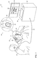
- Fig. 2 is a schematic, pictorial illustration of wand 30, in accordance with an embodiment of the present invention.
- the figure shows wand 30 placed at one of the multiple predefined locations on patient head 41.
- wand 30 comprises a housing 58, which contains a position sensor 60 concentrically disposed around an ultrasound (US) transducer 66.
- the US transducer is configured to produce US pulses 70 into the tissue.
- Fig. 2 shows example tissue structure, which comprises a skin layer 50, an intermediate tissue 52, a bone tissue 56 and an interface layer 54 between tissue 52 and 56.
- physician 24 attaches a tip 59 of wand 30 to skin 50 at one of the predefined locations (e.g., bridge of nose 26), and activates transducer 66 so as to produce US pulses 70.
- US Pulses 70 traverse skin layer 50 and intermediate tissue 52 toward interface layer 54 and bone tissue 56. Pulses 70 are reflected from interface 54 and travel through tissue 52 and skin layer 50, back to wand 30.
- Wand 30 is configured to measure round-trip propagation times of the US pulses, also denoted time-of-flight (TOF). Based on the known speed of the US pulses in tissue 52 and skin layer 50, processor 34 is configured to translate the measured TOF into a thickness of tissue 52, i.e., into the distance between transducer 66 and bone tissue 56. In an embodiment, wand 30 is configured to transmit the position measurements obtained from sensor 60, and the TOF measured using US transducer 66, via cable 32, to processor 34.
- TOF time-of-flight
- sensor 60 is concentrically disposed around transducer 66.
- processor 34 is configured to calculate the US coordinate, i.e., the position of the bone tissue in the coordinate system of the position tracking system.
- sensor 60 and transducer 66 may be concentrically arranged in any other suitable arrangement.
- sensor 60 may be fitted at a fixed, known displacement relative to transducer 66, thus, processor 34 may take into account the fixed displacement in calculating the US coordinate of the bone tissue.
- the pressure applied by tip 59 may deform skin layer 50 and tissue 52.
- Processor 34 essentially prevents such deformation from distorting the registration, since the registration is based on bone tissue correlation.
- Fig. 3 is a flow chart that schematically illustrates a method for registering CT image 35 with the coordinate system of the position tracking system, in accordance with another embodiment of the present invention.
- the method begins with a CT image acquisition step 100, in which processor 34 receives one or more CT images that capture bone tissue (referred to herein as CT bone images) of head 41.
- CT bone images capture bone tissue
- physician 24 attaches wand 30 sequentially to the multiple predefined locations in head 41.
- the subsequent steps (104-110) are performed at each of these predefined locations.
- processor 34 receives the position measurement from sensor 60 and calculates the position of transducer 66 in the coordinate system of the position tracking system.
- system 20 activates transducer 66 so as to acquire US measurements.
- processor 34 receives the position signals from transducer 66 and the TOF measurement from transducer 66, and calculates the US coordinate, i.e., the position of the bone tissue in the coordinate system of the position tracking system.
- processor 34 identifies the corresponding CT coordinate, i.e., the position of the bone tissue in CT image 35, using any suitable technique known in the art. As noted above, steps 104-110 are repeated per each predefined location.
- processor 34 registers CT image 35 with the coordinate system of the position tracking system by correlating between the US bone tissue coordinates and the corresponding CT bone tissue coordinates at the predefined locations in head 41.
Abstract
Description
- The present invention relates generally to image guided medical procedures, and particularly to methods and systems for registration of an anatomical image with a position-tracking system.
- Ultrasound (US) transducers and position-tracking systems may be used in various medical applications, such as image guided procedures.
- For example,
U.S. Patent 5,575,288 , whose disclosure is incorporated herein by reference, describes an ultrasonic imaging system having a remote ultrasound console and a probe connected thereto for inspecting an interior region of a body. The ultrasonic imaging system includes a scan-head housing disposed at a distal end of the probe. A transducer is mounted upon a support structure within the scan-head housing and is electrically connected to the ultrasonic imaging system. A magnetic position sensor is located within the scan-head housing and coupled to the ultrasonic imaging system. -
U.S. Patent 7,751,868 , whose disclosure is incorporated herein by reference, describes an integrated skin-mountable multifunction device for use with a computer assisted or image guided surgical system and methods of using the same. The multifunction device includes a patient mountable portion that has at least one position indicating element and at least one imageable pattern whose geometry is known in a coordinate system of the at least one position indicating element, wherein the imageable pattern is visible on an imaging modality. -
U.S. Patent Application Publication 2006/0253031 , whose disclosure is incorporated herein by reference, describes a system and method for imaging a target in a patient's body uses a pre-acquired image of the target and a catheter having a position sensor and an ultrasonic imaging sensor. The catheter is placed in the patient's body and positional information of a portion of the catheter in the patient's body is determined using the position sensor. The catheter is used to generate an ultrasonic image of the target using the ultrasonic imaging sensor. An image processor is used for determining positional information for any pixel of the ultrasonic image of the target and registering the pre-acquired image with the ultrasonic image, and a display is used for displaying the registered pre-acquired image and ultrasonic image. - An embodiment of the present invention that is described herein provides a method including receiving multiple measurements, which are acquired using a registration tool including an ultrasound (US) transducer and a position sensor of a position-tracking system. The measurements are acquired by attaching the registration tool to multiple respective locations on a patient head and acquiring respective position measurements of the position sensor and respective US measurements of bone tissue at the locations. First positions of the bone tissue at the multiple locations are calculated based on the position measurement and the US measurements obtained using the registration tool. Second positions of the bone tissue at the multiple locations are identified in an anatomical image of the patient head. The anatomical image is registered with a coordinate system of the position tracking system, by correlating the first positions and the second positions, so as to enable tracking a medical instrument, which is inserted into the patient head and includes another position sensor of the position-tracking system, using the anatomical image registered with the position-tracking system.
- In some embodiments, the US transducer is disposed at a fixed displacement relative to the position sensor, and calculating the first positions includes considering the fixed displacement in calculation of the first positions. In other embodiments, the US transducer and the position sensor are concentric. In yet other embodiments, the anatomical image includes one or more computerized tomography (CT) images.
- In an embodiment, the anatomical image includes one or more computerized tomography (CT) images. In another embodiment, the position-tracking system includes a magnetic position-tracking system. In yet another embodiment, receiving the US measurements include receiving round-trip propagation times of US pulses traversing at each location between an external surface of the head and the respective bone tissue.
- In some embodiments, the medical instrument includes a sinuplasty catheter. In other embodiments, the registration tool includes a handheld wand.
- There is additionally provided, in accordance with an embodiment of the present invention, an apparatus including a registration tool and a processor. The registration tool includes an ultrasound (US) transducer and a position sensor of a position-tracking system. The US transducer is configured, when the registration tool is attached sequentially to multiple respective locations on a patient head, to acquire respective US measurements of bone tissue at the locations. The position sensor is configured to acquire respective position measurements of the registration tool at the locations. The processor is configured to receive the multiple US measurements and the respective position measurements acquired by the registration tool, calculate first positions of the bone tissue at the multiple locations, based on the position measurements and the US measurements, identify second positions of the bone tissue at the multiple locations, in an anatomical image of the patient head, and register the anatomical image with a coordinate system of the position tracking system, by correlating the first positions and the second positions, so as to enable tracking a medical instrument, which is inserted into the patient head and includes another position sensor of the position-tracking system, using the anatomical image registered with the position-tracking system.
- The present invention will be more fully understood from the following detailed description of the embodiments thereof, taken together with the drawings in which:
-
-
FIG. 1 is a schematic, pictorial illustration of a sinuplasty surgical system, in accordance with an embodiment of the present invention; -
Fig. 2 is a schematic, pictorial illustration of a registration tool, in accordance with an embodiment of the present invention; and -
Fig. 3 is a flow chart that schematically illustrates a method for registering an anatomical image with a coordinate system of a position tracking system, in accordance with another embodiment of the present invention. - Some medical procedures such as sinuplasty require registration of an anatomical image of relevant organs with a coordinate system of a position tracking system. Using the registration, a surgical tool fitted with a position sensor is navigated to the treated organs, and is visualized overlaid on the anatomical image.
- In principle, the registration may be carried out using some external registration tool fitted with a position sensor of the position tracking system. Such a tool could be attached to preselected locations on the patient face (e.g., nose tip, and centers of the two cheeks). The anatomical image could then be registered to the coordinate system of the position tracking system based on the measured positions of bone tissue at the preselected locations.
- This possible solution, however, is likely to be inaccurate and unsuitable for sinuplasty procedures, in which it is typically important to obtain registration of the anatomical image at accuracy level better than 1 mm. Since facial elements comprise soft tissue that deforms naturally (e.g., due to changes in liquid level in the cheeks along the day), and because of the uncontrolled pressure applied by the registration tool thereon, the accuracy of this hypothetical solution may become unacceptable.
- Embodiments of the present invention that are described hereinbelow provide improved techniques for registering an anatomical image with the coordinate system of a position-tracking system. In the disclosed embodiments, a registration tool comprises an ultrasound (US) transducer coupled to a position sensor of the position-tracking system. In order to perform registration, an operator (e.g., physician) attaches the registration tool to multiple predefined locations on the patient's face. At each of the predefined locations, the following measurements are performed:
- ▪ The position tracking system measures the position and orientation of the position sensor fitted in the registration tool.
- ▪ The US transducer images the bone tissue at the respective location.
- A processor then uses the above measurements to calculate, for each of the predefined locations on the patient's face, the position of the respective bone tissue in the coordinate system of the position tracking system.
- For a given predefined location on the patient's face, the output of the US transducer is indicative of the distance between the US transducer and the bone tissue. The relative displacement (if any) between the US transducer and the position sensor is fixed and known. The position of the position sensor has been measured in the coordinate system of the position tracking system. Therefore, the processor uses the above measurements to calculate the exact position of the bone tissue in the coordinate system of the position tracking system. This position is referred to herein as the "US coordinate" of the bone tissue for the predefined location on the patient face. The above procedure is repeated for each of the multiple predefined locations, to produce a set of US coordinates.
- In addition, the processor identifies the positions of the bone tissue at the predefined multiple locations in a pre-acquired computerized tomography (CT) image. These positions are referred to herein as "CT coordinates" of the bone tissue. The processor then registers the CT image with the coordinate system of the position tracking system, e.g., by calculating a geometrical transformation that matches the US coordinates with the respective CT coordinates.
- Since the disclosed registration process is based on correlation between coordinates of bone tissue, as opposed to soft tissue, it is highly accurate and insensitive to the impairments described above. The proposed techniques thus enable, for example, improved navigation of a sinuplasty surgical tool, which is inserted into the patient head and comprises another position sensor of the position-tracking system. Furthermore, the disclosed techniques are not sensitive to tissue deformation due to natural variations and due to the pressure applied by the registration tool.
-
FIG. 1 is a schematic pictorial illustration of a sinuplastysurgical system 20, in accordance with an embodiment of the present invention.System 20 comprises a magnetic position tracking system, which is configured to track the position of one or more position sensors in the head of apatient 22. The magnetic position tracking system comprises magnetic field-generators and one or more position sensors. The position sensors generate position signals in response to sensed external magnetic fields from the field generators, thereby enabling aprocessor 34 to map the position of each sensor in the coordinate system of the position tracking system as will be described below. - This method of position sensing is implemented in various medical applications, for example, in the CARTO™ system, produced by Biosense Webster Inc. (Diamond Bar, Calif.) and is described in detail in
U.S. Patents 5, 391, 199 ,6, 690, 963 ,6, 484, 118 ,6, 239, 724 ,6, 618, 612 and6,332,089 , inPCT Patent Publication WO 96/05768 U.S. Patent Application Publications 2002/0065455 A1 ,2003/0120150 A1 and2004/0068178 A1 , whose disclosures are all incorporated herein by reference. - In the present example,
system 20 comprises alocation pad 40, which comprises multiple field-generators 44 fixed on aframe 46. In the exemplary configuration shown inFIG. 1 ,pad 40 comprises five field-generators 44, but any other suitable number ofgenerators 44 can be used.Pad 40 further comprises apillow 42 placed under ahead 41 ofpatient 22, such thatgenerators 44 are located at fixed, known positions external to the patient.System 20 further comprises aconsole 33, which comprises a driver circuit (not shown) configured to drive field-generators 44 with suitable signals so as to generate magnetic fields in a predefined working volume aroundhead 41. - In an embodiment,
processor 34 is typically a general-purpose computer comprising suitable front end and interface circuits for receiving data from external sources, as well as measurements fromwand 30, via acable 32, and for controlling other components ofsystem 20.Console 33 further comprisesinput devices 39 and auser display 36, which is configured to display the data. - In some embodiments,
system 20 comprises a registration tool, such as ahandheld wand 30, which is used bysystem 20 for registering the coordinate system of the magnetic position tracking system with that of a pre-acquired CT image. The registration tool is configured to acquire ultrasound and position measurements, and is depicted in detail inFig. 2 below. - Typically, a
physician 24 attacheswand 30 sequentially to multiple predefined locations on an external surface ofpatient head 41. Each predefined location is typically chosen to be an easily identifiable feature onhead 41, such as a cheek bone protrusion, a bridge of a nose 26 (located between the eyes of patient 22), a tip ofnose 26, a chin, or any other suitable identifiable feature. - In an embodiment,
processor 34 receives a computerized tomography (CT)image 35 obtained using an external CT system (not shown).Processor 34 usesimage 35 to form a surface image of at least part ofpatient head 41. In some embodiments,processor 34 may use hounsfield units (HU) ranging between 700 and 3000 for determining the radiodensity of bones in the patient face, compared with HU of -1000, which is a standard scale for air, so as to determine boundaries of the patient face. In an alternative embodiment, HU above 500 may be used for determining the radiodensity of bones, HU of -200 and below may be used for air, and HU ranging between -200 and 500 may be used for muscular tissue. Alternatively, any other suitable values can be used. Further alternatively,processor 34 may distinguish between different types of tissue in the CT image, and in particular identify bone tissue, using any other suitable criterion or technique. - In an embodiment, when placed at a predefined location on the patient head,
wand 30 is configured to (i) acquire US measurements of bone tissue, and (ii) generate position signals indicative of the position of this predefined location in the coordinate system of the magnetic position tracking system. The acquisition of the bone tissue measurements bywand 30 is described in detail inFig. 2 below. - In some embodiments,
processor 34 is configured to calculate two coordinates for each predefined location on the patient head - A "US coordinate" and a "CT coordinate." The US coordinate is derived from the US and position measurements ofwand 30 at this predefined location, and is indicative of the coordinate of the bone tissue at this location in the coordinate system of the magnetic position tracking system. The CT measurement is indicative of the coordinate of the bone tissue at this location, as identified in the CT image. - In an embodiment,
processor 34 is configured to correlate between the US coordinates and the CT coordinates of the predefined locations inimage 35, so as to register the CT image with the coordinate system of the position tracking system. - The registration process is typically performed before the actual sinuplasty procedure. During the sinuplasty procedure,
physician 24 may insert into head 41 a medical device (not shown), such as a sinuplasty catheter or other surgical tool, which comprises an additional position sensor of the position-tracking system. Since the CT image is already registered with the position-tracking system,physician 24 may navigate the medical device whose distal end is displayed on the CT image, to a target location inhead 41. - In alternative embodiments, instead of
CT image 35,processor 34 is configured to receive one or more images acquired using another suitable anatomical imaging technique, such as fluoroscopy or magnetic resonance imaging (MRI), and to register these anatomical images with the coordinate system as described above. -
Fig. 1 shows only elements related to the disclosed techniques, for the sake of simplicity and clarity.System 20 typically comprises additional modules and elements that are not directly related to the disclosed techniques, and thus, intentionally omitted fromFig. 1 and from the corresponding description. -
Processor 34 may be programmed in software to carry out the functions that are used by the system, and to store data in a memory (not shown) to be processed or otherwise used by the software. The software may be downloaded to the processor in electronic form, over a network, for example, or it may be provided on non-transitory tangible media, such as optical, magnetic or electronic memory media. Alternatively, some or all of the functions ofprocessor 34 may be carried out by dedicated or programmable digital hardware components. -
Fig. 2 is a schematic, pictorial illustration ofwand 30, in accordance with an embodiment of the present invention. The figure showswand 30 placed at one of the multiple predefined locations onpatient head 41. In some embodiments,wand 30 comprises ahousing 58, which contains aposition sensor 60 concentrically disposed around an ultrasound (US)transducer 66. The US transducer is configured to produceUS pulses 70 into the tissue.Fig. 2 shows example tissue structure, which comprises askin layer 50, anintermediate tissue 52, abone tissue 56 and aninterface layer 54 betweentissue - In an embodiment,
physician 24 attaches atip 59 ofwand 30 toskin 50 at one of the predefined locations (e.g., bridge of nose 26), and activatestransducer 66 so as to produceUS pulses 70.US Pulses 70traverse skin layer 50 andintermediate tissue 52 towardinterface layer 54 andbone tissue 56.Pulses 70 are reflected frominterface 54 and travel throughtissue 52 andskin layer 50, back towand 30. -
Wand 30 is configured to measure round-trip propagation times of the US pulses, also denoted time-of-flight (TOF). Based on the known speed of the US pulses intissue 52 andskin layer 50,processor 34 is configured to translate the measured TOF into a thickness oftissue 52, i.e., into the distance betweentransducer 66 andbone tissue 56. In an embodiment,wand 30 is configured to transmit the position measurements obtained fromsensor 60, and the TOF measured usingUS transducer 66, viacable 32, toprocessor 34. - In some embodiments,
sensor 60 is concentrically disposed aroundtransducer 66. Using the position ofsensor 60 and the TOF measurement oftransducer 66,processor 34 is configured to calculate the US coordinate, i.e., the position of the bone tissue in the coordinate system of the position tracking system. - In alternative embodiments,
sensor 60 andtransducer 66 may be concentrically arranged in any other suitable arrangement. In yet alternative embodiments,sensor 60 may be fitted at a fixed, known displacement relative totransducer 66, thus,processor 34 may take into account the fixed displacement in calculating the US coordinate of the bone tissue. - As depicted in
Fig. 2 , the pressure applied bytip 59 may deformskin layer 50 andtissue 52.Processor 34 essentially prevents such deformation from distorting the registration, since the registration is based on bone tissue correlation. -
Fig. 3 is a flow chart that schematically illustrates a method for registeringCT image 35 with the coordinate system of the position tracking system, in accordance with another embodiment of the present invention. The method begins with a CTimage acquisition step 100, in whichprocessor 34 receives one or more CT images that capture bone tissue (referred to herein as CT bone images) ofhead 41. - At a registration tool attachment step 102,
physician 24 attacheswand 30 sequentially to the multiple predefined locations inhead 41. The subsequent steps (104-110) are performed at each of these predefined locations. - At a
position calculation step 104,processor 34 receives the position measurement fromsensor 60 and calculates the position oftransducer 66 in the coordinate system of the position tracking system. At an US measurements step 106,system 20 activatestransducer 66 so as to acquire US measurements. At a tissueposition calculation step 108,processor 34 receives the position signals fromtransducer 66 and the TOF measurement fromtransducer 66, and calculates the US coordinate, i.e., the position of the bone tissue in the coordinate system of the position tracking system. At a CTtissue identification step 110,processor 34 identifies the corresponding CT coordinate, i.e., the position of the bone tissue inCT image 35, using any suitable technique known in the art. As noted above, steps 104-110 are repeated per each predefined location. - At a
registration step 112,processor 34registers CT image 35 with the coordinate system of the position tracking system by correlating between the US bone tissue coordinates and the corresponding CT bone tissue coordinates at the predefined locations inhead 41. - Although the embodiments described herein mainly address sinuplasty procedures, the methods and systems described herein can also be used in other applications, such as in orthopedic procedures, in which
physician 24 may attachwand 30 to any suitable deformable feature on a human body. - It will thus be appreciated that the embodiments described above are cited by way of example, and that the present invention is not limited to what has been particularly shown and described hereinabove. Rather, the scope of the present invention includes both combinations and sub-combinations of the various features described hereinabove, as well as variations and modifications thereof which would occur to persons skilled in the art upon reading the foregoing description and which are not disclosed in the prior art. Documents incorporated by reference in the present patent application are to be considered an integral part of the application except that to the extent any terms are defined in these incorporated documents in a manner that conflicts with the definitions made explicitly or implicitly in the present specification, only the definitions in the present specification should be considered.
Claims (12)
- A method, comprising:receiving multiple measurements, which are acquired using a registration tool comprising an ultrasound (US) transducer and a position sensor of a position-tracking system, wherein the measurements are acquired by attaching the registration tool to multiple respective locations on a patient head and acquiring respective position measurements of the position sensor and respective US measurements of bone tissue at the locations;calculating first positions of the bone tissue at the multiple locations, based on the position measurement and the US measurements obtained using the registration tool;identifying second positions of the bone tissue at the multiple locations, in an anatomical image of the patient head; andregistering the anatomical image with a coordinate system of the position tracking system, by correlating the first positions and the second positions, so as to enable tracking a medical instrument, which is inserted into the patient head and comprises another position sensor of the position-tracking system, using the anatomical image registered with the position-tracking system.
- The method according to claim 1, wherein the US transducer is disposed at a fixed displacement relative to the position sensor, and wherein calculating the first positions comprises considering the fixed displacement in calculation of the first positions.
- The method according to claim 1, wherein receiving the US measurements comprise receiving round-trip propagation times of US pulses traversing at each location between an external surface of the head and the respective bone tissue.
- An apparatus, comprising:a registration tool, comprising:an ultrasound (US) transducer, which is configured, when the registration tool is attached sequentially to multiple respective locations on a patient head, to acquire respective US measurements of bone tissue at the locations; anda position sensor of a position-tracking system, which is configured to acquire respective position measurements of the registration tool at the locations; anda processor, which is configured to:receive the multiple US measurements and the respective position measurements acquired by the registration tool;calculate first positions of the bone tissue at the multiple locations, based on the position measurements and the US measurements;identify second positions of the bone tissue at the multiple locations, in an anatomical image of the patient head; andregister the anatomical image with a coordinate system of the position tracking system, by correlating the first positions and the second positions, so as to enable tracking a medical instrument, which is inserted into the patient head and comprises another position sensor of the position-tracking system, using the anatomical image registered with the position-tracking system.
- The apparatus according to claim 4, wherein the US transducer is disposed at a fixed displacement relative to the position sensor, and wherein the processor is configured to consider the fixed displacement in calculation of the first positions.
- The method according to claim 1 or the apparatus according to claim 4, wherein the US transducer and the position sensor are concentric.
- The method according to claim 1 or the apparatus according to claim 4, wherein the anatomical image comprises one or more computerized tomography (CT) images.
- The method according to claim 1 or the apparatus according to claim 4, wherein the locations comprise locations of bone features selected from a list consisting of a cheek bone protrusion, a bridge of a nose, a tip of the nose and a chin.
- The method according to claim 1 or the apparatus according to claim 4, wherein the position-tracking system comprises a magnetic position-tracking system.
- The apparatus according to claim 4, wherein the multiple US measurements comprise round-trip propagation times of US pulses traversing at each location between an external surface of the head and the respective bone tissue.
- The method according to claim 1 or the apparatus according to claim 4, wherein the medical instrument comprises a sinuplasty catheter.
- The method according to claim 1 or the apparatus according to claim 4, wherein the registration tool comprises a handheld wand.
Applications Claiming Priority (1)
| Application Number | Priority Date | Filing Date | Title |
|---|---|---|---|
| US15/286,891 US20180098816A1 (en) | 2016-10-06 | 2016-10-06 | Pre-Operative Registration of Anatomical Images with a Position-Tracking System Using Ultrasound |
Publications (2)
| Publication Number | Publication Date |
|---|---|
| EP3305202A1 true EP3305202A1 (en) | 2018-04-11 |
| EP3305202B1 EP3305202B1 (en) | 2024-03-06 |
Family
ID=60051365
Family Applications (1)
| Application Number | Title | Priority Date | Filing Date |
|---|---|---|---|
| EP17194989.4A Active EP3305202B1 (en) | 2016-10-06 | 2017-10-05 | Pre-operative registration of anatomical images with a position-tracking system using ultrasound |
Country Status (7)
| Country | Link |
|---|---|
| US (1) | US20180098816A1 (en) |
| EP (1) | EP3305202B1 (en) |
| JP (1) | JP7106258B2 (en) |
| CN (1) | CN107913102B (en) |
| AU (1) | AU2017235897A1 (en) |
| CA (1) | CA2981039A1 (en) |
| IL (1) | IL254680B1 (en) |
Cited By (1)
| Publication number | Priority date | Publication date | Assignee | Title |
|---|---|---|---|---|
| EP3454299A1 (en) * | 2017-08-10 | 2019-03-13 | Biosense Webster (Israel) Ltd. | Method and apparatus for performing facial registration |
Families Citing this family (126)
| Publication number | Priority date | Publication date | Assignee | Title |
|---|---|---|---|---|
| US11871901B2 (en) | 2012-05-20 | 2024-01-16 | Cilag Gmbh International | Method for situational awareness for surgical network or surgical network connected device capable of adjusting function based on a sensed situation or usage |
| US11504192B2 (en) | 2014-10-30 | 2022-11-22 | Cilag Gmbh International | Method of hub communication with surgical instrument systems |
| US10510171B2 (en) | 2016-11-29 | 2019-12-17 | Biosense Webster (Israel) Ltd. | Visualization of anatomical cavities |
| US11291510B2 (en) | 2017-10-30 | 2022-04-05 | Cilag Gmbh International | Method of hub communication with surgical instrument systems |
| US11564756B2 (en) | 2017-10-30 | 2023-01-31 | Cilag Gmbh International | Method of hub communication with surgical instrument systems |
| US11311342B2 (en) | 2017-10-30 | 2022-04-26 | Cilag Gmbh International | Method for communicating with surgical instrument systems |
| US11759224B2 (en) | 2017-10-30 | 2023-09-19 | Cilag Gmbh International | Surgical instrument systems comprising handle arrangements |
| US11229436B2 (en) | 2017-10-30 | 2022-01-25 | Cilag Gmbh International | Surgical system comprising a surgical tool and a surgical hub |
| US11510741B2 (en) | 2017-10-30 | 2022-11-29 | Cilag Gmbh International | Method for producing a surgical instrument comprising a smart electrical system |
| US11801098B2 (en) | 2017-10-30 | 2023-10-31 | Cilag Gmbh International | Method of hub communication with surgical instrument systems |
| US11911045B2 (en) | 2017-10-30 | 2024-02-27 | Cllag GmbH International | Method for operating a powered articulating multi-clip applier |
| US11406390B2 (en) | 2017-10-30 | 2022-08-09 | Cilag Gmbh International | Clip applier comprising interchangeable clip reloads |
| US11317919B2 (en) | 2017-10-30 | 2022-05-03 | Cilag Gmbh International | Clip applier comprising a clip crimping system |
| US11257589B2 (en) | 2017-12-28 | 2022-02-22 | Cilag Gmbh International | Real-time analysis of comprehensive cost of all instrumentation used in surgery utilizing data fluidity to track instruments through stocking and in-house processes |
| US11666331B2 (en) | 2017-12-28 | 2023-06-06 | Cilag Gmbh International | Systems for detecting proximity of surgical end effector to cancerous tissue |
| US11291495B2 (en) | 2017-12-28 | 2022-04-05 | Cilag Gmbh International | Interruption of energy due to inadvertent capacitive coupling |
| US11056244B2 (en) | 2017-12-28 | 2021-07-06 | Cilag Gmbh International | Automated data scaling, alignment, and organizing based on predefined parameters within surgical networks |
| US11109866B2 (en) | 2017-12-28 | 2021-09-07 | Cilag Gmbh International | Method for circular stapler control algorithm adjustment based on situational awareness |
| US11424027B2 (en) | 2017-12-28 | 2022-08-23 | Cilag Gmbh International | Method for operating surgical instrument systems |
| US11179175B2 (en) | 2017-12-28 | 2021-11-23 | Cilag Gmbh International | Controlling an ultrasonic surgical instrument according to tissue location |
| US11051876B2 (en) | 2017-12-28 | 2021-07-06 | Cilag Gmbh International | Surgical evacuation flow paths |
| US11832840B2 (en) | 2017-12-28 | 2023-12-05 | Cilag Gmbh International | Surgical instrument having a flexible circuit |
| US11559307B2 (en) | 2017-12-28 | 2023-01-24 | Cilag Gmbh International | Method of robotic hub communication, detection, and control |
| US11633237B2 (en) | 2017-12-28 | 2023-04-25 | Cilag Gmbh International | Usage and technique analysis of surgeon / staff performance against a baseline to optimize device utilization and performance for both current and future procedures |
| US11389164B2 (en) | 2017-12-28 | 2022-07-19 | Cilag Gmbh International | Method of using reinforced flexible circuits with multiple sensors to optimize performance of radio frequency devices |
| US11273001B2 (en) | 2017-12-28 | 2022-03-15 | Cilag Gmbh International | Surgical hub and modular device response adjustment based on situational awareness |
| US11304699B2 (en) | 2017-12-28 | 2022-04-19 | Cilag Gmbh International | Method for adaptive control schemes for surgical network control and interaction |
| US11304763B2 (en) | 2017-12-28 | 2022-04-19 | Cilag Gmbh International | Image capturing of the areas outside the abdomen to improve placement and control of a surgical device in use |
| US11311306B2 (en) | 2017-12-28 | 2022-04-26 | Cilag Gmbh International | Surgical systems for detecting end effector tissue distribution irregularities |
| US11896322B2 (en) | 2017-12-28 | 2024-02-13 | Cilag Gmbh International | Sensing the patient position and contact utilizing the mono-polar return pad electrode to provide situational awareness to the hub |
| US11786251B2 (en) | 2017-12-28 | 2023-10-17 | Cilag Gmbh International | Method for adaptive control schemes for surgical network control and interaction |
| US11419630B2 (en) | 2017-12-28 | 2022-08-23 | Cilag Gmbh International | Surgical system distributed processing |
| US11076921B2 (en) | 2017-12-28 | 2021-08-03 | Cilag Gmbh International | Adaptive control program updates for surgical hubs |
| US11432885B2 (en) | 2017-12-28 | 2022-09-06 | Cilag Gmbh International | Sensing arrangements for robot-assisted surgical platforms |
| US11324557B2 (en) | 2017-12-28 | 2022-05-10 | Cilag Gmbh International | Surgical instrument with a sensing array |
| US11058498B2 (en) | 2017-12-28 | 2021-07-13 | Cilag Gmbh International | Cooperative surgical actions for robot-assisted surgical platforms |
| US11602393B2 (en) | 2017-12-28 | 2023-03-14 | Cilag Gmbh International | Surgical evacuation sensing and generator control |
| US11317937B2 (en) | 2018-03-08 | 2022-05-03 | Cilag Gmbh International | Determining the state of an ultrasonic end effector |
| US11266468B2 (en) | 2017-12-28 | 2022-03-08 | Cilag Gmbh International | Cooperative utilization of data derived from secondary sources by intelligent surgical hubs |
| US11166772B2 (en) | 2017-12-28 | 2021-11-09 | Cilag Gmbh International | Surgical hub coordination of control and communication of operating room devices |
| US11284936B2 (en) | 2017-12-28 | 2022-03-29 | Cilag Gmbh International | Surgical instrument having a flexible electrode |
| US11069012B2 (en) | 2017-12-28 | 2021-07-20 | Cilag Gmbh International | Interactive surgical systems with condition handling of devices and data capabilities |
| US10892899B2 (en) | 2017-12-28 | 2021-01-12 | Ethicon Llc | Self describing data packets generated at an issuing instrument |
| US10944728B2 (en) | 2017-12-28 | 2021-03-09 | Ethicon Llc | Interactive surgical systems with encrypted communication capabilities |
| US11864728B2 (en) | 2017-12-28 | 2024-01-09 | Cilag Gmbh International | Characterization of tissue irregularities through the use of mono-chromatic light refractivity |
| US11464559B2 (en) | 2017-12-28 | 2022-10-11 | Cilag Gmbh International | Estimating state of ultrasonic end effector and control system therefor |
| US11612444B2 (en) | 2017-12-28 | 2023-03-28 | Cilag Gmbh International | Adjustment of a surgical device function based on situational awareness |
| US11744604B2 (en) | 2017-12-28 | 2023-09-05 | Cilag Gmbh International | Surgical instrument with a hardware-only control circuit |
| US11937769B2 (en) | 2017-12-28 | 2024-03-26 | Cilag Gmbh International | Method of hub communication, processing, storage and display |
| US20190201146A1 (en) | 2017-12-28 | 2019-07-04 | Ethicon Llc | Safety systems for smart powered surgical stapling |
| US11857152B2 (en) | 2017-12-28 | 2024-01-02 | Cilag Gmbh International | Surgical hub spatial awareness to determine devices in operating theater |
| US11253315B2 (en) | 2017-12-28 | 2022-02-22 | Cilag Gmbh International | Increasing radio frequency to create pad-less monopolar loop |
| US11096693B2 (en) | 2017-12-28 | 2021-08-24 | Cilag Gmbh International | Adjustment of staple height of at least one row of staples based on the sensed tissue thickness or force in closing |
| US10932872B2 (en) | 2017-12-28 | 2021-03-02 | Ethicon Llc | Cloud-based medical analytics for linking of local usage trends with the resource acquisition behaviors of larger data set |
| US11446052B2 (en) | 2017-12-28 | 2022-09-20 | Cilag Gmbh International | Variation of radio frequency and ultrasonic power level in cooperation with varying clamp arm pressure to achieve predefined heat flux or power applied to tissue |
| US11576677B2 (en) | 2017-12-28 | 2023-02-14 | Cilag Gmbh International | Method of hub communication, processing, display, and cloud analytics |
| US11410259B2 (en) | 2017-12-28 | 2022-08-09 | Cilag Gmbh International | Adaptive control program updates for surgical devices |
| US11376002B2 (en) | 2017-12-28 | 2022-07-05 | Cilag Gmbh International | Surgical instrument cartridge sensor assemblies |
| US11423007B2 (en) | 2017-12-28 | 2022-08-23 | Cilag Gmbh International | Adjustment of device control programs based on stratified contextual data in addition to the data |
| US11419667B2 (en) | 2017-12-28 | 2022-08-23 | Cilag Gmbh International | Ultrasonic energy device which varies pressure applied by clamp arm to provide threshold control pressure at a cut progression location |
| US11234756B2 (en) | 2017-12-28 | 2022-02-01 | Cilag Gmbh International | Powered surgical tool with predefined adjustable control algorithm for controlling end effector parameter |
| US11202570B2 (en) | 2017-12-28 | 2021-12-21 | Cilag Gmbh International | Communication hub and storage device for storing parameters and status of a surgical device to be shared with cloud based analytics systems |
| US10849697B2 (en) | 2017-12-28 | 2020-12-01 | Ethicon Llc | Cloud interface for coupled surgical devices |
| US11304720B2 (en) | 2017-12-28 | 2022-04-19 | Cilag Gmbh International | Activation of energy devices |
| US10755813B2 (en) | 2017-12-28 | 2020-08-25 | Ethicon Llc | Communication of smoke evacuation system parameters to hub or cloud in smoke evacuation module for interactive surgical platform |
| US11832899B2 (en) | 2017-12-28 | 2023-12-05 | Cilag Gmbh International | Surgical systems with autonomously adjustable control programs |
| US10892995B2 (en) | 2017-12-28 | 2021-01-12 | Ethicon Llc | Surgical network determination of prioritization of communication, interaction, or processing based on system or device needs |
| US10695081B2 (en) | 2017-12-28 | 2020-06-30 | Ethicon Llc | Controlling a surgical instrument according to sensed closure parameters |
| US11540855B2 (en) | 2017-12-28 | 2023-01-03 | Cilag Gmbh International | Controlling activation of an ultrasonic surgical instrument according to the presence of tissue |
| US11364075B2 (en) | 2017-12-28 | 2022-06-21 | Cilag Gmbh International | Radio frequency energy device for delivering combined electrical signals |
| US11559308B2 (en) | 2017-12-28 | 2023-01-24 | Cilag Gmbh International | Method for smart energy device infrastructure |
| US10943454B2 (en) | 2017-12-28 | 2021-03-09 | Ethicon Llc | Detection and escalation of security responses of surgical instruments to increasing severity threats |
| US11818052B2 (en) | 2017-12-28 | 2023-11-14 | Cilag Gmbh International | Surgical network determination of prioritization of communication, interaction, or processing based on system or device needs |
| US11278281B2 (en) | 2017-12-28 | 2022-03-22 | Cilag Gmbh International | Interactive surgical system |
| US11571234B2 (en) | 2017-12-28 | 2023-02-07 | Cilag Gmbh International | Temperature control of ultrasonic end effector and control system therefor |
| US11100631B2 (en) | 2017-12-28 | 2021-08-24 | Cilag Gmbh International | Use of laser light and red-green-blue coloration to determine properties of back scattered light |
| US11160605B2 (en) | 2017-12-28 | 2021-11-02 | Cilag Gmbh International | Surgical evacuation sensing and motor control |
| US11596291B2 (en) | 2017-12-28 | 2023-03-07 | Cilag Gmbh International | Method of compressing tissue within a stapling device and simultaneously displaying of the location of the tissue within the jaws |
| US11147607B2 (en) | 2017-12-28 | 2021-10-19 | Cilag Gmbh International | Bipolar combination device that automatically adjusts pressure based on energy modality |
| US11896443B2 (en) | 2017-12-28 | 2024-02-13 | Cilag Gmbh International | Control of a surgical system through a surgical barrier |
| US11464535B2 (en) | 2017-12-28 | 2022-10-11 | Cilag Gmbh International | Detection of end effector emersion in liquid |
| US20190201039A1 (en) | 2017-12-28 | 2019-07-04 | Ethicon Llc | Situational awareness of electrosurgical systems |
| US11045591B2 (en) | 2017-12-28 | 2021-06-29 | Cilag Gmbh International | Dual in-series large and small droplet filters |
| US11529187B2 (en) | 2017-12-28 | 2022-12-20 | Cilag Gmbh International | Surgical evacuation sensor arrangements |
| US11678881B2 (en) | 2017-12-28 | 2023-06-20 | Cilag Gmbh International | Spatial awareness of surgical hubs in operating rooms |
| US10987178B2 (en) | 2017-12-28 | 2021-04-27 | Ethicon Llc | Surgical hub control arrangements |
| US11179208B2 (en) | 2017-12-28 | 2021-11-23 | Cilag Gmbh International | Cloud-based medical analytics for security and authentication trends and reactive measures |
| US11903601B2 (en) | 2017-12-28 | 2024-02-20 | Cilag Gmbh International | Surgical instrument comprising a plurality of drive systems |
| US11589888B2 (en) | 2017-12-28 | 2023-02-28 | Cilag Gmbh International | Method for controlling smart energy devices |
| US11786245B2 (en) | 2017-12-28 | 2023-10-17 | Cilag Gmbh International | Surgical systems with prioritized data transmission capabilities |
| US11308075B2 (en) | 2017-12-28 | 2022-04-19 | Cilag Gmbh International | Surgical network, instrument, and cloud responses based on validation of received dataset and authentication of its source and integrity |
| US10966791B2 (en) | 2017-12-28 | 2021-04-06 | Ethicon Llc | Cloud-based medical analytics for medical facility segmented individualization of instrument function |
| US11132462B2 (en) | 2017-12-28 | 2021-09-28 | Cilag Gmbh International | Data stripping method to interrogate patient records and create anonymized record |
| US11672605B2 (en) | 2017-12-28 | 2023-06-13 | Cilag Gmbh International | Sterile field interactive control displays |
| US11304745B2 (en) | 2017-12-28 | 2022-04-19 | Cilag Gmbh International | Surgical evacuation sensing and display |
| US20190201139A1 (en) | 2017-12-28 | 2019-07-04 | Ethicon Llc | Communication arrangements for robot-assisted surgical platforms |
| US11659023B2 (en) | 2017-12-28 | 2023-05-23 | Cilag Gmbh International | Method of hub communication |
| US10758310B2 (en) * | 2017-12-28 | 2020-09-01 | Ethicon Llc | Wireless pairing of a surgical device with another device within a sterile surgical field based on the usage and situational awareness of devices |
| US11589915B2 (en) | 2018-03-08 | 2023-02-28 | Cilag Gmbh International | In-the-jaw classifier based on a model |
| US11259830B2 (en) | 2018-03-08 | 2022-03-01 | Cilag Gmbh International | Methods for controlling temperature in ultrasonic device |
| US11464532B2 (en) | 2018-03-08 | 2022-10-11 | Cilag Gmbh International | Methods for estimating and controlling state of ultrasonic end effector |
| US11219453B2 (en) | 2018-03-28 | 2022-01-11 | Cilag Gmbh International | Surgical stapling devices with cartridge compatible closure and firing lockout arrangements |
| US11278280B2 (en) | 2018-03-28 | 2022-03-22 | Cilag Gmbh International | Surgical instrument comprising a jaw closure lockout |
| US11090047B2 (en) | 2018-03-28 | 2021-08-17 | Cilag Gmbh International | Surgical instrument comprising an adaptive control system |
| US10973520B2 (en) | 2018-03-28 | 2021-04-13 | Ethicon Llc | Surgical staple cartridge with firing member driven camming assembly that has an onboard tissue cutting feature |
| US11207067B2 (en) | 2018-03-28 | 2021-12-28 | Cilag Gmbh International | Surgical stapling device with separate rotary driven closure and firing systems and firing member that engages both jaws while firing |
| US11129611B2 (en) | 2018-03-28 | 2021-09-28 | Cilag Gmbh International | Surgical staplers with arrangements for maintaining a firing member thereof in a locked configuration unless a compatible cartridge has been installed therein |
| US11096688B2 (en) | 2018-03-28 | 2021-08-24 | Cilag Gmbh International | Rotary driven firing members with different anvil and channel engagement features |
| US11471156B2 (en) | 2018-03-28 | 2022-10-18 | Cilag Gmbh International | Surgical stapling devices with improved rotary driven closure systems |
| US11259806B2 (en) | 2018-03-28 | 2022-03-01 | Cilag Gmbh International | Surgical stapling devices with features for blocking advancement of a camming assembly of an incompatible cartridge installed therein |
| US11497566B2 (en) * | 2018-10-26 | 2022-11-15 | Biosense Webster (Israel) Ltd. | Loose mode for robot |
| US11317915B2 (en) | 2019-02-19 | 2022-05-03 | Cilag Gmbh International | Universal cartridge based key feature that unlocks multiple lockout arrangements in different surgical staplers |
| US11369377B2 (en) | 2019-02-19 | 2022-06-28 | Cilag Gmbh International | Surgical stapling assembly with cartridge based retainer configured to unlock a firing lockout |
| US11357503B2 (en) | 2019-02-19 | 2022-06-14 | Cilag Gmbh International | Staple cartridge retainers with frangible retention features and methods of using same |
| US11751872B2 (en) | 2019-02-19 | 2023-09-12 | Cilag Gmbh International | Insertable deactivator element for surgical stapler lockouts |
| US11331100B2 (en) | 2019-02-19 | 2022-05-17 | Cilag Gmbh International | Staple cartridge retainer system with authentication keys |
| WO2020207583A1 (en) * | 2019-04-10 | 2020-10-15 | Brainlab Ag | Method of sampling relevant surface points of a subject |
| USD964564S1 (en) | 2019-06-25 | 2022-09-20 | Cilag Gmbh International | Surgical staple cartridge retainer with a closure system authentication key |
| USD950728S1 (en) | 2019-06-25 | 2022-05-03 | Cilag Gmbh International | Surgical staple cartridge |
| USD952144S1 (en) | 2019-06-25 | 2022-05-17 | Cilag Gmbh International | Surgical staple cartridge retainer with firing system authentication key |
| US11062445B2 (en) * | 2019-08-28 | 2021-07-13 | Hiwin Technologies Corp. | Method of medical image registration |
| CN111329586B (en) * | 2020-02-17 | 2021-05-11 | 南京航空航天大学 | In-vivo instrument positioning and tracking system based on ultrasonic positioning belt |
| US11571260B2 (en) | 2020-03-31 | 2023-02-07 | Biosense Webster (Israel) Ltd. | Pre-operative registration of anatomical images with a position-tracking system using ultrasound measurement of skin tissue |
| US11832883B2 (en) | 2020-04-23 | 2023-12-05 | Johnson & Johnson Surgical Vision, Inc. | Using real-time images for augmented-reality visualization of an ophthalmology surgical tool |
| US20210330394A1 (en) | 2020-04-23 | 2021-10-28 | Johnson & Johnson Surgical Vision, Inc. | Augmented-reality visualization of an ophthalmic surgical tool |
| CN111938700B (en) * | 2020-08-21 | 2021-11-09 | 电子科技大学 | Ultrasonic probe guiding system and method based on real-time matching of human anatomy structure |
Citations (18)
| Publication number | Priority date | Publication date | Assignee | Title |
|---|---|---|---|---|
| US5391199A (en) | 1993-07-20 | 1995-02-21 | Biosense, Inc. | Apparatus and method for treating cardiac arrhythmias |
| WO1996005768A1 (en) | 1994-08-19 | 1996-02-29 | Biosense, Inc. | Medical diagnosis, treatment and imaging systems |
| US5575288A (en) | 1993-05-28 | 1996-11-19 | Acuson Corporation | Compact rotationally steerable ultrasound transducer |
| US6239724B1 (en) | 1997-12-30 | 2001-05-29 | Remon Medical Technologies, Ltd. | System and method for telemetrically providing intrabody spatial position |
| US6332089B1 (en) | 1996-02-15 | 2001-12-18 | Biosense, Inc. | Medical procedures and apparatus using intrabody probes |
| US20020035321A1 (en) * | 1993-04-26 | 2002-03-21 | Bucholz Richard D. | Surgical navigation systems including reference and localization frames |
| US20020065455A1 (en) | 1995-01-24 | 2002-05-30 | Shlomo Ben-Haim | Medical diagnosis, treatment and imaging systems |
| US6484118B1 (en) | 2000-07-20 | 2002-11-19 | Biosense, Inc. | Electromagnetic position single axis system |
| US20030120150A1 (en) | 2001-12-21 | 2003-06-26 | Assaf Govari | Wireless position sensor |
| US6618612B1 (en) | 1996-02-15 | 2003-09-09 | Biosense, Inc. | Independently positionable transducers for location system |
| US20040068178A1 (en) | 2002-09-17 | 2004-04-08 | Assaf Govari | High-gradient recursive locating system |
| US20040236223A1 (en) * | 2003-05-22 | 2004-11-25 | Siemens Medical Solutions Usa, Inc.. | Transducer arrays with an integrated sensor and methods of use |
| WO2006092602A1 (en) * | 2005-03-01 | 2006-09-08 | Kings College London | 3d ultrasound imaging |
| US20060253031A1 (en) | 2005-04-26 | 2006-11-09 | Altmann Andres C | Registration of ultrasound data with pre-acquired image |
| US7751868B2 (en) | 2004-11-12 | 2010-07-06 | Philips Electronics Ltd | Integrated skin-mounted multifunction device for use in image-guided surgery |
| DE102012103725A1 (en) * | 2012-04-27 | 2013-10-31 | Aesculap Ag | Device for finding signal inputs of femur of patient in ultrasound image data sets in orthopedics region to detect bone breakages, has computer originatingly defining third signal inputs in transition region of first inputs to second inputs |
| US20140276001A1 (en) * | 2013-03-15 | 2014-09-18 | Queen's University At Kingston | Device and Method for Image-Guided Surgery |
| US20160008083A1 (en) * | 2014-07-09 | 2016-01-14 | Acclarent, Inc. | Guidewire navigation for sinuplasty |
Family Cites Families (10)
| Publication number | Priority date | Publication date | Assignee | Title |
|---|---|---|---|---|
| US6006126A (en) * | 1991-01-28 | 1999-12-21 | Cosman; Eric R. | System and method for stereotactic registration of image scan data |
| IL119262A0 (en) * | 1996-02-15 | 1996-12-05 | Biosense Israel Ltd | Locatable biopsy needle |
| JP2001204739A (en) * | 2000-01-27 | 2001-07-31 | Olympus Optical Co Ltd | Microscopic medical operation support system |
| US20060063973A1 (en) * | 2004-04-21 | 2006-03-23 | Acclarent, Inc. | Methods and apparatus for treating disorders of the ear, nose and throat |
| US20090080737A1 (en) * | 2007-09-25 | 2009-03-26 | General Electric Company | System and Method for Use of Fluoroscope and Computed Tomography Registration for Sinuplasty Navigation |
| US9456766B2 (en) * | 2007-11-26 | 2016-10-04 | C. R. Bard, Inc. | Apparatus for use with needle insertion guidance system |
| JP6023190B2 (en) * | 2011-06-27 | 2016-11-09 | コーニンクレッカ フィリップス エヌ ヴェKoninklijke Philips N.V. | Ultrasound image guidance system and calibration method based on volume motion |
| US10758209B2 (en) * | 2012-03-09 | 2020-09-01 | The Johns Hopkins University | Photoacoustic tracking and registration in interventional ultrasound |
| CN103717142B (en) * | 2012-07-30 | 2016-08-17 | 东芝医疗系统株式会社 | Equipment fixed adapter and ultrasound probe system |
| US10674933B2 (en) * | 2014-10-22 | 2020-06-09 | Biosense Webster (Israel) Ltd. | Enlargement of tracking volume by movement of imaging bed |
-
2016
- 2016-10-06 US US15/286,891 patent/US20180098816A1/en not_active Abandoned
-
2017
- 2017-09-25 IL IL254680A patent/IL254680B1/en unknown
- 2017-09-26 AU AU2017235897A patent/AU2017235897A1/en not_active Abandoned
- 2017-09-29 CN CN201710903790.8A patent/CN107913102B/en active Active
- 2017-10-02 CA CA2981039A patent/CA2981039A1/en not_active Abandoned
- 2017-10-05 EP EP17194989.4A patent/EP3305202B1/en active Active
- 2017-10-05 JP JP2017194909A patent/JP7106258B2/en active Active
Patent Citations (19)
| Publication number | Priority date | Publication date | Assignee | Title |
|---|---|---|---|---|
| US20020035321A1 (en) * | 1993-04-26 | 2002-03-21 | Bucholz Richard D. | Surgical navigation systems including reference and localization frames |
| US5575288A (en) | 1993-05-28 | 1996-11-19 | Acuson Corporation | Compact rotationally steerable ultrasound transducer |
| US5391199A (en) | 1993-07-20 | 1995-02-21 | Biosense, Inc. | Apparatus and method for treating cardiac arrhythmias |
| WO1996005768A1 (en) | 1994-08-19 | 1996-02-29 | Biosense, Inc. | Medical diagnosis, treatment and imaging systems |
| US6690963B2 (en) | 1995-01-24 | 2004-02-10 | Biosense, Inc. | System for determining the location and orientation of an invasive medical instrument |
| US20020065455A1 (en) | 1995-01-24 | 2002-05-30 | Shlomo Ben-Haim | Medical diagnosis, treatment and imaging systems |
| US6332089B1 (en) | 1996-02-15 | 2001-12-18 | Biosense, Inc. | Medical procedures and apparatus using intrabody probes |
| US6618612B1 (en) | 1996-02-15 | 2003-09-09 | Biosense, Inc. | Independently positionable transducers for location system |
| US6239724B1 (en) | 1997-12-30 | 2001-05-29 | Remon Medical Technologies, Ltd. | System and method for telemetrically providing intrabody spatial position |
| US6484118B1 (en) | 2000-07-20 | 2002-11-19 | Biosense, Inc. | Electromagnetic position single axis system |
| US20030120150A1 (en) | 2001-12-21 | 2003-06-26 | Assaf Govari | Wireless position sensor |
| US20040068178A1 (en) | 2002-09-17 | 2004-04-08 | Assaf Govari | High-gradient recursive locating system |
| US20040236223A1 (en) * | 2003-05-22 | 2004-11-25 | Siemens Medical Solutions Usa, Inc.. | Transducer arrays with an integrated sensor and methods of use |
| US7751868B2 (en) | 2004-11-12 | 2010-07-06 | Philips Electronics Ltd | Integrated skin-mounted multifunction device for use in image-guided surgery |
| WO2006092602A1 (en) * | 2005-03-01 | 2006-09-08 | Kings College London | 3d ultrasound imaging |
| US20060253031A1 (en) | 2005-04-26 | 2006-11-09 | Altmann Andres C | Registration of ultrasound data with pre-acquired image |
| DE102012103725A1 (en) * | 2012-04-27 | 2013-10-31 | Aesculap Ag | Device for finding signal inputs of femur of patient in ultrasound image data sets in orthopedics region to detect bone breakages, has computer originatingly defining third signal inputs in transition region of first inputs to second inputs |
| US20140276001A1 (en) * | 2013-03-15 | 2014-09-18 | Queen's University At Kingston | Device and Method for Image-Guided Surgery |
| US20160008083A1 (en) * | 2014-07-09 | 2016-01-14 | Acclarent, Inc. | Guidewire navigation for sinuplasty |
Non-Patent Citations (1)
| Title |
|---|
| DIAKOV G ET AL: "Automatische Registration des Patienten mit A-Mode-Ultraschall fà 1/4 r computerunterstà 1/4 tzte Chirurgie ; Funktionsnachweis im Labor ; Automatic registration of patients with A-mode ultrasound for computer-assisted surgery ; Laboratory proof of concept", HNO ; DEUTSCHE GESELLSCHAFT FÜR HALS-NASEN-OHREN-HEILKUNDE, KOPFUND HALS-CHIRURGIE, SPRINGER, BERLIN, DE, vol. 58, no. 11, 30 September 2010 (2010-09-30), pages 1067 - 1073, XP019858654, ISSN: 1433-0458, DOI: 10.1007/S00106-010-2171-1 * |
Cited By (1)
| Publication number | Priority date | Publication date | Assignee | Title |
|---|---|---|---|---|
| EP3454299A1 (en) * | 2017-08-10 | 2019-03-13 | Biosense Webster (Israel) Ltd. | Method and apparatus for performing facial registration |
Also Published As
| Publication number | Publication date |
|---|---|
| EP3305202B1 (en) | 2024-03-06 |
| IL254680A0 (en) | 2017-11-30 |
| JP7106258B2 (en) | 2022-07-26 |
| AU2017235897A1 (en) | 2018-04-26 |
| IL254680B1 (en) | 2024-02-01 |
| JP2018061835A (en) | 2018-04-19 |
| CA2981039A1 (en) | 2018-04-06 |
| CN107913102A (en) | 2018-04-17 |
| CN107913102B (en) | 2022-08-16 |
| US20180098816A1 (en) | 2018-04-12 |
Similar Documents
| Publication | Publication Date | Title |
|---|---|---|
| CN107913102B (en) | Preoperative registration of anatomical images with position tracking system using ultrasound | |
| US10631935B2 (en) | Head registration using a personalized gripper | |
| CN108720862B (en) | Registering anatomical images and position tracking coordinate systems based on proximity to bone tissue | |
| EP2953569B1 (en) | Tracking apparatus for tracking an object with respect to a body | |
| EP3392835B1 (en) | Improving registration of an anatomical image with a position-tracking coordinate system based on visual proximity to bone tissue | |
| US11160610B2 (en) | Systems and methods for soft tissue navigation | |
| US11571260B2 (en) | Pre-operative registration of anatomical images with a position-tracking system using ultrasound measurement of skin tissue | |
| US20190142521A1 (en) | Calibration of a Rigid ENT Tool | |
| US20200375617A1 (en) | Ear-nose-throat (ent) navigable shaver with ferromagnetic components |
Legal Events
| Date | Code | Title | Description |
|---|---|---|---|
| PUAI | Public reference made under article 153(3) epc to a published international application that has entered the european phase |
Free format text: ORIGINAL CODE: 0009012 |
|
| STAA | Information on the status of an ep patent application or granted ep patent |
Free format text: STATUS: THE APPLICATION HAS BEEN PUBLISHED |
|
| AK | Designated contracting states |
Kind code of ref document: A1 Designated state(s): AL AT BE BG CH CY CZ DE DK EE ES FI FR GB GR HR HU IE IS IT LI LT LU LV MC MK MT NL NO PL PT RO RS SE SI SK SM TR |
|
| AX | Request for extension of the european patent |
Extension state: BA ME |
|
| STAA | Information on the status of an ep patent application or granted ep patent |
Free format text: STATUS: REQUEST FOR EXAMINATION WAS MADE |
|
| 17P | Request for examination filed |
Effective date: 20181011 |
|
| RBV | Designated contracting states (corrected) |
Designated state(s): AL AT BE BG CH CY CZ DE DK EE ES FI FR GB GR HR HU IE IS IT LI LT LU LV MC MK MT NL NO PL PT RO RS SE SI SK SM TR |
|
| STAA | Information on the status of an ep patent application or granted ep patent |
Free format text: STATUS: REQUEST FOR EXAMINATION WAS MADE |
|
| STAA | Information on the status of an ep patent application or granted ep patent |
Free format text: STATUS: EXAMINATION IS IN PROGRESS |
|
| 17Q | First examination report despatched |
Effective date: 20210531 |
|
| STAA | Information on the status of an ep patent application or granted ep patent |
Free format text: STATUS: EXAMINATION IS IN PROGRESS |
|
| RAP3 | Party data changed (applicant data changed or rights of an application transferred) |
Owner name: BIOSENSE WEBSTER (ISRAEL) LTD. |
|
| GRAP | Despatch of communication of intention to grant a patent |
Free format text: ORIGINAL CODE: EPIDOSNIGR1 |
|
| STAA | Information on the status of an ep patent application or granted ep patent |
Free format text: STATUS: GRANT OF PATENT IS INTENDED |
|
| INTG | Intention to grant announced |
Effective date: 20230324 |
|
| GRAJ | Information related to disapproval of communication of intention to grant by the applicant or resumption of examination proceedings by the epo deleted |
Free format text: ORIGINAL CODE: EPIDOSDIGR1 |
|
| STAA | Information on the status of an ep patent application or granted ep patent |
Free format text: STATUS: EXAMINATION IS IN PROGRESS |
|
| GRAP | Despatch of communication of intention to grant a patent |
Free format text: ORIGINAL CODE: EPIDOSNIGR1 |
|
| STAA | Information on the status of an ep patent application or granted ep patent |
Free format text: STATUS: GRANT OF PATENT IS INTENDED |
|
| INTC | Intention to grant announced (deleted) | ||
| INTG | Intention to grant announced |
Effective date: 20230810 |
|
| GRAJ | Information related to disapproval of communication of intention to grant by the applicant or resumption of examination proceedings by the epo deleted |
Free format text: ORIGINAL CODE: EPIDOSDIGR1 |
|
| STAA | Information on the status of an ep patent application or granted ep patent |
Free format text: STATUS: EXAMINATION IS IN PROGRESS |
|
| GRAP | Despatch of communication of intention to grant a patent |
Free format text: ORIGINAL CODE: EPIDOSNIGR1 |
|
| STAA | Information on the status of an ep patent application or granted ep patent |
Free format text: STATUS: GRANT OF PATENT IS INTENDED |
|
| INTC | Intention to grant announced (deleted) | ||
| INTG | Intention to grant announced |
Effective date: 20231020 |
|
| GRAS | Grant fee paid |
Free format text: ORIGINAL CODE: EPIDOSNIGR3 |
|
| GRAA | (expected) grant |
Free format text: ORIGINAL CODE: 0009210 |
|
| STAA | Information on the status of an ep patent application or granted ep patent |
Free format text: STATUS: THE PATENT HAS BEEN GRANTED |
|
| AK | Designated contracting states |
Kind code of ref document: B1 Designated state(s): AL AT BE BG CH CY CZ DE DK EE ES FI FR GB GR HR HU IE IS IT LI LT LU LV MC MK MT NL NO PL PT RO RS SE SI SK SM TR |
|
| REG | Reference to a national code |
Ref country code: GB Ref legal event code: FG4D |
|
| REG | Reference to a national code |
Ref country code: CH Ref legal event code: EP |
|
| REG | Reference to a national code |
Ref country code: IE Ref legal event code: FG4D |
|
| REG | Reference to a national code |
Ref country code: DE Ref legal event code: R096 Ref document number: 602017079720 Country of ref document: DE |