EP3305070A1 - Dementia model transgenic mouse and screening method using thereof - Google Patents
Dementia model transgenic mouse and screening method using thereof Download PDFInfo
- Publication number
- EP3305070A1 EP3305070A1 EP17195792.1A EP17195792A EP3305070A1 EP 3305070 A1 EP3305070 A1 EP 3305070A1 EP 17195792 A EP17195792 A EP 17195792A EP 3305070 A1 EP3305070 A1 EP 3305070A1
- Authority
- EP
- European Patent Office
- Prior art keywords
- tau
- gene
- vector
- fluorescence
- oligomer formation
- Prior art date
- Legal status (The legal status is an assumption and is not a legal conclusion. Google has not performed a legal analysis and makes no representation as to the accuracy of the status listed.)
- Granted
Links
Images



Classifications
-
- C—CHEMISTRY; METALLURGY
- C12—BIOCHEMISTRY; BEER; SPIRITS; WINE; VINEGAR; MICROBIOLOGY; ENZYMOLOGY; MUTATION OR GENETIC ENGINEERING
- C12N—MICROORGANISMS OR ENZYMES; COMPOSITIONS THEREOF; PROPAGATING, PRESERVING, OR MAINTAINING MICROORGANISMS; MUTATION OR GENETIC ENGINEERING; CULTURE MEDIA
- C12N15/00—Mutation or genetic engineering; DNA or RNA concerning genetic engineering, vectors, e.g. plasmids, or their isolation, preparation or purification; Use of hosts therefor
- C12N15/09—Recombinant DNA-technology
- C12N15/63—Introduction of foreign genetic material using vectors; Vectors; Use of hosts therefor; Regulation of expression
- C12N15/79—Vectors or expression systems specially adapted for eukaryotic hosts
- C12N15/85—Vectors or expression systems specially adapted for eukaryotic hosts for animal cells
- C12N15/8509—Vectors or expression systems specially adapted for eukaryotic hosts for animal cells for producing genetically modified animals, e.g. transgenic
-
- A—HUMAN NECESSITIES
- A01—AGRICULTURE; FORESTRY; ANIMAL HUSBANDRY; HUNTING; TRAPPING; FISHING
- A01K—ANIMAL HUSBANDRY; AVICULTURE; APICULTURE; PISCICULTURE; FISHING; REARING OR BREEDING ANIMALS, NOT OTHERWISE PROVIDED FOR; NEW BREEDS OF ANIMALS
- A01K67/00—Rearing or breeding animals, not otherwise provided for; New or modified breeds of animals
- A01K67/027—New or modified breeds of vertebrates
- A01K67/0275—Genetically modified vertebrates, e.g. transgenic
-
- C—CHEMISTRY; METALLURGY
- C07—ORGANIC CHEMISTRY
- C07K—PEPTIDES
- C07K14/00—Peptides having more than 20 amino acids; Gastrins; Somatostatins; Melanotropins; Derivatives thereof
- C07K14/435—Peptides having more than 20 amino acids; Gastrins; Somatostatins; Melanotropins; Derivatives thereof from animals; from humans
- C07K14/46—Peptides having more than 20 amino acids; Gastrins; Somatostatins; Melanotropins; Derivatives thereof from animals; from humans from vertebrates
- C07K14/47—Peptides having more than 20 amino acids; Gastrins; Somatostatins; Melanotropins; Derivatives thereof from animals; from humans from vertebrates from mammals
-
- G—PHYSICS
- G01—MEASURING; TESTING
- G01N—INVESTIGATING OR ANALYSING MATERIALS BY DETERMINING THEIR CHEMICAL OR PHYSICAL PROPERTIES
- G01N33/00—Investigating or analysing materials by specific methods not covered by groups G01N1/00 - G01N31/00
- G01N33/48—Biological material, e.g. blood, urine; Haemocytometers
- G01N33/50—Chemical analysis of biological material, e.g. blood, urine; Testing involving biospecific ligand binding methods; Immunological testing
- G01N33/5005—Chemical analysis of biological material, e.g. blood, urine; Testing involving biospecific ligand binding methods; Immunological testing involving human or animal cells
- G01N33/5008—Chemical analysis of biological material, e.g. blood, urine; Testing involving biospecific ligand binding methods; Immunological testing involving human or animal cells for testing or evaluating the effect of chemical or biological compounds, e.g. drugs, cosmetics
- G01N33/5044—Chemical analysis of biological material, e.g. blood, urine; Testing involving biospecific ligand binding methods; Immunological testing involving human or animal cells for testing or evaluating the effect of chemical or biological compounds, e.g. drugs, cosmetics involving specific cell types
- G01N33/5058—Neurological cells
-
- G—PHYSICS
- G01—MEASURING; TESTING
- G01N—INVESTIGATING OR ANALYSING MATERIALS BY DETERMINING THEIR CHEMICAL OR PHYSICAL PROPERTIES
- G01N33/00—Investigating or analysing materials by specific methods not covered by groups G01N1/00 - G01N31/00
- G01N33/48—Biological material, e.g. blood, urine; Haemocytometers
- G01N33/50—Chemical analysis of biological material, e.g. blood, urine; Testing involving biospecific ligand binding methods; Immunological testing
- G01N33/5005—Chemical analysis of biological material, e.g. blood, urine; Testing involving biospecific ligand binding methods; Immunological testing involving human or animal cells
- G01N33/5008—Chemical analysis of biological material, e.g. blood, urine; Testing involving biospecific ligand binding methods; Immunological testing involving human or animal cells for testing or evaluating the effect of chemical or biological compounds, e.g. drugs, cosmetics
- G01N33/5082—Supracellular entities, e.g. tissue, organisms
- G01N33/5088—Supracellular entities, e.g. tissue, organisms of vertebrates
-
- G—PHYSICS
- G01—MEASURING; TESTING
- G01N—INVESTIGATING OR ANALYSING MATERIALS BY DETERMINING THEIR CHEMICAL OR PHYSICAL PROPERTIES
- G01N33/00—Investigating or analysing materials by specific methods not covered by groups G01N1/00 - G01N31/00
- G01N33/48—Biological material, e.g. blood, urine; Haemocytometers
- G01N33/50—Chemical analysis of biological material, e.g. blood, urine; Testing involving biospecific ligand binding methods; Immunological testing
- G01N33/68—Chemical analysis of biological material, e.g. blood, urine; Testing involving biospecific ligand binding methods; Immunological testing involving proteins, peptides or amino acids
- G01N33/6893—Chemical analysis of biological material, e.g. blood, urine; Testing involving biospecific ligand binding methods; Immunological testing involving proteins, peptides or amino acids related to diseases not provided for elsewhere
- G01N33/6896—Neurological disorders, e.g. Alzheimer's disease
-
- A—HUMAN NECESSITIES
- A01—AGRICULTURE; FORESTRY; ANIMAL HUSBANDRY; HUNTING; TRAPPING; FISHING
- A01K—ANIMAL HUSBANDRY; AVICULTURE; APICULTURE; PISCICULTURE; FISHING; REARING OR BREEDING ANIMALS, NOT OTHERWISE PROVIDED FOR; NEW BREEDS OF ANIMALS
- A01K2207/00—Modified animals
- A01K2207/15—Humanized animals
-
- A—HUMAN NECESSITIES
- A01—AGRICULTURE; FORESTRY; ANIMAL HUSBANDRY; HUNTING; TRAPPING; FISHING
- A01K—ANIMAL HUSBANDRY; AVICULTURE; APICULTURE; PISCICULTURE; FISHING; REARING OR BREEDING ANIMALS, NOT OTHERWISE PROVIDED FOR; NEW BREEDS OF ANIMALS
- A01K2217/00—Genetically modified animals
- A01K2217/05—Animals comprising random inserted nucleic acids (transgenic)
-
- A—HUMAN NECESSITIES
- A01—AGRICULTURE; FORESTRY; ANIMAL HUSBANDRY; HUNTING; TRAPPING; FISHING
- A01K—ANIMAL HUSBANDRY; AVICULTURE; APICULTURE; PISCICULTURE; FISHING; REARING OR BREEDING ANIMALS, NOT OTHERWISE PROVIDED FOR; NEW BREEDS OF ANIMALS
- A01K2217/00—Genetically modified animals
- A01K2217/07—Animals genetically altered by homologous recombination
- A01K2217/072—Animals genetically altered by homologous recombination maintaining or altering function, i.e. knock in
-
- A—HUMAN NECESSITIES
- A01—AGRICULTURE; FORESTRY; ANIMAL HUSBANDRY; HUNTING; TRAPPING; FISHING
- A01K—ANIMAL HUSBANDRY; AVICULTURE; APICULTURE; PISCICULTURE; FISHING; REARING OR BREEDING ANIMALS, NOT OTHERWISE PROVIDED FOR; NEW BREEDS OF ANIMALS
- A01K2217/00—Genetically modified animals
- A01K2217/20—Animal model comprising regulated expression system
- A01K2217/206—Animal model comprising tissue-specific expression system, e.g. tissue specific expression of transgene, of Cre recombinase
-
- A—HUMAN NECESSITIES
- A01—AGRICULTURE; FORESTRY; ANIMAL HUSBANDRY; HUNTING; TRAPPING; FISHING
- A01K—ANIMAL HUSBANDRY; AVICULTURE; APICULTURE; PISCICULTURE; FISHING; REARING OR BREEDING ANIMALS, NOT OTHERWISE PROVIDED FOR; NEW BREEDS OF ANIMALS
- A01K2227/00—Animals characterised by species
- A01K2227/10—Mammal
- A01K2227/105—Murine
-
- A—HUMAN NECESSITIES
- A01—AGRICULTURE; FORESTRY; ANIMAL HUSBANDRY; HUNTING; TRAPPING; FISHING
- A01K—ANIMAL HUSBANDRY; AVICULTURE; APICULTURE; PISCICULTURE; FISHING; REARING OR BREEDING ANIMALS, NOT OTHERWISE PROVIDED FOR; NEW BREEDS OF ANIMALS
- A01K2267/00—Animals characterised by purpose
- A01K2267/03—Animal model, e.g. for test or diseases
- A01K2267/0306—Animal model for genetic diseases
- A01K2267/0312—Animal model for Alzheimer's disease
-
- A—HUMAN NECESSITIES
- A01—AGRICULTURE; FORESTRY; ANIMAL HUSBANDRY; HUNTING; TRAPPING; FISHING
- A01K—ANIMAL HUSBANDRY; AVICULTURE; APICULTURE; PISCICULTURE; FISHING; REARING OR BREEDING ANIMALS, NOT OTHERWISE PROVIDED FOR; NEW BREEDS OF ANIMALS
- A01K2267/00—Animals characterised by purpose
- A01K2267/03—Animal model, e.g. for test or diseases
- A01K2267/0306—Animal model for genetic diseases
- A01K2267/0318—Animal model for neurodegenerative disease, e.g. non- Alzheimer's
-
- C—CHEMISTRY; METALLURGY
- C07—ORGANIC CHEMISTRY
- C07K—PEPTIDES
- C07K2319/00—Fusion polypeptide
- C07K2319/60—Fusion polypeptide containing spectroscopic/fluorescent detection, e.g. green fluorescent protein [GFP]
-
- C—CHEMISTRY; METALLURGY
- C12—BIOCHEMISTRY; BEER; SPIRITS; WINE; VINEGAR; MICROBIOLOGY; ENZYMOLOGY; MUTATION OR GENETIC ENGINEERING
- C12N—MICROORGANISMS OR ENZYMES; COMPOSITIONS THEREOF; PROPAGATING, PRESERVING, OR MAINTAINING MICROORGANISMS; MUTATION OR GENETIC ENGINEERING; CULTURE MEDIA
- C12N15/00—Mutation or genetic engineering; DNA or RNA concerning genetic engineering, vectors, e.g. plasmids, or their isolation, preparation or purification; Use of hosts therefor
- C12N15/09—Recombinant DNA-technology
- C12N15/63—Introduction of foreign genetic material using vectors; Vectors; Use of hosts therefor; Regulation of expression
- C12N15/79—Vectors or expression systems specially adapted for eukaryotic hosts
- C12N15/85—Vectors or expression systems specially adapted for eukaryotic hosts for animal cells
- C12N15/8509—Vectors or expression systems specially adapted for eukaryotic hosts for animal cells for producing genetically modified animals, e.g. transgenic
- C12N2015/8527—Vectors or expression systems specially adapted for eukaryotic hosts for animal cells for producing genetically modified animals, e.g. transgenic for producing animal models, e.g. for tests or diseases
-
- C—CHEMISTRY; METALLURGY
- C12—BIOCHEMISTRY; BEER; SPIRITS; WINE; VINEGAR; MICROBIOLOGY; ENZYMOLOGY; MUTATION OR GENETIC ENGINEERING
- C12N—MICROORGANISMS OR ENZYMES; COMPOSITIONS THEREOF; PROPAGATING, PRESERVING, OR MAINTAINING MICROORGANISMS; MUTATION OR GENETIC ENGINEERING; CULTURE MEDIA
- C12N15/00—Mutation or genetic engineering; DNA or RNA concerning genetic engineering, vectors, e.g. plasmids, or their isolation, preparation or purification; Use of hosts therefor
- C12N15/09—Recombinant DNA-technology
- C12N15/63—Introduction of foreign genetic material using vectors; Vectors; Use of hosts therefor; Regulation of expression
- C12N15/79—Vectors or expression systems specially adapted for eukaryotic hosts
- C12N15/85—Vectors or expression systems specially adapted for eukaryotic hosts for animal cells
- C12N15/8509—Vectors or expression systems specially adapted for eukaryotic hosts for animal cells for producing genetically modified animals, e.g. transgenic
- C12N2015/8527—Vectors or expression systems specially adapted for eukaryotic hosts for animal cells for producing genetically modified animals, e.g. transgenic for producing animal models, e.g. for tests or diseases
- C12N2015/8536—Animal models for genetic diseases
-
- C—CHEMISTRY; METALLURGY
- C12—BIOCHEMISTRY; BEER; SPIRITS; WINE; VINEGAR; MICROBIOLOGY; ENZYMOLOGY; MUTATION OR GENETIC ENGINEERING
- C12N—MICROORGANISMS OR ENZYMES; COMPOSITIONS THEREOF; PROPAGATING, PRESERVING, OR MAINTAINING MICROORGANISMS; MUTATION OR GENETIC ENGINEERING; CULTURE MEDIA
- C12N15/00—Mutation or genetic engineering; DNA or RNA concerning genetic engineering, vectors, e.g. plasmids, or their isolation, preparation or purification; Use of hosts therefor
- C12N15/09—Recombinant DNA-technology
- C12N15/63—Introduction of foreign genetic material using vectors; Vectors; Use of hosts therefor; Regulation of expression
- C12N15/79—Vectors or expression systems specially adapted for eukaryotic hosts
- C12N15/85—Vectors or expression systems specially adapted for eukaryotic hosts for animal cells
- C12N15/8509—Vectors or expression systems specially adapted for eukaryotic hosts for animal cells for producing genetically modified animals, e.g. transgenic
- C12N2015/8527—Vectors or expression systems specially adapted for eukaryotic hosts for animal cells for producing genetically modified animals, e.g. transgenic for producing animal models, e.g. for tests or diseases
- C12N2015/859—Animal models comprising reporter system for screening tests
-
- C—CHEMISTRY; METALLURGY
- C12—BIOCHEMISTRY; BEER; SPIRITS; WINE; VINEGAR; MICROBIOLOGY; ENZYMOLOGY; MUTATION OR GENETIC ENGINEERING
- C12N—MICROORGANISMS OR ENZYMES; COMPOSITIONS THEREOF; PROPAGATING, PRESERVING, OR MAINTAINING MICROORGANISMS; MUTATION OR GENETIC ENGINEERING; CULTURE MEDIA
- C12N2830/00—Vector systems having a special element relevant for transcription
- C12N2830/008—Vector systems having a special element relevant for transcription cell type or tissue specific enhancer/promoter combination
-
- G—PHYSICS
- G01—MEASURING; TESTING
- G01N—INVESTIGATING OR ANALYSING MATERIALS BY DETERMINING THEIR CHEMICAL OR PHYSICAL PROPERTIES
- G01N2800/00—Detection or diagnosis of diseases
- G01N2800/28—Neurological disorders
- G01N2800/2814—Dementia; Cognitive disorders
Definitions
- the present invention relates to a vector pair for screening tau oligomer formation, a mouse embryo introduced with the vector pair, a transgenic model mouse of neurological disease obtained from the mouse embryo, and a method of screening a tau oligomer formation inhibitor candidate using the transgenic model mouse.
- Tau protein is a kind of microtubule-associated protein having a molecular weight of 50,000 to 70,000, which shows remarkable molecular diversity by its phosphorylation.
- six tau isoforms are generated by insertion of 29 or 58 amino acid residues in the N-terminal region and mRNA alternative splicing of 3 or 4 repeating structures (also referred to as microtubule-associated sites).
- tau protein is specific for the central nervous system and found primarily in axons, but it is currently known that tau protein is expressed in astrocytes and oligodendroglia in relatively many tissues, as well as neurons, and is also found in dendrities in addition to axons.
- Tau contributes to the stability of microtubules, and the excessive aggregation of tau protein by phosphorylation causes various brain neurological diseases.
- central nervous diseases including Alzheimer disease, fontotemporal lobar degeneration (FTLD), Pick's disease, corticobasal degeneration (CBD), progressive supranuclear palsy (PSP) and the like, are known to involve tau protein aggregates.
- FTLD fontotemporal lobar degeneration
- CBD corticobasal degeneration
- PSP progressive supranuclear palsy
- tauopathies Tau protein aggregates appearing in tauopathy patients are mainly found in neuronal cell bodies and dendrite, called neurofibrillary tangles (NFT) and neuropil threads.
- Neurofibrillary tangles are composed of paired helical filaments (PHFs) of aggregated and hyperphosphorylated tau protein, unlike normal tau protein.
- PHFs paired helical filaments
- tau protein plays an important role.
- tau oligomers rather than neurofibrillary tangles directly cause neuronal cell toxicity and are also metastasized to other sites of the brain to spread tauopathy.
- Mirbaha et al. suggested tau trimers as structurally modified bases for intracellular tau, which can be received by cells.
- Michel et al. suggested that tau monomers are suitable fundamental metastatic forms, based on high-resolution imaging.
- Wu et al. suggested that dimeric or trimeric tau having low molecular weight is a fundamental form that is metastasized between cells.
- the present inventors have developed a method for constructing a transgenic model mouse of neurological disease, which displays fluorescence in the brain upon tau oligomer formation.
- the developed transgenic model mouse is a transgenic model mouse in which tau oligomer formation can be detected by a bimolecular fluorescence complementation (BiFC) technique.
- the present invention provides a transgenic mouse in which tau oligomer formation in living tissue can be detected.
- the present invention makes it possible to directly detect tau oligomer formation in tissue, particularly the brain, and to screen a tau oligomer formation inhibitor.
- the present invention makes it possible to screen a tau oligomer formation inhibitor candidate and access an agent for treating neurological disease such as dementia whose perfect curing method has not yet been developed.
- a first aspect of the present invention is directed to a vector pair for screening tau oligomer formation, the vector pair comprising: a first vector comprising a first tau gene, a first fluorescence protein gene and a first neuron-specific promoter; and a second vector comprising a second tau gene, a second fluorescence protein gene and a second neuron-specific promoter, wherein a protein expressed from the first fluorescence protein gene and a protein expressed from the second fluorescence protein gene bind to each other to display fluorescence, by association between a protein expressed from the first tau gene and a protein expressed from the second tau gene.
- the first tau gene and/or the second tau gene may be a full-length tau, a fragment of the full-length tau, or a variant of the full-length tau.
- the tau gene may be human full-length tau gene Tau-P301L, but is not limited thereto.
- a full-length tau gene represented by SEQ ID NO: 1 was used.
- Tau protein is a kind of microtubule-associated protein (MAP) having a molecular weight of 50,000 to 70,000, and it is known that abnormal tau aggregation is a primary pathological hallmark in Alzheimer's disease (AD) and multiple other neurodegenerative disorders, collectively called tauopathies ( Biochimica. et biophysica. acta. 1739: 331-354 ).
- AD Alzheimer's disease
- tauopathies Biochimica. et biophysica. acta. 1739: 331-354 .
- tau stabilizes microtubules by promoting axonal outgrowth and neuronal cell polarization.
- tau dissociates from microtubules and forms insoluble aggregates (Mol. Neurodegener. 4: 13).
- a structural framework for tau aggregation has been suggested for many years.
- NFTs neurofibrillary tangles
- tauopathies the causes and molecular mechanisms responsible for triggering the process remain largely unknown, and this is because there is no reliable method for monitoring tau aggregation under physiological conditions.
- tau aggregation needs to be induced artificially by adding cofactors such as heparin. For this reason, animal models that can induce and monitor tau oligomer formation in living animals may provide a useful tool to investigate tau pathology and to discover methods capable of preventing and restoring the process.
- the first fluorescence protein and/or the second fluorescence protein is one allowing proteins expressed from the first and second first fluorescence proteins to bind to each other to display fluorescence, by association between proteins expressed from the first tau gene and the second tau gene. Namely, when proteins expressed from the first tau gene and the second tau gene interact with each other, first fluorescence protein and the second fluorescence protein bind to each other simultaneously with or subsequently to the interaction to thereby display fluorescence.
- the fluorescence protein may be a Venus protein.
- the first fluorescence protein gene may be represented by SEQ ID NO: 3
- the second fluorescence protein gene may be represented by SEQ ID NO: 5.
- the present invention is an application of a method for visualizing protein-protein interactions that is based on a bimolecular fluorescence complementation (BiFC) technique of forming a fluorescence protein complex from non-fluorescent constituents attached to the proteins of interest ( Annu. Rev. Biophys. 37: 465-487 ).
- BiFC bimolecular fluorescence complementation
- a split green fluorescence protein (GFP) complementation technique was used to quantify tau aggregation.
- tau is fused to a smaller GFP fragment (GFP 11), and co-expressed in cells with a larger GFP fragment (GFP 1-10).
- the BiFC technique based on the Venus protein a kind of yellow fluorescence protein (YFP)
- YFP yellow fluorescence protein
- the use of this technique makes it possible to visually observe that an interaction between two target proteins occurred.
- this technique has the advantage of enabling protein-protein interactions to be visually observed in an optimal physical/chemical environment in which protein-protein interactions in cells or tissue may occur and be maintained. This technique makes it possible to determine not only a position where protein-protein interactions in cells or tissue occur, but also information about movement of these proteins.
- the protein that is used in the present invention may be the Venus protein.
- the Venus protein can be effectively used for analysis of proteins such as tau protein, which are difficult to analyze spatially and temporally, because (1) it has fast and efficient maturation, (2) its self-assembly rate is low compared to that of other BiFC pairs, and (3) the fluorescence intensity of Venus-based BiFC is 10 times higher than that of EYFP-based BiFC ( Biotechniques 40: 61-66 .; Biotechniques 49: 793-805 ).
- a fragment as referred to in accordance with the present invention comprises at least 50, at least 100, at least 150, at least 200, at least 250, at least 500, at least 750, at least 1000, at least 1250, at least 1500, at least 1750, at least 2000, or at least 2250 consecutive nucleotides of any one of the aforementioned nucleic acid sequences or encodes an amino acid sequence comprising at least 20, at least 30, at least 50, at least 80, at least 100, at least 150, at least 200, at least 250, at least 300, at least 350, at least 400, at least 450, at least 500, at least 550, at least 600, at least 650, at least 700, or at least 750 consecutive amino acids of any one of the aforementioned amino acid sequences.
- the fragment shall encode a polypeptide which still has the activity as specified above. Accordingly, the polypeptide may comprise or consist of the domains of the nucleic acid sequence and/or amino acid sequences of the present invention conferring the said
- a variant as referred to in accordance with the present invention shall have a nucleic acid sequence which differs due to at least one or more nucleic acid substitution, deletion and/or addition wherein the nucleic acid sequence of the variant is still, preferable, at least 50 %, at least 60 %, at least 70 %, at least 80 %, at least 85 %, at least 90 %, at least 92 %, at least 95 %, at least 97 %, at least 98 %, at least 99 %, at least 99,5 %, or at least 99,9 % identical with the nucleic acid sequence represented by the present invention.
- the variant shall encode a polypeptide which still has the activity as specified above. Accordingly, the polypeptide may comprise or consist of the domains of the nucleic acid sequence and/or amino acid sequences of the present invention conferring the said biological activity.
- the first tau gene and the first fluorescence protein gene may be operably linked to each other, and the second tau gene and the second fluorescence protein gene may be operably linked to each other.
- the tau gene and the fluorescence protein gene are preferably sequentially expressed by a single promoter.
- the first tau gene and the first fluorescence protein gene may be linked to each other by a first linker
- the second tau gene and the second fluorescence protein gene may be linked to each other by a second linker.
- the first linker represented by SEQ ID NO: 2 and the second linker represented by SEQ ID NO: 4 were used.
- the first neuron-specific promoter and/or the second neuron-specific promoter serves to express the vector pair of the present invention, is not particularly limited, and may comprise any promoter known in the art.
- the first neuron-specific promoter and/or the second neuron-specific promoter is preferably a Thyl promoter. Since the Thyl promoter is expressed specifically in neurons, it can induce expression of the genes inserted in the vector in neurons or neural tissue, particularly brain tissue.
- the present invention is characterized in that, when a protein expressed from the first fluorescence protein gene and a protein expressed from the second fluorescence protein gene bind to each other to display fluorescence, by association between a protein expressed from the first tau gene and a protein expressed from the second tau gene.
- the present invention is characterized in that the genes inserted in the vector can be expressed by the neuron-specific promoter in neurons or neural tissue, particularly brain tissue. Accordingly, tau oligomer formation in the mouse brain can be visualized directly by fluorescence, thereby monitoring and quantifying the tau oligomerization process in the brain.
- each of the first vector and the second vector, which comprise the Thyl promoter may be a pTSC21K vector comprising the Thyl promoter, but is not limited thereto.
- first tau gene, the first fluorescence protein gene and the first linker may be inserted into the XhoI site of the pTSC21K vector
- second tau gene, the second fluorescence protein gene and the second linker may be inserted into the XhoI site of the pTSC21K vector, but the scope of the present invention is not limited thereto.
- the first vector may comprise a nucleotide sequence of SEQ ID NO: 6, and the second vector may comprise a nucleotide sequence of SEQ ID NO: 7, but the scope of the present invention is not limited thereto.
- the present invention may be directed to a vector pair for screening tau oligomer formation, comprising: a first vector which comprises a Thyl promoter and wherein a full-length tau gene represented by SEQ ID NO: 1, a first linker represented by SEQ ID NO: 2, and a first fluorescence protein gene represented by SEQ ID NO: 3 are operably linked to one another; and a second vector which comprises a Thyl promoter and wherein a full-length tau gene represented by SEQ ID NO: 1, a second linker represented by SEQ ID NO: 4, and a second fluorescence protein gene represented by SEQ ID NO: 5 are operably linked to one another.
- a second aspect of the present invention is directed to a mouse embryo for screening tau oligomer formation, which has introduced therein the vector pair according to the first embodiment of the present invention.
- the mouse and its embryo are not particularly limited.
- a mouse can be obtained in which a protein expressed from the first fluorescence protein gene and a protein expressed from the second fluorescence protein gene bind to each other to display fluorescence, by association between a protein expressed from the first tau gene and a protein expressed from the second tau gene in neuronal tissue.
- the mouse embryo may be a mouse embryo deposited in the Korean Collection for Type Cultures under accession number KCTC13076BP, but is no limited thereto.
- the present inventors obtained a mouse embryo using the above-described vector pair, and produced a transgenic mouse from the mouse embryo, and confirmed the target gene in the transgenic mouse. In this process, a mouse embryo, showing high gene expression levels and enabling fluorescence to be easily observed, was selected and deposited.
- a third aspect of the present invention is directed to a transgenic model mouse of neurological disease, which is obtained from the mouse embryo according to the second aspect of the present invention.
- the neurological disease is preferably a neurodegenerative brain disease, particularly dementia.
- the transgenic mouse may be a mouse wherein fluorescence is displayed by binding between the first fluorescence protein and the second fluorescence protein, when the full-length tau expressed from the first vector and the full-length tau expressed from the second vector form an oligomer.
- the transgenic mouse of the present invention is not limited thereto.
- a fourth aspect of the present invention is directed to a method of detecting tau oligomer formation using a transgenic model mouse, the method comprising the steps of: obtaining a brain tissue section from the brain of the transgenic model mouse according to the third aspect of the present invention; subjecting the obtained brain tissue section to imaging analysis to measure fluorescence intensity; and comparing the measured fluorescence intensity with a reference value.
- the step of obtaining the brain tissue section may comprises injecting a tau aggregation inducer into the transgenic model mouse and obtaining the brain tissue section from the brain of the transgenic model mouse injected with the tau aggregation inducer.
- a fifth aspect of the present invention is directed to a method for screening a tau oligomer formation inhibitor candidate, the method comprising the steps of: inducing tau oligomer formation in the brain of the transgenic model mouse; evaluating the level of tau oligomer formation in the brain of the transgenic model mouse; administering a tau oligomer formation inhibitor candidate to the transgenic model mouse; evaluating the level of tau oligomer formation in the brain of the transgenic model mouse; and comparing the level of tau oligomer formation between before and after administering the tau oligomer formation inhibitor candidate.
- the step of evaluating the level of tau oligomer formation may comprise quantifying the fluorescence intensity of the fluorescence protein, that is, the Venus protein, but is not limited thereto.
- the step of inducing tau oligomer formation in the brain of the transgenic model mouse may comprise administering forskolin or okadaic acid to the transgenic model mouse, but is not limited thereto.
- the forskolin or the okadaic acid may be used to induce hyperphosphorylation of tau protein.
- vectors were constructed using a Venus-based BiFC system.
- the mammalian expression vector pCMV6-hTau40-GFP was purchased from OriGene Technologies Inc. (Rockville, MD, USA), and the amino acid proline at position 301 was replaced with leucine, thereby constructing pCMV6-hTau40P301L-GFP.
- the forward (SEQ ID NO: 8) and reverse primer sequences (SEQ ID NO: 9) used herein are shown in Table 1 below.
- pBiFC-VN173 and pBiFC-VC155 were purchased from Addgene (Cambridge, MA), and then amplified using PCR primers having XhoI/PmeI restriction enzyme sequences.
- a substituted human full-length tau (441 amino acids) was fused to the N-terminal fragment (1-172, VN173) (first fluorescence protein) and C-terminal fragment (155-238, VC155) (second fluorescence protein) of the fluorescence protein Venus.
- pCMV6-TauP301L-GFP and the PCR-amplified insert were digested with XhoI/PmeI and ligated with each other, thereby constructing pCMV6-TauP301L-VN173 and pCMV6-TauP301L-VC155 which are insertion genes.
- the linker peptide and fluorescence protein sequences used in construction of the insertion genes are shown in Tables 2 and 3 below.
- the Thyl promoter from mouse thy1.2 gene is a promoter that is expressed specifically in mouse brain neurons.
- Mouse Thyl gene (mouse Thy-1.2 glycoprotein gene) is 5572 bp in total length and includes three exon regions and three intron regions. TauP301L-VN173 and Tau-P301L-VC155 were inserted into exon 3 of the Thyl gene.
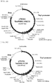
- FIGS. 1A and 1B show the above-constructed pTSC21K-TauP301L-VN173 and pTSC21K-TauP301L-VC155 recombinant plasmids, respectively.
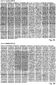
- FIGS. 2A and 2B show the sequences of the recombinant plasmids. Then, in order to examine expression of TauP301L-BiFC under the control of the Thyl promoter, the recombinant plasmids were transduced into SH-SY5Y cells, and 24 hours, normal expression of TauP301L-BiFC in the neuronal cells was observed.
- FIG. 3 shows a fluorescence microscopic image of the SH-SY5Y cells expressing TauP301L-BiFC.
- FIG. 4 shows the results of electrophoresis of the plasmids linearized with EcoRI.
- transgenic mice the confirmed plasmids were injected into embryos.
- pregnant mare serum gonadotropin and human chorionic gonadotropin were injected into C57BL/6N female mice to induce superovulation.
- the superovulation-induced C57BL/6N female mice were mated with C57BL/6N male mice, and after mating, embryos were obtained from pregnant C57BL/6N female mice.
- the vectors expressing Thy1-TauP301L-VN173 and Thy1-Tau-P301L-VC155 were injected into the male pronucleus of zygote of the obtained embryos, and the injected embryos were transferred into ICR surrogate mothers.
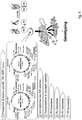
- genotyping was performed to select mice having both TauP301L-VN173 and TauP301L-VC155 ( FIG. 5 ) .
- Western blotting analysis was performed. First, 3-month-old genetically modified mice were anesthetized, and the brain was extracted from each of the mice.
- the extracted brain was lysed in RIPA containing phosphatase and protease to prepare a brain lysate sample. 40 ⁇ g of the sample was loaded, and antigen-antibody reactions with tau antibodies (pS199 and pS396) targeting phosphorylated tau were analyzed. As a result, it was shown that TauP301L-VN173 and TauP301L-VC155 in the mice obtained from the mouse embryos injected with the vectors were normally expressed, unlike those in normal control mice ( FIG. 6 ).
- the constructed Tau-P301L BiFC transgenic mice display fluorescence when tau protein formed oligomers in the brain.
- the brain of 7-month-old mice was purfused, fixed, and extracted, and the extracted brain was sectioned to a thickness of 40 ⁇ m.
- the brain tissue sections of various regions were imaged without immunofluorescence staining, and as a result, BiFC fluorescence was observed.
- BiFC fluorescence could be observed in the hippocampus, cortex, amygdale and the like of tau-expressing animal models known to show tau aggregation.
- FIG. 7 shows an image of BiFC fluorescence displayed in the brain tissue section.
- BiFC fluorescence displayed in the mouse brain tissue results from tau oligomer formation
- immunofluorescence staining of the same brain tissue section with the tau antibody AT8 targeting phosphorylated tau was performed. As a result, it was shown that the BiFC fluorescence displayed was consistent with fluorescence labeled with AT8 ( FIG. 8 ).
- tau oligomer BiFC fluorescence would be observed following injection of a tau aggregation inducer was examined.
- the tau aggregation inducing drug forskolin was filled in a drug injection kit, and then the drug was allowed to flow into a ventricle in the brain of about 5-month-old Tau-P301L BiFC transgenic mice ( FIG. 9 ). After about 10 days of drug injection, brain section samples were made for observation of BiFC fluorescence.
- tau oligomer formation occurring in the brain of mice can be visualized directly by fluorescence, thereby monitoring and quantifying the tau oligomerization process in the brain.
- the use of this technology makes it possible to investigate diseases such as dementia in which tau protein is involved, and to screen a tau oligomer formation inhibitor candidate.
- the present invention may be used as a useful tool in development of dementia therapeutic agents.
Landscapes
- Health & Medical Sciences (AREA)
- Life Sciences & Earth Sciences (AREA)
- Engineering & Computer Science (AREA)
- Biomedical Technology (AREA)
- Chemical & Material Sciences (AREA)
- Molecular Biology (AREA)
- Biotechnology (AREA)
- Immunology (AREA)
- Genetics & Genomics (AREA)
- General Health & Medical Sciences (AREA)
- Urology & Nephrology (AREA)
- Hematology (AREA)
- Zoology (AREA)
- Biochemistry (AREA)
- Bioinformatics & Cheminformatics (AREA)
- Cell Biology (AREA)
- Physics & Mathematics (AREA)
- Organic Chemistry (AREA)
- Microbiology (AREA)
- Medicinal Chemistry (AREA)
- Veterinary Medicine (AREA)
- General Engineering & Computer Science (AREA)
- Wood Science & Technology (AREA)
- Environmental Sciences (AREA)
- Analytical Chemistry (AREA)
- Food Science & Technology (AREA)
- General Physics & Mathematics (AREA)
- Pathology (AREA)
- Toxicology (AREA)
- Biophysics (AREA)
- Tropical Medicine & Parasitology (AREA)
- Biodiversity & Conservation Biology (AREA)
- Animal Husbandry (AREA)
- Animal Behavior & Ethology (AREA)
- Plant Pathology (AREA)
- Proteomics, Peptides & Aminoacids (AREA)
- Neurology (AREA)
- Neurosurgery (AREA)
- Gastroenterology & Hepatology (AREA)
- Measuring Or Testing Involving Enzymes Or Micro-Organisms (AREA)
Abstract
Description
- The present invention relates to a vector pair for screening tau oligomer formation, a mouse embryo introduced with the vector pair, a transgenic model mouse of neurological disease obtained from the mouse embryo, and a method of screening a tau oligomer formation inhibitor candidate using the transgenic model mouse.
- Tau protein is a kind of microtubule-associated protein having a molecular weight of 50,000 to 70,000, which shows remarkable molecular diversity by its phosphorylation. In the human brain, six tau isoforms are generated by insertion of 29 or 58 amino acid residues in the N-terminal region and mRNA alternative splicing of 3 or 4 repeating structures (also referred to as microtubule-associated sites). It was previously thought that tau protein is specific for the central nervous system and found primarily in axons, but it is currently known that tau protein is expressed in astrocytes and oligodendroglia in relatively many tissues, as well as neurons, and is also found in dendrities in addition to axons. Tau contributes to the stability of microtubules, and the excessive aggregation of tau protein by phosphorylation causes various brain neurological diseases. Namely, central nervous diseases, including Alzheimer disease, fontotemporal lobar degeneration (FTLD), Pick's disease, corticobasal degeneration (CBD), progressive supranuclear palsy (PSP) and the like, are known to involve tau protein aggregates. Because of such pathological features, such neurological diseases are collectively called tauopathies. Tau protein aggregates appearing in tauopathy patients are mainly found in neuronal cell bodies and dendrite, called neurofibrillary tangles (NFT) and neuropil threads. Neurofibrillary tangles are composed of paired helical filaments (PHFs) of aggregated and hyperphosphorylated tau protein, unlike normal tau protein. Although the role of abnormal tau protein aggregation, which appears in tauopathies, in a severe disease stage, has not been clearly known, it is similar to an aggregation phenomenon that appears commonly in neurodegenerative brain diseases, including Huntington's disease, Parkinson's disease, Creutzfeldt-Jakob disease, and the like, and there is a significant correlation between neurofibrillary tangle formation and cognitive impairment. In this respect, it is evident that tau protein plays an important role.
- A series of recent studies indicate that various tau oligomers rather than neurofibrillary tangles directly cause neuronal cell toxicity and are also metastasized to other sites of the brain to spread tauopathy. For example, Mirbaha et al. suggested tau trimers as structurally modified bases for intracellular tau, which can be received by cells. On the contrary, Michel et al. suggested that tau monomers are suitable fundamental metastatic forms, based on high-resolution imaging. In 2013, Wu et al. suggested that dimeric or trimeric tau having low molecular weight is a fundamental form that is metastasized between cells. Some studies on the isolation of tau protein and the identification of toxic tau oligomers by a series of biochemical experimental techniques were reported. However, direct observation of tau oligomers in the brain has not yet been reported, and the biggest reason why observation of tau oligomers is difficult is because of the absence of an experimental method that can specifically distinguish tau oligomers in the early stage of aggregation from normal tau protein present in large amounts. Regarding methods for detecting tau aggregates in neurons, immunotherapeutic technology using tau antibodies (
US Patent No. 8778343 , andUS Patent Publication Nos. 2013-0209453 and2014-0161875 ), and a tau-related disease cell model based on stem cells (US Patent Publication No. 2014-0011197 ) were developed. However, the development of technology related to cell models enabling real-time monitoring of tau aggregation mechanisms in living cells is still insufficient. - The present inventors have developed a method for constructing a transgenic model mouse of neurological disease, which displays fluorescence in the brain upon tau oligomer formation. The developed transgenic model mouse is a transgenic model mouse in which tau oligomer formation can be detected by a bimolecular fluorescence complementation (BiFC) technique. Particularly, the present invention provides a transgenic mouse in which tau oligomer formation in living tissue can be detected. Thus, the present invention makes it possible to directly detect tau oligomer formation in tissue, particularly the brain, and to screen a tau oligomer formation inhibitor. Furthermore, the present invention makes it possible to screen a tau oligomer formation inhibitor candidate and access an agent for treating neurological disease such as dementia whose perfect curing method has not yet been developed.
- Objects to be solved by the present invention are not limited to the above-mentioned objects, and other objects that are not mentioned herein may be clearly understood by those skilled in the art from the following description.
- Hereinafter, various embodiments of the present invention will be described with reference to the accompanying drawings.
- The present invention can be embodied in a variety of different forms and may have various embodiments, and exemplary embodiments are illustrated in the accompanying drawings and will be described in detail in the detailed description. However, it should be understood that the present invention is not limited to specific embodiments and encompasses all modifications, equivalents or replacement as that fall within the spirit and technical scope of the present invention. In the following description, the detailed description of related known technology will be omitted when it may obscure the subject matter of the present invention.
- Terms used in this specification are used only to describe a specific embodiment and are not intended to limit the scope of the present invention. Singular expressions are intended to include plural expressions unless specified otherwise in the context thereof. In this specification, the terms "comprise", "have", etc., are intended to denote the existence of mentioned characteristics, numbers, steps, operations, components, parts, or combinations thereof, but do not preclude the probability of existence or addition of one or more other characteristics, numbers, steps, operations, components, parts, or combinations thereof.
- Unless otherwise defined, all technical and scientific terms used herein have the same meaning as commonly understood by those skilled in the art to which the present invention belongs.
- A first aspect of the present invention is directed to a vector pair for screening tau oligomer formation, the vector pair comprising: a first vector comprising a first tau gene, a first fluorescence protein gene and a first neuron-specific promoter; and a second vector comprising a second tau gene, a second fluorescence protein gene and a second neuron-specific promoter, wherein a protein expressed from the first fluorescence protein gene and a protein expressed from the second fluorescence protein gene bind to each other to display fluorescence, by association between a protein expressed from the first tau gene and a protein expressed from the second tau gene.
- The first tau gene and/or the second tau gene may be a full-length tau, a fragment of the full-length tau, or a variant of the full-length tau. For example, the tau gene may be human full-length tau gene Tau-P301L, but is not limited thereto. In an example of the present invention, a full-length tau gene represented by SEQ ID NO: 1 was used.
- Tau protein is a kind of microtubule-associated protein (MAP) having a molecular weight of 50,000 to 70,000, and it is known that abnormal tau aggregation is a primary pathological hallmark in Alzheimer's disease (AD) and multiple other neurodegenerative disorders, collectively called tauopathies (Biochimica. et biophysica. acta. 1739: 331-354). In a healthy neuron, tau stabilizes microtubules by promoting axonal outgrowth and neuronal cell polarization. When pathologically hyperphosphorylated, tau dissociates from microtubules and forms insoluble aggregates (Mol. Neurodegener. 4: 13). A structural framework for tau aggregation has been suggested for many years. Evidences have been suggested that insoluble filaments are formed from 10 soluble monomers, and such filaments associate into higher order structures, called neurofibrillary tangles (NFTs). However, the pathophysiological importance of neurofibrillary tangles in tauopathies, the causes and molecular mechanisms responsible for triggering the process remain largely unknown, and this is because there is no reliable method for monitoring tau aggregation under physiological conditions. Until now, most studies on tau aggregation have been conducted using purified tau or tau fragments under non-physiological conditions. Furthermore, due to its extreme solubility, tau aggregation needs to be induced artificially by adding cofactors such as heparin. For this reason, animal models that can induce and monitor tau oligomer formation in living animals may provide a useful tool to investigate tau pathology and to discover methods capable of preventing and restoring the process.
- The first fluorescence protein and/or the second fluorescence protein is one allowing proteins expressed from the first and second first fluorescence proteins to bind to each other to display fluorescence, by association between proteins expressed from the first tau gene and the second tau gene. Namely, when proteins expressed from the first tau gene and the second tau gene interact with each other, first fluorescence protein and the second fluorescence protein bind to each other simultaneously with or subsequently to the interaction to thereby display fluorescence. For example, the fluorescence protein may be a Venus protein. In addition, the first fluorescence protein gene may be represented by SEQ ID NO: 3, and the second fluorescence protein gene may be represented by SEQ ID NO: 5.
- The present invention is an application of a method for visualizing protein-protein interactions that is based on a bimolecular fluorescence complementation (BiFC) technique of forming a fluorescence protein complex from non-fluorescent constituents attached to the proteins of interest (Annu. Rev. Biophys. 37: 465-487). Previously, a split green fluorescence protein (GFP) complementation technique was used to quantify tau aggregation. In the assay, tau is fused to a smaller GFP fragment (GFP 11), and co-expressed in cells with a larger GFP fragment (GFP 1-10). When tau exists as a monomer or low degree aggregate, the large GFP fragment is able to access the small GFP fragment fused to tau, leading to the association of the fluorescently active GFP. However, when tau aggregates, the reconstitution of active GFP is inhibited and GFP fluorescence decreases in cells. As a method of quantifying tau aggregation, the split-GFP assay has been highlighted; however, the limited scope and resolution of the assay do not allow the monitoring of tau oligomers that cause neuronal toxicity.
- The BiFC technique based on the Venus protein, a kind of yellow fluorescence protein (YFP), is based on the principle according to which, when two different target proteins approach each other for interaction, fluorescence protein fragments linked to the target proteins also approach each other, and as a result, reconstruction between the fluorescence protein fragments occurs to display fluorescence. The use of this technique makes it possible to visually observe that an interaction between two target proteins occurred. Thus, this technique has the advantage of enabling protein-protein interactions to be visually observed in an optimal physical/chemical environment in which protein-protein interactions in cells or tissue may occur and be maintained. This technique makes it possible to determine not only a position where protein-protein interactions in cells or tissue occur, but also information about movement of these proteins.
- The protein that is used in the present invention may be the Venus protein. The Venus protein can be effectively used for analysis of proteins such as tau protein, which are difficult to analyze spatially and temporally, because (1) it has fast and efficient maturation, (2) its self-assembly rate is low compared to that of other BiFC pairs, and (3) the fluorescence intensity of Venus-based BiFC is 10 times higher than that of EYFP-based BiFC (Biotechniques 40: 61-66.; Biotechniques 49: 793-805).
- Further, it is to be understood that a fragment as referred to in accordance with the present invention comprises at least 50, at least 100, at least 150, at least 200, at least 250, at least 500, at least 750, at least 1000, at least 1250, at least 1500, at least 1750, at least 2000, or at least 2250 consecutive nucleotides of any one of the aforementioned nucleic acid sequences or encodes an amino acid sequence comprising at least 20, at least 30, at least 50, at least 80, at least 100, at least 150, at least 200, at least 250, at least 300, at least 350, at least 400, at least 450, at least 500, at least 550, at least 600, at least 650, at least 700, or at least 750 consecutive amino acids of any one of the aforementioned amino acid sequences. Furthermore, the fragment shall encode a polypeptide which still has the activity as specified above. Accordingly, the polypeptide may comprise or consist of the domains of the nucleic acid sequence and/or amino acid sequences of the present invention conferring the said biological activity.
- Moreover, it is to be understood that a variant as referred to in accordance with the present invention shall have a nucleic acid sequence which differs due to at least one or more nucleic acid substitution, deletion and/or addition wherein the nucleic acid sequence of the variant is still, preferable, at least 50 %, at least 60 %, at least 70 %, at least 80 %, at least 85 %, at least 90 %, at least 92 %, at least 95 %, at least 97 %, at least 98 %, at least 99 %, at least 99,5 %, or at least 99,9 % identical with the nucleic acid sequence represented by the present invention. Further, the variant shall encode a polypeptide which still has the activity as specified above. Accordingly, the polypeptide may comprise or consist of the domains of the nucleic acid sequence and/or amino acid sequences of the present invention conferring the said biological activity.
- According to one embodiment of the present invention, the first tau gene and the first fluorescence protein gene may be operably linked to each other, and the second tau gene and the second fluorescence protein gene may be operably linked to each other. Namely, in each of the first vector and the second vector, the tau gene and the fluorescence protein gene are preferably sequentially expressed by a single promoter. For example, the first tau gene and the first fluorescence protein gene may be linked to each other by a first linker, and the second tau gene and the second fluorescence protein gene may be linked to each other by a second linker. In an example of the present invention, the first linker represented by SEQ ID NO: 2 and the second linker represented by SEQ ID NO: 4 were used.
- The first neuron-specific promoter and/or the second neuron-specific promoter serves to express the vector pair of the present invention, is not particularly limited, and may comprise any promoter known in the art. For example, the first neuron-specific promoter and/or the second neuron-specific promoter is preferably a Thyl promoter. Since the Thyl promoter is expressed specifically in neurons, it can induce expression of the genes inserted in the vector in neurons or neural tissue, particularly brain tissue.
- The present invention is characterized in that, when a protein expressed from the first fluorescence protein gene and a protein expressed from the second fluorescence protein gene bind to each other to display fluorescence, by association between a protein expressed from the first tau gene and a protein expressed from the second tau gene. Particularly, the present invention is characterized in that the genes inserted in the vector can be expressed by the neuron-specific promoter in neurons or neural tissue, particularly brain tissue. Accordingly, tau oligomer formation in the mouse brain can be visualized directly by fluorescence, thereby monitoring and quantifying the tau oligomerization process in the brain.
- According to one embodiment of the present invention, each of the first vector and the second vector, which comprise the Thyl promoter, may be a pTSC21K vector comprising the Thyl promoter, but is not limited thereto.
- Furthermore, the first tau gene, the first fluorescence protein gene and the first linker may be inserted into the XhoI site of the pTSC21K vector, and the second tau gene, the second fluorescence protein gene and the second linker may be inserted into the XhoI site of the pTSC21K vector, but the scope of the present invention is not limited thereto.
- Moreover, the first vector may comprise a nucleotide sequence of SEQ ID NO: 6, and the second vector may comprise a nucleotide sequence of SEQ ID NO: 7, but the scope of the present invention is not limited thereto.
- In addition, the present invention may be directed to a vector pair for screening tau oligomer formation, comprising: a first vector which comprises a Thyl promoter and wherein a full-length tau gene represented by SEQ ID NO: 1, a first linker represented by SEQ ID NO: 2, and a first fluorescence protein gene represented by SEQ ID NO: 3 are operably linked to one another; and a second vector which comprises a Thyl promoter and wherein a full-length tau gene represented by SEQ ID NO: 1, a second linker represented by SEQ ID NO: 4, and a second fluorescence protein gene represented by SEQ ID NO: 5 are operably linked to one another.
- A second aspect of the present invention is directed to a mouse embryo for screening tau oligomer formation, which has introduced therein the vector pair according to the first embodiment of the present invention.
- The mouse and its embryo are not particularly limited. When this mouse embryo is used, a mouse can be obtained in which a protein expressed from the first fluorescence protein gene and a protein expressed from the second fluorescence protein gene bind to each other to display fluorescence, by association between a protein expressed from the first tau gene and a protein expressed from the second tau gene in neuronal tissue.
- In according to one embodiment of the present invention, the mouse embryo may be a mouse embryo deposited in the Korean Collection for Type Cultures under accession number KCTC13076BP, but is no limited thereto. The present inventors obtained a mouse embryo using the above-described vector pair, and produced a transgenic mouse from the mouse embryo, and confirmed the target gene in the transgenic mouse. In this process, a mouse embryo, showing high gene expression levels and enabling fluorescence to be easily observed, was selected and deposited.
- A third aspect of the present invention is directed to a transgenic model mouse of neurological disease, which is obtained from the mouse embryo according to the second aspect of the present invention. For example, the neurological disease is preferably a neurodegenerative brain disease, particularly dementia.
- In the transgenic model mouse, human full-length tau proteins and two kinds of Venus proteins (VN173 and VC155) linked thereto, respectively, are expressed by a Thyl promoter. The Thy promoter operates specifically in neurons, and thus when the tau proteins having VN173 and VC155, respectively, form an oligomer in the brain of the mouse, BiFC fluorescence is displayed by the Venus proteins. According to this principle, whether or not a tau oligomer would be formed in the brain tissue of the mouse and the extent of tau oligomer formation can be visually observed.
- According to one embodiment of the present invention, the transgenic mouse may be a mouse wherein fluorescence is displayed by binding between the first fluorescence protein and the second fluorescence protein, when the full-length tau expressed from the first vector and the full-length tau expressed from the second vector form an oligomer. However, the transgenic mouse of the present invention is not limited thereto.
- A fourth aspect of the present invention is directed to a method of detecting tau oligomer formation using a transgenic model mouse, the method comprising the steps of: obtaining a brain tissue section from the brain of the transgenic model mouse according to the third aspect of the present invention; subjecting the obtained brain tissue section to imaging analysis to measure fluorescence intensity; and comparing the measured fluorescence intensity with a reference value.
- The step of obtaining the brain tissue section may comprises injecting a tau aggregation inducer into the transgenic model mouse and obtaining the brain tissue section from the brain of the transgenic model mouse injected with the tau aggregation inducer.
- A fifth aspect of the present invention is directed to a method for screening a tau oligomer formation inhibitor candidate, the method comprising the steps of: inducing tau oligomer formation in the brain of the transgenic model mouse; evaluating the level of tau oligomer formation in the brain of the transgenic model mouse; administering a tau oligomer formation inhibitor candidate to the transgenic model mouse; evaluating the level of tau oligomer formation in the brain of the transgenic model mouse; and comparing the level of tau oligomer formation between before and after administering the tau oligomer formation inhibitor candidate.
- For example, the step of evaluating the level of tau oligomer formation may comprise quantifying the fluorescence intensity of the fluorescence protein, that is, the Venus protein, but is not limited thereto.
- According to one embodiment of the present invention, the step of inducing tau oligomer formation in the brain of the transgenic model mouse may comprise administering forskolin or okadaic acid to the transgenic model mouse, but is not limited thereto. The forskolin or the okadaic acid may be used to induce hyperphosphorylation of tau protein.
-
-
FIGS. 1A and 1B are schematic views showing the structures of pTSC21K-TauP301L-VN173 and pTSC21K-TauP301L-VC155 recombinant plasmids, respectively, used in an example of the present invention. -
FIGS. 2A and 2B are schematic views showing the results of sequencing of pTSC21K-TauP301L-VN173 and pTSC21K-TauP301L-VC155 recombinant plasmids, respectively, used in an example of the present invention. -
FIG. 3 is an image showing the results of analyzing BiFC fluorescence displayed by tau oligomer formation in SH-SY5Y cells introduced with pTSC21K-TauP301L-VN173 and pTSC21K-TauP301L-VC155 recombinant plasmids in an example of the present invention. -
FIG. 4 is an image showing the results of electrophoresis performed after linearization of each of pTSC21K-TauP301L-VN173 and pTSC21K-TauP301L-VC155 recombinant plasmids constricted in an example of the present invention. -
FIG. 5 is a schematic view showing a method for constructing a transgenic mouse according to an embodiment of the present invention. -
FIG. 6 shows the results of Western blot analysis performed to determine whether or not TauP301L-VN173 and TauP301L-VC155 proteins in transgenic mice constructed in an example of the present invention would be normally expressed. -
FIG. 7 depicts images showing BiFC fluorescence resulting from tau oligomer formation in the brain tissue of transgenic mice constructed in an example of the present invention. -
FIG. 8 depicts staining images comparing AT8 immunofluorescence with BiFC fluorescence resulting from tau oligomer formation in the brain tissue of transgenic mice constructed according to an example of the present invention. -
FIG. 9 depicts images showing the results of observing BiFC fluorescence in the brain tissue of a transgenic mouse injected with a tau oligomer formation inducer in an example of the present invention. - Hereinafter, the present invention will be described in further detail with reference to examples. It is to be understood, however, that these examples are for illustrative purposes only and are not intended to limit the scope of the present invention as defined in the appended claims.
- To express tau protein specifically in mouse neurons, two bimolecular fluorescence complementation (BiFC) constructs (tau-VN173 and tau-VC155) were cloned into 323-pTSC21K vectors including a Thyl promoter.
- Particularly, in the present invention, vectors were constructed using a Venus-based BiFC system. To this end, the mammalian expression vector pCMV6-hTau40-GFP was purchased from OriGene Technologies Inc. (Rockville, MD, USA), and the amino acid proline at position 301 was replaced with leucine, thereby constructing pCMV6-hTau40P301L-GFP. The forward (SEQ ID NO: 8) and reverse primer sequences (SEQ ID NO: 9) used herein are shown in Table 1 below.
Table 1 P301L-F 5' -AAT ATC AAA CAC GTC CTG GGA GGC GGC ACT G-3' P301L-R 5' -CAC ACT GCC GCC TCC CAG GAC GTG TTT-3' - To replace GFP with a Venus fluorescence protein fragment, pBiFC-VN173 and pBiFC-VC155 were purchased from Addgene (Cambridge, MA), and then amplified using PCR primers having XhoI/PmeI restriction enzyme sequences. Next, a substituted human full-length tau (441 amino acids) was fused to the N-terminal fragment (1-172, VN173) (first fluorescence protein) and C-terminal fragment (155-238, VC155) (second fluorescence protein) of the fluorescence protein Venus.
- pCMV6-TauP301L-GFP and the PCR-amplified insert were digested with XhoI/PmeI and ligated with each other, thereby constructing pCMV6-TauP301L-VN173 and pCMV6-TauP301L-VC155 which are insertion genes. The linker peptide and fluorescence protein sequences used in construction of the insertion genes are shown in Tables 2 and 3 below.
Table 2 pCMV6-TauP301 L-VN173 First linker peptide ACGCGTACGCGGCCGCTCGAGTCTAGAAGATCCATCGCCACC First fluorescence protein (VN173) 
Table 3 pCMV6-TauP301L-VC155 Second linker peptide ACGCGTACGCGGCCGCTCGAGAAG Second fluorescence protein (VC155) 
- The Thyl promoter from mouse thy1.2 gene is a promoter that is expressed specifically in mouse brain neurons. Mouse Thyl gene (mouse Thy-1.2 glycoprotein gene) is 5572 bp in total length and includes three exon regions and three intron regions. TauP301L-VN173 and Tau-P301L-VC155 were inserted into
exon 3 of the Thyl gene. - Each of two tau- BiFC plasmids (i.e., pCMV6-TauP301L-VN173 and pCMV6-TauP301L-VC155) prepared as insertion genes was cloned into the Xho-1 site of a 323-pTSC21K vector.
FIGS. 1A and 1B show the above-constructed pTSC21K-TauP301L-VN173 and pTSC21K-TauP301L-VC155 recombinant plasmids, respectively. After the re-cloning process, in order to confirm whether pTSC21K-TauP301L-VN173 and pTSC21KTauP301L-VC155 would be successfully constructed, the insert region and surrounding region of the two recombinant plasmids were sequenced.FIGS. 2A and 2B show the sequences of the recombinant plasmids. Then, in order to examine expression of TauP301L-BiFC under the control of the Thyl promoter, the recombinant plasmids were transduced into SH-SY5Y cells, and 24 hours, normal expression of TauP301L-BiFC in the neuronal cells was observed.FIG. 3 shows a fluorescence microscopic image of the SH-SY5Y cells expressing TauP301L-BiFC. - For injection into mouse embryos, the Thy1-TauP301L-VN173 and Thy1-Tau-P301L-VC155 recombinant plasmids were linearized with the restriction enzyme EcoRI.
FIG. 4 shows the results of electrophoresis of the plasmids linearized with EcoRI. To construct transgenic mice, the confirmed plasmids were injected into embryos. To obtain embryos, pregnant mare serum gonadotropin and human chorionic gonadotropin were injected into C57BL/6N female mice to induce superovulation. The superovulation-induced C57BL/6N female mice were mated with C57BL/6N male mice, and after mating, embryos were obtained from pregnant C57BL/6N female mice. Then, the vectors expressing Thy1-TauP301L-VN173 and Thy1-Tau-P301L-VC155 were injected into the male pronucleus of zygote of the obtained embryos, and the injected embryos were transferred into ICR surrogate mothers. Next, genotyping was performed to select mice having both TauP301L-VN173 and TauP301L-VC155 (FIG. 5 ) . Next, in order to examine whether both the TauP301L-VN173 and TauP301L-VC155 proteins would be normally expressed by introduction of the genes, Western blotting analysis was performed. First, 3-month-old genetically modified mice were anesthetized, and the brain was extracted from each of the mice. The extracted brain was lysed in RIPA containing phosphatase and protease to prepare a brain lysate sample. 40 µg of the sample was loaded, and antigen-antibody reactions with tau antibodies (pS199 and pS396) targeting phosphorylated tau were analyzed. As a result, it was shown that TauP301L-VN173 and TauP301L-VC155 in the mice obtained from the mouse embryos injected with the vectors were normally expressed, unlike those in normal control mice (FIG. 6 ). - The constructed Tau-P301L BiFC transgenic mice display fluorescence when tau protein formed oligomers in the brain. In order to actually confirm whether fluorescence would be observed by tau oligomer formation in the brain tissue of the Tau-P301L BiFC transgenic mice, the brain of 7-month-old mice was purfused, fixed, and extracted, and the extracted brain was sectioned to a thickness of 40 µm. The brain tissue sections of various regions were imaged without immunofluorescence staining, and as a result, BiFC fluorescence was observed. Specifically, BiFC fluorescence could be observed in the hippocampus, cortex, amygdale and the like of tau-expressing animal models known to show tau aggregation.
FIG. 7 shows an image of BiFC fluorescence displayed in the brain tissue section. In order to verify that BiFC fluorescence displayed in the mouse brain tissue results from tau oligomer formation, immunofluorescence staining of the same brain tissue section with the tau antibody AT8 targeting phosphorylated tau was performed. As a result, it was shown that the BiFC fluorescence displayed was consistent with fluorescence labeled with AT8 (FIG. 8 ). - Using 4-month-old Tau-P301L BiFC transgenic mice which have not yet shown tau aggregation, whether tau oligomer BiFC fluorescence would be observed following injection of a tau aggregation inducer was examined. The tau aggregation inducing drug forskolin was filled in a drug injection kit, and then the drug was allowed to flow into a ventricle in the brain of about 5-month-old Tau-P301L BiFC transgenic mice (
FIG. 9 ). After about 10 days of drug injection, brain section samples were made for observation of BiFC fluorescence. As a result, in the brain of the Tau-P301L BiFC transgenic mice injected with forskolin, BiFC fluorescence was observed in a portion surrounding the injected region, unlike mice injected with DMSO as a control. This result suggests that tau oligomer formation in the mouse brain was accelerated by injection of the forskolin drug. In addition, it was demonstrated that the level of tau oligomer formation could be directly observed without using a separate antibody. - As described above, according to the present invention, tau oligomer formation occurring in the brain of mice can be visualized directly by fluorescence, thereby monitoring and quantifying the tau oligomerization process in the brain. The use of this technology makes it possible to investigate diseases such as dementia in which tau protein is involved, and to screen a tau oligomer formation inhibitor candidate. Thus, the present invention may be used as a useful tool in development of dementia therapeutic agents.
- While the present invention has been described with reference to the particular illustrative embodiments, it will be understood by those skilled in the art to which the present invention pertains that the present invention may be embodied in other specific forms without departing from the technical spirit or essential characteristics of the present invention. Therefore, the embodiments described above should be considered in a descriptive sense only and not for purposes of limitation. For example, each component described in a single form may be carried out in a distributed fashion, and likewise, components described in a distributed form may be carried out in a combined fashion.
- Therefore, the scope of the present invention is defined not by the detailed description, but by the claims and their equivalents, and all variations within the scope of the claims and their equivalents are to be construed as being included in the scope of the present invention.


Claims (20)
- A vector pair for screening tau oligomer formation, comprising:a first vector comprising a first tau gene, a first fluorescence protein gene and a first neuron-specific promoter; anda second vector comprising a second tau gene, a second fluorescence protein gene and a second neuron-specific promoter,wherein a protein expressed from the first fluorescence protein gene and a protein expressed from the second fluorescence protein gene bind to each other to display fluorescence, by association between a protein expressed from the first tau gene and a protein expressed from the second tau gene.
- The vector pair of claim 1, wherein each of the first tau gene and the second tau gene is a full-length tau, a fragment of the full-length tau, or a variant of the full-length tau.
- The vector pair of claim 2, wherein the variant of the full-length tau is human full-length tau gene Tau-P301L.
- The vector pair of claim 1, wherein the first tau gene and the first fluorescence protein gene are operably linked to each other, and the second tau gene and the second fluorescence protein gene are operably linked to each other.
- The vector pair of claim 4, wherein the first tau gene and the first fluorescence protein gene are linked to each other by a first linker, and the second tau gene and the second fluorescence protein gene are linked to each other by a second linker.
- The vector pair of claim 5, wherein each of the first neuron-specific promoter and the second neuron-specific promoter is a Thyl promoter.
- The vector pair of claim 6, wherein each of the first vector and the second vector, which comprise the Thyl promoter, is a pTSC21K vector.
- The vector pair of claim 7, wherein the first tau gene, the first fluorescence protein gene and the first linker are inserted into the XhoI site of the pTSC21K vector, and
the second tau gene, the second fluorescence protein gene and the second linker are inserted into the XhoI site of the pTSC21K vector. - The vector pair of claim 5, wherein
the first tau gene is represented by SEQ ID NO: 1,
the first fluorescence protein gene is represented by SEQ ID NO: 3,
the second tau gene is represented by SEQ ID NO: 1, and
the second fluorescence protein gene is represented by SEQ ID NO: 5. - The vector pair of claim 9, wherein
the first linker is represented by SEQ ID NO: 2, and
the second linker is represented by SEQ ID NO: 4. - The vector pair of claim 10, wherein
the first vector comprises a nucleotide sequence of SEQ ID NO: 6, and
the second vector comprises a nucleotide sequence of SEQ ID NO: 7. - A mouse embryo for screening tau oligomer formation, which has introduced therein the vector pair of claim 1.
- The mouse embryo of claim 12, which is deposited under accession number KCTC13076BP.
- A transgenic model mouse of neurological disease, obtained from the mouse embryo of claim 12.
- The transgenic model mouse of claim 14, wherein fluorescence is displayed by binding between the first fluorescence protein and the second fluorescence protein, when a full-length tau expressed from the first vector and a full-length tau expressed from the second vector form an oligomer.
- The transgenic model mouse of claim 14, wherein the neurological disease is dementia.
- A method of detecting tau oligomer formation using a transgenic mouse, the method comprising the steps of:obtaining a brain tissue section from the brain of the transgenic model mouse of claim 14;subjecting the obtained brain tissue section to imaging analysis to measure fluorescence intensity; andcomparing the measured fluorescence intensity with a reference value.
- The method of claim 17, wherein the step of obtaining the brain tissue section comprises injecting a tau aggregation inducer into the transgenic model mice, and obtaining the brain tissue section from the brain of the transgenic model mice injected with the tau aggregation inducer.
- A method for screening a tau oligomer formation inhibitor candidate, the method comprising the steps of:inducing tau oligomer formation in the brain of the transgenic model mouse of claim 14;evaluating a level of tau oligomer formation in the brain of the transgenic model mouse;administering a tau oligomer formation inhibitor candidate to the transgenic model mouse;evaluating a level of tau oligomer formation in the brain of the transgenic model mouse; andcomparing the level of tau oligomer formation between before and after administering the tau oligomer formation inhibitor candidate.
- The method of claim 19, wherein the step of inducing tau oligomer formation in the brain of the transgenic model mouse comprises administering forskolin or okadaic acid to the transgenic model mouse.
Applications Claiming Priority (1)
| Application Number | Priority Date | Filing Date | Title |
|---|---|---|---|
| KR1020160130813A KR101876423B1 (en) | 2016-10-10 | 2016-10-10 | Dementia Model Transgenic Mouse and Screening Method using thereof |
Publications (2)
| Publication Number | Publication Date |
|---|---|
| EP3305070A1 true EP3305070A1 (en) | 2018-04-11 |
| EP3305070B1 EP3305070B1 (en) | 2020-07-22 |
Family
ID=60186001
Family Applications (1)
| Application Number | Title | Priority Date | Filing Date |
|---|---|---|---|
| EP17195792.1A Active EP3305070B1 (en) | 2016-10-10 | 2017-10-10 | Dementia model transgenic mouse and screening method using thereof |
Country Status (3)
| Country | Link |
|---|---|
| US (1) | US10544427B2 (en) |
| EP (1) | EP3305070B1 (en) |
| KR (1) | KR101876423B1 (en) |
Families Citing this family (3)
| Publication number | Priority date | Publication date | Assignee | Title |
|---|---|---|---|---|
| KR102230812B1 (en) | 2019-04-26 | 2021-03-23 | 한국과학기술연구원 | A pair of amino acid sequences for monitoring formation of TDP-43 oligomer in living cells via bimolecular fluorescence complementation |
| CN110841059A (en) * | 2019-12-03 | 2020-02-28 | 南通大学 | Preparation method of mouse model for senile dementia |
| MX2024003519A (en) | 2021-09-24 | 2024-04-01 | Alnylam Pharmaceuticals Inc | Microtubule associated protein tau (mapt) irna agent compositions and methods of use thereof. |
Citations (8)
| Publication number | Priority date | Publication date | Assignee | Title |
|---|---|---|---|---|
| US20130209453A1 (en) | 2007-10-17 | 2013-08-15 | Wyeth Llc | Use of tau to monitor immunotherapy |
| KR20130091288A (en) * | 2012-02-07 | 2013-08-16 | (주)나노몰 | A system for detecting tau aggregation |
| US20140011197A1 (en) | 2011-02-16 | 2014-01-09 | The Regents Of The University Of California | Alzheimer's Disease Cellular Model for Diagnostic and Therapeutic Development |
| WO2014089104A1 (en) * | 2012-12-03 | 2014-06-12 | Washington University | Method for detection of aggregates in biological samples |
| US20140161875A1 (en) | 2011-07-14 | 2014-06-12 | Katholieke Universiteit Leuven | Antibodies to phosphorylated tau aggregates |
| US8778343B2 (en) | 2009-08-28 | 2014-07-15 | The Board Of Regents Of The University Of Texas System | Antibodies that bind tau oligomers |
| WO2015053439A1 (en) * | 2013-10-08 | 2015-04-16 | 한국과학기술연구원 | Bimolecular fluorescence complementary amino acid sequence pair for monitoring interaction of intracellular tau protein |
| KR20160120146A (en) * | 2015-04-07 | 2016-10-17 | 한국과학기술연구원 | Tau aggregation cell line in response to traumatic stimuli |
Family Cites Families (3)
| Publication number | Priority date | Publication date | Assignee | Title |
|---|---|---|---|---|
| US287682A (en) * | 1883-10-30 | Yoke for draft-animals | ||
| WO1999062548A1 (en) * | 1998-06-01 | 1999-12-09 | Advanced Research And Technology Institute | Methods and compositions for diagnosing tauopathies |
| US7479579B2 (en) * | 2001-12-20 | 2009-01-20 | The Regents Of The University Of California | Triple transgenic mouse model of Alzheimer's disease |
-
2016
- 2016-10-10 KR KR1020160130813A patent/KR101876423B1/en active Active
-
2017
- 2017-09-20 US US15/709,822 patent/US10544427B2/en active Active
- 2017-10-10 EP EP17195792.1A patent/EP3305070B1/en active Active
Patent Citations (8)
| Publication number | Priority date | Publication date | Assignee | Title |
|---|---|---|---|---|
| US20130209453A1 (en) | 2007-10-17 | 2013-08-15 | Wyeth Llc | Use of tau to monitor immunotherapy |
| US8778343B2 (en) | 2009-08-28 | 2014-07-15 | The Board Of Regents Of The University Of Texas System | Antibodies that bind tau oligomers |
| US20140011197A1 (en) | 2011-02-16 | 2014-01-09 | The Regents Of The University Of California | Alzheimer's Disease Cellular Model for Diagnostic and Therapeutic Development |
| US20140161875A1 (en) | 2011-07-14 | 2014-06-12 | Katholieke Universiteit Leuven | Antibodies to phosphorylated tau aggregates |
| KR20130091288A (en) * | 2012-02-07 | 2013-08-16 | (주)나노몰 | A system for detecting tau aggregation |
| WO2014089104A1 (en) * | 2012-12-03 | 2014-06-12 | Washington University | Method for detection of aggregates in biological samples |
| WO2015053439A1 (en) * | 2013-10-08 | 2015-04-16 | 한국과학기술연구원 | Bimolecular fluorescence complementary amino acid sequence pair for monitoring interaction of intracellular tau protein |
| KR20160120146A (en) * | 2015-04-07 | 2016-10-17 | 한국과학기술연구원 | Tau aggregation cell line in response to traumatic stimuli |
Non-Patent Citations (6)
| Title |
|---|
| ANNU. REV. BIOPHYS., vol. 37, pages 465 - 487 |
| BIOCHIMICA. ET BIOPHYSICA. ACTA., vol. 1739, pages 331 - 354 |
| BIOTECHNIQUES, vol. 40, pages 61 - 66 |
| BIOTECHNIQUES, vol. 49, pages 793 - 805 |
| HYEJIN TAK ET AL: "Bimolecular Fluorescence Complementation; Lighting-Up Tau-Tau Interaction in Living Cells", PLOS ONE, vol. 8, no. 12, 2 December 2013 (2013-12-02), pages e81682, XP055452308, DOI: 10.1371/journal.pone.0081682 * |
| LI M ET AL: "Optimal promoter usage for lentiviral vector-mediated transduction of cultured central nervous system cells", JOURNAL OF NEUROSCIENCE METHODS, ELSEVIER SCIENCE PUBLISHER B.V., AMSTERDAM, NL, vol. 189, no. 1, 30 May 2010 (2010-05-30), pages 56 - 64, XP027037853, ISSN: 0165-0270, [retrieved on 20100501] * |
Also Published As
| Publication number | Publication date |
|---|---|
| US20180119169A1 (en) | 2018-05-03 |
| US10544427B2 (en) | 2020-01-28 |
| KR101876423B1 (en) | 2018-07-09 |
| KR20180039471A (en) | 2018-04-18 |
| EP3305070B1 (en) | 2020-07-22 |
Similar Documents
| Publication | Publication Date | Title |
|---|---|---|
| US11939355B2 (en) | Variant adeno-associated viruses and methods of using | |
| DE69034171T2 (en) | Recombinant protease inhibitor of Alzheimer's amyloid protein | |
| Paquet et al. | A zebrafish model of tauopathy allows in vivo imaging of neuronal cell death and drug evaluation | |
| JP5971661B2 (en) | DNA having neural activity-dependent promoter activity and vector containing the same | |
| KR20210133227A (en) | Interneuron-specific therapeutics to normalize neuronal cell excitability and treat Dravet syndrome | |
| EP3305070B1 (en) | Dementia model transgenic mouse and screening method using thereof | |
| Petkova et al. | Live‐imaging of revertant and therapeutically restored dystrophin in the DmdEGFP‐mdx mouse model for duchenne muscular dystrophy | |
| KR102288597B1 (en) | A probe for detecting autophagosome and uses thereof | |
| Takeuchi | A gene trap approach to identify genes that control development | |
| KR20150042308A (en) | A pair of amino acid sequences for monitoring tau-tau interaction in living cells via bimolecular fluorescence complementation | |
| KR102296075B1 (en) | epcam Variant Zebrafish and Uses Thereof | |
| CA2574540A1 (en) | Cell cycle reporting cell line comprising phosphorylated subcellular localization domain (psld) of human dna helicase b (hdbd) | |
| US9989540B2 (en) | Diagnostic and monitoring system for Huntington's disease | |
| EP1867716B1 (en) | Model animal in which state of disease condition is observable in real time, gene construct for achieving the same and use of the same | |
| KR101678961B1 (en) | Animal model for epilepsy and method for producing the same | |
| KR102120076B1 (en) | Detection of autophagic body binding protein in vivo | |
| KR20200037522A (en) | A pair of amino acid sequences for monitoring tau-tubulin interaction in living cells via bimolecular fluorescence complementation | |
| US20090158448A1 (en) | Dna encoding polypeptide capable of modulating muscle-specific tyrosine kinase activity | |
| US7745123B2 (en) | Cell cycle reporting cell line | |
| Li et al. | rAAV-miniBEND: A targeted vector for brain endothelial cell gene delivery and cerebrovascular malformation modeling | |
| Günther | On the Rational Design of Adeno-Associated Viral Vectors as Precision Tools in Neuroscience | |
| US20230141187A1 (en) | Non-human animal secretome models | |
| HK40007187B (en) | Variant adeno-associated viruses and methods of using | |
| HK40007187A (en) | Variant adeno-associated viruses and methods of using | |
| DE10116826A1 (en) | Detection method for homologous recombination events |
Legal Events
| Date | Code | Title | Description |
|---|---|---|---|
| PUAI | Public reference made under article 153(3) epc to a published international application that has entered the european phase |
Free format text: ORIGINAL CODE: 0009012 |
|
| STAA | Information on the status of an ep patent application or granted ep patent |
Free format text: STATUS: REQUEST FOR EXAMINATION WAS MADE |
|
| 17P | Request for examination filed |
Effective date: 20171026 |
|
| AK | Designated contracting states |
Kind code of ref document: A1 Designated state(s): AL AT BE BG CH CY CZ DE DK EE ES FI FR GB GR HR HU IE IS IT LI LT LU LV MC MK MT NL NO PL PT RO RS SE SI SK SM TR |
|
| AX | Request for extension of the european patent |
Extension state: BA ME |
|
| RBV | Designated contracting states (corrected) |
Designated state(s): AL AT BE BG CH CY CZ DE DK EE ES FI FR GB GR HR HU IE IS IT LI LT LU LV MC MK MT NL NO PL PT RO RS SE SI SK SM TR |
|
| STAA | Information on the status of an ep patent application or granted ep patent |
Free format text: STATUS: EXAMINATION IS IN PROGRESS |
|
| 17Q | First examination report despatched |
Effective date: 20190516 |
|
| GRAP | Despatch of communication of intention to grant a patent |
Free format text: ORIGINAL CODE: EPIDOSNIGR1 |
|
| STAA | Information on the status of an ep patent application or granted ep patent |
Free format text: STATUS: GRANT OF PATENT IS INTENDED |
|
| RIC1 | Information provided on ipc code assigned before grant |
Ipc: A01K 67/027 20060101AFI20200115BHEP Ipc: G01N 33/68 20060101ALI20200115BHEP Ipc: C12N 5/073 20100101ALI20200115BHEP Ipc: G01N 33/50 20060101ALI20200115BHEP Ipc: C12N 15/85 20060101ALI20200115BHEP |
|
| INTG | Intention to grant announced |
Effective date: 20200207 |
|
| GRAS | Grant fee paid |
Free format text: ORIGINAL CODE: EPIDOSNIGR3 |
|
| GRAA | (expected) grant |
Free format text: ORIGINAL CODE: 0009210 |
|
| STAA | Information on the status of an ep patent application or granted ep patent |
Free format text: STATUS: THE PATENT HAS BEEN GRANTED |
|
| AK | Designated contracting states |
Kind code of ref document: B1 Designated state(s): AL AT BE BG CH CY CZ DE DK EE ES FI FR GB GR HR HU IE IS IT LI LT LU LV MC MK MT NL NO PL PT RO RS SE SI SK SM TR |
|
| REG | Reference to a national code |
Ref country code: GB Ref legal event code: FG4D |
|
| REG | Reference to a national code |
Ref country code: CH Ref legal event code: EP |
|
| REG | Reference to a national code |
Ref country code: DE Ref legal event code: R096 Ref document number: 602017020088 Country of ref document: DE |
|
| REG | Reference to a national code |
Ref country code: AT Ref legal event code: REF Ref document number: 1292484 Country of ref document: AT Kind code of ref document: T Effective date: 20200815 |
|
| REG | Reference to a national code |
Ref country code: IE Ref legal event code: FG4D |
|
| REG | Reference to a national code |
Ref country code: LT Ref legal event code: MG4D |
|
| REG | Reference to a national code |
Ref country code: AT Ref legal event code: MK05 Ref document number: 1292484 Country of ref document: AT Kind code of ref document: T Effective date: 20200722 |
|
| PG25 | Lapsed in a contracting state [announced via postgrant information from national office to epo] |
Ref country code: GR Free format text: LAPSE BECAUSE OF FAILURE TO SUBMIT A TRANSLATION OF THE DESCRIPTION OR TO PAY THE FEE WITHIN THE PRESCRIBED TIME-LIMIT Effective date: 20201023 Ref country code: NO Free format text: LAPSE BECAUSE OF FAILURE TO SUBMIT A TRANSLATION OF THE DESCRIPTION OR TO PAY THE FEE WITHIN THE PRESCRIBED TIME-LIMIT Effective date: 20201022 Ref country code: ES Free format text: LAPSE BECAUSE OF FAILURE TO SUBMIT A TRANSLATION OF THE DESCRIPTION OR TO PAY THE FEE WITHIN THE PRESCRIBED TIME-LIMIT Effective date: 20200722 Ref country code: SE Free format text: LAPSE BECAUSE OF FAILURE TO SUBMIT A TRANSLATION OF THE DESCRIPTION OR TO PAY THE FEE WITHIN THE PRESCRIBED TIME-LIMIT Effective date: 20200722 Ref country code: BG Free format text: LAPSE BECAUSE OF FAILURE TO SUBMIT A TRANSLATION OF THE DESCRIPTION OR TO PAY THE FEE WITHIN THE PRESCRIBED TIME-LIMIT Effective date: 20201022 Ref country code: PT Free format text: LAPSE BECAUSE OF FAILURE TO SUBMIT A TRANSLATION OF THE DESCRIPTION OR TO PAY THE FEE WITHIN THE PRESCRIBED TIME-LIMIT Effective date: 20201123 Ref country code: AT Free format text: LAPSE BECAUSE OF FAILURE TO SUBMIT A TRANSLATION OF THE DESCRIPTION OR TO PAY THE FEE WITHIN THE PRESCRIBED TIME-LIMIT Effective date: 20200722 Ref country code: FI Free format text: LAPSE BECAUSE OF FAILURE TO SUBMIT A TRANSLATION OF THE DESCRIPTION OR TO PAY THE FEE WITHIN THE PRESCRIBED TIME-LIMIT Effective date: 20200722 Ref country code: LT Free format text: LAPSE BECAUSE OF FAILURE TO SUBMIT A TRANSLATION OF THE DESCRIPTION OR TO PAY THE FEE WITHIN THE PRESCRIBED TIME-LIMIT Effective date: 20200722 Ref country code: HR Free format text: LAPSE BECAUSE OF FAILURE TO SUBMIT A TRANSLATION OF THE DESCRIPTION OR TO PAY THE FEE WITHIN THE PRESCRIBED TIME-LIMIT Effective date: 20200722 |
|
| PG25 | Lapsed in a contracting state [announced via postgrant information from national office to epo] |
Ref country code: LV Free format text: LAPSE BECAUSE OF FAILURE TO SUBMIT A TRANSLATION OF THE DESCRIPTION OR TO PAY THE FEE WITHIN THE PRESCRIBED TIME-LIMIT Effective date: 20200722 Ref country code: PL Free format text: LAPSE BECAUSE OF FAILURE TO SUBMIT A TRANSLATION OF THE DESCRIPTION OR TO PAY THE FEE WITHIN THE PRESCRIBED TIME-LIMIT Effective date: 20200722 Ref country code: RS Free format text: LAPSE BECAUSE OF FAILURE TO SUBMIT A TRANSLATION OF THE DESCRIPTION OR TO PAY THE FEE WITHIN THE PRESCRIBED TIME-LIMIT Effective date: 20200722 Ref country code: IS Free format text: LAPSE BECAUSE OF FAILURE TO SUBMIT A TRANSLATION OF THE DESCRIPTION OR TO PAY THE FEE WITHIN THE PRESCRIBED TIME-LIMIT Effective date: 20201122 |
|
| PG25 | Lapsed in a contracting state [announced via postgrant information from national office to epo] |
Ref country code: NL Free format text: LAPSE BECAUSE OF FAILURE TO SUBMIT A TRANSLATION OF THE DESCRIPTION OR TO PAY THE FEE WITHIN THE PRESCRIBED TIME-LIMIT Effective date: 20200722 |
|
| REG | Reference to a national code |
Ref country code: DE Ref legal event code: R097 Ref document number: 602017020088 Country of ref document: DE |
|
| PG25 | Lapsed in a contracting state [announced via postgrant information from national office to epo] |
Ref country code: SM Free format text: LAPSE BECAUSE OF FAILURE TO SUBMIT A TRANSLATION OF THE DESCRIPTION OR TO PAY THE FEE WITHIN THE PRESCRIBED TIME-LIMIT Effective date: 20200722 Ref country code: EE Free format text: LAPSE BECAUSE OF FAILURE TO SUBMIT A TRANSLATION OF THE DESCRIPTION OR TO PAY THE FEE WITHIN THE PRESCRIBED TIME-LIMIT Effective date: 20200722 Ref country code: IT Free format text: LAPSE BECAUSE OF FAILURE TO SUBMIT A TRANSLATION OF THE DESCRIPTION OR TO PAY THE FEE WITHIN THE PRESCRIBED TIME-LIMIT Effective date: 20200722 Ref country code: DK Free format text: LAPSE BECAUSE OF FAILURE TO SUBMIT A TRANSLATION OF THE DESCRIPTION OR TO PAY THE FEE WITHIN THE PRESCRIBED TIME-LIMIT Effective date: 20200722 Ref country code: CZ Free format text: LAPSE BECAUSE OF FAILURE TO SUBMIT A TRANSLATION OF THE DESCRIPTION OR TO PAY THE FEE WITHIN THE PRESCRIBED TIME-LIMIT Effective date: 20200722 Ref country code: RO Free format text: LAPSE BECAUSE OF FAILURE TO SUBMIT A TRANSLATION OF THE DESCRIPTION OR TO PAY THE FEE WITHIN THE PRESCRIBED TIME-LIMIT Effective date: 20200722 |
|
| PLBE | No opposition filed within time limit |
Free format text: ORIGINAL CODE: 0009261 |
|
| STAA | Information on the status of an ep patent application or granted ep patent |
Free format text: STATUS: NO OPPOSITION FILED WITHIN TIME LIMIT |
|
| PG25 | Lapsed in a contracting state [announced via postgrant information from national office to epo] |
Ref country code: AL Free format text: LAPSE BECAUSE OF FAILURE TO SUBMIT A TRANSLATION OF THE DESCRIPTION OR TO PAY THE FEE WITHIN THE PRESCRIBED TIME-LIMIT Effective date: 20200722 |
|
| REG | Reference to a national code |
Ref country code: CH Ref legal event code: PL |
|
| 26N | No opposition filed |
Effective date: 20210423 |
|
| PG25 | Lapsed in a contracting state [announced via postgrant information from national office to epo] |
Ref country code: MC Free format text: LAPSE BECAUSE OF FAILURE TO SUBMIT A TRANSLATION OF THE DESCRIPTION OR TO PAY THE FEE WITHIN THE PRESCRIBED TIME-LIMIT Effective date: 20200722 Ref country code: LU Free format text: LAPSE BECAUSE OF NON-PAYMENT OF DUE FEES Effective date: 20201010 Ref country code: SK Free format text: LAPSE BECAUSE OF FAILURE TO SUBMIT A TRANSLATION OF THE DESCRIPTION OR TO PAY THE FEE WITHIN THE PRESCRIBED TIME-LIMIT Effective date: 20200722 |
|
| REG | Reference to a national code |
Ref country code: BE Ref legal event code: MM Effective date: 20201031 |
|
| PG25 | Lapsed in a contracting state [announced via postgrant information from national office to epo] |
Ref country code: FR Free format text: LAPSE BECAUSE OF NON-PAYMENT OF DUE FEES Effective date: 20201031 |
|
| PG25 | Lapsed in a contracting state [announced via postgrant information from national office to epo] |
Ref country code: CH Free format text: LAPSE BECAUSE OF NON-PAYMENT OF DUE FEES Effective date: 20201031 Ref country code: BE Free format text: LAPSE BECAUSE OF NON-PAYMENT OF DUE FEES Effective date: 20201031 Ref country code: SI Free format text: LAPSE BECAUSE OF FAILURE TO SUBMIT A TRANSLATION OF THE DESCRIPTION OR TO PAY THE FEE WITHIN THE PRESCRIBED TIME-LIMIT Effective date: 20200722 Ref country code: LI Free format text: LAPSE BECAUSE OF NON-PAYMENT OF DUE FEES Effective date: 20201031 |
|
| REG | Reference to a national code |
Ref country code: NL Ref legal event code: MP Effective date: 20200722 |
|
| PG25 | Lapsed in a contracting state [announced via postgrant information from national office to epo] |
Ref country code: IE Free format text: LAPSE BECAUSE OF NON-PAYMENT OF DUE FEES Effective date: 20201010 |
|
| PG25 | Lapsed in a contracting state [announced via postgrant information from national office to epo] |
Ref country code: TR Free format text: LAPSE BECAUSE OF FAILURE TO SUBMIT A TRANSLATION OF THE DESCRIPTION OR TO PAY THE FEE WITHIN THE PRESCRIBED TIME-LIMIT Effective date: 20200722 Ref country code: MT Free format text: LAPSE BECAUSE OF FAILURE TO SUBMIT A TRANSLATION OF THE DESCRIPTION OR TO PAY THE FEE WITHIN THE PRESCRIBED TIME-LIMIT Effective date: 20200722 Ref country code: CY Free format text: LAPSE BECAUSE OF FAILURE TO SUBMIT A TRANSLATION OF THE DESCRIPTION OR TO PAY THE FEE WITHIN THE PRESCRIBED TIME-LIMIT Effective date: 20200722 |
|
| GBPC | Gb: european patent ceased through non-payment of renewal fee |
Effective date: 20211010 |
|
| PG25 | Lapsed in a contracting state [announced via postgrant information from national office to epo] |
Ref country code: MK Free format text: LAPSE BECAUSE OF FAILURE TO SUBMIT A TRANSLATION OF THE DESCRIPTION OR TO PAY THE FEE WITHIN THE PRESCRIBED TIME-LIMIT Effective date: 20200722 |
|
| PG25 | Lapsed in a contracting state [announced via postgrant information from national office to epo] |
Ref country code: GB Free format text: LAPSE BECAUSE OF NON-PAYMENT OF DUE FEES Effective date: 20211010 |
|
| PGFP | Annual fee paid to national office [announced via postgrant information from national office to epo] |
Ref country code: DE Payment date: 20240920 Year of fee payment: 8 |