EP3303625B1 - Method for identifying subjects with aggressive melanoma skin cancer at diagnosis - Google Patents
Method for identifying subjects with aggressive melanoma skin cancer at diagnosis Download PDFInfo
- Publication number
- EP3303625B1 EP3303625B1 EP16729800.9A EP16729800A EP3303625B1 EP 3303625 B1 EP3303625 B1 EP 3303625B1 EP 16729800 A EP16729800 A EP 16729800A EP 3303625 B1 EP3303625 B1 EP 3303625B1
- Authority
- EP
- European Patent Office
- Prior art keywords
- melanoma
- methylation
- promoter
- dna
- methylated
- Prior art date
- Legal status (The legal status is an assumption and is not a legal conclusion. Google has not performed a legal analysis and makes no representation as to the accuracy of the status listed.)
- Active
Links
Images



Classifications
-
- C—CHEMISTRY; METALLURGY
- C12—BIOCHEMISTRY; BEER; SPIRITS; WINE; VINEGAR; MICROBIOLOGY; ENZYMOLOGY; MUTATION OR GENETIC ENGINEERING
- C12Q—MEASURING OR TESTING PROCESSES INVOLVING ENZYMES, NUCLEIC ACIDS OR MICROORGANISMS; COMPOSITIONS OR TEST PAPERS THEREFOR; PROCESSES OF PREPARING SUCH COMPOSITIONS; CONDITION-RESPONSIVE CONTROL IN MICROBIOLOGICAL OR ENZYMOLOGICAL PROCESSES
- C12Q1/00—Measuring or testing processes involving enzymes, nucleic acids or microorganisms; Compositions therefor; Processes of preparing such compositions
- C12Q1/68—Measuring or testing processes involving enzymes, nucleic acids or microorganisms; Compositions therefor; Processes of preparing such compositions involving nucleic acids
- C12Q1/6876—Nucleic acid products used in the analysis of nucleic acids, e.g. primers or probes
- C12Q1/6883—Nucleic acid products used in the analysis of nucleic acids, e.g. primers or probes for diseases caused by alterations of genetic material
- C12Q1/6886—Nucleic acid products used in the analysis of nucleic acids, e.g. primers or probes for diseases caused by alterations of genetic material for cancer
-
- C—CHEMISTRY; METALLURGY
- C12—BIOCHEMISTRY; BEER; SPIRITS; WINE; VINEGAR; MICROBIOLOGY; ENZYMOLOGY; MUTATION OR GENETIC ENGINEERING
- C12Q—MEASURING OR TESTING PROCESSES INVOLVING ENZYMES, NUCLEIC ACIDS OR MICROORGANISMS; COMPOSITIONS OR TEST PAPERS THEREFOR; PROCESSES OF PREPARING SUCH COMPOSITIONS; CONDITION-RESPONSIVE CONTROL IN MICROBIOLOGICAL OR ENZYMOLOGICAL PROCESSES
- C12Q2600/00—Oligonucleotides characterized by their use
- C12Q2600/112—Disease subtyping, staging or classification
-
- C—CHEMISTRY; METALLURGY
- C12—BIOCHEMISTRY; BEER; SPIRITS; WINE; VINEGAR; MICROBIOLOGY; ENZYMOLOGY; MUTATION OR GENETIC ENGINEERING
- C12Q—MEASURING OR TESTING PROCESSES INVOLVING ENZYMES, NUCLEIC ACIDS OR MICROORGANISMS; COMPOSITIONS OR TEST PAPERS THEREFOR; PROCESSES OF PREPARING SUCH COMPOSITIONS; CONDITION-RESPONSIVE CONTROL IN MICROBIOLOGICAL OR ENZYMOLOGICAL PROCESSES
- C12Q2600/00—Oligonucleotides characterized by their use
- C12Q2600/118—Prognosis of disease development
-
- C—CHEMISTRY; METALLURGY
- C12—BIOCHEMISTRY; BEER; SPIRITS; WINE; VINEGAR; MICROBIOLOGY; ENZYMOLOGY; MUTATION OR GENETIC ENGINEERING
- C12Q—MEASURING OR TESTING PROCESSES INVOLVING ENZYMES, NUCLEIC ACIDS OR MICROORGANISMS; COMPOSITIONS OR TEST PAPERS THEREFOR; PROCESSES OF PREPARING SUCH COMPOSITIONS; CONDITION-RESPONSIVE CONTROL IN MICROBIOLOGICAL OR ENZYMOLOGICAL PROCESSES
- C12Q2600/00—Oligonucleotides characterized by their use
- C12Q2600/154—Methylation markers
Definitions
- the invention is in the field of molecular biology and medical diagnosis. It provides means and methods for determining the prognosis and disease outcome of a subject having a melanoma.
- Cutaneous melanoma is a lethal skin tumor with continuously rising incidence, resulting in a growing healthcare burden [1, 2]. Worldwide, roughly 232,000 new cases and 55,000 deaths were reported in 2012 [3]. Patients diagnosed with localized disease have a five-year survival rate of more than 95% after treatment by surgical excision alone [4]. If the cancer is more advanced, however, survival rates drop substantially, i.e. 30% to 60% after five years, primarily depending on the tumor thickness, i.e. Breslow's depth.
- Metastatic disease generally leads to poor patient outcomes, as treatment options were limited for a long time.
- rapid development of next-generation sequencing technologies has identified most genetic alterations and molecular pathways involved in melanoma development and provided the basis for novel targeted therapies [5].
- novel immunomodulatory therapies are successfully being developed for melanoma treatment [6].
- AJCC American Joint Committee on Cancer
- Breslow thickness is viewed as the most important prognostic parameter, however, 20-30% of patients diagnosed with thin melanomas ( ⁇ 2.0mm thickness) still die from their disease [8, 9].
- cutaneous melanoma is a highly aggressive skin cancer that accounts for approximately 75% of skin cancer-related deaths.
- Biomarkers for melanoma based on the analysis of differential methylation have been described in the prior art [55-57].
- Differential methylation of the LY75 gene has been linked to cardiac disease [58].
- LY75 has been described as melanoma treatment target [59].
- the AJCC melanomanoma staging and classification system has not yet incorporated potentially molecular changes [7].
- improvements to the current staging system are necessary to more accurately identify individual patients with aggressive disease at diagnosis. These patients with a so-called poor prognosis might benefit from additional therapy leading to improved clinical management and better patient outcome.
- the invention therefore relates to a method for determining whether a subject having a melanoma has a poor prognosis, the method comprising the step of determining in a sample from the subject whether the LY75 promoter is methylated and if the LY75 promoter is methylated, classifying the subject as having a poor prognosis.
- LY75 also known as CD-205 or DEC-205, is a collagen-binding mannose family receptor that is predominantly expressed on thymic cortical epithelium and myeloid dendritic cell subsets [37]. LY75 has been reported to play a role in the endocytic uptake of antigen leading to both CD4+ and CD8+ T-cell response [37-39].
- LY75 is Ensemble gene ID ENSG00000054219, situated at chromosome 2, with gene description Lymphocyte antigen 75 Precursor (DEC-205)(gp200-MR6)(CD205 antigen).
- LY75 gene promoter was analyzed for its methylation status in melanoma cell lines and normal human epidermal melanocytes (NHEM), and in 20 primary melanoma samples and 20 common nevus samples. It was found that the promoter was methylated in 6 out of 6 melanoma cell lines, not in the NHEM cells, in 35% of the pilot melanomas and in 0% of the pilot nevi samples.
- LY75 promoter methylation was identified as a strong predictor of poor melanoma prognosis and identified patients with aggressive disease at diagnosis independent of current prognostic parameters. LY75 promoter methylation is therefore an important aid in the identification of patients who require more extensive surgery, adjuvant treatment, and closer follow-up which then leads to improved clinical outcome.
- the invention therefore relates to a method for determining whether a subject having a melanoma has a poor prognosis, the method comprising a step of determining in a sample from the subject whether the LY75 promoter is methylated and if the LY75 promoter is methylated, classifying the subject as having a poor prognosis.
- pool prognosis is used herein to indicate that patients with a methylated LY75 promoter may be expected or be predicted to have a shorter life expectancy than those having an unmethylated LY75 promoter.
- Table 4 Associations of clinicopathological characteristics and methylation marker LY75 with melanoma-specific survival.
- probe A AAACAACAAAACTATAACATCAAAACACCCAACAAACTACAAAACTAACA (SEQ ID NO: 7)
- probe B AAACAACAAAACTATAACGTCGAAACACCCAACGAACTACGAAACTAACG (SEQ ID NO: 8) were employed.
- Probe A detects unmethylated alleles and probe B detects methylated alleles.
- ⁇ -value As a cut-off value, a normalized ⁇ -value of 0.2 was chosen, wherein ⁇ is equal to the intensities of the A and B probes according to the equation: B/(A+B). In other words, if the intensity of the B probe was more than 20% of the total intenstity of probes A and B together, the sample was scored as having a methylated LY75 promoter. Samples with ⁇ -value >0.2 were thus scored as methylated and samples with ⁇ -value below or at 0.2 were scored as unmethylated.
- cut-off values may advantageously employed. Depending on the desired specificity and sensitivity of the method, the cut-off value may be adjusted.
- the step of determining whether a subject has a methylated LY75 promoter may thus advantageously include a step of determining whether the level of methylation of the LY75 promoter is above a predetermined reference value or cut-off value.
- a predetermined reference value may for instance be the value obtained using the same probes and methods as described herein when applied to a normal individual or a panel of normal individuals. It may also be an arbitrarily chosen value or it may be determined by trial and error.
- a preferred reference value is a beta value of more than 0.2 as determined with a suitable probe set, such as for instance probes A and B according to SEQ ID NO: 7 and SEQ ID NO: 8 respectively.
- LY75 methylation outperformed Breslow thickness, the most important clinical prognostic parameter. This is especially of interest since the incidence of patients diagnosed with thin melanoma has been rising and an increasing proportion of melanoma-related deaths occur among these patients [8, 9, 36].
- TILs tumor-infiltrating lymphocytes
- LY75 methylation was recognized as a strong, independent predictor of poor prognosis, both in predicting melanoma-specific death and predicting the formation of distant metastases in stage I and II melanoma patients.
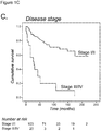
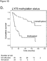
- Figure 1 Kaplan-Meier survival curves of the best prognostic markers.
- A Kaplan-Meier curve of melanoma-specific survival of patients grouped according to primary tumor thickness divided by AJCC tumor stage.
- B Kaplan-Meier curve of melanoma-specific survival of patients grouped according to the presence or absence of tumor ulceration.
- C Kaplan-Meier curve of melanoma-specific survival of patients grouped according to localized disease (Stage I/II) and metastatic disease (Stage III/IV).
- D Kaplan-Meier curve of melanoma-specific survival of patients grouped according to LY75 promoter methylation.
- Methyl-binding domain (MBD)-sequencing was performed on six melanoma cell lines (WM35, WM3248, WM164, A375, M14, SK-MEL-28) and normal human epidermal melanocytes (NHEM) provided by Dr. Leon van Kempen (McGill University, Montreal, Canada). Authentication of all cell lines was performed using short tandem repeat (STR) profiling (DSMZ, Braunschweig, Germany).
- STR short tandem repeat
- WM cell lines were cultured in W489 medium consisting of four parts of MCDB153 (Sigma-Aldrich, Zwijndrecht, The Netherlands) and one part of L15 (Sigma-Aldrich, Zwijndrecht, The Netherlands), A375, M14, and SK-MEL-28 cells were cultured in Dulbecco's modified Eagle's medium (Invitrogen, Breda, the Netherlands). Cells were supplemented with 2% or 10% heat inactivated fetal calf serum (Hyclone Perbio Science, Erembodegem-Aalst, Belgium), respectively. NHEM cells were cultured in ready-to-use medium supplied by Promocell (Heidelberg, Germany). Genomic DNA was isolated using the Puregene® DNA isolation kit (Gentra systems, Minneapolis, MN) according to the manufacturer's instructions.
- Genomic DNA of all samples was subjected to methylation-enrichment sequencing using the MethylCap kit with high-salt elution (Diagenode, Basil, Belgium) as described previously [25]. For each sample, and each methylation core, the maximum read count was used in downstream analyses.
- RNA-sequencing on WM35, WM3248, M14, and SK-MEL-28 cells was performed to determine functional methylation, i.e. methylation associated with downregulated gene expression [26].
- total ribonucleic acid (RNA) was isolated using the standard procedure for TRIzol ® RNA extraction (Invitrogen, Bleiswijk, The Netherlands) and stored at -80°C.
- RNA sequencing library preparation was carried out using a modified version of the Illumina 'Directional mRNA-sequencing Sample Preparation' protocol with total RNA instead of mRNA. Ribosomal DNA was depleted from the DNA fraction using Illumina's Duplex-Specific Thermostable Nuclease normalization protocol for bidirectional mRNA sequencing (application note 15014673).
- Genomic DNA from the 14 nevus samples was extracted as described previously [27]. DNA quantification was performed using a Qubit 2.0 plate reader (Invitrogen, Bleiswijk, The Netherlands) and PicoGreen dye (Invitrogen, Bleiswijk, The Netherlands). DNA quality was inspected on agarose gels stained with SYBR ® Safe (Invitrogen, Bleiswijk, The Netherlands). Bisulfite conversion of DNA samples was carried out using the EZ DNA methylation kit (Zymo Research, Orange, CA) and converted DNA was hybridized on Infinium-450K BeadChips, following the Illumina Infinium HD Methylation protocol as described elsewhere [28].
- Example 4 Patient samples.
- MPTC Maastricht Pathology Tissue Collection
- Example 5 DNA isolation, bisulfite conversion, and promoter CpG island methylation analyses.
- a 4- ⁇ m section of each FFPE tissue block was stained with haematoxylin & eosin (H&E) and reviewed by an experienced dermato-pathologist. Cases that contained >50% nevus or melanoma cells were included. Subsequently, ten sections of 10 ⁇ m were cut and another H&E section was made to confirm the percentage of nevus and melanoma cells. Next, slides were deparaffinised and DNA was extracted following macro dissection with the QIAamp DNA Micro Kit (Qiagen, Venlo, The Netherlands). NanoDrop quantification was used to estimate the quality and concentration of extracted DNA (NanoDrop ND-1000 Spectrophotometer). Sodium bisulphite modification of 500ng genomic DNA was performed using the EpiTect Bisulfite Kit (Qiagen, Venlo, The Netherlands) according to the manufacturer's instructions.
- H&E haematoxylin & eosin
- MSP multiplex methylation-specific polymerase chain reaction
- PCR conditions were as follows: The PCR mixture contains 1 x PCR buffer (16.6 mM ammonium sulfate/67 mM Tris, pH 8.8/6.7 mM MgCl2/10 mM 2-mercaptoethanol), dNTPs (each at 1.25 mM), primers (300 ng each per reaction), and bisulfite-modified DNA ('50 ng) in a final volume of 50 ul. Reactions were hot-started at 95°C for 5 min before the addition of 1.25 units of Taq polymerase (BRL).
- PCR buffer (16.6 mM ammonium sulfate/67 mM Tris, pH 8.8/6.7 mM MgCl2/10 mM 2-mercaptoethanol
- dNTPs each at 1.25 mM
- primers 300 ng each per reaction
- bisulfite-modified DNA '50 ng
- Amplification was carried out in a thermocycler for 30 cycles (30 sec at 95°C, 30 sec at the annealing temperature of 64°C, and 30 sec at 72°C), followed by a final 4-min extension at 72°C.
- the flanking PCR was carried at 56°C for 35 cycles.
- PCR reactions were performed with controls for unmethylated alleles (for example unmethylated human control DNA, EpiTect Control DNA, Qiagen, Cat. no. 59568), methylated alleles (normal lymphocyte DNA treated in vitro with Sssl methyltransferase [New England Biolabs]), and a no-template DNA control.
- unmethylated alleles for example unmethylated human control DNA, EpiTect Control DNA, Qiagen, Cat. no. 59568
- methylated alleles normal lymphocyte DNA treated in vitro with Sssl methyltransferase [New England Biolabs]
- Table 3 primer sequences and conditions used for gene LY75: Direction Position Sequence SEQ ID NO: Temp (°C) Sense primer (5'-> 3') Nested TTAGGATGAGGATAGGTTGGG 1 56 Unmethylated GGATAGGTTGGGTGATTTTTTGTT 2 64 Methylated GGTTGGGCGATTTTTCGTC 3 64 Antisense primer (3'-> 5') Nested CAAACTAAAAAACAACAAAACTATAAC 4 56 Unmethylated AAACTATAACATCAAAACACCCAACA 5 64 Methylated TATAACGTCGAAACACCCAACG 6 64
- Nested MSP reactions were performed with controls for unmethylated alleles (unmethylated human control DNA, EpiTect Control DNA, Qiagen, Cat. no. 59568), methylated alleles (normal lymphocyte DNA treated in vitro with Sssl methyltransferase [New England Biolabs]), and a no-template DNA control.
- MSP reactions were performed in duplicate starting from DNA amplification with flanking primers. Discordant results were analyzed a third time, and the majority vote principle was used to determine the methylation status.
- Cox proportional hazards regression was used to evaluate the effect of gene methylation and clinicopathological variables on melanoma-specific survival, resulting in hazard ratios (HRs) and their corresponding 95% confidence intervals (95%-CI).
- HRs hazard ratios
- 95%-CI 95% confidence intervals
- Survival time was defined as the time between first diagnosis and the first date of diagnosis of a distant metastasis.
- Akaike Information Criterion (AIC) was used to assess the predictive capacity of models with single and multiple methylation markers. The model with the lowest AIC was chosen as the best model. All reported P-values were two-sided, and P ⁇ .05 was considered statistically significant. Analyses were performed using the statistical package IBM SPSS Statistics 21 (IBM, New York, USA) and R (R Foundation for Statistical Computing, Vienna, Austria).
Landscapes
- Chemical & Material Sciences (AREA)
- Life Sciences & Earth Sciences (AREA)
- Health & Medical Sciences (AREA)
- Organic Chemistry (AREA)
- Proteomics, Peptides & Aminoacids (AREA)
- Engineering & Computer Science (AREA)
- Immunology (AREA)
- Pathology (AREA)
- Analytical Chemistry (AREA)
- Zoology (AREA)
- Genetics & Genomics (AREA)
- Wood Science & Technology (AREA)
- Physics & Mathematics (AREA)
- Biotechnology (AREA)
- Microbiology (AREA)
- Molecular Biology (AREA)
- Hospice & Palliative Care (AREA)
- Biophysics (AREA)
- Oncology (AREA)
- Biochemistry (AREA)
- Bioinformatics & Cheminformatics (AREA)
- General Engineering & Computer Science (AREA)
- General Health & Medical Sciences (AREA)
- Measuring Or Testing Involving Enzymes Or Micro-Organisms (AREA)
Description
- The invention is in the field of molecular biology and medical diagnosis. It provides means and methods for determining the prognosis and disease outcome of a subject having a melanoma.
- Cutaneous melanoma is a lethal skin tumor with continuously rising incidence, resulting in a growing healthcare burden [1, 2]. Worldwide, roughly 232,000 new cases and 55,000 deaths were reported in 2012 [3]. Patients diagnosed with localized disease have a five-year survival rate of more than 95% after treatment by surgical excision alone [4]. If the cancer is more advanced, however, survival rates drop substantially, i.e. 30% to 60% after five years, primarily depending on the tumor thickness, i.e. Breslow's depth.
- Metastatic disease generally leads to poor patient outcomes, as treatment options were limited for a long time. However, rapid development of next-generation sequencing technologies has identified most genetic alterations and molecular pathways involved in melanoma development and provided the basis for novel targeted therapies [5]. Moreover, novel immunomodulatory therapies are successfully being developed for melanoma treatment [6].
- Currently, the American Joint Committee on Cancer (AJCC) classifies patients predominantly based on histological features of the primary tumor, i.e. Breslow thickness, ulceration, and mitotic rate, and indicates that the initial biopsy is a critical component of both diagnosis and staging [7]. In addition, the presence of advanced disease stage (stage III/IV) and, to a lesser extent, patient age, gender, and tumor location, are prognostic melanoma factors. Breslow thickness is viewed as the most important prognostic parameter, however, 20-30% of patients diagnosed with thin melanomas (≤2.0mm thickness) still die from their disease [8, 9]. Hence, improvements to the current staging system that lead to more accurate prediction of prognosis are warranted, allowing clinicians to better address prognosis of individual patients. Moreover, it is of importance to identify high-risk patients with aggressive disease at an early stage as these patients may benefit from more extensive surgery, adjuvant therapy, and closer follow-up.
- In summary, cutaneous melanoma is a highly aggressive skin cancer that accounts for approximately 75% of skin cancer-related deaths. Biomarkers for melanoma based on the analysis of differential methylation have been described in the prior art [55-57]. Differential methylation of the LY75 gene has been linked to cardiac disease [58]. LY75 has been described as melanoma treatment target [59]. Despite an increased understanding of the biology of melanoma development and the identification of molecular alterations that accompany melanoma progression [10, 30], the AJCC melanoma staging and classification system has not yet incorporated potentially molecular changes [7]. However, improvements to the current staging system are necessary to more accurately identify individual patients with aggressive disease at diagnosis. These patients with a so-called poor prognosis might benefit from additional therapy leading to improved clinical management and better patient outcome.
- We found that methylation of the promoter of lymphocyte antigen 75 (LY75), also known as CD-205 or DEC-205, is a strong marker that predicts poor clinical outcome, independent of the currently used prognosticators in an independent melanoma series. The invention therefore relates to a method for determining whether a subject having a melanoma has a poor prognosis, the method comprising the step of determining in a sample from the subject whether the LY75 promoter is methylated and if the LY75 promoter is methylated, classifying the subject as having a poor prognosis.
- We examined the methylation status of the lymphocyte antigen 75 (LY75) promoter in a well-characterized series of 123 primary melanomas with follow-up data. We found that LY75 promoter methylation (HR=4.442; 95%-CI 2.307-8.553, P<.001) together with ulceration (HR=2.262; 95%-CI 1.164-4.396, P=.016), and metastatic disease at diagnosis (HR=5.069; 95%-CI 2.489-10.325, P<.001) were significant predictors of melanoma survival.
- LY75, also known as CD-205 or DEC-205, is a collagen-binding mannose family receptor that is predominantly expressed on thymic cortical epithelium and myeloid dendritic cell subsets [37]. LY75 has been reported to play a role in the endocytic uptake of antigen leading to both CD4+ and CD8+ T-cell response [37-39].
- LY75 is Ensemble gene ID ENSG00000054219, situated at
chromosome 2, with gene description Lymphocyte antigen 75 Precursor (DEC-205)(gp200-MR6)(CD205 antigen). - LY75 gene promoter was analyzed for its methylation status in melanoma cell lines and normal human epidermal melanocytes (NHEM), and in 20 primary melanoma samples and 20 common nevus samples. It was found that the promoter was methylated in 6 out of 6 melanoma cell lines, not in the NHEM cells, in 35% of the pilot melanomas and in 0% of the pilot nevi samples.
- LY75 promoter methylation was identified as a strong predictor of poor melanoma prognosis and identified patients with aggressive disease at diagnosis independent of current prognostic parameters. LY75 promoter methylation is therefore an important aid in the identification of patients who require more extensive surgery, adjuvant treatment, and closer follow-up which then leads to improved clinical outcome.
- The invention therefore relates to a method for determining whether a subject having a melanoma has a poor prognosis, the method comprising a step of determining in a sample from the subject whether the LY75 promoter is methylated and if the LY75 promoter is methylated, classifying the subject as having a poor prognosis.
- In the patient series examined herein, higher Breslow thickness, presence of ulceration, and presence of metastatic disease at diagnosis were the main prognostic indicators (P<.001, Table 4; Kaplan-Meier survival curves depicted in
Figure 1A , B, and C, respectively). - Additionally, a higher age at diagnosis, presence of tumor mitoses, and location on the head and neck were significant predictors of poor prognosis (Table 3), indicating that this series was suitable to study the additional prognostic value of methylation markers as it reflected the overall disease progression in a similar tendency as the general population.
- The term "poor prognosis" is used herein to indicate that patients with a methylated LY75 promoter may be expected or be predicted to have a shorter life expectancy than those having an unmethylated LY75 promoter.
Table 4. Associations of clinicopathological characteristics and methylation marker LY75 with melanoma-specific survival. Univariate analysis Multivariate analysis* Final** HR 95% CI P-value HR 95% CI P-value HR 95% CI P-value Current prognostic markers Gender (male vs female) 1.675 0.900-3.116 .103 1.612 0.810-3.206 .174 - - - Age (continuous) 1.026 1.004-1.050 .023 1.007 0.982-1.032 .595 - - - Location (head/neck vs other) 2.875 1.451-5.697 .002 2.206 1.018-4.780 .045 - - - Breslow thickness (continuous) 1.175 1.114-1.239 <.001 1.154 1.073-1.242 <.001 - - - Ulceration (yes vs no) 3.355 1.827-6.160 <.001 1.864 0.908-3.827 .090 2.262 1.164-4.396 .016 Disease stage (stage III/IV vs stage I/II) 6.498 3.372-12.520 <.001 3.957 1.856-8.436 <.001 5.069 2.489-10.325 <.001 Mitoses (>1mm2 vs <1mm2)*** 30.835 1.405-676.773 .030 - - - - - - Methylation marker; methylated vs un methylated (% methylation) LY75 29/114 (25%) 5.395 2.854-10.200 <.001 4.011 1.693-9.502 .002 4.442 2.307-8.553 <.001 *Multivariate Cox proportional hazards regression analyses were adjusted for gender, age, location, Breslow thickness, ulceration, and disease stage. **Final analyses were the result of backward stepwise elimination on a saturated multivariate Cox proportional hazards regression model with the current prognostic makers (except for tumor mitoses) and methylation markers as covariates
***The wide 95% confidence interval for mitotic rate can be explained by the fact that none of the patients died of melanoma when mitoses were absent, as a result the statistical analysis was problematic by the presence of a value equal to zero. Additionally, the AJCC staging system [7] uses the presence of mitoses only to categorize T1 melanoma (<1.0mm thickness). For these reasons we left out tumor mitoses in multivariate analysis. - Tumor ulceration and metastatic disease at diagnosis were, together with LY75 methylation, the most significant predictors of survival. Using Akaike Information Criterion (AIC) showed that LY75 methylation as single methylation marker always outperformed this panel.
- Since melanoma survival largely depends on the formation of lethal metastases we wondered if LY75 methylation was a significant predictor of distant metastases formation in disease stage I and II patients. Of 95 stage I and II patients with known methylation status, a total of 26 patients developed metastatic disease. Using univariate analysis we observed a strong prognostic value of LY75 methylation to predict distant metastasis formation, HR=7.835, 95%-CI 3.554-17.274, P<.001. Applying multivariate analyses, promoter methylation of LY75 (HRLY75=7.924, 95%-CI 3.492-17.980, P<.001) remained the best predictor of metastatic disease development together with ulceration (HRulceration=3.477, 95%-CI 1.579-7.655, P=.002) and age (HRage=1.040, 95%-CI 1.009-1.072, P=.011).
- To validate the observed association of LY75 methylation with distant metastasis formation we evaluated the the Cancer Genome Data Atlas (TCGA) dataset. This dataset provides clinical follow-up data for 44 primary melanomas of which 14 patients did recur. (https://tcga-data.nci.nih.gov/tcga/)
- For analysis, a single representative probe region was selected (cg24478096; wherein probe A: AAACAACAAAACTATAACATCAAAACACCCAACAAACTACAAAACTAACA (SEQ ID NO: 7), and probe B: AAACAACAAAACTATAACGTCGAAACACCCAACGAACTACGAAACTAACG (SEQ ID NO: 8) were employed. Probe A detects unmethylated alleles and probe B detects methylated alleles.
- As a cut-off value, a normalized β-value of 0.2 was chosen, wherein β is equal to the intensities of the A and B probes according to the equation: B/(A+B). In other words, if the intensity of the B probe was more than 20% of the total intenstity of probes A and B together, the sample was scored as having a methylated LY75 promoter. Samples with β-value >0.2 were thus scored as methylated and samples with β-value below or at 0.2 were scored as unmethylated.
- It goes without saying that other cut-off values may advantageously employed. Depending on the desired specificity and sensitivity of the method, the cut-off value may be adjusted.
- The step of determining whether a subject has a methylated LY75 promoter may thus advantageously include a step of determining whether the level of methylation of the LY75 promoter is above a predetermined reference value or cut-off value. A skilled person may be well aware of ways of obtaining such a predetermined reference value. It may for instance be the value obtained using the same probes and methods as described herein when applied to a normal individual or a panel of normal individuals. It may also be an arbitrarily chosen value or it may be determined by trial and error. A preferred reference value is a beta value of more than 0.2 as determined with a suitable probe set, such as for instance probes A and B according to SEQ ID NO: 7 and SEQ ID NO: 8 respectively.
- Kaplan-Meier survival analysis showed that primary melanomas that were methylated had a significantly higher risk to develop metastatic disease (log-rank P=.020).
- Backward stepwise elimination on a saturated multivariate Cox proportional hazards regression model with disease stage, Breslow thickness, tumor ulceration, gender, and age as covariates revealed that LY75 methylation was the best single marker to predict recurrence in this series (HR LY75 =3.568, 95%-CI 1.142-11.149, P=.029).
- Among stage I and II melanoma patients, LY75 methylation was the strongest predictor (HR=7.924, 95%-CI 3.492-17.980, P<.001) of distant metastasis development, together with tumor ulceration (HR=3.477, 95%-CI 1.579-7.655, P=.002) and older age at diagnosis (HR=1.040, 95%-CI 1.009-1.072, P=.011). LY75 methylation outperformed Breslow thickness, the most important clinical prognostic parameter. This is especially of interest since the incidence of patients diagnosed with thin melanoma has been rising and an increasing proportion of melanoma-related deaths occur among these patients [8, 9, 36]. Thus, notwithstanding their generally favorable prognosis, thin melanomas contribute considerably (approximately 25%) to melanoma mortality. The identification of a strong relationship between LY75 methylation and poor prognosis that was irrespective of Breslow thickness provides a promising lead to identify high-risk patients, that are not detected by traditional risk factors, who might benefit from adjuvant therapy and closer follow-up.
- Without wanting to be bound by theory, we hypothesize that the poor clinical outcome of patients with LY75-methylated tumors might be partly explained by poor immune recognition, although we could not observe a significant association of methylation with the absence of tumor-infiltrating lymphocytes (TILs) in our series (data not shown). However, it might also be that the TILs are functionally defective or incompletely activated as is commonly seen during tumor progression [40]. Melanoma is increasingly treated with immunomodulatory therapies, such as anti-CTLA4, and anti-PD1 antibodies that exploit the capacity of CD8+ T-cells to kill immunogenic melanoma cells [41, 42]. Although responses can be durable, the response rate to these therapies is generally low (roughly 25% of patients) [6] and biomarkers predicting response are thus far lacking [43]. It is therefore conceivable that LY75 methylation might reflect poor response to immune therapies as well.
- In summary, LY75 methylation was recognized as a strong, independent predictor of poor prognosis, both in predicting melanoma-specific death and predicting the formation of distant metastases in stage I and II melanoma patients.
- (A) Kaplan-Meier curve of melanoma-specific survival of patients grouped according to primary tumor thickness divided by AJCC tumor stage. (B) Kaplan-Meier curve of melanoma-specific survival of patients grouped according to the presence or absence of tumor ulceration. (C) Kaplan-Meier curve of melanoma-specific survival of patients grouped according to localized disease (Stage I/II) and metastatic disease (Stage III/IV). (D) Kaplan-Meier curve of melanoma-specific survival of patients grouped according to LY75 promoter methylation.
- Kaplan-Meier survival curve of recurrence-free survival of TCGA patients grouped according to LY75 methylation status at probe cg24478096.
- Methyl-binding domain (MBD)-sequencing was performed on six melanoma cell lines (WM35, WM3248, WM164, A375, M14, SK-MEL-28) and normal human epidermal melanocytes (NHEM) provided by Dr. Leon van Kempen (McGill University, Montreal, Canada). Authentication of all cell lines was performed using short tandem repeat (STR) profiling (DSMZ, Braunschweig, Germany). WM cell lines were cultured in W489 medium consisting of four parts of MCDB153 (Sigma-Aldrich, Zwijndrecht, The Netherlands) and one part of L15 (Sigma-Aldrich, Zwijndrecht, The Netherlands), A375, M14, and SK-MEL-28 cells were cultured in Dulbecco's modified Eagle's medium (Invitrogen, Breda, the Netherlands). Cells were supplemented with 2% or 10% heat inactivated fetal calf serum (Hyclone Perbio Science, Erembodegem-Aalst, Belgium), respectively. NHEM cells were cultured in ready-to-use medium supplied by Promocell (Heidelberg, Germany). Genomic DNA was isolated using the Puregene® DNA isolation kit (Gentra systems, Minneapolis, MN) according to the manufacturer's instructions.
- Genomic DNA of all samples was subjected to methylation-enrichment sequencing using the MethylCap kit with high-salt elution (Diagenode, Liege, Belgium) as described previously [25]. For each sample, and each methylation core, the maximum read count was used in downstream analyses.
- RNA-sequencing on WM35, WM3248, M14, and SK-MEL-28 cells was performed to determine functional methylation, i.e. methylation associated with downregulated gene expression [26]. In brief, total ribonucleic acid (RNA) was isolated using the standard procedure for TRIzol ® RNA extraction (Invitrogen, Bleiswijk, The Netherlands) and stored at -80°C. For total RNA sequencing library preparation was carried out using a modified version of the Illumina 'Directional mRNA-sequencing Sample Preparation' protocol with total RNA instead of mRNA. Ribosomal DNA was depleted from the DNA fraction using Illumina's Duplex-Specific Thermostable Nuclease normalization protocol for bidirectional mRNA sequencing (application note 15014673).
- Since The Cancer Genome Atlas (TCGA) has no methylation data available on control samples we performed Infinium-450K assays on 14 fresh-frozen nevi, collected from the archives of the University Hospital of Leuven, Belgium, to be able to determine melanoma-specific methylation (Table 1).
Table 1. Samples used for methylation analysis Characteristics No. of Patients % Common nevi used for Infinium-450K analyses (n=14) Gender Female 5 36 Male 9 64 Age (years) 20.6±24. Location Head and neck 3 21 Trunk 8 57 Extremities 3 21 Common nevi used for validation with MSP (n=20) Gender Female 10 50 Male 10 50 Age (years) 31.7±14.3 Location Head and neck 3 22 Trunk 11 57 Extremities 5 21 - Genomic DNA from the 14 nevus samples was extracted as described previously [27]. DNA quantification was performed using a Qubit 2.0 plate reader (Invitrogen, Bleiswijk, The Netherlands) and PicoGreen dye (Invitrogen, Bleiswijk, The Netherlands). DNA quality was inspected on agarose gels stained with SYBR ® Safe (Invitrogen, Bleiswijk, The Netherlands). Bisulfite conversion of DNA samples was carried out using the EZ DNA methylation kit (Zymo Research, Orange, CA) and converted DNA was hybridized on Infinium-450K BeadChips, following the Illumina Infinium HD Methylation protocol as described elsewhere [28].
- LY75 Promoter CpG island methylation was examined in a well-characterized series of formalin-fixed, paraffin-embedded (FFPE) common nevi (n=20) and primary melanomas (n=123) of patients diagnosed at the Maastricht University Medical Centre, The Netherlands and University Hospital Leuven, Belgium. Collection, storage and use of all tissues and patient data were performed in agreement with the "Code for Proper Secondary Use of Human Tissue in the Netherlands". All of the used samples and corresponding data were de-linked and anonymized. Usage of both melanoma and healthy tissue samples was approved by the Maastricht Pathology Tissue Collection (MPTC) scientific committee. Detailed clinicopathological information of melanoma samples is shown in Table 2, characteristics of nevus samples are listed in Table 1.
Table 2. Clinicopathological characteristics of 123 primary melanoma cases with follow-up Characteristics No. of Patients* % Gender Female 82 67 Male 41 33 Age (years) Mean 59.9 ± 16.9 ≤50 years 37 30 >50 years 86 70 Disease stage Localized - Stage I/ II 103 84 Metastasized - Stage III/ IV 20 16 Breslow thickness, mm 0.01-1.0 32 26 1.01-2.0 29 24 2.01-4.0 29 24 > 4.0 33 27 Ulceration Absent 86 70 Present 37 30 Mitotic Rate <1/mm2 23 19 ≥1/mm2 99 81 TILs Absent 29 24 Non-brisk 69 57 Brisk 24 20 Histological subtype SSM 85 73 NM 21 18 LMM 9 8 ALM 2 2 Location Head and neck 21 17 Trunk 33 28 Extremities 67 55 Distant metastasis formation No 80 65 Yes 43 35 Disease-related death No 81 66 Yes 42 34 Mean follow-up (months) 75.3 ± 57.0 ALM, acral lentigious melanoma; LMM, lentigo maligna melanoma; NM, nodular melanoma; SSM, superficial spreading melanoma; TILs, tumor infiltrating lymphocytes - A 4-µm section of each FFPE tissue block was stained with haematoxylin & eosin (H&E) and reviewed by an experienced dermato-pathologist. Cases that contained >50% nevus or melanoma cells were included. Subsequently, ten sections of 10µm were cut and another H&E section was made to confirm the percentage of nevus and melanoma cells. Next, slides were deparaffinised and DNA was extracted following macro dissection with the QIAamp DNA Micro Kit (Qiagen, Venlo, The Netherlands). NanoDrop quantification was used to estimate the quality and concentration of extracted DNA (NanoDrop ND-1000 Spectrophotometer). Sodium bisulphite modification of 500ng genomic DNA was performed using the EpiTect Bisulfite Kit (Qiagen, Venlo, The Netherlands) according to the manufacturer's instructions.
- Following bisulfite conversion, nested, multiplex methylation-specific polymerase chain reaction (MSP) analyses were performed as described elsewhere [19]. Primer sequences and conditions are shown in Table 3. The number of PCR cycles performed was 30.
- PCR conditions were as follows: The PCR mixture contains 1 x PCR buffer (16.6 mM ammonium sulfate/67 mM Tris, pH 8.8/6.7 mM MgCl2/10 mM 2-mercaptoethanol), dNTPs (each at 1.25 mM), primers (300 ng each per reaction), and bisulfite-modified DNA ('50 ng) in a final volume of 50 ul. Reactions were hot-started at 95°C for 5 min before the addition of 1.25 units of Taq polymerase (BRL). Amplification was carried out in a thermocycler for 30 cycles (30 sec at 95°C, 30 sec at the annealing temperature of 64°C, and 30 sec at 72°C), followed by a final 4-min extension at 72°C. The flanking PCR was carried at 56°C for 35 cycles.
- PCR reactions were performed with controls for unmethylated alleles (for example unmethylated human control DNA, EpiTect Control DNA, Qiagen, Cat. no. 59568), methylated alleles (normal lymphocyte DNA treated in vitro with Sssl methyltransferase [New England Biolabs]), and a no-template DNA control.
- Ten µl of each MSP reaction was loaded onto 2% agarose gels containing GelStar Nucleic Acid Gel Stain (Cambrex, New Jersey, USA), and visualized under UV illumination. The presence of a PCR product performed with the methylated primers indicates the presence of methylated DNA and predicts a poor prognosis of the melanoma patient.
Table 3: primer sequences and conditions used for gene LY75: Direction Position Sequence SEQ ID NO: Temp (°C) Sense primer (5'-> 3') Nested TTAGGATGAGGATAGGTTGGG 1 56 Unmethylated GGATAGGTTGGGTGATTTTTTGTT 2 64 Methylated GGTTGGGCGATTTTTCGTC 3 64 Antisense primer (3'-> 5') Nested CAAACTAAAAAACAACAAAACTATAAC 4 56 Unmethylated AAACTATAACATCAAAACACCCAACA 5 64 Methylated TATAACGTCGAAACACCCAACG 6 64 - Nested MSP reactions were performed with controls for unmethylated alleles (unmethylated human control DNA, EpiTect Control DNA, Qiagen, Cat. no. 59568), methylated alleles (normal lymphocyte DNA treated in vitro with Sssl methyltransferase [New England Biolabs]), and a no-template DNA control.
- To ensure reproducibility, MSP reactions were performed in duplicate starting from DNA amplification with flanking primers. Discordant results were analyzed a third time, and the majority vote principle was used to determine the methylation status.
- Cox proportional hazards regression was used to evaluate the effect of gene methylation and clinicopathological variables on melanoma-specific survival, resulting in hazard ratios (HRs) and their corresponding 95% confidence intervals (95%-CI). For LY75 methylation, the Cox proportional hazards model was used to evaluate the effect of methylation on distant metastasis formation (melanoma-free survival).
- Survival time was defined as the time between first diagnosis and the first date of diagnosis of a distant metastasis. Akaike Information Criterion (AIC) was used to assess the predictive capacity of models with single and multiple methylation markers. The model with the lowest AIC was chosen as the best model. All reported P-values were two-sided, and P<.05 was considered statistically significant. Analyses were performed using the statistical package IBM SPSS Statistics 21 (IBM, New York, USA) and R (R Foundation for Statistical Computing, Vienna, Austria).
-
- 1. MacKie RM, Hauschild A, Eggermont AM. Epidemiology of invasive cutaneous melanoma. Ann Oncol, 2009. 20 Suppl 6: p. vi1-7.
- 2. Ferlay J, Shin HR, Bray F, et al. Estimates of worldwide burden of cancer in 2008: GLOBOCAN 2008. Int J Cancer, 2010. 127(12): p. 2893-2917.
- 3. Ferlay J, Soerjomataram I, Ervik M, Dikshit R, Eser S, Mathers C, Rebelo M, Parkin DM, Forman D, Bray, F. GLOBOCAN 2012 v1.0, Cancer Incidence and Mortality Worldwide: IARC CancerBase No. 11 [Internet]. Lyon, France: International Agency for Research on Cancer; 2013. Available from: http://globocan.iarc.fr, accessed on 01/09/2014.
- 4. Gray-Schopfer V, Wellbrock C, Marais R. Melanoma biology and new targeted therapy. Nature, 2007. 445(7130): p. 851-857.
- 5. Griewank KG, Scolyer RA, Thompson JF, et al. Genetic alterations and personalized medicine in melanoma: progress and future prospects. J Natl Cancer Inst, 2014. 106(2): p. djt435.
- 6. Miller DM, Flaherty KT, Tsao H. Current status and future directions of molecularly targeted therapies and immunotherapies for melanoma. Semin Cutan Med Surg, 2014. 33(2): p. 60-67.
- 7. Balch CM, Gershenwald JE, Soong SJ, et al. Final version of 2009 AJCC melanoma staging and classification. J Clin Oncol, 2009. 27(36): p. 6199-6206.
- 8. Criscione VD, Weinstock MA. Melanoma thickness trends in the United States, 1988-2006. J Invest Dermatol, 2010. 130(3): p. 793-797.
- 9. Whiteman DC, Baade PD, Olsen CM. More People Die from Thin Melanomas (1 mm) than from Thick Melanomas (>4 mm) in Queensland, Australia. J Invest Dermatol, 2014. p.
- 10. van den Hurk K, Niessen HE, Veeck J, et al. Genetics and epigenetics of cutaneous malignant melanoma: a concert out of tune. Biochim Biophys Acta, 2012. 1826(1): p. 89-102.
- 11. Laird PW. The power and the promise of DNA methylation markers. Nat Rev Cancer, 2003. 3(4): p. 253-266.
- 12. Heyn H, Esteller M. DNA methylation profiling in the clinic: applications and challenges. Nat Rev Genet, 2012. 13(10): p. 679-692.
- 13. Conway K, Edmiston SN, Khondker ZS, et al. DNA-methylation profiling distinguishes malignant melanomas from benign nevi. Pigment Cell Melanoma Res, 2011. 24(2): p. 352-360.
- 14. Sigalotti L, Covre A, Fratta E, et al. Whole genome methylation profiles as independent markers of survival in stage IIIC melanoma patients. J Transl Med, 2012. 10: p. 185.
- 15. Gao L, Smit MA, van den Oord JJ, et al. Genome-wide promoter methylation analysis identifies epigenetic silencing of MAPK13 in primary cutaneous melanoma. Pigment Cell Melanoma Res, 2013. 26(4): p. 542-554.
- 16. Rakosy Z, Ecsedi S, Toth R, et al. Integrative genomics identifies gene signature associated with melanoma ulceration. PLoS One, 2013. 8(1): p. e54958.
- 17. Thomas NE, Slater NA, Edmiston SN, et al. DNA methylation profiles in primary cutaneous melanomas are associated with clinically significant pathologic features. Pigment Cell Melanoma Res, 2014. 27(6): p. 1097-1105.
- 18. Ecsedi S, Hernandez-Vargas H, Lima SC, et al. DNA methylation characteristics of primary melanomas with distinct biological behaviour. PLoS One, 2014. 9(5): p. e96612.
- 19. Gao L, van den Hurk K, Moerkerk PT, et al. Promoter CpG Island Hypermethylation in Dysplastic Nevus and Melanoma: CLDN11 as an Epigenetic Biomarker for Malignancy. J Invest Dermatol, 2014. p.
- 20. Carmona FJ, Villanueva A, Vidal A, et al. Epigenetic disruption of cadherin-11 in human cancer metastasis. J Pathol, 2012. 228(2): p. 230-240.
- 21. Deng Z, Niu G, Cai L, et al. The prognostic significance of CD44V6, CDH11, and betacatenin expression in patients with osteosarcoma. Biomed Res Int, 2013. 2013: p. 496193.
- 22. Li L, Ying J, Li H, et al. The .
- 23. Song YH, Shiota M, Kuroiwa K, et al. The important role of glycine N-methyltransferase in the carcinogenesis and progression of prostate cancer. Mod Pathol, 2011. 24(9): p. 1272-1280.
- 24. Huang YC, Chen M, Shyr YM, et al. Glycine N-methyltransferase is a favorable prognostic marker for human cholangiocarcinoma. J Gastroenterol Hepatol, 2008. 23(9): p. 1384-1389.
- 25. De Meyer T, Mampaey E, Vlemmix M, et al. Quality evaluation of methyl binding domain based kits for enrichment DNA-methylation sequencing. PLoS One, 2013. 8(3): p. e59068.
- 26. van Vlodrop IJ, Niessen HE, Derks S, et al. Analysis of promoter CpG island hypermethylation in cancer: location, location, location! Clin Cancer Res, 2011. 17(13): p. 4225-4231.
- 27. van den Hurk K, Balint, B., Toomey S., O'Leary, P.C., Unwin, L., Sheahan, K., McDermott, E.W., Murphy, I., van den Oord, J.J., Rafferty, M., FitzGerald, D., Moran, J., Cummins, R., MacEneaney, O., Kay, E., O'Brien, C.P., Finn, S.P., Heffron, C.C.B.B., Murphy, M., Yela, R., Power, D.G., Regan, P.J., McDermott, C., O'Keeffe, A., Orosz, Z., Donnellan, P.P., Crown, J.P., Hennessy, B.T, and Gallagher, W.M.. High-throughput oncogene mutation profiling reveals demographic differences in BRAF mutation rates among melanoma patients. Melanoma Research. Mel Res, 2015. In press: p.
- 28. Sandoval J, Heyn H, Moran S, et al. Validation of a DNA methylation microarray for 450,000 CpG sites in the human genome. Epigenetics, 2011. 6(6): p. 692-702.
- 29. Derks S, Lentjes MH, Hellebrekers DM, et al. Methylation-specific PCR unraveled. Cell Oncol, 2004. 26(5-6): p. 291-299.
- 30. Miller AJ, Mihm MC, Jr. Melanoma. N Engl J Med, 2006. 355(1): p. 51-65.
- 31. Colabroy KL, Zhai H, Li T, et al. The mechanism of inactivation of 3-hydroxyanthranilate-3,4-dioxygenase by 4-chloro-3-hydroxyanthranilate. Biochemistry, 2005. 44(21): p. 7623-7631.
- 32. Mahapatra S, Klee EW, Young CY, et al. Global methylation profiling for risk prediction of prostate cancer. Clin Cancer Res, 2012. 18(10): p. 2882-2895.
- 33. Litovkin K, Joniau S, Lerut E, et al. Methylation of PITX2, HOXD3, RASSF1 and TDRD1 predicts biochemical recurrence in high-risk prostate cancer. J Cancer Res Clin Oncol, 2014. 140(11): p. 1849-1861.
- 34. Huang YW, Jansen RA, Fabbri E, et al. Identification of candidate epigenetic biomarkers for ovarian cancer detection. Oncol Rep, 2009. 22(4): p. 853-861.
- 35. Huang YW, Luo J, Weng YI, et al. Promoter hypermethylation of CIDEA, HAAO and RXFP3 associated with microsatellite instability in endometrial carcinomas. Gynecol Oncol, 2010. 117(2): p. 239-247.
- 36. Welch HG, Woloshin S, Schwartz LM. Skin biopsy rates and incidence of melanoma: population based ecological study. BMJ, 2005. 331(7515): p. 481.
- 37. Jiang W, Swiggard WJ, Heufler C, et al. The receptor DEC-205 expressed by dendritic cells and thymic epithelial cells is involved in antigen processing. Nature, 1995. 375(6527): p. 151-155.
- 38. Bozzacco L, Trumpfheller C, Siegal FP, et al. DEC-205 receptor on dendritic cells mediates presentation of HIV gag protein to CD8+ T cells in a spectrum of human MHC I haplotypes. Proc Natl Acad Sci U S A, 2007. 104(4): p. 1289-1294.
- 39. Cheong C, Choi JH, Vitale L, et al. Improved cellular and humoral immune responses in vivo following targeting of HIV Gag to dendritic cells within human anti-human DEC205 monoclonal antibody. Blood, 2010. 116(19): p. 3828-3838.
- 40. Marincola FM, Jaffee EM, Hicklin DJ, et al. Escape of human solid tumors from T-cell recognition: molecular mechanisms and functional significance. Adv Immunol, 2000. 74: p. 181-273.
- 41. Brahmer JR, Tykodi SS, Chow LQ, et al. Safety and activity of anti-PD-L1 antibody in patients with advanced cancer. N Engl J Med, 2012. 366(26): p. 2455-2465.
- 42. Hodi FS, O'Day SJ, McDermott DF, et al. Improved survival with ipilimumab in patients with metastatic melanoma. N Engl J Med, 2010. 363(8): p. 711-723.
- 43. Griewank KG, Ugurel S, Schadendorf D, et al. New developments in biomarkers for melanoma. Curr Opin Oncol, 2013. 25(2): p. 145-151.
- 44. al-Tubuly AA, Luqmani YA, Shousha S, et al. Differential expression of gp200-MR6 molecule in benign hyperplasia and down-regulation in invasive carcinoma of the breast. Br J Cancer, 1996. 74(7): p. 1005-1011.
- 45. Tungekar MF, Gatter KC, Ritter MA. Bladder carcinomas and normal urothelium universally express gp200-MR6, a molecule functionally associated with the .
- 46. Al-Tubuly AA, Spijker R, Pignatelli M, et al. Inhibition of growth and enhancement of differentiation of colorectal carcinoma cell lines by MAb MR6 and IL-4. Int J Cancer, 1997. 71(4): p. 605-611.
- 47. Kaklamanis L, Koukourakis MI, Leek R, et al. Loss of .
- 48. Haas J, Frese KS, Park YJ, et al. Alterations in cardiac DNA methylation in human dilated cardiomyopathy. EMBO Mol Med, 2013. 5(3): p. 413-429.
- 49. Giridhar PV, Funk HM, Gallo CA, et al. Interleukin-6 receptor enhances early colonization of the murine omentum by upregulation of a mannose family receptor, LY75, in ovarian tumor cells. Clin Exp Metastasis, 2011. 28(8): p. 887-897.
- 50. Chapman EJ, Kelly G, Knowles MA. Genes involved in differentiation, stem cell renewal, and tumorigenesis are modulated in telomerase-immortalized human urothelial cells. Mol Cancer Res, 2008. 6(7): p. 1154-1168.
- 51. Horn S, Figl A, Rachakonda PS, et al. TERT promoter mutations in familial and sporadic melanoma. Science, 2013. 339(6122): p. 959-961.
- 52. Huang FW, Hodis E, Xu MJ, et al. Highly recurrent TERT promoter mutations in human melanoma. Science, 2013. 339(6122): p. 957-959.
- 53. Griewank KG, Murali R, Puig-Butille JA, et al. TERT promoter mutation status as an independent prognostic factor in cutaneous melanoma. J Natl Cancer Inst, 2014. 106(9): p.
- 54. loannidis JP. Biomarker failures. Clin Chem, 2013. 59(1): p. 202-204.
- 55. De Araujo E. Biomed Research International, 2015. 94(4): p. 1115-8.
- 56.
WO 2012/037128 (Univ. North Carolina). - 57. Abbas, O. The American Journal of Dermatopathology, 2014. 36(5): p. 363-379.
- 58.
WO 2013/135830 (Univ. Ruprecht Karls Heidelberg). - 59.
WO 2015/052537 (Oxford Biotherapeutics LTD). -
- <110> Universiteit Maastricht and Academisch Ziekenhuis Maastricht
- <120> METHOD FOR IDENTIFYING SUBJECTS WITH AGGRESSIVE MELANOMA SKIN CANCER AT DIAGNOSIS
- <130> 309 WO
- <160> 8
- <170> PatentIn version 3.5
- <210> 1
<211> 21
<212> DNA
<213> Artificial Sequence - <220>
<223> Probes and primers for detection of methylated LY75 promoter - <400> 1
ttaggatgag gataggttggg 21 - <210> 2
<211> 24
<212> DNA
<213> artificial sequence - <220>
<223> Probes and primers for detection of methylated LY75 promoter - <400> 2
ggataggttg ggtgattttt tgtt 24 - <210> 3
<211> 19
<212> DNA
<213> artificial sequence - <220>
<223> Probes and primers for detection of methylated LY75 promoter - <400> 3
ggttgggcga tttttcgtc 19 - <210> 4
<211> 27
<212> DNA
<213> artificial sequence - <220>
<223> Probes and primers for detection of methylated LY75 promoter - <400> 4
caaactaaaa aacaacaaaa ctataac 27 - <210> 5
<211> 26
<212> DNA
<213> artificial sequence - <220>
<223> Probes and primers for detection of methylated LY75 promoter - <400> 5
aaactataac atcaaaacac ccaaca 26 - <210> 6
<211> 22
<212> DNA
<213> artificial sequence - <220>
<223> Probes and primers for detection of methylated LY75 promoter - <400> 6
tataacgtcg aaacacccaa cg 22 - <210> 7
<211> 50
<212> DNA
<213> artificial sequence - <220>
<223> Probes and primers for detection of methylated LY75 promoter - <400> 7
aaacaacaaa actataacat caaaacaccc aacaaactac aaaactaaca 50 - <210> 8
<211> 50
<212> DNA
<213> artificial sequence - <220>
<223> Probes and primers for detection of methylated LY75 promoter - <400> 8
aaacaacaaa actataacgt cgaaacaccc aacgaactac gaaactaacg 50
Claims (5)
- Method for determining whether a subject having a melanoma has a poor prognosis, the method comprising the step of determining in a sample from the subject whether the LY75 promoter is methylated and if the LY75 promoter is methylated, classifying the subject as having a poor prognosis, wherein the sample is a melanoma sample.
- Method according to claim 1 wherein the sample is obtained from a biopsy taken from the subject.
- Method according to claims 1 or 2 wherein an additional step is performed selected from the group consisting of determining tumor ulceration, determining metastatic disease at diagnosis, disease stage, and Breslow thickness of the melanoma.
- Method according to any one of claims 1 - 3 wherein the step of determining in a sample from the subject whether the LY75 promoter is methylated is performed by multiplex methylation-specific polymerase chain reaction analysis.
- Method according to claim 4 wherein the multiplex methylation-specific polymerase chain reaction is a nested multiplex methylation-specific polymerase chain reaction.
Applications Claiming Priority (2)
| Application Number | Priority Date | Filing Date | Title |
|---|---|---|---|
| EP15169951 | 2015-05-29 | ||
| PCT/EP2016/061913 WO2016193117A1 (en) | 2015-05-29 | 2016-05-26 | Method for identifying subjects with aggressive melanoma skin cancer at diagnosis |
Publications (2)
| Publication Number | Publication Date |
|---|---|
| EP3303625A1 EP3303625A1 (en) | 2018-04-11 |
| EP3303625B1 true EP3303625B1 (en) | 2019-03-27 |
Family
ID=53298172
Family Applications (1)
| Application Number | Title | Priority Date | Filing Date |
|---|---|---|---|
| EP16729800.9A Active EP3303625B1 (en) | 2015-05-29 | 2016-05-26 | Method for identifying subjects with aggressive melanoma skin cancer at diagnosis |
Country Status (6)
| Country | Link |
|---|---|
| US (2) | US10260109B2 (en) |
| EP (1) | EP3303625B1 (en) |
| AU (1) | AU2016269632B2 (en) |
| CA (1) | CA2987505A1 (en) |
| ES (1) | ES2732178T3 (en) |
| WO (1) | WO2016193117A1 (en) |
Cited By (1)
| Publication number | Priority date | Publication date | Assignee | Title |
|---|---|---|---|---|
| WO2026093592A1 (en) * | 2024-11-04 | 2026-05-07 | Universiteit Maastricht | Method for the prognosis of melanoma |
Families Citing this family (2)
| Publication number | Priority date | Publication date | Assignee | Title |
|---|---|---|---|---|
| AU2016269632B2 (en) | 2015-05-29 | 2021-09-09 | Mla Diagnostics B.V. | Method for identifying subjects with aggressive melanoma skin cancer at diagnosis |
| CN115867566A (en) * | 2020-05-04 | 2023-03-28 | 梅约医学教育与研究基金会 | Detecting melanoma |
Family Cites Families (5)
| Publication number | Priority date | Publication date | Assignee | Title |
|---|---|---|---|---|
| US20130237445A1 (en) * | 2010-09-14 | 2013-09-12 | The University Of North Carolina At Chapel Hill | Methods and kits for detecting melanoma |
| EP2825665B1 (en) | 2012-03-14 | 2017-09-06 | Ruprecht-Karls-Universität Heidelberg | Epigenetic signatures as marker for cardiomyopathies and myocardial insufficiencies |
| GB201220010D0 (en) * | 2012-11-07 | 2012-12-19 | Oxford Biotherapeutics Ltd | Therapeutic amd diagnostic target |
| SG11201602460QA (en) * | 2013-10-11 | 2016-04-28 | Oxford Biotherapeutics Ltd | Conjugated antibodies against ly75 for the treatment of cancer |
| AU2016269632B2 (en) | 2015-05-29 | 2021-09-09 | Mla Diagnostics B.V. | Method for identifying subjects with aggressive melanoma skin cancer at diagnosis |
-
2016
- 2016-05-26 AU AU2016269632A patent/AU2016269632B2/en active Active
- 2016-05-26 CA CA2987505A patent/CA2987505A1/en active Pending
- 2016-05-26 US US15/577,185 patent/US10260109B2/en active Active
- 2016-05-26 WO PCT/EP2016/061913 patent/WO2016193117A1/en not_active Ceased
- 2016-05-26 ES ES16729800T patent/ES2732178T3/en active Active
- 2016-05-26 EP EP16729800.9A patent/EP3303625B1/en active Active
-
2019
- 2019-03-01 US US16/289,897 patent/US11078544B2/en active Active
Non-Patent Citations (1)
| Title |
|---|
| None * |
Cited By (1)
| Publication number | Priority date | Publication date | Assignee | Title |
|---|---|---|---|---|
| WO2026093592A1 (en) * | 2024-11-04 | 2026-05-07 | Universiteit Maastricht | Method for the prognosis of melanoma |
Also Published As
| Publication number | Publication date |
|---|---|
| WO2016193117A1 (en) | 2016-12-08 |
| ES2732178T3 (en) | 2019-11-21 |
| US11078544B2 (en) | 2021-08-03 |
| CA2987505A1 (en) | 2016-12-08 |
| EP3303625A1 (en) | 2018-04-11 |
| US10260109B2 (en) | 2019-04-16 |
| AU2016269632B2 (en) | 2021-09-09 |
| NZ737739A (en) | 2024-04-26 |
| US20180171410A1 (en) | 2018-06-21 |
| US20190256926A1 (en) | 2019-08-22 |
| AU2016269632A1 (en) | 2017-12-07 |
Similar Documents
| Publication | Publication Date | Title |
|---|---|---|
| Marini et al. | Epigenetic inactivation of tumor suppressor genes in serum of patients with cutaneous melanoma | |
| EP2670861B1 (en) | Markers of melanoma and uses thereof | |
| US20110212444A1 (en) | Use of methylation status of mint loci and tumor related genes as a marker for melanoma and breast cancer | |
| EP2861759B1 (en) | Biomarkers for prostate cancer | |
| Ostrow et al. | Pharmacologic unmasking of epigenetically silenced genes in breast cancer | |
| Sigalotti et al. | Methylation levels of the" long interspersed nucleotide element-1" repetitive sequences predict survival of melanoma patients | |
| KR20160098351A (en) | Prostate Cancer Classification | |
| CN109072310A (en) | Cancer is detected in urine | |
| AU2009299862A1 (en) | Molecular markers in prostate cancer | |
| WO2009076478A2 (en) | Hypomethylated genes in cancer | |
| US20150031022A1 (en) | Dna methylation markers and methods of use | |
| WO2019012105A1 (en) | Method for predicting responsiveness to immunotherapy | |
| US11078544B2 (en) | Method for identifying subjects with aggressive melanoma skin cancer at diagnosis | |
| Wehrhan et al. | PD1 expression and correlation with its ligands in oral cancer specimens and peripheral blood | |
| US20110091970A1 (en) | Methylation of estrogen receptor alpha and uses thereof | |
| Parrella et al. | High specificity of quantitative methylation‐specific PCR analysis for MGMT promoter hypermethylation detection in gliomas | |
| Cavusoglu et al. | Promoter methylation and expression changes of CDH1 and P16 genes in invasive breast cancer and adjacent normal breast tissue | |
| Mithani et al. | Use of integrative epigenetic and cytogenetic analyses to identify novel tumor-suppressor genes in malignant melanoma | |
| Kumar et al. | IGFBP3 gene promoter methylation analysis and its association with clinicopathological characteristics of colorectal carcinoma | |
| Zhang et al. | Frequent epigenetic inactivation of deleted in lung and esophageal cancer 1 gene by promoter methylation in non–small-cell lung cancer | |
| US9708666B2 (en) | Prognostic molecular signature of sarcomas, and uses thereof | |
| Akiyama et al. | Human PinX1, a potent telomerase inhibitor, is not involved in human gastrointestinal tract carcinoma | |
| Inamura et al. | HOXB2 as a novel prognostic indicator for stage I lung adenocarcinomas | |
| Pandey | Skin Cancer: molecular biomarker for diagnosis, prognosis, Prevention, and targeted therapy | |
| Kirkwood et al. | Methylation levels of the" long interspersed nucleotide element-1" repetitive sequences predict survival of melanoma patients |
Legal Events
| Date | Code | Title | Description |
|---|---|---|---|
| STAA | Information on the status of an ep patent application or granted ep patent |
Free format text: STATUS: THE INTERNATIONAL PUBLICATION HAS BEEN MADE |
|
| PUAI | Public reference made under article 153(3) epc to a published international application that has entered the european phase |
Free format text: ORIGINAL CODE: 0009012 |
|
| STAA | Information on the status of an ep patent application or granted ep patent |
Free format text: STATUS: REQUEST FOR EXAMINATION WAS MADE |
|
| 17P | Request for examination filed |
Effective date: 20171130 |
|
| AK | Designated contracting states |
Kind code of ref document: A1 Designated state(s): AL AT BE BG CH CY CZ DE DK EE ES FI FR GB GR HR HU IE IS IT LI LT LU LV MC MK MT NL NO PL PT RO RS SE SI SK SM TR |
|
| AX | Request for extension of the european patent |
Extension state: BA ME |
|
| DAV | Request for validation of the european patent (deleted) | ||
| DAX | Request for extension of the european patent (deleted) | ||
| REG | Reference to a national code |
Ref country code: DE Ref legal event code: R079 Ref document number: 602016011622 Country of ref document: DE Free format text: PREVIOUS MAIN CLASS: C12Q0001680000 Ipc: C12Q0001688600 |
|
| GRAP | Despatch of communication of intention to grant a patent |
Free format text: ORIGINAL CODE: EPIDOSNIGR1 |
|
| STAA | Information on the status of an ep patent application or granted ep patent |
Free format text: STATUS: GRANT OF PATENT IS INTENDED |
|
| RIC1 | Information provided on ipc code assigned before grant |
Ipc: C12Q 1/6886 20180101AFI20180924BHEP |
|
| INTG | Intention to grant announced |
Effective date: 20181015 |
|
| GRAS | Grant fee paid |
Free format text: ORIGINAL CODE: EPIDOSNIGR3 |
|
| GRAA | (expected) grant |
Free format text: ORIGINAL CODE: 0009210 |
|
| STAA | Information on the status of an ep patent application or granted ep patent |
Free format text: STATUS: THE PATENT HAS BEEN GRANTED |
|
| AK | Designated contracting states |
Kind code of ref document: B1 Designated state(s): AL AT BE BG CH CY CZ DE DK EE ES FI FR GB GR HR HU IE IS IT LI LT LU LV MC MK MT NL NO PL PT RO RS SE SI SK SM TR |
|
| REG | Reference to a national code |
Ref country code: GB Ref legal event code: FG4D |
|
| REG | Reference to a national code |
Ref country code: CH Ref legal event code: EP |
|
| REG | Reference to a national code |
Ref country code: AT Ref legal event code: REF Ref document number: 1113154 Country of ref document: AT Kind code of ref document: T Effective date: 20190415 |
|
| REG | Reference to a national code |
Ref country code: IE Ref legal event code: FG4D |
|
| REG | Reference to a national code |
Ref country code: DE Ref legal event code: R096 Ref document number: 602016011622 Country of ref document: DE |
|
| REG | Reference to a national code |
Ref country code: NL Ref legal event code: FP |
|
| REG | Reference to a national code |
Ref country code: CH Ref legal event code: NV Representative=s name: CABINET GERMAIN AND MAUREAU, CH |
|
| PG25 | Lapsed in a contracting state [announced via postgrant information from national office to epo] |
Ref country code: SE Free format text: LAPSE BECAUSE OF FAILURE TO SUBMIT A TRANSLATION OF THE DESCRIPTION OR TO PAY THE FEE WITHIN THE PRESCRIBED TIME-LIMIT Effective date: 20190327 Ref country code: LT Free format text: LAPSE BECAUSE OF FAILURE TO SUBMIT A TRANSLATION OF THE DESCRIPTION OR TO PAY THE FEE WITHIN THE PRESCRIBED TIME-LIMIT Effective date: 20190327 Ref country code: NO Free format text: LAPSE BECAUSE OF FAILURE TO SUBMIT A TRANSLATION OF THE DESCRIPTION OR TO PAY THE FEE WITHIN THE PRESCRIBED TIME-LIMIT Effective date: 20190627 Ref country code: FI Free format text: LAPSE BECAUSE OF FAILURE TO SUBMIT A TRANSLATION OF THE DESCRIPTION OR TO PAY THE FEE WITHIN THE PRESCRIBED TIME-LIMIT Effective date: 20190327 |
|
| PG25 | Lapsed in a contracting state [announced via postgrant information from national office to epo] |
Ref country code: GR Free format text: LAPSE BECAUSE OF FAILURE TO SUBMIT A TRANSLATION OF THE DESCRIPTION OR TO PAY THE FEE WITHIN THE PRESCRIBED TIME-LIMIT Effective date: 20190628 Ref country code: BG Free format text: LAPSE BECAUSE OF FAILURE TO SUBMIT A TRANSLATION OF THE DESCRIPTION OR TO PAY THE FEE WITHIN THE PRESCRIBED TIME-LIMIT Effective date: 20190627 Ref country code: RS Free format text: LAPSE BECAUSE OF FAILURE TO SUBMIT A TRANSLATION OF THE DESCRIPTION OR TO PAY THE FEE WITHIN THE PRESCRIBED TIME-LIMIT Effective date: 20190327 Ref country code: LV Free format text: LAPSE BECAUSE OF FAILURE TO SUBMIT A TRANSLATION OF THE DESCRIPTION OR TO PAY THE FEE WITHIN THE PRESCRIBED TIME-LIMIT Effective date: 20190327 Ref country code: HR Free format text: LAPSE BECAUSE OF FAILURE TO SUBMIT A TRANSLATION OF THE DESCRIPTION OR TO PAY THE FEE WITHIN THE PRESCRIBED TIME-LIMIT Effective date: 20190327 |
|
| REG | Reference to a national code |
Ref country code: AT Ref legal event code: MK05 Ref document number: 1113154 Country of ref document: AT Kind code of ref document: T Effective date: 20190327 |
|
| PG25 | Lapsed in a contracting state [announced via postgrant information from national office to epo] |
Ref country code: AL Free format text: LAPSE BECAUSE OF FAILURE TO SUBMIT A TRANSLATION OF THE DESCRIPTION OR TO PAY THE FEE WITHIN THE PRESCRIBED TIME-LIMIT Effective date: 20190327 Ref country code: RO Free format text: LAPSE BECAUSE OF FAILURE TO SUBMIT A TRANSLATION OF THE DESCRIPTION OR TO PAY THE FEE WITHIN THE PRESCRIBED TIME-LIMIT Effective date: 20190327 Ref country code: CZ Free format text: LAPSE BECAUSE OF FAILURE TO SUBMIT A TRANSLATION OF THE DESCRIPTION OR TO PAY THE FEE WITHIN THE PRESCRIBED TIME-LIMIT Effective date: 20190327 Ref country code: SK Free format text: LAPSE BECAUSE OF FAILURE TO SUBMIT A TRANSLATION OF THE DESCRIPTION OR TO PAY THE FEE WITHIN THE PRESCRIBED TIME-LIMIT Effective date: 20190327 Ref country code: PT Free format text: LAPSE BECAUSE OF FAILURE TO SUBMIT A TRANSLATION OF THE DESCRIPTION OR TO PAY THE FEE WITHIN THE PRESCRIBED TIME-LIMIT Effective date: 20190727 Ref country code: EE Free format text: LAPSE BECAUSE OF FAILURE TO SUBMIT A TRANSLATION OF THE DESCRIPTION OR TO PAY THE FEE WITHIN THE PRESCRIBED TIME-LIMIT Effective date: 20190327 |
|
| REG | Reference to a national code |
Ref country code: ES Ref legal event code: FG2A Ref document number: 2732178 Country of ref document: ES Kind code of ref document: T3 Effective date: 20191121 |
|
| PG25 | Lapsed in a contracting state [announced via postgrant information from national office to epo] |
Ref country code: SM Free format text: LAPSE BECAUSE OF FAILURE TO SUBMIT A TRANSLATION OF THE DESCRIPTION OR TO PAY THE FEE WITHIN THE PRESCRIBED TIME-LIMIT Effective date: 20190327 Ref country code: PL Free format text: LAPSE BECAUSE OF FAILURE TO SUBMIT A TRANSLATION OF THE DESCRIPTION OR TO PAY THE FEE WITHIN THE PRESCRIBED TIME-LIMIT Effective date: 20190327 |
|
| PG25 | Lapsed in a contracting state [announced via postgrant information from national office to epo] |
Ref country code: IS Free format text: LAPSE BECAUSE OF FAILURE TO SUBMIT A TRANSLATION OF THE DESCRIPTION OR TO PAY THE FEE WITHIN THE PRESCRIBED TIME-LIMIT Effective date: 20190727 Ref country code: AT Free format text: LAPSE BECAUSE OF FAILURE TO SUBMIT A TRANSLATION OF THE DESCRIPTION OR TO PAY THE FEE WITHIN THE PRESCRIBED TIME-LIMIT Effective date: 20190327 |
|
| REG | Reference to a national code |
Ref country code: DE Ref legal event code: R097 Ref document number: 602016011622 Country of ref document: DE |
|
| PG25 | Lapsed in a contracting state [announced via postgrant information from national office to epo] |
Ref country code: MC Free format text: LAPSE BECAUSE OF FAILURE TO SUBMIT A TRANSLATION OF THE DESCRIPTION OR TO PAY THE FEE WITHIN THE PRESCRIBED TIME-LIMIT Effective date: 20190327 Ref country code: DK Free format text: LAPSE BECAUSE OF FAILURE TO SUBMIT A TRANSLATION OF THE DESCRIPTION OR TO PAY THE FEE WITHIN THE PRESCRIBED TIME-LIMIT Effective date: 20190327 |
|
| PLBE | No opposition filed within time limit |
Free format text: ORIGINAL CODE: 0009261 |
|
| STAA | Information on the status of an ep patent application or granted ep patent |
Free format text: STATUS: NO OPPOSITION FILED WITHIN TIME LIMIT |
|
| PG25 | Lapsed in a contracting state [announced via postgrant information from national office to epo] |
Ref country code: LU Free format text: LAPSE BECAUSE OF NON-PAYMENT OF DUE FEES Effective date: 20190526 Ref country code: SI Free format text: LAPSE BECAUSE OF FAILURE TO SUBMIT A TRANSLATION OF THE DESCRIPTION OR TO PAY THE FEE WITHIN THE PRESCRIBED TIME-LIMIT Effective date: 20190327 |
|
| 26N | No opposition filed |
Effective date: 20200103 |
|
| PG25 | Lapsed in a contracting state [announced via postgrant information from national office to epo] |
Ref country code: TR Free format text: LAPSE BECAUSE OF FAILURE TO SUBMIT A TRANSLATION OF THE DESCRIPTION OR TO PAY THE FEE WITHIN THE PRESCRIBED TIME-LIMIT Effective date: 20190327 |
|
| PG25 | Lapsed in a contracting state [announced via postgrant information from national office to epo] |
Ref country code: IE Free format text: LAPSE BECAUSE OF NON-PAYMENT OF DUE FEES Effective date: 20190526 |
|
| PG25 | Lapsed in a contracting state [announced via postgrant information from national office to epo] |
Ref country code: CY Free format text: LAPSE BECAUSE OF FAILURE TO SUBMIT A TRANSLATION OF THE DESCRIPTION OR TO PAY THE FEE WITHIN THE PRESCRIBED TIME-LIMIT Effective date: 20190327 |
|
| PG25 | Lapsed in a contracting state [announced via postgrant information from national office to epo] |
Ref country code: HU Free format text: LAPSE BECAUSE OF FAILURE TO SUBMIT A TRANSLATION OF THE DESCRIPTION OR TO PAY THE FEE WITHIN THE PRESCRIBED TIME-LIMIT; INVALID AB INITIO Effective date: 20160526 Ref country code: MT Free format text: LAPSE BECAUSE OF FAILURE TO SUBMIT A TRANSLATION OF THE DESCRIPTION OR TO PAY THE FEE WITHIN THE PRESCRIBED TIME-LIMIT Effective date: 20190327 |
|
| PG25 | Lapsed in a contracting state [announced via postgrant information from national office to epo] |
Ref country code: MK Free format text: LAPSE BECAUSE OF FAILURE TO SUBMIT A TRANSLATION OF THE DESCRIPTION OR TO PAY THE FEE WITHIN THE PRESCRIBED TIME-LIMIT Effective date: 20190327 |
|
| P01 | Opt-out of the competence of the unified patent court (upc) registered |
Effective date: 20230525 |
|
| REG | Reference to a national code |
Ref country code: DE Ref legal event code: R081 Ref document number: 602016011622 Country of ref document: DE Owner name: MLA DIAGNOSTICS B.V., NL Free format text: FORMER OWNERS: ACADEMISCH ZIEKENHUIS MAASTRICHT, MAASTRICHT, NL; UNIVERSITEIT MAASTRICHT, MAASTRICHT, NL |
|
| REG | Reference to a national code |
Ref country code: GB Ref legal event code: 732E Free format text: REGISTERED BETWEEN 20250306 AND 20250312 |
|
| REG | Reference to a national code |
Ref country code: NL Ref legal event code: PD Owner name: MLA DIAGNOSTICS B.V.; NL Free format text: DETAILS ASSIGNMENT: CHANGE OF OWNER(S), ASSIGNMENT; FORMER OWNER NAME: UNIVERSITEIT MAASTRICHT Effective date: 20250418 |
|
| REG | Reference to a national code |
Ref country code: BE Ref legal event code: PD Owner name: MLA DIAGNOSTICS B.V.; NL Free format text: DETAILS ASSIGNMENT: CHANGE OF OWNER(S), ASSIGNMENT; FORMER OWNER NAME: ACADEMISCH ZIEKENHUIS MAASTRICHT Effective date: 20250206 |
|
| PGFP | Annual fee paid to national office [announced via postgrant information from national office to epo] |
Ref country code: NL Payment date: 20250521 Year of fee payment: 10 |
|
| PGFP | Annual fee paid to national office [announced via postgrant information from national office to epo] |
Ref country code: DE Payment date: 20250521 Year of fee payment: 10 |
|
| PGFP | Annual fee paid to national office [announced via postgrant information from national office to epo] |
Ref country code: IT Payment date: 20250527 Year of fee payment: 10 Ref country code: BE Payment date: 20250521 Year of fee payment: 10 |
|
| PGFP | Annual fee paid to national office [announced via postgrant information from national office to epo] |
Ref country code: FR Payment date: 20250528 Year of fee payment: 10 |
|
| PGFP | Annual fee paid to national office [announced via postgrant information from national office to epo] |
Ref country code: CH Payment date: 20250601 Year of fee payment: 10 |
|
| PGFP | Annual fee paid to national office [announced via postgrant information from national office to epo] |
Ref country code: ES Payment date: 20250630 Year of fee payment: 10 |
|
| PGFP | Annual fee paid to national office [announced via postgrant information from national office to epo] |
Ref country code: GB Payment date: 20260306 Year of fee payment: 11 |