EP2825665B1 - Epigenetic signatures as marker for cardiomyopathies and myocardial insufficiencies - Google Patents
Epigenetic signatures as marker for cardiomyopathies and myocardial insufficiencies Download PDFInfo
- Publication number
- EP2825665B1 EP2825665B1 EP13709132.8A EP13709132A EP2825665B1 EP 2825665 B1 EP2825665 B1 EP 2825665B1 EP 13709132 A EP13709132 A EP 13709132A EP 2825665 B1 EP2825665 B1 EP 2825665B1
- Authority
- EP
- European Patent Office
- Prior art keywords
- seq
- methylation
- heart
- dcm
- adora2a
- Prior art date
- Legal status (The legal status is an assumption and is not a legal conclusion. Google has not performed a legal analysis and makes no representation as to the accuracy of the status listed.)
- Active
Links
- 239000003550 marker Substances 0.000 title claims description 12
- 208000031229 Cardiomyopathies Diseases 0.000 title description 21
- 230000001973 epigenetic effect Effects 0.000 title description 8
- 230000002107 myocardial effect Effects 0.000 title description 8
- 238000007069 methylation reaction Methods 0.000 claims description 128
- 230000011987 methylation Effects 0.000 claims description 123
- 206010056370 Congestive cardiomyopathy Diseases 0.000 claims description 104
- 201000010046 Dilated cardiomyopathy Diseases 0.000 claims description 104
- 108090000623 proteins and genes Proteins 0.000 claims description 104
- 230000007067 DNA methylation Effects 0.000 claims description 81
- 101710125610 Adenosine receptor A2a Proteins 0.000 claims description 76
- 102100035990 Adenosine receptor A2a Human genes 0.000 claims description 76
- 102100033486 Lymphocyte antigen 75 Human genes 0.000 claims description 64
- 238000002560 therapeutic procedure Methods 0.000 claims description 62
- 208000019622 heart disease Diseases 0.000 claims description 61
- 239000013610 patient sample Substances 0.000 claims description 58
- 101710100969 Receptor tyrosine-protein kinase erbB-3 Proteins 0.000 claims description 55
- 102100029986 Receptor tyrosine-protein kinase erbB-3 Human genes 0.000 claims description 55
- 206010019280 Heart failures Diseases 0.000 claims description 40
- 239000013068 control sample Substances 0.000 claims description 37
- 102100021088 Homeobox protein Hox-B13 Human genes 0.000 claims description 32
- 101001041145 Homo sapiens Homeobox protein Hox-B13 Proteins 0.000 claims description 32
- 108020004414 DNA Proteins 0.000 claims description 31
- 238000012544 monitoring process Methods 0.000 claims description 31
- 210000001519 tissue Anatomy 0.000 claims description 28
- 238000000034 method Methods 0.000 claims description 27
- 230000002861 ventricular Effects 0.000 claims description 26
- 230000000694 effects Effects 0.000 claims description 22
- 210000005259 peripheral blood Anatomy 0.000 claims description 20
- 239000011886 peripheral blood Substances 0.000 claims description 20
- 230000002829 reductive effect Effects 0.000 claims description 19
- 238000003745 diagnosis Methods 0.000 claims description 18
- 238000011282 treatment Methods 0.000 claims description 18
- 238000004393 prognosis Methods 0.000 claims description 17
- 239000000523 sample Substances 0.000 claims description 17
- 101001059220 Homo sapiens Zinc finger protein Gfi-1 Proteins 0.000 claims description 16
- 102100029004 Zinc finger protein Gfi-1 Human genes 0.000 claims description 16
- OPTASPLRGRRNAP-UHFFFAOYSA-N cytosine Chemical class NC=1C=CNC(=O)N=1 OPTASPLRGRRNAP-UHFFFAOYSA-N 0.000 claims description 15
- 239000002773 nucleotide Substances 0.000 claims description 14
- 125000003729 nucleotide group Chemical group 0.000 claims description 14
- -1 ATP2C Proteins 0.000 claims description 13
- 102100024396 Adrenodoxin, mitochondrial Human genes 0.000 claims description 13
- 102100038447 Claudin-4 Human genes 0.000 claims description 13
- 102100021654 Extracellular sulfatase Sulf-2 Human genes 0.000 claims description 13
- 101000833098 Homo sapiens Adrenodoxin, mitochondrial Proteins 0.000 claims description 13
- 101000884385 Homo sapiens Arylamine N-acetyltransferase 1 Proteins 0.000 claims description 13
- 101000882890 Homo sapiens Claudin-4 Proteins 0.000 claims description 13
- 101001034811 Homo sapiens Eukaryotic translation initiation factor 4 gamma 2 Proteins 0.000 claims description 13
- 101000820626 Homo sapiens Extracellular sulfatase Sulf-2 Proteins 0.000 claims description 13
- 101000639975 Homo sapiens Sodium-dependent noradrenaline transporter Proteins 0.000 claims description 13
- 101000652578 Homo sapiens Thyroid transcription factor 1-associated protein 26 Proteins 0.000 claims description 13
- 108091006657 SLC9A6 Proteins 0.000 claims description 13
- 102100029972 Sodium/hydrogen exchanger 6 Human genes 0.000 claims description 13
- 102100030344 Thyroid transcription factor 1-associated protein 26 Human genes 0.000 claims description 13
- 102100033055 Transketolase Human genes 0.000 claims description 13
- 108010088412 Trefoil Factor-1 Proteins 0.000 claims description 13
- 102000008817 Trefoil Factor-1 Human genes 0.000 claims description 13
- 230000004217 heart function Effects 0.000 claims description 13
- 108091034117 Oligonucleotide Proteins 0.000 claims description 12
- 101150104557 Ppargc1a gene Proteins 0.000 claims description 12
- JLCPHMBAVCMARE-UHFFFAOYSA-N [3-[[3-[[3-[[3-[[3-[[3-[[3-[[3-[[3-[[3-[[3-[[5-(2-amino-6-oxo-1H-purin-9-yl)-3-[[3-[[3-[[3-[[3-[[3-[[5-(2-amino-6-oxo-1H-purin-9-yl)-3-[[5-(2-amino-6-oxo-1H-purin-9-yl)-3-hydroxyoxolan-2-yl]methoxy-hydroxyphosphoryl]oxyoxolan-2-yl]methoxy-hydroxyphosphoryl]oxy-5-(5-methyl-2,4-dioxopyrimidin-1-yl)oxolan-2-yl]methoxy-hydroxyphosphoryl]oxy-5-(6-aminopurin-9-yl)oxolan-2-yl]methoxy-hydroxyphosphoryl]oxy-5-(6-aminopurin-9-yl)oxolan-2-yl]methoxy-hydroxyphosphoryl]oxy-5-(6-aminopurin-9-yl)oxolan-2-yl]methoxy-hydroxyphosphoryl]oxy-5-(6-aminopurin-9-yl)oxolan-2-yl]methoxy-hydroxyphosphoryl]oxyoxolan-2-yl]methoxy-hydroxyphosphoryl]oxy-5-(5-methyl-2,4-dioxopyrimidin-1-yl)oxolan-2-yl]methoxy-hydroxyphosphoryl]oxy-5-(4-amino-2-oxopyrimidin-1-yl)oxolan-2-yl]methoxy-hydroxyphosphoryl]oxy-5-(5-methyl-2,4-dioxopyrimidin-1-yl)oxolan-2-yl]methoxy-hydroxyphosphoryl]oxy-5-(5-methyl-2,4-dioxopyrimidin-1-yl)oxolan-2-yl]methoxy-hydroxyphosphoryl]oxy-5-(6-aminopurin-9-yl)oxolan-2-yl]methoxy-hydroxyphosphoryl]oxy-5-(6-aminopurin-9-yl)oxolan-2-yl]methoxy-hydroxyphosphoryl]oxy-5-(4-amino-2-oxopyrimidin-1-yl)oxolan-2-yl]methoxy-hydroxyphosphoryl]oxy-5-(4-amino-2-oxopyrimidin-1-yl)oxolan-2-yl]methoxy-hydroxyphosphoryl]oxy-5-(4-amino-2-oxopyrimidin-1-yl)oxolan-2-yl]methoxy-hydroxyphosphoryl]oxy-5-(6-aminopurin-9-yl)oxolan-2-yl]methoxy-hydroxyphosphoryl]oxy-5-(4-amino-2-oxopyrimidin-1-yl)oxolan-2-yl]methyl [5-(6-aminopurin-9-yl)-2-(hydroxymethyl)oxolan-3-yl] hydrogen phosphate Polymers Cc1cn(C2CC(OP(O)(=O)OCC3OC(CC3OP(O)(=O)OCC3OC(CC3O)n3cnc4c3nc(N)[nH]c4=O)n3cnc4c3nc(N)[nH]c4=O)C(COP(O)(=O)OC3CC(OC3COP(O)(=O)OC3CC(OC3COP(O)(=O)OC3CC(OC3COP(O)(=O)OC3CC(OC3COP(O)(=O)OC3CC(OC3COP(O)(=O)OC3CC(OC3COP(O)(=O)OC3CC(OC3COP(O)(=O)OC3CC(OC3COP(O)(=O)OC3CC(OC3COP(O)(=O)OC3CC(OC3COP(O)(=O)OC3CC(OC3COP(O)(=O)OC3CC(OC3COP(O)(=O)OC3CC(OC3COP(O)(=O)OC3CC(OC3COP(O)(=O)OC3CC(OC3COP(O)(=O)OC3CC(OC3COP(O)(=O)OC3CC(OC3CO)n3cnc4c(N)ncnc34)n3ccc(N)nc3=O)n3cnc4c(N)ncnc34)n3ccc(N)nc3=O)n3ccc(N)nc3=O)n3ccc(N)nc3=O)n3cnc4c(N)ncnc34)n3cnc4c(N)ncnc34)n3cc(C)c(=O)[nH]c3=O)n3cc(C)c(=O)[nH]c3=O)n3ccc(N)nc3=O)n3cc(C)c(=O)[nH]c3=O)n3cnc4c3nc(N)[nH]c4=O)n3cnc4c(N)ncnc34)n3cnc4c(N)ncnc34)n3cnc4c(N)ncnc34)n3cnc4c(N)ncnc34)O2)c(=O)[nH]c1=O JLCPHMBAVCMARE-UHFFFAOYSA-N 0.000 claims description 12
- 210000005003 heart tissue Anatomy 0.000 claims description 11
- 210000002064 heart cell Anatomy 0.000 claims description 10
- 239000003153 chemical reaction reagent Substances 0.000 claims description 9
- 150000007523 nucleic acids Chemical group 0.000 claims description 9
- 238000012163 sequencing technique Methods 0.000 claims description 9
- 108091028043 Nucleic acid sequence Proteins 0.000 claims description 8
- 230000006607 hypermethylation Effects 0.000 claims description 8
- 102100039439 DNA-binding protein inhibitor ID-4 Human genes 0.000 claims description 7
- 101001036276 Homo sapiens DNA-binding protein inhibitor ID-4 Proteins 0.000 claims description 7
- 101000929429 Homo sapiens Discoidin domain-containing receptor 2 Proteins 0.000 claims description 7
- 101000800463 Homo sapiens Transketolase Proteins 0.000 claims description 7
- 230000000295 complement effect Effects 0.000 claims description 6
- 238000005516 engineering process Methods 0.000 claims description 5
- 102000003960 Ligases Human genes 0.000 claims description 4
- 108090000364 Ligases Proteins 0.000 claims description 4
- LSNNMFCWUKXFEE-UHFFFAOYSA-N Sulfurous acid Chemical compound OS(O)=O LSNNMFCWUKXFEE-UHFFFAOYSA-N 0.000 claims description 4
- 238000007672 fourth generation sequencing Methods 0.000 claims description 4
- 238000007481 next generation sequencing Methods 0.000 claims description 4
- 238000012175 pyrosequencing Methods 0.000 claims description 4
- 238000007841 sequencing by ligation Methods 0.000 claims description 4
- 102100033769 Sodium-coupled neutral amino acid transporter 3 Human genes 0.000 claims 3
- 101001018034 Homo sapiens Lymphocyte antigen 75 Proteins 0.000 claims 2
- 101710157884 Lymphocyte antigen 75 Proteins 0.000 description 64
- 230000014509 gene expression Effects 0.000 description 41
- 210000002216 heart Anatomy 0.000 description 35
- 108091029523 CpG island Proteins 0.000 description 31
- 241000252212 Danio rerio Species 0.000 description 31
- 208000037265 diseases, disorders, signs and symptoms Diseases 0.000 description 25
- 201000010099 disease Diseases 0.000 description 24
- 230000000747 cardiac effect Effects 0.000 description 19
- 210000004165 myocardium Anatomy 0.000 description 16
- 238000012216 screening Methods 0.000 description 14
- 102100026406 G/T mismatch-specific thymine DNA glycosylase Human genes 0.000 description 12
- 229940079593 drug Drugs 0.000 description 12
- 239000003814 drug Substances 0.000 description 12
- 210000002257 embryonic structure Anatomy 0.000 description 12
- 238000001574 biopsy Methods 0.000 description 11
- 102100033929 Sodium-dependent noradrenaline transporter Human genes 0.000 description 10
- 238000004458 analytical method Methods 0.000 description 10
- 206010020871 hypertrophic cardiomyopathy Diseases 0.000 description 10
- 108020004999 messenger RNA Proteins 0.000 description 10
- 238000002054 transplantation Methods 0.000 description 10
- 201000007170 intrinsic cardiomyopathy Diseases 0.000 description 9
- 230000010076 replication Effects 0.000 description 9
- LSNNMFCWUKXFEE-UHFFFAOYSA-M Bisulfite Chemical compound OS([O-])=O LSNNMFCWUKXFEE-UHFFFAOYSA-M 0.000 description 8
- 102000040945 Transcription factor Human genes 0.000 description 8
- 108091023040 Transcription factor Proteins 0.000 description 8
- 230000007608 epigenetic mechanism Effects 0.000 description 8
- 230000006870 function Effects 0.000 description 8
- 238000003753 real-time PCR Methods 0.000 description 8
- 238000010200 validation analysis Methods 0.000 description 8
- 108010014064 CCCTC-Binding Factor Proteins 0.000 description 7
- 230000001594 aberrant effect Effects 0.000 description 7
- 238000013459 approach Methods 0.000 description 7
- 239000000090 biomarker Substances 0.000 description 7
- 210000004369 blood Anatomy 0.000 description 7
- 239000008280 blood Substances 0.000 description 7
- 210000004027 cell Anatomy 0.000 description 7
- UYTPUPDQBNUYGX-UHFFFAOYSA-N guanine Chemical class O=C1NC(N)=NC2=C1N=CN2 UYTPUPDQBNUYGX-UHFFFAOYSA-N 0.000 description 7
- 125000004573 morpholin-4-yl group Chemical group N1(CCOCC1)* 0.000 description 7
- 230000035772 mutation Effects 0.000 description 7
- 230000037361 pathway Effects 0.000 description 7
- 238000013517 stratification Methods 0.000 description 7
- 230000002103 transcriptional effect Effects 0.000 description 7
- 239000013598 vector Substances 0.000 description 7
- 108091032973 (ribonucleotides)n+m Proteins 0.000 description 6
- 102100027671 Transcriptional repressor CTCF Human genes 0.000 description 6
- 150000001413 amino acids Chemical group 0.000 description 6
- 230000033228 biological regulation Effects 0.000 description 6
- 238000001514 detection method Methods 0.000 description 6
- 102000004169 proteins and genes Human genes 0.000 description 6
- 230000001105 regulatory effect Effects 0.000 description 6
- 108700039691 Genetic Promoter Regions Proteins 0.000 description 5
- 206010028980 Neoplasm Diseases 0.000 description 5
- 108010029485 Protein Isoforms Proteins 0.000 description 5
- 102000001708 Protein Isoforms Human genes 0.000 description 5
- 108700009124 Transcription Initiation Site Proteins 0.000 description 5
- 230000004075 alteration Effects 0.000 description 5
- 238000003556 assay Methods 0.000 description 5
- 238000001369 bisulfite sequencing Methods 0.000 description 5
- 210000004413 cardiac myocyte Anatomy 0.000 description 5
- 230000000875 corresponding effect Effects 0.000 description 5
- 229940104302 cytosine Drugs 0.000 description 5
- 238000003197 gene knockdown Methods 0.000 description 5
- 238000010199 gene set enrichment analysis Methods 0.000 description 5
- 208000028867 ischemia Diseases 0.000 description 5
- 230000001404 mediated effect Effects 0.000 description 5
- 102000001301 EGF receptor Human genes 0.000 description 4
- 108060006698 EGF receptor Proteins 0.000 description 4
- 108700024394 Exon Proteins 0.000 description 4
- 206010048858 Ischaemic cardiomyopathy Diseases 0.000 description 4
- 108060001084 Luciferase Proteins 0.000 description 4
- 239000005089 Luciferase Substances 0.000 description 4
- FAPWRFPIFSIZLT-UHFFFAOYSA-M Sodium chloride Chemical compound [Na+].[Cl-] FAPWRFPIFSIZLT-UHFFFAOYSA-M 0.000 description 4
- 230000001154 acute effect Effects 0.000 description 4
- 239000002299 complementary DNA Substances 0.000 description 4
- 230000003247 decreasing effect Effects 0.000 description 4
- 238000013461 design Methods 0.000 description 4
- 230000002068 genetic effect Effects 0.000 description 4
- 230000001506 immunosuppresive effect Effects 0.000 description 4
- 230000001771 impaired effect Effects 0.000 description 4
- 238000001727 in vivo Methods 0.000 description 4
- 238000013507 mapping Methods 0.000 description 4
- 230000001225 therapeutic effect Effects 0.000 description 4
- 208000024172 Cardiovascular disease Diseases 0.000 description 3
- 102000008186 Collagen Human genes 0.000 description 3
- 108010035532 Collagen Proteins 0.000 description 3
- 108091029430 CpG site Proteins 0.000 description 3
- 206010052337 Diastolic dysfunction Diseases 0.000 description 3
- LFQSCWFLJHTTHZ-UHFFFAOYSA-N Ethanol Chemical compound CCO LFQSCWFLJHTTHZ-UHFFFAOYSA-N 0.000 description 3
- 101000609957 Homo sapiens PTB-containing, cubilin and LRP1-interacting protein Proteins 0.000 description 3
- 208000029578 Muscle disease Diseases 0.000 description 3
- 102000005604 Myosin Heavy Chains Human genes 0.000 description 3
- 102100039157 PTB-containing, cubilin and LRP1-interacting protein Human genes 0.000 description 3
- 241000242739 Renilla Species 0.000 description 3
- 241000251539 Vertebrata <Metazoa> Species 0.000 description 3
- 230000004913 activation Effects 0.000 description 3
- 230000003321 amplification Effects 0.000 description 3
- 238000010171 animal model Methods 0.000 description 3
- 230000000692 anti-sense effect Effects 0.000 description 3
- 238000003491 array Methods 0.000 description 3
- 201000011510 cancer Diseases 0.000 description 3
- 238000006243 chemical reaction Methods 0.000 description 3
- 229920001436 collagen Polymers 0.000 description 3
- 208000029078 coronary artery disease Diseases 0.000 description 3
- 230000002596 correlated effect Effects 0.000 description 3
- 230000006866 deterioration Effects 0.000 description 3
- 238000011161 development Methods 0.000 description 3
- 230000018109 developmental process Effects 0.000 description 3
- 230000004076 epigenetic alteration Effects 0.000 description 3
- 201000005219 extrinsic cardiomyopathy Diseases 0.000 description 3
- 230000036541 health Effects 0.000 description 3
- 210000002837 heart atrium Anatomy 0.000 description 3
- 238000009396 hybridization Methods 0.000 description 3
- 238000007901 in situ hybridization Methods 0.000 description 3
- 238000000338 in vitro Methods 0.000 description 3
- 238000003670 luciferase enzyme activity assay Methods 0.000 description 3
- 239000003607 modifier Substances 0.000 description 3
- 210000003205 muscle Anatomy 0.000 description 3
- 208000010125 myocardial infarction Diseases 0.000 description 3
- 239000013642 negative control Substances 0.000 description 3
- 238000003199 nucleic acid amplification method Methods 0.000 description 3
- 210000000056 organ Anatomy 0.000 description 3
- 230000036961 partial effect Effects 0.000 description 3
- 230000008506 pathogenesis Effects 0.000 description 3
- 230000008569 process Effects 0.000 description 3
- 230000009467 reduction Effects 0.000 description 3
- 230000002441 reversible effect Effects 0.000 description 3
- 238000004904 shortening Methods 0.000 description 3
- 238000013518 transcription Methods 0.000 description 3
- 230000035897 transcription Effects 0.000 description 3
- 108091006106 transcriptional activators Proteins 0.000 description 3
- LRSASMSXMSNRBT-UHFFFAOYSA-N 5-methylcytosine Chemical compound CC1=CNC(=O)N=C1N LRSASMSXMSNRBT-UHFFFAOYSA-N 0.000 description 2
- 102100024442 60S ribosomal protein L13 Human genes 0.000 description 2
- 108091093088 Amplicon Proteins 0.000 description 2
- 108020000948 Antisense Oligonucleotides Proteins 0.000 description 2
- 208000002150 Arrhythmogenic Right Ventricular Dysplasia Diseases 0.000 description 2
- 201000006058 Arrhythmogenic right ventricular cardiomyopathy Diseases 0.000 description 2
- IJGRMHOSHXDMSA-UHFFFAOYSA-N Atomic nitrogen Chemical compound N#N IJGRMHOSHXDMSA-UHFFFAOYSA-N 0.000 description 2
- 108010084085 Atrial Myosins Proteins 0.000 description 2
- 102100029801 Calcium-transporting ATPase type 2C member 1 Human genes 0.000 description 2
- 208000020446 Cardiac disease Diseases 0.000 description 2
- 206010007556 Cardiac failure acute Diseases 0.000 description 2
- 206010007558 Cardiac failure chronic Diseases 0.000 description 2
- 206010007572 Cardiac hypertrophy Diseases 0.000 description 2
- 208000006029 Cardiomegaly Diseases 0.000 description 2
- 238000001353 Chip-sequencing Methods 0.000 description 2
- WQZGKKKJIJFFOK-QTVWNMPRSA-N D-mannopyranose Chemical compound OC[C@H]1OC(O)[C@@H](O)[C@@H](O)[C@@H]1O WQZGKKKJIJFFOK-QTVWNMPRSA-N 0.000 description 2
- 108700039887 Essential Genes Proteins 0.000 description 2
- 206010016654 Fibrosis Diseases 0.000 description 2
- 102000003688 G-Protein-Coupled Receptors Human genes 0.000 description 2
- 108090000045 G-Protein-Coupled Receptors Proteins 0.000 description 2
- 102100023524 Glutathione S-transferase Mu 5 Human genes 0.000 description 2
- 108700005087 Homeobox Genes Proteins 0.000 description 2
- 101000691550 Homo sapiens 39S ribosomal protein L13, mitochondrial Proteins 0.000 description 2
- 101001118201 Homo sapiens 60S ribosomal protein L13 Proteins 0.000 description 2
- 101000728145 Homo sapiens Calcium-transporting ATPase type 2C member 1 Proteins 0.000 description 2
- 101000906394 Homo sapiens Glutathione S-transferase Mu 5 Proteins 0.000 description 2
- 101000664703 Homo sapiens Transcription factor SOX-10 Proteins 0.000 description 2
- 102000004889 Interleukin-6 Human genes 0.000 description 2
- 108090001005 Interleukin-6 Proteins 0.000 description 2
- 108010038501 Interleukin-6 Receptors Proteins 0.000 description 2
- 102100037792 Interleukin-6 receptor subunit alpha Human genes 0.000 description 2
- 102000015335 Ku Autoantigen Human genes 0.000 description 2
- 108010025026 Ku Autoantigen Proteins 0.000 description 2
- TWRXJAOTZQYOKJ-UHFFFAOYSA-L Magnesium chloride Chemical compound [Mg+2].[Cl-].[Cl-] TWRXJAOTZQYOKJ-UHFFFAOYSA-L 0.000 description 2
- 241000124008 Mammalia Species 0.000 description 2
- 241000699666 Mus <mouse, genus> Species 0.000 description 2
- 108020004485 Nonsense Codon Proteins 0.000 description 2
- 208000005228 Pericardial Effusion Diseases 0.000 description 2
- 108020005067 RNA Splice Sites Proteins 0.000 description 2
- 206010067171 Regurgitation Diseases 0.000 description 2
- 206010063837 Reperfusion injury Diseases 0.000 description 2
- 206010038748 Restrictive cardiomyopathy Diseases 0.000 description 2
- 238000000692 Student's t-test Methods 0.000 description 2
- 210000001744 T-lymphocyte Anatomy 0.000 description 2
- 102100038808 Transcription factor SOX-10 Human genes 0.000 description 2
- ISAKRJDGNUQOIC-UHFFFAOYSA-N Uracil Chemical compound O=C1C=CNC(=O)N1 ISAKRJDGNUQOIC-UHFFFAOYSA-N 0.000 description 2
- 108010051583 Ventricular Myosins Proteins 0.000 description 2
- 230000003044 adaptive effect Effects 0.000 description 2
- 230000032683 aging Effects 0.000 description 2
- 239000000074 antisense oligonucleotide Substances 0.000 description 2
- 238000012230 antisense oligonucleotides Methods 0.000 description 2
- 230000001746 atrial effect Effects 0.000 description 2
- 210000004227 basal ganglia Anatomy 0.000 description 2
- 230000017531 blood circulation Effects 0.000 description 2
- 210000001772 blood platelet Anatomy 0.000 description 2
- 230000003491 cAMP production Effects 0.000 description 2
- 230000005961 cardioprotection Effects 0.000 description 2
- 230000008859 change Effects 0.000 description 2
- 230000001684 chronic effect Effects 0.000 description 2
- 230000037319 collagen production Effects 0.000 description 2
- 238000002586 coronary angiography Methods 0.000 description 2
- 230000007423 decrease Effects 0.000 description 2
- 230000001419 dependent effect Effects 0.000 description 2
- 238000010586 diagram Methods 0.000 description 2
- WBZKQQHYRPRKNJ-UHFFFAOYSA-L disulfite Chemical compound [O-]S(=O)S([O-])(=O)=O WBZKQQHYRPRKNJ-UHFFFAOYSA-L 0.000 description 2
- 230000003828 downregulation Effects 0.000 description 2
- 230000004064 dysfunction Effects 0.000 description 2
- 231100001129 embryonic lethality Toxicity 0.000 description 2
- 239000003623 enhancer Substances 0.000 description 2
- 230000004049 epigenetic modification Effects 0.000 description 2
- 238000011156 evaluation Methods 0.000 description 2
- 230000007717 exclusion Effects 0.000 description 2
- 238000002474 experimental method Methods 0.000 description 2
- 230000004761 fibrosis Effects 0.000 description 2
- 108020004445 glyceraldehyde-3-phosphate dehydrogenase Proteins 0.000 description 2
- 230000009067 heart development Effects 0.000 description 2
- 230000007770 heart valve development Effects 0.000 description 2
- 229940079826 hydrogen sulfite Drugs 0.000 description 2
- 229940125721 immunosuppressive agent Drugs 0.000 description 2
- 238000002347 injection Methods 0.000 description 2
- 239000007924 injection Substances 0.000 description 2
- 238000003780 insertion Methods 0.000 description 2
- 230000037431 insertion Effects 0.000 description 2
- 230000003993 interaction Effects 0.000 description 2
- 210000005240 left ventricle Anatomy 0.000 description 2
- 238000007726 management method Methods 0.000 description 2
- 238000004949 mass spectrometry Methods 0.000 description 2
- 238000005259 measurement Methods 0.000 description 2
- 230000007246 mechanism Effects 0.000 description 2
- 210000004379 membrane Anatomy 0.000 description 2
- 239000012528 membrane Substances 0.000 description 2
- 230000004048 modification Effects 0.000 description 2
- 238000012986 modification Methods 0.000 description 2
- 230000010016 myocardial function Effects 0.000 description 2
- 210000000933 neural crest Anatomy 0.000 description 2
- 210000002569 neuron Anatomy 0.000 description 2
- 102000039446 nucleic acids Human genes 0.000 description 2
- 108020004707 nucleic acids Proteins 0.000 description 2
- 230000008520 organization Effects 0.000 description 2
- 230000002018 overexpression Effects 0.000 description 2
- 230000007170 pathology Effects 0.000 description 2
- 230000000750 progressive effect Effects 0.000 description 2
- 238000005086 pumping Methods 0.000 description 2
- 102000005962 receptors Human genes 0.000 description 2
- 108020003175 receptors Proteins 0.000 description 2
- 238000012502 risk assessment Methods 0.000 description 2
- 210000001908 sarcoplasmic reticulum Anatomy 0.000 description 2
- 238000002864 sequence alignment Methods 0.000 description 2
- 239000011780 sodium chloride Substances 0.000 description 2
- 239000000126 substance Substances 0.000 description 2
- 238000012360 testing method Methods 0.000 description 2
- 231100000419 toxicity Toxicity 0.000 description 2
- 230000001988 toxicity Effects 0.000 description 2
- 238000011144 upstream manufacturing Methods 0.000 description 2
- 210000005166 vasculature Anatomy 0.000 description 2
- 101150050629 1.8 gene Proteins 0.000 description 1
- 108020004463 18S ribosomal RNA Proteins 0.000 description 1
- XAUDJQYHKZQPEU-KVQBGUIXSA-N 5-aza-2'-deoxycytidine Chemical compound O=C1N=C(N)N=CN1[C@@H]1O[C@H](CO)[C@@H](O)C1 XAUDJQYHKZQPEU-KVQBGUIXSA-N 0.000 description 1
- NMUSYJAQQFHJEW-KVTDHHQDSA-N 5-azacytidine Chemical compound O=C1N=C(N)N=CN1[C@H]1[C@H](O)[C@H](O)[C@@H](CO)O1 NMUSYJAQQFHJEW-KVTDHHQDSA-N 0.000 description 1
- 102000007469 Actins Human genes 0.000 description 1
- 108010085238 Actins Proteins 0.000 description 1
- 102000009346 Adenosine receptors Human genes 0.000 description 1
- 108050000203 Adenosine receptors Proteins 0.000 description 1
- 101150051188 Adora2a gene Proteins 0.000 description 1
- 206010003658 Atrial Fibrillation Diseases 0.000 description 1
- OKTJSMMVPCPJKN-UHFFFAOYSA-N Carbon Chemical compound [C] OKTJSMMVPCPJKN-UHFFFAOYSA-N 0.000 description 1
- 102000014914 Carrier Proteins Human genes 0.000 description 1
- 108010077544 Chromatin Proteins 0.000 description 1
- 102000005853 Clathrin Human genes 0.000 description 1
- 108010019874 Clathrin Proteins 0.000 description 1
- 102000012422 Collagen Type I Human genes 0.000 description 1
- 108010022452 Collagen Type I Proteins 0.000 description 1
- PMATZTZNYRCHOR-CGLBZJNRSA-N Cyclosporin A Chemical compound CC[C@@H]1NC(=O)[C@H]([C@H](O)[C@H](C)C\C=C\C)N(C)C(=O)[C@H](C(C)C)N(C)C(=O)[C@H](CC(C)C)N(C)C(=O)[C@H](CC(C)C)N(C)C(=O)[C@@H](C)NC(=O)[C@H](C)NC(=O)[C@H](CC(C)C)N(C)C(=O)[C@H](C(C)C)NC(=O)[C@H](CC(C)C)N(C)C(=O)CN(C)C1=O PMATZTZNYRCHOR-CGLBZJNRSA-N 0.000 description 1
- 108010036949 Cyclosporine Proteins 0.000 description 1
- 108010030351 DEC-205 receptor Proteins 0.000 description 1
- 102000053602 DNA Human genes 0.000 description 1
- 238000007399 DNA isolation Methods 0.000 description 1
- 238000000018 DNA microarray Methods 0.000 description 1
- 206010061818 Disease progression Diseases 0.000 description 1
- 238000003718 Dual-Luciferase Reporter Assay System Methods 0.000 description 1
- 102000004190 Enzymes Human genes 0.000 description 1
- 108090000790 Enzymes Proteins 0.000 description 1
- 241000206602 Eukaryota Species 0.000 description 1
- HKVAMNSJSFKALM-GKUWKFKPSA-N Everolimus Chemical compound C1C[C@@H](OCCO)[C@H](OC)C[C@@H]1C[C@@H](C)[C@H]1OC(=O)[C@@H]2CCCCN2C(=O)C(=O)[C@](O)(O2)[C@H](C)CC[C@H]2C[C@H](OC)/C(C)=C/C=C/C=C/[C@@H](C)C[C@@H](C)C(=O)[C@H](OC)[C@H](O)/C(C)=C/[C@@H](C)C(=O)C1 HKVAMNSJSFKALM-GKUWKFKPSA-N 0.000 description 1
- 240000008168 Ficus benjamina Species 0.000 description 1
- 206010071602 Genetic polymorphism Diseases 0.000 description 1
- 102100031181 Glyceraldehyde-3-phosphate dehydrogenase Human genes 0.000 description 1
- 208000005176 Hepatitis C Diseases 0.000 description 1
- 108010033040 Histones Proteins 0.000 description 1
- 101100178941 Homo sapiens HOXB13 gene Proteins 0.000 description 1
- 206010021143 Hypoxia Diseases 0.000 description 1
- 208000026350 Inborn Genetic disease Diseases 0.000 description 1
- 238000000585 Mann–Whitney U test Methods 0.000 description 1
- 102100025169 Max-binding protein MNT Human genes 0.000 description 1
- 241001465754 Metazoa Species 0.000 description 1
- 201000003793 Myelodysplastic syndrome Diseases 0.000 description 1
- 208000009525 Myocarditis Diseases 0.000 description 1
- 102400001263 NT-proBNP Human genes 0.000 description 1
- 206010030113 Oedema Diseases 0.000 description 1
- 229910019142 PO4 Inorganic materials 0.000 description 1
- 102100028960 Peroxisome proliferator-activated receptor gamma coactivator 1-alpha Human genes 0.000 description 1
- BVAYTJBBDODANA-UHFFFAOYSA-N Prednisolon Natural products O=C1C=CC2(C)C3CCC(C)(C(CC4)(O)C(=O)CO)C4C3CCC2=C1 BVAYTJBBDODANA-UHFFFAOYSA-N 0.000 description 1
- 239000013614 RNA sample Substances 0.000 description 1
- 238000011529 RT qPCR Methods 0.000 description 1
- DWAQJAXMDSEUJJ-UHFFFAOYSA-M Sodium bisulfite Chemical group [Na+].OS([O-])=O DWAQJAXMDSEUJJ-UHFFFAOYSA-M 0.000 description 1
- 206010049418 Sudden Cardiac Death Diseases 0.000 description 1
- QJJXYPPXXYFBGM-LFZNUXCKSA-N Tacrolimus Chemical compound C1C[C@@H](O)[C@H](OC)C[C@@H]1\C=C(/C)[C@@H]1[C@H](C)[C@@H](O)CC(=O)[C@H](CC=C)/C=C(C)/C[C@H](C)C[C@H](OC)[C@H]([C@H](C[C@H]2C)OC)O[C@@]2(O)C(=O)C(=O)N2CCCC[C@H]2C(=O)O1 QJJXYPPXXYFBGM-LFZNUXCKSA-N 0.000 description 1
- 108010035344 Thymine DNA Glycosylase Proteins 0.000 description 1
- 239000007983 Tris buffer Substances 0.000 description 1
- 102000004903 Troponin Human genes 0.000 description 1
- 108090001027 Troponin Proteins 0.000 description 1
- 102000044209 Tumor Suppressor Genes Human genes 0.000 description 1
- 108700025716 Tumor Suppressor Genes Proteins 0.000 description 1
- 208000036142 Viral infection Diseases 0.000 description 1
- 239000002253 acid Substances 0.000 description 1
- 150000007513 acids Chemical class 0.000 description 1
- 230000002776 aggregation Effects 0.000 description 1
- 238000004220 aggregation Methods 0.000 description 1
- 238000000540 analysis of variance Methods 0.000 description 1
- 239000000427 antigen Substances 0.000 description 1
- 108091007433 antigens Proteins 0.000 description 1
- 102000036639 antigens Human genes 0.000 description 1
- 230000006907 apoptotic process Effects 0.000 description 1
- 206010003119 arrhythmia Diseases 0.000 description 1
- 230000006793 arrhythmia Effects 0.000 description 1
- QVGXLLKOCUKJST-UHFFFAOYSA-N atomic oxygen Chemical compound [O] QVGXLLKOCUKJST-UHFFFAOYSA-N 0.000 description 1
- 229960002756 azacitidine Drugs 0.000 description 1
- 239000011324 bead Substances 0.000 description 1
- 230000008901 benefit Effects 0.000 description 1
- 108091008324 binding proteins Proteins 0.000 description 1
- FUHMZYWBSHTEDZ-UHFFFAOYSA-M bispyribac-sodium Chemical compound [Na+].COC1=CC(OC)=NC(OC=2C(=C(OC=3N=C(OC)C=C(OC)N=3)C=CC=2)C([O-])=O)=N1 FUHMZYWBSHTEDZ-UHFFFAOYSA-M 0.000 description 1
- 230000000903 blocking effect Effects 0.000 description 1
- 238000009395 breeding Methods 0.000 description 1
- 230000001488 breeding effect Effects 0.000 description 1
- 238000010805 cDNA synthesis kit Methods 0.000 description 1
- 229910052799 carbon Inorganic materials 0.000 description 1
- 210000005242 cardiac chamber Anatomy 0.000 description 1
- 231100000457 cardiotoxic Toxicity 0.000 description 1
- 230000001451 cardiotoxic effect Effects 0.000 description 1
- 210000000748 cardiovascular system Anatomy 0.000 description 1
- 239000000969 carrier Substances 0.000 description 1
- 230000015556 catabolic process Effects 0.000 description 1
- 230000001364 causal effect Effects 0.000 description 1
- 238000002512 chemotherapy Methods 0.000 description 1
- 210000003483 chromatin Anatomy 0.000 description 1
- 210000000349 chromosome Anatomy 0.000 description 1
- 229960001265 ciclosporin Drugs 0.000 description 1
- 229930193282 clathrin Natural products 0.000 description 1
- 238000003776 cleavage reaction Methods 0.000 description 1
- 238000007621 cluster analysis Methods 0.000 description 1
- 238000012790 confirmation Methods 0.000 description 1
- 238000002247 constant time method Methods 0.000 description 1
- 238000013211 curve analysis Methods 0.000 description 1
- 230000009615 deamination Effects 0.000 description 1
- 238000006481 deamination reaction Methods 0.000 description 1
- 230000034994 death Effects 0.000 description 1
- 229960003603 decitabine Drugs 0.000 description 1
- 238000006731 degradation reaction Methods 0.000 description 1
- 230000008021 deposition Effects 0.000 description 1
- 206010012601 diabetes mellitus Diseases 0.000 description 1
- 230000003205 diastolic effect Effects 0.000 description 1
- 230000009274 differential gene expression Effects 0.000 description 1
- 230000010339 dilation Effects 0.000 description 1
- 230000003292 diminished effect Effects 0.000 description 1
- 208000016097 disease of metabolism Diseases 0.000 description 1
- 230000005750 disease progression Effects 0.000 description 1
- 208000035475 disorder Diseases 0.000 description 1
- 238000010494 dissociation reaction Methods 0.000 description 1
- 230000005593 dissociations Effects 0.000 description 1
- 238000002592 echocardiography Methods 0.000 description 1
- 230000007613 environmental effect Effects 0.000 description 1
- 229940088598 enzyme Drugs 0.000 description 1
- 229960005167 everolimus Drugs 0.000 description 1
- 238000010195 expression analysis Methods 0.000 description 1
- 210000002950 fibroblast Anatomy 0.000 description 1
- 239000012634 fragment Substances 0.000 description 1
- 238000013467 fragmentation Methods 0.000 description 1
- 238000006062 fragmentation reaction Methods 0.000 description 1
- 230000037433 frameshift Effects 0.000 description 1
- 208000016361 genetic disease Diseases 0.000 description 1
- 230000008303 genetic mechanism Effects 0.000 description 1
- 208000035474 group of disease Diseases 0.000 description 1
- 208000018578 heart valve disease Diseases 0.000 description 1
- 230000014200 hypermethylation of CpG island Effects 0.000 description 1
- 208000015210 hypertensive heart disease Diseases 0.000 description 1
- 230000007954 hypoxia Effects 0.000 description 1
- 208000022368 idiopathic cardiomyopathy Diseases 0.000 description 1
- 238000012405 in silico analysis Methods 0.000 description 1
- 208000015181 infectious disease Diseases 0.000 description 1
- 230000030214 innervation Effects 0.000 description 1
- 239000012212 insulator Substances 0.000 description 1
- 230000000302 ischemic effect Effects 0.000 description 1
- 230000000670 limiting effect Effects 0.000 description 1
- 239000007788 liquid Substances 0.000 description 1
- 230000004807 localization Effects 0.000 description 1
- 238000012153 long-term therapy Methods 0.000 description 1
- 229910001629 magnesium chloride Inorganic materials 0.000 description 1
- 239000000463 material Substances 0.000 description 1
- 238000001840 matrix-assisted laser desorption--ionisation time-of-flight mass spectrometry Methods 0.000 description 1
- 238000002483 medication Methods 0.000 description 1
- 208000030159 metabolic disease Diseases 0.000 description 1
- 230000002503 metabolic effect Effects 0.000 description 1
- 230000001035 methylating effect Effects 0.000 description 1
- 230000003990 molecular pathway Effects 0.000 description 1
- 238000003012 network analysis Methods 0.000 description 1
- 229910052757 nitrogen Inorganic materials 0.000 description 1
- 208000022324 non-compaction cardiomyopathy Diseases 0.000 description 1
- 238000010606 normalization Methods 0.000 description 1
- 239000000101 novel biomarker Substances 0.000 description 1
- 210000004940 nucleus Anatomy 0.000 description 1
- 239000001301 oxygen Substances 0.000 description 1
- 229910052760 oxygen Inorganic materials 0.000 description 1
- 238000006213 oxygenation reaction Methods 0.000 description 1
- 230000003950 pathogenic mechanism Effects 0.000 description 1
- 230000001575 pathological effect Effects 0.000 description 1
- 108010060054 peroxisome-proliferator-activated receptor-gamma coactivator-1 Proteins 0.000 description 1
- NBIIXXVUZAFLBC-UHFFFAOYSA-K phosphate Chemical compound [O-]P([O-])([O-])=O NBIIXXVUZAFLBC-UHFFFAOYSA-K 0.000 description 1
- 239000010452 phosphate Substances 0.000 description 1
- 229920001184 polypeptide Polymers 0.000 description 1
- 230000000270 postfertilization Effects 0.000 description 1
- 230000003334 potential effect Effects 0.000 description 1
- 238000001556 precipitation Methods 0.000 description 1
- OIGNJSKKLXVSLS-VWUMJDOOSA-N prednisolone Chemical compound O=C1C=C[C@]2(C)[C@H]3[C@@H](O)C[C@](C)([C@@](CC4)(O)C(=O)CO)[C@@H]4[C@@H]3CCC2=C1 OIGNJSKKLXVSLS-VWUMJDOOSA-N 0.000 description 1
- 230000002028 premature Effects 0.000 description 1
- 108010008064 pro-brain natriuretic peptide (1-76) Proteins 0.000 description 1
- 102000004196 processed proteins & peptides Human genes 0.000 description 1
- 108090000765 processed proteins & peptides Proteins 0.000 description 1
- 238000012545 processing Methods 0.000 description 1
- 230000001915 proofreading effect Effects 0.000 description 1
- 230000014493 regulation of gene expression Effects 0.000 description 1
- 238000007634 remodeling Methods 0.000 description 1
- 238000011160 research Methods 0.000 description 1
- 210000005241 right ventricle Anatomy 0.000 description 1
- 238000005070 sampling Methods 0.000 description 1
- 231100000241 scar Toxicity 0.000 description 1
- 230000007017 scission Effects 0.000 description 1
- 230000011664 signaling Effects 0.000 description 1
- 235000010267 sodium hydrogen sulphite Nutrition 0.000 description 1
- 230000002269 spontaneous effect Effects 0.000 description 1
- 238000010186 staining Methods 0.000 description 1
- 238000007619 statistical method Methods 0.000 description 1
- 238000011477 surgical intervention Methods 0.000 description 1
- 229960001967 tacrolimus Drugs 0.000 description 1
- QJJXYPPXXYFBGM-SHYZHZOCSA-N tacrolimus Natural products CO[C@H]1C[C@H](CC[C@@H]1O)C=C(C)[C@H]2OC(=O)[C@H]3CCCCN3C(=O)C(=O)[C@@]4(O)O[C@@H]([C@H](C[C@H]4C)OC)[C@@H](C[C@H](C)CC(=C[C@@H](CC=C)C(=O)C[C@H](O)[C@H]2C)C)OC QJJXYPPXXYFBGM-SHYZHZOCSA-N 0.000 description 1
- RWQNBRDOKXIBIV-UHFFFAOYSA-N thymine Chemical class CC1=CNC(=O)NC1=O RWQNBRDOKXIBIV-UHFFFAOYSA-N 0.000 description 1
- 108091006107 transcriptional repressors Proteins 0.000 description 1
- 238000001890 transfection Methods 0.000 description 1
- 230000014616 translation Effects 0.000 description 1
- LENZDBCJOHFCAS-UHFFFAOYSA-N tris Chemical compound OCC(N)(CO)CO LENZDBCJOHFCAS-UHFFFAOYSA-N 0.000 description 1
- 229940035893 uracil Drugs 0.000 description 1
- 108010064245 urinary gonadotropin fragment Proteins 0.000 description 1
- 230000008189 vertebrate development Effects 0.000 description 1
- 230000009385 viral infection Effects 0.000 description 1
Images


Classifications
-
- C—CHEMISTRY; METALLURGY
- C12—BIOCHEMISTRY; BEER; SPIRITS; WINE; VINEGAR; MICROBIOLOGY; ENZYMOLOGY; MUTATION OR GENETIC ENGINEERING
- C12Q—MEASURING OR TESTING PROCESSES INVOLVING ENZYMES, NUCLEIC ACIDS OR MICROORGANISMS; COMPOSITIONS OR TEST PAPERS THEREFOR; PROCESSES OF PREPARING SUCH COMPOSITIONS; CONDITION-RESPONSIVE CONTROL IN MICROBIOLOGICAL OR ENZYMOLOGICAL PROCESSES
- C12Q1/00—Measuring or testing processes involving enzymes, nucleic acids or microorganisms; Compositions therefor; Processes of preparing such compositions
- C12Q1/68—Measuring or testing processes involving enzymes, nucleic acids or microorganisms; Compositions therefor; Processes of preparing such compositions involving nucleic acids
- C12Q1/6876—Nucleic acid products used in the analysis of nucleic acids, e.g. primers or probes
- C12Q1/6883—Nucleic acid products used in the analysis of nucleic acids, e.g. primers or probes for diseases caused by alterations of genetic material
-
- C—CHEMISTRY; METALLURGY
- C12—BIOCHEMISTRY; BEER; SPIRITS; WINE; VINEGAR; MICROBIOLOGY; ENZYMOLOGY; MUTATION OR GENETIC ENGINEERING
- C12Q—MEASURING OR TESTING PROCESSES INVOLVING ENZYMES, NUCLEIC ACIDS OR MICROORGANISMS; COMPOSITIONS OR TEST PAPERS THEREFOR; PROCESSES OF PREPARING SUCH COMPOSITIONS; CONDITION-RESPONSIVE CONTROL IN MICROBIOLOGICAL OR ENZYMOLOGICAL PROCESSES
- C12Q2600/00—Oligonucleotides characterized by their use
- C12Q2600/106—Pharmacogenomics, i.e. genetic variability in individual responses to drugs and drug metabolism
-
- C—CHEMISTRY; METALLURGY
- C12—BIOCHEMISTRY; BEER; SPIRITS; WINE; VINEGAR; MICROBIOLOGY; ENZYMOLOGY; MUTATION OR GENETIC ENGINEERING
- C12Q—MEASURING OR TESTING PROCESSES INVOLVING ENZYMES, NUCLEIC ACIDS OR MICROORGANISMS; COMPOSITIONS OR TEST PAPERS THEREFOR; PROCESSES OF PREPARING SUCH COMPOSITIONS; CONDITION-RESPONSIVE CONTROL IN MICROBIOLOGICAL OR ENZYMOLOGICAL PROCESSES
- C12Q2600/00—Oligonucleotides characterized by their use
- C12Q2600/112—Disease subtyping, staging or classification
-
- C—CHEMISTRY; METALLURGY
- C12—BIOCHEMISTRY; BEER; SPIRITS; WINE; VINEGAR; MICROBIOLOGY; ENZYMOLOGY; MUTATION OR GENETIC ENGINEERING
- C12Q—MEASURING OR TESTING PROCESSES INVOLVING ENZYMES, NUCLEIC ACIDS OR MICROORGANISMS; COMPOSITIONS OR TEST PAPERS THEREFOR; PROCESSES OF PREPARING SUCH COMPOSITIONS; CONDITION-RESPONSIVE CONTROL IN MICROBIOLOGICAL OR ENZYMOLOGICAL PROCESSES
- C12Q2600/00—Oligonucleotides characterized by their use
- C12Q2600/118—Prognosis of disease development
-
- C—CHEMISTRY; METALLURGY
- C12—BIOCHEMISTRY; BEER; SPIRITS; WINE; VINEGAR; MICROBIOLOGY; ENZYMOLOGY; MUTATION OR GENETIC ENGINEERING
- C12Q—MEASURING OR TESTING PROCESSES INVOLVING ENZYMES, NUCLEIC ACIDS OR MICROORGANISMS; COMPOSITIONS OR TEST PAPERS THEREFOR; PROCESSES OF PREPARING SUCH COMPOSITIONS; CONDITION-RESPONSIVE CONTROL IN MICROBIOLOGICAL OR ENZYMOLOGICAL PROCESSES
- C12Q2600/00—Oligonucleotides characterized by their use
- C12Q2600/154—Methylation markers
Definitions
- the present invention relates to the use of a DNA methylation profile of a patient sample comprising genomic DNA from heart cells, heart tissue or peripheral blood for the diagnosis and/ or therapy monitoring of dilated cardiomyopathy (DCM) or heart failure in a patient, wherein the DNA methylation profile of the patient sample is compared with the DNA methylation profile of a control sample,and wherein a difference in the DNA methylation profile of the patient sample compared to the control sample is indicative of DCM or heart failure or of the risk of developing DCM or heart failure or for a prediction of therapy effects or therapy outcome,wherein the difference in the DNA methylation profile of the patient sample compared to the DNA methylation profile of the control sample is a different degree of CpG methylation and wherein the methylation level of at least the gene LY75 (SEQ ID NO.
- DCM dilated cardiomyopathy
- the present invention further relates to a kit for the diagnosis and/or therapy monitoring of dilated cardiomyopathy (DCM) or heart failure in a patient according to any of claims 1 to 5, consisting of at least two sets of oligonucleotides, wherein the oligonucleotides of one set are identical, complementary or hybridize under stringent conditions to an at least 15 nucleotides long segment of a nucleic acid sequence of SEQ ID NO.
- DCM dilated cardiomyopathy
- oligonucleotides of each further set are identical, complementary or hybridize under stringent conditions to an at least 15 nucleotides long segment of a nucleic acid sequence selected from SEQ ID NOs. 1, 2 and 4 to 18; and optionally, a reagent that distinguishes between methylated and non-methylated CpG dinucleotides.
- the present invention further relates to the use of LY75 as marker for the diagnosis and/or therapy monitoring of dilated cardiomyopathy (DCM) or heart failure in a patient,comprising determining the methylation level of SEQ ID NO.
- DCM dilated cardiomyopathy
- a patient sample comprising genomic DNA from heart cells, heart tissue or peripheral blood; and comparing it with the methylation level from a normal subject not having a heart disease,wherein a difference in the DNA methylation profile is indicative of DCM or of the risk for developing DCM or heart failure or for a prediction of therapy effects or therapy outcome, wherein the patient sample is a sample of left ventricular tissue, right ventricular tissue or peripheral blood, and wherein the control sample is from a normal subject not having a heart disease or having a normal heart function.
- Cardiomyopathy is the deterioration of the function of the myocardium for any reason. People with cardiomyopathy are at risk of heart failure, arrhythmia and/or sudden cardiac death. Cardiomyopathy can often go undetected, making it especially dangerous to carriers of the disease. Cardiomyopathies can be categorized as extrinsic or intrinsic. An extrinsic cardiomyopathy is a cardiomyopathy where the primary pathology is outside the myocardium itself. Most cardiomyopathies are extrinsic, because by far the most common cause of a cardiomyopathy is ischemia. The World Health Organization calls these specific cardiomyopathies. An intrinsic cardiomyopathy is defined as weakness in the muscle of the heart that is not due to an identifiable external cause.
- the intrinsic cardiomyopathies consist of a variety of disease states, each with their own causes. Many intrinsic cardiomyopathies now have identifiable external causes including ischemia, drug and alcohol toxicity, viral infections and various genetic and idiopathic causes.
- Dilated cardiomyopathy is one of the most frequent heart muscle diseases with an estimated prevalence of 1:2500. The progressive nature of this disorder is responsible for about 30-40% of all heart failure cases and is the main cause for heart transplantation in young adults. In the last decades, it was recognized that DCM has a substantial genetic contribution. It is estimated that about 30-40% of all DCM cases have a familial aggregation and until now more than 40 different genes were found to cause monogenetic DCM. However, since the course of the disease is highly variable and only a fraction of patients suffer a causal mutation, genetic modifiers are thought to play an important role (Friedrichs et al., 2009; Villard et al., 2011).
- methylation of the 5' carbon of cytosine is a form of epigenetic modification that does not afftect the primary DNA sequence, but affects secondary interactions that play a critical role in the regaulation of gene expression. Aberrant DNA methylation may suppress transcription and subsequently gene expression.
- epigenetic mechanisms are increasingly recognized as contributors to human disease.
- most studies conducted so far have focused on cancer and only few have investigated the role of epigenetic mechanisms, especially DNA methylation in cardiovascular disease (Movassagh et al., 2011).
- epigenetic mechanisms are thought to control key processes such as cardiac hypertrophy, fibrosis and failure (Backs et al., 2006; Backs et al., 2008).
- a DNA methylation profile of a patient sample comprising genomic DNA from heart cells, heart tissue or peripheral blood for the diagnosis, prognosis and/or therapy monitoring of a heart disease in a patient, wherein the DNA methylation profile of the patient sample is compared with the DNA methylation profile of a control sample, and wherein a difference in the DNA methylation profile of the patient sample compared to the control sample is indicative of a heart disease or of the risk of developing a heart disease or for a prediction of therapy effects or therapy outcome.
- a method for the diagnosis, prognosis and/or therapy monitoring a heart disease in a patient comprising
- the present disclosure provides the use of a DNA methylation profile of a patient sample comprising genomic DNA from heart cells, heart tissue or peripheral blood for the diagnosis, prognosis and/or therapy monitoring of a heart disease in a patient.
- the DNA methylation profile of the patient sample is compared with the DNA methylation profile of a control sample.
- a difference in the DNA methylation profile of the patient sample compared to the control sample is indicative:
- providing a diagnosis of a subject/patient is determining heart failure, namely independent on the etiology of the heart failure, i.e. determining whether or not a subject has suffered heart failure recently or in the past.
- providing a prognosis of a subject /patient is preferably selected from determining heart failure severity, risk for subsequent all-cause mortality and risk assessment of the subject with heart failure or risk for developing heart failure.
- “Risk assessment” or “risk stratification” of subjects or patients with heart failure refers to the evaluation of factors, such as DNA methylation profiles, mutations, biomarkers, in order to predict the risk of future events or even death and in order to decide about the type, manner, dosis, regimen of therapy and treatment for the individual subject.
- Disease classification refers to the severity of the specific heart failure type.
- the prognosis of a heart disease comprises risk stratification and/or disease classification.
- “therapy monitoring” or “therapy management” of a heart disease comprises treatment monitoring or treatment decision making as well as the prediction of therapy effects or therapy outcome.
- the "DNA methylation profile" according to the invention comprises the DNA methylation levels of CpG islands or nucleotides.
- the difference in the DNA methylation profile of the patient sample compared to the DNA methylation profile of the control sample is preferably
- CpG sites or CG sites are regions of DNA where a cytosine nucleotide occurs next to a guanine nucleotide in the linear sequence of bases along its length.
- Cytosines in CpG dinucleotides can be methylated to form 5-methylcytosine. In mammals, methylating the cytosine within a gene can turn the gene off, a mechanism that is part of a larger field of science studying gene regulation that is called epigenetics. In mammals, 70% to 80% of CpG cytosines are methylated.
- CpG islands There are regions of the genome that have a higher concentration of CpG sites, known as CpG islands. Many genes in mammalian genomes have CpG islands associated with the start of the gene.
- a "CpG island” as used herein and according to Takai and Jones (20002) is a region with:
- CpG islands typically occur at or near the transcription start site of genes, particularly housekeeping genes, in vertebrates. Normally a C (cytosine) base followed immediately by a G (guanine) base (a CpG) is rare in vertebrate DNA because the cytosines in such an arrangement tend to be methylated. This methylation helps distinguish the newly synthesized DNA strand from the parent strand, which aids in the final stages of DNA proofreading after duplication. However, over evolutionary time methylated cytosines tend to turn into thymines because of spontaneous deamination.
- TDG Thimine-DNA glycosylase
- the DNA methylation profiles are determined by hybridisation-based arrays or by the use of next-generation sequencing techniques, polymerase-based as well as ligase based sequencing technologies like pyrosequencing, sequencing by ligation, single-molecule sequencing or nanopore sequencing alone or in combination with the bisulfit treatment of cytosine nucleotides.
- the DNA methylation profile of the patient sample is determined over time.
- This embodiment is preferably used for therapy monitoring or therapy management, preferably during/for long term therapy monitoring
- the DNA methylation profile of the patient sample is determined at different time points, during therapy.
- the DNA methylation profile is preferably determined before or at the beginning of the therapy and is then determined at different time points during the therapy.
- a change in the DNA methylation profile at these different time points, when compared to the DNA methylation profile of the first measured time point, can be an indication of the progression or advance of the heart disease in the patient and/or of the success of a therapy.
- biomarkers and/or genetic mutations are determined in the patient sample.
- biomarkers are, for example, well established metabolic biomarkes, e.g. troponins or NT-proBNP, The determination of such further biomarkers and/or genetic mutations, together with the methylation profile, contributes preferably to disease risk stratification in a combinatorial manner.
- the expression "heart disease” comprises a cardiomyopathy, myocardial insufficiency, acute or chronic heart failure, such as dilated cardiomyopathy (DCM), hypertrophic cardiomyopathy (HCM), ischemic cardiomyopathy, diastolic dysfunction, myocardial infarction.
- DCM dilated cardiomyopathy
- HCM hypertrophic cardiomyopathy
- ischemic cardiomyopathy diastolic dysfunction
- myocardial infarction myocardial infarction.
- Cardiomyopathy which literally means “heart muscle disease” is the deterioration of the function of the myocardium for any reason. Cardiomyopathies can generally be categorized into two groups, based on WHO guidelines: extrinsic and intrinsic cardiomyopathies.
- Extrinsic cardiomyopathies are cardiomyopathies where the primary pathology is outside the myocardium itself. Most cardiomyopathies are extrinsic, because by far the most common cause of a cardiomyopathy is ischemia. Ischemic cardiomyopathy, for instance, is a weakness in the muscle of the heart due to inadequate oxygen delivery to the myocardium with coronary artery disease being the most common cause.
- An intrinsic cardiomyopathy is weakness in the muscle of the heart that is not due to an identifiable external cause.
- the term intrinsic cardiomyopathy does not describe the specific etiology of weakened heart muscle.
- the intrinsic cardiomyopathies are a heterogeneous group of disease states, each with their own causes.
- Intrinsic cardiomyopathy has a number of causes including drug and alcohol toxicity, certain infections (including hepatitis C), and various genetic and idiopathic (i.e., unknown) causes.
- DCM dilated cardiomyopathy
- hypertrophic cardiomyopathy HCM or HOCM
- HCM hypertrophic cardiomyopathy
- ARVC arrhythmogenic right ventricular cardiomyopathy
- RCM restrictive cardiomyopathy
- the walls of the ventricles are stiff, but may not be thickened, and resist the normal filling of the heart with blood.
- noncompaction cardiomyopathy a more recent form of cardiomyopathy is recognized as its own separate type since the 1980's. It refers to a cardiomyopathy where the left ventricle wall has failed to properly grow from birth and such has a spongy appearance when viewed during an echocardiogram.
- the patient sample is preferably a sample of left ventricular tissue (such as a LV biopsy), a sample of right ventricular tissue or peripheral blood.
- left ventricular tissue such as a LV biopsy
- right ventricular tissue or peripheral blood.
- the control sample is preferably from a normal subject not having a heart disease or having a normal heart function, i.e. having an unaltered myocardial function.
- the inventors have measured genome-wide DNA methylation profiles of (DCM) patient samples and identified candidate genes with altered methylation status that have prognostic and diagnostic value for heart diseases and are suitable for therapy monitoring as well.
- the methylation level of at least one of the following genes is different in the patient sample compared to the control sample: Genebank Accession No. ** adenosine receptor A2A (ADORA2A) SEQ ID NO. 1 ENSG00000128271 ERBB3 SEQ ID NO. 2 ENSG00000065361 LY75 SEQ ID NO. 3 ENSG00000054219 HOXB13 SEQ ID NO. 4 ENSG00000159184 GFI1 SEQ ID NO. 5 ENSG00000162676 CLDN4 SEQ ID NO. 6 ENSG00000189143 FDX1 SEQ ID NO. 7 ENSG00000137714 ID4 SEQ ID NO.
- ADORA2A adenosine receptor A2A
- the methylation level of at least one of ADORA2A, ERBB3 and LY75 is different in the patient sample compared to the control sample.
- the methylation level of at least one of LY75, ADORA2A, ERBB3 and HOXB 13 is different in the patient sample compared to the control sample.
- the methylation level of at least one of LY75, ADORA2A, ERBB3, HOXB13 and GFI1 is different in the patient sample compared to the control sample
- the methylation level of at least one of ADORA2A, ERBB3, HOXB13, CLDN4, FDX1, ID4, NAT1, PPARGC1A, SULF2, TFF1 and TKT is reduced in the patient sample compared to the control sample (hypo-methylation), more preferably of at least one of ADORA2A, ERBB3, HOXB13.
- the methylation level of at least one of LY75, GFI1, ATP2C, CCDC59, GSTM5m, SLC9A6 and TDG is higher in the patient sample compared to the control sample (hyper-methylation), more preferably of LY75.
- LY75 is a collagen-binding mannose family receptor that is transcriptionally controlled by the Interleukin-6 receptor IL6R ⁇ (Giridhar et al., 2011).
- IL6R ⁇ Interleukin-6 receptor IL6R ⁇
- the dys-methylated CpGs reside within a classical CpG island covering Exon 1 as well as part of the 5' upstream region. Its transcriptional start-site is predicted in very close vicinity (1,395 bp).
- the adenosine receptor A2A (ADORA2A) is a member of the G protein-coupled receptor family and is highly abundant in neurons of basal ganglia, T lymphocytes, platelets, and vasculature, but also shows relevant expression in myocardium as shown herein.
- ADORA2A enhances cAMP production through ⁇ Gs proteins (Sommerschild et al., 2000) and overexpression results in increased contractility and sarcoplasmic reticulum Ca 2+ uptake (Hamad et al., 2010) as well as cardio protection in ischemia and reperfusion damage (Urmaliya et al., 2009).
- ADORA2A methylation is reduced, we find mRNA levels to be significantly down-regulated. Hence, hyper-methylation can sometimes result in increased gene expression as observed in the case of ADORA2A.
- Our CGI annotation was based on the criteria by Takai and Jones (2002), which define a CpG island as a nucleotide sequence of 200 bp or greater in length or with a GC content of 50% or more, or a ratio of observed versus expected CpGs of 0.6 or higher. In case of ADORA2A, the tested CGIs only met the second criterion and may therefore not be classical CGIs.
- the receptor tyrosine-protein kinase ERBB3 belongs to the membrane bound epidermal growth factor receptor (EGFR) family. Functionally it is implicated in SOX10 mediated neural crest and early heart development (Erickson et al., 1997). However, since knock-out of ERBB3 leads to early embryonic lethality, its role in regulation of cardiac contractility was previously unknown. In the present invention, the inventors used the zebrafish as animal model for investigating the functional role of ERBB3. Knock-down of erbb3b results in a similar, but earlier phenotype as adora2a. Additionally, erbb3b -morphants show regurgitation of blood into the ventricle, suggesting impaired cardiac valve development. cDNA splice-site analysis revealed a completely insertion of intron 2 as well a partial exclusion exon 2 producing an aberrant, frameshifted transcripts with premature termination codons.
- EGFR membrane bound epidermal growth factor receptor
- the HOXB13 gene is a member of the homeobox gene family and is encoding a transcription factor.
- the homeobox gene family clusters on chromosome 17 in the 17qw21-22 region and is highly conserved and essential for vertebrate development.
- the present disclosure provides a method for the diagnosis, prognosis and/or therapy monitoring of a heart disease in a patient.
- the method comprises
- the method furthermore preferably comprising the step of
- Reagents that distinguish between methylated and non-methylated CpG dinucleotides are known in the art, such as bisulfite, hydrogen sulfite, disulfite, and combinations thereof.
- the DNA methylation profile of the patient sample is determined over time at different time points, preferably during therapy monitoring, as described above.
- the DNA methylation profiles are determined by hybridisation-based arrays or by the use of next-generation sequencing techniques, polymerase-based as well as ligase based sequencing technologies like pyrosequencing, sequencing by ligation, single-molecule sequencing or nanopore sequencing alone or in combination with the bisulfit conversion/treatment of cytosine nucleotides.
- the heart disease is a cardiomyopathy, myocardial insufficiency, acute or chronic heart failure, such as dilated cardiomyopathy (DCM), hypertrophic cardiomyopathy (HCM), ischemic cardiomyopathy, diastolic dysfunction, myocardial infarction.
- DCM dilated cardiomyopathy
- HCM hypertrophic cardiomyopathy
- ischemic cardiomyopathy diastolic dysfunction
- myocardial infarction myocardial infarction.
- the patient sample is preferably a sample of left ventricular tissue (such as a LV biopsy), a sample of right ventricular tissue or peripheral blood.
- left ventricular tissue such as a LV biopsy
- right ventricular tissue or peripheral blood.
- the control sample is preferably from a normal subject not having a heart disease or having a normal heart function, i.e. having an unaltered myocardial function.
- the prognosis of a heart disease preferably comprises risk stratification and/or disease classification.
- the therapy monitoring of a heart disease preferably comprises treatment monitoring or treatment decision making as well as the prediction of therapy effects or therapy outcome.
- the inventors have measured genome-wide DNA methylation profiles of (DCM) patient samples and identified candidate genes with altered methylation status that have prognostic and diagnostic for heart diseases and are suitable for therapy monitoring as well.
- the methylation level of at least one of the following genes is different in the patient sample compared to the control sample: Genebank Accession No. ** adenosine receptor A2A (ADORA2A) SEQ ID NO. 1 ENSG00000128271 ERBB3 SEQ ID NO. 2 ENSG00000065361 LY75 SEQ ID NO. 3 ENSG00000054219 HOXB 13 SEQ ID NO. 4 ENSG00000159184 GFI1 SEQ ID NO. 5 ENSG00000162676 CLDN4 SEQ ID NO. 6 ENSG00000189143 FDX1 SEQ ID NO. 7 ENSG00000137714 ID4 SEQ ID NO.
- ADORA2A adenosine receptor A2A
- the methylation level of at least one of ADORA2A, ERBB3 and LY75 is different in the patient sample compared to the control sample.
- the methylation level of at least one of ADORA2A, ERBB3, HOXB 13 and LY75 is different in the patient sample compared to the control sample.
- the methylation level of at least one of LY75, ADORA2A, ERBB3 and HOXB13 is different in the patient sample compared to the control sample.
- the methylation level of at least one of LY75, ADORA2A, ERBB3, HOXB13 and GFI1 is different in the patient sample compared to the control sample
- the methylation level of at least one of ADORA2A, ERBB3, HOXB13, CLDN4, FDX1, ID4, NAT1, PPARGC1A, SULF2, TFF1 and TKT is reduced in the patient sample compared to the control sample (hypo-methylation), more preferably at least one of ADORA2A, ERBB3, HOXB 13.
- the methylation level of at least one of LY75, GFI1, ATP2C, CCDC59, GSTM5m, SLC9A6 and TDG is higher in the patient sample compared to the control sample (hyper-methylation), more preferably of LY75.
- the present disclosure provides a kit for the diagnosis, prognosis and/or therapy monitoring of a heart disease in a patient.
- the kit comprises at least two sets of oligonucleotides, wherein the oligonucleotides of each set are identical, complementary or hybridize under stringent conditions to an at least 15 nucleotides long segment of a nucleic acid sequence selected from SEQ ID NOs. 1 to 18, preferably 15 to 100 nucleotides long segment of a nucleic acid sequence selected from SEQ ID NOs. 1 to 18, such as 20 to 80 or 30 to 70, such as about 25 nucleotides.
- the kit optionally comprises a reagent that distinguishes between methylated and non-methylated CpG dinucleotides.
- the kit is preferably suitable for the use of/witll/in hybridisation-based arrays or next-generation sequencing techniques, polymerase-based as well as ligase based sequencing technologies, like pyrosequencing, sequencing by ligation, single-molecule sequencing or nanopore sequencing alone or in combination with the bisulfit treatment/conversion of cytosine nucleotides.
- the kit comprises at least two sets of oligonucleotides for the detection of two target nucleic acids / genes (selected from SEQ ID NOs. 1 to 18).
- Reagents that distinguish between methylated and non-methylated CpG dinucleotides are known in the art, such as bisulfite, hydrogen sulfite, disulfite, and combinations thereof.
- the prognosis of a heart disease preferably comprises risk stratification and/or disease classification.
- the therapy monitoring of a heart disease preferably comprises treatment monitoring or treatment decision making as well as the prediction of therapy effects or therapy outcome.
- the present disclosure provides the use of at least one of ADORA2A, ERBB3, LY75, HOXB13, GFI1, CLDN4, FDX1, ID4, NAT1, PPARGC1A, SULF2, TFF1, TKT, ATP2C, CCDC59, GSTM5m, SLC9A6 and TDG as marker for the diagnosis, prognosis and/or therapy monitoring of a heart disease in a patient.
- At least one of ADORA2A, ERBB3 and LY75 are used as such a marker.
- At least one of LY75, ADORA2A, ERBB3 and HOXB13 are used as such a marker.
- At least one of LY75, ADORA2A, ERBB3, HOXB13 and GFI1 are used as such a marker.
- the use of said (target) genes, nucleic acids or polypeptides encoded by the genes/nucleis acids as biomarkers comprises determining the methylation level of at least one of SEQ ID NOs. 1-18 in a patient sample comprising genomic DNA from heart cells, heart tissue or peripheral blood; and comparing it with the methylation level from a normal subject not having a heart disease, wherein, and as described above, a difference in the DNA methylation profile is indicative of a heart disease or of the risk for developing a heart disease or for a prediction of therapy effects or therapy outcome.
- the heart disease is a cardiomyopathy, myocardial insufficiency, heart failure (acute or chronic), such as DCM dilated cardiomyopathy (DCM), hypertrophic cardiomyopathy (HCM), ischemic cardiomyopathy, diastolic dysfunction, myocardial infarction.
- DCM DCM dilated cardiomyopathy
- HCM hypertrophic cardiomyopathy
- ischemic cardiomyopathy diastolic dysfunction
- myocardial infarction a cardiomyopathy, myocardial insufficiency, heart failure (acute or chronic), such as DCM dilated cardiomyopathy (DCM), hypertrophic cardiomyopathy (HCM), ischemic cardiomyopathy, diastolic dysfunction, myocardial infarction.
- HCM hypertrophic cardiomyopathy
- ischemic cardiomyopathy diastolic dysfunction
- myocardial infarction myocardial infarction
- the patient sample is a sample of left ventricular tissue, right ventricular tissue or peripheral blood.
- control sample is from a normal subject not having a heart disease or having a normal heart function.
- the prognosis of a heart disease preferably comprises risk stratification and/or disease classification.
- the therapy monitoring of a heart disease preferably comprises treatment monitoring or treatment decision making as well as the prediction of therapy effects or therapy outcome.
- DCM Dilated cardiomyopathy
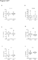
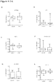
- Results We detected distinct DNA methylation patterns in left ventricular heart tissue of DCM patients and replicated the epigenetic mode of regulation for several genes with previously unknown function in DCM, namely Lymphocyte antigen 75 (LY75), Tyrosine kinase-type cell surface receptor HER3 (ERBB3), Homeobox B13 (HOXB13), and Adenosine receptor A2A (ADORA2A).
- LY75 Lymphocyte antigen 75
- ERBB3 Tyrosine kinase-type cell surface receptor HER3
- HOXB13 Homeobox B13
- ADORA2A Adenosine receptor A2A
- Tissue samples are composed of different cell types, which might change during disease progression.
- Grzeskowiak et. al (2003) it was demonstrated that differences in gene expression of LV myocardial biopsies from controls and idiopathic DCM patients are almost exclusively due to genes expressed in cardiomyocytes and that the contribution of the second most common cell type, fibroblasts, is rather low and mostly unchanged during DCM.
- the increased fibrosis observed in human DCM seems to be mainly due to extracellular collagen deposition rather than an increase of cardiofibroblasts.
- ERBB3 The receptor tyrosine-protein kinase ERBB3 belongs to the membrane bound epidermal growth factor receptor (EGFR) family. Functionally it is implicated in SOX10 mediated neural crest and early heart development (Erickson et al., 1997). However, since knock-out of ERBB3 leads to early embryonic lethality, its role in regulation of cardiac contractility was previously unknown. Although we could not observe changes in ERBB3 transcript levels, the significant changes in DNA methylation could be a susceptibility factor mediated through yet unknown pathways, e.g. by exerting effects on DNA stability, ERBB3 isoform expression, or Histon binding.
- EGFR membrane bound epidermal growth factor receptor
- LY75 a collagen-binding mannose family receptor, is transcriptionally controlled by the Interleukin-6 receptor IL6R ⁇ (Giridhar et al., 2011) and mediates antigen uptake and presentation in a Clathrin-dependent manner.
- IL6R ⁇ Interleukin-6 receptor IL6R ⁇
- DCM Interleukin-6 receptor
- IL-6/LY75 signaling a role of IL-6/LY75 signaling in cardiac remodeling (Timonen et al., 2008).
- the dys-methylated CpGs reside within a classical CpG island covering Exon 1 as well as part of the 5' upstream region. Its transcriptional start-site is predicted in very close vicinity (1,395 bp).
- the adenosine receptor A2A (ADORA2A) is a member of the G protein-coupled receptor family and is highly abundant in neurons of basal ganglia, T lymphocytes, platelets, and vasculature, but also shows relevant expression in myocardium as shown herein.
- ADORA2A enhances cAMP production through ⁇ Gs proteins (Sommerschild et al., 2000) and overexpression results in increased contractility and sarcoplasmic reticulum Ca 2+ uptake (Hamad et al., 2010) as well as cardio protection in ischemia and reperfusion damage (Urmaliya et al., 2009).
- CTCF transcription factor
- CTCF sites emerge as central players in regulatory networks linking gene regulation with epigenetic modifications (Bell and Felsenfield, 2000). Accordingly, our observed decrease in ADORA2A expression might be a result of enhanced binding of CTCF-repressors due to CGI hypomethylation.
- hyper-methylation cannot only silence the binding of transcriptional activators, but also affect repressors of transcription. Hence, hyper-methylation can sometimes result in increased gene expression as observed in the case of ADORA2A.
- DCM was diagnosed according to the guidelines of the World Health Organization (WHO). Inclusion criteria for DCM cases were the presence of reduced left ventricular systolic function (left ventricular ejection fraction ⁇ 45% assessed by echocardiography) in the absence of relevant coronary artery disease (CAD) as determined by coronary angiography. Patients with valvular or hypertensive heart disease, history of myocarditis, regular alcohol consumption or cardio-toxic chemotherapy were also excluded. The control biopsy specimens were obtained from patients who had received heart transplantation.
- WHO World Health Organization
- Biopsy specimens were obtained from the apical part of the free left ventricular wall (LV) from DCM patients or cardiac transplant patients (controls) undergoing cardiac catheterization using a standardized protocol. Biopsies were washed with NaCl (0.9%) and immediately transferred and stored in liquid nitrogen until DNA or RNA was extracted. DNA was extracted using the DNeasy blood and tissue kit, total RNA using the RNeasy kit according to the manufacturer's protocol (Qiagen, Germany). RNA purity and concentration were determined using the Bioanalyzer 2100 (Agilent Technologies, Berkshire, UK) with a Eukaryote Total RNA Pico assay chip. Since a critical issue for the reliability of gene expression analysis is the quality of the RNA samples, a RNA integrity number (RIN) > 6 was defined as minimum requirement for further analyses.
- RIN RNA integrity number
- DNA methylation was validated by the MassARRAY technique as previously described (Ehrich et al., 2005). Briefly, 400-500 ng genomic DNA was chemically modified with sodium bisulfite using the EZ methylation kit (Zymo Research) according to the manufacturer's instructions. The bisulfite-treated DNA was PCR-amplified by primers designed to cover the Infinium probes that showed differential methylation at each locus. The amplicons were transcribed by T7 polymerase, followed by T specific-RNAase-A cleavage. The digested fragments were quantified by MALDI-TOF-based technique. The primer sequences are given in Table 1.
- DNA methylation standards (0%, 20%, 40%, 60%, 80%, and 100% methylated genomic DNA) were used to confirm the unbiased amplification of the amplicons.
- LY75 and ADORA2A we additionally generated 14 and 12 clones, respectively, from 2 patients with DCM and 2 controls, each, and sequenced them before and after treatment with bisulfite. The methylation data was statistically analyzed by Students t-test, or ANOVA, followed by Dunnett's multiple comparison. Table 1: Primer Sequences for mean CpG-island methylation of candidate genes. Genes Forward primers Reverse primers ADORA2A TTTTTTAGGTGGGTGTTGGTAGTT (SEQ ID NO.
- AATTCCCCTAATCCAATAAAATTCC (SEQ ID NO. 20) ATP2C1 ATTGGATAAAGGTATAGTTGTTAAAAGAAG (SEQ ID NO. 21) AACAATCACAATCTCAACAAACAAC (SEQ ID NO. 22) CCDC59 TTTTTAGTGGAAGTAGAGTGGGAAG (SEQ ID NO. 23) TTAAAACAACCTCCTTACACAATCC (SEQ ID NO. 24) CLDN4 GTTTATTTATAGGTTTTTTTAGATGGTTTG (SEQ ID NO. 25) CATCAAAACTAACTTTATCTCCTAACTCA (SEQ ID NO. 26) ERBB3 GGAGATTTTTTAGTAGAGAATAGGTTTTTT (SEQ ID NO.
- the respective target genomic sequence is shown in SEQ ID NO. 81.
- TSS transcription start-sites
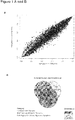
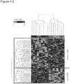
- Hierarchical clustering was applied prior to the clustering. Prior to the clustering, the patients were ranked for each gene according to the degree of methylation, i.e., the patient with the lowest degree of methylation for a certain gene received a rank of 1, while the patient with the highest degree of methylation received a rank of 17, since 17 samples have met filter criteria for the screening stage. Then, for all samples and all genes the pair-wise Euclidian distances were computed and a bottom-up clustering on these distances was carried out. All computations were carried out using the statistical programming language R.
- GSEA Gene Set Enrichment Analysis
- q-PCR Quantitative real-time PCR
- Primers were designed using NCBI Primer-Blast and synthesized by Eurofins MWG Operon (Ebersberg, Germany).
- 60 ng of total RNA extracted from biopsies of independent DCM patients was reverse transcribed using SuperScript III first strand cDNA synthesis kit (Invitrogen).
- q-PCR was carried out according to standard protocols with the SYBR-Green method (Thermo Scientific) using an ABI 7000 system (ABI). Specificity of each primer-pair was monitored by dissociation curve analysis. Threshold cycle (CT) values were assessed in the exponential phase of amplification and the data were analyzed using the delta-CT method.
- CT Threshold cycle
- ly75 and adora2a in the zebrafish heart and whole organism, we performed q-PCR with the following primer-pair for ly75 : 5'-CATGGCCAGTTTCGATCCAT-3' [SEQ ID NO. 84] and 5'-CACCTGGGACTACACCTCCT-3' [SEQ ID NO. 85] and adora2a: 5'-TGCTGACCCAGAGCTCCATA-3' [SEQ ID NO. 86] and 5'-AGAGGCATCATCGCGATCTG-3' [SEQ ID NO. 87].
- Results are shown as relative expression values (normalization against the house-keeping gene elf1a ) .
- the methylation data was statistically analyzed by Students t-test.
- zebrafish experiments were performed under institutional approvals that conform to the Guide for the Care and Use of Laboratory Animals published by The US National Institute of Health (NIH Publication No. 85-23, revised 1996). Care and breeding of zebrafish ( Danio rerio ) were as described (Meder et al. 2011).
- Morpholinos or control oligonucleotides were injected into 1-cell stage embryos as described before (Meder et al. 2009).
- MO- control control oligonucleotides
- GSEA Gene Set Enrichment Analysis
- FIG. 1C gives a graphical representation of differential methylation patterns by using hierarchical clustering on the Euclidian distance. The heat map shows the patterns of higher- and lower methylated genes in DCM patients and controls.
- methylation patterns of single genes were used to define candidates for a mass-spectrometry-based fine-mapping.
- Figure 2A and B show the five most hyper- and hypomethylated genes, while C details the 20 most significantly dys-methylated genes of the screening stage. The final selection was based on either unadjusted p-values or absolute methylation difference, CGI localization, capability to design specific assay probes, and known expression in the heart.
- DNA methylation is often correlated with changes in the accessibility of DNA to transcriptional activators, enhancers, or repressors.
- q-PCR quantitative PCR
- CTCF aberrantly methylated CGI-1 two potential repressor sites
- CGI-2 one repressor site
- CGI-3 one repressor site
- both hypomethylated CGIs (1 and 2) carry CTCF binding sites, which were recently identified as epigenetic key regulators in various other diseases.
- adora2a and ly75 are highly expressed in the zebrafish heart using q-PCR and RNA antisense in-situ hybridization ( FIG. 7 , 8 and 9 ). Additionally, as shown in cultured neonatal rat cells, both genes are higher expressed in cardiomyocytes than in cardiofibroblasts ( Fig. 7B, C ). Hence, to recapitulate the downregulation of both genes as observed in the human heart, we inactivated them in zebrafish embryos by injection of Morpholino-modified antisense oligonucleotides directed against the splice donor-site of ly75 or the translational start-site of adora2a.
- ly75-morphailts showed a pronounced detachment and edema of the skin especially noticeable in the eye and head region ( Fig. 8C ).
- MO- adora2a injected embryos also developed severe heart failure with progressively decreasing ventricular contractility as measured by fractional shortening ( Fig. 9E ).
- the ventricular contractility of adora2a -morphants decreased from 47 ⁇ 11% at 48 hpf to 39 ⁇ 8% at 72 hpf.
- 96 hpf both heart chambers became almost silent.
- atrial fibrillation in adora2a -morphants and embryos developed excessive pericardial effusion and precardial blood congestion as consequence of the reduced cardiac function.
- l y75- and adora2a- morphants we saw no alterations in the molecular chamber specification and expression of atrial and ventricular myosin heavy chain genes ( Fig. 8 and 9 ).
- erbb3b For investigating the functional role of ERBB3, we also used the zebrafish as animal model. We find homology of 56% between human and zebrafish orthologues sequence of ERBB3. Knock-down of erbb3b results in a similar, but earlier phenotype as adora2a. Already by 72 hpf, 86% of injected erbb3b -morphants suffer from significantly reduced cardiac contractility, reduced blood flow through the atrium and ventricle, and pericardial effusion (data not shown). FS at 96 hpf is reduced to 21 ⁇ 4% and blood aggregates at the cardiac inflow tract (data not shown).
- erbb3b -morphants show regurgitation of blood into the ventricle (data not shown), suggesting impaired cardiac valve development.
- cDNA splice-site analysis revealed a completely insertion of intron 2 as well a partial exclusion exon 2 producing an aberrant, frameshifted transcripts with premature termination codons.
Description
- The present invention relates to the use of a DNA methylation profile of a patient sample comprising genomic DNA from heart cells, heart tissue or peripheral blood for the diagnosis and/ or therapy monitoring of dilated cardiomyopathy (DCM) or heart failure in a patient, wherein the DNA methylation profile of the patient sample is compared with the DNA methylation profile of a control sample,and wherein a difference in the DNA methylation profile of the patient sample compared to the control sample is indicative of DCM or heart failure or of the risk of developing DCM or heart failure or for a prediction of therapy effects or therapy outcome,wherein the difference in the DNA methylation profile of the patient sample compared to the DNA methylation profile of the control sample is a different degree of CpG methylation and wherein the methylation level of at least the gene LY75 (SEQ ID NO. 3) is higher in the patient sample compared to the control sample,wherein the patient sample is a sample of left ventricular tissue, right ventricular tissue or peripheral blood, and wherein the control sample is from a normal subject not having a heart disease or having a normal heart function. The present invention further relates to a kit for the diagnosis and/or therapy monitoring of dilated cardiomyopathy (DCM) or heart failure in a patient according to any of
claims 1 to 5, consisting of at least two sets of oligonucleotides, wherein the oligonucleotides of one set are identical, complementary or hybridize under stringent conditions to an at least 15 nucleotides long segment of a nucleic acid sequence of SEQ ID NO. 3, and wherein the oligonucleotides of each further set are identical, complementary or hybridize under stringent conditions to an at least 15 nucleotides long segment of a nucleic acid sequence selected from SEQ ID NOs. 1, 2 and 4 to 18; and optionally, a reagent that distinguishes between methylated and non-methylated CpG dinucleotides. The present invention further relates to the use of LY75 as marker for the diagnosis and/or therapy monitoring of dilated cardiomyopathy (DCM) or heart failure in a patient,comprising determining the methylation level of SEQ ID NO. 3 in a patient sample comprising genomic DNA from heart cells, heart tissue or peripheral blood; and comparing it with the methylation level from a normal subject not having a heart disease,wherein a difference in the DNA methylation profile is indicative of DCM or of the risk for developing DCM or heart failure or for a prediction of therapy effects or therapy outcome, wherein the patient sample is a sample of left ventricular tissue, right ventricular tissue or peripheral blood, and wherein the control sample is from a normal subject not having a heart disease or having a normal heart function. - Cardiomyopathy is the deterioration of the function of the myocardium for any reason. People with cardiomyopathy are at risk of heart failure, arrhythmia and/or sudden cardiac death. Cardiomyopathy can often go undetected, making it especially dangerous to carriers of the disease. Cardiomyopathies can be categorized as extrinsic or intrinsic. An extrinsic cardiomyopathy is a cardiomyopathy where the primary pathology is outside the myocardium itself. Most cardiomyopathies are extrinsic, because by far the most common cause of a cardiomyopathy is ischemia. The World Health Organization calls these specific cardiomyopathies. An intrinsic cardiomyopathy is defined as weakness in the muscle of the heart that is not due to an identifiable external cause. This definition was used to categorize previously idiopathic cardiomyopathies although specific external causes have since been identified for many. The intrinsic cardiomyopathies consist of a variety of disease states, each with their own causes. Many intrinsic cardiomyopathies now have identifiable external causes including ischemia, drug and alcohol toxicity, viral infections and various genetic and idiopathic causes.
- Dilated cardiomyopathy is one of the most frequent heart muscle diseases with an estimated prevalence of 1:2500. The progressive nature of this disorder is responsible for about 30-40% of all heart failure cases and is the main cause for heart transplantation in young adults. In the last decades, it was recognized that DCM has a substantial genetic contribution. It is estimated that about 30-40% of all DCM cases have a familial aggregation and until now more than 40 different genes were found to cause monogenetic DCM. However, since the course of the disease is highly variable and only a fraction of patients suffer a causal mutation, genetic modifiers are thought to play an important role (Friedrichs et al., 2009; Villard et al., 2011). Accordingly, several studies have now identified common genetic polymorphisms, which are associated with DCM or heart failure (Friedrichs et al., 2009; Villard et al., 2011). But even then, the existence of such modifiers also does not completely explain the high variability in phenotypic expression and unexplained cases of DCM.
- Epigenetic mechanisms play important roles during normal development, aging and a variety of disease conditions. Numerous studies have implicated aberrant methylation in the etiology of human diseases, including cancer, MS and diabetes. Hypermethylation of CpG islands located in promoter regions of tumor suppressor genes is firmly established as the most frequent mechanism for gene activation in cancers.
- Briefly, methylation of the 5' carbon of cytosine is a form of epigenetic modification that does not afftect the primary DNA sequence, but affects secondary interactions that play a critical role in the regaulation of gene expression. Aberrant DNA methylation may suppress transcription and subsequently gene expression.
- Disease modification through epigenetic alterations has been convincingly demonstrated for different diseases (Jones and Baylin, 2002; Feinberg and Tycko, 2004). In the cardiovascular system, histone modifications and chromatin remodelling are thought to direct adaptive as well as maladaptive molecular pathways in cardiac hypertrophy and failure (Montgomery et al., 2007) and DNA methylation was found to be responsible for the hypermutability of distinct cardiac genes (Meurs and Kuan, 2011). Furthermore, recent studies have highlighted the potential interplay between environmental factors and the disease phenotype by epigenetic mechanisms (Jirtle and Skinner, 2007; Herceg and Vaissiere, 2011). However, the knowledge about the impact of epigenetic alterations on the disease phenotype in human patients is still very limited.
- Thus, epigenetic mechanisms are increasingly recognized as contributors to human disease. Surprisingly, most studies conducted so far have focused on cancer and only few have investigated the role of epigenetic mechanisms, especially DNA methylation in cardiovascular disease (Movassagh et al., 2011). However, epigenetic mechanisms are thought to control key processes such as cardiac hypertrophy, fibrosis and failure (Backs et al., 2006; Backs et al., 2008).
- Furthermore, the decision to initiate or escalate therapies (drugs, devices, surgical interventions) is currently based on assumptions and supported only by few diagnostic measures (Bielecka-Dabrowa et al., 2008).
- There is a need for diagnostic means and methods that not only allow the detection of a heart disease but also allow to draw conclusions about the further development of an existing heart disease or whether a heart disease will develop in a patient.
- According to the present invention this object is solved by the use and kit as defined in appended
claims 1 to 10. Herein disclosed is the use of a DNA methylation profile of a patient sample comprising genomic DNA from heart cells, heart tissue or peripheral blood for the diagnosis, prognosis and/or therapy monitoring of a heart disease in a patient, wherein the DNA methylation profile of the patient sample is compared with the DNA methylation profile of a control sample, and wherein a difference in the DNA methylation profile of the patient sample compared to the control sample is indicative of a heart disease or of the risk of developing a heart disease or for a prediction of therapy effects or therapy outcome. Further disclosed is a method for the diagnosis, prognosis and/or therapy monitoring a heart disease in a patient, comprising - determining the DNA methylation profile in a patient sample comprising genomic DNA from heart cells, heart tissue or peripheral blood; and
- comparing the DNA methylation profile in the patient sample with the DNA methylation profile from a normal subject not having a heart disease or having a normal heart function, wherein a difference in the DNA methylation profile is indicative of a heart disease or of the risk for developing a heart disease or for a prediction of therapy effects or therapy outcome.
- Before the present invention is described in more detail below, it is to be understood that this invention is not limited to the particular methodology, protocols and reagents described herein as these may vary. It is also to be understood that the terminology used herein is for the purpose of describing particular embodiments only, and is not intended to limit the scope of the present invention which will be limited only by the appended claims. Unless defined otherwise, all technical and scientific terms used herein have the same meanings as commonly understood by one of ordinary skill in the art.
- Concentrations, amounts, and other numerical data may be expressed or presented herein in a range format. It is to be understood that such a range format is used merely for convenience and brevity and thus should be interpreted flexibly to include not only the numerical values explicitly recited as the limits of the range, but also to include all the individual numerical values or sub-ranges encompassed within that range as if each numerical value and sub-range is explicitly recited. As an illustration, a numerical range of "at least 15 nucleotides, preferably 15 to 100" should be interpreted to include not only the explicitly recited values of 15 to 100, but also include individual values and sub-ranges within the indicated range. Thus, included in this numerical range are individual values such as 15, 16, 17, 18, 19, 20, 21, .... 96, 97, 98, 99 and 100 and sub-ranges such as from 20 to 80, from 30 to 70, from 15 to 40, from 25 to 50, from 15 to 25, and from 25 to 50, etc. This same principle applies to ranges reciting only one numerical value. Furthermore, such an interpretation should apply regardless of the breadth of the range or the characteristics being described.
- As described above, the present disclosure provides the use of a DNA methylation profile of a patient sample comprising genomic DNA from heart cells, heart tissue or peripheral blood for the diagnosis, prognosis and/or therapy monitoring of a heart disease in a patient.
- Thereby, the DNA methylation profile of the patient sample is compared with the DNA methylation profile of a control sample.
- A difference in the DNA methylation profile of the patient sample compared to the control sample is indicative:
- of a heart disease,
- of the risk of developing a heart disease,
- of the progression or advance of a heart disease, and/or
- for a prediction of therapy effects or therapy outcome.
- As used herein, providing a diagnosis of a subject/patient is determining heart failure, namely independent on the etiology of the heart failure, i.e. determining whether or not a subject has suffered heart failure recently or in the past.
- As used herein, providing a prognosis of a subject /patient is preferably selected from determining heart failure severity, risk for subsequent all-cause mortality and risk assessment of the subject with heart failure or risk for developing heart failure.
- "Risk assessment" or "risk stratification" of subjects or patients with heart failure refers to the evaluation of factors, such as DNA methylation profiles, mutations, biomarkers, in order to predict the risk of future events or even death and in order to decide about the type, manner, dosis, regimen of therapy and treatment for the individual subject.
- "Disease classification" refers to the severity of the specific heart failure type.
- Preferably, the prognosis of a heart disease comprises risk stratification and/or disease classification.
- As used herein, "therapy monitoring" or "therapy management" of a heart disease comprises treatment monitoring or treatment decision making as well as the prediction of therapy effects or therapy outcome.
- The "DNA methylation profile" according to the invention comprises the DNA methylation levels of CpG islands or nucleotides.
- The difference in the DNA methylation profile of the patient sample compared to the DNA methylation profile of the control sample is preferably
- a different degree of CpG methylation.
- CpG sites or CG sites are regions of DNA where a cytosine nucleotide occurs next to a guanine nucleotide in the linear sequence of bases along its length. "CpG" is shorthand for a cytosine and a guanine separated by only one phosphate, the "CpG" notation is used to distinguish this linear sequence from the CG base-pairing of cytosine and guanine. Cytosines in CpG dinucleotides can be methylated to form 5-methylcytosine. In mammals, methylating the cytosine within a gene can turn the gene off, a mechanism that is part of a larger field of science studying gene regulation that is called epigenetics. In mammals, 70% to 80% of CpG cytosines are methylated.
- There are regions of the genome that have a higher concentration of CpG sites, known as CpG islands. Many genes in mammalian genomes have CpG islands associated with the start of the gene.
- A "CpG island" as used herein and according to Takai and Jones (20002) is a region with:
- a nucleotide length of at least 200 bp, or
- with a GC percentage of 50% or higher, or
- with an observed versus expected CpG ratio that is 60% or higher (where the observed/expected CpG ratio is calculated by formula: number of CpG/(number of C × number of G)) × total number of nucleotides in the sequence).
- CpG islands typically occur at or near the transcription start site of genes, particularly housekeeping genes, in vertebrates. Normally a C (cytosine) base followed immediately by a G (guanine) base (a CpG) is rare in vertebrate DNA because the cytosines in such an arrangement tend to be methylated. This methylation helps distinguish the newly synthesized DNA strand from the parent strand, which aids in the final stages of DNA proofreading after duplication. However, over evolutionary time methylated cytosines tend to turn into thymines because of spontaneous deamination. While there is a special enzyme in human (Thymine-DNA glycosylase, or TDG) that specifically replaces T's from T/G mismatches, it is not sufficiently effective to prevent the relatively rapid mutation of the dinucleotides. The result is that CpGs are relatively rare unless there is selective pressure to keep them or a region is not methylated for some reason, perhaps having to do with the regulation of gene expression.
- Preferably, the DNA methylation profiles are determined by hybridisation-based arrays or by the use of next-generation sequencing techniques, polymerase-based as well as ligase based sequencing technologies like pyrosequencing, sequencing by ligation, single-molecule sequencing or nanopore sequencing alone or in combination with the bisulfit treatment of cytosine nucleotides.
- In a preferred embodiment, the DNA methylation profile of the patient sample is determined over time.
- This embodiment is preferably used for therapy monitoring or therapy management, preferably during/for long term therapy monitoring
- The DNA methylation profile of the patient sample is determined at different time points, during therapy.
- Thereby, the DNA methylation profile is preferably determined before or at the beginning of the therapy and is then determined at different time points during the therapy.
- A change in the DNA methylation profile at these different time points, when compared to the DNA methylation profile of the first measured time point, can be an indication of the progression or advance of the heart disease in the patient and/or of the success of a therapy.
- In an embodiment, further biomarkers and/or genetic mutations are determined in the patient sample.
- Further biomarkers are, for example, well established metabolic biomarkes, e.g. troponins or NT-proBNP,
The determination of such further biomarkers and/or genetic mutations, together with the methylation profile, contributes preferably to disease risk stratification in a combinatorial manner. The expression "heart disease" comprises a cardiomyopathy, myocardial insufficiency, acute or chronic heart failure,
such as dilated cardiomyopathy (DCM), hypertrophic cardiomyopathy (HCM), ischemic cardiomyopathy, diastolic dysfunction, myocardial infarction. - Cardiomyopathy, which literally means "heart muscle disease", is the deterioration of the function of the myocardium for any reason. Cardiomyopathies can generally be categorized into two groups, based on WHO guidelines: extrinsic and intrinsic cardiomyopathies.
- Extrinsic cardiomyopathies are cardiomyopathies where the primary pathology is outside the myocardium itself. Most cardiomyopathies are extrinsic, because by far the most common cause of a cardiomyopathy is ischemia. Ischemic cardiomyopathy, for instance, is a weakness in the muscle of the heart due to inadequate oxygen delivery to the myocardium with coronary artery disease being the most common cause.
- An intrinsic cardiomyopathy is weakness in the muscle of the heart that is not due to an identifiable external cause. The term intrinsic cardiomyopathy does not describe the specific etiology of weakened heart muscle. The intrinsic cardiomyopathies are a heterogeneous group of disease states, each with their own causes. Intrinsic cardiomyopathy has a number of causes including drug and alcohol toxicity, certain infections (including hepatitis C), and various genetic and idiopathic (i.e., unknown) causes. There are four main types of intrinsic cardiomyopathy: first, dilated cardiomyopathy (DCM), the most common form, and one of the leading indications for heart transplantation. In DCM the heart (especially the left ventricle) is enlarged and the pumping function is diminished. Second, hypertrophic cardiomyopathy (HCM or HOCM), a genetic disorder caused by various mutations in genes encoding sarcomeric proteins. In HCM the heart muscle is thickened, which can obstruct blood flow and prevent the heart from functioning properly. Third, arrhythmogenic right ventricular cardiomyopathy (ARVC) arises from an electrical disturbance of the heart in which heart muscle is replaced by fibrous scar tissue. The right ventricle is generally most affected. Fourth, restrictive cardiomyopathy (RCM) is the least common cardiomyopathy. The walls of the ventricles are stiff, but may not be thickened, and resist the normal filling of the heart with blood. Furthermore, noncompaction cardiomyopathy a more recent form of cardiomyopathy is recognized as its own separate type since the 1980's. It refers to a cardiomyopathy where the left ventricle wall has failed to properly grow from birth and such has a spongy appearance when viewed during an echocardiogram.
- The patient sample is preferably a sample of left ventricular tissue (such as a LV biopsy), a sample of right ventricular tissue or peripheral blood.
- The control sample is preferably from a normal subject not having a heart disease or having a normal heart function, i.e. having an unaltered myocardial function.
- The inventors have measured genome-wide DNA methylation profiles of (DCM) patient samples and identified candidate genes with altered methylation status that have prognostic and diagnostic value for heart diseases and are suitable for therapy monitoring as well.
- In a preferred aspect, the methylation level of at least one of the following genes is different in the patient sample compared to the control sample:
Genebank Accession No. ** adenosine receptor A2A (ADORA2A) SEQ ID NO. 1 ENSG00000128271 ERBB3 SEQ ID NO. 2 ENSG00000065361 LY75 SEQ ID NO. 3 ENSG00000054219 HOXB13 SEQ ID NO. 4 ENSG00000159184 GFI1 SEQ ID NO. 5 ENSG00000162676 CLDN4 SEQ ID NO. 6 ENSG00000189143 FDX1 SEQ ID NO. 7 ENSG00000137714 ID4 SEQ ID NO. 8 ENSG00000172201 NAT1 SEQ ID NO. 9 ENSG00000171428 PPARGC1A SEQ ID NO. 10 ENSG00000109819 SULF2 SEQ ID NO. 11 ENSG00000196562 TFF1 SEQ ID NO. 12 ENSG00000160182 TKT SEQ ID NO. 13 ENSG00000163931 ATP2C SEQ ID NO. 14 ENSG00000017260 CCDC59 SEQ ID NO. 15 ENSG00000133773 GSTM5m SEQ ID NO. 16 ENSG00000134201 SLC9A6 SEQ ID NO. 17 ENSG00000198689 TDG SEQ ID NO. 18 ENSG00000139372 ** Ensembl Genebank (see www.ensembl.org) - All listed identifier and gene names and the corresponding gene sequences and gene related sequences like transcript and protein sequences were retrieved from the Ensembl database (http://www.ensembl.org) release 70 from January 2013. Which relates to either GRCh37.p10 (human) or Zv9 (zebrafish). The mentioned resources have also been used for primer design.
- More preferably, the methylation level of at least one of ADORA2A, ERBB3 and LY75 is different in the patient sample compared to the control sample.
- More preferably, the methylation level of at least one of LY75, ADORA2A, ERBB3 and HOXB 13 is different in the patient sample compared to the control sample.
- More preferably, the methylation level of at least one of LY75, ADORA2A, ERBB3, HOXB13 and GFI1 is different in the patient sample compared to the control sample
- Preferably, the methylation level of at least one of ADORA2A, ERBB3, HOXB13, CLDN4, FDX1, ID4, NAT1, PPARGC1A, SULF2, TFF1 and TKT (SEQ ID NOs. 1, 2, 4, 6-13) is reduced in the patient sample compared to the control sample (hypo-methylation), more preferably of at least one of ADORA2A, ERBB3, HOXB13.
- Preferably, the methylation level of at least one of LY75, GFI1, ATP2C, CCDC59, GSTM5m, SLC9A6 and TDG (SEQ ID NOs. 3, 5, 14-18) is higher in the patient sample compared to the control sample (hyper-methylation), more preferably of LY75.
- LY75 is a collagen-binding mannose family receptor that is transcriptionally controlled by the Interleukin-6 receptor IL6Rα (Giridhar et al., 2011). For LY75, the dys-methylated CpGs reside within a classical CpG
island covering Exon 1 as well as part of the 5' upstream region. Its transcriptional start-site is predicted in very close vicinity (1,395 bp). Our findings of a significantly increased DNA methylation together with strongly reduced LY75-mRNA levels in DCM patients suggested a functional role. While it is to our knowledge not possible to recreate exactly the same methylation patterns in vitro as seen in the primary tissue, we could investigate here the functional consequence of global LY75 promoter methylation, suggesting a direct link between methylation and transcriptional activity. We also knocked down ly75 in the zebrafish model and find not only cardiac dysfunction in ly75-ablated embryos, but also a noticeable skin detachment phenotype, potentially due to disturbed collagen production. - The adenosine receptor A2A (ADORA2A) is a member of the G protein-coupled receptor family and is highly abundant in neurons of basal ganglia, T lymphocytes, platelets, and vasculature, but also shows relevant expression in myocardium as shown herein. In the heart, activation of ADORA2A enhances cAMP production through αGs proteins (Sommerschild et al., 2000) and overexpression results in increased contractility and sarcoplasmic reticulum Ca2+ uptake (Hamad et al., 2010) as well as cardio protection in ischemia and reperfusion damage (Urmaliya et al., 2009). Surprisingly, although ADORA2A methylation is reduced, we find mRNA levels to be significantly down-regulated. Hence, hyper-methylation can sometimes result in increased gene expression as observed in the case of ADORA2A. Our CGI annotation was based on the criteria by Takai and Jones (2002), which define a CpG island as a nucleotide sequence of 200 bp or greater in length or with a GC content of 50% or more, or a ratio of observed versus expected CpGs of 0.6 or higher. In case of ADORA2A, the tested CGIs only met the second criterion and may therefore not be classical CGIs.
- The receptor tyrosine-protein kinase ERBB3 belongs to the membrane bound epidermal growth factor receptor (EGFR) family. Functionally it is implicated in SOX10 mediated neural crest and early heart development (Erickson et al., 1997). However, since knock-out of ERBB3 leads to early embryonic lethality, its role in regulation of cardiac contractility was previously unknown. In the present invention, the inventors used the zebrafish as animal model for investigating the functional role of ERBB3. Knock-down of erbb3b results in a similar, but earlier phenotype as adora2a. Additionally, erbb3b-morphants show regurgitation of blood into the ventricle, suggesting impaired cardiac valve development. cDNA splice-site analysis revealed a completely insertion of
intron 2 as well apartial exclusion exon 2 producing an aberrant, frameshifted transcripts with premature termination codons. - The HOXB13 gene is a member of the homeobox gene family and is encoding a transcription factor. The homeobox gene family clusters on
chromosome 17 in the 17qw21-22 region and is highly conserved and essential for vertebrate development. - As described above, the present disclosure provides a method for the diagnosis, prognosis and/or therapy monitoring of a heart disease in a patient.
- The method comprises
- determining the DNA methylation profile in a patient sample comprising genomic DNA from heart cells, heart tissue or peripheral blood; and
- comparing the DNA methylation profile in the patient sample with the DNA methylation profile from a normal subject not having a heart disease or having a normal heart function.
- As described above, a difference in the DNA methylation profile of the patient sample compared to the control sample is indicative:
- of a heart disease,
- of the risk of developing a heart disease, and/or
- for a prediction of therapy effects or therapy outcome.
- The method furthermore preferably comprising the step of
- contacting genomic DNA isolated from the patient sample with at least one reagent that distinguishes between methylated and non-methylated CpG dinucleotides.
- Reagents that distinguish between methylated and non-methylated CpG dinucleotides are known in the art, such as bisulfite, hydrogen sulfite, disulfite, and combinations thereof.
- In a preferred embodiment, the DNA methylation profile of the patient sample is determined over time at different time points, preferably during therapy monitoring, as described above.
- Preferably, the DNA methylation profiles are determined by hybridisation-based arrays or by the use of next-generation sequencing techniques, polymerase-based as well as ligase based sequencing technologies like pyrosequencing, sequencing by ligation, single-molecule sequencing or nanopore sequencing alone or in combination with the bisulfit conversion/treatment of cytosine nucleotides.
- As described above, the heart disease is a cardiomyopathy, myocardial insufficiency, acute or chronic heart failure,
such as dilated cardiomyopathy (DCM), hypertrophic cardiomyopathy (HCM), ischemic cardiomyopathy, diastolic dysfunction, myocardial infarction. - The patient sample is preferably a sample of left ventricular tissue (such as a LV biopsy), a sample of right ventricular tissue or peripheral blood.
- The control sample is preferably from a normal subject not having a heart disease or having a normal heart function, i.e. having an unaltered myocardial function.
- As described above, the prognosis of a heart disease preferably comprises risk stratification and/or disease classification.
- As described above, the therapy monitoring of a heart disease preferably comprises treatment monitoring or treatment decision making as well as the prediction of therapy effects or therapy outcome.
- The inventors have measured genome-wide DNA methylation profiles of (DCM) patient samples and identified candidate genes with altered methylation status that have prognostic and diagnostic for heart diseases and are suitable for therapy monitoring as well.
- In a preferred aspect, the methylation level of at least one of the following genes is different in the patient sample compared to the control sample:
Genebank Accession No. ** adenosine receptor A2A (ADORA2A) SEQ ID NO. 1 ENSG00000128271 ERBB3 SEQ ID NO. 2 ENSG00000065361 LY75 SEQ ID NO. 3 ENSG00000054219 HOXB 13 SEQ ID NO. 4 ENSG00000159184 GFI1 SEQ ID NO. 5 ENSG00000162676 CLDN4 SEQ ID NO. 6 ENSG00000189143 FDX1 SEQ ID NO. 7 ENSG00000137714 ID4 SEQ ID NO. 8 ENSG00000172201 NAT1 SEQ ID NO. 9 ENSG00000171428 PPARGC1A SEQ ID NO. 10 ENSG00000109819 SULF2 SEQ ID NO. 11 ENSG00000196562 TFF1 SEQ ID NO. 12 ENSG00000160182 TKT SEQ ID NO. 13 ENSG00000163931 ATP2C SEQ ID NO. 14 ENSG00000017260 CCDC59 SEQ ID NO. 15 ENSG00000133773 GSTM5m SEQ ID NO. 16 ENSG00000134201 SLC9A6 SEQ ID NO. 17 ENSG00000198689 TDG SEQ ID NO. 18 ENSG00000139372 ** Ensembl Genebank (see www.ensembl.org) - All listed identifier and gene names and the corresponding gene sequences and gene related sequences like transcript and protein sequences were retrieved from the Ensembl database (http://www.ensembl.org) release 70 from January 2013. Which relates to either GRCh37.p10 (human) or Zv9 (zebrafish). The mentioned resources have also been used for primer design. More preferably, the methylation level of at least one of ADORA2A, ERBB3 and LY75 is different in the patient sample compared to the control sample.
- More preferably, the methylation level of at least one of ADORA2A, ERBB3, HOXB 13 and LY75 is different in the patient sample compared to the control sample.
- More preferably, the methylation level of at least one of LY75, ADORA2A, ERBB3 and HOXB13 is different in the patient sample compared to the control sample.
- More preferably, the methylation level of at least one of LY75, ADORA2A, ERBB3, HOXB13 and GFI1 is different in the patient sample compared to the control sample
- Preferably, the methylation level of at least one of ADORA2A, ERBB3, HOXB13, CLDN4, FDX1, ID4, NAT1, PPARGC1A, SULF2, TFF1 and TKT (SEQ ID NOs. 1, 2, 4, 6-13) is reduced in the patient sample compared to the control sample (hypo-methylation), more preferably at least one of ADORA2A, ERBB3, HOXB 13.
- Preferably, the methylation level of at least one of LY75, GFI1, ATP2C, CCDC59, GSTM5m, SLC9A6 and TDG (SEQ ID NOs. 3, 5, 14-18) is higher in the patient sample compared to the control sample (hyper-methylation), more preferably of LY75.
- As described above, the present disclosure provides a kit for the diagnosis, prognosis and/or therapy monitoring of a heart disease in a patient.
- The kit comprises at least two sets of oligonucleotides, wherein the oligonucleotides of each set are identical, complementary or hybridize under stringent conditions to an at least 15 nucleotides long segment of a nucleic acid sequence selected from SEQ ID NOs. 1 to 18, preferably 15 to 100 nucleotides long segment of a nucleic acid sequence selected from SEQ ID NOs. 1 to 18, such as 20 to 80 or 30 to 70, such as about 25 nucleotides.
- The kit optionally comprises a reagent that distinguishes between methylated and non-methylated CpG dinucleotides.
- The kit is preferably suitable for the use of/witll/in hybridisation-based arrays or next-generation sequencing techniques, polymerase-based as well as ligase based sequencing technologies, like pyrosequencing, sequencing by ligation, single-molecule sequencing or nanopore sequencing alone or in combination with the bisulfit treatment/conversion of cytosine nucleotides.
- Thus, the kit comprises at least two sets of oligonucleotides for the detection of two target nucleic acids / genes (selected from SEQ ID NOs. 1 to 18).
- Reagents that distinguish between methylated and non-methylated CpG dinucleotides are known in the art, such as bisulfite, hydrogen sulfite, disulfite, and combinations thereof.
- As described above, the prognosis of a heart disease preferably comprises risk stratification and/or disease classification.
- As described above, the therapy monitoring of a heart disease preferably comprises treatment monitoring or treatment decision making as well as the prediction of therapy effects or therapy outcome.
- As described above, the present disclosure provides the use of at least one of ADORA2A, ERBB3, LY75, HOXB13, GFI1, CLDN4, FDX1, ID4, NAT1, PPARGC1A, SULF2, TFF1, TKT, ATP2C, CCDC59, GSTM5m, SLC9A6 and TDG as marker for the diagnosis, prognosis and/or therapy monitoring of a heart disease in a patient.
Genebank Accession No. ** adenosine receptor A2A (ADORA2A) SEQ ID NO. 1 ENSG00000128271 ERBB3 SEQ ID NO. 2 ENSG00000065361 LY75 SEQ ID NO. 3 ENSG00000054219 HOXB13 SEQ ID NO. 4 ENSG00000159184 GFI1 SEQ ID NO. 5 ENSG00000162676 CLDN4 SEQ ID NO. 6 ENSG00000189143 FDX1 SEQ ID NO. 7 ENSG00000137714 ID4 SEQ ID NO. 8 ENSG00000172201 NAT1 SEQ ID NO. 9 ENSG00000171428 PPARGC1A SEQ ID NO. 10 ENSG00000109819 SULF2 SEQ ID NO. 11 ENSG00000196562 TFF1 SEQ ID NO. 12 ENSG00000160182 TKT SEQ ID NO. 13 ENSG00000163931 ATP2C SEQ ID NO. 14 ENSG00000017260 CCDC59 SEQ ID NO. 15 ENSG00000133773 GSTM5m SEQ ID NO. 16 ENSG00000134201 SLC9A6 SEQ ID NO. 17 ENSG00000198689 TDG SEQ ID NO. 18 ENSG00000139372 ** Ensembl Genebank (see www.ensembl.org) - All listed identifier and gene names and the corresponding gene sequences and gene related sequences like transcript and protein sequences were retrieved from the Ensembl database (http://www.ensembl.org) release 70 from January 2013. Which relates to either GRCh37.p10 (human) or Zv9 (zebrafish). The mentioned resources have also been used for primer design.
- In a preferred embodiment at least one of ADORA2A, ERBB3 and LY75 are used as such a marker.
- In a preferred embodiment at least one of LY75, ADORA2A, ERBB3 and HOXB13 are used as such a marker.
- In a preferred embodiment at least one of LY75, ADORA2A, ERBB3, HOXB13 and GFI1 are used as such a marker.
- Preferably, the use of said (target) genes, nucleic acids or polypeptides encoded by the genes/nucleis acids as biomarkers comprises determining the methylation level of at least one of SEQ ID NOs. 1-18 in a patient sample comprising genomic DNA from heart cells, heart tissue or peripheral blood; and comparing it with the methylation level from a normal subject not having a heart disease, wherein, and as described above, a difference in the DNA methylation profile is indicative of a heart disease or of the risk for developing a heart disease or for a prediction of therapy effects or therapy outcome.
- Preferably, and as described above, the heart disease is a cardiomyopathy, myocardial insufficiency, heart failure (acute or chronic),
such as DCM dilated cardiomyopathy (DCM), hypertrophic cardiomyopathy (HCM), ischemic cardiomyopathy, diastolic dysfunction, myocardial infarction. - Preferably, and as described above, the patient sample is a sample of left ventricular tissue, right ventricular tissue or peripheral blood.
- Preferably, and as described above, the control sample is from a normal subject not having a heart disease or having a normal heart function.
- As described above, the prognosis of a heart disease preferably comprises risk stratification and/or disease classification.
- As described above, the therapy monitoring of a heart disease preferably comprises treatment monitoring or treatment decision making as well as the prediction of therapy effects or therapy outcome.
- Problem: Dilated cardiomyopathy (DCM) is one the most frequent heart muscle diseases. Although several factors including genetic mechanisms are found to cause DCM, we still find many cases unexplained and observe a high phenotypic variability with respect to disease severity and prognosis. Epigenetic mechanisms are increasingly recognized as causes and modulators of human disease. Therefore, we studied genome-wide cardiac DNA methylation in DCM patients and controls to detect a possible epigenetic contribution to DCM.
- Results: We detected distinct DNA methylation patterns in left ventricular heart tissue of DCM patients and replicated the epigenetic mode of regulation for several genes with previously unknown function in DCM, namely Lymphocyte antigen 75 (LY75), Tyrosine kinase-type cell surface receptor HER3 (ERBB3), Homeobox B13 (HOXB13), and Adenosine receptor A2A (ADORA2A). The results were carefully verified by alternative techniques in a well phenotyped and large independent cohort of DCM patients and controls. Furthermore, we are able to show the functional relevance for the contribution of the identified genes in the pathogenesis of heart failure by using the zebrafish as an in vivo model.
- Impact: Our results hint at a novel layer in the pathogenesis of DCM and heart failure and have an impact on the development of novel biomarkers and future therapeutic strategies.
- In the present study, genome-wide cardiac DNA methylation was examined for the first time in patients with idiopathic DCM and controls. We detected methylation differences in pathways related to heart disease, but also in genes with yet unknown function in DCM or heart failure, namely Lymphocyte antigen 75 (LY75), Tyrosine kinase-type cell surface receptor HER3 (ERBB3), Homeobox B13 (HOXB13), and Adenosine receptor, A2A (ADORA2A). Mass-spectrometric analysis and bisulfite-sequencing enabled confirmation of the observed DNA methylation changes in independent cohorts. Aberrant DNA methylation in DCM patients was associated with significant changes in LY75 and ADORA2A mRNA expression, but not in ERBB3 and HOXB13. By in vivo studies of orthologous ly75 and adora2a in zebrafish, we could demonstrate for the first time a functional role of these genes in adaptive or maladaptive pathways in heart failure.
- In detail, we investigated here DNA methylation patterns on a genome-wide level in myocardium from patients with idiopathic DCM and functionally unaffected hearts of patients who had received heart transplantation. We found and confirmed aberrant DNA methylation alterations in a number of CGIs, suggesting that DNA methylation is associated with cardiac function and may modulate phenotypic characteristics of idiopathic DCM.
- Recently, Movassagh et al. (2011) reported distinct epigenomic features in explanted human failing hearts, highlighting a potential role of DNA methylation also in end-stage heart failure. However, feasibility of the used methyl-DNA precipitation and sequencing is restricted by the relatively large amounts of required tissue, which is not easily available from living patients.
- Furthermore, handling of the tissue and time points of DNA isolation were quite different in controls and patients. Tissue specimens from explanted hearts of end-stage heart failure patients may be altered due to tissue handling and could have undergone secondary alterations because of degradation, missing oxygenation, innervation, and arrest of pumping function (Talens et al., 2010). Also, the authors did not replicate their findings, which were obtained in 4 patients, in an independent series of unrelated samples and by an alternative methodology. Therefore, we used a different approach, confirmed our findings in a large replication cohort with two alternative methodologies and applied functional studies to underscore the relevance of our results.
- Since availability of appropriate myocardial tissue from living patients is limited, studies on epigenetic alterations in patients with heart disease are rare. As a primary referral center for cardiomyopathies, we could enroll a high number of patients and controls in this study. Our controls are not completely healthy, but had previously received heart transplantation. Hence, potential confounders include patient's medication and medical history. Importantly, on the functional level all controls had an unconstrained cardiac function as a prerequisite and we could apply the exactly same standardized procedure to sample and process left ventricular biopsies from DCM patients and controls. However, all controls received immunosuppressive medications to prevent organ rejection. This medication might influence on at least some genes. As shown herein, we found unchanged methylation levels for the tested genes in control subjects before and after heart transplantation, showing that immunosuppressives do not, at least in whole peripheral blood, impact on CGI DNA methylation of LY75, ADORA2A, ERBB3 and HOB13.
- Tissue samples are composed of different cell types, which might change during disease progression. In the present study, we have paid special attention to patient selection and the process of tissue sampling. Consequently, this resulted in high homogeneity of overall methylation patterns as shown above. In a study by Grzeskowiak et. al (2003) it was demonstrated that differences in gene expression of LV myocardial biopsies from controls and idiopathic DCM patients are almost exclusively due to genes expressed in cardiomyocytes and that the contribution of the second most common cell type, fibroblasts, is rather low and mostly unchanged during DCM. The increased fibrosis observed in human DCM, for instance, seems to be mainly due to extracellular collagen deposition rather than an increase of cardiofibroblasts. Our in vivo experiments clearly underline that reduced expression of LY75 and ADORA2A in cardiomyocytes leads to heart failure as observed in our patients. Together, this approach allowed us to study epigenetic mechanisms and its functional consequences without the confounding background of organ hypoxia and apoptosis in explanted hearts of deceased patients.
- In a funnel approach, we first investigated global methylation patterns and found that methylation patterns also reflect cardiac disease pathways. Our global methylation analyses identified cardiac disease pathways and were complemented by extensive validation studies applying the highly precise MassARRAY and bisulfite sequencing techniques. We found 12 of 20 genes showing the same direction of dys-methylation as found in the screening stage, and four reached statistical significance. This has at least three reasons: 1) we expected a high false positive rate due to the relatively small screening cohorts in contrast to the number of measured epigenetic features, 2) we performed the replication measurements with a completely independent methodology, and 3) we carried out these analyses in independent patients and controls. Therefore, the genes identified here represent consistently validated targets that show an epigenetic mode of regulation.
- The deciphering of the functional implications of changes in DNA methylation is difficult but of pivotal importance. Since in many cases changes in CpG methylation are correlated to the accessibility of DNA to transcription factors and polymerases, measurements of mRNA levels represent an appropriate methodology. In the present study, we find that methylation of novel cardiac genes is altered in patients with DCM. Hence, it can be hypothesized that the dys-methylated and dys-regulated genes also exert a functional role in the heart.
- The receptor tyrosine-protein kinase ERBB3 belongs to the membrane bound epidermal growth factor receptor (EGFR) family. Functionally it is implicated in SOX10 mediated neural crest and early heart development (Erickson et al., 1997). However, since knock-out of ERBB3 leads to early embryonic lethality, its role in regulation of cardiac contractility was previously unknown. Although we could not observe changes in ERBB3 transcript levels, the significant changes in DNA methylation could be a susceptibility factor mediated through yet unknown pathways, e.g. by exerting effects on DNA stability, ERBB3 isoform expression, or Histon binding.
- LY75, a collagen-binding mannose family receptor, is transcriptionally controlled by the Interleukin-6 receptor IL6Rα (Giridhar et al., 2011) and mediates antigen uptake and presentation in a Clathrin-dependent manner. For DCM, a correlation of IL-6 levels and crosslinked type I collagen was found, potentially implicating a role of IL-6/LY75 signaling in cardiac remodeling (Timonen et al., 2008). For LY75, the dys-methylated CpGs reside within a classical CpG
island covering Exon 1 as well as part of the 5' upstream region. Its transcriptional start-site is predicted in very close vicinity (1,395 bp). Our findings of a significantly increased DNA methylation together with strongly reduced LY75-mRNA levels in DCM patients suggested a functional role. While it is to our knowledge not possible to recreate exactly the same methylation patterns in vitro as seen in the primary tissue, we could investigate here the functional consequence of global LY75 promoter methylation, suggesting a direct link between methylation and transcriptional activity. We also knocked down ly75 in the zebrafish model and find not only cardiac dysfunction in ly75-ablated embryos, but also a noticeable skin detachment phenotype, potentially due to disturbed collagen production. - The adenosine receptor A2A (ADORA2A) is a member of the G protein-coupled receptor family and is highly abundant in neurons of basal ganglia, T lymphocytes, platelets, and vasculature, but also shows relevant expression in myocardium as shown herein. In the heart, activation of ADORA2A enhances cAMP production through αGs proteins (Sommerschild et al., 2000) and overexpression results in increased contractility and sarcoplasmic reticulum Ca2+ uptake (Hamad et al., 2010) as well as cardio protection in ischemia and reperfusion damage (Urmaliya et al., 2009). Surprisingly, although ADORA2A methylation is reduced, we find mRNA levels to be significantly down-regulated. Such a positive relationship was described for other genes and is not fully understood in each case (Zhang et al., 2006). Our CGI annotation was based on the criteria by Takai and Jones (2002), which define a CpG island as a nucleotide sequence of 200 bp or greater in length or with a GC content of 50% or more, or a ratio of observed versus expected CpGs of 0.6 or higher. In case of ADORA2A, the tested CGIs only met the second criterion and may therefore not be classical CGIs.
- For such sites, one explanation can be that hyper-methylation cannot only impede the binding of transcriptional activators but also can affect repressors of transcription. We therefore performed an in silico analysis of the promoter region of ADORA2A and found two binding-sites of the transcription factor CTCF (CCCTC-binding factor) in close vicinity to both significantly hypo-methylated CGIs, but not within the single unaffected CGI of ADORA2A. CTCF is a unique insulator-binding protein, which can act as transcriptional repressor by blocking distinct enhancer regions (Bell et al., 1999; Gaszner and Felsenfield, 2006). Intriguingly, CTCF sites emerge as central players in regulatory networks linking gene regulation with epigenetic modifications (Bell and Felsenfield, 2000). Accordingly, our observed decrease in ADORA2A expression might be a result of enhanced binding of CTCF-repressors due to CGI hypomethylation. One explanation is that hyper-methylation cannot only silence the binding of transcriptional activators, but also affect repressors of transcription. Hence, hyper-methylation can sometimes result in increased gene expression as observed in the case of ADORA2A.
- The detection of epigenetic mechanisms in human heart disease represents an attractive option to identify and dissect and later in target completely novel pathomechanisms. Hence, the validated targets from this study that have shown functional relevance are most likely modifiers of DCM rather than being independent disease causes and can, thus, can be further investigated for their diagnostic and therapeutic potential. Therefore, it is reasonable to investigate their potential as diagnostic and therapeutic targets specifically in DCM but also in heart failure due to other causes. In the cancer field, epigenetic drugs have already entered the clinical arena (e.g. Decitabine or Azacitidine in the treatment of myelodysplastic syndromes), and methylation patterns are used as biomarkers to subtype and stage various cancers as a critical step towards a more effective personalized care (Coppede 2011; Litzow 2011). Hence, the genes identified here are relevant drugable targets in DCM and heart failure and aid in disease classification or risk stratification.
- The following examples and drawings illustrate the present invention without, however, limiting the same thereto.
-
-
Figure 1 Detection of DNA methylation patterns.- A) Correlation plot showing the percentage of CpG methylation in controls versus DCM patients resulting in an overall very high correlation. Overall, correlation between both cohorts is high, however some outliers show hyper- or hypo-methylation in cases.
- B) Gene-Set Enrichment Analysis for NIA human disease pathways. The area proportional Venn diagram shows that methylation changes in cardiovascular disease genes are significantly enriched together with the overlap of the other indicated gene sets.
- C) Cluster analysis for genes with known expression in the human heart and significantly altered methylation. The color code used for the heatmap is shown in the upper left corner, values range from 1 (sample with the lowest methylation for the considered gene) to 17 (sample with the highest methylation for the considered genes). Genes with higher methylation found in controls cluster in the upper right quarter. Genes with higher methylation in DCM gather in the lower left quarter. Hypomethylation is shown for genes clustering in the upper left quarter for DCM and lower right quarter for controls.
-
Figure 2 Differentially methylated genes in DCM patients.- A-C) Bar graphs showing the degree of methylation of CGIs of the screening cohort (n=9 DCM patients; n=8 controls). The 5 genes with the largest increase in methylation in DCM patients are shown in A) and genes with the largest decrease in methylation are displayed in B). C) Shown are the 20 genes with the most significant methylation changes in the screening phase. Error bars indicating standard deviation.
-
Figure 3 MassARRAY-based validation of dys-methylated genes.- A and B) MassARRAY-based validation of differential methylation in LY75 and ERBB3.
DCM patients show significantly increased DNA methylation in LY75 (A), while ERBB3 is significantly lower methylated (B). The schemes above the methylation graphs represent the tested CGIs (CGI-1, CGI-2, CGI-3), in relation to the predicted transcription start-site (leftmost vertical line), the exons (black bars) and alternatively spliced exons (white boxes) of the genes. A) The box and whiskers plot (min to max) on the upper left side represents the mean CpG methylation measured by MassARRAY, the individual CpG methylation is shown at the bottom. The pattern of CpG methylation in multiple clones is shown on the upper right insert. B) The box and whiskers plot (min to max) on the left side represents the mean CpG methylation measured by MassARRAY, the individual CpG methylation is shown on the right. - C and D) MassARRAY-based validation of differential methylation in HOXB13 and ADORA2A.
DCM patients show significantly decreased DNA methylation in HOXB13 (A) and ADORA2A (B). A) The box and whiskers plot (min to max) on the left side represents the mean CpG methylation measured by MassARRAY, the individual CpG methylation is shown on the right. B) The scheme above the graph represents the tested CGI, in relation to the predicted transcription start site (leftmost vertical line), the exons (black boxes) and alternatively spliced exons (white boxes) of adora2a. Dashed lines indicate the 1500 bp up- and downstream region of the CGIs wherein the listed transcription factor binding sites are found. Boxplots below show the mean methylation in DCM and controls at the corresponding CGI. The pattern of CpG methylation as assessed by bisulfite-sequencing in multiple clones is shown for CGI-1 at the bottom of the figure. - (E) MassARRAY-based validation of differential methylation in ADORA2A. Graph representing individual CpG methylation in ADORA2A measured by MassARRAY in DCM patients and controls.
- A and B) MassARRAY-based validation of differential methylation in LY75 and ERBB3.
-
Figure 4 MassARRAY-based validation of further dys-methylated genes.
Graphs representing the mean CGI methylation of targets included in the replication stage. DCM patients show a reduction in methylation level in CLDN4, FDX1, ID4, NAT1, PPARGC1A, SULF2, TFF1, TKT, FLJ20701 and an increase of methylation in ATP2C, CCDC59, GFI1, GSTM5m, SLC9A6, and TDG. -
Figure 5 CpG island methylation of control subjects before and after heart transplantation.
The line diagram shows the highly similar degree of methylation of DNA derived from peripheral blood of control subjects pre- (circlescattered line) and post- (squaresscattered line) heart transplantation (n=11) for LY75, ADORA2A, ERBB3 and HOXB13. Error bars indicating standard deviation. -
Figure 6 mRNA Expression of genes with altered methylation status and gene promoter analysis.- A-C) Bar graphs showing relative mRNA expression levels of LY75, ERBB3 and ADORA2A in mild and moderate-severe DCM in comparison to controls. A) LY75 expression is strongly reduced (relative expression = 0.04), while ERBB3 (B) mRNA levels are not differentially regulated. C) ADORA2A is also significantly down-regulated in DCM (relative expression = 0.37).
- D) Given are the mean delta-CT values (± SEM) of LY75, ERBB3, and ADORA2A in the different groups (controls, moderate DCM, severe DCM). The reference is based on the mean of the three housekeeper genes: GAPDH, RPL13, β-Actin.
- E) Left: Schema of the vector construct to measure LY75 promoter activity without and after treatment with the methylase SssI. Right: Relative promoter activity (mean of 3 technical replicates ± SD) showing the strongly reduced activity in LY75 after methylation, whereas the negative control does not show significant differences.
-
Figure 7 ly75 and adora2a tissue and cell-type expression.- A) Relative expression levels of ly75 and adora2a quantified by q-PCR in 40 whole zebrafish embryos (grey bars) and 200 isolated zebrafish embryonic hearts (black bars). B-C) Expression of ly75 and adora2a in neonatal rat cardiomyocytes and cardiofibroblasts (n=3 biological replicates). Error bars indicating standard deviation.
-
Figure 8 ly75 is essential to maintain cardiac contractility.- A) mRNA antisense in situ hybridization showing that ly75 is significantly expressed in the zebrafish heart. B, C) Lateral views of MO3-control (B) and MO-ly75 (C) injected embryos at 72 hpf. After depletion of ly75, which can be monitored by cDNA splice-analysis (D), zebrafish hearts show reduced contractile force (E) and precardial blood congestion (arrowhead) as sign of manifest heart failure. Ly75-morphants also show noticeable skin detachment most pronounced in the head/eye region (C).
- F) Molecular chamber definition is not impaired in ly75-morphants compared to the control-injected zebrafish as demonstrated by regular amhc, vmhc, MF20, and S46 expression. hpf = hours post fertilization; amhc = atrial myosin heavy-chain; vmhc = ventricular myosin heavy-chain; A = atrium; V = ventricle. Error bars indicating standard deviation.
-
Figure 9 Loss-of adora2a leads to heart failure in zebrafish.- A) Amino acid sequence alignments of human (h), mouse (m) and zebrafish (z) ADORA2A demonstrating its high cross-species homology. Black boxes indicate amino acid identity, gray boxes indicate amino acids with similar chemical properties. B) Adora2A is significantly expressed in the zebrafish heart. C, D) Lateral view of MOS-control (B), and MO-adora2a (C) injected embryos at 72 hpf. After injection of MO-adora2a, 66% of morphant embryos develop progressive heart failure and deterioration in ventricular contractility measured by fractional shortening (E) at the indicated developmental stages. Error bars indicating standard deviation.
- F) Molecular chamber definition is not impaired in adora2a-morphants compared to the control-injected zebrafish as demonstrated by regular amhc and vmhc expression.
-
Figure 10 Protein conservation of LY75.
Amino acid sequence alignments of human (h), mouse (m) and zebrafish (z) LY75 demonstrating its cross-species homology. Black boxes indicate amino acid identity, gray boxes indicate amino acids with similar chemical properties. - The study was conducted in accordance with the principles of the Declaration of Helsinki. All participants of the study have given written informed consent and the study was approved by the ethic committees. DCM was diagnosed according to the guidelines of the World Health Organization (WHO). Inclusion criteria for DCM cases were the presence of reduced left ventricular systolic function (left ventricular ejection fraction < 45% assessed by echocardiography) in the absence of relevant coronary artery disease (CAD) as determined by coronary angiography. Patients with valvular or hypertensive heart disease, history of myocarditis, regular alcohol consumption or cardio-toxic chemotherapy were also excluded. The control biopsy specimens were obtained from patients who had received heart transplantation. All patients of the control cohort had successful cardiac transplantation more than 6 months ago with normal systolic and diastolic function and no evidence for relevant vasculopathy as judged by coronary angiography. Furthermore, all controls showed freedom from relevant acute or chronic organ rejection.
- Biopsy specimens were obtained from the apical part of the free left ventricular wall (LV) from DCM patients or cardiac transplant patients (controls) undergoing cardiac catheterization using a standardized protocol. Biopsies were washed with NaCl (0.9%) and immediately transferred and stored in liquid nitrogen until DNA or RNA was extracted. DNA was extracted using the DNeasy blood and tissue kit, total RNA using the RNeasy kit according to the manufacturer's protocol (Qiagen, Germany). RNA purity and concentration were determined using the Bioanalyzer 2100 (Agilent Technologies, Berkshire, UK) with a Eukaryote Total RNA Pico assay chip. Since a critical issue for the reliability of gene expression analysis is the quality of the RNA samples, a RNA integrity number (RIN) > 6 was defined as minimum requirement for further analyses.
- For measuring methylation profiles, we used the
Infinium HumanMethylation 27 BeadChip assay from Illumina with 1000 ng DNA per sample. The procedure followed the manufacturers standard workflow, starting with the bisulfite treatment of the sample DNA leading to a conversion of unmethylated cytosins to uracil, while 5-methylcytosines remain unchanged. After amplification and fragmentation of the bisulfite-converted DNA, they were hybridized to the Infinium BeadChips. - DNA methylation was validated by the MassARRAY technique as previously described (Ehrich et al., 2005). Briefly, 400-500 ng genomic DNA was chemically modified with sodium bisulfite using the EZ methylation kit (Zymo Research) according to the manufacturer's instructions. The bisulfite-treated DNA was PCR-amplified by primers designed to cover the Infinium probes that showed differential methylation at each locus. The amplicons were transcribed by T7 polymerase, followed by T specific-RNAase-A cleavage. The digested fragments were quantified by MALDI-TOF-based technique. The primer sequences are given in Table 1. DNA methylation standards (0%, 20%, 40%, 60%, 80%, and 100% methylated genomic DNA) were used to confirm the unbiased amplification of the amplicons. For LY75 and ADORA2A we additionally generated 14 and 12 clones, respectively, from 2 patients with DCM and 2 controls, each, and sequenced them before and after treatment with bisulfite. The methylation data was statistically analyzed by Students t-test, or ANOVA, followed by Dunnett's multiple comparison.
Table 1: Primer Sequences for mean CpG-island methylation of candidate genes. Genes Forward primers Reverse primers ADORA2A TTTTTTAGGTGGGTGTTGGTAGTT (SEQ ID NO. 19) AATTCCCCTAATCCAATAAAATTCC (SEQ ID NO. 20) ATP2C1 ATTGGATAAAGGTATAGTTGTTAAAAGAAG (SEQ ID NO. 21) AACAATCACAATCTCAACAAACAAC (SEQ ID NO. 22) CCDC59 TTTTTAGTGGAAGTAGAGTGGGAAG (SEQ ID NO. 23) TTAAAACAACCTCCTTACACAATCC (SEQ ID NO. 24) CLDN4 GTTTATTTATAGGTTTTTTTAGATGGTTTG (SEQ ID NO. 25) CATCAAAACTAACTTTATCTCCTAACTCA (SEQ ID NO. 26) ERBB3 GGAGATTTTTTAGTAGAGAATAGGTTTTTT (SEQ ID NO. 27) TACTACCCAAAACCCTACTAATCCC (SEQ ID NO. 28) FDX1 TTTGTTTTATAGTGGGTAGAATTTATTT (SEQ ID NO. 29) AACCTATAAAAACATTAACAAACAAAAC (SEQ ID NO. 30) FLJ20701 GATTTTAAATTTTAGTGAGGGGTGAA (SEQ ID NO. 31) TCCAAAACAAAAAATAAACTCCAAA (SEQ ID NO. 32) GFI1 TTTATTTTAATGAGTAAAGGGTGTAGGT (SEQ ID NO. 33) CCTAAAATCATACCCAAACACTAAAT (SEQ ID NO. 34) GSTM5 GGAGGGGGTTTATTGATTTTAGTTT (SEQ ID NO. 35) TAACTTTCTCTACACCAAACCAACC (SEQ ID NO. 36) HOXB13 GGTAGTTTTTGGTTTTGGGTTTT (SEQ ID NO. 37) ACTATCCCTAAATCTCATCTCTCCC (SEQ ID NO. 38) ID4 AATGGAGTGTTTTTTTTATTGGTT (SEQ ID NO. 39) AATATCCTAATCACTCCCTTC (SEQ ID NO. 40) LY75 GTATTTTTGTTTAGGTTTTTGGGG (SEQ ID NO. 41) AAAAATCATAAATCCCTTTCCAATC (SEQ ID NO. 42) NAT1 TTTTTTTAGGAAGTAAAAGGAATGTT (SEQ ID NO. 43) TCAAACTACCCTAATCTCACTATCCC (SEQ ID NO. 44) PPARGC1 A GATTTTTTTAAAAGTTTTTGTTTGG (SEQ ID NO. 45) ACAAATACCTTCAATTCACTCTCAA (SEQ ID NO. 46) SLC9A6 GTTTTTGATTGGGTAGGGGT (SEQ ID NO. 47) AACTTACCATAAATCATAACCAAACC (SEQ ID NO. 48) SULF2 GAGTTTTGTGAAAGTAGATAAAAGAAAAT (SEQ ID NO. 49) TAACACACAAACCAAAAAACATC (SEQ ID NO. 50) TDG TGGTTGGTAGTATTTAGATAGTGGTTG (SEQ ID NO. 51) ATATACACAACACCCCAAAAACAA (SEQ ID NO. 52) TFF1 AGGTTGTTAGAGTTGGTTGTGGTT (SEQ ID NO. 53) AACTTTCTAAATCTCAAATCCCTCAA (SEQ ID NO. 54) TKT GGGAAGGTTATTATTATTGTTGTTTT (SEQ ID NO. 55) AAATCCCAAAATTCTACACCCA (SEQ ID NO. 56) XRCC5 GTTTTTTGTTAGGTTTGAAAGGGG (SEQ ID NO. 57) AAAAAAAATCTAACTCCAAAACTCTAA (SEQ ID NO. 58) - The respective target genomic sequences are shown in SEQ ID NOs. 59 to 78: target genomic sequence of gene
ADORA 2A SEQ ID NO. 59; ATP2C1 SEQ ID NO. 60; CCDC59 SEQ ID NO. 61; CLDN4 SEQ ID NO. 62; ERBB3 SEQ ID NO. 63; FDX1 SEQ ID NO. 64; FLJ20701 SEQ ID NO. 65; GFI1 SEQ ID NO. 66; GSTM5 SEQ ID NO. 67; HOXB13 SEQ ID NO. 68; ID4 SEQ ID NO. 69; LY75 SEQ ID NO. 70; NAT1 SEQ ID NO. 71; PPARGC1A SEQ ID NO. 72; SLC9A6 SEQ ID NO. 73; SULF2 SEQ ID NO. 74; TDG SEQ ID NO. 75; TFF1 SEQ ID NO. 76; TKT SEQ ID NO. 77; XRCC5 SEQ ID NO. 78. - To assess the causative link of CGI methylation within the promoter region of LY75 with reduced mRNA expression observed in myocardial tissue, we performed a promoter luciferase assay essentially as described before (Smith et al., 2006). Therefore, we cloned a 1.6 kb large core fragment (Table 2) of the LY75 promoter region including the analyzed CGI into SpeI/BamHI sites of the CpG-free luciferase vector pCpGL-basic. As negative control for the methylation effect we utilized the CpG-free control reporter vector pCpGL-CMV/EF1 (Klug and Rehli, 2006). Next, we treated both vectors with the methylase SssI (New England BioLabs) or leaved them untreated, respectively. The methylation was performed at 10 mM Tris, pH7.9, 50mM NaCl, 10 mM MgCl2, 1 mM DTT and 160µM SAM at 37°C for 1 hour (Blesa et al., 2008). Transfection of human HEK293A cells with equal amounts of the SssI-treated or untreated pCpG-LY75 or pCpGL-CMV/EF1 vectors was carried out using Lipofectamin (Invitrogen). Additionally, we co-transfected these cells with the pGL4.74 [hRluc/TK] Renilla control vector as a reference (Promega). After 48 hours, cells were lysed and luciferase and Renilla activity measured according to manufactures protocol (Dual-Luciferase Reporter Assay System, Promega). Results are expressed as relative luciferase activity (background substracted and normalized to Renilla reference; three technical replicates) of the methylated versus the unmethylated constructs.
Table 2: Primer sequences for promoter luciferase assay. Gene Forward primer Reverse primer LY75 TGGCACTACCCTGAACATCA (SEQ ID NO. 79) CATGAAATTTCTGGTTCTGCAT (SEQ ID NO. 80) - The respective target genomic sequence is shown in SEQ ID NO. 81.
- We applied the Promoter 2.0 Prediction Software that predicts transcription start-sites (TSS) of vertebrate Pol II promoters in genomic DNA to estimate the distance of the CGIs from the potential transcriptional start-sites (Knudsen 1999). For the detection of transcription factor binding sites, the ENCODE transcription factor ChIP-seq data was used. For each region (size of the CGI and 1500 bp up- and downstream), we selected in the maximum the three factors up- and three factors downstream with strongest observed binding.
- To generate a graphical representation of the most significantly dys-methylated cardiac genes in the screening stage and to detect patterns among the respective genes, hierarchical clustering was applied. Prior to the clustering, the patients were ranked for each gene according to the degree of methylation, i.e., the patient with the lowest degree of methylation for a certain gene received a rank of 1, while the patient with the highest degree of methylation received a rank of 17, since 17 samples have met filter criteria for the screening stage. Then, for all samples and all genes the pair-wise Euclidian distances were computed and a bottom-up clustering on these distances was carried out. All computations were carried out using the statistical programming language R.
- To test whether genes with differential methylation patterns belong to a certain biological category we carried out an unweighted Gene Set Enrichment Analysis (GSEA). In detail, we first sorted all genes according to the absolute value of the median distances between DCM patients and controls, generated a sorted list of genes where the most differentially methylated genes were on top while genes without differential methylation were located at the bottom of the list. If one gene was represented by multiple features on the biochip (different methylation sites) the median position of all replicates in the sorted list was computed. This sorted list was then used as input for GeneTrail (Backes et al. 2007). For each category, a significance value is computed by a dynamic programming approach (Keller et al. 2007) and all significance values of a certain category have been adjusted for multiple testing using Benjamini-Hochberg adjustment (Benjamini and Hochberg, 1995). The advantage of the applied cutoff-free biostatistical approach is that genes with p-values of 0.049 and 0.051 are considered to be almost equally important in our approach while genes with larger values in the middle and at the end of the list are considered to be not of relevance. In contrast, classical over-representation analyses relying on cutoffs (usually 0.05) would consider genes with p-value of 0.049 of relevance while genes with p-values of 0.051 would be considered to be non-relevant, having the same impact as genes with large p-values.
- Quantitative real-time PCR (q-PCR) was performed in order to measure expression of selected genes. Primers were designed using NCBI Primer-Blast and synthesized by Eurofins MWG Operon (Ebersberg, Germany). 60 ng of total RNA extracted from biopsies of independent DCM patients (moderate: n=7; severe: n=5) and controls (n=7) was reverse transcribed using SuperScript III first strand cDNA synthesis kit (Invitrogen). q-PCR was carried out according to standard protocols with the SYBR-Green method (Thermo Scientific) using an ABI 7000 system (ABI). Specificity of each primer-pair was monitored by dissociation curve analysis. Threshold cycle (CT) values were assessed in the exponential phase of amplification and the data were analyzed using the delta-CT method. The mean value of the reference genes GAPDH, RPL13 and â-actin was used as a reference.
- To identify the predominant cardiac isoform of ADORA2A, we performed PCR with the following primer-pair for both isoforms (ENST00000337539 and ENST00000541988):
5'-CTGTGACATGGAGCAGGAGC-3' [SEQ ID NO. 82] and 5'-GCTGTCGTTTGCCATCGGCCT-3' [SEQ ID NO. 83]. - To evaluate the expression levels of ly75 and adora2a in the zebrafish heart and whole organism, we performed q-PCR with the following primer-pair for ly75:
5'-CATGGCCAGTTTCGATCCAT-3' [SEQ ID NO. 84] and 5'-CACCTGGGACTACACCTCCT-3' [SEQ ID NO. 85] 5'-TGCTGACCCAGAGCTCCATA-3' [SEQ ID NO. 86] and 5'-AGAGGCATCATCGCGATCTG-3' [SEQ ID NO. 87]. - Results are shown as relative expression values (normalization against the house-keeping gene elf1a).
- To analyze the expression of ly75 and adora2a in different cell types, we cultured neonatal rat cardiomyocytes and cardiofibroblasts for 5 days and proceeded as described above. q-PCR was performed using the primer pairs
18s RNA5'-GGACATCTAAGGGCATCAC-3' [SEQ ID NO. 88] and 5'-CCTCCGACTTTCGTTCTTGA-3' [SEQ ID NO. 89]; 5'-CACGGTCTGATGAGCTGTGT-3' [SEQ ID NO. 90] and 5'-ACGAACTGCAACCTGACCAT-3' [SEQ ID NO. 91]; 5'-CTGGTCCTCACGCAGAGTTC-3' [SEQ ID NO. 92] and 5'-GCGAAGGGCATCATTGCAAT-3' [SEQ ID NO. 93]. - The methylation data was statistically analyzed by Students t-test.
- The zebrafish experiments were performed under institutional approvals that conform to the Guide for the Care and Use of Laboratory Animals published by The US National Institute of Health (NIH Publication No. 85-23, revised 1996). Care and breeding of zebrafish (Danio rerio) were as described (Meder et al. 2011).
- The following Morpholino-modified antisense oligonucleotides (GeneTools, USA) were designed:
- The ly75 (ENSDARG00000053113) Morpholino targets the splice-donor site of exon 3:
- MO-ly75: 5'-GTGATGAAACGCACACCTCTCCTGA-3' [SEQ ID NO. 94];
- scrambled control MO3-control: 5'-GTCATGAAAGGCACACGTCTGCTCA-3') [SEQ ID NO. 95],
- while adora2a (ENSDARG00000033706) was targeted at its start-site:
- MO-adora2a: 5'-CATTGTTCAGCATGGTGAGGTCGCT-3' [SEQ ID NO. 96];
- scrambled control MO2-control: 5'-CATTCTTCACCATCGTGACGTGGCT-3' [SEQ ID NO. 97].
- Morpholinos or control oligonucleotides (MO-control) were injected into 1-cell stage embryos as described before (Meder et al. 2009). To confirm the efficiency of splice-site Morpholinos, we analyzed their target region by cDNA splice-site analysis (Meder et al. 2011).
- Still images and video films were recorded and digitized with a Zeiss microscope/MCU II. The functional assessment of cardiac contractility was carried out as described before (Meder et al. 2009). Atrial and ventricular diameters were measured to calculate fractional shortening with the help of the zebraFS software (http://www.mederlab.com). Results are expressed as mean ± standard deviation. MF20/S46 stainings and mRNA antisense in situ hybridization was performed as described previously (Meder et al. 2009).
- We performed here a two-staged, funnel-like DNA methylation mapping in non-ischemic, ideopathic DCM patients and controls (Table 3).
Table 3: Study sample characteristics of the screening and replication cohorts. Cohort N Female (%) Age (years) LV-EF (%) Screening DCM cases 9 33 57±5.4 27±7.5 Controls 8 38 42±14 60±4 Replication DCM Cases 30 37 58±14 25.6±8.4 Controls 28 46 57±12 61.5±5.3 Female = female donor status of transplanted heart;
LV-EF = left ventricular ejection fraction. - In the screening stage, we assessed genome-wide DNA methylation levels of CpG islands (CGIs) using the
Infinium HumanMethylation 27 platform. To do so, we first extracted 1000 ng of genomic DNA from LV biopsies from 10 DCM patients and 10 controls. After methylation profiling, 17 datasets passed the stringent quality filter criteria, exemplarily shown by reaching highly similar bead color signal intensities (data not shown).Figure 1A shows a correlation-plot of the 27,578 individual methylation-sites for all further analyzed patients and controls. While the degree of methylation for most CpG sites is highly correlated between the two groups, we detect several CGIs that are hypo- (green dots) or hypermethylated (red dots) in DCM compared to the controls (unadjusted p-value < 0.05). - We used the comprehensive screening datasets in a Gene Set Enrichment Analysis (GSEA) and identified within the top three (ranked by p-values) enriched disease categories provided by the "National Institute of Aging" (NIA) the disease pathways "Cardiovascular Disease", "Metabolic Disease", and "Pathological Conditions", all reasonably associated with DCM and with considerable overlaps between each other (
Fig. 1B ). To detect patterns of genes with differential methylation in the screening cohorts, we carried out a clustering approach on genes with known abundant expression in the human heart (http://c-it.mpi-bn.mpg.de). From the annotated 2,018 individual genes, 1,858 (92.1%) were covered by the applied Infinium assay. Since the degrees of methylation for the genes were not normally distributed, we used two-tailed Wilcoxon rank-sum test to compute a significance value for each gene, which resulted in a total of 90 genes surpassing statistical significance (p-value < 0.05). While about one third showed increased methylation in DCM patients, approximately two thirds were significantly less methylated.Figure 1C gives a graphical representation of differential methylation patterns by using hierarchical clustering on the Euclidian distance. The heat map shows the patterns of higher- and lower methylated genes in DCM patients and controls. - For the replication stage in an independent cohort, methylation patterns of single genes were used to define candidates for a mass-spectrometry-based fine-mapping.
Figure 2A and B show the five most hyper- and hypomethylated genes, while C details the 20 most significantly dys-methylated genes of the screening stage. The final selection was based on either unadjusted p-values or absolute methylation difference, CGI localization, capability to design specific assay probes, and known expression in the heart. - As denoted above, we carried out an independent replication and fine-mapping of the selected genes in a larger cohort of 30 idiopathic DCM patients and 28 controls. The selected candidates were fine-mapped by using MassARRAY (Ehrich et al. 2005).
- For each gene, several CpGs were retrieved and their methylation status quantified. From 20 candidate genes, twelve showed the same direction of altered methylation between the screening and the replication stage and four of them reached statistical significance, namely LY75 (p=0.000), ERBB3 (p=0.013), HOXB13 (p=0.001), and ADORA2A (p=0.011).
Figure 3 presents the mean methylation changes of the replicated genes. Additionally, methylation of individual CpGs is displayed for LY75 and ERBB3 (Fig. 3A, B ), HOXB13 (Fig. 3C ) and ADORA2A (Fig. 3D ). Interestingly, ADORA2A showed significantly altered methylation throughout all tested CpGs, while ERBB3 or LY75 showed methylation alterations in a subset of the CpG nucleotides, possibly resulting in different functional consequences.Figure 4 gives the mean methylation of the rest of the investigated CGIs. Since epigenetic marks may be also dependent on the gender, we additionally matched the gender ratio of cases and controls of the replication cohort to the ratio of the screening cohort, leading to significance of the same CGIs shown above. This is also true when matching females and males 1:1. - In addition to MassARRAY, we also applied bisulfite sequencing for LY75 and ADORA2A to fine-map and again technically replicate our results. To do so, we generated 14 LY75 and 12 ADORA2A clones from two DCM patients and two controls each, and sequenced them before and after treatment with bisulfite (
Fig. 3A andFig. 3D ; black circles = methylated CpG, white circles = unmethylated CpG). Bisulfite sequencing confirmed the corresponding MassARRAY data and, hence, demonstrated reliability of the latter technique. - Next, to rule out a potential effect of the immunosuppressive medication received by the control subjects (Table 4) on the methylation of the here investigated genes, we analyzed the methylation patterns of genomic DNA of 11 subjects drawn pre- and post-heart transplantation (HTX) (mean timespan after HTX = 37 months under medication). As shown in
Figure 5 we found highly comparable methylation levels (p = n.s.) of LY75, ADORA2A, ERBB3, and HOXB13 CGIs in pre- and post-HTX, indicating that the observed methylation differences in DCM is not due to methylation changes in the controls receiving immunosuppressive medication.Table 4: Immunosuppressive medication of control individuals. Medication N % Tacrolimus 19 54 Mycophenolat- 23 66 Ciclosporin 8 23 Prednisolon 11 31 Everolimus 14 40 - All control individuals received one or more immunosuppressives.
- DNA methylation is often correlated with changes in the accessibility of DNA to transcriptional activators, enhancers, or repressors. Hence, we first studied the impact of DNA methylation at LY75, ERBB3, HOXB13, and ADORA2A loci on their gene expression by quantitative PCR (q-PCR) in controls, mild DCM (NYHA class II) and moderate to severe DCM (NYHA class III-IV).
- For ADORA2A, we found a positive relationship of gene expression with methylation (relative expression = 0.33; p=0.002), while ERBB3 did not show significant alterations in cardiac expression levels (
Fig. 6B , C, D). HOXB13 transcripts could not be PCR amplified in LV biopsies from both, patients and controls. In case of LY75, the hypermethylated CpG island is relatively close to the transcriptional start-site (distance 1,395 bp), whereas the distance between the CGIs in ERBB3 (7,518 bp) and ADORA2A (14,329 bp) is markedly larger (Fig.3 ). Since we observed also a strong reduction in LY75 expression in myocardial tissue (relative expression = 0.04; p=0.001) in patients with DCM (Fig. 6A, D ), we asked if increased promoter methylation is directly responsible for decreased transcriptional activity. Hence, we performed a luciferase promoter assay with methylated and unmethylated LY75 promoters. As a negative control served the CpG-free control reporter vector pCpGL-CMV/EF1. As shown inFigure 6E , we found a strong reduction in promoter activity after in vitro methylation, supporting the functional relationship between LY75 hypermethylation and reduced expression in human DCM. - Since we found a rather unusual positive relationship of ADORA2A gene expression and methylation, we first sought to identify the predominant isoform of ADORA2A in the human myocardium. By PCR, we found that the longer ADORA2A transcripts ENST00000337539 and ENST00000541988 are highly expressed in the heart. However isoform ENST00000417596, which is highly expressed in peripheral blood, is not significantly expressed in the heart. We then investigated potential repressor regions in the close vicinity of the tested CGIs. For the detection of transcription factor binding sites, the ENCODE transcription factor ChIP-seq data was used. We found for the aberrantly methylated CGI-1 two potential repressor sites (CTCF and NFKB), for aberrantly methylated CGI-2 one repressor site (CTCF) and for the unaltered CGI-3 one repressor site (Max). Interestingly, both hypomethylated CGIs (1 and 2) carry CTCF binding sites, which were recently identified as epigenetic key regulators in various other diseases.
- Since LY75 and ADORA2A were not previously known to be involved in DCM or heart failure pathogenesis and both showed significant downregulation in the myocardium of DCM patients, we investigated their functional roles by gene knockdown in zebrafish embryos (Dahme et al., 2009). We identified orthologous sequences for human LY75 and ADORA2A by BLAST searches against the zebrafish Genbank database. Protein sequence identity between the zebrafish and human version was 61% for adora2a (
Figure 9A ) and 37% for ly75 (Figure 10 ). Similar to the human situation, we found adora2a and ly75 to be highly expressed in the zebrafish heart using q-PCR and RNA antisense in-situ hybridization (Fig. 7 ,8 and9 ). Additionally, as shown in cultured neonatal rat cells, both genes are higher expressed in cardiomyocytes than in cardiofibroblasts (Fig. 7B, C ). Hence, to recapitulate the downregulation of both genes as observed in the human heart, we inactivated them in zebrafish embryos by injection of Morpholino-modified antisense oligonucleotides directed against the splice donor-site of ly75 or the translational start-site of adora2a. While control-injected zebrafish (standard control Morpholino as well as scrambled control Morpholinos) embryos did not show any obvious phenotype, splice-site Morpholino-mediated knockdown of ly75 resulted in partial skipping of exon 8 (Fig. 8D ) and consecutive frameshift that predictably leads to a premature stop of protein translation. As a result, ly75-morphants developed "late-onset" heart failure with dilation of the atrium (Figure 8C ) and reduced ventricular contractility beginning at the 96 hours developmental stage (FS = 46±5% at 48 hpf and 29±4% at 96 hpf) (Fig. 8E ). Additionally, ly75-morphailts showed a pronounced detachment and edema of the skin especially noticeable in the eye and head region (Fig. 8C ). MO-adora2a injected embryos also developed severe heart failure with progressively decreasing ventricular contractility as measured by fractional shortening (Fig. 9E ). In detail, the ventricular contractility of adora2a-morphants decreased from 47±11% at 48 hpf to 39±8% at 72 hpf. By 96 hpf, both heart chambers became almost silent. Occasionally, we also observed atrial fibrillation in adora2a-morphants and embryos developed excessive pericardial effusion and precardial blood congestion as consequence of the reduced cardiac function. For both, ly75- and adora2a-morphants, we saw no alterations in the molecular chamber specification and expression of atrial and ventricular myosin heavy chain genes (Fig. 8 and9 ). - For investigating the functional role of ERBB3, we also used the zebrafish as animal model. We find homology of 56% between human and zebrafish orthologues sequence of ERBB3. Knock-down of erbb3b results in a similar, but earlier phenotype as adora2a. Already by 72 hpf, 86% of injected erbb3b-morphants suffer from significantly reduced cardiac contractility, reduced blood flow through the atrium and ventricle, and pericardial effusion (data not shown). FS at 96 hpf is reduced to 21±4% and blood aggregates at the cardiac inflow tract (data not shown). Additionally, erbb3b-morphants show regurgitation of blood into the ventricle (data not shown), suggesting impaired cardiac valve development. cDNA splice-site analysis revealed a completely insertion of
intron 2 as well apartial exclusion exon 2 producing an aberrant, frameshifted transcripts with premature termination codons. -
- Backes C, Keller A, Kuentzer J, Kneissl B, Comtesse N, Elnakady YA, Muller R, Meese E, Lenhof HP. GeneTrail--advanced gene set enrichment analysis. Nucleic Acids Res 2007;35(Web Server issue):W186-92.
- Backs J, Song K, Bezprozvannaya S, Chang S, Olson EN. CaM kinase II selectively signals to histone deacetylase 4 during cardiomyocyte hypertrophy. J Clin Invest 2006;116(7):1853-64.
- Backs J, Backs T, Bezprozvannaya S, McKinsey TA, Olson EN. .
- Bell, A.C., A.G. West, and G. Felsenfeld, The protein CTCF is required for the enhancer blocking activity of vertebrate insulators. Cell, 1999. 98(3): p. 387-96.
- Bell, A.C. and G. Felsenfeld, Methylation of a CTCF-dependent boundary controls imprinted expression of the Igf2 genes. Nature, 2000. 405(6785): p. 482-5.
- Benjamini Y, Hochberg Y. Controlling the false discovery rate: A practical and powerful approach to multiple testing. J R Statist Soc B 1995;57:289-300 .
- Bielecka-Dabrowa et al., New methods in laboratory diagnostics of dilated cardiomyopathy. Cardiol J. 2008;15(4):388-95
- Blesa, J.R., A.A. Hegde, and J. Hewandez-Yago, In vitro methylation of nuclear respiratory factor-2 binding sites suppresses the promoter activity of the human TOMM70 gene. Gene, 2008. 427(1-2): p. 58-64.
- Coppede F. Epigenetic biomarkers of colorectal cancer: focus on DNA methylation. Cancer Lett 2011.
- Dahme, T., H.A. Katus, and W. Rottbauer, Fishing for the genetic basis of cardiovascular disease. Dis Model Mech, 2009. 2(1-2): p. 18-22.
- Ehrich M, Nelson MR, Stanssens P, Zabeau M, Liloglou T, Xinarianos G, Cantor CR, Field JK, van den Boom D. Quantitative high-throughput analysis of DNA methylation patterns by base-specific cleavage and mass spectrometry. Proc Natl Acad Sci USA 2005;102(44):15785-90.
- Erickson SL, O'Shea KS, Ghaboosi N, Loverro L, Frantz G, Bauer M, Lu LH, Moore MW. ErbB3 is required for normal cerebellar and cardiac development: a comparison with ErB2- and heregulin-deficient mice. Development. 1997 Dec;124(24):4999-5011.
- Feinberg AP, Tycko B. The history of cancer epigenetics. Nat Rev Cancer 2004;4(2):143-53.
- Friedrichs F, Zugck C, Rauch GJ, Ivandic B, Weichenhan D, Muller-Bardorff M, Meder B, El Mokhtari NE, Regitz-Zagrosek V, Hetzer R, Schafer A, Schreiber S, Chen J, Neuhaus I, Ji R, Siemers NO, Frey N, Rottbauer W, Katus HA, Stoll M. HBEGF, SRA1, and IK: Three cosegregating genes as determinants of cardiomyopathy. Genome Res 2009;19(3):395-403.
- Gaszner, M. and G. Felsenfeld, Insulators: exploiting transcriptional and epigenetic mechanisms. Nat Rev Genet, 2006. 7(9): p. 703-13.
- Giridhar PV, Funk HM, Gallo CA, Porollo A, Mercer CA, Plas DR, Drew AF. Interleukin-6 receptor enhances early colonization of the murine omentum by upregulation of a mannose family receptor, LY75, in ovarian tumor cells. Clin Exp Metastasis 2011;28(8):887-97.
- Grzeskowiak, R., et al., Expression profiling of human idiopathic dilated cardiomyopathy. Cardiovasc Res, 2003. 59(2): p. 400-11.
- Hamad EA, Li X, Song J, Zhang XQ, Myers V, Funakoshi H, Zhang J, Wang J, Li J, Swope D, Madonick A, Farber J, Radice GL, Cheung JY, Chan TO, Feldman AM. Effects of cardiac-restricted overexpression of the A(2A) adenosine receptor on adriamycin-induced cardiotoxicity. Am J Physiol Heart Circ Physiol 2010;298(6):H1738-47.
- Herceg Z, Vaissiere T. Epigenetic mechanisms and cancer: an interface between the environment and the genome. Epigenetics 2011;6(7):804-19.
- Jirtle RL, Skinner MK. Environmental epigenomics and disease susceptibility. Nat Rev Genet 2007;8(4):253-62.
- Jones PA, Baylin SB. The fundamental role of epigenetic events in cancer. Nat Rev Genet 2002;3(6):415-28.
- Keller A, Backes C, Lenhof HP. Computation of significance scores of unweighted Gene Set Enrichment Analyses. BMC Bioinformatics 2007;8:290.
- Klug, M. and M. Rehli, Functional analysis of promoter CpG methylation using a CpG-free luciferase reported vector. Epigenetics, 2006. 1(3): p. 127-30.
- Knudsen, S., Promoter2.0: for the recognition of PolII promoter sequences. Bioinformatics, 1999.15(5): p. 356-61.
- Litzow MR. Novel therapeutic approaches for acute lymphoblastic leukemia. Hematol Oncol Clin North Am 2011;25(6):1303-17.
- Meder B, Laufer C, Hassel D, Just S, Marquart S, Vogel B, Hess A, Fishman MC, Katus HA, Rottbauer W. A single serine in the carboxyl terminus of cardiac essential myosin light chain-1 controls cardiomyocyte contractility in vivo. Circ Res 2009;104(5):650-9.
- Meder B, Huttner IG, Sedaghat-Hamedani F, Just S, Dahme T, Frese KS, Vogel B, Kohler D, Kloos W, Rudloff J, Marquart S, Katus HA, Rottbauer W. PINCH proteins regulate cardiac contractility by modulating integrin-linked kinase-protein kinase B signaling. Mol Cell Biol 2011;31(16):3424-35.
- Meurs KM, Kuan M. Differential methylation of CpG sites in two isoforms of myosin binding protein C, an important hypertrophic cardiomyopathy gene. Environ Mol Mutagen 2011;52(2):161-4.
- Montgomery RL, Davis CA, Potthoff MJ, Haberland M, Fielitz J, Qi X, Hill JA, Richardson JA, Olson EN. .
- Movassagh M, Choy MK, Knowles DA, Cordeddu L, Haider S, Down T, Siggens L, Vujic A, Simeoni I, Penkett C, Goddard M, Lio P, Bennett MR, Foo RS. Distinct epigenomic features in end-stage failing human hearts. Circulation 2011;124(22):2411-22.
- Smith, L.T., et al., Epigenetic regulation of the tumor suppressor gene TCF21 on 6q23-q24 in lung and head and neck cancer. Proc Natl Acad Sci U S A, 2006.103(4): p. 982-7.
- Sommerschild HT, Kirkeboen KA. Adenosine and cardioprotection during ischaemia and reperfusion--an overview. Acta Anaesthesiol Scand 2000;44(9):1038-55.
- Takai D, Jones PA. Comprehensive analysis of CpG islands in human chromosomes 21 and 22. Proc Natl Acad Sci U S A 2002;99(6):3740-5.
- Talens, R.P., et al., Variation, patterns, and temporal stability of DNA methylation: considerations for epigenetic epidemiology. Faseb J, 2010. 24(9): p. 3135-44.
- Timonen P, Magga J, Risteli J, Punnonen K, Vanninen E, Turpeinen A, Tuomainen P, Kuusisto J, Vuolteenaho O, Peuhkurinen K. Cytokines, interstitial collagen and ventricular remodelling in dilated cardiomyopathy. Int J Cardiol 2008;124(3):293-300.
- Urmaliya VB, Church JE, Coupar IM, Rose'Meyer RB, Pouton CW, White PJ. Cardioprotection induced by adenosine A1 receptor agonists in a cardiac cell ischemia model involves cooperative activation of adenosine A2A and A2B receptors by endogenous adenosine. J Cardiovasc Pharmacol 2009;53(5):424-33.
- Villard E, Perret C, Gary F, Proust C, Dilanian G, Hengstenberg C, Ruppert V, Arbustini E, Wichter T, Germain M, Dubourg O, Tavazzi L, Aumont MC, Degroote P, Fauchier L, Trochu JN, Gibelin P, Aupetit JF, Stark K, Erdmann J, Hetzer R, Roberts AM, Barton PJ, Regitz-Zagrosek V, Aslam U, Duboscq-Bidot L, Meyborg M, Maisch B, Madeira H, Waldenstrom A, Galve E, Cleland JG, Dorent R, Roizes G, Zeller T, Blankenberg S, Goodall AH, Cook S, Tregouet DA, Tiret L, Isnard R, Komajda M, Charron P, Cambien F. A genome-wide association study identifies two loci associated with heart failure due to dilated cardiomyopathy. Eur Heart J 2011 .
- Zhang X, Yazaki J, Sundaresan A, Cokus S, Chan SW, Chen H, Henderson IR, Shinn P, Pellegrini M, Jacobsen SE, Ecker JR. Genome-wide high-resolution mapping and functional analysis of DNA methylation in arabidopsis. Cell 2006;126(6):1189-201.
Claims (10)
- Use of a DNA methylation profile of a patient sample comprising genomic DNA from heart cells, heart tissue or peripheral blood for the diagnosis and/or therapy monitoring of dilated cardiomyopathy (DCM) or heart failure in a patient,
wherein the DNA methylation profile of the patient sample is compared with the DNA methylation profile of a control sample,
and wherein a difference in the DNA methylation profile of the patient sample compared to the control sample is indicative of DCM or heart failure or of the risk of developing DCM or heart failure or for a prediction of therapy effects or therapy outcome,
wherein the difference in the DNA methylation profile of the patient sample compared to the DNA methylation profile of the control sample is a different degree of CpG methylation and wherein the methylation level of at least the gene LY75 (SEQ ID NO. 3) is higher in the patient sample compared to the control sample,
wherein the patient sample is a sample of left ventricular tissue, right ventricular tissue or peripheral blood, and wherein the control sample is from a normal subject not having a heart disease or having a normal heart function. - Use according to claim 1, wherein the DNA methylation profile of the patient sample is determined over time at different time points, preferably during therapy monitoring.
- Use according to claim 1 or 2, wherein the methylation level of at least one of the following further genes is different in the patient sample compared to the control sample:
adenosine receptor A2A (ADORA2A) SEQ ID NO. 1 ERBB3 SEQ ID NO. 2 HOXB13 SEQ ID NO. 4 PPARGC1A SEQ ID NO. 10 SLC9A6 SEQ ID NO. 17 FDX1 SEQ ID NO. 7 ATP2C SEQ ID NO. 14 CCDC59 SEQ ID NO. 15 CLDN4 SEQ ID NO. 6 GFI1 SEQ ID NO. 5 GSTM5m SEQ ID NO. 16 ID4 SEQ ID NO. 8 NAT1 SEQ ID NO. 9 SULF2 SEQ ID NO. 11 TDG SEQ ID NO. 18 TFF1 SEQ ID NO. 12 TKT SEQ ID NO. 13 - Use according to claim 3, wherein the methylation level of at least one of ADORA2A, ERBB3, HOXB13, CLDN4, FDX1, ID4, NAT1, PPARGC1A, SULF2, TFF1 and TKT (SEQ ID NOs. 1, 2, 4, 6-13) is reduced in the patient sample compared to the control sample (hypo-methylation), preferably at least one of ADORA2A, ERBB3, HOXB13,
or wherein the methylation level of at least one of GFI1, ATP2C, CCDC59, GSTM5m, SLC9A6 and TDG (SEQ ID NOs. 5, 14-18) is higher in the patient sample compared to the control sample (hyper-methylation). - Use according to any of the preceding claims, comprising contacting genomic DNA isolated from the patient sample with at least one reagent that distinguishes between methylated and non-methylated CpG dinucleotides.
- Kit for the diagnosis and/or therapy monitoring of dilated cardiomyopathy (DCM) or heart failure in a patient according to any of claims 1 to 5, consisting of at least two sets of oligonucleotides, wherein the oligonucleotides of one set are identical, complementary or hybridize under stringent conditions to an at least 15 nucleotides long segment of a nucleic acid sequence of SEQ ID NO. 3, and wherein the oligonucleotides of each further set are identical, complementary or hybridize under stringent conditions to an at least 15 nucleotides long segment of a nucleic acid sequence selected from SEQ ID NOs. 1, 2 and 4 to 18; and
optionally, a reagent that distinguishes between methylated and non-methylated CpG dinucleotides. - Kit according to claim 6, suitable for the use with next-generation sequencing techniques, polymerase-based as well as ligase based sequencing technologies, like pyrosequencing, sequencing by ligation, single-molecule sequencing or nanopore sequencing alone or in combination with the bisulfit treatment of cytosine nucleotides.
- Use of LY75 as marker for the diagnosis and/or therapy monitoring of dilated cardiomyopathy (DCM) or heart failure in a patient,
comprising determining the methylation level of SEQ ID NO. 3 in a patient sample comprising genomic DNA from heart cells, heart tissue or peripheral blood; and comparing it with the methylation level from a normal subject not having a heart disease,
wherein a difference in the DNA methylation profile is indicative of DCM or of the risk for developing DCM or heart failure or for a prediction of therapy effects or therapy outcome,
wherein the patient sample is a sample of left ventricular tissue, right ventricular tissue or peripheral blood, and wherein the control sample is from a normal subject not having a heart disease or having a normal heart function. - The use as marker according to claim 8, comprising the further use of at least one of ADORA2A, ERBB3, HOXB13, GFI1, CLDN4, FDX1, ID4, NAT1, PPARGC1A, SULF2, TFF1, TKT, ATP2C, CCDC59, GSTM5m, SLC9A6 and TDG as marker for the diagnosis, prognosis and/or therapy monitoring of a heart disease in a patient,
preferably at least one of ADORA2A, ERBB3, and HOXB13. - The use as marker according to any one of claims 8 or 9, comprising determining the methylation level of at least one of SEQ ID NOs. 1, 2 and 4 to 18 in a patient sample comprising genomic DNA from heart cells, heart tissue or peripheral blood; and comparing it with the methylation level from a normal subject not having a heart disease,
wherein a difference in the DNA methylation profile is indicative of a heart disease or of the risk for developing a heart disease or for a prediction of therapy effects or therapy outcome.
Applications Claiming Priority (2)
| Application Number | Priority Date | Filing Date | Title |
|---|---|---|---|
| US201261610841P | 2012-03-14 | 2012-03-14 | |
| PCT/EP2013/055267 WO2013135830A1 (en) | 2012-03-14 | 2013-03-14 | Epigenetic signatures as marker for cardiomyopathies and myocardial insufficiencies |
Publications (2)
| Publication Number | Publication Date |
|---|---|
| EP2825665A1 EP2825665A1 (en) | 2015-01-21 |
| EP2825665B1 true EP2825665B1 (en) | 2017-09-06 |
Family
ID=47878066
Family Applications (1)
| Application Number | Title | Priority Date | Filing Date |
|---|---|---|---|
| EP13709132.8A Active EP2825665B1 (en) | 2012-03-14 | 2013-03-14 | Epigenetic signatures as marker for cardiomyopathies and myocardial insufficiencies |
Country Status (4)
| Country | Link |
|---|---|
| US (1) | US9938580B2 (en) |
| EP (1) | EP2825665B1 (en) |
| ES (1) | ES2650387T3 (en) |
| WO (1) | WO2013135830A1 (en) |
Families Citing this family (12)
| Publication number | Priority date | Publication date | Assignee | Title |
|---|---|---|---|---|
| CA2961340C (en) * | 2014-09-26 | 2023-10-17 | Somalogic, Inc. | Cardiovascular risk event prediction and uses thereof |
| US10260109B2 (en) | 2015-05-29 | 2019-04-16 | Universiteit Maastricht | Method for identifying subjects with aggressive melanoma skin cancer at diagnosis |
| US20190071795A1 (en) * | 2016-03-09 | 2019-03-07 | Molecular Stethoscope, Inc. | Methods and systems for detecting tissue conditions |
| AU2017277666B2 (en) * | 2016-06-08 | 2023-07-27 | University Of Iowa Research Foundation | Compositions and methods for detecting predisposition to cardiovascular disease |
| EP3455377A2 (en) * | 2016-07-07 | 2019-03-20 | Siemens Healthcare GmbH | Epigenome-wide association study identifies cardiac developmental gene patterning and a novel class of biomarkers for heart failure |
| SG10201705285SA (en) * | 2017-06-27 | 2019-01-30 | Agency Science Tech & Res | Antisense oligonucleotides |
| GB2597379B (en) | 2019-02-14 | 2022-12-28 | Mirvie Inc | Methods and systems for determining a pregnancy-related state of a subject |
| GB201902077D0 (en) * | 2019-02-14 | 2019-04-03 | Univ College Dublin Nat Univ Ireland Dublin | Biomarkers |
| CN111041086B (en) * | 2019-11-25 | 2022-07-19 | 济南市儿童医院(山东大学齐鲁儿童医院) | SNP site susceptible to RPL13 gene congenital heart disease and application thereof |
| CN111064662B (en) * | 2020-01-02 | 2021-05-25 | 重庆紫光华山智安科技有限公司 | Virtual routing redundancy protocol configuration method and related device |
| CN113837493B (en) * | 2021-10-27 | 2024-03-29 | 广东工业大学 | Reservoir downstream TDG concentration generation prediction method and device |
| CN113999901B (en) * | 2021-10-28 | 2023-07-21 | 中国医学科学院阜外医院 | Myocardial specific methylation markers |
Family Cites Families (1)
| Publication number | Priority date | Publication date | Assignee | Title |
|---|---|---|---|---|
| DE10032529A1 (en) * | 2000-06-30 | 2002-02-07 | Epigenomics Ag | Diagnosis of major genetic parameters within the Major Histocompatibility Complex (MHC) |
-
2013
- 2013-03-14 WO PCT/EP2013/055267 patent/WO2013135830A1/en active Application Filing
- 2013-03-14 EP EP13709132.8A patent/EP2825665B1/en active Active
- 2013-03-14 ES ES13709132.8T patent/ES2650387T3/en active Active
- 2013-03-14 US US14/384,955 patent/US9938580B2/en active Active
Non-Patent Citations (1)
| Title |
|---|
| None * |
Also Published As
| Publication number | Publication date |
|---|---|
| US20150065355A1 (en) | 2015-03-05 |
| ES2650387T3 (en) | 2018-01-18 |
| WO2013135830A1 (en) | 2013-09-19 |
| US9938580B2 (en) | 2018-04-10 |
| EP2825665A1 (en) | 2015-01-21 |
Similar Documents
| Publication | Publication Date | Title |
|---|---|---|
| EP2825665B1 (en) | Epigenetic signatures as marker for cardiomyopathies and myocardial insufficiencies | |
| Haas et al. | Alterations in cardiac DNA methylation in human dilated cardiomyopathy | |
| US11725249B2 (en) | Methods for identifying cancer risk | |
| US20220205047A1 (en) | Method of diagnosing bladder cancer | |
| De Carvalho et al. | DNA methylation screening identifies driver epigenetic events of cancer cell survival | |
| US10370725B2 (en) | FGR fusions | |
| EP3155131B1 (en) | Raf1 fusions | |
| Rakoczy et al. | MicroRNAs-140-5p/140-3p modulate Leydig cell numbers in the developing mouse testis | |
| Vanichkina et al. | Challenges in defining the role of intron retention in normal biology and disease | |
| Goh et al. | Integrative analysis of array-comparative genomic hybridisation and matched gene expression profiling data reveals novel genes with prognostic significance in oesophageal adenocarcinoma | |
| JP6216486B2 (en) | Association of low-frequency recurrent genetic variation with attention-deficit / hyperactivity disorder and its use for diagnosis and treatment | |
| EP2885427A1 (en) | Colorectal cancer markers | |
| KR20130094342A (en) | Methods and compositions for detecting mutation in the human epidermal growth factor receptor gene | |
| Pan et al. | Aberrant DNA methylation of miRNAs in Fuchs endothelial corneal dystrophy | |
| Cattaneo et al. | DOT1L regulates chamber-specific transcriptional networks during cardiogenesis and mediates postnatal cell cycle withdrawal | |
| Michaelson-Cohen et al. | Genome-wide de novo methylation in epithelial ovarian cancer | |
| KR102549013B1 (en) | Methylation marker genes for pancreatic cancer diagnosis and use thereof | |
| Bersani et al. | Exploring circular MET RNA as a potential biomarker in tumors exhibiting high MET activity | |
| EP3168310A1 (en) | Methylation markers for colorectal cancer | |
| Cui et al. | Regulatory landscape and clinical implication of MBD3 in human malignant glioma | |
| Fennell et al. | Genome scale epigenetic profiling reveals five distinct subtypes of colorectal cancer | |
| Cattaneo et al. | The epigenetic modifier DOT1L regulates gene regulatory networks necessary for cardiac patterning and cardiomyocyte cell cycle withdrawal | |
| Wiggins | Gene expression variability in breast tumour development | |
| Weiss | Identification and mechanistic characterization of DLX6-AS1 lncRNA in prostate cancer | |
| CN111742045A (en) | Gametogenesis |
Legal Events
| Date | Code | Title | Description |
|---|---|---|---|
| PUAI | Public reference made under article 153(3) epc to a published international application that has entered the european phase |
Free format text: ORIGINAL CODE: 0009012 |
|
| 17P | Request for examination filed |
Effective date: 20141001 |
|
| AK | Designated contracting states |
Kind code of ref document: A1 Designated state(s): AL AT BE BG CH CY CZ DE DK EE ES FI FR GB GR HR HU IE IS IT LI LT LU LV MC MK MT NL NO PL PT RO RS SE SI SK SM TR |
|
| AX | Request for extension of the european patent |
Extension state: BA ME |
|
| DAX | Request for extension of the european patent (deleted) | ||
| 17Q | First examination report despatched |
Effective date: 20151106 |
|
| GRAP | Despatch of communication of intention to grant a patent |
Free format text: ORIGINAL CODE: EPIDOSNIGR1 |
|
| INTG | Intention to grant announced |
Effective date: 20170411 |
|
| GRAS | Grant fee paid |
Free format text: ORIGINAL CODE: EPIDOSNIGR3 |
|
| GRAA | (expected) grant |
Free format text: ORIGINAL CODE: 0009210 |
|
| AK | Designated contracting states |
Kind code of ref document: B1 Designated state(s): AL AT BE BG CH CY CZ DE DK EE ES FI FR GB GR HR HU IE IS IT LI LT LU LV MC MK MT NL NO PL PT RO RS SE SI SK SM TR |
|
| REG | Reference to a national code |
Ref country code: GB Ref legal event code: FG4D |
|
| REG | Reference to a national code |
Ref country code: CH Ref legal event code: EP Ref country code: AT Ref legal event code: REF Ref document number: 925998 Country of ref document: AT Kind code of ref document: T Effective date: 20170915 |
|
| REG | Reference to a national code |
Ref country code: IE Ref legal event code: FG4D |
|
| REG | Reference to a national code |
Ref country code: DE Ref legal event code: R096 Ref document number: 602013026107 Country of ref document: DE |
|
| REG | Reference to a national code |
Ref country code: NL Ref legal event code: FP |
|
| REG | Reference to a national code |
Ref country code: ES Ref legal event code: FG2A Ref document number: 2650387 Country of ref document: ES Kind code of ref document: T3 Effective date: 20180118 |
|
| REG | Reference to a national code |
Ref country code: LT Ref legal event code: MG4D |
|
| PG25 | Lapsed in a contracting state [announced via postgrant information from national office to epo] |
Ref country code: LT Free format text: LAPSE BECAUSE OF FAILURE TO SUBMIT A TRANSLATION OF THE DESCRIPTION OR TO PAY THE FEE WITHIN THE PRESCRIBED TIME-LIMIT Effective date: 20170906 Ref country code: HR Free format text: LAPSE BECAUSE OF FAILURE TO SUBMIT A TRANSLATION OF THE DESCRIPTION OR TO PAY THE FEE WITHIN THE PRESCRIBED TIME-LIMIT Effective date: 20170906 Ref country code: FI Free format text: LAPSE BECAUSE OF FAILURE TO SUBMIT A TRANSLATION OF THE DESCRIPTION OR TO PAY THE FEE WITHIN THE PRESCRIBED TIME-LIMIT Effective date: 20170906 Ref country code: SE Free format text: LAPSE BECAUSE OF FAILURE TO SUBMIT A TRANSLATION OF THE DESCRIPTION OR TO PAY THE FEE WITHIN THE PRESCRIBED TIME-LIMIT Effective date: 20170906 Ref country code: NO Free format text: LAPSE BECAUSE OF FAILURE TO SUBMIT A TRANSLATION OF THE DESCRIPTION OR TO PAY THE FEE WITHIN THE PRESCRIBED TIME-LIMIT Effective date: 20171206 |
|
| REG | Reference to a national code |
Ref country code: AT Ref legal event code: MK05 Ref document number: 925998 Country of ref document: AT Kind code of ref document: T Effective date: 20170906 |
|
| PG25 | Lapsed in a contracting state [announced via postgrant information from national office to epo] |
Ref country code: GR Free format text: LAPSE BECAUSE OF FAILURE TO SUBMIT A TRANSLATION OF THE DESCRIPTION OR TO PAY THE FEE WITHIN THE PRESCRIBED TIME-LIMIT Effective date: 20171207 Ref country code: LV Free format text: LAPSE BECAUSE OF FAILURE TO SUBMIT A TRANSLATION OF THE DESCRIPTION OR TO PAY THE FEE WITHIN THE PRESCRIBED TIME-LIMIT Effective date: 20170906 Ref country code: RS Free format text: LAPSE BECAUSE OF FAILURE TO SUBMIT A TRANSLATION OF THE DESCRIPTION OR TO PAY THE FEE WITHIN THE PRESCRIBED TIME-LIMIT Effective date: 20170906 Ref country code: BG Free format text: LAPSE BECAUSE OF FAILURE TO SUBMIT A TRANSLATION OF THE DESCRIPTION OR TO PAY THE FEE WITHIN THE PRESCRIBED TIME-LIMIT Effective date: 20171206 |
|
| REG | Reference to a national code |
Ref country code: FR Ref legal event code: PLFP Year of fee payment: 6 |
|
| PG25 | Lapsed in a contracting state [announced via postgrant information from national office to epo] |
Ref country code: RO Free format text: LAPSE BECAUSE OF FAILURE TO SUBMIT A TRANSLATION OF THE DESCRIPTION OR TO PAY THE FEE WITHIN THE PRESCRIBED TIME-LIMIT Effective date: 20170906 Ref country code: PL Free format text: LAPSE BECAUSE OF FAILURE TO SUBMIT A TRANSLATION OF THE DESCRIPTION OR TO PAY THE FEE WITHIN THE PRESCRIBED TIME-LIMIT Effective date: 20170906 Ref country code: CZ Free format text: LAPSE BECAUSE OF FAILURE TO SUBMIT A TRANSLATION OF THE DESCRIPTION OR TO PAY THE FEE WITHIN THE PRESCRIBED TIME-LIMIT Effective date: 20170906 |
|
| PG25 | Lapsed in a contracting state [announced via postgrant information from national office to epo] |
Ref country code: SK Free format text: LAPSE BECAUSE OF FAILURE TO SUBMIT A TRANSLATION OF THE DESCRIPTION OR TO PAY THE FEE WITHIN THE PRESCRIBED TIME-LIMIT Effective date: 20170906 Ref country code: EE Free format text: LAPSE BECAUSE OF FAILURE TO SUBMIT A TRANSLATION OF THE DESCRIPTION OR TO PAY THE FEE WITHIN THE PRESCRIBED TIME-LIMIT Effective date: 20170906 Ref country code: AT Free format text: LAPSE BECAUSE OF FAILURE TO SUBMIT A TRANSLATION OF THE DESCRIPTION OR TO PAY THE FEE WITHIN THE PRESCRIBED TIME-LIMIT Effective date: 20170906 Ref country code: IS Free format text: LAPSE BECAUSE OF FAILURE TO SUBMIT A TRANSLATION OF THE DESCRIPTION OR TO PAY THE FEE WITHIN THE PRESCRIBED TIME-LIMIT Effective date: 20180106 Ref country code: SM Free format text: LAPSE BECAUSE OF FAILURE TO SUBMIT A TRANSLATION OF THE DESCRIPTION OR TO PAY THE FEE WITHIN THE PRESCRIBED TIME-LIMIT Effective date: 20170906 |
|
| REG | Reference to a national code |
Ref country code: DE Ref legal event code: R097 Ref document number: 602013026107 Country of ref document: DE |
|
| PLBE | No opposition filed within time limit |
Free format text: ORIGINAL CODE: 0009261 |
|
| STAA | Information on the status of an ep patent application or granted ep patent |
Free format text: STATUS: NO OPPOSITION FILED WITHIN TIME LIMIT |
|
| PG25 | Lapsed in a contracting state [announced via postgrant information from national office to epo] |
Ref country code: DK Free format text: LAPSE BECAUSE OF FAILURE TO SUBMIT A TRANSLATION OF THE DESCRIPTION OR TO PAY THE FEE WITHIN THE PRESCRIBED TIME-LIMIT Effective date: 20170906 |
|
| 26N | No opposition filed |
Effective date: 20180607 |
|
| PG25 | Lapsed in a contracting state [announced via postgrant information from national office to epo] |
Ref country code: SI Free format text: LAPSE BECAUSE OF FAILURE TO SUBMIT A TRANSLATION OF THE DESCRIPTION OR TO PAY THE FEE WITHIN THE PRESCRIBED TIME-LIMIT Effective date: 20170906 |
|
| REG | Reference to a national code |
Ref country code: CH Ref legal event code: PL |
|
| PG25 | Lapsed in a contracting state [announced via postgrant information from national office to epo] |
Ref country code: MC Free format text: LAPSE BECAUSE OF FAILURE TO SUBMIT A TRANSLATION OF THE DESCRIPTION OR TO PAY THE FEE WITHIN THE PRESCRIBED TIME-LIMIT Effective date: 20170906 |
|
| REG | Reference to a national code |
Ref country code: IE Ref legal event code: MM4A |
|
| PG25 | Lapsed in a contracting state [announced via postgrant information from national office to epo] |
Ref country code: LU Free format text: LAPSE BECAUSE OF NON-PAYMENT OF DUE FEES Effective date: 20180314 |
|
| PG25 | Lapsed in a contracting state [announced via postgrant information from national office to epo] |
Ref country code: IE Free format text: LAPSE BECAUSE OF NON-PAYMENT OF DUE FEES Effective date: 20180314 |
|
| PG25 | Lapsed in a contracting state [announced via postgrant information from national office to epo] |
Ref country code: CH Free format text: LAPSE BECAUSE OF NON-PAYMENT OF DUE FEES Effective date: 20180331 Ref country code: LI Free format text: LAPSE BECAUSE OF NON-PAYMENT OF DUE FEES Effective date: 20180331 |
|
| PG25 | Lapsed in a contracting state [announced via postgrant information from national office to epo] |
Ref country code: MT Free format text: LAPSE BECAUSE OF NON-PAYMENT OF DUE FEES Effective date: 20180314 |
|
| PG25 | Lapsed in a contracting state [announced via postgrant information from national office to epo] |
Ref country code: TR Free format text: LAPSE BECAUSE OF FAILURE TO SUBMIT A TRANSLATION OF THE DESCRIPTION OR TO PAY THE FEE WITHIN THE PRESCRIBED TIME-LIMIT Effective date: 20170906 |
|
| PG25 | Lapsed in a contracting state [announced via postgrant information from national office to epo] |
Ref country code: PT Free format text: LAPSE BECAUSE OF FAILURE TO SUBMIT A TRANSLATION OF THE DESCRIPTION OR TO PAY THE FEE WITHIN THE PRESCRIBED TIME-LIMIT Effective date: 20170906 Ref country code: HU Free format text: LAPSE BECAUSE OF FAILURE TO SUBMIT A TRANSLATION OF THE DESCRIPTION OR TO PAY THE FEE WITHIN THE PRESCRIBED TIME-LIMIT; INVALID AB INITIO Effective date: 20130314 |
|
| PG25 | Lapsed in a contracting state [announced via postgrant information from national office to epo] |
Ref country code: CY Free format text: LAPSE BECAUSE OF FAILURE TO SUBMIT A TRANSLATION OF THE DESCRIPTION OR TO PAY THE FEE WITHIN THE PRESCRIBED TIME-LIMIT Effective date: 20170906 Ref country code: MK Free format text: LAPSE BECAUSE OF NON-PAYMENT OF DUE FEES Effective date: 20170906 |
|
| PG25 | Lapsed in a contracting state [announced via postgrant information from national office to epo] |
Ref country code: AL Free format text: LAPSE BECAUSE OF FAILURE TO SUBMIT A TRANSLATION OF THE DESCRIPTION OR TO PAY THE FEE WITHIN THE PRESCRIBED TIME-LIMIT Effective date: 20170906 |
|
| PGFP | Annual fee paid to national office [announced via postgrant information from national office to epo] |
Ref country code: FR Payment date: 20230320 Year of fee payment: 11 |
|
| PGFP | Annual fee paid to national office [announced via postgrant information from national office to epo] |
Ref country code: GB Payment date: 20230323 Year of fee payment: 11 Ref country code: DE Payment date: 20230320 Year of fee payment: 11 Ref country code: BE Payment date: 20230321 Year of fee payment: 11 |
|
| P01 | Opt-out of the competence of the unified patent court (upc) registered |
Effective date: 20230515 |
|
| PGFP | Annual fee paid to national office [announced via postgrant information from national office to epo] |
Ref country code: NL Payment date: 20230322 Year of fee payment: 11 |
|
| PGFP | Annual fee paid to national office [announced via postgrant information from national office to epo] |
Ref country code: IT Payment date: 20230331 Year of fee payment: 11 Ref country code: ES Payment date: 20230414 Year of fee payment: 11 |
|
| PGFP | Annual fee paid to national office [announced via postgrant information from national office to epo] |
Ref country code: NL Payment date: 20240320 Year of fee payment: 12 |