EP3303369B1 - Viral chimeric particle of potato virus x and use thereof for in vitro diagnosis of sjögren syndrome - Google Patents
Viral chimeric particle of potato virus x and use thereof for in vitro diagnosis of sjögren syndrome Download PDFInfo
- Publication number
- EP3303369B1 EP3303369B1 EP16747629.0A EP16747629A EP3303369B1 EP 3303369 B1 EP3303369 B1 EP 3303369B1 EP 16747629 A EP16747629 A EP 16747629A EP 3303369 B1 EP3303369 B1 EP 3303369B1
- Authority
- EP
- European Patent Office
- Prior art keywords
- pvx
- virus
- chimeric
- fusion protein
- plant
- Prior art date
- Legal status (The legal status is an assumption and is not a legal conclusion. Google has not performed a legal analysis and makes no representation as to the accuracy of the status listed.)
- Active
Links
Images



Classifications
-
- C—CHEMISTRY; METALLURGY
- C07—ORGANIC CHEMISTRY
- C07K—PEPTIDES
- C07K14/00—Peptides having more than 20 amino acids; Gastrins; Somatostatins; Melanotropins; Derivatives thereof
- C07K14/005—Peptides having more than 20 amino acids; Gastrins; Somatostatins; Melanotropins; Derivatives thereof from viruses
- C07K14/08—RNA viruses
-
- C—CHEMISTRY; METALLURGY
- C07—ORGANIC CHEMISTRY
- C07K—PEPTIDES
- C07K14/00—Peptides having more than 20 amino acids; Gastrins; Somatostatins; Melanotropins; Derivatives thereof
- C07K14/005—Peptides having more than 20 amino acids; Gastrins; Somatostatins; Melanotropins; Derivatives thereof from viruses
-
- C—CHEMISTRY; METALLURGY
- C07—ORGANIC CHEMISTRY
- C07K—PEPTIDES
- C07K14/00—Peptides having more than 20 amino acids; Gastrins; Somatostatins; Melanotropins; Derivatives thereof
- C07K14/435—Peptides having more than 20 amino acids; Gastrins; Somatostatins; Melanotropins; Derivatives thereof from animals; from humans
- C07K14/46—Peptides having more than 20 amino acids; Gastrins; Somatostatins; Melanotropins; Derivatives thereof from animals; from humans from vertebrates
- C07K14/47—Peptides having more than 20 amino acids; Gastrins; Somatostatins; Melanotropins; Derivatives thereof from animals; from humans from vertebrates from mammals
-
- C—CHEMISTRY; METALLURGY
- C12—BIOCHEMISTRY; BEER; SPIRITS; WINE; VINEGAR; MICROBIOLOGY; ENZYMOLOGY; MUTATION OR GENETIC ENGINEERING
- C12N—MICROORGANISMS OR ENZYMES; COMPOSITIONS THEREOF; PROPAGATING, PRESERVING, OR MAINTAINING MICROORGANISMS; MUTATION OR GENETIC ENGINEERING; CULTURE MEDIA
- C12N7/00—Viruses; Bacteriophages; Compositions thereof; Preparation or purification thereof
-
- C—CHEMISTRY; METALLURGY
- C12—BIOCHEMISTRY; BEER; SPIRITS; WINE; VINEGAR; MICROBIOLOGY; ENZYMOLOGY; MUTATION OR GENETIC ENGINEERING
- C12N—MICROORGANISMS OR ENZYMES; COMPOSITIONS THEREOF; PROPAGATING, PRESERVING, OR MAINTAINING MICROORGANISMS; MUTATION OR GENETIC ENGINEERING; CULTURE MEDIA
- C12N7/00—Viruses; Bacteriophages; Compositions thereof; Preparation or purification thereof
- C12N7/04—Inactivation or attenuation; Producing viral sub-units
- C12N7/045—Pseudoviral particles; Non infectious pseudovirions, e.g. genetically engineered
-
- G—PHYSICS
- G01—MEASURING; TESTING
- G01N—INVESTIGATING OR ANALYSING MATERIALS BY DETERMINING THEIR CHEMICAL OR PHYSICAL PROPERTIES
- G01N33/00—Investigating or analysing materials by specific methods not covered by groups G01N1/00 - G01N31/00
- G01N33/48—Biological material, e.g. blood, urine; Haemocytometers
- G01N33/50—Chemical analysis of biological material, e.g. blood, urine; Testing involving biospecific ligand binding methods; Immunological testing
- G01N33/53—Immunoassay; Biospecific binding assay; Materials therefor
- G01N33/569—Immunoassay; Biospecific binding assay; Materials therefor for microorganisms, e.g. protozoa, bacteria, viruses
- G01N33/56983—Viruses
-
- C—CHEMISTRY; METALLURGY
- C07—ORGANIC CHEMISTRY
- C07K—PEPTIDES
- C07K2319/00—Fusion polypeptide
-
- C—CHEMISTRY; METALLURGY
- C12—BIOCHEMISTRY; BEER; SPIRITS; WINE; VINEGAR; MICROBIOLOGY; ENZYMOLOGY; MUTATION OR GENETIC ENGINEERING
- C12N—MICROORGANISMS OR ENZYMES; COMPOSITIONS THEREOF; PROPAGATING, PRESERVING, OR MAINTAINING MICROORGANISMS; MUTATION OR GENETIC ENGINEERING; CULTURE MEDIA
- C12N2770/00—MICROORGANISMS OR ENZYMES; COMPOSITIONS THEREOF; PROPAGATING, PRESERVING, OR MAINTAINING MICROORGANISMS; MUTATION OR GENETIC ENGINEERING; CULTURE MEDIA ssRNA viruses positive-sense
- C12N2770/00011—Details
- C12N2770/40011—Tymoviridae
- C12N2770/40022—New viral proteins or individual genes, new structural or functional aspects of known viral proteins or genes
Definitions
- the present invention relates to a chimeric virus particle of potato virus X and use thereof in the in vitro diagnosis of Sjögren's Syndrome.
- the invention relates to a chimeric virus particle of potato virus X, said particle having fused to its capsid protein a lipocalin-derived autoantigenic determinant associated with Sjögren's Syndrome, and the use thereof in the in vitro diagnosis of the Sjögren's Syndrome, for example using the ELISA method.
- Sjögren's Syndrome is an autoimmune disease with an incidence of 0.6% in the general population and a female:male ratio of 9:1.
- the disease manifests itself early with involvement of the exocrine glands, above all the salivary and lachrymal glands, resulting in xerostomia and xerophthalmia.
- the systemic manifestations characterized by organ involvement at a cutaneous, articular, pulmonary, renal, pancreatic and neurological level are generally less frequent and appear later, although in a percentage ranging between 25 and 92% of cases neurological involvement can represent the initial symptom.
- SS can manifest itself with extremely different symptoms, and with extremely heterogeneous involvement and degree of disability.
- Some patients can present with an isolated glandular involvement, with xerostomia and xerophthalmia ranging from mild to severe, when the gland insufficiency and loss of the saliva and/or tear secretion function lead to extreme consequences, such as swelling and parotid pain, extreme dryness of the mouth with aphthous ulcers, dysphagia variably associated with oral cavity infections, as well as a major alteration of the visus with photophobia, pain and a condition of blepharitis and/or keratitis which eventually leads to opacification and corneal ulceration.
- Systemic involvement can be characterized by cutaneous involvement, first of all Raynaud's phenomenon, but also purpura or urticarial vasculitis ; musculoskeletal involvement, characterized by arthromyalgia or arthritis, generally symmetrical and rarely of an erosive character; pulmonary involvement, with coughing and/or secondary dyspnea, dry tracheobronchitis or interstitiopathy ; or renal involvement ranging from an asymptomatic tubular dysfunction with tubular acidosis to kidney failure with a possible outcome also requiring dialysis treatment.
- the involvement of the gastrointestinal tract is also variable, with possible autoimmune pancreatitis, or association with autoimmune conditions such as atrophic gastritis, celiac disease, primitive biliary cirrhosis or autoimmune hepatitis, with the consequent associated set of symptoms, as well as neurological involvement, generally tied to sensory or more infrequently motor peripheral neuropathies, with allodynia or hyperalgesia, paresthesias and proprioceptive disorders, or less frequently to central forms, which are, however, associated with a greater degree of disability, as in the case of cerebral vasculitis, cerebellar syndromes, and memory lapses, or even manifest forms of dementia or transverse myelitis.
- autoimmune pancreatitis or association with autoimmune conditions such as atrophic gastritis, celiac disease, primitive biliary cirrhosis or autoimmune hepatitis, with the consequent associated set of symptoms, as well as neurological involvement, generally tied to sensory or more infrequently motor peripheral neuropathies, with
- Th1 cells release interferon gamma (IFNy), regulate cell-mediated immunity, and play a role in delaying cell maturation by influencing the organogenesis of salivary glands and the expression of cellular antigens, with a consequent loss of self-tolerance.
- IFNy interferon gamma
- Th2 cells release interleukin 4 (IL-4), which is fundamental in the early stage of the adaptive immune response, in particular, for isotype switching of immunoglobulin (Ig) and the consequent synthesis of pathogenetic IgG1 autoantibodies (Lee et al. 2009).
- Th17 cells make up a particular subset of memory CD4 cells, capable of secreting cytokines with a powerful pro-inflammatory activity belonging to the IL 17 family (Bettelli et al. 2008).
- IL-4 interleukin 4
- the first stage one observes a prevalent involvement of the Th1/IFN ⁇ system characterized by delayed organogenesis of the salivary glands, delayed expression of antigens on the surface of acinar cells, with a consequent loss of the clonal deletion of B and T cells, a loss of self-tolerance and an increased expression of aberrant proteins and cell apoptosis.
- This is followed, in the second preclinical stage (i.e.
- lipocalin being a protein that demonstrates a high sequence homology with ⁇ -fodrin as well as with the early protein D of EBV (Epstein-Barr Virus), which seems to confirm it to be a potential environmental agent triggering the disease in genetically predisposed subjects.
- EBV Epstein-Barr Virus
- autoimmune diseases due to the complex, variegated array of symptoms, as well as the presence of different laboratory and/or instrumental data, the diagnosis is often based on a global assessment of the situation and the criteria for classifying the disease are often used to support the diagnosis itself.
- Classification criteria in fact represent objective diagnostic parameters, which are fundamental to assure the standardization of patients who are included in clinical studies, facilitate the analysis and render the results of different clinical studies comparable, as well as enabling a comparison among patients referred to different clinical facilities.
- AECG American European Consensus Group
- ocular symptoms symptoms of dry eyes for over 3 months or use of artificial tears for over 3 months
- oral symptoms dry mouth for at least 3 months or recurrent or persistently swollen salivary glands or need for liquids to swallow solid foods
- III ocular signs (abnormal Shirmer's test or Rose Bengal/fluorescein staining)
- IV histopathological characteristics with a focus score ⁇ 1 on biopsy of the minor salivary glands
- VI presence of autoantibodies (anti-ENA SSA/Ro and/or SSB/La positivity).
- AECG criteria in order to formulate a diagnosis of SS it is necessary for at least 4 of the 6 criteria listed above to be met, including necessarily the histopathological criterion or presence of autoantibodies, and at least 3 of the 4 objective criteria.
- the two procedures biopsy and serological analysis
- a serological analysis is performed first and only in the event that the latter is negative will a salivary gland biopsy follow.
- the ACR criteria do not take into consideration either the subjective symptoms of the patient (presence of xerostomia/xerophthalmia) or functional assessments regarding the major salivary glands (salivary flow measurement, scialography or scintigraphy), but only an evaluation of ocular involvement with a test of a qualitative type and the presence of a broader spectrum of autoantibodies, in particular ANA, which is positive in a percentage of patients ranging from 60 to 85% of the cases (Nardi et al. 2006), as compared to what has been documented for anti-ENA SSA/Ro or anti-ENA SSB/La, equal, respectively, to 52-67% or 49% (Routsias et al.
- the diagnostic criteria as opposed to the classification criteria, also depend on information derived from the patient's medical history, a global assessment of the patient's laboratory and instrumental data, as well as the clinician's experience, it is worth noting the existence of a subgroup of patients who are negative for autoantibodies (i.e. ANA, anti-ENA SSA/Ro and anti-ENA SSB/La negative), but have strongly suggestive overall clinical, laboratory and instrumental data and a salivary gland histology which support the diagnosis of the pathology. In particular, for this subset of patients, it is essential to find new diagnostic markers which may serve to identify also the affected individuals in whom the diagnosis has been ruled out due to the absence of the currently available markers.
- autoantibodies i.e. ANA, anti-ENA SSA/Ro and anti-ENA SSB/La negative
- Tear lipocalin is the protein most largely represented in human tears, but it is also produced by von Ebner's glands, in the nasal mucosa, in the tracheobronchial mucosa and at the level of the prostate. It belongs to the family of lipocalins, low-molecular-weight proteins characterized by an ability to bind small hydrophobic molecules, complex with other soluble macromolecules to form macro complexes, and bind to specific receptors exposed on the cell surface to mediate the transduction and internalization of a signal.
- Tear lipocalin binds various ligands, including phospholipids, fatty acids, alcohols, glycolipids and cholesterol, and has a function that is closely associated with the role of its ligands. It prevents tear evaporation by binding fatty acids and phospholipids, maintains corneal integrity through a barrier function, again by virtue of the bond with the lipid components of tears, and performs an anti-microbial action, in particular an anti-fungal one (Gachon et al. 1998). Lipocalin also has the function of an enzyme and enzymatic inhibitor; it represents, in fact, the main tear endonuclease and has an inhibitory activity on cysteine-proteinase (Glasgow et al. 2011).
- tear lipocalin A reduction in tear lipocalin has been suggested in all diseases with reduced tear secretion; moreover, recent studies have demonstrated that the concentration thereof is significantly reduced during the course of SS, whereas in the case of dry-eye syndrome in the post-menopausal age, as well as in situations of keratoconjunctivitis sicca not associated with SS, the concentration of tear lipocalin shows to be substantially comparable to that present in the tears of control subjects (Caffery et al. 2008).
- lipocalin as a potential autoantigen for SS was based on the screening of a peptide library, with the use of random peptide sequences tested with the serum of patients suffering from the pathology of interest, SS in this specific case (Navone et al. 2005).
- This method served to reveal a synthetic dodecapeptide sequence, called Sjögren's peptide (GDRDAGSRGLVS (SEQ ID NO:1)), with a high sequence homology with tear lipocalin (FEKAAGARGLST (SEQ ID NO:2) and ⁇ -fodrin, as well as with the D protein antigen of EBV.
- ELISA Enzyme-Linked Immunosorbent Assay
- the lipocalin peptide (LIP peptide) obtained by chemical synthesis has already been used to test sera obtained from control subjects and subjects with autoimmune diseases, including SS, rheumatoid arthritis, LES and scleroderma, precisely with the aim of validating its diagnostic sensitivity and specificity, and it has demonstrated to be a potential autoantigen of significant diagnostic value.
- an ELISA based on the use solely of the peptide obtained by chemical synthesis is not completely reliable, as the small dimensions and physicochemical characteristics of the peptide itself cannot guarantee adhesion to the polystyrene of the plate used for the test, impairing its reproducibility and effectiveness.
- the result obtained with experiments in triplicate has not always proven to be reproducible and, in the case of some lots of plates, not usable, given that the data obtained from the reaction between the synthetic LIP peptide and the serum showed a lower value than the absorbance of the substrate alone, used as the internal experimental control of the system (the so-called reference blank).
- the potato virus X belongs to the family Alphaflexiviridae and genus Potexvirus. It is a filamentous and flexible rod-like virus, with an average length of 500nm and a diameter of about 13nm. Its genome consists of a positive-sense single-stranded RNA molecule 6435 bases long (for the strain X3, which is the one of reference in this case; Accession Number D00344) and codes for 5 different proteins: an RNA-dependent RNA polymerase (RdRp, replicase), three viral movement proteins, identified as p8, p12 and p25 (or also proteins of the Triple Gene Block, TGBp1, TGBp2 and TGBp3, respectively) and the viral coat protein (CP) ( Figure 1 ).
- RdRp RNA-dependent RNA polymerase
- CP viral coat protein
- the expression of the open reading frames is regulated by the activity of several specific subgenomic promoters.
- the genome is 5' capped (i.e. it has a methylguanosine cap, m7GpppG) and 3' polyadenylated (it has a polyA tail).
- the completely assembled virion consists of a molecule of RNA totally surrounded by about 1300 identical copies of CP, which are associated to form a spiral around the viral nucleic acid with a progressive process. This is why in vivo, during replication inside the plant cell, virions of different lengths corresponding to the different stages of progress in the assembly can be identified.
- the protocol adopted makes it possible to obtain a preparation of particles that are homogeneous in length and have an expected size of 500nm.
- the virus travels from the inoculation site via a cell-to-cell movement; then it reaches the phloem vessels, through which it moves into the leaves of the phytomers above the ones where inoculation took place.
- the typical symptoms it produces are chlorotic and then necrotic spots corresponding to the individual loci of infection on inoculated leaves, and a diffuse chlorotic mosaic accompanied by leafroll in systemically infected leaves.
- samples of the symptomatic systemic tissue are taken and the CVPs are purified and used in the planned experiments.
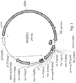
- PVX201 The entire PVX genome has been cloned, in the form of cDNA, in an expression vector for plant cells called plasmid pPVX201 (Baulcombe et al. 1995; figure 3 ; see SEQ ID NO:3 in the appended list of the sequences; see also http://www.plantsci.cam.ac.uk/research/davidbaulcombe/methods/pvx-expression-vectors/sequences).
- the vector consists of: the strong constitutive promoter 35S derived from the cauliflower mosaic virus, placed upstream of the PVX cDNA; and the NOS terminator derived from the nopaline synthase gene of Agrobacterium tumefaciens , placed downstream of the PVX cDNA. These two regions constitute the portions of the vector that regulate the expression of the viral genome inside the plant cell.
- the plasmid also has an origin of bacterial replication and the beta-lactamase gene, which lends it resistance to the antibiotic ampicillin; this is useful for the selection of correctly transformed bacterial clones.
- a single multi-cloning site is inserted upstream of the CP gene for the insertion of additional ORFs, along with a duplicated copy of the subgenomic promoter of the CP, which has the function of directing the expression of the additional ORF inserted by cloning in the MCS.
- PVX has been widely used as an expression vector, in plants, of heterologous proteins, mostly ones of pharmaceutical-biomedicinal interest, but not limited to these.
- the sequence coding for the protein of interest is inserted in the MCS and the recombinant plasmid thus obtained is used for direct inoculation into the leaves of the plant of interest.
- the 35S promoter will start replication of the viral genome with the production of a RNA copy of the viral cDNA and the translation of the first protein, RdRp.
- the viral RNA polymerase will guide the production of subgenomic RNAs and other viral proteins, together with the heterologous protein of interest, which will be produced free inside the cell cytoplasm.
- the sequence of interest can be fused to the coat protein of the virus.
- the Nhel site useful for cloning also came to be eliminated, so the pPVX-CC plasmid was further modified to facilitate subsequent cloning by inserting the single Smal/Xmal restriction site, with a nucleotide sequence CCCGGG, corresponding to the amino acids proline and glycine ( Figure 4c ).
- the nucleotide and protein sequence of the new CP which originates a virus called PVX-Sma (Lico et al. 2006), was thus further modified compared to CP-CC, as is evident from the nucleotide ( Figure 7 ) and protein ( Figure 6 ) alignment.
- the new mutant is stable in subsequent generations and shows normal fitness and symptoms in vivo.
- the pPVX-Sma plasmid was used to insert, at the 5' end of the CP-Sma gene, the sequence coding for the LIP peptide.
- the plasmid thus obtained was then used to produce chimeric viral particles of PVX which expose, as the capsid protein, the fusion protein containing the capsid protein-Sma and LIP peptide.
- the chimeric virus particle of PVX according to the present invention can be advantageously used for the in vitro diagnosis of SS, in particular using the ELISA method.
- fusion protein comprising or consisting of:
- the PVX virus whose capsid protein is used can be selected in the group consisting of PVX X3, BS, EX, NL4, HB, WS2, ROTH1, XS, UK3, OS, NL1, Taiwan, X4, preferably X3.
- the capsid protein used in the fusion protein according to the present invention consists of the following sequence: PGTPATASGLFTIPDGDFFSTARAIVASNAVATNEDLSKIEAIWKDMKVPT DTMAQAAWDLVRHCADVGSSAQTEMIDTGPYSNGISRARLAAAIKEVCT LRQFCMKYAPVVWNWMLTNNSPPANWQAQGFKPEHKFAAFDFFNGVT NPAAIMPKEGLIRPPSEAEMNAAQTAAFVKITKARAQSNDFASLDAAVTR GRITGTTTAEAVVTLPPP (SEQ ID NO:6).
- This protein is obtained by deletion in the 5' portion of the CP gene corresponding to the nucleotides 1-63 coding for aa 1-21 of the CP.
- the fusion protein according to the present invention can have the following sequence:
- the present invention further relates to a polynucleotide which codes for the fusion protein as defined above.
- the polynucleotide can have the following sequence (SEQ ID NO:8):
- the subject matter of the present invention is further represented by an expression vector comprising the polynucleotide as defined above. Moreover, the present invention relates to a host cell comprising the expression vector as defined above.
- the present invention relates to a genome sequence of a PVX virus, said genome sequence comprising the polynucleotide as defined above.
- the present invention relates to a chimeric virus particle of potato virus X, said chimeric virus particle being characterized in that it comprises, as a capsid protein, the fusion protein of the present invention as defined above, said viral particle having exposed, on the outside thereof, the amino-terminal portion comprising or consisting of the antigenic determinant of lipocalin.
- the viral genome contained in the chimeric virus particle consists of the genome sequence as defined above.
- the present invention further relates to the use of the chimeric virus particle according to the present invention, or of the fusion protein as defined above, for the in vitro diagnosis of Sjögren's Syndrome, for example using the ELISA, dipstick or microchip method.
- the subject matter of the present invention further relates to a kit for the diagnosis in vitro of Sjögren's Syndrome, said kit comprising or consisting of the chimeric virus particle as defined above, or in the fusion protein as defined above, possibly in combination with suitable reagents for detection purposes.
- the kit can be a kit ELISA, dipstick or microchip.
- the present invention relates to moreover a plant, preferably belonging to the family of the Solanaceae, including Solanum lycopersicon , Solanum tuberosum , and the plants belonging to the genus Nicotiana, and in particular N. benthamiana, said plant being characterized in that it comprises within it the viral chimeric particle as defined above, or the expression vector as defined above.
- the present invention also relates to plant cells of the plant as defined above and which express the chimeric viral particle as defined above, or the expression vector as defined above.
- the subject matter of the present invention further relates to the use of a peptide sequence comprising or consisting of an antigenic determinant of lipocalin for the preparation of the fusion protein as defined above, the chimeric viral particle as defined above, the plant as defined above, and of plant cells as defined above.
- the present invention relates to the use of the plant as defined above or of the plant cells as defined above, for the production of the chimeric viral particle as defined above.
- the subject matter of the present invention further relates to a process for the preparation of the fusion protein according to the present invention, said process comprising or consisting of the following steps:
- the PVX virus that can be used according to the present invention can be selected in the group consisting of PVX X3, BS, EX, NL4, HB, WS2, ROTH1, XS, UK3, OS, NL1, Taiwan, X4, preferably X3.
- said capsid protein can consist of the following sequence: MPGTPATASGLFTIPDGDFFSTARAIVASNAVATNEDLSKIEAIWKDMKV PTDTMAQAAWDLVRHCADVGSSAQTEMIDTGPYSNGISRARLAAAIKEV CTLRQFCMKYAPVVWNWMLTNNSPPANWQAQGFKPEHKFAAFDFFNG VTNPAAIMPKEGLIRPPSEAEMNAAQTAAFVKITKARAQSNDFASLDAAV TRGRITGTTTAEAVVTLPPP (SEQ ID NO:6).
- the protein is obtained by deletion in the 5' portion of the CP gene corresponding to nucleotides 1-63 coding for the aa 1-21 of the CP.
- the present invention further relates to a process for the preparation of the chimeric viral particle according to the present invention, said process comprising or consisting of the step of replacing the capsid protein of a PVX virus, in said PVX virus, with the fusion protein defined above.
- the process for the production of the viral particle according to the present invention can comprise or consist of the following steps:
- EXAMPLE 1 Preparation of the chimeric viral particle PVX according to the present invention
- the pPVX-Sma plasmid was purified on a small scale and digested with the restriction enzymes Nhel and Xmal to insert the sequence coding for the LIP peptide at the 5' end of the CP-Sma gene.
- This sequence was obtained by in vitro pairing of two complementary synthetic oligonucleotides carrying suitable hemi-restriction sites at their ends, to enable cloning directly in the receiving viral vector, duly digested.
- the protocol provides for the dilution in water of equimolar amounts of the two strands (sense and antisense) and boiling of the solution for 5 minutes; after cooling at room temperature the complementary strands will pair.
- the designed oligonucleotides are the following:
- the sense and antisense oligonucleotides coding for the LIP peptide were designed in such a way as to have the codon usage of Nicotiana benthamiana , the 5' end compatible with the Nhel restriction hemisite and the 3' end compatible with the Xmal hemisite, so that once in vitro pairing had taken place they would be ready for direct cloning in the pPVX-Sma plasmid digested with Nhel-Xmal ( Figure 4c ). Moreover, a serine was inserted immediately after the initial methionine to improve the characteristics of stability of the chimera in vivo.
- the doubly digested pPVX-Sma plasmid was purified from the agarose gel using a commercial kit and the quantity and quality were re-verified in agarose gel and by spectrophotometric analysis.
- the recombinant plasmid obtained is inserted into bacterial cells of the XL1-Blue strain by electroporation and plated in selective LB-agar medium containing ampicillin. The plates are incubated at 37°C and the resulting colonies analysed by PCR with oligonucleotides specific for the nucleotide region of the PVX vector straddling the cloned region ( Figure 4 ):
- the expected band is 370 bases.
- the positive colonies were used for small-scale purification of the plasmid and analysis thereof via sequencing in order to verify the correct presence of the additional LIP sequence.
- the positive clone identified was purified on a large scale and used for a first cycle of infections of plants of N.benthamiama.
- the plants of N. benthamiana were grown in hothouses with a photoperiod of 16 hours of light and 8 hours of darkness, at a constant temperature of 24 °C.
- the light intensity in the hothouses is about 16000 lux.
- a solution containing 20 ⁇ g of plasmid diluted in 50 ⁇ l of water is prepared for the primary infection, whilst for the subsequent infections 50 ⁇ l of crude extract drawn from the infected tissue is used.
- the infection is carried out mechanically on the adaxial (upper) side of the leaf by lightly rubbing with the fingers in the presence both of the prepared solution and silicon carbide, or Carborundum (VWR International, Prolabo).
- the action of this powder causes microabrasions on the leaf surface, enabling the plasmid to enter effectively into the plant cells and express themselves.
- RNA was extracted from the systemically infected leaves by means of the RNeasy plant mini kit (Qiagen) (http://www1.qiagen.com/literature/render.aspx?id 352) and RT-PCR reactions were carried out using the GeneAmp RNA PCR Kit (Perkin Elmer).
- PVX-LIP chimeric PVX
- PVX-wt chimeric PVX
- the plant tissue frozen at -80 °C and mechanically pulverized in a mortar chilled in liquid nitrogen, is mixed in an equivalent volume of phosphate-buffered saline 1X (1X PBS) (10 mM Na2HPO4, 10 mM NaH2PO4, 150 mM NaCl, pH 7.2) and mechanically crushed using an Ultraturrax electric homogenizer brought up to maximum speed. The homogenate must then be centrifuged at about 20000g for 3 minutes.
- 1X PBS phosphate-buffered saline 1X
- the purified virus was dialyzed and concentrated by filtration through a membrane using Vivaspin 2 HS columns (Vivascience, http://teachline.ls.huji.ac.il/72682/Booklets/VIVASPIN_Ultrafiltration_produ cts_II.pdf).
- the purified virus was then analysed through silver-stained polyacrylamide gel to verify the degree of purity of the preparation and the presence of a band with a higher weight than the wt CP due to the presence of the LIP peptide and was quantified by reading with a spectrophotometer at a wavelength of 280 nm considering the molar extinction coefficient ⁇ equal to 1.25 ml/(mg*cm).
- the PVX-LIP and wt PVX viral particles were purified from the plant with a yield of 0.12 mg per gram of fresh tissue.
- the sequence encoding the LIP peptide was cloned in the vector by ligation of the vector itself digested with the enzymes Nhel and Aatll and the sequence encoding the peptide obtained by pairing the primer CPMV-for 5'-CTAGC ACT CCT CCT GCT TTT GAA AAG GCT GCT GGT GCT AGA GGT TTG TCT ACT CCA TTT TCA GACGT - 3' (SEQ ID NO:13) and the primer CPMV-rev 5'- C TGA AAA TGG AGT AGA CAA ACC TCT AGC ACC AGC AGC CTT TTC AAA AGC AGG AGG AGT G - 3'(SEQ ID NO:14).
- the vector thus obtained permits the exposure of the peptide in a BB-BC loop of the small subunit of the coat protein and in this manner the peptide is exposed on its outer surface.
- the vectors pEAQ-HT-VP60-LIP and pEAQ-HT-24K were electroporated in cells of Agrobacterium tumefaciens strain LBA4404 and both used to agroinfiltrate plants of N.benthamiana.
- the bacterial cultures transformed for the two vectors were made to grow separately in a liquid medium containing antibiotic agents, precipitated by centrifugation at 4000 g and resuspended in MMA (10 mM MES pH 5.6, 10 mM MgCl2 and 100 mM acetosyringone) until arriving at an O.D.600 equal to 0.8. Subsequently, after 4 h of incubation at room temperature, equal volumes of the two bacterial suspensions were mixed and used for infiltration, by means of a needleless syringe, of leaves of 4- to 5-week-old plants of N. benthamiana. Four leaves per plant were infiltrated. Samples were then taken of the infiltrated leaves 6 days after infiltration (dpi). For the expression of the empty CPMV the same procedure was carried out with the vectors pEAQ-HT-VP60 and pEAQ-HT-24K.
- the extract was clarified by filtration with two sheets of Miracloth paper (Merck Millipore, 475855-1R) and subsequent centrifugation (30000 g, 1 h, 4°C).
- the supernatant was loaded onto an anion-exchange resin (DEAE SephadexTM A-50, GE Healthcare, 17-0180-01), with a sample:resin ratio of 4:1.
- the flow-through was then concentrated at 4 ml, centrifuged (10000 g X 10 minutes) and loaded into a size-exclusion chromatography column (HiPrep 16/60 Sephacryl S-500 HR, GE Healthcare, 28-9356-06) in 0.1 M sodium phosphate pH 7.0 containing 0.15 M of NaCl at a flow velocity of 0.8 ml/min.
- the eluted fractions were analysed using 12% SDS-PAGE gel stained with silver nitrate.
- the fractions containing CPMV. LIP and CPMV were joined separately and filtered with a 100 kDa centrifugal filter system (Amicon® Ultra-15, Merck Millipore, UFC910024).
- the purified particles were analysed using 12% SDS-PAGE gel stained with silver nitrate and subsequently quantified by reading the absorbance with a spectrophotometer at 280 nm.
- EXAMPLE 2 ELISA diagnostic test using the chimeric viral particle of PVX according to the present invention
- ELISA assays In order to evaluate the bond of the antibodies present in the serum of the patients, ELISA assays (Immunolon II-Dynax and Maxisorp-NUNC) were set up with the LIP peptide produced by chemical synthesis and with PVX.LIP and wild-type CVP.
- the plates were coated with 50 ⁇ l of 40 ⁇ g/ml of synthetic peptide in 1X PBS (2 micrograms of peptide) and with 50 ⁇ l of a 50 ⁇ g/ml solution of wild-type PVX and purified PVX.LIP (2.5 micrograms of chimeric CP corresponding to about 131.6 ng of peptide), similarly diluted in 1X PBS.
- the reference scale was obtained using known concentrations of immunoglobulin pools from donors (Sandoglobulin 50 and 100 ⁇ g/mL).
- the plates were then blocked using 3% BSA in 1X PBS and subsequently incubated with the serum of the subjects belonging to the three above-described experimental groups diluted 1:50 in 1X PBS with 1% BSA for 4 hours at room temperature. After the incubation, a sequence of washes was carried out, once with a solution of 1X PBS and Tween-20 and 3 successive washes in 1X PBS. Subsequently, the plates were incubated at 4°C overnight with a human anti-IgG conjugated with alkaline phosphatase.
- the result of the ELISA test revealed several difficulties of a methodological type.
- the CPMV system was thus excluded from the analysis and considered unsuitable for performing a diagnostic test.
- the sensitivity of the ELISA test conducted using the peptide of the invention on the subgroup of patients with SS characterized by an ANA-negative antibody profile is equal to 98.7%, as compared to a value of 75% obtained with the use of the synthetic peptide.
Landscapes
- Health & Medical Sciences (AREA)
- Life Sciences & Earth Sciences (AREA)
- Chemical & Material Sciences (AREA)
- Organic Chemistry (AREA)
- Genetics & Genomics (AREA)
- Virology (AREA)
- Engineering & Computer Science (AREA)
- Biochemistry (AREA)
- General Health & Medical Sciences (AREA)
- Medicinal Chemistry (AREA)
- Zoology (AREA)
- Molecular Biology (AREA)
- Immunology (AREA)
- Wood Science & Technology (AREA)
- Bioinformatics & Cheminformatics (AREA)
- Biomedical Technology (AREA)
- Biophysics (AREA)
- Gastroenterology & Hepatology (AREA)
- Proteomics, Peptides & Aminoacids (AREA)
- Microbiology (AREA)
- Biotechnology (AREA)
- General Engineering & Computer Science (AREA)
- Urology & Nephrology (AREA)
- Hematology (AREA)
- Toxicology (AREA)
- Food Science & Technology (AREA)
- Physics & Mathematics (AREA)
- Analytical Chemistry (AREA)
- General Physics & Mathematics (AREA)
- Pathology (AREA)
- Cell Biology (AREA)
- Tropical Medicine & Parasitology (AREA)
- Peptides Or Proteins (AREA)
- Micro-Organisms Or Cultivation Processes Thereof (AREA)
Description
- The present invention relates to a chimeric virus particle of potato virus X and use thereof in the in vitro diagnosis of Sjögren's Syndrome.
- In greater detail, the invention relates to a chimeric virus particle of potato virus X, said particle having fused to its capsid protein a lipocalin-derived autoantigenic determinant associated with Sjögren's Syndrome, and the use thereof in the in vitro diagnosis of the Sjögren's Syndrome, for example using the ELISA method.
- Sjögren's Syndrome (SS) is an autoimmune disease with an incidence of 0.6% in the general population and a female:male ratio of 9:1. The disease manifests itself early with involvement of the exocrine glands, above all the salivary and lachrymal glands, resulting in xerostomia and xerophthalmia. The systemic manifestations, characterized by organ involvement at a cutaneous, articular, pulmonary, renal, pancreatic and neurological level are generally less frequent and appear later, although in a percentage ranging between 25 and 92% of cases neurological involvement can represent the initial symptom.
- In consideration of the different visceral and non-visceral organ involvement, SS can manifest itself with extremely different symptoms, and with extremely heterogeneous involvement and degree of disability. Some patients, in fact, can present with an isolated glandular involvement, with xerostomia and xerophthalmia ranging from mild to severe, when the gland insufficiency and loss of the saliva and/or tear secretion function lead to extreme consequences, such as swelling and parotid pain, extreme dryness of the mouth with aphthous ulcers, dysphagia variably associated with oral cavity infections, as well as a major alteration of the visus with photophobia, pain and a condition of blepharitis and/or keratitis which eventually leads to opacification and corneal ulceration. Systemic involvement can be characterized by cutaneous involvement, first of all Raynaud's phenomenon, but also purpura or urticarial vasculitis; musculoskeletal involvement, characterized by arthromyalgia or arthritis, generally symmetrical and rarely of an erosive character; pulmonary involvement, with coughing and/or secondary dyspnea, dry tracheobronchitis or interstitiopathy; or renal involvement ranging from an asymptomatic tubular dysfunction with tubular acidosis to kidney failure with a possible outcome also requiring dialysis treatment. The involvement of the gastrointestinal tract is also variable, with possible autoimmune pancreatitis, or association with autoimmune conditions such as atrophic gastritis, celiac disease, primitive biliary cirrhosis or autoimmune hepatitis, with the consequent associated set of symptoms, as well as neurological involvement, generally tied to sensory or more infrequently motor peripheral neuropathies, with allodynia or hyperalgesia, paresthesias and proprioceptive disorders, or less frequently to central forms, which are, however, associated with a greater degree of disability, as in the case of cerebral vasculitis, cerebellar syndromes, and memory lapses, or even manifest forms of dementia or transverse myelitis.
- As a result of its multi-faceted and variably disabling clinical manifestations, many aspects of the etiopathogenesis of the disease are still unknown, as in the case of a large part of autoimmune disorders, though there is known to be an interaction between genetic susceptibility - in particular as regards both histocompatibility complex class II genes and non-HLA genes - and triggering environmental factors, primarily infectious agents, capable of setting off an inflammatory process which is characterized by immune system auto-aggression against specific target structures, with the autoimmune process subsequently becoming chronic, resulting in tissue damage (Lee et al. 2009).
- Both T lymphocytes and B lymphocytes are involved in the pathogenesis of the SS and in the tissue damage that characterizes it. In particular, Th1, Th2 and Th17 cells as well as B cells are involved in the different stages characterizing the natural history of the pathology, from the pre-clinical stage to the stage of clinical expression with clear and manifest symptoms. In SS, Th1 cells release interferon gamma (IFNy), regulate cell-mediated immunity, and play a role in delaying cell maturation by influencing the organogenesis of salivary glands and the expression of cellular antigens, with a consequent loss of self-tolerance. Th2 cells release interleukin 4 (IL-4), which is fundamental in the early stage of the adaptive immune response, in particular, for isotype switching of immunoglobulin (Ig) and the consequent synthesis of pathogenetic IgG1 autoantibodies (Lee et al. 2009). Th17 cells make up a particular subset of memory CD4 cells, capable of secreting cytokines with a powerful pro-inflammatory activity belonging to the IL 17 family (Bettelli et al. 2008). In view of the complexity of the interactions between cell and cytokine patterns induced by environmental stimuli and involved in the pathogenesis of SS, Lee et al. (2009) have hypothesized that the disease develops in three different successive stages, two of which preclinical, i.e. asymptomatic and silent, and a clinical stage. In the first stage one observes a prevalent involvement of the Th1/IFNγ system characterized by delayed organogenesis of the salivary glands, delayed expression of antigens on the surface of acinar cells, with a consequent loss of the clonal deletion of B and T cells, a loss of self-tolerance and an increased expression of aberrant proteins and cell apoptosis. This is followed, in the second preclinical stage (i.e. silent for the patient and mediated by Th2 and Th17 lymphocytes and B lymphocytes) by an increased expression of numerous epitopes at the glandular level, with the appearance of a major leukocyte infiltration, the triggering of an autoreactive local immune response and the onset of active autoantibody synthesis. The effects of these phenomena culminate in the third stage, in which the autoimmune process gives rise to clinically evident functional damage, which is expressed through a reduction in exocrine secretion from the gland tissue involved, with the consequent clinical symptoms.
- Multiple autoantibodies are produced during the course of SS: some of them have a diagnostic value, others are predictive of possible organ damage, and still others have a prognostic or unclear value. ANA positivity, mostly with a speckled pattern, anti-ENA Ro/SSA and La/SSB, anti-salivary duct cell antibodies, anti-thyroid antibodies, anti-gastric parietal cell antibodies and antimitochondrial antibodies frequently occur. High titles of anti-α-fodrin,
anti-muscarinic receptors type 3, anti-carbonic anhydrase II and anti-proteasome antibodies have also been described. Recently, some authors have also described high titles of anti-lipocalin antibodies (Navone et al. 2005), lipocalin being a protein that demonstrates a high sequence homology with α-fodrin as well as with the early protein D of EBV (Epstein-Barr Virus), which seems to confirm it to be a potential environmental agent triggering the disease in genetically predisposed subjects. Given the close association between EBV and SS, also in relation to the high sequence homology that has been demonstrated between a protein epitope of that virus and two autoantigens of relevance in the disease itself, it is interesting to evaluate their pathogenetic role and diagnostic potential. - Within the field of autoimmune diseases, due to the complex, variegated array of symptoms, as well as the presence of different laboratory and/or instrumental data, the diagnosis is often based on a global assessment of the situation and the criteria for classifying the disease are often used to support the diagnosis itself. Classification criteria in fact represent objective diagnostic parameters, which are fundamental to assure the standardization of patients who are included in clinical studies, facilitate the analysis and render the results of different clinical studies comparable, as well as enabling a comparison among patients referred to different clinical facilities.
- With regard to the diagnosis of SS, the classification criteria still most widely used today are the AECG (American European Consensus Group) criteria of 2002 (Vitali et al. 2002), which are based on an assessment of 6 phenomena, namely: I) ocular symptoms (symptoms of dry eyes for over 3 months or use of artificial tears for over 3 months); II) oral symptoms (dry mouth for at least 3 months or recurrent or persistently swollen salivary glands or need for liquids to swallow solid foods); III) ocular signs (abnormal Shirmer's test or Rose Bengal/fluorescein staining); IV) histopathological characteristics with a focus score ≥1 on biopsy of the minor salivary glands; V) reduction in salivation, documented by salivary flow measurement or indirectly with parotid scialography or salivary gland scintigraphy; and VI) presence of autoantibodies (anti-ENA SSA/Ro and/or SSB/La positivity).
- Based on AECG criteria, in order to formulate a diagnosis of SS it is necessary for at least 4 of the 6 criteria listed above to be met, including necessarily the histopathological criterion or presence of autoantibodies, and at least 3 of the 4 objective criteria. Given the large differences between the two procedures (biopsy and serological analysis) in terms of cost and invasiveness, in clinical practice, where the other criteria established by the AECG (dry eye and dry mouth, appropriately measured) are met, a serological analysis is performed first and only in the event that the latter is negative will a salivary gland biopsy follow.
- In 2012, new ACR (American College of Rheumatology) classification criteria were introduced (Shiboski et al. 2012), which, compared to the AECG criteria, introduce a simplification of the procedure, it being necessary for only 2 of the 3 objective criteria identified to be met. The criteria to be met are: I) ocular signs (fluorescein staining with a score ≥3); II) histopatological characteristics with a focus score ≥1 on minor salivary gland biopsy; III) presence of autoantibodies (positive anti-ENA SSA/Ro and/or SSB/La or alternatively positive rheumatoid factor or ANA with a title ≥1:320).
- Unlike the AECG criteria, the ACR criteria do not take into consideration either the subjective symptoms of the patient (presence of xerostomia/xerophthalmia) or functional assessments regarding the major salivary glands (salivary flow measurement, scialography or scintigraphy), but only an evaluation of ocular involvement with a test of a qualitative type and the presence of a broader spectrum of autoantibodies, in particular ANA, which is positive in a percentage of patients ranging from 60 to 85% of the cases (Nardi et al. 2006), as compared to what has been documented for anti-ENA SSA/Ro or anti-ENA SSB/La, equal, respectively, to 52-67% or 49% (Routsias et al. 2007). These criteria no doubt enable the recognition of subjects with SS at an earlier stage; specifically, they also enable the subset of subjects negative for anti-ENA SSA/Ro or anti-ENA SSB/La (about 50% of affected subjects) to be diagnosed with the disease. However, they have not yet been validated in a large number of European case studies and, moreover, the AECG criteria of 2002, though more stringent, still demonstrate higher specificity (AECG criteria Sp 90.4% versus ACR criteria Sp 83.5%; Cornec et al. 2014) and are therefore still more widely used.
- For the sake of completeness, given that the diagnostic criteria, as opposed to the classification criteria, also depend on information derived from the patient's medical history, a global assessment of the patient's laboratory and instrumental data, as well as the clinician's experience, it is worth noting the existence of a subgroup of patients who are negative for autoantibodies (i.e. ANA, anti-ENA SSA/Ro and anti-ENA SSB/La negative), but have strongly suggestive overall clinical, laboratory and instrumental data and a salivary gland histology which support the diagnosis of the pathology. In particular, for this subset of patients, it is essential to find new diagnostic markers which may serve to identify also the affected individuals in whom the diagnosis has been ruled out due to the absence of the currently available markers. It has recently been proposed to assay anti-α-fodrin antibodies as markers of the pathology: until now, the usefulness of this assay has been confirmed only as validation of the diagnosis in the presence of anti-ENA SSA/Ro or anti-ENA SSB/La (Qin et al. 2014).
- With regard to new diagnostic markers for the diagnosis of SS, the presence of autoantibodies against human tear lipocalin has recently been demonstrated in the serum of subjects suffering from SS.
- Tear lipocalin, together with lysozyme and lactoferrin, is the protein most largely represented in human tears, but it is also produced by von Ebner's glands, in the nasal mucosa, in the tracheobronchial mucosa and at the level of the prostate. It belongs to the family of lipocalins, low-molecular-weight proteins characterized by an ability to bind small hydrophobic molecules, complex with other soluble macromolecules to form macro complexes, and bind to specific receptors exposed on the cell surface to mediate the transduction and internalization of a signal. Tear lipocalin binds various ligands, including phospholipids, fatty acids, alcohols, glycolipids and cholesterol, and has a function that is closely associated with the role of its ligands. It prevents tear evaporation by binding fatty acids and phospholipids, maintains corneal integrity through a barrier function, again by virtue of the bond with the lipid components of tears, and performs an anti-microbial action, in particular an anti-fungal one (Gachon et al. 1998). Lipocalin also has the function of an enzyme and enzymatic inhibitor; it represents, in fact, the main tear endonuclease and has an inhibitory activity on cysteine-proteinase (Glasgow et al. 2011). A reduction in tear lipocalin has been suggested in all diseases with reduced tear secretion; moreover, recent studies have demonstrated that the concentration thereof is significantly reduced during the course of SS, whereas in the case of dry-eye syndrome in the post-menopausal age, as well as in situations of keratoconjunctivitis sicca not associated with SS, the concentration of tear lipocalin shows to be substantially comparable to that present in the tears of control subjects (Caffery et al. 2008).
- As noted above, the presence of autoantibodies against human tear lipocalin in the serum of subjects suffering from SS has recently been demonstrated (Navone et al. 2005), confirming the close association between tear lipocalin and the pathology. Titles of these antibodies are not detectable in the serum of control subjects or patients with other autoimmune pathologies such as rheumatoid arthritis or LES, which confirms the selective link between the protein itself and the disease.
- The recognition of lipocalin as a potential autoantigen for SS was based on the screening of a peptide library, with the use of random peptide sequences tested with the serum of patients suffering from the pathology of interest, SS in this specific case (Navone et al. 2005). This method served to reveal a synthetic dodecapeptide sequence, called Sjögren's peptide (GDRDAGSRGLVS (SEQ ID NO:1)), with a high sequence homology with tear lipocalin (FEKAAGARGLST (SEQ ID NO:2) and α-fodrin, as well as with the D protein antigen of EBV. This homology was validated both by means of an ELISA test with affinity purified antibodies from the serum of patients with SS, and using the immunoprecipitation of lipocalin from a cell lysate and tears, respectively tested with purified antibodies from patients and commercial anti-lipocalin antibodies. It was thus hypothesized that, in genetically predisposed subjects, via a mechanism of molecular mimicry, EBV infection or reactivation of EBV infection may induce an increase in the production of α-fodrin degradation fragments, as an effect of the immune response against the EBV early antigen D, with a consequent alteration of this cytoskeletal protein, already known as an important autoantigen during the course of SS (Qin et al. 2014). The anti-EBV D antigen antibodies also appear to bind tear lipocalin, leading to a functional modification thereof, as well as a reduction in gland secretion, with obvious clinical consequences.
- ELISA (Enzyme-Linked Immunosorbent Assay) is a method which makes it possible to ascertain the presence of antibodies specific for a given antigen in the serum of patients suffering from the pathology of interest. In the case of SS, identifying an autoantigen sensitive and specific for the disease is of considerable interest, as there is undoubtedly a portion of patients who are diagnosed late or not diagnosed with the disease based on negative results for anti-ENA SSA/Ro or anti-ENA SSB/La or, using the more recent ACR criteria, ANA. From this perspective, as mentioned above, tear lipocalin demonstrates to be a molecule of considerable interest, given both the close association the molecule has with the pathology (Caffery et al. 2008) and its demonstrated pathogenetic role in triggering the disease following an EBV infection or reactivation of EBV infection (Navone et al. 2005). As previously described, the lipocalin peptide (LIP peptide) obtained by chemical synthesis has already been used to test sera obtained from control subjects and subjects with autoimmune diseases, including SS, rheumatoid arthritis, LES and scleroderma, precisely with the aim of validating its diagnostic sensitivity and specificity, and it has demonstrated to be a potential autoantigen of significant diagnostic value.
- However, an ELISA based on the use solely of the peptide obtained by chemical synthesis is not completely reliable, as the small dimensions and physicochemical characteristics of the peptide itself cannot guarantee adhesion to the polystyrene of the plate used for the test, impairing its reproducibility and effectiveness. In particular, notwithstanding the use of highly specific polystyrene plates for binding small peptides, the result obtained with experiments in triplicate has not always proven to be reproducible and, in the case of some lots of plates, not usable, given that the data obtained from the reaction between the synthetic LIP peptide and the serum showed a lower value than the absorbance of the substrate alone, used as the internal experimental control of the system (the so-called reference blank).
- In light of the above, there is an evident need to be able to have new methods for diagnosing SS which can overcome the disadvantages of the known methods.
- The potato virus X (PVX) belongs to the family Alphaflexiviridae and genus Potexvirus. It is a filamentous and flexible rod-like virus, with an average length of 500nm and a diameter of about 13nm. Its genome consists of a positive-sense single-stranded RNA molecule 6435 bases long (for the strain X3, which is the one of reference in this case; Accession Number D00344) and codes for 5 different proteins: an RNA-dependent RNA polymerase (RdRp, replicase), three viral movement proteins, identified as p8, p12 and p25 (or also proteins of the Triple Gene Block, TGBp1, TGBp2 and TGBp3, respectively) and the viral coat protein (CP) (
Figure 1 ). With the exception of the replicase gene, the expression of the open reading frames (ORFs) is regulated by the activity of several specific subgenomic promoters. The genome is 5' capped (i.e. it has a methylguanosine cap, m7GpppG) and 3' polyadenylated (it has a polyA tail). The completely assembled virion consists of a molecule of RNA totally surrounded by about 1300 identical copies of CP, which are associated to form a spiral around the viral nucleic acid with a progressive process. This is why in vivo, during replication inside the plant cell, virions of different lengths corresponding to the different stages of progress in the assembly can be identified. During purification, however, the protocol adopted makes it possible to obtain a preparation of particles that are homogeneous in length and have an expected size of 500nm. The host of choice of the virus is the potato plant, Solanum tuberosum, but PVX is capable of replicating and infecting plants belonging to the family Solanaceae in general, including plants belonging to the genus Nicotiana, and in particular N. benthamiana, an amphitetraploid herbaceous plant (2n=4x=38) native to western Australia and by now widely used as a model plant in plant virology experiments and in the transient expression of heterologous proteins. - The virus travels from the inoculation site via a cell-to-cell movement; then it reaches the phloem vessels, through which it moves into the leaves of the phytomers above the ones where inoculation took place. The typical symptoms it produces are chlorotic and then necrotic spots corresponding to the individual loci of infection on inoculated leaves, and a diffuse chlorotic mosaic accompanied by leafroll in systemically infected leaves.
- An approach commonly used in molecular farming to produce peptides is to genetically fuse the sequence of interest to the gene encoding the CP of plant viruses. In the case of PVX in particular, this takes place by fusion at the 5' of the gene, i.e. the N-terminal end of the corresponding protein. In this manner, when the CP is translated it will be chimeric, i.e. bear an additional portion, and when assembled in the mature virion it will produce chimeric virus particles (CVPs) which will expose the heterologous portion on the surface of the virus (
Figure 2b ). - Following the viral infection, samples of the symptomatic systemic tissue are taken and the CVPs are purified and used in the planned experiments.
- The entire PVX genome has been cloned, in the form of cDNA, in an expression vector for plant cells called plasmid pPVX201 (Baulcombe et al. 1995;
figure 3 ; see SEQ ID NO:3 in the appended list of the sequences; see also http://www.plantsci.cam.ac.uk/research/davidbaulcombe/methods/pvx-expression-vectors/sequences). - The vector consists of: the strong
constitutive promoter 35S derived from the cauliflower mosaic virus, placed upstream of the PVX cDNA; and the NOS terminator derived from the nopaline synthase gene of Agrobacterium tumefaciens, placed downstream of the PVX cDNA. These two regions constitute the portions of the vector that regulate the expression of the viral genome inside the plant cell. Among its most relevant elements, the plasmid also has an origin of bacterial replication and the beta-lactamase gene, which lends it resistance to the antibiotic ampicillin; this is useful for the selection of correctly transformed bacterial clones. In this vector, moreover, a single multi-cloning site (MCS) is inserted upstream of the CP gene for the insertion of additional ORFs, along with a duplicated copy of the subgenomic promoter of the CP, which has the function of directing the expression of the additional ORF inserted by cloning in the MCS. - Thanks to this plasmid, which enables the viral genome to be easily modified, PVX has been widely used as an expression vector, in plants, of heterologous proteins, mostly ones of pharmaceutical-biomedicinal interest, but not limited to these. In this approach, the sequence coding for the protein of interest is inserted in the MCS and the recombinant plasmid thus obtained is used for direct inoculation into the leaves of the plant of interest. Inside its cells, the 35S promoter will start replication of the viral genome with the production of a RNA copy of the viral cDNA and the translation of the first protein, RdRp. From this moment, the viral RNA polymerase will guide the production of subgenomic RNAs and other viral proteins, together with the heterologous protein of interest, which will be produced free inside the cell cytoplasm. Alternatively, with another procedure, described in detail below, the sequence of interest can be fused to the coat protein of the virus.
- With the above-mentioned method, a chimeric PVX bearing on its surface the 2F5 peptide (ELDKWAS (SEQ ID NO:4)) derived from the coat protein gp41 of the HIV-1 virus was produced in the ENEA laboratory for the production of a vaccine (Marusic et al. 2001 and patent application RM2000A000327).
- During the subsequent cloning of other peptides of immunological interest, a spontaneous mutant deriving from one of these new chimeras was identified in vivo. The virus had rearranged itself in such a way as to eliminate the additional heterologous sequence, which evidently destabilized it. During this process, however, it also lost a region consisting of the 5' portion of the CP gene, corresponding to the 63 nucleotides coding for the first 21 aa of the CP, and its subgenomic promoter (
Figure 4b ). The nucleotide rearrangement is such as to reestablish the ATG of the CP. This appears evident from the nucleotide (Figure 5 ) and protein (Figure 6 ) alignment of the wild-type (wt) CP and mutant. Notwithstanding this major deletion, the mutant CP gave rise to a stable virus in subsequent generations, and it was thus hypothesized that the mutant (called PVX-CC) could be used for in-fusion cloning with much longer CP sequences than those supported by the wt CP. The sequence of this mutant was therefore stably cloned in the pPVX201 vector, as a replacement for the wt CP, giving rise to the pPVX-CC plasmid. In this operation, the Nhel site useful for cloning also came to be eliminated, so the pPVX-CC plasmid was further modified to facilitate subsequent cloning by inserting the single Smal/Xmal restriction site, with a nucleotide sequence CCCGGG, corresponding to the amino acids proline and glycine (Figure 4c ). The nucleotide and protein sequence of the new CP, which originates a virus called PVX-Sma (Lico et al. 2006), was thus further modified compared to CP-CC, as is evident from the nucleotide (Figure 7 ) and protein (Figure 6 ) alignment. The new mutant is stable in subsequent generations and shows normal fitness and symptoms in vivo. - From the viewpoint of its application, the use of the mutant for cloning a broad-spectrum antimicrobial peptide (Donini et al. 2005) and a peptide deriving from the influenza virus for a new vaccine formulation (Lico et al. 2009) is known.
- According to the present invention, the pPVX-Sma plasmid was used to insert, at the 5' end of the CP-Sma gene, the sequence coding for the LIP peptide. The plasmid thus obtained was then used to produce chimeric viral particles of PVX which expose, as the capsid protein, the fusion protein containing the capsid protein-Sma and LIP peptide.
- It was found that the chimeric virus particle of PVX according to the present invention can be advantageously used for the in vitro diagnosis of SS, in particular using the ELISA method.
- A comparison between the results of the ELISA using the synthetic peptide and the peptide mounted on the PVX viral scaffold showed a sensitivity, i.e. the ability to identify patients affected by the disease, of 84.7% and 97.1%, respectively; whereas the specificity, i.e. the ability to correctly identify healthy subjects is equal to 90.0% in both systems.
- The analysis conducted on a subgroup of patients with SS who were characterized by an ANA-negative antibody profile and recognized as having the disease based on the presence of histopathological evidence drawn from the salivary gland biopsy used to diagnose the disease was of particular interest. In these patients the ELISA test performed with the chimeric viral particles according to the present invention demonstrated a greater sensitivity than the same ELISA test with the synthetic peptide, i.e. 98.7% versus 75%. The data for ANA-negative subjects is of particular interest, since in this subgroup of patients the evidence of a positive test for this new autoantigen would make it possible to avoid relying on a salivary gland biopsy in order to achieve diagnostic certainty, with considerable savings in terms of both costs and times (time for scheduling the biopsy and subsequent wait for the histological test), as well as reduced patient discomfort.
- The subject matter of the present invention thus specifically relates to a fusion protein comprising or consisting of:
- an amino-terminal portion consisting of a peptide sequence comprising or consisting of an antigenic determinant of lipocalin, wherein said antigenic determinant of lipocalin can be selected in the group which consists of FEKAAGARGLST (SEQ ID NO:2), MSFEKAAGARGLST (SEQ ID NO:5); and
- a carboxy-terminal portion consisting of a peptide sequence comprising or consisting of the capsid protein of a PVX virus, said capsid protein being intact or deleted in the 5' portion of the gene encodes the wild-type capsid protein, said amino-terminal portion being fused to said carboxy-terminal portion in such a way that said antigenic determinant is in frame with said capsid protein.
- The PVX virus whose capsid protein is used can be selected in the group consisting of PVX X3, BS, EX, NL4, HB, WS2, ROTH1, XS, UK3, OS, NL1, Taiwan, X4, preferably X3.
- According to a particular embodiment of the present invention, the capsid protein used in the fusion protein according to the present invention consists of the following sequence:
PGTPATASGLFTIPDGDFFSTARAIVASNAVATNEDLSKIEAIWKDMKVPT DTMAQAAWDLVRHCADVGSSAQTEMIDTGPYSNGISRARLAAAIKEVCT LRQFCMKYAPVVWNWMLTNNSPPANWQAQGFKPEHKFAAFDFFNGVT NPAAIMPKEGLIRPPSEAEMNAAQTAAFVKITKARAQSNDFASLDAAVTR GRITGTTTAEAVVTLPPP (SEQ ID NO:6). This protein is obtained by deletion in the 5' portion of the CP gene corresponding to the nucleotides 1-63 coding for aa 1-21 of the CP. - In particular, the fusion protein according to the present invention can have the following sequence:


- The present invention further relates to a polynucleotide which codes for the fusion protein as defined above. In particular, the polynucleotide can have the following sequence (SEQ ID NO:8):

- The subject matter of the present invention is further represented by an expression vector comprising the polynucleotide as defined above. Moreover, the present invention relates to a host cell comprising the expression vector as defined above.
- According to a further embodiment, the present invention relates to a genome sequence of a PVX virus, said genome sequence comprising the polynucleotide as defined above.
- Moreover, the present invention relates to a chimeric virus particle of potato virus X, said chimeric virus particle being characterized in that it comprises, as a capsid protein, the fusion protein of the present invention as defined above, said viral particle having exposed, on the outside thereof, the amino-terminal portion comprising or consisting of the antigenic determinant of lipocalin.
- The viral genome contained in the chimeric virus particle consists of the genome sequence as defined above.
- The present invention further relates to the use of the chimeric virus particle according to the present invention, or of the fusion protein as defined above, for the in vitro diagnosis of Sjögren's Syndrome, for example using the ELISA, dipstick or microchip method.
- Therefore, the subject matter of the present invention further relates to a kit for the diagnosis in vitro of Sjögren's Syndrome, said kit comprising or consisting of the chimeric virus particle as defined above, or in the fusion protein as defined above, possibly in combination with suitable reagents for detection purposes. For example, the kit can be a kit ELISA, dipstick or microchip.
- The present invention relates to moreover a plant, preferably belonging to the family of the Solanaceae, including Solanum lycopersicon, Solanum tuberosum, and the plants belonging to the genus Nicotiana, and in particular N. benthamiana, said plant being characterized in that it comprises within it the viral chimeric particle as defined above, or the expression vector as defined above.
- The present invention also relates to plant cells of the plant as defined above and which express the chimeric viral particle as defined above, or the expression vector as defined above.
- The subject matter of the present invention further relates to the use of a peptide sequence comprising or consisting of an antigenic determinant of lipocalin for the preparation of the fusion protein as defined above, the chimeric viral particle as defined above, the plant as defined above, and of plant cells as defined above.
- Moreover, the present invention relates to the use of the plant as defined above or of the plant cells as defined above, for the production of the chimeric viral particle as defined above.
- The subject matter of the present invention further relates to a process for the preparation of the fusion protein according to the present invention, said process comprising or consisting of the following steps:
- a) cloning, in a vector, the peptide sequence comprising or consisting in the capsid protein of a PVX virus, said capsid protein being intact or deleted in the 5' portion of the gene which encodes the wild-type capsid protein; and
- b) cloning a peptide sequence comprising or consisting in an antigenic determinant of lipocalin, said peptide sequence being fused to said peptide sequence comprising or consisting in the capsid protein of a PVX virus in the vector resulting from step a), wherein said antigenic determinant of lipocalin can be selected in the group which consists of FEKAAGARGLST (SEQ ID NO:2), MSFEKAAGARGLST (SEQ ID NO:5).
- The PVX virus that can be used according to the present invention can be selected in the group consisting of PVX X3, BS, EX, NL4, HB, WS2, ROTH1, XS, UK3, OS, NL1, Taiwan, X4, preferably X3.
- According to a particular embodiment, said capsid protein can consist of the following sequence:
MPGTPATASGLFTIPDGDFFSTARAIVASNAVATNEDLSKIEAIWKDMKV PTDTMAQAAWDLVRHCADVGSSAQTEMIDTGPYSNGISRARLAAAIKEV CTLRQFCMKYAPVVWNWMLTNNSPPANWQAQGFKPEHKFAAFDFFNG VTNPAAIMPKEGLIRPPSEAEMNAAQTAAFVKITKARAQSNDFASLDAAV TRGRITGTTTAEAVVTLPPP (SEQ ID NO:6). The protein is obtained by deletion in the 5' portion of the CP gene corresponding to nucleotides 1-63 coding for the aa 1-21 of the CP. - The present invention further relates to a process for the preparation of the chimeric viral particle according to the present invention, said process comprising or consisting of the step of replacing the capsid protein of a PVX virus, in said PVX virus, with the fusion protein defined above.
- The process for the production of the viral particle according to the present invention can comprise or consist of the following steps:
- a1) infecting a plant, preferably belonging to the family Solanaceae, including Solanum lycopersicon, Solanum tuberosum, and plants belonging to the genus Nicotiana, and in particular N. benthamiana and N. tabacum, with the viral particle as defined above or the expression vector as defined above;
- b1) cultivating the plant obtained according to the step a1);
- c) extracting said viral particle from said plant.
- The present invention will now be described, by way of illustration according to a preferred embodiment thereof, with particular reference to the figures of the appended drawings, in which:
-
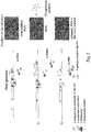
figure 1 shows a diagram representation of the genome of the potato virus X; RdRp, RNA-dependent RNA polymerase; MT, methyltransferase domain; HEL, helicase domain; POL, polymerase domain; 25K/ 12K/ 8K, movement proteins; CP, capsid protein; SGP, subgenomic promoter; sgRNA, subgenomic RNA; 7-methylguanine cap; terminal cap 5'; Poly(A): 3' terminal poly(A) tail. -
Figure 2 shows a schematic representation of how the viral genome can be modified and how a chimeric virus particle (CVP) is constructed. A) modification by gene insertion: the nucleotide sequence encoding the protein of interest is added to the viral genome with a duplication of the subgenomic promoter of the CP. B) modification by gene fusion: the nucleotide sequence encoding the protein/peptide of interest is fused to the 5' portion of the CP gene (N-terminal portion of the protein).
R, viral replicase; M, movement proteins; CP, coat protein, capsid protein; arrow, viral subgenomic promoter. -
Figure 3 shows a schematic representation of the plasmid pPVX201 containing the genome of the PVX subform of cDNA, 35S, promoter derived from CaMV; 5'UTR: 5' untranslated region; RdRp, RNA-dependent viral replicase; p8/p12/p25, viral movement proteins; prom sg CP, subgenomic promoter of the CP; prom sg2CP, duplicate subgenomic promoter of the CP; CP, coat protein; 3'UTR: 3' untranslated region; NOS term, terminator derived from the nopaline synthase gene of A.tumefaciens; beta-lactamase: gene coding for the beta-lactamase. The main restriction sites and their corresponding nucleotide position on the vector are indicated. -
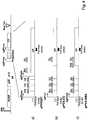
Figure 4 shows a schematic representation of the plasmid containing the PVX genome in the form of cDNA, both the wild-type and mutant variants with the deletion at 5' of the CP gene. The subsequent panels show the region undergoing modification in detail, with the main restriction sites, the two oligonucleotides (PVX Back and New) used in the PCR reactions and their respective nucleotide positions. A) pPVX201: the plasmid containing the wild genome of PVX (it originates wild-type viral particles, wt PVX). B) pPVXCC: the plasmid derived from pPVX201 containing the mutant with the N-terminal deletion of the CP. C) pPVX-Sma: the plasmid derived from pPVX-CC and further modified and used in this patent to construct the chimeric viral particles PVX-LIP. -
Figure 5 shows the nucleotide alignment between the CP gene of wt PVX (SEQ ID NO: 16) and that of the deleted CP of PVX-CC (SEQ ID NO: 15). -
Figure 6 shows the protein alignment between the CP of wt PVX (SEQ ID NO: 17), that of the deleted one of PVX-CC (SEQ ID NO: 18) and the deleted and mutated one of PVX-Sma (SEQ ID NO: 19). -
Figure 7 shows the nucleotide alignment between the deleted CP gene of PVX-CC (SEQ ID NO: 15) and that of the deleted and mutated CP of PVX-Sma (SEQ ID NO: 20). - The pPVX-Sma plasmid was purified on a small scale and digested with the restriction enzymes Nhel and Xmal to insert the sequence coding for the LIP peptide at the 5' end of the CP-Sma gene.
- This sequence was obtained by in vitro pairing of two complementary synthetic oligonucleotides carrying suitable hemi-restriction sites at their ends, to enable cloning directly in the receiving viral vector, duly digested.
- The protocol provides for the dilution in water of equimolar amounts of the two strands (sense and antisense) and boiling of the solution for 5 minutes; after cooling at room temperature the complementary strands will pair.
- The designed oligonucleotides are the following:
- sense LIP: 5' - CTAGC CTCGAG ATG TCT TTT GAA AAG GCT GCT GGT GCT AGA GGT TTG TCT ACT C - 3' (SEQ ID NO:9)
- antisense LIP: 5' - CCGGG AGT AGA CAA ACC TCT AGC ACC AGC AGC CTT TTC AAA AGA CAT CTCGAG G - 3'(SEQ ID NO:10)
- The sense and antisense oligonucleotides coding for the LIP peptide were designed in such a way as to have the codon usage of Nicotiana benthamiana, the 5' end compatible with the Nhel restriction hemisite and the 3' end compatible with the Xmal hemisite, so that once in vitro pairing had taken place they would be ready for direct cloning in the pPVX-Sma plasmid digested with Nhel-Xmal (
Figure 4c ). Moreover, a serine was inserted immediately after the initial methionine to improve the characteristics of stability of the chimera in vivo. - The doubly digested pPVX-Sma plasmid was purified from the agarose gel using a commercial kit and the quantity and quality were re-verified in agarose gel and by spectrophotometric analysis.
- On completion of the ligation reaction between the digested and purified vector and the oligonucleotides paired in vitro, the recombinant plasmid obtained is inserted into bacterial cells of the XL1-Blue strain by electroporation and plated in selective LB-agar medium containing ampicillin. The plates are incubated at 37°C and the resulting colonies analysed by PCR with oligonucleotides specific for the nucleotide region of the PVX vector straddling the cloned region (
Figure 4 ): - 5' back 5' AGCAGTCATTAGCACTTC 3' (SEQ ID NO:11)
- 3' new 5' CACCTTCATGTCCTTCCA 3' (SEQ ID NO:12)
- The expected band is 370 bases. The positive colonies were used for small-scale purification of the plasmid and analysis thereof via sequencing in order to verify the correct presence of the additional LIP sequence. The positive clone identified was purified on a large scale and used for a first cycle of infections of plants of N.benthamiama. The plants of N. benthamiana were grown in hothouses with a photoperiod of 16 hours of light and 8 hours of darkness, at a constant temperature of 24 °C. The light intensity in the hothouses is about 16000 lux.
- For every leaf of N. benthamiana to be infected (2 leaves per plant) a solution containing 20 µg of plasmid diluted in 50 µl of water is prepared for the primary infection, whilst for the subsequent infections 50 µl of crude extract drawn from the infected tissue is used. The infection is carried out mechanically on the adaxial (upper) side of the leaf by lightly rubbing with the fingers in the presence both of the prepared solution and silicon carbide, or Carborundum (VWR International, Prolabo). The action of this powder causes microabrasions on the leaf surface, enabling the plasmid to enter effectively into the plant cells and express themselves.
- After the onset of systemic symptoms in a time and manner similar to PVX-wt was verified, the tissue was used to:
- 1. extract the viral RNA, carry out a reverse transcription to DNA (RT) and a PCR, sequence the PCR fragment and verify the correct presence of the LIP sequence at the nucleotide level;
- 2. derive a crude protein extract with which to repeat a cycle of infection in vivo.
- The entire process was then repeated another time to determine the stability of the chimeric PVX for LIP over three generations.
- In order to determine the stability of the chimeric PVX, between 6 and 8 days after the infection, RNA was extracted from the systemically infected leaves by means of the RNeasy plant mini kit (Qiagen) (http://www1.qiagen.com/literature/render.aspx?id=352) and RT-PCR reactions were carried out using the GeneAmp RNA PCR Kit (Perkin Elmer). The cDNA was synthesized with oligo d(T) and PCR was performed using the 5' back and 3' new oligonucleotides. The cDNA thus obtained was sequenced and the presence of the correct sequence coding for the LIP peptide was verified.
- Then followed a large-scale infection with purification of the chimeric PVX (PVX-LIP) and PVX devoid of the LIP peptide (PVX-wt) as a control.
- For the purpose of protein extraction, the plant tissue, frozen at -80 °C and mechanically pulverized in a mortar chilled in liquid nitrogen, is mixed in an equivalent volume of phosphate-buffered saline 1X (1X PBS) (10 mM Na2HPO4, 10 mM NaH2PO4, 150 mM NaCl, pH 7.2) and mechanically crushed using an Ultraturrax electric homogenizer brought up to maximum speed. The homogenate must then be centrifuged at about 20000g for 3 minutes.
- For the purification of the chimeric viral particles, the following protocol is used.
- Homogenize 50 g of tissue stored at -80 °C and pulverized with a mortar and pestle in 2 volumes of cold boric acid 0.5 M pH 7.8 (about 200-300 ml); filter the mixture through 3 layers of sterile gauze and adjust the pH to 6.5 with HCl. Then add 0.2% (w/v) of ascorbic acid and 0.2% (w/v) of sodium sulphite and leave under stirring until completely dissolved. Remove the particulate by centrifuging at 5500g for 20 minutes at 4 °C and leave the supernatant a room temperature for 3 hours after having added 0.15 volumes of a 0.5% silver nitrate solution. After repeating the centrifugation at 5500g for 20 minutes at 4 °C, allow the supernatant to settle overnight at 4 °C in 0.2 volumes of a solution containing 1M NaCl and 20% PEG in boric acid 0.5 M pH 7.8. The next day, precipitate the viral particles by centrifuging at 8000g for 30 minutes at 4 °C, and resuspend the pellet in 10 ml of a solution containing boric acid 0.5 M, urea 0.5 M, 0.1% 2-mercaptoethanol, pH 7.8; centrifuge the mixture at 8000g for 30 minutes at 4 °C. Load the supernatant containing the virus on a 30% sucrose cushion prepared in water and centrifuge at 72500g for 2 hours and 30 minutes at 4 °C. Finally, resuspend the pellet in 1 ml of boric acid 0.5 M pH 7.8 and, after two hours of settling, centrifuge at 6000g for 15 minutes at 4 °C. The supernatant containing the virus is then loaded onto a 10-45% sucrose gradient prepared in boric acid and centrifuged at 90000g for 1 hour. The individual fractions of the gradient are then analysed to identify those with the highest content of viral particles. The protocol is drawn from Udhe et al. 2005. The purified virus was dialyzed and concentrated by filtration through a
membrane using Vivaspin 2 HS columns (Vivascience, http://teachline.ls.huji.ac.il/72682/Booklets/VIVASPIN_Ultrafiltration_produ cts_II.pdf). - The purified virus was then analysed through silver-stained polyacrylamide gel to verify the degree of purity of the preparation and the presence of a band with a higher weight than the wt CP due to the presence of the LIP peptide and was quantified by reading with a spectrophotometer at a wavelength of 280 nm considering the molar extinction coefficient ε equal to 1.25 ml/(mg*cm).
- An LAL assay was also performed (following the protocol provided by pbi International); it verified the presence of endotoxins in the preparations described, presumably due to an "environmental" contamination given by the laboratory instrumentation. An EndoTrap red1/1 column (Hyglos) was used to remove the endotoxins, following the manufacturer's instructions.
- The PVX-LIP and wt PVX viral particles were purified from the plant with a yield of 0.12 mg per gram of fresh tissue.
- For the production of chimeric particles derived from the CPMV (Cowpea Mosaic Virus) which expose the LIP peptide, use was made of the pEAQ-HT system, in which the precursor of the coat protein (VP60) and the 24K protease are separately introduced in two pEAQ constructs to produce particles similar to empty viruses (devoid of genetic material). The sequence encoding the LIP peptide was cloned in the vector by ligation of the vector itself digested with the enzymes Nhel and Aatll and the sequence encoding the peptide obtained by pairing the primer CPMV-for 5'-CTAGC ACT CCT CCT GCT TTT GAA AAG GCT GCT GGT GCT AGA GGT TTG TCT ACT CCA TTT TCA GACGT - 3' (SEQ ID NO:13) and the primer CPMV-rev 5'- C TGA AAA TGG AGT AGA CAA ACC TCT AGC ACC AGC AGC CTT TTC AAA AGC AGG AGG AGT G - 3'(SEQ ID NO:14). The vector thus obtained, called pEAQ-HT-VP60-LIP, permits the exposure of the peptide in a BB-BC loop of the small subunit of the coat protein and in this manner the peptide is exposed on its outer surface. Subsequently, the vectors pEAQ-HT-VP60-LIP and pEAQ-HT-24K were electroporated in cells of Agrobacterium tumefaciens strain LBA4404 and both used to agroinfiltrate plants of N.benthamiana. In particular, the bacterial cultures transformed for the two vectors were made to grow separately in a liquid medium containing antibiotic agents, precipitated by centrifugation at 4000 g and resuspended in MMA (10 mM MES pH 5.6, 10 mM MgCl2 and 100 mM acetosyringone) until arriving at an O.D.600 equal to 0.8. Subsequently, after 4 h of incubation at room temperature, equal volumes of the two bacterial suspensions were mixed and used for infiltration, by means of a needleless syringe, of leaves of 4- to 5-week-old plants of N. benthamiana. Four leaves per plant were infiltrated. Samples were then taken of the infiltrated leaves 6 days after infiltration (dpi). For the expression of the empty CPMV the same procedure was carried out with the vectors pEAQ-HT-VP60 and pEAQ-HT-24K.
- For the purification of the viral particles, 30 g of infiltrated leaves were processed. The plant material was homogenized with 4 volumes of sodium phosphate 0.1 M pH 7.0 containing 2% polyvinylpolypyrrolidone and a protease inhibitor (Complete EDTA-Free Cocktail, Roche, 04693132001).
- The extract was clarified by filtration with two sheets of Miracloth paper (Merck Millipore, 475855-1R) and subsequent centrifugation (30000 g, 1 h, 4°C). The supernatant was loaded onto an anion-exchange resin (DEAE Sephadex™ A-50, GE Healthcare, 17-0180-01), with a sample:resin ratio of 4:1. The flow-through was then concentrated at 4 ml, centrifuged (10000 g X 10 minutes) and loaded into a size-exclusion chromatography column (HiPrep 16/60 Sephacryl S-500 HR, GE Healthcare, 28-9356-06) in 0.1 M sodium phosphate pH 7.0 containing 0.15 M of NaCl at a flow velocity of 0.8 ml/min. The eluted fractions were analysed using 12% SDS-PAGE gel stained with silver nitrate. The fractions containing CPMV. LIP and CPMV were joined separately and filtered with a 100 kDa centrifugal filter system (Amicon® Ultra-15, Merck Millipore, UFC910024). The purified particles were analysed using 12% SDS-PAGE gel stained with silver nitrate and subsequently quantified by reading the absorbance with a spectrophotometer at 280 nm.
- With the aim of optimizing the sensitivity and specificity of this diagnostic test and overcoming the methodological obstacles (type of plate), an assessment was made as to the advisability of performing an ELISA test that simultaneously exploited the synthetic LIP peptide and the LIP peptide expressed by chimeric particles of PVX (PVX-LIP), as well as by the control PVX, in order to be able to compare the result for the individual patient serum tested, by eliminating intra-experiment variability. We proceeded in an analogous manner, on a different plate, using the synthetic LIP peptide and the LIP peptide expressed by chimeric particles of CPMV, as well as by the control CPMV.
- The following groups of subjects were enrolled in the present study:
- 1) 91 patients with the disease (5 men and 86 women) who met the AECG diagnostic criteria;
- 2) 60 healthy donors, comparable in terms of age and sex;
- 3) 60 patients with other autoimmune diseases (20 with systemic sclerosis, 20 with rheumatoid arthritis and 20 subjects with systemic lupus erythematosus).
- In order to evaluate the bond of the antibodies present in the serum of the patients, ELISA assays (Immunolon II-Dynax and Maxisorp-NUNC) were set up with the LIP peptide produced by chemical synthesis and with PVX.LIP and wild-type CVP.
- The plates were coated with 50 µl of 40 µg/ml of synthetic peptide in 1X PBS (2 micrograms of peptide) and with 50 µl of a 50 µg/ml solution of wild-type PVX and purified PVX.LIP (2.5 micrograms of chimeric CP corresponding to about 131.6 ng of peptide), similarly diluted in 1X PBS. The reference scale was obtained using known concentrations of immunoglobulin pools from donors (Sandoglobulin 50 and 100 µg/mL). The plates were then blocked using 3% BSA in 1X PBS and subsequently incubated with the serum of the subjects belonging to the three above-described experimental groups diluted 1:50 in 1X PBS with 1% BSA for 4 hours at room temperature. After the incubation, a sequence of washes was carried out, once with a solution of 1X PBS and Tween-20 and 3 successive washes in 1X PBS. Subsequently, the plates were incubated at 4°C overnight with a human anti-IgG conjugated with alkaline phosphatase. At the end of the incubation the plates were treated with a new sequence of washes (1X PBS solution and Tween20 once and 3 successive washes in 1X PBS) and the signal was detected using p-nitrophenyl phosphate and a TECAN SUNRISE III microplate reader (wavelength 405 nm).
- The result of the ELISA test revealed several difficulties of a methodological type. First, despite using plates polystyrene plates that were highly specific for the bond to small peptides, we had to limit the analysis of the result to plates that allowed the comparability of the data obtained, for the same subject, with the use of the synthetic LIP peptide and of the LIP peptide expressed by the chimeric particles of PVX and CPMV. Second, we had to discard the use of the LIP peptide expressed by the chimeric particles of CPMV, since the result obtained was not analysable. In fact, the absorbance obtained from the reaction with the viral particles alone, for all subjects, irrespective of whether they were patients or control subjects or affected by another autoimmune pathology, demonstrated to be higher than the value obtained with the LIP peptide expressed by the chimeric particles of CPMV. The CPMV system was thus excluded from the analysis and considered unsuitable for performing a diagnostic test.
- The results of the ELISA test obtained using the synthetic peptide and the peptide mounted on the PVX viral scaffold showed a sensitivity of 84.7% and 97.1% respectively, whereas the specificity in both systems was equal to 90.0%.
- The sensitivity of the ELISA test conducted using the peptide of the invention on the subgroup of patients with SS characterized by an ANA-negative antibody profile is equal to 98.7%, as compared to a value of 75% obtained with the use of the synthetic peptide.
- In order to further validate the diagnostic method just described, we decided to assess the stability and reproducibility of the ELISA test with the PVX.LIP system. We therefore prepared plates coated with the PVX.LIP system (i.e. the wild-type PVX and PVX.LIP) and stored the same at 4°C for
use -
- Baulcombe DC, Chapman S, Santa Cruz S. (1995). Jellyfish green fluorescent protein as a reporter for virus infections. Plant J. 7: 1045-1053.
- Bettelli E, Korn T, Oukka M, Kuchroo VK. (2008). Induction and effector functions of T(h)17 cells. Nature 453: 1051-1057.
- Caffery B, Joyce E, Boone E, Slomovic A, Simpson T, Jones L, Senchyna M. (2008). Tear lipocalin and lysozyme in Sjogren and non-Sjogren dry eye. Optom Vis Sci 85: 661-667.
- Cornec D, Saraux A, Cochener B, Pers JO, Jousse-Jolin S, Renaudineau Y, Marhadour T, Devauchelle-Pensec V. (2014) Level of agreement between 2002 American-European Consensus Group and 2012 American College of Rheumatology classification criteria for Sjögren's syndrome and reasons for discrepancies. Arthritis Res Ther. 16: R74.
- Donini M, Lico C, Baschieri S, Conti S, Magliani W, Polonelli L, Benvenuto E. (2005). Production of an engineered killer peptide in Nicotiana benthamiana by using a Potato Virus X expression system. Appl Environ Microbiol. 71: 6360-6367.
- Gachon AM, Lacazette E. (1998). Tear lipocalin and the eye's front line of defence. Br J Opthalmol. 82: 453-455.
- Glasgow BJ, Gasymov OK. (2011). Focus on molecules: tear lipocalin. Exp Eye Res. 92: 242-243.
- Lee BH, Tudares MA, Nguyen CQ. (2009). Sjogren syndrome: an old tale with a new twist. Arch Immunol Ther Exp. 57: 57-66.
- Lico C, Capuano F, Renzone G, Donini M, Marusic C, Scaloni A, Benvenuto E, Baschieri S. (2006). Peptide display on Potato virus X: molecular features of the coat protein-fused peptide affecting cell-to-cell and phloem movement of chimeric virus particles. J Gen Virol. 87: 3103-3112.
- Lico C, Mancini C, Italiani P, Betti C, Boraschi D, Benvenuto E, Baschieri S. (2009). Plant-produced potato virus X chimeric particles displaying an influenza virus-derived peptide activate specific CD8+ T cells in mice. Vaccine 27: 5069-5076.
- Marusic C, Rizza P, Lattanzi L, Mancini C, Spada M, Belardelli F, Benvenuto E, Capone I. (2001). Chimeric plant virus particles as immunogens for inducing murine and human immune responses against human .
- Nardi N, Brito-Zeron P, Ramos-Casals M, Aguilo S, Cervera R, Ingelmo M et al. (2006). Circulating auto-antibodies against nuclear and non-nuclear antigens in primary Sjögren's syndrome: prevalence and clinical significance in 335 patients. Clin Rheumatol. 25: 341-346.
- Navone R, Lunardi C, Gerli R, Tinazzi E, Peterlana D, Bason C, Corrocher R, Puccetti A. (2005). Identification of tear lipocalin as a novel autoantigen
target in Sjögren's syndrome. J Autoimmun. 25: 229-234. - Qin Q, Wang H, Wang HZ, Huang YL, Li H, Zhang WW, Zhang JR, He LL, Xia R, Zhao DB, Deng AM. (2014). Diagnostic accuracy of anti-alpha-fodrin antibodies for primary Sjögren's syndrome. Mod Rheumatol. 24:793-797.
- Routsias JG, Tzioufas AG. (2007). Sjögren's syndrome-study of autoantigens and autoantibodies. Clin Rev Allergy Immunol. 32: 238-251.
- Shiboski SC, Shiboski CH, Criswell LA, Baer AN, Challacombe S, Lanfranchi H, Schiødt M, Umehara H, Vivino F, Zhao Y, Dong Y, Greenspan D, Heidenreich AM, Helin P, Kirkham B, Kitagawa K, Larkin G, Li M, Lietman T, Lindergaard J, McNamara N, Sack K, Shirlaw P, Sugai S, Volleinweinder C, Whitcher J, Wu A, Zhang S, Zhang W, Greenspan JS. (2012). American College of rheumatology classification criteria for Sjögren's syndrome : a data-driven, expert consensus approach in the Sjögren's International Collaborative Clinical Alliance Cohort. Arthritis Care Res. 64: 475-487.
- Uhde K, Fischer R, Commandeur U. (2005). Expression of multiple foreign epitopes presented as synthetic antigens on the surface of Potato virus X particles. Arch Virol. 150:327-340.
- Vitali C, Bombardieri S, Jonsson R, Moutsopoulos HM, Alexander EL, Carsons SE, Daniels TE, FOX PC, Fox RI, Kassan S (2002). Classification criteria for Sjogren's syndrome: a revised version of the European criteria proposed by the American-European Consensus Group. Ann Rheum Dis. 61: 554-558.
-
- <110> ENEA Universita degli Studi di Verona
- <120> Viral chimeric particle of patato virus X and use thereof in in vitro diagnosis of Sjögren Syndrome
- <130> PCT39895
- <150>
102015000020005
<151> 2015-06-03 - <160> 20
- <170> PatentIn version 3.5
- <210> 1
<211> 12
<212> PRT
<213> Artificial Sequence - <220>
<223> Sjögren peptide - <400> 1

- <210> 2
<211> 12
<212> PRT
<213> Artificial Sequence - <220>
<223> antigenic determinant of lipocalin - <400> 2

- <210> 3
<211> 9880
<212> DNA
<213> Artificial Sequence - <220>
<223> sequence of pPVX201 plasmid - <400> 3


- <210> 4
<211> 7
<212> PRT
<213> Artificial Sequence - <220>
<223> HIV1 gp41 2F5 peptide - <400> 4

- <210> 5
<211> 14
<212> PRT
<213> Artificial Sequence - <220>
<223> antigenic determinant of lipocalin - <400> 5

- <210> 6
<211> 215
<212> PRT
<213> Artificial Sequence - <220>
<223> PVX X3 capsid protein - <400> 6


- <210> 7
<211> 229
<212> PRT
<213> Artificial Sequence - <220>
<223> fusion protein obtained by fusion of SEQ ID NO:5 and SEQ ID NO:6 - <400> 7


- <210> 8
<211> 690
<212> DNA
<213> Artificial Sequence - <220>
<223> polynucleotide sequence encoding SEQ ID NO:7 - <400> 8


- <210> 9
<211> 54
<212> DNA
<213> Artificial Sequence - <220>
<223> lipocalin forward primer - <400> 9
ctagcctcga gatgtctttt gaaaaggctg ctggtgctag aggtttgtct actc 54 - <210> 10
<211> 54
<212> DNA
<213> Artificial Sequence - <220>
<223> lipocalin reverse primer - <400> 10
ccgggagtag acaaacctct agcaccagca gccttttcaa aagacatctc gagg 54 - <210> 11
<211> 18
<212> DNA
<213> Artificial Sequence - <220>
<223> PVX vector forward primer - <400> 11
agcagtcatt agcacttc 18 - <210> 12
<211> 18
<212> DNA
<213> Artificial Sequence - <220>
<223> PVX vector reverse primer - <400> 12
caccttcatg tccttcca 18 - <210> 13
<211> 67
<212> DNA
<213> Artificial Sequence - <220>
<223> CPMV forward primer - <400> 13

- <210> 14
<211> 59
<212> DNA
<213> Artificial Sequence - <220>
<223> CPMV reverse primer - <400> 14
ctgaaaatgg agtagacaaa cctctagcac cagcagcctt ttcaaaagca ggaggagtg 59 - <210> 15
<211> 651
<212> PRT
<213> Artificial Sequence - <220>
<223> nucleotide sequence encoding PVX deleted capsid protein - <400> 15


- <210> 16
<211> 714
<212> DNA
<213> Artificial Sequence - <220>
<223> nucleotide sequence encoding PVX capsid protein - <400> 16

- <210> 17
<211> 237
<212> PRT
<213> Artificial Sequence - <220>
<223> PVX capsid protein - <400> 17


- <210> 18
<211> 216
<212> PRT
<213> Artificial Sequence - <220>
<223> deleted PVX capsid protein - <400> 18

- <210> 19
<211> 216
<212> PRT
<213> Artificial Sequence - <220>
<223> deleted and mutated PVX capsid protein - <400> 19

- <210> 20
<211> 651
<212> DNA
<213> Artificial Sequence - <220>
<223> nucleotide sequence encoding deleted and mutated PVX capsid protein - <400> 20

Claims (22)
- A fusion protein comprising or consisting of:- an amino-terminal portion consisting of a peptide sequence comprising or consisting of an antigenic determinant of lipocalin, wherein said antigenic determinant of lipocalin is selected in the group which consists of FEKAAGARGLST (SEQ ID NO:2), MSFEKAAGARGLST (SEQ ID NO:5); and- a carboxy-terminal portion consisting of a peptide sequence comprising or consisting of the capsid protein of a PVX virus, said capsid protein being intact or deleted in the 5' portion of the gene which encodes the wild-type capsid protein, said amino-terminal portion being fused to said carboxy-terminal portion in such a way that said antigenic determinant is in frame with said capsid protein.
- The fusion protein according to claim 1, wherein the PVX virus is selected in the group consisting of PVX X3, BS, EX, NL4, HB, WS2, ROTH1, XS, UK3, OS, NL1, Taiwan, X4, preferably X3.
- The fusion protein according to any one of claims 1-2, wherein said capsid protein consists of the following sequence:
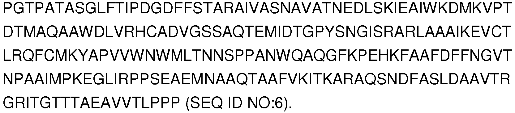
- The fusion protein according to any one of claims 1-3, said fusion protein having the following sequence:

- A polynucleotide which codes for the fusion protein as defined in any one of claims 1-4.
- The polynucleotide according to claim 5, said polynucleotide having the following sequence (SEQ ID NO:8):


- An expression vector comprising the polynucleotide as defined in any one of claims 5-6.
- A host cell comprising the expression vector as defined in claim 7.
- A genome sequence of a PVX virus, said genome sequence comprising the polynucleotide as defined in any one of claims 5-6.
- A chimeric virus particle of potato virus X, said chimeric virus particle being characterized in that it comprises, as a capsid protein, the fusion protein as defined in any one of claims 1-4, said viral particle having exposed, on the outside thereof, the amino-terminal portion comprising or consisting of the antigenic determinant of lipocalin.
- The chimeric virus particle according to claim 10, wherein the viral genome consists of the genome sequence as defined in claim 9.
- A use of the chimeric virus particle according to any one of claims 10-11 or of the fusion protein according to any one of claims 1-4 for the in vitro diagnosis of Sjögren's Syndrome, for example using the ELISA, dipstick or microchip method.
- A kit for the in vitro diagnosis of Sjögren's Syndrome, said kit comprising or consisting of the chimeric virus particle according to any one of claims 10-11 or in the fusion protein according to any one of claims 1-4, possibly in combination with suitable reagents for detection purposes.
- A plant characterized in that it comprises within it the chimeric viral particle as defined in any one of claims 10-11, or the expression vector as defined in claim 7.
- Plant cells of the plant according to claim 14, which express the chimeric viral particle as defined in any one of claims 10-11, or the expression vector as defined in claim 7.
- A use of a peptide sequence comprising or consisting of an antigenic determinant of lipocalin for the preparation of the fusion protein as defined in any one of claims 1-4, the chimeric viral particle according to any one of claims 10-11, the plant according to claim 14, and the plant cells according to claim 15.
- A use of the plant according to claim 14 or of the plant cells according to claim 15, for the production of the chimeric viral particle according to any one of claims 10-11.
- A process for the preparation of the fusion protein according to any one of claims 1-4, said process comprising or consisting of the following steps:a) cloning, in a vector, the peptide sequence comprising or consisting of the capsid protein of a PVX virus, said capsid protein being intact or deleted in the 5' portion of the gene which encodes the wild-type capsid protein; andb) cloning a peptide sequence comprising or consisting of an antigenic determinant of lipocalin, said peptide sequence being fused to said peptide sequence comprising or consisting of the capsid protein of a PVX virus in the vector resulting from step a), wherein said antigenic determinant of lipocalin is selected in the group which consists of FEKAAGARGLST (SEQ ID NO:2) and MSFEKAAGARGLST (SEQ ID NO:5).
- The process according to claim 18, wherein the PVX virus is selected in the group consisting of PVX X3, BS, EX, NL4, HB, WS2, ROTH1, XS, UK3, OS, NL1, Taiwan, X4, preferably X3.
- The process according to any one of claims 18-19, wherein said capsid protein consists of the following sequence:

- A process for the preparation of the chimeric viral particle according to claim 10 or 11, said process comprising or consisting of the step of replacing the capsid protein of a PVX virus, in said PVX virus, with the fusion protein according to any one of claims 1-4.
- A process for the production of the viral particle according to claim 10 or 11; said process comprising or consisting of the following steps:a1) infecting a plant with the viral particle according to claim 10 or 11 or the expression vector as defined in claim 7;b1) cultivating the plant obtained according to step a1);c) extracting said viral particle from said plant.
Priority Applications (1)
| Application Number | Priority Date | Filing Date | Title |
|---|---|---|---|
| PL16747629T PL3303369T3 (en) | 2015-06-03 | 2016-06-03 | Viral chimeric particle of potato virus x and use thereof for in vitro diagnosis of sjögren syndrome |
Applications Claiming Priority (2)
| Application Number | Priority Date | Filing Date | Title |
|---|---|---|---|
| ITUB20151199 | 2015-06-03 | ||
| PCT/IT2016/000146 WO2016194014A1 (en) | 2015-06-03 | 2016-06-03 | Viral chimeric particle of potato virus x and use thereof for in vitro diagnosis of sjögren syndrome |
Publications (2)
| Publication Number | Publication Date |
|---|---|
| EP3303369A1 EP3303369A1 (en) | 2018-04-11 |
| EP3303369B1 true EP3303369B1 (en) | 2019-08-07 |
Family
ID=54288887
Family Applications (1)
| Application Number | Title | Priority Date | Filing Date |
|---|---|---|---|
| EP16747629.0A Active EP3303369B1 (en) | 2015-06-03 | 2016-06-03 | Viral chimeric particle of potato virus x and use thereof for in vitro diagnosis of sjögren syndrome |
Country Status (6)
| Country | Link |
|---|---|
| US (1) | US10626149B2 (en) |
| EP (1) | EP3303369B1 (en) |
| CA (1) | CA2988072A1 (en) |
| ES (1) | ES2753170T3 (en) |
| PL (1) | PL3303369T3 (en) |
| WO (1) | WO2016194014A1 (en) |
Family Cites Families (1)
| Publication number | Priority date | Publication date | Assignee | Title |
|---|---|---|---|---|
| US9522940B2 (en) * | 2012-05-23 | 2016-12-20 | Pieris Pharmaceuticals Gmbh | Lipocalin muteins with binding-affinity for glypican-3 (GPC-3) and use of lipocalin muteins for target-specific delivery to cells expressing GPC-3 |
-
2016
- 2016-06-03 EP EP16747629.0A patent/EP3303369B1/en active Active
- 2016-06-03 PL PL16747629T patent/PL3303369T3/en unknown
- 2016-06-03 ES ES16747629T patent/ES2753170T3/en active Active
- 2016-06-03 WO PCT/IT2016/000146 patent/WO2016194014A1/en not_active Ceased
- 2016-06-03 CA CA2988072A patent/CA2988072A1/en not_active Abandoned
- 2016-06-03 US US15/579,150 patent/US10626149B2/en not_active Expired - Fee Related
Non-Patent Citations (1)
| Title |
|---|
| None * |
Also Published As
| Publication number | Publication date |
|---|---|
| WO2016194014A1 (en) | 2016-12-08 |
| PL3303369T3 (en) | 2020-03-31 |
| US20180170970A1 (en) | 2018-06-21 |
| US10626149B2 (en) | 2020-04-21 |
| ES2753170T3 (en) | 2020-04-07 |
| EP3303369A1 (en) | 2018-04-11 |
| CA2988072A1 (en) | 2016-12-08 |
Similar Documents
| Publication | Publication Date | Title |
|---|---|---|
| CN111218459B (en) | A recombinant novel coronavirus vaccine using human replication-deficient adenovirus as a vector | |
| WO2021254327A1 (en) | Envelope replacement-type viral vector vaccine and construction method therefor | |
| CN110759973B (en) | Cell strain for expressing African swine fever virus CD2v protein and application thereof | |
| CN111234036B (en) | African swine fever virus p72 fusion protein and preparation method and application thereof | |
| CN111560354A (en) | Recombinant novel coronavirus and its preparation method and application | |
| Šroller et al. | Seroprevalence rates of HPyV6, HPyV7, TSPyV, HPyV9, MWPyV and KIPyV polyomaviruses among the healthy blood donors | |
| CN112574306B (en) | Adiponectin monoclonal antibody, antibody pair, preparation method and application thereof | |
| Tinazzi et al. | Plant-derived chimeric virus particles for the diagnosis of primary Sjögren syndrome | |
| CN114106157B (en) | Canine parvovirus nano antibody CPV-VHH-E3 and application thereof | |
| CN117700537B (en) | Monoclonal antibody with good affinity reaction capacity for VP7 protein of African horse sickness virus and application | |
| CN109111507B (en) | Virus recombinant glycoprotein and eukaryotic cell high-efficiency expression method and application thereof | |
| Liu et al. | Expression and Immunogenicity of Two Recombinant Fusion Proteins Comprising Foot‐and‐Mouth Disease Virus Structural Protein VP1 and DC‐SIGN‐Binding Glycoproteins | |
| US10626149B2 (en) | Viral chimeric particle of potato virus X and use thereof for in vitro diagnosis of Sjögren syndrome | |
| CN101481675B (en) | Anti-swine fever multi-epitope DNA vaccine, construction method and use | |
| CN103409377A (en) | Preparation and use of canine parvovirus virus-like particles | |
| CN101121748B (en) | Anti-Infectious Bursal Disease Virus VP4 Protein Monoclonal Antibody | |
| CN117007801A (en) | Preparation method and application of African swine fever virus membrane protein E146L | |
| CN115725613B (en) | mRNA vaccine against delta mutant strain of novel coronavirus and its preparation method and application | |
| CN105906694B (en) | Datum hole Kenya virus-specific detects antigen and its application | |
| KR102657261B1 (en) | Recombinant COVID-19 spike proteins for diagnosis of COVID-19 and use thereof | |
| CN119039427B (en) | A recombinant single B cell antibody against bluetongue virus in sheep | |
| CN115894718B (en) | Antigen epitope peptide of African swine fever virus and application thereof | |
| CN118421572B (en) | Construction of an attenuated African swine fever virus strain with 24 missing genes and its application as a vaccine | |
| CN116376977B (en) | Rubella virus recombinant virus-like particle expression vector and application thereof | |
| CN121471347B (en) | Fully human monoclonal antibody for resisting SARS-CoV and SARS-CoV-2 and its application |
Legal Events
| Date | Code | Title | Description |
|---|---|---|---|
| STAA | Information on the status of an ep patent application or granted ep patent |
Free format text: STATUS: THE INTERNATIONAL PUBLICATION HAS BEEN MADE |
|
| PUAI | Public reference made under article 153(3) epc to a published international application that has entered the european phase |
Free format text: ORIGINAL CODE: 0009012 |
|
| STAA | Information on the status of an ep patent application or granted ep patent |
Free format text: STATUS: REQUEST FOR EXAMINATION WAS MADE |
|
| 17P | Request for examination filed |
Effective date: 20171212 |
|
| AK | Designated contracting states |
Kind code of ref document: A1 Designated state(s): AL AT BE BG CH CY CZ DE DK EE ES FI FR GB GR HR HU IE IS IT LI LT LU LV MC MK MT NL NO PL PT RO RS SE SI SK SM TR |
|
| AX | Request for extension of the european patent |
Extension state: BA ME |
|
| GRAP | Despatch of communication of intention to grant a patent |
Free format text: ORIGINAL CODE: EPIDOSNIGR1 |
|
| STAA | Information on the status of an ep patent application or granted ep patent |
Free format text: STATUS: GRANT OF PATENT IS INTENDED |
|
| INTG | Intention to grant announced |
Effective date: 20190313 |
|
| GRAS | Grant fee paid |
Free format text: ORIGINAL CODE: EPIDOSNIGR3 |
|
| GRAA | (expected) grant |
Free format text: ORIGINAL CODE: 0009210 |
|
| STAA | Information on the status of an ep patent application or granted ep patent |
Free format text: STATUS: THE PATENT HAS BEEN GRANTED |
|
| AK | Designated contracting states |
Kind code of ref document: B1 Designated state(s): AL AT BE BG CH CY CZ DE DK EE ES FI FR GB GR HR HU IE IS IT LI LT LU LV MC MK MT NL NO PL PT RO RS SE SI SK SM TR |
|
| AX | Request for extension of the european patent |
Extension state: BA ME |
|
| REG | Reference to a national code |
Ref country code: GB Ref legal event code: FG4D |
|
| REG | Reference to a national code |
Ref country code: AT Ref legal event code: REF Ref document number: 1163702 Country of ref document: AT Kind code of ref document: T Effective date: 20190815 Ref country code: CH Ref legal event code: EP |
|
| REG | Reference to a national code |
Ref country code: DE Ref legal event code: R096 Ref document number: 602016018237 Country of ref document: DE |
|
| REG | Reference to a national code |
Ref country code: IE Ref legal event code: FG4D |
|
| REG | Reference to a national code |
Ref country code: SE Ref legal event code: TRGR |
|
| REG | Reference to a national code |
Ref country code: NL Ref legal event code: MP Effective date: 20190807 |
|
| REG | Reference to a national code |
Ref country code: LT Ref legal event code: MG4D |
|
| PG25 | Lapsed in a contracting state [announced via postgrant information from national office to epo] |
Ref country code: BG Free format text: LAPSE BECAUSE OF FAILURE TO SUBMIT A TRANSLATION OF THE DESCRIPTION OR TO PAY THE FEE WITHIN THE PRESCRIBED TIME-LIMIT Effective date: 20191107 Ref country code: NO Free format text: LAPSE BECAUSE OF FAILURE TO SUBMIT A TRANSLATION OF THE DESCRIPTION OR TO PAY THE FEE WITHIN THE PRESCRIBED TIME-LIMIT Effective date: 20191107 Ref country code: HR Free format text: LAPSE BECAUSE OF FAILURE TO SUBMIT A TRANSLATION OF THE DESCRIPTION OR TO PAY THE FEE WITHIN THE PRESCRIBED TIME-LIMIT Effective date: 20190807 Ref country code: LT Free format text: LAPSE BECAUSE OF FAILURE TO SUBMIT A TRANSLATION OF THE DESCRIPTION OR TO PAY THE FEE WITHIN THE PRESCRIBED TIME-LIMIT Effective date: 20190807 Ref country code: NL Free format text: LAPSE BECAUSE OF FAILURE TO SUBMIT A TRANSLATION OF THE DESCRIPTION OR TO PAY THE FEE WITHIN THE PRESCRIBED TIME-LIMIT Effective date: 20190807 Ref country code: FI Free format text: LAPSE BECAUSE OF FAILURE TO SUBMIT A TRANSLATION OF THE DESCRIPTION OR TO PAY THE FEE WITHIN THE PRESCRIBED TIME-LIMIT Effective date: 20190807 Ref country code: PT Free format text: LAPSE BECAUSE OF FAILURE TO SUBMIT A TRANSLATION OF THE DESCRIPTION OR TO PAY THE FEE WITHIN THE PRESCRIBED TIME-LIMIT Effective date: 20191209 |
|
| REG | Reference to a national code |
Ref country code: AT Ref legal event code: MK05 Ref document number: 1163702 Country of ref document: AT Kind code of ref document: T Effective date: 20190807 |
|
| PG25 | Lapsed in a contracting state [announced via postgrant information from national office to epo] |
Ref country code: IS Free format text: LAPSE BECAUSE OF FAILURE TO SUBMIT A TRANSLATION OF THE DESCRIPTION OR TO PAY THE FEE WITHIN THE PRESCRIBED TIME-LIMIT Effective date: 20191207 Ref country code: GR Free format text: LAPSE BECAUSE OF FAILURE TO SUBMIT A TRANSLATION OF THE DESCRIPTION OR TO PAY THE FEE WITHIN THE PRESCRIBED TIME-LIMIT Effective date: 20191108 Ref country code: LV Free format text: LAPSE BECAUSE OF FAILURE TO SUBMIT A TRANSLATION OF THE DESCRIPTION OR TO PAY THE FEE WITHIN THE PRESCRIBED TIME-LIMIT Effective date: 20190807 Ref country code: RS Free format text: LAPSE BECAUSE OF FAILURE TO SUBMIT A TRANSLATION OF THE DESCRIPTION OR TO PAY THE FEE WITHIN THE PRESCRIBED TIME-LIMIT Effective date: 20190807 Ref country code: AL Free format text: LAPSE BECAUSE OF FAILURE TO SUBMIT A TRANSLATION OF THE DESCRIPTION OR TO PAY THE FEE WITHIN THE PRESCRIBED TIME-LIMIT Effective date: 20190807 |
|
| REG | Reference to a national code |
Ref country code: CH Ref legal event code: NV Representative=s name: VALIPAT S.A. C/O BOVARD SA NEUCHATEL, CH |
|
| REG | Reference to a national code |
Ref country code: ES Ref legal event code: FG2A Ref document number: 2753170 Country of ref document: ES Kind code of ref document: T3 Effective date: 20200407 |
|
| PG25 | Lapsed in a contracting state [announced via postgrant information from national office to epo] |
Ref country code: DK Free format text: LAPSE BECAUSE OF FAILURE TO SUBMIT A TRANSLATION OF THE DESCRIPTION OR TO PAY THE FEE WITHIN THE PRESCRIBED TIME-LIMIT Effective date: 20190807 Ref country code: EE Free format text: LAPSE BECAUSE OF FAILURE TO SUBMIT A TRANSLATION OF THE DESCRIPTION OR TO PAY THE FEE WITHIN THE PRESCRIBED TIME-LIMIT Effective date: 20190807 Ref country code: AT Free format text: LAPSE BECAUSE OF FAILURE TO SUBMIT A TRANSLATION OF THE DESCRIPTION OR TO PAY THE FEE WITHIN THE PRESCRIBED TIME-LIMIT Effective date: 20190807 Ref country code: RO Free format text: LAPSE BECAUSE OF FAILURE TO SUBMIT A TRANSLATION OF THE DESCRIPTION OR TO PAY THE FEE WITHIN THE PRESCRIBED TIME-LIMIT Effective date: 20190807 Ref country code: IT Free format text: LAPSE BECAUSE OF FAILURE TO SUBMIT A TRANSLATION OF THE DESCRIPTION OR TO PAY THE FEE WITHIN THE PRESCRIBED TIME-LIMIT Effective date: 20190807 |
|
| VS25 | Lapsed in a validation state [announced via postgrant information from nat. office to epo] |
Ref country code: MD Free format text: LAPSE BECAUSE OF FAILURE TO SUBMIT A TRANSLATION OF THE DESCRIPTION OR TO PAY THE FEE WITHIN THE PRESCRIBED TIME-LIMIT Effective date: 20190807 |
|
| PG25 | Lapsed in a contracting state [announced via postgrant information from national office to epo] |
Ref country code: SM Free format text: LAPSE BECAUSE OF FAILURE TO SUBMIT A TRANSLATION OF THE DESCRIPTION OR TO PAY THE FEE WITHIN THE PRESCRIBED TIME-LIMIT Effective date: 20190807 Ref country code: IS Free format text: LAPSE BECAUSE OF FAILURE TO SUBMIT A TRANSLATION OF THE DESCRIPTION OR TO PAY THE FEE WITHIN THE PRESCRIBED TIME-LIMIT Effective date: 20200224 Ref country code: CZ Free format text: LAPSE BECAUSE OF FAILURE TO SUBMIT A TRANSLATION OF THE DESCRIPTION OR TO PAY THE FEE WITHIN THE PRESCRIBED TIME-LIMIT Effective date: 20190807 Ref country code: SK Free format text: LAPSE BECAUSE OF FAILURE TO SUBMIT A TRANSLATION OF THE DESCRIPTION OR TO PAY THE FEE WITHIN THE PRESCRIBED TIME-LIMIT Effective date: 20190807 |
|
| REG | Reference to a national code |
Ref country code: DE Ref legal event code: R097 Ref document number: 602016018237 Country of ref document: DE |
|
| PLBE | No opposition filed within time limit |
Free format text: ORIGINAL CODE: 0009261 |
|
| STAA | Information on the status of an ep patent application or granted ep patent |
Free format text: STATUS: NO OPPOSITION FILED WITHIN TIME LIMIT |
|
| PG2D | Information on lapse in contracting state deleted |
Ref country code: IS |
|
| 26N | No opposition filed |
Effective date: 20200603 |
|
| PG25 | Lapsed in a contracting state [announced via postgrant information from national office to epo] |
Ref country code: SI Free format text: LAPSE BECAUSE OF FAILURE TO SUBMIT A TRANSLATION OF THE DESCRIPTION OR TO PAY THE FEE WITHIN THE PRESCRIBED TIME-LIMIT Effective date: 20190807 |
|
| PG25 | Lapsed in a contracting state [announced via postgrant information from national office to epo] |
Ref country code: MC Free format text: LAPSE BECAUSE OF FAILURE TO SUBMIT A TRANSLATION OF THE DESCRIPTION OR TO PAY THE FEE WITHIN THE PRESCRIBED TIME-LIMIT Effective date: 20190807 |
|
| PG25 | Lapsed in a contracting state [announced via postgrant information from national office to epo] |
Ref country code: LU Free format text: LAPSE BECAUSE OF NON-PAYMENT OF DUE FEES Effective date: 20200603 |
|
| REG | Reference to a national code |
Ref country code: BE Ref legal event code: MM Effective date: 20200630 |
|
| PG25 | Lapsed in a contracting state [announced via postgrant information from national office to epo] |
Ref country code: IE Free format text: LAPSE BECAUSE OF NON-PAYMENT OF DUE FEES Effective date: 20200603 |
|
| PG25 | Lapsed in a contracting state [announced via postgrant information from national office to epo] |
Ref country code: BE Free format text: LAPSE BECAUSE OF NON-PAYMENT OF DUE FEES Effective date: 20200630 |
|
| PGFP | Annual fee paid to national office [announced via postgrant information from national office to epo] |
Ref country code: PL Payment date: 20210315 Year of fee payment: 6 |
|
| VS25 | Lapsed in a validation state [announced via postgrant information from nat. office to epo] |
Ref country code: MA Free format text: LAPSE BECAUSE OF FAILURE TO SUBMIT A TRANSLATION OF THE DESCRIPTION OR TO PAY THE FEE WITHIN THE PRESCRIBED TIME-LIMIT Effective date: 20190807 |
|
| PGFP | Annual fee paid to national office [announced via postgrant information from national office to epo] |
Ref country code: DE Payment date: 20210505 Year of fee payment: 6 Ref country code: FR Payment date: 20210412 Year of fee payment: 6 |
|
| PGFP | Annual fee paid to national office [announced via postgrant information from national office to epo] |
Ref country code: CH Payment date: 20210616 Year of fee payment: 6 Ref country code: TR Payment date: 20210527 Year of fee payment: 6 Ref country code: SE Payment date: 20210610 Year of fee payment: 6 Ref country code: GB Payment date: 20210513 Year of fee payment: 6 |
|
| PGFP | Annual fee paid to national office [announced via postgrant information from national office to epo] |
Ref country code: ES Payment date: 20210708 Year of fee payment: 6 |
|
| PG25 | Lapsed in a contracting state [announced via postgrant information from national office to epo] |
Ref country code: MT Free format text: LAPSE BECAUSE OF FAILURE TO SUBMIT A TRANSLATION OF THE DESCRIPTION OR TO PAY THE FEE WITHIN THE PRESCRIBED TIME-LIMIT Effective date: 20190807 Ref country code: CY Free format text: LAPSE BECAUSE OF FAILURE TO SUBMIT A TRANSLATION OF THE DESCRIPTION OR TO PAY THE FEE WITHIN THE PRESCRIBED TIME-LIMIT Effective date: 20190807 |
|
| PG25 | Lapsed in a contracting state [announced via postgrant information from national office to epo] |
Ref country code: MK Free format text: LAPSE BECAUSE OF FAILURE TO SUBMIT A TRANSLATION OF THE DESCRIPTION OR TO PAY THE FEE WITHIN THE PRESCRIBED TIME-LIMIT Effective date: 20190807 |
|
| REG | Reference to a national code |
Ref country code: DE Ref legal event code: R119 Ref document number: 602016018237 Country of ref document: DE |
|
| REG | Reference to a national code |
Ref country code: CH Ref legal event code: PL Ref country code: SE Ref legal event code: EUG |
|
| GBPC | Gb: european patent ceased through non-payment of renewal fee |
Effective date: 20220603 |
|
| PG25 | Lapsed in a contracting state [announced via postgrant information from national office to epo] |
Ref country code: SE Free format text: LAPSE BECAUSE OF NON-PAYMENT OF DUE FEES Effective date: 20220604 Ref country code: LI Free format text: LAPSE BECAUSE OF NON-PAYMENT OF DUE FEES Effective date: 20220630 Ref country code: FR Free format text: LAPSE BECAUSE OF NON-PAYMENT OF DUE FEES Effective date: 20220630 Ref country code: CH Free format text: LAPSE BECAUSE OF NON-PAYMENT OF DUE FEES Effective date: 20220630 |
|
| PG25 | Lapsed in a contracting state [announced via postgrant information from national office to epo] |
Ref country code: GB Free format text: LAPSE BECAUSE OF NON-PAYMENT OF DUE FEES Effective date: 20220603 Ref country code: DE Free format text: LAPSE BECAUSE OF NON-PAYMENT OF DUE FEES Effective date: 20230103 |
|
| REG | Reference to a national code |
Ref country code: ES Ref legal event code: FD2A Effective date: 20230727 |
|
| PG25 | Lapsed in a contracting state [announced via postgrant information from national office to epo] |
Ref country code: ES Free format text: LAPSE BECAUSE OF NON-PAYMENT OF DUE FEES Effective date: 20220604 |
|
| PG25 | Lapsed in a contracting state [announced via postgrant information from national office to epo] |
Ref country code: PL Free format text: LAPSE BECAUSE OF NON-PAYMENT OF DUE FEES Effective date: 20220603 |
|
| PG25 | Lapsed in a contracting state [announced via postgrant information from national office to epo] |
Ref country code: TR Free format text: LAPSE BECAUSE OF NON-PAYMENT OF DUE FEES Effective date: 20220603 |