EP3290512A1 - Method of distinguishing mesenchymal stem cells and method of determining purity of mesenchymal stem cells - Google Patents
Method of distinguishing mesenchymal stem cells and method of determining purity of mesenchymal stem cells Download PDFInfo
- Publication number
- EP3290512A1 EP3290512A1 EP17177108.2A EP17177108A EP3290512A1 EP 3290512 A1 EP3290512 A1 EP 3290512A1 EP 17177108 A EP17177108 A EP 17177108A EP 3290512 A1 EP3290512 A1 EP 3290512A1
- Authority
- EP
- European Patent Office
- Prior art keywords
- mscs
- placenta
- fibroblasts
- related tissue
- stem cells
- Prior art date
- Legal status (The legal status is an assumption and is not a legal conclusion. Google has not performed a legal analysis and makes no representation as to the accuracy of the status listed.)
- Granted
Links
Images


Classifications
-
- G—PHYSICS
- G01—MEASURING; TESTING
- G01N—INVESTIGATING OR ANALYSING MATERIALS BY DETERMINING THEIR CHEMICAL OR PHYSICAL PROPERTIES
- G01N33/00—Investigating or analysing materials by specific methods not covered by groups G01N1/00 - G01N31/00
- G01N33/48—Biological material, e.g. blood, urine; Haemocytometers
- G01N33/50—Chemical analysis of biological material, e.g. blood, urine; Testing involving biospecific ligand binding methods; Immunological testing
- G01N33/53—Immunoassay; Biospecific binding assay; Materials therefor
- G01N33/569—Immunoassay; Biospecific binding assay; Materials therefor for microorganisms, e.g. protozoa, bacteria, viruses
- G01N33/56966—Animal cells
-
- C—CHEMISTRY; METALLURGY
- C12—BIOCHEMISTRY; BEER; SPIRITS; WINE; VINEGAR; MICROBIOLOGY; ENZYMOLOGY; MUTATION OR GENETIC ENGINEERING
- C12N—MICROORGANISMS OR ENZYMES; COMPOSITIONS THEREOF; PROPAGATING, PRESERVING, OR MAINTAINING MICROORGANISMS; MUTATION OR GENETIC ENGINEERING; CULTURE MEDIA
- C12N5/00—Undifferentiated human, animal or plant cells, e.g. cell lines; Tissues; Cultivation or maintenance thereof; Culture media therefor
- C12N5/06—Animal cells or tissues; Human cells or tissues
- C12N5/0602—Vertebrate cells
- C12N5/0652—Cells of skeletal and connective tissues; Mesenchyme
- C12N5/0662—Stem cells
- C12N5/0668—Mesenchymal stem cells from other natural sources
-
- C—CHEMISTRY; METALLURGY
- C12—BIOCHEMISTRY; BEER; SPIRITS; WINE; VINEGAR; MICROBIOLOGY; ENZYMOLOGY; MUTATION OR GENETIC ENGINEERING
- C12Q—MEASURING OR TESTING PROCESSES INVOLVING ENZYMES, NUCLEIC ACIDS OR MICROORGANISMS; COMPOSITIONS OR TEST PAPERS THEREFOR; PROCESSES OF PREPARING SUCH COMPOSITIONS; CONDITION-RESPONSIVE CONTROL IN MICROBIOLOGICAL OR ENZYMOLOGICAL PROCESSES
- C12Q1/00—Measuring or testing processes involving enzymes, nucleic acids or microorganisms; Compositions therefor; Processes of preparing such compositions
- C12Q1/68—Measuring or testing processes involving enzymes, nucleic acids or microorganisms; Compositions therefor; Processes of preparing such compositions involving nucleic acids
- C12Q1/6876—Nucleic acid products used in the analysis of nucleic acids, e.g. primers or probes
- C12Q1/6881—Nucleic acid products used in the analysis of nucleic acids, e.g. primers or probes for tissue or cell typing, e.g. human leukocyte antigen [HLA] probes
-
- G—PHYSICS
- G01—MEASURING; TESTING
- G01N—INVESTIGATING OR ANALYSING MATERIALS BY DETERMINING THEIR CHEMICAL OR PHYSICAL PROPERTIES
- G01N15/00—Investigating characteristics of particles; Investigating permeability, pore-volume or surface-area of porous materials
- G01N15/10—Investigating individual particles
- G01N15/14—Optical investigation techniques, e.g. flow cytometry
-
- G—PHYSICS
- G01—MEASURING; TESTING
- G01N—INVESTIGATING OR ANALYSING MATERIALS BY DETERMINING THEIR CHEMICAL OR PHYSICAL PROPERTIES
- G01N35/00—Automatic analysis not limited to methods or materials provided for in any single one of groups G01N1/00 - G01N33/00; Handling materials therefor
- G01N35/0098—Automatic analysis not limited to methods or materials provided for in any single one of groups G01N1/00 - G01N33/00; Handling materials therefor involving analyte bound to insoluble magnetic carrier, e.g. using magnetic separation
-
- C—CHEMISTRY; METALLURGY
- C12—BIOCHEMISTRY; BEER; SPIRITS; WINE; VINEGAR; MICROBIOLOGY; ENZYMOLOGY; MUTATION OR GENETIC ENGINEERING
- C12N—MICROORGANISMS OR ENZYMES; COMPOSITIONS THEREOF; PROPAGATING, PRESERVING, OR MAINTAINING MICROORGANISMS; MUTATION OR GENETIC ENGINEERING; CULTURE MEDIA
- C12N2501/00—Active agents used in cell culture processes, e.g. differentation
- C12N2501/50—Cell markers; Cell surface determinants
- C12N2501/599—Cell markers; Cell surface determinants with CD designations not provided for elsewhere
-
- C—CHEMISTRY; METALLURGY
- C12—BIOCHEMISTRY; BEER; SPIRITS; WINE; VINEGAR; MICROBIOLOGY; ENZYMOLOGY; MUTATION OR GENETIC ENGINEERING
- C12N—MICROORGANISMS OR ENZYMES; COMPOSITIONS THEREOF; PROPAGATING, PRESERVING, OR MAINTAINING MICROORGANISMS; MUTATION OR GENETIC ENGINEERING; CULTURE MEDIA
- C12N5/00—Undifferentiated human, animal or plant cells, e.g. cell lines; Tissues; Cultivation or maintenance thereof; Culture media therefor
- C12N5/06—Animal cells or tissues; Human cells or tissues
- C12N5/0602—Vertebrate cells
- C12N5/0603—Embryonic cells ; Embryoid bodies
- C12N5/0605—Cells from extra-embryonic tissues, e.g. placenta, amnion, yolk sac, Wharton's jelly
-
- C—CHEMISTRY; METALLURGY
- C12—BIOCHEMISTRY; BEER; SPIRITS; WINE; VINEGAR; MICROBIOLOGY; ENZYMOLOGY; MUTATION OR GENETIC ENGINEERING
- C12Q—MEASURING OR TESTING PROCESSES INVOLVING ENZYMES, NUCLEIC ACIDS OR MICROORGANISMS; COMPOSITIONS OR TEST PAPERS THEREFOR; PROCESSES OF PREPARING SUCH COMPOSITIONS; CONDITION-RESPONSIVE CONTROL IN MICROBIOLOGICAL OR ENZYMOLOGICAL PROCESSES
- C12Q2600/00—Oligonucleotides characterized by their use
- C12Q2600/158—Expression markers
-
- G—PHYSICS
- G01—MEASURING; TESTING
- G01N—INVESTIGATING OR ANALYSING MATERIALS BY DETERMINING THEIR CHEMICAL OR PHYSICAL PROPERTIES
- G01N15/00—Investigating characteristics of particles; Investigating permeability, pore-volume or surface-area of porous materials
- G01N15/10—Investigating individual particles
- G01N15/14—Optical investigation techniques, e.g. flow cytometry
- G01N2015/1488—Methods for deciding
-
- G—PHYSICS
- G01—MEASURING; TESTING
- G01N—INVESTIGATING OR ANALYSING MATERIALS BY DETERMINING THEIR CHEMICAL OR PHYSICAL PROPERTIES
- G01N2333/00—Assays involving biological materials from specific organisms or of a specific nature
- G01N2333/435—Assays involving biological materials from specific organisms or of a specific nature from animals; from humans
- G01N2333/705—Assays involving receptors, cell surface antigens or cell surface determinants
- G01N2333/70596—Molecules with a "CD"-designation not provided for elsewhere in G01N2333/705
Definitions
- the present invention pertains to a method of distinguishing mesenchymal stem cells (MSCs) in a cell culture derived from a placenta-related tissue.
- the present invention also relates to a method of increasing the purity of MSC population in a cell culture derived from a placenta-related tissue.
- the invention pertains to a method of assessing purity of MSCs in a cell culture derived from a placenta-related tissue
- MSCs Mesenchymal stem or stromal cells
- BM bone marrow
- MSCs are a subpopulation of a more complex cell composition of stromal cells contained in mesenchymal tissue. Due to the heterogeneous nature and the absence of known biomarkers specific for mesenchymal stem cells, it is a challenging task to define MSC phenotypes and characteristics [4-6].
- lack of specific cell surface markers renders the cell culture at potential contamination risk with other cell types, in particular, those mature stromal cells such as fibroblasts, which, conversely, are abundant in mesenchymal tissue [4-6].
- non-MSCs including fibroblasts, placenta-derived epithelial cells, and placenta-derived reticular cells, often coexist with MSCs during the in vitro cultivation.
- fibroblast is the main source of contamination.
- Fibroblasts are considered mature mesenchymal cells that are particularly abundant in the connective tissue. Therefore, these cells are the most frequent contaminating cell phenotype present in many cell culture systems. Not only is it difficult to apply techniques which successfully eliminate fibroblasts from a culture, it is also particularly complex to distinguish MSCs from fibroblasts in the same culture. Fibroblasts and MSCs have an extremely similar morphological appearance; they both proliferate well and share many identical cell surface markers [7-8]. MSC are currently defined as plastic adherent, multipotent fibroblast-like cells expressing CD73, CD90, CD105 and negative for the hematopoietic markers CD14, CD34 and CD45 by the International Society of Cellular Therapy (ISCT).
- ISCT International Society of Cellular Therapy
- CD 146 (melanoma cell adhesion molecule, MCAM), a surface marker of vascular endothelial cells, functions by facilitating cell-cell interactions and angiogenesis.
- MCAM melanoma cell adhesion molecule
- US Patent No. 9,470,685 discloses a method of distinguishing MSCs from fibroblasts using a marker EphA2 expressed on the MSCs.
- mesenchymal stem cells can be distinguished in a cell culture derived from a placenta-related tissue, based on their expression levels of a specific surface marker, CD146 (melanoma cell adhesion molecule, MCAM).
- CD146 melanoma cell adhesion molecule
- the present invention features a method of distinguishing mesenchymal stem cells (MSCs) from fibroblasts, comprising isolating the MSCs from the fibroblasts using a marker CD146 expressed on the MSCs so as to distinguish the MSCs from the fibroblasts, wherein the MSCs and fibroblasts are from a cell culture derived from a placenta-related tissue.
- MSCs mesenchymal stem cells
- the method further comprises the preliminary steps of: collecting the MSCs and the fibroblasts from the placenta-related tissue; and culturing the MSCs and the fibroblasts in a culture medium to prepare the cell culture.
- the present invention provides a method of increasing a purity of mesenchymal stem cells (MSCs) population in a cell culture, comprising isolating and collecting MSCs using a marker CD146 expressed on the MSCs, wherein the MSCs are from a cell culture derived from a placenta-related tissue; and culturing the MSCs.
- MSCs mesenchymal stem cells
- the placenta-related tissue may be selected from the group consisting of amniotic membrane, chorionic disk, chorionic membrane, and umbilical cord.
- the placenta-related tissue is umbilical cord.
- the sorting step may be performed using a technique known or to be developed in the art, for example, an antibody-based or a nucleotide-based isolation method.
- the cells derived from a placenta-related tissue are cultured in a culture medium for MSC.
- the present invention provides a method of assessing purity of mesenchymal stem cells (MSCs) in a cell culture derived from a placenta-related tissue. Said method comprises the step of determining the percentage of cells expressing CD 146 in the culture.
- MSCs mesenchymal stem cells
- the purity of MSCs has a positive correlation with the percentage of cells expressing CD146.
- the percentage of cells expressing CD146 is determined by a flow cytometry.
- the present invention is based on the unexpected finding that through a cell sorting by surface marker CD146, mesenchymal stem cells (MSCs) can be distinguished in a cell culture derived from a placenta-related tissue.
- MSCs mesenchymal stem cells
- the present invention provides a method of distinguishing mesenchymal stem cells (MSCs) from fibroblasts.
- the method comprises isolating the MSCs from the fibroblasts using a marker CD146 expressed on the MSCs so as to distinguish the MSCs from the fibroblasts, wherein the MSCs and fibroblasts are from a cell culture derived from a placenta-related tissue.
- the method further comprises the preliminary steps of: collecting the MSCs and the fibroblasts from the placenta-related tissue; and culturing the MSCs and the fibroblasts in a culture medium to prepare the cell culture.
- the present invention provides a method of increasing a purity of mesenchymal stem cells (MSCs) population in a cell culture, comprising isolating and collecting MSCs using a marker CD 146 expressed on the MSCs, wherein the MSCs are from a cell culture derived from a placenta-related tissue; and culturing the MSCs.
- MSCs mesenchymal stem cells
- the present invention provides a method of isolating a population of mesenchymal stem cells (MSCs), comprising isolating and collecting MSCs using a marker CD 146 expressed on the MSCs, wherein the MSCs are from a cell culture derived from a placenta-related tissue, and the population of MSCs has a higher differentiation ability, elicits higher anti-inflammatory effects, has a lower degree of senescence, and/or secretes a higher amount of growth factors.
- MSCs mesenchymal stem cells
- the cells are freshly derived, obtained or collected from a placenta-related tissue following a protocol known in the art, for example, that of Shen et al. [21].
- the cells derived from a placenta-related tissue are then cultured in a culture medium for MSC.
- a standard medium for MSC comprises ⁇ minimal essential medium (with different versions of modification), fetal bovine serum (FBS), L-glutamine, and basic fibroblast growth factor (bFGF) [3, 16-20].
- the placenta-related tissue may be selected from the group consisting of amniotic membrane, chorionic disk, chorionic membrane, and umbilical cord. In one preferred embodiment of the present invention, the placenta-related tissue is umbilical cord.
- a cell culture derived from a placenta-related tissue is subjected to a cell sorting by CD146.
- the cell sorting may be performed through a technique known or to be developed in the art, for example, an antibody-based or a nucleotide-based isolation method.
- the cell sorting is performed by an antibody-based magnetic cell sorting.
- the MACS method MACS ® Technology, Miltenyi Biotec.
- the cell sorting may be preform through a flow cytometry method, e.g. an antibody-based or a nucleotide-based flow cytometry.
- the present invention in a further aspect provides a method of assessing purity of mesenchymal stem cells (MSCs) in a cell culture derived from a placenta-related tissue. Said method comprises the step of determining the percentage of cells expressing CD 146 in the culture.
- MSCs mesenchymal stem cells
- the percentage of cells expressing CD 146 is determined by a flow cytometry.
- the population of MSCs isolated by a method of the present invention has a higher differentiation ability.
- the population of MSCs has a higher adipogenesis ability.
- the population of MSCs isolated by a method of the present invention elicits higher anti-inflammatory effects.
- the population of MSCs exhibits a higher ability in inhibiting PBMC proliferation.
- the population of MSCs isolated by a method of the present invention has a lower degree of senescence.
- the population of MSCs expresses a lower level of cellular ROS, mitochondrial ROS, or beta-galactosidase.
- the population of MSCs isolated by a method of the present invention secretes a higher amount of growth factors.
- the population of MSCs secretes a higher amount of a hepatocyte growth factor (HGF).
- HGF hepatocyte growth factor
- Example 1 Flow cytometry analysis of mixed populations of MSCs and fibroblast
- CD 146 could serve as a biomarker to separate placenta-derived MSCs from fibroblasts
- MSCs derived from umbilical cord were mixed with fibroblasts in Eppendorf tubes by following ratios (MSC: fibroblasts in cell number): 2x10 5 : 0, 1.8x10 5 : 2x10 4 , 1x10 5 : 1x10 5 , 2x10 4 : 1.8x10 5 , or 0 : 2x10 5 .
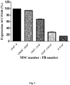
- CD146+ population in each Eppendorf tube was then analyzed by flow cytometry. Referring to Fig. 1 , the results shows that the percentage of CD146+ population detected by anti-CD146 antibodies (anti-human CD146-V450, BD biosciences) via flow cytometry decreased proportionally in relation to the increase of fibroblast population.
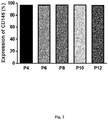
- MSCs derived from umbilical cord were harvested at different passage numbers (passage numbers: 4, 6, 8, 10, and 12) and the expression levels of CD146 were analyzed by flow cytometry. The results show that CD146 expression levels are maintained along passages ( Fig. 2 ).
- MSCs derived from umbilical cord were mixed with fibroblasts in Eppendorf tubes by following ratios (MSC: fibroblasts in cell number): 2x10 5 : 0, 1.8x10 5 : 2x10 4 , 1x10 5 : 1x10 5 , 2x10 4 : 1.8x10 5 , or 0 : 2x10 5 .
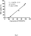
- CD146+ population in each Eppendorf tube was then analyzed by flow cytometry. Subsequently, the relationship between the purity (%) of MSCs and the percentage of cells expressing CD146 was analyzed by simple linear regression, results of which are shown in Fig. 3 .
- MSCs derived from different umbilical cords were examined for expression of CD146 using flow cytometry. Two groups of MSCs were obtained: CD146 high MSCs and CD146 low MSCs ( Fig. 4A ). 2 x 10 5 /well MSCs were seeded into 12-well plate, cultured overnight, and then cultured in adipocyte differentiation basal medium (InvitroGen) supplemented with adipogenesis supplement (InvitroGen). After culture for 14 days, the two groups of MSCs were fixed formalin, stained with Oil Red, and observed under a microscope to count the number of adipocytes. As shown in Fig. 4B , CD146 high MSCs exhibited higher adipogenesis ability.
- CD146 high MSCs and CD146 low MSCs were obtained as described in Example 4.
- the immune modulation capability of MSC was analyzed by PBMC proliferation assay.
- Carboxyfluorescein succinimidyl ester (CFSE) labeled PBMC is stimulated by phytohemagglutinin (PHA) and co-cultured with MSCs.
- PHA phytohemagglutinin
- the percentage of proliferated PBMCs was analyzed by FACS.
- the proliferation rate of PBMC was determined by assessing the reduction of the intensity of the fluorescent cell permeable dye CFSE.
- the results show that CD146 high MSCs were more effective in inhibiting PBMC proliferation than CD146 low MSCs ( Fig. 5 ), suggesting that CD146 high MSCs elicit higher anti-inflammatory effects.
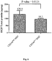
- Example 6 CD146 high MSCs secretes a higher amount of growth factors than CD146 low MSCs
- CD146 high MSCs and CD146 low MSCs were obtained as described in Example 4.
- Hepatocyte growth factor (HGF) level in conditioned medium from both groups was analyzed by ELISA and MSC total protein was analyzed by BCA assay.
- HGF level is expressed as a ratio of HGF amount to MSC total protein amount. As shown in Fig. 6 , CD146 high MSCs secreted a significantly higher amount of HGF than CD146 low MSCs.
- Example 7 CD146 high MSCs have a lower degree of senescence than CD146 low MSCs
- CD146 high MSCs and CD146 low MSCs were obtained as described in Example 4. Higher levels of cellular ROS, mitochondrial ROS and beta-galactosidase are considered to be related to senescence.
- cellular ROS level and mitochondrial ROS level were determined using commercial kits (DCFDA of Sigma, and MitoSox of Invitrogen, respectively).
- the level of senescence-associated beta-galactosidase was determined using C12FDG (Molecular Probes).
- C12FDG Molecular Probes
Landscapes
- Health & Medical Sciences (AREA)
- Life Sciences & Earth Sciences (AREA)
- Engineering & Computer Science (AREA)
- Chemical & Material Sciences (AREA)
- Immunology (AREA)
- Biomedical Technology (AREA)
- Biochemistry (AREA)
- General Health & Medical Sciences (AREA)
- Zoology (AREA)
- Cell Biology (AREA)
- Biotechnology (AREA)
- Analytical Chemistry (AREA)
- Molecular Biology (AREA)
- Organic Chemistry (AREA)
- Physics & Mathematics (AREA)
- Microbiology (AREA)
- Hematology (AREA)
- Urology & Nephrology (AREA)
- Wood Science & Technology (AREA)
- Pathology (AREA)
- General Physics & Mathematics (AREA)
- Genetics & Genomics (AREA)
- Bioinformatics & Cheminformatics (AREA)
- Developmental Biology & Embryology (AREA)
- General Engineering & Computer Science (AREA)
- Food Science & Technology (AREA)
- Medicinal Chemistry (AREA)
- Proteomics, Peptides & Aminoacids (AREA)
- Virology (AREA)
- Tropical Medicine & Parasitology (AREA)
- Rheumatology (AREA)
- Dispersion Chemistry (AREA)
- Biophysics (AREA)
- Measuring Or Testing Involving Enzymes Or Micro-Organisms (AREA)
- Micro-Organisms Or Cultivation Processes Thereof (AREA)
- Medicines Containing Material From Animals Or Micro-Organisms (AREA)
Abstract
Description
- The present invention pertains to a method of distinguishing mesenchymal stem cells (MSCs) in a cell culture derived from a placenta-related tissue. The present invention also relates to a method of increasing the purity of MSC population in a cell culture derived from a placenta-related tissue. In another aspect, the invention pertains to a method of assessing purity of MSCs in a cell culture derived from a placenta-related tissue
- Mesenchymal stem or stromal cells (MSCs) are multipotent cells of embryonic mesodermal origin, with a fibroblast-like morphology. These cells can differentiate into adipocytes, osteocytes, chondrocytes, neural lineage cells, and myocytes among other cell types depending on the stimuli and culture conditions. Although the plasticity of hMSCs and their role in tissue repair and regeneration have been extensively studied, it is their immunological trophic property that has gained the most interest recently [1-2]. Human mesenchymal stem cells have been isolated from a variety of tissues. The most frequently used source of MSCs is the bone marrow (BM). However, the isolation procedure is extremely invasive. To avoid the invasive isolation procedures, tissues such as human umbilical cord and placenta have been considered as good candidates since they are normally discarded after labor. The isolation of hMSCs from umbilical cord or placenta has proven to be efficient by previous studies [3].
- MSCs are a subpopulation of a more complex cell composition of stromal cells contained in mesenchymal tissue. Due to the heterogeneous nature and the absence of known biomarkers specific for mesenchymal stem cells, it is a challenging task to define MSC phenotypes and characteristics [4-6]. The molecular components responsible for MSCs functionalities, in particular, those on the plasma membrane, remain largely unknown. In addition, lack of specific cell surface markers renders the cell culture at potential contamination risk with other cell types, in particular, those mature stromal cells such as fibroblasts, which, conversely, are abundant in mesenchymal tissue [4-6]. In the process of isolation of MSCs from placenta-derived tissues, non-MSCs, including fibroblasts, placenta-derived epithelial cells, and placenta-derived reticular cells, often coexist with MSCs during the in vitro cultivation. In particular, fibroblast is the main source of contamination.
- Fibroblasts are considered mature mesenchymal cells that are particularly abundant in the connective tissue. Therefore, these cells are the most frequent contaminating cell phenotype present in many cell culture systems. Not only is it difficult to apply techniques which successfully eliminate fibroblasts from a culture, it is also particularly complex to distinguish MSCs from fibroblasts in the same culture. Fibroblasts and MSCs have an extremely similar morphological appearance; they both proliferate well and share many identical cell surface markers [7-8]. MSC are currently defined as plastic adherent, multipotent fibroblast-like cells expressing CD73, CD90, CD105 and negative for the hematopoietic markers CD14, CD34 and CD45 by the International Society of Cellular Therapy (ISCT). However, these properties and markers are also shared by fibroblasts. The current definition suggested by ISCT is thus incapable of distinguishing MSC from generic fibroblasts. Until now, the best way to distinguish MSCs from fibroblasts is based on the analysis of the functional properties of these two kinds of cells; MSCs retain multipotent stemness and immunomodulation capacity, while fibroblasts seem more limited in both of these functional properties.
- Since Friedenstein's pioneering work in identification of MSCs [48], there are no distinct differences in culture-derivation methodology, morphology, and gene expression signature that consistently and unequivocally distinguish ex vivo culture-expanded MSC from fibroblasts [9-12]. Presently, there is no accepted criterion or single cell-surface marker for separating the MSCs from fibroblasts. Due to the fact that fibroblast is the common contaminant cell population in MSC culture when derived from placenta, a novel surface protein as a biomarker to distinguish MSCs from fibroblasts is crucial to ensure the homogeneity of primary culture of placenta-derived MSCs.
- CD 146 (melanoma cell adhesion molecule, MCAM), a surface marker of vascular endothelial cells, functions by facilitating cell-cell interactions and angiogenesis. Recent research reveals that some MSCs express CD146 [13-14, 16], and the expression levels of CD146 are associated with their abilities of self-renewal, or abilities of differentiation into adipocytes and osteogenesis [13, 15]. Further, MSCs isolated from different tissues show different CD146 expression levels: about 40% of MSCs isolated from bone marrow express CD146, and its expression levels decrease as the passage number increases [13-15], while only less than 10% of MSCs isolated from adipose tissues express CD146 [14].
-
US Patent No. 9,470,685 - It was unexpectedly found in the present invention that mesenchymal stem cells (MSCs) can be distinguished in a cell culture derived from a placenta-related tissue, based on their expression levels of a specific surface marker, CD146 (melanoma cell adhesion molecule, MCAM).
- Accordingly, in one aspect, the present invention features a method of distinguishing mesenchymal stem cells (MSCs) from fibroblasts, comprising isolating the MSCs from the fibroblasts using a marker CD146 expressed on the MSCs so as to distinguish the MSCs from the fibroblasts, wherein the MSCs and fibroblasts are from a cell culture derived from a placenta-related tissue.
- In certain embodiments of the present invention, the method further comprises the preliminary steps of: collecting the MSCs and the fibroblasts from the placenta-related tissue; and culturing the MSCs and the fibroblasts in a culture medium to prepare the cell culture.
- In another aspect, the present invention provides a method of increasing a purity of mesenchymal stem cells (MSCs) population in a cell culture, comprising isolating and collecting MSCs using a marker CD146 expressed on the MSCs, wherein the MSCs are from a cell culture derived from a placenta-related tissue; and culturing the MSCs.
- According to the present invention, the placenta-related tissue may be selected from the group consisting of amniotic membrane, chorionic disk, chorionic membrane, and umbilical cord. In one preferred embodiment, the placenta-related tissue is umbilical cord.
- According to the present invention, the sorting step may be performed using a technique known or to be developed in the art, for example, an antibody-based or a nucleotide-based isolation method.
- In certain embodiments of the present invention, the cells derived from a placenta-related tissue are cultured in a culture medium for MSC.
- In a further aspect, the present invention provides a method of assessing purity of mesenchymal stem cells (MSCs) in a cell culture derived from a placenta-related tissue. Said method comprises the step of determining the percentage of cells expressing CD 146 in the culture.
- According to the present invention, the purity of MSCs has a positive correlation with the percentage of cells expressing CD146. In preferred embodiments of the present invention, the percentage of cells expressing CD146 is determined by a flow cytometry. In one embodiment, the purity of MSCs is calculated by the following equation: Y = SX + I, where Y is the purity (%) of MSCs, X is the percentage of cells expressing CD146, S is a value ranging from1.129 to 1.283, and I is a value ranging from -28.676 to -17.964.
- It is to be understood that both the foregoing general description and the following detailed description are exemplary and explanatory only and are not restrictive of the invention.
- The foregoing summary, as well as the following detailed description of the invention, will be better understood when read in conjunction with the appended drawings. For the purpose of illustrating the invention, there are shown in the drawings embodiments which are presently preferred.
- In the drawings:
-
Fig. 1 shows the results of flow cytometry analysis of mixed populations of MSCs and fibroblasts. MSCs derived from the umbilical cord from a donor were mixed with fibroblasts in different ratios. The results demonstrated that the percentage of CD146+ population detected by flow cytometry decreased proportionally in response to the increased fibroblast population. FB= fibroblasts. -
Fig. 2 shows the results of flow cytometry analysis on CD 146 expression of MSCs derived from umbilical cord at different passage numbers. -
Fig. 3 shows the linear regression between the purity (%) of MSCs and the percentage of cells expressing CD146 in a cell culture derived from a placenta-related tissue. -
Fig. 4A shows the CD 146 expression level of CD146high MSCs and CD146low MSCs.Fig. 4B shows the results of adipogenesis differentiation from CD146high MSCs and CD146low MSCs. -
Fig. 5 shows the results of PBMC proliferation assay. -
Fig. 6 shows the levels of hepatocyte growth factor (HGF) in conditioned medium of CD146high MSCs and CD146low MSCs. -
Fig. 7 shows the relative levels of cellular ROS, mitochondrial ROS and β-galactosidase (a senescence marker) in CD146high MSCs and CD146low MSCs. - The present invention is based on the unexpected finding that through a cell sorting by surface marker CD146, mesenchymal stem cells (MSCs) can be distinguished in a cell culture derived from a placenta-related tissue.
- In one aspect, the present invention provides a method of distinguishing mesenchymal stem cells (MSCs) from fibroblasts. The method comprises isolating the MSCs from the fibroblasts using a marker CD146 expressed on the MSCs so as to distinguish the MSCs from the fibroblasts, wherein the MSCs and fibroblasts are from a cell culture derived from a placenta-related tissue.
- In certain embodiments of the present invention, the method further comprises the preliminary steps of: collecting the MSCs and the fibroblasts from the placenta-related tissue; and culturing the MSCs and the fibroblasts in a culture medium to prepare the cell culture.
- In another aspect, the present invention provides a method of increasing a purity of mesenchymal stem cells (MSCs) population in a cell culture, comprising isolating and collecting MSCs using a marker CD 146 expressed on the MSCs, wherein the MSCs are from a cell culture derived from a placenta-related tissue; and culturing the MSCs.
- In a still further aspect, the present invention provides a method of isolating a population of mesenchymal stem cells (MSCs), comprising isolating and collecting MSCs using a marker CD 146 expressed on the MSCs, wherein the MSCs are from a cell culture derived from a placenta-related tissue, and the population of MSCs has a higher differentiation ability, elicits higher anti-inflammatory effects, has a lower degree of senescence, and/or secretes a higher amount of growth factors.
- According to the present invention, the cells are freshly derived, obtained or collected from a placenta-related tissue following a protocol known in the art, for example, that of Shen et al. [21]. In certain preferred embodiments, the cells derived from a placenta-related tissue are then cultured in a culture medium for MSC. A standard medium for MSC comprises α minimal essential medium (with different versions of modification), fetal bovine serum (FBS), L-glutamine, and basic fibroblast growth factor (bFGF) [3, 16-20].
- The placenta-related tissue may be selected from the group consisting of amniotic membrane, chorionic disk, chorionic membrane, and umbilical cord. In one preferred embodiment of the present invention, the placenta-related tissue is umbilical cord.
- In carrying out the methods of the present invention, a cell culture derived from a placenta-related tissue is subjected to a cell sorting by CD146. The cell sorting may be performed through a technique known or to be developed in the art, for example, an antibody-based or a nucleotide-based isolation method. Preferably, the cell sorting is performed by an antibody-based magnetic cell sorting. For example, the MACS method (MACS® Technology, Miltenyi Biotec). In addition, the cell sorting may be preform through a flow cytometry method, e.g. an antibody-based or a nucleotide-based flow cytometry.
- Further, it was unexpected found in the present invention that the purity of MSCs has a positive correlation with the percentage of cells expressing CD146.
- Accordingly, the present invention in a further aspect provides a method of assessing purity of mesenchymal stem cells (MSCs) in a cell culture derived from a placenta-related tissue. Said method comprises the step of determining the percentage of cells expressing CD 146 in the culture.
- In preferred embodiments of the present invention, the percentage of cells expressing CD 146 is determined by a flow cytometry.
- In one embodiment, the purity of MSCs is calculated by the following equation: Y = SX + I, where Y is the purity (%) of MSCs, X is the percentage of cells expressing CD 146, S is a value ranging from 1.129 to 1.283, and I is a value ranging from -28.676 to -17.964.
- According to certain embodiments of the present invention, the population of MSCs isolated by a method of the present invention has a higher differentiation ability. In one embodiment, the population of MSCs has a higher adipogenesis ability.
- According to certain embodiments of the present invention, the population of MSCs isolated by a method of the present invention elicits higher anti-inflammatory effects. In one embodiment, the population of MSCs exhibits a higher ability in inhibiting PBMC proliferation.
- According to certain embodiments of the present invention, the population of MSCs isolated by a method of the present invention has a lower degree of senescence. In some embodiments, the population of MSCs expresses a lower level of cellular ROS, mitochondrial ROS, or beta-galactosidase.
- According to certain embodiments of the present invention, the population of MSCs isolated by a method of the present invention secretes a higher amount of growth factors. In one embodiment, the population of MSCs secretes a higher amount of a hepatocyte growth factor (HGF).
- It is to be understood that both the foregoing general description and the following detailed description are exemplary and explanatory only and are not restrictive of the invention.
- The present invention is further illustrated by the following examples, which are provided for the purpose of demonstration rather than limitation.
- To demonstrate that CD 146 could serve as a biomarker to separate placenta-derived MSCs from fibroblasts, MSCs derived from umbilical cord were mixed with fibroblasts in Eppendorf tubes by following ratios (MSC: fibroblasts in cell number): 2x10⁵ : 0, 1.8x10⁵ : 2x10⁴, 1x10⁵ : 1x10⁵, 2x10⁴ : 1.8x10⁵, or 0 : 2x10⁵. CD146+ population in each Eppendorf tube was then analyzed by flow cytometry. Referring to
Fig. 1 , the results shows that the percentage of CD146+ population detected by anti-CD146 antibodies (anti-human CD146-V450, BD biosciences) via flow cytometry decreased proportionally in relation to the increase of fibroblast population. - MSCs derived from umbilical cord were harvested at different passage numbers (passage numbers: 4, 6, 8, 10, and 12) and the expression levels of CD146 were analyzed by flow cytometry. The results show that CD146 expression levels are maintained along passages (
Fig. 2 ). - MSCs derived from umbilical cord were mixed with fibroblasts in Eppendorf tubes by following ratios (MSC: fibroblasts in cell number): 2x10⁵ : 0, 1.8x10⁵ : 2x10⁴, 1x10⁵ : 1x10⁵, 2x10⁴ : 1.8x10⁵, or 0 : 2x10⁵. CD146+ population in each Eppendorf tube was then analyzed by flow cytometry. Subsequently, the relationship between the purity (%) of MSCs and the percentage of cells expressing CD146 was analyzed by simple linear regression, results of which are shown in
Fig. 3 . The regression equation is Y = 1.206X - 23.32, where Y is the purity (%) of MSCs and X is the percentage of cells expressing CD146. The results demonstrated that there is a linear relationship between the expression level of CD146 and the purity of MSCs. - MSCs derived from different umbilical cords were examined for expression of CD146 using flow cytometry. Two groups of MSCs were obtained: CD146high MSCs and CD146low MSCs (
Fig. 4A ). 2 x 10⁵/well MSCs were seeded into 12-well plate, cultured overnight, and then cultured in adipocyte differentiation basal medium (InvitroGen) supplemented with adipogenesis supplement (InvitroGen). After culture for 14 days, the two groups of MSCs were fixed formalin, stained with Oil Red, and observed under a microscope to count the number of adipocytes. As shown inFig. 4B , CD146high MSCs exhibited higher adipogenesis ability. - CD146high MSCs and CD146low MSCs were obtained as described in Example 4. The immune modulation capability of MSC was analyzed by PBMC proliferation assay. Carboxyfluorescein succinimidyl ester (CFSE) labeled PBMC is stimulated by phytohemagglutinin (PHA) and co-cultured with MSCs. The percentage of proliferated PBMCs was analyzed by FACS. The proliferation rate of PBMC was determined by assessing the reduction of the intensity of the fluorescent cell permeable dye CFSE. The results show that CD146high MSCs were more effective in inhibiting PBMC proliferation than CD146low MSCs (
Fig. 5 ), suggesting that CD146high MSCs elicit higher anti-inflammatory effects. - CD146high MSCs and CD146low MSCs were obtained as described in Example 4. Hepatocyte growth factor (HGF) level in conditioned medium from both groups was analyzed by ELISA and MSC total protein was analyzed by BCA assay. HGF level is expressed as a ratio of HGF amount to MSC total protein amount. As shown in
Fig. 6 , CD146high MSCs secreted a significantly higher amount of HGF than CD146low MSCs. - CD146high MSCs and CD146low MSCs were obtained as described in Example 4. Higher levels of cellular ROS, mitochondrial ROS and beta-galactosidase are considered to be related to senescence. In the present experiment, cellular ROS level and mitochondrial ROS level were determined using commercial kits (DCFDA of Sigma, and MitoSox of Invitrogen, respectively). The level of senescence-associated beta-galactosidase was determined using C12FDG (Molecular Probes). The results show that the levels of cellular ROS, mitochondrial ROS and beta-galactosidase in CD146high MSCs were lower than in CD146low MSCs (
Fig. 7 ), suggesting that CD146high MSCs have a lower degree of senescence than CD146low MSCs. - It will be appreciated by those skilled in the art that changes could be made to the embodiments described above without departing from the broad inventive concept thereof. It is understood, therefore, that this invention is not limited to the particular embodiments disclosed, but it is intended to cover modifications within the spirit and scope of the present invention as defined by the appended claims.
-
- [1] Stagg J et al., Curr Mol Med. 13(5):856-67 (2013).
- [2] Casado JG et al., Stem Cell Rev. 9(2):184-9 (2013).
- [3] Fukuchi Y et al., Stem Cells. 2004;22(5):649-58.
- [4] Javazon EH et al., Exp Hematol. 32(5):414-25 (2004).
- [5] Nombela-Arrieta C et al., Nat Rev Mol Cell Biol. 12(2):126-31 (2011).
- [6] Keating A., Cell Stem Cell. 10(6):709-16 (2012).
- [7] Linge C et al., Exp Cell Res 1989, 185:519-528.
- [8] Lorenz K et al., Exp Dermatol 2008, 17:925-932.
- [9] Blasi et al., Vascular Cell 2011, 3:5.
- [10] Covasa et al., Volume 36, ).
- [11] Muzlifah A. Haniffa et al., Haematologica 94:258-263 (2009).
- [12] Pittenger et al., Science Vol. 284 no. 5411 pp. 143-147(1999).
- [13] Lv F.J. et al., Stem Cells. 2014;32(6):1408-19.
- [14] Dmitrieva R.I.et al., Cell Cycle. 2012;11(2):377-83.
- [15] Halfon S. et al., Stem Cells Dev. 2011;20(1):53-66.
- [16] Miao Z et al., Cell Biol Int. 2006 Sep;30(9):681-7.
- [17] Malek A et al., J Stem Cells. 2011;6(2):75-92.
- [18] Cavallo C et al., J Cell Biochem. 2011 May;112(5):1418-30. doi: 10.1002/jcb.23058.
- [19] Shalini Vellasamy et al., World J Stem Cells. Jun 26, 2012; 4(6): 53-61.
- [20] Luan X et al., Tissue Cell. 2013 Feb;45(1):32-8. doi: 10.1016/j.tice.2012.09.002. Epub 2012 Oct 27.
- [21] Shen et al., Taiwan J Obstet Gynecol. 2015 Dec;54(6):749-56. doi: 10.1016/j.tjog.2015.10.012.
Claims (17)
- A method of distinguishing mesenchymal stem cells (MSCs) from fibroblasts, comprising:isolating the MSCs from the fibroblasts using a marker CD146 expressed on the MSCs so as to distinguish the MSCs from the fibroblasts,wherein the MSCs and fibroblasts are from a cell culture derived from a placenta-related tissue.
- The method of claim 1, wherein the placenta-related tissue is selected from the group consisting of amniotic membrane, chorionic disk, chorionic membrane, and umbilical cord.
- The method of claim 2, wherein the placenta-related tissue is umbilical cord.
- The method of claim 1, wherein the isolating step is performed through an antibody-based or a nucleotide-based isolation method.
- The method of claim 1, further comprising the preliminary steps of:collecting the MSCs and the fibroblasts from the placenta-related tissue; andculturing the MSCs and the fibroblasts in a culture medium to prepare the cell culture.
- The method of claim 4, wherein the antibody-based isolation method is an antibody-based magnetic cell sorting or an antibody-based flow cytometry.
- The method of claim 4, wherein the nucleotide-based isolation method is a nucleotide-based flow cytometry.
- A method of increasing a purity of mesenchymal stem cells (MSCs) population in a cell culture, comprising:isolating and collecting MSCs using a marker CD 146 expressed on the MSCs, wherein the MSCs are from a cell culture derived from a placenta-related tissue; andculturing the MSCs.
- The method of claim 8, wherein the placenta-related tissue is selected from the group consisting of amniotic membrane, chorionic disk, chorionic membrane, and umbilical cord.
- The method of claim 8, wherein the isolating step is performed through an antibody-based or a nucleotide-based isolation method.
- The method of claim 8, wherein the cells derived from a placenta-related tissue are cultured in a culture medium for MSC.
- The method of claim 10, wherein the antibody-based isolation method is an antibody-based magnetic cell sorting or an antibody-based flow cytometry.
- The method of claim 10, wherein the nucleotide-based isolation method is a nucleotide-based flow cytometry.
- A method of assessing a purity of mesenchymal stem cells (MSCs) in a cell culture derived from a placenta-related tissue, comprising determining the percentage of cells expressing CD146 in the culture.
- The method of claim 14, wherein the percentage of cells expressing CD146 is determined by a flow cytometry.
- The method of claim 14, wherein the purity of MSCs has a positive correlation with the percentage of cells expressing CD146.
- The method of claim 14, wherein the purity of MSCs is calculated by the equation shown below:
Y = SX + I, where Y is the purity (%) of MSCs, X is the percentage of cells expressing CD146, S is a value ranging from 1.129 to 1.283, and I is a value ranging from -28.676 to -17.964.
Applications Claiming Priority (1)
| Application Number | Priority Date | Filing Date | Title |
|---|---|---|---|
| US201662381934P | 2016-08-31 | 2016-08-31 |
Publications (3)
| Publication Number | Publication Date |
|---|---|
| EP3290512A1 true EP3290512A1 (en) | 2018-03-07 |
| EP3290512C0 EP3290512C0 (en) | 2023-10-25 |
| EP3290512B1 EP3290512B1 (en) | 2023-10-25 |
Family
ID=59093499
Family Applications (1)
| Application Number | Title | Priority Date | Filing Date |
|---|---|---|---|
| EP17177108.2A Active EP3290512B1 (en) | 2016-08-31 | 2017-06-21 | Method of determining purity of human umbilical-cord derived mesenchymal stem cells |
Country Status (7)
| Country | Link |
|---|---|
| US (1) | US20180059109A1 (en) |
| EP (1) | EP3290512B1 (en) |
| JP (1) | JP2018033445A (en) |
| KR (1) | KR20180025158A (en) |
| CN (1) | CN107796936A (en) |
| SG (1) | SG10201705182RA (en) |
| TW (1) | TWI689589B (en) |
Cited By (1)
| Publication number | Priority date | Publication date | Assignee | Title |
|---|---|---|---|---|
| WO2022091086A1 (en) * | 2020-10-26 | 2022-05-05 | Hadasit Medical Research Services And Development Ltd. | Mesenchymal stem cells and their culture |
Families Citing this family (14)
| Publication number | Priority date | Publication date | Assignee | Title |
|---|---|---|---|---|
| CA2999420A1 (en) | 2005-09-27 | 2007-04-05 | Tissuetech, Inc. | Amniotic membrane preparations and purified compositions and methods of use |
| WO2012149486A1 (en) | 2011-04-28 | 2012-11-01 | Tissuetech, Inc. | Methods of modulating bone remodeling |
| EP2717888B1 (en) | 2011-06-10 | 2020-09-09 | Tissuetech, Inc. | Methods of processing fetal support tissues |
| TW201603818A (en) | 2014-06-03 | 2016-02-01 | 組織科技股份有限公司 | Composition and method |
| WO2016138025A2 (en) | 2015-02-23 | 2016-09-01 | Tissuetech, Inc. | Apparatuses and methods for treating ophthalmic diseases and disorders |
| TWI720984B (en) | 2015-05-20 | 2021-03-11 | 美商帝聖工業公司 | Compositions and methods for preventing the proliferation and epithelial-mesenchymal transition of epithelial cells |
| TW201733600A (en) | 2016-01-29 | 2017-10-01 | 帝聖工業公司 | Fetal support tissue products and methods of use |
| WO2018073615A1 (en) | 2016-10-21 | 2018-04-26 | Longboat Explorers Ab | Methods and compositions for generating hematopoietic cells |
| US12465878B2 (en) | 2019-06-20 | 2025-11-11 | Amniotics Ab | Apparatus for filtering amniotic fluid |
| CA3147962A1 (en) * | 2019-08-15 | 2021-02-18 | Steadman Philippon Research Institute | Methods for treating disease associated with senescence |
| EP4234019A3 (en) * | 2019-10-18 | 2023-09-13 | Amniotics AB | Processes and apparatuses for obtaining amniotic mesenchymal stem cells from amniotic fluid and cells derived thereof |
| US12435308B2 (en) | 2020-11-06 | 2025-10-07 | Amniotics Ab | Immunomodulation by amniotic fluid mesenchymal stem cells |
| CN114703131A (en) * | 2022-04-25 | 2022-07-05 | 宁夏医科大学总医院 | A method for sorting CD146+ cells from serum-free cultured human placental fetal side mesenchymal stem cells |
| CN120322547A (en) * | 2023-10-26 | 2025-07-15 | 京东方科技集团股份有限公司 | Cell culture methods, screening methods and mesenchymal stem cells |
Citations (3)
| Publication number | Priority date | Publication date | Assignee | Title |
|---|---|---|---|---|
| EP2014294A1 (en) * | 2007-07-13 | 2009-01-14 | Institut National De La Sante Et De La Recherche Medicale (Inserm) | Use of CD200 as a mesenchymal stem cells marker |
| WO2016086403A1 (en) * | 2014-12-05 | 2016-06-09 | Meridigen Biotech Co., Ltd. | Method of distinguishing mesenchymal stem cells |
| US9470685B2 (en) | 2014-12-05 | 2016-10-18 | Meridigen Biotech Co., Ltd. | Method of distinguishing mesenchymal stem cells |
Family Cites Families (8)
| Publication number | Priority date | Publication date | Assignee | Title |
|---|---|---|---|---|
| GB0600972D0 (en) * | 2006-01-18 | 2006-03-01 | Univ Leeds | Enrichment of cells |
| EP2186883A1 (en) * | 2008-11-04 | 2010-05-19 | Fresenius Medical Care | An isolated multipotent mesenchymal stem cell from human adult glomeruli (hGL-MSC), a method of preparing thereof and uses thereof in the regenerative medicine of the kidney |
| WO2010114572A1 (en) * | 2009-03-31 | 2010-10-07 | The Board Of Regents Of The University Of Texas System | Isolation of human umbilical cord blood-derived mesenchymal stem cells |
| KR101168224B1 (en) * | 2009-11-13 | 2012-07-24 | (주) 에이프로젠 | Method for identification of mesenchymal stem cells using anti-TM4SF1 antibody and composition for it |
| EP2655602B1 (en) * | 2010-12-22 | 2019-01-23 | The Administrators of the Tulane Educational Fund | Method for identifcation and culture of multipotent mesenchymal stem cells with high proliferation potential |
| US20150064141A1 (en) * | 2012-04-05 | 2015-03-05 | The Regents Of The University Of California | Regenerative sera cells and mesenchymal stem cells |
| WO2015128826A1 (en) * | 2014-02-26 | 2015-09-03 | Glusense Ltd. | Cell-protected implant |
| CN105695401B (en) * | 2016-03-29 | 2017-11-03 | 南京大学医学院附属鼓楼医院 | The preparation of all stem cells of a kind of umbilical artery and vein blood vessel and store method |
-
2016
- 2016-12-07 US US15/371,607 patent/US20180059109A1/en not_active Abandoned
- 2016-12-07 TW TW105140358A patent/TWI689589B/en active
-
2017
- 2017-04-07 CN CN201710224918.8A patent/CN107796936A/en active Pending
- 2017-06-21 EP EP17177108.2A patent/EP3290512B1/en active Active
- 2017-06-22 SG SG10201705182RA patent/SG10201705182RA/en unknown
- 2017-06-22 JP JP2017122181A patent/JP2018033445A/en active Pending
- 2017-06-23 KR KR1020170079464A patent/KR20180025158A/en not_active Ceased
Patent Citations (3)
| Publication number | Priority date | Publication date | Assignee | Title |
|---|---|---|---|---|
| EP2014294A1 (en) * | 2007-07-13 | 2009-01-14 | Institut National De La Sante Et De La Recherche Medicale (Inserm) | Use of CD200 as a mesenchymal stem cells marker |
| WO2016086403A1 (en) * | 2014-12-05 | 2016-06-09 | Meridigen Biotech Co., Ltd. | Method of distinguishing mesenchymal stem cells |
| US9470685B2 (en) | 2014-12-05 | 2016-10-18 | Meridigen Biotech Co., Ltd. | Method of distinguishing mesenchymal stem cells |
Non-Patent Citations (31)
| Title |
|---|
| BENEDETTO SACCHETTI ET AL: "Self-Renewing Osteoprogenitors in Bone Marrow Sinusoids Can Organize a Hematopoietic Microenvironment", CELL, vol. 131, no. 2, 1 October 2007 (2007-10-01), US, pages 324 - 336, XP055434206, ISSN: 0092-8674, DOI: 10.1016/j.cell.2007.08.025 * |
| BLASI ET AL., VASCULAR CELL, vol. 3, 2011, pages 5 |
| CAPLAN A I: "All MSCs are pericytes?", CELL STEM CELL, ELSEVIER, CELL PRESS, AMSTERDAM, NL, vol. 3, no. 3, 11 September 2008 (2008-09-11), pages 229 - 230, XP002611380, ISSN: 1934-5909, [retrieved on 20080910], DOI: 10.1016/J.STEM.2008.08.008 * |
| CASADO JG ET AL., STEM CELL REV., vol. 9, no. 2, 2013, pages 184 - 9 |
| CAVALLO C ET AL., J CELL BIOCHEM., vol. 112, no. 5, May 2011 (2011-05-01), pages 1418 - 30 |
| CHRISTINE ULRICH ET AL: "Human Placenta-Derived CD146-Positive Mesenchymal Stromal Cells Display a Distinct Osteogenic Differentiation Potential", STEM CELLS AND DEVELOPMENT, vol. 24, no. 13, 1 July 2015 (2015-07-01), NL, pages 1558 - 1569, XP055434231, ISSN: 1547-3287, DOI: 10.1089/scd.2014.0465 * |
| COVAS D T ET AL: "Multipotent mesenchymal stromal cells obtained from diverse human tissues share functional properties and gene-expression profile with CD146<+> perivascular cells and fibroblasts", EXPERIMENTAL HEMATOLOGY, ELSEVIER INC, US, vol. 36, no. 5, 1 May 2008 (2008-05-01), pages 642 - 654, XP022619850, ISSN: 0301-472X, [retrieved on 20080304], DOI: 10.1016/J.EXPHEM.2007.12.015 * |
| CRISAN M ET AL: "A Perivascular Origin for Mesenchymal Stem Cells in Multiple Human Organs", CELL STEM CELL, ELSEVIER, CELL PRESS, AMSTERDAM, NL, vol. 3, no. 3, 11 September 2008 (2008-09-11), pages 301 - 313, XP002611378, ISSN: 1934-5909, [retrieved on 20080910], DOI: 10.1016/J.STEM.2008.07.003 * |
| DMITRIEVA R.I. ET AL., CELL CYCLE, vol. 11, no. 2, 2012, pages 377 - 83 |
| DOLORES BAKSH ET AL: "Comparison of Proliferative and Multilineage Differentiation Potential of Human Mesenchymal Stem Cells Derived from Umbilical Cord and Bone Marrow", STEM CELLS, vol. 25, no. 6, 1 June 2007 (2007-06-01), pages 1384 - 1392, XP055062689, ISSN: 1066-5099, DOI: 10.1634/stemcells.2006-0709 * |
| FENG-JUAN LV ET AL: "Concise Review: The Surface Markers and Identity of Human Mesenchymal Stem Cells", STEM CELLS, vol. 32, no. 6, 23 June 2014 (2014-06-23), pages 1408 - 1419, XP055152721, ISSN: 1066-5099, DOI: 10.1002/stem.1681 * |
| FUKUCHI Y ET AL., STEM CELLS, vol. 22, no. 5, 2004, pages 649 - 58 |
| HALFON S. ET AL., STEM CELLS DEV., vol. 20, no. 1, 2011, pages 53 - 66 |
| HYE JIN JIN ET AL: "Downregulation of Melanoma Cell Adhesion Molecule (MCAM/CD146) Accelerates Cellular Senescence in Human Umbilical Cord Blood-Derived Mesenchymal Stem Cells : CD146: A Negative Regulator of hUCB-MSC Senescence", STEM CELLS TRANSLATIONAL MEDICINE : SCTM, vol. 5, no. 4, 3 March 2016 (2016-03-03), Durham, pages 427 - 439, XP055434226, ISSN: 2157-6564, DOI: 10.5966/sctm.2015-0109 * |
| JAVAZON EH ET AL., EXP HEMATOL., vol. 32, no. 5, 2004, pages 414 - 25 |
| KEATING A., CELL STEM CELL, vol. 10, no. 6, 2012, pages 709 - 16 |
| LINGE C ET AL., EXP CELL RES, vol. 185, 1989, pages 519 - 528 |
| LORENZ K ET AL., EXP DERMATOL, vol. 17, 2008, pages 925 - 932 |
| LUAN X ET AL., TISSUE CELL, vol. 45, no. 1, 27 October 2012 (2012-10-27), pages 32 - 8 |
| LV F.J. ET AL., STEM CELLS, vol. 32, no. 6, 2014, pages 1408 - 19 |
| MALEK A ET AL., J STEM CELLS., vol. 6, no. 2, 2011, pages 75 - 92 |
| MIAO Z ET AL., CELL BIOL INT., vol. 30, no. 9, September 2006 (2006-09-01), pages 681 - 7 |
| MUZLIFAH A. HANIFFA ET AL., HAEMATOLOGICA, vol. 94, 2009, pages 258 - 263 |
| NOMBELA-ARRIETA C ET AL., NAT REV MOL CELL BIOL., vol. 12, no. 2, 2011, pages 126 - 31 |
| PITTENGER ET AL., SCIENCE, vol. 284, no. 5411, 1999, pages 143 - 147 |
| SHALINI VELLASAMY ET AL., WORLD J STEM CELLS, vol. 4, no. 6, 26 June 2012 (2012-06-26), pages 53 - 61 |
| SHEN ET AL., TAIWAN J OBSTET GYNECOL., vol. 54, no. 6, December 2015 (2015-12-01), pages 749 - 56 |
| SHEN SHIH-PEI ET AL: "EphA2 is a biomarker of hMSCs derived from human placenta and umbilical cord", TAIWANESE JOURNAL OF OBSTETRICS AND GYNECOLOGY, vol. 54, no. 6, 15 December 2015 (2015-12-15), pages 749 - 756, XP029351081, ISSN: 1028-4559, DOI: 10.1016/J.TJOG.2015.10.012 * |
| SORRENTINO A ET AL: "Isolation and characterization of CD146^+ multipotent mesenchymal stromal cells", EXPERIMENTAL HEMATOLOGY, ELSEVIER INC, US, vol. 36, no. 8, 1 August 2008 (2008-08-01), pages 1035 - 1046, XP022940507, ISSN: 0301-472X, [retrieved on 20080527], DOI: 10.1016/J.EXPHEM.2008.03.004 * |
| STAGG J ET AL., CURR MOL MED., vol. 13, no. 5, 2013, pages 856 - 67 |
| WING PUI TSANG ET AL: "CD146+ Human Umbilical Cord Perivascular Cells Maintain Stemness under Hypoxia and as a Cell Source for Skeletal Regeneration", PLOS ONE, vol. 8, no. 10, 18 October 2013 (2013-10-18), pages e76153, XP055434265, DOI: 10.1371/journal.pone.0076153 * |
Cited By (1)
| Publication number | Priority date | Publication date | Assignee | Title |
|---|---|---|---|---|
| WO2022091086A1 (en) * | 2020-10-26 | 2022-05-05 | Hadasit Medical Research Services And Development Ltd. | Mesenchymal stem cells and their culture |
Also Published As
| Publication number | Publication date |
|---|---|
| EP3290512C0 (en) | 2023-10-25 |
| TWI689589B (en) | 2020-04-01 |
| CN107796936A (en) | 2018-03-13 |
| KR20180025158A (en) | 2018-03-08 |
| SG10201705182RA (en) | 2018-03-28 |
| HK1247959A1 (en) | 2018-10-05 |
| JP2018033445A (en) | 2018-03-08 |
| EP3290512B1 (en) | 2023-10-25 |
| TW201812004A (en) | 2018-04-01 |
| US20180059109A1 (en) | 2018-03-01 |
Similar Documents
| Publication | Publication Date | Title |
|---|---|---|
| EP3290512B1 (en) | Method of determining purity of human umbilical-cord derived mesenchymal stem cells | |
| Battula et al. | Isolation of functionally distinct mesenchymal stem cell subsets using antibodies against CD56, CD271, and mesenchymal stem cell antigen-1 | |
| Pham et al. | CD73, CD90, CD105 and Cadherin-11 RT-PCR screening for mesenchymal stem cells from cryopreserved human cord tissue | |
| Sarugaser et al. | Isolation, propagation, and characterization of human umbilical cord perivascular cells (HUCPVCs) | |
| KR101717317B1 (en) | Method for isolation of dermal stem cell | |
| Li et al. | Donor's age dependent proliferation decrease of human bone marrow mesenchymal stem cells is linked to diminished clonogenicity | |
| CN115011553B (en) | Preparation method and use of trunk neural crest-derived bone marrow mesenchymal stem cells | |
| Montemurro et al. | Angiogenic and anti-inflammatory properties of mesenchymal stem cells from cord blood: soluble factors and extracellular vesicles for cell regeneration | |
| Yan et al. | Scalable generation of mesenchymal stem cells from human embryonic stem cells in 3D | |
| CN107937338B (en) | Mesenchymal stem cell of mesoderm lineage derived from pluripotent stem cells and preparation method thereof | |
| Koo et al. | Isolation and characterization of chorionic mesenchymal stromal cells from human full term placenta | |
| EP1733027A2 (en) | Method for obtaining mesenchymal stem cells | |
| CN102559586A (en) | Separation, purification and identification methods of human amnion mesenchymal stem cells | |
| US11091739B2 (en) | Reagent kit for step-by-step hUC-MSC culture and hUC-MSC acquired using said reagent kit | |
| Reppel et al. | Hypoxic culture conditions for mesenchymal stromal/stem cells from Wharton’s jelly: a critical parameter to consider in a therapeutic context | |
| EP3181686A1 (en) | Culture medium and method for enriching and maintaining cancer stem cells (cscs) using said medium | |
| Gargiulo et al. | Isolation and characterization of multipotent and pluripotent stem cells from human peripheral blood | |
| EP2964755B1 (en) | Menstrual stems cells for the efficient support and expansion of cd34+ cd133+ hematopoietic stem cells in vitro | |
| Issaragrisil | Isolation, characterization and neural differentiation potential of amnion derived mesenchymal stem cells | |
| CN106011052B (en) | A method for preparing high-purity menstrual blood-derived stem cells | |
| Varga et al. | Mesenchymal stem cell like (MSCl) cells generated from human embryonic stem cells support pluripotent cell growth | |
| HK1247959B (en) | Method of determining purity of human umbilical-cord derived mesenchymal stem cells | |
| JP2025538784A (en) | Conditioned medium and uses thereof | |
| CN110484491B (en) | Method for obtaining amniotic membrane and amniotic fluid derived endothelial progenitor cells and purification culture method thereof | |
| US20080081370A1 (en) | Directed differentiation of human embryonic stem cells into mesenchymal/stromal cells |
Legal Events
| Date | Code | Title | Description |
|---|---|---|---|
| PUAI | Public reference made under article 153(3) epc to a published international application that has entered the european phase |
Free format text: ORIGINAL CODE: 0009012 |
|
| STAA | Information on the status of an ep patent application or granted ep patent |
Free format text: STATUS: REQUEST FOR EXAMINATION WAS MADE |
|
| 17P | Request for examination filed |
Effective date: 20170621 |
|
| AK | Designated contracting states |
Kind code of ref document: A1 Designated state(s): AL AT BE BG CH CY CZ DE DK EE ES FI FR GB GR HR HU IE IS IT LI LT LU LV MC MK MT NL NO PL PT RO RS SE SI SK SM TR |
|
| AX | Request for extension of the european patent |
Extension state: BA ME |
|
| REG | Reference to a national code |
Ref country code: HK Ref legal event code: DE Ref document number: 1247959 Country of ref document: HK |
|
| STAA | Information on the status of an ep patent application or granted ep patent |
Free format text: STATUS: EXAMINATION IS IN PROGRESS |
|
| 17Q | First examination report despatched |
Effective date: 20190809 |
|
| GRAP | Despatch of communication of intention to grant a patent |
Free format text: ORIGINAL CODE: EPIDOSNIGR1 |
|
| STAA | Information on the status of an ep patent application or granted ep patent |
Free format text: STATUS: GRANT OF PATENT IS INTENDED |
|
| RIC1 | Information provided on ipc code assigned before grant |
Ipc: G01N 33/569 20060101ALI20230424BHEP Ipc: C12N 5/073 20100101ALI20230424BHEP Ipc: C12N 5/0775 20100101AFI20230424BHEP |
|
| INTG | Intention to grant announced |
Effective date: 20230515 |
|
| GRAS | Grant fee paid |
Free format text: ORIGINAL CODE: EPIDOSNIGR3 |
|
| GRAA | (expected) grant |
Free format text: ORIGINAL CODE: 0009210 |
|
| STAA | Information on the status of an ep patent application or granted ep patent |
Free format text: STATUS: THE PATENT HAS BEEN GRANTED |
|
| AK | Designated contracting states |
Kind code of ref document: B1 Designated state(s): AL AT BE BG CH CY CZ DE DK EE ES FI FR GB GR HR HU IE IS IT LI LT LU LV MC MK MT NL NO PL PT RO RS SE SI SK SM TR |
|
| REG | Reference to a national code |
Ref country code: GB Ref legal event code: FG4D |
|
| REG | Reference to a national code |
Ref country code: CH Ref legal event code: EP |
|
| REG | Reference to a national code |
Ref country code: DE Ref legal event code: R096 Ref document number: 602017075604 Country of ref document: DE |
|
| REG | Reference to a national code |
Ref country code: IE Ref legal event code: FG4D |
|
| U01 | Request for unitary effect filed |
Effective date: 20231115 |
|
| U07 | Unitary effect registered |
Designated state(s): AT BE BG DE DK EE FI FR IT LT LU LV MT NL PT SE SI Effective date: 20231121 |
|
| PG25 | Lapsed in a contracting state [announced via postgrant information from national office to epo] |
Ref country code: GR Free format text: LAPSE BECAUSE OF FAILURE TO SUBMIT A TRANSLATION OF THE DESCRIPTION OR TO PAY THE FEE WITHIN THE PRESCRIBED TIME-LIMIT Effective date: 20240126 |
|
| PG25 | Lapsed in a contracting state [announced via postgrant information from national office to epo] |
Ref country code: IS Free format text: LAPSE BECAUSE OF FAILURE TO SUBMIT A TRANSLATION OF THE DESCRIPTION OR TO PAY THE FEE WITHIN THE PRESCRIBED TIME-LIMIT Effective date: 20240225 |
|
| PG25 | Lapsed in a contracting state [announced via postgrant information from national office to epo] |
Ref country code: ES Free format text: LAPSE BECAUSE OF FAILURE TO SUBMIT A TRANSLATION OF THE DESCRIPTION OR TO PAY THE FEE WITHIN THE PRESCRIBED TIME-LIMIT Effective date: 20231025 |
|
| PG25 | Lapsed in a contracting state [announced via postgrant information from national office to epo] |
Ref country code: IS Free format text: LAPSE BECAUSE OF FAILURE TO SUBMIT A TRANSLATION OF THE DESCRIPTION OR TO PAY THE FEE WITHIN THE PRESCRIBED TIME-LIMIT Effective date: 20240225 Ref country code: GR Free format text: LAPSE BECAUSE OF FAILURE TO SUBMIT A TRANSLATION OF THE DESCRIPTION OR TO PAY THE FEE WITHIN THE PRESCRIBED TIME-LIMIT Effective date: 20240126 Ref country code: ES Free format text: LAPSE BECAUSE OF FAILURE TO SUBMIT A TRANSLATION OF THE DESCRIPTION OR TO PAY THE FEE WITHIN THE PRESCRIBED TIME-LIMIT Effective date: 20231025 |
|
| U20 | Renewal fee for the european patent with unitary effect paid |
Year of fee payment: 8 Effective date: 20240405 |
|
| PG25 | Lapsed in a contracting state [announced via postgrant information from national office to epo] |
Ref country code: RS Free format text: LAPSE BECAUSE OF FAILURE TO SUBMIT A TRANSLATION OF THE DESCRIPTION OR TO PAY THE FEE WITHIN THE PRESCRIBED TIME-LIMIT Effective date: 20231025 Ref country code: PL Free format text: LAPSE BECAUSE OF FAILURE TO SUBMIT A TRANSLATION OF THE DESCRIPTION OR TO PAY THE FEE WITHIN THE PRESCRIBED TIME-LIMIT Effective date: 20231025 Ref country code: NO Free format text: LAPSE BECAUSE OF FAILURE TO SUBMIT A TRANSLATION OF THE DESCRIPTION OR TO PAY THE FEE WITHIN THE PRESCRIBED TIME-LIMIT Effective date: 20240125 Ref country code: HR Free format text: LAPSE BECAUSE OF FAILURE TO SUBMIT A TRANSLATION OF THE DESCRIPTION OR TO PAY THE FEE WITHIN THE PRESCRIBED TIME-LIMIT Effective date: 20231025 |
|
| PG25 | Lapsed in a contracting state [announced via postgrant information from national office to epo] |
Ref country code: CZ Free format text: LAPSE BECAUSE OF FAILURE TO SUBMIT A TRANSLATION OF THE DESCRIPTION OR TO PAY THE FEE WITHIN THE PRESCRIBED TIME-LIMIT Effective date: 20231025 |
|
| REG | Reference to a national code |
Ref country code: DE Ref legal event code: R097 Ref document number: 602017075604 Country of ref document: DE |
|
| PG25 | Lapsed in a contracting state [announced via postgrant information from national office to epo] |
Ref country code: SK Free format text: LAPSE BECAUSE OF FAILURE TO SUBMIT A TRANSLATION OF THE DESCRIPTION OR TO PAY THE FEE WITHIN THE PRESCRIBED TIME-LIMIT Effective date: 20231025 |
|
| PG25 | Lapsed in a contracting state [announced via postgrant information from national office to epo] |
Ref country code: SM Free format text: LAPSE BECAUSE OF FAILURE TO SUBMIT A TRANSLATION OF THE DESCRIPTION OR TO PAY THE FEE WITHIN THE PRESCRIBED TIME-LIMIT Effective date: 20231025 Ref country code: SK Free format text: LAPSE BECAUSE OF FAILURE TO SUBMIT A TRANSLATION OF THE DESCRIPTION OR TO PAY THE FEE WITHIN THE PRESCRIBED TIME-LIMIT Effective date: 20231025 Ref country code: RO Free format text: LAPSE BECAUSE OF FAILURE TO SUBMIT A TRANSLATION OF THE DESCRIPTION OR TO PAY THE FEE WITHIN THE PRESCRIBED TIME-LIMIT Effective date: 20231025 Ref country code: CZ Free format text: LAPSE BECAUSE OF FAILURE TO SUBMIT A TRANSLATION OF THE DESCRIPTION OR TO PAY THE FEE WITHIN THE PRESCRIBED TIME-LIMIT Effective date: 20231025 |
|
| PLBE | No opposition filed within time limit |
Free format text: ORIGINAL CODE: 0009261 |
|
| STAA | Information on the status of an ep patent application or granted ep patent |
Free format text: STATUS: NO OPPOSITION FILED WITHIN TIME LIMIT |
|
| 26N | No opposition filed |
Effective date: 20240726 |
|
| PG25 | Lapsed in a contracting state [announced via postgrant information from national office to epo] |
Ref country code: MC Free format text: LAPSE BECAUSE OF FAILURE TO SUBMIT A TRANSLATION OF THE DESCRIPTION OR TO PAY THE FEE WITHIN THE PRESCRIBED TIME-LIMIT Effective date: 20231025 |
|
| PG25 | Lapsed in a contracting state [announced via postgrant information from national office to epo] |
Ref country code: IE Free format text: LAPSE BECAUSE OF NON-PAYMENT OF DUE FEES Effective date: 20240621 |
|
| PGFP | Annual fee paid to national office [announced via postgrant information from national office to epo] |
Ref country code: GB Payment date: 20250627 Year of fee payment: 9 |
|
| U20 | Renewal fee for the european patent with unitary effect paid |
Year of fee payment: 9 Effective date: 20250628 |
|
| PGFP | Annual fee paid to national office [announced via postgrant information from national office to epo] |
Ref country code: CH Payment date: 20250701 Year of fee payment: 9 |
|
| PG25 | Lapsed in a contracting state [announced via postgrant information from national office to epo] |
Ref country code: CY Free format text: LAPSE BECAUSE OF FAILURE TO SUBMIT A TRANSLATION OF THE DESCRIPTION OR TO PAY THE FEE WITHIN THE PRESCRIBED TIME-LIMIT; INVALID AB INITIO Effective date: 20170621 |
|
| PG25 | Lapsed in a contracting state [announced via postgrant information from national office to epo] |
Ref country code: HU Free format text: LAPSE BECAUSE OF FAILURE TO SUBMIT A TRANSLATION OF THE DESCRIPTION OR TO PAY THE FEE WITHIN THE PRESCRIBED TIME-LIMIT; INVALID AB INITIO Effective date: 20170621 |