EP3269378B1 - Composition for preventing or treating inflammatory diseases, comprising lactic acid bacteria-derived extracellular vesicles as active ingredients - Google Patents
Composition for preventing or treating inflammatory diseases, comprising lactic acid bacteria-derived extracellular vesicles as active ingredients Download PDFInfo
- Publication number
- EP3269378B1 EP3269378B1 EP16762026.9A EP16762026A EP3269378B1 EP 3269378 B1 EP3269378 B1 EP 3269378B1 EP 16762026 A EP16762026 A EP 16762026A EP 3269378 B1 EP3269378 B1 EP 3269378B1
- Authority
- EP
- European Patent Office
- Prior art keywords
- derived
- evs
- lactic acid
- acid bacteria
- atopic dermatitis
- Prior art date
- Legal status (The legal status is an assumption and is not a legal conclusion. Google has not performed a legal analysis and makes no representation as to the accuracy of the status listed.)
- Active
Links
Images









Classifications
-
- A—HUMAN NECESSITIES
- A61—MEDICAL OR VETERINARY SCIENCE; HYGIENE
- A61K—PREPARATIONS FOR MEDICAL, DENTAL OR TOILETRY PURPOSES
- A61K35/00—Medicinal preparations containing materials or reaction products thereof with undetermined constitution
- A61K35/66—Microorganisms or materials therefrom
- A61K35/74—Bacteria
- A61K35/741—Probiotics
- A61K35/744—Lactic acid bacteria, e.g. enterococci, pediococci, lactococci, streptococci or leuconostocs
- A61K35/747—Lactobacilli, e.g. L. acidophilus or L. brevis
-
- A—HUMAN NECESSITIES
- A23—FOODS OR FOODSTUFFS; TREATMENT THEREOF, NOT COVERED BY OTHER CLASSES
- A23L—FOODS, FOODSTUFFS OR NON-ALCOHOLIC BEVERAGES, NOT OTHERWISE PROVIDED FOR; PREPARATION OR TREATMENT THEREOF
- A23L33/00—Modifying nutritive qualities of foods; Dietetic products; Preparation or treatment thereof
- A23L33/10—Modifying nutritive qualities of foods; Dietetic products; Preparation or treatment thereof using additives
- A23L33/135—Bacteria or derivatives thereof, e.g. probiotics
-
- A—HUMAN NECESSITIES
- A61—MEDICAL OR VETERINARY SCIENCE; HYGIENE
- A61K—PREPARATIONS FOR MEDICAL, DENTAL OR TOILETRY PURPOSES
- A61K35/00—Medicinal preparations containing materials or reaction products thereof with undetermined constitution
- A61K35/66—Microorganisms or materials therefrom
- A61K35/74—Bacteria
- A61K35/741—Probiotics
- A61K35/744—Lactic acid bacteria, e.g. enterococci, pediococci, lactococci, streptococci or leuconostocs
-
- A—HUMAN NECESSITIES
- A61—MEDICAL OR VETERINARY SCIENCE; HYGIENE
- A61K—PREPARATIONS FOR MEDICAL, DENTAL OR TOILETRY PURPOSES
- A61K8/00—Cosmetics or similar toiletry preparations
- A61K8/18—Cosmetics or similar toiletry preparations characterised by the composition
- A61K8/96—Cosmetics or similar toiletry preparations characterised by the composition containing materials, or derivatives thereof of undetermined constitution
- A61K8/99—Cosmetics or similar toiletry preparations characterised by the composition containing materials, or derivatives thereof of undetermined constitution from microorganisms other than algae or fungi, e.g. protozoa or bacteria
-
- A—HUMAN NECESSITIES
- A61—MEDICAL OR VETERINARY SCIENCE; HYGIENE
- A61K—PREPARATIONS FOR MEDICAL, DENTAL OR TOILETRY PURPOSES
- A61K9/00—Medicinal preparations characterised by special physical form
- A61K9/0012—Galenical forms characterised by the site of application
- A61K9/007—Pulmonary tract; Aromatherapy
-
- A—HUMAN NECESSITIES
- A61—MEDICAL OR VETERINARY SCIENCE; HYGIENE
- A61K—PREPARATIONS FOR MEDICAL, DENTAL OR TOILETRY PURPOSES
- A61K9/00—Medicinal preparations characterised by special physical form
- A61K9/0012—Galenical forms characterised by the site of application
- A61K9/007—Pulmonary tract; Aromatherapy
- A61K9/0073—Sprays or powders for inhalation; Aerolised or nebulised preparations generated by other means than thermal energy
-
- A—HUMAN NECESSITIES
- A61—MEDICAL OR VETERINARY SCIENCE; HYGIENE
- A61P—SPECIFIC THERAPEUTIC ACTIVITY OF CHEMICAL COMPOUNDS OR MEDICINAL PREPARATIONS
- A61P11/00—Drugs for disorders of the respiratory system
-
- A—HUMAN NECESSITIES
- A61—MEDICAL OR VETERINARY SCIENCE; HYGIENE
- A61P—SPECIFIC THERAPEUTIC ACTIVITY OF CHEMICAL COMPOUNDS OR MEDICINAL PREPARATIONS
- A61P11/00—Drugs for disorders of the respiratory system
- A61P11/02—Nasal agents, e.g. decongestants
-
- A—HUMAN NECESSITIES
- A61—MEDICAL OR VETERINARY SCIENCE; HYGIENE
- A61P—SPECIFIC THERAPEUTIC ACTIVITY OF CHEMICAL COMPOUNDS OR MEDICINAL PREPARATIONS
- A61P11/00—Drugs for disorders of the respiratory system
- A61P11/06—Antiasthmatics
-
- A—HUMAN NECESSITIES
- A61—MEDICAL OR VETERINARY SCIENCE; HYGIENE
- A61P—SPECIFIC THERAPEUTIC ACTIVITY OF CHEMICAL COMPOUNDS OR MEDICINAL PREPARATIONS
- A61P17/00—Drugs for dermatological disorders
-
- A—HUMAN NECESSITIES
- A61—MEDICAL OR VETERINARY SCIENCE; HYGIENE
- A61P—SPECIFIC THERAPEUTIC ACTIVITY OF CHEMICAL COMPOUNDS OR MEDICINAL PREPARATIONS
- A61P29/00—Non-central analgesic, antipyretic or antiinflammatory agents, e.g. antirheumatic agents; Non-steroidal antiinflammatory drugs [NSAID]
-
- A—HUMAN NECESSITIES
- A61—MEDICAL OR VETERINARY SCIENCE; HYGIENE
- A61Q—SPECIFIC USE OF COSMETICS OR SIMILAR TOILETRY PREPARATIONS
- A61Q19/00—Preparations for care of the skin
-
- C—CHEMISTRY; METALLURGY
- C12—BIOCHEMISTRY; BEER; SPIRITS; WINE; VINEGAR; MICROBIOLOGY; ENZYMOLOGY; MUTATION OR GENETIC ENGINEERING
- C12Q—MEASURING OR TESTING PROCESSES INVOLVING ENZYMES, NUCLEIC ACIDS OR MICROORGANISMS; COMPOSITIONS OR TEST PAPERS THEREFOR; PROCESSES OF PREPARING SUCH COMPOSITIONS; CONDITION-RESPONSIVE CONTROL IN MICROBIOLOGICAL OR ENZYMOLOGICAL PROCESSES
- C12Q1/00—Measuring or testing processes involving enzymes, nucleic acids or microorganisms; Compositions therefor; Processes of preparing such compositions
- C12Q1/68—Measuring or testing processes involving enzymes, nucleic acids or microorganisms; Compositions therefor; Processes of preparing such compositions involving nucleic acids
- C12Q1/6869—Methods for sequencing
-
- C—CHEMISTRY; METALLURGY
- C12—BIOCHEMISTRY; BEER; SPIRITS; WINE; VINEGAR; MICROBIOLOGY; ENZYMOLOGY; MUTATION OR GENETIC ENGINEERING
- C12Q—MEASURING OR TESTING PROCESSES INVOLVING ENZYMES, NUCLEIC ACIDS OR MICROORGANISMS; COMPOSITIONS OR TEST PAPERS THEREFOR; PROCESSES OF PREPARING SUCH COMPOSITIONS; CONDITION-RESPONSIVE CONTROL IN MICROBIOLOGICAL OR ENZYMOLOGICAL PROCESSES
- C12Q1/00—Measuring or testing processes involving enzymes, nucleic acids or microorganisms; Compositions therefor; Processes of preparing such compositions
- C12Q1/68—Measuring or testing processes involving enzymes, nucleic acids or microorganisms; Compositions therefor; Processes of preparing such compositions involving nucleic acids
- C12Q1/6876—Nucleic acid products used in the analysis of nucleic acids, e.g. primers or probes
- C12Q1/6883—Nucleic acid products used in the analysis of nucleic acids, e.g. primers or probes for diseases caused by alterations of genetic material
-
- C—CHEMISTRY; METALLURGY
- C12—BIOCHEMISTRY; BEER; SPIRITS; WINE; VINEGAR; MICROBIOLOGY; ENZYMOLOGY; MUTATION OR GENETIC ENGINEERING
- C12Q—MEASURING OR TESTING PROCESSES INVOLVING ENZYMES, NUCLEIC ACIDS OR MICROORGANISMS; COMPOSITIONS OR TEST PAPERS THEREFOR; PROCESSES OF PREPARING SUCH COMPOSITIONS; CONDITION-RESPONSIVE CONTROL IN MICROBIOLOGICAL OR ENZYMOLOGICAL PROCESSES
- C12Q1/00—Measuring or testing processes involving enzymes, nucleic acids or microorganisms; Compositions therefor; Processes of preparing such compositions
- C12Q1/68—Measuring or testing processes involving enzymes, nucleic acids or microorganisms; Compositions therefor; Processes of preparing such compositions involving nucleic acids
- C12Q1/6876—Nucleic acid products used in the analysis of nucleic acids, e.g. primers or probes
- C12Q1/6888—Nucleic acid products used in the analysis of nucleic acids, e.g. primers or probes for detection or identification of organisms
- C12Q1/689—Nucleic acid products used in the analysis of nucleic acids, e.g. primers or probes for detection or identification of organisms for bacteria
-
- G—PHYSICS
- G01—MEASURING; TESTING
- G01N—INVESTIGATING OR ANALYSING MATERIALS BY DETERMINING THEIR CHEMICAL OR PHYSICAL PROPERTIES
- G01N33/00—Investigating or analysing materials by specific methods not covered by groups G01N1/00 - G01N31/00
- G01N33/48—Biological material, e.g. blood, urine; Haemocytometers
- G01N33/50—Chemical analysis of biological material, e.g. blood, urine; Testing involving biospecific ligand binding methods; Immunological testing
-
- A—HUMAN NECESSITIES
- A23—FOODS OR FOODSTUFFS; TREATMENT THEREOF, NOT COVERED BY OTHER CLASSES
- A23V—INDEXING SCHEME RELATING TO FOODS, FOODSTUFFS OR NON-ALCOHOLIC BEVERAGES AND LACTIC OR PROPIONIC ACID BACTERIA USED IN FOODSTUFFS OR FOOD PREPARATION
- A23V2002/00—Food compositions, function of food ingredients or processes for food or foodstuffs
-
- C—CHEMISTRY; METALLURGY
- C12—BIOCHEMISTRY; BEER; SPIRITS; WINE; VINEGAR; MICROBIOLOGY; ENZYMOLOGY; MUTATION OR GENETIC ENGINEERING
- C12Q—MEASURING OR TESTING PROCESSES INVOLVING ENZYMES, NUCLEIC ACIDS OR MICROORGANISMS; COMPOSITIONS OR TEST PAPERS THEREFOR; PROCESSES OF PREPARING SUCH COMPOSITIONS; CONDITION-RESPONSIVE CONTROL IN MICROBIOLOGICAL OR ENZYMOLOGICAL PROCESSES
- C12Q2531/00—Reactions of nucleic acids characterised by
- C12Q2531/10—Reactions of nucleic acids characterised by the purpose being amplify/increase the copy number of target nucleic acid
- C12Q2531/113—PCR
-
- C—CHEMISTRY; METALLURGY
- C12—BIOCHEMISTRY; BEER; SPIRITS; WINE; VINEGAR; MICROBIOLOGY; ENZYMOLOGY; MUTATION OR GENETIC ENGINEERING
- C12Q—MEASURING OR TESTING PROCESSES INVOLVING ENZYMES, NUCLEIC ACIDS OR MICROORGANISMS; COMPOSITIONS OR TEST PAPERS THEREFOR; PROCESSES OF PREPARING SUCH COMPOSITIONS; CONDITION-RESPONSIVE CONTROL IN MICROBIOLOGICAL OR ENZYMOLOGICAL PROCESSES
- C12Q2600/00—Oligonucleotides characterized by their use
- C12Q2600/118—Prognosis of disease development
Definitions
- the present disclosure relates to a composition for preventing, improving or treating an inflammatory disease, comprising lactic acid bacteria-derived extracellular vesicles as active ingredients, and a method for diagnosing atopic dermatitis.
- Inflammation is a defense mechanism occurring in the body in response to external toxic substances entering the body.
- Acute inflammation is triggered by short-term infection of environmental bacteria, fungi, etc.
- chronic inflammation is triggered when causative agents such as microorganisms, allergens, etc. that exist in the environment are continuously absorbed into the body.
- causative agents such as microorganisms, allergens, etc. that exist in the environment are continuously absorbed into the body.
- helicobacter bacteria that have been known to co-exist in the stomach have been identified as a critical causative agent of gastritis, gastric cancer, etc., bacteria and substances derived from the bacteria, which are known to co-exist in the environment, have recently attracted attention as causative agents of cancer triggered by chronic inflammatory diseases and complications of chronic inflammation.
- Atopic dermatitis is a type of dermal disease that is most frequently found in early-infancy and childhood, and the first signal of atopic march progressing to asthma or allergic rhinitis.
- atopic dermatitis is a chronic inflammatory disease characterized by chronic itching and repeated inflammation of the skin and thus physically and mentally affecting not only the patient himself but also the quality of life in the family.
- the increase in global prevalence of the atopic dermatitis is being followed by an increasing attention thereto.
- An allergic disease such as atopic dermatitis is triggered by combined interaction between a genetic factor and an environmental factor, and the recent growth trend is insufficient to be explained only by the genetic factor.
- differences in incidence of allergic diseases depending on a country reflects differences among ethnic groups, that is, the significance of genetic factors, the same result as shown by a surge of allergic diseases in the former East German region after reunification indicates that environmental factors are also significant.
- Such a surge of allergic diseases is involved in westernized life, and there may be three possible causes: first, an increase in exposure to house dust mites because of generally spending more time indoors, secondly, exposure to a wide range of microorganisms due to environmental hygiene and use of antibiotics, and thirdly, westernized eating habits.
- Atopic dermatitis patients frequently have dermal infection caused by functional disorder of a skin barrier, or immune dysfunction.
- bacterial infections by Staphylococcus bacterial infections by Staphylococcus, viral infections by herpes simplex viruses, and fungal infections are common.
- Staphylococcus aureus is detected in 90% of patients with atopic dermatitis and is also detected in skin lesions without obvious infection symptoms, and the toxin of Staphylococcus aureus serves as a superantigen that increases allergic immune responses, which is known to exacerbate skin itching and lesions.
- atopic dermatitis The prognosis of atopic dermatitis varies depending on a patient's skin condition, stimulation factor, accompanying allergic disease, and bacterial infection. Generally, atopic dermatitis tends to improve with an increasing age, while symptoms are severe at the young age and chronic lesions persist. However, some reports show that about 40% of patients have improved symptoms at the age of around 5, and the reason for this improvement is controversial. The most representative method for treating atopic dermatitis when exacerbated due to various reasons includes elimination of the causative agents and simultaneous application of topical and systemic steroids or topical immunosuppressants as well as proper use of moisturizers. However, recent studies suggest that, in addition to these factors, lactic acid bacteria or a culture thereof may be used to lower the severity of atopic dermatitis.
- Asthma and chronic obstructive pulmonary disease are diseases characterized by airway obstruction due to chronic airway inflammation, wherein the asthma is characterized by reversible airway obstruction, and the COPD is characterized by irreversible airway obstruction.
- a hypersensitivity reaction to allergens derived from house dust mites as a causative factor of asthma is known to be important in the pathogenesis of eosinophilic asthma, and in recent years, research has been focused on the importance of bacteria-derived substances for neutrophilic inflammation, which is a characteristic pathological feature of severe asthma or COPD. Particularly, the finding that extracellular vesicles derived from pathogenic bacteria present in indoor dust are important in the onset of asthma and COPD has been reported.
- Gram-negative bacteria-derived extracellular vesicles, or outer membrane vesicles (OMVs) contain a toxic protein and bacterial DNA and RNA as well as lipopolysaccharides, and gram-positive bacteria-derived extracellular vesicles also contain bacterial cell wall ingredients, such as peptidoglycan and lipoteichoic acid as well as a toxic protein and nucleic acid.
- metagenomics also called environmental genomics may be analysis of metagenome materials obtained from environmental samples.
- the bacterial composition of human microbial flora can be cataloged by a method based on a 16S ribosome RNA (16S rRNA) base sequence, and 16S rRNA is sequenced using the 454FLX titanium platform.
- 16S rRNA 16S ribosome RNA
- bacteria-derived EVs are used to prevent or treat an inflammatory disease such as atopic dermatitis, asthma, COPD, chronic rhinitis, chronic rhinosinusitis, and sepsis.
- US 2012/0196352 A1 relates to a "novel Lactobacillus plantarum and composition comprising same".
- Kang et al., PLoS ONE, 8 (10), e76520 (2013 ) relates to an investigation of "extracellular vesicles derived from gut microbiota, especially Akkermansia muciniphila".
- Hong et al., Allergy, 66 (3), 351 - 359 (2010 ) relates to "extracellular vesicles derived from Staphylococcus aureus” and their ability to "induce atopic dermatitis-like skin inflammation”.
- Lee et al., Proteomics, 9 24), 5425 - 5436 (2009 ) relates to "characterization of Staphylococcus aureus-derived membrane vesicles".
- JP 2010-047504 A relates to an "atopic dermatitis mitigative" which "contains a bacterial cell of Lactobacillus plantarum HSK201 strain".
- KR 2011-0025603 A relates to "extracellular vesicles derived from gram positive bacteria and use thereof'.
- KR 2013-0034764 A relates to "a microbial agent containing Lactobacillus plantarum K -1 which improves an anti-allergic effect and immunity".
- KR 2005-0109928 A relates to "lactic acid bacteria” and their production of "soluble factors (such as peptides or proteins) that block inflammatory responses”.
- lactic acid bacteria-derived EVs were significantly increased in the urine of the normal person compared to the patient with atopic dermatitis, and the lactic acid bacteria-derived EVs effectively inhibit inflammatory responses induced by EVs derived from Staphylococcus aureus ( S. aureus ) and Pseudomonas aerugionsa ( P. aeruginosa ), which are the main causative agents of an inflammatory disease.
- Staphylococcus aureus S. aureus
- Pseudomonas aerugionsa P. aeruginosa
- compositions for preventing, improving or treating an inflammatory disease comprising lactic acid bacteria-derived EVs as active ingredients.
- the present disclosure provides a pharmaceutical composition for preventing or treating an inflammatory disease, comprising lactic acid bacteria-derived EVs as active ingredients.
- the present disclosure provides a cosmetic composition for improving an inflammatory disease, comprising lactic acid bacteria-derived EVs as active ingredients.
- the present disclosure provides a health functional food composition for improving an inflammatory disease, comprising lactic acid bacteria-derived EVs as active ingredients.
- the present disclosure provides an inhalant composition for preventing or treating an inflammatory disease, comprising lactic acid bacteria-derived EVs as active ingredients.
- the lactic acid bacteria may include bacteria in the order Lactobacillales.
- the bacteria in the order Lactobacillales may include bacteria in the genus Lactococcus, Lactobacillus, or Leuconostoc.
- the bacteria in the genus Lactobacillus may include Lactobacillus plantarum.
- the EVs may have an average diameter of 10 to 300 nm.
- the EVs may be isolated from a culture of lactic acid bacteria.
- the EVs are naturally or artificially secreted from lactic acid bacteria.
- the inflammatory disease may be a disease selected from the group consisting of atopic dermatitis, chronic rhinitis, chronic rhinosinusitis, asthma, COPD, and sepsis.
- the present disclosure provides a method for diagnosing atopic dermatitis, comprising the following steps:
- the gene may be DNA or RNA.
- the clinical sample may be urine or blood.
- the sequencing may be performed through polymerase chain reaction (PCR).
- PCR polymerase chain reaction
- the present disclosure provides a method for preventing or treating an inflammatory disease, which comprises administering a composition comprising lactic acid bacteria-derived EVs as active ingredients to a subject.
- the present disclosure provides a use of lactic acid bacteria-derived EVs for preventing or treating an inflammatory disease.
- the present disclosure provides a use of lactic acid bacteria-derived EVs for diagnosing atopic dermatitis.
- lactic acid bacteria-derived EVs are significantly distributed in a normal person compared to an atopic dermatitis patient, inflammation responses in epithelial cells and inflammatory cells due to exposure of Staphylococcus or Pseudomonas-derived EVs are effectively inhibited by the lactic acid bacteria-derived EVs, and a composition comprising the lactic acid bacteria-derived EVs as active ingredients to prevent, improve or treat an inflammatory disease such as atopic dermatitis, chronic rhinitis, chronic rhinosinusitis, asthma, chronic obstructive pulmonary disease or sepsis is expected to be useful in development of drugs, cosmetics, or health functional food.
- an inflammatory disease such as atopic dermatitis, chronic rhinitis, chronic rhinosinusitis, asthma, chronic obstructive pulmonary disease or sepsis
- the lactic acid bacteria-derived EVs can be useful for diagnosing atopic dermatitis.
- DNA of bacteria-derived EVs isolated from the urine and sera of atopic dermatitis patients are each subjected to metagenomic analysis at a bacteria phylum, class, order, family or genus level, and as a result, it was confirmed that distributions of bacteria-derived EVs in the urine and serum exactly match (see Example 3).
- metagenomic analysis was carried out using DNA of EVs isolated from the urine of atopic dermatitis patients and normal persons.
- EVs derived from bacteria in the genus Alicyclobacillus, the genus Methylobacterium, the order Streptophyta, the genus Propionibacterium, and the genus Pseudomonas are significantly increased, but EVs derived from bacteria the order Lactobacillales, the genus Lactobacillus, the genus Leuconostoc, and the genus Lactococcus are significantly decreased in the urine of the atopic dermatitis patients.
- spherical vesicles having an average diameter of approximately 15 nm were able to be isolated (see Example 5), and it was confirmed that, following the treatment of dermal epithelial cells and macrophages with the vesicles, inflammation responses induced by the treatment of EVs secreted from S. aureus and P. aeruginosa, which are the main causative agents of inflammatory diseases, are significantly inhibited (see Examples 6 to 9).
- the present disclosure provides a pharmaceutical composition for preventing or treating an inflammatory disease, comprising lactic acid bacteria-derived EVs as active ingredients.
- infectious disease used herein includes infectious inflammatory diseases induced by exposure to infectious factors such as bacteria or bacteria-derived vesicles, toxins, viruses, or fungi, allergens, allergic inflammatory diseases induced by exposure to allergens, and autoimmune inflammatory diseases caused by self-antigens.
- inflammatory disease used herein is the generic term for inflammatory diseases caused by infectious factors, such as atopic dermatitis, chronic rhinitis, chronic rhinosinusitis, asthma, chronic obstructive pulmonary disease (COPD), and sepsis.
- infectious factors such as atopic dermatitis, chronic rhinitis, chronic rhinosinusitis, asthma, chronic obstructive pulmonary disease (COPD), and sepsis.
- allergic inflammatory disease used herein is the generic term for inflammatory diseases caused by allergens, such as atopic dermatitis, chronic rhinitis, chronic rhinosinusitis, asthma, and chronic obstructive pulmonary disease (COPD).
- allergens such as atopic dermatitis, chronic rhinitis, chronic rhinosinusitis, asthma, and chronic obstructive pulmonary disease (COPD).
- COPD chronic obstructive pulmonary disease
- atopic dermatitis used herein is the generic term for dermal diseases accompanying itching chronically occurring on the skin.
- prevention refers to all actions of inhibiting or delaying inflammatory diseases by administration of the pharmaceutical composition according to the present disclosure.
- treatment refers to all actions involved in alleviating or beneficially changing symptoms of inflammatory diseases by administration of the pharmaceutical composition according to the present disclosure.
- the lactic acid bacteria of the present disclosure may include bacteria in the order Lactobacillales, the genus Lactococcus, the genus Lactobacillus, or the genus Leuconostoc, but the present disclosure is not limited thereto.
- the bacteria in the genus Lactobacillus preferably include Lactobacillus plantarum, but the present disclosure is not limited thereto.
- the EVs of the present disclosure may be isolated from a culture of the lactic acid bacteria or food fermented with the lactic acid bacteria, and may be naturally or artificially secreted from the lactic acid bacteria, but the present disclosure is not limited thereto.
- a method for isolating EVs from a culture of the lactic acid bacteria of the present disclosure or fermented food is not particularly limited as long as the culture or fermented food contains EVs.
- EVs may be isolated using centrifugation, ultracentrifugation, filtration using a filter, gel filtration chromatography, free-flow electrophoresis, capillary electrophoresis or a combination thereof, and the method may further include washing for removing impurities, and concentration of the obtained EVs.
- the EVs isolated by the method of the present disclosure may have an average diameter of 10 to 300 nm, and preferably 10 to 200 nm, but the present disclosure is not limited thereto.
- the pharmaceutical composition according to the present disclosure comprises lactic acid bacteria-derived EVs as active ingredients, and a pharmaceutically acceptable carrier.
- the pharmaceutically acceptable carrier is conventionally used for preparation, and may be, but is not limited to, a saline solution, distilled water, Ringer's solution, buffered saline, a cyclodextrin solution, a dextrose solution, a maltodextrin solution, glycerol, ethanol, or liposomes, etc., and may further include another conventional additive such as an antioxidant or a buffer as needed.
- the pharmaceutically acceptable carrier may be prepared as injectable forms such as an aqueous solution, a suspension, and an emulsion, pills, capsules, granules or tablets by further adding diluents, dispersants, surfactants, binders, lubricants, etc.
- Suitable pharmaceutically acceptable carriers and their preparations may be prepared according to each ingredient using a method disclosed in the Remington's Pharmaceutical Science.
- the pharmaceutical composition of the present disclosure is not limited to dosage forms, and thus may be prepared as injections, inhalants, topical formulations for skin, or oral medication.
- the pharmaceutical composition of the present disclosure may be administered orally or parenterally (e.g., intravenously, subcutaneously, percutaneously, nasally, or intratracheally) according to a desired method, and a dose of the pharmaceutical composition of the present disclosure may be selected according to a patient's condition and body weight, severity of a disease, a dosage form, an administration route and duration by those of ordinary skill in the art.
- parenterally e.g., intravenously, subcutaneously, percutaneously, nasally, or intratracheally
- the pharmaceutical composition of the present disclosure is administered at a pharmaceutically effective amount.
- the "pharmaceutically effective amount” refers to an amount sufficient to treat the disease at a reasonable benefit/risk ratio applicable for medical treatment, and an effective dosage may be determined by parameters including a type of a patient's disease, severity, drug activity, sensitivity to a drug, administration time, an administration route and an excretion rate, the duration of treatment and drugs simultaneously used, and other parameters well known in the medical field.
- the pharmaceutical composition of the present disclosure may be administered separately or in combination with other therapeutic agents, and may be sequentially or simultaneously administered with a conventional therapeutic agent, or administered in a single dose or multiple doses. In consideration of all of the above-mentioned parameters, it is important to achieve the maximum effect with the minimum dose without a side effect, and such a dose may be easily determined by one of ordinary skill in the art.
- the effective amount of the compound according to the present disclosure may vary depending on a patient's age, sex, and body weight, and may be generally administered at 0.001 to 150 mg and, preferably, 0.01 to 100 mg/ kg of body weight daily or every other day, or once to three times a day. However, the effective amount may vary depending on an administration route, the severity of obesity, sex, body weight or age.
- the present disclosure provides a cosmetic composition for improving inflammatory diseases, comprising lactic acid bacteria-derived EVs as active ingredients.
- the cosmetic composition of the present disclosure may comprise ingredients conventionally used in the cosmetic composition, for example, conventional additives including an antioxidant, a stabilizer, a solubilizer, a vitamin, a pigment and a flavoring, and a carrier, as well as lactic acid bacteria-derived EVs.
- conventional additives including an antioxidant, a stabilizer, a solubilizer, a vitamin, a pigment and a flavoring, and a carrier, as well as lactic acid bacteria-derived EVs.
- composition of the present disclosure may be used in combination with an organic sunblock that has been conventionally used so long as the skin protection effect is not impaired, in addition to lactic acid bacteria-derived EVs.
- the organic sunblock may include at least one selected from the group consisting of glyceryl PABA, drometrizole trisiloxane, drometrizole, digalloyl trioleate, disodium phenyl dibenzimidazole tetrasulfonate, diethylhexyl butamido triazone, diethylamino hydroxybenzoyl hexyl benzoate, DEA-methoxycinnamate, a Lawson/dihydroxyacetone mixture, methylene bis-benzotriazolyl tetramethylbutyphenol, 4-methylbenzylidene camphor, menthyl anthranilate, benzophenone-3 (oxybenzone), benzophenone-4, benzophenone-8 (dixoypriolzone),
- Products that can contain the cosmetic composition of the present disclosure include, for example, cosmetics such as an astringent, a moisturizing lotion, a nourishing lotion, various creams, an essence, a mask pack, a foundation, etc., a cleanser, soap, a treatment, and other cosmetic liquids.
- cosmetics such as an astringent, a moisturizing lotion, a nourishing lotion, various creams, an essence, a mask pack, a foundation, etc.
- a cleanser, soap, a treatment and other cosmetic liquids.
- Specific types of the cosmetic composition of the present disclosure include a skin lotion, a skin softener, a skin toner, an astringent, a lotion, a milk lotion, a moisturizer lotion, a nourishing lotion, a massage cream, a nourishing cream, a moisturizing cream, a hand cream, an essence, a nourishing essence, a pack, soap, a shampoo, a cleansing foam, a cleansing lotion, a cleansing cream, a body lotion, a body cleanser, an emulsion, a lipstick, a makeup base, a foundation, a pressed powder, a loose powder, and an eye shadow.
- the present disclosure provides a health functional food composition for improving an inflammatory disease, comprising lactic acid bacteria-derived EVs as active ingredients.
- improvement refers to all types of actions that at least reduce parameters related to a condition to be treated, for example, a degree of a symptom.
- the active ingredient may be added alone to food or suitably used together with another food or other food ingredients according to a conventional method.
- a content of the active ingredient may be properly determined depending on the purpose of use (for prevention or improvement).
- the composition of the present disclosure is added at 15 wt% or less, and preferably 10 wt% or less with respect to all ingredients.
- the content may be lower than the above ratio in the case of long-term absorption for the purpose of health and hygiene, or managing health.
- the health functional food composition of the present disclosure contains any ingredient without particular limitation, in addition to the active ingredient as an essential ingredient in the above described ratio, and may contain a variety of flavoring agents or natural carbohydrates as additional ingredients like conventional beverages.
- natural carbohydrates include conventional sugars including monosaccharides such as glucose, fructose, etc.; disaccharides such as maltose, sucrose, etc.; and polysaccharides such as dextrin, cyclodextrin, etc., and sugar alcohols such as xylitol, sorbitol, erythritol, etc.
- natural flavoring agents thaumatin, stevia extracts (e.g., rebaudioside A, glycyrrhizin, etc.)
- synthetic flavoring agents sacharin, aspartame, etc.
- a ratio of the natural carbohydrate may be suitably determined by the choice of one of ordinary skill in the art.
- the health functional food composition of the present disclosure may contain a variety of nutrients, vitamins, minerals (electrolytes), flavoring agents including synthetic and natural flavoring agents, coloring agents and fillers (cheese, chocolate, etc.), pectic acid and a salt thereof, alginic acid and a salt thereof, an organic acid, a protective colloid thickening agent, a pH regulator, a stabilizer, a preservative, glycerin, alcohol, or a carbonating agent used for soft drinks.
- Such ingredients may be used alone or in combination. Ratios of such additives may also be suitably selected by one of ordinary skill in the art.
- the present disclosure provides an inhalant composition for preventing or treating an inflammatory disease, comprising lactic acid bacteria-derived EVs as active ingredients.
- the active ingredient may be directly added to an inhalant or used in combination with other ingredients according to a conventional method.
- a content of the active ingredient may be suitably determined depending on the purpose of use (for prevention or treatment).
- the present disclosure provides a method for diagnosing atopic dermatitis, which includes: extracting genes from EVs isolated from clinical samples; sequencing the genes; and determining that a risk of atopic dermatitis will be high when distribution of lactic acid bacteria-derived EVs is lower than a normal person through the sequencing.
- the gene may be DNA or RNA, but the present disclosure is not limited thereto.
- the clinical sample may be urine or blood, but the present disclosure is not limited thereto.
- the sequencing may be performed by polymerase chain reaction (PCR), but the present disclosure is not limited thereto.
- PCR polymerase chain reaction
- the present disclosure provides a method for preventing or treating an inflammatory disease, which includes administering a composition comprising lactic acid bacteria-derived EVs as active ingredients to a subject.
- the "subject" used herein refers to a target with a disease to be treated, more specifically a human, or a mammal such as a non-human primate, a mouse, a rat, a dog, a cat, a horse or a cow.
- EVs were isolated from enterobacteria strains such as P. cedrina and P. panacis, and to analyze their characteristics, the two strains were cultured in Luria-Bertani broth (LB broth) at 30 °C and 25 °C, respectively. Each bacterial culture was centrifuged at 5,000 g for 30 minutes twice to filter a supernatant using a 0.45 ⁇ m bottle-top filter (Corning), and then an eluted sample was concentrated using QuixStandTM (GE Healthcare Bio-Sciences AB). The concentrated sample was filtered once again using a 0.22 ⁇ m bottle-top filter (Corning) to isolate EVs, and a protein concentration of EVs was measured using a BCA assay (Thermo Scientific).
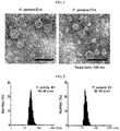
- FIG. 1 P. cedrina and P. panacis -derived EVs were observed to have spherical shapes.
- DLS dynamic light scattering
- diameters of EVs were measured using Zetasizer Nano S (Malvern Instruments Ltd., 633-nm laser line, scattered intensity 10 ⁇ 30 s).
- FIG. 2 diameters of the P. cedrina and P. panacis -derived EVs were measured at 30 to 40 nm and 20 to 30 nm, respectively.
- the P. panacis and EVs derived from the bacteria were labeled with Cy7 (GE Healthcare) for 1 hour at room temperature, and then the mice were fed a dose of 20 ⁇ g/mouse after overnight fasting. whole body images of the mice were taken at a wavelength of 780 to 800 nm using an IVIS spectrum CT (SelectScience) after 0 hr (h), 5 min (min), 3 hr (h), and 12 hr (h).
- the large intestine was extracted from a mouse having undergone overnight fasting, and after sealing both ends thereof, the P. panacis -derived EVs labeled with 10 ⁇ g of GFP emitting green fluorescence were injected into the intestine. Afterward, an image of the blood of the lamina intestinal of the large intestine was taken using a TCS SP5 microscope (Leica).
- Example 3 Metagenomic analysis of bacteria-derived EVs using urine and serum of atopic dermatitis patient
- the bacteria-derived EVs present in the urine and the serum of an atopic dermatitis patient were isolated, and then DNA contained therein were extracted.
- atopic dermatitis patients up to the age of 17 years, which satisfy Hanifin and Rajka diagnostic criteria for atopic dermatitis, were targeted. Patients with other skin diseases or who had received topical, oral or injectable antibiotics within the last 14 days were excluded. As a normal control, infants not having any skin disorders including atopic dermatitis, allergic disease, a significant medical history and drug use were selected. Urine and serum samples were obtained from the patients in the atopic dermatitis patient group and the normal control, consenting to this research. Afterward, EVs were isolated from the urine and serum samples by the method of Example 1, boiled at a high temperature to extract DNA, and then quantification and qualification of the DNA were detected using a NanoDropTM instrument before proceeding to the next step.
- Metagenomic analysis was performed on DNA isolated from EVs present in the urine and serum samples of the atopic dermatitis patients by the method of Example 3-1.
- PCR amplification was performed on emulsions prepared by mixing oil and amplicons to create "micro-reactors" containing an amplification mix and a single bead with TissueLyser II (Qiagen) and a GS-FLX plus emPCR Kit (454 Life Sciences).
- the emulsion was dispensed into a 96-well plate, and a 20-ng aliquot of each DNA sample was included in a 50- ⁇ l PCR reaction mix, followed by PCR proceeding according to the manufacturer's protocol (first step: 3 min at 94 °C; second step: 35 cycles of 15 sec at 94 °C, 45 sec at 55 °C, and 1 min at 72 °C; and final step: 8 min at 72 °C).
- Each DNA was amplified by emulsion PCR (emPCR), and the amplicons were purified using an AMpure Bead kit (Beckman Coulter, Brea, CA, USA), and then quantified using a Picogreen method (Invitrogen, Carlsbad, CA, USA). Subsequently, the amplicons were diluted and analyzed using a GS-FLX Titanium sequencer (Roche, Basel, Switzerland). After PCR amplification, each emulsion was chemically decomposed, and the beads with an amplified DNA library were washed through filtration. Positive beads were purified using biotinylated primer A (complementary to adaptor A), and attached to streptavidin-coated magnetic beads.
- the DNA library beads attached to the magnetic beads were separated from the magnetic beads by melting the double helix structure, and thus allowed single-stranded DNA to pass through.
- the sequencing primer was used again to create amplified single-stranded DNAs.
- the beads containing the amplified single-stranded DNA were counted using a particle counter (Beckman Coulter). Sequencing was performed using a Genome Sequencer FLX titanium (454 Life Sciences), and each sample was loaded into a 70 mm to 75 mm PicoTiter plate (454 Life Sciences).
- Phred quality scores (average Phred number > 20) and read lengths (> 300 bp) were checked to collect high-quality sequences.
- Operational taxonomic units (OUTs) were analyzed using UCLUST and USEARCH (Edgar, 2010), and phylogenetic classification was analyzed using QIIME (Lozupone, et al., 2006). Based on similarity, all of the 16s RNA sequences were classified at the following phylogenetic levels: species > 97% similarity; genus > 94% similarity; family > 90% similarity; order > 85% similarity; class > 80% similarity; and phylum > 75% similarity.
- Genus-level bacterial compositions were plotted as heat maps when a significant difference in compositions between the atopic dermatitis patient group and the normal control was detected to be two-fold or higher.
- Hierarchical clustering was performed when a significant difference in compositions was two-fold or higher or there was a 1% or higher average composition at the genus level between the atopic dermatitis patient group and the normal control.
- Example 3-1 After DNA was isolated from bacteria-derived EVs present in the urine and serum of the atopic dermatitis patient according to the method of Example 3-1, the base sequence of 16S rDNA was analyzed through metagenomic analysis according to the method of Example 3-2 to compare distributions of bacteria-derived EVs present in the urine and serum.
- FIGS. 7 to 11 show the results of metagenome analyses at phylum, class, order, family and genus levels, respectively, and EV distribution in the urine sample (boiled urine) of each patient is indicated by a red underline, and EV distribution in the serum sample (serum) thereof is indicated by a blue underline.
- Example 4 Analysis of difference in distribution of bacteria-derived EVs in urine of atopic dermatitis patient and normal person
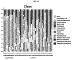
- FIGS. 12 to 16 show the results of the metagenome analyses at phylum, class, order, family and genus levels, respectively, and EV distribution in the urine sample of the normal person (control) is indicated by a blue underline, and EV distribution in the urine sample of the atopic dermatitis patient (case) is indicated by a red underline.
- bacteria-derived EVs in the urine of the atopic dermatitis patient and the normal person at all of phylum, class, order, family and genus levels. More specifically, as shown in FIG. 17 and Table 1 below, among bacteria-derived EVs present at 1% or more in the urine, Alicyclobacillus -derived EVs (normal person vs. atopic dermatitis patient: 0.31% vs. 8.49%), Methylobacterium -derived EVs (normal person vs. atopic dermatitis patient: 0.22% vs. 2.02%), Streptophyta -derived EVs (normal person vs.
- atopic dermatitis patient 2.87% vs. 7.89%)
- Propionibacterium -derived EVs normal person vs. atopic dermatitis patient: 2.78% vs. 7.6%
- Pseudomonas -derived EVs normal person vs. atopic dermatitis patient: 9.56% vs. 21.91%) are significantly increased in the urine of the atopic dermatitis patients (case) compared to the normal person (control).
- Lactobacillus -derived EVs normal person vs. atopic dermatitis patient: 20.92% vs.
- Leuconostoc- derived EVs normal person vs. atopic dermatitis patient: 14.60% vs. 0.12%
- Lactobacillales- derived EVs normal person vs. atopic dermatitis patient: 18.19% vs. 0.05%)
- Lactococcus -derived EVs normal person vs. atopic dermatitis patient: 11.57% vs. 0.03%
- Example 4 From the result of Example 4, since it was confirmed that the Lactobacillales and Lactobacillus -derived EVs are significantly decreased in the urine of the atopic dermatitis patients compared to the normal persons, the effects of immune responses and inflammation responses on the Lactobacillus -derived EVs were investigated.
- lactic acid bacteria such as Lactobacillus plantarum isolated from kimchi were cultured, and then a culture of the lactic acid bacteria was centrifuged at 10,000 x g for 20 minutes, followed by collecting only a supernatant and filtering it with a 0.22-mm filter. After EVs in the separated filtrate, having a size of 100 kDa or larger, were concentrated at a certain amount, and ultracentrifuged at 150,000 x g and 4 °C for 3 hours, the EV cluster remaining after the supernatant was discarded was diluted with normal saline (PBS), and then its concentration was measured using a BCA protein assay (Pierce, USA). Afterward, to confirm that physical properties of EVs isolated from the lactic acid bacteria are the same, dynamic light scattering and electron microscopy were performed.
- PBS normal saline
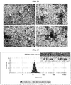
- the lactic acid bacteria-derived EVs have spherical shapes, and as shown in FIG. 19 , it was confirmed that they have an average diameter of approximately 15 nm through the dynamic light scattering.
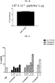
- a nanoparticle tracking analysis (NanoSight : LM10HS) was performed. After 2 ml of an EV sample adjusted to a concentration of 500 ng/ml was prepared, 0.3 to 0.4 ml of the sample was placed in a chamber of the LM-10HS instrument, and focused using a camera so that particles are clearly visible. After the camera was raised to the maximum level while being focused, the sample was captured by the stepwise lowering of the level to confirm whether there was no drift of the sample. After confirming the concentration, focus and drift of the sample, a capture duration was set to 30 seconds and thus data was obtained. As a result, as shown in FIG. 20 , it was confirmed that 2.47 x 10 8 EVs are present based on 1 ⁇ g of protein.
- lactic acid bacteria-derived EVs 2 x 10 5 cells of a dermal epithelial cell line (HaCaT) were treated with S . aureus-derived EVs ( S . aureus EV) and Propionibacterium acne-derived EVs ( P. acnes EV), known as the main causative agents of inflammatory diseases, and lactic acid bacteria-derived EVs (CJLP133 EV) isolated by the method of Example 5 at concentrations of 10 ng to 25 ⁇ g/ml, respectively, cultured for 12 hours, and a secretion level of cytokine IL-6 causing a Th17 immune response in a supernatant was detected by ELISA.
- S . aureus-derived EVs S . aureus EV
- P. acnes EV Propionibacterium acne-derived EVs
- CJLP133 EV lactic acid bacteria-derived EVs isolated by the method of Example 5 at concentrations of 10 ng to 25 ⁇ g/m
- dermal epithelial cells treated with the lactic acid bacteria-derived EVs at various concentrations (0.1, 1, and 10 ⁇ g/ml) 12 hours before were treated with 25 ⁇ g/ml of S . aureus or Propionibacterium acne -derived vesicles and then cultured for 12 hours, followed by identifying a secretion level of inflammatory cytokine IL-6 in a supernatant by ELISA.
- S. aureus EV lactic acid bacteria-derived EVs
- aureus -derived vesicles was concentration-dependently decreased, but there was no effect of the lactic acid bacteria-derived EVs on IL-6 secretion caused by the Propionibacterium acne -derived vesicles ( P. acnes EV).
- inflammatory cells that is, a mouse peritoneal macrophage line (Raw 264.7) was treated with 1 ⁇ of S . aureus -derived EVs ( S. aureus ), or 10 ng, 100 ng or 1 ⁇ g of lactic acid bacteria-derived EVs (CJLP133) for 12 hours, and thus amounts of inflammatory cytokines IL-6 and TNF- ⁇ secreted from peritoneal macrophages were detected and compared. As a result, as shown in FIG.
- the mouse peritoneal macrophage line was treated with the lactic acid bacteria-derived EVs at various concentrations (0.01, 0.1 and 1 ⁇ g/ml), 12 hours later, the S. aureus -derived EVs were treated at a concentration of 1 ⁇ g/ml, and then another 12 hours later, amounts of the inflammatory cytokines IL-6 and TNF- ⁇ were detected by ELISA.
- FIG. 24 it was confirmed that, when the lactic acid bacteria-derived EVs were pre-treated, the IL-6 and TNF- ⁇ secretion caused by the S. aureus-derived EVs is inhibited. This means that the lactic acid bacteria-derived EVs can effectively inhibit inflammation responses of inflammatory cells triggered by the S. aureus-derived EVs.
- P. aeruginosa is the antibiotic-resistant main pathogenic bacteria belonging to the genus Pseudomonas, and also known as the main causative bacteria of sepsis.
- P . aeruginosa -derived EVs have been known as the main causative agent of a chronic lung disease such as asthma, chronic obstructive pulmonary disease (COPD) or lung cancer, due to repeated exposure through the airway.
- COPD chronic obstructive pulmonary disease
- inflammatory cells a peritoneal macrophage line (Raw 264.7), were treated with lactic acid bacteria-derived EVs (CJLP133 EV) at various concentrations (0.01, 0.1, and 1 ⁇ g/ml), 12 hours later, P. aeruginosa-derived EVs ( P. aeruginosa EV) were treated at a concentration of 1 ⁇ g/ml, and then concentrations of inflammatory cytokines IL-6 and TNF- ⁇ were detected by ELISA. As a result, as shown in FIG. 26 , it was confirmed that the secretion of IL-6 and TNF- ⁇ by P. aeruginosa-derived vesicle stimulation is almost perfectly inhibited in proportion to a concentration of the lactic acid bacteria-derived EVs treated.
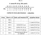
- SKH-1 hairless mice were repeatedly coated with 10 ⁇ g of the S . aureus-derived EVs ( S. aureus EV) three times a week for 4 weeks to produce an atopic dermatitis model, 12 hours before the S. aureus- derived EVs were coated, a therapeutic effect on the development of atopic dermatitis was observed by coating the skin with the lactic acid bacteria-derived EVs (CJLP133 EV).
- CJLP133 EV lactic acid bacteria-derived EVs
- an immunosuppressant used as a medication for atopic dermatitis, dexamethasone was intraperitoneally injected at a concentration of 300 ⁇ g, and the experiment was carried out under the same conditions.
- Example 12 Evaluation of safety by multiple oral administration of high dose of lactic acid bacteria-derived EVs
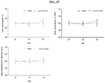
- lactic acid bacteria-derived EVs 500 ⁇ g of lactic acid bacteria-derived EVs (CJLP133) were orally administered three times a week for four weeks (total 12 times) by the method illustrated in FIG. 34 . Afterward, in a control in which the vesicles were not administered and a group to which lactic acid bacteria-derived vesicles were orally administered, the body weight, feed intake, and body temperature of each mouse were detected.
- the blood was extracted to analyze blood cell ingredients, resulting in that there was no significant difference in 8 types of indexes shown in FIG. 38 (WBC: the number of white blood cells, RBC: the number of red blood cells, HGB: the concentration of hemoglobin, HCT: hematocrit, MCV: mean corpuscular volume, MCH: mean corpuscular hemoglobin, MCHC: mean corpuscular hemoglobin concentration, and PLT: the number of platelets) between the control and the group to which the lactic acid bacteria-derived EVs were administered.
- WBC the number of white blood cells
- RBC the number of red blood cells
- HGB the concentration of hemoglobin
- HCT hematocrit
- MCV mean corpuscular volume
- MCH mean corpuscular hemoglobin
- MCHC mean corpuscular hemoglobin concentration
- PLT the number of platelets
Landscapes
- Health & Medical Sciences (AREA)
- Life Sciences & Earth Sciences (AREA)
- Chemical & Material Sciences (AREA)
- General Health & Medical Sciences (AREA)
- Engineering & Computer Science (AREA)
- Organic Chemistry (AREA)
- Veterinary Medicine (AREA)
- Public Health (AREA)
- Animal Behavior & Ethology (AREA)
- Medicinal Chemistry (AREA)
- Pharmacology & Pharmacy (AREA)
- Microbiology (AREA)
- Molecular Biology (AREA)
- Bioinformatics & Cheminformatics (AREA)
- Proteomics, Peptides & Aminoacids (AREA)
- Mycology (AREA)
- Epidemiology (AREA)
- Pulmonology (AREA)
- General Chemical & Material Sciences (AREA)
- Chemical Kinetics & Catalysis (AREA)
- Nuclear Medicine, Radiotherapy & Molecular Imaging (AREA)
- Analytical Chemistry (AREA)
- Biotechnology (AREA)
- Zoology (AREA)
- Wood Science & Technology (AREA)
- Immunology (AREA)
- Biochemistry (AREA)
- Physics & Mathematics (AREA)
- Genetics & Genomics (AREA)
- Dermatology (AREA)
- Biophysics (AREA)
- General Engineering & Computer Science (AREA)
- Urology & Nephrology (AREA)
- Biomedical Technology (AREA)
- Hematology (AREA)
- Food Science & Technology (AREA)
- Pathology (AREA)
- Otolaryngology (AREA)
- Tropical Medicine & Parasitology (AREA)
- Birds (AREA)
Description
- The present disclosure relates to a composition for preventing, improving or treating an inflammatory disease, comprising lactic acid bacteria-derived extracellular vesicles as active ingredients, and a method for diagnosing atopic dermatitis.
- In recent years, disease patterns have changed from acute infectious diseases to chronic inflammatory diseases because of a change in life style. Inflammation is a defense mechanism occurring in the body in response to external toxic substances entering the body. Acute inflammation is triggered by short-term infection of environmental bacteria, fungi, etc., and chronic inflammation is triggered when causative agents such as microorganisms, allergens, etc. that exist in the environment are continuously absorbed into the body. Recently, the importance of infectious causes for the occurrence of chronic inflammatory diseases that have been recognized to be triggered by noninfectious causes is being emphasized. As, representatively, helicobacter bacteria that have been known to co-exist in the stomach have been identified as a critical causative agent of gastritis, gastric cancer, etc., bacteria and substances derived from the bacteria, which are known to co-exist in the environment, have recently attracted attention as causative agents of cancer triggered by chronic inflammatory diseases and complications of chronic inflammation.
- Atopic dermatitis is a type of dermal disease that is most frequently found in early-infancy and childhood, and the first signal of atopic march progressing to asthma or allergic rhinitis. In addition, atopic dermatitis is a chronic inflammatory disease characterized by chronic itching and repeated inflammation of the skin and thus physically and mentally affecting not only the patient himself but also the quality of life in the family. In recent years, the increase in global prevalence of the atopic dermatitis is being followed by an increasing attention thereto. In Korea, according to the International Study of Allergy and Asthma in Childhood (ISAAC) survey conducted by the Korean Pediatric Allergy Respiratory Society, for atopic dermatitis until now, compared to 1995, prevalence increased both at the ages ranging from 6 to 12 in 2000 (15.3% in 1995 and 17.0% in 2000) and at the ages ranging from 12 to 15 (7.2% in 1995 and 9.2% in 2000), and for atopic dermatitis in the last 12 months, prevalence increased both at the age ranging from 6 to 12 in 2000 (7.3% in 1995 and 10.7% in 2000), and at the ages ranging from 12 to 15 (3.9% in 1995 and 6.1% in 2000). An allergic disease such as atopic dermatitis is triggered by combined interaction between a genetic factor and an environmental factor, and the recent growth trend is insufficient to be explained only by the genetic factor. Although differences in incidence of allergic diseases depending on a country reflects differences among ethnic groups, that is, the significance of genetic factors, the same result as shown by a surge of allergic diseases in the former East German region after reunification indicates that environmental factors are also significant. Such a surge of allergic diseases is involved in westernized life, and there may be three possible causes: first, an increase in exposure to house dust mites because of generally spending more time indoors, secondly, exposure to a wide range of microorganisms due to environmental hygiene and use of antibiotics, and thirdly, westernized eating habits.
- Atopic dermatitis patients frequently have dermal infection caused by functional disorder of a skin barrier, or immune dysfunction. Particularly, bacterial infections by Staphylococcus, viral infections by herpes simplex viruses, and fungal infections are common. Among these, Staphylococcus aureus is detected in 90% of patients with atopic dermatitis and is also detected in skin lesions without obvious infection symptoms, and the toxin of Staphylococcus aureus serves as a superantigen that increases allergic immune responses, which is known to exacerbate skin itching and lesions.
- The prognosis of atopic dermatitis varies depending on a patient's skin condition, stimulation factor, accompanying allergic disease, and bacterial infection. Generally, atopic dermatitis tends to improve with an increasing age, while symptoms are severe at the young age and chronic lesions persist. However, some reports show that about 40% of patients have improved symptoms at the age of around 5, and the reason for this improvement is controversial. The most representative method for treating atopic dermatitis when exacerbated due to various reasons includes elimination of the causative agents and simultaneous application of topical and systemic steroids or topical immunosuppressants as well as proper use of moisturizers. However, recent studies suggest that, in addition to these factors, lactic acid bacteria or a culture thereof may be used to lower the severity of atopic dermatitis.
- Asthma and chronic obstructive pulmonary disease (COPD) are diseases characterized by airway obstruction due to chronic airway inflammation, wherein the asthma is characterized by reversible airway obstruction, and the COPD is characterized by irreversible airway obstruction. A hypersensitivity reaction to allergens derived from house dust mites as a causative factor of asthma is known to be important in the pathogenesis of eosinophilic asthma, and in recent years, research has been focused on the importance of bacteria-derived substances for neutrophilic inflammation, which is a characteristic pathological feature of severe asthma or COPD. Particularly, the finding that extracellular vesicles derived from pathogenic bacteria present in indoor dust are important in the onset of asthma and COPD has been reported. In addition, it has been reported that bacteria and substances derived from the bacteria co-existing in the nasal cavity play an important role in the pathogenesis of chronic rhinosinusitis, and the reduction of the diversity of bacterial flora in the nasal cavity and the increase in specific pathogens, particularly, Staphylococci, are important in the onset of chronic rhinosinusitis.
- Bacteria secrete bilayer-structured lipoproteins, that is, extracellular vesicles (EVs) frequently called nanovesicles into an extracellular environment. Gram-negative bacteria-derived extracellular vesicles, or outer membrane vesicles (OMVs) contain a toxic protein and bacterial DNA and RNA as well as lipopolysaccharides, and gram-positive bacteria-derived extracellular vesicles also contain bacterial cell wall ingredients, such as peptidoglycan and lipoteichoic acid as well as a toxic protein and nucleic acid. In recent years, it has been reported that EVs secreted from the representative gram-positive bacteria Staphylococcus aureus are important causative agents of atopic dermatitis, chronic rhinosinusitis, neutrophilic asthma and COPD, and it has been reported that large amounts of EVs derived from pathogenic gram-negative bacteria such as Pseudomonas aeruginosa and Acinetobacter bacumannii are present in indoor dust, and are important causative agents of asthma, COPD, and lung cancer when inhaled.
- Meanwhile, metagenomics also called environmental genomics may be analysis of metagenome materials obtained from environmental samples. Recently, the bacterial composition of human microbial flora can be cataloged by a method based on a 16S ribosome RNA (16S rRNA) base sequence, and 16S rRNA is sequenced using the 454FLX titanium platform. In the meantime, while research has been conducted on metagenomes analyzed from patient samples, it was not known that bacteria-derived EVs are present in serum or urine, and therefore metagenomic analysis was not conducted on bacteria-derived EVs isolated from serum or urine. In addition, on the basis of results of metagenomic analysis using DNA of EVs isolated from patients' sera or urine, there is no case that bacteria-derived EVs are used to prevent or treat an inflammatory disease such as atopic dermatitis, asthma, COPD, chronic rhinitis, chronic rhinosinusitis, and sepsis.
- Al-Nedawi et al., FASEB J., 29 (2), 684 - 695 (2014) relates to an investigation of "gut commensal microvesicles" found to "reproduce parent bacterial signals to host immune and enteric nervous systems".
-
US 2012/0196352 A1 relates to a "novel Lactobacillus plantarum and composition comprising same". - Kang et al., PLoS ONE, 8 (10), e76520 (2013) relates to an investigation of "extracellular vesicles derived from gut microbiota, especially Akkermansia muciniphila".
- Hong et al., Allergy, 66 (3), 351 - 359 (2010) relates to "extracellular vesicles derived from Staphylococcus aureus" and their ability to "induce atopic dermatitis-like skin inflammation".
- Lee et al., Proteomics, 9 24), 5425 - 5436 (2009) relates to "characterization of Staphylococcus aureus-derived membrane vesicles".
-
JP 2010-047504 A -
KR 2011-0025603 A -
KR 2013-0034764 A -
KR 2005-0109928 A - As a result of metagenomic analysis performed using DNA of EVs isolated from the urine of a patient with atopic dermatitis and a normal person, the present authors confirmed that lactic acid bacteria-derived EVs were significantly increased in the urine of the normal person compared to the patient with atopic dermatitis, and the lactic acid bacteria-derived EVs effectively inhibit inflammatory responses induced by EVs derived from Staphylococcus aureus (S. aureus) and Pseudomonas aerugionsa (P. aeruginosa), which are the main causative agents of an inflammatory disease.
- The present invention is as defined in the claims. This invention and other aspects of the present disclosure may be more fully understood by reference to the following description.
- Disclosed herein is a composition for preventing, improving or treating an inflammatory disease, comprising lactic acid bacteria-derived EVs as active ingredients.
- Also disclosed is a method for diagnosing atopic dermatitis.
- In one aspect, the present disclosure provides a pharmaceutical composition for preventing or treating an inflammatory disease, comprising lactic acid bacteria-derived EVs as active ingredients.
- In another aspect, the present disclosure provides a cosmetic composition for improving an inflammatory disease, comprising lactic acid bacteria-derived EVs as active ingredients.
- In still another aspect, the present disclosure provides a health functional food composition for improving an inflammatory disease, comprising lactic acid bacteria-derived EVs as active ingredients.
- In yet another aspect, the present disclosure provides an inhalant composition for preventing or treating an inflammatory disease, comprising lactic acid bacteria-derived EVs as active ingredients.
- In an exemplary embodiment of the present disclosure, the lactic acid bacteria may include bacteria in the order Lactobacillales.
- In another exemplary embodiment of the present disclosure, the bacteria in the order Lactobacillales may include bacteria in the genus Lactococcus, Lactobacillus, or Leuconostoc.
- In still another exemplary embodiment of the present disclosure, the bacteria in the genus Lactobacillus may include Lactobacillus plantarum.
- In yet another exemplary embodiment of the present disclosure, the EVs may have an average diameter of 10 to 300 nm.
- In yet another exemplary embodiment of the present disclosure, the EVs may be isolated from a culture of lactic acid bacteria.
- In yet another exemplary embodiment of the present disclosure, the EVs are naturally or artificially secreted from lactic acid bacteria.
- In yet another exemplary embodiment of the present disclosure, the inflammatory disease may be a disease selected from the group consisting of atopic dermatitis, chronic rhinitis, chronic rhinosinusitis, asthma, COPD, and sepsis.
- In yet another aspect, the present disclosure provides a method for diagnosing atopic dermatitis, comprising the following steps:
- (a) extracting genes from EVs isolated from clinical samples;
- (b) sequencing the genes; and
- (c) determining that there is a high risk of atopic dermatitis when distribution of lactic acid bacteria-derived EVs is lower than that of a normal person through the sequencing.
- In one exemplary embodiment of the present disclosure, the gene may be DNA or RNA.
- In another exemplary embodiment of the present disclosure, the clinical sample may be urine or blood.
- In still another exemplary embodiment of the present disclosure, the sequencing may be performed through polymerase chain reaction (PCR).
- In yet another aspect, the present disclosure provides a method for preventing or treating an inflammatory disease, which comprises administering a composition comprising lactic acid bacteria-derived EVs as active ingredients to a subject.
- In yet another aspect, the present disclosure provides a use of lactic acid bacteria-derived EVs for preventing or treating an inflammatory disease.
- In yet another aspect, the present disclosure provides a use of lactic acid bacteria-derived EVs for diagnosing atopic dermatitis.
- The authors identified by in vitro and in vivo experiments that larger amounts of lactic acid bacteria-derived EVs are significantly distributed in a normal person compared to an atopic dermatitis patient, inflammation responses in epithelial cells and inflammatory cells due to exposure of Staphylococcus or Pseudomonas-derived EVs are effectively inhibited by the lactic acid bacteria-derived EVs, and a composition comprising the lactic acid bacteria-derived EVs as active ingredients to prevent, improve or treat an inflammatory disease such as atopic dermatitis, chronic rhinitis, chronic rhinosinusitis, asthma, chronic obstructive pulmonary disease or sepsis is expected to be useful in development of drugs, cosmetics, or health functional food.
- Also, as distribution of lactic acid bacteria-derived EVs is detected in a urine or blood sample, the lactic acid bacteria-derived EVs can be useful for diagnosing atopic dermatitis.
-
-
FIG. 1 shows Pseudomonas cedrina (P. cedrina) and Pseudomonas panacis (P. panacis)-derived EVs isolated from in vitro cultures of enterobacteria such as P. cedrina and P. panacis, obtained using an electron microscope, respectively. -
FIG. 2 shows sizes of P. cedrina and P. panacis-derived EVs, measured by dynamic light scattering. -
FIG. 3 shows CBB-R-250-stained gels obtained by SDS-PAGE to evaluate expression of proteins contained in P. cedrina, P. panacis and EVs derived therefrom. -
FIG. 4 shows whole body images illustrating distribution of bacteria and EVs in mice at a predetermined time after P. panacis and P. panacis-derived EVs are orally administered to the mice. -
FIG. 5 shows bacterial and EV distribution extracted from the blood, heart, lungs, liver and kidneys of a mouse at 12 hours after oral administration of P. panacis and P. panacis-derived EVs thereto. -
FIG. 6 shows that EVs are present inintestinal capillaries 10 minutes after P. panacis-derived EVs are directly administered to intestines. -
FIG. 7 shows the result of metagenomic analysis at a bacteria phylum level to compare distribution of bacteria-derived EVs isolated from the urine and sera of atopic dermatitis patients. -
FIG. 8 shows the result of metagenomic analysis at a bacteria class level to compare distribution of bacteria-derived EVs isolated from the urine and sera of atopic dermatitis patients. -
FIG. 9 shows the result of metagenomic analysis at a bacteria order level to compare distribution of bacteria-derived EVs isolated from the urine and sera of atopic dermatitis patients. -
FIG. 10 shows the result of metagenomic analysis at a bacteria family level to compare distribution of bacteria-derived EVs isolated from the urine and sera of atopic dermatitis patients. -
FIG. 11 shows the result of metagenomic analysis at a bacteria genus level to compare distribution of bacteria-derived EVs isolated from the urine and sera of atopic dermatitis patients. -
FIG. 12 shows the result of metagenomic analysis at a bacteria phylum level to compare distribution of bacteria-derived EVs isolated from the urine of an atopic dermatitis patient and a normal person. -
FIG. 13 shows the result of metagenomic analysis at a bacteria class level to compare distribution of bacteria-derived EVs isolated from the urine of an atopic dermatitis patient and a normal person. -
FIG. 14 shows the result of metagenomic analysis at a bacteria order level to compare distribution of bacteria-derived EVs isolated from the urine of an atopic dermatitis patient and a normal person. -
FIG. 15 shows the result of metagenomic analysis at a bacteria family level to compare distribution of bacteria-derived EVs isolated from the urine of an atopic dermatitis patient and a normal person. -
FIG. 16 shows the result of metagenomic analysis at a bacteria genus level to compare distribution of bacteria-derived EVs isolated from the urine of an atopic dermatitis patient and a normal person. -
FIG. 17 is a heat map showing the result of metagenomic analysis of bacteria-derived EVs isolated from the urine of an atopic dermatitis patient and a normal person. -
FIG. 18 shows images of EVs isolated from a culture of Lactobacillus plantarum, obtained using an electron microscope. -
FIG. 19 shows the sizes of EVs isolated from a culture of Lactobacillus plantarum by dynamic light scattering. -
FIG. 20 shows the number of EVs isolated from a culture of Lactobacillus plantarum by a nanotrafficking assay (NTA). -
FIG. 21 shows IL-6 secretion levels measured by ELISA, after S. aureus-derived EVs (S. aureus EV), Propionibacterium acne-derived EVs (P. acnes EV), and lactic acid bacteria-derived EVs (CJLP133 EV) are administered to dermal epithelial cells. -
FIG. 22 shows IL-6 secretion levels measured by ELISA, after lactic acid bacteria-derived EVs (CJLP133 EV) are administered 12 hours before S. aureus-derived EVs (S. aureus EV), or Propionibacterium acne-derived EVs (P. acnes EV) are administered to dermal epithelial cells. -
FIG. 23 shows IL-6 and TNF-α secretion levels measured by ELISA, after S. aureus-derived EVs (S. aureus EV), or lactic acid bacteria-derived EVs (CJLP133 EV) are administered to peritoneal macrophages. -
FIG. 24 shows IL-6 and TNF-α secretion levels measured by ELISA, after lactic acid bacteria-derived EVs (CJLP133 EV) are administered 12 hours before S. aureus-derived EVs (S. aureus EV) are administered to peritoneal macrophages. -
FIG. 25 shows a degree of apoptosis evaluated by MTT assays, after lactic acid bacteria-derived EVs (CJLP133 EV) are administered 12 hours before S. aureus-derived EVs (S. aureus EV) are administered to dermal epithelial cells. -
FIG. 26 shows IL-6 and TNF-α secretion levels measured by ELISA, after lactic acid bacteria-derived EVs (CJLP133 EV are administered 12 hours before Pseudomonas aeruginosa-derived EVs (P. aeruginosa EV) are administered to peritoneal macrophages. -
FIG. 27 shows IL-6 and TNF-α secretion levels measured by ELISA, after Pseudomonas aeruginosa-derived EVs (P. aeruginosa EV) are administered to peritoneal macrophages together with lactic acid bacteria-derived EVs (CJLP133 EV). -
FIG. 28 shows the protocol for evaluating therapeutic effects caused by dermal administration of lactic acid bacteria-derived EVs (CJLP133 EV) in atopic dermatitis mouse models caused by S. aureus-derived EVs (S. aureus EV). -
FIG. 29 shows images of skin, taken to evaluate therapeutic effects after lactic acid bacteria-derived EVs (CJLP133 EV) are percutaneously administered in atopic dermatitis mouse models caused by S. aureus-derived EVs (S. aureus EV). -
FIG. 30 shows IL-17 concentrations in dermal tissue after lactic acid bacteria-derived EVs (CJLP133 EV) are percutaneously administered to atopic dermatitis mouse models caused by S. aureus-derived EVs (S. aureus EV). -
FIG. 31 shows the protocol for evaluating therapeutic effects by oral administration of lactic acid bacteria-derived EVs (CJLP133 EV) to atopic dermatitis mouse models caused by S. aureus-derived EVs (S. aureus EV). -
FIG. 32 shows images of skin, taken to evaluate therapeutic effects after lactic acid bacteria-derived EVs (CJLP133 EV) are orally administered in atopic dermatitis mouse models caused by S. aureus-derived EVs (S. aureus EV). -
FIG. 33 shows concentrations of IL-4, IL-6, IL-17, and IFN-γ, which are immune response markers, in dermal tissue after lactic acid bacteria-derived EVs (CJLP133 EV) are orally administered to atopic dermatitis mouse models caused by S. aureus-derived EVs (S. aureus EV). -
FIG. 34 shows the protocol for evaluating toxicity by high-dose oral administration of lactic acid bacteria-derived EVs (CJLP133 EV). -
FIG. 35 shows changes in body weights after high doses of lactic acid bacteria-derived EVs (CJLP133 EV) are orally administered multiple times. -
FIG. 36 shows the feed intake of mice after high doses of lactic acid bacteria-derived EVs (CJLP133 EV) are orally administered multiple times. -
FIG. 37 shows the changes in body temperatures after high doses of lactic acid bacteria-derived EVs (CJLP133 EV) are orally administered multiple times. -
FIG. 38 shows the changes in blood cells in the blood after high doses of lactic acid bacteria-derived EVs (CJLP133 EV) are orally administered multiple times (WBC: the number of white blood cells, RBC: the number of red blood cells, HGB: the concentration of hemoglobins, HCT: hematocrit, MCV: mean corpuscular volume (mean volume of red blood cells), MCH: mean corpuscular hemoglobin (mean number of hemoglobins), MCHC: mean corpuscular hemoglobin concentration (mean concentration of hemoglobins), and PLT: the number of platelets). - As a result of metagenomic analysis using DNA of EVs isolated from the urine of an atopic dermatitis patient and a normal person, it was confirmed that, compared to the atopic dermatitis patient, lactic acid bacteria-derived EVs in the urine of the normal person are considerably increased, and the lactic acid bacteria-derived EVs efficiently inhibit inflammation responses induced by EVs derived from main causative factors of inflammatory diseases such as S. aureus (S. aureus) and Pseudomonas aeruginosa (P. aeruginosa).
- In one exemplary embodiment of the present disclosure, as a result of identifying distribution and excretion patterns by isolating EVs from enterobacteria such as P. cedrina and P. panacis in the bodies and orally administering the bacteria and the EVs derived therefrom to mice, it was confirmed that the enterobacteria are not absorbed in the whole body, whereas the enterobacteria-derived EVs are systematically absorbed and then excreted in urine (see Examples 1 and 2).
- In another exemplary embodiment of the present disclosure, DNA of bacteria-derived EVs isolated from the urine and sera of atopic dermatitis patients are each subjected to metagenomic analysis at a bacteria phylum, class, order, family or genus level, and as a result, it was confirmed that distributions of bacteria-derived EVs in the urine and serum exactly match (see Example 3).
- In still another exemplary embodiment of the present disclosure, based on the above result, metagenomic analysis was carried out using DNA of EVs isolated from the urine of atopic dermatitis patients and normal persons. As a result of analysis of EV distribution at each of the bacteria phylum, class, order, family, and genus levels, compared to the normal persons, EVs derived from bacteria in the genus Alicyclobacillus, the genus Methylobacterium, the order Streptophyta, the genus Propionibacterium, and the genus Pseudomonas are significantly increased, but EVs derived from bacteria the order Lactobacillales, the genus Lactobacillus, the genus Leuconostoc, and the genus Lactococcus are significantly decreased in the urine of the atopic dermatitis patients.
- In yet another exemplary embodiment of the present disclosure, based on the metagenome result, as a result of analysis of the characteristics of EVs isolated after lactic acid bacteria such as Lactobacillus plantarum isolated from fermented food were in vitro cultured, spherical vesicles having an average diameter of approximately 15 nm were able to be isolated (see Example 5), and it was confirmed that, following the treatment of dermal epithelial cells and macrophages with the vesicles, inflammation responses induced by the treatment of EVs secreted from S. aureus and P. aeruginosa, which are the main causative agents of inflammatory diseases, are significantly inhibited (see Examples 6 to 9).
- In yet another exemplary embodiment of the present disclosure, based on the in vitro test result, it was confirmed that, when lactic acid bacteria-derived vesicles are orally or percutaneously administered to a atopic dermatitis mouse model induced by S. aureus-derived EVs, dermal inflammation caused by S. aureus-derived vesicles is effectively inhibited by lactic acid bacteria-derived vesicles (see Examples 10 and 11), and according to multiple oral administrations of high doses of lactic acid bacteria-derived EVs to mice, it was confirmed that there was no difference in various biomarkers from the control, resulting in verification of EV stability (see Example 12).
- Thus, in one aspect, the present disclosure provides a pharmaceutical composition for preventing or treating an inflammatory disease, comprising lactic acid bacteria-derived EVs as active ingredients.
- The term "inflammatory disease" used herein includes infectious inflammatory diseases induced by exposure to infectious factors such as bacteria or bacteria-derived vesicles, toxins, viruses, or fungi, allergens, allergic inflammatory diseases induced by exposure to allergens, and autoimmune inflammatory diseases caused by self-antigens.
- The term "inflammatory disease" used herein is the generic term for inflammatory diseases caused by infectious factors, such as atopic dermatitis, chronic rhinitis, chronic rhinosinusitis, asthma, chronic obstructive pulmonary disease (COPD), and sepsis.
- The term "allergic inflammatory disease" used herein is the generic term for inflammatory diseases caused by allergens, such as atopic dermatitis, chronic rhinitis, chronic rhinosinusitis, asthma, and chronic obstructive pulmonary disease (COPD).
- The term "atopic dermatitis" used herein is the generic term for dermal diseases accompanying itching chronically occurring on the skin.
- The term "prevention" used herein refers to all actions of inhibiting or delaying inflammatory diseases by administration of the pharmaceutical composition according to the present disclosure.
- The term "treatment" used herein refers to all actions involved in alleviating or beneficially changing symptoms of inflammatory diseases by administration of the pharmaceutical composition according to the present disclosure.
- The lactic acid bacteria of the present disclosure may include bacteria in the order Lactobacillales, the genus Lactococcus, the genus Lactobacillus, or the genus Leuconostoc, but the present disclosure is not limited thereto.
- The bacteria in the genus Lactobacillus preferably include Lactobacillus plantarum, but the present disclosure is not limited thereto.
- The EVs of the present disclosure may be isolated from a culture of the lactic acid bacteria or food fermented with the lactic acid bacteria, and may be naturally or artificially secreted from the lactic acid bacteria, but the present disclosure is not limited thereto.
- A method for isolating EVs from a culture of the lactic acid bacteria of the present disclosure or fermented food is not particularly limited as long as the culture or fermented food contains EVs. For example, EVs may be isolated using centrifugation, ultracentrifugation, filtration using a filter, gel filtration chromatography, free-flow electrophoresis, capillary electrophoresis or a combination thereof, and the method may further include washing for removing impurities, and concentration of the obtained EVs.
- The EVs isolated by the method of the present disclosure may have an average diameter of 10 to 300 nm, and preferably 10 to 200 nm, but the present disclosure is not limited thereto.
- The pharmaceutical composition according to the present disclosure comprises lactic acid bacteria-derived EVs as active ingredients, and a pharmaceutically acceptable carrier. The pharmaceutically acceptable carrier is conventionally used for preparation, and may be, but is not limited to, a saline solution, distilled water, Ringer's solution, buffered saline, a cyclodextrin solution, a dextrose solution, a maltodextrin solution, glycerol, ethanol, or liposomes, etc., and may further include another conventional additive such as an antioxidant or a buffer as needed. In addition, the pharmaceutically acceptable carrier may be prepared as injectable forms such as an aqueous solution, a suspension, and an emulsion, pills, capsules, granules or tablets by further adding diluents, dispersants, surfactants, binders, lubricants, etc. Suitable pharmaceutically acceptable carriers and their preparations may be prepared according to each ingredient using a method disclosed in the Remington's Pharmaceutical Science. The pharmaceutical composition of the present disclosure is not limited to dosage forms, and thus may be prepared as injections, inhalants, topical formulations for skin, or oral medication.
- The pharmaceutical composition of the present disclosure may be administered orally or parenterally (e.g., intravenously, subcutaneously, percutaneously, nasally, or intratracheally) according to a desired method, and a dose of the pharmaceutical composition of the present disclosure may be selected according to a patient's condition and body weight, severity of a disease, a dosage form, an administration route and duration by those of ordinary skill in the art.
- The pharmaceutical composition of the present disclosure is administered at a pharmaceutically effective amount. In the present disclosure, the "pharmaceutically effective amount" refers to an amount sufficient to treat the disease at a reasonable benefit/risk ratio applicable for medical treatment, and an effective dosage may be determined by parameters including a type of a patient's disease, severity, drug activity, sensitivity to a drug, administration time, an administration route and an excretion rate, the duration of treatment and drugs simultaneously used, and other parameters well known in the medical field. The pharmaceutical composition of the present disclosure may be administered separately or in combination with other therapeutic agents, and may be sequentially or simultaneously administered with a conventional therapeutic agent, or administered in a single dose or multiple doses. In consideration of all of the above-mentioned parameters, it is important to achieve the maximum effect with the minimum dose without a side effect, and such a dose may be easily determined by one of ordinary skill in the art.
- Specifically, the effective amount of the compound according to the present disclosure may vary depending on a patient's age, sex, and body weight, and may be generally administered at 0.001 to 150 mg and, preferably, 0.01 to 100 mg/ kg of body weight daily or every other day, or once to three times a day. However, the effective amount may vary depending on an administration route, the severity of obesity, sex, body weight or age.
- In another aspect, the present disclosure provides a cosmetic composition for improving inflammatory diseases, comprising lactic acid bacteria-derived EVs as active ingredients.
- The cosmetic composition of the present disclosure may comprise ingredients conventionally used in the cosmetic composition, for example, conventional additives including an antioxidant, a stabilizer, a solubilizer, a vitamin, a pigment and a flavoring, and a carrier, as well as lactic acid bacteria-derived EVs.
- In addition, the composition of the present disclosure may be used in combination with an organic sunblock that has been conventionally used so long as the skin protection effect is not impaired, in addition to lactic acid bacteria-derived EVs. The organic sunblock may include at least one selected from the group consisting of glyceryl PABA, drometrizole trisiloxane, drometrizole, digalloyl trioleate, disodium phenyl dibenzimidazole tetrasulfonate, diethylhexyl butamido triazone, diethylamino hydroxybenzoyl hexyl benzoate, DEA-methoxycinnamate, a Lawson/dihydroxyacetone mixture, methylene bis-benzotriazolyl tetramethylbutyphenol, 4-methylbenzylidene camphor, menthyl anthranilate, benzophenone-3 (oxybenzone), benzophenone-4, benzophenone-8 (dixoyphebenzone), butyl methoxydibenzoylmethane, bisethylhexyloxyphenol methoxy phenyltriazine, cinoxate, ethyl dihydroxypropyl PABA, octocrylene, ethyl hexyldimethyl PABA, ethylhexyl methoxycinnamate, ethylhexyl salicylate, ethylhexyl triazone, isoamyl-p-methoxycinnamate, polysilicon-15 (dimethicodiethylbenzal malonate), terephthalylidene dicamphorsulfonic acid and a salt thereof, TEA-salicylate, and aminobenzoic acid (PABA).
- Products that can contain the cosmetic composition of the present disclosure include, for example, cosmetics such as an astringent, a moisturizing lotion, a nourishing lotion, various creams, an essence, a mask pack, a foundation, etc., a cleanser, soap, a treatment, and other cosmetic liquids. Specific types of the cosmetic composition of the present disclosure include a skin lotion, a skin softener, a skin toner, an astringent, a lotion, a milk lotion, a moisturizer lotion, a nourishing lotion, a massage cream, a nourishing cream, a moisturizing cream, a hand cream, an essence, a nourishing essence, a pack, soap, a shampoo, a cleansing foam, a cleansing lotion, a cleansing cream, a body lotion, a body cleanser, an emulsion, a lipstick, a makeup base, a foundation, a pressed powder, a loose powder, and an eye shadow.
- In still another aspect, the present disclosure provides a health functional food composition for improving an inflammatory disease, comprising lactic acid bacteria-derived EVs as active ingredients.
- The term "improvement" used herein refers to all types of actions that at least reduce parameters related to a condition to be treated, for example, a degree of a symptom.
- In the health functional food composition of the present disclosure, the active ingredient may be added alone to food or suitably used together with another food or other food ingredients according to a conventional method. A content of the active ingredient may be properly determined depending on the purpose of use (for prevention or improvement). Generally, in preparation of food or beverages, the composition of the present disclosure is added at 15 wt% or less, and preferably 10 wt% or less with respect to all ingredients. However, the content may be lower than the above ratio in the case of long-term absorption for the purpose of health and hygiene, or managing health.
- The health functional food composition of the present disclosure contains any ingredient without particular limitation, in addition to the active ingredient as an essential ingredient in the above described ratio, and may contain a variety of flavoring agents or natural carbohydrates as additional ingredients like conventional beverages. Examples of the above-described natural carbohydrates include conventional sugars including monosaccharides such as glucose, fructose, etc.; disaccharides such as maltose, sucrose, etc.; and polysaccharides such as dextrin, cyclodextrin, etc., and sugar alcohols such as xylitol, sorbitol, erythritol, etc. Other than the above described flavoring agents, natural flavoring agents (thaumatin, stevia extracts (e.g., rebaudioside A, glycyrrhizin, etc.)) and synthetic flavoring agents (saccharin, aspartame, etc.) may be preferably used as a flavoring agent. A ratio of the natural carbohydrate may be suitably determined by the choice of one of ordinary skill in the art.
- In addition to the above ingredients, the health functional food composition of the present disclosure may contain a variety of nutrients, vitamins, minerals (electrolytes), flavoring agents including synthetic and natural flavoring agents, coloring agents and fillers (cheese, chocolate, etc.), pectic acid and a salt thereof, alginic acid and a salt thereof, an organic acid, a protective colloid thickening agent, a pH regulator, a stabilizer, a preservative, glycerin, alcohol, or a carbonating agent used for soft drinks. Such ingredients may be used alone or in combination. Ratios of such additives may also be suitably selected by one of ordinary skill in the art.
- In yet another aspect, the present disclosure provides an inhalant composition for preventing or treating an inflammatory disease, comprising lactic acid bacteria-derived EVs as active ingredients.
- In the inhalant composition of the present disclosure, the active ingredient may be directly added to an inhalant or used in combination with other ingredients according to a conventional method. A content of the active ingredient may be suitably determined depending on the purpose of use (for prevention or treatment).
- In yet another aspect, the present disclosure provides a method for diagnosing atopic dermatitis, which includes: extracting genes from EVs isolated from clinical samples; sequencing the genes; and determining that a risk of atopic dermatitis will be high when distribution of lactic acid bacteria-derived EVs is lower than a normal person through the sequencing.
- The gene may be DNA or RNA, but the present disclosure is not limited thereto.
- The clinical sample may be urine or blood, but the present disclosure is not limited thereto.
- The sequencing may be performed by polymerase chain reaction (PCR), but the present disclosure is not limited thereto.
- In yet another aspect of the present disclosure, the present disclosure provides a method for preventing or treating an inflammatory disease, which includes administering a composition comprising lactic acid bacteria-derived EVs as active ingredients to a subject.
- The "subject" used herein refers to a target with a disease to be treated, more specifically a human, or a mammal such as a non-human primate, a mouse, a rat, a dog, a cat, a horse or a cow.
- Hereinafter, exemplary examples will be provided to help in understanding of the present disclosure.
- EVs were isolated from enterobacteria strains such as P. cedrina and P. panacis, and to analyze their characteristics, the two strains were cultured in Luria-Bertani broth (LB broth) at 30 °C and 25 °C, respectively. Each bacterial culture was centrifuged at 5,000 g for 30 minutes twice to filter a supernatant using a 0.45 µm bottle-top filter (Corning), and then an eluted sample was concentrated using QuixStand™ (GE Healthcare Bio-Sciences AB). The concentrated sample was filtered once again using a 0.22 µm bottle-top filter (Corning) to isolate EVs, and a protein concentration of EVs was measured using a BCA assay (Thermo Scientific).
- To observe the EVs isolated from the bacterial culture using an electron microscope, EVs in normal saline (PBS, 50 µg/ml) were placed on 300-mesh copper grids (Electron Microscopy Sciences), and stained with 2% uranyl acetate for 12 hours. Afterward, images were taken under an accelerating voltage of 100 kV using a JEM1011 microscope (JEOL) to observe the EVs.
- As a result, as shown in
FIG. 1 , P. cedrina and P. panacis-derived EVs were observed to have spherical shapes. In addition, to measure sizes of EVs isolated from the bacterial culture by dynamic light scattering (DLS), diameters of EVs were measured using Zetasizer Nano S (Malvern Instruments Ltd., 633-nm laser line,scattered intensity 10 × 30 s). As a result, as shown inFIG. 2 , diameters of the P. cedrina and P. panacis-derived EVs were measured at 30 to 40 nm and 20 to 30 nm, respectively. - Subsequently, to evaluate expressions of proteins contained in EVs derived from the two strains, sodium dodecyl sulfate polyacrylamide gel electrophoresis (SDS-PAGE) was carried out on protein samples. Whole-cell lysates (WCLs) obtained from the bacterial cells and the EVs derived from the bacteria (EV) were loaded in a 15% resolving gel, electrophoresis was performed, and then the gel in which the proteins are separated in sizes was stained with CBB R-250 (Coomassie Brilliant Blue R-250). As a result, as shown in
FIG. 3 , two strains of bacteria and the EVs derived therefrom showed different protein compositions. - To evaluate absorption, distribution and excretion after oral administration of P. panacis bacteria and P. panacis-derived EVs, experiments were carried out using C57BL/6J (6 to 8-wk-old) male mice provided from the Jackson Laboratory.
- The P. panacis and EVs derived from the bacteria were labeled with Cy7 (GE Healthcare) for 1 hour at room temperature, and then the mice were fed a dose of 20 µg/mouse after overnight fasting. whole body images of the mice were taken at a wavelength of 780 to 800 nm using an IVIS spectrum CT (SelectScience) after 0 hr (h), 5 min (min), 3 hr (h), and 12 hr (h).
- As a result, as shown in
FIG. 4 , P. panacis were found in thestomach 5 minutes after oral administration, and then excreted without being absorbed. However, it was confirmed that P. panacis-derived EVs (EV) were systemically absorbed 5 minutes after the oral administration, distributed into the cardiovascular system, the lungs, and the liver three hours after the administration, and excreted by the bladder. Afterward, to evaluate tissue distribution of the bacteria and the bacteria-derived EVs, the heart, the lungs, the liver and the kidneys were extracted at 12 hours after the oral administration, and fluorescent substances were detected using an IVIS spectrum CT. As a result, as shown inFIG. 5 , in the blood, the heart, the lungs, the liver, and the kidneys, it was confirmed that the P. panacis-derived EVs were found and no P. panacis was found. - Further, to examine whether the bacteria-derived EVs were actually absorbed in the intestines by intestinal blood vessels, the large intestine was extracted from a mouse having undergone overnight fasting, and after sealing both ends thereof, the P. panacis-derived EVs labeled with 10 µg of GFP emitting green fluorescence were injected into the intestine. Afterward, an image of the blood of the lamina propria of the large intestine was taken using a TCS SP5 microscope (Leica).
- Ten minutes after the administration, images were obtained through two-photon microscopy, and as shown in
FIG. 6 , it was confirmed that EVs are found in the capillaries. - Based on the result that the absorbed bacteria-derived EVs are excreted in the urine and distributed in the blood according to Example 2, the bacteria-derived EVs present in the urine and the serum of an atopic dermatitis patient were isolated, and then DNA contained therein were extracted.
- Among the patients visiting the Pediatric Allergy Respiratory Center of Soonchunhyang University, Seoul, atopic dermatitis patients up to the age of 17 years, which satisfy Hanifin and Rajka diagnostic criteria for atopic dermatitis, were targeted. Patients with other skin diseases or who had received topical, oral or injectable antibiotics within the last 14 days were excluded. As a normal control, infants not having any skin disorders including atopic dermatitis, allergic disease, a significant medical history and drug use were selected. Urine and serum samples were obtained from the patients in the atopic dermatitis patient group and the normal control, consenting to this research. Afterward, EVs were isolated from the urine and serum samples by the method of Example 1, boiled at a high temperature to extract DNA, and then quantification and qualification of the DNA were detected using a NanoDrop™ instrument before proceeding to the next step.
- Metagenomic analysis was performed on DNA isolated from EVs present in the urine and serum samples of the atopic dermatitis patients by the method of Example 3-1.
- First, PCR was performed on each clone for sequencing using 16s rDNA fusion primers and a FastStart High Fidelity PCR System (Roche, Basel, Switzerland) to amplify V1-V3 regions. The sequences of the 16s rDNA fusion primers are shown in Table 1.
[Table 1] Primer Sequence 27F Forward 5'-GAGTTTGATCMTGGCTCAG-3' 518R Reverse 5'-WTTACCGCGGCTGCTGG-3' - PCR amplification was performed on emulsions prepared by mixing oil and amplicons to create "micro-reactors" containing an amplification mix and a single bead with TissueLyser II (Qiagen) and a GS-FLX plus emPCR Kit (454 Life Sciences). The emulsion was dispensed into a 96-well plate, and a 20-ng aliquot of each DNA sample was included in a 50-µl PCR reaction mix, followed by PCR proceeding according to the manufacturer's protocol (first step: 3 min at 94 °C; second step: 35 cycles of 15 sec at 94 °C, 45 sec at 55 °C, and 1 min at 72 °C; and final step: 8 min at 72 °C). Each DNA was amplified by emulsion PCR (emPCR), and the amplicons were purified using an AMpure Bead kit (Beckman Coulter, Brea, CA, USA), and then quantified using a Picogreen method (Invitrogen, Carlsbad, CA, USA). Subsequently, the amplicons were diluted and analyzed using a GS-FLX Titanium sequencer (Roche, Basel, Switzerland). After PCR amplification, each emulsion was chemically decomposed, and the beads with an amplified DNA library were washed through filtration. Positive beads were purified using biotinylated primer A (complementary to adaptor A), and attached to streptavidin-coated magnetic beads. Subsequently, the DNA library beads attached to the magnetic beads were separated from the magnetic beads by melting the double helix structure, and thus allowed single-stranded DNA to pass through. The sequencing primer was used again to create amplified single-stranded DNAs. Finally, the beads containing the amplified single-stranded DNA were counted using a particle counter (Beckman Coulter). Sequencing was performed using a Genome Sequencer FLX titanium (454 Life Sciences), and each sample was loaded into a 70 mm to 75 mm PicoTiter plate (454 Life Sciences).
- For metagenomic analysis by bioinformatics, Phred quality scores (average Phred number > 20) and read lengths (> 300 bp) were checked to collect high-quality sequences. Operational taxonomic units (OUTs) were analyzed using UCLUST and USEARCH (Edgar, 2010), and phylogenetic classification was analyzed using QIIME (Lozupone, et al., 2006). Based on similarity, all of the 16s RNA sequences were classified at the following phylogenetic levels: species > 97% similarity; genus > 94% similarity; family > 90% similarity; order > 85% similarity; class > 80% similarity; and phylum > 75% similarity. Genus-level bacterial compositions were plotted as heat maps when a significant difference in compositions between the atopic dermatitis patient group and the normal control was detected to be two-fold or higher. Hierarchical clustering was performed when a significant difference in compositions was two-fold or higher or there was a 1% or higher average composition at the genus level between the atopic dermatitis patient group and the normal control.
- After DNA was isolated from bacteria-derived EVs present in the urine and serum of the atopic dermatitis patient according to the method of Example 3-1, the base sequence of 16S rDNA was analyzed through metagenomic analysis according to the method of Example 3-2 to compare distributions of bacteria-derived EVs present in the urine and serum.
-
FIGS. 7 to 11 show the results of metagenome analyses at phylum, class, order, family and genus levels, respectively, and EV distribution in the urine sample (boiled urine) of each patient is indicated by a red underline, and EV distribution in the serum sample (serum) thereof is indicated by a blue underline. As a result, it can be seen that the distribution of the bacteria-derived EVs present in the urine and the distribution of the bacteria-derived EVs present in the serum of the atopic dermatitis patient exactly match. - A difference in distribution of the bacteria-derived EVs in the urine of the atopic dermatitis patient and the normal person was evaluated according to the methods of Examples 3-1 and 3-2.
-
FIGS. 12 to 16 show the results of the metagenome analyses at phylum, class, order, family and genus levels, respectively, and EV distribution in the urine sample of the normal person (control) is indicated by a blue underline, and EV distribution in the urine sample of the atopic dermatitis patient (case) is indicated by a red underline. - As a result, it was confirmed that there are significant differences in the distribution of bacteria-derived EVs in the urine of the atopic dermatitis patient and the normal person at all of phylum, class, order, family and genus levels. More specifically, as shown in
FIG. 17 and Table 1 below, among bacteria-derived EVs present at 1% or more in the urine, Alicyclobacillus-derived EVs (normal person vs. atopic dermatitis patient: 0.31% vs. 8.49%), Methylobacterium-derived EVs (normal person vs. atopic dermatitis patient: 0.22% vs. 2.02%), Streptophyta-derived EVs (normal person vs. atopic dermatitis patient: 2.87% vs. 7.89%), Propionibacterium-derived EVs (normal person vs. atopic dermatitis patient: 2.78% vs. 7.6%), and Pseudomonas-derived EVs (normal person vs. atopic dermatitis patient: 9.56% vs. 21.91%) are significantly increased in the urine of the atopic dermatitis patients (case) compared to the normal person (control). However, among the bacteria-derived EVs present in the urine at 1% or more, it was confirmed that Lactobacillus-derived EVs (normal person vs. atopic dermatitis patient: 20.92% vs. 4.61%), Leuconostoc-derived EVs (normal person vs. atopic dermatitis patient: 14.60% vs. 0.12%), Lactobacillales-derived EVs (normal person vs. atopic dermatitis patient: 18.19% vs. 0.05%), and Lactococcus-derived EVs (normal person vs. atopic dermatitis patient: 11.57% vs. 0.03%) are significantly decreased in the urine of the atopic dermatitis patients compared to the normal person.[Table 2] Taxon mean of control mean of case fold change (>2) Spirosoma 0.00% 1.01% Rhodovulum 0.00% 2.11% Comamonadaceae(f) 0.13% 3.88% 30.19119825 Alicyclobacillus 0.31% 8.49% 27.51403969 Xanthomonadaceae(f) 0.22% 2.90% 13.06785494 Oxatobacteraceae(f) 0.21% 2.43% 11.60691873 Methylobacterium 0.22% 2.02% 9.012406882 Corynebacterium 0,22% 1.04% 4.756459494 Erythrobacteraceae(f) 0.41% 1.22% 2.968795879 Streptophyta(o) 2.87% 7.89% 2.753642811 Propionibacterium 2.78% 7.60% 2.735314802 Enterobacteriaceae(f) 0.70% 1.63% 2.308796704 Pseudomonas 9.56% 21.91% 2.290976998 Lactobacillus 20.92% 4.61% -4.532536438 Bradyrhizobium 1.66% 0.34% -4.905485845 Beijerinckiaceae(f) 1.33% 0.02% -83.67457993 Leuconostoc 14.60% 0.12% -125.9090135 Lactobacillales(o) 18.19% 0.05% -331.1640723 Lactococcus 11.57% 0.03% -423.8955206 - From the result of Example 4, since it was confirmed that the Lactobacillales and Lactobacillus-derived EVs are significantly decreased in the urine of the atopic dermatitis patients compared to the normal persons, the effects of immune responses and inflammation responses on the Lactobacillus-derived EVs were investigated.
- To this end, lactic acid bacteria such as Lactobacillus plantarum isolated from kimchi were cultured, and then a culture of the lactic acid bacteria was centrifuged at 10,000 x g for 20 minutes, followed by collecting only a supernatant and filtering it with a 0.22-mm filter. After EVs in the separated filtrate, having a size of 100 kDa or larger, were concentrated at a certain amount, and ultracentrifuged at 150,000 x g and 4 °C for 3 hours, the EV cluster remaining after the supernatant was discarded was diluted with normal saline (PBS), and then its concentration was measured using a BCA protein assay (Pierce, USA). Afterward, to confirm that physical properties of EVs isolated from the lactic acid bacteria are the same, dynamic light scattering and electron microscopy were performed.
- Through the electron microscopy, as shown in
FIG. 18 , it was observed that the lactic acid bacteria-derived EVs have spherical shapes, and as shown inFIG. 19 , it was confirmed that they have an average diameter of approximately 15 nm through the dynamic light scattering. - Afterward, to identify an amount of the isolated EVs, a nanoparticle tracking analysis (NanoSight : LM10HS) was performed. After 2 ml of an EV sample adjusted to a concentration of 500 ng/ml was prepared, 0.3 to 0.4 ml of the sample was placed in a chamber of the LM-10HS instrument, and focused using a camera so that particles are clearly visible. After the camera was raised to the maximum level while being focused, the sample was captured by the stepwise lowering of the level to confirm whether there was no drift of the sample. After confirming the concentration, focus and drift of the sample, a capture duration was set to 30 seconds and thus data was obtained. As a result, as shown in
FIG. 20 , it was confirmed that 2.47 x 10⁸ EVs are present based on 1 µg of protein. - To see the effect of lactic acid bacteria-derived EVs in inflammatory responses, 2 x 10⁵ cells of a dermal epithelial cell line (HaCaT) were treated with S. aureus-derived EVs (S. aureus EV) and Propionibacterium acne-derived EVs (P. acnes EV), known as the main causative agents of inflammatory diseases, and lactic acid bacteria-derived EVs (CJLP133 EV) isolated by the method of Example 5 at concentrations of 10 ng to 25 µg/ml, respectively, cultured for 12 hours, and a secretion level of cytokine IL-6 causing a Th17 immune response in a supernatant was detected by ELISA. As a result, as shown in
FIG. 21 , compared to S. aureus and Propionibacterium acne-derived vesicles, when treated with the lactic acid bacteria-derived EVs, it was identified that the secretion level of IL-6 was considerably lower. - In addition, dermal epithelial cells treated with the lactic acid bacteria-derived EVs at various concentrations (0.1, 1, and 10 µg/ml) 12 hours before were treated with 25 µg/ml of S. aureus or Propionibacterium acne-derived vesicles and then cultured for 12 hours, followed by identifying a secretion level of inflammatory cytokine IL-6 in a supernatant by ELISA. As a result, as shown in
FIG. 22 , when the lactic acid bacteria-derived EVs (S. aureus EV) were pre-treated, IL-6 secretion caused by the S. aureus-derived vesicles was concentration-dependently decreased, but there was no effect of the lactic acid bacteria-derived EVs on IL-6 secretion caused by the Propionibacterium acne-derived vesicles (P. acnes EV). - Such a result shows that the lactic acid bacteria-derived EVs inhibited the Th1-mediated immune response and inflammation responses by the S. aureus-derived EVs, and specifically acts on an abnormality of the immune function caused by the S. aureus-derived EVs.
- The effect of the lactic acid bacteria-derived EVs on the inflammation responses triggered by the S. aureus-derived EVs in inflammatory cells was examined. First, inflammatory cells, that is, a mouse peritoneal macrophage line (Raw 264.7) was treated with 1 µ of S. aureus-derived EVs (S. aureus), or 10 ng, 100 ng or 1 µg of lactic acid bacteria-derived EVs (CJLP133) for 12 hours, and thus amounts of inflammatory cytokines IL-6 and TNF-α secreted from peritoneal macrophages were detected and compared. As a result, as shown in
FIG. 23 , when treated with the lactic acid bacteria-derived EVs, compared to treatment with the S. aureus-derived vesicles, IL-6 and TNF-α secretion was significantly decreased regardless of concentration. This means that, compared to the S. aureus-derived vesicles, when the lactic acid bacteria-derived EVs were absorbed in the mouse, it is much safer. - Based on the result, the mouse peritoneal macrophage line was treated with the lactic acid bacteria-derived EVs at various concentrations (0.01, 0.1 and 1 µg/ml), 12 hours later, the S. aureus-derived EVs were treated at a concentration of 1 µg/ml, and then another 12 hours later, amounts of the inflammatory cytokines IL-6 and TNF-α were detected by ELISA. As a result, as shown in
FIG. 24 , it was confirmed that, when the lactic acid bacteria-derived EVs were pre-treated, the IL-6 and TNF-α secretion caused by the S. aureus-derived EVs is inhibited. This means that the lactic acid bacteria-derived EVs can effectively inhibit inflammation responses of inflammatory cells triggered by the S. aureus-derived EVs. - To evaluate inhibitory effect on the apoptosis of lactic acid bacteria-derived EVs in the apoptosis of dermal epithelial cells caused by S. aureus-derived EVs, dermal epithelial cells were pre-treated with lactic acid bacteria-derived EVs at the same concentration under the same conditions used in Example 6, followed by treating S. aureus-derived vesicles (S. aureus EV) at a concentration of 25 µg for 24 hours, and performing MTT assay (Sigma, USA). As a result, as shown in
FIG. 25 , it was confirmed that the apoptosis of the dermal epithelial cells caused by the S. aureus-derived vesicles is effectively inhibited by the treatment of the lactic acid bacteria-derived EVs at a concentration of 0.1 µg/ml or 1 µg/ml. - P. aeruginosa is the antibiotic-resistant main pathogenic bacteria belonging to the genus Pseudomonas, and also known as the main causative bacteria of sepsis. Recently, P. aeruginosa-derived EVs have been known as the main causative agent of a chronic lung disease such as asthma, chronic obstructive pulmonary disease (COPD) or lung cancer, due to repeated exposure through the airway. With this background, an antiinflammatory effect of lactic acid bacteria-derived EVs on inflammation responses caused by the P. aeruginosa-derived vesicles was evaluated.
- To this end, inflammatory cells, a peritoneal macrophage line (Raw 264.7), were treated with lactic acid bacteria-derived EVs (CJLP133 EV) at various concentrations (0.01, 0.1, and 1 µg/ml), 12 hours later, P. aeruginosa-derived EVs (P. aeruginosa EV) were treated at a concentration of 1 µg/ml, and then concentrations of inflammatory cytokines IL-6 and TNF-α were detected by ELISA. As a result, as shown in
FIG. 26 , it was confirmed that the secretion of IL-6 and TNF-α by P. aeruginosa-derived vesicle stimulation is almost perfectly inhibited in proportion to a concentration of the lactic acid bacteria-derived EVs treated. - In addition, according to evaluation of simultaneous treatment of a peritoneal macrophage line with the P. aeruginosa-derived EVs and the lactic acid bacteria-derived EVs and secretion levels of the inflammatory cytokines, as shown in
FIG. 27 , it was confirmed that the IL-6 and TNF-α secretion caused by P. aeruginosa-derived EV stimulation is significantly inhibited by treatment of the lactic acid bacteria-derived vesicles. The above results show that inflammation caused by P. aeruginosa-derived vesicles known as a causative factor of the chronic lung disease and sepsis is effectively inhibited by the lactic acid bacteria-derived vesicles. - Since effects of inhibiting immune modulation, antiinflammation, and dermal cell apoptosis, caused by the S. aureus-derived EVs, by the lactic acid bacteria-derived EVs were identified according to Examples 6 to 8, a practical atopic dermatitis-treating effect by dermal administration of lactic acid bacteria-derived EVs was evaluated.
- To this end, according to the protocol illustrated in
FIG. 28 , SKH-1 hairless mice were repeatedly coated with 10 µg of the S. aureus-derived EVs (S. aureus EV) three times a week for 4 weeks to produce an atopic dermatitis model, 12 hours before the S. aureus-derived EVs were coated, a therapeutic effect on the development of atopic dermatitis was observed by coating the skin with the lactic acid bacteria-derived EVs (CJLP133 EV). Here, as a positive control, an immunosuppressant used as a medication for atopic dermatitis, dexamethasone, was intraperitoneally injected at a concentration of 300 µg, and the experiment was carried out under the same conditions. - As a result, as shown in
FIG. 29 , when an atopic dermatitis mouse model induced by S. aureus-derived EVs was coated with lactic acid bacteria-derived EVs, compared to an uncoated mouse model, sizes of dermal lesions were decreased, and when 100 µg of the lactic acid bacteria-derived vesicles were administered, it was confirmed that dermal lesions were inhibited similar to the case of intraperitoneally administration of the immunosuppressant dexamethasone. - In addition, as a result of measuring a representative immune marker IL-17 causing Th17 inflammation in dermal tissue, as shown in
FIG. 30 , when lactic acid bacteria-derived EVs were applied to the skin, compared to the lactic acid bacteria-derived EVs were not applied, a low concentration of IL-17 was detected, which was similar to that of the positive control (dexa). The above result shows that, when the lactic acid bacteria-derived EVs were percutaneously administered, dermal inflammatory diseases such as atopic dermatitis can be effectively inhibited. - To evaluate the effect of treating atopic dermatitis by the oral administration of lactic acid bacteria-derived EVs, according to the protocol illustrated in
FIG. 31 ,10 µg of S. aureus-derived vesicles (S. aureus EV) were repeatedly applied to SKH-1 hairless mice three times a week for four weeks to produce an atopic dermatitis model, and 12 hours before the S. aureus-derived vesicles were applied, the therapeutic effect on the development of atopic dermatitis was observed by orally administering lactic acid bacteria-derived EVs (CJLP133 EV). - As a result, as shown in
FIG. 32 , when the lactic acid bacteria-derived EVs are orally administered, compared to when the lactic acid bacteria-derived EVs were not administered, the sizes of dermal lesions were decreased, and in some mice to which the lactic acid bacteria-derived vesicles were administered at 10 µg or 100 µg, no dermal lesions were observed. - In addition, to evaluate immune and inflammatory markers in dermal tissue, as a result of measuring IL-4, IL-5, IL-17, and IFN-γ concentrations in the dermal tissue, as shown in
FIG. 33 , when lactic acid bacteria-derived EVs were orally administered, it was confirmed that secretion of the cytokines were significantly inhibited. The above result shows that, when the lactic acid bacteria-derived EVs were orally administered, a dermal inflammatory disease such as atopic dermatitis was effectively inhibited. - To evaluate toxicity generated by high-dose repeated administration of lactic acid bacteria-derived EVs, 500 µg of lactic acid bacteria-derived EVs (CJLP133) were orally administered three times a week for four weeks (total 12 times) by the method illustrated in
FIG. 34 . Afterward, in a control in which the vesicles were not administered and a group to which lactic acid bacteria-derived vesicles were orally administered, the body weight, feed intake, and body temperature of each mouse were detected. - As a result, as shown in
FIGS. 35 to 37 , it was confirmed that there were no significant differences between all of the three indexes in the control and the group to which the lactic acid bacteria-derived vesicles were administered. - In addition, 4 weeks after the oral administration, the blood was extracted to analyze blood cell ingredients, resulting in that there was no significant difference in 8 types of indexes shown in
FIG. 38 (WBC: the number of white blood cells, RBC: the number of red blood cells, HGB: the concentration of hemoglobin, HCT: hematocrit, MCV: mean corpuscular volume, MCH: mean corpuscular hemoglobin, MCHC: mean corpuscular hemoglobin concentration, and PLT: the number of platelets) between the control and the group to which the lactic acid bacteria-derived EVs were administered. The above result shows that the lactic acid bacteria-derived EVs can safely exhibit a therapeutic effect when orally administered at a high dose. - <210> 1
<211> 19
<212> DNA
<213> Artificial Sequence - <220>
<223> 16s rDNA fusion primer_27F - <400> 1
gagtttgatc mtggctcag 19 - <210> 2
<211> 17
<212> DNA
<213> Artificial Sequence - <220>
<223> 16s rDNA fusion primer_518R - <400> 2
wttaccgcgg ctgctgg 17 - <110> MyungDo Medical Co., LTD
- <120> Composition for Prevention or Treatment of Inflammatory disease Comprising Extracellular Vesicles Derived from Lactic acid bacteria
- <130> MPCT16-007
- <150>
KR 10-2015-0033698
<151> 2015-03-11 - <150>
KR 10-2016-0029069
<151> 2016-03-10 - <160> 2
- <170> KoPatentIn 3.0
- <210> 1
<211> 19
<212> DNA
<213> Artificial Sequence - <220>
<223> 16s rDNA fusion primer_27F - <400> 1
gagtttgatc mtggctcag 19 - <210> 2
<211> 17
<212> DNA
<213> Artificial Sequence - <220>
<223> 16s rDNA fusion primer_518R - <400> 2
wttaccgcgg ctgctgg 17
Claims (8)
- A pharmaceutical composition for use in preventing or treating an inflammatory disease, comprising Lactobacillus plantarum-derived extracellular vesicles as active ingredients.
- The pharmaceutical composition for use of claim 1, wherein the extracellular vesicles have an average diameter of 10 to 300 nm.
- The pharmaceutical composition for use of claim 1, wherein the extracellular vesicles are isolated from a culture of Lactobacillus plantarum bacteria.
- The pharmaceutical composition for use of claim 1, wherein the extracellular vesicles are naturally or artificially secreted from the Lactobacillus plantarum bacteria.
- The pharmaceutical composition for use of claim 1, wherein the inflammatory disease is a disease selected from the group consisting of atopic dermatitis, chronic rhinitis, chronic rhinosinusitis, asthma, chronic obstructive pulmonary disease (COPD), and sepsis.
- The pharmaceutical composition for use of any of claims 1-5, wherein the composition is prepared as a health functional food composition, or an inhalant composition.
- A method for determining risk of atopic dermatitis, comprising the following steps:(a) extracting genes from extracellular vesicles isolated from a subject's urine or blood sample;(b) sequencing the genes; and(c) determining that there is a high risk of atopic dermatitis when the sequencing shows distribution of lactic acid bacteria-derived EVs in the sample is lower than that of a person not having atopic dermatitis.
- The method of claim 7, wherein the genes are DNA or RNA.
Applications Claiming Priority (3)
| Application Number | Priority Date | Filing Date | Title |
|---|---|---|---|
| KR20150033698 | 2015-03-11 | ||
| KR1020160029069A KR20160110232A (en) | 2015-03-11 | 2016-03-10 | Composition for Prevention or Treatment of Inflammatory disease Comprising Extracellular Vesicles Derived from Lactic acid bacteria |
| PCT/KR2016/002473 WO2016144139A2 (en) | 2015-03-11 | 2016-03-11 | Composition for preventing or treating inflammatory diseases, comprising lactic acid bacteria-derived extracellular vesicles as active ingredients |
Publications (3)
| Publication Number | Publication Date |
|---|---|
| EP3269378A2 EP3269378A2 (en) | 2018-01-17 |
| EP3269378A4 EP3269378A4 (en) | 2018-04-18 |
| EP3269378B1 true EP3269378B1 (en) | 2020-08-05 |
Family
ID=57080496
Family Applications (1)
| Application Number | Title | Priority Date | Filing Date |
|---|---|---|---|
| EP16762026.9A Active EP3269378B1 (en) | 2015-03-11 | 2016-03-11 | Composition for preventing or treating inflammatory diseases, comprising lactic acid bacteria-derived extracellular vesicles as active ingredients |
Country Status (5)
| Country | Link |
|---|---|
| US (1) | US10406184B2 (en) |
| EP (1) | EP3269378B1 (en) |
| JP (1) | JP6700297B2 (en) |
| KR (3) | KR20160110232A (en) |
| CN (1) | CN107750161B (en) |
Families Citing this family (74)
| Publication number | Priority date | Publication date | Assignee | Title |
|---|---|---|---|---|
| MX376115B (en) | 2014-11-10 | 2025-03-07 | Procter & Gamble | COMPOSITIONS FOR PERSONAL CARE WITH TWO BENEFICIAL PHASES. |
| US11207248B2 (en) | 2014-11-10 | 2021-12-28 | The Procter And Gamble Company | Personal care compositions with two benefit phases |
| US10966916B2 (en) | 2014-11-10 | 2021-04-06 | The Procter And Gamble Company | Personal care compositions |
| KR102085787B1 (en) * | 2016-08-12 | 2020-03-06 | 주식회사 엠디헬스케어 | Nanovesicles derived from Bacillus bacteria and Use thereof |
| WO2018030732A1 (en) * | 2016-08-12 | 2018-02-15 | 주식회사 엠디헬스케어 | Nanovesicles derived from genus bacillus bacteria and use thereof |
| KR20180019474A (en) * | 2016-08-16 | 2018-02-26 | 주식회사 엠디헬스케어 | Composition for Prevention or Treatment of Mental Disorders Comprising Extracellular Vesicles Derived from Lactic acid bacteria |
| KR101833503B1 (en) * | 2016-12-26 | 2018-03-05 | 주식회사 엠디헬스케어 | Method for diagnosis of lung cancer in chronic obstructive pulmonary disease patients using analysis of bacteria metagenome |
| KR101833348B1 (en) * | 2016-12-26 | 2018-03-02 | 주식회사 엠디헬스케어 | Method for diagnosis of breast cancer using analysis of bacteria metagenome |
| KR101940426B1 (en) * | 2016-12-28 | 2019-01-18 | 주식회사 엠디헬스케어 | Method for diagnosis of colon tumor using analysis of bacteria metagenome |
| WO2018124744A1 (en) * | 2016-12-28 | 2018-07-05 | 주식회사 엠디헬스케어 | Method for diagnosing liver disease via bacterial metagenomic analysis |
| WO2018124735A1 (en) * | 2016-12-28 | 2018-07-05 | 주식회사 엠디헬스케어 | Method for diagnosing colon tumor via bacterial metagenomic analysis |
| KR101940445B1 (en) * | 2017-02-24 | 2019-01-18 | 주식회사 엠디헬스케어 | Method for diagnosis of diabetes using analysis of bacteria metagenome |
| WO2018155950A1 (en) * | 2017-02-24 | 2018-08-30 | 주식회사 엠디헬스케어 | Method for diagnosing diabetes through bacterial metagenome analysis |
| WO2018225945A1 (en) * | 2017-06-07 | 2018-12-13 | 주식회사 엠디헬스케어 | Method for diagnosing atopic dermatitis through microbial metagenomic analysis |
| KR102348048B1 (en) * | 2017-06-29 | 2022-01-10 | (주)아모레퍼시픽 | Anti-aging composition comprising extracellular vesicles derived from lactic acid bacteria |
| WO2019004668A1 (en) | 2017-06-30 | 2019-01-03 | 주식회사 엠디헬스케어 | Nanovesicle derived from proteus genus bacteria, and use thereof |
| WO2019079405A1 (en) | 2017-10-20 | 2019-04-25 | The Procter & Gamble Company | Aerosol foam skin cleanser |
| WO2019079409A1 (en) | 2017-10-20 | 2019-04-25 | The Procter & Gamble Company | Aerosol foam skin cleanser |
| KR102335710B1 (en) * | 2017-10-31 | 2021-12-07 | (주)아모레퍼시픽 | Immunomodulatory composition comprising extracellular vesicles derived from lactic acid bacteria |
| KR102095355B1 (en) * | 2018-01-12 | 2020-03-31 | 주식회사 엠디헬스케어 | Nanovesicles derived from Morganella bacteria and Use thereof |
| KR102282490B1 (en) * | 2018-01-12 | 2021-07-28 | 주식회사 엠디헬스케어 | Nanovesicles derived from Faecalibacterium prausnitzii and Use thereof |
| KR102250596B1 (en) * | 2018-02-02 | 2021-05-12 | 주식회사 엠디헬스케어 | Nanovesicles derived from Bifidobacterium bacteria and Use thereof |
| KR102063196B1 (en) * | 2018-02-06 | 2020-01-07 | 주식회사 엠디헬스케어 | Method for diagnosis of irritable bowel syndrome using analysis of bacteria metagenome |
| WO2019156449A1 (en) * | 2018-02-08 | 2019-08-15 | 주식회사 엠디헬스케어 | Nanovesicles derived from bacteria of lactococcus genus and use thereof |
| KR102194286B1 (en) * | 2018-02-08 | 2020-12-22 | 주식회사 엠디헬스케어 | Nanovesicles derived from Lactococcus bacteria and Use thereof |
| KR102194274B1 (en) * | 2018-02-20 | 2020-12-22 | 주식회사 엠디헬스케어 | Nanovesicles derived from Catenibacterium bacteria and Use thereof |
| KR102087105B1 (en) * | 2018-02-21 | 2020-03-10 | 주식회사 엠디헬스케어 | Nanovesicles derived from Cupriavidus bacteria and Use thereof |
| WO2019164230A1 (en) * | 2018-02-21 | 2019-08-29 | 주식회사 엠디헬스케어 | Cupriavidus sp. bacterium-derived nanovesicles and use thereof |
| US11771742B2 (en) | 2018-02-21 | 2023-10-03 | Md Healthcare Inc. | Nano-vesicles derived from genus cupriavidus bacteria and use thereof |
| KR102101692B1 (en) * | 2018-03-05 | 2020-04-20 | 주식회사 엠디헬스케어 | Nanovesicles derived from Lactobacillus bacteria and Use thereof |
| KR102118989B1 (en) | 2018-03-05 | 2020-06-05 | 주식회사 엠디헬스케어 | Nanovesicles derived from Enhydrobacter bacteria and Use thereof |
| WO2019172598A1 (en) * | 2018-03-05 | 2019-09-12 | 주식회사 엠디헬스케어 | Nanovesicles derived from lactobacillus sp. bacteria, and use thereof |
| KR102118203B1 (en) * | 2018-03-06 | 2020-06-02 | 주식회사 엠디헬스케어 | Nanovesicles derived from Coprococcus bacteria and Use thereof |
| KR102015116B1 (en) * | 2018-05-04 | 2019-10-21 | (주)메디톡스 | Extracellular vesicle isolated from recombinant microorganism comprising polynucleotide encoding target protein and use thereof |
| KR102553825B1 (en) * | 2018-05-04 | 2023-07-13 | (주)메디톡스 | Extracellular vesicle isolated from recombinant microorganism comprising polynucleotide encoding target protein and use thereof |
| ES2954685T3 (en) * | 2018-06-01 | 2023-11-23 | Nzym Deutschland Gmbh | Microvesicles derived from fermented plant products, method for their preparation and use |
| KR102183389B1 (en) * | 2018-06-28 | 2020-11-26 | 주식회사 엠디헬스케어 | Method for diagnosis of inflammatory bowel disease using analysis of bacteria metagenome |
| US20200040373A1 (en) * | 2018-08-06 | 2020-02-06 | The Procter & Gamble Company | Method of Screening Skin Products |
| WO2020112486A1 (en) | 2018-11-29 | 2020-06-04 | The Procter & Gamble Company | Methods for screening personal care products |
| KR102223406B1 (en) * | 2018-12-10 | 2021-03-05 | 주식회사 엠디헬스케어 | Nanovesicles derived from Corynebacterium bacteria and Use thereof |
| JP7240031B2 (en) * | 2018-12-10 | 2023-03-15 | エムディー ヘルスケア インコーポレイテッド | Nanovesicles derived from Weissella bacteria and uses thereof |
| KR102356626B1 (en) * | 2018-12-10 | 2022-01-28 | 주식회사 엠디헬스케어 | Nanovesicles derived from Weissella bacteria and Use thereof |
| KR102651296B1 (en) * | 2018-12-28 | 2024-03-26 | 씨제이제일제당 (주) | Food composition containing kimchi for preventing or improving stomach cancer caused by Helicobacter pylori infection |
| KR102098067B1 (en) * | 2018-12-31 | 2020-04-07 | 주식회사 엠디헬스케어 | Extracellular vesicle derived from Lactobacillus paracasei and use thereof |
| KR102328744B1 (en) * | 2019-10-08 | 2021-11-22 | 한국식품연구원 | Composition for improving respiratory disease comprising Lactobacillus plantarum strain as an active ingredient |
| US12376615B2 (en) | 2019-10-08 | 2025-08-05 | Korea Food Research Institute | Composition for improving respiratory diseases using Lactobacillus plantarum strain |
| JP7741559B2 (en) * | 2020-01-20 | 2025-09-18 | イーブイ、セル、バイオテック、(コアンチョウ)、カンパニー、リミテッド | Extracellular vesicles and their use in skin products |
| KR102135879B1 (en) * | 2020-02-13 | 2020-07-21 | 주식회사 케이티앤지 | the composition comprising Lactobacillus plantarum KC3 as an active ingredient for preventing or treating immune disorders, respiratory inflammation disease, allergy or asthma and the use thereof |
| KR102264827B1 (en) * | 2020-08-31 | 2021-06-15 | 주식회사 케이티앤지 | a composition comprising a combination consisting of an extract of Leonurus japonicus and Lactobacillus plantarum KC3 as an active ingredient for preventing or treating immune disorders, respiratory organ disease, allergy or asthma |
| WO2022055250A1 (en) * | 2020-09-08 | 2022-03-17 | 주식회사 바이오솔루션 | Method for manufacturing microorganism-derived extracellular vesicles having improved yield, and composition for improving skin condition comprising extracellular vesicles manufactured thereby |
| US20230392127A1 (en) * | 2020-10-21 | 2023-12-07 | Kangstem Biotech Co., Ltd. | Method for construction of atopic dermatitis model by using pluripotent stem cell-derived skin organoid |
| KR102764550B1 (en) * | 2020-10-21 | 2025-02-07 | 주식회사 강스템바이오텍 | Modeling atopic dermatitis with human pluripotent stem cell-derived skin organoids |
| KR20220061555A (en) | 2020-11-06 | 2022-05-13 | (주)프로스테믹스 | Novel Lactobacillus strain and use of extracellular vesicle derived from the strain |
| KR102332021B1 (en) | 2020-11-19 | 2021-12-01 | 주식회사 현대바이오랜드 | Manufacturing method for aminobiotics using medium comprising A2 beta casein, and amniobiotics prepared therethrough |
| WO2022114528A1 (en) * | 2020-11-30 | 2022-06-02 | 주식회사 엠디헬스케어 | Composition for preventing, treating or ameliorating eosinophilic inflammatory diseases or th2 hypersensitivity immune diseases comprising lactococcus lactis-derived vesicles |
| KR102448547B1 (en) | 2020-12-10 | 2022-09-30 | 대한민국 | Pseudomonas cedrina S26-32 strain for controlling fire blight and uses thereof |
| WO2022139531A1 (en) * | 2020-12-23 | 2022-06-30 | 주식회사 리스큐어바이오사이언시스 | Composition for ameliorating, preventing, or treating fatty liver comprising leuconostoc citreum-derived extracellular vesicles as active ingredient |
| WO2022139532A1 (en) * | 2020-12-23 | 2022-06-30 | 주식회사 리스큐어바이오사이언시스 | Composition for ameliorating, preventing, or treating fatty liver, comprising weissella hellenica-derived extracellular vesicle as active ingredient |
| EP4285916A4 (en) * | 2021-02-01 | 2024-12-18 | MD Healthcare Inc. | COMPOSITION CONTAINING VESICLES FROM LACTOBACILLUS PARACASEI FOR THE PREVENTION, TREATMENT OR RELIEF OF VIRAL INFECTIOUS DISEASES OR RESPIRATORY DISEASES |
| KR102439760B1 (en) * | 2021-03-09 | 2022-09-02 | 주식회사 오라팜 | Composition for preventing and treating upper respiratory tract infectious diseases |
| TWI793595B (en) * | 2021-05-07 | 2023-02-21 | 葡萄王生技股份有限公司 | Probiotic extracellular vesicles and the use thereof |
| CN115305211A (en) * | 2021-05-07 | 2022-11-08 | 葡萄王生技股份有限公司 | Probiotic exosomes and their uses |
| WO2022255744A1 (en) * | 2021-06-03 | 2022-12-08 | 주식회사 엠디헬스케어 | Lactobacillus plantarum-derived vesicle and use thereof |
| EP4349353A4 (en) * | 2021-06-03 | 2025-06-25 | MD Healthcare Inc. | VESICLE DERIVED FROM LACTOBACILLUS RHAMNOSUS AND ITS USES |
| US20240269201A1 (en) * | 2021-06-03 | 2024-08-15 | Md Healthcare Inc. | Lactobacillus acidophilus-derived vesicles and use thereof |
| EP4349354A1 (en) * | 2021-06-03 | 2024-04-10 | MD Healthcare Inc. | Extracellular vesicles derived from lactobacillus bacteria, and use thereof |
| KR102661178B1 (en) * | 2021-06-03 | 2024-04-29 | 주식회사 엠디헬스케어 | Composition for Prevention or Treatment of Ocular Diseases Comprising Extracellular Vesicles derived from Lactobacillus paracasei |
| US20240261345A1 (en) * | 2021-06-07 | 2024-08-08 | Md Healthcare Inc. | Composition for preventing or treating inflammatory diseases or cancer, comprising leuconostoc bacteria-derived vesicles |
| CN117440819A (en) * | 2021-06-07 | 2024-01-23 | Md保健株式会社 | Compositions for preventing or treating inflammatory diseases or cancers containing vesicles derived from bacteria of the genus Leuconostoc |
| US20250057893A1 (en) * | 2021-12-16 | 2025-02-20 | Enterobiome Inc. | Pharmaceutical composition for preventing or treating cancer or inflammatory disease |
| AU2023277074A1 (en) | 2022-05-27 | 2024-12-19 | Asahi Group Holdings, Ltd. | Fpr2 agonist containing outer membrane vesicles derived from lactic acid bacterium |
| KR20250070458A (en) | 2023-11-13 | 2025-05-20 | 경북대학교 산학협력단 | Extracellular vesicles derived from oral and gut microorganisms and uses thereof |
| WO2025175293A1 (en) * | 2024-02-16 | 2025-08-21 | University Of Maryland, College Park | Systems and method for production of bacterial extracellular vesicles |
| KR102903469B1 (en) | 2024-06-26 | 2025-12-23 | 주식회사 제이투케이바이오 | Cosmetic composition containing extracellular vesicles derives from Lactobacellus brevis J2K-55 strain with skin barrier enhancemet |
Family Cites Families (9)
| Publication number | Priority date | Publication date | Assignee | Title |
|---|---|---|---|---|
| US20040208863A1 (en) * | 2003-01-30 | 2004-10-21 | James Versalovic | Anti-inflammatory activity from lactic acid bacteria |
| KR100745013B1 (en) * | 2006-01-25 | 2007-08-10 | 주식회사 알엔에이 | Lactic acid bacteria for the prevention and treatment of allergic diseases |
| JP2010047504A (en) * | 2008-08-20 | 2010-03-04 | Nippon Meat Packers Inc | Atopic dermatitis mitigative |
| KR101486999B1 (en) * | 2009-07-22 | 2015-01-28 | 씨제이제일제당 주식회사 | Novel Lactobacillus plantarum and compositions comprising same |
| WO2011027956A2 (en) | 2009-09-04 | 2011-03-10 | 주식회사이언메딕스 | Extracellular vesicles derived from gram-positive bacteria, and disease model using same |
| KR101629525B1 (en) * | 2010-01-11 | 2016-06-13 | 이화여자대학교 산학협력단 | Composition comprising extracellular vesicles derived from fermented food, and use thereof |
| WO2012002760A2 (en) * | 2010-07-01 | 2012-01-05 | 포항공과대학교 산학협력단 | Method for treating and diagnosing cancer by using cell-derived microvesicles |
| KR20130034764A (en) * | 2011-09-29 | 2013-04-08 | 정용현 | Lactobacillus plantarum k-1 on antiallergic properties, anti atopic dermatitis, enhancement of level of immunity, microbial agent and containing the same |
| KR101726488B1 (en) * | 2015-02-23 | 2017-04-13 | 이화여자대학교 산학협력단 | Composition for the treatment of pregnancy-related diseases comprising the extracellular vesicles derived from Bacillus spp. |
-
2016
- 2016-03-10 KR KR1020160029069A patent/KR20160110232A/en not_active Ceased
- 2016-03-11 US US15/556,953 patent/US10406184B2/en active Active
- 2016-03-11 CN CN201680014982.7A patent/CN107750161B/en active Active
- 2016-03-11 JP JP2017547484A patent/JP6700297B2/en active Active
- 2016-03-11 EP EP16762026.9A patent/EP3269378B1/en active Active
- 2016-12-08 KR KR1020160166788A patent/KR20170000369A/en not_active Ceased
-
2017
- 2017-02-01 KR KR1020170014513A patent/KR101862507B1/en active Active
Non-Patent Citations (1)
| Title |
|---|
| None * |
Also Published As
| Publication number | Publication date |
|---|---|
| EP3269378A4 (en) | 2018-04-18 |
| JP6700297B2 (en) | 2020-05-27 |
| JP2018511583A (en) | 2018-04-26 |
| CN107750161B (en) | 2022-01-14 |
| KR20170000369A (en) | 2017-01-02 |
| CN107750161A (en) | 2018-03-02 |
| KR20170015958A (en) | 2017-02-10 |
| KR20160110232A (en) | 2016-09-21 |
| KR101862507B1 (en) | 2018-05-29 |
| US20180055894A1 (en) | 2018-03-01 |
| EP3269378A2 (en) | 2018-01-17 |
| US10406184B2 (en) | 2019-09-10 |
Similar Documents
| Publication | Publication Date | Title |
|---|---|---|
| US10406184B2 (en) | Method of treating an inflammatory disease using lactic acid bacteria-derived extracellular vesicles | |
| KR102091514B1 (en) | Nanovesicles derived from Lactobacillus bacteria and Use thereof | |
| US11883440B2 (en) | Nanovesicle derived from Proteus genus bacteria, and use thereof | |
| US11944652B2 (en) | Nano-vesicles derived from genus Sphingomonas bacteria and use thereof | |
| US10888591B2 (en) | Nano-vesicles derived from genus Micrococcus bacteria and use thereof | |
| US12097227B2 (en) | Nanovesicles derived from bacteria of Lactococcus genus and use thereof | |
| US20220090171A1 (en) | Nanovesicles derived from bacteria of genus deinococcus, and use thereof | |
| US20230287517A1 (en) | Nanovesicles derived from collinsella sp. bacteria, and use thereof | |
| EP3896175A1 (en) | Nano-vesicle derived from corynebacterium sp. bacteria and use thereof | |
| EP3910074B1 (en) | Nanovesicles derived from bacteria of genus deinococcus, and use thereof | |
| EP3910071A1 (en) | Nanovesicles derived from bacteria of genus rhodococcus, and use thereof | |
| US20220047648A1 (en) | Nanovesicles derived from bacteria of genus rhodococcus, and use thereof | |
| KR20200062142A (en) | Nanovesicles derived from Geobacillus bacteria and Use thereof |
Legal Events
| Date | Code | Title | Description |
|---|---|---|---|
| STAA | Information on the status of an ep patent application or granted ep patent |
Free format text: STATUS: THE INTERNATIONAL PUBLICATION HAS BEEN MADE |
|
| PUAI | Public reference made under article 153(3) epc to a published international application that has entered the european phase |
Free format text: ORIGINAL CODE: 0009012 |
|
| STAA | Information on the status of an ep patent application or granted ep patent |
Free format text: STATUS: REQUEST FOR EXAMINATION WAS MADE |
|
| 17P | Request for examination filed |
Effective date: 20171011 |
|
| AK | Designated contracting states |
Kind code of ref document: A2 Designated state(s): AL AT BE BG CH CY CZ DE DK EE ES FI FR GB GR HR HU IE IS IT LI LT LU LV MC MK MT NL NO PL PT RO RS SE SI SK SM TR |
|
| AX | Request for extension of the european patent |
Extension state: BA ME |
|
| A4 | Supplementary search report drawn up and despatched |
Effective date: 20180315 |
|
| RIC1 | Information provided on ipc code assigned before grant |
Ipc: A61P 11/02 20060101ALI20180309BHEP Ipc: A61K 35/747 20150101AFI20180309BHEP Ipc: G01N 33/50 20060101ALI20180309BHEP Ipc: A61P 17/00 20060101ALI20180309BHEP Ipc: A61P 11/06 20060101ALI20180309BHEP Ipc: A61P 29/00 20060101ALI20180309BHEP Ipc: A61K 35/744 20150101ALI20180309BHEP Ipc: A61P 11/00 20060101ALI20180309BHEP |
|
| DAV | Request for validation of the european patent (deleted) | ||
| DAX | Request for extension of the european patent (deleted) | ||
| STAA | Information on the status of an ep patent application or granted ep patent |
Free format text: STATUS: EXAMINATION IS IN PROGRESS |
|
| 17Q | First examination report despatched |
Effective date: 20190207 |
|
| GRAP | Despatch of communication of intention to grant a patent |
Free format text: ORIGINAL CODE: EPIDOSNIGR1 |
|
| STAA | Information on the status of an ep patent application or granted ep patent |
Free format text: STATUS: GRANT OF PATENT IS INTENDED |
|
| INTG | Intention to grant announced |
Effective date: 20200424 |
|
| GRAS | Grant fee paid |
Free format text: ORIGINAL CODE: EPIDOSNIGR3 |
|
| GRAA | (expected) grant |
Free format text: ORIGINAL CODE: 0009210 |
|
| STAA | Information on the status of an ep patent application or granted ep patent |
Free format text: STATUS: THE PATENT HAS BEEN GRANTED |
|
| AK | Designated contracting states |
Kind code of ref document: B1 Designated state(s): AL AT BE BG CH CY CZ DE DK EE ES FI FR GB GR HR HU IE IS IT LI LT LU LV MC MK MT NL NO PL PT RO RS SE SI SK SM TR |
|
| REG | Reference to a national code |
Ref country code: GB Ref legal event code: FG4D |
|
| REG | Reference to a national code |
Ref country code: CH Ref legal event code: EP |
|
| REG | Reference to a national code |
Ref country code: AT Ref legal event code: REF Ref document number: 1297754 Country of ref document: AT Kind code of ref document: T Effective date: 20200815 |
|
| REG | Reference to a national code |
Ref country code: DE Ref legal event code: R096 Ref document number: 602016041423 Country of ref document: DE |
|
| REG | Reference to a national code |
Ref country code: IE Ref legal event code: FG4D |
|
| REG | Reference to a national code |
Ref country code: LT Ref legal event code: MG4D |
|
| REG | Reference to a national code |
Ref country code: NL Ref legal event code: MP Effective date: 20200805 |
|
| REG | Reference to a national code |
Ref country code: AT Ref legal event code: MK05 Ref document number: 1297754 Country of ref document: AT Kind code of ref document: T Effective date: 20200805 |
|
| PG25 | Lapsed in a contracting state [announced via postgrant information from national office to epo] |
Ref country code: FI Free format text: LAPSE BECAUSE OF FAILURE TO SUBMIT A TRANSLATION OF THE DESCRIPTION OR TO PAY THE FEE WITHIN THE PRESCRIBED TIME-LIMIT Effective date: 20200805 Ref country code: GR Free format text: LAPSE BECAUSE OF FAILURE TO SUBMIT A TRANSLATION OF THE DESCRIPTION OR TO PAY THE FEE WITHIN THE PRESCRIBED TIME-LIMIT Effective date: 20201106 Ref country code: NO Free format text: LAPSE BECAUSE OF FAILURE TO SUBMIT A TRANSLATION OF THE DESCRIPTION OR TO PAY THE FEE WITHIN THE PRESCRIBED TIME-LIMIT Effective date: 20201105 Ref country code: HR Free format text: LAPSE BECAUSE OF FAILURE TO SUBMIT A TRANSLATION OF THE DESCRIPTION OR TO PAY THE FEE WITHIN THE PRESCRIBED TIME-LIMIT Effective date: 20200805 Ref country code: AT Free format text: LAPSE BECAUSE OF FAILURE TO SUBMIT A TRANSLATION OF THE DESCRIPTION OR TO PAY THE FEE WITHIN THE PRESCRIBED TIME-LIMIT Effective date: 20200805 Ref country code: LT Free format text: LAPSE BECAUSE OF FAILURE TO SUBMIT A TRANSLATION OF THE DESCRIPTION OR TO PAY THE FEE WITHIN THE PRESCRIBED TIME-LIMIT Effective date: 20200805 Ref country code: SE Free format text: LAPSE BECAUSE OF FAILURE TO SUBMIT A TRANSLATION OF THE DESCRIPTION OR TO PAY THE FEE WITHIN THE PRESCRIBED TIME-LIMIT Effective date: 20200805 Ref country code: BG Free format text: LAPSE BECAUSE OF FAILURE TO SUBMIT A TRANSLATION OF THE DESCRIPTION OR TO PAY THE FEE WITHIN THE PRESCRIBED TIME-LIMIT Effective date: 20201105 Ref country code: ES Free format text: LAPSE BECAUSE OF FAILURE TO SUBMIT A TRANSLATION OF THE DESCRIPTION OR TO PAY THE FEE WITHIN THE PRESCRIBED TIME-LIMIT Effective date: 20200805 Ref country code: PT Free format text: LAPSE BECAUSE OF FAILURE TO SUBMIT A TRANSLATION OF THE DESCRIPTION OR TO PAY THE FEE WITHIN THE PRESCRIBED TIME-LIMIT Effective date: 20201207 |
|
| PG25 | Lapsed in a contracting state [announced via postgrant information from national office to epo] |
Ref country code: PL Free format text: LAPSE BECAUSE OF FAILURE TO SUBMIT A TRANSLATION OF THE DESCRIPTION OR TO PAY THE FEE WITHIN THE PRESCRIBED TIME-LIMIT Effective date: 20200805 Ref country code: LV Free format text: LAPSE BECAUSE OF FAILURE TO SUBMIT A TRANSLATION OF THE DESCRIPTION OR TO PAY THE FEE WITHIN THE PRESCRIBED TIME-LIMIT Effective date: 20200805 Ref country code: NL Free format text: LAPSE BECAUSE OF FAILURE TO SUBMIT A TRANSLATION OF THE DESCRIPTION OR TO PAY THE FEE WITHIN THE PRESCRIBED TIME-LIMIT Effective date: 20200805 Ref country code: RS Free format text: LAPSE BECAUSE OF FAILURE TO SUBMIT A TRANSLATION OF THE DESCRIPTION OR TO PAY THE FEE WITHIN THE PRESCRIBED TIME-LIMIT Effective date: 20200805 Ref country code: IS Free format text: LAPSE BECAUSE OF FAILURE TO SUBMIT A TRANSLATION OF THE DESCRIPTION OR TO PAY THE FEE WITHIN THE PRESCRIBED TIME-LIMIT Effective date: 20201205 |
|
| PG25 | Lapsed in a contracting state [announced via postgrant information from national office to epo] |
Ref country code: EE Free format text: LAPSE BECAUSE OF FAILURE TO SUBMIT A TRANSLATION OF THE DESCRIPTION OR TO PAY THE FEE WITHIN THE PRESCRIBED TIME-LIMIT Effective date: 20200805 Ref country code: SM Free format text: LAPSE BECAUSE OF FAILURE TO SUBMIT A TRANSLATION OF THE DESCRIPTION OR TO PAY THE FEE WITHIN THE PRESCRIBED TIME-LIMIT Effective date: 20200805 Ref country code: RO Free format text: LAPSE BECAUSE OF FAILURE TO SUBMIT A TRANSLATION OF THE DESCRIPTION OR TO PAY THE FEE WITHIN THE PRESCRIBED TIME-LIMIT Effective date: 20200805 Ref country code: CZ Free format text: LAPSE BECAUSE OF FAILURE TO SUBMIT A TRANSLATION OF THE DESCRIPTION OR TO PAY THE FEE WITHIN THE PRESCRIBED TIME-LIMIT Effective date: 20200805 Ref country code: DK Free format text: LAPSE BECAUSE OF FAILURE TO SUBMIT A TRANSLATION OF THE DESCRIPTION OR TO PAY THE FEE WITHIN THE PRESCRIBED TIME-LIMIT Effective date: 20200805 |
|
| REG | Reference to a national code |
Ref country code: DE Ref legal event code: R097 Ref document number: 602016041423 Country of ref document: DE |
|
| PG25 | Lapsed in a contracting state [announced via postgrant information from national office to epo] |
Ref country code: AL Free format text: LAPSE BECAUSE OF FAILURE TO SUBMIT A TRANSLATION OF THE DESCRIPTION OR TO PAY THE FEE WITHIN THE PRESCRIBED TIME-LIMIT Effective date: 20200805 |
|
| PLBE | No opposition filed within time limit |
Free format text: ORIGINAL CODE: 0009261 |
|
| STAA | Information on the status of an ep patent application or granted ep patent |
Free format text: STATUS: NO OPPOSITION FILED WITHIN TIME LIMIT |
|
| PG25 | Lapsed in a contracting state [announced via postgrant information from national office to epo] |
Ref country code: SK Free format text: LAPSE BECAUSE OF FAILURE TO SUBMIT A TRANSLATION OF THE DESCRIPTION OR TO PAY THE FEE WITHIN THE PRESCRIBED TIME-LIMIT Effective date: 20200805 |
|
| 26N | No opposition filed |
Effective date: 20210507 |
|
| PG25 | Lapsed in a contracting state [announced via postgrant information from national office to epo] |
Ref country code: IT Free format text: LAPSE BECAUSE OF FAILURE TO SUBMIT A TRANSLATION OF THE DESCRIPTION OR TO PAY THE FEE WITHIN THE PRESCRIBED TIME-LIMIT Effective date: 20200805 |
|
| PG25 | Lapsed in a contracting state [announced via postgrant information from national office to epo] |
Ref country code: SI Free format text: LAPSE BECAUSE OF FAILURE TO SUBMIT A TRANSLATION OF THE DESCRIPTION OR TO PAY THE FEE WITHIN THE PRESCRIBED TIME-LIMIT Effective date: 20200805 |
|
| PG25 | Lapsed in a contracting state [announced via postgrant information from national office to epo] |
Ref country code: MC Free format text: LAPSE BECAUSE OF FAILURE TO SUBMIT A TRANSLATION OF THE DESCRIPTION OR TO PAY THE FEE WITHIN THE PRESCRIBED TIME-LIMIT Effective date: 20200805 |
|
| REG | Reference to a national code |
Ref country code: CH Ref legal event code: PL |
|
| REG | Reference to a national code |
Ref country code: BE Ref legal event code: MM Effective date: 20210331 |
|
| PG25 | Lapsed in a contracting state [announced via postgrant information from national office to epo] |
Ref country code: IE Free format text: LAPSE BECAUSE OF NON-PAYMENT OF DUE FEES Effective date: 20210311 Ref country code: CH Free format text: LAPSE BECAUSE OF NON-PAYMENT OF DUE FEES Effective date: 20210331 Ref country code: LI Free format text: LAPSE BECAUSE OF NON-PAYMENT OF DUE FEES Effective date: 20210331 Ref country code: LU Free format text: LAPSE BECAUSE OF NON-PAYMENT OF DUE FEES Effective date: 20210311 |
|
| PG25 | Lapsed in a contracting state [announced via postgrant information from national office to epo] |
Ref country code: BE Free format text: LAPSE BECAUSE OF NON-PAYMENT OF DUE FEES Effective date: 20210331 |
|
| PG25 | Lapsed in a contracting state [announced via postgrant information from national office to epo] |
Ref country code: CY Free format text: LAPSE BECAUSE OF FAILURE TO SUBMIT A TRANSLATION OF THE DESCRIPTION OR TO PAY THE FEE WITHIN THE PRESCRIBED TIME-LIMIT Effective date: 20200805 |
|
| PG25 | Lapsed in a contracting state [announced via postgrant information from national office to epo] |
Ref country code: HU Free format text: LAPSE BECAUSE OF FAILURE TO SUBMIT A TRANSLATION OF THE DESCRIPTION OR TO PAY THE FEE WITHIN THE PRESCRIBED TIME-LIMIT; INVALID AB INITIO Effective date: 20160311 |
|
| PG25 | Lapsed in a contracting state [announced via postgrant information from national office to epo] |
Ref country code: MK Free format text: LAPSE BECAUSE OF FAILURE TO SUBMIT A TRANSLATION OF THE DESCRIPTION OR TO PAY THE FEE WITHIN THE PRESCRIBED TIME-LIMIT Effective date: 20200805 |
|
| PG25 | Lapsed in a contracting state [announced via postgrant information from national office to epo] |
Ref country code: MT Free format text: LAPSE BECAUSE OF FAILURE TO SUBMIT A TRANSLATION OF THE DESCRIPTION OR TO PAY THE FEE WITHIN THE PRESCRIBED TIME-LIMIT Effective date: 20200805 |
|
| PGFP | Annual fee paid to national office [announced via postgrant information from national office to epo] |
Ref country code: DE Payment date: 20250924 Year of fee payment: 10 |
|
| PG25 | Lapsed in a contracting state [announced via postgrant information from national office to epo] |
Ref country code: TR Free format text: LAPSE BECAUSE OF FAILURE TO SUBMIT A TRANSLATION OF THE DESCRIPTION OR TO PAY THE FEE WITHIN THE PRESCRIBED TIME-LIMIT Effective date: 20200805 |
|
| PGFP | Annual fee paid to national office [announced via postgrant information from national office to epo] |
Ref country code: GB Payment date: 20260330 Year of fee payment: 11 |
|
| PGFP | Annual fee paid to national office [announced via postgrant information from national office to epo] |
Ref country code: FR Payment date: 20260331 Year of fee payment: 11 |