EP3265478B1 - Fusion proteins comprising a binding protein and an interleukin-15 polypeptide having a reduced affinity for il15ra and therapeutic uses thereof - Google Patents
Fusion proteins comprising a binding protein and an interleukin-15 polypeptide having a reduced affinity for il15ra and therapeutic uses thereof Download PDFInfo
- Publication number
- EP3265478B1 EP3265478B1 EP16708154.6A EP16708154A EP3265478B1 EP 3265478 B1 EP3265478 B1 EP 3265478B1 EP 16708154 A EP16708154 A EP 16708154A EP 3265478 B1 EP3265478 B1 EP 3265478B1
- Authority
- EP
- European Patent Office
- Prior art keywords
- cancer
- fusion protein
- antibody
- protein
- binding
- Prior art date
- Legal status (The legal status is an assumption and is not a legal conclusion. Google has not performed a legal analysis and makes no representation as to the accuracy of the status listed.)
- Revoked
Links
- 108020001507 fusion proteins Proteins 0.000 title claims description 357
- 102000037865 fusion proteins Human genes 0.000 title claims description 357
- 102000003812 Interleukin-15 Human genes 0.000 title claims description 270
- 108090000172 Interleukin-15 Proteins 0.000 title claims description 270
- 108091008324 binding proteins Proteins 0.000 title claims description 157
- 230000002829 reductive effect Effects 0.000 title claims description 28
- 230000001225 therapeutic effect Effects 0.000 title description 14
- 102000023732 binding proteins Human genes 0.000 title 1
- 210000004027 cell Anatomy 0.000 claims description 186
- 102000014914 Carrier Proteins Human genes 0.000 claims description 156
- 230000027455 binding Effects 0.000 claims description 117
- 125000003275 alpha amino acid group Chemical group 0.000 claims description 107
- 230000000694 effects Effects 0.000 claims description 61
- 210000003719 b-lymphocyte Anatomy 0.000 claims description 52
- 210000000822 natural killer cell Anatomy 0.000 claims description 52
- 238000006467 substitution reaction Methods 0.000 claims description 52
- 206010028980 Neoplasm Diseases 0.000 claims description 48
- 102100024222 B-lymphocyte antigen CD19 Human genes 0.000 claims description 47
- 101000980825 Homo sapiens B-lymphocyte antigen CD19 Proteins 0.000 claims description 47
- 108090000623 proteins and genes Proteins 0.000 claims description 44
- 239000000427 antigen Substances 0.000 claims description 43
- 108091007433 antigens Proteins 0.000 claims description 43
- 102000036639 antigens Human genes 0.000 claims description 43
- 102000004169 proteins and genes Human genes 0.000 claims description 43
- 239000012636 effector Substances 0.000 claims description 40
- 230000010056 antibody-dependent cellular cytotoxicity Effects 0.000 claims description 38
- 102000000588 Interleukin-2 Human genes 0.000 claims description 33
- 239000012634 fragment Substances 0.000 claims description 32
- 108010002350 Interleukin-2 Proteins 0.000 claims description 31
- 201000011510 cancer Diseases 0.000 claims description 27
- 108010021625 Immunoglobulin Fragments Proteins 0.000 claims description 22
- 102000008394 Immunoglobulin Fragments Human genes 0.000 claims description 22
- 102000005962 receptors Human genes 0.000 claims description 22
- 108020003175 receptors Proteins 0.000 claims description 22
- 102100022005 B-lymphocyte antigen CD20 Human genes 0.000 claims description 21
- 102100041003 Glutamate carboxypeptidase 2 Human genes 0.000 claims description 21
- 101000897405 Homo sapiens B-lymphocyte antigen CD20 Proteins 0.000 claims description 21
- 101000892862 Homo sapiens Glutamate carboxypeptidase 2 Proteins 0.000 claims description 21
- 102100027347 Neural cell adhesion molecule 1 Human genes 0.000 claims description 21
- 210000001744 T-lymphocyte Anatomy 0.000 claims description 20
- 102100020718 Receptor-type tyrosine-protein kinase FLT3 Human genes 0.000 claims description 17
- 208000037265 diseases, disorders, signs and symptoms Diseases 0.000 claims description 16
- 201000010099 disease Diseases 0.000 claims description 15
- 150000007523 nucleic acids Chemical class 0.000 claims description 15
- 239000008194 pharmaceutical composition Substances 0.000 claims description 15
- 238000011282 treatment Methods 0.000 claims description 15
- 206010025323 Lymphomas Diseases 0.000 claims description 14
- 230000022534 cell killing Effects 0.000 claims description 14
- 108020004707 nucleic acids Proteins 0.000 claims description 14
- 102000039446 nucleic acids Human genes 0.000 claims description 14
- 229920001481 poly(stearyl methacrylate) Polymers 0.000 claims description 14
- 102100037241 Endoglin Human genes 0.000 claims description 13
- 230000004913 activation Effects 0.000 claims description 13
- 108090000765 processed proteins & peptides Proteins 0.000 claims description 13
- 108010036395 Endoglin Proteins 0.000 claims description 12
- 208000023275 Autoimmune disease Diseases 0.000 claims description 11
- -1 (CD115) Proteins 0.000 claims description 10
- 108010008125 Tenascin Proteins 0.000 claims description 10
- 208000032839 leukemia Diseases 0.000 claims description 10
- 102000004196 processed proteins & peptides Human genes 0.000 claims description 10
- 108091023037 Aptamer Proteins 0.000 claims description 9
- 102100037362 Fibronectin Human genes 0.000 claims description 9
- 108010067306 Fibronectins Proteins 0.000 claims description 9
- 102000019298 Lipocalin Human genes 0.000 claims description 9
- 108050006654 Lipocalin Proteins 0.000 claims description 9
- 230000001404 mediated effect Effects 0.000 claims description 9
- 229920001184 polypeptide Polymers 0.000 claims description 9
- 102100033177 Vascular endothelial growth factor receptor 2 Human genes 0.000 claims description 8
- 108010003374 fms-Like Tyrosine Kinase 3 Proteins 0.000 claims description 8
- 208000010839 B-cell chronic lymphocytic leukemia Diseases 0.000 claims description 7
- 108010022366 Carcinoembryonic Antigen Proteins 0.000 claims description 7
- 102100025475 Carcinoembryonic antigen-related cell adhesion molecule 5 Human genes 0.000 claims description 7
- 101000932478 Homo sapiens Receptor-type tyrosine-protein kinase FLT3 Proteins 0.000 claims description 7
- 102220058321 rs730881995 Human genes 0.000 claims description 7
- 239000013598 vector Substances 0.000 claims description 7
- 241000271566 Aves Species 0.000 claims description 6
- 102100028757 Chondroitin sulfate proteoglycan 4 Human genes 0.000 claims description 6
- 102100031940 Epithelial cell adhesion molecule Human genes 0.000 claims description 6
- 101000920667 Homo sapiens Epithelial cell adhesion molecule Proteins 0.000 claims description 6
- 101000851007 Homo sapiens Vascular endothelial growth factor receptor 2 Proteins 0.000 claims description 6
- 108700020796 Oncogene Proteins 0.000 claims description 6
- 208000021841 acute erythroid leukemia Diseases 0.000 claims description 6
- 239000000539 dimer Substances 0.000 claims description 6
- 238000004519 manufacturing process Methods 0.000 claims description 6
- 230000003612 virological effect Effects 0.000 claims description 6
- 108010049777 Ankyrins Proteins 0.000 claims description 5
- 102000008102 Ankyrins Human genes 0.000 claims description 5
- 102100038080 B-cell receptor CD22 Human genes 0.000 claims description 5
- 208000017604 Hodgkin disease Diseases 0.000 claims description 5
- 208000021519 Hodgkin lymphoma Diseases 0.000 claims description 5
- 208000010747 Hodgkins lymphoma Diseases 0.000 claims description 5
- 101000884305 Homo sapiens B-cell receptor CD22 Proteins 0.000 claims description 5
- 101001057504 Homo sapiens Interferon-stimulated gene 20 kDa protein Proteins 0.000 claims description 5
- 101001055144 Homo sapiens Interleukin-2 receptor subunit alpha Proteins 0.000 claims description 5
- 101000851376 Homo sapiens Tumor necrosis factor receptor superfamily member 8 Proteins 0.000 claims description 5
- 102100026878 Interleukin-2 receptor subunit alpha Human genes 0.000 claims description 5
- 102100036857 Tumor necrosis factor receptor superfamily member 8 Human genes 0.000 claims description 5
- 210000004475 gamma-delta t lymphocyte Anatomy 0.000 claims description 5
- 208000024893 Acute lymphoblastic leukemia Diseases 0.000 claims description 4
- 208000014697 Acute lymphocytic leukaemia Diseases 0.000 claims description 4
- 208000032791 BCR-ABL1 positive chronic myelogenous leukemia Diseases 0.000 claims description 4
- 102100024217 CAMPATH-1 antigen Human genes 0.000 claims description 4
- 208000010833 Chronic myeloid leukaemia Diseases 0.000 claims description 4
- 102100022623 Hepatocyte growth factor receptor Human genes 0.000 claims description 4
- 101000916489 Homo sapiens Chondroitin sulfate proteoglycan 4 Proteins 0.000 claims description 4
- 101000610551 Homo sapiens Prominin-1 Proteins 0.000 claims description 4
- 102100033493 Interleukin-3 receptor subunit alpha Human genes 0.000 claims description 4
- 208000033761 Myelogenous Chronic BCR-ABL Positive Leukemia Diseases 0.000 claims description 4
- 206010029260 Neuroblastoma Diseases 0.000 claims description 4
- 102100030485 Platelet-derived growth factor receptor alpha Human genes 0.000 claims description 4
- 102100026547 Platelet-derived growth factor receptor beta Human genes 0.000 claims description 4
- 208000006664 Precursor Cell Lymphoblastic Leukemia-Lymphoma Diseases 0.000 claims description 4
- 102100040120 Prominin-1 Human genes 0.000 claims description 4
- 206010039710 Scleroderma Diseases 0.000 claims description 4
- 102100033179 Vascular endothelial growth factor receptor 3 Human genes 0.000 claims description 4
- 201000001981 dermatomyositis Diseases 0.000 claims description 4
- 201000000596 systemic lupus erythematosus Diseases 0.000 claims description 4
- 102100031585 ADP-ribosyl cyclase/cyclic ADP-ribose hydrolase 1 Human genes 0.000 claims description 3
- 206010005003 Bladder cancer Diseases 0.000 claims description 3
- 108010058905 CD44v6 antigen Proteins 0.000 claims description 3
- 102100032768 Complement receptor type 2 Human genes 0.000 claims description 3
- 102100031573 Hematopoietic progenitor cell antigen CD34 Human genes 0.000 claims description 3
- 101000777636 Homo sapiens ADP-ribosyl cyclase/cyclic ADP-ribose hydrolase 1 Proteins 0.000 claims description 3
- 101000941929 Homo sapiens Complement receptor type 2 Proteins 0.000 claims description 3
- 101000777663 Homo sapiens Hematopoietic progenitor cell antigen CD34 Proteins 0.000 claims description 3
- 101000777628 Homo sapiens Leukocyte antigen CD37 Proteins 0.000 claims description 3
- 101000934338 Homo sapiens Myeloid cell surface antigen CD33 Proteins 0.000 claims description 3
- 102100031586 Leukocyte antigen CD37 Human genes 0.000 claims description 3
- 102100025243 Myeloid cell surface antigen CD33 Human genes 0.000 claims description 3
- 108090000028 Neprilysin Proteins 0.000 claims description 3
- 102000003729 Neprilysin Human genes 0.000 claims description 3
- 208000007097 Urinary Bladder Neoplasms Diseases 0.000 claims description 3
- 201000008968 osteosarcoma Diseases 0.000 claims description 3
- 201000006292 polyarteritis nodosa Diseases 0.000 claims description 3
- 201000005112 urinary bladder cancer Diseases 0.000 claims description 3
- FFILOTSTFMXQJC-QCFYAKGBSA-N (2r,4r,5s,6s)-2-[3-[(2s,3s,4r,6s)-6-[(2s,3r,4r,5s,6r)-5-[(2s,3r,4r,5r,6r)-3-acetamido-4,5-dihydroxy-6-(hydroxymethyl)oxan-2-yl]oxy-2-[(2r,3s,4r,5r,6r)-4,5-dihydroxy-2-(hydroxymethyl)-6-[(e)-3-hydroxy-2-(octadecanoylamino)octadec-4-enoxy]oxan-3-yl]oxy-3-hy Chemical compound O[C@@H]1[C@@H](O)[C@H](OCC(NC(=O)CCCCCCCCCCCCCCCCC)C(O)\C=C\CCCCCCCCCCCCC)O[C@H](CO)[C@H]1O[C@H]1[C@H](O)[C@@H](O[C@]2(O[C@@H]([C@@H](N)[C@H](O)C2)C(O)C(O)CO[C@]2(O[C@@H]([C@@H](N)[C@H](O)C2)C(O)C(O)CO)C(O)=O)C(O)=O)[C@@H](O[C@H]2[C@@H]([C@@H](O)[C@@H](O)[C@@H](CO)O2)NC(C)=O)[C@@H](CO)O1 FFILOTSTFMXQJC-QCFYAKGBSA-N 0.000 claims description 2
- 208000031261 Acute myeloid leukaemia Diseases 0.000 claims description 2
- 208000026872 Addison Disease Diseases 0.000 claims description 2
- 206010061424 Anal cancer Diseases 0.000 claims description 2
- 208000003343 Antiphospholipid Syndrome Diseases 0.000 claims description 2
- 208000007860 Anus Neoplasms Diseases 0.000 claims description 2
- 206010003827 Autoimmune hepatitis Diseases 0.000 claims description 2
- 206010069002 Autoimmune pancreatitis Diseases 0.000 claims description 2
- 208000005440 Basal Cell Neoplasms Diseases 0.000 claims description 2
- 206010004146 Basal cell carcinoma Diseases 0.000 claims description 2
- 206010004593 Bile duct cancer Diseases 0.000 claims description 2
- 208000008439 Biliary Liver Cirrhosis Diseases 0.000 claims description 2
- 208000033222 Biliary cirrhosis primary Diseases 0.000 claims description 2
- 206010005949 Bone cancer Diseases 0.000 claims description 2
- 208000018084 Bone neoplasm Diseases 0.000 claims description 2
- 208000003174 Brain Neoplasms Diseases 0.000 claims description 2
- 206010006187 Breast cancer Diseases 0.000 claims description 2
- 208000026310 Breast neoplasm Diseases 0.000 claims description 2
- 108700012439 CA9 Proteins 0.000 claims description 2
- 101150013553 CD40 gene Proteins 0.000 claims description 2
- 108010065524 CD52 Antigen Proteins 0.000 claims description 2
- 102100035350 CUB domain-containing protein 1 Human genes 0.000 claims description 2
- 102100024423 Carbonic anhydrase 9 Human genes 0.000 claims description 2
- 206010007279 Carcinoid tumour of the gastrointestinal tract Diseases 0.000 claims description 2
- 208000005024 Castleman disease Diseases 0.000 claims description 2
- 206010008342 Cervix carcinoma Diseases 0.000 claims description 2
- 108010009685 Cholinergic Receptors Proteins 0.000 claims description 2
- 102000002029 Claudin Human genes 0.000 claims description 2
- 108050009302 Claudin Proteins 0.000 claims description 2
- 102100040836 Claudin-1 Human genes 0.000 claims description 2
- 108090000600 Claudin-1 Proteins 0.000 claims description 2
- 102100039518 Claudin-12 Human genes 0.000 claims description 2
- 101710197000 Claudin-12 Proteins 0.000 claims description 2
- 108090000580 Claudin-2 Proteins 0.000 claims description 2
- 108090000599 Claudin-3 Proteins 0.000 claims description 2
- 108090000601 Claudin-4 Proteins 0.000 claims description 2
- 108090000582 Claudin-5 Proteins 0.000 claims description 2
- 102100038449 Claudin-6 Human genes 0.000 claims description 2
- 108090000229 Claudin-6 Proteins 0.000 claims description 2
- 108050007296 Claudin-7 Proteins 0.000 claims description 2
- 208000015943 Coeliac disease Diseases 0.000 claims description 2
- 206010009944 Colon cancer Diseases 0.000 claims description 2
- 101100481408 Danio rerio tie2 gene Proteins 0.000 claims description 2
- 102100030438 Derlin-1 Human genes 0.000 claims description 2
- 101710178882 Derlin-1 Proteins 0.000 claims description 2
- 206010014733 Endometrial cancer Diseases 0.000 claims description 2
- 206010014759 Endometrial neoplasm Diseases 0.000 claims description 2
- 108010066687 Epithelial Cell Adhesion Molecule Proteins 0.000 claims description 2
- 102000018651 Epithelial Cell Adhesion Molecule Human genes 0.000 claims description 2
- 208000000461 Esophageal Neoplasms Diseases 0.000 claims description 2
- 208000012468 Ewing sarcoma/peripheral primitive neuroectodermal tumor Diseases 0.000 claims description 2
- 101710181403 Frizzled Proteins 0.000 claims description 2
- 208000022072 Gallbladder Neoplasms Diseases 0.000 claims description 2
- 208000018522 Gastrointestinal disease Diseases 0.000 claims description 2
- 208000024869 Goodpasture syndrome Diseases 0.000 claims description 2
- 108010058597 HLA-DR Antigens Proteins 0.000 claims description 2
- 102000006354 HLA-DR Antigens Human genes 0.000 claims description 2
- 208000030836 Hashimoto thyroiditis Diseases 0.000 claims description 2
- 101000980814 Homo sapiens CAMPATH-1 antigen Proteins 0.000 claims description 2
- 101000737742 Homo sapiens CUB domain-containing protein 1 Proteins 0.000 claims description 2
- 101000972946 Homo sapiens Hepatocyte growth factor receptor Proteins 0.000 claims description 2
- 101100396742 Homo sapiens IL3RA gene Proteins 0.000 claims description 2
- 101001034652 Homo sapiens Insulin-like growth factor 1 receptor Proteins 0.000 claims description 2
- 101000916644 Homo sapiens Macrophage colony-stimulating factor 1 receptor Proteins 0.000 claims description 2
- 101001126417 Homo sapiens Platelet-derived growth factor receptor alpha Proteins 0.000 claims description 2
- 101000692455 Homo sapiens Platelet-derived growth factor receptor beta Proteins 0.000 claims description 2
- 101001136592 Homo sapiens Prostate stem cell antigen Proteins 0.000 claims description 2
- 101001012157 Homo sapiens Receptor tyrosine-protein kinase erbB-2 Proteins 0.000 claims description 2
- 101000738771 Homo sapiens Receptor-type tyrosine-protein phosphatase C Proteins 0.000 claims description 2
- 101000874179 Homo sapiens Syndecan-1 Proteins 0.000 claims description 2
- 101000851030 Homo sapiens Vascular endothelial growth factor receptor 3 Proteins 0.000 claims description 2
- 206010021042 Hypopharyngeal cancer Diseases 0.000 claims description 2
- 206010056305 Hypopharyngeal neoplasm Diseases 0.000 claims description 2
- 206010021245 Idiopathic thrombocytopenic purpura Diseases 0.000 claims description 2
- 208000022559 Inflammatory bowel disease Diseases 0.000 claims description 2
- 102100039688 Insulin-like growth factor 1 receptor Human genes 0.000 claims description 2
- 102000007482 Interleukin-13 Receptor alpha2 Subunit Human genes 0.000 claims description 2
- 108010085418 Interleukin-13 Receptor alpha2 Subunit Proteins 0.000 claims description 2
- 102100020793 Interleukin-13 receptor subunit alpha-2 Human genes 0.000 claims description 2
- 101710112634 Interleukin-13 receptor subunit alpha-2 Proteins 0.000 claims description 2
- 208000007766 Kaposi sarcoma Diseases 0.000 claims description 2
- 208000008839 Kidney Neoplasms Diseases 0.000 claims description 2
- 206010023825 Laryngeal cancer Diseases 0.000 claims description 2
- 206010058467 Lung neoplasm malignant Diseases 0.000 claims description 2
- 102100028198 Macrophage colony-stimulating factor 1 receptor Human genes 0.000 claims description 2
- 208000032271 Malignant tumor of penis Diseases 0.000 claims description 2
- 208000002030 Merkel cell carcinoma Diseases 0.000 claims description 2
- 108090000015 Mesothelin Proteins 0.000 claims description 2
- 102000003735 Mesothelin Human genes 0.000 claims description 2
- 208000034578 Multiple myelomas Diseases 0.000 claims description 2
- 101100346932 Mus musculus Muc1 gene Proteins 0.000 claims description 2
- 101100481410 Mus musculus Tek gene Proteins 0.000 claims description 2
- 201000003793 Myelodysplastic syndrome Diseases 0.000 claims description 2
- 208000001894 Nasopharyngeal Neoplasms Diseases 0.000 claims description 2
- 206010061306 Nasopharyngeal cancer Diseases 0.000 claims description 2
- 108010069196 Neural Cell Adhesion Molecules Proteins 0.000 claims description 2
- 102100024964 Neural cell adhesion molecule L1 Human genes 0.000 claims description 2
- 208000015914 Non-Hodgkin lymphomas Diseases 0.000 claims description 2
- 206010031096 Oropharyngeal cancer Diseases 0.000 claims description 2
- 206010057444 Oropharyngeal neoplasm Diseases 0.000 claims description 2
- 206010033128 Ovarian cancer Diseases 0.000 claims description 2
- 206010061535 Ovarian neoplasm Diseases 0.000 claims description 2
- 206010061902 Pancreatic neoplasm Diseases 0.000 claims description 2
- 208000002471 Penile Neoplasms Diseases 0.000 claims description 2
- 206010034299 Penile cancer Diseases 0.000 claims description 2
- 208000031845 Pernicious anaemia Diseases 0.000 claims description 2
- 102100021768 Phosphoserine aminotransferase Human genes 0.000 claims description 2
- 208000007913 Pituitary Neoplasms Diseases 0.000 claims description 2
- 206010035226 Plasma cell myeloma Diseases 0.000 claims description 2
- 108010051742 Platelet-Derived Growth Factor beta Receptor Proteins 0.000 claims description 2
- 101710148465 Platelet-derived growth factor receptor alpha Proteins 0.000 claims description 2
- 208000012654 Primary biliary cholangitis Diseases 0.000 claims description 2
- 206010060862 Prostate cancer Diseases 0.000 claims description 2
- 102100036735 Prostate stem cell antigen Human genes 0.000 claims description 2
- 108010072866 Prostate-Specific Antigen Proteins 0.000 claims description 2
- 208000000236 Prostatic Neoplasms Diseases 0.000 claims description 2
- 108010014608 Proto-Oncogene Proteins c-kit Proteins 0.000 claims description 2
- 102000016971 Proto-Oncogene Proteins c-kit Human genes 0.000 claims description 2
- 108010089836 Proto-Oncogene Proteins c-met Proteins 0.000 claims description 2
- 201000004681 Psoriasis Diseases 0.000 claims description 2
- 102100030086 Receptor tyrosine-protein kinase erbB-2 Human genes 0.000 claims description 2
- 101710100969 Receptor tyrosine-protein kinase erbB-3 Proteins 0.000 claims description 2
- 102100029986 Receptor tyrosine-protein kinase erbB-3 Human genes 0.000 claims description 2
- 102100029981 Receptor tyrosine-protein kinase erbB-4 Human genes 0.000 claims description 2
- 101710100963 Receptor tyrosine-protein kinase erbB-4 Proteins 0.000 claims description 2
- 101710151245 Receptor-type tyrosine-protein kinase FLT3 Proteins 0.000 claims description 2
- 102100037422 Receptor-type tyrosine-protein phosphatase C Human genes 0.000 claims description 2
- 208000015634 Rectal Neoplasms Diseases 0.000 claims description 2
- 206010038389 Renal cancer Diseases 0.000 claims description 2
- 201000000582 Retinoblastoma Diseases 0.000 claims description 2
- 101150050515 Robo4 gene Proteins 0.000 claims description 2
- 208000004337 Salivary Gland Neoplasms Diseases 0.000 claims description 2
- 206010061934 Salivary gland cancer Diseases 0.000 claims description 2
- 206010039491 Sarcoma Diseases 0.000 claims description 2
- 208000021386 Sjogren Syndrome Diseases 0.000 claims description 2
- 208000000453 Skin Neoplasms Diseases 0.000 claims description 2
- 206010041067 Small cell lung cancer Diseases 0.000 claims description 2
- 208000005718 Stomach Neoplasms Diseases 0.000 claims description 2
- 208000024313 Testicular Neoplasms Diseases 0.000 claims description 2
- 206010057644 Testis cancer Diseases 0.000 claims description 2
- 208000031981 Thrombocytopenic Idiopathic Purpura Diseases 0.000 claims description 2
- 208000000728 Thymus Neoplasms Diseases 0.000 claims description 2
- 208000024770 Thyroid neoplasm Diseases 0.000 claims description 2
- 108060008683 Tumor Necrosis Factor Receptor Proteins 0.000 claims description 2
- 102100040245 Tumor necrosis factor receptor superfamily member 5 Human genes 0.000 claims description 2
- 206010067584 Type 1 diabetes mellitus Diseases 0.000 claims description 2
- 208000006105 Uterine Cervical Neoplasms Diseases 0.000 claims description 2
- 108010053099 Vascular Endothelial Growth Factor Receptor-2 Proteins 0.000 claims description 2
- 108010053100 Vascular Endothelial Growth Factor Receptor-3 Proteins 0.000 claims description 2
- 206010047642 Vitiligo Diseases 0.000 claims description 2
- 206010047741 Vulval cancer Diseases 0.000 claims description 2
- 208000004354 Vulvar Neoplasms Diseases 0.000 claims description 2
- 208000016025 Waldenstroem macroglobulinemia Diseases 0.000 claims description 2
- 208000033559 Waldenström macroglobulinemia Diseases 0.000 claims description 2
- 208000008383 Wilms tumor Diseases 0.000 claims description 2
- 102000034337 acetylcholine receptors Human genes 0.000 claims description 2
- 230000003213 activating effect Effects 0.000 claims description 2
- 201000005188 adrenal gland cancer Diseases 0.000 claims description 2
- 208000024447 adrenal gland neoplasm Diseases 0.000 claims description 2
- 208000004631 alopecia areata Diseases 0.000 claims description 2
- 201000011165 anus cancer Diseases 0.000 claims description 2
- 201000003710 autoimmune thrombocytopenic purpura Diseases 0.000 claims description 2
- 208000026900 bile duct neoplasm Diseases 0.000 claims description 2
- 230000000903 blocking effect Effects 0.000 claims description 2
- 210000004556 brain Anatomy 0.000 claims description 2
- 201000010881 cervical cancer Diseases 0.000 claims description 2
- 208000006990 cholangiocarcinoma Diseases 0.000 claims description 2
- 108010039524 chondroitin sulfate proteoglycan 4 Proteins 0.000 claims description 2
- 201000010902 chronic myelomonocytic leukemia Diseases 0.000 claims description 2
- 208000025302 chronic primary adrenal insufficiency Diseases 0.000 claims description 2
- 108090000239 claudin 8 Proteins 0.000 claims description 2
- 208000029742 colonic neoplasm Diseases 0.000 claims description 2
- 208000017763 cutaneous neuroendocrine carcinoma Diseases 0.000 claims description 2
- 108010087914 epidermal growth factor receptor VIII Proteins 0.000 claims description 2
- 102000052116 epidermal growth factor receptor activity proteins Human genes 0.000 claims description 2
- 108700015053 epidermal growth factor receptor activity proteins Proteins 0.000 claims description 2
- 201000004101 esophageal cancer Diseases 0.000 claims description 2
- 208000024519 eye neoplasm Diseases 0.000 claims description 2
- 230000001605 fetal effect Effects 0.000 claims description 2
- 210000002950 fibroblast Anatomy 0.000 claims description 2
- 229940014144 folate Drugs 0.000 claims description 2
- 108020005243 folate receptor Proteins 0.000 claims description 2
- 102000006815 folate receptor Human genes 0.000 claims description 2
- OVBPIULPVIDEAO-LBPRGKRZSA-N folic acid Chemical compound C=1N=C2NC(N)=NC(=O)C2=NC=1CNC1=CC=C(C(=O)N[C@@H](CCC(O)=O)C(O)=O)C=C1 OVBPIULPVIDEAO-LBPRGKRZSA-N 0.000 claims description 2
- 235000019152 folic acid Nutrition 0.000 claims description 2
- 239000011724 folic acid Substances 0.000 claims description 2
- 201000010175 gallbladder cancer Diseases 0.000 claims description 2
- 206010017758 gastric cancer Diseases 0.000 claims description 2
- 201000011243 gastrointestinal stromal tumor Diseases 0.000 claims description 2
- 208000003884 gestational trophoblastic disease Diseases 0.000 claims description 2
- 208000007475 hemolytic anemia Diseases 0.000 claims description 2
- 201000006866 hypopharynx cancer Diseases 0.000 claims description 2
- 201000010982 kidney cancer Diseases 0.000 claims description 2
- 206010023841 laryngeal neoplasm Diseases 0.000 claims description 2
- 201000007270 liver cancer Diseases 0.000 claims description 2
- 208000014018 liver neoplasm Diseases 0.000 claims description 2
- 201000005202 lung cancer Diseases 0.000 claims description 2
- 208000026807 lung carcinoid tumor Diseases 0.000 claims description 2
- 208000020816 lung neoplasm Diseases 0.000 claims description 2
- 208000006178 malignant mesothelioma Diseases 0.000 claims description 2
- 208000015486 malignant pancreatic neoplasm Diseases 0.000 claims description 2
- 201000001441 melanoma Diseases 0.000 claims description 2
- 239000012528 membrane Substances 0.000 claims description 2
- 210000000214 mouth Anatomy 0.000 claims description 2
- 201000006417 multiple sclerosis Diseases 0.000 claims description 2
- 206010028417 myasthenia gravis Diseases 0.000 claims description 2
- YOHYSYJDKVYCJI-UHFFFAOYSA-N n-[3-[[6-[3-(trifluoromethyl)anilino]pyrimidin-4-yl]amino]phenyl]cyclopropanecarboxamide Chemical compound FC(F)(F)C1=CC=CC(NC=2N=CN=C(NC=3C=C(NC(=O)C4CC4)C=CC=3)C=2)=C1 YOHYSYJDKVYCJI-UHFFFAOYSA-N 0.000 claims description 2
- 208000018795 nasal cavity and paranasal sinus carcinoma Diseases 0.000 claims description 2
- 201000008026 nephroblastoma Diseases 0.000 claims description 2
- 208000002154 non-small cell lung carcinoma Diseases 0.000 claims description 2
- 201000008106 ocular cancer Diseases 0.000 claims description 2
- 201000005443 oral cavity cancer Diseases 0.000 claims description 2
- 201000006958 oropharynx cancer Diseases 0.000 claims description 2
- 201000002528 pancreatic cancer Diseases 0.000 claims description 2
- 208000008443 pancreatic carcinoma Diseases 0.000 claims description 2
- 206010038038 rectal cancer Diseases 0.000 claims description 2
- 201000001275 rectum cancer Diseases 0.000 claims description 2
- 201000009410 rhabdomyosarcoma Diseases 0.000 claims description 2
- 206010039073 rheumatoid arthritis Diseases 0.000 claims description 2
- 201000000306 sarcoidosis Diseases 0.000 claims description 2
- 210000003491 skin Anatomy 0.000 claims description 2
- 201000000849 skin cancer Diseases 0.000 claims description 2
- 208000000587 small cell lung carcinoma Diseases 0.000 claims description 2
- 201000002314 small intestine cancer Diseases 0.000 claims description 2
- 206010062261 spinal cord neoplasm Diseases 0.000 claims description 2
- 206010041823 squamous cell carcinoma Diseases 0.000 claims description 2
- 208000017572 squamous cell neoplasm Diseases 0.000 claims description 2
- 201000011549 stomach cancer Diseases 0.000 claims description 2
- 101150047061 tag-72 gene Proteins 0.000 claims description 2
- 201000003120 testicular cancer Diseases 0.000 claims description 2
- 201000009377 thymus cancer Diseases 0.000 claims description 2
- 201000002510 thyroid cancer Diseases 0.000 claims description 2
- 102000003298 tumor necrosis factor receptor Human genes 0.000 claims description 2
- 208000029729 tumor suppressor gene on chromosome 11 Diseases 0.000 claims description 2
- 208000037965 uterine sarcoma Diseases 0.000 claims description 2
- 206010046885 vaginal cancer Diseases 0.000 claims description 2
- 208000013139 vaginal neoplasm Diseases 0.000 claims description 2
- 230000002792 vascular Effects 0.000 claims description 2
- 201000005102 vulva cancer Diseases 0.000 claims description 2
- 102220041769 rs5030829 Human genes 0.000 claims 3
- 102100038126 Tenascin Human genes 0.000 claims 2
- FSPQCTGGIANIJZ-UHFFFAOYSA-N 2-[[(3,4-dimethoxyphenyl)-oxomethyl]amino]-4,5,6,7-tetrahydro-1-benzothiophene-3-carboxamide Chemical compound C1=C(OC)C(OC)=CC=C1C(=O)NC1=C(C(N)=O)C(CCCC2)=C2S1 FSPQCTGGIANIJZ-UHFFFAOYSA-N 0.000 claims 1
- 102000004056 Claudin-2 Human genes 0.000 claims 1
- 102000004106 Claudin-3 Human genes 0.000 claims 1
- 102000004161 Claudin-4 Human genes 0.000 claims 1
- 102000004057 Claudin-5 Human genes 0.000 claims 1
- 102000018156 Claudin-7 Human genes 0.000 claims 1
- 102000003899 claudin 8 Human genes 0.000 claims 1
- 235000001014 amino acid Nutrition 0.000 description 127
- 125000005647 linker group Chemical group 0.000 description 124
- 150000001413 amino acids Chemical class 0.000 description 53
- 238000012986 modification Methods 0.000 description 42
- 210000003819 peripheral blood mononuclear cell Anatomy 0.000 description 42
- 235000018102 proteins Nutrition 0.000 description 37
- 230000035755 proliferation Effects 0.000 description 33
- 230000004048 modification Effects 0.000 description 29
- 102200036624 rs104893875 Human genes 0.000 description 27
- IQFYYKKMVGJFEH-XLPZGREQSA-N Thymidine Chemical compound O=C1NC(=O)C(C)=CN1[C@@H]1O[C@H](CO)[C@@H](O)C1 IQFYYKKMVGJFEH-XLPZGREQSA-N 0.000 description 25
- 230000000875 corresponding effect Effects 0.000 description 24
- 241000282414 Homo sapiens Species 0.000 description 22
- 238000002474 experimental method Methods 0.000 description 21
- 230000035772 mutation Effects 0.000 description 21
- 101000581981 Homo sapiens Neural cell adhesion molecule 1 Proteins 0.000 description 19
- 230000006051 NK cell activation Effects 0.000 description 19
- 101001055157 Homo sapiens Interleukin-15 Proteins 0.000 description 18
- 102000056003 human IL15 Human genes 0.000 description 18
- 230000008685 targeting Effects 0.000 description 16
- 230000001461 cytolytic effect Effects 0.000 description 14
- 238000000034 method Methods 0.000 description 14
- 102100025137 Early activation antigen CD69 Human genes 0.000 description 13
- 101000934374 Homo sapiens Early activation antigen CD69 Proteins 0.000 description 13
- 102220506282 Wiskott-Aldrich syndrome protein family member 2_I50D_mutation Human genes 0.000 description 13
- 239000003814 drug Substances 0.000 description 13
- 102100024952 Protein CBFA2T1 Human genes 0.000 description 11
- 230000006870 function Effects 0.000 description 11
- 102220332829 rs1553258115 Human genes 0.000 description 11
- 238000002560 therapeutic procedure Methods 0.000 description 11
- 230000005764 inhibitory process Effects 0.000 description 10
- 230000002483 superagonistic effect Effects 0.000 description 10
- DWRXFEITVBNRMK-UHFFFAOYSA-N Beta-D-1-Arabinofuranosylthymine Natural products O=C1NC(=O)C(C)=CN1C1C(O)C(O)C(CO)O1 DWRXFEITVBNRMK-UHFFFAOYSA-N 0.000 description 9
- 108010017535 Interleukin-15 Receptors Proteins 0.000 description 9
- 102000004556 Interleukin-15 Receptors Human genes 0.000 description 9
- IQFYYKKMVGJFEH-UHFFFAOYSA-N beta-L-thymidine Natural products O=C1NC(=O)C(C)=CN1C1OC(CO)C(O)C1 IQFYYKKMVGJFEH-UHFFFAOYSA-N 0.000 description 9
- 230000020411 cell activation Effects 0.000 description 9
- 229940079593 drug Drugs 0.000 description 9
- 230000001965 increasing effect Effects 0.000 description 9
- 229960003347 obinutuzumab Drugs 0.000 description 9
- 229940104230 thymidine Drugs 0.000 description 9
- YBJHBAHKTGYVGT-ZKWXMUAHSA-N (+)-Biotin Chemical compound N1C(=O)N[C@@H]2[C@H](CCCCC(=O)O)SC[C@@H]21 YBJHBAHKTGYVGT-ZKWXMUAHSA-N 0.000 description 8
- FWMNVWWHGCHHJJ-SKKKGAJSSA-N 4-amino-1-[(2r)-6-amino-2-[[(2r)-2-[[(2r)-2-[[(2r)-2-amino-3-phenylpropanoyl]amino]-3-phenylpropanoyl]amino]-4-methylpentanoyl]amino]hexanoyl]piperidine-4-carboxylic acid Chemical compound C([C@H](C(=O)N[C@H](CC(C)C)C(=O)N[C@H](CCCCN)C(=O)N1CCC(N)(CC1)C(O)=O)NC(=O)[C@H](N)CC=1C=CC=CC=1)C1=CC=CC=C1 FWMNVWWHGCHHJJ-SKKKGAJSSA-N 0.000 description 8
- 102000007000 Tenascin Human genes 0.000 description 8
- 230000003292 diminished effect Effects 0.000 description 8
- 229940127130 immunocytokine Drugs 0.000 description 8
- 238000001727 in vivo Methods 0.000 description 8
- 239000000203 mixture Substances 0.000 description 8
- 229920001542 oligosaccharide Polymers 0.000 description 8
- 150000002482 oligosaccharides Chemical class 0.000 description 8
- 102000004127 Cytokines Human genes 0.000 description 7
- 108090000695 Cytokines Proteins 0.000 description 7
- 108010087819 Fc receptors Proteins 0.000 description 7
- 102000009109 Fc receptors Human genes 0.000 description 7
- 238000000684 flow cytometry Methods 0.000 description 7
- 230000013595 glycosylation Effects 0.000 description 7
- 238000006206 glycosylation reaction Methods 0.000 description 7
- 230000002147 killing effect Effects 0.000 description 7
- 210000004698 lymphocyte Anatomy 0.000 description 7
- 210000001266 CD8-positive T-lymphocyte Anatomy 0.000 description 6
- BCCRXDTUTZHDEU-VKHMYHEASA-N Gly-Ser Chemical group NCC(=O)N[C@@H](CO)C(O)=O BCCRXDTUTZHDEU-VKHMYHEASA-N 0.000 description 6
- DHMQDGOQFOQNFH-UHFFFAOYSA-N Glycine Chemical group NCC(O)=O DHMQDGOQFOQNFH-UHFFFAOYSA-N 0.000 description 6
- 239000004471 Glycine Substances 0.000 description 6
- 108060003951 Immunoglobulin Proteins 0.000 description 6
- 108010067060 Immunoglobulin Variable Region Proteins 0.000 description 6
- 102000017727 Immunoglobulin Variable Region Human genes 0.000 description 6
- 230000000259 anti-tumor effect Effects 0.000 description 6
- 238000003556 assay Methods 0.000 description 6
- 239000011324 bead Substances 0.000 description 6
- 238000002619 cancer immunotherapy Methods 0.000 description 6
- 238000004113 cell culture Methods 0.000 description 6
- 102000018358 immunoglobulin Human genes 0.000 description 6
- 238000005457 optimization Methods 0.000 description 6
- 230000018883 protein targeting Effects 0.000 description 6
- 108020004414 DNA Proteins 0.000 description 5
- 208000031422 Lymphocytic Chronic B-Cell Leukemia Diseases 0.000 description 5
- KDXKERNSBIXSRK-UHFFFAOYSA-N Lysine Natural products NCCCCC(N)C(O)=O KDXKERNSBIXSRK-UHFFFAOYSA-N 0.000 description 5
- 239000000872 buffer Substances 0.000 description 5
- 208000032852 chronic lymphocytic leukemia Diseases 0.000 description 5
- 238000013461 design Methods 0.000 description 5
- 230000002708 enhancing effect Effects 0.000 description 5
- 230000004927 fusion Effects 0.000 description 5
- 239000000243 solution Substances 0.000 description 5
- 206010027480 Metastatic malignant melanoma Diseases 0.000 description 4
- 241001465754 Metazoa Species 0.000 description 4
- 108010090804 Streptavidin Proteins 0.000 description 4
- 229960002685 biotin Drugs 0.000 description 4
- 235000020958 biotin Nutrition 0.000 description 4
- 239000011616 biotin Substances 0.000 description 4
- 230000037396 body weight Effects 0.000 description 4
- 238000012512 characterization method Methods 0.000 description 4
- 238000006243 chemical reaction Methods 0.000 description 4
- 238000010276 construction Methods 0.000 description 4
- 238000012217 deletion Methods 0.000 description 4
- 230000037430 deletion Effects 0.000 description 4
- 238000011161 development Methods 0.000 description 4
- 238000005516 engineering process Methods 0.000 description 4
- 239000013604 expression vector Substances 0.000 description 4
- 238000010353 genetic engineering Methods 0.000 description 4
- 239000012642 immune effector Substances 0.000 description 4
- 229940121354 immunomodulator Drugs 0.000 description 4
- 238000000338 in vitro Methods 0.000 description 4
- 210000002540 macrophage Anatomy 0.000 description 4
- 238000002826 magnetic-activated cell sorting Methods 0.000 description 4
- 208000021039 metastatic melanoma Diseases 0.000 description 4
- 210000001616 monocyte Anatomy 0.000 description 4
- 125000003729 nucleotide group Chemical group 0.000 description 4
- 206010036807 progressive multifocal leukoencephalopathy Diseases 0.000 description 4
- 229960004641 rituximab Drugs 0.000 description 4
- 108091032973 (ribonucleotides)n+m Proteins 0.000 description 3
- 239000004215 Carbon black (E152) Substances 0.000 description 3
- SHZGCJCMOBCMKK-UHFFFAOYSA-N D-mannomethylose Natural products CC1OC(O)C(O)C(O)C1O SHZGCJCMOBCMKK-UHFFFAOYSA-N 0.000 description 3
- 102100020715 Fms-related tyrosine kinase 3 ligand protein Human genes 0.000 description 3
- 101710162577 Fms-related tyrosine kinase 3 ligand protein Proteins 0.000 description 3
- PNNNRSAQSRJVSB-SLPGGIOYSA-N Fucose Natural products C[C@H](O)[C@@H](O)[C@H](O)[C@H](O)C=O PNNNRSAQSRJVSB-SLPGGIOYSA-N 0.000 description 3
- WHUUTDBJXJRKMK-UHFFFAOYSA-N Glutamic acid Natural products OC(=O)C(N)CCC(O)=O WHUUTDBJXJRKMK-UHFFFAOYSA-N 0.000 description 3
- PEDCQBHIVMGVHV-UHFFFAOYSA-N Glycerine Chemical compound OCC(O)CO PEDCQBHIVMGVHV-UHFFFAOYSA-N 0.000 description 3
- 239000004472 Lysine Substances 0.000 description 3
- 241000124008 Mammalia Species 0.000 description 3
- 108091034117 Oligonucleotide Proteins 0.000 description 3
- 230000006044 T cell activation Effects 0.000 description 3
- 208000024799 Thyroid disease Diseases 0.000 description 3
- 239000002253 acid Substances 0.000 description 3
- 230000009471 action Effects 0.000 description 3
- 239000000443 aerosol Substances 0.000 description 3
- 125000000539 amino acid group Chemical group 0.000 description 3
- 210000002889 endothelial cell Anatomy 0.000 description 3
- 229930195733 hydrocarbon Natural products 0.000 description 3
- 150000002430 hydrocarbons Chemical class 0.000 description 3
- 238000009169 immunotherapy Methods 0.000 description 3
- 238000001802 infusion Methods 0.000 description 3
- 239000003446 ligand Substances 0.000 description 3
- 239000007788 liquid Substances 0.000 description 3
- 238000005259 measurement Methods 0.000 description 3
- 239000002773 nucleotide Substances 0.000 description 3
- 238000011275 oncology therapy Methods 0.000 description 3
- 239000000546 pharmaceutical excipient Substances 0.000 description 3
- 230000003389 potentiating effect Effects 0.000 description 3
- 230000000284 resting effect Effects 0.000 description 3
- 241000894007 species Species 0.000 description 3
- 229940121503 tafasitamab Drugs 0.000 description 3
- 230000000699 topical effect Effects 0.000 description 3
- 210000004881 tumor cell Anatomy 0.000 description 3
- MTCFGRXMJLQNBG-REOHCLBHSA-N (2S)-2-Amino-3-hydroxypropansäure Chemical compound OC[C@H](N)C(O)=O MTCFGRXMJLQNBG-REOHCLBHSA-N 0.000 description 2
- 102220537135 Alpha-parvin_L66E_mutation Human genes 0.000 description 2
- 108010032595 Antibody Binding Sites Proteins 0.000 description 2
- 108010039627 Aprotinin Proteins 0.000 description 2
- IJGRMHOSHXDMSA-UHFFFAOYSA-N Atomic nitrogen Chemical compound N#N IJGRMHOSHXDMSA-UHFFFAOYSA-N 0.000 description 2
- 101000583086 Bunodosoma granuliferum Delta-actitoxin-Bgr2b Proteins 0.000 description 2
- 206010048610 Cardiotoxicity Diseases 0.000 description 2
- 206010052358 Colorectal cancer metastatic Diseases 0.000 description 2
- 102220503788 Cytotoxic T-lymphocyte protein 4_L47D_mutation Human genes 0.000 description 2
- 102220556497 Deubiquitinase MYSM1_I67D_mutation Human genes 0.000 description 2
- 102000012545 EGF-like domains Human genes 0.000 description 2
- 108050002150 EGF-like domains Proteins 0.000 description 2
- 208000032612 Glial tumor Diseases 0.000 description 2
- 206010018338 Glioma Diseases 0.000 description 2
- 102000003886 Glycoproteins Human genes 0.000 description 2
- 108090000288 Glycoproteins Proteins 0.000 description 2
- 108010017213 Granulocyte-Macrophage Colony-Stimulating Factor Proteins 0.000 description 2
- 102100039620 Granulocyte-macrophage colony-stimulating factor Human genes 0.000 description 2
- 206010019280 Heart failures Diseases 0.000 description 2
- 101000917858 Homo sapiens Low affinity immunoglobulin gamma Fc region receptor III-A Proteins 0.000 description 2
- 101000917839 Homo sapiens Low affinity immunoglobulin gamma Fc region receptor III-B Proteins 0.000 description 2
- 206010020751 Hypersensitivity Diseases 0.000 description 2
- 208000001953 Hypotension Diseases 0.000 description 2
- 206010021143 Hypoxia Diseases 0.000 description 2
- 108010054477 Immunoglobulin Fab Fragments Proteins 0.000 description 2
- 102000001706 Immunoglobulin Fab Fragments Human genes 0.000 description 2
- 206010062016 Immunosuppression Diseases 0.000 description 2
- 108010066719 Interleukin Receptor Common gamma Subunit Proteins 0.000 description 2
- 102000018682 Interleukin Receptor Common gamma Subunit Human genes 0.000 description 2
- 108010038453 Interleukin-2 Receptors Proteins 0.000 description 2
- SHZGCJCMOBCMKK-DHVFOXMCSA-N L-fucopyranose Chemical compound C[C@@H]1OC(O)[C@@H](O)[C@H](O)[C@@H]1O SHZGCJCMOBCMKK-DHVFOXMCSA-N 0.000 description 2
- 102100029185 Low affinity immunoglobulin gamma Fc region receptor III-B Human genes 0.000 description 2
- 206010027476 Metastases Diseases 0.000 description 2
- 108010021466 Mutant Proteins Proteins 0.000 description 2
- 102000008300 Mutant Proteins Human genes 0.000 description 2
- OVRNDRQMDRJTHS-UHFFFAOYSA-N N-acelyl-D-glucosamine Natural products CC(=O)NC1C(O)OC(CO)C(O)C1O OVRNDRQMDRJTHS-UHFFFAOYSA-N 0.000 description 2
- OVRNDRQMDRJTHS-FMDGEEDCSA-N N-acetyl-beta-D-glucosamine Chemical compound CC(=O)N[C@H]1[C@H](O)O[C@H](CO)[C@@H](O)[C@@H]1O OVRNDRQMDRJTHS-FMDGEEDCSA-N 0.000 description 2
- MBLBDJOUHNCFQT-LXGUWJNJSA-N N-acetylglucosamine Natural products CC(=O)N[C@@H](C=O)[C@@H](O)[C@H](O)[C@H](O)CO MBLBDJOUHNCFQT-LXGUWJNJSA-N 0.000 description 2
- 206010033661 Pancytopenia Diseases 0.000 description 2
- 108010029485 Protein Isoforms Proteins 0.000 description 2
- 102000001708 Protein Isoforms Human genes 0.000 description 2
- 108010076504 Protein Sorting Signals Proteins 0.000 description 2
- 206010037660 Pyrexia Diseases 0.000 description 2
- 241000700159 Rattus Species 0.000 description 2
- MTCFGRXMJLQNBG-UHFFFAOYSA-N Serine Natural products OCC(N)C(O)=O MTCFGRXMJLQNBG-UHFFFAOYSA-N 0.000 description 2
- 206010040914 Skin reaction Diseases 0.000 description 2
- 102000003929 Transaminases Human genes 0.000 description 2
- 108090000340 Transaminases Proteins 0.000 description 2
- 108060008682 Tumor Necrosis Factor Proteins 0.000 description 2
- 102220514958 Vacuolar protein sorting-associated protein 4B_L66D_mutation Human genes 0.000 description 2
- 241000251539 Vertebrata <Metazoa> Species 0.000 description 2
- JLCPHMBAVCMARE-UHFFFAOYSA-N [3-[[3-[[3-[[3-[[3-[[3-[[3-[[3-[[3-[[3-[[3-[[5-(2-amino-6-oxo-1H-purin-9-yl)-3-[[3-[[3-[[3-[[3-[[3-[[5-(2-amino-6-oxo-1H-purin-9-yl)-3-[[5-(2-amino-6-oxo-1H-purin-9-yl)-3-hydroxyoxolan-2-yl]methoxy-hydroxyphosphoryl]oxyoxolan-2-yl]methoxy-hydroxyphosphoryl]oxy-5-(5-methyl-2,4-dioxopyrimidin-1-yl)oxolan-2-yl]methoxy-hydroxyphosphoryl]oxy-5-(6-aminopurin-9-yl)oxolan-2-yl]methoxy-hydroxyphosphoryl]oxy-5-(6-aminopurin-9-yl)oxolan-2-yl]methoxy-hydroxyphosphoryl]oxy-5-(6-aminopurin-9-yl)oxolan-2-yl]methoxy-hydroxyphosphoryl]oxy-5-(6-aminopurin-9-yl)oxolan-2-yl]methoxy-hydroxyphosphoryl]oxyoxolan-2-yl]methoxy-hydroxyphosphoryl]oxy-5-(5-methyl-2,4-dioxopyrimidin-1-yl)oxolan-2-yl]methoxy-hydroxyphosphoryl]oxy-5-(4-amino-2-oxopyrimidin-1-yl)oxolan-2-yl]methoxy-hydroxyphosphoryl]oxy-5-(5-methyl-2,4-dioxopyrimidin-1-yl)oxolan-2-yl]methoxy-hydroxyphosphoryl]oxy-5-(5-methyl-2,4-dioxopyrimidin-1-yl)oxolan-2-yl]methoxy-hydroxyphosphoryl]oxy-5-(6-aminopurin-9-yl)oxolan-2-yl]methoxy-hydroxyphosphoryl]oxy-5-(6-aminopurin-9-yl)oxolan-2-yl]methoxy-hydroxyphosphoryl]oxy-5-(4-amino-2-oxopyrimidin-1-yl)oxolan-2-yl]methoxy-hydroxyphosphoryl]oxy-5-(4-amino-2-oxopyrimidin-1-yl)oxolan-2-yl]methoxy-hydroxyphosphoryl]oxy-5-(4-amino-2-oxopyrimidin-1-yl)oxolan-2-yl]methoxy-hydroxyphosphoryl]oxy-5-(6-aminopurin-9-yl)oxolan-2-yl]methoxy-hydroxyphosphoryl]oxy-5-(4-amino-2-oxopyrimidin-1-yl)oxolan-2-yl]methyl [5-(6-aminopurin-9-yl)-2-(hydroxymethyl)oxolan-3-yl] hydrogen phosphate Polymers Cc1cn(C2CC(OP(O)(=O)OCC3OC(CC3OP(O)(=O)OCC3OC(CC3O)n3cnc4c3nc(N)[nH]c4=O)n3cnc4c3nc(N)[nH]c4=O)C(COP(O)(=O)OC3CC(OC3COP(O)(=O)OC3CC(OC3COP(O)(=O)OC3CC(OC3COP(O)(=O)OC3CC(OC3COP(O)(=O)OC3CC(OC3COP(O)(=O)OC3CC(OC3COP(O)(=O)OC3CC(OC3COP(O)(=O)OC3CC(OC3COP(O)(=O)OC3CC(OC3COP(O)(=O)OC3CC(OC3COP(O)(=O)OC3CC(OC3COP(O)(=O)OC3CC(OC3COP(O)(=O)OC3CC(OC3COP(O)(=O)OC3CC(OC3COP(O)(=O)OC3CC(OC3COP(O)(=O)OC3CC(OC3COP(O)(=O)OC3CC(OC3CO)n3cnc4c(N)ncnc34)n3ccc(N)nc3=O)n3cnc4c(N)ncnc34)n3ccc(N)nc3=O)n3ccc(N)nc3=O)n3ccc(N)nc3=O)n3cnc4c(N)ncnc34)n3cnc4c(N)ncnc34)n3cc(C)c(=O)[nH]c3=O)n3cc(C)c(=O)[nH]c3=O)n3ccc(N)nc3=O)n3cc(C)c(=O)[nH]c3=O)n3cnc4c3nc(N)[nH]c4=O)n3cnc4c(N)ncnc34)n3cnc4c(N)ncnc34)n3cnc4c(N)ncnc34)n3cnc4c(N)ncnc34)O2)c(=O)[nH]c1=O JLCPHMBAVCMARE-UHFFFAOYSA-N 0.000 description 2
- 150000007513 acids Chemical class 0.000 description 2
- 239000000654 additive Substances 0.000 description 2
- 208000007502 anemia Diseases 0.000 description 2
- 230000033115 angiogenesis Effects 0.000 description 2
- 229940124691 antibody therapeutics Drugs 0.000 description 2
- 206010003119 arrhythmia Diseases 0.000 description 2
- 125000004429 atom Chemical group 0.000 description 2
- 230000004071 biological effect Effects 0.000 description 2
- 229960003008 blinatumomab Drugs 0.000 description 2
- 238000009566 cancer vaccine Methods 0.000 description 2
- 231100000259 cardiotoxicity Toxicity 0.000 description 2
- 230000008859 change Effects 0.000 description 2
- 239000003153 chemical reaction reagent Substances 0.000 description 2
- JCKYGMPEJWAADB-UHFFFAOYSA-N chlorambucil Chemical compound OC(=O)CCCC1=CC=C(N(CCCl)CCCl)C=C1 JCKYGMPEJWAADB-UHFFFAOYSA-N 0.000 description 2
- 229960004630 chlorambucil Drugs 0.000 description 2
- 238000010367 cloning Methods 0.000 description 2
- 238000011278 co-treatment Methods 0.000 description 2
- 230000008878 coupling Effects 0.000 description 2
- 238000010168 coupling process Methods 0.000 description 2
- 238000005859 coupling reaction Methods 0.000 description 2
- 238000004132 cross linking Methods 0.000 description 2
- 206010052015 cytokine release syndrome Diseases 0.000 description 2
- 230000009089 cytolysis Effects 0.000 description 2
- 210000004443 dendritic cell Anatomy 0.000 description 2
- 238000012377 drug delivery Methods 0.000 description 2
- 230000007705 epithelial mesenchymal transition Effects 0.000 description 2
- 238000009472 formulation Methods 0.000 description 2
- 229930182830 galactose Natural products 0.000 description 2
- 208000005017 glioblastoma Diseases 0.000 description 2
- 208000006454 hepatitis Diseases 0.000 description 2
- 231100000283 hepatitis Toxicity 0.000 description 2
- 210000005260 human cell Anatomy 0.000 description 2
- JYGXADMDTFJGBT-VWUMJDOOSA-N hydrocortisone Chemical compound O=C1CC[C@]2(C)[C@H]3[C@@H](O)C[C@](C)([C@@](CC4)(O)C(=O)CO)[C@@H]4[C@@H]3CCC2=C1 JYGXADMDTFJGBT-VWUMJDOOSA-N 0.000 description 2
- 230000036543 hypotension Effects 0.000 description 2
- 230000005847 immunogenicity Effects 0.000 description 2
- 230000001506 immunosuppresive effect Effects 0.000 description 2
- 238000011065 in-situ storage Methods 0.000 description 2
- 208000015181 infectious disease Diseases 0.000 description 2
- 239000004615 ingredient Substances 0.000 description 2
- 238000002347 injection Methods 0.000 description 2
- 239000007924 injection Substances 0.000 description 2
- NOESYZHRGYRDHS-UHFFFAOYSA-N insulin Chemical compound N1C(=O)C(NC(=O)C(CCC(N)=O)NC(=O)C(CCC(O)=O)NC(=O)C(C(C)C)NC(=O)C(NC(=O)CN)C(C)CC)CSSCC(C(NC(CO)C(=O)NC(CC(C)C)C(=O)NC(CC=2C=CC(O)=CC=2)C(=O)NC(CCC(N)=O)C(=O)NC(CC(C)C)C(=O)NC(CCC(O)=O)C(=O)NC(CC(N)=O)C(=O)NC(CC=2C=CC(O)=CC=2)C(=O)NC(CSSCC(NC(=O)C(C(C)C)NC(=O)C(CC(C)C)NC(=O)C(CC=2C=CC(O)=CC=2)NC(=O)C(CC(C)C)NC(=O)C(C)NC(=O)C(CCC(O)=O)NC(=O)C(C(C)C)NC(=O)C(CC(C)C)NC(=O)C(CC=2NC=NC=2)NC(=O)C(CO)NC(=O)CNC2=O)C(=O)NCC(=O)NC(CCC(O)=O)C(=O)NC(CCCNC(N)=N)C(=O)NCC(=O)NC(CC=3C=CC=CC=3)C(=O)NC(CC=3C=CC=CC=3)C(=O)NC(CC=3C=CC(O)=CC=3)C(=O)NC(C(C)O)C(=O)N3C(CCC3)C(=O)NC(CCCCN)C(=O)NC(C)C(O)=O)C(=O)NC(CC(N)=O)C(O)=O)=O)NC(=O)C(C(C)CC)NC(=O)C(CO)NC(=O)C(C(C)O)NC(=O)C1CSSCC2NC(=O)C(CC(C)C)NC(=O)C(NC(=O)C(CCC(N)=O)NC(=O)C(CC(N)=O)NC(=O)C(NC(=O)C(N)CC=1C=CC=CC=1)C(C)C)CC1=CN=CN1 NOESYZHRGYRDHS-UHFFFAOYSA-N 0.000 description 2
- 201000002364 leukopenia Diseases 0.000 description 2
- 231100001022 leukopenia Toxicity 0.000 description 2
- 230000000670 limiting effect Effects 0.000 description 2
- 210000004185 liver Anatomy 0.000 description 2
- 210000004072 lung Anatomy 0.000 description 2
- 230000009401 metastasis Effects 0.000 description 2
- 239000011325 microbead Substances 0.000 description 2
- 238000002703 mutagenesis Methods 0.000 description 2
- 231100000350 mutagenesis Toxicity 0.000 description 2
- 208000025113 myeloid leukemia Diseases 0.000 description 2
- 229950006780 n-acetylglucosamine Drugs 0.000 description 2
- 231100000417 nephrotoxicity Toxicity 0.000 description 2
- 231100000252 nontoxic Toxicity 0.000 description 2
- 230000003000 nontoxic effect Effects 0.000 description 2
- 230000004962 physiological condition Effects 0.000 description 2
- 239000006187 pill Substances 0.000 description 2
- 239000013612 plasmid Substances 0.000 description 2
- 229920005862 polyol Polymers 0.000 description 2
- 150000003077 polyols Chemical class 0.000 description 2
- 239000000843 powder Substances 0.000 description 2
- 238000011002 quantification Methods 0.000 description 2
- 230000007420 reactivation Effects 0.000 description 2
- 230000009467 reduction Effects 0.000 description 2
- 210000003289 regulatory T cell Anatomy 0.000 description 2
- 238000012552 review Methods 0.000 description 2
- 230000028327 secretion Effects 0.000 description 2
- 206010040400 serum sickness Diseases 0.000 description 2
- 230000035483 skin reaction Effects 0.000 description 2
- 231100000430 skin reaction Toxicity 0.000 description 2
- 239000002904 solvent Substances 0.000 description 2
- 230000009870 specific binding Effects 0.000 description 2
- 238000010186 staining Methods 0.000 description 2
- 230000004936 stimulating effect Effects 0.000 description 2
- 230000000638 stimulation Effects 0.000 description 2
- 239000000829 suppository Substances 0.000 description 2
- 239000000725 suspension Substances 0.000 description 2
- 230000009885 systemic effect Effects 0.000 description 2
- 238000012360 testing method Methods 0.000 description 2
- 210000000115 thoracic cavity Anatomy 0.000 description 2
- 206010043554 thrombocytopenia Diseases 0.000 description 2
- 210000001519 tissue Anatomy 0.000 description 2
- 230000004614 tumor growth Effects 0.000 description 2
- 102000003390 tumor necrosis factor Human genes 0.000 description 2
- 108010047303 von Willebrand Factor Proteins 0.000 description 2
- 102100036537 von Willebrand factor Human genes 0.000 description 2
- 229960001134 von willebrand factor Drugs 0.000 description 2
- XLYOFNOQVPJJNP-UHFFFAOYSA-N water Substances O XLYOFNOQVPJJNP-UHFFFAOYSA-N 0.000 description 2
- NFGXHKASABOEEW-UHFFFAOYSA-N 1-methylethyl 11-methoxy-3,7,11-trimethyl-2,4-dodecadienoate Chemical compound COC(C)(C)CCCC(C)CC=CC(C)=CC(=O)OC(C)C NFGXHKASABOEEW-UHFFFAOYSA-N 0.000 description 1
- WPWUFUBLGADILS-WDSKDSINSA-N Ala-Pro Chemical compound C[C@H](N)C(=O)N1CCC[C@H]1C(O)=O WPWUFUBLGADILS-WDSKDSINSA-N 0.000 description 1
- 108010011170 Ala-Trp-Arg-His-Pro-Gln-Phe-Gly-Gly Proteins 0.000 description 1
- 241000024188 Andala Species 0.000 description 1
- 239000004475 Arginine Substances 0.000 description 1
- 241000283690 Bos taurus Species 0.000 description 1
- 108090000342 C-Type Lectins Proteins 0.000 description 1
- 102000003930 C-Type Lectins Human genes 0.000 description 1
- 108050009459 C2 domains Proteins 0.000 description 1
- 102000002110 C2 domains Human genes 0.000 description 1
- 241000282836 Camelus dromedarius Species 0.000 description 1
- 208000005623 Carcinogenesis Diseases 0.000 description 1
- 208000005243 Chondrosarcoma Diseases 0.000 description 1
- 102100038445 Claudin-2 Human genes 0.000 description 1
- 102100038423 Claudin-3 Human genes 0.000 description 1
- 102100038447 Claudin-4 Human genes 0.000 description 1
- 102100038446 Claudin-5 Human genes 0.000 description 1
- 102100026098 Claudin-7 Human genes 0.000 description 1
- 102100026096 Claudin-8 Human genes 0.000 description 1
- 101710184994 Complement control protein Proteins 0.000 description 1
- 235000004035 Cryptotaenia japonica Nutrition 0.000 description 1
- 108010025905 Cystine-Knot Miniproteins Proteins 0.000 description 1
- 102100026234 Cytokine receptor common subunit gamma Human genes 0.000 description 1
- 108091008102 DNA aptamers Proteins 0.000 description 1
- 206010013975 Dyspnoeas Diseases 0.000 description 1
- 238000002965 ELISA Methods 0.000 description 1
- 101150029707 ERBB2 gene Proteins 0.000 description 1
- 229940126611 FBTA05 Drugs 0.000 description 1
- 241000282326 Felis catus Species 0.000 description 1
- 102000002090 Fibronectin type III Human genes 0.000 description 1
- 108050009401 Fibronectin type III Proteins 0.000 description 1
- 102100031181 Glyceraldehyde-3-phosphate dehydrogenase Human genes 0.000 description 1
- HVLSXIKZNLPZJJ-TXZCQADKSA-N HA peptide Chemical compound C([C@@H](C(=O)N[C@@H](CC(O)=O)C(=O)N[C@@H](C(C)C)C(=O)N1[C@@H](CCC1)C(=O)N[C@@H](CC(O)=O)C(=O)N[C@@H](CC=1C=CC(O)=CC=1)C(=O)N[C@@H](C)C(O)=O)NC(=O)[C@H]1N(CCC1)C(=O)[C@@H](N)CC=1C=CC(O)=CC=1)C1=CC=C(O)C=C1 HVLSXIKZNLPZJJ-TXZCQADKSA-N 0.000 description 1
- 208000035186 Hemolytic Autoimmune Anemia Diseases 0.000 description 1
- 102000013271 Hemopexin Human genes 0.000 description 1
- 108010026027 Hemopexin Proteins 0.000 description 1
- 241000282412 Homo Species 0.000 description 1
- 101001003140 Homo sapiens Interleukin-15 receptor subunit alpha Proteins 0.000 description 1
- 101001076408 Homo sapiens Interleukin-6 Proteins 0.000 description 1
- 108010073807 IgG Receptors Proteins 0.000 description 1
- 102000009490 IgG Receptors Human genes 0.000 description 1
- 102000016844 Immunoglobulin-like domains Human genes 0.000 description 1
- 108050006430 Immunoglobulin-like domains Proteins 0.000 description 1
- 102100023915 Insulin Human genes 0.000 description 1
- 108090001061 Insulin Proteins 0.000 description 1
- 102100020789 Interleukin-15 receptor subunit alpha Human genes 0.000 description 1
- 102100026879 Interleukin-2 receptor subunit beta Human genes 0.000 description 1
- ODKSFYDXXFIFQN-BYPYZUCNSA-P L-argininium(2+) Chemical compound NC(=[NH2+])NCCC[C@H]([NH3+])C(O)=O ODKSFYDXXFIFQN-BYPYZUCNSA-P 0.000 description 1
- CKLJMWTZIZZHCS-REOHCLBHSA-N L-aspartic acid Chemical compound OC(=O)[C@@H](N)CC(O)=O CKLJMWTZIZZHCS-REOHCLBHSA-N 0.000 description 1
- WHUUTDBJXJRKMK-VKHMYHEASA-N L-glutamic acid Chemical compound OC(=O)[C@@H](N)CCC(O)=O WHUUTDBJXJRKMK-VKHMYHEASA-N 0.000 description 1
- HNDVDQJCIGZPNO-YFKPBYRVSA-N L-histidine Chemical compound OC(=O)[C@@H](N)CC1=CN=CN1 HNDVDQJCIGZPNO-YFKPBYRVSA-N 0.000 description 1
- KDXKERNSBIXSRK-YFKPBYRVSA-N L-lysine Chemical compound NCCCC[C@H](N)C(O)=O KDXKERNSBIXSRK-YFKPBYRVSA-N 0.000 description 1
- 108010001831 LDL receptors Proteins 0.000 description 1
- GUBGYTABKSRVRQ-QKKXKWKRSA-N Lactose Natural products OC[C@H]1O[C@@H](O[C@H]2[C@H](O)[C@@H](O)C(O)O[C@@H]2CO)[C@H](O)[C@@H](O)[C@H]1O GUBGYTABKSRVRQ-QKKXKWKRSA-N 0.000 description 1
- 108010006444 Leucine-Rich Repeat Proteins Proteins 0.000 description 1
- 108050001157 Link domains Proteins 0.000 description 1
- 102000010954 Link domains Human genes 0.000 description 1
- 102100024640 Low-density lipoprotein receptor Human genes 0.000 description 1
- 241001529936 Murinae Species 0.000 description 1
- 241000699666 Mus <mouse, genus> Species 0.000 description 1
- 101100335081 Mus musculus Flt3 gene Proteins 0.000 description 1
- 241000699670 Mus sp. Species 0.000 description 1
- 206010061309 Neoplasm progression Diseases 0.000 description 1
- 108091028043 Nucleic acid sequence Proteins 0.000 description 1
- 241000009328 Perro Species 0.000 description 1
- 239000002202 Polyethylene glycol Substances 0.000 description 1
- 239000004721 Polyphenylene oxide Substances 0.000 description 1
- 241000288906 Primates Species 0.000 description 1
- 108091008103 RNA aptamers Proteins 0.000 description 1
- 102000014400 SH2 domains Human genes 0.000 description 1
- 108050003452 SH2 domains Proteins 0.000 description 1
- 108050008861 SH3 domains Proteins 0.000 description 1
- 102000000395 SH3 domains Human genes 0.000 description 1
- 108050008568 SRCR domains Proteins 0.000 description 1
- 102000000185 SRCR domains Human genes 0.000 description 1
- 229940122055 Serine protease inhibitor Drugs 0.000 description 1
- 101710102218 Serine protease inhibitor Proteins 0.000 description 1
- 108010003723 Single-Domain Antibodies Proteins 0.000 description 1
- 102000000890 Somatomedin B domains Human genes 0.000 description 1
- 108050007913 Somatomedin B domains Proteins 0.000 description 1
- 235000021355 Stearic acid Nutrition 0.000 description 1
- 101000677856 Stenotrophomonas maltophilia (strain K279a) Actin-binding protein Smlt3054 Proteins 0.000 description 1
- NINIDFKCEFEMDL-UHFFFAOYSA-N Sulfur Chemical compound [S] NINIDFKCEFEMDL-UHFFFAOYSA-N 0.000 description 1
- 241000282898 Sus scrofa Species 0.000 description 1
- 101150116072 TNC gene Proteins 0.000 description 1
- 102100028644 Tenascin-R Human genes 0.000 description 1
- 102100024554 Tetranectin Human genes 0.000 description 1
- 108060008245 Thrombospondin Proteins 0.000 description 1
- 102000002938 Thrombospondin Human genes 0.000 description 1
- 102100033504 Thyroglobulin Human genes 0.000 description 1
- 108010034949 Thyroglobulin Proteins 0.000 description 1
- 102000007641 Trefoil Factors Human genes 0.000 description 1
- 235000015724 Trifolium pratense Nutrition 0.000 description 1
- 108090000848 Ubiquitin Proteins 0.000 description 1
- 102000044159 Ubiquitin Human genes 0.000 description 1
- 102000005789 Vascular Endothelial Growth Factors Human genes 0.000 description 1
- 108010019530 Vascular Endothelial Growth Factors Proteins 0.000 description 1
- 101710185494 Zinc finger protein Proteins 0.000 description 1
- 102100023597 Zinc finger protein 816 Human genes 0.000 description 1
- 230000002159 abnormal effect Effects 0.000 description 1
- 230000002378 acidificating effect Effects 0.000 description 1
- 229950009084 adecatumumab Drugs 0.000 description 1
- 238000001042 affinity chromatography Methods 0.000 description 1
- 238000003314 affinity selection Methods 0.000 description 1
- 108010087924 alanylproline Proteins 0.000 description 1
- 150000001298 alcohols Chemical class 0.000 description 1
- 125000003277 amino group Chemical group 0.000 description 1
- 229940121369 angiogenesis inhibitor Drugs 0.000 description 1
- 239000004037 angiogenesis inhibitor Substances 0.000 description 1
- 230000002491 angiogenic effect Effects 0.000 description 1
- 230000002494 anti-cea effect Effects 0.000 description 1
- ODKSFYDXXFIFQN-UHFFFAOYSA-N arginine Natural products OC(=O)C(N)CCCNC(N)=N ODKSFYDXXFIFQN-UHFFFAOYSA-N 0.000 description 1
- 235000003704 aspartic acid Nutrition 0.000 description 1
- QVGXLLKOCUKJST-UHFFFAOYSA-N atomic oxygen Chemical group [O] QVGXLLKOCUKJST-UHFFFAOYSA-N 0.000 description 1
- 201000000448 autoimmune hemolytic anemia Diseases 0.000 description 1
- 238000002869 basic local alignment search tool Methods 0.000 description 1
- 230000009286 beneficial effect Effects 0.000 description 1
- OQFSQFPPLPISGP-UHFFFAOYSA-N beta-carboxyaspartic acid Natural products OC(=O)C(N)C(C(O)=O)C(O)=O OQFSQFPPLPISGP-UHFFFAOYSA-N 0.000 description 1
- 239000011230 binding agent Substances 0.000 description 1
- 230000031018 biological processes and functions Effects 0.000 description 1
- 201000008275 breast carcinoma Diseases 0.000 description 1
- 229960000455 brentuximab vedotin Drugs 0.000 description 1
- 230000036952 cancer formation Effects 0.000 description 1
- 229940022399 cancer vaccine Drugs 0.000 description 1
- 239000002775 capsule Substances 0.000 description 1
- 150000001720 carbohydrates Chemical class 0.000 description 1
- 125000004432 carbon atom Chemical group C* 0.000 description 1
- 231100000504 carcinogenesis Toxicity 0.000 description 1
- 229960000419 catumaxomab Drugs 0.000 description 1
- 230000021164 cell adhesion Effects 0.000 description 1
- 230000012292 cell migration Effects 0.000 description 1
- 210000004671 cell-free system Anatomy 0.000 description 1
- 229960005395 cetuximab Drugs 0.000 description 1
- 239000003795 chemical substances by application Substances 0.000 description 1
- 238000002512 chemotherapy Methods 0.000 description 1
- 239000002299 complementary DNA Substances 0.000 description 1
- 238000004590 computer program Methods 0.000 description 1
- 229920001577 copolymer Polymers 0.000 description 1
- 230000002596 correlated effect Effects 0.000 description 1
- 125000004122 cyclic group Chemical group 0.000 description 1
- 230000016396 cytokine production Effects 0.000 description 1
- 102000003675 cytokine receptors Human genes 0.000 description 1
- 108010057085 cytokine receptors Proteins 0.000 description 1
- 208000024389 cytopenia Diseases 0.000 description 1
- 231100000433 cytotoxic Toxicity 0.000 description 1
- 230000001472 cytotoxic effect Effects 0.000 description 1
- 238000001514 detection method Methods 0.000 description 1
- 229960004497 dinutuximab Drugs 0.000 description 1
- 208000035475 disorder Diseases 0.000 description 1
- 125000002228 disulfide group Chemical group 0.000 description 1
- 239000003937 drug carrier Substances 0.000 description 1
- 230000009977 dual effect Effects 0.000 description 1
- 239000003995 emulsifying agent Substances 0.000 description 1
- 239000000839 emulsion Substances 0.000 description 1
- 229950008579 ertumaxomab Drugs 0.000 description 1
- 230000007717 exclusion Effects 0.000 description 1
- 239000003925 fat Substances 0.000 description 1
- 239000000945 filler Substances 0.000 description 1
- 238000001914 filtration Methods 0.000 description 1
- 238000001943 fluorescence-activated cell sorting Methods 0.000 description 1
- 230000033581 fucosylation Effects 0.000 description 1
- 150000002270 gangliosides Chemical class 0.000 description 1
- 239000007903 gelatin capsule Substances 0.000 description 1
- 229960000578 gemtuzumab Drugs 0.000 description 1
- 235000013922 glutamic acid Nutrition 0.000 description 1
- 239000004220 glutamic acid Substances 0.000 description 1
- 108020004445 glyceraldehyde-3-phosphate dehydrogenase Proteins 0.000 description 1
- 229940022353 herceptin Drugs 0.000 description 1
- 125000005842 heteroatom Chemical group 0.000 description 1
- HNDVDQJCIGZPNO-UHFFFAOYSA-N histidine Natural products OC(=O)C(N)CC1=CN=CN1 HNDVDQJCIGZPNO-UHFFFAOYSA-N 0.000 description 1
- 229940116886 human interleukin-6 Drugs 0.000 description 1
- 229960000890 hydrocortisone Drugs 0.000 description 1
- 125000002887 hydroxy group Chemical group [H]O* 0.000 description 1
- 230000007954 hypoxia Effects 0.000 description 1
- 210000002865 immune cell Anatomy 0.000 description 1
- 229940072221 immunoglobulins Drugs 0.000 description 1
- 238000003364 immunohistochemistry Methods 0.000 description 1
- 229940125721 immunosuppressive agent Drugs 0.000 description 1
- 230000006872 improvement Effects 0.000 description 1
- 230000006698 induction Effects 0.000 description 1
- 230000002757 inflammatory effect Effects 0.000 description 1
- 229960000598 infliximab Drugs 0.000 description 1
- 239000003978 infusion fluid Substances 0.000 description 1
- 239000003112 inhibitor Substances 0.000 description 1
- 230000002401 inhibitory effect Effects 0.000 description 1
- 239000008011 inorganic excipient Substances 0.000 description 1
- 238000003780 insertion Methods 0.000 description 1
- 230000037431 insertion Effects 0.000 description 1
- 229940125396 insulin Drugs 0.000 description 1
- 230000003993 interaction Effects 0.000 description 1
- 238000000185 intracerebroventricular administration Methods 0.000 description 1
- 238000007918 intramuscular administration Methods 0.000 description 1
- 238000007912 intraperitoneal administration Methods 0.000 description 1
- 238000007913 intrathecal administration Methods 0.000 description 1
- 238000010253 intravenous injection Methods 0.000 description 1
- 229950000518 labetuzumab Drugs 0.000 description 1
- 239000008101 lactose Substances 0.000 description 1
- 239000002502 liposome Substances 0.000 description 1
- 238000002865 local sequence alignment Methods 0.000 description 1
- 239000000463 material Substances 0.000 description 1
- 230000010534 mechanism of action Effects 0.000 description 1
- 210000003071 memory t lymphocyte Anatomy 0.000 description 1
- 108020004999 messenger RNA Proteins 0.000 description 1
- 210000002500 microbody Anatomy 0.000 description 1
- 239000003094 microcapsule Substances 0.000 description 1
- 210000004088 microvessel Anatomy 0.000 description 1
- 238000013508 migration Methods 0.000 description 1
- 229950002142 minretumomab Drugs 0.000 description 1
- 229960005027 natalizumab Drugs 0.000 description 1
- 210000000581 natural killer T-cell Anatomy 0.000 description 1
- 229950010203 nimotuzumab Drugs 0.000 description 1
- 229910052757 nitrogen Inorganic materials 0.000 description 1
- 229950005751 ocrelizumab Drugs 0.000 description 1
- QIQXTHQIDYTFRH-UHFFFAOYSA-N octadecanoic acid Chemical compound CCCCCCCCCCCCCCCCCC(O)=O QIQXTHQIDYTFRH-UHFFFAOYSA-N 0.000 description 1
- OQCDKBAXFALNLD-UHFFFAOYSA-N octadecanoic acid Natural products CCCCCCCC(C)CCCCCCCCC(O)=O OQCDKBAXFALNLD-UHFFFAOYSA-N 0.000 description 1
- 229960002450 ofatumumab Drugs 0.000 description 1
- 230000009437 off-target effect Effects 0.000 description 1
- 239000003921 oil Substances 0.000 description 1
- 235000019198 oils Nutrition 0.000 description 1
- 239000002674 ointment Substances 0.000 description 1
- 239000008012 organic excipient Substances 0.000 description 1
- 229910052760 oxygen Inorganic materials 0.000 description 1
- 239000001301 oxygen Substances 0.000 description 1
- 229950007318 ozogamicin Drugs 0.000 description 1
- 229960001972 panitumumab Drugs 0.000 description 1
- 230000037361 pathway Effects 0.000 description 1
- 229960002087 pertuzumab Drugs 0.000 description 1
- 229920000570 polyether Polymers 0.000 description 1
- 229920001223 polyethylene glycol Polymers 0.000 description 1
- 238000010837 poor prognosis Methods 0.000 description 1
- 238000002360 preparation method Methods 0.000 description 1
- 239000003755 preservative agent Substances 0.000 description 1
- 230000003449 preventive effect Effects 0.000 description 1
- 239000000047 product Substances 0.000 description 1
- 238000004393 prognosis Methods 0.000 description 1
- 238000012383 pulmonary drug delivery Methods 0.000 description 1
- 238000000746 purification Methods 0.000 description 1
- 238000001959 radiotherapy Methods 0.000 description 1
- 230000004044 response Effects 0.000 description 1
- HNMATTJJEPZZMM-BPKVFSPJSA-N s-[(2r,3s,4s,6s)-6-[[(2r,3s,4s,5r,6r)-5-[(2s,4s,5s)-5-[acetyl(ethyl)amino]-4-methoxyoxan-2-yl]oxy-6-[[(2s,5z,9r,13e)-13-[2-[[4-[(2e)-2-[1-[4-(4-amino-4-oxobutoxy)phenyl]ethylidene]hydrazinyl]-2-methyl-4-oxobutan-2-yl]disulfanyl]ethylidene]-9-hydroxy-12-(m Chemical compound C1[C@H](OC)[C@@H](N(CC)C(C)=O)CO[C@H]1O[C@H]1[C@H](O[C@@H]2C\3=C(NC(=O)OC)C(=O)C[C@@](C/3=C/CSSC(C)(C)CC(=O)N\N=C(/C)C=3C=CC(OCCCC(N)=O)=CC=3)(O)C#C\C=C/C#C2)O[C@H](C)[C@@H](NO[C@@H]2O[C@H](C)[C@@H](SC(=O)C=3C(=C(OC)C(O[C@H]4[C@@H]([C@H](OC)[C@@H](O)[C@H](C)O4)O)=C(I)C=3C)OC)[C@@H](O)C2)[C@@H]1O HNMATTJJEPZZMM-BPKVFSPJSA-N 0.000 description 1
- 150000003839 salts Chemical class 0.000 description 1
- 238000002864 sequence alignment Methods 0.000 description 1
- 229950008834 seribantumab Drugs 0.000 description 1
- 239000003001 serine protease inhibitor Substances 0.000 description 1
- 125000003607 serino group Chemical group [H]N([H])[C@]([H])(C(=O)[*])C(O[H])([H])[H] 0.000 description 1
- 229950008684 sibrotuzumab Drugs 0.000 description 1
- 239000007787 solid Substances 0.000 description 1
- 239000008247 solid mixture Substances 0.000 description 1
- 229950011267 solitomab Drugs 0.000 description 1
- 239000003381 stabilizer Substances 0.000 description 1
- 239000008117 stearic acid Substances 0.000 description 1
- 239000008223 sterile water Substances 0.000 description 1
- 239000003206 sterilizing agent Substances 0.000 description 1
- 238000007920 subcutaneous administration Methods 0.000 description 1
- 229910052717 sulfur Inorganic materials 0.000 description 1
- 239000011593 sulfur Substances 0.000 description 1
- 239000006228 supernatant Substances 0.000 description 1
- 239000013589 supplement Substances 0.000 description 1
- 238000002198 surface plasmon resonance spectroscopy Methods 0.000 description 1
- 230000004083 survival effect Effects 0.000 description 1
- 238000013268 sustained release Methods 0.000 description 1
- 239000012730 sustained-release form Substances 0.000 description 1
- 239000003826 tablet Substances 0.000 description 1
- 239000000454 talc Substances 0.000 description 1
- 229910052623 talc Inorganic materials 0.000 description 1
- 108010020387 tenascin R Proteins 0.000 description 1
- 229950001790 tendamistat Drugs 0.000 description 1
- 108010037401 tendamistate Proteins 0.000 description 1
- 108010013645 tetranectin Proteins 0.000 description 1
- 230000004797 therapeutic response Effects 0.000 description 1
- 150000003573 thiols Chemical group 0.000 description 1
- 229960002175 thyroglobulin Drugs 0.000 description 1
- 210000001685 thyroid gland Anatomy 0.000 description 1
- 238000004448 titration Methods 0.000 description 1
- 229960005267 tositumomab Drugs 0.000 description 1
- 229960000575 trastuzumab Drugs 0.000 description 1
- 230000005751 tumor progression Effects 0.000 description 1
- 229960005486 vaccine Drugs 0.000 description 1
- 235000015112 vegetable and seed oil Nutrition 0.000 description 1
- 239000008158 vegetable oil Substances 0.000 description 1
- 239000003981 vehicle Substances 0.000 description 1
- 229950000815 veltuzumab Drugs 0.000 description 1
- 229940088594 vitamin Drugs 0.000 description 1
- 239000011782 vitamin Substances 0.000 description 1
- 235000013343 vitamin Nutrition 0.000 description 1
- 229930003231 vitamin Natural products 0.000 description 1
- 239000001993 wax Substances 0.000 description 1
- 239000000080 wetting agent Substances 0.000 description 1
Images


Classifications
-
- C—CHEMISTRY; METALLURGY
- C07—ORGANIC CHEMISTRY
- C07K—PEPTIDES
- C07K14/00—Peptides having more than 20 amino acids; Gastrins; Somatostatins; Melanotropins; Derivatives thereof
- C07K14/435—Peptides having more than 20 amino acids; Gastrins; Somatostatins; Melanotropins; Derivatives thereof from animals; from humans
- C07K14/52—Cytokines; Lymphokines; Interferons
- C07K14/54—Interleukins [IL]
- C07K14/5443—IL-15
-
- A—HUMAN NECESSITIES
- A61—MEDICAL OR VETERINARY SCIENCE; HYGIENE
- A61P—SPECIFIC THERAPEUTIC ACTIVITY OF CHEMICAL COMPOUNDS OR MEDICINAL PREPARATIONS
- A61P35/00—Antineoplastic agents
-
- A—HUMAN NECESSITIES
- A61—MEDICAL OR VETERINARY SCIENCE; HYGIENE
- A61P—SPECIFIC THERAPEUTIC ACTIVITY OF CHEMICAL COMPOUNDS OR MEDICINAL PREPARATIONS
- A61P35/00—Antineoplastic agents
- A61P35/02—Antineoplastic agents specific for leukemia
-
- A—HUMAN NECESSITIES
- A61—MEDICAL OR VETERINARY SCIENCE; HYGIENE
- A61P—SPECIFIC THERAPEUTIC ACTIVITY OF CHEMICAL COMPOUNDS OR MEDICINAL PREPARATIONS
- A61P37/00—Drugs for immunological or allergic disorders
- A61P37/02—Immunomodulators
-
- A—HUMAN NECESSITIES
- A61—MEDICAL OR VETERINARY SCIENCE; HYGIENE
- A61P—SPECIFIC THERAPEUTIC ACTIVITY OF CHEMICAL COMPOUNDS OR MEDICINAL PREPARATIONS
- A61P43/00—Drugs for specific purposes, not provided for in groups A61P1/00-A61P41/00
-
- C—CHEMISTRY; METALLURGY
- C07—ORGANIC CHEMISTRY
- C07K—PEPTIDES
- C07K16/00—Immunoglobulins [IGs], e.g. monoclonal or polyclonal antibodies
- C07K16/18—Immunoglobulins [IGs], e.g. monoclonal or polyclonal antibodies against material from animals or humans
-
- C—CHEMISTRY; METALLURGY
- C07—ORGANIC CHEMISTRY
- C07K—PEPTIDES
- C07K16/00—Immunoglobulins [IGs], e.g. monoclonal or polyclonal antibodies
- C07K16/18—Immunoglobulins [IGs], e.g. monoclonal or polyclonal antibodies against material from animals or humans
- C07K16/28—Immunoglobulins [IGs], e.g. monoclonal or polyclonal antibodies against material from animals or humans against receptors, cell surface antigens or cell surface determinants
- C07K16/2803—Immunoglobulins [IGs], e.g. monoclonal or polyclonal antibodies against material from animals or humans against receptors, cell surface antigens or cell surface determinants against the immunoglobulin superfamily
-
- C—CHEMISTRY; METALLURGY
- C07—ORGANIC CHEMISTRY
- C07K—PEPTIDES
- C07K16/00—Immunoglobulins [IGs], e.g. monoclonal or polyclonal antibodies
- C07K16/18—Immunoglobulins [IGs], e.g. monoclonal or polyclonal antibodies against material from animals or humans
- C07K16/28—Immunoglobulins [IGs], e.g. monoclonal or polyclonal antibodies against material from animals or humans against receptors, cell surface antigens or cell surface determinants
- C07K16/30—Immunoglobulins [IGs], e.g. monoclonal or polyclonal antibodies against material from animals or humans against receptors, cell surface antigens or cell surface determinants from tumour cells
- C07K16/3069—Reproductive system, e.g. ovaria, uterus, testes, prostate
-
- C—CHEMISTRY; METALLURGY
- C07—ORGANIC CHEMISTRY
- C07K—PEPTIDES
- C07K19/00—Hybrid peptides, i.e. peptides covalently bound to nucleic acids, or non-covalently bound protein-protein complexes
-
- A—HUMAN NECESSITIES
- A61—MEDICAL OR VETERINARY SCIENCE; HYGIENE
- A61K—PREPARATIONS FOR MEDICAL, DENTAL OR TOILETRY PURPOSES
- A61K38/00—Medicinal preparations containing peptides
-
- C—CHEMISTRY; METALLURGY
- C07—ORGANIC CHEMISTRY
- C07K—PEPTIDES
- C07K2317/00—Immunoglobulins specific features
- C07K2317/40—Immunoglobulins specific features characterized by post-translational modification
- C07K2317/41—Glycosylation, sialylation, or fucosylation
-
- C—CHEMISTRY; METALLURGY
- C07—ORGANIC CHEMISTRY
- C07K—PEPTIDES
- C07K2317/00—Immunoglobulins specific features
- C07K2317/50—Immunoglobulins specific features characterized by immunoglobulin fragments
- C07K2317/52—Constant or Fc region; Isotype
- C07K2317/526—CH3 domain
-
- C—CHEMISTRY; METALLURGY
- C07—ORGANIC CHEMISTRY
- C07K—PEPTIDES
- C07K2317/00—Immunoglobulins specific features
- C07K2317/70—Immunoglobulins specific features characterized by effect upon binding to a cell or to an antigen
- C07K2317/72—Increased effector function due to an Fc-modification
-
- C—CHEMISTRY; METALLURGY
- C07—ORGANIC CHEMISTRY
- C07K—PEPTIDES
- C07K2317/00—Immunoglobulins specific features
- C07K2317/70—Immunoglobulins specific features characterized by effect upon binding to a cell or to an antigen
- C07K2317/73—Inducing cell death, e.g. apoptosis, necrosis or inhibition of cell proliferation
- C07K2317/732—Antibody-dependent cellular cytotoxicity [ADCC]
-
- C—CHEMISTRY; METALLURGY
- C07—ORGANIC CHEMISTRY
- C07K—PEPTIDES
- C07K2319/00—Fusion polypeptide
-
- C—CHEMISTRY; METALLURGY
- C07—ORGANIC CHEMISTRY
- C07K—PEPTIDES
- C07K2319/00—Fusion polypeptide
- C07K2319/33—Fusion polypeptide fusions for targeting to specific cell types, e.g. tissue specific targeting, targeting of a bacterial subspecies
-
- C—CHEMISTRY; METALLURGY
- C07—ORGANIC CHEMISTRY
- C07K—PEPTIDES
- C07K2319/00—Fusion polypeptide
- C07K2319/70—Fusion polypeptide containing domain for protein-protein interaction
- C07K2319/74—Fusion polypeptide containing domain for protein-protein interaction containing a fusion for binding to a cell surface receptor
- C07K2319/75—Fusion polypeptide containing domain for protein-protein interaction containing a fusion for binding to a cell surface receptor containing a fusion for activation of a cell surface receptor, e.g. thrombopoeitin, NPY and other peptide hormones
Definitions
- the present invention relates to fusion proteins comprising a binding protein and an IL-15 polypeptide having a reduced affinity for IL15R ⁇ as well as uses thereof, pharmaceutical compositions comprising such fusion proteins and a method for producing such fusion proteins.
- Second generation chimeric or humanized monoclonal antibodies such as Rituxan and Herceptin
- Rituxan and Herceptin have considerably improved treatment of patients with malignant lymphomas and Her2-positive mammary carcinomas, respectively.
- the therapeutic activity of second generation antibodies is limited and there remains an urgent medical need for the development of optimized antibody based reagents (see, for example, Beck A, Wurch T, Bailly C, Corvaia N. Strategies and challenges for the next generation of therapeutic antibodies. Nat Rev Immunol. 2010; 10:345-352 ).
- FcR Fc-receptor
- NK cells NK cells
- ADCC antibody dependent cellular cytotoxicity
- cytokines IL-2 and IL-15 enhance the cytolytic activity of both natural killer (NK) cells and T cells. Both cytokines use a common ⁇ -receptor that is completed by a differing ⁇ -chain. The differential expression of the ⁇ -chain largely determines the biological activity of the cytokines. Both are capable of stimulating NK cells and T cells. However, whereas IL-2 stimulates T-regulatory cells (T regs), IL-15 appears to promote the expansion of e.g. CD8+ memory T cells and inhibit T regs ( Ring et al.
- IL-2 the tumor necrosis factor (TNF), GM-CSF and IL-15 were used for the construction of immunocytokines ( Kaspar M, Trachsel E, Neri D (2007) "The antibody-mediated targeted delivery of interleukin-15 and GM-CSF to the tumor neovasculature inhibits tumor growth and metastasis.” Cancer Res. 15;67(10):4940-8 ).
- These immunocytokines are fusion proteins of cytokines with antibodies, constructed and intended to allow for a focused cytokine activity.
- the proportion of antibody that is specifically bound to a tumor is below 1%.
- the activity of a conventional fusion protein does not depend on its specific binding.
- IL-15 triggers its receptor in trans, that is the ⁇ -chain of the receptor, expressed on monocytes and dendritic cells trans-stimulates the ⁇ / ⁇ -receptor on NK- and T cells.
- a target cell restricted fusion protein such as an immunocytokine that triggers the ⁇ -complex of the IL-15 receptor (IL-15R ⁇ ) only after specific binding of the antibody to a target cell.
- IL-15R ⁇ ⁇ -complex of the IL-15 receptor
- Müller et al. (2012) “Targeted cancer immunotherapy”, Oncoimmunology, 1(7):1213-1214 disclose targeted cancer immunotherapy by antibody-mediated trans-presentation of IL-15 using a fusion protein comprising (a) an antibody binding to an antigen on a cancer cell and (b) IL-15.
- the present invention relates to a fusion protein comprising
- the present invention relates to a fusion protein of the present invention for use in target cell-restricted activation of effector cells expressing IL2/IL-15 R ⁇ .
- the present invention relates to the fusion protein of the present invention for use in target cell-restricted target cell killing mediated by effector cells expressing IL-2/IL-15R ⁇ .
- the present invention also relates to a fusion protein of the present invention for use in enhancing cytolytic activity of NK cells and T cells, preferably NK cells, gamma delta T cell, NK T cell and CD8+ T cells compared to the cytolytic activity of an unmodified binding protein as described herein.
- the present invention relates to a fusion protein of the present invention for use in the treatment of a disease.
- the present invention also relates to a pharmaceutical composition comprising the fusion protein of the present invention.
- the present invention relates to a nucleic acid molecule encoding for the fusion protein of the present invention.
- the present disclosure relates to a host cell comprising the nucleic acid molecule of the present invention or a vector comprising the nucleic acid molecule of the present invention.
- the present invention relates to a method for producing the fusion protein of the present invention, comprising using the nucleic acid encoding the fusion protein for expression of the fusion protein under conditions allowing expression of the fusion protein.
- kits comprising the fusion protein of the present invention.
- a fusion protein of the present invention comprises (i) a binding protein, which, for example, may be an intact dimeric antibody molecule that is directed to a target antigen e.g. on a tumor cell ( Fig. 1A ).
- a target antigen can, for example, be CD19 or PSMA.
- the fusion protein of the present invention further comprises (ii) an IL-15 polypeptide that has a reduced affinity for IL-15R ⁇ compared to the affinity of wild-type IL-15 of SEQ ID NO: 1 and/or is no longer capable of binding to the IL-15R ⁇ .
- such a fusion protein of the invention has two binding specificities, namely a) a target antigen such as CD19 or PSMA and b) IL-15R ⁇ .
- the binding protein of the inventive protein is a bispecific protein, for example, a bispecific single chain Fv fragment which has one binding site that binds, for example, CD19 and a second binding site that binds, for example, CD3.
- Such a bispecific fusion protein can be fused, either at its N- or C-terminus with an IL-15 polypeptide that comprises at least one amino acid substitution at one or more positions corresponding to position(s) 92, 93, 94, 95, 96, 97, 98, 99, 100, 112, 113, 114, 115 and/or 116 of the amino acid sequence shown in SEQ ID NO: 1 resulting in a molecule with three binding sites.
- a fusion protein of the present invention comprises an IL-15 polypeptide that has a reduced or any affinity for IL-15R ⁇ compared to the affinity of wild-type IL-15 of SEQ ID NO: 1.
- IL-15 polypeptide is also schematically depicted in Fig. 1A .
- the IL-15 polypeptide can be attached to the binding protein via a (peptidic or non-peptidic) linker.
- the IL-15 polypeptide can also be fused directly to the binding protein.
- a fusion protein of the invention at least one (i.e. one, two or more, for example, even three) IL-15 polypeptides are fused to the binding protein.
- the fusion protein contains two mutated IL-15 polypeptides, each of which is fused to the CH3 domain of the heavy chain of the antibody molecule that is used as binding protein.
- a fusion protein of the present invention allows for a target cell restricted activation of the IL-15- ⁇ -receptor.
- the reason for this is the way IL-15 exerts its functions.
- IL-15 binds to two targets, which are expressed on different cells (trans presentation). Firstly, wild type IL-15 binds with high affinity to the IL15R ⁇ receptor that is expressed e.g. on monocytes and dendritic cells. Secondly, IL15R ⁇ then presents the bound IL-15 (in trans) to opposing cells that express the IL-2/IL-15R ⁇ . Under physiological conditions the IL-2/IL-15 ⁇ -receptor is expressed on e.g.
- Fig. 1B and Fig. 1C exemplarily depict the functioning of a fusion protein of the present invention.
- the binding protein binds to a target cell.
- a fusion protein of the present invention is then able to simultaneously bind to ⁇ -receptor-positive cells such a CD8+ T cells or NK cells (see above) and activate these cells.
- ⁇ -receptor-positive cells such as CD8+ T cells or NK cells (see above) and activate these cells.
- This mimics the presentation of IL-15 via the IL-5R ⁇ .
- trans-presentation of IL-15 to the ⁇ -complex of its receptor is achieved by the target cell rather than a cell expressing the ⁇ -part of the IL-15 receptor.
- the endogenous affinity to IL-15R ⁇ is reduced in the fusion proteins of the present invention.
- IL-15R ⁇ mediated presentation does not occur and consequently ⁇ -receptor-positive effector cells are not activated.
- FIG. 2 Binding to IL-15R ⁇ (A) and cytolytic activity of various fusion proteins (B) comprising different IL-15 polypeptides.
- A CD19-positive NALM16 cells were incubated with the indicated concentrations of distinct fusion proteins, stained with a recombinant, His-tagged IL-15R- ⁇ -Fc-fusion protein, a Biotin-labeled anti-His antibody and finally with a streptavidin PE-conjugate. Cells were then analyzed by flow cytometry.
- This fusion protein served as a control.
- fusion proteins named ⁇ CD19-IL15-E46K, ⁇ CD19-IL15-V49D or ⁇ CD19-IL15-I50D comprised the 4G7 antibody with an Fc optimized (SDIE) human IgG1 constant region (SDIE mutation as described herein).
- SDIE Fc optimized human IgG1 constant region
- the IL-15 polypeptide was mutated at the indicated positions. These positions correspond to the indicated positions in SEQ ID NO: 2 and SEQ ID NO: 4, as shown in Fig. 5 . These amino acid positions furthermore correspond to amino acid substitutions E94K (for E46K), V97D (for V49D) and I98D (for I50D) with regard to SEQ ID NO: 1 (also described herein in more detail).
- the hIL-15 is directly linked to the CH3 domain via the two amino acid glycine-serine linker.
- a His-tagged IL-15R- ⁇ -Fc-fusion protein (R&D systems) was added to the cultures.
- the IL-15 polypeptide part of the fusion proteins remained its ability to bind to IL-15R ⁇ the added His-tagged IL-15R- ⁇ -Fc-fusion protein will bind (via the fusion protein) to the NALM 16 cells.
- a detectable signal will be generated. This signal was measured by flow cytometry (mean Fluorescence Intensity; MFI); y-axis of Figure 2A ).
- the ⁇ CD19-IL15-V49D (4G7-IL15-V49D) fusion protein exhibited a similar signal when compared to the control thereby indicating that this fusion protein binds to ILR ⁇ albeit with a slightly lower affinity.
- the fusion proteins ⁇ CD19-IL15-E46K (4G7-IL15-E46K) and ⁇ CD19-IL15-I50D (4G7-IL15-I50D) showed a MFI signal of around 150, indicating that the binding to IL-15R ⁇ is strongly diminished (or even absent).
- the dashed line in Figure 2 A indicates background staining, that is, staining of cells by the labeled detection antibodies (without IL15 containing fusion proteins).
- the IL-15 polypeptides comprising amino acid substitutions E46K and I50D showed a strongly diminished/or were even devoid of ILR-15 ⁇ -binding if used within a fusion protein comprising a binding protein ( Figure 2A ).
- Fig. 2B NALM16 cells were incubated with the respective fusion proteins also used for the experiments shown in Fig. 2A and the peripheral blood mononuclear cells (PBMC) of a healthy volunteer. After 2 days, proliferation was assessed using a 3 H-thymidine uptake assay. Proliferation in the absence of fusion proteins and PBMC was defined as 100% proliferation that is 0% inhibition of proliferation.
- PBMC peripheral blood mononuclear cells
- PBMC cells comprise lymphocytes, monocytes and macrophages.
- the NALM16 cells upon binding of the fusion protein(s) to both, the NALM16 cells via the CD19 binding protein (4G7: CD19-specific antibody) and the NK cells via IL15, the NALM16 cells can be killed. Binding of IL-15 to the IL-15R ⁇ -activates NK cells and enhances antibody mediated killing.
- the experiments depicted in Fig. 2A the cytolytic activity of the distinct fusion proteins was analyzed.
- the ⁇ CD19-IL15-wt (4G7-IL15-wt, wherein 4G7 is an Fc-optimized 4G7 antibody carrying the SDIE-modification) and the ⁇ CD19-IL15-E46K (4G7-IL15-E46K, wherein 4G7 is an Fc-optimized 4G7 antibody carrying the SDIE-modification) fusion proteins both yielded an inhibition of about 80-85%.
- the ⁇ CD19-IL15-V49D (4G7-IL15-V49D, wherein 4G7 is an Fc-optimized 4G7 antibody carrying the SDIE-modification) protein reached a similar level, however, it was less effective at lower concentrations.
- the ⁇ CD19-IL15-150D (4G7-IL15-I50D, wherein 4G7 is a Fc-optimized 4G7 antibody carrying the SDIE-modification) fusion protein and the unmodified ⁇ CD19-SDIE (4G7-SDIE) antibody without a fused IL-15 protein resulted in an inhibition of about 60% of the proliferation.
- the fusion protein containing the E46K amino acid substitution in the IL-15 polypeptide showed the highest cytolytic activity against CD19 expressing target cells and was thus used in subsequent experiments.
- Fig. 3 Proliferation induced by different fusion proteins in NK92 cells ( Fig. 3A ) and PBMC ( Fig.3B ). While the experiments depicted in Fig. 2 were performed with fusion proteins containing a short linker the experiments presented in Fig. 3 made additional use of fusion proteins containing a long linker (L).
- the fusion proteins with the long linker comprise a 20 amino acid long amino acid stretch (4-glycine 1-serine) 4 .
- fusion proteins comprising the short linker hIL-15 is directly linked to the CH3 domain via the short glycine-serine linker. Fusion proteins containing the long or the short linker were incubated with NK92 cells ( Fig. 3A ) or PBMC ( Fig. 3B ) cells for two days. Then, cells were pulsed with 3 H thymidine, harvested at day 3 on filter mats and counted in a liquid scintillation counter.
- NK92 cells were incubated with different concentrations of the distinct fusion proteins as indicated (x-axis and figure legend in Fig. 3A ).
- NK92 cells are natural killer lymphoma cells, which do not express CD19 to which the ⁇ CD19 binding protein binds ( Gong et al. (1994) "Characterization of a human cell line (NK-92) with phenotypical and functional characteristics of activated natural killer cells” Leukemia; 8(4):652-8 ).
- this cell culture is devoid of target cells.
- proliferation via 3 H-thymidine counts as depicted on the y-axis ( Fig. 3A )
- the ability of the fusion proteins to induce proliferation in effector cells in general is assessed.
- ⁇ CD19-IL15-wt 4G7-IL15wt; containing the short linker, wherein 4G7 is an Fc-optimized 4G7 antibody carrying the SDIE-modification
- ⁇ CD19-IL15-wt-L 4G7-IL15wt-L; containing the long linker, wherein 4G7 is a Fc-optimized 4G7 antibody carrying the SDIE-modification
- ⁇ CD19-IL15-wt-L 4G7-IL15wt-L; containing the long linker, wherein 4G7 is a Fc-optimized 4G7 antibody carrying the SDIE-modification
- the fusion proteins containing the IL-15 polypeptide of the fusion proteins of the present invention have been less active than those containing the wild-type IL-15 protein.
- the E46K mutation diminishes IL-15 activity as expected since these fusion proteins cannot be presented by IL-15R ⁇ .
- IL-15 activity is further diminished by fusion via a short linker.
- PBMC cells were incubated with different concentrations of the distinct fusion proteins as indicated (x-axis and figure legend in Fig. 3B ).
- Some PBMCs (such as B cells) also express CD19, which is detected by the ⁇ CD19 (here 4G7) binding protein.
- a PBMC culture also comprises some potential effector cells such as e.g. NK cells.
- the proliferation was determined by 3 H-thymidine counts as depicted on the y-axis ( Fig. 3B ).
- the ⁇ CD19-IL15-E46K (4G7-IL15-E46K, wherein 4G7 is a Fc-optimized 4G7 antibody carrying the SDIE-modification) provided for a lesser extend of proliferation (about 4500 counts of 3 H thymidine), while the fusion-protein ⁇ CD19-IL15-E46K-L (4G7-IL15-E46K-L; containing the long linker, wherein 4G7 is a Fc-optimized 4G7 antibody carrying the SDIE-modification) resulted in a detected proliferation similar to the IL-15 wild-type fusion proteins (about 8500 counts; Fig. 3B ).
- Fig. 4 Target cell restricted NK cell activation ( Fig. 4A ) and B cell killing ( Fig. 4B ) in PBMC cultures by different fusion proteins.
- normal PBMC were incubated with wild-type IL-15 and IL-15 polypeptides having a reduced affinity for IL-15R ⁇ than wild-type IL-15 comprised in different fusion proteins targeting the B cell associated CD19 antigen and the prostate specific membrane antigen (PSMA), respectively. Since B cells are present within PBMC cultures, CD19 serves as a relevant target antigen in this setting, whereas PSMA is absent and thus irrelevant.
- PBMC were incubated for three days with the indicated fusion proteins (0.1 ⁇ g/ml) and were then analyzed by flow cytometry.
- NK cell activation and B cell killing was assessed by measuring the numbers of CD56-positive cells expressing CD69 (CD56+/CD69+ double positive cells).
- CD69 is expressed by activated NK cells, while CD56 is expressed by resting NK cells as well.
- double positive cells CD56+/CD69+
- only activated NK cells are measured.
- Fig. 4A cell counts are provided on the y-axis and the different fusion proteins utilized are depicted on the x-axis.
- control PBMC culture (“only PBMC") about 12000 cells expressed CD56 (CD56+) and about 1000 were double-positive for CD56 and CD69 (CD56+/CD69+).
- 4G7 binding protein targeting CD19 and comprising the SDIE mutations slightly increased the number of activated NK cells ( ⁇ CD19-SDIE: Fc-optimized 4G7 antibody carrying the SDIE-modification, without IL-15 polypeptide attached thereto).
- the number of activated NK cells within PBMC cultures massively increased as a result of the addition of the ⁇ CD19-SDIE-IL15wt-L fusion protein (4G7-IL15wt-L, containing the long linker, wherein 4G7 is a Fc-optimized 4G7 antibody carrying the SDIE-modification).
- a less prominent but still remarkable increase in the number of activated NK cells was also observed with the ⁇ CD19-SDIE-IL15-E46K-L fusion protein (4G7-IL15-E46K-L, containing the long linker, wherein 4G7 is a Fc-optimized 4G7 antibody carrying the SDIE-modification).
- NK cell activation by the fusion proteins containing an IL-15 polypeptide of the present invention is target cell restricted, that is the binding protein targeting CD19, but not that targeting PSMA activates NK cells and kills B cells.
- fusion proteins comprising wild-type IL-15 target cell restricted activation of NK cell is clearly less prominent. This is because wild type IL-15 is trans-presented by IL-15R ⁇ and does not require binding of the fusion protein to target cells.
- NK cell activation and B cell killing was assessed by measuring the numbers of CD20+ B cells.
- CD20 is exclusively expressed by B cells.
- a cell culture containing only PBMC cells without the addition of any fusion proteins served as a control.
- CD20-positive (CD20+) B cells were counted.
- the addition of the ⁇ CD19-SDIE control resulted in a decrease of CD20-positive B cells (to about 4000 cells).
- the ⁇ CD19-SDIE-IL15wt-L (comprising the long linker) and ⁇ CD19-SDIE-IL-15E46K-L (comprising the long linker) fusion proteins resulted in the greatest decrease in CD20-positive B cells (about 2000 cells).
- the addition of a PSMA-IL-15wt fusion protein showed a decrease to about 6000 cells.
- the PSMA-IL-15-E46K-L fusion protein did not alter the number of CD20-positive B-cells in comparison to the "only PBMC" control.
- the reduction of the number of CD20+ cells (B cell depletion) by the binding protein-IL-15 polypeptide fusion proteins was more pronounced than that by the Fc-optimized CD19 antibody ( ⁇ CD19SDIE) alone ( Fig. 4B ).
- the PSMA directed fusion proteins showed that the IL-15-E46K-L fusion protein did not show any effect on the CD20-postive B cells.
- the target cell restricted B cell killing is most prominent using the IL-15-E46K-L fusion proteins, while fusion proteins comprising IL-15 wild-type showed a less prominent or even absent target cell restricted killing of B cells ( Fig. 4B ).
- NK-cell activation was assessed in B-cell depleted PBMC cultures.
- B cells were depleted with magnetic-activated cell sorting (MACS) using CD19 MicroBeads (Miltenyi Biotec).
- MCS magnetic-activated cell sorting
- BD negative beads negative beads from BD Biosciences
- flow cytometry measurement the same number of BD negative beads were acquired for all samples. This allowed the quantification of cells and direct comparison of cell numbers between different samples from one experiment.
- Fig. 5 Depiction of different sequences.
- the sequence of wild-type hIL-15 is depicted in SEQ ID NO: 1.
- the first 48 amino acids of this sequence that is underlined comprise the long 48 amino acid signal peptide, which is cleaved during secretion of hIL-15 and is not part of the IL-15 fusion proteins.
- SEQ ID NO: 1 the corresponding amino acids, which are important for binding of wild-type IL-15 to the IL-15R ⁇ (shown in SEQ ID NO: 2, 3 and SEQ ID NO: 4, as depicted in Fig. 5 ) are highlighted in grey. Also highlighted in grey in SEQ ID NO: 1 are amino acids important for ⁇ -receptor binding as described in Ring et al.
- amino acids important for ⁇ receptor binding are the amino acids corresponding to amino acid positions 55, 56, 58, 59, 109, 113, 116, 117, 156, 157, 160 of SEQ ID NO: 1.
- amino acid positions important for IL15R ⁇ or ⁇ -receptor binding are also depicted in Tables 1 and 2 below.
- amino acid sequences of single heavy chain variable domain of anti-CD19 antibody 4G7 (SEQ ID NO: 5), single light chain variable domain of anti-CD19 antibody 4G7 (SEQ ID NO: 6), single heavy chain variable domain of anti-FLT3 antibody BV10 (SEQ ID NO: 7), single light chain variable domain of anti-FLT3 antibody BV10 (SEQ ID NO: 8), single heavy chain variable domain of anti-FLT3 antibody 4G8 (SEQ ID NO: 9), single light chain variable domain of anti-FLT3 antibody 4G8 (SEQ ID NO: 10), single heavy chain variable domain of anti-PSMA antibody J591 (SEQ ID NO: 11), single light chain variable domain of anti-PSMA antibody J591 (SEQ ID NO: 12), single heavy chain variable domain of anti-endoglin antibody Kro23 (SEQ ID NO: 25), single light chain variable domain of anti-endoglin antibody Kro23 (SEQ ID NO: 26). CDR sequences are underlined.
- Fig. 6 Depiction of a fusion protein of anti-endoglin IgG1 (Kro23) heavy chain comprising SDIE modification and an IL15 mutant (SEQ ID NO: 27), as well the light chain of anti-endoglin IgG1 (Kro23) (SEQ ID NO: 28).
- SEQ ID NO: 27 the VH region is highlighted in grey, where the CDRs are underlined, followed by the CH1 region without highlights.
- the hinge region is again highlighted in grey and represented by italic letters, whereas the following CH2 region is again not highlighted.
- the subsequent CH3 region is highlighted in grey and represented by bold letters, followed by a linker sequence, which is not highlighted.
- the final part is the IL15 mutant (IL15mut), which is again highlighted in grey and represented by bold italic letters, and wherein the mutated amino acid of the IL15 mutant is underlined.
- This mutation corresponds to an E46K mutation of SEQ ID NO: 4.
- SEQ ID NO: 26 the VL region is highlighted in grey, where the CDRs are underlined, whereas the CL region is not highlighted.
- Fig. 7 Depiction of a fusion protein of anti-CD19 IgG1 (4G7) heavy chain comprising SDIE modification and an IL15 wild type with glycine-serine linker (short linker) (SEQ ID NO: 29), a fusion protein of anti-CD19 IgG1 (4G7) heavy chain comprising SDIE modification and an IL15 E46K mutant with short linker (SEQ ID NO: 30), a fusion protein of anti-CD19 IgG1 (4G7) heavy chain comprising SDIE modification and an IL15 V49D mutant with short linker (SEQ ID NO: 31), a fusion protein of anti-CD19 IgG1 (4G7) heavy chain comprising SDIE modification and an IL15 I50D mutant with short linker (SEQ ID NO: 32), a fusion protein of anti-CD19 IgG1 (4G7) heavy chain comprising SDIE modification and an IL15 wild type with (glycine 4 -serine 1 ) 4 linker (long linker) (S
- the VH region is highlighted in grey, where the CDRs are underlined, followed by the CH1 region without highlights.
- the hinge region is again highlighted in grey and represented by italic letters, whereas the following CH2 region is again not highlighted.
- the subsequent CH3 region is highlighted in grey and represented by bold letters, followed by a linker sequence, which is not highlighted.
- the final part is the IL15 mutant (IL15mut), which is again highlighted in grey and represented by bold italic letters, and wherein the mutated amino acid is underlined in the fusion proteins comprising IL-15 mutants.
- SEQ ID NOs: 30 and 34 corresponds to an E46K (SEQ ID NOs: 30 and 34), V49D (SEQ ID NOs: 31 and 35), or I50D (SEQ ID NOs: 32 and 36) mutation of SEQ ID NO: 4, respectively.
- SEQ ID NO: 37 the VL region is highlighted in grey, where the CDRs are underlined, whereas the CL region is not highlighted.
- Fig. 8 Depiction of a fusion protein of anti-FLT3 IgG1 (BV10) heavy chain comprising SDIE modification and an IL15 mutant (SEQ ID NO: 38), as well the light chain of anti-FLT3 IgG1 (BV10) (SEQ ID NO: 39).
- SEQ ID NO: 38 the VH region is highlighted in grey, where the CDRs are underlined, followed by the CH1 region without highlights.
- the hinge region is again highlighted in grey and represented by italic letters, whereas the following CH2 region is again not highlighted.
- the subsequent CH3 region is highlighted in grey and represented by bold letters, followed by a linker sequence, which is not highlighted.
- the final part is the IL15 mutant (IL15mut), which is again highlighted in grey and represented by bold italic letters, and wherein the mutated amino acid of the IL15 mutant is underlined.
- This mutation corresponds to an E46K mutation of SEQ ID NO: 4.
- SEQ ID NO: 39 the VL region is highlighted in grey, where the CDRs are underlined, whereas the CL region is not highlighted.
- Fig. 9 Depiction of a fusion protein of anti-FLT3 IgG1 (4G8) heavy chain comprising SDIE modification and an IL15 mutant (SEQ ID NO: 40), as well the light chain of anti-FLT3 IgG1 (4G8) (SEQ ID NO: 41).
- SEQ ID NO: 40 the VH region is highlighted in grey, where the CDRs are underlined, followed by the CH1 region without highlights.
- the hinge region is again highlighted in grey and represented by italic letters, whereas the following CH2 region is again not highlighted.
- the subsequent CH3 region is highlighted in grey and represented by bold letters, followed by a linker sequence, which is not highlighted.
- the final part is the IL15 mutant (IL15mut), which is again highlighted in grey and represented by bold italic letters, and wherein the mutated amino acid of the IL15 mutant is underlined.
- This mutation corresponds to an E46K mutation of SEQ ID NO: 4.
- SEQ ID NO: 41 the VL region is highlighted in grey, where the CDRs are underlined, whereas the CL region is not highlighted.
- Fig. 10 Depiction of a fusion protein of anti-PSMA IgG1 (J591) heavy chain comprising SDIE modification and an IL15 wild type (SEQ ID NO: 42), a fusion protein of anti-PSMA IgG1 (J591) heavy chain comprising SDIE modification and an IL15 E46K mutant (SEQ ID NO: 43), as well the light chain of anti- PSMA IgG1 (J591) (SEQ ID NO: 44).
- SEQ ID NOs: 42 and 43 the VH region is highlighted in grey, where the CDRs are underlined, followed by the CH1 region without highlights.
- the hinge region is again highlighted in grey and represented by italic letters, whereas the following CH2 region is again not highlighted.
- the subsequent CH3 region is highlighted in grey and represented by bold letters, followed by a linker sequence, which is not highlighted.
- the final part is the IL15 mutant (IL15mut), which is again highlighted in grey and represented by bold italic letters, and wherein the mutated amino acid is underlined in the fusion protein comprising the IL-15 mutant.
- IL15mut IL15 mutant
- E46K SEQ ID NO: 43
- SEQ ID NO: 44 the VL region is highlighted in grey, where the CDRs are underlined, whereas the CL region is not highlighted.
- Table 1 Sequences important for IL-15R ⁇ binding Amino acid positions with regard to SEQ ID NO: 1 92, 93, 94, 95, 96, 97, 98, 99, 100, 112, 113, 114, 115, 116 Amino acid positions with regard to SEQ ID NO: 4 44, 45, 46, 47, 48, 49, 50, 51, 52, 64, 65, 66, 67, 68
- Table 2 Sequences important for IL-15R ⁇ binding Amino acid positions with regard to SEQ ID NO: 1 55, 56, 58, 59, 109, 113, 116, 117, ( ⁇ -chain binding) 156, 157, 160 ( ⁇ -chain binding) Amino acid positions with regard to SEQ ID NO: 4 7, 8, 10, 11, 61, 65, 68, 69 ( ⁇ -binding), 108, 109 , 112 ( ⁇ -binding)
- amino acid sequence of SEQ ID NO: 4 is a fragment of SEQ ID NO: 1, in which the amino acid positions important for IL-15R ⁇ and IL-2/IL-15R ⁇ binding are highlighted as well in Fig. 5 .
- the present application provides fusion proteins that are capable of target cell restricted killing (of these target cells), wherein these fusion proteins contain two "different functional parts", namely as a first part a binding protein (as described in detail herein) and, as a second part, an IL-15 polypeptide, preferably a human IL-15 polypeptide that has a reduced affinity for the human "IL-15R ⁇ compared to the affinity of wild-type IL-15 of SEQ ID NO: 1 (UniProt accession number: P40933).
- IL-15R ⁇ is used in its regular meaning to refer to the Interleukin 15 receptor, alpha subunit, that means the subunit of the Interleukin 15 receptor that binds IL-15 with high affinity and independently of the two other subunits of the IL-15 receptor, CD122 and CD132 (which the IL-15 receptor shares with the receptor for IL-2).
- the amino acid sequence of "IL-15R ⁇ ” is known for many species, the sequence of the human "IL-15R ⁇ ” has been deposited under the UniProt accession number Q13261.
- IL-15 or "IL15” is used in its regular meaning to refer to the Interleukin 15.
- the amino acid sequence of "IL-15” is known for many species, the sequence of the human “IL-15” has been deposited as Isoform IL15-S48AA under the UniProt accession number P40933 and GeneBank Accession number DQ893709, respectively and is depicted in SEQ ID NO: 1).
- the target binding protein replaces the function of the ⁇ -chain of the IL-15 receptor, thereby resulting in a target cell restricted stimulation of the ⁇ -chain that is depending on the binding of the binding protein to its target antigen, expressed e.g. on a tumor cell.
- a target cell restricted stimulation of the ⁇ -chain that is depending on the binding of the binding protein to its target antigen, expressed e.g. on a tumor cell.
- off-target effects are reduced and application of higher doses, capable of achieving e.g. sufficient NK and T cell activation in vivo is possible.
- NK cells can be effectively recruited by e.g. an Fc optimized targeting binding protein comprised in a fusion protein of the present invention ( Fig. 1 ).
- the fusion proteins of the present invention are an advantageous alternative over known complexes or fusion proteins of IL-15 and soluble recombinant IL-15R ⁇ or fragments thereof. See in this respect, e.g. Bessard et al. (2009) "High antitumor activity of RLI, an interleukin-15 (IL-15)-IL-15 receptor alpha fusion protein, in metastatic melanoma and colorectal cancer” Mol Cancer Ther; 8:2736-2745 , Vincent et al. (2013) "Tumor targeting of the IL-15 superagonist RLI by an anti-GD2 antibody strongly enhances its antitumor potency.” Int J Cancer; 133:757-765 , or Kermer et al.
- IL-15 superagonists Due to the unexpected superagonistic function of IL15/IL-15R ⁇ complexes it is highly unlikely that their effect might become target cell restricted by fusion to e.g. an antibody targeting a tumor associated antigen. That the activity of "IL-15 superagonists" is not further enhanced by Fc-crosslinking ( Rubinstein et al. (2006) supports this notion.
- the present invention relates to a fusion protein comprising
- the binding protein of the fusion protein of the present disclosure can be any protein that is able to bind an antigen that is associated with a target cell.
- the binding protein is an antibody molecule, for example, an intact antibody, a divalent antibody fragment, or a monovalent antibody fragment.
- the binding protein is a proteinaceous binding molecule with antibody-like binding properties.
- an “antibody molecule” as used herein can be a full length antibody, a recombinant antibody molecule, or a fully human antibody molecule.
- a full length antibody is any naturally occurring antibody.
- the term “antibody” also includes immunoglobulins (Ig's) of different classes (i.e. IgA, IgG, IgM, IgD and IgE) and subclasses (such as IgG1, IgG2).
- Ig's immunoglobulins
- IgA, IgG, IgM, IgD and IgE immunoglobulins
- subclasses such as IgG1, IgG2
- Such full length antibodies can be isolated from different animals such as e.g. different mammalian species.
- a “recombinant antibody molecule” refers to an antibody molecule the genes of which has been cloned, and that is produced recombinantly in a host cell or organism, using well-known methodologies of genetic engineering.
- a recombinant antibody molecule has been genetically altered to comprise an amino acid sequence, which is not found in nature.
- a recombinant antibody molecule can be a chimeric antibody molecule or a humanized antibody molecule.
- the fusion protein comprises the heavy chain of an immunoglobulin described herein and an IL-15 mutant described herein, which may be connected via a linker described herein. In this arrangement, it is preferred that the immunoglobulin moiety is located N terminally of the IL-15 mutant.
- the light chain of the antibody molecule is paired with the antibody heavy chain as in any regular antibody or antibody fragment (cf. in this respect, also Fig.1A )
- the binding protein of the fusion protein of the present invention can also be an "antibody fragment".
- antibody fragments comprise at least those parts of an antibody, that form the (antigen) binding site.
- Illustrative examples of such an antibody fragment are single chain variable fragments (scFv), Fv fragments, single domain antibodies, such as e.g. VHH (camelid) antibodies, di-scFvs, fragment antigen binding regions (Fab), F(ab') 2 fragments, Fab' fragments, diabodies, domain antibodies, ( Holt LJ, Herring C, Jespers LS, Woolven BP, Tomlinson IM. Domain antibodies: proteins for therapy. Trends Biotechnol.
- the binding protein of the fusion protein of the present invention may be an antibody or a divalent antibody fragment comprising two binding sites with different specificities, for example, one specificity against a tumor associated antigen such as FLT3, CD20, CD10, CD21, CD22, CD25, CD30, CD33, CD34, CD37, CD38, or CD44v6 (see also below) and an receptor such as CD3 or CD16 that is present on effector cells such as T-cells or NK cells.
- a tumor associated antigen such as FLT3, CD20, CD10, CD21, CD22, CD25, CD30, CD33, CD34, CD37, CD38, or CD44v6 (see also below) and an receptor such as CD3 or CD16 that is present on effector cells such as T-cells or NK cells.
- Examples of formats that can be used for such divalent antibody fragments include a (Fab) 2 '-fragment, a bispecific single-chain Fv fragment, a bsFc-1/2-dimer or a bsFc-CH3-1/2 dimer as described in International Patent Application WO 2013/092001 .
- bispecific (or trispecific) antibody molecules described e.g. in Lamerisa et al. (2014) "Bispecific antibody platforms for cancer immunotherapy” Crit Rev Oncol Hematol. S1040-8428(14)00135-8 , Kontermann (2012) "Dual targeting strategies with bispecific antibodies” Austin Bioscience mAbs Vol. 4, Issue 2 182-197 can be utilized.
- the binding protein of the fusion protein of the present invention can also be a bivalent proteinaceous artificial binding molecule such as a lipocalin mutein that is also known as "duocalin”.
- the binding protein of the fusion protein of the present invention may however only have a single binding site, i.e., may be monovalent.
- monovalent binding proteins include a monovalent antibody fragment, a proteinaceous binding molecule with antibody-like binding properties.
- monovalent antibody fragments include a Fab fragment, a Fv fragment, a single-chain Fv fragment (scFv) or a scFv-Fc fragment.
- the binding protein of the fusion protein of the present invention may alternatively be a proteinaceous binding molecule with antibody-like binding properties.
- proteinaceous binding molecules with antibody-like binding properties include an aptamer, a mutein based on a polypeptide of the lipocalin family (exemplary lipocalin muteins that are also known under their trademark name "Anticalin®" are, for example, described in PCT applications WO 99/16873 , WO 00/75308 , WO 03/029471 , WO 03/029462 , WO 03/029463 , WO 2005/019254 , WO 2005/019255 , WO 2005/019256 , WO 2006/56464 or WO 2008/015239 , or the review article of Skerra, A.
- a glubody a protein based on the ankyrin scaffold, a protein based on the crystalline scaffold, an adnectin, an avimer, a EGF-like domain, a Kringle-domain, a fibronectin type I domain, a fibronectin type II domain, a fibronectin type III domain, a PAN domain, a G1a domain, a SRCR domain, a Kunitz/Bovine pancreatic trypsin Inhibitor domain, tendamistat, a Kazal-type serine protease inhibitor domain, a Trefoil (P-type) domain, a von Willebrand factor type C domain, an Anaphylatoxin-like domain, a CUB domain, a thyroglobulin type I repeat, LDL-receptor class A domain, a Sushi domain (complement control protein (CCP) modules), a Link domain,
- CCP complement control protein
- binding protein of the fusion protein of the present invention also includes a non-proteinaceous aptamer.
- aptamer is an oligonucleic acid that binds to a specific target molecule.
- aptamers are usually created by selecting them from a large random sequence pool, but natural aptamers also exist. More specifically, aptamers can be classified as: DNA or RNA aptamers. They consist of (usually short) strands of oligonucleotides. Therefore, a proteinaceous aptamer as described above may also include an oligonucleotide portion in addition to a protein portion.
- the binding protein of the fusion protein of the present invention can thus be a proteinaceous binding molecule with antibody-like binding properties, which is selected from the group of an aptamer, a mutein based on a polypeptide of the lipocalin family, a glubody, a protein based on the ankyrin scaffold, a protein based on the crystalline scaffold, an adnectin, or an avimer.
- the (receptor) protein that is bound by the binding protein can be any antigen associated with a target cell as described herein. It is envisioned in this context by the present invention that also a ligand of a receptor protein that is being selected herein as a target of the binding protein be a (target) protein, which is associated with a target cell as described herein. Examples for such a naturally occurring or recombinant ligand that binds a receptor protein associated with a target cell are CD135 also known as Fms-like tyrosine kinase 3 (FLT-3), receptor-type tyrosine-protein kinase FLT3, or fetal liver kinase-2 (Flk2).
- FLT-3 Fms-like tyrosine kinase 3
- FLT3 receptor-type tyrosine-protein kinase FLT3
- Flk2 fetal liver kinase-2
- CD135 is the receptor for the cytokine Flt3 ligand (FLT3L).
- FLT3L can be used as a binding protein to bind to FLT-3.
- FLT-3 receptor may be used as binding protein to bind to the FLT-3L.
- a fusion protein of the invention the capability to recruit Fc-receptor (FcR)-positive immune effector cells, such as NK cells, is seen as important for the therapeutic activity of the fusion proteins of the present invention.
- FcR Fc-receptor
- NK cells a fusion protein containing as binding protein ("part a)" of the fusion protein)
- binding protein such as an antibody molecule with constant CH2 and CH3 domains
- ADCC antibody dependent cellular cytotoxicity
- binding protein other proteinaceous molecules such as a lipocalin mutein which is fused with a part of an Fc polypeptide (that may comprise a CH2 and/or CH3 domain) that provides ADCC activity.
- the Fc polypeptide is in turn fused with a mutated IL-15 polypeptide as described herein.
- the Fc part/polypeptide serves not only the "ADCC activity" providing moiety but at the same time as a linker between the binding protein and the mutated IL-15 polypeptide.
- the binding protein of the fusion protein of the present invention may be modified such that it has an enhanced antibody dependent cellular cytotoxicity (ADCC)-activity compared to an unmodified binding protein.
- ADCC activity can be measured by well-known tests, such as e.g. aCellaTM-TOX, a GAPDH release Assay, which can be obtained from e.g. Promega or Interchim.
- ADCC can also be measured as described in the Examples herein.
- the modified binding protein of the fusion protein of the present invention may thus have an increased ADCC activity when compared to the (same but) unmodified binding protein not comprising the modification.
- a modified 4G7 antibody as described herein in the Examples has an increased ADCC activity than a "normal" 4G7 antibody not comprising the SDIE modifications in its Fc part.
- One of the strategies to improve cell killing activity of the fusion proteins of the present invention may, for example, be the use of conventional chimeric as well as of Fc-optimized antibody molecules containing an "SDIE mutation".
- the latter antibodies are known to mediate markedly enhanced antibody dependent cellular cytotoxicity (ADCC) ( Lazar et al. (2006) "Engineered antibody Fc variants with enhanced effector function. Proc Nat Acad Sci USA 2006; 103:4005-4010 ; Horton et al.
- the modified binding protein binding protein of the fusion protein of the present invention can be Fc optimized. This modification results in an enhanced antibody dependent cellular cytotoxicity (ADCC)-activity compared to the ADCC-activity of the unmodified binding protein.
- the modified binding protein can, for example, be an intact antibody, a scFv-Fc fragment, a bsFc-1/2 dimer or a bsFc-CH3-1/2 dimer. The latter two antibody formats are described in the International Patent application WO2013/092001 .
- the Fc-optimization can comprise an amino acid substitution, which is selected from the group consisting of F243L and/or D270E and/or R292P and/or S298A and/or S298N and/or Y300L and/or 305I and/or A330V and/or A330L and/or I332E and/or E333A and/or K334A and/or P396L and/or S239D wherein the positional numbering is according to the EU index.
- the numbering of amino acids used corresponds to the sequence positions according to the Kabat numbering [EU-Index].
- the Fc-optimized modified binding protein can also comprise an "SDIE" mutation as described in Hofmann et al.
- the Fc-optimization can comprise an amino acid substitution comprising S239D and I332E wherein the positional numbering is according to the EU index.
- the Fc-optimization can also comprise the amino acid substitutions F243L, R292P, Y300L, V305I, and P396L wherein the positional numbering is according to the EU index.
- the Fc-optimization can also comprise the amino acid substitutions F243L, R292P and Y300L wherein the positional numbering is according to the EU index.
- indicated amino acid substitutions correspond to the indicated amino acid positions. That means that, for example, in antibody fragments or binding proteins comprising an Fc domain the positional numbering of the indicated amino acids may differ but may still have similar neighboring amino acids as also described herein below in more detail.
- FcR Fc-receptor
- NK cells NK cells
- ADCC antibody dependent cellular cytotoxicity
- such a modified binding protein of the fusion protein of the present invention may have a glycosylation pattern of Fc-linked oligosaccharides that is different from the glycosylation pattern of Fc-linked oligosaccharides of the unmodified binding protein.
- the modified binding protein is less fucosylated than the unmodified binding protein.
- the modified binding protein can also be non-fucosylated.
- the Fc-linked oligosaccharides of the modified binding protein can be 10%, 20%, 30%, 40%, 50%, 60%, 70%, 80%, 90%, 95%, 99% or 100% less fucosylated than the Fc-linked oligosaccharides of the unmodified binding protein.
- the fucosylation of a protein can be measured by techniques known to the person skilled in the art. For example, one may use Click-iT® fucose alkyne from Life Technologies or a method as described in EP 2483693 .
- the modified binding protein of the fusion protein of the present invention can also comprise both, an Fc-optimization and a glycosylation pattern of Fc-linked oligosaccharides that is different from the glycosylation pattern of the Fc-linked oligosaccharides of the unmodified binding protein.
- an enhanced ADCC of the binding protein of the present invention can be achieved by genetic engineering of the glycosylation pattern and/or the amino acid sequence of the CH2 domain of the IgG-Fc part that is contained in most antitumor antibodies in current clinical use ( Shinkawa et al. (2003) "The absence of fucose but not the presence of galactose or bisecting N-acetylglucosamine of human IgG1 complex-type oligosaccharides shows the critical role of enhancing antibody-dependent cellular cytotoxicity" J Biol Chem; 278:3466-3473 ; Lazar et al. (2006) "Engineered antibody Fc variants with enhanced effector function” Proc Nat Acad Sci USA; 103:4005-4010 ).
- Such commercially available binding proteins include veltuzumab (anti-CD20 antibody), ocrelizumab (anti-CD20), ocratuzumab (anti-CD20 antibody), obinutuzumab (anti-CD20 antibody), XmAb5574 (anti-CD19 antibody) and XmAb2513 (anti-CD20 antibody).
- binding protein any commercially available binding protein can be used for the fusion protein of the present invention.
- Further exemplary and purely illustrative binding proteins which can be used in the fusion protein of the present invention include rituximab (anti-CD20 antibody), ibritumomab (anti-CD20 antibody), tiuxetan (anti-CD20 antibody), tositumomab (anti-CD20 antibody), ofatumumab (anti-CD20 antibody), brentuximab vedotin (anti-CD30 antibody), gemtuzumab (anti-CD33 antibody), ozogamicin (anti-CD33 antibody), IGN101 (anti-EpCAM antibody), adecatumumab (anti-EpCAM antibody), labetuzumab (anti-CEA antibody), minretumomab (anti-TAG-72 antibody), J591 (anti-PSMA antibody), hu3S193 (anti-Lewis Y antibody), IgN
- bispecific binding proteins with one binding site binding to an antigen which is associated with a target cell as described herein, while the second binding site of the bispecific binding proteins binds to an antigen associated with an effector cell or a target cell.
- Antigens associated with an effector cell can include e.g. CD3 or CD16.
- bispecific antibodies and divalent antibody fragments have already been used in clinical settings.
- bispecific antibodies and divalent antibody fragments that can also be used for the fusion proteins of the present invention include BiMAb (anti-CD16 x anti-CD30 bispecific monoclonal antibody), HRS-3/A9 (anti-CD16 x anti-CD30 antibody), catumaxomab (removab, anti-EpCAM x anti-CD3), ertumaxomab (anti-HER2 x anti-CD3), SHR-1 (anti-CD3 x anti-CD19), blinatumomab, CBA-CEACD3 (CD3 x CEA), BIS-1 (anti-EGP-2 x anti-CD3), MT-110 (anti-EpCAM x anti-CD3), EGFRBi (anti-CD3 x anti-EGFR BiAb), CD20Bi (anti-CD3 x anti-CD20 BiAb), MGD006 (anti-CD123 x anti-CD3)
- bispecific antibody molecule binds CD3 and CD19.
- bispecific CD3 x CD19 antibody molecules include those single-chain antibody molecules that are described in International Patent Applications WO 99/54440 and WO 2004/106381 .
- Such commercially available binding proteins may also be modified such that they exhibit an enhanced ADCC activity compared to the unmodified binding protein as described herein.
- a nucleic acid is generated which allows, upon expression, the fusion of the mutated IL-15 polypeptide at either the C- or the N-terminus of the binding protein. It is within the knowledge of the person of average skill in the art to determine (empirically) how to fuse the mutated IL-15 polypeptide to the binding protein while maintaining the functionality of both moieties in the fusion protein of the invention.
- the binding protein of the fusion protein of the present invention comprises a binding site of the 4G7 antibody, which has a sequence identity of at least 80%, or at least 85 %, or at least 90 %, or at least 95 %, or at least 98 %, or at least 99 % or 100 % to SEQ ID NO. 5 (the sequence of the heavy chain of the variable domain of 4G7).
- the binding protein of the fusion protein of the present invention can additionally, or alternatively also comprise a binding site of the 4G7 antibody, which has a sequence identity of at least 80%, or at least 85 %, or at least 90 %, or at least 95 %, or at least 98 %, or at least 99 % or 100 % to SEQ ID NO. 6 (the sequence of the light chain of the variable domain of 4G7). Also these binding proteins may also be modified such that they exhibit an enhanced ADCC activity compared to the unmodified binding protein as described herein.
- the binding protein of the fusion protein of the present invention comprises a binding site of the BV10 antibody, which has a sequence identity of at least 80%, or at least 85 %, or at least 90 %, or at least 95 %, or at least 98 %, or at least 99 % or 100 % to SEQ ID NO. 7 (the sequence of the heavy chain of the variable domain of BV10).
- the binding protein of the fusion protein of the present invention can additionally, or alternatively also comprise a binding site of the BV10 antibody, which has a sequence identity of at least 80%, or at least 85 %, or at least 90 %, or at least 95 %, or at least 98 %, or at least 99 % or 100 % to SEQ ID NO. 8 (the sequence of the light chain of the variable domain of BV10).
- these binding proteins may also be modified such that they exhibit an enhanced ADCC activity compared to the unmodified binding protein as described herein.
- the binding protein of the fusion protein of the present invention comprises a binding site of the 4G8 antibody, which has a sequence identity of at least 80%, or at least 85 %, or at least 90 %, or at least 95 %, or at least 98 %, or at least 99 % or 100 % to SEQ ID NO. 9 (the sequence of the heavy chain of the variable domain of 4G8).
- the binding protein of the fusion protein of the present invention can additionally, or alternatively also comprise a binding site of the 4G8 antibody, which has a sequence identity of at least 80%, or at least 85 %, or at least 90 %, or at least 95 %, or at least 98 %, or at least 99 % or 100 % to SEQ ID NO. 10 (the sequence of the light chain of the variable domain of 4G8).
- these binding proteins may also be modified such that they exhibit an enhanced ADCC activity compared to the unmodified binding protein as described herein.
- the binding protein of the fusion protein of the present invention comprises a binding site of the J591 antibody, which has a sequence identity of at least 80%, or at least 85 %, or at least 90 %, or at least 95 %, or at least 98 %, or at least 99 % or 100 % to SEQ ID NO. 11 (the sequence of the heavy chain of the variable domain of J591).
- the binding protein of the fusion protein of the present invention can additionally, or alternatively also comprise a binding site of the J591 antibody, which has a sequence identity of at least 80%, or at least 85 %, or at least 90 %, or at least 95 %, or at least 98 %, or at least 99 % or 100 % to SEQ ID NO.
- binding protein of the fusion protein of the present invention comprises at least one binding site.
- the binding protein may however also comprise two or more (for example three or four) binding sites.
- the binding protein of the fusion protein of the present invention comprises a binding site of the K-ro23 antibody, which has a sequence identity of at least 80%, or at least 85 %, or at least 90 %, or at least 95 %, or at least 98 %, or at least 99 % or 100 % to SEQ ID NO: 25 (the sequence of the heavy chain of the variable domain of K-ro23).
- the binding protein of the fusion protein of the present invention can additionally, or alternatively also comprise a binding site of the K-ro23 antibody, which has a sequence identity of at least 80%, or at least 85 %, or at least 90 %, or at least 95 %, or at least 98 %, or at least 99 % or 100 % to SEQ ID NO: 26 (the sequence of the light chain of the variable domain of K-ro23). Also these binding proteins may also be modified such that they exhibit an enhanced ADCC activity compared to the unmodified binding protein as described herein.
- the binding site of the binding protein of the fusion protein of the present invention binds to an antigen associated with a target cell.
- the target cell can, for example, express a tumor associated antigen (TAA) and/or an antigen associated with autoimmune diseases.
- TAA tumor associated antigen
- Examples of such a TAA include CD19, CD20, CD10, CD21, CD22, CD25, CD30, CD33, CD34, CD37, CD38, CD44v6, CD45, CDw52, Fms-like tyrosine kinase 3 (FLT-3, CD135), c-Kit (CD117), CSF1R, (CD115), CD123, CD133, PDGFR- ⁇ (CD140a), PDGFR- ⁇ (CD140b), chondroitin sulfate proteoglycan 4 (CSPG4, melanoma-associated chondroitin sulfate proteoglycan), Muc-1, EGFR, de2-7-EGFR, EGFRv
- Exemplary antigens associated with autoimmune diseases include CD20, CD22, CD52 and TNFR, CD19, CD25, CD40.
- the target cell can be a tumor/cancer cell and/or a B cell.
- the fusion protein of the present invention also comprises a mutated IL-15 polypeptide, wherein the IL-15 polypeptide comprises at least one amino acid substitution at one or more positions corresponding to position(s) 92, 93, 94, 95, 96, 97, 98, 99, 100, 112, 113, 114, 115 and/or 116 of the amino acid sequence shown in SEQ ID NO: 1.
- a mutation results in an IL-15 polypeptide that has a reduced affinity for IL-15R ⁇ compared to the affinity of wild-type IL-15 of SEQ ID NO: 1 (Uniprot number: P40933-1).
- the IL-15 polypeptide may also not bind at all to IL-15R ⁇ .
- the IL-15 polypeptide binds to IL-2/IL-15R ⁇ .
- any amino acid substitution/deletion that results in an IL-15 polypeptide having a reduced affinity for IL-15R ⁇ compared to the affinity of wild-type IL-15 of SEQ ID NO: 1 is contemplated by the present invention.
- position when used in accordance with the present invention means the position of an amino acid within an amino acid sequence depicted herein.
- corresponding as used herein also includes that a position is not only determined by the number of the preceding amino acids.
- the position of a given amino acid in accordance with the present invention which may be substituted may vary due to deletions or additional nucleotides elsewhere in e.g. the (mutant or fragment) IL-15 polypeptide.
- the position of a given amino acid in accordance with the present invention which may be substituted may vary due to deletion or addition of amino acids elsewhere in in e.g. the (mutant, fragment or wild-type) IL-15 polypeptide.
- corresponding position amino acids may differ in the indicated number but may still have similar neighboring amino acids.
- the amino acids which may be exchanged, deleted or added are also comprised by the term "corresponding position".
- the skilled person may, when aligning the reference sequence (subject sequence) for example SEQ ID No: 1 with an amino acid sequence of interest (query sequence), for example, inspect a sequence of interest for the sequence of SEQ ID NO: 4 (or the corresponding nucleic acid sequence encoding this protein, respectively) when looking for the amino acid position as specified herein (i.e., a position corresponding to position 93 and/or 112 of the amino acid sequence shown in SEQ ID No: 1).
- amino acid "L” or "E” respectively of said position is subject to substitution. Said “L” or “E” is then replaced by another amino acid.
- the "L” of said position is substituted by an amino acid other than "L”.
- the "E" of said position is substituted by an amino acid other than "E”.
- the IL-15 polypeptide of the fusion protein of the present invention can have at least 10 %, 20 %, 30 %, 40 %, 50 %, 60 %, 70%, 80%, 90%, 95% or 99% identity to the IL-15 polypeptide as shown in SEQ ID NO: 1.
- sequence identity means the percentage of pair-wise identical residues - optionally following (homology) alignment of a sequence of a IL-15 polypeptide of the invention with a sequence in question - with respect to the number of residues in the longer of these two sequences. Identity is measured by dividing the number of identical residues by the total number of residues and multiplying the product by 100.
- sequence identity of an IL-15 polypeptide sequence identified herein is defined as the percentage of amino acids in a candidate sequence (sequence of interest) that are identical with the amino acids in the protein sequence shown in SEQ ID No: 1.
- sequence identity may be determined after aligning the sequences and introducing gaps, if necessary, to achieve the maximum percent sequence identity, and not considering any conservative substitutions as part of the sequence identity. Alignment for purposes of determining percent amino acid sequence identity can be achieved in various ways that are within the skill in the art, for instance, using publically available computer software such as BLAST, ALIGN, or Megalign (DNASTAR) software. Those skilled in the art can determine appropriate parameters for measuring alignment, including any algorithms needed to achieve maximum alignment over the full length of the sequences being compared. For example, BLAST2.0 ( Altschul, Nucl. Acids Res. 25 (1997), 3389-3402 ; Altschul, J. Mol. Evol. 36 (1993), 290-300 ; Altschul, J. Mol.
- the IL-15 polypeptide can also comprise at least one amino acid substitution at one or more positions corresponding to position(s) 93, 94, 97, 98, 99, 100, 114 and/or 115 of the amino acid sequence shown in SEQ ID NO: 1.
- the IL-15 polypeptide can also comprise at least one amino acid substitution at one or more positions corresponding to position(s) 94, 97, 99 and/or 100 of the amino acid sequence shown in SEQ ID NO: 1.
- the IL-15 polypeptide can also comprise at least one amino acid substitution at one or more positions corresponding to position(s) 94, 97 and/or 98 of the amino acid sequence shown in SEQ ID NO: 1.
- the IL-15 polypeptide can also comprise at least one amino acid substitution at one or more positions corresponding to position(s) 94 and/or 98 of the amino acid sequence shown in SEQ ID NO: 1.
- the amino acid substitution can be any amino acid substitution, which results in a change of the amino acid at this position.
- the amino acid is substituted with an acidic amino acid, such as aspartic acid, glutamic acid or a basic amino acid such as lysine. It is also envisioned by the present invention that the amino acid corresponding to position 94 of the amino acid sequence shown in SEQ ID NO: 1 is substituted with a basic amino acid.
- the basic amino acid can for example be selected from the group consisting of arginine, lysine or histidine, preferably lysine.
- the at least one amino acid substitution is selected from the group consisting of L44D, E46K, L47D, V49D, I50D, L66D, L66E, I67D, and I67E.
- the numbering of these amino acid substitutions corresponds to the polypeptide sequences of SEQ ID NO: 2, SEQ ID NO: 3 and SEQ ID NO: 4 ( Figure 5 ).
- these amino acid substitutions correspond to the amino acid substitutions L92D, E94K, L95D, V97D, I98D, L114D, L114E, 1115D, and 1115E with respect to SEQ ID NO: 1 see Tables 3 and 4 below.
- Table 3 Amino acid position of amino acid substitution with regard to different SEQ ID NOs. Amino acid position of amino acid substitution with regard to SEQ ID NO: 1 Corresponding amino acid position of amino acid substitution with regard to SEQ ID NO: 2 and SEQ ID NO: 4 in Fig. 5 L92D L44D E94K E46K L95D L47D V97D V49D I98D I50D
- Table 4 Amino acid position of amino acid substitution with regard to different SEQ ID NOs. Amino acid position of amino acid substitution with regard to SEQ ID NO: 1 Corresponding amino acid position of amino acid substitution with regard to SEQ ID NO: 3 and SEQ ID NO: 4 in Fig. 5 L114D L66D L114E L66E I115D I67D I115E I67E
- the at least one amino acid substitution can also be selected from the group consisting of E46K, V49D and/or I50D in correspondence to the amino acid positions of SEQ ID NO: 2 or SEQ ID NO: 4 as depicted in Fig. 5 .
- the at least one amino acid substitution can also be selected from the group consisting of E46K and/or I50D in correspondence to the amino acid positions of SEQ ID NO: 2 or SEQ ID NO: 4 as depicted in Fig. 5 .
- the at least one amino acid substitution can further be selected from the group consisting of E46K in correspondence to the amino acid positions of SEQ ID NO: 2 or SEQ ID NO. 4 as depicted in Fig. 5 .
- the at least one amino acid substitution can also be selected from the group consisting of E94K, V97D and/or I98D in correspondence to the amino acid positions of SEQ ID NO: 1.
- the at least one amino acid substitution can also be selected from the group consisting of E94K and/or I98D in correspondence to the amino acid positions of SEQ ID NO: 1.
- the at least one amino acid substitution can further be selected from the group consisting of E94K in correspondence to the amino acid positions of SEQ ID NO: 1.
- the IL-15 polypeptide of the fusion protein of the present invention can be the full length IL-15 protein or any fragment or mutant thereof, wherein the fragment and or mutant has a reduced affinity for IL-15R ⁇ compared to the affinity of wild-type IL-15 of SEQ ID NO: 1.
- the fragment or mutant further has the capability of binding to IL-2/IL-15R ⁇ .
- the IL-15 polypeptides as well as mutants or fragments thereof have two functions on the one hand they bind to IL-2/IL-15R ⁇ and on the other hand (and at the same time) they have a reduced affinity for IL-15R ⁇ compared to the affinity of wild-type IL-15 of SEQ ID NO: 1.
- An IL-15 polypeptide fragment can be any fragment of the IL-15 polypeptide that binds to IL-2/IL-15R ⁇ and on the other hand (and at the same time) has a reduced affinity for IL-15R ⁇ compared to the affinity of wild-type IL-15 of SEQ ID NO: 1.
- the fragment can comprises less amino acids than the IL-15 polypeptide of the fusion protein of the present invention.
- Such a fragment can also include amino acid deletions with respect to SEQ ID NO: 1.
- the IL-15 polypeptide fragment can have a length of less than 162 amino acids, for example, a length of less than 155, 150, 145, 140, 135, 130, 125, 120, 115, 110, 105, 100 or less than even 50 amino acids.
- An exemplary fragment can be the IL-15 polypeptide, which consists of or comprises at least the amino acid sequence shown in SEQ ID NO: 4, which comprises at least one amino acid substitution at one or more positions corresponding to position(s) 40, 41, 42, 43, 44, 45, 46, 47, 48, 60, 61, 62, 63 and/or 64 of the amino acid sequence shown in SEQ ID NO: 4.
- Such a IL-15 fragment can of course, include the amino acid substitutions as described herein with regard to SEQ ID NO: 1, 2 or 3 (corresponding to the respective sequence positions in SEQ ID NO: 4).
- An IL-15 polypeptide fragment can also at least comprise the amino acids important for ⁇ receptor binding of SEQ ID NO: 1.
- a IL-15 polypeptide fragment can have an amino acid sequence including amino acid positions 55, 56, 58, 59, 116, 117, 156, 157, 160 of SEQ ID NO: 1.
- An IL-15 polypeptide mutant may comprise additional amino acid substitutions or insertions with respect to the IL-15 polypeptide of the fusion protein of the present invention.
- the IL-15 polypeptides may have at least 10 %, 20 %, 30 %, 40 %, 50 %, 60 %, 70%, 80%, 90%, 95% or 99% identity to the IL-15 polypeptide as shown in SEQ ID NO: 1.
- affinity means the strength of interaction between an epitope and a binding proteins binding site.
- Methods for determining the affinity of a given binding protein/ IL-15 polypeptide are well known to the person skilled in the art.
- the binding affinity of a mutant IL-15 polypeptide may be measured by a multitude of methods such as fluorescence titration, competition ELISA or surface plasmon resonance (BIAcoreTM).
- An IL-15 polypeptide having a reduced affinity for IL-15R ⁇ has a lower affinity to the IL-15R ⁇ compared to the affinity of wild-type IL-15 of SEQ ID NO: 1 to IL-15R ⁇ .
- the IL-15 polypeptide of the fusion protein of the present invention has an affinity to IL-15R ⁇ which is 5 %, 10 %, 20 %, 30 %, 40 %, 50 %, 60 %, 70 %, 80 %, 90 %, 100 % lower than the affinity of wild-type IL-15 of SEQ ID NO: 1 to IL-15R ⁇ .
- binding is considered specific when the binding affinity is higher than 10 -7 M. In particular, binding is considered specific when binding affinity is about 10 -8 to 10 -11 M (K D ), or of about 10 -9 to 10 -11 M or even higher.
- binding proteins with an affinity of the first binding site and/or the second binding site and IL-15 polypeptides in the picomolar range are also encompassed in the present invention.
- the IL-2/IL-15R ⁇ can be expressed by an effector cell that is relied upon in pharmaceutical uses of the invention.
- the effector cell can express IL-2/IL-15R ⁇ .
- exemplary effector cells include a NK cell or a T-cell, such as a CD8+ T cell, gamma delta T cell or NK T cell.
- An effector cell can also be a CD8+ T cell or a NK cell.
- the binding protein and the IL-15 polypeptide of the fusion protein of the present invention can further comprise a linker.
- a linker may not be present and that the IL-15 polypeptide may be directly connected to the binding protein.
- a linker may also be present to connect the binding protein with the IL-15 polypeptide.
- the linker can in principle be attached anywhere to the binding protein and the IL-15 polypeptide, usually in between the C-terminus of one part of the fusion protein and the N-terminus of the other part.
- Suitable linkers are known in the art and for example described in Chen et al. (2013) "Fusion protein linkers: property, design and functionality” Adv Drug Deliv Rev; 65(10):1357-69 .
- the linker can therefore be any linker known in the art.
- the linker may, for example, be a straight or branched hydrocarbon based moiety that is coupled to the both partners via activated chain side groups such as amino-, thiol or hydroxyl groups.
- the linker can also comprise cyclic moieties. If the linking moiety is a hydrocarbon-based moiety the main chain of the linker may comprise only carbon atoms but can also contain heteroatoms such as oxygen (O), nitrogen (N) or sulfur (S) atoms.
- the linker may for example include a C 1 -C 20 carbon atom chain or a polyether based chain such as polyethylene glycol based chain with -(O-CH 2 -CH 2 )- repeating units.
- the linking moiety may comprise between 1 to about 150, 1 to about 100, 1 to about 75, 1 to about 50, or 1 to about 40, or 1 to about 30, or 1 to about 20, including 2, 3, 4, 5, 6, 7, 8, 9, 10, 11, 12, 13, 14, 15, 16, 17, 18, and 19 main chain atoms.
- the linker may comprise: wherein n is an integer from 0 to 20, or from 1 to 10, or from 1 to 5, or wherein n is 3.
- n may be 0, 1, 2, 3, 4, 5, 6, 7, 8, 9, 10, 11, 12, 13, 14, 15, 16, 17, 18, 19, or 20.
- the linker may however also be a peptide linker of any suitable length as long as the linker does not interfere with the function of one or both parts of the fusion protein.
- the linker may comprise two or more, five or more, 10 or more, 15 or more, or 20 or more amino acid residues.
- the peptide linker may comprise any amino acid residue.
- the peptide linker may be rich in small or polar amino acids such as Gly and Ser, but can contain additional amino acids such as Thr and Ala to maintain flexibility, as well as polar amino acids such as Lys and Glu to improve solubility.
- Exemplary flexible linkers include (GGGGS) n , wherein n can be a number between 1-7 (SEQ ID NO: 13), KESGSVSSEQLAQFRSLD (SEQ ID NO: 14) and EGKSSGSGSESKST (SEQ ID NO: 15), (Gly) 8 (SEQ ID NO: 16), GSAGSAAGSGEF (SEQ ID NO: 17), (Gly) 6 (SEQ ID NO: 18).
- Exemplary rigid linkers include A(EAAAK) n A, wherein n can be a number between 1-7 (SEQ ID NO: 19), (XP) n wherein n can be a number between 1-13, with X designating any amino acid, preferably Ala, Lys, or Glu, (Ala-Pro) 7 (SEQ ID NO: 20), or a 33-residue peptides containing repeating -Glu-Pro- or -Lys-Pro-.
- Exemplary in vivo cleavable linker include for example LEAGCKNFFPRSFTSCGSLE (SEQ ID NO: 21), G-S-S-T (SEQ ID NO: 22), CRRRRRREAEAC (SEQ ID NO: 23).
- the fusion protein of the present invention comprises a linker, which comprises glycine and serine.
- the linker can, for example, comprise the amino acid sequence GGGGSGGGGSGGGGSGGGGS ((4-glycine 1-serine) 4; SEQ ID NO: 24). It is possible that either N-terminally and/or C-terminally such as "glycine-serine" linker may contain further amino acids that are arranged in between the IL-15 polypeptide and/or the binding protein. These additional amino acids may, for example, be present due to the cloning strategy that is used to generate the fusion protein of the invention. In one such illustrative example, an "glycine-serine" linker such as a (GGGGS) 4 linker may have an additional N-terminal serine (see, for example, the fusion protein shown in Fig. 6 ).
- linker will however not be attached to the actual binding site or binding sites of the binding protein and IL-15 polypeptide.
- the linker(s) will often not be attached to the CDR3 regions of an antibody or antibody fragment.
- the linker(s) can, for example, be attached to the Fab part or the Fc part of the antibody molecule (binding protein). More particularly, the linker(s) may be attached to the CH3, CH2 or CH1 domain of the binding protein of the fusion protein of the present invention. Thus, the linker(s) can also be attached to the region, which is most external in a binding protein. This means that for example, the linker(s) can be attached to the CH2 domain in e.g. CH3 deleted binding proteins, or to the CH1 in CH2/CH3 deleted binding proteins. It also envisioned by the present invention that two linkers are attached to two CH3 regions of a binding protein such as an antibody, so that each CH3 domain has one linker attached. However, the linker may also be attached to only one CH3 region of a given binding protein. In one embodiment, the IL-15 polypeptide is linked to the CH3 domain of the binding protein via the linker.
- linker(s) can also be, for example, in the case of binding proteins sole consisting of the V H and V L chains of an antibody that the linker(s) is attached to the hinge region connecting the two chains.
- the linker(s) can also be attached to the linker connecting the two different binding sites.
- linkers are attached to the Fc domain of binding proteins as described herein.
- this can be done for proteinaceous binding molecule with antibody-like binding properties as described herein or with immunoreceptors or their ligands as described herein.
- such CH1, CH2 and/or CH3 domains can be fused to a lipocalin molecule via e.g. cloning these domains to the sequence encoding the proteinaceous binding molecule.
- a linker or more than one linker can then also be attached to these CH1, CH2 and/or CH3 domains.
- the fusion protein of the present invention may bind endoglin (CD105) as antigen and thus may comprise a binding protein that binds to endoglin.
- the binding protein may, for example, be an antibody molecule or an lipocalin mutein.
- Endoglin is a protein that is overexpressed on endothelial cells and that is essential for angiogenesis, therefore making it an important protein for tumor growth, survival and metastasis of cancer cells to other locations in the body. Endoglin is selectively expressed at high density on angiogenic endothelial cells and is upregulated by hypoxia through induction of hypoxia-inducible factor-1-a (HIF-1-a).
- HIF-1-a hypoxia-inducible factor-1-a
- Endoglin expression is also upregulated on tumor endothelial cells following inhibition of the VEGF pathway.
- high tumor microvessel density as assessed by endoglin immunohistochemistry, has been correlated with poor prognosis.
- An exemplary endoglin-binding antibody that can be used in the fusion protein of the invention is K-ro23, which has been described in Schwartz, K, doctoral thesis, 2013, Eberhard Karls University, Tübingen, Germany, "Generleiter, venezklinische purmaschine und Optimierung monoklonaler Anti emotions zur anti-angiogenetician solider Tumoren ".
- a fusion protein of the present invention may preferably comprise the antibody binding site of K-ro23 or TRC105.
- This antibody binding site may comprise or preferably consist of an amino acid sequence which has a sequence identity of at least 80%, preferably at least 85 %, preferably at least 90 %, preferably at least 95 %, preferably at least 98 %, preferably at least 99 % preferably 100 % to SEQ ID NO. 27.
- Said fusion protein may further be attached, preferably covalently attached to an IgG light chain comprising or preferably consisting of an amino acid sequence which has a sequence identity of at least 80%, preferably at least 85 %, preferably at least 90 %, preferably at least 95 %, preferably at least 98 %, preferably at least 99 % preferably 100 % to SEQ ID NO. 28.
- Another preferred binding partner for the binding site comprised in the fusion proteins of the invention is fibronectin, preferably the extra domain-B of fibronectin.
- Extra domain-B containing fibronectin (EDB-FN) is a high-molecular-weight glycoprotein that mediates cell adhesion and migration.
- the expression of EDB-FN is associated with a number of cancer-related biological processes such as tumorigenesis, angiogenesis, and epithelial-to-mesenchymal transition (EMT).
- a preferred embodiment of the fusion protein of the invention may thus comprise an antibody molecule that specifically binds to fibronectin, preferably the extra domain-B of fibronectin, and an IL-15 mutant described herein.
- the binding partner may be a lipocalin mutein described in International Patent Application WO2011/069992 that binds the fibronectin extra-domain B.
- a further preferred binding partner for the binding site comprised in the fusion proteins of the invention is tenascin, preferably various isoforms of tenascin C.
- Tenascin C is a glycoprotein that in humans is encoded by the TNC gene. It is implicated in a number of different cancers such as osteosarcomas, chondrosarcomas, bladder cancer, and glioblastomas. In glioblastoma cells, Tenascin-C expression provides much clinical and functional significance in terms of cancer prognosis and tumor progression.
- a preferred embodiment of the fusion protein of the invention may thus comprise an antibody molecule that specifically binds to tenascin, preferably the tenascin c, and an IL-15 mutant described herein.
- Further preferred binding partners comprised in the fusion proteins of the invention are carbohydrate antigens, such as gangliosides GD2 and GD3 and acetylated variants thereof. Such antigens are expressed on the cell surface of most cancers and their expression on normal tissue is often limited.
- GD2 antibodies as well as immunocytokines derived thereof have been successfully used for the treatment of children with neuroblastoma (see, for example, Navid et al, Immune Therapies for Neuroblastoma, Cancer Biol Ther.
- anti-GD2 antibodies such as the murine monoclonal anti-GD2 antibody 3F8, or the chimeric antibody Ch14.18 and the immunocytokine ch14.18-IL-2 in which the Fab portion of the antibody 14.18 was linked to IL-2.
- the present invention also relates to the fusion protein of the present invention for use in target cell-restricted activation of effector cells expressing IL2/IL-15R ⁇ .
- target cell restricted effector cell activation means that IL2/IL-15R ⁇ expressing effector cells are activated in the presence of target cells. This is achieved by the fusion proteins of the present invention.
- the fusion protein of the present invention comprises an IL-15 polypeptide as described herein, which binds to IL2/IL-15R ⁇ expressing effector cells.
- the fusion protein of the present invention comprises a binding protein, which comprises at least one binding site, which binds to an antigen associated with a target cell.
- the present invention also relates to the fusion protein of the present invention for use in target cell-restricted target cell killing mediated by effector cell expressing IL-2/IL-15R ⁇ .
- the fusion protein of the present invention can be used in enhancing cytolytic activity of NK cells and T cells, preferably NK cells, gamma delta T cell, NK T cell and CD8+ T cells compared to the cytolytic activity of a fusion protein comprising an unmodified binding protein not having an enhanced ADCC-activity as defined herein.
- the present invention also relates to the fusion protein of the present invention for use in treatment of a disease.
- This treatment can be any treatment which is based on the engagement of effector cells.
- any therapy directed at specific target cell associated antigens, involving engagement of effector cells is meant by this term.
- the treatment of a disease can be the treatment of a proliferatory disease or an autoimmune disease.
- proliferatory diseases include adrenal cancer, anal cancer, bile duct cancer, bladder cancer, bone cancer, brain and spinal cord tumors, breast cancer, Castleman disease, cervical cancer, colon cancer, endometrial cancer, esophagus cancer, Ewing family of tumors, eye cancer, gallbladder cancer, gastrointestinal carcinoid tumors, gastrointestinal stromal tumor (GIST), gestational trophoblastic disease, Hodgkin disease, Kaposi sarcoma, kidney cancer, laryngeal and hypopharyngeal cancer, leukemia, acute lymphocytic leukemia (ALL), acute myeloid leukemia (AML), chronic lymphocytic leukemia (CLL), chronic myeloid leukemia (CML), chronic myelomonocytic leukemia (CMML), liver cancer, lung cancer, non-small cell lung cancer, small cell lung cancer, lung carcinoid tumor, lymphoma, lymphoma of the skin, malignant mesothelioma,
- autoimmune diseases include Systemic lupus erythematosus (SLE), Goodpasture's syndrome, Sarcoidosis, Scleroderma, Rheumatoid arthritis, Dermatomyositis, Sjögren's Syndrome, Scleroderma, Dermatomyositis, Psoriasis, Vitiligo, Alopecia areata, Type 1 diabetes mellitus, Autoimmune pancreatitis, Hashimoto's thyroiditis, Addison's disease, Multiple sclerosis, Myasthenia gravis, Polyarteritis nodosa, Idiopathic thrombocytopenic purpura, Hemolytic anemia, Antiphospholipid antibody syndrome, Pernicious anemia, Gastrointestinal diseases, Celiac disease, Inflammatory bowel disease, Autoimmune hepatitis or Primary biliary cirrhosis.
- SLE Systemic lupus erythematosus
- Goodpasture's syndrome Sarcoidosis
- the fusion protein of the present invention it is intended to generally reduce side effects observed after application of IL-15 to human patients ( Conlon et al. (2015) "Redistribution, hyperproliferation, activation of natural killer cells and CD8 T cells, and cytokine production during first-in-human clinical trial of recombinant human interleukin-15 in patients with cancer.” J Clin Oncol. 33(1):74-82 ).
- these "side effects” are negative side effects, which are not beneficial to the patients. Without being bound to theory, it is believed that such negative side effects are due to unspecific (meaning non target cell specific) effector cell activation.
- the fusion proteins of the present invention provide for a target cell restricted activation of effector cells, these side effects can be reduced or even diminished.
- an "unspecific effector cell activation" or an “off target activation” of effector cells could therefore be any activation of effector cells, which is not related to the binding of the binding protein as described herein to an antigen associated with a target cell.
- an off target effector cell activation could thus be a target cell-independent effector cell activation.
- the unspecific effector cell activation comprises the activation of effector cells in the absence of target cells.
- Illustrative examples of side effects include infusion reaction, elevated temperature/fever, dypnoe, circulatory system problems, immunogenicity, hypersensitivity reactions, immunosuppression, infections, anemia, autoimmune haemolytic anaemia, leukopenia, thrombopenia, pancytopenia, cytopenia, worsening heart failure, tumor lysis, cytokine release syndrome, thyroid disorders, cardiotoxicity, local skin reaction, elevated liver transaminases, hypotension, serum sickness, mucocutaneous reactions, hepatitis reactivation, progressive multifocal leukoencephalopathy (PML), renal toxicity and cardiac arrhythmias.
- PML progressive multifocal leukoencephalopathy
- the fusion protein of the present invention can also be used to increase the dosage of the administered fusion protein compared to the dosage used for a fusion protein comprising the binding protein of the fusion protein of the present invention, and an IL-15 polypeptide not comprising at least one amino acid substitution at one or more positions corresponding to position(s) 92, 93, 94, 95, 96, 97, 98, 99, 100, 112, 113, 114, 115 and/or 116 of the amino acid sequence shown in SEQ ID NO: 1.
- the fusion protein of the present invention can be used in treatment in such a way that the dosage of the administered fusion protein of the present invention is increased compared to the dosage used for fusion proteins comprising the binding protein as described herein and the wild-type IL-15 polypeptide (e.g. of SEQ ID NO: 1).
- a "dosage" of the fusion protein of the present invention applied may vary within wide limits to achieve the desired preventive effect or therapeutic response. It will, for instance, depend on the affinity of a binding protein for a chosen target as well as on the half-life of the complex between a binding protein/IL-15 polypeptide and the target antigen in vivo. Further, the optimal dosage will depend on the biodistribution of the fusion protein of the present invention, the mode of administration, the severity of the disease/disorder being treated as well as the medical condition of the subject/patient. For example, when used in an ointment for topical applications, a high concentration of the fusion protein of the invention can be used.
- any suitable dosage of the fusion protein of the present invention can be used. It is within knowledge of the person of average skill in the art to, for example, empirically determine a suitable dosage of the fusion protein of the present invention.
- the fusion protein can be used in a dosage of about 0.3 mg/kg body weight of the patient, of about 0.5 mg/kg body weight, of about 1 mg/kg body weight, of about 2 mg/kg body weight, up to 20 mg/kg or even a higher dosage.
- the dosage can also be maximally 20 mg/m 2 /day, 15 mg/m 2 /day, 10 mg/m 2 /day, 7.5 mg/m 2 /day, 6 mg/m 2 /day, 4 mg/m 2 /day or less.
- the present invention also relates to a pharmaceutical composition
- a pharmaceutical composition comprising the fusion protein of the present invention and optionally a pharmaceutically acceptable excipient.
- the fusion protein of the present invention can be formulated into compositions using pharmaceutically acceptable ingredients as well as established methods of preparation ( Gennaro, A.L. and Gennaro, A.R. (2000) Remington: The Science and Practice of Pharmacy, 20th Ed., Lippincott Williams & Wilkins, Philadelphia, PA ).
- pharmaceutically inert inorganic or organic excipients can be used.
- pills, powders, gelatin capsules or suppositories for example, lactose, talc, stearic acid and its salts, fats, waxes, solid or liquid polyols, natural and hardened oils can be used.
- Suitable excipients for the production of solutions, suspensions, emulsions, aerosol mixtures or powders for reconstitution into solutions or aerosol mixtures prior to use include water, alcohols, glycerol, polyols, and suitable mixtures thereof as well as vegetable oils.
- the pharmaceutical composition may also contain additives, such as, for example, fillers, binders, wetting agents, glidants, stabilizers, preservatives, emulsifiers, and furthermore solvents or solubilizers or agents for achieving a depot effect.
- additives such as, for example, fillers, binders, wetting agents, glidants, stabilizers, preservatives, emulsifiers, and furthermore solvents or solubilizers or agents for achieving a depot effect.
- additives such as, for example, fillers, binders, wetting agents, glidants, stabilizers, preservatives, emulsifiers, and furthermore solvents or solubilizers or agents for achieving a depot effect.
- fusion proteins may be incorporated into slow or sustained release or targeted delivery systems, such as liposomes and microcapsules.
- the formulations can be sterilized by numerous means, including filtration through a bacteria-retaining filter, or by incorporating sterilizing agents in the form of sterile solid compositions which can be dissolved or dispersed in sterile water or other sterile medium just prior to use. Numerous possible applications for the fusion protein of the present invention exist in medicine.
- the present invention also relates to a nucleic acid molecule encoding for the fusion protein of the present invention.
- the nucleic acid molecule may be DNA or RNA and may be of genomic or synthetic origin and may be single or double stranded.
- Examples of nucleic acids include mRNA, cRNA, synthetic RNA, genomic DNA, cDNA synthetic DNA, a copolymer of DNA and RNA, oligonucleotides.
- a respective nucleic acid may furthermore contain non-natural nucleotide analogues and/or be linked to an affinity tag or a label, which can then also be comprised by the fusion protein.
- Such an affinity tag can be his-tag, flag tag, strep-tag, HA-tag, calmodulin-tag or GFP-tag.
- the nucleic acid molecule of the present invention can for example be comprised in a vector.
- the present disclosure also relates to a host cell comprising the nucleic acid molecule of the present invention or the vector as described herein.
- the present invention also relates to a method for producing the fusion protein of the present invention, comprising using the nucleic acid encoding the fusion protein for expression of the fusion protein under conditions allowing expression of the fusion protein.
- the fusion protein is expressed by a host cell or in a cell-free system.
- the present disclosure also relates to a method of treating a disease comprising administering a therapeutically effective amount of the fusion protein of the present invention to a subject.
- the disease can for example be a proliferatory or autoimmune disease as described herein.
- administration means administering of a therapeutically effective dose of the fusion protein to a subject.
- administration also relates to a method of incorporating the fusion protein into tissues of an organism. Different routes of administration are possible.
- the fusion protein or the pharmaceutical composition of the present invention can, for example, be administered via different ways such as any parenteral or non-parenteral (enteral or topical) route that is therapeutically effective for (preferably proteinaceous) drugs.
- Parenteral application methods include, for example, subcutaneous, intramuscular, intracerebral, intracerebroventricular, intrathecal, intranasal, intra-atrial, intraperitoneal or intravenous injection and infusion techniques, e.g.
- Non-parenteral delivery modes are, for instance, enteral delivery modes such as oral delivery, e.g. in the form of pills, tablets, capsules, solutions or suspensions, or rectally, e.g. in the form of suppositories.
- enteral delivery modes such as oral delivery, e.g. in the form of pills, tablets, capsules, solutions or suspensions, or rectally, e.g. in the form of suppositories.
- Topical application routes include epicutaneous or inhalational applications.
- An overview about pulmonary drug delivery, i.e. either via inhalation of aerosols (which can also be used in intranasal administration) or intracheal instillation is given by Patton et al. (2004) for example ( J.S. Patton et al. The lungs as a portal of entry for systemic drug delivery. Proc. Amer.
- fusion proteins and pharmaceutical compositions of the present invention can be administered in formulations containing conventional non-toxic pharmaceutically acceptable excipients or carriers, additives and vehicles as desired and described herein.
- a therapeutic effect refers to the inhibition or activation of factors causing or contributing to the abnormal condition.
- a therapeutic effect can be the killing of target cells mediated by the effector cells.
- one can measure such a therapeutic effect by measuring a decrease in e.g. tumor size or decrease in the amount of B cells in a subject that received the fusion protein or pharmaceutical composition of the present invention compared to the subject before application of the fusion protein or pharmaceutical composition of the present invention.
- the fusion proteins or pharmaceutical compositions of the present invention can also be used in co-treatment with other therapies.
- a co-treatment can include administration of the fusion proteins or pharmaceutical compositions of the present invention, preferably in the form of a medicament, to a subject suffering from a disease, such as proliferatory or autoimmune disease and the administration of another medicament/drug.
- additional drugs are drugs used in chemotherapy, radiation therapy, angiogenesis inhibitors or cancer vaccines.
- additional drugs are thyroid supplements, vitamins such as B12, or insulin injections, immunosuppressives, such as cortisol, natalizumab or Infliximab.
- the fusion proteins or pharmaceutical compositions of the present invention can also be applied to a subject.
- the term "subject” can also mean human or an animal.
- the subject can also be a subject suffering from cancer or an autoimmune disease.
- the subject can be a vertebrate, more preferably a mammal. Mammals include farm animals, sport animals, pets, primates, mice and rats.
- a mammal is as a human, dog, cat, cow, pig, mouse, rat, particularly preferred is a human being.
- the subject is a vertebrate, preferably a human being.
- the present disclosure also relates to a use of the fusion protein of the present invention or the pharmaceutical composition of the present invention in the manufacture of a medicament for treating a subject having a disease.
- kits comprising the fusion protein of the present invention.
- a kit can further comprise
- Suitable buffers include buffers, in which the fusion proteins or pharmaceutical compositions of the present invention can be stored or in which they can be directly administered to the subject. In the latter case, such buffer is a non-toxic, physiologically acceptable buffer.
- the present disclosure also relates to a method for reducing unspecific target cell activation in therapy, the therapy comprising administering to a subject a fusion protein of the present invention.
- the present disclosure relates to a method for reducing a side effect in therapy, the therapy comprising administering to a subject a fusion protein of the present invention.
- a side effect include at least one of infusion reaction, elevated temperature/fever, dyspnoea, circulatory system problems, immunogenicity, hypersensitivity reactions, immunosuppression, infections, anemia, leukopenia, thrombopenia, worsening heart failure, tumor lysis, cytokine release syndrome, thyroid disorders, cardiotoxicity, local skin reaction, thyroid disorders, elevated liver transaminases, hypotension, serum sickness, mucocutaneous reactions, hepatitis reactivation, progressive multifocal leukoencephalopathy (PML), renal toxicity, or cardiac arrhythmias.
- PML progressive multifocal leukoencephalopathy
- the present disclosure also relates to a method for increasing the dosage of a fusion protein in therapy, the therapy comprising administering to a subject a fusion protein of the present invention.
- Fc-optimized, SDIE-modified antibodies Construction of Fc-optimized, SDIE-modified antibodies is described in Hofmann et al. (2012) "Generation, selection and preclinical characterization of an Fc-optimized FLT3 antibody for the treatment of myeloid leukemia” Leukemia 26:1228-1237 .
- a plasmid containing the human IL-15 sequence was obtained from the DKFZ Genomics and Proteomics Core facility (vector: pENTR221, hIL-15 GeneBank Accession number: DQ893709, also depicted in SEQ ID NO: 1).
- the first 48 amino acids of this sequence comprise the long 48 amino acid signal peptide, which is cleaved during secretion of hIL-15 and is not part of the IL-15 fusion proteins.
- BspEI and Spel restriction sites were added via PCR at the 5' and 3' end of the IL-15 sequence which was afterwards cloned into the pJet1.2 blunt vector.
- the BspEI restriction site within the IL-15 sequence was removed using mutagenesis PCR.
- the 3 different IL-15 polypeptides were generated via mutagenesis PCR (E46K, V49D, and I50D- these mutations correspond to amino acid substitutions E94K, V97D and I98D with regard to SEQ ID NO: 1, see also Table 3 as described herein).
- the four different IL-15 polypeptides were introduced via BspEI and Spel restriction sites into an expression vector containing the variable domain for 4G7 (anti-CD19/ ⁇ CD19) and the Fc optimized human IgG1 constant region, i.e. the constant domain carries the amino acid exchanges S239D and I332E (EU numbering).
- the hIL-15 is directly linked to the CH3 domain via glycine-Serine (short linker) ( Figure 1 ).
- PSMA-IL-15wt and PSMA-IL-15-E46K constructs were generated accordingly with an expression vector containing the sequence for J591 (anti-PSMA).
- the constructs with the long linker were generated as follows: a sequence coding for the 20 amino acid long linker (4-glycine 1-serine)4 plus the first 39 nucleotides of the IL-15 sequence were synthesized as DNA fragments (containing 5' BspEI and 3' BgIII restrictions sites) at Eurofins Genomics, Ebersberg, Germany.
- the IL-15 sequence contains a BgIII restriction site at nucleotide position 39, thus the (4G-1S) 4 linker was ligated into the different IL-15 containing expression vectors via BspEI and Bglll.
- the endoglin-IL-15wt and endoglin IL-15-E46K constructs were generated accordingly using an expression vector containing the sequence for K-ro23 (chimeric anti-endoglin antibody).
- All heavy chain plasmids were transfected together with appropriate light chain vectors into SP2/0 cells.
- Single antibody producing clones were expanded and the antibodies were purified from the supernatant by affinity chromatography with protein A.
- CD19-positive NALM16 cells were incubated with different concentrations of distinct fusion proteins, stained with a recombinant, His-tagged IL-15R- ⁇ -Fc-fusion protein (R&D systems), a Biotin-labeled anti-His antibody (Qiagen) and finally with a streptavidin PE-conjugate (Life technologies). Cells were then analyzed by flow cytometry (BD FACS Calibur). To select the most suitable fusion proteins CD19-positive NALM16 cells were incubated with the indicated concentrations of distinct fusion proteins (on the y-axis of Figure 2 A and B ).
- Fusion protein ⁇ CD19-IL15wt comprised a 4G7 antibody (anti-CD19 antibody) with an Fc optimized human IgG1 constant region (SDIE mutation as described above) fused to wild-type human IL-15, wherein the hIL-15 is directly linked to the CH3 domain via glycine-serine (short linker). This fusion protein served as a control.
- fusion proteins named ⁇ CD19-IL15-E46K (4G7-IL15-E46K), ⁇ CD19-IL15-V49D (4G7-IL15-V49D) or ⁇ CD19-IL15-150D (4G7-IL15-150D) comprised the 4G7 antibody, with an Fc optimized (SDIE) human IgG1 constant region.
- the IL-15 polypeptide comprised the indicated amino acid substitution, respectively. These amino acid substitutions correspond to the numbering of SEQ ID NO: 2 (shown in Fig. 5 ).
- the hIL-15 is directly linked to the CH3 domain via glycine-serine (short linker).
- the CD19 binding protein (here 4G7 antibody) is directed against CD19 it will thus bind to the NALM16 cells in this assay.
- a His-tagged IL-15R ⁇ -Fc-fusion protein (R&D systems) was added to the cultures.
- the IL-15 polypeptide of the fusion proteins remained its ability to bind to IL-15R ⁇ the added His-tagged IL-15R ⁇ -Fc-fusion protein will remain bound via the fusion protein to the NALM 16 cells.
- Figure 2 A shows that the MFI detected for the ⁇ CD19-IL15-wt (4G7-IL-15-wt) fusion protein is around 300 MFI. Since IL-15 normally binds to ILR-15R ⁇ this signal provides evidence for a binding of IL-15 to ILR-15 ⁇ .
- the ⁇ CD19-IL15- V49D (4G7-IL15-V49D) fusion protein performed not as prominent as the control thereby indicating that this fusion protein did bind to ILR-15 ⁇ with a slightly reduced compared to the ⁇ CD19-IL-15-wt fusion protein.
- the fusion proteins ⁇ CD19-IL15-E46K and ⁇ CD19-IL15-I50D showed a MFI signal of around 150, indicating that the binding to ILR-15 ⁇ is strongly diminished (or even absent) compared to the ⁇ CD19-IL-15-wt fusion protein.
- two of the three IL-15 polypeptides evaluated, E46K and I50D were diminished in/devoid of IL-15R ⁇ -binding if used within a fusion protein of the present invention ( Figure 2A ).
- Example 3 Cytolytic activity of various fusion proteins containing different IL-15 polypeptides
- PBMC peripheral blood mononuclear cells
- PBMC cells comprise lymphocytes, monocytes and macrophages.
- lymphocytes such as e.g. NK cells, CD8+ T cells or NK T cells express the ⁇ and common ⁇ chain of the IL-2/IL-15 receptor to which IL-15 can bind (in addition to the IL-15 ⁇ chain).
- proliferation of the NALM16 target cells was assessed using a 3 H-thymidine uptake assay after 2 days.
- proliferation in the absence of fusion proteins and PBMC was defined as 100% proliferation, which means 0% inhibition of proliferation.
- the higher the amount of the inhibition of proliferation detected the less proliferation takes place in this assay.
- the smaller the amount of proliferation the higher the cytolytic activity of the fusion protein.
- the ⁇ CD19-IL15-wt and the ⁇ CD19-IL15-V49D fusion proteins performed both at about 80-85%, however, in the case of the mutant protein higher concentrations were required to achieve a comparable activity.
- the ⁇ CD19-IL15-I50D fusion protein resulted in an inhibition of about 60% of the proliferation comparable to that obtained by the parental ⁇ CD19-SDIE (4G7SDIE) antibody without an IL-15 moiety fused to it.
- the ⁇ CD19-IL15-E46K fusion protein together with the wild type protein provided the highest inhibition of proliferation ( Figure 2 B) .
- the fusion protein containing the IL-15 polypeptide with the E46K amino acid substitution showed the highest cytolytic activity of all mutated proteins against CD19 expressing target cells and was thus used in subsequent experiments.
- Example 4 Proliferation induced by different fusion proteins in NK92 cells and PBMC cells
- IL15 responsive cells were either NK92 cells ( Figure 3A ) or PBMC ( Fig. 3B ).
- hIL-15 is directly linked to the CH3 domain via glycine-serine (short linker).
- the fusion proteins with the long linker comprise a 20 amino acid long linker (4-glycine 1-serine)4, which directly links hIL-15 to the CH3 domain.
- Fusion proteins containing the long or the short linker were incubated with NK92 cells ( Figure 3A ) or PBMC ( Figure 3B ) cells for two days. Then, cells were pulsed with 3 H thymidine, harvested at day 3 on filter mats and counted in a liquid scintillation counter.
- NK92 cells were incubated with different concentrations of the distinct fusion proteins as indicated (x-axis and Figure legend).
- NK92 cells are natural killer lymphoma cells, which do not express CD19 to which the ⁇ CD19 (4G7) binding protein binds ( Gong et al. (1994) "Characterization of a human cell line (NK-92) with phenotypical and functional characteristics of activated natural killer cells” Leukemia; 8(4):652-8 ).
- this cell culture is devoid of target cells.
- proliferation 3 H-thymidine counts as depicted on the y-axis ( Figure 3A )
- the ability of the fusion proteins to induce proliferation in effector cells in general is assessed.
- the ⁇ CD19-IL15wt and ⁇ CD19-IL15wt-L resulted in a 3 H thymidine count of about 25000, however in the case of the mutant protein higher concentrations are required to achieve comparable activity.
- the ⁇ CD19-IL15-E46K provided for a lesser amount of proliferation in the cell culture (about 5000 counts of 3 H thymidine).
- the fusion protein ⁇ CD19-IL15-E46K-L (containing the long linker) induced a proliferation of about 20000 counts ( Figure 3A ).
- the fusion protein ⁇ CD19-IL15-E46K-L induced a higher proliferation/activation than the ⁇ CD19-IL15-E46K fusion protein.
- PBMC cells were incubated with different concentrations of the distinct fusion proteins as indicated (x-axis and Figure legend in Fig. 3B ).
- Some PBMCs (such as B cells) also express CD19, which is detected by the 4G7 binding protein.
- a PBMC culture also comprises some potential effector cells such as e.g. NK cells.
- the proliferation was determined by 3 H-thymidine counts as depicted on the y-axis ( Figure 3B ).
- the ⁇ CD19-IL15wt and ⁇ CD19-IL15wt-L resulted in a 3 H thymidine count of about 6000 and 12000, respectively.
- the ⁇ CD19-IL15-E46K provided for a lesser extend of proliferation (about 4500 counts of 3 H thymidine)
- the fusion-protein ⁇ CD19-IL15-E46K-L resulted in a detected proliferation similar to the IL-15 wild-type fusion proteins (about 8500 counts; Figure 3B ).
- the ⁇ CD19-IL15-E46K-L induced a higher proliferation/activation of cells than the ⁇ CD19-IL15-E46K fusion protein.
- Example 5 Target cell restricted NK cell activation and target cell killing
- PBMC normal PBMC were incubated with wild-type IL-15 and mutated IL-15 polypeptides comprised in fusion proteins targeting the B cell associated CD19 antigen and the prostate specific membrane antigen (PSMA). Since B cells are present within PBMC cultures CD19 serves as a relevant target antigen in this setting, whereas PSMA is absent and thus irrelevant. PBMC were incubated for three days with the indicated fusion proteins (0.1 ⁇ g/ml) and were then analyzed by flow cytometry.
- PSMA prostate specific membrane antigen
- NK cell activation was assessed by measuring the numbers of CD56-positive cells expressing CD69 (CD56+/CD69+ double positive cells).
- CD69 is expressed by activated T and B cells, activated macrophages and NK cells, while CD56 is expressed by only NK cells. By selecting double positive cells (CD56+/CD69+), only activated NK cells are measured.
- Fig 4A cell counts are provided on the y-axis and the different fusion proteins utilized are depicted on the x-axis.
- the control PBMC culture (“only PBMC") about 15000 cells expressed CD56 (CD56+) and about 1000 were double-positive for CD56 and CD69 (CD56+/CD69+).
- Application of the ⁇ CD19-SDIE (4G7 antibody comprising the SDIE mutations; 4G7-SDIE) did change the composition of the cells only marginally (to about 2500 activated NK cells).
- the number of activated NK cells within PBMC cultures massively increased as a result of the addition of the ⁇ CD19-SDIE-IL15wt-L fusion protein to approx. 20.000 activated NK cells).
- a less prominent but still remarkable increase in the number of activated NK cells (approx. 12.000) was observed with the ⁇ CD19-SDIE-IL15-E46K-L construct.
- NK cell activation by the constructs containing the IL-15 polypeptide with reduced affinity to IL-15R ⁇ is target cell restricted, that is the fusion protein targeting CD19 but not that targeting PSMA activates NK cells and kills B cells.
- fusion proteins comprising wild-type IL-15 (with normal affinity for IL-15R ⁇ ) target cell restricted activation of NK cell is less prominent or even absent. This is because wild type IL-15 is trans-presented by ⁇ IL15R ⁇ and therefore does not require target cell binding to exert its activity.
- Fig. 4B B cell killing was assessed by measuring the numbers of CD20+ B cells.
- CD20 is exclusively expressed in B cells, thus the less B cells are present in the cell culture the more effective is the B cell killing of the fusion proteins comprising an ⁇ CD19 binding protein (4G7; anti-CD19 antibody).
- a cell culture containing only PBMC cells without the addition of any fusion proteins served as a control.
- CD20-positive (CD20+) B cells were counted.
- the addition of the ⁇ CD19-SDIE control resulted in a decrease of CD20-positive B cells (to about 5000 cells).
- the ⁇ CD19-15wt-L and ⁇ CD19-IL15-E46K-L fusion proteins resulted in the greatest decrease in CD20-positive B cells (about 2000 cells).
- the addition of a PSMA-IL15wt fusion protein showed a decrease to about 6000 cells.
- the PSMA-IL15-E46K-L fusion protein did not alter the number of CD20-positive B-cells in comparison to the "only PBMC" control.
- the reduction of the number of CD20+ cells (B cell depletion) by the binding protein/IL-15 fusion proteins was more pronounced than that achieved by the Fc-optimized CD19 antibody ( ⁇ CD19-SDIE; 4G7SDIE) alone ( Fig. 4B ).
- the PSMA directed fusion protein PSMA-IL15-E46K-L did not have any effect on the CD20-postive B cells and the PSMA IL15-wt fusion protein had a moderate effect.
- the target cell restricted B cell killing is most prominent using the IL-15-E46K-L fusion proteins, while fusion proteins comprising IL-15 wild-type showed a less prominent or even absent target cell restricted killing of B cells ( Fig. 4 B) .
- NK-cell activation was assessed in B-cell depleted PBMC cultures.
- B cells were depleted with magnetic-activated cell sorting (MACS) using CD19 MicroBeads (Miltenyi Biotec).
- MCS magnetic-activated cell sorting
- BD negative beads negative beads from BD Biosciences
- flow cytometry measurement of the same number of BD negative beads were acquired for all samples. This allowed the quantification of cells and direct comparison of cell numbers between different samples from one experiment.
- CD69 is expressed by activated T and B cells
- activated macrophages and NK cells and CD56 is expressed by only NK cells, depletion of B cells restricted these cell pools to T cells and NK cells.
- CD19 expressing cells are depleted on these experiments the activity of the CD19 targeting fusion protein containing mutated IL-15 is strongly reduced but not of that containing wild type protein.
- NK cell activation by the fusion proteins comprising the IL-15 polypeptides with a reduced affinity for IL-15R ⁇ of wild-type IL-15 are target cell restricted, that is the protein targeting CD19 but not that targeting PSMA activates NK cells and kills B cells. Consequently, NK cell activation by the CD19 targeting fusion protein was diminished if B cells were depleted from the PBMC ( Fig. 4C ).
- the conjunctive term "and/or" between multiple recited elements is understood as encompassing both individual and combined options. For instance, where two elements are conjoined by "and/or", a first option refers to the applicability of the first element without the second. A second option refers to the applicability of the second element without the first. A third option refers to the applicability of the first and second elements together. Any one of these options is understood to fall within the meaning, and therefore satisfy the requirement of the term "and/or” as used herein. Concurrent applicability of more than one of the options is also understood to fall within the meaning, and therefore satisfy the requirement of the term "and/or” as used herein.
- the word “about” as used herein refers to a value being within an acceptable error range for the particular value as determined by one of ordinary skill in the art, which will depend in part on how the value is measured or determined, i.e., the limitations of the measurement system. For example, “about” can mean within 1 or more than 1 standard deviation, per the practice in the art. The term “about” is also used to indicate that the amount or value in question may be the value designated or some other value that is approximately the same. The phrase is intended to convey that similar values promote equivalent results or effects according to the invention. In this context "about” may refer to a range above and/or below of up to 10%.
- the word “about” refers in some embodiments to a range above and below a certain value that is up to 5%, such as up to up to 2%, up to 1%, or up to 0.5 % above or below that value. In one embodiment “about” refers to a range up to 0.1 % above and below a given value.
Description
- The present invention relates to fusion proteins comprising a binding protein and an IL-15 polypeptide having a reduced affinity for IL15Rα as well as uses thereof, pharmaceutical compositions comprising such fusion proteins and a method for producing such fusion proteins.
- Second generation chimeric or humanized monoclonal antibodies, such as Rituxan and Herceptin, have considerably improved treatment of patients with malignant lymphomas and Her2-positive mammary carcinomas, respectively. In general, however, the therapeutic activity of second generation antibodies is limited and there remains an urgent medical need for the development of optimized antibody based reagents (see, for example, Beck A, Wurch T, Bailly C, Corvaia N. Strategies and challenges for the next generation of therapeutic antibodies. Nat Rev Immunol. 2010; 10:345-352).
- The capability to recruit Fc-receptor (FcR)-positive immune effector cells, such as NK cells, is considered as being crucial for the therapeutic activity of most antibodies. Thus, many of the strategies used for antibody optimization focus on the improvement of the Fc-part resulting in an enhanced antibody dependent cellular cytotoxicity (ADCC)-activity. In principle this can be achieved by genetic engineering of the glycosylation pattern and/or the amino acid sequence of the CH2 domain of the IgGI-Fc part that is contained in most antitumor antibodies in current clinical use. (Shinkawa T, Nakamura K, Yamane N, et al. (2003) "The absence of fucose but not the presence of galactose or bisecting N-acetylglucosamine of human IgG1 complex-type oligosaccharides shows the critical role of enhancing antibody-dependent cellular cytotoxicity." J Biol Chem.278:3466-3473; Lazar GA, Dang W, Karki S, Vafa 0, Peng JS, Hyun L, Chan C, Chung HS, Eivazi A, Yoder SC, Vielmetter J, Carmichael DF, Hayes RJ, Dahiyat BI. (2006) "Engineered antibody Fc variants with enhanced effector function." Proc Natl Acad Sci USA 103:4005-4010.
- Both strategies have been used by the pharmaceutical industry for the development of Fc optimized third generation antibodies (Oflazoglu E, Audoly LP (2010) "Evolution of anti-CD20 monoclonal antibody therapeutics in oncology." MAbs 2:14-19): Roche (Basel, Switzerland) in cooperation with Glycart (Schlieren, Switzerland) has developed a glyco engineered CD20-antibody GA101 (Obinutuzumab). In a recent large clinical trial with patients suffering from chronic lymphatic leukemia (CLL) this antibody was superior to Rituxan (Goede V et al. (2014) "Obinutuzumab plus Chlorambucil in Patients with CLL and Coexisting Conditions." N Engl J Med 370:1101-1110).
- Two other antibodies directed to the lymphoma associated antigens CD19 (XmAb5574) and CD30 (XmAb2513), developed by Xencor (Monrovia, CA USA) carry the amino acid exchanges S239D and I332E (SDIE-modification). As GA101, these antibodies were reported to exert markedly enhanced ADCC (Horton et al. (2008) "Potent in vitro and in vivo activity of an Fc-engineered anti-CD19 monoclonal antibody against lymphoma and leukemia." Cancer Res; 68:8049-8057; Foyil KV, Bartlett NL (2010) "Anti-CD30 Antibodies for Hodgkin lymphoma." Curr Hematol Malig Rep 5:140-147) and are currently evaluated in clinical trials.
- It is well established that the cytokines IL-2 and IL-15 enhance the cytolytic activity of both natural killer (NK) cells and T cells. Both cytokines use a common βγ-receptor that is completed by a differing α-chain. The differential expression of the α-chain largely determines the biological activity of the cytokines. Both are capable of stimulating NK cells and T cells. However, whereas IL-2 stimulates T-regulatory cells (T regs), IL-15 appears to promote the expansion of e.g. CD8+ memory T cells and inhibit T regs (Ring et al. (2012) "Mechanistic and structural insight into the functional dichotomy between IL-2 and IL-15" Nat Immunol; 13:1187-1195; Waldmann (2006) "The biology of interleukin-2 and interleukin-15: implications for cancer therapy and vaccine design" Nat Rev Immunol; 6:595-601; Perna et al. (2013) "Interleukin 15 provides relief to CTLs from regulatory T cell-mediated inhibition: implications for adoptive T cell-based therapies for lymphoma" Clin Cancer Res; 19:106-117).
- Up to now, IL-2 the tumor necrosis factor (TNF), GM-CSF and IL-15 were used for the construction of immunocytokines (Kaspar M, Trachsel E, Neri D (2007) "The antibody-mediated targeted delivery of interleukin-15 and GM-CSF to the tumor neovasculature inhibits tumor growth and metastasis." Cancer Res. 15;67(10):4940-8). These immunocytokines are fusion proteins of cytokines with antibodies, constructed and intended to allow for a focused cytokine activity. However, upon in vivo application, the proportion of antibody that is specifically bound to a tumor is below 1%. In addition, the activity of a conventional fusion protein does not depend on its specific binding. Consequently, side effects of such fusion proteins exerted by off-target binding to immune cells expressing the respective cytokine receptors are considerable and limit the safely applicable doses. See, for example, List and Neri (2013) "Immunocytokines: a review of molecules in clinical development for cancer therapy". Clin Pharmacol; 5:29-45, Gillies et al. (1992) "Antibody-targeted , Albertini et al. (2012) "Phase II trial of hu14.18-11.2 for patients with metastatic melanoma". Cancer Immunol Immunother; 61:2261-2271, or Ribas et al. (2009) ".
- In principle, the particular mechanism of action of IL-15 might provide a solution to this problem: in contrast to IL-2, IL-15 triggers its receptor in trans, that is the α-chain of the receptor, expressed on monocytes and dendritic cells trans-stimulates the β/γ-receptor on NK- and T cells. Theoretically, this is an optimal situation for the construction of a target cell restricted fusion protein such as an immunocytokine that triggers the βγ-complex of the IL-15 receptor (IL-15Rβγ) only after specific binding of the antibody to a target cell. For this purpose, however, the binding of IL-15 to the α-chain of its receptor has to be prevented.
- In principle that can be achieved by coupling to binding proteins complexes or fusion proteins of IL-15 and soluble recombinant IL15Rα or fragments thereof (Bessard A et al. (2009) "High antitumor activity of RLI, an interleukin-15 (IL-15)-IL-15 receptor alpha fusion protein, in metastatic melanoma and colorectal cancer." Mol Cancer Ther; 8:2736-2745; Vincent M et al. (2013) "Tumor targeting of the IL-15 superagonist RLI by an anti-GD2 antibody strongly enhances its antitumor potency." Int J Cancer; 133:757-765; Kermer V et al. (2012) "An antibody fusion protein for cancer immunotherapy mimicking IL-15 trans-presentation at the tumor site." Mol Cancer Ther.; 11:1279-1288). Surprisingly, however, fusion proteins or complexes of IL-15 and IL-15RαFc are more effective than the cytokine alone and have been termed IL-15 superagonists. Thus, such proteins cannot be expected to exert target cell restricted activity after coupling to a binding protein that targets this cell.
- Müller et al. (2012) "Targeted cancer immunotherapy", Oncoimmunology, 1(7):1213-1214 disclose targeted cancer immunotherapy by antibody-mediated trans-presentation of IL-15 using a fusion protein comprising (a) an antibody binding to an antigen on a cancer cell and (b) IL-15.
- In line with this reasoning the activity of IL-15 superagonists is not further enhanced by Fc-crosslinking (Rubinsteinet al. "Converting IL-15 to a superagonist by binding to soluble IL-15Rα." Proc Natl Acad Sci U S A 2006; 103:9166-9171). Thus, it is highly unlikely that the activity of immunocytokines containing such superagonists will be target cell restricted in the sense outlined above.
- In summary, there is a still need for the provision of fusion proteins, which can act in a target cell specific manner and/or do not provoke side effects and/or can be administered in safe doses. This problem is solved by the embodiments reflected in the claims, described in the description, and illustrated in the Examples and Figures of the present application.
- The invention is defined by the appended claims and the following embodiments.
- The above being said, the present invention relates to a fusion protein comprising
- a) a binding protein comprising at least one binding site, wherein the binding site binds to an antigen associated with a target cell; and
- b) an IL-15 polypeptide, wherein the IL-15 polypeptide comprises at least one amino acid substitution at one or more positions corresponding to position(s) 92, 93, 94, 95, 96, 97, 98, 99, 100, 112, 113, 114, 115 and/or 116 of the amino acid sequence shown in SEQ ID NO: 1 thereby having a reduced affinity for IL-15Rα compared to the affinity of wild-type IL-15 of SEQ ID NO: 1 (Uniprot number: P40933-1) wherein the IL-15 polypeptide binds to IL-2/IL-15Rβγ, wherein the binding protein is selected from the group consisting of an antibody, a divalent antibody fragment, a monovalent antibody fragment, or a proteinaceous binding molecule with antibody-like binding properties.
- In addition, the present invention relates to a fusion protein of the present invention for use in target cell-restricted activation of effector cells expressing IL2/IL-15 R βγ.
- Further, the present invention relates to the fusion protein of the present invention for use in target cell-restricted target cell killing mediated by effector cells expressing IL-2/IL-15Rβγ.
- The present invention also relates to a fusion protein of the present invention for use in enhancing cytolytic activity of NK cells and T cells, preferably NK cells, gamma delta T cell, NK T cell and CD8+ T cells compared to the cytolytic activity of an unmodified binding protein as described herein.
- In addition, the present invention relates to a fusion protein of the present invention for use in the treatment of a disease.
- The present invention also relates to a pharmaceutical composition comprising the fusion protein of the present invention.
- In addition, the present invention relates to a nucleic acid molecule encoding for the fusion protein of the present invention.
- Furthermore, the present disclosure relates to a host cell comprising the nucleic acid molecule of the present invention or a vector comprising the nucleic acid molecule of the present invention.
- In addition, the present invention relates to a method for producing the fusion protein of the present invention, comprising using the nucleic acid encoding the fusion protein for expression of the fusion protein under conditions allowing expression of the fusion protein.
- Also disclosed herein within the context of the invention is a kit comprising the fusion protein of the present invention.
- These aspects of the invention will be more fully understood in view of the following description, drawings and non-limiting examples.
-
Fig. 1 . Target cell restricted stimulation of the IL-15Rβγ-receptor by an exemplary fusion protein of the present invention. A fusion protein of the present invention comprises (i) a binding protein, which, for example, may be an intact dimeric antibody molecule that is directed to a target antigen e.g. on a tumor cell (Fig. 1A ). Such a target antigen can, for example, be CD19 or PSMA. The fusion protein of the present invention further comprises (ii) an IL-15 polypeptide that has a reduced affinity for IL-15Rα compared to the affinity of wild-type IL-15 of SEQ ID NO: 1 and/or is no longer capable of binding to the IL-15Rα. Accordingly, such a fusion protein of the invention has two binding specificities, namely a) a target antigen such as CD19 or PSMA and b) IL-15Rβγ. It is however also possible that the binding protein of the inventive protein is a bispecific protein, for example, a bispecific single chain Fv fragment which has one binding site that binds, for example, CD19 and a second binding site that binds, for example, CD3. Such a bispecific fusion protein can be fused, either at its N- or C-terminus with an IL-15 polypeptide that comprises at least one amino acid substitution at one or more positions corresponding to position(s) 92, 93, 94, 95, 96, 97, 98, 99, 100, 112, 113, 114, 115 and/or 116 of the amino acid sequence shown in SEQ ID NO: 1 resulting in a molecule with three binding sites. - Reverting to the example of the inventive fusion protein shown in
Fig. 1A that contains as binding molecule an antibody molecule with constant CH2 and CH3 domains. Such a binding protein can further be modified such that it exerts an enhanced antibody dependent cellular cytotoxicity (ADCC) activity compared to its unmodified counterpart.Fig. 1A shows one example of how such a modified binding protein can be obtained, namely by including the so-called SDIE modification of the CH2 domain. This mutation is known to increase ADCC activity of antibody molecules. As mentioned above, in addition to the binding protein part, a fusion protein of the present invention comprises an IL-15 polypeptide that has a reduced or any affinity for IL-15Rα compared to the affinity of wild-type IL-15 of SEQ ID NO: 1. Such an IL-15 polypeptide is also schematically depicted inFig. 1A . The IL-15 polypeptide can be attached to the binding protein via a (peptidic or non-peptidic) linker. Alternatively, the IL-15 polypeptide can also be fused directly to the binding protein. In addition, in a fusion protein of the invention at least one (i.e. one, two or more, for example, even three) IL-15 polypeptides are fused to the binding protein. In the Example ofFig. 1A , the fusion protein contains two mutated IL-15 polypeptides, each of which is fused to the CH3 domain of the heavy chain of the antibody molecule that is used as binding protein. - A fusion protein of the present invention allows for a target cell restricted activation of the IL-15-βγ-receptor. The reason for this is the way IL-15 exerts its functions. Under physiological conditions IL-15 binds to two targets, which are expressed on different cells (trans presentation). Firstly, wild type IL-15 binds with high affinity to the IL15Rα receptor that is expressed e.g. on monocytes and dendritic cells. Secondly, IL15Rα then presents the bound IL-15 (in trans) to opposing cells that express the IL-2/IL-15Rβγ. Under physiological conditions the IL-2/IL-15 βγ-receptor is expressed on e.g. memory CD8+ T cells, gamma delta T cells, NKT cells or NK cells (
Fig. 1B ). This means, in the absence of target cells the βγ-receptor cannot be activated by the inventive fusion proteins, since trans presentation by the α-chain cannot occur (Fig. 1C ). -
Fig. 1B and Fig. 1C exemplarily depict the functioning of a fusion protein of the present invention. The binding protein binds to a target cell. With its IL-15 polypeptide part, a fusion protein of the present invention is then able to simultaneously bind to βγ-receptor-positive cells such a CD8+ T cells or NK cells (see above) and activate these cells. This mimics the presentation of IL-15 via the IL-5Rα. This is because trans-presentation of IL-15 to the βγ-complex of its receptor is achieved by the target cell rather than a cell expressing the α-part of the IL-15 receptor. The endogenous affinity to IL-15Rα is reduced in the fusion proteins of the present invention. Thus, if no target cell is present IL-15Rα mediated presentation does not occur and consequently βγ-receptor-positive effector cells are not activated. -
Fig. 2 . Binding to IL-15Rα (A) and cytolytic activity of various fusion proteins (B) comprising different IL-15 polypeptides. (A): CD19-positive NALM16 cells were incubated with the indicated concentrations of distinct fusion proteins, stained with a recombinant, His-tagged IL-15R-α-Fc-fusion protein, a Biotin-labeled anti-His antibody and finally with a streptavidin PE-conjugate. Cells were then analyzed by flow cytometry. - In particular, the fusion protein αCD19-IL15wt (wt = wild-type) comprised the CD19 antibody 4G7 with an Fc optimized human IgG1 constant region (SDIE mutation as described herein) fused to wild-type human IL-15 (hIL-15), wherein the hIL-15 is directly linked to the CH3 domain via a two amino acid linker, namely a Glycine-Serine linker (also referred herein as "short linker"). This fusion protein served as a control. Further tested fusion proteins named αCD19-IL15-E46K, αCD19-IL15-V49D or αCD19-IL15-I50D comprised the 4G7 antibody with an Fc optimized (SDIE) human IgG1 constant region (SDIE mutation as described herein). The IL-15 polypeptide was mutated at the indicated positions. These positions correspond to the indicated positions in SEQ ID NO: 2 and SEQ ID NO: 4, as shown in
Fig. 5 . These amino acid positions furthermore correspond to amino acid substitutions E94K (for E46K), V97D (for V49D) and I98D (for I50D) with regard to SEQ ID NO: 1 (also described herein in more detail). In all these fusion proteins the hIL-15 is directly linked to the CH3 domain via the two amino acid glycine-serine linker. - To analyze whether the IL-15 polypeptides comprising the different amino acid substitutions remained their ability to bind to IL-15 Rα a His-tagged IL-15R-α-Fc-fusion protein (R&D systems) was added to the cultures. Thus, if the IL-15 polypeptide part of the fusion proteins remained its ability to bind to IL-15Rα the added His-tagged IL-15R-α-Fc-fusion protein will bind (via the fusion protein) to the NALM 16 cells. Thus, upon addition of a Biotin-labeled anti-His antibody (Qiagen) and a streptavidin PE-conjugate (Life technologies) a detectable signal will be generated. This signal was measured by flow cytometry (mean Fluorescence Intensity; MFI); y-axis of
Figure 2A ). -
Fig. 2A shows that the MFI detected for the αCD19-IL15-wt (4G7-IL15-wt; wt = wild-type) fusion protein (control) is around 300 MFI. Since IL-15 normally binds to ILR-15α this signal provides evidence for a binding of IL-15 to IL-15Rα. The αCD19-IL15-V49D (4G7-IL15-V49D) fusion protein exhibited a similar signal when compared to the control thereby indicating that this fusion protein binds to ILRα albeit with a slightly lower affinity. The fusion proteins αCD19-IL15-E46K (4G7-IL15-E46K) and αCD19-IL15-I50D (4G7-IL15-I50D) showed a MFI signal of around 150, indicating that the binding to IL-15Rα is strongly diminished (or even absent). The dashed line inFigure 2 A indicates background staining, that is, staining of cells by the labeled detection antibodies (without IL15 containing fusion proteins). Thus, the IL-15 polypeptides comprising amino acid substitutions E46K and I50D showed a strongly diminished/or were even devoid of ILR-15α-binding if used within a fusion protein comprising a binding protein (Figure 2A ). - In
Fig. 2B , NALM16 cells were incubated with the respective fusion proteins also used for the experiments shown inFig. 2A and the peripheral blood mononuclear cells (PBMC) of a healthy volunteer. After 2 days, proliferation was assessed using a 3H-thymidine uptake assay. Proliferation in the absence of fusion proteins and PBMC was defined as 100% proliferation that is 0% inhibition of proliferation. - Usually such PBMC cells comprise lymphocytes, monocytes and macrophages. Some of the lymphocytes, in particular NK cells, express Fcγ-receptors as well as the β and common γ chain of the IL-2/IL-15 receptor to which IL-15 can bind (see above). Thus, upon binding of the fusion protein(s) to both, the NALM16 cells via the CD19 binding protein (4G7: CD19-specific antibody) and the NK cells via IL15, the NALM16 cells can be killed. Binding of IL-15 to the IL-15Rβγ-activates NK cells and enhances antibody mediated killing. With the experiments depicted in
Fig. 2A the cytolytic activity of the distinct fusion proteins was analyzed. - As can be seen in
Fig. 2B , the αCD19-IL15-wt (4G7-IL15-wt, wherein 4G7 is an Fc-optimized 4G7 antibody carrying the SDIE-modification) and the αCD19-IL15-E46K (4G7-IL15-E46K, wherein 4G7 is an Fc-optimized 4G7 antibody carrying the SDIE-modification) fusion proteins both yielded an inhibition of about 80-85%. The αCD19-IL15-V49D (4G7-IL15-V49D, wherein 4G7 is an Fc-optimized 4G7 antibody carrying the SDIE-modification) protein reached a similar level, however, it was less effective at lower concentrations. The αCD19-IL15-150D (4G7-IL15-I50D, wherein 4G7 is a Fc-optimized 4G7 antibody carrying the SDIE-modification) fusion protein and the unmodified αCD19-SDIE (4G7-SDIE) antibody without a fused IL-15 protein resulted in an inhibition of about 60% of the proliferation. Thus, from the group of fusion proteins with a mutated IL-15, the fusion protein containing the E46K amino acid substitution in the IL-15 polypeptide showed the highest cytolytic activity against CD19 expressing target cells and was thus used in subsequent experiments. - Conclusions from the data presented in
Fig. 2 : Two of the three mutated IL-15 polypeptides evaluated, E46K and I50D, were devoid of ILR-15Rα-binding if used within a CD19-targeting fusion protein (A). The fusion protein containing the E46K amino acid substitution had the highest cytolytic activity against CD19 expressing target cells (B). -
Fig. 3 . Proliferation induced by different fusion proteins in NK92 cells (Fig. 3A ) and PBMC (Fig.3B ). While the experiments depicted inFig. 2 were performed with fusion proteins containing a short linker the experiments presented inFig. 3 made additional use of fusion proteins containing a long linker (L). The fusion proteins with the long linker comprise a 20 amino acid long amino acid stretch (4-glycine 1-serine)4. In fusion proteins comprising the short linker hIL-15 is directly linked to the CH3 domain via the short glycine-serine linker. Fusion proteins containing the long or the short linker were incubated with NK92 cells (Fig. 3A ) or PBMC (Fig. 3B ) cells for two days. Then, cells were pulsed with 3H thymidine, harvested atday 3 on filter mats and counted in a liquid scintillation counter. - In
Fig. 3A , NK92 cells were incubated with different concentrations of the distinct fusion proteins as indicated (x-axis and figure legend inFig. 3A ). NK92 cells are natural killer lymphoma cells, which do not express CD19 to which the αCD19 binding protein binds (Gong et al. (1994) "Characterization of a human cell line (NK-92) with phenotypical and functional characteristics of activated natural killer cells" Leukemia; 8(4):652-8). Thus, this cell culture is devoid of target cells. By measuring proliferation via 3H-thymidine counts as depicted on the y-axis (Fig. 3A ), the ability of the fusion proteins to induce proliferation in effector cells in general is assessed. - The αCD19-IL15-wt (4G7-IL15wt; containing the short linker, wherein 4G7 is an Fc-optimized 4G7 antibody carrying the SDIE-modification) and αCD19-IL15-wt-L (4G7-IL15wt-L; containing the long linker, wherein 4G7 is a Fc-optimized 4G7 antibody carrying the SDIE-modification) resulted in a 3H thymidine count of about 25000. However, the protein with the long linker has been considerably more active at lower concentration indicating that the short linker affects IL-15 activity. Likewise, the fusion proteins containing the IL-15 polypeptide of the fusion proteins of the present invention have been less active than those containing the wild-type IL-15 protein. Thus, the E46K mutation diminishes IL-15 activity as expected since these fusion proteins cannot be presented by IL-15Rα. In addition, IL-15 activity is further diminished by fusion via a short linker.
- Similar results were also obtained in the experiments depicted in
Fig. 3B . Here, PBMC cells were incubated with different concentrations of the distinct fusion proteins as indicated (x-axis and figure legend inFig. 3B ). Some PBMCs (such as B cells) also express CD19, which is detected by the αCD19 (here 4G7) binding protein. Notably, a PBMC culture also comprises some potential effector cells such as e.g. NK cells. As inFig. 3A , the proliferation was determined by 3H-thymidine counts as depicted on the y-axis (Fig. 3B ). - In these experiments, the αCD19-IL15-wt (4G7-IL15wt; short linker) and αCD19-IL15-wt-L (4G7-IL15wt-L; containing the long linker and wild-type (wt) IL-15) resulted in a 3H thymidine count of about 6000 and 12000, respectively. Again, the αCD19-IL15-E46K (4G7-IL15-E46K, wherein 4G7 is a Fc-optimized 4G7 antibody carrying the SDIE-modification) provided for a lesser extend of proliferation (about 4500 counts of 3H thymidine), while the fusion-protein αCD19-IL15-E46K-L (4G7-IL15-E46K-L; containing the long linker, wherein 4G7 is a Fc-optimized 4G7 antibody carrying the SDIE-modification) resulted in a detected proliferation similar to the IL-15 wild-type fusion proteins (about 8500 counts;
Fig. 3B ). Thus, in this experiment, the αCD19-IL15-E46K-L (4G7-IL15-E46K-L) induced a higher proliferation/activation of cells than the αCD19-IL15-E46K (4G7-IL15-E46K) fusion protein confirming the results obtained with the NK92 cells. - Conclusions from the data presented in
Fig. 3 : (i) The fusion protein with the IL-15 comprising the amino acid substitution E46K is less active than the fusion protein comprising wild-type IL-15 and (ii) the fusion protein with a long linker (L) are more active than those with a short linker. Thus, fusion proteins containing the long linker were used in the subsequent experiments. -
Fig. 4 . Target cell restricted NK cell activation (Fig. 4A ) and B cell killing (Fig. 4B ) in PBMC cultures by different fusion proteins. To evaluate target cell restriction of the generated fusion proteins, normal PBMC were incubated with wild-type IL-15 and IL-15 polypeptides having a reduced affinity for IL-15Rα than wild-type IL-15 comprised in different fusion proteins targeting the B cell associated CD19 antigen and the prostate specific membrane antigen (PSMA), respectively. Since B cells are present within PBMC cultures, CD19 serves as a relevant target antigen in this setting, whereas PSMA is absent and thus irrelevant. PBMC were incubated for three days with the indicated fusion proteins (0.1µg/ml) and were then analyzed by flow cytometry. - In
Fig. 4A NK cell activation and B cell killing was assessed by measuring the numbers of CD56-positive cells expressing CD69 (CD56+/CD69+ double positive cells). CD69 is expressed by activated NK cells, while CD56 is expressed by resting NK cells as well. By selecting double positive cells (CD56+/CD69+), only activated NK cells are measured. - In
Fig. 4A , cell counts are provided on the y-axis and the different fusion proteins utilized are depicted on the x-axis. In the control PBMC culture ("only PBMC") about 12000 cells expressed CD56 (CD56+) and about 1000 were double-positive for CD56 and CD69 (CD56+/CD69+). Application of the 4G7 binding protein targeting CD19 and comprising the SDIE mutations (αCD19-SDIE) slightly increased the number of activated NK cells (αCD19-SDIE: Fc-optimized 4G7 antibody carrying the SDIE-modification, without IL-15 polypeptide attached thereto). On the contrary, the number of activated NK cells within PBMC cultures massively increased as a result of the addition of the αCD19-SDIE-IL15wt-L fusion protein (4G7-IL15wt-L, containing the long linker, wherein 4G7 is a Fc-optimized 4G7 antibody carrying the SDIE-modification). A less prominent but still remarkable increase in the number of activated NK cells was also observed with the αCD19-SDIE-IL15-E46K-L fusion protein (4G7-IL15-E46K-L, containing the long linker, wherein 4G7 is a Fc-optimized 4G7 antibody carrying the SDIE-modification). - Additional experiments were conducted with fusion proteins targeting PSMA, which is not expressed by PBMC cells. Here, the addition of the αPSMA-IL15-E46K-L fusion protein (containing the long linker) did not significantly alter the composition of the cells. On the contrary, the application of the αPSMA-IL15wt fusion protein (containing the long linker) significantly increased the number of activated NK cells within the CD56-positive cell pool.
- From these results it can be concluded that NK cell activation by the fusion proteins containing an IL-15 polypeptide of the present invention is target cell restricted, that is the binding protein targeting CD19, but not that targeting PSMA activates NK cells and kills B cells. Upon application of fusion proteins comprising wild-type IL-15 target cell restricted activation of NK cell is clearly less prominent. This is because wild type IL-15 is trans-presented by IL-15Rα and does not require binding of the fusion protein to target cells.
- In
Fig. 4B NK cell activation and B cell killing was assessed by measuring the numbers of CD20+ B cells. CD20 is exclusively expressed by B cells. In this experiment, a cell culture containing only PBMC cells without the addition of any fusion proteins served as a control. Here about 12000 CD20-positive (CD20+) B cells were counted. The addition of the αCD19-SDIE control resulted in a decrease of CD20-positive B cells (to about 4000 cells). Notably, the αCD19-SDIE-IL15wt-L (comprising the long linker) and αCD19-SDIE-IL-15E46K-L (comprising the long linker) fusion proteins resulted in the greatest decrease in CD20-positive B cells (about 2000 cells). On the contrary, the addition of a PSMA-IL-15wt fusion protein showed a decrease to about 6000 cells. Notably, the PSMA-IL-15-E46K-L fusion protein did not alter the number of CD20-positive B-cells in comparison to the "only PBMC" control. - Thus, the reduction of the number of CD20+ cells (B cell depletion) by the binding protein-IL-15 polypeptide fusion proteins was more pronounced than that by the Fc-optimized CD19 antibody (αCD19SDIE) alone (
Fig. 4B ). Furthermore, the PSMA directed fusion proteins showed that the IL-15-E46K-L fusion protein did not show any effect on the CD20-postive B cells. Thus, the target cell restricted B cell killing is most prominent using the IL-15-E46K-L fusion proteins, while fusion proteins comprising IL-15 wild-type showed a less prominent or even absent target cell restricted killing of B cells (Fig. 4B ). - In
Fig. 4C NK-cell activation was assessed in B-cell depleted PBMC cultures. B cells were depleted with magnetic-activated cell sorting (MACS) using CD19 MicroBeads (Miltenyi Biotec). As inFig. 4A and4B , for determination of cell numbers an equal amount of BD negative beads (negative beads from BD Biosciences) was added to each sample. During flow cytometry measurement the same number of BD negative beads were acquired for all samples. This allowed the quantification of cells and direct comparison of cell numbers between different samples from one experiment. These experiments demonstrated that NK cell activation is markedly reduced in the presence of the fusion protein containing the E46K mutated IL-15 polypeptide almost reaching the level of the corresponding protein targeting PSMA. This is because cells carrying the target antigen CD19 had been depleted. The activity of fusion proteins containing wild type IL-15 was hardly affected. - Conclusions from the data presented in
Fig. 4 : - a. NK cell activation by the fusion proteins containing an IL-15 polypeptide of the present invention is target cell restricted, that is the protein targeting CD19 but not that targeting PSMA activates NK cells and kills B cells. NK cell activation by the CD19 targeting fusion proteins comprising mutated IL-15 polypeptides was diminished if B cells were depleted from the PBMC (
Fig. 4C ) - b. NK cell activation by both fusion proteins containing wild-type IL-15 is not target cell restricted, that is both fusion proteins induce NK cell activation and at least some killing of B cells irrespective of the antigen targeted.
- c. B cell depletion by the inventive fusion proteins is more pronounced than that by the Fc-optimized CD19 antibody (4G7SDIE) alone (
Fig. 4B ). -
Fig. 5 . Depiction of different sequences. The sequence of wild-type hIL-15 is depicted in SEQ ID NO: 1. The first 48 amino acids of this sequence that is underlined comprise the long 48 amino acid signal peptide, which is cleaved during secretion of hIL-15 and is not part of the IL-15 fusion proteins. In SEQ ID NO: 1 the corresponding amino acids, which are important for binding of wild-type IL-15 to the IL-15Rα (shown in SEQ ID NO: 2, 3 and SEQ ID NO: 4, as depicted inFig. 5 ) are highlighted in grey. Also highlighted in grey in SEQ ID NO: 1 are amino acids important for βγ-receptor binding as described in Ring et al. (2012) (Ring et al. (2012) "Mechanistic and structural insight into the functional dichotomy between IL-2 and IL-15" Nat Immunol; 13:1187-1195). In particular, amino acids important for βγ receptor binding are the amino acids corresponding to amino acid positions 55, 56, 58, 59, 109, 113, 116, 117, 156, 157, 160 of SEQ ID NO: 1. The amino acid positions important for IL15Rα or βγ-receptor binding are also depicted in Tables 1 and 2 below. - Also depicted are amino acid sequences of single heavy chain variable domain of anti-CD19 antibody 4G7 (SEQ ID NO: 5), single light chain variable domain of anti-CD19 antibody 4G7 (SEQ ID NO: 6), single heavy chain variable domain of anti-FLT3 antibody BV10 (SEQ ID NO: 7), single light chain variable domain of anti-FLT3 antibody BV10 (SEQ ID NO: 8), single heavy chain variable domain of anti-FLT3 antibody 4G8 (SEQ ID NO: 9), single light chain variable domain of anti-FLT3 antibody 4G8 (SEQ ID NO: 10), single heavy chain variable domain of anti-PSMA antibody J591 (SEQ ID NO: 11), single light chain variable domain of anti-PSMA antibody J591 (SEQ ID NO: 12), single heavy chain variable domain of anti-endoglin antibody Kro23 (SEQ ID NO: 25), single light chain variable domain of anti-endoglin antibody Kro23 (SEQ ID NO: 26). CDR sequences are underlined.
-
Fig. 6 . Depiction of a fusion protein of anti-endoglin IgG1 (Kro23) heavy chain comprising SDIE modification and an IL15 mutant (SEQ ID NO: 27), as well the light chain of anti-endoglin IgG1 (Kro23) (SEQ ID NO: 28). In the depiction of SEQ ID NO: 27, the VH region is highlighted in grey, where the CDRs are underlined, followed by the CH1 region without highlights. The hinge region is again highlighted in grey and represented by italic letters, whereas the following CH2 region is again not highlighted. The subsequent CH3 region is highlighted in grey and represented by bold letters, followed by a linker sequence, which is not highlighted. The final part is the IL15 mutant (IL15mut), which is again highlighted in grey and represented by bold italic letters, and wherein the mutated amino acid of the IL15 mutant is underlined. This mutation corresponds to an E46K mutation of SEQ ID NO: 4. In SEQ ID NO: 26, the VL region is highlighted in grey, where the CDRs are underlined, whereas the CL region is not highlighted. -
Fig. 7 . Depiction of a fusion protein of anti-CD19 IgG1 (4G7) heavy chain comprising SDIE modification and an IL15 wild type with glycine-serine linker (short linker) (SEQ ID NO: 29), a fusion protein of anti-CD19 IgG1 (4G7) heavy chain comprising SDIE modification and an IL15 E46K mutant with short linker (SEQ ID NO: 30), a fusion protein of anti-CD19 IgG1 (4G7) heavy chain comprising SDIE modification and an IL15 V49D mutant with short linker (SEQ ID NO: 31), a fusion protein of anti-CD19 IgG1 (4G7) heavy chain comprising SDIE modification and an IL15 I50D mutant with short linker (SEQ ID NO: 32), a fusion protein of anti-CD19 IgG1 (4G7) heavy chain comprising SDIE modification and an IL15 wild type with (glycine4-serine1)4 linker (long linker) (SEQ ID NO: 33), a fusion protein of anti-CD19 IgG1 (4G7) heavy chain comprising SDIE modification and an IL15 E46K mutant with long linker (SEQ ID NO: 34), a fusion protein of anti-CD19 IgG1 (4G7) heavy chain comprising SDIE modification and an IL15 V49D mutant with long linker (SEQ ID NO: 35), a fusion protein of anti-CD19 IgG1 (4G7) heavy chain comprising SDIE modification and an IL15 I50D mutant with long linker (SEQ ID NO: 36), as well the light chain of anti-CD19 IgG1 (4G7) (SEQ ID NO: 37). In the depiction of SEQ ID NOs: 30-37, the VH region is highlighted in grey, where the CDRs are underlined, followed by the CH1 region without highlights. The hinge region is again highlighted in grey and represented by italic letters, whereas the following CH2 region is again not highlighted. The subsequent CH3 region is highlighted in grey and represented by bold letters, followed by a linker sequence, which is not highlighted. The final part is the IL15 mutant (IL15mut), which is again highlighted in grey and represented by bold italic letters, and wherein the mutated amino acid is underlined in the fusion proteins comprising IL-15 mutants. These mutation corresponds to an E46K (SEQ ID NOs: 30 and 34), V49D (SEQ ID NOs: 31 and 35), or I50D (SEQ ID NOs: 32 and 36) mutation of SEQ ID NO: 4, respectively. In SEQ ID NO: 37, the VL region is highlighted in grey, where the CDRs are underlined, whereas the CL region is not highlighted. -
Fig. 8 . Depiction of a fusion protein of anti-FLT3 IgG1 (BV10) heavy chain comprising SDIE modification and an IL15 mutant (SEQ ID NO: 38), as well the light chain of anti-FLT3 IgG1 (BV10) (SEQ ID NO: 39). In the depiction of SEQ ID NO: 38, the VH region is highlighted in grey, where the CDRs are underlined, followed by the CH1 region without highlights. The hinge region is again highlighted in grey and represented by italic letters, whereas the following CH2 region is again not highlighted. The subsequent CH3 region is highlighted in grey and represented by bold letters, followed by a linker sequence, which is not highlighted. The final part is the IL15 mutant (IL15mut), which is again highlighted in grey and represented by bold italic letters, and wherein the mutated amino acid of the IL15 mutant is underlined. This mutation corresponds to an E46K mutation of SEQ ID NO: 4. In SEQ ID NO: 39, the VL region is highlighted in grey, where the CDRs are underlined, whereas the CL region is not highlighted. -
Fig. 9 . Depiction of a fusion protein of anti-FLT3 IgG1 (4G8) heavy chain comprising SDIE modification and an IL15 mutant (SEQ ID NO: 40), as well the light chain of anti-FLT3 IgG1 (4G8) (SEQ ID NO: 41). In the depiction of SEQ ID NO: 40, the VH region is highlighted in grey, where the CDRs are underlined, followed by the CH1 region without highlights. The hinge region is again highlighted in grey and represented by italic letters, whereas the following CH2 region is again not highlighted. The subsequent CH3 region is highlighted in grey and represented by bold letters, followed by a linker sequence, which is not highlighted. The final part is the IL15 mutant (IL15mut), which is again highlighted in grey and represented by bold italic letters, and wherein the mutated amino acid of the IL15 mutant is underlined. This mutation corresponds to an E46K mutation of SEQ ID NO: 4. In SEQ ID NO: 41, the VL region is highlighted in grey, where the CDRs are underlined, whereas the CL region is not highlighted. -
Fig. 10 . Depiction of a fusion protein of anti-PSMA IgG1 (J591) heavy chain comprising SDIE modification and an IL15 wild type (SEQ ID NO: 42), a fusion protein of anti-PSMA IgG1 (J591) heavy chain comprising SDIE modification and an IL15 E46K mutant (SEQ ID NO: 43), as well the light chain of anti- PSMA IgG1 (J591) (SEQ ID NO: 44). In the depiction of SEQ ID NOs: 42 and 43, the VH region is highlighted in grey, where the CDRs are underlined, followed by the CH1 region without highlights. The hinge region is again highlighted in grey and represented by italic letters, whereas the following CH2 region is again not highlighted. The subsequent CH3 region is highlighted in grey and represented by bold letters, followed by a linker sequence, which is not highlighted. The final part is the IL15 mutant (IL15mut), which is again highlighted in grey and represented by bold italic letters, and wherein the mutated amino acid is underlined in the fusion protein comprising the IL-15 mutant. These mutation corresponds to an E46K (SEQ ID NO: 43) mutation of SEQ ID NO: 4. In SEQ ID NO: 44, the VL region is highlighted in grey, where the CDRs are underlined, whereas the CL region is not highlighted. -
Table 1: Sequences important for IL-15Rα binding Amino acid positions with regard to SEQ ID NO: 1 92, 93, 94, 95, 96, 97, 98, 99, 100, 112, 113, 114, 115, 116 Amino acid positions with regard to SEQ ID NO: 4 44, 45, 46, 47, 48, 49, 50, 51, 52, 64, 65, 66, 67, 68 -
Table 2: Sequences important for IL-15Rβγ binding Amino acid positions with regard to SEQ ID NO: 1 55, 56, 58, 59, 109, 113, 116, 117, (β-chain binding) 156, 157, 160 (γ-chain binding) Amino acid positions with regard to SEQ ID NO: 4 7, 8, 10, 11, 61, 65, 68, 69 (β-binding), 108, 109 , 112 (γ-binding) - The amino acid sequence of SEQ ID NO: 4 is a fragment of SEQ ID NO: 1, in which the amino acid positions important for IL-15Rα and IL-2/IL-15Rβγ binding are highlighted as well in
Fig. 5 . - The present application provides fusion proteins that are capable of target cell restricted killing (of these target cells), wherein these fusion proteins contain two "different functional parts", namely as a first part a binding protein (as described in detail herein) and, as a second part, an IL-15 polypeptide, preferably a human IL-15 polypeptide that has a reduced affinity for the human "IL-15Rα compared to the affinity of wild-type IL-15 of SEQ ID NO: 1 (UniProt accession number: P40933). The inventors have found that such an IL-15 polypeptide comprises at least one amino acid substitution at one or more positions corresponding to position(s) 92, 93, 94, 95, 96, 97, 98, 99, 100, 112, 113, 114, 115 and/or 116 of the amino acid sequence shown in SEQ ID NO: 1. It is noted in this context that the term "IL-15Rα" is used in its regular meaning to refer to the
Interleukin 15 receptor, alpha subunit, that means the subunit of theInterleukin 15 receptor that binds IL-15 with high affinity and independently of the two other subunits of the IL-15 receptor, CD122 and CD132 (which the IL-15 receptor shares with the receptor for IL-2). The amino acid sequence of "IL-15Rα" is known for many species, the sequence of the human "IL-15Rα" has been deposited under the UniProt accession number Q13261. - Also the term "IL-15" or "IL15" is used in its regular meaning to refer to the
Interleukin 15. The amino acid sequence of "IL-15" is known for many species, the sequence of the human "IL-15" has been deposited as Isoform IL15-S48AA under the UniProt accession number P40933 and GeneBank Accession number DQ893709, respectively and is depicted in SEQ ID NO: 1). - In principle in fusion proteins comprising such a IL-15 polypeptide the target binding protein replaces the function of the α-chain of the IL-15 receptor, thereby resulting in a target cell restricted stimulation of the βγ-chain that is depending on the binding of the binding protein to its target antigen, expressed e.g. on a tumor cell. Thus, off-target effects are reduced and application of higher doses, capable of achieving e.g. sufficient NK and T cell activation in vivo is possible. Once activated, e.g. NK cells can be effectively recruited by e.g. an Fc optimized targeting binding protein comprised in a fusion protein of the present invention (
Fig. 1 ). - The fusion proteins of the present invention are an advantageous alternative over known complexes or fusion proteins of IL-15 and soluble recombinant IL-15Rα or fragments thereof. See in this respect, e.g. Bessard et al. (2009) "High antitumor activity of RLI, an interleukin-15 (IL-15)-IL-15 receptor alpha fusion protein, in metastatic melanoma and colorectal cancer" Mol Cancer Ther; 8:2736-2745, Vincent et al. (2013) "Tumor targeting of the IL-15 superagonist RLI by an anti-GD2 antibody strongly enhances its antitumor potency." Int J Cancer; 133:757-765, or Kermer et al. (2012) "An antibody fusion protein for cancer immunotherapy mimicking IL-15 trans-presentation at the tumor site" Mol Cancer Ther; 11:1279-1288). The recombinant IL-15Rα or fragments thereof described in these references all comprise the so called "sushi domain", which is important for IL-15 binding to the IL-15Rα (see Wei et al. (2001) "The Sushi Domain of Soluble IL-15 Receptor α Is Essential for Binding IL-15 and Inhibiting Inflammatory and Allogenic Responses In Vitro and In Vivo" The Journal of Immunology; vol. 167, no. 1, p. 277-282). For the constructs comprising only IL-15, it is clear that they only function via binding to both the IL-15αR and the L-2/IL-15Rβγ. Without being bound to theory it is believed that this double-dependency on two receptors (α and βγ) renders the action of these IL-15 wild-type constructs less target cell specific. This is also demonstrated in the Examples of the present application, where fusion proteins of the present invention are shown to act in a much more target-restricted manner than the fusion proteins comprising wild-type IL-15.
- It has also been shown that fusion proteins or complexes of IL-15 and IL-15RαFc are more effective in stimulating proliferation of memory-phenotype CD8+ cells and NK cells than the cytokine alone. Therefore, those fusion proteins have been termed "IL-15 superagonists". Due to the unexpected superagonistic function of IL15/IL-15Rα complexes it is highly unlikely that their effect might become target cell restricted by fusion to e.g. an antibody targeting a tumor associated antigen. That the activity of "IL-15 superagonists" is not further enhanced by Fc-crosslinking (Rubinstein et al. (2006) supports this notion. "Converting IL-15 to a superagonist by binding to soluble IL-15Rα" Proc Natl Acad Sci USA; 103:9166-9171). In contrast, the inventive fusion proteins described here comprising a binding protein and mutated IL-15 variants exhibit markedly reduced IL-15 activity unless they are bound to a target cell via the binding protein.
- In particular, the present invention relates to a fusion protein comprising
- a) a binding protein comprising at least one binding site, wherein the binding site binds to an antigen associated with a target cell; and
- b) an IL-15 polypeptide, wherein the IL-15 polypeptide comprises at least one amino acid substitution at one or more positions corresponding to position(s) 92, 93, 94, 95, 96, 97, 98, 99, 100, 112, 113, 114, 115 and/or 116 of the amino acid sequence shown in SEQ ID NO:1 thereby having a reduced affinity for IL-15Rα compared to the affinity of wild-type IL-15 of SEQ ID NO: 1 (Uniprot number: P40933-1),
- The binding protein of the fusion protein of the present disclosure can be any protein that is able to bind an antigen that is associated with a target cell. The binding protein is an antibody molecule, for example, an intact antibody, a divalent antibody fragment, or a monovalent antibody fragment. Alternatively, the binding protein is a proteinaceous binding molecule with antibody-like binding properties.
- An "antibody molecule" as used herein can be a full length antibody, a recombinant antibody molecule, or a fully human antibody molecule. A full length antibody is any naturally occurring antibody. The term "antibody" also includes immunoglobulins (Ig's) of different classes (i.e. IgA, IgG, IgM, IgD and IgE) and subclasses (such as IgG1, IgG2). Such full length antibodies can be isolated from different animals such as e.g. different mammalian species. A "recombinant antibody molecule" refers to an antibody molecule the genes of which has been cloned, and that is produced recombinantly in a host cell or organism, using well-known methodologies of genetic engineering. Typically, a recombinant antibody molecule has been genetically altered to comprise an amino acid sequence, which is not found in nature. Thus, a recombinant antibody molecule can be a chimeric antibody molecule or a humanized antibody molecule. In preferred embodiments, the fusion protein comprises the heavy chain of an immunoglobulin described herein and an IL-15 mutant described herein, which may be connected via a linker described herein. In this arrangement, it is preferred that the immunoglobulin moiety is located N terminally of the IL-15 mutant. In such a fusion protein, the light chain of the antibody molecule is paired with the antibody heavy chain as in any regular antibody or antibody fragment (cf. in this respect, also
Fig.1A ) - The binding protein of the fusion protein of the present invention can also be an "antibody fragment". Such antibody fragments comprise at least those parts of an antibody, that form the (antigen) binding site. Illustrative examples of such an antibody fragment are single chain variable fragments (scFv), Fv fragments, single domain antibodies, such as e.g. VHH (camelid) antibodies, di-scFvs, fragment antigen binding regions (Fab), F(ab')2 fragments, Fab' fragments, diabodies, domain antibodies, (Holt LJ, Herring C, Jespers LS, Woolven BP, Tomlinson IM. Domain antibodies: proteins for therapy. Trends Biotechnol. 2003 Nov; 21(11):484-90), or bispecific "Fabsc"-antibody molecules as described in International patent application
WO 2013/092001 comprising a single chain Fv fragment which is connected to an Fab fragment via a CH2 domain to name only a few. - As indicated above, the binding protein of the fusion protein of the present invention may be an antibody or a divalent antibody fragment comprising two binding sites with different specificities, for example, one specificity against a tumor associated antigen such as FLT3, CD20, CD10, CD21, CD22, CD25, CD30, CD33, CD34, CD37, CD38, or CD44v6 (see also below) and an receptor such as CD3 or CD16 that is present on effector cells such as T-cells or NK cells. Examples of formats that can be used for such divalent antibody fragments include a (Fab)2'-fragment, a bispecific single-chain Fv fragment, a bsFc-1/2-dimer or a bsFc-CH3-1/2 dimer as described in International Patent Application
WO 2013/092001 . Also e.g. bispecific (or trispecific) antibody molecules described e.g. in Lamerisa et al. (2014) "Bispecific antibody platforms for cancer immunotherapy" Crit Rev Oncol Hematol. S1040-8428(14)00135-8, Kontermann (2012) "Dual targeting strategies with bispecific antibodies" Landes Bioscience mAbs Vol. 4, can be utilized. Alternatively, the binding protein of the fusion protein of the present invention can also be a bivalent proteinaceous artificial binding molecule such as a lipocalin mutein that is also known as "duocalin". - The binding protein of the fusion protein of the present invention may however only have a single binding site, i.e., may be monovalent. Examples of monovalent binding proteins include a monovalent antibody fragment, a proteinaceous binding molecule with antibody-like binding properties. Examples of such monovalent antibody fragments include a Fab fragment, a Fv fragment, a single-chain Fv fragment (scFv) or a scFv-Fc fragment.
- As explained above, the binding protein of the fusion protein of the present invention may alternatively be a proteinaceous binding molecule with antibody-like binding properties. Illustrative examples of proteinaceous binding molecules with antibody-like binding properties that can be used as binding proteins include an aptamer, a mutein based on a polypeptide of the lipocalin family (exemplary lipocalin muteins that are also known under their trademark name "Anticalin®" are, for example, described in
PCT applications WO 99/16873 WO 00/75308 WO 03/029471 WO 03/029462 WO 03/029463 WO 2005/019254 WO 2005/019255 WO 2005/019256 WO 2006/56464 WO 2008/015239 - The term "binding protein" of the fusion protein of the present invention also includes a non-proteinaceous aptamer. Such an aptamer is an oligonucleic acid that binds to a specific target molecule. These aptamers are usually created by selecting them from a large random sequence pool, but natural aptamers also exist. More specifically, aptamers can be classified as: DNA or RNA aptamers. They consist of (usually short) strands of oligonucleotides. Therefore, a proteinaceous aptamer as described above may also include an oligonucleotide portion in addition to a protein portion.
- The binding protein of the fusion protein of the present invention can thus be a proteinaceous binding molecule with antibody-like binding properties, which is selected from the group of an aptamer, a mutein based on a polypeptide of the lipocalin family, a glubody, a protein based on the ankyrin scaffold, a protein based on the crystalline scaffold, an adnectin, or an avimer.
- The (receptor) protein that is bound by the binding protein can be any antigen associated with a target cell as described herein. It is envisioned in this context by the present invention that also a ligand of a receptor protein that is being selected herein as a target of the binding protein be a (target) protein, which is associated with a target cell as described herein. Examples for such a naturally occurring or recombinant ligand that binds a receptor protein associated with a target cell are CD135 also known as Fms-like tyrosine kinase 3 (FLT-3), receptor-type tyrosine-protein kinase FLT3, or fetal liver kinase-2 (Flk2). CD135 is the receptor for the cytokine Flt3 ligand (FLT3L). Thus, also the FLT3L can be used as a binding protein to bind to FLT-3. On the other hand also the FLT-3 receptor may be used as binding protein to bind to the FLT-3L.
- Turning now to the physiological action of a fusion protein of the invention, the capability to recruit Fc-receptor (FcR)-positive immune effector cells, such as NK cells, is seen as important for the therapeutic activity of the fusion proteins of the present invention. While a fusion protein containing as binding protein ("part a)" of the fusion protein) such as an antibody molecule with constant CH2 and CH3 domains can be used for this purpose, it is noted that various strategies are known to the person skilled in the art that can be used to enhance antibody dependent cellular cytotoxicity (ADCC)-activity of the binding proteins of the fusion proteins (exemplarily summarized in Beck et al. (2010) and Natsume et al. (2009) (Beck A et al. (2010) "Strategies and challenges for the next generation of therapeutic antibodies. Nat Rev Immunol; 10:345-352; Natsume A, Niwa R, Satoh M. "Improving effector functions of antibodies for cancer treatment: Enhancing ADCC and CDC" Drug Des Devel Ther. 2009;3:7-16). The ability to recruit Fc-receptor positive immune effector cells is however by no means limited to fusion proteins in the binding protein is based on an antibody/immunoglobulin. Rather, it is for example, also possible to use as a binding protein other proteinaceous molecules such as a lipocalin mutein which is fused with a part of an Fc polypeptide (that may comprise a CH2 and/or CH3 domain) that provides ADCC activity. The Fc polypeptide is in turn fused with a mutated IL-15 polypeptide as described herein. In such a fusion protein the Fc part/polypeptide serves not only the "ADCC activity" providing moiety but at the same time as a linker between the binding protein and the mutated IL-15 polypeptide.
- Thus, the binding protein of the fusion protein of the present invention may be modified such that it has an enhanced antibody dependent cellular cytotoxicity (ADCC)-activity compared to an unmodified binding protein. The ADCC activity can be measured by well-known tests, such as e.g. aCella™-TOX, a GAPDH release Assay, which can be obtained from e.g. Promega or Interchim. Alternatively, ADCC can also be measured as described in the Examples herein. The modified binding protein of the fusion protein of the present invention may thus have an increased ADCC activity when compared to the (same but) unmodified binding protein not comprising the modification. For example, a modified 4G7 antibody as described herein in the Examples has an increased ADCC activity than a "normal" 4G7 antibody not comprising the SDIE modifications in its Fc part.
- One of the strategies to improve cell killing activity of the fusion proteins of the present invention may, for example, be the use of conventional chimeric as well as of Fc-optimized antibody molecules containing an "SDIE mutation". The latter antibodies are known to mediate markedly enhanced antibody dependent cellular cytotoxicity (ADCC) (Lazar et al. (2006) "Engineered antibody Fc variants with enhanced effector function. Proc Nat Acad Sci USA 2006; 103:4005-4010; Horton et al. (2008) "Potent in vitro and in vivo activity of an Fc-engineered anti-CD19 monoclonal antibody against lymphoma and leukemia" Cancer Res; 68:8049-8057; Foyil and Bartlett (2010) "Anti-CD30 Antibodies for Hodgkin lymphoma" Curr Hematol Malig Rep; 5:140-147).
- Therefore, the modified binding protein binding protein of the fusion protein of the present invention can be Fc optimized. This modification results in an enhanced antibody dependent cellular cytotoxicity (ADCC)-activity compared to the ADCC-activity of the unmodified binding protein. The modified binding protein can, for example, be an intact antibody, a scFv-Fc fragment, a bsFc-1/2 dimer or a bsFc-CH3-1/2 dimer. The latter two antibody formats are described in the International Patent application
WO2013/092001 . - In particular, the Fc-optimization can comprise an amino acid substitution, which is selected from the group consisting of F243L and/or D270E and/or R292P and/or S298A and/or S298N and/or Y300L and/or 305I and/or A330V and/or A330L and/or I332E and/or E333A and/or K334A and/or P396L and/or S239D wherein the positional numbering is according to the EU index. The numbering of amino acids used corresponds to the sequence positions according to the Kabat numbering [EU-Index]. The Fc-optimized modified binding protein can also comprise an "SDIE" mutation as described in Hofmann et al. (2012) "Generation, selection and preclinical characterization of an Fc-optimized FLT3 antibody for the treatment of myeloid leukemia" Leukemia; 26:1228-1237. Thus, the Fc-optimization can comprise an amino acid substitution comprising S239D and I332E wherein the positional numbering is according to the EU index. The Fc-optimization can also comprise the amino acid substitutions F243L, R292P, Y300L, V305I, and P396L wherein the positional numbering is according to the EU index. The Fc-optimization can also comprise the amino acid substitutions F243L, R292P and Y300L wherein the positional numbering is according to the EU index.
- It is also envisioned that the indicated amino acid substitutions correspond to the indicated amino acid positions. That means that, for example, in antibody fragments or binding proteins comprising an Fc domain the positional numbering of the indicated amino acids may differ but may still have similar neighboring amino acids as also described herein below in more detail.
- As explained above the capability to recruit Fc-receptor (FcR)-positive immune effector cells, such as NK cells, is considered as being crucial for the therapeutic activity of most antibodies. Another strategy to obtain modified binding proteins with an enhanced antibody dependent cellular cytotoxicity (ADCC)-activity, when compared with the unmodified binding protein is achieved by genetic engineering of the glycosylation pattern of the modified binding protein.
- Thus, if corresponding antibody molecules are being used as binding protein, such a modified binding protein of the fusion protein of the present invention may have a glycosylation pattern of Fc-linked oligosaccharides that is different from the glycosylation pattern of Fc-linked oligosaccharides of the unmodified binding protein. In particular, the modified binding protein is less fucosylated than the unmodified binding protein. The modified binding protein can also be non-fucosylated. For example, the Fc-linked oligosaccharides of the modified binding protein can be 10%, 20%, 30%, 40%, 50%, 60%, 70%, 80%, 90%, 95%, 99% or 100% less fucosylated than the Fc-linked oligosaccharides of the unmodified binding protein. In general the fucosylation of a protein can be measured by techniques known to the person skilled in the art. For example, one may use Click-iT® fucose alkyne from Life Technologies or a method as described in
EP 2483693 . - The modified binding protein of the fusion protein of the present invention can also comprise both, an Fc-optimization and a glycosylation pattern of Fc-linked oligosaccharides that is different from the glycosylation pattern of the Fc-linked oligosaccharides of the unmodified binding protein.
- Thus, in principle an enhanced ADCC of the binding protein of the present invention can be achieved by genetic engineering of the glycosylation pattern and/or the amino acid sequence of the CH2 domain of the IgG-Fc part that is contained in most antitumor antibodies in current clinical use (Shinkawa et al. (2003) "The absence of fucose but not the presence of galactose or bisecting N-acetylglucosamine of human IgG1 complex-type oligosaccharides shows the critical role of enhancing antibody-dependent cellular cytotoxicity" J Biol Chem; 278:3466-3473; Lazar et al. (2006) "Engineered antibody Fc variants with enhanced effector function" Proc Nat Acad Sci USA; 103:4005-4010).
- Both strategies have already been used by the pharmaceutical industry for the development of Fc optimized third generation antibodies (Oflazoglu & Audoly (2010) "Evolution of anti-CD20 monoclonal antibody therapeutics in oncology" MAbs; 2:14-19): Roche (Basel, Switzerland) in cooperation with Glycart (Schlieren, Switzerland) has developed a glyco engineered CD20-antibody GA101 (Obinutuzumab). In a recent large clinical trial with patients suffering from chronic lymphatic leukemia (OLL) this antibody was superior to Rituxan (Goede et al. (2014) "Obinutuzumab plus Chlorambucil in Patients with CLL and Coexisting Conditions" N Engt J Med; 370:1101-1110). Two other antibodies directed to the lymphoma associated antigens CD19 (XmAb5574) and CD30 (XmAb2513), developed by Xencor (Monrovia, CA USA) carry the amino acid exchanges S239D and I332E (SDIE-modification). As GA101, these antibodies were reported to exert marked ADCC (Horton et al. (2008) "Potent in vitro and in vivo activity of an Fc-engineered anti-CD19 monoclonal antibody against lymphoma and leukemia" Cancer Res; 68:8049-8057; Foyil and Bartlett (2010) "Anti-CD30 Antibodies for Hodgkin lymphoma" Curr Hematol Malig Rep; 5:140-147) and are currently evaluated in clinical trials). Such commercially available binding proteins include veltuzumab (anti-CD20 antibody), ocrelizumab (anti-CD20), ocratuzumab (anti-CD20 antibody), obinutuzumab (anti-CD20 antibody), XmAb5574 (anti-CD19 antibody) and XmAb2513 (anti-CD20 antibody).
- In principle, though any commercially available binding protein can be used for the fusion protein of the present invention. Further exemplary and purely illustrative binding proteins which can be used in the fusion protein of the present invention include rituximab (anti-CD20 antibody), ibritumomab (anti-CD20 antibody), tiuxetan (anti-CD20 antibody), tositumomab (anti-CD20 antibody), ofatumumab (anti-CD20 antibody), brentuximab vedotin (anti-CD30 antibody), gemtuzumab (anti-CD33 antibody), ozogamicin (anti-CD33 antibody), IGN101 (anti-EpCAM antibody), adecatumumab (anti-EpCAM antibody), labetuzumab (anti-CEA antibody), minretumomab (anti-TAG-72 antibody), J591 (anti-PSMA antibody), hu3S193 (anti-Lewis Y antibody), IgN311 (anti-Lewis Y antibody), IM-2C6 (anti-VEGF antibody), CDP791 (anti-VEGF antibody), brevacizumab (anti-VEGF antibody), trastuzumab (anti-ERBB2 antibody), pertuzumab (anti-ERBB2 antibody), MM-121 (anti-ERBB3 antibody), cetuximab (anti-EGFR antibody), panitumumab (anti-EGFR antibody), nimotuzumab (anti-EGFR antibody), 806 (anti-EGFR antibody), sibrotuzumab (anti-FAP antibody), F19 (anti-FAP antibody) and 81C6 (anti-tenascin antibody). It is further contemplated by the present invention that commercially available binding proteins can be modified such that they have an enhanced ADCC activity compared to their unmodified counterparts.
- As mentioned above, also envisioned are bispecific binding proteins with one binding site binding to an antigen which is associated with a target cell as described herein, while the second binding site of the bispecific binding proteins binds to an antigen associated with an effector cell or a target cell. Antigens associated with an effector cell can include e.g. CD3 or CD16.
- Different bispecific antibodies and divalent antibody fragments have already been used in clinical settings. Examples of such bispecific antibodies and divalent antibody fragments that can also be used for the fusion proteins of the present invention include BiMAb (anti-CD16 x anti-CD30 bispecific monoclonal antibody), HRS-3/A9 (anti-CD16 x anti-CD30 antibody), catumaxomab (removab, anti-EpCAM x anti-CD3), ertumaxomab (anti-HER2 x anti-CD3), SHR-1 (anti-CD3 x anti-CD19), blinatumomab, CBA-CEACD3 (CD3 x CEA), BIS-1 (anti-EGP-2 x anti-CD3), MT-110 (anti-EpCAM x anti-CD3), EGFRBi (anti-CD3 x anti-EGFR BiAb), CD20Bi (anti-CD3 x anti-CD20 BiAb), MGD006 (anti-CD123 x anti-CD3), FBTA05 (anti-CD20 x anti-CD3), MGD007 (anti-gpA33 x anti-CD3), MOR209/ES414 (anti-PSMA x anti-CD3), BAY2010112 (anti-PSMA x anti-CD3), triomab antibodies such as anti-CD3 x anti-EpCAM triomab and EGFRXCD3 bsFab2 (Jung et al. Int J Cancer "Local immunotherapy of glioma patients with a combination of 2 bispecific antibody fragments and resting autologous lymphocytes: evidence for in situ T-cell activation and therapeutic efficacy." ). In one particular example, the bispecific antibody molecule binds CD3 and CD19. Examples of such bispecific CD3 x CD19 antibody molecules include those single-chain antibody molecules that are described in International Patent Applications
WO 99/54440 WO 2004/106381 . Such commercially available binding proteins may also be modified such that they exhibit an enhanced ADCC activity compared to the unmodified binding protein as described herein. - It is noted here that it is within the knowledge of the person of average skill in the field of protein expression and purification to construct a fusion of any such binding protein described herein (antibody molecule or a proteinaceous binding protein) with antibody-like properties such as "Anticalin®) with a mutated IL-15 polypeptide as used herein. Typically, for this purpose, a nucleic acid is generated which allows, upon expression, the fusion of the mutated IL-15 polypeptide at either the C- or the N-terminus of the binding protein. It is within the knowledge of the person of average skill in the art to determine (empirically) how to fuse the mutated IL-15 polypeptide to the binding protein while maintaining the functionality of both moieties in the fusion protein of the invention.
- It is further envisioned that in one specific embodiment the binding protein of the fusion protein of the present invention comprises a binding site of the 4G7 antibody, which has a sequence identity of at least 80%, or at least 85 %, or at least 90 %, or at least 95 %, or at least 98 %, or at least 99 % or 100 % to SEQ ID NO. 5 (the sequence of the heavy chain of the variable domain of 4G7). The binding protein of the fusion protein of the present invention can additionally, or alternatively also comprise a binding site of the 4G7 antibody, which has a sequence identity of at least 80%, or at least 85 %, or at least 90 %, or at least 95 %, or at least 98 %, or at least 99 % or 100 % to SEQ ID NO. 6 (the sequence of the light chain of the variable domain of 4G7). Also these binding proteins may also be modified such that they exhibit an enhanced ADCC activity compared to the unmodified binding protein as described herein.
- It is also envisioned that the binding protein of the fusion protein of the present invention comprises a binding site of the BV10 antibody, which has a sequence identity of at least 80%, or at least 85 %, or at least 90 %, or at least 95 %, or at least 98 %, or at least 99 % or 100 % to SEQ ID NO. 7 (the sequence of the heavy chain of the variable domain of BV10). The binding protein of the fusion protein of the present invention can additionally, or alternatively also comprise a binding site of the BV10 antibody, which has a sequence identity of at least 80%, or at least 85 %, or at least 90 %, or at least 95 %, or at least 98 %, or at least 99 % or 100 % to SEQ ID NO. 8 (the sequence of the light chain of the variable domain of BV10). Also these binding proteins may also be modified such that they exhibit an enhanced ADCC activity compared to the unmodified binding protein as described herein.
- It is also envisioned that the binding protein of the fusion protein of the present invention comprises a binding site of the 4G8 antibody, which has a sequence identity of at least 80%, or at least 85 %, or at least 90 %, or at least 95 %, or at least 98 %, or at least 99 % or 100 % to SEQ ID NO. 9 (the sequence of the heavy chain of the variable domain of 4G8). The binding protein of the fusion protein of the present invention can additionally, or alternatively also comprise a binding site of the 4G8 antibody, which has a sequence identity of at least 80%, or at least 85 %, or at least 90 %, or at least 95 %, or at least 98 %, or at least 99 % or 100 % to SEQ ID NO. 10 (the sequence of the light chain of the variable domain of 4G8). Also these binding proteins may also be modified such that they exhibit an enhanced ADCC activity compared to the unmodified binding protein as described herein.
- It is further envisioned that the binding protein of the fusion protein of the present invention comprises a binding site of the J591 antibody, which has a sequence identity of at least 80%, or at least 85 %, or at least 90 %, or at least 95 %, or at least 98 %, or at least 99 % or 100 % to SEQ ID NO. 11 (the sequence of the heavy chain of the variable domain of J591). The binding protein of the fusion protein of the present invention can additionally, or alternatively also comprise a binding site of the J591 antibody, which has a sequence identity of at least 80%, or at least 85 %, or at least 90 %, or at least 95 %, or at least 98 %, or at least 99 % or 100 % to SEQ ID NO. 12 (the sequence of the light chain of the variable domain of J591). The J591 antibody is described in
US patent 6,107,090 and has also been deposited under accession number ATCC HB-12126. Also these binding proteins may also be modified such that they exhibit an enhanced ADCC activity compared to the unmodified binding protein as described herein.
As mentioned above, the binding protein of the fusion protein of the present invention comprises at least one binding site. The binding protein may however also comprise two or more (for example three or four) binding sites. - It is further envisioned that the binding protein of the fusion protein of the present invention comprises a binding site of the K-ro23 antibody, which has a sequence identity of at least 80%, or at least 85 %, or at least 90 %, or at least 95 %, or at least 98 %, or at least 99 % or 100 % to SEQ ID NO: 25 (the sequence of the heavy chain of the variable domain of K-ro23). The binding protein of the fusion protein of the present invention can additionally, or alternatively also comprise a binding site of the K-ro23 antibody, which has a sequence identity of at least 80%, or at least 85 %, or at least 90 %, or at least 95 %, or at least 98 %, or at least 99 % or 100 % to SEQ ID NO: 26 (the sequence of the light chain of the variable domain of K-ro23). Also these binding proteins may also be modified such that they exhibit an enhanced ADCC activity compared to the unmodified binding protein as described herein.
- The binding site of the binding protein of the fusion protein of the present invention binds to an antigen associated with a target cell. The target cell can, for example, express a tumor associated antigen (TAA) and/or an antigen associated with autoimmune diseases. Examples of such a TAA include CD19, CD20, CD10, CD21, CD22, CD25, CD30, CD33, CD34, CD37, CD38, CD44v6, CD45, CDw52, Fms-like tyrosine kinase 3 (FLT-3, CD135), c-Kit (CD117), CSF1R, (CD115), CD123, CD133, PDGFR-α (CD140a), PDGFR-β (CD140b), chondroitin sulfate proteoglycan 4 (CSPG4, melanoma-associated chondroitin sulfate proteoglycan), Muc-1, EGFR, de2-7-EGFR, EGFRvIII, Folate blocking protein, Her2neu, Her3, PSMA, PSCA, PSA, TAG-72, HLA-DR, IGFR, CD133, IL3R, fibroblast activating protein (FAP), Carboanhydrase IX (MN/CA IX), Carcinoembryonic antigen (CEA), EpCAM, CDCP1, Derlin1, Tenascin, frizzled 1-10, the vascular antigens VEGFR2 (KDR/FLK1), VEGFR3 (FLT4, CD309), Endoglin, CLEC14, Tem1-8, Tie2, mesothelin, epithelial glycoprotein 2 (EGP2), epithelial glycoprotein 40 (EGP40), cancer antigen 72-4 (CA72-4), interleukin 13 receptor alpha-2 subunit, IL13Rα2, Ig kappa light chain (κ), GD3-ganglioside (GD3), GD2-ganglioside (GD2), acetylated variants of GD2 and GD3, CD171, NCAM, alpha folate receptor (αFR), Lewis(Y), fetal acetylcholine receptor (FAR), avian erythroblastic leukemia viral oncogene homolog 3 (ERBB3), avian erythroblastic leukemia viral oncogene homolog 4 (ERBB4), avian erythroblastic leukemia viral oncogene homolog 2 (ERBB2), hepatocyte growth factor receptor (HGFR/c-Met), claudin 18.2, claudin 3, claudin 4, claudin 1, claudin 12, claudin 2, claudin 5, claudin 8, claudin 7, claudin 6, membrane bound CEA, Robo4 and CD138.
- Exemplary antigens associated with autoimmune diseases include CD20, CD22, CD52 and TNFR, CD19, CD25, CD40. The target cell can be a tumor/cancer cell and/or a B cell.
- The fusion protein of the present invention also comprises a mutated IL-15 polypeptide, wherein the IL-15 polypeptide comprises at least one amino acid substitution at one or more positions corresponding to position(s) 92, 93, 94, 95, 96, 97, 98, 99, 100, 112, 113, 114, 115 and/or 116 of the amino acid sequence shown in SEQ ID NO: 1. Such a mutation results in an IL-15 polypeptide that has a reduced affinity for IL-15Rα compared to the affinity of wild-type IL-15 of SEQ ID NO: 1 (Uniprot number: P40933-1). The IL-15 polypeptide may also not bind at all to IL-15Rα. The IL-15 polypeptide binds to IL-2/IL-15Rβγ. In principle, however, any amino acid substitution/deletion that results in an IL-15 polypeptide having a reduced affinity for IL-15Rα compared to the affinity of wild-type IL-15 of SEQ ID NO: 1 is contemplated by the present invention.
- The term "position" when used in accordance with the present invention means the position of an amino acid within an amino acid sequence depicted herein. The term "corresponding" as used herein also includes that a position is not only determined by the number of the preceding amino acids. The position of a given amino acid in accordance with the present invention which may be substituted may vary due to deletions or additional nucleotides elsewhere in e.g. the (mutant or fragment) IL-15 polypeptide. Similarly, the position of a given amino acid in accordance with the present invention which may be substituted may vary due to deletion or addition of amino acids elsewhere in in e.g. the (mutant, fragment or wild-type) IL-15 polypeptide.
- Thus, by a "corresponding position" as used is the present invention it is understood that amino acids may differ in the indicated number but may still have similar neighboring amino acids. The amino acids which may be exchanged, deleted or added are also comprised by the term "corresponding position". Specifically, the skilled person may, when aligning the reference sequence (subject sequence) for example SEQ ID No: 1 with an amino acid sequence of interest (query sequence), for example, inspect a sequence of interest for the sequence of SEQ ID NO: 4 (or the corresponding nucleic acid sequence encoding this protein, respectively) when looking for the amino acid position as specified herein (i.e., a position corresponding to position 93 and/or 112 of the amino acid sequence shown in SEQ ID No: 1).
- More specifically, the amino acid "L" or "E" respectively of said position is subject to substitution. Said "L" or "E" is then replaced by another amino acid. For example, the "L" of said position is substituted by an amino acid other than "L". Or, for example, the "E" of said position is substituted by an amino acid other than "E".
- In order to determine whether an amino acid residue in a given (mutant, fragment) IL-15 polypeptide sequence corresponds to a certain position in the amino acid sequence of SEQ ID No: 1 the skilled person can use means and methods well-known in the art, e.g., alignments, either manually or by using computer programs such as BLAST2.0, which stands for Basic Local Alignment Search Tool or ClustalW or any other suitable program which is suitable to generate sequence alignments.
- The IL-15 polypeptide of the fusion protein of the present invention can have at least 10 %, 20 %, 30 %, 40 %, 50 %, 60 %, 70%, 80%, 90%, 95% or 99% identity to the IL-15 polypeptide as shown in SEQ ID NO: 1.
- The term "sequence identity" or "identity" as used in the present invention means the percentage of pair-wise identical residues - optionally following (homology) alignment of a sequence of a IL-15 polypeptide of the invention with a sequence in question - with respect to the number of residues in the longer of these two sequences. Identity is measured by dividing the number of identical residues by the total number of residues and multiplying the product by 100. Typically, the sequence identity of an IL-15 polypeptide sequence identified herein is defined as the percentage of amino acids in a candidate sequence (sequence of interest) that are identical with the amino acids in the protein sequence shown in SEQ ID No: 1.
- The sequence identity may be determined after aligning the sequences and introducing gaps, if necessary, to achieve the maximum percent sequence identity, and not considering any conservative substitutions as part of the sequence identity. Alignment for purposes of determining percent amino acid sequence identity can be achieved in various ways that are within the skill in the art, for instance, using publically available computer software such as BLAST, ALIGN, or Megalign (DNASTAR) software. Those skilled in the art can determine appropriate parameters for measuring alignment, including any algorithms needed to achieve maximum alignment over the full length of the sequences being compared. For example, BLAST2.0 (Altschul, Nucl. Acids Res. 25 (1997), 3389-3402; Altschul, J. Mol. Evol. 36 (1993), 290-300; Altschul, J. Mol. Biol. 215 (1990), 403-410; see also http://blast.ncbi.nlm.nih.gov/Blast.cgi?CMD=Web&PAGE_TYPE=BlastHome), can be used to search for local sequence alignments.
- The IL-15 polypeptide can also comprise at least one amino acid substitution at one or more positions corresponding to position(s) 93, 94, 97, 98, 99, 100, 114 and/or 115 of the amino acid sequence shown in SEQ ID NO: 1. The IL-15 polypeptide can also comprise at least one amino acid substitution at one or more positions corresponding to position(s) 94, 97, 99 and/or 100 of the amino acid sequence shown in SEQ ID NO: 1. The IL-15 polypeptide can also comprise at least one amino acid substitution at one or more positions corresponding to position(s) 94, 97 and/or 98 of the amino acid sequence shown in SEQ ID NO: 1. The IL-15 polypeptide can also comprise at least one amino acid substitution at one or more positions corresponding to position(s) 94 and/or 98 of the amino acid sequence shown in SEQ ID NO: 1.
- The amino acid substitution can be any amino acid substitution, which results in a change of the amino acid at this position. In one embodiment, the amino acid is substituted with an acidic amino acid, such as aspartic acid, glutamic acid or a basic amino acid such as lysine. It is also envisioned by the present invention that the amino acid corresponding to position 94 of the amino acid sequence shown in SEQ ID NO: 1 is substituted with a basic amino acid. The basic amino acid can for example be selected from the group consisting of arginine, lysine or histidine, preferably lysine.
- It is further contemplated by the present invention that the at least one amino acid substitution is selected from the group consisting of L44D, E46K, L47D, V49D, I50D, L66D, L66E, I67D, and I67E. The numbering of these amino acid substitutions corresponds to the polypeptide sequences of SEQ ID NO: 2, SEQ ID NO: 3 and SEQ ID NO: 4 (
Figure 5 ). Thus, these amino acid substitutions correspond to the amino acid substitutions L92D, E94K, L95D, V97D, I98D, L114D, L114E, 1115D, and 1115E with respect to SEQ ID NO: 1 see Tables 3 and 4 below. -
Table 3: Amino acid position of amino acid substitution with regard to different SEQ ID NOs. Amino acid position of amino acid substitution with regard to SEQ ID NO: 1 Corresponding amino acid position of amino acid substitution with regard to SEQ ID NO: 2 and SEQ ID NO: 4 in Fig. 5 L92D L44D E94K E46K L95D L47D V97D V49D I98D I50D -
Table 4: Amino acid position of amino acid substitution with regard to different SEQ ID NOs. Amino acid position of amino acid substitution with regard to SEQ ID NO: 1 Corresponding amino acid position of amino acid substitution with regard to SEQ ID NO: 3 and SEQ ID NO: 4 in Fig. 5 L114D L66D L114E L66E I115D I67D I115E I67E - The at least one amino acid substitution can also be selected from the group consisting of E46K, V49D and/or I50D in correspondence to the amino acid positions of SEQ ID NO: 2 or SEQ ID NO: 4 as depicted in
Fig. 5 . The at least one amino acid substitution can also be selected from the group consisting of E46K and/or I50D in correspondence to the amino acid positions of SEQ ID NO: 2 or SEQ ID NO: 4 as depicted inFig. 5 . The at least one amino acid substitution can further be selected from the group consisting of E46K in correspondence to the amino acid positions of SEQ ID NO: 2 or SEQ ID NO. 4 as depicted inFig. 5 . - The at least one amino acid substitution can also be selected from the group consisting of E94K, V97D and/or I98D in correspondence to the amino acid positions of SEQ ID NO: 1. The at least one amino acid substitution can also be selected from the group consisting of E94K and/or I98D in correspondence to the amino acid positions of SEQ ID NO: 1. The at least one amino acid substitution can further be selected from the group consisting of E94K in correspondence to the amino acid positions of SEQ ID NO: 1.
- The IL-15 polypeptide of the fusion protein of the present invention can be the full length IL-15 protein or any fragment or mutant thereof, wherein the fragment and or mutant has a reduced affinity for IL-15Rα compared to the affinity of wild-type IL-15 of SEQ ID NO: 1. The fragment or mutant further has the capability of binding to IL-2/IL-15Rβγ.
- Thus, the IL-15 polypeptides as well as mutants or fragments thereof have two functions on the one hand they bind to IL-2/IL-15Rβγ and on the other hand (and at the same time) they have a reduced affinity for IL-15Rα compared to the affinity of wild-type IL-15 of SEQ ID NO: 1.
- An IL-15 polypeptide fragment can be any fragment of the IL-15 polypeptide that binds to IL-2/IL-15Rβγ and on the other hand (and at the same time) has a reduced affinity for IL-15Rα compared to the affinity of wild-type IL-15 of SEQ ID NO: 1. The fragment can comprises less amino acids than the IL-15 polypeptide of the fusion protein of the present invention. Such a fragment can also include amino acid deletions with respect to SEQ ID NO: 1. The IL-15 polypeptide fragment can have a length of less than 162 amino acids, for example, a length of less than 155, 150, 145, 140, 135, 130, 125, 120, 115, 110, 105, 100 or less than even 50 amino acids. An exemplary fragment can be the IL-15 polypeptide, which consists of or comprises at least the amino acid sequence shown in SEQ ID NO: 4, which comprises at least one amino acid substitution at one or more positions corresponding to position(s) 40, 41, 42, 43, 44, 45, 46, 47, 48, 60, 61, 62, 63 and/or 64 of the amino acid sequence shown in SEQ ID NO: 4. Such a IL-15 fragment, can of course, include the amino acid substitutions as described herein with regard to SEQ ID NO: 1, 2 or 3 (corresponding to the respective sequence positions in SEQ ID NO: 4). An IL-15 polypeptide fragment can also at least comprise the amino acids important for βγ receptor binding of SEQ ID NO: 1. For example a IL-15 polypeptide fragment can have an amino acid sequence including amino acid positions 55, 56, 58, 59, 116, 117, 156, 157, 160 of SEQ ID NO: 1.
- An IL-15 polypeptide mutant may comprise additional amino acid substitutions or insertions with respect to the IL-15 polypeptide of the fusion protein of the present invention. Thus the IL-15 polypeptides may have at least 10 %, 20 %, 30 %, 40 %, 50 %, 60 %, 70%, 80%, 90%, 95% or 99% identity to the IL-15 polypeptide as shown in SEQ ID NO: 1.
- In general the term "affinity" means the strength of interaction between an epitope and a binding proteins binding site. Methods for determining the affinity of a given binding protein/ IL-15 polypeptide are well known to the person skilled in the art. The binding affinity of a mutant IL-15 polypeptide may be measured by a multitude of methods such as fluorescence titration, competition ELISA or surface plasmon resonance (BIAcore™). An IL-15 polypeptide having a reduced affinity for IL-15Rα has a lower affinity to the IL-15Rα compared to the affinity of wild-type IL-15 of SEQ ID NO: 1 to IL-15Rα. It is also envisioned by the present invention that the IL-15 polypeptide of the fusion protein of the present invention has an affinity to IL-15Rα which is 5 %, 10 %, 20 %, 30 %, 40 %, 50 %, 60 %, 70 %, 80 %, 90 %, 100 % lower than the affinity of wild-type IL-15 of SEQ ID NO: 1 to IL-15Rα.
- For the binding proteins and IL-15 polypeptides described herein, binding is considered specific when the binding affinity is higher than 10-7 M. In particular, binding is considered specific when binding affinity is about 10-8 to 10-11 M (KD), or of about 10-9 to 10-11 M or even higher. Thus, binding proteins with an affinity of the first binding site and/or the second binding site and IL-15 polypeptides in the picomolar range (with a KD of 10-12 M) are also encompassed in the present invention.
- It is also envisioned by the present invention that the IL-2/IL-15Rβγ can be expressed by an effector cell that is relied upon in pharmaceutical uses of the invention. This means that the effector cell can express IL-2/IL-15Rβγ. Exemplary effector cells include a NK cell or a T-cell, such as a CD8+ T cell, gamma delta T cell or NK T cell. An effector cell can also be a CD8+ T cell or a NK cell.
- The binding protein and the IL-15 polypeptide of the fusion protein of the present invention can further comprise a linker. This also means that a linker may not be present and that the IL-15 polypeptide may be directly connected to the binding protein. However, a linker may also be present to connect the binding protein with the IL-15 polypeptide.
- In the latter case, the linker can in principle be attached anywhere to the binding protein and the IL-15 polypeptide, usually in between the C-terminus of one part of the fusion protein and the N-terminus of the other part. Suitable linkers are known in the art and for example described in Chen et al. (2013) "Fusion protein linkers: property, design and functionality" Adv Drug Deliv Rev; 65(10):1357-69. The linker can therefore be any linker known in the art.
- The linker may, for example, be a straight or branched hydrocarbon based moiety that is coupled to the both partners via activated chain side groups such as amino-, thiol or hydroxyl groups. The linker can also comprise cyclic moieties. If the linking moiety is a hydrocarbon-based moiety the main chain of the linker may comprise only carbon atoms but can also contain heteroatoms such as oxygen (O), nitrogen (N) or sulfur (S) atoms. The linker may for example include a C1-C20 carbon atom chain or a polyether based chain such as polyethylene glycol based chain with -(O-CH2-CH2)- repeating units. In typical embodiments of hydrocarbon based linkers, the linking moiety may comprise between 1 to about 150, 1 to about 100, 1 to about 75, 1 to about 50, or 1 to about 40, or 1 to about 30, or 1 to about 20, including 2, 3, 4, 5, 6, 7, 8, 9, 10, 11, 12, 13, 14, 15, 16, 17, 18, and 19 main chain atoms. For example, the linker may comprise:

- The linker may however also be a peptide linker of any suitable length as long as the linker does not interfere with the function of one or both parts of the fusion protein. The linker may comprise two or more, five or more, 10 or more, 15 or more, or 20 or more amino acid residues. The peptide linker may comprise any amino acid residue. In one embodiment, the peptide linker may be rich in small or polar amino acids such as Gly and Ser, but can contain additional amino acids such as Thr and Ala to maintain flexibility, as well as polar amino acids such as Lys and Glu to improve solubility. Exemplary flexible linkers include (GGGGS)n, wherein n can be a number between 1-7 (SEQ ID NO: 13), KESGSVSSEQLAQFRSLD (SEQ ID NO: 14) and EGKSSGSGSESKST (SEQ ID NO: 15), (Gly)8 (SEQ ID NO: 16), GSAGSAAGSGEF (SEQ ID NO: 17), (Gly)6 (SEQ ID NO: 18).
- Exemplary rigid linkers include A(EAAAK)nA, wherein n can be a number between 1-7 (SEQ ID NO: 19), (XP)n wherein n can be a number between 1-13, with X designating any amino acid, preferably Ala, Lys, or Glu, (Ala-Pro)7 (SEQ ID NO: 20), or a 33-residue peptides containing repeating -Glu-Pro- or -Lys-Pro-. Exemplary in vivo cleavable linker include for example LEAGCKNFFPRSFTSCGSLE (SEQ ID NO: 21), G-S-S-T (SEQ ID NO: 22), CRRRRRREAEAC (SEQ ID NO: 23).
- In one embodiment, the fusion protein of the present invention comprises a linker, which comprises glycine and serine. The linker can, for example, comprise the amino acid sequence GGGGSGGGGSGGGGSGGGGS ((4-glycine 1-serine)4; SEQ ID NO: 24). It is possible that either N-terminally and/or C-terminally such as "glycine-serine" linker may contain further amino acids that are arranged in between the IL-15 polypeptide and/or the binding protein. These additional amino acids may, for example, be present due to the cloning strategy that is used to generate the fusion protein of the invention. In one such illustrative example, an "glycine-serine" linker such as a (GGGGS)4 linker may have an additional N-terminal serine (see, for example, the fusion protein shown in
Fig. 6 ). - The linker will however not be attached to the actual binding site or binding sites of the binding protein and IL-15 polypeptide. For example, the linker(s) will often not be attached to the CDR3 regions of an antibody or antibody fragment.
- For an antibody based binding protein, the linker(s) can, for example, be attached to the Fab part or the Fc part of the antibody molecule (binding protein). More particularly, the linker(s) may be attached to the CH3, CH2 or CH1 domain of the binding protein of the fusion protein of the present invention. Thus, the linker(s) can also be attached to the region, which is most external in a binding protein. This means that for example, the linker(s) can be attached to the CH2 domain in e.g. CH3 deleted binding proteins, or to the CH1 in CH2/CH3 deleted binding proteins. It also envisioned by the present invention that two linkers are attached to two CH3 regions of a binding protein such as an antibody, so that each CH3 domain has one linker attached. However, the linker may also be attached to only one CH3 region of a given binding protein. In one embodiment, the IL-15 polypeptide is linked to the CH3 domain of the binding protein via the linker.
- It can however also be, for example, in the case of binding proteins sole consisting of the VH and VL chains of an antibody that the linker(s) is attached to the hinge region connecting the two chains. In the case of bispecific binding proteins such as e.g. BiTE constructs like Blinatumomab, the linker(s) can also be attached to the linker connecting the two different binding sites.
- As pointed out herein typically linkers are attached to the Fc domain of binding proteins as described herein. Notably, it is additionally possible to fuse a CH1, CH2 and/or CH3 domain to a binding protein typically not comprising such domains. For example, this can be done for proteinaceous binding molecule with antibody-like binding properties as described herein or with immunoreceptors or their ligands as described herein. For example, such CH1, CH2 and/or CH3 domains can be fused to a lipocalin molecule via e.g. cloning these domains to the sequence encoding the proteinaceous binding molecule. In such cases a linker or more than one linker can then also be attached to these CH1, CH2 and/or CH3 domains.
- In one illustrative embodiment, the fusion protein of the present invention may bind endoglin (CD105) as antigen and thus may comprise a binding protein that binds to endoglin. The binding protein may, for example, be an antibody molecule or an lipocalin mutein. Endoglin is a protein that is overexpressed on endothelial cells and that is essential for angiogenesis, therefore making it an important protein for tumor growth, survival and metastasis of cancer cells to other locations in the body. Endoglin is selectively expressed at high density on angiogenic endothelial cells and is upregulated by hypoxia through induction of hypoxia-inducible factor-1-a (HIF-1-a). Endoglin expression is also upregulated on tumor endothelial cells following inhibition of the VEGF pathway. In patients with solid tumors, high tumor microvessel density, as assessed by endoglin immunohistochemistry, has been correlated with poor prognosis. An exemplary endoglin-binding antibody that can be used in the fusion protein of the invention is K-ro23, which has been described in Schwartz, K, doctoral thesis, 2013, Eberhard Karls University, Tübingen, Germany, "Generierung, präklinische Charakterisierung und Optimierung monoklonaler Antikörper zur anti-angiogenetischen Therapie solider Tumoren". Another exemplary endoglin-binding antibody that can be used in fusion protein of the invention is the antibody termed TRC05, the variable domains of which are described in Seon et al., (2011), Endoglin targeted cancer therapy, Curr Drug Deliv, 8(1):135-143. The antibody TRC105 is currently being studied in clinical trials for the treatment of multiple solid tumor types, with
phase 1 andphase 2 clinical trials being completed. Accordingly, a fusion protein of the present invention may preferably comprise the antibody binding site of K-ro23 or TRC105. This antibody binding site may comprise or preferably consist of an amino acid sequence which has a sequence identity of at least 80%, preferably at least 85 %, preferably at least 90 %, preferably at least 95 %, preferably at least 98 %, preferably at least 99 % preferably 100 % to SEQ ID NO. 27. Said fusion protein may further be attached, preferably covalently attached to an IgG light chain comprising or preferably consisting of an amino acid sequence which has a sequence identity of at least 80%, preferably at least 85 %, preferably at least 90 %, preferably at least 95 %, preferably at least 98 %, preferably at least 99 % preferably 100 % to SEQ ID NO. 28. - Another preferred binding partner for the binding site comprised in the fusion proteins of the invention is fibronectin, preferably the extra domain-B of fibronectin. Extra domain-B containing fibronectin (EDB-FN) is a high-molecular-weight glycoprotein that mediates cell adhesion and migration. The expression of EDB-FN is associated with a number of cancer-related biological processes such as tumorigenesis, angiogenesis, and epithelial-to-mesenchymal transition (EMT). A preferred embodiment of the fusion protein of the invention may thus comprise an antibody molecule that specifically binds to fibronectin, preferably the extra domain-B of fibronectin, and an IL-15 mutant described herein. In another preferred embodiment, the binding partner may be a lipocalin mutein described in International Patent Application
WO2011/069992 that binds the fibronectin extra-domain B. - A further preferred binding partner for the binding site comprised in the fusion proteins of the invention is tenascin, preferably various isoforms of tenascin C. Tenascin C (TN-C) is a glycoprotein that in humans is encoded by the TNC gene. It is implicated in a number of different cancers such as osteosarcomas, chondrosarcomas, bladder cancer, and glioblastomas. In glioblastoma cells, Tenascin-C expression provides much clinical and functional significance in terms of cancer prognosis and tumor progression. A preferred embodiment of the fusion protein of the invention may thus comprise an antibody molecule that specifically binds to tenascin, preferably the tenascin c, and an IL-15 mutant described herein.
- Further preferred binding partners comprised in the fusion proteins of the invention are carbohydrate antigens, such as gangliosides GD2 and GD3 and acetylated variants thereof. Such antigens are expressed on the cell surface of most cancers and their expression on normal tissue is often limited. GD2 antibodies as well as immunocytokines derived thereof have been successfully used for the treatment of children with neuroblastoma (see, for example, Navid et al, Immune Therapies for Neuroblastoma, Cancer Biol Ther. 2009 May; 8(10): 874-882, that reports clinical trial results for anti-GD2 antibodies such as the murine monoclonal anti-GD2 antibody 3F8, or the chimeric antibody Ch14.18 and the immunocytokine ch14.18-IL-2 in which the Fab portion of the antibody 14.18 was linked to IL-2.
- The present invention also relates to the fusion protein of the present invention for use in target cell-restricted activation of effector cells expressing IL2/IL-15Rβγ. The term "target cell restricted effector cell activation" means that IL2/IL-15Rβγ expressing effector cells are activated in the presence of target cells. This is achieved by the fusion proteins of the present invention. On one hand, the fusion protein of the present invention comprises an IL-15 polypeptide as described herein, which binds to IL2/IL-15Rβγ expressing effector cells. In addition, the fusion protein of the present invention comprises a binding protein, which comprises at least one binding site, which binds to an antigen associated with a target cell. Thus, without being bound to theory, by bringing the effector and target cell in close proximity to each other, the effector cell can mediate its function such as target cell killing in a target cell specific manner. Thus, the present invention also relates to the fusion protein of the present invention for use in target cell-restricted target cell killing mediated by effector cell expressing IL-2/IL-15Rβγ. For example, the fusion protein of the present invention can be used in enhancing cytolytic activity of NK cells and T cells, preferably NK cells, gamma delta T cell, NK T cell and CD8+ T cells compared to the cytolytic activity of a fusion protein comprising an unmodified binding protein not having an enhanced ADCC-activity as defined herein.
- The present invention also relates to the fusion protein of the present invention for use in treatment of a disease. This treatment can be any treatment which is based on the engagement of effector cells. Furthermore, any therapy directed at specific target cell associated antigens, involving engagement of effector cells is meant by this term. For example, the treatment of a disease can be the treatment of a proliferatory disease or an autoimmune disease.
- Examples of proliferatory diseases include adrenal cancer, anal cancer, bile duct cancer, bladder cancer, bone cancer, brain and spinal cord tumors, breast cancer, Castleman disease, cervical cancer, colon cancer, endometrial cancer, esophagus cancer, Ewing family of tumors, eye cancer, gallbladder cancer, gastrointestinal carcinoid tumors, gastrointestinal stromal tumor (GIST), gestational trophoblastic disease, Hodgkin disease, Kaposi sarcoma, kidney cancer, laryngeal and hypopharyngeal cancer, leukemia, acute lymphocytic leukemia (ALL), acute myeloid leukemia (AML), chronic lymphocytic leukemia (CLL), chronic myeloid leukemia (CML), chronic myelomonocytic leukemia (CMML), liver cancer, lung cancer, non-small cell lung cancer, small cell lung cancer, lung carcinoid tumor, lymphoma, lymphoma of the skin, malignant mesothelioma, multiple myeloma, myelodysplastic syndrome, nasal cavity and paranasal sinus cancer, nasopharyngeal cancer, neuroblastoma, non-Hodgkin lymphoma, oral cavity and oropharyngeal cancer, osteosarcoma, ovarian cancer, pancreatic cancer, penile cancer, pituitary tumors, prostate cancer, rectum cancer, retinoblastoma, rhabdomyosarcoma, salivary gland cancer, sarcoma, skin cancer, basal and squamous cell cancer, melanoma, merkel cell cancer, small intestine cancer, stomach cancer, testicular cancer, thymus cancer, thyroid cancer, uterine sarcoma, vaginal cancer, vulvar cancer, Waldenstrom macroglobulinemia, or Wilms tumor. The proliferatory disease can also be leukemia or lymphoma.
- Examples of autoimmune diseases include Systemic lupus erythematosus (SLE), Goodpasture's syndrome, Sarcoidosis, Scleroderma, Rheumatoid arthritis, Dermatomyositis, Sjögren's Syndrome, Scleroderma, Dermatomyositis, Psoriasis, Vitiligo, Alopecia areata,
Type 1 diabetes mellitus, Autoimmune pancreatitis, Hashimoto's thyroiditis, Addison's disease, Multiple sclerosis, Myasthenia gravis, Polyarteritis nodosa, Idiopathic thrombocytopenic purpura, Hemolytic anemia, Antiphospholipid antibody syndrome, Pernicious anemia, Gastrointestinal diseases, Celiac disease, Inflammatory bowel disease, Autoimmune hepatitis or Primary biliary cirrhosis. - With the fusion protein of the present invention it is intended to generally reduce side effects observed after application of IL-15 to human patients (Conlon et al. (2015) "Redistribution, hyperproliferation, activation of natural killer cells and CD8 T cells, and cytokine production during first-in-human clinical trial of recombinant human interleukin-15 in patients with cancer." J Clin Oncol. 33(1):74-82). Especially, these "side effects" are negative side effects, which are not beneficial to the patients. Without being bound to theory, it is believed that such negative side effects are due to unspecific (meaning non target cell specific) effector cell activation. Thus, since the fusion proteins of the present invention provide for a target cell restricted activation of effector cells, these side effects can be reduced or even diminished.
- An "unspecific effector cell activation" or an "off target activation" of effector cells could therefore be any activation of effector cells, which is not related to the binding of the binding protein as described herein to an antigen associated with a target cell. For example, an off target effector cell activation could thus be a target cell-independent effector cell activation. In one embodiment, the unspecific effector cell activation comprises the activation of effector cells in the absence of target cells.
- Illustrative examples of side effects include infusion reaction, elevated temperature/fever, dypnoe, circulatory system problems, immunogenicity, hypersensitivity reactions, immunosuppression, infections, anemia, autoimmune haemolytic anaemia, leukopenia, thrombopenia, pancytopenia, cytopenia, worsening heart failure, tumor lysis, cytokine release syndrome, thyroid disorders, cardiotoxicity, local skin reaction, elevated liver transaminases, hypotension, serum sickness, mucocutaneous reactions, hepatitis reactivation, progressive multifocal leukoencephalopathy (PML), renal toxicity and cardiac arrhythmias.
- The fusion protein of the present invention can also be used to increase the dosage of the administered fusion protein compared to the dosage used for a fusion protein comprising the binding protein of the fusion protein of the present invention, and an IL-15 polypeptide not comprising at least one amino acid substitution at one or more positions corresponding to position(s) 92, 93, 94, 95, 96, 97, 98, 99, 100, 112, 113, 114, 115 and/or 116 of the amino acid sequence shown in SEQ ID NO: 1.
- The rationale behind this application is again the target cell restricted mode of action of the fusion proteins of the invention. The more target restricted the activity of a fusion molecule is, the smaller are the expected side effects, and thus the higher can be the applicable dosage. Thus, the fusion protein of the present invention can be used in treatment in such a way that the dosage of the administered fusion protein of the present invention is increased compared to the dosage used for fusion proteins comprising the binding protein as described herein and the wild-type IL-15 polypeptide (e.g. of SEQ ID NO: 1).
- A "dosage" of the fusion protein of the present invention applied may vary within wide limits to achieve the desired preventive effect or therapeutic response. It will, for instance, depend on the affinity of a binding protein for a chosen target as well as on the half-life of the complex between a binding protein/IL-15 polypeptide and the target antigen in vivo. Further, the optimal dosage will depend on the biodistribution of the fusion protein of the present invention, the mode of administration, the severity of the disease/disorder being treated as well as the medical condition of the subject/patient. For example, when used in an ointment for topical applications, a high concentration of the fusion protein of the invention can be used.
- Any suitable dosage of the fusion protein of the present invention can be used. It is within knowledge of the person of average skill in the art to, for example, empirically determine a suitable dosage of the fusion protein of the present invention. In illustrative embodiments, the fusion protein can be used in a dosage of about 0.3 mg/kg body weight of the patient, of about 0.5 mg/kg body weight, of about 1 mg/kg body weight, of about 2 mg/kg body weight, up to 20 mg/kg or even a higher dosage. The dosage can also be maximally 20 mg/m2/day, 15 mg/m2/day, 10 mg/m2/day, 7.5 mg/m2/day, 6 mg/m2/day, 4 mg/m2/day or less.
- The present invention also relates to a pharmaceutical composition comprising the fusion protein of the present invention and optionally a pharmaceutically acceptable excipient. Accordingly, the fusion protein of the present invention can be formulated into compositions using pharmaceutically acceptable ingredients as well as established methods of preparation (Gennaro, A.L. and Gennaro, A.R. (2000) Remington: The Science and Practice of Pharmacy, 20th Ed., Lippincott Williams & Wilkins, Philadelphia, PA). To prepare the pharmaceutical compositions, pharmaceutically inert inorganic or organic excipients can be used. To prepare e.g. pills, powders, gelatin capsules or suppositories, for example, lactose, talc, stearic acid and its salts, fats, waxes, solid or liquid polyols, natural and hardened oils can be used. Suitable excipients for the production of solutions, suspensions, emulsions, aerosol mixtures or powders for reconstitution into solutions or aerosol mixtures prior to use include water, alcohols, glycerol, polyols, and suitable mixtures thereof as well as vegetable oils.
- The pharmaceutical composition may also contain additives, such as, for example, fillers, binders, wetting agents, glidants, stabilizers, preservatives, emulsifiers, and furthermore solvents or solubilizers or agents for achieving a depot effect. The latter is that fusion proteins may be incorporated into slow or sustained release or targeted delivery systems, such as liposomes and microcapsules.
- The formulations can be sterilized by numerous means, including filtration through a bacteria-retaining filter, or by incorporating sterilizing agents in the form of sterile solid compositions which can be dissolved or dispersed in sterile water or other sterile medium just prior to use. Numerous possible applications for the fusion protein of the present invention exist in medicine.
- The present invention also relates to a nucleic acid molecule encoding for the fusion protein of the present invention. The nucleic acid molecule may be DNA or RNA and may be of genomic or synthetic origin and may be single or double stranded. Examples of nucleic acids include mRNA, cRNA, synthetic RNA, genomic DNA, cDNA synthetic DNA, a copolymer of DNA and RNA, oligonucleotides. A respective nucleic acid may furthermore contain non-natural nucleotide analogues and/or be linked to an affinity tag or a label, which can then also be comprised by the fusion protein. Such an affinity tag can be his-tag, flag tag, strep-tag, HA-tag, calmodulin-tag or GFP-tag. The nucleic acid molecule of the present invention can for example be comprised in a vector. The present disclosure also relates to a host cell comprising the nucleic acid molecule of the present invention or the vector as described herein.
- The present invention also relates to a method for producing the fusion protein of the present invention, comprising using the nucleic acid encoding the fusion protein for expression of the fusion protein under conditions allowing expression of the fusion protein. In one embodiment, the fusion protein is expressed by a host cell or in a cell-free system.
- The present disclosure also relates to a method of treating a disease comprising administering a therapeutically effective amount of the fusion protein of the present invention to a subject. The disease can for example be a proliferatory or autoimmune disease as described herein.
- The term "administration" means administering of a therapeutically effective dose of the fusion protein to a subject. The term "administering" also relates to a method of incorporating the fusion protein into tissues of an organism. Different routes of administration are possible. The fusion protein or the pharmaceutical composition of the present invention can, for example, be administered via different ways such as any parenteral or non-parenteral (enteral or topical) route that is therapeutically effective for (preferably proteinaceous) drugs. Parenteral application methods include, for example, subcutaneous, intramuscular, intracerebral, intracerebroventricular, intrathecal, intranasal, intra-atrial, intraperitoneal or intravenous injection and infusion techniques, e.g. in the form of injection solutions, infusion solutions or tinctures. Non-parenteral delivery modes are, for instance, enteral delivery modes such as oral delivery, e.g. in the form of pills, tablets, capsules, solutions or suspensions, or rectally, e.g. in the form of suppositories. However, non-oral delivery is preferred. Topical application routes include epicutaneous or inhalational applications. An overview about pulmonary drug delivery, i.e. either via inhalation of aerosols (which can also be used in intranasal administration) or intracheal instillation is given by Patton et al. (2004) for example (J.S. Patton et al. The lungs as a portal of entry for systemic drug delivery. Proc. Amer. Thoracic Soc. 2004 Vol. 1 pages 338-344). In general, fusion proteins and pharmaceutical compositions of the present invention can be administered in formulations containing conventional non-toxic pharmaceutically acceptable excipients or carriers, additives and vehicles as desired and described herein.
- Notably, the term "therapeutic" or "therapeutic effect" refers to the inhibition or activation of factors causing or contributing to the abnormal condition. For example, a therapeutic effect can be the killing of target cells mediated by the effector cells. Thus, one can measure such a therapeutic effect by measuring a decrease in e.g. tumor size or decrease in the amount of B cells in a subject that received the fusion protein or pharmaceutical composition of the present invention compared to the subject before application of the fusion protein or pharmaceutical composition of the present invention.
- The fusion proteins or pharmaceutical compositions of the present invention can also be used in co-treatment with other therapies. Such a co-treatment can include administration of the fusion proteins or pharmaceutical compositions of the present invention, preferably in the form of a medicament, to a subject suffering from a disease, such as proliferatory or autoimmune disease and the administration of another medicament/drug. Examples of such additional drugs are drugs used in chemotherapy, radiation therapy, angiogenesis inhibitors or cancer vaccines. Further examples of such additional drugs are thyroid supplements, vitamins such as B12, or insulin injections, immunosuppressives, such as cortisol, natalizumab or Infliximab.
- The fusion proteins or pharmaceutical compositions of the present invention can also be applied to a subject. The term "subject" can also mean human or an animal. The subject can also be a subject suffering from cancer or an autoimmune disease. The subject can be a vertebrate, more preferably a mammal. Mammals include farm animals, sport animals, pets, primates, mice and rats. Preferably, a mammal is as a human, dog, cat, cow, pig, mouse, rat, particularly preferred is a human being. Thus, in one embodiment, the subject is a vertebrate, preferably a human being.
- The present disclosure also relates to a use of the fusion protein of the present invention or the pharmaceutical composition of the present invention in the manufacture of a medicament for treating a subject having a disease.
- The present disclosure also relates to a kit comprising the fusion protein of the present invention. Such a kit can further comprise
- a) one or more buffer(s);
- b) one or more protocol(s).
- Suitable buffers include buffers, in which the fusion proteins or pharmaceutical compositions of the present invention can be stored or in which they can be directly administered to the subject. In the latter case, such buffer is a non-toxic, physiologically acceptable buffer.
- The present disclosure also relates to a method for reducing unspecific target cell activation in therapy, the therapy comprising administering to a subject a fusion protein of the present invention.
- In addition, the present disclosure relates to a method for reducing a side effect in therapy, the therapy comprising administering to a subject a fusion protein of the present invention. Illustrative examples of a side effect include at least one of infusion reaction, elevated temperature/fever, dyspnoea, circulatory system problems, immunogenicity, hypersensitivity reactions, immunosuppression, infections, anemia, leukopenia, thrombopenia, worsening heart failure, tumor lysis, cytokine release syndrome, thyroid disorders, cardiotoxicity, local skin reaction, thyroid disorders, elevated liver transaminases, hypotension, serum sickness, mucocutaneous reactions, hepatitis reactivation, progressive multifocal leukoencephalopathy (PML), renal toxicity, or cardiac arrhythmias.
- The present disclosure also relates to a method for increasing the dosage of a fusion protein in therapy, the therapy comprising administering to a subject a fusion protein of the present invention.
- The invention is further illustrated by the following non-limiting Examples.
- Construction of Fc-optimized, SDIE-modified antibodies is described in Hofmann et al. (2012) "Generation, selection and preclinical characterization of an Fc-optimized FLT3 antibody for the treatment of myeloid leukemia" Leukemia 26:1228-1237. A plasmid containing the human IL-15 sequence was obtained from the DKFZ Genomics and Proteomics Core facility (vector: pENTR221, hIL-15 GeneBank Accession number: DQ893709, also depicted in SEQ ID NO: 1). Notably, the first 48 amino acids of this sequence comprise the long 48 amino acid signal peptide, which is cleaved during secretion of hIL-15 and is not part of the IL-15 fusion proteins. BspEI and Spel restriction sites were added via PCR at the 5' and 3' end of the IL-15 sequence which was afterwards cloned into the pJet1.2 blunt vector. In a first step, the BspEI restriction site within the IL-15 sequence was removed using mutagenesis PCR. In a second step the 3 different IL-15 polypeptides were generated via mutagenesis PCR (E46K, V49D, and I50D- these mutations correspond to amino acid substitutions E94K, V97D and I98D with regard to SEQ ID NO: 1, see also Table 3 as described herein). The four different IL-15 polypeptides were introduced via BspEI and Spel restriction sites into an expression vector containing the variable domain for 4G7 (anti-CD19/αCD19) and the Fc optimized human IgG1 constant region, i.e. the constant domain carries the amino acid exchanges S239D and I332E (EU numbering). In these constructs the hIL-15 is directly linked to the CH3 domain via glycine-Serine (short linker) (
Figure 1 ). - The PSMA-IL-15wt and PSMA-IL-15-E46K constructs were generated accordingly with an expression vector containing the sequence for J591 (anti-PSMA). The constructs with the long linker were generated as follows: a sequence coding for the 20 amino acid long linker (4-glycine 1-serine)4 plus the first 39 nucleotides of the IL-15 sequence were synthesized as DNA fragments (containing 5' BspEI and 3' BgIII restrictions sites) at Eurofins Genomics, Ebersberg, Germany. The IL-15 sequence contains a BgIII restriction site at
nucleotide position 39, thus the (4G-1S)4 linker was ligated into the different IL-15 containing expression vectors via BspEI and Bglll. Likewise, the endoglin-IL-15wt and endoglin IL-15-E46K constructs were generated accordingly using an expression vector containing the sequence for K-ro23 (chimeric anti-endoglin antibody). - All heavy chain plasmids were transfected together with appropriate light chain vectors into SP2/0 cells. Single antibody producing clones were expanded and the antibodies were purified from the supernatant by affinity chromatography with protein A.
- CD19-positive NALM16 cells were incubated with different concentrations of distinct fusion proteins, stained with a recombinant, His-tagged IL-15R-α-Fc-fusion protein (R&D systems), a Biotin-labeled anti-His antibody (Qiagen) and finally with a streptavidin PE-conjugate (Life technologies). Cells were then analyzed by flow cytometry (BD FACS Calibur). To select the most suitable fusion proteins CD19-positive NALM16 cells were incubated with the indicated concentrations of distinct fusion proteins (on the y-axis of
Figure 2 A and B ). - Fusion protein αCD19-IL15wt comprised a 4G7 antibody (anti-CD19 antibody) with an Fc optimized human IgG1 constant region (SDIE mutation as described above) fused to wild-type human IL-15, wherein the hIL-15 is directly linked to the CH3 domain via glycine-serine (short linker). This fusion protein served as a control. Further tested fusion proteins named αCD19-IL15-E46K (4G7-IL15-E46K), αCD19-IL15-V49D (4G7-IL15-V49D) or αCD19-IL15-150D (4G7-IL15-150D) comprised the 4G7 antibody, with an Fc optimized (SDIE) human IgG1 constant region. The IL-15 polypeptide comprised the indicated amino acid substitution, respectively. These amino acid substitutions correspond to the numbering of SEQ ID NO: 2 (shown in
Fig. 5 ). In all these fusion proteins the hIL-15 is directly linked to the CH3 domain via glycine-serine (short linker). - Since the CD19 binding protein (here 4G7 antibody) is directed against CD19 it will thus bind to the NALM16 cells in this assay. To understand if the IL-15 polypeptides of the fusion proteins remained their ability to bind to IL-15Rα a His-tagged IL-15Rα-Fc-fusion protein (R&D systems) was added to the cultures. Thus, if the IL-15 polypeptide of the fusion proteins remained its ability to bind to IL-15Rα the added His-tagged IL-15Rα-Fc-fusion protein will remain bound via the fusion protein to the NALM 16 cells. Thus, upon addition of a Biotin-labeled anti-His antibody (Qiagen) and a streptavidin PE-conjugate (Life technologies) a detectable signal will be generated. This signal was measured by the Mean Fluorescence Intensity (MFI; y-axis of
Figure 2A ). -
Figure 2 A shows that the MFI detected for the αCD19-IL15-wt (4G7-IL-15-wt) fusion protein is around 300 MFI. Since IL-15 normally binds to ILR-15Rα this signal provides evidence for a binding of IL-15 to ILR-15α. The αCD19-IL15- V49D (4G7-IL15-V49D) fusion protein performed not as prominent as the control thereby indicating that this fusion protein did bind to ILR-15α with a slightly reduced compared to the αCD19-IL-15-wt fusion protein. On the contrary, the fusion proteins αCD19-IL15-E46K and αCD19-IL15-I50D showed a MFI signal of around 150, indicating that the binding to ILR-15α is strongly diminished (or even absent) compared to the αCD19-IL-15-wt fusion protein. Thus, two of the three IL-15 polypeptides evaluated, E46K and I50D, were diminished in/devoid of IL-15Rα-binding if used within a fusion protein of the present invention (Figure 2A ). - In the next experiment the cytolytic activity of the fusion proteins tested in Example 2 was analyzed. To achieve that, NALM16 cells were incubated with the respective fusion proteins. Additionally, the peripheral blood mononuclear cells (PBMC) of a healthy volunteer were added to this culture. Usually such PBMC cells comprise lymphocytes, monocytes and macrophages. Some of the lymphocytes such as e.g. NK cells, CD8+ T cells or NK T cells express the β and common γ chain of the IL-2/IL-15 receptor to which IL-15 can bind (in addition to the IL-15 α chain). Thus, upon binding of the fusion proteins to both the NALM16 cells via the αCD19 (4G7) binding protein and to the βγ-positive cell via the IL-15 polypeptide the NALM16 (CD19 expressing) target cell can be killed.
- To analyze this cytolytic activity, proliferation of the NALM16 target cells was assessed using a 3H-thymidine uptake assay after 2 days. In this regard, proliferation in the absence of fusion proteins and PBMC was defined as 100% proliferation, which means 0% inhibition of proliferation. Thus, the higher the amount of the inhibition of proliferation detected, the less proliferation takes place in this assay. Similarly, the smaller the amount of proliferation, the higher the cytolytic activity of the fusion protein.
- As can be seen in
Figure 2B , the αCD19-IL15-wt and the αCD19-IL15-V49D fusion proteins performed both at about 80-85%, however, in the case of the mutant protein higher concentrations were required to achieve a comparable activity. The αCD19-IL15-I50D fusion protein resulted in an inhibition of about 60% of the proliferation comparable to that obtained by the parental αCD19-SDIE (4G7SDIE) antibody without an IL-15 moiety fused to it. Notably, the αCD19-IL15-E46K fusion protein together with the wild type protein provided the highest inhibition of proliferation (Figure 2 B) . Thus, the fusion protein containing the IL-15 polypeptide with the E46K amino acid substitution showed the highest cytolytic activity of all mutated proteins against CD19 expressing target cells and was thus used in subsequent experiments. - To test for the importance of a long linker (L) versus a short linker, proliferation of IL15 responsive cells was assessed using the 3H-thymidine uptake assay described in Example 3. Responsive cells were either NK92 cells (
Figure 3A ) or PBMC (Fig. 3B ). In fusion proteins comprising the short linker hIL-15 is directly linked to the CH3 domain via glycine-serine (short linker). The fusion proteins with the long linker comprise a 20 amino acid long linker (4-glycine 1-serine)4, which directly links hIL-15 to the CH3 domain. - Fusion proteins containing the long or the short linker were incubated with NK92 cells (
Figure 3A ) or PBMC (Figure 3B ) cells for two days. Then, cells were pulsed with 3H thymidine, harvested atday 3 on filter mats and counted in a liquid scintillation counter. - In
Figure 3A , NK92 cells were incubated with different concentrations of the distinct fusion proteins as indicated (x-axis and Figure legend). NK92 cells are natural killer lymphoma cells, which do not express CD19 to which the αCD19 (4G7) binding protein binds (Gong et al. (1994) "Characterization of a human cell line (NK-92) with phenotypical and functional characteristics of activated natural killer cells" Leukemia; 8(4):652-8). Thus, this cell culture is devoid of target cells. By measuring proliferation 3H-thymidine counts as depicted on the y-axis (Figure 3A ), the ability of the fusion proteins to induce proliferation in effector cells in general is assessed. - The αCD19-IL15wt and αCD19-IL15wt-L (containing the long linker) resulted in a 3H thymidine count of about 25000, however in the case of the mutant protein higher concentrations are required to achieve comparable activity. The αCD19-IL15-E46K provided for a lesser amount of proliferation in the cell culture (about 5000 counts of 3H thymidine). Notably, the fusion protein αCD19-IL15-E46K-L (containing the long linker) induced a proliferation of about 20000 counts (
Figure 3A ). Thus, the fusion protein αCD19-IL15-E46K-L induced a higher proliferation/activation than the αCD19-IL15-E46K fusion protein. - Similar results were also obtained in
Figure 3B . Here, PBMC cells were incubated with different concentrations of the distinct fusion proteins as indicated (x-axis and Figure legend inFig. 3B ). Some PBMCs (such as B cells) also express CD19, which is detected by the 4G7 binding protein. Notably, a PBMC culture also comprises some potential effector cells such as e.g. NK cells. As inFigure 3A , the proliferation was determined by 3H-thymidine counts as depicted on the y-axis (Figure 3B ). - In these experiments, the αCD19-IL15wt and αCD19-IL15wt-L (containing the long linker and wild-type (wt) IL-15 resulted in a 3H thymidine count of about 6000 and 12000, respectively. Again, the αCD19-IL15-E46K provided for a lesser extend of proliferation (about 4500 counts of 3H thymidine), while the fusion-protein αCD19-IL15-E46K-L (containing the long linker) resulted in a detected proliferation similar to the IL-15 wild-type fusion proteins (about 8500 counts;
Figure 3B ). Thus in this experiment, the αCD19-IL15-E46K-L induced a higher proliferation/activation of cells than the αCD19-IL15-E46K fusion protein. - To evaluate target cell restricted activity of the generated fusion proteins, normal PBMC were incubated with wild-type IL-15 and mutated IL-15 polypeptides comprised in fusion proteins targeting the B cell associated CD19 antigen and the prostate specific membrane antigen (PSMA). Since B cells are present within PBMC cultures CD19 serves as a relevant target antigen in this setting, whereas PSMA is absent and thus irrelevant. PBMC were incubated for three days with the indicated fusion proteins (0.1µg/ml) and were then analyzed by flow cytometry.
- In
Fig. 4A NK cell activation was assessed by measuring the numbers of CD56-positive cells expressing CD69 (CD56+/CD69+ double positive cells). CD69 is expressed by activated T and B cells, activated macrophages and NK cells, while CD56 is expressed by only NK cells. By selecting double positive cells (CD56+/CD69+), only activated NK cells are measured. - In
Fig 4A , cell counts are provided on the y-axis and the different fusion proteins utilized are depicted on the x-axis. In the control PBMC culture ("only PBMC") about 15000 cells expressed CD56 (CD56+) and about 1000 were double-positive for CD56 and CD69 (CD56+/CD69+). Application of the αCD19-SDIE (4G7 antibody comprising the SDIE mutations; 4G7-SDIE) did change the composition of the cells only marginally (to about 2500 activated NK cells). On the contrary, the number of activated NK cells within PBMC cultures massively increased as a result of the addition of the αCD19-SDIE-IL15wt-L fusion protein to approx. 20.000 activated NK cells). A less prominent but still remarkable increase in the number of activated NK cells (approx. 12.000) was observed with the αCD19-SDIE-IL15-E46K-L construct. - Additional experiments were conducted with fusion proteins targeting PSMA, which is not expressed by PBMC cells. Here, the addition of the PSMA-IL-15-E46K-L fusion protein did not activate NK cells. On the contrary, the application of the PSMA-IL-15wt construct also significantly increased the number of activated NK cells within the CD56-positive cell pool. From these results it can be concluded that NK cell activation by the constructs containing the IL-15 polypeptide with reduced affinity to IL-15Rα is target cell restricted, that is the fusion protein targeting CD19 but not that targeting PSMA activates NK cells and kills B cells. In contrast, upon application of fusion proteins comprising wild-type IL-15 (with normal affinity for IL-15Rα) target cell restricted activation of NK cell is less prominent or even absent. This is because wild type IL-15 is trans-presented by αIL15Rα and therefore does not require target cell binding to exert its activity.
- In
Fig. 4B B cell killing was assessed by measuring the numbers of CD20+ B cells. CD20 is exclusively expressed in B cells, thus the less B cells are present in the cell culture the more effective is the B cell killing of the fusion proteins comprising an αCD19 binding protein (4G7; anti-CD19 antibody). - Also in this experiment, a cell culture containing only PBMC cells without the addition of any fusion proteins served as a control. Here about 12000 CD20-positive (CD20+) B cells were counted. The addition of the αCD19-SDIE control resulted in a decrease of CD20-positive B cells (to about 5000 cells). Notably, the αCD19-15wt-L and αCD19-IL15-E46K-L fusion proteins resulted in the greatest decrease in CD20-positive B cells (about 2000 cells). On the contrary, the addition of a PSMA-IL15wt fusion protein showed a decrease to about 6000 cells. Notably, the PSMA-IL15-E46K-L fusion protein did not alter the number of CD20-positive B-cells in comparison to the "only PBMC" control.
- Thus, the reduction of the number of CD20+ cells (B cell depletion) by the binding protein/IL-15 fusion proteins was more pronounced than that achieved by the Fc-optimized CD19 antibody (αCD19-SDIE; 4G7SDIE) alone (
Fig. 4B ). Furthermore, the PSMA directed fusion protein PSMA-IL15-E46K-L did not have any effect on the CD20-postive B cells and the PSMA IL15-wt fusion protein had a moderate effect. Thus, the target cell restricted B cell killing is most prominent using the IL-15-E46K-L fusion proteins, while fusion proteins comprising IL-15 wild-type showed a less prominent or even absent target cell restricted killing of B cells (Fig. 4 B) . - In
Fig. 4C NK-cell activation was assessed in B-cell depleted PBMC cultures. B cells were depleted with magnetic-activated cell sorting (MACS) using CD19 MicroBeads (Miltenyi Biotec). As inFig. 4B , for determination of cell numbers an equal amount of BD negative beads (negative beads from BD Biosciences) was added to each sample. During flow cytometry measurement of the same number of BD negative beads were acquired for all samples. This allowed the quantification of cells and direct comparison of cell numbers between different samples from one experiment. Since CD69 is expressed by activated T and B cells, activated macrophages and NK cells and CD56 is expressed by only NK cells, depletion of B cells restricted these cell pools to T cells and NK cells. Because CD19 expressing cells are depleted on these experiments the activity of the CD19 targeting fusion protein containing mutated IL-15 is strongly reduced but not of that containing wild type protein. - Conclusions: NK cell activation by the fusion proteins comprising the IL-15 polypeptides with a reduced affinity for IL-15Rα of wild-type IL-15 (SEQ ID NO: 1) are target cell restricted, that is the protein targeting CD19 but not that targeting PSMA activates NK cells and kills B cells. Consequently, NK cell activation by the CD19 targeting fusion protein was diminished if B cells were depleted from the PBMC (
Fig. 4C ). - a. NK cell activation by both fusion proteins containing wild-type IL-15 is not target cell restricted, that is both fusion proteins induce NK cell activation and at least some killing of B cells irrespective of the antigen targeted.
- b. B cell depletion by the binding protein/IL-15 fusion proteins (with reduced affinity for IL-15Rα than wild-type IL-15) is more pronounced than that by the Fc-optimized CD19 antibody (αCD19-SDIE) alone (
Fig. 4B ). - Unless otherwise stated, the following terms used in this document, including the description and claims, have the definitions given below.
- Unless otherwise indicated, the term "at least" preceding a series of elements is to be understood to refer to every element in the series.
- Throughout this specification and the claims which follow, unless the context requires otherwise, the word "comprise", and variations such as "comprises" and "comprising", will be understood to imply the inclusion of a stated integer or step or group of integers or steps but not the exclusion of any other integer or step or group of integer or step. When used herein the term "comprising" can be substituted with the term "containing" or sometimes when used herein with the term "having".
- When used herein "consisting of" excludes any element, step, or ingredient not specified in the claim element. When used herein, "consisting essentially of" does not exclude materials or steps that do not materially affect the basic and novel characteristics of the claim. In each instance herein any of the terms "comprising", "consisting essentially of" and "consisting of" may be replaced with either of the other two terms.
- As used herein, the conjunctive term "and/or" between multiple recited elements is understood as encompassing both individual and combined options. For instance, where two elements are conjoined by "and/or", a first option refers to the applicability of the first element without the second. A second option refers to the applicability of the second element without the first. A third option refers to the applicability of the first and second elements together. Any one of these options is understood to fall within the meaning, and therefore satisfy the requirement of the term "and/or" as used herein. Concurrent applicability of more than one of the options is also understood to fall within the meaning, and therefore satisfy the requirement of the term "and/or" as used herein.
- The word "about" as used herein refers to a value being within an acceptable error range for the particular value as determined by one of ordinary skill in the art, which will depend in part on how the value is measured or determined, i.e., the limitations of the measurement system. For example, "about" can mean within 1 or more than 1 standard deviation, per the practice in the art. The term "about" is also used to indicate that the amount or value in question may be the value designated or some other value that is approximately the same. The phrase is intended to convey that similar values promote equivalent results or effects according to the invention. In this context "about" may refer to a range above and/or below of up to 10%. The word "about" refers in some embodiments to a range above and below a certain value that is up to 5%, such as up to up to 2%, up to 1%, or up to 0.5 % above or below that value. In one embodiment "about" refers to a range up to 0.1 % above and below a given value.
-
- 1. Beck A, Wurch T, Bailly C, Corvaia N. Strategies and challenges for the next generation of therapeutic antibodies. Nat Rev Immunol. 2010; 10:345-352.
- 2. Shinkawa T, Nakamura K, Yamane N, et al. The absence of fucose but not the presence of galactose or bisecting N-acetylglucosamine of human IgG1 complex-type oligosaccharides shows the critical role of enhancing antibody-dependent cellular cytotoxicity. J Biol Chem. 2003; 278:3466-3473.
- 3. Lazar GA, Dang W, Karki S, .
- 4. Oflazoglu E, Audoly LP. Evolution of anti-CD20 monoclonal antibody therapeutics in oncology. MAbs. 2010; 2:14-19.
- 5. Valentin Goede, M.D., Kirsten Fischer, M.D., Raymonde Busch, M.S., Anja Engelke, M.D., Barbara Eichhorst, M.D., Clemens M. Wendtner, M.D., Tatiana Chagorova, M.D., Javier de la Serna, M.D., Marie-Sarah Dilhuydy, M.D., Thomas Ilimer, M.D., Stephen Opat, M.D., Carolyn J. Owen, M.D., Olga Samoylova, M.D., Karl-Anton Kreuzer, M.D., Stephan Stilgenbauer, M.D., Hartmut Döhner, M.D., Anton W. Langerak, Ph.D., Matthias Ritgen, M.D., Michael Kneba, Elina Asikanius, M.Sc., Kathryn Humphrey, B.Sc., Michael Wenger, M.D., and Michael Hallek, M.D. Obinutuzumab plus Chlorambucil in Patients with CLL and Coexisting Conditions. N Engt J Med 2014; 370:1101-1110.
- 6. Horton HM, Bernett MJ, Pong E, Peipp M, Karki S, Chu SY, Richards JO, Vostiar I, Joyce PF, Repp R, Desjarlais JR, Zhukovsky EA. Potent in vitro and in vivo activity of an Fc-engineered anti-CD19 monoclonal antibody against lymphoma and leukemia. Cancer Res 2008; 68:8049-8057.
- 7. Foyil KV, Bartlett NL. Anti-CD30 Antibodies for Hodgkin lymphoma. Curr Hematol Malig Rep 2010; 5:140-147.
- 8. Ring AM, Lin JX, Feng D, Mitra S, Rickert M, Bowman GR, Pande VS, Li P, Moraga I, Spolski R, Ozkan E, Leonard WJ, Garcia KC. Mechanistic and structural insight into the functional dichotomy between IL-2 and IL-15. Nat Immunof 2012; 13:1187-1195.
- 9. Waldmann TA. The biology of interleukin-2 and interleukin-15: implications for cancer therapy and vaccine design. Nat Rev Immunol 2006; 6:595-601.
- 10. Perna K, De Angelis B, Pagliara D, Hasan ST, Zhang L, Mahendravada A, Heslop HE, Brenner MK, Rooney CM, Dotti G. .
- 11. Liu RB, Engels B, Schreiber K, Ciszewski C, Schietinger A, Schreiber H, Jabri B. IL-15 in tumor microenvironment causes rejection of large established tumors by T cells in a noncognate T cell receptor-dependent manner. Proc Natl Acad Sci USA 2013; 110:8158-8163.
- 12. List T, Neri D. Immunocytokines: a review of molecules in clinical development for cancer therapy. Clin Pharmacol 2013; 5:29-45.
- 13. Gillies SD, Reilly EB, Lo KM. Reisfeld RA. Antibody-targeted .
- 14. Albertini MR, Hank JA, .
- 15. Ribas A, Kirkwood JM, Atkins MB, Whiteside TL, Gooding W, Kovar A, Gillies SD, .
- 16. Bessard A, Sole V, Bouchaud G, Quernener A. Jacques Y. High antitumor activity of RLI, an interleukin-15 (1L-15)-1L-15 receptor alpha fusion protein, in metastatic melanoma and colorectal cancer. Mol Cancer Ther 2009; 8:2736-2745.
- 17. Vincent M, Bessard A, Cochonneau D, Teppaz G, Sold V, Mailiasson M, Birkld S, Garrigue-Antar L, Quernener A, Jacques Y. Tumor targeting of the 1L-15 superagonist RLI by an anti-GD2 antibody strongly enhances its antitumor potency. Int J Cancer 2013; 133:757-765.
- 18. Kermer V, Baum V, Hornig N, Kontermann RE, Müller D. An antibody fusion protein for cancer immunotherapy mimicking IL-15 trans-presentation at the tumor site. Mol Cancer Ther. 2012; 11:1279-1288.
- 19. Rubinstein MP, Kovar M, Purton JF, Cho JH, .
- 20. Bernard J, Harb C, Mortier E, Qudmener A, Meloen RH, Vermot-Desroches C, Wijdeness J, van Dijken P, Grötzinger J, Slootstra JW, Plet A, Jacques Y. Identification of an interleukin-15alpha receptor-binding site an human interleukin-15. J Biol Chem 2004; 279:24313-24322.
- 21. Quemener A, Bernard J, Mortier E, Plet A, Jacques Y, Tran V. Docking of human interleukin-15 to its specific receptor alpha chain: correlation between molecular modeling and mutagenesis experimental data. Proteins 2006; 65:623-636
- 22. Hofmann M, Grosse-Hovest L, Nubling T, Pyz E. Bamberg ML, Aulwurm S, Buhring HJ, Schwartz K, Haen SP, Schilbach K, Rammensee HG, Salih HR, Jung G. Generation, selection and preclinical characterization of an Fc-optimized FLT3 antibody for the treatment of myeloid leukemia. Leukemia 2012; 26:1228-1237.
- 23. Holt LJ, Herring C, Jespers LS, Woolven BP, Tomlinson IM. Domain antibodies: proteins for therapy. Trends Biotechnol. 2003 Nov; 21(11):484-90
- 24. III CR, Gonzales JN, Houtz EK, Ludwig JR, Melcher ED, Hale JE, Pourmand R, Keivens VM, Myers L, Beidler K, Stuart P, Cheng S, Radhakrishnan R. Design and construction of a hybrid immunoglobulin domain with properties of both heavy and light chain variable regions. Protein Eng. 1997 Aug;10(8):949-57
- 25. Martin F, Toniatti C, Salvati AL, Venturini S, Ciliberto G, Cortese R, Sollazzo M. The affinity-selection of a minibody polypeptide inhibitor of human interleukin-6. EMBO J. 1994
- 26. Traunecker A, Lanzavecchia A, Karjalainen K. Bispecific single chain molecules (Janusins) target cytotoxic lymphocytes on HIV infected cells. EMBO J. 1991 Dec;10(12):3655-9
- 27. Traunecker A, Lanzavecchia A, Karjalainen K. Janusin: new molecular design for bispecific reagents. Int J Cancer Suppl. 1992;7:51-2
- 28. Silverman J, Liu Q, Bakker A, To W, Duguay A, Alba BM, Smith R, Rivas A, Li P, Le H, Whitehorn E, Moore KW, Swimmer C, Perlroth V, Vogt M, Kolkman J, Stemmer WP. Multivalent avimer proteins evolved by exon shuffling of a family of human receptor domains. Nat Biotechnol. 2005 Dec;23(12):1556-61. Epub 2005
Nov 20 - 29. Silverman J, Liu Q, Bakker A, To W, Duguay A, Alba BM, Smith R, Rivas A, Li P, Le H, Whitehorn E, Moore KW, Swimmer C, Perlroth V, Vogt M, Kolkman J, Stemmer WP. Multivalent avimer proteins evolved by exon shuffling of a family of human receptor domains. Nat Biotechnol. 2005 Dec;23(12):1556-61. Epub 2005
Nov 20. - 30. Lamerisa et al. "Bispecific antibody platforms for cancer immunotherapy" Crit Rev Oncol Hematol. 2014; S1040-8428(14)00135-8
- 31. Altschul, Nucl. Acids Res. 25 (1997), 3389-3402
- 32. Chen et al. "Fusion protein linkers: property, design and functionality" Adv Drug Deliv Rev; 2013; 65(10):1357-69
- 33. Gennaro, A.L. and Gennaro, A.R. (2000) Remington: The Science and Practice of Pharmacy, 20th Ed., Lippincott Williams & Wilkins, Philadelphia, PA
- 34. J.S. Patton et al. The lungs as a portal of entry for systemic drug delivery. Proc. Amer. Thoracic Soc. 2004 Vol. 1 pages 338-344
- 35. Jung et al. Local immunotherapy of glioma patients with a combination of 2 bispecific antibody fragments and resting autologous lymphocytes: evidence for in situ t-cell activation and therapeutic efficacy Int J Cancer Jan 2001; 15;91 (2):225-30,
- 36. Natsume A, Niwa R, Satoh M. "Improving effector functions of antibodies for cancer treatment: Enhancing ADCC and CDC" Drug Des Devel Ther. 2009; 3:7-16.
- 37. Gong JH, Maki G, Klingemann HG. "Characterization of a human cell line (NK-92) with phenotypical and functional characteristics of activated natural killer cells" Leukemia; 1994,8(4):652-8
- 38. Altschul, J. Mol. Evol. 36 (1993), 290-300
- 39. Altschul, J. Mol. Biol. 215 (1990), 403-410
- 40. Kontermann (2012) "Dual targeting strategies with bispecific antibodies" Landes Bioscience mAbs Vol. 4,
- 41. Kaspar M, Trachsel E, Neri D. "The antibody-mediated targeted delivery of interleukin-15 and GM-CSF to the tumor neovasculature inhibits tumor growth and metastasis." Cancer Res. 2007 May 15;67(10):4940-8.
- 42. Conlon KC, Lugli E1, Welles HC, Rosenberg SA, Fojo AT, Morris JC, Fleisher TA, Dubois SP, Perera LP, Stewart DM, Goldman CK, Bryant BR, Decker JM, Chen J, Worthy TA, Figg WD Sr, Peer CJ, Sneller MC, Lane HC, Yovandich JL, Creekmore SP, Roederer M, Waldmann TA "Redistribution, hyperproliferation, activation of natural killer cells and CD8 T cells, and cytokine production during first-in-human clinical trial of recombinant human interleukin-15 in patients with cancer." J Clin Oncol. 2015 .
- 43. Navid et al, Immune Therapies for Neuroblastoma, Cancer Biol Ther. 2009 May; 8(10): 874-8
-
- <110> Eberhard Karls Universitaet Tuebingen Deutsches Krebsforschungszentrum
- <120> FUSION PROTEINS COMPRISING A BINDING PROTEIN AND AN INTERLEUKIN-15 POLYPEPTIDE HAVING A REDUCED AFFINITY FOR IL15Ralpha AND THERAPEUTIC USES THEREOF
- <130> DKF15230PCT
- <150>
EP15157911.7
<151> 2015-03-06 - <160> 44
- <170> PatentIn version 3.5
- <210> 1
<211> 162
<212> PRT
<213> Artificial Sequence - <220>
<223> IL-15 - <400> 1


- <210> 2
<211> 9
<212> PRT
<213> Artificial Sequence - <220>
<223> sequence important for alpha binding of IL-15 - <400> 2

- <210> 3
<211> 5
<212> PRT
<213> Artificial Sequence - <220>
<223> sequence important for alpha binding of IL-15 - <400> 3

- <210> 4
<211> 114
<212> PRT
<213> Artificial Sequence - <220>
<223> IL-15 fragment - <400> 4


- <210> 5
<211> 121
<212> PRT
<213> Artificial Sequence - <220>
<223> Anti-CD19 single variable domain VH (clone 4G7) - <400> 5

- <210> 6
<211> 112
<212> PRT
<213> Artificial Sequence - <220>
<223> Anti-CD19 single variable domain VL (clone 4G7) - <400> 6

- <210> 7
<211> 123
<212> PRT
<213> Artificial Sequence - <220>
<223> anti-FLT3 single variable domain VH (clone BV10) - <400> 7
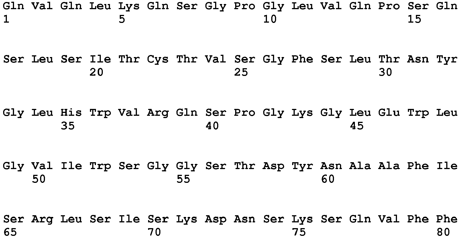

- <210> 8
<211> 113
<212> PRT
<213> Artificial Sequence - <220>
<223> anti-FLT3 single variable domain VL (clone BV10) - <400> 8

- <210> 9
<211> 118
<212> PRT
<213> Artificial Sequence - <220>
<223> anti-FLT3 chimeric single variable domain heavy chain (clone 4G8)VH - <400> 9

- <210> 10
<211> 107
<212> PRT
<213> Artificial Sequence - <220>
<223> anti-FLT3 chimeric single variable domain light chain (clone 4G8) VL - <400> 10


- <210> 11
<211> 115
<212> PRT
<213> Artificial Sequence - <220>
<223> anti-PSMA single variable domain heavy chain (clone J591) VH - <400> 11

- <210> 12
<211> 107
<212> PRT
<213> Artificial Sequence - <220>
<223> anti-PSMA single variable domain light chain (clone J591) VL - <400> 12


- <210> 13
<211> 5
<212> PRT
<213> Artificial Sequence - <220>
<223> linker - <400> 13

- <210> 14
<211> 18
<212> PRT
<213> Artificial Sequence - <220>
<223> linker - <400> 14

- <210> 15
<211> 14
<212> PRT
<213> Artificial Sequence - <220>
<223> linker - <400> 15

- <210> 16
<211> 8
<212> PRT
<213> Artificial Sequence - <220>
<223> linker - <400> 16

- <210> 17
<211> 12
<212> PRT
<213> Artificial Sequence - <220>
<223> linker - <400> 17

- <210> 18
<211> 6
<212> PRT
<213> Artificial Sequence - <220>
<223> linker - <400> 18

- <210> 19
<211> 7
<212> PRT
<213> Artificial Sequence - <220>
<223> linker - <400> 19


- <210> 20
<211> 14
<212> PRT
<213> Artificial Sequence - <220>
<223> linker - <400> 20

- <210> 21
<211> 20
<212> PRT
<213> Artificial Sequence - <220>
<223> linker - <400> 21

- <210> 22
<211> 4
<212> PRT
<213> Artificial Sequence - <220>
<223> linker - <400> 22

- <210> 23
<211> 12
<212> PRT
<213> Artificial Sequence - <220>
<223> linker - <400> 23

- <210> 24
<211> 20
<212> PRT
<213> Artificial Sequence - <220>
<223> linker - <400> 24

- <210> 25
<211> 117
<212> PRT
<213> Artificial Sequence - <220>
<223> anti-endoglin single variable domain heavy chain (clone Kro23) VH - <400> 25

- <210> 26
<211> 107
<212> PRT
<213> Artificial Sequence - <220>
<223> anti-endoglin single variable domain light chain (clone Kro23) VL - <400> 26

- <210> 27
<211> 582
<212> PRT
<213> Artificial Sequence - <220>
<223> fusion protein of anti-endoglin IgG (Kro23) heavy chain with SDIE modificaion and IL15 mutant - <400> 27


- <210> 28
<211> 213
<212> PRT
<213> Artificial sequence - <220>
<223> light chain of anti-endoglin antibody Kro23 - <400> 28


- <210> 29
<211> 567
<212> PRT
<213> Artificial Sequence - <220>
<223> Fusion protein of anti-CD19 IgG1 (4G7) heavy chain with SDIE modifications and IL15 wild type with short linker - <400> 29


- <210> 30
<211> 567
<212> PRT
<213> Artificial Sequence - <220>
<223> Fusion protein of anti-CD19 IgG1 (4G7) heavy chain with SDIE modifications and IL15 (E46K) mutant with short linker - <400> 30


- <210> 31
<211> 567
<212> PRT
<213> Artificial Sequence - <220>
<223> Fusion protein of anti-CD19 IgG1 (4G7) heavy chain with SDIE modifications and IL15 (V49D) mutant with short linker - <400> 31


- <210> 32
<211> 567
<212> PRT
<213> Artificial Sequence - <220>
<223> Fusion protein of anti-CD19 IgG1 (4G7) heavy chain with SDIE modifications and IL15 (I50D) mutant with short linker - <400> 32


- <210> 33
<211> 586
<212> PRT
<213> Artificial Sequence - <220>
<223> Fusion protein of anti-CD19 IgG1 (4G7) heavy chain with SDIE modifications and IL15 wild type with long linker - <400> 33


- <210> 34
<211> 586
<212> PRT
<213> Artificial Sequence - <220>
<223> Fusion protein of anti-CD19 IgG1 (4G7) heavy chain with SDIE modifications and IL15 (E46K) mutant with long linker - <400> 34


- <210> 35
<211> 586
<212> PRT
<213> Artificial Sequence - <220>
<223> Fusion protein of anti-CD19 IgG1 (4G7) heavy chain with SDIE modifications and IL15 (V49D) mutant with long linker - <400> 35


- <210> 36
<211> 586
<212> PRT
<213> Artificial Sequence - <220>
<223> Fusion protein of anti-CD19 IgG1 (4G7) heavy chain with SDIE modifications and IL15 (I50D) mutant with long linker - <400> 36

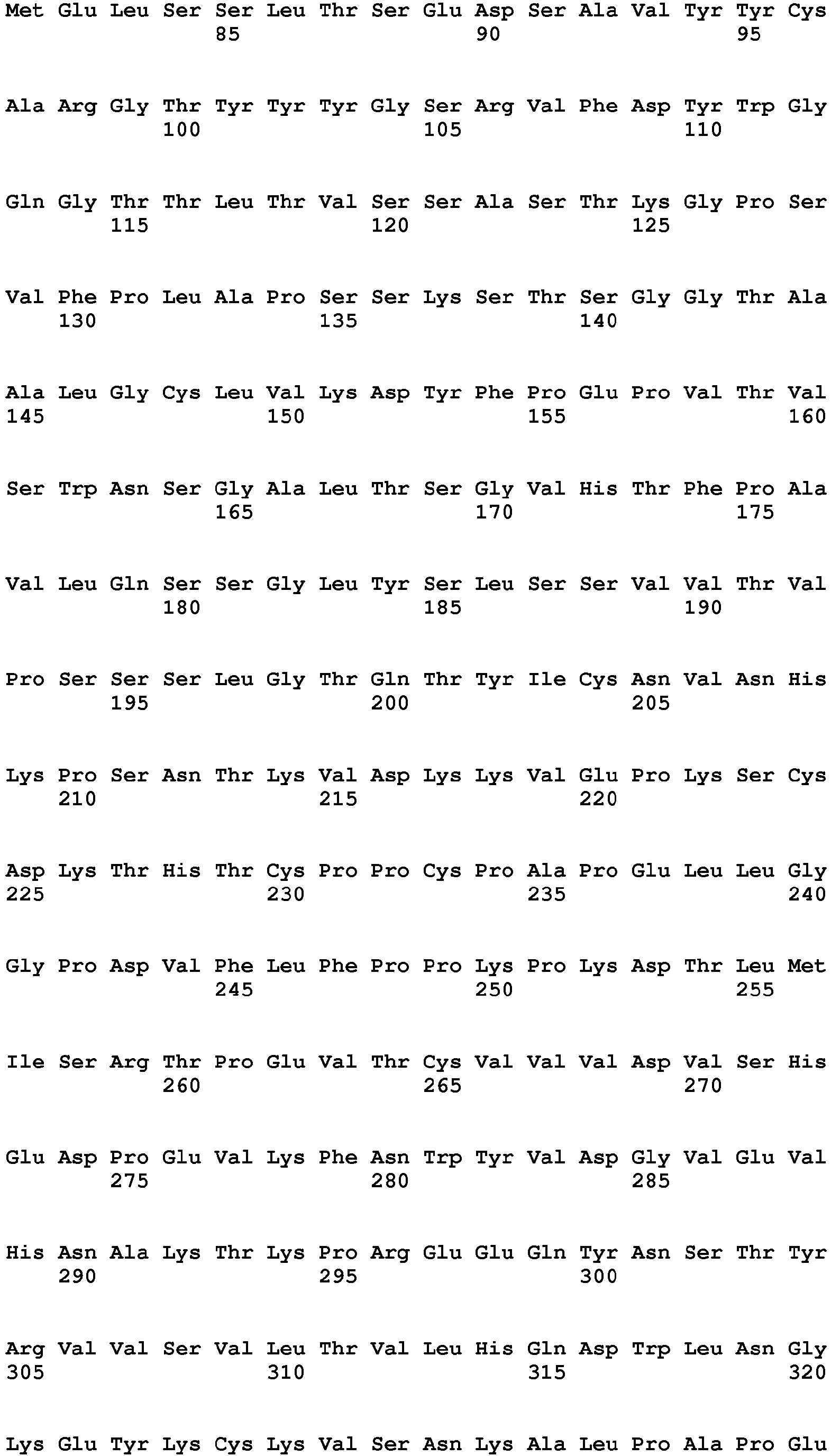


- <210> 37
<211> 219
<212> PRT
<213> Artificial Sequence - <220>
<223> anti-CD19 IgG1 (4G7) light chain - <400> 37


- <210> 38
<211> 588
<212> PRT
<213> Artificial Sequence - <220>
<223> Fusion protein of anti-FLT3 IgG1 (BV10) heavy chain with SDIE modifications and IL15 mutant - <400> 38


- <210> 39
<211> 220
<212> PRT
<213> Artificial Sequence - <220>
<223> anti-FLT3 IgG1 (BV10) light chain - <400> 39


- <210> 40
<211> 583
<212> PRT
<213> Artificial Sequence - <220>
<223> Fusion protein of anti-FLT3 IgG1 (4G8) heavy chain with SDIE modifications and IL15 mutant - <400> 40


- <210> 41
<211> 214
<212> PRT
<213> Artificial Sequence - <220>
<223> anti-endoglin IgG1 (4G8) light chain - <400> 41


- <210> 42
<211> 580
<212> PRT
<213> Artificial Sequence - <220>
<223> Fusion protein of anti-PSMA IgG1 (J591) heavy chain with SDIE modifications and IL15 wild type - <400> 42


- <210> 43
<211> 580
<212> PRT
<213> Artificial Sequence - <220>
<223> Fusion protein of anti-PSMA IgG1 (J591) heavy chain with SDIE modifications and IL15 (E46K) mutant - <400> 43


- <210> 44
<211> 214
<212> PRT
<213> Artificial Sequence - <220>
<223> anti-PSMA IgG1 (J591) light chain - <400> 44


wherein the binding protein is selected from the group consisting of an antibody, a divalent antibody fragment, a monovalent antibody fragment, or a proteinaceous binding molecule with antibody-like binding properties.
Claims (15)
- A fusion protein comprisinga) a binding protein comprising at least one binding site, wherein the binding site binds to an antigen associated with a target cell; andb) an IL-15 polypeptide, wherein the IL-15 polypeptide comprises at least one amino acid substitution at one or more positions corresponding to position(s) 92, 93, 94, 95, 96, 97, 98, 99, 100, 112, 113, 114, 115 and/or 116 of the amino acid sequence shown in SEQ ID NO:1thereby having a reduced affinity for IL-15Rα compared to the affinity of wild-type IL-15 of SEQ ID NO: 1 (Uniprot number: P40933-1),
wherein the IL-15 polypeptide binds to IL-2/IL-15Rβγ,
wherein the binding protein is selected from the group consisting of an antibody, a divalent antibody fragment, a monovalent antibody fragment, or a proteinaceous binding molecule with antibody-like binding properties. - The fusion protein of claim 1, wherein the proteinaceous binding molecule with antibody-like binding properties is selected from the group of an aptamer, a mutein based on a polypeptide of the lipocalin family, a glubody, a protein based on the ankyrin scaffold, a protein based on the crystalline scaffold, an adnectin, an avimer or a (recombinant) receptor protein.
- The fusion protein of claim 1 or 2, wherein the binding protein is modified such that it has an enhanced antibody dependent cellular cytotoxicity (ADCC)-activity compared to the same but unmodified binding protein, optionally wherein the modified binding protein is Fc optimized and/or optionally wherein the modified binding protein is an antibody, a scFv-Fc fragment, a bsFc-1/2 dimer or a bsFc-CH3-1/2 dimer.
- The fusion protein of any of claims 1-3, wherein the binding protein comprises a binding site of the 4G7 antibody, which has a sequence identity of at least 80%, or at least 85 %, or at least 90 %, or at least 95 %, or at least 98 %, or at least 99 % or 100 % to SEQ ID NO. 5 (the sequence of the heavy chain of the variable domain of 4G7); and/or
wherein the binding protein comprises a binding site of the 4G7 antibody, which has a sequence identity of at least 80%, or at least 85 %, or at least 90 %, or at least 95 %, or at least 98 %, or at least 99 % or 100 % to SEQ ID NO. 6 (the sequence of the light chain of the variable domain of 4G7). - The fusion protein of any of claims 1-4, wherein the target cell expresses a tumor associated antigen (TAA) and/or an antigen associated with autoimmune diseases, wherein preferably the TAA is selected from the group consisting of CD19, CD20, CD10, CD21, CD22, CD25, CD30, CD33, CD34, CD37, CD38, CD44v6, CD45, CDw52, Fms-like tyrosine kinase 3 (FLT-3, CD135), c-Kit (CD117), CSF1R, (CD115), CD123, CD133, PDGFR-α (CD140a), PDGFR-β (CD140b), chondroitin sulfate proteoglycan 4 (CSPG4, melanoma-associated chondroitin sulfate proteoglycan), Muc-1, EGFR, de2-7-EGFR, EGFRvIII, Folate blocking protein, Her2neu, Her3, PSMA, PSCA, PSA, TAG-72, HLA-DR, IGFR, CD133, IL3R, fibroblast activating protein (FAP), Carboanhydrase IX (MN/CA IX), Carcinoembryonic antigen (CEA), EpCAM, CDCP1, Derlin1, Tenascin, frizzled 1-10, the vascular antigens VEGFR2 (KDR/FLK1), VEGFR3 (FLT4, CD309), Endoglin, CLEC14, Tem1-8, Tie2, mesothelin, epithelial glycoprotein 2 (EGP2), epithelial glycoprotein 40 (EGP40), cancer antigen 72-4 (CA72-4), interleukin 13 receptor alpha-2 subunit, IL13Rα2, Ig kappa light chain (κ), GD3-ganglioside (GD3), GD2-ganglioside (GD2), acetylated variants of GD2 and GD3, CD171, NCAM, alpha folate receptor (αFR), Lewis(Y), fetal acetylcholine receptor (FAR), avian erythroblastic leukemia viral oncogene homolog 3 (ERBB3), avian erythroblastic leukemia viral oncogene homolog 4 (ERBB4), avian erythroblastic leukemia viral oncogene homolog 2 (ERBB2), hepatocyte growth factor receptor (HGFR/c-Met), claudin 18.2, claudin 3, claudin 4, claudin 1, claudin 12, claudin 2, claudin 5, claudin 8, claudin 7, claudin 6, membrane bound CEA, Robo4, CD138, tenascin and the extra domain-B of fibronectin; and/or
wherein preferably the target cell expresses an antigen associated with autoimmune diseases, which antigen is selected from the group consisting of CD20, CD22, CD52 and TNFR, CD19, CD25, CD40, and/or wherein preferably the target cell is a tumor/cancer cell and/or a B cell. - The fusion protein of any of claims 1-5, wherein the at least one amino acid substitution is selected from the group consisting of L92D, E94K, L95D, V97D, I98D, L114D, L114E, I115D, I115E and/or, preferably E94K, V97D and/or I98D, most preferably E94K.
- The fusion protein of any of claims 1-6, wherein the IL-15 polypeptide does not bind to IL-15Rα, and/or wherein the IL-15 polypeptide is full length IL-15 protein or a fragment or mutant thereof, which fragment or mutant has a reduced affinity for IL-15Rα compared to the affinity of wild-type IL-15 of SEQ ID NO: 1.
- The fusion protein of any of claims 1-7, wherein the IL-15 polypeptide comprises at least an amino acid sequence as shown in SEQ ID NO: 4, which comprises at least one amino acid substitution at one or more positions corresponding to position(s) 44, 45, 46, 47, 48, 49, 50, 51, 52, 64, 65, 66, 67 and/or 68 of the amino acid sequence shown in SEQ ID NO: 4.
- The fusion protein of any of claims 1-8 for use in target cell-restricted activation of effector cells expressing IL2/IL-15Rβγ and/or for use in target cell-restricted target cell killing mediated by effector cells expressing IL-2/IL-15Rβγ.
- The fusion protein for use of claim 9, wherein the effector cell is a NK cell or a T-cell, preferably a NK cell, a CD8+ T cell, gamma delta T cell or NK T cell.
- The fusion protein of any of claims 1-8 for use in treatment of a disease, wherein preferably side effects of the treatment are reduced.
- The fusion protein for use of claim 11, wherein the disease is a proliferatory disease or an autoimmune disease,
wherein preferably the proliferatory disease is selected from the group consisting of adrenal cancer, anal cancer, bile duct cancer, bladder cancer, bone cancer, brain and spinal cord tumors, breast cancer, Castleman disease, cervical cancer, colon cancer, endometrial cancer, esophagus cancer, Ewing family of tumors, eye cancer, gallbladder cancer, gastrointestinal carcinoid tumors, gastrointestinal stromal tumor (GIST), gestational trophoblastic disease, Hodgkin disease, Kaposi sarcoma, kidney cancer, laryngeal and hypopharyngeal cancer, leukemia, acute lymphocytic leukemia (ALL), acute myeloid leukemia (AML), chronic lymphocytic leukemia (CLL), chronic myeloid leukemia (CML), chronic myelomonocytic leukemia (CMML), liver cancer, lung cancer, non-small cell lung cancer, small cell lung cancer, lung carcinoid tumor, lymphoma, lymphoma of the skin, malignant mesothelioma, multiple myeloma, myelodysplastic syndrome, nasal cavity and paranasal sinus cancer, nasopharyngeal cancer, neuroblastoma, non-Hodgkin lymphoma, oral cavity and oropharyngeal cancer, osteosarcoma, ovarian cancer, pancreatic cancer, penile cancer, pituitary tumors, prostate cancer, rectum cancer, retinoblastoma, rhabdomyosarcoma, salivary gland cancer, sarcoma, skin cancer, basal and squamous cell cancer, melanoma, merkel cell cancer, small intestine cancer, stomach cancer, testicular cancer, thymus cancer, thyroid cancer, uterine sarcoma, vaginal cancer, vulvar cancer, Waldenstrom macroglobulinemia, or Wilms tumor; or
wherein preferably the autoimmune disease is selected from the group consisting of Systemic lupus erythematosus (SLE), Goodpasture's syndrome, Sarcoidosis, Scleroderma, Rheumatoid arthritis, Dermatomyositis, Sjögren's Syndrome, Scleroderma, Dermatomyositis, Psoriasis, Vitiligo, Alopecia areata, Type 1 diabetes mellitus, Autoimmune pancreatitis, Hashimoto's thyroiditis, Addison's disease, Multiple sclerosis, Myasthenia gravis, Polyarteritis nodosa, Idiopathic thrombocytopenic purpura, Hemolytic anemia, Antiphospholipid antibody syndrome, Pernicious anemia, Gastrointestinal diseases, Celiac disease, Inflammatory bowel disease, Autoimmune hepatitis or Primary biliary cirrhosis. - A pharmaceutical composition comprising the fusion protein of any of claims 1-8.
- A nucleic acid molecule encoding the fusion protein of any of claims 1-8, preferably comprised in a vector.
- A method for producing the fusion protein of any of claims 1-8, comprising using the nucleic acid encoding the fusion protein for expression of the fusion protein under conditions allowing expression of the fusion protein.
Priority Applications (4)
| Application Number | Priority Date | Filing Date | Title |
|---|---|---|---|
| RS20191593A RS59658B1 (en) | 2015-03-06 | 2016-03-07 | Fusion proteins comprising a binding protein and an interleukin-15 polypeptide having a reduced affinity for il15ra and therapeutic uses thereof |
| PL16708154T PL3265478T3 (en) | 2015-03-06 | 2016-03-07 | Fusion proteins comprising a binding protein and an interleukin-15 polypeptide having a reduced affinity for il15ra and therapeutic uses thereof |
| SI201630550T SI3265478T1 (en) | 2015-03-06 | 2016-03-07 | Fusion proteins comprising a binding protein and an interleukin-15 polypeptide having a reduced affinity for il15ra and therapeutic uses thereof |
| HRP20192190TT HRP20192190T1 (en) | 2015-03-06 | 2019-12-04 | FUSION PROTEINS COMPRISING A BINDING PROTEIN AND AN INTERLEUKIN-15 POLYPEPTIDE HAVING A REDUCED AFFINITY FOR IL15Ra AND THERAPEUTIC USES THEREOF |
Applications Claiming Priority (2)
| Application Number | Priority Date | Filing Date | Title |
|---|---|---|---|
| EP15157911.7A EP3064507A1 (en) | 2015-03-06 | 2015-03-06 | Fusion proteins comprising a binding protein and an interleukin-15 polypeptide having a reduced affinity for IL15ra and therapeutic uses thereof |
| PCT/EP2016/054729 WO2016142314A1 (en) | 2015-03-06 | 2016-03-07 | FUSION PROTEINS COMPRISING A BINDING PROTEIN AND AN INTERLEUKIN-15 POLYPEPTIDE HAVING A REDUCED AFFINITY FOR IL15Ra AND THERAPEUTIC USES THEREOF |
Publications (2)
| Publication Number | Publication Date |
|---|---|
| EP3265478A1 EP3265478A1 (en) | 2018-01-10 |
| EP3265478B1 true EP3265478B1 (en) | 2019-09-11 |
Family
ID=52629425
Family Applications (2)
| Application Number | Title | Priority Date | Filing Date |
|---|---|---|---|
| EP15157911.7A Withdrawn EP3064507A1 (en) | 2015-03-06 | 2015-03-06 | Fusion proteins comprising a binding protein and an interleukin-15 polypeptide having a reduced affinity for IL15ra and therapeutic uses thereof |
| EP16708154.6A Revoked EP3265478B1 (en) | 2015-03-06 | 2016-03-07 | Fusion proteins comprising a binding protein and an interleukin-15 polypeptide having a reduced affinity for il15ra and therapeutic uses thereof |
Family Applications Before (1)
| Application Number | Title | Priority Date | Filing Date |
|---|---|---|---|
| EP15157911.7A Withdrawn EP3064507A1 (en) | 2015-03-06 | 2015-03-06 | Fusion proteins comprising a binding protein and an interleukin-15 polypeptide having a reduced affinity for IL15ra and therapeutic uses thereof |
Country Status (16)
| Country | Link |
|---|---|
| US (3) | US10906952B2 (en) |
| EP (2) | EP3064507A1 (en) |
| JP (1) | JP6757338B2 (en) |
| CN (1) | CN107636015A (en) |
| BR (1) | BR112017018975A2 (en) |
| CA (1) | CA2978808A1 (en) |
| CY (1) | CY1122494T1 (en) |
| DK (1) | DK3265478T3 (en) |
| ES (1) | ES2763598T3 (en) |
| HR (1) | HRP20192190T1 (en) |
| HU (1) | HUE047818T2 (en) |
| PL (1) | PL3265478T3 (en) |
| PT (1) | PT3265478T (en) |
| RS (1) | RS59658B1 (en) |
| SI (1) | SI3265478T1 (en) |
| WO (1) | WO2016142314A1 (en) |
Cited By (6)
| Publication number | Priority date | Publication date | Assignee | Title |
|---|---|---|---|---|
| US11084863B2 (en) | 2017-06-30 | 2021-08-10 | Xencor, Inc. | Targeted heterodimeric Fc fusion proteins containing IL-15 IL-15alpha and antigen binding domains |
| US11377477B2 (en) | 2018-10-12 | 2022-07-05 | Xencor, Inc. | PD-1 targeted IL-15/IL-15RALPHA fc fusion proteins and uses in combination therapies thereof |
| US11505595B2 (en) | 2018-04-18 | 2022-11-22 | Xencor, Inc. | TIM-3 targeted heterodimeric fusion proteins containing IL-15/IL-15RA Fc-fusion proteins and TIM-3 antigen binding domains |
| US11524991B2 (en) | 2018-04-18 | 2022-12-13 | Xencor, Inc. | PD-1 targeted heterodimeric fusion proteins containing IL-15/IL-15Ra Fc-fusion proteins and PD-1 antigen binding domains and uses thereof |
| US11584794B2 (en) | 2016-10-14 | 2023-02-21 | Xencor, Inc. | Bispecific heterodimeric fusion proteins containing IL-15-IL-15Ralpha Fc-fusion proteins and immune checkpoint antibody fragments |
| US11618776B2 (en) | 2018-12-20 | 2023-04-04 | Xencor, Inc. | Targeted heterodimeric Fc fusion proteins containing IL-15/IL-15RA and NKG2D antigen binding domains |
Families Citing this family (33)
| Publication number | Priority date | Publication date | Assignee | Title |
|---|---|---|---|---|
| CN103842383B (en) | 2011-05-16 | 2017-11-03 | 健能隆医药技术(上海)有限公司 | Polyspecific FAB fusion proteins and its application method |
| DK3143134T3 (en) | 2014-05-15 | 2021-01-04 | Nat Univ Singapore | Modified, natural killer cells and their uses |
| EP3064507A1 (en) * | 2015-03-06 | 2016-09-07 | Deutsches Krebsforschungszentrum Stiftung des öffentlichen Rechts | Fusion proteins comprising a binding protein and an interleukin-15 polypeptide having a reduced affinity for IL15ra and therapeutic uses thereof |
| CN106380521B (en) | 2015-07-02 | 2020-12-29 | 博际生物医药科技(杭州)有限公司 | Interleukin-15 fusion protein for tumor targeted therapy |
| EP3430058A4 (en) | 2016-03-15 | 2019-10-23 | Generon (Shanghai) Corporation Ltd. | Multispecific fab fusion proteins and use thereof |
| JP7185530B2 (en) | 2016-06-13 | 2022-12-07 | トルク セラピューティクス, インコーポレイテッド | Methods and compositions for promoting immune cell function |
| EP3601537A4 (en) | 2017-03-27 | 2021-01-13 | National University of Singapore | Stimulatory cell lines for ex vivo expansion and activation of natural killer cells |
| CA3056439A1 (en) | 2017-03-27 | 2018-10-04 | National University Of Singapore | Truncated nkg2d chimeric receptors and uses thereof in natural killer cell immunotherapy |
| LT3606946T (en) * | 2017-04-03 | 2022-10-25 | F. Hoffmann-La Roche Ag | Immunoconjugates of an anti-pd-1 antibody with a mutant il-2 or with il-15 |
| KR20200037353A (en) | 2017-08-07 | 2020-04-08 | 더 리전트 오브 더 유니버시티 오브 캘리포니아 | Platform for creating safe cell therapy products |
| CN111432836A (en) | 2017-09-05 | 2020-07-17 | 转矩医疗股份有限公司 | Therapeutic protein compositions and methods of making and using same |
| JP2021514974A (en) * | 2018-02-26 | 2021-06-17 | シンソークス, インコーポレイテッド | IL-15 conjugate and its use |
| CA3034912A1 (en) * | 2018-02-28 | 2019-08-28 | Pfizer Inc. | Il-15 variants and uses thereof |
| KR20210022004A (en) * | 2018-06-18 | 2021-03-02 | 안위타 바이오사이언시스, 인코포레이티드 | Anti-mesothelin constructs and uses thereof |
| WO2019246142A1 (en) * | 2018-06-18 | 2019-12-26 | Haiming Chen | Soluble bispecific fusion proteins for cancer immunotherapy |
| EP3847196A4 (en) * | 2018-09-07 | 2023-01-04 | ITabMed (HK) Limited | Bispecific antigen binding proteins and uses thereof |
| CN109207430A (en) * | 2018-09-25 | 2019-01-15 | 华东师范大学 | A kind of Chimeric antigen receptor NK cell and its preparation method and application |
| JP2022502088A (en) | 2018-09-27 | 2022-01-11 | エクシリオ デベロップメント, インコーポレイテッド | Masked cytokine polypeptide |
| JP6935383B2 (en) * | 2018-09-27 | 2021-09-15 | 公益財団法人実験動物中央研究所 | Immunodeficient mouse |
| EP3773918A4 (en) | 2019-03-05 | 2022-01-05 | Nkarta, Inc. | Cd19-directed chimeric antigen receptors and uses thereof in immunotherapy |
| KR20220012296A (en) | 2019-05-20 | 2022-02-03 | 싸이튠 파마 | IL-2/IL-15Rβγ agonist dosing regimen for treatment of cancer or infectious disease |
| CA3146987A1 (en) * | 2019-07-17 | 2021-01-21 | National University Of Singapore | Functional binders synthesized and secreted by immune cells |
| TW202128757A (en) | 2019-10-11 | 2021-08-01 | 美商建南德克公司 | Pd-1 targeted il-15/il-15ralpha fc fusion proteins with improved properties |
| CN111690068B (en) * | 2019-11-13 | 2022-04-19 | 中国科学技术大学 | IL-15/SuIL-15R alpha-dFc-gamma 4 complex protein and construction method and application thereof |
| EP3822288A1 (en) | 2019-11-18 | 2021-05-19 | Deutsches Krebsforschungszentrum, Stiftung des öffentlichen Rechts | Antibodies targeting, and other modulators of, the cd276 antigen, and uses thereof |
| CN114761433A (en) * | 2019-12-11 | 2022-07-15 | 上海复宏汉霖生物技术股份有限公司 | anti-Claudin18.2 monoclonal antibody, preparation method and application thereof |
| CA3193073A1 (en) * | 2020-09-17 | 2022-03-24 | Liping SONG | Bispecific recombinant protein and use thereof |
| JP2023550880A (en) | 2020-10-26 | 2023-12-06 | サイチューン ファーマ | IL-2/IL-15Rβγ agonist for treating non-melanoma skin cancer |
| AU2021367887A1 (en) | 2020-10-26 | 2023-06-01 | Cytune Pharma | IL-2/IL-15Rβү AGONIST FOR TREATING SQUAMOUS CELL CARCINOMA |
| BR112023027305A2 (en) | 2021-06-23 | 2024-03-12 | Cytune Pharma | INTERLEUCIN-15 VARIANT, CONJUGATE, FUSION PROTEIN, NUCLEIC ACID, VECTOR, HOST CELL AND PHARMACEUTICAL COMPOSITION |
| WO2023280287A1 (en) * | 2021-07-09 | 2023-01-12 | Nanjing Legend Biotech Co., Ltd. | Interleukin-15 variants and uses thereof |
| KR20240043797A (en) | 2021-08-13 | 2024-04-03 | 싸이튠 파마 | Combination of IL-2/IL-15Rβγ agonist and antibody-drug conjugate for cancer treatment |
| CN113956346B (en) * | 2021-10-26 | 2024-03-01 | 西安龙腾景云生物科技有限公司 | Recombinant interleukin-15 variant |
Citations (5)
| Publication number | Priority date | Publication date | Assignee | Title |
|---|---|---|---|---|
| WO2005085282A1 (en) | 2004-02-27 | 2005-09-15 | Inserm (Institut National De La Sante Et De La Recherche Medicale) | Il-15 binding site for il15-ralpha and specific il-15 mutants having agonists/antagonists activity |
| WO2006063974A2 (en) | 2004-12-13 | 2006-06-22 | Cytos Biotechnology Ag | Il-15 antigen arrays and uses thereof |
| WO2012040323A2 (en) | 2010-09-21 | 2012-03-29 | Altor Bioscien Corporation | Multimeric il-15 soluble fusion molecules and methods of making and using same |
| WO2014207173A1 (en) | 2013-06-27 | 2014-12-31 | INSERM (Institut National de la Santé et de la Recherche Médicale) | Interleukin 15 (il-15) antagonists and uses thereof for the treatment of autoimmune diseases and inflammatory diseases |
| US20190263877A1 (en) | 2018-02-28 | 2019-08-29 | Pfizer Inc. | IL-15 Variants and Uses Thereof |
Family Cites Families (17)
| Publication number | Priority date | Publication date | Assignee | Title |
|---|---|---|---|---|
| US6107090A (en) | 1996-05-06 | 2000-08-22 | Cornell Research Foundation, Inc. | Treatment and diagnosis of prostate cancer with antibodies to extracellur PSMA domains |
| DE19742706B4 (en) | 1997-09-26 | 2013-07-25 | Pieris Proteolab Ag | lipocalin muteins |
| PT1071752E (en) | 1998-04-21 | 2003-11-28 | Micromet Ag | SPECIFIC POLYMPEPTIDES FOR CD19XCD3 AND ITS USES |
| DE19926068C1 (en) | 1999-06-08 | 2001-01-11 | Arne Skerra | Muteins of the bilin binding protein |
| AU2001298053A1 (en) | 2001-09-27 | 2003-04-14 | Pieris Proteolab Ag | Muteins of apolipoprotein D |
| WO2003029462A1 (en) | 2001-09-27 | 2003-04-10 | Pieris Proteolab Ag | Muteins of human neutrophil gelatinase-associated lipocalin and related proteins |
| US20040132101A1 (en) | 2002-09-27 | 2004-07-08 | Xencor | Optimized Fc variants and methods for their generation |
| US7635472B2 (en) | 2003-05-31 | 2009-12-22 | Micromet Ag | Pharmaceutical compositions comprising bispecific anti-cd3, anti-cd19 antibody constructs for the treatment of b-cell related disorders |
| AU2003264100A1 (en) | 2003-08-25 | 2005-03-10 | Pieris Proteolab Ag | Muteins of a bilin-binding protein with affinity for a given target |
| AU2003275958A1 (en) | 2003-08-25 | 2005-03-10 | Pieris Proteolab Ag | Muteins of tear lipocalin |
| WO2006056464A2 (en) | 2004-11-26 | 2006-06-01 | Pieris Ag | Compound with affinity for the cytotoxic t lymphocyte-associated antigen (ctla-4) |
| JP5608368B2 (en) | 2006-08-01 | 2014-10-15 | ピエリス アーゲー | Tear lipocalin mutein and method for obtaining the same |
| DK2769984T3 (en) * | 2007-05-11 | 2017-10-16 | Altor Bioscience Corp | Fusion molecules and IL-15 variants |
| US20120196310A1 (en) | 2009-10-02 | 2012-08-02 | Christiane Jaeger | A-fucosylation detection in antibodies |
| BR112012013662B1 (en) | 2009-12-07 | 2022-08-02 | Pieris Pharmaceuticals Gmbh | LIPOCALIN MUTEINS ASSOCIATED WITH HUMAN NEUTROPHIL GELATINASE (LCN2, HNGAL), THEIR USE AND THEIR GENERATION AND PRODUCTION METHODS, NUCLEIC ACID MOLECULE, HOST CELL, PHARMACEUTICAL COMPOSITION AND DIAGNOSIS OR ANALYTICAL KIT |
| JP6441079B2 (en) | 2011-12-19 | 2018-12-19 | シンイミューン ゲーエムベーハー | Bispecific antibody molecule |
| EP3064507A1 (en) * | 2015-03-06 | 2016-09-07 | Deutsches Krebsforschungszentrum Stiftung des öffentlichen Rechts | Fusion proteins comprising a binding protein and an interleukin-15 polypeptide having a reduced affinity for IL15ra and therapeutic uses thereof |
-
2015
- 2015-03-06 EP EP15157911.7A patent/EP3064507A1/en not_active Withdrawn
-
2016
- 2016-03-07 HU HUE16708154A patent/HUE047818T2/en unknown
- 2016-03-07 EP EP16708154.6A patent/EP3265478B1/en not_active Revoked
- 2016-03-07 SI SI201630550T patent/SI3265478T1/en unknown
- 2016-03-07 PT PT167081546T patent/PT3265478T/en unknown
- 2016-03-07 CA CA2978808A patent/CA2978808A1/en active Pending
- 2016-03-07 US US15/556,282 patent/US10906952B2/en active Active
- 2016-03-07 RS RS20191593A patent/RS59658B1/en unknown
- 2016-03-07 CN CN201680025275.8A patent/CN107636015A/en active Pending
- 2016-03-07 PL PL16708154T patent/PL3265478T3/en unknown
- 2016-03-07 WO PCT/EP2016/054729 patent/WO2016142314A1/en active Application Filing
- 2016-03-07 BR BR112017018975-5A patent/BR112017018975A2/en active Search and Examination
- 2016-03-07 DK DK16708154.6T patent/DK3265478T3/en active
- 2016-03-07 ES ES16708154T patent/ES2763598T3/en active Active
- 2016-03-07 JP JP2017564951A patent/JP6757338B2/en active Active
-
2019
- 2019-12-04 HR HRP20192190TT patent/HRP20192190T1/en unknown
- 2019-12-11 CY CY20191101303T patent/CY1122494T1/en unknown
-
2020
- 2020-12-22 US US17/130,975 patent/US20210163564A1/en active Pending
-
2021
- 2021-12-30 US US17/565,942 patent/US20220324931A1/en active Pending
Patent Citations (5)
| Publication number | Priority date | Publication date | Assignee | Title |
|---|---|---|---|---|
| WO2005085282A1 (en) | 2004-02-27 | 2005-09-15 | Inserm (Institut National De La Sante Et De La Recherche Medicale) | Il-15 binding site for il15-ralpha and specific il-15 mutants having agonists/antagonists activity |
| WO2006063974A2 (en) | 2004-12-13 | 2006-06-22 | Cytos Biotechnology Ag | Il-15 antigen arrays and uses thereof |
| WO2012040323A2 (en) | 2010-09-21 | 2012-03-29 | Altor Bioscien Corporation | Multimeric il-15 soluble fusion molecules and methods of making and using same |
| WO2014207173A1 (en) | 2013-06-27 | 2014-12-31 | INSERM (Institut National de la Santé et de la Recherche Médicale) | Interleukin 15 (il-15) antagonists and uses thereof for the treatment of autoimmune diseases and inflammatory diseases |
| US20190263877A1 (en) | 2018-02-28 | 2019-08-29 | Pfizer Inc. | IL-15 Variants and Uses Thereof |
Non-Patent Citations (24)
| Title |
|---|
| "Antibody mimetic", WIKIPEDIA, 25 November 2017 (2017-11-25), XP055733729, Retrieved from the Internet <URL:https://en.wikipedia.org/w/index.php?title=antibody_mimetic&oldid=812087322> |
| "Avimer", WIKIPEDIA, 2 February 2016 (2016-02-02), XP055733733, Retrieved from the Internet <URL:https://en.wikipedia.org/w/index.php?title=avimer&oldid=702868839> |
| "Monobody", WIKIPEDIA, 26 October 2017 (2017-10-26), XP055733738, Retrieved from the Internet <URL:https://en.wikipedia.org/w/index.php?title=monobody&oldid=807139681> |
| ALBERTINI ET AL.: "Phase II trial of hu14.18-IL2 for patients with metastatic melanoma", CANCER IMMUNOL IMMUNOTHER, vol. 61, 2012, pages 2261 - 2271, XP035144789 |
| ARNE SKERRA: "'Anticalins': a new class of engineered ligand-binding proteins with antibody-like properties", REVIEW IN MOLECULAR BIOTECHNOLOGY, vol. 74, 2001, pages 257 - 275, XP002269554 |
| ARNE SKERRA: "Engineered protein scaffolds for molecular recognition", J OF MOLECULAR RECOGNITION, vol. 13, 2000, pages 167 - 187, XP002966570 |
| ARNE SKERRA: "Engineered protein scaffolds for molecular recognition", JOURNAL OF MOLECULAR RECOGNITION, HEYDEN & SON LTD., LONDON, GB, vol. 13, 1 January 2000 (2000-01-01), pages 167 - 187, ISSN: 0952-3499, DOI: 10.1002/1099-1352(200007/08)13:4<167::AID-JMR502>3.0.CO;2-9 * |
| BERNARD ET AL.: "Identification of an interleukin-15alpha receptor-binding site on human interleukin-15", J OF BIOLOGICAL CHEMISTRY, vol. 279, no. 23, 4 June 2004 (2004-06-04), pages 24313 - 24322, XP002286785 |
| BERNARD ET AL.: "Identification of an interleukin-15alpha receptor-binding site on human interleukin-15", J. BIOL. CHEM., vol. 279L23, 2004, pages 24313 - 24322, XP002286785 |
| CONLON ET AL.: "Redistribution, Hyperproliferation, Activation of Natural Killer Cells and CD8 T Cells, and Cytokine Production During First-in-Human Clinical Trial of Recombinant Human Interleukin-15 in Patients With Cancer", JOURNAL OF CLINICAL ONCOLOGY, vol. 33, no. 1, 1 January 2015 (2015-01-01), pages 74 - 82, XP055550401 |
| DAFNE MÜLLER: "Targeted cancer immunotherapy", ONCOLMMUNOLOGY, vol. 1, no. 7, October 2012 (2012-10-01), pages 1213 - 1214, XP055209394 |
| GARCIN ET AL.: "High efficiency cell-specific targeting of cytokine activity", NATURE COMMUNICATIONS, vol. 5, 3016, 8 January 2014 (2014-01-08), XP055610390 |
| GILLIES ET AL.: "ANTIBODY-TARGETED INTERLEUKIN 2 STIMULATES T-CELL KILLING OF AUTOLOGOUS TUMOR CELLS", PROC NATL ACAD SCI USA, vol. 89, 1992, pages 1428 - 1432, XP002069101 |
| HOFMANN ET AL.: "Generation, selection and preclinical characterization of an Fc-optimized FLT3 antibody for the treatment of myeloid leukemia", LEUKEMIA, vol. 26, 2012, pages 1228 - 1237, XP055041249 |
| HORTON ET AL.: "Potent in vitro and in vivo activity of an Fc-engineered anti-CD19 monoclonal antibody against lymphoma and leukemia", CANCER RES, vol. 68, 2008, pages 8049 - 8057, XP009145044 |
| KASPAR ET AL.: "The antibody-mediated targeted delivery of interleukin-15 and GM-CSF to the tumor neovasculature inhibits tumor growth and metastasis", CANCER RES, vol. 67, no. 10, 15 May 2007 (2007-05-15), pages 4940 - 4948, XP002468217 |
| KELLNER ET AL.: "Heterodimeric bispecific antibody-derivatives against CD19 and CD16 induce effective antibody-dependent cellular cytotoxicity against B-lymphoid tumor cells", CANCER LETTERS, vol. 303, 2011, pages 128 - 139, XP055311969 |
| KERMER ET AL.: "An antibody fusion protein for cancer immunotherapy mimicking IL-15 trans-presentation at the tumor site", MOL CANCER THER., vol. 11, no. 6, 2012, pages 1279 - 1288, XP002683666 |
| LIST ET AL.: "Immunocytokines: a review of molecules in clinical development for cancer therapy", CLIN PHARMACOL, vol. 5, 2013, pages 29 - 45, XP055289369 |
| MÜLLER: "Targeted cancer immunotherapy", ONCOIMMUNOLOGY, vol. 1, no. 7, 2012, pages 1213 - 1214, XP055209394 |
| ORTIS-SÁNCHEZ ET AL.: "Antibody-cytokine fusion proteins: applications in cancer therapy", EXPERT OPIN BIOL THER., vol. 8, no. 5, 2008, pages 609 - 632, XP008145785 |
| RIBAS ET AL.: "Phase I/II open-label study of the biologic effects of the interleukin-2 immunocytokine EMD 273063 (hu14.18-IL2) in patients with metastatic malignant melanoma", J TRANSL MED, vol. 7, 68, 2009, XP021059155 |
| SKERRA A: "'Anticalins': a new class of engineered ligand-binding proteins with antibody-like properties", REVIEWS IN MOLECULAR BIOTECHNOLOGY, ELSEVIER, AMSTERDAM, NL, vol. 74, no. 4, 1 June 2001 (2001-06-01), pages 257 - 275, ISSN: 1389-0352, DOI: 10.1016/S1389-0352(01)00020-4 * |
| ZHU ET AL.: "Novel Human Interleukin-15 Agonists", J IMMUNOL., vol. 183, 2009, pages 3598 - 3607, XP055732478 |
Cited By (6)
| Publication number | Priority date | Publication date | Assignee | Title |
|---|---|---|---|---|
| US11584794B2 (en) | 2016-10-14 | 2023-02-21 | Xencor, Inc. | Bispecific heterodimeric fusion proteins containing IL-15-IL-15Ralpha Fc-fusion proteins and immune checkpoint antibody fragments |
| US11084863B2 (en) | 2017-06-30 | 2021-08-10 | Xencor, Inc. | Targeted heterodimeric Fc fusion proteins containing IL-15 IL-15alpha and antigen binding domains |
| US11505595B2 (en) | 2018-04-18 | 2022-11-22 | Xencor, Inc. | TIM-3 targeted heterodimeric fusion proteins containing IL-15/IL-15RA Fc-fusion proteins and TIM-3 antigen binding domains |
| US11524991B2 (en) | 2018-04-18 | 2022-12-13 | Xencor, Inc. | PD-1 targeted heterodimeric fusion proteins containing IL-15/IL-15Ra Fc-fusion proteins and PD-1 antigen binding domains and uses thereof |
| US11377477B2 (en) | 2018-10-12 | 2022-07-05 | Xencor, Inc. | PD-1 targeted IL-15/IL-15RALPHA fc fusion proteins and uses in combination therapies thereof |
| US11618776B2 (en) | 2018-12-20 | 2023-04-04 | Xencor, Inc. | Targeted heterodimeric Fc fusion proteins containing IL-15/IL-15RA and NKG2D antigen binding domains |
Also Published As
| Publication number | Publication date |
|---|---|
| CN107636015A (en) | 2018-01-26 |
| CY1122494T1 (en) | 2021-01-27 |
| HUE047818T2 (en) | 2020-05-28 |
| US10906952B2 (en) | 2021-02-02 |
| HRP20192190T1 (en) | 2020-02-21 |
| EP3265478A1 (en) | 2018-01-10 |
| JP2018512168A (en) | 2018-05-17 |
| PL3265478T3 (en) | 2020-03-31 |
| JP6757338B2 (en) | 2020-09-16 |
| DK3265478T3 (en) | 2019-12-16 |
| PT3265478T (en) | 2020-01-08 |
| US20180044391A1 (en) | 2018-02-15 |
| WO2016142314A1 (en) | 2016-09-15 |
| US20220324931A1 (en) | 2022-10-13 |
| SI3265478T1 (en) | 2020-01-31 |
| BR112017018975A2 (en) | 2018-04-17 |
| ES2763598T3 (en) | 2020-05-29 |
| RS59658B1 (en) | 2020-01-31 |
| US20210163564A1 (en) | 2021-06-03 |
| EP3064507A1 (en) | 2016-09-07 |
| CA2978808A1 (en) | 2016-09-15 |
Similar Documents
| Publication | Publication Date | Title |
|---|---|---|
| US20210163564A1 (en) | FUSION PROTEINS COMPRISING A BINDING PROTEIN AND AN INTERLEUKIN-15 POLYPEPTIDE HAVING A REDUCED AFFINITY FOR IL15Ra AND THERAPEUTIC USES THEREOF | |
| JP7153626B2 (en) | Anti-human 4-1BB antibody and uses thereof | |
| CN106659775B (en) | IL-15-based molecules, methods and uses thereof | |
| EP3250611B1 (en) | Car t-cells recognizing cancer-specific il 13r-alpha2 | |
| CN111655716A (en) | IL-15-based fusions with IL-7 and IL-21 | |
| JP2019533449A (en) | Molecules based on multimeric IL-15 | |
| JP2022528030A (en) | Multifunctional fusion protein and its use | |
| JP2022109934A (en) | Monospecific and bispecific proteins with immune checkpoint regulation for cancer therapy | |
| WO2020088459A1 (en) | Novel rationally designed protein compositions | |
| CN114364696A (en) | TEAC and ATTAC immunooncology compositions and methods | |
| CN117321087A (en) | Multi-specific proteins comprising nkp binding site, cancer antigen binding site fused to cytokine for nk cell engagement | |
| TW201708260A (en) | Constructs targeting PSA peptide/MHC complexes and uses thereof | |
| CN111465618A (en) | Bispecific CD 16-binding molecules and their use in the treatment of disease | |
| US10919976B2 (en) | IFN-gamma-inducible regulatory T cell convertible anti-cancer (IRTCA) antibody and uses thereof | |
| CN117616050A (en) | Multispecific proteins that bind to NKP46, cytokine receptor, tumor antigen and CD16A | |
| EP4237451A1 (en) | Bcma/taci antigen-binding molecules | |
| KR20230042292A (en) | Improved antigen binding receptor | |
| CN116323655A (en) | SIRP alpha-Fc fusion protein | |
| JP2022542543A (en) | Anti-DLL3 chimeric antigen receptor and use thereof | |
| CA3221886A1 (en) | Interleukin-15 based immunocytokines | |
| WO2022258678A1 (en) | Multispecific proteins binding to nkp30, a cytokine receptor, a tumour antigen and cd16a | |
| WO2024056862A1 (en) | Multispecific antigen binding proteins for tumor-targeting of nk cells and use thereof | |
| WO2022258691A1 (en) | Multispecific proteins binding to nkg2d, a cytokine receptor, a tumour antigen and cd16a |
Legal Events
| Date | Code | Title | Description |
|---|---|---|---|
| STAA | Information on the status of an ep patent application or granted ep patent |
Free format text: STATUS: THE INTERNATIONAL PUBLICATION HAS BEEN MADE |
|
| PUAI | Public reference made under article 153(3) epc to a published international application that has entered the european phase |
Free format text: ORIGINAL CODE: 0009012 |
|
| STAA | Information on the status of an ep patent application or granted ep patent |
Free format text: STATUS: REQUEST FOR EXAMINATION WAS MADE |
|
| 17P | Request for examination filed |
Effective date: 20171006 |
|
| AK | Designated contracting states |
Kind code of ref document: A1 Designated state(s): AL AT BE BG CH CY CZ DE DK EE ES FI FR GB GR HR HU IE IS IT LI LT LU LV MC MK MT NL NO PL PT RO RS SE SI SK SM TR |
|
| AX | Request for extension of the european patent |
Extension state: BA ME |
|
| DAV | Request for validation of the european patent (deleted) | ||
| DAX | Request for extension of the european patent (deleted) | ||
| STAA | Information on the status of an ep patent application or granted ep patent |
Free format text: STATUS: EXAMINATION IS IN PROGRESS |
|
| 17Q | First examination report despatched |
Effective date: 20181010 |
|
| GRAP | Despatch of communication of intention to grant a patent |
Free format text: ORIGINAL CODE: EPIDOSNIGR1 |
|
| STAA | Information on the status of an ep patent application or granted ep patent |
Free format text: STATUS: GRANT OF PATENT IS INTENDED |
|
| RIC1 | Information provided on ipc code assigned before grant |
Ipc: C12N 15/24 20060101ALI20190225BHEP Ipc: C12N 15/62 20060101ALI20190225BHEP Ipc: C07K 14/54 20060101AFI20190225BHEP |
|
| INTG | Intention to grant announced |
Effective date: 20190329 |
|
| GRAS | Grant fee paid |
Free format text: ORIGINAL CODE: EPIDOSNIGR3 |
|
| GRAA | (expected) grant |
Free format text: ORIGINAL CODE: 0009210 |
|
| STAA | Information on the status of an ep patent application or granted ep patent |
Free format text: STATUS: THE PATENT HAS BEEN GRANTED |
|
| AK | Designated contracting states |
Kind code of ref document: B1 Designated state(s): AL AT BE BG CH CY CZ DE DK EE ES FI FR GB GR HR HU IE IS IT LI LT LU LV MC MK MT NL NO PL PT RO RS SE SI SK SM TR |
|
| REG | Reference to a national code |
Ref country code: GB Ref legal event code: FG4D |
|
| REG | Reference to a national code |
Ref country code: CH Ref legal event code: EP |
|
| REG | Reference to a national code |
Ref country code: AT Ref legal event code: REF Ref document number: 1178336 Country of ref document: AT Kind code of ref document: T Effective date: 20190915 |
|
| REG | Reference to a national code |
Ref country code: DE Ref legal event code: R096 Ref document number: 602016020346 Country of ref document: DE Ref country code: IE Ref legal event code: FG4D |
|
| REG | Reference to a national code |
Ref country code: HR Ref legal event code: TUEP Ref document number: P20192190T Country of ref document: HR |
|
| REG | Reference to a national code |
Ref country code: RO Ref legal event code: EPE |
|
| REG | Reference to a national code |
Ref country code: DK Ref legal event code: T3 Effective date: 20191212 |
|
| REG | Reference to a national code |
Ref country code: SE Ref legal event code: TRGR |
|
| REG | Reference to a national code |
Ref country code: PT Ref legal event code: SC4A Ref document number: 3265478 Country of ref document: PT Date of ref document: 20200108 Kind code of ref document: T Free format text: AVAILABILITY OF NATIONAL TRANSLATION Effective date: 20191219 |
|
| REG | Reference to a national code |
Ref country code: NL Ref legal event code: FP |
|
| REG | Reference to a national code |
Ref country code: NO Ref legal event code: T2 Effective date: 20190911 |
|
| REG | Reference to a national code |
Ref country code: HR Ref legal event code: T1PR Ref document number: P20192190 Country of ref document: HR |
|
| REG | Reference to a national code |
Ref country code: SK Ref legal event code: T3 Ref document number: E 32873 Country of ref document: SK |
|
| REG | Reference to a national code |
Ref country code: EE Ref legal event code: FG4A Ref document number: E018610 Country of ref document: EE Effective date: 20200106 |
|
| REG | Reference to a national code |
Ref country code: HR Ref legal event code: ODRP Ref document number: P20192190 Country of ref document: HR Payment date: 20200225 Year of fee payment: 5 |
|
| REG | Reference to a national code |
Ref country code: GR Ref legal event code: EP Ref document number: 20190403791 Country of ref document: GR Effective date: 20200318 |
|
| REG | Reference to a national code |
Ref country code: HU Ref legal event code: AG4A Ref document number: E047818 Country of ref document: HU |
|
| PG25 | Lapsed in a contracting state [announced via postgrant information from national office to epo] |
Ref country code: SM Free format text: LAPSE BECAUSE OF FAILURE TO SUBMIT A TRANSLATION OF THE DESCRIPTION OR TO PAY THE FEE WITHIN THE PRESCRIBED TIME-LIMIT Effective date: 20190911 |
|
| REG | Reference to a national code |
Ref country code: ES Ref legal event code: FG2A Ref document number: 2763598 Country of ref document: ES Kind code of ref document: T3 Effective date: 20200529 |
|
| REG | Reference to a national code |
Ref country code: DE Ref legal event code: R026 Ref document number: 602016020346 Country of ref document: DE |
|
| PLBI | Opposition filed |
Free format text: ORIGINAL CODE: 0009260 |
|
| PLBI | Opposition filed |
Free format text: ORIGINAL CODE: 0009260 |
|
| PLAX | Notice of opposition and request to file observation + time limit sent |
Free format text: ORIGINAL CODE: EPIDOSNOBS2 |
|
| REG | Reference to a national code |
Ref country code: FI Ref legal event code: MDE Opponent name: PFIZER, INC. Ref country code: FI Ref legal event code: MDE Opponent name: MARGARET DIXON LIMITED |
|
| 26 | Opposition filed |
Opponent name: PFIZER, INC. Effective date: 20200610 |
|
| REG | Reference to a national code |
Ref country code: FI Ref legal event code: MDE Opponent name: PFIZER, INC. Ref country code: FI Ref legal event code: MDE Opponent name: MARGARET DIXON LIMITED |
|
| 26 | Opposition filed |
Opponent name: MARGARET DIXON LIMITED Effective date: 20200611 |
|
| REG | Reference to a national code |
Ref country code: EE Ref legal event code: HC1A Ref document number: E018610 Country of ref document: EE |
|
| PG25 | Lapsed in a contracting state [announced via postgrant information from national office to epo] |
Ref country code: MC Free format text: LAPSE BECAUSE OF FAILURE TO SUBMIT A TRANSLATION OF THE DESCRIPTION OR TO PAY THE FEE WITHIN THE PRESCRIBED TIME-LIMIT Effective date: 20190911 |
|
| PLBB | Reply of patent proprietor to notice(s) of opposition received |
Free format text: ORIGINAL CODE: EPIDOSNOBS3 |
|
| REG | Reference to a national code |
Ref country code: HR Ref legal event code: ODRP Ref document number: P20192190 Country of ref document: HR Payment date: 20210222 Year of fee payment: 6 |
|
| RDAF | Communication despatched that patent is revoked |
Free format text: ORIGINAL CODE: EPIDOSNREV1 |
|
| REG | Reference to a national code |
Ref country code: HR Ref legal event code: ODRP Ref document number: P20192190 Country of ref document: HR Payment date: 20220223 Year of fee payment: 7 |
|
| APBM | Appeal reference recorded |
Free format text: ORIGINAL CODE: EPIDOSNREFNO |
|
| APBP | Date of receipt of notice of appeal recorded |
Free format text: ORIGINAL CODE: EPIDOSNNOA2O |
|
| APAH | Appeal reference modified |
Free format text: ORIGINAL CODE: EPIDOSCREFNO |
|
| REG | Reference to a national code |
Ref country code: AT Ref legal event code: UEP Ref document number: 1178336 Country of ref document: AT Kind code of ref document: T Effective date: 20190911 |
|
| APBQ | Date of receipt of statement of grounds of appeal recorded |
Free format text: ORIGINAL CODE: EPIDOSNNOA3O |
|
| PGFP | Annual fee paid to national office [announced via postgrant information from national office to epo] |
Ref country code: MK Payment date: 20220225 Year of fee payment: 7 |
|
| PG25 | Lapsed in a contracting state [announced via postgrant information from national office to epo] |
Ref country code: AL Free format text: LAPSE BECAUSE OF FAILURE TO SUBMIT A TRANSLATION OF THE DESCRIPTION OR TO PAY THE FEE WITHIN THE PRESCRIBED TIME-LIMIT Effective date: 20190911 |
|
| REG | Reference to a national code |
Ref country code: HR Ref legal event code: ODRP Ref document number: P20192190 Country of ref document: HR Payment date: 20230222 Year of fee payment: 8 |
|
| PGFP | Annual fee paid to national office [announced via postgrant information from national office to epo] |
Ref country code: RO Payment date: 20230307 Year of fee payment: 8 Ref country code: NO Payment date: 20230322 Year of fee payment: 8 Ref country code: LU Payment date: 20230322 Year of fee payment: 8 Ref country code: LT Payment date: 20230223 Year of fee payment: 8 Ref country code: IE Payment date: 20230322 Year of fee payment: 8 Ref country code: FR Payment date: 20230322 Year of fee payment: 8 Ref country code: FI Payment date: 20230322 Year of fee payment: 8 Ref country code: DK Payment date: 20230321 Year of fee payment: 8 Ref country code: CZ Payment date: 20230224 Year of fee payment: 8 Ref country code: AT Payment date: 20230322 Year of fee payment: 8 |
|
| PGFP | Annual fee paid to national office [announced via postgrant information from national office to epo] |
Ref country code: TR Payment date: 20230302 Year of fee payment: 8 Ref country code: SK Payment date: 20230223 Year of fee payment: 8 Ref country code: SI Payment date: 20230224 Year of fee payment: 8 Ref country code: SE Payment date: 20230322 Year of fee payment: 8 Ref country code: RS Payment date: 20230221 Year of fee payment: 8 Ref country code: PT Payment date: 20230222 Year of fee payment: 8 Ref country code: PL Payment date: 20230224 Year of fee payment: 8 Ref country code: LV Payment date: 20230322 Year of fee payment: 8 Ref country code: IS Payment date: 20230226 Year of fee payment: 8 Ref country code: HU Payment date: 20230228 Year of fee payment: 8 Ref country code: HR Payment date: 20230222 Year of fee payment: 8 Ref country code: GR Payment date: 20230323 Year of fee payment: 8 Ref country code: GB Payment date: 20230322 Year of fee payment: 8 Ref country code: EE Payment date: 20230322 Year of fee payment: 8 Ref country code: DE Payment date: 20230323 Year of fee payment: 8 Ref country code: CY Payment date: 20230224 Year of fee payment: 8 Ref country code: BE Payment date: 20230322 Year of fee payment: 8 |
|
| PLAB | Opposition data, opponent's data or that of the opponent's representative modified |
Free format text: ORIGINAL CODE: 0009299OPPO |
|
| P01 | Opt-out of the competence of the unified patent court (upc) registered |
Effective date: 20230515 |
|
| PGFP | Annual fee paid to national office [announced via postgrant information from national office to epo] |
Ref country code: NL Payment date: 20230325 Year of fee payment: 8 Ref country code: MT Payment date: 20230323 Year of fee payment: 8 |
|
| R26 | Opposition filed (corrected) |
Opponent name: PFIZER INC. Effective date: 20200610 |
|
| PGFP | Annual fee paid to national office [announced via postgrant information from national office to epo] |
Ref country code: IT Payment date: 20230331 Year of fee payment: 8 Ref country code: ES Payment date: 20230403 Year of fee payment: 8 Ref country code: CH Payment date: 20230401 Year of fee payment: 8 Ref country code: BG Payment date: 20230330 Year of fee payment: 8 |
|
| PGFP | Annual fee paid to national office [announced via postgrant information from national office to epo] |
Ref country code: AL Payment date: 20230308 Year of fee payment: 8 |
|
| REG | Reference to a national code |
Ref country code: DE Ref legal event code: R103 Ref document number: 602016020346 Country of ref document: DE Ref country code: DE Ref legal event code: R064 Ref document number: 602016020346 Country of ref document: DE |
|
| APBU | Appeal procedure closed |
Free format text: ORIGINAL CODE: EPIDOSNNOA9O |
|
| RDAG | Patent revoked |
Free format text: ORIGINAL CODE: 0009271 |
|
| STAA | Information on the status of an ep patent application or granted ep patent |
Free format text: STATUS: PATENT REVOKED |
|
| REG | Reference to a national code |
Ref country code: CH Ref legal event code: PL |
|
| REG | Reference to a national code |
Ref country code: LT Ref legal event code: MM9A Effective date: 20360307 Ref country code: SK Ref legal event code: MC4A Ref document number: E 32873 Country of ref document: SK Effective date: 20231212 |
|
| 27W | Patent revoked |
Effective date: 20231212 |
|
| GBPR | Gb: patent revoked under art. 102 of the ep convention designating the uk as contracting state |
Effective date: 20231212 |
|
| PGFP | Annual fee paid to national office [announced via postgrant information from national office to epo] |
Ref country code: MK Payment date: 20230223 Year of fee payment: 8 |
|
| REG | Reference to a national code |
Ref country code: GR Ref legal event code: NF Ref document number: 20190403791 Country of ref document: GR Effective date: 20240312 |
|
| REG | Reference to a national code |
Ref country code: HR Ref legal event code: PNEV Ref document number: P20192190 Country of ref document: HR |
|
| PG25 | Lapsed in a contracting state [announced via postgrant information from national office to epo] |
Ref country code: GR Free format text: THE PATENT HAS BEEN ANNULLED BY A DECISION OF A NATIONAL AUTHORITY Effective date: 20240212 |
|
| REG | Reference to a national code |
Ref country code: SE Ref legal event code: ECNC |
|
| REG | Reference to a national code |
Ref country code: AT Ref legal event code: MA03 Ref document number: 1178336 Country of ref document: AT Kind code of ref document: T Effective date: 20231212 |