EP3260556B1 - Methods for the extraction of nucleic acid from a sample - Google Patents
Methods for the extraction of nucleic acid from a sample Download PDFInfo
- Publication number
- EP3260556B1 EP3260556B1 EP17180962.7A EP17180962A EP3260556B1 EP 3260556 B1 EP3260556 B1 EP 3260556B1 EP 17180962 A EP17180962 A EP 17180962A EP 3260556 B1 EP3260556 B1 EP 3260556B1
- Authority
- EP
- European Patent Office
- Prior art keywords
- nucleic acid
- dna
- solid support
- molecular weight
- sample
- Prior art date
- Legal status (The legal status is an assumption and is not a legal conclusion. Google has not performed a legal analysis and makes no representation as to the accuracy of the status listed.)
- Active
Links
Images


Classifications
-
- C—CHEMISTRY; METALLURGY
- C12—BIOCHEMISTRY; BEER; SPIRITS; WINE; VINEGAR; MICROBIOLOGY; ENZYMOLOGY; MUTATION OR GENETIC ENGINEERING
- C12N—MICROORGANISMS OR ENZYMES; COMPOSITIONS THEREOF; PROPAGATING, PRESERVING, OR MAINTAINING MICROORGANISMS; MUTATION OR GENETIC ENGINEERING; CULTURE MEDIA
- C12N15/00—Mutation or genetic engineering; DNA or RNA concerning genetic engineering, vectors, e.g. plasmids, or their isolation, preparation or purification; Use of hosts therefor
- C12N15/09—Recombinant DNA-technology
- C12N15/10—Processes for the isolation, preparation or purification of DNA or RNA
- C12N15/1003—Extracting or separating nucleic acids from biological samples, e.g. pure separation or isolation methods; Conditions, buffers or apparatuses therefor
- C12N15/1006—Extracting or separating nucleic acids from biological samples, e.g. pure separation or isolation methods; Conditions, buffers or apparatuses therefor by means of a solid support carrier, e.g. particles, polymers
-
- C—CHEMISTRY; METALLURGY
- C12—BIOCHEMISTRY; BEER; SPIRITS; WINE; VINEGAR; MICROBIOLOGY; ENZYMOLOGY; MUTATION OR GENETIC ENGINEERING
- C12N—MICROORGANISMS OR ENZYMES; COMPOSITIONS THEREOF; PROPAGATING, PRESERVING, OR MAINTAINING MICROORGANISMS; MUTATION OR GENETIC ENGINEERING; CULTURE MEDIA
- C12N15/00—Mutation or genetic engineering; DNA or RNA concerning genetic engineering, vectors, e.g. plasmids, or their isolation, preparation or purification; Use of hosts therefor
- C12N15/09—Recombinant DNA-technology
- C12N15/10—Processes for the isolation, preparation or purification of DNA or RNA
- C12N15/1003—Extracting or separating nucleic acids from biological samples, e.g. pure separation or isolation methods; Conditions, buffers or apparatuses therefor
- C12N15/1006—Extracting or separating nucleic acids from biological samples, e.g. pure separation or isolation methods; Conditions, buffers or apparatuses therefor by means of a solid support carrier, e.g. particles, polymers
- C12N15/1013—Extracting or separating nucleic acids from biological samples, e.g. pure separation or isolation methods; Conditions, buffers or apparatuses therefor by means of a solid support carrier, e.g. particles, polymers by using magnetic beads
-
- C—CHEMISTRY; METALLURGY
- C12—BIOCHEMISTRY; BEER; SPIRITS; WINE; VINEGAR; MICROBIOLOGY; ENZYMOLOGY; MUTATION OR GENETIC ENGINEERING
- C12Q—MEASURING OR TESTING PROCESSES INVOLVING ENZYMES, NUCLEIC ACIDS OR MICROORGANISMS; COMPOSITIONS OR TEST PAPERS THEREFOR; PROCESSES OF PREPARING SUCH COMPOSITIONS; CONDITION-RESPONSIVE CONTROL IN MICROBIOLOGICAL OR ENZYMOLOGICAL PROCESSES
- C12Q1/00—Measuring or testing processes involving enzymes, nucleic acids or microorganisms; Compositions therefor; Processes of preparing such compositions
- C12Q1/68—Measuring or testing processes involving enzymes, nucleic acids or microorganisms; Compositions therefor; Processes of preparing such compositions involving nucleic acids
- C12Q1/6806—Preparing nucleic acids for analysis, e.g. for polymerase chain reaction [PCR] assay
-
- C—CHEMISTRY; METALLURGY
- C12—BIOCHEMISTRY; BEER; SPIRITS; WINE; VINEGAR; MICROBIOLOGY; ENZYMOLOGY; MUTATION OR GENETIC ENGINEERING
- C12Q—MEASURING OR TESTING PROCESSES INVOLVING ENZYMES, NUCLEIC ACIDS OR MICROORGANISMS; COMPOSITIONS OR TEST PAPERS THEREFOR; PROCESSES OF PREPARING SUCH COMPOSITIONS; CONDITION-RESPONSIVE CONTROL IN MICROBIOLOGICAL OR ENZYMOLOGICAL PROCESSES
- C12Q1/00—Measuring or testing processes involving enzymes, nucleic acids or microorganisms; Compositions therefor; Processes of preparing such compositions
- C12Q1/68—Measuring or testing processes involving enzymes, nucleic acids or microorganisms; Compositions therefor; Processes of preparing such compositions involving nucleic acids
- C12Q1/6844—Nucleic acid amplification reactions
- C12Q1/686—Polymerase chain reaction [PCR]
Definitions
- the invention relates to methods for the extraction of nucleic acids from a sample as characterized in the claims.
- the methods of the invention may be used in a wide range of applications, including the extraction of fetal nucleic acids from maternal plasma, the detection of circulating nucleic acids from neoplasms (malignant or non-malignant), the detection of early onset of tissue rejection, or any other application requiring the selective separation of nucleic acids based on their size and/or apoptotic origin.
- nucleic acids play a central role in molecular biology. Isolated, purified nucleic acids may be used, inter alia , as a starting material for diagnosis and prognosis of diseases or disorders. Therefore, the isolation of nucleic acids, particularly by non-invasive means, is of particular importance for use in genetic analyses.
- Chromatography-based methods increase flexibility and automation since these methods can be used in combination with multiple matrices (e.g., membranes, latex, magnetic beads, micro-titer plate, etc.) and in the presence or absence of ligands (e . g ., DEAE, silica, acrylamide, etc.). However, these methods are better suited to extract larger strands of nucleic acids to ensure greater success in downstream analysis.
- multiple matrices e.g., membranes, latex, magnetic beads, micro-titer plate, etc.
- ligands e. g ., DEAE, silica, acrylamide, etc.
- the invention relates to the embodiments as characterized in the claims. There is a need for improved extraction methods capable of capturing small nucleic acid molecules. At the same time, these methods need to be simple, cost-effective and automatable in order to prove useful in the research and clinical environments. Thus, the invention relates to methods for the extraction of nucleic acids based on their size.
- the invention relates to a method for extracting low molecular weight nucleic acid from a sample comprising a mixture of low molecular weight nucleic acid and high molecular weight nucleic acid, which comprises: (a) mixing the sample, a salt and a solid support capable of reversibly binding nucleic acid, thereby forming a first binding solution, wherein the salt is present at a concentration at which the high molecular weight nucleic acid selectively adsorbs to the solid support; (b) separating the solid support from the first binding solution, thereby yielding a fraction separated from the solid support; (c) mixing the fraction separated from the solid support in (b) with additional salt, whereby the salt is present at a higher concentration than that of the first binding solution in (a); (d) introducing additional solid support to the mixture in (c), thereby forming a second binding solution, wherein the low molecular weight nucleic acid selectively adsorbs to the additional solid support; and (e) separating the additional solid support
- compositions and kits for the extraction, amplification and analysis of nucleic acids based on their size are also disclosed herein.
- the present invention therefore, provides methods for the enrichment, based on size discrimination, of nucleic acid of approximately 1,200 base pairs or less (herein referred to as "target nucleic acid”) in a high background of genomic nucleic acid (herein referred to as “non-target nucleic acid”).
- target nucleic acid nucleic acid of approximately 1,200 base pairs or less
- non-target nucleic acid a high background of genomic nucleic acid
- the present invention provides methods for extracting target nucleic acid from a biological sample containing a mixture of non-target nucleic acid based on the size of the nucleic acid, wherein the target nucleic acid size is less than the size of the non-target nucleic acid in the mixture, comprising the steps of introducing the biological sample to a first extraction method designed to isolate non-target nucleic acid, wherein the target nucleic acid is not substantially isolated, thereby creating a supernatant that contains target nucleic acid; removing the supernatant and introducing said supernatant to a second extraction method designed to isolate target nucleic acid, and, optionally, eluting the target nucleic acid with an elution buffer suitable for eluting nucleic acid, whereby the target nucleic acid has been selectively extracted from the sample as defined in the claims.
- the present specification also provides compositions, methods and kits for the adsorption of target nucleic acid to a solid support in the presence of increasing concentrations of salt, whereby the target nucleic acid is selectively enriched based on its molecular size.
- the compositions and methods may be used to extract and enrich the amount of normally trace nucleic acid, which is initially in the presence of high amounts of non-desired background nucleic acid, to levels suitable for detection and analysis.
- the specification also provides compositions and methods for binding nucleic acid under specific conditions to introduce size selection with the purpose of extraction of any nucleic acid within the range of about 10 bases to about 5000 bases.
- Nucleic acids are known to bind to a solid phase in the presence of a chaotropic agent (see US Patent No. 5,234,809 ).
- a chaotropic agent see US Patent No. 5,234,809 .
- improved methods for extracting low molecular weight nucleic acid in a sample by bringing a nucleic acid-containing solution to a low salt concentration state; adsorbing the nucleic acid to a solid support and separating the solid support from the solution; bringing the solution to a high salt concentration state; adsorbing the nucleic acid to a solid support and separating the solid support from the solution; and eluting adsorbed nucleic acid from the solid support, whereby the low molecular weight nucleic acid has been selectively enriched from the sample.
- the invention provides a method for extracting target nucleic acid from a biological sample containing a mixture of non-target nucleic acid based on the size of the nucleic acid, wherein the target nucleic acid size is less than the size of the non-target nucleic acid in the mixture, comprising the steps of mixing said biological sample, a salt and a nucleic acid binding solid support, wherein the salt is present at a concentration sufficient to bind non-target nucleic acid, while binding substantially little to no target nucleic acid, thereby creating a first binding solution; adsorbing the non-target nucleic acid to the solid support, and separating the solid support from the solution; removing the supernatant of the first binding solution, and mixing said supernatant with additional salt and a nucleic acid binding solid support, wherein the salt is present at a concentration sufficient to bind the target nucleic acid, thereby creating a second binding solution; adsorbing the target nucleic acid to the solid support, and separating
- the methods of the present disclosure may be used to extract nucleic acid within the range of about 10 bases to about 5000 bases.
- the target nucleic acid is at least about 25 base pairs, but less than about 1200 base pairs, and can be between about 200 base pairs and about 600 base pairs.
- the present disclosure also relates to extracting nucleic acids such as DNA, RNA, mRNA, oligonucleosomal, mitochondrial, epigenetically modified, single-stranded, double-stranded, circular, plasmid, cosmid, yeast artificial chromosomes, artificial or man-made DNA, including unique DNA sequences, and DNA that has been reverse transcribed from an RNA sample, such as cDNA, and combinations thereof.
- the nucleic acid may be cell-free nucleic acid.
- the nucleic acids are derived from apoptotic cells.
- the target nucleic acid is of fetal origin, and the non-target nucleic acid is of maternal origin.
- the present disclosure relates to extracting nucleic acid from a biological sample such as whole blood, serum, plasma, umbilical cord blood, chorionic villi, amniotic fluid, cerbrospinal fluid, spinal fluid, lavage fluid (e . g ., bronchoalveolar, gastric, peritoneal, ductal, ear, athroscopic) biopsy sample, urine, feces, sputum, saliva, nasal mucous, prostate fluid, semen, lymphatic fluid, bile, tears, sweat, breast milk, breast fluid, embryonic cells and fetal cells.
- the biological sample is plasma.
- the biological sample may be cell-free or substantially cell-free.
- the biological sample may be a sample of previously extracted nucleic acids.
- the present invention is particularly useful for extracting fetal nucleic acid from maternal plasma.
- the biological sample may be from an animal, most preferably a human.
- the biological sample is from a pregnant human.
- the biological sample may be collected from a pregnant human after the fifth week of gestation.
- the pregnant human may have a relatively elevated concentration of free fetal nucleic acid in her blood, plasma or amniotic fluid.
- the pregnant human may have a relatively decreased concentration of apoptotic nucleic acid in her blood, plasma or amniotic fluid.
- the methods of the present invention may be performed in conjunction with any known method to elevate fetal nucleic acid in maternal blood, plasma or amniotic fluid.
- the methods of the present invention may be performed in conjunction with any known method to decrease apoptotic nucleic acid in maternal blood, plasma or amniotic fluid.
- the presently described method is based on the ability of nucleic acid to reversibly bind to a nucleic acid-binding solid support in the presence of a salt, such as guanidine salt, sodium iodide, potassium iodide, sodium thiocyanate, urea, sodium chloride, magnesium chloride, calcium chloride, potassium chloride, lithium chloride, barium chloride, cesium chloride, ammonium acetate, sodium acetate, ammonium perchlorate or sodium perchlorate, for example.
- the salt is a guanidine salt, most preferably guanidine (iso)thiocyanate, or is a sodium salt, most preferably sodium perchlorate.
- the salt is introduced at a concentration to bind nucleic acid to a solid support.
- a salt is added to yield a solution with a concentration in the range of 10 to 30% weight per volume capable of binding non-target nucleic acid, while minimizing the binding of target nucleic acid.
- the non-target nucleic acid is at least 1200 base pairs.
- a chaotropic substance is added to yield a solution with a salt concentration greater than 10%, and preferably in the range of 20 to 60% weight per volume, which is capable of binding target nucleic acid.
- the solid support is a hydroxyl donor (e . g ., silica or glass).
- the solid support contains a functional group that serves as a hydroxyl donor and is attached to a solid support.
- solid supports include paramagnetic microparticles, silica gel, silica particles, controlled pore glass, magnetic beads, biomagnetic separation beads, microspheres, divinylbenzene (DVB) resin, cellulose beads, capillaries, filter membranes, columns, nitrocellulose paper, flat supports, glass surfaces, metal surfaces, plastic materials, multiwell plates or membranes, wafers, combs, pins and needles, or any combination thereof, for example.
- the solid support may be modified to reversibly bind nucleic acid.
- the solid support may be a silica gel membrane.
- the nucleic acid-solid support interaction may be an electrostatic interaction or a polar interaction.
- the solid support may have a functional group-coated surface.
- the functional group-coated surface may be silica-coated, hydroxyl coated, amine-coated, carboxyl-coated or encapsulated carboxyl group-coated, for example.
- a bead may be silica-coated or a membrane may contain silica gel.
- the solid support e . g ., silica-coated magnetic bead
- the solid support can be separated from the solutions by any method known in the art, including applying a magnetic field, applying vacuum filtration and/or centrifugation, or any combination thereof.
- Paramagnetic beads may be separated from one or both solutions using magnets or magnetic devices.
- the biological sample may be first lysed in the presence of a lysis buffer, which may comprise a chaotropic substance (e . g ., salt), a proteinase, a protease or a detergent, or combinations thereof, for example.
- a lysis buffer which may comprise a chaotropic substance (e . g ., salt), a proteinase, a protease or a detergent, or combinations thereof, for example.
- the lysis step and the creation of the first binding solution may be performed simultaneously at a salt concentration sufficient to solubilize or precipitate non-nucleic acid material (e.g., protein) in the sample and to bind the non-target nucleic acid to the solid support.
- the method may include adding a washing step or steps to remove non-nucleic acid from the solid-support-target nucleic acid complex.
- the solid support-target nucleic acid complex may be further washed successively with a wash buffer and one or more alcohol-water solutions, and subsequently dried.
- the wash buffer may comprise a chaotropic substance (e.g., salt), and optionally, a carrier such as LPA, RNA, tRNA, dextran blue, glycogen or polyA RNA, for example.
- the second binding solution may also comprise a carrier such as LPA, RNA, tRNA, dextran blue, glycogen or polyA RNA, for example.
- the methods provided herein may also be modified to combine steps, for example, in order to improve automation. For example, mixing the first binding solution and adsorbing the non-target nucleic acid to the solid support may be performed simultaneously. Likewise, mixing the second binding solution and adsorbing the target nucleic acid to the solid support may be performed simultaneously.
- the methods provided herein may be performed prior to, subsequent to, or simultaneously with another method for extracting nucleic acid such as electrophoresis, liquid chromatography, size exclusion, microdialysis, electrodialysis, centrifugal membrane exclusion, organic or inorganic extraction, affinity chromatography, PCR, genome-wide PCR, sequence-specific PCR, methylation-specific PCR, introducing a silica membrane or molecular sieve, and fragment selective amplification.
- nucleic acid such as electrophoresis, liquid chromatography, size exclusion, microdialysis, electrodialysis, centrifugal membrane exclusion, organic or inorganic extraction, affinity chromatography, PCR, genome-wide PCR, sequence-specific PCR, methylation-specific PCR, introducing a silica membrane or molecular sieve, and fragment selective amplification.
- the present disclosure also further relates to a kit comprising reagents for a first binding buffer formulated to comprise a suitable salt, wherein the salt is present at a concentration appropriate for binding a non-target nucleic acid characterized by a particular size, to the solid support; a second binding buffer formulated to comprise a suitable salt, wherein the salt is present at a concentration appropriate for binding a target nucleic acid characterized by a particular size, to the solid support; an aqueous solution of functional group-coated paramagnetic microparticles; and instructions for performing the target nucleic acid extraction.
- the kit may additionally comprise reagents for the formulation of a wash buffer and an elution buffer, wherein the wash buffer dissolves impurities, but not nucleic acids bound to solid support and the elution buffer is a non-salt buffered solution with a pH range between about 7.0 to 8.5.
- the present disclosure also provides methods for a post purification process which allows enrichment of target nucleic acid by ligation-based methods followed by amplification.
- the present disclosure also provides a method for selectively amplifying a target nucleic acid from a biological sample containing a mixture of non-target nucleic acid, wherein the target nucleic acid is a double stranded, blunt end nucleic acid fragment with 5' phosphorylated ends, comprising the steps of a) mixing the biological sample, a 5' adapter and a 3' adapter, wherein the 3' adapter is complementary to the 5' adapter at the 3' end and thus capable of creating a double-stranded adapter complex; b) introducing a ligase to the mixture of step a) and ligating the 5' adapter of the double-stranded adapter complex to the target nucleic acid, thereby creating a ligated sample; c) heating ligated sample to release the 3' adapter; d) adding a polyme
- the method may include the additional step of performing target-specific amplification using target-specific primers.
- a dideoxy-nucleotide may be incorporated into the 3' position of the 3' adapter.
- the 5' adapters of step a) may be bound to a solid support.
- spacer arms are introduced between the 5' adapter at the 5' end and the solid support.
- Solid support-bound ligation products may be combined with the non-solid support products of claim 1 prior to amplification step e).
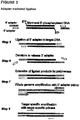
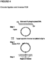
- Also disclosed herein is a method that selectively detects and amplifies target nucleic acid using a combination of the following 3 steps: 1) treating total isolated nucleic acid from a biological sample with a ligase that can covalently join blunt 5'-phosphorylated nucleic acid ends (e.g.
- T4 or T7 DNA ligase under conditions that favor unimolecular circularization of the nucleic acid molecules; 2) amplifying the nucleic acid with target-specific primers and a method that is selective for circular nucleic acid, for example, either a) via a rolling circle amplification with target-specific primers, or b) via inverse PCR with target-specific primers for the gene of interest; and 3) characterizing the amplified nucleic acid by direct or indirect qualitative and/or quantitative molecular characterization methods.
- cell-free nucleic acid may originate from several sources, it has been demonstrated that one source of circulating extracellular nucleic acid originates from programmed cell death, also known as apoptosis.
- the source of nucleic acid that arise as a result of apoptosis may be found in many body fluids and originate from several sources, including, but not limited to, normal programmed cell death in the host, induced programmed cell death in the case of an autoimmune disease, septic shock, neoplasms (malignant or non-malignant), or non-host sources such as an allograft (transplanted tissue), or the fetus or placenta of a pregnant woman.
- the applications for the detection, extraction and relative enrichment of extracellular nucleic acid from peripheral blood or other body fluids are widespread and may include inter alia, non-invasive prenatal diagnosis, cancer diagnostics, pathogen detection, auto-immune response and allograft rejection.
- the present invention includes methods to extract and relatively enrich by physical separation short base pair nucleic acid in the presence of a high background of genomic material (e . g ., host or maternal nucleic acids). More specifically, the present invention provides methods for the selective extraction and relative enrichment, based on size discrimination, of nucleic acid of approximately 1,200 base pairs or less (herein referred to as "target nucleic acid”) in a high background of genomic nucleic acids (herein referred to as "non-target nucleic acid”). This leads to a relatively enriched fraction of nucleic acid that has a higher concentration of smaller nucleic acids.
- the methods of the present invention may be used to improve pathogen detection.
- Methods for rapid identification of unknown bioagents using a combination of nucleic acid amplification and determination of base composition of informative amplicons by molecular mass analysis are disclosed in published U.S. Patent applications 20030027135 , 20030082539 , 20030124556 , 20030175696 , 20030175695 , 20030175697 , and 20030190605 and U.S. patent application Ser. Nos. 10/326,047 , 10/660,997 , 10/660,122 and 10/660,996 .
- host cell is any cell into which exogenous nucleic acid can be introduced, producing a host cell which contains exogenous nucleic acid, in addition to host cell nucleic acid.
- host cell nucleic acid and “endogenous nucleic acid” refer to nucleic acid species (e . g ., genomic or chromosomal nucleic acid) that are present in a host cell as the cell is obtained.
- exogenous refers to nucleic acid other than host cell nucleic acid; exogenous nucleic acid can be present into a host cell as a result of being introduced in the host cell or being introduced into an ancestor of the host cell.
- a nucleic acid species which is exogenous to a particular host cell is a nucleic acid species which is non-endogenous (not present in the host cell as it was obtained or an ancestor of the host cell).
- Appropriate host cells include, but are not limited to, bacterial cells, yeast cells, plant cells and mammalian cells.
- extraction refers to the partial or complete separation and isolation of a nucleic acid from a biological or non-biological sample comprising other nucleic acids.
- selective and selective refer to the ability to extract a particular species of nucleic acid molecule, on the basis of molecular size from a combination which includes or is a mixture of species of nucleic acid molecules.
- nucleic acid and “nucleic acid molecule” may be used interchangeably throughout the disclosure.
- the terms refer to a deoxyribonucleotide (DNA), ribonucleotide polymer (RNA), RNA/DNA hybrids and polyamide nucleic acids (PNAs) in either single- or double-stranded form, and unless otherwise limited, would encompass known analogs of natural nucleotides that can function in a similar manner as naturally occurring nucleotides.
- DNA deoxyribonucleotide
- RNA ribonucleotide polymer
- PNAs polyamide nucleic acids
- target nucleic acid refers to the nucleic acid of interest that is extracted based on its molecular size, preferably in a second extraction step, and further isolated for downstream analysis.
- the target nucleic acid has a molecular size smaller than the non-target nucleic acid present in the biological sample, for example, smaller than 1200 base pairs.
- the target nucleic acid may be from apoptotic DNA, fetal DNA, oncogenic DNA, or any non-host DNA.
- the target nucleic acid may be cell-free nucleic acid.
- the target nucleic acid may be oligonucleosomal nucleic acid generated during programmed cell death.
- non-target nucleic acid refers to the relatively high amount of non-desired background nucleic acid present in a biological sample, which is extracted, preferably, in a first extraction step.
- non-target nucleic acid has a molecular size larger than target nucleic acid, for example, greater than 1200 base pairs.
- Non-target nucleic acid may be from a host or host cell. In a preferred embodiment, non-target nucleic acid is of maternal origin.
- molecular size refers to the size of a nucleic acid molecule, which may be measured in terms of a nucleic acid molecule's mass or length (bases or base pairs).
- Fetal nucleic acid is present in maternal plasma from the first trimester onwards, with concentrations that increase with progressing gestational age ( Lo et al. Am J Hum Genet (1998) 62:768-775 ). After delivery, fetal nucleic acid is cleared very rapidly from the maternal plasma ( Lo et al. Am J Hum Genet (1999) 64:218-224 ). Fetal nucleic acid is present in maternal plasma in a much higher fractional concentration than fetal nucleic acid in the cellular fraction of maternal blood ( Lo et al. Am J Hum Genet (1998) 62:768-775 ).
- the target nucleic acid is of fetal origin
- the non-target nucleic acid is of maternal origin.
- the present invention relates to extracting nucleic acid from a biological sample as defined in the claims, including blood serum, blood plasma or urine.
- biological samples such as whole blood, umbilical cord blood, chorionic villi, amniotic fluid, cerbrospinal fluid, spinal fluid, lavage fluid ( e . g ., bronchoalveolar, gastric, peritoneal, ductal, ear, athroscopic), biopsy sample, urine, feces, sputum, saliva, nasal mucous, prostate fluid, semen, lymphatic fluid, bile, tears, sweat, breast milk, breast fluid, embryonic cells and fetal cells.
- the biological sample is blood plasma.
- blood encompasses whole blood or any fractions of blood, such as serum and plasma as conventionally defined.
- Blood plasma refers to the fraction of whole blood resulting from centrifugation of blood treated with anticoagulants.
- Blood serum refers to the watery portion of fluid remaining after a blood sample has coagulated.
- blood handling protocols are followed to ensure minimal degradation of nucleic acid in the sample and to minimize the creation of apoptotic nucleic acid in the sample. Blood handling methods are well known in the art.
- the biological sample may be cell-free or substantially cell-free.
- the biological sample may be a sample containing previously extracted, isolated or purified nucleic acids.
- One way of targeting target nucleic acid is to use the non-cellular fraction of a biological sample; thus limiting the amount of intact cellular material (e . g ., large strand genomic DNA) from contaminating the sample.
- a cell-free sample such as pre-cleared plasma, urine, etc. may be first treated to inactivate intracellular nucleases through the addition of an enzyme, a chaotropic substance, a detergent or any combination thereof.
- the biological sample may be first treated to remove substantially all cells from the sample by any of the methods known in the art, for example, centrifugation, filtration, affinity chromatography, etc.
- concentration sufficient to selectively bind refers to an amount sufficient to cause at least 50%, more preferably 70%, even more preferably 90% or more of the target nucleic acid to bind to an adsorptive surface.
- Suitable solid phase carriers include, but are not limited to, other particles, fibers, beads and or supports which have an affinity for nucleic acids or may be modified ( e . g ., the addition of a functional group or groups) to bind nucleic acids, and which can embody a variety of shapes, that are either regular or irregular in form, provided that the shape maximizes the surface area of the solid phase, and embodies a carrier which is amenable to microscale manipulations.
- silica-coated magnetic beads are used.
- the solid support may be modified to reversibly bind nucleic acid.
- the solid support may have a functional group-coated surface.
- the functional group-coated surface may be silica-coated, hydroxyl-coated, amine-coated, carboxyl-coated and encapsulated carboxyl group-coated.
- the term "functional group-coated surface” as used herein refers to a surface which is coated with moieties which reversibly bind, nucleic acids.
- One example is a surface which is coated with moieties which each have a free functional group which is bound to the amino group of the amino silane or the solid support; as a result, the surfaces of the solid support are coated with the functional group containing moieties.
- the functional group may be a carboxylic acid.
- a suitable moiety with a free carboxylic acid functional group is a succinic acid moiety in which one of the carboxylic acid groups is bonded to the amine of amino silanes through an amide bond and the second carboxylic acid is unbonded, resulting in a free carboxylic acid group attached or tethered to the surface of the paramagnetic microparticle.
- Suitable solid phase carriers having a functional group coated surface that reversibly binds nucleic acid molecules are for example, magnetically responsive solid phase carriers having a functional group-coated surface, such as, but not limited to, silica-coated, hydroxyl-coated, amino-coated, carboxyl-coated and encapsulated carboxyl group-coated magnetic beads.
- an oligonucleotide e.g ., an adapter or primer
- biotin which may bind to immobilized streptavidin.
- nucleic acid from biological material requires cell lysis, inactivation of cellular nucleases and separation of the desired nucleic acid from cellular debris.
- Common lysis procedures include mechanical disruption (e . g ., grinding, hypotonic lysis), chemical treatment (e . g ., detergent lysis, chaotropic agents, thiol reduction), and enzymatic digestion (e . g ., proteinase K).
- the biological sample may be first lysed in the presence of a lysis buffer, chaotropic agent (e.g., salt) and proteinase or protease. Cell membrane disruption and inactivation of intracellular nucleases may be combined.
- a single solution may contain detergents to solubilise cell membranes and strong chaotropic salts to inactivate intracellular enzymes. After cell lysis and nuclease inactivation, cellular debris may easily be removed by filtration or precipitation.
- Lysis may be blocked.
- the sample may be mixed with an agent that inhibits cell lysis to inhibit the lysis of cells, if cells are present, where the agent is a membrane stabilizer, a cross-linker, or a cell lysis inhibitor.
- the agent may be a cell lysis inhibitor, and may be glutaraldehyde, derivatives of glutaraldehyde, formaldehyde, formalin, or derivatives of formaldehyde. See U.S. patent application 20040137470 .
- the method may include adding a washing step or steps to remove non-nucleic acid molecules, for example salts, from the solid-support-target nucleic acid complex or surrounding solution.
- Non-nucleic acid molecules are then removed with an alcohol-based wash and the target nucleic acid is eluted under low- or no-salt conditions (TE buffer or water) in small volumes, ready for immediate use without further concentration.
- Extraction may be improved by the introduction of a carrier such as tRNA, glycogen, polyA RNA, dextran blue, linear poly acrylamide (LPA), or any material that increases the recovery of nucleic acid.
- the carriers may be added to the second binding solution or washing buffer.
- the final relative percentage of target nucleic acid to non-target nucleic acid may be at least about 5-6% fetal DNA, about 7-8% fetal DNA, about 9-10% fetal DNA, about 11-12% fetal DNA, about 13-14% fetal DNA.
- fetal DNA about 15-16% fetal DNA, about 16-17% fetal DNA, about 17-18% fetal DNA, about 18-19% fetal DNA, about 19-20% fetal DNA, about 20-21% fetal DNA, about 21-22% fetal DNA, about 22-23% fetal DNA, about 23-24% fetal DNA, about 24-25% fetal DNA, about 25-35% fetal DNA, about 35-45% fetal DNA, about 45-55% fetal DNA, about 55-65% fetal DNA, about 65-75% fetal DNA, about 75-85% fetal DNA, about 85-90% fetal DNA, about 90-91% fetal DNA, about 91-92% fetal DNA, about 92-93% fetal DNA, about 93-94% fetal DNA, about 94-95% fetal DNA, about 95-96% fetal DNA, about 96-97% fetal DNA, about 97-98% fetal DNA, about 98-99% fetal DNA, or about 99-99.7% fetal
- the methods provided herein may also be modified to combine steps, for example, in order to improve automation.
- the methods of the present invention may be used in conjunction with any known technique suitable for the extraction, isolation or purification of nucleic acids, including, but not limited to, cesium chloride gradients, gradients, sucrose gradients, glucose gradients, centrifugation protocols, boiling, Microcon 100 filter, Chemagen viral DNA/RNA 1k kit, Chemagen blood kit, Qiagen purification systems, Qiagen MinElute kits, QIA DNA blood purification kit, HiSpeed Plasmid Maxi Kit, QIAfilter plasmid kit, Promega DNA purification systems, MangeSil Paramagnetic Particle based systems, Wizard SV technology, Wizard Genomic DNA purification kit, Amersham purification systems, GFX Genomic Blood DNA purification kit, Invitrogen Life Technologies Purification Systems, CONCERT purification system, Mo Bio Laboratories purification systems, UltraClean BloodSpin Kits, and UlraClean Blood DNA Kit.
- cesium chloride gradients including cesium chloride gradients, gradients, sucrose gradients, glucose
- the first extraction method may be any known or modified technique suitable for the extraction, isolation or purification of non-target nucleic acids (i . e ., larger than target nucleic acids), including, but not limited to, cesium chloride gradients, gradients, sucrose gradients, glucose gradients, centrifugation protocols, boiling, Microcon 100 filter, Chemagen viral DNA/RNA 1k kit, Chemagen blood kit, Qiagen purification systems, Qiagen MinElute kits, QIA DNA blood purification kit, HiSpeed Plasmid Maxi Kit, QIAfilter plasmid kit, Promega DNA purification systems, MangeSil Paramagnetic Particle based systems, Wizard SV technology, Wizard Genomic DNA purification kit, Amersham purification systems, GFX Genomic Blood DNA purification kit, Invitrogen Life Technologies Purification Systems, CONCERT purification system, Mo Bio Laboratories purification systems, UltraClean BloodSpin Kits, and UlraClean Blood DNA Kit.
- One or more of the above methods may
- the second extraction method may be any known or modified technique suitable for the extraction, isolation or purification of target nucleic acids (i . e ., smaller than non-target nucleic acids), including, but not limited to, cesium chloride gradients, gradients, sucrose gradients, glucose gradients, centrifugation protocols, boiling, Microcon 100 filter, Chemagen viral DNA/RNA 1k kit, Chemagen blood kit, Qiagen purification systems, Qiagen MinElute kits, QIA DNA blood purification kit, HiSpeed Plasmid Maxi Kit, QIAfilter plasmid kit, Promega DNA purification systems, MangeSil Paramagnetic Particle based systems, Wizard SV technology, Wizard Genomic DNA purification kit, Amersham purification systems, GFX Genomic Blood DNA purification kit, Invitrogen Life Technologies Purification Systems, CONCERT purification system, Mo Bio Laboratories purification systems, UltraClean BloodSpin Kits, and UlraClean Blood DNA Kit.
- One or more of the above methods may
- the present specification also further relates to kits for practicing the disclosed methods.
- apoptosis is an essential mechanism in morphogenesis, development, differentiation, and homeostasis in all multicellular organisms.
- apoptosis is distinguished from necrosis by activation of specific pathways that result in characteristic morphological features including DNA fragmentation, chromatin condensation, cytoplasmic and nuclear breakdown, and the formation of apoptotic bodies.
- CAD Caspase-activated DNase
- DFF DNA fragmentation factor
- any method that can select for DNA fragments with blunt, 5'- phosphorylated ends is suitable to select for specific features (such as size, sequence and DNA base methylation differences) of the apoptotic DNA in a given biological sample. See for example, US patent applications 20050019769 , 20050164241 , 20030044388 , or 20060019278 .
- a method For enrichment and detection of apoptotic DNA ladders in mammalian tissues, a method has been described that takes advantage of the presence of blunt, 5'-phosphorylated ends in apoptotic DNA by ligation of synthetic, blunt-ended linkers to both ends of linear apoptotic DNA fragments with T4 ligase which is able to form a covalent bond between the 3'-hydroxy ends of the synthetic linker and the 5'-phosphorylated ends of the DNA fragments ( Staley et al, Cell Death Differ. 1997 Jan;4(1):66-75 ).
- the method can only be used as a generic tool to characterize the size distribution of apoptotic ladders in specific tissues in general, and is not site or sequence specific.
- the present specification therefore, also provides a method for selectively amplifying short, fragmented nucleic acid by adapter mediated ligation and other related methods.
- the method capitalizes on the blunt end and 5'-phosphorylated nature of the target nucleic acid as a means to attach a non-genome specific adapter to the blunt ends using a ligation process. While the nature of the termini of all cell-free nucleic acid is unknown, coupling this method with short extension times during amplification will favor the amplification of the oligonucleosome monomer and short multimers. Since the target nucleic acid is shorter than the non-targeted nucleic acid, the target nucleic acid can be enriched over the non-target nucleic acid.
- This method can be further coupled with specific amplification of a nucleic acid region of interest for further analysis.
- the 3' and 5' dephosphorylated adapters are complementary and form a double-stranded blunt end adapter complex.
- the 5' adapter of the adapter complex ligates to the 5' phosphorylated strand of the target nucleic acid, and heat is introduced to release the shorter, unligated 3' adapter.
- the 5' protruding ends of the ligated complex are filled in by a thermostable DNA polymerase.
- the 5' adapter is reintroduced and serves as a PCR primer for whole genome amplification.
- Oligonucleosomes are the repeating structural units of chromatin, each consisting of approximately 200 base pairs of DNA wound around a histone core that partially protects the DNA from nuclease digestion in vitro and in vivo. These units can be found as monomers or multimers and produce what is commonly referred to as an apoptotic DNA ladder. The units are formed by nuclease digestion of the flanking DNA not bound to histone resulting in the majority of oligonucleosomes being blunt ended and 5'-phorsphorylated.
- oligonucleosomes are hard to detect and harder to isolate; however, they can serve as predictors for disease and other conditions (see US patent application 20040009518 ).
- 5' dephosphorylated adapter refers to a nucleic acid which comprises about 20 to 30 base pairs that is complementary to a short dephosphorylated adapter and capable of hybridizing thereto to form a double-stranded, blunt end adapter complex capable of ligating to target nucleic acid. Specifically, the 5'adapter ligates to the 5'phosphorylated base of the target nucleic acid.
- 3' dephosphorylated adapter refers to a nucleic acid which comprises about 10 to 15 base pairs that is complementary to the 5' adapter at the 3' end, thus capable of creating a double-stranded blunted end necessary for ligation.
- the 3' adapter does not bind or ligate to the oligonucleosomal DNA.
- 5' adapter primer refers to the same oligonucleotide sequence as the 5' dephosphorylated adapter, but is later reintroduced to the ligated sample to facilitate the whole genome amplification.
- adapter complex refers to the hybridized, double-stranded 5' adapter and 3' adapter molecule.
- the method is semi quantitative. By comparing the numbers of PCR cycles needed to detect target nucleic acid in two samples, the relative amount of target nucleic acid occurring in each sample can be estimated.
- the 5' adapters may be bound to a solid support for increased enrichment of the target nucleic acid.
- non-ligated, non-target nucleic acid is substantially removed from the solution, and amplification can proceed using only the targeted material that has ligated to the 5' adapters. This improves the enrichment of the target nucleic acid by removing genomic non-target nucleic acid that may compete with the target nucleic acid in the target-specific amplification step.
- the target sequence is present in both the mother and the fetus, and the maternal sample is very abundant (>95%), under normal circumstances the fetal nucleic acid would not be detectable as it would be out-competed by the maternal nucleic acid in the first cycles of amplification.
- the fetal nucleic acid is part or wholly oligonucleosomal in nature, and the majority of the ligated sample is fetal in nature, maternal nucleic acid is still present in the sample, which can compete with the fetal nucleic acid in the target-specific amplification step. Separation of non-target nucleic acid from the target nucleic acid ( i .
- ligated sample increases the detection of fetal nucleic acid in cases where there is an abundance of maternal nucleic acid in the initial biological sample, there is sample degradation, or there is a maternal condition (e . g ., autoimmune disease, transplant rejection, cancer) that increases the amount of maternal oligonucleosomes.
- Spacer arms may be introduced between the solid support and 5' adapter to improve ligation of the target nucleic acid to the adapter molecule.
- the ligated sample bound to a solid support may be combined with a ligated sample that does not have a solid support prior to the amplification step.
- spacer arms refers to any molecule that can be used in single or multiples to create space between the solid support and an oligonucleotide (e.g., target nucleic acidOne or more hexathylene (HEG) spacer units may be inserted between the aminohexyl groups and the 5' end of the 5' adapter.
- the aminohexyl group is used for covalent coupling to the solid support.
- spacer arms include multiple dTTP's (up to 15), spacer 18 (an 18 atom hexa-ethylene glycol spacer), spacer 9 (a triethylene glycol spacer), or photocleavable spacers known in the art.
- An alternative method disclosed herein selectively detects and amplifies target nucleic acid using a combination of the following 3 steps:
- any combination of these 3 steps will selectively enrich the double-stranded, blunt-ended 5'-phosphorylated DNA such as DNA from apoptotic ladders by several orders of magnitude over the DNA fragments present in the biological sample that cannot by circularized due to lack of blunt ends and/or missing 5'-terminal phosphate groups and allow a comparison of its sequence with the wild-type sequence of the same organism or the host organism in case of a transplant or a maternal sequence in case of a pregnancy, for instance.
- step 2 an aliquot of the total DNA is either treated with methylation-sensitive or methylation-resistant enzymes or with chemicals that convert methylated bases into different bases so that methylated bases in the apoptotic DNA fragments can be characterized after step 2 and 3.
- Circulating nucleic acids in the plasma and serum of patients are associated with certain diseases and conditions (See, Lo YMD et al., N Eng J Med 1998;339:1734-8 ; Chen XQ, et al., Nat Med 1996;2:1033-5 , Nawroz H et al., Nat Med 1996;2:1035-7 ; Lo YMD et al., Lancet 1998;351:1329-30 ; Lo YMD, et al., Clin Chem 2000;46:319-23 ). Further, the method of nucleic acid isolation may affect the ability to detect these disease-associated nucleic acids circulating in the blood ( Wang et al. Clin Chem. 2004 Jan;50(1):211-3 ).
- the characteristics and biological origin of circulating nucleic acids are not completely understood. However, it is likely that cell death, including apoptosis, is one major factor ( Fournie e al., Gerontology 1993;39:215-21 ; Fournie et al., Cancer Lett 1995;91:221-7 ). Without being bound by theory, as cells undergoing apoptosis dispose nucleic acids into apoptotic bodies, it is possible that at least part of the circulating nucleic acids in the plasma or serum of human subjects is short, fragmented DNA that takes the form particle-associated nucleosomes.
- the present invention provides methods for extracting the short, fragmented circulating nucleic acids present in the plasma or serum of subjects, thereby enriching the short, predictive nucleic acids relative to the background genomic DNA.
- the present specification provides methods of evaluating a disease condition in a patient suspected of suffering or known to suffer from the disease condition.
- the specification describes obtaining a biological sample from the patient suspected of suffering or known to suffer from a disease condition, selectively extracting and enriching extracellular nucleic acid in the sample based on its size using the methods provided herein, and evaluating the disease condition by determining the amount or concentration or characteristic of enriched extracellular nucleic acid and comparing the amount or concentration or characteristic of enriched extracellular nucleic acid to a control (e . g ., background genomic DNA from biological sample).
- a control e . g ., background genomic DNA from biological sample.
- evaluating a disease condition refers to assessing the disease condition of a patient.
- evaluating the condition of a patient can include detecting the presence or absence of the disease in the patient. Once the presence of disease in the patient is detected, evaluating the disease condition of the patient may include determining the severity of disease in the patient. It may further include using that determination to make a disease prognosis, e . g . a prognosis or treatment plan. Evaluating the condition of a patient may also include determining if a patient has a disease or has suffered from a disease condition in the past. Evaluating the disease condition in that instant might also include determining the probability of reoccurrence of the disease condition or monitoring the reoccurrence in a patient.
- Evaluating the disease condition might also include monitoring a patient for signs of disease. Evaluating a disease condition therefore includes detecting, diagnosing, or monitoring a disease condition in a patient as well as determining a patient prognosis or treatment plan. The method of evaluating a disease condition aids in risk stratification.
- nucleic acids including DNA and RNA
- RNA RNA
- the methods provided herein may be used to extract oncogenic nucleic acid, which may be further used for the detection, diagnosis or prognosis of a cancer-related disorder.
- nucleic acids including DNA and RNA
- RNA RNA
- These molecules are likely packaged in apoptotic bodies and, hence, rendered more stable compared to 'free RNA' ( Anker P and Stroun M, Clin Chem (2002) 48, 1210-1211 ; Ng EK, et al. Proc Natl Acad Sci USA (2003) 100, 4748-4753 ).
- Tumor-specific DNA for a wide range of malignancies has been found: haematological, colorectal, pancreatic, skin, head-and-neck, lung, breast, kidney, ovarian, nasopharyngeal, liver, bladder, gastric, prostate and cervix.
- haematological, colorectal, pancreatic, skin, head-and-neck, lung, breast, kidney, ovarian, nasopharyngeal, liver, bladder, gastric, prostate and cervix In aggregate, the above data show that tumor-derived DNA in plasma is ubiquitous in affected patients, and likely the result of a common biological process such as apoptosis. Investigations into the size of these plasma DNA fragments from cancer patients has revealed that the majority show lengths in multiples of nucleosomal DNA, a characteristic of apoptotic DNA fragmentation ( Jahr S, et al. Cancer Res (2001) 61,1659-1665 ).
- PCR-specific strategies can be developed.
- clinical application awaits optimization of methods to isolate, quantify and characterize the tumor-specific DNA compared to the patient's normal DNA, which is also present in plasma. Therefore, understanding the molecular structure and dynamics of DNA in plasma of normal individuals is necessary to achieve further advancement in this field.
- the present specification describes detection of specific extracellular nucleic acid in plasma or serum fractions of human or animal blood associated with neoplastic, premalignant or proliferative disease. Specifically, the specification relates to detection of nucleic acid derived from mutant oncogenes or other tumor-associated DNA, and to those methods of detecting and monitoring extracellular mutant oncogenes or tumor-associated DNA found in the plasma or serum fraction of blood by using DNA extraction with enrichment for mutant DNA as provided herein.
- the specification relates to the detection, identification, or monitoring of the existence, progression or clinical status of benign, premalignant, or malignant neoplasms in humans or other animals that contain a mutation that is associated with the neoplasm through the size selective enrichment methods provided herein, and subsequent detection of the mutated nucleic acid of the neoplasm in the enriched DNA.
- the present specification features methods for identifying DNA originating from a tumor in a biological sample. These methods may be used to differentiate or detect tumor-derived DNA in the form of apoptotic bodies or nucleosomes in a biological sample. In preferred cases, the non-cancerous DNA and tumor-derived DNA are differentiated by observing nucleic acid size differences, wherein low base pair DNA is associated with cancer.
- the present specification features methods for differentiating DNA species originating from different individuals in a biological sample. These methods may be used to differentiate or detect fetal DNA in a maternal sample.
- the DNA species can be differentiated by observing nucleic acid size differences.
- the differentiation between maternal and fetal DNA may be performed with or without quantifying the concentration of fetal DNA in maternal plasma or serum. Where the fetal DNA is quantified, the measured concentration may be used to predict, monitor or diagnose or prognosticate a pregnancy-associated disorder.
- non-invasive and invasive techniques available for prenatal diagnosis including ultrasonography, amniocentesis, chorionic villi sampling (CVS), fetal blood cells in maternal blood, maternal serum alpha-fetoprotein, maternal serum beta-HCG, and maternal serum estriol.
- CVS chorionic villi sampling
- fetal blood cells in maternal blood maternal serum alpha-fetoprotein
- maternal serum beta-HCG maternal serum alpha-fetoprotein
- maternal serum estriol maternal serum estriol
- the first marker that was developed for fetal DNA detection in maternal plasma was the Y chromosome, which is present in male fetuses ( Lo et al. Am J Hum Genet (1998) 62:768-775 ).
- the robustness of Y chromosomal markers has been reproduced by many workers in the field ( Costa JM, et al. Prenat Diagn 21:1070-1074 ). This approach constitutes a highly accurate method for the determination of fetal gender, which is useful for the prenatal investigation of sex-linked diseases ( Costa JM, Ernault P (2002) Clin Chem 48:679-680 ).
- Maternal plasma DNA analysis is also useful for the noninvasive prenatal determination of fetal RhD blood group status in RhD-negative pregnant women ( Lo et al. (1998) N Engl J Med 339:1734-1738 ). This approach has been shown by many groups to be accurate, and has been introduced as a routine service by the British National Blood Service since 2001 ( Finning KM, et al. (2002) Transfusion 42:1079-1085 ).
- fetal DNA in maternal plasma include the detection of achondroplasia ( Saito H, et al. (2000) Lancet 356:1170 ), myotonic dystrophy ( Amicucci P, et al. (2000) Clin Chem 46:301-302 ), cystic fibrosis ( Gonzalez-Gonzalez MC, et al. (2002) Prenat Diagn 22:946-948 ), Huntington disease ( Gonzalez-Gonzalez MC, et al. (2003) Prenat Diagn 23:232-234 ), and congenital adrenal hyperplasia ( Rijnders RJ, et al. (2001) Obstet Gynecol 98:374-378 ). It is expected that the spectrum of such applications will increase over the next few years.
- the patient is pregnant and the method of evaluating a disease or physiological condition in the patient or her fetus aids in the detection, monitoring, prognosis or treatment of the patient or her fetus. More specifically, the present specification features methods of detecting abnormalities in a fetus by detecting fetal DNA in a biological sample obtained from a mother.
- the methods according to the present specification provide for detecting fetal DNA in a maternal sample by differentiating the fetal DNA from the maternal DNA based on DNA characteristics ( e . g ., size, weight, 5' phosphorylated, blunt end). See Chan et al. Clin Chem. 2004 Jan;50(1):88-92 ; and Li et al. Clin Chem.
- fetal DNA that is predictive of a genetic anomaly or genetic-based disease may be identified thereby providing methods for prenatal diagnosis. These methods are applicable to any and all pregnancy-associated conditions for which nucleic acid changes, mutations or other characteristics (e . g ., methylation state) are associated with a disease state. Exemplary diseases that may be diagnosed include, for example, preeclampsia, preterm labor, hyperemesis gravidarum, ectopic pregnancy, fetal chromosomal aneuploidy (such as trisomy 18, 21, or 13), and intrauterine growth retardation.
- the methods of the present invention allow for the analysis of fetal genetic traits including those involved in chromosomal aberrations (e . g . aneuploidies or chromosomal aberrations associated with Down's syndrome) or hereditary Mendelian genetic disorders and, respectively, genetic markers associated therewith (e . g . single gene disorders such as cystic fibrosis or the hemoglobinopathies). Size-based extraction of extracellular fetal DNA in the maternal circulation thus facilitates the non-invasive detection of fetal genetic traits, including paternally inherited polymorphisms which permit paternity testing.
- pregnancy-associated disorder refers to any condition or disease that may affect a pregnant woman, the fetus the woman is carrying, or both the woman and the fetus. Such a condition or disease may manifest its symptoms during a limited time period, e.g., during pregnancy or delivery, or may last the entire life span of the fetus following its birth.
- a pregnancy-associated disorder include ectopic pregnancy, preeclampsia, preterm labor, and fetal chromosomal abnormalities such as trisomy 13, 18, or 21.
- chromosomal abnormality refers to a deviation between the structure of the subject chromosome and a normal homologous chromosome.
- normal refers to the predominate karyotype or banding pattern found in healthy individuals of a particular species.
- a chromosomal abnormality can be numerical or structural, and includes but is not limited to aneuploidy, polyploidy, inversion, a trisomy, a monosomy, duplication, deletion, deletion of a part of a chromosome, addition, addition of a part of chromosome, insertion, a fragment of a chromosome, a region of a chromosome, chromosomal rearrangement, and translocation.
- a chromosomal abnormality can be correlated with presence of a pathological condition or with a predisposition to develop a pathological condition.
- the present specification also provides a method of evaluating the disease condition of a patient suspected of having suffered from a trauma or known to have suffered from a trauma.
- the method includes obtaining a sample of plasma or serum from the patient suspected of having suffered from a trauma or known to have had suffered from a trauma, and detecting the quantity or concentration of mitochondrial nucleic acid in the sample.
- the example provides a procedure, using a method provided herein, to selectively extract DNA based on its size.
- chaotropic salt for example, less than 30% solution (weight per volume) to the sample solution to denature proteins and inactivate nucleases, proteinase K or any protease, for example 100 to 1000 ⁇ g) to further inactivate nucleases and break down proteins in solution.
- detergents for example SDS or Triton-X 100 up to 1% volume per volume, may be used alone or in combination with a salt.
- wash target nucleic acid-bound solid support using an appropriate washing solution comprised of salt, buffer, water and alcohol.
- an appropriate washing solution comprised of salt, buffer, water and alcohol.
- Figure 1 shows the successful extraction of low base pair DNA from a 1kb DNA ladder (PromegaTM) in the presence of guanidine thiocyanate (GuSCN).
- the DNA is first bound to silica at various low concentrations of guanidine thiocyanate as shown in Figure 1 .
- the supernatant from the first binding solution is subsequently bound to silica at varying guanidine thiocyanate concentrations, with a finishing high concentration of 4.6M. These steps are followed by wash and elution steps.
- the method can be employed to produce size selective separation of a commercially available DNA ladder with DNA strands ranging in mass from 250bp to 10,000 bp from normal human plasma in the presence of guanidine thiocyanate as the chaotropic salt. The following steps are performed:
- Figure 2 shows the successful extraction of low base pair DNA from a 1kb DNA ladder (PromegaTM) in the presence of sodium perchlorate (NaClO 4 ).
- the DNA is first bound to silica at various low concentrations of sodium perchlorate as shown in Figure 2 .
- the supernatant from the first binding solution is subsequently bound to silica at varying sodium perchlorate concentrations, with a finishing high concentration of 4.5 M. These steps are followed by wash and elution steps.
- the method can be employed to produce size selective separation of a commercially available DNA ladder with DNA strands ranging in mass from 250bp to 10,000 bp from normal human plasma in the presence of sodium perchlorate (NaClO 4 ) as the chaotropic salt.
- NaClO 4 sodium perchlorate
- the below example provides a procedure, using the method provided herein, to selectively amplify target nucleic acid that is blunt-ended and 5'-phosphorylated.
- the method relies on the ligation of a non-genome specific adapter to the blunt ends, which allows for whole genome amplification followed by target-specific amplification. While the nature of the termini of all cell-free nucleic acid is unknown, coupling this method with short extension times during amplification will favor the amplification of the oligonucleosome monomer and short multimers. Since the target nucleic acid is shorter than the non-targeted nucleic acid, the target nucleic acid can be enriched over the non-target nucleic acid.
- the 3' adapter is complementary to the 3'-end of 5' adapter to create the blunt-end, double-stranded adapter complex.
- 5' adapter 5'-ACACGGCGCACGCCTCCACG-3' ⁇ blunt-end
- 3' adapter 3'-GTGCGGAGGTGC-5'
- the 3' adapter molecule is modified such that the new sequence consists of 13 nucleobases with a dideoxy-nucleotide at its 3'-position. This terminator nucleotide does not allow extension of the 3' adapter molecule by any polymerase, thus improving assay efficiency and detection.
- Alternative 3' adapter 3' adapter (SSGA13dd3'): 5'-CGTGGAGGCGTG ddNTP -3'
- the 3' adapter is shorter to reduce the melting temperature and allow for release from the 5' adapter following ligation.
- Both adapters are non-phosphorylated, and may be made of any sequence that is nonspecific to the nucleic acid to be amplified to prevent non-specific amplification of the genome.
- An exemplary procedure is provided below: 1. Prepare total or size selective nucleic acid sample in water or buffer. 2. Add ligation buffer, 5' adapter, 3' adapter, and water to reaction volume. 3. Place the reaction into a thermocycler and heat the reaction to 55°C for 10 minutes, then slowly ramp down the temperature to 10°C over 1 hour. 4. Add 1 ⁇ l T4 ligase (1-3 U per ul) (or ligation enzyme) and mix well and incubate for 10 min at 10°C, then ramp temperature up to 16°C and incubate for 10 minutes to over night (12-16 hours). 5. Add 5' primer, 10x PCR buffer, MgCl 2 , dNTPs, and polymerase. 6.
- Described hereafter is a method for intramolecular ligation followed by amplification of a target by inverse PCR or rolling circle amplification to detect a target nucleic acid.
Description
- The invention relates to methods for the extraction of nucleic acids from a sample as characterized in the claims. The methods of the invention may be used in a wide range of applications, including the extraction of fetal nucleic acids from maternal plasma, the detection of circulating nucleic acids from neoplasms (malignant or non-malignant), the detection of early onset of tissue rejection, or any other application requiring the selective separation of nucleic acids based on their size and/or apoptotic origin.
- The isolation and subsequent amplification of nucleic acids play a central role in molecular biology. Isolated, purified nucleic acids may be used, inter alia, as a starting material for diagnosis and prognosis of diseases or disorders. Therefore, the isolation of nucleic acids, particularly by non-invasive means, is of particular importance for use in genetic analyses.
- Current methods for the extraction of nucleic acids include the use of organic-based methods (e.g., phenol/chloroform/isoamyl alcohol), or capitalize upon ion interaction of nucleic acids in an aqueous solution (e.g., salting out in combination with alcohol, solution pH and temperature) alone or in combination with anion exchange chromatography or cation exchange chromatography. Organic-based methods employ the use of phenol/chloroform/isoamyl alcohol or variations thereof for isolating DNA, but have serious disadvantages, namely the processes are very time-consuming, require considerable experimental effort, and are associated with an acute risk of exposure to toxic substances to those carrying out the isolation. Chromatography-based methods increase flexibility and automation since these methods can be used in combination with multiple matrices (e.g., membranes, latex, magnetic beads, micro-titer plate, etc.) and in the presence or absence of ligands (e.g., DEAE, silica, acrylamide, etc.). However, these methods are better suited to extract larger strands of nucleic acids to ensure greater success in downstream analysis.
- Previously, the recovery of smaller, fragmented nucleic acids from biological samples was considered unimportant, and extraction methods were designed to isolate large, undegraded nucleic acid molecules. Recently, however, it is shorter base pair nucleic acids (e.g., highly degraded RNA or mRNA and apoptotic DNA) that have been shown to be highly informative for a wide range of applications, including prenatal diagnostics and the study of apoptotic DNA from host or non-host sources. Methods to capture and protect RNA during extraction are now common; however the ability to successfully analyze short, fragmented DNA in the presence of more abundant, longer DNA has remained elusive.
- The invention relates to the embodiments as characterized in the claims. There is a need for improved extraction methods capable of capturing small nucleic acid molecules. At the same time, these methods need to be simple, cost-effective and automatable in order to prove useful in the research and clinical environments. Thus, the invention relates to methods for the extraction of nucleic acids based on their size. In particular, the invention relates to a method for extracting low molecular weight nucleic acid from a sample comprising a mixture of low molecular weight nucleic acid and high molecular weight nucleic acid, which comprises: (a) mixing the sample, a salt and a solid support capable of reversibly binding nucleic acid, thereby forming a first binding solution, wherein the salt is present at a concentration at which the high molecular weight nucleic acid selectively adsorbs to the solid support; (b) separating the solid support from the first binding solution, thereby yielding a fraction separated from the solid support; (c) mixing the fraction separated from the solid support in (b) with additional salt, whereby the salt is present at a higher concentration than that of the first binding solution in (a); (d) introducing additional solid support to the mixture in (c), thereby forming a second binding solution, wherein the low molecular weight nucleic acid selectively adsorbs to the additional solid support; and (e) separating the additional solid support from the second binding solution, thereby extracting low molecular weight nucleic acid from the sample, wherein the low molecular weight nucleic acid comprises less than 1200 base pairs and the high molecular weight nucleic acid comprises 1200 or more base pairs. Also disclosed herein are compositions and kits for the extraction, amplification and analysis of nucleic acids based on their size. Studies have shown that the majority of cell-free nucleic acid resulting from neoplasms, allograft rejection, autoimmune reactions, fetal tissue, etc. has a relatively small size of approximately 1,200 base pairs or less, whereas the majority of cell-free nucleic acid arising in the host from non-programmed cell death-associated events has a size greater than approximately 1,200 base pairs.
- The present invention, therefore, provides methods for the enrichment, based on size discrimination, of nucleic acid of approximately 1,200 base pairs or less (herein referred to as "target nucleic acid") in a high background of genomic nucleic acid (herein referred to as "non-target nucleic acid"). This leads to a relatively enriched fraction of nucleic acid that has a higher concentration of smaller nucleic acid.
- The present invention provides methods for extracting target nucleic acid from a biological sample containing a mixture of non-target nucleic acid based on the size of the nucleic acid, wherein the target nucleic acid size is less than the size of the non-target nucleic acid in the mixture, comprising the steps of introducing the biological sample to a first extraction method designed to isolate non-target nucleic acid, wherein the target nucleic acid is not substantially isolated, thereby creating a supernatant that contains target nucleic acid; removing the supernatant and introducing said supernatant to a second extraction method designed to isolate target nucleic acid, and, optionally, eluting the target nucleic acid with an elution buffer suitable for eluting nucleic acid, whereby the target nucleic acid has been selectively extracted from the sample as defined in the claims.
- The present specification also provides compositions, methods and kits for the adsorption of target nucleic acid to a solid support in the presence of increasing concentrations of salt, whereby the target nucleic acid is selectively enriched based on its molecular size. The compositions and methods may be used to extract and enrich the amount of normally trace nucleic acid, which is initially in the presence of high amounts of non-desired background nucleic acid, to levels suitable for detection and analysis. The specification also provides compositions and methods for binding nucleic acid under specific conditions to introduce size selection with the purpose of extraction of any nucleic acid within the range of about 10 bases to about 5000 bases.
- Nucleic acids are known to bind to a solid phase in the presence of a chaotropic agent (see
US Patent No. 5,234,809 ). Thus, provided herein are improved methods for extracting low molecular weight nucleic acid in a sample by bringing a nucleic acid-containing solution to a low salt concentration state; adsorbing the nucleic acid to a solid support and separating the solid support from the solution; bringing the solution to a high salt concentration state; adsorbing the nucleic acid to a solid support and separating the solid support from the solution; and eluting adsorbed nucleic acid from the solid support, whereby the low molecular weight nucleic acid has been selectively enriched from the sample. - In a related embodiment, the invention provides a method for extracting target nucleic acid from a biological sample containing a mixture of non-target nucleic acid based on the size of the nucleic acid, wherein the target nucleic acid size is less than the size of the non-target nucleic acid in the mixture, comprising the steps of mixing said biological sample, a salt and a nucleic acid binding solid support, wherein the salt is present at a concentration sufficient to bind non-target nucleic acid, while binding substantially little to no target nucleic acid, thereby creating a first binding solution; adsorbing the non-target nucleic acid to the solid support, and separating the solid support from the solution; removing the supernatant of the first binding solution, and mixing said supernatant with additional salt and a nucleic acid binding solid support, wherein the salt is present at a concentration sufficient to bind the target nucleic acid, thereby creating a second binding solution; adsorbing the target nucleic acid to the solid support, and separating the solid support from the second binding solution, thereby creating a solid support-target nucleic acid complex; and eluting the adsorbed target nucleic acid from the solid support with an elution buffer suitable for eluting nucleic acid, whereby the target nucleic acid has been selectively extracted from the sample as defined in the claims.
- The methods of the present disclosure may be used to extract nucleic acid within the range of about 10 bases to about 5000 bases. In a preferred embodiment, the target nucleic acid is at least about 25 base pairs, but less than about 1200 base pairs, and can be between about 200 base pairs and about 600 base pairs.
- The present disclosure also relates to extracting nucleic acids such as DNA, RNA, mRNA, oligonucleosomal, mitochondrial, epigenetically modified, single-stranded, double-stranded, circular, plasmid, cosmid, yeast artificial chromosomes, artificial or man-made DNA, including unique DNA sequences, and DNA that has been reverse transcribed from an RNA sample, such as cDNA, and combinations thereof. The nucleic acid may be cell-free nucleic acid. In another embodiment, the nucleic acids are derived from apoptotic cells. In another embodiment, the target nucleic acid is of fetal origin, and the non-target nucleic acid is of maternal origin.
- The present disclosure relates to extracting nucleic acid from a biological sample such as whole blood, serum, plasma, umbilical cord blood, chorionic villi, amniotic fluid, cerbrospinal fluid, spinal fluid, lavage fluid (e.g., bronchoalveolar, gastric, peritoneal, ductal, ear, athroscopic) biopsy sample, urine, feces, sputum, saliva, nasal mucous, prostate fluid, semen, lymphatic fluid, bile, tears, sweat, breast milk, breast fluid, embryonic cells and fetal cells. In a preferred embodiment, the biological sample is plasma. The biological sample may be cell-free or substantially cell-free. The biological sample may be a sample of previously extracted nucleic acids.
- The present invention is particularly useful for extracting fetal nucleic acid from maternal plasma. The biological sample may be from an animal, most preferably a human. In another preferred embodiment, the biological sample is from a pregnant human. The biological sample may be collected from a pregnant human after the fifth week of gestation. The pregnant human may have a relatively elevated concentration of free fetal nucleic acid in her blood, plasma or amniotic fluid. The pregnant human may have a relatively decreased concentration of apoptotic nucleic acid in her blood, plasma or amniotic fluid. The methods of the present invention may be performed in conjunction with any known method to elevate fetal nucleic acid in maternal blood, plasma or amniotic fluid. Likewise, the methods of the present invention may be performed in conjunction with any known method to decrease apoptotic nucleic acid in maternal blood, plasma or amniotic fluid.
- The presently described method is based on the ability of nucleic acid to reversibly bind to a nucleic acid-binding solid support in the presence of a salt, such as guanidine salt, sodium iodide, potassium iodide, sodium thiocyanate, urea, sodium chloride, magnesium chloride, calcium chloride, potassium chloride, lithium chloride, barium chloride, cesium chloride, ammonium acetate, sodium acetate, ammonium perchlorate or sodium perchlorate, for example. In a preferred embodiment, the salt is a guanidine salt, most preferably guanidine (iso)thiocyanate, or is a sodium salt, most preferably sodium perchlorate. In the methods provided herein, the salt is introduced at a concentration to bind nucleic acid to a solid support. In the first binding solution, a salt is added to yield a solution with a concentration in the range of 10 to 30% weight per volume capable of binding non-target nucleic acid, while minimizing the binding of target nucleic acid. The non-target nucleic acid is at least 1200 base pairs. In the second binding solution, a chaotropic substance is added to yield a solution with a salt concentration greater than 10%, and preferably in the range of 20 to 60% weight per volume, which is capable of binding target nucleic acid.
- In a related embodiment, the solid support is a hydroxyl donor (e.g., silica or glass). In other cases, the solid support contains a functional group that serves as a hydroxyl donor and is attached to a solid support. Examples of solid supports include paramagnetic microparticles, silica gel, silica particles, controlled pore glass, magnetic beads, biomagnetic separation beads, microspheres, divinylbenzene (DVB) resin, cellulose beads, capillaries, filter membranes, columns, nitrocellulose paper, flat supports, glass surfaces, metal surfaces, plastic materials, multiwell plates or membranes, wafers, combs, pins and needles, or any combination thereof, for example. The solid support may be modified to reversibly bind nucleic acid. The solid support may be a silica gel membrane.
- The nucleic acid-solid support interaction may be an electrostatic interaction or a polar interaction.
- The solid support may have a functional group-coated surface. The functional group-coated surface may be silica-coated, hydroxyl coated, amine-coated, carboxyl-coated or encapsulated carboxyl group-coated, for example. A bead may be silica-coated or a membrane may contain silica gel.
- In the present invention, it is necessary to separate the nucleic acid-coated solid support from the first or second binding solutions. The solid support (e.g., silica-coated magnetic bead) can be separated from the solutions by any method known in the art, including applying a magnetic field, applying vacuum filtration and/or centrifugation, or any combination thereof. Paramagnetic beads may be separated from one or both solutions using magnets or magnetic devices.
- The methods provided herein may also be modified to introduce additional steps, for example, in order to improve the extraction of nucleic acid or improve analysis of target nucleic acid following extraction. For example, the biological sample may be first lysed in the presence of a lysis buffer, which may comprise a chaotropic substance (e.g., salt), a proteinase, a protease or a detergent, or combinations thereof, for example. The lysis step and the creation of the first binding solution may be performed simultaneously at a salt concentration sufficient to solubilize or precipitate non-nucleic acid material (e.g., protein) in the sample and to bind the non-target nucleic acid to the solid support. The method may include adding a washing step or steps to remove non-nucleic acid from the solid-support-target nucleic acid complex. The solid support-target nucleic acid complex may be further washed successively with a wash buffer and one or more alcohol-water solutions, and subsequently dried. The wash buffer may comprise a chaotropic substance (e.g., salt), and optionally, a carrier such as LPA, RNA, tRNA, dextran blue, glycogen or polyA RNA, for example. The second binding solution may also comprise a carrier such as LPA, RNA, tRNA, dextran blue, glycogen or polyA RNA, for example.
- The methods provided herein may also be modified to combine steps, for example, in order to improve automation. For example, mixing the first binding solution and adsorbing the non-target nucleic acid to the solid support may be performed simultaneously. Likewise, mixing the second binding solution and adsorbing the target nucleic acid to the solid support may be performed simultaneously.
- The methods provided herein may be performed prior to, subsequent to, or simultaneously with another method for extracting nucleic acid such as electrophoresis, liquid chromatography, size exclusion, microdialysis, electrodialysis, centrifugal membrane exclusion, organic or inorganic extraction, affinity chromatography, PCR, genome-wide PCR, sequence-specific PCR, methylation-specific PCR, introducing a silica membrane or molecular sieve, and fragment selective amplification.
- The present disclosure also further relates to a kit comprising reagents for a first binding buffer formulated to comprise a suitable salt, wherein the salt is present at a concentration appropriate for binding a non-target nucleic acid characterized by a particular size, to the solid support; a second binding buffer formulated to comprise a suitable salt, wherein the salt is present at a concentration appropriate for binding a target nucleic acid characterized by a particular size, to the solid support; an aqueous solution of functional group-coated paramagnetic microparticles; and instructions for performing the target nucleic acid extraction. The kit may additionally comprise reagents for the formulation of a wash buffer and an elution buffer, wherein the wash buffer dissolves impurities, but not nucleic acids bound to solid support and the elution buffer is a non-salt buffered solution with a pH range between about 7.0 to 8.5.
- The present disclosure also provides methods for a post purification process which allows enrichment of target nucleic acid by ligation-based methods followed by amplification. The present disclosure also provides a method for selectively amplifying a target nucleic acid from a biological sample containing a mixture of non-target nucleic acid, wherein the target nucleic acid is a double stranded, blunt end nucleic acid fragment with 5' phosphorylated ends, comprising the steps of a) mixing the biological sample, a 5' adapter and a 3' adapter, wherein the 3' adapter is complementary to the 5' adapter at the 3' end and thus capable of creating a double-stranded adapter complex; b) introducing a ligase to the mixture of step a) and ligating the 5' adapter of the double-stranded adapter complex to the target nucleic acid, thereby creating a ligated sample; c) heating ligated sample to release the 3' adapter; d) adding a polymerase to fill in the single-stranded 5' protruding ends; and e) adding 5' adapter primers to amplify the target nucleic acid. The method may include the additional step of performing target-specific amplification using target-specific primers. A dideoxy-nucleotide may be incorporated into the 3' position of the 3' adapter. The 5' adapters of step a) may be bound to a solid support. Optionally, spacer arms are introduced between the 5' adapter at the 5' end and the solid support. Solid support-bound ligation products may be combined with the non-solid support products of
claim 1 prior to amplification step e). - Also disclosed herein is a method that selectively detects and amplifies target nucleic acid using a combination of the following 3 steps: 1) treating total isolated nucleic acid from a biological sample with a ligase that can covalently join blunt 5'-phosphorylated nucleic acid ends (e.g. T4 or T7 DNA ligase) under conditions that favor unimolecular circularization of the nucleic acid molecules; 2) amplifying the nucleic acid with target-specific primers and a method that is selective for circular nucleic acid, for example, either a) via a rolling circle amplification with target-specific primers, or b) via inverse PCR with target-specific primers for the gene of interest; and 3) characterizing the amplified nucleic acid by direct or indirect qualitative and/or quantitative molecular characterization methods.
-
-
Figure 1 shows the successful extraction of low base pair DNA from a 1kb DNA ladder (Promega™) in the presence of guanidine thiocyanate (GuSCN). -
Figure 2 shows the successful extraction of low base pair DNA from a 1kb DNA ladder (Promega™) in the presence of sodium perchlorate (NaClO4). -
Figure 3 is a schematic showing the steps of adapter mediated ligation for the selective detection and amplification of target nucleic acids. -
Figure 4 is a schematic showing the steps of circular ligation and inverse PCR for the selective detection and amplification of target nucleic acids. -
Figure 5 is a schematic showing the steps of rolling circle amplification (RCA) for the selective detection and amplification of target nucleic acids. - The presence of cell-free nucleic acid in peripheral blood is a well established phenomenon. While cell-free nucleic acid may originate from several sources, it has been demonstrated that one source of circulating extracellular nucleic acid originates from programmed cell death, also known as apoptosis. The source of nucleic acid that arise as a result of apoptosis may be found in many body fluids and originate from several sources, including, but not limited to, normal programmed cell death in the host, induced programmed cell death in the case of an autoimmune disease, septic shock, neoplasms (malignant or non-malignant), or non-host sources such as an allograft (transplanted tissue), or the fetus or placenta of a pregnant woman. The applications for the detection, extraction and relative enrichment of extracellular nucleic acid from peripheral blood or other body fluids are widespread and may include inter alia, non-invasive prenatal diagnosis, cancer diagnostics, pathogen detection, auto-immune response and allograft rejection.
- The present invention includes methods to extract and relatively enrich by physical separation short base pair nucleic acid in the presence of a high background of genomic material (e.g., host or maternal nucleic acids). More specifically, the present invention provides methods for the selective extraction and relative enrichment, based on size discrimination, of nucleic acid of approximately 1,200 base pairs or less (herein referred to as "target nucleic acid") in a high background of genomic nucleic acids (herein referred to as "non-target nucleic acid"). This leads to a relatively enriched fraction of nucleic acid that has a higher concentration of smaller nucleic acids.
- The methods of the present invention may be used to improve pathogen detection. Methods for rapid identification of unknown bioagents using a combination of nucleic acid amplification and determination of base composition of informative amplicons by molecular mass analysis are disclosed in published
U.S. Patent applications 20030027135 ,20030082539 ,20030124556 ,20030175696 ,20030175695 ,20030175697 , and20030190605 andU.S. patent application Ser. Nos. 10/326,047 ,10/660,997 10/660,122 10/660,996 - The term "host cell" as used herein is any cell into which exogenous nucleic acid can be introduced, producing a host cell which contains exogenous nucleic acid, in addition to host cell nucleic acid. As used herein the terms "host cell nucleic acid" and "endogenous nucleic acid" refer to nucleic acid species (e.g., genomic or chromosomal nucleic acid) that are present in a host cell as the cell is obtained. As used herein, the term "exogenous" refers to nucleic acid other than host cell nucleic acid; exogenous nucleic acid can be present into a host cell as a result of being introduced in the host cell or being introduced into an ancestor of the host cell. Thus, for example, a nucleic acid species which is exogenous to a particular host cell is a nucleic acid species which is non-endogenous (not present in the host cell as it was obtained or an ancestor of the host cell). Appropriate host cells include, but are not limited to, bacterial cells, yeast cells, plant cells and mammalian cells.
- The term "extraction" as used herein refers to the partial or complete separation and isolation of a nucleic acid from a biological or non-biological sample comprising other nucleic acids. The terms "selective" and "selectively" as used herein refer to the ability to extract a particular species of nucleic acid molecule, on the basis of molecular size from a combination which includes or is a mixture of species of nucleic acid molecules.
- The terms "nucleic acid" and "nucleic acid molecule" may be used interchangeably throughout the disclosure. The terms refer to a deoxyribonucleotide (DNA), ribonucleotide polymer (RNA), RNA/DNA hybrids and polyamide nucleic acids (PNAs) in either single- or double-stranded form, and unless otherwise limited, would encompass known analogs of natural nucleotides that can function in a similar manner as naturally occurring nucleotides.
- The term "target nucleic acid" as used herein refers to the nucleic acid of interest that is extracted based on its molecular size, preferably in a second extraction step, and further isolated for downstream analysis. The target nucleic acid has a molecular size smaller than the non-target nucleic acid present in the biological sample, for example, smaller than 1200 base pairs. The target nucleic acid may be from apoptotic DNA, fetal DNA, oncogenic DNA, or any non-host DNA. The target nucleic acid may be cell-free nucleic acid. The target nucleic acid may be oligonucleosomal nucleic acid generated during programmed cell death.
- The term "non-target nucleic acid" as used herein refers to the relatively high amount of non-desired background nucleic acid present in a biological sample, which is extracted, preferably, in a first extraction step. In accordance with the claims, non-target nucleic acid has a molecular size larger than target nucleic acid, for example, greater than 1200 base pairs. Non-target nucleic acid may be from a host or host cell. In a preferred embodiment, non-target nucleic acid is of maternal origin.
- The term "molecular size" as used herein refers to the size of a nucleic acid molecule, which may be measured in terms of a nucleic acid molecule's mass or length (bases or base pairs).
- Fetal nucleic acid is present in maternal plasma from the first trimester onwards, with concentrations that increase with progressing gestational age (Lo et al. Am J Hum Genet (1998) 62:768-775). After delivery, fetal nucleic acid is cleared very rapidly from the maternal plasma (Lo et al. Am J Hum Genet (1999) 64:218-224). Fetal nucleic acid is present in maternal plasma in a much higher fractional concentration than fetal nucleic acid in the cellular fraction of maternal blood (Lo et al. Am J Hum Genet (1998) 62:768-775). Thus, in another embodiment, the target nucleic acid is of fetal origin, and the non-target nucleic acid is of maternal origin.
- The present invention relates to extracting nucleic acid from a biological sample as defined in the claims, including blood serum, blood plasma or urine. Also described herein are biological samples such as whole blood, umbilical cord blood, chorionic villi, amniotic fluid, cerbrospinal fluid, spinal fluid, lavage fluid (e.g., bronchoalveolar, gastric, peritoneal, ductal, ear, athroscopic), biopsy sample, urine, feces, sputum, saliva, nasal mucous, prostate fluid, semen, lymphatic fluid, bile, tears, sweat, breast milk, breast fluid, embryonic cells and fetal cells. In a preferred embodiment, the biological sample is blood plasma. As used herein, the term "blood" encompasses whole blood or any fractions of blood, such as serum and plasma as conventionally defined. Blood plasma refers to the fraction of whole blood resulting from centrifugation of blood treated with anticoagulants. Blood serum refers to the watery portion of fluid remaining after a blood sample has coagulated. In a preferred method, blood handling protocols are followed to ensure minimal degradation of nucleic acid in the sample and to minimize the creation of apoptotic nucleic acid in the sample. Blood handling methods are well known in the art.
- The biological sample may be cell-free or substantially cell-free. The biological sample may be a sample containing previously extracted, isolated or purified nucleic acids. One way of targeting target nucleic acid is to use the non-cellular fraction of a biological sample; thus limiting the amount of intact cellular material (e.g., large strand genomic DNA) from contaminating the sample. A cell-free sample such as pre-cleared plasma, urine, etc. may be first treated to inactivate intracellular nucleases through the addition of an enzyme, a chaotropic substance, a detergent or any combination thereof. The biological sample may be first treated to remove substantially all cells from the sample by any of the methods known in the art, for example, centrifugation, filtration, affinity chromatography, etc.
- The term "concentration sufficient to selectively bind" as used herein refers to an amount sufficient to cause at least 50%, more preferably 70%, even more preferably 90% or more of the target nucleic acid to bind to an adsorptive surface. Suitable solid phase carriers include, but are not limited to, other particles, fibers, beads and or supports which have an affinity for nucleic acids or may be modified (e.g., the addition of a functional group or groups) to bind nucleic acids, and which can embody a variety of shapes, that are either regular or irregular in form, provided that the shape maximizes the surface area of the solid phase, and embodies a carrier which is amenable to microscale manipulations. In a preferred embodiment, silica-coated magnetic beads are used. The solid support may be modified to reversibly bind nucleic acid. The solid support may have a functional group-coated surface. The functional group-coated surface may be silica-coated, hydroxyl-coated, amine-coated, carboxyl-coated and encapsulated carboxyl group-coated.
- The term "functional group-coated surface" as used herein refers to a surface which is coated with moieties which reversibly bind, nucleic acids. One example is a surface which is coated with moieties which each have a free functional group which is bound to the amino group of the amino silane or the solid support; as a result, the surfaces of the solid support are coated with the functional group containing moieties. The functional group may be a carboxylic acid. A suitable moiety with a free carboxylic acid functional group is a succinic acid moiety in which one of the carboxylic acid groups is bonded to the amine of amino silanes through an amide bond and the second carboxylic acid is unbonded, resulting in a free carboxylic acid group attached or tethered to the surface of the paramagnetic microparticle. Suitable solid phase carriers having a functional group coated surface that reversibly binds nucleic acid molecules are for example, magnetically responsive solid phase carriers having a functional group-coated surface, such as, but not limited to, silica-coated, hydroxyl-coated, amino-coated, carboxyl-coated and encapsulated carboxyl group-coated magnetic beads. In another example, an oligonucleotide (e.g., an adapter or primer) is labeled with biotin which may bind to immobilized streptavidin.
- The extraction of nucleic acid from biological material requires cell lysis, inactivation of cellular nucleases and separation of the desired nucleic acid from cellular debris. Common lysis procedures include mechanical disruption (e.g., grinding, hypotonic lysis), chemical treatment (e.g., detergent lysis, chaotropic agents, thiol reduction), and enzymatic digestion (e.g., proteinase K). The biological sample may be first lysed in the presence of a lysis buffer, chaotropic agent (e.g., salt) and proteinase or protease. Cell membrane disruption and inactivation of intracellular nucleases may be combined. For instance, a single solution may contain detergents to solubilise cell membranes and strong chaotropic salts to inactivate intracellular enzymes. After cell lysis and nuclease inactivation, cellular debris may easily be removed by filtration or precipitation.
- Lysis may be blocked. In these cases, the sample may be mixed with an agent that inhibits cell lysis to inhibit the lysis of cells, if cells are present, where the agent is a membrane stabilizer, a cross-linker, or a cell lysis inhibitor. The agent may be a cell lysis inhibitor, and may be glutaraldehyde, derivatives of glutaraldehyde, formaldehyde, formalin, or derivatives of formaldehyde. See
U.S. patent application 20040137470 . - The method may include adding a washing step or steps to remove non-nucleic acid molecules, for example salts, from the solid-support-target nucleic acid complex or surrounding solution. Non-nucleic acid molecules are then removed with an alcohol-based wash and the target nucleic acid is eluted under low- or no-salt conditions (TE buffer or water) in small volumes, ready for immediate use without further concentration. Extraction may be improved by the introduction of a carrier such as tRNA, glycogen, polyA RNA, dextran blue, linear poly acrylamide (LPA), or any material that increases the recovery of nucleic acid. The carriers may be added to the second binding solution or washing buffer.
- The final relative percentage of target nucleic acid to non-target nucleic acid may be at least about 5-6% fetal DNA, about 7-8% fetal DNA, about 9-10% fetal DNA, about 11-12% fetal DNA, about 13-14% fetal DNA. about 15-16% fetal DNA, about 16-17% fetal DNA, about 17-18% fetal DNA, about 18-19% fetal DNA, about 19-20% fetal DNA, about 20-21% fetal DNA, about 21-22% fetal DNA, about 22-23% fetal DNA, about 23-24% fetal DNA, about 24-25% fetal DNA, about 25-35% fetal DNA, about 35-45% fetal DNA, about 45-55% fetal DNA, about 55-65% fetal DNA, about 65-75% fetal DNA, about 75-85% fetal DNA, about 85-90% fetal DNA, about 90-91% fetal DNA, about 91-92% fetal DNA, about 92-93% fetal DNA, about 93-94% fetal DNA, about 94-95% fetal DNA, about 95-96% fetal DNA, about 96-97% fetal DNA, about 97-98% fetal DNA, about 98-99% fetal DNA, or about 99-99.7% fetal DNA.
- The methods provided herein may also be modified to combine steps, for example, in order to improve automation.
- In another example, the methods of the present invention may be used in conjunction with any known technique suitable for the extraction, isolation or purification of nucleic acids, including, but not limited to, cesium chloride gradients, gradients, sucrose gradients, glucose gradients, centrifugation protocols, boiling, Microcon 100 filter, Chemagen viral DNA/RNA 1k kit, Chemagen blood kit, Qiagen purification systems, Qiagen MinElute kits, QIA DNA blood purification kit, HiSpeed Plasmid Maxi Kit, QIAfilter plasmid kit, Promega DNA purification systems, MangeSil Paramagnetic Particle based systems, Wizard SV technology, Wizard Genomic DNA purification kit, Amersham purification systems, GFX Genomic Blood DNA purification kit, Invitrogen Life Technologies Purification Systems, CONCERT purification system, Mo Bio Laboratories purification systems, UltraClean BloodSpin Kits, and UlraClean Blood DNA Kit.
- The first extraction method may be any known or modified technique suitable for the extraction, isolation or purification of non-target nucleic acids (i.e., larger than target nucleic acids), including, but not limited to, cesium chloride gradients, gradients, sucrose gradients, glucose gradients, centrifugation protocols, boiling, Microcon 100 filter, Chemagen viral DNA/RNA 1k kit, Chemagen blood kit, Qiagen purification systems, Qiagen MinElute kits, QIA DNA blood purification kit, HiSpeed Plasmid Maxi Kit, QIAfilter plasmid kit, Promega DNA purification systems, MangeSil Paramagnetic Particle based systems, Wizard SV technology, Wizard Genomic DNA purification kit, Amersham purification systems, GFX Genomic Blood DNA purification kit, Invitrogen Life Technologies Purification Systems, CONCERT purification system, Mo Bio Laboratories purification systems, UltraClean BloodSpin Kits, and UlraClean Blood DNA Kit. One or more of the above methods may be modified to selectively extract larger non-target nucleic acids while not extracting smaller target nucleic acids. For example, the temperature, pH or reagent concentrations of one or more of the above methods may be modified.
- The second extraction method may be any known or modified technique suitable for the extraction, isolation or purification of target nucleic acids (i.e., smaller than non-target nucleic acids), including, but not limited to, cesium chloride gradients, gradients, sucrose gradients, glucose gradients, centrifugation protocols, boiling, Microcon 100 filter, Chemagen viral DNA/RNA 1k kit, Chemagen blood kit, Qiagen purification systems, Qiagen MinElute kits, QIA DNA blood purification kit, HiSpeed Plasmid Maxi Kit, QIAfilter plasmid kit, Promega DNA purification systems, MangeSil Paramagnetic Particle based systems, Wizard SV technology, Wizard Genomic DNA purification kit, Amersham purification systems, GFX Genomic Blood DNA purification kit, Invitrogen Life Technologies Purification Systems, CONCERT purification system, Mo Bio Laboratories purification systems, UltraClean BloodSpin Kits, and UlraClean Blood DNA Kit. One or more of the above methods may be modified to selectively extract smaller nucleic acids, for example, present in a supernatant from a previously extracted sample. For example, the temperature, pH or reagent concentrations of one or more of the above methods may be modified.
- The present specification also further relates to kits for practicing the disclosed methods.
- Programmed cell death or apoptosis is an essential mechanism in morphogenesis, development, differentiation, and homeostasis in all multicellular organisms. Typically, apoptosis is distinguished from necrosis by activation of specific pathways that result in characteristic morphological features including DNA fragmentation, chromatin condensation, cytoplasmic and nuclear breakdown, and the formation of apoptotic bodies.
- Caspase-activated DNase (CAD), alternatively called DNA fragmentation factor (DFF or DFF40), has been shown to generate double-stranded DNA breaks in the internucleosomal linker regions of chromatin leading to nucleosomal ladders consisting of DNA oligomers of approximately 180 base pairs or multiples thereof. The majority of the ladder fragments (up to 70%) occur as nucleosomal monomers of 180bp. All fragments carry 5'- phosphorylated ends and the majority of them are blunt-ended (Widlak et al, J Biol Chem. 2000 Mar 17;275(11):8226-32). Since non-apoptotic DNA is lacking this feature, any method that can select for DNA fragments with blunt, 5'- phosphorylated ends, is suitable to select for specific features (such as size, sequence and DNA base methylation differences) of the apoptotic DNA in a given biological sample. See for example,
US patent applications 20050019769 ,20050164241 ,20030044388 , or20060019278 . - Very short, single base 3' and 5'-overhangs have also been detected but represent a minority of the DNA species in apoptotic ladders (Didenko et al, Am J Pathol. 2003 May; 162(5): 1571-8; Widlak et al, 2000). Hence, methods that are selective for both, blunt, and 5'-phosphorylated blunt ends, are only slightly less sensitive but retain very high specificity for DNA of apoptotic origin.
- For enrichment and detection of apoptotic DNA ladders in mammalian tissues, a method has been described that takes advantage of the presence of blunt, 5'-phosphorylated ends in apoptotic DNA by ligation of synthetic, blunt-ended linkers to both ends of linear apoptotic DNA fragments with T4 ligase which is able to form a covalent bond between the 3'-hydroxy ends of the synthetic linker and the 5'-phosphorylated ends of the DNA fragments (Staley et al, Cell Death Differ. 1997 Jan;4(1):66-75). The method can only be used as a generic tool to characterize the size distribution of apoptotic ladders in specific tissues in general, and is not site or sequence specific.
- A variation of the method, that employs biotinylated hairpin probes stained with fluorescence dye streptavidin conjugates had been introduced described patent (Didenko et al 1999;
US patents 6,013,438 and6,596,480 ) to selectively detect terminal apoptotic activities in tissue sections. - Recently, the concept of blunt-end ligation-mediated whole genome amplification of apoptotic and necrotic plasma DNA has been introduced (Li et al, J Mol Diagn. 2006 Feb;8(1):22-30) for the analysis of allelic imbalance in tumor-specific DNA biomarkers. In this approach, isolated plasma DNA is first treated with T4 DNA polymerase to convert DNA fragments to blunt-ends before the blunt, 5'-phophorylated DNA termini are self-ligated or cross ligated. The self-ligated, circular fragments are then amplified approximately 1,000 fold via random primer-initiated multiple displacement amplification. However, since this approach amplifies all apoptotic DNA sequences present in the sample, at least 1ng (which represents about 300 genome equivalents of human DNA) is required to maintain equal genomic representation and gene-dosage and allelic ratios present before amplification.
- Thus, there is an increasing need to characterize known mutations and epimutations of specific DNA fragments from specific cells or tissues or present as extracellular fragments in biological fluids in a target-specific manner in the presence of high background of wild-type DNA (e.g. somatic mutations of DNA from cells responding to a xenobiotic of drug treatment; from inflamed, malignant or otherwise diseased tissues; from transplants or from differences of fetal and maternal DNA during pregnancy).
- The present specification, therefore, also provides a method for selectively amplifying short, fragmented nucleic acid by adapter mediated ligation and other related methods. The method capitalizes on the blunt end and 5'-phosphorylated nature of the target nucleic acid as a means to attach a non-genome specific adapter to the blunt ends using a ligation process. While the nature of the termini of all cell-free nucleic acid is unknown, coupling this method with short extension times during amplification will favor the amplification of the oligonucleosome monomer and short multimers. Since the target nucleic acid is shorter than the non-targeted nucleic acid, the target nucleic acid can be enriched over the non-target nucleic acid. This method can be further coupled with specific amplification of a nucleic acid region of interest for further analysis. The 3' and 5' dephosphorylated adapters are complementary and form a double-stranded blunt end adapter complex. The 5' adapter of the adapter complex ligates to the 5' phosphorylated strand of the target nucleic acid, and heat is introduced to release the shorter, unligated 3' adapter. Next, the 5' protruding ends of the ligated complex are filled in by a thermostable DNA polymerase. The 5' adapter is reintroduced and serves as a PCR primer for whole genome amplification.
- The method is particularly useful for detecting oligonucleosomes. Oligonucleosomes are the repeating structural units of chromatin, each consisting of approximately 200 base pairs of DNA wound around a histone core that partially protects the DNA from nuclease digestion in vitro and in vivo. These units can be found as monomers or multimers and produce what is commonly referred to as an apoptotic DNA ladder. The units are formed by nuclease digestion of the flanking DNA not bound to histone resulting in the majority of oligonucleosomes being blunt ended and 5'-phorsphorylated. In biological systems in which only a small percentage of cells are apoptotic, or in which apoptosis is occurring asynchronously, oligonucleosomes are hard to detect and harder to isolate; however, they can serve as predictors for disease and other conditions (see
US patent application 20040009518 ). - The term "5' dephosphorylated adapter" as used herein refers to a nucleic acid which comprises about 20 to 30 base pairs that is complementary to a short dephosphorylated adapter and capable of hybridizing thereto to form a double-stranded, blunt end adapter complex capable of ligating to target nucleic acid. Specifically, the 5'adapter ligates to the 5'phosphorylated base of the target nucleic acid.
- The term "3' dephosphorylated adapter" refers to a nucleic acid which comprises about 10 to 15 base pairs that is complementary to the 5' adapter at the 3' end, thus capable of creating a double-stranded blunted end necessary for ligation. The 3' adapter does not bind or ligate to the oligonucleosomal DNA.
- The term "5' adapter primer" as used herein refers to the same oligonucleotide sequence as the 5' dephosphorylated adapter, but is later reintroduced to the ligated sample to facilitate the whole genome amplification.
- The term "adapter complex" as used herein refers to the hybridized, double-stranded 5' adapter and 3' adapter molecule.
- The method is semi quantitative. By comparing the numbers of PCR cycles needed to detect target nucleic acid in two samples, the relative amount of target nucleic acid occurring in each sample can be estimated.
- The 5' adapters may be bound to a solid support for increased enrichment of the target nucleic acid. In this case, non-ligated, non-target nucleic acid is substantially removed from the solution, and amplification can proceed using only the targeted material that has ligated to the 5' adapters. This improves the enrichment of the target nucleic acid by removing genomic non-target nucleic acid that may compete with the target nucleic acid in the target-specific amplification step. For example, in a maternal sample, if the target sequence is present in both the mother and the fetus, and the maternal sample is very abundant (>95%), under normal circumstances the fetal nucleic acid would not be detectable as it would be out-competed by the maternal nucleic acid in the first cycles of amplification. If the fetal nucleic acid is part or wholly oligonucleosomal in nature, and the majority of the ligated sample is fetal in nature, maternal nucleic acid is still present in the sample, which can compete with the fetal nucleic acid in the target-specific amplification step. Separation of non-target nucleic acid from the target nucleic acid (i.e., ligated sample) increases the detection of fetal nucleic acid in cases where there is an abundance of maternal nucleic acid in the initial biological sample, there is sample degradation, or there is a maternal condition (e.g., autoimmune disease, transplant rejection, cancer) that increases the amount of maternal oligonucleosomes. Spacer arms may be introduced between the solid support and 5' adapter to improve ligation of the target nucleic acid to the adapter molecule. The ligated sample bound to a solid support may be combined with a ligated sample that does not have a solid support prior to the amplification step.
- The term "spacer arms" as used herein refers to any molecule that can be used in single or multiples to create space between the solid support and an oligonucleotide (e.g., target nucleic acidOne or more hexathylene (HEG) spacer units may be inserted between the aminohexyl groups and the 5' end of the 5' adapter. The aminohexyl group is used for covalent coupling to the solid support. Other examples of spacer arms that may be used include multiple dTTP's (up to 15), spacer 18 (an 18 atom hexa-ethylene glycol spacer), spacer 9 (a triethylene glycol spacer), or photocleavable spacers known in the art.
- An alternative method disclosed herein selectively detects and amplifies target nucleic acid using a combination of the following 3 steps:
- 1) treating total isolated DNA from a biological sample with a ligase that can covalently join blunt 5'-phosphorylated DNA ends (e.g. T4 or T7 DNA ligase) under conditions that favor unimolecular circularization of the DNA molecules;
- 2) amplifying the DNA with target-specific primers and a method that is selective for circular DNA, for example, either a) via a rolling circle amplification with target-specific primers, or b) via inverse PCR with target-specific primers for the gene of interest; and
- 3) characterizing the amplified DNA by direct or indirect qualitative and/or quantitative molecular characterization methods, such as: a) Sequenom Inc.'s primer extension method (e.g., iPLEX™), or b) any known method for detection and quantitation of nucleic acids such as DNA sequencing, restriction fragment length polymorphism (RFLP analysis), allele specific oligonucleotide (ASO) analysis, methylation-specific PCR (MSPCR), pyrosequencing analysis, acycloprime analysis, Reverse dot blot, GeneChip microarrays, Dynamic allele-specific hybridization (DASH), Peptide nucleic acid (PNA) and locked nucleic acids (LNA) probes, TaqMan, Molecular Beacons, Intercalating dye, FRET primers, AlphaScreen, SNPstream, genetic bit analysis (GBA), Multiplex minisequencing, SNaPshot, GOOD assay, Microarray miniseq, arrayed primer extension (APEX), Microarray primer extension, Tag arrays, Coded microspheres, Template-directed incorporation (TDI), fluorescence polarization, Colorimetric oligonucleotide ligation assay (OLA), Sequence-coded OLA, Microarray ligation, Ligase chain reaction, Padlock probes, and Invader assay, or combinations thereof.
- Any combination of these 3 steps will selectively enrich the double-stranded, blunt-ended 5'-phosphorylated DNA such as DNA from apoptotic ladders by several orders of magnitude over the DNA fragments present in the biological sample that cannot by circularized due to lack of blunt ends and/or missing 5'-terminal phosphate groups and allow a comparison of its sequence with the wild-type sequence of the same organism or the host organism in case of a transplant or a maternal sequence in case of a pregnancy, for instance.
- In a variation of the method, before
step 2, an aliquot of the total DNA is either treated with methylation-sensitive or methylation-resistant enzymes or with chemicals that convert methylated bases into different bases so that methylated bases in the apoptotic DNA fragments can be characterized afterstep - Circulating nucleic acids in the plasma and serum of patients are associated with certain diseases and conditions (See, Lo YMD et al., N Eng J Med 1998;339:1734-8; Chen XQ, et al., Nat Med 1996;2:1033-5, Nawroz H et al., Nat Med 1996;2:1035-7; Lo YMD et al., Lancet 1998;351:1329-30; Lo YMD, et al., Clin Chem 2000;46:319-23). Further, the method of nucleic acid isolation may affect the ability to detect these disease-associated nucleic acids circulating in the blood (Wang et al. Clin Chem. 2004 Jan;50(1):211-3).
- The characteristics and biological origin of circulating nucleic acids are not completely understood. However, it is likely that cell death, including apoptosis, is one major factor (Fournie e al., Gerontology 1993;39:215-21; Fournie et al., Cancer Lett 1995;91:221-7). Without being bound by theory, as cells undergoing apoptosis dispose nucleic acids into apoptotic bodies, it is possible that at least part of the circulating nucleic acids in the plasma or serum of human subjects is short, fragmented DNA that takes the form particle-associated nucleosomes. The present invention provides methods for extracting the short, fragmented circulating nucleic acids present in the plasma or serum of subjects, thereby enriching the short, predictive nucleic acids relative to the background genomic DNA.
- The present specification provides methods of evaluating a disease condition in a patient suspected of suffering or known to suffer from the disease condition. The specification describes obtaining a biological sample from the patient suspected of suffering or known to suffer from a disease condition, selectively extracting and enriching extracellular nucleic acid in the sample based on its size using the methods provided herein, and evaluating the disease condition by determining the amount or concentration or characteristic of enriched extracellular nucleic acid and comparing the amount or concentration or characteristic of enriched extracellular nucleic acid to a control (e.g., background genomic DNA from biological sample).
- The phrase "evaluating a disease condition" refers to assessing the disease condition of a patient. For example, evaluating the condition of a patient can include detecting the presence or absence of the disease in the patient. Once the presence of disease in the patient is detected, evaluating the disease condition of the patient may include determining the severity of disease in the patient. It may further include using that determination to make a disease prognosis, e.g. a prognosis or treatment plan. Evaluating the condition of a patient may also include determining if a patient has a disease or has suffered from a disease condition in the past. Evaluating the disease condition in that instant might also include determining the probability of reoccurrence of the disease condition or monitoring the reoccurrence in a patient. Evaluating the disease condition might also include monitoring a patient for signs of disease. Evaluating a disease condition therefore includes detecting, diagnosing, or monitoring a disease condition in a patient as well as determining a patient prognosis or treatment plan. The method of evaluating a disease condition aids in risk stratification.
- The methods provided herein may be used to extract oncogenic nucleic acid, which may be further used for the detection, diagnosis or prognosis of a cancer-related disorder. In plasma from cancer patients, nucleic acids, including DNA and RNA, are known to be present (Lo KW, et al. Clin Chem (1999) 45,1292-1294). These molecules are likely packaged in apoptotic bodies and, hence, rendered more stable compared to 'free RNA' (Anker P and Stroun M, Clin Chem (2002) 48, 1210-1211; Ng EK, et al. Proc Natl Acad Sci USA (2003) 100, 4748-4753).
- In the late 1980s and 1990s several groups demonstrated that plasma DNA derived from cancer patients displayed tumor-specific characteristics, including decreased strand stability, Ras and p53 mutations, mircrosatellite alterations, abnormal promoter hypermethylation of selected genes, mitochondrial DNA mutations and tumor-related viral DNA (Stroun M, et al. Oncology (1989) 46,318-322; Chen XQ, et al. Nat Med (1996) 2,1033-1035; Anker P, et al. Cancer Metastasis Rev (1999) 18,65-73; Chan KC and Lo YM, Histol Histopathol (2002) 17,937-943). Tumor-specific DNA for a wide range of malignancies has been found: haematological, colorectal, pancreatic, skin, head-and-neck, lung, breast, kidney, ovarian, nasopharyngeal, liver, bladder, gastric, prostate and cervix. In aggregate, the above data show that tumor-derived DNA in plasma is ubiquitous in affected patients, and likely the result of a common biological process such as apoptosis. Investigations into the size of these plasma DNA fragments from cancer patients has revealed that the majority show lengths in multiples of nucleosomal DNA, a characteristic of apoptotic DNA fragmentation (Jahr S, et al. Cancer Res (2001) 61,1659-1665).
- If a cancer shows specific viral DNA sequences or tumor suppressor and/or oncogene mutant sequences, PCR-specific strategies can be developed. However, for most cancers (and most Mendelian disorders), clinical application awaits optimization of methods to isolate, quantify and characterize the tumor-specific DNA compared to the patient's normal DNA, which is also present in plasma. Therefore, understanding the molecular structure and dynamics of DNA in plasma of normal individuals is necessary to achieve further advancement in this field.
- Thus, the present specification describes detection of specific extracellular nucleic acid in plasma or serum fractions of human or animal blood associated with neoplastic, premalignant or proliferative disease. Specifically, the specification relates to detection of nucleic acid derived from mutant oncogenes or other tumor-associated DNA, and to those methods of detecting and monitoring extracellular mutant oncogenes or tumor-associated DNA found in the plasma or serum fraction of blood by using DNA extraction with enrichment for mutant DNA as provided herein. In particular, the specification relates to the detection, identification, or monitoring of the existence, progression or clinical status of benign, premalignant, or malignant neoplasms in humans or other animals that contain a mutation that is associated with the neoplasm through the size selective enrichment methods provided herein, and subsequent detection of the mutated nucleic acid of the neoplasm in the enriched DNA.
- The present specification features methods for identifying DNA originating from a tumor in a biological sample. These methods may be used to differentiate or detect tumor-derived DNA in the form of apoptotic bodies or nucleosomes in a biological sample. In preferred cases, the non-cancerous DNA and tumor-derived DNA are differentiated by observing nucleic acid size differences, wherein low base pair DNA is associated with cancer.
- Since 1997, it is known that free fetal DNA can be detected in the blood circulation of pregnant women. In absence of pregnancy-associated complications, the total concentration of circulating DNA is in the range of 10-100ng or 1,000 to 10,000 genome equivalents/ml plasma (Bischoff et al., Hum Reprod Update. 2005 Jan-Feb; 11(1):59-67 and references cited therein) while the concentrations of the fetal DNA fraction increases from ca. 20 copies/ml in the first trimester to >250 copies/ml in the third trimester. After electron microscopic investigation and ultrafiltration enrichment experiments, the authors conclude that apoptotic bodies carrying fragmented nucleosomal DNA of placental origin are the source of fetal DNA in maternal plasma.
- It has been demonstrated that the circulating DNA molecules are significantly larger in size in pregnant women than in non-pregnant women with median percentages of total plasma DNA of >201 bp at 57% and 14% for pregnant and non-pregnant women, respectively while the median percentages of fetal-derived DNA with sizes >193 bp and >313 bp were only 20% and 0%, respectively (Chan et al, Clin Chem. 2004 Jan;50(1):88-92).
- These findings have been independently confirmed (Li et al, Clin Chem. 2004 Jun;50(6):1002-11); Patent application
US200516424 ) who showed as a proof of concept, that a >5fold relative enrichment of fetal DNA from ca. 5% to >28% of total circulating plasma DNA is possible be means of size exclusion chromatography via preparative agarose gel electrophoresis and elution of the <300bp size fraction. Unfortunately, the method is not very practical for reliable routine use because it is difficult to automate and due to possible loss of DNA material and the low concentration of the DNA recovered from the relevant Agarose gel section. - Thus, the present specification features methods for differentiating DNA species originating from different individuals in a biological sample. These methods may be used to differentiate or detect fetal DNA in a maternal sample. The DNA species can be differentiated by observing nucleic acid size differences.
- The differentiation between maternal and fetal DNA may be performed with or without quantifying the concentration of fetal DNA in maternal plasma or serum. Where the fetal DNA is quantified, the measured concentration may be used to predict, monitor or diagnose or prognosticate a pregnancy-associated disorder.
- There are a variety of non-invasive and invasive techniques available for prenatal diagnosis including ultrasonography, amniocentesis, chorionic villi sampling (CVS), fetal blood cells in maternal blood, maternal serum alpha-fetoprotein, maternal serum beta-HCG, and maternal serum estriol. However, the techniques that are non-invasive are less specific, and the techniques with high specificity and high sensitivity are highly invasive. Furthermore, most techniques can be applied only during specific time periods during pregnancy for greatest utility
- The first marker that was developed for fetal DNA detection in maternal plasma was the Y chromosome, which is present in male fetuses (Lo et al. Am J Hum Genet (1998) 62:768-775). The robustness of Y chromosomal markers has been reproduced by many workers in the field (Costa JM, et al. Prenat Diagn 21:1070-1074). This approach constitutes a highly accurate method for the determination of fetal gender, which is useful for the prenatal investigation of sex-linked diseases (Costa JM, Ernault P (2002) Clin Chem 48:679-680).
- Maternal plasma DNA analysis is also useful for the noninvasive prenatal determination of fetal RhD blood group status in RhD-negative pregnant women (Lo et al. (1998) N Engl J Med 339:1734-1738). This approach has been shown by many groups to be accurate, and has been introduced as a routine service by the British National Blood Service since 2001 (Finning KM, et al. (2002) Transfusion 42:1079-1085).
- More recently, maternal plasma DNA analysis has been shown to be useful for the noninvasive prenatal exclusion of fetal β-thalassemia major (Chiu RWK, et al. (2002) Lancet 360:998-1000). A similar approach has also been used for prenatal detection of the HbE gene (Fucharoen G, et al. (2003) Prenat Diagn 23:393-396).
- Other genetic applications of fetal DNA in maternal plasma include the detection of achondroplasia (Saito H, et al. (2000) Lancet 356:1170), myotonic dystrophy (Amicucci P, et al. (2000) Clin Chem 46:301-302), cystic fibrosis (Gonzalez-Gonzalez MC, et al. (2002) Prenat Diagn 22:946-948), Huntington disease (Gonzalez-Gonzalez MC, et al. (2003) Prenat Diagn 23:232-234), and congenital adrenal hyperplasia (Rijnders RJ, et al. (2001) Obstet Gynecol 98:374-378). It is expected that the spectrum of such applications will increase over the next few years.
- Also described herein is that the patient is pregnant and the method of evaluating a disease or physiological condition in the patient or her fetus aids in the detection, monitoring, prognosis or treatment of the patient or her fetus. More specifically, the present specification features methods of detecting abnormalities in a fetus by detecting fetal DNA in a biological sample obtained from a mother. The methods according to the present specification provide for detecting fetal DNA in a maternal sample by differentiating the fetal DNA from the maternal DNA based on DNA characteristics (e.g., size, weight, 5' phosphorylated, blunt end). See Chan et al. Clin Chem. 2004 Jan;50(1):88-92; and Li et al. Clin Chem. 2004 Jun;50(6):1002-11. Employing such methods, fetal DNA that is predictive of a genetic anomaly or genetic-based disease may be identified thereby providing methods for prenatal diagnosis. These methods are applicable to any and all pregnancy-associated conditions for which nucleic acid changes, mutations or other characteristics (e.g., methylation state) are associated with a disease state. Exemplary diseases that may be diagnosed include, for example, preeclampsia, preterm labor, hyperemesis gravidarum, ectopic pregnancy, fetal chromosomal aneuploidy (such as trisomy 18, 21, or 13), and intrauterine growth retardation.
- The methods of the present invention allow for the analysis of fetal genetic traits including those involved in chromosomal aberrations (e.g. aneuploidies or chromosomal aberrations associated with Down's syndrome) or hereditary Mendelian genetic disorders and, respectively, genetic markers associated therewith (e.g. single gene disorders such as cystic fibrosis or the hemoglobinopathies). Size-based extraction of extracellular fetal DNA in the maternal circulation thus facilitates the non-invasive detection of fetal genetic traits, including paternally inherited polymorphisms which permit paternity testing.
- The term "pregnancy-associated disorder," as used in this application, refers to any condition or disease that may affect a pregnant woman, the fetus the woman is carrying, or both the woman and the fetus. Such a condition or disease may manifest its symptoms during a limited time period, e.g., during pregnancy or delivery, or may last the entire life span of the fetus following its birth. Some examples of a pregnancy-associated disorder include ectopic pregnancy, preeclampsia, preterm labor, and fetal chromosomal abnormalities such as trisomy 13, 18, or 21.
- The term "chromosomal abnormality" refers to a deviation between the structure of the subject chromosome and a normal homologous chromosome. The term "normal" refers to the predominate karyotype or banding pattern found in healthy individuals of a particular species. A chromosomal abnormality can be numerical or structural, and includes but is not limited to aneuploidy, polyploidy, inversion, a trisomy, a monosomy, duplication, deletion, deletion of a part of a chromosome, addition, addition of a part of chromosome, insertion, a fragment of a chromosome, a region of a chromosome, chromosomal rearrangement, and translocation. A chromosomal abnormality can be correlated with presence of a pathological condition or with a predisposition to develop a pathological condition.
- Many diseases, disorders and conditions (e.g., tissue or organ rejection) produce apoptotic or nucleosomal DNA that may be detected by the methods provided herein. Diseases and disorders believed to produce apoptotic DNA include diabetes, heart disease, stroke, trauma and rheumatoid arthritis. Lupus erythematosus (SLE) (Rumore and Steinman J Clin Invest. 1990 Jul;86(1):69-74). Rumore et al. noted that DNA purified from SLE plasma formed discrete bands, corresponding to sizes of about 150-200, 400, 600, and 800 bp, closely resembling the characteristic 200 bp "ladder" found with oligonucleosomal DNA.
- The present specification also provides a method of evaluating the disease condition of a patient suspected of having suffered from a trauma or known to have suffered from a trauma. The method includes obtaining a sample of plasma or serum from the patient suspected of having suffered from a trauma or known to have had suffered from a trauma, and detecting the quantity or concentration of mitochondrial nucleic acid in the sample.
- The examples hereafter illustrate but do not limit the invention.
- The example provides a procedure, using a method provided herein, to selectively extract DNA based on its size.
- Add a low concentration of chaotropic salt, for example, less than 30% solution (weight per volume) to the sample solution to denature proteins and inactivate nucleases, proteinase K or any protease, for example 100 to 1000 µg) to further inactivate nucleases and break down proteins in solution. Alternatively, detergents, for example SDS or Triton-X 100 up to 1% volume per volume, may be used alone or in combination with a salt.
- Mix the solution thoroughly, and incubate for 10-30 minutes at 55°C, or sufficient time and temperature for the enzyme in use.
- Add a low concentration of salt, for example, 10-30% weight per volume, and add the solid support.
- Mix the solution thoroughly, and incubate 10-30 minutes at ambient temperature.
- Transfer the solid support or solution from the vessel to a new vessel.
- Add a high concentration of salt, for example, 20-60% weight per volume, and add (fresh) solid support.
- Add a carrier complex.
- Mix the solution thoroughly, and incubate 10-30 minutes at ambient temperature.
- Discard the supernatant and proceed to washing the target nucleic acid-bound solid support.
- Wash target nucleic acid-bound solid support using an appropriate washing solution comprised of salt, buffer, water and alcohol. (Addition of carrier to wash solution may increase recovery).
- Remove washing solution and repeat by gradually increasing alcohol concentration in the wash solution.
- Air dry solid support at ambient temperature or by exposing to heat to completely dry and remove any remaining alcohol that would inhibit downstream use of the sample.
- Release the target nucleic acid from the solid support by addition of sufficient sterile water or buffered solution (e.g. 1xTE pH 7-8.5) at ambient temperature or by exposing to heat.
-
Figure 1 shows the successful extraction of low base pair DNA from a 1kb DNA ladder (Promega™) in the presence of guanidine thiocyanate (GuSCN). The DNA is first bound to silica at various low concentrations of guanidine thiocyanate as shown inFigure 1 . The supernatant from the first binding solution is subsequently bound to silica at varying guanidine thiocyanate concentrations, with a finishing high concentration of 4.6M. These steps are followed by wash and elution steps. - The method can be employed to produce size selective separation of a commercially available DNA ladder with DNA strands ranging in mass from 250bp to 10,000 bp from normal human plasma in the presence of guanidine thiocyanate as the chaotropic salt. The following steps are performed:
- 1. To four separate vessels, add 10 µL protease solution (20 ug/µL), 200 µL normal human plasma, and 100 µL 4.5 M GuSCN (final concentration 1.45 M).
- 2. Add GuSCN sufficient to increase the concentration to 4M, 3M, 2.5M or 2M. Add 5 µL hydrated silica, 10 µL 1kb ladder. Mix the solution and incubated 10 minutes at ambient temperature.
- 3. Centrifuge at 7,000 rpm for 2.5 minutes. Transfer the supernatant to a new vessel. Label the non-target DNA silica as E1 and proceed to washing step (Step 6).
- 4. Increase the concentration of the supernatant to 4.6 M GuSCN for all samples. Add 5 µL of silica, mix and incubate for 10 minutes at ambient temperature.
- 5. Centrifuge at 7,000 rpm for 2.5 minutes. Discard the supernatant. Label the target DNA silica as E2 and proceed to washing step (Step 6).
- 6. Wash
pellet 1x 500 µL of 2 M GuSCN in 67% ethanol, 1 x 500 µL of 1M GuSCN in 83% ethanol, 1x 100 µl ethanol. - 7. Air
dry silica 5 minutes on low heat. - 8. Add 10 µL of 1x TE and incubate at 55 degrees C for 10 minutes.
- 9. Transfer eluates for E1 and E2 to new tube. Add 2.5 µL bromophenol blue loading buffer, and load 10 µL per lane onto a 1.2% agarose gel in 1X TBE. Electrophoresis is used to separate the DNA strands, and ethidium bromide is used to visualize the DNA.
Figure 1 shows results of the method. -
Figure 2 shows the successful extraction of low base pair DNA from a 1kb DNA ladder (Promega™) in the presence of sodium perchlorate (NaClO4). The DNA is first bound to silica at various low concentrations of sodium perchlorate as shown inFigure 2 . The supernatant from the first binding solution is subsequently bound to silica at varying sodium perchlorate concentrations, with a finishing high concentration of 4.5 M. These steps are followed by wash and elution steps. - The method can be employed to produce size selective separation of a commercially available DNA ladder with DNA strands ranging in mass from 250bp to 10,000 bp from normal human plasma in the presence of sodium perchlorate (NaClO4) as the chaotropic salt. The following steps are performed:
- 1. To four separate vessels, add 10 µL protease solution (20 ug/ul), 200 µL normal human plasma, and 100 µL 4.5 M GuSCN (final concentration 1.45 M).
- 2. Add NaClO4 to final concentrations of 3M, 2M, 1.5M or 1M. Add 5 µL hydrated silica, 10 µL 1kb ladder. Mix the solution and incubate 10 minutes at ambient temperature.
- 3. Centrifuge at 7,000 rpm for 2.5 minutes. Transfer the supernatant to a new vessel. Label the non-target DNA silica as E1 and proceeded to washing step (Step 6).
- 4. Increase the concentration of the supernatant to 4.5 M NaClO4 for all samples. Add 5 µL of silica, mix and incubate for 10 minutes at ambient temperature.
- 5. Centrifuge at 7,000 rpm for 2.5 minutes. Discard the supernatant. Label the target DNA silica as E2 and proceeded to washing step (Step 6).
- 6. Wash
pellet 2x 500 µL of 1.2 M NaClO4 in 70% ethanol, 1x 100 µl ethanol. - 7. Air
dry silica 5 minutes on low heat. - 8. Add 10 µL of 1x TE and incubate at 55 C for 10 minutes.
- 9. Transfer eluates for E1 and E2 to new tube. Add 2.5 µL bromophenol blue loading buffer, and loaded 10 µL per lane onto a 1.2% agarose gel in 1X TBE. Electrophoresis is used to separate the DNA strands, and ethidium bromide is used to visualize the DNA.
Figure 2 shows results of the method. - The below example provides a procedure, using the method provided herein, to selectively amplify target nucleic acid that is blunt-ended and 5'-phosphorylated. The method relies on the ligation of a non-genome specific adapter to the blunt ends, which allows for whole genome amplification followed by target-specific amplification. While the nature of the termini of all cell-free nucleic acid is unknown, coupling this method with short extension times during amplification will favor the amplification of the oligonucleosome monomer and short multimers. Since the target nucleic acid is shorter than the non-targeted nucleic acid, the target nucleic acid can be enriched over the non-target nucleic acid. This method can be further coupled with specific amplification of a nucleic acid region of interest for further analysis.
Figure 3 shows results of the method.Exemplary adapters: 5' adapter: 5'-ACACGGCGCACGCCTCCACG-3' 3' adapter: 5'-CGTGGAGGCGTG-3' . - The 3' adapter is complementary to the 3'-end of 5' adapter to create the blunt-end, double-stranded adapter complex.
5' adapter: 5'-ACACGGCGCACGCCTCCACG-3' } blunt-end 3' adapter: 3'-GTGCGGAGGTGC-5' - Alternatively, the 3' adapter molecule is modified such that the new sequence consists of 13 nucleobases with a dideoxy-nucleotide at its 3'-position. This terminator nucleotide does not allow extension of the 3' adapter molecule by any polymerase, thus improving assay efficiency and detection.
Alternative 3' adapter: 3' adapter (SSGA13dd3'): 5'-CGTGGAGGCGTGddNTP-3' - The 3' adapter is shorter to reduce the melting temperature and allow for release from the 5' adapter following ligation. Both adapters are non-phosphorylated, and may be made of any sequence that is nonspecific to the nucleic acid to be amplified to prevent non-specific amplification of the genome.
- An exemplary procedure is provided below:
1. Prepare total or size selective nucleic acid sample in water or buffer.
2. Add ligation buffer, 5' adapter, 3' adapter, and water to reaction volume.
3. Place the reaction into a thermocycler and heat the reaction to 55°C for 10 minutes, then slowly ramp down the temperature to 10°C over 1 hour.
4. Add 1 µl T4 ligase (1-3 U per ul) (or ligation enzyme) and mix well and incubate for 10 min at 10°C, then ramp temperature up to 16°C and incubate for 10 minutes to over night (12-16 hours).
5. Add 5' primer, 10x PCR buffer, MgCl2, dNTPs, and polymerase.
6. Incubate at 72°C for 10 minutes (displacement of 3' adapter and initial extension of the ligated sample)
7. Thermocycle for non-template specific amplificationTemplate Denaturation 10 sec at 94°C 5' adapter primer annealing 10 sec at 56°C 5' adapter primer Extension 10 sec at 72°C Final extension 1 min at 72°C Hold at 4°C
8. Add template-specific 5' and 3' primers, 10x PCR buffer, MgCl2, dNTPs, and polymerase.
9. Continue with sample to perform target-specific amplification and detection. - Described hereafter is a method for intramolecular ligation followed by amplification of a target by inverse PCR or rolling circle amplification to detect a target nucleic acid.
-
1. Prepare total or size selective nucleic acid sample in water or buffer.
2. Add ligation buffer, 1 µL ligase (1-10 U per µL) and water to reaction volume.
3. Mix well and incubate for 10 minutes to over night (12-16 hours) at 4-25°C, followed by ligase inactivation at 65 C for 10 min (or as required for enzyme used).
4. Add PCR buffer, dNTPs, MgCl, inverse PCR primers, and polymerase to the sample.
5. Thermocycle for 45 cycles non-template specific amplificationTemplate Denaturation 20 sec at 94°C 5' primer Annealing 15 sec at 56°C 5' primer Extension 10 sec at 72°C Hold at 4°C -
- 1. Prepare total or size selective nucleic acid sample in water or buffer.
- 2. Add ligation buffer, 1 µL ligase (1-10 U per µL) and water to reaction volume.
- 3. Mix well and incubate for 10 minutes to over night (12-16 hours) at 4-25°C, followed by ligase inactivation at 65°C for 10 min (or as required for enzyme used).
- 4. Add target specific forward and reverse primers, heat the reaction to 95°C for 3 minutes to denature the template, then rapidly cool to 4°C (on ice).
- 5. Add reaction buffer, dNTP, and strand displacing polymerase (e.g. Templi Phi (Amersham).
- 6. Elongate at 30°C for 12 hours or more
- 7. Stop the reaction by heating to 65°C for 10 minutes
- 8. Proceed to detection method
-
- <110> Sequenom, Inc.
- <120> METHODS AND COMPOSITIONS FOR THE EXTRACTION AND AMPLIFICATION OF NUCLEIC ACID FROM A SAMPLE
- <130> P3421 EP/2
- <150>
US 60/810,228
<151> 2006-05-31 - <150>
US 60/807,061
<151> 2006-07-11 - <160> 5
- <170> PatentIn version 3.4
- <210> 1
<211> 20
<212> DNA
<213> Artificial Sequence - <220>
<223> /note="Description of artificial sequence: 5' adapter" - <400> 1
acacggcgca cgcctccacg 20 - <210> 2
<211> 12
<212> DNA
<213> Artificial Sequence - <220>
<223> /note="Description of artificial sequence: 3' adapter" - <400> 2
cgtggaggcg tg 12 - <210> 3
<211> 20
<212> DNA
<213> Artificial Sequence - <220>
<223> /note="Description of artificial sequence: 5' adapter" - <400> 3
acacggcgca cgcctccacg 20 - <210> 4
<211> 12
<212> DNA
<213> Artificial Sequence - <220>
<223> /note="Description of artificial sequence: 3' adapter" - <400> 4
gtgcggaggt gc 12 - <210> 5
<211> 12
<212> DNA
<213> Artificial Sequence - <220>
<223> /note="Description of artificial sequence: SSGA13dd3' ;13 nucleobases with a dideoxy-nucleotide at its 3' position." - <400> 5
cgtggaggcg tg 12
Claims (14)
- A method for extracting low molecular weight nucleic acid from a sample comprising a mixture of low molecular weight nucleic acid and high molecular weight nucleic acid, which comprises:(a) mixing the sample, a salt and a solid support capable of reversibly binding nucleic acid, thereby forming a first binding solution, wherein the salt is present at a concentration at which the high molecular weight nucleic acid selectively adsorbs to the solid support;(b) separating the solid support from the first binding solution, thereby yielding a fraction separated from the solid support;(c) mixing the fraction separated from the solid support in (b) with additional salt, whereby the salt is present at a higher concentration than that of the first binding solution in (a);(d) introducing additional solid support to the mixture in (c), thereby forming a second binding solution, wherein the low molecular weight nucleic acid selectively adsorbs to the additional solid support; and(e) separating the additional solid support from the second binding solution, thereby extracting low molecular weight nucleic acid from the sample, wherein the low molecular weight nucleic acid comprises less than 1200 base pairs and the high molecular weight nucleic acid comprises 1200 or more base pairs.
- The method of claim 1, wherein the sample is blood serum, blood plasma or urine.
- The method of claim 1 or claim 2, further comprising eluting the adsorbed nucleic acid from the separated solid support in (e).
- The method of claim 2, wherein the plasma is from a pregnant human.
- The method of any of claims 1-4, wherein the low molecular weight nucleic acid is at least 10 base pairs.
- The method of any of claims 1-5, wherein the low molecular weight nucleic acid is an apoptotic product.
- The method of any of claims 1-6, wherein the low molecular weight nucleic acid is of fetal origin.
- The method of any of claims 1-7, wherein the high molecular weight nucleic acid is of maternal origin.
- The method of any of claims 1-8, wherein the salt is at least one compound selected from the group consisting of sodium chloride, magnesium chloride, calcium chloride, potassium chloride, lithium chloride, barium chloride, cesium chloride, ammonium acetate, sodium acetate, guanidine (iso)thiocyanate, sodium iodide, sodium perchlorate, potassium iodide, sodium thiocyanate, urea and ammonium perchlorate.
- The method of any of claims 1-9, wherein the salt concentration in (a) is in the range of 10% to 30% weight per volume.
- The method of any of claims 1-10, wherein the salt concentration in (c) is in the range of 20% to 60% weight per volume.
- The method of any of claims 1-11, wherein the solid support is selected from the group consisting of paramagnetic microparticles, silica gel, silica particles, silica coated magnetic beads, controlled pore glass, magnetic beads, biomagnetic separation beads, microspheres, divinylbenzene (DVB) resin, nitrocellulose paper, cellulose beads, capillaries, glass surfaces, metal surfaces, plastic, filter membranes, columns, flat supports, multiwell plates or membranes, wafers, combs, pins and needles.
- The method of any of claims 1-12, wherein the solid support comprises a hydroxyl donor.
- The method of claim 13, wherein the hydroxyl donor comprises silica or glass.
Priority Applications (1)
| Application Number | Priority Date | Filing Date | Title |
|---|---|---|---|
| EP19183631.1A EP3617321A3 (en) | 2006-05-31 | 2007-05-30 | Methods and compositions for the extraction and amplification of nucleic acid from a sample |
Applications Claiming Priority (5)
| Application Number | Priority Date | Filing Date | Title |
|---|---|---|---|
| US81022806P | 2006-05-31 | 2006-05-31 | |
| US80706106P | 2006-07-11 | 2006-07-11 | |
| EP12196349.0A EP2602321B1 (en) | 2006-05-31 | 2007-05-30 | Methods and compositions for the extraction and amplification of nucleic acid from a sample |
| PCT/US2007/069991 WO2007140417A2 (en) | 2006-05-31 | 2007-05-30 | Methods and compositions for the extraction and amplification of nucleic acid from a sample |
| EP07797885.6A EP2029777B1 (en) | 2006-05-31 | 2007-05-30 | Methods and compositions for the extraction of nucleic acid from a sample |
Related Parent Applications (3)
| Application Number | Title | Priority Date | Filing Date |
|---|---|---|---|
| EP07797885.6A Division EP2029777B1 (en) | 2006-05-31 | 2007-05-30 | Methods and compositions for the extraction of nucleic acid from a sample |
| EP12196349.0A Division EP2602321B1 (en) | 2006-05-31 | 2007-05-30 | Methods and compositions for the extraction and amplification of nucleic acid from a sample |
| EP12196349.0A Division-Into EP2602321B1 (en) | 2006-05-31 | 2007-05-30 | Methods and compositions for the extraction and amplification of nucleic acid from a sample |
Related Child Applications (1)
| Application Number | Title | Priority Date | Filing Date |
|---|---|---|---|
| EP19183631.1A Division EP3617321A3 (en) | 2006-05-31 | 2007-05-30 | Methods and compositions for the extraction and amplification of nucleic acid from a sample |
Publications (2)
| Publication Number | Publication Date |
|---|---|
| EP3260556A1 EP3260556A1 (en) | 2017-12-27 |
| EP3260556B1 true EP3260556B1 (en) | 2019-07-31 |
Family
ID=38779445
Family Applications (4)
| Application Number | Title | Priority Date | Filing Date |
|---|---|---|---|
| EP17180962.7A Active EP3260556B1 (en) | 2006-05-31 | 2007-05-30 | Methods for the extraction of nucleic acid from a sample |
| EP19183631.1A Pending EP3617321A3 (en) | 2006-05-31 | 2007-05-30 | Methods and compositions for the extraction and amplification of nucleic acid from a sample |
| EP12196349.0A Active EP2602321B1 (en) | 2006-05-31 | 2007-05-30 | Methods and compositions for the extraction and amplification of nucleic acid from a sample |
| EP07797885.6A Active EP2029777B1 (en) | 2006-05-31 | 2007-05-30 | Methods and compositions for the extraction of nucleic acid from a sample |
Family Applications After (3)
| Application Number | Title | Priority Date | Filing Date |
|---|---|---|---|
| EP19183631.1A Pending EP3617321A3 (en) | 2006-05-31 | 2007-05-30 | Methods and compositions for the extraction and amplification of nucleic acid from a sample |
| EP12196349.0A Active EP2602321B1 (en) | 2006-05-31 | 2007-05-30 | Methods and compositions for the extraction and amplification of nucleic acid from a sample |
| EP07797885.6A Active EP2029777B1 (en) | 2006-05-31 | 2007-05-30 | Methods and compositions for the extraction of nucleic acid from a sample |
Country Status (3)
| Country | Link |
|---|---|
| US (4) | US8679741B2 (en) |
| EP (4) | EP3260556B1 (en) |
| WO (1) | WO2007140417A2 (en) |
Families Citing this family (130)
| Publication number | Priority date | Publication date | Assignee | Title |
|---|---|---|---|---|
| US9424392B2 (en) | 2005-11-26 | 2016-08-23 | Natera, Inc. | System and method for cleaning noisy genetic data from target individuals using genetic data from genetically related individuals |
| WO2007140417A2 (en) | 2006-05-31 | 2007-12-06 | Sequenom, Inc. | Methods and compositions for the extraction and amplification of nucleic acid from a sample |
| US7902345B2 (en) | 2006-12-05 | 2011-03-08 | Sequenom, Inc. | Detection and quantification of biomolecules using mass spectrometry |
| AU2008230813B2 (en) | 2007-03-26 | 2014-01-30 | Sequenom, Inc. | Restriction endonuclease enhanced polymorphic sequence detection |
| EP2195452B1 (en) | 2007-08-29 | 2012-03-14 | Sequenom, Inc. | Methods and compositions for universal size-specific polymerase chain reaction |
| US8357296B2 (en) | 2007-09-24 | 2013-01-22 | Emd Millipore Corporation | Centrifugal filter |
| US7888127B2 (en) | 2008-01-15 | 2011-02-15 | Sequenom, Inc. | Methods for reducing adduct formation for mass spectrometry analysis |
| CN101999003A (en) * | 2008-02-12 | 2011-03-30 | 诺瓦蒂斯公司 | Method for isolating cell free apoptotic or fetal nucleic acids |
| AU2009228312B2 (en) | 2008-03-26 | 2015-05-21 | Sequenom, Inc. | Restriction endonuclease enhanced polymorphic sequence detection |
| EP2128169A1 (en) | 2008-05-30 | 2009-12-02 | Qiagen GmbH | Method for isolating short chain nucleic acids |
| WO2010009398A1 (en) | 2008-07-18 | 2010-01-21 | Xenomics, Inc. | Methods for pcr-based detection of "ultra short" nucleic acid sequences |
| AU2009293232B2 (en) | 2008-09-16 | 2015-05-14 | Sequenom Center For Molecular Medicine | Processes and compositions for methylation-based enrichment of fetal nucleic acid from a maternal sample useful for non invasive prenatal diagnoses |
| US8962247B2 (en) | 2008-09-16 | 2015-02-24 | Sequenom, Inc. | Processes and compositions for methylation-based enrichment of fetal nucleic acid from a maternal sample useful for non invasive prenatal diagnoses |
| US8476013B2 (en) | 2008-09-16 | 2013-07-02 | Sequenom, Inc. | Processes and compositions for methylation-based acid enrichment of fetal nucleic acid from a maternal sample useful for non-invasive prenatal diagnoses |
| EP3211095B1 (en) | 2009-04-03 | 2019-01-02 | Sequenom, Inc. | Nucleic acid preparation compositions and methods |
| US9926593B2 (en) | 2009-12-22 | 2018-03-27 | Sequenom, Inc. | Processes and kits for identifying aneuploidy |
| US20190010543A1 (en) | 2010-05-18 | 2019-01-10 | Natera, Inc. | Methods for simultaneous amplification of target loci |
| US9677118B2 (en) | 2014-04-21 | 2017-06-13 | Natera, Inc. | Methods for simultaneous amplification of target loci |
| US11326208B2 (en) | 2010-05-18 | 2022-05-10 | Natera, Inc. | Methods for nested PCR amplification of cell-free DNA |
| US11339429B2 (en) | 2010-05-18 | 2022-05-24 | Natera, Inc. | Methods for non-invasive prenatal ploidy calling |
| US11332793B2 (en) | 2010-05-18 | 2022-05-17 | Natera, Inc. | Methods for simultaneous amplification of target loci |
| US11322224B2 (en) | 2010-05-18 | 2022-05-03 | Natera, Inc. | Methods for non-invasive prenatal ploidy calling |
| US11408031B2 (en) | 2010-05-18 | 2022-08-09 | Natera, Inc. | Methods for non-invasive prenatal paternity testing |
| US11939634B2 (en) | 2010-05-18 | 2024-03-26 | Natera, Inc. | Methods for simultaneous amplification of target loci |
| US11332785B2 (en) | 2010-05-18 | 2022-05-17 | Natera, Inc. | Methods for non-invasive prenatal ploidy calling |
| AU2011255641A1 (en) | 2010-05-18 | 2012-12-06 | Natera, Inc. | Methods for non-invasive prenatal ploidy calling |
| US10316362B2 (en) | 2010-05-18 | 2019-06-11 | Natera, Inc. | Methods for simultaneous amplification of target loci |
| US20130059762A1 (en) | 2011-04-28 | 2013-03-07 | Life Technologies Corporation | Methods and compositions for multiplex pcr |
| EP3072977B1 (en) | 2011-04-28 | 2018-09-19 | Life Technologies Corporation | Methods and compositions for multiplex pcr |
| US20130059738A1 (en) | 2011-04-28 | 2013-03-07 | Life Technologies Corporation | Methods and compositions for multiplex pcr |
| US20140235474A1 (en) | 2011-06-24 | 2014-08-21 | Sequenom, Inc. | Methods and processes for non invasive assessment of a genetic variation |
| US9304070B2 (en) | 2011-07-13 | 2016-04-05 | Emd Millipore Corporation | All-in-one sample preparation device and method |
| EP2760999A1 (en) | 2011-09-26 | 2014-08-06 | Qiagen GmbH | Methods for separating nucleic acids by size |
| US9984198B2 (en) | 2011-10-06 | 2018-05-29 | Sequenom, Inc. | Reducing sequence read count error in assessment of complex genetic variations |
| CA2850785C (en) | 2011-10-06 | 2022-12-13 | Sequenom, Inc. | Methods and processes for non-invasive assessment of genetic variations |
| US10424394B2 (en) | 2011-10-06 | 2019-09-24 | Sequenom, Inc. | Methods and processes for non-invasive assessment of genetic variations |
| US9367663B2 (en) | 2011-10-06 | 2016-06-14 | Sequenom, Inc. | Methods and processes for non-invasive assessment of genetic variations |
| US10196681B2 (en) | 2011-10-06 | 2019-02-05 | Sequenom, Inc. | Methods and processes for non-invasive assessment of genetic variations |
| JP6073902B2 (en) | 2011-10-06 | 2017-02-01 | セクエノム, インコーポレイテッド | Methods and processes for non-invasive assessment of genetic variation |
| EP2766496B1 (en) | 2011-10-11 | 2017-03-08 | Sequenom, Inc. | Methods and processes for non-invasive assessment of genetic variations |
| US8688388B2 (en) | 2011-10-11 | 2014-04-01 | Sequenom, Inc. | Methods and processes for non-invasive assessment of genetic variations |
| WO2013109981A1 (en) | 2012-01-20 | 2013-07-25 | Sequenom, Inc. | Diagnostic processes that factor experimental conditions |
| ES2930180T3 (en) | 2012-03-02 | 2022-12-07 | Sequenom Inc | Methods for enriching for cancer nucleic acid from a biological sample |
| US9920361B2 (en) | 2012-05-21 | 2018-03-20 | Sequenom, Inc. | Methods and compositions for analyzing nucleic acid |
| EP3663409B1 (en) | 2012-05-21 | 2021-10-06 | Sequenom, Inc. | Methods and processes for non-invasive assessment of genetic variations |
| US10504613B2 (en) | 2012-12-20 | 2019-12-10 | Sequenom, Inc. | Methods and processes for non-invasive assessment of genetic variations |
| SI2854981T1 (en) * | 2012-05-24 | 2018-11-30 | Meridian Bioscience, Inc. | Methods of nucleic acid fractionation and detection |
| WO2013181651A1 (en) | 2012-06-01 | 2013-12-05 | Omega Bio-Tek, Inc. | Selective nucleic acid fragment recovery |
| US10497461B2 (en) | 2012-06-22 | 2019-12-03 | Sequenom, Inc. | Methods and processes for non-invasive assessment of genetic variations |
| CA2878979C (en) | 2012-07-13 | 2021-09-14 | Sequenom, Inc. | Processes and compositions for methylation-based enrichment of fetal nucleic acid from a maternal sample useful for non-invasive prenatal diagnoses |
| EP2878673B1 (en) * | 2012-07-25 | 2017-08-09 | Sony Corporation | Preparation method and preparation device for sample for nucleic acid amplification reaction |
| US10482994B2 (en) | 2012-10-04 | 2019-11-19 | Sequenom, Inc. | Methods and processes for non-invasive assessment of genetic variations |
| CA2887094C (en) | 2012-10-04 | 2021-09-07 | Sequenom, Inc. | Methods and processes for non-invasive assessment of genetic variations |
| CN105190656B (en) | 2013-01-17 | 2018-01-16 | 佩索纳里斯公司 | Method and system for genetic analysis |
| US20130309666A1 (en) | 2013-01-25 | 2013-11-21 | Sequenom, Inc. | Methods and processes for non-invasive assessment of genetic variations |
| US20130189684A1 (en) | 2013-03-12 | 2013-07-25 | Sequenom, Inc. | Quantification of cell-specific nucleic acid markers |
| EP3597774A1 (en) | 2013-03-13 | 2020-01-22 | Sequenom, Inc. | Primers for dna methylation analysis |
| US9305756B2 (en) | 2013-03-13 | 2016-04-05 | Agena Bioscience, Inc. | Preparation enhancements and methods of use for MALDI mass spectrometry |
| EP2981921B1 (en) | 2013-04-03 | 2023-01-18 | Sequenom, Inc. | Methods and processes for non-invasive assessment of genetic variations |
| IL309903A (en) | 2013-05-24 | 2024-03-01 | Sequenom Inc | Methods and processes for non-invasive assessment of genetic variations |
| EP3540076A1 (en) | 2013-06-21 | 2019-09-18 | Sequenom, Inc. | Methods and processes for non-invasive assessment of genetic variations |
| US9644232B2 (en) | 2013-07-26 | 2017-05-09 | General Electric Company | Method and device for collection and amplification of circulating nucleic acids |
| US9217167B2 (en) | 2013-07-26 | 2015-12-22 | General Electric Company | Ligase-assisted nucleic acid circularization and amplification |
| EP3039161B1 (en) | 2013-08-30 | 2021-10-06 | Personalis, Inc. | Methods and systems for genomic analysis |
| WO2015051275A1 (en) | 2013-10-03 | 2015-04-09 | Personalis, Inc. | Methods for analyzing genotypes |
| IL295860B2 (en) | 2013-10-04 | 2024-01-01 | Sequenom Inc | Methods and processes for non-invasive assessment of genetic variations |
| JP6680680B2 (en) | 2013-10-07 | 2020-04-15 | セクエノム, インコーポレイテッド | Methods and processes for non-invasive assessment of chromosomal alterations |
| EP3077072B1 (en) | 2013-12-02 | 2019-03-06 | Biocartis N.V. | Extraction of circulating nucleic acids |
| US11286519B2 (en) | 2013-12-11 | 2022-03-29 | Accuragen Holdings Limited | Methods and compositions for enrichment of amplification products |
| US11859246B2 (en) | 2013-12-11 | 2024-01-02 | Accuragen Holdings Limited | Methods and compositions for enrichment of amplification products |
| EP3495506B1 (en) | 2013-12-11 | 2023-07-12 | AccuraGen Holdings Limited | Methods for detecting rare sequence variants |
| EP3736344A1 (en) | 2014-03-13 | 2020-11-11 | Sequenom, Inc. | Methods and processes for non-invasive assessment of genetic variations |
| CN109971852A (en) | 2014-04-21 | 2019-07-05 | 纳特拉公司 | Detect the mutation and ploidy in chromosome segment |
| CN105087756A (en) * | 2014-04-23 | 2015-11-25 | 北京贝瑞和康生物技术有限公司 | Method and kit for non-invasive measurement on fetus deaf pathogenic gene mutation |
| AU2015257654A1 (en) | 2014-05-09 | 2016-11-10 | Lifecodexx Ag | Detection of DNA that originates from a specific cell-type and related methods |
| EP2942400A1 (en) | 2014-05-09 | 2015-11-11 | Lifecodexx AG | Multiplex detection of DNA that originates from a specific cell-type |
| CA3191504A1 (en) | 2014-05-30 | 2015-12-03 | Sequenom, Inc. | Chromosome representation determinations |
| US11783911B2 (en) | 2014-07-30 | 2023-10-10 | Sequenom, Inc | Methods and processes for non-invasive assessment of genetic variations |
| EP3192869B1 (en) | 2014-09-12 | 2019-03-27 | MGI Tech Co., Ltd. | Isolated oligonucleotide and use thereof in nucleic acid sequencing |
| JP6971845B2 (en) | 2014-10-10 | 2021-11-24 | セクエノム, インコーポレイテッド | Methods and treatments for non-invasive assessment of genetic variation |
| WO2016065218A1 (en) | 2014-10-23 | 2016-04-28 | Corning Incorporated | Polymer-encapsulated magnetic nanoparticles |
| CA2965500A1 (en) | 2014-10-24 | 2016-04-28 | Abbott Molecular Inc. | Enrichment of small nucleic acids |
| EP4026913A1 (en) | 2014-10-30 | 2022-07-13 | Personalis, Inc. | Methods for using mosaicism in nucleic acids sampled distal to their origin |
| GB201504459D0 (en) * | 2015-03-17 | 2015-04-29 | Moorlodge Biotech Ventures Ltd | Isolation of DNA |
| US11479812B2 (en) | 2015-05-11 | 2022-10-25 | Natera, Inc. | Methods and compositions for determining ploidy |
| CN107922971A (en) * | 2015-05-18 | 2018-04-17 | 凯锐思公司 | Composition and method for enriched nucleic acid colony |
| TWI691595B (en) * | 2015-09-02 | 2020-04-21 | 創想生物科技有限公司 | Method and kit for selective isolation of nucleic acid |
| EP3344767A4 (en) * | 2015-09-04 | 2019-01-16 | Qiagen Sciences LLC | Small-molecule mediated size selection of nucleic acids |
| WO2017040992A1 (en) | 2015-09-04 | 2017-03-09 | Mo Bio Laboratories, Inc. | Methods for co-isolation of nucelic acids and proteins |
| EP4289356A3 (en) | 2015-09-09 | 2024-02-28 | Drawbridge Health, Inc. | Devices for sample collection, stabilization and preservation |
| CN108368545B (en) | 2015-10-09 | 2022-05-17 | 安可济控股有限公司 | Methods and compositions for enriching amplification products |
| EP3168309B8 (en) | 2015-11-10 | 2020-06-03 | Eurofins LifeCodexx GmbH | Detection of foetal chromosomal aneuploidies using dna regions that are differentially methylated between the foetus and the pregnant female |
| US11427866B2 (en) | 2016-05-16 | 2022-08-30 | Accuragen Holdings Limited | Method of improved sequencing by strand identification |
| EP4043581A1 (en) | 2016-05-27 | 2022-08-17 | Sequenom, Inc. | Method for generating a paralog assay system |
| US11299783B2 (en) | 2016-05-27 | 2022-04-12 | Personalis, Inc. | Methods and systems for genetic analysis |
| CN109563543B (en) | 2016-07-15 | 2022-08-30 | 加利福尼亚大学董事会 | Method for generating nucleic acid libraries |
| WO2018022890A1 (en) | 2016-07-27 | 2018-02-01 | Sequenom, Inc. | Genetic copy number alteration classifications |
| CA3030894A1 (en) | 2016-07-27 | 2018-02-01 | Sequenom, Inc. | Methods for non-invasive assessment of genomic instability |
| SG11201901296TA (en) | 2016-08-15 | 2019-03-28 | Accuragen Holdings Ltd | Compositions and methods for detecting rare sequence variants |
| JP6996298B2 (en) * | 2016-09-14 | 2022-01-17 | 東レ株式会社 | How to recover cell-free DNA |
| CA3037366A1 (en) | 2016-09-29 | 2018-04-05 | Myriad Women's Health, Inc. | Noninvasive prenatal screening using dynamic iterative depth optimization |
| US11485996B2 (en) | 2016-10-04 | 2022-11-01 | Natera, Inc. | Methods for characterizing copy number variation using proximity-litigation sequencing |
| US10011870B2 (en) | 2016-12-07 | 2018-07-03 | Natera, Inc. | Compositions and methods for identifying nucleic acid molecules |
| EP3571615B1 (en) | 2017-01-20 | 2024-01-24 | Sequenom, Inc. | Methods for non-invasive assessment of genetic alterations |
| CA3049457C (en) | 2017-01-20 | 2023-05-16 | Sequenom, Inc. | Methods for non-invasive assessment of copy number alterations |
| US11352662B2 (en) | 2017-01-20 | 2022-06-07 | Sequenom, Inc. | Sequence adapter manufacture and use |
| EP3574424A1 (en) | 2017-01-24 | 2019-12-04 | Sequenom, Inc. | Methods and processes for assessment of genetic variations |
| CN106834274B (en) * | 2017-02-17 | 2019-12-06 | 苏州贝斯派生物科技有限公司 | method for recovering fragmented degraded DNA |
| WO2018170511A1 (en) | 2017-03-17 | 2018-09-20 | Sequenom, Inc. | Methods and processes for assessment of genetic mosaicism |
| CN107779389B (en) * | 2017-12-04 | 2024-02-06 | 浙江拓创医疗科技有限公司 | Nucleic acid extraction amplification device and method of use thereof |
| US11584929B2 (en) | 2018-01-12 | 2023-02-21 | Claret Bioscience, Llc | Methods and compositions for analyzing nucleic acid |
| US20190225958A1 (en) * | 2018-01-23 | 2019-07-25 | JBS Science Inc. | Method for enrichment and purification of cell-free dna from body fluid for high-throughput processing |
| US11203782B2 (en) | 2018-03-29 | 2021-12-21 | Accuragen Holdings Limited | Compositions and methods comprising asymmetric barcoding |
| US10801064B2 (en) | 2018-05-31 | 2020-10-13 | Personalis, Inc. | Compositions, methods and systems for processing or analyzing multi-species nucleic acid samples |
| US11814750B2 (en) | 2018-05-31 | 2023-11-14 | Personalis, Inc. | Compositions, methods and systems for processing or analyzing multi-species nucleic acid samples |
| WO2019236726A1 (en) | 2018-06-06 | 2019-12-12 | The Regents Of The University Of California | Methods of producing nucleic acid libraries and compositions and kits for practicing same |
| US11525159B2 (en) | 2018-07-03 | 2022-12-13 | Natera, Inc. | Methods for detection of donor-derived cell-free DNA |
| US20210301342A1 (en) | 2018-09-07 | 2021-09-30 | Sequenom, Inc. | Methods, and systems to detect transplant rejection |
| WO2020118046A1 (en) * | 2018-12-05 | 2020-06-11 | William Marsh Rice University | Quantifying foreign dna in low-volume blood samples using snp profiling |
| KR20210148304A (en) | 2019-04-05 | 2021-12-07 | 클라렛 바이오사이언스, 엘엘씨 | Methods and compositions for analyzing nucleic acids |
| US20230014607A1 (en) | 2019-10-09 | 2023-01-19 | Claret Bioscience, Llc | Methods and compositions for analyzing nucleic acid |
| CA3159786A1 (en) | 2019-10-31 | 2021-05-06 | Sequenom, Inc. | Application of mosaicism ratio in multifetal gestations and personalized risk assessment |
| CA3131632A1 (en) * | 2019-12-04 | 2021-06-10 | Tong Liu | Preparation of dna sequencing libraries for detection of dna pathogens in plasma |
| WO2021130415A1 (en) * | 2019-12-23 | 2021-07-01 | Biopsense Oy | Method, automated system and cartridge for extraction of cell-free nucleic acids from a blood sample |
| WO2021174079A2 (en) | 2020-02-28 | 2021-09-02 | Laboratory Corporation Of America Holdings | Compositions, methods, and systems for paternity determination |
| CN111549024B (en) * | 2020-05-09 | 2023-06-30 | 浙江省中药研究所有限公司 | Nucleic acid extraction test strip and use method thereof |
| AU2021297880A1 (en) | 2020-06-24 | 2023-01-19 | Claret Bioscience, Llc | Methods and compositions for analyzing nucleic acid |
| WO2022076574A1 (en) | 2020-10-08 | 2022-04-14 | Claret Bioscience, Llc | Methods and compositions for analyzing nucleic acid |
| US20220403372A1 (en) * | 2021-06-18 | 2022-12-22 | Luminex Corporation | Method for extraction of cell-free dna |
| CN114317521B (en) * | 2021-11-25 | 2022-10-11 | 北京擎科生物科技有限公司 | Method and kit for purifying oligoribonucleotides |
Family Cites Families (143)
| Publication number | Priority date | Publication date | Assignee | Title |
|---|---|---|---|---|
| GB8311018D0 (en) | 1983-04-22 | 1983-05-25 | Amersham Int Plc | Detecting mutations in dna |
| US4683195A (en) | 1986-01-30 | 1987-07-28 | Cetus Corporation | Process for amplifying, detecting, and/or-cloning nucleic acid sequences |
| US5656493A (en) | 1985-03-28 | 1997-08-12 | The Perkin-Elmer Corporation | System for automated performance of the polymerase chain reaction |
| US4965188A (en) | 1986-08-22 | 1990-10-23 | Cetus Corporation | Process for amplifying, detecting, and/or cloning nucleic acid sequences using a thermostable enzyme |
| US4683202A (en) | 1985-03-28 | 1987-07-28 | Cetus Corporation | Process for amplifying nucleic acid sequences |
| US4851331A (en) | 1986-05-16 | 1989-07-25 | Allied Corporation | Method and kit for polynucleotide assay including primer-dependant DNA polymerase |
| US5525464A (en) | 1987-04-01 | 1996-06-11 | Hyseq, Inc. | Method of sequencing by hybridization of oligonucleotide probes |
| US5202231A (en) | 1987-04-01 | 1993-04-13 | Drmanac Radoje T | Method of sequencing of genomes by hybridization of oligonucleotide probes |
| US5234809A (en) * | 1989-03-23 | 1993-08-10 | Akzo N.V. | Process for isolating nucleic acid |
| US5075212A (en) | 1989-03-27 | 1991-12-24 | University Of Patents, Inc. | Methods of detecting picornaviruses in biological fluids and tissues |
| US5143854A (en) | 1989-06-07 | 1992-09-01 | Affymax Technologies N.V. | Large scale photolithographic solid phase synthesis of polypeptides and receptor binding screening thereof |
| US5800992A (en) | 1989-06-07 | 1998-09-01 | Fodor; Stephen P.A. | Method of detecting nucleic acids |
| US6013431A (en) | 1990-02-16 | 2000-01-11 | Molecular Tool, Inc. | Method for determining specific nucleotide variations by primer extension in the presence of mixture of labeled nucleotides and terminators |
| WO1991018117A1 (en) * | 1990-05-23 | 1991-11-28 | The Trustees Of Columbia University In The City Of New York | Replicatable hybridizable recombinant rna probes and hybridization assay |
| EP0529086B1 (en) * | 1991-02-26 | 1999-05-19 | Ajinomoto Co., Inc. | Process for purifying human bcdf |
| US6004744A (en) | 1991-03-05 | 1999-12-21 | Molecular Tool, Inc. | Method for determining nucleotide identity through extension of immobilized primer |
| DE4214112A1 (en) | 1991-08-02 | 1993-02-04 | Europ Lab Molekularbiolog | NEW METHOD FOR SEQUENCING NUCLEIC ACIDS |
| US5346994A (en) | 1992-01-28 | 1994-09-13 | Piotr Chomczynski | Shelf-stable product and process for isolating RNA, DNA and proteins |
| GB9208733D0 (en) | 1992-04-22 | 1992-06-10 | Medical Res Council | Dna sequencing method |
| GB9211979D0 (en) | 1992-06-05 | 1992-07-15 | Buchard Ole | Uses of nucleic acid analogues |
| CA2153387A1 (en) | 1993-01-07 | 1994-07-21 | Hubert Koester | Dna sequencing by mass spectrometry |
| US6194144B1 (en) | 1993-01-07 | 2001-02-27 | Sequenom, Inc. | DNA sequencing by mass spectrometry |
| US5605798A (en) | 1993-01-07 | 1997-02-25 | Sequenom, Inc. | DNA diagnostic based on mass spectrometry |
| DE4321904B4 (en) | 1993-07-01 | 2013-05-16 | Qiagen Gmbh | Method for chromatographic purification and separation of nucleic acid mixtures |
| US5503816A (en) | 1993-09-27 | 1996-04-02 | Becton Dickinson And Company | Silicate compounds for DNA purification |
| US6045996A (en) | 1993-10-26 | 2000-04-04 | Affymetrix, Inc. | Hybridization assays on oligonucleotide arrays |
| US6156501A (en) | 1993-10-26 | 2000-12-05 | Affymetrix, Inc. | Arrays of modified nucleic acid probes and methods of use |
| DE69426731T2 (en) | 1993-11-17 | 2001-06-28 | Amersham Pharm Biotech Uk Ltd | METHOD FOR MASS SPECTROSCOPIC SEQUENCE ANALYSIS OF A NUCLEIC ACID BY PRIMER EXTENSION |
| EP0754240B1 (en) | 1994-02-07 | 2003-08-20 | Beckman Coulter, Inc. | Ligase/polymerase-mediated genetic bit analysis of single nucleotide polymorphisms and its use in genetic analysis |
| DK0743950T3 (en) * | 1994-02-11 | 2002-03-25 | Qiagen Gmbh | Method for separating double-stranded / single-stranded nucleic acid structures |
| US5851770A (en) | 1994-04-25 | 1998-12-22 | Variagenics, Inc. | Detection of mismatches by resolvase cleavage using a magnetic bead support |
| ATE248224T1 (en) | 1994-04-25 | 2003-09-15 | Avitech Diagnostics Inc | DETERMINATION OF MUTATIONS BY CLIVATION WITH RESOLVASE |
| US5510328A (en) * | 1994-04-28 | 1996-04-23 | La Jolla Cancer Research Foundation | Compositions that inhibit wound contraction and methods of using same |
| US5834189A (en) | 1994-07-08 | 1998-11-10 | Visible Genetics Inc. | Method for evaluation of polymorphic genetic sequences, and the use thereof in identification of HLA types |
| US5589330A (en) | 1994-07-28 | 1996-12-31 | Genzyme Corporation | High-throughput screening method for sequence or genetic alterations in nucleic acids using elution and sequencing of complementary oligonucleotides |
| US5849483A (en) | 1994-07-28 | 1998-12-15 | Ig Laboratories, Inc. | High throughput screening method for sequences or genetic alterations in nucleic acids |
| US5705628A (en) | 1994-09-20 | 1998-01-06 | Whitehead Institute For Biomedical Research | DNA purification and isolation using magnetic particles |
| FR2726286B1 (en) * | 1994-10-28 | 1997-01-17 | Genset Sa | SOLID PHASE NUCLEIC ACID AMPLIFICATION PROCESS AND REAGENT KIT USEFUL FOR CARRYING OUT SAID PROCESS |
| US5660984A (en) | 1994-12-09 | 1997-08-26 | Davis; Thomas E. | DNA isolating apparatus comprising a non-porous DNA binding, anion exchange resin and methods of use thereof |
| US5565340A (en) * | 1995-01-27 | 1996-10-15 | Clontech Laboratories, Inc. | Method for suppressing DNA fragment amplification during PCR |
| US6239273B1 (en) | 1995-02-27 | 2001-05-29 | Affymetrix, Inc. | Printing molecular library arrays |
| DE19515552A1 (en) | 1995-04-27 | 1996-10-31 | Europ Lab Molekularbiolog | Simultaneous sequencing of nucleic acids |
| US5981186A (en) | 1995-06-30 | 1999-11-09 | Visible Genetics, Inc. | Method and apparatus for DNA-sequencing using reduced number of sequencing mixtures |
| JP3193301B2 (en) | 1995-09-14 | 2001-07-30 | 麒麟麦酒株式会社 | Bioactive protein p160 |
| US5869242A (en) | 1995-09-18 | 1999-02-09 | Myriad Genetics, Inc. | Mass spectrometry to assess DNA sequence polymorphisms |
| US6613508B1 (en) * | 1996-01-23 | 2003-09-02 | Qiagen Genomics, Inc. | Methods and compositions for analyzing nucleic acid molecules utilizing sizing techniques |
| AU2217597A (en) | 1996-03-18 | 1997-10-22 | Sequenom, Inc. | Dna sequencing by mass spectrometry |
| BR9710836A (en) | 1996-04-25 | 2000-10-24 | Spectrametrix Inc | Analyte assay using particle marks |
| US5928906A (en) | 1996-05-09 | 1999-07-27 | Sequenom, Inc. | Process for direct sequencing during template amplification |
| US5786146A (en) | 1996-06-03 | 1998-07-28 | The Johns Hopkins University School Of Medicine | Method of detection of methylated nucleic acid using agents which modify unmethylated cytosine and distinguishing modified methylated and non-methylated nucleic acids |
| JP2000512498A (en) | 1996-06-14 | 2000-09-26 | サルノフ コーポレーション | Polynucleotide sequencing method |
| GB9620209D0 (en) | 1996-09-27 | 1996-11-13 | Cemu Bioteknik Ab | Method of sequencing DNA |
| AU746737B2 (en) | 1996-11-06 | 2002-05-02 | Sequenom, Inc. | Compositions and methods for immobilizing nucleic acids to solid supports |
| US6013438A (en) | 1996-11-27 | 2000-01-11 | Baylor College Of Medicine | Assay for detecting apoptotic cells |
| US6017702A (en) | 1996-12-05 | 2000-01-25 | The Perkin-Elmer Corporation | Chain-termination type nucleic acid sequencing method including 2'-deoxyuridine-5'-triphosphate |
| US5876934A (en) | 1996-12-18 | 1999-03-02 | Pharmacia Biotech Inc. | DNA sequencing method |
| US6046005A (en) | 1997-01-15 | 2000-04-04 | Incyte Pharmaceuticals, Inc. | Nucleic acid sequencing with solid phase capturable terminators comprising a cleavable linking group |
| US20050287583A1 (en) * | 1997-01-21 | 2005-12-29 | Promega Corporation | Methods and kits for isolating biological target materials using silica magnetic particles |
| DE69823206T2 (en) | 1997-07-25 | 2004-08-19 | Affymetrix, Inc. (a Delaware Corp.), Santa Clara | METHOD FOR PRODUCING A BIO-INFORMATICS DATABASE |
| AU9198298A (en) | 1997-08-15 | 1999-03-08 | Affymetrix, Inc. | Polymorphism detection utilizing clustering analysis |
| US5817798A (en) * | 1997-09-17 | 1998-10-06 | Abbott Laboratories | Rapid RNA isolation procedure in the presence of a transition metal ion |
| US6111096A (en) * | 1997-10-31 | 2000-08-29 | Bbi Bioseq, Inc. | Nucleic acid isolation and purification |
| US6596480B1 (en) | 1997-11-26 | 2003-07-22 | Baylor College Of Medicine | Assay for detecting apoptotic cells |
| US5998143A (en) | 1997-12-05 | 1999-12-07 | The Perkin-Elmer Corporation | Cycle sequencing thermal profiles |
| CA2314225A1 (en) | 1997-12-12 | 1999-06-17 | Intergen Company | Method for detecting apoptosis using fret labeled oligonucleotides |
| CA2319712A1 (en) | 1998-02-04 | 1999-08-12 | Variagenics, Inc. | Mismatch detection techniques |
| US7078224B1 (en) | 1999-05-14 | 2006-07-18 | Promega Corporation | Cell concentration and lysate clearance using paramagnetic particles |
| US6183958B1 (en) | 1998-05-06 | 2001-02-06 | Variagenics, Inc. | Probes for variance detection |
| WO1999058664A1 (en) | 1998-05-14 | 1999-11-18 | Whitehead Institute For Biomedical Research | Solid phase technique for selectively isolating nucleic acids |
| US7270958B2 (en) | 1998-09-10 | 2007-09-18 | The Regents Of The University Of Michigan | Compositions and methods for analysis of nucleic acids |
| US6140054A (en) | 1998-09-30 | 2000-10-31 | University Of Utah Research Foundation | Multiplex genotyping using fluorescent hybridization probes |
| US6229991B1 (en) * | 1998-10-13 | 2001-05-08 | Motorola, Inc. | Method of and apparatus for automatic frequency control range extension |
| GB2346615B (en) | 1998-11-17 | 2003-10-15 | Cambridge Molecular Tech | Isolating nucleic acid |
| NO986133D0 (en) | 1998-12-23 | 1998-12-23 | Preben Lexow | Method of DNA Sequencing |
| US6136541A (en) | 1999-02-22 | 2000-10-24 | Vialogy Corporation | Method and apparatus for analyzing hybridized biochip patterns using resonance interactions employing quantum expressor functions |
| US6142681A (en) | 1999-02-22 | 2000-11-07 | Vialogy Corporation | Method and apparatus for interpreting hybridized bioelectronic DNA microarray patterns using self-scaling convergent reverberant dynamics |
| CA2299119C (en) * | 1999-02-23 | 2013-02-05 | Qiagen Gmbh | A method of stabilizing and/or isolating nucleic acids |
| US6303343B1 (en) * | 1999-04-06 | 2001-10-16 | Caliper Technologies Corp. | Inefficient fast PCR |
| CA2721172C (en) | 1999-06-28 | 2012-04-10 | California Institute Of Technology | Microfabricated elastomeric valve and pump systems |
| US20050287592A1 (en) | 2000-08-29 | 2005-12-29 | Yeda Research And Development Co. Ltd. | Template-dependent nucleic acid polymerization using oligonucleotide triphosphates building blocks |
| US6274320B1 (en) | 1999-09-16 | 2001-08-14 | Curagen Corporation | Method of sequencing a nucleic acid |
| EP1257662A2 (en) | 1999-10-06 | 2002-11-20 | Amersham Biosciences Corp. | Method for detecting mutations using arrayed primer extension |
| US6221600B1 (en) | 1999-10-08 | 2001-04-24 | Board Of Regents, The University Of Texas System | Combinatorial oligonucleotide PCR: a method for rapid, global expression analysis |
| US6297016B1 (en) | 1999-10-08 | 2001-10-02 | Applera Corporation | Template-dependent ligation with PNA-DNA chimeric probes |
| WO2001027327A2 (en) | 1999-10-08 | 2001-04-19 | Protogene Laboratories, Inc. | Method and apparatus for performing large numbers of reactions using array assembly |
| US6709816B1 (en) | 1999-10-18 | 2004-03-23 | Affymetrix, Inc. | Identification of alleles |
| US7001724B1 (en) * | 2000-11-28 | 2006-02-21 | Applera Corporation | Compositions, methods, and kits for isolating nucleic acids using surfactants and proteases |
| EP1390757B1 (en) * | 2001-02-21 | 2008-10-08 | Stryker Corporation | Determining cartilage degeneration and aging using bone morphogenic proteins |
| US20030027135A1 (en) | 2001-03-02 | 2003-02-06 | Ecker David J. | Method for rapid detection and identification of bioagents |
| US7226739B2 (en) | 2001-03-02 | 2007-06-05 | Isis Pharmaceuticals, Inc | Methods for rapid detection and identification of bioagents in epidemiological and forensic investigations |
| US20040121313A1 (en) | 2002-12-06 | 2004-06-24 | Ecker David J. | Methods for rapid detection and identification of bioagents in organs for transplantation |
| US20040121314A1 (en) | 2002-12-06 | 2004-06-24 | Ecker David J. | Methods for rapid detection and identification of bioagents in containers |
| DE10112515B4 (en) | 2001-03-09 | 2004-02-12 | Epigenomics Ag | Method for the detection of cytosine methylation patterns with high sensitivity |
| US6777187B2 (en) * | 2001-05-02 | 2004-08-17 | Rubicon Genomics, Inc. | Genome walking by selective amplification of nick-translate DNA library and amplification from complex mixtures of templates |
| US7217510B2 (en) | 2001-06-26 | 2007-05-15 | Isis Pharmaceuticals, Inc. | Methods for providing bacterial bioagent characterizing information |
| US6927028B2 (en) | 2001-08-31 | 2005-08-09 | Chinese University Of Hong Kong | Non-invasive methods for detecting non-host DNA in a host using epigenetic differences between the host and non-host DNA |
| DE10147439B4 (en) | 2001-09-26 | 2014-01-30 | Qiagen Gmbh | Method for isolating DNA from biological samples |
| US20040137449A1 (en) | 2001-10-05 | 2004-07-15 | Nargessi R D | Magnetic isolation and purification of nucleic acids |
| DK1442045T3 (en) | 2001-11-06 | 2009-02-09 | Cortex Biochem Inc | Isolation and purification of nucleic acids |
| AU2002359522A1 (en) * | 2001-11-28 | 2003-06-10 | Applera Corporation | Compositions and methods of selective nucleic acid isolation |
| AU2002233088A1 (en) * | 2002-02-15 | 2003-09-04 | Ocean Nutrition Canada Limited | Shark cartilage extracts and use thereof for immunomodulation |
| US6977162B2 (en) | 2002-03-01 | 2005-12-20 | Ravgen, Inc. | Rapid analysis of variations in a genome |
| ATE349555T1 (en) | 2002-03-15 | 2007-01-15 | Epigenomics Ag | DISCOVERY AND DIAGNOSTIC METHODS USING 5-METHYLCYTOSINE DNA GLYCOSYLASE |
| US20040009518A1 (en) | 2002-05-14 | 2004-01-15 | The Chinese University Of Hong Kong | Methods for evaluating a disease condition by nucleic acid detection and fractionation |
| US20050053942A1 (en) * | 2002-06-24 | 2005-03-10 | Sakari Kauppinen | Methods and systems for detection and isolation of a nucleotide sequence |
| US7049073B2 (en) * | 2002-10-30 | 2006-05-23 | The University Of Chicago | Double stranded nucleic acid biochips |
| WO2004050839A2 (en) | 2002-11-27 | 2004-06-17 | Sequenom, Inc. | Fragmentation-based methods and systems for sequence variation detection and discovery |
| US20040209299A1 (en) * | 2003-03-07 | 2004-10-21 | Rubicon Genomics, Inc. | In vitro DNA immortalization and whole genome amplification using libraries generated from randomly fragmented DNA |
| US20060078923A1 (en) * | 2003-04-02 | 2006-04-13 | Mckernan Kevin | Method for isolating nucleic acids |
| JP4330372B2 (en) * | 2003-04-25 | 2009-09-16 | 独立行政法人理化学研究所 | Method for detecting metastatic cancer cells derived from gastric cancer |
| US20050016424A1 (en) | 2003-05-16 | 2005-01-27 | Ellington Kenneth J. | Crop preparation apparatus |
| ATE493508T1 (en) * | 2003-05-19 | 2011-01-15 | Univ Brandeis | METHOD, KITS AND DEVICES FOR PROCESSING NUCLEIC ACIDS |
| WO2004108741A1 (en) | 2003-06-04 | 2004-12-16 | Qiagen As | Process for the concentration and/or isolation of nucleic acid or nucleic acid-containing species |
| WO2004108925A1 (en) | 2003-06-04 | 2004-12-16 | Qiagen As | Method for sequentially isolating dna and rna from the same nucleic acid-containing sample |
| US20050059024A1 (en) * | 2003-07-25 | 2005-03-17 | Ambion, Inc. | Methods and compositions for isolating small RNA molecules |
| EP1510577A1 (en) | 2003-08-29 | 2005-03-02 | Qiagen GmbH | Method for magnetic bead isolation of nucleic acids |
| EP2354253A3 (en) | 2003-09-05 | 2011-11-16 | Trustees of Boston University | Method for non-invasive prenatal diagnosis |
| DE60328193D1 (en) | 2003-10-16 | 2009-08-13 | Sequenom Inc | Non-invasive detection of fetal genetic traits |
| EP1529840A1 (en) | 2003-11-04 | 2005-05-11 | Qiagen GmbH | A rapid and low cost method for isolating nucleic acid |
| US7432055B2 (en) * | 2004-03-05 | 2008-10-07 | Uchicago Argonne Llc | Dual phase multiplex polymerase chain reaction |
| JP4929153B2 (en) | 2004-03-05 | 2012-05-09 | ジェン−プロウブ インコーポレイテッド | Reagents, methods, and kits for use in inactivating nucleic acids |
| US7709194B2 (en) | 2004-06-04 | 2010-05-04 | The Chinese University Of Hong Kong | Marker for prenatal diagnosis and monitoring |
| US20060024712A1 (en) * | 2004-06-25 | 2006-02-02 | Invitrogen Corporation | Separation of nucleic acid |
| WO2006015326A2 (en) * | 2004-07-30 | 2006-02-09 | Agencourt Bioscience Corporation | Methods of isolating nucleic acids using multifunctional group-coated solid phase carriers |
| US7482122B1 (en) * | 2004-10-19 | 2009-01-27 | Hai Xing Chen | Method of signal enhancement for measurement of nucleic acid sequences |
| US9074013B2 (en) | 2004-11-29 | 2015-07-07 | Sequenom, Inc. | Means and methods for detecting methylated DNA |
| DE102005009479A1 (en) * | 2005-03-02 | 2006-09-07 | Molzym Gmbh & Co. Kg | Use of nucleases for the degradation of nucleic acid in the presence of chaotropic agents and / or surfactants |
| US8758990B2 (en) * | 2005-07-28 | 2014-06-24 | Id-Fish Technology, Inc. | Methods for detecting and differentiating Mycobacterium genus and Mycobacterium avium complex in a sample or culture |
| DE102005040259A1 (en) | 2005-08-24 | 2007-03-01 | Qiagen Gmbh | Process for recovering nucleic acids from blood |
| US7569367B2 (en) * | 2005-09-07 | 2009-08-04 | Roche Diagnostics Operations, Inc. | Nucleic acid preparation from whole blood for use in diagnosis of transmissible spongiform encephalopathy |
| JP4699868B2 (en) | 2005-11-04 | 2011-06-15 | 株式会社日立ハイテクノロジーズ | Nucleic acid purification method and nucleic acid purification instrument |
| ES2383762T5 (en) | 2005-12-14 | 2015-08-12 | Basf Performance Products Plc | Paper making process |
| HUE030215T2 (en) | 2006-02-02 | 2017-04-28 | Univ Leland Stanford Junior | Non-invasive fetal genetic screening by digital analysis |
| US20070202511A1 (en) * | 2006-02-28 | 2007-08-30 | Sigma-Aldrich Co. | Methods and compositions for the rapid isolation of small RNA molecules |
| WO2007140417A2 (en) | 2006-05-31 | 2007-12-06 | Sequenom, Inc. | Methods and compositions for the extraction and amplification of nucleic acid from a sample |
| CA2655269A1 (en) | 2006-06-16 | 2007-12-21 | Sequenom, Inc. | Methods and compositions for the amplification, detection and quantification of nucleic acid from a sample |
| US20080003585A1 (en) * | 2006-06-29 | 2008-01-03 | Bio-Rad Laboratories, Inc., A Corporation Of The State Of Delaware | Purification and amplification of nucleic acids in a microfluidic device |
| CN101617045A (en) * | 2006-09-12 | 2009-12-30 | 谢拉分子公司 | Use the analysis of molecules chaff interference of the buffered soln removal nucleic acid of chaotropic agent |
| US8133701B2 (en) | 2006-12-05 | 2012-03-13 | Sequenom, Inc. | Detection and quantification of biomolecules using mass spectrometry |
| WO2009032779A2 (en) | 2007-08-29 | 2009-03-12 | Sequenom, Inc. | Methods and compositions for the size-specific seperation of nucleic acid from a sample |
| EP2195452B1 (en) | 2007-08-29 | 2012-03-14 | Sequenom, Inc. | Methods and compositions for universal size-specific polymerase chain reaction |
| US9034580B2 (en) | 2008-01-17 | 2015-05-19 | Sequenom, Inc. | Single molecule nucleic acid sequence analysis processes and compositions |
| EP3211095B1 (en) | 2009-04-03 | 2019-01-02 | Sequenom, Inc. | Nucleic acid preparation compositions and methods |
-
2007
- 2007-05-30 WO PCT/US2007/069991 patent/WO2007140417A2/en active Application Filing
- 2007-05-30 EP EP17180962.7A patent/EP3260556B1/en active Active
- 2007-05-30 EP EP19183631.1A patent/EP3617321A3/en active Pending
- 2007-05-30 EP EP12196349.0A patent/EP2602321B1/en active Active
- 2007-05-30 US US12/301,985 patent/US8679741B2/en active Active
- 2007-05-30 EP EP07797885.6A patent/EP2029777B1/en active Active
-
2014
- 2014-02-14 US US14/180,810 patent/US9453257B2/en active Active
-
2016
- 2016-08-18 US US15/240,692 patent/US10662421B2/en active Active
-
2020
- 2020-04-15 US US16/849,780 patent/US11952569B2/en active Active
Non-Patent Citations (1)
| Title |
|---|
| None * |
Also Published As
| Publication number | Publication date |
|---|---|
| US10662421B2 (en) | 2020-05-26 |
| WO2007140417A3 (en) | 2008-02-21 |
| EP3617321A3 (en) | 2020-04-29 |
| EP2602321A1 (en) | 2013-06-12 |
| EP2029777A4 (en) | 2010-05-19 |
| WO2007140417A2 (en) | 2007-12-06 |
| EP3617321A2 (en) | 2020-03-04 |
| US20100297710A1 (en) | 2010-11-25 |
| EP2029777B1 (en) | 2017-03-08 |
| US11952569B2 (en) | 2024-04-09 |
| US9453257B2 (en) | 2016-09-27 |
| US20200332279A1 (en) | 2020-10-22 |
| US8679741B2 (en) | 2014-03-25 |
| EP3260556A1 (en) | 2017-12-27 |
| US20170029807A1 (en) | 2017-02-02 |
| US20140255943A1 (en) | 2014-09-11 |
| EP2029777A2 (en) | 2009-03-04 |
| EP2602321B1 (en) | 2017-08-23 |
Similar Documents
| Publication | Publication Date | Title |
|---|---|---|
| US11952569B2 (en) | Methods and compositions for the extraction and amplification of nucleic acid from a sample | |
| EP2195452B1 (en) | Methods and compositions for universal size-specific polymerase chain reaction | |
| WO2009032779A2 (en) | Methods and compositions for the size-specific seperation of nucleic acid from a sample | |
| US20080096766A1 (en) | Methods and compositions for the amplification, detection and quantification of nucleic acid from a sample | |
| EP3402896B1 (en) | Deep sequencing profiling of tumors | |
| JP2022036975A (en) | Rapid Sequencing of Short DNA Fragments Using Nanopore Technology | |
| US10053686B2 (en) | Methods for one step nucleic acid amplification of non-eluted samples | |
| AU2017246480A1 (en) | Isolation and analysis of fetal DNA from extravillous trophoblast cells retrieved from the endocervical canal | |
| US20210155972A1 (en) | Targeted rare allele crispr enrichment | |
| CN115380119A (en) | Method for detecting structural rearrangement in genome | |
| WO2021023123A1 (en) | Method and kit for non-specific amplification of natural short-fragment nucleic acid | |
| EP3510153B1 (en) | Methods and compositions for purifying double stranded nucleic acids | |
| EP4077716A1 (en) | Methods for extracting and sequencing single-stranded dna and rna from non-treated biospecimens | |
| CN117625750A (en) | Whole genome amplification primer compositions, kits and methods | |
| CN113166797A (en) | Nuclease-based RNA depletion |
Legal Events
| Date | Code | Title | Description |
|---|---|---|---|
| PUAI | Public reference made under article 153(3) epc to a published international application that has entered the european phase |
Free format text: ORIGINAL CODE: 0009012 |
|
| STAA | Information on the status of an ep patent application or granted ep patent |
Free format text: STATUS: THE APPLICATION HAS BEEN PUBLISHED |
|
| AC | Divisional application: reference to earlier application |
Ref document number: 2029777 Country of ref document: EP Kind code of ref document: P Ref document number: 2602321 Country of ref document: EP Kind code of ref document: P |
|
| AK | Designated contracting states |
Kind code of ref document: A1 Designated state(s): AT BE BG CH CY CZ DE DK EE ES FI FR GB GR HU IE IS IT LI LT LU LV MC MT NL PL PT RO SE SI SK TR |
|
| STAA | Information on the status of an ep patent application or granted ep patent |
Free format text: STATUS: REQUEST FOR EXAMINATION WAS MADE |
|
| 17P | Request for examination filed |
Effective date: 20180607 |
|
| RBV | Designated contracting states (corrected) |
Designated state(s): AT BE BG CH CY CZ DE DK EE ES FI FR GB GR HU IE IS IT LI LT LU LV MC MT NL PL PT RO SE SI SK TR |
|
| STAA | Information on the status of an ep patent application or granted ep patent |
Free format text: STATUS: EXAMINATION IS IN PROGRESS |
|
| 17Q | First examination report despatched |
Effective date: 20180718 |
|
| REG | Reference to a national code |
Ref country code: HK Ref legal event code: DE Ref document number: 1247966 Country of ref document: HK |
|
| REG | Reference to a national code |
Ref country code: DE Ref legal event code: R079 Ref document number: 602007058966 Country of ref document: DE Free format text: PREVIOUS MAIN CLASS: C12Q0001680000 Ipc: C12Q0001686000 |
|
| GRAP | Despatch of communication of intention to grant a patent |
Free format text: ORIGINAL CODE: EPIDOSNIGR1 |
|
| STAA | Information on the status of an ep patent application or granted ep patent |
Free format text: STATUS: GRANT OF PATENT IS INTENDED |
|
| RIC1 | Information provided on ipc code assigned before grant |
Ipc: C12N 15/10 20060101ALI20190108BHEP Ipc: C12Q 1/6806 20180101ALI20190108BHEP Ipc: C12Q 1/686 20180101AFI20190108BHEP |
|
| INTG | Intention to grant announced |
Effective date: 20190131 |
|
| GRAJ | Information related to disapproval of communication of intention to grant by the applicant or resumption of examination proceedings by the epo deleted |
Free format text: ORIGINAL CODE: EPIDOSDIGR1 |
|
| STAA | Information on the status of an ep patent application or granted ep patent |
Free format text: STATUS: EXAMINATION IS IN PROGRESS |
|
| GRAR | Information related to intention to grant a patent recorded |
Free format text: ORIGINAL CODE: EPIDOSNIGR71 |
|
| GRAS | Grant fee paid |
Free format text: ORIGINAL CODE: EPIDOSNIGR3 |
|
| STAA | Information on the status of an ep patent application or granted ep patent |
Free format text: STATUS: GRANT OF PATENT IS INTENDED |
|
| GRAA | (expected) grant |
Free format text: ORIGINAL CODE: 0009210 |
|
| STAA | Information on the status of an ep patent application or granted ep patent |
Free format text: STATUS: THE PATENT HAS BEEN GRANTED |
|
| INTC | Intention to grant announced (deleted) | ||
| AC | Divisional application: reference to earlier application |
Ref document number: 2029777 Country of ref document: EP Kind code of ref document: P Ref document number: 2602321 Country of ref document: EP Kind code of ref document: P |
|
| AK | Designated contracting states |
Kind code of ref document: B1 Designated state(s): AT BE BG CH CY CZ DE DK EE ES FI FR GB GR HU IE IS IT LI LT LU LV MC MT NL PL PT RO SE SI SK TR |
|
| INTG | Intention to grant announced |
Effective date: 20190625 |
|
| REG | Reference to a national code |
Ref country code: CH Ref legal event code: EP Ref country code: GB Ref legal event code: FG4D |
|
| REG | Reference to a national code |
Ref country code: AT Ref legal event code: REF Ref document number: 1160936 Country of ref document: AT Kind code of ref document: T Effective date: 20190815 |
|
| REG | Reference to a national code |
Ref country code: IE Ref legal event code: FG4D |
|
| REG | Reference to a national code |
Ref country code: DE Ref legal event code: R096 Ref document number: 602007058966 Country of ref document: DE |
|
| REG | Reference to a national code |
Ref country code: NL Ref legal event code: MP Effective date: 20190731 |
|
| REG | Reference to a national code |
Ref country code: LT Ref legal event code: MG4D |
|
| REG | Reference to a national code |
Ref country code: AT Ref legal event code: MK05 Ref document number: 1160936 Country of ref document: AT Kind code of ref document: T Effective date: 20190731 |
|
| PG25 | Lapsed in a contracting state [announced via postgrant information from national office to epo] |
Ref country code: PT Free format text: LAPSE BECAUSE OF FAILURE TO SUBMIT A TRANSLATION OF THE DESCRIPTION OR TO PAY THE FEE WITHIN THE PRESCRIBED TIME-LIMIT Effective date: 20191202 Ref country code: SE Free format text: LAPSE BECAUSE OF FAILURE TO SUBMIT A TRANSLATION OF THE DESCRIPTION OR TO PAY THE FEE WITHIN THE PRESCRIBED TIME-LIMIT Effective date: 20190731 Ref country code: BG Free format text: LAPSE BECAUSE OF FAILURE TO SUBMIT A TRANSLATION OF THE DESCRIPTION OR TO PAY THE FEE WITHIN THE PRESCRIBED TIME-LIMIT Effective date: 20191031 Ref country code: NL Free format text: LAPSE BECAUSE OF FAILURE TO SUBMIT A TRANSLATION OF THE DESCRIPTION OR TO PAY THE FEE WITHIN THE PRESCRIBED TIME-LIMIT Effective date: 20190731 Ref country code: LT Free format text: LAPSE BECAUSE OF FAILURE TO SUBMIT A TRANSLATION OF THE DESCRIPTION OR TO PAY THE FEE WITHIN THE PRESCRIBED TIME-LIMIT Effective date: 20190731 Ref country code: FI Free format text: LAPSE BECAUSE OF FAILURE TO SUBMIT A TRANSLATION OF THE DESCRIPTION OR TO PAY THE FEE WITHIN THE PRESCRIBED TIME-LIMIT Effective date: 20190731 Ref country code: AT Free format text: LAPSE BECAUSE OF FAILURE TO SUBMIT A TRANSLATION OF THE DESCRIPTION OR TO PAY THE FEE WITHIN THE PRESCRIBED TIME-LIMIT Effective date: 20190731 |
|
| PG25 | Lapsed in a contracting state [announced via postgrant information from national office to epo] |
Ref country code: ES Free format text: LAPSE BECAUSE OF FAILURE TO SUBMIT A TRANSLATION OF THE DESCRIPTION OR TO PAY THE FEE WITHIN THE PRESCRIBED TIME-LIMIT Effective date: 20190731 Ref country code: GR Free format text: LAPSE BECAUSE OF FAILURE TO SUBMIT A TRANSLATION OF THE DESCRIPTION OR TO PAY THE FEE WITHIN THE PRESCRIBED TIME-LIMIT Effective date: 20191101 Ref country code: LV Free format text: LAPSE BECAUSE OF FAILURE TO SUBMIT A TRANSLATION OF THE DESCRIPTION OR TO PAY THE FEE WITHIN THE PRESCRIBED TIME-LIMIT Effective date: 20190731 Ref country code: IS Free format text: LAPSE BECAUSE OF FAILURE TO SUBMIT A TRANSLATION OF THE DESCRIPTION OR TO PAY THE FEE WITHIN THE PRESCRIBED TIME-LIMIT Effective date: 20191130 |
|
| PG25 | Lapsed in a contracting state [announced via postgrant information from national office to epo] |
Ref country code: TR Free format text: LAPSE BECAUSE OF FAILURE TO SUBMIT A TRANSLATION OF THE DESCRIPTION OR TO PAY THE FEE WITHIN THE PRESCRIBED TIME-LIMIT Effective date: 20190731 |
|
| PG25 | Lapsed in a contracting state [announced via postgrant information from national office to epo] |
Ref country code: IT Free format text: LAPSE BECAUSE OF FAILURE TO SUBMIT A TRANSLATION OF THE DESCRIPTION OR TO PAY THE FEE WITHIN THE PRESCRIBED TIME-LIMIT Effective date: 20190731 Ref country code: RO Free format text: LAPSE BECAUSE OF FAILURE TO SUBMIT A TRANSLATION OF THE DESCRIPTION OR TO PAY THE FEE WITHIN THE PRESCRIBED TIME-LIMIT Effective date: 20190731 Ref country code: DK Free format text: LAPSE BECAUSE OF FAILURE TO SUBMIT A TRANSLATION OF THE DESCRIPTION OR TO PAY THE FEE WITHIN THE PRESCRIBED TIME-LIMIT Effective date: 20190731 Ref country code: PL Free format text: LAPSE BECAUSE OF FAILURE TO SUBMIT A TRANSLATION OF THE DESCRIPTION OR TO PAY THE FEE WITHIN THE PRESCRIBED TIME-LIMIT Effective date: 20190731 Ref country code: EE Free format text: LAPSE BECAUSE OF FAILURE TO SUBMIT A TRANSLATION OF THE DESCRIPTION OR TO PAY THE FEE WITHIN THE PRESCRIBED TIME-LIMIT Effective date: 20190731 |
|
| PG25 | Lapsed in a contracting state [announced via postgrant information from national office to epo] |
Ref country code: CZ Free format text: LAPSE BECAUSE OF FAILURE TO SUBMIT A TRANSLATION OF THE DESCRIPTION OR TO PAY THE FEE WITHIN THE PRESCRIBED TIME-LIMIT Effective date: 20190731 Ref country code: SK Free format text: LAPSE BECAUSE OF FAILURE TO SUBMIT A TRANSLATION OF THE DESCRIPTION OR TO PAY THE FEE WITHIN THE PRESCRIBED TIME-LIMIT Effective date: 20190731 Ref country code: IS Free format text: LAPSE BECAUSE OF FAILURE TO SUBMIT A TRANSLATION OF THE DESCRIPTION OR TO PAY THE FEE WITHIN THE PRESCRIBED TIME-LIMIT Effective date: 20200224 |
|
| REG | Reference to a national code |
Ref country code: DE Ref legal event code: R097 Ref document number: 602007058966 Country of ref document: DE |
|
| PLBE | No opposition filed within time limit |
Free format text: ORIGINAL CODE: 0009261 |
|
| STAA | Information on the status of an ep patent application or granted ep patent |
Free format text: STATUS: NO OPPOSITION FILED WITHIN TIME LIMIT |
|
| PG2D | Information on lapse in contracting state deleted |
Ref country code: IS |
|
| PG25 | Lapsed in a contracting state [announced via postgrant information from national office to epo] |
Ref country code: IS Free format text: LAPSE BECAUSE OF FAILURE TO SUBMIT A TRANSLATION OF THE DESCRIPTION OR TO PAY THE FEE WITHIN THE PRESCRIBED TIME-LIMIT Effective date: 20191030 |
|
| PGFP | Annual fee paid to national office [announced via postgrant information from national office to epo] |
Ref country code: MC Payment date: 20200430 Year of fee payment: 14 |
|
| 26N | No opposition filed |
Effective date: 20200603 |
|
| PG25 | Lapsed in a contracting state [announced via postgrant information from national office to epo] |
Ref country code: SI Free format text: LAPSE BECAUSE OF FAILURE TO SUBMIT A TRANSLATION OF THE DESCRIPTION OR TO PAY THE FEE WITHIN THE PRESCRIBED TIME-LIMIT Effective date: 20190731 |
|
| PGFP | Annual fee paid to national office [announced via postgrant information from national office to epo] |
Ref country code: BE Payment date: 20200416 Year of fee payment: 14 |
|
| PG25 | Lapsed in a contracting state [announced via postgrant information from national office to epo] |
Ref country code: CH Free format text: LAPSE BECAUSE OF NON-PAYMENT OF DUE FEES Effective date: 20200531 Ref country code: LI Free format text: LAPSE BECAUSE OF NON-PAYMENT OF DUE FEES Effective date: 20200531 |
|
| PG25 | Lapsed in a contracting state [announced via postgrant information from national office to epo] |
Ref country code: LU Free format text: LAPSE BECAUSE OF NON-PAYMENT OF DUE FEES Effective date: 20200530 |
|
| PG25 | Lapsed in a contracting state [announced via postgrant information from national office to epo] |
Ref country code: IE Free format text: LAPSE BECAUSE OF NON-PAYMENT OF DUE FEES Effective date: 20200530 |
|
| PG25 | Lapsed in a contracting state [announced via postgrant information from national office to epo] |
Ref country code: MC Free format text: LAPSE BECAUSE OF NON-PAYMENT OF DUE FEES Effective date: 20210531 |
|
| REG | Reference to a national code |
Ref country code: BE Ref legal event code: MM Effective date: 20210531 |
|
| PG25 | Lapsed in a contracting state [announced via postgrant information from national office to epo] |
Ref country code: MT Free format text: LAPSE BECAUSE OF FAILURE TO SUBMIT A TRANSLATION OF THE DESCRIPTION OR TO PAY THE FEE WITHIN THE PRESCRIBED TIME-LIMIT Effective date: 20190731 Ref country code: CY Free format text: LAPSE BECAUSE OF FAILURE TO SUBMIT A TRANSLATION OF THE DESCRIPTION OR TO PAY THE FEE WITHIN THE PRESCRIBED TIME-LIMIT Effective date: 20190731 |
|
| PG25 | Lapsed in a contracting state [announced via postgrant information from national office to epo] |
Ref country code: BE Free format text: LAPSE BECAUSE OF NON-PAYMENT OF DUE FEES Effective date: 20210531 |
|
| REG | Reference to a national code |
Ref country code: FR Ref legal event code: PLFP Year of fee payment: 17 |
|
| P01 | Opt-out of the competence of the unified patent court (upc) registered |
Effective date: 20230425 |
|
| PGFP | Annual fee paid to national office [announced via postgrant information from national office to epo] |
Ref country code: FR Payment date: 20230411 Year of fee payment: 17 Ref country code: DE Payment date: 20230404 Year of fee payment: 17 |
|
| PGFP | Annual fee paid to national office [announced via postgrant information from national office to epo] |
Ref country code: GB Payment date: 20230406 Year of fee payment: 17 |