EP3257948B1 - Method for linking point of care rapid diagnostic testing results to laboratory-based methods - Google Patents
Method for linking point of care rapid diagnostic testing results to laboratory-based methods Download PDFInfo
- Publication number
- EP3257948B1 EP3257948B1 EP17165677.0A EP17165677A EP3257948B1 EP 3257948 B1 EP3257948 B1 EP 3257948B1 EP 17165677 A EP17165677 A EP 17165677A EP 3257948 B1 EP3257948 B1 EP 3257948B1
- Authority
- EP
- European Patent Office
- Prior art keywords
- sample
- poc
- test
- processed
- stabilization
- Prior art date
- Legal status (The legal status is an assumption and is not a legal conclusion. Google has not performed a legal analysis and makes no representation as to the accuracy of the status listed.)
- Active
Links
- 238000000034 method Methods 0.000 title claims description 48
- 238000002405 diagnostic procedure Methods 0.000 title 1
- 238000012360 testing method Methods 0.000 claims description 203
- 239000000523 sample Substances 0.000 claims description 166
- 230000006641 stabilisation Effects 0.000 claims description 65
- 238000011105 stabilization Methods 0.000 claims description 65
- 239000003085 diluting agent Substances 0.000 claims description 62
- 239000003153 chemical reaction reagent Substances 0.000 claims description 52
- 238000012545 processing Methods 0.000 claims description 50
- 238000003752 polymerase chain reaction Methods 0.000 claims description 36
- 238000003018 immunoassay Methods 0.000 claims description 22
- FAPWRFPIFSIZLT-UHFFFAOYSA-M Sodium chloride Chemical compound [Na+].[Cl-] FAPWRFPIFSIZLT-UHFFFAOYSA-M 0.000 claims description 18
- 239000003599 detergent Substances 0.000 claims description 16
- 238000009533 lab test Methods 0.000 claims description 15
- 244000005700 microbiome Species 0.000 claims description 10
- 108020004707 nucleic acids Proteins 0.000 claims description 10
- 102000039446 nucleic acids Human genes 0.000 claims description 10
- 150000007523 nucleic acids Chemical class 0.000 claims description 10
- 239000007983 Tris buffer Substances 0.000 claims description 9
- 239000011780 sodium chloride Substances 0.000 claims description 9
- LENZDBCJOHFCAS-UHFFFAOYSA-N tris Chemical compound OCC(N)(CO)CO LENZDBCJOHFCAS-UHFFFAOYSA-N 0.000 claims description 9
- 230000009089 cytolysis Effects 0.000 claims description 8
- 230000007613 environmental effect Effects 0.000 claims description 8
- 230000011514 reflex Effects 0.000 claims description 8
- 239000012472 biological sample Substances 0.000 claims description 7
- 238000007826 nucleic acid assay Methods 0.000 claims description 6
- 150000003839 salts Chemical class 0.000 claims description 6
- 239000000872 buffer Substances 0.000 claims description 5
- 238000002123 RNA extraction Methods 0.000 description 65
- 238000003757 reverse transcription PCR Methods 0.000 description 19
- 108020000999 Viral RNA Proteins 0.000 description 17
- 239000011543 agarose gel Substances 0.000 description 17
- 208000037797 influenza A Diseases 0.000 description 13
- 206010022000 influenza Diseases 0.000 description 12
- 239000006163 transport media Substances 0.000 description 12
- 238000003860 storage Methods 0.000 description 11
- 238000009472 formulation Methods 0.000 description 10
- 239000000203 mixture Substances 0.000 description 10
- 239000012895 dilution Substances 0.000 description 9
- 238000010790 dilution Methods 0.000 description 9
- 208000037798 influenza B Diseases 0.000 description 7
- 238000003672 processing method Methods 0.000 description 6
- 230000003612 virological effect Effects 0.000 description 6
- 239000013614 RNA sample Substances 0.000 description 5
- 239000003550 marker Substances 0.000 description 5
- 239000011159 matrix material Substances 0.000 description 5
- 108090000623 proteins and genes Proteins 0.000 description 5
- 239000000243 solution Substances 0.000 description 5
- 241000712431 Influenza A virus Species 0.000 description 4
- 238000001514 detection method Methods 0.000 description 4
- 238000007430 reference method Methods 0.000 description 4
- 238000003556 assay Methods 0.000 description 3
- 230000015556 catabolic process Effects 0.000 description 3
- 238000006731 degradation reaction Methods 0.000 description 3
- 239000012898 sample dilution Substances 0.000 description 3
- 108010061100 Nucleoproteins Proteins 0.000 description 2
- 238000003149 assay kit Methods 0.000 description 2
- 230000000694 effects Effects 0.000 description 2
- 230000001747 exhibiting effect Effects 0.000 description 2
- 238000010166 immunofluorescence Methods 0.000 description 2
- 239000007788 liquid Substances 0.000 description 2
- 238000012123 point-of-care testing Methods 0.000 description 2
- 238000012546 transfer Methods 0.000 description 2
- 0 CC(CC*)=NC* Chemical compound CC(CC*)=NC* 0.000 description 1
- KCXVZYZYPLLWCC-UHFFFAOYSA-N EDTA Chemical compound OC(=O)CN(CC(O)=O)CCN(CC(O)=O)CC(O)=O KCXVZYZYPLLWCC-UHFFFAOYSA-N 0.000 description 1
- 102000010029 Homer Scaffolding Proteins Human genes 0.000 description 1
- 108010077223 Homer Scaffolding Proteins Proteins 0.000 description 1
- 208000002979 Influenza in Birds Diseases 0.000 description 1
- 102000011931 Nucleoproteins Human genes 0.000 description 1
- 238000012408 PCR amplification Methods 0.000 description 1
- 238000010222 PCR analysis Methods 0.000 description 1
- 241000831652 Salinivibrio sharmensis Species 0.000 description 1
- 241000700605 Viruses Species 0.000 description 1
- 238000004458 analytical method Methods 0.000 description 1
- 239000000427 antigen Substances 0.000 description 1
- 102000036639 antigens Human genes 0.000 description 1
- 108091007433 antigens Proteins 0.000 description 1
- 206010064097 avian influenza Diseases 0.000 description 1
- 239000012148 binding buffer Substances 0.000 description 1
- 238000004113 cell culture Methods 0.000 description 1
- 238000012512 characterization method Methods 0.000 description 1
- 239000002738 chelating agent Substances 0.000 description 1
- 238000006243 chemical reaction Methods 0.000 description 1
- 238000012790 confirmation Methods 0.000 description 1
- 239000012228 culture supernatant Substances 0.000 description 1
- 230000003247 decreasing effect Effects 0.000 description 1
- 239000012470 diluted sample Substances 0.000 description 1
- 239000003814 drug Substances 0.000 description 1
- 229940079593 drug Drugs 0.000 description 1
- 239000002532 enzyme inhibitor Substances 0.000 description 1
- ZJYYHGLJYGJLLN-UHFFFAOYSA-N guanidinium thiocyanate Chemical compound SC#N.NC(N)=N ZJYYHGLJYGJLLN-UHFFFAOYSA-N 0.000 description 1
- 230000003100 immobilizing effect Effects 0.000 description 1
- 238000011065 in-situ storage Methods 0.000 description 1
- 208000015181 infectious disease Diseases 0.000 description 1
- 239000006101 laboratory sample Substances 0.000 description 1
- 239000012139 lysis buffer Substances 0.000 description 1
- 230000001404 mediated effect Effects 0.000 description 1
- 102000044158 nucleic acid binding protein Human genes 0.000 description 1
- 108700020942 nucleic acid binding protein Proteins 0.000 description 1
- 238000011330 nucleic acid test Methods 0.000 description 1
- 238000004321 preservation Methods 0.000 description 1
- 230000002035 prolonged effect Effects 0.000 description 1
- 230000000241 respiratory effect Effects 0.000 description 1
- 230000035945 sensitivity Effects 0.000 description 1
- 239000000758 substrate Substances 0.000 description 1
- 208000024891 symptom Diseases 0.000 description 1
- 238000012549 training Methods 0.000 description 1
Images

Classifications
-
- C—CHEMISTRY; METALLURGY
- C12—BIOCHEMISTRY; BEER; SPIRITS; WINE; VINEGAR; MICROBIOLOGY; ENZYMOLOGY; MUTATION OR GENETIC ENGINEERING
- C12Q—MEASURING OR TESTING PROCESSES INVOLVING ENZYMES, NUCLEIC ACIDS OR MICROORGANISMS; COMPOSITIONS OR TEST PAPERS THEREFOR; PROCESSES OF PREPARING SUCH COMPOSITIONS; CONDITION-RESPONSIVE CONTROL IN MICROBIOLOGICAL OR ENZYMOLOGICAL PROCESSES
- C12Q1/00—Measuring or testing processes involving enzymes, nucleic acids or microorganisms; Compositions therefor; Processes of preparing such compositions
- C12Q1/68—Measuring or testing processes involving enzymes, nucleic acids or microorganisms; Compositions therefor; Processes of preparing such compositions involving nucleic acids
- C12Q1/6806—Preparing nucleic acids for analysis, e.g. for polymerase chain reaction [PCR] assay
-
- C—CHEMISTRY; METALLURGY
- C12—BIOCHEMISTRY; BEER; SPIRITS; WINE; VINEGAR; MICROBIOLOGY; ENZYMOLOGY; MUTATION OR GENETIC ENGINEERING
- C12Q—MEASURING OR TESTING PROCESSES INVOLVING ENZYMES, NUCLEIC ACIDS OR MICROORGANISMS; COMPOSITIONS OR TEST PAPERS THEREFOR; PROCESSES OF PREPARING SUCH COMPOSITIONS; CONDITION-RESPONSIVE CONTROL IN MICROBIOLOGICAL OR ENZYMOLOGICAL PROCESSES
- C12Q1/00—Measuring or testing processes involving enzymes, nucleic acids or microorganisms; Compositions therefor; Processes of preparing such compositions
- C12Q1/70—Measuring or testing processes involving enzymes, nucleic acids or microorganisms; Compositions therefor; Processes of preparing such compositions involving virus or bacteriophage
-
- G—PHYSICS
- G01—MEASURING; TESTING
- G01N—INVESTIGATING OR ANALYSING MATERIALS BY DETERMINING THEIR CHEMICAL OR PHYSICAL PROPERTIES
- G01N33/00—Investigating or analysing materials by specific methods not covered by groups G01N1/00 - G01N31/00
- G01N33/48—Biological material, e.g. blood, urine; Haemocytometers
- G01N33/50—Chemical analysis of biological material, e.g. blood, urine; Testing involving biospecific ligand binding methods; Immunological testing
- G01N33/53—Immunoassay; Biospecific binding assay; Materials therefor
- G01N33/569—Immunoassay; Biospecific binding assay; Materials therefor for microorganisms, e.g. protozoa, bacteria, viruses
- G01N33/56983—Viruses
Definitions
- POC point of care
- swab specimens 100 are used almost exclusively to deliver sample into solution 110 for the rapid test 120.
- Processing and testing the sample in the physician's office and other non-laboratory sample collection sites does not contemplate a means for enabling lab-based testing such as confirmatory and/or reflex testing or other tests the require lab-based analysis.
- a physician or other administrator of a POC rapid test
- a lab-based test using the sample collected at the site (e.g. the physician's office). Therefore, an opportunity to perform lab-based testing on such samples is lost.
- Swab samples 130 collected at POC sites within a hospital or clinic are almost exclusively placed within a volume of liquid transport media 140 for transfer to the testing laboratory for remote testing.
- the diluted samples 150 may be further processed by adding them to a solution 160 for a rapid test 170.
- this method often results in a POC sample diluted 5 to 10-fold, or more, which can diminish performance of the rapid test due to sample dilution effects.
- Collection and transport of a second swab at the POC site could be used to address the need to perform laboratory-based testing, although this is clearly not the standard of practice and doubles the number of samples to be taken.
- variations in collection methods, organism load, etc. could lead to erroneous results when comparing the test results between two independently collected swab specimens. Accordingly, a system and method that addresses these problems is desired.
- FOO, H., ET AL "Laboratory test performance in young adults during influenza outbreaks at World Teen Day 2008", J. CLIN. VIR., Vol. 46, No. 4, pp. 384-386 (December 1, 2009) (Elsevier, Amsterdam, NL ) compared the performance of point of care tests (POC) for influenza with immunofluorescence assays (IFAs) and with nucleic acid tests.
- POC point of care tests
- USAID "AVIAN INFLUENZA COMMODITIES TRAINING GUIDE, March 1, 2007 describes test kits for influenza detection.
- WO 2008/096225 A2 to NATHANIEL, ANDREW HOMER (August 14, 2008 ) describes a kit for in situ detection of trace quantities of drugs on a surface.
- a method for using a sample initially collected at the point of care (POC) as source for sample for use in a laboratory that is not at the point of care (POC) comprising:
- the stabilization transport diluent stabilizes nucleic acids in the processed sample and comprises at least one buffer and at least one salt.
- the method above wherein the nucleic acid assay is polymerase chain reaction (PCR).
- the sample suspected of containing a target microorganism is selected from the group consisting of a biological sample and an environmental sample.
- a method for using a single sample suspected of containing a target microorganism for both a POC test immunoassay and a laboratory test comprising:
- the stabilization transport diluent stabilizes nucleic acids in the processed sample and comprises at least one buffer and at least one salt.
- the sample suspected of containing a target microorganism is selected from the group consisting of a biological sample and an environmental sample.
- a method for testing a single biological or environmental sample using both a POC test and a laboratory test that indicate the presence or absence of microorganisms in the sample comprising:
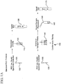
- FIG. 1A illustrates the current standard protocol for POC and lab-based testing.
- a specimen 105 is collected, for example, using a swab 100.
- Other conventional implements for collecting biological samples are contemplated for use herein. Such implements, such as a scraper or spatula are not described in detail herein and are well known to those skilled in the art.
- Specimen 105 is then processed directly by placing swab 100 with sample 105 in solution 110 for POC rapid testing 120. In this situation, any remaining sample is discarded and a new sample must be collected for additional lab-based testing such as confirmatory testing or reflex testing.
- specimen 105 on swab 130 is first diluted in transport media 140.
- a portion of the diluted transport media 140 containing specimen 105 is further processed in solution 160 for POC testing 170.
- processed specimen 105 is diluted to a level that diminishes the results of POC testing 170 as illustrated in Example 1 below.
- the remaining portion in the diluent is used for laboratory testing 150, such as subtyping and reflex testing.
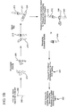
- FIGS. 1B illustrates one embodiment of the method for POC and laboratory testing of the present invention.
- Specimen 205 is collected on swab 200 and processed directly using a rapid test processing reagent 210 that is optimized for producing the maximum clinical performance for the particular immunoassay.
- the swab 200 is removed and the rapid test container 215 is closed with dispenser lid 216.
- the capped test container 215 with dispenser lid 216 is used to dispense a portion of processed sample 211 onto rapid test strip 220.
- Rapid POC testing 220 is performed using a portion of the specimen 211 processed in the rapid test processing reagent.
- the remaining portion 212 of the processed sample after POC testing is transported 300 to the clinical lab for laboratory testing 400.
- the remaining portion 212 of the processed sample after POC testing is added to transport vial 230 that contains stabilization transport diluent 240.
- the stabilization transport diluent is designed to help maintain the integrity of the sample.
- the stabilized sample is then transported to the clinical lab 300 for confirmatory or other laboratory testing 400.
- This embodiment illustrates how one sample, specimen 205, can be processed at the POC site for both POC testing and lab-based testing. This embodiment also demonstrates that a sample processed in conditions optimal for POC testing can be used for lab-based testing.
- Rapid tests There are a variety of rapid tests that are currently commercially available. Such rapid tests are not described in detail herein, but are available from a variety of sources including Becton Dickinson, Alere, Quidel, Meridian, Genzyme, etc. The invention is not limited to use with a particular rapid test.
- H1N1 positive clinical specimen collected by upper nasal swab from an individual exhibiting positive flu symptoms.
- the swab was placed in 3 ml of commercially available transport media (BDTM Universal Viral Transport Media available from Becton Dickinson) and confirmation that the sample tested positive for H1N1 was obtained.
- transport media BDTM Universal Viral Transport Media available from Becton Dickinson
- certain 50 ⁇ l aliquots of that specimen were obtained.
- One aliquot was mixed directly with a rapid test processing reagent for the immunoassay and others were further diluted (5X, 25X, 125X or 625X) with stabilization transport diluent prior to mixing with the rapid test processing reagent.
- the immunoassay test results in Table 1 demonstrate the effect of specimen dilution on rapid test performance. Samples diluted greater than 1:5 resulted in a negative rapid immunoassay test. In order to provide optimal POC clinical performance, specimen dilution should therefore be minimized or avoided. Dilution of the specimen (excluding the initial placement of the sample into solution) greater than 1:5 diminishes the possibility of detection with a rapid immunoassay test.
- the use of direct swab processing in the POC setting enhances the clinical performance of rapid immunoassays. However, standard POC testing methods using direct swab samples, as noted above, do not enable lab-based testing because of initial placement of such samples into a transport diluent.
- RNA samples were mixed with 25 ⁇ l rapid test processing reagent.
- One set of processed samples was stored at room temperature (RT) for 5 minutes prior to RNA extraction using a Qiagen Viral RNA miniprep kit according to the manufacturer's instructions. Additional sets of processed samples were stored for 4 hours at either 4°C or RT prior to RNA extraction.
- a 5 ⁇ l portion of the extracted RNA samples was then used as target for reverse transcription-polymerase chain reaction (RT-PCR) with primers specific for the matrix gene of influenza A virus.
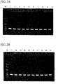
- RT-PCR results are shown in FIGS. 2A and 2B .
- Tables 2 and 3 show the processing conditions corresponding to each lane of the agarose gel of the RT-PCR results shown in FIGS 2A and 2B .
- Table 2 Lanes of Agarose Gel of FIG. 2A Processing Method Lane on Agarose Gel Molecular Weight Marker M Sample diluted 1:5, processed for rapid test, RNA extraction immediately 1 Sample diluted 1:5, processed for rapid test, stored at 4°C for 4 hrs before RNA extraction 2 Sample diluted 1:5, processed for rapid test, stored at Room Temperature for 4 hrs before RNA extraction 3 Sample diluted 1:25, processed for rapid test, RNA extraction immediately 4 Sample diluted 1:25, processed for rapid test, stored at 4°C for 4 hrs before RNA extraction 5 Sample diluted 1:25, processed for rapid test, stored at Room Temperature for 4 hrs before RNA extraction 6 Sample diluted 1:5, RNA extraction (no rapid test/no rapid test reagent) 7 Sample diluted 1:25, RNA extraction (no rapid test/no rapid test reagent) 8 Sample diluted 1:125, RNA extraction
- Figures 2A and 2B demonstrate that samples processed for rapid immunoassay testing could also be used for RNA extraction, which enabled lab-based PCR testing to be performed.
- RNA was extracted from samples diluted well below the limit of detection for the rapid test, indicating that even small amounts of viral RNA remained stable in the processed sample. Storage of the processed samples at 4°C or room temperature for up to four hours before RNA extraction also indicates the viral nucleic acid remained stable after processing.
- Comparison of PCR test results using RNA isolated from the processed samples to RNA extracted directly from the sample dilutions demonstrated that the integrity of the viral RNA was minimally affected by the processing step for the rapid test.
- RNA in processed samples was examined using two different rapid test processing reagents optimized for use in the rapid immunoassay for influenza A/B.
- Sample processing for rapid immunoassays typically involves the use of a relatively gentle lysis treatment mediated by a reagent containing various salts and detergents.
- Two different formulations for the rapid test processing reagent were examined for compatibility with the described method.
- Formulation A contained Tris buffer, NaCl, 16% detergent at a pH of 7.8.
- Formulation B contained Tris buffer, NaCl, 6% detergent at a pH of 8.0.
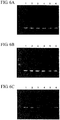
- FIG. 3 demonstrates both formulation A and formulation B are compatible with use of the processed sample for RNA extraction and PCR testing.
- Storage of the processed samples at 4°C for up to 24 hours prior to RNA extraction suggests little degradation of the viral RNA occurred in samples processed with either formulation.
- prolonged storage of the extracted samples at RT demonstrated decreased PCR performance, possibly due to viral RNA degradation over time (lanes 8, 10).
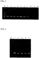
- stabilization transport diluents Two potential stabilization transport diluents were examined in an attempt to increase stability of viral RNA in samples processed for POC testing.
- the stabilization transport diluent was designed to help maintain the integrity of nucleic acids present in the sample.
- Various formulations are possible where optimal pH conditions, buffer types, salts, chelating agents, enzyme inhibitors, nucleic acid binding proteins, chaotropes, etc. may be employed.
- Stabilization transport diluent A contained Qiagen viral RNA lysis/binding buffer.
- Stabilization transport diluent B contained 6 M guanidine thiocyanate + 20 mM EDTA. Aliquots of an H1N1 positive clinical specimen were processed using rapid test processing reagent B.
- the processed samples were immediately used for RNA extraction or mixed with one of the two different stabilization transport diluents and stored at 4°C for up to six days prior to RNA extraction. A portion of the extracted RNA samples was then used as target for RT-PCR with primers specific for the matrix gene of the influenza A virus.
- the RT-PCR results are shown in FIG. 4 .
- Table 5 shows the processing conditions corresponding to each lane of the agarose gel of the RT-PCR results shown in FIG. 4 .
- Table 5 Lanes of Agarose Gel of FIG. 4.
- RNA extraction immediately 1 Sample processed for rapid test, mixed with stabilization transport diluent A, stored at 4°C for 3 days before RNA extraction 2 Sample processed for rapid test, mixed with stabilization transport diluent B, stored at 4°C for 3 days before RNA extraction 3 Sample processed for rapid test, mixed with stabilization transport diluent A, stored at 4°C for 6 days before RNA extraction 4 Sample processed for rapid test, mixed with stabilization transport diluent B, stored at 4°C for 6 days before RNA extraction 5
- RNA extracted from samples processed for POC testing that had been stored for up to 6 days at 4°C was extracted from samples processed for POC testing that had been stored for up to 6 days at 4°C. Comparing the PCR results from the stored samples (lanes 2-5) to those obtained using RNA extracted immediately after processing (lane 1) suggest little, if any, degradation of the viral RNA occurred over time in the processed samples treated with either stabilization transport diluent.
- a stabilization transport diluent was used in an attempt to increase stability of the viral RNA in samples processed for POC testing, particularly when samples are stored for extended periods of time at room temperature.
- Aliquots of an H1N1 positive clinical specimen were processed using rapid test processing reagent formulation B described in Example 3, and the processed samples were immediately used for RNA extraction, or mixed with stabilization transport diluent A and stored at RT and 4°C for up to seven days prior to RNA extraction.
- a portion of the extracted RNA samples was then used as target for RT-PCR with primers specific for the matrix gene of the influenza A virus.
- the PCR results are shown in FIG. 5 .
- Table 6 shows the processing conditions corresponding to each lane of the agarose gel of the RT-PCR results shown in FIG. 5 .
- Table 6 Lanes of Agarose Gel of FIG. 5. Processing Method Lane on Agarose Gel Molecular Weight Marker M Sample processed for rapid test, RNA extraction immediately 1 Sample processed for rapid test, mixed with stabilization transport diluent, stored at 4°C for 3 days before RNA extraction 2 Sample processed for rapid test, mixed with stabilization transport diluent, stored at 4°C for 4 days before RNA extraction 3 Sample processed for rapid test, mixed with stabilization transport diluent, stored at 4°C for 5 days before RNA extraction 4 Sample processed for rapid test, mixed with stabilization transport diluent, stored at 4°C for 6 days before RNA extraction 5 Sample processed for rapid test, mixed with stabilization transport diluent, stored at 4°C for 7 days before RNA extraction 6 Sample processed for rapid test, mixed with stabilization transport diluent, stored at room temperature for 1 day before RNA extraction 7 Sample processed for rapid test, mixed with stabilization transport diluent, stored at room temperature for 2 days before RNA extraction 8 Sample processed for rapid test, mixed with
- FIG. 5 demonstrates that intact viral RNA can be extracted from processed samples mixed with the stabilization transport diluent after storage of the samples for up to 7 days at 4°C or up to 4 days at room temperature.
- Stabilization transport diluent B was used to examine stabilization properties across different influenza strains: A: Influenza A strain A/Solomon Island/03/06 (H1N1); B: Influenza A strain A/Wisconsin/67/2005 (H3N2); and C: Influenza B strain B/Jiangsu/10/2003.
- Table 7 shows the processing conditions corresponding to each lane of the agarose gel of the RT-PCR results shown in FIG. 6 .
- FIG. 6 demonstrates that intact viral RNA from various strains can be extracted from processed samples mixed with the stabilization transport diluent after storage of the samples for up to 7 days at 4°C or up to 14 days at -20°C.
- intact viral RNA was extracted after storage under all conditions.
- FIG. 1B Utility of one embodiment of the method of the present invention illustrated in FIG. 1B was demonstrated in a clinical trial performed during the 2010-2011 influenza season.
- Paired nasopharyngeal (NPS) or upper nasal swabs (NS) were collected from patients enrolled in the POC influenza study.
- One swab was processed directly for use in an investigational rapid immunoassay at the POC site and then a portion (3 to 5 drops) of the remaining sample was mixed with the stabilization transport diluent B (200 ⁇ l) and stored at either 2 to 8°C for up to 5 days or -20°C for up to two weeks prior to being sent to a laboratory for PCR analysis.
- the second swab was placed in 3 ml of viral transport media and sent directly to a clinical laboratory for PCR testing.
- RNA samples Five microliters of extracted RNA was used for PCR amplification using a Cepheid SmartCycler II instrument according to the assay procedure described in the Prodesse ProFlu+ package insert. Interpretation of PCR results for specimens and controls was determined using the Cepheid SmartCycler Dx software according to the protocols outlined in the Prodesse ProFlu+ package insert. The positive and negative percent agreement between results obtained from the POC stabilized sample (POC PCR) and the swab-in-transport media sample (Reference PCR) are shown below in Table 7.
- Table 7 demonstrates that samples can be processed for rapid POC testing and a portion of that processed sample can be used for lab-based testing such as PCR. Greater than 91.9% agreement was obtained in various viral strains when comparing a sample that was directly processed for PCR to a sample that was first processed under conditions optimal for rapid POC testing and then subsequently processed for PCR.
Description
- The linkage between point of care (POC) rapid testing and laboratory-based testing has typically been addressed through preservation of samples to support culture-based laboratory testing methods. Currently, samples collected at the POC site are either processed and used directly in a rapid test (the portion of the processed sample not used in the rapid test being discarded), or diluted in liquid transport media to enable transfer for laboratory-based testing such as rapid immunoassay, culture and/or polymerase chain reaction (PCR). Specimens that generate negative results from a POC test are often reflex tested -the negative result is confirmed by lab-based testing methods such as PCR. In addition, specimens that generate positive POC test results are frequently tested for additional characterizations such as subtyping or other epidemiologic information.
- Referring to
FIG. 1 , at physician office POC sites (and other non-laboratory sites where patients are seen or samples for rapid testing are collected),swab specimens 100 are used almost exclusively to deliver sample intosolution 110 for therapid test 120. Processing and testing the sample in the physician's office and other non-laboratory sample collection sites does not contemplate a means for enabling lab-based testing such as confirmatory and/or reflex testing or other tests the require lab-based analysis. With the current methods, a physician (or other administrator of a POC rapid test) cannot perform both a POC rapid test and a lab-based test using the sample collected at the site (e.g. the physician's office). Therefore, an opportunity to perform lab-based testing on such samples is lost.Swab samples 130 collected at POC sites within a hospital or clinic are almost exclusively placed within a volume ofliquid transport media 140 for transfer to the testing laboratory for remote testing. The dilutedsamples 150 may be further processed by adding them to asolution 160 for arapid test 170. However, this method often results in a POC sample diluted 5 to 10-fold, or more, which can diminish performance of the rapid test due to sample dilution effects. - Collection and transport of a second swab at the POC site could be used to address the need to perform laboratory-based testing, although this is clearly not the standard of practice and doubles the number of samples to be taken. In addition, although collected from the same patient, variations in collection methods, organism load, etc. could lead to erroneous results when comparing the test results between two independently collected swab specimens. Accordingly, a system and method that addresses these problems is desired.
- Booth, S. et al., "Comparison of two rapid influenza A/B test kits with reference methods showing high specificity and sensitivity for influenza A infection," J. MED. VIR., Vol. 78, No. 5, pp. 619-622 (Jan. 1, 2006) describes point of care tests for detecting influenza and the results compared with those of culture immunofluorescence (IFA) and PCR.
- FOO, H., ET AL: "Laboratory test performance in young adults during influenza outbreaks at World Youth Day 2008", J. CLIN. VIR., Vol. 46, No. 4, pp. 384-386 (December 1, 2009) (Elsevier, Amsterdam, NL) compared the performance of point of care tests (POC) for influenza with immunofluorescence assays (IFAs) and with nucleic acid tests.
-
US Patent Publication No. 2009/306230A1 to Semikhodskii et al., December 10, 2009 ) describes obtaining a sample at a first location and immobilizing such sample on a substrate and transporting the immobilized sample to a second location for testing. - USAID: "AVIAN INFLUENZA COMMODITIES TRAINING GUIDE, March 1, 2007 describes test kits for influenza detection.
-
WO 2008/096225 A2 to NATHANIEL, ANDREW HOMER (August 14, 2008 ) describes a kit for in situ detection of trace quantities of drugs on a surface. - A method for using a sample initially collected at the point of care (POC) as source for sample for use in a laboratory that is not at the point of care (POC) comprising:
- a) collecting a sample suspected of containing a target microorganism for the POC test wherein the POC test is an immunoassay,
- b) preparing the collected sample for use in the POC test by combining the collected sample with a POC with a processing reagent configured for use with a POC test, wherein the POC processing reagent consists essentially of a gentle lysis reagent that consists of a Tris buffer, NaCl, and one of 16% detergent at a pH of 7.8 or 6% detergent at a pH of 8.0, and wherein the collected sample is not combined with any additional reagents thereafter prior to the POC test,
- c) using only a portion of the processed sample for the POC test leaving the remaining portion not used for the POC test, and
- d) using at least a portion of the remaining portion of the processed sample for a laboratory test.
- The method above wherein an implement is used to collect the sample and the implement is selected from the group consisting of a scraper and a swab.
- The method above, wherein the location for the POC test is a physician's office.
- The method above wherein, after a portion of the processed sample has been removed for use in the POC test, the at least a portion of the remaining portion of the processed sample and a stabilization transport diluent are combined together in a transport container for sample stabilization during transport to the laboratory test.
- The method above, wherein the stabilization transport diluent stabilizes nucleic acids in the processed sample and comprises at least one buffer and at least one salt.
- The method above wherein, after a portion of the processed sample has been removed for use in the POC test, the at least a portion of the remaining portion of the processed sample is not combined with a stabilization transport diluent for sample stabilization during transport to the laboratory test.
- The method above wherein the laboratory test is subtyping based upon a nucleic acid assay, reflex testing or a nucleic acid assay.
- The method above wherein the nucleic acid assay is polymerase chain reaction (PCR).
- The method above wherein the stabilization transport diluent is added to a container containing at least a portion of the remaining portion of the processed sample or at least a portion of the remaining portion of the processed sample is added to a container containing the stabilization transport diluent.
- The method above wherein the sample suspected of containing a target microorganism is selected from the group consisting of a biological sample and an environmental sample.
- A method for using a single sample suspected of containing a target microorganism for both a POC test immunoassay and a laboratory test comprising:
- a) collecting a sample for a POC test,
- b) processing the sample for the POC test by combining the collected sample with a POC processing reagent wherein the POC processing reagent consists essentially of a gentle lysis reagent that consists of a Tris buffer, NaCl, and one of 16% detergent at a pH of 7.8 or 6% detergent at a pH of 8.0,
- c) selecting the POC test;
- d) using only a portion of the processed sample for the POC test, leaving a remaining portion of the processed sample;
- e) combining in a container at least a portion of the remaining portion of the processed sample and a stabilization transport diluent that stabilizes at least nucleic acids in the portion of the processed sample combined with the stabilization transport diluent;
- f) transferring the container containing the combined remaining portion of the processed sample and stabilization transport diluent to a location for laboratory testing, and
- g) using the combined remaining portion of the processed sample combined and stabilization transport diluent for reflex testing or subtyping using PCR.
- The method above wherein an implement is used to collect the biological sample and the implement is selected from the group consisting of a scraper and a swab.
- The method above wherein the location for the POC test is a physician's office.
- The method above wherein the stabilization transport diluent stabilizes nucleic acids in the processed sample and comprises at least one buffer and at least one salt.
- The method above, wherein the sample suspected of containing a target microorganism is selected from the group consisting of a biological sample and an environmental sample.
- A method for testing a single biological or environmental sample using both a POC test and a laboratory test that indicate the presence or absence of microorganisms in the sample comprising:
- a) collecting a biological or environmental sample for the POC test, wherein the POC test is a rapid immunoassay;
- b) processing the sample for a POC test by combining the collected sample with the POC test processing reagent configured for use with a POC test wherein the POC processing reagent consists essentially of a gentle lysis reagent that consists of a Tris buffer, NaCl, and one of 16% detergent at a pH of 7.8 or 6% detergent at a pH of 8.0;
- c) using a portion of the processed sample for the POC test, and
- d) using at least a portion of the remaining portion of the processed sample for a laboratory test.
-
-
FIG. 1A illustrates the prior art method for the standard protocol for POC and lab-based testing. -
FIG. 1B illustrates the method for POC and lab-based testing of the present invention. -
FIG. 2A /B demonstrates the results from RT-PCR for Influenza A from samples processed for POC testing under various dilution conditions. -
FIG. 3 demonstrates the results from RT-PCR for Influenza A from samples processed for POC testing using two different rapid test processing reagents and various storage conditions using the method of the present invention. -
FIG. 4 demonstrates the results from RT-PCR for Influenza A from samples processed for POC testing and then mixed with two different stabilization transport diluents and various storage conditions using the method of the present invention. -
FIG. 5 demonstrates the results from RT-PCR for Influenza A from samples processed for POC testing and then mixed with a stabilization transport diluent and various storage conditions using the method of the present invention. -
FIG. 6A-C demonstrate the results from RT-PCR for two strains of Influenza A and one strain of Influenza B under various storage conditions using the method of the present invention. -
FIG. 1A illustrates the current standard protocol for POC and lab-based testing. Aspecimen 105 is collected, for example, using aswab 100. Other conventional implements for collecting biological samples are contemplated for use herein. Such implements, such as a scraper or spatula are not described in detail herein and are well known to those skilled in the art.Specimen 105 is then processed directly by placingswab 100 withsample 105 insolution 110 for POCrapid testing 120. In this situation, any remaining sample is discarded and a new sample must be collected for additional lab-based testing such as confirmatory testing or reflex testing. In the alternative standard protocol,specimen 105 onswab 130 is first diluted intransport media 140. A portion of the dilutedtransport media 140 containingspecimen 105 is further processed insolution 160 forPOC testing 170. In this situation, processedspecimen 105 is diluted to a level that diminishes the results ofPOC testing 170 as illustrated in Example 1 below. The remaining portion in the diluent is used forlaboratory testing 150, such as subtyping and reflex testing. -
FIGS. 1B illustrates one embodiment of the method for POC and laboratory testing of the present invention.Specimen 205 is collected onswab 200 and processed directly using a rapidtest processing reagent 210 that is optimized for producing the maximum clinical performance for the particular immunoassay. Theswab 200 is removed and therapid test container 215 is closed withdispenser lid 216. The cappedtest container 215 withdispenser lid 216 is used to dispense a portion of processedsample 211 ontorapid test strip 220.Rapid POC testing 220 is performed using a portion of thespecimen 211 processed in the rapid test processing reagent. The remainingportion 212 of the processed sample after POC testing is transported 300 to the clinical lab forlaboratory testing 400. In the alternative, the remainingportion 212 of the processed sample after POC testing is added to transportvial 230 that containsstabilization transport diluent 240. The stabilization transport diluent is designed to help maintain the integrity of the sample. The stabilized sample is then transported to theclinical lab 300 for confirmatory orother laboratory testing 400. This embodiment illustrates how one sample,specimen 205, can be processed at the POC site for both POC testing and lab-based testing. This embodiment also demonstrates that a sample processed in conditions optimal for POC testing can be used for lab-based testing. - There are a variety of rapid tests that are currently commercially available. Such rapid tests are not described in detail herein, but are available from a variety of sources including Becton Dickinson, Alere, Quidel, Meridian, Genzyme, etc. The invention is not limited to use with a particular rapid test.
- The ability to detect influenza viral RNA in samples processed for use in a POC rapid immunoassay was demonstrated using an H1N1 positive clinical specimen collected by upper nasal swab from an individual exhibiting positive flu symptoms. The swab was placed in 3 ml of commercially available transport media (BD™ Universal Viral Transport Media available from Becton Dickinson) and confirmation that the sample tested positive for H1N1 was obtained. For testing, certain 50 µl aliquots of that specimen were obtained. One aliquot was mixed directly with a rapid test processing reagent for the immunoassay and others were further diluted (5X, 25X, 125X or 625X) with stabilization transport diluent prior to mixing with the rapid test processing reagent. Each 50 µl aliquot of sample was combined with 25 µl of rapid test processing reagent. The rapid test processing reagent (tris buffer, NaCl, 6% detergent and pH adjusted to 8.0) was optimized to release and preserve the influenza nucleoprotein which is the target antigen for the rapid immunoassay. The immunoassay testing results on the various sample dilutions are shown in Table 1.
Table 1: Immunoassay Results H1N1 Clinical Sample Rapid Immunoassay Result undiluted Flu A positive/ Flu B negative 1:5 dilution Flu A positive/ Flu B negative 1:25 dilution Flu A negative/ Flu B negative 1:125 dilution Flu A negative/ Flu B negative 1:625 dilution Flu A negative/ Flu B negative - The immunoassay test results in Table 1 demonstrate the effect of specimen dilution on rapid test performance. Samples diluted greater than 1:5 resulted in a negative rapid immunoassay test. In order to provide optimal POC clinical performance, specimen dilution should therefore be minimized or avoided. Dilution of the specimen (excluding the initial placement of the sample into solution) greater than 1:5 diminishes the possibility of detection with a rapid immunoassay test. The use of direct swab processing in the POC setting enhances the clinical performance of rapid immunoassays. However, standard POC testing methods using direct swab samples, as noted above, do not enable lab-based testing because of initial placement of such samples into a transport diluent.
- Aliquots (50 µl) of each dilution prepared in Example 1 were mixed with 25 µl rapid test processing reagent. One set of processed samples was stored at room temperature (RT) for 5 minutes prior to RNA extraction using a Qiagen Viral RNA miniprep kit according to the manufacturer's instructions. Additional sets of processed samples were stored for 4 hours at either 4°C or RT prior to RNA extraction. A 5 µl portion of the extracted RNA samples was then used as target for reverse transcription-polymerase chain reaction (RT-PCR) with primers specific for the matrix gene of influenza A virus. The RT-PCR results are shown in
FIGS. 2A and 2B . Tables 2 and 3 show the processing conditions corresponding to each lane of the agarose gel of the RT-PCR results shown inFIGS 2A and 2B .Table 2: Lanes of Agarose Gel of FIG. 2A Processing Method Lane on Agarose Gel Molecular Weight Marker M Sample diluted 1:5, processed for rapid test, RNA extraction immediately 1 Sample diluted 1:5, processed for rapid test, stored at 4°C for 4 hrs before RNA extraction 2 Sample diluted 1:5, processed for rapid test, stored at Room Temperature for 4 hrs before RNA extraction 3 Sample diluted 1:25, processed for rapid test, RNA extraction immediately 4 Sample diluted 1:25, processed for rapid test, stored at 4°C for 4 hrs before RNA extraction 5 Sample diluted 1:25, processed for rapid test, stored at Room Temperature for 4 hrs before RNA extraction 6 Sample diluted 1:5, RNA extraction (no rapid test/no rapid test reagent) 7 Sample diluted 1:25, RNA extraction (no rapid test/no rapid test reagent) 8 Sample diluted 1:125, RNA extraction(no rapid test/no rapid test reagent) 9 Sample diluted 1:625, RNA extraction (no rapid test/no rapid test reagent) 10 Table 3: Lanes of Agarose Gel of FIG. 2B Processing Method Lane on Agarose Gel Molecular Weight Marker M Sample diluted 1:125, processed for rapid test, RNA extraction immediately 11 Sample diluted 1:125, processed for rapid test, stored at 4°C for 4 hrs before RNA extraction 12 Sample diluted 1:125, processed for rapid test, stored at Room Temperature for 4 hrs before RNA extraction 13 Sample diluted 1:625, processed for rapid test, RNA extraction immediately 14 Sample diluted 1:625, processed for rapid test, stored at 4°C for 4 hrs before RNA extraction 15 Sample diluted 1:625, processed for rapid test, stored at Room Temperature for 4 hrs before RNA extraction 16 Sample diluted 1:5, RNA extraction (no rapid test/no rapid test reagent) 17 Sample diluted 1:25, RNA extraction (no rapid test/no rapid test reagent) 18 Sample diluted 1:125, RNA extraction (no rapid test/no rapid test reagent) 19 Sample diluted 1:625, RNA extraction (no rapid test/no rapid test reagent) 20 -
Figures 2A and 2B demonstrate that samples processed for rapid immunoassay testing could also be used for RNA extraction, which enabled lab-based PCR testing to be performed. RNA was extracted from samples diluted well below the limit of detection for the rapid test, indicating that even small amounts of viral RNA remained stable in the processed sample. Storage of the processed samples at 4°C or room temperature for up to four hours before RNA extraction also indicates the viral nucleic acid remained stable after processing. Comparison of PCR test results using RNA isolated from the processed samples to RNA extracted directly from the sample dilutions (lanes 7-10, 17-20) demonstrated that the integrity of the viral RNA was minimally affected by the processing step for the rapid test. - The stability of viral RNA in processed samples was examined using two different rapid test processing reagents optimized for use in the rapid immunoassay for influenza A/B. Sample processing for rapid immunoassays typically involves the use of a relatively gentle lysis treatment mediated by a reagent containing various salts and detergents. Two different formulations for the rapid test processing reagent were examined for compatibility with the described method. Formulation A contained Tris buffer, NaCl, 16% detergent at a pH of 7.8. Formulation B contained Tris buffer, NaCl, 6% detergent at a pH of 8.0. Aliquots of an H1N1 positive clinical specimen described in Example 1 were processed with both formulations, and the processed samples were used immediately for RNA extraction using the Qiagen Viral RNA miniprep kit, or stored at RT and 4°C for up to 24 hours prior to RNA extraction. A portion of the extracted RNA samples was then used as target for RT-PCR with primers specific for the matrix gene of the influenza A virus. The RT-PCR results are shown in
FIG. 3 . Table 4 shows the processing conditions corresponding to each lane of the agarose gel of the RT-PCR results shown inFIG. 3 .Table 4: Lanes of Agarose Gel of FIG. 3. Processing Method Lane on Agarose Gel Molecular Weight Marker M Sample processed with rapid test processing reagent A, RNA extraction immediately 1 Sample processed with rapid test processing reagent A, stored at 4°C for 4 hrs before RNA extraction 2 Sample processed with rapid test processing reagent A, stored at 4°C for 24 hrs before RNA extraction 3 Sample processed with rapid test processing reagent B, RNA extraction immediately 4 Sample processed with rapid test processing reagent B, stored at 4°C for 4 hrs before RNA extraction 5 Sample processed with rapid test processing reagent B, stored at 4°C for 24 hrs before RNA extraction 6 Sample processed with rapid test processing reagent A, stored at room temperature for 4 hrs before RNA extraction 7 Sample processed with rapid test processing reagent A, stored at room temperature for 24 hrs before RNA extraction 8 Sample processed with rapid test processing reagent B, stored at room temperature for 4 hrs before RNA extraction 9 Sample processed with rapid test processing reagent B, stored at room temperature for 24 hrs before RNA extraction 10 -
FIG. 3 demonstrates both formulation A and formulation B are compatible with use of the processed sample for RNA extraction and PCR testing. Storage of the processed samples at 4°C for up to 24 hours prior to RNA extraction suggests little degradation of the viral RNA occurred in samples processed with either formulation. However, prolonged storage of the extracted samples at RT demonstrated decreased PCR performance, possibly due to viral RNA degradation over time (lanes 8, 10). - Two potential stabilization transport diluents were examined in an attempt to increase stability of viral RNA in samples processed for POC testing. The stabilization transport diluent was designed to help maintain the integrity of nucleic acids present in the sample. Various formulations are possible where optimal pH conditions, buffer types, salts, chelating agents, enzyme inhibitors, nucleic acid binding proteins, chaotropes, etc. may be employed. Stabilization transport diluent A contained Qiagen viral RNA lysis/binding buffer. Stabilization transport diluent B contained 6 M guanidine thiocyanate + 20 mM EDTA. Aliquots of an H1N1 positive clinical specimen were processed using rapid test processing reagent B. The processed samples were immediately used for RNA extraction or mixed with one of the two different stabilization transport diluents and stored at 4°C for up to six days prior to RNA extraction. A portion of the extracted RNA samples was then used as target for RT-PCR with primers specific for the matrix gene of the influenza A virus. The RT-PCR results are shown in
FIG. 4 . Table 5 shows the processing conditions corresponding to each lane of the agarose gel of the RT-PCR results shown inFIG. 4 .Table 5: Lanes of Agarose Gel of FIG. 4. Processing Method Lane on Agarose Gel Molecular Weight Marker M Sample processed for rapid test, RNA extraction immediately 1 Sample processed for rapid test, mixed with stabilization transport diluent A, stored at 4°C for 3 days before RNA extraction 2 Sample processed for rapid test, mixed with stabilization transport diluent B, stored at 4°C for 3 days before RNA extraction 3 Sample processed for rapid test, mixed with stabilization transport diluent A, stored at 4°C for 6 days before RNA extraction 4 Sample processed for rapid test, mixed with stabilization transport diluent B, stored at 4°C for 6 days before RNA extraction 5 - Using either formulation of the stabilization transport diluent, intact viral RNA was extracted from samples processed for POC testing that had been stored for up to 6 days at 4°C. Comparing the PCR results from the stored samples (lanes 2-5) to those obtained using RNA extracted immediately after processing (lane 1) suggest little, if any, degradation of the viral RNA occurred over time in the processed samples treated with either stabilization transport diluent.
- A stabilization transport diluent was used in an attempt to increase stability of the viral RNA in samples processed for POC testing, particularly when samples are stored for extended periods of time at room temperature. Aliquots of an H1N1 positive clinical specimen were processed using rapid test processing reagent formulation B described in Example 3, and the processed samples were immediately used for RNA extraction, or mixed with stabilization transport diluent A and stored at RT and 4°C for up to seven days prior to RNA extraction. A portion of the extracted RNA samples was then used as target for RT-PCR with primers specific for the matrix gene of the influenza A virus. The PCR results are shown in
FIG. 5 . Table 6 shows the processing conditions corresponding to each lane of the agarose gel of the RT-PCR results shown inFIG. 5 .Table 6: Lanes of Agarose Gel of FIG. 5. Processing Method Lane on Agarose Gel Molecular Weight Marker M Sample processed for rapid test, RNA extraction immediately 1 Sample processed for rapid test, mixed with stabilization transport diluent, stored at 4°C for 3 days before RNA extraction 2 Sample processed for rapid test, mixed with stabilization transport diluent, stored at 4°C for 4 days before RNA extraction 3 Sample processed for rapid test, mixed with stabilization transport diluent, stored at 4°C for 5 days before RNA extraction 4 Sample processed for rapid test, mixed with stabilization transport diluent, stored at 4°C for 6 days before RNA extraction 5 Sample processed for rapid test, mixed with stabilization transport diluent, stored at 4°C for 7 days before RNA extraction 6 Sample processed for rapid test, mixed with stabilization transport diluent, stored at room temperature for 1 day before RNA extraction 7 Sample processed for rapid test, mixed with stabilization transport diluent, stored at room temperature for 2 days before RNA extraction 8 Sample processed for rapid test, mixed with stabilization transport diluent, stored at room temperature for 3 days before RNA extraction 9 Sample processed for rapid test, mixed with stabilization transport diluent, stored at room temperature for 4 days before RNA extraction 10 - Mixing the processed sample with a stabilization transport diluent increased the stability of the viral nucleic acid, and enabled longer-term storage and transport of the processed sample at various temperatures.
FIG. 5 demonstrates that intact viral RNA can be extracted from processed samples mixed with the stabilization transport diluent after storage of the samples for up to 7 days at 4°C or up to 4 days at room temperature. - Stabilization transport diluent B was used to examine stabilization properties across different influenza strains: A: Influenza A strain A/Solomon Island/03/06 (H1N1); B: Influenza A strain A/Wisconsin/67/2005 (H3N2); and C: Influenza B strain B/Jiangsu/10/2003. Aliquots (50 µl) of cell culture supernatants from cultures into which virus had been introduced from nasal swabs of a patient exhibiting symptoms of influenza and these aliquots were combined with rapid test processing reagent B (25 µl) and the processed samples were either immediately used for RNA extraction, or mixed with stabilization transport diluent B (75 µl) and stored at 4°C or -20°C for up to fourteen days prior to RNA extraction. A portion of the extracted RNA samples was then used as target for RT-PCR reactions with primers specific for the matrix gene of influenza A or the nucleoprotein gene of influenza B. The PCR results are shown in
FIG. 6 . Table 7 shows the processing conditions corresponding to each lane of the agarose gel of the RT-PCR results shown inFIG. 6 .Table 7: Table 6: Lanes of Agarose Gel of FIG. 6. Processing Method Lane on Agarose Gel Sample processed for rapid test, RNA extraction immediately 1 Sample processed for rapid test, mixed with stabilization transport diluents B, stored at 4°C for 2 days before RNA extraction 2 Sample processed for rapid test, mixed with stabilization transport diluents B, stored at 4°C for 7 days before RNA extraction 3 Sample processed for rapid test, mixed with stabilization transport diluents B, stored at 4°C for 10 days before RNA extraction 4 Sample processed for rapid test, mixed with stabilization transport diluents B, stored at 4°C for 14 days before RNA extraction 5 Sample processed for rapid test, mixed with stabilization transport diluents B, stored at -20°C for 7 days before RNA extraction 6 - Mixing the processed sample with a stabilization transport diluent increased the stability of the viral nucleic acid in all three strains of Influenza for up to 14 days at 4°C or up to 7 days at -20°C.
FIG. 6 demonstrates that intact viral RNA from various strains can be extracted from processed samples mixed with the stabilization transport diluent after storage of the samples for up to 7 days at 4°C or up to 14 days at -20°C. For both the A and B strains, intact viral RNA was extracted after storage under all conditions. - Utility of one embodiment of the method of the present invention illustrated in
FIG. 1B was demonstrated in a clinical trial performed during the 2010-2011 influenza season. Paired nasopharyngeal (NPS) or upper nasal swabs (NS) were collected from patients enrolled in the POC influenza study. One swab was processed directly for use in an investigational rapid immunoassay at the POC site and then a portion (3 to 5 drops) of the remaining sample was mixed with the stabilization transport diluent B (200 µl) and stored at either 2 to 8°C for up to 5 days or -20°C for up to two weeks prior to being sent to a laboratory for PCR analysis. The second swab was placed in 3 ml of viral transport media and sent directly to a clinical laboratory for PCR testing. - All PCR testing was performed using the Prodesse ProFlu+ assay available from GenProbe, Inc. (San Diego, CA). The Prodesse ProFlu+ test is FDA-cleared and is able to detect and differentiate Influenza A, Influenza B, and RSV in respiratory specimens. For the swab-in-transport media specimens, RNA was extracted using the NucliSENS easyMAG System (bioMérieux) according to the Prodesse ProFlu+ package insert. For the POC processed samples in the stabilization transport diluent, RNA was extracted using the Qiagen Viral RNA miniprep kit according to the manufacturer. Five microliters of extracted RNA was used for PCR amplification using a Cepheid SmartCycler II instrument according to the assay procedure described in the Prodesse ProFlu+ package insert. Interpretation of PCR results for specimens and controls was determined using the Cepheid SmartCycler Dx software according to the protocols outlined in the Prodesse ProFlu+ package insert. The positive and negative percent agreement between results obtained from the POC stabilized sample (POC PCR) and the swab-in-transport media sample (Reference PCR) are shown below in Table 7.
Table 7: Comparison of PCR results from POC processed samples compared to Reference PCR Influenza A Reference PCR POC PCR P N P 150 22 172 N 5 335 340 155 357 512 Reference Method: PCR from swab in transport media Positive Percent Agreement: 96.8% Negative Percent Agreement: 93.8% Influenza B Reference PCR POC PCR P N P 91 12 103 N 8 401 409 99 413 512 Reference Method: PCR from swab in transport media Positive Percent Agreement: 91.9% Negative Percent Agreement: 97.1% RSV Reference PCR POC PCR P N P 18 2 20 N 1 491 492 19 493 512 Reference Method: PCR from swab in transport media Positive Percent Agreement: 94.7% Negative Percent Agreement: 99.6% - Table 7 demonstrates that samples can be processed for rapid POC testing and a portion of that processed sample can be used for lab-based testing such as PCR. Greater than 91.9% agreement was obtained in various viral strains when comparing a sample that was directly processed for PCR to a sample that was first processed under conditions optimal for rapid POC testing and then subsequently processed for PCR.
- Although the invention herein has been described with reference to particular embodiments, it is to be understood that these embodiments are merely illustrative of the principles and applications of the present invention.
Claims (16)
- A method for using a sample initially collected at the point of care (POC) as source for sample for use in a laboratory that is not at the point of care (POC) comprising:a) collecting a sample suspected of containing a target microorganism for the POC test wherein the POC test is an immunoassay,b) preparing the collected sample for use in the POC test by combining the collected sample with a POC with a processing reagent configured for use with a POC test, wherein the POC processing reagent consists essentially of a gentle lysis reagent that consists of a Tris buffer, NaCl, and one of 16% detergent at a pH of 7.8 or 6% detergent at a pH of 8.0, and wherein the collected sample is not combined with any additional reagents thereafter prior to the POC test,c) using only a portion of the processed sample for the POC test leaving the remaining portion not used for the POC test, andd) using at least a portion of the remaining portion of the processed sample for a laboratory test.
- The method of claim 1, wherein an implement is used to collect the sample and the implement is selected from the group consisting of a scraper and a swab.
- The method of claim 1 or 2, wherein the location for the POC test is a physician's office.
- The method of claim 1, wherein, after a portion of the processed sample has been removed for use in the POC test, the at least a portion of the remaining portion of the processed sample and a stabilization transport diluent are combined together in a transport container for sample stabilization during transport to the laboratory test.
- The method of claim 4, wherein the stabilization transport diluent stabilizes nucleic acids in the processed sample and comprises at least one buffer and at least one salt.
- The method of claim 1, after a portion of the processed sample has been removed for use in the POC test, the at least a portion of the remaining portion of the processed sample is not combined with a stabilization transport diluent for sample stabilization during transport to the laboratory test.
- The method of claim 1 to 6, wherein the laboratory test is subtyping based upon a nucleic acid assay, reflex testing or a nucleic acid assay.
- The method of claim 7, wherein the nucleic acid assay is polymerase chain reaction (PCR).
- The method of claim 4 wherein the stabilization transport diluent is added to a container containing at least a portion of the remaining portion of the processed sample or at least a portion of the remaining portion of the processed sample is added to a container containing the stabilization transport diluent.
- The method of claim 1 to 9 wherein the sample suspected of containing a target microorganism is selected from the group consisting of a biological sample and an environmental sample.
- A method for using a single sample suspected of containing a target microorganism for both a POC test immunoassay and a laboratory test comprising:a) collecting a sample for a POC test,b) processing the sample for the POC test by combining the collected sample with a POC processing reagent wherein the POC processing reagent consists essentially of a gentle lysis reagent that consists of a Tris buffer, NaCl, and one of 16% detergent at a pH of 7.8 or 6% detergent at a pH of 8.0,c) selecting the POC test;d) using only a portion of the processed sample for the POC test, leaving a remaining portion of the processed sample;e) combining in a container at least a portion of the remaining portion of the processed sample and a stabilization transport diluent that stabilizes at least nucleic acids in the portion of the processed sample combined with the stabilization transport diluent;f) transferring the container containing the combined remaining portion of the processed sample and stabilization transport diluent to a location for laboratory testing, andg) using the combined remaining portion of the processed sample combined and stabilization transport diluent for reflex testing or subtyping using PCR.
- The method of claim 11, wherein an implement is used to collect the biological sample and the implement is selected from the group consisting of a scraper and a swab.
- The method of claim 11 or 12, wherein the location for the POC test is a physician's office.
- The method of claim 11 to 13, wherein the stabilization transport diluent stabilizes nucleic acids in the processed sample and comprises at least one buffer and at least one salt.
- The method of claim 11 to 14, wherein the sample suspected of containing a target microorganism is selected from the group consisting of a biological sample and an environmental sample.
- A method for testing a single biological or environmental sample using both a POC test and a laboratory test that indicate the presence or absence of microorganisms in the sample comprising:a) collecting a biological or environmental sample for the POC test, wherein the POC test is a rapid immunoassay;b) processing the sample for a POC test by combining the collected sample with the POC test processing reagent configured for use with a POC test wherein the POC processing reagent consists essentially of a gentle lysis reagent that consists of a Tris buffer, NaCl, and one of 16% detergent at a pH of 7.8 or 6% detergent at a pH of 8.0;c) using a portion of the processed sample for the POC test, andd) using at least a portion of the remaining portion of the processed sample for a laboratory test.
Applications Claiming Priority (3)
| Application Number | Priority Date | Filing Date | Title |
|---|---|---|---|
| US36607610P | 2010-07-20 | 2010-07-20 | |
| EP11810337.3A EP2596121A4 (en) | 2010-07-20 | 2011-07-20 | Method for linking point of care rapid diagnostic testing results to laboratory-based methods |
| PCT/US2011/044674 WO2012012527A2 (en) | 2010-07-20 | 2011-07-20 | Method for linking point of care rapid diagnostic testing results to laboratory-based methods |
Related Parent Applications (1)
| Application Number | Title | Priority Date | Filing Date |
|---|---|---|---|
| EP11810337.3A Division EP2596121A4 (en) | 2010-07-20 | 2011-07-20 | Method for linking point of care rapid diagnostic testing results to laboratory-based methods |
Publications (2)
| Publication Number | Publication Date |
|---|---|
| EP3257948A1 EP3257948A1 (en) | 2017-12-20 |
| EP3257948B1 true EP3257948B1 (en) | 2019-10-02 |
Family
ID=45497436
Family Applications (2)
| Application Number | Title | Priority Date | Filing Date |
|---|---|---|---|
| EP11810337.3A Ceased EP2596121A4 (en) | 2010-07-20 | 2011-07-20 | Method for linking point of care rapid diagnostic testing results to laboratory-based methods |
| EP17165677.0A Active EP3257948B1 (en) | 2010-07-20 | 2011-07-20 | Method for linking point of care rapid diagnostic testing results to laboratory-based methods |
Family Applications Before (1)
| Application Number | Title | Priority Date | Filing Date |
|---|---|---|---|
| EP11810337.3A Ceased EP2596121A4 (en) | 2010-07-20 | 2011-07-20 | Method for linking point of care rapid diagnostic testing results to laboratory-based methods |
Country Status (8)
| Country | Link |
|---|---|
| US (1) | US9945855B2 (en) |
| EP (2) | EP2596121A4 (en) |
| JP (1) | JP5911861B2 (en) |
| CN (2) | CN103069002A (en) |
| AU (1) | AU2011282214B2 (en) |
| CA (1) | CA2805486C (en) |
| ES (1) | ES2761654T3 (en) |
| WO (1) | WO2012012527A2 (en) |
Families Citing this family (8)
| Publication number | Priority date | Publication date | Assignee | Title |
|---|---|---|---|---|
| WO2013039931A1 (en) * | 2011-09-13 | 2013-03-21 | Monk Akarshala Design Private Limited | Learning facility management in a modular learning system |
| IN2014DN09302A (en) | 2012-04-13 | 2015-07-10 | Becton Dickinson Co | |
| EP3030681A4 (en) * | 2013-08-07 | 2017-03-29 | Xagenic, Inc. | Systems, devices, and methods for deploying onboard reagents in a diagnostic device |
| WO2020049569A2 (en) | 2018-09-05 | 2020-03-12 | Hero Scientific Ltd. | Testing for particulates |
| GB201703383D0 (en) | 2017-03-02 | 2017-04-19 | Gargle Tech Ltd | Testing for particulates |
| CA3086538A1 (en) | 2017-12-15 | 2019-06-20 | Evanostics Llc | Optical reader for analyte testing |
| US11864868B2 (en) * | 2018-12-12 | 2024-01-09 | Morgan State University | Modular, portable and rapidly deployable system for health assessment |
| CA3202405A1 (en) | 2021-01-06 | 2022-07-14 | Zvi Feldman | Filtration sampling devices |
Family Cites Families (36)
| Publication number | Priority date | Publication date | Assignee | Title |
|---|---|---|---|---|
| BE791340A (en) * | 1972-01-06 | 1973-03-01 | Becton Dickinson Co | NEW METHOD AND APPARATUS FOR TAKING A CULTURE AND IDENTIFYING MICRO-ORGANISMS OF MOODS |
| US4666850A (en) * | 1983-10-28 | 1987-05-19 | Becton, Dickinson And Company | Microbial pathogen detecting system and process |
| US4693972A (en) * | 1984-01-16 | 1987-09-15 | Becton, Dickinson And Company | Composition and method for rapid detection of microorganisms in clinical samples |
| US4618576A (en) * | 1984-02-27 | 1986-10-21 | Becton Dickinson And Company | Diagnostic test for Streptococcus A |
| US5091316A (en) * | 1988-06-09 | 1992-02-25 | Becton, Dickinson And Company | Biological sample collection and transport device |
| US5084005A (en) * | 1988-07-13 | 1992-01-28 | Becton, Dickinson And Company | Swab for collection of biological samples |
| US5700636A (en) * | 1990-10-19 | 1997-12-23 | Becton Dickinson And Company | Methods for selectively detecting microorganisms associated with vaginal infections in complex biological samples |
| US5747265A (en) * | 1992-10-30 | 1998-05-05 | T Cell Diagnostics, Inc. | Method for measuring the amount of a cell-associated molecule |
| US6514461B1 (en) * | 1997-02-14 | 2003-02-04 | Escreen, Inc. | System for automatically testing a fluid specimen |
| US6394952B1 (en) * | 1998-02-03 | 2002-05-28 | Adeza Biomedical Corporation | Point of care diagnostic systems |
| US6267722B1 (en) * | 1998-02-03 | 2001-07-31 | Adeza Biomedical Corporation | Point of care diagnostic systems |
| CA2428864C (en) | 2000-11-08 | 2011-04-12 | Becton, Dickinson And Company | Method and device for collecting and stabilizing a biological sample |
| US6602718B1 (en) * | 2000-11-08 | 2003-08-05 | Becton, Dickinson And Company | Method and device for collecting and stabilizing a biological sample |
| US20040176705A1 (en) * | 2003-03-04 | 2004-09-09 | Stevens Timothy A. | Cartridge having an integrated collection element for point of care system |
| US7662562B2 (en) * | 2004-08-10 | 2010-02-16 | Becton, Dickinson And Company | Method for rapid identification of microorganisms |
| JP2006084351A (en) * | 2004-09-16 | 2006-03-30 | Denka Seiken Co Ltd | Specimen suspension liquid composition, kit and test method |
| US20060246423A1 (en) * | 2005-02-10 | 2006-11-02 | Adelson Martin E | Method and kit for the collection and maintenance of the detectability of a plurality of microbiological species in a single gynecological sample |
| US20060286557A1 (en) * | 2005-06-15 | 2006-12-21 | Basehore Lee S | Combined lysis and PCR buffer |
| GB0601302D0 (en) * | 2006-01-23 | 2006-03-01 | Semikhodskii Andrei | Diagnostic methods and apparatus |
| US7932099B2 (en) | 2006-02-21 | 2011-04-26 | Nexus Dx, Inc. | Methods and compositions for analyte detection |
| US8080645B2 (en) * | 2007-10-01 | 2011-12-20 | Longhorn Vaccines & Diagnostics Llc | Biological specimen collection/transport compositions and methods |
| US20090098527A1 (en) * | 2006-09-12 | 2009-04-16 | Fischer Gerald W | Biological organism identification product and methods |
| US8097419B2 (en) * | 2006-09-12 | 2012-01-17 | Longhorn Vaccines & Diagnostics Llc | Compositions and method for rapid, real-time detection of influenza A virus (H1N1) swine 2009 |
| US8084443B2 (en) * | 2007-10-01 | 2011-12-27 | Longhorn Vaccines & Diagnostics Llc | Biological specimen collection and transport system and methods of use |
| US7989185B2 (en) * | 2006-11-29 | 2011-08-02 | The Board Of Trustees Of The Leland Stanford Junior University | Rapid, informative diagnostic assay for influenza viruses including H5N1 |
| JP5431644B2 (en) | 2006-12-28 | 2014-03-05 | シスメックス株式会社 | Examination method of respiratory infection |
| US20080254550A1 (en) * | 2007-02-05 | 2008-10-16 | Andrew Homer Nathaniel | Drug detection kit and method |
| US7748283B2 (en) * | 2007-02-16 | 2010-07-06 | Whatman, Inc. | Controlled transfer biological sample collection devices and methods of using such devices |
| TW200844441A (en) * | 2007-05-11 | 2008-11-16 | Animal Health Res Inst Council Of Agriculture | Method of testing foot–and- mouth disease (FMD) by using immunochromatographic |
| US20110065108A1 (en) * | 2008-02-01 | 2011-03-17 | Siemens Healthcare Diagnostics Inc. | Urine Transport Medium |
| US8802370B2 (en) * | 2008-05-23 | 2014-08-12 | Strand Diagnostics, Llc | Method and apparatus to minimize diagnostic and other errors due to transposition of biological specimens among subjects |
| JPWO2010064628A1 (en) * | 2008-12-05 | 2012-05-10 | オリンパス株式会社 | Nucleic acid-containing sample preparation method, sample preparation solution, and nucleic acid analysis method |
| USD655424S1 (en) * | 2009-11-18 | 2012-03-06 | Becton, Dickinson And Company | Cassette for a medical device |
| US20120003710A1 (en) * | 2010-02-19 | 2012-01-05 | Barbara Dawn Leinweber | Isolation of Biomolecules from Biological Samples |
| US10023903B2 (en) * | 2011-08-04 | 2018-07-17 | The Regents Of The University Of California | Saliva collection, processing, stabilization, and storage method |
| US8603769B2 (en) * | 2011-10-07 | 2013-12-10 | Becton, Dickinson And Company | Method for direct and rapid identification of microorganisms and antimicrobial susceptibility testing from positive blood cultures |
-
2011
- 2011-07-20 JP JP2013520836A patent/JP5911861B2/en active Active
- 2011-07-20 EP EP11810337.3A patent/EP2596121A4/en not_active Ceased
- 2011-07-20 EP EP17165677.0A patent/EP3257948B1/en active Active
- 2011-07-20 ES ES17165677T patent/ES2761654T3/en active Active
- 2011-07-20 CA CA2805486A patent/CA2805486C/en active Active
- 2011-07-20 WO PCT/US2011/044674 patent/WO2012012527A2/en active Application Filing
- 2011-07-20 CN CN201180039453XA patent/CN103069002A/en active Pending
- 2011-07-20 US US13/810,948 patent/US9945855B2/en active Active
- 2011-07-20 AU AU2011282214A patent/AU2011282214B2/en active Active
- 2011-07-20 CN CN201710902140.1A patent/CN107663553A/en active Pending
Non-Patent Citations (1)
| Title |
|---|
| None * |
Also Published As
| Publication number | Publication date |
|---|---|
| AU2011282214B2 (en) | 2015-07-23 |
| AU2011282214A1 (en) | 2013-02-07 |
| ES2761654T3 (en) | 2020-05-20 |
| US20130216998A1 (en) | 2013-08-22 |
| WO2012012527A3 (en) | 2012-07-05 |
| CA2805486C (en) | 2018-03-06 |
| CA2805486A1 (en) | 2012-01-26 |
| JP5911861B2 (en) | 2016-04-27 |
| CN103069002A (en) | 2013-04-24 |
| EP2596121A4 (en) | 2013-12-18 |
| WO2012012527A8 (en) | 2013-03-14 |
| JP2013533487A (en) | 2013-08-22 |
| CN107663553A (en) | 2018-02-06 |
| EP2596121A2 (en) | 2013-05-29 |
| EP3257948A1 (en) | 2017-12-20 |
| WO2012012527A2 (en) | 2012-01-26 |
| US9945855B2 (en) | 2018-04-17 |
Similar Documents
| Publication | Publication Date | Title |
|---|---|---|
| EP3257948B1 (en) | Method for linking point of care rapid diagnostic testing results to laboratory-based methods | |
| RU2595421C2 (en) | Compositions and methods for detection and identification of nucleic acid sequences in biological samples | |
| US8097419B2 (en) | Compositions and method for rapid, real-time detection of influenza A virus (H1N1) swine 2009 | |
| CA2424867C (en) | A kit for detecting non-pathogenic or pathogenic influenza a subtype h5 virus | |
| TWI631217B (en) | Biological organism identification product and methods | |
| Matsuzaki et al. | A nationwide epidemic of influenza C virus infection in Japan in 2004 | |
| AU2002219707A1 (en) | Testing endosymbiont cellular organelles and compounds identifiable therewith | |
| US20140030703A1 (en) | Compositions and Methods for Detecting and Identifying Nucleic Acid Sequences in Biological Samples | |
| Mota et al. | Recombinase polymerase amplification in the molecular diagnosis of microbiological targets and its applications | |
| US11041216B2 (en) | Compositions and methods for detecting and quantifying nucleic acid sequences in blood samples | |
| IE20210233A1 (en) | Compositions and methods for screening biological samples | |
| Uzay et al. | CRISPR-based approaches for the point-of-care diagnosis of COVID19 | |
| US20190323068A1 (en) | PCR Ready Compositions and Methods for Screening Biological Samples | |
| JP5532635B2 (en) | Gene analysis method and gene analysis kit using nucleic acid-containing liposome | |
| Le et al. | Circulation of influenza B lineages in northern Viet Nam, 2007–2014 | |
| WO2017091809A1 (en) | Compositions and methods for detecting and quantifying nucleic acid sequences in blood samples | |
| JP2000342261A (en) | Method and kit for detecting burkholderia cepacia | |
| KR20190072994A (en) | Primers set for diagnosing or detecting Setaria yellow dwarf virus | |
| de Mello | The role of laboratory diagnosis of influenza | |
| Kamatchiammal et al. | Direct diagnosis of Mycobacterium tuberculosis in blood samples of HIV infected patients by polymerase chain reaction | |
| Ojuromi | Molecular Studies on Microsporidia Species and their Prevalence in Lagos, Nigeria | |
| EP2971188A1 (en) | Selective detection of hepatitis a, b, c, d, or e viruses or combinations thereof |
Legal Events
| Date | Code | Title | Description |
|---|---|---|---|
| PUAI | Public reference made under article 153(3) epc to a published international application that has entered the european phase |
Free format text: ORIGINAL CODE: 0009012 |
|
| STAA | Information on the status of an ep patent application or granted ep patent |
Free format text: STATUS: THE APPLICATION HAS BEEN PUBLISHED |
|
| AC | Divisional application: reference to earlier application |
Ref document number: 2596121 Country of ref document: EP Kind code of ref document: P |
|
| AK | Designated contracting states |
Kind code of ref document: A1 Designated state(s): AL AT BE BG CH CY CZ DE DK EE ES FI FR GB GR HR HU IE IS IT LI LT LU LV MC MK MT NL NO PL PT RO RS SE SI SK SM TR |
|
| STAA | Information on the status of an ep patent application or granted ep patent |
Free format text: STATUS: REQUEST FOR EXAMINATION WAS MADE |
|
| STAA | Information on the status of an ep patent application or granted ep patent |
Free format text: STATUS: EXAMINATION IS IN PROGRESS |
|
| 17P | Request for examination filed |
Effective date: 20180620 |
|
| RBV | Designated contracting states (corrected) |
Designated state(s): AL AT BE BG CH CY CZ DE DK EE ES FI FR GB GR HR HU IE IS IT LI LT LU LV MC MK MT NL NO PL PT RO RS SE SI SK SM TR |
|
| 17Q | First examination report despatched |
Effective date: 20180716 |
|
| GRAP | Despatch of communication of intention to grant a patent |
Free format text: ORIGINAL CODE: EPIDOSNIGR1 |
|
| STAA | Information on the status of an ep patent application or granted ep patent |
Free format text: STATUS: GRANT OF PATENT IS INTENDED |
|
| INTG | Intention to grant announced |
Effective date: 20190415 |
|
| GRAS | Grant fee paid |
Free format text: ORIGINAL CODE: EPIDOSNIGR3 |
|
| GRAA | (expected) grant |
Free format text: ORIGINAL CODE: 0009210 |
|
| STAA | Information on the status of an ep patent application or granted ep patent |
Free format text: STATUS: THE PATENT HAS BEEN GRANTED |
|
| AC | Divisional application: reference to earlier application |
Ref document number: 2596121 Country of ref document: EP Kind code of ref document: P |
|
| AK | Designated contracting states |
Kind code of ref document: B1 Designated state(s): AL AT BE BG CH CY CZ DE DK EE ES FI FR GB GR HR HU IE IS IT LI LT LU LV MC MK MT NL NO PL PT RO RS SE SI SK SM TR |
|
| REG | Reference to a national code |
Ref country code: GB Ref legal event code: FG4D |
|
| REG | Reference to a national code |
Ref country code: CH Ref legal event code: EP Ref country code: AT Ref legal event code: REF Ref document number: 1186205 Country of ref document: AT Kind code of ref document: T Effective date: 20191015 |
|
| REG | Reference to a national code |
Ref country code: DE Ref legal event code: R096 Ref document number: 602011062544 Country of ref document: DE |
|
| REG | Reference to a national code |
Ref country code: IE Ref legal event code: FG4D |
|
| REG | Reference to a national code |
Ref country code: NL Ref legal event code: MP Effective date: 20191002 |
|
| REG | Reference to a national code |
Ref country code: LT Ref legal event code: MG4D |
|
| REG | Reference to a national code |
Ref country code: AT Ref legal event code: MK05 Ref document number: 1186205 Country of ref document: AT Kind code of ref document: T Effective date: 20191002 |
|
| PG25 | Lapsed in a contracting state [announced via postgrant information from national office to epo] |
Ref country code: NL Free format text: LAPSE BECAUSE OF FAILURE TO SUBMIT A TRANSLATION OF THE DESCRIPTION OR TO PAY THE FEE WITHIN THE PRESCRIBED TIME-LIMIT Effective date: 20191002 Ref country code: AT Free format text: LAPSE BECAUSE OF FAILURE TO SUBMIT A TRANSLATION OF THE DESCRIPTION OR TO PAY THE FEE WITHIN THE PRESCRIBED TIME-LIMIT Effective date: 20191002 Ref country code: BG Free format text: LAPSE BECAUSE OF FAILURE TO SUBMIT A TRANSLATION OF THE DESCRIPTION OR TO PAY THE FEE WITHIN THE PRESCRIBED TIME-LIMIT Effective date: 20200102 Ref country code: FI Free format text: LAPSE BECAUSE OF FAILURE TO SUBMIT A TRANSLATION OF THE DESCRIPTION OR TO PAY THE FEE WITHIN THE PRESCRIBED TIME-LIMIT Effective date: 20191002 Ref country code: GR Free format text: LAPSE BECAUSE OF FAILURE TO SUBMIT A TRANSLATION OF THE DESCRIPTION OR TO PAY THE FEE WITHIN THE PRESCRIBED TIME-LIMIT Effective date: 20200103 Ref country code: NO Free format text: LAPSE BECAUSE OF FAILURE TO SUBMIT A TRANSLATION OF THE DESCRIPTION OR TO PAY THE FEE WITHIN THE PRESCRIBED TIME-LIMIT Effective date: 20200102 Ref country code: LT Free format text: LAPSE BECAUSE OF FAILURE TO SUBMIT A TRANSLATION OF THE DESCRIPTION OR TO PAY THE FEE WITHIN THE PRESCRIBED TIME-LIMIT Effective date: 20191002 Ref country code: PL Free format text: LAPSE BECAUSE OF FAILURE TO SUBMIT A TRANSLATION OF THE DESCRIPTION OR TO PAY THE FEE WITHIN THE PRESCRIBED TIME-LIMIT Effective date: 20191002 Ref country code: PT Free format text: LAPSE BECAUSE OF FAILURE TO SUBMIT A TRANSLATION OF THE DESCRIPTION OR TO PAY THE FEE WITHIN THE PRESCRIBED TIME-LIMIT Effective date: 20200203 Ref country code: SE Free format text: LAPSE BECAUSE OF FAILURE TO SUBMIT A TRANSLATION OF THE DESCRIPTION OR TO PAY THE FEE WITHIN THE PRESCRIBED TIME-LIMIT Effective date: 20191002 Ref country code: LV Free format text: LAPSE BECAUSE OF FAILURE TO SUBMIT A TRANSLATION OF THE DESCRIPTION OR TO PAY THE FEE WITHIN THE PRESCRIBED TIME-LIMIT Effective date: 20191002 |
|
| REG | Reference to a national code |
Ref country code: ES Ref legal event code: FG2A Ref document number: 2761654 Country of ref document: ES Kind code of ref document: T3 Effective date: 20200520 |
|
| PG25 | Lapsed in a contracting state [announced via postgrant information from national office to epo] |
Ref country code: HR Free format text: LAPSE BECAUSE OF FAILURE TO SUBMIT A TRANSLATION OF THE DESCRIPTION OR TO PAY THE FEE WITHIN THE PRESCRIBED TIME-LIMIT Effective date: 20191002 Ref country code: RS Free format text: LAPSE BECAUSE OF FAILURE TO SUBMIT A TRANSLATION OF THE DESCRIPTION OR TO PAY THE FEE WITHIN THE PRESCRIBED TIME-LIMIT Effective date: 20191002 Ref country code: IS Free format text: LAPSE BECAUSE OF FAILURE TO SUBMIT A TRANSLATION OF THE DESCRIPTION OR TO PAY THE FEE WITHIN THE PRESCRIBED TIME-LIMIT Effective date: 20200224 Ref country code: CZ Free format text: LAPSE BECAUSE OF FAILURE TO SUBMIT A TRANSLATION OF THE DESCRIPTION OR TO PAY THE FEE WITHIN THE PRESCRIBED TIME-LIMIT Effective date: 20191002 |
|
| PG25 | Lapsed in a contracting state [announced via postgrant information from national office to epo] |
Ref country code: AL Free format text: LAPSE BECAUSE OF FAILURE TO SUBMIT A TRANSLATION OF THE DESCRIPTION OR TO PAY THE FEE WITHIN THE PRESCRIBED TIME-LIMIT Effective date: 20191002 |
|
| REG | Reference to a national code |
Ref country code: DE Ref legal event code: R097 Ref document number: 602011062544 Country of ref document: DE |
|
| PG2D | Information on lapse in contracting state deleted |
Ref country code: IS |
|
| PG25 | Lapsed in a contracting state [announced via postgrant information from national office to epo] |
Ref country code: RO Free format text: LAPSE BECAUSE OF FAILURE TO SUBMIT A TRANSLATION OF THE DESCRIPTION OR TO PAY THE FEE WITHIN THE PRESCRIBED TIME-LIMIT Effective date: 20191002 Ref country code: DK Free format text: LAPSE BECAUSE OF FAILURE TO SUBMIT A TRANSLATION OF THE DESCRIPTION OR TO PAY THE FEE WITHIN THE PRESCRIBED TIME-LIMIT Effective date: 20191002 Ref country code: EE Free format text: LAPSE BECAUSE OF FAILURE TO SUBMIT A TRANSLATION OF THE DESCRIPTION OR TO PAY THE FEE WITHIN THE PRESCRIBED TIME-LIMIT Effective date: 20191002 Ref country code: IS Free format text: LAPSE BECAUSE OF FAILURE TO SUBMIT A TRANSLATION OF THE DESCRIPTION OR TO PAY THE FEE WITHIN THE PRESCRIBED TIME-LIMIT Effective date: 20200202 |
|
| PLBE | No opposition filed within time limit |
Free format text: ORIGINAL CODE: 0009261 |
|
| STAA | Information on the status of an ep patent application or granted ep patent |
Free format text: STATUS: NO OPPOSITION FILED WITHIN TIME LIMIT |
|
| PG25 | Lapsed in a contracting state [announced via postgrant information from national office to epo] |
Ref country code: SK Free format text: LAPSE BECAUSE OF FAILURE TO SUBMIT A TRANSLATION OF THE DESCRIPTION OR TO PAY THE FEE WITHIN THE PRESCRIBED TIME-LIMIT Effective date: 20191002 Ref country code: SM Free format text: LAPSE BECAUSE OF FAILURE TO SUBMIT A TRANSLATION OF THE DESCRIPTION OR TO PAY THE FEE WITHIN THE PRESCRIBED TIME-LIMIT Effective date: 20191002 |
|
| 26N | No opposition filed |
Effective date: 20200703 |
|
| PG25 | Lapsed in a contracting state [announced via postgrant information from national office to epo] |
Ref country code: SI Free format text: LAPSE BECAUSE OF FAILURE TO SUBMIT A TRANSLATION OF THE DESCRIPTION OR TO PAY THE FEE WITHIN THE PRESCRIBED TIME-LIMIT Effective date: 20191002 |
|
| PG25 | Lapsed in a contracting state [announced via postgrant information from national office to epo] |
Ref country code: MC Free format text: LAPSE BECAUSE OF FAILURE TO SUBMIT A TRANSLATION OF THE DESCRIPTION OR TO PAY THE FEE WITHIN THE PRESCRIBED TIME-LIMIT Effective date: 20191002 |
|
| REG | Reference to a national code |
Ref country code: CH Ref legal event code: PL |
|
| REG | Reference to a national code |
Ref country code: BE Ref legal event code: MM Effective date: 20200731 |
|
| PG25 | Lapsed in a contracting state [announced via postgrant information from national office to epo] |
Ref country code: CH Free format text: LAPSE BECAUSE OF NON-PAYMENT OF DUE FEES Effective date: 20200731 Ref country code: LI Free format text: LAPSE BECAUSE OF NON-PAYMENT OF DUE FEES Effective date: 20200731 Ref country code: LU Free format text: LAPSE BECAUSE OF NON-PAYMENT OF DUE FEES Effective date: 20200720 |
|
| PG25 | Lapsed in a contracting state [announced via postgrant information from national office to epo] |
Ref country code: BE Free format text: LAPSE BECAUSE OF NON-PAYMENT OF DUE FEES Effective date: 20200731 |
|
| PG25 | Lapsed in a contracting state [announced via postgrant information from national office to epo] |
Ref country code: IE Free format text: LAPSE BECAUSE OF NON-PAYMENT OF DUE FEES Effective date: 20200720 |
|
| PG25 | Lapsed in a contracting state [announced via postgrant information from national office to epo] |
Ref country code: TR Free format text: LAPSE BECAUSE OF FAILURE TO SUBMIT A TRANSLATION OF THE DESCRIPTION OR TO PAY THE FEE WITHIN THE PRESCRIBED TIME-LIMIT Effective date: 20191002 Ref country code: MT Free format text: LAPSE BECAUSE OF FAILURE TO SUBMIT A TRANSLATION OF THE DESCRIPTION OR TO PAY THE FEE WITHIN THE PRESCRIBED TIME-LIMIT Effective date: 20191002 Ref country code: CY Free format text: LAPSE BECAUSE OF FAILURE TO SUBMIT A TRANSLATION OF THE DESCRIPTION OR TO PAY THE FEE WITHIN THE PRESCRIBED TIME-LIMIT Effective date: 20191002 |
|
| PG25 | Lapsed in a contracting state [announced via postgrant information from national office to epo] |
Ref country code: MK Free format text: LAPSE BECAUSE OF FAILURE TO SUBMIT A TRANSLATION OF THE DESCRIPTION OR TO PAY THE FEE WITHIN THE PRESCRIBED TIME-LIMIT Effective date: 20191002 |
|
| PGFP | Annual fee paid to national office [announced via postgrant information from national office to epo] |
Ref country code: IT Payment date: 20230620 Year of fee payment: 13 Ref country code: FR Payment date: 20230621 Year of fee payment: 13 |
|
| PGFP | Annual fee paid to national office [announced via postgrant information from national office to epo] |
Ref country code: GB Payment date: 20230620 Year of fee payment: 13 Ref country code: ES Payment date: 20230801 Year of fee payment: 13 |
|
| PGFP | Annual fee paid to national office [announced via postgrant information from national office to epo] |
Ref country code: DE Payment date: 20230620 Year of fee payment: 13 |