EP3253888B1 - Single cell rna and mutational analysis pcr (scrm-pcr): a method for simultaneous analysis of dna and rna at the single-cell level - Google Patents
Single cell rna and mutational analysis pcr (scrm-pcr): a method for simultaneous analysis of dna and rna at the single-cell level Download PDFInfo
- Publication number
- EP3253888B1 EP3253888B1 EP16740482.1A EP16740482A EP3253888B1 EP 3253888 B1 EP3253888 B1 EP 3253888B1 EP 16740482 A EP16740482 A EP 16740482A EP 3253888 B1 EP3253888 B1 EP 3253888B1
- Authority
- EP
- European Patent Office
- Prior art keywords
- seq
- nos
- dna
- primer
- cell
- Prior art date
- Legal status (The legal status is an assumption and is not a legal conclusion. Google has not performed a legal analysis and makes no representation as to the accuracy of the status listed.)
- Active
Links
- 238000000034 method Methods 0.000 title claims description 114
- 238000004458 analytical method Methods 0.000 title claims description 38
- 230000000869 mutational effect Effects 0.000 title description 11
- 210000004027 cell Anatomy 0.000 claims description 180
- 206010028980 Neoplasm Diseases 0.000 claims description 75
- 239000002299 complementary DNA Substances 0.000 claims description 51
- 238000003199 nucleic acid amplification method Methods 0.000 claims description 46
- 230000003321 amplification Effects 0.000 claims description 44
- 230000014509 gene expression Effects 0.000 claims description 37
- 230000001351 cycling effect Effects 0.000 claims description 27
- 238000007857 nested PCR Methods 0.000 claims description 25
- 238000000137 annealing Methods 0.000 claims description 20
- 230000000694 effects Effects 0.000 claims description 19
- 102100034343 Integrase Human genes 0.000 claims description 15
- 108010092799 RNA-directed DNA polymerase Proteins 0.000 claims description 15
- 201000011510 cancer Diseases 0.000 claims description 15
- 230000035772 mutation Effects 0.000 claims description 13
- 108010014303 DNA-directed DNA polymerase Proteins 0.000 claims description 12
- 102000016928 DNA-directed DNA polymerase Human genes 0.000 claims description 12
- 238000004925 denaturation Methods 0.000 claims description 11
- 230000036425 denaturation Effects 0.000 claims description 11
- 238000010839 reverse transcription Methods 0.000 claims description 10
- 239000011541 reaction mixture Substances 0.000 claims description 5
- 210000000130 stem cell Anatomy 0.000 claims description 5
- 244000052769 pathogen Species 0.000 claims description 4
- 238000010200 validation analysis Methods 0.000 claims description 4
- 210000002459 blastocyst Anatomy 0.000 claims description 3
- 210000002308 embryonic cell Anatomy 0.000 claims description 2
- 108020004414 DNA Proteins 0.000 description 254
- 206010009944 Colon cancer Diseases 0.000 description 132
- 208000001333 Colorectal Neoplasms Diseases 0.000 description 132
- 238000011282 treatment Methods 0.000 description 68
- 108091032973 (ribonucleotides)n+m Proteins 0.000 description 67
- 108091028043 Nucleic acid sequence Proteins 0.000 description 60
- 239000000523 sample Substances 0.000 description 55
- 238000010804 cDNA synthesis Methods 0.000 description 46
- 108020004635 Complementary DNA Proteins 0.000 description 45
- 210000001519 tissue Anatomy 0.000 description 44
- 238000003752 polymerase chain reaction Methods 0.000 description 43
- 108090000623 proteins and genes Proteins 0.000 description 43
- 230000003511 endothelial effect Effects 0.000 description 37
- 102100025064 Cellular tumor antigen p53 Human genes 0.000 description 29
- 210000004369 blood Anatomy 0.000 description 29
- 239000008280 blood Substances 0.000 description 29
- 108010078814 Tumor Suppressor Protein p53 Proteins 0.000 description 28
- 101000738771 Homo sapiens Receptor-type tyrosine-protein phosphatase C Proteins 0.000 description 23
- 102100037422 Receptor-type tyrosine-protein phosphatase C Human genes 0.000 description 23
- 230000007705 epithelial mesenchymal transition Effects 0.000 description 22
- 238000010186 staining Methods 0.000 description 19
- 238000005516 engineering process Methods 0.000 description 15
- 238000013461 design Methods 0.000 description 14
- 102100024616 Platelet endothelial cell adhesion molecule Human genes 0.000 description 13
- 238000006243 chemical reaction Methods 0.000 description 13
- 239000003153 chemical reaction reagent Substances 0.000 description 13
- 210000005266 circulating tumour cell Anatomy 0.000 description 13
- 210000002889 endothelial cell Anatomy 0.000 description 13
- 238000001471 micro-filtration Methods 0.000 description 13
- 210000000265 leukocyte Anatomy 0.000 description 12
- 239000003550 marker Substances 0.000 description 12
- 239000000203 mixture Substances 0.000 description 12
- 102100029761 Cadherin-5 Human genes 0.000 description 11
- 102100030708 GTPase KRas Human genes 0.000 description 11
- 101000794587 Homo sapiens Cadherin-5 Proteins 0.000 description 11
- 101000584612 Homo sapiens GTPase KRas Proteins 0.000 description 11
- 238000002474 experimental method Methods 0.000 description 11
- 238000001356 surgical procedure Methods 0.000 description 11
- 238000011529 RT qPCR Methods 0.000 description 10
- 208000005443 Circulating Neoplastic Cells Diseases 0.000 description 9
- 102000012804 EPCAM Human genes 0.000 description 9
- 101150084967 EPCAM gene Proteins 0.000 description 9
- 241000282414 Homo sapiens Species 0.000 description 9
- 238000003559 RNA-seq method Methods 0.000 description 9
- 101150057140 TACSTD1 gene Proteins 0.000 description 9
- 102100035071 Vimentin Human genes 0.000 description 9
- 108010065472 Vimentin Proteins 0.000 description 9
- 102000039446 nucleic acids Human genes 0.000 description 9
- 108020004707 nucleic acids Proteins 0.000 description 9
- 150000007523 nucleic acids Chemical class 0.000 description 9
- 210000005048 vimentin Anatomy 0.000 description 9
- 102100037362 Fibronectin Human genes 0.000 description 8
- 101000998020 Homo sapiens Keratin, type I cytoskeletal 18 Proteins 0.000 description 8
- 101000984753 Homo sapiens Serine/threonine-protein kinase B-raf Proteins 0.000 description 8
- 102100033421 Keratin, type I cytoskeletal 18 Human genes 0.000 description 8
- 238000001514 detection method Methods 0.000 description 8
- 239000012530 fluid Substances 0.000 description 8
- 239000011535 reaction buffer Substances 0.000 description 8
- 102100036537 von Willebrand factor Human genes 0.000 description 8
- 102100021084 Forkhead box protein C1 Human genes 0.000 description 7
- 101000818310 Homo sapiens Forkhead box protein C1 Proteins 0.000 description 7
- 101001078143 Homo sapiens Integrin alpha-IIb Proteins 0.000 description 7
- 101000975496 Homo sapiens Keratin, type II cytoskeletal 8 Proteins 0.000 description 7
- 102100025306 Integrin alpha-IIb Human genes 0.000 description 7
- 108091092195 Intron Proteins 0.000 description 7
- 102100023972 Keratin, type II cytoskeletal 8 Human genes 0.000 description 7
- 102100027103 Serine/threonine-protein kinase B-raf Human genes 0.000 description 7
- 238000003556 assay Methods 0.000 description 7
- 230000015572 biosynthetic process Effects 0.000 description 7
- 102000052116 epidermal growth factor receptor activity proteins Human genes 0.000 description 7
- 108700015053 epidermal growth factor receptor activity proteins Proteins 0.000 description 7
- 230000003211 malignant effect Effects 0.000 description 7
- YOHYSYJDKVYCJI-UHFFFAOYSA-N n-[3-[[6-[3-(trifluoromethyl)anilino]pyrimidin-4-yl]amino]phenyl]cyclopropanecarboxamide Chemical compound FC(F)(F)C1=CC=CC(NC=2N=CN=C(NC=3C=C(NC(=O)C4CC4)C=CC=3)C=2)=C1 YOHYSYJDKVYCJI-UHFFFAOYSA-N 0.000 description 7
- 238000012163 sequencing technique Methods 0.000 description 7
- 238000003786 synthesis reaction Methods 0.000 description 7
- PJOHVEQSYPOERL-SHEAVXILSA-N (e)-n-[(4r,4as,7ar,12br)-3-(cyclopropylmethyl)-9-hydroxy-7-oxo-2,4,5,6,7a,13-hexahydro-1h-4,12-methanobenzofuro[3,2-e]isoquinoline-4a-yl]-3-(4-methylphenyl)prop-2-enamide Chemical compound C1=CC(C)=CC=C1\C=C\C(=O)N[C@]1(CCC(=O)[C@@H]2O3)[C@H]4CC5=CC=C(O)C3=C5[C@]12CCN4CC1CC1 PJOHVEQSYPOERL-SHEAVXILSA-N 0.000 description 6
- 102100034608 Angiopoietin-2 Human genes 0.000 description 6
- 102000006277 CDX2 Transcription Factor Human genes 0.000 description 6
- 108010083123 CDX2 Transcription Factor Proteins 0.000 description 6
- 102100025805 Cadherin-1 Human genes 0.000 description 6
- 102100036364 Cadherin-2 Human genes 0.000 description 6
- 102100025475 Carcinoembryonic antigen-related cell adhesion molecule 5 Human genes 0.000 description 6
- 108091007854 Cdh1/Fizzy-related Proteins 0.000 description 6
- 102100023126 Cell surface glycoprotein MUC18 Human genes 0.000 description 6
- 102100024230 Dendritic cell-specific transmembrane protein Human genes 0.000 description 6
- 108010066687 Epithelial Cell Adhesion Molecule Proteins 0.000 description 6
- 102000018651 Epithelial Cell Adhesion Molecule Human genes 0.000 description 6
- 102100021083 Forkhead box protein C2 Human genes 0.000 description 6
- 102100035716 Glycophorin-A Human genes 0.000 description 6
- 102100031573 Hematopoietic progenitor cell antigen CD34 Human genes 0.000 description 6
- 101000924533 Homo sapiens Angiopoietin-2 Proteins 0.000 description 6
- 101000714537 Homo sapiens Cadherin-2 Proteins 0.000 description 6
- 101000914324 Homo sapiens Carcinoembryonic antigen-related cell adhesion molecule 5 Proteins 0.000 description 6
- 101000623903 Homo sapiens Cell surface glycoprotein MUC18 Proteins 0.000 description 6
- 101000832060 Homo sapiens Dendritic cell-specific transmembrane protein Proteins 0.000 description 6
- 101000896557 Homo sapiens Eukaryotic translation initiation factor 3 subunit B Proteins 0.000 description 6
- 101000818305 Homo sapiens Forkhead box protein C2 Proteins 0.000 description 6
- 101001074244 Homo sapiens Glycophorin-A Proteins 0.000 description 6
- 101000777663 Homo sapiens Hematopoietic progenitor cell antigen CD34 Proteins 0.000 description 6
- 101000988834 Homo sapiens Hypoxanthine-guanine phosphoribosyltransferase Proteins 0.000 description 6
- 101000998011 Homo sapiens Keratin, type I cytoskeletal 19 Proteins 0.000 description 6
- 101000994460 Homo sapiens Keratin, type I cytoskeletal 20 Proteins 0.000 description 6
- 101000917858 Homo sapiens Low affinity immunoglobulin gamma Fc region receptor III-A Proteins 0.000 description 6
- 101000934372 Homo sapiens Macrosialin Proteins 0.000 description 6
- 101001116302 Homo sapiens Platelet endothelial cell adhesion molecule Proteins 0.000 description 6
- 101000633054 Homo sapiens Zinc finger protein SNAI2 Proteins 0.000 description 6
- 102100029098 Hypoxanthine-guanine phosphoribosyltransferase Human genes 0.000 description 6
- 102100033420 Keratin, type I cytoskeletal 19 Human genes 0.000 description 6
- 102100032700 Keratin, type I cytoskeletal 20 Human genes 0.000 description 6
- 102100029193 Low affinity immunoglobulin gamma Fc region receptor III-A Human genes 0.000 description 6
- 102100025136 Macrosialin Human genes 0.000 description 6
- 238000000585 Mann–Whitney U test Methods 0.000 description 6
- 108010022233 Plasminogen Activator Inhibitor 1 Proteins 0.000 description 6
- 102100039418 Plasminogen activator inhibitor 1 Human genes 0.000 description 6
- XUIMIQQOPSSXEZ-UHFFFAOYSA-N Silicon Chemical compound [Si] XUIMIQQOPSSXEZ-UHFFFAOYSA-N 0.000 description 6
- 102100029570 Zinc finger protein SNAI2 Human genes 0.000 description 6
- 108091092259 cell-free RNA Proteins 0.000 description 6
- 238000010166 immunofluorescence Methods 0.000 description 6
- 229910052710 silicon Inorganic materials 0.000 description 6
- 239000010703 silicon Substances 0.000 description 6
- 238000012360 testing method Methods 0.000 description 6
- 210000005166 vasculature Anatomy 0.000 description 6
- PRDFBSVERLRRMY-UHFFFAOYSA-N 2'-(4-ethoxyphenyl)-5-(4-methylpiperazin-1-yl)-2,5'-bibenzimidazole Chemical compound C1=CC(OCC)=CC=C1C1=NC2=CC=C(C=3NC4=CC(=CC=C4N=3)N3CCN(C)CC3)C=C2N1 PRDFBSVERLRRMY-UHFFFAOYSA-N 0.000 description 5
- 102100033601 Collagen alpha-1(I) chain Human genes 0.000 description 5
- 108091034117 Oligonucleotide Proteins 0.000 description 5
- 102000006280 Twist-Related Protein 1 Human genes 0.000 description 5
- 108010083162 Twist-Related Protein 1 Proteins 0.000 description 5
- 108010029483 alpha 1 Chain Collagen Type I Proteins 0.000 description 5
- 238000013459 approach Methods 0.000 description 5
- 230000001413 cellular effect Effects 0.000 description 5
- 230000002934 lysing effect Effects 0.000 description 5
- 239000002773 nucleotide Substances 0.000 description 5
- 125000003729 nucleotide group Chemical group 0.000 description 5
- 210000004940 nucleus Anatomy 0.000 description 5
- 210000003819 peripheral blood mononuclear cell Anatomy 0.000 description 5
- 238000000513 principal component analysis Methods 0.000 description 5
- 102000004190 Enzymes Human genes 0.000 description 4
- 108090000790 Enzymes Proteins 0.000 description 4
- 102100041003 Glutamate carboxypeptidase 2 Human genes 0.000 description 4
- 101000892862 Homo sapiens Glutamate carboxypeptidase 2 Proteins 0.000 description 4
- 101000702691 Homo sapiens Zinc finger protein SNAI1 Proteins 0.000 description 4
- 102000011782 Keratins Human genes 0.000 description 4
- 108010076876 Keratins Proteins 0.000 description 4
- 206010027476 Metastases Diseases 0.000 description 4
- 102100037891 Plexin domain-containing protein 1 Human genes 0.000 description 4
- 102100030917 Zinc finger protein SNAI1 Human genes 0.000 description 4
- 239000002671 adjuvant Substances 0.000 description 4
- 238000007844 allele-specific PCR Methods 0.000 description 4
- 239000012472 biological sample Substances 0.000 description 4
- 230000000903 blocking effect Effects 0.000 description 4
- BQRGNLJZBFXNCZ-UHFFFAOYSA-N calcein am Chemical compound O1C(=O)C2=CC=CC=C2C21C1=CC(CN(CC(=O)OCOC(C)=O)CC(=O)OCOC(C)=O)=C(OC(C)=O)C=C1OC1=C2C=C(CN(CC(=O)OCOC(C)=O)CC(=O)OCOC(=O)C)C(OC(C)=O)=C1 BQRGNLJZBFXNCZ-UHFFFAOYSA-N 0.000 description 4
- 239000002771 cell marker Substances 0.000 description 4
- 238000012512 characterization method Methods 0.000 description 4
- 210000001072 colon Anatomy 0.000 description 4
- 230000000295 complement effect Effects 0.000 description 4
- 201000010099 disease Diseases 0.000 description 4
- 208000037265 diseases, disorders, signs and symptoms Diseases 0.000 description 4
- 229940088598 enzyme Drugs 0.000 description 4
- 210000000981 epithelium Anatomy 0.000 description 4
- 238000013412 genome amplification Methods 0.000 description 4
- 238000002372 labelling Methods 0.000 description 4
- 239000012139 lysis buffer Substances 0.000 description 4
- 239000000463 material Substances 0.000 description 4
- 239000002609 medium Substances 0.000 description 4
- 238000002844 melting Methods 0.000 description 4
- 230000008018 melting Effects 0.000 description 4
- 230000009401 metastasis Effects 0.000 description 4
- 210000004088 microvessel Anatomy 0.000 description 4
- 210000002445 nipple Anatomy 0.000 description 4
- BASFCYQUMIYNBI-UHFFFAOYSA-N platinum Chemical compound [Pt] BASFCYQUMIYNBI-UHFFFAOYSA-N 0.000 description 4
- 230000001915 proofreading effect Effects 0.000 description 4
- 238000011160 research Methods 0.000 description 4
- 206010006187 Breast cancer Diseases 0.000 description 3
- 208000026310 Breast neoplasm Diseases 0.000 description 3
- 230000009946 DNA mutation Effects 0.000 description 3
- KCXVZYZYPLLWCC-UHFFFAOYSA-N EDTA Chemical compound OC(=O)CN(CC(O)=O)CCN(CC(O)=O)CC(O)=O KCXVZYZYPLLWCC-UHFFFAOYSA-N 0.000 description 3
- 108700024394 Exon Proteins 0.000 description 3
- 238000000729 Fisher's exact test Methods 0.000 description 3
- 101001096065 Homo sapiens Plexin domain-containing protein 1 Proteins 0.000 description 3
- 238000012408 PCR amplification Methods 0.000 description 3
- 238000002944 PCR assay Methods 0.000 description 3
- 238000001793 Wilcoxon signed-rank test Methods 0.000 description 3
- 239000000872 buffer Substances 0.000 description 3
- 238000012937 correction Methods 0.000 description 3
- 238000003745 diagnosis Methods 0.000 description 3
- 238000009826 distribution Methods 0.000 description 3
- 210000003743 erythrocyte Anatomy 0.000 description 3
- 210000001808 exosome Anatomy 0.000 description 3
- 238000010195 expression analysis Methods 0.000 description 3
- 238000009396 hybridization Methods 0.000 description 3
- 150000002500 ions Chemical class 0.000 description 3
- 238000002955 isolation Methods 0.000 description 3
- 230000036210 malignancy Effects 0.000 description 3
- 102000040430 polynucleotide Human genes 0.000 description 3
- 108091033319 polynucleotide Proteins 0.000 description 3
- 239000002157 polynucleotide Substances 0.000 description 3
- 239000011148 porous material Substances 0.000 description 3
- 238000003753 real-time PCR Methods 0.000 description 3
- 238000002271 resection Methods 0.000 description 3
- 108091008146 restriction endonucleases Proteins 0.000 description 3
- 238000007480 sanger sequencing Methods 0.000 description 3
- 210000003491 skin Anatomy 0.000 description 3
- FWMNVWWHGCHHJJ-SKKKGAJSSA-N 4-amino-1-[(2r)-6-amino-2-[[(2r)-2-[[(2r)-2-[[(2r)-2-amino-3-phenylpropanoyl]amino]-3-phenylpropanoyl]amino]-4-methylpentanoyl]amino]hexanoyl]piperidine-4-carboxylic acid Chemical compound C([C@H](C(=O)N[C@H](CC(C)C)C(=O)N[C@H](CCCCN)C(=O)N1CCC(N)(CC1)C(O)=O)NC(=O)[C@H](N)CC=1C=CC=CC=1)C1=CC=CC=C1 FWMNVWWHGCHHJJ-SKKKGAJSSA-N 0.000 description 2
- 102100026802 72 kDa type IV collagenase Human genes 0.000 description 2
- 108700028369 Alleles Proteins 0.000 description 2
- 102100032556 C-type lectin domain family 14 member A Human genes 0.000 description 2
- 208000031404 Chromosome Aberrations Diseases 0.000 description 2
- 241000070918 Cima Species 0.000 description 2
- 102000053602 DNA Human genes 0.000 description 2
- 238000001712 DNA sequencing Methods 0.000 description 2
- AHCYMLUZIRLXAA-SHYZEUOFSA-N Deoxyuridine 5'-triphosphate Chemical compound O1[C@H](COP(O)(=O)OP(O)(=O)OP(O)(O)=O)[C@@H](O)C[C@@H]1N1C(=O)NC(=O)C=C1 AHCYMLUZIRLXAA-SHYZEUOFSA-N 0.000 description 2
- 102100031759 Endothelial cell-specific chemotaxis regulator Human genes 0.000 description 2
- 108010067306 Fibronectins Proteins 0.000 description 2
- 101000627872 Homo sapiens 72 kDa type IV collagenase Proteins 0.000 description 2
- 101000942280 Homo sapiens C-type lectin domain family 14 member A Proteins 0.000 description 2
- 101000866525 Homo sapiens Endothelial cell-specific chemotaxis regulator Proteins 0.000 description 2
- 101000577877 Homo sapiens Stromelysin-3 Proteins 0.000 description 2
- 206010061309 Neoplasm progression Diseases 0.000 description 2
- 102100037369 Nidogen-1 Human genes 0.000 description 2
- 240000007594 Oryza sativa Species 0.000 description 2
- 235000007164 Oryza sativa Nutrition 0.000 description 2
- 102100037765 Periostin Human genes 0.000 description 2
- 206010036790 Productive cough Diseases 0.000 description 2
- 102100028847 Stromelysin-3 Human genes 0.000 description 2
- 108010006785 Taq Polymerase Proteins 0.000 description 2
- JLCPHMBAVCMARE-UHFFFAOYSA-N [3-[[3-[[3-[[3-[[3-[[3-[[3-[[3-[[3-[[3-[[3-[[5-(2-amino-6-oxo-1H-purin-9-yl)-3-[[3-[[3-[[3-[[3-[[3-[[5-(2-amino-6-oxo-1H-purin-9-yl)-3-[[5-(2-amino-6-oxo-1H-purin-9-yl)-3-hydroxyoxolan-2-yl]methoxy-hydroxyphosphoryl]oxyoxolan-2-yl]methoxy-hydroxyphosphoryl]oxy-5-(5-methyl-2,4-dioxopyrimidin-1-yl)oxolan-2-yl]methoxy-hydroxyphosphoryl]oxy-5-(6-aminopurin-9-yl)oxolan-2-yl]methoxy-hydroxyphosphoryl]oxy-5-(6-aminopurin-9-yl)oxolan-2-yl]methoxy-hydroxyphosphoryl]oxy-5-(6-aminopurin-9-yl)oxolan-2-yl]methoxy-hydroxyphosphoryl]oxy-5-(6-aminopurin-9-yl)oxolan-2-yl]methoxy-hydroxyphosphoryl]oxyoxolan-2-yl]methoxy-hydroxyphosphoryl]oxy-5-(5-methyl-2,4-dioxopyrimidin-1-yl)oxolan-2-yl]methoxy-hydroxyphosphoryl]oxy-5-(4-amino-2-oxopyrimidin-1-yl)oxolan-2-yl]methoxy-hydroxyphosphoryl]oxy-5-(5-methyl-2,4-dioxopyrimidin-1-yl)oxolan-2-yl]methoxy-hydroxyphosphoryl]oxy-5-(5-methyl-2,4-dioxopyrimidin-1-yl)oxolan-2-yl]methoxy-hydroxyphosphoryl]oxy-5-(6-aminopurin-9-yl)oxolan-2-yl]methoxy-hydroxyphosphoryl]oxy-5-(6-aminopurin-9-yl)oxolan-2-yl]methoxy-hydroxyphosphoryl]oxy-5-(4-amino-2-oxopyrimidin-1-yl)oxolan-2-yl]methoxy-hydroxyphosphoryl]oxy-5-(4-amino-2-oxopyrimidin-1-yl)oxolan-2-yl]methoxy-hydroxyphosphoryl]oxy-5-(4-amino-2-oxopyrimidin-1-yl)oxolan-2-yl]methoxy-hydroxyphosphoryl]oxy-5-(6-aminopurin-9-yl)oxolan-2-yl]methoxy-hydroxyphosphoryl]oxy-5-(4-amino-2-oxopyrimidin-1-yl)oxolan-2-yl]methyl [5-(6-aminopurin-9-yl)-2-(hydroxymethyl)oxolan-3-yl] hydrogen phosphate Polymers Cc1cn(C2CC(OP(O)(=O)OCC3OC(CC3OP(O)(=O)OCC3OC(CC3O)n3cnc4c3nc(N)[nH]c4=O)n3cnc4c3nc(N)[nH]c4=O)C(COP(O)(=O)OC3CC(OC3COP(O)(=O)OC3CC(OC3COP(O)(=O)OC3CC(OC3COP(O)(=O)OC3CC(OC3COP(O)(=O)OC3CC(OC3COP(O)(=O)OC3CC(OC3COP(O)(=O)OC3CC(OC3COP(O)(=O)OC3CC(OC3COP(O)(=O)OC3CC(OC3COP(O)(=O)OC3CC(OC3COP(O)(=O)OC3CC(OC3COP(O)(=O)OC3CC(OC3COP(O)(=O)OC3CC(OC3COP(O)(=O)OC3CC(OC3COP(O)(=O)OC3CC(OC3COP(O)(=O)OC3CC(OC3COP(O)(=O)OC3CC(OC3CO)n3cnc4c(N)ncnc34)n3ccc(N)nc3=O)n3cnc4c(N)ncnc34)n3ccc(N)nc3=O)n3ccc(N)nc3=O)n3ccc(N)nc3=O)n3cnc4c(N)ncnc34)n3cnc4c(N)ncnc34)n3cc(C)c(=O)[nH]c3=O)n3cc(C)c(=O)[nH]c3=O)n3ccc(N)nc3=O)n3cc(C)c(=O)[nH]c3=O)n3cnc4c3nc(N)[nH]c4=O)n3cnc4c(N)ncnc34)n3cnc4c(N)ncnc34)n3cnc4c(N)ncnc34)n3cnc4c(N)ncnc34)O2)c(=O)[nH]c1=O JLCPHMBAVCMARE-UHFFFAOYSA-N 0.000 description 2
- 239000011543 agarose gel Substances 0.000 description 2
- 210000003567 ascitic fluid Anatomy 0.000 description 2
- 238000011888 autopsy Methods 0.000 description 2
- 239000011324 bead Substances 0.000 description 2
- 238000001574 biopsy Methods 0.000 description 2
- 210000001124 body fluid Anatomy 0.000 description 2
- 239000010839 body fluid Substances 0.000 description 2
- 238000000339 bright-field microscopy Methods 0.000 description 2
- GEHJBWKLJVFKPS-UHFFFAOYSA-N bromochloroacetic acid Chemical compound OC(=O)C(Cl)Br GEHJBWKLJVFKPS-UHFFFAOYSA-N 0.000 description 2
- 238000004422 calculation algorithm Methods 0.000 description 2
- 238000004113 cell culture Methods 0.000 description 2
- 238000005119 centrifugation Methods 0.000 description 2
- 239000003795 chemical substances by application Substances 0.000 description 2
- 210000002358 circulating endothelial cell Anatomy 0.000 description 2
- 238000009643 clonogenic assay Methods 0.000 description 2
- 231100000096 clonogenic assay Toxicity 0.000 description 2
- 230000000052 comparative effect Effects 0.000 description 2
- 150000001875 compounds Chemical class 0.000 description 2
- SUYVUBYJARFZHO-RRKCRQDMSA-N dATP Chemical compound C1=NC=2C(N)=NC=NC=2N1[C@H]1C[C@H](O)[C@@H](COP(O)(=O)OP(O)(=O)OP(O)(O)=O)O1 SUYVUBYJARFZHO-RRKCRQDMSA-N 0.000 description 2
- SUYVUBYJARFZHO-UHFFFAOYSA-N dATP Natural products C1=NC=2C(N)=NC=NC=2N1C1CC(O)C(COP(O)(=O)OP(O)(=O)OP(O)(O)=O)O1 SUYVUBYJARFZHO-UHFFFAOYSA-N 0.000 description 2
- RGWHQCVHVJXOKC-SHYZEUOFSA-J dCTP(4-) Chemical compound O=C1N=C(N)C=CN1[C@@H]1O[C@H](COP([O-])(=O)OP([O-])(=O)OP([O-])([O-])=O)[C@@H](O)C1 RGWHQCVHVJXOKC-SHYZEUOFSA-J 0.000 description 2
- HAAZLUGHYHWQIW-KVQBGUIXSA-N dGTP Chemical compound C1=NC=2C(=O)NC(N)=NC=2N1[C@H]1C[C@H](O)[C@@H](COP(O)(=O)OP(O)(=O)OP(O)(O)=O)O1 HAAZLUGHYHWQIW-KVQBGUIXSA-N 0.000 description 2
- NHVNXKFIZYSCEB-XLPZGREQSA-N dTTP Chemical compound O=C1NC(=O)C(C)=CN1[C@@H]1O[C@H](COP(O)(=O)OP(O)(=O)OP(O)(O)=O)[C@@H](O)C1 NHVNXKFIZYSCEB-XLPZGREQSA-N 0.000 description 2
- 239000005547 deoxyribonucleotide Substances 0.000 description 2
- 125000002637 deoxyribonucleotide group Chemical group 0.000 description 2
- 230000008021 deposition Effects 0.000 description 2
- 239000000975 dye Substances 0.000 description 2
- 210000002919 epithelial cell Anatomy 0.000 description 2
- 210000000416 exudates and transudate Anatomy 0.000 description 2
- 239000012634 fragment Substances 0.000 description 2
- 230000006870 function Effects 0.000 description 2
- 210000004209 hair Anatomy 0.000 description 2
- 210000003958 hematopoietic stem cell Anatomy 0.000 description 2
- 238000012165 high-throughput sequencing Methods 0.000 description 2
- 230000002962 histologic effect Effects 0.000 description 2
- 238000003384 imaging method Methods 0.000 description 2
- 238000012744 immunostaining Methods 0.000 description 2
- 230000002757 inflammatory effect Effects 0.000 description 2
- 230000000977 initiatory effect Effects 0.000 description 2
- 238000011835 investigation Methods 0.000 description 2
- 210000002751 lymph Anatomy 0.000 description 2
- 238000004519 manufacturing process Methods 0.000 description 2
- 108020004999 messenger RNA Proteins 0.000 description 2
- 206010061289 metastatic neoplasm Diseases 0.000 description 2
- 210000003097 mucus Anatomy 0.000 description 2
- 238000005457 optimization Methods 0.000 description 2
- 210000000056 organ Anatomy 0.000 description 2
- 210000005259 peripheral blood Anatomy 0.000 description 2
- 239000011886 peripheral blood Substances 0.000 description 2
- 230000002572 peristaltic effect Effects 0.000 description 2
- 229910052697 platinum Inorganic materials 0.000 description 2
- 239000013641 positive control Substances 0.000 description 2
- 238000012552 review Methods 0.000 description 2
- 235000009566 rice Nutrition 0.000 description 2
- 210000000582 semen Anatomy 0.000 description 2
- 210000002966 serum Anatomy 0.000 description 2
- 239000002689 soil Substances 0.000 description 2
- 210000003802 sputum Anatomy 0.000 description 2
- 208000024794 sputum Diseases 0.000 description 2
- 238000007619 statistical method Methods 0.000 description 2
- 239000000725 suspension Substances 0.000 description 2
- 210000004243 sweat Anatomy 0.000 description 2
- 230000008685 targeting Effects 0.000 description 2
- 210000001138 tear Anatomy 0.000 description 2
- 230000001225 therapeutic effect Effects 0.000 description 2
- 230000002103 transcriptional effect Effects 0.000 description 2
- 210000002700 urine Anatomy 0.000 description 2
- 239000011534 wash buffer Substances 0.000 description 2
- FWBHETKCLVMNFS-UHFFFAOYSA-N 4',6-Diamino-2-phenylindol Chemical compound C1=CC(C(=N)N)=CC=C1C1=CC2=CC=C(C(N)=N)C=C2N1 FWBHETKCLVMNFS-UHFFFAOYSA-N 0.000 description 1
- 102100028914 Catenin beta-1 Human genes 0.000 description 1
- 108060005980 Collagenase Proteins 0.000 description 1
- 102000029816 Collagenase Human genes 0.000 description 1
- 230000004544 DNA amplification Effects 0.000 description 1
- 239000006144 Dulbecco’s modified Eagle's medium Substances 0.000 description 1
- 108010067770 Endopeptidase K Proteins 0.000 description 1
- 244000148064 Enicostema verticillatum Species 0.000 description 1
- 241001522296 Erithacus rubecula Species 0.000 description 1
- 102100039788 GTPase NRas Human genes 0.000 description 1
- 101000916173 Homo sapiens Catenin beta-1 Proteins 0.000 description 1
- 101000744505 Homo sapiens GTPase NRas Proteins 0.000 description 1
- 101001021527 Homo sapiens Huntingtin-interacting protein 1 Proteins 0.000 description 1
- 101001056180 Homo sapiens Induced myeloid leukemia cell differentiation protein Mcl-1 Proteins 0.000 description 1
- 101001005719 Homo sapiens Melanoma-associated antigen 3 Proteins 0.000 description 1
- 101000601047 Homo sapiens Nidogen-1 Proteins 0.000 description 1
- 101001095308 Homo sapiens Periostin Proteins 0.000 description 1
- 101000605639 Homo sapiens Phosphatidylinositol 4,5-bisphosphate 3-kinase catalytic subunit alpha isoform Proteins 0.000 description 1
- 101000779418 Homo sapiens RAC-alpha serine/threonine-protein kinase Proteins 0.000 description 1
- 102100035957 Huntingtin-interacting protein 1 Human genes 0.000 description 1
- 102000004157 Hydrolases Human genes 0.000 description 1
- 108090000604 Hydrolases Proteins 0.000 description 1
- 102100026539 Induced myeloid leukemia cell differentiation protein Mcl-1 Human genes 0.000 description 1
- 208000008839 Kidney Neoplasms Diseases 0.000 description 1
- 206010058467 Lung neoplasm malignant Diseases 0.000 description 1
- 241000124008 Mammalia Species 0.000 description 1
- 108091027974 Mature messenger RNA Proteins 0.000 description 1
- 102100025082 Melanoma-associated antigen 3 Human genes 0.000 description 1
- 102100038610 Myeloperoxidase Human genes 0.000 description 1
- 108090000235 Myeloperoxidases Proteins 0.000 description 1
- 241000283973 Oryctolagus cuniculus Species 0.000 description 1
- 108010011536 PTEN Phosphohydrolase Proteins 0.000 description 1
- 102000014160 PTEN Phosphohydrolase Human genes 0.000 description 1
- 229930040373 Paraformaldehyde Natural products 0.000 description 1
- 208000034038 Pathologic Neovascularization Diseases 0.000 description 1
- 101710199268 Periostin Proteins 0.000 description 1
- 102100038332 Phosphatidylinositol 4,5-bisphosphate 3-kinase catalytic subunit alpha isoform Human genes 0.000 description 1
- 108050009432 Plexin domain-containing protein 1 Proteins 0.000 description 1
- 206010060862 Prostate cancer Diseases 0.000 description 1
- 208000000236 Prostatic Neoplasms Diseases 0.000 description 1
- 102100033810 RAC-alpha serine/threonine-protein kinase Human genes 0.000 description 1
- 238000002123 RNA extraction Methods 0.000 description 1
- 238000010802 RNA extraction kit Methods 0.000 description 1
- 238000011530 RNeasy Mini Kit Methods 0.000 description 1
- 206010038389 Renal cancer Diseases 0.000 description 1
- 238000003646 Spearman's rank correlation coefficient Methods 0.000 description 1
- 230000001594 aberrant effect Effects 0.000 description 1
- NIXOWILDQLNWCW-UHFFFAOYSA-N acrylic acid group Chemical group C(C=C)(=O)O NIXOWILDQLNWCW-UHFFFAOYSA-N 0.000 description 1
- 230000003044 adaptive effect Effects 0.000 description 1
- 238000009098 adjuvant therapy Methods 0.000 description 1
- 230000004075 alteration Effects 0.000 description 1
- 238000011319 anticancer therapy Methods 0.000 description 1
- 239000000427 antigen Substances 0.000 description 1
- 108091007433 antigens Proteins 0.000 description 1
- 102000036639 antigens Human genes 0.000 description 1
- 238000003491 array Methods 0.000 description 1
- 230000001580 bacterial effect Effects 0.000 description 1
- 230000027455 binding Effects 0.000 description 1
- 238000009534 blood test Methods 0.000 description 1
- 210000004204 blood vessel Anatomy 0.000 description 1
- 210000004556 brain Anatomy 0.000 description 1
- 239000006143 cell culture medium Substances 0.000 description 1
- 239000008004 cell lysis buffer Substances 0.000 description 1
- 210000003855 cell nucleus Anatomy 0.000 description 1
- 230000005754 cellular signaling Effects 0.000 description 1
- 230000000739 chaotic effect Effects 0.000 description 1
- 230000002759 chromosomal effect Effects 0.000 description 1
- 210000003040 circulating cell Anatomy 0.000 description 1
- 238000003776 cleavage reaction Methods 0.000 description 1
- 238000010367 cloning Methods 0.000 description 1
- 229960002424 collagenase Drugs 0.000 description 1
- 230000001332 colony forming effect Effects 0.000 description 1
- 239000003086 colorant Substances 0.000 description 1
- 239000002131 composite material Substances 0.000 description 1
- 238000004590 computer program Methods 0.000 description 1
- 239000000356 contaminant Substances 0.000 description 1
- 238000011109 contamination Methods 0.000 description 1
- 230000002559 cytogenic effect Effects 0.000 description 1
- 230000009089 cytolysis Effects 0.000 description 1
- 238000013480 data collection Methods 0.000 description 1
- 230000007423 decrease Effects 0.000 description 1
- 238000000708 deep reactive-ion etching Methods 0.000 description 1
- 239000003599 detergent Substances 0.000 description 1
- 238000007847 digital PCR Methods 0.000 description 1
- 238000010790 dilution Methods 0.000 description 1
- 239000012895 dilution Substances 0.000 description 1
- 239000000539 dimer Substances 0.000 description 1
- 108010007093 dispase Proteins 0.000 description 1
- 101150042537 dld1 gene Proteins 0.000 description 1
- 230000006862 enzymatic digestion Effects 0.000 description 1
- 238000001976 enzyme digestion Methods 0.000 description 1
- 210000003527 eukaryotic cell Anatomy 0.000 description 1
- 230000007717 exclusion Effects 0.000 description 1
- 238000000605 extraction Methods 0.000 description 1
- 210000002950 fibroblast Anatomy 0.000 description 1
- 239000012997 ficoll-paque Substances 0.000 description 1
- 239000007850 fluorescent dye Substances 0.000 description 1
- 229940014144 folate Drugs 0.000 description 1
- 235000019152 folic acid Nutrition 0.000 description 1
- 239000011724 folic acid Substances 0.000 description 1
- 210000003953 foreskin Anatomy 0.000 description 1
- 238000009472 formulation Methods 0.000 description 1
- 238000001502 gel electrophoresis Methods 0.000 description 1
- 238000007429 general method Methods 0.000 description 1
- 230000002068 genetic effect Effects 0.000 description 1
- 230000007614 genetic variation Effects 0.000 description 1
- 239000001046 green dye Substances 0.000 description 1
- 239000001963 growth medium Substances 0.000 description 1
- 230000036541 health Effects 0.000 description 1
- 230000002489 hematologic effect Effects 0.000 description 1
- 238000011534 incubation Methods 0.000 description 1
- 230000003834 intracellular effect Effects 0.000 description 1
- 201000010982 kidney cancer Diseases 0.000 description 1
- 238000013332 literature search Methods 0.000 description 1
- 238000011068 loading method Methods 0.000 description 1
- 201000005202 lung cancer Diseases 0.000 description 1
- 208000020816 lung neoplasm Diseases 0.000 description 1
- 238000002826 magnetic-activated cell sorting Methods 0.000 description 1
- 230000007246 mechanism Effects 0.000 description 1
- 210000003593 megakaryocyte Anatomy 0.000 description 1
- 210000000135 megakaryocyte-erythroid progenitor cell Anatomy 0.000 description 1
- 239000012528 membrane Substances 0.000 description 1
- 230000001394 metastastic effect Effects 0.000 description 1
- 238000002493 microarray Methods 0.000 description 1
- 238000012986 modification Methods 0.000 description 1
- 230000004048 modification Effects 0.000 description 1
- 238000010369 molecular cloning Methods 0.000 description 1
- 238000009099 neoadjuvant therapy Methods 0.000 description 1
- 238000007481 next generation sequencing Methods 0.000 description 1
- 108010008217 nidogen Proteins 0.000 description 1
- 230000009871 nonspecific binding Effects 0.000 description 1
- 238000012758 nuclear staining Methods 0.000 description 1
- 108700025694 p53 Genes Proteins 0.000 description 1
- 238000009116 palliative therapy Methods 0.000 description 1
- 229920002866 paraformaldehyde Polymers 0.000 description 1
- 230000001575 pathological effect Effects 0.000 description 1
- 239000013610 patient sample Substances 0.000 description 1
- 229920000729 poly(L-lysine) polymer Polymers 0.000 description 1
- 229920001296 polysiloxane Polymers 0.000 description 1
- 230000002980 postoperative effect Effects 0.000 description 1
- 230000002265 prevention Effects 0.000 description 1
- 238000004393 prognosis Methods 0.000 description 1
- 210000002307 prostate Anatomy 0.000 description 1
- 102000004169 proteins and genes Human genes 0.000 description 1
- 238000012175 pyrosequencing Methods 0.000 description 1
- 238000003908 quality control method Methods 0.000 description 1
- 238000011002 quantification Methods 0.000 description 1
- 150000003839 salts Chemical class 0.000 description 1
- 238000001878 scanning electron micrograph Methods 0.000 description 1
- 230000007017 scission Effects 0.000 description 1
- HOZOZZFCZRXYEK-HNHWXVNLSA-M scopolamine butylbromide Chemical compound [Br-].C1([C@@H](CO)C(=O)OC2C[C@@H]3[N+]([C@H](C2)[C@@H]2[C@H]3O2)(C)CCCC)=CC=CC=C1 HOZOZZFCZRXYEK-HNHWXVNLSA-M 0.000 description 1
- 238000007789 sealing Methods 0.000 description 1
- 238000013207 serial dilution Methods 0.000 description 1
- 239000000243 solution Substances 0.000 description 1
- 238000000527 sonication Methods 0.000 description 1
- 241000894007 species Species 0.000 description 1
- 238000012421 spiking Methods 0.000 description 1
- 238000010561 standard procedure Methods 0.000 description 1
- 238000000528 statistical test Methods 0.000 description 1
- 238000003860 storage Methods 0.000 description 1
- 239000000758 substrate Substances 0.000 description 1
- 238000012731 temporal analysis Methods 0.000 description 1
- 238000000700 time series analysis Methods 0.000 description 1
- 238000013518 transcription Methods 0.000 description 1
- 230000035897 transcription Effects 0.000 description 1
- 238000011222 transcriptome analysis Methods 0.000 description 1
- 210000004881 tumor cell Anatomy 0.000 description 1
- 230000035899 viability Effects 0.000 description 1
- 108010047303 von Willebrand Factor Proteins 0.000 description 1
- 229960001134 von willebrand factor Drugs 0.000 description 1
- 238000005406 washing Methods 0.000 description 1
Images


Classifications
-
- C—CHEMISTRY; METALLURGY
- C12—BIOCHEMISTRY; BEER; SPIRITS; WINE; VINEGAR; MICROBIOLOGY; ENZYMOLOGY; MUTATION OR GENETIC ENGINEERING
- C12Q—MEASURING OR TESTING PROCESSES INVOLVING ENZYMES, NUCLEIC ACIDS OR MICROORGANISMS; COMPOSITIONS OR TEST PAPERS THEREFOR; PROCESSES OF PREPARING SUCH COMPOSITIONS; CONDITION-RESPONSIVE CONTROL IN MICROBIOLOGICAL OR ENZYMOLOGICAL PROCESSES
- C12Q1/00—Measuring or testing processes involving enzymes, nucleic acids or microorganisms; Compositions therefor; Processes of preparing such compositions
- C12Q1/68—Measuring or testing processes involving enzymes, nucleic acids or microorganisms; Compositions therefor; Processes of preparing such compositions involving nucleic acids
- C12Q1/6844—Nucleic acid amplification reactions
- C12Q1/686—Polymerase chain reaction [PCR]
-
- C—CHEMISTRY; METALLURGY
- C12—BIOCHEMISTRY; BEER; SPIRITS; WINE; VINEGAR; MICROBIOLOGY; ENZYMOLOGY; MUTATION OR GENETIC ENGINEERING
- C12Q—MEASURING OR TESTING PROCESSES INVOLVING ENZYMES, NUCLEIC ACIDS OR MICROORGANISMS; COMPOSITIONS OR TEST PAPERS THEREFOR; PROCESSES OF PREPARING SUCH COMPOSITIONS; CONDITION-RESPONSIVE CONTROL IN MICROBIOLOGICAL OR ENZYMOLOGICAL PROCESSES
- C12Q1/00—Measuring or testing processes involving enzymes, nucleic acids or microorganisms; Compositions therefor; Processes of preparing such compositions
- C12Q1/68—Measuring or testing processes involving enzymes, nucleic acids or microorganisms; Compositions therefor; Processes of preparing such compositions involving nucleic acids
- C12Q1/6806—Preparing nucleic acids for analysis, e.g. for polymerase chain reaction [PCR] assay
-
- C—CHEMISTRY; METALLURGY
- C12—BIOCHEMISTRY; BEER; SPIRITS; WINE; VINEGAR; MICROBIOLOGY; ENZYMOLOGY; MUTATION OR GENETIC ENGINEERING
- C12Q—MEASURING OR TESTING PROCESSES INVOLVING ENZYMES, NUCLEIC ACIDS OR MICROORGANISMS; COMPOSITIONS OR TEST PAPERS THEREFOR; PROCESSES OF PREPARING SUCH COMPOSITIONS; CONDITION-RESPONSIVE CONTROL IN MICROBIOLOGICAL OR ENZYMOLOGICAL PROCESSES
- C12Q1/00—Measuring or testing processes involving enzymes, nucleic acids or microorganisms; Compositions therefor; Processes of preparing such compositions
- C12Q1/68—Measuring or testing processes involving enzymes, nucleic acids or microorganisms; Compositions therefor; Processes of preparing such compositions involving nucleic acids
- C12Q1/6876—Nucleic acid products used in the analysis of nucleic acids, e.g. primers or probes
-
- C—CHEMISTRY; METALLURGY
- C12—BIOCHEMISTRY; BEER; SPIRITS; WINE; VINEGAR; MICROBIOLOGY; ENZYMOLOGY; MUTATION OR GENETIC ENGINEERING
- C12Q—MEASURING OR TESTING PROCESSES INVOLVING ENZYMES, NUCLEIC ACIDS OR MICROORGANISMS; COMPOSITIONS OR TEST PAPERS THEREFOR; PROCESSES OF PREPARING SUCH COMPOSITIONS; CONDITION-RESPONSIVE CONTROL IN MICROBIOLOGICAL OR ENZYMOLOGICAL PROCESSES
- C12Q2537/00—Reactions characterised by the reaction format or use of a specific feature
- C12Q2537/10—Reactions characterised by the reaction format or use of a specific feature the purpose or use of
- C12Q2537/143—Multiplexing, i.e. use of multiple primers or probes in a single reaction, usually for simultaneously analyse of multiple analysis
-
- C—CHEMISTRY; METALLURGY
- C12—BIOCHEMISTRY; BEER; SPIRITS; WINE; VINEGAR; MICROBIOLOGY; ENZYMOLOGY; MUTATION OR GENETIC ENGINEERING
- C12Q—MEASURING OR TESTING PROCESSES INVOLVING ENZYMES, NUCLEIC ACIDS OR MICROORGANISMS; COMPOSITIONS OR TEST PAPERS THEREFOR; PROCESSES OF PREPARING SUCH COMPOSITIONS; CONDITION-RESPONSIVE CONTROL IN MICROBIOLOGICAL OR ENZYMOLOGICAL PROCESSES
- C12Q2539/00—Reactions characterised by analysis of gene expression or genome comparison
- C12Q2539/10—The purpose being sequence identification by analysis of gene expression or genome comparison characterised by
- C12Q2539/103—Serial analysis of gene expression [SAGE]
-
- C—CHEMISTRY; METALLURGY
- C12—BIOCHEMISTRY; BEER; SPIRITS; WINE; VINEGAR; MICROBIOLOGY; ENZYMOLOGY; MUTATION OR GENETIC ENGINEERING
- C12Q—MEASURING OR TESTING PROCESSES INVOLVING ENZYMES, NUCLEIC ACIDS OR MICROORGANISMS; COMPOSITIONS OR TEST PAPERS THEREFOR; PROCESSES OF PREPARING SUCH COMPOSITIONS; CONDITION-RESPONSIVE CONTROL IN MICROBIOLOGICAL OR ENZYMOLOGICAL PROCESSES
- C12Q2539/00—Reactions characterised by analysis of gene expression or genome comparison
- C12Q2539/10—The purpose being sequence identification by analysis of gene expression or genome comparison characterised by
- C12Q2539/105—Involving introns, exons, or splice junctions
-
- C—CHEMISTRY; METALLURGY
- C12—BIOCHEMISTRY; BEER; SPIRITS; WINE; VINEGAR; MICROBIOLOGY; ENZYMOLOGY; MUTATION OR GENETIC ENGINEERING
- C12Q—MEASURING OR TESTING PROCESSES INVOLVING ENZYMES, NUCLEIC ACIDS OR MICROORGANISMS; COMPOSITIONS OR TEST PAPERS THEREFOR; PROCESSES OF PREPARING SUCH COMPOSITIONS; CONDITION-RESPONSIVE CONTROL IN MICROBIOLOGICAL OR ENZYMOLOGICAL PROCESSES
- C12Q2549/00—Reactions characterised by the features used to influence the efficiency or specificity
- C12Q2549/10—Reactions characterised by the features used to influence the efficiency or specificity the purpose being that of reducing false positive or false negative signals
- C12Q2549/119—Reactions characterised by the features used to influence the efficiency or specificity the purpose being that of reducing false positive or false negative signals using nested primers
Description
- The present invention generally relates to polymerase chain reaction (PCR). In particular, the present invention relates to a method to simultaneously analyse RNA and DNA.
- The polymerase chain reaction (PCR) technology, developed by Kary Mullis in 1983, provides a method to rapidly amplify small amounts of a particular target DNA. The amplified DNA can be used to facilitate analysis for the presence of DNA sequence variation, mutations, restriction enzyme cleavage or ligation of oligonucleotide pairs. The PCR has become a common and often indispensable tool in medical and biological research laboratories in a wide variety of applications.
- Some of these applications have increasingly pointed to a need to link specific DNA profiles with its gene expression. For example, it has become increasingly useful to determine a mutation(s) in a cancer cell circulating in the bloodstream and linking this specific DNA profile with its gene expression to determine the type and origin of the cell in order to facilitate diagnosis, prognosis, prevention and treatment of various human cancers. One problem with such analysis of clinical samples or other samples such as forensic samples, is that the samples often contain a low number of cells (such as 1-100 cells), or the target nucleic acids are present in very limited amounts.
- In the field of next generation sequencing, such as in RNA sequencing (RNA-seq), data and findings derived from large-scale transcriptomics often need to be verified by targeted assays. However, DNA sequences derived from RNA-seq data are often prone to errors due to lack of proofreading activity in reverse transcriptases. This is particularly problematic where the RNA-seq experiments are performed on a low number of cells.
- Accordingly, there is a need to provide a method to simultaneously analyse RNA and DNA from a single cell or limited amounts of nucleic acids that overcomes, or at least ameliorates one or more of the disadvantages described above.
- There is a need to provide such a method in a cost-effective way, with minimum procedural steps.
- Dey et al. (2015), Nat Biotechnol 33(3) 285-289, and Van Loo and Voet (2014), Curr Opin Genet Dev. 24:82-91, both disclose methods of analysing both RNA and DNA from a single cell.
- The invention is defined by the scope of the appended claims.
- In a first aspect, there is disclosed a method of simultaneously analyzing RNA and DNA in a sample, the method comprising the steps of:
- (a) contacting the sample with a reverse primer from a first primer pair, the reverse primer from the first primer pair being directed to a target RNA region, and a reverse transcriptase to effect reverse transcription of the RNA into cDNA;
- (b) subsequently contacting the sample with:
- (i) a forward primer from the first primer pair, the forward primer from the first primer pair being directed to a target cDNA region,
- (ii) a reverse primer and a forward primer from a second primer pair, the reverse primer and forward primer from the second primer pair being directed to a target DNA region, and
- (iii) a DNA polymerase to simultaneously amplify the target cDNA region and the target DNA region; and
- (c) analyzing the amplified target cDNA region and/or the amplified target DNA region. One embodiment of the method of the first aspect further comprises the step of subjecting the sample from step (b) to a semi-nested PCR using the reverse primer in step (a) or the forward primer in step (b)(i), and a nested primer that binds within the amplified target cDNA region. In a second aspect, there is disclosed a method of simultaneously analyzing RNA and DNA in a sample, the method comprising the steps of:
- (a) lysing a cell in the sample;
- (b) contacting the lysed cell sample with a reverse primer from a first primer pair, the reverse primer from the first primer pair being directed to a target RNA region, and a reverse transcriptase to effect reverse transcription of the RNA into cDNA;
- (c) subsequently contacting the lysed cell sample with:
- (i) a forward primer from the first primer pair, the forward primer from the first primer pair being directed to a target cDNA region,
- (ii) a reverse primer and a forward primer from a second primer pair, the reverse primer and forward primer from the second primer pair being directed to a target DNA region, and
- (iii) a DNA polymerase
- (d) subjecting the sample from step (c) to a semi-nested PCR using the reverse primer in step (b) or the forward primer in step (c)(i), a nested primer that binds within the pre-amplified target cDNA region, and a DNA polymerase to further amplify the target cDNA region, and/or subjecting the sample from step (c) to a nested PCR using a nested primer pair that binds within the pre-amplified target DNA region and a DNA polymerase to further amplify the target DNA region; and
- (e) analyzing the further amplified target cDNA region and/or the further amplified target DNA region.
- In a third aspect, there is disclosed use of the method according to the first or second aspect for determining the DNA profile and/or the gene expression profile of a single cell or a plurality of cells, or cell-free RNA and/or DNA derived from a single cell or a plurality of cells.
- In a fourth aspect, there is disclosed use of the method according to the first or second aspect for validation of large-scale transcriptomics data sets.
- In a fifth aspect, there is disclosed a kit for performing the method according to the first or second aspect, or for use according to the third or fourth aspect, wherein the kit comprises:
- (a) a primer selected from the group consisting of:
- i. the reverse primer of step (a) of the method of the first aspect or step (b) of the method of the second aspect,
- ii. the forward primer of step (b)(i) of the method of the first aspect or step (c)(i) of the method of the second aspect,
- iii. the primer pair of step (b)(ii) of the method of the first aspect or step (c)(ii) of the method of the second aspect, and
- iv. the nested primer and nested primer pair of the method of the first aspect or step (d) of the method of the second aspect;
- (b) one or more reagents, selected from the group consisting of:
- i. a reverse transcriptase and one or more suitable reaction buffers for the reverse transcription in step (a) of the method of the first aspect or step (b) of the method of the second aspect,
- ii. a DNA polymerase and one or more suitable reaction buffers for the amplification in step (b) or the semi-nested or nested PCR of the method of the first aspect, or step (c) or (d) of the method of the second aspect, and
- iii. one or more labelled or unlabelled deoxyribonucleotides selected from the group consisting of dATP, dCTP, dGTP, and dTTP or dUTP; and
- (c) instructions for performing the method according to the first or second aspect, or for use according to the third or fourth aspect.
- The invention will be better understood with reference to the detailed description when considered in conjunction with the non-limiting examples and the accompanying drawings, in which:
-
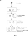
Fig.1
[Fig. 1 ] provides an outline of the scrmPCR method of the disclosure. This method can be used to detect and quantify RNA transcripts and to sequence DNA from the same single cell. A schematic of the method is enclosed asFigure 1 and is composed of the following steps:- 1) Primer design: Primer sets are designed for semi-nested PCR (RNA transcripts) or nested PCR (genomic DNA). In some cases, primers were derived from the literature (Table 1). 2) Reverse transcription: Multiplex reverse primers are added to the lysed single cell, together with a reverse transcriptase enzyme (step II in
Fig. 1 ). 3) Pre-amplification: Multiplex forward primers are added to the mixture, together with primers specific for DNA sequences and a pre-amplification step is performed using an optimized annealing temperature (Step III inFig. 1 ). 4) Cleanup: Unused primers are removed by spin column or enzymatic digestion. 5) Dilution of the sample. 6) For RNA analysis, perform qPCR using a semi-nested approach, where a primer is designed to bind a sequence internal to the pre-amplified product and the other primer has been previously used during the pre-amplification step (step IV inFig. 1 ) 7) For DNA analysis: PCR using a nested approach where both primers are designed to bind a sequence internal to the pre-amplified product (step IV inFig. 1 ).
- 1) Primer design: Primer sets are designed for semi-nested PCR (RNA transcripts) or nested PCR (genomic DNA). In some cases, primers were derived from the literature (Table 1). 2) Reverse transcription: Multiplex reverse primers are added to the lysed single cell, together with a reverse transcriptase enzyme (step II in
-
Fig.2
[Fig. 2 ] shows that nested PCR is required for specific amplification of PCR products from single cells. In (a), primers forTP53 exon 6 were optimized to amplify tissue DNA and obtain specific products that can be sequenced. In (b),TP53 exon 6 was preamplified using the same primer pair and the reaction was split in 2 equal amounts. The first part was used in a second round of PCR using the same primer pairs. This resulted in unspecific and inefficient amplification (top panel). Specific PCR products were then obtained by performing the second amplification using a nested pair of primers (bottom panel). (c) shows melting curves of several seminested assays for RNA detection by qPCR, indicating specific amplification of the target product. -
Fig.3
[Fig. 3 ] provides the proof of principle for the scrmPCR method of the disclosure. Single DLD1 and RKO cells (colorectal cancer cell lines) were micro-manipulated in 5µl 2x Reaction Buffer (CellDirect kit). scrmPCR was then performed as described herein, with the results shown in (a). Genomic regions belonging to TP53, KRAS and BRAF genes were amplified. PCR products were subjected to Sanger sequencing and known hotspot mutations that have been previously characterized in both cell lines were detected as shown in (b). At the same time several transcripts from the same cells were amplified and shown to have variable gene expression in both cell lines. Gene expression specificity was verified by the melting curve peak temperature and by the presence of a single peak, as shown in (c). -
Fig.4
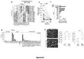
[Fig. 4 ] provides the proof of principle for the scrmPCR method of the disclosure using single cells and single clusters of cells isolated from clinical samples. Single cells or single clusters of unknown origin isolated from blood of colorectal cancer patients were micro-manipulated and subjected to scrmPCR as described herein. In (a), the gene expression of several epithelial and mesenchymal markers shows the predominant mesenchymal nature of single clusters. The expression of PTPRC (CD45) is also shown. Colours represent gene expression from absent (dark grey) to maximum (light grey). NTC, no template control. In (b), results of Sanger sequencing of targeted gene region mutated in matching colorectal cancer tissues show that the same clusters shown in (a) do not contain mutations and are thus not related to the tumor epithelium. (c) shows the use of the latest scrmPCR protocol containing additional assays to quantify transcripts from a set of clusters, to show that clusters express transcripts belonging to the endothelial lineage. Heat map represents scrmPCR gene expression in control single-cells and 14 CTM (n = 4 patients). Color scale indicate gene expression from low or absent (dark grey) to high (light grey). -
Fig.5
[Fig. 5 ] shows the retrieval of circulating tumour cells using a microfiltration device. (a) shows the device setup. Microfiltration devices enclosing a silicon microsieve (inset, scale bar =10 µm) are connected to a peristaltic pump for flow rate control. (b) shows the microfiltration procedure for various downstream applications. The numbers indicate procedure time for each step. (c) shows optimization of retrieval efficiency and purity for downstream single-cell micromanipulation. The individual scatter plots represent experiments using various flow rates and microsieve pore diameters. Black dashed rectangle indicates the target area of > 90% retrieval efficiency and > 5×10³ WBC depletion for optimal downstream handling of retrieved cells. Data points are means ± s.e.m. of three independent experiments under each condition. -
Fig.6
[Fig. 6 ] shows that CTM express EMT markers, but do not mirror primary tumour mutations or chromosome abnormalities. (a) shows images of nine CTM from 4 CRC patients with known primary tumour mutations micro-manipulated in single tubes for downstream scrmPCR. (b) CTM aCGH shows images of three CTM from a representative CRC patient with known chromosomal abnormality. (c) shows aCGH analysis of CTM shown in (b) with matching normal and tumour tissues. -
Fig.7
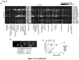
[Fig. 7 ] shows that CTM are tumour-derived mature endothelial cells. (a) shows lineage inference from RNA-Seq data of single-CTM. Normalized odds ratios compare number of genes enriched for each sample (rows) and cell type (columns) over random enrichment. MEP, Megakaryocyte-erythroid progenitor. In (b), immunofluorescence studies confirm the endothelial lineage of CTM. Representative CTM stained for the indicated antibodies with internal controls for each staining. Inset mid panel, a CD45+ WBC. Inset right panel, a CD41/CD42B+ platelet aggregate. The Table indicates quantification of CTM positive or negative for the indicated immunofluorescence. (c) shows the results of the EPC clonogenic assay for CTM (n = 5 patients). The scatter plot represents initial (x axis) and final (y axis) number of CTM (grey symbols) or HUVEC cells (black circles). Same grey symbols are technical replicates. HUVEC cells data are represented as mean ± s.e.m. of technical replicates. One representative experiment of two is shown. (d) shows the rank correlation of CTM and tissues RNA Seq data by principal component analysis. (e) shows the ladder plot of CD31+CD45- CTM counts 0-24 h before and 24-72 h after surgery. Lines connect data from the same patient. Two-tailed Wilcoxon signed-rank test, ***P = 0.0006, effect size r = 0.54. (f) shows FOLH1 expression for the indicated tissues, PBMCs, and CTM for individual patients. Bars represent means. In tissues, error bars are s.e.m. from independent datapoints (n = 3 patients). (g) shows CD276 in normal, tumour tissues and in single-CTM from RNA-Seq data. (h) shows the microvessel density count in patients with low (≤10 CTM/2ml) and high (>10 CTM/2 ml) CTM counts (n = 17 patients). Arrows and dashed white lines indicate microvessel lumens in representative images. Two-tailed Wilcoxon-Mann-Whitney U test with Bonferroni correction, * P = 0.03, effect size r = 0.57, Δ̂ (95% CI) = 2 (0.5-4.16). ns, not significant. -
Fig.8
[Fig. 8 ] shows that CTM are prevalent in CRC patients, mirror therapeutic intervention and indicate the presence of colorectal cancer in early stage treatment-naive patients. (a) is a case-control study of CTM in blood. CTM count for each healthy control (n = 45) and CRC case (n = 80) are shown. Two-tailed Wilcoxon-Mann-Whitney U test, ***P = 7.31×10⁻¹⁵, effect size r = 0.65, Δ̂ (95% CI) = 4 (3-9). (b) shows association of CTM count with patients and tumour characteristics (n = 80 CRC cases). Two-tailed Wilcoxon-Mann-Whitney U test with Bonferroni correction, **P = 0.0072, effect size r = 0.34, Δ̂ (95% CI) = -6 (-13-(-1)). (c) shows the trend of CTM count during colorectal cancer sequence of treatment. Blood samples were collected independently at discrete time points: 1) Treatment-naive, 2) Post neoadjuvant therapy, 3) Post surgery, 4) Post adjuvant therapy, 5) Palliative therapy. Light grey boxes indicate the interquartile range (IQR), line across boxes indicates the median, dashed line indicates the interpolation of medians by spline function. Arrows indicate treatment events. n = 80 CRC cases, two-tailed Wilcoxon-Mann-Whitney U test, ***P = 0.0002, effect size r = 0.41, Δ̂ (95% CI) = -7 (-15-(-2)). ND, not detected. Post operative samples shown inFig. 7e are not included. (d) shows the ROC curve for treatment-naïve CRC patients versus healthy controls (n = 89). Shaded area (light grey) represents the bootstrapped 95% CI. AUC (95% CI) = 0.930 (0.880-0.980), effect size r = 0.71. (e) Same as (d), but comparing early-stage CRC cases (≤ IIA) with healthy controls (n = 61). AUC (95% CI) = 0.922 (0.846-0.999), effect size r = 0.70. In (a), (d) and (e), 100% stacked bar charts indicate the percentage of positive (dark grey) and negative (light grey) samples for both healthy controls and CRC cases. -
Fig.9
[Fig. 9 ] illustrates a silicon microsieve, microfiltration device and its retrieval efficiency and purity. (a) shows a bright field composite image (left panel) and scanning electron micrographs (right panels) of silicon microsieve. (b) shows a microfiltration device with components. (c) shows a silicon microsieves allow efficient retrieval of captured cells. Capture efficiency ofHCT 116 cells from the whole blood, indicating % of captured cells that can be retrieved for downstream assays (light grey bars) or cannot be retrieved (dark grey bars). Six independent experiments are shown. (d) shows the size distribution of SW620 (light grey line), (n = 50). Median size of WBCs and CTCs isolated from colorectal, prostate and breast cancer patients respectively reported from Coumans, F et al, 2013. (e) shows contaminating nucleated cells in clinical samples using optimized protocol shown inFig. 5b and 5c (n = 13). (f) shows the retrieval efficiency from additional cell lines using optimized protocol shown inFig. 5b and 5c . Bars represent mean ± s.e.m. -
Fig.10
[Fig. 10 ] shows characterization and definition of CTM. (a) shows a representative uncropped image of a CD45- CTM (P7-CTM3, see alsoFig. 1 ,Fig. 4a andFig. 6a ) in 10µl of wash buffer, indicating purity of sample suitable for downstream analysis, and showing the presence of target CD45- CTM and sparse RBCs and a WBC, before final micromanipulation (See Methods). Scale bar = 50 µm. (b) shows representative immunofluorescence of CD45 and Ep-CAM of CTM retrieved from 5 CRC patients. The table indicates CD45- CTM counts for each staining and patient. Scale bars = 10 µm. (c) Representative cytomorphology of CTM. Scale bars = 10 µm. DIC: differential interference contrast. -
Fig.11
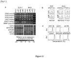
[Fig. 11 ] shows scrmPCR proof of principle using an epithelial-like (DLD-1) and a mesenchymal-like (RKO) CRC cell line. (a) shows DNA amplification (gel electrophoresis) and RNA quantitation (heat map) for the indicated genes and single or bulk cells. (b) shows sequence chromatograms from DNA hotspots of known mutant alleles in RKO and DLD-1 single-cells. (c) shows melt curve plots of the indicated genes from qPCR reactions, sc, single-cell. NTC, no template control. Rn, normalized reporter signal -
Fig.12
[Fig 12 ] shows that CTM express EMT markers but have normal chromosomal structures. (a) shows representative 4-colour immunofluorescence of two CTM for CD45, Vimentin (VIM), pan-Keratin (CK) and DAPI, indicating heterogeneous mesenchymal and epithelial markers expression, (b) shows a control experiment to assess the impact of whole genome amplification (WGA) for aCGH experiments using single-cells. (c-e) each shows aCGH of single-CTM for the indicated patients similar to normal tissue DNA shown in (b). -
Fig.13
[Fig. 13 ] shows that cytomorphology of CTM used for RNA-Seq. (a-g) each shows differential interference contrast (DIC) and Hoechst 33342 images of CTM used for RNA-Seq for each respective patients. No images are available for patient P1. -
Fig.14
[Fig. 14 ] shows a lineage inference workflow. (a) is a flow chart of the lineage inference workflow. (b) shows selected genes with highest specificity index for representative lineages are verified for specificity using BioGPS (Wu et al) (c) shows gene expression level of markers commonly used in CTC research to denote epithelial cells. Note KRT18 expression in the endothelial lineages and EPCAM expression in hematopoietic cells. -
Fig.15
[Fig. 15 ] shows a lineage inference algorithm validation. (a) shows heat maps comparing number of genes enriched for each sample (rows) and lineage (columns) over random enrichment. Samples are published RNA-Seq data from selected lineages. Each coloured box represents a normalized odds ratio of the respective Fisher's exact test from 0 (black) to 1 (light grey). (b) Same as in (a), except that whole tissues or complex cell mixtures such as PBMCs, skin and brain datasets are used. -
Fig.16
[Fig. 16 ] shows a comparison between CTM and CTC-clusters reported in Aceto et al. Lineage inference of CTC clusters data from Aceto et al. shows enrichment with epithelial cell types. Platelet and red blood cell signals were removed from this analysis. RNA-Seq data from CTM analyzed in this study are shown for comparison. MCF7 and HUVEC RNA-Seq are shown as positive controls. -
Fig.17
[Fig. 17 ] shows tumour endothelial markers expressed in CTM. Additional tumor endothelial markers were expressed in normal, tumour tissues and CTM using RNA-Seq data. PLXDC1, plexin domain containing 1 (tumorendothelial marker 3/7) (St Croix et al); MMP2, matrix metallopeptidase 2 (St Croix et al); NID1, nidogen 1 (St Croix et al); MMP11(St Croix et al),matrix metallopeptidase 11; CLEC14A, C-typelectin domain family 14, member A (Mura et al); POSTN, periostin (Borgia et al); VWF, von Willebrand factor (Zanetta et al); ECSCR, endothelial cell surface expressed chemotaxis and apoptosis regulator (Herbert et al). -
Fig.18
[Fig. 18 ] shows that CTM counts do not correlate with inflammatory markers or other variables. (a-c) show the association of CTM number with the indicated tumour characteristics, patient's characteristics, and blood test values respectively. Correlations are shown as dot plots and measured using the Kendall's τ coefficient and its derived P value. Comparisons of dichotomized variables are shown as boxplots and differences are quantified using P values from two-tailed exact Wilcoxon-Mann-Whitney U tests. - The method of the present disclosure can be used to simultaneously quantify RNA transcripts and sequence DNA regions from a single cell. The method can be applied to other samples, such as clinical or forensic samples, where nucleic acids are often present in very limited amounts, or to experimental samples containing a low number of cells, such as 2-100 cells.
- In a first aspect, there is provided a method of simultaneously analyzing RNA and DNA in a sample, the method comprising the steps of:
- (a) contacting the sample with a reverse primer from a first primer pair, the reverse primer from the first primer pair being directed to a target RNA region, and a reverse transcriptase to effect reverse transcription of the RNA into cDNA;
- (b) subsequently contacting the sample with:
- (i) a forward primer from the first primer pair, the forward primer from the first primer pair being directed to a target cDNA region,
- (ii) a reverse primer and a forward primer from a second primer pair, the reverse primer and forward primer from the second primer pair being directed to a target DNA region, and
- (iii) a DNA polymerase
- (c) analyzing the amplified target cDNA region and/or the amplified target DNA region.
- Advantageously, the simultaneous amplification of the target cDNA region and the target DNA region in step (b) may form a pre-amplification step that increases the amount of cDNA and/or DNA as templates for further amplification of the target cDNA and/or target DNA regions prior to analysis. The target DNA region may be a target genomic DNA region.
- By "primer" is meant an oligonucleotide which, when paired with a strand of DNA or RNA, is capable of initiating the synthesis of a primer extension product in the presence of a suitable polymerising agent. The primer is preferably single-stranded for maximum efficiency in amplification but may alternatively be double-stranded. A primer must be sufficiently long to prime the synthesis of extension products in the presence of the polymerisation agent. The length of the primer depends on many factors, including application, temperature to be employed, template reaction conditions, other reagents, and source of primers. For example, depending on the complexity of the target sequence, the oligonucleotide primer typically contains 15 to 35 or more nucleotides, although it may contain fewer nucleotides. Primers can be large polynucleotides, such as from about 200 nucleotides to several kilobases or more. Primers may be selected to be "substantially complementary" to the sequence on the template to which it is designed to hybridise and serve as a site for the initiation of synthesis. For example, not all bases in the primer need to reflect the sequence of the template molecule to which the primer will hybridize - the primer need only contain sufficient complementary bases to enable the primer to hybridize to the template. The primer may include additional bases, for example in the form of a restriction enzyme recognition sequence at the 5' end, to facilitate cloning of the amplified DNA. A primer may also include mismatch bases at one or more positions, being bases that are not complementary to bases in the template, but rather are designed to incorporate changes into the DNA upon base extension or amplification.
- The term "amplification" or "amplify" relates to the production of additional copies of a nucleic acid. Amplification may be carried out using polymerase chain reaction (PCR) technologies or other nucleic acid amplification technologies well known in the art.
- "Primer pairs" can be used for amplification (and identification) of a nucleic acid, e.g., by the polymerase chain reaction (PCR). The "primer pair" may comprise a "forward primer" and a "reverse primer". In a PCR reaction, both strands of a double stranded DNA are amplified. The "forward primer" may bind to one strand of the DNA and allow the synthesis of a primer extension product from the 5' to 3' direction. The "reverse primer" may bind to the complementary strand of DNA, and also allow the synthesis of a primer extension product in the 5' to 3' direction of the complementary DNA strand. In a reverse transcription reaction, the "reverse primer" may bind to an RNA strand and allow the synthesis of a complementary DNA (cDNA) strand in a 5' to 3' direction of the cDNA strand in the presence of a reverse transcriptase enzyme. The "reverse primer" may subsequently be used together with a "forward primer" to amplify the synthesized cDNA strand. PCR primer pairs can be derived from a known sequence, for example, by using computer programs intended for that purpose such as Primer (Version 0.5, 1991, Whitehead Institute for Biomedical Research, Cambridge MA) and those used in the Examples disclosed herein (e.g. PrimerBLAST, Ncbi primer design tool, ensembl genome browser, Netprimer). Oligonucleotides for use as primers are selected using software known in the art for such purpose. For example, OLIGO 4.06 software is useful for the selection of PCR primer pairs of up to 30-100 nucleotides each, and for the analysis of oligonucleotides and larger polynucleotides of up to 5,000 nucleotides from an input polynucleotide sequence of up to 32 kilobases.
- The methods and reagents for use in PCR amplification reactions, restriction enzyme digestion and subsequent fragment resolution, and nucleic acid sequencing are well known to those skilled in the art. In each case, suitable protocols and reagents will largely depend on individual circumstances. Guidance may be obtained from a variety of sources, such as for example Sambrook et al., Molecular Cloning : A Laboratory Manual, Cold Spring Harbor, New York, 1989, and Ausubel et al., Current Protocols in Molecular Biology, Greene Publ. Assoc. and Wiley-Intersciences, 1992. A person skilled in the art would readily appreciate that various parameters of these procedures may be altered without affecting the ability to achieve the desired product. For example, in the case of PCR amplification, the salt concentration may be varied. Similarly, the amount of DNA used as a template may also be varied depending on the amount of DNA available or the optimal amount of template required for efficient amplification.
- A skilled person would be able to understand that a "reverse transcriptase" is an enzyme that may be used to synthesise cDNA based on an RNA template. A skilled person would also understand that a "DNA polymerase" is an enzyme that can synthesise DNA molecules based on a DNA template.
- By "contacting", a primer may be brought into physical association with a sample. This allows, for example, a primer pair to anneal with the DNA present in the sample, and subsequently amplify the DNA by PCR. This also allows a primer to anneal to an RNA strand present in the sample, to allow synthesis of cDNA using a reverse transcriptase enzyme as known to a person skilled in the art.
- The method as defined herein allows one to "simultaneously" amplify RNA and DNA in a sample or single cell. The term "simultaneously" means to be able to amplify both RNA and DNA, present in the very same sample or single cell. It may also mean being able to analyze both RNA and DNA from the same sample or single cell.
- To the inventor's knowledge, this is the first method that allows targeted analysis of both DNA mutations and RNA transcription in the same single cell. The method has undergone several optimization steps.
- The inventors have found that specific amplification of both DNA and RNA can only be achieved by using at least a semi-nested approach for RNA and a fully nested approach for DNA molecules (e.g. see
Fig 2 ). The term "semi-nested PCR" as used herein refers to a modified PCR technique in which one "nested primer" is used to reduce non-specific binding due to the amplification of unexpected binding sites. A "fully nested approach" would refer to a modified PCR technique where two nested primers are used on either side on a template DNA. The use of "nested primers" allow the specific recognition of a PCR product amplified using a first set of primers, thus eliminating contamination from unwanted products such as primer dimers, hairpins and alternative primer target sequences. The inventors have also found that amplification of DNA and RNA molecules are differentially affected by annealing temperature in the pre-amplification step. A trade-off therefore needs to be set in order to amplify both molecules. - Accordingly, in one embodiment, the method according to the first aspect further comprises the step of: subjecting the sample from step (b) to a semi-nested PCR using the reverse primer in step (a) or the forward primer in step (b)(i), and a nested primer that binds within the amplified target cDNA region. The nested primer may be one that matches or corresponds to the reverse primer in step (a) or the forward primer in step (b)(i). In another embodiment, the method according to the first aspect further comprises the step of: subjecting the sample from step (b) to a nested PCR using a nested primer pair that binds within the amplified target DNA region. In one embodiment, steps (a) and (b) are conducted in the same reaction mixture.
- In one embodiment, the method according to the first aspect is performed simultaneously for one or more target RNA regions, and/or one or more target cDNA regions, and/or one or more target DNA regions. Accordingly, one or more reverse primers, each having the same or a differing specificity for a target RNA region may be used in step (a), one or more forward primers, each having the same or a differing specificity for a target cDNA region may be used in step (b)(i), one or more primer pairs, each having the same or a differing specificity for a target DNA region may be used in step (b)(ii), one or more nested primers that bind to a target cDNA region, and one or more nested primer pairs that bind to a target DNA region, may be used.
- Advantageously, the method according to the first aspect can be used to analyze RNA and DNA in instances where a limited amount of sample is available.
- In one embodiment, the sample comprises a single cell, or a plurality of cells (e.g. a low number of cells comprising about 2 to about 100 cells, about 2 to about 90 cells, about 2 to about 80 cells, about 2 to about 70 cells, about 2 to about 60 cells, about 2 to about 50 cells, about 2 to about 40 cells, about 2 to about 30 cells, about 2 to about 20 cells, about 2 to about 10 cells, about 2 to about 5 cells, about 5 to about 100 cells, about 10 to about 100 cells, about 20 to about 100 cells, about 30 to about 100 cells, about 40 to about 100 cells, about 50 to about 100 cells, about 60 to about 100 cells, about 70 to about 100 cells, about 80 to about 100 cells, or about 90 to about 100 cells). The single cell or plurality of cells may be lysed to release the RNA and DNA contained within the cell (or cells) prior to step (a).
- In another embodiment, the sample comprises cell-free RNA, or cell-free DNA.
- The RNA or DNA may be present in a low amount, for example from about 1 pg to about lOng, about 5 pg to about 10 ng, about 5 pg to about 5 ng, about 5 pg to about 1 ng, about 5 pg to about 500 pg, about 5 pg to about 250 pg, about 5 pg to about 125 pg, about 5 pg to about 100 pg, or about 5 pg to about 50 pg.
- The sample may be a sample of tissues, cells, vesicles such as exosomes, body fluids and isolates thereof etc., isolated from a subject. Examples of samples include: whole blood, blood fluids (e.g. serum and plasma), lymph and cystic fluids, sputum, stool, tears, mucus, hair, skin, ascitic fluid, cystic fluid, urine, nipple exudates, nipple aspirates, semen, vaginal fluid, sweat, exosomes or other vesicles, sections of tissues such as biopsy and autopsy samples, frozen sections taken for histologic purposes, archival samples, explants and primary and/or transformed cell cultures derived from patient tissues, clinical samples, forensic samples from crime scenes, soil samples etc.
- The single cell or plurality of cells may be a eukaryotic cell, a bacterial cell or an archeal cell, a cell from a pre-implantation embryo, a stem cell, a suspected cancer cell, a suspected tumor-derived cell, a suspected embryonic cell, a cell from a pathogenic organism, or a cell obtained from a crime scene.
- In one embodiment, the first primer pair comprises primers that span exon-exon boundaries or are separated by at least one intron on the corresponding DNA region.
- In one embodiment, the second primer pair comprises primers that bind to intronic regions of the target DNA region.
- The term "exon" refers to the portion of the genomic DNA that becomes a part of the genomic DNA that is converted into the mature messenger mRNA. The term "intron" or "intronic region" refers to the portion of the genomic DNA that is removed by RNA splicing and which would therefore not be present in the final mature mRNA.
- The term "analyze" or "analyzing" refers to studying or examining the amplified target cDNA region and/or the amplified target DNA region by various techniques known in the art. The amplified cDNA region and/or the amplified target DNA region may be studied for its gene expression or for mutations that may be present.
- In one embodiment, the analysis in step (c) comprises analyzing the amplified target cDNA for gene expression (e.g. in a gene expression analysis). The gene expression analysis may be conducted using any techniques known in the art, such as quantitative PCR, digital PCR, microarray, and the like.
- In one embodiment, the analysis in step (c) comprises analyzing the amplified target DNA for mutations (e.g in a mutational analysis). The mutational analysis may be conducted using any techniques known in the art, such as Sanger sequencing, Maxam-Gilbert sequencing, Pyrosequencing, Shot-gun sequencing, high-throughput DNA sequencing, Allele-Specific PCR (ASPCR) or High Resolution Melting temperature PCR (HRM).
- The pre-amplification in step (b) of the method according to the first aspect may comprise one or more cycling steps. Each cycling step may comprise one or more cycles of amplification (i.e. denaturation, annealing and elongation) at a pre-determined temperature for a pre-determined duration. It would be appreciated that the number of cycling steps, the number of cycles of denaturation, annealing and elongation, the temperature(s) at which the cycles are conducted, and the duration for which each temperature is applied would depend on the reagents used in the amplification reactions, the target cDNA or DNA region, the primers used, the sample(s) to be amplified. In one embodiment, the amplification does not include a final extension step.
- In one embodiment, step (b) comprises about 1 to about 50 cycling steps, about 1 to about 40 cycling steps, about 1 to about 30 cycling steps, about 1 to about 25 cycling steps, about 1 to about 20 cycling steps, about 1 to about 10 cycling steps, about 1 to about 5 cycling steps, about 1 to about 4 cycling steps, about 1 to about 3 cycling steps, about 1 cycling step, about 2 cycling steps, or about 3 cycling steps. In one embodiment, step (b) comprises 3 cycling steps.
- In one embodiment, each cycling step comprises about 1 to about 50 cycles, about 1 to about 40 cycles, about 1 to about 30 cycles, about 1 to about 25 cycles, about 1 to about 20 cycles, about 1 to about 18 cycles, about 1 to about 15 cycles, about 1 to about 10 cycles, about 1 to about 6 cycles, about 2 cycles, about 4 cycles, about 6 cycles, about 8 cycles, about 10 cycles, about 20 cycles, about 30 cycles, about 40 cycles, or about 50 cycles of denaturation, annealing and elongation. In one embodiment, each cycling step comprises 6 cycles of denaturation, annealing and elongation.
- In one embodiment, the annealing and/or elongation temperature in a cycle is about 40°C to about 75°C, about 40°C to about 70°C, about 40°C to about 65°C, about 40°C to about 60°C, about 40°C to about 55°C, about 40°C to about 50°C, about 40°C, about 45°C, about 50°C, about 55°C, about 60°C, about 65°C, about 70°C, or about 75°C.
- The annealing and/or elongation temperature for successive cycling steps may be reduced by about 1°C to about 10°C, about 1 °C to about 9°C, about 1°C to about 8°C, about 1 °C to about 7°C, about 1°C to about 6°C, about 1°C to about 5°C, about 1°C to about 4°C, about 1°C to about 3°C, or about 1°C to about 2°C.
- In one embodiment, the annealing and/or elongation is carried out for about 10 seconds to about 10 minutes, about 10 seconds to about 8 minutes, about 10 seconds to about 6 minutes, about 10 seconds to about 4 minutes, about 10 seconds to about 2 minutes, about 10 seconds to about 1 minute, about 1 minute, about 2 minutes, about 4 minutes, about 6 minutes, about 8 minutes, or about 10 minutes.
- In one embodiment, the denaturation is carried out at a temperature of about 75°C to about 120°C, about 75°C to about 105°C, about 75°C to about 95°C, about 75°C to about 90°C, about 75°C to about 85°C, about 75°C to about 80°C, about 75°C, about 80°C, about 85°C, about 90°C, about 95°C, about 100°C, about 105°C, about 110°C, about 115°C, or about 120°C. The denaturation may be carried out for about 1 second to about 10 minutes, about 1 second to about 5 minutes, about 1 second to about 4 minutes, about 1 second to about 3 minutes, about 1 second to about 2 minutes, about 1 second to about 1 minute, about 1 second, about 10 seconds, about 20 seconds, about 30 seconds, about 40 seconds, about 50 seconds, about 1 minute, about 2 minutes, about 3 minutes, about 4 minutes, about 5 minutes, or about 10 minutes. In one embodiment, step (b) of the method according to the first aspect comprises:
- 6 cycles of 60°C for 4 minutes followed by 95°C for 1 minute,
- 6 cycles of 55°C for 4 minutes followed by 95°C for 1 minute, and
- 6 cycles of 50°C for 4 minutes followed by 95°C for 1 minute.
- In a second aspect, there is provided a method of simultaneously analyzing RNA and DNA in a sample, the method comprising the steps of:
- (a) lysing a cell in the sample;
- (b) contacting the lysed cell sample with a reverse primer from a first primer pair, the reverse primer from the first primer pair being directed to a target RNA region, and a reverse transcriptase to effect reverse transcription of the RNA into cDNA;
- (c) subsequently contacting the lysed cell sample with:
- (i) a forward primer from the first primer pair, the forward primer from the first primer pair being directed to a target cDNA region,
- (ii) a reverse primer and a forward primer from a second primer pair, the reverse primer and forward primer from the second primer pair being directed to a target DNA region, and
- (iii) a DNA polymerase
- (d) subjecting the sample from step (c) to a semi-nested PCR using the reverse primer in step (b) or the forward primer in step (c)(i), a nested primer that binds within the pre-amplified target cDNA region, and a DNA polymerase to further amplify the target cDNA region, and/or subjecting the sample from step (c) to a nested PCR using a nested primer pair that binds within the pre-amplified target DNA region and a DNA polymerase to further amplify the target DNA region; and
- (e) analyzing the further amplified target cDNA region and/or the further amplified target DNA region.
- In one embodiment of the method of the second aspect, the nested primer in step (d) may be one that matches or corresponds to the reverse primer in step (b) or the forward primer in step (c)(i). In one embodiment of the method of the second aspect, steps (b) and (c) are conducted in the same reaction mixture.
- Like the method of the first aspect, the method according to the second aspect may also be performed simultaneously for one or more target RNA regions, and/or one or more target cDNA regions, and/or one or more target DNA regions (such as one or more target genomic DNA regions). Accordingly, one or more reverse primers, each having the same or a differing specificity for a target RNA region may be used in step (b) of the method according to the second aspect, one or more forward primers, each having the same or a differing specificity for a target cDNA region may be used in step (c)(i) of the method according to the second aspect, one or more primer pairs, each having the same or a differing specificity for a target DNA region may be used in step (c)(ii) of the method according to the second aspect, one or more nested primers that bind to a target cDNA region, and one or more nested primer pairs that bind to a target DNA region, may be used in step (d) of the method of the second aspect.
- The term "lysing" refers to breaking apart the membrane of a cell. This allows DNA or RNA in the cell to be accessible to contact by, for example, one or more primers and enzymes.
- The lysing in step (a) of the method of the second aspect may be conducted using any techniques known in the art, for example, use of a lysis buffer, sonication, freeze-thaw, mechanical disruption, and the like.
- In one embodiment, the pre-amplification in step (c) of the method of the second aspect does not include a final extension step.
- The samples, primers, primer pairs, analytical techniques, and pre-amplification conditions (such as cycling steps, and temperatures and durations used therein) as described above for the method of the first aspect may also be used in the method of the second aspect.
- In a third aspect, there is provided use of the method according to the first or second aspect for determining the DNA profile and/or the gene expression profile of a single cell or a plurality of cells (e.g. a low number of cells comprising about 2 to about 100 cells), or cell-free RNA and/or DNA (e.g. a low amount of DNA and/or RNA comprising about 1 pg to about 10 ng of DNA and/or RNA) derived from a single cell or a plurality of cells.
- The term "DNA profile" refers to the characterization of the DNA sequence of a selection of genes within a cell or a plurality of cells. The term "gene expression profile" refers to the characterization of the expression level of a selection of genes within a cell or plurality of cells.
- The use may be applied to a biological sample comprising heterogenous cell types from a subject or a low amount of cell-free RNA and/or DNA. The biological sample may be selected from the group consisting of a pre-implantation embryo, tissues, cells (e.g. a stem cell, a suspected cancer cell), body fluids and isolates thereof etc., isolated from a subject. Examples of biological samples include: whole blood, blood fluids (e.g. serum and plasma), lymph and cystic fluids, sputum, stool, tears, mucus, hair, skin, ascitic fluid, cystic fluid, urine, nipple exudates, nipple aspirates, semen, vaginal fluid, sweat, exosomes or other vesicles, sections of tissues such as biopsy and autopsy samples, frozen sections taken for histologic purposes, archival samples, explants and primary and/or transformed cell cultures derived from patient tissues, clinical samples, forensic samples from crime scenes, soil samples etc.
- The subject may be a mammal, such as a human. The linkage of the DNA profile to the gene expression profile for the single cell or the plurality of cells, or the low amount of cell-free DNA and/or cell-free RNA may be used to optimize anti-cancer therapy for the subject or for diagnostic/prognostic purposes, as illustrated by the Examples as described herein. Alternatively, the use may be applied to a cell from a pathogenic organism, or a cell obtained from a crime scene, or a low amount of cell-free RNA and/or cell-free DNA derived from a pathogenic organism or a crime scene.
- In a fourth aspect, there is provided use of the method according to the first or second aspect for validation of large-scale transcriptomics data sets. For example, the use may be applied to confirm specific results obtained by high-throughput RNA sequencing experiments that have been generated using small-scale samples (such as a biological sample comprising of a single cell or a low number of cells, e.g. about 2 to about 100 cells, or a low amount of RNA). This is important to rule out false positive results and to validate the results using an independent methodology. The method can be used to validate both RNA expression and/or mutational profile results that are generated by high-throughput RNA sequencing.
- In a fifth aspect, there is provided a kit for performing the method according to the first or second aspect, or for use according to the third or fourth aspect, wherein the kit comprises:
- (a) a primer selected from the group consisting of:
- i. the reverse primer of step (a) of the method of the first aspect or step (b) of the method of the second aspect,
- ii. the forward primer of step (b)(i) of the method of the first aspect or step (c)(i) of the method of the second aspect,
- iii. the primer pair of step (b)(ii) of the method of the first aspect or step (c)(ii) of the method of the second aspect, and
- iv. the nested primer and nested primer pair of the method of the first aspect or step (d) of the method of the second aspect;
- (b) one or more reagents, selected from the group consisting of:
- i. a reverse transcriptase and one or more suitable reaction buffers for the reverse transcription in step (a) of the method of the first aspect or step (b) of the method of the second aspect,
- ii. a DNA polymerase and one or more suitable reaction buffers for the amplification in step (b) or the semi-nested or nested PCR of the method of the first aspect, or step (c) or (d) of the method of the second aspect, and
- iii. one or more labelled or unlabelled deoxyribonucleotides selected from the group consisting of dATP, dCTP, dGTP, and dTTP or dUTP; and
- (c) instructions for performing the method according to the first or second aspect, or for use according to the third or fourth aspect.
- The kit according to the fifth aspect may include one or more lysis buffers for lysing a cell in a sample prior to the method of the first aspect, or in step (a) of the method of the second aspect. Lysis buffers commonly used in the art, such as alkaline lysis buffers or cell lysis buffers containing proteinase K, or simply buffers containing a detergent or a compound and /or an enzyme that will disrupt the cell and allow its nucleic acids to be released in solution may be used.
- The kit according to the fifth aspect may also include probes or dyes for quantitative real-time PCR. Exemplary probes and dyes include, but are not limited to SYBR green dye, EvaGreen, dsGreen, TaqMan probes, hybridization probes and the like.
- The kit may also include instructions for designing one or more of the primers of component (a) of the kit of the fifth aspect, and/or optimizing the pre-amplification cycling conditions of step (b) of the method of the first aspect or step (c) of the method of the second aspect.
- In one embodiment, the primers and/or reagents are pre-mixed in combinations suitable for the lysis, pre-amplification, and amplification steps described above. In another embodiment, the primers are pre-mixed in combinations suitable for analysis of gene expression profiles or mutation signatures. The primers may be ones that have been designed for amplifying one or more target genes of interest.
- In a further embodiment, the kit comprises one or more containers comprising one or more reaction buffers for performing the methods and/or uses described above. In some embodiments, the kit includes software-driven assay protocols for use in commercial PCR instrumentation (such as the Life Technologies 7500 FastDx or Cepheid SmartCycler® II), which may be provided on a CD.
- As used herein, the singular form "a", "an" and "the" include plural references unless the context clearly dictates otherwise. For example, the term "a primer" includes a plurality of primers, including mixtures thereof.
- The word "substantially" does not exclude "completely" e.g. a composition which is "substantially free" from Y may be completely free from Y. Where necessary, the word "substantially" may be omitted from the definition of the invention.
- Unless specified otherwise, the terms "comprising" and "comprise", and grammatical variants thereof, are intended to represent "open" or "inclusive" language such that they include recited elements but also permit inclusion of additional, unrecited elements.
- As used herein, the term "about", in the context of concentrations of components of the formulations, typically means +/- 5% of the stated value, more typically +/- 4% of the stated value, more typically +/- 3% of the stated value, more typically, +/- 2% of the stated value, even more typically +/- 1 % of the stated value, and even more typically +/- 0.5% of the stated value.
- Throughout this disclosure, certain embodiments may be disclosed in a range format. It should be understood that the description in range format is merely for convenience and brevity and should not be construed as an inflexible limitation on the scope of the disclosed ranges. Accordingly, the description of a range should be considered to have specifically disclosed all the possible sub-ranges as well as individual numerical values within that range. For example, description of a range such as from 1 to 6 should be considered to have specifically disclosed sub-ranges such as from 1 to 3, from 1 to 4, from 1 to 5, from 2 to 4, from 2 to 6, from 3 to 6 etc., as well as individual numbers within that range, for example, 1, 2, 3, 4, 5, and 6. This applies regardless of the breadth of the range.
- Certain embodiments may also be described broadly and generically herein. Each of the narrower species and subgeneric groupings falling within the generic disclosure also form part of the disclosure. This includes the generic description of the embodiments with a proviso or negative limitation removing any subject matter from the genus, regardless of whether or not the excised material is specifically recited herein.
- The following protocol steps or materials have been implemented in the method from sources already described elsewhere:
- 1) Conditions described in Peixoto et al., specifically:
- a. Use of reverse specific primers for reverse transcriptase reaction.
- b. Use of seminested PCR for RNA transcripts quantitation.
- 2) Materials from the CellsDirect™ One-Step qRT-PCR Kit (Invitrogen), specifically:
- a. 2X Reaction Mix cat # 11753-100
- b. SuperScript® III RT/Platinum® Taq Mix contained in cat # 11753-100
- 3) Conditions described in Protocol PN 100-4109 from the Biomark HD system (Fluidigm) (Sanchez-Freire et al) and http://www.fluidigm.com/home/fluidigm/docs/AppNote_2v1Step_pf9.pdf), specifically:
- a. Deposition of single cell in 2x Reaction mix.
- b. Annealing time of the preamplification step (4 minutes).
- To design primers for mRNA amplification in the presence of genomic DNA at the single cell scale, there must be an intronic sequence on the genomic DNA between primers, or at least one primer must sit at the exon-exon junction. In addition, the primers need to be specific and should not amplify unwanted products.
- This protocol can be used to generate primers for seminested/nested PCRs following the general method of Peixoto et.al. (http://www.ncbi.nlm.nih.gov/pubmed/15466292). Seminested PCR is required for robust amplification of RNA-derived gene transcripts. Nested PCR is required for robust amplification of genomic DNA.
- The following tools were used (by opening in Firefox):
- 1) Ncbi primer design tool at http://www.ncbi.nlm.nih.gov/tools/primer-blast/
This software is based on Primer3 and Blast for optimal primer pair findings. It can also design primers at the exon-exon junction.
Seminested PCR: for the external primer pairs, the parameters used may be varied accordingly depending on sample type and objective of the study. - 2) Ensembl genome browser at http://www.ensembl.org/Homo_sapiens/Info/Index This webtool allows for straightforward gene selection and curated ID retrieval.
- 3) Netprimer at http://www.premierbiosoft.com/jsp/marketing/FreeToolLogin.jsp?PID=3
- This webtool allows checking of the quality of the primers, by rating it accordingly.
- The following steps using the above webtools were applied:
- a) -searched for the gene of interest in Ensembl
- b) -checked for the correct identification and clicked on "external references"
- c) -scrolled down and identified the RefSeq identifier
- d) -copied and pasted the identifier on the ncbi primer design tool window
- e) -looked for primers using the link provided above or customized stringency
- f) -checked the primers on the Netprimer application and selected the best primer pair by rating.
- g) -if no primers were returned, less stringent parameters were used.
- Example: Design of CD45 primers.
- Forward primer AAGACAACAGTGGAGAAAGGACGCA (SEQ ID NO: 137)
- Reverse primer CAGTGGGGGAAGGTGTTGGGCT (SEQ ID NO: 138)
- Product length 159
- Exon junction 131/132 (forward primer) on template NM_002838.3
- To look for the internal probe, the same search as above may be performed, using the primer with better ratings and constrain the PCR product length to retrieve a primer that sits in the PCR product.
- For primers on genomic regions, the following steps were applied:
- a) looked for the gene of interest in Ensembl
- b) clicked on sequence
- c) selected genomic region for sequencing including intronic regions
- d) copied and pasted on primer-blast
- e) looked for primers targeting the region of interest
- Two pairs of primers were designed, one with annealing temperature (T) of 60° C, one with annealing T of 65° C.
- For design of internal probes, the amplified region was selected by primer set one, copied and pasted in primer-blast. Primers with an annealing T of 60°C were selected.
- Primers were designed using Primer-BLAST (Ye et al and Bbenek, et al) and using the primer design protocol described above. For each RNA transcript, primers either spanning exon-exon boundaries or primers separated by at least one intron on the corresponding genomic DNA region were designed. This allows primers to specifically amplify RNA transcripts, whereas DNA regions would not be amplified. Furthermore, primers for mutational analysis were designed to bind intronic regions of the target gene. This would avoid amplification of cDNA transcripts that may have arisen from unspecific or specific reverse transcription. (see Primer Design protocol (above) and Tables 1A to 1C).
Table 1A: Primers used in scrm-PCR assays (Pre-amplification step) Preamplification step Gene name ID (transcript or gene) Marker Forward primer Reverse primer CEACAM5 NM_004363.2 C CCCAAGCCCTCCATCTCCAGCAAC (SEQ ID NO: 1) GCATCCGGGCCATAGAGGACATT (SEQ ID NO: 2) CDX2 NM_001265.4 C TGGGCTGCTGCAAACGCTCAAC (SEQ ID NO: 3) TITCGTCCTGGTTITCACTIGGCTG (SEQ ID NO: 4) KRT20 NT_10783.15 C AAAATGGCCATGCAGAACCT (SEQ ID NO: 5) GAAGTCCTCAGCAGCCAGTT (SEQ ID NO: 6) FN1 NT_5403.17 EMT CCACGGGAGCCTCGAA (SEQ ID NO: 7) TAAAACCTCGGCTTCCTCCA (SEQ ID NO: 8) CDH2 NT_010966.14 EMT GGATGAAGATGGCATGGTGT (SEQ ID NO: 9) CCCAGTCTCTCTICTOCCIT (SEQ ID NO: 10) SERPlNEl NT_007933.15 EMT GCCAAGAGCGCTGTCAA (SEQ ID NO: 11) CAGCAGACCCTTCACCAAA (SEQ ID NO: 12) TWIS11 NM_00D474.3 EMT ACCTAGATGTCA'ITGTTTCCAGAGA (SEQ ID NO: 13) GCAGAGGTGTGAGGATGGTG (SEQ ID NO: 14) SNA11 NM_005985.3 EMT GACCCCAATCGGAAGCCTAA (SEQ ID NO: 15) GCGGTGGGGTTGAGGAT (SEQ ID NO: 16) VIM NM_003380.3 EMT GATGTTTCCAAGCCTGACCT (SEQ ID NO: 17) CAGTGGACTCCTGCTTTGC (SEQ ID NO: 18) SNAI2 NlM_003068.4 EMT AGCGAACTGGACACACATACA (SEQ ID NO: 19) AGGAGGTGTCAGATGGAGGA (SEQ ID NO: 20) FOXC2 NM_005251.2 EMT CGCCTAAGGACCTGGTGAA (SEQ ID NO: 21) GAAGCGGTCCATGATGAACT (SEQ ID NO: 22) FOXC1 NM_001453.2 EMT CACACCCTCAAAGCCGAACT (SEQ ID NO: 23) AAAGTGGAGGTGGCTCTGAA (SEQ ID NO: 24) CDH1 NT_010498.15 EMT/L(Ep) GAGAGAGGCCGCGTCCT (SEQ ID NO: 25) GGCCTITTGACTGTAATCACAC (SEQ ID NO: 26) KRT18(1) NM_000224.2 EMT/L(Ep) TGCTCACCACACAGTCTGAT (SEQ ID NO: 27) CACTTTGCCATCCACTAGCC (SEQ ID NO: 28) KRT8 NM_002273.3 EMT/L(Ep) AAGGATGCCAACGCCAAGTT (SEQ ID NO: 291 CCGCTGGTGGTCTTCGTATG (SEQ ID NO: 30) KRT19 NM_002276.4 EMT/L(Ep) CAGCCACTACTACACGACCA (SEQ ID NO: 31) CGTTGATGTCGGCCTCCA (SEQ ID NO: 32) EPCAM NT_022184.15 EMT/L(Ep) GCAGGTCCTCGCGTTCG (SEQ ID NO: 33) TCTCCCAAGTTTTGAGCCATTC (SEQ ID NO: 34) TP53 exon 5 ENSG00000141510 G TGTTCACTTGTGCCCTGACT (SEQ ID NO:35) CAGCCCTGTCGTCTCTCCAG (SEQ ID NO: 36) TP53 exon 6 ENSG00000141510 G TGGT TGCCCAGGGTCCCCA (SEQ ID NO: 37) TGGAGGGCCACTGACAACCA (SEQ ID NO: 38) TP53 exon 7 ENSG00000141510 G CTTGCCACAGGTCTCCCCAA (SEQ ID NO: 39) AGGGGTCAGAGGCAAGCAGA (SEQ ID NO: 40) TP53 exon 8 ENSG00000141510 G TTCCTTACTGCCTCTTGCTT (SEQ ID NO: 41) AGGCATAACTGCACCCTTGG (SEQ ID NO: 42) BRAF exon 15 (2) ENSG00000157764 G CATAATGCTTGCTCrGATAGG (SEQ ID NO: 43) GGCCAAAAATTTAATCAGTGGA (SEQ ID NO: 44) KRAS exon 2 ENSG00000133703 G TTATAAGGCCTGCTGAAAATGACTG (SEQ ID NO: 45) TCATGAAAATGGTCAGAGAAACCTT (SEQ ID NO: 46) EGFR NT_033968.6 L(Ep) GTUGGGCTCTGGAGGAAAA (SEQ ID NO: 47) CTCTGGAGGCTGAGAAAATGAT (SEQ ID NO: 48) PTPRC NT_004487.19 L(He) GACATCATCACCTAGCAGTTCATG (SEQ ID NO: 49) CAGTGGGGGAAGGTGTTGG (SEQ ID NO: 50) VWF NM_000552.3 L(En) ACACAGGGGGACCAAAGAG (SEQ ID NO: 51) GAGATGCCCGTTCACACCA (SEQ ID NO: 52) PECAM1 NM_000442.4 L(He,En) TCTCAACGGTGACTTGTGG (SEQ ID NO: 53) GTTCTTCCCATTTTGCACCGT (SEQ ID NO: 54) COLIA1 NM_000088.3 L(Me) TGTTCAGCTTTGTGGACCTC (SEQ ID NO: 55) GGTITCCACACGTCTCGGT (SEQ ID NO: 56) CD68 NM_001251.2 L/Mo CAT TCTTTCACCAGCTGTCCA (SEQ ID NO: 57) GCACCAGGGCGAGGA (SEQ ID NO: 58) MCAM NM_006500.2 L(En) CTCGGTCCCAGGAGTACC (SEQ ID NO: 59) TGTACAAACCACTCGACTCCA (SEQ ID NO: 60) FCGR3A NM_000569.6 L(He) CCCTTGCCAGACTTCAGACT (SEQ ID NO: 61) GGGAGATCTTCAGTCCGCAT (SEQ ID NO: 62) GYPA NM_002099.6 L(Er) CTAGCAGGCTAAGGTCAGACA (SEQ ID NO: 63) GTGTCCCGTTTGTGCGTATC (SEQ ID NO: 64) ITGA2B NM_000419.3 L(Me) CTTCTATGCAGGCCCCAAT (SEQ ID NO: 65) AGCGTACATTTCGGGTCTCATC (SEQ ID NO: 66) DCSTAMP NM_030788.3 L(He) CTCCCGCTGAATAAGGAGGAA (SEQ ID NO: 67) TCTTGAGTTCCTTGTTTCTCTCCGT (SEQ ID NO: 68) CD34 NM_001773.2 S/L(En) CCTTCTGGGTTCATGAGTCTTGACA (SEQ ID NO: 69) TGTCGTTTCTGTGTGTTTGTTGTG (SEQ ID NO: 70) ACTB NM_001101.3 QC CTGGCACCACACCTTCTACA (SEQ ID NO: 71) TAGCACAGCCTGGATAGCAA (SEQ ID NO: 72) FOLH1 NT_009237.18 TEC CGGATATTGTACCACCTITCAGT (SEQ ID NO: 73) AGCAGGGTCGGAGTAGAGAA (SEQ ID NO: 74) ENG NT_008470.19 L(En) GTGACGGTGAAGGTGGAACTGA (SEQ ID NO: 75) TTGAGGTGTGTCTGGGAGCT (SEQ ID NO: 76) KDR NM_002253.2 L(En) GAAATGACACTGGAGCCTACAAG (SEQ ID NO: 77) AATGGACCCGAGACATGGAAT (SEQ ID NO: 78) CDH5 NM_001795.3 L(En) GTTCACGCATCGGTTGTTCAAT (SEQ ID NO: 79) GCCTGCTTCTCTCGGTCCAA (SEQ ID NO: 80) TEK NT_008413.19 L(En) CTTATTTCTGTGAAGGGCGAGTT (SEQ ID NO: 81) CTCCCTTGTCCACAGTCATAGT (SEQ ID NO: 82) ANGPT2 NM_001147.2 L(En) AACACTCCCTCTCGACAAACAAATT (SEQ ID NO: 83) CTGTAGTTGGATGATGTGCTTGTC (SEQ ID NO: 84) HPRT1 NM_000194.2 QC TGACCAGTCAACAGGGGACA (SEQ ID NO: 85) GGTCCITTTCACCAGCAAGCT (SEQ ID NO: 86) UBC NM_021009.5 QC TCGGCCTTAGAACCCCAGTA (SEQ ID NO: 87) ACGAAGATCTGCATTGTCAAGTG (SEQ ID NO: 88) -
- (1) derived from Hesse et al. (2001) J. Cell Sci. 114, 2569
- (2) derived from Sakaizawa et al. Br. J.Cancer (2012) 106, 939
-
- C: Colon-specific
- EMT: Epithelial-mesenchymal transition marker
- L: Lineage marker
- G: Gene
- QC: Quality control
- TEC: Tumor endothelial cell marker
- S: Stem cell marker
- Ep: Epithelial marker
- He: Hematopoietic cell marker
- En: Endothelial cell marker
- Me: Megakaryocyte/platelet marker
- Mo: Monocyte-macrophage marker
- Er: Erythrocyte marker
-
- (1) derived from Hesse et al. (2001) J. Cell Sci. 114,2569
- (2) derived from Sakaizawa et al. Br. J.Cancer (2012) 106, 939
-
- (1) derived from Hesse et al. (2001) J. Cell Sci. 114,2569
- (2) derived from Sakaizawa et al. Br. J.Cancer (2012) 106, 939
-
- 1) Single cells were micromanipulated in 5µl 2x Reaction Buffer (CellsDirect™ One-Step qRTPCR Kit (Invitrogen)) and stored at -80°C until use.
- 2) Single cell RNA transcripts were reverse transcribed at 50°C for 30 min using SuperScript® III RT (Invitrogen) and a mix of 100nM target reverse primers. The reverse transcriptase component was inactivated at 95°C for one minute. Next, the samples were placed on ice, and was proceeded immediately to the next step.
- 3) A pre-amplification round was next performed by adding a mix of forward primers (matching the reverse primers added in step 2) and primer pairs for specific DNA regions.
- 4) Preamplification was performed similarly as described for the Protocol PN 100-4109 from the Biomark HD system (Fluidigm), using 4 minutes annealing time but changing the annealing temperature to the following cycling steps:
- 6x cycles: 4', 60°C - 1', 95°C
- 6x cycles: 4', 55°C - 1', 95°C
- 6x cycles: 4', 50°C - 1', 95°C
- 5) Primers cleanup from the samples was performed using standard methods (e.g. spin columns such as Axygen Axyprep PCR Clean-up Kit -Cat # 14-223-018).
- 6) Samples were diluted 1/20-1/50 and stored at -20 until further use. This resulted in at least 200µl of preamplified sample, which is sufficient for 100 reactions.
- 7) For RNA transcript quantitation, quantitative PCR was performed on a ViiA7 Instrument (Applied Biosystems) using 2ul of preamplification reaction from 5), seminested primer pairs according to the target transcript (Table 1B) and the SensiFAST SYBR Lo-ROX Kit (Bioline) following the manufacturer protocol. To analyze selected DNA mutational hotspots, PCR was performed by using 2ul of preamplification reaction from 5), nested PCR primer pairs (Table 1B) and a mastermix containing a proof reading polymerase (KOD Mastermix, EMD Millipore) following manufacturer's instructions. PCR products were separated on agarose gel, specific bands were excised and sequenced using the Sanger method.
Step 7 can be performed using any qPCR method and the listed primers. PCR for DNA sequence can be performed using any Taq polymerase with proofreading activity and mutation detection can be performed also using qPCR techniques such as Allele specific PCR (ASPCR) or High resolution melting temperature PCR (HRM). - Patient samples and clinical data. All subjects had given informed written consent to participate. Clinical samples were obtained between July 2012 and April 2014 according to protocols approved by the Institutional Review Boards (IRB) of the National University of Singapore, Fortis Surgical Hospital and Singapore Health Services (SingHealth). Consecutive blood samples from 82 CRC patients were provided by Fortis Surgical Hospital (FSH) and National Cancer Center, Singapore (NCC). Blood samples from 45 healthy subjects were provided by the Singapore Consortium of Cohort Studies (SCCS). All samples were collected in EDTA Vacutainer tubes (Becton-Dickinson) and processed within 6 h at the Institute of Bioengineering and Nanotechnology. Two cases were excluded from analysis because of technical failure of the microfiltration device. Wherever available, matched tumour and metastatic samples were immediately frozen after resection, and stored at -80°C until use. Clinicopathologic data for participating subjects are described in Supplementary Table 6 and were collected retrospectively after completion of CTM counts. Clinical data collection was conducted without prior knowledge of CTM counts. Similarly, clinical data for CRC patients were not known at the time of CTM count except for diagnosis and preoperative status of FSH samples. Tumour area was calculated by width x length.
- Cell lines and culture.
HCT 116,COLO 201, SW480, SW620, DLD-1 and RKO colorectal cancer cell lines, BJ-5ta immortalized human foreskin fibroblasts and HUVECs were from ATCC. HUVECs were used atpassage - Device fabrication and assembly. Silicon microsieves were fabricated as described (Lim et al). Briefly, the microsieve consists of a silicon disk having an overall diameter (ø) of 7.3 mm and a support ring of
thickness 300 µm. The central capture region has ø 5.3 mm and 60 µm thickness containing 100,000 circular pores obtained by deep reactive ion etching. To embed the microsieve in a sterile 3-ml syringe, an acrylic sleeve insert was designed, consisting of an inlet channel of ø 8.58 mm tapered to a ø 5.54 mm channel, which corresponded to the microsieve cell capture region. The sleeve insert housed the microsieve and silicone O-rings (0.5 mm-thick) that ensured good sealing and cushioning as shown inFig. 9b . The retrieval device was assembled as follow. Firstly, the rubber plug of a 3 ml syringe plunger was removed and a hole of 5.5 mm diameter was created using a punch cutter. The perforated rubber plug was placed in the 3-ml syringe. Next, an O-ring was placed in the slot of sleeve insert, followed by microsieve and another O-ring. Finally the sleeve insert with microsieve and O-rings was placed in the 3-ml syringe above the perforated rubber plug. This arrangement enabled the microfiltration of cells by size from whole blood and the subsequent retrieval of captured cells from the upper surface of microsieve in a convenient set-up. Microfiltration. To optimize blood microfiltration, 5 µM CellTracker (Life Technologies) labelled cells were added to donor blood at 10-50 cells per ml of whole blood. Blood was filtered at various flow rates by means of a peristaltic pump (Ismatec). After 6 washes using PBS, 0.5% BSA and 2 mM EDTA, cells were resuspended in culture medium. Subsequently, cell nuclei were stained using Hoechst 33342 (Life Technologies), and cells were retrieved to determine retrieval efficiency and fold depletion of contaminating WBCs. In some experiments, CellTracker positive cells remaining on the microsieve were also counted. - Percent retrieval efficiency was calculated as follow:

- Fold depletion from 1 ml whole blood was calculated as follow:

- WBC count in microfiltrate is defined as the number of any Hoechst 33342 positive, CellTracker negative event in the case of experimental enrichment or by any CD45 positive event in the case of clinical sample analysis. All clinical samples were immediately processed for the indicated downstream applications using optimized parameters described in
Fig. 5b and c. To estimate ideal target WBC depletion, micromanipulation on serial dilution of PBMCs containing 50 CellTrackerpositive HCT 116 cells was performed. Five thousand fold depletion allowed micromanipulation ofpure HCT 116 cells without contaminant white blood cells. The ideal target retrieval efficiency was chosen based on literature search on existing label-free CTC isolation devices (Cima et al). Microfiltration of clinical samples was performed using 2 ml whole blood for each device and optimized microfiltration conditions. - On-sieve immunofluorescence. Suspension cells were stained for 30 min directly 'on sieve' after 5 washes in PBS containing 0.5% BSA, 2 mM EDTA and human FcR Blocking Reagent (Miltenyi Biotec) using following fluorescent-labelled antibodies: anti-CD45 1:200 (clone 2D1; eBioscience), anti-Ep-CAM 1:20 (9C4, BioLegend), anti-CD31 1:20 (WM59, BioLegend), anti-CD144 1:10 (55-7H1, BD), anti-CD41 1:20 (HIP8, BioLegend) anti-CD42B 1:20 (HIP1, BioLegend). For intracellular antigens, the Inside Stain kit (Miltenyi Biotec) and human FcR Blocking Reagent were used with following antibodies: anti-VWF 1:200 (rabbit polyclonal A 0082, DAKO, conjugated in-house to Alexa 488 or Alexa 555 using Life Technologies APEX Antibody Labeling Kit), anti-Vimentin (V9, Santa Cruz Biotechnology), anti-pan Cytokeratin (C11, Cell Signaling Technology). Nuclei were stained using Hoechst 33342 (Life Technologies). In some experiments, Calcein AM (Life Technologies) was used to identify living cells. After a washing step, cells were retrieved and visualized in suspension under an inverted fluorescence microscope (IX81, Olympus) for imaging, counting and/or micromanipulation. Images were recorded using the MetaMorph software (Molecular Devices) with a CoolSNAP HQ2 CCD Camera (Photometrics).
- CTM definition and count. CTM were defined in this study by "any CD45- cell or CD45- cellular cluster with a major axis of > 10 µm and having at least 2 clearly distinct nuclei". In the studies described herein, all cells defined as such, stained positive for endothelial markers CD31, CD144 or VWF (
Fig. 7b ). Importantly, the cellular populations belonging to the megakaryocytic lineages, having large and lobulated single nuclei or large and round single nuclei, were excluded. These cells had characteristic cytomorphology easily discernible from CTM, stained positive for CD41 and CD42B, and were predominantly observed in CRC patients undergoing treatment, but also in some healthy volunteers and treatment-naive CRC patients. Single endothelial cells, owing to their smaller diameter that would allow them to pass through the microsieve, were also excluded from the analysis. CTM were counted by applying these inclusion and exclusion criteria by adding the microfiltrate obtained from 2 ml of whole blood to a well of a 96-well plate. After a short centrifugation step, CTM were identified and counted by manually scanning the target well three times using a 20x objective. A positive sample was defined by the detection of at least one CTM. - Target cell identification, micromanipulation and storage. Target cells were manually micropipetted using a mouth pipette attached to a 25-ml syringe. Briefly, cells were identified from total cell retrieval by means of bright field image, nuclear staining and specific fluorescent signals. Target single-cells or CTM were then micropipetted in a 10-µl droplet of wash buffer, followed by deposition in 0.2-ml PCR tubes containing appropriate buffer: 5 µl of 2x Reaction buffer (CellsDirect One-Step qRT-PCR Kit, Life Technologies) for scrmPCR, 2 µl of PBS for whole genome amplification or 2 µl of SuperBlock buffer (Thermo Scientific) for low-input RNA-Seq. Cells were stored immediately at -80°C until use. In some cases, the complete microfiltrate was spun down, and stored at -80°C until further use.
- Single-cell RNA and mutational analysis PCR (scrmPCR). Primers were designed using Primer-BLAST (Ye et al). For each RNA transcript, primers were desgined either spanning exon-exon boundaries or primers separated by at least one intron on the corresponding genomic DNA region. Primers for mutational analysis were designed to bind intronic regions of the target gene (Tables 1A-C). The method for scrmPCR was highlighted in
Fig 1 as described in Example 1 above. This method could be used to simultaneously detect and quantify RNA transcripts and sequence DNA hotspots in the same cell. Briefly, single-cell RNA transcripts were reverse transcribed at 50°C for 30 min using SuperScript III Reverse Transcriptase (Invitrogen) and a mix of 500 nM target reverse primers. A preamplification round (Table 1A) was then performed using Platinum Taq DNA polymerase (Invitrogen) by adding a matching mix of forward primers to the transcript-specific reverse primers and primers pairs for targeted genomic regions. Preamplification cycling was conducted by alternating annealing and denaturation steps without extension as follow: 6x cycles at 60°C, 4 min, 95°C, 1 min; 6x cycles at 55°C, 4 min, 95°C, 1 min; 6x cycles at 50°C, 4 min, 95°C, 1 min. Primers cleanup was performed using the Axyprep PCR Clean-up Kit (Axygen). Samples were diluted 1/20 and stored at -20°C until further use. For RNA transcript quantitation, quantitative PCR was performed on a ViiA7 Instrument (Applied Biosystems) using 2 µl of preamplification reaction, seminested primer pairs according to the target transcript (Table 1B) and the SensiFAST SYBR Lo-ROX Kit (Bioline) following manufacturer's protocol. Relative gene expression was normalized using ACTB as reference gene. To analyze selected DNA mutational hotspots, PCR was performed by using 2 µl of preamplification reaction, nested PCR primer pairs (Table 1B) and a master mix containing a proof-reading polymerase (KOD Hot Start Master Mix, EMD Millipore) following manufacturer's instructions. ForKRAS exon 2 sequencing in tumour and normal tissue (Fig. 4b ), PCR amplification was performed using the following forward primer, TTTGTATTAAAAGGTACTGGTGGAG (SEQ ID NO: 135) and reverse primer, CCTTTATCTGTAT CAAAGAATGGTC (SEQ ID NO: 136). PCR products were separated on agarose gel; specific bands were excised and sequenced using the Sanger method. - Nucleic Acid Extraction. Complete microfiltrates or isolated cells were subjected to RNA extraction using the RNAqueous-Micro Total RNA Isolation Kit (Ambion) following manufacturer's instructions. Total RNA from tissues was isolated using the RNeasy mini kit (Qiagen). DNA from tissues was isolated using DNeasy mini kit (Qiagen).
- CTM targeted resequencing and array comparative genomic hybridization (aCGH). Single-CTM were subjected to whole genome amplification using the GenomePlex Single-cell Whole Genome Amplification Kit (Sigma) and following manufacturer's instructions. Tissue DNA (50 pg) samples were amplified using the same procedure. For targeted resequencing, a custom gene panel targeting exons for NRAS, CTNNB1, PIK3CA, EGFR, BRAF, PTEN, KRAS, AKT1 and TP53 genes (∼6.1kb) was designed. The libraries were constructed using Ion AmpliSeq Library Kits 2.0 (Life Technologies) with 10 ng of input DNA. Targeted resequencing run was performed on Ion Torrent Personal Genome Machine (PGM) Sequencer (Life Technologies). Variants were called using Ion Torrent Variant Caller Plugin in high stringency settings. aCGH was performed by hybridizing 250 ng of DNA to CytoScan 750 K arrays (Affymetrix) with manufacturer's instructions and reagents. Data were analyzed and visualized using Chas software version 2.1 (Affymetrix).
- CTM and tissues cDNA synthesis and RNA-Seq. cDNA was synthesised from single-CTM and 10 pg of tissue RNA with the SMARTer Ultra Low RNA kit (Clontech Laboratories) using long distance PCR (LDPCR) with 25 cycles and 18 cycles respectively. For each sample, cDNA was sheared using the Adaptive Focused Acoustics system (Covaris). Libraries using NEBNext DNA Library Prep Master Mix kit (New England Biolabs) were constructed. All libraries were barcoded using unique indexes and pooled for RNA sequencing run on the Illumina HiSeq 2000 platform. Data were mapped to Human Genome version 19 (hg19) using Tophat (version 2) (Trapnell et al, 2009). Cufflinks (version 2.2) (Trapnell, C. et al, 2010) was used to quantify gene expression as FPKM (Fragments Per Kilobase of transcript per Million mapped reads).
- RNA-Seq data principal component analysis. Principal component analysis on the complete RNA-Seq dataset (
Fig. 16 ). was performed. Rank correlations coefficients were calculated by selecting the top 300 genes sorted by their maximum loading in the 1st to 3rd principal component. From this list, the Spearman rank correlation coefficient (p) was calculated for each CTM and tissues and the resulting data were plotted as a heatmap. Dendrograms were generated by average linkage clustering. - RNA-Seq data lineage inference. Workflow for lineage inference is presented in
Figure 14a and was implemented in an R script. Briefly, the primary cell atlas dataset (GSE49910) (Mabbott et al) was obtained and expression data from 298 different experiments were selected, corresponding to N = 42 different cell types or 'lineages'. For each gene g in each lineage l, a 'specificity index' S was calculated based on Shannon information entropy and the Q statistics introduced by Schug et al.,
Fig. 14b ). For each lineage the top 80 genes with highest specificity index ('specific genes') were selected. 80 genes were chosen as this provided the best resolution in the analysis reported herein. Next, for each RNA-Seq sample, the number of genes specific for each lineage was calculated. At the same time, 1,000× lists of 80 randomly selected genes were generated from the Affymetrix HG-U133_Plus_2 gene list ('random genes') and the average number of genes present by chance in each experimental RNA-Seq profile was determined. Finally, it was examined whether the number of enriched specific genes was equal to the number of randomly enriched genes by performing a Fisher exact test for each tested lineage in each experimental sample. The odds ratios for each test were mean-centered, scaled and visualized in a heat map comprising all tested lineages. The final results were used to generate hypotheses on cellular lineages based on the distribution of the normalized odds ratios. The algorithm was validated using published RNA-Seq datasets generated from various cell types and tissues (Fig. 15 ). - Endothelial progenitor cell (EPC) assay. Colony-forming EPC assay was performed as previously described (Kalka et al, Colombo et al). Briefly, living endothelial CTM were counted in 2-ml microfiltrates by CD144 and Calcein AM fluorescent staining. Unstained microfiltrates from 2 ml of blood from a second device was then placed in culture on 96-well plate coated with fibronectin (1 µg/cm2) (Sigma-Aldrich) in the presence of EGM-2 cell culture medium (Lonza). Presence of CTM was confirmed by bright field microscopy before incubation. HUVECs were used as positive control as follow: 10,000 HUVECs were spiked in 2ml of donor blood and isolated by microfiltration using two devices. In one device, retrieved HUVECs were quantified by CD144 and Calcein AM staining. HUVECs retrieved from the other device were seeded at defined numbers (5, 10, 20, 40, 80 and 160 cells) in octuplicate wells. After 2 days, the medium was changed and cells were allowed to grow for a total of 30 days by changing half of the medium every other day. Presence and viability of colonies were monitored every week under bright field microscopy. After 30 days, cells were detached by trypsinisation, stained using CD144 antibodies, Calcein AM and Hoechst 33342, and quantified under an IX81 (Olympus) inverted fluorescence microscope.
- Microvessel density and lumen count. Microvessel density (MVD) count was performed using immunofluorescence images of CD31-stained tissue sections as described previously (Wild et al, Gupta et al) and using ImageJ (Schneider et al). Briefly, fresh tissues were embedded in Tissue-Tek O.C.T Compound (Sakura) and stored at -80°C until further use. From all available tissues, five-micrometer cryostat sections were cut on poly-L-lysine slides, fixed in PBS containing 4% paraformaldehyde for 8 min, washed in PBS, and stained using PE-anti-CD antibodies (1:20, clone WM59, BioLegend). The whole tumour area for each tissue section was imaged with a 10× objective by means of an IX71 microscope system (Olympus) and the MetaMorph software (Molecular Devices). Before imaging and throughout MVD and lumen count, patient's IDs were blinded to avoid subjective bias during data acquisition and analysis.
- Endothelial cell isolation from fresh tissues. Endothelial cells were isolated from normal colon and tumour tissues as previously described (Van Beijnum et al) with minor modifications of the protocol. Briefly, fresh tissues were minced and digested for 60 min at 37°C using collagenase, dispase and DNAse as described. After a Ficoll-Paque density centrifugation step, a two-step magnetic selection was performed using MACS reagents and materials (Miltenyi Biotec) following manufacturer's instructions. First, CD45- expressing cells were depleted by negative selection on LD columns, after labelling the cells with anti-CD45 magnetic beads and Human FcR Blocking Reagent. The CD45-depleted fraction was next collected and a second labelling was performed by adding anti-CD31 magnetic beads and human FcR Blocking Reagent. After a positive selection using MS columns the fraction with enriched CD31+CD45- cells was stored at -80°C until further use.
- Statistical analysis. Statistical analysis was performed in R environment (version 3.1.0) (R Core Team et al). Unpaired samples were tested using two-tailed Wilcoxon-Mann-Whitney U test with Bonferroni correction in case of multiple comparisons. For each test, exact P value with location parameter (Hodges-Lehmann estimate Δ̂) and its 95% confidence interval (CI) were computed using the 'coin' package (Zeileis et al). For paired samples, a two-tailed exact Wilcoxon signed-rank test was used. ROC curves with AUC and 95% CI intervals were computed using the 'pROC' package (Robin et al). For easy interpretation and comparison of effect sizes, the effect size r for each statistical test was derived as follows:

Fig. 10b ), information derived from four healthy controls with negative CTM counts and a review of the literature that reported no CTM in healthy individuals but widespread presence of CTM in cases in various cancer types (Supplementary Table 1). Correlations inFigure 18 were tested using Kendall's tau (τ) coefficient and its derived P value. For lineage inference and principal component analysis of RNA-Seq data, Fisher's exact tests and Spearman correlation coefficient (p) were used respectively, as described in the dedicated method paragraphs. Level of significance was set at 0.05. One asterisk (*), P < 0.05; two asterisks (**), P < 0.01; three asterisks (***), P < 0.001; not significant (ns), P ≥ 0.05. - Circulating tumour microemboli (CTM) are clusters of cells commonly observed in blood sampled from cancer patients. CTM have been generally described as malignant entities for over 50 years and their investigation might yield insights on tumour progression and clinical outcomes. However, comprehensive characterization of these structures has not yet been presented. Here it is shown that contrary to current consensus, CTM isolated from colorectal cancer patients are not cancerous but represent a distinct population of tumour-derived endothelial cells. CTM do not mirror the genetic variations of matching tumours, yet CTM express epithelial and mesenchymal transcripts in agreement with previous reports on circulating tumour cell phenotyping. Transcriptome analysis of single-CTM reveals their identity as endothelial cells with further results indicating their tumor origin and mature phenotype. Widespread presence of endothelial CTM was found in blood sampled from preoperative, early stage cancer patients but not in healthy donors, suggesting endothelial CTM count as potential indicator for colorectal cancer. Endothelial CTM should not be confused with bona fide circulating tumour cells although their analysis might be helpful diagnostically, and provide direct information on the underlying tumour vasculature during treatment and disease course.
- To analyze transcriptional and genetic profiles of circulating tumour cells (CTCs), a label-free, size-based microfiltration device that enables both retrieval and downstream micromanipulation of CTCs (
Fig. 5a, b andFig. 9a-c ). was developed. CTC enrichment and retrieval efficiency was optimised by spiking 1 ml of donor blood with 30 SW620 cells, a colorectal cancer (CRC) cell line with similar median size as CTCs (Fig. 9d ). An optimal tradeoff between retrieval efficiency and cell purity was obtained using a flow rate of 0.25 ml min-1 and pore sizes of 9-10 µm. This resulted in > 90% SW620 retrieval efficiency with > 5×10³ fold depletion of white blood cells (Fig. 5c ), allowing for a variety of downstream applications beyond cell counting (Fig. 5b ,Fig. 9f andFig. 10a ). Similar results were obtained using additional CRC cell lines (Fig. 9f ). - Microfiltrates derived from a pilot series of five patients were next tested and stained for epithelial cell adhesion molecule (Ep-CAM), a canonical CTC marker, and CD45, a marker for white blood cells. Ep-CAM positive cells were not detected. However, it was noted that there were large aberrant clusters of Ep-CAM/CD45- cells present (
Fig. 10b ). First described in 1959, many studies have reported malignant circulating cell clusters or circulating tumour microemboli (CTM) (Supplementary Table 1). The CTM that have been isolated displayed similar cytomorphology consistent with malignancy such as atypical nuclei, prominent nucleoli, and high nuclear-to-cytoplasmic ratio (Fig. 10a-c ). In addition, lack of Ep-CAM staining suggested a drift from the epithelial phenotype, an event previously reported for CTCs. It was therefore hypothesized that CTM harboured DNA alterations mirroring the primary tumour. To confirm the presence of DNA mutations in single-cells undergoing epithelial-mesenchymal transition (EMT), the polymerase chain reaction (PCR) protocol as described herein was established for the simultaneous quantitation of RNA transcripts and detection of DNA mutations at the single-cell scale (Single-cell RNA and Mutational Analysis PCR or 'scrmPCR') (Fig. 1 ,Fig. 11 , Tables 1A-1C). scrmPCR in 9 CTM derived from 4 patients revealed the presence of epithelial and mesenchymal markers including SERPINE1, FOXC1 and KRT8, in line with epithelial-mesenchymal profiles reported previously for breast cancer CTCs (Fig. 4a and6a ). These results were confirmed by panCK and Vimentin immunostaining (Fig 11a and Cima et al). These CTM were next sequenced for mutations present in the corresponding primary tumours. Surprisingly, all tested DNA sequences hotspots matched the wild-type alleles (Fig. 4b ). Targeted high-throughput DNA sequencing was further applied to 8 commonly mutated genes in DNA amplified from 16 single-CTM (6 patients) and matching tumour tissues. Again, matching mutations between tumour tissues and associated CTM (Supplementary Tables 2 and 3) could not be detected. Using amplified DNA from 12 CTM (4 patients), array comparative genomic hybridization (aCGH) was next performed. In fact, CTCs from lung cancer patients have been shown to reproducibly mirror cancer tissue copy number variations (Ni. X, et al). Here, the CTM had instead normal cytogenetic profiles in contrast to matched primary tumours (Fig. 6b-c andFig. 11b-c ). In summary, single-cell scale analysis of 26 CTM from 10 patients, while displaying epithelial-mesenchymal marker expression, did not mirror DNA anomalies found in matching tumour tissues. This suggested a source for CTM that was unrelated to the tumour epithelium. - Next, 18 single-CTM from 8 patients and matching normal colon and tumour tissues were subjected to RNA expression profiling by high-throughput sequencing (RNA-Seq) (
Fig. 12 ,Fig. 16 , Supplementary Table 4). A workflow for the inference of cellular lineages from transcriptional profiles was further developed (Fig. 14 ,Fig. 15 ). In a comparison including 42 different cell types, all CTM transcriptomes were associated with the cell types of the endothelial lineage (Fig. 7a ). The presence of a series of endothelial lineage markers together with general EMT markers by scrmPCR was confirmed in an additional 14 CTM (Fig. 4c ). Endothelial cells are considered a specialized epithelium, and are known to express both Vimentin (often used as a mesenchymal marker) and various keratins (classic markers of epithelium). All CTM, including those with malignant cytomorphology, stained without exception for endothelial markers such as CD31, VWF or CD144 (Fig. 7b ) but were negative for CD45 or markers of megakaryocytic lineages CD41 and CD42B. This indicated that in the CRC patients, all CTM detected were of endothelial origin. In addition, single tumour cells within CTM were not detected as reported previously elsewhere. The present findings were in line with El-Heliebi et al., who reported CD31 expression on circulating non-hematologic cells (CNHC) from kidney cancer patients, but were dissimilar from a recent report that described CTC clusters of malignant origins (Aceto et al). Lineage inference from the RNA-Seq data of CTC-clusters described in Aceto et al. in fact indicated the presence of epithelial derived cells (Fig. 15 ). CTM characterized in the present study represented thus a distinct population of circulating endothelial cell clusters in CRC patients. - An endothelial progenitor clonogenic assay (Kalka et al and Colombo et al) revealed a mature phenotype for CTM as living CTM failed to proliferate on fibronectin substrate over a 30-day period (
Fig. 7c ). Principal component analysis of CTM-derived RNA-Seq data showed stronger associations with the tumour tissue transcriptome as compared to the normal colon tissue transcriptome (Fig. 7d ). To test if CTM were tumour-derived, paired samples from 17 CRC patients 0-24 h before and 24-72 h were collected after surgical tumour resection (n = 34). Tumour removal caused a sharp decline of endothelial CTM, supporting the direct link between the tumour and CTM (Fig. 7e and Supplementary Table 5). Folate hydrolase (FOLH1), the gene encoding for prostate-specific membrane antigen (PSMA), is specifically expressed in tumour vasculature of various cancer types, but absent in normal vasculature and peripheral blood. FOLH1 was indeed expressed in CD31+CD45- cells isolated from fresh CRC tissues and in CTM isolated from the blood of 7/10 CRC patients, but not in endothelial cells isolated from normal tissues or in healthy donor peripheral blood mononuclear cells (PBMCs). This result further supported the tumour origin of endothelial CTM (Fig. 7f ). Additionally, RNA-Seq data of CTM revealed the expression of several tumour endothelial markers, including CD276 (Fig. 7g andFig. 17 ). It was further asked whether CTM numbers might correlate with features of the underlying tumour vasculature, by counting blood vessels in tumour tissues derived from patients with low or high CTM count. Although the median number of vessel units did not differ, the median number of lumens was significantly higher in patients with high CTM counts (Fig. 7h ). Taken together, it was shown that CTM in colorectal cancer patients were not malignant entities but clusters of tumour-derived mature endothelial cells. - Because of the above-described associations between CTM and the primary tumour, it was next asked if endothelial CTM were informative indicators of CRC. Endothelial CTM from a total of 141 clinical specimens from 125 subjects (45 control healthy volunteers and a consecutive series of 80 CRC patients, including CTM counts from above-mentioned patients) were counted. At least one endothelial CTM in 76.2% (61/80) of CRC patients but only in 2.2% (1/45) of healthy individuals was observed (
Fig. 8a ). It was found that treatment-naive patients presented with significantly higher endothelial CTM counts as compared to patients that underwent therapeutic interventions for CRC (n = 80) (Fig. 8b ). However, endothelial CTM count did not associate with clinical parameters such as tumour stage, grade or presence of distant metastasis (Fig. 8b , Supplementary Tables 6 and 7) or with other variables, including inflammatory markers (Fig. 18 ). In particular, endothelial CTM numbers in time series analysis indicated that surgical resection events has the strongest effect on CTM distribution, confirming the results inFig. 7e and further supporting the association of endothelial CTM with the presence of a primary tumour (Fig. 8c ). The presence of endothelial CTM in 86.5% of treatment-naive patients (45/52), but only of 2.2% of healthy controls (1/45) indicated that CTM count might be useful in assisting CRC diagnosis. Area under the curve (AUC) of the receiving operator characteristic (ROC) curve comparing treatment-naive patients and healthy controls was 0.930 (Fig. 8d ). Remarkably, CRC patients with low pathologic tumour stage (stage ≤ IIA) were also positive for endothelial CTM in 86.4% (19/22) of cases, with AUC = 0.922 (Fig. 8e ). Taken together, these results further confirmed the association between endothelial CTM counts and presence of a primary tumour. Moreover, widespread presence of endothelial CTM in treatment-naive patients but not in healthy individuals indicated the potential use of endothelial CTM count as a diagnostic adjunct for CRC. - In conclusion, the isolation, retrieval and analysis of single circulating tumour microemboli (CTM) from colorectal cancer patients is reported here. It is presented for the first time transcriptome profiling of single-CTM and, in contrast to current consensus, several lines of evidence for the tumour endothelial origin of CTM are provided. Endothelial CTM were detected as structures of multiple cells. As such, CTM might be shed from the chaotic tumour vasculature undergoing pathological angiogenesis, a recognized early event in CRC tumour progression. Preclinical models might reveal the mechanisms underlying tumor endothelial cell shedding in circulation, and are currently under investigation. In contrast to CTCs, which are often detected in patients with advanced diseases, CTM were tumor-derived entities prevalent in early stage and preoperative CRC patients. Endothelial CTM counts represent therefore an intriguing modus for early CRC detection. In this study, the presence of CTC clusters was not detected as reported in Aceto et al.. This might be the result of differences in patient profiles. In fact, Aceto et al. analyzed blood samples from terminal breast cancer patients, whereas blood samples in this study were mostly derived from preoperative CRC patients. Further studies would need to address specificities of circulating endothelial cell clusters in various diseases. Interestingly, tissue-specific molecular signatures have been demonstrated in endothelial cells from various organs, indicating that CTM might be traced back to their organ of origin based on the expression of specific gene sets. Because of their cellular morphology reminiscent of malignancy, keratins expression and the mixed epithelial and mesenchymal marker profiles, endothelial CTM should not be confused with bona fide malignant CTCs undergoing EMT. At the same time, endothelial CTM analysis might contribute to early colorectal cancer detection and provide direct information on the underlying tumour vasculature during treatment and disease course.
Supplementary Table 1:Setected publications Inducting circulating tumor cell dusters or CTM described as cancerous entities (1969-2014) # Year Article reporting CTM or CTC clusters as cancerous entity Experimental evidence used to define malignancy 1 1959 Engell, H. Cytomorphobgy 2 1960 Finkel, G. C., & Tishkoff, G. H. Cytomorphology 3 1964 Seal, S. H. Cytomorphology 4 1964 Selkvood, R. A. et al. Cytomorphology 5 1965 Cole, W. H. et al. Cytomorphology 6 1971 Song, J., et al. Cytomorphology 7 1973 Griffiths, J. D. et al. Cytomorphology 8 1975 Salsbury, A. J. Cytomorphology 9 1979 Ejeckam, G. C. et al. Cytomorphology/Myeloperoxidase staining 10 1988 Glaves, D. et al, Cytomorphology/CK staining 11 1992 Aboulafia, D. M. Cytomorphology/CK staining 12 2000 Vona, G. et al. Cytomorphology/AFP staining 13 2001 Molnar, B at al. Keratin magnetic labeling 14 2004 Vona, G. et al. Cytomorphology/AFP staining 15 2004 Allard, W.J. at al. Cytomorphology/CD45- keratin staining 16 2007 Paterlini-Brechot, P. & Benali, N. L. Cytomorphology 17 2010 Stott, S. L et al. PSMA/CD45, CK7,8/CD45 stainings 18 2010 Hou, J. M. et al. Cytomorphology/CD45-NSE stainings 19 2011 Hou, J. M. at al. CD45/various epithelial and mesenchymal markers immunostainings 20 2011 Khoja, L et al. Cytomorphology/CD45-CK stainings 21 2011 Desitter, I. et al. Cytomotphology/CD45-CK stainings 22 2011 Hoftman, V. J. et al. Cytomorphology 23 2011 Hoftman, V. et al. Cytomorphology 24 2012 Hou, J. M. et al. EPCAM/CD45/CK/Ki67/Mcl-1 stainings 25 2012 Kling, J. CD45-CK stainings 26 2012 Cho, E. H. et al. CD45-CK stainings 27 2012 Krebs, M. G. et al. Cytomorphology/CD45 28 2012 Marrinucci, D. et al. CD45-CK stainings 29 2013 Yu, M. et al. Epithelial and mesenchymal transcript and protein markers, high-throughput RNA sequencing 30 2014 Aceto, N. et a/. Various stainings Including PSMA, EPCAM, CK. Single cell high-throughput sequencing 


Supplementary Table 4: RNA-seq data, uniquely mapped reads to hg19 exons ID #Unique reads mapped to hp19 exons P10-T 9,824,250 P19-Met 9,200,220 P1-N 12,046,807 P1-TUc 11,627,650 P1-TUd 10,474,561 P1-TUs 12,120,551 P18-N 11,691,022 P18-T 10,077,598 P10-N 11,940,709 P21-N 12,115,725 P21-T 10,030,315 P20-N 9,710,218 P20-T 7,196,431 P8-N 9,574,544 P8-T 9,413,088 P19-N 9,868,631 P19-T 9,156,651 P8- CTM 107,055,733 P8- CTM 116,873,889 P16-CTM2 772,636 P21-CTM11 6,526,429 P19-CTM2 741,682 P1-CTM1 3,368,445 P1-CTM3 4,825,103 P1-CTM4 5,493,502 P10-CTM10 2,916,536 P18-CTM4 357,116 P18-CTM6 3,113,219 P20-CTM16 1,001,059 P20- CTM 14896,617 P1-CTM8 620,380 P18-CTM5 1,363,670 P8-CTM7 3,763,227 P20-CTM15 1,424,262 P18-CTM2 1,007,933 -
- P, patient
- T, tumor tissue
- c, center
- d, deep
- s, superficial
- N, normal tissue
- Met, metastasis
- CTM, circulating tumor microembolus
- Pre: CTM count in blood 0-24 hrs before surgery
- Post: CTM count in blood 24-72 hrs after surgery
-
- NUH, National University Hospital Singapore
- IBN, Institute of Bioengineering and Nanotechnology, Singapore
- FSH, Fortis Surgical Hospital, Singapore
- NCC, National Cancer Center, Singapore
- CRC, Colorectal cancer
- Early stage, stage ≤ IIA
- *post-OP samples from
figure 3f not included -
- Aboulafia, D. M. Carcinocythemia. A terminal manifestation of metastatic breast cancer. West. J. Med. 157,672-674 (1992).
- Allard, W. J. et al. Tumor cells circulate in the peripheral blood of all major carcinomas but not in healthy subjects or patients with nonmalignant diseases. Clin. Cancer Res. 10, 6897-6904 (2004).
- Aceto, N. et al. Circulating tumor cell clusters are oligoclonal precursors of breast cancer metastasis. Cell 158, 1110-1122 (2014).
- Bbenek, K., Kunkel, A.T., (1993). "The fidelity of retroviral reverse transcriptases". In Skalka, M. A., Goff, P. S. Reverse transcriptase. New York: Cold Spring Harbor Laboratory Press. p. 85.ISBN 0-87969-382-7.
- Borgia, B. et al. A proteomic approach for the identification of vascular markers of liver metastasis. Cancer Res. 70, 309-318 (2010).
- Champely, S. pwr: Basic Functions for Power Analysis. (R Foundation for Statistical Computing, Vienna, 2009).
- Cima, I. et al. Label-free isolation of circulating tumor cells in microfluidic devices: current research and perspectives. ).
- Cohen, J. Statistical Power Analysis for the Behavioral Sciences. (L. Erlbaum Associates, 1988).
- Cole, W. H., Roberts, S. S., Webb, R. S., Strehl, F. W. & Oates, G. D. Dissemination of cancer with special emphasis on vascular spread and implantation. Ann. Surg. 161, 753-770 (1965).
- Colombo, E., Calcaterra, F., Cappelletti, M., Mavilio, D. & Della Bella, S. Comparison of fibronectin and collagen in supporting the isolation and expansion of endothelial progenitor cells from human adult peripheral blood. PLoS One 8, e66734 (2013).
- Coumans, F. A. W., van Dalum, G., Beck, M. & Terstappen, L. W. M. M. Filter characteristics influencing circulating tumor cell enrichment from whole blood. PLoS One 8, e61770 (2013).
- Cho, E. H. et al. Characterization of circulating tumor cell aggregates identified in patients with epithelial tumors. Phys. Biol. 9, 016001 (2012).
- Desitter, I. et al. A new device for rapid isolation by size and characterization of rare circulating tumor cells. Anticancer Res. 31, 427-441 (2011).
- Ejeckam, G. C., Sogbein, S. K. & McLeish, W. A. Carcinocythemia due to metastatic oat-cell carcinoma of the lung. Can. Med. Assoc. J. 120,336-338 (1979).
- Engell, H. C. Cancer cells in the blood; a five to nine year follow up study. Ann.Surg. 149, 457-461 (1959).
- El-Heliebi, A. et al. Are morphological criteria sufficient for the identification of circulating tumor cells in renal cancer? J. Transl. Med. 11, 214 (2013).
- Finkel, G. C. & Tishkoff, G. H. Malignant cells in a peripheral blood smear: report of a case. N. Engl. J. Med. 262, 187-188 (1960).
- Glaves, D., Huben, R. P. & Weiss, L. Haematogenous dissemination of cells from human renal adenocarcinomas. Br. J. Cancer 57,32-35 (1988).
- Griffiths, J. D., McKinna, J. A., Rowbotham, H. D., Tsolakidis, P. & Salsbury, A. J. Carcinoma of the colon and rectum: circulating malignant cells and five-year survival. Cancer 31,226-236 (1973).
- Gupta, G. P. et al. Mediators of vascular remodelling co-opted for sequential steps in lung metastasis. Nature 446, 765-770 (2007).
- Herbert, J. M. J., Stekel, D., Sanderson, S., Heath, V. L. & Bicknell, R. A novel method of differential gene expression analysis using multiple cDNA libraries applied to the identification of tumour endothelial genes. ).
- Hofman, V. J. et al. Cytopathologic detection of circulating tumor cells using the isolation by size of epithelial tumor cell method: promises and pitfalls. Am. J. Clin. Pathol. 135, 146-156 (2011).
- Hofman, V. et al. Preoperative circulating tumor cell detection using the isolation by size of epithelial tumor cell method for patients with lung cancer is a new prognostic biomarker. Clin. Cancer Res. 17, 827-835 (2011).
- Hou, J.-M. et al. Circulating tumor cells, enumeration and beyond. Cancers (Basel). 2, 1236-1250 (2010).
- Hou, J.-M. et al. Circulating tumor cells as a window on metastasis biology in lung cancer. Am. J. Pathol. 178, 989-996 (2011).
- Hou, J.M. et al. Clinical significance and molecular characteristics of circulating tumor cells and circulating tumor microemboli in patients with small-cell lung cancer. J. Clin. Oncol. 30, 525-532 (2012).
- Kalka, C. et al. Transplantation of ex vivo expanded endothelial progenitor cells for therapeutic neovascularization. Proc. Natl. Acad. Sci. USA 97, 3422-3427 (2000).
- Khoja, L. et al. A pilot study to explore circulating tumour cells in pancreatic cancer as a novel biomarker. Br. J. Cancer 106, 508-16 (2012).
- Kling, J. Beyond counting tumor cells. Nat. Biotechnol. 30, 578-580 (2012).
- Krebs, M. G. et al. Analysis of circulating tumor cells in patients with non-small cell lung cancer using epithelial marker-dependent and -independent approaches. J. Thorac. Oncol. 7, 306-315 (2012).
- Lim, L. S. et al. Microsieve lab-chip device for rapid enumeration and fluorescence in situ hybridization of circulating tumor cells. Lab on a ).
- Mabbott, N. A., Baillie, J. K., Brown, H., Freeman, T. C. & Hume, D. A. An expression atlas of human primary cells: inference of gene function from coexpression networks. ).
- Marrinucci, D. et al. Fluid biopsy in patients with metastatic prostate, pancreatic and breast cancers. Phys. Biol. 9, 016003 (2012).
- Molnar, B., Ladanyi, A., Tanko, L., Sréter, L. & Tulassay, Z. Circulating tumor cell clusters in the peripheral blood of colorectal cancer patients. Clin. Cancer Res. 7, 4080-4085 (2001).
- Mura, M. et al. Identification and angiogenic role of the novel tumor endothelial marker CLEC14A. Oncogene 31, 293-305 (2012).
- Ni, X. et al. Reproducible copy number variation patterns among single circulating tumor cells of lung cancer patients. Proc. Natl. Acad. Sci. USA 110, 21083-21088 (2013).
- Paterlini-Brechot, P. & Benali, N. L. Circulating tumor cells (CTC) detection: clinical impact and future directions. Cancer Lett. 253, 180-204 (2007).
- Peixoto, A., Monteiro, M., Rocha, B. & Veiga-Fernandes, H. Quantification of multiple gene expression in individual cells. Genome Res. 14, 1938-1947 (2004).
- R Core Team. R: A Language and Environment for Statistical Computing. (R Foundation for Statistical Computing, Vienna, 2005).
- Rice, M. E. & Harris, G. T. Comparing effect sizes in follow-up studies: ROC Area, Cohen's d, and r. Law Hum. Behav. 29, 615-620 (2005).
- Robin, X. et al. pROC: an open-source package for R and S+ to analyze and compare ROC curves. ).
- Rosenthal, R. Meta-analytic Procedures for Social Research. (SAGE Publications, 1991).
- Salsbury, A. J. The significance of the circulating cancer cell. Cancer Treat. Rev. 2, 55-72 (1975).
- Sanchez-Freire, V., Ebert, A.D., Kalisky, T., Quake, S.R. & Wu, J.C. Microfluidic single-cellreal-time PCR for comparative analysis of gene expression patterns. ).
- Schneider, C.A., Rasband, W.S. & Eliceiri, K.W. NIH Image to ImageJ: 25 years of image analysis. ).
- Schug, J. et al. Promoter features related to tissue specificity as measured by Shannon entropy. Genome Biol. 6, R33 (2005).
- Seal, S. H. A sieve for the isolation of cancer cells and other large cells from the blood. Cancer 17, 637-642 (1964).
- Sellwood, R. A., Kuper, S. W., Burn, J. I. & Wallace, E. N. Circulating cancer cells. Br. Med. J. 1, 1683-1686 (1964).
- Song, J., From, P., Morrissey, W. J. & Sams, J. Circulating cancer cells: pre- and post-chemotherapy observations. Cancer 28, 553-561 (1971).
- St Croix, B. et al. Genes expressed in human tumor endothelium. Science 289, 1197-1202 (2000).
- Stott, S. L. et al. Isolation of circulating tumor cells using a microvortex-generating herringbone-chip. Proc. Natl. Acad. Sci. USA 107, 18392-18397 (2010).
- Trapnell, C., Pachter, L. & Salzberg, S. L. TopHat: discovering splice junctions with RNA-Seq. Bioinformatics 25, 1105-1111 (2009).
- Trapnell, C. et al. Transcript assembly and quantification by RNA-Seq reveals unannotated transcripts and isoform switching during cell differentiation. Nat.Biotechnol. 28, 511-515 (2010).
- Wild, R., Ramakrishnan, S., Sedgewick, J. & Griffioen, A. W. Quantitative assessment of angiogenesis and tumor vessel architecture by computer-assisted digital image analysis: effects of VEGF-toxin conjugate on tumor microvessel density. Microvasc. Res. 59, 368-376 (2000).
- Wu, C. et al. BioGPS: an extensible and customizable portal for querying and organizing gene annotation resources. Genome Biol. 10, R130 (2009).
- Ye, J. et al. Primer-BLAST: A tool to design target-specific primers for polymerase chain reaction. ).
- Yu, M. et al. Circulating breast tumor cells exhibit dynamic changes in epithelial and mesenchymal composition. Science 339, 580-584 (2013).
- Van Beijnum, J. R., Rousch, M., Castermans, K., van der Linden, E. & Griffioen, A.W. Isolation of endothelial cells from fresh tissues. Nat. Protoc. 3, 1085-1091 (2008).
- Vona, G. et al. Isolation by size of epithelial tumour cells: a new method for the immunomorphological and molecular characterization of circulating tumor cells. Am. J. Pathol. 156, 57-63 (2000).
- Vona, G. 590 et al. Impact of cytomorphological detection of circulating tumor cells in patients with liver cancer. Hepatology 39, 792-797 (2004).
- Zanetta, L. et al. Expression of Von Willebrand factor, an endothelial cell marker, is up-regulated by angiogenesis factors: a potential method for objective assessment of tumor angiogenesis. Int. J. Cancer 85,281-288 (2000)
- Zeileis, A., Wiel, M., Hornik, K. & Hothom, T. Implementing a class of permutation tests: The coin package. J. Stat. Softw. 28, 1-23 (2008).
-
- <110> Agency for Science Technology and Research
- <120> SINGLE CELL RNA AND MUTATIONAL ANALYSIS PCR (SCRM-PCR) : A METHOD FOR SIMULTANEOUS ANALYSIS OF DNA AND RNA AT THE SINGLE-CELL LEVEL
- <130> 9869SG3572
- <160> 138
- <170> PatentIn version 3.5
- <210> 1
<211> 24
<212> DNA
<213> Artificial Sequence - <220>
<223> CEACAM5 Forward primer - <400> 1
cccaagccct ccatctccag caac 24 - <210> 2
<211> 23
<212> DNA
<213> Artificial Sequence - <220>
<223> CEACAM5 Reverse primer - <400> 2
gcatccgggc catagaggac att 23 - <210> 3
<211> 22
<212> DNA
<213> Artificial Sequence - <220>
<223> CDX2 Forward primer - <400> 3
tgggctgctg caaacgctcaac 22 - <210> 4
<211> 25
<212> DNA
<213> Artificial Sequence - <220>
<223> CDX2 Reverse primer - <400> 4
tttcgtcctg gttttcactt ggctg 25 - <210> 5
<211> 20
<212> DNA
<213> Artificial Sequence - <220>
<223> KRT20 Forward primer - <400> 5
aaaatggcca tgcagaacct 20 - <210> 6
<211> 20
<212> DNA
<213> Artificial Sequence - <220>
<223> KRT20 Reverse primer - <400> 6
gaagtcctca gcagccagtt 20 - <210> 7
<211> 16
<212> DNA
<213> Artificial Sequence - <220>
<223> FN1 Forward primer - <400> 7
ccacgggagc ctcgaa 16 - <210> 8
<211> 20
<212> DNA
<213> Artificial Sequence - <220>
<223> FN1 Reverse primer - <400> 8
taaaacctcg gcttcctcca 20 - <210> 9
<211> 20
<212> DNA
<213> Artificial Sequence - <220>
<223> CDH2 Forward primer - <400> 9
ggatgaagat ggcatggtgt 20 - <210> 10
<211> 20
<212> DNA
<213> Artificial Sequence - <220>
<223> CDH2 Reverse primer - <400> 10
cccagtctct cttctgcctt 20 - <210> 11
<211> 17
<212> DNA
<213> Artificial Sequence - <220>
<223> SERPINE1 Forward primer - <400> 11
gccaagagcg ctgtcaa 17 - <210> 12
<211> 19
<212> DNA
<213> Artificial Sequence - <220>
<223> SERPINE1 Reverse primer - <400> 12
cagcagaccc ttcaccaaa 19 - <210> 13
<211> 25
<212> DNA
<213> Artificial Sequence - <220>
<223> TWIST1 Forward primer - <400> 13
acctagatgt cattgtttcc agaga 25 - <210> 14
<211> 20
<212> DNA
<213> Artificial Sequence - <220>
<223> TWIST1 Reverse primer - <400> 14
gcagaggtgt gaggatggtg 20 - <210> 15
<211> 20
<212> DNA
<213> Artificial Sequence - <220>
<223> SNAI1 Forward primer - <400> 15
gaccccaatc ggaagcctaa 20 - <210> 16
<211> 17
<212> DNA
<213> Artificial Sequence - <220>
<223> SNAI1 Reverse Primer - <400> 16
gcggtggggt tgaggat 17 - <210> 17
<211> 20
<212> DNA
<213> Artificial Sequence - <220>
<223> VIM Forward primer - <400> 17
gatgtttcca agcctgacct 20 - <210> 18
<211> 19
<212> DNA
<213> Artificial Sequence - <220>
<223> VIM Reverse primer - <400> 18
cagtggactc ctgctttgc 19 - <210> 19
<211> 21
<212> DNA
<213> Artificial Sequence - <220>
<223> SNAI2 Forward primer - <400> 19
agcgaactgg acacacatac a 21 - <210> 20
<211> 20
<212> DNA
<213> Artificial Sequence - <220>
<223> SNAI2 Reverse primer - <400> 20
aggaggtgtc agatggagga 20 - <210> 21
<211> 19
<212> DNA
<213> Artificial Sequence - <220>
<223> FOXC2 Forward primer - <400> 21
cgcctaagga cctggtgaa 19 - <210> 22
<211> 20
<212> DNA
<213> Artificial Sequence - <220>
<223> FOXC2 Reverse primer - <400> 22
gaagcggtcc atgatgaact 20 - <210> 23
<211> 20
<212> DNA
<213> Artificial Sequence - <220>
<223> FOXC1 Forward primer - <400> 23
cacaccctca aagccgaact 20 - <210> 24
<211> 20
<212> DNA
<213> Artificial Sequence - <220>
<223> FOXC1 Reverse primer - <400> 24
aaagtggagg tggctctgaa 20 - <210> 25
<211> 17
<212> DNA
<213> Artificial Sequence - <220>
<223> CDH1 Forward primer - <400> 25
gagagaggcc gcgtcct 17 - <210> 26
<211> 22
<212> DNA
<213> Artificial Sequence - <220>
<223> CDH1 Reverse primer - <400> 26
ggccttttga ctgtaatcac ac 22 - <210> 27
<211> 20
<212> DNA
<213> Artificial Sequence - <220>
<223> KRT18(1) Forward primer - <400> 27
tgctcaccac acagtctgat 20 - <210> 28
<211> 20
<212> DNA
<213> Artificial Sequence - <220>
<223> KRT18(1) Reverse primer - <400> 28
cactttgcca tccactagcc 20 - <210> 29
<211> 20
<212> DNA
<213> Artificial Sequence - <220>
<223> KRT8(2) Forward primer - <400> 29
aaggatgcca acgccaagtt 20 - <210> 30
<211> 20
<212> DNA
<213> Artificial Sequence - <220>
<223> KRT8(2) Reverse primer - <400> 30
ccgctggtgg tcttcgtatg 20 - <210> 31
<211> 20
<212> DNA
<213> Artificial Sequence - <220>
<223> KRT19 Forward primer - <400> 31
cagccactac tacacgacca 20 - <210> 32
<211> 18
<212> DNA
<213> Artificial Sequence - <220>
<223> KRT19 Reverse primer - <400> 32
cgttgatgtc ggcctcca 18 - <210> 33
<211> 17
<212> DNA
<213> Artificial Sequence - <220>
<223> EPCAM Forward primer - <400> 33
gcaggtcctc gcgttcg 17 - <210> 34
<211> 22
<212> DNA
<213> Artificial Sequence - <220>
<223> EPCAM Reverse primer - <400> 34
tctcccaagt tttgagccattc 22 - <210> 35
<211> 20
<212> DNA
<213> Artificial Sequence - <220>
<223> TP53 exon 5(3) Forward - <400> 35
tgttcacttg tgccctgact 20 - <210> 36
<211> 20
<212> DNA
<213> Artificial Sequence - <220>
<223> TP53 exon 5(3) Reverse primer - <400> 36
cagccctgtc gtctctccag 20 - <210> 37
<211> 19
<212> DNA
<213> Artificial Sequence - <220>
<223> TP53 exon 6(4) Forward primer - <400> 37
tggttgccca gggtcccca 19 - <210> 38
<211> 20
<212> DNA
<213> Artificial Sequence - <220>
<223> TP53 exon 6(4) Reverse primer - <400> 38
tggagggcca ctgacaacca 20 - <210> 39
<211> 20
<212> DNA
<213> Artificial Sequence - <220>
<223> TP53 exon 7(5) Forward primer - <400> 39
cttgccacag gtctccccaa 20 - <210> 40
<211> 20
<212> DNA
<213> Artificial Sequence - <220>
<223> TP53 exon 7(5) Reverse primer - <400> 40
aggggtcaga ggcaagcaga 20 - <210> 41
<211> 20
<212> DNA
<213> Artificial Sequence - <220>
<223> TP53 exon 8(6) Forward primer - <400> 41
ttccttactg cctcttgctt 20 - <210> 42
<211> 20
<212> DNA
<213> Artificial Sequence - <220>
<223> TP53 exon 8(6) Reverse primer - <400> 42
aggcataact gcacccttgg 20 - <210> 43
<211> 21
<212> DNA
<213> Artificial Sequence - <220>
<223> BRAF exon 15(7) Forward primer - <400> 43
cataatgctt gctctgatag g 21 - <210> 44
<211> 22
<212> DNA
<213> Artificial Sequence - <220>
<223> BRAF exon 15(7) Reverse primer - <400> 44
ggccaaaaat ttaatcagtgga 22 - <210> 45
<211> 25
<212> DNA
<213> Artificial Sequence - <220>
<223> KRAS exon 2(8) Forward primer - <400> 45
ttataaggcc tgctgaaaat gactg 25 - <210> 46
<211> 25
<212> DNA
<213> Artificial Sequence - <220>
<223> KRAS exon 2(8) Reverse primer - <400> 46
tcatgaaaat ggtcagagaa acctt 25 - <210> 47
<211> 19
<212> DNA
<213> Artificial Sequence - <220>
<223> EGFR Forward primer - <400> 47
gtcgggctct ggaggaaaa 19 - <210> 48
<211> 22
<212> DNA
<213> Artificial Sequence - <220>
<223> EGFR Reverse primer - <400> 48
ctctggaggc tgagaaaatg at 22 - <210> 49
<211> 24
<212> DNA
<213> Artificial Sequence - <220>
<223> PTPRC Forward primer - <400> 49
gacatcatca cctagcagtt catg 24 - <210> 50
<211> 19
<212> DNA
<213> Artificial Sequence - <220>
<223> PTPRC Reverse primer - <400> 50
cagtggggga aggtgttgg 19 - <210> 51
<211> 19
<212> DNA
<213> Artificial Sequence - <220>
<223> VWF Forward primer - <400> 51
acacaggggg accaaagag 19 - <210> 52
<211> 19
<212> DNA
<213> Artificial Sequence - <220>
<223> VWF Reverse primer - <400> 52
gagatgcccg ttcacacca 19 - <210> 53
<211> 19
<212> DNA
<213> Artificial Sequence - <220>
<223> PECAM1 Forward primer - <400> 53
tctcaacggt gacttgtgg 19 - <210> 54
<211> 21
<212> DNA
<213> Artificial Sequence - <220>
<223> PECAM1 Reverse primer - <400> 54
gttcttccca ttttgcaccg t 21 - <210> 55
<211> 20
<212> DNA
<213> Artificial Sequence - <220>
<223> COL1A1 Forward primer - <400> 55
tgttcagctt tgtggacctc 20 - <210> 56
<211> 19
<212> DNA
<213> Artificial Sequence - <220>
<223> COL1A1 Reverse primer - <400> 56
ggtttccaca cgtctcggt 19 - <210> 57
<211> 21
<212> DNA
<213> Artificial Sequence - <220>
<223> CD68 Forward primer - <400> 57
cattctttca ccagctgtcc a 21 - <210> 58
<211> 15
<212> DNA
<213> Artificial Sequence - <220>
<223> CD68 Reverse primer - <400> 58
gcaccagggc gagga 15 - <210> 59
<211> 18
<212> DNA
<213> Artificial Sequence - <220>
<223> MCAM Forward primer - <400> 59
ctcggtccca ggagtacc 18 - <210> 60
<211> 21
<212> DNA
<213> Artificial Sequence - <220>
<223> MCAM Reverse primer - <400> 60
tgtacaaacc actcgactcc a 21 - <210> 61
<211> 20
<212> DNA
<213> Artificial Sequence - <220>
<223> FCGR3A Forward primer - <400> 61
cccttgccag acttcagact 20 - <210> 62
<211> 20
<212> DNA
<213> Artificial Sequence - <220>
<223> FCGR3A Reverse primer - <400> 62
gggagatctt cagtccgcat 20 - <210> 63
<211> 21
<212> DNA
<213> Artificial Sequence - <220>
<223> GYPA Forward primer - <400> 63
ctagcaggct aaggtcagac a 21 - <210> 64
<211> 20
<212> DNA
<213> Artificial Sequence - <220>
<223> GYPA Reverse primer - <400> 64
gtgtcccgtt tgtgcgtatc 20 - <210> 65
<211> 19
<212> DNA
<213> Artificial Sequence - <220>
<223> ITGA2B Forward primer - <400> 65
cttctatgca ggccccaat 19 - <210> 66
<211> 22
<212> DNA
<213> Artificial Sequence - <220>
<223> ITGA2B Reverse primer - <400> 66
agcctacatt tcgggtctca tc 22 - <210> 67
<211> 21
<212> DNA
<213> Artificial Sequence - <220>
<223> DCSTAMP Forward primer - <400> 67
ctcccgctga ataaggagga a 21 - <210> 68
<211> 25
<212> DNA
<213> Artificial Sequence - <220>
<223> DCSTAMP Reverse primer - <400> 68
tcttgagttc cttgtttctc tccgt 25 - <210> 69
<211> 25
<212> DNA
<213> Artificial Sequence - <220>
<223> CD34 Forward primer - <400> 69
ccttctgggt tcatgagtct tgaca 25 - <210> 70
<211> 25
<212> DNA
<213> Artificial Sequence - <220>
<223> CD34 Reverse primer - <400> 70
tgtcgtttct gtgatgtttg ttgtg 25 - <210> 71
<211> 20
<212> DNA
<213> Artificial Sequence - <220>
<223> ACTB Forward primer - <400> 71
ctggcaccac accttctaca 20 - <210> 72
<211> 20
<212> DNA
<213> Artificial Sequence - <220>
<223> ACTB Reverse primer - <400> 72
tagcacagcc tggatagcaa 20 - <210> 73
<211> 23
<212> DNA
<213> Artificial Sequence - <220>
<223> FOLS1 Forward primer - <400> 73
cggatattgt accacctttc agt 23 - <210> 74
<211> 20
<212> DNA
<213> Artificial Sequence - <220>
<223> FOLH1 Reverse primer - <400> 74
agcagggtcg gagtagagaa 20 - <210> 75
<211> 22
<212> DNA
<213> Artificial Sequence - <220>
<223> ENG Fowrard primer - <400> 75
gtgacggtga aggtggaactga 22 - <210> 76
<211> 20
<212> DNA
<213> Artificial Sequence - <220>
<223> ENG Reverse primer - <400> 76
ttgaggtgtg tctgggagct 20 - <210> 77
<211> 23
<212> DNA
<213> Artificial Sequence - <220>
<223> KDR Forward primer - <400> 77
gaaatgacac tggagcctac aag 23 - <210> 78
<211> 21
<212> DNA
<213> Artificial Sequence - <220>
<223> KDR Reverse primer - <400> 78
aatggacccg agacatggaa t 21 - <210> 79
<211> 22
<212> DNA
<213> Artificial Sequence - <220>
<223> CDH5 Forward primer - <400> 79
gttcacgcat cggttgttca at 22 - <210> 80
<211> 20
<212> DNA
<213> Artificial Sequence - <220>
<223> CDH5 Reverse primer - <400> 80
gcctgcttct ctcggtccaa 20 - <210> 81
<211> 23
<212> DNA
<213> Artificial Sequence - <220>
<223> TEK Forward primer - <400> 81
cttatttctg tgaagggcga gtt 23 - <210> 82
<211> 22
<212> DNA
<213> Artificial Sequence - <220>
<223> TEK Reverse primer - <400> 82
ctcccttgtc cacagtcatagt 22 - <210> 83
<211> 25
<212> DNA
<213> Artificial Sequence - <220>
<223> ANGPT2 Forward primer - <400> 83
aacactccct ctcgacaaac aaatt 25 - <210> 84
<211> 24
<212> DNA
<213> Artificial Sequence - <220>
<223> ANGPT2 Reverse primer - <400> 84
ctgtagttgg atgatgtgct tgtc 24 - <210> 85
<211> 20
<212> DNA
<213> Artificial Sequence - <220>
<223> HPRT1 Forward primer - <400> 85
tgaccagtca acaggggaca 20 - <210> 86
<211> 21
<212> DNA
<213> Artificial Sequence - <220>
<223> HPRT1 Reverse primer - <400> 86
ggtccttttc accagcaagc t 21 - <210> 87
<211> 20
<212> DNA
<213> Artificial Sequence - <220>
<223> UBC Forward primer - <400> 87
tcggccttag aaccccagta 20 - <210> 88
<211> 23
<212> DNA
<213> Artificial Sequence - <220>
<223> UBC Reverse primer - <400> 88
acgaagatct gcattgtcaa gtg 23 - <210> 89
<211> 24
<212> DNA
<213> Artificial Sequence - <220>
<223> CEACAM5 (Reverse primer on exon junction) Forward primer - <400> 89
tgtggtgggt aaacaatcag agcc 24 - <210> 90
<211> 20
<212> DNA
<213> Artificial Sequence - <220>
<223> CDX2 (Reverse primer on exon junction) Forward primer - <400> 90
cggcggaacc tgtgcgagtg 20 - <210> 91
<211> 23
<212> DNA
<213> Artificial Sequence - <220>
<223> KRT20 (2141 bp intron in DNA sequence) Forward Primer - <400> 91
gcgactacag tgcatattac aga 23 - <210> 92
<211> 20
<212> DNA
<213> Artificial Sequence - <220>
<223> FN1 (830 bp intron in DNA sequence) Forward Primer - <400> 92
caagcccggt tgttatgaca 20 - <210> 93
<211> 23
<212> DNA
<213> Artificial Sequence - <220>
<223> CDH2 (1690 bp intron in DNA sequence) Forward Primer - <400> 93
aagttcctga tatatgccca aga 23 - <210> 94
<211> 23
<212> DNA
<213> Artificial Sequence - <220>
<223> SERPINE1 (1148 bp intron in DNA sequence) Forward Primer - <400> 94
agaacttcag gatgcagatg tct 23 - <210> 95
<211> 24
<212> DNA
<213> Artificial Sequence - <220>
<223> TWIST1 (Forward primer on exon junction) Forward Primer - <400> 95
ccagagaagg agaaaatgga cagt 24 - <210> 96
<211> 20
<212> DNA
<213> Artificial Sequence - <220>
<223> SNAI1 (682bp intron in DNA sequence) Reverse Primer - <400> 96
gtagggctgc tggaaggtaa 20 - <210> 97
<211> 21
<212> DNA
<213> Artificial Sequence - <220>
<223> VIM (761 bp intron in DNA sequence) Reverse Primer - <400> 97
tgtaccattc ttctgcctcc t 21 - <210> 98
<211> 20
<212> DNA
<213> Artificial Sequence - <220>
<223> SNAI2 (745 bp intron in DNA sequence) Reverse Primer - <400> 98
gtggaatgga gcagcggtag 20 - <210> 99
<211> 19
<212> DNA
<213> Artificial Sequence - <220>
<223> FOXC2 (NA (single exon coding gene)) Forward Primer - <400> 99
gctcatcacc atggccatc 19 - <210> 100
<211> 22
<212> DNA
<213> Artificial Sequence - <220>
<223> FOXC1 (NA (single exon coding gene)) Reverse Primer - <400> 100
gagggatatt ctgttcgctggt 22 - <210> 101
<211> 24
<212> DNA
<213> Artificial Sequence - <220>
<223> CDH1 (63258bp intron in DNA sequence) Forward Primer - <400> 101
ggcagagtga attttgaaga ttgc 24 - <210> 102
<211> 20
<212> DNA
<213> Artificial Sequence - <220>
<223> KRT18 (641 bp introns in DNA sequence) Forward Primer - <400> 102
tggaggaccg ctacgcccta 20 - <210> 103
<211> 20
<212> DNA
<213> Artificial Sequence - <220>
<223> KRT18 (641 bp introns in DNA sequence) Reverse Primer - <400> 103
ccaaggcatc accaagacta 20 - <210> 104
<211> 17
<212> DNA
<213> Artificial Sequence - <220>
<223> KRT8 (159 bp intron in DNA sequence) Forward Primer - <400> 104
gctggagggc gaggaga 17 - <210> 105
<211> 20
<212> DNA
<213> Artificial Sequence - <220>
<223> KRT19 (2745 bp intron in DNA sequence) Forward Primer - <400> 105
tgcgggacaa gattcttggt 20 - <210> 106
<211> 20
<212> DNA
<213> Artificial Sequence - <220>
<223> EPCAM (4118 bp intron in DNA sequence) Forward Primer - <400> 106
ccgcagctca ggaagaatgt 20 - <210> 107
<211> 20
<212> DNA
<213> Artificial Sequence - <220>
<223> TP53 exon 5 (Primers on intronic sequences) Forward Primer - <400> 107
ttcaactctg tctccttcct 20 - <210> 108
<211> 20
<212> DNA
<213> Artificial Sequence - <220>
<223> TP53 exon 6 (Primers on intronic sequences) Forward Primer - <400> 108
gcctctgatt cctcactgat 20 - <210> 109
<211> 20
<212> DNA
<213> Artificial Sequence - <220>
<223> TP53 exon 6 (Primers on intronic sequences) Reverse Primer - <400> 109
ttaacccctc ctcccagaga 20 - <210> 110
<211> 21
<212> DNA
<213> Artificial Sequence - <220>
<223> TP53 exon 7 (Primers on intronic sequences) Forward Primer - <400> 110
aaggcgcact ggcctcatct t 21 - <210> 111
<211> 21
<212> DNA
<213> Artificial Sequence - <220>
<223> TP53 exon 8 (Primers on intronic sequences) Forward Primer - <400> 111
agtggtaatc tactggacg g 21 - <210> 112
<211> 20
<212> DNA
<213> Artificial Sequence - <220>
<223> TP53 exon 8 (Primers on intronic sequences) Reverse Primer - <400> 112
acctcgctta gtgctccctg 20 - <210> 113
<211> 20
<212> DNA
<213> Artificial Sequence - <220>
<223> BRAF exon 15 (Primers on intronic sequences) Reverse Primer - <400> 113
tagcctcaat tcttaccatc 20 - <210> 114
<211> 25
<212> DNA
<213> Artificial Sequence - <220>
<223> KRAS exon 2 (Primers on intronic sequences) Reverse Primer - <400> 114
caaagaatgg tcctgcacca gtaat 25 - <210> 115
<211> 22
<212> DNA
<213> Artificial Sequence - <220>
<223> EGFR (122920 bp intron in DNA sequence) Forward Primer - <400> 115
tctggaggaa aagaaagttt gc 22 - <210> 116
<211> 21
<212> DNA
<213> Artificial Sequence - <220>
<223> PTPRC (53092 bp intron in DNA sequence) Forward Primer - <400> 116
caacagtgga gaaaggacgc a 21 - <210> 117
<211> 20
<212> DNA
<213> Artificial Sequence - <220>
<223> VWF (Forward primer on exon junction) Forward Primer - <400> 117
tgcctccaaa gggctgtatc 20 - <210> 118
<211> 21
<212> DNA
<213> Artificial Sequence - <220>
<223> PECAM1 (12457 bp intron in DNA sequence) Foward Primer - <400> 118
cagtcttcac tctcaggatg c 21 - <210> 119
<211> 20
<212> DNA
<213> Artificial Sequence - <220>
<223> COL1A1 (1463 bp intron in DNA sequence) Reverse Primer - <400> 119
gtgggatgtc ttcgtcttgg 20 - <210> 120
<211> 20
<212> DNA
<213> Artificial Sequence - <220>
<223> CD68 (311 bp intron in DNA sequence) Forward Primer - <400> 120
aaagtttctc ctgccccagt 20 - <210> 121
<211> 22
<212> DNA
<213> Artificial Sequence - <220>
<223> MCAM (1724 bp intron in DNA sequence) Reverse Primer - <400> 121
cggccattct tgtaccagatga 22 - <210> 122
<211> 17
<212> DNA
<213> Artificial Sequence - <220>
<223> FCGR3A (Reverse primer on exon junction & intron in DNA seq) Reverse Primer - <400> 122
tttccccagc ccctcca 17 - <210> 123
<211> 21
<212> DNA
<213> Artificial Sequence - <220>
<223> GYPA (20010 bp introns in DNA sequence) Forward Primer - <400> 123
caggaaccag ctcatgatct c 21 - <210> 124
<211> 16
<212> DNA
<213> Artificial Sequence - <220>
<223> ITGA2B (3242 bp intron in DNA sequence) Forward Primer - <400> 124
ggcggcgtgt tcctgt 16 - <210> 125
<211> 20
<212> DNA
<213> Artificial Sequence - <220>
<223> DCSTAMP (Reverse primer on exon junction) Forward Primer - <400> 125
acctggggct gtttttcctc 20 - <210> 126
<211> 22
<212> DNA
<213> Artificial Sequence - <220>
<223> CD34 (Forward and reverse primers on exon junction) Forward Primer - <400> 126
ctaccccaga gttacctacc ca 22 - <210> 127
<211> 20
<212> DNA
<213> Artificial Sequence - <220>
<223> FOLH1 (6811 bp intron in DNA sequence) Forward Primer - <400> 127
ccagagggcg atctagtgta 20 - <210> 128
<211> 24
<212> DNA
<213> Artificial Sequence - <220>
<223> ENG (256 bp intron in DNA sequence) Reverse Primer - <400> 128
agtattctcc agtggtccag atct 24 - <210> 129
<211> 21
<212> DNA
<213> Artificial Sequence - <220>
<223> KDR (3192 bp intron in DNA sequence) Reverse Primer - <400> 129
tgttggtcac taacagaagc a 21 - <210> 130
<211> 20
<212> DNA
<213> Artificial Sequence - <220>
<223> CDH5 (2143 bp intron in DNA sequence) Forward Primer - <400> 130
cacgcctctg tcatgtacca 20 - <210> 131
<211> 21
<212> DNA
<213> Artificial Sequence - <220>
<223> TEK (10352 bp intron in DNA sequence) Reverse Primer - <400> 131
gtagctggta ggaaggaagc t 21 - <210> 132
<211> 24
<212> DNA
<213> Artificial Sequence - <220>
<223> ANGPT2 (6144 bp intron in DNA sequence) Forward Primer - <400> 132
ggaccagacc agtgaaataa acaa 24 - <210> 133
<211> 21
<212> DNA
<213> Artificial Sequence - <220>
<223> HPRT1 (Reverse primer on exon junction & introns in DNA seq) Forward Primer - <400> 133
tgacactggc aaaacaatgc a 21 - <210> 134
<211> 20
<212> DNA
<213> Artificial Sequence - <220>
<223> UBC (Reverse primer on exon junction) Forward Primer - <400> 134
aaagtagtcc cttctcggcg 20 - <210> 135
<211> 25
<212> DNA
<213> Artificial Sequence - <220>
<223> Primers forKRAS exon 2 sequencing - Forward primer - <400> 135
tttgtattaa aaggtactgg tggag 25 - <210> 136
<211> 25
<212> DNA
<213> Artificial Sequence - <220>
<223> Primers forKRAS exon 2 sequencing - Reverse primer - <400> 136
cctttatctg tatcaaagaa tggtc 25 - <210> 137
<211> 25
<212> DNA
<213> Artificial Sequence - <220>
<223> CD45 primer (Forward Primer) - <400> 137
aagacaacag tggagaaagg acgca 25 - <210> 138
<211> 22
<212> DNA
<213> Artificial Sequence - <220>
<223> CD45 primer (Reverse Primer) - <400> 138
cagtggggga aggtgttggg ct 22
| Amplification step | |||
| Gale name | Allows DNA/RNA discrimination | Forward primer | Reverse primer |
| CEACAM5 | Reverse primer on exon junction | TGTGGTGGGTAAACAATCAGAGCC (SEQ ID NO: 89) | GCATCCGGGCCATAGAGGACATT (SEQ ID NO: 2) |
| CDX2 | Reverse primer on exon junction | CGGCGGAACCTGTGCGAGTG (SEQ ID NO:90) | TTTCGTCCTGGTTTTCACITGGCTG (SEQ ID NO: 4) |
| KRT20 | 2141 bp intron in DNA sequence | GCGACTACAGTGCATATTACAGA (SEQ ID NO: 91) | GAAGTCCTCAGCAGCCAGTT (SEQ ID NO: 6) |
| FN1 | 830 bp intron in DNA sequence | CAAGCCCGGTTGTTATGACA (SEQ ID NO:92) | TAAAACCTCGGCTTCCTCCA (SEQ ID NO: 8) |
| CDH2 | 1690 bp intron in DNA sequence | AAGTTCCTGATATATGCCCAAGA (SEQ ID NO: 93) | CCCAGTCTCTCTTCTGCCTT (SEQ ID NO: 10) |
| SERPINE1 | 1148 bp intron in DNA sequence | AGAACTTCAGGATGCAGATGTCT (SEQ ID NO: 94) | CAGCAGACCCTTCACCAAA (SEQ ID NO: 12) |
| TWIST1 | Forward primer on exon junction | CCAGAGAAGGAGAAAATGGACAGT (SEQ ID NO: 95) | GCAGAGGTGTGAGGATGGTG (SEQ ID NO: 14) |
| SNAI1 | 682bp intron in DNA sequence | GACCCCAATCGGAAGCCTAA (SEQ ID NO: 15) | GTAGGGCTGCTGGAAGGTAA (SEQ ID NO: 96) |
| VIM | 761 bp intron in DNA sequence | GATGTTTCCAAGCCTGACCT (SEQ ID NO: 17) | TGTACCATTCTTCTGCCTCCT (SEQ ID NO: 97) |
| SNAI2 | 745 bp intron in DNA sequence | AGCGAACTGGACACACATACA (SEQ ID NO: 19) | GTGGAATGGAGCAGCGGTAG (SEQ ID NO: 98) |
| FOXC2 | NA (single exon coding gene) | GCTCATCACCATGGCCATC (SEQ ID NO: 99) | GAAGCGGTCCATGATGAACT (SEQ ID NO: 22) |
| FOXC1 | NA (single exon coding gene) | CACACCCTCAAAGCCGAACT (SEQ ID NO: 23) | GAGGGATATTCTGTTCGCTGGT (SEQ ID NO: 100) |
| CDH1 | 63258bp intron in DNA sequence | GGCAGAGTOAATTTTGAAGATTGC (SEQ ID NO: 101) | GGCCTTITGACTGTAATCACAC (SEQ ID NO: 26) |
| KRT18(1) | 641 bp introns in DNA sequence | TGGAGGACCGCTACGCCCTA (SEQ ID NO: 102) | CCAAGGCATCACCAAGACTA (SEQ ID NO: 103) |
| KRT8 | 159 bp intron in DNA sequence | GCTGGAGGGCGAGGAGA (SEQ ID NO: 104) | CCGCTGGTGGTCTTCGTATG (SEQ ID NO: 30) |
| KRT19 | 2745 bp intron in DNA sequence | TGCGGGACAAGATTCTTGGT (SEQ ID NO: 105) | CGTTGATGTCGGCCTCCA (SEQ ID NO: 32) |
| EPCAM | 4118 bp intron in DNA sequence | CCGCAGCTCAGGAAGAATGT (SEQ ID NO: 106) | TCTCCCAAGTTTTGAGCCATTC (SEQ ID NO: 34) |
| TP53 exon 5 | Primers on intronic sequences | TTCAACTCTGTCTCCTTCCT (SEQ ID NO: 107) | CAGCCCTGTCGTCTCTCCAG (SEQ ID NO: 36) |
| TP53 exon 6 | Primers on intronic sequences | GCCTCTGATTCCTCACTGAT (SEQ ID NO: 108 | TTAACCCCTCCTCCCAGAGA (SEQ ID NO: 109) |
| TP53 exon 7 | Primers on intronic sequences | AAGGCGCACTGGCCTCATCTT (SEQ ID NO: 110) | AGGGGTCAGAGGCAAGCAGA (SEQ ID NO: 40) |
| TP53 exon 8 | Primers on intronic sequences | AGTGGTAATCTACTGGGACGG (SEQ ID NO: 111) | ACCTCGCTTAGTGCTCCCTG (SEQ ID NO: 112) |
| BRAF exon 15(2) | Primers on intronic sequences | CATAATGCTTGCTCTGATAGG (SEQ ID NO: 43) | TAGCCTCAATTCTTACCATC (SEQ ID NO: 113) |
| KRAS ewn 2 | Primers on intronic sequences | TTATAAGGCCTGCTGAAAATGACTG (SEQ ID NO: 45) | CAAAGAATGGTCCTGCACCAGTAAT (SEQ ID NO: 114) |
| EGFR | 122920 bp intron in DNA sequence | TCTGAGGAAAAGAAAGTTTGC (SEQ ID NO: 115) | CTCTGGAGGCTGAGAAAATGAT (SEQ ID NO: 48) |
| PTPRC | 53092 bp intron in DNA sequence | CAACAGTGGAGAAAGGACGCA (SEQ ID NO: 116) | CAGTGGGGGAAGGTGTTGG (SEQ ID NO: 50) |
| VWF | Forward primer on exon junction | TGCCTCCAAAGGGCTGTATC (SEQ ID NO: 117) | GAGATGCCCGTTCACACCA (SEQ ID NO: 52) |
| PECAM1 | 12457 bp intron in DNA sequence | CAGTCTTCACTCTCAGGATGC (SEQ ID NO: 118) | GTTCTTCCCATTTTGCACCGT (SEQ ID NO: 54) |
| COL1A1 | 1463 bp intron in DNA sequence | TGTTCAGCTTTGTGGACCTC (SEQ ID NO: 55) | GTGGGATGTCTTCGTCTTGG (SEQ ID NO: 119) |
| CD68 | 311 bp intron in DNA sequence | AAAGTTTCTCCTGCCCCAGT (SEQ ID NO: 120) | GCACCAGGGCGAGGA (SEQ ID NO: 58) |
| MCAM | 1724 bp intron in DNA sequence | CTCGGTCCCAGGAGTACC (SEQ ID NO: 59) | CGGCCATTCTTGTACCAGATGA (SEQ ID NO: 121) |
| FCGR3A | Reverse primer on exon junction & intron in DNA seq | CCCTTGCCAGACTTCAGACT (SEQ ID NO: 61) | TTTCCCCAGCCCCTCCA (SEQ ID NO: 122) |
| GYPA | 20010 bp introns in DNA sequence | CAGGAACCAGCTCATGATCTC (SEQ ID NO: 123) | GTGTCCCGTTTGTGCGTATC (SEQ ID NO: 64) |
| TTGA2B | 3242 bp intron in DNA sequence | GGCGGCGTGTTCCTGT (SEQ ID NO: 124) | AGCCTACATTTCGGGTCTCATC (SEQ ID NO: 66) |
| DCSTAMP | Reverse primer on exon junction | ACCTGGGGCTGTTTTTCCTC (SEQ ID NO: 125) | TCTTGAGTTCCTTGTTTCTCTCCGT (SEQ ID NO: 68) |
| CD34 | Forward and reverse primers on exon junction | CTACCCCAGAGTTACCTACCCA (SEQ ID NO: 126) | TGTCGTTTCTGTGATGTTTGTTGTG (SEQ ID NO: 70) |
| ACTB | 441 bp intron in DNA sequence | CTGGCACCACACCTTCTACA (SEQ ID NO: 71) | TAGCACAGCCTGGATAGCAA (SEQ ID NO: 72) |
| FOLH1 | 6811 bp intron in DNA sequence | CCAGAGGGCGATCTAGTGTA (SEQ ID NO: 127) | AGCAGGGTCGGAGTAGAGAA (SEQ ID NO: 74) |
| ENG | 256 bp intron in DNA sequence | GTGACGGTGAAGGTGGAACTGA (SEQ ID NO: 75) | AGTATTCTCCAGTGGTCCAGATCT (SEQ ID NO: 128) |
| KDR | 3192 bp intron in DNA sequence | GAAATGACACTGGAGCCTACAAG (SEQ ID NO: 77) | TGTTGGTCACTAACAGAAGCA (SEQ ID NO: 129) |
| CDH5 | 2143 bp intron in DNA sequence | CACGCCTCTGTCATGTACCA (SEQ ID NO: 130) | GCCTGCTTCTCTCGGTCCAA (SEQ ID NO: 80) |
| TEK | 10352 bp intron in DNA sequence | CTTATTTCTGTGAAGGGCGAGTT (SEQ ID NO: 81) | GTAGCTGGTAGGAAGGAAGCT (SEQ ID NO: 131) |
| ANGPT2 | 6144 bp intron in DNA sequence | GGACCAGACCAGTGAAATAAACAA (SEQ ID NO: 132) | CTGTAGTTGGATGATGTGCTTGTC (SEQ ID NO: 84) |
| HPRT1 | Reverse primer on exon junction & introns in DNA seq | TGACACTGGCAAAACAATGCA (SEQ ID NO: 133) | GGTCCTTTTCACCAGCAAGCT (SEQ ID NO: 86) |
| UBC | Reverse primer on exon junction | AAAGTAGTCCCTTCTCGGCG (SEQ ID NO: 134) | ACGAAGATCTGCATTGTCAAGTG (SEQ ID NO: 88) |
| Gene name | Amplification product (bp) | qPCR melt curve peak (°C) |
| CEACAM5 | 187 | 85.4 |
| CDX2 | 75 | 92.6 |
| KRT20 | 122 | 82.2 |
| FN1 | 113 | 81.8 |
| CDH2 | 184 | 83.2 |
| SERPINE1 | 81 | 84.8 |
| TWIST1 | 208 | 86 |
| SIVAI1 | 72 | 82.4 |
|
|
100 | 84.8 |
| SNAI2 | 140 | 83.4 |
| FOXC2 | 82 | 83.7 |
| FOXC1 | 146 | 76.1 |
| CDH1 | 107 | 83 |
| KRT18(1) | 210 | 89.2 |
| KRT8 | 72 | 83.5 |
| KRT19 | 144 | 86.35 |
| EPCAM | 179 | 82.9 |
|
|
248 | |
| 7P53 exon | ||
| 6 | 182 | |
| TP53 exon | ||
| 7 | 219 | |
| TP53 exon | ||
| 8 | 141 | NA |
| BRAF exon 15(2) | 197 |
|
| KRAS exon | ||
| 2 | 177 | NA |
| EGFR | 87 | 81.75 |
| PTPRC | 155 | 83.5 |
| VWF | 275 | 86.2 |
| PECAM1 | 173 | 86.4 |
| COL1A1 | 108 | 88 |
| CD68 | 84 | 85.5 |
| MCAM | 175 | 86.8 |
| FCGR3A | 90 | 81.8 |
| GYPA | 177 | 81/84 |
| IIGA2B | 91 | 88.4 |
| DCSTAMP | 172 | 78.1 |
| CD34 | 155 | 82.3 |
| ACTB | 166 | 88.5 |
| FOLH1 | 189 | 82 |
| ENG | 133 | 87.5 |
| KDR | 115 | 81 |
| CDH5 | 107 | 80.8 |
| TEK | 88 | 82.4 |
| ANGPT2 | 97 | 82.9 |
| HPRT1 | 94 | 82.3 |
| UBC | 174 | 87.9 |
| Patient | CTM count | |
| | Post | |
| P05 | ||
| 9 | 6 | |
| |
2 | 0 |
| P22 | 124 | 0 |
| |
0 | 3 |
| P64 | 4 | 0 |
| P66 | 46 | 0 |
| |
1 | 0 |
| |
0 | 0 |
| P71 | 79 | 2 |
| P72 | 24 | 0 |
| P73 | 48 | 0 |
| |
13 | 2 |
| P75 | 3 | 0 |
| |
1 | 0 |
| |
1 | 0 |
| |
0 | 0 |
| P82 | 36 | 0 |
| Characteristic | Patients | Controls |
| Total, |
80 | 45 |
| -Age, yr, median (range) | 60 (26-80) | 45 (26-81) |
| -Gender, n (%) | ||
| Male | 48 (60) | 19 (43) |
| Female | 32 (40) | 25 (57) |
| -Ethnicity, n (%) | ||
| Chinese | 56 (70) | |
| Other | 24 (30) |
| -Tumor Location n (%) | ||
| Recto-sigmoid | 67 (77) | |
| Other | 18 (23) |
| -Stage, n (%) | ||
| ≤ IIA | 26 (35) | |
| IIB - IIIC | 26 (35) | |
| IV | 22 (30) |
| -Grade, n (%) | ||
| 1-2 | 58 (89) | |
| 3-4 | 7 (11) |
| -Metastatic CRC, n (%) | ||
| M0 (no distant metastasis) | 54 (72) | |
| M1 (distant metastasis) | 21 (28) |
| -Treatment, n (%) | ||
| Untreated | 52 (65) | |
| Neoadjuvant | 11 (14) | |
| Surgery* | 5 (6) | |
| Adjuvant | 4 (5) | |
| Palliative | 8 (10) |
| * post op. data from |
| Patient ID | Abbreviation | Source | Baseline sample type | CTM count* |
| Donor 1 | D01 | NUH | Healthy | 0 |
| Donor 2 | D02 | NUH | Healthy | 0 |
| Donor 3 | D03 | NUH | Healthy | 0 |
| Donor 4 | D04 | NUH | Healthy | 0 |
| Donor 5 | D05 | NUH | Healthy | 0 |
| Donor 6 | D06 | NUH | Healthy | 0 |
| Donor 7 | D07 | NUH | Healthy | 0 |
| Donor 8 | D08 | NUH | Healthy | 0 |
| Donor 9 | D09 | NUH | Healthy | 0 |
| Donor 10 | D10 | NUH | Healthy | 0 |
| Donor 11 | D11 | NUH | Healthy | 0 |
| Donor 12 | D12 | NUH | Heatthy | 0 |
| Donor 13 | D13 | IBN | Healthy | 0 |
| Donor 14 | D14 | NUH | Healthy | 0 |
| Donor 15 | D15 | NUH | Healthy | 0 |
| Donor 16 | D16 | NUH | Healthy | 0 |
| Donor 17 | D17 | NUH | Healthy | 0 |
| Donor 18 | D18 | NUH | Healthy | 0 |
| Donor 19 | D19 | NUH | Healthy | 0 |
| Donor 20 | D20 | NUH | Healthy | 0 |
| Donor 21 | D21 | NUH | Healthy | 0 |
| Donor 22 | D22 | NUH | Healthy | 0 |
| Donor 23 | D23 | NUH | Healthy | 0 |
| Donor 24 | D24 | NUH | Healthy | 0 |
| Donor 25 | D25 | NUH | Healthy | 0 |
| Donor 26 | D26 | NUH | Healthy | 0 |
| Donor 27 | D27 | NUH | Healthy | 0 |
| Donor 28 | D28 | NUH | Healthy | 0 |
| Donor 29 | D29 | IBN | Healthy | 0 |
| Donor 30 | D30 | IBN | Healthy | 0 |
| Donor 31 | D31 | IBN | Healthy | 0 |
| Donor 32 | D32 | IBN | Healthy | 0 |
| Donor 33 | D33 | NUH | Healthy | 0 |
| Donor 34 | D34 | NUH | Healthy | 0 |
| Donor 35 | D35 | NUH | Healthy | 0 |
| Donor 36 | D36 | NUH | Healthy | 0 |
| Donor 37 | D37 | NUH | Healthy | 0 |
| Donor 38 | D38 | NUH | Healthy | 0 |
| Donor 39 | D39 | NUH | Healthy | 0 |
| Donor 40 | D40 | NUH | Healthy | 0 |
| Donor 41 | D41 | NUH | Healthy | 0 |
| Donor 42 | D42 | NUH | Healthy | 0 |
| Donor 43 | D43 | NUH | Healthy | 0 |
| Donor 44 | D44 | NUH | Healthy | 9 |
| Donor 45 | D45 | NUH | Healthy | 0 |
| Patient 01 | P01 | NCC | CRC - Treatment Naive | 49 |
| Patient 02 | P02 | NCC | CRC - Treatment Naive | 3 |
| Patient 03 | P03 | FSH | CRC - Treatment Naive | 7 |
| Patient 04 | P04 | NCC | CRC - Palliative | 17 |
| Patient 05 | P05 | NCC | CRC - Treatment Naive | 9 |
| Patient 06 | P06 | FSH | CRC - Post Neoadjuvant | 0 |
| Patient 07 | P07 | FSH | CRC - Treatment Naive- Early stage | 26 |
| Patient 08 | P08 | FSH | CRC - Treatment Naive- Early stage | 76 |
| Patient 09 | P09 | FSH | CRC - Treatment Naive-Early stage | 52 |
| Patient 10 | P10 | FSH | CRC - Treatment Naive | 26 |
| Patient 11 | P11 | NCC | CRC - Palliative | 1 |
| Patient 12 | P12 | NCC | CRC - Palliative | 0 |
| Patient 13 | P13 | FSH | CRC - Treatment Naive-Early stage | 32 |
| Patient 14 | P14 | FSH | CRC - Treatment Naive- Early stage | 3 |
| Patient 15 | P15 | FSH | CRC - Treatment Naive- Early stage | 13 |
| Patient 16 | P16 | FSH | CRC - Treatment Naive | 2 |
| Patient 17 | P17 | FSH | CRC - Treatment Naive | 0 |
| Patient 18 | P18 | FSH | CRC - Treatment Naive- Early stage | 9 |
| Patient 19 | P19 | NCC | CRC - Treatment Naive | 2 |
| Patient 20 | P20 | FSH | CRC - Treatment Naive | 80 |
| Patient 21 | P21 | FSH | CRC - Treatment Naive | 16 |
| Patient 22 | P22 | FSH | CRC - Post Neoadjuvant | 124 |
| Patient 23 | P23 | FSH | CRC - Treatment Naive- Early stage | 12 |
| Patient 24 | P24 | FSH | CRC - Treatment Naive- Early stage | 23 |
| Patient 25 | P25 | FSH | CRC - Treatment Naive | 45 |
| Patient 26 | P26 | FSH | CRC - Treatment Naive | 5 |
| Patient 27 | P27 | FSH | CRC - Treatment Naive-Early stage | 34 |
| Patient 28 | P28 | FSH | NA | |
| Patient 29 | P29 | FSH | CRC - Post Neoadjuvant | 15 |
| Patient 30 | P30 | FSH | CRC - Treatment Naive | 3 |
| Patient 31 | P31 | FSH | CRC - Treatment Naive | 0 |
| Patient 32 | P32 | FSH | CRC - Treatment Naive-Early stage | 18 |
| Patient 33 | P33 | FSH | CRC - Post Neoadjuvant | 3 |
| Patient 34 | P34 | FSH | CRC - Treatment Naive-Early stage | 2 |
| Patient 35 | P35 | FSH | CRC - Post Neoadjuvant | 2 |
| Patient 36 | P36 | FSH | CRC - Post Neoadjuvant | 0 |
| Patient 37 | P37 | FSH | CRC - Post Neoadjuvant | 1 |
| Patient 38 | P38 | FSH | NA | |
| Patient 39 | P39 | FSH | CRC - Treatment Naive | 0 |
| Patient 40 | P40 | FSH | CRC - Treatment Naive-Early stage | 1 |
| Patient 41 | P41 | FSH | CRC - Treatment Naive | 2 |
| Patient 42 | P42 | FSH | CRC - Treatment Naive-Early stage | 3 |
| Patient 43 | P43 | FSH | CRC - Treatment Naive | 93 |
| Patient 44 | P44 | FSH | CRC - Treatment Naive | 48 |
| Patient 45 | P45 | FSH | CRC - Treatment Naive | 9 |
| Patient 46 | P46 | FSH | CRC - Treatment Naive | 249 |
| Patient 47 | P47 | FSH | CRC -Treatment Naive- Early stage | 0 |
| Patient 48 | P48 | FSH | CRC - Treatment Naive | 6 |
| Patient 49 | P49 | FSH | CRC - Treatment Naive | 25 |
| Patient 50 | P50 | FSH | CRC - Treatment Naive-Early stage | 0 |
| Patient 51 | P51 | NCC | CRC - Palliative | 24 |
| Patient 52 | P52 | NCC | CRC - Palliative | 0 |
| Patient 53 | P53 | NCC | CRC - Post Surgery | 0 |
| Patient 54 | P54 | NCC | CRC - Post Neoadjuvant | 0 |
| Patient 55 | P55 | NCC | CRC - Post Surgery | 0 |
| Patient 56 | P56 | NCC | CRC - Post Surgery | 0 |
| Patient 57 | P57 | NCC | CRC - Post Adjuvant | 0 |
| Patient 58 | P58 | NCC | CRC - Post Adjuvant | 6 |
| Patient 59 | P59 | NCC | CRC - Palliative | 1 |
| Patient 60 | P60 | NCC | CRC - Palliative | 0 |
| Patient 61 | P61 | NCC | CRC - Post Surgery | 0 |
| Patient 62 | P62 | NCC | CRC - Palliative | 3 |
| Patient 63 | P63 | NCC | CRC - Post Neoadjuvant | 40 |
| Patient 64 | P64 | NCC | CRC - Post Neoadjuvant | 4 |
| Patient 65 | P65 | NCC | CRC - Post Surgery | 0 |
| Patient 66 | P66 | NCC | CRC - Treatment Naive | 46 |
| Patient 67 | P67 | NCC | CRC - Treatment Naive | 1 |
| Patient 68 | P68 | NCC | CRC - Post Adjuvant | 0 |
| Patient 69 | P69 | NCC | CRC - Treatment Naive-Early stage | 0 |
| Patient 70 | P70 | NCC | CRC - Treatment Naive | 7 |
| Patient 71 | P71 | NCC | CRC - Treatment Naive | 79 |
| Patient 72 | P72 | NCC | CRC - Treatment Naive- Early stage | 24 |
| Patient 73 | P73 | NCC | CRC - Treatment Naive | 48 |
| Patient 74 | P74 | NCC | CRC - Treatment Naive-Early stage | 13 |
| Patient 75 | P75 | NCC | CRC - Treatment Naive-Early stage | 3 |
| Patient 76 | P76 | NCC | CRC - Treatment Naive | 12 |
| Patient 77 | P77 | NCC | CRC - Treatment Naive-Early stage | 1 |
| Patient 78 | P78 | NCC | CRC - Treatment Naive | 1 |
| Patient 79 | P79 | NCC | CRC - Treatment Naive- Early stage | 14 |
| Patient 80 | P80 | NCC | CRC - Treatment Naive | 0 |
| Patient 81 | P81 | NCC | CRC - Treatment Naive | 14 |
| Patient 82 | P82 | NCC | CRC - Post Neoadjuvant | 36 |
Claims (8)
- A method of simultaneously analyzing RNA and DNA in a sample, the method comprising the steps of:(a) contacting the sample with a reverse primer from a first primer pair, the reverse primer from the first primer pair being directed to a target RNA region, and a reverse transcriptase to effect reverse transcription of the RNA into cDNA;(b) subsequently contacting the sample with:(i) a forward primer from the first primer pair, the forward primer from the first primer pair being directed to a target cDNA region,(ii) a reverse primer and a forward primer from a second primer pair, the reverse primer and forward primer from the second primer pair being directed to a target DNA region, and(iii) a DNA polymeraseto simultaneously amplify the target cDNA region and the target DNA region;(c)(A) subjecting the sample from step (b) to a semi-nested PCR using the reverse primer in step (a) or the forward primer in step (b)(i), and a nested primer that binds within the amplified target cDNA region; and(B) subjecting the sample from step (b) to a nested PCR using a nested primer pair that binds within the amplified target DNA region;and(d) analyzing the amplified target cDNA region and the amplified target DNA region;wherein the sample is a single cell; and
wherein the first primer pair comprises primers that span exon-exon boundaries or are separated by at least one intron on the corresponding DNA region; and
wherein steps (a) and (b) are conducted in the same reaction mixture. - The method according to claim 1, wherein the single cell is selected from the group consisting of: a cell from a pre-implantation embryo, a stem cell, a suspected cancer cell, a suspected tumour-derived cell, a suspected embryonic cell, a cell from a pathogenic organism, and a cell obtained from a crime scene.
- The method according to claim 1 or 2, whereina) the forward primer of the first or second primer pair is selected from the group consisting of SEQ ID NOs 1, 3, 5, 7, 9, 11, 13, 15, 17, 19, 21, 23, 25, 27, 29, 31, 33, 35, 37, 39, 41, 43, 45, 47, 49, 51, 53, 55, 57, 59, 61, 63, 65, 67, 69, 71, 73, 75, 77, 79, 81, 83, 85 and 87;b) the reverse primer of the first or second primer pair is selected from the group consisting of SEQ ID NOs: 2, 4, 6, 8, 10, 12, 14, 16, 18, 20, 24, 26, 28, 30, 32, 34, 36, 38, 40, 42, 44, 46, 48, 50, 52, 54, 56, 58, 60, 62, 64, 66, 68, 70, 72, 74, 76, 78, 80, 82, 84, 86 and 88; orc) the first or second primer pair is selected from the group consisting of SEQ ID NOs 1 and 2; SEQ ID NOs 3 and 4; SEQ ID NOs 5 and 6; SEQ ID NOs 7 and 8; SEQ ID NOs 9 and 10; SEQ ID NOs 11 and 12; SEQ ID NOs 13 and 14; SEQ ID NOs 15 and 16; SEQ ID NOs 17 and 18; SEQ ID NOs 19 and 20; SEQ ID NOs 21 and 22; SEQ ID NOs 23 and 24; SEQ ID NOs 25 and 26; SEQ ID NOs 27 and 28; SEQ ID NOs 29 and 30; SEQ ID NOs 31 and 32; SEQ ID NOs 33 and 34; SEQ ID NOs 35 and 36; SEQ ID NOs 37 and 38; SEQ ID NOs 39 and 40; SEQ ID NOs 41 and 42; SEQ ID NOs 43 and 44; SEQ ID NOs 45 and 46; SEQ ID NOs 47 and 48; SEQ ID NOs 49 and 50; SEQ ID NOs 51 and 52; SEQ ID NOs 53 and 54; SEQ ID NOs 55 and 56; SEQ ID NOs 57 and 58; SEQ ID NOs 59 and 60; SEQ ID NOs 61 and 62; SEQ ID NOs 63 and 64; SEQ ID NOs 65 and 66; SEQ ID NOs 67 and 68; SEQ ID NOs 69 and 70; SEQ ID NOs 71 and 72; SEQ ID NOs 73 and 74; SEQ ID NOs 75 and 76; SEQ ID NOs 77 and 78; SEQ ID NOs 79 and 80; SEQ ID NOs 81 and 82; SEQ ID NOs 83 and 84; SEQ ID NOs 85 and 86; and SEQ ID NOs 87 and 88;wherein the second primer pair comprises primers that bind to intronic regions of the target DNA region;
whereina) the forward primer for semi-nested or nested PCR is selected from the group consisting of SEQ ID NOs: 89, 90, 91, 92, 93, 94, 95, 15, 17, 19, 99, 23, 101, 102, 104, 105, 106, 107, 108, 110, 111, 43, 45, 115, 116, 117, 118, 55, 120, 59, 61, 123, 124, 125, 126, 71, 127, 75, 77, 130, 81, 132, 133 and 134;b) the reverse primer for semi-nested or nested PCR is selected from the group consisting of SEQ ID NOs: 2, 4, 6, 8, 10, 12, 14, 96, 97, 98, 22, 100, 26, 103, 30, 32, 34, 36, 109, 40, 112, 113, 114, 48, 50, 52, 54, 119, 58, 121, 122, 64, 66, 68, 70, 72, 74, 128, 129, 80, 131, 84, 86 and 88; orc) the primers for semi-nested or nested PCR is selected from the group consisting of SEQ ID NOs 89 and 2; SEQ ID NOs 90 and 4; SEQ ID NOs 91 and 6; SEQ ID NOs 92 and 8; SEQ ID NOs 93 and 10; SEQ ID NOs 94 and 12; SEQ ID NOs 95 and 14; SEQ ID NOs 15 and 96; SEQ ID NOs 17 and 97; SEQ ID NOs 19 and 98; SEQ ID NOs 99 and 22; SEQ ID NOs 23 and 100; SEQ ID NOs 101 and 26; SEQ ID NOs 102 and 103; SEQ ID NOs 104 and 30; SEQ ID NOs 105 and 32; SEQ ID NOs 106 and 34; SEQ ID NOs 107 and 36; SEQ ID NOs 108 and 109; SEQ ID NOs 110 and 40; SEQ ID NOs 111 and 112; SEQ ID NOs 43 and 113; SEQ ID NOs 45 and 114; SEQ ID NOs 115 and 48; SEQ ID NOs 116 and 50; SEQ ID NOs 117 and 52; SEQ ID NOs 118 and 54; SEQ ID NOs 55 and 119; SEQ ID NOs 120 and 58; SEQ ID NOs 59 and 121; SEQ ID NOs 61 and 122; SEQ ID NOs 123 and 64; SEQ ID NOs 124 and 66; SEQ ID NOs 125 and 68; SEQ ID NOs 126 and 70; SEQ ID NOs 71 and 72; SEQ ID NOs 127 and 74; SEQ ID NOs 75 and 128; SEQ ID NOs 77 and 129; SEQ ID NOs 130 and 80; SEQ ID NOs 81 and 131; SEQ ID NOs 132 and 84; SEQ ID NOs 133 and 86; and SEQ ID NOs 134 and 88;wherein the analysis in step (d) comprises analyzing the amplified target cDNA for gene expression;
and wherein the analysis in step (d) comprises analyzing the amplified target DNA for mutations. - The method according to any one of the preceding claims, wherein step (b) comprises about 1 to about 50 cycling steps,
wherein optionally step (b) comprises 3 cycling steps; wherein optionally each cycling step comprises about 1 to about 50 cycles of denaturation, annealing and elongation,
wherein optionally each cycling step comprises 6 cycles of denaturation, annealing and elongation;
wherein optionally the annealing and/or elongation temperature in a cycle is about 40°C to about 75°C;
wherein optionally the annealing and/or elongation is carried out for about 10 seconds to about 10 minutes;
wherein optionally the denaturation is carried out at a temperature of about 75°C to about 120°C;
wherein optionally the denaturation is carried out for about 1 second to about 10 minutes; and wherein optionally
step (b) comprises:6 cycles of 60°C for 4 minutes followed by 95°C for 1 minute,6 cycles of 55°C for 4 minutes followed by 95°C for 1 minute, and6 cycles of 50°C for 4 minutes followed by 95°C for 1 minute. - The method according to claim 1, wherein the nested primer in step (c) is one that matches or corresponds to the reverse primer in step (a) or the forward primer in step (b)(i); and wherein the pre-amplification in step (b) does not include a final extension step.
- The method according to claim 5, whereina) the forward primer of the first or second primer pair is selected from the group consisting of SEQ ID NOs 1, 3, 5, 7, 9, 11, 13, 15, 17, 19, 21, 23, 25, 27, 29, 31, 33, 35, 37, 39, 41, 43, 45, 47, 49, 51, 53, 55, 57, 59, 61, 63, 65, 67, 69, 71, 73, 75, 77, 79, 81, 83, 85 and 87;b) the reverse primer of the first or second primer pair is selected from the group consisting of SEQ ID NOs: 2, 4, 6, 8, 10, 12, 14, 16, 18, 20, 24, 26, 28, 30, 32, 34, 36, 38, 40, 42, 44, 46, 48, 50, 52, 54, 56, 58, 60, 62, 64, 66, 68, 70, 72, 74, 76, 78, 80, 82, 84, 86 and 88; orc) the first or second primer pair is selected from the group consisting of SEQ ID NOs 1 and 2; SEQ ID NOs 3 and 4; SEQ ID NOs 5 and 6; SEQ ID NOs 7 and 8; SEQ ID NOs 9 and 10; SEQ ID NOs 11 and 12; SEQ ID NOs 13 and 14; SEQ ID NOs 15 and 16; SEQ ID NOs 17 and 18; SEQ ID NOs 19 and 20; SEQ ID NOs 21 and 22; SEQ ID NOs 23 and 24; SEQ ID NOs 25 and 26; SEQ ID NOs 27 and 28; SEQ ID NOs 29 and 30; SEQ ID NOs 31 and 32; SEQ ID NOs 33 and 34; SEQ ID NOs 35 and 36; SEQ ID NOs 37 and 38; SEQ ID NOs 39 and 40; SEQ ID NOs 41 and 42; SEQ ID NOs 43 and 44; SEQ ID NOs 45 and 46; SEQ ID NOs 47 and 48; SEQ ID NOs 49 and 50; SEQ ID NOs 51 and 52; SEQ ID NOs 53 and 54; SEQ ID NOs 55 and 56; SEQ ID NOs 57 and 58; SEQ ID NOs 59 and 60; SEQ ID NOs 61 and 62; SEQ ID NOs 63 and 64; SEQ ID NOs 65 and 66; SEQ ID NOs 67 and 68; SEQ ID NOs 69 and 70; SEQ ID NOs 71 and 72; SEQ ID NOs 73 and 74; SEQ ID NOs 75 and 76; SEQ ID NOs 77 and 78; SEQ ID NOs 79 and 80; SEQ ID NOs 81 and 82; SEQ ID NOs 83 and 84; SEQ ID NOs 85 and 86; and SEQ ID NOs 87 and 88;wherein optionallya) the forward primer for semi-nested or nested PCR is selected from the group consisting of SEQ ID NOs: 89, 90, 91, 92, 93, 94, 95, 15, 17, 19, 99, 23, 101, 102, 104, 105, 106, 107, 108, 110, 111, 43, 45, 115, 116, 117, 118, 55, 120, 59, 61, 123, 124, 125, 126, 71, 127, 75, 77, 130, 81, 132, 133 and 134;b) the reverse primer for semi-nested or nested PCR is selected from the group consisting of SEQ ID NOs: 2, 4, 6, 8, 10, 12, 14, 96, 97, 98, 22, 100, 26, 103, 30, 32, 34, 36, 109, 40, 112, 113, 114, 48, 50, 52, 54, 119, 58, 121, 122, 64, 66, 68, 70, 72, 74, 128, 129, 80, 131, 84, 86 and 88; orc) the primers for semi-nested or nested PCR is selected from the group consisting of SEQ ID NOs 89 and 2; SEQ ID NOs 90 and 4; SEQ ID NOs 91 and 6; SEQ ID NOs 92 and 8; SEQ ID NOs 93 and 10; SEQ ID NOs 94 and 12; SEQ ID NOs 95 and 14; SEQ ID NOs 15 and 96; SEQ ID NOs 17 and 97; SEQ ID NOs 19 and 98; SEQ ID NOs 99 and 22; SEQ ID NOs 23 and 100; SEQ ID NOs 101 and 26; SEQ ID NOs 102 and 103; SEQ ID NOs 104 and 30; SEQ ID NOs 105 and 32; SEQ ID NOs 106 and 34; SEQ ID NOs 107 and 36; SEQ ID NOs 108 and 109; SEQ ID NOs 110 and 40; SEQ ID NOs 111 and 112; SEQ ID NOs 43 and 113; SEQ ID NOs 45 and 114; SEQ ID NOs 115 and 48; SEQ ID NOs 116 and 50; SEQ ID NOs 117 and 52; SEQ ID NOs 118 and 54; SEQ ID NOs 55 and 119; SEQ ID NOs 120 and 58; SEQ ID NOs 59 and 121; SEQ ID NOs 61 and 122; SEQ ID NOs 123 and 64; SEQ ID NOs 124 and 66; SEQ ID NOs 125 and 68; SEQ ID NOs 126 and 70; SEQ ID NOs 71 and 72; SEQ ID NOs 127 and 74; SEQ ID NOs 75 and 128; SEQ ID NOs 77 and 129; SEQ ID NOs 130 and 80; SEQ ID NOs 81 and 131; SEQ ID NOs 132 and 84; SEQ ID NOs 133 and 86; and SEQ ID NOs 134 and 88.
- Use of the method according to any one of the preceding claims for determining the DNA profile and/or the gene expression profile of a single cell.
- Use of the method according to any one of claims 1-6 for validation of large-scale transcriptomics data sets.
Applications Claiming Priority (2)
| Application Number | Priority Date | Filing Date | Title |
|---|---|---|---|
| SG10201500472R | 2015-01-21 | ||
| PCT/SG2016/050026 WO2016118085A1 (en) | 2015-01-21 | 2016-01-21 | Single cell rna and mutational analysis pcr (scrm-pcr): a method for simultaneous analysis of dna and rna at the single-cell level |
Publications (3)
| Publication Number | Publication Date |
|---|---|
| EP3253888A1 EP3253888A1 (en) | 2017-12-13 |
| EP3253888A4 EP3253888A4 (en) | 2018-06-20 |
| EP3253888B1 true EP3253888B1 (en) | 2021-03-03 |
Family
ID=56417482
Family Applications (1)
| Application Number | Title | Priority Date | Filing Date |
|---|---|---|---|
| EP16740482.1A Active EP3253888B1 (en) | 2015-01-21 | 2016-01-21 | Single cell rna and mutational analysis pcr (scrm-pcr): a method for simultaneous analysis of dna and rna at the single-cell level |
Country Status (5)
| Country | Link |
|---|---|
| US (1) | US11186865B2 (en) |
| EP (1) | EP3253888B1 (en) |
| CN (1) | CN107208154A (en) |
| SG (2) | SG10201906653YA (en) |
| WO (1) | WO2016118085A1 (en) |
Families Citing this family (4)
| Publication number | Priority date | Publication date | Assignee | Title |
|---|---|---|---|---|
| JP6970205B2 (en) * | 2017-03-08 | 2021-11-24 | エフ.ホフマン−ラ ロシュ アーゲーF. Hoffmann−La Roche Aktiengesellschaft | Primer extension target enrichment, including simultaneous enrichment of DNA and RNA, and improvements to it |
| CN109971832A (en) * | 2017-12-28 | 2019-07-05 | 浙江数问生物技术有限公司 | It is a kind of to detect the kit of gene mutation, method and application thereof |
| CN110938680A (en) * | 2019-12-25 | 2020-03-31 | 江苏为真生物医药技术股份有限公司 | Gene variation site detection method for simultaneously detecting DNA and RNA |
| WO2023212223A1 (en) * | 2022-04-28 | 2023-11-02 | BioSkryb Genomics, Inc. | Single cell multiomics |
Family Cites Families (12)
| Publication number | Priority date | Publication date | Assignee | Title |
|---|---|---|---|---|
| CA2124581A1 (en) | 1992-09-29 | 1994-04-14 | John M. Vierling | One step pcr for detection of rare rna and dna |
| US20030082616A1 (en) | 1999-10-15 | 2003-05-01 | Hitachi, Ltd. | Apparatus and method for analyzing nucleic acids and related genetic abnormality |
| AU2003227371A1 (en) | 2002-04-02 | 2003-10-13 | Youn Sang Choi | Armored foldaway inflatable floating device |
| US8889361B2 (en) | 2007-09-19 | 2014-11-18 | The Research Foundation For The State University Of New York | Gene expression signatures in enriched tumor cell samples |
| US20090191535A1 (en) | 2007-12-22 | 2009-07-30 | Mark Carle Connelly | Method of assessing metastatic carcinomas from circulating endothelial cells and disseminated tumor cells |
| FR2926091B1 (en) | 2008-01-09 | 2014-09-05 | Metagenex | METHOD AND DEVICE FOR ISOLATING AND CULTIVATING LIVING CELLS ON A FILTER |
| US20140315295A1 (en) | 2013-03-15 | 2014-10-23 | Creatv Microtech, Inc. | Polymer microfilters, devices comprising the same, methods of manufacturing the same, and uses thereof |
| CN103002975A (en) | 2010-05-04 | 2013-03-27 | 新加坡科技研究局 | A microsieve for cells and particles filtration |
| CN102304581B (en) | 2011-09-01 | 2013-10-30 | 广州好芝生物科技有限公司 | Kit and method for detecting KRAS genetic mutation |
| JP2015509823A (en) | 2011-11-21 | 2015-04-02 | クリーティービー マイクロテック, インク.Creatv Microtech, Inc. | Polymer microfiltration device, manufacturing method thereof and use of microfiltration device |
| US10161007B2 (en) * | 2012-08-13 | 2018-12-25 | The Regents Of The University Of California | Methods and systems for detecting biological components |
| CN104141013B (en) | 2014-08-08 | 2016-08-24 | 中国人民解放军总医院第一附属医院 | A kind of diagnosis of colon cancer kit and preparation method thereof |
-
2016
- 2016-01-21 SG SG10201906653YA patent/SG10201906653YA/en unknown
- 2016-01-21 WO PCT/SG2016/050026 patent/WO2016118085A1/en active Application Filing
- 2016-01-21 EP EP16740482.1A patent/EP3253888B1/en active Active
- 2016-01-21 US US15/541,295 patent/US11186865B2/en active Active
- 2016-01-21 CN CN201680005645.1A patent/CN107208154A/en active Pending
- 2016-01-21 SG SG11201704893YA patent/SG11201704893YA/en unknown
Non-Patent Citations (1)
| Title |
|---|
| None * |
Also Published As
| Publication number | Publication date |
|---|---|
| SG11201704893YA (en) | 2017-07-28 |
| EP3253888A4 (en) | 2018-06-20 |
| WO2016118085A1 (en) | 2016-07-28 |
| US11186865B2 (en) | 2021-11-30 |
| CN107208154A (en) | 2017-09-26 |
| EP3253888A1 (en) | 2017-12-13 |
| SG10201906653YA (en) | 2019-09-27 |
| US20190119721A1 (en) | 2019-04-25 |
Similar Documents
| Publication | Publication Date | Title |
|---|---|---|
| Cima et al. | Tumor-derived circulating endothelial cell clusters in colorectal cancer | |
| Wen et al. | Genome-scale detection of hypermethylated CpG islands in circulating cell-free DNA of hepatocellular carcinoma patients | |
| Remstein et al. | Mucosa-associated lymphoid tissue lymphomas with t (11; 18)(q21; q21) and mucosa-associated lymphoid tissue lymphomas with aneuploidy develop along different pathogenetic pathways | |
| US11701657B2 (en) | Column-based device for retrieval of rare cells based on size, and uses thereof | |
| Lianidou et al. | Circulating tumor cells and circulating tumor DNA | |
| EP3253888B1 (en) | Single cell rna and mutational analysis pcr (scrm-pcr): a method for simultaneous analysis of dna and rna at the single-cell level | |
| JP6990713B2 (en) | Separation based on the size of the dissociated fixative | |
| KR20120055917A (en) | Methylation Biomarker of Gastric Cancer Specific for Gastric Cancer Diagnosis | |
| Riethdorf et al. | The current status and clinical value of circulating tumor cells and circulating cell-free tumor DNA in bladder cancer | |
| KR20210043560A (en) | Analysis method of circulating microparticles | |
| Magbanua et al. | Advances in genomic characterization of circulating tumor cells | |
| Deng et al. | A non-invasive genomic diagnostic method for bladder cancer using size-based filtration and microchip electrophoresis | |
| Jaberi et al. | Identification of unique and shared mitochondrial DNA mutations in neurodegeneration and cancer by single-cell mitochondrial DNA structural variation sequencing (MitoSV-seq) | |
| Liu et al. | Low-frequency somatic copy number alterations in normal human lymphocytes revealed by large-scale single-cell whole-genome profiling | |
| Sun et al. | An improved detection of circulating tumor DNA in extracellular vesicles-depleted plasma | |
| CN111020710A (en) | ctDNA high-throughput detection of hematopoietic and lymphoid tissue tumors | |
| Wang et al. | Research progress of CTC, ctDNA, and EVs in cancer liquid biopsy | |
| Blanes et al. | Coexistent intraurothelial carcinoma and muscle-invasive urothelial carcinoma of the bladder: clonality and somatic down-regulation of DNA mismatch repair | |
| Bubendorf et al. | Ancillary studies for Serous fluids | |
| WO2023236189A1 (en) | Method for diagnosing t cell lymphoma by using non-complete recombinant t cell receptor nucleotide sequence, and kit | |
| WO2022001948A1 (en) | Methylation detection method and kit for biological sample genomic dna or free dna molecules | |
| McConnell | Targeted Next Generation Sequencing to Improve Solid Tumour Diagnosis Using Tissue-derived and Cell-free DNA. | |
| Swennenhuis | Unmasking circulating tumor cells | |
| Zhang | Molecular Genetics and Cell Biology for Hematopathology | |
| Taylor | Biomarkers of Lung Cancer Risk and Progression |
Legal Events
| Date | Code | Title | Description |
|---|---|---|---|
| STAA | Information on the status of an ep patent application or granted ep patent |
Free format text: STATUS: THE INTERNATIONAL PUBLICATION HAS BEEN MADE |
|
| PUAI | Public reference made under article 153(3) epc to a published international application that has entered the european phase |
Free format text: ORIGINAL CODE: 0009012 |
|
| STAA | Information on the status of an ep patent application or granted ep patent |
Free format text: STATUS: REQUEST FOR EXAMINATION WAS MADE |
|
| 17P | Request for examination filed |
Effective date: 20170630 |
|
| AK | Designated contracting states |
Kind code of ref document: A1 Designated state(s): AL AT BE BG CH CY CZ DE DK EE ES FI FR GB GR HR HU IE IS IT LI LT LU LV MC MK MT NL NO PL PT RO RS SE SI SK SM TR |
|
| AX | Request for extension of the european patent |
Extension state: BA ME |
|
| DAV | Request for validation of the european patent (deleted) | ||
| DAX | Request for extension of the european patent (deleted) | ||
| REG | Reference to a national code |
Ref country code: DE Ref legal event code: R079 Ref document number: 602016053574 Country of ref document: DE Free format text: PREVIOUS MAIN CLASS: C12Q0001680000 Ipc: C12Q0001686000 |
|
| A4 | Supplementary search report drawn up and despatched |
Effective date: 20180522 |
|
| RIC1 | Information provided on ipc code assigned before grant |
Ipc: C12Q 1/686 20180101AFI20180515BHEP |
|
| STAA | Information on the status of an ep patent application or granted ep patent |
Free format text: STATUS: EXAMINATION IS IN PROGRESS |
|
| 17Q | First examination report despatched |
Effective date: 20190502 |
|
| GRAP | Despatch of communication of intention to grant a patent |
Free format text: ORIGINAL CODE: EPIDOSNIGR1 |
|
| STAA | Information on the status of an ep patent application or granted ep patent |
Free format text: STATUS: GRANT OF PATENT IS INTENDED |
|
| INTG | Intention to grant announced |
Effective date: 20200930 |
|
| GRAS | Grant fee paid |
Free format text: ORIGINAL CODE: EPIDOSNIGR3 |
|
| STAA | Information on the status of an ep patent application or granted ep patent |
Free format text: STATUS: GRANT OF PATENT IS INTENDED |
|
| GRAA | (expected) grant |
Free format text: ORIGINAL CODE: 0009210 |
|
| STAA | Information on the status of an ep patent application or granted ep patent |
Free format text: STATUS: THE PATENT HAS BEEN GRANTED |
|
| AK | Designated contracting states |
Kind code of ref document: B1 Designated state(s): AL AT BE BG CH CY CZ DE DK EE ES FI FR GB GR HR HU IE IS IT LI LT LU LV MC MK MT NL NO PL PT RO RS SE SI SK SM TR |
|
| REG | Reference to a national code |
Ref country code: GB Ref legal event code: FG4D |
|
| REG | Reference to a national code |
Ref country code: AT Ref legal event code: REF Ref document number: 1367254 Country of ref document: AT Kind code of ref document: T Effective date: 20210315 Ref country code: CH Ref legal event code: EP |
|
| REG | Reference to a national code |
Ref country code: DE Ref legal event code: R096 Ref document number: 602016053574 Country of ref document: DE |
|
| REG | Reference to a national code |
Ref country code: IE Ref legal event code: FG4D |
|
| REG | Reference to a national code |
Ref country code: LT Ref legal event code: MG9D |
|
| PG25 | Lapsed in a contracting state [announced via postgrant information from national office to epo] |
Ref country code: FI Free format text: LAPSE BECAUSE OF FAILURE TO SUBMIT A TRANSLATION OF THE DESCRIPTION OR TO PAY THE FEE WITHIN THE PRESCRIBED TIME-LIMIT Effective date: 20210303 Ref country code: HR Free format text: LAPSE BECAUSE OF FAILURE TO SUBMIT A TRANSLATION OF THE DESCRIPTION OR TO PAY THE FEE WITHIN THE PRESCRIBED TIME-LIMIT Effective date: 20210303 Ref country code: GR Free format text: LAPSE BECAUSE OF FAILURE TO SUBMIT A TRANSLATION OF THE DESCRIPTION OR TO PAY THE FEE WITHIN THE PRESCRIBED TIME-LIMIT Effective date: 20210604 Ref country code: NO Free format text: LAPSE BECAUSE OF FAILURE TO SUBMIT A TRANSLATION OF THE DESCRIPTION OR TO PAY THE FEE WITHIN THE PRESCRIBED TIME-LIMIT Effective date: 20210603 Ref country code: BG Free format text: LAPSE BECAUSE OF FAILURE TO SUBMIT A TRANSLATION OF THE DESCRIPTION OR TO PAY THE FEE WITHIN THE PRESCRIBED TIME-LIMIT Effective date: 20210603 Ref country code: LT Free format text: LAPSE BECAUSE OF FAILURE TO SUBMIT A TRANSLATION OF THE DESCRIPTION OR TO PAY THE FEE WITHIN THE PRESCRIBED TIME-LIMIT Effective date: 20210303 |
|
| REG | Reference to a national code |
Ref country code: NL Ref legal event code: MP Effective date: 20210303 |
|
| REG | Reference to a national code |
Ref country code: AT Ref legal event code: MK05 Ref document number: 1367254 Country of ref document: AT Kind code of ref document: T Effective date: 20210303 |
|
| PG25 | Lapsed in a contracting state [announced via postgrant information from national office to epo] |
Ref country code: SE Free format text: LAPSE BECAUSE OF FAILURE TO SUBMIT A TRANSLATION OF THE DESCRIPTION OR TO PAY THE FEE WITHIN THE PRESCRIBED TIME-LIMIT Effective date: 20210303 Ref country code: PL Free format text: LAPSE BECAUSE OF FAILURE TO SUBMIT A TRANSLATION OF THE DESCRIPTION OR TO PAY THE FEE WITHIN THE PRESCRIBED TIME-LIMIT Effective date: 20210303 Ref country code: LV Free format text: LAPSE BECAUSE OF FAILURE TO SUBMIT A TRANSLATION OF THE DESCRIPTION OR TO PAY THE FEE WITHIN THE PRESCRIBED TIME-LIMIT Effective date: 20210303 Ref country code: RS Free format text: LAPSE BECAUSE OF FAILURE TO SUBMIT A TRANSLATION OF THE DESCRIPTION OR TO PAY THE FEE WITHIN THE PRESCRIBED TIME-LIMIT Effective date: 20210303 |
|
| PG25 | Lapsed in a contracting state [announced via postgrant information from national office to epo] |
Ref country code: NL Free format text: LAPSE BECAUSE OF FAILURE TO SUBMIT A TRANSLATION OF THE DESCRIPTION OR TO PAY THE FEE WITHIN THE PRESCRIBED TIME-LIMIT Effective date: 20210303 |
|
| PG25 | Lapsed in a contracting state [announced via postgrant information from national office to epo] |
Ref country code: AT Free format text: LAPSE BECAUSE OF FAILURE TO SUBMIT A TRANSLATION OF THE DESCRIPTION OR TO PAY THE FEE WITHIN THE PRESCRIBED TIME-LIMIT Effective date: 20210303 Ref country code: SM Free format text: LAPSE BECAUSE OF FAILURE TO SUBMIT A TRANSLATION OF THE DESCRIPTION OR TO PAY THE FEE WITHIN THE PRESCRIBED TIME-LIMIT Effective date: 20210303 Ref country code: CZ Free format text: LAPSE BECAUSE OF FAILURE TO SUBMIT A TRANSLATION OF THE DESCRIPTION OR TO PAY THE FEE WITHIN THE PRESCRIBED TIME-LIMIT Effective date: 20210303 Ref country code: EE Free format text: LAPSE BECAUSE OF FAILURE TO SUBMIT A TRANSLATION OF THE DESCRIPTION OR TO PAY THE FEE WITHIN THE PRESCRIBED TIME-LIMIT Effective date: 20210303 |
|
| PG25 | Lapsed in a contracting state [announced via postgrant information from national office to epo] |
Ref country code: SK Free format text: LAPSE BECAUSE OF FAILURE TO SUBMIT A TRANSLATION OF THE DESCRIPTION OR TO PAY THE FEE WITHIN THE PRESCRIBED TIME-LIMIT Effective date: 20210303 Ref country code: RO Free format text: LAPSE BECAUSE OF FAILURE TO SUBMIT A TRANSLATION OF THE DESCRIPTION OR TO PAY THE FEE WITHIN THE PRESCRIBED TIME-LIMIT Effective date: 20210303 Ref country code: PT Free format text: LAPSE BECAUSE OF FAILURE TO SUBMIT A TRANSLATION OF THE DESCRIPTION OR TO PAY THE FEE WITHIN THE PRESCRIBED TIME-LIMIT Effective date: 20210705 Ref country code: IS Free format text: LAPSE BECAUSE OF FAILURE TO SUBMIT A TRANSLATION OF THE DESCRIPTION OR TO PAY THE FEE WITHIN THE PRESCRIBED TIME-LIMIT Effective date: 20210703 |
|
| REG | Reference to a national code |
Ref country code: DE Ref legal event code: R097 Ref document number: 602016053574 Country of ref document: DE |
|
| PLBE | No opposition filed within time limit |
Free format text: ORIGINAL CODE: 0009261 |
|
| STAA | Information on the status of an ep patent application or granted ep patent |
Free format text: STATUS: NO OPPOSITION FILED WITHIN TIME LIMIT |
|
| PG25 | Lapsed in a contracting state [announced via postgrant information from national office to epo] |
Ref country code: AL Free format text: LAPSE BECAUSE OF FAILURE TO SUBMIT A TRANSLATION OF THE DESCRIPTION OR TO PAY THE FEE WITHIN THE PRESCRIBED TIME-LIMIT Effective date: 20210303 Ref country code: ES Free format text: LAPSE BECAUSE OF FAILURE TO SUBMIT A TRANSLATION OF THE DESCRIPTION OR TO PAY THE FEE WITHIN THE PRESCRIBED TIME-LIMIT Effective date: 20210303 Ref country code: DK Free format text: LAPSE BECAUSE OF FAILURE TO SUBMIT A TRANSLATION OF THE DESCRIPTION OR TO PAY THE FEE WITHIN THE PRESCRIBED TIME-LIMIT Effective date: 20210303 |
|
| 26N | No opposition filed |
Effective date: 20211206 |
|
| PG25 | Lapsed in a contracting state [announced via postgrant information from national office to epo] |
Ref country code: SI Free format text: LAPSE BECAUSE OF FAILURE TO SUBMIT A TRANSLATION OF THE DESCRIPTION OR TO PAY THE FEE WITHIN THE PRESCRIBED TIME-LIMIT Effective date: 20210303 |
|
| PG25 | Lapsed in a contracting state [announced via postgrant information from national office to epo] |
Ref country code: IT Free format text: LAPSE BECAUSE OF FAILURE TO SUBMIT A TRANSLATION OF THE DESCRIPTION OR TO PAY THE FEE WITHIN THE PRESCRIBED TIME-LIMIT Effective date: 20210303 |
|
| PG25 | Lapsed in a contracting state [announced via postgrant information from national office to epo] |
Ref country code: IS Free format text: LAPSE BECAUSE OF FAILURE TO SUBMIT A TRANSLATION OF THE DESCRIPTION OR TO PAY THE FEE WITHIN THE PRESCRIBED TIME-LIMIT Effective date: 20210703 |
|
| PG25 | Lapsed in a contracting state [announced via postgrant information from national office to epo] |
Ref country code: MC Free format text: LAPSE BECAUSE OF FAILURE TO SUBMIT A TRANSLATION OF THE DESCRIPTION OR TO PAY THE FEE WITHIN THE PRESCRIBED TIME-LIMIT Effective date: 20210303 |
|
| REG | Reference to a national code |
Ref country code: CH Ref legal event code: PL |
|
| REG | Reference to a national code |
Ref country code: BE Ref legal event code: MM Effective date: 20220131 |
|
| PG25 | Lapsed in a contracting state [announced via postgrant information from national office to epo] |
Ref country code: LU Free format text: LAPSE BECAUSE OF NON-PAYMENT OF DUE FEES Effective date: 20220121 |
|
| PG25 | Lapsed in a contracting state [announced via postgrant information from national office to epo] |
Ref country code: BE Free format text: LAPSE BECAUSE OF NON-PAYMENT OF DUE FEES Effective date: 20220131 |
|
| PG25 | Lapsed in a contracting state [announced via postgrant information from national office to epo] |
Ref country code: LI Free format text: LAPSE BECAUSE OF NON-PAYMENT OF DUE FEES Effective date: 20220131 Ref country code: CH Free format text: LAPSE BECAUSE OF NON-PAYMENT OF DUE FEES Effective date: 20220131 |
|
| PG25 | Lapsed in a contracting state [announced via postgrant information from national office to epo] |
Ref country code: IE Free format text: LAPSE BECAUSE OF NON-PAYMENT OF DUE FEES Effective date: 20220121 |
|
| PGFP | Annual fee paid to national office [announced via postgrant information from national office to epo] |
Ref country code: FR Payment date: 20230125 Year of fee payment: 8 |
|
| PG25 | Lapsed in a contracting state [announced via postgrant information from national office to epo] |
Ref country code: HU Free format text: LAPSE BECAUSE OF FAILURE TO SUBMIT A TRANSLATION OF THE DESCRIPTION OR TO PAY THE FEE WITHIN THE PRESCRIBED TIME-LIMIT; INVALID AB INITIO Effective date: 20160121 |
|
| PG25 | Lapsed in a contracting state [announced via postgrant information from national office to epo] |
Ref country code: MK Free format text: LAPSE BECAUSE OF FAILURE TO SUBMIT A TRANSLATION OF THE DESCRIPTION OR TO PAY THE FEE WITHIN THE PRESCRIBED TIME-LIMIT Effective date: 20210303 Ref country code: CY Free format text: LAPSE BECAUSE OF FAILURE TO SUBMIT A TRANSLATION OF THE DESCRIPTION OR TO PAY THE FEE WITHIN THE PRESCRIBED TIME-LIMIT Effective date: 20210303 |
|
| PGFP | Annual fee paid to national office [announced via postgrant information from national office to epo] |
Ref country code: DE Payment date: 20240201 Year of fee payment: 9 Ref country code: GB Payment date: 20240124 Year of fee payment: 9 |