EP3252738A1 - Verfahren zur beurteilung der leistung eines menschen oder roboters, der einen medizinischen eingriff ausführt, und bewertungswerkzeug - Google Patents
Verfahren zur beurteilung der leistung eines menschen oder roboters, der einen medizinischen eingriff ausführt, und bewertungswerkzeug Download PDFInfo
- Publication number
- EP3252738A1 EP3252738A1 EP16172004.0A EP16172004A EP3252738A1 EP 3252738 A1 EP3252738 A1 EP 3252738A1 EP 16172004 A EP16172004 A EP 16172004A EP 3252738 A1 EP3252738 A1 EP 3252738A1
- Authority
- EP
- European Patent Office
- Prior art keywords
- phantom
- region
- tissue
- kidney
- assessment
- Prior art date
- Legal status (The legal status is an assumption and is not a legal conclusion. Google has not performed a legal analysis and makes no representation as to the accuracy of the status listed.)
- Withdrawn
Links
Images


Classifications
-
- G—PHYSICS
- G09—EDUCATION; CRYPTOGRAPHY; DISPLAY; ADVERTISING; SEALS
- G09B—EDUCATIONAL OR DEMONSTRATION APPLIANCES; APPLIANCES FOR TEACHING, OR COMMUNICATING WITH, THE BLIND, DEAF OR MUTE; MODELS; PLANETARIA; GLOBES; MAPS; DIAGRAMS
- G09B23/00—Models for scientific, medical, or mathematical purposes, e.g. full-sized devices for demonstration purposes
- G09B23/28—Models for scientific, medical, or mathematical purposes, e.g. full-sized devices for demonstration purposes for medicine
- G09B23/285—Models for scientific, medical, or mathematical purposes, e.g. full-sized devices for demonstration purposes for medicine for injections, endoscopy, bronchoscopy, sigmoidscopy, insertion of contraceptive devices or enemas
-
- G—PHYSICS
- G09—EDUCATION; CRYPTOGRAPHY; DISPLAY; ADVERTISING; SEALS
- G09B—EDUCATIONAL OR DEMONSTRATION APPLIANCES; APPLIANCES FOR TEACHING, OR COMMUNICATING WITH, THE BLIND, DEAF OR MUTE; MODELS; PLANETARIA; GLOBES; MAPS; DIAGRAMS
- G09B23/00—Models for scientific, medical, or mathematical purposes, e.g. full-sized devices for demonstration purposes
- G09B23/28—Models for scientific, medical, or mathematical purposes, e.g. full-sized devices for demonstration purposes for medicine
- G09B23/30—Anatomical models
-
- G—PHYSICS
- G16—INFORMATION AND COMMUNICATION TECHNOLOGY [ICT] SPECIALLY ADAPTED FOR SPECIFIC APPLICATION FIELDS
- G16H—HEALTHCARE INFORMATICS, i.e. INFORMATION AND COMMUNICATION TECHNOLOGY [ICT] SPECIALLY ADAPTED FOR THE HANDLING OR PROCESSING OF MEDICAL OR HEALTHCARE DATA
- G16H40/00—ICT specially adapted for the management or administration of healthcare resources or facilities; ICT specially adapted for the management or operation of medical equipment or devices
- G16H40/20—ICT specially adapted for the management or administration of healthcare resources or facilities; ICT specially adapted for the management or operation of medical equipment or devices for the management or administration of healthcare resources or facilities, e.g. managing hospital staff or surgery rooms
-
- G—PHYSICS
- G16—INFORMATION AND COMMUNICATION TECHNOLOGY [ICT] SPECIALLY ADAPTED FOR SPECIFIC APPLICATION FIELDS
- G16H—HEALTHCARE INFORMATICS, i.e. INFORMATION AND COMMUNICATION TECHNOLOGY [ICT] SPECIALLY ADAPTED FOR THE HANDLING OR PROCESSING OF MEDICAL OR HEALTHCARE DATA
- G16H50/00—ICT specially adapted for medical diagnosis, medical simulation or medical data mining; ICT specially adapted for detecting, monitoring or modelling epidemics or pandemics
- G16H50/50—ICT specially adapted for medical diagnosis, medical simulation or medical data mining; ICT specially adapted for detecting, monitoring or modelling epidemics or pandemics for simulation or modelling of medical disorders
Definitions
- the present invention relates to a method of assessing the performance of a human or robot carrying out a medical procedure by using a phantom resembling a human or animal organ or tissue and to an assessment tool for assessing the performance of a human or a robot carrying out a simulated medical procedure on a phantom.
- Medical training aids are known that can be used in the training of medical personnel. These training aids typically have an outer shape that resembles that of the concerned body part. These training aids are frequently not produced with a high degree of anatomical correctness, so that e.g. for radiological training purposes, e.g. calyces in a kidney cannot readily be identified therefrom, as these are either not present or the structures present in the training aid are too large so that one cannot carry out a differentiation at the scale normally associated with these structures in a real organ.
- these training aids are frequently not made from a material that permit a surgeon to obtain a correct feel of the organ as e.g. the elastic modulus of the materials used to make these phantoms differs from that of the real organ, such that their use in surgical training is limited.
- an object of the present invention to provide an improved training aid by means of which an improved surgical or diagnostic training is made possible. It is a further object of the invention to provide a training aid by means of which improved feedback can be given on the medical procedure conducted with the training aid. It is yet a further object to provide a low cost surgical training aid that can be produced with a so far unknown anatomical correctness. It is yet a further object of the present invention to provide a surgical phantom that responds in a way similar to the real organ.
- An assessment tool for assessing the performance of a human or a robot carrying out a simulated medical procedure on a phantom has the phantom, with the phantom comprising artificial tissue having at least one property that resembles at least one property of the human or animal tissue, with said artificial tissue comprising at least first and second regions, wherein said second region has at least one characteristic that is different from a characteristic of the first region, the tool including a visualization aid permitting the simulated medical procedure to be visualized to carry out the assessment, with said second region comprising a target region, in particular simulating a tumor, a kidney stone, a bone fragment, a bullet or a bullet or knife wound.
- the phantom comprises artificial tissue having at least one property that resembles at least one property of the human or animal tissue this can be used e.g. for diagnostic purposes as the at least one property is then selected such that the phantom can be diagnosed correctly in a medical imaging device.
- the material that has the at least one tissue like property is selected such that the at least one tissue like property reproduces at least one of a mechanical property, an imaging contrast in MRI, CT, X-ray or Ultrasound, an optical property, a visual appearance, a tissue's or organ's absorbance of electromagnetic radiation, a tissue's or organ's absorbance of acoustic waves, a haptic property of the tissue or organ, and an elastic modulus of the corresponding tissue found in the organ.
- a material used for the second structure has at least one property that reproduces at least one of a mechanical property, an imaging contrast in MRI, CT, X-ray or Ultrasound, an optical property, a visual appearance, a tissue's or organ's absorbance of electromagnetic radiation, a tissue's or organ's absorbance of acoustic waves, a haptic property of the tissue or organ, and an elastic modulus of the corresponding tissue or defect, e.g. tumor, found in the organ.
- the properties of the phantom can be tailored to the specific application of the assessment tool.
- the target region and/or the phantom can be provided with a specific contrast agent and/or be made from a material that can be visualized particularly well using medical imaging devices in such a way that the assessment tool can be used in assessing the performance of a test subject conducting a diagnostic procedure.
- the assessment tool can comprise a phantom that is suitable for surgical training purposes, in this instance the tissue like property is selected such that the phantom has properties that resemble those of a real organ, like its touch and feel to a scalpel, e.g. its elastic properties.
- Providing the phantom with a target region, e.g. a second region with at least one characteristic that is different from a characteristic of the first region means that this difference can be visualized and this difference can then also be used for the assessment of the target region of the phantom.
- a target region that e.g. simulates a tumor, a kidney stone, a bone fragment, a bullet or a bullet or knife wound in the phantom can be diagnosed using a medical imaging device.
- a surgical procedure carried out to remove the target region can be assessed on the basis of the visualization, e.g. by doping the target region with a material that can be visualized, for example, through the use of UV light.
- the method comprises the further step of providing the phantom with a third region simulating a transient region in between the first and second regions.
- the assessment is then carried out by analyzing an amount of tissue removed from and/or left in the phantom, in particular by measuring how much volume and/or weight of the second and third regions remain present within the first region; and/or by measuring how much volume and/or weight of the second and third regions are removed.
- the assessment is carried out by analyzing the size and/or position and/or intensity of the third region.
- This transient region or third region respectively then replicates a transition zone that may exist in a real organ that is part tumorous and part normal tissue. Having regard to an ideal surgical intervention this complete transition zone is also removed during surgery.
- the second and first regions are not typically in contact with one another but are separated from one another via the third region.
- Such assessment steps can be conducted to see if the surgical procedure and/or diagnostic procedure has been carried out to the expected level.
- the second region has at least one different material property in comparison to the first and third regions, with the at least one different material property of the second region being detectable by a person or robot conducting the medical procedure, namely a surgical procedure, with this at least one different material property being selected from the group of members consisting of a visual property of the texture, a color, an elasticity, a heat conductivity, a roughness, a haptic feeling and combinations thereof, wherein the second and third regions preferably have at least one further different material property in comparison to the first region such that this difference in the at least one further different material property can be visually inspected, e.g. by at least one medical imaging method.
- Such properties can be perceived by a person or robot conducting the medical procedure. Moreover, they can be visualized e.g. through the use of a black light or through the application of phosphorescent material.
- the scoring system for the surgical assessment can then be established such that it is based on the measured volume and/or weight of the second and third regions.
- the scoring system can include separate scores for the removal of the second and third regions, for example. In order to do this respective regions of removal or diagnosis can be defined and the scoring rule is adjusted due to the target region that is to be diagnosed or surgically approached.
- the phantom with a fourth region, with the fourth region resembling another anatomical structure.
- the fourth region can be a cavity, e.g. a urethra, a fluid collecting system, a blood vessel, a GI tract, and/or an air lumen.
- the phantom can be made of soft elastomeric materials, since these replicate the features of a real organ in a desirable way.
- the assessment need not necessarily be the removal of a complete tumor or the like, but rather be the performance of a robot or a person conducting a biopsy of the relevant organ. For example, an assessment can then be made whether the person or robot has removed material from one of the first, second or third regions, with the removal of material from the second region being a good pass, that of the first region being a clear fail and the removal of material from the third region being one of a marginal fail or a marginal pass, for example.
- the assessment tool further comprises a recording device for recording the visualization of the simulated medical procedure or a parameter related thereto, in particular the at least one characteristic of the second region that is to be visualized and/or imaged.
- the assessment tool further comprises a comparison device for comparing the result of the visualization with a reference standard.
- the reference standard aids in the assessment of the medical personnel or robot.
- the reference standard can be the exact position and/or size of the target region, so that an assessment of the conformity of the diagnosed target region with the reference standard provides feedback on the quality of the diagnosis provided by the person being assessed.
- the assessment tool is configured to assess the performance of a human or a robot by analyzing changes of the phantom before and after a medical procedure has been carried out, e.g. by tumor removal or e.g. by an analysis of target region in comparison with a reference standard.
- the assessment tool further comprises a transient region surrounding the target region.
- the at least one property of the tissue and/or the at least one characteristic of the at least first and/or second regions and/or, if provided, a characteristic of the transient region is/are selected from the group of members consisting of density, elasticity, hardness, stiffness, fiber content, absorbance, scattering of electromagnetic radiation or acoustic waves, the visual appearance thereof, or combinations of the foregoing.
- human or animal phantom is selected from the group of members consisting of:
- the phantom resembles a human or animal organ or tissue, and comprises at least one first region having at least one tissue like property and at least one cavity having a plurality of hollow branches connected thereto, with at least some of the plurality of hollow branches being formed such that they project into the first region having tissue like properties.
- the cavity having the branches connected thereto can then e.g. mimic a blood vessel or a further structure present in the phantom.
- the phantom can be obtained by a method of making a phantom comprising the steps of:
- the phantom that resembles a human or animal organ or tissue comprises at least one region having at least one tissue like property and at least a first cavity having a plurality of hollow branches connected thereto, with at least some of the plurality of hollow branches being formed within the at least one region having tissue like properties, wherein the plurality of branches connected to the first cavity are produced with an average root mean square error of less than 2 mm, preferably of less than 1 mm and most preferably of less than 0.6 mm.
- Such a phantom comprises internal structures that are formed with a never seen before anatomical correctness.
- the phantom is designed on the basis of data obtained from a CT scanner scanning a real organ and the finished first structure and/or phantom is likewise scanned using a CT scanner, with the image data of the scanned first structure or phantom being compared to the image data used to design the first structure in order to obtain data on the average root mean square error.
- the limit to the resolution of the first structure is the limit of the resolution of the CT scanner used to scan the real organ and to subsequently design the first structure.
- the material having the at least one tissue like property is water soluble, e.g. agarose gel, such a phantom has very similar properties to a real organ. This is due to the fact that human or animal organs are mainly composed of water.
- kidney phantom For example on the production of a kidney phantom this can comprise a first cavity whose shape corresponds to that of the ureter, the renal pelvis and at least some of the major and minor calyxes connected thereto.
- the kidney phantom can also comprise cavities that are designed to form the renal vein and the renal artery which then branch off into the interlobular vein and the interlobular artery respectively. The kidney phantom is then provided with a tumor in the region of the tissue-like material.
- liquids resembling blood and water can be conducted through the various cavities. Part of the assessment can then also be based on whether the medical personnel whose performance is being assessed severs a cavity in which the fluid is conducted and not only on whether sufficient material comprising tumor or too much "healthy" tissue is removed.
- a part of the assessment can then also be based on how fast such a severing of a fluid conducting vessel is blocked during the training exercise. This could for example be assessed by analyzing a pressure of the fluid in the system, in particular by measuring the time between a detected loss in pressure and a stop in the loss of pressure.
- the tumor material is doped with an agent that indicates the presence of a tumor so that following the training exercise one can analyze whether sufficient tumor material was removed from the phantom.
- the at least one characteristic of the second region is configured to be visualized using UV light, or by means of using phosphorescent materials. These are comparatively cheap and simple materials whose presence can be discovered by simply turning on a black light or by removing any ambient lighting.
- the structure forming the tumor can be introduced into the tissue-like material with a transition zone present at the interface between the first and second regions comprising a mixture of the respective materials of the at least first and second regions. Provision could then for example be made that only this transition zone comprises the material that can be visualized as a successful completion of the surgical exercise could be the complete removal of this transition zone of material. In this way one could see if a test subject, e.g. a person or a machine, cuts away the correct amount of material.
- the phantom is selected from the group of members consisting of: a model for a human or animal heart, a brain, a lung, a kidney, at least one blood vessel, a liver, a pancreas, a gall bladder, a GI tract, a urinary tract, a testicle, a penis, a female reproductive tract, a breast, a prostate, an ear and an eye.
- the method of assessing the performance of a human or robot carrying out a medical procedure by using a phantom resembling a human or animal organ or tissue comprises the steps of:
- This kind of method could then be used to analyze the surgical skills of a test subject, e.g. on the removal of a tumor from the phantom.
- Such a method preferably further includes the step of using a medical imaging device, such as an endoscope, to assist the human or robot while carrying out the simulated medical procedure.
- a medical imaging device such as an endoscope
- the comparison is carried out by preparing the phantom so that material removed or modified by the medical procedure can be visualized and the comparison is carried out by an analysis of the material removed or modified with respect to a material that is left behind in the phantom or with reference to an evaluation standard.
- the material removed or modified by the medical procedure can comprise e.g. the removal of a tumor; the destruction and removal of a lens in cataract surgery; kidney, bladder, or gall stone removal after destruction of the stone.
- Such steps, where applicable, require that (i) the fragments to do not damage the surrounding tissue and (ii) that the tissue or fragments to be removed are removed as completely as possible.
- the phantom can be used to assess the performance and completeness of the destruction and removal of material representing for instance a tumor, a lens, or a stone.
- the material to be removed is then prepared so that it gives a high imaging contrast compared to the rest of the phantom.
- This imaging modality is ideally different to the one used by the surgeon during the intervention such that an ideal assessment can take place without the surgeon being aware of what is being assessed.
- the phantom is then examined with this additional high-contrast imaging modality to show any "remaining parts" that have been left in the phantom.
- the imaging contrast affords the following assessments:
- the target region further comprises an optical pattern, with the optical pattern including information regarding at least one of the position, size and illumination intensity of the target region.
- the optical pattern can e.g. transmit information to the assessment tool, i.e. the software implemented in the assessment tool, that enables the performance evaluation of the diagnostic procedure on the basis of a comparison of the optical pattern and the diagnostic procedure carried out by the medical professional or robot.
- this optical pattern conveys no information to the medical professional or robot including visual clues about the orientation or others related to the target region.
- a further assessment parameter can be the completeness (surface area coverage) with which for instance an imaging or inspection has been performed. For instance in bladder cystoscopy (inspection) it is important that, during an endoscopic cysto-scopic procedure, the entire inside of a bladder is inspected and that no region is missed, as this could for instance mean that the inspection misses a tumor.
- the performance of such a procedure can then be evaluated by patterning the inside of the bladder with a suitable optical pattern.
- the human or robot performing the procedure records images during the procedure.
- the images carry no meaning and comprise an optical pattern such that they do not provide any visual cues about the orientation to the operator.
- the recorded images can be analyzed using image analysis software and compared to a database holding the inside view of the bladder. The completeness of the inspection can thus be gauged.
- Other inspection procedures and other organs can also be targeted.
- the optical pattern can be generated by color difference, intensity difference, and/or surface textures.
- the assessment of the medical intervention can include the total time taken for the critical aspect of the intervention, as a performance and/or assessment parameter. This is because short times for instance mean that less anesthetic is required.
- a scoring system for the surgical assessment can be established on the basis of the comparison using measured physical parameters. Using measured physical parameters as the basis of the comparison makes the assessment more sophisticated and the results of the assessment are more reproducible.
- the physical parameters can be selected from the group of members consisting of the time of the procedure, a blood loss, a measurement of volume/weight of material removed, mechanical strength, elasticity, electrical connection, pressure, fluidic flow, and/or proper function of a device or implant.
- the assessment of the medical procedure can be extended to include a form of measurement and testing after the procedure has been carried out, for instance, in the event that the intervention (medical procedure) involves the removal of a section of the intestine.
- the intervention medical procedure
- the phantom will be inspected, the section or material will be removed by excising a section or segment from the intestine, then this cut will be sutured, or otherwise repaired, and the quality of the intervention not only entails ensuring that the correct section of the correct size has been removed, but also that the tissue has been repaired and sutured to a desired degree of quality.
- At least one of the following measurements is carried out: a measurement of a mechanical strength of the suture, and a subsequent comparison with a target value; a measurement of the pressure of the phantom after the intervention, possibly by a comparison of pressure before and after, and a comparison with a target value; a measurement of the elasticity of an implant or a connection; a measurement of an electrical connection in case of a simulated neuronal connection; and a test to see if an implant or device embedded during the medical procedure functions correctly.
- a further assessment parameter is for instance blood loss.
- This can be monitored by pumping a fluid through a suitable material or tubular system. The cutting or rupture of this cavity structure containing a test fluid causes loss of pressure or fluid.
- the intervention may require the surgeon to suture the vessels or stem the loss of fluid. The amount of fluid lost can thus be quantified.
- the fluid may represent nerves and the fluid thus serves as a means of determining whether nerves have been severed.
- the airwaves, or the lymphatic system may be modeled by cavity structures that are embedded in the phantom and that are connected to a measurement setup that determines volume, flow rate, or pressure and that can be used to indicate cuts, ruptures, or damage, and/or be used to quantify the amount of fluid and/or gas lost.
- the amount may be representative of a volume of blood, or may indicate a time taken to repair a rupture etc. and is thus a performance parameter that can be used during the intervention.
- the material removed or modified by the medical procedure is visualized by at least one of:
- a contrast agent can be mixed into the polymeric material before this is cured during the molding process, e.g. an iodinated contrast agent (Imeron 400; Bracco S.p.A., Milan, Italy), metallic micro- and nanoparticles, micro- and nano-sized gas bubbles, or fluorescent dyes could be used as a contrast agent.
- an iodinated contrast agent Imeron 400; Bracco S.p.A., Milan, Italy
- metallic micro- and nanoparticles, micro- and nano-sized gas bubbles, or fluorescent dyes could be used as a contrast agent.
- the method further comprises the step of providing the phantom with a region simulating the presence of a tumor; and the assessment is carried out by analyzing an amount of the tumor removed from the phantom, in particular by comparing an amount of the tumor removed in comparison to an amount of tissue without tumor that was removed, and/or by comparing an amount of tumor that remains in the phantom and/or by analyzing an amount of non-tumorous tissue that is removed from the phantom.
- a method of assessing the performance of a human or robot carrying out a medical procedure by using a phantom resembling a human or animal organ or tissue comprising the steps of:
- test subject should be able to differentiate between e.g. a tumor and a medical implant in the medical images. This is vital in the training of e.g. radiologists, as based on their findings a treatment plan of a human or animal is typically devised.
- the medical imaging device is one of ultrasound, by MRI, by CT, by fluorescence, and/or by X-ray imaging, PET and wherein image data obtained by means of the medical imaging device is stored.
- This kind of apparatus can advantageously be used in the diagnosis of various medical pathologies.
- the phantom comprises tissue having at least one property that resembles at least one property of the human or animal tissue, with said tissue comprising at least first and second regions, wherein said second region has at least one characteristic that is different from a characteristic of the first region and said at least one characteristic is configured to be visualized or imaged, with said second region comprising the target region.
- the assessment is made by assessing the at least one characteristic of the second region, optionally by comparing the size and/or position and/or intensity of the at least one characteristic of the second region with the size and/or position and/or intensity of the at least one characteristic of the first region or with the size and/or position and/or intensity of a reference.
- the assessment can comprise an analysis of an amount of the second region removed in the second state by comparing material removed to material left behind in the phantom on the basis of the at least one characteristic of the second region.
- doping the first region to highlight whether too much of this material has been removed could be an assessment step, thus an amount of the first region removed in the second state can be assessed by assessing the at least one characteristic of the second region.
- the assessment comprises a comparison of the amount of the second region and an amount of the first region removed. If too much tissue is removed from e.g. a kidney, then the kidney's filter function can no longer be guaranteed. In contrast to this if too little tumor material is removed then the tumor will continue to grow and the patient will not have been treated to a sufficient degree.
- Fig. 1 shows a flow chart detailing the steps required to design a mold for an anatomically correct kidney phantom 10 (see Fig. 2 ).
- CT computed tomography
- a first step an X-ray computed tomography (CT) image 12 of a human kidney was taken (see Fig. 1 a) .
- an iodinated contrast agent iodine concentration of 400 mg/ml; Imeron 400; Bracco S.p.A., Milan, Italy
- the DICOM files obtained from the CT scan were subsequently imported into computer program having the name InVesalius 3.0.0 (currently available on http://www.cti.gov.br/invesalius/).
- the kidney has two distinct regions, the collecting system 14 and the surrounding tissue 16. These two regions 14, 16 can easily be distinguished from one another in the CT scans due to the large contrast between the different materials of these parts of the kidney.
- the collecting system 14 in the center appears different in color from the kidney tissue 16 because of the concentrated contrast agent, with the background being shown in black.
- the data from these two regions 14, 16 of the kidney was separated in order to calculate and construct an inner mold or insert 18 ( Fig. 1 c) and an outer mold 20a, b ( Fig. 1 d) respectively for the kidney phantom 10.
- the surfaces for each part were exported as so-called STL files respectively, as shown in green and red in Fig. 1 b.
- Fig. 1c shows the insert 18 which is an anatomically correct mold of the collecting system 14 for the kidney phantom 10.
- Fig. 1 d shows a mold 20a, b for an outer shape of the kidney phantom 10. This is split into two negative molds 20a, b that are separated in the middle.
- anatomically correct molds 20a, b and an anatomically correct insert 18 were modelled using the software program Inventor 2016 (Autodesk, US).
- the DICOM files respectively the exported STL files were used as a starting point in Inventor 2016.
- the insert 18 forming the collecting system 14 was printed using an engineered wax (Solidscape ® build material, this material is a mixture and the exact chemical composition is a trade secret of the company) on a 3D printer (3Z Pro, Solidscape ® ).
- the supporting wax (Solidscape ® support material) was removed with petroleum at 55°C with continuous magnetic stirring on a hot plate (not shown).
- the resultant insert 18 can be seen in Fig. 2a .
- the outer molds 20a, b were printed with a UV curable polymer VeroClear ® on a 3D printer (Objet 260 Connex, Stratasys, Israel). The supporting material was removed by pressurized water jet. The respective printed halves of the outer mold 20a, b are shown in Fig. 2b . Following this the inner mold 18 was arranged in one of the two halves of the outer mold 20a (see Fig. 2c ) and then the other half of the outer mold 20b was placed onto the first half of the mold 20a with a gasket arranged between the two halves of the mold 20a, b which are subsequently combined and sealed off using screws 22 to form the complete outer mold 20a, b as shown in Fig. 2d .
- a silicone elastomer material (EcoFlex®, Smooth-on Inc., US) was mixed 1A:1 B by weight on a digital balance, thoroughly mixed, degassed for 10 min, poured into the assembled mold and degassed for 30 min again in a vacuum oven. The polymer was cured at room temperature, and then it was carefully demolded from the mold 20a, b.
- Fig. 2e shows the polymeric kidney phantom 10 removed from the outer mold 20a, b. The insert 18 is still present within the kidney phantom 10.
- the insert 18 is subsequently removed by dissolving the wax in ethanol with a continuous magnetic stirring at 70 °C to form the kidney phantom 10 as shown in Fig. 1f.
- the obtained kidney phantom 10 can be attached to a silicone tube at the pelvis to mimic the ureter (not shown).
- the material used to form the insert used in forming the collecting system 14 by way of the mold 18 has different physical or chemical properties or characteristics in comparison to the material used to form the tissue 16 in the mold 20a, b.
- these different physical or chemical characteristics allow the selective removal of the material of the insert used to form the collecting system 14 while preserving the material of the tissue 16. These characteristics can be a difference in the solubility, or the melting point, or the difference in reactivity between the materials.
- the material of the insert 18 forming the collecting system 14 is not water soluble (e.g. wax), whereas the material for the tissue 16 is water soluble (e.g. agarose). This means that water soluble materials, such as agarose, can be used to form the bulk tissue of the proposed phantom 10. This is because the insert 18 can be removed on the application of heat without the use of a solvent. This was previously not known.
- the material used to form the tissue 16 of the kidney phantom 10 is chosen to reproduce a property of the tissue of the real organ, such as a mechanical property, an imaging contrast in MRI, CT, or Ultrasound, an optical property or visual appearance, the tissue's or organ's absorbance of electromagnetic radiation, or a tissue's or organ's absorbance of acoustic waves, or a haptic property of the tissue or organ.
- a property of the tissue of the real organ such as a mechanical property, an imaging contrast in MRI, CT, or Ultrasound, an optical property or visual appearance, the tissue's or organ's absorbance of electromagnetic radiation, or a tissue's or organ's absorbance of acoustic waves, or a haptic property of the tissue or organ.
- the material used to form the tissue 16 can include a mixture that forms a homogenous or an inhomogeneous mixture that reproduces further features of the tissue 16 of the real organ, e.g. absorbance, scattering of electromagnetic radiation or acoustic waves, or its visual appearance.
- kidney phantom 10 In order to form a kidney phantom 10 various materials can be considered, the following none conclusive list shows exemplary materials.
- kidney phantom 10 is used as a surgical training tool, then a pigment or colorant can be added to the material that forms the tissue 16 in order to mimic the color of the real organ in the kidney phantom 10.
- Fig. 3a shows a kidney phantom 10 made from a tissue-like silicone elastomer
- Fig. 3b shows a kidney phantom 10 made from PDMS
- Fig. 3c shows a kidney phantom 10 made from agarose (4%).
- Figs. 3a to c match that of the 3D reconstructed model.
- a second CT scan was performed on the kidney phantom 10 with the same parameters as the real kidney.
- Fig 4a shows a photograph of the kidney phantom 10 prior to being inserted into the CT scanner 8. From a radiological point of view both the renal pelvis and all calyces corresponded to the respective structures of the CT image 12 of the cadaveric kidney as shown in Fig. 1 a.
- the CT reconstruction showed that the molding process successfully reproduced morphological details of the collecting system 14 down to sub-millimeter structures. The resolution achieved was limited by the resolution of the original CT scan. From the reconstruction of the 3D model, it is clear that the inner and outer surfaces of the model closely represent those of the original organ. This is shown in Fig. 4b .
- Fig. 4c shows a quantitative error analysis of the collecting system in the phantom, comparing with the original CT scan.
- a quantitative comparison was carried out using the STL files obtained for both the real kidney and the kidney phantom 10. This comparison was conducted using the software Cloud-Compare v2.6.1 (currently obtainable at http://www.danielgm.net/cc/).
- the results show a maximum error of 2 mm with respect to a comparison of the medical imaging data used to construct the insert 18 forming the collecting system 14.
- the mean error over the total collecting system 14 (with a bounding box dimension of approximately 7 cm [length] x 4 cm [width] x 3 cm [height]) is 0.5 mm ( Fig. 4c ).
- the mean error of the model is about 1 %, which is suitable for endoscopic training and testing purpose.
- the reconstruction and comparison of the other two materials show similar precision with about 0.5 mm mean distance error.
- an average root mean square error of the tissue 16 formed in the mold 20a, b is less than 5 mm when compared to the medical imaging data used to design the mold 20a, b.
- FIG. 5A In order to test the performance of materials used to form the kidney phantom 10, ultrasound images of the three different kinds of kidney phantoms 10 were compared with a real human kidney (shown in Fig. 5A ).
- the ultrasound image relating to the agarose model shown in Fig. 5B shows that the structures of the collecting system 14 and of the kidney tissue 16 can be recognized more clearly than those of the kidney phantom 10 made from silicone elastomer ( Fig. 5C ) and also those of the kidney phantom 10 made from PDMS (see Fig. 5D ).
- the collecting system 14 and the tissue 16 of the agarose kidney phantom 10 can also be more clearly recognized than those of the real organ ( Fig. 5A ).
- Fig. 6a shows a view of an endoscope 24 positioned inside the transparent kidney phantom 10.
- Fig. 6b shows an endoscopic view of an upper calyx in the human kidney and
- Fig. 6c shows the same endoscopic view in the kidney phantom 10.
- the fact that the position of the endoscope 24 can be tracked by eye is useful for a surgeon training to conduct such medical procedures on a kidney phantom, as he can, on the one hand, see how the endoscope 24 reacts when he initiates a movement thereof. On the other hand, he can directly compare the behavior of different endoscopes (not shown) when examining one and the same kidney phantom 10. In this way the kidney phantom 10 can form part of an assessment tool used in the training of medical personnel.
- Agarose gel is a polysaccharide polymer material that is easy-to-prepare and biocompatible, thus it has been widely used as a material to mimic soft tissues for magnetic resonance imaging (MRI) and ultrasound imaging.
- Table 1 shows a comparison of the mechanical properties of the three polymers used to replicate kidney tissue, as well as, TangoBlackPlus® (a directly 3D printable material).
- Kidney tissue Elastomer Agarose gel (4%) PDMS TangoBlackPlus ® Shore hardness - 20 (type 00) 60 ⁇ 70 (type 00) 44 ⁇ 54 (type A) 26 ⁇ 28 (type A) Elastic modulus (kPa) 49 60 49 1320 ⁇ 2970 965 ⁇ 1051 Tensile strength (MPa) 4 ⁇ 9 1.1 0.3 ⁇ 0.5 3.51 ⁇ 7.65 0.8 ⁇ 1.5
- kidney phantom 10 depending on the application of the kidney phantom 10 a different kind of material of the kidney phantom 10 can be made available. For example if the kidney phantom 10 is to be used for imaging purposes a phantom made of Agarose gel would be a good choice. In contrast to this if a surgical evaluation of the kidney phantom 10 is to be performed a kidney phantom 10 made from an elastomer or PDMS may be the better choice due to the tensile strength of these materials.
- kidney phantom 10 In order to form a kidney phantom 10 that can be used to train medical personnel in the removal or detection of e.g. a tumor or a stone from a kidney a second feature 26 could be embedded into the kidney phantom 10.
- FIGs. 7a to 7c show the schematic of the workflow of embedding a tumor 26 and a kidney stone 27 in the kidney phantom 10.
- the kidney stone 27 is incorporated in the phantom 10 by including a material mimicking a real kidney stone in the collecting system 14 formed by the insert 18 during the fabrication process of the insert 18.
- a cavity could be provided in the insert 18 on a manufacture thereof and this cavity could subsequently be filled with the material mimicking a real kidney stone.
- a larger sized stone can be placed within the calyx with a small opening to the collecting system 14 and can then be used in the surgical procedures associated with the removal of a kidney stone, e.g. by means of lithotripsy.
- the tumor 26 was made by molding PDMS material in a separate 3D printed mold.
- An important anatomical detail about the common renal tumor is that it is neither in contact with the outer surface of the kidney, nor in contact with the collecting system 14. In other words, the tumor 26 should be fully surrounded with normal tissue 16.
- the tumor 26 is inserted into a further 3D printed negative mold (not shown), which has the same shape as part of the final outer mold 20a, b.
- a tumor insert 28 comprising the tumor 26 that is encased by tissue 16 is formed in the negative mold for the tumor insert 28.
- This tumor insert 28 has an outer contour that fits exactly in shape with part of the outer shape of the final mold 20a, b.
- the mold After assembling the collecting system 14 and the tumor insert 28 comprising the tumor 26 in the final outer mold 20a, b (Fig. 7b), the mold is filled with liquid polymer to form the tissue 16. When the polymer solidifies, it will connect as one whole piece with the same material of the tissue 16 surrounding the tumor 26 formed in the further mold 28 (Fig. 7c).
- kidney phantom 10 having a tumor 26 is illustrated in Fig. 8 , as a training model for tumor removal using open or laparoscopic surgery techniques.
- kidney phantom 10 having a tumor 26 can be used to calibrate imaging devices, such as MRI, CT scanner 8, X-ray, and Ultrasound and/or to assess a medical personnel operating such an imaging device.
- imaging devices such as MRI, CT scanner 8, X-ray, and Ultrasound and/or to assess a medical personnel operating such an imaging device.
- Similar methods can also be used to embed other important anatomies, such as blood vessels and nerves into a kidney phantom 10.
- Figs. 8a to c show images showing the removal of a simulated renal tumor 26 from the soft kidney phantom 10.
- Fig. 8a in this regard shows the cutting of tissue 16 in order to access the simulated renal tumor 26. Following the access to the tumor 26 it can be exposed and removed as seen in Fig. 8b . Thereafter the kidney phantom 10 can be repaired by suturing as is shown in Fig. 8c .
- the tumor 26 and/or the kidney phantom 10 can respectively comprise some form of contrast agent that indicates the presence of the tumor 26.
- the contrast agent could, for example, be a phosphorescent kind of material that glows in the dark so a quick comparison of the material removed with that left behind in the kidney phantom 10 by turning off a light indicates whether sufficient tumor material has been removed.
- the contrast agent can comprise materials sensitive to UV light, so a black light could be used to assess the presence of any tumor remnants in the phantom 10 and hence the performance of medical personnel carrying out a performance test.
- materials that can be imaged particularly well using one of CT, MRI and Ultrasound could be embedded in the kidney phantom 10 and/or the tumor 26 such that one of these imaging techniques could be used to analyze and asses the performance of someone removing the tumor 26 from the kidney phantom 10.
- two contrast agents can be embedded for different imaging modalities, e.g. the combinations of CT, MRI, PET, Ultrasound, x-ray and/or fluorescence; and/or two contrast agents can be embedded for the same imaging modality but for different imaging sequences, e.g. for an intensity sequence and a pulse sequence in MRI.
- kidney phantom 10 can be provided with a target region, simulating a tumor, a kidney stone, a bone fragment, a bullet or a bullet or knife wound at a pre-defined position with a pre-defined size.
- a diagnostic procedure is carried out to determine the position and/or extent of the target region.
- the assessment is then carried out by comparing the diagnostic result with the known size and position of the target region.
- the diagnostic procedure is one of ultrasound, by MRI, by CT and/or by X-ray imaging, PET.
- kidney phantom 10 Rather than including a tumor 26 in the kidney phantom other structures could additionally be included in the kidney phantom 10. These can be selected from the group of members comprising e.g. a cavity, at least one blood vessel, at least one nerve, a kidney stone, a prosthesis or a medical implant.
- a so-called glove mold process can be used for a mass production of the kidney phantom 10, i.e. of an anatomical structure.
- a positive plug 18a is obtained by 3D printing a UV curable polymer (see Fig. 9a and Fig. 10a ), which is a hard plastic material and does not easily dissolve in solvent or melt with temperature as the wax materials mentioned above do. Then, 3 to 5 layers of a glove mold material 30 are applied to the positive plug 18a, in the present instance is brushed onto the positive plug 18a as shown in Fig. 9b .
- a UV curable polymer see Fig. 9a and Fig. 10a
- 3 to 5 layers of a glove mold material 30 are applied to the positive plug 18a, in the present instance is brushed onto the positive plug 18a as shown in Fig. 9b .
- the glove mold 30 can be peeled off as it is soft and stretchy as shown in Figs. 9c and 10b respectively.
- This glove mold 30 can then serve repeatedly as a mold for the insert 18 forming the collecting system 14 of a kidney phantom 10 as shown in Fig. 9d .
- This glove mold 30 can be used to mold the collecting system 14 using materials, such as paraffin and gelatin as shown in Fig. 10c and 10d respectively. In this way multiple anatomical features can be replicated precisely, fast and cost-effectively as is schematically indicated in Fig. 9e by the plurality of inserts 18.
- the glove mold method can also be used to make other anatomical structures, such as the tumor 26 mentioned above.
- the restriction to this method is that the molded shape cannot contain any closed loop, thus it will not be a suitable method for structures such as blood vessels and nerves.
- the glove mold method can also be used to make one part of the phantom (e.g. the collecting system 14), which can then be assembled with other parts that are made by other techniques (e.g. 3D printing of the blood vessels) in order to mold the final kidney phantom 10 with multiple anatomical features (similar to what is shown in Fig. 7 ), in order to produce even more sophisticated kidney phantoms 10.
- These can then be connected to fluid conveying devices to mimic the flow of blood and possibly of other liquids.
- a surgeon is given direct feedback on whether he or she cut e.g. a blood vessel of the kidney phantom 10 or not.
- kidney phantom 10 The foregoing description relates to a kidney phantom 10.
- phantoms of various other organs could be produced and used for training purposes.
- animal or human organ phantoms include, but are not limited to the liver, the intestine, the prostate, the lungs, the brain and the heart, blood vessel, pancreas, gall bladder, GI tract, urinary tract, testicle, penis, female reproductive tract, breast, and an ear.
- the surgery at all of these organs is conducted by highly skilled medical staff and the production of comparatively cheap phantom organs makes available a comparatively low cost training and assessment tool that can be used in training the medical personnel.
- part of the phantom can include a pump or material forming a pump to mimic the behavior of the real organ in an improved way during a training exercise.
- the material of the tissue of the phantom used has an elastic modulus that corresponds to the organ's elastic modulus of the corresponding tissue found in the organ.
- sensors could be incorporated into the phantom 10 described herein. These sensors could include physiological markers embedded within the phantom 10. Using these sensors, signals, such as the blood flow/pressure, fluidic flow, tissue intactness, tumor removal rate etc., could be evaluated from the phantom 10. This evaluation could be done in real time through the use of the sensors or offline in a CT scanner 8 or an MRI device etc.
- sensors could be embedded in parts of the phantom resembling nerves and on cutting these nerves an audible sound could be emitted to indicate that nerves have been severed or punctured.
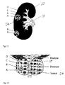
- Fig. 11 shows a further kind of kidney phantom 10 comprising a transition region B that surrounds a second region A that can, for example, be a tumor.
- This transition region B separates the tumor A from the normal tissue C present within the phantom 10.
- This transition zone can comprise a mixture of the respective materials of the first and second regions A and C.
- this transition zone B can also comprise different material properties from those of regions A and C and provision can then for example be made that only this transition zone comprises the material that can be visualized as a successful completion of the surgical exercise could be the complete removal of this transition zone B.
- Fig. 12 shows a further kind of phantom 10, namely a phantom of a prostate.
- a further structure e.g. a tumor, is inserted in a region marked A.
- This structure is surrounded by a region comprising material B and can be embedded in a first region comprising material C.
- Both of the phantoms 10 of Figs. 11 and 12 can also comprise further regions D that can model structures of the specific organ.
- region D corresponds to a collecting system 14 of a kidney
- Fig. 12 region D corresponds to a urethra.
- the target of the medical procedure is to completely remove the region A, maintain the maximum volume of region B and absolutely no removal of region C (according to the kidney tumor removal of Fig. 11 ). In other cases the target could be the removal of both regions A and B while maintained region C.
- the medical procedure can be the removal of tissue for the purpose of a biopsy.
- the aim being the removal of a small amount of only material A.
- the removal of only material A could lead to a positive assessment, whereas the removal of only material C could lead to a negative assessment etc.
- the second structure 26; A, B introduced into the phantom 10 could also replicate diseased tissue and/or an anomalous structure present in the organ and as such may either be partly removed for the purpose of biopsy, i.e. a diagnostic purpose, or completely removed by means of a medical procedure.
- the phantom 10 is provided to allow a medical professional or a robot to practice cutting techniques on a simulated organ rather than on a real organ which are hard to come by and hence expensive. These different cutting techniques can range from simple cutting and suturing practice for medical students to practicing biopsies at regions hard to reach and or tumor removal at sensitive positions. Generally speaking these hard to reach positions cannot be simulated using real organs as these are generally very specific cases of application.
- Fig. 13 shows a view of an endoscopic procedure being conducted on a bladder phantom 10, with an enlarged field of view of the endoscope 24 being shown in the circle.
- the assessment of the medical procedure can then be a measurement of the viewing field scanned by the medical personnel or robot.
- the phantom 10 may further comprise an optical pattern embedded therein, more specifically embedded in the target region 26.
- This optical pattern then conveys information relating to the position and size of the target region.
- This information is either optically invisible to the endoscope 24 or cannot be interpreted by the operator, but can be analyzed using specific filters in software provided for the assessment of the medical procedure.
- a surface area of any scan taken can be made and compared to a surface area that is obtainable by that specific device. In this way one can assess if the medical personnel or robot has detected the complete target region respectively a surface area thereof or not.
- a further assessment parameter is the completeness (surface area coverage) with which for instance an imaging or inspection has been performed.
- completeness surface area coverage
- an imaging or inspection it is important that, during an endoscopic cystoscopic procedure, the entire inside of a bladder is inspected and that no region is missed, as this could for instance mean that the inspection misses a tumor.
- Fig. 14 shows a further view of a kidney phantom 10 subjected to a surgical procedure.
- the ureter is severed at a point of severance 32.
- This point of severance 32 needs to be repaired in order to repair the kidney phantom 10.
- This repair is conducted by suturing the ureter using a suture 34.
- the quality of this suture 34 can then also be assessed as part of the review of the medical procedure.
Priority Applications (5)
| Application Number | Priority Date | Filing Date | Title |
|---|---|---|---|
| EP16172004.0A EP3252738A1 (de) | 2016-05-30 | 2016-05-30 | Verfahren zur beurteilung der leistung eines menschen oder roboters, der einen medizinischen eingriff ausführt, und bewertungswerkzeug |
| CN201780033205.1A CN109196570A (zh) | 2016-05-30 | 2017-05-23 | 评估执行医疗程序的人或机器人的表现的方法和评估工具 |
| PCT/EP2017/062441 WO2017207361A1 (en) | 2016-05-30 | 2017-05-23 | Method of assessing the performance of a human or robot carrying out a medical procedure and assessment tool |
| US16/094,180 US20190130791A1 (en) | 2016-05-30 | 2017-05-23 | Method of assessing the performance of a human or robot carrying out a medical procedure and assessment tool |
| EP17724394.6A EP3424034A1 (de) | 2016-05-30 | 2017-05-23 | Verfahren zur beurteilung der leistung eines menschen oder roboters, der einen medizinischen eingriff ausführt, und bewertungswerkzeug |
Applications Claiming Priority (1)
| Application Number | Priority Date | Filing Date | Title |
|---|---|---|---|
| EP16172004.0A EP3252738A1 (de) | 2016-05-30 | 2016-05-30 | Verfahren zur beurteilung der leistung eines menschen oder roboters, der einen medizinischen eingriff ausführt, und bewertungswerkzeug |
Publications (1)
| Publication Number | Publication Date |
|---|---|
| EP3252738A1 true EP3252738A1 (de) | 2017-12-06 |
Family
ID=56098035
Family Applications (2)
| Application Number | Title | Priority Date | Filing Date |
|---|---|---|---|
| EP16172004.0A Withdrawn EP3252738A1 (de) | 2016-05-30 | 2016-05-30 | Verfahren zur beurteilung der leistung eines menschen oder roboters, der einen medizinischen eingriff ausführt, und bewertungswerkzeug |
| EP17724394.6A Pending EP3424034A1 (de) | 2016-05-30 | 2017-05-23 | Verfahren zur beurteilung der leistung eines menschen oder roboters, der einen medizinischen eingriff ausführt, und bewertungswerkzeug |
Family Applications After (1)
| Application Number | Title | Priority Date | Filing Date |
|---|---|---|---|
| EP17724394.6A Pending EP3424034A1 (de) | 2016-05-30 | 2017-05-23 | Verfahren zur beurteilung der leistung eines menschen oder roboters, der einen medizinischen eingriff ausführt, und bewertungswerkzeug |
Country Status (4)
| Country | Link |
|---|---|
| US (1) | US20190130791A1 (de) |
| EP (2) | EP3252738A1 (de) |
| CN (1) | CN109196570A (de) |
| WO (1) | WO2017207361A1 (de) |
Cited By (1)
| Publication number | Priority date | Publication date | Assignee | Title |
|---|---|---|---|---|
| CN115206146A (zh) * | 2021-04-14 | 2022-10-18 | 北京医智影科技有限公司 | 用于放疗靶区勾画的智能教学方法、系统、设备和介质 |
Families Citing this family (2)
| Publication number | Priority date | Publication date | Assignee | Title |
|---|---|---|---|---|
| WO2020227118A1 (en) * | 2019-05-03 | 2020-11-12 | Arizona Board Of Regents On Behalf Of The University Of Arizona | Systems and methods for an ultrasound-guided percutaneous nephrostomy model |
| CN115578437B (zh) * | 2022-12-01 | 2023-03-14 | 武汉楚精灵医疗科技有限公司 | 肠体病灶深度数据获取方法、装置、电子设备及存储介质 |
Citations (5)
| Publication number | Priority date | Publication date | Assignee | Title |
|---|---|---|---|---|
| WO2008021720A2 (en) * | 2006-08-14 | 2008-02-21 | Artann Laboratories, Inc. | Human tissue phantoms and methods for manufacturing thereof |
| WO2011032840A1 (en) * | 2009-09-18 | 2011-03-24 | Commissariat A L'energie Atomique Et Aux Energies Alternatives | Bimodal organ phantom and associated production method |
| WO2013076056A1 (en) * | 2011-11-21 | 2013-05-30 | INSERM (Institut National de la Santé et de la Recherche Médicale) | Prostate phantom, system for planning a focal therapy of a prostate cancer comprising such prostate phantom and method for planning a focal therapy of a prostate cancer implementing such system |
| US20130157240A1 (en) * | 2011-12-20 | 2013-06-20 | Applied Medical Resources Corporation | Advanced surgical simulation |
| WO2015003271A1 (en) * | 2013-07-11 | 2015-01-15 | Synaptive Medical (Barbados) Inc. | Surgical training and imaging brain phantom |
-
2016
- 2016-05-30 EP EP16172004.0A patent/EP3252738A1/de not_active Withdrawn
-
2017
- 2017-05-23 US US16/094,180 patent/US20190130791A1/en active Pending
- 2017-05-23 WO PCT/EP2017/062441 patent/WO2017207361A1/en active Application Filing
- 2017-05-23 CN CN201780033205.1A patent/CN109196570A/zh active Pending
- 2017-05-23 EP EP17724394.6A patent/EP3424034A1/de active Pending
Patent Citations (5)
| Publication number | Priority date | Publication date | Assignee | Title |
|---|---|---|---|---|
| WO2008021720A2 (en) * | 2006-08-14 | 2008-02-21 | Artann Laboratories, Inc. | Human tissue phantoms and methods for manufacturing thereof |
| WO2011032840A1 (en) * | 2009-09-18 | 2011-03-24 | Commissariat A L'energie Atomique Et Aux Energies Alternatives | Bimodal organ phantom and associated production method |
| WO2013076056A1 (en) * | 2011-11-21 | 2013-05-30 | INSERM (Institut National de la Santé et de la Recherche Médicale) | Prostate phantom, system for planning a focal therapy of a prostate cancer comprising such prostate phantom and method for planning a focal therapy of a prostate cancer implementing such system |
| US20130157240A1 (en) * | 2011-12-20 | 2013-06-20 | Applied Medical Resources Corporation | Advanced surgical simulation |
| WO2015003271A1 (en) * | 2013-07-11 | 2015-01-15 | Synaptive Medical (Barbados) Inc. | Surgical training and imaging brain phantom |
Non-Patent Citations (4)
| Title |
|---|
| ADAMS F ET AL: "358 Experimental 3D-printed kidney model based on medical imaging data of human cadavers for educational and surgery planning purposes", EUROPEAN UROLOGY SUPPLEMENTS, vol. 15, no. 3, March 2016 (2016-03-01), XP029442925, ISSN: 1569-9056, DOI: 10.1016/S1569-9056(16)60360-3 * |
| DE GRAND ET AL: "Tissue-like phantoms for near-infrared fluorencence imaging system assessment and the training of surgeons", SPIE, PO BOX 10 BELLINGHAM WA 98227-0010 USA, February 2006 (2006-02-01), XP040214098 * |
| PACIONI ALESSIA ET AL: "Patient-specific ultrasound liver phantom: materials and fabrication method", INTERNATIONAL JOURNAL OF COMPUTER ASSISTED RADIOLOGY AND SURGERY, SPRINGER, DE, vol. 10, no. 7, 1 October 2014 (2014-10-01), pages 1065 - 1075, XP035506231, ISSN: 1861-6410, [retrieved on 20141001], DOI: 10.1007/S11548-014-1120-Y * |
| PLEIJHUIS R G ET AL: "Near-infrared fluorescence (NIRF) imaging in breast-conserving surgery: Assessing intraoperative techniques in tissue-simulating breast phantoms", EUROPEAN JOURNAL OF SURGICAL ONCOLOGY, LONDON, GB, vol. 37, no. 1, 1 January 2011 (2011-01-01), pages 32 - 39, XP027570341, ISSN: 0748-7983, [retrieved on 20101225] * |
Cited By (2)
| Publication number | Priority date | Publication date | Assignee | Title |
|---|---|---|---|---|
| CN115206146A (zh) * | 2021-04-14 | 2022-10-18 | 北京医智影科技有限公司 | 用于放疗靶区勾画的智能教学方法、系统、设备和介质 |
| CN115206146B (zh) * | 2021-04-14 | 2023-09-22 | 北京医智影科技有限公司 | 用于放疗靶区勾画的智能教学方法、系统、设备和介质 |
Also Published As
| Publication number | Publication date |
|---|---|
| CN109196570A (zh) | 2019-01-11 |
| US20190130791A1 (en) | 2019-05-02 |
| EP3424034A1 (de) | 2019-01-09 |
| WO2017207361A1 (en) | 2017-12-07 |
Similar Documents
| Publication | Publication Date | Title |
|---|---|---|
| US10573201B2 (en) | Method of producing a phantom and phantom | |
| US10083632B2 (en) | Patient specific anatomic kidney phatnom | |
| US9183764B2 (en) | Method for manufacturing three-dimensional molded model and support tool for medical treatment, medical training, research, and education | |
| EP1887543B1 (de) | Katheter-chirurgiesimulation | |
| JP2020522398A (ja) | 所定の解剖学的、生物力学的、および生理学的プロパティを有する合成解剖モデルを構築するためのシステムおよび方法 | |
| CN108352132A (zh) | 超声模拟方法 | |
| KR20170118201A (ko) | 시뮬레이션된 조직 구조체들 및 방법들 | |
| JP2018516718A (ja) | 拡張リアリティの形態学的診断法 | |
| DE112006003722B4 (de) | Simulationssystem für chirurgische Eingriffe in der Human- und Veterinärmedizin | |
| US10350833B1 (en) | Methods and systems for creating anatomical models | |
| US20190130791A1 (en) | Method of assessing the performance of a human or robot carrying out a medical procedure and assessment tool | |
| Choi et al. | A high-fidelity phantom for the simulation and quantitative evaluation of transurethral resection of the prostate | |
| RU2691524C1 (ru) | Симулятор для освоения навыков выполнения операций на почке | |
| Oberoi et al. | 3D printed biomimetic rabbit airway simulation model for nasotracheal intubation training | |
| Li et al. | Soft phantom for the training of renal calculi diagnostics and lithotripsy | |
| CN111063245A (zh) | 一种经皮肺穿刺模型及其制造方法 | |
| CN214671380U (zh) | 手术训练模型及减重代谢手术训练模型 | |
| Cheung et al. | Magnetic resonance imaging properties of multimodality anthropomorphic silicone rubber phantoms for validating surgical robots and image guided therapy systems | |
| Marconi et al. | Quantitative Assessment of 3D Printed Blood Vessels Produced with J750™ Digital Anatomy™ for Suture Simulation | |
| JP7280446B2 (ja) | 3d気管・気管支モデル、及びこれを用いた気道再建トレーニング方法 | |
| Kleszcz et al. | Assessing a new coarctation repair simulator based on real patient’s anatomy | |
| JP2006113440A (ja) | 立体モデル | |
| Shao et al. | Physical patient simulators for surgical training: a review | |
| CN113160676B (zh) | 手术训练模型、减重代谢手术训练模型及训练方法 | |
| Speck et al. | Development and Characterisation of New Hydrogels for Medical Science Education |
Legal Events
| Date | Code | Title | Description |
|---|---|---|---|
| PUAI | Public reference made under article 153(3) epc to a published international application that has entered the european phase |
Free format text: ORIGINAL CODE: 0009012 |
|
| AK | Designated contracting states |
Kind code of ref document: A1 Designated state(s): AL AT BE BG CH CY CZ DE DK EE ES FI FR GB GR HR HU IE IS IT LI LT LU LV MC MK MT NL NO PL PT RO RS SE SI SK SM TR |
|
| AX | Request for extension of the european patent |
Extension state: BA ME |
|
| STAA | Information on the status of an ep patent application or granted ep patent |
Free format text: STATUS: THE APPLICATION IS DEEMED TO BE WITHDRAWN |
|
| 18D | Application deemed to be withdrawn |
Effective date: 20180607 |