EP3242935B1 - O-glycan sialylated recombinant glycoproteins and cell lines for producing the same - Google Patents
O-glycan sialylated recombinant glycoproteins and cell lines for producing the same Download PDFInfo
- Publication number
- EP3242935B1 EP3242935B1 EP15821020.3A EP15821020A EP3242935B1 EP 3242935 B1 EP3242935 B1 EP 3242935B1 EP 15821020 A EP15821020 A EP 15821020A EP 3242935 B1 EP3242935 B1 EP 3242935B1
- Authority
- EP
- European Patent Office
- Prior art keywords
- cells
- st3gal1
- inh
- cell line
- glycans
- Prior art date
- Legal status (The legal status is an assumption and is not a legal conclusion. Google has not performed a legal analysis and makes no representation as to the accuracy of the status listed.)
- Active
Links
Images







Classifications
-
- C—CHEMISTRY; METALLURGY
- C12—BIOCHEMISTRY; BEER; SPIRITS; WINE; VINEGAR; MICROBIOLOGY; ENZYMOLOGY; MUTATION OR GENETIC ENGINEERING
- C12N—MICROORGANISMS OR ENZYMES; COMPOSITIONS THEREOF; PROPAGATING, PRESERVING, OR MAINTAINING MICROORGANISMS; MUTATION OR GENETIC ENGINEERING; CULTURE MEDIA
- C12N9/00—Enzymes; Proenzymes; Compositions thereof; Processes for preparing, activating, inhibiting, separating or purifying enzymes
- C12N9/10—Transferases (2.)
- C12N9/1048—Glycosyltransferases (2.4)
- C12N9/1081—Glycosyltransferases (2.4) transferring other glycosyl groups (2.4.99)
-
- C—CHEMISTRY; METALLURGY
- C12—BIOCHEMISTRY; BEER; SPIRITS; WINE; VINEGAR; MICROBIOLOGY; ENZYMOLOGY; MUTATION OR GENETIC ENGINEERING
- C12P—FERMENTATION OR ENZYME-USING PROCESSES TO SYNTHESISE A DESIRED CHEMICAL COMPOUND OR COMPOSITION OR TO SEPARATE OPTICAL ISOMERS FROM A RACEMIC MIXTURE
- C12P21/00—Preparation of peptides or proteins
- C12P21/005—Glycopeptides, glycoproteins
-
- C—CHEMISTRY; METALLURGY
- C07—ORGANIC CHEMISTRY
- C07K—PEPTIDES
- C07K14/00—Peptides having more than 20 amino acids; Gastrins; Somatostatins; Melanotropins; Derivatives thereof
- C07K14/435—Peptides having more than 20 amino acids; Gastrins; Somatostatins; Melanotropins; Derivatives thereof from animals; from humans
- C07K14/475—Growth factors; Growth regulators
- C07K14/4753—Hepatocyte growth factor; Scatter factor; Tumor cytotoxic factor II
-
- C—CHEMISTRY; METALLURGY
- C07—ORGANIC CHEMISTRY
- C07K—PEPTIDES
- C07K14/00—Peptides having more than 20 amino acids; Gastrins; Somatostatins; Melanotropins; Derivatives thereof
- C07K14/81—Protease inhibitors
- C07K14/8107—Endopeptidase (E.C. 3.4.21-99) inhibitors
- C07K14/811—Serine protease (E.C. 3.4.21) inhibitors
- C07K14/8121—Serpins
-
- C—CHEMISTRY; METALLURGY
- C12—BIOCHEMISTRY; BEER; SPIRITS; WINE; VINEGAR; MICROBIOLOGY; ENZYMOLOGY; MUTATION OR GENETIC ENGINEERING
- C12Y—ENZYMES
- C12Y204/00—Glycosyltransferases (2.4)
- C12Y204/99—Glycosyltransferases (2.4) transferring other glycosyl groups (2.4.99)
- C12Y204/99001—Beta-galactoside alpha-2,6-sialyltransferase (2.4.99.1)
-
- C—CHEMISTRY; METALLURGY
- C12—BIOCHEMISTRY; BEER; SPIRITS; WINE; VINEGAR; MICROBIOLOGY; ENZYMOLOGY; MUTATION OR GENETIC ENGINEERING
- C12Y—ENZYMES
- C12Y204/00—Glycosyltransferases (2.4)
- C12Y204/99—Glycosyltransferases (2.4) transferring other glycosyl groups (2.4.99)
- C12Y204/99004—Beta-galactoside alpha-2,3-sialyltransferase (2.4.99.4)
Definitions
- the present invention relates to cell lines that are genetically modified to overexpress a ⁇ -galactoside ⁇ -2,3-sialyltransferase 1 (ST3Gal1), which are further genetically modified to overexpress a ⁇ -galactoside ⁇ -2,3-sialyltransferase 4 (ST3Gal4), which can be used for the production of recombinant glycoproteins having highly or fully sialylated O-linked GalNAc glycans (GaINAc O-glycans), preferably core 1 GalNAc O-glycans, as well as to respective recombinant glycoproteins. Further, the present invention relates to respective methods of expressing recombinant glycoproteins.
- ST3Gal1 ⁇ -galactoside ⁇ -2,3-sialyltransferase 1
- ST3Gal4 ⁇ -galactoside ⁇ -2,3-sialyltransferase 4
- therapeutic proteins Due to their clinical importance, the development of therapeutic proteins has accelerated dramatically over the past years. However, the development of therapeutic proteins is often held up, in particular for complex glycosylated proteins, due to the difficulties in obtaining proteins with favorable glycosylation patterns. This often leads to suboptimal pharmacological properties such as e.g. reduced serum half-life or increased immunogenicity.
- Another drawback is heterogeneity of the therapeutic proteins due to variations in the post-translational modification, in particular of the glycostructures. This can lead to inconsistencies in product quality between different production batches. Therefore, in order to advance the development of therapeutic proteins, it is important to achieve homogenous post-translational modification.
- glycosylation is the most common post-translational modification. Nearly all biopharmaceuticals need to be correctly glycosylated in order to display the optimal therapeutic efficacy.
- glycosylation refers to the covalent attachment of sugars to the protein surface, wherein the sugars are either connected to asparagine residues resulting in N-linked glycans (N-glycans) or serine or threonine residues resulting in O-linked glycans (O-glycans).
- O-linked glycans The most common group of O-linked glycans are GalNAc O-glycans in which the serine or threonine is linked to N-acetylgalactosamine (GaINAc) which in turn is linked to additional monosaccharides.
- GaINAc N-acetylgalactosamine
- the terms "O-linked glycan” or "O-glycan” always relate to GaINAc O-glycans.
- the glycosylation pattern can be very diverse from molecule to molecule as the attached forms can be different in monosaccharide order, branching pattern, and length (micro-heterogeneity). Additionally, not all glycosylation sites are fully occupied (macro-heterogeneity).
- Glycosylation influences the solubility of proteins, their resistance to proteolysis, and their binding behavior to other proteins or to protein receptors such as e.g. the ASGPR (asialoglycoprotein receptor) and therefore influences the half-life of the glycoprotein in the plasma.
- ASGPR asialoglycoprotein receptor
- clearance occurs mainly via renal clearance. Beside the size of a protein, also the protein surface charge has influence on the renal clearance, as the filtration of highly charged proteins in the kidney is decreased.
- ASGPR ASGPR
- glycoproteins with terminal sialic acids covering the adjacent carbohydrate, galactose Due to the action of this receptor, glycoproteins with terminal sialic acids covering the adjacent carbohydrate, galactose, have up to 100-fold increase in half-life as compared to their non-sialylated counterparts with terminal galactose residues on their N-linked glycans.
- Other receptors bind specifically to mannose, N-acetyl-glucosamine, or fucose and clear glycoproteins with these terminal sugars from the system.
- a native N-glycosylation pattern including a high degree of terminal sialylation is crucial for therapeutic proteins since it determines the pharmacokinetic properties of the therapeutic protein.
- terminal sialic acids with the proper linkage on glycoproteins reduce the immunogenicity of the glycoproteins.
- CHO cells Choinese hamster ovary cells
- CHO cells lack the enzyme needed to catalyze 2,6-linkage of sialic acids, and therefore they can only catalyze the 2,3-linkage.
- CHO cells lack the enzyme needed to catalyze 2,6-linkage of sialic acids, and therefore they can only catalyze the 2,3-linkage.
- N-acetylneuraminic acid sialic acid; NeuAc
- they also link N-glycolylneuraminic acid (NeuGc) to glycans, a sugar not synthesized in humans and therefore immunogenic when injected into humans.
- CAP cells derived from human amniocytes
- HEK293 cells derived from human embryo kidney cells
- O-linked GalNAc glycans always have an ⁇ -linked N-acetylgalactosamine residue linked to serine or threonine.
- the GalNAc can be extended with residues like galactose, GlcNAc, fucose, or sialic acid.
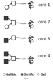
- O-linked GalNAc glycosylation four principal core structures can be distinguished, core 1 (GalGalNAc), core 2 (GalGlcNAcGalNAc), core 3 (GlcNAcGalNAc), and core 4 (GlcNAc2GalNAc) ( Fig. 1 ).
- the terminal ends are often further modified, e.g. with phosphates, sulfates, carboxylic acids, or sialic acids.
- O-linked GalNAc glycans play a role in maintaining structures of fully folded proteins, conferring protease stability.
- Figure 2 shows the biosynthesis of core 1 and core 2 GalNAc O-glycans.
- IGFBP6 Human insulin-like growth factor binding protein-6
- BR3-Fc B cell-activating factor receptor 3
- BR3-Fc has multiple O-linked glycosylation sites and sialylation levels vary in the manufacturing process. Separating the different forms, it could be shown that exposed galactose on the desialylated O-linked glycans of BR3-Fc was associated with rapid clearance due to uptake and degradation in the liver, in particular by non-parenchymal cell mediated clearance.
- Adiponectin an adipocyte-secreted, insulin-sensitizing hormone, has three putative O-linked glycosylation sites, which are not necessary for multimer formation but plasma clearance of the desialylated protein was accelerated compared with that of sialylated protein. So far, no methods have been described that will affect the sialylation or the structure of O-linked GalNAc glycans during recombinant expression of glycoproteins.
- Vallejo-Ruiz et al. ( Vallejo-Ruiz, A. V. et al.; Biochimica et Biophysica Acta, 1549(2); October 2001; pages 161-173 ) describes the delineation of the minimal catalytic domain of human Galbeta1-3GalNAc alpha2,3-sialyltransferase. Further, Dalziel et al. ( Dalziel, M.
- CN 101 613 678 A describes an MDCK-ST3GAL-1 cell line, concretely an MDCK cell line stably expressing beta-galactoside alpha-2,3-sialytransferase 1 (ST3Gal 1), methods of making the same, and uses thereof.
- ST3Gal 1 beta-galactoside alpha-2,3-sialytransferase 1
- US 2012/322738 A1 describes conjugated FVIII variants comprising different polymeric groups as well as use thereof.
- Shang et al. Shang, J. et al.; European Journal of Biochemistry, 265(2); October 1999; pages 580-588 ) describes the molecular cloning and expression of Galbeta1,3GalNAc alpha2,3-sialyltransferase from human fetal liver.
- the technical problem underlying the present invention is to provide recombinant glycoproteins having highly or fully sialylated GalNAc O-glycans, as well as cell lines that are capable of recombinantly producing such proteins. Further, respective methods of expressing recombinant glycoproteins should be provided.
- the present invention relates to the following items:
- an animal cell line preferably an insect, avian, or mammalian cell line, more preferably a mammalian, in particular human, cell line that is genetically modified to overexpress a ⁇ -galactoside ⁇ -2,3-sialyltransferase 1 (ST3Gal1), which is further modified to overexpress a ⁇ -galactoside ⁇ -2,3-sialyltransferase 4 (ST3Gal4).
- ST3Gal1 ⁇ -galactoside ⁇ -2,3-sialyltransferase 1
- ST3Gal4 ⁇ -galactoside ⁇ -2,3-sialyltransferase 4
- cell line genetically modified to overexpress a ST3Gal1 indicates that upon genetic modification, the individual cells of the cell line display a higher activity of the protein, e.g. the sialyltransferase, than they did before the genetic modification.
- the cell line comprises an endogenous gene encoding a ST3Gal 1, such as e.g. human cell lines.
- the cells can be genetically modified by inserting a promoter, enhancing element, and/or stabilizing element into the genome of the cells in a position suitable to cause overexpression of said nucleic acid. This can be done by homologous recombination using TALENS, Zn-finger proteins, CRISPR-CAS9, or other methods known in the art.
- the cell line can comprise an endogenous gene encoding ST3Gal1 and optionally endogenous genes encoding ST3Gal4 and/or ST6Gal1, and further has at least one genetic element, selected from the group consisting of a promoter, an enhancing element, and a stabilizing element inserted into the genome in one or more position(s) suitable to cause overexpression of ST3Gal1 and optionally ST3Gal4 and/or ST6Gal1.
- Suitable promoters, enhancing elements and stabilizing elements are not particularly limited and are known in the art.
- promoters include constitutive promoters, e.g.
- enhancing elements include CMV enhancer, ⁇ -globin enhancer, immunoglobulin enhancer, and PGK-enhancer.
- stabilizing elements include matrix attachment regions (MARS), locus control regions (LCRs), and ubiquitously acting chromatin opening elements (UCOEs).
- nucleic acid encoding a ST3Gal1 into the cells.
- Methods for introducing nucleic acids into cells are not particularly limited and are known in the art.
- said nucleic acids could be introduced in circular or linearized form into the cells by electroporation, nucleofection, microinjection, via viral vectors, e.g. lentiviral vectors, reagent based methods, e.g. lipids, calcium phosphate, cationic polymers or other methods known in the art.
- the nucleic acids can be transiently or stably introduced into the cell by episomal systems or by stable integration of the nucleic acid into the genome.
- Said nucleic acids can be present in the cells in the form of one or more expression vector(s), e.g. pcDNA, pCEP, pLenti, pEntr, pDest, pEF, pEAK, pCMV, pStbl, or other expression vectors known in the art.
- Expression of the ST3Gal1 can be under the control of a constitutive promoter, e.g.
- nucleic acids encoding the ST3Gal1 can be present as one continuous nucleic acid, or can be present as separate nucleic acids, e.g. as separate expression vectors. Said nucleic acids can contain, in addition to the coding region and a promoter, suitable restriction sites, Kozak sequences, ribosomal binding sites, chromatin modulating elements, selection cassettes, episomal replication systems, e.g.
- the cell line can comprise an exogenous nucleic acid encoding a ⁇ -galactoside ⁇ -2,3-sialyltransferase 1 (ST3GaI1), and optionally exogenous nucleic acids encoding a ⁇ -galactoside ⁇ -2,3-sialyltransferase 4 (ST3Gal4) and/or a ⁇ -galactoside ⁇ -2,6-sialyltransferase 1 (ST6GaI1).
- ST3GaI1 ⁇ -galactoside ⁇ -2,3-sialyltransferase 1
- ST3Gal4 optionally exogenous nucleic acids encoding a ⁇ -galactoside ⁇ -2,3-sialyltransferase 4
- ST6GaI1 ⁇ -galactoside ⁇ -2,6-sialyltransferase 1
- Suitable genes encoding a ST3Gal1 for transfection of cell lines are not particularly limited and include any genes from any origin that encode a protein having ST3Gal1 activity, i.e. a protein that catalyzes the linkage of sialic acid to a Gal-beta-1,3-GalNAc structure linked to Thr or Ser.
- such genes are selected from ST3Gal1 genes encoding a ST3Gal1 protein comprising in its amino acid sequence, in particular in motif 3 of the C-terminal acceptor binding site, the conserved consensus sequence (H/C/R)(Y/H/F)(W/Y/F)(E/D/H/Y), wherein the sequence HYWE (SEQ ID NO: 5) is particularly preferred.
- Figure 14 shows an alignment of the amino acids sequences of ST3Gal1 of various species, covering amino acids 191 to 340 of human ST3Gal1 comprising the catalytic domain, and displays said consensus sequence.
- the gene is selected from ST3Gal1 genes encoding a ST3Gal1 protein comprising an amino acid sequence which is at least 50%, preferably 75% and more preferably 90% % identical to amino acids 263 to 321 of the human ST3Gal1.
- the gene is selected from the group consisting of ST3Gal1 genes derived from a species, selected from the group consisting of Homo sapiens (SEQ ID NO: 1), Pan troglodytes (SEQ ID NO: 6), Macaca mulatta (SEQ ID NO: 7), Sus scrota (SEQ ID NO: 8), Rattus norvegicus (SEQ ID NO: 9), Mus musculus (SEQ ID NO: 10), Nannospalax galili (SEQ ID NO: 11), Monodelphis domestica (SEQ ID NO: 12), Oryctolagus cuniculus (SEQ ID NO: 13), Cricetulus griseus (SEQ ID NO: 14), Canis familiaris (SEQ ID NO: 15), Felis catus (SEQ ID NO: 16), Equus caballus (SEQ ID NO: 17), Gallus gallus (SEQ ID NO: 18), Columba livia (SEQ ID NO: 19), Alligator sinensis (SEQ ID
- the cell line can further be genetically modified to overexpress a ⁇ -galactoside ⁇ -2,3-sialyltransferase 4 (ST3Gal4), preferably human ST3Gal4, and/or is further genetically modified to overexpress ⁇ -galactoside ⁇ -2,6-sialyltransferase 1 (ST6GaI1), preferably human ST6Gal1.
- ST3Gal4 and suitable genes encoding ST6Gal1 are not particularly limited and include any genes from any origin that encode a protein having the respective activity. Again, mammalian, in particular human genes are particularly preferred.
- the cell lines described herein can be derived from cell lines, e.g. mammalian cell lines, known in the art.
- a cell line described herein can be derived from Muscovy Duck cells (AGE.CR ® ) African green monkey kidney epithelial cells (Vero), Madin Darby canine kidney cells (MDCK), baby hamster kidney cells (BHK), Chinese hamster ovary (CHO) cells, human hepatocarcinoma cell lines (HepG2, Huh7), human embryonic kidney 293 (HEK293) cells, human neuronal precursor cells (AGE1.HN ® and NC5T11), human embryonic retinoblasts (Per.C6), myeloma cell lines (HMCLs, MM.1, U266, RPMI8226), CML tumor cell lines (NM, NM-F9,), hybrid HEK293 and lymphoma cell (HKB11), or human amniocytes (CAP; cf. EP 1 230 354 B1 ), where
- CAP cells are permanent amniocytic cell lines comprising a nucleic acid encoding the gene products of the adenovirus, in particular adenovirus type 5 (Ad5), E1A and E1B regions.
- CAP cells are derived from primary human amniocytes that are transformed with a nucleic acid encoding Ad5 E1A and E1B.
- the cell lines described herein can be derived from human primary amniocytes comprising at least one nucleic acid encoding the gene products of the adenoviral E1 and pIX region, preferably E1 and pIX region of adenovirus type 5 (Ad5) from nt. 505 to 4079, in which E1A is under the control of the murine phosphoglycerate kinase (pgk) promoter, while E1B and pIX expression is controlled from their natural promoters.
- the E1B downstream intron, splice acceptor and polyA signal are replaced by corresponding motifs from SV40.
- a recombinant glycoprotein having GaINAc O-glycans that are sialylated to a degree of at least 80%.
- the term "recombinant glycoprotein” indicates that the respective glycoproteins are biotechnologically produced in genetically modified organisms or cells.
- the glycoproteins described herein have GalNac O-glycans that are sialylated to a degree of at least 80%, preferably at least 82%, more preferably at least 84%, at least 86%, at least 88%, at least 90%, at least 91%, at least 92%, at least 93%, at least 94%, at least 95%, at least 95.5%, at least 96%, at least 96.5%, at least 97%, at least 97.5%, at least 98%, at least 98.5%,or at least 99%.
- the glycoproteins described herein have GalNac O-glycans that are sialylated to a degree of 95% or above.
- O-glycans that are sialylated to a degree of at least X% indicates that X% of all terminal GalNac O-glycan monosaccharide moieties in a given glycoprotein preparation are sialic acid (N-acetylneuraminic acid; NeuAc).
- At least 80%, preferably at least 82%, more preferably at least 84%, at least 86%, at least 88%, at least 90%, at least 91%, at least 92%, at least 93%, at least 94%, at least 95%, at least 95.5%, at least 96%, at least 96.5%, at least 97%, at least 97.5%, or at least 98% GalNac O-glycans of the glycoproteins described herein are core 1 GalNAc O-glycan, i.e.
- GalNac O-glycans having a core structure of GP-Ser/Thr-O-GalNAc-Gal- wherein GP is the glycoprotein, Ser/Thr-O is a serine or threonine amino acid side chain of the glycoprotein, GalNAc is N-acetylgalactosamine, and Gal is galactose ( Fig. 1 ).
- the glycoproteins described herein have core 1 GalNAc O-glycans that are disialylated to a degree of at least 25%, preferably at least 30%, more preferably at least 35%, at least 37.5%, at least 40%, at least 42.5%, or at least 45%.
- core 1 GalNAc O-glycans that are disialylated to a degree of at least X% indicates that X% of all core 1 GalNAc O-glycans have two terminal sialic acid moieties.
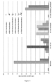
- Figure 11 shows the relative amounts of the six main O-glycan species in various preparations of the glycoprotein C1 esterase inhibitor (C1 Inh).
- Respective recombinant glycoproteins can be produced as described herein, e.g. by overexpression of a ⁇ -galactoside ⁇ -2,3-sialyltransferase 1 (ST3GaI1) together with the recombinant glycoprotein, and by additional overexpression of a ⁇ -galactoside ⁇ -2,3-sialyltransferase 4 (ST3Gal4) and optionally a ⁇ -galactoside ⁇ -2,6-sialyltransferase 1 (ST6GaI1).
- ST3GaI1 ⁇ -galactoside ⁇ -2,3-sialyltransferase 1
- ST3Gal4 ⁇ -galactoside ⁇ -2,3-sialyltransferase 4
- ST6GaI1 ⁇ -galactoside ⁇ -2,6-sialyltransferase 1
- said glycoproteins are produced in a cell line as
- the GalNAc O-glycans of the recombinant glycoproteins described herein can be characterized by a reduced micro-heterogeneity, i.e ., a reduced diversity of GalNAc O-glycan structure and composition within a given glycoprotein preparation.
- Respective recombinant glycoproteins can be produced as described herein, e.g. by overexpression of ST3Gal1 together with the recombinant glycoprotein, and by additional overexpression of ST3Gal4 and optionally ST6Gal1.
- said glycoproteins are produced in a cell line as described herein.
- glycoproteins described herein are not particularly limited, provided that said glycoproteins have GalNAc O-glycans and fulfill the respective requirements of GalNAc O-glycan sialylation.
- said glycoproteins are mammalian, more preferably human glycoproteins.
- Glycoproteins can be selected from the group consisting of growth factors, peptide hormones, cytokines, enzymes, antibodies, antibody fragments, blood clotting factors, and protease inhibitors.
- the glycoprotein is selected from the group consisting of hepatocyte growth factor (HGF), erythropoietin (EPO), Factor VIII (FVIII), Factor IX (FIX), von-Willebrand-Factor (vWF), and C1 esterase inhibitor (C1-inhibitor; C1 Inh), wherein C1 Inh is particularly preferred.
- HGF hepatocyte growth factor
- EPO erythropoietin
- FVIII Factor VIII
- FIX Factor IX
- vWF von-Willebrand-Factor
- C1 esterase inhibitor C1-inhibitor; C1 Inh
- C1 esterase inhibitor C1-inhibitor; C1 Inh
- C1 Inh a recombinant C1 esterase inhibitor
- human C1 Inh having GalNAc O-glycans that are sialylated to a degree of at least 80%.
- All of the above preferred and/or specific embodiments described for recombinant glycoproteins in general also apply to this specific embodiment of recombinant (human) C1 Inh.
- a method for the expression of recombinant glycoproteins having GalNAc O-glycans that are sialylated to a degree of at least 80% to a method for increasing the degree of sialylation of GalNAc O-glycans of recombinant glycoproteins, and to a method for decreasing the micro-heterogeneity of GalNAc O-glycans of recombinant glycoproteins, all of said methods comprising the step of overexpressing with the recombinant glycoproteins a ⁇ -galactoside ⁇ -2,3-sialyltransferase 1 (ST3Gal1).
- Said methods can comprise the steps of:
- the recombinant glycoproteins produced in said methods can be any glycoproteins described herein as defined above.
- the cell lines provided in step (a) of the methods described herein can be any cell line as defined above.
- step (b) of expressing the glycoprotein of interest in said cell line encompasses the transfection of a respective coding nucleic acid into said cell line prior to the actual expression of the glycoprotein.
- means for isolating a glycoprotein of interest from a cell culture are not particularly limited and are known in the art.
- described herein is a use of a cell line described herein for the production of recombinant glycoproteins having GalNAc O-glycans that are sialylated to a degree of at least 80%, and/or for increasing the degree of sialylation of recombinant glycoproteins and/or for decreasing the micro-heterogeneity of GalNAc O-glycans of recombinant glycoproteins.
- the recombinant glycoproteins produced in said use can be any glycoproteins as defined above.
- the used cell lines can be any cell lines as defined above.
- the permanent human amniocyte cell line CAP 1D5 was cultured in suspension, either in chemically defined, animal component free CAP-CDM medium (CEVEC Pharmaceuticals, Germany) supplemented with 6 mM stable glutamine (biochrom, Germany), or in serum free PEM media (Life Technologies) supplemented with 4 mM stable glutamine (biochrom, Germany).
- the 293F cells from Life Technologies were cultured in suspension in Freestyle 293 expression media (Life Technologies), supplemented with 4 mM stable glutamine (biochrom, Germany).
- the adherent CHO-K1 cells (ATCC, CCL-61) were cultured in F12-K media (Life Technologies), supplemented with 10 % FBS, and 2 mM stable glutamine (biochrom, Germany).
- the adherent MDCK.1 cells (ATCC, CRL-2935) were cultured either in EMEM media (ATCC) or in DMEM F-12 (ATCC) supplemented with 10 % FBS and with 2 mM stable glutamine (biochrom, Germany).
- CAP cells and 293F cells were cultivated at 37 °C in shaker flasks (Corning, 125 mL (25 mL wv) or 3000 mL (1000 mL wv)) at 5% CO 2 , and 185 rpm.
- CAP cells were fed at d3, d5, and d7 with 10 % CAP-CDM feed solution (CEVEC Pharmaceuticals, Germany) and 4 mM stable glutamine (biochrom, Germany).
- Adherent CHO-K1 and MDCK.1 cells were cultured at 37 °C, 5% CO 2 , in 6 cm or 10 cm cell culture dishes (TPP) or 225cm 2 cell culture dishes (BD).
- cells were sequentially nucleofected with the nucleic acid constructs encoding the glyco structure modifying enzymes ST3Gal1 and/or ST3Gal4, as well as the specific recombinant protein. Only stable cell lines were utilized. Table 1 lists all cell lines created. Table 1: Stable cell lines used herein. Cell line rec.
- sequence information of the precursor protein and mature protein was based of the database entry UniProt Q11201 (SEQ ID NO: 1).
- a ClaI restriction site and a Kozak sequence were added 5' of the start codon of the human ST3Gal1 cDNA and an EcoRV restriction site was added 3' of the stop codon to be inserted between the ClaI and EcoRV restriction sites in the pStbl-Puro-CMV-MCS(-) vector resulting in the expression plasmid pStbl-Puro-CMV-ST3Gal1.
- This vector contains a CMV promoter driving the expression of the gene of interest, followed by an SV40 intron for improved, splicing-mediated mRNA transport and a multiple cloning site for the insertion of the gene of interest.
- the selection marker (Puromycin) is driven by the human ubiquitin (UbC) promoter.
- cDNA synthesis was performed at GeneArt (Germany, Life Technologies).
- sequence information of the precursor protein and mature protein was based of the database entry UniProt Q11206 (SEQ ID NO: 2).
- a ClaI restriction site and a Kozak sequence were added 5' of the start codon of the human ST3Gal4 cDNA and a EcoRV restriction site was added 3' of the stop codon to be inserted between the ClaI and EcoRV restriction sites in the pStbl-Puro-CMV-MCS(-) vector resulting in the expression plasmid pStbl-Puro-CMV-ST3Gal4.
- cDNA synthesis was performed at GeneArt (Germany, Life Technologies).
- Nucleofection was performed using a Nucleofector (LONZA) with the appropriate Nucleofector Kit (KitV; CAP cells, 293F, and CHO or the KitT; MDCK.1 cells) according to the manufacturer's protocol. Briefly, during exponential growth phase of the culture 1 ⁇ 10 7 cells were harvested via centrifugation (150 g for 5 min) and resuspended in 100 ⁇ l complete nucleofector solution and mixed with a total of 5 ⁇ g plasmid. Nucleofection was performed using the X001 program (CAP and 293F cells), the U024 program (CHO-K1), or the P029 program (MDCK.1). After the pulse, cells were recovered in complete cell culture media. The cells were cultured as before.
- 72 to 96 h post-nucleofection cells were selected with 5 ⁇ g/ml blasticidin (therapeutic protein) and/or 200 ⁇ g/ml neomycin (pStbl-neo-CMV-ST3Gal4) and/or 2 ⁇ g/ml puromycin (pStbl-Puro-CMV-ST3Gal1), in order to generate stable pools.
- the methylated glycan alditols were applied to the stainless steel target by mixing 1:1 with matrix ( ⁇ -cyano-4-hydroxycinnamic acid in 50 % acetonitrile/0.1 % TFA). Analyses were performed by positive ion detection in MS1 and MS2 (Post-Source-Decay-Modus). Identification of the different glycan species were based i) on the molecule ions, which give information about the monosaccharide composition and ii) the fragmentation in the MS2 analysis (B, C, Y, and Z ions).
- PNGaseF is an amidase that cleaves between the innermost GlcNAc and asparagine residues of N-linked glycoproteins. Therefore, after treatment of C1 Inh proteins with PNGaseF the protein backbone and the O-linked glycans remain. As the protein backbone between the plasma derived human C1 Inh and C1 Inh expressed in CAP cells is equal, the PNGaseF treatment carries indirectly information about the structure of the O-glycans.
- Lectins are proteins that bind specific carbohydrate structures. Biotin-coupled lectins can be therefore used to analyze N-linked and O-linked glycans.
- Erythrina crista galli (ECL) lectin detects ⁇ 1-4 linked terminal galactose on N-linked glycans
- peanut agglutinin (PNA) detects ⁇ 1-3 linked terminal galactose on O-linked glycans
- Sambucus nigra agglutinin (SNA) preferentially binds to a 2,6-linked sialic acid
- MAL Maackia amurensis lectin
- Purified protein or cell culture supernatants with or without co-expression of ST3Gal1 and/or ST3Gal4 were separated as described above and blotted onto Hybond ECL nitrocellulose membrane (GE healthcare).
- the membrane was stained with streptavidin-coupled horseradish peroxidase for 1 h at RT (1:1000 diluted in PBSTB).
- the HRP signal was amplified using anti-streptavidin IgG and anti IgG-HRP.
- the proteins were detected using the Pierce ECL WB Substrate Kit via a chemiluminescence detector (INTAS).
- IEF isoelectric focusing
- pl isoelectric point
- IEF analysis was done according to the manufacturers protocol (Invitrogen). Briefly, 5 ⁇ g of purified protein were loaded on pH3-7 gels and subjected to electrophoresis (1h 100 V, 1 h 200 V, 30 min 500 V). Proteins were stained with SimplyBlue SafeStain according to the manufacturers protocol (Invitrogen).
- the protease inhibitor C1 esterase inhibitor (C1 Inh) belongs to the serpin superfamily. Its main function is the inhibition of the complement system to prevent spontaneous activation.
- the 500 aa protein is highly glycosylated with 7 predicted N-glycans and 8 predicted O-linked glycans.
- CAP 1D5 previously stably transfected to express human recombinant C1 Inhibitor (rhC1 Inh, SEQ ID NO: 3) (CAP-C1 Inh) were nucleofected with a vector encoding for ST3Gal4 linearized with Scal in order to facilitated stable integration of the construct into the genome.
- the vector contains a drug expression cassette, for selection of stable integration of the linearized construct into the genome.
- the obtained stable CAP-C1 Inh-ST3Gal4 pool was subject to single cell cloning via limiting dilution. Selected clones were analyzed to proof expression of ST3Gal4.
- CAP single cell clones expressing ST3Gal4 were then further nucleofected with the gene encoding for ST3Gal1 gene.
- Cells were selected with antibiotics to obtain a pool of cells stably co-expressing rhC1 Inh, human ST3Gal4 and ST3Gal1 (CAP-C1 Inh-ST3Gal1/4).
- C1 Inh In order to generate sufficient amounts of recombinant C1 Inh, the generated CAP-C1 Inh cell lines overexpressing human C1 Inh were cultured as described in experimental procedures. Subsequently, C1 Inh was purified from the cell culture supernatant of the following cell lines: CAP-C1 Inh, CAP-C1 lnh-ST3Gal4, and CAP-C1 Inh-ST3Gal1/4 (pool A and B), as described below.
- ECL lectin immunoblot was performed.
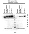
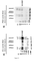
- Erythrina crista galli (ECL) lectin detects ⁇ 1-4 linked terminal galactose on N-linked glycans. Therefore, a diminished signal in the ECL blot means an increased amount of sialylation.
- the N-linked glycans of Berinert are almost completely sialylated.
- N-Glycans of the C1 Inh from CAP cells overexpressing ST3Gal4 are equally sialylated
- C1 Inh from CAP cells overexpressing ST3Gal1/4 display a slightly higher amount of asialo N-glycans.
- C1 Inh purified from CAP cells without overexpression of sialyltransferase give a strong signal in the ECL blot indicating a large amount of asialo N-glycans.
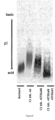
- results from the lectin blots could be confirmed by isoelectric focusing (IEF).
- IEF isoelectric focusing
- the backbones of the different C1 Inh are identical, changes in the IEF are most likely due to changes in the sialic acid content.
- the resulting modified C1 Inh shifts very slightly towards the anode, indicating a small increase in the total amount of sialic acids per molecule.
- the additional expression of ST3Gal1 results in a pronounced shift of C1 Inh towards the anode, indicating a significant increase in the total amount of sialic acids.
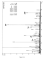
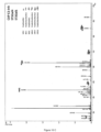
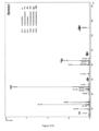
- CAP C1 Inh samples without additional overexpression of ST3Gal1 or ST3Gal4, namely CAP C1 Inh 75 % of the detectable O-glycans are core 2 structure with only 35 % sialylated O-glycans without terminal galactose residues(Gal1-3(Gal1-GlcNAc1-6)GalNAc-ol_Core 2) (m/z 983), or NeuAc2-3Gal1-3(Gal1-4GlcNAc1-6)GalNAc-ol (m/z 1344), Gal1-3(NeuAc2-3Gal1-4GlcNAc1-6)GaINAc-ol (m/z 1344) and NeuAc2-3Gal1-3(NeuAc2-3Gal1-4GlcNAc1-6)GaINAc-ol (m/z 1706)).
- Core 1 structures are only rarely detectable. Additional expression of ST3Gal4 leads to a shift towards core 1 structures (68.7%)(NeuAc2-3Gal1-3GalNAc-ol_Core 1). Interestingly, the expression of C1 Inh in CAP cells coupled with the overexpression of ST3Gal1 leads to the exclusive expression of core 1 O-glycans (99.1%) which are almost entirely sialylated (98.5%) without any terminal galactose residues, in particular (NeuAc2-3Gal1-3GalNAc-ol respectively Gal1-3(NeuAc2-6)GalNAc-ol (m/z 895) and NeuAc2-3Gal1-3(NeuAc2-6)GalNAc-ol (m/z 1256).
- Hepatocyte growth factor is a potent mitogen for mature parenchymal hepatocyte cells, seems to be a hepatotrophic factor, and acts as a growth factor for a broad spectrum of tissues and cell types.
- the 728 aa sized protein contains 4 predicted N-glycans and 1 predicted O-linked glycan.
- CAP 1D5 cells stably expressing human recombinant hepatocyte growth factor (SEQ ID NO: 4) were nucleofected either with a vector encoding for ST3Gal4, ST3Gal1, or ST3Gal4 and ST3Gal1 linearized with Scal in order to facilitate stable integration of the construct into the genome.
- the vectors contain a drug expression cassette, which facilitates selection for cells with a stable integration of the linearized construct into the genome.
- CAP-HGF, CAP-HGF-ST3Gal1, CAP-HGF-ST3Gal4, and CAP-HGF-ST3Gal1/4 were cultured as described in experimental procedures. Cell culture supernatants containing recombinant human HGF were examined by ECL and PNA lectin blots to determine the existing N- and O-linked glycostructures.
- Erythrina crista galli (ECL) lectin detects ⁇ 1-4 linked terminal galactose on N-linked glycans. Therefore a diminished signal in the ECL blot means an increased amount of sialylation.
- ECL Erythrina crista galli
- 293F cells (Life Technologies, R-970-07), immortalized human embryonic kidney cells, were nucleofected either with a vector encoding for ST3Gal1, or ST3Gal1 and ST3Gal4 linearized with Scal in order to facilitate stable integration of the construct into the genome.
- the vectors contain a drug expression cassette, which facilitates selection for cells with a stable integration of the linearized construct into the genome.
- 293F-ST3Gal1, and 293F-ST3Gal1/4 and wildtype 293F cells were further nucleofected with the gene encoding for hC1 Inh.
- C1 Inh was purified from C1 Inh containing cell culture supernatant as described in the method section and examined by ECL and PNA lectin blots to determine the existing N- and O-linked glycostructures.
- Erythrina crista galli (ECL) lectin detects ⁇ 1-4 linked terminal galactose on N-linked glycans. Therefore a diminished signal in the ECL blot means an increased amount of sialylation.
- ECL Erythrina crista galli
- CHO-K1 (ATCC, CCL-61) cell line was derived as a subclone from the parental CHO cell line initiated from a biopsy of an ovary of an adult Chinese hamster.
- CHO-K1 cells were nucleofected with a vector encoding for ST3Gal1 linearized with Scal in order to facilitate stable integration of the construct into the genome.
- the vector contains a drug expression cassette, which facilitates selection for cells with a stable integration of the linearized construct into the genome.
- After pool generation the obtained stable CHO-K1 cell pool, CHO-ST3Gal1, and wildtype CHO-K1 cells were further nucleofected with the gene encoding for hC1 Inh. Cells were selected with antibiotics to obtain pools of cells stably expressing i) rhC1 Inh, ii) rhC1 Inh and human ST3Gal1.
- C1 Inh was purified as described in the method section.
- Canine MDCK.1 cells (ATCC, CRL-2935) were stably transfected.
- the resulting stable MDCK.1 cell pool, MDCK.1 -ST3Gal1, and wildtype MDCK.1 cells were further stably transfected to obtain pools of cells stably expressing i) rhC1 Inh, ii) rhC1 Inh and human ST3Gal1.
- MDCK.1 cells were expanded as described in the method section.
- For the production of cell culture supernatant containing human C1 Inh cells were seeded onto 225 cm 2 cell culture dishes, 2 d post seeding cells were extensively washed with 1 ⁇ PBS in order to remove fetal bovine serum, subsequently adding fresh serum free media.
- Five days later cell culture supernatant were harvest, cells and cell debris were removed by centrifugation and filtration via a 0.22 ⁇ m filter.
- C1 Inh was purified as described in the method section and examined by ECL and PNA lectin blots to determine the existing N- and O-linked glycostructures.
- Erythrina crista galli (ECL) lectin detects ⁇ 1-4 linked terminal galactose on N-linked glycans. Therefore a diminished signal in the ECL blot means an increased amount of sialylation. As shown in Figure 17 , overexpression of ST3Gal1 does not result in an increased sialylation of the N-linked glycans.
- an ST3Gal1 catalyzing the transfer of sialic acid from CMP-sialic acid to galactose-containing substrates was overexpressed in mammalian cells, either alone or in combination with an ST3Gal4.
- Modifying the glycan structures of therapeutic proteins in order to improve their pharmacokinetic profile is a very potent tool.
- two common weaknesses of therapeutic proteins are addressed, first the limited serum half-life and second the heterogeneity of glyco-structures. This can be achieved by a forced expression of ST3Gal1 enzyme resulting in secreted glycoproteins showing nearly complete sialylation of GalNAc O-glycans.
- This invention is not limited to one particular cell line from one host but is rather applicable to a broad range of animal cell lines. Additionally, it is not restricted to one particular group of glycoproteins but is applicable to a wide range of glycoproteins containing at least one GalNAc O-linked glycan, e.g. growth factors, peptide hormones, cytokines, enzymes, antibodies, antibody fragments, blood clotting factors, or protease inhibitors.
- growth factors e hormones, cytokines, enzymes, antibodies, antibody fragments, blood clotting factors, or protease inhibitors.
- the present invention relates to the following amino acid and nucleotide sequences.
Landscapes
- Chemical & Material Sciences (AREA)
- Organic Chemistry (AREA)
- Health & Medical Sciences (AREA)
- Life Sciences & Earth Sciences (AREA)
- Zoology (AREA)
- Genetics & Genomics (AREA)
- Engineering & Computer Science (AREA)
- Wood Science & Technology (AREA)
- Bioinformatics & Cheminformatics (AREA)
- General Health & Medical Sciences (AREA)
- Biochemistry (AREA)
- General Engineering & Computer Science (AREA)
- Molecular Biology (AREA)
- Medicinal Chemistry (AREA)
- Biotechnology (AREA)
- Microbiology (AREA)
- Gastroenterology & Hepatology (AREA)
- Biomedical Technology (AREA)
- Biophysics (AREA)
- Proteomics, Peptides & Aminoacids (AREA)
- Chemical Kinetics & Catalysis (AREA)
- General Chemical & Material Sciences (AREA)
- Toxicology (AREA)
- Preparation Of Compounds By Using Micro-Organisms (AREA)
- Peptides Or Proteins (AREA)
- Micro-Organisms Or Cultivation Processes Thereof (AREA)
- Medicines That Contain Protein Lipid Enzymes And Other Medicines (AREA)
Description
- The present invention relates to cell lines that are genetically modified to overexpress a β-galactoside α-2,3-sialyltransferase 1 (ST3Gal1), which are further genetically modified to overexpress a β-galactoside α-2,3-sialyltransferase 4 (ST3Gal4), which can be used for the production of recombinant glycoproteins having highly or fully sialylated O-linked GalNAc glycans (GaINAc O-glycans), preferably
core 1 GalNAc O-glycans, as well as to respective recombinant glycoproteins. Further, the present invention relates to respective methods of expressing recombinant glycoproteins. - Due to their clinical importance, the development of therapeutic proteins has accelerated immensely over the past years. However, the development of therapeutic proteins is often held up, in particular for complex glycosylated proteins, due to the difficulties in obtaining proteins with favorable glycosylation patterns. This often leads to suboptimal pharmacological properties such as e.g. reduced serum half-life or increased immunogenicity.
- Another drawback is heterogeneity of the therapeutic proteins due to variations in the post-translational modification, in particular of the glycostructures. This can lead to inconsistencies in product quality between different production batches. Therefore, in order to advance the development of therapeutic proteins, it is important to achieve homogenous post-translational modification.
- Glycosylation is the most common post-translational modification. Nearly all biopharmaceuticals need to be correctly glycosylated in order to display the optimal therapeutic efficacy. In general, glycosylation refers to the covalent attachment of sugars to the protein surface, wherein the sugars are either connected to asparagine residues resulting in N-linked glycans (N-glycans) or serine or threonine residues resulting in O-linked glycans (O-glycans). The most common group of O-linked glycans are GalNAc O-glycans in which the serine or threonine is linked to N-acetylgalactosamine (GaINAc) which in turn is linked to additional monosaccharides. As used herein, the terms "O-linked glycan" or "O-glycan" always relate to GaINAc O-glycans. As mentioned above, the glycosylation pattern can be very diverse from molecule to molecule as the attached forms can be different in monosaccharide order, branching pattern, and length (micro-heterogeneity). Additionally, not all glycosylation sites are fully occupied (macro-heterogeneity).
- Glycosylation influences the solubility of proteins, their resistance to proteolysis, and their binding behavior to other proteins or to protein receptors such as e.g. the ASGPR (asialoglycoprotein receptor) and therefore influences the half-life of the glycoprotein in the plasma.
- For small size proteins under about 50 kDa, clearance occurs mainly via renal clearance. Beside the size of a protein, also the protein surface charge has influence on the renal clearance, as the filtration of highly charged proteins in the kidney is decreased.
- For large size proteins, clearance occurs mainly in the liver through specific and/or unspecific hepatic uptake. Examples for a specific uptake mediator would be the ASGPR, which binds specifically non-sialylated N-linked glycoproteins with a terminal galactose. Due to the action of this receptor, glycoproteins with terminal sialic acids covering the adjacent carbohydrate, galactose, have up to 100-fold increase in half-life as compared to their non-sialylated counterparts with terminal galactose residues on their N-linked glycans. Other receptors bind specifically to mannose, N-acetyl-glucosamine, or fucose and clear glycoproteins with these terminal sugars from the system.
- From this perspective, a native N-glycosylation pattern including a high degree of terminal sialylation is crucial for therapeutic proteins since it determines the pharmacokinetic properties of the therapeutic protein. In addition, terminal sialic acids with the proper linkage on glycoproteins reduce the immunogenicity of the glycoproteins.
- The foremost strategy to obtain nearly native glycostructures and a high degree of sialylation is to produce the recombinant proteins in cell lines capable of linking mammalian-like glycostructures to proteins, e.g. CHO cells (Chinese hamster ovary cells). However, CHO cells lack the enzyme needed to catalyze 2,6-linkage of sialic acids, and therefore they can only catalyze the 2,3-linkage. Moreover, in addition to N-acetylneuraminic acid (sialic acid; NeuAc), they also link N-glycolylneuraminic acid (NeuGc) to glycans, a sugar not synthesized in humans and therefore immunogenic when injected into humans. Therefore, a better strategy would be to generate therapeutic proteins from cell lines derived from human cells like CAP cells (derived from human amniocytes) or HEK293 cells (derived from human embryo kidney cells). However, sialylation of therapeutic proteins secreted from mammalian cell lines during fermentation is often incomplete.
- Due to the importance of sialylation for the pharmacokinetic profile of a protein, a lot of efforts were undertaken to increase the degree of sialylation. The beneficial effect of complete sialylation of N-linked glycosylation is well understood, but relatively little is known about the effect of O-glycosylation. Therefore, so far all cell-engineering efforts directed at improving the sialylation take aim on N-linked glycostructures.
- O-linked GalNAc glycans always have an α-linked N-acetylgalactosamine residue linked to serine or threonine. The GalNAc can be extended with residues like galactose, GlcNAc, fucose, or sialic acid. For O-linked GalNAc glycosylation, four principal core structures can be distinguished, core 1 (GalGalNAc), core 2 (GalGlcNAcGalNAc), core 3 (GlcNAcGalNAc), and core 4 (GlcNAc2GalNAc) (
Fig. 1 ). The terminal ends are often further modified, e.g. with phosphates, sulfates, carboxylic acids, or sialic acids. O-linked GalNAc glycans play a role in maintaining structures of fully folded proteins, conferring protease stability.Figure 2 shows the biosynthesis ofcore 1 andcore 2 GalNAc O-glycans. - Although the dramatic positive impact of terminal sialic acids on N-linked glycans regarding increased half-life of therapeutic proteins is well established, the possible effect of terminal sialic acids at O-linked GalNAc glycans and the exact appearance of O-linked structures are not well understood.
- Human insulin-like growth factor binding protein-6 (IGFBP6) has five O-linked glycosylation sites, and clearance from the blood is decreased for the O-glycosylated form in comparison to the deglycosylated one, indicating a general involvement of O-linked glycans while the role of sialic acids remains unclear. B cell-activating factor receptor 3 (BR3)-Fc has multiple O-linked glycosylation sites and sialylation levels vary in the manufacturing process. Separating the different forms, it could be shown that exposed galactose on the desialylated O-linked glycans of BR3-Fc was associated with rapid clearance due to uptake and degradation in the liver, in particular by non-parenchymal cell mediated clearance. Interestingly, the decrease in clearance rate that was associated with increased sialylated Gal was also observed with increased asialo GalNAc, indicating that the terminal asialo Gal might be the signal for the clearance. Adiponectin, an adipocyte-secreted, insulin-sensitizing hormone, has three putative O-linked glycosylation sites, which are not necessary for multimer formation but plasma clearance of the desialylated protein was accelerated compared with that of sialylated protein. So far, no methods have been described that will affect the sialylation or the structure of O-linked GalNAc glycans during recombinant expression of glycoproteins.
- In this context, Vallejo-Ruiz et al. (Vallejo-Ruiz, A. V. et al.; Biochimica et Biophysica Acta, 1549(2); October 2001; pages 161-173) describes the delineation of the minimal catalytic domain of human Galbeta1-3GalNAc alpha2,3-sialyltransferase. Further, Dalziel et al. (Dalziel, M. et al.; Journal of Biological Chemistry, 276(14); December 2000; pages 11007-11015) describes how the relative activities of the C2GnT1 and ST3Gal-1 glycosyltransferases determine O-glycan structure and expression of a tumor-associated epitope on MUC1. Further, Whitehouse et al. (Whitehouse, C. et al.; The Journal of Cell Biology, 137(6); June 1997; pages 1229-1241) describes how a transfected sialyltransferase that is elevated in breast cancer and localizes to the medial/trans-Golgi apparatus inhibits the development of core-2-based O-glycans. Furthermore,
CN 101 613 678 A describes an MDCK-ST3GAL-1 cell line, concretely an MDCK cell line stably expressing beta-galactoside alpha-2,3-sialytransferase 1 (ST3Gal 1), methods of making the same, and uses thereof. Moreover,US 2012/322738 A1 describes conjugated FVIII variants comprising different polymeric groups as well as use thereof. Finally, Shang et al. (Shang, J. et al.; European Journal of Biochemistry, 265(2); October 1999; pages 580-588) describes the molecular cloning and expression of Galbeta1,3GalNAc alpha2,3-sialyltransferase from human fetal liver. - Accordingly, the technical problem underlying the present invention is to provide recombinant glycoproteins having highly or fully sialylated GalNAc O-glycans, as well as cell lines that are capable of recombinantly producing such proteins. Further, respective methods of expressing recombinant glycoproteins should be provided.
- The solution to the above technical problem is achieved by the embodiments characterized in the claims.
- The present invention relates to the following items:
- 1. A cell line that is genetically modified to overexpress a β-galactoside α-2,3-sialyltransferase 1 (ST3GaI1), which is further genetically modified to overexpress a β-galactoside α-2,3-sialyltransferase 4 (ST3Gal4).
- 2. The cell line according to
item 1, which is further genetically modified to overexpress a β-galactoside α-2,6-sialyltransferase 1 (ST6GaI1). - 3. The cell line according to
item 1 oritem 2, wherein the cell line comprises endogenous genes encoding ST3Gal1 and ST3Gal4, and optionally an endogenous gene encoding ST6Gal1, and further has at least one genetic element, selected from the group consisting of a promoter, an enhancing element, and a stabilizing element inserted into the genome in one or more position(s) suitable to cause overexpression of ST3Gal1 and ST4Gal4, and optionally ST6Gal1. - 4. The cell line according to any one of
items 1 to 3, wherein the cell line comprises exogenous nucleic acids encoding ST3Gal1 and ST3Gal4, and optionally an exogenous nucleic acid encoding ST6Gal1. - 5. The cell line according to any one of
items 1 to 4, wherein the cell line is a mammalian cell line, preferably a human cell line. - 6. The cell line according to any one of
items 1 to 4, wherein the cell line is derived from a cell line, selected from the group consisting of AGE.CR® cells, Vero cells, MDCK cells, BHK cells, CHO cells, HEK293 cells, HepG2 cells, Huh7 cells, AGE1.HN® cells, NC5T11 cells, Per.C6 cells, HMCLs cells, MM.1 cells, U266 cells, RPMI18226 cells, HKB11 cells, NM cells, NM-F9 cells, and CAP cells. - 7. The cell line according to any one of
items 1 to 4, wherein the cell line is derived from human primary amniocytes comprising at least one nucleic acid encoding the gene products of the adenoviral E1 and pIX regions. - 8. A recombinant glycoprotein having GaINAc O-glycans that are sialylated to a degree of at least 80%, wherein said glycoprotein is produced in a cell line according to any one of
items 1 to 7. - 9. The recombinant glycoprotein according to
item 8 having GalNAc O-glycans that are sialylated to a degree of at least 95%. - 10. The recombinant glycoprotein according to
item 8 or item 9, wherein at least 80% of the GalNAc O-glycans are core 1 GalNAc O-glycans. - 11. The recombinant glycoprotein according to
item 10, wherein thecore 1 GalNAc O-glycans are disialylated to a degree of at least 25%. - 12. The recombinant glycoprotein according to any one of
items 8 to 11, wherein the glycoprotein is selected from the group consisting of growth factors, peptide hormones, cytokines, enzymes, antibodies, antibody fragments, blood clotting factors, and protease inhibitors. - 13. The recombinant glycoprotein according to any one of
items 8 to 12, wherein the glycoprotein is selected from the group consisting of hepatocyte growth factor (HGF), erythropoietin (EPO), Factor VIII (FVIII), Factor IX (FIX), von-Willebrand-Factor (vWF), and C1 esterase inhibitor (C1-inhibitor; C1 Inh). - 14. The recombinant glycoprotein according to any one of
items 8 to 13, wherein the glycoprotein is a human glycoprotein. - 15. A method for the expression of recombinant glycoproteins having GalNAc O-glycans that are sialylated to a degree of at least 80%, comprising the step of overexpressing with the recombinant glycoproteins a β-galactoside α-2,3-sialyltransferase 1 (ST3Gal1) and a β-galactoside α-2,3-sialyltransferase 4 (ST3Gal4), wherein the method is not practiced on the human or animal body.
- 16. The method of
item 15, further comprising the step of overexpressing with the recombinant glycoproteins a β-galactoside α-2,6-sialyltransferase 1 (ST6Gal1). - Described herein is an animal cell line, preferably an insect, avian, or mammalian cell line, more preferably a mammalian, in particular human, cell line that is genetically modified to overexpress a β-galactoside α-2,3-sialyltransferase 1 (ST3Gal1), which is further modified to overexpress a β-galactoside α-2,3-sialyltransferase 4 (ST3Gal4).
- The term "cell line genetically modified to overexpress a ST3Gal1" as used herein, also in the context of other transferases hereinafter, indicates that upon genetic modification, the individual cells of the cell line display a higher activity of the protein, e.g. the sialyltransferase, than they did before the genetic modification.
- Genetic modifications that allow the overexpression of a given protein are not particularly limited and are known in the art. In a particular example, the cell line comprises an endogenous gene encoding a
ST3Gal 1, such as e.g. human cell lines. In such cases, the cells can be genetically modified by inserting a promoter, enhancing element, and/or stabilizing element into the genome of the cells in a position suitable to cause overexpression of said nucleic acid. This can be done by homologous recombination using TALENS, Zn-finger proteins, CRISPR-CAS9, or other methods known in the art. Thus, the cell line can comprise an endogenous gene encoding ST3Gal1 and optionally endogenous genes encoding ST3Gal4 and/or ST6Gal1, and further has at least one genetic element, selected from the group consisting of a promoter, an enhancing element, and a stabilizing element inserted into the genome in one or more position(s) suitable to cause overexpression of ST3Gal1 and optionally ST3Gal4 and/or ST6Gal1. Suitable promoters, enhancing elements and stabilizing elements are not particularly limited and are known in the art. For example, promoters include constitutive promoters, e.g. a CMV, EF1alpha, SV40, RSV, UbC, CAG, BOS or PGK promoter, and inducible promoters, e.g. tetracycline inducible promoters or other inducible promoters known in the art. Further, enhancing elements (enhancers) include CMV enhancer, β-globin enhancer, immunoglobulin enhancer, and PGK-enhancer. Furthermore, stabilizing elements (chromatin elements) include matrix attachment regions (MARS), locus control regions (LCRs), and ubiquitously acting chromatin opening elements (UCOEs). - Alternatively, in cases where the cells do not comprise an endogenous gene encoding a ST3Gal1, or additionally, in cases where the cells do comprise an endogenous gene encoding a ST3Gal1, genetic modification of the cells can be achieved by introducing a nucleic acid, encoding a ST3Gal1 into the cells. Methods for introducing nucleic acids into cells are not particularly limited and are known in the art. For example, said nucleic acids could be introduced in circular or linearized form into the cells by electroporation, nucleofection, microinjection, via viral vectors, e.g. lentiviral vectors, reagent based methods, e.g. lipids, calcium phosphate, cationic polymers or other methods known in the art. The nucleic acids can be transiently or stably introduced into the cell by episomal systems or by stable integration of the nucleic acid into the genome. Said nucleic acids can be present in the cells in the form of one or more expression vector(s), e.g. pcDNA, pCEP, pLenti, pEntr, pDest, pEF, pEAK, pCMV, pStbl, or other expression vectors known in the art. Expression of the ST3Gal1 can be under the control of a constitutive promoter, e.g. a CMV, EF1alpha, SV40, RSV, UbC, CAG, BOS or PGK promoter, the endogenous promoter, or of an inducible promoter, e.g. tetracycline inducible promoter or other inducible promoters known in the art. Further, the nucleic acids encoding the ST3Gal1 can be present as one continuous nucleic acid, or can be present as separate nucleic acids, e.g. as separate expression vectors. Said nucleic acids can contain, in addition to the coding region and a promoter, suitable restriction sites, Kozak sequences, ribosomal binding sites, chromatin modulating elements, selection cassettes, episomal replication systems, e.g. Epstein-Barr Nuclear Antigen and ori P, or SV40 ori and SV40 T-large antigen, internal ribosomal entry sites (IRES), splicing signals, and polyadenylation signals known in the art. Thus, the cell line can comprise an exogenous nucleic acid encoding a β-galactoside α-2,3-sialyltransferase 1 (ST3GaI1), and optionally exogenous nucleic acids encoding a β-galactoside α-2,3-sialyltransferase 4 (ST3Gal4) and/or a β-galactoside α-2,6-sialyltransferase 1 (ST6GaI1).
- Suitable genes encoding a ST3Gal1 for transfection of cell lines are not particularly limited and include any genes from any origin that encode a protein having ST3Gal1 activity, i.e. a protein that catalyzes the linkage of sialic acid to a Gal-beta-1,3-GalNAc structure linked to Thr or Ser. Preferably, such genes are selected from ST3Gal1 genes encoding a ST3Gal1 protein comprising in its amino acid sequence, in particular in
motif 3 of the C-terminal acceptor binding site, the conserved consensus sequence (H/C/R)(Y/H/F)(W/Y/F)(E/D/H/Y), wherein the sequence HYWE (SEQ ID NO: 5) is particularly preferred.Figure 14 shows an alignment of the amino acids sequences of ST3Gal1 of various species, coveringamino acids 191 to 340 of human ST3Gal1 comprising the catalytic domain, and displays said consensus sequence. More preferably the gene is selected from ST3Gal1 genes encoding a ST3Gal1 protein comprising an amino acid sequence which is at least 50%, preferably 75% and more preferably 90% % identical to amino acids 263 to 321 of the human ST3Gal1. More preferably, the gene is selected from the group consisting of ST3Gal1 genes derived from a species, selected from the group consisting of Homo sapiens (SEQ ID NO: 1), Pan troglodytes (SEQ ID NO: 6), Macaca mulatta (SEQ ID NO: 7), Sus scrota (SEQ ID NO: 8), Rattus norvegicus (SEQ ID NO: 9), Mus musculus (SEQ ID NO: 10), Nannospalax galili (SEQ ID NO: 11), Monodelphis domestica (SEQ ID NO: 12), Oryctolagus cuniculus (SEQ ID NO: 13), Cricetulus griseus (SEQ ID NO: 14), Canis familiaris (SEQ ID NO: 15), Felis catus (SEQ ID NO: 16), Equus caballus (SEQ ID NO: 17), Gallus gallus (SEQ ID NO: 18), Columba livia (SEQ ID NO: 19), Alligator sinensis (SEQ ID NO: 20), Latimeria chalumnae (SEQ ID NO: 21), and Ciona intestinalis (SEQ ID NO: 22), wherein mammalian S3Gal1 genes, in particular the human ST3Gal1 gene, are particularly preferred. - The cell line can further be genetically modified to overexpress a β-galactoside α-2,3-sialyltransferase 4 (ST3Gal4), preferably human ST3Gal4, and/or is further genetically modified to overexpress β-galactoside α-2,6-sialyltransferase 1 (ST6GaI1), preferably human ST6Gal1. Respective genetic modifications are preferably as defined above for ST3Gal1. Further, suitable genes encoding ST3Gal4 and suitable genes encoding ST6Gal1 are not particularly limited and include any genes from any origin that encode a protein having the respective activity. Again, mammalian, in particular human genes are particularly preferred.
- The cell lines described herein can be derived from cell lines, e.g. mammalian cell lines, known in the art.A cell line described herein can be derived from Muscovy Duck cells (AGE.CR®) African green monkey kidney epithelial cells (Vero), Madin Darby canine kidney cells (MDCK), baby hamster kidney cells (BHK), Chinese hamster ovary (CHO) cells, human hepatocarcinoma cell lines (HepG2, Huh7), human embryonic kidney 293 (HEK293) cells, human neuronal precursor cells (AGE1.HN® and NC5T11), human embryonic retinoblasts (Per.C6), myeloma cell lines (HMCLs, MM.1, U266, RPMI8226), CML tumor cell lines (NM, NM-F9,), hybrid HEK293 and lymphoma cell (HKB11), or human amniocytes (CAP; cf.
EP 1 230 354 B1 - In this context, CAP cells are permanent amniocytic cell lines comprising a nucleic acid encoding the gene products of the adenovirus, in particular adenovirus type 5 (Ad5), E1A and E1B regions. CAP cells are derived from primary human amniocytes that are transformed with a nucleic acid encoding Ad5 E1A and E1B.
- Accordingly, the cell lines described herein can be derived from human primary amniocytes comprising at least one nucleic acid encoding the gene products of the adenoviral E1 and pIX region, preferably E1 and pIX region of adenovirus type 5 (Ad5) from nt. 505 to 4079, in which E1A is under the control of the murine phosphoglycerate kinase (pgk) promoter, while E1B and pIX expression is controlled from their natural promoters. The E1B downstream intron, splice acceptor and polyA signal are replaced by corresponding motifs from SV40.
- Any or all of the above preferred and/or specific embodiments described for mammalian cell lines can be combined with each other in any manner.
- In a further aspect, described herein is a recombinant glycoprotein having GaINAc O-glycans that are sialylated to a degree of at least 80%.
- As used herein, the term "recombinant glycoprotein" indicates that the respective glycoproteins are biotechnologically produced in genetically modified organisms or cells.
- The glycoproteins described herein have GalNac O-glycans that are sialylated to a degree of at least 80%, preferably at least 82%, more preferably at least 84%, at least 86%, at least 88%, at least 90%, at least 91%, at least 92%, at least 93%, at least 94%, at least 95%, at least 95.5%, at least 96%, at least 96.5%, at least 97%, at least 97.5%, at least 98%, at least 98.5%,or at least 99%. Preferably, the glycoproteins described herein have GalNac O-glycans that are sialylated to a degree of 95% or above. In this context, the term "O-glycans that are sialylated to a degree of at least X%" as used herein indicates that X% of all terminal GalNac O-glycan monosaccharide moieties in a given glycoprotein preparation are sialic acid (N-acetylneuraminic acid; NeuAc).
- Preferably, at least 80%, preferably at least 82%, more preferably at least 84%, at least 86%, at least 88%, at least 90%, at least 91%, at least 92%, at least 93%, at least 94%, at least 95%, at least 95.5%, at least 96%, at least 96.5%, at least 97%, at least 97.5%, or at least 98% GalNac O-glycans of the glycoproteins described herein are core 1 GalNAc O-glycan, i.e. GalNac O-glycans having a core structure of
GP-Ser/Thr-O-GalNAc-Gal-
wherein GP is the glycoprotein, Ser/Thr-O is a serine or threonine amino acid side chain of the glycoprotein, GalNAc is N-acetylgalactosamine, and Gal is galactose (Fig. 1 ). - Preferably, the glycoproteins described herein have
core 1 GalNAc O-glycans that are disialylated to a degree of at least 25%, preferably at least 30%, more preferably at least 35%, at least 37.5%, at least 40%, at least 42.5%, or at least 45%. In this context, the term "core 1 GalNAc O-glycans that are disialylated to a degree of at least X%" as used herein indicates that X% of allcore 1 GalNAc O-glycans have two terminal sialic acid moieties. In this context,Figure 11 shows the relative amounts of the six main O-glycan species in various preparations of the glycoprotein C1 esterase inhibitor (C1 Inh). - Respective recombinant glycoproteins can be produced as described herein, e.g. by overexpression of a β-galactoside α-2,3-sialyltransferase 1 (ST3GaI1) together with the recombinant glycoprotein, and by additional overexpression of a β-galactoside α-2,3-sialyltransferase 4 (ST3Gal4) and optionally a β-galactoside α-2,6-sialyltransferase 1 (ST6GaI1). Preferably, said glycoproteins are produced in a cell line as described herein.
- The GalNAc O-glycans of the recombinant glycoproteins described herein can be characterized by a reduced micro-heterogeneity, i.e., a reduced diversity of GalNAc O-glycan structure and composition within a given glycoprotein preparation. Respective recombinant glycoproteins can be produced as described herein, e.g. by overexpression of ST3Gal1 together with the recombinant glycoprotein, and by additional overexpression of ST3Gal4 and optionally ST6Gal1. Preferably, said glycoproteins are produced in a cell line as described herein.
- Specific glycoproteins described herein are not particularly limited, provided that said glycoproteins have GalNAc O-glycans and fulfill the respective requirements of GalNAc O-glycan sialylation. Preferably, said glycoproteins are mammalian, more preferably human glycoproteins. Glycoproteins can be selected from the group consisting of growth factors, peptide hormones, cytokines, enzymes, antibodies, antibody fragments, blood clotting factors, and protease inhibitors. Preferably, the glycoprotein is selected from the group consisting of hepatocyte growth factor (HGF), erythropoietin (EPO), Factor VIII (FVIII), Factor IX (FIX), von-Willebrand-Factor (vWF), and C1 esterase inhibitor (C1-inhibitor; C1 Inh), wherein C1 Inh is particularly preferred.
- Thus, described herein is a recombinant C1 esterase inhibitor (C1-inhibitor; C1 Inh), preferably recombinant human C1 Inh, having GalNAc O-glycans that are sialylated to a degree of at least 80%. All of the above preferred and/or specific embodiments described for recombinant glycoproteins in general also apply to this specific embodiment of recombinant (human) C1 Inh.
- Any or all of the above preferred and/or specific embodiments described for recombinant glycoproteins can be combined with each other in any manner.
- In further aspects, described herein is a method for the expression of recombinant glycoproteins having GalNAc O-glycans that are sialylated to a degree of at least 80%, to a method for increasing the degree of sialylation of GalNAc O-glycans of recombinant glycoproteins, and to a method for decreasing the micro-heterogeneity of GalNAc O-glycans of recombinant glycoproteins, all of said methods comprising the step of overexpressing with the recombinant glycoproteins a β-galactoside α-2,3-sialyltransferase 1 (ST3Gal1).
- Said methods can comprise the steps of:
- (a) providing a cell line described herein;
- (b) expressing the glycoprotein of interest in said cell line; and
- (c) overexpressing a ST3Gal1 and ST3Gal4, and optionally ST6Gal1, together with the glycoprotein of interest.
Further, the methods described herein can comprise the step of - (d) isolating the glycoprotein of interest.
- In these aspects, all of the definitions and preferred and/or specific embodiments described for the recombinant glycoproteins described herein and the cell lines described herein apply in an analogous manner where applicable.
- In particular, the recombinant glycoproteins produced in said methods, or the recombinant glycoproteins whose degree of sialylation is increased in said methods, or the recombinant glycoproteins whose GalNAc O-glycan micro-heterogeneity is decreased in said methods, can be any glycoproteins described herein as defined above. Further, the cell lines provided in step (a) of the methods described herein can be any cell line as defined above.
- Means for the expression of proteins in the cell lines described herein are not particularly limited and are known in the art. In this context, the step (b) of expressing the glycoprotein of interest in said cell line encompasses the transfection of a respective coding nucleic acid into said cell line prior to the actual expression of the glycoprotein. Further, means for isolating a glycoprotein of interest from a cell culture are not particularly limited and are known in the art.
- In related aspects, described herein is a use of a cell line described herein for the production of recombinant glycoproteins having GalNAc O-glycans that are sialylated to a degree of at least 80%, and/or for increasing the degree of sialylation of recombinant glycoproteins and/or for decreasing the micro-heterogeneity of GalNAc O-glycans of recombinant glycoproteins.
- In these aspects, all of the definitions and preferred and/or specific embodiments described for the recombinant glycoproteins described herein and the cell lines described herein apply in an analogous manner where applicable.
- In particular, the recombinant glycoproteins produced in said use, or the recombinant glycoproteins whose degree of sialylation is increased in said use, or the recombinant glycoproteins whose GalNAc O-glycan micro-heterogeneity is decreased in said use, can be any glycoproteins as defined above. Further, the used cell lines can be any cell lines as defined above.
- The figures show:
-
Figure 1 :
Four major core GaINAc O-linked glycosylation structures can be distinguished.
GalNAc O-linked glycan contains an α- linked N-acetylgalactosamine residue linked to serine or threonine. The GalNAc can be extended with residues like galactose, GlcNAc, fucose, or sialic acid. Four principal core structures can be distinguished, core 1 (GalGalNAc), core 2 (GalGlcNAcGalNAc), core 3 (GlcNAcGalNAc), and core 4 (GlcNAc2GalNAc). These core structures can further be extended and branched. -
Figure 2 :
Biosynthesis ofcore 1 andcore 2 GalNAc 0-glycans.
First step in the biosynthesis is the linkage of a GalNAc to a specific serine or threonine residue in the protein backbone, resulting in so called Tn-antigen. The enzyme C1GalT-1 catalyzes then the linkage of a galactose to the GaINAc, forming therefore the basis for thecore 1 structure also called T-antigen.Core 1 serves as a substrate for either the C2Gnt, which catalyzes the synthesis of GlcNAc β1-6 linked branches, resulting incore 2 structures.Core 1 structures can also additionally be sialylated by the ST3Gal1 resulting in mono-sialylated Gal1-3(NeuAc2-6)GaINAc or di-sialylated NeuAc2-3Gal1-3(NeuAc2-6)GalNAc. -
Figure 3 :
Serum concentration of C1 Inh after a single intravenous injection of recombinant C1 Inh (CAP C1 Inh, CAP C1 Inh ST3Gal4, CAP C1 Inh ST3Gal1/ST3GAL4 pool A and pool B) or Berinert in rats.
Pharmacokinetic studies were performed in rats injected with either Berinert (C1 Inh derived from human plasma) or recombinant C1 Inh, expressed in CAP C1 Inh, CAP C1 Inh ST3Gal4, or CAP C1 Inh ST3Gal1/ST3GAL4. Residual hC1 Inh amounts after i.v. bolus injection of a 10 mg/kg dose in Sprague Dawley female rats were determined at different time points: 5 min, 10 min, 15 min, 20 min, 30 min, 60 min, 2 h, 4 h, 6 h, and 24 h. The percentage of the residual C1 Inh was detected via ELISA. For each animal, C1 Inh concentration was normalized to the concentration at 5 min = 100%. Values were fitted and plotted versus time post injection. From each group (n=4, except Berinert n=7) a graph for one representative animal is shown. -
Figure 4 :
Serum half-life of C1 Inh after a single intravenous injection of recombinant C1 Inh (CAP C1 Inh, CAP C1 Inh ST3Gal4, CAP C1 Inh ST3Gal1/ST3GAL4 pool A and pool B) or Berinert in rats.
Pharmacokinetic studies were performed in rats injected with either Berinert (C1 Inh derived from human plasma) or recombinant C1 Inh, expressed in CAP C1 Inh, CAP C1 Inh ST3Gal4, or CAP C1 Inh ST3Gal1/ST3GAL4. Residual hC1 Inh amounts after i.v. bolus injection of a 10 mg/kg dose in Sprague Dawley female rats were determined at different time points: 5 min, 10 min, 15 min, 20 min, 30 min, 60 min, 2 h, 4 h, 6 h, and 24 h. The percentage of the residual C1 Inh was detected via ELISA. For each animal, C1 Inh concentration was normalized to the concentration at 5 min = 100%. Values were fitted and plotted versus time post injection. Shown are the mean values for t1/2 (n=4, Berinert n=7), error bars=±SD. -
Figure 5 :
Determined AUC values for the varying recombinant C1 Inh (CAP C1 Inh, CAP C1 Inh ST3Gal4, CAP C1 Inh ST3Gal1/ST3GAL4 pool A and pool B) or Berinert.
Comparison of bioavailability between the different recombinant glyco-optimized C1 Inh samples. Shown are the mean values for AUC (n=4, Berinert n=7), error bars=±SD. -
Figure 6 :
N-linked glycosylation analysis of C1 Inh expressed from CAP cell lines with and without coupled expression of ST3Gal1 and/or ST3Gal4 and add-on western blot analysis.
Purified recombinant C1 Inh expressed in CAP cells migrates slower in SDS PAGE compared to Berinert. Expression of ST3Gal1 and ST3Gal4 causes a shift of the recombinant C1 Inh towards Berinert after removal of N-glycosylation by PNGase, indicating a mass reduction of the remaining O-linked-glycans. 100 ng purified recombinant C1 Inh expressed in glyco-improved C1-expressing cell lines were digested with 500 U PNGaseF (NEB) for 1 h at 37 °C, followed by a separation on a 4 - 12 % Bis-Tris gel. (Molecular weight marker: MagicMark™ XP Western Protein Standard). -
Figure 7 :
ECL lectin immunoblot of recombinant C1 Inh expressed in CAP cells in the presents or absence of sialyltransferases.
Erythrina crista galli (ECL) lectin detects β1-4 linked terminal galactose on N-linked glycans. Therefore, a diminished signal in the ECL blot means an increased amount of sialylation. N-linked glycans of Berinert are almost completely sialylated, C1 Inh from CAP cells overexpressing ST3Gal4 are also nearly completely sialylated. C1 Inh from CAP cells overexpressing ST3Gal1/4 display a slightly higher amount of asialo N-glycans. In comparison, unmodified C1 Inh purified from CAP cells without overexpression of sialyltransferase provide a strong signal in the ECL blot indicating a certain amount of asialo N-glycans. As loading control same samples were treated with neuraminidase to remove the entire sialic acids content from the glycan structures. (Molecular weight marker: MagicMark™ XP Western Protein Standard). -
Figure 8 :
PNA lectin immunoblot of recombinant C1 Inh expressed in CAP cells in the presents or absence of sialyltransferases.
The amount of sialylation of O-linked glycans of recombinant C1 Inh in CAP cells upon overexpression of ST3Gal1 was tested by PNA lectin immunoblots. Peanut agglutinin (PNA) detects β1-3 linked terminal galactose on O-linked glycans. Therefore, a decreased signal in the PNA lectin blot implies an increased level of sialylation of the galactose residues of O-glycans. C1 Inh purified from cell culture supernatants of CAP cells overexpressing ST3Gal1 displays no signal indicating complete sialylation of the O-glycans. The same is true for plasma derived C1 Inh, Berinert. (Molecular weight marker: MagicMark™ XP Western Protein Standard). -
Figure 9 :
Comparison of rhC1 Inh isoform patterns by IEF analysis.
As the backbones of the different C1 Inh are identical, changes in the IEF are most likely due to changes in the sialic acid content. C1 Inh expressed in CAP with additional expression of ST3Gal4 results in a modified C1 Inh which shifts, in comparison to the unmodified C1 Inh, only slightly towards the anode, indicating a small increase in the total amount of sialic acids per molecule. In contrast, the additional expression of ST3Gal1 results in a pronounced C1 Inh shift towards the anode, indicating a significant increase in the total amount of sialic acids per molecule. -
Figure 10 :
MALDI-TOF mass spectrum analysis of CAP C1 Inh O-qlycans of protein expressed with or without additional overexpression of ST3Gal1 and/or ST3GAL4 in comparison to Berinert.
(A) The analysis of the CAP C1 Inh sample reveals a high abundance of monosialylated O-glycans with acore 2 structure and a terminal galactose. (B) Expression of C1 Inh combined with overexpression of ST3Gal4 also results in large quantities of monosialylated O-glycans with acore 2 structure and a terminal galactose. (C) Expression of C1 Inh combined with overexpression of ST3Gal4 and ST3Gal1 leads to a shift towards core 1 O-glycan structures, which are mono- or di-sialylated, but without any terminal galactose residues.Core 2 structures are barely detectable. (D) O-glyco analysis of Berinert shows the present of only core 1 O-glycans which are mostly mono-sialylated without terminal galactose residues. -
Figure 11 :
Summary of the MALDI-TOF mass spectrum results of C1 Inh expressed in CAP cells coupled with or without overexpression of ST3Gal1 and/or ST3GAL4 compared to Berinert.
Displayed are the amounts of the six main glycan species in relation to the total sum of the same glycans. Glycan fragments generated by MALDI-TOF or negligible signal were not used for this analysis. In the CAP C1 Inh samples without additional overexpression of ST3Gal1 or ST3Gal4, namely CAP C1 Inh, predominantly O-glycans with acore 2 structure are detectable (Gal1-3(Gal1-GlcNAc1-6)GalNAc-ol_Core 2) (m/z 983), or NeuAc2-3Gal1-3(Gal1-4GlcNAc1-6)GalNAc-ol (m/z 1344), Gal1-3(NeuAc2-3Gal1-4GlcNAc1-6)GalNAc-ol (m/z 1344) and NeuAc2-3Gal1-3(NeuAc2-3Gal1-4GlcNAc1-6)GalNAc-ol (m/z 1706)).Core 1 structures are only rarely detectable. Additional expression of ST3Gal4 leads to a slight shift towardscore 1 structures (NeuAc2-3Gal1-3GalNAc-ol_Core 1). Interestingly, the expression of C1 Inh in CAP cells coupled with the overexpression of ST3Gal1 leads to the exclusive expression of NeuAc2-3Gal1-3GalNAc-ol (Core 1) respectively Gal1-3(NeuAc2-6)GaINAc-ol (m/z 895) and NeuAc2-3Gal1-3(NeuAc2-6)GalNAc-ol (m/z 1256). "Hex" in this figure designates hexose. -
Figure 12 :
Thedifferent core 1 andcore 2 GalNAc 0-glycan structures.
The MALDI-TOF-mass spectrum analysis revealed that the different samples group in two different clusters of eithercore 1 or core 2 O-glycan structures. -
Figure 13 :
ECL and PNA lectin immunoblots of recombinant HGF expressed in CAP cells in the presence or absence of sialyltransferases.
(A) Erythrina crista galli (ECL) lectin detects β1-4 linked terminal galactose on N-linked glycans. Therefore, a diminished signal in the ECL blot means an increased amount of sialylation. Overexpression of ST3Gal4 or ST3Gal1/4 results in an increased sialylation of the N-linked glycans, whereas overexpression of ST3Gal1 has no effect compared to unmodified C1 Inh. (B) The amount of sialylation of recombinant HGF in CAP cells upon overexpression of ST3Gal1 was tested by PNA lectin immunoblots. Peanut agglutinin (PNA) detects β1-3 linked terminal galactose on O-linked glycans. Therefore, a decreased signal in the PNA lectin blot implies an increased level of sialylation of the galactose residues of O-glycans. Cell culture supernatant from CAP cells overexpressing ST3Gal1 results in a significant decrease of the signal indicating increased sialylation of the O-glycan. (Molecular weight marker: MagicMark™ XP Western Protein Standard). -
Figure 14 :
Domain structure and sequence alignment of ST3Gal1.
A) Domain structure of ST3Gal1 Sialyltransferases. TM, transmembrane domain; Stem, stem region; L, sialyl motif L (long); sialyl motif S (short); 3, sialyl motif III; VS, sialyl motif VS (very short). The L motif is involved in binding of CMP-Sia, the motif S is involved in binding of CMP-Sia as well as acceptor,motif 3 and VS contain the catalytic consensus sequence and is involved in binding the acceptor. B) Alignment of ST3Gal1 C-terminal sequences from humans, mammals, birds, reptiles, fish, and ascidians. Amino acid residues identical in all species are shaded in light grey, amino acids identical in most species in dark gray and blocks of similar amino acids in medium gray. The consensus sequence for the catalytically active amino acids inmotif 3 reads (H/C/R)(Y/H/F)(W/Y/F)(E/D/H/Y) (preferred amino acids in corresponding position in bold). -
Figure 15 :
ECL and PNA lectin immunoblots of recombinant C1 Inh expressed in 293F cells in the presence or absence of sialyltransferases.
(A) Erythrina crista galli (ECL) lectin detects β1-4 linked terminal galactose on N-linked glycans. Overexpression of ST3Gal4 or ST3Gal1/4 results in an increased sialylation of the N-linked glycans, whereas overexpression of ST3Gal1 alone has no effect compared to unmodified C1 Inh. Neuraminidase catalyzes the hydrolysis of N-acetyl-neuraminic acid residues from oligosaccharide, thus neuraminidase treated samples serve as positive control. (B) The amount of sialylation of recombinant C1 Inh in 293F cells upon overexpression of ST3Gal1 was tested by PNA lectin immunoblots. Cell culture supernatant from 293F cells overexpressing ST3Gal1 results in a significant decrease of the signal indicating increased sialylation of the O-glycan. Neuraminidase treated samples serve as positive control. (Molecular weight marker: MagicMark™ XP Western Protein Standard). -
Figure 16 :
PNA lectin immunoblots of recombinant C1 Inh expressed in CHO-K1 cells in the presence or absence of sialyltransferases.
The amount of sialylation of recombinant C1 Inh in CHO-K1 cells upon overexpression of ST3Gal1 was tested by PNA lectin immunoblots. Cell culture supernatant from CHO-K1 cells overexpressing ST3Gal1 show a significant decrease of the signal indicating increased sialylation of the O-glycan compared to C1 Inh purified from CHO-K1 C1 Inh control cells. Western Blot analysis of the same protein samples served as loading control. (Molecular weight marker: MagicMark™ XP Western Protein Standard). -
Figure 17 :
ECL and PNA lectin immunoblots of recombinant C1 Inh expressed in MDCK.1 cells in the presence or absence of sialyltransferases.
(A) Erythrina crista galli (ECL) lectin detects β1-4 linked terminal galactose on N-linked glycans. Overexpression of ST3Gal1 in MDCK.1 cells has no effect on the amount of sialylation of the N-linked glycans. (B) The amount of sialylation of recombinant C1 Inh upon overexpression of ST3Gal1 in MDCK.1 cells was tested by PNA lectin immunoblots. Cell culture supernatant from MDCK.1 cells overexpressing ST3Gal1 show a significant decrease of the signal indicating increased sialylation of the O-glycan. (Molecular weight marker: MagicMark™ XP Western Protein Standard). - The present invention will be further illustrated in the following examples without being limited thereto.
- The permanent human amniocyte cell line CAP 1D5 was cultured in suspension, either in chemically defined, animal component free CAP-CDM medium (CEVEC Pharmaceuticals, Germany) supplemented with 6 mM stable glutamine (biochrom, Germany), or in serum free PEM media (Life Technologies) supplemented with 4 mM stable glutamine (biochrom, Germany).
- The 293F cells from Life Technologies were cultured in suspension in Freestyle 293 expression media (Life Technologies), supplemented with 4 mM stable glutamine (biochrom, Germany).
- The adherent CHO-K1 cells (ATCC, CCL-61) were cultured in F12-K media (Life Technologies), supplemented with 10 % FBS, and 2 mM stable glutamine (biochrom, Germany).
- The adherent MDCK.1 cells (ATCC, CRL-2935) were cultured either in EMEM media (ATCC) or in DMEM F-12 (ATCC) supplemented with 10 % FBS and with 2 mM stable glutamine (biochrom, Germany).
- CAP cells and 293F cells were cultivated at 37 °C in shaker flasks (Corning, 125 mL (25 mL wv) or 3000 mL (1000 mL wv)) at 5% CO2, and 185 rpm. During fermentation CAP cells were fed at d3, d5, and d7 with 10 % CAP-CDM feed solution (CEVEC Pharmaceuticals, Germany) and 4 mM stable glutamine (biochrom, Germany). Adherent CHO-K1 and MDCK.1 cells were cultured at 37 °C, 5% CO2, in 6 cm or 10 cm cell culture dishes (TPP) or 225cm2 cell culture dishes (BD).
- For the generation of the cell lines used herein, cells were sequentially nucleofected with the nucleic acid constructs encoding the glyco structure modifying enzymes ST3Gal1 and/or ST3Gal4, as well as the specific recombinant protein. Only stable cell lines were utilized. Table 1 lists all cell lines created.
Table 1: Stable cell lines used herein. Cell line rec. protein overexpression of the sialyltransferase(s) CAP-C1 Inh C1 Inh / CAP-C1 Inh-ST3Gal1 C1 Inh ST3Gal1 CAP-C1 Inh-ST3Gal4 C1 Inh ST3Gal4 CAP-C1 Inh-ST3Gal1/4 C1 Inh ST3Gal1/ ST3Gal4 CAP-HGF HGF / CAP-HGF-ST3Gal1 HGF ST3Gal1 CAP-HGF-ST3Gal4 HGF ST3Gal4 CAP-HGF-ST3Gal1/4 HGF ST3Gal1/ ST3Gal4 293F-C1 Inh C1 Inh / 293F -C1 Inh-ST3Gal1 C1 Inh ST3Gal1 293F-C1 Inh-ST3Gal1/4 C1 Inh ST3Gal1/ST3Gal4 CHO-C1 Inh C1 Inh / CHO-C1 Inh-ST3Gal1 C1 Inh ST3Gal1 MDCK.1-C1 Inh C1 Inh / MDCK.1-C1 Inh-ST3Gal1 C1 Inh ST3Gal1 - For designing the ST3Gal1 cDNA, sequence information of the precursor protein and mature protein was based of the database entry UniProt Q11201 (SEQ ID NO: 1). For cloning, a ClaI restriction site and a Kozak sequence were added 5' of the start codon of the human ST3Gal1 cDNA and an EcoRV restriction site was added 3' of the stop codon to be inserted between the ClaI and EcoRV restriction sites in the pStbl-Puro-CMV-MCS(-) vector resulting in the expression plasmid pStbl-Puro-CMV-ST3Gal1. This vector contains a CMV promoter driving the expression of the gene of interest, followed by an SV40 intron for improved, splicing-mediated mRNA transport and a multiple cloning site for the insertion of the gene of interest. The selection marker (Puromycin) is driven by the human ubiquitin (UbC) promoter. cDNA synthesis was performed at GeneArt (Germany, Life Technologies).
- For designing the ST3Gal4 cDNA, sequence information of the precursor protein and mature protein was based of the database entry UniProt Q11206 (SEQ ID NO: 2). For cloning, a ClaI restriction site and a Kozak sequence were added 5' of the start codon of the human ST3Gal4 cDNA and a EcoRV restriction site was added 3' of the stop codon to be inserted between the ClaI and EcoRV restriction sites in the pStbl-Puro-CMV-MCS(-) vector resulting in the expression plasmid pStbl-Puro-CMV-ST3Gal4. cDNA synthesis was performed at GeneArt (Germany, Life Technologies).
- Nucleofection was performed using a Nucleofector (LONZA) with the appropriate Nucleofector Kit (KitV; CAP cells, 293F, and CHO or the KitT; MDCK.1 cells) according to the manufacturer's protocol. Briefly, during exponential growth phase of the
culture 1 × 10⁷ cells were harvested via centrifugation (150 g for 5 min) and resuspended in 100 µl complete nucleofector solution and mixed with a total of 5 µg plasmid. Nucleofection was performed using the X001 program (CAP and 293F cells), the U024 program (CHO-K1), or the P029 program (MDCK.1). After the pulse, cells were recovered in complete cell culture media. The cells were cultured as before. - 72 to 96 h post-nucleofection cells were selected with 5 µg/ml blasticidin (therapeutic protein) and/or 200 µg/ml neomycin (pStbl-neo-CMV-ST3Gal4) and/or 2 µg/ml puromycin (pStbl-Puro-CMV-ST3Gal1), in order to generate stable pools.
- Comparable pharmacokinetic studies were performed in rats injected with either Berinert (C1 Inh derived from human plasma) or purified recombinant C1 Inh, expressed in CAP C1 Inh, CAP C1 Inh ST3Gal4, or CAP C1 Inh ST3Gal1/ST3GAL4. Residual hC1 Inh concentrations after i.v. bolus injection of a 10 mg/kg dose in Sprague Dawley female rats were determined at different time points: 5 min, 10 min, 15 min, 20 min, 30 min, 60 min, 2 h, 4 h, 6 h, and 24 h.
- In
Figure 3 the serum concentration of C1 Inh after a single intravenous injection of recombinant C1 Inh or Berinert is shown. The percentage of the residual C1 Inh was detected via ELISA. For each animal C1 Inh concentrations were normalized to the concentration at 5 min = 100%. Values were fitted and plotted versus time post injection. From each group (n=4, except Berinert n=7) a graph for one representative animal is shown. InFigure 5 the area under the curve (AUC) calculations for the different forms of C1 Inh are displayed. - In order to desalt the samples, 100 µg protein were precipitated twice using chloroform-methanol and dried by vacuum rotation. Glycans were β-eliminated in 50 µL NaBH4 (1 M in 50 mM NaOH) under argon by overnight incubation at 50 °C. After desalting with Dowex50 × 8 (H+) and the removal of borate by co-distillation of the methylester from acidified methanol, the dried residues were methylated as known in the art. MALDI-MS was performed on an UltrafleXtreme instrument (Bruker Daltonics). The methylated glycan alditols were applied to the stainless steel target by mixing 1:1 with matrix (α-cyano-4-hydroxycinnamic acid in 50 % acetonitrile/0.1 % TFA). Analyses were performed by positive ion detection in MS1 and MS2 (Post-Source-Decay-Modus). Identification of the different glycan species were based i) on the molecule ions, which give information about the monosaccharide composition and ii) the fragmentation in the MS2 analysis (B, C, Y, and Z ions).
- PNGaseF is an amidase that cleaves between the innermost GlcNAc and asparagine residues of N-linked glycoproteins. Therefore, after treatment of C1 Inh proteins with PNGaseF the protein backbone and the O-linked glycans remain. As the protein backbone between the plasma derived human C1 Inh and C1 Inh expressed in CAP cells is equal, the PNGaseF treatment carries indirectly information about the structure of the O-glycans.
- The PNGaseF digestion was done as described in the manufacturer's instructions. Briefly, 100 ng purified protein were incubated with 500 U PNGaseF (NEB) for 1 h at 37 °C. Subsequently, samples were separated on a NuPAGE Novex 4-12 % Bis-Tris Gel under reducing conditions, according to the manufacturer's instructions. The separated proteins were transferred via an Blot Module (Invitrogen) (30 V for 60 min. at RT) onto an Amersham Hybond ECL membrane (100 V for 60 min at RT). The membrane was blocked for 1 h at RT with PBSTB (phosphate-buffered saline, pH = 7.4, supplemented with 0.1
% Tween - Lectins are proteins that bind specific carbohydrate structures. Biotin-coupled lectins can be therefore used to analyze N-linked and O-linked glycans. Erythrina crista galli (ECL) lectin detects β1-4 linked terminal galactose on N-linked glycans, peanut agglutinin (PNA) detects β1-3 linked terminal galactose on O-linked glycans, Sambucus nigra agglutinin (SNA) preferentially binds to a 2,6-linked sialic acid, whereas Maackia amurensis lectin (MAL) preferentially binds to 2,3-linked sialic acids.
- Purified protein or cell culture supernatants with or without co-expression of ST3Gal1 and/or ST3Gal4 were separated as described above and blotted onto Hybond ECL nitrocellulose membrane (GE healthcare). The membrane was blocked for 1 h at RT with PBSTB (phosphate-buffered saline, pH = 7.4, supplemented with 0.1
% Tween - Isoelectric focusing (IEF) was performed in order to analyze the isoelectric point (pl) of C1 Inh purified from CAP C1 Inh cells, or CAP C1 Inh cells transfected with ST3Gal4 and/or ST3Gal1, respectively. The degree of sialylation correlates with a given proteins acidity and therefore, with its pl. IEF analysis was done according to the manufacturers protocol (Invitrogen). Briefly, 5 µg of purified protein were loaded on pH3-7 gels and subjected to electrophoresis (1h 100 V, 1
h 200 V, 30 min 500 V). Proteins were stained with SimplyBlue SafeStain according to the manufacturers protocol (Invitrogen). - The protease inhibitor C1 esterase inhibitor (C1 Inh) belongs to the serpin superfamily. Its main function is the inhibition of the complement system to prevent spontaneous activation. The 500 aa protein is highly glycosylated with 7 predicted N-glycans and 8 predicted O-linked glycans.
- Cells of the human amniocyte cell line CAP 1D5 previously stably transfected to express human recombinant C1 Inhibitor (rhC1 Inh, SEQ ID NO: 3) (CAP-C1 Inh) were nucleofected with a vector encoding for ST3Gal4 linearized with Scal in order to facilitated stable integration of the construct into the genome. The vector contains a drug expression cassette, for selection of stable integration of the linearized construct into the genome. After pool generation, the obtained stable CAP-C1 Inh-ST3Gal4 pool was subject to single cell cloning via limiting dilution. Selected clones were analyzed to proof expression of ST3Gal4. CAP single cell clones expressing ST3Gal4 were then further nucleofected with the gene encoding for ST3Gal1 gene. Cells were selected with antibiotics to obtain a pool of cells stably co-expressing rhC1 Inh, human ST3Gal4 and ST3Gal1 (CAP-C1 Inh-ST3Gal1/4).
- In order to generate sufficient amounts of recombinant C1 Inh, the generated CAP-C1 Inh cell lines overexpressing human C1 Inh were cultured as described in experimental procedures. Subsequently, C1 Inh was purified from the cell culture supernatant of the following cell lines: CAP-C1 Inh, CAP-C1 lnh-ST3Gal4, and CAP-C1 Inh-ST3Gal1/4 (pool A and B), as described below.
- Cell culture supernatants or purified recombinant C1 Inh and Berinert were analyzed to determine glycostructures.
- Immunoblotting with a C1 Inh specific antibody (
Figure 6 ) reveals that C1 Inh purified from plasma (Berinert) migrates faster during electrophoresis, indicating a diminished mass in comparison to recombinant C1 Inh from CAP cells. Overexpression of the sialyltransferases ST3Gal1 in combination with ST3Gal4 likewise increases migration speed. This effect is even more apparent after cleavage of N-linked glycans from the protein backbone by PNGase digestions, indicating that the faster migration is actually due to size differences in the remaining O-linked glycans. - To determine the degree of sialylation of the N-linked glycans, an ECL lectin immunoblot was performed. Erythrina crista galli (ECL) lectin detects β1-4 linked terminal galactose on N-linked glycans. Therefore, a diminished signal in the ECL blot means an increased amount of sialylation. As shown in
Figure 7 , the N-linked glycans of Berinert are almost completely sialylated. N-Glycans of the C1 Inh from CAP cells overexpressing ST3Gal4 are equally sialylated, C1 Inh from CAP cells overexpressing ST3Gal1/4 display a slightly higher amount of asialo N-glycans. In comparison, C1 Inh purified from CAP cells without overexpression of sialyltransferase give a strong signal in the ECL blot indicating a large amount of asialo N-glycans. - The degree of sialylation of O-linked glycans of recombinant C1 Inh in CAP cells upon overexpression of ST3Gal1 was initially tested by PNA lectin immunoblots. Peanut agglutinin (PNA) detects β1-3 linked terminal galactose on O-linked glycans. Therefore, a diminished signal in the PNA lectin blot implies an increased level of sialylation of the galactose residues of O-glycans. As depicted in
Figure 8 , C1 Inh purified from cell culture supernatants of CAP cells overexpressing ST3Gal1 displays no signal indicating complete sialylation of the O-glycans. The same is true for plasma derived C1 Inh, Berinert. - Results from the lectin blots could be confirmed by isoelectric focusing (IEF). As the backbones of the different C1 Inh are identical, changes in the IEF are most likely due to changes in the sialic acid content. As shown in
Figure 9 , by additional expression of ST3Gal4, the resulting modified C1 Inh shifts very slightly towards the anode, indicating a small increase in the total amount of sialic acids per molecule. In contrast, the additional expression of ST3Gal1 results in a pronounced shift of C1 Inh towards the anode, indicating a significant increase in the total amount of sialic acids. - In
Figure 10 , detailed MALDI-TOF-mass spectra of the O-linked glycans are displayed. InFigure 11 , these data are summarized by outlining the peak intensity for the different O-glycan structures.Figure 12 shows an interpretation of the obtained data. In the CAP C1 Inh samples without additional overexpression of ST3Gal1 or ST3Gal4, namely CAP C1 Inh, 75 % of the detectable O-glycans are core 2 structure with only 35 % sialylated O-glycans without terminal galactose residues(Gal1-3(Gal1-GlcNAc1-6)GalNAc-ol_Core 2) (m/z 983), or NeuAc2-3Gal1-3(Gal1-4GlcNAc1-6)GalNAc-ol (m/z 1344), Gal1-3(NeuAc2-3Gal1-4GlcNAc1-6)GaINAc-ol (m/z 1344) and NeuAc2-3Gal1-3(NeuAc2-3Gal1-4GlcNAc1-6)GaINAc-ol (m/z 1706)).Core 1 structures are only rarely detectable. Additional expression of ST3Gal4 leads to a shift towardscore 1 structures (68.7%)(NeuAc2-3Gal1-3GalNAc-ol_Core 1). Interestingly, the expression of C1 Inh in CAP cells coupled with the overexpression of ST3Gal1 leads to the exclusive expression of core 1 O-glycans (99.1%) which are almost entirely sialylated (98.5%) without any terminal galactose residues, in particular (NeuAc2-3Gal1-3GalNAc-ol respectively Gal1-3(NeuAc2-6)GalNAc-ol (m/z 895) and NeuAc2-3Gal1-3(NeuAc2-6)GalNAc-ol (m/z 1256). - In order to determine the serum half-life of the different glyco-improved recombinant C1 Inh, CAP-C1 lnh-ST3Gal4, and CAP-C1 lnh-ST3Gal1/4, versus recombinant C1 Inh from CAP cells (wild-type), or plasma derived C1 Inh (Berinert), pharmacokinetic studies were performed.
- After normalizing the concentration time curves of the different samples, it became clear that the different samples cluster into two distinct groups with almost the same shape and curve progression: on the one hand, the CAP C1 Inh without further expression of any sialyltransferases together with CAP-C1 lnh-ST3Gal4 and on the other hand CAP-C1 Inh-ST3Gal1/4 together with the plasma derived human C1 Inh, Berinert (
Figure 3 ). The substances in one group are eliminated from the blood stream in the same manner following the same pharmacokinetics. - While overexpression of ST3Gal4 alone had no beneficial effect on serum half-life of the recombinantly expressed C1 Inh, the additional co-expression of ST3Gal1 enhanced serum half-life about 6-fold (
Figure 4 ). AUC and therefore the bioavailability increase around 6 fold compared to the unmodified C1 Inh and is equivalent to the AUC values measured for Berinert (Figure 5 ). Briefly, these results point out that for glycoproteins exhibiting N-glycans as well as O-glycans, broad sialylation of the O-glycans is as important as it is for the N-glycans. - Hepatocyte growth factor is a potent mitogen for mature parenchymal hepatocyte cells, seems to be a hepatotrophic factor, and acts as a growth factor for a broad spectrum of tissues and cell types. The 728 aa sized protein contains 4 predicted N-glycans and 1 predicted O-linked glycan.
- CAP 1D5 cells stably expressing human recombinant hepatocyte growth factor (SEQ ID NO: 4) were nucleofected either with a vector encoding for ST3Gal4, ST3Gal1, or ST3Gal4 and ST3Gal1 linearized with Scal in order to facilitate stable integration of the construct into the genome. The vectors contain a drug expression cassette, which facilitates selection for cells with a stable integration of the linearized construct into the genome. After pool generation the obtained stable CAP cell pools, CAP-HGF, CAP-HGF-ST3Gal1, CAP-HGF-ST3Gal4, and CAP-HGF-ST3Gal1/4 were cultured as described in experimental procedures. Cell culture supernatants containing recombinant human HGF were examined by ECL and PNA lectin blots to determine the existing N- and O-linked glycostructures.
- Erythrina crista galli (ECL) lectin detects β1-4 linked terminal galactose on N-linked glycans. Therefore a diminished signal in the ECL blot means an increased amount of sialylation. As shown in
Figure 13 , overexpression of ST3Gal4 or ST3Gal1/4 results in an increased sialylation of the N-linked glycans, whereas overexpression of ST3Gal1 has no effect. - The amount of sialylation of recombinant HGF in CAP cells upon overexpression of ST3Gal1 was tested by PNA lectin immunoblots. Peanut agglutinin (PNA) detects β1-3 linked terminal galactose on O-linked glycans. Therefore, a decreased signal in the PNA lectin blot implies an increased level of sialylation of the galactose residues of O-glycans. As depicted in
Figure 13 , overexpression of ST3Gal1 alone or in combination with ST3Gal4 resulted in a significant increase in sialylation of the O-glycan of recombinant HGF. - The following experiment was executed in order to investigate if the observed increased sialylation of the O-glycans of glycoproteins upon overexpression of the sialyltransferase ST3Gal1 is common feature shared by diverse cell lines utilized for the manufacture of recombinant proteins or viruses for pharmaceutical production and/or biomedical research.
- 293F cells (Life Technologies, R-970-07), immortalized human embryonic kidney cells, were nucleofected either with a vector encoding for ST3Gal1, or ST3Gal1 and ST3Gal4 linearized with Scal in order to facilitate stable integration of the construct into the genome. The vectors contain a drug expression cassette, which facilitates selection for cells with a stable integration of the linearized construct into the genome. After pool generation the obtained stable 293F cell pools, 293F-ST3Gal1, and 293F-ST3Gal1/4 and wildtype 293F cells were further nucleofected with the gene encoding for hC1 Inh. Cells were selected with antibiotics to obtain pools of cells stably expressing i) rhC1 Inh, ii) rhC1 Inh and human ST3Gal1, iii) rhC1 Inh, human ST3Gal1, and human ST3Gal4. C1 Inh was purified from C1 Inh containing cell culture supernatant as described in the method section and examined by ECL and PNA lectin blots to determine the existing N- and O-linked glycostructures.
- Erythrina crista galli (ECL) lectin detects β1-4 linked terminal galactose on N-linked glycans. Therefore a diminished signal in the ECL blot means an increased amount of sialylation. As shown in
Figure 15 , overexpression of ST3Gal1/4 results in an increased sialylation of the N-linked glycans, whereas overexpression of ST3Gal1 alone has no effect. - The amount of sialylation of recombinant C1 Inh in 293F cells upon overexpression of ST3Gal1 was tested by PNA lectin immunoblots. Peanut agglutinin (PNA) detects β1-3 linked terminal galactose on O-linked glycans. Therefore, a decreased signal in the PNA lectin blot implies an increased level of sialylation of the galactose residues of O-glycans. As depicted in
Figure 15 , overexpression of ST3Gal1 alone or in combination with ST3Gal4 resulted in a significant increase in sialylation of the O-glycan of recombinant human C1Inh. - CHO-K1 (ATCC, CCL-61) cell line was derived as a subclone from the parental CHO cell line initiated from a biopsy of an ovary of an adult Chinese hamster.
- In order to investigate if the observed increased sialylation of O-linked glycans upon overexpression of human sialyltranserase ST3Gal1 also takes place in non-human mammalian cell lines, human C1 Inh was expressed in these cells in the presence or absence of the sialyltransferase ST3Gal1.
- CHO-K1 cells were nucleofected with a vector encoding for ST3Gal1 linearized with Scal in order to facilitate stable integration of the construct into the genome. The vector contains a drug expression cassette, which facilitates selection for cells with a stable integration of the linearized construct into the genome. After pool generation the obtained stable CHO-K1 cell pool, CHO-ST3Gal1, and wildtype CHO-K1 cells were further nucleofected with the gene encoding for hC1 Inh. Cells were selected with antibiotics to obtain pools of cells stably expressing i) rhC1 Inh, ii) rhC1 Inh and human ST3Gal1.
- Cells were expanded as described in the method section. For the production of cell culture supernatant containing human C1 Inh cells were seeded in 10 cm cell culture dishes, 3 d post seeding cells were extensively washed with 1xPBS in order to remove fetal bovine serum, subsequently adding fresh serum free media. Four days later cell culture supernatant were harvest, cells and cell debris were removed by centrifugation and filtration via a 0.22µm filter. C1 Inh was purified as described in the method section.
- The amount of sialylation of O-glycans of recombinant C1 Inh in CHO-K1 cells upon overexpression of ST3Gal1 was tested by PNA lectin immunoblots. Peanut agglutinin (PNA) detects β1-3 linked terminal galactose on O-linked glycans. Therefore, a decreased signal in the PNA lectin blot implies an increased level of sialylation of the galactose residues of O-glycans. As shown in
Figure 16 , overexpression of ST3Gal1 alone resulted in a significant increase in sialylation of the O-glycan of recombinant human C1Inh. - Canine MDCK.1 cells (ATCC, CRL-2935) were stably transfected. The resulting stable MDCK.1 cell pool, MDCK.1 -ST3Gal1, and wildtype MDCK.1 cells were further stably transfected to obtain pools of cells stably expressing i) rhC1 Inh, ii) rhC1 Inh and human ST3Gal1.
- MDCK.1 cells were expanded as described in the method section. For the production of cell culture supernatant containing human C1 Inh cells were seeded onto 225 cm2 cell culture dishes, 2 d post seeding cells were extensively washed with 1×PBS in order to remove fetal bovine serum, subsequently adding fresh serum free media. Five days later cell culture supernatant were harvest, cells and cell debris were removed by centrifugation and filtration via a 0.22µm filter. C1 Inh was purified as described in the method section and examined by ECL and PNA lectin blots to determine the existing N- and O-linked glycostructures.
- Erythrina crista galli (ECL) lectin detects β1-4 linked terminal galactose on N-linked glycans. Therefore a diminished signal in the ECL blot means an increased amount of sialylation. As shown in
Figure 17 , overexpression of ST3Gal1 does not result in an increased sialylation of the N-linked glycans. - The amount of sialylation of O-glycans of recombinant C1 Inh in MDCK.1 cells upon overexpression of ST3Gal1 was tested by PNA lectin immunoblots. Peanut agglutinin (PNA) detects β1-3 linked terminal galactose on O-linked glycans. Therefore, a decreased signal in the PNA lectin blot implies an increased level of sialylation of the galactose residues of O-glycans. As shown in
Figure 17 , overexpression of ST3Gal1 alone resulted in a significant increase in sialylation of the O-glycans of recombinant human C1Inh. - In the experiments presented above, an ST3Gal1 catalyzing the transfer of sialic acid from CMP-sialic acid to galactose-containing substrates was overexpressed in mammalian cells, either alone or in combination with an ST3Gal4.
- Overexpression of ST3Gal1 resulted in nearly complete sialylation of co-expressed O-glycosylated recombinant proteins. Additionally, the heterogeneity of the GalNAc O-glycans was significantly reduced.
- Surprisingly, the overexpression of ST3Gal4 alone had no effect on the pharmacokinetic profile of the examined glycoprotein, whereas additional overexpression of ST3Gal1 and the resulting increase in the degree of sialylation of the GalNAc O-glycans increased the serum half-life about 6-fold.
- Modifying the glycan structures of therapeutic proteins in order to improve their pharmacokinetic profile is a very potent tool. Herein, in particular two common weaknesses of therapeutic proteins are addressed, first the limited serum half-life and second the heterogeneity of glyco-structures. This can be achieved by a forced expression of ST3Gal1 enzyme resulting in secreted glycoproteins showing nearly complete sialylation of GalNAc O-glycans.
- This invention is not limited to one particular cell line from one host but is rather applicable to a broad range of animal cell lines. Additionally, it is not restricted to one particular group of glycoproteins but is applicable to a wide range of glycoproteins containing at least one GalNAc O-linked glycan, e.g. growth factors, peptide hormones, cytokines, enzymes, antibodies, antibody fragments, blood clotting factors, or protease inhibitors.
- The present invention relates to the following amino acid and nucleotide sequences.
- SEQ ID NO: 1
Human ST3Gal1
- SEQ ID NO: 2
Human ST3Gal4
- SEQ ID NO: 3
Human C1 Inh
- SEQ ID NO: 4
Human HGF
- SEQ ID NO: 5
ST3Gal1 motif 3 consensus sequence
HYWE 4 - SEQ ID NO: 6
Pan troglodytes ST3Gal1
- SEQ ID NO: 7
Macaca mulatta ST3Gal1
- SEQ ID NO: 8
Sus scrofa ST3Gal1
- SEQ ID NO: 9
Rattus norvegicus ST3Gal1
- SEQ ID NO: 10
Mus musculus ST3Gal1
- SEQ ID NO: 11
Nannospalax galili ST3Gal1
- SEQ ID NO: 12
Monodelphis domestica ST3Gal1
- SEQ ID NO: 13
Oryctolagus cuniculus ST3Gal1
- SEQ ID NO: 14
Cricetulus griseus ST3Gal1
- SEQ ID NO: 15
Canis familiaris ST3Gal1
- SEQ ID NO: 16
Felis catus ST3Gal1
- SEQ ID NO: 17
Equus caballus ST3Gal1
- SEQ ID NO: 18
Gallus ST3Gal1
- SEQ ID NO: 19
Columba livia ST3Gal1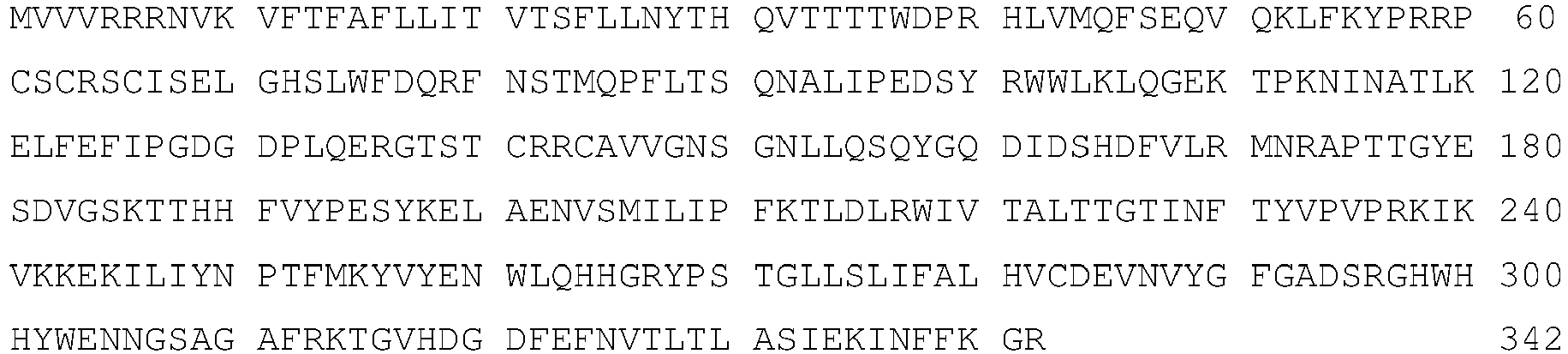
- SEQ ID NO: 20
Alligator sinensis ST3Gal1
- SEQ ID NO: 21
Latimeria chalumnae ST3Gal1
- SEQ ID NO: 22
Ciona intestinalis ST3Gal1
Claims (16)
- A cell line that is genetically modified to overexpress a β-galactoside α-2,3-sialyltransferase 1 (ST3Gal1), which is further genetically modified to overexpress a β-galactoside α-2,3-sialyltransferase 4 (ST3Gal4).
- The cell line according to claim 1, which is further genetically modified to overexpress a β-galactoside α-2,6-sialyltransferase 1 (ST6Gal1).
- The cell line according to claim 1 or claim 2, wherein the cell line comprises endogenous genes encoding ST3Gal1 and ST3Gal4, and optionally an endogenous gene encoding ST6Gal1, and further has at least one genetic element, selected from the group consisting of a promoter, an enhancing element, and a stabilizing element inserted into the genome in one or more position(s) suitable to cause overexpression of ST3Gal1 and ST4Gal4, and optionally ST6Gal1.
- The cell line according to any one of claims 1 to 3, wherein the cell line comprises exogenous nucleic acids encoding ST3Gal1 and ST3Gal4, and optionally an exogenous nucleic acid encoding ST6Gal1.
- The cell line according to any one of claims 1 to 4, wherein the cell line is a mammalian cell line, preferably a human cell line.
- The cell line according to any one of claims 1 to 4, wherein the cell line is derived from a cell line, selected from the group consisting of AGE.CR® cells, Vero cells, MDCK cells, BHK cells, CHO cells, HEK293 cells, HepG2 cells, Huh7 cells, AGE1.HN® cells, NC5T11 cells, Per.C6 cells, HMCLs cells, MM.1 cells, U266 cells, RPMI18226 cells, HKB11 cells, NM cells, NM-F9 cells, and CAP cells.
- The cell line according to any one of claims 1 to 4, wherein the cell line is derived from human primary amniocytes comprising at least one nucleic acid encoding the gene products of the adenoviral E1 and pIX regions.
- A recombinant glycoprotein having GaINAc O-glycans that are sialylated to a degree of at least 80%, wherein said glycoprotein is produced in a cell line according to any one of claims 1 to 7.
- The recombinant glycoprotein according to claim 8 having GalNAc O-glycans that are sialylated to a degree of at least 95%.
- The recombinant glycoprotein according to claim 8 or claim 9, wherein at least 80% of the GalNAc O-glycans are core 1 GalNAc O-glycans.
- The recombinant glycoprotein according to claim 10, wherein the core 1 GalNAc O-glycans are disialylated to a degree of at least 25%.
- The recombinant glycoprotein according to any one of claims 8 to 11, wherein the glycoprotein is selected from the group consisting of growth factors, peptide hormones, cytokines, enzymes, antibodies, antibody fragments, blood clotting factors, and protease inhibitors.
- The recombinant glycoprotein according to any one of claims 8 to 12, wherein the glycoprotein is selected from the group consisting of hepatocyte growth factor (HGF), erythropoietin (EPO), Factor VIII (FVIII), Factor IX (FIX), von-Willebrand-Factor (vWF), and C1 esterase inhibitor (C1-inhibitor; C1 Inh).
- The recombinant glycoprotein according to any one of claims 8 to 13, wherein the glycoprotein is a human glycoprotein.
- A method for the expression of recombinant glycoproteins having GalNAc O-glycans that are sialylated to a degree of at least 80%, comprising the step of overexpressing with the recombinant glycoproteins a β-galactoside α-2,3-sialyltransferase 1 (ST3Ga)1) and a β-galactoside α-2,3-sialyltransferase 4 (ST3Gal4), wherein the method is not practiced on the human or animal body.
- The method of claim 15, further comprising the step of overexpressing with the recombinant glycoproteins a β-galactoside α-2,6-sialyltransferase 1 (ST6Gal1).
Applications Claiming Priority (2)
| Application Number | Priority Date | Filing Date | Title |
|---|---|---|---|
| EP15000016.4A EP3042952A1 (en) | 2015-01-07 | 2015-01-07 | O-glycan sialylated recombinant glycoproteins and cell lines for producing the same |
| PCT/EP2015/002521 WO2016110302A1 (en) | 2015-01-07 | 2015-12-15 | O-glycan sialylated recombinant glycoproteins and cell lines for producing the same |
Publications (2)
| Publication Number | Publication Date |
|---|---|
| EP3242935A1 EP3242935A1 (en) | 2017-11-15 |
| EP3242935B1 true EP3242935B1 (en) | 2023-11-22 |
Family
ID=52396403
Family Applications (2)
| Application Number | Title | Priority Date | Filing Date |
|---|---|---|---|
| EP15000016.4A Withdrawn EP3042952A1 (en) | 2015-01-07 | 2015-01-07 | O-glycan sialylated recombinant glycoproteins and cell lines for producing the same |
| EP15821020.3A Active EP3242935B1 (en) | 2015-01-07 | 2015-12-15 | O-glycan sialylated recombinant glycoproteins and cell lines for producing the same |
Family Applications Before (1)
| Application Number | Title | Priority Date | Filing Date |
|---|---|---|---|
| EP15000016.4A Withdrawn EP3042952A1 (en) | 2015-01-07 | 2015-01-07 | O-glycan sialylated recombinant glycoproteins and cell lines for producing the same |
Country Status (16)
| Country | Link |
|---|---|
| US (2) | US10081798B2 (en) |
| EP (2) | EP3042952A1 (en) |
| JP (1) | JP6395943B2 (en) |
| KR (1) | KR102013712B1 (en) |
| CN (1) | CN107109370B (en) |
| AU (2) | AU2015376273B2 (en) |
| BR (1) | BR112017014769A2 (en) |
| CA (1) | CA2972932C (en) |
| DK (1) | DK3242935T3 (en) |
| IL (1) | IL253339A0 (en) |
| MX (1) | MX387417B (en) |
| MY (1) | MY187076A (en) |
| RU (1) | RU2714209C2 (en) |
| SG (1) | SG11201704571WA (en) |
| WO (1) | WO2016110302A1 (en) |
| ZA (1) | ZA201703767B (en) |
Families Citing this family (12)
| Publication number | Priority date | Publication date | Assignee | Title |
|---|---|---|---|---|
| EP2877589B1 (en) | 2012-07-26 | 2024-03-06 | Momenta Pharmaceuticals, Inc. | Glycoproteins with anti-inflammatory properties |
| WO2017087882A1 (en) * | 2015-11-19 | 2017-05-26 | Shire Human Genetic Therapies, Inc. | Recombinant human c1 esterase inhibitor and uses thereof |
| EP3382014A1 (en) | 2017-03-29 | 2018-10-03 | CEVEC Pharmaceuticals GmbH | Recombinant glycoproteins with reduced antennary fucosylation |
| EP3575404B1 (en) * | 2018-05-28 | 2022-10-19 | Chr. Hansen HMO GmbH | Fermentative production of sialylated saccharides |
| US12331320B2 (en) | 2018-10-10 | 2025-06-17 | The Research Foundation For The State University Of New York | Genome edited cancer cell vaccines |
| CN110029129A (en) * | 2019-03-06 | 2019-07-19 | 华南农业大学 | A kind of construction method of MDCK stable cell strain that expressing 1 gene of chicken ST3GAL |
| EP3736286A1 (en) | 2019-05-09 | 2020-11-11 | Biotest AG | Single chain factor viii molecule |
| EP3785726A1 (en) | 2019-09-02 | 2021-03-03 | Biotest AG | Factor viii protein with increased half-life |
| EP4017521A1 (en) | 2019-09-02 | 2022-06-29 | Biotest AG | Factor viii protein with increased half-life |
| EP4106798A1 (en) | 2020-02-17 | 2022-12-28 | Biotest AG | Subcutaneous administration of factor viii |
| AU2021376384B2 (en) | 2020-11-06 | 2026-04-16 | Cho Pharma, Inc. | Immune composition comprising antigen and glycoengineered antibody thereof |
| US12234472B2 (en) | 2021-10-18 | 2025-02-25 | Regeneron Pharmaceuticals, Inc. | Eukaryotic cells comprising adenovirus-associated virus polynucleotides |
Family Cites Families (15)
| Publication number | Priority date | Publication date | Assignee | Title |
|---|---|---|---|---|
| US5262308A (en) | 1992-01-28 | 1993-11-16 | Thomas Jefferson University | Cell lines which constitutively express IGF-1 and IGF-1 R |
| DE19955558C2 (en) | 1999-11-18 | 2003-03-20 | Stefan Kochanek | Permanent amniocyte cell line, its production and use for the production of gene transfer vectors |
| KR101495549B1 (en) * | 2006-07-13 | 2015-02-25 | 와이어쓰 엘엘씨 | Production of glycoproteins |
| JP2010512766A (en) | 2006-12-22 | 2010-04-30 | エフ.ホフマン−ラ ロシュ アーゲー | SHRNA-mediated suppression of α-1,6-fucosyltransferase expression |
| CA2679732A1 (en) | 2007-03-07 | 2008-09-18 | Glycofi, Inc. | Production of glycoproteins with modified fucosylation |
| WO2010012793A1 (en) | 2008-08-01 | 2010-02-04 | Bayer Cropscience Sa | Fungicide aminothiazole derivatives |
| CN101613678A (en) | 2009-01-14 | 2009-12-30 | 中国农业科学院哈尔滨兽医研究所 | Establishment of MDCK cell line stably expressing β-galactoside α-2,3-sialyltransferase I (ST3GAL I) |
| WO2011096750A2 (en) * | 2010-02-08 | 2011-08-11 | Korea Advanced Institute Of Science And Technology | Method for preparing recombinant glycoproteins with high sialic acid content |
| JP5914363B2 (en) * | 2010-02-16 | 2016-05-11 | ノヴォ ノルディスク アー/エス | Factor VIII molecule with reduced VWF binding |
| TW201209160A (en) | 2010-04-27 | 2012-03-01 | Lonza Ag | Improved glycosylation of proteins in host cells |
| WO2012077128A1 (en) | 2010-12-07 | 2012-06-14 | Intas Biopharmaceuticals Limited | A novel cell line development process to produce recombinant proteins using twin vector expression system |
| DK2661492T3 (en) * | 2011-01-06 | 2018-01-15 | Univ Johns Hopkins | Method for Preparing Recombinant Glycoproteins with Increased Circuit Half Life in Mammalian Cells |
| WO2013093760A2 (en) | 2011-12-19 | 2013-06-27 | Grifols, S.A. | Compositions, methods, and kits for preparing sialylated recombinant proteins |
| WO2014015227A1 (en) | 2012-07-19 | 2014-01-23 | The Scripps Research Institute | Engineering cell surfaces for orthogonal selectability |
| BR112015019348A2 (en) | 2013-02-13 | 2017-08-22 | Lab Francais Du Fractionnement | METHODS FOR PRODUCING GLYCOSYLATION MODIFIED AND SIALYLATION ENHANCED PROTEIN, TO ENHANCE SIALYL TRANSFERASE ACTIVITY IN THE MAMMARY GLAND AND TO PRODUCE SIALYL TRANSFERASE, GLYCOSYLATION MODIFIED PROTEIN OR SIALYLATION ENHANCED PROTEIN, COMPOSITION, SIALYL TRANSFERASE, MAMMALIAN, AND TRANSGENE MAMMARY EPITHELIAL |
-
2015
- 2015-01-07 EP EP15000016.4A patent/EP3042952A1/en not_active Withdrawn
- 2015-12-15 SG SG11201704571WA patent/SG11201704571WA/en unknown
- 2015-12-15 AU AU2015376273A patent/AU2015376273B2/en active Active
- 2015-12-15 KR KR1020177021780A patent/KR102013712B1/en active Active
- 2015-12-15 JP JP2017536883A patent/JP6395943B2/en active Active
- 2015-12-15 WO PCT/EP2015/002521 patent/WO2016110302A1/en not_active Ceased
- 2015-12-15 BR BR112017014769-6A patent/BR112017014769A2/en not_active Application Discontinuation
- 2015-12-15 CA CA2972932A patent/CA2972932C/en active Active
- 2015-12-15 RU RU2017127276A patent/RU2714209C2/en active
- 2015-12-15 MY MYPI2017000930A patent/MY187076A/en unknown
- 2015-12-15 DK DK15821020.3T patent/DK3242935T3/en active
- 2015-12-15 CN CN201580072695.7A patent/CN107109370B/en active Active
- 2015-12-15 US US15/541,565 patent/US10081798B2/en active Active
- 2015-12-15 MX MX2017008923A patent/MX387417B/en unknown
- 2015-12-15 EP EP15821020.3A patent/EP3242935B1/en active Active
-
2017
- 2017-06-01 ZA ZA2017/03767A patent/ZA201703767B/en unknown
- 2017-07-05 IL IL253339A patent/IL253339A0/en active IP Right Grant
- 2017-07-27 AU AU2017208292A patent/AU2017208292A1/en not_active Abandoned
-
2018
- 2018-08-21 US US16/107,009 patent/US10793839B2/en active Active
Also Published As
| Publication number | Publication date |
|---|---|
| US10793839B2 (en) | 2020-10-06 |
| RU2017127276A3 (en) | 2019-02-07 |
| AU2015376273B2 (en) | 2017-07-27 |
| DK3242935T3 (en) | 2024-01-02 |
| CA2972932A1 (en) | 2016-07-14 |
| US20170342390A1 (en) | 2017-11-30 |
| KR102013712B1 (en) | 2019-08-23 |
| US10081798B2 (en) | 2018-09-25 |
| RU2714209C2 (en) | 2020-02-13 |
| MX2017008923A (en) | 2017-10-11 |
| CA2972932C (en) | 2019-08-06 |
| ZA201703767B (en) | 2018-04-25 |
| CN107109370A (en) | 2017-08-29 |
| MY187076A (en) | 2021-08-29 |
| AU2015376273A1 (en) | 2017-06-08 |
| JP6395943B2 (en) | 2018-09-26 |
| EP3042952A1 (en) | 2016-07-13 |
| HK1243728A1 (en) | 2018-07-20 |
| IL253339A0 (en) | 2017-09-28 |
| US20200095562A1 (en) | 2020-03-26 |
| WO2016110302A1 (en) | 2016-07-14 |
| CN107109370B (en) | 2022-03-22 |
| MX387417B (en) | 2025-03-18 |
| EP3242935A1 (en) | 2017-11-15 |
| KR20170094454A (en) | 2017-08-17 |
| BR112017014769A2 (en) | 2018-01-16 |
| AU2017208292A1 (en) | 2017-08-17 |
| RU2017127276A (en) | 2019-02-07 |
| JP2018500922A (en) | 2018-01-18 |
| SG11201704571WA (en) | 2017-07-28 |
Similar Documents
| Publication | Publication Date | Title |
|---|---|---|
| US10793839B2 (en) | O-glycan sialylated recombinant glycoproteins | |
| EP3601548B1 (en) | Recombinant glycoproteins with reduced antennary fucosylation | |
| EP3417054B1 (en) | Cell lines for producing recombinant glycoproteins with di-antennary n-glycans, methods using the same, and recombinant glycoproteins | |
| Beatty et al. | Biochemical characterization of the soluble form of tumor antigen MUC1 isolated from sera and ascites fluid of breast and pancreatic cancer patients | |
| HK1243728B (en) | O-glycan sialylated recombinant glycoproteins and cell lines for producing the same | |
| BR122024008803A2 (en) | RECOMBINANT GLYCOPROTEIN AND ITS EXPRESSION METHOD, AS WELL AS CELL LINE FOR PRODUCING SAID GLYCOPROTEIN AND ITS GENERATION METHOD | |
| BR112018016696B1 (en) | METHOD FOR EXPRESSING A RECOMBINANT GLYCOPROTEIN IN THE SAID CELL LINE |
Legal Events
| Date | Code | Title | Description |
|---|---|---|---|
| STAA | Information on the status of an ep patent application or granted ep patent |
Free format text: STATUS: THE INTERNATIONAL PUBLICATION HAS BEEN MADE |
|
| PUAI | Public reference made under article 153(3) epc to a published international application that has entered the european phase |
Free format text: ORIGINAL CODE: 0009012 |
|
| STAA | Information on the status of an ep patent application or granted ep patent |
Free format text: STATUS: REQUEST FOR EXAMINATION WAS MADE |
|
| 17P | Request for examination filed |
Effective date: 20170614 |
|
| AK | Designated contracting states |
Kind code of ref document: A1 Designated state(s): AL AT BE BG CH CY CZ DE DK EE ES FI FR GB GR HR HU IE IS IT LI LT LU LV MC MK MT NL NO PL PT RO RS SE SI SK SM TR |
|
| AX | Request for extension of the european patent |
Extension state: BA ME |
|
| STAA | Information on the status of an ep patent application or granted ep patent |
Free format text: STATUS: EXAMINATION IS IN PROGRESS |
|
| 17Q | First examination report despatched |
Effective date: 20180216 |
|
| DAV | Request for validation of the european patent (deleted) | ||
| DAX | Request for extension of the european patent (deleted) | ||
| REG | Reference to a national code |
Ref country code: HK Ref legal event code: DE Ref document number: 1238290 Country of ref document: HK |
|
| GRAP | Despatch of communication of intention to grant a patent |
Free format text: ORIGINAL CODE: EPIDOSNIGR1 |
|
| STAA | Information on the status of an ep patent application or granted ep patent |
Free format text: STATUS: GRANT OF PATENT IS INTENDED |
|
| INTG | Intention to grant announced |
Effective date: 20230327 |
|
| P01 | Opt-out of the competence of the unified patent court (upc) registered |
Effective date: 20230430 |
|
| GRAJ | Information related to disapproval of communication of intention to grant by the applicant or resumption of examination proceedings by the epo deleted |
Free format text: ORIGINAL CODE: EPIDOSDIGR1 |
|
| STAA | Information on the status of an ep patent application or granted ep patent |
Free format text: STATUS: EXAMINATION IS IN PROGRESS |
|
| GRAP | Despatch of communication of intention to grant a patent |
Free format text: ORIGINAL CODE: EPIDOSNIGR1 |
|
| STAA | Information on the status of an ep patent application or granted ep patent |
Free format text: STATUS: GRANT OF PATENT IS INTENDED |
|
| INTC | Intention to grant announced (deleted) | ||
| INTG | Intention to grant announced |
Effective date: 20230801 |
|
| GRAS | Grant fee paid |
Free format text: ORIGINAL CODE: EPIDOSNIGR3 |
|
| GRAA | (expected) grant |
Free format text: ORIGINAL CODE: 0009210 |
|
| STAA | Information on the status of an ep patent application or granted ep patent |
Free format text: STATUS: THE PATENT HAS BEEN GRANTED |
|
| AK | Designated contracting states |
Kind code of ref document: B1 Designated state(s): AL AT BE BG CH CY CZ DE DK EE ES FI FR GB GR HR HU IE IS IT LI LT LU LV MC MK MT NL NO PL PT RO RS SE SI SK SM TR |
|
| REG | Reference to a national code |
Ref country code: GB Ref legal event code: FG4D |
|
| REG | Reference to a national code |
Ref country code: CH Ref legal event code: EP Ref country code: DE Ref legal event code: R096 Ref document number: 602015086659 Country of ref document: DE |
|
| REG | Reference to a national code |
Ref country code: IE Ref legal event code: FG4D Ref country code: NL Ref legal event code: FP |
|
| REG | Reference to a national code |
Ref country code: SE Ref legal event code: TRGR |
|
| REG | Reference to a national code |
Ref country code: DK Ref legal event code: T3 Effective date: 20231219 |
|
| REG | Reference to a national code |
Ref country code: LT Ref legal event code: MG9D |
|
| PG25 | Lapsed in a contracting state [announced via postgrant information from national office to epo] |
Ref country code: GR Free format text: LAPSE BECAUSE OF FAILURE TO SUBMIT A TRANSLATION OF THE DESCRIPTION OR TO PAY THE FEE WITHIN THE PRESCRIBED TIME-LIMIT Effective date: 20240223 |
|
| PG25 | Lapsed in a contracting state [announced via postgrant information from national office to epo] |
Ref country code: IS Free format text: LAPSE BECAUSE OF FAILURE TO SUBMIT A TRANSLATION OF THE DESCRIPTION OR TO PAY THE FEE WITHIN THE PRESCRIBED TIME-LIMIT Effective date: 20240322 |
|
| PG25 | Lapsed in a contracting state [announced via postgrant information from national office to epo] |
Ref country code: LT Free format text: LAPSE BECAUSE OF FAILURE TO SUBMIT A TRANSLATION OF THE DESCRIPTION OR TO PAY THE FEE WITHIN THE PRESCRIBED TIME-LIMIT Effective date: 20231122 |
|
| REG | Reference to a national code |
Ref country code: AT Ref legal event code: UEP Ref document number: 1633879 Country of ref document: AT Kind code of ref document: T Effective date: 20231122 |
|
| PG25 | Lapsed in a contracting state [announced via postgrant information from national office to epo] |
Ref country code: ES Free format text: LAPSE BECAUSE OF FAILURE TO SUBMIT A TRANSLATION OF THE DESCRIPTION OR TO PAY THE FEE WITHIN THE PRESCRIBED TIME-LIMIT Effective date: 20231122 |
|
| PG25 | Lapsed in a contracting state [announced via postgrant information from national office to epo] |
Ref country code: LT Free format text: LAPSE BECAUSE OF FAILURE TO SUBMIT A TRANSLATION OF THE DESCRIPTION OR TO PAY THE FEE WITHIN THE PRESCRIBED TIME-LIMIT Effective date: 20231122 Ref country code: IS Free format text: LAPSE BECAUSE OF FAILURE TO SUBMIT A TRANSLATION OF THE DESCRIPTION OR TO PAY THE FEE WITHIN THE PRESCRIBED TIME-LIMIT Effective date: 20240322 Ref country code: GR Free format text: LAPSE BECAUSE OF FAILURE TO SUBMIT A TRANSLATION OF THE DESCRIPTION OR TO PAY THE FEE WITHIN THE PRESCRIBED TIME-LIMIT Effective date: 20240223 Ref country code: ES Free format text: LAPSE BECAUSE OF FAILURE TO SUBMIT A TRANSLATION OF THE DESCRIPTION OR TO PAY THE FEE WITHIN THE PRESCRIBED TIME-LIMIT Effective date: 20231122 Ref country code: BG Free format text: LAPSE BECAUSE OF FAILURE TO SUBMIT A TRANSLATION OF THE DESCRIPTION OR TO PAY THE FEE WITHIN THE PRESCRIBED TIME-LIMIT Effective date: 20240222 Ref country code: PT Free format text: LAPSE BECAUSE OF FAILURE TO SUBMIT A TRANSLATION OF THE DESCRIPTION OR TO PAY THE FEE WITHIN THE PRESCRIBED TIME-LIMIT Effective date: 20240322 |
|
| PG25 | Lapsed in a contracting state [announced via postgrant information from national office to epo] |
Ref country code: RS Free format text: LAPSE BECAUSE OF FAILURE TO SUBMIT A TRANSLATION OF THE DESCRIPTION OR TO PAY THE FEE WITHIN THE PRESCRIBED TIME-LIMIT Effective date: 20231122 Ref country code: PL Free format text: LAPSE BECAUSE OF FAILURE TO SUBMIT A TRANSLATION OF THE DESCRIPTION OR TO PAY THE FEE WITHIN THE PRESCRIBED TIME-LIMIT Effective date: 20231122 Ref country code: NO Free format text: LAPSE BECAUSE OF FAILURE TO SUBMIT A TRANSLATION OF THE DESCRIPTION OR TO PAY THE FEE WITHIN THE PRESCRIBED TIME-LIMIT Effective date: 20240222 Ref country code: LV Free format text: LAPSE BECAUSE OF FAILURE TO SUBMIT A TRANSLATION OF THE DESCRIPTION OR TO PAY THE FEE WITHIN THE PRESCRIBED TIME-LIMIT Effective date: 20231122 Ref country code: HR Free format text: LAPSE BECAUSE OF FAILURE TO SUBMIT A TRANSLATION OF THE DESCRIPTION OR TO PAY THE FEE WITHIN THE PRESCRIBED TIME-LIMIT Effective date: 20231122 |
|
| PG25 | Lapsed in a contracting state [announced via postgrant information from national office to epo] |
Ref country code: CZ Free format text: LAPSE BECAUSE OF FAILURE TO SUBMIT A TRANSLATION OF THE DESCRIPTION OR TO PAY THE FEE WITHIN THE PRESCRIBED TIME-LIMIT Effective date: 20231122 |
|
| PG25 | Lapsed in a contracting state [announced via postgrant information from national office to epo] |
Ref country code: SK Free format text: LAPSE BECAUSE OF FAILURE TO SUBMIT A TRANSLATION OF THE DESCRIPTION OR TO PAY THE FEE WITHIN THE PRESCRIBED TIME-LIMIT Effective date: 20231122 |
|
| PG25 | Lapsed in a contracting state [announced via postgrant information from national office to epo] |
Ref country code: SM Free format text: LAPSE BECAUSE OF FAILURE TO SUBMIT A TRANSLATION OF THE DESCRIPTION OR TO PAY THE FEE WITHIN THE PRESCRIBED TIME-LIMIT Effective date: 20231122 Ref country code: SK Free format text: LAPSE BECAUSE OF FAILURE TO SUBMIT A TRANSLATION OF THE DESCRIPTION OR TO PAY THE FEE WITHIN THE PRESCRIBED TIME-LIMIT Effective date: 20231122 Ref country code: RO Free format text: LAPSE BECAUSE OF FAILURE TO SUBMIT A TRANSLATION OF THE DESCRIPTION OR TO PAY THE FEE WITHIN THE PRESCRIBED TIME-LIMIT Effective date: 20231122 Ref country code: EE Free format text: LAPSE BECAUSE OF FAILURE TO SUBMIT A TRANSLATION OF THE DESCRIPTION OR TO PAY THE FEE WITHIN THE PRESCRIBED TIME-LIMIT Effective date: 20231122 Ref country code: CZ Free format text: LAPSE BECAUSE OF FAILURE TO SUBMIT A TRANSLATION OF THE DESCRIPTION OR TO PAY THE FEE WITHIN THE PRESCRIBED TIME-LIMIT Effective date: 20231122 |
|
| PG25 | Lapsed in a contracting state [announced via postgrant information from national office to epo] |
Ref country code: LU Free format text: LAPSE BECAUSE OF NON-PAYMENT OF DUE FEES Effective date: 20231215 |
|
| REG | Reference to a national code |
Ref country code: DE Ref legal event code: R097 Ref document number: 602015086659 Country of ref document: DE |
|
| PG25 | Lapsed in a contracting state [announced via postgrant information from national office to epo] |
Ref country code: MC Free format text: LAPSE BECAUSE OF FAILURE TO SUBMIT A TRANSLATION OF THE DESCRIPTION OR TO PAY THE FEE WITHIN THE PRESCRIBED TIME-LIMIT Effective date: 20231122 |
|
| PG25 | Lapsed in a contracting state [announced via postgrant information from national office to epo] |
Ref country code: MC Free format text: LAPSE BECAUSE OF FAILURE TO SUBMIT A TRANSLATION OF THE DESCRIPTION OR TO PAY THE FEE WITHIN THE PRESCRIBED TIME-LIMIT Effective date: 20231122 Ref country code: LU Free format text: LAPSE BECAUSE OF NON-PAYMENT OF DUE FEES Effective date: 20231215 |
|
| PLBE | No opposition filed within time limit |
Free format text: ORIGINAL CODE: 0009261 |
|
| STAA | Information on the status of an ep patent application or granted ep patent |
Free format text: STATUS: NO OPPOSITION FILED WITHIN TIME LIMIT |
|
| REG | Reference to a national code |
Ref country code: IE Ref legal event code: MM4A |
|
| PG25 | Lapsed in a contracting state [announced via postgrant information from national office to epo] |
Ref country code: IE Free format text: LAPSE BECAUSE OF NON-PAYMENT OF DUE FEES Effective date: 20231215 |
|
| PG25 | Lapsed in a contracting state [announced via postgrant information from national office to epo] |
Ref country code: SI Free format text: LAPSE BECAUSE OF FAILURE TO SUBMIT A TRANSLATION OF THE DESCRIPTION OR TO PAY THE FEE WITHIN THE PRESCRIBED TIME-LIMIT Effective date: 20231122 |
|
| 26N | No opposition filed |
Effective date: 20240823 |
|
| PG25 | Lapsed in a contracting state [announced via postgrant information from national office to epo] |
Ref country code: SI Free format text: LAPSE BECAUSE OF FAILURE TO SUBMIT A TRANSLATION OF THE DESCRIPTION OR TO PAY THE FEE WITHIN THE PRESCRIBED TIME-LIMIT Effective date: 20231122 Ref country code: IE Free format text: LAPSE BECAUSE OF NON-PAYMENT OF DUE FEES Effective date: 20231215 |
|
| PGFP | Annual fee paid to national office [announced via postgrant information from national office to epo] |
Ref country code: NL Payment date: 20241004 Year of fee payment: 10 |
|
| PGFP | Annual fee paid to national office [announced via postgrant information from national office to epo] |
Ref country code: DK Payment date: 20241212 Year of fee payment: 10 |
|
| PGFP | Annual fee paid to national office [announced via postgrant information from national office to epo] |
Ref country code: BE Payment date: 20241007 Year of fee payment: 10 |
|
| PGFP | Annual fee paid to national office [announced via postgrant information from national office to epo] |
Ref country code: AT Payment date: 20241126 Year of fee payment: 10 |
|
| PGFP | Annual fee paid to national office [announced via postgrant information from national office to epo] |
Ref country code: IT Payment date: 20241112 Year of fee payment: 10 |
|
| PGFP | Annual fee paid to national office [announced via postgrant information from national office to epo] |
Ref country code: CH Payment date: 20250101 Year of fee payment: 10 |
|
| PG25 | Lapsed in a contracting state [announced via postgrant information from national office to epo] |
Ref country code: FI Free format text: LAPSE BECAUSE OF FAILURE TO SUBMIT A TRANSLATION OF THE DESCRIPTION OR TO PAY THE FEE WITHIN THE PRESCRIBED TIME-LIMIT Effective date: 20231122 |
|
| PG25 | Lapsed in a contracting state [announced via postgrant information from national office to epo] |
Ref country code: CY Free format text: LAPSE BECAUSE OF FAILURE TO SUBMIT A TRANSLATION OF THE DESCRIPTION OR TO PAY THE FEE WITHIN THE PRESCRIBED TIME-LIMIT; INVALID AB INITIO Effective date: 20151215 |
|
| PG25 | Lapsed in a contracting state [announced via postgrant information from national office to epo] |
Ref country code: HU Free format text: LAPSE BECAUSE OF FAILURE TO SUBMIT A TRANSLATION OF THE DESCRIPTION OR TO PAY THE FEE WITHIN THE PRESCRIBED TIME-LIMIT; INVALID AB INITIO Effective date: 20151215 |
|
| REG | Reference to a national code |
Ref country code: DE Ref legal event code: R081 Ref document number: 602015086659 Country of ref document: DE Owner name: CEVEC PHARMACEUTICALS GMBH, DE Free format text: FORMER OWNER: CEVEC PHARMACEUTICALS GMBH, 51105 KOELN, DE |
|
| PG25 | Lapsed in a contracting state [announced via postgrant information from national office to epo] |
Ref country code: TR Free format text: LAPSE BECAUSE OF FAILURE TO SUBMIT A TRANSLATION OF THE DESCRIPTION OR TO PAY THE FEE WITHIN THE PRESCRIBED TIME-LIMIT Effective date: 20231122 |
|
| PGFP | Annual fee paid to national office [announced via postgrant information from national office to epo] |
Ref country code: GB Payment date: 20251223 Year of fee payment: 11 |
|
| PGFP | Annual fee paid to national office [announced via postgrant information from national office to epo] |
Ref country code: FR Payment date: 20251230 Year of fee payment: 11 |
|
| PGFP | Annual fee paid to national office [announced via postgrant information from national office to epo] |
Ref country code: SE Payment date: 20251222 Year of fee payment: 11 |
|
| PGFP | Annual fee paid to national office [announced via postgrant information from national office to epo] |
Ref country code: DE Payment date: 20251229 Year of fee payment: 11 |