EP3241896A1 - Device and method for inducing pluripotent cells using energy - Google Patents
Device and method for inducing pluripotent cells using energy Download PDFInfo
- Publication number
- EP3241896A1 EP3241896A1 EP15866086.0A EP15866086A EP3241896A1 EP 3241896 A1 EP3241896 A1 EP 3241896A1 EP 15866086 A EP15866086 A EP 15866086A EP 3241896 A1 EP3241896 A1 EP 3241896A1
- Authority
- EP
- European Patent Office
- Prior art keywords
- cells
- ultrasound
- culture medium
- culture
- physics
- Prior art date
- Legal status (The legal status is an assumption and is not a legal conclusion. Google has not performed a legal analysis and makes no representation as to the accuracy of the status listed.)
- Granted
Links
- 230000001939 inductive effect Effects 0.000 title claims abstract description 38
- 238000000034 method Methods 0.000 title claims abstract description 38
- 238000010438 heat treatment Methods 0.000 claims abstract description 19
- 210000004027 cell Anatomy 0.000 claims description 331
- 238000002604 ultrasonography Methods 0.000 claims description 113
- 108090000623 proteins and genes Proteins 0.000 claims description 56
- 239000001963 growth medium Substances 0.000 claims description 50
- 239000003550 marker Substances 0.000 claims description 50
- 102100035423 POU domain, class 5, transcription factor 1 Human genes 0.000 claims description 44
- 101001094700 Homo sapiens POU domain, class 5, transcription factor 1 Proteins 0.000 claims description 34
- 101000713275 Homo sapiens Solute carrier family 22 member 3 Proteins 0.000 claims description 34
- 239000000203 mixture Substances 0.000 claims description 34
- 210000001654 germ layer Anatomy 0.000 claims description 30
- 239000006143 cell culture medium Substances 0.000 claims description 27
- 239000002609 medium Substances 0.000 claims description 26
- 238000004114 suspension culture Methods 0.000 claims description 23
- 238000004264 monolayer culture Methods 0.000 claims description 20
- 210000002950 fibroblast Anatomy 0.000 claims description 18
- 230000008672 reprogramming Effects 0.000 claims description 17
- 238000012258 culturing Methods 0.000 claims description 13
- 102000008730 Nestin Human genes 0.000 claims description 11
- 108010088225 Nestin Proteins 0.000 claims description 11
- 102100037506 Paired box protein Pax-6 Human genes 0.000 claims description 11
- 210000005055 nestin Anatomy 0.000 claims description 11
- 101000687905 Homo sapiens Transcription factor SOX-2 Proteins 0.000 claims description 10
- 108010032788 PAX6 Transcription Factor Proteins 0.000 claims description 10
- 102100024270 Transcription factor SOX-2 Human genes 0.000 claims description 10
- -1 Brachyury Proteins 0.000 claims description 9
- 101000835745 Homo sapiens Teratocarcinoma-derived growth factor 1 Proteins 0.000 claims description 9
- 102100026404 Teratocarcinoma-derived growth factor 1 Human genes 0.000 claims description 9
- 210000001671 embryonic stem cell Anatomy 0.000 claims description 9
- 210000001519 tissue Anatomy 0.000 claims description 9
- 101000819074 Homo sapiens Transcription factor GATA-4 Proteins 0.000 claims description 8
- 102100021380 Transcription factor GATA-4 Human genes 0.000 claims description 8
- 210000000130 stem cell Anatomy 0.000 claims description 8
- 238000011282 treatment Methods 0.000 claims description 8
- 101001139134 Homo sapiens Krueppel-like factor 4 Proteins 0.000 claims description 6
- 102100020677 Krueppel-like factor 4 Human genes 0.000 claims description 6
- 206010028980 Neoplasm Diseases 0.000 claims description 6
- 201000011510 cancer Diseases 0.000 claims description 6
- 230000024245 cell differentiation Effects 0.000 claims description 4
- 230000001678 irradiating effect Effects 0.000 claims description 3
- 241000124008 Mammalia Species 0.000 claims description 2
- 210000000056 organ Anatomy 0.000 claims description 2
- 230000000694 effects Effects 0.000 abstract description 21
- 230000014509 gene expression Effects 0.000 description 95
- 238000009210 therapy by ultrasound Methods 0.000 description 30
- 230000004069 differentiation Effects 0.000 description 28
- 210000000170 cell membrane Anatomy 0.000 description 26
- 241000699666 Mus <mouse, genus> Species 0.000 description 23
- 210000002236 cellular spheroid Anatomy 0.000 description 23
- 238000009826 distribution Methods 0.000 description 18
- 210000001808 exosome Anatomy 0.000 description 15
- 238000004458 analytical method Methods 0.000 description 13
- 238000010586 diagram Methods 0.000 description 12
- 210000003981 ectoderm Anatomy 0.000 description 11
- 102000004169 proteins and genes Human genes 0.000 description 11
- 101710126211 POU domain, class 5, transcription factor 1 Proteins 0.000 description 10
- 238000010240 RT-PCR analysis Methods 0.000 description 10
- 230000002500 effect on skin Effects 0.000 description 10
- 210000003716 mesoderm Anatomy 0.000 description 10
- 238000010186 staining Methods 0.000 description 10
- 230000015572 biosynthetic process Effects 0.000 description 9
- 238000004113 cell culture Methods 0.000 description 9
- 210000001900 endoderm Anatomy 0.000 description 9
- 230000004660 morphological change Effects 0.000 description 9
- 238000003757 reverse transcription PCR Methods 0.000 description 9
- 230000001580 bacterial effect Effects 0.000 description 8
- 230000003834 intracellular effect Effects 0.000 description 8
- 230000002068 genetic effect Effects 0.000 description 7
- 238000000338 in vitro Methods 0.000 description 7
- 230000006698 induction Effects 0.000 description 7
- 230000002062 proliferating effect Effects 0.000 description 7
- 230000000638 stimulation Effects 0.000 description 7
- 238000003556 assay Methods 0.000 description 6
- 238000002474 experimental method Methods 0.000 description 6
- 210000004263 induced pluripotent stem cell Anatomy 0.000 description 6
- 230000001537 neural effect Effects 0.000 description 6
- XJMOSONTPMZWPB-UHFFFAOYSA-M propidium iodide Chemical compound [I-].[I-].C12=CC(N)=CC=C2C2=CC=C(N)C=C2[N+](CCC[N+](C)(CC)CC)=C1C1=CC=CC=C1 XJMOSONTPMZWPB-UHFFFAOYSA-M 0.000 description 6
- 239000002096 quantum dot Substances 0.000 description 6
- UCSJYZPVAKXKNQ-HZYVHMACSA-N streptomycin Chemical compound CN[C@H]1[C@H](O)[C@@H](O)[C@H](CO)O[C@H]1O[C@@H]1[C@](C=O)(O)[C@H](C)O[C@H]1O[C@@H]1[C@@H](NC(N)=N)[C@H](O)[C@@H](NC(N)=N)[C@H](O)[C@H]1O UCSJYZPVAKXKNQ-HZYVHMACSA-N 0.000 description 6
- PRDFBSVERLRRMY-UHFFFAOYSA-N 2'-(4-ethoxyphenyl)-5-(4-methylpiperazin-1-yl)-2,5'-bibenzimidazole Chemical compound C1=CC(OCC)=CC=C1C1=NC2=CC=C(C=3NC4=CC(=CC=C4N=3)N3CCN(C)CC3)C=C2N1 PRDFBSVERLRRMY-UHFFFAOYSA-N 0.000 description 5
- 102000002260 Alkaline Phosphatase Human genes 0.000 description 5
- 108020004774 Alkaline Phosphatase Proteins 0.000 description 5
- 108020004414 DNA Proteins 0.000 description 5
- 239000006144 Dulbecco’s modified Eagle's medium Substances 0.000 description 5
- 108700039691 Genetic Promoter Regions Proteins 0.000 description 5
- 230000006907 apoptotic process Effects 0.000 description 5
- 230000000747 cardiac effect Effects 0.000 description 5
- 238000003365 immunocytochemistry Methods 0.000 description 5
- 238000001727 in vivo Methods 0.000 description 5
- 108020004999 messenger RNA Proteins 0.000 description 5
- 238000001000 micrograph Methods 0.000 description 5
- 230000002107 myocardial effect Effects 0.000 description 5
- 210000002569 neuron Anatomy 0.000 description 5
- 239000000725 suspension Substances 0.000 description 5
- 210000001550 testis Anatomy 0.000 description 5
- DGVVWUTYPXICAM-UHFFFAOYSA-N β‐Mercaptoethanol Chemical compound OCCS DGVVWUTYPXICAM-UHFFFAOYSA-N 0.000 description 5
- 102100027875 Homeobox protein Nkx-2.5 Human genes 0.000 description 4
- 229930040373 Paraformaldehyde Natural products 0.000 description 4
- 239000002458 cell surface marker Substances 0.000 description 4
- 150000001875 compounds Chemical class 0.000 description 4
- 210000002242 embryoid body Anatomy 0.000 description 4
- 238000000684 flow cytometry Methods 0.000 description 4
- 238000013532 laser treatment Methods 0.000 description 4
- 229920002866 paraformaldehyde Polymers 0.000 description 4
- 239000000047 product Substances 0.000 description 4
- 230000035755 proliferation Effects 0.000 description 4
- 230000004044 response Effects 0.000 description 4
- 238000012360 testing method Methods 0.000 description 4
- 102000010825 Actinin Human genes 0.000 description 3
- 108010063503 Actinin Proteins 0.000 description 3
- 238000012413 Fluorescence activated cell sorting analysis Methods 0.000 description 3
- 102100037060 Forkhead box protein D3 Human genes 0.000 description 3
- 102100028707 Homeobox protein MSX-1 Human genes 0.000 description 3
- 101001029308 Homo sapiens Forkhead box protein D3 Proteins 0.000 description 3
- 101000985653 Homo sapiens Homeobox protein MSX-1 Proteins 0.000 description 3
- 101000632197 Homo sapiens Homeobox protein Nkx-2.5 Proteins 0.000 description 3
- 101000979001 Homo sapiens Methionine aminopeptidase 2 Proteins 0.000 description 3
- 101000969087 Homo sapiens Microtubule-associated protein 2 Proteins 0.000 description 3
- 101000777245 Homo sapiens Undifferentiated embryonic cell transcription factor 1 Proteins 0.000 description 3
- 101000976622 Homo sapiens Zinc finger protein 42 homolog Proteins 0.000 description 3
- 102100021118 Microtubule-associated protein 2 Human genes 0.000 description 3
- 229930182555 Penicillin Natural products 0.000 description 3
- JGSARLDLIJGVTE-MBNYWOFBSA-N Penicillin G Chemical compound N([C@H]1[C@H]2SC([C@@H](N2C1=O)C(O)=O)(C)C)C(=O)CC1=CC=CC=C1 JGSARLDLIJGVTE-MBNYWOFBSA-N 0.000 description 3
- 102000002294 Purinergic P2X Receptors Human genes 0.000 description 3
- 108010000836 Purinergic P2X Receptors Proteins 0.000 description 3
- 102000002298 Purinergic P2Y Receptors Human genes 0.000 description 3
- 108010000818 Purinergic P2Y Receptors Proteins 0.000 description 3
- 229920004890 Triton X-100 Polymers 0.000 description 3
- 102100031278 Undifferentiated embryonic cell transcription factor 1 Human genes 0.000 description 3
- 102100023550 Zinc finger protein 42 homolog Human genes 0.000 description 3
- 210000001130 astrocyte Anatomy 0.000 description 3
- 230000001413 cellular effect Effects 0.000 description 3
- 230000004700 cellular uptake Effects 0.000 description 3
- 230000008859 change Effects 0.000 description 3
- 239000003153 chemical reaction reagent Substances 0.000 description 3
- 239000003797 essential amino acid Substances 0.000 description 3
- 230000011987 methylation Effects 0.000 description 3
- 238000007069 methylation reaction Methods 0.000 description 3
- 210000003205 muscle Anatomy 0.000 description 3
- 210000004940 nucleus Anatomy 0.000 description 3
- 229940049954 penicillin Drugs 0.000 description 3
- 239000011148 porous material Substances 0.000 description 3
- 230000008569 process Effects 0.000 description 3
- 210000002966 serum Anatomy 0.000 description 3
- 229960005322 streptomycin Drugs 0.000 description 3
- 239000000126 substance Substances 0.000 description 3
- 210000000689 upper leg Anatomy 0.000 description 3
- 108091032973 (ribonucleotides)n+m Proteins 0.000 description 2
- 102100023635 Alpha-fetoprotein Human genes 0.000 description 2
- CIWBSHSKHKDKBQ-JLAZNSOCSA-N Ascorbic acid Chemical compound OC[C@H](O)[C@H]1OC(=O)C(O)=C1O CIWBSHSKHKDKBQ-JLAZNSOCSA-N 0.000 description 2
- LSNNMFCWUKXFEE-UHFFFAOYSA-M Bisulfite Chemical compound OS([O-])=O LSNNMFCWUKXFEE-UHFFFAOYSA-M 0.000 description 2
- 102100032912 CD44 antigen Human genes 0.000 description 2
- CURLTUGMZLYLDI-UHFFFAOYSA-N Carbon dioxide Chemical compound O=C=O CURLTUGMZLYLDI-UHFFFAOYSA-N 0.000 description 2
- 230000007067 DNA methylation Effects 0.000 description 2
- 102100028072 Fibroblast growth factor 4 Human genes 0.000 description 2
- 108010010803 Gelatin Proteins 0.000 description 2
- ZDXPYRJPNDTMRX-UHFFFAOYSA-N Glutamine Chemical compound OC(=O)C(N)CCC(N)=O ZDXPYRJPNDTMRX-UHFFFAOYSA-N 0.000 description 2
- 101000868273 Homo sapiens CD44 antigen Proteins 0.000 description 2
- 101001060274 Homo sapiens Fibroblast growth factor 4 Proteins 0.000 description 2
- 101001120813 Homo sapiens Myosin regulatory light chain 2, atrial isoform Proteins 0.000 description 2
- 101000984042 Homo sapiens Protein lin-28 homolog A Proteins 0.000 description 2
- 101000819088 Homo sapiens Transcription factor GATA-6 Proteins 0.000 description 2
- 101710123134 Ice-binding protein Proteins 0.000 description 2
- 101710082837 Ice-structuring protein Proteins 0.000 description 2
- 101150009249 MAP2 gene Proteins 0.000 description 2
- 108700011259 MicroRNAs Proteins 0.000 description 2
- 241000699670 Mus sp. Species 0.000 description 2
- 102100038895 Myc proto-oncogene protein Human genes 0.000 description 2
- 101710135898 Myc proto-oncogene protein Proteins 0.000 description 2
- 102100026057 Myosin regulatory light chain 2, atrial isoform Human genes 0.000 description 2
- 102100025460 Protein lin-28 homolog A Human genes 0.000 description 2
- 102100021382 Transcription factor GATA-6 Human genes 0.000 description 2
- 101710150448 Transcriptional regulator Myc Proteins 0.000 description 2
- 101710101305 Transducin-like enhancer protein 1 Proteins 0.000 description 2
- 102100039362 Transducin-like enhancer protein 1 Human genes 0.000 description 2
- 102100026893 Troponin T, cardiac muscle Human genes 0.000 description 2
- 101710165323 Troponin T, cardiac muscle Proteins 0.000 description 2
- 101710107540 Type-2 ice-structuring protein Proteins 0.000 description 2
- 230000003321 amplification Effects 0.000 description 2
- 239000002771 cell marker Substances 0.000 description 2
- 238000002659 cell therapy Methods 0.000 description 2
- 230000004637 cellular stress Effects 0.000 description 2
- 239000002299 complementary DNA Substances 0.000 description 2
- 230000007423 decrease Effects 0.000 description 2
- 230000003247 decreasing effect Effects 0.000 description 2
- 238000001962 electrophoresis Methods 0.000 description 2
- 230000007613 environmental effect Effects 0.000 description 2
- 210000002919 epithelial cell Anatomy 0.000 description 2
- 238000002073 fluorescence micrograph Methods 0.000 description 2
- 239000007850 fluorescent dye Substances 0.000 description 2
- 229920000159 gelatin Polymers 0.000 description 2
- 239000008273 gelatin Substances 0.000 description 2
- 235000019322 gelatine Nutrition 0.000 description 2
- 235000011852 gelatine desserts Nutrition 0.000 description 2
- 238000012744 immunostaining Methods 0.000 description 2
- 230000001057 ionotropic effect Effects 0.000 description 2
- 239000010410 layer Substances 0.000 description 2
- 239000000463 material Substances 0.000 description 2
- 239000002679 microRNA Substances 0.000 description 2
- 238000003199 nucleic acid amplification method Methods 0.000 description 2
- 210000004248 oligodendroglia Anatomy 0.000 description 2
- 239000012474 protein marker Substances 0.000 description 2
- 230000002685 pulmonary effect Effects 0.000 description 2
- 239000003642 reactive oxygen metabolite Substances 0.000 description 2
- 230000002441 reversible effect Effects 0.000 description 2
- 238000012163 sequencing technique Methods 0.000 description 2
- 210000004927 skin cell Anatomy 0.000 description 2
- 150000003384 small molecules Chemical class 0.000 description 2
- 230000001052 transient effect Effects 0.000 description 2
- 238000002054 transplantation Methods 0.000 description 2
- 102100036732 Actin, aortic smooth muscle Human genes 0.000 description 1
- 108010085238 Actins Proteins 0.000 description 1
- 102000007469 Actins Human genes 0.000 description 1
- HJCMDXDYPOUFDY-WHFBIAKZSA-N Ala-Gln Chemical compound C[C@H](N)C(=O)N[C@H](C(O)=O)CCC(N)=O HJCMDXDYPOUFDY-WHFBIAKZSA-N 0.000 description 1
- 102100024155 Cadherin-11 Human genes 0.000 description 1
- 101100257359 Caenorhabditis elegans sox-2 gene Proteins 0.000 description 1
- 241000283707 Capra Species 0.000 description 1
- 102100025745 Cerberus Human genes 0.000 description 1
- 206010008342 Cervix carcinoma Diseases 0.000 description 1
- 230000004544 DNA amplification Effects 0.000 description 1
- 102100037124 Developmental pluripotency-associated 5 protein Human genes 0.000 description 1
- 101100347633 Drosophila melanogaster Mhc gene Proteins 0.000 description 1
- 108010067770 Endopeptidase K Proteins 0.000 description 1
- 101150099612 Esrrb gene Proteins 0.000 description 1
- 102000003974 Fibroblast growth factor 2 Human genes 0.000 description 1
- 108090000379 Fibroblast growth factor 2 Proteins 0.000 description 1
- OUVXYXNWSVIOSJ-UHFFFAOYSA-N Fluo-4 Chemical compound CC1=CC=C(N(CC(O)=O)CC(O)=O)C(OCCOC=2C(=CC=C(C=2)C2=C3C=C(F)C(=O)C=C3OC3=CC(O)=C(F)C=C32)N(CC(O)=O)CC(O)=O)=C1 OUVXYXNWSVIOSJ-UHFFFAOYSA-N 0.000 description 1
- 101150057663 Foxa2 gene Proteins 0.000 description 1
- 101150066002 GFP gene Proteins 0.000 description 1
- 102100039289 Glial fibrillary acidic protein Human genes 0.000 description 1
- 101710193519 Glial fibrillary acidic protein Proteins 0.000 description 1
- 102100029284 Hepatocyte nuclear factor 3-beta Human genes 0.000 description 1
- 108010090007 Homeobox Protein Nkx-2.5 Proteins 0.000 description 1
- 101000929319 Homo sapiens Actin, aortic smooth muscle Proteins 0.000 description 1
- 101000762236 Homo sapiens Cadherin-11 Proteins 0.000 description 1
- 101000914195 Homo sapiens Cerberus Proteins 0.000 description 1
- 101000881848 Homo sapiens Developmental pluripotency-associated 5 protein Proteins 0.000 description 1
- 101001052035 Homo sapiens Fibroblast growth factor 2 Proteins 0.000 description 1
- 101001062347 Homo sapiens Hepatocyte nuclear factor 3-beta Proteins 0.000 description 1
- 101001128090 Homo sapiens Homeobox protein NANOG Proteins 0.000 description 1
- 101001116388 Homo sapiens Melatonin-related receptor Proteins 0.000 description 1
- 101001120087 Homo sapiens P2Y purinoceptor 11 Proteins 0.000 description 1
- 101100137155 Homo sapiens POU5F1 gene Proteins 0.000 description 1
- 101000601647 Homo sapiens Paired box protein Pax-6 Proteins 0.000 description 1
- 101001001810 Homo sapiens Pleckstrin homology domain-containing family M member 3 Proteins 0.000 description 1
- 101000617738 Homo sapiens Survival motor neuron protein Proteins 0.000 description 1
- 101000652324 Homo sapiens Transcription factor SOX-17 Proteins 0.000 description 1
- 101000642523 Homo sapiens Transcription factor SOX-7 Proteins 0.000 description 1
- 101000713575 Homo sapiens Tubulin beta-3 chain Proteins 0.000 description 1
- 108700021430 Kruppel-Like Factor 4 Proteins 0.000 description 1
- ZDXPYRJPNDTMRX-VKHMYHEASA-N L-glutamine Chemical compound OC(=O)[C@@H](N)CCC(N)=O ZDXPYRJPNDTMRX-VKHMYHEASA-N 0.000 description 1
- 229930182816 L-glutamine Natural products 0.000 description 1
- 108010020004 Microtubule-Associated Proteins Proteins 0.000 description 1
- 102000009664 Microtubule-Associated Proteins Human genes 0.000 description 1
- 101001001809 Mus musculus Pleckstrin homology domain-containing family M member 3 Proteins 0.000 description 1
- 101100310657 Mus musculus Sox1 gene Proteins 0.000 description 1
- 101100257363 Mus musculus Sox2 gene Proteins 0.000 description 1
- 102000008934 Muscle Proteins Human genes 0.000 description 1
- 108010074084 Muscle Proteins Proteins 0.000 description 1
- 239000012580 N-2 Supplement Substances 0.000 description 1
- 101100500679 Neurospora crassa (strain ATCC 24698 / 74-OR23-1A / CBS 708.71 / DSM 1257 / FGSC 987) cot-3 gene Proteins 0.000 description 1
- 102000019040 Nuclear Antigens Human genes 0.000 description 1
- 108010051791 Nuclear Antigens Proteins 0.000 description 1
- 102100037601 P2X purinoceptor 4 Human genes 0.000 description 1
- 101710189967 P2X purinoceptor 4 Proteins 0.000 description 1
- 102100037602 P2X purinoceptor 7 Human genes 0.000 description 1
- 101710189965 P2X purinoceptor 7 Proteins 0.000 description 1
- 102100037600 P2Y purinoceptor 1 Human genes 0.000 description 1
- 108050008996 P2Y purinoceptor 1 Proteins 0.000 description 1
- 102100026172 P2Y purinoceptor 11 Human genes 0.000 description 1
- 102100028045 P2Y purinoceptor 2 Human genes 0.000 description 1
- 101710096700 P2Y purinoceptor 2 Proteins 0.000 description 1
- ISWSIDIOOBJBQZ-UHFFFAOYSA-N Phenol Chemical compound OC1=CC=CC=C1 ISWSIDIOOBJBQZ-UHFFFAOYSA-N 0.000 description 1
- 102100036332 Pleckstrin homology domain-containing family M member 3 Human genes 0.000 description 1
- 102100034836 Proliferation marker protein Ki-67 Human genes 0.000 description 1
- 101710194663 Proliferation marker protein Ki-67 Proteins 0.000 description 1
- 101710150336 Protein Rex Proteins 0.000 description 1
- 101100247004 Rattus norvegicus Qsox1 gene Proteins 0.000 description 1
- 238000011579 SCID mouse model Methods 0.000 description 1
- 101150086694 SLC22A3 gene Proteins 0.000 description 1
- 101150028062 Slc17a7 gene Proteins 0.000 description 1
- 102100021947 Survival motor neuron protein Human genes 0.000 description 1
- 206010043276 Teratoma Diseases 0.000 description 1
- 102100030243 Transcription factor SOX-17 Human genes 0.000 description 1
- 102100036730 Transcription factor SOX-7 Human genes 0.000 description 1
- 102000004987 Troponin T Human genes 0.000 description 1
- 108090001108 Troponin T Proteins 0.000 description 1
- 102100036790 Tubulin beta-3 chain Human genes 0.000 description 1
- 208000006105 Uterine Cervical Neoplasms Diseases 0.000 description 1
- 108010053096 Vascular Endothelial Growth Factor Receptor-1 Proteins 0.000 description 1
- 102100033178 Vascular endothelial growth factor receptor 1 Human genes 0.000 description 1
- 238000010521 absorption reaction Methods 0.000 description 1
- 230000004931 aggregating effect Effects 0.000 description 1
- 238000013459 approach Methods 0.000 description 1
- 229960005070 ascorbic acid Drugs 0.000 description 1
- 235000010323 ascorbic acid Nutrition 0.000 description 1
- 239000011668 ascorbic acid Substances 0.000 description 1
- 238000011130 autologous cell therapy Methods 0.000 description 1
- 230000008901 benefit Effects 0.000 description 1
- 229910002092 carbon dioxide Inorganic materials 0.000 description 1
- 239000001569 carbon dioxide Substances 0.000 description 1
- 230000032823 cell division Effects 0.000 description 1
- 210000003855 cell nucleus Anatomy 0.000 description 1
- 230000004663 cell proliferation Effects 0.000 description 1
- 230000003833 cell viability Effects 0.000 description 1
- 201000010881 cervical cancer Diseases 0.000 description 1
- 238000006243 chemical reaction Methods 0.000 description 1
- 238000011109 contamination Methods 0.000 description 1
- 230000001276 controlling effect Effects 0.000 description 1
- 238000012136 culture method Methods 0.000 description 1
- 210000004748 cultured cell Anatomy 0.000 description 1
- 210000000172 cytosol Anatomy 0.000 description 1
- 230000032459 dedifferentiation Effects 0.000 description 1
- 230000002950 deficient Effects 0.000 description 1
- 230000017858 demethylation Effects 0.000 description 1
- 238000010520 demethylation reaction Methods 0.000 description 1
- 230000001419 dependent effect Effects 0.000 description 1
- 201000010099 disease Diseases 0.000 description 1
- 208000037265 diseases, disorders, signs and symptoms Diseases 0.000 description 1
- 239000000975 dye Substances 0.000 description 1
- 210000004039 endoderm cell Anatomy 0.000 description 1
- 230000003511 endothelial effect Effects 0.000 description 1
- 238000011156 evaluation Methods 0.000 description 1
- 238000012239 gene modification Methods 0.000 description 1
- 230000005017 genetic modification Effects 0.000 description 1
- 235000013617 genetically modified food Nutrition 0.000 description 1
- 210000005046 glial fibrillary acidic protein Anatomy 0.000 description 1
- 210000002064 heart cell Anatomy 0.000 description 1
- 102000054643 human NANOG Human genes 0.000 description 1
- 238000010191 image analysis Methods 0.000 description 1
- 238000003384 imaging method Methods 0.000 description 1
- 230000003993 interaction Effects 0.000 description 1
- 230000001788 irregular Effects 0.000 description 1
- 239000007788 liquid Substances 0.000 description 1
- 238000010859 live-cell imaging Methods 0.000 description 1
- 238000012423 maintenance Methods 0.000 description 1
- 238000005259 measurement Methods 0.000 description 1
- 230000007246 mechanism Effects 0.000 description 1
- 239000012092 media component Substances 0.000 description 1
- 239000012528 membrane Substances 0.000 description 1
- 210000001704 mesoblast Anatomy 0.000 description 1
- 125000002496 methyl group Chemical group [H]C([H])([H])* 0.000 description 1
- 230000009456 molecular mechanism Effects 0.000 description 1
- 238000002703 mutagenesis Methods 0.000 description 1
- 231100000350 mutagenesis Toxicity 0.000 description 1
- 230000035772 mutation Effects 0.000 description 1
- 210000005155 neural progenitor cell Anatomy 0.000 description 1
- 238000012758 nuclear staining Methods 0.000 description 1
- 230000007903 penetration ability Effects 0.000 description 1
- 210000001778 pluripotent stem cell Anatomy 0.000 description 1
- 102000005962 receptors Human genes 0.000 description 1
- 108020003175 receptors Proteins 0.000 description 1
- 230000001105 regulatory effect Effects 0.000 description 1
- 230000008439 repair process Effects 0.000 description 1
- 238000011160 research Methods 0.000 description 1
- 238000001878 scanning electron micrograph Methods 0.000 description 1
- 238000004626 scanning electron microscopy Methods 0.000 description 1
- 239000002356 single layer Substances 0.000 description 1
- 210000001082 somatic cell Anatomy 0.000 description 1
- 230000007480 spreading Effects 0.000 description 1
- 238000013112 stability test Methods 0.000 description 1
- 230000010473 stable expression Effects 0.000 description 1
- 239000012128 staining reagent Substances 0.000 description 1
- 230000002459 sustained effect Effects 0.000 description 1
- 230000002792 vascular Effects 0.000 description 1
- 210000003556 vascular endothelial cell Anatomy 0.000 description 1
- 239000013603 viral vector Substances 0.000 description 1
Images


Classifications
-
- C—CHEMISTRY; METALLURGY
- C12—BIOCHEMISTRY; BEER; SPIRITS; WINE; VINEGAR; MICROBIOLOGY; ENZYMOLOGY; MUTATION OR GENETIC ENGINEERING
- C12N—MICROORGANISMS OR ENZYMES; COMPOSITIONS THEREOF; PROPAGATING, PRESERVING, OR MAINTAINING MICROORGANISMS; MUTATION OR GENETIC ENGINEERING; CULTURE MEDIA
- C12N5/00—Undifferentiated human, animal or plant cells, e.g. cell lines; Tissues; Cultivation or maintenance thereof; Culture media therefor
- C12N5/06—Animal cells or tissues; Human cells or tissues
- C12N5/0602—Vertebrate cells
- C12N5/0696—Artificially induced pluripotent stem cells, e.g. iPS
-
- C—CHEMISTRY; METALLURGY
- C12—BIOCHEMISTRY; BEER; SPIRITS; WINE; VINEGAR; MICROBIOLOGY; ENZYMOLOGY; MUTATION OR GENETIC ENGINEERING
- C12M—APPARATUS FOR ENZYMOLOGY OR MICROBIOLOGY; APPARATUS FOR CULTURING MICROORGANISMS FOR PRODUCING BIOMASS, FOR GROWING CELLS OR FOR OBTAINING FERMENTATION OR METABOLIC PRODUCTS, i.e. BIOREACTORS OR FERMENTERS
- C12M1/00—Apparatus for enzymology or microbiology
- C12M1/42—Apparatus for the treatment of microorganisms or enzymes with electrical or wave energy, e.g. magnetism, sonic waves
-
- C—CHEMISTRY; METALLURGY
- C12—BIOCHEMISTRY; BEER; SPIRITS; WINE; VINEGAR; MICROBIOLOGY; ENZYMOLOGY; MUTATION OR GENETIC ENGINEERING
- C12M—APPARATUS FOR ENZYMOLOGY OR MICROBIOLOGY; APPARATUS FOR CULTURING MICROORGANISMS FOR PRODUCING BIOMASS, FOR GROWING CELLS OR FOR OBTAINING FERMENTATION OR METABOLIC PRODUCTS, i.e. BIOREACTORS OR FERMENTERS
- C12M3/00—Tissue, human, animal or plant cell, or virus culture apparatus
-
- C—CHEMISTRY; METALLURGY
- C12—BIOCHEMISTRY; BEER; SPIRITS; WINE; VINEGAR; MICROBIOLOGY; ENZYMOLOGY; MUTATION OR GENETIC ENGINEERING
- C12M—APPARATUS FOR ENZYMOLOGY OR MICROBIOLOGY; APPARATUS FOR CULTURING MICROORGANISMS FOR PRODUCING BIOMASS, FOR GROWING CELLS OR FOR OBTAINING FERMENTATION OR METABOLIC PRODUCTS, i.e. BIOREACTORS OR FERMENTERS
- C12M31/00—Means for providing, directing, scattering or concentrating light
-
- C—CHEMISTRY; METALLURGY
- C12—BIOCHEMISTRY; BEER; SPIRITS; WINE; VINEGAR; MICROBIOLOGY; ENZYMOLOGY; MUTATION OR GENETIC ENGINEERING
- C12M—APPARATUS FOR ENZYMOLOGY OR MICROBIOLOGY; APPARATUS FOR CULTURING MICROORGANISMS FOR PRODUCING BIOMASS, FOR GROWING CELLS OR FOR OBTAINING FERMENTATION OR METABOLIC PRODUCTS, i.e. BIOREACTORS OR FERMENTERS
- C12M41/00—Means for regulation, monitoring, measurement or control, e.g. flow regulation
- C12M41/12—Means for regulation, monitoring, measurement or control, e.g. flow regulation of temperature
- C12M41/14—Incubators; Climatic chambers
-
- C—CHEMISTRY; METALLURGY
- C12—BIOCHEMISTRY; BEER; SPIRITS; WINE; VINEGAR; MICROBIOLOGY; ENZYMOLOGY; MUTATION OR GENETIC ENGINEERING
- C12M—APPARATUS FOR ENZYMOLOGY OR MICROBIOLOGY; APPARATUS FOR CULTURING MICROORGANISMS FOR PRODUCING BIOMASS, FOR GROWING CELLS OR FOR OBTAINING FERMENTATION OR METABOLIC PRODUCTS, i.e. BIOREACTORS OR FERMENTERS
- C12M41/00—Means for regulation, monitoring, measurement or control, e.g. flow regulation
- C12M41/30—Means for regulation, monitoring, measurement or control, e.g. flow regulation of concentration
-
- C—CHEMISTRY; METALLURGY
- C12—BIOCHEMISTRY; BEER; SPIRITS; WINE; VINEGAR; MICROBIOLOGY; ENZYMOLOGY; MUTATION OR GENETIC ENGINEERING
- C12M—APPARATUS FOR ENZYMOLOGY OR MICROBIOLOGY; APPARATUS FOR CULTURING MICROORGANISMS FOR PRODUCING BIOMASS, FOR GROWING CELLS OR FOR OBTAINING FERMENTATION OR METABOLIC PRODUCTS, i.e. BIOREACTORS OR FERMENTERS
- C12M41/00—Means for regulation, monitoring, measurement or control, e.g. flow regulation
- C12M41/46—Means for regulation, monitoring, measurement or control, e.g. flow regulation of cellular or enzymatic activity or functionality, e.g. cell viability
-
- C—CHEMISTRY; METALLURGY
- C12—BIOCHEMISTRY; BEER; SPIRITS; WINE; VINEGAR; MICROBIOLOGY; ENZYMOLOGY; MUTATION OR GENETIC ENGINEERING
- C12N—MICROORGANISMS OR ENZYMES; COMPOSITIONS THEREOF; PROPAGATING, PRESERVING, OR MAINTAINING MICROORGANISMS; MUTATION OR GENETIC ENGINEERING; CULTURE MEDIA
- C12N13/00—Treatment of microorganisms or enzymes with electrical or wave energy, e.g. magnetism, sonic waves
-
- C—CHEMISTRY; METALLURGY
- C12—BIOCHEMISTRY; BEER; SPIRITS; WINE; VINEGAR; MICROBIOLOGY; ENZYMOLOGY; MUTATION OR GENETIC ENGINEERING
- C12N—MICROORGANISMS OR ENZYMES; COMPOSITIONS THEREOF; PROPAGATING, PRESERVING, OR MAINTAINING MICROORGANISMS; MUTATION OR GENETIC ENGINEERING; CULTURE MEDIA
- C12N2500/00—Specific components of cell culture medium
- C12N2500/30—Organic components
- C12N2500/32—Amino acids
-
- C—CHEMISTRY; METALLURGY
- C12—BIOCHEMISTRY; BEER; SPIRITS; WINE; VINEGAR; MICROBIOLOGY; ENZYMOLOGY; MUTATION OR GENETIC ENGINEERING
- C12N—MICROORGANISMS OR ENZYMES; COMPOSITIONS THEREOF; PROPAGATING, PRESERVING, OR MAINTAINING MICROORGANISMS; MUTATION OR GENETIC ENGINEERING; CULTURE MEDIA
- C12N2500/00—Specific components of cell culture medium
- C12N2500/30—Organic components
- C12N2500/44—Thiols, e.g. mercaptoethanol
-
- C—CHEMISTRY; METALLURGY
- C12—BIOCHEMISTRY; BEER; SPIRITS; WINE; VINEGAR; MICROBIOLOGY; ENZYMOLOGY; MUTATION OR GENETIC ENGINEERING
- C12N—MICROORGANISMS OR ENZYMES; COMPOSITIONS THEREOF; PROPAGATING, PRESERVING, OR MAINTAINING MICROORGANISMS; MUTATION OR GENETIC ENGINEERING; CULTURE MEDIA
- C12N2501/00—Active agents used in cell culture processes, e.g. differentation
- C12N2501/10—Growth factors
- C12N2501/115—Basic fibroblast growth factor (bFGF, FGF-2)
-
- C—CHEMISTRY; METALLURGY
- C12—BIOCHEMISTRY; BEER; SPIRITS; WINE; VINEGAR; MICROBIOLOGY; ENZYMOLOGY; MUTATION OR GENETIC ENGINEERING
- C12N—MICROORGANISMS OR ENZYMES; COMPOSITIONS THEREOF; PROPAGATING, PRESERVING, OR MAINTAINING MICROORGANISMS; MUTATION OR GENETIC ENGINEERING; CULTURE MEDIA
- C12N2506/00—Differentiation of animal cells from one lineage to another; Differentiation of pluripotent cells
- C12N2506/13—Differentiation of animal cells from one lineage to another; Differentiation of pluripotent cells from connective tissue cells, from mesenchymal cells
- C12N2506/1307—Differentiation of animal cells from one lineage to another; Differentiation of pluripotent cells from connective tissue cells, from mesenchymal cells from adult fibroblasts
-
- C—CHEMISTRY; METALLURGY
- C12—BIOCHEMISTRY; BEER; SPIRITS; WINE; VINEGAR; MICROBIOLOGY; ENZYMOLOGY; MUTATION OR GENETIC ENGINEERING
- C12N—MICROORGANISMS OR ENZYMES; COMPOSITIONS THEREOF; PROPAGATING, PRESERVING, OR MAINTAINING MICROORGANISMS; MUTATION OR GENETIC ENGINEERING; CULTURE MEDIA
- C12N2506/00—Differentiation of animal cells from one lineage to another; Differentiation of pluripotent cells
- C12N2506/30—Differentiation of animal cells from one lineage to another; Differentiation of pluripotent cells from cancer cells, e.g. reversion of tumour cells
-
- C—CHEMISTRY; METALLURGY
- C12—BIOCHEMISTRY; BEER; SPIRITS; WINE; VINEGAR; MICROBIOLOGY; ENZYMOLOGY; MUTATION OR GENETIC ENGINEERING
- C12N—MICROORGANISMS OR ENZYMES; COMPOSITIONS THEREOF; PROPAGATING, PRESERVING, OR MAINTAINING MICROORGANISMS; MUTATION OR GENETIC ENGINEERING; CULTURE MEDIA
- C12N2521/00—Culture process characterised by the use of hydrostatic pressure, flow or shear forces
- C12N2521/10—Sound, e.g. ultrasounds
-
- C—CHEMISTRY; METALLURGY
- C12—BIOCHEMISTRY; BEER; SPIRITS; WINE; VINEGAR; MICROBIOLOGY; ENZYMOLOGY; MUTATION OR GENETIC ENGINEERING
- C12N—MICROORGANISMS OR ENZYMES; COMPOSITIONS THEREOF; PROPAGATING, PRESERVING, OR MAINTAINING MICROORGANISMS; MUTATION OR GENETIC ENGINEERING; CULTURE MEDIA
- C12N2523/00—Culture process characterised by temperature
-
- C—CHEMISTRY; METALLURGY
- C12—BIOCHEMISTRY; BEER; SPIRITS; WINE; VINEGAR; MICROBIOLOGY; ENZYMOLOGY; MUTATION OR GENETIC ENGINEERING
- C12N—MICROORGANISMS OR ENZYMES; COMPOSITIONS THEREOF; PROPAGATING, PRESERVING, OR MAINTAINING MICROORGANISMS; MUTATION OR GENETIC ENGINEERING; CULTURE MEDIA
- C12N2527/00—Culture process characterised by the use of mechanical forces, e.g. strain, vibration
-
- C—CHEMISTRY; METALLURGY
- C12—BIOCHEMISTRY; BEER; SPIRITS; WINE; VINEGAR; MICROBIOLOGY; ENZYMOLOGY; MUTATION OR GENETIC ENGINEERING
- C12N—MICROORGANISMS OR ENZYMES; COMPOSITIONS THEREOF; PROPAGATING, PRESERVING, OR MAINTAINING MICROORGANISMS; MUTATION OR GENETIC ENGINEERING; CULTURE MEDIA
- C12N2529/00—Culture process characterised by the use of electromagnetic stimulation
- C12N2529/10—Stimulation by light
Definitions
- the present invention relates to a device and a method for inducing pluripotent cells using energy capable of inducing pluripotent cells having pluripotent characteristics by applying energy such as ultrasound, lasers, heat treatment, etc.
- Pluripotency is an ability of cells to differentiate into three germ layer lineages, that is, ectoderm, mesoderm and endoderm.
- Pluripotent stem cells are clinically important for disease models or transplantation because the stem cells are differentiated into any types of cells or tissues in the body. Therefore, the current major requests in reprogramming or differentiation of embryonic stem cells, induced pluripotent stem cells (iPSC), somatic cells, and patient-derived cells should be simple, fast, efficient, and safe for clinical application with free of the introduction of exogenous genetic materials or chemical or small molecules.
- iPSC induced pluripotent stem cells
- somatic cells somatic cells
- patient-derived cells should be simple, fast, efficient, and safe for clinical application with free of the introduction of exogenous genetic materials or chemical or small molecules. Recent studies have demonstrated that the interactions between the environment and genotypes are closely related to the gene expression and phenotypic variation in living organisms.
- Controlling the environmental stimulation such as structural, mechanical, magnetic, ultrasonic cues can modulate cell fate, proliferation, and cellular uptake efficiency. Although the exact molecular mechanism for these approaches is still unclear, these methods are acceptable as an alternative way to achieve the safety without introduction of genetic materials, chemical compounds and small molecules.
- the present inventors have developed a new method for inducing novel pluripotent cells having pluripotent characteristics that express undifferentiated markers and three germ layer marker genes that may differentiate into three germ layer, that is, ectoderm, mesoderm and endoderm, called Physics ( p luripotent sp h ere yielded by ultra s on ic s timulus) cells, by applying energy with cellular environmental cues under gene- and chemical-free condition.
- Physics p luripotent sp h ere yielded by ultra s on ic s timulus
- a method of reprogramming differentiated cells into pluripotent cells which includes mixing differentiated cells with a culture medium and forming spheroids by applying energy to the resulting mixture and culturing the mixture for a predetermined time, wherein, the spheroids have pluripotent characteristics.
- a device for inducing pluripotent cells which includes a culture chamber configured to accommodate differentiated cells and a culture medium; and a device for applying energy to the differentiated cells and the culture medium equipped at one side of the culture chamber, wherein, the differentiated cells and the culture medium are mixed, and spheroids are formed by applying energy to the resulting mixture and culturing the mixture for a predetermined time, and the spheroids have pluripotent characteristics.
- the present invention has an effect of inducing a novel type of pluripotent cells, which has pluripotent characteristics and shows a stronger differentiation property than known induced pluripotent stem cells, from differentiated cells by applying suitable energy such as ultrasonic waves, lasers, heat treatment, etc. without introduction of a factor for inducing dedifferentiation into differentiated cells and a chemical compound.
- the present invention relates to a method of reprogramming differentiated cells into pluripotent cells, which includes mixing differentiated cells with a culture medium, and forming spheroids by applying energy to the resulting mixture and culturing the mixture for a predetermined time, wherein, the spheroids have pluripotent characteristics.
- the present invention is characterized in that a novel type of pluripotent cells, which has pluripotent characteristics and shows a stronger differentiation property than known induced pluripotent stem cells, may be induced from differentiated cells by applying suitable energy without introduction of a reprogramming inducing material such as a reprogramming inducing factor, a chemical compound, etc. into differentiated cells.
- a reprogramming inducing material such as a reprogramming inducing factor, a chemical compound, etc.
- the pluripotent cells have a characteristic distinguished from the known induced pluripotent stem cells in that the differentiation of the pluripotent cells is easily induced in response to an external environment, and the pluripotent cells show a property of progenitor cells having a strong differentiation property, compared to that of the stem cells. That is, when embryonic stem cells such as induced pluripotent stem cells are used for cell therapy, the embryonic stem cells somewhat require a preparative step of undergoing a differentiation process, which involves a risk factor for generating cancer, and safety issues are encountered when a viral vector is used to introduce the reprogramming inducing factor.
- the pluripotent cells of the present invention do not have to be cultured by co-culturing different types of cells because the pluripotent cells are induced without separately introducing the reprogramming inducing material such as a reprogramming inducing factor or a chemical compound to mutate a gene, and thus there are no problems regarding cell contamination (a problem arising from mixing pluripotent cells with another type of cells).
- the pluripotent cells of the present invention have no problem regarding the occurrence of cancer because the pluripotent cells do not form a teratoma similar to cancer cells in an in vivo experiment, and thus safety is secured. That is, the pluripotent cells of the present invention have an advantage in that a time spanning from autologous cell treatment to transplantation may be remarkably shortened due to a simple and short induction process.
- the spheroids may be specifically formed with high yield by applying energy to both of the culture medium and the differentiated cells.
- the energy may include one selected from ultrasound, lasers, and heat treatment.
- the pluripotent cells of the present invention are characterized by stably expressing any one undifferentiated markers or three germ layers marker genes consisting of the ectoderm, the mesoderm and the endoderm among OCT3/4, SOX2, NANOG, c-MYC, KLF4, TDGF1, SSEA4, TRA-1-60, PAX6, Nestin, Brachyury, SMA, GATA4, and AFP.
- the present inventors have contemplated the correlation of the pluripotent cells with an exosome. That is, the ultrasound, the lasers or the heat treatment induces a rise in temperature due to energy application, generation of reactive oxygen species (ROS), vibration of microbubbles generated by ultrasound, and occurrence of liquid flow, that is, induces generation of microstreams along a cell membrane to cause fine damage to the cell membrane due to such an effect, thereby inducing formation of holes to increase the absorption of foreign substances.
- ROS reactive oxygen species
- This is proven by analyzing a change in intracellular Ca 2+ concentration and confirming whether H 2 O 2 is generated in the cells.
- the analysis of the change in intracellular Ca 2+ concentration reflects that the Ca 2+ concentration in the cells (cytosols) instantly increases when the cell membrane is damaged or the fluidity of the membrane increases, thereby verifying an increase in fluidity of the cell membrane.
- the Ca 2+ concentration rapidly increases immediately after the ultrasound treatment, and then gradually decreases to a level of the control in which the cells are not treated with ultrasound, indicating that the cell membrane is recovered after the damage to the cell membrane is induced.
- the cell membrane is recovered after the damage to the cell membrane is induced as an amount of the generated H 2 O 2 increases immediately after the initial ultrasound treatment and then gradually decreases to a level of the control in a pattern similar to that of the Ca 2+ concentration.
- ATP is used as a signal responding to various types of cellular stress
- the ATP concentration in the pluripotent cells is analyzed after the ultrasound treatment. As a result, the ATP is released at a higher level, compared to the untreated control.
- the expression of an ionotropic P2X receptor and a metabotropic P2Y receptor by the ATP release is also activated in the pluripotent cells, compare to the control.
- exosomes are known to contain genetic elements (DNA, mRNA, microRNA, proteins, etc.) in an inside thereof.
- the genetic elements present inside the exosomes may be transferred. Therefore, as the expression of undifferentiated markers which have been poorly expressed in the cells or whose expression has been maintained in a suppressed state is induced and promoted in response to the stimuli caused by the ultrasound treatment, and cell membranes are damaged at the same time, the exosomes present inside the cells including the undifferentiated markers whose expression is induced and promoted are released from the cells to enter the surrounding cells.
- a culture medium is recovered during the induction of the pluripotent cells, and the exosomes in the medium are extracted to determine whether a pluripotent cell-related undifferentiated marker is present inside the exosomes.
- pluripotent cells may be prepared by inducing the release of the exosomes when the cell membranes are damaged.
- pluripotency refers to a state in which an undifferentiated marker expressed in embryonic stem cells is stably expressed. Further, the pluripotency refers to a state in which three germ layer markers for the endoderm, the ectoderm and the mesoderm are expressed.
- pluripotent cells may be used interchangeably with "Physics ( p luripotent sp h ere y ielded by ultra s on ic s timulus) cells” or "Physics spheroids.”
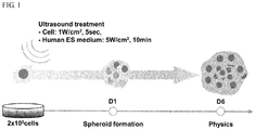
- a method of reprogramming the differentiated cells into pluripotent cells of the present invention will be described in detail with reference to FIG. 1 .
- a cell culture medium and differentiated cells are mixed, and energy is applied to the resulting mixture.
- the energy Prior to applying the energy to the mixture, the energy may be applied to the cell culture medium to enhance efficiency of reprogramming into the pluripotent cell.
- the energy may include any one among ultrasound, lasers or heat treatment.
- the ultrasound treatment of the culture medium may be performed by treating the culture medium with ultrasound having an output intensity of 1 W/cm 2 to 20 W/cm 2 for 1 to 20 minutes, specifically, with ultrasound having an output intensity of 2 W/cm 2 to 10 W/cm 2 for 5 to 15 minutes, more specifically, with ultrasound having an output intensity of 3 W/cm 2 to 7 W/cm 2 for 7 to 13 minutes.
- the treatment of the culture medium with the lasers may be performed by treating the culture medium with pulsed laser beams having a wavelength band of 300 to 900 nm for 1 second to 20 seconds, more specifically, with the pulsed laser beams having this wavelength band for 3 seconds to 10 seconds, even more specifically, with the pulsed laser beams having this wavelength band for 4 seconds to 6 seconds.
- a wavelength of 400 nm, 808 nm, or 880 nm may, for example, be used as the wavelength band.
- the heat treatment of the culture medium may be performed for 5 minutes to 20 minutes under the condition of a temperature of 40 to 50 °C.
- An embryonic stem cell culture medium, a stem cell differentiation-inducing medium, and the like may be used as the culture medium.
- Mammal-derived fibroblast cells may be used as the differentiated cells.
- cancer cells including cervical cancer cells (HeLa cells); or organ tissue cells including pulmonary epithelial cells (L132 cells) may be used as the differentiated cells.
- HeLa cells cervical cancer cells
- L132 cells organ tissue cells including pulmonary epithelial cells
- the differentiated cells When energy is applied to the differentiated cells, the differentiated cells are desirably exposed to a certain intensity of the energy. Cell viability may be reduced when the intensity falls out of this intensity range.
- the ultrasound treatment of the mixture of the culture medium and the differentiated cells may be performed at an output intensity of 0.5 W/cm 2 to 3 W/cm 2 for 1 to 5 seconds, more specifically, at an output intensity of 0.7 W/cm 2 to 2 W/cm 2 for 1 to 5 seconds, even more specifically, at an output intensity of 0.8 W/cm 2 to 1.5 W/cm 2 for 1 to 5 seconds.
- the treatment of the mixture of the culture medium and the differentiated cells with the lasers may be performed by irradiating the mixture with pulsed laser beams having a wavelength band of 300 to 900 nm for 1 second to 20 seconds, more specifically, with the pulsed laser beams having this wavelength band for 3 seconds to 10 seconds, even more specifically, with the pulsed laser beams having this wavelength band for 4 seconds to 6 seconds.
- a wavelength of 400 nm, 808 nm, or 880 nm may, for example, be used as the wavelength band.
- the heat treatment of the mixture of the culture medium and the differentiated cells may be performed by exposing the mixture to heat for 1 minute to 10 minutes under the condition of a temperature of 40 to 50 °C, and then exposing the mixture to heat for 5 to 10 seconds under the condition of a temperature of 0 °C to 4 °C.
- the mixture to which the energy is applied is cultured for a predetermined time to form spheroids having pluripotency.
- the culture of the mixture to which the energy is applied may be performed for a period of time in which the spheroids stably expressing an undifferentiated marker are formed by a suspension culture method or a monolayer culture method, that is, for 3 days to 10 days.
- the culture time may be suitably adjusted at a level of ordinary skill in the art because the formation of the spheroids having pluripotency varies depending on the culture method, the types of cells or culture media.
- the suspension culture is more efficient in forming the spheroids, compared to the monolayer culture. Also, the number and size of the spheroids in the suspension culture are larger than those in the monolayer culture, and the suspension culture exhibits a uniform size distribution.
- the expression of the undifferentiated markers is increased or stabilized from approximately day 3, indicating that the reprogramming starts from this point of time. Also, the expression of the undifferentiated markers is increased or stabilized from approximately day 8 upon the suspension culture of heat-treated human dermal fibroblast cells, indicating that the reprogramming starts from this point of time.
- the undifferentiated markers for example, OCT3/4, SOX2, NANOG, c-MYC, KLF4, TDGF1, SSEA4, TRA-1-60, and the like are expressed, it can be seen that the spheroids have pluripotency.
- the presence of the undifferentiated markers may be analyzed using RT-PCR or an immunocytochemistry, but the present invention is not limited thereto.
- the pluripotent cells of the present invention have a characteristic of expressing three germ layer markers, that is, ectoderm (PAX6 and Nestin), mesoderm (Brachyury and SMA), endoderm (GATA4 and AFP) markers at a high level.
- ectoderm PAX6 and Nestin
- mesoderm Brainury and SMA
- endoderm GATA4 and AFP
- the pluripotent cells of the present invention have a characteristic of having a proliferative capacity since the pluripotent cells express Ki-67 which is a proliferation marker protein.
- the proliferation of the pluripotent cells increases, and differentiate into the ectoderm/endoderm/mesoderm and nerve cells/myocardial cells after the pluripotent cells are cultured in a differentiation-inducing medium.
- the present invention provides a device for inducing pluripotent cells, which includes:
- the culture chamber refers to an incubator generally used to culture cells.
- the culture chamber is provided with a temperature control unit and a carbon dioxide control unit.
- the cell culture conditions in the culture chamber may be properly adjusted at a level of ordinary skill in the art, depending on a purpose and the type of cells.
- the culture chamber may have a structure in which such a culture is possible.
- the culture chamber may be a culture chamber provided with an agitator for suspension culture.
- the device for applying energy may include an ultrasound generation device configured to radiate ultrasound, a laser generation device configured to radiate lasers, or a temperature control device.
- ultrasound generation devices that generate ultrasound having a frequency of 10 kHz to 100 MHz may be used as the ultrasound generation device without limitation.
- Laser devices that generate pulsed laser beams having a wavelength band of 300 to 900 nm and an output of 1 to 15 W, a pulse duration time of 1 ms to 900 ms and a frequency of 1 to 100 Hz may be used as the laser generation device, but the present invention is not limited thereto.
- Known temperature control devices capable of regulating a temperature in a range of -40 °C to 99.9 °C may be used as the temperature control device, but the present invention is not limited thereto.
- the device for inducing pluripotent cells may be used to reprogram the differentiated cells into the pluripotent cells by treating the mixture of the culture medium and the differentiated cells with ultrasound, lasers or heat treatment using the ultrasound generation device, the laser generation device or the temperature control device and culturing the mixture for a predetermined time to form spheroids having pluripotency.
- the culture medium may be previously treated with the ultrasound, lasers or heat treatment prior to mixing the culture medium with the differentiated cells so as to enhance reprogramming efficiency.
- FIG. 1 is a schematic diagram for forming Physics ( p luripotent sp h ere y ielded by ultra s on ic s timulus) cells according to the present invention.
- Human dermal fibroblast cells (HDFa, Cat. No. C-013-5C, GIBCO (Invitrogen cell culture)) (1 ⁇ 10 6 ) were mixed with an embryonic stem (ES) medium, which had been treated with ultrasound at an intensity of 5 W/cm 2 for 10 minutes, and the resulting mixture including the cells was treated with ultrasound at an intensity of 1 W/cm 2 for 5 seconds.
- the viable cells were selected, and 2 ⁇ 10 5 HDFs were then suspension-cultured for 6 days in a human ES cell culture medium in a 35-mm Petri dish for bacterial culture.
- the spheroids were formed from the first day of culture, and the undifferentiated markers were expressed from day 3 onward.
- suspension culture in which cells were cultured in a cell culture dish whose surface was not coated (i. e., a Petri dish for bacterial culture), and monolayer culture, in which cells were cultured in a cell culture dish whose surface was coated so that the cells were easily attached to the surface of the dish (i. e., a tissue culture dish), were used.
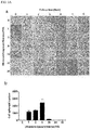
- HDFs (1 ⁇ 10 6 ) were directly exposed to ultrasound at ultrasound intensities (0, 0.5, 1, 3, 5, and 10 W/cm 2 for 5 seconds) to establish the ultrasound intensity conditions. Then, the viable cells were selected, and 2 ⁇ 10 5 HDFs were then cultured for 6 days in a human ES cell culture medium in a 35-mm Petri dish for bacterial culture.
- the HDFs were cultured in a human ES cell culture medium at differing exposure times (0, 1, 2, 5, 10, 20, and 40 seconds) over 3 days under the condition of a fixed ultrasound intensity of 1 W/cm 2 in a 35-mm Petri dish for bacterial culture.
- an ES cell culture medium was treated with ultrasound at varying exposure intensities (0, 1, 5, and 10 W/cm 2 ) for 10 minutes.
- 2 ⁇ 10 5 HDFs (1 W/cm 2 for 5 seconds) exposed to ultrasound were cultured in such media for 3 days in 35-mm Petri dishes for bacterial culture.
- an ESC culture medium was treated with ultrasound (5 W/cm 2 for 10 minutes), and HDFs (1 ⁇ 10 6 ) were treated with ultrasound (1 W/cm 2 for 5 seconds).
- the live HDFs ( ⁇ 10 5 ) were selected, and then suspension-cultured in a Petri dish for bacterial culture or monolayer-cultured in a tissue culture dish.
- the suspension-cultured ultrasound-treated HDFs had a higher spheroid-forming efficiency, compared to the monolayer-cultured HDFs. Also, when the stimulus such as ultrasound was applied to both the cells and the culture medium, the HDFs showed a higher spheroid-forming efficiency.
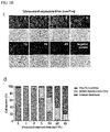
- the ultrasound-treated HDFs or untreated HDFs were cultured in an ES cell culture medium treated with ultrasound or an untreated ES cell culture medium in a Petri dish for bacterial culture or a tissue culture dish.
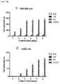
- suspension culture condition exhibited a higher efficiency, and the spheroids were larger in number and size (a diameter of 200 ⁇ m or more), and exhibited a uniform size distribution, compared to the monolayer culture condition.
- the ultrasound-treated HDFs (UC) grown in the untreated ES cell culture medium formed spheroids.
- the number and size (up to 200 ⁇ m) of the spheroids were very small, compared to the UCUM condition.
- UM normal HDFs
- a small amount of spheroids having a smaller size (100 ⁇ m or less) were formed.
- Most of the HDFs were attached to a surface of the culture dish, and the number of the spheroids was very small.
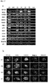
- the cells of the control and the ultrasound-treated groups (Null, UM, UC, and UCUM) were recovered in different culture time (days 1, 2, 3, 4, 5 and 6) according to the method of Example 1, and mRNA was extracted using a Dynabeads mRNA direct kit (Ambion). Then, SuperScript-II (Invitrogen) cDNA was synthesized, PCR-amplified using primers as listed in Table 2, and then subjected to electrophoresis for the purpose of analysis.
- the undifferentiated marker genes were stably expressed when both the HDFs and the culture medium were treated with ultrasound, as shown in FIG. 9 .
- the undifferentiated marker genes were expressed at a higher level, compared to the monolayer-cultured cells.
- the cells cultured for a culture time (0, 1, 2, 3, 4, 5 and 6 days) after the ultrasound treatment were fixed with 4% paraformaldehyde for 30 minutes, and then exposed to a PBS buffer supplemented with 0.1% Triton X100 for 40 minutes to improve a penetration ability of antibodies. Thereafter, the cells were blocked with a PBS buffer supplemented with 5% non-goat serum at room temperature for 30 minutes to prevent a non-specific protein reaction.
- the cells were washed, and a primary antibody (OCT4; 1:200, Abcam) was added. The resulting mixture was reacted overnight at 4 °C, and washed three times with a PBS buffer supplemented with 0.03% Triton X100. Then, a secondary antibody (IgG anti-rabbit conjugate Alexa 488) was diluted 1:1000 with a D-PBS buffer, and the cells were stained at room temperature for 2 hours. The stained cells were washed 4 times with a PBS buffer supplemented with 0.03% Triton X100, a DAPI-added mounting solution was sprayed on a slide so that the slide was covered with a cover slip, and edges of the slide were sealed with nail polish. Then, the cells were observed under a confocal laser microscope.
- OCT4 OCT4; 1:200, Abcam
- the OCT3/4 expression was detected immediately one day after ultrasound treatment under the UCUM conditions.
- the OCT3/4 expression gradually increased, and a level of the OCT3/4 expression was higher under the suspension culture condition, compared to the monolayer culture condition.
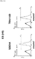
- OCT3/4, SOX2, NANOG, SSEA-4 and TRA-1-60 was similar to that of the H9 human ES cells ( FIG. 15 ). Also, the pluripotent characteristics of the multicellular spheroids were determined through flow cytometry ( FIG. 16 ). More than 99.5% of SSEA4 and TRA-60 were expressed in the spheroids, and an expression level of each of the SSEA4 and TRA-60 was similar to that of the H9 human ES cells.
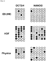
- a DNA methylation assay was performed. It can be seen that, when a promoter region in which the gene expression is initiated is methylated, the gene expression is not initiated in this region. This means that, when the promoter region is demethylated, that is, a methyl group is removed from DNA, a gene is expressed from the promoter region. Therefore, to check whether the OCT3/4 and NANOG genes were expressed as the main genes of the undifferentiated stem cells, it was determined whether the promoter regions of the two genes were methylated.
- DNA was extracted from the Physics spheroids using proteinase K and phenol, and the methylation of OCT3/4 and NANOG DNAs in the Physics spheroids was analyzed using an EZ DNA methylation kit (Zymo Research). Primers for DNA amplification used for analysis are as follows.
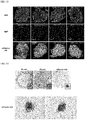
- the proliferative capacity of the Physics cells was evaluated using a method of immunostaining a proliferation marker protein Ki-67 and differential nuclear staining using Hoechst 33342 and propidium iodide (PI).
- the cultured Physics cells were stained on day 5 using Hoechst 33342 capable of staining the nuclei of the live cells due to good penetrability. After a staining reagent was completely removed, the Physics cells were cultured for 3 days. Thereafter, the cultured Physics cells were fixed with 4% paraformaldehyde for a total of 8 days, and the cell nuclei was further stained with PI. A non-overlapping red signal represents Physics cells newly formed by cell division after 5 days. Also, single spheroids were cultured for 5 days, and diameters of the spheroids were then measured on moving images. As a measurement result shows an increase in size, the proliferative capacity of the Physics cells was proven.
- the Physics cells stained with Hoechst 33342 were cultured for another 5 days, fixed with 4% paraformaldehyde, and then stained again with PI and OCT3/4.
- PI signals represent the nuclei in the Physics cells.
- the nuclei counterstained with OCT3/4 nearly merging with Hoechst 33342 meant that the Physics cells were stained with Hoechst 33342 before day 5.
- the pluripotent characteristics (OCT3/4) were not inherited by daughter Physics cells during 5 days of further culture. These results suggest that the Physics cells were able to proliferate, but were not self-renewed after 5 days.
- the repair process of the damaged plasma membranes was also proven by cellular analysis using a live/dead kit (Green fluorescence in the case of cells whose cell membranes are not damaged/Red fluorescence in the case of dead cells or cells whose cell membranes are damaged).
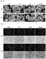
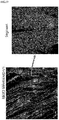
- the HDFs were stained with a fluorescent reagent immediately after the ultrasound treatment and after 2 hours had elapsed. As a result, it was revealed that, since a percentage of the red fluorescence was reduced after 2 hours, the cell membranes damaged by ultrasound was repaired 2 hours after the ultrasound treatment, as shown in the SEM analysis results ( FIG. 24 ).
- ultrasound-treated HDFs were added to a live/dead kit, and green/red dually stained HDFs were traced for 24 hours using a live cell imaging apparatus.
- the HDFs simply aggregated with other green-stained or red/green dually stained HDFs to form multicellular spheroids. After 24 hours, most of the slightly damaged HDFs formed stable Physics cells.
- the ultrasound-induced cell membrane damage and transient permeation were characterized by increased intracellular Ca 2+ concentration and intracellular H 2 O 2 generation using a fluorescent dye (i. e., a Fluo-4 dye) and CM-H2DCFDA.
- a fluorescent dye i. e., a Fluo-4 dye
- CM-H2DCFDA a fluorescent dye
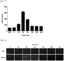
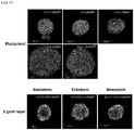
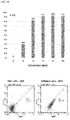
- FIG. 26 The intracellular concentration of H 2 O 2 in the Physics cells was six-fold higher than that of the untreated HDFs as the control after 60 minutes of ultrasound exposure ( FIGS. 27 and 28 ).
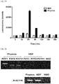
- the ultrasound stimulated the release of a 22-fold higher level of ATP from the Physics cells, compared to the untreated HDFs.
- exosomal RNA was prepared from a Physics cell culture medium, and a gene expression pattern in a cell culture environment during Physics cell generation was studied through RT-PCR analysis.
- an exosome includes several genetic elements, for example, RNA, microRNA, DNA, proteins.
- an expression profile of the genetic elements in the exosome is cell status-dependent.
- the highest expression level of the pluripotent marker genes was observed in a purified exosome from the Physics cell culture medium.
- the most outstanding gene expression was observed for OCT3/4 and NANOG.
- the NANOG expression dropped after 4 days.
- the c-MYC expression was constantly maintained under the suspension culture conditions, but dropped under the monolayer culture conditions after 2 days.
- the expression of all the pluripotent marker genes was detected under the suspension culture conditions even when the pluripotent marker genes were expressed at a low expression level. However, these genes were not detected under the monolayer culture condition.
- HDFs not treated with ultrasound were co-cultured with the Physics cells.
- the HDFs were stained with a Cy5.5 red fluorescence dye using Lipofectamine.
- the Physics cells were prepared separately. After the Physics cells were maintained for 2 days, the Physics cells were added to a Cy5.5-stained HDF culture dish. During co-culturing, the culture medium was not treated with ultrasound. This is because the expression of the pluripotent marker genes was also induced under the UM condition.
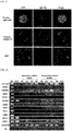
- SRY-box including gene gene 17 (SOX17; the endoderm), paired box 6 (PAX6; the ectoderm), Nestin (a nerve cell marker), microtubule-associated protein 2 (MAP2; the ectoderm), class III beta-tubulin (TuJ1; a nerve cell marker), msh homeobox 1 (MSX1; the mesoderm), Brachyury (the mesoderm), myosin light chain 7 (MYL7; myocardial cells), NK2 homeobox 5 (NKX2.5; myocardial cells), and Troponin T type 2 (TnnT2; myocardial cells) was observed during a differentiation time of 1 to 2 weeks using RT-PCR.
- MSX1 msh homeobox 1
- Brachyury the mesoderm
- MYL7 myosin light chain 7
- NK2 homeobox 5 NKX2.5; myocardial cells
- Troponin T type 2 Troponin
- OCT3/4 was significantly reduced after the induction of differentiation.
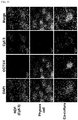
- the differentiation into nerve cells or cardiac cells was also confirmed through immunocytochemistry.
- the neural progenitor cell markers (PAX6 and Nestin) were observed in the Physics cells grown in an astrocyte medium.
- the expression of each of the oligodendrocyte markers (MAP2 and 04) or the neuronal markers (MAP2 and Tuj1) was observed.
- a differentiation time of 2 weeks was sufficient to detect the cardiac markers including MHC, SMA, Actinin, NKX2.5 and TnTc.
- a typical segmented actin pattern was detected in actinin.
- no neural or cardiac markers were expressed in the HDFs under the same culture conditions.
- the ultrasound did not cause undesirable side-effects such as mutagenesis, genetic modifications, cancer generation, etc.
- the Physics cells had a normal karyotype ( FIG. 40 ).
- a human ES cell culture medium was used to generate Physics cells.
- the ES cell culture medium was developed as a defined medium for maintaining and proliferating ES cells in an undifferentiated state.
- a normal HDF culture medium was used to generate Physics cells.
- the shape and spheroid-forming efficiency of the HDF culture medium ware quite different from those of the ES cell culture medium.
- the Physics cells were seeded in an ultrasound-treated HDF medium, a small amount of multicellular spheroids were formed. However, most of the spheroids were attached to a surface of a dish after 2 days. On day 4 of culture, all the spheroids were attached to the surface of the dish to grow into typical fibroblast cells. Immunocytochemical results also showed that different gene expression patterns appeared between the two different culture medium conditions.
- the typical Physics cells formed using the ES cell culture medium showed a high expression level of OCT3/4, SOX2, NANOG, SSEA-4, and TRA-1-60.
- a DMEM medium had no effect on induction of expression of the undifferentiated marker genes and three germ layer marker genes.
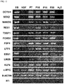
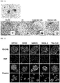
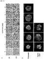
- the expression of the pluripotent marker including OCT3/4, SOX2, NANOG, SSEA4, and TRA-1-60 and the three germ layer marker genes including GATA4, AFP, PAX6, Nestin, Brachyury, and SMA in two types of different Physics cells were confirmed through immunocytochemistry.
- dermal fibroblast cells were exposed to 42 °C for 2 minutes, and then kept on ice for approximately 5 seconds.
- the multicellular spheroids were successfully formed after both of the HDF and ES cell culture media were subjected to the laser or heat treatment.
- the laser-treated HDFs also formed multicellular spheroids immediately after laser induction.
- the spheroids had an irregular shape and a non-uniform size distribution, it was observed that the pluripotent markers and three germ layer markers were expressed at a high level.
- the heat treatment also induced the spheroid formation. However, the efficiency of the heat treatment was lower than those of the ultrasound and laser treatment. At least half of the multicellular spheroids induced by heat were attached to a surface of the dish for 8 days of maintenance. Despite the lower spheroid-forming efficiency, high expression levels of the pluripotent markers and three germ layer markers were observed.
- Mouse Physics cells were prepared according to a method shown in FIG. 48 .
- OG2 mouse embryonic fibroblast (MEF) were mixed with an ES cell culture medium which had been treated with 20 KHz ultrasound at an intensity of 5 W/cm 2 for 10 minutes. Thereafter, the cells were directly treated with ultrasound at an intensity of 1 W/cm 2 for 5 seconds, and then cultured. The cultured cells were observed at intervals of 1, 3, 5, 8 and 10 days under a fluorescence microscope to determine a morphological change of the cells and fluorescent GFP expression.
- the compositions of the media for ultrasound treatment are as listed in Table 1.
- the MEF cells were embryonic fibroblasts of 13.5-day old mice transformed with a GFP gene into which an OCT4 promoter was inserted and typically did not express OCT4. However, when OCT4 was expressed, GFP was also expressed, thereby producing green fluorescence.
- the control did not produce green fluorescence in images of the OG2 MEF cells (OCT4 was not expressed).
- OCT4 was not expressed.
- the size of cell spheres increased and the green fluorescence intensity increased as culture time passed in the case of OG2 MEFs treated with ultrasound. This indicates that the ultrasound treatment induced the OCT4 expression, and OCT4 had important characteristics of undifferentiated stem cells, indicating that the OG2 MEF cells reprogrammed into stem cells due to the ultrasound treatment.
- FIG. 49B is a diagram of images merged as Tile scan images after multiple pictures were taken over a wide range, which shows an effect of ultrasound treatment.
- a large number of MEF cells reprogrammed due to the ultrasound treatment to express OCT4-GFP.
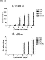
- the GFP expression efficiency of the spheroids formed as shown in FIG. 50 was analyzed. As a result, it was revealed that the spheroids had approximately 93% OCT4-GFP expression efficiency. Also, it was revealed that GFP was expressed in approximately 85.3% of the cells when the GFP expression in the whole cells was confirmed using flow cytometry. Further, it was revealed that SSEA1, which is an undifferentiated protein marker on the cell surface, was expressed in approximately 75.5% of the cells even when the SSEA1 expression was confirmed ( FIG. 51 ). Such results suggest that the reprogramming efficiency was significantly increased due to the ultrasound treatment.
- RT-PCR primers used to check expression of undifferentiated markers in mouse ES Gene names Primer sequences (5' ⁇ 3') Oct3/4 F CTGAGGGCCAGGCAGGAGCACGAG R CTGTAGGGAGGGCTTCGGGCACTT Sox2 F TAGAGCTAGACTCCGGGCGATGA R TTGCCTTAAACAAGACCACGAAA Nanog F CAGGTGTTTGAGGGTAGCTC R CGGTTCATCATGGTACAGTC c-Myc F TGACCTAACTCGAGGAGGAGCTGGAATC R AAGTTTGAGGCAGTTAAAATTATGGCTGAAGC Klf4 F GCGAACTCACACAGGCGAGAAACC R TCGCTTCCTCTTCCTCCGACACA Esg1 F GAAGTCTGGTTCCTTGGCAGGATG R ACTCGATACACTGGCCTAGC Rex1 F ACGAGTGGCAGT
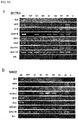
- the expression of the three germ layer markers was checked. As a result, it was revealed that the markers of the endoderm (GATA6), the ectoderm (Nestin) and the mesoderm (Brachyury) were expressed at a high level. The other genes of the three germ layers started to be expressed on day 3 after formation of the Physics cells. The expression level of the three germ layer markers gradually increased for 20 days of culture ( FIG. 55 ). Also, as shown in FIG. 56 , the expression of the three germ layer protein markers was confirmed through immunostaining.
- the mPhysics cells formed by the ultrasound had a normal karyotype ( FIG. 57 ).
- the pluripotent cells of the present invention can be used in the field of cell therapy.
Landscapes
- Health & Medical Sciences (AREA)
- Life Sciences & Earth Sciences (AREA)
- Engineering & Computer Science (AREA)
- Chemical & Material Sciences (AREA)
- Bioinformatics & Cheminformatics (AREA)
- Organic Chemistry (AREA)
- Wood Science & Technology (AREA)
- Zoology (AREA)
- Biomedical Technology (AREA)
- Genetics & Genomics (AREA)
- Biotechnology (AREA)
- Microbiology (AREA)
- General Health & Medical Sciences (AREA)
- Biochemistry (AREA)
- General Engineering & Computer Science (AREA)
- Sustainable Development (AREA)
- Cell Biology (AREA)
- Analytical Chemistry (AREA)
- Transplantation (AREA)
- Developmental Biology & Embryology (AREA)
- Virology (AREA)
- Medicinal Chemistry (AREA)
- Physics & Mathematics (AREA)
- Thermal Sciences (AREA)
- Micro-Organisms Or Cultivation Processes Thereof (AREA)
- Electromagnetism (AREA)
- Apparatus Associated With Microorganisms And Enzymes (AREA)
- Plasma & Fusion (AREA)
- Optics & Photonics (AREA)
- Immobilizing And Processing Of Enzymes And Microorganisms (AREA)
Abstract
Description
- The present invention relates to a device and a method for inducing pluripotent cells using energy capable of inducing pluripotent cells having pluripotent characteristics by applying energy such as ultrasound, lasers, heat treatment, etc.
- Pluripotency is an ability of cells to differentiate into three germ layer lineages, that is, ectoderm, mesoderm and endoderm. Pluripotent stem cells are clinically important for disease models or transplantation because the stem cells are differentiated into any types of cells or tissues in the body. Therefore, the current major requests in reprogramming or differentiation of embryonic stem cells, induced pluripotent stem cells (iPSC), somatic cells, and patient-derived cells should be simple, fast, efficient, and safe for clinical application with free of the introduction of exogenous genetic materials or chemical or small molecules. Recent studies have demonstrated that the interactions between the environment and genotypes are closely related to the gene expression and phenotypic variation in living organisms. Controlling the environmental stimulation such as structural, mechanical, magnetic, ultrasonic cues can modulate cell fate, proliferation, and cellular uptake efficiency. Although the exact molecular mechanism for these approaches is still unclear, these methods are acceptable as an alternative way to achieve the safety without introduction of genetic materials, chemical compounds and small molecules.
- In this regard, the present inventors have developed a new method for inducing novel pluripotent cells having pluripotent characteristics that express undifferentiated markers and three germ layer marker genes that may differentiate into three germ layer, that is, ectoderm, mesoderm and endoderm, called Physics ( p luripotent sp h ere yielded by ultra s on ic s timulus) cells, by applying energy with cellular environmental cues under gene- and chemical-free condition.
- Therefore, it is an aspect of the present disclosure to provide a method of inducing a novel type of pluripotent cells having pluripotent characteristics from differentiated cells by applying energy without any introduction of a reprogramming inducing factor into differentiated cells and a chemical, and a device for inducing the pluripotent cells.
- To solve the above problems, according to an aspect of the present invention, there is provided a method of reprogramming differentiated cells into pluripotent cells, which includes mixing differentiated cells with a culture medium and forming spheroids by applying energy to the resulting mixture and culturing the mixture for a predetermined time, wherein, the spheroids have pluripotent characteristics.
- According to another aspect of the present invention, there is provided a device for inducing pluripotent cells, which includes a culture chamber configured to accommodate differentiated cells and a culture medium; and a device for applying energy to the differentiated cells and the culture medium equipped at one side of the culture chamber, wherein, the differentiated cells and the culture medium are mixed, and spheroids are formed by applying energy to the resulting mixture and culturing the mixture for a predetermined time, and the spheroids have pluripotent characteristics.
- The present invention has an effect of inducing a novel type of pluripotent cells, which has pluripotent characteristics and shows a stronger differentiation property than known induced pluripotent stem cells, from differentiated cells by applying suitable energy such as ultrasonic waves, lasers, heat treatment, etc. without introduction of a factor for inducing dedifferentiation into differentiated cells and a chemical compound.
-
-
FIG. 1 is a schematic diagram of human Physics cells having pluripotent characteristics according to the present invention. -
FIG. 2A is a diagram showing an effect of ultrasound intensity on human dermal fibroblasts (HDFs): a) shows the comparison of morphological changes of HDFs under different ultrasound intensity, and b) shows the number of multicellular spheroids formed under different ultrasound intensity. -
FIG. 2B is a diagram showing an effect of ultrasound intensity on the human dermal fibroblasts (HDFs): c) shows results of live/dead cell assay of ultrasound-treated HDFs, and d) shows percentages of viable and damaged cells in c). -
FIG. 3A is a diagram showing an effect of ultrasound exposure time under a fixed intensity of 1 W/cm2: a) shows the comparison of morphological changes of HDFs under different ultrasound exposure time, and b) shows the number of multicellular spheroids formed under different ultrasound exposure time. -
FIG. 3B is a diagram showing an effect of ultrasound exposure time under a fixed intensity of 1 W/cm2: c) shows results of live/dead cell assay of ultrasound-treated HDFs, and d) shows percentages of viable and damaged cells in c). -
FIG. 4 is a diagram showing an effect of ultrasound intensity on human ESC culture media: the upper panel shows the comparison of morphological changes of ultrasound-treated HDFs growing in an ultrasound-treated medium, and the lower panel shows the number of multicellular spheroids formed in the medium. -
FIG. 5 is a diagram showing an effect of ultrasound exposure time under a fixed intensity of 5 W/cm2: a) shows the comparison of morphological changes of ultrasound-treated HDFs growing in an ultrasound-treated medium, and b) shows the number of multicellular spheroids formed in a). -
FIG. 6 is a diagram showing effects of an ultrasound treatment condition and a culture condition for forming multicellular spheroids: a) shows suspension culture results, and b) shows monolayer culture results. -
FIG. 7A shows size distributions of multicellular spheroids formed under a suspension culture condition under different ultrasound stimuli (UC, UM, and UCUM): a) shows the distribution of the spheroids by total size, and b) shows the distribution of the spheroid having a size of 50 to 100 µm. -
FIG. 7B shows size distributions of the multicellular spheroids formed under the suspension culture condition under different the ultrasound stimuli (UC, UM, and UCUM): c) shows the distribution of the spheroids having a size of 100 to 200 µm, and d) shows the distribution of the spheroids having a size of more than 200 µm. -
FIG. 8A shows size distributions of the multicellular spheroids formed under a monolayer culture condition under different the ultrasound stimuli (UC, UM, and UCUM): a) shows the distribution of the spheroids by total size, and b) shows the distribution of the spheroid having a size of 50 to 100 µm. -
FIG. 8B shows size distributions of the multicellular spheroids formed under the monolayer culture condition under different the ultrasound stimuli (UC, UM, and UCUM): c) shows the distribution of the spheroids having a size of 100 to 200 µm, and d) shows the distribution of the spheroids having a size of more than 200 µm. -
FIG. 9A shows RT-PCR analysis results of comparing expression levels of a pluripotent marker gene, OCT3/4 (a) and SOX2 (b) in human Physics cells between the suspension culture condition and the monolayer culture condition. -
FIG. 9B shows RT-PCR analysis results of comparing expression levels of the pluripotent marker gene, NANOG (c) and TDGF1 (d) in the human Physics cells between the suspension culture condition and the monolayer culture condition. -
FIG. 10A is a confocal laser microscope image of OCT3/4 whose expression level is analyzed during suspension culturing. -
FIG. 10B is a confocal laser microscope image of the OCT3/4 whose expression level is analyzed during monolayer culturing. -
FIG. 11 shows RT-PCR analysis results of expression of pluripotent marker genes for a culture time of 6 days. -
FIG. 12 shows RT-PCR analysis results of determining an expression stage of OCT3/4 in ultrasound-treated HDF spheroids in different culture time. -
FIG. 13 shows results of determining the expression of representative undifferentiated markers in ES cells, HDFs and human Physics cells. -
FIG. 14 shows alkaline phosphatase staining results for characterizing a pluripotent state of the multicellular spheroids. -
FIG. 15 shows immunocytochemical results of expression of pluripotent markers. -
FIG. 16A shows FACS analysis results of a human ES (H9) cell surface marker. -
FIG. 16B shows FACS analysis results of a human HDF cell surface marker. -
FIG. 16C shows FACS analysis results of a human Physics cell surface marker. -
FIG. 17 show results of analyzing (a) gene expression of the pluripotent markers using RT-PCR and (b) protein expression of the pluripotent markers using an immunocytochemical method when feeder cells and Physics cells are co-cultured. -
FIG. 18 shows bisulfite genomic sequencing results showing methylation states of COT3/4 and NANOG promoters. -
FIG. 19 shows experimental results of verifying the proliferative capacity of the human Physics cells: a) shows results showing expression of Ki67 which is a proliferation marker, b) shows results of staining increased cells using H33342 and PI, and c) show the sizes of the spheroids in different culture time. -
FIG. 20 shows experimental results of verifying the self-renewal capacity of the human Physics cells. -
FIG. 21A shows immunocytochemical analysis results of a three germ layer marker expressed in human ES (H9) cells. -
FIG. 21B shows immunocytochemical analysis results of the three germ layer marker expressed in human Physics cells. -
FIG. 22 shows immunocytochemical analysis results of expression patterns of OCT3/4 and the three germ layer marker during 15 days of culturing the human Physics cells. -
FIG. 23 shows an SEM image of the human Physics cells in different ultrasound stimulation conditions. -
FIG. 24 shows results of live/dead cell assay of human Physics cells as viewed in a fluorescence image after ultrasound treatment and after 2 hours of culture under ultrasound stimulation conditions -
FIG. 25 shows results of the live/dead cell assay of human Physics cells after formation of the multicellular spheroids. -
FIG. 26 shows changes in intracellular Ca2+ concentrations by ultrasonic stimulus. -
FIG. 27 shows results of analyzing generation of H2O2 by the ultrasonic stimulus. -
FIG. 28 is a fluorescence image of the human Physics cells stained with CM-H2DCFDA to analyze the generation of intracellular H2O2 by the ultrasound treatment as shown inFIG. 27 . -
FIG. 29 shows results of analyzing release of intracellular ATP by the ultrasonic stimulus. -
FIG. 30 shows expression patterns of P2X and P2Y receptors in the human Physics cells. -
FIG. 31 is a confocal microscope image of proving a higher uptake capacity of the human Physics cells usingQuantum dot 705. -
FIG. 32 shows RT-PCR analysis results of expression of pluripotent marker genes from an exosome in human Physics cell culture media. -
FIG. 33 shows immunocytochemical results showing a transport process into HDFs by exosomal OCT4 released from the human Physics cells: Yellow arrows indicateOCT3/4 expression in surrounding HDFs. -
FIG. 34 shows immunocytochemical results of expression of the three germ layer marker after induction of in vitro differentiation of the human Physics cells. -
FIG. 35 shows RT-PCR analysis results of expression of neuronal lineage genes to check in vitro differentiation of the human Physics cells. -
FIG. 36 shows RT-PCR analysis results of expression of cardiac lineage genes to check in vitro differentiation of the human Physics cells. -
FIG. 37 shows immunocytochemical results of expression of a neuronal lineage marker to check in vitro differentiation of the human Physics cells. -
FIG. 38 shows immunocytochemical results of expression of a cardiac lineage marker to check in vitro differentiation of the human Physics cells. -
FIG. 39 is an immunocytochemical image of actinin. -
FIG. 40 shows karyotyping analysis results of the human Physics cells of the present invention. -
FIG. 41 shows fluorescent immunohisto logical staining results for determining in vivo differentiation of the human Physics cells in a mouse testis. -
FIG. 42 shows fluorescent immunohistological staining results of the human Physics cells to check in vivo differentiation of muscle in mouse thighs. -
FIG. 43 shows an effect of cell culture media: a) shows results using an ES medium and an HDF culture medium, and b) shows results of inducing the human Physics cells in the ES medium and the HDF culture medium and determining the presence of an ES marker. -
FIG. 44 shows results of inducing the human Physics cells from other cell lines: a) shows morphological changes of the cells, b) and c) shows results of inducing the human Physics cells from the other cell lines and determining the presence of b) an ES marker and c) a three germ layer marker. -
FIG. 45 shows results of inducing the human Physics cells from patient skin cells: a) shows morphological changes of the cells, and b) shows results of inducing the human Physics cells from the other cell lines and determining the presence of the ES marker and the three germ layer marker. -
FIG. 46 shows results of inducing the human Physics cells using heat treatment as another energy source and determining the presence of the ES marker and the three germ layer markers. -
FIG. 47 shows results of inducing the human Physics cells using a laser as still another energy source and determining the presence of the ES marker and the three germ layer marker. -
FIG. 48 shows a process of inducing the mouse Physics cells from mouse embryonic fibroblast cells. -
FIG. 49 shows results of determining morphological changes of and expression of Oct4 of transformed mouse embryonic fibroblast cells (OG2 MEF cells) in different culture time after ultrasound treatment: A shows results of determining GFP expression in mouse Physics spheroids, and B is a diagram of images merged as Tile scan images after multiple pictures were taken over a wide range. -
FIG. 50 is a graph plotted for a GFP expression rate of spheroids formed by ultrasound treatment, and a diagram showing results of analyzing a GFP expression rate in the entire ultrasound-treated cells using flow cytometry. -
FIG. 51 shows results of analyzing expression of an undifferentiated cell surface marker (SSEA1) in the mouse Physics cells using flow cytometry. -
FIG. 52 shows RT-PCR analysis results of the pluripotent marker genes in the mouse Physics cells. -
FIG. 53 shows results of analyzing the pluripotent protein markers in the mouse Physics cells. -
FIG. 54 shows alkaline phosphatase staining results for characterizing a pluripotent state of the mouse Physics cells. -
FIG. 55 shows RT-PCR analysis results of expression of the three germ layer marker in the mouse Physics cells. -
FIG. 56 shows results of analyzing expression of the three germ layer marker in the mouse Physics cells using an immunocytochemistry. -
FIG. 57 shows results of karyotyping analysis of the mouse Physics cells. - Hereinafter, configurations of the present invention will be described in detail.
- The present invention relates to a method of reprogramming differentiated cells into pluripotent cells, which includes mixing differentiated cells with a culture medium, and forming spheroids by applying energy to the resulting mixture and culturing the mixture for a predetermined time, wherein, the spheroids have pluripotent characteristics.
- The present invention is characterized in that a novel type of pluripotent cells, which has pluripotent characteristics and shows a stronger differentiation property than known induced pluripotent stem cells, may be induced from differentiated cells by applying suitable energy without introduction of a reprogramming inducing material such as a reprogramming inducing factor, a chemical compound, etc. into differentiated cells.
- The pluripotent cells have a characteristic distinguished from the known induced pluripotent stem cells in that the differentiation of the pluripotent cells is easily induced in response to an external environment, and the pluripotent cells show a property of progenitor cells having a strong differentiation property, compared to that of the stem cells. That is, when embryonic stem cells such as induced pluripotent stem cells are used for cell therapy, the embryonic stem cells somewhat require a preparative step of undergoing a differentiation process, which involves a risk factor for generating cancer, and safety issues are encountered when a viral vector is used to introduce the reprogramming inducing factor. However, the pluripotent cells of the present invention do not have to be cultured by co-culturing different types of cells because the pluripotent cells are induced without separately introducing the reprogramming inducing material such as a reprogramming inducing factor or a chemical compound to mutate a gene, and thus there are no problems regarding cell contamination (a problem arising from mixing pluripotent cells with another type of cells). Also, the pluripotent cells of the present invention have no problem regarding the occurrence of cancer because the pluripotent cells do not form a teratoma similar to cancer cells in an in vivo experiment, and thus safety is secured. That is, the pluripotent cells of the present invention have an advantage in that a time spanning from autologous cell treatment to transplantation may be remarkably shortened due to a simple and short induction process.
- According to the present invention, the spheroids may be specifically formed with high yield by applying energy to both of the culture medium and the differentiated cells.
- The energy may include one selected from ultrasound, lasers, and heat treatment.
- The pluripotent cells of the present invention are characterized by stably expressing any one undifferentiated markers or three germ layers marker genes consisting of the ectoderm, the mesoderm and the endoderm among OCT3/4, SOX2, NANOG, c-MYC, KLF4, TDGF1, SSEA4, TRA-1-60, PAX6, Nestin, Brachyury, SMA, GATA4, and AFP.
- As described above, since the pluripotent cells are formed without introducing the reprogramming inducing factor into differentiated cells, the present inventors have contemplated the correlation of the pluripotent cells with an exosome. That is, the ultrasound, the lasers or the heat treatment induces a rise in temperature due to energy application, generation of reactive oxygen species (ROS), vibration of microbubbles generated by ultrasound, and occurrence of liquid flow, that is, induces generation of microstreams along a cell membrane to cause fine damage to the cell membrane due to such an effect, thereby inducing formation of holes to increase the absorption of foreign substances. This is proven by analyzing a change in intracellular Ca2+ concentration and confirming whether H2O2 is generated in the cells. That is, the analysis of the change in intracellular Ca2+ concentration reflects that the Ca2+ concentration in the cells (cytosols) instantly increases when the cell membrane is damaged or the fluidity of the membrane increases, thereby verifying an increase in fluidity of the cell membrane. According to one exemplary embodiment of the present invention, it can be seen that the Ca2+ concentration rapidly increases immediately after the ultrasound treatment, and then gradually decreases to a level of the control in which the cells are not treated with ultrasound, indicating that the cell membrane is recovered after the damage to the cell membrane is induced. In the experiment confirming whether H2O2 is generated in the cells, it can also be seen that the cell membrane is recovered after the damage to the cell membrane is induced as an amount of the generated H2O2 increases immediately after the initial ultrasound treatment and then gradually decreases to a level of the control in a pattern similar to that of the Ca2+ concentration. Also, since ATP is used as a signal responding to various types of cellular stress, the ATP concentration in the pluripotent cells is analyzed after the ultrasound treatment. As a result, the ATP is released at a higher level, compared to the untreated control. Further, the expression of an ionotropic P2X receptor and a metabotropic P2Y receptor by the ATP release is also activated in the pluripotent cells, compare to the control.
- Meanwhile, exosomes are known to contain genetic elements (DNA, mRNA, microRNA, proteins, etc.) in an inside thereof. In this case, as the exosomes flowing out of a cell membrane, due to the damage to the cell membrane, enter other surrounding cells, the genetic elements present inside the exosomes may be transferred. Therefore, as the expression of undifferentiated markers which have been poorly expressed in the cells or whose expression has been maintained in a suppressed state is induced and promoted in response to the stimuli caused by the ultrasound treatment, and cell membranes are damaged at the same time, the exosomes present inside the cells including the undifferentiated markers whose expression is induced and promoted are released from the cells to enter the surrounding cells. In this case, it is assumed that, since cell membranes of the surrounding cells are also partially damaged, the exosomes enter the cells with high efficiency due to the increased fluidity of the cell membranes, compared to when the cell membranes are in a normal state. It is contemplated that genetic elements on the undifferentiated markers whose expression is induced and promoted, which are present inside such exosomes, is transported to make pluripotent cells. According to one exemplary embodiment of the present invention, a culture medium is recovered during the induction of the pluripotent cells, and the exosomes in the medium are extracted to determine whether a pluripotent cell-related undifferentiated marker is present inside the exosomes. As a result, it was revealed that known undifferentiated markers are expressed at a high level. Therefore, the present inventors have contemplated these facts to support this hypothesis. Also, it was revealed that the karyotype is normal without any mutation even when treated with such ultrasound, lasers or heat treatment.
- Such a hypothesis suggests that the pluripotent cells may be prepared by inducing the release of the exosomes when the cell membranes are damaged.
- In this specification, the term "pluripotent cell" refers to a cell that retains pluripotency after energy, that is, ultrasound, lasers or heat treatment in a strict sense, is applied to the cells. In this specification, the pluripotency refers to a state in which an undifferentiated marker expressed in embryonic stem cells is stably expressed. Further, the pluripotency refers to a state in which three germ layer markers for the endoderm, the ectoderm and the mesoderm are expressed. The term "pluripotent cells" may be used interchangeably with "Physics ( p luripotent sp h ere y ielded by ultra s on ic s timulus) cells" or "Physics spheroids." A method of reprogramming the differentiated cells into pluripotent cells of the present invention will be described in detail with reference to
FIG. 1 . - First of all, a cell culture medium and differentiated cells are mixed, and energy is applied to the resulting mixture.
- Prior to applying the energy to the mixture, the energy may be applied to the cell culture medium to enhance efficiency of reprogramming into the pluripotent cell.
- The energy may include any one among ultrasound, lasers or heat treatment.
- The ultrasound treatment of the culture medium may be performed by treating the culture medium with ultrasound having an output intensity of 1 W/cm2 to 20 W/cm2 for 1 to 20 minutes, specifically, with ultrasound having an output intensity of 2 W/cm2 to 10 W/cm2 for 5 to 15 minutes, more specifically, with ultrasound having an output intensity of 3 W/cm2 to 7 W/cm2 for 7 to 13 minutes.
- The treatment of the culture medium with the lasers may be performed by treating the culture medium with pulsed laser beams having a wavelength band of 300 to 900 nm for 1 second to 20 seconds, more specifically, with the pulsed laser beams having this wavelength band for 3 seconds to 10 seconds, even more specifically, with the pulsed laser beams having this wavelength band for 4 seconds to 6 seconds. A wavelength of 400 nm, 808 nm, or 880 nm may, for example, be used as the wavelength band.
- The heat treatment of the culture medium may be performed for 5 minutes to 20 minutes under the condition of a temperature of 40 to 50 °C.
- An embryonic stem cell culture medium, a stem cell differentiation-inducing medium, and the like may be used as the culture medium.
- Mammal-derived fibroblast cells; cancer cells including cervical cancer cells (HeLa cells); or organ tissue cells including pulmonary epithelial cells (L132 cells) may be used as the differentiated cells.
- When energy is applied to the differentiated cells, the differentiated cells are desirably exposed to a certain intensity of the energy. Cell viability may be reduced when the intensity falls out of this intensity range.
- Therefore, the ultrasound treatment of the mixture of the culture medium and the differentiated cells may be performed at an output intensity of 0.5 W/cm2 to 3 W/cm2 for 1 to 5 seconds, more specifically, at an output intensity of 0.7 W/cm2 to 2 W/cm2 for 1 to 5 seconds, even more specifically, at an output intensity of 0.8 W/cm2 to 1.5 W/cm2 for 1 to 5 seconds.
- The treatment of the mixture of the culture medium and the differentiated cells with the lasers may be performed by irradiating the mixture with pulsed laser beams having a wavelength band of 300 to 900 nm for 1 second to 20 seconds, more specifically, with the pulsed laser beams having this wavelength band for 3 seconds to 10 seconds, even more specifically, with the pulsed laser beams having this wavelength band for 4 seconds to 6 seconds. A wavelength of 400 nm, 808 nm, or 880 nm may, for example, be used as the wavelength band.
- The heat treatment of the mixture of the culture medium and the differentiated cells may be performed by exposing the mixture to heat for 1 minute to 10 minutes under the condition of a temperature of 40 to 50 °C, and then exposing the mixture to heat for 5 to 10 seconds under the condition of a temperature of 0 °C to 4 °C.
- Next, the mixture to which the energy is applied is cultured for a predetermined time to form spheroids having pluripotency.
- The culture of the mixture to which the energy is applied may be performed for a period of time in which the spheroids stably expressing an undifferentiated marker are formed by a suspension culture method or a monolayer culture method, that is, for 3 days to 10 days. However, the culture time may be suitably adjusted at a level of ordinary skill in the art because the formation of the spheroids having pluripotency varies depending on the culture method, the types of cells or culture media.
- According to one exemplary embodiment of the present invention, the suspension culture is more efficient in forming the spheroids, compared to the monolayer culture. Also, the number and size of the spheroids in the suspension culture are larger than those in the monolayer culture, and the suspension culture exhibits a uniform size distribution.
- According to one exemplary embodiment of the present invention, when human dermal fibroblast cells treated with the ultrasound or lasers are suspension-cultured, the expression of the undifferentiated markers is increased or stabilized from approximately
day 3, indicating that the reprogramming starts from this point of time. Also, the expression of the undifferentiated markers is increased or stabilized from approximatelyday 8 upon the suspension culture of heat-treated human dermal fibroblast cells, indicating that the reprogramming starts from this point of time. - From the fact that the undifferentiated markers, for example, OCT3/4, SOX2, NANOG, c-MYC, KLF4, TDGF1, SSEA4, TRA-1-60, and the like are expressed, it can be seen that the spheroids have pluripotency. The presence of the undifferentiated markers may be analyzed using RT-PCR or an immunocytochemistry, but the present invention is not limited thereto.
- Also, the pluripotent cells of the present invention have a characteristic of expressing three germ layer markers, that is, ectoderm (PAX6 and Nestin), mesoderm (Brachyury and SMA), endoderm (GATA4 and AFP) markers at a high level.
- In addition, the pluripotent cells of the present invention have a characteristic of having a proliferative capacity since the pluripotent cells express Ki-67 which is a proliferation marker protein.
- Further, it can be seen that, when the aforementioned reprogrammed pluripotent cells are co-cultured with feeder cells, the proliferation of the pluripotent cells increases, and differentiate into the ectoderm/endoderm/mesoderm and nerve cells/myocardial cells after the pluripotent cells are cultured in a differentiation-inducing medium.
- Also, the present invention provides a device for inducing pluripotent cells, which includes:
- a culture chamber configured to accommodate cells and a culture medium; and
- a device for applying energy to the differentiated cells and the culture medium equipped at one side of the culture chamber, wherein, the differentiated cells and the culture medium are mixed, and spheroids are formed by applying energy to the resulting mixture and culturing the mixture for a predetermined time, and the spheroids have pluripotent characteristics.
- The culture chamber refers to an incubator generally used to culture cells. For example, the culture chamber is provided with a temperature control unit and a carbon dioxide control unit. In this case, the cell culture conditions in the culture chamber may be properly adjusted at a level of ordinary skill in the art, depending on a purpose and the type of cells.
- Also, because the suspension or monolayer culture methods may be used to reprogram the differentiated cells into the pluripotent cells, the culture chamber may have a structure in which such a culture is possible. For example, the culture chamber may be a culture chamber provided with an agitator for suspension culture.
- The device for applying energy may include an ultrasound generation device configured to radiate ultrasound, a laser generation device configured to radiate lasers, or a temperature control device.
- Known ultrasound generation devices that generate ultrasound having a frequency of 10 kHz to 100 MHz may be used as the ultrasound generation device without limitation.
- Laser devices that generate pulsed laser beams having a wavelength band of 300 to 900 nm and an output of 1 to 15 W, a pulse duration time of 1 ms to 900 ms and a frequency of 1 to 100 Hz may be used as the laser generation device, but the present invention is not limited thereto.
- Known temperature control devices capable of regulating a temperature in a range of -40 °C to 99.9 °C may be used as the temperature control device, but the present invention is not limited thereto.
- The device for inducing pluripotent cells according to the present invention may be used to reprogram the differentiated cells into the pluripotent cells by treating the mixture of the culture medium and the differentiated cells with ultrasound, lasers or heat treatment using the ultrasound generation device, the laser generation device or the temperature control device and culturing the mixture for a predetermined time to form spheroids having pluripotency. In this case, the culture medium may be previously treated with the ultrasound, lasers or heat treatment prior to mixing the culture medium with the differentiated cells so as to enhance reprogramming efficiency.
- Hereinafter, the present invention will be described in detail with reference to examples thereof. However, it should be understood that the following examples are just preferred examples for the purpose of illustration only and are not intended to limit or define the scope of the invention.
-
FIG. 1 is a schematic diagram for forming Physics ( p luripotent sp h ere y ielded by ultra s on ic s timulus) cells according to the present invention. Human dermal fibroblast cells (HDFa, Cat. No. C-013-5C, GIBCO (Invitrogen cell culture)) (1 × 10⁶) were mixed with an embryonic stem (ES) medium, which had been treated with ultrasound at an intensity of 5 W/cm2 for 10 minutes, and the resulting mixture including the cells was treated with ultrasound at an intensity of 1 W/cm2 for 5 seconds. The viable cells were selected, and 2 × 10⁵ HDFs were then suspension-cultured for 6 days in a human ES cell culture medium in a 35-mm Petri dish for bacterial culture. - The spheroids were formed from the first day of culture, and the undifferentiated markers were expressed from
day 3 onward. - Because the human dermal fibroblast cells formed spheroids when treated with ultrasound, experiments were performed under different ultrasound treatment conditions using different cell culture methods to establish the optimal conditions used to improve spheroid-forming efficiency.
- As the cell culture method, suspension culture, in which cells were cultured in a cell culture dish whose surface was not coated (i. e., a Petri dish for bacterial culture), and monolayer culture, in which cells were cultured in a cell culture dish whose surface was coated so that the cells were easily attached to the surface of the dish (i. e., a tissue culture dish), were used.
- Also, after classification into an untreated group (Null) as a control, a group in which a medium was treated with ultrasound (an ultrasound-treated medium (UM) treated at an ultrasound intensity of 5 W/cm2 for 10 minutes), a group in which cells were treated with ultrasound (ultrasound treated cells (UC) treated at an ultrasound intensity of 1 W/cm2 for 5 seconds), and a group in which both the cells and the medium were treated with ultrasound (UM plus UC (UCUM)), morphological changes of the cells in different culture time were observed, and spheroid-forming efficiency was determined by counting the spheroids to analyze changes in the number and size of the spheroids in different culture time. The cells used for experiments were human dermal fibroblast cells.
- First, HDFs (1 × 10⁶) were directly exposed to ultrasound at ultrasound intensities (0, 0.5, 1, 3, 5, and 10 W/cm2 for 5 seconds) to establish the ultrasound intensity conditions. Then, the viable cells were selected, and 2 × 10⁵ HDFs were then cultured for 6 days in a human ES cell culture medium in a 35-mm Petri dish for bacterial culture.
[Table 1] Compositions of ES media Reagent names Volume Final concentration Note DMEM/F-12 500 mL 500 mL Serum Replacement (KnockOut™ Serum Replacement) 100 mL 20% Non-essential amino acids (NEAAs) 5 mL 1% Penicillin & Streptomycin (P/S) 5 mL 1% β-Mercaptoethanol 0.9 mL 0.1 mM Glutamin (L-Glutamine, 200 mM solution) 2.5 mL 1mM Basic human fibroblast growth factor 2 (FGF) (Recombinant Human FGF-Basic) 2 mg 4 ng/mL Added after ultrasound treatment - As shown in (a) of
FIG. 2A , most ultrasound-treated HDFs spontaneously aggregated at an ultrasound intensity of 0.5, 1 and 3 W/cm2 to form multicellular spheroids. In the case of the control, the HDFs were attached to a surface of the dish, but the apoptosis of the HDFs treated with ultrasound having an intensity of 5 and 10 W/cm2 increased without forming the spheroids. - (b) of
FIG. 2A shows the number of the multicellular spheroids formed under different the ultrasound intensity as shown in (a) ofFIG. 2A . - From the results of live/dead cell analysis and image analysis at an ultrasound intensity of 1 W/cm2, it was revealed that 25% of the cells were partially damaged, and more than 95% or more of the cells were viable, as shown in
FIGS. 2C and 2D . However, the HDFs were severely damaged at an ultrasound intensity of more than 1 W/cm2, leading to apoptosis. - Therefore, the HDFs were cultured in a human ES cell culture medium at differing exposure times (0, 1, 2, 5, 10, 20, and 40 seconds) over 3 days under the condition of a fixed ultrasound intensity of 1 W/cm2 in a 35-mm Petri dish for bacterial culture.
- As shown in (a) through (d) of
FIGS. 3A and3B , when the cells were exposed to ultrasound for 5 seconds, the number of formed spheroids was higher, compared to the other exposure times. However, when the cells were exposed for 10 seconds or more, apoptosis dramatically increased, it seems that the apoptosis was due to damage to the cell membranes. - Next, an ES cell culture medium was treated with ultrasound at varying exposure intensities (0, 1, 5, and 10 W/cm2) for 10 minutes. 2 × 10⁵ HDFs (1 W/cm2 for 5 seconds) exposed to ultrasound were cultured in such media for 3 days in 35-mm Petri dishes for bacterial culture.
- As shown in
FIG. 4 , approximately twice as many spheroids were formed in the culture medium treated with ultrasound at the intensity of 5 W/cm2, compared to treatment at the intensity of 1 W/cm2. - Changes in exposure time (0, 5, 10, and 20 minutes) did not have a significant influence on spheroid-forming efficiency. In general, a shorter exposure time led to a uniform size range and an increased number of the spheroids (
FIG. 5 ). - Subsequently, to examine a spheroid-forming effect under different culture conditions, an ESC culture medium was treated with ultrasound (5 W/cm2 for 10 minutes), and HDFs (1 × 10⁶) were treated with ultrasound (1 W/cm2 for 5 seconds). The live HDFs (× 105) were selected, and then suspension-cultured in a Petri dish for bacterial culture or monolayer-cultured in a tissue culture dish.
- As shown in
FIG. 6 , the suspension-cultured ultrasound-treated HDFs had a higher spheroid-forming efficiency, compared to the monolayer-cultured HDFs. Also, when the stimulus such as ultrasound was applied to both the cells and the culture medium, the HDFs showed a higher spheroid-forming efficiency. - To observe a size distribution of the multicellular spheroids formed under different the ultrasound stimuli under the suspension or monolayer culture conditions, the ultrasound-treated HDFs or untreated HDFs were cultured in an ES cell culture medium treated with ultrasound or an untreated ES cell culture medium in a Petri dish for bacterial culture or a tissue culture dish.
- As shown in
FIGS. 7 and8 , the higher spheroid-forming efficiency was observed in both of the culture dishes when both the HDFs and the culture medium were treated with ultrasound (UCUM). - Also, the suspension culture condition exhibited a higher efficiency, and the spheroids were larger in number and size (a diameter of 200 µm or more), and exhibited a uniform size distribution, compared to the monolayer culture condition.
- The ultrasound-treated HDFs (UC) grown in the untreated ES cell culture medium formed spheroids. However, the number and size (up to 200 µm) of the spheroids were very small, compared to the UCUM condition. When the normal HDFs (UM) were cultured in the ultrasound-treated ES cell culture medium, a small amount of spheroids having a smaller size (100 µm or less) were formed. There was a very low spheroid-forming efficiency when culturing under the UC and UM conditions for monolayer culture in the tissue culture dish. Most of the HDFs were attached to a surface of the culture dish, and the number of the spheroids was very small.
- Then, to examine expression of representative undifferentiated genes according to the culture time of the multicellular spheroids formed under different the ultrasound stimulus under the suspension or monolayer culture conditions, the cells of the control and the ultrasound-treated groups (Null, UM, UC, and UCUM) were recovered in different culture time (
days - From the analysis results using RT-PCR, the undifferentiated marker genes were stably expressed when both the HDFs and the culture medium were treated with ultrasound, as shown in
FIG. 9 . In particular, when the HDFs were suspension-cultured, the undifferentiated marker genes were expressed at a higher level, compared to the monolayer-cultured cells. - To determine a difference in formation of the spheroids having an undifferentiation property under culture environments, expression levels of the undifferentiated marker OCT3/4 under the suspension or monolayer culture conditions were compared. For this purpose, the cells cultured for a culture time (0, 1, 2, 3, 4, 5 and 6 days) after the ultrasound treatment were fixed with 4% paraformaldehyde for 30 minutes, and then exposed to a PBS buffer supplemented with 0.1% Triton X100 for 40 minutes to improve a penetration ability of antibodies. Thereafter, the cells were blocked with a PBS buffer supplemented with 5% non-goat serum at room temperature for 30 minutes to prevent a non-specific protein reaction. Then, the cells were washed, and a primary antibody (OCT4; 1:200, Abcam) was added. The resulting mixture was reacted overnight at 4 °C, and washed three times with a PBS buffer supplemented with 0.03% Triton X100. Then, a secondary antibody (IgG anti-rabbit conjugate Alexa 488) was diluted 1:1000 with a D-PBS buffer, and the cells were stained at room temperature for 2 hours. The stained cells were washed 4 times with a PBS buffer supplemented with 0.03% Triton X100, a DAPI-added mounting solution was sprayed on a slide so that the slide was covered with a cover slip, and edges of the slide were sealed with nail polish. Then, the cells were observed under a confocal laser microscope.
- As shown in
FIG. 10 , interestingly, the OCT3/4 expression was detected immediately one day after ultrasound treatment under the UCUM conditions. The OCT3/4 expression gradually increased, and a level of the OCT3/4 expression was higher under the suspension culture condition, compared to the monolayer culture condition. - Subsequently, six undifferentiated marker genes, that is, OCT3/4, SOX2, NANOG, TDGF1, c-MYC and KLF4 were analyzed by RT-PCR for 6 days of suspension culture in which the spheroids were cultured under the UCUM condition. As a result, as shown in
FIG. 11 , the expression of the OCT3/4 and NANOG genes increased on day one after treatment, and the expression of the other genes also had increased as culture time passed. The expression of all the marker genes was observed onday 2, and the stable expression of the marker genes was observed after 3 days. The OCT3/4 expression was first observed 10 hours after ultrasound treatment (FIG. 12 ). - To check the undifferentiation capacity of the Physics cells, four randomly selected spheroids were subjected to RT-PCR. As a result, expression of the pluripotent marker genes including OCT3/4, SOX2, NANOG, REX1, TDGF1, FOXD3, FGF4, UTF1, ESG1, LIN28a, KLF4, and c-MYC was confirmed, and compared to the human H9 ESC and normal HDFs (
FIG. 13 ). An experiment was performed as follows: The Physics cells cultured for 5 days were collected, and mRNA was extracted using a Dynabeads mRNA direct kit (Ambion). Thereafter, SuperScript-II (Invitrogen) cDNA was synthesized, PCR-amplified using the primers as listed in Table 2, and then subjected to electrophoresis[Table 2] Gene names Primer sequences (5' → 3') OCT3/4 F GACAGGGGGAGGGGAGGAGCTAGG R CTTCCCTCCAACCAGTTGCCCCAAAC SOX-2 F GGGAAATGGGAGGGGTGCAAAAGAGG R TTGCGTGAGTGTGGATGGGATTGGTG NANOG F CAGCCCCGATTCTTCCACCAGTCCC R CGGAAGATTCCCAGTCGGGTTCACC c-Myc F AAACACAAACTTGAACAGCTAC R ATTTGAGGCAGTTTACATTATGG KLF4 F CCCACATGAAGCGACTTCCC R CAGGTCCAGGAGATCGTTGAA UTF1 F CCGTCGCTGAACACCGCCCTGCTG R CGCGCTGCCCAGAATGAAGCCCAC LIN28 F AGCGCAGATCAAAAGGAGACA R CCTCTCGAAAGTAGGTTGGCT REX1 F CAGATCCTAAACAGCTCGCAGAAT R GCGTACGCAAATTAAAGTCCAGA FGF4 F CTACAACGCCTACGAGTCCTACA R GTTGCACCAGAAAAGTCAGAGTTG FOXD3 F AAGCTGGTCGAGCAAACTCA R CTCCCATCCCCACGGTACTA ESG1 F ATATCCCGCCGTGGGTGAAAGTTC R ACTCAGCCATGGACTGGAGCATCC TDGF1 F CTGCTGCCTGAATGGGGGAACCTGC R GCCACGAGGTGCTCATCCATCACAAGG B-ACTN F CATGTACGTTGCTATCCAGGC R CTCCTTAATGTCACGCACGAT - From the alkaline phosphatase (AP) staining results, it was confirmed that the multicellular spheroids had pluripotent characteristics. The suspension-cultured spheroids had a more distinct red color than the mono layer-cultured spheroids (
FIG. 14 ). - The expression of OCT3/4, SOX2, NANOG, SSEA-4 and TRA-1-60 was similar to that of the H9 human ES cells (
FIG. 15 ). Also, the pluripotent characteristics of the multicellular spheroids were determined through flow cytometry (FIG. 16 ). More than 99.5% of SSEA4 and TRA-60 were expressed in the spheroids, and an expression level of each of the SSEA4 and TRA-60 was similar to that of the H9 human ES cells. - Also, when the spheroids were transferred and co-cultured with mouse embryonic fibroblast (MEF) feeder cells in a gelatin-coated tissue culture plate, cell spreading and growth were observed. Also, the expression of the pluripotent marker genes was sustained (
FIG. 17 ). - In addition, to further check the expression of the undifferentiated markers, a DNA methylation assay was performed. It can be seen that, when a promoter region in which the gene expression is initiated is methylated, the gene expression is not initiated in this region. This means that, when the promoter region is demethylated, that is, a methyl group is removed from DNA, a gene is expressed from the promoter region. Therefore, to check whether the OCT3/4 and NANOG genes were expressed as the main genes of the undifferentiated stem cells, it was determined whether the promoter regions of the two genes were methylated.
- For this purpose, DNA was extracted from the Physics spheroids using proteinase K and phenol, and the methylation of OCT3/4 and NANOG DNAs in the Physics spheroids was analyzed using an EZ DNA methylation kit (Zymo Research). Primers for DNA amplification used for analysis are as follows.
- 1) Primers for amplification of human NANOG:
- Forward primer: 5'-TAGGAGTAGAGTGTAGAGGAGAATGAGTTA-3'
- Reverse primer: 5'-ATCTATCCCTCCTCCCAAATAATC-3'
- Size of amplified product: 377 bp, Tm: 55 °C, and CpGs in the product: 6
- 2) Primers for amplification of human OCT4:
- Forward primer: 5'-TTTTTTTAAATTAGAAATTTTAATTATTTG-3'
- Reverse primer: 5/-AATTACAAAAACCATACCTACAACC-3'
- Size of amplified product: 417bp, Tm: 55 °C, and CpGs in the product: 4
- In case of the methylation of cytosine-guanine dinucleotides (CpG) in the promoter regions of the pluripotency-specific OCT3/4 and NANOG genes evaluated by bisulfite genomic sequencing, the Physics cells were highly demethylated at a level similar to the ES cells, but the CpG dinucleotides of these regions in the original HDFs exhibited low demethylation (
FIG. 18 ). These results suggest that the OCT3/4 and NANOG promoters were activated by the ultrasound treatment. - The proliferative capacity of the Physics cells was evaluated using a method of immunostaining a proliferation marker protein Ki-67 and differential nuclear staining using Hoechst 33342 and propidium iodide (PI).
- As shown in
FIG. 19 , the expression of Ki-67 in the Physics cells was observed onday 5. - To verify the proliferative capacity of the Physics cells more clearly, cell proliferation was confirmed using a method as described below. The cultured Physics cells were stained on
day 5 using Hoechst 33342 capable of staining the nuclei of the live cells due to good penetrability. After a staining reagent was completely removed, the Physics cells were cultured for 3 days. Thereafter, the cultured Physics cells were fixed with 4% paraformaldehyde for a total of 8 days, and the cell nuclei was further stained with PI. A non-overlapping red signal represents Physics cells newly formed by cell division after 5 days. Also, single spheroids were cultured for 5 days, and diameters of the spheroids were then measured on moving images. As a measurement result shows an increase in size, the proliferative capacity of the Physics cells was proven. - To evaluate the self-renewal capacity of the Physics cells, the Physics cells stained with Hoechst 33342 were cultured for another 5 days, fixed with 4% paraformaldehyde, and then stained again with PI and OCT3/4. PI signals represent the nuclei in the Physics cells. The nuclei counterstained with OCT3/4 nearly merging with Hoechst 33342 meant that the Physics cells were stained with Hoechst 33342 before
day 5. - As shown in
FIG. 20 , the pluripotent characteristics (OCT3/4) were not inherited by daughter Physics cells during 5 days of further culture. These results suggest that the Physics cells were able to proliferate, but were not self-renewed after 5 days. - Also, the expression of the specific marker genes in each of the germ layers was detected during an initial culture period of the Physics cells. A unique gene expression pattern of the Physics cells expressing both the undifferentiated and differentiated markers was comparable to embryoid body (EB) derived from human ESCs (
FIG. 21 ). This is because the Physics cells and EB have a very similar shape. - From the immunocytochemical analysis results, it was revealed that the endoderm (GATA4 and AFP), ectoderm (PAX6 and Nestin), mesoderm (Brachyury and SMA) markers were expressed at a high level in the Physics cells and EB. In addition to the OCT3/4, the expression of PAX6 was also detected on the first day after the ultrasound treatment. The expression of the other genes of the three germ layers began on
day 3 after formation of the Physics cells. The expression levels of the three germ layer markers gradually increased during the 15 days of culture. However, the OCT3/4 expression decreased after day 8 (FIG. 22 ). - Different ultrasonic conditions were compared to evaluate the effect of ultrasonic stimuli to the HDFs during the generation of Physics cells. An SEM assay was directly performed after the ultrasound treatment and after 2 hours of culture of ultrasound-treated HDFs.
- As shown in
FIG. 23 , some cell membrane pores were formed under both the UC and UCUM conditions. Perhaps, it appears to be because the HDFs were directly exposed to ultrasound. However, no pores were formed across the cell membranes in the HDFs under the UM conditions. This was because the cell membranes were not sufficiently damaged when only the culture medium was treated with ultrasound. In particular, after 2 hours of cell culture, the formed pores disappeared under both the UC and UCUM conditions. These results suggest that the ultrasound stimulation was not severe enough to induce apoptosis but was sufficient to induce transient permeation through the plasma membranes, and the damaged plasma membranes were then repaired during an initial cell culture period. - The repair process of the damaged plasma membranes was also proven by cellular analysis using a live/dead kit (Green fluorescence in the case of cells whose cell membranes are not damaged/Red fluorescence in the case of dead cells or cells whose cell membranes are damaged). The HDFs were stained with a fluorescent reagent immediately after the ultrasound treatment and after 2 hours had elapsed. As a result, it was revealed that, since a percentage of the red fluorescence was reduced after 2 hours, the cell membranes damaged by ultrasound was repaired 2 hours after the ultrasound treatment, as shown in the SEM analysis results (
FIG. 24 ). - Also, to determine the correlation between the cells stimulated with ultrasound and the formation of spheroids, using a reagent, ultrasound-treated HDFs were added to a live/dead kit, and green/red dually stained HDFs were traced for 24 hours using a live cell imaging apparatus.
- As shown in
FIG. 25 , the HDFs simply aggregated with other green-stained or red/green dually stained HDFs to form multicellular spheroids. After 24 hours, most of the slightly damaged HDFs formed stable Physics cells. - The ultrasound-induced cell membrane damage and transient permeation were characterized by increased intracellular Ca2+ concentration and intracellular H2O2 generation using a fluorescent dye (i. e., a Fluo-4 dye) and CM-H2DCFDA. As soon as the Physics cells were exposed to ultrasound, the Ca2+ concentration in the Physics cells suddenly increased, and then decreased at 150 seconds (
FIG. 26 ). The intracellular concentration of H2O2 in the Physics cells was six-fold higher than that of the untreated HDFs as the control after 60 minutes of ultrasound exposure (FIGS. 27 and 28 ). - Further, since ATP was used as a signal in response to various types of cellular stress, the concentration of ATP released from the cells was analyzed.
- As shown in
FIG. 29 , the ultrasound stimulated the release of a 22-fold higher level of ATP from the Physics cells, compared to the untreated HDFs. - Because the expression of an ionotropic P2X receptor and a metabotropic P2Y receptor are known to be activated by the ATP release, expression levels of these receptors were compared.
- Higher levels of expression of P2X4, P2X7, P2Y1, P2Y2 and P2Y11 were detected in the Physics cells (
FIG. 30 ). Improved cellular uptake into the Physics cells was further confirmed using Alexa-705-labeled quantum dots (QD705). QD705 was added to a culture dish, and confocal microscope images were obtained after 24 hours. - Both a spheroid type of Physics cells and a single cell type of neighboring single Physics cells not aggregating with other Physics cells absorbed QD705. However, the normal HDFs did not absorb QD705 (
FIG. 31 ). This proves that the cellular uptake of external elements was improved due to ultrasound stimulation. - Meanwhile, exosomal RNA was prepared from a Physics cell culture medium, and a gene expression pattern in a cell culture environment during Physics cell generation was studied through RT-PCR analysis. In general, an exosome includes several genetic elements, for example, RNA, microRNA, DNA, proteins. Also, an expression profile of the genetic elements in the exosome is cell status-dependent.
- As shown in
FIG. 32 , the highest expression level of the pluripotent marker genes was observed in a purified exosome from the Physics cell culture medium. The most outstanding gene expression was observed for OCT3/4 and NANOG. As culture time passed, the OCT3/4 expression remarkably increased. The NANOG expression dropped after 4 days. The c-MYC expression was constantly maintained under the suspension culture conditions, but dropped under the monolayer culture conditions after 2 days. The expression of all the pluripotent marker genes, for example, REX1, TDGF1, FOXD3, UTF1, and LIN28, was detected under the suspension culture conditions even when the pluripotent marker genes were expressed at a low expression level. However, these genes were not detected under the monolayer culture condition. These results suggest a probability of transferring the genetic elements in the ultrasound-treated HDFs. - To prove this hypothesis, HDFs not treated with ultrasound were co-cultured with the Physics cells. For imaging analysis, the HDFs were stained with a Cy5.5 red fluorescence dye using Lipofectamine. The Physics cells were prepared separately. After the Physics cells were maintained for 2 days, the Physics cells were added to a Cy5.5-stained HDF culture dish. During co-culturing, the culture medium was not treated with ultrasound. This is because the expression of the pluripotent marker genes was also induced under the UM condition.
- In the confocal microscope images, the OCT3/4 expression in the Cy5.5-stained HDFs was observed while being co-cultured with the Physics cells. The OCT3/4 expression was not detected when the Cy5.5-stained HDFs were cultured alone. These results strongly prove that the pluripotent characteristics of the Physics cells were inherited by neighboring normal cells, and the normal cells were then reprogrammed into the Physics cells. In general, the exosomes participated in transport of genetic elements in the cells (
FIG. 33 ). - For in vitro differentiation, Physics cells cultured for 5 days were transferred to a gelatin-coated tissue culture dish. The transferred cells were induced to differentiate into a neuronal or cardiac lineage using specific differentiation media. The specific differentiation media are listed in Table 3. Main three germ layer proteins (GATA4, AFP, PAX6, Nestin, Brachyury, and SMA) were expressed in the eight-day-old cells after the induction of differentiation (
FIG. 34 ).[Table 3] Type of media Components Content Medium 1 (ectoderm/astrocyte differentiation-inducing medium) DMEM FBS 1 % N2 supplement 1% Glutamax- I 1% Medium 2 (mesoderm/myocardial cell differentiation-inducing medium) DMEM FBS 20% 2-Mercaptoethanol Non-essential amino acids 1% Penicillin/ streptomycin 1 % Ascorbic acid 100 µM Medium 3 (endoderm/nerve cell differentiation-inducing medium) DMEM FBS 20% 2-Mercaptoethanol Non-essential amino acids 1% Penicillin/ streptomycin 1% [Table 4] Gene names Primer sequences (5' → 3') AFP F GAATGCTGCAAACTGACCACGCTGGAAC R TGGCATTCAAGAGGGTTTTCAGTCTGGA FOXA2 F TGGGAGCGGTGAAGATGGAAGGGCAC R TCATGCCAGCGCCCACGTACGACGAC Brachyury F GCCCTCTCCCTCCCCTCCACGCACAG R CGGCGCCGTTGCTCACAGACCACAGG MSX1 F CGAGAGGACCCCGTGGATGCAGAG R GGCGGCCATCTTCAGCTTCTCCAG ACTA2 (a-SMA) F CTATGAGGGCTATGCCTTGCC R GCTCAGCAGTAGTAACGAAGGA TnTc F ATGAGCGGGAGAAGGAGCGGCAGAAC R TCAATGGCCAGCACCTTCCTCCTCTC GATA4 F CGACACCCCAATCTCGATATG R GTTGCACAGATAGTGACCCGT NKX2.5 F CCAAGGACCCTAGAGCCGAA R ATAGGCGGGGTAGGCGTTAT Nestin F GAAACAGCCATAGAGGGCAAA R TGGTTTTCCAGAGTCTTCAGTGA PAX6 F ACCCATTATCCAGATGTGTTTGCCCGAG R ATGGTGAAGCTGGGCATAGGCGGCAG Map2 F CAGGTGGCGGACGTGTGAAAATTGAGAGTG R CACGCTGGATCTGCCTGGGGACTGTG GFAP F GGCCCGCCACTTGCAGGAGTACCAGG R CTTCTGCTCGGGCCCCTCATGAGACG Sox1 F TACAGCCCCATCTCCAACTC R GCTCCGACTTCACCAGAGAG Chat F GGAGGCGTGGAFCTCAGCGACACC R CGGGGAGCTCGCTGACGGAGTCTG Aadc F CGCCAGGATCCCCGCTTGAAATCTG R TCGGCCGCCAGCTCTTTGATGTGTTC Dat F ACAGAGGGGAGGTGCGCCAGTTCACG R ACGGGGTGGACCTCGCTGCACAGATC Th F CTGTGGCCTTTGAGGAGAAG R GGTGGATTTTGGCTTCAAAC Tuj1 F GAGCGGATCAGCGTCTACTACAA R GATACTCCTCACGCACCTTGCT Vglut1 F CGACGACAGCCTTTTGTGGT R GCCGAGACGTAGAAAACAGAG Vmat2 F CTTTGGAGTTGGTTTTGC R GAGTTGTGGTCCATGAG - As shown in
FIGS. 35 and36 , the expression of SRY-box including gene gene 17 (SOX17; the endoderm), paired box 6 (PAX6; the ectoderm), Nestin (a nerve cell marker), microtubule-associated protein 2 (MAP2; the ectoderm), class III beta-tubulin (TuJ1; a nerve cell marker), msh homeobox 1 (MSX1; the mesoderm), Brachyury (the mesoderm), myosin light chain 7 (MYL7; myocardial cells), NK2 homeobox 5 (NKX2.5; myocardial cells), and Troponin T type 2 (TnnT2; myocardial cells) was observed during a differentiation time of 1 to 2 weeks using RT-PCR. - In particular, the expression of OCT3/4 was significantly reduced after the induction of differentiation.
- The differentiation into nerve cells or cardiac cells was also confirmed through immunocytochemistry.
- As shown in
FIGS. 37 to 39 , the neural progenitor cell markers (PAX6 and Nestin) were observed in the Physics cells grown in an astrocyte medium. When additional differentiation of these differentiated Physics cells was induced for 2 weeks after the astrocyte medium was replaced with an oligodendrocyte medium or a neuronal medium, the expression of each of the oligodendrocyte markers (MAP2 and 04) or the neuronal markers (MAP2 and Tuj1) was observed. A differentiation time of 2 weeks was sufficient to detect the cardiac markers including MHC, SMA, Actinin, NKX2.5 and TnTc. In particular, a typical segmented actin pattern was detected in actinin. However, no neural or cardiac markers were expressed in the HDFs under the same culture conditions. - The ultrasound did not cause undesirable side-effects such as mutagenesis, genetic modifications, cancer generation, etc. The Physics cells had a normal karyotype (
FIG. 40 ). - To evaluate the in vivo differentiation capacity of the Physics cells, 1 × 10⁶ cells of the Physics cells cultured for 5 days were injected into testes and thigh muscles of 4- to 5-week-old immune-deficient mice (NOD/SCID mice), and raised for 4 weeks. Thereafter, the testes and muscles were recovered, fixed with 4% paraformaldehyde, and then cryosectioned to locate the injected cells through human nuclear antigen staining. Then, a variety of proliferation- and differentiation-associated protein markers (Ki67, CD44, and SMA) were stained to determine whether the injected cells had differentiated.
- As shown in
FIG. 41 , it was confirmed that the Physics cells injected into the testes were observed in vascular endothelial cells in the testes after 4 weeks, and the Physics cells continued to proliferate, through Ki67 staining. Also, it was confirmed that CD44, which is a vascular endothelial marker, was stained, as observed in the cells indicated by arrows. - It was confirmed that the cells injected into the mouse thighs were found in a laminar layer outside a fibromuscular layer, and SMA, which is a muscle protein marker, was stained (
FIG. 42 ). Such results suggested that the Physics cells injected into the bodies differentiated in response to the surrounding cells and environments. - To generate Physics cells, a human ES cell culture medium was used. The ES cell culture medium was developed as a defined medium for maintaining and proliferating ES cells in an undifferentiated state. To examine an effect of the cell culture medium, a normal HDF culture medium was used to generate Physics cells.
- As shown in
FIGS. 43A and 43B , the shape and spheroid-forming efficiency of the HDF culture medium ware quite different from those of the ES cell culture medium. On the first day after the Physics cells were seeded in an ultrasound-treated HDF medium, a small amount of multicellular spheroids were formed. However, most of the spheroids were attached to a surface of a dish after 2 days. Onday 4 of culture, all the spheroids were attached to the surface of the dish to grow into typical fibroblast cells. Immunocytochemical results also showed that different gene expression patterns appeared between the two different culture medium conditions. The typical Physics cells formed using the ES cell culture medium showed a high expression level of OCT3/4, SOX2, NANOG, SSEA-4, and TRA-1-60. A DMEM medium had no effect on induction of expression of the undifferentiated marker genes and three germ layer marker genes. These results suggest that the formation of the spheroids and the expression of the specific marker genes were closely associated with the components of the cell culture medium. - To evaluate whether a method of generating the Physics cells was applicable to other cell lines and to clinical applications in the future, generation of the Physics cells were examined using other cell lines such as HeLa cells, L132 human pulmonary epithelial cells and patient-derived dermal fibroblast cells.
- As shown in
FIGS. 44A to 44C , when two cell lines were directly exposed to ultrasound and then cultured in an ultrasound-treated ES cell culture medium in a Petri dish for bacterial culture, the multicellular spheroids were formed. Interestingly, the novel Physics cells formed from the two cell lines had quite different shape and size distributions, compared to the Physics cells formed from the HDFs. The size distribution of the HeLa cell-derived Physics cells had no significant consistency and a very large size. The L132 cell-derived Physics cells had a more complicated aggregate shape. The respective spheroids were further fused to form a panel-like structure. - As shown in
FIGS. 44B and 44C , the expression of the pluripotent marker including OCT3/4, SOX2, NANOG, SSEA4, and TRA-1-60 and the three germ layer marker genes including GATA4, AFP, PAX6, Nestin, Brachyury, and SMA in two types of different Physics cells were confirmed through immunocytochemistry. - Also, the experimental results using patient skin cells also showed that the spheroids were formed like the Physics cells, and the expression of the pluripotent markers and the three germ layer markers were confirmed through immunocytochemistry (
FIG. 45 ). - These results strongly prove that the ultrasound stimulation inducing the generation of the Physics cells was applicable to various cell lines, and showed the possibility of autologous cell therapy using patient cells.
- Additional external stimuli were applied to HDF and ES cell culture media to confirm and evaluate a mechanism of generating the Physics cells.
- It was determined whether the Physics cells were formed through laser treatment instead of the ultrasound treatment. For this purpose, the same human dermal fibroblast cells as used for the ultrasound treatment were used, and laser treatment conditions were as follows: cells were irradiated with 808 nm laser beams for 5 seconds using a laser (Ndlux) for OCLA treatment, and then cultured.
- For heat treatment, dermal fibroblast cells were exposed to 42 °C for 2 minutes, and then kept on ice for approximately 5 seconds.
- As shown in
FIGS. 46 and47 , the multicellular spheroids were successfully formed after both of the HDF and ES cell culture media were subjected to the laser or heat treatment. The laser-treated HDFs also formed multicellular spheroids immediately after laser induction. Although the spheroids had an irregular shape and a non-uniform size distribution, it was observed that the pluripotent markers and three germ layer markers were expressed at a high level. The heat treatment also induced the spheroid formation. However, the efficiency of the heat treatment was lower than those of the ultrasound and laser treatment. At least half of the multicellular spheroids induced by heat were attached to a surface of the dish for 8 days of maintenance. Despite the lower spheroid-forming efficiency, high expression levels of the pluripotent markers and three germ layer markers were observed. These results strongly prove that the generation of the Physics cells is closely associated with physical stimuli. - Mouse Physics cells were prepared according to a method shown in
FIG. 48 . For this purpose, OG2 mouse embryonic fibroblast (MEF) were mixed with an ES cell culture medium which had been treated with 20 KHz ultrasound at an intensity of 5 W/cm2 for 10 minutes. Thereafter, the cells were directly treated with ultrasound at an intensity of 1 W/cm2 for 5 seconds, and then cultured. The cultured cells were observed at intervals of 1, 3, 5, 8 and 10 days under a fluorescence microscope to determine a morphological change of the cells and fluorescent GFP expression. The compositions of the media for ultrasound treatment are as listed in Table 1. - The MEF cells were embryonic fibroblasts of 13.5-day old mice transformed with a GFP gene into which an OCT4 promoter was inserted and typically did not express OCT4. However, when OCT4 was expressed, GFP was also expressed, thereby producing green fluorescence.
- As shown in
FIG. 49A , the control did not produce green fluorescence in images of the OG2 MEF cells (OCT4 was not expressed). However, it can be seen that the size of cell spheres increased and the green fluorescence intensity increased as culture time passed in the case of OG2 MEFs treated with ultrasound. This indicates that the ultrasound treatment induced the OCT4 expression, and OCT4 had important characteristics of undifferentiated stem cells, indicating that the OG2 MEF cells reprogrammed into stem cells due to the ultrasound treatment. -
FIG. 49B is a diagram of images merged as Tile scan images after multiple pictures were taken over a wide range, which shows an effect of ultrasound treatment. A large number of MEF cells reprogrammed due to the ultrasound treatment to express OCT4-GFP. The GFP expression efficiency of the spheroids formed as shown inFIG. 50 was analyzed. As a result, it was revealed that the spheroids had approximately 93% OCT4-GFP expression efficiency. Also, it was revealed that GFP was expressed in approximately 85.3% of the cells when the GFP expression in the whole cells was confirmed using flow cytometry. Further, it was revealed that SSEA1, which is an undifferentiated protein marker on the cell surface, was expressed in approximately 75.5% of the cells even when the SSEA1 expression was confirmed (FIG. 51 ). Such results suggest that the reprogramming efficiency was significantly increased due to the ultrasound treatment. - Next, the expression of the undifferentiated marker genes and protein markers (OCT4, SOX2, NANOG, and SSEA1) of the representative mouse embryonic stem cells (ESCs) was confirmed by RT-PCR and an immunocytochemical method using sets of primers as listed in the following Table 5.
- As shown in
FIGS. 52 and53 , it was confirmed that the undifferentiated markers were expressed in the mouse Physics cells. - Also, it was revealed that the undifferentiated markers were expressed through alkaline phosphatase staining (
FIG. 54 ).[Table 5] RT-PCR primers used to check expression of undifferentiated markers in mouse ES Gene names Primer sequences (5' → 3') Oct3/4 F CTGAGGGCCAGGCAGGAGCACGAG R CTGTAGGGAGGGCTTCGGGCACTT Sox2 F TAGAGCTAGACTCCGGGCGATGA R TTGCCTTAAACAAGACCACGAAA Nanog F CAGGTGTTTGAGGGTAGCTC R CGGTTCATCATGGTACAGTC c-Myc F TGACCTAACTCGAGGAGGAGCTGGAATC R AAGTTTGAGGCAGTTAAAATTATGGCTGAAGC Klf4 F GCGAACTCACACAGGCGAGAAACC R TCGCTTCCTCTTCCTCCGACACA Esg1 F GAAGTCTGGTTCCTTGGCAGGATG R ACTCGATACACTGGCCTAGC Rex1 F ACGAGTGGCAGTTTCTTCTTGGGA R TATGACTCACTTCCAGGGGGCACT Utf1 F GGATGTCCCGGTGACTACGTCTG R GGCGGATCTGGTTATCGAAGGGT TDGF1 F ATGGACGCAACTGTGAACATGATGTTCGCA R CTTTGAGGTCCTGGTCCATCACGTGACCAT Esrrb F GTGGCTGAGGGCATCAATG R AACCGAATGTCGTCCGAAGAC Sall4 F TGGCAGACGAGAAGTTCTTTC R TCCAACATTTATCCGAGCACAG LIN28a F GGCATCTGTAAGTGGTTCAACG R GCCAGTGACACGGATGGATT B-actin F CTGGCTGGCCGGGACCTGAC R ACCGCTCGTTGCCAATAGTGATGA [Table 6] RT-PCR primers used to check expression of mouse differentiation markers Gene names Primer sequences (5' → 3') Tuj1 F ATCCACCTTCATTGGCAACAGCAC R ACTCGGACACCAGGTCATTCATGT Map2 F AGCCGCAACGCCAATGGATT R TTTGTTCCGAGGCTGGCGAT GATA4 F AACCAGAAAACGGAAGCCCAAG R TACGCGGTGATTATGTCCCCAT SOX7 F AACACGCTGCCTGAGAAAAACG R AATAGGCTGGAGATGGGGGACA Foxa2 F TACACACACGCCAAACCTCCCT R GCTTCCTTCAGTGCCAGTTGCT CER1 F AGGCAGAAGACAAGCCGGATCT R TCTTCATGGGCAATGGTCTGGT Brachyury F CCCGGTGCTGAAGGTAAATGTG R ATGAACTGGGTCTCGGGAAAGC FLT-1 F TACGAAAAGTCCGTGTCCTCGC R TTTCAGGTCCTCTCCTTCGGCT CAD11 F AAGACCCAGATGCTGCCAACAG R GCATGATTTCAGGGGGTAGGCT KDR F TTTCCTGGGACTGTGGCGAA R TGGACTCAATGGGCCTTCCA Nef1 F CGGAAGACGCCACTAACGAGAA R CTTCGGCGTTCTGCATGTTCTT Nestin F GGCATCCCTGAATTACCCAA R AGCTCATGGGCATCTGTCAA GATA6 F ACCTTATGGCGTAGAAATGCTGAGGGTG R CTGAATACTTGAGGTCACTGTTCTCGGG - The expression of the three germ layer markers was checked. As a result, it was revealed that the markers of the endoderm (GATA6), the ectoderm (Nestin) and the mesoderm (Brachyury) were expressed at a high level. The other genes of the three germ layers started to be expressed on
day 3 after formation of the Physics cells. The expression level of the three germ layer markers gradually increased for 20 days of culture (FIG. 55 ). Also, as shown inFIG. 56 , the expression of the three germ layer protein markers was confirmed through immunostaining. - Even in the mouse cells, the mPhysics cells formed by the ultrasound had a normal karyotype (
FIG. 57 ). - The pluripotent cells of the present invention can be used in the field of cell therapy.
Claims (18)
- A method of reprogramming differentiated cells into pluripotent cells, comprising:mixing differentiated cells with a culture medium; andforming spheroids by applying energy to the mixture and culturing the mixture for a predetermined time,wherein the spheroids have pluripotent characteristics.
- The method of claim 1, wherein the energy comprises one selected from the group consisting of ultrasound, lasers and heat treatment.
- The method of claim 1, wherein the spheroids express one undifferentiated marker gene or a three germ layer marker gene selected from OCT3/4, SOX2, NANOG, c-MYC, KLF4, TDGF1, SSEA4, TRA-1-60, PAX6, Nestin, Brachyury, SMA, GATA4, and AFP.
- The method of claim 1, wherein the differentiated cells comprise one selected from mammal-derived fibroblast cells, cancer cells, and organ tissue cells.
- The method of claim 1, wherein the culture medium comprise one selected from an embryonic stem cell culture medium, and a stem cell differentiation-inducing medium.
- The method of claim 1 or 2, further comprising treating the culture medium with ultrasound having an output intensity of 1 W/cm2 to 20 W/cm2 for 1 to 20 minutes prior to mixing the differentiated cells with the culture medium.
- The method of claim 1 or 2, wherein the treatment of the mixture of the culture medium and the differentiated cells with the ultrasound is performed at an output intensity of 0.5 W/cm2 to 3 W/cm2 for 1 to 5 seconds.
- The method of claim 1 or 2, further comprising irradiating the culture medium with pulsed laser beams having a wavelength band of 300 to 900 nm for 1 second to 20 seconds prior to mixing the differentiated cells with the culture medium.
- The method of claim 1 or 2, wherein the treatment of the mixture of the culture medium and the differentiated cells with the lasers is performed by irradiating the mixture with pulsed laser beams having a wavelength band of 300 to 900 nm for 1 second to 10 seconds.
- The method of claim 1 or 2, comprising heat-treating the culture medium for 5 minutes to 20 minutes under the condition of a temperature of 40 to 50 °C prior to mixing the differentiated cells with the culture medium.
- The method of claim 1 or 2, wherein the heat treatment of the mixture of the culture medium and the differentiated cells is performed by exposing the mixture to heat for 1 minute to 10 minutes under the condition of a temperature of 40 to 50 °C, followed by exposure for 5 to 10 seconds under the condition of a temperature of 0°C to 4°C.
- The method of claim 1, wherein the mixture to which the energy is applied is cultured for 3 days to 10 days using a suspension culture method or a monolayer culture method.
- A device for inducing pluripotent cells, comprising:a culture chamber configured to accommodate differentiated cells and a culture medium; anda device for applying energy to the differentiated cells and the culture medium equipped at one side of the culture chamber,wherein the differentiated cells and the culture medium are mixed, and spheroids are formed by applying energy to the resulting mixture and culturing the mixture for a predetermined time, andthe spheroids have pluripotent characteristics.
- The device of claim 13, wherein the culture chamber has a structure in which a suspension culture method or a monolayer culture method is able to be used.
- The device of claim 13, wherein the device for applying energy comprises an ultrasound generation device configured to radiate ultrasound, a laser generation device configured to radiate lasers, or a temperature control device.
- The device of claim 15, wherein the ultrasound generation device generates ultrasound having a frequency of 10 kHz to 100 MHz.
- The device of claim 15, wherein the laser generation device generates pulsed laser beams having a wavelength band of 300 to 900 nm.
- The device of claim 15, wherein the temperature control device controls a temperature to be in a range of -40 °C to 99.9 °C.
Applications Claiming Priority (2)
| Application Number | Priority Date | Filing Date | Title |
|---|---|---|---|
| KR20140173007 | 2014-12-04 | ||
| PCT/KR2015/013269 WO2016089178A1 (en) | 2014-12-04 | 2015-12-04 | Device and method for inducing pluripotent cells using energy |
Publications (3)
| Publication Number | Publication Date |
|---|---|
| EP3241896A1 true EP3241896A1 (en) | 2017-11-08 |
| EP3241896A4 EP3241896A4 (en) | 2018-05-16 |
| EP3241896B1 EP3241896B1 (en) | 2019-11-06 |
Family
ID=56191924
Family Applications (1)
| Application Number | Title | Priority Date | Filing Date |
|---|---|---|---|
| EP15866086.0A Active EP3241896B1 (en) | 2014-12-04 | 2015-12-04 | Device and method for inducing pluripotent cells using energy |
Country Status (5)
| Country | Link |
|---|---|
| US (1) | US11773387B2 (en) |
| EP (1) | EP3241896B1 (en) |
| JP (2) | JP6986968B2 (en) |
| KR (1) | KR101798726B1 (en) |
| CN (1) | CN107438668B (en) |
Cited By (1)
| Publication number | Priority date | Publication date | Assignee | Title |
|---|---|---|---|---|
| EP3428275B1 (en) * | 2016-03-11 | 2024-01-24 | STEMON Inc. | Cell reprogramming method using imposition of physical stimulation-mediated environmental transition |
Families Citing this family (6)
| Publication number | Priority date | Publication date | Assignee | Title |
|---|---|---|---|---|
| US11773387B2 (en) * | 2014-12-04 | 2023-10-03 | Stemon Inc. | Device and method for inducing pluripotent cells using energy |
| CA3043166A1 (en) * | 2016-11-07 | 2018-05-11 | K2 Research Holdings, Llc | Production and therapeutic uses of epinul pluripotent cells and differentiated cells derived therefrom |
| KR101978270B1 (en) | 2017-12-27 | 2019-05-14 | 연세대학교 산학협력단 | Apparatus and method for mass transfer in the 3D cell culture |
| KR102416171B1 (en) * | 2018-10-02 | 2022-07-06 | 주식회사 스템온 | Reprosomes, the exosomes capable of inducing reprogramming of cells and the preparation method thereof |
| IL298567B2 (en) * | 2018-11-08 | 2023-10-01 | Yissum Res Dev Co Of Hebrew Univ Jerusalem Ltd | Anchorage-independent cells and use thereof |
| CN114264639B (en) * | 2021-12-23 | 2023-07-04 | 深圳大学 | Visualization device for cell micro-damage induction and fluorescence monitoring method |
Family Cites Families (7)
| Publication number | Priority date | Publication date | Assignee | Title |
|---|---|---|---|---|
| GB9913979D0 (en) | 1999-06-17 | 1999-08-18 | Univ Wales Medicine | Spheroid preparation |
| US20070065420A1 (en) * | 2005-08-23 | 2007-03-22 | Johnson Lanny L | Ultrasound Therapy Resulting in Bone Marrow Rejuvenation |
| EP2326716B1 (en) * | 2008-08-26 | 2015-04-22 | Intelligentnano Inc. | Enhanced animal cell growth using ultrasound |
| WO2013134931A1 (en) * | 2012-03-13 | 2013-09-19 | Institute Of Genetics And Developmental Biology, Chinese Academy Of Sciences | Reprogramming cells by three-dimensional cultivation |
| US11963977B2 (en) * | 2012-04-24 | 2024-04-23 | Vcell Therapeutics Inc. | Generating pluripotent cells de novo |
| US20150072416A1 (en) | 2012-05-29 | 2015-03-12 | Korea Research Institute Of Bioscience And Biotech | Metabolite for improving production, maintenance and proliferation of pluripotent stem cells, composition comprising the same, and method of culturing pluripotent stem cell using the same |
| US11773387B2 (en) * | 2014-12-04 | 2023-10-03 | Stemon Inc. | Device and method for inducing pluripotent cells using energy |
-
2015
- 2015-12-04 US US15/532,032 patent/US11773387B2/en active Active
- 2015-12-04 EP EP15866086.0A patent/EP3241896B1/en active Active
- 2015-12-04 KR KR1020150172501A patent/KR101798726B1/en active IP Right Grant
- 2015-12-04 CN CN201580075450.XA patent/CN107438668B/en active Active
- 2015-12-04 JP JP2017549154A patent/JP6986968B2/en active Active
-
2019
- 2019-06-27 JP JP2019119847A patent/JP7252076B2/en active Active
Cited By (1)
| Publication number | Priority date | Publication date | Assignee | Title |
|---|---|---|---|---|
| EP3428275B1 (en) * | 2016-03-11 | 2024-01-24 | STEMON Inc. | Cell reprogramming method using imposition of physical stimulation-mediated environmental transition |
Also Published As
| Publication number | Publication date |
|---|---|
| JP7252076B2 (en) | 2023-04-04 |
| KR20160067805A (en) | 2016-06-14 |
| EP3241896B1 (en) | 2019-11-06 |
| KR101798726B1 (en) | 2017-11-16 |
| US20170327814A1 (en) | 2017-11-16 |
| CN107438668B (en) | 2021-07-27 |
| EP3241896A4 (en) | 2018-05-16 |
| JP6986968B2 (en) | 2021-12-22 |
| CN107438668A (en) | 2017-12-05 |
| JP2019154452A (en) | 2019-09-19 |
| JP2017536853A (en) | 2017-12-14 |
| US11773387B2 (en) | 2023-10-03 |
Similar Documents
| Publication | Publication Date | Title |
|---|---|---|
| EP3241896B1 (en) | Device and method for inducing pluripotent cells using energy | |
| US20240101998A1 (en) | Cell reprogramming method using imposition of physical stimulation-mediated environmental transition | |
| ES2590036T3 (en) | Method for inducing differentiation of pluripotent stem cells into neural precursor cells | |
| Medvedev et al. | Human induced pluripotent stem cells derived from fetal neural stem cells successfully undergo directed differentiation into cartilage | |
| CN106170543B (en) | Composition for inducing direct transdifferentiation from somatic cells into oligodendrocyte progenitor cells and use thereof | |
| ES2868360T3 (en) | Methods to Obtain Retinal Progenitors, Retinal Pigmented Epithelial Cells, and Neural Retinal Cells | |
| KR20180100512A (en) | Device for cell reprogramming | |
| Kang et al. | Reprogramming of mouse somatic cells into pluripotent stem-like cells using a combination of small molecules | |
| Ghasemi‐Dehkordi et al. | Comparison between the cultures of human induced pluripotent stem cells (hiPSCs) on feeder‐and serum‐free system (Matrigel matrix), MEF and HDF feeder cell lines | |
| Sanges et al. | Reprogramming cell fate to pluripotency: the decision-making signalling pathways | |
| Muchkaeva et al. | Generation of iPS cells from human hair follice dermal papilla cells | |
| WO2014065435A1 (en) | Method of efficiently establishing induced pluripotent stem cells | |
| WO2015119995A1 (en) | Guided differentiation of induced pluripotent stem cells | |
| WO2013011093A1 (en) | Novel method for generation of neural progenitor cells | |
| WO2016089178A1 (en) | Device and method for inducing pluripotent cells using energy | |
| WO2013124309A1 (en) | Direct reprogramming of somatic cells into neural stem cells | |
| Xu et al. | Road to future: iPSC clinical application in Parkinson’s disease treatment | |
| Calatayud Aristoy et al. | CRISPR/Cas9-mediated generation of a tyrosine hydroxylase reporter iPSC line for live imaging and isolation of dopaminergic neurons | |
| Chung | Manipulating Somatic Cells to Remove Barriers in Induced Pluripotent Stem Cell Reprogramming | |
| LE PHUONG | ANALYSIS ON TRANSCRIPTIONAL REGULATION IN THE EARLY PHASE OF SOMATIC CELL REPROGRAMMING | |
| 이정아 | Generation and characterization of disease-specific induced pluripotent stem cells | |
| Marttila | Establishment and characterisation of new human induced pluripotent stem cell lines and cardiomyocyte differentiation: a comparative view | |
| JPWO2017141900A1 (en) | Medium for differentiation of pluripotent stem cells into neural stem cells and use thereof | |
| Parameswaran et al. | Nucleic Acid and Non-Nucleic Acid-Based Reprogramming of |
Legal Events
| Date | Code | Title | Description |
|---|---|---|---|
| STAA | Information on the status of an ep patent application or granted ep patent |
Free format text: STATUS: THE INTERNATIONAL PUBLICATION HAS BEEN MADE |
|
| PUAI | Public reference made under article 153(3) epc to a published international application that has entered the european phase |
Free format text: ORIGINAL CODE: 0009012 |
|
| STAA | Information on the status of an ep patent application or granted ep patent |
Free format text: STATUS: REQUEST FOR EXAMINATION WAS MADE |
|
| 17P | Request for examination filed |
Effective date: 20170704 |
|
| AK | Designated contracting states |
Kind code of ref document: A1 Designated state(s): AL AT BE BG CH CY CZ DE DK EE ES FI FR GB GR HR HU IE IS IT LI LT LU LV MC MK MT NL NO PL PT RO RS SE SI SK SM TR |
|
| AX | Request for extension of the european patent |
Extension state: BA ME |
|
| RAP1 | Party data changed (applicant data changed or rights of an application transferred) |
Owner name: CATHOLIC KWANDONG UNIVERSITY INDUSTRY FOUNDATION |
|
| DAV | Request for validation of the european patent (deleted) | ||
| DAX | Request for extension of the european patent (deleted) | ||
| A4 | Supplementary search report drawn up and despatched |
Effective date: 20180418 |
|
| RIC1 | Information provided on ipc code assigned before grant |
Ipc: H01S 3/00 20060101ALI20180412BHEP Ipc: C12M 3/00 20060101ALI20180412BHEP Ipc: C12N 5/071 20100101AFI20180412BHEP Ipc: C12M 1/42 20060101ALI20180412BHEP Ipc: C12N 5/07 20100101ALI20180412BHEP Ipc: C12N 13/00 20060101ALI20180412BHEP |
|
| RAP1 | Party data changed (applicant data changed or rights of an application transferred) |
Owner name: STEMON INC. |
|
| STAA | Information on the status of an ep patent application or granted ep patent |
Free format text: STATUS: EXAMINATION IS IN PROGRESS |
|
| 17Q | First examination report despatched |
Effective date: 20190116 |
|
| GRAP | Despatch of communication of intention to grant a patent |
Free format text: ORIGINAL CODE: EPIDOSNIGR1 |
|
| STAA | Information on the status of an ep patent application or granted ep patent |
Free format text: STATUS: GRANT OF PATENT IS INTENDED |
|
| INTG | Intention to grant announced |
Effective date: 20190708 |
|
| GRAS | Grant fee paid |
Free format text: ORIGINAL CODE: EPIDOSNIGR3 |
|
| GRAA | (expected) grant |
Free format text: ORIGINAL CODE: 0009210 |
|
| STAA | Information on the status of an ep patent application or granted ep patent |
Free format text: STATUS: THE PATENT HAS BEEN GRANTED |
|
| RAP1 | Party data changed (applicant data changed or rights of an application transferred) |
Owner name: STEMON INC. |
|
| AK | Designated contracting states |
Kind code of ref document: B1 Designated state(s): AL AT BE BG CH CY CZ DE DK EE ES FI FR GB GR HR HU IE IS IT LI LT LU LV MC MK MT NL NO PL PT RO RS SE SI SK SM TR |
|
| REG | Reference to a national code |
Ref country code: GB Ref legal event code: FG4D |
|
| REG | Reference to a national code |
Ref country code: CH Ref legal event code: EP Ref country code: AT Ref legal event code: REF Ref document number: 1198743 Country of ref document: AT Kind code of ref document: T Effective date: 20191115 |
|
| REG | Reference to a national code |
Ref country code: IE Ref legal event code: FG4D |
|
| REG | Reference to a national code |
Ref country code: DE Ref legal event code: R096 Ref document number: 602015041396 Country of ref document: DE |
|
| REG | Reference to a national code |
Ref country code: NL Ref legal event code: MP Effective date: 20191106 |
|
| REG | Reference to a national code |
Ref country code: LT Ref legal event code: MG4D |
|
| PG25 | Lapsed in a contracting state [announced via postgrant information from national office to epo] |
Ref country code: LT Free format text: LAPSE BECAUSE OF FAILURE TO SUBMIT A TRANSLATION OF THE DESCRIPTION OR TO PAY THE FEE WITHIN THE PRESCRIBED TIME-LIMIT Effective date: 20191106 Ref country code: PL Free format text: LAPSE BECAUSE OF FAILURE TO SUBMIT A TRANSLATION OF THE DESCRIPTION OR TO PAY THE FEE WITHIN THE PRESCRIBED TIME-LIMIT Effective date: 20191106 Ref country code: NL Free format text: LAPSE BECAUSE OF FAILURE TO SUBMIT A TRANSLATION OF THE DESCRIPTION OR TO PAY THE FEE WITHIN THE PRESCRIBED TIME-LIMIT Effective date: 20191106 Ref country code: FI Free format text: LAPSE BECAUSE OF FAILURE TO SUBMIT A TRANSLATION OF THE DESCRIPTION OR TO PAY THE FEE WITHIN THE PRESCRIBED TIME-LIMIT Effective date: 20191106 Ref country code: BG Free format text: LAPSE BECAUSE OF FAILURE TO SUBMIT A TRANSLATION OF THE DESCRIPTION OR TO PAY THE FEE WITHIN THE PRESCRIBED TIME-LIMIT Effective date: 20200206 Ref country code: LV Free format text: LAPSE BECAUSE OF FAILURE TO SUBMIT A TRANSLATION OF THE DESCRIPTION OR TO PAY THE FEE WITHIN THE PRESCRIBED TIME-LIMIT Effective date: 20191106 Ref country code: SE Free format text: LAPSE BECAUSE OF FAILURE TO SUBMIT A TRANSLATION OF THE DESCRIPTION OR TO PAY THE FEE WITHIN THE PRESCRIBED TIME-LIMIT Effective date: 20191106 Ref country code: PT Free format text: LAPSE BECAUSE OF FAILURE TO SUBMIT A TRANSLATION OF THE DESCRIPTION OR TO PAY THE FEE WITHIN THE PRESCRIBED TIME-LIMIT Effective date: 20200306 Ref country code: GR Free format text: LAPSE BECAUSE OF FAILURE TO SUBMIT A TRANSLATION OF THE DESCRIPTION OR TO PAY THE FEE WITHIN THE PRESCRIBED TIME-LIMIT Effective date: 20200207 Ref country code: NO Free format text: LAPSE BECAUSE OF FAILURE TO SUBMIT A TRANSLATION OF THE DESCRIPTION OR TO PAY THE FEE WITHIN THE PRESCRIBED TIME-LIMIT Effective date: 20200206 |
|
| PG25 | Lapsed in a contracting state [announced via postgrant information from national office to epo] |
Ref country code: HR Free format text: LAPSE BECAUSE OF FAILURE TO SUBMIT A TRANSLATION OF THE DESCRIPTION OR TO PAY THE FEE WITHIN THE PRESCRIBED TIME-LIMIT Effective date: 20191106 Ref country code: IS Free format text: LAPSE BECAUSE OF FAILURE TO SUBMIT A TRANSLATION OF THE DESCRIPTION OR TO PAY THE FEE WITHIN THE PRESCRIBED TIME-LIMIT Effective date: 20200306 Ref country code: RS Free format text: LAPSE BECAUSE OF FAILURE TO SUBMIT A TRANSLATION OF THE DESCRIPTION OR TO PAY THE FEE WITHIN THE PRESCRIBED TIME-LIMIT Effective date: 20191106 |
|
| PG25 | Lapsed in a contracting state [announced via postgrant information from national office to epo] |
Ref country code: AL Free format text: LAPSE BECAUSE OF FAILURE TO SUBMIT A TRANSLATION OF THE DESCRIPTION OR TO PAY THE FEE WITHIN THE PRESCRIBED TIME-LIMIT Effective date: 20191106 |
|
| PG25 | Lapsed in a contracting state [announced via postgrant information from national office to epo] |
Ref country code: EE Free format text: LAPSE BECAUSE OF FAILURE TO SUBMIT A TRANSLATION OF THE DESCRIPTION OR TO PAY THE FEE WITHIN THE PRESCRIBED TIME-LIMIT Effective date: 20191106 Ref country code: RO Free format text: LAPSE BECAUSE OF FAILURE TO SUBMIT A TRANSLATION OF THE DESCRIPTION OR TO PAY THE FEE WITHIN THE PRESCRIBED TIME-LIMIT Effective date: 20191106 Ref country code: CZ Free format text: LAPSE BECAUSE OF FAILURE TO SUBMIT A TRANSLATION OF THE DESCRIPTION OR TO PAY THE FEE WITHIN THE PRESCRIBED TIME-LIMIT Effective date: 20191106 Ref country code: ES Free format text: LAPSE BECAUSE OF FAILURE TO SUBMIT A TRANSLATION OF THE DESCRIPTION OR TO PAY THE FEE WITHIN THE PRESCRIBED TIME-LIMIT Effective date: 20191106 Ref country code: DK Free format text: LAPSE BECAUSE OF FAILURE TO SUBMIT A TRANSLATION OF THE DESCRIPTION OR TO PAY THE FEE WITHIN THE PRESCRIBED TIME-LIMIT Effective date: 20191106 |
|
| REG | Reference to a national code |
Ref country code: DE Ref legal event code: R097 Ref document number: 602015041396 Country of ref document: DE |
|
| REG | Reference to a national code |
Ref country code: AT Ref legal event code: MK05 Ref document number: 1198743 Country of ref document: AT Kind code of ref document: T Effective date: 20191106 |
|
| REG | Reference to a national code |
Ref country code: BE Ref legal event code: MM Effective date: 20191231 |
|
| PG25 | Lapsed in a contracting state [announced via postgrant information from national office to epo] |
Ref country code: MC Free format text: LAPSE BECAUSE OF FAILURE TO SUBMIT A TRANSLATION OF THE DESCRIPTION OR TO PAY THE FEE WITHIN THE PRESCRIBED TIME-LIMIT Effective date: 20191106 Ref country code: SK Free format text: LAPSE BECAUSE OF FAILURE TO SUBMIT A TRANSLATION OF THE DESCRIPTION OR TO PAY THE FEE WITHIN THE PRESCRIBED TIME-LIMIT Effective date: 20191106 Ref country code: SM Free format text: LAPSE BECAUSE OF FAILURE TO SUBMIT A TRANSLATION OF THE DESCRIPTION OR TO PAY THE FEE WITHIN THE PRESCRIBED TIME-LIMIT Effective date: 20191106 |
|
| PLBE | No opposition filed within time limit |
Free format text: ORIGINAL CODE: 0009261 |
|
| STAA | Information on the status of an ep patent application or granted ep patent |
Free format text: STATUS: NO OPPOSITION FILED WITHIN TIME LIMIT |
|
| 26N | No opposition filed |
Effective date: 20200807 |
|
| PG25 | Lapsed in a contracting state [announced via postgrant information from national office to epo] |
Ref country code: LU Free format text: LAPSE BECAUSE OF NON-PAYMENT OF DUE FEES Effective date: 20191204 Ref country code: IE Free format text: LAPSE BECAUSE OF NON-PAYMENT OF DUE FEES Effective date: 20191204 |
|
| PG25 | Lapsed in a contracting state [announced via postgrant information from national office to epo] |
Ref country code: SI Free format text: LAPSE BECAUSE OF FAILURE TO SUBMIT A TRANSLATION OF THE DESCRIPTION OR TO PAY THE FEE WITHIN THE PRESCRIBED TIME-LIMIT Effective date: 20191106 Ref country code: BE Free format text: LAPSE BECAUSE OF NON-PAYMENT OF DUE FEES Effective date: 20191231 Ref country code: AT Free format text: LAPSE BECAUSE OF FAILURE TO SUBMIT A TRANSLATION OF THE DESCRIPTION OR TO PAY THE FEE WITHIN THE PRESCRIBED TIME-LIMIT Effective date: 20191106 |
|
| PG25 | Lapsed in a contracting state [announced via postgrant information from national office to epo] |
Ref country code: IT Free format text: LAPSE BECAUSE OF FAILURE TO SUBMIT A TRANSLATION OF THE DESCRIPTION OR TO PAY THE FEE WITHIN THE PRESCRIBED TIME-LIMIT Effective date: 20191106 |
|
| PG25 | Lapsed in a contracting state [announced via postgrant information from national office to epo] |
Ref country code: CY Free format text: LAPSE BECAUSE OF FAILURE TO SUBMIT A TRANSLATION OF THE DESCRIPTION OR TO PAY THE FEE WITHIN THE PRESCRIBED TIME-LIMIT Effective date: 20191106 |
|
| PG25 | Lapsed in a contracting state [announced via postgrant information from national office to epo] |
Ref country code: MT Free format text: LAPSE BECAUSE OF FAILURE TO SUBMIT A TRANSLATION OF THE DESCRIPTION OR TO PAY THE FEE WITHIN THE PRESCRIBED TIME-LIMIT Effective date: 20191106 Ref country code: HU Free format text: LAPSE BECAUSE OF FAILURE TO SUBMIT A TRANSLATION OF THE DESCRIPTION OR TO PAY THE FEE WITHIN THE PRESCRIBED TIME-LIMIT; INVALID AB INITIO Effective date: 20151204 |
|
| PG25 | Lapsed in a contracting state [announced via postgrant information from national office to epo] |
Ref country code: TR Free format text: LAPSE BECAUSE OF FAILURE TO SUBMIT A TRANSLATION OF THE DESCRIPTION OR TO PAY THE FEE WITHIN THE PRESCRIBED TIME-LIMIT Effective date: 20191106 |
|
| PG25 | Lapsed in a contracting state [announced via postgrant information from national office to epo] |
Ref country code: MK Free format text: LAPSE BECAUSE OF FAILURE TO SUBMIT A TRANSLATION OF THE DESCRIPTION OR TO PAY THE FEE WITHIN THE PRESCRIBED TIME-LIMIT Effective date: 20191106 |
|
| REG | Reference to a national code |
Ref country code: FR Ref legal event code: PLFP Year of fee payment: 8 |
|
| PGFP | Annual fee paid to national office [announced via postgrant information from national office to epo] |
Ref country code: CH Payment date: 20221213 Year of fee payment: 8 |
|
| PGFP | Annual fee paid to national office [announced via postgrant information from national office to epo] |
Ref country code: GB Payment date: 20231220 Year of fee payment: 9 |
|
| PGFP | Annual fee paid to national office [announced via postgrant information from national office to epo] |
Ref country code: FR Payment date: 20231221 Year of fee payment: 9 Ref country code: DE Payment date: 20231214 Year of fee payment: 9 |