EP3236944B1 - Methods and compositions for growth, storage, and use of bacterial preparations for wound and surface treatments - Google Patents
Methods and compositions for growth, storage, and use of bacterial preparations for wound and surface treatments Download PDFInfo
- Publication number
- EP3236944B1 EP3236944B1 EP15873791.6A EP15873791A EP3236944B1 EP 3236944 B1 EP3236944 B1 EP 3236944B1 EP 15873791 A EP15873791 A EP 15873791A EP 3236944 B1 EP3236944 B1 EP 3236944B1
- Authority
- EP
- European Patent Office
- Prior art keywords
- plasmid
- donor
- composition
- cell
- gene
- Prior art date
- Legal status (The legal status is an assumption and is not a legal conclusion. Google has not performed a legal analysis and makes no representation as to the accuracy of the status listed.)
- Active
Links
- 230000001580 bacterial effect Effects 0.000 title claims description 104
- 239000000203 mixture Substances 0.000 title claims description 99
- 238000000034 method Methods 0.000 title claims description 64
- 238000003860 storage Methods 0.000 title description 72
- 230000012010 growth Effects 0.000 title description 29
- 238000002360 preparation method Methods 0.000 title description 17
- 238000004381 surface treatment Methods 0.000 title 1
- 239000013612 plasmid Substances 0.000 claims description 188
- 108090000623 proteins and genes Proteins 0.000 claims description 179
- PEDCQBHIVMGVHV-UHFFFAOYSA-N Glycerine Chemical compound OCC(O)CO PEDCQBHIVMGVHV-UHFFFAOYSA-N 0.000 claims description 102
- GMKMEZVLHJARHF-UHFFFAOYSA-N 2,6-diaminopimelic acid Chemical compound OC(=O)C(N)CCCC(N)C(O)=O GMKMEZVLHJARHF-UHFFFAOYSA-N 0.000 claims description 49
- 238000012546 transfer Methods 0.000 claims description 43
- HDTRYLNUVZCQOY-UHFFFAOYSA-N α-D-glucopyranosyl-α-D-glucopyranoside Natural products OC1C(O)C(O)C(CO)OC1OC1C(O)C(O)C(O)C(CO)O1 HDTRYLNUVZCQOY-UHFFFAOYSA-N 0.000 claims description 26
- HDTRYLNUVZCQOY-WSWWMNSNSA-N Trehalose Natural products O[C@@H]1[C@@H](O)[C@@H](O)[C@@H](CO)O[C@@H]1O[C@@H]1[C@H](O)[C@@H](O)[C@@H](O)[C@@H](CO)O1 HDTRYLNUVZCQOY-WSWWMNSNSA-N 0.000 claims description 26
- HDTRYLNUVZCQOY-LIZSDCNHSA-N alpha,alpha-trehalose Chemical compound O[C@@H]1[C@@H](O)[C@H](O)[C@@H](CO)O[C@@H]1O[C@@H]1[C@H](O)[C@@H](O)[C@H](O)[C@@H](CO)O1 HDTRYLNUVZCQOY-LIZSDCNHSA-N 0.000 claims description 26
- -1 aromatic amino acid Chemical class 0.000 claims description 23
- 150000001875 compounds Chemical class 0.000 claims description 21
- 229920000663 Hydroxyethyl cellulose Polymers 0.000 claims description 17
- 239000001963 growth medium Substances 0.000 claims description 17
- 241000589516 Pseudomonas Species 0.000 claims description 16
- 230000035772 mutation Effects 0.000 claims description 16
- 230000003902 lesion Effects 0.000 claims description 14
- 239000004354 Hydroxyethyl cellulose Substances 0.000 claims description 10
- 206010000269 abscess Diseases 0.000 claims description 10
- 235000019447 hydroxyethyl cellulose Nutrition 0.000 claims description 10
- 241000588748 Klebsiella Species 0.000 claims description 9
- 208000025865 Ulcer Diseases 0.000 claims description 9
- 231100000397 ulcer Toxicity 0.000 claims description 9
- 241000606660 Bartonella Species 0.000 claims description 8
- 241001453443 Rothia <bacteria> Species 0.000 claims description 8
- 208000035143 Bacterial infection Diseases 0.000 claims description 7
- 241000588914 Enterobacter Species 0.000 claims description 7
- KDXKERNSBIXSRK-UHFFFAOYSA-N Lysine Natural products NCCCCC(N)C(O)=O KDXKERNSBIXSRK-UHFFFAOYSA-N 0.000 claims description 7
- 239000004472 Lysine Substances 0.000 claims description 7
- 241000204031 Mycoplasma Species 0.000 claims description 7
- 208000022362 bacterial infectious disease Diseases 0.000 claims description 7
- 241000589291 Acinetobacter Species 0.000 claims description 5
- 241000186046 Actinomyces Species 0.000 claims description 5
- 206010007247 Carbuncle Diseases 0.000 claims description 5
- 241000606701 Rickettsia Species 0.000 claims description 5
- 241000607142 Salmonella Species 0.000 claims description 5
- 241000187747 Streptomyces Species 0.000 claims description 5
- 239000008057 potassium phosphate buffer Substances 0.000 claims description 5
- 241000590020 Achromobacter Species 0.000 claims description 4
- 241000604451 Acidaminococcus Species 0.000 claims description 4
- 241000606750 Actinobacillus Species 0.000 claims description 4
- 241000187362 Actinomadura Species 0.000 claims description 4
- 241000193798 Aerococcus Species 0.000 claims description 4
- 241000607534 Aeromonas Species 0.000 claims description 4
- 241000190801 Afipia Species 0.000 claims description 4
- 241000589158 Agrobacterium Species 0.000 claims description 4
- 241000588986 Alcaligenes Species 0.000 claims description 4
- 241001135699 Arcanobacterium Species 0.000 claims description 4
- 241001135163 Arcobacter Species 0.000 claims description 4
- 241000193830 Bacillus <bacterium> Species 0.000 claims description 4
- 241000606125 Bacteroides Species 0.000 claims description 4
- 241000186000 Bifidobacterium Species 0.000 claims description 4
- 241001495171 Bilophila Species 0.000 claims description 4
- 241000588807 Bordetella Species 0.000 claims description 4
- 241000589968 Borrelia Species 0.000 claims description 4
- 241000589562 Brucella Species 0.000 claims description 4
- 241000589876 Campylobacter Species 0.000 claims description 4
- 241000207206 Cardiobacterium Species 0.000 claims description 4
- 241001185363 Chlamydiae Species 0.000 claims description 4
- 241000588923 Citrobacter Species 0.000 claims description 4
- 241000193403 Clostridium Species 0.000 claims description 4
- 241000589519 Comamonas Species 0.000 claims description 4
- 241001464948 Coprococcus Species 0.000 claims description 4
- 241000186216 Corynebacterium Species 0.000 claims description 4
- 241001445332 Coxiella <snail> Species 0.000 claims description 4
- 241000607473 Edwardsiella <enterobacteria> Species 0.000 claims description 4
- 241000605314 Ehrlichia Species 0.000 claims description 4
- 241000588877 Eikenella Species 0.000 claims description 4
- 241000194033 Enterococcus Species 0.000 claims description 4
- 241000186811 Erysipelothrix Species 0.000 claims description 4
- 241000588722 Escherichia Species 0.000 claims description 4
- 241000186394 Eubacterium Species 0.000 claims description 4
- 241000131486 Ewingella Species 0.000 claims description 4
- 241000589565 Flavobacterium Species 0.000 claims description 4
- 241000605909 Fusobacterium Species 0.000 claims description 4
- 241000207202 Gardnerella Species 0.000 claims description 4
- 241000193789 Gemella Species 0.000 claims description 4
- 241000606790 Haemophilus Species 0.000 claims description 4
- 241000588731 Hafnia Species 0.000 claims description 4
- 241000588752 Kluyvera Species 0.000 claims description 4
- 241000186660 Lactobacillus Species 0.000 claims description 4
- 241000194036 Lactococcus Species 0.000 claims description 4
- 241000589248 Legionella Species 0.000 claims description 4
- 208000007764 Legionnaires' Disease Diseases 0.000 claims description 4
- 241000589902 Leptospira Species 0.000 claims description 4
- 241001453171 Leptotrichia Species 0.000 claims description 4
- 241000192132 Leuconostoc Species 0.000 claims description 4
- 241000186781 Listeria Species 0.000 claims description 4
- 208000016604 Lyme disease Diseases 0.000 claims description 4
- 241000604449 Megasphaera Species 0.000 claims description 4
- 241000192041 Micrococcus Species 0.000 claims description 4
- 241000203736 Mobiluncus Species 0.000 claims description 4
- 241000588621 Moraxella Species 0.000 claims description 4
- 241000588771 Morganella <proteobacterium> Species 0.000 claims description 4
- 241000186359 Mycobacterium Species 0.000 claims description 4
- 241000588653 Neisseria Species 0.000 claims description 4
- 241000187654 Nocardia Species 0.000 claims description 4
- 241000293010 Oligella Species 0.000 claims description 4
- 241000606860 Pasteurella Species 0.000 claims description 4
- 241000206591 Peptococcus Species 0.000 claims description 4
- 241000191992 Peptostreptococcus Species 0.000 claims description 4
- 241000193804 Planococcus <bacterium> Species 0.000 claims description 4
- 241000605894 Porphyromonas Species 0.000 claims description 4
- 241000605861 Prevotella Species 0.000 claims description 4
- 241000186429 Propionibacterium Species 0.000 claims description 4
- 241000588769 Proteus <enterobacteria> Species 0.000 claims description 4
- 241000588768 Providencia Species 0.000 claims description 4
- 241000316848 Rhodococcus <scale insect> Species 0.000 claims description 4
- 241000192031 Ruminococcus Species 0.000 claims description 4
- 241000607720 Serratia Species 0.000 claims description 4
- 241000863430 Shewanella Species 0.000 claims description 4
- 241000607768 Shigella Species 0.000 claims description 4
- 241000605008 Spirillum Species 0.000 claims description 4
- 241000191940 Staphylococcus Species 0.000 claims description 4
- 241001478878 Streptobacillus Species 0.000 claims description 4
- 241000194017 Streptococcus Species 0.000 claims description 4
- 241000589886 Treponema Species 0.000 claims description 4
- 241000202898 Ureaplasma Species 0.000 claims description 4
- 241000607598 Vibrio Species 0.000 claims description 4
- 241000190866 Weeksella Species 0.000 claims description 4
- 241000589634 Xanthomonas Species 0.000 claims description 4
- 241000607734 Yersinia <bacteria> Species 0.000 claims description 4
- 239000012530 fluid Substances 0.000 claims description 4
- CJNBYAVZURUTKZ-UHFFFAOYSA-N hafnium(IV) oxide Inorganic materials O=[Hf]=O CJNBYAVZURUTKZ-UHFFFAOYSA-N 0.000 claims description 4
- 229940039696 lactobacillus Drugs 0.000 claims description 4
- 208000035874 Excoriation Diseases 0.000 claims description 3
- 206010017553 Furuncle Diseases 0.000 claims description 3
- 241000589989 Helicobacter Species 0.000 claims description 3
- 241000605941 Wolinella Species 0.000 claims description 3
- 238000005299 abrasion Methods 0.000 claims description 3
- KDXKERNSBIXSRK-YFKPBYRVSA-N L-lysine Chemical compound NCCCC[C@H](N)C(O)=O KDXKERNSBIXSRK-YFKPBYRVSA-N 0.000 claims 1
- 210000004027 cell Anatomy 0.000 description 231
- 238000011282 treatment Methods 0.000 description 145
- 241000894006 Bacteria Species 0.000 description 107
- 239000000872 buffer Substances 0.000 description 94
- 230000035899 viability Effects 0.000 description 80
- 241000588724 Escherichia coli Species 0.000 description 76
- 239000000546 pharmaceutical excipient Substances 0.000 description 75
- 230000021615 conjugation Effects 0.000 description 73
- 230000002147 killing effect Effects 0.000 description 72
- 244000052769 pathogen Species 0.000 description 68
- OWNRRUFOJXFKCU-UHFFFAOYSA-N Bromadiolone Chemical compound C=1C=C(C=2C=CC(Br)=CC=2)C=CC=1C(O)CC(C=1C(OC2=CC=CC=C2C=1O)=O)C1=CC=CC=C1 OWNRRUFOJXFKCU-UHFFFAOYSA-N 0.000 description 63
- 102000004169 proteins and genes Human genes 0.000 description 59
- 235000018102 proteins Nutrition 0.000 description 57
- 230000001717 pathogenic effect Effects 0.000 description 54
- 241000699670 Mus sp. Species 0.000 description 52
- 208000015181 infectious disease Diseases 0.000 description 52
- 239000000047 product Substances 0.000 description 51
- 238000002474 experimental method Methods 0.000 description 48
- 239000013598 vector Substances 0.000 description 48
- 210000001519 tissue Anatomy 0.000 description 44
- 150000007523 nucleic acids Chemical class 0.000 description 43
- 108020004414 DNA Proteins 0.000 description 41
- 239000002609 medium Substances 0.000 description 40
- 238000005516 engineering process Methods 0.000 description 38
- 230000006801 homologous recombination Effects 0.000 description 38
- 238000002744 homologous recombination Methods 0.000 description 38
- 238000007710 freezing Methods 0.000 description 36
- 238000012217 deletion Methods 0.000 description 35
- 208000027418 Wounds and injury Diseases 0.000 description 34
- 102000039446 nucleic acids Human genes 0.000 description 34
- 108020004707 nucleic acids Proteins 0.000 description 34
- 230000008014 freezing Effects 0.000 description 33
- 230000000844 anti-bacterial effect Effects 0.000 description 32
- 101000576320 Homo sapiens Max-binding protein MNT Proteins 0.000 description 31
- 229930006000 Sucrose Natural products 0.000 description 31
- CZMRCDWAGMRECN-UGDNZRGBSA-N Sucrose Chemical compound O[C@H]1[C@H](O)[C@@H](CO)O[C@@]1(CO)O[C@@H]1[C@H](O)[C@@H](O)[C@H](O)[C@@H](CO)O1 CZMRCDWAGMRECN-UGDNZRGBSA-N 0.000 description 31
- 206010052428 Wound Diseases 0.000 description 31
- 230000037430 deletion Effects 0.000 description 31
- 238000000338 in vitro Methods 0.000 description 31
- 239000005720 sucrose Substances 0.000 description 31
- 239000006142 Luria-Bertani Agar Substances 0.000 description 30
- 230000007423 decrease Effects 0.000 description 27
- 230000008569 process Effects 0.000 description 26
- 101150037081 aroA gene Proteins 0.000 description 25
- 210000000952 spleen Anatomy 0.000 description 25
- 241000660147 Escherichia coli str. K-12 substr. MG1655 Species 0.000 description 24
- 238000003780 insertion Methods 0.000 description 24
- 230000037431 insertion Effects 0.000 description 24
- 229920001817 Agar Polymers 0.000 description 23
- 241001167018 Aroa Species 0.000 description 23
- 101100491986 Emericella nidulans (strain FGSC A4 / ATCC 38163 / CBS 112.46 / NRRL 194 / M139) aromA gene Proteins 0.000 description 23
- 239000008272 agar Substances 0.000 description 23
- 230000000694 effects Effects 0.000 description 23
- 108010073254 Colicins Proteins 0.000 description 22
- 101150011371 dapA gene Proteins 0.000 description 22
- 239000002054 inoculum Substances 0.000 description 21
- 230000010354 integration Effects 0.000 description 21
- 101100021843 Shigella flexneri lpxM1 gene Proteins 0.000 description 20
- 101100021844 Shigella flexneri lpxM2 gene Proteins 0.000 description 20
- 229960005091 chloramphenicol Drugs 0.000 description 20
- WIIZWVCIJKGZOK-RKDXNWHRSA-N chloramphenicol Chemical compound ClC(Cl)C(=O)N[C@H](CO)[C@H](O)C1=CC=C([N+]([O-])=O)C=C1 WIIZWVCIJKGZOK-RKDXNWHRSA-N 0.000 description 20
- 101150109249 lacI gene Proteins 0.000 description 20
- 101150060640 lpxM gene Proteins 0.000 description 20
- 229940024606 amino acid Drugs 0.000 description 19
- 230000010076 replication Effects 0.000 description 19
- 210000003491 skin Anatomy 0.000 description 19
- RZVAJINKPMORJF-UHFFFAOYSA-N Acetaminophen Chemical compound CC(=O)NC1=CC=C(O)C=C1 RZVAJINKPMORJF-UHFFFAOYSA-N 0.000 description 18
- 101100465553 Dictyostelium discoideum psmB6 gene Proteins 0.000 description 18
- 101100169519 Pyrococcus abyssi (strain GE5 / Orsay) dapAL gene Proteins 0.000 description 18
- 239000002253 acid Substances 0.000 description 18
- 210000000349 chromosome Anatomy 0.000 description 18
- 230000014509 gene expression Effects 0.000 description 18
- 238000012360 testing method Methods 0.000 description 18
- 239000003550 marker Substances 0.000 description 17
- 210000003205 muscle Anatomy 0.000 description 17
- 101150079601 recA gene Proteins 0.000 description 17
- 108091028043 Nucleic acid sequence Proteins 0.000 description 16
- 241000181693 Pseudomonas aeruginosa PA14 Species 0.000 description 16
- 238000009472 formulation Methods 0.000 description 16
- 239000012634 fragment Substances 0.000 description 16
- 239000013641 positive control Substances 0.000 description 16
- 101150025220 sacB gene Proteins 0.000 description 16
- 230000001225 therapeutic effect Effects 0.000 description 16
- XLYOFNOQVPJJNP-UHFFFAOYSA-N water Substances O XLYOFNOQVPJJNP-UHFFFAOYSA-N 0.000 description 16
- 108091032973 (ribonucleotides)n+m Proteins 0.000 description 15
- 101710116034 Immunity protein Proteins 0.000 description 15
- 241001465754 Metazoa Species 0.000 description 15
- 101100309436 Streptococcus mutans serotype c (strain ATCC 700610 / UA159) ftf gene Proteins 0.000 description 15
- 238000010790 dilution Methods 0.000 description 15
- 239000012895 dilution Substances 0.000 description 15
- 238000004108 freeze drying Methods 0.000 description 15
- 108091026890 Coding region Proteins 0.000 description 14
- 150000001413 amino acids Chemical class 0.000 description 14
- 239000003795 chemical substances by application Substances 0.000 description 14
- 238000010276 construction Methods 0.000 description 14
- 230000006870 function Effects 0.000 description 14
- 206010051814 Eschar Diseases 0.000 description 13
- 230000015572 biosynthetic process Effects 0.000 description 13
- 239000000463 material Substances 0.000 description 13
- 108020004999 messenger RNA Proteins 0.000 description 13
- 239000013642 negative control Substances 0.000 description 13
- 108091093088 Amplicon Proteins 0.000 description 12
- 231100000333 eschar Toxicity 0.000 description 12
- 230000002265 prevention Effects 0.000 description 12
- 150000003839 salts Chemical class 0.000 description 12
- 239000000523 sample Substances 0.000 description 12
- 230000000699 topical effect Effects 0.000 description 12
- 239000003981 vehicle Substances 0.000 description 12
- 230000001018 virulence Effects 0.000 description 12
- 206010040047 Sepsis Diseases 0.000 description 11
- 239000003242 anti bacterial agent Substances 0.000 description 11
- 108090000765 processed proteins & peptides Proteins 0.000 description 11
- 102000004196 processed proteins & peptides Human genes 0.000 description 11
- 244000052616 bacterial pathogen Species 0.000 description 10
- 230000003115 biocidal effect Effects 0.000 description 10
- 230000000295 complement effect Effects 0.000 description 10
- 238000011161 development Methods 0.000 description 10
- 239000003814 drug Substances 0.000 description 10
- 230000001965 increasing effect Effects 0.000 description 10
- 229920001184 polypeptide Polymers 0.000 description 10
- 108020005065 3' Flanking Region Proteins 0.000 description 9
- 108020005029 5' Flanking Region Proteins 0.000 description 9
- 229940088710 antibiotic agent Drugs 0.000 description 9
- 238000006243 chemical reaction Methods 0.000 description 9
- 238000002347 injection Methods 0.000 description 9
- 239000007924 injection Substances 0.000 description 9
- 244000005700 microbiome Species 0.000 description 9
- 239000002773 nucleotide Substances 0.000 description 9
- 125000003729 nucleotide group Chemical group 0.000 description 9
- 230000002829 reductive effect Effects 0.000 description 9
- 235000000346 sugar Nutrition 0.000 description 9
- 230000004083 survival effect Effects 0.000 description 9
- 108091034117 Oligonucleotide Proteins 0.000 description 8
- 238000010222 PCR analysis Methods 0.000 description 8
- 102000009661 Repressor Proteins Human genes 0.000 description 8
- 238000003556 assay Methods 0.000 description 8
- QVGXLLKOCUKJST-UHFFFAOYSA-N atomic oxygen Chemical compound [O] QVGXLLKOCUKJST-UHFFFAOYSA-N 0.000 description 8
- 239000002585 base Substances 0.000 description 8
- 239000013611 chromosomal DNA Substances 0.000 description 8
- 229910052760 oxygen Inorganic materials 0.000 description 8
- 239000001301 oxygen Substances 0.000 description 8
- 102000040430 polynucleotide Human genes 0.000 description 8
- 108091033319 polynucleotide Proteins 0.000 description 8
- 239000002157 polynucleotide Substances 0.000 description 8
- 238000002560 therapeutic procedure Methods 0.000 description 8
- 108700012359 toxins Proteins 0.000 description 8
- 108091092195 Intron Proteins 0.000 description 7
- 108010034634 Repressor Proteins Proteins 0.000 description 7
- FAPWRFPIFSIZLT-UHFFFAOYSA-M Sodium chloride Chemical compound [Na+].[Cl-] FAPWRFPIFSIZLT-UHFFFAOYSA-M 0.000 description 7
- 238000004458 analytical method Methods 0.000 description 7
- 229940079593 drug Drugs 0.000 description 7
- 239000003349 gelling agent Substances 0.000 description 7
- 230000004077 genetic alteration Effects 0.000 description 7
- 231100000118 genetic alteration Toxicity 0.000 description 7
- 238000009396 hybridization Methods 0.000 description 7
- 230000006798 recombination Effects 0.000 description 7
- 238000005215 recombination Methods 0.000 description 7
- 239000000243 solution Substances 0.000 description 7
- 231100000331 toxic Toxicity 0.000 description 7
- 230000002588 toxic effect Effects 0.000 description 7
- 239000003053 toxin Substances 0.000 description 7
- 231100000765 toxin Toxicity 0.000 description 7
- 238000013518 transcription Methods 0.000 description 7
- 230000035897 transcription Effects 0.000 description 7
- 241000588626 Acinetobacter baumannii Species 0.000 description 6
- 102000004190 Enzymes Human genes 0.000 description 6
- 108090000790 Enzymes Proteins 0.000 description 6
- LFQSCWFLJHTTHZ-UHFFFAOYSA-N Ethanol Chemical compound CCO LFQSCWFLJHTTHZ-UHFFFAOYSA-N 0.000 description 6
- 239000006137 Luria-Bertani broth Substances 0.000 description 6
- 230000003247 decreasing effect Effects 0.000 description 6
- 238000010586 diagram Methods 0.000 description 6
- 231100000673 dose–response relationship Toxicity 0.000 description 6
- 239000002158 endotoxin Substances 0.000 description 6
- 229940088598 enzyme Drugs 0.000 description 6
- 230000002401 inhibitory effect Effects 0.000 description 6
- 208000014674 injury Diseases 0.000 description 6
- 238000004519 manufacturing process Methods 0.000 description 6
- 230000003472 neutralizing effect Effects 0.000 description 6
- 239000003016 pheromone Substances 0.000 description 6
- 230000009467 reduction Effects 0.000 description 6
- 238000013207 serial dilution Methods 0.000 description 6
- 238000003786 synthesis reaction Methods 0.000 description 6
- 101150058668 tra2 gene Proteins 0.000 description 6
- 102000053602 DNA Human genes 0.000 description 5
- 101100052318 Escherichia coli (strain K12) ybhC gene Proteins 0.000 description 5
- 241000233866 Fungi Species 0.000 description 5
- WQZGKKKJIJFFOK-GASJEMHNSA-N Glucose Natural products OC[C@H]1OC(O)[C@H](O)[C@@H](O)[C@@H]1O WQZGKKKJIJFFOK-GASJEMHNSA-N 0.000 description 5
- 108060003951 Immunoglobulin Proteins 0.000 description 5
- 230000003321 amplification Effects 0.000 description 5
- 230000002759 chromosomal effect Effects 0.000 description 5
- 238000011260 co-administration Methods 0.000 description 5
- 239000002299 complementary DNA Substances 0.000 description 5
- 230000006378 damage Effects 0.000 description 5
- 230000034994 death Effects 0.000 description 5
- 238000001804 debridement Methods 0.000 description 5
- 239000012520 frozen sample Substances 0.000 description 5
- 239000000499 gel Substances 0.000 description 5
- 238000012224 gene deletion Methods 0.000 description 5
- 239000008103 glucose Substances 0.000 description 5
- 208000027096 gram-negative bacterial infections Diseases 0.000 description 5
- 102000018358 immunoglobulin Human genes 0.000 description 5
- 238000001727 in vivo Methods 0.000 description 5
- 230000005764 inhibitory process Effects 0.000 description 5
- 229960000318 kanamycin Drugs 0.000 description 5
- 229930027917 kanamycin Natural products 0.000 description 5
- SBUJHOSQTJFQJX-NOAMYHISSA-N kanamycin Chemical compound O[C@@H]1[C@@H](O)[C@H](O)[C@@H](CN)O[C@@H]1O[C@H]1[C@H](O)[C@@H](O[C@@H]2[C@@H]([C@@H](N)[C@H](O)[C@@H](CO)O2)O)[C@H](N)C[C@@H]1N SBUJHOSQTJFQJX-NOAMYHISSA-N 0.000 description 5
- 229930182823 kanamycin A Natural products 0.000 description 5
- 229920006008 lipopolysaccharide Polymers 0.000 description 5
- 230000013011 mating Effects 0.000 description 5
- GMKMEZVLHJARHF-SYDPRGILSA-N meso-2,6-diaminopimelic acid Chemical compound [O-]C(=O)[C@@H]([NH3+])CCC[C@@H]([NH3+])C([O-])=O GMKMEZVLHJARHF-SYDPRGILSA-N 0.000 description 5
- 238000003199 nucleic acid amplification method Methods 0.000 description 5
- 239000003223 protective agent Substances 0.000 description 5
- 230000001105 regulatory effect Effects 0.000 description 5
- 206010040872 skin infection Diseases 0.000 description 5
- 239000011780 sodium chloride Substances 0.000 description 5
- 241000894007 species Species 0.000 description 5
- 150000008163 sugars Chemical class 0.000 description 5
- 230000001131 transforming effect Effects 0.000 description 5
- 230000014616 translation Effects 0.000 description 5
- ALYNCZNDIQEVRV-UHFFFAOYSA-M 4-aminobenzoate Chemical compound NC1=CC=C(C([O-])=O)C=C1 ALYNCZNDIQEVRV-UHFFFAOYSA-M 0.000 description 4
- FJKROLUGYXJWQN-UHFFFAOYSA-M 4-hydroxybenzoate Chemical compound OC1=CC=C(C([O-])=O)C=C1 FJKROLUGYXJWQN-UHFFFAOYSA-M 0.000 description 4
- 108020004635 Complementary DNA Proteins 0.000 description 4
- 102000012410 DNA Ligases Human genes 0.000 description 4
- 108010061982 DNA Ligases Proteins 0.000 description 4
- 241000282412 Homo Species 0.000 description 4
- 206010021531 Impetigo Diseases 0.000 description 4
- 108010061833 Integrases Proteins 0.000 description 4
- 206010072170 Skin wound Diseases 0.000 description 4
- 108020004566 Transfer RNA Proteins 0.000 description 4
- 150000007513 acids Chemical class 0.000 description 4
- 239000004599 antimicrobial Substances 0.000 description 4
- 238000013459 approach Methods 0.000 description 4
- 230000027455 binding Effects 0.000 description 4
- 238000005119 centrifugation Methods 0.000 description 4
- 239000003153 chemical reaction reagent Substances 0.000 description 4
- 238000011026 diafiltration Methods 0.000 description 4
- 201000010099 disease Diseases 0.000 description 4
- 208000037265 diseases, disorders, signs and symptoms Diseases 0.000 description 4
- 238000012377 drug delivery Methods 0.000 description 4
- 239000011521 glass Substances 0.000 description 4
- 238000011534 incubation Methods 0.000 description 4
- 230000001939 inductive effect Effects 0.000 description 4
- 239000007788 liquid Substances 0.000 description 4
- 230000007774 longterm Effects 0.000 description 4
- 230000007246 mechanism Effects 0.000 description 4
- 244000000010 microbial pathogen Species 0.000 description 4
- 238000001964 muscle biopsy Methods 0.000 description 4
- 230000037361 pathway Effects 0.000 description 4
- 239000008363 phosphate buffer Substances 0.000 description 4
- 238000007747 plating Methods 0.000 description 4
- 206010040882 skin lesion Diseases 0.000 description 4
- 231100000444 skin lesion Toxicity 0.000 description 4
- 210000004872 soft tissue Anatomy 0.000 description 4
- 101150048371 trbK gene Proteins 0.000 description 4
- LWIHDJKSTIGBAC-UHFFFAOYSA-K tripotassium phosphate Chemical compound [K+].[K+].[K+].[O-]P([O-])([O-])=O LWIHDJKSTIGBAC-UHFFFAOYSA-K 0.000 description 4
- 102000052866 Amino Acyl-tRNA Synthetases Human genes 0.000 description 3
- 108700028939 Amino Acyl-tRNA Synthetases Proteins 0.000 description 3
- QGZKDVFQNNGYKY-UHFFFAOYSA-N Ammonia Chemical compound N QGZKDVFQNNGYKY-UHFFFAOYSA-N 0.000 description 3
- 108010062877 Bacteriocins Proteins 0.000 description 3
- IAZDPXIOMUYVGZ-UHFFFAOYSA-N Dimethylsulphoxide Chemical compound CS(C)=O IAZDPXIOMUYVGZ-UHFFFAOYSA-N 0.000 description 3
- 241000206602 Eukaryota Species 0.000 description 3
- 101000796673 Homo sapiens Transformation/transcription domain-associated protein Proteins 0.000 description 3
- 102100034343 Integrase Human genes 0.000 description 3
- GUBGYTABKSRVRQ-QKKXKWKRSA-N Lactose Natural products OC[C@H]1O[C@@H](O[C@H]2[C@H](O)[C@@H](O)C(O)O[C@@H]2CO)[C@H](O)[C@@H](O)[C@H]1O GUBGYTABKSRVRQ-QKKXKWKRSA-N 0.000 description 3
- UILOTUUZKGTYFQ-UHFFFAOYSA-N Mafenide acetate Chemical compound CC(O)=O.NCC1=CC=C(S(N)(=O)=O)C=C1 UILOTUUZKGTYFQ-UHFFFAOYSA-N 0.000 description 3
- 101710190786 PI protein Proteins 0.000 description 3
- 108020005091 Replication Origin Proteins 0.000 description 3
- 102000004167 Ribonuclease P Human genes 0.000 description 3
- 108090000621 Ribonuclease P Proteins 0.000 description 3
- HEMHJVSKTPXQMS-UHFFFAOYSA-M Sodium hydroxide Chemical compound [OH-].[Na+] HEMHJVSKTPXQMS-UHFFFAOYSA-M 0.000 description 3
- 206010062255 Soft tissue infection Diseases 0.000 description 3
- 241000191967 Staphylococcus aureus Species 0.000 description 3
- 239000004098 Tetracycline Substances 0.000 description 3
- 206010053615 Thermal burn Diseases 0.000 description 3
- 102100032762 Transformation/transcription domain-associated protein Human genes 0.000 description 3
- 108010046504 Type IV Secretion Systems Proteins 0.000 description 3
- 239000000729 antidote Substances 0.000 description 3
- 238000001574 biopsy Methods 0.000 description 3
- 210000004899 c-terminal region Anatomy 0.000 description 3
- 230000015556 catabolic process Effects 0.000 description 3
- 238000004113 cell culture Methods 0.000 description 3
- 230000001413 cellular effect Effects 0.000 description 3
- 230000008859 change Effects 0.000 description 3
- 238000010367 cloning Methods 0.000 description 3
- 230000001332 colony forming effect Effects 0.000 description 3
- 238000011109 contamination Methods 0.000 description 3
- 238000012258 culturing Methods 0.000 description 3
- 239000003623 enhancer Substances 0.000 description 3
- 230000002255 enzymatic effect Effects 0.000 description 3
- 238000001976 enzyme digestion Methods 0.000 description 3
- 210000000981 epithelium Anatomy 0.000 description 3
- 230000002068 genetic effect Effects 0.000 description 3
- 239000000710 homodimer Substances 0.000 description 3
- 239000008101 lactose Substances 0.000 description 3
- 230000010534 mechanism of action Effects 0.000 description 3
- 230000004048 modification Effects 0.000 description 3
- 238000012986 modification Methods 0.000 description 3
- 230000009871 nonspecific binding Effects 0.000 description 3
- 239000002674 ointment Substances 0.000 description 3
- 210000000056 organ Anatomy 0.000 description 3
- 239000008188 pellet Substances 0.000 description 3
- 239000002243 precursor Substances 0.000 description 3
- 238000012545 processing Methods 0.000 description 3
- 230000002035 prolonged effect Effects 0.000 description 3
- 238000000746 purification Methods 0.000 description 3
- 238000011084 recovery Methods 0.000 description 3
- 108091008146 restriction endonucleases Proteins 0.000 description 3
- 239000007921 spray Substances 0.000 description 3
- 239000003381 stabilizer Substances 0.000 description 3
- 230000009885 systemic effect Effects 0.000 description 3
- 229960002180 tetracycline Drugs 0.000 description 3
- 229930101283 tetracycline Natural products 0.000 description 3
- 235000019364 tetracycline Nutrition 0.000 description 3
- 150000003522 tetracyclines Chemical class 0.000 description 3
- 229940124597 therapeutic agent Drugs 0.000 description 3
- 230000009466 transformation Effects 0.000 description 3
- 238000013519 translation Methods 0.000 description 3
- 230000008733 trauma Effects 0.000 description 3
- 238000009281 ultraviolet germicidal irradiation Methods 0.000 description 3
- 241001515965 unidentified phage Species 0.000 description 3
- 101150008276 ybhC gene Proteins 0.000 description 3
- 125000004178 (C1-C4) alkyl group Chemical group 0.000 description 2
- HPZMWTNATZPBIH-UHFFFAOYSA-N 1-methyladenine Chemical compound CN1C=NC2=NC=NC2=C1N HPZMWTNATZPBIH-UHFFFAOYSA-N 0.000 description 2
- RFLVMTUMFYRZCB-UHFFFAOYSA-N 1-methylguanine Chemical compound O=C1N(C)C(N)=NC2=C1N=CN2 RFLVMTUMFYRZCB-UHFFFAOYSA-N 0.000 description 2
- YSAJFXWTVFGPAX-UHFFFAOYSA-N 2-[(2,4-dioxo-1h-pyrimidin-5-yl)oxy]acetic acid Chemical compound OC(=O)COC1=CNC(=O)NC1=O YSAJFXWTVFGPAX-UHFFFAOYSA-N 0.000 description 2
- FZWGECJQACGGTI-UHFFFAOYSA-N 2-amino-7-methyl-1,7-dihydro-6H-purin-6-one Chemical compound NC1=NC(O)=C2N(C)C=NC2=N1 FZWGECJQACGGTI-UHFFFAOYSA-N 0.000 description 2
- OVONXEQGWXGFJD-UHFFFAOYSA-N 4-sulfanylidene-1h-pyrimidin-2-one Chemical compound SC=1C=CNC(=O)N=1 OVONXEQGWXGFJD-UHFFFAOYSA-N 0.000 description 2
- OIVLITBTBDPEFK-UHFFFAOYSA-N 5,6-dihydrouracil Chemical compound O=C1CCNC(=O)N1 OIVLITBTBDPEFK-UHFFFAOYSA-N 0.000 description 2
- DCPSTSVLRXOYGS-UHFFFAOYSA-N 6-amino-1h-pyrimidine-2-thione Chemical compound NC1=CC=NC(S)=N1 DCPSTSVLRXOYGS-UHFFFAOYSA-N 0.000 description 2
- FHVDTGUDJYJELY-UHFFFAOYSA-N 6-{[2-carboxy-4,5-dihydroxy-6-(phosphanyloxy)oxan-3-yl]oxy}-4,5-dihydroxy-3-phosphanyloxane-2-carboxylic acid Chemical compound O1C(C(O)=O)C(P)C(O)C(O)C1OC1C(C(O)=O)OC(OP)C(O)C1O FHVDTGUDJYJELY-UHFFFAOYSA-N 0.000 description 2
- 241000203069 Archaea Species 0.000 description 2
- IJGRMHOSHXDMSA-UHFFFAOYSA-N Atomic nitrogen Chemical compound N#N IJGRMHOSHXDMSA-UHFFFAOYSA-N 0.000 description 2
- 108010001478 Bacitracin Proteins 0.000 description 2
- 208000034309 Bacterial disease carrier Diseases 0.000 description 2
- 241000283690 Bos taurus Species 0.000 description 2
- 238000009631 Broth culture Methods 0.000 description 2
- 241000282465 Canis Species 0.000 description 2
- CURLTUGMZLYLDI-UHFFFAOYSA-N Carbon dioxide Chemical compound O=C=O CURLTUGMZLYLDI-UHFFFAOYSA-N 0.000 description 2
- 230000033616 DNA repair Effects 0.000 description 2
- 238000001712 DNA sequencing Methods 0.000 description 2
- 101100152865 Danio rerio thraa gene Proteins 0.000 description 2
- 241000588697 Enterobacter cloacae Species 0.000 description 2
- 241000194031 Enterococcus faecium Species 0.000 description 2
- 241000283073 Equus caballus Species 0.000 description 2
- 101100383218 Escherichia coli colE7 gene Proteins 0.000 description 2
- 241000282324 Felis Species 0.000 description 2
- 241000588747 Klebsiella pneumoniae Species 0.000 description 2
- COLNVLDHVKWLRT-QMMMGPOBSA-N L-phenylalanine Chemical compound OC(=O)[C@@H](N)CC1=CC=CC=C1 COLNVLDHVKWLRT-QMMMGPOBSA-N 0.000 description 2
- QIVBCDIJIAJPQS-VIFPVBQESA-N L-tryptophane Chemical compound C1=CC=C2C(C[C@H](N)C(O)=O)=CNC2=C1 QIVBCDIJIAJPQS-VIFPVBQESA-N 0.000 description 2
- OUYCCCASQSFEME-QMMMGPOBSA-N L-tyrosine Chemical compound OC(=O)[C@@H](N)CC1=CC=C(O)C=C1 OUYCCCASQSFEME-QMMMGPOBSA-N 0.000 description 2
- 102000003960 Ligases Human genes 0.000 description 2
- 108090000364 Ligases Proteins 0.000 description 2
- 241001529936 Murinae Species 0.000 description 2
- HYVABZIGRDEKCD-UHFFFAOYSA-N N(6)-dimethylallyladenine Chemical compound CC(C)=CCNC1=NC=NC2=C1N=CN2 HYVABZIGRDEKCD-UHFFFAOYSA-N 0.000 description 2
- QSDSSSQWVNLFIG-UHFFFAOYSA-N Neosporin Natural products CC(O)CC1=C(OC)C(=O)C2=CC(O)=C3OCOC4=C(O)C=C5C6=C4C3=C2C1=C6C(CC(C)O)=C(OC)C5=O QSDSSSQWVNLFIG-UHFFFAOYSA-N 0.000 description 2
- 108700026244 Open Reading Frames Proteins 0.000 description 2
- 101150038013 PIR gene Proteins 0.000 description 2
- 239000004743 Polypropylene Substances 0.000 description 2
- 241001417524 Pomacanthidae Species 0.000 description 2
- 208000004210 Pressure Ulcer Diseases 0.000 description 2
- 101710150114 Protein rep Proteins 0.000 description 2
- 241000589517 Pseudomonas aeruginosa Species 0.000 description 2
- LOUPRKONTZGTKE-WZBLMQSHSA-N Quinine Chemical compound C([C@H]([C@H](C1)C=C)C2)C[N@@]1[C@@H]2[C@H](O)C1=CC=NC2=CC=C(OC)C=C21 LOUPRKONTZGTKE-WZBLMQSHSA-N 0.000 description 2
- 101710152114 Replication protein Proteins 0.000 description 2
- 102000006382 Ribonucleases Human genes 0.000 description 2
- 108010083644 Ribonucleases Proteins 0.000 description 2
- 241000701835 Salmonella virus P22 Species 0.000 description 2
- 108091081021 Sense strand Proteins 0.000 description 2
- QIVBCDIJIAJPQS-UHFFFAOYSA-N Tryptophan Natural products C1=CC=C2C(CC(N)C(O)=O)=CNC2=C1 QIVBCDIJIAJPQS-UHFFFAOYSA-N 0.000 description 2
- 239000013543 active substance Substances 0.000 description 2
- 125000002252 acyl group Chemical group 0.000 description 2
- OIRDTQYFTABQOQ-KQYNXXCUSA-N adenosine group Chemical group [C@@H]1([C@H](O)[C@H](O)[C@@H](CO)O1)N1C=NC=2C(N)=NC=NC12 OIRDTQYFTABQOQ-KQYNXXCUSA-N 0.000 description 2
- 239000002671 adjuvant Substances 0.000 description 2
- 229940072056 alginate Drugs 0.000 description 2
- 235000010443 alginic acid Nutrition 0.000 description 2
- 229920000615 alginic acid Polymers 0.000 description 2
- 230000000172 allergic effect Effects 0.000 description 2
- 238000010171 animal model Methods 0.000 description 2
- 230000000845 anti-microbial effect Effects 0.000 description 2
- 208000010668 atopic eczema Diseases 0.000 description 2
- 229960003071 bacitracin Drugs 0.000 description 2
- 229930184125 bacitracin Natural products 0.000 description 2
- CLKOFPXJLQSYAH-ABRJDSQDSA-N bacitracin A Chemical compound C1SC([C@@H](N)[C@@H](C)CC)=N[C@@H]1C(=O)N[C@@H](CC(C)C)C(=O)N[C@H](CCC(O)=O)C(=O)N[C@@H]([C@@H](C)CC)C(=O)N[C@@H]1C(=O)N[C@H](CCCN)C(=O)N[C@@H]([C@@H](C)CC)C(=O)N[C@H](CC=2C=CC=CC=2)C(=O)N[C@@H](CC=2N=CNC=2)C(=O)N[C@H](CC(O)=O)C(=O)N[C@@H](CC(N)=O)C(=O)NCCCC1 CLKOFPXJLQSYAH-ABRJDSQDSA-N 0.000 description 2
- WPYMKLBDIGXBTP-UHFFFAOYSA-N benzoic acid group Chemical group C(C1=CC=CC=C1)(=O)O WPYMKLBDIGXBTP-UHFFFAOYSA-N 0.000 description 2
- WQZGKKKJIJFFOK-VFUOTHLCSA-N beta-D-glucose Chemical compound OC[C@H]1O[C@@H](O)[C@H](O)[C@@H](O)[C@@H]1O WQZGKKKJIJFFOK-VFUOTHLCSA-N 0.000 description 2
- 230000033228 biological regulation Effects 0.000 description 2
- 239000008280 blood Substances 0.000 description 2
- 210000004369 blood Anatomy 0.000 description 2
- 235000011089 carbon dioxide Nutrition 0.000 description 2
- 230000003915 cell function Effects 0.000 description 2
- 230000001684 chronic effect Effects 0.000 description 2
- 238000000576 coating method Methods 0.000 description 2
- 230000001268 conjugating effect Effects 0.000 description 2
- 239000000356 contaminant Substances 0.000 description 2
- 238000006731 degradation reaction Methods 0.000 description 2
- 239000005547 deoxyribonucleotide Substances 0.000 description 2
- 125000002637 deoxyribonucleotide group Chemical group 0.000 description 2
- 230000001419 dependent effect Effects 0.000 description 2
- 210000004207 dermis Anatomy 0.000 description 2
- 239000003599 detergent Substances 0.000 description 2
- 230000001627 detrimental effect Effects 0.000 description 2
- 206010012601 diabetes mellitus Diseases 0.000 description 2
- 238000007865 diluting Methods 0.000 description 2
- 230000003828 downregulation Effects 0.000 description 2
- 239000003937 drug carrier Substances 0.000 description 2
- 108010046161 drug combination polymyxin B neomycin sulfate bacitracin zinc Proteins 0.000 description 2
- 239000000839 emulsion Substances 0.000 description 2
- 230000002346 endotoxic effect Effects 0.000 description 2
- 210000002615 epidermis Anatomy 0.000 description 2
- 239000004744 fabric Substances 0.000 description 2
- 239000013020 final formulation Substances 0.000 description 2
- KWIUHFFTVRNATP-UHFFFAOYSA-N glycine betaine Chemical compound C[N+](C)(C)CC([O-])=O KWIUHFFTVRNATP-UHFFFAOYSA-N 0.000 description 2
- 230000009931 harmful effect Effects 0.000 description 2
- 150000004679 hydroxides Chemical class 0.000 description 2
- 238000007654 immersion Methods 0.000 description 2
- 229940072221 immunoglobulins Drugs 0.000 description 2
- 239000007943 implant Substances 0.000 description 2
- 238000000099 in vitro assay Methods 0.000 description 2
- 239000000543 intermediate Substances 0.000 description 2
- 230000000670 limiting effect Effects 0.000 description 2
- 230000002503 metabolic effect Effects 0.000 description 2
- 238000001471 micro-filtration Methods 0.000 description 2
- 230000000813 microbial effect Effects 0.000 description 2
- 101150035026 mobA gene Proteins 0.000 description 2
- 210000004400 mucous membrane Anatomy 0.000 description 2
- 229940049337 neosporin Drugs 0.000 description 2
- 238000010397 one-hybrid screening Methods 0.000 description 2
- 230000003071 parasitic effect Effects 0.000 description 2
- 230000007918 pathogenicity Effects 0.000 description 2
- COLNVLDHVKWLRT-UHFFFAOYSA-N phenylalanine Natural products OC(=O)C(N)CC1=CC=CC=C1 COLNVLDHVKWLRT-UHFFFAOYSA-N 0.000 description 2
- 229920001308 poly(aminoacid) Polymers 0.000 description 2
- 230000008488 polyadenylation Effects 0.000 description 2
- 229920001155 polypropylene Polymers 0.000 description 2
- 229910000160 potassium phosphate Inorganic materials 0.000 description 2
- 235000011009 potassium phosphates Nutrition 0.000 description 2
- 238000001243 protein synthesis Methods 0.000 description 2
- 238000005057 refrigeration Methods 0.000 description 2
- 230000000717 retained effect Effects 0.000 description 2
- 230000002441 reversible effect Effects 0.000 description 2
- 229920006395 saturated elastomer Polymers 0.000 description 2
- SQGYOTSLMSWVJD-UHFFFAOYSA-N silver(1+) nitrate Chemical compound [Ag+].[O-]N(=O)=O SQGYOTSLMSWVJD-UHFFFAOYSA-N 0.000 description 2
- UEJSSZHHYBHCEL-UHFFFAOYSA-N silver(1+) sulfadiazinate Chemical compound [Ag+].C1=CC(N)=CC=C1S(=O)(=O)[N-]C1=NC=CC=N1 UEJSSZHHYBHCEL-UHFFFAOYSA-N 0.000 description 2
- 238000002791 soaking Methods 0.000 description 2
- 239000001488 sodium phosphate Substances 0.000 description 2
- 229910000162 sodium phosphate Inorganic materials 0.000 description 2
- 239000007787 solid Substances 0.000 description 2
- 239000008221 sterile excipient Substances 0.000 description 2
- 239000008223 sterile water Substances 0.000 description 2
- 239000012536 storage buffer Substances 0.000 description 2
- 229940091629 sulfamylon Drugs 0.000 description 2
- 239000006228 supernatant Substances 0.000 description 2
- 239000013589 supplement Substances 0.000 description 2
- 230000009469 supplementation Effects 0.000 description 2
- 230000008093 supporting effect Effects 0.000 description 2
- 238000001356 surgical procedure Methods 0.000 description 2
- 239000000725 suspension Substances 0.000 description 2
- RWQNBRDOKXIBIV-UHFFFAOYSA-N thymine Chemical compound CC1=CNC(=O)NC1=O RWQNBRDOKXIBIV-UHFFFAOYSA-N 0.000 description 2
- 101150117196 tra-1 gene Proteins 0.000 description 2
- VZCYOOQTPOCHFL-UHFFFAOYSA-N trans-butenedioic acid Natural products OC(=O)C=CC(O)=O VZCYOOQTPOCHFL-UHFFFAOYSA-N 0.000 description 2
- 230000032258 transport Effects 0.000 description 2
- RYFMWSXOAZQYPI-UHFFFAOYSA-K trisodium phosphate Chemical compound [Na+].[Na+].[Na+].[O-]P([O-])([O-])=O RYFMWSXOAZQYPI-UHFFFAOYSA-K 0.000 description 2
- OUYCCCASQSFEME-UHFFFAOYSA-N tyrosine Natural products OC(=O)C(N)CC1=CC=C(O)C=C1 OUYCCCASQSFEME-UHFFFAOYSA-N 0.000 description 2
- 230000003827 upregulation Effects 0.000 description 2
- 101150005573 uvrA gene Proteins 0.000 description 2
- 238000003260 vortexing Methods 0.000 description 2
- 239000002699 waste material Substances 0.000 description 2
- 239000003643 water by type Substances 0.000 description 2
- LSPHULWDVZXLIL-UHFFFAOYSA-N (+/-)-Camphoric acid Chemical compound CC1(C)C(C(O)=O)CCC1(C)C(O)=O LSPHULWDVZXLIL-UHFFFAOYSA-N 0.000 description 1
- RDEIXVOBVLKYNT-VQBXQJRRSA-N (2r,3r,4r,5r)-2-[(1s,2s,3r,4s,6r)-4,6-diamino-3-[(2r,3r,6s)-3-amino-6-(1-aminoethyl)oxan-2-yl]oxy-2-hydroxycyclohexyl]oxy-5-methyl-4-(methylamino)oxane-3,5-diol;(2r,3r,4r,5r)-2-[(1s,2s,3r,4s,6r)-4,6-diamino-3-[(2r,3r,6s)-3-amino-6-(aminomethyl)oxan-2-yl]o Chemical compound OS(O)(=O)=O.O1C[C@@](O)(C)[C@H](NC)[C@@H](O)[C@H]1O[C@@H]1[C@@H](O)[C@H](O[C@@H]2[C@@H](CC[C@@H](CN)O2)N)[C@@H](N)C[C@H]1N.O1C[C@@](O)(C)[C@H](NC)[C@@H](O)[C@H]1O[C@@H]1[C@@H](O)[C@H](O[C@@H]2[C@@H](CC[C@H](O2)C(C)N)N)[C@@H](N)C[C@H]1N.O1[C@H](C(C)NC)CC[C@@H](N)[C@H]1O[C@H]1[C@H](O)[C@@H](O[C@@H]2[C@@H]([C@@H](NC)[C@@](C)(O)CO2)O)[C@H](N)C[C@@H]1N RDEIXVOBVLKYNT-VQBXQJRRSA-N 0.000 description 1
- LNAZSHAWQACDHT-XIYTZBAFSA-N (2r,3r,4s,5r,6s)-4,5-dimethoxy-2-(methoxymethyl)-3-[(2s,3r,4s,5r,6r)-3,4,5-trimethoxy-6-(methoxymethyl)oxan-2-yl]oxy-6-[(2r,3r,4s,5r,6r)-4,5,6-trimethoxy-2-(methoxymethyl)oxan-3-yl]oxyoxane Chemical compound CO[C@@H]1[C@@H](OC)[C@H](OC)[C@@H](COC)O[C@H]1O[C@H]1[C@H](OC)[C@@H](OC)[C@H](O[C@H]2[C@@H]([C@@H](OC)[C@H](OC)O[C@@H]2COC)OC)O[C@@H]1COC LNAZSHAWQACDHT-XIYTZBAFSA-N 0.000 description 1
- SATCOUWSAZBIJO-UHFFFAOYSA-N 1-methyladenine Natural products N=C1N(C)C=NC2=C1NC=N2 SATCOUWSAZBIJO-UHFFFAOYSA-N 0.000 description 1
- WJNGQIYEQLPJMN-IOSLPCCCSA-N 1-methylinosine Chemical compound C1=NC=2C(=O)N(C)C=NC=2N1[C@@H]1O[C@H](CO)[C@@H](O)[C@H]1O WJNGQIYEQLPJMN-IOSLPCCCSA-N 0.000 description 1
- HLYBTPMYFWWNJN-UHFFFAOYSA-N 2-(2,4-dioxo-1h-pyrimidin-5-yl)-2-hydroxyacetic acid Chemical compound OC(=O)C(O)C1=CNC(=O)NC1=O HLYBTPMYFWWNJN-UHFFFAOYSA-N 0.000 description 1
- SGAKLDIYNFXTCK-UHFFFAOYSA-N 2-[(2,4-dioxo-1h-pyrimidin-5-yl)methylamino]acetic acid Chemical compound OC(=O)CNCC1=CNC(=O)NC1=O SGAKLDIYNFXTCK-UHFFFAOYSA-N 0.000 description 1
- SVBOROZXXYRWJL-UHFFFAOYSA-N 2-[(4-oxo-2-sulfanylidene-1h-pyrimidin-5-yl)methylamino]acetic acid Chemical compound OC(=O)CNCC1=CNC(=S)NC1=O SVBOROZXXYRWJL-UHFFFAOYSA-N 0.000 description 1
- XMSMHKMPBNTBOD-UHFFFAOYSA-N 2-dimethylamino-6-hydroxypurine Chemical compound N1C(N(C)C)=NC(=O)C2=C1N=CN2 XMSMHKMPBNTBOD-UHFFFAOYSA-N 0.000 description 1
- SMADWRYCYBUIKH-UHFFFAOYSA-N 2-methyl-7h-purin-6-amine Chemical compound CC1=NC(N)=C2NC=NC2=N1 SMADWRYCYBUIKH-UHFFFAOYSA-N 0.000 description 1
- 229940080296 2-naphthalenesulfonate Drugs 0.000 description 1
- ZRPLANDPDWYOMZ-UHFFFAOYSA-N 3-cyclopentylpropionic acid Chemical compound OC(=O)CCC1CCCC1 ZRPLANDPDWYOMZ-UHFFFAOYSA-N 0.000 description 1
- KOLPWZCZXAMXKS-UHFFFAOYSA-N 3-methylcytosine Chemical compound CN1C(N)=CC=NC1=O KOLPWZCZXAMXKS-UHFFFAOYSA-N 0.000 description 1
- 108010020183 3-phosphoshikimate 1-carboxyvinyltransferase Proteins 0.000 description 1
- GJAKJCICANKRFD-UHFFFAOYSA-N 4-acetyl-4-amino-1,3-dihydropyrimidin-2-one Chemical compound CC(=O)C1(N)NC(=O)NC=C1 GJAKJCICANKRFD-UHFFFAOYSA-N 0.000 description 1
- MQJSSLBGAQJNER-UHFFFAOYSA-N 5-(methylaminomethyl)-1h-pyrimidine-2,4-dione Chemical compound CNCC1=CNC(=O)NC1=O MQJSSLBGAQJNER-UHFFFAOYSA-N 0.000 description 1
- WPYRHVXCOQLYLY-UHFFFAOYSA-N 5-[(methoxyamino)methyl]-2-sulfanylidene-1h-pyrimidin-4-one Chemical compound CONCC1=CNC(=S)NC1=O WPYRHVXCOQLYLY-UHFFFAOYSA-N 0.000 description 1
- LQLQRFGHAALLLE-UHFFFAOYSA-N 5-bromouracil Chemical compound BrC1=CNC(=O)NC1=O LQLQRFGHAALLLE-UHFFFAOYSA-N 0.000 description 1
- KELXHQACBIUYSE-UHFFFAOYSA-N 5-methoxy-1h-pyrimidine-2,4-dione Chemical compound COC1=CNC(=O)NC1=O KELXHQACBIUYSE-UHFFFAOYSA-N 0.000 description 1
- ZLAQATDNGLKIEV-UHFFFAOYSA-N 5-methyl-2-sulfanylidene-1h-pyrimidin-4-one Chemical compound CC1=CNC(=S)NC1=O ZLAQATDNGLKIEV-UHFFFAOYSA-N 0.000 description 1
- LRSASMSXMSNRBT-UHFFFAOYSA-N 5-methylcytosine Chemical compound CC1=CNC(=O)N=C1N LRSASMSXMSNRBT-UHFFFAOYSA-N 0.000 description 1
- HSPHKCOAUOJLIO-UHFFFAOYSA-N 6-(aziridin-1-ylamino)-1h-pyrimidin-2-one Chemical compound N1C(=O)N=CC=C1NN1CC1 HSPHKCOAUOJLIO-UHFFFAOYSA-N 0.000 description 1
- CKOMXBHMKXXTNW-UHFFFAOYSA-N 6-methyladenine Chemical compound CNC1=NC=NC2=C1N=CN2 CKOMXBHMKXXTNW-UHFFFAOYSA-N 0.000 description 1
- SWJYOKZMYFJUOY-KQYNXXCUSA-N 9-[(2r,3r,4s,5r)-3,4-dihydroxy-5-(hydroxymethyl)oxolan-2-yl]-6-(methylamino)-7h-purin-8-one Chemical compound OC1=NC=2C(NC)=NC=NC=2N1[C@@H]1O[C@H](CO)[C@@H](O)[C@H]1O SWJYOKZMYFJUOY-KQYNXXCUSA-N 0.000 description 1
- MSSXOMSJDRHRMC-UHFFFAOYSA-N 9H-purine-2,6-diamine Chemical compound NC1=NC(N)=C2NC=NC2=N1 MSSXOMSJDRHRMC-UHFFFAOYSA-N 0.000 description 1
- QTBSBXVTEAMEQO-UHFFFAOYSA-M Acetate Chemical compound CC([O-])=O QTBSBXVTEAMEQO-UHFFFAOYSA-M 0.000 description 1
- 208000010444 Acidosis Diseases 0.000 description 1
- NIXOWILDQLNWCW-UHFFFAOYSA-N Acrylic acid Chemical compound OC(=O)C=C NIXOWILDQLNWCW-UHFFFAOYSA-N 0.000 description 1
- 206010067484 Adverse reaction Diseases 0.000 description 1
- GUBGYTABKSRVRQ-XLOQQCSPSA-N Alpha-Lactose Chemical compound O[C@@H]1[C@@H](O)[C@@H](O)[C@@H](CO)O[C@H]1O[C@@H]1[C@@H](CO)O[C@H](O)[C@H](O)[C@H]1O GUBGYTABKSRVRQ-XLOQQCSPSA-N 0.000 description 1
- 241000271566 Aves Species 0.000 description 1
- 101100301559 Bacillus anthracis repS gene Proteins 0.000 description 1
- 101100052626 Bacillus subtilis (strain 168) yebA gene Proteins 0.000 description 1
- 101100544068 Bacillus subtilis (strain 168) ygaD gene Proteins 0.000 description 1
- CPELXLSAUQHCOX-UHFFFAOYSA-M Bromide Chemical compound [Br-] CPELXLSAUQHCOX-UHFFFAOYSA-M 0.000 description 1
- 206010051548 Burn infection Diseases 0.000 description 1
- COVZYZSDYWQREU-UHFFFAOYSA-N Busulfan Chemical compound CS(=O)(=O)OCCCCOS(C)(=O)=O COVZYZSDYWQREU-UHFFFAOYSA-N 0.000 description 1
- FERIUCNNQQJTOY-UHFFFAOYSA-M Butyrate Chemical compound CCCC([O-])=O FERIUCNNQQJTOY-UHFFFAOYSA-M 0.000 description 1
- FERIUCNNQQJTOY-UHFFFAOYSA-N Butyric acid Natural products CCCC(O)=O FERIUCNNQQJTOY-UHFFFAOYSA-N 0.000 description 1
- 239000002126 C01EB10 - Adenosine Substances 0.000 description 1
- 102000003846 Carbonic anhydrases Human genes 0.000 description 1
- 108090000209 Carbonic anhydrases Proteins 0.000 description 1
- 208000009043 Chemical Burns Diseases 0.000 description 1
- 241000606161 Chlamydia Species 0.000 description 1
- 101000709520 Chlamydia trachomatis serovar L2 (strain 434/Bu / ATCC VR-902B) Atypical response regulator protein ChxR Proteins 0.000 description 1
- VEXZGXHMUGYJMC-UHFFFAOYSA-M Chloride anion Chemical compound [Cl-] VEXZGXHMUGYJMC-UHFFFAOYSA-M 0.000 description 1
- 235000001258 Cinchona calisaya Nutrition 0.000 description 1
- KRKNYBCHXYNGOX-UHFFFAOYSA-K Citrate Chemical compound [O-]C(=O)CC(O)(CC([O-])=O)C([O-])=O KRKNYBCHXYNGOX-UHFFFAOYSA-K 0.000 description 1
- 101100247969 Clostridium saccharobutylicum regA gene Proteins 0.000 description 1
- 108091033380 Coding strand Proteins 0.000 description 1
- 241000143940 Colias Species 0.000 description 1
- 101710135195 Colicin-E5 immunity protein Proteins 0.000 description 1
- 101710139384 Colicin-E6 immunity protein Proteins 0.000 description 1
- 101710197768 Colicin-E7 immunity protein Proteins 0.000 description 1
- 101710187417 Colicin-E8 immunity protein Proteins 0.000 description 1
- 101710135385 Colicin-E9 immunity protein Proteins 0.000 description 1
- 206010011409 Cross infection Diseases 0.000 description 1
- 244000303965 Cyamopsis psoralioides Species 0.000 description 1
- FBPFZTCFMRRESA-FSIIMWSLSA-N D-Glucitol Natural products OC[C@H](O)[C@H](O)[C@@H](O)[C@H](O)CO FBPFZTCFMRRESA-FSIIMWSLSA-N 0.000 description 1
- FBPFZTCFMRRESA-KVTDHHQDSA-N D-Mannitol Chemical compound OC[C@@H](O)[C@@H](O)[C@H](O)[C@H](O)CO FBPFZTCFMRRESA-KVTDHHQDSA-N 0.000 description 1
- FBPFZTCFMRRESA-JGWLITMVSA-N D-glucitol Chemical compound OC[C@H](O)[C@@H](O)[C@H](O)[C@H](O)CO FBPFZTCFMRRESA-JGWLITMVSA-N 0.000 description 1
- 102000016559 DNA Primase Human genes 0.000 description 1
- 108010092681 DNA Primase Proteins 0.000 description 1
- 108010041986 DNA Vaccines Proteins 0.000 description 1
- 238000012270 DNA recombination Methods 0.000 description 1
- 230000004543 DNA replication Effects 0.000 description 1
- 229940021995 DNA vaccine Drugs 0.000 description 1
- 230000004568 DNA-binding Effects 0.000 description 1
- 102000004163 DNA-directed RNA polymerases Human genes 0.000 description 1
- 108090000626 DNA-directed RNA polymerases Proteins 0.000 description 1
- 102000007260 Deoxyribonuclease I Human genes 0.000 description 1
- 108010008532 Deoxyribonuclease I Proteins 0.000 description 1
- 102000016911 Deoxyribonucleases Human genes 0.000 description 1
- 108010053770 Deoxyribonucleases Proteins 0.000 description 1
- 229920002307 Dextran Polymers 0.000 description 1
- FEWJPZIEWOKRBE-JCYAYHJZSA-N Dextrotartaric acid Chemical compound OC(=O)[C@H](O)[C@@H](O)C(O)=O FEWJPZIEWOKRBE-JCYAYHJZSA-N 0.000 description 1
- 208000008960 Diabetic foot Diseases 0.000 description 1
- 101100310802 Dictyostelium discoideum splA gene Proteins 0.000 description 1
- MYMOFIZGZYHOMD-UHFFFAOYSA-N Dioxygen Chemical compound O=O MYMOFIZGZYHOMD-UHFFFAOYSA-N 0.000 description 1
- 241000196324 Embryophyta Species 0.000 description 1
- 241000305071 Enterobacterales Species 0.000 description 1
- 108010061075 Enterobactin Proteins 0.000 description 1
- 241000194032 Enterococcus faecalis Species 0.000 description 1
- 101100023526 Escherichia coli (strain K12) mltB gene Proteins 0.000 description 1
- 101100141328 Escherichia coli (strain K12) nrdF gene Proteins 0.000 description 1
- 101100136641 Escherichia coli (strain K12) pifC gene Proteins 0.000 description 1
- 101100190889 Escherichia coli (strain K12) pncC gene Proteins 0.000 description 1
- 101100087068 Escherichia coli (strain K12) recX gene Proteins 0.000 description 1
- 101100412434 Escherichia coli (strain K12) repB gene Proteins 0.000 description 1
- 101100206075 Escherichia coli (strain K12) tauA gene Proteins 0.000 description 1
- 101100536438 Escherichia coli (strain K12) tauB gene Proteins 0.000 description 1
- 101100545054 Escherichia coli (strain K12) yuaX gene Proteins 0.000 description 1
- 101100184605 Escherichia coli mobA gene Proteins 0.000 description 1
- 239000001856 Ethyl cellulose Substances 0.000 description 1
- ZZSNKZQZMQGXPY-UHFFFAOYSA-N Ethyl cellulose Chemical compound CCOCC1OC(OC)C(OCC)C(OCC)C1OC1C(O)C(O)C(OC)C(CO)O1 ZZSNKZQZMQGXPY-UHFFFAOYSA-N 0.000 description 1
- 241000192125 Firmicutes Species 0.000 description 1
- GHASVSINZRGABV-UHFFFAOYSA-N Fluorouracil Chemical compound FC1=CNC(=O)NC1=O GHASVSINZRGABV-UHFFFAOYSA-N 0.000 description 1
- VZCYOOQTPOCHFL-OWOJBTEDSA-N Fumaric acid Chemical compound OC(=O)\C=C\C(O)=O VZCYOOQTPOCHFL-OWOJBTEDSA-N 0.000 description 1
- 230000005526 G1 to G0 transition Effects 0.000 description 1
- 206010017711 Gangrene Diseases 0.000 description 1
- 108010010803 Gelatin Proteins 0.000 description 1
- 108700039691 Genetic Promoter Regions Proteins 0.000 description 1
- 238000003794 Gram staining Methods 0.000 description 1
- 101100404143 Haloferax mediterranei (strain ATCC 33500 / DSM 1411 / JCM 8866 / NBRC 14739 / NCIMB 2177 / R-4) nasC gene Proteins 0.000 description 1
- 229920002153 Hydroxypropyl cellulose Polymers 0.000 description 1
- DGAQECJNVWCQMB-PUAWFVPOSA-M Ilexoside XXIX Chemical compound C[C@@H]1CC[C@@]2(CC[C@@]3(C(=CC[C@H]4[C@]3(CC[C@@H]5[C@@]4(CC[C@@H](C5(C)C)OS(=O)(=O)[O-])C)C)[C@@H]2[C@]1(C)O)C)C(=O)O[C@H]6[C@@H]([C@H]([C@@H]([C@H](O6)CO)O)O)O.[Na+] DGAQECJNVWCQMB-PUAWFVPOSA-M 0.000 description 1
- 229930010555 Inosine Natural products 0.000 description 1
- UGQMRVRMYYASKQ-KQYNXXCUSA-N Inosine Chemical compound O[C@@H]1[C@H](O)[C@@H](CO)O[C@H]1N1C2=NC=NC(O)=C2N=C1 UGQMRVRMYYASKQ-KQYNXXCUSA-N 0.000 description 1
- CKLJMWTZIZZHCS-REOHCLBHSA-N L-aspartic acid Chemical compound OC(=O)[C@@H](N)CC(O)=O CKLJMWTZIZZHCS-REOHCLBHSA-N 0.000 description 1
- 231100000111 LD50 Toxicity 0.000 description 1
- 108010054278 Lac Repressors Proteins 0.000 description 1
- JVTAAEKCZFNVCJ-UHFFFAOYSA-M Lactate Chemical compound CC(O)C([O-])=O JVTAAEKCZFNVCJ-UHFFFAOYSA-M 0.000 description 1
- 241000283953 Lagomorpha Species 0.000 description 1
- 241000134253 Lanka Species 0.000 description 1
- 208000005230 Leg Ulcer Diseases 0.000 description 1
- 101710164436 Listeriolysin O Proteins 0.000 description 1
- 239000006391 Luria-Bertani Medium Substances 0.000 description 1
- 108090000988 Lysostaphin Proteins 0.000 description 1
- FYYHWMGAXLPEAU-UHFFFAOYSA-N Magnesium Chemical compound [Mg] FYYHWMGAXLPEAU-UHFFFAOYSA-N 0.000 description 1
- 241000124008 Mammalia Species 0.000 description 1
- 229930195725 Mannitol Natural products 0.000 description 1
- 206010027417 Metabolic acidosis Diseases 0.000 description 1
- AFVFQIVMOAPDHO-UHFFFAOYSA-N Methanesulfonic acid Chemical compound CS(O)(=O)=O AFVFQIVMOAPDHO-UHFFFAOYSA-N 0.000 description 1
- 208000034486 Multi-organ failure Diseases 0.000 description 1
- 102000016943 Muramidase Human genes 0.000 description 1
- 108010014251 Muramidase Proteins 0.000 description 1
- 241000699666 Mus <mouse, genus> Species 0.000 description 1
- SGSSKEDGVONRGC-UHFFFAOYSA-N N(2)-methylguanine Chemical compound O=C1NC(NC)=NC2=C1N=CN2 SGSSKEDGVONRGC-UHFFFAOYSA-N 0.000 description 1
- 108010062010 N-Acetylmuramoyl-L-alanine Amidase Proteins 0.000 description 1
- 229930193140 Neomycin Natural products 0.000 description 1
- 206010028980 Neoplasm Diseases 0.000 description 1
- PVNIIMVLHYAWGP-UHFFFAOYSA-N Niacin Chemical compound OC(=O)C1=CC=CN=C1 PVNIIMVLHYAWGP-UHFFFAOYSA-N 0.000 description 1
- 239000000020 Nitrocellulose Substances 0.000 description 1
- 238000000636 Northern blotting Methods 0.000 description 1
- 206010053159 Organ failure Diseases 0.000 description 1
- 208000012868 Overgrowth Diseases 0.000 description 1
- MUBZPKHOEPUJKR-UHFFFAOYSA-N Oxalic acid Chemical compound OC(=O)C(O)=O MUBZPKHOEPUJKR-UHFFFAOYSA-N 0.000 description 1
- 229910019142 PO4 Inorganic materials 0.000 description 1
- 241000425347 Phyla <beetle> Species 0.000 description 1
- 101710120698 Plasmid recombination enzyme Proteins 0.000 description 1
- 101710172458 Plasmid recombination enzyme type 2 Proteins 0.000 description 1
- 101710172457 Plasmid recombination enzyme type 3 Proteins 0.000 description 1
- RVGRUAULSDPKGF-UHFFFAOYSA-N Poloxamer Chemical compound C1CO1.CC1CO1 RVGRUAULSDPKGF-UHFFFAOYSA-N 0.000 description 1
- 239000002202 Polyethylene glycol Substances 0.000 description 1
- 108010093965 Polymyxin B Proteins 0.000 description 1
- 229920001213 Polysorbate 20 Polymers 0.000 description 1
- 102000029797 Prion Human genes 0.000 description 1
- 108091000054 Prion Proteins 0.000 description 1
- XBDQKXXYIPTUBI-UHFFFAOYSA-M Propionate Chemical compound CCC([O-])=O XBDQKXXYIPTUBI-UHFFFAOYSA-M 0.000 description 1
- 101100391699 Pseudomonas viridiflava gacA gene Proteins 0.000 description 1
- 206010063562 Radiation skin injury Diseases 0.000 description 1
- 102000001218 Rec A Recombinases Human genes 0.000 description 1
- 108010055016 Rec A Recombinases Proteins 0.000 description 1
- 241000283984 Rodentia Species 0.000 description 1
- 241000282849 Ruminantia Species 0.000 description 1
- 240000004808 Saccharomyces cerevisiae Species 0.000 description 1
- MTCFGRXMJLQNBG-UHFFFAOYSA-N Serine Natural products OCC(N)C(O)=O MTCFGRXMJLQNBG-UHFFFAOYSA-N 0.000 description 1
- 102000039471 Small Nuclear RNA Human genes 0.000 description 1
- 108020004688 Small Nuclear RNA Proteins 0.000 description 1
- DBMJMQXJHONAFJ-UHFFFAOYSA-M Sodium laurylsulphate Chemical compound [Na+].CCCCCCCCCCCCOS([O-])(=O)=O DBMJMQXJHONAFJ-UHFFFAOYSA-M 0.000 description 1
- 229920002125 Sokalan® Polymers 0.000 description 1
- 238000002105 Southern blotting Methods 0.000 description 1
- 241000589970 Spirochaetales Species 0.000 description 1
- 101100301564 Staphylococcus aureus repF gene Proteins 0.000 description 1
- 101100114425 Streptococcus agalactiae copG gene Proteins 0.000 description 1
- 108010011834 Streptolysins Proteins 0.000 description 1
- 208000033809 Suppuration Diseases 0.000 description 1
- 101100029707 Synechocystis sp. (strain PCC 6803 / Kazusa) phrA gene Proteins 0.000 description 1
- 230000024932 T cell mediated immunity Effects 0.000 description 1
- ZMZDMBWJUHKJPS-UHFFFAOYSA-M Thiocyanate anion Chemical compound [S-]C#N ZMZDMBWJUHKJPS-UHFFFAOYSA-M 0.000 description 1
- 102000040945 Transcription factor Human genes 0.000 description 1
- 108091023040 Transcription factor Proteins 0.000 description 1
- 241000251539 Vertebrata <Metazoa> Species 0.000 description 1
- 241000700605 Viruses Species 0.000 description 1
- 206010048038 Wound infection Diseases 0.000 description 1
- 239000003070 absorption delaying agent Substances 0.000 description 1
- DPXJVFZANSGRMM-UHFFFAOYSA-N acetic acid;2,3,4,5,6-pentahydroxyhexanal;sodium Chemical compound [Na].CC(O)=O.OCC(O)C(O)C(O)C(O)C=O DPXJVFZANSGRMM-UHFFFAOYSA-N 0.000 description 1
- 230000004913 activation Effects 0.000 description 1
- 239000012190 activator Substances 0.000 description 1
- 239000004480 active ingredient Substances 0.000 description 1
- 229960005305 adenosine Drugs 0.000 description 1
- 239000000853 adhesive Substances 0.000 description 1
- 230000001070 adhesive effect Effects 0.000 description 1
- WNLRTRBMVRJNCN-UHFFFAOYSA-L adipate(2-) Chemical compound [O-]C(=O)CCCCC([O-])=O WNLRTRBMVRJNCN-UHFFFAOYSA-L 0.000 description 1
- 230000006838 adverse reaction Effects 0.000 description 1
- 230000002776 aggregation Effects 0.000 description 1
- 238000004220 aggregation Methods 0.000 description 1
- 229910052783 alkali metal Inorganic materials 0.000 description 1
- 150000001340 alkali metals Chemical class 0.000 description 1
- 229910052784 alkaline earth metal Inorganic materials 0.000 description 1
- 150000001342 alkaline earth metals Chemical class 0.000 description 1
- AWUCVROLDVIAJX-UHFFFAOYSA-N alpha-glycerophosphate Natural products OCC(O)COP(O)(O)=O AWUCVROLDVIAJX-UHFFFAOYSA-N 0.000 description 1
- 229910021529 ammonia Inorganic materials 0.000 description 1
- 229960000723 ampicillin Drugs 0.000 description 1
- AVKUERGKIZMTKX-NJBDSQKTSA-N ampicillin Chemical compound C1([C@@H](N)C(=O)N[C@H]2[C@H]3SC([C@@H](N3C2=O)C(O)=O)(C)C)=CC=CC=C1 AVKUERGKIZMTKX-NJBDSQKTSA-N 0.000 description 1
- 238000002266 amputation Methods 0.000 description 1
- 239000012491 analyte Substances 0.000 description 1
- 150000001450 anions Chemical class 0.000 description 1
- 230000002547 anomalous effect Effects 0.000 description 1
- 230000000692 anti-sense effect Effects 0.000 description 1
- 229940009098 aspartate Drugs 0.000 description 1
- 230000002238 attenuated effect Effects 0.000 description 1
- 101150111890 bamC gene Proteins 0.000 description 1
- 230000009286 beneficial effect Effects 0.000 description 1
- 229940077388 benzenesulfonate Drugs 0.000 description 1
- SRSXLGNVWSONIS-UHFFFAOYSA-M benzenesulfonate Chemical compound [O-]S(=O)(=O)C1=CC=CC=C1 SRSXLGNVWSONIS-UHFFFAOYSA-M 0.000 description 1
- SRSXLGNVWSONIS-UHFFFAOYSA-N benzenesulfonic acid Chemical compound OS(=O)(=O)C1=CC=CC=C1 SRSXLGNVWSONIS-UHFFFAOYSA-N 0.000 description 1
- 229940092714 benzenesulfonic acid Drugs 0.000 description 1
- 229940050390 benzoate Drugs 0.000 description 1
- UUQMNUMQCIQDMZ-UHFFFAOYSA-N betahistine Chemical compound CNCCC1=CC=CC=N1 UUQMNUMQCIQDMZ-UHFFFAOYSA-N 0.000 description 1
- 229960003237 betaine Drugs 0.000 description 1
- 238000010364 biochemical engineering Methods 0.000 description 1
- 239000003124 biologic agent Substances 0.000 description 1
- 230000031018 biological processes and functions Effects 0.000 description 1
- 230000005540 biological transmission Effects 0.000 description 1
- 230000006696 biosynthetic metabolic pathway Effects 0.000 description 1
- 230000036760 body temperature Effects 0.000 description 1
- MIOPJNTWMNEORI-UHFFFAOYSA-N camphorsulfonic acid Chemical compound C1CC2(CS(O)(=O)=O)C(=O)CC1C2(C)C MIOPJNTWMNEORI-UHFFFAOYSA-N 0.000 description 1
- 201000011510 cancer Diseases 0.000 description 1
- 229960001631 carbomer Drugs 0.000 description 1
- 239000001768 carboxy methyl cellulose Substances 0.000 description 1
- 239000000969 carrier Substances 0.000 description 1
- 150000001768 cations Chemical class 0.000 description 1
- 230000001364 causal effect Effects 0.000 description 1
- 230000030833 cell death Effects 0.000 description 1
- 230000032823 cell division Effects 0.000 description 1
- 210000000170 cell membrane Anatomy 0.000 description 1
- 239000006285 cell suspension Substances 0.000 description 1
- 210000002421 cell wall Anatomy 0.000 description 1
- 230000019522 cellular metabolic process Effects 0.000 description 1
- 238000012512 characterization method Methods 0.000 description 1
- 238000001311 chemical methods and process Methods 0.000 description 1
- LOUPRKONTZGTKE-UHFFFAOYSA-N cinchonine Natural products C1C(C(C2)C=C)CCN2C1C(O)C1=CC=NC2=CC=C(OC)C=C21 LOUPRKONTZGTKE-UHFFFAOYSA-N 0.000 description 1
- 230000004186 co-expression Effects 0.000 description 1
- 239000011248 coating agent Substances 0.000 description 1
- 210000001072 colon Anatomy 0.000 description 1
- 238000011443 conventional therapy Methods 0.000 description 1
- 230000009133 cooperative interaction Effects 0.000 description 1
- 239000002537 cosmetic Substances 0.000 description 1
- 230000008878 coupling Effects 0.000 description 1
- 238000010168 coupling process Methods 0.000 description 1
- 238000005859 coupling reaction Methods 0.000 description 1
- 239000006071 cream Substances 0.000 description 1
- 101150086784 cry gene Proteins 0.000 description 1
- 210000004748 cultured cell Anatomy 0.000 description 1
- 230000007547 defect Effects 0.000 description 1
- 230000007812 deficiency Effects 0.000 description 1
- 239000007933 dermal patch Substances 0.000 description 1
- 238000003795 desorption Methods 0.000 description 1
- 238000001514 detection method Methods 0.000 description 1
- 239000012538 diafiltration buffer Substances 0.000 description 1
- 238000000502 dialysis Methods 0.000 description 1
- 239000003085 diluting agent Substances 0.000 description 1
- 238000006471 dimerization reaction Methods 0.000 description 1
- LOKCTEFSRHRXRJ-UHFFFAOYSA-I dipotassium trisodium dihydrogen phosphate hydrogen phosphate dichloride Chemical compound P(=O)(O)(O)[O-].[K+].P(=O)(O)([O-])[O-].[Na+].[Na+].[Cl-].[K+].[Cl-].[Na+] LOKCTEFSRHRXRJ-UHFFFAOYSA-I 0.000 description 1
- WDRWZVWLVBXVOI-QTNFYWBSSA-L dipotassium;(2s)-2-aminopentanedioate Chemical compound [K+].[K+].[O-]C(=O)[C@@H](N)CCC([O-])=O WDRWZVWLVBXVOI-QTNFYWBSSA-L 0.000 description 1
- 239000002612 dispersion medium Substances 0.000 description 1
- MOTZDAYCYVMXPC-UHFFFAOYSA-N dodecyl hydrogen sulfate Chemical compound CCCCCCCCCCCCOS(O)(=O)=O MOTZDAYCYVMXPC-UHFFFAOYSA-N 0.000 description 1
- 229940043264 dodecyl sulfate Drugs 0.000 description 1
- 238000001035 drying Methods 0.000 description 1
- 229920001971 elastomer Polymers 0.000 description 1
- 238000004520 electroporation Methods 0.000 description 1
- 230000008030 elimination Effects 0.000 description 1
- 238000003379 elimination reaction Methods 0.000 description 1
- SERBHKJMVBATSJ-BZSNNMDCSA-N enterobactin Chemical compound OC1=CC=CC(C(=O)N[C@@H]2C(OC[C@@H](C(=O)OC[C@@H](C(=O)OC2)NC(=O)C=2C(=C(O)C=CC=2)O)NC(=O)C=2C(=C(O)C=CC=2)O)=O)=C1O SERBHKJMVBATSJ-BZSNNMDCSA-N 0.000 description 1
- 229940032049 enterococcus faecalis Drugs 0.000 description 1
- 230000009088 enzymatic function Effects 0.000 description 1
- 210000005081 epithelial layer Anatomy 0.000 description 1
- 210000003238 esophagus Anatomy 0.000 description 1
- 239000003797 essential amino acid Substances 0.000 description 1
- 235000020776 essential amino acid Nutrition 0.000 description 1
- 230000032050 esterification Effects 0.000 description 1
- 238000005886 esterification reaction Methods 0.000 description 1
- CCIVGXIOQKPBKL-UHFFFAOYSA-M ethanesulfonate Chemical compound CCS([O-])(=O)=O CCIVGXIOQKPBKL-UHFFFAOYSA-M 0.000 description 1
- 235000019325 ethyl cellulose Nutrition 0.000 description 1
- 229920001249 ethyl cellulose Polymers 0.000 description 1
- 210000003527 eukaryotic cell Anatomy 0.000 description 1
- 239000013604 expression vector Substances 0.000 description 1
- 210000001723 extracellular space Anatomy 0.000 description 1
- 238000001125 extrusion Methods 0.000 description 1
- 235000013861 fat-free Nutrition 0.000 description 1
- 238000001914 filtration Methods 0.000 description 1
- 229940048368 flamazine Drugs 0.000 description 1
- 229960002949 fluorouracil Drugs 0.000 description 1
- 239000006260 foam Substances 0.000 description 1
- 229940014144 folate Drugs 0.000 description 1
- OVBPIULPVIDEAO-LBPRGKRZSA-N folic acid Chemical compound C=1N=C2NC(N)=NC(=O)C2=NC=1CNC1=CC=C(C(=O)N[C@@H](CCC(O)=O)C(O)=O)C=C1 OVBPIULPVIDEAO-LBPRGKRZSA-N 0.000 description 1
- 235000019152 folic acid Nutrition 0.000 description 1
- 239000011724 folic acid Substances 0.000 description 1
- 235000013305 food Nutrition 0.000 description 1
- 230000002496 gastric effect Effects 0.000 description 1
- 210000001035 gastrointestinal tract Anatomy 0.000 description 1
- 101150021028 gcvR gene Proteins 0.000 description 1
- 229920000159 gelatin Polymers 0.000 description 1
- 239000008273 gelatin Substances 0.000 description 1
- 229940014259 gelatin Drugs 0.000 description 1
- 235000019322 gelatine Nutrition 0.000 description 1
- 235000011852 gelatine desserts Nutrition 0.000 description 1
- 238000012239 gene modification Methods 0.000 description 1
- 238000012252 genetic analysis Methods 0.000 description 1
- 230000005017 genetic modification Effects 0.000 description 1
- 235000013617 genetically modified food Nutrition 0.000 description 1
- 235000003869 genetically modified organism Nutrition 0.000 description 1
- 230000009643 growth defect Effects 0.000 description 1
- 210000003780 hair follicle Anatomy 0.000 description 1
- 210000003709 heart valve Anatomy 0.000 description 1
- MNWFXJYAOYHMED-UHFFFAOYSA-N heptanoic acid Chemical compound CCCCCCC(O)=O MNWFXJYAOYHMED-UHFFFAOYSA-N 0.000 description 1
- FUZZWVXGSFPDMH-UHFFFAOYSA-N hexanoic acid Chemical compound CCCCCC(O)=O FUZZWVXGSFPDMH-UHFFFAOYSA-N 0.000 description 1
- 239000000017 hydrogel Substances 0.000 description 1
- XMBWDFGMSWQBCA-UHFFFAOYSA-N hydrogen iodide Chemical compound I XMBWDFGMSWQBCA-UHFFFAOYSA-N 0.000 description 1
- ZMZDMBWJUHKJPS-UHFFFAOYSA-N hydrogen thiocyanate Natural products SC#N ZMZDMBWJUHKJPS-UHFFFAOYSA-N 0.000 description 1
- QAOWNCQODCNURD-UHFFFAOYSA-M hydrogensulfate Chemical compound OS([O-])(=O)=O QAOWNCQODCNURD-UHFFFAOYSA-M 0.000 description 1
- 229920003063 hydroxymethyl cellulose Polymers 0.000 description 1
- 229940031574 hydroxymethyl cellulose Drugs 0.000 description 1
- 239000001863 hydroxypropyl cellulose Substances 0.000 description 1
- 235000010977 hydroxypropyl cellulose Nutrition 0.000 description 1
- 230000008105 immune reaction Effects 0.000 description 1
- 230000005847 immunogenicity Effects 0.000 description 1
- 230000001976 improved effect Effects 0.000 description 1
- 230000006698 induction Effects 0.000 description 1
- 239000012678 infectious agent Substances 0.000 description 1
- 230000002458 infectious effect Effects 0.000 description 1
- 230000001524 infective effect Effects 0.000 description 1
- 230000028709 inflammatory response Effects 0.000 description 1
- 239000003112 inhibitor Substances 0.000 description 1
- 230000000977 initiatory effect Effects 0.000 description 1
- 239000007972 injectable composition Substances 0.000 description 1
- 238000011081 inoculation Methods 0.000 description 1
- 229960003786 inosine Drugs 0.000 description 1
- 230000017730 intein-mediated protein splicing Effects 0.000 description 1
- 230000003993 interaction Effects 0.000 description 1
- 230000008611 intercellular interaction Effects 0.000 description 1
- 210000000936 intestine Anatomy 0.000 description 1
- 230000003834 intracellular effect Effects 0.000 description 1
- 230000002427 irreversible effect Effects 0.000 description 1
- SUMDYPCJJOFFON-UHFFFAOYSA-N isethionic acid Chemical compound OCCS(O)(=O)=O SUMDYPCJJOFFON-UHFFFAOYSA-N 0.000 description 1
- FZWBNHMXJMCXLU-BLAUPYHCSA-N isomaltotriose Chemical compound O[C@@H]1[C@@H](O)[C@H](O)[C@@H](CO)O[C@@H]1OC[C@@H]1[C@@H](O)[C@H](O)[C@@H](O)[C@@H](OC[C@@H](O)[C@@H](O)[C@H](O)[C@@H](O)C=O)O1 FZWBNHMXJMCXLU-BLAUPYHCSA-N 0.000 description 1
- 239000007951 isotonicity adjuster Substances 0.000 description 1
- 231100000518 lethal Toxicity 0.000 description 1
- 231100000636 lethal dose Toxicity 0.000 description 1
- 230000001665 lethal effect Effects 0.000 description 1
- 239000003446 ligand Substances 0.000 description 1
- GZQKNULLWNGMCW-PWQABINMSA-N lipid A (E. coli) Chemical compound O1[C@H](CO)[C@@H](OP(O)(O)=O)[C@H](OC(=O)C[C@@H](CCCCCCCCCCC)OC(=O)CCCCCCCCCCCCC)[C@@H](NC(=O)C[C@@H](CCCCCCCCCCC)OC(=O)CCCCCCCCCCC)[C@@H]1OC[C@@H]1[C@@H](O)[C@H](OC(=O)C[C@H](O)CCCCCCCCCCC)[C@@H](NC(=O)C[C@H](O)CCCCCCCCCCC)[C@@H](OP(O)(O)=O)O1 GZQKNULLWNGMCW-PWQABINMSA-N 0.000 description 1
- 210000004072 lung Anatomy 0.000 description 1
- 229960000274 lysozyme Drugs 0.000 description 1
- 239000004325 lysozyme Substances 0.000 description 1
- 235000010335 lysozyme Nutrition 0.000 description 1
- 229960002721 mafenide acetate Drugs 0.000 description 1
- 229910052749 magnesium Inorganic materials 0.000 description 1
- 239000011777 magnesium Substances 0.000 description 1
- 238000012423 maintenance Methods 0.000 description 1
- VZCYOOQTPOCHFL-UPHRSURJSA-N maleic acid Chemical compound OC(=O)\C=C/C(O)=O VZCYOOQTPOCHFL-UPHRSURJSA-N 0.000 description 1
- 210000004962 mammalian cell Anatomy 0.000 description 1
- 239000000594 mannitol Substances 0.000 description 1
- 235000010355 mannitol Nutrition 0.000 description 1
- 230000001404 mediated effect Effects 0.000 description 1
- 210000004379 membrane Anatomy 0.000 description 1
- 239000012528 membrane Substances 0.000 description 1
- 210000005060 membrane bound organelle Anatomy 0.000 description 1
- 101150007641 mepM gene Proteins 0.000 description 1
- 230000007102 metabolic function Effects 0.000 description 1
- IZAGSTRIDUNNOY-UHFFFAOYSA-N methyl 2-[(2,4-dioxo-1h-pyrimidin-5-yl)oxy]acetate Chemical compound COC(=O)COC1=CNC(=O)NC1=O IZAGSTRIDUNNOY-UHFFFAOYSA-N 0.000 description 1
- 229920000609 methyl cellulose Polymers 0.000 description 1
- 150000004702 methyl esters Chemical class 0.000 description 1
- 239000001923 methylcellulose Substances 0.000 description 1
- 235000010981 methylcellulose Nutrition 0.000 description 1
- 235000013336 milk Nutrition 0.000 description 1
- 239000008267 milk Substances 0.000 description 1
- 210000004080 milk Anatomy 0.000 description 1
- 150000007522 mineralic acids Chemical class 0.000 description 1
- 238000002156 mixing Methods 0.000 description 1
- 101150023011 mobB gene Proteins 0.000 description 1
- 101150089073 mobC gene Proteins 0.000 description 1
- 235000013919 monopotassium glutamate Nutrition 0.000 description 1
- LPUQAYUQRXPFSQ-DFWYDOINSA-M monosodium L-glutamate Chemical compound [Na+].[O-]C(=O)[C@@H](N)CCC(O)=O LPUQAYUQRXPFSQ-DFWYDOINSA-M 0.000 description 1
- 235000013923 monosodium glutamate Nutrition 0.000 description 1
- 239000004223 monosodium glutamate Substances 0.000 description 1
- 210000002200 mouth mucosa Anatomy 0.000 description 1
- 230000004682 mucosal barrier function Effects 0.000 description 1
- 208000029744 multiple organ dysfunction syndrome Diseases 0.000 description 1
- 125000001419 myristoyl group Chemical group O=C([*])C([H])([H])C([H])([H])C([H])([H])C([H])([H])C([H])([H])C([H])([H])C([H])([H])C([H])([H])C([H])([H])C([H])([H])C([H])([H])C([H])([H])C([H])([H])[H] 0.000 description 1
- XJVXMWNLQRTRGH-UHFFFAOYSA-N n-(3-methylbut-3-enyl)-2-methylsulfanyl-7h-purin-6-amine Chemical compound CSC1=NC(NCCC(C)=C)=C2NC=NC2=N1 XJVXMWNLQRTRGH-UHFFFAOYSA-N 0.000 description 1
- KVBGVZZKJNLNJU-UHFFFAOYSA-M naphthalene-2-sulfonate Chemical compound C1=CC=CC2=CC(S(=O)(=O)[O-])=CC=C21 KVBGVZZKJNLNJU-UHFFFAOYSA-M 0.000 description 1
- 210000002850 nasal mucosa Anatomy 0.000 description 1
- 229960004927 neomycin Drugs 0.000 description 1
- 235000001968 nicotinic acid Nutrition 0.000 description 1
- 239000011664 nicotinic acid Substances 0.000 description 1
- 229920001220 nitrocellulos Polymers 0.000 description 1
- 229960000564 nitrofurantoin Drugs 0.000 description 1
- NXFQHRVNIOXGAQ-YCRREMRBSA-N nitrofurantoin Chemical compound O1C([N+](=O)[O-])=CC=C1\C=N\N1C(=O)NC(=O)C1 NXFQHRVNIOXGAQ-YCRREMRBSA-N 0.000 description 1
- 229910052757 nitrogen Inorganic materials 0.000 description 1
- 231100000252 nontoxic Toxicity 0.000 description 1
- 230000003000 nontoxic effect Effects 0.000 description 1
- 210000001331 nose Anatomy 0.000 description 1
- 210000000633 nuclear envelope Anatomy 0.000 description 1
- 210000004940 nucleus Anatomy 0.000 description 1
- 235000015097 nutrients Nutrition 0.000 description 1
- 235000016709 nutrition Nutrition 0.000 description 1
- 235000003170 nutritional factors Nutrition 0.000 description 1
- 230000003287 optical effect Effects 0.000 description 1
- 229940127249 oral antibiotic Drugs 0.000 description 1
- 150000007524 organic acids Chemical class 0.000 description 1
- 235000005985 organic acids Nutrition 0.000 description 1
- 230000002018 overexpression Effects 0.000 description 1
- 230000036961 partial effect Effects 0.000 description 1
- 238000010827 pathological analysis Methods 0.000 description 1
- 230000001575 pathological effect Effects 0.000 description 1
- 230000009589 pathological growth Effects 0.000 description 1
- 230000007170 pathology Effects 0.000 description 1
- PMJHHCWVYXUKFD-UHFFFAOYSA-N penta-1,3-diene Chemical group CC=CC=C PMJHHCWVYXUKFD-UHFFFAOYSA-N 0.000 description 1
- 230000002572 peristaltic effect Effects 0.000 description 1
- JRKICGRDRMAZLK-UHFFFAOYSA-L peroxydisulfate Chemical compound [O-]S(=O)(=O)OOS([O-])(=O)=O JRKICGRDRMAZLK-UHFFFAOYSA-L 0.000 description 1
- 230000002085 persistent effect Effects 0.000 description 1
- 239000008194 pharmaceutical composition Substances 0.000 description 1
- DYUMLJSJISTVPV-UHFFFAOYSA-N phenyl propanoate Chemical compound CCC(=O)OC1=CC=CC=C1 DYUMLJSJISTVPV-UHFFFAOYSA-N 0.000 description 1
- 239000010452 phosphate Substances 0.000 description 1
- 239000002953 phosphate buffered saline Substances 0.000 description 1
- 101150011693 phr gene Proteins 0.000 description 1
- 101150036926 phrB gene Proteins 0.000 description 1
- 229940075930 picrate Drugs 0.000 description 1
- OXNIZHLAWKMVMX-UHFFFAOYSA-M picrate anion Chemical compound [O-]C1=C([N+]([O-])=O)C=C([N+]([O-])=O)C=C1[N+]([O-])=O OXNIZHLAWKMVMX-UHFFFAOYSA-M 0.000 description 1
- 229950010765 pivalate Drugs 0.000 description 1
- IUGYQRQAERSCNH-UHFFFAOYSA-N pivalic acid Chemical compound CC(C)(C)C(O)=O IUGYQRQAERSCNH-UHFFFAOYSA-N 0.000 description 1
- 229960000502 poloxamer Drugs 0.000 description 1
- 229920001983 poloxamer Polymers 0.000 description 1
- 229920001223 polyethylene glycol Polymers 0.000 description 1
- 229920000024 polymyxin B Polymers 0.000 description 1
- 229960005266 polymyxin b Drugs 0.000 description 1
- 239000000256 polyoxyethylene sorbitan monolaurate Substances 0.000 description 1
- 235000010486 polyoxyethylene sorbitan monolaurate Nutrition 0.000 description 1
- 235000010482 polyoxyethylene sorbitan monooleate Nutrition 0.000 description 1
- 229920000053 polysorbate 80 Polymers 0.000 description 1
- 229920001592 potato starch Polymers 0.000 description 1
- 230000003389 potentiating effect Effects 0.000 description 1
- 239000000843 powder Substances 0.000 description 1
- 101150086745 pre gene Proteins 0.000 description 1
- 101150061147 preA gene Proteins 0.000 description 1
- 239000003755 preservative agent Substances 0.000 description 1
- 230000003449 preventive effect Effects 0.000 description 1
- 239000006041 probiotic Substances 0.000 description 1
- 230000000529 probiotic effect Effects 0.000 description 1
- 235000018291 probiotics Nutrition 0.000 description 1
- 239000000651 prodrug Substances 0.000 description 1
- 229940002612 prodrug Drugs 0.000 description 1
- 230000035755 proliferation Effects 0.000 description 1
- 230000000069 prophylactic effect Effects 0.000 description 1
- 230000001681 protective effect Effects 0.000 description 1
- 235000004252 protein component Nutrition 0.000 description 1
- 230000004853 protein function Effects 0.000 description 1
- 238000001742 protein purification Methods 0.000 description 1
- 210000001938 protoplast Anatomy 0.000 description 1
- 101150015622 pyk gene Proteins 0.000 description 1
- 101150100525 pykA gene Proteins 0.000 description 1
- 229960000948 quinine Drugs 0.000 description 1
- 150000007660 quinolones Chemical class 0.000 description 1
- 230000009257 reactivity Effects 0.000 description 1
- 101150044854 repA gene Proteins 0.000 description 1
- 101150055347 repA2 gene Proteins 0.000 description 1
- 101150107738 repB gene Proteins 0.000 description 1
- 101150043558 repc gene Proteins 0.000 description 1
- 238000011160 research Methods 0.000 description 1
- 230000004044 response Effects 0.000 description 1
- 230000003248 secreting effect Effects 0.000 description 1
- 230000028327 secretion Effects 0.000 description 1
- 239000006152 selective media Substances 0.000 description 1
- 230000035945 sensitivity Effects 0.000 description 1
- 101150003830 serC gene Proteins 0.000 description 1
- 210000002966 serum Anatomy 0.000 description 1
- 238000010008 shearing Methods 0.000 description 1
- JXOHGGNKMLTUBP-HSUXUTPPSA-N shikimic acid Chemical compound O[C@@H]1CC(C(O)=O)=C[C@@H](O)[C@H]1O JXOHGGNKMLTUBP-HSUXUTPPSA-N 0.000 description 1
- JXOHGGNKMLTUBP-JKUQZMGJSA-N shikimic acid Natural products O[C@@H]1CC(C(O)=O)=C[C@H](O)[C@@H]1O JXOHGGNKMLTUBP-JKUQZMGJSA-N 0.000 description 1
- 230000019491 signal transduction Effects 0.000 description 1
- 229910001961 silver nitrate Inorganic materials 0.000 description 1
- 229960003600 silver sulfadiazine Drugs 0.000 description 1
- 230000008591 skin barrier function Effects 0.000 description 1
- 231100000075 skin burn Toxicity 0.000 description 1
- 150000003384 small molecules Chemical class 0.000 description 1
- AWUCVROLDVIAJX-GSVOUGTGSA-N sn-glycerol 3-phosphate Chemical compound OC[C@@H](O)COP(O)(O)=O AWUCVROLDVIAJX-GSVOUGTGSA-N 0.000 description 1
- 239000011734 sodium Substances 0.000 description 1
- 229910052708 sodium Inorganic materials 0.000 description 1
- 235000019812 sodium carboxymethyl cellulose Nutrition 0.000 description 1
- 229920001027 sodium carboxymethylcellulose Polymers 0.000 description 1
- 235000019333 sodium laurylsulphate Nutrition 0.000 description 1
- 229940079832 sodium starch glycolate Drugs 0.000 description 1
- 239000008109 sodium starch glycolate Substances 0.000 description 1
- 229920003109 sodium starch glycolate Polymers 0.000 description 1
- 239000002904 solvent Substances 0.000 description 1
- 239000000600 sorbitol Substances 0.000 description 1
- 238000005507 spraying Methods 0.000 description 1
- 238000010186 staining Methods 0.000 description 1
- 238000011272 standard treatment Methods 0.000 description 1
- 238000007920 subcutaneous administration Methods 0.000 description 1
- 210000004003 subcutaneous fat Anatomy 0.000 description 1
- 238000000859 sublimation Methods 0.000 description 1
- 230000008022 sublimation Effects 0.000 description 1
- KDYFGRWQOYBRFD-UHFFFAOYSA-L succinate(2-) Chemical compound [O-]C(=O)CCC([O-])=O KDYFGRWQOYBRFD-UHFFFAOYSA-L 0.000 description 1
- 230000000153 supplemental effect Effects 0.000 description 1
- 208000024891 symptom Diseases 0.000 description 1
- 230000008685 targeting Effects 0.000 description 1
- 229940095064 tartrate Drugs 0.000 description 1
- 230000002123 temporal effect Effects 0.000 description 1
- 101150061166 tetR gene Proteins 0.000 description 1
- 230000036575 thermal burns Effects 0.000 description 1
- 208000037816 tissue injury Diseases 0.000 description 1
- JOXIMZWYDAKGHI-UHFFFAOYSA-N toluene-4-sulfonic acid Chemical compound CC1=CC=C(S(O)(=O)=O)C=C1 JOXIMZWYDAKGHI-UHFFFAOYSA-N 0.000 description 1
- 238000011200 topical administration Methods 0.000 description 1
- 231100000167 toxic agent Toxicity 0.000 description 1
- 239000003440 toxic substance Substances 0.000 description 1
- 231100000419 toxicity Toxicity 0.000 description 1
- 230000001988 toxicity Effects 0.000 description 1
- 238000011269 treatment regimen Methods 0.000 description 1
- ZDPHROOEEOARMN-UHFFFAOYSA-N undecanoic acid Chemical compound CCCCCCCCCCC(O)=O ZDPHROOEEOARMN-UHFFFAOYSA-N 0.000 description 1
- 238000011144 upstream manufacturing Methods 0.000 description 1
- 210000003932 urinary bladder Anatomy 0.000 description 1
- 230000002485 urinary effect Effects 0.000 description 1
- 229960005486 vaccine Drugs 0.000 description 1
- 230000002792 vascular Effects 0.000 description 1
- 229920002554 vinyl polymer Polymers 0.000 description 1
- 238000005406 washing Methods 0.000 description 1
- 230000003313 weakening effect Effects 0.000 description 1
- 239000000080 wetting agent Substances 0.000 description 1
- 230000029663 wound healing Effects 0.000 description 1
- 210000005253 yeast cell Anatomy 0.000 description 1
Images


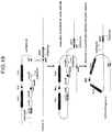


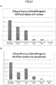
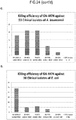


Classifications
-
- A—HUMAN NECESSITIES
- A61—MEDICAL OR VETERINARY SCIENCE; HYGIENE
- A61K—PREPARATIONS FOR MEDICAL, DENTAL OR TOILETRY PURPOSES
- A61K39/00—Medicinal preparations containing antigens or antibodies
- A61K39/02—Bacterial antigens
-
- A—HUMAN NECESSITIES
- A61—MEDICAL OR VETERINARY SCIENCE; HYGIENE
- A61K—PREPARATIONS FOR MEDICAL, DENTAL OR TOILETRY PURPOSES
- A61K35/00—Medicinal preparations containing materials or reaction products thereof with undetermined constitution
- A61K35/66—Microorganisms or materials therefrom
- A61K35/74—Bacteria
-
- A—HUMAN NECESSITIES
- A61—MEDICAL OR VETERINARY SCIENCE; HYGIENE
- A61K—PREPARATIONS FOR MEDICAL, DENTAL OR TOILETRY PURPOSES
- A61K9/00—Medicinal preparations characterised by special physical form
- A61K9/0012—Galenical forms characterised by the site of application
- A61K9/0019—Injectable compositions; Intramuscular, intravenous, arterial, subcutaneous administration; Compositions to be administered through the skin in an invasive manner
-
- A—HUMAN NECESSITIES
- A61—MEDICAL OR VETERINARY SCIENCE; HYGIENE
- A61K—PREPARATIONS FOR MEDICAL, DENTAL OR TOILETRY PURPOSES
- A61K9/00—Medicinal preparations characterised by special physical form
- A61K9/14—Particulate form, e.g. powders, Processes for size reducing of pure drugs or the resulting products, Pure drug nanoparticles
- A61K9/19—Particulate form, e.g. powders, Processes for size reducing of pure drugs or the resulting products, Pure drug nanoparticles lyophilised, i.e. freeze-dried, solutions or dispersions
-
- A—HUMAN NECESSITIES
- A61—MEDICAL OR VETERINARY SCIENCE; HYGIENE
- A61P—SPECIFIC THERAPEUTIC ACTIVITY OF CHEMICAL COMPOUNDS OR MEDICINAL PREPARATIONS
- A61P17/00—Drugs for dermatological disorders
- A61P17/02—Drugs for dermatological disorders for treating wounds, ulcers, burns, scars, keloids, or the like
-
- A—HUMAN NECESSITIES
- A61—MEDICAL OR VETERINARY SCIENCE; HYGIENE
- A61P—SPECIFIC THERAPEUTIC ACTIVITY OF CHEMICAL COMPOUNDS OR MEDICINAL PREPARATIONS
- A61P31/00—Antiinfectives, i.e. antibiotics, antiseptics, chemotherapeutics
- A61P31/04—Antibacterial agents
-
- C—CHEMISTRY; METALLURGY
- C12—BIOCHEMISTRY; BEER; SPIRITS; WINE; VINEGAR; MICROBIOLOGY; ENZYMOLOGY; MUTATION OR GENETIC ENGINEERING
- C12N—MICROORGANISMS OR ENZYMES; COMPOSITIONS THEREOF; PROPAGATING, PRESERVING, OR MAINTAINING MICROORGANISMS; MUTATION OR GENETIC ENGINEERING; CULTURE MEDIA
- C12N1/00—Microorganisms, e.g. protozoa; Compositions thereof; Processes of propagating, maintaining or preserving microorganisms or compositions thereof; Processes of preparing or isolating a composition containing a microorganism; Culture media therefor
- C12N1/04—Preserving or maintaining viable microorganisms
-
- C—CHEMISTRY; METALLURGY
- C12—BIOCHEMISTRY; BEER; SPIRITS; WINE; VINEGAR; MICROBIOLOGY; ENZYMOLOGY; MUTATION OR GENETIC ENGINEERING
- C12N—MICROORGANISMS OR ENZYMES; COMPOSITIONS THEREOF; PROPAGATING, PRESERVING, OR MAINTAINING MICROORGANISMS; MUTATION OR GENETIC ENGINEERING; CULTURE MEDIA
- C12N1/00—Microorganisms, e.g. protozoa; Compositions thereof; Processes of propagating, maintaining or preserving microorganisms or compositions thereof; Processes of preparing or isolating a composition containing a microorganism; Culture media therefor
- C12N1/20—Bacteria; Culture media therefor
-
- C—CHEMISTRY; METALLURGY
- C12—BIOCHEMISTRY; BEER; SPIRITS; WINE; VINEGAR; MICROBIOLOGY; ENZYMOLOGY; MUTATION OR GENETIC ENGINEERING
- C12N—MICROORGANISMS OR ENZYMES; COMPOSITIONS THEREOF; PROPAGATING, PRESERVING, OR MAINTAINING MICROORGANISMS; MUTATION OR GENETIC ENGINEERING; CULTURE MEDIA
- C12N15/00—Mutation or genetic engineering; DNA or RNA concerning genetic engineering, vectors, e.g. plasmids, or their isolation, preparation or purification; Use of hosts therefor
- C12N15/09—Recombinant DNA-technology
- C12N15/63—Introduction of foreign genetic material using vectors; Vectors; Use of hosts therefor; Regulation of expression
- C12N15/70—Vectors or expression systems specially adapted for E. coli
-
- C—CHEMISTRY; METALLURGY
- C12—BIOCHEMISTRY; BEER; SPIRITS; WINE; VINEGAR; MICROBIOLOGY; ENZYMOLOGY; MUTATION OR GENETIC ENGINEERING
- C12N—MICROORGANISMS OR ENZYMES; COMPOSITIONS THEREOF; PROPAGATING, PRESERVING, OR MAINTAINING MICROORGANISMS; MUTATION OR GENETIC ENGINEERING; CULTURE MEDIA
- C12N15/00—Mutation or genetic engineering; DNA or RNA concerning genetic engineering, vectors, e.g. plasmids, or their isolation, preparation or purification; Use of hosts therefor
- C12N15/09—Recombinant DNA-technology
- C12N15/63—Introduction of foreign genetic material using vectors; Vectors; Use of hosts therefor; Regulation of expression
- C12N15/74—Vectors or expression systems specially adapted for prokaryotic hosts other than E. coli, e.g. Lactobacillus, Micromonospora
-
- A—HUMAN NECESSITIES
- A61—MEDICAL OR VETERINARY SCIENCE; HYGIENE
- A61K—PREPARATIONS FOR MEDICAL, DENTAL OR TOILETRY PURPOSES
- A61K39/00—Medicinal preparations containing antigens or antibodies
- A61K39/02—Bacterial antigens
- A61K39/05—Actinobacteria, e.g. Actinomyces, Streptomyces, Nocardia, Bifidobacterium, Gardnerella, Corynebacterium; Propionibacterium
-
- A—HUMAN NECESSITIES
- A61—MEDICAL OR VETERINARY SCIENCE; HYGIENE
- A61K—PREPARATIONS FOR MEDICAL, DENTAL OR TOILETRY PURPOSES
- A61K39/00—Medicinal preparations containing antigens or antibodies
- A61K39/02—Bacterial antigens
- A61K39/104—Pseudomonadales, e.g. Pseudomonas
-
- Y—GENERAL TAGGING OF NEW TECHNOLOGICAL DEVELOPMENTS; GENERAL TAGGING OF CROSS-SECTIONAL TECHNOLOGIES SPANNING OVER SEVERAL SECTIONS OF THE IPC; TECHNICAL SUBJECTS COVERED BY FORMER USPC CROSS-REFERENCE ART COLLECTIONS [XRACs] AND DIGESTS
- Y02—TECHNOLOGIES OR APPLICATIONS FOR MITIGATION OR ADAPTATION AGAINST CLIMATE CHANGE
- Y02A—TECHNOLOGIES FOR ADAPTATION TO CLIMATE CHANGE
- Y02A50/00—TECHNOLOGIES FOR ADAPTATION TO CLIMATE CHANGE in human health protection, e.g. against extreme weather
- Y02A50/30—Against vector-borne diseases, e.g. mosquito-borne, fly-borne, tick-borne or waterborne diseases whose impact is exacerbated by climate change
Definitions
- the present invention relates to the field of bacteriology.
- the invention relates to novel compositions (e.g. , antimicrobial agents) and methods of using the same for treating tissue (e.g ., lesions of the skin and other soft-tissues).
- tissue e.g ., lesions of the skin and other soft-tissues.
- the present invention relates to the killing or altering (e.g ., inhibiting) growth and virulence of populations of microorganisms.
- one of the persistent problems of burn wound care is the development of microbial infections.
- Humans live not in a sterile environment but in a symbiotic relationship with bacteria and other microbes.
- the intact skin and mucosal surface act to maintain a delicate balance between our tissues and the bacterial populations. Any breach in the skin or mucosal barriers alters this balance and thus has the potential to initiate infections by allowing bacteria to gain access to the underlying tissues and achieve critical numbers.
- One of the major treatment goals of a burn surgeon is to prevent infections and, if contamination occurs, the goal is to reduce the microbial contamination below the critical numbers required to initiate and spread infections.
- With the discovery of antibiotics burn wound infections appeared to be under control. However, it has been discovered that bacteria are able to, and indeed have, overcome the antibiotics through development of resistance. Emergence of resistant strains of bacteria has become the major source of many hospital-based infections and has posed a major clinical dilemma to burn surgeons.
- the problem of antibiotic resistance affects all kinds of bacterial infections, including but not limited to infections of the skin and soft-tissues.
- infections including but not limited to infections of the skin and soft-tissues.
- MRSA methicillin-resistant Staphylococcus aureus
- aeruginosa resistance to quinolones in leg ulcers increased from 19% in 1992 to 56% in 2001 ( Valencia et al., J Am Acad Dermatol 50, 845-9 (2004 )). There is no debate in the field; new treatments are needed to control these infections.
- Prophylactic use of antibacterial agents such as silver nitrate, silver sulfadiazine (Silverdene, Thermazine, Flamazine) and mafenide acetate (Sulfamylon) has become the standard of care to reduce bacterial colonization in wounds such as burn wounds.
- these agents have limitations.
- Silverdene has been shown to retard wound healing and cannot be used in patients who are allergic to sulfa drugs.
- the metabolic products of Sulfamylon are potent inhibitors of carbonic anhydrase and therefore can cause metabolic acidosis. Use of this compound is particularly contraindicated in patients who have suffered inhalation injury and those who developed sepsis.
- compositions and methods of altering the growth and virulence of populations of pathogenic microorganisms via "killer vectors" and compatible "donor cells” are known from US 2006/270040 A1 .
- Said document discloses a composition comprising a recombinant transmissible plasmid, which however is different from the recombinant plasmid of the present invention as defined in the claims comprising both colE3 and colE7 in the colE3-aroA -colE7 killing cassette.
- the present invention relates to the field of bacteriology and pharmacology.
- the invention relates to novel compositions (e.g ., antimicrobial agents) and methods of using the same for treating tissue (e.g ., lesions of the skin and other soft tissues) comprising the killing or altering (e.g ., inhibiting) growth and virulence of populations of microorganisms.
- tissue e.g ., lesions of the skin and other soft tissues
- the technology provides compositions for applying an effective dose of non-dividing bacterial cells that prevent and/or treat infection by conjugative transmission of a plasmid that is toxic to recipient pathogen cells.
- Conjugation for transferring genetic material from a donor cell into a target recipient cell for a variety of purposes has been described. See, e.g., PCT Publication WO 02/18605 , U.S. Patent Application Ser. No. 20040137002 , and U.S. Patent Application Ser. No. 10/884,257 .
- the present invention makes use of conjugative transfer to alter the cellular functions of a recipient cell, e.g., to kill or impair the target cell.
- the technology relates to compositions and methods for culturing the donor bacteria in a manner that stably maintains the transmissible plasmid, and for storing the donor bacteria in a manner that maintains both the viability and the killing ability of the donor bacteria.
- the technology further relates to methods for using the donor bacteria in treatments under conditions in which the bacteria cannot further propagate ( i.e ., cannot divide), but in which the killing activity is maintained.
- the technology may provide a recombinant plasmid that is not self-transmissible, and that can be stabily maintained in donor bacterial cell preparations.
- the plasmid is pCON44-74, which is described in detail herein below.
- the technology may provide a donor bacterial cell, e.g., a live bacterium, or a minicell, a maxicell, or a non-dividing cell.
- the donor cell may comprise one or more transfer genes conferring upon it the ability to conjugatively transfer the transmissible plasmid to a recipient cell, and may further comprises a mutation in a gene that renders the donor bacterial cell auxotrophic for a compound, e.g., a nutrient, an amino acid, etc., such that a growth medium must be supplemented with the missing compound for the bacterial cells to replicate.
- the donor bacterial cell may be auxotrophic for one or more aromatic amino acids, 2,6-diaminopimelic acid, and/or lysine.
- the donor bacterial cells are not limited to one particular genus.
- the donor bacterial cell may be selected from the group consisting of Salmonella, Shigella, Escherichia, Enterobacter, Serratia, Proteus, Yersinia, Citrobacter, Edwardsiella, Providencia, Klebsiella, Hafnia, Ewingella, Kluyvera, Morganella, Planococcus, Stomatococcus, Micrococcus, Staphylococcus, Vibrio, Aeromonas, Plessiomonas, Haemophilus, Actinobacillus, Pasteurella, Mycoplasma, Ureaplasma, Rickettsia, Coxiella, Rochalimaea, Ehrlichia, Streptococcus, Enterococcus, Aerococcus, Gemella, Lactococcus, Leuconostoc, Pedicoccus, Bacillus, Corynebacterium, Arcanobacterium, Actinomy
- the technology comprises compositions comprising a donor bacterial cell as described above.
- the composition may comprise a growth medium devoid of aromatic amino acids, while in other examples, the composition may comprise a growth medium supplemented with a compound.
- the composition may comprise a growth medium lacking one or more of tyrosine, tryptophan and phenylalanine, p-aminobenzoate and p-hydroxybenzoate.
- the growth medium may be a minimal medium supplemented with 2, 6-diaminopimelic acid and lysine.
- compositions of the invention may comprise donor bacterial cells grown under controlled conditions for optimal growth.
- cells are grown in conditions in which the pH is controlled.
- the pH of the composition is pH 7.
- the amount of dissolved oxygen in the composition may be controlled.
- the composition may be maintained under conditions that maintain dissolved oxygen at about 50%.
- a composition comprising a donor bacterial cell may comprise or consist of a buffer.
- the buffer may comprise a phosphate buffer, and in particularly preferred examples, the phosphate buffer comprises sodium phosphate and/or potassium phosphate.
- the composition may comprise an excipient mixture, e.g ., for storing or freezing the cells, and comprising a protective agent and a buffer, and optionally also comprising a gelling agent.
- the composition may comprise one or several protective agents, with exemplary protective agents including, e.g ., non-fat milk solids, trehalose, glycerol, betaine, sucrose, glucose, lactose, dextran, polyethylene glycol, sorbitol, mannitol, poly vinyl propylene, potassium glutamate, monosodium glutamate, Tween 20 detergent, Tween 80 detergent, and an amino acid hydrochloride.
- the protective agent may comprise one or more of trehalose, sucrose, and/or glucose.
- the composition may comprise one or more gelling agents selected from the group consisting of hydroxyethyl cellulose, hydroxymethyl cellulose, hydroxypropyl guar, methyl cellulose, ethyl cellulose, hydroxypropyl cellulose, sodium carboxymethyl cellulose, carbomer, alginate, gelatin, and poloxamer.
- composition comprising a donor bacterial cell of the invention as defined in the claims may be in a composition of 10% (w:v) trehalose and 2.0% (w:v) glycerol in 50 mM potassium phosphate buffer. This composition may further comprise 0.5% (w:v) hydroxyethylcellulose.
- compositions of the donor bacterial cells as described above may be in any form that maintains viability, including, e.g ., liquid, gel, frozen, or freeze-dried forms.
- the composition may be formed by addition of water or a buffer to a freeze dried preparation, which may contain the donor bacterial cells and one or more of buffer components, protective agents, and/or gelling agents.
- a composition may be a fluid or gel containing between about 5 x 10 8 to 5 x 10 10 donor bacterial cells (cfu) per ml, preferably 1 x 10 9 to 1 x 10 10 cfu per ml, e.g., configured for therapeutic use, while in other examples, the composition may comprise a fluid or gel containing less than about 1 x 10 9 cfu/ml, e.g., configured for short term storage.
- the technology provides methods for using the compositions of the technology, e.g ., donor bacterial cells comprising a transmissible bacteriocidal plasmid such as pCON44-74, for example, the composition GN-4474.
- the technology may provide methods of treating a tissue surface comprising applying the composition to a surface on said tissue, and ins some embodiments, covering the tissue surface after the applying the composition, e.g ., with a bandage, gauze, fabric, compress, etc.
- compositions and methods find use in the treatment of tissue having a lesion.
- Lesions include, e.g ., burns, cuts, abrasions, abscesses, carbuncles, ulcers, furnuncles, blisters, etc.
- the technology finds particular use in application to tissue comprising epithelial tissue.
- applying a composition of the invention to a tissue may comprise soaking, washing, or rinsing a tissue in the composition.
- the composition may be applied by a process comprising applying the composition to a medical device; and contacting said medical device to the tissue to be treated.
- Treatments are not limited to any particular protocol or schedule for application.
- treating may comprise applying the composition to a tissue a single time, while in some examples, the application may comprise applying the composition to a tissue a plurality of times, e.g., at regularly scheduled intervals.
- Intervals of time between applications are not limited to particular lengths.
- the intervals may be between 1 hour and 24 hours in length, while in some examples, the intervals are 4 hours in length.
- the time intervals in a schedule of treatment may be the same length or different lengths.
- a plurality of applications may comprise at least three applications.
- a method of treating a bacterial infection of a tissue lesion may comprise applying a composition comprising a donor bacterial cell comprising, e.g ., pCON44-74, to an infected lesion in an amount of at least 5 x 10 9 cfu per 20 mm 2 of tissue area.
- the treatment may be repeated at least once, preferably twice, more preferably three times at regular intervals, e.g ., intervals between about 1 hour and 24 hours in length. Treatments may be applied at 4-hour intervals.
- the technology provides methods for preparing and storing bacterial donor cells of the technology.
- the method may be directed to preparations configured for short term storage, e.g., at 4°C, the method comprising:
- the storage medium may further comprise glycerol, and storage medium may comprise 50mM KPO 4 buffer, 10% (w:v) trehalose and 2% (w:v) glycerol.
- the storage medium may further comprise 0.5% hydroxyethyl cellulose.
- Recombinant transmissible plasmid may be pCON44-74 and/or the bacterial donor cell may be an E. coli cell and the growth medium is preferably supplemented with 2,6-diaminopimelic acid and lysine.
- the technology provides a method of preparing a frozen composition of bacterial donor cells for therapeutic use without further culturing, comprising:
- snap freezing is conducted at a temperature of less than 45°C, preferably less than -50°C, -60°C, -70°C or -80°C.
- the excipient medium may comprise between about 1% and 2% (w:v) glycerol, and in particularly preferred examples, the excipient medium comprises 50mM KPO 4 buffer, pH 7, 10% (w:v) trehalose and 2% (w:v) glycerol.
- the excipient medium may further comprise a gelling agent, e.g., about 0.5% (w:v) hydroxyethyl cellulose.
- the term "subject” refers to individuals (e.g., human, animal, or other organism) to be treated by the methods or compositions of the present disclosure.
- Subjects include, but are not limited to, mammals (e.g ., murines, simians, equines, bovines, porcines, canines, felines, and the like), and most preferably includes humans.
- the term “subject” generally refers to an individual who will receive or who has received treatment (e.g ., administration of donor cell, and optionally one or more other agents) for a condition characterized by the presence of pathogenic bacteria, or in anticipation of possible exposure to pathogenic bacteria.
- diagnosis refers to the recognition of a disease (e.g., caused by the presence of pathogenic bacteria) by its signs and symptoms (e.g ., resistance to conventional therapies), or genetic analysis, pathological analysis, histological analysis, and the like.
- in vitro refers to an artificial environment and to processes or reactions that occur within an artificial environment.
- in vitro environments include, but are not limited to, test tubes and cell cultures.
- in vivo refers to the natural environment (e.g ., an animal or a cell) and to processes or reaction that occur within a natural environment.
- conjugation refers to the process of DNA transfer from one cell to another. Conjugation is mediated by complex cellular machinery, and essential protein components are often encoded as a series of genes in a plasmid (e.g., the tra genes for plasmid RK2). Some of these gene products are assembled to facilitate a direct cell-cell interaction ( e.g ., mating pair formation), and some of them serve to transfer DNA and associated protein molecules, and to replicate the DNA molecule ( e.g ., DNA transfer/replication).
- oriT is a DNA sequence from which the transfer of a DNA molecule initiates in the process of conjugation. Conjugative transfer is a phenomenon that occurs across bacterial species and a broad host range of conjugative transfer systems that can be utilized in both Gram-positive and Gram-negative genera are known.
- telomere binding proteins bind specifically to their cognate oriTs and produce a nick in the DNA from which the conjugal transfer begins.
- Transfer proteins Tra form a multi-protein complex called the mating pair formation apparatus (Mpf) that, among other transport-related activities, is needed for formation of pili, and for their extrusion to the cell surface.
- Tra proteins need not be encoded on the same plasmid as the oriT on which they act. They can facilitate the transfer of plasmids that contain unrelated oriTs, provided those plasmids also contain the cognate relaxosomes.
- plasmid DNA Once plasmid DNA is prepared for transfer, it is transported through the donor's cytoplasmic membrane and into the recipient. While not limiting the technology to any particular mechanism of action it is generally believed that DNA crosses the donor membrane with the aid of a coupling protein, so-called because it couples plasmid DNA processing to a TIVSS.
- Free-living (planktonic), as well as surface-bound Gram-negative bacteria are capable of transferring plasmids and one of the few diversifying elements in conjugation systems is the type of pili produced and used to facilitate conjugation. Some pili are thick and flexible while others are long and rigid. Pili have the remarkable ability to retract, allowing them to promote intimate associations of cell surfaces over extended areas, which stabilize mating pairs against shearing forces.
- pheromones small molecules called pheromones
- pCF10 small molecules
- pheromones are typically peptides of seven or eight amino acids and are secreted in miniscule amounts, with as little as 1 to 10 molecules per donor needed to initiate the mating process.
- a given pheromone specifically activates the conjugative transfer system of a particular plasmid type. When a plasmid is acquired, secretion of the related pheromone is prevented, while unrelated pheromones continue to be produced to 'seduce' other potential donor cells.
- transfer functions are naturally up-regulated.
- those de-repressed plasmid systems are able to sustain conjugative DNA transfer with close to 100% efficiency (e.g ., Gram-negative F and Gram-positive pCF10 plasmids).
- Conjugation typically occurs within aggregates of multiple donor and recipient cells; in some cases twenty and in other cases thousands of aggregated and conjugating cells were observed. While Gram-positive and Gram-negative bacteria use different methods in nature to prepare for conjugation, the conjugation that occurs is the same for all types of bacteria. Thus, the conjugative transfer of a bactericidal killer plasmid as a mechanism for killing bacteria can be used with all conjugating bacteria.
- Conjugation also takes place from bacterial cells to higher and lower eukaryote cells ( Waters, Nat Genet. 29:375-376 (2001 ); Nishikawa et al., Jpn J Genet. 65:323-334 (1990 )).
- Trans-kingdom conjugation such as from bacteria into mammalian and yeast cells, is well known. See, e.g ., Bates et al., J Bacteriol 180, 6538-6543 (1998 ); Waters, Nat Genet 29, 375-376 (Dec, 2001 ).
- conjugation donor and “donor cell” are used interchangeably to refer to a cell, e.g., a bacterial cell, carrying a plasmid, wherein said plasmid can be transferred to another cell through conjugation.
- donor cells include, but are not limited to E. coli strains that contain a self-transmissible plasmid or a non-self-transmissible plasmid.
- a cell receiving a plasmid or other cellular material from a donor cell via conjugative transfer is referred to as a "recipient cell”.
- the term "transmissible plasmid” refers to a plasmid that can be transferred from a donor cell to a recipient cell via conjugation.
- self-transmissible plasmid refers to a plasmid encoding all the genes needed to mediate conjugation.
- a recipient of a self-transmissible plasmid becomes a proficient donor to further transfer the self-transmissible plasmid to another recipient cell.
- non-self-transmissible plasmid or "mobilizable plasmid” refers to a plasmid lacking some of the genes needed to mediate conjugation.
- a cell carrying a non-self-transmissible plasmid does not transfer DNA through conjugation unless the missing gene(s) are supplied in trans within the same cell. Therefore, a recipient cell that lacks the missing gene(s), does not become a proficient conjugation donor when it receives the non-self-transmissible plasmid.
- oriT refers to the cis -acting site required for DNA transfer, and integration of an oriT sequence into a non-transmissible plasmid converts it into a mobilizable plasmid ( Lanka and Wilkins, Annu Rev Biochem, 64:141-169 (1995 )).
- a donor cell may be a bacterial cell (e.g., a Gram-positive or Gram-negative bacterium).
- donor cells include, but are not limited to, bacterial cells of a genus of bacteria, selected from the group comprising Salmonella, Shigella, Escherichia, Enterobacter, Serratia, Proteus, Yersinia, Citrobacter, Edwardsiella, Providencia, Klebsiella, Hafnia, Ewingella, Kluyvera, Morganella, Planococcus, Stomatococcus, Micrococcus, Staphylococcus, Vibrio, Aeromonas, Plessiomonas, Haemophilus, Actinobacillus, Pasteurella, Mycoplasma, Ureaplasma, Rickettsia, Coxiella, Rochalimaea, Ehrlichia, Streptococcus, Enterococcus, Aerococcus, Gemella, Lactococcus, Leuconosto
- a donor cell may be a non-viable cell, including but not limited to a bacterial minicell, a maxicell, or a non-dividing cell.
- maxicell refers to the cells that have been treated to maximize chromosomal degradation, e.g ., by UV irradiation and extended incubation. Maxicells contain mostly plasmid DNA, and synthesis of proteins within maxicells occurs essentially exclusively from the plasmid DNA in the cells.
- non-dividing cell refers to cells that are treated in a manner selected to preferentially damage the chromosomal DNA of the cell (e.g., by UV or other irradiation), wherein said cells are further treated, e.g., by rapid chilling after DNA damaging treatment, to minimize chromosomal degradation.
- ND cells can also be obtained in a process such as temporal expression of bactericidal protein (e.g., ColE3) within a donor bacterium.
- induction of proteins destroys the protein synthesis in the cell, leading to cell death while leaving the conjugation apparatus and chromosomal DNA synthesized prior to ColE3 synthesis intact.
- ND cells contain both chromosomal and plasmid DNA but the function of the cell is sufficiently altered, e.g., by UV irradiation, that said ND cells have little or no capability to divide.
- target cells are used interchangeably herein.
- the target cells for the compositions and methods of the present disclosure may include, but are not limited to, microorganisms such as pathogenic organisms (e.g ., pathogenic bacteria) that can receive material from a donor cell via conjugative transfer.
- pathogenic organisms e.g ., pathogenic bacteria
- Pathogenic bacteria include, but are not limited to, Salmonella, Shigella, Escherichia, Enterobacter, Serratia, Proteus, Yersinia, Citrobacter, Edwardsiella, Providencia, Klebsiella, Hafnia, Ewingella, Kluyvera, Morganella, Planococcus, Stomatococcus, Micrococcus, Staphylococcus, Vibrio, Aeromonas, Plessiomonas, Haemophilus, Actinobacillus, Pasteurella, Mycoplasma, Ureaplasma, Rickettsia, Coxiella, Rochalimaea, Ehrlichia, Streptococcus, Enterococcus, Aerococcus, Gemella, Lactococcus, Leuconostoc, Pedicoccus, Bacillus, Corynebacterium, Arcanobacterium, Actinomyces, Rhodococcus, Listeria, Erysipelothrix
- Target cells may be continuously cultured cells.
- Target cells may be uncultured cells existing in their natural environment (e.g., at the site of a wound or infection) or obtained from patient tissues ( e.g., via a biopsy).
- Target cells may exhibit pathological growth or proliferation.
- the term "virulence” refers to the degree of pathogenicity of a microorganism, e.g ., as indicated by the severity of the disease produced or its ability to invade the tissues of a subject. It is generally measured experimentally by the median lethal dose (LD 50 ) or median infective dose (ID 50 ). The term may also be used to refer to the competence of any infectious agent to produce pathologic effects.
- LD 50 median lethal dose
- ID 50 median infective dose
- killer gene refers to a gene that, upon expression in a susceptible cell, produces a product that kills the cell.
- killer plasmid refers to plasmid comprising and configured to express one or more killer genes.
- the terms "attenuate” and “attenuation” as used herein in reference to a feature e.g., of a recipient or target cell refers to a reducing or weakening of that feature, or a reducing of the effect(s) of that feature.
- attenuation generally refers to a reduction in the virulence of the pathogen.
- Attenuation of a pathogen is not limited to any particular mechanism of reduced virulence. Reduced virulence may be achieved, e.g ., by disruption of a secretory pathway.
- Reduced virulence may be achieved by altering cellular metabolism to increase reactivity to or susceptibility to a drug, e.g., a drug that attenuates virulence of the pathogen, or that kills the pathogen. Attenuation may refer to a feature, e.g ., virulence of a population of cells. For example, a population of pathogen cells may be treated, e.g., by the methods and compositions of the description, such that the population of cells is decreased in virulence. See, for example, co-pending applications Serial No. 11/137,948, filed May 26, 2005 , and PCT Application PCT/US2006/020582 .
- an effective amount refers to the amount of a composition (e.g., donor cells) sufficient to effect beneficial or desired results.
- An effective amount can be administered in one or more administrations, applications or dosages and is not intended to be limited to a particular formulation or administration route.
- the term "administration” refers to the act of giving a drug, prodrug, or other agent, or therapeutic treatment (e.g ., compositions of the present invention) to a physiological system (e.g., a subject or in vivo, in vitro, or ex vivo cells, tissues, and organs).
- Exemplary routes of administration to the human body can be through the eyes (ophthalmic), mouth (oral), skin ( e.g ., topical, subcutaneous, transdermal), nose (nasal), lungs (inhalant), oral mucosa (buccal), ear, by injection ( e.g ., intravenously, subcutaneously, intratumorally, intraperitoneally, etc.), by introduction into the intestinal tract ( e.g., rectally), by introduction into the bladder, topically to the surface of the skin or mucous membranes and the like.
- injection e.g ., intravenously, subcutaneously, intratumorally, intraperitoneally, etc.
- introduction into the intestinal tract e.g., rectally
- introduction into the bladder topically to the surface of the skin or mucous membranes and the like.
- tissue refers to any epithelial tissue or layer of an epithelial tissue.
- a skin tissue comprises the epidermis, dermis, hypodermus, and/or any portion of skin exposed, e.g ., by a wound.
- skin wound and “skin lesion” are used interchangeably, and include any breach or damage to skin or to a sublayer of skin.
- a skin wound may include, for example, one or more of burns, cuts, abrasions, abscesses, carbuncles, ulcers, furnuncles, blisters, and the like. Burns are not limited to thermal burns, by also include, e.g ., chemical burns, radiation burns, etc.
- An abscess is a collection of pus and debris that has built up within the body. Abscesses are usually caused by bacterial infection, and can develop anywhere in the body.
- Abscesses in skin and soft tissue are particularly common, and have in fact become more common in recent years; standard treatment for these types of abscesses is to cut the abscess open and drain the fluids. Antibiotics are often prescribed to further limit the infection.
- Carbuncles and furuncles are types of abscesses that often involve infected hair follicles, with carbuncles being larger of the two.
- Furuncles are sometimes referred to as boils.
- Ulcers are defined as sores on the skin or mucous membranes that are accompanied by disintegration of tissue; ulcers can result in complete loss of the epidermis and often portions of the dermis and subcutaneous fat as well leaving these areas susceptible to bacterial infection.
- Impetigo is a contagious skin infection caused by bacteria, most common in infants and children, that produces blisters or sores on the face, neck, hands, and diaper area; impetigo is classified as either non-bullous (crusted) and bullous (large blisters). Impetigo can be treated with topical antibiotics if the affected area is small, or oral antibiotics if the affected area is larger or if the bacteria is resistant.
- treating a surface refers to the act of exposing a surface to one or more compositions of the present invention.
- Methods of treating a surface include, but are not limited to, spraying, misting, submerging, and coating or covering (i.e. a bandage or gauze that was pretreated or saturated with product).
- surface may be, for example, a tissue surface or the surface of a device.
- bandage as used in reference to a dressing, e.g., for covering a wound or treated area, is used broadly to refer to all medical-type dressings commonly used for wound coverage, e.g., gauze strips, gauze pads, fabrics, adhesive bandages, foam pads, hydrofibers, hydrogels, tape, etc.
- co-administration refers to the administration of at least two agent(s) (e.g ., two separate donor bacteria, each comprising a different plasmid) or therapies to a subject.
- the co-administration of two or more agents or therapies may be concurrent.
- a first agent/therapy may be administered prior to a second agent/therapy.
- the formulations and/or routes of administration of the various agents or therapies used may vary.
- the appropriate dosage for co-administration can be readily determined by one skilled in the art.
- agents or therapies are co-administered, the respective agents or therapies may be administered at lower dosages than appropriate for their administration alone.
- co-administration is especially desirable where the co-administration of the agents or therapies lowers the requisite dosage of a potentially harmful (e.g ., toxic) agent(s).
- the term "toxic” refers to any detrimental or harmful effects on a subject, a cell (prokaryotic or eukaryotic), or a tissue as compared to the same cell or tissue prior to the administration of the toxicant.
- composition refers to the combination of an active agent (e.g., donor bacteria cells) with a carrier, inert or active, making the composition especially suitable for diagnostic or therapeutic use in vitro, in vivo or ex vivo.
- active agent e.g., donor bacteria cells
- compositions that do not substantially produce adverse reactions, e.g ., toxic, allergic, or immunological reactions, when administered to a subject.
- topically refers to application of the compositions of the present invention to the surface of the skin and mucosal cells and tissues (e.g ., alveolar, buccal, lingual, masticatory, or nasal mucosa, and other tissues and cells which line hollow organs or body cavities, e.g., the bladder, colon, intestine, esophagus, etc .,).
- mucosal cells and tissues e.g ., alveolar, buccal, lingual, masticatory, or nasal mucosa, and other tissues and cells which line hollow organs or body cavities, e.g., the bladder, colon, intestine, esophagus, etc .,).
- the term "pharmaceutically acceptable carrier” refers to any of the standard pharmaceutical carriers including, but not limited to, phosphate buffered saline solution, water, emulsions (e.g. , such as an oil/water or water/oil emulsions), and various types of wetting agents, any and all solvents, dispersion media, coatings, sodium lauryl sulfate, isotonic and absorption delaying agents, disintrigrants (e.g ., potato starch or sodium starch glycolate), and the like.
- the compositions also can include stabilizers and preservatives. For examples of carriers, stabilizers, and adjuvants.
- compositions of the present invention may be formulated for horticultural or agricultural use. Such formulations include dips, sprays, seed dressings, stem injections, sprays, and mists.
- the term "pharmaceutically acceptable salt” refers to any salt (e.g., obtained by reaction with an acid or a base) of a compound of the present invention that is physiologically tolerated in the target subject (e.g., a mammalian subject, and/or in vivo or ex vivo, cells, tissues, or organs).
- Salts of the compounds of the present invention may be derived from inorganic or organic acids and bases.
- acids include, but are not limited to, hydrochloric, hydrobromic, sulfuric, nitric, perchloric, fumaric, maleic, phosphoric, glycolic, lactic, salicylic, succinic, toluene-p-sulfonic, tartaric, acetic, citric, methanesulfonic, ethanesulfonic, formic, benzoic, malonic, sulfonic, naphthalene-2-sulfonic, benzenesulfonic acid, and the like.
- Other acids such as oxalic, while not in themselves pharmaceutically acceptable, may be employed in the preparation of salts useful as intermediates in obtaining the compounds of the invention and their pharmaceutically acceptable acid addition salts.
- bases include, but are not limited to, alkali metal (e.g ., sodium) hydroxides, alkaline earth metal (e.g ., magnesium) hydroxides, ammonia, and compounds of formula NW 4 + , wherein W is C 1-4 alkyl, and the like.
- salts include, but are not limited to: acetate, adipate, alginate, aspartate, benzoate, benzenesulfonate, bisulfate, butyrate, citrate, camphorate, camphorsulfonate, cyclopentanepropionate, digluconate, dodecylsulfate, ethanesulfonate, fumarate, flucoheptanoate, glycerophosphate, hemisulfate, heptanoate, hexanoate, chloride, bromide, iodide, 2-hydroxyethanesulfonate, lactate, maleate, methanesulfonate, 2-naphthalenesulfonate, nicotinate, oxalate, palmoate, pectinate, persulfate, phenylpropionate, picrate, pivalate, propionate, succinate, tartrate, thiocyanate, tosy
- salts include anions of the compounds of the present invention compounded with a suitable cation such as Na + , NH 4 + , and NW 4 + (wherein W is a C 1-4 alkyl group), and the like.
- a suitable cation such as Na + , NH 4 + , and NW 4 + (wherein W is a C 1-4 alkyl group), and the like.
- salts of the compounds of the present invention are contemplated as being pharmaceutically acceptable.
- salts of acids and bases that are non-pharmaceutically acceptable may also find use, for example, in the preparation or purification of a pharmaceutically acceptable compound.
- salts of the compounds of the present invention are contemplated as being pharmaceutically acceptable.
- salts of acids and bases that are non-pharmaceutically acceptable may also find use, for example, in the preparation or purification of a pharmaceutically acceptable compound.
- medical devices includes any material or device that is used on, in, or through a subject's or patient's body, for example, in the course of medical treatment ( e.g ., for a disease or injury).
- Medical devices include, but are not limited to, such items as medical implants, wound care devices, drug delivery devices, and body cavity and personal protection devices.
- the medical implants include, but are not limited to, urinary catheters, intravascular catheters, dialysis shunts, wound drain tubes, skin sutures, vascular grafts, implantable meshes, intraocular devices, heart valves, and the like.
- Wound care devices include, but are not limited to, general wound dressings, biologic graft materials, tape closures and dressings, and surgical incise drapes.
- Drug delivery devices include, but are not limited to, needles, drug delivery skin patches, drug delivery mucosal patches and medical sponges.
- Body cavity and personal protection devices include, but are not limited to, tampons, sponges, surgical and examination gloves, and toothbrushes.
- birth control devices include, but are not limited to, intrauterine devices (IUDs), diaphragms, and condoms.
- therapeutic agent refers to compositions that decrease the infectivity, morbidity, or onset of mortality in a subject contacted by a pathogenic microorganism or that prevent infectivity, morbidity, or onset of mortality in a host contacted by a pathogenic microorganism.
- therapeutic agents encompass agents used prophylactically, e.g ., in the absence of a pathogen, in view of possible future exposure to a pathogen.
- agents may additionally comprise pharmaceutically acceptable compounds (e.g ., adjuvants, excipients, stabilizers, diluents, and the like).
- the therapeutic agents of the present invention are administered in the form of topical compositions, injectable compositions, ingestible compositions, and the like.
- the form may be, for example, a solution, cream, ointment, salve or spray.
- pathogen refers to a biological agent that causes a disease state (e.g., infection, cancer, etc .) in a host.
- pathogens include, but are not limited to, viruses, bacteria, archaea, fungi, protozoans, mycoplasma, prions, and parasitic organisms.
- bacteria and "bacterium” refer to all prokaryotic organisms, including those within all of the phyla in the Kingdom Procaryotae. It is intended that the term encompass all microorganisms considered to be bacteria including Mycoplasma, Chlamydia, Actinomyces, Streptomyces, and Rickettsia. All forms of bacteria are included within this definition including cocci, bacilli, spirochetes, spheroplasts, protoplasts, etc. Also included within this term are prokaryotic organisms that are Gram-negative or Gram-positive. "Gram-negative” and “Gram-positive” refer to staining patterns with the Gram-staining process, which is well known in the art.
- Gram-positive bacteria are bacteria that retain the primary dye used in the Gram stain, causing the stained cells to generally appear dark blue to purple under the microscope.
- Gram-negative bacteria do not retain the primary dye used in the Gram stain, but are stained by the counterstain. Thus, Gram-negative bacteria generally appear red.
- microorganism refers to any species or type of microorganism, including but not limited to, bacteria, archaea, fungi, protozoans, mycoplasma, and parasitic organisms.
- the present invention contemplates that a number of microorganisms encompassed therein will also be pathogenic to a subject.
- fungi is used in reference to eukaryotic organisms such as the molds and yeasts, including dimorphic fungi.
- non-human animals refers to all non-human animals including, but are not limited to, vertebrates such as rodents, non-human primates, ovines, bovines, ruminants, lagomorphs, porcines, caprines, equines, canines, felines, aves; etc.
- nucleic acid molecule refers to any nucleic acid containing molecule, including but not limited to, DNA or RNA.
- the term encompasses sequences that include any of the known base analogs of DNA and RNA including, but not limited to, 4-acetylcytosine, 8-hydroxy-N6-methyladenosine, aziridinylcytosine, pseudoisocytosine, 5-(carboxyhydroxyl-methyl) uracil, 5-fluorouracil, 5-bromouracil, 5-carboxymethylaminomethyl-2-thiouracil, 5-carboxymethylaminomethyluracil, dihydrouracil, inosine, N6-isopentenyladenine, 1-methyladenine, 1-methylpseudouracil, 1-methylguanine, 1-methylinosine, 2,2-dimethylguanine, 2-methyladenine, 2-methylguanine, 3-methylcytosine, 5-methylcytosine, N6-methyladenine
- gene refers to a nucleic acid (e.g ., DNA or RNA) sequence that comprises coding sequences necessary for the production of a polypeptide, precursor, or RNA (e.g., rRNA, tRNA).
- a polypeptide can be encoded by a full length coding sequence or by any portion of the coding sequence so long as the desired activity or functional properties (e.g ., enzymatic activity, ligand binding, signal transduction, immunogenicity, etc. ) of the full-length or fragment are retained.
- the term also encompasses the coding region of a structural gene and the sequences located adjacent to the coding region on both the 5' and 3' ends for a distance of about 1 kb or more on either end such that the gene corresponds to the length of the full-length mRNA. Sequences located 5' of the coding region and present on the mRNA are referred to as 5' non-translated sequences, or 5' flanking sequences. Sequences located 3' or downstream of the coding region and present on the mRNA are referred to as 3' non-translated sequences or 3' flanking sequences.
- the term "gene” encompasses both cDNA and genomic forms of a gene.
- a genomic form or clone of a gene contains the coding region interrupted with non-coding sequences termed "introns” or “intervening regions” or “intervening sequences.”
- Introns are segments of a gene that are transcribed into pre-mRNA; introns may contain regulatory elements such as enhancers. Introns are generally removed or “spliced out” from the primary (pre-mRNA) transcript; introns therefore are generally absent in the messenger RNA (mRNA) transcript.
- mRNA messenger RNA
- RNA expression refers to the process of converting genetic information encoded in a gene into RNA (e.g ., mRNA, rRNA, tRNA, or snRNA) through "transcription" of the gene (i.e ., via the enzymatic action of an RNA polymerase), and for protein encoding genes, into protein through “translation” of mRNA.
- Gene expression can be regulated at many stages in the process.
- Up-regulation” or “activation” refers to regulation that increases the production of gene expression products (i.e., RNA or protein), while “down-regulation” or “repression” refers to regulation that decrease production.
- Molecules e.g ., transcription factors
- activators e.g ., transcription factors
- wild-type refers to a gene or gene product in the form that would be isolated from a naturally occurring source.
- a wild-type gene is that which is most frequently observed in a population and is thus arbitrarily designed the "normal” or “wild-type” form of the gene.
- modified or mutant refers to a gene or gene product that displays modifications in sequence and or functional properties (i.e ., altered characteristics) when compared to the wild-type gene or gene product. It is noted that naturally occurring mutants can be isolated; these are identified by the fact that they have altered characteristics (including altered nucleic acid sequences) when compared to the wild-type gene or gene product.
- nucleic acid molecule encoding As used herein, the terms “nucleic acid molecule encoding,” “DNA sequence encoding,” and “DNA encoding” refer to the order or sequence of deoxyribonucleotides along a strand of deoxyribonucleic acid. The order of these deoxyribonucleotides determines the order of amino acids along the polypeptide (protein) chain. The sequence of nucleotides in the DNA thus encodes for the sequence of amino acids in the corresponding polypeptide.
- an oligonucleotide having a nucleotide sequence encoding a gene and “polynucleotide having a nucleotide sequence encoding a gene,” means a nucleic acid sequence comprising the coding region of a gene or in other words the nucleic acid sequence that encodes a gene product.
- the coding region may be present in a cDNA, genomic DNA or RNA form.
- the oligonucleotide or polynucleotide may be single-stranded ( i.e., the sense strand) or double-stranded.
- Suitable control elements such as enhancers/promoters, splice junctions, polyadenylation signals, etc. may be placed in close proximity to the coding region of the gene if needed to permit proper initiation of transcription and/or correct processing of the primary RNA transcript.
- the coding region utilized in the expression vectors of the present invention may contain endogenous enhancers/promoters, splice junctions, intervening sequences, polyadenylation signals, etc. or a combination of both endogenous and exogenous control elements.
- a partially complementary sequence is a nucleic acid molecule that at least partially inhibits a completely complementary nucleic acid molecule from hybridizing to a target nucleic acid is "substantially homologous.”
- the inhibition of hybridization of the completely complementary sequence to the target sequence may be examined using a hybridization assay (Southern or Northern blot, solution hybridization and the like) under conditions of low stringency.
- a substantially homologous sequence or probe will compete for and inhibit the binding ( i.e., the hybridization) of a completely homologous nucleic acid molecule to a target under conditions of low stringency.
- low stringency conditions are such that non-specific binding is permitted; low stringency conditions require that the binding of two sequences to one another be a specific ( i.e., selective) interaction.
- the absence of non-specific binding may be tested by the use of a second target that is substantially non-complementary (e.g ., less than about 30% identity); in the absence of non-specific binding the probe will not hybridize to the second non-complementary target.
- the term “substantially homologous” refers to any nucleic acid that can hybridize to either or both strands of the double-stranded nucleic acid sequence under conditions of low stringency as described above.
- the term “substantially homologous” refers to any nucleic acid that can hybridize ( i.e., it is the complement of) to the complement of the single-stranded nucleic acid sequence under conditions of low stringency as described above.
- a gene may produce multiple RNA species that are generated by differential splicing of the primary RNA transcript.
- cDNAs that are splice variants of the same gene will contain regions of sequence identity or complete homology (representing the presence of the same exon or portion of the same exon on both cDNAs) and regions of complete non-identity (for example, representing the presence of exon "A” on cDNA 1 wherein cDNA 2 contains exon "B" instead). Because the two cDNAs contain regions of sequence identity they will both hybridize to a probe derived from the entire gene or portions of the gene containing sequences found on both cDNAs; the two splice variants are therefore substantially homologous to such a probe and to each other.
- hybridization is used in reference to the pairing of complementary nucleic acids. Hybridization and the strength of hybridization (i.e ., the strength of the association between the nucleic acids) is impacted by such factors as the degree of complementary between the nucleic acids, stringency of the conditions involved, the T m of the formed hybrid, and the G:C ratio within the nucleic acids. A single molecule that contains pairing of complementary nucleic acids within its structure is said to be “self-hybridized.”
- portion when in reference to a nucleotide sequence (as in “a portion of a given nucleotide sequence”) refers to fragments of that sequence.
- the fragments may range in size from four nucleotides to the entire nucleotide sequence minus one nucleotide (10 nucleotides, 20, 30, 40, 50, 100, 200, etc .) .
- operable combination refers to the linkage of nucleic acid sequences in such a manner that a nucleic acid molecule capable of directing the transcription of a given gene and/or the synthesis of a desired protein molecule is produced.
- the term also refers to the linkage of amino acid sequences in such a manner so that a functional protein is produced.
- isolated when used in relation to a nucleic acid, as in “an isolated oligonucleotide” or “isolated polynucleotide” refers to a nucleic acid sequence that is identified and separated from at least one component or contaminant with which it is ordinarily associated in its natural source. Isolated nucleic acid is such present in a form or setting that is different from that in which it is found in nature. In contrast, non-isolated nucleic acids as nucleic acids such as DNA and RNA found in the state they exist in nature.
- a given DNA sequence e.g ., a gene
- RNA sequences such as a specific mRNA sequence encoding a specific protein
- isolated nucleic acid encoding a given protein includes, by way of example, such nucleic acid in cells ordinarily expressing the given protein where the nucleic acid is in a chromosomal location different from that of natural cells, or is otherwise flanked by a different nucleic acid sequence than that found in nature.
- the isolated nucleic acid, oligonucleotide, or polynucleotide may be present in single-stranded or double-stranded form.
- the oligonucleotide or polynucleotide will contain at a minimum the sense or coding strand (i.e., the oligonucleotide or polynucleotide may be single-stranded), but may contain both the sense and anti-sense strands ( i.e ., the oligonucleotide or polynucleotide may be double-stranded).
- the term "purified” or “to purify” refers to the removal of components (e.g ., contaminants) from a sample.
- components e.g ., contaminants
- antibodies are purified by removal of contaminating non-immunoglobulin proteins; they are also purified by the removal of immunoglobulin that does not bind to the target molecule.
- the removal of non-immunoglobulin proteins and/or the removal of immunoglobulins that do not bind to the target molecule results in an increase in the percent of target-reactive immunoglobulins in the sample.
- recombinant polypeptides are expressed in bacterial host cells and the polypeptides are purified by the removal of host cell proteins; the percent of recombinant polypeptides is thereby increased in the sample.
- nucleic acids in a sample are purified by removing or reducing one or more components from a sample. Components to be reduced or removed in purification comprise other nucleic acids, damaged nucleic acids, proteins, salts, etc.
- amino acid sequence and terms such as “polypeptide” or “protein” are not meant to limit the amino acid sequence to the complete, native amino acid sequence associated with the recited protein molecule.
- cell culture refers to any in vitro culture of cells. Included within this term are continuous cell lines (e.g ., with an immortal phenotype), primary cell cultures, transformed cell lines, finite cell lines ( e.g ., non-transformed cells), bacterial cultures (using liquid or solid media, e.g. ,) and any other cell population maintained in vitro.
- eukaryote refers to organisms distinguishable from “prokaryotes.” It is intended that the term encompass all organisms with cells that exhibit the usual characteristics of eukaryotes, such as the presence of a true nucleus bounded by a nuclear membrane, within which lie the chromosomes, the presence of membrane-bound organelles, and other characteristics commonly observed in eukaryotic organisms. Thus, the term includes, but is not limited to such organisms as fungi, protozoa, and animals ( e.g ., humans).
- transdominant negative mutant gene refers to a gene encoding a protein product that prevents other copies of the same gene or gene product, which have not been mutated ( i.e ., which have the wild-type sequence) from functioning properly ( e.g., by inhibiting wild type protein function).
- the product of a transdominant negative mutant gene is referred to herein as "dominant negative” or "DN” (e.g., a dominant negative protein, or a DN protein).
- kits refers to any delivery system for delivering materials.
- reaction materials such as donor cells
- delivery systems include systems that allow for the storage, transport, or delivery of reaction reagents (e.g ., cells, buffers, selection reagents, etc., in the appropriate containers) and/or supporting materials (e.g., media, written instructions for performing using the materials, etc. ) from one location to another.
- reaction reagents e.g ., cells, buffers, selection reagents, etc., in the appropriate containers
- supporting materials e.g., media, written instructions for performing using the materials, etc.
- kits include one or more enclosures (e.g ., boxes) containing the relevant reaction reagents and/or supporting materials.
- fragmentmented kit refers to delivery systems comprising two or more separate containers that each contain a subportion of the total kit components.
- the containers may be delivered to the intended recipient together or separately.
- a first container may contain cells for a particular use, while a second container contains selective media.
- fragment kit is intended to encompass kits containing Analyte specific reagents (ASR's) regulated under section 520(e) of the Federal Food, Drug, and Cosmetic Act, but are not limited thereto. Indeed, any delivery system comprising two or more separate containers that each contains a subportion of the total kit components are included in the term “fragmented kit.”
- a “combined kit” refers to a delivery system containing all of the components of a reaction materials needed for a particular use in a single container ( e.g ., in a single box housing each of the desired components).
- kit includes both fragmented and combined kits.
- cellular metabolic function refers to any or all processes conducted by a cell (e.g ., enzymatic or chemical processes associated with cell function), other than genomic replication.
- the present technology is directed to bacterial donor cells usable for conjugative delivery of toxins to pathogens.
- the technology finds particular application in the treatment of tissue lesions, e.g ., skin wounds such as burns for the prevention and treatment of infection and for the prevention of sepsis associated with infection.
- compositions of the present technology are configured so that during use, e.g., on a tissue wound, the probiotic organism cannot grow and it must be administered in an effective dosage that does not require further growth of the bacterial donor cells at the treatment site.
- concentrations of donor bacterial cells used in certain preferred examples are typically greater than 1 x 10 9 cfu/ml.
- overgrowth of the donor bacteria to stationary phase, or refrigeration of the donor bacteria at concentrations at or above the therapeutic range reduced the conjugation competence of the cells.
- plasmids and donor bacteria providing stable compositions for therapeutic use, and dosing regimens for treatment or prevention of infection by a variety of bacterial pathogens.
- Organ failure is the leading cause of death in trauma and surgical patients. Excessive inflammatory response and depression of cell-mediated immunity predisposes these patients to infectious complications (see, e.g., Angele and Faist, Crit Care 6, 298 (2002 ); Faist, Curr Top Microbiol Immunol 216, 259 (1996 ); and Schinkel et al., J Trauma 44, 743 (1998 )).
- the present technology may provide a therapeutic treatment comprising donor bacterial cells (e.g., pathogenic or non-pathogenic bacteria, non-dividing cells) comprising one or more plasmids (e.g ., self-transmissible or non-self-transmissible plasmids), wherein the plasmid may be transferred ( e.g ., through conjugation) from the donor cell to a target/recipient cell (e.g., a pathogenic microorganism), resulting in the plasmid expressing its genetic material in the target.
- donor bacterial cells e.g., pathogenic or non-pathogenic bacteria, non-dividing cells
- plasmids e.g ., self-transmissible or non-self-transmissible plasmids
- a target/recipient cell e.g., a pathogenic microorganism
- the technology may provide a stable preparation of a bacterial cell and a transmissible killer plasmid for use, e.g., in topical treatment of skin wounds and burn infections.
- the technology provides a donor cell that comprises a transmissible killing plasmid termed "pCON44-74," or a variant thereof, that retains the functions required for use in the methods and applications described herein below, and depicted schematically in Fig. 3 . While the use of pCON44-74 plasmid is not limited to use in a particular strain of donor cell, in embodiments discussed herein below, pCON44-74 is used with an E. coli donor cell. As used herein, the term "GN-4474" refers to a bacterial E.
- E. coli CON31-85A containing the pCON44-74 plasmid. Aspects of the technology are directed to methods of growing and storing GN-4474, and to formulations and treatment protocols for using GN-4474 in the prevention and treatment of infection.
- Methods of storing bacterial cells by freezing, with or without lyophilization are known but are commonly done with an expectation that reconstituted cells will be further cultured. The loss of a major portion of viable cells is tolerable when the cells will be further cultured for use.
- An aspect of the present technology is formulations for storage and administration that do not require further growth of the bacteria for effective use.
- the RK2 conjugation system possesses a set of genes that once expressed are very proficient in transferring plasmid DNA from Gram-negative bacterial hosts (e.g., E. coli ).
- Gram-negative bacterial hosts e.g., E. coli
- the functional RK2 conjugation machinery can mobilize plasmid DNA from a large number of Gram-negative bacterial hosts. It has been shown that, as long as proper vegetative replication origins are introduced, a plasmid can be mobilized from these donors ( E. coli and other Gram-negative donors) to other Gram-negative target strains, and even Gram-positive target strains (see, e.g ., Giebelhaus et al., J Bacteriol 178, 6378-6381 (1996 )), generating exconjugants. Conjugation systems of the present disclosure are not limited to RK2, since the majority of conjugative plasmids share strong similarities, and any other system could serve as a delivery system.
- donor cells comprise a transmissible plasmid that is conjugatively transferred into a target, wherein one or more products encoded by the plasmid are expressed ( e.g., to make mRNA or protein) resulting in the killing of the target cells or the inhibiting of their growth.
- the donor cells may further comprise a helper plasmid.
- the transmissible plasmid may be a self-transmissible plasmid.
- Donor cells may comprise a transmissible plasmid comprising nucleic acid that encodes one or more polyamino acids (e.g ., polypeptides or proteins) that are bactericidal. Donor cells may further comprise nucleic acid that encodes a polyamino acid capable of neutralizing the bactericidal properties of the polyamino acid of the non-self-transmissible plasmid within the donor cells (e.g., an immunity protein; see, e.g., Examples 2 and 4).
- the gene encoding the neutralizing polyamino acid may be on a chromosome of the donor cell, while in other examples, it is on a helper plasmid.
- the polyamino acid capable of neutralizing the bactericidal polyamino acid may be under control of a constitutive promoter.
- the polyamino acid capable of neutralizing the bactericidal polyamino acid may be under control of an inducible promoter. While an understanding of the mechanism is not necessary to practice the present invention and while the present invention is not limited to any particular mechanism of action, it is contemplated that, in some examples, the polyamino acid capable of neutralizing bactericidal polyamino acid and the bactericidal polyamino acid form a non-toxic complex within the donor bacteria.
- the bactericidal polyamino acid may be encoded by the colE3 gene.
- the present disclosure is not limited by the type of bactericidal gene used. Indeed a variety of bactericidal genes are contemplated including, but not limited to, colA, colB, colD, colIa, colIb, colK, colN, colE1, colE2, col E4 , colE5, col E6 , colE7, col E8, colE19 and a gene encoding lysozyme, lysostaphin, streptolysin, or listeriolysin O, etc.
- the self-transmissible or non-self-transmissible plasmid may comprise a promoter (e.g ., the lac promoter/operator) that drives expression each of the bactericidal polyamino acid(s).
- a promoter e.g ., the lac promoter/operator
- the promoters are all configured to express the toxins in the same target cell
- a transmissible plasmid will be configured to express a first bacteriocidal protein if transferred to a first type of recipient cell, and to express a second bacteriocidal protein if transferred to a second type of recipient cell.
- Bactericidal proteins such as colicins are not lethal only to bacteria. Although colicins are termed "bactericidal” proteins, the colicin proteins are enzymes that are universally toxic to cells, such as DNase and RNase ( Bowman et al (1971) PNAS 68:964-968 ; Tomita et al (2000) PNAS 97:8278-8283 ; Schaller and Nomura (1976) PNAS 73:3989-3993 ).
- DNase and RNase Bowman et al (1971) PNAS 68:964-968 ; Tomita et al (2000) PNAS 97:8278-8283 ; Schaller and Nomura (1976) PNAS 73:3989-3993 ).
- a bactericidal protein such as a colicin
- a colicin is typically controlled by recognition of a toxin-antidote complex on the surface of the target cell.
- the colicin is secreted to extracellular space by a colicin-producing bacterium as a toxin-antidote complex, and the toxin-antidote complex binds only to target cells that carry a specific surface molecule. Only the toxin molecule is imported into the target cell, killing the cell. In nature, killing by bactericidal proteins such as colicins is restricted to target cells that carry the correct surface molecule.
- bactericidal toxins may be used in targeting non-bacterial cells, e.g., eukaryotic cells, using conjugative transfer.
- a helper plasmid may encode a repressor protein (e.g., lacI ) capable of inhibiting expression of the bactericidal gene.
- a repressor protein may be encoded on the chromosome of the donor strain. The repressor protein may be under control of a constitutive promoter. The repressor protein may be under control of an inducible promoter.
- Plasmids configured for the expression of extremely toxic proteins may be used.
- the vectors of the present disclosure comprise rrnBT 1T2 transcription terminators upstream of a strong bacterial promoter.
- the present disclosure is not limited to the use of the rrnB T1T2 transcription terminators. Other known transcription terminators may be utilized.
- the lactose promoter and operator may be utilized.
- the LacI Q repressor protein may be included on the plasmid or it is provided on a separate vector, F' element, or chromosome.
- the present disclosure in not limited to the use of lactose promoter and operator.
- Other suitable promoters may be utilized, including, but not limited to, tetracycline, PBAD, T7, and T5 promoters.
- the present disclosure may provide vectors comprising a hybrid promoter/operator system.
- One hybrid promoter/operator utilizes the Arc and Mnt repressor proteins from Salmonella bacteriophage P22 as basic scaffolds (see, e.g., WO 2005/072092 ).
- the Arc and Mnt repressor proteins are small transcriptional regulatory proteins with structural similarity.
- Both Arc and Mnt proteins contain two functional domains - a dimeric N-terminal domain that binds operator DNA and a C-terminal coiled-coil domain that mediates protein tetramerization, which is essential for function ( Knight and Sauer. Proc. Natl. Acad. Sci.
- the mutant Mnt-Arc proteins contain the wildtype C-terminal dimerization domain from Mnt; however, six residues within the N-terminal DNA binding domain have been replaced with the corresponding 9 residues from the Arc repressor ( Knight and Sauer. Proc. Natl. Sci. USA 86:797-801 ⁇ 1989 ⁇ ).
- a Mnt-Arc homodimer retains wildtype tetramerization ability, but now recognizes the Arc operator sequence (O2) instead of the Mnt operator (O1).
- the novel repressor heterotetramer of the present invention consists of one wildtype Mnt homodimer and one hybrid Mnt-Arc homodimer.
- the vectors may comprise a low copy number origin of replication (e.g ., low copy modified pSC101 or RK2.
- a low copy number origin of replication e.g ., low copy modified pSC101 or RK2.
- Other exemplary origins of replication include, but are not limited to, wildtype pSC101, pl5a, pACYC.
- Plasmids finding use with the technology may also comprise a multiple cloning site for insertion of nucleic acid encoding genes of interest and a selectable marker (e.g ., an antibiotic resistance gene such as kanamycin, ampicillin, tetracycline, etc. ), and/or protein purification tags (e.g., His-tag, GST-tag, intein tag).
- a selectable marker e.g ., an antibiotic resistance gene such as kanamycin, ampicillin, tetracycline, etc.
- protein purification tags e.g., His-tag, GST-tag, intein tag
- the donor cells of the present disclosure comprise an immunity protein that inhibits or neutralizes the bactericidal protein expressed by the transmissible plasmid.
- an immunity protein that inhibits or neutralizes the bactericidal protein expressed by the transmissible plasmid.
- Numerous pairs of bactericidal proteins and corresponding immunity proteins are known in the art. The bactericidal proteins listed above are inhibited by the corresponding colicin A, colicin B, colicin D, colicin Ia, colicin Ib, colicin K, colicin N, colicin E1, colicin E2, colicin E3, colicin E4, colicin E5, colicin E6, colicin E7, colicin E8, and colicin E9 immunity proteins, respectively.
- bactericidal proteins e.g., bacteriocins
- the gene encoding an immunity protein may be under control of a promoter, wherein said promoter is constitutively active, while in other examples, the gene encoding an immunity protein may be under control of a promoter that is inducible.
- compositions comprising plasmids of the present disclosure find use in research and therapeutic applications.
- any type of bacteria e.g ., Gram-positive and Gram-negative bacteria
- a number of approaches may be taken to prevent spread (e.g ., growth) of donor bacteria.
- non-dividing cells see, e.g., U.S. Pat. App.
- Donor bacterial cells of the present disclosure may comprise temperature sensitive mutation(s).
- a temperature-sensitive mutant grows abnormally within a certain range of temperature compared to its isogenic wild-type bacteria.
- a mutation in the RNA or protein causes effects, e.g., changes in conformation, that are sensitive to temperature such that mutants can be grown in a lab at their permissive temperature; however, they have severe growth defects at non-permissive ( e.g., higher) temperatures (e.g., at body temperature).
- mutations include aminoacyl-tRNA synthetases (see, e.g., Sakamoto et al., J Bacteriol 186, 5899-5905 (2004 ); Martin et al., J Bacteriol 179, 3691-3696 (1997 )), and RNase P ( Li, Rna 9, 518-532 (2003 ); Li and Altman, Proc Natl Acad Sci U S A 100, 13213-13218 (2003 )).
- aminoacyl-tRNA synthetases see, e.g., Sakamoto et al., J Bacteriol 186, 5899-5905 (2004 ); Martin et al., J Bacteriol 179, 3691-3696 (1997 )
- RNase P Li, Rna 9, 518-532 (2003 ); Li and Altman, Proc Natl Acad Sci U S A 100, 13213-13218 (2003 )
- An aminoacyl-tRNA synthetase catalyzes the esterification of a specific amino acid to the 3'-terminal adenosine of the corresponding tRNA, and RNase P is an crucial ribonuclease to generate the mature 5' end of tRNAs in all organisms ( Gopalan et al., J Biol Chem 277, 6759-6762 (2002 ). Defects in these enzymatic functions prevent protein synthesis in the cell.
- the plasmids and donor cells of the invention may be employed to minimize potential risks associated with the use of DNA or genetically modified organisms in the environment. For instance, in environmentally sensitive circumstances it may be preferable to utilize non-self-transmissible plasmids.
- the plasmids may be mobilizable by conjugative machinery but are not self-transmissible. As discussed herein, this may be accomplished in some examples by integrating into the host chromosome all tra genes whose products are necessary for the assembly of conjugative machinery. In such examples, plasmids are configured to possess only an origin of transfer ( oriT ). This feature prevents the recipient, before or even after it dies, from transferring the plasmid further.
- oriT origin of transfer
- Another biosafety feature comprises utilizing conjugation systems with predetermined host-ranges.
- certain elements are known to function only in few related bacteria (narrow-host-range) and others are known to function in many unrelated bacteria (broad-host-range or promiscuous) ( del Solar et al., Mol. Microbiol. 32: 661-666, (1996 ); Zatyka and Thomas, FEMS Microbiol. Rev. 21: 29 1 319, (1998 )).
- many of those conjugation systems can function in either Gram-positive or Gram-negative bacteria but generally not in both (del Solar, 1996, supra; Zatyka and Thomas, 1998, supra).
- Donor bacterial cells of the present disclosure may comprise auxotrophic mutant(s).
- auxotrophic mutants There are large numbers of auxotrophic mutants known in the art. Examples of genes causing such phenotype are dapA and aroA.
- dapA encodes an enzyme dihydropicolinate synthase, a key enzyme for lysine biosynthesis in plant and bacteria (see, e.g., Ledwidge and Blanchard, Biochemistry 38, 3019-3024 (1999 )), and aroA encodes an enzyme 5-enolpyruvylshikimate 3-phosphate synthase, catalyzing a key step in the synthesis of aromatic amino acids (see, e.g., Rogers et al., Appl Environ Microbiol 46, 37-43 (1983 )).
- Environmentally safe bacteria may be utilized as donors. Safe bacteria are known in the art. For example, delivery of DNA vaccines by attenuated intracellular Gram-positive and Gram-negative bacteria has been reported ( Dietrich et al., 2001 Vaccine 19, 2506-2512 ; Grillot-Courvalin et al., 1999 Current Opinion in Biotech. 10, 477-481 ).
- the donor strain can be one of thousands of harmless bacteria that colonize the non-sterile parts of the body (e.g., skin, gastrointestinal, urogenital, mouth, nasal passages, throat and upper airway systems). Low virulence strains may be used.
- minicells and maxicells are well studied model systems of metabolically active but nonviable bacterial cells.
- Minicells lack chromosomal DNA and are generated by special mutant cells that undergo cell division without DNA replication. If the cell contains a multicopy plasmid, many of the minicells will contain plasmids. Minicells neither divide nor grow. However, minicells that possess conjugative plasmids are capable of conjugal replication and transfer of plasmid DNA to living recipient cells. (see, e.g., U.S. Patent No. 4,968,619 ).
- Maxicells are cells that are treated so as to destroy their chromosomal DNA, while retaining the function of plasmids that they contain. Maxicells can be obtained from a strain of E. coli that carries mutations in the key DNA repair pathways ( recA, uvrA and phr ). Because maxicells lack so many DNA repair functions, they die upon exposure to low doses of UV light. Importantly, plasmid molecules (e.g ., pBR322) that do not receive UV irradiation continue to replicate. Transcription and translation (plasmid-directed) can occur efficiently under such conditions ( Sancar et al., J. Bacteriol.
- Non-dividing cells may be utilized as donor cells.
- Non-dividing cells are generally treated such that the ability to divide and grow is removed but conjugation efficiency is preserved.
- Non-dividing cells may be treated such that chromosomal DNA is damaged but is not destroyed to the same extent as it is in the creation of maxicells.
- GN-4474 can be used to treat any of these types of skin infections.
- One method of application would be to apply GN-4474 topically as an ointment; this method of application would be suited to infections such as impetigo.
- GN-4474 can be injected or infused directly into the infected tissue; this method of application would be particularly suited to abscesses and ulcers.
- Application of GN-4474 both topically and injected would be advantageous in some circumstances.
- Example 1 details the genetic modifications made to produce E. coli CON31-85A.
- the resulting strain has the genotype: K-12 E. coli MG1655 ⁇ msbB::LacIQ, ⁇ dapA::immE3-Arc / mnt, ⁇ aroA:: imm x 10 7 -lacIQ, attB::tra-P L -Mnt, ⁇ recA
- Figures 1-3 provide a detailed flow diagram of the assembly of the killing plasmid pCON44-74.
- the skilled person will appreciate that modifications can be made to the plasmid without altering its function in the technology, e.g ., removing portions of nucleic acid that do not affect the function of the conjugation, replication, or toxin functions of the plasmid.
- the plasmid may be transformed into different types of non-pathogenic donor cells, or variant strains of E. coli. It is contemplated that the present technology encompasses such variants of both pCON44-74 and GN-4474. See, e.g., WO 2006/128089 .
- the active ingredient in GN-4474 is a harmless genetically engineered strain of E. coli that possesses conjugation machinery that can transfer "a killer plasmid" into target bacteria, that, once expressed, kills the recipient bacteria.
- antibiotics that are taken orally or that are injected are dosed as the amount (grams, milligrams, etc) that are given so many times per day.
- Topical antibiotics such as Neosporin are dosed as the number of units of each antibiotic in a particular amount (3.5 mg of neomycin, 5,000 units of polymyxin B and 400 units of bacitracin per 1.0 gram of Neosporin).
- a composition such as GN-4474.
- the dosage indicator as used herein is colony forming units (cfus), typically indicated in relation to a volume of a formulation or a unit of area treated or to be treated.
- the GN-4474 composition has been shown to be effective in preventing infection when used as a preventative, i.e., when applied at the same time as exposure to a pathogen. It was demonstrated that when infecting burned mice with between 1x10 4 and 1x10 5 cfu of pathogen ( P. aeruginosa or Acinetobacter baumannii ) a dose response for GN-4474 is observed that prevents death in the mice and also will clear the pathogen from the burn site.
- pathogen P. aeruginosa or Acinetobacter baumannii
- mice infected with P. aeruginosa approximately 50% survival of mice occurred with treatment with 6.0x10 5 cfu of GN-4474 to a burn area of about 20 mm 2 .
- the treatment under these conditions produced the following results: Approximately 75% survival was achieved with treatment with 7.0x10 6 cfu of GN-4474. Greater than 85% survivability was achieved with treatment with 5x10 7 cfu of GN-4474. Greater than 90% survivability was achieved with 5x10 8 cfu of GN-4474 and 100% survivability was achieved with doses of 4.9x10 9 cfu or greater.
- Tissue injuries may be debrided prior to treatment, while in some examples, the formulations of the invention may be applied without debridement.
- Topical treatment of an infected lesion may comprise at least one administration of 1x10 10 cfu of GN-4474 for a lesion of 20 mm 2 of tissue area.
- the treatment at this dosage may be repeated 2 times, preferably 3 times, or more.
- the treatments may be repeated at regular intervals, e.g ., every 4 to 6 hours.
- Treatment of a burn may be done without debriding the burn wound.
- GN-4474 is applied, e.g., by injection below the eschar but above the underlying muscle tissue. Treatment by subeschar injection may reduce the dosage of GN-4474 needed to treat or prevent infection. For example, on a 20 mm 2 burn site, injection of a single dose of 1.0 x 10 9 cfu GN-4474 resulted in nearly 1 log reduction in the number of surviving pathogen organisms.
- compositions of the technology are configured such that they require supplemental amino acids in order to grow, so that in use, e.g., as applied to a skin lesion, they are deprived of essential amino acids and cannot further divide.
- GN-4474 requires 2,6-diaminopimelic acid in order to grow.
- 2,6-diaminopimelic acid As applied to tissue, e.g., on a skin lesion, 2,6-diaminopimelic acid is not available and the donor bacterium thus cannot grow at the application site ( i.e ., it cannot colonize the site or otherwise propagate).
- the conjugation function of donor cell is maintained, so that the transmissible plasmid carrying toxin genes may be transferred into a recipient pathogen cell.
- the plasmid is configured to express encoded toxins in susceptible target pathogens, killing the pathogens.
- the GN-4474 composition is a live viable E. coli bacteria that is intended to be used as a topical therapeutic to treat, e.g ., antibiotic resistant bacteria.
- the GN-4474 cannot be "regrown", it must be in its final formulation and concentration that it is intended to be used at in the hospital setting.
- a high concentration e.g ., approx. 1 x 10 10 cfu/ml.
- the technology may be directed to ways to store GN-4474 for both short term ( ⁇ 2 weeks) and long term (> 1 year) periods of time, while maintaining a very high level of viability and conjugation efficiency. Indeed, some typical bacteria storage conditions may maintain viability, but these same conditions may not maintain conjugation efficiency.
- 1x10 6 to about 1x10 9 cfu/ml may be used for storage, preferably 1x10 8 to 1x10 9 cfu/ml, with 1x10 9 cfu/ml being a particularly preferred concentration for short-term storage, whereby maintenance of both viability and conjugation efficiency are improved significantly relative to significantly more concentrated preparations.
- the composition of the storage medium appears to have an influence on conjugation, but little influence on viability. It was very surprising that conjugation efficiency was better for short term storage when the cells were maintained in spent medium rather than in fresh buffer, as it is typically considered that bacteria should be healthier and more stable when they are removed from a milieu that is depleted of nutritional elements and is laden with waste products. Therefore it is clear that multiple factors influence the stability of the E. coli GN-4474 for short term storage, and that once proper storage concentration is maintained, exact buffer composition influences conjugation efficiency, but has less effect on viability. This would indicate through multiple experiments, that as long as the concentration of E.
- coli is maintained at or below 1x10 9 cfu/ml, viability of the bacteria at 4°C can be maintained in spent medium, or in excipient buffer with or without HEC. However, the conjugation efficiency of the bacteria at 4°C is better maintained in spent medium rather than in buffer with or without HEC.
- Short term storage temperature influences both viability and conjugation efficiency. Different sugars also influence viability. As discussed below in Example 8, glucose as a sugar in short term storage medium is not as good as sucrose or trehalose for maintaining viability. However, all three sugars maintain conjugation efficiency when stored at 4°C. These data show that viability and conjugation efficiency are not directly related and that varying conditions, e.g ., sugars, can affect one feature without affecting the other.
- Snap freezing or flash freezing, is the process by which the samples are lowered to ultra-low temperatures, e.g ., below -70°C, very rapidly. Snap freezing typically involves using dry ice or liquid nitrogen.
- the snap-freezing protocol described in Example 9 maintains both the viability and killing efficiency of GN-4474 in excipient buffer (50mM KPO 4 buffer, 10% trehalose or sucrose, 2% glycerol) for extended periods of time at -80°C. Snap freezing protocols have been developed that maintain the both the viability and killing efficiency using a much lower percentage of glycerol than used in typical bacterial freezing protocols (2% vs. the typical 15-40%).
- Example 10 shows that GN-4474 freeze dried in excipient buffer (50mM KPO 4 buffer, 10% trehalose or sucrose, 2% glycerol and 0.5% HEC) may be stored for extended periods of time at -80°C while maintaining both the viability and killing efficiency.
- excipient buffer 50mM KPO 4 buffer, 10% trehalose or sucrose, 2% glycerol and 0.5% HEC
- GN-4474 is therapeutic for both the prevention of and treatment of gram negative bacterial infections in a burn using a variety of different application strategies.
- coli lac operator/promoter Kan (determinant for kanamycin resistance); Cm (determinant for chloramphenicol resistance); Tral (region encoding genes responsible for conjugative transfer); Control (region encoding control region); oriV (region encoding the origin of vegetative replication); oriT (region encoding the origin of conjugative transfer); tetR (gene encoding repressor of tetA ); tetA (gene encoding resistance to tetracycline); Rep (region encoding genes responsible for replication); Primase (region encoding genes involved in replication); Tra2 (region encoding genes responsible for mating pair formation); colE3 (gene encoding colicin E3); col x 10 7 (gene encoding colicin x 10 7 ); immE3 (gene encoding the immunity protein for colicin E3); immE7 (gene encoding the immunity protein for colicin x 10 7 ), repA, repB and repC (encode
- Donor bacterium construction Construction of E. coli CON31-85A
- E. coli CON31-85A This strain was derived from the wild type E. coli strain MG1655 following the steps shown in Figure 12 .
- E. coli MG1655 was the first completely sequenced strain of E. coli ( Blattner et al, Science. 1997 Sep 5;277(5331):1453-62 .)
- the first genetic alteration to E. coli MG1655 was the deletion of msbB with a lacI Q insertion to take the place of msbB.
- the msbB gene encodes an enzyme responsible for attachment of an acyl group in the lipid A of lipopolysaccharide (LPS), the primary component of bacterial endotoxin.
- LPS lipopolysaccharide
- This mutant strain of E. coli still synthesize LPS lacking the acyl group, and this modified LPS represents significantly reduced (almost 1 million fold) endotoxic effect.
- This modification in E. coli CON31-85A will significantly reduce the endotoxic effect of GN-4474 upon its application in humans.
- the msbB gene in MG1655 was deleted using a standard two-step homologous recombination procedure. This gene deletion (or knock out (KO)) was combined with insertion of lacI Q to take the place of msbB using a gene replacement plasmid (pCON1-96). Since expression of colE3 was repressed with LacI; overexpression of lacI was designed by introducing a second copy of lacI Q . Note that lacI Q has a point mutation in its promoter region, which yields higher expression of lacI. The summary of this gene replacement is depicted in Figure 13 .
- Table 1 lists the individual primers and corresponding primer sequences used to create the pCON1-96 suicide vector and to detect the homologous recombination events that delete the msbB gene and insert the lacI Q gene in replacement. Table 1. Primer names and corresponding sequences of the primers used to create the suicide vector pCON1-96 and to detect the deletion of msbB and insertion of lacI Q .
- the 5' and 3' flanking regions of msbB were PCR cloned using primer pairs, con1-4C/con1-4D, and con1-4A/con1-4B, respectively.
- the lacI Q region was also PCR cloned from E. coli JM109 using the pair of primers con1-8A/1-8B.
- the cloned DNA fragments were verified by DNA sequencing. These fragments were assembled into a plasmid pCON1-96 ( Figure 14 ). This vector has both a selectable marker CamR and counter selective marker sacB on the plasmid.
- pCON1-96 Prior to transformation, pCON1-96 was digested with Asc I to eliminate the replication origin (ori) of the plasmid, and the replicon-less plasmid was circularized by T4 ligase prior to transforming E. coli MG1655.
- Transformants were selected on Luria bertani agar containing 50 ⁇ g/mL chloramphenicol (Cam, 50 ⁇ g/mL), and growing colonies were analyzed by PCR to verify homologous recombination at either 5' or 3' flanking region of msbB.
- the PCR primers used for this analysis are listed in the Table 1 above, and locations of these primers are indicated in Figure 13 .
- the candidate colonies containing the integrated pCON1-96 were subsequently grown in LB broth cultures containing 7% sucrose (with no chloramphenicol), overnight. The cultures were then subcultured for 2 days and were diluted and plated on LB agar containing 7% sucrose without chloramphenicol selection to counter select sacB- containing bacterial cells. Colonies growing on the plate are either msbB -deletion mutants or revertants to wild type, which was verified by PCR using the primer pairs listed in the Table 2.
- PCR primer combinations used in PCR analysis to detect the msbB deletion and lacI Q insertion following homologous recombination. Correct amplicon sizes are shown for the resulting E. coli CON30-12 strain, while amplification of the parental E. coli MG1655 strain, will not yield any amplicons. Table2.
- the second genetic alteration to E. coli MG1655 was the deletion of dapA with an immE3-arc / mnt insertion to take the place of dapA.
- the dapA gene encodes an enzyme partially responsible for the synthesis of the bacterial cell wall and lysine.
- a dapA mutant can grow with medium supplementation with the compound diaminopimelic acid (DAP), which is a precursor in the biosynthetic pathway.
- DAP diaminopimelic acid
- the dapA gene in E. coli CON30-12 was deleted using a standard two-step homologous recombination procedure.
- the dapA gene deletion was combined with integration of immE3 and mnt / arc using a gene replacement plasmid, pCON30-31.
- LacI represses expression of the bactericidal gene colE3 on the plasmid pCON44-74.
- leaky expression of colE3 could cause a detrimental effect on the host survival; thus immE3 was integrated into the host chromosome to neutralize the toxicity of even the smallest amounts of ColE3 within the CON31-85A host cell.
- the dapA deletion genotype was introduced to restrict the growth of the host stain in a DAP limiting environment; thus it prevents spread of the bacterium. This process caused a deletion of 833 bp within the dapA coding region.
- the immE3 and Mnt / Arc genes were integrated into this site. The summary of this gene replacement strategy is depicted in the Figure 15 .
- Table 3 lists the individual primers and corresponding primer sequences used to create the pCON1-96 suicide vector and to detect the homologous recombination events that delete the msbB gene and insert the lacI Q gene in replacement.
- the 5' and 3' flanking regions of dapA were PCR cloned from the chromosomal DNA of E. coli MG1655 using two pairs of primers con8-59A/con8-59B and con8-59C/con8-59D, respectively ( Figure 15 ).
- the immE3 and the arc / mnt hybrid genes were PCR cloned using two pairs of primers. These fragments were assembled into a plasmid pCON30-31 ( Figure 16 ).
- This vector has both a selectable marker CamR and counter selective marker sacB on the plasmid.
- the suicide plasmid pCON30-31 has a gamma R6K origin of replication that cannot replicate in E .
- the candidate colonies containing the integrated pCON30-31 were grown in LB broth cultures containing 100 ⁇ g/mL of 2,6-diaminopimelic acid and 7% sucrose (no chloramphenicol) overnight. These overnight cultures were then subcultured for 2 days, diluted and plated on LB agar containing 100 ⁇ g/mL of 2,6-diaminopimelic acid and 7% sucrose without chloramphenicol selection to counter select sacB -containing bacterial cells. Colonies growing on the plate are either dapA -deletion mutants or revertants to wild type, which was verified by PCR using the primer pairs listed in the Table 4.
- Genotype of CON30-41 K-12 E. coli MG1655 ⁇ msbB::lacIQ, ⁇ dapA::immE3-Arc / mnt
- the third genetic alteration to E. coli MG1655 was the deletion of aroA with an imm x 10 7 -lacIQ insertion to take the place of aroA. Disruption of the aroA gene results in a block in the shikimate pathway of E. coli. This pathway is mainly responsible for the biosynthesis of aromatic amino acids; however, it can provide precursors for folate, enterochelin, and quinine biosynthesis. Published reports of E. coli aroA mutants demonstrated that these strains require the addition of aromatic amino acids, p-aminobenzoate, and p-hydroxybenzoate when grown in minimal medium.
- the aroA genotype was used to make the host strain dependent on a group of amino acids; thus aroA was used as a selective marker for the bactericidal plasmid. In a synthetic culture medium, the bactericidal killer plasmid carrying aroA was demonstrated to be stably maintained. The summary of this gene replacement is depicted in the Figure 17 . Table 5. Primer names and corresponding sequences of the primers used to create the suicide vector pCON29-93 and to detect the deletion of aroA and insertion of imm x 10 7 - lacI Q . Table 5: sequence of primers used to detect the aroA deletion and immE7-lacIQ insertion.
- the 5' and 3' flanking regions of aroA were PCR cloned from the chromosomal DNA of E . coli MG1655 using two pairs of primers con29-28A/con29-28B and con29-28E/con29-28F, respectively.
- the imm x 10 7 and the lacI Q genes were combined with these fragments and cloned into a plasmid to generate pCON29-93.
- This vector has both a selectable marker CamR and counter selective marker sacB on the plasmid.
- the pCON29-93 plasmid was digested with AscI , and the replication origin was removed from the plasmid generating a replicon-less plasmid.
- the resulting replicon-less plasmid was circularized with T4 DNA ligase, and transformed into CON30-41. Since the plasmid is replicon-less the plasmid cannot be stably maintained as a plasmid in this strain. Chloramphenicol resistance was used as a positive selective marker for initial integration of DNA fragment, and sacB was used as a counter selective marker for the second homologous recombination.
- Initial transformants were selected on Luria bertani agar containing 100 ⁇ g/mL of 2,6-diaminopimelic acid and 50 ⁇ g/mL chloramphenicol (Cam, 50 ⁇ g/mL), growing colonies were analyzed by PCR to verify homologous recombination at either 5' or 3' flanking region of aroA.
- the PCR primers used for this analysis are listed in the Table 5 above, and locations of these primers are indicated in Figure 17 .
- the candidate colonies containing the integrated pCON29-93 were subsequently cultured in LB broth containing 100 ⁇ g/mL of 2,6-diaminopimelic acid and 7% sucrose without chloramphenicol overnight.
- PCR primer combinations used in PCR analysis to detect the aroA deletion and imm x 10 7 - lacI Q insertion following homologous recombination. Correct amplicon sizes are shown for the resulting E. coli CON32-16 strain, while amplification of the parental E. coli CON30-41 strain, will not yield any amplicons. Table. Specific PCR products from recombinant strain CON32-16 PCR primers* Product size from CON32-16 Product size from parental strain CON30-41 Con29-95A/con29-77B 1413 bp None Con29-91A/con29-95B 1303 bp none * See the locations of the primers in the Figure 17 .
- the fourth genetic alteration to E. coli MG1655 was the integration of the tra operon into the chromosome of E. coli MG 16556 at the lambda bacteriophage attachment site, attB ( Figure 19 ).
- the bactericidal plasmid pCON44-74 is a mobilizable plasmid that requires a set of tra genes to mediate its transfer from the donor cell ( E. coli CON31-85A) to a recipient cell.
- the plasmid pCON44-74 utilizes RSF1010 for its transfer and replication, and the products of the RK2 tra genes can facilitate this process efficiently.
- a suicide plasmid containing the RK2 tra genes was constructed, and was integrated at the attB site of the host E. coli genome. Recombination of DNA molecules between attB and attP is reversible, and catalyzed by the Int-Xis system of lambda bacteriophage. To avoid its potential gene transfer from the donor strain to another bacterial strain, the integrated plasmid was further modified to prevent its excision process (see the strategy in the Figure 19 ). Briefly, the suicide plasmid carrying a DNA fragment of E. coli chromosome flanking to the attB site, part of the ybhC gene was cloned into this suicide vector as well.
- the tra1 and tra2 regions that were cloned into this plasmid are responsible for providing conjugation machinery necessary for transferring plasmids in the final donor strain.
- the tra1 and tra2 regions must be inserted or integrated into the chromosome of CON31-85A.
- the pCON32-20 suicide plasmid was designed to utilize the Int-Xis system of the lambda bacteriophage and the corresponding attB site in the host chromosome and the attP site in pCON32-20, which was specifically cloned into this suicide vector.
- the attB / attP site recombination event is normally reversible in nature.
- ConjuGon designed pCON32-20 to prevent excision from the host chromosome once integration occurred.
- the pCON32-20 suicide vector was designed to carry a DNA fragment (part of the ybhC gene) of the host E. coli chromosome flanking attB site. Following the site-specific integration of the pCON32-20 plasmid at the attB site in the chromosome, homologous recombination between the two ybhC gene fragments takes place. During this homologous recombination event at the ybhC genes, the attL gene is eliminated, with prevents the potential excision of the plasmid DNA that is integrated at this location.
- the suicide vector pCON32-20 has several important features ( Figure 20 ).
- the Tra1, Tra2 and Control regions (all required for conjugation) were cloned from the well-characterized plasmid RK2.
- the entire Tra1, Tra2 and control regions from RK2 were cloned with the exception trbK.
- Expression of the trbK gene has been shown to reduce conjugation efficiency dramatically in recipient cells.
- the gene was deleted. Elimination of trbK in the donor strain, CON31-85A, does not affect conjugation efficiency of the donor strain.
- KanR for resistance to kanamycin
- CamR for chloramphenicol resistance
- the sacB gene was also cloned into pCON32-20 and used for negative selection to counter select the pCON32-20 plasmid, which will force the second homologous recombination event ( Figure 19 ).
- a copy of the immE3 gene was cloned into the pCON32-20 suicide plasmid however; immE3 was not essential for the purpose of this plasmid development, and after the gene-replacement process, immE3 was designed to be deleted.
- Int protein is essential for recombination between attB and attP.
- the protein was expressed in trans from the plasmid pJW27, which expresses Int under a heat-inducible promoter.
- the plasmid pJW27 is not transferred into the final donor strain CON31-85A and is actually selected against in the final strain.
- the origin of transfer Prior to cloning the entire Tra1, Tra2 and control regions from RK2, the origin of transfer (oriT) was mutated. ConjuGon specifically mutated the oriT to prevent the potential transfer of chromosomal DNA from CON31-85A into pathogenic target bacteria.
- the suicide vector pCON32-20 also carries the Mnt repressor protein that is under control of the lambda P L promoter that is constitutively expressed. This repressor protein is part of ConjuGon's novel Arc/Mnt hybrid promoter/repressor system used to regulate col x 10 7 expression in the CON31-85A donor strain.
- the suicide plasmid pCON32-20 has a gamma R6K origin of replication that cannot replicate in E. coli MG1655 (or CON31-85A) since these strains do not have the pir gene coding for the Pi replication protein required for R6K origins of replication.
- the Pi protein was provided in trans from a specific vector-construction host genome. Since the recipient of the plasmid CON32-16 did not express the Pi protein, the plasmid was not capable of replication; thus only the integrated plasmid could provide resistance against antibiotics, chloramphenicol and kanamycin.
- the tra integration vector (pCON32-20) has to be integrated at the attB site in the chromosome of CON32-16 ( Figure 19 ). Since the E. coli CON32-16 or pCON32-20 vector doesn't contain the lambda integrase gene that is required for integration the lambda integrase needs to be supplied in trans.
- the plasmid pJW27 containing a temperature sensitive replicon, and a heat inducible lambda integrase was transformed into E. coli CON32-16. Following this transformation, electrocompetent E. coli CON32-16/pJW27 were prepared.
- the tra integration vector, pCON32-20 was transformed into the electrocompetent E. coli CON32-16/pJW27 cells.
- the bacterial cells were incubated at 43°C for 1 hour (to induce the lambda integrase, and also to cure the strain of pJW27) and then spread onto LB agar containing 100 ⁇ g/mL of 2,6-diaminopimelic acid and 50 ⁇ g/mL kanamycin (Kn, 50 ⁇ g/mL). These plates were incubated overnight at 37°C. Resulting colonies were analyzed by PCR to verify the initial integration of pCON32-20 at the attB site using the primer pairs con32-8A with con32-8D ( Figure 19 and Table 7) which would generate a 1987 bp amplicon from the integrant strain, with no amplicon from CON32-16.
- PCR-positive clones (those with the integration of pCON32-20 at the attB site) were then grown in LB broth containing 100 ⁇ g/mL of 2,6-diaminopimelic acid and 7% sucrose without antibiotics overnight. These cultures were then subcultured for two days in the same medium, followed by dilution and plating on LB agar containing 100 ⁇ g/mL of 2,6-diaminopimelic acid and 7% sucrose without antibiotic selection to counter select sacB -containing bacterial cells.
- Primer names and corresponding sequences of the primers to detect the integration of the tra genes Sequence of primers used to detect the tra integration
- Sequence of primers used to detect the tra integration Primer name DNA sequence SEQ ID NO: CON32-8A 5'- TTGTAGTTAGTGTCATTCAGATTGCGCTGT 27
- CON32-8C 5'- TGCTACTGCTTCGCAATGCTGGAC 28
- CON32-8D 5'- CAAGAAAGCCATCCAGTTTACTTTGCAG 29
- PCR primer combinations used in PCR analysis to detect the tra integration and subsequent homologous recombination Correct amplicon sizes are shown for the resulting E . coli CON32-27A strain, while amplification of the parental E.
- coli CON32-16 strain will not yield any amplicons that will be amplified in the PCR conditions used for detection.
- Specific PCR products from recombinant strain CON32-27A PCR primer pair Product size from CON32-27A Product size from parental strain CON32-16 CON32-8A x CON32-8C 1695 bp 8897 bp, too large to amplify under the condition used * * PCR condition: 94C-5min, (94C-30sec, 59C-30sec, 72C-1min), 72C-5min, 5% DMSO, 1u/20uL Taq
- Genotype of CON32-27A K-12 E. coli MG1655 ⁇ msbB::lacIQ, ⁇ dapA::immE3-Arc / mnt, ⁇ aroA::imm x 10 7 -lacIQ, attB::tra-P L -Mnt
- the last genetic alteration to E. coli MG1655 was the deletion of recA without any insertion taking the place of recA.
- the ⁇ recA genotype was introduced to minimize potential genetic instability by recombination, and also to attenuate fitness of the host strain. This was the last genetic alteration to the donor strain since the other deletions and insertions listed above required RecA to help facilitate these genetic deletions and insertion to the donor strains chromosome.
- the recA gene in E. coli MG1655 was deleted using the standard two-step homologous recombination. The summary of this gene replacement is depicted in the Figure 21 . Table 9.
- pCON31-45 Construction of the ⁇ recA gene deletion suicide vector, pCON31-45 is described here.
- the 5' and 3' flanking regions of recA were PCR cloned using two pairs of primers, con31-10A/con31-10B, and con31-10C/con31-10D, respectively.
- the cloned DNA fragments were verified by DNA sequencing. These fragments were assembled into a plasmid pCON31-45 ( Figure 22 ).
- This vector has both a selectable marker CamR and counter selective marker sacB on the plasmid.
- pCON31-45 Prior to transformation, pCON31-45 was digested with Asc I to eliminate the replication origin ( ori ) of the plasmid.
- the replicon-less plasmid was circularized by T4 ligase prior to transforming E. coli CON32-27A.
- Transformants were selected on LB agar containing 2,6-diaminopimelic acid (100 ⁇ g/ml) and chloramphenicol (Cam, 50 ⁇ g/ml), growing colonies were analyzed by PCR to verify homologous recombination at either 5' or 3' flanking region of recA.
- the PCR primers used for this analysis are listed in the Table 9 above, and locations of these primers are indicated in the Figure 21 .
- the candidate colonies containing the integrated pCON31-45 were subsequently grown in LB broth containing 2,6-diaminopimelic acid (100 ⁇ g/ml) and 7% sucrose (with no chloramphenicol) overnight. These cultures were then subcultured for 2 days and were diluted and plated on LB agar containing 2,6-diaminopimelic acid (100 ⁇ g/ml) and 7% sucrose without chloramphenicol selection to counter select sacB -containing bacterial cells. Colonies growing on these agar plates are either recA- deletion mutants or revertants to the parental strain, and this was screened by PCR using the primer pair listed in the Table 10 below.
- GN-4474 E. coli CON31-85A transformed with plasmid pCON44-74 is termed "GN-4474." In these experiments, GN-4474 is compared to a negative control strain CON37-55A to assess its effect on killing different target pathogens.
- the pathogen strains were all cultured over night from freezer stocks.
- the GN-4474 and CON37-55A were also cultured overnight from freezer stocks. After overnight growth, the densities of the cultures were standardized spectrophotometrically.
- Target pathogen was then mixed with either GN-4474 or CON37-55A separately, the suspensions were then pelleted in a centrifuge, supernatant removed, and the resulting "mixed" cell pellet was resuspended in saline.
- the resulting suspensions were then spotted onto nitrocellulose filter disks that were placed on top of 0.9% saline agar plates, to ensure that no bacterial growth took place directly on the plates themselves.
- the saline agar plates were then quickly dried in a laminar flow hood, then placed at 37°C for two hours to allow conjugation to occur.
- the plates were removed from the incubator, the filter disks (still containing the target pathogen and treatment cells) were removed from the surface of the agar plates and, then placed in 1.5 ml centrifuge tubes containing 1.0ml sterile saline. The tubes were then vortexed to release the bacterial cells from the filter disks. Serial dilutions were then prepared and plated onto Luria Bertani medium.
- This medium would allow for growth of all of the pathogens tested (Klebsiella, Pseudomonas, Acinetobacter, Enterobacter, Escherichia coli ), however, since this medium was not supplemented with 2,6-diaminopimelic acid, neither the GN-4474 nor the CON37-55A control could grow, ensuring that only target pathogen would be recovered.
- the plates were incubated overnight at 37°C, after which the resulting pathogen colonies were counted. The results are reported as "percent of test strain killed by GN-4474" in Figures 24A-24E .
- the counts from the CON37-55A negative control plates were used as a baseline.
- the killing efficiency of GN-4474 was calculated by dividing the cfu/ml of recovered target pathogen treated with GN-4474 by the cfu/ml of recovered target pathogen treated with CON37-55A, multiplied by 100 to give the % surviving bacteria, as shown in Figures 24A-24E .
- Table 11 Bacterial Species Number of isolates tested E. cloacae 50 K. pneumoniae 50 A. baumanaii 53 E. coli 38 P. aeruginosa 48 Total 239
- GN-4474 is not limited to treating P. aeruginosa, as shown in the examples below, but finds use in treating infection by a range of different pathogens.
- compositions of the present invention The following describes a standard protocol for testing compositions of the present invention.
- mice were burned and then infected with 7400 cfu of P. aeruginosa MAKI delivered in a volume of 200 ⁇ l on day 0. Twenty four hours later (day 1), four groups of 8 mice each were treated as follows:
- mice On Day 2 (48 hours post infection) the mice were sacrificed, muscle biopsies were taken, homogenized, and plated for viable counts. Spleen were also collected, homogenized, and plated for viable counts.
- the GN-4474 bacteria were then concentrated just four hours prior to being applied to the debrided burn site. It is noted that prolonged storage in concentrated form reduces conjugation efficiency.
- the spleen data ( Figure 4B ) shows there is no difference in Pseudomonas loads, indicating that the treatment did not stop sepsis from occurring. However, it was not determined whether sepsis existed prior to treatment, and pre-existing sepsis would not be expected to yield to this topical treatment.
- Example 1 the protocol described in Example 1 was modified by using two different pathogen inoculum amounts to cause the infection.
- the data from Treatment 1 suggested that too large of a P. aeruginosa MAKI inoculum was used to generate this infection.
- the E. coli GN-4474 that was used for this experiment was prepared virtually identically to those in the prior experiment.
- the GN-4474 bacteria were then concentrated just four hours prior to being applied to the debrided burn site.
- mice Two groups of eight mice each received a P. aeruginosa inoculum of (6893 cfu of MAKI). Two other groups of eight mice each received an inoculum of P. aeruginosa MAKI that was roughly one half of the other two groups (3492 cfu of MAKI). Twenty four hours later (day 1), four groups of 8 mice each were treated as follows:
- mice On Day 2 (48 hours post infection) the mice were sacrificed, muscle biopsies were taken, homogenized and plated for viable bacterial counts. Spleens were also collected, homogenized, and plated for viable counts. The results are shown in Figures 5A and 5B .
- aeruginosa MAKI is given to mice to establish an infection, treatment with GN-4474 appears to inhibit or at least decrease the amount of P. aeruginosa MAKI that enters the blood stream, thereby partially inhibiting sepsis ( Figure 5B ).
- GN-4474 was prepared grown and concentrated as for Treatments 1 and 2.
- mice Four groups of 8 mice each were burned and infected with 3400 cfu of P. aeruginosa MAKI. Twenty four hours later the mice were divided into four groups of eight mice each. The groups were treated as follows:
- mice On Day 2 (48 hours post infection) the mice were sacrificed, muscle biopsies were taken, homogenized and plated for viable counts. Spleens were also collected, homogenized and plated for viable counts. The data for this set of experiments is shown in Figures 6A and 6B .
- the number of recovered P. aeruginosa MAKI in the group that were treated 1 time with excipient buffer was 3.51 x 10 7 cfu/ gram of tissue ( Figure 6A ).
- mice treated with 2 treatments of GN-4474 had an average P. aeruginosa MAKI burden of 2.63 x 10 4 CFU/gram of spleen tissue, while the control group of mice treated with 2 treatments of excipient buffer had an average P. aeruginosa MAKI burden of 7.12e5 CFU/gram of spleen tissue ( Fig. 6B ).
- Five of the eight mice in the 2 times treatment with GN-4474 group had no detectable P. aeruginosa MAKI in their spleens.
- One of the remaining 3 mice in this group had a significant decrease in P. aeruginosa MAKI burden as well.
- mice were burned and infected with 3500 cfu of P. aeruginosa MAKI at day 0. Then, 24 hours later, the treatments with vehicle control or with GN-4474 commenced.
- the test groups were as follows:
- GN-4474 significantly altered the dose of GN-4474 that was applied to the wound site. In previous experiments about 1-3 x 10 10 cfu of freshly cultured GN-4474 was applied to the wound site. In this experiment using resuspended lyophilized GN-4474, 33 times less GN-4474 (1.1 x 10 9 cfu) was applied to each burn site, at each treatment time.
- This experiment repeats the protocol of Treatment 3, with the hypothesis that increasing the number of treatments with GN-4474 will continue to decrease the P. aeruginosa MAKI pathogen in the burn site and spleen.
- This experiment utilized GN-4474 that was grown under a slightly different set of conditions.
- mice Three cultures of GN-4474 were grown and the cultures were stopped at OD600s of 2.2-2.5 and prepared on the morning of the day the animals were to be treated. The cultures were grown longer than optimum and were concentrated early in the morning and stored on ice until application. Four groups of 10 mice each were burned and then infected with 3489 CFU of P. aeruginosa MAKI. Twenty four hours later, the eschars were removed and the four groups of 10 mice each were treated as follows:
- Table 12 Recovered pathogen ( P. aeruginosa MAKI) from muscle punches Mean CFUs/Gm Tissue Standard Error Muscle Excipient Control 11,744,311 4,323,708 1x GN-4474 15,619,341 4,670,534 2X GN-4474 4,959,367 2,172,470 3X GN-4474 844,225 435,054 Table 13. Recovered pathogen ( P.
- aeruginosa MAKI from spleen tissue Mean CFUs/Gm Tissue Standard Error Spleen Excipient Control 3,148,364 1,476,464 1x GN-4474 4,624,565 2,076,163 2X GN-4474 899,491 653,886 3X GN-4474 37,587 20,754
- This experiment investigated applying the GN-4474 product without debridement, below the burn eschar.
- the eschar is left in place and the GN-4474 product is injected using a needle below the eschar.
- mice are thermally injured, and then infected with P. aeruginosa MAKI, and left for 24 hours. The next day, groups of infected mice are injected using a needle and syringe below the eschar but above the muscle tissue, with either 300 ⁇ l of excipient buffer (vehicle control) or 300 ⁇ l of resuspended lyophilized GN-4474.
- Lyophilized GN-4474 was resuspended in sterile water. Based on the concentration of GN-4474 in the lyophilized vials, it was calculated that 1.0 x 10 9 cfus of GN-4474 would be injected in the 300 ⁇ l of treatment that is injected subeschar in the mice. The two groups of six mice each were treated as follows:
- mice are treated as follows: Two groups of 4 mice were used, with burning and topical administration as described in Example 2. The mice are then treated as follows:
- a single treatment with 5.0 x 10 8 GN-4474 resulted in a 1.5 log decrease in pathogen numbers in the burn site during this prevention model. This same treatment amount resulted in a greater than 90% survivability.
- This experiment looked at dose-response of mice treated with different amounts of GN-4474 immediately after burn injury and infection.
- the experimental animals received third degree 12% TBSA (total body surface area) dorsal scald burn by immersion in 85°C water for 9 seconds. Pseudomonas aeruginosa PA14 was then applied topically to the burn wound. The pathogens were allowed to absorb into the burn wound, then GN-4474 was applied to the burn surface. The majority of this solution was absorbed into the burn wound as well. The mice are followed for 10 days and mortalities were tracked.
- TBSA total body surface area
- mice were infected with between 1x10 4 and 1x10 5 cfu of pathogen ( P. aeruginosa or Acinetobacter baumannii ) prior to treatment with the indicated doses of GN-4474.
- pathogen P. aeruginosa or Acinetobacter baumannii
- mice infected with P. aeruginosa approximately 50% survival of mice occurred with treatment with 6.0x10 5 cfu of GN-4474. Approximately 75% survival was achieved with treatment with 7.0x10 6 cfu of GN-4474. Greater than 85% survivability was achieved with treatment with 5x10 7 cfu of GN-4474. Greater than 90% survivability was achieved with 5x10 8 cfu of GN-4474 and 100% survivability was achieved with doses of 4.9x10 9 cfu or greater.
- the animals were sacrificed and biopsies were taken analyzed for surviving pathogens at the treatment site.
- GN-4474 is formulated into a thin gel to adhere to bandages, dressing, and for application directly to wound sites. Note the formulation of the gel used in these experiments is the same formulation used in the animal infection and prevention studies.
- a single concentration (100 ⁇ l of 10-3 dilution of standardized) P. aeruginosa was determined to be effective in generating a complete lawn of bacteria on the agar surface, if no treatment was present.
- GN-4474 100 ⁇ l of 10e-3 dilution of standardized P. aeruginosa PA14 is spread on all LB agar plates.
- the GN-4474 was prepared and applied as follows:
- Figure 10 shows the results of direct application of GN-4474 to the agar surface prior to placement of the bandage, with rows A-D as follows:
- the bottom row the controls having no GN-4474, show that the bandages themselves did not detectably inhibit growth of P. aeruginosa. It is clear in the plates in row C that the application of the 10X concentration of GN-4474 produced a clear zone on the plates indicative of inhibition or killing of the P. aeruginosa.
- the plates in row B having 1/10 th the dosage, show a clearer zone in at least one plate, suggesting a dose-related effect of the treatment.
- Figure 11 shows the results of soaking the bandages in preparations of B-4744 prior to placement on the agar surface, with rows A-D as follows:
- the bottom row the controls having no GN-4474, show that the bandages themselves did not detectably inhibit growth of P. aeruginosa. It is clear in the plates in row C that the application of the bandages soaked in the 10X concentration of GN-4474 produced a clear zone on the plates indicative of inhibition or killing of the P. aeruginosa.
- the plates in row B having 1/10 th the dosage, show a clearer zone in all three plates, suggesting a dose-dependent effect of the treatment.
- the GN-4474 bacteria remained viable for up to 72 hours (3 days) at all concentrations. However, by 144 hours (6 days) the [High] was showing a significant decrease in viability. By 10 days, only the [Low] of GN-4474 remained wholly viable with no significant decrease in viability whereas the bacteria at the other three doses had begun to die.
- the data presented in Figure 26B shows that the in vitro killing by GN-4474 ([Low]) was unchanged from Day 1 through Day 10 (with the exception of anomalous data points on Day 7 for both the [Low] sample and fresh killer control reactions). The largest decrease in in vitro killing was obtained from the [High] concentration of GN-4474. When stored at the [High] concentration, GN-4474 lost the ability to effectively conjugate after only 24 hours of storage.
- E. coli GN-4474 when stored in excipient buffer at a concentration of 1 x 10 10 cfu/ml or higher is not stable in terms of viability, and that omission of hydroxyethylcellulose did not reduce the viability or killing ability of the stored cells. Based on viability data and in vitro killing data, this data suggests bacterial concentrations should preferably be maintained at ⁇ 10 9 cfu/ml for short term storage.
- short term storage buffer will not be substantially different than the final excipient(s) buffer in which GN-4474 is frozen, with or without lyophilization.
- One buffer formulation identified for freezing purposes (10% trehalose + 2.0% glycerol + 0.5% hydroxyethylcellulose all in 50 mM potassium phosphate buffer, pH 7.0). Other formulations were tested for short-term storage purposes.
- GN-4474 was stored at 4°C in one of the following four sugar- and glycerol-containing, 50mM phosphate buffers:
- GN-4474 stored in 10% sucrose+1% or 2% glycerol had good initial survivability, but viability decreased starting on day 3. From Day 1 to Day 2, recovery of GN-4474 in 10% sucrose+1% or 2%glycerol dropped from 84% to 59% or from 104% to 76% respectively (Table 2 and Figure 3 ). There was no significant decrease in viability of GN-4474 in 10% trehalose+1% or 2% glycerol for 3 days. By Day 6, viability of GN-4474 decreases in either buffer.
- the in vitro killing data confirms that GN-4474 stored at 4°C is effective when stored in trehalose+1 or 2 % glycerol or sucrose plus 1 or 2 % glycerol. As discussed above, as long as the concentration of E. coli is maintained at or below 1x10 9 cfu/ml, viability and killing efficiency of the bacteria stored at 4°C can also be maintained in spent growth media.
- the GN-4474 was formulated in 50mM KPO 4 buffer, 10% trehalose, 2% glycerol and 0.5% hydroxyethylcellulose. Following overnight storage at 4°C, the GN-4474 culture in spent medium was removed and agitated by hand to make sure the bacteria were fully suspended. The GN-4474 bacteria were then washed and concentrated to ⁇ 3x10 10 cfu/ml in complete excipient buffer. The concentrated GN-4474 in tubes were then subjected to the following chilling and freezing protocol/parameters.
- Control cultures of E. coli GN-4474 (Positive control) and E. coli CON37-55 (Negative control), as well as P. aeruginosa PA14 (Target pathogen) were initiated the day prior to each time point that was monitored (days 0, 1, 2, 5, 6, 15, 27, 45, 51 and 65) and cultured overnight. The following day, the control cultures were placed on ice when the OD 600nm between 1.5 and 1.9. The P. aeruginosa PA14 culture usually had on OD 600nm between 3.0 and 4.0.
- a single 1.0 ml aliquot of frozen GN-4474 in excipient buffer was removed from the -20°C freezer and placed at room temperature for 1 hour prior to use in either viability testing or in vitro killing experiments.
- In vitro killing assays (with target pathogen P. aeruginosa PA14), using the positive and negative control cultures as well as the thawed frozen GN-4474 in excipient buffer, were performed according to the standard protocol.
- Each in vitro killing reaction (PA14 + negative control, PA14 + positive control, and PA14 + thawed controlled frozen GN-4474 in excipient buffer) was performed in duplicate. Time points tested were 0, 1, 2, 5, 6, 15, 27, 45, 51 and 65 days stored at -20°C.
- Figures 28A-28C Resulting viability and conjugation efficiency results from the Controlled Freezing experiments are shown in Figures 28A-28C .
- the viability of these 'controlled frozen' samples of GN-4474 remained stable throughout the course of the experiment (65 days post freezing and storage at -20C).
- Figure 28A shows the viability of all samples examined throughout the 65 day time course remained between 9 x 10 9 cfu/ml and 3.0 x 10 10 cfu/ml (all within standard deviation of each other). Based solely on this viability data the 'controlled freezing' protocol that was tried worked quite well to stabilize and maintain viability of frozen GN-4474 cells in excipient buffer.
- GN-4474 culture in spent medium was removed and agitated by hand to make sure the bacteria were fully suspended.
- the GN-4474 bacteria were then washed and concentrated to ⁇ 1x10 10 cfu/ml in complete excipient buffer (50mM KPO 4 buffer, 10% trehalose, 2% glycerol and 0.5% HEC).
- complete excipient buffer 50mM KPO 4 buffer, 10% trehalose, 2% glycerol and 0.5% HEC.
- the formulated GN-4474 was aliquoted (10.0ml) into 15 ml polypropylene conical tubes. A dry ice/ethanol bath was prepared, and the aliquots were submerged in this bath until the sample was completely frozen (90 sec.) After 'snap freezing' the aliquots were transferred to a -80°C freezer for storage.
- the product may be lyophilized, e.g., for stability, and resuspended at the patient's bedside for application to a wound, to dressings, etc.
- Lyophilization (sometimes referred to as freeze-drying) is typically a two-step process by which a material is 1) frozen, with water then removed under vacuum by the process of sublimation and then 2) warmed to a temperature above freezing, still under vacuum, where residual water is removed by the process of desorption.
- the initial freezing phase during the lyophilization cycle has the following temperatures, rates of temperature change, and holds: 5°C at a rate of 5°C/minute, hold for 30 minutes; -5°C at a rate of 5°C/minute, hold for 40 minutes; and -45°C at a rate of 5°C/minute, hold for 240 minutes.
- Primary and secondary drying steps of the lyophilization cycle may have the following temperatures, rates of temperature change, hold times and vacuum (mT): -30.0°C at a rate of 0.1°C/minute, hold for 4000 minutes at a vacuum of 60.0mT; -28.0°C at a rate of 0.1°C/minute, hold for 2250 minutes at a vacuum of 60.0mT; -10.0°C at a rate of 0.1°C/minute, hold for 600 minutes at a vacuum of 60.0mT; 5.0°C at a rate of 0.1°C/minute, hold for 720 minutes at a vacuum of 60.0mT; 10.0°C at a rate of 0.1°C/minute, hold for 480 minutes at a vacuum of 60.0mT; 20.0°C at a rate of 0.1°C/minute, hold for 480 minutes at a vacuum of 60.0mT.
- the culture was allowed to grow until the OD 600nm of the culture reached 1.55, which took -18 hours.
- the entire Cultibag containing the GN-4474 culture was then removed from the rocking platform and placed at 4°C for -16 hours (overnight).
- the following morning the GN-4474 culture was concentrated from 7.5liters down to -1.0 liter using a 0.45 ⁇ M microfiltration cartridge from GE Healthcare (CFP4-E-8A) and a Watson Marlow 620S peristaltic pump.
- CFP4-E-8A GE Healthcare
- Watson Marlow 620S peristaltic pump Once the culture was concentrated to ⁇ 1.0 liter, diafiltration was initiated.
- the culture was diafiltered with 20 liters of 50mM potassium phosphate buffer, pH 7.0, which was prechilled to 4°C.
- the diafiltered culture was then concentrated to ⁇ 750ml using the same microfiltration cartridge and pump setup.
- the resulting diafiltered GN-4474 was then placed (
- GN-4474 The viability of diafiltered GN-4474 samples remained quite consistent throughout the course of this of the experiment (14 days post diafiltering and storage at 4°C). The viability of GN-4474 throughout this time course stayed between 1.33 x 10 9 and 2.86 x 10 9 cfus/ml (data not shown. GN-4474 was concentrated and diafiltered to a concentration of ⁇ 1 x 10 9 cfu/ml.) With spent medium and metabolic waste and breakdown products removed by filtering and diafiltering, GN-4474 is made very stable at this concentration. These viability data show that GN-4474 at this concentration retained good viability throughout the time course.
- GN-4474 can be prepared in a relatively large batch amounts in the Sartorius CultiBag RM system (at least up to 50 L), then processed according to the manufacturing methods described below. These data show that diafiltered GN-4474 is stable at a concentration of 1 x 10 9 cfu/ml for up to two weeks, and can be stored or transferred to another facility for final processing, e.g., by further concentrating using centrifugation and resuspension in complete excipient buffer followed by snap freezing with or without lyophilization.
- Freeze-dried vials were kept in frozen storage (-80°C) for 10 months. Viability was determined shortly after lyophilization, and subsequently at 2 months and 10 months post manufacture. The following describes viability testing.
- Viability and in vitro killing were tested post-lyophilization.
- cfus colony forming units
- One cfu represents one bacterium, and the reduction in target pathogen numbers compared to controls indicates killing efficiency of the product strain being tested.
- the agar plate lacks diaminopimelic acid (DAP), an essential supplement for the growth of GN-4474; thus only the surviving target pathogen will be counted.
- Serial dilutions are spread on plates using the WASP II (Microbiology International, Frederick, MD), and cfu's are counted using a computer assisted colony counter, ProtoCOL (Microbiology International, Frederick, MD).
Description
- The present application claims priority to
U.S. Provisional Application Serial No. 62/096,980, filed December 26, 2014 - The present invention relates to the field of bacteriology. In particular, the invention relates to novel compositions (e.g., antimicrobial agents) and methods of using the same for treating tissue (e.g., lesions of the skin and other soft-tissues). The present invention relates to the killing or altering (e.g., inhibiting) growth and virulence of populations of microorganisms.
- The spread of antibiotic resistance in pathogens such as the "ESKAPE" pathogens, (Enterococcus faecium, Staphylococcus aureus, Klebsiella pneumoniae, Acinetobacter baumanii, Pseudomonas aeruginosa, and Enterobacter species) has made the treatment of infections, especially skin and soft-tissue infections, increasingly difficult (H. Boucher, et al., Clin Infect Dis. (2009) 48 (1): 1-12.).
- For example, one of the persistent problems of burn wound care is the development of microbial infections. Humans live not in a sterile environment but in a symbiotic relationship with bacteria and other microbes. The intact skin and mucosal surface act to maintain a delicate balance between our tissues and the bacterial populations. Any breach in the skin or mucosal barriers alters this balance and thus has the potential to initiate infections by allowing bacteria to gain access to the underlying tissues and achieve critical numbers. One of the major treatment goals of a burn surgeon is to prevent infections and, if contamination occurs, the goal is to reduce the microbial contamination below the critical numbers required to initiate and spread infections. With the discovery of antibiotics, burn wound infections appeared to be under control. However, it has been discovered that bacteria are able to, and indeed have, overcome the antibiotics through development of resistance. Emergence of resistant strains of bacteria has become the major source of many hospital-based infections and has posed a major clinical dilemma to burn surgeons.
- The problem of antibiotic resistance affects all kinds of bacterial infections, including but not limited to infections of the skin and soft-tissues. There are many examples of the rampant rise in antibiotic resistance in pathogenic organisms. In one hospital in Corpus Christi, Texas, community-acquired methicillin- resistant Staphylococcus aureus (MRSA), most often seen in skin and soft-tissue infections, slowly increased from 3% in 1990 to 10% in 1999 and then rapidly increased over a 4-year period to 62% in 2003 (Goodman, J Clin Invest 114, 1181 (2004)). In a Miami hospital, P. aeruginosa resistance to quinolones in leg ulcers increased from 19% in 1992 to 56% in 2001 (Valencia et al., J Am Acad Dermatol 50, 845-9 (2004)). There is no debate in the field; new treatments are needed to control these infections.
- Prophylactic use of antibacterial agents such as silver nitrate, silver sulfadiazine (Silverdene, Thermazine, Flamazine) and mafenide acetate (Sulfamylon) has become the standard of care to reduce bacterial colonization in wounds such as burn wounds. However, these agents have limitations. For example, Silverdene has been shown to retard wound healing and cannot be used in patients who are allergic to sulfa drugs. The metabolic products of Sulfamylon are potent inhibitors of carbonic anhydrase and therefore can cause metabolic acidosis. Use of this compound is particularly contraindicated in patients who have suffered inhalation injury and those who developed sepsis.
- Other antimicrobial agents that are used to prevent or reduce bacterial colonization are Gentamicin sulfate, Bacitracin, and Nitrofurantoin. Unfortunately constant use of these antimicrobial agents results in the emergence of resistant strains of the offending bacteria.
- Compositions and methods of altering the growth and virulence of populations of pathogenic microorganisms via "killer vectors" and compatible "donor cells" are known from
US 2006/270040 A1 . Said document discloses a composition comprising a recombinant transmissible plasmid, which however is different from the recombinant plasmid of the present invention as defined in the claims comprising both colE3 and colE7 in the colE3-aroA -colE7 killing cassette. - Despite the acceptance of these antimicrobial strategies as standard of care in the treatment of burn patients, development of drug resistant bacterial infections continues to pose significant clinical problems in patients (e.g., critically injured burn patients or diabetic patients with chronic ulcers) during prolonged hospitalization.
- Thus, a great need exists to develop alternative strategies of antimicrobial treatment. In particular, treatments are needed that can address and effectively kill or attenuate drug-resistant microorganisms.
- The invention is set out in the appended claims. The embodiments of the description which do not fall within the scope of said claims are provided for illustrative purposes only and do not form part of the present invention. The present invention relates to the field of bacteriology and pharmacology. In particular, the invention relates to novel compositions (e.g., antimicrobial agents) and methods of using the same for treating tissue (e.g., lesions of the skin and other soft tissues) comprising the killing or altering (e.g., inhibiting) growth and virulence of populations of microorganisms. In particular, the technology provides compositions for applying an effective dose of non-dividing bacterial cells that prevent and/or treat infection by conjugative transmission of a plasmid that is toxic to recipient pathogen cells.
- Conjugation for transferring genetic material from a donor cell into a target recipient cell for a variety of purposes has been described. See, e.g.,
PCT Publication WO 02/18605 U.S. Patent Application Ser. No. 20040137002 , andU.S. Patent Application Ser. No. 10/884,257 . The present invention makes use of conjugative transfer to alter the cellular functions of a recipient cell, e.g., to kill or impair the target cell. - The technology relates to compositions and methods for culturing the donor bacteria in a manner that stably maintains the transmissible plasmid, and for storing the donor bacteria in a manner that maintains both the viability and the killing ability of the donor bacteria. The technology further relates to methods for using the donor bacteria in treatments under conditions in which the bacteria cannot further propagate (i.e., cannot divide), but in which the killing activity is maintained.
- The technology may provide a recombinant plasmid that is not self-transmissible, and that can be stabily maintained in donor bacterial cell preparations. The plasmid is pCON44-74, which is described in detail herein below.
- The technology may provide a donor bacterial cell, e.g., a live bacterium, or a minicell, a maxicell, or a non-dividing cell. The donor cell may comprise one or more transfer genes conferring upon it the ability to conjugatively transfer the transmissible plasmid to a recipient cell, and may further comprises a mutation in a gene that renders the donor bacterial cell auxotrophic for a compound, e.g., a nutrient, an amino acid, etc., such that a growth medium must be supplemented with the missing compound for the bacterial cells to replicate. The donor bacterial cell may be auxotrophic for one or more aromatic amino acids, 2,6-diaminopimelic acid, and/or lysine.
- The donor bacterial cells are not limited to one particular genus. The donor bacterial cell may be selected from the group consisting of Salmonella, Shigella, Escherichia, Enterobacter, Serratia, Proteus, Yersinia, Citrobacter, Edwardsiella, Providencia, Klebsiella, Hafnia, Ewingella, Kluyvera, Morganella, Planococcus, Stomatococcus, Micrococcus, Staphylococcus, Vibrio, Aeromonas, Plessiomonas, Haemophilus, Actinobacillus, Pasteurella, Mycoplasma, Ureaplasma, Rickettsia, Coxiella, Rochalimaea, Ehrlichia, Streptococcus, Enterococcus, Aerococcus, Gemella, Lactococcus, Leuconostoc, Pedicoccus, Bacillus, Corynebacterium, Arcanobacterium, Actinomyces, Rhodococcus, Listeria, Erysipelothrix, Gardnerella, Neisseria, Campylobacter, Arcobacter, Wolinella, Helicobacter, Achromobacter, Acinetobacter, Agrobacterium, Alcaligenes, Chryseomonas, Comamonas, Eikenella, Flavimonas, Flavobacterium, Moraxella, Oligella, Pseudomonas, Shewanella, Weeksella, Xanthomonas, Bordetella, Franciesella, Brucella, Legionella, Afipia, Bartonella, Calymmatobacterium, Cardiobacterium, Streptobacillus, Spirillum, Peptostreptococcus, Peptococcus, Sarcinia, Coprococcus, Ruminococcus, Propionibacterium, Mobiluncus, Bifidobacterium, Eubacterium, Lactobacillus, Rothia, Clostridium, Bacteroides, Porphyromonas, Prevotella, Fusobacterium, Bilophila, Leptotrichia, , Acidaminococcus, Megasphaera, Veilonella, Norcardia, Actinomadura, Norcardiopsis, Streptomyces, Micropolysporas, Thermoactinomycetes, Mycobacterium, Treponema, Borrelia, Leptospira, and Chlamydiae. Preferably the donor bacterial cell is E. coli. The donor bacterial cell may be a minicell, a maxicell, or a non-dividing cell.
- The technology comprises compositions comprising a donor bacterial cell as described above. The composition may comprise a growth medium devoid of aromatic amino acids, while in other examples, the composition may comprise a growth medium supplemented with a compound. the composition may comprise a growth medium lacking one or more of tyrosine, tryptophan and phenylalanine, p-aminobenzoate and p-hydroxybenzoate. The growth medium may be a minimal medium supplemented with 2, 6-diaminopimelic acid and lysine.
- Compositions of the invention may comprise donor bacterial cells grown under controlled conditions for optimal growth. For example,, cells are grown in conditions in which the pH is controlled. In preferred examples, the pH of the composition is
pH 7. The amount of dissolved oxygen in the composition may be controlled. For example, the composition may be maintained under conditions that maintain dissolved oxygen at about 50%. - A composition comprising a donor bacterial cell may comprise or consist of a buffer. The buffer may comprise a phosphate buffer, and in particularly preferred examples, the phosphate buffer comprises sodium phosphate and/or potassium phosphate. The composition may comprise an excipient mixture, e.g., for storing or freezing the cells, and comprising a protective agent and a buffer, and optionally also comprising a gelling agent. The composition may comprise one or several protective agents, with exemplary protective agents including, e.g., non-fat milk solids, trehalose, glycerol, betaine, sucrose, glucose, lactose, dextran, polyethylene glycol, sorbitol, mannitol, poly vinyl propylene, potassium glutamate, monosodium glutamate,
Tween 20 detergent,Tween 80 detergent, and an amino acid hydrochloride. The protective agent may comprise one or more of trehalose, sucrose, and/or glucose. - A number of different gelling agents are known. For example, the composition may comprise one or more gelling agents selected from the group consisting of hydroxyethyl cellulose, hydroxymethyl cellulose, hydroxypropyl guar, methyl cellulose, ethyl cellulose, hydroxypropyl cellulose, sodium carboxymethyl cellulose, carbomer, alginate, gelatin, and poloxamer.
- The composition comprising a donor bacterial cell of the invention as defined in the claims may be in a composition of 10% (w:v) trehalose and 2.0% (w:v) glycerol in 50 mM potassium phosphate buffer. This composition may further comprise 0.5% (w:v) hydroxyethylcellulose.
- It is contemplated that compositions of the donor bacterial cells as described above may be in any form that maintains viability, including, e.g., liquid, gel, frozen, or freeze-dried forms. For example, the composition may be formed by addition of water or a buffer to a freeze dried preparation, which may contain the donor bacterial cells and one or more of buffer components, protective agents, and/or gelling agents. A composition may be a fluid or gel containing between about 5 x 10⁸ to 5 x 10¹⁰ donor bacterial cells (cfu) per ml, preferably 1 x 10⁹ to 1 x 10¹⁰ cfu per ml, e.g., configured for therapeutic use, while in other examples, the composition may comprise a fluid or gel containing less than about 1 x 10⁹ cfu/ml, e.g., configured for short term storage.
- The technology provides methods for using the compositions of the technology, e.g., donor bacterial cells comprising a transmissible bacteriocidal plasmid such as pCON44-74, for example, the composition GN-4474. The technology may provide methods of treating a tissue surface comprising applying the composition to a surface on said tissue, and ins some embodiments, covering the tissue surface after the applying the composition, e.g., with a bandage, gauze, fabric, compress, etc.
- The compositions and methods find use in the treatment of tissue having a lesion. Lesions include, e.g., burns, cuts, abrasions, abscesses, carbuncles, ulcers, furnuncles, blisters, etc. The technology finds particular use in application to tissue comprising epithelial tissue.
- The technology is not limited to particular manners of application. For example, applying a composition of the invention to a tissue may comprise soaking, washing, or rinsing a tissue in the composition. The composition may be applied by a process comprising applying the composition to a medical device; and contacting said medical device to the tissue to be treated.
- Treatments are not limited to any particular protocol or schedule for application. For example, treating may comprise applying the composition to a tissue a single time, while in some examples, the application may comprise applying the composition to a tissue a plurality of times, e.g., at regularly scheduled intervals. Intervals of time between applications are not limited to particular lengths. For example, the intervals may be between 1 hour and 24 hours in length, while in some examples, the intervals are 4 hours in length. The time intervals in a schedule of treatment may be the same length or different lengths. A plurality of applications may comprise at least three applications.
- The technology provides methods of treating established bacterial infections, e.g., on a tissue, and of treating a tissue to prevent infection. A method of treating a bacterial infection of a tissue lesion, may comprise applying a composition comprising a donor bacterial cell comprising, e.g., pCON44-74, to an infected lesion in an amount of at least 5 x 10⁹ cfu per 20 mm2 of tissue area. The treatment may be repeated at least once, preferably twice, more preferably three times at regular intervals, e.g., intervals between about 1 hour and 24 hours in length. Treatments may be applied at 4-hour intervals.
- The technology provides methods for preparing and storing bacterial donor cells of the technology. The method may be directed to preparations configured for short term storage, e.g., at 4°C, the method comprising:
- a) providing a bacterial donor cell, said bacterial donor cell comprising:
- i) a recombinant transmissible plasmid comprising:
- 1) a gene encoding at least one bactericidal protein operably linked to a promoter such that said plasmid is configured to express said gene encoding a bactericidal protein in a recipient cell;
- 2) a selectable marker;
- ii) a gene encoding at least one immunity protein operably linked to a promoter such that said at least one immunity protein is expressed in said bacterial donor cell, wherein said at least one immunity protein inhibits said at least one bactericidal protein;
- iii) a mutation in a gene that renders the donor bacterial cell auxotrophic for a compound;
- iv) one or more transfer genes conferring on said donor bacterial cell the ability to conjugatively transfer said recombinant transmissible plasmid to a recipient cell,
- i) a recombinant transmissible plasmid comprising:
- b) preparing a culture from the donor bacterial cell in a growth medium supplemented with the compound under conditions that select for said selectable marker;
- c) separating donor bacterial cells from the growth medium to produce collected donor bacterial cells;
- d) resuspending the collected donor bacterial cells at a concentration of less than 1 x 10⁹ cfu per ml in a storage medium, wherein said storage medium is selected from the group consisting of: a solution comprising trehalose and/or sucrose; a solution comprising a phosphate buffer selected from sodium phosphate or potassium phosphate; and supplemented or unsupplemented M9 medium;
- The storage medium may further comprise glycerol, and storage medium may comprise 50mM KPO4 buffer, 10% (w:v) trehalose and 2% (w:v) glycerol. The storage medium may further comprise 0.5% hydroxyethyl cellulose.
- Recombinant transmissible plasmid may be pCON44-74 and/or the bacterial donor cell may be an E. coli cell and the growth medium is preferably supplemented with 2,6-diaminopimelic acid and lysine.
- The technology provides a method of preparing a frozen composition of bacterial donor cells for therapeutic use without further culturing, comprising:
- a) providing a bacterial donor cell, said bacterial donor cell comprising:
- i) a recombinant transmissible plasmid comprising:
- 1) a gene encoding at least one bactericidal protein operably linked to a promoter such that said plasmid is configured to express said gene encoding a bactericidal protein in a recipient cell;
- 2) a selectable marker;
- ii) a gene encoding at least one immunity protein operably linked to a promoter such that said at least one immunity protein is expressed in said bacterial donor cell, wherein said at least one immunity protein inhibits said at least one bactericidal protein;
- iii) a mutation in a gene that renders the donor bacterial cell auxotrophic for a compound;
- i) a recombinant transmissible plasmid comprising:
- b) preparing a culture from the donor bacterial cell in a growth medium supplemented with said compound under conditions that select for the selectable marker;
- c) separating the donor bacterial cells from said culture medium to produce collected donor bacterial cells; and
- d) resuspending the collected donor bacterial cells at a concentration of at least 1 x 10¹⁰ cfu per ml in an excipient medium, wherein said excipient medium comprises trehalose and/or sucrose and glycerol; and
- e) snap-freezing the collected donor bacterial cells in the storage medium.
- Preferably, snap freezing is conducted at a temperature of less than 45°C, preferably less than -50°C, -60°C, -70°C or -80°C.
- The excipient medium may comprise between about 1% and 2% (w:v) glycerol, and in particularly preferred examples, the excipient medium comprises 50mM KPO4 buffer,
pH -
-
Figure 1 is a flow diagram describing the generation of an exemplary backbone vector, and provides a plasmid map of backbone vector pCON42-31. -
Figure 2 is a flow diagram describing the generation of an exemplary bacteriocidal killing cassette, and provides a plasmid map of a vector containing a killing cassette, pCON44-66. -
Figure 3 is a diagram describing the generation of an exemplary bacteriocidal donor killer vector, and provides a plasmid map of a donor killer vector pCON44-74. -
Figure 4A shows recovered pathogen (P. aeruginosa MAKI) from muscle punches after treatments with GN-4474. -
Figure 4B shows recovered pathogen (P. aeruginosa MAKI) from spleen tissue after treatments with GN-4474. -
Figure 5A shows recovered pathogen (P. aeruginosa MAKI) from muscle punches using two different P. aeruginosa MAKI inoculums to cause infection subsequently treated with GN-4474 or vehicle control. -
Figure 5B shows recovered pathogen (P. aeruginosa MAKI) from spleen tissue using two different P. aeruginosa MAKI inoculums to cause infection subsequently treated with GN-4474 or vehicle control. -
Figure 6A shows recovered pathogen (P. aeruginosa MAKI) from muscle punches after two treatments with GN-4474. -
Figure 6B shows recovered pathogen (P. aeruginosa MAKI) from spleen tissue after two treatments with GN-4474. -
Figure 7A shows recovered pathogen (P. aeruginosa MAKI) from muscle punches after sequential treatments with GN-4474. -
Figure 7B shows recovered pathogen (P. aeruginosa MAKI) from spleen tissue after sequential treatments with GN-4474. -
Figure 8 shows recovered pathogen (P. aeruginosa MAKI) from eschar and muscle punches after treatment with GN-4474. -
Figure 9 shows recovered pathogen (P. aeruginosa MAKI) from muscle punches after co-application of GN-4474 or a saline control with P. aeruginosa MAKI. -
Figure 10 shows culture plates in which different amounts (cfus) of GN-4474 have been applied to a bacterial lawn and subsequently covered with a bandage dressing to assess killing effects of different doses. -
Figure 11 shows culture plates in which different amounts (cfus) of GN-4474 have been applied to a bacterial lawn by applying bandage dressings that were presoaked with different amounts (cfus) of GN-4474 to assess killing effects of different doses. -
Figure 12 shows a schematic representation for the construction of E. coli CON31-85A including intermediate strains and plasmids used to generate the different strains having particular genetic alterations. -
Figure 13 shows a schematic representation of the two-step homologous recombination event that deletes msbB in E. coli MG1655 and replaces msbB with lacIQ, using the suicide plasmid pCONl-96. -
Figure 14 shows a plasmid map of the pCON1-96 suicide vector used to create the msbB deletion and lacIQ insertion in E. coli MG1655. A portion of pykA and yebA are included in the vector to facilitate homologous recombination. Positive selection gene CamR and counter selective gene sacB are also shown on the map. The ori was excised using AscI restriction enzyme digestion followed by recirculation with T4 DNA ligase prior to transforming E. coli MG1655. This gene replacement process deleted 756bp or the majority of the msbB coding region, and instead an 1197 bp fragment containing lacIQ was inserted in place of msbB. -
Figure 15 shows a schematic representation of the two-step homologous recombination event that deletes dapA in E. coli CON30-12 and replaces dapA with immE3-Arc/mnt, using the suicide plasmid pCON30-31. -
Figure 16 shows a plasmid map of the pCON30-31 suicide vector used to create the dapA deletion and immE3-Arc/mnt insertion in E. coli CON30-12. A portion of nlpB and gcvR are included in the vector to facilitate homologous recombination. Positive selection gene CamR and counter selective gene sacB are also shown on the map. This suicide vector utilizes a R6K gamma origin of replication, that requires the Pi protein that is not found on this plasmid or in the CON30-12 strain, therefore the plasmid cannot replicate and is deemed a suicide vector. -
Figure 17 shows a schematic representation of the two-step homologous recombination event that deletes aroA in E. coli CON30-41 and replaces aroA with imm x 107-lacIQ , using the suicide plasmid pCON29-93. -
Figure 18 shows a plasmid map of the pCON29-93 suicide vector used to create the aroA deletion and imm x 107 -lacIQ insertion in E. coli CON30-41. A portion of serC and ΔaroA are included in the vector to facilitate homologous recombination. Positive selection gene CamR and counter selective gene sacB are also shown on the map. The ori was excised using AscI restriction enzyme digestion followed by recirculization with T4 DNA ligase prior to transforming E. coli CON30-41. This gene replacement process deleted an internal 756bp or majority of the aroA coding region, and replaced this with imm x 107 and lacIQ genes that are inserted. -
Figure 19 shows a schematic diagram of pCON32-30 containing the RK2 tra genes integrated at the attB site. The following homologous recombination eliminated selective, counter selective and other unwanted genes from the plasmid. This process also eliminated attL, and excision of the fragment from the attB locus became irreversible. The two primers con32-8A and C were used to monitor the homologous recombination. -
Figure 20 shows a plasmid map of the pCON32-20 suicide vector used to create the tra integration in E. coli CON32-16. The attP recognition sequence for lambda integration was cloned into the plasmid. Also a portion of ybhC was included in the vector to facilitate the second homologous recombination event after the initial lambda based integration. Positive selection genes CamR or KanR and counter selective gene sacB are also shown on the map. -
Figure 21 shows a schematic representation of the two-step homologous recombination event that deletes recA in E. coli CON32-27A, using the suicide plasmid pCON31-45. -
Figure 22 shows a plasmid map of the pCON31-45 suicide vector used to create the recA deletion in E. coli CON32-27A. The C-terminal end of mltB, all ygaD, all of oraA and the N-terminal end of alaS are included in the vector to facilitate homologous recombination. Positive selection gene CamR and counter selective gene sacB are also shown on the map. The ori was excised using AscI restriction enzyme digestion followed by recirculization with T4 DNA ligase prior to transforming E. coli CON32-27A. This gene replacement process deleted the majority of the recA coding region. -
Figure 23 shows dose response and efficacy testing of GN-4474 against Pseudomonas aeruginosa PA14 in skin burns. -
Figures 24A-24E show graphs showing the killing efficacy of GN-4474 against clinical isolates of E. cloacae, K. pneumoniae, A. baumannii, E. coli, and P. aeruginosa. The number of clinical isolates of each pathogen are indicated. The graphs show the percent killed for each isolate tested on the x axis, with the number of isolates of a particular pathogen falling into each percentage range is shown on the y axis. -
Figures 25A and 25B show the effects of short term storage on viability and conjugation efficiency of GN-4474.Figure 25A shows the viability of GN-4474 following storage in spent M9 culture medium, in the WAVE reactor bag, at 4°C from 0 to 80 hours.Figure 25B shows the effect of storage for 24 h or 72 h (mM9 culture medium, in the WAVE bag, at 4°C), on in vitro killing efficiency. -
Figures 26A and 26B show the effects of bacterial concentration during short term storage on viability and conjugation efficiency of GN-4474.Figure 26A shows the viability of GN-4474 following storage in excipient buffer at 4°C. Bacteria were concentrated and resuspended in excipient buffer to various concentrations as described in the Example 7.Figure 26B shows in vitro killing efficiency of the same GN-4474 samples. -
Figures 27A and 27B show the effects of different storage media for 4°C storage on viability and conjugation efficiency of GN-4474. Bacteria were left in the Cultibag or transferred to sterile conical tubes, pelleted, and resuspended in either spent medium or in lyophilization excipient buffer without the HEC gelling agent prior to storage at 4°C. At various time points, bacteria were removed from each storage condition, diluted, and plated onto LB+Dap plates to quantify viable bacteria (Figure 27A). Figure 27B shows the trends for conjugation-based killing efficiency of GN-4474 stored in various media at 4°C. -
Figure 28A-28C show the effects of controlled freezing on long-term viability and killing efficiency of GN-4474.Figure 28A shows viability expressed as CFUs/ml of "controlled frozen" samples of GN-4474 in excipient buffer after storage at -20°C over time.Figure 28B shows killing efficiency expressed as average CFUs/ml of PA14 recovered after treatment with positive control, negative control or "controlled frozen" samples of GN-4474, andFigure 28C shows killing efficiency expressed as PA14 percent survival after treatment with positive control, negative control or "controlled frozen" samples of GN-4474. -
Figures 29A and 29B show the effects of snap freezing in 15 ml polypropylene tubes on long-term viability and killing efficiency of GN-4474.Figure 29A shows viability expressed as CFUs/ml GN-4474.Figure 29B shows killing efficiency expressed as average CFUs/ml of PA14 recovered after treatment with positive control, negative control or "snap frozen" samples of GN-4474, -
Figures 30A-30C show the effects of snap freezing in glass vials on long-term viability and killing efficiency of GN-4474.Figure 30A shows viability expressed as CFUs/ml GN-4474.Figure 30B shows killing efficiency expressed as average CFUs/ml of PA14 recovered after treatment with positive control, negative control or "snap frozen" samples of GN-4474, andFigure 30C shows killing efficiency expressed as PA14 percent survival after treatment with positive control, negative control or "snap frozen" samples of GN-4474. - To facilitate an understanding of the present invention, a number of terms and phrases are defined below:
As used herein, the term "subject" refers to individuals (e.g., human, animal, or other organism) to be treated by the methods or compositions of the present disclosure. Subjects include, but are not limited to, mammals (e.g., murines, simians, equines, bovines, porcines, canines, felines, and the like), and most preferably includes humans. In the context of the invention, the term "subject" generally refers to an individual who will receive or who has received treatment (e.g., administration of donor cell, and optionally one or more other agents) for a condition characterized by the presence of pathogenic bacteria, or in anticipation of possible exposure to pathogenic bacteria. - The term "diagnosed," as used herein, refers to the recognition of a disease (e.g., caused by the presence of pathogenic bacteria) by its signs and symptoms (e.g., resistance to conventional therapies), or genetic analysis, pathological analysis, histological analysis, and the like.
- As used herein the term, "in vitro" refers to an artificial environment and to processes or reactions that occur within an artificial environment. In vitro environments include, but are not limited to, test tubes and cell cultures. The term "in vivo" refers to the natural environment (e.g., an animal or a cell) and to processes or reaction that occur within a natural environment.
- As used herein, the term "conjugation" refers to the process of DNA transfer from one cell to another. Conjugation is mediated by complex cellular machinery, and essential protein components are often encoded as a series of genes in a plasmid (e.g., the tra genes for plasmid RK2). Some of these gene products are assembled to facilitate a direct cell-cell interaction (e.g., mating pair formation), and some of them serve to transfer DNA and associated protein molecules, and to replicate the DNA molecule (e.g., DNA transfer/replication). oriT is a DNA sequence from which the transfer of a DNA molecule initiates in the process of conjugation. Conjugative transfer is a phenomenon that occurs across bacterial species and a broad host range of conjugative transfer systems that can be utilized in both Gram-positive and Gram-negative genera are known.
- All well-characterized bacterial conjugative systems are grouped together into the type IV secretion system (TIVSS). The proteins required for conjugal transfer fall into three groups. Mobilization proteins (Mob and nickase, which, when in complex with a DNA molecule, are frequently referred to as the "relaxosome") bind specifically to their cognate oriTs and produce a nick in the DNA from which the conjugal transfer begins. Transfer proteins (Tra) form a multi-protein complex called the mating pair formation apparatus (Mpf) that, among other transport-related activities, is needed for formation of pili, and for their extrusion to the cell surface. Tra proteins need not be encoded on the same plasmid as the oriT on which they act. They can facilitate the transfer of plasmids that contain unrelated oriTs, provided those plasmids also contain the cognate relaxosomes.
- Once plasmid DNA is prepared for transfer, it is transported through the donor's cytoplasmic membrane and into the recipient. While not limiting the technology to any particular mechanism of action it is generally believed that DNA crosses the donor membrane with the aid of a coupling protein, so-called because it couples plasmid DNA processing to a TIVSS. Free-living (planktonic), as well as surface-bound Gram-negative bacteria are capable of transferring plasmids and one of the few diversifying elements in conjugation systems is the type of pili produced and used to facilitate conjugation. Some pili are thick and flexible while others are long and rigid. Pili have the remarkable ability to retract, allowing them to promote intimate associations of cell surfaces over extended areas, which stabilize mating pairs against shearing forces.
- Some well-studied Gram-positive conjugation systems employ a different strategy to bring plasmid donors into contact with potential recipients. Rather than using pili, small molecules called pheromones are known (e.g. pCF10) to facilitate cell-to-cell contact. These pheromones are typically peptides of seven or eight amino acids and are secreted in miniscule amounts, with as little as 1 to 10 molecules per donor needed to initiate the mating process. A given pheromone specifically activates the conjugative transfer system of a particular plasmid type. When a plasmid is acquired, secretion of the related pheromone is prevented, while unrelated pheromones continue to be produced to 'seduce' other potential donor cells. The capacity of several plasmids to produce a surface-exposed aggregation protein (in vitro and in vivo) in response to pheromones expressed during conjugation has been well established and, in the case of Enterococcus faecalis, functionally connected to the pathology of the plasmid-bearing organism.
- In some plasmids, transfer functions are naturally up-regulated. Remarkably, those de-repressed plasmid systems are able to sustain conjugative DNA transfer with close to 100% efficiency (e.g., Gram-negative F and Gram-positive pCF10 plasmids). Conjugation typically occurs within aggregates of multiple donor and recipient cells; in some cases twenty and in other cases thousands of aggregated and conjugating cells were observed. While Gram-positive and Gram-negative bacteria use different methods in nature to prepare for conjugation, the conjugation that occurs is the same for all types of bacteria. Thus, the conjugative transfer of a bactericidal killer plasmid as a mechanism for killing bacteria can be used with all conjugating bacteria.
- Conjugation also takes place from bacterial cells to higher and lower eukaryote cells (Waters, Nat Genet. 29:375-376 (2001); Nishikawa et al., Jpn J Genet. 65:323-334 (1990)). Trans-kingdom conjugation, such as from bacteria into mammalian and yeast cells, is well known. See, e.g., Bates et al., J Bacteriol 180, 6538-6543 (1998); Waters, Nat Genet 29, 375-376 (Dec, 2001).
- As used herein, the terms "conjugation donor" and "donor cell" are used interchangeably to refer to a cell, e.g., a bacterial cell, carrying a plasmid, wherein said plasmid can be transferred to another cell through conjugation. Examples of donor cells include, but are not limited to E. coli strains that contain a self-transmissible plasmid or a non-self-transmissible plasmid. A cell receiving a plasmid or other cellular material from a donor cell via conjugative transfer is referred to as a "recipient cell". As used herein, the term "transmissible plasmid" refers to a plasmid that can be transferred from a donor cell to a recipient cell via conjugation.
- As used herein, the term "self-transmissible plasmid" refers to a plasmid encoding all the genes needed to mediate conjugation. A recipient of a self-transmissible plasmid becomes a proficient donor to further transfer the self-transmissible plasmid to another recipient cell.
- As used herein, the term "non-self-transmissible plasmid" or "mobilizable plasmid" refers to a plasmid lacking some of the genes needed to mediate conjugation. A cell carrying a non-self-transmissible plasmid does not transfer DNA through conjugation unless the missing gene(s) are supplied in trans within the same cell. Therefore, a recipient cell that lacks the missing gene(s), does not become a proficient conjugation donor when it receives the non-self-transmissible plasmid.
- As used herein, the term "origin of transfer" or "oriT" refers to the cis-acting site required for DNA transfer, and integration of an oriT sequence into a non-transmissible plasmid converts it into a mobilizable plasmid (Lanka and Wilkins, Annu Rev Biochem, 64:141-169 (1995)).
- A donor cell may be a bacterial cell (e.g., a Gram-positive or Gram-negative bacterium). Examples of donor cells include, but are not limited to, bacterial cells of a genus of bacteria, selected from the group comprising Salmonella, Shigella, Escherichia, Enterobacter, Serratia, Proteus, Yersinia, Citrobacter, Edwardsiella, Providencia, Klebsiella, Hafnia, Ewingella, Kluyvera, Morganella, Planococcus, Stomatococcus, Micrococcus, Staphylococcus, Vibrio, Aeromonas, Plessiomonas, Haemophilus, Actinobacillus, Pasteurella, Mycoplasma, Ureaplasma, Rickettsia, Coxiella, Rochalimaea, Ehrlichia, Streptococcus, Enterococcus, Aerococcus, Gemella, Lactococcus, Leuconostoc, Pedicoccus, Bacillus, Corynebacterium, Arcanobacterium, Actinomyces, Rhodococcus, Listeria, Erysipelothrix, Gardnerella, Neisseria, Campylobacter, Arcobacter, Wolinella, Helicobacter, Achromobacter, Acinetobacter, Agrobacterium, Alcaligenes, Chryseomonas, Comamonas, Eikenella, Flavimonas, Flavobacterium, Moraxella, Oligella, Pseudomonas, Shewanella, Weeksella, Xanthomonas, Bordetella, Franciesella, Brucella, Legionella, Afipia, Bartonella, Calymmatobacterium, Cardiobacterium, Streptobacillus, Spirillum, Peptostreptococcus, Peptococcus, Sarcinia, Coprococcus, Ruminococcus, Propionibacterium, Mobiluncus, Bifidobacterium, Eubacterium, Lactobacillus, Rothia, Clostridium, Bacteroides, Porphyromonas, Prevotella, Fusobacterium, Bilophila, Leptotrichia, Acidaminococcus, Megasphaera, Veilonella, Norcardia, Actinomadura, Norcardiopsis, Streptomyces, Micropolysporas, Thermoactinomycetes, Mycobacterium, Treponema, Borrelia, Leptospira, and Chlamydiae.
- A donor cell may be a non-viable cell, including but not limited to a bacterial minicell, a maxicell, or a non-dividing cell.
- As used herein, the term "maxicell" refers to the cells that have been treated to maximize chromosomal degradation, e.g., by UV irradiation and extended incubation. Maxicells contain mostly plasmid DNA, and synthesis of proteins within maxicells occurs essentially exclusively from the plasmid DNA in the cells.
- As used herein, the term "non-dividing cell" or "ND cell" refers to cells that are treated in a manner selected to preferentially damage the chromosomal DNA of the cell (e.g., by UV or other irradiation), wherein said cells are further treated, e.g., by rapid chilling after DNA damaging treatment, to minimize chromosomal degradation. "ND cells" can also be obtained in a process such as temporal expression of bactericidal protein (e.g., ColE3) within a donor bacterium. Thus, in some examples, induction of proteins (e.g., ColE3) destroys the protein synthesis in the cell, leading to cell death while leaving the conjugation apparatus and chromosomal DNA synthesized prior to ColE3 synthesis intact. ND cells contain both chromosomal and plasmid DNA but the function of the cell is sufficiently altered, e.g., by UV irradiation, that said ND cells have little or no capability to divide.
- The terms "target cells," "targets," "recipient cells," and "recipients" are used interchangeably herein. The target cells for the compositions and methods of the present disclosure may include, but are not limited to, microorganisms such as pathogenic organisms (e.g., pathogenic bacteria) that can receive material from a donor cell via conjugative transfer. Pathogenic bacteria include, but are not limited to, Salmonella, Shigella, Escherichia, Enterobacter, Serratia, Proteus, Yersinia, Citrobacter, Edwardsiella, Providencia, Klebsiella, Hafnia, Ewingella, Kluyvera, Morganella, Planococcus, Stomatococcus, Micrococcus, Staphylococcus, Vibrio, Aeromonas, Plessiomonas, Haemophilus, Actinobacillus, Pasteurella, Mycoplasma, Ureaplasma, Rickettsia, Coxiella, Rochalimaea, Ehrlichia, Streptococcus, Enterococcus, Aerococcus, Gemella, Lactococcus, Leuconostoc, Pedicoccus, Bacillus, Corynebacterium, Arcanobacterium, Actinomyces, Rhodococcus, Listeria, Erysipelothrix, Gardnerella, Neisseria, Campylobacter, Arcobacter, WolinellaHelicobacter, Achromobacter, Acinetobacter, Agrobacterium, Alcaligenes, Chryseomonas, Comamonas, Eikenella, Flavimonas, Flavobacterium, Moraxella, Oligella, Pseudomonas, Shewanella, Weeksella, Xanthomonas, Bordetella, Franciesella, Brucella, Legionella, Afipia, Bartonella, Calymmatobacterium, Cardiobacterium, Streptobacillus, Spirillum, Peptostreptococcus, Peptococcus, Sarcinia, Coprococcus, Ruminococcus, Propionibacterium, Mobiluncus, Bifidobacterium, Eubacterium, Lactobacillus, Rothia, Clostridium, Bacteroides, Porphyromonas, Prevotella, Fusobacterium, Bilophila, Leptotrichia, Acidaminococcus, Megasphaera, Veilonella, Norcardia, Actinomadura, Norcardiopsis, Streptomyces, Micropolysporas, Thermoactinomycetes, Mycobacterium, Treponema, Borrelia, Leptospira, and Chlamydiae. Target cells may be continuously cultured cells. Target cells may be uncultured cells existing in their natural environment (e.g., at the site of a wound or infection) or obtained from patient tissues (e.g., via a biopsy). Target cells may exhibit pathological growth or proliferation.
- As used herein, the term "virulence" refers to the degree of pathogenicity of a microorganism, e.g., as indicated by the severity of the disease produced or its ability to invade the tissues of a subject. It is generally measured experimentally by the median lethal dose (LD50) or median infective dose (ID50). The term may also be used to refer to the competence of any infectious agent to produce pathologic effects.
- The term "killer gene" refers to a gene that, upon expression in a susceptible cell, produces a product that kills the cell.
- The term "killer plasmid" refers to plasmid comprising and configured to express one or more killer genes.
- As used herein, the terms "attenuate" and "attenuation" as used herein in reference to a feature e.g., of a recipient or target cell, refers to a reducing or weakening of that feature, or a reducing of the effect(s) of that feature. For example, when used in reference to a pathogen or the pathogenicity of a target cell, attenuation generally refers to a reduction in the virulence of the pathogen. Attenuation of a pathogen is not limited to any particular mechanism of reduced virulence. Reduced virulence may be achieved, e.g., by disruption of a secretory pathway. Reduced virulence may be achieved by altering cellular metabolism to increase reactivity to or susceptibility to a drug, e.g., a drug that attenuates virulence of the pathogen, or that kills the pathogen. Attenuation may refer to a feature, e.g., virulence of a population of cells. For example, a population of pathogen cells may be treated, e.g., by the methods and compositions of the description, such that the population of cells is decreased in virulence. See, for example, co-pending applications Serial No.
11/137,948, filed May 26, 2005 PCT/US2006/020582 . - As used herein, the term "effective amount" refers to the amount of a composition (e.g., donor cells) sufficient to effect beneficial or desired results. An effective amount can be administered in one or more administrations, applications or dosages and is not intended to be limited to a particular formulation or administration route.
- As used herein, the term "administration" refers to the act of giving a drug, prodrug, or other agent, or therapeutic treatment (e.g., compositions of the present invention) to a physiological system (e.g., a subject or in vivo, in vitro, or ex vivo cells, tissues, and organs). Exemplary routes of administration to the human body can be through the eyes (ophthalmic), mouth (oral), skin (e.g., topical, subcutaneous, transdermal), nose (nasal), lungs (inhalant), oral mucosa (buccal), ear, by injection (e.g., intravenously, subcutaneously, intratumorally, intraperitoneally, etc.), by introduction into the intestinal tract (e.g., rectally), by introduction into the bladder, topically to the surface of the skin or mucous membranes and the like.
- As used herein, the term "tissue" as used in reference to a locus of administration, refers to any epithelial tissue or layer of an epithelial tissue. For example, a skin tissue comprises the epidermis, dermis, hypodermus, and/or any portion of skin exposed, e.g., by a wound.
- As used herein, the term "skin wound" and "skin lesion" are used interchangeably, and include any breach or damage to skin or to a sublayer of skin. A skin wound may include, for example, one or more of burns, cuts, abrasions, abscesses, carbuncles, ulcers, furnuncles, blisters, and the like. Burns are not limited to thermal burns, by also include, e.g., chemical burns, radiation burns, etc. An abscess is a collection of pus and debris that has built up within the body. Abscesses are usually caused by bacterial infection, and can develop anywhere in the body. Abscesses in skin and soft tissue are particularly common, and have in fact become more common in recent years; standard treatment for these types of abscesses is to cut the abscess open and drain the fluids. Antibiotics are often prescribed to further limit the infection. Carbuncles and furuncles are types of abscesses that often involve infected hair follicles, with carbuncles being larger of the two. Furuncles are sometimes referred to as boils. Ulcers are defined as sores on the skin or mucous membranes that are accompanied by disintegration of tissue; ulcers can result in complete loss of the epidermis and often portions of the dermis and subcutaneous fat as well leaving these areas susceptible to bacterial infection. Decubitus ulcers (bedsores) and diabetic foot ulcers are two examples of serious ulcers that can lead to gangrene and amputation of an entire area or limb. Impetigo is a contagious skin infection caused by bacteria, most common in infants and children, that produces blisters or sores on the face, neck, hands, and diaper area; impetigo is classified as either non-bullous (crusted) and bullous (large blisters). Impetigo can be treated with topical antibiotics if the affected area is small, or oral antibiotics if the affected area is larger or if the bacteria is resistant.
- As used herein, the term "treating a surface" refers to the act of exposing a surface to one or more compositions of the present invention. Methods of treating a surface include, but are not limited to, spraying, misting, submerging, and coating or covering (i.e. a bandage or gauze that was pretreated or saturated with product). As surface may be, for example, a tissue surface or the surface of a device.
- As used herein, the term "bandage" as used in reference to a dressing, e.g., for covering a wound or treated area, is used broadly to refer to all medical-type dressings commonly used for wound coverage, e.g., gauze strips, gauze pads, fabrics, adhesive bandages, foam pads, hydrofibers, hydrogels, tape, etc.
- As used herein, the term "co-administration" refers to the administration of at least two agent(s) (e.g., two separate donor bacteria, each comprising a different plasmid) or therapies to a subject. The co-administration of two or more agents or therapies may be concurrent. In other examples, a first agent/therapy may be administered prior to a second agent/therapy. Those of skill in the art understand that the formulations and/or routes of administration of the various agents or therapies used may vary. The appropriate dosage for co-administration can be readily determined by one skilled in the art. When agents or therapies are co-administered, the respective agents or therapies may be administered at lower dosages than appropriate for their administration alone. Thus, co-administration is especially desirable where the co-administration of the agents or therapies lowers the requisite dosage of a potentially harmful (e.g., toxic) agent(s).
- As used herein, the term "toxic" refers to any detrimental or harmful effects on a subject, a cell (prokaryotic or eukaryotic), or a tissue as compared to the same cell or tissue prior to the administration of the toxicant.
- As used herein, the term "pharmaceutical composition" refers to the combination of an active agent (e.g., donor bacteria cells) with a carrier, inert or active, making the composition especially suitable for diagnostic or therapeutic use in vitro, in vivo or ex vivo.
- The terms "pharmaceutically acceptable" or "pharmacologically acceptable," as used herein, refer to compositions that do not substantially produce adverse reactions, e.g., toxic, allergic, or immunological reactions, when administered to a subject.
- As used herein, the term "topically" refers to application of the compositions of the present invention to the surface of the skin and mucosal cells and tissues (e.g., alveolar, buccal, lingual, masticatory, or nasal mucosa, and other tissues and cells which line hollow organs or body cavities, e.g., the bladder, colon, intestine, esophagus, etc.,).
- As used herein, the term "pharmaceutically acceptable carrier" refers to any of the standard pharmaceutical carriers including, but not limited to, phosphate buffered saline solution, water, emulsions (e.g., such as an oil/water or water/oil emulsions), and various types of wetting agents, any and all solvents, dispersion media, coatings, sodium lauryl sulfate, isotonic and absorption delaying agents, disintrigrants (e.g., potato starch or sodium starch glycolate), and the like. The compositions also can include stabilizers and preservatives. For examples of carriers, stabilizers, and adjuvants. (See e.g., Martin, Remington's Pharmaceutical Sciences, 15th Ed., Mack Publ. Co., Easton, Pa. (1975)). Moreover, the compositions of the present invention may be formulated for horticultural or agricultural use. Such formulations include dips, sprays, seed dressings, stem injections, sprays, and mists.
- As used herein, the term "pharmaceutically acceptable salt" refers to any salt (e.g., obtained by reaction with an acid or a base) of a compound of the present invention that is physiologically tolerated in the target subject (e.g., a mammalian subject, and/or in vivo or ex vivo, cells, tissues, or organs). "Salts" of the compounds of the present invention may be derived from inorganic or organic acids and bases. Examples of acids include, but are not limited to, hydrochloric, hydrobromic, sulfuric, nitric, perchloric, fumaric, maleic, phosphoric, glycolic, lactic, salicylic, succinic, toluene-p-sulfonic, tartaric, acetic, citric, methanesulfonic, ethanesulfonic, formic, benzoic, malonic, sulfonic, naphthalene-2-sulfonic, benzenesulfonic acid, and the like. Other acids, such as oxalic, while not in themselves pharmaceutically acceptable, may be employed in the preparation of salts useful as intermediates in obtaining the compounds of the invention and their pharmaceutically acceptable acid addition salts.
- Examples of bases include, but are not limited to, alkali metal (e.g., sodium) hydroxides, alkaline earth metal (e.g., magnesium) hydroxides, ammonia, and compounds of formula NW4 +, wherein W is C1-4 alkyl, and the like.
- Examples of salts include, but are not limited to: acetate, adipate, alginate, aspartate, benzoate, benzenesulfonate, bisulfate, butyrate, citrate, camphorate, camphorsulfonate, cyclopentanepropionate, digluconate, dodecylsulfate, ethanesulfonate, fumarate, flucoheptanoate, glycerophosphate, hemisulfate, heptanoate, hexanoate, chloride, bromide, iodide, 2-hydroxyethanesulfonate, lactate, maleate, methanesulfonate, 2-naphthalenesulfonate, nicotinate, oxalate, palmoate, pectinate, persulfate, phenylpropionate, picrate, pivalate, propionate, succinate, tartrate, thiocyanate, tosylate, undecanoate, and the like. Other examples of salts include anions of the compounds of the present invention compounded with a suitable cation such as Na+, NH4 +, and NW4 + (wherein W is a C1-4 alkyl group), and the like. For therapeutic use, salts of the compounds of the present invention are contemplated as being pharmaceutically acceptable. However, salts of acids and bases that are non-pharmaceutically acceptable may also find use, for example, in the preparation or purification of a pharmaceutically acceptable compound.
- For therapeutic use, salts of the compounds of the present invention are contemplated as being pharmaceutically acceptable. However, salts of acids and bases that are non-pharmaceutically acceptable may also find use, for example, in the preparation or purification of a pharmaceutically acceptable compound.
- As used herein, the term "medical devices" includes any material or device that is used on, in, or through a subject's or patient's body, for example, in the course of medical treatment (e.g., for a disease or injury). Medical devices include, but are not limited to, such items as medical implants, wound care devices, drug delivery devices, and body cavity and personal protection devices. The medical implants include, but are not limited to, urinary catheters, intravascular catheters, dialysis shunts, wound drain tubes, skin sutures, vascular grafts, implantable meshes, intraocular devices, heart valves, and the like. Wound care devices include, but are not limited to, general wound dressings, biologic graft materials, tape closures and dressings, and surgical incise drapes. Drug delivery devices include, but are not limited to, needles, drug delivery skin patches, drug delivery mucosal patches and medical sponges. Body cavity and personal protection devices, include, but are not limited to, tampons, sponges, surgical and examination gloves, and toothbrushes. Birth control devices include, but are not limited to, intrauterine devices (IUDs), diaphragms, and condoms.
- As used herein, the term "therapeutic agent," refers to compositions that decrease the infectivity, morbidity, or onset of mortality in a subject contacted by a pathogenic microorganism or that prevent infectivity, morbidity, or onset of mortality in a host contacted by a pathogenic microorganism. As used herein, therapeutic agents encompass agents used prophylactically, e.g., in the absence of a pathogen, in view of possible future exposure to a pathogen. Such agents may additionally comprise pharmaceutically acceptable compounds (e.g., adjuvants, excipients, stabilizers, diluents, and the like). In some embodiments, the therapeutic agents of the present invention are administered in the form of topical compositions, injectable compositions, ingestible compositions, and the like. When the route is topical, the form may be, for example, a solution, cream, ointment, salve or spray.
- As used herein, the term "pathogen" refers to a biological agent that causes a disease state (e.g., infection, cancer, etc.) in a host. "Pathogens" include, but are not limited to, viruses, bacteria, archaea, fungi, protozoans, mycoplasma, prions, and parasitic organisms.
- The terms "bacteria" and "bacterium" refer to all prokaryotic organisms, including those within all of the phyla in the Kingdom Procaryotae. It is intended that the term encompass all microorganisms considered to be bacteria including Mycoplasma, Chlamydia, Actinomyces, Streptomyces, and Rickettsia. All forms of bacteria are included within this definition including cocci, bacilli, spirochetes, spheroplasts, protoplasts, etc. Also included within this term are prokaryotic organisms that are Gram-negative or Gram-positive. "Gram-negative" and "Gram-positive" refer to staining patterns with the Gram-staining process, which is well known in the art. (See e.g., Finegold and Martin, Diagnostic Microbiology, 6th Ed., CV Mosby St. Louis, pp. 13-15 (1982)). "Gram-positive bacteria" are bacteria that retain the primary dye used in the Gram stain, causing the stained cells to generally appear dark blue to purple under the microscope. "Gram-negative bacteria" do not retain the primary dye used in the Gram stain, but are stained by the counterstain. Thus, Gram-negative bacteria generally appear red.
- As used herein, the term "microorganism" refers to any species or type of microorganism, including but not limited to, bacteria, archaea, fungi, protozoans, mycoplasma, and parasitic organisms. The present invention contemplates that a number of microorganisms encompassed therein will also be pathogenic to a subject.
- As used herein, the term "fungi" is used in reference to eukaryotic organisms such as the molds and yeasts, including dimorphic fungi.
- As used herein, the term "non-human animals" refers to all non-human animals including, but are not limited to, vertebrates such as rodents, non-human primates, ovines, bovines, ruminants, lagomorphs, porcines, caprines, equines, canines, felines, aves; etc.
- As used herein, the term "nucleic acid molecule" refers to any nucleic acid containing molecule, including but not limited to, DNA or RNA. The term encompasses sequences that include any of the known base analogs of DNA and RNA including, but not limited to, 4-acetylcytosine, 8-hydroxy-N6-methyladenosine, aziridinylcytosine, pseudoisocytosine, 5-(carboxyhydroxyl-methyl) uracil, 5-fluorouracil, 5-bromouracil, 5-carboxymethylaminomethyl-2-thiouracil, 5-carboxymethylaminomethyluracil, dihydrouracil, inosine, N6-isopentenyladenine, 1-methyladenine, 1-methylpseudouracil, 1-methylguanine, 1-methylinosine, 2,2-dimethylguanine, 2-methyladenine, 2-methylguanine, 3-methylcytosine, 5-methylcytosine, N6-methyladenine, 7-methylguanine, 5-methylaminomethyluracil, 5-methoxyaminomethyl-2-thiouracil, beta-D-mannosylqueosine, 5'-methoxycarbonylmethyluracil, 5-methoxyuracil, 2-methylthio-N6-isopentenyladenine, uracil-5-oxyacetic acid methylester, uracil-5-oxyacetic acid, oxybutoxosine, pseudouracil, queosine, 2-thiocytosine, 5-methyl-2-thiouracil, 2-thiouracil, 4-thiouracil, 5-methyluracil, N-uracil-5-oxyacetic acid methylester, uracil-5-oxyacetic acid, pseudouracil, queosine, 2-thiocytosine, and 2,6-diaminopurine.
- The term "gene" refers to a nucleic acid (e.g., DNA or RNA) sequence that comprises coding sequences necessary for the production of a polypeptide, precursor, or RNA (e.g., rRNA, tRNA). A polypeptide can be encoded by a full length coding sequence or by any portion of the coding sequence so long as the desired activity or functional properties (e.g., enzymatic activity, ligand binding, signal transduction, immunogenicity, etc.) of the full-length or fragment are retained. The term also encompasses the coding region of a structural gene and the sequences located adjacent to the coding region on both the 5' and 3' ends for a distance of about 1 kb or more on either end such that the gene corresponds to the length of the full-length mRNA. Sequences located 5' of the coding region and present on the mRNA are referred to as 5' non-translated sequences, or 5' flanking sequences. Sequences located 3' or downstream of the coding region and present on the mRNA are referred to as 3' non-translated sequences or 3' flanking sequences. The term "gene" encompasses both cDNA and genomic forms of a gene. A genomic form or clone of a gene contains the coding region interrupted with non-coding sequences termed "introns" or "intervening regions" or "intervening sequences." Introns are segments of a gene that are transcribed into pre-mRNA; introns may contain regulatory elements such as enhancers. Introns are generally removed or "spliced out" from the primary (pre-mRNA) transcript; introns therefore are generally absent in the messenger RNA (mRNA) transcript. The mRNA functions during translation to specify the sequence or order of amino acids in a nascent polypeptide.
- As used herein, the term "gene expression" refers to the process of converting genetic information encoded in a gene into RNA (e.g., mRNA, rRNA, tRNA, or snRNA) through "transcription" of the gene (i.e., via the enzymatic action of an RNA polymerase), and for protein encoding genes, into protein through "translation" of mRNA. Gene expression can be regulated at many stages in the process. "Up-regulation" or "activation" refers to regulation that increases the production of gene expression products (i.e., RNA or protein), while "down-regulation" or "repression" refers to regulation that decrease production. Molecules (e.g., transcription factors) that are involved in up-regulation or down-regulation are often called "activators" and "repressors," respectively.
- The term "wild-type" refers to a gene or gene product in the form that would be isolated from a naturally occurring source. A wild-type gene is that which is most frequently observed in a population and is thus arbitrarily designed the "normal" or "wild-type" form of the gene. In contrast, the term "modified" or "mutant" refers to a gene or gene product that displays modifications in sequence and or functional properties (i.e., altered characteristics) when compared to the wild-type gene or gene product. It is noted that naturally occurring mutants can be isolated; these are identified by the fact that they have altered characteristics (including altered nucleic acid sequences) when compared to the wild-type gene or gene product.
- As used herein, the terms "nucleic acid molecule encoding," "DNA sequence encoding," and "DNA encoding" refer to the order or sequence of deoxyribonucleotides along a strand of deoxyribonucleic acid. The order of these deoxyribonucleotides determines the order of amino acids along the polypeptide (protein) chain. The sequence of nucleotides in the DNA thus encodes for the sequence of amino acids in the corresponding polypeptide.
- As used herein, the terms "an oligonucleotide having a nucleotide sequence encoding a gene" and "polynucleotide having a nucleotide sequence encoding a gene," means a nucleic acid sequence comprising the coding region of a gene or in other words the nucleic acid sequence that encodes a gene product. The coding region may be present in a cDNA, genomic DNA or RNA form. When present in a DNA form, the oligonucleotide or polynucleotide may be single-stranded (i.e., the sense strand) or double-stranded. Suitable control elements such as enhancers/promoters, splice junctions, polyadenylation signals, etc. may be placed in close proximity to the coding region of the gene if needed to permit proper initiation of transcription and/or correct processing of the primary RNA transcript. Alternatively, the coding region utilized in the expression vectors of the present invention may contain endogenous enhancers/promoters, splice junctions, intervening sequences, polyadenylation signals, etc. or a combination of both endogenous and exogenous control elements.
- The term "homology" refers to a degree of similarity between molecules such as nucleic acid molecules. There may be partial homology or complete homology (i.e., identity). A partially complementary sequence is a nucleic acid molecule that at least partially inhibits a completely complementary nucleic acid molecule from hybridizing to a target nucleic acid is "substantially homologous." The inhibition of hybridization of the completely complementary sequence to the target sequence may be examined using a hybridization assay (Southern or Northern blot, solution hybridization and the like) under conditions of low stringency. A substantially homologous sequence or probe will compete for and inhibit the binding (i.e., the hybridization) of a completely homologous nucleic acid molecule to a target under conditions of low stringency. This is not to say that conditions of low stringency are such that non-specific binding is permitted; low stringency conditions require that the binding of two sequences to one another be a specific (i.e., selective) interaction. The absence of non-specific binding may be tested by the use of a second target that is substantially non-complementary (e.g., less than about 30% identity); in the absence of non-specific binding the probe will not hybridize to the second non-complementary target.
- When used in reference to a double-stranded nucleic acid sequence such as a cDNA or genomic clone, the term "substantially homologous" refers to any nucleic acid that can hybridize to either or both strands of the double-stranded nucleic acid sequence under conditions of low stringency as described above. When used in reference to a single-stranded nucleic acid sequence, the term "substantially homologous" refers to any nucleic acid that can hybridize (i.e., it is the complement of) to the complement of the single-stranded nucleic acid sequence under conditions of low stringency as described above.
- A gene may produce multiple RNA species that are generated by differential splicing of the primary RNA transcript. cDNAs that are splice variants of the same gene will contain regions of sequence identity or complete homology (representing the presence of the same exon or portion of the same exon on both cDNAs) and regions of complete non-identity (for example, representing the presence of exon "A" on
cDNA 1 whereincDNA 2 contains exon "B" instead). Because the two cDNAs contain regions of sequence identity they will both hybridize to a probe derived from the entire gene or portions of the gene containing sequences found on both cDNAs; the two splice variants are therefore substantially homologous to such a probe and to each other. - As used herein, the term "hybridization" is used in reference to the pairing of complementary nucleic acids. Hybridization and the strength of hybridization (i.e., the strength of the association between the nucleic acids) is impacted by such factors as the degree of complementary between the nucleic acids, stringency of the conditions involved, the Tm of the formed hybrid, and the G:C ratio within the nucleic acids. A single molecule that contains pairing of complementary nucleic acids within its structure is said to be "self-hybridized."
- As used herein the term "portion" when in reference to a nucleotide sequence (as in "a portion of a given nucleotide sequence") refers to fragments of that sequence. The fragments may range in size from four nucleotides to the entire nucleotide sequence minus one nucleotide (10 nucleotides, 20, 30, 40, 50, 100, 200, etc.).
- The terms "in operable combination," "in operable order," and "operably linked" as used herein refer to the linkage of nucleic acid sequences in such a manner that a nucleic acid molecule capable of directing the transcription of a given gene and/or the synthesis of a desired protein molecule is produced. The term also refers to the linkage of amino acid sequences in such a manner so that a functional protein is produced.
- The term "isolated" when used in relation to a nucleic acid, as in "an isolated oligonucleotide" or "isolated polynucleotide" refers to a nucleic acid sequence that is identified and separated from at least one component or contaminant with which it is ordinarily associated in its natural source. Isolated nucleic acid is such present in a form or setting that is different from that in which it is found in nature. In contrast, non-isolated nucleic acids as nucleic acids such as DNA and RNA found in the state they exist in nature. For example, a given DNA sequence (e.g., a gene) is found on the host cell chromosome in proximity to neighboring genes; RNA sequences, such as a specific mRNA sequence encoding a specific protein, are found in the cell as a mixture with numerous other mRNAs that encode a multitude of proteins. However, isolated nucleic acid encoding a given protein includes, by way of example, such nucleic acid in cells ordinarily expressing the given protein where the nucleic acid is in a chromosomal location different from that of natural cells, or is otherwise flanked by a different nucleic acid sequence than that found in nature. The isolated nucleic acid, oligonucleotide, or polynucleotide may be present in single-stranded or double-stranded form. When an isolated nucleic acid, oligonucleotide or polynucleotide is to be utilized to express a protein, the oligonucleotide or polynucleotide will contain at a minimum the sense or coding strand (i.e., the oligonucleotide or polynucleotide may be single-stranded), but may contain both the sense and anti-sense strands (i.e., the oligonucleotide or polynucleotide may be double-stranded).
- As used herein, the term "purified" or "to purify" refers to the removal of components (e.g., contaminants) from a sample. For example, antibodies are purified by removal of contaminating non-immunoglobulin proteins; they are also purified by the removal of immunoglobulin that does not bind to the target molecule. The removal of non-immunoglobulin proteins and/or the removal of immunoglobulins that do not bind to the target molecule results in an increase in the percent of target-reactive immunoglobulins in the sample. In another example, recombinant polypeptides are expressed in bacterial host cells and the polypeptides are purified by the removal of host cell proteins; the percent of recombinant polypeptides is thereby increased in the sample. In yet another example, nucleic acids in a sample are purified by removing or reducing one or more components from a sample. Components to be reduced or removed in purification comprise other nucleic acids, damaged nucleic acids, proteins, salts, etc.
- "Amino acid sequence" and terms such as "polypeptide" or "protein" are not meant to limit the amino acid sequence to the complete, native amino acid sequence associated with the recited protein molecule.
- As used herein, the term "cell culture" refers to any in vitro culture of cells. Included within this term are continuous cell lines (e.g., with an immortal phenotype), primary cell cultures, transformed cell lines, finite cell lines (e.g., non-transformed cells), bacterial cultures (using liquid or solid media, e.g.,) and any other cell population maintained in vitro.
- As used, the term "eukaryote" refers to organisms distinguishable from "prokaryotes." It is intended that the term encompass all organisms with cells that exhibit the usual characteristics of eukaryotes, such as the presence of a true nucleus bounded by a nuclear membrane, within which lie the chromosomes, the presence of membrane-bound organelles, and other characteristics commonly observed in eukaryotic organisms. Thus, the term includes, but is not limited to such organisms as fungi, protozoa, and animals (e.g., humans).
- As used herein, the term "transdominant negative mutant gene" refers to a gene encoding a protein product that prevents other copies of the same gene or gene product, which have not been mutated (i.e., which have the wild-type sequence) from functioning properly (e.g., by inhibiting wild type protein function). The product of a transdominant negative mutant gene is referred to herein as "dominant negative" or "DN" (e.g., a dominant negative protein, or a DN protein).
- As used herein, the term "kit" refers to any delivery system for delivering materials. In the context of reaction materials such as donor cells, such delivery systems include systems that allow for the storage, transport, or delivery of reaction reagents (e.g., cells, buffers, selection reagents, etc., in the appropriate containers) and/or supporting materials (e.g., media, written instructions for performing using the materials, etc.) from one location to another. For example, kits include one or more enclosures (e.g., boxes) containing the relevant reaction reagents and/or supporting materials. As used herein, the term "fragmented kit" refers to delivery systems comprising two or more separate containers that each contain a subportion of the total kit components. The containers may be delivered to the intended recipient together or separately. For example, a first container may contain cells for a particular use, while a second container contains selective media. The term "fragmented kit" is intended to encompass kits containing Analyte specific reagents (ASR's) regulated under section 520(e) of the Federal Food, Drug, and Cosmetic Act, but are not limited thereto. Indeed, any delivery system comprising two or more separate containers that each contains a subportion of the total kit components are included in the term "fragmented kit." In contrast, a "combined kit" refers to a delivery system containing all of the components of a reaction materials needed for a particular use in a single container (e.g., in a single box housing each of the desired components). The term "kit" includes both fragmented and combined kits.
- As used herein, the term "cellular metabolic function" refers to any or all processes conducted by a cell (e.g., enzymatic or chemical processes associated with cell function), other than genomic replication.
- The invention is set out in the appended claims. The embodiments of the description which do not fall within the scope of said claims are provided for illustrative purposes only and do not form part of the present invention. The present technology is directed to bacterial donor cells usable for conjugative delivery of toxins to pathogens. The technology finds particular application in the treatment of tissue lesions, e.g., skin wounds such as burns for the prevention and treatment of infection and for the prevention of sepsis associated with infection.
- Growth and storage of donor cells poses certain challenges when the cells are to be used for therapeutic purposes. For example, compositions of the present technology are configured so that during use, e.g., on a tissue wound, the probiotic organism cannot grow and it must be administered in an effective dosage that does not require further growth of the bacterial donor cells at the treatment site. As discussed herein below, concentrations of donor bacterial cells used in certain preferred examples, reported in cfu/ml, are typically greater than 1 x 10⁹ cfu/ml. However, during development of the technology it was observed that overgrowth of the donor bacteria to stationary phase, or refrigeration of the donor bacteria at concentrations at or above the therapeutic range reduced the conjugation competence of the cells.
- Although the disclosure herein refers to certain illustrated embodiments, it is to be understood that these embodiments are presented by way of example and not by way of limitation.
- Provided herein are methods for growth and storage of donor bacterial cells in a manner that preserves viability and conjugative killing ability, while achieving preparations having donor bacteria in suitable therapeutic concentrations for direct use without further growth. Also provided are particular transmissible plasmids and donor bacteria providing stable compositions for therapeutic use, and dosing regimens for treatment or prevention of infection by a variety of bacterial pathogens.
- Many types of patients being treated for lesions, e.g., skin lesions, require prolonged hospitalization, multiple surgeries, medical interventions and blood transfusions (e.g., burn victims or diabetic patients with chronic ulcers). Several studies indicate a causal relationship between the severity of trauma and surgery and the predisposition of these patients to develop sepsis (see, e.g., Angele and Faist, ); Roumen et al., Ann Surg 218, 769 (1993)).
- Unresolved sepsis leads to multi-organ failure and ultimately death. Organ failure is the leading cause of death in trauma and surgical patients. Excessive inflammatory response and depression of cell-mediated immunity predisposes these patients to infectious complications (see, e.g., Angele and Faist, ); Faist, Curr Top Microbiol Immunol 216, 259 (1996); and Schinkel et al., J Trauma 44, 743 (1998)).
- In recent years, the emergence of several multi-antibiotic-resistant bacterial strains has made the treatment of nosocomial infections in critically injured patients exceedingly difficult. The spread of antibiotic resistance in pathogens such as the "ESKAPE" pathogens, (Enterococcus faecium, Staphylococcus aureus, Klebsiella pneumoniae, Acinetobacter baumanii, Pseudomonas aeruginosa, and Enterobacter species) has made the treatment of infections, especially skin and soft-tissue infections, increasingly difficult (H. Boucher, et al., Clin Infect Dis. (2009) 48 (1): 1-12.)
- Thus, the present technology may provide a therapeutic treatment comprising donor bacterial cells (e.g., pathogenic or non-pathogenic bacteria, non-dividing cells) comprising one or more plasmids (e.g., self-transmissible or non-self-transmissible plasmids), wherein the plasmid may be transferred (e.g., through conjugation) from the donor cell to a target/recipient cell (e.g., a pathogenic microorganism), resulting in the plasmid expressing its genetic material in the target.
- The technology may provide a stable preparation of a bacterial cell and a transmissible killer plasmid for use, e.g., in topical treatment of skin wounds and burn infections. In particular, the technology provides a donor cell that comprises a transmissible killing plasmid termed "pCON44-74," or a variant thereof, that retains the functions required for use in the methods and applications described herein below, and depicted schematically in
Fig. 3 . While the use of pCON44-74 plasmid is not limited to use in a particular strain of donor cell, in embodiments discussed herein below, pCON44-74 is used with an E. coli donor cell. As used herein, the term "GN-4474" refers to a bacterial E. coli strain (internal designation "E. coli CON31-85A") containing the pCON44-74 plasmid. Aspects of the technology are directed to methods of growing and storing GN-4474, and to formulations and treatment protocols for using GN-4474 in the prevention and treatment of infection. - Methods of storing bacterial cells by freezing, with or without lyophilization, are known but are commonly done with an expectation that reconstituted cells will be further cultured. The loss of a major portion of viable cells is tolerable when the cells will be further cultured for use. An aspect of the present technology is formulations for storage and administration that do not require further growth of the bacteria for effective use.
- The RK2 conjugation system possesses a set of genes that once expressed are very proficient in transferring plasmid DNA from Gram-negative bacterial hosts (e.g., E. coli). Thus, the functional RK2 conjugation machinery can mobilize plasmid DNA from a large number of Gram-negative bacterial hosts. It has been shown that, as long as proper vegetative replication origins are introduced, a plasmid can be mobilized from these donors (E. coli and other Gram-negative donors) to other Gram-negative target strains, and even Gram-positive target strains (see, e.g., Giebelhaus et al., J Bacteriol 178, 6378-6381 (1996)), generating exconjugants. Conjugation systems of the present disclosure are not limited to RK2, since the majority of conjugative plasmids share strong similarities, and any other system could serve as a delivery system.
- While an understanding of the mechanism is not necessary to practice the present invention and while the present invention is not limited to any particular mechanism of action, it is contemplated that, in some examples, donor cells comprise a transmissible plasmid that is conjugatively transferred into a target, wherein one or more products encoded by the plasmid are expressed (e.g., to make mRNA or protein) resulting in the killing of the target cells or the inhibiting of their growth. The donor cells may further comprise a helper plasmid. The transmissible plasmid may be a self-transmissible plasmid.
- Donor cells may comprise a transmissible plasmid comprising nucleic acid that encodes one or more polyamino acids (e.g., polypeptides or proteins) that are bactericidal. Donor cells may further comprise nucleic acid that encodes a polyamino acid capable of neutralizing the bactericidal properties of the polyamino acid of the non-self-transmissible plasmid within the donor cells (e.g., an immunity protein; see, e.g., Examples 2 and 4). The gene encoding the neutralizing polyamino acid may be on a chromosome of the donor cell, while in other examples, it is on a helper plasmid. The polyamino acid capable of neutralizing the bactericidal polyamino acid may be under control of a constitutive promoter. The polyamino acid capable of neutralizing the bactericidal polyamino acid may be under control of an inducible promoter. While an understanding of the mechanism is not necessary to practice the present invention and while the present invention is not limited to any particular mechanism of action, it is contemplated that, in some examples, the polyamino acid capable of neutralizing bactericidal polyamino acid and the bactericidal polyamino acid form a non-toxic complex within the donor bacteria.
- The bactericidal polyamino acid may be encoded by the colE3 gene. The present disclosure is not limited by the type of bactericidal gene used. Indeed a variety of bactericidal genes are contemplated including, but not limited to, colA, colB, colD, colIa, colIb, colK, colN, colE1, colE2, colE4, colE5, colE6, colE7, colE8, colE19 and a gene encoding lysozyme, lysostaphin, streptolysin, or listeriolysin O, etc. The self-transmissible or non-self-transmissible plasmid may comprise a promoter (e.g., the lac promoter/operator) that drives expression each of the bactericidal polyamino acid(s). In some examples in which a plasmid contains more than one bacteriocidal protein genes the promoters are all configured to express the toxins in the same target cell, while in other examples, a transmissible plasmid will be configured to express a first bacteriocidal protein if transferred to a first type of recipient cell, and to express a second bacteriocidal protein if transferred to a second type of recipient cell.
- Bactericidal proteins such as colicins are not lethal only to bacteria. Although colicins are termed "bactericidal" proteins, the colicin proteins are enzymes that are universally toxic to cells, such as DNase and RNase (Bowman et al (1971) PNAS 68:964-968; Tomita et al (2000) PNAS 97:8278-8283; Schaller and Nomura (1976) PNAS 73:3989-3993).
- In nature, the delivery of a bactericidal protein, such as a colicin, is typically controlled by recognition of a toxin-antidote complex on the surface of the target cell. The colicin is secreted to extracellular space by a colicin-producing bacterium as a toxin-antidote complex, and the toxin-antidote complex binds only to target cells that carry a specific surface molecule. Only the toxin molecule is imported into the target cell, killing the cell. In nature, killing by bactericidal proteins such as colicins is restricted to target cells that carry the correct surface molecule. The technology provided herein bypasses this restrictive surface-recognition step by using conjugation to deliver a DNA molecule encoding the toxin directly into the target cell, and does not require recognition of a specific surface molecule on the target cell. Thus, bactericidal toxins may be used in targeting non-bacterial cells, e.g., eukaryotic cells, using conjugative transfer.
- A helper plasmid may encode a repressor protein (e.g., lacI) capable of inhibiting expression of the bactericidal gene. A repressor protein may be encoded on the chromosome of the donor strain. The repressor protein may be under control of a constitutive promoter. The repressor protein may be under control of an inducible promoter.
- Plasmids configured for the expression of extremely toxic proteins may be used. In preferred examples, the vectors of the present disclosure comprise rrnBT1T2 transcription terminators upstream of a strong bacterial promoter. The present disclosure is not limited to the use of the rrnBT1T2 transcription terminators. Other known transcription terminators may be utilized.
- The lactose promoter and operator may be utilized. The LacIQ repressor protein may be included on the plasmid or it is provided on a separate vector, F' element, or chromosome. The present disclosure in not limited to the use of lactose promoter and operator. Other suitable promoters may be utilized, including, but not limited to, tetracycline, PBAD, T7, and T5 promoters.
- The present disclosure may provide vectors comprising a hybrid promoter/operator system. One hybrid promoter/operator utilizes the Arc and Mnt repressor proteins from Salmonella bacteriophage P22 as basic scaffolds (see, e.g.,
WO 2005/072092 ). The Arc and Mnt repressor proteins are small transcriptional regulatory proteins with structural similarity. Both Arc and Mnt proteins contain two functional domains - a dimeric N-terminal domain that binds operator DNA and a C-terminal coiled-coil domain that mediates protein tetramerization, which is essential for function (Knight and Sauer. Proc. Natl. Acad. Sci. USA 86:797-801 {1989}) (shown inFigure 12 ). Tetramerization of Arc and Mnt provide cooperative interactions that increase both the binding affinity and specificity for the operator sites (Berggrun and Sauer. Proc. Natl. Acad. Sci. USA. 98:2301-2305 {2001}). Even with this structural similarity, Arc and Mnt recognize almost completely different operator sequences with only 6 of 21 base pairs in common (Vershon et al. J Mol. Biol. 195:323-31 {1987}; Vershon et al. J. Mol. Biol. 195:311-322 {1987}). - For the promoter/repressor system used in the present technology, co-expression of two repressor proteins, the wildtype Mnt repressor and a mutant Mnt-Arc protein are utilized. The mutant Mnt-Arc proteins contain the wildtype C-terminal dimerization domain from Mnt; however, six residues within the N-terminal DNA binding domain have been replaced with the corresponding 9 residues from the Arc repressor (Knight and Sauer. Proc. Natl. Sci. USA 86:797-801 {1989}). A Mnt-Arc homodimer retains wildtype tetramerization ability, but now recognizes the Arc operator sequence (O2) instead of the Mnt operator (O1). The novel repressor heterotetramer of the present invention consists of one wildtype Mnt homodimer and one hybrid Mnt-Arc homodimer.
- Acquisition of the Mnt and/or Arc repressors by pathogenic bacteria does not readily confer resistance to expression of toxic genes because of the following reasons: (1) The wild-type Mnt tetramer will not recognize the hybrid operator sequence. (2) The wild-type Arc tetramer will not recognize the hybrid operator sequence. (3) A Mnt-Arc protein formed by homologous recombination between acquired Arc and Mnt proteins will eliminate the wildtype copy, which is still required for repression. In addition, bacteriophage P22 is restricted to Salmonella species, and the chance of E. coli and other pathogens being exposed to the genes from this phage is less likely. The hybrid promoter/repressor system of the present disclosure is thus ideal for regulating the expression of genes and RNA in any bacterial species.
- The vectors may comprise a low copy number origin of replication (e.g., low copy modified pSC101 or RK2. Other exemplary origins of replication include, but are not limited to, wildtype pSC101, pl5a, pACYC.
- Plasmids finding use with the technology may also comprise a multiple cloning site for insertion of nucleic acid encoding genes of interest and a selectable marker (e.g., an antibiotic resistance gene such as kanamycin, ampicillin, tetracycline, etc.), and/or protein purification tags (e.g., His-tag, GST-tag, intein tag).
- As described above, in a preferred example, the donor cells of the present disclosure comprise an immunity protein that inhibits or neutralizes the bactericidal protein expressed by the transmissible plasmid. Numerous pairs of bactericidal proteins and corresponding immunity proteins are known in the art. The bactericidal proteins listed above are inhibited by the corresponding colicin A, colicin B, colicin D, colicin Ia, colicin Ib, colicin K, colicin N, colicin E1, colicin E2, colicin E3, colicin E4, colicin E5, colicin E6, colicin E7, colicin E8, and colicin E9 immunity proteins, respectively. Still other combinations of bactericidal proteins (e.g., bacteriocins) and neutralizing immunity proteins are known in the art (see, e.g., exemplary tables of bacteriocin immunity proteins on the World Wide Web site us.expasy.org/cgi-bin/get-entries?KW=Bacteriocin%20immunity, the ExPASy (Expert Protein Analysis System) proteomics server of the Swiss Institute of Bioinformatics (SIB)). The gene encoding an immunity protein may be under control of a promoter, wherein said promoter is constitutively active, while in other examples, the gene encoding an immunity protein may be under control of a promoter that is inducible.
- A variety of self-transmissible and non-self-transmissible plasmids are contemplated. It is contemplated that the compositions comprising plasmids of the present disclosure find use in research and therapeutic applications.
- It is contemplated that any type of bacteria (e.g., Gram-positive and Gram-negative bacteria) can be used as donor cells in the present invention (see, e.g., Example 1). A number of approaches may be taken to prevent spread (e.g., growth) of donor bacteria. In addition to using non-dividing cells as donors (see, e.g.,
U.S. Pat. App. 10/884,257, filed July 2, 2004 - Donor bacterial cells of the present disclosure may comprise temperature sensitive mutation(s). A temperature-sensitive mutant grows abnormally within a certain range of temperature compared to its isogenic wild-type bacteria. In the mutant, a mutation in the RNA or protein causes effects, e.g., changes in conformation, that are sensitive to temperature such that mutants can be grown in a lab at their permissive temperature; however, they have severe growth defects at non-permissive (e.g., higher) temperatures (e.g., at body temperature).
- Examples of these mutations include aminoacyl-tRNA synthetases (see, e.g., Sakamoto et al., J Bacteriol 186, 5899-5905 (2004); Martin et al., J Bacteriol 179, 3691-3696 (1997)), and RNase P (Li, Rna 9, 518-532 (2003); Li and Altman, Proc Natl )). An aminoacyl-tRNA synthetase catalyzes the esterification of a specific amino acid to the 3'-terminal adenosine of the corresponding tRNA, and RNase P is an crucial ribonuclease to generate the mature 5' end of tRNAs in all organisms (Gopalan et al., J Biol Chem 277, 6759-6762 (2002). Defects in these enzymatic functions prevent protein synthesis in the cell.
- Certain features may be employed in the plasmids and donor cells of the invention to minimize potential risks associated with the use of DNA or genetically modified organisms in the environment. For instance, in environmentally sensitive circumstances it may be preferable to utilize non-self-transmissible plasmids. Thus, the plasmids may be mobilizable by conjugative machinery but are not self-transmissible. As discussed herein, this may be accomplished in some examples by integrating into the host chromosome all tra genes whose products are necessary for the assembly of conjugative machinery. In such examples, plasmids are configured to possess only an origin of transfer (oriT). This feature prevents the recipient, before or even after it dies, from transferring the plasmid further.
- Another biosafety feature comprises utilizing conjugation systems with predetermined host-ranges. As discussed above, certain elements are known to function only in few related bacteria (narrow-host-range) and others are known to function in many unrelated bacteria (broad-host-range or promiscuous) (del Solar et al., Mol. Microbiol. 32: 661-666, (1996); Zatyka and Thomas, FEMS Microbiol. Rev. 21: 29 1 319, (1998)). Also, many of those conjugation systems can function in either Gram-positive or Gram-negative bacteria but generally not in both (del Solar, 1996, supra; Zatyka and Thomas, 1998, supra).
- Donor bacterial cells of the present disclosure may comprise auxotrophic mutant(s). There are large numbers of auxotrophic mutants known in the art. Examples of genes causing such phenotype are dapA and aroA. dapA encodes an enzyme dihydropicolinate synthase, a key enzyme for lysine biosynthesis in plant and bacteria (see, e.g., Ledwidge and Blanchard, )), and aroA encodes an enzyme 5-enolpyruvylshikimate 3-phosphate synthase, catalyzing a key step in the synthesis of aromatic amino acids (see, e.g., Rogers et al., Appl Environ Microbiol 46, 37-43 (1983)). These mutants can be grown under laboratory conditions with the supplement of lacking amino acids for these bacteria. However, upon application, these mutants cannot grow well because the key nutritional factor is missing. These are but two examples, and there are many similar auxotrophic mutations known to be available to those of skill in the art.
- Environmentally safe bacteria may be utilized as donors. Safe bacteria are known in the art. For example, delivery of DNA vaccines by attenuated intracellular Gram-positive and Gram-negative bacteria has been reported (Dietrich et al., 2001 Vaccine 19, 2506-2512; Grillot-Courvalin et al., 1999 Current Opinion in Biotech. 10, 477-481). In addition, the donor strain can be one of thousands of harmless bacteria that colonize the non-sterile parts of the body (e.g., skin, gastrointestinal, urogenital, mouth, nasal passages, throat and upper airway systems). Low virulence strains may be used.
- In another strategy non-viable donors are utilized instead of living cells. For example, minicells and maxicells are well studied model systems of metabolically active but nonviable bacterial cells. Minicells lack chromosomal DNA and are generated by special mutant cells that undergo cell division without DNA replication. If the cell contains a multicopy plasmid, many of the minicells will contain plasmids. Minicells neither divide nor grow. However, minicells that possess conjugative plasmids are capable of conjugal replication and transfer of plasmid DNA to living recipient cells. (see, e.g.,
U.S. Patent No. 4,968,619 ). - Maxicells are cells that are treated so as to destroy their chromosomal DNA, while retaining the function of plasmids that they contain. Maxicells can be obtained from a strain of E. coli that carries mutations in the key DNA repair pathways (recA, uvrA and phr). Because maxicells lack so many DNA repair functions, they die upon exposure to low doses of UV light. Importantly, plasmid molecules (e.g., pBR322) that do not receive UV irradiation continue to replicate. Transcription and translation (plasmid-directed) can occur efficiently under such conditions (Sancar et al., J. Bacteriol. 137: 692-693 (1979)), and the proteins made prior to irradiation should be sufficient to sustain conjugation. This is supported by the following two observations: i) that streptomycin-killed cells remain active donors, and ii) that transfer of conjugative plasmids can occur in the presence of antibiotics that prevent de novo gene expression (see, e.g., Heineman and Ankenbauer, J. Bacteriol. 175. 583-588 (1993); Cooper and Heineman, Plasmid 43, 171-175 (2000)). Accordingly, UV-treated maxicells will be able to transfer plasmid DNA to live recipients. It should also be noted that the conservation of recA and uvrA genes among bacteria should allow maxicells of donor strains other than E. coli to be obtained.
- Non-dividing cells (e.g., a described in
U.S. Patent Application Serial No. 10/884,257, filed July 2, 2004 - Particular examples of the technology, e.g., GN-4474, can be used to treat any of these types of skin infections. One method of application would be to apply GN-4474 topically as an ointment; this method of application would be suited to infections such as impetigo. Alternatively, GN-4474 can be injected or infused directly into the infected tissue; this method of application would be particularly suited to abscesses and ulcers. Application of GN-4474 both topically and injected would be advantageous in some circumstances.
- Example 1 details the genetic modifications made to produce E. coli CON31-85A. The resulting strain has the genotype:
K-12 E. coli MG1655 ΔmsbB::LacIQ, ΔdapA::immE3-Arc/mnt, ΔaroA:: imm x 107 -lacIQ, attB::tra-PL-Mnt, ΔrecA -
Figures 1-3 provide a detailed flow diagram of the assembly of the killing plasmid pCON44-74. The skilled person will appreciate that modifications can be made to the plasmid without altering its function in the technology, e.g., removing portions of nucleic acid that do not affect the function of the conjugation, replication, or toxin functions of the plasmid. Further, the plasmid may be transformed into different types of non-pathogenic donor cells, or variant strains of E. coli. It is contemplated that the present technology encompasses such variants of both pCON44-74 and GN-4474. See, e.g.,WO 2006/128089 . - The active ingredient in GN-4474 is a harmless genetically engineered strain of E. coli that possesses conjugation machinery that can transfer "a killer plasmid" into target bacteria, that, once expressed, kills the recipient bacteria. Typically, antibiotics that are taken orally or that are injected are dosed as the amount (grams, milligrams, etc) that are given so many times per day. Topical antibiotics such as Neosporin are dosed as the number of units of each antibiotic in a particular amount (3.5 mg of neomycin, 5,000 units of polymyxin B and 400 units of bacitracin per 1.0 gram of Neosporin).
- Neither measure of dosing is appropriate for a composition such as GN-4474. Using a mass or weight of the active substance, i.e., E. coli GN-4474 bacteria in the final formulation, to indicate dosage is not practical. Rather, the dosage indicator as used herein is colony forming units (cfus), typically indicated in relation to a volume of a formulation or a unit of area treated or to be treated.
- The GN-4474 composition has been shown to be effective in preventing infection when used as a preventative, i.e., when applied at the same time as exposure to a pathogen. It was demonstrated that when infecting burned mice with between 1x10⁴ and 1x10⁵ cfu of pathogen (P. aeruginosa or Acinetobacter baumannii) a dose response for GN-4474 is observed that prevents death in the mice and also will clear the pathogen from the burn site.
- For mice infected with P. aeruginosa, approximately 50% survival of mice occurred with treatment with 6.0x10⁵ cfu of GN-4474 to a burn area of about 20 mm2. As shown in
Figure 23 , the treatment under these conditions produced the following results:
Approximately 75% survival was achieved with treatment with 7.0x10⁶ cfu of GN-4474. Greater than 85% survivability was achieved with treatment with 5x10⁷cfu of GN-4474. Greater than 90% survivability was achieved with 5x10⁸ cfu of GN-4474 and 100% survivability was achieved with doses of 4.9x10⁹ cfu or greater. - Tissue injuries may be debrided prior to treatment, while in some examples, the formulations of the invention may be applied without debridement.
- During development of the technology, it was determined that GN-4474 could be used in treating established infections. Topical treatment of an infected lesion may comprise at least one administration of 1x10¹⁰ cfu of GN-4474 for a lesion of 20 mm2 of tissue area. The treatment at this dosage may be repeated 2 times, preferably 3 times, or more. The treatments may be repeated at regular intervals, e.g., every 4 to 6 hours.
- It was further determined that treatment of established infections typically required higher or more frequent dosing than preventive treatments. Thus, treatment regimens applicable for existing infections are also useful as preventative treatments.
- Treatment of a burn may be done without debriding the burn wound. GN-4474 is applied, e.g., by injection below the eschar but above the underlying muscle tissue. Treatment by subeschar injection may reduce the dosage of GN-4474 needed to treat or prevent infection. For example, on a 20 mm2 burn site, injection of a single dose of 1.0 x 10⁹ cfu GN-4474 resulted in nearly 1 log reduction in the number of surviving pathogen organisms.
- The compositions of the technology are configured such that they require supplemental amino acids in order to grow, so that in use, e.g., as applied to a skin lesion, they are deprived of essential amino acids and cannot further divide. In particular, GN-4474 requires 2,6-diaminopimelic acid in order to grow. As applied to tissue, e.g., on a skin lesion, 2,6-diaminopimelic acid is not available and the donor bacterium thus cannot grow at the application site (i.e., it cannot colonize the site or otherwise propagate). However, the conjugation function of donor cell is maintained, so that the transmissible plasmid carrying toxin genes may be transferred into a recipient pathogen cell. The plasmid is configured to express encoded toxins in susceptible target pathogens, killing the pathogens.
- Different types of bacteria can typically only remain viable for a finite period of time without being "manipulated". For most applications, bacteria can be stored in a state (or set of conditions) that doesn't require optimized conditions, since a relatively small amount of viable bacteria can be grown and expanded to achieve the desired number of bacteria. However, the GN-4474 composition is a live viable E. coli bacteria that is intended to be used as a topical therapeutic to treat, e.g., antibiotic resistant bacteria. For therapeutic use, the GN-4474 cannot be "regrown", it must be in its final formulation and concentration that it is intended to be used at in the hospital setting. During development of the technology, it has been shown that to achieve therapeutic levels, GN-4474 is typically applied at a high concentration (e.g., approx. 1 x 10¹⁰ cfu/ml).
- There are many different ways to store bacteria: frozen in glycerol or other sugars, in a freeze dried powder, or even on agar plates for short time, but not at levels that maintain the necessary high concentration of live cells, without the need for further expansion and growth prior to use. The technology may be directed to ways to store GN-4474 for both short term (∼2 weeks) and long term (> 1 year) periods of time, while maintaining a very high level of viability and conjugation efficiency. Indeed, some typical bacteria storage conditions may maintain viability, but these same conditions may not maintain conjugation efficiency.
- During development of the technology, it was determined that the concentration at which E. coli configured for conjugative transfer is maintained during short term storage (e.g., prior to either freezing or lyophilization) has a significant effect on both viability of the E. coli strain and on the efficiency with which it can conjugate with recipient cells. For E. coli, e.g., GN-4474 as provided herein, it has been determined that 1x10⁶ to about 1x10⁹ cfu/ml may be used for storage, preferably 1x10⁸ to 1x10⁹ cfu/ml, with 1x10⁹ cfu/ml being a particularly preferred concentration for short-term storage, whereby maintenance of both viability and conjugation efficiency are improved significantly relative to significantly more concentrated preparations.
- At the preferred concentration, the composition of the storage medium appears to have an influence on conjugation, but little influence on viability. It was very surprising that conjugation efficiency was better for short term storage when the cells were maintained in spent medium rather than in fresh buffer, as it is typically considered that bacteria should be healthier and more stable when they are removed from a milieu that is depleted of nutritional elements and is laden with waste products. Therefore it is clear that multiple factors influence the stability of the E. coli GN-4474 for short term storage, and that once proper storage concentration is maintained, exact buffer composition influences conjugation efficiency, but has less effect on viability. This would indicate through multiple experiments, that as long as the concentration of E. coli is maintained at or below 1x10⁹ cfu/ml, viability of the bacteria at 4°C can be maintained in spent medium, or in excipient buffer with or without HEC. However, the conjugation efficiency of the bacteria at 4°C is better maintained in spent medium rather than in buffer with or without HEC.
- Short term storage temperature influences both viability and conjugation efficiency. Different sugars also influence viability. As discussed below in Example 8, glucose as a sugar in short term storage medium is not as good as sucrose or trehalose for maintaining viability. However, all three sugars maintain conjugation efficiency when stored at 4°C. These data show that viability and conjugation efficiency are not directly related and that varying conditions, e.g., sugars, can affect one feature without affecting the other.
- In general, when bacteria are frozen it is considered that the cells are metabolically inactive (no biological processes are happening until the bacteria are thawed and incubated at their appropriate temperature, which for E. coli is typically at least 30°C). The data shown for the controlled freeze experiment is therefore quite surprising, as viability was maintained throughout the course of the experiment (no appreciable death of the bacteria occurred). For the first 14 days of the experiment the controlled frozen bacteria maintained excellent conjugation efficiency, in fact it was indistinguishable from freshly prepared GN-4474. However, starting at day 28, the conjugation efficiency started to decrease and continued to get worse throughout the course of the experiment until it was terminated. As it is understood that the bacteria should have been essentially biologically inactive at this frozen -20C temperature, yet even though viability remained constant, conjugation efficiency decreased. This decrease is obviously not a result of a loss in viability. Also it was not the result of the "initial stresses of the freezing procedure" since conjugation efficiency was unaffected for the first 2 weeks.
- Snap freezing, or flash freezing, is the process by which the samples are lowered to ultra-low temperatures, e.g., below -70°C, very rapidly. Snap freezing typically involves using dry ice or liquid nitrogen. The snap-freezing protocol described in Example 9 maintains both the viability and killing efficiency of GN-4474 in excipient buffer (50mM KPO4 buffer, 10% trehalose or sucrose, 2% glycerol) for extended periods of time at -80°C. Snap freezing protocols have been developed that maintain the both the viability and killing efficiency using a much lower percentage of glycerol than used in typical bacterial freezing protocols (2% vs. the typical 15-40%).
- Example 10 shows that GN-4474 freeze dried in excipient buffer (50mM KPO4 buffer, 10% trehalose or sucrose, 2% glycerol and 0.5% HEC) may be stored for extended periods of time at -80°C while maintaining both the viability and killing efficiency.
- The following examples are provided in order to demonstrate and further illustrate certain preferred embodiments and aspects of the present invention. During the development of the technology herein, a dose range was determined in which GN-4474 is therapeutic for both the prevention of and treatment of gram negative bacterial infections in a burn using a variety of different application strategies. Provided herein below are exemplary methods for applying the GN-4474 in combination with a wide range of burn dressings and bandages.
- In the experimental disclosure that follows, the following abbreviations apply: °C. (degrees Centigrade); cm (centimeters); g (grams); 1 or L (liters); µg (micrograms); µl (microliters); µm (micrometers); µM (micromolar); µmol (micromoles); mg (milligrams); ml (milliliters); mm (millimeters); mM (millimolar); mmol (millimoles); M (molar); mol (moles); ng (nanograms); nm (nanometers); nmol (nanomoles); N (normal); pmol (picomoles); bp (base pairs); cfu (colony forming units); Invitrogen (Invitrogen, Carlsbad, CA); lacOP (region encoding the E. coli lac operator/promoter); Kan (determinant for kanamycin resistance); Cm (determinant for chloramphenicol resistance); Tral (region encoding genes responsible for conjugative transfer); Control (region encoding control region); oriV (region encoding the origin of vegetative replication); oriT (region encoding the origin of conjugative transfer); tetR (gene encoding repressor of tetA); tetA (gene encoding resistance to tetracycline); Rep (region encoding genes responsible for replication); Primase (region encoding genes involved in replication); Tra2 (region encoding genes responsible for mating pair formation); colE3 (gene encoding colicin E3); col x 107 (gene encoding colicin x 107); immE3 (gene encoding the immunity protein for colicin E3); immE7 (gene encoding the immunity protein for colicin x 107), repA, repB and repC (encode proteins essential for vegetative replication of plasmid RSF1010); mobA, mobB and mobC (encodes proteins responsible for mobilization of RSF1010); region encoding iterons, ssiA and ssiB (origins of vegetative replication), lacIQ (gene encoding the lac repressor protein) Arc/Mnt (hybrid repressor system), msbB (gene encodes an enzyme responsible for attaching a myristoyl group to lipopolysaccharide), dapA (gene encoding dihydropicolinate synthase) aroA (gene encoding5-enolpyruvylshikimate 3-phosphate synthase), recA (gene encoding the recA DNA recombination protein)
- This example describes the construction of an exemplary donor strain, E. coli CON31-85A. This strain was derived from the wild type E. coli strain MG1655 following the steps shown in
Figure 12 . E. coli MG1655 was the first completely sequenced strain of E. coli (Blattner et al, Science. 1997 .) - The first genetic alteration to E. coli MG1655 was the deletion of msbB with a lacIQ insertion to take the place of msbB. The msbB gene encodes an enzyme responsible for attachment of an acyl group in the lipid A of lipopolysaccharide (LPS), the primary component of bacterial endotoxin. This mutant strain of E. coli still synthesize LPS lacking the acyl group, and this modified LPS represents significantly reduced (almost 1 million fold) endotoxic effect. This modification in E. coli CON31-85A will significantly reduce the endotoxic effect of GN-4474 upon its application in humans.
- The msbB gene in MG1655 was deleted using a standard two-step homologous recombination procedure. This gene deletion (or knock out (KO)) was combined with insertion of lacIQ to take the place of msbB using a gene replacement plasmid (pCON1-96). Since expression of colE3 was repressed with LacI; overexpression of lacI was designed by introducing a second copy of lacIQ. Note that lacIQ has a point mutation in its promoter region, which yields higher expression of lacI. The summary of this gene replacement is depicted in
Figure 13 . Table 1 lists the individual primers and corresponding primer sequences used to create the pCON1-96 suicide vector and to detect the homologous recombination events that delete the msbB gene and insert the lacIQ gene in replacement.Table 1. Primer names and corresponding sequences of the primers used to create the suicide vector pCON1-96 and to detect the deletion of msbB and insertion of lacIQ. Table sequence of primers used to detect the msbB deletion and lacIQ insertion Primer name DNA sequence SEQ ID NO: Con1-4A 5'- CCTCGAGCGTTGATTGGCGTAGATTACCTG 1 Con1-4B 5'- CGGATCCCAACCGTATAAGCGCAAAGATCT 2 Con1-4C 5'- CGGATCCTTTTCCCAGTCGTCCGGCAAAAC 3 Con1-4D 5'- GAAGCTTTGTTGCCAGCGCCAGAAA 4 Con1-8A 5'- CGGATCCAATTATGGTGCAAAACCTTTCGC 5 Con1-8B 5'- GGGATCCGCGCTAACTCACATTAATTGCG 6 Con1-8C 5'- GCACAATCTTCTCGCGCAAC 7 Con1-8D 5'- GATCGTTGGCAACCAGCATC 8 Con35-23G 5'- GTCAGCAATGTACGCAGCTAAC 9 Con35-23H 5'- GTATCGCGCTTTCCCGGTAATAC 10 Figure 14 . The 5' and 3' flanking regions of msbB were PCR cloned using primer pairs, con1-4C/con1-4D, and con1-4A/con1-4B, respectively. The lacIQ region was also PCR cloned from E. coli JM109 using the pair of primers con1-8A/1-8B. The cloned DNA fragments were verified by DNA sequencing. These fragments were assembled into a plasmid pCON1-96 (Figure 14 ). This vector has both a selectable marker CamR and counter selective marker sacB on the plasmid. Prior to transformation, pCON1-96 was digested with AscI to eliminate the replication origin (ori) of the plasmid, and the replicon-less plasmid was circularized by T4 ligase prior to transforming E. coli MG1655. Transformants were selected on Luria bertani agar containing 50µg/mL chloramphenicol (Cam, 50µg/mL), and growing colonies were analyzed by PCR to verify homologous recombination at either 5' or 3' flanking region of msbB. The PCR primers used for this analysis are listed in the Table 1 above, and locations of these primers are indicated inFigure 13 . The candidate colonies containing the integrated pCON1-96 were subsequently grown in LB broth cultures containing 7% sucrose (with no chloramphenicol), overnight. The cultures were then subcultured for 2 days and were diluted and plated on LB agar containing 7% sucrose without chloramphenicol selection to counter select sacB-containing bacterial cells. Colonies growing on the plate are either msbB-deletion mutants or revertants to wild type, which was verified by PCR using the primer pairs listed in the Table 2. - Recombined homologous regions are verified by phenotypes (sucrose insensitive, and chloramphenicol sensitive), and PCR products specific to these recombinations. The primers and the sized of the PCR products are listed in table 2 below.
Table 2. PCR primer combinations used in PCR analysis to detect the msbB deletion and lacIQ insertion following homologous recombination. Correct amplicon sizes are shown for the resulting E. coli CON30-12 strain, while amplification of the parental E. coli MG1655 strain, will not yield any amplicons. Table2. Specific PCR products from recombinant strain CON30-12 PCR primers* Product size from CON30-12 Product size from parental strain MG1655 (wild-type) Con1-8C/con35-23G 1297 bp none Con1-8D/con35-23H 1306 bp none * See the locations of the primers in Figure 13 . - The second genetic alteration to E. coli MG1655 was the deletion of dapA with an immE3-arc/mnt insertion to take the place of dapA. The dapA gene encodes an enzyme partially responsible for the synthesis of the bacterial cell wall and lysine. A dapA mutant can grow with medium supplementation with the compound diaminopimelic acid (DAP), which is a precursor in the biosynthetic pathway. The dapA gene in E. coli CON30-12 was deleted using a standard two-step homologous recombination procedure. The dapA gene deletion was combined with integration of immE3 and mnt/arc using a gene replacement plasmid, pCON30-31. LacI represses expression of the bactericidal gene colE3 on the plasmid pCON44-74. However, leaky expression of colE3 could cause a detrimental effect on the host survival; thus immE3 was integrated into the host chromosome to neutralize the toxicity of even the smallest amounts of ColE3 within the CON31-85A host cell. The dapA deletion genotype was introduced to restrict the growth of the host stain in a DAP limiting environment; thus it prevents spread of the bacterium. This process caused a deletion of 833 bp within the dapA coding region. However, the immE3 and Mnt/Arc genes were integrated into this site. The summary of this gene replacement strategy is depicted in the
Figure 15 .Table 3 lists the individual primers and corresponding primer sequences used to create the pCON1-96 suicide vector and to detect the homologous recombination events that delete the msbB gene and insert the lacIQ gene in replacement. Primer names and corresponding sequences of the primers used to create the suicide vector pCON30-31 and to detect the deletion of dapA and insertion of immE3- Arc/mnt. Table 3. Sequence of primers used to detect the dapA deletion and immE3-Arc/Mnt insertion. Primer name DNA sequence SEQ ID NO: Con8-59A 5'- GAAGCTTTGCCATCAGCGTCAATCAGGAAG 11 Con8-59B 5'- GGGATCCGACAATACTTCCCGTGAACATGGG 12 Con8-59C 5'- GGGATCCAAGCATGCCGGTTTGGTGTAAAGT 13 Con8-59D 5'- CACTAGTGACGCACACGTTTGCGTATCATATC 14 Con13-4A 5'- ACAACGTCGTGGTGATTGCTGGTTT 15 Con13-4D 5'- CGAATTACCGCTAAAATCGC 16 Con35-23c 5'- GCAACGATGCAGAACGACTC 17 Con35-75c 5'- CTTCAGCGTATCAAACACCATCTTC 18 Figure 16 below. The 5' and 3' flanking regions of dapA were PCR cloned from the chromosomal DNA of E. coli MG1655 using two pairs of primers con8-59A/con8-59B and con8-59C/con8-59D, respectively (Figure 15 ). The immE3 and the arc/mnt hybrid genes were PCR cloned using two pairs of primers. These fragments were assembled into a plasmid pCON30-31 (Figure 16 ). This vector has both a selectable marker CamR and counter selective marker sacB on the plasmid. The suicide plasmid pCON30-31 has a gamma R6K origin of replication that cannot replicate in E. coli MG1655 (or CON30-12) since these strains do not have the pir gene coding for the Pi replication protein required for R6K origins of replication. This pCON30-31 suicide vector was then electroporated into E. coli CON30-12. Transformants were selected on Luria bertani agar containing 100 µg/mL of 2,6-diaminopimelic acid and 50 µg/mL chloramphenicol (Cam, 50 µg/mL), and growing colonies were analyzed by PCR to verify homologous recombination at either 5' or 3' flanking region of dapA. The PCR primers used for this analysis are listed in the Table 3 above, and locations of these primers are indicated inFigure 15 . The candidate colonies containing the integrated pCON30-31 were grown in LB broth cultures containing 100 µg/mL of 2,6-diaminopimelic acid and 7% sucrose (no chloramphenicol) overnight. These overnight cultures were then subcultured for 2 days, diluted and plated on LB agar containing 100 µg/mL of 2,6-diaminopimelic acid and 7% sucrose without chloramphenicol selection to counter select sacB-containing bacterial cells. Colonies growing on the plate are either dapA-deletion mutants or revertants to wild type, which was verified by PCR using the primer pairs listed in the Table 4. In addition growing colonies were also checked phenotypically, by replica plating on LB agar alone, since isolates that underwent a successful second counter selection should be unable to grow on LB agar alone without supplementation with 2,6-diaminopimelic acid. - Recombined homologous regions are verified by phenotypes (sucrose insensitive, and chloramphenicol sensitive), and PCR products specific to these recombinations. The primers and the sized of the PCR products are listed in Table 4, below. This process caused a deletion of 832 bp within the dapA-coding region. However, the immE3 and Mnt/Arc genes were integrated into this site.
Table 4. PCR primer combinations used in PCR analysis to detect the dapA deletion and immE3-Arc/mnt insertion following homologous recombination. Correct amplicon sizes are shown for the resulting E. coli CON30-41 strain, while amplification of the parental E. coli CON30-12 strain, will not yield any amplicons. Specific PCR products from recombinant strain CON30-41 PCR primers* Product size from CON30-41 Product size from parental strain Con35-23c/con13-4D 1282 bp None Con35-75c/con13-4A 1800 bp none * See the locations of the primers in the Figure 15 . - The third genetic alteration to E. coli MG1655 was the deletion of aroA with an imm x 107 -lacIQ insertion to take the place of aroA. Disruption of the aroA gene results in a block in the shikimate pathway of E. coli. This pathway is mainly responsible for the biosynthesis of aromatic amino acids; however, it can provide precursors for folate, enterochelin, and quinine biosynthesis. Published reports of E. coli aroA mutants demonstrated that these strains require the addition of aromatic amino acids, p-aminobenzoate, and p-hydroxybenzoate when grown in minimal medium. The aroA genotype was used to make the host strain dependent on a group of amino acids; thus aroA was used as a selective marker for the bactericidal plasmid. In a synthetic culture medium, the bactericidal killer plasmid carrying aroA was demonstrated to be stably maintained. The summary of this gene replacement is depicted in the
Figure 17 .Table 5. Primer names and corresponding sequences of the primers used to create the suicide vector pCON29-93 and to detect the deletion of aroA and insertion of imm x 107- lacIQ. Table 5: sequence of primers used to detect the aroA deletion and immE7-lacIQ insertion. Primer name DNA sequence SEQ ID NO: Con29-28A 5'- GGGAATTCATCGATGGTATCGCCATCGACGAAA 19 Con29-28B 5'- GGGGATCCATATCATTGCTACCCAGACAAAGAGC 20 Con29-28E 5'- GGGCATGCATGATTATATTTCCTGCACGCGTGG 21 Con29-28F 5'- CGTGCGGCAACGATCGCATAAGAAGCTTTC 22 Con29-77B 5'- CCTGTGCTCAGTATCACCGCCAGTG 23 Con29-91A 5'- GCGTGGACCGCTTGCTGCAACTCTC 24 Con29-95A 5'- CAGGTTGCAGAGGAAGCCGAGAAGG 25 Con29-95B 5'- CTGAAATGGCATCGCGTGCGGCAAC 26 Figure 18 . The 5' and 3' flanking regions of aroA were PCR cloned from the chromosomal DNA of E. coli MG1655 using two pairs of primers con29-28A/con29-28B and con29-28E/con29-28F, respectively. The imm x 107 and the lacIQ genes were combined with these fragments and cloned into a plasmid to generate pCON29-93. This vector has both a selectable marker CamR and counter selective marker sacB on the plasmid. Following the cloning of the aroA flanking sequences and the imm x 107-lacIQ insert, the pCON29-93 plasmid was digested with AscI, and the replication origin was removed from the plasmid generating a replicon-less plasmid. The resulting replicon-less plasmid was circularized with T4 DNA ligase, and transformed into CON30-41. Since the plasmid is replicon-less the plasmid cannot be stably maintained as a plasmid in this strain. Chloramphenicol resistance was used as a positive selective marker for initial integration of DNA fragment, and sacB was used as a counter selective marker for the second homologous recombination. Initial transformants were selected on Luria bertani agar containing 100 µg/mL of 2,6-diaminopimelic acid and 50 µg/mL chloramphenicol (Cam, 50 µg/mL), growing colonies were analyzed by PCR to verify homologous recombination at either 5' or 3' flanking region of aroA. The PCR primers used for this analysis are listed in the Table 5 above, and locations of these primers are indicated inFigure 17 . The candidate colonies containing the integrated pCON29-93 were subsequently cultured in LB broth containing 100 µg/mL of 2,6-diaminopimelic acid and 7% sucrose without chloramphenicol overnight. These cultures were then subcultured for two days in the same medium without chloramphenicol, followed by dilution and plating on LB agar containing 100 µg/mL of 2,6-diaminopimelic acid and 7% sucrose without chloramphenicol selection to counter select sacB-containing bacterial cells. Correct recombinants were verified by PCR and the aroA-minus phenotype. Colonies growing on these plates are either aroA-deletion mutants or revertants to wild type, which was verified by PCR using the primer pairs listed in the Table 6. In addition growing colonies were also checked phenotypically, by replica plating on M9 minimum medium and M9 medium supplemented with aromatic amino acids (tyrosine, tryptophan and phenylalanine (100µg/ml each), p-aminobenzoate (20µg/ml) and p-hydroxybenzoate (20µg/ml). Colonies possessing the correct aroA deletion phenotypically cannot grow on M9 minimal medium by itself, however the correct aroA deletion mutants can grow on M9 minimal medium with the aromatic amino acids and the p-aminobenzoate and p-hydroxybenzoate.Table 6. PCR primer combinations used in PCR analysis to detect the aroA deletion and imm x 107-lacIQ insertion following homologous recombination. Correct amplicon sizes are shown for the resulting E. coli CON32-16 strain, while amplification of the parental E. coli CON30-41 strain, will not yield any amplicons. Table. Specific PCR products from recombinant strain CON32-16 PCR primers* Product size from CON32-16 Product size from parental strain CON30-41 Con29-95A/con29-77B 1413 bp None Con29-91A/con29-95B 1303 bp none * See the locations of the primers in the Figure 17 . - The fourth genetic alteration to E. coli MG1655 was the integration of the tra operon into the chromosome of E. coli MG 16556 at the lambda bacteriophage attachment site, attB (
Figure 19 ). The bactericidal plasmid pCON44-74 is a mobilizable plasmid that requires a set of tra genes to mediate its transfer from the donor cell (E. coli CON31-85A) to a recipient cell. The plasmid pCON44-74 utilizes RSF1010 for its transfer and replication, and the products of the RK2 tra genes can facilitate this process efficiently. A suicide plasmid containing the RK2 tra genes was constructed, and was integrated at the attB site of the host E. coli genome. Recombination of DNA molecules between attB and attP is reversible, and catalyzed by the Int-Xis system of lambda bacteriophage. To avoid its potential gene transfer from the donor strain to another bacterial strain, the integrated plasmid was further modified to prevent its excision process (see the strategy in theFigure 19 ). Briefly, the suicide plasmid carrying a DNA fragment of E. coli chromosome flanking to the attB site, part of the ybhC gene was cloned into this suicide vector as well. Following the site-specific integration of the plasmid at the attB site, homologous recombination between the two ybhC fragments took place. This process eliminated attL, and prevented potential excision of the plasmid DNA integrated at this location. The diagram of this plasmid integration is depicted in theFigure 19 . - Construction of the tra-integrating suicide plasmid pCON32-20 is shown in
Figure 20 . Many of the same features that were used in the other suicide vectors used to generate the chromosomal gene deletions in CON31-85A were also used in pCON32-20. The plasmid pCON32-20 is a large plasmid, in excess of 43kb. The tra1 and tra2 regions that were cloned into this plasmid are responsible for providing conjugation machinery necessary for transferring plasmids in the final donor strain. The tra1 and tra2 regions must be inserted or integrated into the chromosome of CON31-85A. Since these regions are very large, using standard two-step homologous recombination to insert these regions into the chromosome is not feasible. Therefore the pCON32-20 suicide plasmid was designed to utilize the Int-Xis system of the lambda bacteriophage and the corresponding attB site in the host chromosome and the attP site in pCON32-20, which was specifically cloned into this suicide vector. The attB/attP site recombination event is normally reversible in nature. ConjuGon designed pCON32-20 to prevent excision from the host chromosome once integration occurred. To accomplish this, the pCON32-20 suicide vector was designed to carry a DNA fragment (part of the ybhC gene) of the host E. coli chromosome flanking attB site. Following the site-specific integration of the pCON32-20 plasmid at the attB site in the chromosome, homologous recombination between the two ybhC gene fragments takes place. During this homologous recombination event at the ybhC genes, the attL gene is eliminated, with prevents the potential excision of the plasmid DNA that is integrated at this location. - The suicide vector pCON32-20 has several important features (
Figure 20 ). The Tra1, Tra2 and Control regions (all required for conjugation) were cloned from the well-characterized plasmid RK2. The entire Tra1, Tra2 and control regions from RK2 were cloned with the exception trbK. Expression of the trbK gene has been shown to reduce conjugation efficiency dramatically in recipient cells. In order to prevent unwanted gene transfer of trbK from CON31-85A into bacterial pathogens, the gene was deleted. Elimination of trbK in the donor strain, CON31-85A, does not affect conjugation efficiency of the donor strain. Two separate antibiotic resistance markers, KanR for resistance to kanamycin and CamR for chloramphenicol resistance were cloned into the pCON32-20 plasmid to be used for positive selection for the first homologous recombination event (Figure 19 ). The sacB gene was also cloned into pCON32-20 and used for negative selection to counter select the pCON32-20 plasmid, which will force the second homologous recombination event (Figure 19 ). A copy of the immE3 gene was cloned into the pCON32-20 suicide plasmid however; immE3 was not essential for the purpose of this plasmid development, and after the gene-replacement process, immE3 was designed to be deleted. Expression of the Int protein is essential for recombination between attB and attP. The protein was expressed in trans from the plasmid pJW27, which expresses Int under a heat-inducible promoter. The plasmid pJW27, is not transferred into the final donor strain CON31-85A and is actually selected against in the final strain. Prior to cloning the entire Tra1, Tra2 and control regions from RK2, the origin of transfer (oriT) was mutated. ConjuGon specifically mutated the oriT to prevent the potential transfer of chromosomal DNA from CON31-85A into pathogenic target bacteria. The suicide vector pCON32-20 also carries the Mnt repressor protein that is under control of the lambda PL promoter that is constitutively expressed. This repressor protein is part of ConjuGon's novel Arc/Mnt hybrid promoter/repressor system used to regulate col x 107 expression in the CON31-85A donor strain. The suicide plasmid pCON32-20 has a gamma R6K origin of replication that cannot replicate in E. coli MG1655 (or CON31-85A) since these strains do not have the pir gene coding for the Pi replication protein required for R6K origins of replication. During the plasmid construction, the Pi protein was provided in trans from a specific vector-construction host genome. Since the recipient of the plasmid CON32-16 did not express the Pi protein, the plasmid was not capable of replication; thus only the integrated plasmid could provide resistance against antibiotics, chloramphenicol and kanamycin. - The tra integration vector (pCON32-20) has to be integrated at the attB site in the chromosome of CON32-16 (
Figure 19 ). Since the E. coli CON32-16 or pCON32-20 vector doesn't contain the lambda integrase gene that is required for integration the lambda integrase needs to be supplied in trans. The plasmid pJW27 containing a temperature sensitive replicon, and a heat inducible lambda integrase was transformed into E. coli CON32-16. Following this transformation, electrocompetent E. coli CON32-16/pJW27 were prepared. The tra integration vector, pCON32-20, was transformed into the electrocompetent E. coli CON32-16/pJW27 cells. After electroporation the bacterial cells were incubated at 43°C for 1 hour (to induce the lambda integrase, and also to cure the strain of pJW27) and then spread onto LB agar containing 100 µg/mL of 2,6-diaminopimelic acid and 50 µg/mL kanamycin (Kn, 50 µg/mL). These plates were incubated overnight at 37°C. Resulting colonies were analyzed by PCR to verify the initial integration of pCON32-20 at the attB site using the primer pairs con32-8A with con32-8D (Figure 19 and Table 7) which would generate a 1987 bp amplicon from the integrant strain, with no amplicon from CON32-16. The PCR-positive clones (those with the integration of pCON32-20 at the attB site) were then grown in LB broth containing 100 µg/mL of 2,6-diaminopimelic acid and 7% sucrose without antibiotics overnight. These cultures were then subcultured for two days in the same medium, followed by dilution and plating on LB agar containing 100 µg/mL of 2,6-diaminopimelic acid and 7% sucrose without antibiotic selection to counter select sacB-containing bacterial cells. This culturing in LB broth with 100 µg/mL of 2,6-diaminopimelic acid and 7% sucrose facilitated the homologous recombination between the ybhC regions (Figure 19 ). The homologous recombination should eliminate the immE3-KanR-gamma ori-CamR-sacB portion of pCON32-20A generating a Tra containing strain without any antibiotic marker. The integration and second step homologous recombination of pCON32-20 at the attB site was verified by conjugation efficiency and PCR analysis. A mobilizable plasmid pCON15-49 was transformed into the potential recombinant, and its conjugation efficiency was compared to the well-studied host E. coli S17-1. The efficiencies of conjugation were same between these strains, thus all the tra genes were properly integrated into the host chromosome. A pair of PCR primers, con32-8A and con32-8C, was used to identify the 2nd step, homologous recombination at the ybhC regions. Locations of these primers are indicated in theFigure 19 above, and their actual sequences are shown in the Table 7 below. PCR analysis using the primers con32-8A and con32-8C will yield a 1695 bp amplicon if the second step homologous recombination event took place. The resulting strain that contains the integrated tra genes was called CON32-27A.Table 7. Primer names and corresponding sequences of the primers to detect the integration of the tra genes. Sequence of primers used to detect the tra integration Primer name DNA sequence SEQ ID NO: CON32-8A 5'- TTGTAGTTAGTGTCATTCAGATTGCGCTGT 27 CON32-8C 5'- TGCTACTGCTTCGCAATGCTGGAC 28 CON32-8D 5'- CAAGAAAGCCATCCAGTTTACTTTGCAG 29 Table 8. PCR primer combinations used in PCR analysis to detect the tra integration and subsequent homologous recombination Correct amplicon sizes are shown for the resulting E. coli CON32-27A strain, while amplification of the parental E. coli CON32-16 strain, will not yield any amplicons that will be amplified in the PCR conditions used for detection. Specific PCR products from recombinant strain CON32-27A PCR primer pair Product size from CON32-27A Product size from parental strain CON32-16 CON32-8A x CON32-8C 1695 bp 8897 bp, too large to amplify under the condition used* * PCR condition: 94C-5min, (94C-30sec, 59C-30sec, 72C-1min), 72C-5min, 5% DMSO, 1u/20uL Taq - The last genetic alteration to E. coli MG1655 was the deletion of recA without any insertion taking the place of recA. The ΔrecA genotype was introduced to minimize potential genetic instability by recombination, and also to attenuate fitness of the host strain. This was the last genetic alteration to the donor strain since the other deletions and insertions listed above required RecA to help facilitate these genetic deletions and insertion to the donor strains chromosome. The recA gene in E. coli MG1655 was deleted using the standard two-step homologous recombination. The summary of this gene replacement is depicted in the
Figure 21 .Table 9. Primer names and corresponding sequences of the primers used to create the suicide vector pCON31-45 and to detect the deletion of recA in the genome. Sequence of primers used to create the suicide vector pCON31-45 Primer name DNA sequence SEQ ID NO: Con31-10A 5'- GTGGATCCGATGCGATCGGTAGCGTG 30 Con31-10B 5'-GGTCTAGACCGGGTAATACCGGATAGTC 31 Con31-10C 5'-GCGTTGACGATACACAAGGGTCGCATCTG 32 Con31-10D 5'- GGAAGCTTGTAGGCTTCGTCGTCGC 33 Con29-133D 5'- GCACCAGTCGATGTCACATTC 34 Con29-133H 5'- CGTGCTGATTATGCCGTGTC 35 - Construction of the ΔrecA gene deletion suicide vector, pCON31-45 is described here. The 5' and 3' flanking regions of recA were PCR cloned using two pairs of primers, con31-10A/con31-10B, and con31-10C/con31-10D, respectively. The cloned DNA fragments were verified by DNA sequencing. These fragments were assembled into a plasmid pCON31-45 (
Figure 22 ). This vector has both a selectable marker CamR and counter selective marker sacB on the plasmid. Prior to transformation, pCON31-45 was digested with AscI to eliminate the replication origin (ori) of the plasmid. The replicon-less plasmid was circularized by T4 ligase prior to transforming E. coli CON32-27A. Transformants were selected on LB agar containing 2,6-diaminopimelic acid (100 µg/ml) and chloramphenicol (Cam, 50 µg/ml), growing colonies were analyzed by PCR to verify homologous recombination at either 5' or 3' flanking region of recA. The PCR primers used for this analysis are listed in the Table 9 above, and locations of these primers are indicated in theFigure 21 . The candidate colonies containing the integrated pCON31-45 were subsequently grown in LB broth containing 2,6-diaminopimelic acid (100 µg/ml) and 7% sucrose (with no chloramphenicol) overnight. These cultures were then subcultured for 2 days and were diluted and plated on LB agar containing 2,6-diaminopimelic acid (100 µg/ml) and 7% sucrose without chloramphenicol selection to counter select sacB-containing bacterial cells. Colonies growing on these agar plates are either recA-deletion mutants or revertants to the parental strain, and this was screened by PCR using the primer pair listed in the Table 10 below. - Recombined homologous regions are verified by phenotypes (sucrose insensitive, and chloramphenicol sensitive), and PCR products specific to these recombinations. The primers and size of the PCR products are listed in the Table 10 below. In addition to the PCR analysis, phenotype of recA was verified by sensitivity of the strain to UV light. This gene deletion process eliminated 1098 bp of the entire coding region of recA.
Table 10. Specific primer combination used in PCR analysis to detect the recA deletion following homologous recombination. Correct amplicon sizes are shown for the resulting E. coli CON31-85A strain, while amplification of the parental E. coli MG1655 and CON32-27A are 1 kb larger. Specific PCR products from recombinant strain CON31-85a PCR primer pair* Product size from CON31-85A Product size from parental strain CON32-27A or WT MG1655 Con29-133D Con29-133H 692 bp 1779 bp * See the locations of the primers in the Figure 21 . - K-12 E. coli MG1655 ΔmsbB::lacIQ, ΔdapA::immE3-Arc/mnt, ΔaroA::imm x 107 -lacIQ, attB::tra-PL-Mnt, ΔrecA
- As noted above, E. coli CON31-85A transformed with plasmid pCON44-74 is termed "GN-4474." In these experiments, GN-4474 is compared to a negative control strain CON37-55A to assess its effect on killing different target pathogens.
- In these sets of experiments, the pathogen strains were all cultured over night from freezer stocks. The GN-4474 and CON37-55A were also cultured overnight from freezer stocks. After overnight growth, the densities of the cultures were standardized spectrophotometrically. Target pathogen was then mixed with either GN-4474 or CON37-55A separately, the suspensions were then pelleted in a centrifuge, supernatant removed, and the resulting "mixed" cell pellet was resuspended in saline. The resulting suspensions were then spotted onto nitrocellulose filter disks that were placed on top of 0.9% saline agar plates, to ensure that no bacterial growth took place directly on the plates themselves. The saline agar plates were then quickly dried in a laminar flow hood, then placed at 37°C for two hours to allow conjugation to occur.
- After two hours, the plates were removed from the incubator, the filter disks (still containing the target pathogen and treatment cells) were removed from the surface of the agar plates and, then placed in 1.5 ml centrifuge tubes containing 1.0ml sterile saline. The tubes were then vortexed to release the bacterial cells from the filter disks. Serial dilutions were then prepared and plated onto Luria Bertani medium. This medium would allow for growth of all of the pathogens tested (Klebsiella, Pseudomonas, Acinetobacter, Enterobacter, Escherichia coli), however, since this medium was not supplemented with 2,6-diaminopimelic acid, neither the GN-4474 nor the CON37-55A control could grow, ensuring that only target pathogen would be recovered. The plates were incubated overnight at 37°C, after which the resulting pathogen colonies were counted. The results are reported as "percent of test strain killed by GN-4474" in
Figures 24A-24E . - The counts from the CON37-55A negative control plates were used as a baseline. The killing efficiency of GN-4474 was calculated by dividing the cfu/ml of recovered target pathogen treated with GN-4474 by the cfu/ml of recovered target pathogen treated with CON37-55A, multiplied by 100 to give the % surviving bacteria, as shown in
Figures 24A-24E .Table 11 Bacterial Species Number of isolates tested E. cloacae 50 K. pneumoniae 50 A. baumanaii 53 E. coli 38 P. aeruginosa 48 Total 239 - These data show that the GN-4474 is not limited to treating P. aeruginosa, as shown in the examples below, but finds use in treating infection by a range of different pathogens.
- These experiments describe the testing of dosages and systems of administration of GN-4474 for inhibition and killing of P. aeruginosa MAKI in burns on mice.
- The following describes a standard protocol for testing compositions of the present invention.
-
- a.
Day 0 mice are burned in hot water at -9:00 am generating a full thickness burn. After the burns are cooled and dried the burn is then infected with -3500 cfus of P. aeruginosa MAKI - b. Day 1 (24 hours after burn and start of infection) the burns are debrided and treatments are started with GN-4474 in complete excipient buffer (50mM KPO4 buffer, 10% trehalose, 2% glycerol and 0.5% HEC) or vehicle control (excipient buffer). Between 1 and 3 applications of either vehicle control or GN-4474 treatment was applied on this day over the course of 8 hours.
- c. Day 2 (48 hours after the burn and infection) the mice are sacrificed. Muscle biopsies from the infected burn site are taken. Also spleens (to represent systemic infection) are collected. Tissues are homogenized in sterile LB medium and plated onto LB agar plates for enumeration.
- d. Modified protocols tested
- i. Different infection inoculums of P. aeruginosa MAKI
- ii. Second days of treatments (both GN-4474 and vehicle control) were performed on Day 2 (48 hours post burn and infection), followed on Day 3 (72 hours post burn and infection) mouse sacrifice and tissue collection.
- e. Groups of mice ranged from 8-11 animals each.
- f. The experimental animals received third degree 12% TBSA (total body surface area) dorsal scald burn by immersion in 85°C water for 9 seconds. The 12% TBSA is approximately 20 mm2.
- Variations to this protocol and results are discussed below.
- In the first experiment to define the "treatment model," pathogen inoculation times and load are defined. For this experiment, the mice were burned and then infected with 7400 cfu of P. aeruginosa MAKI delivered in a volume of 200µl on
day 0. Twenty four hours later (day 1), four groups of 8 mice each were treated as follows: - Group 1- treated with vehicle control (excipient buffer) only no bandage;
- Group 2- treated with a low dose of GN-4474 (3.78 x 10⁹ cfu applied to animal wound) in excipient buffer no bandage;
- Group 3- treated with a high dose of GN-4474 (2.91 x 10¹⁰ cfu applied to animal wound) in excipient buffer no bandage;
- Group 4- treated with bandage saturated with excipient buffer only.
- On Day 2 (48 hours post infection) the mice were sacrificed, muscle biopsies were taken, homogenized, and plated for viable counts. Spleen were also collected, homogenized, and plated for viable counts.
- The E. coli GN-4474 that were used for this experiment were grown to the optimal stage of growth (mid logarithmic OD600=1.8). The GN-4474 bacteria were then concentrated just four hours prior to being applied to the debrided burn site. It is noted that prolonged storage in concentrated form reduces conjugation efficiency.
- Results are shown in
Figures 4A and 4B . These data shows that there is a significant difference between use of the high-dose treatment and the use of either the buffer alone, or the bandage+ buffer treatments. Further, as seen inFig. 4A , there is an observable decrease in the Pseudomonas load after the high dose treatment. The low dose treatment group shows no decrease in Pseudomonas load. - The spleen data (
Figure 4B ) shows there is no difference in Pseudomonas loads, indicating that the treatment did not stop sepsis from occurring. However, it was not determined whether sepsis existed prior to treatment, and pre-existing sepsis would not be expected to yield to this topical treatment. - In this experiment, the protocol described in Example 1 was modified by using two different pathogen inoculum amounts to cause the infection. The data from
Treatment 1 suggested that too large of a P. aeruginosa MAKI inoculum was used to generate this infection. The E. coli GN-4474 that was used for this experiment was prepared virtually identically to those in the prior experiment. The E. coli GN-4474 was grown to the optimal stage of growth (mid logarithmic OD600=1.7). The GN-4474 bacteria were then concentrated just four hours prior to being applied to the debrided burn site. - Two groups of eight mice each received a P. aeruginosa inoculum of (6893 cfu of MAKI). Two other groups of eight mice each received an inoculum of P. aeruginosa MAKI that was roughly one half of the other two groups (3492 cfu of MAKI). Twenty four hours later (day 1), four groups of 8 mice each were treated as follows:
- Group 1- higher inoculum P. aeruginosa treated with vehicle control (excipient buffer);
- Group 2- higher inoculum P. aeruginosa treated with a single dose of GN-4474 (3.70 x 10¹⁰ cfu applied to animal wound) in excipient buffer;
- Group 3- lower inoculum P. aeruginosa treated with vehicle control (excipient buffer);
- Group 4- lower inoculum P. aeruginosa treated with a single dose of GN-4474 (3.70 x 10¹⁰ cfu applied to animal wound) in excipient buffer.
- On Day 2 (48 hours post infection) the mice were sacrificed, muscle biopsies were taken, homogenized and plated for viable bacterial counts. Spleens were also collected, homogenized, and plated for viable counts. The results are shown in
Figures 5A and 5B . - The resulting data shows that the numbers of P. aeruginosa MAKI in the groups that only got excipient buffer as a treatment are just about equal, even when different starting inoculums were used to create the infection (low inoculum versus high inoculum P. aeruginosa MAKI). Both infections ended up between 1.7 x 10⁷ and 3.5 x 10⁷ cfu (
Fig. 5A ). When GN-4474 was applied to the high inoculum P. aeruginosa MAKI group, the P. aeruginosa MAKI cfu declined to 3.8 x 10⁶ cfu. An even greater reduction in residual infection was observed when the same amount of GN-4474 treatment was applied to the low inoculum P. aeruginosa MAKI mice, such that the recovered bacteria were at 6.1e5 cfu. - When the cfu data from spleens (indicating systemic spread) is examined (
Figure 5B ) virtually no difference is seen between the mice that were infected with the high inoculum of P. aeruginosa MAKI strain (5.6 x 10⁶ compared to 3.0 x 10⁶), suggesting that with the high inoculum of P. aeruginosa MAKI being administered, the mice had already started to go septic before treatment with GN-4474 commenced, and the topical application of GN-4474 does not decrease the cfu of systemic bacteria. However, when a lower inoculum of P. aeruginosa MAKI is given to mice to establish an infection, treatment with GN-4474 appears to inhibit or at least decrease the amount of P. aeruginosa MAKI that enters the blood stream, thereby partially inhibiting sepsis (Figure 5B ). - The lower dose inoculum of P. aeruginosa MAKI was used for additional characterization of GN-4474 treatments. A standard protocol for additional testing is as follows:
- Day 0: Groups of mice are burned and infected with -3500 cfu of P. aeruginosa MAKI.
- Day 1: Twenty four hours later, the mice are debrided and treated with GN-4474 or vehicle control (excipient buffer). Depending on the specific experiment being conducted, a single treatment with GN-4474 is performed in the morning (e.g., 9:00 AM). In some experiments, multiple applications of GN-4474 are performed at day 1 (2-3 applications applied between about 9:00 am and 4:00 pm).
- Day 2: Then the following day (48 hours post infection) the mice are sacrificed, muscle punches are taken from the burn site, and spleens are collected.
- This experiment examined whether multiple applications of the same concentration of GN-4474 product would decrease the P. aeruginosa bacterial load in the burn sites. GN-4474 was prepared grown and concentrated as for
Treatments - Four groups of 8 mice each were burned and infected with 3400 cfu of P. aeruginosa MAKI. Twenty four hours later the mice were divided into four groups of eight mice each. The groups were treated as follows:
- Group 1- burned, infected with 3400 cfu of P. aeruginosa MAKI and treated with
excipient buffer 1x; - Group 2- burned, infected with 3400 cfu of P. aeruginosa MAKI and treated with a single dose of GN-4474 (1.38 x 10¹⁰ cfu applied to animal wound) in excipient buffer;
- Group 3- burned, infected with 3400 cfu of P. aeruginosa MAKI and treated with excipient buffer 2x (one initially at -9:00 am and another 5 hours later);
- Group 4- burned, infected with 3400 cfu of P. aeruginosa MAKI and treated with two doses of GN-4474 (1.38 x 10¹⁰ cfu applied to animal wound) in excipient buffer (one treatment at 9:00 am and another 5 hours later.
- On Day 2 (48 hours post infection) the mice were sacrificed, muscle biopsies were taken, homogenized and plated for viable counts. Spleens were also collected, homogenized and plated for viable counts. The data for this set of experiments is shown in
Figures 6A and 6B . - The number of recovered P. aeruginosa MAKI in the group that were treated 1 time with excipient buffer was 3.51 x 10⁷ cfu/ gram of tissue (
Figure 6A ). There was a small reduction of P. aeruginosa MAKI (2.63 x 10⁷ cfu/gram of muscle tissue in the group that was treated 1 time with 1.38 x 10¹⁰ CFU of GN-4474. It is noted here that in the previous animal experiment showed better clearing of P. aeruginosa MAKI in the burn sites after a single treatment with GN-4474, where for this experiment clearing was not significant, although it should be noted that the CFUs of GN-4474 that were applied as a treatment were lower than in the previous experiment. - When mice were treated 2 times, approx. 5 hours apart, with GN-4474 , there was a significant decrease in P. aeruginosa MAKI in the burn sights compared to the treatment with
excipient buffer 2 times. After two treatments with GN-4474, the average P. aeruginosa MAKI cfu/gram of tissue was 1.39 x 10⁶ cfu/gram of tissue versus 2.45 x 10⁷ cfu/gram tissue when treated 2 times with excipient buffer (Figure 6A ). It should be noted here that 4 of the 8 mice that received 2 treatments of GN-4474 had burn sites completely cleared of P. aeruginosa MAKI, and two of the remaining mice in this group had significant decreases in P. aeruginosa MAKI burden in the burn sites. - A similar trend was observed in the spleen data (
Figure 6B ). Again, with only a single treatment of GN-4474, no significant decrease was observed in the number of CFUs of P. aeruginosa recovered in the spleens of these mice (1.70 x 10⁶ CFU/gram of spleen tissue for the 1 time GN-4474 treatment versus 1.59 x 10⁶ CFU/gram of spleen tissue for the 1 time excipient buffer treatment) (Figure 6B ). - These data show that one treatment of 1.38 x 10¹⁰ CFU of GN-4474 does not prevent sepsis, but the mice treated with 2 treatments of GN-4474 had an average P. aeruginosa MAKI burden of 2.63 x 10⁴ CFU/gram of spleen tissue, while the control group of mice treated with 2 treatments of excipient buffer had an average P. aeruginosa MAKI burden of 7.12e5 CFU/gram of spleen tissue (
Fig. 6B ). Five of the eight mice in the 2 times treatment with GN-4474 group had no detectable P. aeruginosa MAKI in their spleens. One of the remaining 3 mice in this group had a significant decrease in P. aeruginosa MAKI burden as well. - This experiment examined the effect of treating the
infected burn site 1x, 2x, and 3x during eight hours. As above, mice were burned and infected with 3500 cfu of P. aeruginosa MAKI atday 0. Then, 24 hours later, the treatments with vehicle control or with GN-4474 commenced. The test groups were as follows: -
Group 1 was treated withexcipient buffer 1x at 9:00 am; -
Group 2 was treated 1x with GN-4474 at 9:00 am; -
Group 3 was treated 2x with GN-4474 at 9:00 am and 12:00 pm; and -
Group 4 was treated 3x with GN-4474 at 9:00, 12:00 pm and 2:00pm. - In contrast to the prior treatments, rather than using freshly prepared GN-4474 product for this experiment, resuspended preparation of lyophilized GN-4474 was used.
- Using a resuspended vial of GN-4474 significantly altered the dose of GN-4474 that was applied to the wound site. In previous experiments about 1-3
x 1010 cfu of freshly cultured GN-4474 was applied to the wound site. In this experiment using resuspended lyophilized GN-4474, 33 times less GN-4474 (1.1 x 10⁹ cfu) was applied to each burn site, at each treatment time. - Treatment with the resuspended preparation showed no inhibition or decrease in cfu of P. aeruginosa MAKI recovered from either muscle or spleen tissue, regardless of the number of treatments applied. (data not shown)
- This experiment repeats the protocol of
Treatment 3, with the hypothesis that increasing the number of treatments with GN-4474 will continue to decrease the P. aeruginosa MAKI pathogen in the burn site and spleen. This experiment utilized GN-4474 that was grown under a slightly different set of conditions. - Three cultures of GN-4474 were grown and the cultures were stopped at OD600s of 2.2-2.5 and prepared on the morning of the day the animals were to be treated. The cultures were grown longer than optimum and were concentrated early in the morning and stored on ice until application. Four groups of 10 mice each were burned and then infected with 3489 CFU of P. aeruginosa MAKI. Twenty four hours later, the eschars were removed and the four groups of 10 mice each were treated as follows:
- Group 1- treat one time (1X) with 150 µl of excipient buffer that is pipetted onto the burn site,
- Group 2- treat with a single dose of (1.04 x 10¹⁰ cfu) of GN-4474 applied to the wound site;
- Group 3- treat two times (2X) with a dose of (1.04 x 10¹⁰ cfu) GN-4474 applied to the wound site, One treatment initially followed by a second treatment GN-4474 2 hours later;
- Group 4- treat three times (3X) with a dose (1.04 x 10¹⁰ cfu) GN-4474. One treatment initially, followed by a second treatment GN-4474 2 hours later, followed by a
third treatment 3 hours after the second treatment (5 hours total). - The treated animals were sacrificed and the tissues processed as described above. The results are shown below in Table 12 and 13 and in
Figures 7A and 7B .Table 12: Recovered pathogen (P. aeruginosa MAKI) from muscle punches Mean CFUs/Gm Tissue Standard Error Muscle Excipient Control 11,744,311 4,323,708 1x GN-4474 15,619,341 4,670,534 2X GN-4474 4,959,367 2,172,470 3X GN-4474 844,225 435,054 Table 13. Recovered pathogen (P. aeruginosa MAKI) from spleen tissue Mean CFUs/Gm Tissue Standard Error Spleen Excipient Control 3,148,364 1,476,464 1x GN-4474 4,624,565 2,076,163 2X GN-4474 899,491 653,886 3X GN-4474 37,587 20,754 - These data show that the pathogen counts in both muscle and spleen tissues decrease with increased numbers of treatments with GN-4474, and that three treatments improves not only burn site infection, it also decreased sepsis, as indicated by the spleen tissue. Three of the ten mice in the 3X group showing no detectable P. aeruginosa in the spleen tissue.
- These findings all further define the application and dosing procedure for the administering of GN-4474 to infected burns. It was observed that if the burn sites were debrided and left for greater than 16 hours without treatment, the treatment had minimal if any efficacy in clearing the infection. However, if treatment was started shortly after debridement, and then continued at approx. 4 hour intervals, the efficacy of treatment using GN-4474 increased as the concentration of GN-4474 product increased, and as the number of applications increased.
- This experiment investigated applying the GN-4474 product without debridement, below the burn eschar. In this approach, the eschar is left in place and the GN-4474 product is injected using a needle below the eschar.
- The following procedure was used for subeschar injections:
Mice are thermally injured, and then infected with P. aeruginosa MAKI, and left for 24 hours. The next day, groups of infected mice are injected using a needle and syringe below the eschar but above the muscle tissue, with either 300 µl of excipient buffer (vehicle control) or 300 µl of resuspended lyophilized GN-4474. - Lyophilized GN-4474 was resuspended in sterile water. Based on the concentration of GN-4474 in the lyophilized vials, it was calculated that 1.0 x 10⁹ cfus of GN-4474 would be injected in the 300µl of treatment that is injected subeschar in the mice. The two groups of six mice each were treated as follows:
- Group 1- burned, infected with P. aeruginosa MAKI; left for 24 hours and then treated by injecting 300µl of 1x excipient buffer below the eschar (vehicle control):
- Group 2- burned, infected with P. aeruginosa MAKI; left for 24 hours and then treated by injecting a single 300 µl dose of resuspended GN-4474 from a lyophilized vial (1 x 10⁹ cfu/300 µl).
- The eschar and muscle tissue of the treated mice was then assayed for recoverable P. aeruginosa MAKI. The results are shown in
Figure 8 . - These data show that treatment with sub-eschar injection did not reduce the bacterial load in the eschar itself (it wasn't expected to and shouldn't affect the bacterial burden in the eschar itself), but that did significantly reduce the bacterial load in the muscle tissue at the burn site. Further, as compared to the debridement treatment protocol, administering the GN-4474 product below the burn eschar itself led to a significant reduction in P. aeruginosa MAKI bacterial burden in the muscle using significantly less GN-4474 (1.0 x 10⁹ cfu), administered in a single-dose treatment. Further, this treatment does not require debridement of the wound.
- This experiment examined the prevention of infection by administration of P. aeruginosa MAKI and GN-4474 to wound sites. The mice are treated as follows: Two groups of 4 mice were used, with burning and topical administration as described in Example 2. The mice are then treated as follows:
- Group 1 - burned, infected with P. aeruginosa MAKI; treated with
excipient buffer 1x immediately afterwards and - Group 2 - burned, infected with P. aeruginosa MAKI; treated with a single dose of resuspended lyophilized GN-4474 at 5.0 x 10⁸ cfu per treatment.
- The data are shown in
Figure 9 . These data show that immediate treatment with GN-4474 significantly reduced infection by P. aeruginosa MAKI, even when the GN-4474 was used in a single dose and at a lower concentration than was observed to be effective for pre-existing infections described in Example 1. - A single treatment with 5.0 x 10⁸ GN-4474 resulted in a 1.5 log decrease in pathogen numbers in the burn site during this prevention model. This same treatment amount resulted in a greater than 90% survivability.
- This experiment looked at dose-response of mice treated with different amounts of GN-4474 immediately after burn injury and infection.
- The experimental animals received third degree 12% TBSA (total body surface area) dorsal scald burn by immersion in 85°C water for 9 seconds. Pseudomonas aeruginosa PA14 was then applied topically to the burn wound. The pathogens were allowed to absorb into the burn wound, then GN-4474 was applied to the burn surface. The majority of this solution was absorbed into the burn wound as well. The mice are followed for 10 days and mortalities were tracked.
- The mice were infected with between 1x10⁴ and 1x10⁵ cfu of pathogen (P. aeruginosa or Acinetobacter baumannii) prior to treatment with the indicated doses of GN-4474. The results are shown in
Figure 23 . - For mice infected with P. aeruginosa, approximately 50% survival of mice occurred with treatment with 6.0x10⁵ cfu of GN-4474. Approximately 75% survival was achieved with treatment with 7.0x10⁶ cfu of GN-4474. Greater than 85% survivability was achieved with treatment with 5x10⁷ cfu of GN-4474. Greater than 90% survivability was achieved with 5x10⁸ cfu of GN-4474 and 100% survivability was achieved with doses of 4.9x10⁹ cfu or greater.
- Similar numbers were observed when Acinetobacter baumannii was the target pathogen used to infect the burn sites.
- The animals were sacrificed and biopsies were taken analyzed for surviving pathogens at the treatment site.
- These data show that GN-4474 prevents death in the mice, and the biopsies showed that the GN-4474 treatments cleared the wound site of pathogens in a dose-dependent manner.
- For these experiments, GN-4474 is formulated into a thin gel to adhere to bandages, dressing, and for application directly to wound sites. Note the formulation of the gel used in these experiments is the same formulation used in the animal infection and prevention studies.
- A single concentration (100µl of 10-3 dilution of standardized) P. aeruginosa was determined to be effective in generating a complete lawn of bacteria on the agar surface, if no treatment was present. The
- 100µl of 10e-3 dilution of standardized P. aeruginosa PA14 is spread on all LB agar plates. The GN-4474 was prepared and applied as follows:
- 1. GN-4474 is grown to OD600=1.5 The 1.0L culture of GN-4474 was pelleted in 4 separate 250ml centrifuge bottles at 6000rpm for 15 minutes.
- 2. Decant the supernatent and resuspend each GN-4474 cell pellet in 25.0mls of complete 1x excipient buffer. Combine the resuspended pellets together. This in now concentrated 10x.
- 3. Prepare a 1X GN-4474 formulation by diluting the 10x GN-4474 ten times in excipient buffer (2.5mls of 10x GN-4474 into 22.5 mls of sterile excipient buffer).
- 4. Prepare a 0.1X GN-4474 formulation by diluting the 1x GN-4474 ten times in excipient buffer (2.5mls of 1x GN-4474 into 22.5 mls of sterile excipient buffer).
- 5. Take 12 of the LB agar plates that was previously spread with 100µl of the 10-3 dilution of standardized PA14.
- 6. Completely soak three separate 1" x 1" 10 ply wide mesh bandages in 1x excipient buffer. (A 1" x 1" 10 ply wide mesh bandage should hold ∼2.0mls when completely soaked. Place a single soaked bandage onto each of three separate LB agar plates that were spread with the PA14.
- 7.
Repeat step 6 using three separate 1" x 1" 10 ply wide mesh bandages that were completely soaked in the 0.1x GN-4474. - 8.
Repeat step 6 using three separate 1" x 1" 10 ply wide mesh bandages that were completely soaked in the 1.0x GN-4474. - 9.
Repeat step 6 using three separate 1" x 1" 10 ply wide mesh bandages that were completely soaked in the 10.0x GN-4474. - 10. Take the remaining 12 LB agar plates that was previously spread with 100µl of the 10-3 dilution of standardized PA14 and use these for the "overlay" part of the experiment.
- 11. Take three of these LB agar plates that was spread with the PA14 dilution and pipette 625µl of 1x excipient buffer onto the surface in the middle of these three agar plates and immediately place a dry 1" x 1" 10 ply wide mesh bandage on top of the spot of excipient buffer.
- 12. Take three more of these LB agar plates that was spread with the PA14 dilution and pipette 625µl of 0.1x GN-4474 onto the surface in the middle of these three agar plates and immediately place a dry 1" x 1" 10 ply wide mesh bandage on top of the spot of 0.1 x GN-4474.
- 13. Take three more of these LB agar plates that was spread with the PA14 dilution and pipette 625µl of 1.0x GN-4474 onto the surface in the middle of these three agar plates and immediately place a dry 1" x 1" 10 ply wide mesh bandage on top of the spot of 1.0x GN-4474.
- 14. Take the last three of these LB agar plates that was spread with the PA14 dilution and pipette 625µl of 10x GN-4474 onto the surface in the middle of these three agar plates and immediately place a dry 1" x 1" 10 ply wide mesh bandage on top of the spot of 10x GN-4474.
- 15. Place all plates into the 37°C incubator for two hours. Do not invert the plates and rather keep the agar side face up.
- 16. Carefully remove each bandage from the agar surface of all plates and return the plates agar facing up to the 37°C incubator for overnight incubation.
- 17. After 16-18 hours of total incubation remove the plates and visualize.
-
Figure 10 shows the results of direct application of GN-4474 to the agar surface prior to placement of the bandage, with rows A-D as follows: - A. 0.1X Test Plates: 100µl of PA14 (10-3) spread onto LB agar plate. 625µl of 0.1x GN-4474 in excipient buffer (∼2 x 10⁷ cfu/ml) was spotted onto the surface and a
1in x 1in 10 ply wide mesh bandage was overlaid on top of the treatment, this was incubated at 37°C for 2 hrs. - B. 1X Test Plates: 100µl of PA14 (10-3) spread onto LB agar plate. 625µl of 1x GN-4474 in excipient buffer (∼2 x 10⁸ cfu/ml) was spotted onto the surface and a
1in x 1in 10 ply wide mesh bandage was overlaid on top of the treatment; this was incubated at 37°C for 2 hrs. - C. 10X Test Plates: 100µl of PA14 (10-3) spread onto LB agar plate. 625µl of 10x GN-4474 in excipient buffer (∼2 x 10⁹ cfu/ml) was spotted onto the surface and a
1in x 1in 10 ply wide mesh bandage was overlaid on top of the treatment; this was incubated at 37°C for 2 hrs. - D. Control Plates: 100µl of PA14 (10-3) spread onto LB agar plate. 625µl of complete excipient buffer was spotted onto the surface and a
1in x 1in 10 ply wide mesh bandage was overlaid on top of the treatment, this was incubated at 37°C for 2 hrs. - The bottom row, the controls having no GN-4474, show that the bandages themselves did not detectably inhibit growth of P. aeruginosa. It is clear in the plates in row C that the application of the 10X concentration of GN-4474 produced a clear zone on the plates indicative of inhibition or killing of the P. aeruginosa. The plates in row B, having 1/10th the dosage, show a clearer zone in at least one plate, suggesting a dose-related effect of the treatment.
-
Figure 11 shows the results of soaking the bandages in preparations of B-4744 prior to placement on the agar surface, with rows A-D as follows: - A. 0.1X Test Plates: 100µl of PA14 (10-3) spread onto LB agar plate. A
1in x 1in 10 ply wide mesh bandage soaked in 0.1x GN-4474 (∼2 x 10⁷ cfu/ml) in excipient buffer was placed onto the surface of the spread agar plate, which was incubated at 37°C for 2 hrs. - B. 1.0X Test Plates: 100µl of PA 14 (10-3) spread onto LB agar plate. A
1in x 1in 10 ply wide mesh bandage soaked in 1x GN-4474 (∼2 x 10⁸ cfu/ml) in excipient buffer was placed onto the surface of the spread agar plate, which was incubated at 37°C for 2 hrs. - C. 10X Test Plates: 100µl of PA 14 (10-3) spread onto LB agar plate. A
1in x 1in 10 ply wide mesh bandage soaked in 10x GN-4474 (∼2 x 10⁹ cfu/ml) in excipient buffer was placed onto the surface of the spread agar plate, which was incubated at 37°C for 2 hrs. - D. Control Plates: 100µl of PA14 (10-3) spread onto LB agar plate. A
1in x 1in 10 ply wide mesh bandage soaked in complete 1x excipient buffer was placed onto the surface of the spread agar plate, which was incubated at 37°C for 2 hrs. - The bottom row, the controls having no GN-4474, show that the bandages themselves did not detectably inhibit growth of P. aeruginosa. It is clear in the plates in row C that the application of the bandages soaked in the 10X concentration of GN-4474 produced a clear zone on the plates indicative of inhibition or killing of the P. aeruginosa. The plates in row B, having 1/10th the dosage, show a clearer zone in all three plates, suggesting a dose-dependent effect of the treatment.
- The experiments described in which GN-4474 is applied directly to the agar were repeated using different types of bandage materials, including: 1 ply fine mesh, a 2 ply, 4 ply and 10 ply wide mesh, 10 ply fine mesh, and a 6 ply with a "wavy appearing" wide mesh. The result were very similar to the data shown in
Figures 10 and11 , with the 10X applications showing a cleared zone, and with the 1X dilutions of GN-4474 showing little effect (data not shown). None of the bandage materials alone inhibited growth of the P. aeruginosa lawn, and none appeared to have an effect on the killing ability of the GN-4474 composition. - The stability (viability and conjugation competence) of GN-4474 grown to the late-exponential phase of growth (the stage of growth at which the cells would normally be harvested) and then transferred to a 4°C refrigerator was examined. Bacteria were kept in the growth chamber in spent medium at a concentration of ∼6x10⁸ cfu/ml. As shown in
Figure 25A , no significant decrease in the viability of cells was observed at 24 h or at 72 h. However, as shown inFigure 25B , the ability of GN-4474 to efficiently kill P. aeruginosa strain PA14 in an in vitro assay is slightly reduced after storage at 4°C for 72 h. For this experiment, neither pH nor dissolved oxygen could be controlled using this particular bioreactor. - To examine the effects of concentration, bacteria were grown to the late exponential phase of growth in the GE Healthcare WAVE system, harvested, and resuspended in complete excipient buffer (10% trehalose + 2.0% glycerol + 0.5% hydroxyethylcellulose all in 50 mM potassium phosphate buffer, pH 7.0) to various concentrations as follows: [Low]=∼5.0x10⁸ cfu/ml; [Low-High]=∼5.0x10⁹; [Medium-High]=1.0x10¹⁰ cfu/ml; and [High]=∼5.0x10¹⁰ cfu/ml. Bacteria were stored on ice, in the refrigerator at 4°C. Aliquots (triplicate) were removed from each of the four concentrations, at time points corresponding to Day 1 (24 h), Day 2 (48 h), Day 3 (72 h), Day 6 (144 h), Day 7 (168 h), and Day 10 (240 h); viability of the bacteria (
Figure 26A ) and in vitro killing efficiency (Figure 26B ) were determined. - As seen in
Figure 26A , the GN-4474 bacteria remained viable for up to 72 hours (3 days) at all concentrations. However, by 144 hours (6 days) the [High] was showing a significant decrease in viability. By 10 days, only the [Low] of GN-4474 remained wholly viable with no significant decrease in viability whereas the bacteria at the other three doses had begun to die. Similarly, the data presented inFigure 26B shows that the in vitro killing by GN-4474 ([Low]) was unchanged fromDay 1 through Day 10 (with the exception of anomalous data points onDay 7 for both the [Low] sample and fresh killer control reactions). The largest decrease in in vitro killing was obtained from the [High] concentration of GN-4474. When stored at the [High] concentration, GN-4474 lost the ability to effectively conjugate after only 24 hours of storage. - Other experiments show E. coli GN-4474, when stored in excipient buffer at a concentration of 1 x 10¹⁰ cfu/ml or higher is not stable in terms of viability, and that omission of hydroxyethylcellulose did not reduce the viability or killing ability of the stored cells. Based on viability data and in vitro killing data, this data suggests bacterial concentrations should preferably be maintained at <109 cfu/ml for short term storage.
- The effects of different storage media and buffer formulations for short term storage were investigated. In this experiment the E. coli GN-4474 was stored in an excipient buffer without the hydroxyethylcellulose gelling agent (50mM KPO4 buffer pH7.0, 10%w/v trehalose, 2%(w/v) glycerol and 0.5%(w/v)) or in spent M9 media. The effects that different storage buffer compositions have on both short term storage viability and conjugation efficiency were examined.
- GN-4474 was grown under optimum conditions (50% dissolved oxygen, and pH 7.0) to an O.D.600nm=1.8) and the cells were stored in three ways: 1) in spent medium, leaving the cells in the Cultibag without centrifugation; 2) in spent medium, but transferred to sterile conical tubes, pelleted, and resuspended in the supernatant to control for centrifugation; and 3) pelleted by centrifugation and resuspended in the lyophilization excipient buffer described above. Data are shown in
Figures 27A and 27B . - As shown in
Figure 27A , there was no significant decrease in viability of GN-4474 stored in spent medium (in the bag or in another container) or in the excipient buffer described above over 14 days at 4°C, as long as the concentration of GN-4474 is at 1 x 10⁹ cfu/ml. It was observed over multiple experiments that, as long as the concentration of E. coli is maintained at or below 1x10⁹ cfu/ml, viability of the bacteria at 4°C can be maintained in spent medium, or in excipient buffer with or without HEC. - As shown in
Figure 27B , there was no noticeable difference in the efficiency of conjugation-based killing as measured by our in vitro assay for bacteria stored in spent medium. Surprisingly, the bacteria stored in excipient buffer showed a decrease in efficiency of conjugation-based killing within 24 hours and the efficiency continued to decrease over time. - In preferred embodiments, short term storage buffer will not be substantially different than the final excipient(s) buffer in which GN-4474 is frozen, with or without lyophilization. One buffer formulation identified for freezing purposes (10% trehalose + 2.0% glycerol + 0.5% hydroxyethylcellulose all in 50 mM potassium phosphate buffer, pH 7.0). Other formulations were tested for short-term storage purposes.
- During development of the technology, one of component examined was the sugar or sugars present, e.g., glucose, sucrose, and trehalose. Viability data indicated that storage of GN-4474 in any of these buffers at -20°C had modest protective effect. After just three days of storage at -20°C, the viability of GN-4474 was reduced by more than 70% in each of the three sugar-containing buffers, although viability of GN-4474 stored in these same buffers at 4°C was much better than samples stored at -20°C. After three days in either 10% sucrose (100% recovery) or 10% trehalose (96% recovery) there was no loss of viability. Viability in 10% glucose was not as good (only 44% of the GN-4474 was viable after three days). Viability of GN-4474 decreased at
days - Addition of glycerol into the buffered sugar storage solutions was tested and the viability and in vitro killing efficiency of GN-4474. GN-4474 was stored at 4°C in one of the following four sugar- and glycerol-containing, 50mM phosphate buffers:
- 10% sucrose+1% glycerol;
- 10% sucrose+2% glycerol;
- 10% trehalose+1% glycerol; and
- 10% trehalose+2% glycerol.
- GN-4474 stored in 10% sucrose+1% or 2% glycerol had good initial survivability, but viability decreased starting on
day 3. FromDay 1 toDay 2, recovery of GN-4474 in 10% sucrose+1% or 2%glycerol dropped from 84% to 59% or from 104% to 76% respectively (Table 2 andFigure 3 ). There was no significant decrease in viability of GN-4474 in 10% trehalose+1% or 2% glycerol for 3 days. ByDay 6, viability of GN-4474 decreases in either buffer.Table 14 Day 0Day 1Day 3Day 6Day 7Day 1010 % Sucrose 1% Glycerol100.00 83.94 59.09 56.76 38.14 56.15 10 % Sucrose 2% Glycerol100.00 104.13 76.48 68.16 33.97 34.28 10 % Trehalose 1% Glycerol100.00 104.69 107.55 25.79 18.36 9.77 10 % Trehalose 2% Glycerol100.00 169.96 150.19 41.68 22.67 12.93 - The in vitro killing data confirms that GN-4474 stored at 4°C is effective when stored in trehalose+1 or 2 % glycerol or sucrose plus 1 or 2 % glycerol. As discussed above, as long as the concentration of E. coli is maintained at or below 1x10⁹ cfu/ml, viability and killing efficiency of the bacteria stored at 4°C can also be maintained in spent growth media.
- Experiments were conducted to examine the stability of GN-4474 frozen in excipient buffer. Controlled chilling and freezing was compared to snap freezing, with respect to maintaining viability and conjugation-based killing ability upon extended storage in a frozen state, at -20°C or at -80°C.
- For these experiments, the GN-4474 was formulated in 50mM KPO4 buffer, 10% trehalose, 2% glycerol and 0.5% hydroxyethylcellulose. Following overnight storage at 4°C, the GN-4474 culture in spent medium was removed and agitated by hand to make sure the bacteria were fully suspended. The GN-4474 bacteria were then washed and concentrated to ∼3x10¹⁰ cfu/ml in complete excipient buffer. The concentrated GN-4474 in tubes were then subjected to the following chilling and freezing protocol/parameters.
- 1. Tubes were transferred to the 4°C incubator, and cooled for 90 minutes.
- 2. The thermostat on the incubator was changed to -10°C (82 minutes was required for the incubator to reach -10°C).
- 3. The samples were incubated at -10°C for 90 minutes (Some vials remained unfrozen).
- 4. All vials were transferred to a -20°C freezer, and stored at -20°C until analyzed for viability and in vitro killing efficiency.
- Control cultures of E. coli GN-4474 (Positive control) and E. coli CON37-55 (Negative control), as well as P. aeruginosa PA14 (Target pathogen) were initiated the day prior to each time point that was monitored (
days -
- 1. Sterile saline was used to prepare 10 fold serial dilutions of the thawed controlled frozen GN-4474.
- 2. Two separate dilution series were prepared through 10-8 dilution for each sample to be tested
- 3. Serial dilutions of 10-5, 10-6, 10-7 and 10-8 for the vials were plated on LB +Dap plates.
- 4. A single 10-1 dilution from each dilution series was plated on LB agar to look for contamination.
- 5. The plates were placed in the 37°C incubator to allow for colony growth (∼48 hours).
- In vitro killing assays (with target pathogen P. aeruginosa PA14), using the positive and negative control cultures as well as the thawed frozen GN-4474 in excipient buffer, were performed according to the standard protocol. The positive control, negative control and test thawed controlled frozen GN-4474 in excipient buffer had their volumes and concentrations adjusted to an OD600nm=5.0 to standardize the inputs for the assay. Each in vitro killing reaction (PA14 + negative control, PA14 + positive control, and PA14 + thawed controlled frozen GN-4474 in excipient buffer) was performed in duplicate. Time points tested were 0, 1, 2, 5, 6, 15, 27, 45, 51 and 65 days stored at -20°C.
- Resulting viability and conjugation efficiency results from the Controlled Freezing experiments are shown in
Figures 28A-28C . The viability of these 'controlled frozen' samples of GN-4474 remained stable throughout the course of the experiment (65 days post freezing and storage at -20C).Figure 28A shows the viability of all samples examined throughout the 65 day time course remained between 9 x 10⁹ cfu/ml and 3.0 x 10¹⁰ cfu/ml (all within standard deviation of each other). Based solely on this viability data the 'controlled freezing' protocol that was tried worked quite well to stabilize and maintain viability of frozen GN-4474 cells in excipient buffer. - The in vitro killing efficiency data shown in
Figures 28B-28C clearly show that freshly cultured positive control (GN-4474) killed target P. aeruginosa PA14 pathogen as expected, with survivingPA14 4 never exceeding 20%. The controlled frozen samples of GN-4474 killed target PA14 pathogen to similar levels as positive control freshly cultured GN-4474 through the first 27 days of storage. However, the stored controlled frozen GN-4474 samples started to lose their conjugation based killing efficiency after ∼30 days of storage. Frozen samples of GN-4474 were checked for in vitro killing efficiency intermittently to 65 days of storage. It was confirmed that the in vitro killing efficiency of these samples decreased significantly after -30 days, and remained less effective until the experiment was stopped at day 65. - Following overnight storage at 4°C, the GN-4474 culture in spent medium was removed and agitated by hand to make sure the bacteria were fully suspended. The GN-4474 bacteria were then washed and concentrated to ∼1x10¹⁰ cfu/ml in complete excipient buffer (50mM KPO4 buffer, 10% trehalose, 2% glycerol and 0.5% HEC). The formulated GN-4474 was aliquoted (10.0ml) into 15 ml polypropylene conical tubes. A dry ice/ethanol bath was prepared, and the aliquots were submerged in this bath until the sample was completely frozen (90 sec.) After 'snap freezing' the aliquots were transferred to a -80°C freezer for storage.
- Viability and killing efficiency of frozen samples are analyzed as described above. Resulting viability and conjugation efficiency results from this Snap Freezing experiment are shown in
Figures 29A and 29B . The viability of these snap-frozen samples of GN-4474 remained consistent (∼1x10¹⁰cfu/ml) throughout the course of this of the experiment (410 days post freezing and storage at -80°C).Figure 29A shows the viability of all samples examined throughout the 410 day time course remained between 6.3 x 10⁹ cfu/ml and 1.4 x 10¹⁰ cfu/ml (all within standard deviation of each other). Based solely on this viability data the snap-freezing protocol that was tried worked quite well to stabilize and maintain viability of frozen GN-4474 cells in excipient buffer for over 1 year. - The in vitro killing efficiency data shown in
Figure 29B clearly shows that freshly cultured positive (GN-4474) killed target P. aeruginosa PA14 pathogen as expected with surviving P. aeruginosa PA14 never exceeding 20%. The snap-frozen samples of GN-4474 killed target P. aeruginosa PA14 pathogen to similar levels as positive control freshly cultured GN-4474 through the entire 410 days of storage. In all but one of the time points (day 28) the surviving target pathogen levels were all below 15% surviving. - These data indicates that this 'snap freezing' protocol maintains both the viability and killing efficiency of GN-4474 in excipient buffer for at least 1 year at a -80°C storage temperature. As noted, substantially less glycerol (2%) is used than is typically used for snap freezing bacteria (15-20%)
- This protocol was repeated using 50 ml conical tubes (containing the same 10 ml volume) and glass lyophilization vials (Wheaton 20ml glass serum vials and capped with gray WESTAR® RS 1M/PK Stoppers from WEST Pharmaceutical), with storage at -80°C for up to 103 or 141 days respectively (data not shown). Similar results were observed, in that cells frozen and stored in the larger conical tubes maintained viability and killing efficiency similar to freshly cultured GN-4474 (with P. aeruginosa survival of <5%), and with storage in glass showing a modest decrease in both activities (with P. aeruginosa survival between 9.95% to 27.86%).
- The product may be lyophilized, e.g., for stability, and resuspended at the patient's bedside for application to a wound, to dressings, etc. Lyophilization (sometimes referred to as freeze-drying) is typically a two-step process by which a material is 1) frozen, with water then removed under vacuum by the process of sublimation and then 2) warmed to a temperature above freezing, still under vacuum, where residual water is removed by the process of desorption.
- The initial freezing phase during the lyophilization cycle has the following temperatures, rates of temperature change, and holds: 5°C at a rate of 5°C/minute, hold for 30 minutes; -5°C at a rate of 5°C/minute, hold for 40 minutes; and -45°C at a rate of 5°C/minute, hold for 240 minutes.
- Primary and secondary drying steps of the lyophilization cycle may have the following temperatures, rates of temperature change, hold times and vacuum (mT): -30.0°C at a rate of 0.1°C/minute, hold for 4000 minutes at a vacuum of 60.0mT; -28.0°C at a rate of 0.1°C/minute, hold for 2250 minutes at a vacuum of 60.0mT; -10.0°C at a rate of 0.1°C/minute, hold for 600 minutes at a vacuum of 60.0mT; 5.0°C at a rate of 0.1°C/minute, hold for 720 minutes at a vacuum of 60.0mT; 10.0°C at a rate of 0.1°C/minute, hold for 480 minutes at a vacuum of 60.0mT; 20.0°C at a rate of 0.1°C/minute, hold for 480 minutes at a vacuum of 60.0mT.
- Additional freeze-dried preparations and methods of making and using them are described, e.g., in
US Patent No. 8,715,639 . - In addition to the above, experiments were conducted to assess the stability of the GN-4474 grown in a Sartorius CultiBag RM bioreactor, washed with 50mm KPO4 buffer pH=7.0 using diafiltration, followed by concentration of GN-4474 to 1x10⁹ cfu/ml. The GN-4474 was grown at a temperature of 37°C, at a platform angle of 8°, an initial rocking rate of 25 rpm and air flow of 1.01 pm. pH was maintained with the addition of 1.0N NaOH. Dissolved oxygen was controlled using two separate cascades controlled by the Sartorius Cultibag RM tower. As the dissolved oxygen drops to 50%, two separate cascades are initiated to maintain dissolved oxygen at 50.0%. The first cascade increases the mixing of pure oxygen being added. When this cascade step is not sufficient to maintain dissolve oxygen at 50% then the second cascade which is increased rocking rate is initiated.
- The culture was allowed to grow until the OD600nm of the culture reached 1.55, which took -18 hours. The entire Cultibag containing the GN-4474 culture was then removed from the rocking platform and placed at 4°C for -16 hours (overnight). The following morning the GN-4474 culture was concentrated from 7.5liters down to -1.0 liter using a 0.45µM microfiltration cartridge from GE Healthcare (CFP4-E-8A) and a Watson Marlow 620S peristaltic pump. Once the culture was concentrated to ∼ 1.0 liter, diafiltration was initiated. The culture was diafiltered with 20 liters of 50mM potassium phosphate buffer, pH 7.0, which was prechilled to 4°C. The diafiltered culture was then concentrated to ∼750ml using the same microfiltration cartridge and pump setup. The resulting diafiltered GN-4474 was then placed (in the same sterile bioprocessing bag that the diafiltration buffer was in) at 4°C.
- The viability of diafiltered GN-4474 samples remained quite consistent throughout the course of this of the experiment (14 days post diafiltering and storage at 4°C). The viability of GN-4474 throughout this time course stayed between 1.33 x 10⁹ and 2.86 x 10⁹ cfus/ml (data not shown. GN-4474 was concentrated and diafiltered to a concentration of ∼1 x 10⁹ cfu/ml.) With spent medium and metabolic waste and breakdown products removed by filtering and diafiltering, GN-4474 is made very stable at this concentration. These viability data show that GN-4474 at this concentration retained good viability throughout the time course.
- The in vitro killing efficiency showed that freshly cultured positive control (GN-4474) killed target P. aeruginosa PA14 pathogen very efficiently and as expected with surviving P. aeruginosa PA14 never exceeding 5%. The stored diafiltered GN-4474 samples killed target P. aeruginosa PA14 pathogen to similar levels as positive control through the 14 days of this time course (data not shown). P. aeruginosa PA14 that was treated with test diafiltered GN-4474 never had more than 20% surviving following treatment.
- Collectively these data indicate that GN-4474 can be prepared in a relatively large batch amounts in the Sartorius CultiBag RM system (at least up to 50 L), then processed according to the manufacturing methods described below. These data show that diafiltered GN-4474 is stable at a concentration of 1 x 10⁹ cfu/ml for up to two weeks, and can be stored or transferred to another facility for final processing, e.g., by further concentrating using centrifugation and resuspension in complete excipient buffer followed by snap freezing with or without lyophilization.
- Freeze-dried vials were kept in frozen storage (-80°C) for 10 months. Viability was determined shortly after lyophilization, and subsequently at 2 months and 10 months post manufacture. The following describes viability testing.
-
- 1. Crimped safety rings around vials were removed, and entire outside of vials were wiped with 70% ethanol.
- 2. 8.5 ml of room temp sterile water was drawn up in a 10ml syringe fitted with a 20
gauge 1 ½ inch long needle. The needle was inserted through the rubber stopper and water expelled into vial. - 3. Immediately after adding the water the vials were inverted and shaken by hand for 30 seconds.
- 4. The vials were left for 5 minutes at room temperature. Followed by inverting the vials and vortexing at full speed for 1 minute.
- 5. The vials were left for an additional 5 minutes at room temperature. Followed by inverting the vials and vortexing at full speed for 1 minute.
- The vials were then visually inspected to determine if the cakes were fully resuspended.
- Viability and in vitro killing were tested post-lyophilization. Three separate lyophilized vials, at
time - The optical density of each of the resuspended vials was determined in order to standardize each for the in vitro killing assay. For this assay the inputs are standardized to an OD600nm=5.0. Prior to preparing the vials, the target pathogen (P. aeruginosa PA14) was grown overnight in appropriate culture medium and the cell densities adjusted based on OD600nm. Normally, a positive control (GN-4474) and negative control (CON37-55A which is identical to GN-4474 without the conjugation genes or killer plasmid) are similarly cultured overnight and cell density adjusted. The positive and negative controls, and test samples, are mixed with the target pathogen, and the cell suspension spotted onto a filter disk placed on the surface of an agar plate to initiate filter conjugation. The assay is incubated at 37°C for 2 hours.
- Following incubation, the cells are harvested from the filter, serially diluted and plated to measure colony forming units (cfus). One cfu represents one bacterium, and the reduction in target pathogen numbers compared to controls indicates killing efficiency of the product strain being tested. The agar plate lacks diaminopimelic acid (DAP), an essential supplement for the growth of GN-4474; thus only the surviving target pathogen will be counted. Serial dilutions are spread on plates using the WASP II (Microbiology International, Frederick, MD), and cfu's are counted using a computer assisted colony counter, ProtoCOL (Microbiology International, Frederick, MD).
-
Time Average Standard Deviation Initial (as Determined by CoH) 8.10E+09 CFUs/ml Not Determined 2 Months Storage at -80°C 9.36E+09 CFUs/ml 2.28E+09 CFUs/ ml 10 Months Storage at -80°C 1.09E+10 CFUs/ml 6.44E+09 CFUs/ml -
Time % PA14 Surviving After Treatment with + Control % PA14 Surviving After Treatment with - Control % PA14 Surviving After Treatment with GMP Vial 2 Months Storage 2.90% 100% 22.56% 10 Months Storage 1.88% 100% 19.99% - These data show that there is no significant change in viability or in vitro killing efficiency following storage for 10 months.
e) storing the collected donor bacterial cells in said storage medium under refrigeration, preferably at about 4°C, for up to 14 days.
Claims (14)
- A composition comprising recombinant plasmid pCON44-74 as shown in Figure 3.
- A donor bacterial cell, comprising:i) the recombinant transmissible plasmid pCON44-74 of claim 1;ii) one or more transfer genes conferring upon the donor bacterial cell the ability to conjugatively transfer the transmissible plasmid to a recipient cell,iii) a mutation in a gene that renders the donor bacterial cell auxotrophic for a compound.
- The donor bacterial cell of claim 2, wherein the donor bacterial cell is selected from the group consisting of Salmonella, Shigella, Escherichia, Enterobacter, Serratia, Proteus, Yersinia, Citrobacter, Edwardsiella, Providencia, Klebsiella, Hafnia, Ewingella, Kluyvera, Morganella, Planococcus, Stomatococcus, Micrococcus, Staphylococcus, Vibrio, Aeromonas, Plessiomonas, Haemophilus, Actinobacillus, Pasteurella, Mycoplasma, Ureaplasma, Rickettsia, Coxiella, Rochalimaea, Ehrlichia, Streptococcus, Enterococcus, Aerococcus, Gemella, Lactococcus, Leuconostoc, Pedicoccus, Bacillus, Corynebacterium, Arcanobacterium, Actinomyces, Rhodococcus, Listeria, Erysipelothrix, Gardnerella, Neisseria, Campylobacter, Arcobacter, Wolinella, Helicobacter, Achromobacter, Acinetobacter, Agrobacterium, Alcaligenes, Chryseomonas, Comamonas, Eikenella, Flavimonas, Flavobacterium, Moraxella, Oligella, Pseudomonas, Shewanella, Weeksella, Xanthomonas, Bordetella, Franciesella, Brucella, Legionella, Afipia, Bartonella, Calymmatobacterium, Cardiobacterium, Streptobacillus, Spirillum, Peptostreptococcus, Peptococcus, Sarcinia, Coprococcus, Ruminococcus, Propionibacterium, Mobiluncus, Bifidobacterium, Eubacterium, Lactobacillus, Rothia, Clostridium, Bacteroides, Porphyromonas, Prevotella, Fusobacterium, Bilophila, Leptotrichia, , Acidaminococcus, Megasphaera, Veilonella, Norcardia, Actinomadura, Norcardiopsis, Streptomyces, Micropolysporas, Thermoactinomycetes, Mycobacterium, Treponema, Borrelia, Leptospira, and Chlamydiae.
- The donor bacterial cell of claim 2, wherein said donor bacterial cell is auxotrophic for one or more compounds selected from the group consisting an aromatic amino acid, lysine, and 2,6-diaminopimelic acid.
- The donor bacterial cell of claim 2, wherein said donor bacterial cell is a minicell, a maxicell, or a non-dividing cell.
- A composition comprising a donor bacterial cell of any one of claims 2-5.
- The composition of claim 6, wherein said composition comprises a growth medium devoid of aromatic amino acids.
- The composition of claim 6, wherein said composition comprises a growth medium supplemented with 2, 6-diaminopimelic acid and lysine.
- The composition of claim 6, comprising 10% (w:v) trehalose, 2.0% (w:v) glycerol, 0.5% (w:v) hydroxyethylcellulose in 50 mM potassium phosphate buffer.
- The composition of claim 6, wherein said composition is in freeze-dried form.
- The composition of claim 6, wherein said composition is a fluid or gel containing between about 5 x 10⁸ to 5 x 10¹⁰ donor bacterial cells per ml, preferably between about 1 x 10⁹ to 1 x 10¹⁰ donor bacterial cells per ml.
- Composition of claim 11 for use in a method of treating a tissue surface comprising applying the composition to a surface on said tissue.
- Composition of claim 12, wherein said tissue has a lesion, preferably a lesion selected from burns, cuts, abrasions, abscesses, carbuncles, ulcers, furuncles, and blisters.
- Composition of claim 6 for use in method of treating a bacterial infection of a tissue lesion, comprising applying the composition to an infected lesion in an amount of at least 5 x 10⁹ cfu per 20 mm2 of tissue area.
Applications Claiming Priority (2)
| Application Number | Priority Date | Filing Date | Title |
|---|---|---|---|
| US201462096980P | 2014-12-26 | 2014-12-26 | |
| PCT/US2015/000246 WO2016105510A2 (en) | 2014-12-26 | 2015-12-23 | Methods and compositions for growth, storage, and use of bacterial preparations for wound and surface treatments |
Publications (3)
| Publication Number | Publication Date |
|---|---|
| EP3236944A2 EP3236944A2 (en) | 2017-11-01 |
| EP3236944A4 EP3236944A4 (en) | 2018-06-06 |
| EP3236944B1 true EP3236944B1 (en) | 2020-07-15 |
Family
ID=56151612
Family Applications (1)
| Application Number | Title | Priority Date | Filing Date |
|---|---|---|---|
| EP15873791.6A Active EP3236944B1 (en) | 2014-12-26 | 2015-12-23 | Methods and compositions for growth, storage, and use of bacterial preparations for wound and surface treatments |
Country Status (6)
| Country | Link |
|---|---|
| US (1) | US10568952B2 (en) |
| EP (1) | EP3236944B1 (en) |
| JP (1) | JP6936145B2 (en) |
| AU (1) | AU2015371319B2 (en) |
| CA (1) | CA2972233A1 (en) |
| WO (1) | WO2016105510A2 (en) |
Families Citing this family (6)
| Publication number | Priority date | Publication date | Assignee | Title |
|---|---|---|---|---|
| WO2018053021A1 (en) | 2016-09-13 | 2018-03-22 | Allergan, Inc. | Stabilized non-protein clostridial toxin compositions |
| MX2020003100A (en) * | 2017-09-19 | 2020-08-20 | Advaxis Inc | COMPOSITIONS AND METHODS FOR LYOPHILIZATION OF BACTERIA OR <i>LISTERIA </i>STRAINS. |
| US20230022575A1 (en) * | 2018-07-11 | 2023-01-26 | Société De Commercialisation Des Produits De La Recherche Appliquée Socpra Sciences Santé Et Humaine | Bacterial conjugative system and therapeutic uses thereof |
| US11153281B2 (en) | 2018-12-06 | 2021-10-19 | Bank Of America Corporation | Deploying and utilizing a dynamic data stenciling system with a smart linking engine |
| EP3908298A4 (en) * | 2019-01-11 | 2022-08-24 | The Trustees of Columbia University in the City of New York | Microbiome engineering through engineered mobile genetic elements |
| GB201912176D0 (en) * | 2019-08-23 | 2019-10-09 | Snipr Biome Aps | Plasmids |
Family Cites Families (8)
| Publication number | Priority date | Publication date | Assignee | Title |
|---|---|---|---|---|
| US4968619A (en) | 1976-09-27 | 1990-11-06 | Research Corporation | Modified microorganisms and method of preparing and using same |
| US6962696B1 (en) * | 1999-10-04 | 2005-11-08 | Vion Pharmaceuticals Inc. | Compositions and methods for tumor-targeted delivery of effector molecules |
| US6991786B1 (en) | 2000-08-30 | 2006-01-31 | Wisconsin Alumni Research Foundation | Anti-microbial biotherapeutic agents: alternatives to conventional pharmaceutical antibiotics |
| US7758854B2 (en) | 2001-08-30 | 2010-07-20 | Wisconsin Alumni Research Foundation | Anti-microbial agents |
| EP1706490A4 (en) | 2003-12-12 | 2008-02-27 | Conjugon Inc | Systems for tightly regulated gene expression |
| US20060003454A1 (en) * | 2004-07-02 | 2006-01-05 | Conjugon, Inc. | Non-dividing donor cells for gene transfer |
| US20060270040A1 (en) | 2005-05-26 | 2006-11-30 | Conjugon, Inc. | Compositions and methods for treating tissue |
| US20090041727A1 (en) | 2007-08-08 | 2009-02-12 | Conjugon, Inc. | Compositions and Methods for Microbe Storage and Delivery |
-
2015
- 2015-12-23 EP EP15873791.6A patent/EP3236944B1/en active Active
- 2015-12-23 US US15/539,774 patent/US10568952B2/en active Active
- 2015-12-23 CA CA2972233A patent/CA2972233A1/en active Pending
- 2015-12-23 AU AU2015371319A patent/AU2015371319B2/en active Active
- 2015-12-23 JP JP2017534254A patent/JP6936145B2/en active Active
- 2015-12-23 WO PCT/US2015/000246 patent/WO2016105510A2/en active Application Filing
Non-Patent Citations (1)
| Title |
|---|
| None * |
Also Published As
| Publication number | Publication date |
|---|---|
| JP2018501795A (en) | 2018-01-25 |
| US10568952B2 (en) | 2020-02-25 |
| AU2015371319A1 (en) | 2017-07-13 |
| US20180333475A1 (en) | 2018-11-22 |
| WO2016105510A3 (en) | 2016-08-18 |
| JP6936145B2 (en) | 2021-09-15 |
| EP3236944A2 (en) | 2017-11-01 |
| CA2972233A1 (en) | 2016-06-30 |
| AU2015371319B2 (en) | 2021-10-21 |
| EP3236944A4 (en) | 2018-06-06 |
| WO2016105510A2 (en) | 2016-06-30 |
Similar Documents
| Publication | Publication Date | Title |
|---|---|---|
| EP3236944B1 (en) | Methods and compositions for growth, storage, and use of bacterial preparations for wound and surface treatments | |
| CA2610017C (en) | Compositions and methods for treating tissue | |
| EP3132034B1 (en) | Therapeutic | |
| JP6918854B2 (en) | Immunomodulatory minicells and how to use | |
| TWI458489B (en) | Pharmaceutical composition to protect an animal against a disorder arising from an infection with a bacterium that belongs to the group of nocardioform actinomycetes | |
| US20190054129A1 (en) | Reducing conjugative plasmids in bacteria | |
| EP4038185A1 (en) | Modulation of microbiota compositions using targeted nucleases | |
| Filutowicz et al. | Bacterial conjugation-based antimicrobial agents | |
| JP7016865B2 (en) | Gene-modified bacteria that stably express IL-10 and insulin | |
| EP3249043B1 (en) | A transformation plasmid | |
| US20210032638A1 (en) | Plasmid curing | |
| JP2023549042A (en) | Targeted antimicrobial plasmids and their use by combining conjugation and CRISPR/CAS systems | |
| US20060270044A1 (en) | Compositions and methods for altering cellular functions | |
| CN114854758B (en) | Method for targeted killing of salmonella based on CRISPR-Cas13a system and application thereof | |
| JP2004525602A (en) | Tissue-specific and pathogen-specific toxic agents, ribozymes, DNAzymes and antisense oligonucleotides and methods of using them | |
| WO2005010144A2 (en) | Displacing a plasmid in a bacterial population | |
| JP2023533000A (en) | Genetically engineered living bacteria and methods for their construction |
Legal Events
| Date | Code | Title | Description |
|---|---|---|---|
| STAA | Information on the status of an ep patent application or granted ep patent |
Free format text: STATUS: THE INTERNATIONAL PUBLICATION HAS BEEN MADE |
|
| PUAI | Public reference made under article 153(3) epc to a published international application that has entered the european phase |
Free format text: ORIGINAL CODE: 0009012 |
|
| STAA | Information on the status of an ep patent application or granted ep patent |
Free format text: STATUS: REQUEST FOR EXAMINATION WAS MADE |
|
| 17P | Request for examination filed |
Effective date: 20170707 |
|
| AK | Designated contracting states |
Kind code of ref document: A2 Designated state(s): AL AT BE BG CH CY CZ DE DK EE ES FI FR GB GR HR HU IE IS IT LI LT LU LV MC MK MT NL NO PL PT RO RS SE SI SK SM TR |
|
| AX | Request for extension of the european patent |
Extension state: BA ME |
|
| DAV | Request for validation of the european patent (deleted) | ||
| DAX | Request for extension of the european patent (deleted) | ||
| A4 | Supplementary search report drawn up and despatched |
Effective date: 20180508 |
|
| RIC1 | Information provided on ipc code assigned before grant |
Ipc: A61K 9/10 20060101AFI20180502BHEP Ipc: C12N 15/70 20060101ALI20180502BHEP Ipc: A61K 9/19 20060101ALI20180502BHEP Ipc: C12N 1/21 20060101ALI20180502BHEP Ipc: C12N 15/00 20060101ALI20180502BHEP Ipc: C12N 15/09 20060101ALI20180502BHEP Ipc: C12N 1/04 20060101ALI20180502BHEP |
|
| RAP1 | Party data changed (applicant data changed or rights of an application transferred) |
Owner name: ATTERX BIOTHERAPEUTICS, INC. |
|
| STAA | Information on the status of an ep patent application or granted ep patent |
Free format text: STATUS: EXAMINATION IS IN PROGRESS |
|
| 17Q | First examination report despatched |
Effective date: 20190712 |
|
| GRAP | Despatch of communication of intention to grant a patent |
Free format text: ORIGINAL CODE: EPIDOSNIGR1 |
|
| STAA | Information on the status of an ep patent application or granted ep patent |
Free format text: STATUS: GRANT OF PATENT IS INTENDED |
|
| INTG | Intention to grant announced |
Effective date: 20200127 |
|
| GRAS | Grant fee paid |
Free format text: ORIGINAL CODE: EPIDOSNIGR3 |
|
| GRAA | (expected) grant |
Free format text: ORIGINAL CODE: 0009210 |
|
| STAA | Information on the status of an ep patent application or granted ep patent |
Free format text: STATUS: THE PATENT HAS BEEN GRANTED |
|
| AK | Designated contracting states |
Kind code of ref document: B1 Designated state(s): AL AT BE BG CH CY CZ DE DK EE ES FI FR GB GR HR HU IE IS IT LI LT LU LV MC MK MT NL NO PL PT RO RS SE SI SK SM TR |
|
| REG | Reference to a national code |
Ref country code: GB Ref legal event code: FG4D Ref country code: CH Ref legal event code: EP |
|
| REG | Reference to a national code |
Ref country code: IE Ref legal event code: FG4D |
|
| REG | Reference to a national code |
Ref country code: DE Ref legal event code: R096 Ref document number: 602015055961 Country of ref document: DE |
|
| REG | Reference to a national code |
Ref country code: AT Ref legal event code: REF Ref document number: 1290185 Country of ref document: AT Kind code of ref document: T Effective date: 20200815 |
|
| REG | Reference to a national code |
Ref country code: LT Ref legal event code: MG4D |
|
| REG | Reference to a national code |
Ref country code: AT Ref legal event code: MK05 Ref document number: 1290185 Country of ref document: AT Kind code of ref document: T Effective date: 20200715 |
|
| REG | Reference to a national code |
Ref country code: NL Ref legal event code: MP Effective date: 20200715 |
|
| PG25 | Lapsed in a contracting state [announced via postgrant information from national office to epo] |
Ref country code: HR Free format text: LAPSE BECAUSE OF FAILURE TO SUBMIT A TRANSLATION OF THE DESCRIPTION OR TO PAY THE FEE WITHIN THE PRESCRIBED TIME-LIMIT Effective date: 20200715 Ref country code: ES Free format text: LAPSE BECAUSE OF FAILURE TO SUBMIT A TRANSLATION OF THE DESCRIPTION OR TO PAY THE FEE WITHIN THE PRESCRIBED TIME-LIMIT Effective date: 20200715 Ref country code: PT Free format text: LAPSE BECAUSE OF FAILURE TO SUBMIT A TRANSLATION OF THE DESCRIPTION OR TO PAY THE FEE WITHIN THE PRESCRIBED TIME-LIMIT Effective date: 20201116 Ref country code: SE Free format text: LAPSE BECAUSE OF FAILURE TO SUBMIT A TRANSLATION OF THE DESCRIPTION OR TO PAY THE FEE WITHIN THE PRESCRIBED TIME-LIMIT Effective date: 20200715 Ref country code: BG Free format text: LAPSE BECAUSE OF FAILURE TO SUBMIT A TRANSLATION OF THE DESCRIPTION OR TO PAY THE FEE WITHIN THE PRESCRIBED TIME-LIMIT Effective date: 20201015 Ref country code: LT Free format text: LAPSE BECAUSE OF FAILURE TO SUBMIT A TRANSLATION OF THE DESCRIPTION OR TO PAY THE FEE WITHIN THE PRESCRIBED TIME-LIMIT Effective date: 20200715 Ref country code: AT Free format text: LAPSE BECAUSE OF FAILURE TO SUBMIT A TRANSLATION OF THE DESCRIPTION OR TO PAY THE FEE WITHIN THE PRESCRIBED TIME-LIMIT Effective date: 20200715 Ref country code: NO Free format text: LAPSE BECAUSE OF FAILURE TO SUBMIT A TRANSLATION OF THE DESCRIPTION OR TO PAY THE FEE WITHIN THE PRESCRIBED TIME-LIMIT Effective date: 20201015 Ref country code: GR Free format text: LAPSE BECAUSE OF FAILURE TO SUBMIT A TRANSLATION OF THE DESCRIPTION OR TO PAY THE FEE WITHIN THE PRESCRIBED TIME-LIMIT Effective date: 20201016 Ref country code: FI Free format text: LAPSE BECAUSE OF FAILURE TO SUBMIT A TRANSLATION OF THE DESCRIPTION OR TO PAY THE FEE WITHIN THE PRESCRIBED TIME-LIMIT Effective date: 20200715 |
|
| PG25 | Lapsed in a contracting state [announced via postgrant information from national office to epo] |
Ref country code: IS Free format text: LAPSE BECAUSE OF FAILURE TO SUBMIT A TRANSLATION OF THE DESCRIPTION OR TO PAY THE FEE WITHIN THE PRESCRIBED TIME-LIMIT Effective date: 20201115 Ref country code: RS Free format text: LAPSE BECAUSE OF FAILURE TO SUBMIT A TRANSLATION OF THE DESCRIPTION OR TO PAY THE FEE WITHIN THE PRESCRIBED TIME-LIMIT Effective date: 20200715 Ref country code: LV Free format text: LAPSE BECAUSE OF FAILURE TO SUBMIT A TRANSLATION OF THE DESCRIPTION OR TO PAY THE FEE WITHIN THE PRESCRIBED TIME-LIMIT Effective date: 20200715 Ref country code: PL Free format text: LAPSE BECAUSE OF FAILURE TO SUBMIT A TRANSLATION OF THE DESCRIPTION OR TO PAY THE FEE WITHIN THE PRESCRIBED TIME-LIMIT Effective date: 20200715 |
|
| PG25 | Lapsed in a contracting state [announced via postgrant information from national office to epo] |
Ref country code: NL Free format text: LAPSE BECAUSE OF FAILURE TO SUBMIT A TRANSLATION OF THE DESCRIPTION OR TO PAY THE FEE WITHIN THE PRESCRIBED TIME-LIMIT Effective date: 20200715 |
|
| REG | Reference to a national code |
Ref country code: DE Ref legal event code: R097 Ref document number: 602015055961 Country of ref document: DE |
|
| PG25 | Lapsed in a contracting state [announced via postgrant information from national office to epo] |
Ref country code: EE Free format text: LAPSE BECAUSE OF FAILURE TO SUBMIT A TRANSLATION OF THE DESCRIPTION OR TO PAY THE FEE WITHIN THE PRESCRIBED TIME-LIMIT Effective date: 20200715 Ref country code: RO Free format text: LAPSE BECAUSE OF FAILURE TO SUBMIT A TRANSLATION OF THE DESCRIPTION OR TO PAY THE FEE WITHIN THE PRESCRIBED TIME-LIMIT Effective date: 20200715 Ref country code: SM Free format text: LAPSE BECAUSE OF FAILURE TO SUBMIT A TRANSLATION OF THE DESCRIPTION OR TO PAY THE FEE WITHIN THE PRESCRIBED TIME-LIMIT Effective date: 20200715 Ref country code: IT Free format text: LAPSE BECAUSE OF FAILURE TO SUBMIT A TRANSLATION OF THE DESCRIPTION OR TO PAY THE FEE WITHIN THE PRESCRIBED TIME-LIMIT Effective date: 20200715 Ref country code: CZ Free format text: LAPSE BECAUSE OF FAILURE TO SUBMIT A TRANSLATION OF THE DESCRIPTION OR TO PAY THE FEE WITHIN THE PRESCRIBED TIME-LIMIT Effective date: 20200715 Ref country code: DK Free format text: LAPSE BECAUSE OF FAILURE TO SUBMIT A TRANSLATION OF THE DESCRIPTION OR TO PAY THE FEE WITHIN THE PRESCRIBED TIME-LIMIT Effective date: 20200715 |
|
| PLBE | No opposition filed within time limit |
Free format text: ORIGINAL CODE: 0009261 |
|
| STAA | Information on the status of an ep patent application or granted ep patent |
Free format text: STATUS: NO OPPOSITION FILED WITHIN TIME LIMIT |
|
| PG25 | Lapsed in a contracting state [announced via postgrant information from national office to epo] |
Ref country code: AL Free format text: LAPSE BECAUSE OF FAILURE TO SUBMIT A TRANSLATION OF THE DESCRIPTION OR TO PAY THE FEE WITHIN THE PRESCRIBED TIME-LIMIT Effective date: 20200715 |
|
| 26N | No opposition filed |
Effective date: 20210416 |
|
| PG25 | Lapsed in a contracting state [announced via postgrant information from national office to epo] |
Ref country code: SK Free format text: LAPSE BECAUSE OF FAILURE TO SUBMIT A TRANSLATION OF THE DESCRIPTION OR TO PAY THE FEE WITHIN THE PRESCRIBED TIME-LIMIT Effective date: 20200715 |
|
| REG | Reference to a national code |
Ref country code: CH Ref legal event code: PL |
|
| PG25 | Lapsed in a contracting state [announced via postgrant information from national office to epo] |
Ref country code: SI Free format text: LAPSE BECAUSE OF FAILURE TO SUBMIT A TRANSLATION OF THE DESCRIPTION OR TO PAY THE FEE WITHIN THE PRESCRIBED TIME-LIMIT Effective date: 20200715 Ref country code: MC Free format text: LAPSE BECAUSE OF FAILURE TO SUBMIT A TRANSLATION OF THE DESCRIPTION OR TO PAY THE FEE WITHIN THE PRESCRIBED TIME-LIMIT Effective date: 20200715 |
|
| REG | Reference to a national code |
Ref country code: BE Ref legal event code: MM Effective date: 20201231 |
|
| PG25 | Lapsed in a contracting state [announced via postgrant information from national office to epo] |
Ref country code: IE Free format text: LAPSE BECAUSE OF NON-PAYMENT OF DUE FEES Effective date: 20201223 Ref country code: LU Free format text: LAPSE BECAUSE OF NON-PAYMENT OF DUE FEES Effective date: 20201223 |
|
| PG25 | Lapsed in a contracting state [announced via postgrant information from national office to epo] |
Ref country code: LI Free format text: LAPSE BECAUSE OF NON-PAYMENT OF DUE FEES Effective date: 20201231 Ref country code: CH Free format text: LAPSE BECAUSE OF NON-PAYMENT OF DUE FEES Effective date: 20201231 |
|
| PG25 | Lapsed in a contracting state [announced via postgrant information from national office to epo] |
Ref country code: TR Free format text: LAPSE BECAUSE OF FAILURE TO SUBMIT A TRANSLATION OF THE DESCRIPTION OR TO PAY THE FEE WITHIN THE PRESCRIBED TIME-LIMIT Effective date: 20200715 Ref country code: MT Free format text: LAPSE BECAUSE OF FAILURE TO SUBMIT A TRANSLATION OF THE DESCRIPTION OR TO PAY THE FEE WITHIN THE PRESCRIBED TIME-LIMIT Effective date: 20200715 Ref country code: CY Free format text: LAPSE BECAUSE OF FAILURE TO SUBMIT A TRANSLATION OF THE DESCRIPTION OR TO PAY THE FEE WITHIN THE PRESCRIBED TIME-LIMIT Effective date: 20200715 |
|
| PG25 | Lapsed in a contracting state [announced via postgrant information from national office to epo] |
Ref country code: MK Free format text: LAPSE BECAUSE OF FAILURE TO SUBMIT A TRANSLATION OF THE DESCRIPTION OR TO PAY THE FEE WITHIN THE PRESCRIBED TIME-LIMIT Effective date: 20200715 |
|
| PG25 | Lapsed in a contracting state [announced via postgrant information from national office to epo] |
Ref country code: BE Free format text: LAPSE BECAUSE OF NON-PAYMENT OF DUE FEES Effective date: 20201231 |
|
| PGFP | Annual fee paid to national office [announced via postgrant information from national office to epo] |
Ref country code: DE Payment date: 20221228 Year of fee payment: 8 |
|
| PGFP | Annual fee paid to national office [announced via postgrant information from national office to epo] |
Ref country code: GB Payment date: 20231227 Year of fee payment: 9 |
|
| PGFP | Annual fee paid to national office [announced via postgrant information from national office to epo] |
Ref country code: FR Payment date: 20231227 Year of fee payment: 9 |