EP3210625B1 - Therapeutic peptides comprising antibodies binding to mhc class 1 polypeptide related sequence a (mica) - Google Patents
Therapeutic peptides comprising antibodies binding to mhc class 1 polypeptide related sequence a (mica) Download PDFInfo
- Publication number
- EP3210625B1 EP3210625B1 EP17153342.5A EP17153342A EP3210625B1 EP 3210625 B1 EP3210625 B1 EP 3210625B1 EP 17153342 A EP17153342 A EP 17153342A EP 3210625 B1 EP3210625 B1 EP 3210625B1
- Authority
- EP
- European Patent Office
- Prior art keywords
- antibody
- artificial sequence
- seq
- mica
- amino acid
- Prior art date
- Legal status (The legal status is an assumption and is not a legal conclusion. Google has not performed a legal analysis and makes no representation as to the accuracy of the status listed.)
- Active
Links
- 229910052618 mica group Inorganic materials 0.000 title claims description 163
- 239000010445 mica Substances 0.000 title claims description 109
- 108090000765 processed proteins & peptides Proteins 0.000 title description 409
- 102000004196 processed proteins & peptides Human genes 0.000 title description 223
- 230000027455 binding Effects 0.000 title description 81
- 229920001184 polypeptide Polymers 0.000 title description 61
- 230000001225 therapeutic effect Effects 0.000 title description 51
- 125000003275 alpha amino acid group Chemical group 0.000 claims description 118
- 102100030301 MHC class I polypeptide-related sequence A Human genes 0.000 claims description 116
- 239000000203 mixture Substances 0.000 claims description 75
- 108010047041 Complementarity Determining Regions Proteins 0.000 claims description 51
- 238000006467 substitution reaction Methods 0.000 claims description 51
- 108010021625 Immunoglobulin Fragments Proteins 0.000 claims description 42
- 102000008394 Immunoglobulin Fragments Human genes 0.000 claims description 42
- 239000008194 pharmaceutical composition Substances 0.000 claims description 20
- 239000002246 antineoplastic agent Substances 0.000 claims description 8
- 101710102605 MHC class I polypeptide-related sequence A Proteins 0.000 claims description 6
- 101100112922 Candida albicans CDR3 gene Proteins 0.000 description 117
- 108020003285 Isocitrate lyase Proteins 0.000 description 109
- 238000000034 method Methods 0.000 description 108
- 239000000427 antigen Substances 0.000 description 97
- 108091007433 antigens Proteins 0.000 description 97
- 102000036639 antigens Human genes 0.000 description 97
- 210000004027 cell Anatomy 0.000 description 92
- 108020004414 DNA Proteins 0.000 description 86
- 102000009075 Angiopoietin-2 Human genes 0.000 description 58
- 108010048036 Angiopoietin-2 Proteins 0.000 description 58
- 201000010099 disease Diseases 0.000 description 56
- 208000037265 diseases, disorders, signs and symptoms Diseases 0.000 description 56
- 235000001014 amino acid Nutrition 0.000 description 53
- 206010028980 Neoplasm Diseases 0.000 description 51
- 201000011510 cancer Diseases 0.000 description 45
- 241000282414 Homo sapiens Species 0.000 description 44
- 210000002865 immune cell Anatomy 0.000 description 40
- 108090000623 proteins and genes Proteins 0.000 description 37
- 210000001806 memory b lymphocyte Anatomy 0.000 description 32
- 150000007523 nucleic acids Chemical group 0.000 description 31
- 239000011324 bead Substances 0.000 description 29
- 102000004169 proteins and genes Human genes 0.000 description 27
- 101001109501 Homo sapiens NKG2-D type II integral membrane protein Proteins 0.000 description 25
- 102100022680 NKG2-D type II integral membrane protein Human genes 0.000 description 25
- 108091028043 Nucleic acid sequence Proteins 0.000 description 24
- 230000014509 gene expression Effects 0.000 description 24
- 210000004881 tumor cell Anatomy 0.000 description 24
- 108091033319 polynucleotide Proteins 0.000 description 23
- 102000040430 polynucleotide Human genes 0.000 description 23
- 239000002157 polynucleotide Substances 0.000 description 23
- 102000009840 Angiopoietins Human genes 0.000 description 22
- 108010009906 Angiopoietins Proteins 0.000 description 22
- 210000000822 natural killer cell Anatomy 0.000 description 22
- 210000003719 b-lymphocyte Anatomy 0.000 description 21
- 108010031034 MHC class I-related chain A Proteins 0.000 description 19
- 108010090804 Streptavidin Proteins 0.000 description 18
- 239000012634 fragment Substances 0.000 description 18
- 235000018102 proteins Nutrition 0.000 description 18
- 239000000523 sample Substances 0.000 description 16
- 239000003814 drug Substances 0.000 description 15
- 230000028993 immune response Effects 0.000 description 15
- 238000011282 treatment Methods 0.000 description 15
- 108700028369 Alleles Proteins 0.000 description 14
- 101000924533 Homo sapiens Angiopoietin-2 Proteins 0.000 description 14
- 102000047825 human ANGPT2 Human genes 0.000 description 14
- 230000002163 immunogen Effects 0.000 description 14
- 102100024222 B-lymphocyte antigen CD19 Human genes 0.000 description 13
- 101000980825 Homo sapiens B-lymphocyte antigen CD19 Proteins 0.000 description 13
- -1 antibodies Proteins 0.000 description 13
- 239000013641 positive control Substances 0.000 description 13
- 239000000047 product Substances 0.000 description 13
- 210000002966 serum Anatomy 0.000 description 13
- 150000001875 compounds Chemical class 0.000 description 12
- 238000000684 flow cytometry Methods 0.000 description 12
- 238000011534 incubation Methods 0.000 description 12
- 239000013642 negative control Substances 0.000 description 12
- 208000024891 symptom Diseases 0.000 description 12
- 108060003951 Immunoglobulin Proteins 0.000 description 11
- 102000018358 immunoglobulin Human genes 0.000 description 11
- 238000002372 labelling Methods 0.000 description 11
- 108091003079 Bovine Serum Albumin Proteins 0.000 description 10
- 102100027207 CD27 antigen Human genes 0.000 description 10
- 229910052693 Europium Inorganic materials 0.000 description 10
- 101000914511 Homo sapiens CD27 antigen Proteins 0.000 description 10
- 239000003446 ligand Substances 0.000 description 10
- 108020004999 messenger RNA Proteins 0.000 description 10
- 239000013598 vector Substances 0.000 description 10
- 101000991061 Homo sapiens MHC class I polypeptide-related sequence B Proteins 0.000 description 9
- 102100030300 MHC class I polypeptide-related sequence B Human genes 0.000 description 9
- 150000001413 amino acids Chemical class 0.000 description 9
- 238000003556 assay Methods 0.000 description 9
- 210000004369 blood Anatomy 0.000 description 9
- 239000008280 blood Substances 0.000 description 9
- 239000013604 expression vector Substances 0.000 description 9
- 238000001943 fluorescence-activated cell sorting Methods 0.000 description 9
- 210000003819 peripheral blood mononuclear cell Anatomy 0.000 description 9
- 239000000243 solution Substances 0.000 description 9
- 108010048154 Angiopoietin-1 Proteins 0.000 description 8
- 102000009088 Angiopoietin-1 Human genes 0.000 description 8
- 206010058467 Lung neoplasm malignant Diseases 0.000 description 8
- FAPWRFPIFSIZLT-UHFFFAOYSA-M Sodium chloride Chemical compound [Na+].[Cl-] FAPWRFPIFSIZLT-UHFFFAOYSA-M 0.000 description 8
- 239000012636 effector Substances 0.000 description 8
- 230000001747 exhibiting effect Effects 0.000 description 8
- 238000003780 insertion Methods 0.000 description 8
- 230000037431 insertion Effects 0.000 description 8
- 230000001404 mediated effect Effects 0.000 description 8
- 102000039446 nucleic acids Human genes 0.000 description 8
- 108020004707 nucleic acids Proteins 0.000 description 8
- NFGXHKASABOEEW-UHFFFAOYSA-N 1-methylethyl 11-methoxy-3,7,11-trimethyl-2,4-dodecadienoate Chemical compound COC(C)(C)CCCC(C)CC=CC(C)=CC(=O)OC(C)C NFGXHKASABOEEW-UHFFFAOYSA-N 0.000 description 7
- 102100033402 Angiopoietin-4 Human genes 0.000 description 7
- 108091008875 B cell receptors Proteins 0.000 description 7
- 206010035226 Plasma cell myeloma Diseases 0.000 description 7
- 239000002253 acid Substances 0.000 description 7
- 108010069801 angiopoietin 4 Proteins 0.000 description 7
- 238000013459 approach Methods 0.000 description 7
- 230000000903 blocking effect Effects 0.000 description 7
- 229940098773 bovine serum albumin Drugs 0.000 description 7
- 125000004122 cyclic group Chemical group 0.000 description 7
- 238000001514 detection method Methods 0.000 description 7
- 230000000694 effects Effects 0.000 description 7
- OGPBJKLSAFTDLK-UHFFFAOYSA-N europium atom Chemical compound [Eu] OGPBJKLSAFTDLK-UHFFFAOYSA-N 0.000 description 7
- MHMNJMPURVTYEJ-UHFFFAOYSA-N fluorescein-5-isothiocyanate Chemical compound O1C(=O)C2=CC(N=C=S)=CC=C2C21C1=CC=C(O)C=C1OC1=CC(O)=CC=C21 MHMNJMPURVTYEJ-UHFFFAOYSA-N 0.000 description 7
- 201000001441 melanoma Diseases 0.000 description 7
- 230000004048 modification Effects 0.000 description 7
- 238000012986 modification Methods 0.000 description 7
- 239000002773 nucleotide Substances 0.000 description 7
- 125000003729 nucleotide group Chemical group 0.000 description 7
- 229940032072 GVAX vaccine Drugs 0.000 description 6
- 101000917858 Homo sapiens Low affinity immunoglobulin gamma Fc region receptor III-A Proteins 0.000 description 6
- 101000917839 Homo sapiens Low affinity immunoglobulin gamma Fc region receptor III-B Proteins 0.000 description 6
- 101000946889 Homo sapiens Monocyte differentiation antigen CD14 Proteins 0.000 description 6
- 102100029185 Low affinity immunoglobulin gamma Fc region receptor III-B Human genes 0.000 description 6
- 102100035877 Monocyte differentiation antigen CD14 Human genes 0.000 description 6
- 241001529936 Murinae Species 0.000 description 6
- 108010001657 NK Cell Lectin-Like Receptor Subfamily K Proteins 0.000 description 6
- 102000000812 NK Cell Lectin-Like Receptor Subfamily K Human genes 0.000 description 6
- UIIMBOGNXHQVGW-UHFFFAOYSA-M Sodium bicarbonate Chemical compound [Na+].OC([O-])=O UIIMBOGNXHQVGW-UHFFFAOYSA-M 0.000 description 6
- 125000002015 acyclic group Chemical group 0.000 description 6
- 238000007413 biotinylation Methods 0.000 description 6
- 239000003153 chemical reaction reagent Substances 0.000 description 6
- 230000007423 decrease Effects 0.000 description 6
- LOKCTEFSRHRXRJ-UHFFFAOYSA-I dipotassium trisodium dihydrogen phosphate hydrogen phosphate dichloride Chemical compound P(=O)(O)(O)[O-].[K+].P(=O)(O)([O-])[O-].[Na+].[Na+].[Cl-].[K+].[Cl-].[Na+] LOKCTEFSRHRXRJ-UHFFFAOYSA-I 0.000 description 6
- 238000002474 experimental method Methods 0.000 description 6
- 230000006870 function Effects 0.000 description 6
- 210000003734 kidney Anatomy 0.000 description 6
- 125000005647 linker group Chemical group 0.000 description 6
- 239000002953 phosphate buffered saline Substances 0.000 description 6
- 239000002904 solvent Substances 0.000 description 6
- 239000000126 substance Substances 0.000 description 6
- 238000003786 synthesis reaction Methods 0.000 description 6
- 102000017420 CD3 protein, epsilon/gamma/delta subunit Human genes 0.000 description 5
- 108050005493 CD3 protein, epsilon/gamma/delta subunit Proteins 0.000 description 5
- 238000002965 ELISA Methods 0.000 description 5
- DHMQDGOQFOQNFH-UHFFFAOYSA-N Glycine Chemical compound NCC(O)=O DHMQDGOQFOQNFH-UHFFFAOYSA-N 0.000 description 5
- 108010017213 Granulocyte-Macrophage Colony-Stimulating Factor Proteins 0.000 description 5
- 102100039620 Granulocyte-macrophage colony-stimulating factor Human genes 0.000 description 5
- 108010043121 Green Fluorescent Proteins Proteins 0.000 description 5
- 102000004144 Green Fluorescent Proteins Human genes 0.000 description 5
- 206010033128 Ovarian cancer Diseases 0.000 description 5
- 239000002202 Polyethylene glycol Substances 0.000 description 5
- 210000001744 T-lymphocyte Anatomy 0.000 description 5
- 239000002671 adjuvant Substances 0.000 description 5
- 125000000539 amino acid group Chemical group 0.000 description 5
- 230000003321 amplification Effects 0.000 description 5
- 239000012131 assay buffer Substances 0.000 description 5
- 239000002585 base Substances 0.000 description 5
- 230000004071 biological effect Effects 0.000 description 5
- 239000000872 buffer Substances 0.000 description 5
- 230000008859 change Effects 0.000 description 5
- 238000012217 deletion Methods 0.000 description 5
- 230000037430 deletion Effects 0.000 description 5
- 239000000975 dye Substances 0.000 description 5
- 239000005090 green fluorescent protein Substances 0.000 description 5
- 201000005202 lung cancer Diseases 0.000 description 5
- 208000020816 lung neoplasm Diseases 0.000 description 5
- 238000007857 nested PCR Methods 0.000 description 5
- 238000003199 nucleic acid amplification method Methods 0.000 description 5
- 210000004180 plasmocyte Anatomy 0.000 description 5
- 229920001223 polyethylene glycol Polymers 0.000 description 5
- 238000000746 purification Methods 0.000 description 5
- 235000002639 sodium chloride Nutrition 0.000 description 5
- 230000008685 targeting Effects 0.000 description 5
- 241000283690 Bos taurus Species 0.000 description 4
- 206010006187 Breast cancer Diseases 0.000 description 4
- 208000026310 Breast neoplasm Diseases 0.000 description 4
- AOJJSUZBOXZQNB-TZSSRYMLSA-N Doxorubicin Chemical compound O([C@H]1C[C@@](O)(CC=2C(O)=C3C(=O)C=4C=CC=C(C=4C(=O)C3=C(O)C=21)OC)C(=O)CO)[C@H]1C[C@H](N)[C@H](O)[C@H](C)O1 AOJJSUZBOXZQNB-TZSSRYMLSA-N 0.000 description 4
- 241000588724 Escherichia coli Species 0.000 description 4
- 101000914484 Homo sapiens T-lymphocyte activation antigen CD80 Proteins 0.000 description 4
- 206010027476 Metastases Diseases 0.000 description 4
- 108010004729 Phycoerythrin Proteins 0.000 description 4
- 102100027222 T-lymphocyte activation antigen CD80 Human genes 0.000 description 4
- 238000004458 analytical method Methods 0.000 description 4
- 230000000259 anti-tumor effect Effects 0.000 description 4
- 230000006287 biotinylation Effects 0.000 description 4
- 239000003795 chemical substances by application Substances 0.000 description 4
- 210000002825 class switched memory b cell Anatomy 0.000 description 4
- 230000009089 cytolysis Effects 0.000 description 4
- 239000002270 dispersing agent Substances 0.000 description 4
- 239000002552 dosage form Substances 0.000 description 4
- 239000003937 drug carrier Substances 0.000 description 4
- 230000007717 exclusion Effects 0.000 description 4
- 229940072221 immunoglobulins Drugs 0.000 description 4
- 238000000338 in vitro Methods 0.000 description 4
- 229960005386 ipilimumab Drugs 0.000 description 4
- 238000002955 isolation Methods 0.000 description 4
- 210000004072 lung Anatomy 0.000 description 4
- 239000003550 marker Substances 0.000 description 4
- 239000000463 material Substances 0.000 description 4
- 201000000050 myeloid neoplasm Diseases 0.000 description 4
- 231100000252 nontoxic Toxicity 0.000 description 4
- 230000003000 nontoxic effect Effects 0.000 description 4
- 210000003720 plasmablast Anatomy 0.000 description 4
- 229920000136 polysorbate Polymers 0.000 description 4
- 230000009257 reactivity Effects 0.000 description 4
- 239000011780 sodium chloride Substances 0.000 description 4
- 238000010186 staining Methods 0.000 description 4
- 229960005486 vaccine Drugs 0.000 description 4
- 239000003981 vehicle Substances 0.000 description 4
- WAEXFXRVDQXREF-UHFFFAOYSA-N vorinostat Chemical compound ONC(=O)CCCCCCC(=O)NC1=CC=CC=C1 WAEXFXRVDQXREF-UHFFFAOYSA-N 0.000 description 4
- 206010000060 Abdominal distension Diseases 0.000 description 3
- WVDDGKGOMKODPV-UHFFFAOYSA-N Benzyl alcohol Chemical compound OCC1=CC=CC=C1 WVDDGKGOMKODPV-UHFFFAOYSA-N 0.000 description 3
- 229940045513 CTLA4 antagonist Drugs 0.000 description 3
- 206010009944 Colon cancer Diseases 0.000 description 3
- 102000004190 Enzymes Human genes 0.000 description 3
- 108090000790 Enzymes Proteins 0.000 description 3
- 108010087819 Fc receptors Proteins 0.000 description 3
- 102000009109 Fc receptors Human genes 0.000 description 3
- 101000581981 Homo sapiens Neural cell adhesion molecule 1 Proteins 0.000 description 3
- 108010054477 Immunoglobulin Fab Fragments Proteins 0.000 description 3
- 102000001706 Immunoglobulin Fab Fragments Human genes 0.000 description 3
- 206010025323 Lymphomas Diseases 0.000 description 3
- 208000034578 Multiple myelomas Diseases 0.000 description 3
- 241000699670 Mus sp. Species 0.000 description 3
- 102100027347 Neural cell adhesion molecule 1 Human genes 0.000 description 3
- 206010060862 Prostate cancer Diseases 0.000 description 3
- 206010038389 Renal cancer Diseases 0.000 description 3
- 101710120037 Toxin CcdB Proteins 0.000 description 3
- 230000033115 angiogenesis Effects 0.000 description 3
- 230000015572 biosynthetic process Effects 0.000 description 3
- 210000000481 breast Anatomy 0.000 description 3
- 201000008275 breast carcinoma Diseases 0.000 description 3
- 125000003178 carboxy group Chemical group [H]OC(*)=O 0.000 description 3
- 230000000295 complement effect Effects 0.000 description 3
- 238000010276 construction Methods 0.000 description 3
- 238000007796 conventional method Methods 0.000 description 3
- 230000001351 cycling effect Effects 0.000 description 3
- 235000014113 dietary fatty acids Nutrition 0.000 description 3
- 239000003085 diluting agent Substances 0.000 description 3
- 229940079593 drug Drugs 0.000 description 3
- 239000000839 emulsion Substances 0.000 description 3
- INVTYAOGFAGBOE-UHFFFAOYSA-N entinostat Chemical compound NC1=CC=CC=C1NC(=O)C(C=C1)=CC=C1CNC(=O)OCC1=CC=CN=C1 INVTYAOGFAGBOE-UHFFFAOYSA-N 0.000 description 3
- 208000021045 exocrine pancreatic carcinoma Diseases 0.000 description 3
- 239000000194 fatty acid Substances 0.000 description 3
- 229930195729 fatty acid Natural products 0.000 description 3
- 108091006047 fluorescent proteins Proteins 0.000 description 3
- 102000034287 fluorescent proteins Human genes 0.000 description 3
- 238000009472 formulation Methods 0.000 description 3
- 230000004927 fusion Effects 0.000 description 3
- 108010074605 gamma-Globulins Proteins 0.000 description 3
- 206010017758 gastric cancer Diseases 0.000 description 3
- 208000010749 gastric carcinoma Diseases 0.000 description 3
- 230000002496 gastric effect Effects 0.000 description 3
- 239000000499 gel Substances 0.000 description 3
- 210000005260 human cell Anatomy 0.000 description 3
- 230000001900 immune effect Effects 0.000 description 3
- 230000002147 killing effect Effects 0.000 description 3
- 208000032839 leukemia Diseases 0.000 description 3
- 230000007774 longterm Effects 0.000 description 3
- 201000005296 lung carcinoma Diseases 0.000 description 3
- 239000003068 molecular probe Substances 0.000 description 3
- 230000002611 ovarian Effects 0.000 description 3
- 208000008443 pancreatic carcinoma Diseases 0.000 description 3
- 238000002360 preparation method Methods 0.000 description 3
- 125000002924 primary amino group Chemical group [H]N([H])* 0.000 description 3
- 238000012545 processing Methods 0.000 description 3
- 210000002307 prostate Anatomy 0.000 description 3
- 201000001514 prostate carcinoma Diseases 0.000 description 3
- 230000002829 reductive effect Effects 0.000 description 3
- 230000001105 regulatory effect Effects 0.000 description 3
- 201000010174 renal carcinoma Diseases 0.000 description 3
- 238000011160 research Methods 0.000 description 3
- OHRURASPPZQGQM-GCCNXGTGSA-N romidepsin Chemical compound O1C(=O)[C@H](C(C)C)NC(=O)C(=C/C)/NC(=O)[C@H]2CSSCC\C=C\[C@@H]1CC(=O)N[C@H](C(C)C)C(=O)N2 OHRURASPPZQGQM-GCCNXGTGSA-N 0.000 description 3
- 238000012163 sequencing technique Methods 0.000 description 3
- 229910000030 sodium bicarbonate Inorganic materials 0.000 description 3
- 201000000498 stomach carcinoma Diseases 0.000 description 3
- 239000000725 suspension Substances 0.000 description 3
- 238000012360 testing method Methods 0.000 description 3
- 229960000814 tetanus toxoid Drugs 0.000 description 3
- 238000003325 tomography Methods 0.000 description 3
- 230000001988 toxicity Effects 0.000 description 3
- 231100000419 toxicity Toxicity 0.000 description 3
- 230000009466 transformation Effects 0.000 description 3
- 230000001052 transient effect Effects 0.000 description 3
- 238000010200 validation analysis Methods 0.000 description 3
- 229960000237 vorinostat Drugs 0.000 description 3
- YBJHBAHKTGYVGT-ZKWXMUAHSA-N (+)-Biotin Chemical compound N1C(=O)N[C@@H]2[C@H](CCCCC(=O)O)SC[C@@H]21 YBJHBAHKTGYVGT-ZKWXMUAHSA-N 0.000 description 2
- PUPZLCDOIYMWBV-UHFFFAOYSA-N (+/-)-1,3-Butanediol Chemical compound CC(O)CCO PUPZLCDOIYMWBV-UHFFFAOYSA-N 0.000 description 2
- 125000003088 (fluoren-9-ylmethoxy)carbonyl group Chemical group 0.000 description 2
- FWMNVWWHGCHHJJ-SKKKGAJSSA-N 4-amino-1-[(2r)-6-amino-2-[[(2r)-2-[[(2r)-2-[[(2r)-2-amino-3-phenylpropanoyl]amino]-3-phenylpropanoyl]amino]-4-methylpentanoyl]amino]hexanoyl]piperidine-4-carboxylic acid Chemical compound C([C@H](C(=O)N[C@H](CC(C)C)C(=O)N[C@H](CCCCN)C(=O)N1CCC(N)(CC1)C(O)=O)NC(=O)[C@H](N)CC=1C=CC=CC=1)C1=CC=CC=C1 FWMNVWWHGCHHJJ-SKKKGAJSSA-N 0.000 description 2
- YXHLJMWYDTXDHS-IRFLANFNSA-N 7-aminoactinomycin D Chemical compound C[C@H]1OC(=O)[C@H](C(C)C)N(C)C(=O)CN(C)C(=O)[C@@H]2CCCN2C(=O)[C@@H](C(C)C)NC(=O)[C@H]1NC(=O)C1=C(N)C(=O)C(C)=C2OC(C(C)=C(N)C=C3C(=O)N[C@@H]4C(=O)N[C@@H](C(N5CCC[C@H]5C(=O)N(C)CC(=O)N(C)[C@@H](C(C)C)C(=O)O[C@@H]4C)=O)C(C)C)=C3N=C21 YXHLJMWYDTXDHS-IRFLANFNSA-N 0.000 description 2
- 108700012813 7-aminoactinomycin D Proteins 0.000 description 2
- 208000031261 Acute myeloid leukaemia Diseases 0.000 description 2
- KXDAEFPNCMNJSK-UHFFFAOYSA-N Benzamide Chemical compound NC(=O)C1=CC=CC=C1 KXDAEFPNCMNJSK-UHFFFAOYSA-N 0.000 description 2
- 231100000023 Cell-mediated cytotoxicity Toxicity 0.000 description 2
- 206010057250 Cell-mediated cytotoxicity Diseases 0.000 description 2
- 241000282693 Cercopithecidae Species 0.000 description 2
- 229920002261 Corn starch Polymers 0.000 description 2
- 206010011224 Cough Diseases 0.000 description 2
- 241000699800 Cricetinae Species 0.000 description 2
- 241000699802 Cricetulus griseus Species 0.000 description 2
- 229920000858 Cyclodextrin Polymers 0.000 description 2
- KCXVZYZYPLLWCC-UHFFFAOYSA-N EDTA Chemical compound OC(=O)CN(CC(O)=O)CCN(CC(O)=O)CC(O)=O KCXVZYZYPLLWCC-UHFFFAOYSA-N 0.000 description 2
- 241000282326 Felis catus Species 0.000 description 2
- 229910052688 Gadolinium Inorganic materials 0.000 description 2
- 102000005720 Glutathione transferase Human genes 0.000 description 2
- 108010070675 Glutathione transferase Proteins 0.000 description 2
- 239000004471 Glycine Substances 0.000 description 2
- 241000282412 Homo Species 0.000 description 2
- OUYCCCASQSFEME-QMMMGPOBSA-N L-tyrosine Chemical compound OC(=O)[C@@H](N)CC1=CC=C(O)C=C1 OUYCCCASQSFEME-QMMMGPOBSA-N 0.000 description 2
- GUBGYTABKSRVRQ-QKKXKWKRSA-N Lactose Natural products OC[C@H]1O[C@@H](O[C@H]2[C@H](O)[C@@H](O)C(O)O[C@@H]2CO)[C@H](O)[C@@H](O)[C@H]1O GUBGYTABKSRVRQ-QKKXKWKRSA-N 0.000 description 2
- 108060001084 Luciferase Proteins 0.000 description 2
- 239000005089 Luciferase Substances 0.000 description 2
- 206010025671 Malignant melanoma stage IV Diseases 0.000 description 2
- 206010027452 Metastases to bone Diseases 0.000 description 2
- 206010027457 Metastases to liver Diseases 0.000 description 2
- 206010027458 Metastases to lung Diseases 0.000 description 2
- 229930193140 Neomycin Natural products 0.000 description 2
- 206010061535 Ovarian neoplasm Diseases 0.000 description 2
- 208000002193 Pain Diseases 0.000 description 2
- 208000006994 Precancerous Conditions Diseases 0.000 description 2
- 206010037660 Pyrexia Diseases 0.000 description 2
- 102000007056 Recombinant Fusion Proteins Human genes 0.000 description 2
- 108010008281 Recombinant Fusion Proteins Proteins 0.000 description 2
- 108700005078 Synthetic Genes Proteins 0.000 description 2
- 108010022394 Threonine synthase Proteins 0.000 description 2
- 239000007983 Tris buffer Substances 0.000 description 2
- 108010073929 Vascular Endothelial Growth Factor A Proteins 0.000 description 2
- 102000005789 Vascular Endothelial Growth Factors Human genes 0.000 description 2
- 108010019530 Vascular Endothelial Growth Factors Proteins 0.000 description 2
- CUJRVFIICFDLGR-UHFFFAOYSA-N acetylacetonate Chemical compound CC(=O)[CH-]C(C)=O CUJRVFIICFDLGR-UHFFFAOYSA-N 0.000 description 2
- 125000000217 alkyl group Chemical group 0.000 description 2
- 210000000628 antibody-producing cell Anatomy 0.000 description 2
- 239000007900 aqueous suspension Substances 0.000 description 2
- 230000033228 biological regulation Effects 0.000 description 2
- 210000001185 bone marrow Anatomy 0.000 description 2
- GYKLFBYWXZYSOW-UHFFFAOYSA-N butanoyloxymethyl 2,2-dimethylpropanoate Chemical compound CCCC(=O)OCOC(=O)C(C)(C)C GYKLFBYWXZYSOW-UHFFFAOYSA-N 0.000 description 2
- 210000004900 c-terminal fragment Anatomy 0.000 description 2
- 210000004899 c-terminal region Anatomy 0.000 description 2
- 238000002619 cancer immunotherapy Methods 0.000 description 2
- 239000002775 capsule Substances 0.000 description 2
- 239000001768 carboxy methyl cellulose Substances 0.000 description 2
- 230000005890 cell-mediated cytotoxicity Effects 0.000 description 2
- 230000001413 cellular effect Effects 0.000 description 2
- 238000010367 cloning Methods 0.000 description 2
- 238000002648 combination therapy Methods 0.000 description 2
- 239000002299 complementary DNA Substances 0.000 description 2
- 238000011109 contamination Methods 0.000 description 2
- 239000010949 copper Substances 0.000 description 2
- 239000008120 corn starch Substances 0.000 description 2
- 235000018417 cysteine Nutrition 0.000 description 2
- 102000004419 dihydrofolate reductase Human genes 0.000 description 2
- 238000010790 dilution Methods 0.000 description 2
- 239000012895 dilution Substances 0.000 description 2
- 239000000539 dimer Substances 0.000 description 2
- 230000003828 downregulation Effects 0.000 description 2
- 239000003995 emulsifying agent Substances 0.000 description 2
- 238000005516 engineering process Methods 0.000 description 2
- 150000004665 fatty acids Chemical class 0.000 description 2
- 239000012894 fetal calf serum Substances 0.000 description 2
- 239000007850 fluorescent dye Substances 0.000 description 2
- 125000000524 functional group Chemical group 0.000 description 2
- UIWYJDYFSGRHKR-UHFFFAOYSA-N gadolinium atom Chemical compound [Gd] UIWYJDYFSGRHKR-UHFFFAOYSA-N 0.000 description 2
- 239000007789 gas Substances 0.000 description 2
- 238000001641 gel filtration chromatography Methods 0.000 description 2
- 210000004602 germ cell Anatomy 0.000 description 2
- 125000005456 glyceride group Chemical group 0.000 description 2
- 230000036541 health Effects 0.000 description 2
- 239000003276 histone deacetylase inhibitor Substances 0.000 description 2
- 239000012216 imaging agent Substances 0.000 description 2
- 238000009169 immunotherapy Methods 0.000 description 2
- 208000015181 infectious disease Diseases 0.000 description 2
- 230000002401 inhibitory effect Effects 0.000 description 2
- 230000005764 inhibitory process Effects 0.000 description 2
- 230000003993 interaction Effects 0.000 description 2
- BPHPUYQFMNQIOC-NXRLNHOXSA-N isopropyl beta-D-thiogalactopyranoside Chemical compound CC(C)S[C@@H]1O[C@H](CO)[C@H](O)[C@H](O)[C@H]1O BPHPUYQFMNQIOC-NXRLNHOXSA-N 0.000 description 2
- 239000008101 lactose Substances 0.000 description 2
- 230000000670 limiting effect Effects 0.000 description 2
- 238000012417 linear regression Methods 0.000 description 2
- 210000004698 lymphocyte Anatomy 0.000 description 2
- HQKMJHAJHXVSDF-UHFFFAOYSA-L magnesium stearate Chemical compound [Mg+2].CCCCCCCCCCCCCCCCCC([O-])=O.CCCCCCCCCCCCCCCCCC([O-])=O HQKMJHAJHXVSDF-UHFFFAOYSA-L 0.000 description 2
- 230000036210 malignancy Effects 0.000 description 2
- 210000004962 mammalian cell Anatomy 0.000 description 2
- 238000004519 manufacturing process Methods 0.000 description 2
- 239000002609 medium Substances 0.000 description 2
- 210000004379 membrane Anatomy 0.000 description 2
- 239000012528 membrane Substances 0.000 description 2
- 239000000178 monomer Substances 0.000 description 2
- 230000035772 mutation Effects 0.000 description 2
- 229960004927 neomycin Drugs 0.000 description 2
- 239000000346 nonvolatile oil Substances 0.000 description 2
- 239000003921 oil Substances 0.000 description 2
- 235000019198 oils Nutrition 0.000 description 2
- 210000001672 ovary Anatomy 0.000 description 2
- 230000002018 overexpression Effects 0.000 description 2
- 230000037361 pathway Effects 0.000 description 2
- 210000005259 peripheral blood Anatomy 0.000 description 2
- 239000011886 peripheral blood Substances 0.000 description 2
- 230000026731 phosphorylation Effects 0.000 description 2
- 238000006366 phosphorylation reaction Methods 0.000 description 2
- 239000013612 plasmid Substances 0.000 description 2
- 230000008569 process Effects 0.000 description 2
- 125000006239 protecting group Chemical group 0.000 description 2
- 230000009145 protein modification Effects 0.000 description 2
- RXWNCPJZOCPEPQ-NVWDDTSBSA-N puromycin Chemical compound C1=CC(OC)=CC=C1C[C@H](N)C(=O)N[C@H]1[C@@H](O)[C@H](N2C3=NC=NC(=C3N=C2)N(C)C)O[C@@H]1CO RXWNCPJZOCPEPQ-NVWDDTSBSA-N 0.000 description 2
- 238000011002 quantification Methods 0.000 description 2
- 108020003175 receptors Proteins 0.000 description 2
- 102000005962 receptors Human genes 0.000 description 2
- 238000003259 recombinant expression Methods 0.000 description 2
- 230000009467 reduction Effects 0.000 description 2
- 230000004044 response Effects 0.000 description 2
- 230000002441 reversible effect Effects 0.000 description 2
- 108010091666 romidepsin Proteins 0.000 description 2
- 229960003452 romidepsin Drugs 0.000 description 2
- OHRURASPPZQGQM-UHFFFAOYSA-N romidepsin Natural products O1C(=O)C(C(C)C)NC(=O)C(=CC)NC(=O)C2CSSCCC=CC1CC(=O)NC(C(C)C)C(=O)N2 OHRURASPPZQGQM-UHFFFAOYSA-N 0.000 description 2
- 238000011896 sensitive detection Methods 0.000 description 2
- 235000017557 sodium bicarbonate Nutrition 0.000 description 2
- 239000007787 solid Substances 0.000 description 2
- 230000009870 specific binding Effects 0.000 description 2
- 238000010561 standard procedure Methods 0.000 description 2
- 239000004094 surface-active agent Substances 0.000 description 2
- 239000000375 suspending agent Substances 0.000 description 2
- 230000002194 synthesizing effect Effects 0.000 description 2
- 239000003826 tablet Substances 0.000 description 2
- 229940124597 therapeutic agent Drugs 0.000 description 2
- 238000001890 transfection Methods 0.000 description 2
- 238000000844 transformation Methods 0.000 description 2
- 238000011269 treatment regimen Methods 0.000 description 2
- LENZDBCJOHFCAS-UHFFFAOYSA-N tris Chemical compound OCC(N)(CO)CO LENZDBCJOHFCAS-UHFFFAOYSA-N 0.000 description 2
- 238000012800 visualization Methods 0.000 description 2
- 239000011534 wash buffer Substances 0.000 description 2
- 238000005406 washing Methods 0.000 description 2
- XLYOFNOQVPJJNP-UHFFFAOYSA-N water Substances O XLYOFNOQVPJJNP-UHFFFAOYSA-N 0.000 description 2
- WRIDQFICGBMAFQ-UHFFFAOYSA-N (E)-8-Octadecenoic acid Natural products CCCCCCCCCC=CCCCCCCC(O)=O WRIDQFICGBMAFQ-UHFFFAOYSA-N 0.000 description 1
- BWDQBBCUWLSASG-MDZDMXLPSA-N (e)-n-hydroxy-3-[4-[[2-hydroxyethyl-[2-(1h-indol-3-yl)ethyl]amino]methyl]phenyl]prop-2-enamide Chemical compound C=1NC2=CC=CC=C2C=1CCN(CCO)CC1=CC=C(\C=C\C(=O)NO)C=C1 BWDQBBCUWLSASG-MDZDMXLPSA-N 0.000 description 1
- IIZPXYDJLKNOIY-JXPKJXOSSA-N 1-palmitoyl-2-arachidonoyl-sn-glycero-3-phosphocholine Chemical compound CCCCCCCCCCCCCCCC(=O)OC[C@H](COP([O-])(=O)OCC[N+](C)(C)C)OC(=O)CCC\C=C/C\C=C/C\C=C/C\C=C/CCCCC IIZPXYDJLKNOIY-JXPKJXOSSA-N 0.000 description 1
- CHHHXKFHOYLYRE-UHFFFAOYSA-M 2,4-Hexadienoic acid, potassium salt (1:1), (2E,4E)- Chemical compound [K+].CC=CC=CC([O-])=O CHHHXKFHOYLYRE-UHFFFAOYSA-M 0.000 description 1
- RZESKRXOCXWCFX-UHFFFAOYSA-N 2-[bis[2-[carboxymethyl-[2-(methylamino)-2-oxoethyl]amino]ethyl]amino]acetic acid Chemical compound CNC(=O)CN(CC(O)=O)CCN(CC(O)=O)CCN(CC(O)=O)CC(=O)NC RZESKRXOCXWCFX-UHFFFAOYSA-N 0.000 description 1
- NEAQRZUHTPSBBM-UHFFFAOYSA-N 2-hydroxy-3,3-dimethyl-7-nitro-4h-isoquinolin-1-one Chemical compound C1=C([N+]([O-])=O)C=C2C(=O)N(O)C(C)(C)CC2=C1 NEAQRZUHTPSBBM-UHFFFAOYSA-N 0.000 description 1
- LQJBNNIYVWPHFW-UHFFFAOYSA-N 20:1omega9c fatty acid Natural products CCCCCCCCCCC=CCCCCCCCC(O)=O LQJBNNIYVWPHFW-UHFFFAOYSA-N 0.000 description 1
- YTXSYWAKVMZICI-PVCZSOGJSA-N 4-(carboxymethyl)-2-[(1r)-1-[[2-[(2,5-dichlorobenzoyl)amino]acetyl]amino]-3-methylbutyl]-6-oxo-1,3,2-dioxaborinane-4-carboxylic acid Chemical compound N([C@@H](CC(C)C)B1OC(CC(O)=O)(CC(=O)O1)C(O)=O)C(=O)CNC(=O)C1=CC(Cl)=CC=C1Cl YTXSYWAKVMZICI-PVCZSOGJSA-N 0.000 description 1
- IOJFHZXQSLNAQJ-UHFFFAOYSA-N 4-azido-2,3,5,6-tetrafluorobenzoic acid Chemical compound OC(=O)C1=C(F)C(F)=C(N=[N+]=[N-])C(F)=C1F IOJFHZXQSLNAQJ-UHFFFAOYSA-N 0.000 description 1
- OBKXEAXTFZPCHS-UHFFFAOYSA-N 4-phenylbutyric acid Chemical compound OC(=O)CCCC1=CC=CC=C1 OBKXEAXTFZPCHS-UHFFFAOYSA-N 0.000 description 1
- ZCYVEMRRCGMTRW-UHFFFAOYSA-N 7553-56-2 Chemical compound [I] ZCYVEMRRCGMTRW-UHFFFAOYSA-N 0.000 description 1
- QSBYPNXLFMSGKH-UHFFFAOYSA-N 9-Heptadecensaeure Natural products CCCCCCCC=CCCCCCCCC(O)=O QSBYPNXLFMSGKH-UHFFFAOYSA-N 0.000 description 1
- 108091007504 ADAM10 Proteins 0.000 description 1
- 108091007505 ADAM17 Proteins 0.000 description 1
- 206010069754 Acquired gene mutation Diseases 0.000 description 1
- 108010000239 Aequorin Proteins 0.000 description 1
- HJCMDXDYPOUFDY-WHFBIAKZSA-N Ala-Gln Chemical compound C[C@H](N)C(=O)N[C@H](C(O)=O)CCC(N)=O HJCMDXDYPOUFDY-WHFBIAKZSA-N 0.000 description 1
- LDLSENBXQNDTPB-DCAQKATOSA-N Ala-Lys-Arg Chemical compound NCCCC[C@H](NC(=O)[C@@H](N)C)C(=O)N[C@H](C(O)=O)CCCN=C(N)N LDLSENBXQNDTPB-DCAQKATOSA-N 0.000 description 1
- IPWKGIFRRBGCJO-IMJSIDKUSA-N Ala-Ser Chemical compound C[C@H]([NH3+])C(=O)N[C@@H](CO)C([O-])=O IPWKGIFRRBGCJO-IMJSIDKUSA-N 0.000 description 1
- KTXKIYXZQFWJKB-VZFHVOOUSA-N Ala-Thr-Ser Chemical compound [H]N[C@@H](C)C(=O)N[C@@H]([C@@H](C)O)C(=O)N[C@@H](CO)C(O)=O KTXKIYXZQFWJKB-VZFHVOOUSA-N 0.000 description 1
- QDGMZAOSMNGBLP-MRFFXTKBSA-N Ala-Trp-Tyr Chemical compound C[C@@H](C(=O)N[C@@H](CC1=CNC2=CC=CC=C21)C(=O)N[C@@H](CC3=CC=C(C=C3)O)C(=O)O)N QDGMZAOSMNGBLP-MRFFXTKBSA-N 0.000 description 1
- 102000009027 Albumins Human genes 0.000 description 1
- 108010088751 Albumins Proteins 0.000 description 1
- 241000282979 Alces alces Species 0.000 description 1
- 239000012103 Alexa Fluor 488 Substances 0.000 description 1
- 101001084702 Arabidopsis thaliana Histone H2B.10 Proteins 0.000 description 1
- XLWSGICNBZGYTA-CIUDSAMLSA-N Arg-Glu-Asp Chemical compound NC(N)=NCCC[C@H](N)C(=O)N[C@@H](CCC(O)=O)C(=O)N[C@@H](CC(O)=O)C(O)=O XLWSGICNBZGYTA-CIUDSAMLSA-N 0.000 description 1
- WCFCYFDBMNFSPA-ACZMJKKPSA-N Asp-Asp-Glu Chemical compound OC(=O)C[C@H](N)C(=O)N[C@@H](CC(O)=O)C(=O)N[C@H](C(O)=O)CCC(O)=O WCFCYFDBMNFSPA-ACZMJKKPSA-N 0.000 description 1
- 208000034048 Asymptomatic disease Diseases 0.000 description 1
- 108010035053 B7-1 Antigen Proteins 0.000 description 1
- 102000038504 B7-1 Antigen Human genes 0.000 description 1
- 241000894006 Bacteria Species 0.000 description 1
- 102000004506 Blood Proteins Human genes 0.000 description 1
- 108010017384 Blood Proteins Proteins 0.000 description 1
- 101000611570 Bos taurus Prolactin Proteins 0.000 description 1
- 206010006220 Breast cyst Diseases 0.000 description 1
- 206010006272 Breast mass Diseases 0.000 description 1
- 206010006298 Breast pain Diseases 0.000 description 1
- 125000001433 C-terminal amino-acid group Chemical group 0.000 description 1
- 102000002086 C-type lectin-like Human genes 0.000 description 1
- 108050009406 C-type lectin-like Proteins 0.000 description 1
- 210000001266 CD8-positive T-lymphocyte Anatomy 0.000 description 1
- 102000008203 CTLA-4 Antigen Human genes 0.000 description 1
- 108010021064 CTLA-4 Antigen Proteins 0.000 description 1
- 241000282472 Canis lupus familiaris Species 0.000 description 1
- 241000283707 Capra Species 0.000 description 1
- 229920002134 Carboxymethyl cellulose Polymers 0.000 description 1
- 244000132059 Carica parviflora Species 0.000 description 1
- 235000014653 Carica parviflora Nutrition 0.000 description 1
- 102000014914 Carrier Proteins Human genes 0.000 description 1
- 241000700198 Cavia Species 0.000 description 1
- 101150015280 Cel gene Proteins 0.000 description 1
- 206010057248 Cell death Diseases 0.000 description 1
- 241000282994 Cervidae Species 0.000 description 1
- 102100034330 Chromaffin granule amine transporter Human genes 0.000 description 1
- VYZAMTAEIAYCRO-UHFFFAOYSA-N Chromium Chemical compound [Cr] VYZAMTAEIAYCRO-UHFFFAOYSA-N 0.000 description 1
- 108020004705 Codon Proteins 0.000 description 1
- 206010010774 Constipation Diseases 0.000 description 1
- RYGMFSIKBFXOCR-UHFFFAOYSA-N Copper Chemical compound [Cu] RYGMFSIKBFXOCR-UHFFFAOYSA-N 0.000 description 1
- NLCZGISONIGRQP-DCAQKATOSA-N Cys-Arg-Lys Chemical compound C(CCN)C[C@@H](C(=O)O)NC(=O)[C@H](CCCN=C(N)N)NC(=O)[C@H](CS)N NLCZGISONIGRQP-DCAQKATOSA-N 0.000 description 1
- 102000004127 Cytokines Human genes 0.000 description 1
- 108090000695 Cytokines Proteins 0.000 description 1
- FBPFZTCFMRRESA-KVTDHHQDSA-N D-Mannitol Chemical compound OC[C@@H](O)[C@@H](O)[C@H](O)[C@H](O)CO FBPFZTCFMRRESA-KVTDHHQDSA-N 0.000 description 1
- 230000005778 DNA damage Effects 0.000 description 1
- 231100000277 DNA damage Toxicity 0.000 description 1
- 108010002156 Depsipeptides Proteins 0.000 description 1
- 206010012735 Diarrhoea Diseases 0.000 description 1
- BWGNESOTFCXPMA-UHFFFAOYSA-N Dihydrogen disulfide Chemical compound SS BWGNESOTFCXPMA-UHFFFAOYSA-N 0.000 description 1
- 102100039673 Disintegrin and metalloproteinase domain-containing protein 10 Human genes 0.000 description 1
- 102100031111 Disintegrin and metalloproteinase domain-containing protein 17 Human genes 0.000 description 1
- 208000000059 Dyspnea Diseases 0.000 description 1
- 206010013975 Dyspnoeas Diseases 0.000 description 1
- 238000012286 ELISA Assay Methods 0.000 description 1
- YQYJSBFKSSDGFO-UHFFFAOYSA-N Epihygromycin Natural products OC1C(O)C(C(=O)C)OC1OC(C(=C1)O)=CC=C1C=C(C)C(=O)NC1C(O)C(O)C2OCOC2C1O YQYJSBFKSSDGFO-UHFFFAOYSA-N 0.000 description 1
- 241000283086 Equidae Species 0.000 description 1
- LFQSCWFLJHTTHZ-UHFFFAOYSA-N Ethanol Chemical compound CCO LFQSCWFLJHTTHZ-UHFFFAOYSA-N 0.000 description 1
- 239000005977 Ethylene Substances 0.000 description 1
- MFLMFRZBAJSGHK-ACZMJKKPSA-N Gln-Cys-Ser Chemical compound C(CC(=O)N)[C@@H](C(=O)N[C@@H](CS)C(=O)N[C@@H](CO)C(=O)O)N MFLMFRZBAJSGHK-ACZMJKKPSA-N 0.000 description 1
- NSORZJXKUQFEKL-JGVFFNPUSA-N Gln-Gly-Pro Chemical compound C1C[C@@H](N(C1)C(=O)CNC(=O)[C@H](CCC(=O)N)N)C(=O)O NSORZJXKUQFEKL-JGVFFNPUSA-N 0.000 description 1
- 102100035716 Glycophorin-A Human genes 0.000 description 1
- 108091005250 Glycophorins Proteins 0.000 description 1
- IQUHNCOJRJBMSU-UHFFFAOYSA-N H3HP-DO3A Chemical compound CC(O)CN1CCN(CC(O)=O)CCN(CC(O)=O)CCN(CC(O)=O)CC1 IQUHNCOJRJBMSU-UHFFFAOYSA-N 0.000 description 1
- 208000034507 Haematemesis Diseases 0.000 description 1
- 101710154606 Hemagglutinin Proteins 0.000 description 1
- 208000002250 Hematologic Neoplasms Diseases 0.000 description 1
- 102100029360 Hematopoietic cell signal transducer Human genes 0.000 description 1
- 102100031573 Hematopoietic progenitor cell antigen CD34 Human genes 0.000 description 1
- FPNWKONEZAVQJF-GUBZILKMSA-N His-Asn-Gln Chemical compound C1=C(NC=N1)C[C@@H](C(=O)N[C@@H](CC(=O)N)C(=O)N[C@@H](CCC(=O)N)C(=O)O)N FPNWKONEZAVQJF-GUBZILKMSA-N 0.000 description 1
- RNAYRCNHRYEBTH-IHRRRGAJSA-N His-Met-Leu Chemical compound [H]N[C@@H](CC1=CNC=N1)C(=O)N[C@@H](CCSC)C(=O)N[C@@H](CC(C)C)C(O)=O RNAYRCNHRYEBTH-IHRRRGAJSA-N 0.000 description 1
- ILUVWFTXAUYOBW-CUJWVEQBSA-N His-Ser-Thr Chemical compound C[C@H]([C@@H](C(=O)O)NC(=O)[C@H](CO)NC(=O)[C@H](CC1=CN=CN1)N)O ILUVWFTXAUYOBW-CUJWVEQBSA-N 0.000 description 1
- QLBXWYXMLHAREM-PYJNHQTQSA-N His-Val-Ile Chemical compound CC[C@H](C)[C@@H](C(=O)O)NC(=O)[C@H](C(C)C)NC(=O)[C@H](CC1=CN=CN1)N QLBXWYXMLHAREM-PYJNHQTQSA-N 0.000 description 1
- 102000003964 Histone deacetylase Human genes 0.000 description 1
- 108090000353 Histone deacetylase Proteins 0.000 description 1
- 101000924552 Homo sapiens Angiopoietin-1 Proteins 0.000 description 1
- 101000732676 Homo sapiens Angiopoietin-4 Proteins 0.000 description 1
- 101000641221 Homo sapiens Chromaffin granule amine transporter Proteins 0.000 description 1
- 101000990188 Homo sapiens Hematopoietic cell signal transducer Proteins 0.000 description 1
- 101000777663 Homo sapiens Hematopoietic progenitor cell antigen CD34 Proteins 0.000 description 1
- 101001011906 Homo sapiens Matrix metalloproteinase-14 Proteins 0.000 description 1
- 101000934338 Homo sapiens Myeloid cell surface antigen CD33 Proteins 0.000 description 1
- 101001098769 Homo sapiens Protein disulfide-isomerase A6 Proteins 0.000 description 1
- 101000934346 Homo sapiens T-cell surface antigen CD2 Proteins 0.000 description 1
- 108010001336 Horseradish Peroxidase Proteins 0.000 description 1
- 108091006905 Human Serum Albumin Proteins 0.000 description 1
- 102000008100 Human Serum Albumin Human genes 0.000 description 1
- 208000008454 Hyperhidrosis Diseases 0.000 description 1
- 206010020772 Hypertension Diseases 0.000 description 1
- 108010079585 Immunoglobulin Subunits Proteins 0.000 description 1
- 102000012745 Immunoglobulin Subunits Human genes 0.000 description 1
- 108010067060 Immunoglobulin Variable Region Proteins 0.000 description 1
- 102000017727 Immunoglobulin Variable Region Human genes 0.000 description 1
- 102000003814 Interleukin-10 Human genes 0.000 description 1
- 108090000174 Interleukin-10 Proteins 0.000 description 1
- 206010023126 Jaundice Diseases 0.000 description 1
- ZDXPYRJPNDTMRX-VKHMYHEASA-N L-glutamine Chemical compound OC(=O)[C@@H](N)CCC(N)=O ZDXPYRJPNDTMRX-VKHMYHEASA-N 0.000 description 1
- 206010023804 Large intestine perforation Diseases 0.000 description 1
- 102000006830 Luminescent Proteins Human genes 0.000 description 1
- 108010047357 Luminescent Proteins Proteins 0.000 description 1
- ZXFRGTAIIZHNHG-AJNGGQMLSA-N Lys-Ile-Leu Chemical compound CC[C@H](C)[C@@H](C(=O)N[C@@H](CC(C)C)C(=O)O)NC(=O)[C@H](CCCCN)N ZXFRGTAIIZHNHG-AJNGGQMLSA-N 0.000 description 1
- GILLQRYAWOMHED-DCAQKATOSA-N Lys-Val-Ser Chemical compound OC[C@@H](C(O)=O)NC(=O)[C@H](C(C)C)NC(=O)[C@@H](N)CCCCN GILLQRYAWOMHED-DCAQKATOSA-N 0.000 description 1
- 102000043129 MHC class I family Human genes 0.000 description 1
- 108091054437 MHC class I family Proteins 0.000 description 1
- FYYHWMGAXLPEAU-UHFFFAOYSA-N Magnesium Chemical compound [Mg] FYYHWMGAXLPEAU-UHFFFAOYSA-N 0.000 description 1
- 241000124008 Mammalia Species 0.000 description 1
- PWHULOQIROXLJO-UHFFFAOYSA-N Manganese Chemical compound [Mn] PWHULOQIROXLJO-UHFFFAOYSA-N 0.000 description 1
- 229930195725 Mannitol Natural products 0.000 description 1
- 208000006662 Mastodynia Diseases 0.000 description 1
- 102100030216 Matrix metalloproteinase-14 Human genes 0.000 description 1
- SGDBTWWWUNNDEQ-UHFFFAOYSA-N Merphalan Chemical compound OC(=O)C(N)CC1=CC=C(N(CCCl)CCCl)C=C1 SGDBTWWWUNNDEQ-UHFFFAOYSA-N 0.000 description 1
- 206010027480 Metastatic malignant melanoma Diseases 0.000 description 1
- 241001465754 Metazoa Species 0.000 description 1
- 101100216078 Mus musculus Ang4 gene Proteins 0.000 description 1
- 208000007101 Muscle Cramp Diseases 0.000 description 1
- 241000282331 Mustelidae Species 0.000 description 1
- 101710135898 Myc proto-oncogene protein Proteins 0.000 description 1
- 102100038895 Myc proto-oncogene protein Human genes 0.000 description 1
- 208000033776 Myeloid Acute Leukemia Diseases 0.000 description 1
- 102100025243 Myeloid cell surface antigen CD33 Human genes 0.000 description 1
- QPCDCPDFJACHGM-UHFFFAOYSA-N N,N-bis{2-[bis(carboxymethyl)amino]ethyl}glycine Chemical compound OC(=O)CN(CC(O)=O)CCN(CC(=O)O)CCN(CC(O)=O)CC(O)=O QPCDCPDFJACHGM-UHFFFAOYSA-N 0.000 description 1
- HRNLUBSXIHFDHP-UHFFFAOYSA-N N-(2-aminophenyl)-4-[[[4-(3-pyridinyl)-2-pyrimidinyl]amino]methyl]benzamide Chemical compound NC1=CC=CC=C1NC(=O)C(C=C1)=CC=C1CNC1=NC=CC(C=2C=NC=CC=2)=N1 HRNLUBSXIHFDHP-UHFFFAOYSA-N 0.000 description 1
- 108091007491 NSP3 Papain-like protease domains Proteins 0.000 description 1
- 206010028813 Nausea Diseases 0.000 description 1
- 239000005642 Oleic acid Substances 0.000 description 1
- ZQPPMHVWECSIRJ-UHFFFAOYSA-N Oleic acid Natural products CCCCCCCCC=CCCCCCCCC(O)=O ZQPPMHVWECSIRJ-UHFFFAOYSA-N 0.000 description 1
- 108091034117 Oligonucleotide Proteins 0.000 description 1
- 108020005187 Oligonucleotide Probes Proteins 0.000 description 1
- 241000283973 Oryctolagus cuniculus Species 0.000 description 1
- 101710093908 Outer capsid protein VP4 Proteins 0.000 description 1
- 101710135467 Outer capsid protein sigma-1 Proteins 0.000 description 1
- 229910019142 PO4 Inorganic materials 0.000 description 1
- 241001494479 Pecora Species 0.000 description 1
- KHGNFPUMBJSZSM-UHFFFAOYSA-N Perforine Natural products COC1=C2CCC(O)C(CCC(C)(C)O)(OC)C2=NC2=C1C=CO2 KHGNFPUMBJSZSM-UHFFFAOYSA-N 0.000 description 1
- 108010002747 Pfu DNA polymerase Proteins 0.000 description 1
- DEZCWWXTRAKZKJ-UFYCRDLUSA-N Phe-Phe-Met Chemical compound [H]N[C@@H](CC1=CC=CC=C1)C(=O)N[C@@H](CC1=CC=CC=C1)C(=O)N[C@@H](CCSC)C(O)=O DEZCWWXTRAKZKJ-UFYCRDLUSA-N 0.000 description 1
- IPVPGAADZXRZSH-RNXOBYDBSA-N Phe-Tyr-Trp Chemical compound [H]N[C@@H](CC1=CC=CC=C1)C(=O)N[C@@H](CC1=CC=C(O)C=C1)C(=O)N[C@@H](CC1=CNC2=C1C=CC=C2)C(O)=O IPVPGAADZXRZSH-RNXOBYDBSA-N 0.000 description 1
- 239000004698 Polyethylene Substances 0.000 description 1
- 239000004793 Polystyrene Substances 0.000 description 1
- 108010007568 Protamines Proteins 0.000 description 1
- 102000007327 Protamines Human genes 0.000 description 1
- 229940079156 Proteasome inhibitor Drugs 0.000 description 1
- 101710176177 Protein A56 Proteins 0.000 description 1
- 102100029812 Protein S100-A12 Human genes 0.000 description 1
- 101710110949 Protein S100-A12 Proteins 0.000 description 1
- 108010076504 Protein Sorting Signals Proteins 0.000 description 1
- 102100037061 Protein disulfide-isomerase A6 Human genes 0.000 description 1
- 102000004022 Protein-Tyrosine Kinases Human genes 0.000 description 1
- 108090000412 Protein-Tyrosine Kinases Proteins 0.000 description 1
- 241000700159 Rattus Species 0.000 description 1
- 108020004511 Recombinant DNA Proteins 0.000 description 1
- 239000006146 Roswell Park Memorial Institute medium Substances 0.000 description 1
- 229920002684 Sepharose Polymers 0.000 description 1
- 238000012300 Sequence Analysis Methods 0.000 description 1
- RJHJPZQOMKCSTP-CIUDSAMLSA-N Ser-His-Asn Chemical compound [H]N[C@@H](CO)C(=O)N[C@@H](CC1=CNC=N1)C(=O)N[C@@H](CC(N)=O)C(O)=O RJHJPZQOMKCSTP-CIUDSAMLSA-N 0.000 description 1
- VYPSYNLAJGMNEJ-UHFFFAOYSA-N Silicium dioxide Chemical compound O=[Si]=O VYPSYNLAJGMNEJ-UHFFFAOYSA-N 0.000 description 1
- 108010003723 Single-Domain Antibodies Proteins 0.000 description 1
- 102220497176 Small vasohibin-binding protein_T47D_mutation Human genes 0.000 description 1
- 208000005392 Spasm Diseases 0.000 description 1
- 241000282887 Suidae Species 0.000 description 1
- 206010042602 Supraventricular extrasystoles Diseases 0.000 description 1
- 102100025237 T-cell surface antigen CD2 Human genes 0.000 description 1
- 101710137500 T7 RNA polymerase Proteins 0.000 description 1
- 108010055044 Tetanus Toxin Proteins 0.000 description 1
- WDLRUFUQRNWCPK-UHFFFAOYSA-N Tetraxetan Chemical compound OC(=O)CN1CCN(CC(O)=O)CCN(CC(O)=O)CCN(CC(O)=O)CC1 WDLRUFUQRNWCPK-UHFFFAOYSA-N 0.000 description 1
- GCXFWAZRHBRYEM-NUMRIWBASA-N Thr-Gln-Asn Chemical compound C[C@H]([C@@H](C(=O)N[C@@H](CCC(=O)N)C(=O)N[C@@H](CC(=O)N)C(=O)O)N)O GCXFWAZRHBRYEM-NUMRIWBASA-N 0.000 description 1
- 102000006601 Thymidine Kinase Human genes 0.000 description 1
- 108020004440 Thymidine kinase Proteins 0.000 description 1
- RTAQQCXQSZGOHL-UHFFFAOYSA-N Titanium Chemical compound [Ti] RTAQQCXQSZGOHL-UHFFFAOYSA-N 0.000 description 1
- 101710150448 Transcriptional regulator Myc Proteins 0.000 description 1
- 102000004887 Transforming Growth Factor beta Human genes 0.000 description 1
- 108090001012 Transforming Growth Factor beta Proteins 0.000 description 1
- 102100031988 Tumor necrosis factor ligand superfamily member 6 Human genes 0.000 description 1
- 108050002568 Tumor necrosis factor ligand superfamily member 6 Proteins 0.000 description 1
- 206010046788 Uterine haemorrhage Diseases 0.000 description 1
- 206010046910 Vaginal haemorrhage Diseases 0.000 description 1
- 101000776133 Viola hederacea Leaf cyclotide 1 Proteins 0.000 description 1
- 101000776083 Viola hederacea Leaf cyclotide 2 Proteins 0.000 description 1
- 206010047700 Vomiting Diseases 0.000 description 1
- JLCPHMBAVCMARE-UHFFFAOYSA-N [3-[[3-[[3-[[3-[[3-[[3-[[3-[[3-[[3-[[3-[[3-[[5-(2-amino-6-oxo-1H-purin-9-yl)-3-[[3-[[3-[[3-[[3-[[3-[[5-(2-amino-6-oxo-1H-purin-9-yl)-3-[[5-(2-amino-6-oxo-1H-purin-9-yl)-3-hydroxyoxolan-2-yl]methoxy-hydroxyphosphoryl]oxyoxolan-2-yl]methoxy-hydroxyphosphoryl]oxy-5-(5-methyl-2,4-dioxopyrimidin-1-yl)oxolan-2-yl]methoxy-hydroxyphosphoryl]oxy-5-(6-aminopurin-9-yl)oxolan-2-yl]methoxy-hydroxyphosphoryl]oxy-5-(6-aminopurin-9-yl)oxolan-2-yl]methoxy-hydroxyphosphoryl]oxy-5-(6-aminopurin-9-yl)oxolan-2-yl]methoxy-hydroxyphosphoryl]oxy-5-(6-aminopurin-9-yl)oxolan-2-yl]methoxy-hydroxyphosphoryl]oxyoxolan-2-yl]methoxy-hydroxyphosphoryl]oxy-5-(5-methyl-2,4-dioxopyrimidin-1-yl)oxolan-2-yl]methoxy-hydroxyphosphoryl]oxy-5-(4-amino-2-oxopyrimidin-1-yl)oxolan-2-yl]methoxy-hydroxyphosphoryl]oxy-5-(5-methyl-2,4-dioxopyrimidin-1-yl)oxolan-2-yl]methoxy-hydroxyphosphoryl]oxy-5-(5-methyl-2,4-dioxopyrimidin-1-yl)oxolan-2-yl]methoxy-hydroxyphosphoryl]oxy-5-(6-aminopurin-9-yl)oxolan-2-yl]methoxy-hydroxyphosphoryl]oxy-5-(6-aminopurin-9-yl)oxolan-2-yl]methoxy-hydroxyphosphoryl]oxy-5-(4-amino-2-oxopyrimidin-1-yl)oxolan-2-yl]methoxy-hydroxyphosphoryl]oxy-5-(4-amino-2-oxopyrimidin-1-yl)oxolan-2-yl]methoxy-hydroxyphosphoryl]oxy-5-(4-amino-2-oxopyrimidin-1-yl)oxolan-2-yl]methoxy-hydroxyphosphoryl]oxy-5-(6-aminopurin-9-yl)oxolan-2-yl]methoxy-hydroxyphosphoryl]oxy-5-(4-amino-2-oxopyrimidin-1-yl)oxolan-2-yl]methyl [5-(6-aminopurin-9-yl)-2-(hydroxymethyl)oxolan-3-yl] hydrogen phosphate Polymers Cc1cn(C2CC(OP(O)(=O)OCC3OC(CC3OP(O)(=O)OCC3OC(CC3O)n3cnc4c3nc(N)[nH]c4=O)n3cnc4c3nc(N)[nH]c4=O)C(COP(O)(=O)OC3CC(OC3COP(O)(=O)OC3CC(OC3COP(O)(=O)OC3CC(OC3COP(O)(=O)OC3CC(OC3COP(O)(=O)OC3CC(OC3COP(O)(=O)OC3CC(OC3COP(O)(=O)OC3CC(OC3COP(O)(=O)OC3CC(OC3COP(O)(=O)OC3CC(OC3COP(O)(=O)OC3CC(OC3COP(O)(=O)OC3CC(OC3COP(O)(=O)OC3CC(OC3COP(O)(=O)OC3CC(OC3COP(O)(=O)OC3CC(OC3COP(O)(=O)OC3CC(OC3COP(O)(=O)OC3CC(OC3COP(O)(=O)OC3CC(OC3CO)n3cnc4c(N)ncnc34)n3ccc(N)nc3=O)n3cnc4c(N)ncnc34)n3ccc(N)nc3=O)n3ccc(N)nc3=O)n3ccc(N)nc3=O)n3cnc4c(N)ncnc34)n3cnc4c(N)ncnc34)n3cc(C)c(=O)[nH]c3=O)n3cc(C)c(=O)[nH]c3=O)n3ccc(N)nc3=O)n3cc(C)c(=O)[nH]c3=O)n3cnc4c3nc(N)[nH]c4=O)n3cnc4c(N)ncnc34)n3cnc4c(N)ncnc34)n3cnc4c(N)ncnc34)n3cnc4c(N)ncnc34)O2)c(=O)[nH]c1=O JLCPHMBAVCMARE-UHFFFAOYSA-N 0.000 description 1
- 230000002159 abnormal effect Effects 0.000 description 1
- 238000002835 absorbance Methods 0.000 description 1
- 229940124532 absorption promoter Drugs 0.000 description 1
- DPXJVFZANSGRMM-UHFFFAOYSA-N acetic acid;2,3,4,5,6-pentahydroxyhexanal;sodium Chemical compound [Na].CC(O)=O.OCC(O)C(O)C(O)C(O)C=O DPXJVFZANSGRMM-UHFFFAOYSA-N 0.000 description 1
- 230000021736 acetylation Effects 0.000 description 1
- 238000006640 acetylation reaction Methods 0.000 description 1
- 150000007513 acids Chemical class 0.000 description 1
- 239000000999 acridine dye Substances 0.000 description 1
- 230000004913 activation Effects 0.000 description 1
- 239000004480 active ingredient Substances 0.000 description 1
- 230000001154 acute effect Effects 0.000 description 1
- 108091005764 adaptor proteins Proteins 0.000 description 1
- 102000035181 adaptor proteins Human genes 0.000 description 1
- 229940009456 adriamycin Drugs 0.000 description 1
- 239000011543 agarose gel Substances 0.000 description 1
- 125000001931 aliphatic group Chemical group 0.000 description 1
- 125000004450 alkenylene group Chemical group 0.000 description 1
- 229940100198 alkylating agent Drugs 0.000 description 1
- 239000002168 alkylating agent Substances 0.000 description 1
- 125000002947 alkylene group Chemical group 0.000 description 1
- 125000004419 alkynylene group Chemical group 0.000 description 1
- PNEYBMLMFCGWSK-UHFFFAOYSA-N aluminium oxide Inorganic materials [O-2].[O-2].[O-2].[Al+3].[Al+3] PNEYBMLMFCGWSK-UHFFFAOYSA-N 0.000 description 1
- CEGOLXSVJUTHNZ-UHFFFAOYSA-K aluminium tristearate Chemical compound [Al+3].CCCCCCCCCCCCCCCCCC([O-])=O.CCCCCCCCCCCCCCCCCC([O-])=O.CCCCCCCCCCCCCCCCCC([O-])=O CEGOLXSVJUTHNZ-UHFFFAOYSA-K 0.000 description 1
- 229940063655 aluminum stearate Drugs 0.000 description 1
- 230000009435 amidation Effects 0.000 description 1
- 238000007112 amidation reaction Methods 0.000 description 1
- 125000006295 amino methylene group Chemical group [H]N(*)C([H])([H])* 0.000 description 1
- 208000007502 anemia Diseases 0.000 description 1
- 238000010171 animal model Methods 0.000 description 1
- 238000005571 anion exchange chromatography Methods 0.000 description 1
- 229940124650 anti-cancer therapies Drugs 0.000 description 1
- 230000003474 anti-emetic effect Effects 0.000 description 1
- 238000011319 anticancer therapy Methods 0.000 description 1
- 239000002111 antiemetic agent Substances 0.000 description 1
- 229940125683 antiemetic agent Drugs 0.000 description 1
- 230000004596 appetite loss Effects 0.000 description 1
- 210000004507 artificial chromosome Anatomy 0.000 description 1
- 125000000732 arylene group Chemical group 0.000 description 1
- 238000003149 assay kit Methods 0.000 description 1
- QVGXLLKOCUKJST-UHFFFAOYSA-N atomic oxygen Chemical compound [O] QVGXLLKOCUKJST-UHFFFAOYSA-N 0.000 description 1
- 150000001540 azides Chemical class 0.000 description 1
- NCNRHFGMJRPRSK-MDZDMXLPSA-N belinostat Chemical compound ONC(=O)\C=C\C1=CC=CC(S(=O)(=O)NC=2C=CC=CC=2)=C1 NCNRHFGMJRPRSK-MDZDMXLPSA-N 0.000 description 1
- 230000008901 benefit Effects 0.000 description 1
- 235000019445 benzyl alcohol Nutrition 0.000 description 1
- 108091008324 binding proteins Proteins 0.000 description 1
- 230000003115 biocidal effect Effects 0.000 description 1
- 229960002685 biotin Drugs 0.000 description 1
- 235000020958 biotin Nutrition 0.000 description 1
- 239000011616 biotin Substances 0.000 description 1
- 229930189065 blasticidin Natural products 0.000 description 1
- 208000024330 bloating Diseases 0.000 description 1
- 210000004204 blood vessel Anatomy 0.000 description 1
- 230000037396 body weight Effects 0.000 description 1
- 229960001467 bortezomib Drugs 0.000 description 1
- GXJABQQUPOEUTA-RDJZCZTQSA-N bortezomib Chemical compound C([C@@H](C(=O)N[C@@H](CC(C)C)B(O)O)NC(=O)C=1N=CC=NC=1)C1=CC=CC=C1 GXJABQQUPOEUTA-RDJZCZTQSA-N 0.000 description 1
- 239000006227 byproduct Substances 0.000 description 1
- 229940022399 cancer vaccine Drugs 0.000 description 1
- 238000009566 cancer vaccine Methods 0.000 description 1
- 235000014633 carbohydrates Nutrition 0.000 description 1
- 150000001720 carbohydrates Chemical class 0.000 description 1
- 235000010948 carboxy methyl cellulose Nutrition 0.000 description 1
- 239000008112 carboxymethyl-cellulose Substances 0.000 description 1
- BLMPQMFVWMYDKT-NZTKNTHTSA-N carfilzomib Chemical compound C([C@@H](C(=O)N[C@@H](CC(C)C)C(=O)N[C@@H](CC=1C=CC=CC=1)C(=O)N[C@@H](CC(C)C)C(=O)[C@]1(C)OC1)NC(=O)CN1CCOCC1)CC1=CC=CC=C1 BLMPQMFVWMYDKT-NZTKNTHTSA-N 0.000 description 1
- 239000000969 carrier Substances 0.000 description 1
- 239000004359 castor oil Substances 0.000 description 1
- 235000019438 castor oil Nutrition 0.000 description 1
- 229940038872 cell-based cancer vaccine Drugs 0.000 description 1
- 230000004637 cellular stress Effects 0.000 description 1
- 239000001913 cellulose Substances 0.000 description 1
- 229920002678 cellulose Polymers 0.000 description 1
- 235000010980 cellulose Nutrition 0.000 description 1
- 238000005119 centrifugation Methods 0.000 description 1
- 238000012412 chemical coupling Methods 0.000 description 1
- 238000006243 chemical reaction Methods 0.000 description 1
- 229940044683 chemotherapy drug Drugs 0.000 description 1
- 238000004587 chromatography analysis Methods 0.000 description 1
- 229910052804 chromium Inorganic materials 0.000 description 1
- 239000011651 chromium Substances 0.000 description 1
- 208000013116 chronic cough Diseases 0.000 description 1
- 230000001684 chronic effect Effects 0.000 description 1
- 238000012411 cloning technique Methods 0.000 description 1
- 239000013599 cloning vector Substances 0.000 description 1
- 239000008119 colloidal silica Substances 0.000 description 1
- 210000001072 colon Anatomy 0.000 description 1
- 239000003086 colorant Substances 0.000 description 1
- 238000004440 column chromatography Methods 0.000 description 1
- 230000021615 conjugation Effects 0.000 description 1
- 239000002872 contrast media Substances 0.000 description 1
- 229910052802 copper Inorganic materials 0.000 description 1
- 239000012228 culture supernatant Substances 0.000 description 1
- 229940097362 cyclodextrins Drugs 0.000 description 1
- GLNWREBYRLDPQP-MHZLTWQESA-N cyclopentyl (2s)-2-[[4-[[8-(hydroxyamino)-8-oxooctanoyl]amino]phenyl]methylamino]-2-phenylacetate Chemical compound C1=CC(NC(=O)CCCCCCC(=O)NO)=CC=C1CN[C@@H](C=1C=CC=CC=1)C(=O)OC1CCCC1 GLNWREBYRLDPQP-MHZLTWQESA-N 0.000 description 1
- XUJNEKJLAYXESH-UHFFFAOYSA-N cysteine Natural products SCC(N)C(O)=O XUJNEKJLAYXESH-UHFFFAOYSA-N 0.000 description 1
- 150000001945 cysteines Chemical class 0.000 description 1
- 230000001086 cytosolic effect Effects 0.000 description 1
- 231100000433 cytotoxic Toxicity 0.000 description 1
- 230000001472 cytotoxic effect Effects 0.000 description 1
- 231100000135 cytotoxicity Toxicity 0.000 description 1
- 230000003013 cytotoxicity Effects 0.000 description 1
- 230000006378 damage Effects 0.000 description 1
- 230000034994 death Effects 0.000 description 1
- 230000002950 deficient Effects 0.000 description 1
- SJFBTAPEPRWNKH-CCKFTAQKSA-N delanzomib Chemical compound CC(C)C[C@@H](B(O)O)NC(=O)[C@H]([C@@H](C)O)NC(=O)C1=CC=CC(C=2C=CC=CC=2)=N1 SJFBTAPEPRWNKH-CCKFTAQKSA-N 0.000 description 1
- 210000004443 dendritic cell Anatomy 0.000 description 1
- 230000001419 dependent effect Effects 0.000 description 1
- 238000010511 deprotection reaction Methods 0.000 description 1
- 238000011161 development Methods 0.000 description 1
- 230000018109 developmental process Effects 0.000 description 1
- NIJJYAXOARWZEE-UHFFFAOYSA-N di-n-propyl-acetic acid Natural products CCCC(C(O)=O)CCC NIJJYAXOARWZEE-UHFFFAOYSA-N 0.000 description 1
- 239000000032 diagnostic agent Substances 0.000 description 1
- 229940039227 diagnostic agent Drugs 0.000 description 1
- 150000004845 diazirines Chemical class 0.000 description 1
- 150000008049 diazo compounds Chemical class 0.000 description 1
- 235000005911 diet Nutrition 0.000 description 1
- 230000037213 diet Effects 0.000 description 1
- QKSGNWJOQMSBEP-UHFFFAOYSA-N diethyl-[[6-[[4-(hydroxycarbamoyl)phenyl]carbamoyloxymethyl]naphthalen-2-yl]methyl]azanium;chloride Chemical compound [Cl-].C1=CC2=CC(C[NH+](CC)CC)=CC=C2C=C1COC(=O)NC1=CC=C(C(=O)NO)C=C1 QKSGNWJOQMSBEP-UHFFFAOYSA-N 0.000 description 1
- 238000009792 diffusion process Methods 0.000 description 1
- GXGAKHNRMVGRPK-UHFFFAOYSA-N dimagnesium;dioxido-bis[[oxido(oxo)silyl]oxy]silane Chemical compound [Mg+2].[Mg+2].[O-][Si](=O)O[Si]([O-])([O-])O[Si]([O-])=O GXGAKHNRMVGRPK-UHFFFAOYSA-N 0.000 description 1
- ZPWVASYFFYYZEW-UHFFFAOYSA-L dipotassium hydrogen phosphate Chemical compound [K+].[K+].OP([O-])([O-])=O ZPWVASYFFYYZEW-UHFFFAOYSA-L 0.000 description 1
- 229910000396 dipotassium phosphate Inorganic materials 0.000 description 1
- 235000019797 dipotassium phosphate Nutrition 0.000 description 1
- BNIILDVGGAEEIG-UHFFFAOYSA-L disodium hydrogen phosphate Chemical compound [Na+].[Na+].OP([O-])([O-])=O BNIILDVGGAEEIG-UHFFFAOYSA-L 0.000 description 1
- 239000006185 dispersion Substances 0.000 description 1
- 208000002173 dizziness Diseases 0.000 description 1
- 239000003534 dna topoisomerase inhibitor Substances 0.000 description 1
- 231100000673 dose–response relationship Toxicity 0.000 description 1
- 229960004679 doxorubicin Drugs 0.000 description 1
- 239000000890 drug combination Substances 0.000 description 1
- 238000012377 drug delivery Methods 0.000 description 1
- 230000007937 eating Effects 0.000 description 1
- 239000003792 electrolyte Substances 0.000 description 1
- 230000001804 emulsifying effect Effects 0.000 description 1
- 210000003989 endothelium vascular Anatomy 0.000 description 1
- 108010048367 enhanced green fluorescent protein Proteins 0.000 description 1
- 239000003623 enhancer Substances 0.000 description 1
- 229950005837 entinostat Drugs 0.000 description 1
- BEFDCLMNVWHSGT-UHFFFAOYSA-N ethenylcyclopentane Chemical compound C=CC1CCCC1 BEFDCLMNVWHSGT-UHFFFAOYSA-N 0.000 description 1
- 210000003527 eukaryotic cell Anatomy 0.000 description 1
- 230000017188 evasion or tolerance of host immune response Effects 0.000 description 1
- 230000029142 excretion Effects 0.000 description 1
- 239000011536 extraction buffer Substances 0.000 description 1
- 230000006126 farnesylation Effects 0.000 description 1
- 239000000796 flavoring agent Substances 0.000 description 1
- 239000012530 fluid Substances 0.000 description 1
- GNBHRKFJIUUOQI-UHFFFAOYSA-N fluorescein Chemical compound O1C(=O)C2=CC=CC=C2C21C1=CC=C(O)C=C1OC1=CC(O)=CC=C21 GNBHRKFJIUUOQI-UHFFFAOYSA-N 0.000 description 1
- 125000001153 fluoro group Chemical group F* 0.000 description 1
- 238000002594 fluoroscopy Methods 0.000 description 1
- WSFSSNUMVMOOMR-UHFFFAOYSA-N formaldehyde Natural products O=C WSFSSNUMVMOOMR-UHFFFAOYSA-N 0.000 description 1
- 230000022244 formylation Effects 0.000 description 1
- 238000006170 formylation reaction Methods 0.000 description 1
- 108020001507 fusion proteins Proteins 0.000 description 1
- 102000037865 fusion proteins Human genes 0.000 description 1
- 238000007429 general method Methods 0.000 description 1
- BRZYSWJRSDMWLG-CAXSIQPQSA-N geneticin Natural products O1C[C@@](O)(C)[C@H](NC)[C@@H](O)[C@H]1O[C@@H]1[C@@H](O)[C@H](O[C@@H]2[C@@H]([C@@H](O)[C@H](O)[C@@H](C(C)O)O2)N)[C@@H](N)C[C@H]1N BRZYSWJRSDMWLG-CAXSIQPQSA-N 0.000 description 1
- ZEMPKEQAKRGZGQ-XOQCFJPHSA-N glycerol triricinoleate Natural products CCCCCC[C@@H](O)CC=CCCCCCCCC(=O)OC[C@@H](COC(=O)CCCCCCCC=CC[C@@H](O)CCCCCC)OC(=O)CCCCCCCC=CC[C@H](O)CCCCCC ZEMPKEQAKRGZGQ-XOQCFJPHSA-N 0.000 description 1
- 229960002449 glycine Drugs 0.000 description 1
- 150000002337 glycosamines Chemical group 0.000 description 1
- 230000013595 glycosylation Effects 0.000 description 1
- 238000006206 glycosylation reaction Methods 0.000 description 1
- 150000002367 halogens Chemical group 0.000 description 1
- 230000003862 health status Effects 0.000 description 1
- 239000000185 hemagglutinin Substances 0.000 description 1
- 208000035861 hematochezia Diseases 0.000 description 1
- 206010073071 hepatocellular carcinoma Diseases 0.000 description 1
- 231100000844 hepatocellular carcinoma Toxicity 0.000 description 1
- 125000004474 heteroalkylene group Chemical group 0.000 description 1
- 125000005549 heteroarylene group Chemical group 0.000 description 1
- 229940121372 histone deacetylase inhibitor Drugs 0.000 description 1
- 102000044072 human ANGPT1 Human genes 0.000 description 1
- 229910052739 hydrogen Inorganic materials 0.000 description 1
- 238000003384 imaging method Methods 0.000 description 1
- 230000008629 immune suppression Effects 0.000 description 1
- 229940127121 immunoconjugate Drugs 0.000 description 1
- 230000016784 immunoglobulin production Effects 0.000 description 1
- 239000003547 immunosorbent Substances 0.000 description 1
- 230000001771 impaired effect Effects 0.000 description 1
- 230000008676 import Effects 0.000 description 1
- 230000006872 improvement Effects 0.000 description 1
- 238000000099 in vitro assay Methods 0.000 description 1
- 238000001727 in vivo Methods 0.000 description 1
- 238000005462 in vivo assay Methods 0.000 description 1
- 230000006698 induction Effects 0.000 description 1
- 238000001802 infusion Methods 0.000 description 1
- 239000003112 inhibitor Substances 0.000 description 1
- 229940102223 injectable solution Drugs 0.000 description 1
- 229940102213 injectable suspension Drugs 0.000 description 1
- 238000002347 injection Methods 0.000 description 1
- 239000007924 injection Substances 0.000 description 1
- 238000007689 inspection Methods 0.000 description 1
- 230000009878 intermolecular interaction Effects 0.000 description 1
- 238000001361 intraarterial administration Methods 0.000 description 1
- 238000007917 intracranial administration Methods 0.000 description 1
- 238000007918 intramuscular administration Methods 0.000 description 1
- 238000007919 intrasynovial administration Methods 0.000 description 1
- 238000007913 intrathecal administration Methods 0.000 description 1
- 238000001990 intravenous administration Methods 0.000 description 1
- 229910052740 iodine Inorganic materials 0.000 description 1
- 239000011630 iodine Substances 0.000 description 1
- 238000005342 ion exchange Methods 0.000 description 1
- 150000002500 ions Chemical class 0.000 description 1
- 239000000797 iron chelating agent Substances 0.000 description 1
- QXJSBBXBKPUZAA-UHFFFAOYSA-N isooleic acid Natural products CCCCCCCC=CCCCCCCCCC(O)=O QXJSBBXBKPUZAA-UHFFFAOYSA-N 0.000 description 1
- 230000000155 isotopic effect Effects 0.000 description 1
- 229960003648 ixazomib Drugs 0.000 description 1
- 238000005304 joining Methods 0.000 description 1
- 210000003292 kidney cell Anatomy 0.000 description 1
- 101150066555 lacZ gene Proteins 0.000 description 1
- 235000010445 lecithin Nutrition 0.000 description 1
- 239000000787 lecithin Substances 0.000 description 1
- 229940067606 lecithin Drugs 0.000 description 1
- 150000002632 lipids Chemical class 0.000 description 1
- 239000007788 liquid Substances 0.000 description 1
- 230000004807 localization Effects 0.000 description 1
- 208000019017 loss of appetite Diseases 0.000 description 1
- 235000021266 loss of appetite Nutrition 0.000 description 1
- 239000000314 lubricant Substances 0.000 description 1
- 238000010841 mRNA extraction Methods 0.000 description 1
- 239000011777 magnesium Substances 0.000 description 1
- 229910052749 magnesium Inorganic materials 0.000 description 1
- 239000000391 magnesium silicate Substances 0.000 description 1
- 235000019359 magnesium stearate Nutrition 0.000 description 1
- 229940099273 magnesium trisilicate Drugs 0.000 description 1
- 229910000386 magnesium trisilicate Inorganic materials 0.000 description 1
- 235000019793 magnesium trisilicate Nutrition 0.000 description 1
- 238000002595 magnetic resonance imaging Methods 0.000 description 1
- 238000012423 maintenance Methods 0.000 description 1
- 230000003211 malignant effect Effects 0.000 description 1
- 229910052748 manganese Inorganic materials 0.000 description 1
- 239000011572 manganese Substances 0.000 description 1
- 239000000594 mannitol Substances 0.000 description 1
- 235000010355 mannitol Nutrition 0.000 description 1
- 229950002736 marizomib Drugs 0.000 description 1
- 230000007246 mechanism Effects 0.000 description 1
- 230000002503 metabolic effect Effects 0.000 description 1
- 208000021039 metastatic melanoma Diseases 0.000 description 1
- 208000024191 minimally invasive lung adenocarcinoma Diseases 0.000 description 1
- 108091005601 modified peptides Proteins 0.000 description 1
- 230000007498 myristoylation Effects 0.000 description 1
- QRGHOAATPOLDPF-VQFNDLOPSA-N nanatinostat Chemical compound N1=CC(C(=O)NO)=CN=C1N1C[C@@H]([C@@H]2NCC=3N=C4C=CC(F)=CC4=CC=3)[C@@H]2C1 QRGHOAATPOLDPF-VQFNDLOPSA-N 0.000 description 1
- 239000007922 nasal spray Substances 0.000 description 1
- 230000031942 natural killer cell mediated cytotoxicity Effects 0.000 description 1
- 230000008693 nausea Effects 0.000 description 1
- 210000002445 nipple Anatomy 0.000 description 1
- 210000004882 non-tumor cell Anatomy 0.000 description 1
- 235000021048 nutrient requirements Nutrition 0.000 description 1
- ZQPPMHVWECSIRJ-KTKRTIGZSA-N oleic acid Chemical compound CCCCCCCC\C=C/CCCCCCCC(O)=O ZQPPMHVWECSIRJ-KTKRTIGZSA-N 0.000 description 1
- 239000002751 oligonucleotide probe Substances 0.000 description 1
- 239000004006 olive oil Substances 0.000 description 1
- 235000008390 olive oil Nutrition 0.000 description 1
- 238000005457 optimization Methods 0.000 description 1
- 239000001301 oxygen Substances 0.000 description 1
- 229910052760 oxygen Inorganic materials 0.000 description 1
- 230000026792 palmitoylation Effects 0.000 description 1
- FPOHNWQLNRZRFC-ZHACJKMWSA-N panobinostat Chemical compound CC=1NC2=CC=CC=C2C=1CCNCC1=CC=C(\C=C\C(=O)NO)C=C1 FPOHNWQLNRZRFC-ZHACJKMWSA-N 0.000 description 1
- 230000036961 partial effect Effects 0.000 description 1
- 238000010647 peptide synthesis reaction Methods 0.000 description 1
- 229930192851 perforin Natural products 0.000 description 1
- 230000000737 periodic effect Effects 0.000 description 1
- 210000003200 peritoneal cavity Anatomy 0.000 description 1
- 206010034674 peritonitis Diseases 0.000 description 1
- 230000002085 persistent effect Effects 0.000 description 1
- 230000000144 pharmacologic effect Effects 0.000 description 1
- 239000012071 phase Substances 0.000 description 1
- 229950009215 phenylbutanoic acid Drugs 0.000 description 1
- 235000021317 phosphate Nutrition 0.000 description 1
- 150000003013 phosphoric acid derivatives Chemical class 0.000 description 1
- 239000007856 photoaffinity label Substances 0.000 description 1
- 229920000058 polyacrylate Polymers 0.000 description 1
- 229940113116 polyethylene glycol 1000 Drugs 0.000 description 1
- 229920002704 polyhistidine Polymers 0.000 description 1
- 229920000642 polymer Polymers 0.000 description 1
- 235000010482 polyoxyethylene sorbitan monooleate Nutrition 0.000 description 1
- 229920000053 polysorbate 80 Polymers 0.000 description 1
- 229920002223 polystyrene Polymers 0.000 description 1
- 229920005990 polystyrene resin Polymers 0.000 description 1
- 229920000036 polyvinylpyrrolidone Polymers 0.000 description 1
- 239000001267 polyvinylpyrrolidone Substances 0.000 description 1
- 235000013855 polyvinylpyrrolidone Nutrition 0.000 description 1
- 230000001323 posttranslational effect Effects 0.000 description 1
- 235000010241 potassium sorbate Nutrition 0.000 description 1
- 239000004302 potassium sorbate Substances 0.000 description 1
- 229940069338 potassium sorbate Drugs 0.000 description 1
- 239000000843 powder Substances 0.000 description 1
- 239000003755 preservative agent Substances 0.000 description 1
- 150000003141 primary amines Chemical class 0.000 description 1
- 210000001236 prokaryotic cell Anatomy 0.000 description 1
- 230000035755 proliferation Effects 0.000 description 1
- 238000011321 prophylaxis Methods 0.000 description 1
- 229950008679 protamine sulfate Drugs 0.000 description 1
- 239000003207 proteasome inhibitor Substances 0.000 description 1
- 238000000159 protein binding assay Methods 0.000 description 1
- 230000017854 proteolysis Effects 0.000 description 1
- 230000006337 proteolytic cleavage Effects 0.000 description 1
- 230000005180 public health Effects 0.000 description 1
- 229950010131 puromycin Drugs 0.000 description 1
- 230000002285 radioactive effect Effects 0.000 description 1
- 238000002708 random mutagenesis Methods 0.000 description 1
- 238000011084 recovery Methods 0.000 description 1
- 230000006884 regulation of angiogenesis Effects 0.000 description 1
- 238000009877 rendering Methods 0.000 description 1
- 230000010076 replication Effects 0.000 description 1
- 229920005989 resin Polymers 0.000 description 1
- 239000011347 resin Substances 0.000 description 1
- 230000000717 retained effect Effects 0.000 description 1
- 238000003757 reverse transcription PCR Methods 0.000 description 1
- 238000012552 review Methods 0.000 description 1
- PYWVYCXTNDRMGF-UHFFFAOYSA-N rhodamine B Chemical compound [Cl-].C=12C=CC(=[N+](CC)CC)C=C2OC2=CC(N(CC)CC)=CC=C2C=1C1=CC=CC=C1C(O)=O PYWVYCXTNDRMGF-UHFFFAOYSA-N 0.000 description 1
- NGWSFRIPKNWYAO-SHTIJGAHSA-N salinosporamide A Chemical compound C([C@@H]1[C@H](O)[C@]23C(=O)O[C@]2([C@H](C(=O)N3)CCCl)C)CCC=C1 NGWSFRIPKNWYAO-SHTIJGAHSA-N 0.000 description 1
- 150000003839 salts Chemical class 0.000 description 1
- 229910052594 sapphire Inorganic materials 0.000 description 1
- 239000010980 sapphire Substances 0.000 description 1
- 229920006395 saturated elastomer Polymers 0.000 description 1
- 238000012216 screening Methods 0.000 description 1
- 230000028327 secretion Effects 0.000 description 1
- 230000035945 sensitivity Effects 0.000 description 1
- 238000007860 single-cell PCR Methods 0.000 description 1
- 238000002741 site-directed mutagenesis Methods 0.000 description 1
- 238000004513 sizing Methods 0.000 description 1
- 229940126586 small molecule drug Drugs 0.000 description 1
- 150000003384 small molecules Chemical class 0.000 description 1
- 235000019812 sodium carboxymethyl cellulose Nutrition 0.000 description 1
- 229920001027 sodium carboxymethylcellulose Polymers 0.000 description 1
- 239000007790 solid phase Substances 0.000 description 1
- 238000010532 solid phase synthesis reaction Methods 0.000 description 1
- 230000003381 solubilizing effect Effects 0.000 description 1
- 230000000392 somatic effect Effects 0.000 description 1
- 230000037439 somatic mutation Effects 0.000 description 1
- 235000010199 sorbic acid Nutrition 0.000 description 1
- 229940075582 sorbic acid Drugs 0.000 description 1
- 239000004334 sorbic acid Substances 0.000 description 1
- 241000894007 species Species 0.000 description 1
- 238000007619 statistical method Methods 0.000 description 1
- 238000007920 subcutaneous administration Methods 0.000 description 1
- KDYFGRWQOYBRFD-UHFFFAOYSA-L succinate(2-) Chemical compound [O-]C(=O)CCC([O-])=O KDYFGRWQOYBRFD-UHFFFAOYSA-L 0.000 description 1
- 230000035322 succinylation Effects 0.000 description 1
- 238000010613 succinylation reaction Methods 0.000 description 1
- 239000006228 supernatant Substances 0.000 description 1
- 230000009747 swallowing Effects 0.000 description 1
- 125000005931 tert-butyloxycarbonyl group Chemical group [H]C([H])([H])C(OC(*)=O)(C([H])([H])[H])C([H])([H])[H] 0.000 description 1
- 229940118376 tetanus toxin Drugs 0.000 description 1
- ZRKFYGHZFMAOKI-QMGMOQQFSA-N tgfbeta Chemical compound C([C@H](NC(=O)[C@H](C(C)C)NC(=O)CNC(=O)[C@H](CCC(O)=O)NC(=O)[C@H](CCCNC(N)=N)NC(=O)[C@H](CC(N)=O)NC(=O)[C@H](CC(C)C)NC(=O)[C@H]([C@@H](C)O)NC(=O)[C@H](CCC(O)=O)NC(=O)[C@H]([C@@H](C)O)NC(=O)[C@H](CC(C)C)NC(=O)CNC(=O)[C@H](C)NC(=O)[C@H](CO)NC(=O)[C@H](CCC(N)=O)NC(=O)[C@@H](NC(=O)[C@H](C)NC(=O)[C@H](C)NC(=O)[C@@H](NC(=O)[C@H](CC(C)C)NC(=O)[C@@H](N)CCSC)C(C)C)[C@@H](C)CC)C(=O)N[C@@H]([C@@H](C)O)C(=O)N[C@@H](C(C)C)C(=O)N[C@@H](CC=1C=CC=CC=1)C(=O)N[C@@H](C)C(=O)N1[C@@H](CCC1)C(=O)N[C@@H]([C@@H](C)O)C(=O)N[C@@H](CC(N)=O)C(=O)N[C@@H](CCC(O)=O)C(=O)N[C@@H](C)C(=O)N[C@@H](CC=1C=CC=CC=1)C(=O)N[C@@H](CCCNC(N)=N)C(=O)N[C@@H](C)C(=O)N[C@@H](CC(C)C)C(=O)N1[C@@H](CCC1)C(=O)N1[C@@H](CCC1)C(=O)N[C@@H](CCCNC(N)=N)C(=O)N[C@@H](CCC(O)=O)C(=O)N[C@@H](CCCNC(N)=N)C(=O)N[C@@H](CO)C(=O)N[C@@H](CCCNC(N)=N)C(=O)N[C@@H](CC(C)C)C(=O)N[C@@H](CC(C)C)C(O)=O)C1=CC=C(O)C=C1 ZRKFYGHZFMAOKI-QMGMOQQFSA-N 0.000 description 1
- ANRHNWWPFJCPAZ-UHFFFAOYSA-M thionine Chemical compound [Cl-].C1=CC(N)=CC2=[S+]C3=CC(N)=CC=C3N=C21 ANRHNWWPFJCPAZ-UHFFFAOYSA-M 0.000 description 1
- 210000001519 tissue Anatomy 0.000 description 1
- 229960001295 tocopherol Drugs 0.000 description 1
- 239000011732 tocopherol Substances 0.000 description 1
- 229940044693 topoisomerase inhibitor Drugs 0.000 description 1
- 238000003151 transfection method Methods 0.000 description 1
- 239000012096 transfection reagent Substances 0.000 description 1
- 238000011426 transformation method Methods 0.000 description 1
- 102000027257 transmembrane receptors Human genes 0.000 description 1
- 108091008578 transmembrane receptors Proteins 0.000 description 1
- RTKIYFITIVXBLE-QEQCGCAPSA-N trichostatin A Chemical compound ONC(=O)/C=C/C(/C)=C/[C@@H](C)C(=O)C1=CC=C(N(C)C)C=C1 RTKIYFITIVXBLE-QEQCGCAPSA-N 0.000 description 1
- 230000005747 tumor angiogenesis Effects 0.000 description 1
- 230000005748 tumor development Effects 0.000 description 1
- 230000004614 tumor growth Effects 0.000 description 1
- 230000005909 tumor killing Effects 0.000 description 1
- 230000005760 tumorsuppression Effects 0.000 description 1
- 238000011870 unpaired t-test Methods 0.000 description 1
- 210000002700 urine Anatomy 0.000 description 1
- MSRILKIQRXUYCT-UHFFFAOYSA-M valproate semisodium Chemical compound [Na+].CCCC(C(O)=O)CCC.CCCC(C([O-])=O)CCC MSRILKIQRXUYCT-UHFFFAOYSA-M 0.000 description 1
- 229960000604 valproic acid Drugs 0.000 description 1
- 230000002792 vascular Effects 0.000 description 1
- 235000013311 vegetables Nutrition 0.000 description 1
- 230000007998 vessel formation Effects 0.000 description 1
- 239000013603 viral vector Substances 0.000 description 1
- 230000008673 vomiting Effects 0.000 description 1
- 239000001993 wax Substances 0.000 description 1
- 230000004580 weight loss Effects 0.000 description 1
- 239000000080 wetting agent Substances 0.000 description 1
- 210000002268 wool Anatomy 0.000 description 1
- 150000003751 zinc Chemical class 0.000 description 1
- 229940061261 zolinza Drugs 0.000 description 1
Images


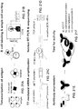


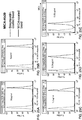


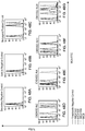
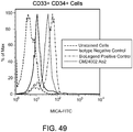


Classifications
-
- A—HUMAN NECESSITIES
- A61—MEDICAL OR VETERINARY SCIENCE; HYGIENE
- A61K—PREPARATIONS FOR MEDICAL, DENTAL OR TOILETRY PURPOSES
- A61K39/00—Medicinal preparations containing antigens or antibodies
- A61K39/395—Antibodies; Immunoglobulins; Immune serum, e.g. antilymphocytic serum
-
- C—CHEMISTRY; METALLURGY
- C07—ORGANIC CHEMISTRY
- C07K—PEPTIDES
- C07K16/00—Immunoglobulins [IGs], e.g. monoclonal or polyclonal antibodies
- C07K16/18—Immunoglobulins [IGs], e.g. monoclonal or polyclonal antibodies against material from animals or humans
- C07K16/28—Immunoglobulins [IGs], e.g. monoclonal or polyclonal antibodies against material from animals or humans against receptors, cell surface antigens or cell surface determinants
- C07K16/2803—Immunoglobulins [IGs], e.g. monoclonal or polyclonal antibodies against material from animals or humans against receptors, cell surface antigens or cell surface determinants against the immunoglobulin superfamily
- C07K16/2833—Immunoglobulins [IGs], e.g. monoclonal or polyclonal antibodies against material from animals or humans against receptors, cell surface antigens or cell surface determinants against the immunoglobulin superfamily against MHC-molecules, e.g. HLA-molecules
-
- A—HUMAN NECESSITIES
- A61—MEDICAL OR VETERINARY SCIENCE; HYGIENE
- A61K—PREPARATIONS FOR MEDICAL, DENTAL OR TOILETRY PURPOSES
- A61K39/00—Medicinal preparations containing antigens or antibodies
- A61K39/395—Antibodies; Immunoglobulins; Immune serum, e.g. antilymphocytic serum
- A61K39/39533—Antibodies; Immunoglobulins; Immune serum, e.g. antilymphocytic serum against materials from animals
- A61K39/3955—Antibodies; Immunoglobulins; Immune serum, e.g. antilymphocytic serum against materials from animals against proteinaceous materials, e.g. enzymes, hormones, lymphokines
-
- A—HUMAN NECESSITIES
- A61—MEDICAL OR VETERINARY SCIENCE; HYGIENE
- A61K—PREPARATIONS FOR MEDICAL, DENTAL OR TOILETRY PURPOSES
- A61K45/00—Medicinal preparations containing active ingredients not provided for in groups A61K31/00 - A61K41/00
- A61K45/06—Mixtures of active ingredients without chemical characterisation, e.g. antiphlogistics and cardiaca
-
- A—HUMAN NECESSITIES
- A61—MEDICAL OR VETERINARY SCIENCE; HYGIENE
- A61K—PREPARATIONS FOR MEDICAL, DENTAL OR TOILETRY PURPOSES
- A61K49/00—Preparations for testing in vivo
- A61K49/06—Nuclear magnetic resonance [NMR] contrast preparations; Magnetic resonance imaging [MRI] contrast preparations
- A61K49/08—Nuclear magnetic resonance [NMR] contrast preparations; Magnetic resonance imaging [MRI] contrast preparations characterised by the carrier
- A61K49/10—Organic compounds
- A61K49/14—Peptides, e.g. proteins
- A61K49/16—Antibodies; Immunoglobulins; Fragments thereof
-
- A—HUMAN NECESSITIES
- A61—MEDICAL OR VETERINARY SCIENCE; HYGIENE
- A61K—PREPARATIONS FOR MEDICAL, DENTAL OR TOILETRY PURPOSES
- A61K51/00—Preparations containing radioactive substances for use in therapy or testing in vivo
- A61K51/02—Preparations containing radioactive substances for use in therapy or testing in vivo characterised by the carrier, i.e. characterised by the agent or material covalently linked or complexing the radioactive nucleus
- A61K51/04—Organic compounds
- A61K51/08—Peptides, e.g. proteins, carriers being peptides, polyamino acids, proteins
- A61K51/10—Antibodies or immunoglobulins; Fragments thereof, the carrier being an antibody, an immunoglobulin or a fragment thereof, e.g. a camelised human single domain antibody or the Fc fragment of an antibody
-
- A—HUMAN NECESSITIES
- A61—MEDICAL OR VETERINARY SCIENCE; HYGIENE
- A61P—SPECIFIC THERAPEUTIC ACTIVITY OF CHEMICAL COMPOUNDS OR MEDICINAL PREPARATIONS
- A61P35/00—Antineoplastic agents
-
- A—HUMAN NECESSITIES
- A61—MEDICAL OR VETERINARY SCIENCE; HYGIENE
- A61P—SPECIFIC THERAPEUTIC ACTIVITY OF CHEMICAL COMPOUNDS OR MEDICINAL PREPARATIONS
- A61P35/00—Antineoplastic agents
- A61P35/02—Antineoplastic agents specific for leukemia
-
- A—HUMAN NECESSITIES
- A61—MEDICAL OR VETERINARY SCIENCE; HYGIENE
- A61P—SPECIFIC THERAPEUTIC ACTIVITY OF CHEMICAL COMPOUNDS OR MEDICINAL PREPARATIONS
- A61P43/00—Drugs for specific purposes, not provided for in groups A61P1/00-A61P41/00
-
- C—CHEMISTRY; METALLURGY
- C07—ORGANIC CHEMISTRY
- C07K—PEPTIDES
- C07K16/00—Immunoglobulins [IGs], e.g. monoclonal or polyclonal antibodies
- C07K16/06—Immunoglobulins [IGs], e.g. monoclonal or polyclonal antibodies from serum
-
- C—CHEMISTRY; METALLURGY
- C07—ORGANIC CHEMISTRY
- C07K—PEPTIDES
- C07K16/00—Immunoglobulins [IGs], e.g. monoclonal or polyclonal antibodies
- C07K16/12—Immunoglobulins [IGs], e.g. monoclonal or polyclonal antibodies against material from bacteria
- C07K16/1267—Immunoglobulins [IGs], e.g. monoclonal or polyclonal antibodies against material from bacteria from Gram-positive bacteria
- C07K16/1282—Immunoglobulins [IGs], e.g. monoclonal or polyclonal antibodies against material from bacteria from Gram-positive bacteria from Clostridium (G)
-
- C—CHEMISTRY; METALLURGY
- C07—ORGANIC CHEMISTRY
- C07K—PEPTIDES
- C07K16/00—Immunoglobulins [IGs], e.g. monoclonal or polyclonal antibodies
- C07K16/18—Immunoglobulins [IGs], e.g. monoclonal or polyclonal antibodies against material from animals or humans
- C07K16/22—Immunoglobulins [IGs], e.g. monoclonal or polyclonal antibodies against material from animals or humans against growth factors ; against growth regulators
-
- C—CHEMISTRY; METALLURGY
- C12—BIOCHEMISTRY; BEER; SPIRITS; WINE; VINEGAR; MICROBIOLOGY; ENZYMOLOGY; MUTATION OR GENETIC ENGINEERING
- C12N—MICROORGANISMS OR ENZYMES; COMPOSITIONS THEREOF; PROPAGATING, PRESERVING, OR MAINTAINING MICROORGANISMS; MUTATION OR GENETIC ENGINEERING; CULTURE MEDIA
- C12N5/00—Undifferentiated human, animal or plant cells, e.g. cell lines; Tissues; Cultivation or maintenance thereof; Culture media therefor
- C12N5/06—Animal cells or tissues; Human cells or tissues
- C12N5/0602—Vertebrate cells
- C12N5/0634—Cells from the blood or the immune system
-
- A—HUMAN NECESSITIES
- A61—MEDICAL OR VETERINARY SCIENCE; HYGIENE
- A61K—PREPARATIONS FOR MEDICAL, DENTAL OR TOILETRY PURPOSES
- A61K39/00—Medicinal preparations containing antigens or antibodies
- A61K2039/505—Medicinal preparations containing antigens or antibodies comprising antibodies
-
- A—HUMAN NECESSITIES
- A61—MEDICAL OR VETERINARY SCIENCE; HYGIENE
- A61K—PREPARATIONS FOR MEDICAL, DENTAL OR TOILETRY PURPOSES
- A61K31/00—Medicinal preparations containing organic active ingredients
- A61K31/16—Amides, e.g. hydroxamic acids
-
- A—HUMAN NECESSITIES
- A61—MEDICAL OR VETERINARY SCIENCE; HYGIENE
- A61K—PREPARATIONS FOR MEDICAL, DENTAL OR TOILETRY PURPOSES
- A61K31/00—Medicinal preparations containing organic active ingredients
- A61K31/325—Carbamic acids; Thiocarbamic acids; Anhydrides or salts thereof
-
- A—HUMAN NECESSITIES
- A61—MEDICAL OR VETERINARY SCIENCE; HYGIENE
- A61K—PREPARATIONS FOR MEDICAL, DENTAL OR TOILETRY PURPOSES
- A61K31/00—Medicinal preparations containing organic active ingredients
- A61K31/33—Heterocyclic compounds
- A61K31/335—Heterocyclic compounds having oxygen as the only ring hetero atom, e.g. fungichromin
- A61K31/365—Lactones
-
- A—HUMAN NECESSITIES
- A61—MEDICAL OR VETERINARY SCIENCE; HYGIENE
- A61K—PREPARATIONS FOR MEDICAL, DENTAL OR TOILETRY PURPOSES
- A61K39/00—Medicinal preparations containing antigens or antibodies
-
- A—HUMAN NECESSITIES
- A61—MEDICAL OR VETERINARY SCIENCE; HYGIENE
- A61K—PREPARATIONS FOR MEDICAL, DENTAL OR TOILETRY PURPOSES
- A61K39/00—Medicinal preparations containing antigens or antibodies
- A61K39/395—Antibodies; Immunoglobulins; Immune serum, e.g. antilymphocytic serum
- A61K39/39533—Antibodies; Immunoglobulins; Immune serum, e.g. antilymphocytic serum against materials from animals
- A61K39/39558—Antibodies; Immunoglobulins; Immune serum, e.g. antilymphocytic serum against materials from animals against tumor tissues, cells, antigens
-
- C—CHEMISTRY; METALLURGY
- C07—ORGANIC CHEMISTRY
- C07K—PEPTIDES
- C07K2317/00—Immunoglobulins specific features
- C07K2317/10—Immunoglobulins specific features characterized by their source of isolation or production
-
- C—CHEMISTRY; METALLURGY
- C07—ORGANIC CHEMISTRY
- C07K—PEPTIDES
- C07K2317/00—Immunoglobulins specific features
- C07K2317/20—Immunoglobulins specific features characterized by taxonomic origin
- C07K2317/21—Immunoglobulins specific features characterized by taxonomic origin from primates, e.g. man
-
- C—CHEMISTRY; METALLURGY
- C07—ORGANIC CHEMISTRY
- C07K—PEPTIDES
- C07K2317/00—Immunoglobulins specific features
- C07K2317/50—Immunoglobulins specific features characterized by immunoglobulin fragments
- C07K2317/56—Immunoglobulins specific features characterized by immunoglobulin fragments variable (Fv) region, i.e. VH and/or VL
- C07K2317/565—Complementarity determining region [CDR]
-
- C—CHEMISTRY; METALLURGY
- C07—ORGANIC CHEMISTRY
- C07K—PEPTIDES
- C07K2317/00—Immunoglobulins specific features
- C07K2317/70—Immunoglobulins specific features characterized by effect upon binding to a cell or to an antigen
- C07K2317/73—Inducing cell death, e.g. apoptosis, necrosis or inhibition of cell proliferation
-
- C—CHEMISTRY; METALLURGY
- C07—ORGANIC CHEMISTRY
- C07K—PEPTIDES
- C07K2317/00—Immunoglobulins specific features
- C07K2317/70—Immunoglobulins specific features characterized by effect upon binding to a cell or to an antigen
- C07K2317/76—Antagonist effect on antigen, e.g. neutralization or inhibition of binding
-
- C—CHEMISTRY; METALLURGY
- C07—ORGANIC CHEMISTRY
- C07K—PEPTIDES
- C07K2317/00—Immunoglobulins specific features
- C07K2317/90—Immunoglobulins specific features characterized by (pharmaco)kinetic aspects or by stability of the immunoglobulin
- C07K2317/92—Affinity (KD), association rate (Ka), dissociation rate (Kd) or EC50 value
Definitions
- This invention relates to therapeutic compositions related to human subjects.
- MICA is a ligand for NKG2D, a C-type lectin-like, type II transmembrane receptor expressed on most human NK cells, ⁇ T cells, and CD8+ T cells.
- NKG2D signals through the adaptor protein DAP10 to evoke perforin dependent cytolysis and to provide co-stimulation.
- the NKG2D ligands include MHC class I chain-related protein A (MICA), the closely related MICB, UL-16 binding proteins (ULBP) 1-4, and RAE-1G.
- NKG2D ligands are not usually found on healthy tissues, various forms of cellular stress, including DNA damage, may upregulate ligand expression, resulting in their frequent detection in multiple solid and hematologic malignancies, including melanoma.
- NKG2D activation through ligand positive transformed cells contributes to extrinsic tumor suppression, since NKG2D deficient and wild type mice treated with anti-NKG2D blocking antibodies manifest enhanced tumor susceptibility. Immune escape may be achieved in patients, however, by the shedding of NKG2D ligands from tumor cells, which triggers internalization of surface NKG2D and impaired function of cytotoxic lymphocytes.
- Soluble NKG2D ligands may also stimulate the expansion of regulatory NKG2D+CD4+Foxp3- T cells that may antagonize antitumor cytotoxicity through Fas ligand, IL-10, and TGF- ⁇ .
- MICA is a NKG2D ligand shed from tumor cells, i.e., released from the cell surface into the surrounding medium, and sera from cancer patients typically contain elevated levels of the soluble form (sMICA). MICA shedding is accomplished in part through interactions with the protein disulfide isomerase ERp5, which forms a disulfide bond with a critical cysteine that results in unfolding of the ⁇ 3 domain, rendering it susceptible to proteolysis by ADAM-10/17 and MMP14.
- MICA polypeptides and antibodies binding to MICA are described in WO 03/089616 .
- Angiogenesis is the process of forming new capillaries from preexisting blood vessels and has been implicated as a critical part of tumor growth and dissemination. Tumors stimulate angiogenesis to meet increasing oxygen and nutrient requirements that exceed those that can be met by diffusion alone. Consequently, tumors recruit, remodel and expand existing vascular to meet their metabolic demand. The dependence of growing tumors on new blood vessel formation has made angiogenesis an appealing target for anti-cancer therapies. Many cytokines have been are believed to play a role in the regulation of angiogenesis, including vascular endothelial growth factor (VEGF) family members and the angiopoietins.
- VEGF vascular endothelial growth factor
- angiopoietin-1 angiopoietin-1
- Ang-4 angiopoietin-4
- angiopoietins e.g., Ang-1 and Ang-2
- tumor angiogenesis e.g., tumor angiogenesis
- compositions and methods related to antibodies with therapeutic potential are described herein.
- compositions comprising an antibody or antibody fragment that immunospecifically binds to MHC class I polypeptide-related sequence A (MICA), or an epitope thereon.
- Said antibody or antibody fragment comprises a heavy chain variable region (V H ) and a light chain variable region (V L ) and complementarity determining region (CDR) 3 of the V H set forth in SEQ ID NO:176 of antibody ID 7; or complementarity determining region (CDR) 3 of the V H set forth in SEQ ID NO:194 of antibody ID 8.
- compositions are described herein comprising peptides which include complementarity determining region (CDR) 3 of the V H of antibody ID 1, 6, 7, 8 or 9 shown in Table 1 having 5 or fewer conservative amino acid substitutions, and CDR3 of the V L of antibody ID 1, 6, 7, 8 or 9 shown in Table 1 having 5 or fewer conservative amino acid substitutions.
- Such peptides may further include complementarity determining region (CDR) 3 of the V H of antibody ID 1, 6, 7, 8 or 9 shown in Table 1, and CDR3 of the V L of antibody ID 1, 6, 7, 8 or 9 shown in Table 1.
- Peptides described herein may further include CDR2 of the V H of antibody ID 1, 6, 7, 8 or 9 shown in Table 1 having 5 or fewer conservative amino acid substitutions, or CDR2 of the V L of antibody ID 1, 6, 7, 8 or 9 shown in Table 1 having 5 or fewer conservative amino acid substitutions, or both.
- Such peptides may include complementarity determining region CDR2 of the V H of antibody ID 1, 6, 7, 8 or 9 shown in Table 1, or CDR2 of the V L of antibody ID 1, 6, 7, 8 or 9 shown in Table 1, or both.
- Peptides described herein may further include CDR1 of the V H of antibody ID 1, 6, 7, 8 or 9 shown in Table 1 having 5 or fewer conservative amino acid substitutions, or CDR1 of the V L of antibody ID 1, 6, 7, 8 or 9 shown in Table 1 having 5 or fewer conservative amino acid substitutions, or both.
- Such peptides may include complementarity determining region CDR1 of the V H of antibody ID 1, 6, 7, 8 or 9 shown in Table 1, or CDR1 of the V L of antibody ID 1, 6, 7, 8 or 9 shown in Table 1, or both.
- Peptides described herein may be antibodies or antibody fragments that include: a V H chain with identity to SEQ ID NO:2, wherein regions corresponding to CDR1, CDR2, and CDR3 comprise CDR1, CDR2, and CDR3 of the V H of antibody ID 1 shown in table 1 having 5 or fewer conservative amino acid substitutions, and regions within SEQ ID NO:2 corresponding to FR1, FR2, FR3, FR4, comprise amino acid sequences with at least 80%, 85%, 90%, 95%, 96%, 97%, 98, 99%, or 100% identity to FR1, FR2, FR3, FR4 of the V H of antibody ID 1 shown in table 1; and a V L chain with identity to SEQ ID NO:11, wherein regions corresponding to CDR1, CDR2, and CDR3 comprise CDR1, CDR2, and CDR3 of the V L of antibody ID 1 shown in table 1 having 5 or fewer conservative amino acid substitutions, and regions within SEQ ID NO:11 corresponding to FR1, FR2, FR3, FR4,
- compositions described herein may include an antibody or antibody fragment comprising a V H chain comprising SEQ ID NO:2 and a V L chain comprising SEQ ID NO:11.
- compositions described herein may further include one or more (e.g., 1 2, 3, 4, 5, 6, 7, 8, 9, 10, or less than 20) anti-cancer therapeutics.
- Compositions described herein may be formulated as pharmaceutical compositions (e.g., for administration to a subject).
- Peptides described herein may be antibodies or antibody fragments that include: a V H chain with identity to SEQ ID NO:149, wherein regions corresponding to CDR1, CDR2, and CDR3 comprise CDR1, CDR2, and CDR3 of the V H of antibody ID 6 shown in table 1 having 5 or fewer conservative amino acid substitutions within the CDR1, CDR2, and CDR3 regions, and regions within SEQ ID NO:149 corresponding to FR1, FR2, FR3, FR4, comprise amino acid sequences with at least 80%, 85%, 90%, 95%, 96%, 97%, 98, 99%, or 100% identity to FR1, FR2, FR3, FR4 of the V H of antibody ID 6 shown in table 1; and a V L chain with identity to SEQ ID NO:151, wherein regions corresponding to CDR1, CDR2, and CDR3 comprise CDR1, CDR2, and CDR3 of the V L of antibody ID 6 shown in table 1 having 5 or fewer conservative amino acid substitutions within the CDR1, CDR2, and CDR
- compositions described herein may include an antibody or antibody fragment comprising a V H chain comprising SEQ ID NO:149 and a V L chain comprising SEQ ID NO:151.
- compositions described herein may further include one or more (e.g., 1 2, 3, 4, 5, 6, 7, 8, 9, 10, or less than 20) anti-cancer therapeutics.
- Compositions described herein may be formulated as pharmaceutical compositions (e.g., for administration to a subject).
- Peptides described herein may be antibodies or antibody fragments that include: a V H chain with identity to SEQ ID NO:168, wherein regions corresponding to CDR1, CDR2, and CDR3 comprise CDR1, CDR2, and CDR3 of the V H of antibody ID 7 shown in table 1 having 5 or fewer conservative amino acid substitutions within the CDR1, CDR2, and CDR3 regions, and regions within SEQ ID NO:168 corresponding to FR1, FR2, FR3, FR4, comprise amino acid sequences with at least 80%, 85%, 90%, 95%, 96%, 97%, 98, 99%, or 100% identity to FR1, FR2, FR3, FR4 of the V H of antibody ID 7 shown in table 1; and a V L chain with identity to SEQ ID NO:170, wherein regions corresponding to CDR1, CDR2, and CDR3 comprise CDR1, CDR2, and CDR3 of the V L of antibody ID 7 shown in table 1 having 5 or fewer conservative amino acid substitutions within the CDR1, CDR2, and C
- the antibody or antibody fragment according to the present invention comprises a V H chain comprising SEQ ID NO:168 and a V L chain comprising SEQ ID NO:170.
- the compositions further include one or more (e.g., 1 2, 3, 4, 5, 6, 7, 8, 9, 10, or less than 20) anti-cancer therapeutics.
- compositions are formulated as pharmaceutical compositions (e.g., for administration to a subject).
- Peptides described herein may be antibodies or antibody fragments that include: a V H chain with identity to SEQ ID NO:186, wherein regions corresponding to CDR1, CDR2, and CDR3 comprise CDR1, CDR2, and CDR3 of the V H of antibody ID 8 shown in table 1 having 5 or fewer conservative amino acid substitutions within the CDR1, CDR2, and CDR3 regions, and regions within SEQ ID NO:186 corresponding to FR1, FR2, FR3, FR4, comprise amino acid sequences with at least 80%, 85%, 90%, 95%, 96%, 97%, 98, 99%, or 100% identity to FR1, FR2, FR3, FR4 of the V H of antibody ID 8 shown in table 1; and a V L chain with identity to SEQ ID NO:188, wherein regions corresponding to CDR1, CDR2, and CDR3 comprise CDR1, CDR2, and CDR3 of the V L of antibody ID 8 shown in table 1 having 5 or fewer conservative amino acid substitutions within the CDR1, CDR2, and CDR
- the antibody or antibody fragment according to the present invention comprises a V H chain comprising SEQ ID NO:186 and a V L chain comprising SEQ ID NO:188.
- the compositions further include one or more (e.g., 1 2, 3, 4, 5, 6, 7, 8, 9, 10, or less than 20) anti-cancer therapeutics.
- the compositions are formulated as pharmaceutical compositions (e.g., for administration to a subject).
- Peptides described herein may be antibodies or antibody fragments that include: a V H chain with identity to SEQ ID NO:204, wherein regions corresponding to CDR1, CDR2, and CDR3 comprise CDR1, CDR2, and CDR3 of the V H of antibody ID 9 shown in table 1 having 5 or fewer conservative amino acid substitutions within the CDR1, CDR2, and CDR3 regions, and regions within SEQ ID NO:204 corresponding to FR1, FR2, FR3, FR4, comprise amino acid sequences with at least 80%, 85%, 90%, 95%, 96%, 97%, 98, 99%, or 100% identity to FR1, FR2, FR3, FR4 of the V H of antibody ID 9 shown in table 1; and a V L chain with identity to SEQ ID NO:206, wherein regions corresponding to CDR1, CDR2, and CDR3 comprise CDR1, CDR2, and CDR3 of the V L of antibody ID 9 shown in table 1 having 5 or fewer conservative amino acid substitutions within the CDR1, CDR2, and CDR
- compositions described herein may include an antibody or antibody fragment comprising a V H chain comprising SEQ ID NO:204 and a V L chain comprising SEQ ID NO:206.
- compositions described herein may further include one or more (e.g., 1 2, 3, 4, 5, 6, 7, 8, 9, 10, or less than 20) anti-cancer therapeutics.
- Compositions described herein may be formulated as pharmaceutical compositions (e.g., for administration to a subject).
- compositions are described herein that include one or more peptides that bind to angiopoietin or an epitope thereon.
- Peptides of the compositions described herein may include complementarity determining region (CDR) 3 of the V H of antibody ID 2, 3, 4, 5 or 10 shown in Table 1 having 5 or fewer conservative amino acid substitutions, and CDR3 of the V L of antibody ID 2, 3, 4 5 or 10 shown in Table 1 having 5 or fewer conservative amino acid substitutions within the CDR1, CDR2, and CDR3 regions.
- Peptides described herein can include complementarity determining region (CDR) 3 of the V H of antibody ID 2, 3, 4 or 5 or 10 shown in Table 1, and CDR3 of the V L of antibody ID 2, 3, 4 or 5 or 10 shown in Table 1.
- Peptides described herein can further include CDR2 of the V H of antibody ID 2, 3, 4 or 5 or 10 shown in Table 1 having 5 or fewer conservative amino acid substitutions, or CDR2 of the V L of antibody ID 2, 3, 4 or 5 or 10 shown in Table 1 having 5 or fewer conservative amino acid substitutions, or both.
- Such peptides can include complementarity determining region CDR2 of the V H of antibody ID 2, 3, 4 or 5 or 10 shown in Table 1, or CDR2 of the V L of antibody ID 2, 3, 4 or 5 or 10 shown in Table 1, or both.
- Peptides described herein can further include CDR1 of the V H of antibody ID 2, 3, 4 or 5 or 10 shown in Table 1 having 5 or fewer conservative amino acid substitutions, or CDR1 of the V L of antibody ID 2, 3, 4, or 5 shown in Table 1 having 5 or fewer conservative amino acid substitutions, or both.
- Such peptides can include complementarity determining region CDR1 of the V H of antibody ID 2, 3, 4 or 5 or 10 shown in Table 1, or CDR1 of the V L of antibody ID 2, 3, 4 or 5 or 10 shown in Table 1, or both.
- Peptides described herein may include an antibody or antibody fragment comprising: a V H chain with identity to SEQ ID NO:20, wherein regions corresponding to CDR1, CDR2, and CDR3 comprise CDR1, CDR2, and CDR3 of the V H of antibody ID 2 shown in table 1 having 5 or fewer conservative amino acid substitutions within the CDR1, CDR2, and CDR3 regions, and regions within SEQ ID NO:20 corresponding to FR1, FR2, FR3, FR4, comprise amino acid sequences with at least 80%, 85%, 90%, 95%, 96%, 97%, 98, 99%, or 100% identity to FR1, FR2, FR3, FR4 of the V H of antibody ID 2 shown in table 1; and a V L chain with identity to SEQ ID NO:29, wherein regions corresponding to CDR1, CDR2, and CDR3 comprise CDR1, CDR2, and CDR3 of the V L of antibody ID 2 shown in table 1 having 5 or fewer conservative amino acid substitutions within the CDR1, CDR2, and CDR
- the peptides described herein may be antibodies or antibody fragments comprising: a V H chain with identity to SEQ ID NO:38, wherein regions corresponding to CDR1, CDR2, and CDR3 comprise CDR1, CDR2, and CDR3 of the V H of antibody ID 3 shown in table 1 having 5 or fewer conservative amino acid substitutions within the CDR1, CDR2, and CDR3 regions, and regions within SEQ ID NO:38 corresponding to FR1, FR2, FR3, FR4, comprise amino acid sequences with at least 80%, 85%, 90%, 95%, 96%, 97%, 98, 99%, or 100% identity to FR1, FR2, FR3, FR4 of the V H of antibody ID 3 shown in table 1; and a V L chain with identity to SEQ ID NO:47, wherein regions corresponding to CDR1, CDR2, and CDR3 comprise CDR1, CDR2, and CDR3 of the V L of antibody ID 3 shown in table 1 having 5 or fewer conservative amino acid substitutions within the CDR1, CDR2, and
- Peptides described herein may include an antibody or antibody fragment comprising: a V H chain with identity to SEQ ID NO:56, wherein regions corresponding to CDR1, CDR2, and CDR3 comprise CDR1, CDR2, and CDR3 of the V H of antibody ID 4 shown in table 1 having 5 or fewer conservative amino acid substitutions within the CDR1, CDR2, and CDR3 regions, and regions within SEQ ID NO:56 corresponding to FR1, FR2, FR3, FR4, comprise amino acid sequences with at least 80%, 85%, 90%, 95%, 96%, 97%, 98, 99%, or 100% identity to FR1, FR2, FR3, FR4 of the V H of antibody ID 4 shown in table 1; and a V L chain with identity to SEQ ID NO:65, wherein regions corresponding to CDR1, CDR2, and CDR3 comprise CDR1, CDR2, and CDR3 of the V L of antibody ID 4 shown in table 1 having 5 or fewer conservative amino acid substitutions within the CDR1, CDR2, and CDR
- Peptides described herein may include an antibody or antibody fragment comprising: a V H chain with identity to SEQ ID NO:74, wherein regions corresponding to CDR1, CDR2, and CDR3 comprise CDR1, CDR2, and CDR3 of the V H of antibody ID 5 shown in table 1 having 5 or fewer conservative amino acid substitutions within the CDR1, CDR2, and CDR3 regions, and regions within SEQ ID NO:74 corresponding to FR1, FR2, FR3, FR4, comprise amino acid sequences with at least 80%, 85%, 90%, 95%, 96%, 97%, 98, 99%, or 100% identity to FR1, FR2, FR3, FR4 of the V H of antibody ID 5 shown in table 1; and a V L chain with identity to SEQ ID NO:83, wherein regions corresponding to CDR1, CDR2, and CDR3 comprise CDR1, CDR2, and CDR3 of the V L of antibody ID 5 shown in table 1 having 5 or fewer conservative amino acid substitutions within the CDR1, CDR2, and CDR
- the peptides described herein may include an antibody or antibody fragment comprising a V H chain comprising SEQ ID NO:74 and a V L chain comprising SEQ ID NO:83.
- the peptides described herein may immunospecifically bind to at least angiopoietin-2.
- the compositions described herein may further include one or more anti-cancer therapeutics.
- the compositions described herein may be formulated as a pharmaceutical composition.
- Peptides described herein may include an antibody or antibody fragment comprising: a V H chain with identity to SEQ ID NO:222, wherein regions corresponding to CDR1, CDR2, and CDR3 comprise CDR1, CDR2, and CDR3 of the V H of antibody ID 10 shown in table 1 having 5 or fewer conservative amino acid substitutions within the CDR1, CDR2, and CDR3 regions, and regions within SEQ ID NO:222 corresponding to FR1, FR2, FR3, FR4, comprise amino acid sequences with at least 80%, 85%, 90%, 95%, 96%, 97%, 98, 99%, or 100% identity to FR1, FR2, FR3, FR4 of the V H of antibody ID 10 shown in table 1; and a V L chain with identity to SEQ ID NO:224, wherein regions corresponding to CDR1, CDR2, and CDR3 comprise CDR1, CDR2, and CDR3 of the V L of antibody ID 10 shown in table 1 having 5 or fewer conservative amino acid substitutions within the CDR1, CDR2, and CDR
- the peptides described herein may include an antibody or antibody fragment comprising a V H chain comprising SEQ ID NO:222 and a V L chain comprising SEQ ID NO:224.
- the peptides described herein may immunospecifically bind to at least angiopoietin-2.
- the compositions described herein may further include one or more anti-cancer therapeutics.
- the compositions described herein may be formulated as a pharmaceutical composition.
- methods of treating cancer in a subject are described herein.
- the methods may include administering to a subject a composition described herein.
- a method of obtaining immune cells directed against a self antigen from a subject comprising identifying a subject exhibiting a positive immune response towards the self antigen, providing a multimeric form of the self antigen, contacting the multimeric form of the self antigen with a sample from the subject exhibiting a positive immune response towards the self antigen, and obtaining immune cells bound to the multimeric form of the self antigen.
- a method of obtaining immune cells from a cancer patient directed against a self antigen comprising identifying a subject exhibiting a positive immune response towards the self antigen; providing a multimeric form of the self antigen; contacting the multimeric form of the self antigen with a sample from the subject exhibiting a positive immune response towards the self antigen; and obtaining immune cells bound to the multimeric form of the self antigen.
- the present disclosure is based, in part, on the observation that antibodies directed against therapeutic targets important in a disease can be obtained from human subjects exposed to the disease by labeling of B cells with a tetrameric form of the antigen of interest.
- prior methods are limited at least in that they are inefficient at identifying appropriate B cells in human subjects and/or because they induce any captured B cells to undergo phenotypic changes, thus reducing their value.
- methods are described herein that allow capture of rare memory B cells directed against specific disease-related antigens. As described below, the methods require tetramerization of the disease-related antigen, which process, as demonstrated in the Examples below, enhances the identification of appropriate memory B cells.
- methods herein permit more efficient capture of appropriate memory B cells for increased periods of time following initial exposure of a subject to the antigen.
- Methods herein also include antibodies (and peptides generated from the sequences of such antibodies) generated using genetic material obtained from memory B cells captured using the methods described herein.
- Described herein are human antibodies against MHC class I polypeptide-related sequence A (MICA) and human antibodies targeted against angiopoietin-2. Both types of human antibodies were identified from patients who had received a cell-based cancer vaccine (GM-CSF transduced autologous tumor cells) by methods that entail the use of tetrameric antigens.
- MICA MHC class I polypeptide-related sequence A
- GM-CSF transduced autologous tumor cells GM-based cancer vaccine
- Methods for specifically obtaining or targeting antibodies with therapeutic potential from select human subjects and therapeutic compositions resulting therefrom are described herein. These methods can include: obtaining or targeting immune cells in a human subject, wherein immune cells include but are not limited to, for example, B cells and/or memory B cells, isolating or purifying genetic material (e.g., DNA and/or mRNA) from the obtained or targeted immune cells, and using the isolated or purified genetic material to produce therapeutic compositions, e.g., therapeutic compositions disclosed herein. Further description of the methods is provided under the section entitled "Methods," below.
- genetic material e.g., DNA and/or mRNA
- compositions e.g., including therapeutic peptides, including antibodies, antibody fragments, antibody derivatives, and/or antibody conjugates
- therapeutic peptides including antibodies, antibody fragments, antibody derivatives, and/or antibody conjugates
- therapeutic compositions herein can interact with (e.g., bind, bind specifically and/or bind immunospecifically) binding partners (e.g., an immunogen(s), antigen(s), and/or epitope(s)) related to a disease or condition, wherein interaction between the therapeutic composition and the binding partners results in a positive immune response towards the condition or disease (e.g., a decrease in the level of disease or symptoms thereof in a subject).
- binding partners e.g., an immunogen(s), antigen(s), and/or epitope(s)
- therapeutic compositions can include peptides that include (e.g., comprise, consist essentially of, or consist of) at least one (e.g., one, two, three, four, five, and/or six) complementarity determining region (CDR) of the variable heavy chain (V H ) and/or variable light chain (V L ) of antibody ID 1, 2, 3, 4, or 5, 6, 7, 8, 9 or 10, shown in Table 1.
- CDR complementarity determining region
- therapeutic compositions can include peptides that include (e.g., comprise, consist essentially of, or consist of) at least one (e.g., one, two, three, four, five, and/or six) complementarity determining region (CDR) of the variable heavy chain (VH) and/or variable light chain (VL) of antibody ID 1, 2, 3, 4, 5, 6, 7, 8, 9 or 10, shown in Table 1, and that interact with (e.g., bind, bind specifically and/or bind immunospecifically) to MHC class I polypeptide-related sequence A (MICA (e.g., UniGene Hs.130838)) (e.g., soluble MICA (sMICA)) and/or angiopoietin-2 (e.g., UniGene Hs.583870), including epitopes thereof.
- MICA MHC class I polypeptide-related sequence A
- sMICA soluble MICA
- angiopoietin-2 e.g., UniGene Hs.
- therapeutic compositions can include peptides that include at least one CDR of the V H and/or V L of antibody ID 1, 6, 7, 8 and/or 9 shown in Table 1, wherein the peptide binds (e.g., binds specifically and/or binds immunospecifically) to MICA (e.g., human MICA (e.g., soluble MICA (sMICA))).
- MICA e.g., human MICA (e.g., soluble MICA (sMICA)
- peptides can include at least two CDRs, wherein the at least two CDRs are CDRs shown in Table 1 for different antibodies..
- CDRs (and FRs and/or AA sequences) shown in Table 1 for antibodies IDs 1, 6, 7, 8 and 9 are interchangeable and can be combined to generate peptides, so long as the peptides bind (e.g., bind specifically and/or bind immunospecifically) to MICA (e.g., human MICA (e.g., soluble MICA (sMICA))).
- MICA e.g., human MICA (e.g., soluble MICA (sMICA)
- peptides include CDR3 of the V H and/or V L of antibody ID 1, 6, 7, 8 and/or 9 shown in Table 1.
- such peptides include CDR3 of the V H and V L of antibody ID 1, 6, 7, 8 and/or 9 and CDR1 and/or CDR2 of the V H and/or V L of antibody ID 1, 6, 7, 8 and/or 9 shown in Table 1.
- such peptides include CDR1 CDR2, and CDR3 of the V H and/or V L of antibody ID 1, 6, 7, 8 and/or 9.
- such peptides include CDR1, CDR2, and CDR3 of the V H and/or V L of antibody ID 1, 6, 7, 8 and/or 9 and at least one of FR1 FR2 FR3, and/or FR4 of the V H and/or V L of antibody ID 1, 6, 7, 8 and/or 9, shown in Table 1.
- such peptides include one of SEQ ID NO:2, 149, 168, 186 or 204 and/or one of SEQ ID NO:11, 151, 170, 188, or 206.
- the peptide can bind (e.g., bind specifically and/or bind immunospecifically) to MICA (e.g., human MICA (e.g., soluble MICA (sMICA))).
- MICA e.g., human MICA (e.g., soluble MICA (sMICA)
- the affinity of binding between the peptides and MICA can be between about 0.1nM to 1 ⁇ M, for example, about 10nM.
- therapeutic compositions can include peptides that include at least one CDR of the V H and/or V L of antibody ID 6 shown in Table 1, wherein the peptide binds (e.g., binds specifically and/or binds immunospecifically) to MICA (e.g., human MICA (e.g., soluble MICA (sMICA))).
- MICA e.g., human MICA (e.g., soluble MICA (sMICA)
- such peptides include CDR3 of the V H and/or V L of antibody ID 6 shown in Table 1.
- such peptides include CDR3 of the V H and V L of antibody ID 6 and CDR1 and/or CDR2 of the V H and/or V L of antibody ID 6 shown in Table 1.
- such peptides include CDR1 CDR2, and CDR3 of the V H and/or V L of antibody ID 6. In some instances, such peptides include CDR1, CDR2, and CDR3 of the V H and/or V L of antibody ID 6 and at least one of FR1 FR2 FR3, and/or FR4 of the V H and/or V L of antibody ID 6, shown in Table 1. In some instances, such peptides include SEQ ID NO:149 and/or SEQ ID NO:151. In each instance, the peptide can bind (e.g., bind specifically and/or bind immunospecifically) to MICA (e.g., human MICA (e.g., soluble MICA (sMICA))). In some instances, the affinity of binding between the peptides and MICA can be between about 0.1nM to 1 ⁇ M, for example, about 10nM.
- MICA e.g., human MICA (e.g., soluble MICA (sMICA)
- therapeutic compositions can include peptides that include at least one CDR of the V H and/or V L of antibody ID 7 shown in Table 1, wherein the peptide binds (e.g., binds specifically and/or binds immunospecifically) to MICA (e.g., human MICA (e.g., soluble MICA (sMICA))).
- MICA e.g., human MICA (e.g., soluble MICA (sMICA)
- such peptides include CDR3 of the V H and/or V L of antibody ID 7 shown in Table 1.
- such peptides include CDR3 of the V H and V L of antibody ID 7 and CDR1 and/or CDR2 of the V H and/or V L of antibody ID 7 shown in Table 1.
- such peptides include CDR1 CDR2, and CDR3 of the V H and/or V L of antibody ID 7. In some instances, such peptides include CDR1, CDR2, and CDR3 of the V H and/or V L of antibody ID 7 and at least one of FR1 FR2 FR3, and/or FR4 of the V H and/or V L of antibody ID 7, shown in Table 1. In some instances, such peptides include SEQ ID NO:168 and/or SEQ ID NO:170. In each instance, the peptide can bind (e.g., bind specifically and/or bind immunospecifically) to MICA (e.g., human MICA (e.g., soluble MICA (sMICA))). In some instances, the affinity of binding between the peptides and MICA can be between about 0.1nM to 1 ⁇ M, for example, about 10nM.
- MICA e.g., human MICA (e.g., soluble MICA (sMICA)
- therapeutic compositions can include peptides that include at least one CDR of the V H and/or V L of antibody ID 8 shown in Table 1, wherein the peptide binds (e.g., binds specifically and/or binds immunospecifically) to MICA (e.g., human MICA (e.g., soluble MICA (sMICA))).
- MICA e.g., human MICA (e.g., soluble MICA (sMICA)
- such peptides include CDR3 of the V H and/or V L of antibody ID 8 shown in Table 1.
- such peptides include CDR3 of the V H and V L of antibody ID 8 and CDR1 and/or CDR2 of the V H and/or V L of antibody ID 8 shown in Table 1.
- such peptides include CDR1 CDR2, and CDR3 of the V H and/or V L of antibody ID 8. In some instances, such peptides include CDR1, CDR2, and CDR3 of the V H and/or V L of antibody ID 8 and at least one of FR1 FR2 FR3, and/or FR4 of the V H and/or V L of antibody ID 8, shown in Table 1. In some instances, such peptides include SEQ ID NO:186 and/or SEQ ID NO:188. In each instance, the peptide can bind (e.g., bind specifically and/or bind immunospecifically) to MICA (e.g., human MICA (e.g., soluble MICA (sMICA))). In some instances, the affinity of binding between the peptides and MICA can be between about 0.1nM to 1 ⁇ M, for example, about 10nM.
- MICA e.g., human MICA (e.g., soluble MICA (sMICA)
- therapeutic compositions can include peptides that include at least one CDR of the V H and/or V L of antibody ID 9 shown in Table 1, wherein the peptide binds (e.g., binds specifically and/or binds immunospecifically) to MICA (e.g., human MICA (e.g., soluble MICA (sMICA))).
- MICA e.g., human MICA (e.g., soluble MICA (sMICA)
- such peptides include CDR3 of the V H and/or V L of antibody ID 9 shown in Table 1.
- such peptides include CDR3 of the V H and V L of antibody ID 9 and CDR1 and/or CDR2 of the V H and/or V L of antibody ID 9 shown in Table 1.
- such peptides include CDR1 CDR2, and CDR3 of the V H and/or V L of antibody ID 9. In some instances, such peptides include CDR1, CDR2, and CDR3 of the V H and/or V L of antibody ID 9 and at least one of FR1 FR2 FR3, and/or FR4 of the V H and/or V L of antibody ID 9, shown in Table 1. In some instances, such peptides include SEQ ID N0:204 and/or SEQ ID NO:206. In each instance, the peptide can bind (e.g., bind specifically and/or bind immunospecifically) to MICA (e.g., human MICA (e.g., soluble MICA (sMICA))). In some instances, the affinity of binding between the peptides and MICA can be between about 0.1nM to 1 ⁇ M, for example, about 10nM.
- MICA e.g., human MICA (e.g., soluble MICA (sMICA)
- therapeutic compositions can include peptides that include at least one CDR of the V H and/or V L of antibody ID 2, 3, 4, 5, and/or 10 shown in Table 1, wherein the peptide binds (e.g., binds specifically and/or binds immunospecifically) to angiopoietin-2 (e.g., human angiopoietin-2).
- peptides can include at least two CDRs, wherein the at least two CDRs are CDRs shown in Table 1 for different antibodies.
- CDRs (and FRs and/or AA sequences) shown in Table 1 for antibodies IDs 2, 3 4, 5, and 10 are interchangeable and can be combined to generate peptides, so long as the peptides bind (e.g., bind specifically and/or bind immunospecifically) to angiopoietin-2 (e.g., human angiopoietin-2).
- angiopoietin-2 e.g., human angiopoietin-2
- such peptides include CDR3 of the V H and/or V L of antibody ID 2, 3, 4, 5, and/or 10 shown in Table 1.
- such peptides include CDR3 of the V H and V L of antibody ID 2, 3, 4, 5, and/or 10 and CDR1 and/or CDR2 of the V H and/or V L of antibody ID 2, 3, 4, 5, and/or 10 shown in Table 1.
- such peptides include CDR1, CDR2, and CDR3 of the V H and/or V L of antibody ID 2, 3, 4, 5, and/or 10.
- such peptides include CDR1 CDR2, and CDR3 of the V H and/or V L of antibody ID 2, 3, 4, 5, and/or 10 and at least one of FR1 FR2 FR3, and/or FR4 of the V H and/or V L of antibody ID 2, 3, 4, 5, and/or 10 5, shown in Table 1.
- such peptides include one of SEQ ID NO:20, 38, 56, 74, or 222 and/or one of SEQ ID NO:29, 47, 65, 83 or 224. In some instances, peptides include one of SEQ ID NO:20, 38, 56, 74, or 222 and one of SEQ ID NO:29, 47, 65, 83 or 224. In each instance, the peptide can bind (e.g., bind specifically and/or bind immunospecifically) to angiopoietin-2 (e.g., human angiopoietin-2 (e.g, UniGene Hs.583870)).
- angiopoietin-2 e.g., human angiopoietin-2 (e.g, UniGene Hs.583870)
- therapeutic compositions can include peptides that include at least one CDR of the V H and/or V L of antibody ID 2 shown in Table 1, wherein the peptide binds (e.g., binds specifically and/or binds immunospecifically) to angiopoietin-2 (e.g., human angiopoietin-2).
- angiopoietin-2 e.g., human angiopoietin-2
- such peptides include CDR3 of the V H and/or V L of antibody ID 2 shown in Table 1.
- such peptides include CDR3 of the V H and V L of antibody ID 2 and CDR1 and/or CDR2 of the V H and/or V L of antibody ID 2 shown in Table 1.
- such peptides include CDR1, CDR2, and CDR3 of the V H and/or V L of antibody ID 2.
- such peptides include CDR1 CDR2, and CDR3 of the V H and/or V L of antibody ID 2 and at least one of FR1 FR2 FR3, and/or FR4 of the V H and/or V L of antibody ID 2, shown in Table 1.
- such peptides include SEQ ID NO:20 and/or SEQ ID NO:29.
- the peptide can bind (e.g., bind specifically and/or bind immunospecifically) to angiopoietin-2 (e.g., human angiopoietin-2).
- the affinity of binding between the peptides and angiopoietin-2 can be between about 0.1nM to 1 ⁇ M, for example, about 10nM.
- therapeutic compositions can include peptides that include at least one CDR of the V H and/or V L of antibody ID 3 shown in Table 1, wherein the peptide binds (e.g., binds specifically and/or binds immunospecifically) to angiopoietin-2 (e.g., human angiopoietin-2).
- angiopoietin-2 e.g., human angiopoietin-2
- such peptides include CDR3 of the V H and/or V L of antibody ID 3 shown in Table 1.
- such peptides include CDR3 of the V H and V L of antibody ID 3 and CDR1 and/or CDR2 of the V H and/or V L of antibody ID 3 shown in Table 1.
- such peptides include CDR1, CDR2, and CDR3 of the V H and/or V L of antibody ID 3. In some instances, such peptides include CDR1 CDR2, and CDR3 of the V H and/or V L of antibody ID 3 and at least one of FR1 FR2 FR3, and/or FR4 of the V H and/or V L of antibody ID 3, shown in Table 1. In some instances, such peptides include SEQ ID NO:38 and/or SEQ ID NO:47. In each instance, the peptide can bind (e.g., bind specifically and/or bind immunospecifically) to angiopoietin-2 (e.g., human angiopoietin-2). In some instances, the affinity of binding between the peptides and angiopoietin-2 can be between about 0.1nM to 1 ⁇ M, for example, about 10nM.
- angiopoietin-2 e.g., human angiopoietin-2
- therapeutic compositions can include peptides that include at least one CDR of the V H and/or V L of antibody ID 4 shown in Table 1, wherein the peptide binds (e.g., binds specifically and/or binds immunospecifically) to angiopoietin-2 (e.g., human angiopoietin-2).
- angiopoietin-2 e.g., human angiopoietin-2
- such peptides include CDR3 of the V H and/or V L of antibody ID 4 shown in Table 1.
- such peptides include CDR3 of the V H and V L of antibody ID 4 and CDR1 and/or CDR2 of the V H and/or V L of antibody ID 4 shown in Table 1.
- such peptides include CDR1, CDR2, and CDR3 of the V H and/or V L of antibody ID 4.
- such peptides include CDR1 CDR2, and CDR3 of the V H and/or V L of antibody ID 4 and at least one of FR1 FR2 FR3, and/or FR4 of the V H and/or V L of antibody ID 4, shown in Table 1.
- such peptides include SEQ ID NO:56 and/or SEQ ID NO:65.
- the peptide can bind (e.g., bind specifically and/or bind immunospecifically) to angiopoietin-2 (e.g., human angiopoietin-2).
- the affinity of binding between the peptide and angiopoietin-2 can be between X-Y, for example, X-Y, X-Y. In some instances, the affinity of binding between the peptides and angiopoietin-2 can be between about 0.1nM to 1 ⁇ M, for example, about 10nM.
- therapeutic compositions can include peptides that include at least one CDR of the V H and/or V L of antibody ID 5 shown in Table 1, wherein the peptide binds (e.g., binds specifically and/or binds immunospecifically) to angiopoietin-2 (e.g., human angiopoietin-2).
- angiopoietin-2 e.g., human angiopoietin-2
- such peptides include CDR3 of the V H and/or V L of antibody ID 5 shown in Table 1.
- such peptides include CDR3 of the V H and V L of antibody ID 5 and CDR1 and/or CDR2 of the V H and/or V L of antibody ID 5 shown in Table 1.
- such peptides include CDR1, CDR2, and CDR3 of the V H and/or V L of antibody ID 5.
- such peptides include CDR1 CDR2, and CDR3 of the V H and/or V L of antibody ID 5 and at least one of FR1 FR2 FR3, and/or FR4 of the V H and/or V L of antibody ID 5, shown in Table 1.
- such peptides include SEQ ID NO:74 and/or SEQ ID NO:83.
- the peptide can bind (e.g., bind specifically and/or bind immunospecifically) to angiopoietin-2 (e.g., human angiopoietin-2).
- the affinity of binding between the peptides and angiopoietin-2 can be between about 0.1nM to 1 ⁇ M, for example, about 10nM.
- therapeutic compositions can include peptides that include at least one CDR of the V H and/or V L of antibody ID 10 shown in Table 1, wherein the peptide binds (e.g., binds specifically and/or binds immunospecifically) to angiopoietin-2 (e.g., human angiopoietin-2).
- angiopoietin-2 e.g., human angiopoietin-2
- such peptides include CDR3 of the V H and/or V L of antibody ID 10 shown in Table 1.
- such peptides include CDR3 of the V H and V L of antibody ID 10 and CDR1 and/or CDR2 of the V H and/or V L of antibody ID 10 shown in Table 1.
- such peptides include CDR1, CDR2, and CDR3 of the V H and/or V L of antibody ID 10.
- such peptides include CDR1 CDR2, and CDR3 of the V H and/or V L of antibody ID 10 and at least one of FR1 FR2 FR3, and/or FR4 of the V H and/or V L of antibody ID 10, shown in Table 1.
- such peptides include SEQ ID NO:222 and/or SEQ ID NO:224.
- the peptide can bind (e.g., bind specifically and/or bind immunospecifically) to angiopoietin-2 (e.g., human angiopoietin-2).
- the affinity of binding between the peptides and angiopoietin-2 can be between about 0.1nM to 1 ⁇ M, for example, about 10nM.
- peptides that bind to angiopoietin-2 can also bind to angiopoietin-1 (e.g., Unigene Hs.369675) and/or angiopoietin-4 (e.g., Unigene Hs.278973).
- angiopoietin-1 e.g., Unigene Hs.369675
- angiopoietin-4 e.g., Unigene Hs.278973
- peptides that bind to angiopoietin-2 can also bind specifically and/or immunospecifically relative to other antigens (other than angiopoietin-1) to angiopoietin-1.
- peptides that bind to angiopoietin-2 can also bind specifically and/or immunospecifically relative to other antigens (other than angiopoietin-4) to angiopoietin-4.
- therapeutic compositions can include peptides that include: SEQ ID NO: 2 and/or SEQ ID NO:11; SEQ ID NO: 149 and/or SEQ ID NO:151; SEQ ID NO: 168 and/or SEQ ID NO:170; SEQ ID NO: 186 and/or SEQ ID NO:188; SEQ ID NO: 204 and/or SEQ ID NO:206; SEQ ID N0:20 and/or SEQ ID NO:29; SEQ ID NO:38 and/or SEQ ID NO:47; SEQ ID NO:56 and/or SEQ ID NO:65; SEQ ID NO:74 and/or SEQ ID NO:83; and SEQ ID NO: 222 and/or SEQ ID NO:224.
- SEQ ID NO: 187 (see FIG. 38 ) 9 Human MICA V H SEQ ID NO: 204 (see FIG. 41 ) SEQ ID NO: 203 (see FIG. 40 ) V L RIS (SEQ ID NO: 217) SEQ ID NO: 206 (see FIG. 43 ) SEQ ID NO: 205 (see FIG. 42 ) 2 Angiopoietin-2 V H SEQ ID NO: 20 (see FIG. 6 ) SEQ ID NO: 19 (see FIG. 5 ) V L QIS (SEQ ID NO: 33) SEQ ID NO: 29 (see FIG. 8 ) SEQ ID NO: 28 (see FIG. 7 ) 3 Angiopoietin-2 V H SEQ ID NO: 38 (see FIG.
- SEQ ID NO: 17 5 V L QDIS TW (SEQ ID NO: 85) GAS (SEQ ID NO: 87) SEQ ID NO: 83 (see FIG. 20 ) SEQ ID NO: 82 (see FIG. 19 ) 10 Angiopoietin-2 V H GFLISSYF (SEQ ID NO:226) IYSDGST (SEQ ID NO:228) SEQ ID NO: 222 (see FIG. 45 ) SEQ ID NO: 221 (see FIG. 44 ) V L KVS (SEQ ID NO:234) SEQ ID NO: 224 (see FIG. 47 ) SEQ ID NO: 223 (see FIG.
- Sequences include sequences or variants with (e.g., with at least) 80%, 85%, 90%, 95%, 96%, 97%, 98, 99%, and/or 100% sequence identity to the sequences shown. ** Sequences can include one, two, three, four, five, less than five, or less than ten conservative amino acid modifications.
- Sequences include sequences or variants with (e.g., with at least) 80%, 85%, 90%, 95%, 96%, 97%, 98, 99%, and/or 100% sequence identity to the sequences shown, e.g., within regions corresponding to FR1, FR2, FR3, and/or FR4, and/or one, two, three, four, five, less than 5, or less than ten conservative amino acid modifications within regions corresponding to CDRs 1, 2, and/or 3.
- Sequences include sequences or variants with (e.g., with at least) 80%, 85%, 90%, 95%, 96%, 97%, 98, 99%, and/or 100% sequence identity to the sequences shown, wherein the sequences encode the corresponding AA A.A. # shows the V H or V L amino acid sequence.
- Nuc. Acid ## shows the V H or V L nucleic acid sequence. While CDR and FR regions arc shown above, such regions can also be defined according to Kabat (Scqucnccs of Proteins of Immunological Interest (National Institutes of Health, Bethesda, Md., 1987 and 1991)). Amino acid numbering of antibodies or antigen binding fragments is also according to that of Kabat.
- therapeutic compositions can include peptides, including for example, antibodies, including full length and/or intact antibodies, or antibody fragments.
- An “antibody” is an immunoglobulin molecule capable of specific binding to a target, such as a carbohydrate, polynucleotide, lipid, polypeptide, etc., through at least one antigen recognition site, located in the variable region of the immunoglobulin molecule.
- the term “antibody” encompasses not only intact polyclonal or monoclonal antibodies, but also any antigen binding fragment (i.e., "antigen-binding portion") or single chain thereof, fusion proteins comprising an antibody, and any other modified configuration of the immunoglobulin molecule that comprises an antigen recognition site including.
- An antibody includes an antibody of any class, such as IgG, IgA, or IgM (or sub-class thereof), and the antibody need not be of any particular class.
- immunoglobulins can be assigned to different classes. There are five major classes of immunoglobulins: IgA, IgD, IgE, IgG, and IgM, and several of these may be further divided into subclasses (isotypes), e.g., IgG1, IgG2, IgG3, IgG4, IgA1 and IgA2.
- the heavy-chain constant regions that correspond to the different classes of immunoglobulins are called alpha, delta, epsilon, gamma, and mu, respectively.
- the subunit structures and three-dimensional configurations of different classes of immunoglobulins are well known.
- Exemplary antibodies and antibody fragments include, but are not limited to, monoclonal antibodies (including full-length monoclonal antibodies), polyclonal antibodies, multispecific antibodies formed from at least two different epitope binding fragments (e.g., bispecific antibodies), camelised antibodies, chimeric antibodies, single-chain Fvs (scFv), single-chain antibodies, single domain antibodies, domain antibodies, Fab fragments, F(ab')2 fragments, antibody fragments that exhibit the desired biological activity (e.g.
- Antibodies or antibody fragments can be human or humanized.
- Fragments of antibodies are suitable for use in the methods described herein so long as they retain the desired affinity and specificity of the full-length antibody.
- a fragment of an anti- MICA antibody or the anti-Angiopoietin antibody will retain an ability to bind to MICA or angiopoietin, respectively, in the Fv portion and the ability to bind the Fc receptor on dendritic cells in the FC portion.
- fragments are characterized by properties similar to the corresponding full-length anti-MICA antibody or the anti-Angiopoietin antibody, that is, the fragments will specifically bind a human MICA antigen or the angiopoietin antigen, respectively, expressed on the surface of a human cell or the corresponding sMICA antigen that has been shed into the media.
- An Fv fragment is an antibody fragment which contains a complete antigen recognition and binding site.
- This region consists of a dimer of one heavy and one light chain variable domain in tight association, which can be covalent in nature, for example in scFv. It is in this configuration that the three CDRs of each variable domain interact to define an antigen binding site on the surface of the VH-VL dimer.
- the six CDRs or a subset thereof confer antigen binding specificity to the antibody.
- a single variable domain or half of an Fv comprising only three CDRs specific for an antigen
- Single-chain Fv or (scFv) antibody fragments comprise the V H and V L domains of antibody, wherein these domains are present in a single polypeptide chain.
- the Fv polypeptide further comprises a polypeptide linker between the V H and V L domains, which enables the scFv to form the desired structure for antigen binding.
- the Fab fragment contains a variable and constant domain of the light chain and a variable domain and the first constant domain (CH1) of the heavy chain.
- F(ab')2 antibody fragments comprise a pair of Fab fragments which are generally covalently linked near their carboxy termini by hinge cysteines between them. Other chemical couplings of antibody fragments are also known in the art.
- Diabodies are small antibody fragments with two antigen-binding sites, which fragments comprise a V H connected to a V L in the same polypeptide chain (V H and V L ). By using a linker that is too short to allow pairing between the two domains on the same chain, the domains are forced to pair with the complementary domains of another chain and create two antigen-binding sites.
- Linear antibodies comprise a pair of tandem Fd segments (V H -CH1-V H -CH1) which, together with complementary light chain polypeptides, form a pair of antigen binding regions.
- Linear antibodies can be bispecific or monospecific.
- Antibodies and antibody fragments useful in the present invention can be modified in the Fc region to provide desired effector functions or serum half-life.
- the Fc region can be conjugated to PEG or albumin to increase the serum half-life, or some other conjugation that results in the desired effect.
- certain other Fc regions may be used.
- Human and humanized antibodies include antibodies having variable and constant regions derived from (or having the same amino acid sequence as those derived from) human germline immunoglobulin sequences. Human antibodies may include amino acid residues not encoded by human germline immunoglobulin sequences (e.g., mutations introduced by random or site-specific mutagenesis in vitro or by somatic mutation in vivo), for example in the CDRs and in particular CDR3.
- a "CDR" of a variable domain are amino acid residues within the hypervariable region that are identified in accordance with the definitions of the Kabat, Chothia, the cumulation of both Kabat and Chothia, AbM, contact, and/or conformational definitions or any method of CDR determination well known in the art.
- Antibody CDRs may be identified as the hypervariable regions originally defined by Kabat et al. See, e.g., Kabat et al., 1992, Sequences of Proteins of Immunological Interest, 5th ed., Public Health Service, NIH, Washington D.C. The positions of the CDRs may also be identified as the structural loop structures originally described by Chothia and others.
- CDR identification includes the "AbM definition,” which is a compromise between Kabat and Chothia and is derived using Oxford Molecular's AbM antibody modeling software (now Accelrys®), or the "contact definition" of CDRs based on observed antigen contacts, set forth in MacCallum et al., 1996, J. Mol. Biol., 262:732-745 .
- the positions of the CDRs may be identified as the residues that make enthalpic contributions to antigen binding.
- CDR may refer to CDRs defined by any approach known in the art, including combinations of approaches. The methods used herein may utilize CDRs defined according to any of these approaches. For any given embodiment containing more than one CDR, the CDRs may be defined in accordance with any of Kabat, Chothia, extended, AbM, contact, and/or conformational definitions.
- amino acid sequences of the peptides disclosed herein can be modified and varied to create peptide variants (e.g., peptides with a defined sequence homology to the peptides disclosed herein), for example, so long as the antigen binding property of the peptide variant is maintained or improved relative to the unmodified peptide (antigen binding properties of any modified peptide can be assessed using the in vitro and/or in vivo assays described herein and/or techniques known in the art).
- variants are generally observed and discussed at the amino acid level, the actual modifications are typically introduced or performed at the nucleic acid level.
- variants with 80%, 85%, 90%, 95%, 96%, 97%, 98, or 99% amino acid sequence identity to the peptides shown in Table 1 can be generated by modifying the nucleic acids encoding SEQ ID NOs:1, 10, 19, 28, 37, 46, 55, 64, 73, and/or 82 or portions/fragments thereof, using techniques (e.g., cloning techniques) known in the art and/or that are disclosed herein.
- Insertions typically fall into one or more of three classes: substitutional, insertional, or deletional modifications. Insertions include amino and/or terminal fusions as well as intra-sequence insertions of single or multiple amino acid residues. Insertions ordinarily will be smaller insertions than those of amino or carboxyl terminal fusions, for example, on the order of one to four residues. Deletions are characterized by the removal of one or more amino acid residues from the protein sequence. Typically, no more than about from 2 to 6 residues are deleted at any one site within the protein molecule.
- Amino acid substitutions are typically of single residues, but can occur at a number of different locations at once; insertions usually will be on the order of about from 1 to 10 amino acid residues; and deletions will range about from 1 to 30 residues. Deletions or insertions can be made in adjacent pairs, i.e., a deletion of 2 residues or insertion of 2 residues. Substitutions, deletions, insertions or any combination thereof may be combined to arrive at a final construct. The mutations must not place the sequence out of reading frame and preferably will not create complementary regions that could produce secondary mRNA structure. Substitutional modifications are those in which at least one residue has been removed and a different residue inserted in its place. In some instances, substitutions can be conservative amino acid substitutions.
- peptides herein can include one or more conservative amino acid substitutions relative to a peptide shown in Table 1.
- variants can include 1, 2, 3, 4, 5, 6, 7, 8, 9, 10, 11, 12, 13, 14, 15, 16, 17, 18, 19, 20, 20-30, 30-40, or 40-50 conservative amino acid substitutions relative to a peptide shown in Table 1.
- variants can include 50 or fewer, 40 or fewer, 30 or fewer, 20 or fewer, 10 or fewer, 9 or fewer, 8 or fewer, 7 or fewer, 6 or fewer, 5 or fewer, 4 or fewer, 3 or fewer, or 2 or fewer conservative amino acid substitutions relative to a peptide shown in Table 1.
- substitutions generally are made in accordance with the following Table 2 and are referred to as conservative substitutions.
- Methods for predicting tolerance to protein modification are known in the art (see, e.g., Guo et al., Proc. Natl. Acad. Sci., USA, 101(25):9205-9210 (2004 )).
- substitutions are not conservative.
- an amino acid in a peptide shown in Table 1 can be replaced with an amino acid that can alter some property or aspect of the peptide.
- non-conservative amino acid substitutions can be made, e.g., to change the structure of a peptide, to change the binding properties of a peptide (e.g., to increase or decrease the affinity of binding of the peptide to an antigen and/or to alter increase or decrease the binding specificity of the peptide to the antigen).
- peptides and/or peptide variants can include or can be fragments of the peptides shown in Table 1.
- Such fragments can include, for example, 1, 2, 3, 4, 5, 6, 7, 8, 9, 10, 11, 12, 13, 14, 15, 16, 17, 18, 19, 20, 21, 22, 23, 24, 25, 26, 27, 28, 29, 30, 31, 32, 33, 34, 35, 36 37, 38, 39, 40, 41, 42, 43, 44, 45, 46, 47, 48, 49, 50, 50-100, 101-150, fewer amino acids than the CDRs, FRs, and/or AAs shown in Table 1, e.g., so long as the fragments retain at least at portion of the binding properties of the full-length peptide (e.g., at least 50%, 60%, 70%, 80%, 90%, or 100% of the binding properties of the full-length peptide). Truncations can be made at the amino-terminus, the carboxy-terminus, and/or within the peptides herein.
- the interacting face of a peptide variant can be the same (e.g., substantially the same) as an unmodified peptide, e.g., to alter (e.g., increase or decrease), preserve, or maintain the binding properties of the peptide variant relative to the unmodified peptide.
- Methods for identifying the interacting face of a peptide are known in the art ( Gong et al., BMC: Bioinformatics, 6:1471-2105 (2007 ); Andrade and Wei et al., Pure and Appl.
- identity can be calculated after aligning the two sequences so that the identity is at its highest level.
- Another way of calculating identity can be performed by published algorithms. Optimal alignment of sequences for comparison may be conducted by the local identity algorithm of Smith and Waterman, Adv. Appl. Math, 2:482 (1981 ), by the identity alignment algorithm of Needleman and Wunsch, J. Mol. Biol. 48:443 (1970 ), by the search for similarity method of Pearson and Lipman, Proc. Natl. Acad. Sci. USA 85:2444 (1988 ), by computerized implementations of these algorithms (GAP, BESTFIT, FASTA, and TFASTA in the Wisconsin Genetics Software Package, Genetics Computer Group, 575 Science Dr., Madison, WI), or by inspection.
- therapeutic compositions disclosed herein can be produced using genetic material (e.g., DNA and/or mRNA) isolated and/or purified from immune cells (e.g., B cells, including memory B cells) obtained using the methods disclosed herein. Once such genetic material has been obtained, methods for using it to produce the therapeutic compositions disclosed herein are known in the art and/or are summarized below.
- genetic material e.g., DNA and/or mRNA
- immune cells e.g., B cells, including memory B cells
- peptides can include a detectable label.
- a "label” refers to a moiety that has at least one element, isotope, or functional group incorporated into the moiety which enables detection of the peptide to which the label is attached.
- Labels can be directly attached (ie, via a bond) or can be attached by a linker (e.g., such as, for example, a cyclic or acyclic, branched or unbranched, substituted or unsubstituted alkylene; cyclic or acyclic, branched or unbranched, substituted or unsubstituted alkenylene; cyclic or acyclic, branched or unbranched, substituted or unsubstituted alkynylene; cyclic or acyclic, branched or unbranched, substituted or unsubstituted heteroalkylene; cyclic or acyclic, branched or unbranched, substituted or unsubstituted heteroalkenylene; cyclic or acyclic, branched or unbranched, substituted or unsubstituted heteroalkynylene; substituted or unsubstituted arylene; substituted or unsubstituted heteroarylene; or
- Labels can include: labels that contain isotopic moieties, which may be radioactive or heavy isotopes, including, but not limited to, 2 H, 3 H, 13 C, 14 C, 15 N, 31 P, 32 P, 35 S, 67 Ga, 99m Tc (Tc-99m), 111 In, 123 I, 125 I, 169 Yb, and 186 Re; labels that include immune or immunoreactive moieties, which may be antibodies or antigens, which may be bound to enzymes ⁇ e.g., such as horseradish peroxidase); labels that are colored, luminescent, phosphorescent, or include fluorescent moieties (e.g., such as the fluorescent label FITC); labels that have one or more photoaffinity moieties; labels that have ligand moieties with one or more known binding partners (such as biotin-streptavidin, FK506-FKBP, etc.).
- isotopic moieties which may be radioactive or heavy isotopes,
- labels can include one or more photoaffinity moieties for the direct elucidation of intermolecular interactions in biological systems.
- photophores can be employed, most relying on photoconversion of diazo compounds, azides, or diazirines to nitrenes or carbenes (see, e.g., Bayley, H., Photogenerated Reagents in Biochemistry and Molecular Biology (1983), Elsevier, Amsterd am).
- the photoaffinity labels employed are o-, m- and p-azidobenzoyls, substituted with one or more halogen moieties, including, but not limited to 4-azido-2,3,5,6-tetrafluorobenzoic acid.
- Labels can also be or can serve as imaging agents.
- imaging agents include, but are not limited to, those used in positron emissions tomography (PET), computer assisted tomography (CAT), single photon emission computerized tomography, x-ray, fluoroscopy, and magnetic resonance imaging (MRI); anti-emetics; and contrast agents.
- Exemplary diagnostic agents include but are not limited to, fluorescent moieties, luminescent moieties, magnetic moieties; gadolinium chelates (e.g., gadolinium chelates with DTPA, DTPA-BMA, DOTA and HP-DO3A), iron chelates, magnesium chelates, manganese chelates, copper chelates, chromium chelates, iodine -based materials useful for CAT and x-ray imaging, and radionuclides.
- gadolinium chelates e.g., gadolinium chelates with DTPA, DTPA-BMA, DOTA and HP-DO3A
- iron chelates e.g., gadolinium chelates with DTPA, DTPA-BMA, DOTA and HP-DO3A
- iron chelates e.g., gadolinium chelates with DTPA, DTPA-BMA, DOTA and HP-DO3A
- iron chelates e
- Suitable radionuclides include, but are not limited to, 123 I, 125 I, 130 I, 131 I, 133 I, 135 I, 47 Sc, 72 As, 72 Se, 90 Y, 88 Y, 97 Ru, 100 Pd, 101 mRh, 119 Sb, 128 Ba, 197 Hg, 211 At, 212 Bi, 212 Pb, 109 Pd, 111 In, 67 Ga, 68 Ga, 67 Cu, 75 Br, 77 Br, 99 mTc, 14 C, 13 N, 15 0, 32 P, 33 P, and 18 F.
- Fluorescent and luminescent moieties include, but are not limited to, a variety of different organic or inorganic small molecules commonly referred to as "dyes,” “labels,” or “indicators.” Examples include, but are not limited to, fluorescein, rhodamine, acridine dyes, Alexa dyes, cyanine dyes, etc. Fluorescent and luminescent moieties may include a variety of naturally occurring proteins and derivatives thereof, e.g., genetically engineered variants. For example, fluorescent proteins include green fluorescent protein (GFP), enhanced GFP, red, blue, yellow, cyan, and sapphire fluorescent proteins, reef coral fluorescent protein, etc. Luminescent proteins include luciferase, aequorin and derivatives thereof.
- fluorescent proteins include green fluorescent protein (GFP), enhanced GFP, red, blue, yellow, cyan, and sapphire fluorescent proteins, reef coral fluorescent protein, etc.
- Luminescent proteins include luciferase, aequorin and derivatives
- purified refers to other molecules, e.g. polypeptide, nucleic acid molecule that have been identified and separated and/or recovered from a component of its natural environment.
- the antibodies useful in the invention are purified antibodies wherein they have been separated from one or more components of their natural environment.
- epitope refers to a protein determinant capable of binding to an antibody.
- Epitopes usually consist of chemically active surface groupings of molecules such as amino acids or sugar side chains and usually have specific three dimensional structural characteristics, as well as specific charge characteristics. Conformational and non-conformational epitopes are distinguished in that the binding to the former but not the latter is lost in the presence of denaturing solvents.
- Nucleotide sequences corresponding to (e.g., encoding) the disclosed peptides are described herein. These sequences include all degenerate sequences related to the disclosed peptides, i.e., all nucleic acids having a sequence that encodes one particular peptide and variants and derivatives thereof. Thus, while each particular nucleic acid sequence may not be written out herein, it is understood that each and every sequence is in fact disclosed and described herein through the disclosed polypeptide sequences.
- nucleic acids of the disclosed can include expression vectors.
- suitable vectors include, but are not limited to, plasmids, artificial chromosomes, such as BACs, YACs, or PACs, and viral vectors.
- the vectors described herein also can include, for example, origins of replication and/or markers.
- a marker gene can confer a selectable phenotype, e.g., antibiotic resistance, on a cell.
- the marker product is used to determine if the vector has been delivered to the cell and once delivered is being expressed.
- selectable markers for mammalian cells are dihydrofolate reductase (DHFR), thymidine kinase, neomycin, neomycin analog G418, hygromycin, puromycin, and blasticidin. When such selectable markers are successfully transferred into a mammalian host cell, the transformed mammalian host cell can survive if placed under selective pressure. Examples of other markers include, for example, the E.
- an expression vector can include a tag sequence designed to facilitate manipulation or detection (e.g., purification or localization) of the expressed polypeptide.
- Tag sequences such as GFP, glutathione S-transferase (GST), polyhistidine, c-myc, hemagglutinin, or FLAGTM tag (Kodak; New Haven, CT) sequences typically are expressed as a fusion with the encoded polypeptide.
- GFP glutathione S-transferase
- GST glutathione S-transferase
- polyhistidine polyhistidine
- c-myc hemagglutinin
- FLAGTM tag FLAGTM tag
- Cells are described herein which comprise the nucleic acids (e.g., vectors) and/or peptides disclosed herein.
- Cells can include, for example, eukaryotic and/or prokaryotic cells.
- cells that can be used herein are commercially available from, for example, the American Type Culture Collection (ATCC), P.O. Box 1549, Manassas, VA 20108. See also F. Ausubel et al., Current Protocols in Molecular Biology, John Wiley & Sons, New York, NY, (1998 ). Transformation and transfection methods useful in the generation of the cells disclosed herein are described, e.g., in F. Ausubel et al., Current Protocols in Molecular Biology, John Wiley & Sons, New York, NY, (1998 ).
- therapeutic compositions disclosed herein can include other compounds, drugs, and/or agents used for the treatment of cancer. Such compounds, drugs, and/or agents can include, for example, chemotherapy drugs, small molecule drugs or antibodies that stimulate the immune response to a given cancer.
- therapeutic compositions can include, for example, one or more peptides disclosed herein and one or more of an anti-CTLA-4 antibody or peptide, an anti-PD-1 antibody or peptide, and/or an anti-PDL-1 antibody or peptide.
- therapeutic compositions disclosed herein can be combined with one or more (e.g., one, two, three, four, five, or less than ten) compounds.
- therapeutic compositions disclosed herein can include other compounds including histone deacetylase inhibitors ("HDAC”) inhibitors.
- HDAC inhibitors include, for example, hydroxamic acid, Vorinostat (Zolinza); suberoylanilide hydroxamic acid (SAHA)(Merck), Trichostatin A (TSA), LAQ824 (Novartis), Panobinostat (LBH589) (Novartis), Belinostat (PXD101)(CuraGen), ITF2357 Italfarmaco SpA (Cinisello), Cyclic tetrapeptide; Depsipeptide (romidepsin, FK228) (Gloucester Pharmaceuticals), Benzamide; Entinostat (SNDX-275/MS-275)(Syndax Pharmaceuticals), MGCD0103 (Celgene), Short-chain aliphatic acids, Valproic acid, Phenyl butyrate, AN-9, pivanex (Titan Pharmaceutical), CHR
- therapeutic compositions disclosed herein can include other compounds including proteasome inhibitors, including, for example, Bortezomib, (Millennium Pharmaceuticals), NPI-0052 (Nereus Pharmaceuticals), Carfilzomib (PR-171)(Onyx Pharmaceuticals), CEP 18770, and MLN9708
- proteasome inhibitors including, for example, Bortezomib, (Millennium Pharmaceuticals), NPI-0052 (Nereus Pharmaceuticals), Carfilzomib (PR-171)(Onyx Pharmaceuticals), CEP 18770, and MLN9708
- the therapeutic compositions disclosed herein can include alkylating agents such as mephalan and topoisomerase inhibitors such as Adriamycin (doxorubicin) have been shown to increase MICA expression, which could enhance efficacy of an anti-MICA monoclonal antibody.
- alkylating agents such as mephalan
- topoisomerase inhibitors such as Adriamycin (doxorubicin) have been shown to increase MICA expression, which could enhance efficacy of an anti-MICA monoclonal antibody.
- compositions disclosed herein can be formulated for use as or in pharmaceutical compositions.
- Such compositions can be formulated or adapted for administration to a subject via any route, e.g., any route approved by the Food and Drug Administration (FDA).
- FDA Food and Drug Administration
- Exemplary methods are described in the FDA's CDER Data Standards Manual, version number 004 (which is available at fda.give/cder/dsm/DRG/drg00301.htm).
- compositions can include an effective amount of one or more peptides.
- effective amount and “effective to treat,” as used herein, refer to an amount or a concentration of one or more peptides for a period of time (including acute or chronic administration and periodic or continuous administration) that is effective within the context of its administration for causing an intended effect or physiological outcome.
- compositions can include one or more peptides and any pharmaceutically acceptable carrier, adjuvant and/or vehicle.
- pharmaceuticals can further include one or more additional therapeutic agents in amounts effective for achieving a modulation of disease or disease symptoms.
- pharmaceutically acceptable carrier or adjuvant refers to a carrier or adjuvant that may be administered to a patient, together with a peptide described herein, and which does not destroy the pharmacological activity thereof and is nontoxic when administered in doses sufficient to deliver a therapeutic amount of the compound.
- Pharmaceutically acceptable carriers, adjuvants and vehicles that may be used in the pharmaceutical compositions of this invention include, but are not limited to, ion exchangers, alumina, aluminum stearate, lecithin, self-emulsifying drug delivery systems (SEDDS) such as d-I-tocopherol polyethyleneglycol 1000 succinate, surfactants used in pharmaceutical dosage forms such as Tweens or other similar polymeric delivery matrices, serum proteins, such as human serum albumin, buffer substances such as phosphates, glycine, sorbic acid, potassium sorbate, partial glyceride mixtures of saturated vegetable fatty acids, water, salts or electrolytes, such as protamine sulfate, disodium hydrogen phosphate, potassium hydrogen phosphate, sodium chloride, zinc salts, colloidal silica, magnesium trisilicate, polyvinyl pyrrolidone, cellulose-based substances, polyethylene glycol, sodium carboxymethylcellulose, polyacrylates, waxes, polyethylene-
- compositions of this invention may contain any conventional non-toxic pharmaceutically-acceptable carriers, adjuvants or vehicles.
- pH of the formulation may be adjusted with pharmaceutically acceptable acids, bases or buffers to enhance the stability of the formulated compound or its delivery form.
- parenteral as used herein includes subcutaneous, intracutaneous, intravenous, intramuscular, intra ⁇ articular, intraarterial, intrasynovial, intrasternal, intrathecal, intralesional and intracranial injection or infusion techniques.
- compositions can be in the form of a solution or powder for inhalation and/or nasal administration.
- Such compositions may be formulated according to techniques known in the art using suitable dispersing or wetting agents (such as, for example, Tween 80) and suspending agents.
- the sterile injectable preparation may also be a sterile injectable solution or suspension in a non-toxic parenterally acceptable diluent or solvent, for example, as a solution in 1,3-butanediol.
- suitable vehicles and solvents that may be employed are mannitol, water, Ringer's solution and isotonic sodium chloride solution.
- sterile, fixed oils are conventionally employed as a solvent or suspending medium.
- any bland fixed oil may be employed including synthetic mono- or diglycerides.
- Fatty acids, such as oleic acid and its glyceride derivatives are useful in the preparation of injectables, as are natural pharmaceutically-acceptable oils, such as olive oil or castor oil, especially in their polyoxyethylated versions.
- These oil solutions or suspensions may also contain a long-chain alcohol diluent or dispersant, or carboxymethyl cellulose or similar dispersing agents which are commonly used in the formulation of pharmaceutically acceptable dosage forms such as emulsions and or suspensions.
- surfactants such as Tweens or Spans and/or other similar emulsifying agents or bioavailability enhancers which are commonly used in the manufacture of pharmaceutically acceptable solid, liquid, or other dosage forms may also be used for the purposes of formulation.
- compositions can be orally administered in any orally acceptable dosage form including, but not limited to, capsules, tablets, emulsions and aqueous suspensions, dispersions and solutions.
- carriers which are commonly used include lactose and corn starch.
- Lubricating agents such as magnesium stearate, are also typically added.
- useful diluents include lactose and dried corn starch.
- compositions can be administered by nasal aerosol or inhalation.
- Such compositions are prepared according to techniques well-known in the art of pharmaceutical formulation and may be prepared as solutions in saline, employing benzyl alcohol or other suitable preservatives, absorption promoters to enhance bioavailability, fluorocarbons, and/or other solubilizing or dispersing agents known in the art.
- Substance X for use as a medicament in the treatment of one or more diseases or conditions disclosed herein e.g., cancer, referred to in the following examples as 'Y'.
- compositions disclosed herein can be formulated for sale in the US, import into the US, and/or export from the US.
- methods can include selection of a human subject who has or had a condition or disease and who exhibits or exhibited a positive immune response towards the condition or disease.
- suitable subjects include, for example, subjects who have or had a condition or disease but that resolved the disease or an aspect thereof, present reduced symptoms of disease (e.g., relative to other subjects (e.g., the majority of subjects) with the same condition or disease), and/or that survive for extended periods of time with the condition or disease (e.g., relative to other subjects (e.g., the majority of subjects) with the same condition or disease), e.g., in an asymptomatic state (e.g., relative to other subjects (e.g., the majority of subjects) with the same condition or disease).
- subjects can be selected if they have been vaccinated (e.g., previously vaccinated and/or vaccinated and re-vaccinated (e.g., received a booster vaccine)) against a condition or disease.
- subject refers to any animal. In some instances, the subject is a mammal. In some instances, the term “subject”, as used herein, refers to a human (e.g., a man, a woman, or a child). Samples for use in the methods can include serum samples, e.g., obtained from the selected subject.
- subject selection can include obtaining a sample from a subject (e.g., a candidate subject) and testing the sample for an indication that the subject is suitable for selection.
- the subject can be confirmed or identified, e.g. by a health care professional, as having had or having a condition or disease.
- exhibition of a positive immune response towards a condition or disease can be made from patient records, family history, and/or detecting an indication of a positive immune response.
- multiple parties can be included in subject selection. For example, a first party can obtain a sample from a candidate subject and a second party can test the sample.
- subjects can be selected and/or referred by a medical practitioner (e.g., a general practitioner).
- subject selection can include obtaining a sample from a selected subject and storing the sample and/or using the in the methods disclosed herein. Samples can include, for example, cells or populations of cells.
- obtaining or targeting immune cells can include one or more and/or combinations of, for example: obtaining or providing a tetrameric immunogen that can bind (e.g., bind specifically) to a target immune cell; contacting the tetrameric immunogen with a sample; detecting the tetrameric immunogen; determining whether the tetrameric immunogen is bound to a target immune cell; and, if the tetrameric immunogen is bound to a target immune cell, then obtaining the target immune cell.
- a tetrameric immunogen that can bind (e.g., bind specifically) to a target immune cell
- contacting the tetrameric immunogen with a sample detecting the tetrameric immunogen; determining whether the tetrameric immunogen is bound to a target immune cell; and, if the tetrameric immunogen is bound to a target immune cell, then obtaining the target immune cell.
- Tetrameric immunogens can include immunogens related to a condition or disease and/or that bind (e.g., bind specifically) to a target immune cell, e.g., wherein the target immune cell is related to a selected condition or disease.
- Immunogens and target immune cells related to a condition or disease include, for example, immunogens or immune cells present in subjects with a certain condition or disease, but not subjects without the condition or disease; and/or immunogens or immune cells present at altered levels (e.g., increased) in subjects with a certain condition or disease relative to subjects without the condition or disease.
- immunogens or immune cells can be cancer specific. Immunogens can be soluble.
- Tetrameric immunogen can include tetrameric (including, e.g., tetramerized monomeric, dimeric, and/or trimeric antigen immunogen (e.g., antigen and/or epitope).
- a tetrameric immunogen has increased binding to a cell relative to the level of binding between a non-tetrameric form of the immunogen to the cell under similar conditions.
- a tetrameric antigen includes a detectable moiety, e.g., a streptavidin moiety. Tetramerization methods are known in the art and are disclosed herein.
- Detecting tetrameric immunogen and/or determining whether tetrameric immunogen is bound to a target cell can be performed using methods known in the art and/or disclosed herein.
- methods can include flow cytometry. Optimization methods for flow cytometry, including sorting and gating methods, are known in the art and/or are disclosed herein.
- methods can include analysis of the level of binding, binding affinity, and/or binding specificity between a tetrameric immunogen bound to a target immune cell.
- a target immune cell can be obtained if (e.g., only if) a pre-determined level of binding between a tetrameric immunogen and a target immune cell is determined.
- Pre-determined levels of binding can be specific levels and/or can be relative levels.
- Obtaining target immune cells can include obtaining, providing, identifying, selecting, purifying, and/or isolating the target immune cells. Such methods can include, for example, cell sorting methods, cell enrichment, and/or background reduction.
- obtaining immune cells directed against a self antigen can include one or more and/or combinations of, for example, identifying a subject exhibiting a positive immune response towards the self antigen; obtaining or providing a multimeric form of the self antigen; contacting the multimeric form of the self antigen with a sample from the subject exhibiting a positive immune response towards the self antigen; obtaining immune cells bound to the multimeric form of the self antigen.
- methods can include obtaining immune cells directed against a self antigen from a cancer patient, can include one or more and/or combinations of, for example, identifying a subject exhibiting a positive immune response towards the self antigen; providing a multimeric form of the self antigen; contacting the multimeric form of the self antigen with a sample from the subject exhibiting a positive immune response towards the self antigen; and obtaining immune cells bound to the multimeric form of the self antigen.
- Multimeric forms of a self antigen can include self antigens related to a condition or disease and/or that bind (e.g., bind specifically) to a target immune cell, e.g., wherein the target immune cell is related to a selected condition or disease.
- Self antigens and target immune cells related to a condition or disease include, for example, antigens or immune cells present in subjects with a certain condition or disease, but not subjects without the condition or disease; and/or immunogens or immune cells present at altered levels (e.g., increased) in subjects with a certain condition or disease relative to subjects without the condition or disease.
- the condition or disease can be a cancer.
- the cancer is melanoma, lung, breast, kidney, ovarian, prostate, pancreatic, gastric, and colon carcinoma, lymphoma or leukemia.
- the self antigens or immune cells can be cancer specific.
- the self antigens can be soluble.
- Multimeric form of the self antigen can include a tetrameric form (including, e.g., tetramerized monomeric, dimeric, and/or trimeric antigen) of the self-antigen (e.g., antigen and/or epitope).
- a multimeric form of the self antigen includes a detectable moiety, e.g., a streptavidin moiety. Multimerization methods are known in the art and are disclosed herein.
- genetic material e.g., DNA and/or mRNA
- Methods for isolating or purifying genetic material e.g., DNA and/or mRNA
- methods for using it to produce the therapeutic compositions disclosed herein are known in the art and/or are summarized below.
- genetic material can be varied, using techniques known in the art to create peptide variants disclosed herein.
- Generating peptides from nucleic acids (e.g., cDNA) contained within or obtained from the target cell can include, for example, analysis, e.g., sequencing of heavy and light chain variable domains from target immune cells (e.g., single or isolated identified target immune cells).
- methods can include generating fully human antibodies, or fragments thereof (e.g., as disclosed above), and humanization of non-human antibodies.
- DNA can be readily isolated and/or sequenced from the obtained immune cells using conventional procedures (e.g., by using oligonucleotide probes that are capable of binding specifically to genes encoding the heavy and light chains of murine antibodies).
- DNA can be placed into expression vectors, which are then transfected into host cells such as Escherichia coli cells, simian COS cells, Chinese Hamster Ovary (CHO) cells, or myeloma cells that do not otherwise produce antibody protein, to obtain the synthesis of monoclonal antibodies in the recombinant host cells.
- host cells such as Escherichia coli cells, simian COS cells, Chinese Hamster Ovary (CHO) cells, or myeloma cells that do not otherwise produce antibody protein
- replicable vectors are described herein which comprise a nucleotide sequence encoding an antibody molecule, a heavy or light chain of an antibody, a heavy or light chain variable domain of an antibody or a portion thereof, or a heavy or light chain CDR, operably linked to a promoter.
- Such vectors may include the nucleotide sequence encoding the constant region of the antibody molecule (see, e.g., US. Patent Nos.
- variable domain of the antibody may be cloned into such a vector for expression of the entire heavy, the entire light chain, or both the entire heavy and light chains.
- the transfected cells are then cultured by conventional techniques to produce an antibody.
- host cells are described herein which contain a polynucleotide encoding an antibody according to the invention or fragments thereof, or a heavy or light chain thereof, or portion thereof, or a single-chain antibody according to the invention, operably linked to a heterologous promoter.
- vectors encoding both the heavy and light chains may be co-expressed in the host cell for expression of the entire immunoglobulin molecule, as detailed below.
- Mammalian cell lines available as hosts for expression of recombinant antibodies are well known in the art and include many immortalized cell lines available from the American Type Culture Collection (ATCC), including but not limited to Chinese hamster ovary (CHO) cells, HeLa cells, baby hamster kidney (BHK) cells, monkey kidney cells (COS), human hepatocellular carcinoma cells (e.g., Hep G2), human epithelial kidney 293 cells, and a number of other cell lines.
- ATCC American Type Culture Collection
- CHO Chinese hamster ovary
- HeLa cells HeLa cells
- BHK baby hamster kidney
- COS monkey kidney cells
- human hepatocellular carcinoma cells e.g., Hep G2
- human epithelial kidney 293 cells e.g., Hep G2
- Different host cells have characteristic and specific mechanisms for the post-translational processing and modification of proteins and gene products. Appropriate cell lines or host systems can be chosen to ensure the correct modification and processing of the antibody or portion thereof
- eukaryotic host cells which possess the cellular machinery for proper processing of the primary transcript, glycosylation, and phosphorylation of the gene product may be used.
- mammalian host cells include but are not limited to CHO, VERY, BHK, Hela, COS, MDCK, 293, 3T3, W138, BT483, Hs578T, HTB2, BT20 and T47D, NS0 (a murine myeloma cell line that does not endogenously produce any functional immunoglobulin chains), SP20, CRL7O3O and HsS78Bst cells.
- human cell lines developed by immortalizing human lymphocytes can be used to recombinantly produce monoclonal antibodies.
- the human cell line PER.C6. (Crucell, Netherlands) can be used to recombinantly produce monoclonal antibodies.
- peptides disclosed herein can be generated synthetically.
- Synthetic chemistry transformations and protecting group methodologies (protection and deprotection) useful in synthesizing peptides described herein are known in the art and include, for example, those such as described in R. Larock, Comprehensive Organic Transformations, VCH Publishers (1989 ); T.W. Greene and P.G.M. Wuts, Protective Groups in Organic Synthesis, 3d. Ed., John Wiley and Sons (1999 ); L. Fieser and M. Fieser, Fieser and Fieser's Reagents for Organic Synthesis, John Wiley and Sons (1994 ); and L. Paquette, ed., Encyclopedia of Reagents for Organic Synthesis, John Wiley and Sons (1995 ), and subsequent editions thereof.
- Peptides can also be made by chemical synthesis methods, which are well known to the ordinarily skilled artisan. See, for example, Fields et al., Chapter 3 in Synthetic Peptides: A User's Guide, ed. Grant, W. H. Freeman & Co., New York, N.Y., 1992, p. 77. Hence, peptides can be synthesized using the automated Merrifield techniques of solid phase synthesis with the ⁇ -NH 2 protected by either t-Boc or Fmoc chemistry using side chain protected amino acids on, for example, an Applied Biosystems Peptide Synthesizer Model 430A or 431.
- SPPS solid phase peptide synthesis
- the C-terminal amino acid is attached to a cross-linked polystyrene resin via an acid labile bond with a linker molecule.
- This resin is insoluble in the solvents used for synthesis, making it relatively simple and fast to wash away excess reagents and by-products.
- the N-terminus is protected with the Fmoc group, which is stable in acid, but removable by base. Any side chain functional groups are protected with base stable, acid labile groups.
- peptides could be made by conjoining individual synthetic peptides using native chemical ligation. Alternatively, the longer synthetic peptides can be synthesized by well-known recombinant DNA techniques. Such techniques are provided in well-known standard manuals with detailed protocols.
- To construct a gene encoding a peptide described herein the amino acid sequence is reverse translated to obtain a nucleic acid sequence encoding the amino acid sequence, preferably with codons that are optimum for the organism in which the gene is to be expressed.
- a synthetic gene is made, typically by synthesizing oligonucleotides which encode the peptide and any regulatory elements, if necessary.
- the synthetic gene is inserted in a suitable cloning vector and transfected into a host cell. The peptide is then expressed under suitable conditions appropriate for the selected expression system and host.
- the peptide is purified and characterized by standard methods.
- the peptides can be made in a high-throughput, combinatorial fashion, e.g., using a high-throughput multiple channel combinatorial synthesizer available from Advanced Chemtech.
- C(O)-NH retro-inverso bonds
- NH-CH 2 reduced amide bond
- Peptides can be further modified by: acetylation, amidation, biotinylation, cinnamoylation, farnesylation, fluoresceination, formylation, myristoylation, palmitoylation, phosphorylation (Ser, Tyr or Thr), stearoylation, succinylation and sulfurylation.
- peptides can be conjugated to, for example, polyethylene glycol (PEG); alkyl groups (e.g., C1-C20 straight or branched alkyl groups); fatty acid radicals; and combinations thereof.
- peptides can be purified by any method known in the art for purification of an immunoglobulin molecule, for example, by chromatography (e.g., ion exchange, affinity, particularly by affinity for the specific antigens Protein A or Protein G, and sizing column chromatography), centrifugation, differential solubility, or by any other standard technique for the purification of proteins.
- chromatography e.g., ion exchange, affinity, particularly by affinity for the specific antigens Protein A or Protein G, and sizing column chromatography
- centrifugation e.g., centrifugation, differential solubility, or by any other standard technique for the purification of proteins.
- the antibodies useful in the present invention or fragments thereof may be fused to heterologous polypeptide sequences (refered to herein as "tags") described above or otherwise known in the art to facilitate purification.
- FIG. 21 An exemplary, non-limiting, overview of the methods is shown in FIG. 21 . Ordering is not implied.
- Methods of treatment include administering to a subject a composition disclosed herein.
- Methods for treating and/or preventing cancer or symptoms of cancer in a subject comprise administering to the subject a therapeutically effective amount of a composition comprising a peptide that immunospecifically binds to MHC class I polypeptide-related sequence A (MICA), wherein the peptide comprises complementarity determining region (CDR) 3 of the V H of antibody ID 1, 6, 7, 8 or 9 shown in Table 1 having 5 or fewer conservative amino acid substitutions, and CDR3 of the V L of antibody ID 1, 6, 7, 8 or 9 shown in Table 1 having 5 or fewer conservative amino acid substitutions.
- MICA MHC class I polypeptide-related sequence A
- the cancer is a cancer associated with overexpression of MICA.
- the cancer is melanoma, lung, breast, kidney, ovarian, prostate, pancreatic, gastric, and colon carcinoma, lymphoma or leukemia. In some embodiments, the cancer is melanoma. In some embodiments, the cancer is a plasma cell malignancy, for example, multiple myeloma (MM) or pre-malignant condition of plasma cells. In some embodiments the subject has been diagnosed as having a cancer or as being predisposed to cancer.
- MM multiple myeloma
- Methods for treating and/or preventing cancer or symptoms of cancer in a subject comprise administering to the subject a therapeutically effective amount of a composition comprising an isolated antibody which specifically binds to MHC class I polypeptide-related sequence A (MICA), wherein the antibody comprises a heavy chain variable region (VH) comprising the VH CDR1, VH CDR2, and VH CDR3 as shown in the VH sequence of SEQ ID NO: 11, 149, 168, 186, or 204 and a light chain variable region (VL) sequence of SEQ ID No: 4, 151, 170, 189, or 206.
- MICA MHC class I polypeptide-related sequence A
- Also described herein are methods for treating and/or preventing cancer or symptoms of cancer in a subject comprising administering to the subject a therapeutically effective amount of a peptide that immunospecifically binds to angiopoietin, wherein the peptide comprises complementarity determining region (CDR) 3 of the VH of antibody ID 2, 3, 4 or 5 or 10 shown in Table 1 having 5 or fewer conservative amino acid substitutions, and CDR3 of the VL of antibody ID 2, 3, 4 or 5 shown in Table 1 having 5 or fewer conservative amino acid substitutions.
- the cancer is a cancer associated with overexpression of MICA.
- the cancer is melanoma, lung, breast, kidney, ovarian, prostate, pancreatic, gastric, and colon carcinoma, lymphoma or leukemia. In some embodiments, the cancer is melanoma. In some embodiments, the cancer is a plasma cell malignancy, for example, multiple myeloma (MM) or pre-malignant condition of plasma cells. In some embodiments the subject has been diagnosed as having a cancer or as being predisposed to cancer.
- MM multiple myeloma
- Methods for treating and/or preventing cancer or symptoms of cancer in a subject comprise administering to the subject a therapeutically effective amount of a composition comprising an isolated antibody which specifically binds to angiopoietin (e.g., angiopoietin-2), wherein the antibody comprises a heavy chain variable region (VH) comprising the VH CDR1, VH CDR2, and VH CDR3 as shown in the VH sequence of SEQ ID NO: 20, 38, 56, 74, 222 and a light chain variable region (VL) sequence of SEQ ID No: 29, 47, 65, 83, or 224.
- VH heavy chain variable region
- VL light chain variable region
- Symptoms of cancer are well-known to those of skill in the art and include, without limitation, unusual mole features, a change in the appearance of a mole, including asymmetry, border, color and/or diameter, a newly pigmented skin area, an abnormal mole, darkened area under nail, breast lumps, nipple changes, breast cysts, breast pain, death, weight loss, weakness, excessive fatigue, difficulty eating, loss of appetite, chronic cough, worsening breathlessness, coughing up blood, blood in the urine, blood in stool, nausea, vomiting, liver metastases, lung metastases, bone metastases, abdominal fullness, bloating, fluid in peritoneal cavity, vaginal bleeding, constipation, abdominal distension, perforation of colon, acute peritonitis (infection, fever, pain), pain, vomiting blood, heavy sweating, fever, high blood pressure, anemia, diarrhea, jaundice, dizziness, chills, muscle spasms, colon metastases, lung metastases, bladder metastases, liver metastases
- the methods disclosed herein can be applied to a wide range of species, e.g., humans, non-human primates (e.g., monkeys), horses, cattle, pigs, sheep, deer, elk, goats, dogs, cats, mustelids, rabbits, guinea pigs, hamsters, rats, and mice.
- non-human primates e.g., monkeys
- horses cattle, pigs, sheep, deer, elk, goats, dogs, cats, mustelids, rabbits, guinea pigs, hamsters, rats, and mice.
- treat refers to partially or completely alleviating, inhibiting, ameliorating, and/or relieving the disease or condition from which the subject is suffering. In some instances, treatment can result in the continued absence of the disease or condition from which the subject is suffering.
- methods include selecting a subject at risk for or with a condition or disease.
- the subject's condition or disease can be treated with a pharmaceutical composition disclosed herein.
- methods include selecting a subject with cancer, e.g., wherein the subject's cancer can be treated by targeting one or both of MICA and/or angiopoetin-2.
- treatments methods can include a single administration, multiple administrations, and repeating administration as required for the prophylaxis or treatment of the disease or condition from which the subject is suffering.
- treatment methods can include assessing a level of disease in the subject prior to treatment, during treatment, and/or after treatment. In some instances, treatment can continue until a decrease in the level of disease in the subject is detected.
- administer refers to implanting, absorbing, ingesting, injecting, or inhaling, the inventive peptide (i.e. antibody or antibody fragment), regardless of form.
- inventive peptide i.e. antibody or antibody fragment
- one or more of the peptides disclosed herein can be administered to a subject topically (e.g., nasally) and/or orally.
- the methods herein include administration of an effective amount of compound or compound composition to achieve the desired or stated effect.
- Specific dosage and treatment regimens for any particular patient will depend upon a variety of factors, including the activity of the specific compound employed, the age, body weight, general health status, sex, diet, time of administration, rate of excretion, drug combination, the severity and course of the disease, condition or symptoms, the patient's disposition to the disease, condition or symptoms, and the judgment of the treating physician.
- the subject can be evaluated to detect, assess, or determine their level of disease.
- treatment can continue until a change (e.g., reduction) in the level of disease in the subject is detected.
- a maintenance dose of a compound, composition or combination of this invention may be administered, if necessary. Subsequently, the dosage or frequency of administration, or both, may be reduced, as a function of the symptoms, to a level at which the improved condition is retained. Patients may, however, require intermittent treatment on a long-term basis upon any recurrence of disease symptoms.
- Such methods can be used, for example, to monitor the levels of immune cells e.g., B cells and/or memory B cells, in a human subject, e.g., following an event.
- Exemplary events can include, but are not limited to, detection of diseases, infection; administration of a therapeutic composition disclosed herein, administration of a therapeutic agent or treatment regimen, administration of a vaccine, induction of an immune response.
- Such methods can be used clinically and/or for research.
- Methods are described herein that allow sensitive, specific, and reliable detection of rare memory B cells, with defined antigen specificity, from limited quantities of peripheral blood. Methods allowed visualization and isolation of memory B cells months to years after antigen had been cleared.
- TTCF tetanus toxin C-fragment
- TTCF i.e., the 52 kDa, non-toxic, C-terminal fragment of TTCF
- TTCF was selected as a model antigen because the majority of individuals have been vaccinated with tetanus toxoid and persistent IgG antibody titers are induced by the vaccine ( Amanna et al., N. Engl. J. Med., 357:1903-1915, 2007 ). Accordingly, use of TTCF afforded a large pool of subjects in which the methods disclosed herein could be verified.
- the present methods can be adapted to include any disease-related antigen using routine skill. As demonstrated in the examples below, such adaption has been shown through the acquisition of antibodies directed against MICA and angiopoietin-2, which are cancer-related antigens.
- TTCF was expressed in Eschericia coli and a BirA site was attached to the N-terminus for site-specific mono-biotinylation by BirA enzyme.
- a flexible linker was placed between the protein and the biotinylation site to prevent steric hindrance of antibody binding.
- TTCF was purified by anion-exchange chromatography, biotinylated with BirA, and separated from free biotin and BirA by gel filtration chromatography.
- TTCF tetramers were generated by incubating fluorescently tagged streptavidin with biotinylated TTCF antigen at a molar ratio of 1:4. These tetramers were then used along with a panel of mAbs for the identification of tetanus toxoid specific memory B cells.
- TTCF was cloned in pET-15b (Novagen). Protein expression was induced in BL21(DE3) Eschericia coli with 1mM isopropyl ⁇ -D-1-thiogalactopyranoside (IPTG) for 4 hours at 28°C. Cells were washed, lysed, and resulting supernatant was collected. TTCF was purified using a HIS-Select affinity column (Sigma). The His-tag was removed proteolytically. Murine CD80 membrane proximal domain was produced using similar methods. Proteins were mono-biotinylated. For certain experiments, Alexa-488 dye molecules (Molecular probes) were linked to primary amines on biotinylated TTCF or CD80.
- IPTG isopropyl ⁇ -D-1-thiogalactopyranoside
- Antigen tetramers were prepared by incubating biotinyated antigen with premium grade PE labeled streptavidin (Molecular Probes) for at least 20 minutes on ice at a molar ratio of 4:1. Prior to use, tetramer preparations were centrifuged to remove aggregates. In some experiments, tetramers were formed with Alexa-fluor-488 tagged antigens and non-fluorescent streptavidin at a 4:1 ratio.
- Cells were sorted on a BD FACS Aria II cell sorter. Cells were single-cell sorted. Samples were first gated on CD19 + cells that were negative for a panel of exclusion markers (CD3, CD14, CD16, 7AAD) then gated on plasmablasts, identified by high levels of CD27 and an immediate level of CD19 expression, and finally on tetramer + CD19 + cells.
- a panel of exclusion markers CD3, CD14, CD16, 7AAD
- B cells were first enriched by negative selection (cocktail of antibodies to CD2, CD3, CD14, CD16, CD56 and glycophorin A) to remove most cells that could non-specifically bind the tetramer. Enriched cells were split evenly and stained with TTCF or a control tetramer followed by labeling with CD19, CD27 and IgM to specifically select class-switched memory B cells. The gating strategy considered expression of CD19, lack of labeling with a panel of exclusion markers (CD3, CD14, CD16, 7AAD), expression of the memory marker CD27 and lack of IgM expression as evidence of class switching. Tetramer staining was plotted versus CD27 staining for visualization of memory B cells with the antigen specificity of interest. Tetramer-positive B cells were directly sorted into PCR strips containing 3 ⁇ l mRNA extraction buffer.
- Tubes were kept cold during sorting and sorted cells were frozen and stored at - 80°C.
- CD19+ CD27+ IgM- B cells were used as positive controls.
- Nested RT-PCR was performed as described in Franz et al., Blood, 118(2):348-357 (2011 ).
- Negative controls were included to monitor and guard against contamination. From a total of 35 single cells labeled with the TTCF tetramer, 32 heavy and 30 light chain segments were amplified and directly sequence from gel-purified PCR products, corresponding to an overall PCR efficiency of 89%. Sequence analysis revealed that TTCF tetramer + cells employed a variety of different V H D-J H gene segments, without dominance of one particular gene segment. Sequences observed supported that clones represented cells diversified by somatic hypermutation.
- Antibody production and purification included cloning heavy and light variable domain DNA into separate pcDNA3.3 expression vectors containing the bovine prolactin signal peptide sequence as well as full length IgG1 heavy or kappa light chain constant domains. Antibodies were expressed in CHO-S media (Invitrogen) supplemented with 8mM Glutamax (Gibco) in 100ml sinner flasks at 37 °C with 8% CO 2 . One day prior to transfection, cells were split to 6x10 5 cells/ml. On the day of transfection, cells were adjusted, were necessary, to 1x10 6 cells/ml.
- TTCF-europium was diluted in assay buffer (100nM to 4pM) and 200 ⁇ l was added per well in triplicate.
- Heavy and light chain variable domain sequences were analyzed using IMGT/V-Quest and JIONSOLVER software.
- Flow cytometry data were evaluated using FlowJo analysis software.
- Statistical analyses were carried out using GraphPad Prism 5 software using unpaired t-test. To determine antibody K D values, saturation binding data were fitted using GraphPad Prism 5 software using non-linear regression analysis.
- TTCF Tetrameric and monomeric TTCF were compared.
- TTCF was fluorescently labeled with Alexa-488 and then used in monomeric form or was converted to a tetramer using unlabeled streptavidin (see above).
- Enriched B cells were then incubated with tetrameric or monomeric TTCF-Alexa-488 at the same concentration.
- Control protein (CD80 membrane proximal domain) was labeled in the same way and also used as a tetramer.
- TTCF labeled some memory B cells but frequencies identified with tetramer were substantially larger (1.6-7.3 fold) using cells from three donors. In one of the three donors TTCF specific memory B cells could be detected with the tetramer but not with the monomer.
- Fully human antibodies were generated by joining constant regions of IgG heavy and kappa chains to isolated variable segments via overlap PCR.
- Antibodies were expressed in a transient, serum free mammalian expression system using CHO-S cells for a period of 6-8 days. Antibodies were purified using protein G and gel filtration chromatography.
- antibodies isolated from TTCF-specific plasmablasts showed high binding affinities to TTCF antigen, with a K D of 2.2 nM (TTCF Ab 1) and 323 pM (TTCF Ab 2)( FIG. 23B .
- Antibodies isolated from memory B cells also exhibited high binding affinities, with K D of 382 pM, 228 pM, and 1.4 nM, for other antibodies (TTCF Abs 3, 4, and 5)( FIG. 23B ).
- Antibodies that immunospecifically bind to MICA were developed using the methods herein.
- MICA antigen (UniGene Hs.130838) was expressed with a C-terminal BirA tag (GLNDIFEAQKIEWHE (SEQ ID NO: 238)), which enables mono-biotinylation of the antigen.
- Antigen was tetramerized with streptavidin (SA) labeled with R-Phycoerythrin (PE) at a molar ration of 4 MICA: 1 SA.
- Peripheral blood mononuclear cells were obtained from advanced stage melanoma patients who had been vaccinated with autologous tumor cells transduced with a GM-CSF expression vector (GVAX) (PNAS 103: 9190, 2006), and subsequently treated with the anti-CTLA-4 monoclonal antibody ipilimumab (YERVOYTM (available from Bristol Myers Squib)) Peripheral blood mononuclear cells were quickly thawed, washed and resuspended at 5x10 6 in phosphate buffered saline (pH 7.2) supplemented with 2% fetal calf serum and stained with approximately 0.1ug/ml tetramer for 30 minutes on ice.
- GVAX GM-CSF expression vector
- Antibodies were added to identify class-switched, memory B-cells (CD19 + , CD27 + , and IgM - ).
- a panel of exclusion antibodies labeling T-cells, natural killer-cells, marcrophages, and dead cells were included to reduce background tetramer staining (CD3, CD14, CD16, 7-AAD).
- Single B-cells that bound to the MICA tetramer were sorted into 8-tube-PCR strips using the BD FACS Aria II.
- the B-cell receptor (BCR) mRNA was amplified using a commercial kit from Epicentre Biotechnologies (catalog number: MBCL90310) using gene specific primers shown below:
- the primers and PCR cycling conditions used in PCR one and PCR two are adapted from Wang and Stollar et al. (journal of immunological methods2000).
- 2ul cDNA generated via mRNA amplification was used as a template for first-round PCR, with the following cycling conditions: 3 cycles of preamplification (94°C/45 seconds, 45°C/45 seconds, 72°C/105 seconds); 30 cycles of amplification (94°C/45 seconds, 50°C/45 seconds, 72°C/105 seconds); 10 minutes of final extension at 72°C.
- Peripheral blood mononuclear cells were obtained from advanced stage melanoma patients who had been vaccinated with autologous tumor cells transduced with a GM-CSF expression vector (GVAX) ( PNAS 103: 9190, 2006 ).
- the antibodies were expressed as full-length IgG1 antibodies in a transient CHO-S expression system.
- the first assay used a commercially available solution-based bead assay kit designed for detection of anti-MICA antibodies reactive to a variety of MICA alleles (One Lambda, catalog number LSMICA001). Varying concentrations of the MICA antibody were incubated with beads, then washed, and incubated with an anti-human IgG antibody conjugated with phycoerythrin. Following a second wash step, beads were analyzed on a Luminex machine. A negative control consisted of incubation of beads with anti-human IgG antibody conjugated with phycoerythrin alone (no anti-MICA antibody).
- a positive control consisted of incubation of beads with a commercially available anti-MICA/MICB monoclonal antibody (clone 6D4) directly conjugated to phycoerythrin (BioLegend catalog #320906).
- the second assay was developed internally using polystyrene beads conjugated with streptavidin. Beads were coated with monobiotinylated MICA protein, and incubated with varying concentrations of anti-MICA antibody, anti-TTCF antibody (isotype negative control), or BioLegend anti-MICA/MICB antibody directly conjugated to phycoerythrin (positive control).
- Beads incubated with anti-MICA antibody or anti-TTCF antibody were washed and then incubated with anti-human IgG antibody conjugated with Alexa488. To determine background binding to the beads, the same incubation was performed using streptavidin-conjugated beads not coated with MICA protein for comparison. Beads were analyzed for binding to antibodies on a FACS Caliber flow cytometer.
- anti-MICA antibodies (MICA-Ab12 and MICA-Ab20) bind with high affinity to MICA.
- MICA-Ab20 corresponds to the anti-MICA antibody ID-1 described in Table 1.
- MICA-specific antibodies reactive to common alleles were identified in patients who had received a cellular cancer vaccine (GM-CSF transduced cancer cells, referred to as GVAX) and an antibody that blocks the inhibitory CTLA-4 receptor on T cells ipilimumab (YERVOYTM (available from Bristol Myers Squib)).
- GVAX GM-CSF transduced cancer cells
- YERVOYTM an antibody that blocks the inhibitory CTLA-4 receptor on T cells ipilimumab
- Heavy and light chain sequences were determined from these B cells by single cell PCR, as outlined in the in Example 5. This effort led to the identification of antibodies that recognize alleles common in the North American population.
- CM24002 Ab2 (anti-MICA antibody ID-6 described in Table 1) is an antibody isolated from a patient with acute myeloid leukemia (AML) who demonstrated a significant clinical response to the GVAX + Ipilimumab combination therapy and whose plasma reacted strongly with MICA.
- the CM24002 Ab2 light chain ( FIGs. 30 and 31 ) and heavy chain ( FIGs. 28 and 29 ) nucleotide and amino acid sequences are shown, with CDR1, CDR2 and CDR3 sequences underlined.
- An additional antibody with strong binding was obtained from the same patient and is labeled as CM24002 Ab4 (anti-MICA antibody ID-7 described in Table 1)
- the CM24002 Ab4 light chain ( FIGs. 34 and 35 ) and heavy chain ( FIGs. 23 and 32 ) nucleotide and amino acid sequences are shown, with CDR1, CDR2 and CDR3 sequences underlined.
- CM33322 Ab11 (anti-MICA antibody ID-8 described in Table 1) and CM33322 Ab29 (anti-MICA antibody ID-9 described in Table 1) are antibodies isolated from a patient with metastatic melanoma who is a long-term responder (>15 years) to the GVAX + Ipilimumab combination therapy.
- the CM33322 Ab11 light chain (( FIGs. 38 and 39 ) and heavy chain ( FIGs. 36 and 37 ) nucleotide and amino acid sequences are shown, with CDR1, CDR2 and CDR3 sequences underlined.
- the CM33322 Ab29 light chain (( FIGs. 42 and 43 ) and heavy chain ( FIGs. 40 and 41 ) nucleotide and amino acid sequences are shown, with CDR1, CDR2 and CDR3 sequences underlined. Due to the long-term clinical response of this patient, these antibodies are of particular interest.
- cytometric bead assay After initial identification, cloning, and expression of the antibodies of interest, the specificity of these antibodies for different MICA alleles was determined with a cytometric bead assay. Briefly, soluble, recombinant MICA alleles 002, 008, 009 and MICB with a single BirA biotinylation site were expressed, purified, and captured on streptavidin beads. Indicated anti-MICA antibodies were then incubated with the beads coated with recombinant MICA at different concentrations for one hour, then washed, and incubated with a FITC-labeled anti-human IgG secondary antibody. Following a second wash step, quantification of bead-bound FITC fluorescence was completed by flow cytometry.
- MICA alleles 002, 008, 009 as well as the related MICB protein were chosen based on their prevalence in the North American population ( FIG. 48 ). MICA alleles 002, 008, 009 as well as the related MICB protein were also chosen based on their generally high prevalence worldwide.
- CM24002 Ab2 and CM33322 Ab29 bound strongly to all MICA alleles as well as to MICB.
- CM24002 Ab2 The ability of isolated anti-MICA antibody CM24002 Ab2 to bind to autologous tumor cells was examined by flow cytometry ( FIG. 49 ). Bone marrow obtained from patient CM24002 and tested binding to tumor cells by CM24002 Ab2. Tumor cells were then identified from the bone marrow sample as CD33+ CD34+ cells. The tumor cells were then stained with 10 ⁇ g/ml with anti-MICA antibody CM24002 Ab2, positive control commercial MICA antibody (BioLegend) or a negative control antibody (TTCF specific). As shown in FIG. 49 , CM24002 Ab2 strongly bound to these cells. CM24002 Ab2 did not display binding to non-tumor cells (CD16+ and CD3+ cells) and only background binding to CD14+ cells, demonstrating anti-tumor specificity (data not shown).
- CM24002 Ab2 The ability of isolated anti-MICA antibody CM24002 Ab2 to prevent soluble MICA-mediated down-regulation of is cognate receptor, NKG2D was examined. Serum from patient CM24002 was used at a 1:10 dilution and incubated with human NK cells for a period of 48 hours. CM24002 Ab2 (concentration of 10 ⁇ g/ml), positive control commercial MICA antibody (BioLegend) or a negative control antibody (TTCF specific) were added to these cultures. NKG2D expression was assessed by flow cytometry at 48hr ( FIG. 50 ). Serum from patient CM24002 strongly down-regulated expression of NKG2D (thus disabling the function of this receptor).
- CM24002 Ab2 and the positive control MICA antibody partially restored NKG2D surface expression by NK cells.
- CM24002 Ab2 completely prevented MICA-mediated down-regulation of NKG2D expression, while the negative control antibody (specific for TTCF) had no effect ( FIG. 51 ).
- CM24002 Ab2 enables cell-mediated cytotoxicity
- human NK cells effector cells
- recombinant MICA (2ng/ml) in the presence of CM24002 Ab2
- TTCF specific negative control antibody
- BioLegend positive control antibody
- K562 tumor cells were washed and incubated with K562 tumor cells at 20:1, 10:1, and 5:1 effector:target ratios for 4 hours.
- Specific lysis of target cells by NK cells was determined by release of a cytosolic protein (LDH) from K562 tumor cells. In the absence of MICA antibodies, there was no killing of K562 tumor cells by NK cells.
- LDH cytosolic protein
- CM24002 Ab2 greatly enhanced NK cell mediated lysis of K562 tumor cells and was more effective than the positive control murine MICA antibody at all effector:target ratios ( FIG. 52 ). It was further demonstrated that killing of K562 tumor cells was indeed mediated by the NKG2D pathway (rather than Fc receptors). The above experiment was repeated, with the addition two experimental groups: a blocking antibody for NKG2D and human Fc block.
- CM33322 Ab29 was also tested. The data show that addition of CM24002 Ab2 and CM33322 Ab29 enabled NK cell mediated cytotoxicity. Killing of K562 cells did not occur when a blocking NKG2D antibody was added, while the Fc blocking reagent had little effect ( FIG. 53 ). These data show that CM24002 Ab2 and CM33322 Ab29 restore the anti-tumor function of the NKG2D pathway.
- the NKG2D receptor binds to the top alpha 1 and alpha 2 domains of MICA, and antibodies that bind to the same site may compete with the NKG2D receptor and thereby block killing of tumor cells by NK cells.
- Antibodies that bind to the alpha 3 domain are of particular interest because they cannot block NKG2D receptor binding. At the same time, such antibodies can interfere with proteolytic cleavage of MICA from the tumor cell surface.
- the ability of anti-MICA antibodies to the MICA alpha 3 domain was assessed using the previously described cytometric bead assay. The biotinylated recombinant protein was captured on streptavidin beads.
- TTCF specific negative control antibody
- BioLegend BioLegend
- CM24002 Ab2 and CM33322 Ab29 The potential of CM24002 Ab2 and CM33322 Ab29 to be used to target a broad range of cancers was assessed.
- a panel of multiple myeloma (RPMI 8226 and Xg-1), ovarian cancer (OVCAR3), acute myeloid leukemia (U937), melanoma (K028), lung cancer (1792 and 827), and breast cancer (MCF7) cells were tested for labeling by CM24002 Ab2 and CM33322 Ab29.
- the tumor cells were resuspended at a concentration of 1x10 6 cells/ml in PBS with 1% BSA and stained with the CM24002 Ab2 and CM33322 Ab29, as well as positive and negative controls (murine MICA antibody and TTCF-specific antibody, respectively)(directly conjugated) at a concentration of 10 ⁇ g/ml for 1 hour at 4°C. Labeling was assessed by flow cytometry ( FIG. 55 ). CM24002 Ab2 and CM33322 Ab29 both bound every tumor cell type tested, with labeling being greater than the commercial positive control for the majority of tested cell lines.
- CM33322 Ab29 The allelic specificity of CM33322 Ab29 was assessed using a commercially available Luminex assay.
- the commercial test kit contains recombinant MICA alleles (MICA*001, *002, *007, *012, *017, *018, *027, *004, *009, and *015) directly conjugated to Luminex beads, each with intrinsic fluorescent properties enabling binding to be assessed in a single sample.
- Luminex beads coated with the indicated MICA alleles were incubated with CM33322 Ab29, BioLegend positive control, and the negative control (TTCF), at 10 ⁇ g/ml for 1 hr, with subsequent incubation with PE-conjugated anti-human IgG secondary antibody.
- CM33322 Ab29 was able to bind to all alleles present in the commercial assay, indicating that it may be used in patients regardless of MICA genotype.
- Antibodies that bind to angiopoietin-2 were developed using the methods herein. Briefly, biotinylated angiopoietin-2 (UniGene Hs.583870) was purchased from R&D Systems. Peripheral blood mononuclear cells were quickly thawed, washed and resuspended at 5x10 6 in phosphate buffered saline (pH 7.2) supplemented with 2% fetal calf serum and stained with approximately 0.5ug/ml angiopoietin-2 for 30 minutes on ice. Cells were washed twice with 4ml PBS/2% FCS.
- antibodies were added to identify class-switched, memory B-cells (CD19+, CD27+, and IgM-) as well as SA-PE to label B-cells with biotinylated angiopoietin on the surface.
- a panel of exclusion antibodies labeling T-cells, natural killer-cells, marcrophages, and dead cells were included to reduce background tetramer staining (CD3, CD14, CD16, 7-AAD).
- Single B-cells that bound to angiopoietin-2 were sorted into 8-tube-PCR strips using the BD FACS Aria II.
- the B-cell receptor (BCR) mRNA was amplified using a commercial kit from Epicentre Biotechnologies (catalog number: MBCL90310) using gene specific primers (see above).
- a two-step nested PCR amplifies the BCR variable domains of heavy and light chains (see above).
- Peripheral blood mononuclear cells were obtained from a patient with malignant non-small cell lung carcimonnoma who had been vaccinated with autologous tumor cells transduced with a GM-CSF expression vector (GVAX) ( Cancer Res. 70: 10150, 2010 ).
- the antibodies were expressed as full-length IgG1 antibodies in a transient CHO-S expression system.
- angiopoietin-2 binding to angiopoietin-2 was performed using ELISA assays. Briefly, angiopoietin-2 was coated overnight at 4 ⁇ g/ml in 100mM sodium bicarbonate buffer pH 9.6 in 96-well flat bottom plates (PerkinElmer) at 4°C. Plates were blocked with assay buffer containing bovine serum albumin and bovine gamma globulins (PerkinElmer) at room temperature for three hours. Antibodies were diluted in assay buffer at 20ug/ml-0.16ug/ml and incubated for 1 hour at 4°C.
- FIGs. 27A-27C Relevant data is shown in FIGs. 27A-27C , that provide graphs and a gel relating to isolation of angiopoietin-specific antibodies from a lung cancer patient.
- BSA bovine serum albumin
- FACS plot showing PBMC sample (timepoint- 10/98) gated on CD19+, CD27+ IgM-B cells with CD19 on the X-axis and fluorescently-tagged angiopoietin-2 on the Y-axis.
- the gate indicates approximately where the sorting cut-off was made.
- Ten B-cells were sorted from this sample.
- antibodies ID 2, 3, 4, and 5 (see Table 1)bind with high specificity to angiopoietin-1 -2, and -4. Antibodies do not bind to Ang-like-3, a structurally-related protein ( see FIG. 26D ).
- anti-Ang2 Ab6 An additional anti- angiopoietin antibody, designated anti-Ang2 Ab6 (anti-MICA antibody ID-10 described in Table 1) with clinically relevant biological properties were developed using the methods herein. Binding of anti-Ang2 Ab6 to human recombinant angiopoietin family members was analyzed as described above. Briefly, ELISA plates were coated with 4 ⁇ g/ml of angiopoietins Ang-1, Ang-2, Ang4, and Ang-like-3 binding, and detection by anti-Ang2 Ab6 was tested at 20 ⁇ g/ml. 4 ⁇ g/ml, 0.8 ⁇ g/ml, and 0.16 ⁇ g/ml. Europium conjugated anti-human IgG secondary was used, with europium counts measured after 45 minutes. As shown in FIG. 57 , anti-Ang2 Ab6 binds to all angiopoietins in a dose dependent manner.
Description
- This invention relates to therapeutic compositions related to human subjects.
- Human subjects exposed to a condition or disease offer a source of antibodies with therapeutic potential and general methods for obtaining such antibodies are known in the art. However, methods for specifically obtaining antibodies with therapeutic potential are generally limited by the low frequency, slow proliferation rate, and low antibody secretion levels of B cells that express such antibodies. For example, memory B cells with defined specificity typically account for only one cell per million peripheral blood mononuclear cells or approximately one milliliter of blood (Lanzavecchia et al., Curr. Opin. Immunol., 21:298-304 (2009): Yoshida et al., Immunol. Rev., 237:117-139 (2010)). The frequency of antibodies with therapeutic potential is likely to be even lower in cancer patients, necessitating the development of novel approaches that enable isolation of such cells with high sensitivity and efficiency.
- Conventional methods generally rely on conversion of memory B cells into antibody secreting cells by in vitro culture and/or use of immunized animal models (e.g., mice) (Crotty et al., J. Immunol., 171:4969-4973 (2003): Fecteau et al., Immunology, 128:e353-e365 (2009): Buisman et al., Vaccine, 28:179-186 (2009): Corti et al., PLoS One, 5:e8805 (2010)). For example, following in vitro culture for up to one week, antibodies can be measured in culture supernatants and frequencies of antibody secreting cells assessing using enzyme-linked immunosorbent spot (ELISPOT) assay. Limitations of such methods are reported (Henn et al., J. Immunol., 183:31777-3187 (2009): Cao et al., J. Immunol., Methods, 358:56-65 (2010)). For instances, in vitro culture of memory B cells alters the memory B cell phenotype to resemble plasma cells with distinct functional properties (Jiang et al., Eur. J. Immunol., 37:2205-2213 (2007): Huggins et al., Blood, 109:1611-1619 (2007): Jourdan et al., Blood, 114:5173-5181 (2009)). Limitations for fluorescent antigen-based methods are also reported (Hofer et al., Immunol. Rev., 211:295-302 (2006): Odendahl et al., Blood, 105:1614-1621 (2005); Kunkel et al., Nat. Rev. Immunol., 3:822-829 (2003): Scheid et al., Nature, 458:636-640 (2009): Wu et al., Science, 329:856-861 (2010)).
- Improved methods for specifically obtaining or targeting antibodies with therapeutic potential are required.
- MICA is a ligand for NKG2D, a C-type lectin-like, type II transmembrane receptor expressed on most human NK cells, γδ T cells, and CD8+ T cells. Upon ligation, NKG2D signals through the adaptor protein DAP10 to evoke perforin dependent cytolysis and to provide co-stimulation. In humans, the NKG2D ligands include MHC class I chain-related protein A (MICA), the closely related MICB, UL-16 binding proteins (ULBP) 1-4, and RAE-1G. While NKG2D ligands are not usually found on healthy tissues, various forms of cellular stress, including DNA damage, may upregulate ligand expression, resulting in their frequent detection in multiple solid and hematologic malignancies, including melanoma. NKG2D activation through ligand positive transformed cells contributes to extrinsic tumor suppression, since NKG2D deficient and wild type mice treated with anti-NKG2D blocking antibodies manifest enhanced tumor susceptibility. Immune escape may be achieved in patients, however, by the shedding of NKG2D ligands from tumor cells, which triggers internalization of surface NKG2D and impaired function of cytotoxic lymphocytes. Soluble NKG2D ligands may also stimulate the expansion of regulatory NKG2D+CD4+Foxp3- T cells that may antagonize antitumor cytotoxicity through Fas ligand, IL-10, and TGF-β. MICA is a NKG2D ligand shed from tumor cells, i.e., released from the cell surface into the surrounding medium, and sera from cancer patients typically contain elevated levels of the soluble form (sMICA). MICA shedding is accomplished in part through interactions with the protein disulfide isomerase ERp5, which forms a disulfide bond with a critical cysteine that results in unfolding of the α3 domain, rendering it susceptible to proteolysis by ADAM-10/17 and MMP14. MICA polypeptides and antibodies binding to MICA are described in
WO 03/089616 - Angiogenesis is the process of forming new capillaries from preexisting blood vessels and has been implicated as a critical part of tumor growth and dissemination. Tumors stimulate angiogenesis to meet increasing oxygen and nutrient requirements that exceed those that can be met by diffusion alone. Consequently, tumors recruit, remodel and expand existing vascular to meet their metabolic demand. The dependence of growing tumors on new blood vessel formation has made angiogenesis an appealing target for anti-cancer therapies. Many cytokines have been are believed to play a role in the regulation of angiogenesis, including vascular endothelial growth factor (VEGF) family members and the angiopoietins. The angiopioetins were discovered as ligands for the Ties, a family of tyrosine kinases that is selectively expressed in the vascular endothelium. There are four know angiopoietins: angiopoietin-1 ("Ang-1") through angiopoietin-4 ("Ang-4"). Studies have suggested that angiopoietins (e.g., Ang-1 and Ang-2) may be involved and tumor angiogenesis. With this information, angiopoietins have been identified as potential targets of immune-based cancer therapy.
- There is a need to identify new agents that specifically recognize and bind targets of immune-based cancer therapy, such as MICA and angiopoietins. Such agents would be useful for diagnostic screening and therapeutic intervention in disease states that are associated with tumor development.
- Compositions and methods related to antibodies with therapeutic potential are described herein.
- The present invention provides compositions comprising an antibody or antibody fragment that immunospecifically binds to MHC class I polypeptide-related sequence A (MICA), or an epitope thereon. Said antibody or antibody fragment comprises a heavy chain variable region (VH) and a light chain variable region (VL) and complementarity determining region (CDR) 3 of the VH set forth in SEQ ID NO:176 of antibody ID 7; or complementarity determining region (CDR) 3 of the VH set forth in SEQ ID NO:194 of
antibody ID 8. Further, compositions are described herein comprising peptides which include complementarity determining region (CDR) 3 of the VH ofantibody ID antibody ID antibody ID antibody ID antibody ID antibody ID antibody ID antibody ID antibody ID antibody ID antibody ID antibody ID - Peptides described herein may be antibodies or antibody fragments that include: a VH chain with identity to SEQ ID NO:2, wherein regions corresponding to CDR1, CDR2, and CDR3 comprise CDR1, CDR2, and CDR3 of the VH of
antibody ID 1 shown in table 1 having 5 or fewer conservative amino acid substitutions, and regions within SEQ ID NO:2 corresponding to FR1, FR2, FR3, FR4, comprise amino acid sequences with at least 80%, 85%, 90%, 95%, 96%, 97%, 98, 99%, or 100% identity to FR1, FR2, FR3, FR4 of the VH ofantibody ID 1 shown in table 1; and a VL chain with identity to SEQ ID NO:11, wherein regions corresponding to CDR1, CDR2, and CDR3 comprise CDR1, CDR2, and CDR3 of the VL ofantibody ID 1 shown in table 1 having 5 or fewer conservative amino acid substitutions, and regions within SEQ ID NO:11 corresponding to FR1, FR2, FR3, FR4, comprise amino acid sequences with at least 80%, 85%, 90%, 95%, 96%, 97%, 98, 99%, or 100% identity to FR1, FR2, FR3, FR4 of the VL ofantibody ID 1 shown in table 1. Peptides described herein may include an antibody or antibody fragment comprising a VH chain comprising SEQ ID NO:2 and a VL chain comprising SEQ ID NO:11. In addition to the peptides, compositions described herein may further include one or more (e.g., 1 2, 3, 4, 5, 6, 7, 8, 9, 10, or less than 20) anti-cancer therapeutics. Compositions described herein may be formulated as pharmaceutical compositions (e.g., for administration to a subject). - Peptides described herein may be antibodies or antibody fragments that include: a VH chain with identity to SEQ ID NO:149, wherein regions corresponding to CDR1, CDR2, and CDR3 comprise CDR1, CDR2, and CDR3 of the VH of
antibody ID 6 shown in table 1 having 5 or fewer conservative amino acid substitutions within the CDR1, CDR2, and CDR3 regions, and regions within SEQ ID NO:149 corresponding to FR1, FR2, FR3, FR4, comprise amino acid sequences with at least 80%, 85%, 90%, 95%, 96%, 97%, 98, 99%, or 100% identity to FR1, FR2, FR3, FR4 of the VH ofantibody ID 6 shown in table 1; and a VL chain with identity to SEQ ID NO:151, wherein regions corresponding to CDR1, CDR2, and CDR3 comprise CDR1, CDR2, and CDR3 of the VL ofantibody ID 6 shown in table 1 having 5 or fewer conservative amino acid substitutions within the CDR1, CDR2, and CDR3 regions, and regions within SEQ ID NO:151 corresponding to FR1, FR2, FR3, FR4, comprise amino acid sequences with at least 80%, 85%, 90%, 95%, 96%, 97%, 98, 99%, or 100% identity to FR1, FR2, FR3, FR4 of the VL ofantibody ID 6 shown in table 1. Peptides described herein may include an antibody or antibody fragment comprising a VH chain comprising SEQ ID NO:149 and a VL chain comprising SEQ ID NO:151. In addition to the peptides, compositions described herein may further include one or more (e.g., 1 2, 3, 4, 5, 6, 7, 8, 9, 10, or less than 20) anti-cancer therapeutics. Compositions described herein may be formulated as pharmaceutical compositions (e.g., for administration to a subject). - Peptides described herein may be antibodies or antibody fragments that include: a VH chain with identity to SEQ ID NO:168, wherein regions corresponding to CDR1, CDR2, and CDR3 comprise CDR1, CDR2, and CDR3 of the VH of antibody ID 7 shown in table 1 having 5 or fewer conservative amino acid substitutions within the CDR1, CDR2, and CDR3 regions, and regions within SEQ ID NO:168 corresponding to FR1, FR2, FR3, FR4, comprise amino acid sequences with at least 80%, 85%, 90%, 95%, 96%, 97%, 98, 99%, or 100% identity to FR1, FR2, FR3, FR4 of the VH of antibody ID 7 shown in table 1; and a VL chain with identity to SEQ ID NO:170, wherein regions corresponding to CDR1, CDR2, and CDR3 comprise CDR1, CDR2, and CDR3 of the VL of antibody ID 7 shown in table 1 having 5 or fewer conservative amino acid substitutions within the CDR1, CDR2, and CDR3 regions, and regions within SEQ ID NO:170 corresponding to FR1, FR2, FR3, FR4, comprise amino acid sequences with at least 80%, 85%, 90%, 95%, 96%, 97%, 98, 99%, or 100% identity to FR1, FR2, FR3, FR4 of the VL of antibody ID 7 shown in table 1. In some aspects, the antibody or antibody fragment according to the present invention comprises a VH chain comprising SEQ ID NO:168 and a VL chain comprising SEQ ID NO:170. In some aspects, in addition to the antibody or antibody fragment, the compositions further include one or more (e.g., 1 2, 3, 4, 5, 6, 7, 8, 9, 10, or less than 20) anti-cancer therapeutics. In some aspects, compositions are formulated as pharmaceutical compositions (e.g., for administration to a subject).
- Peptides described herein may be antibodies or antibody fragments that include: a VH chain with identity to SEQ ID NO:186, wherein regions corresponding to CDR1, CDR2, and CDR3 comprise CDR1, CDR2, and CDR3 of the VH of
antibody ID 8 shown in table 1 having 5 or fewer conservative amino acid substitutions within the CDR1, CDR2, and CDR3 regions, and regions within SEQ ID NO:186 corresponding to FR1, FR2, FR3, FR4, comprise amino acid sequences with at least 80%, 85%, 90%, 95%, 96%, 97%, 98, 99%, or 100% identity to FR1, FR2, FR3, FR4 of the VH ofantibody ID 8 shown in table 1; and a VL chain with identity to SEQ ID NO:188, wherein regions corresponding to CDR1, CDR2, and CDR3 comprise CDR1, CDR2, and CDR3 of the VL ofantibody ID 8 shown in table 1 having 5 or fewer conservative amino acid substitutions within the CDR1, CDR2, and CDR3 regions, and regions within SEQ ID NO:188 corresponding to FR1, FR2, FR3, FR4, comprise amino acid sequences with at least 80%, 85%, 90%, 95%, 96%, 97%, 98, 99%, or 100% identity to FR1, FR2, FR3, FR4 of the VL ofantibody ID 8 shown in table 1. In some aspects, the antibody or antibody fragment according to the present invention comprises a VH chain comprising SEQ ID NO:186 and a VL chain comprising SEQ ID NO:188. In some aspects, in addition to the antibody or antibody fragment, the compositions further include one or more (e.g., 1 2, 3, 4, 5, 6, 7, 8, 9, 10, or less than 20) anti-cancer therapeutics. In some aspects, the compositions are formulated as pharmaceutical compositions (e.g., for administration to a subject). - Peptides described herein may be antibodies or antibody fragments that include: a VH chain with identity to SEQ ID NO:204, wherein regions corresponding to CDR1, CDR2, and CDR3 comprise CDR1, CDR2, and CDR3 of the VH of antibody ID 9 shown in table 1 having 5 or fewer conservative amino acid substitutions within the CDR1, CDR2, and CDR3 regions, and regions within SEQ ID NO:204 corresponding to FR1, FR2, FR3, FR4, comprise amino acid sequences with at least 80%, 85%, 90%, 95%, 96%, 97%, 98, 99%, or 100% identity to FR1, FR2, FR3, FR4 of the VH of antibody ID 9 shown in table 1; and a VL chain with identity to SEQ ID NO:206, wherein regions corresponding to CDR1, CDR2, and CDR3 comprise CDR1, CDR2, and CDR3 of the VL of antibody ID 9 shown in table 1 having 5 or fewer conservative amino acid substitutions within the CDR1, CDR2, and CDR3 regions, and regions within SEQ ID NO:206 corresponding to FR1, FR2, FR3, FR4, comprise amino acid sequences with at least 80%, 85%, 90%, 95%, 96%, 97%, 98, 99%, or 100% identity to FR1, FR2, FR3, FR4 of the VL of antibody ID 9 shown in table 1. Peptides described herein may include an antibody or antibody fragment comprising a VH chain comprising SEQ ID NO:204 and a VL chain comprising SEQ ID NO:206. In addition to the peptides, compositions described herein may further include one or more (e.g., 1 2, 3, 4, 5, 6, 7, 8, 9, 10, or less than 20) anti-cancer therapeutics. Compositions described herein may be formulated as pharmaceutical compositions (e.g., for administration to a subject).
- Further, compositions are described herein that include one or more peptides that bind to angiopoietin or an epitope thereon. Peptides of the compositions described herein may include complementarity determining region (CDR) 3 of the VH of
antibody ID antibody ID antibody ID antibody ID antibody ID antibody ID antibody ID antibody ID antibody ID antibody ID antibody ID antibody ID - Peptides described herein may include an antibody or antibody fragment comprising: a VH chain with identity to SEQ ID NO:20, wherein regions corresponding to CDR1, CDR2, and CDR3 comprise CDR1, CDR2, and CDR3 of the VH of antibody ID 2 shown in table 1 having 5 or fewer conservative amino acid substitutions within the CDR1, CDR2, and CDR3 regions, and regions within SEQ ID NO:20 corresponding to FR1, FR2, FR3, FR4, comprise amino acid sequences with at least 80%, 85%, 90%, 95%, 96%, 97%, 98, 99%, or 100% identity to FR1, FR2, FR3, FR4 of the VH of antibody ID 2 shown in table 1; and a VL chain with identity to SEQ ID NO:29, wherein regions corresponding to CDR1, CDR2, and CDR3 comprise CDR1, CDR2, and CDR3 of the VL of antibody ID 2 shown in table 1 having 5 or fewer conservative amino acid substitutions within the CDR1, CDR2, and CDR3 regions, and regions within SEQ ID NO:29 corresponding to FR1, FR2, FR3, FR4, comprise amino acid sequences with at least 80%, 85%, 90%, 95%, 96%, 97%, 98, 99%, or 100% identity to FR1, FR2, FR3, FR4 of the VL of antibody ID 2 shown in table 1. The peptides described herein may include an antibody or antibody fragment comprising a VH chain comprising SEQ ID NO:20 and a VL chain comprising SEQ ID NO:29.
- The peptides described herein may be antibodies or antibody fragments comprising: a VH chain with identity to SEQ ID NO:38, wherein regions corresponding to CDR1, CDR2, and CDR3 comprise CDR1, CDR2, and CDR3 of the VH of antibody ID 3 shown in table 1 having 5 or fewer conservative amino acid substitutions within the CDR1, CDR2, and CDR3 regions, and regions within SEQ ID NO:38 corresponding to FR1, FR2, FR3, FR4, comprise amino acid sequences with at least 80%, 85%, 90%, 95%, 96%, 97%, 98, 99%, or 100% identity to FR1, FR2, FR3, FR4 of the VH of antibody ID 3 shown in table 1; and a VL chain with identity to SEQ ID NO:47, wherein regions corresponding to CDR1, CDR2, and CDR3 comprise CDR1, CDR2, and CDR3 of the VL of antibody ID 3 shown in table 1 having 5 or fewer conservative amino acid substitutions within the CDR1, CDR2, and CDR3 regions, and regions within SEQ ID NO:47 corresponding to FR1, FR2, FR3, FR4, comprise amino acid sequences with at least 80%, 85%, 90%, 95%, 96%, 97%, 98, 99%, or 100% identity to FR1, FR2, FR3, FR4 of the VL of antibody ID 3 shown in table 1. Peptides described herein may include an antibody or antibody fragment comprising a VH chain comprising SEQ ID NO:38 and a VL chain comprising SEQ ID NO:47.
- Peptides described herein may include an antibody or antibody fragment comprising: a VH chain with identity to SEQ ID NO:56, wherein regions corresponding to CDR1, CDR2, and CDR3 comprise CDR1, CDR2, and CDR3 of the VH of antibody ID 4 shown in table 1 having 5 or fewer conservative amino acid substitutions within the CDR1, CDR2, and CDR3 regions, and regions within SEQ ID NO:56 corresponding to FR1, FR2, FR3, FR4, comprise amino acid sequences with at least 80%, 85%, 90%, 95%, 96%, 97%, 98, 99%, or 100% identity to FR1, FR2, FR3, FR4 of the VH of antibody ID 4 shown in table 1; and a VL chain with identity to SEQ ID NO:65, wherein regions corresponding to CDR1, CDR2, and CDR3 comprise CDR1, CDR2, and CDR3 of the VL of antibody ID 4 shown in table 1 having 5 or fewer conservative amino acid substitutions within the CDR1, CDR2, and CDR3 regions, and regions within SEQ ID NO:65 corresponding to FR1, FR2, FR3, FR4, comprise amino acid sequences with at least 80%, 85%, 90%, 95%, 96%, 97%, 98, 99%, or 100% identity to FR1, FR2, FR3, FR4 of the VL of antibody ID 4 shown in table 1. Peptides described herein may include an antibody or antibody fragment comprising a VH chain comprising SEQ ID NO:56 and a VL chain comprising SEQ ID NO:65.
- Peptides described herein may include an antibody or antibody fragment comprising: a VH chain with identity to SEQ ID NO:74, wherein regions corresponding to CDR1, CDR2, and CDR3 comprise CDR1, CDR2, and CDR3 of the VH of antibody ID 5 shown in table 1 having 5 or fewer conservative amino acid substitutions within the CDR1, CDR2, and CDR3 regions, and regions within SEQ ID NO:74 corresponding to FR1, FR2, FR3, FR4, comprise amino acid sequences with at least 80%, 85%, 90%, 95%, 96%, 97%, 98, 99%, or 100% identity to FR1, FR2, FR3, FR4 of the VH of antibody ID 5 shown in table 1; and a VL chain with identity to SEQ ID NO:83, wherein regions corresponding to CDR1, CDR2, and CDR3 comprise CDR1, CDR2, and CDR3 of the VL of antibody ID 5 shown in table 1 having 5 or fewer conservative amino acid substitutions within the CDR1, CDR2, and CDR3 regions, and regions within SEQ ID NO:83 corresponding to FR1, FR2, FR3, FR4, comprise amino acid sequences with at least 80%, 85%, 90%, 95%, 96%, 97%, 98, 99%, or 100% identity to FR1, FR2, FR3, FR4 of the VL of antibody ID 5 shown in table 1. The peptides described herein may include an antibody or antibody fragment comprising a VH chain comprising SEQ ID NO:74 and a VL chain comprising SEQ ID NO:83. The peptides described herein may immunospecifically bind to at least angiopoietin-2. The compositions described herein may further include one or more anti-cancer therapeutics. The compositions described herein may be formulated as a pharmaceutical composition.
- Peptides described herein may include an antibody or antibody fragment comprising: a VH chain with identity to SEQ ID NO:222, wherein regions corresponding to CDR1, CDR2, and CDR3 comprise CDR1, CDR2, and CDR3 of the VH of antibody ID 10 shown in table 1 having 5 or fewer conservative amino acid substitutions within the CDR1, CDR2, and CDR3 regions, and regions within SEQ ID NO:222 corresponding to FR1, FR2, FR3, FR4, comprise amino acid sequences with at least 80%, 85%, 90%, 95%, 96%, 97%, 98, 99%, or 100% identity to FR1, FR2, FR3, FR4 of the VH of antibody ID 10 shown in table 1; and a VL chain with identity to SEQ ID NO:224, wherein regions corresponding to CDR1, CDR2, and CDR3 comprise CDR1, CDR2, and CDR3 of the VL of antibody ID 10 shown in table 1 having 5 or fewer conservative amino acid substitutions within the CDR1, CDR2, and CDR3 regions, and regions within SEQ ID NO:224 corresponding to FR1, FR2, FR3, FR4, comprise amino acid sequences with at least 80%, 85%, 90%, 95%, 96%, 97%, 98, 99%, or 100% identity to FR1, FR2, FR3, FR4 of the VL of antibody ID 10 shown in table 1. The peptides described herein may include an antibody or antibody fragment comprising a VH chain comprising SEQ ID NO:222 and a VL chain comprising SEQ ID NO:224. The peptides described herein may immunospecifically bind to at least angiopoietin-2. The compositions described herein may further include one or more anti-cancer therapeutics. The compositions described herein may be formulated as a pharmaceutical composition.
- Further, methods of treating cancer in a subject are described herein. The methods may include administering to a subject a composition described herein.
- Also described herein are methods of isolating human antibodies from cancer patients following immunotherapy.
- A method of obtaining immune cells directed against a self antigen from a subject is described herein, the method comprising identifying a subject exhibiting a positive immune response towards the self antigen, providing a multimeric form of the self antigen, contacting the multimeric form of the self antigen with a sample from the subject exhibiting a positive immune response towards the self antigen, and obtaining immune cells bound to the multimeric form of the self antigen.
- A method of obtaining immune cells from a cancer patient directed against a self antigen is described herein, the method comprising identifying a subject exhibiting a positive immune response towards the self antigen; providing a multimeric form of the self antigen; contacting the multimeric form of the self antigen with a sample from the subject exhibiting a positive immune response towards the self antigen; and obtaining immune cells bound to the multimeric form of the self antigen.
- Unless otherwise defined, all technical and scientific terms used herein have the same meaning as commonly understood by one of ordinary skill in the art to which this invention belongs. Methods and materials are described herein for use in the present invention; other, suitable methods and materials known in the art can also be used. The materials, methods, and examples are illustrative only and not intended to be limiting.
- Other features and advantages of the invention will be apparent from the following detailed description and figures, and from the claims.
-
-
FIG. 1 | Nucleic acid sequence of the variable heavy (VH) chain of antibody ID 1 (anti-MHC class I polypeptide-related sequence A (MICA) antibody) (SEQ ID NO:1). -
FIG. 2 | Amino acid sequence of VH chain of antibody ID 1 (anti-MICA antibody) (SEQ ID NO:2). -
FIG. 3 | Nucleic acid sequence of the variable light (VL) chain of antibody ID 1 (anti-MICA antibody) (SEQ ID NO:10). -
FIG. 4 | Amino acid sequence of VL chain of antibody ID 1 (anti-MICA antibody) (SEQ ID NO:11). -
FIG. 5 | Nucleic acid sequence of the VH chain of antibody ID 2 (anti-angiopoietin-2 antibody) (SEQ ID NO:19). -
FIG. 6 | Amino acid sequence of VH chain of antibody ID 2 (anti- angiopoietin-2 antibody) (SEQ ID NO:20). -
FIG. 7 | Nucleic acid sequence of the VL chain of antibody ID 2 (anti-angiopoietin-2 antibody) (SEQ ID NO:28). -
FIG. 8 | Amino acid sequence of VL chain of antibody ID 2 (anti- angiopoietin-2 antibody) (SEQ ID NO:29). -
FIG. 9 | Nucleic acid sequence of the VH chain of antibody ID 3 (anti-angiopoietin-2 antibody) (SEQ ID NO:37). -
FIG. 10 | Amino acid sequence of VH chain of antibody ID 3 (anti- angiopoietin-2 antibody) (SEQ ID NO:38). -
FIG. 11 | Nucleic acid sequence of the VL chain of antibody ID 3 (anti-angiopoietin-2 antibody) (SEQ ID NO:46). -
FIG. 12 | Amino acid sequence of VL chain of antibody ID 3 (anti- angiopoietin-2 antibody) (SEQ ID NO:47). -
FIG. 13 | Nucleic acid sequence of the VH chain of antibody ID 4 (anti-angiopoietin-2 antibody) (SEQ ID NO:55). -
FIG. 14 | Amino acid sequence of VH chain of antibody ID 4 (anti- angiopoietin-2 antibody) (SEQ ID NO:56). -
FIG. 15 | Nucleic acid sequence of the VL chain of antibody ID 4 (anti-angiopoietin-2 antibody) (SEQ ID NO:64). -
FIG. 16 | Amino acid sequence of VL chain of antibody ID 4 (anti- angiopoietin-2 antibody) (SEQ ID NO:65). -
FIG. 17 | Nucleic acid sequence of the VH chain of antibody ID 5 (anti-angiopoietin-2 antibody) (SEQ ID NO:73). -
FIG. 18 | Amino acid sequence of VH chain of antibody ID 5 (anti- angiopoietin-2 antibody) (SEQ ID NO:74). -
FIG. 19 | Nucleic acid sequence of the VL chain of antibody ID 5 (anti-angiopoietin-2 antibody) (SEQ ID NO:82). -
FIG. 20 | Amino acid sequence of VL chain of antibody ID 5 (anti- angiopoietin-2 antibody) (SEQ ID NO:83). -
FIG. 21A-21F | Illustrates exemplary methods for making antibodies from B-cells. (A) Antigen is expressed with a BirA tag for site-specific biotinylation and tetramerization with fluorescently-labeled streptavidin. (B) B cells are stained with tetramer and a panel of monoclonal antibodies. Tetramer+, class-switched memory B cells are single-cell sorted into PCR strips. (C) mRNA amplification is performed with T7 RNA polymerase. (D) Sequencing of PCR products is carried out using 300-400bp PCR products. (E) Overlap PCR is used for construction of full-length IgG1 heavy chain and kappa/lambda light sequences which are cloned into separate vectors. Vectors are transiently transfected into CHO-S cells for expression of fully human recombinant antibodies. (F) Antibodies are tested for antigen binding and assessed for potential therapeutic properties. -
FIGs. 22A-22B | Graphs showing comparison of monomeric and tetrameric antigen for identification of memory B cells. (A) Mono-biotinylated TTCF or CD80 antigens were directly labeled with Alexa-488 fluorophore; tetramers were generated with unlabeled streptavidin. Enriched B cells from each donor were split into three fractions and stained with control CD80 tetramer, TTCF monomer, or TTCF tetramer at the same total antigen concentration of 0.125 µg/mL. FACS plots depict CD19+ CD27+ IgM- class-switched memory B cells; numbers adjacent to the gate represent the percentage of the parental gate. (B) Frequencies of tetramer+ memory B cells detected in three different donors. Numbers are calculated as tetramer+ cells per 1x10⁶ CD19+ memory B cells. -
FIGs. 23A-23B | Line graphs showing high affinity binding of TTCF by antibodies generated from plasmablasts and memory B cells. Saturation binding experiments were carried out to determine the affinities of recombinant antibodies. TTCF antigen was labeled with europium, which emits a strong fluorescent signal at 615nm upon incubation with a chelating reagent. Antibodies were immobilized in a 96-well plate and incubated with TTCF-europium (100nM to 4pM) for two hours at 37°C. Fluorescent counts at 615nm were recorded and K D calculated using non-linear regression analysis. Control antibody (clone 8.18.C5) that was also produced in CHO-S cells was included in all experiments. (A)Recombinant TTCF Abs TTCF antibodies -
FIG. 24 | Bar chart showing binding of anti-MICA antibodies to MICA-coated luminex beads. -
FIGs. 25A-25O | Line graphs showing binding of anti-MICA antibodies to MICA-coated beads. -
FIGs 26A-26D | Bar graphs showing binding of fourhuman angiopoietin 2 specific antibodies as well as a control antibody to three human angiopoietins (angiopoietin-1, 2 and 4) and ang-like-3. Recombinant angiopoietins were immobilized in an ELISA plate and binding of human recombinant antibodies was detected with europium-labeled streptavidin. -
FIGs. 27A-27C | Show graphs and a gel relating to isolation of angiopoietin-specific antibodies from a lung cancer patient. (A) Angiopoietin-2 reactivity of lung cancer patient (L19) serum (diluted 1:1000) determined by ELISA. (B) FACS plot showing PBMC sample (timepoint- 10/98) gated on CD19+, CD27+ IgM-B cells withCD 19 on the X-axis and fluorescently-tagged angiopoietin-2 on the Y-axis. (C) Heavy, light chain, and hinge region PCR products from 10 angiopoietin-2 reactive memory B-cells isolated from patient L19. The 500 base pair marker is indicated on the left. -
FIG. 28 | Nucleic acid sequence of the variable heavy (VH) chain of antibody ID 6 (anti-MHC class I polypeptide-related sequence A (MICA) antibody) (SEQ ID NO:148). -
FIG. 29 | Amino acid sequence of VH chain of antibody 6 (anti-MICA antibody) (SEQ ID NO:149). -
FIG. 30 | Nucleic acid sequence of the variable light (VL) chain of antibody ID 6 (anti-MICA antibody) (SEQ ID NO:150). -
FIG. 31 | Amino acid sequence of VL chain of antibody ID 6 (anti-MICA antibody) (SEQ ID NO: 151). -
FIG. 32 | Nucleic acid sequence of the variable heavy (VH) chain of antibody ID 7 (anti-MHC class I polypeptide-related sequence A (MICA) antibody) (SEQ ID NO:167). -
FIG. 33 | Amino acid sequence of VH chain of antibody ID 7 (anti-MICA antibody) (SEQ ID NO:168). -
FIG. 34 | Nucleic acid sequence of the variable light (VL) chain of antibody ID 7 (anti-MICA antibody) (SEQ ID NO:169). -
FIG. 35 | Amino acid sequence of VL chain of antibody ID 7 (anti-MICA antibody) (SEQ ID NO: 170). -
FIG. 36 | Nucleic acid sequence of the variable heavy (VH) chain of antibody ID 8 (anti-MHC class I polypeptide-related sequence A (MICA) antibody) (SEQ ID NO:185). -
FIG. 37 | Amino acid sequence of VH chain of antibody ID 8 (anti-MICA antibody) (SEQ ID NO:186). -
FIG. 38 | Nucleic acid sequence of the variable light (VL) chain of antibody ID 8 (anti-MICA antibody) (SEQ ID NO:187). -
FIG. 39 | Amino acid sequence of VL chain of antibody ID 8 (anti-MICA antibody) (SEQ ID NO: 188). -
FIG. 40 | Nucleic acid sequence of the variable heavy (VH) chain of antibody ID 9 (anti-MHC class I polypeptide-related sequence A (MICA) antibody) (SEQ ID NO:203). -
FIG. 41 | Amino acid sequence of VH chain of antibody ID 9 (anti-MICA antibody) (SEQ ID NO:204). -
FIG. 42 | Nucleic acid sequence of the variable light (VL) chain of antibody ID 9 (anti-MICA antibody) (SEQ ID NO:205). -
FIG. 43 | Amino acid sequence of VL chain of antibody ID 9 (anti-MICA antibody) (SEQ ID NO: 206). -
FIG. 44 | Nucleic acid sequence of the VH chain of antibody ID 10 (anti-angiopoietin-2 antibody) (SEQ ID NO:221). -
FIG. 45 | Amino acid sequence of VH chain of antibody ID 10 (anti- angiopoietin-2 antibody) (SEQ ID NO:222). -
FIG. 46 | Nucleic acid sequence of the VL chain of antibody ID 10 (anti-angiopoietin-2 antibody) (SEQ ID NO:223). -
FIG. 47 | Amino acid sequence of VL chain of antibody ID 10 (anti- angiopoietin-2 antibody) (SEQ ID NO:224). -
FIGs. 48A-G | Line graphs showing assessment of MICA allele-specific binding by recombinant anti-MICA antibodies. -
FIG. 49 | Line graph showing labeling of autologous tumor cells by anti-MICA antibody CM24002 Ab2. -
FIG. 50 | A series of FACS plot showing regulation of NKG2D by serum MICA. Human NK cells were incubated with control serum from patient CM24002 and a 1:10 dilution for 48 hours. Indicated antibodies were added at the start of the incubation at a concentration of 10 µg/ml. NKG2D expression was assessed on CD56+ NK cells by flow cytometry. -
FIG. 51 | A series of FACS plot showing regulation of NKG2D by recombinant MICA. Human NK cells were incubated with recombinant MICA at a concentration of 2 ng/ml for 48 hours. Indicated antibodies were added at the start of the incubation at a concentration of 10 µg/ml. After 48 hours, NKG2D expression was assessed on CD56+ NK cells by flow cytometry. -
FIG. 52 | Line graph demonstrating enhancement of cell-mediated toxicity by anti-MICA antibody CM24002 Ab2. Human NK cells were incubated with recombinant MICA (2ng/ml) for 48 hours in the presence of indicated antibodies at 10 µg/ml. The ability of NK cells (effectors) to kill K562 target cells was assessed by measuring LDH release following 4 hour incubation at the indicated ratios. -
FIG. 53 | Bar graph demonstration cell-mediated toxicity by anti-MICA antibodies CM24002 Ab2 and CM33322 Ab29. Human NK cells were incubated with recombinant MICA (2ng/ml) for 48 hours in the presence of indicated antibodies at 10 µg/ml. The ability of NK cells (effectors) to kill K562 target cells was assessed by measuring LDH release following 4 hour incubation. NKG2D blocking antibody or Fc blocking antibody was added during the 4 hr incubation of effector and target cells to assess the contribution of Fc receptor and NKG2D to cell-mediated toxicity. -
FIG. 54 | A series of line graphs showing binding ofMICA alpha 3 domain by recombinant anti-MICA antibodies.Recombinant MICA alpha 3 domains were biotinylated and captured on the surface of streptavidin-coated beads. Indicated antibodies were incubated at 10µg/ml with the beads coated with the individual recombinant protein for 1hr. Beads were subsequently washed and incubated with FITC-conjugated anti-human IgG secondary antibody. FITC fluorescence was quantified by flow cytometry. -
FIG. 55 | Line graphs demonstrating labeling of tumor cells by anti-MICA antibodies CM24002 Ab2 and CM33322 Ab29. Fluorescence was determined by flow cytometry. -
FIG. 56 | Bar graph demonstrating MICA allelic specificity of anti-MICA antibodies CM33322 Ab29 as determined by Luminex assay. -
FIG. 57 | Bar graphs showing binding ofanti-angiopoietin 2 specific antibody anti-Ang6 Ab2 as well as a control antibody to three human angiopoietins (angiopoietin-1, 2 and 4) and ang-like-3. Recombinant angiopoietins were immobilized in an ELISA plate and binding of human recombinant antibodies was detected with europium-labeled streptavidin. - The present disclosure is based, in part, on the observation that antibodies directed against therapeutic targets important in a disease can be obtained from human subjects exposed to the disease by labeling of B cells with a tetrameric form of the antigen of interest. As described in the background section above, prior methods are limited at least in that they are inefficient at identifying appropriate B cells in human subjects and/or because they induce any captured B cells to undergo phenotypic changes, thus reducing their value. In contrast, methods are described herein that allow capture of rare memory B cells directed against specific disease-related antigens. As described below, the methods require tetramerization of the disease-related antigen, which process, as demonstrated in the Examples below, enhances the identification of appropriate memory B cells. Specifically, methods herein permit more efficient capture of appropriate memory B cells for increased periods of time following initial exposure of a subject to the antigen. Methods herein also include antibodies (and peptides generated from the sequences of such antibodies) generated using genetic material obtained from memory B cells captured using the methods described herein.
- Described herein are human antibodies against MHC class I polypeptide-related sequence A (MICA) and human antibodies targeted against angiopoietin-2. Both types of human antibodies were identified from patients who had received a cell-based cancer vaccine (GM-CSF transduced autologous tumor cells) by methods that entail the use of tetrameric antigens.
- Methods for specifically obtaining or targeting antibodies with therapeutic potential from select human subjects and therapeutic compositions resulting therefrom are described herein. These methods can include: obtaining or targeting immune cells in a human subject, wherein immune cells include but are not limited to, for example, B cells and/or memory B cells, isolating or purifying genetic material (e.g., DNA and/or mRNA) from the obtained or targeted immune cells, and using the isolated or purified genetic material to produce therapeutic compositions, e.g., therapeutic compositions disclosed herein. Further description of the methods is provided under the section entitled "Methods," below.
- Therapeutic compositions (e.g., including therapeutic peptides, including antibodies, antibody fragments, antibody derivatives, and/or antibody conjugates) related to antibodies present in subjects that have or had a condition or disease and that exhibited a positive immune response towards the condition or disease are also described herein.
- In some instances, therapeutic compositions herein can interact with (e.g., bind, bind specifically and/or bind immunospecifically) binding partners (e.g., an immunogen(s), antigen(s), and/or epitope(s)) related to a disease or condition, wherein interaction between the therapeutic composition and the binding partners results in a positive immune response towards the condition or disease (e.g., a decrease in the level of disease or symptoms thereof in a subject).
- In some instances, therapeutic compositions can include peptides that include (e.g., comprise, consist essentially of, or consist of) at least one (e.g., one, two, three, four, five, and/or six) complementarity determining region (CDR) of the variable heavy chain (VH) and/or variable light chain (VL) of
antibody ID - In some instances, therapeutic compositions can include peptides that include (e.g., comprise, consist essentially of, or consist of) at least one (e.g., one, two, three, four, five, and/or six) complementarity determining region (CDR) of the variable heavy chain (VH) and/or variable light chain (VL) of
antibody ID - In some instances, therapeutic compositions can include peptides that include at least one CDR of the VH and/or VL of
antibody ID antibodies IDs antibody ID antibody ID antibody ID antibody ID antibody ID antibody ID - In some instances, therapeutic compositions can include peptides that include at least one CDR of the VH and/or VL of
antibody ID 6 shown in Table 1, wherein the peptide binds (e.g., binds specifically and/or binds immunospecifically) to MICA (e.g., human MICA (e.g., soluble MICA (sMICA))). In some instances, such peptides include CDR3 of the VH and/or VL ofantibody ID 6 shown in Table 1. In some instances, such peptides include CDR3 of the VH and VL ofantibody ID 6 and CDR1 and/or CDR2 of the VH and/or VL ofantibody ID 6 shown in Table 1. In some instances, such peptides include CDR1 CDR2, and CDR3 of the VH and/or VL ofantibody ID 6. In some instances, such peptides include CDR1, CDR2, and CDR3 of the VH and/or VL ofantibody ID 6 and at least one of FR1 FR2 FR3, and/or FR4 of the VH and/or VL ofantibody ID 6, shown in Table 1. In some instances, such peptides include SEQ ID NO:149 and/or SEQ ID NO:151. In each instance, the peptide can bind (e.g., bind specifically and/or bind immunospecifically) to MICA (e.g., human MICA (e.g., soluble MICA (sMICA))). In some instances, the affinity of binding between the peptides and MICA can be between about 0.1nM to 1µM, for example, about 10nM. - In some instances, therapeutic compositions can include peptides that include at least one CDR of the VH and/or VL of antibody ID 7 shown in Table 1, wherein the peptide binds (e.g., binds specifically and/or binds immunospecifically) to MICA (e.g., human MICA (e.g., soluble MICA (sMICA))). In some instances, such peptides include CDR3 of the VH and/or VL of antibody ID 7 shown in Table 1. In some instances, such peptides include CDR3 of the VH and VL of antibody ID 7 and CDR1 and/or CDR2 of the VH and/or VL of antibody ID 7 shown in Table 1. In some instances, such peptides include CDR1 CDR2, and CDR3 of the VH and/or VL of antibody ID 7. In some instances, such peptides include CDR1, CDR2, and CDR3 of the VH and/or VL of antibody ID 7 and at least one of FR1 FR2 FR3, and/or FR4 of the VH and/or VL of antibody ID 7, shown in Table 1. In some instances, such peptides include SEQ ID NO:168 and/or SEQ ID NO:170. In each instance, the peptide can bind (e.g., bind specifically and/or bind immunospecifically) to MICA (e.g., human MICA (e.g., soluble MICA (sMICA))). In some instances, the affinity of binding between the peptides and MICA can be between about 0.1nM to 1µM, for example, about 10nM.
- In some instances, therapeutic compositions can include peptides that include at least one CDR of the VH and/or VL of
antibody ID 8 shown in Table 1, wherein the peptide binds (e.g., binds specifically and/or binds immunospecifically) to MICA (e.g., human MICA (e.g., soluble MICA (sMICA))). In some instances, such peptides include CDR3 of the VH and/or VL ofantibody ID 8 shown in Table 1. In some instances, such peptides include CDR3 of the VH and VL ofantibody ID 8 and CDR1 and/or CDR2 of the VH and/or VL ofantibody ID 8 shown in Table 1. In some instances, such peptides include CDR1 CDR2, and CDR3 of the VH and/or VL ofantibody ID 8. In some instances, such peptides include CDR1, CDR2, and CDR3 of the VH and/or VL ofantibody ID 8 and at least one of FR1 FR2 FR3, and/or FR4 of the VH and/or VL ofantibody ID 8, shown in Table 1. In some instances, such peptides include SEQ ID NO:186 and/or SEQ ID NO:188. In each instance, the peptide can bind (e.g., bind specifically and/or bind immunospecifically) to MICA (e.g., human MICA (e.g., soluble MICA (sMICA))). In some instances, the affinity of binding between the peptides and MICA can be between about 0.1nM to 1µM, for example, about 10nM. - In some instances, therapeutic compositions can include peptides that include at least one CDR of the VH and/or VL of antibody ID 9 shown in Table 1, wherein the peptide binds (e.g., binds specifically and/or binds immunospecifically) to MICA (e.g., human MICA (e.g., soluble MICA (sMICA))). In some instances, such peptides include CDR3 of the VH and/or VL of antibody ID 9 shown in Table 1. In some instances, such peptides include CDR3 of the VH and VL of antibody ID 9 and CDR1 and/or CDR2 of the VH and/or VL of antibody ID 9 shown in Table 1. In some instances, such peptides include CDR1 CDR2, and CDR3 of the VH and/or VL of antibody ID 9. In some instances, such peptides include CDR1, CDR2, and CDR3 of the VH and/or VL of antibody ID 9 and at least one of FR1 FR2 FR3, and/or FR4 of the VH and/or VL of antibody ID 9, shown in Table 1. In some instances, such peptides include SEQ ID N0:204 and/or SEQ ID NO:206. In each instance, the peptide can bind (e.g., bind specifically and/or bind immunospecifically) to MICA (e.g., human MICA (e.g., soluble MICA (sMICA))). In some instances, the affinity of binding between the peptides and MICA can be between about 0.1nM to 1µM, for example, about 10nM.
- In some instances, therapeutic compositions can include peptides that include at least one CDR of the VH and/or VL of
antibody ID antibodies IDs antibody ID antibody ID antibody ID antibody ID antibody ID antibody ID - In some instances, therapeutic compositions can include peptides that include at least one CDR of the VH and/or VL of
antibody ID 2 shown in Table 1, wherein the peptide binds (e.g., binds specifically and/or binds immunospecifically) to angiopoietin-2 (e.g., human angiopoietin-2). In some instances, such peptides include CDR3 of the VH and/or VL ofantibody ID 2 shown in Table 1. In some instances, such peptides include CDR3 of the VH and VL ofantibody ID 2 and CDR1 and/or CDR2 of the VH and/or VL ofantibody ID 2 shown in Table 1. In some instances, such peptides include CDR1, CDR2, and CDR3 of the VH and/or VL ofantibody ID 2. In some instances, such peptides include CDR1 CDR2, and CDR3 of the VH and/or VL ofantibody ID 2 and at least one of FR1 FR2 FR3, and/or FR4 of the VH and/or VL ofantibody ID 2, shown in Table 1. In some instances, such peptides include SEQ ID NO:20 and/or SEQ ID NO:29. In each instance, the peptide can bind (e.g., bind specifically and/or bind immunospecifically) to angiopoietin-2 (e.g., human angiopoietin-2). In some instances, the affinity of binding between the peptides and angiopoietin-2 can be between about 0.1nM to 1µM, for example, about 10nM. - In some instances, therapeutic compositions can include peptides that include at least one CDR of the VH and/or VL of
antibody ID 3 shown in Table 1, wherein the peptide binds (e.g., binds specifically and/or binds immunospecifically) to angiopoietin-2 (e.g., human angiopoietin-2). In some instances, such peptides include CDR3 of the VH and/or VL ofantibody ID 3 shown in Table 1. In some instances, such peptides include CDR3 of the VH and VL ofantibody ID 3 and CDR1 and/or CDR2 of the VH and/or VL ofantibody ID 3 shown in Table 1. In some instances, such peptides include CDR1, CDR2, and CDR3 of the VH and/or VL ofantibody ID 3. In some instances, such peptides include CDR1 CDR2, and CDR3 of the VH and/or VL ofantibody ID 3 and at least one of FR1 FR2 FR3, and/or FR4 of the VH and/or VL ofantibody ID 3, shown in Table 1. In some instances, such peptides include SEQ ID NO:38 and/or SEQ ID NO:47. In each instance, the peptide can bind (e.g., bind specifically and/or bind immunospecifically) to angiopoietin-2 (e.g., human angiopoietin-2). In some instances, the affinity of binding between the peptides and angiopoietin-2 can be between about 0.1nM to 1µM, for example, about 10nM. - In some instances, therapeutic compositions can include peptides that include at least one CDR of the VH and/or VL of
antibody ID 4 shown in Table 1, wherein the peptide binds (e.g., binds specifically and/or binds immunospecifically) to angiopoietin-2 (e.g., human angiopoietin-2). In some instances, such peptides include CDR3 of the VH and/or VL ofantibody ID 4 shown in Table 1. In some instances, such peptides include CDR3 of the VH and VL ofantibody ID 4 and CDR1 and/or CDR2 of the VH and/or VL ofantibody ID 4 shown in Table 1. In some instances, such peptides include CDR1, CDR2, and CDR3 of the VH and/or VL ofantibody ID 4. In some instances, such peptides include CDR1 CDR2, and CDR3 of the VH and/or VL ofantibody ID 4 and at least one of FR1 FR2 FR3, and/or FR4 of the VH and/or VL ofantibody ID 4, shown in Table 1. In some instances, such peptides include SEQ ID NO:56 and/or SEQ ID NO:65. In each instance, the peptide can bind (e.g., bind specifically and/or bind immunospecifically) to angiopoietin-2 (e.g., human angiopoietin-2). In some instances, the affinity of binding between the peptide and angiopoietin-2 can be between X-Y, for example, X-Y, X-Y. In some instances, the affinity of binding between the peptides and angiopoietin-2 can be between about 0.1nM to 1µM, for example, about 10nM. - In some instances, therapeutic compositions can include peptides that include at least one CDR of the VH and/or VL of
antibody ID 5 shown in Table 1, wherein the peptide binds (e.g., binds specifically and/or binds immunospecifically) to angiopoietin-2 (e.g., human angiopoietin-2). In some instances, such peptides include CDR3 of the VH and/or VL ofantibody ID 5 shown in Table 1. In some instances, such peptides include CDR3 of the VH and VL ofantibody ID 5 and CDR1 and/or CDR2 of the VH and/or VL ofantibody ID 5 shown in Table 1. In some instances, such peptides include CDR1, CDR2, and CDR3 of the VH and/or VL ofantibody ID 5. In some instances, such peptides include CDR1 CDR2, and CDR3 of the VH and/or VL ofantibody ID 5 and at least one of FR1 FR2 FR3, and/or FR4 of the VH and/or VL ofantibody ID 5, shown in Table 1. In some instances, such peptides include SEQ ID NO:74 and/or SEQ ID NO:83. In each instance, the peptide can bind (e.g., bind specifically and/or bind immunospecifically) to angiopoietin-2 (e.g., human angiopoietin-2). In some instances, the affinity of binding between the peptides and angiopoietin-2 can be between about 0.1nM to 1µM, for example, about 10nM. - In some instances, therapeutic compositions can include peptides that include at least one CDR of the VH and/or VL of
antibody ID 10 shown in Table 1, wherein the peptide binds (e.g., binds specifically and/or binds immunospecifically) to angiopoietin-2 (e.g., human angiopoietin-2). In some instances, such peptides include CDR3 of the VH and/or VL ofantibody ID 10 shown in Table 1. In some instances, such peptides include CDR3 of the VH and VL ofantibody ID 10 and CDR1 and/or CDR2 of the VH and/or VL ofantibody ID 10 shown in Table 1. In some instances, such peptides include CDR1, CDR2, and CDR3 of the VH and/or VL ofantibody ID 10. In some instances, such peptides include CDR1 CDR2, and CDR3 of the VH and/or VL ofantibody ID 10 and at least one of FR1 FR2 FR3, and/or FR4 of the VH and/or VL ofantibody ID 10, shown in Table 1. In some instances, such peptides include SEQ ID NO:222 and/or SEQ ID NO:224. In each instance, the peptide can bind (e.g., bind specifically and/or bind immunospecifically) to angiopoietin-2 (e.g., human angiopoietin-2). In some instances, the affinity of binding between the peptides and angiopoietin-2 can be between about 0.1nM to 1µM, for example, about 10nM. - In some instances, peptides that bind to angiopoietin-2 can also bind to angiopoietin-1 (e.g., Unigene Hs.369675) and/or angiopoietin-4 (e.g., Unigene Hs.278973). For example, in some instances, peptides that bind to angiopoietin-2 can also bind specifically and/or immunospecifically relative to other antigens (other than angiopoietin-1) to angiopoietin-1. In some instances, peptides that bind to angiopoietin-2 can also bind specifically and/or immunospecifically relative to other antigens (other than angiopoietin-4) to angiopoietin-4.
- In some instances, therapeutic compositions can include peptides that include: SEQ ID NO: 2 and/or SEQ ID NO:11; SEQ ID NO: 149 and/or SEQ ID NO:151; SEQ ID NO: 168 and/or SEQ ID NO:170; SEQ ID NO: 186 and/or SEQ ID NO:188; SEQ ID NO: 204 and/or SEQ ID NO:206; SEQ ID N0:20 and/or SEQ ID NO:29; SEQ ID NO:38 and/or SEQ ID NO:47; SEQ ID NO:56 and/or SEQ ID NO:65; SEQ ID NO:74 and/or SEQ ID NO:83; and SEQ ID NO: 222 and/or SEQ ID NO:224.
TABLE 1 ID Target VH | VL FR1* CDR1** FR2* CDR2** FR3* CDR3** FR4* A.A.# Nuc. Acid ## 1 Human MICA VH 


INHSGVT (SEQ ID NO: 6) 


SEQ ID NO: 2 (see FIG. 2 )SEQ ID NO: 1 (see FIG. 1 )VL 


WAS (SEQ ID NO: 15) 


SEQ ID NO: 11 (see FIG. 4 )SEQ ID NO: 10 (see FIG. 3 )VH 


IHHIGRS (SEQ ID NO: 156) 


SEQ ID NO: 149 (see FIG. 28 )SEQ ID NO: 148 (see FIG. 29 )6 Human MICA 
VL 


ATS (SEQ ID NO: 162) 


SEQ ID NO: 151 (see FIG. 31 )SEQ ID NO: 150 (see FIG. 30 )7 Human MICA VH 


SEQ ID NO: 168 (see FIG. 33 )SEQ ID NO: 167 (see FIG. 32 )VL 


GAS (SEQ ID NO: 181) 


SEQ ID NO: 170 (see FIG. 35 )SEQ ID NO: 169 (see FIG. 34 )VH 


MYSTGSP (SEQ ID NO: 192) 


SEQ ID NO: 186 (see FIG. 37 )SEQ ID NO: 185 (see FIG. 36 )8 Human MICA 


VL 


KIS (SEQ ID NO: 199) 


SEQ ID NO: 188 (see FIG. 39 )SEQ ID NO: 187 (see FIG. 38 )9 Human MICA VH 


SEQ ID NO: 204 (see FIG. 41 )SEQ ID NO: 203 (see FIG. 40 )VL 


RIS (SEQ ID NO: 217) 


SEQ ID NO: 206 (see FIG. 43 )SEQ ID NO: 205 (see FIG. 42 )2 Angiopoietin-2 VH 


SEQ ID NO: 20 (see FIG. 6 )SEQ ID NO: 19 (see FIG. 5 )VL 


QIS (SEQ ID NO: 33) 


SEQ ID NO: 29 (see FIG. 8 )SEQ ID NO: 28 (see FIG. 7 )3 Angiopoietin-2 VH 


SEQ ID NO: 38 (see FIG. 10 )SEQ ID NO: 37 (see FIG. 9 )VL 


EIS (SEQ ID NO: 51) 


SEQ ID NO: 47 (see FIG. 12 )SEQ ID NO: 46 (see FIG. 11 )


4 Angiopoietin-2 VH 


SEQ ID NO: 56 (see FIG. 14 )SEQ ID NO: 55 (see FIG. 13 )VL 


QIS (SEQ ID NO: 69) 


SEQ ID NO: 65 (see FIG. 16 )SEQ ID NO: 64 (see FIG. 15 )Angiopoietin - 2 VH 


SEQ ID NO: 74 (see FIG. 18 )SEQ ID NO: 73 (see FIG. 17 )5 
VL 
QDIS TW (SEQ ID NO: 85) 
GAS (SEQ ID NO: 87) 


SEQ ID NO: 83 (see FIG. 20 )SEQ ID NO: 82 (see FIG. 19 )10 Angiopoietin-2 VH 
GFLISSYF (SEQ ID NO:226) 
IYSDGST (SEQ ID NO:228) 


SEQ ID NO: 222 (see FIG. 45 )SEQ ID NO: 221 (see FIG. 44 )VL 


KVS (SEQ ID NO:234) 


SEQ ID NO: 224 (see FIG. 47 )SEQ ID NO: 223 (see FIG. 46 )* Sequences include sequences or variants with (e.g., with at least) 80%, 85%, 90%, 95%, 96%, 97%, 98, 99%, and/or 100% sequence identity to the sequences shown.
** Sequences can include one, two, three, four, five, less than five, or less than ten conservative amino acid modifications.
# Sequences include sequences or variants with (e.g., with at least) 80%, 85%, 90%, 95%, 96%, 97%, 98, 99%, and/or 100% sequence identity to the sequences shown, e.g., within regions corresponding to FR1, FR2, FR3, and/or FR4, and/or one, two, three, four, five, less than 5, or less than ten conservative amino acid modifications within regions corresponding toCDRs
## Sequences include sequences or variants with (e.g., with at least) 80%, 85%, 90%, 95%, 96%, 97%, 98, 99%, and/or 100% sequence identity to the sequences shown, wherein the sequences encode the corresponding AA
A.A.# shows the VH or VL amino acid sequence.
Nuc. Acid ## shows the VH or VL nucleic acid sequence.
While CDR and FR regions arc shown above, such regions can also be defined according to Kabat (Scqucnccs of Proteins of Immunological Interest (National Institutes of Health, Bethesda, Md., 1987 and 1991)). Amino acid numbering of antibodies or antigen binding fragments is also according to that of Kabat. - In some instances, therapeutic compositions can include peptides, including for example, antibodies, including full length and/or intact antibodies, or antibody fragments. An "antibody" is an immunoglobulin molecule capable of specific binding to a target, such as a carbohydrate, polynucleotide, lipid, polypeptide, etc., through at least one antigen recognition site, located in the variable region of the immunoglobulin molecule. As used herein, the term "antibody" encompasses not only intact polyclonal or monoclonal antibodies, but also any antigen binding fragment (i.e., "antigen-binding portion") or single chain thereof, fusion proteins comprising an antibody, and any other modified configuration of the immunoglobulin molecule that comprises an antigen recognition site including. An antibody includes an antibody of any class, such as IgG, IgA, or IgM (or sub-class thereof), and the antibody need not be of any particular class. Depending on the antibody amino acid sequence of the constant region of its heavy chains, immunoglobulins can be assigned to different classes. There are five major classes of immunoglobulins: IgA, IgD, IgE, IgG, and IgM, and several of these may be further divided into subclasses (isotypes), e.g., IgG1, IgG2, IgG3, IgG4, IgA1 and IgA2. The heavy-chain constant regions that correspond to the different classes of immunoglobulins are called alpha, delta, epsilon, gamma, and mu, respectively. The subunit structures and three-dimensional configurations of different classes of immunoglobulins are well known. Exemplary antibodies and antibody fragments include, but are not limited to, monoclonal antibodies (including full-length monoclonal antibodies), polyclonal antibodies, multispecific antibodies formed from at least two different epitope binding fragments (e.g., bispecific antibodies), camelised antibodies, chimeric antibodies, single-chain Fvs (scFv), single-chain antibodies, single domain antibodies, domain antibodies, Fab fragments, F(ab')2 fragments, antibody fragments that exhibit the desired biological activity (e.g. the antigen binding portion), disulfide-linked Fvs (dsFv), and anti-idiotypic (anti-Id) antibodies (including, e.g., anti-Id antibodies to antibodies useful in the invention), intrabodies, and epitope-binding fragments of any of the above. Antibodies or antibody fragments can be human or humanized.
- Fragments of antibodies are suitable for use in the methods described herein so long as they retain the desired affinity and specificity of the full-length antibody. Thus, a fragment of an anti- MICA antibody or the anti-Angiopoietin antibody will retain an ability to bind to MICA or angiopoietin, respectively, in the Fv portion and the ability to bind the Fc receptor on dendritic cells in the FC portion. Such fragments are characterized by properties similar to the corresponding full-length anti-MICA antibody or the anti-Angiopoietin antibody, that is, the fragments will specifically bind a human MICA antigen or the angiopoietin antigen, respectively, expressed on the surface of a human cell or the corresponding sMICA antigen that has been shed into the media.
- An Fv fragment is an antibody fragment which contains a complete antigen recognition and binding site. This region consists of a dimer of one heavy and one light chain variable domain in tight association, which can be covalent in nature, for example in scFv. It is in this configuration that the three CDRs of each variable domain interact to define an antigen binding site on the surface of the VH-VL dimer. Collectively, the six CDRs or a subset thereof confer antigen binding specificity to the antibody. However, even a single variable domain (or half of an Fv comprising only three CDRs specific for an antigen) can have the ability to recognize and bind antigen, although usually at a lower affinity than the entire binding site.
- Single-chain Fv or (scFv) antibody fragments comprise the VH and VL domains of antibody, wherein these domains are present in a single polypeptide chain. Generally the Fv polypeptide further comprises a polypeptide linker between the VH and VL domains, which enables the scFv to form the desired structure for antigen binding.
- The Fab fragment contains a variable and constant domain of the light chain and a variable domain and the first constant domain (CH1) of the heavy chain. F(ab')2 antibody fragments comprise a pair of Fab fragments which are generally covalently linked near their carboxy termini by hinge cysteines between them. Other chemical couplings of antibody fragments are also known in the art.
- Diabodies are small antibody fragments with two antigen-binding sites, which fragments comprise a VH connected to a VL in the same polypeptide chain (VH and VL). By using a linker that is too short to allow pairing between the two domains on the same chain, the domains are forced to pair with the complementary domains of another chain and create two antigen-binding sites.
- Linear antibodies comprise a pair of tandem Fd segments (VH-CH1-VH-CH1) which, together with complementary light chain polypeptides, form a pair of antigen binding regions. Linear antibodies can be bispecific or monospecific.
- Antibodies and antibody fragments useful in the present invention can be modified in the Fc region to provide desired effector functions or serum half-life. In some instances, the Fc region can be conjugated to PEG or albumin to increase the serum half-life, or some other conjugation that results in the desired effect. Alternatively, where it is desirable to eliminate or reduce effector function, so as to minimize side effects or therapeutic complications, certain other Fc regions may be used.
- Human and humanized antibodies include antibodies having variable and constant regions derived from (or having the same amino acid sequence as those derived from) human germline immunoglobulin sequences. Human antibodies may include amino acid residues not encoded by human germline immunoglobulin sequences (e.g., mutations introduced by random or site-specific mutagenesis in vitro or by somatic mutation in vivo), for example in the CDRs and in particular CDR3.
- A "CDR" of a variable domain are amino acid residues within the hypervariable region that are identified in accordance with the definitions of the Kabat, Chothia, the cumulation of both Kabat and Chothia, AbM, contact, and/or conformational definitions or any method of CDR determination well known in the art. Antibody CDRs may be identified as the hypervariable regions originally defined by Kabat et al. See, e.g., Kabat et al., 1992, Sequences of Proteins of Immunological Interest, 5th ed., Public Health Service, NIH, Washington D.C. The positions of the CDRs may also be identified as the structural loop structures originally described by Chothia and others. See, e.g., Chothia et al., 1989, Nature 342:877-883. Other approaches to CDR identification include the "AbM definition," which is a compromise between Kabat and Chothia and is derived using Oxford Molecular's AbM antibody modeling software (now Accelrys®), or the "contact definition" of CDRs based on observed antigen contacts, set forth in MacCallum et al., 1996, J. Mol. Biol., 262:732-745. In another approach, referred to herein as the "conformational definition" of CDRs, the positions of the CDRs may be identified as the residues that make enthalpic contributions to antigen binding. See, e.g., Makabe et al., 2008, Journal of Biological Chemistry, 283:1156-1166. Still other CDR boundary definitions may not strictly follow one of the above approaches, but will nonetheless overlap with at least a portion of the Kabat CDRs, although they may be shortened or lengthened in light of prediction or experimental findings that particular residues or groups of residues or even entire CDRs do not significantly impact antigen binding. As used herein, a CDR may refer to CDRs defined by any approach known in the art, including combinations of approaches. The methods used herein may utilize CDRs defined according to any of these approaches. For any given embodiment containing more than one CDR, the CDRs may be defined in accordance with any of Kabat, Chothia, extended, AbM, contact, and/or conformational definitions.
- In some instances, amino acid sequences of the peptides disclosed herein can be modified and varied to create peptide variants (e.g., peptides with a defined sequence homology to the peptides disclosed herein), for example, so long as the antigen binding property of the peptide variant is maintained or improved relative to the unmodified peptide (antigen binding properties of any modified peptide can be assessed using the in vitro and/or in vivo assays described herein and/or techniques known in the art).
- While peptide variants are generally observed and discussed at the amino acid level, the actual modifications are typically introduced or performed at the nucleic acid level. For example, variants with 80%, 85%, 90%, 95%, 96%, 97%, 98, or 99% amino acid sequence identity to the peptides shown in Table 1 can be generated by modifying the nucleic acids encoding SEQ ID NOs:1, 10, 19, 28, 37, 46, 55, 64, 73, and/or 82 or portions/fragments thereof, using techniques (e.g., cloning techniques) known in the art and/or that are disclosed herein.
- Amino acid sequence modifications typically fall into one or more of three classes: substitutional, insertional, or deletional modifications. Insertions include amino and/or terminal fusions as well as intra-sequence insertions of single or multiple amino acid residues. Insertions ordinarily will be smaller insertions than those of amino or carboxyl terminal fusions, for example, on the order of one to four residues. Deletions are characterized by the removal of one or more amino acid residues from the protein sequence. Typically, no more than about from 2 to 6 residues are deleted at any one site within the protein molecule. Amino acid substitutions are typically of single residues, but can occur at a number of different locations at once; insertions usually will be on the order of about from 1 to 10 amino acid residues; and deletions will range about from 1 to 30 residues. Deletions or insertions can be made in adjacent pairs, i.e., a deletion of 2 residues or insertion of 2 residues. Substitutions, deletions, insertions or any combination thereof may be combined to arrive at a final construct. The mutations must not place the sequence out of reading frame and preferably will not create complementary regions that could produce secondary mRNA structure. Substitutional modifications are those in which at least one residue has been removed and a different residue inserted in its place. In some instances, substitutions can be conservative amino acid substitutions. In some instances, peptides herein can include one or more conservative amino acid substitutions relative to a peptide shown in Table 1. For example, variants can include 1, 2, 3, 4, 5, 6, 7, 8, 9, 10, 11, 12, 13, 14, 15, 16, 17, 18, 19, 20, 20-30, 30-40, or 40-50 conservative amino acid substitutions relative to a peptide shown in Table 1. Alternatively, variants can include 50 or fewer, 40 or fewer, 30 or fewer, 20 or fewer, 10 or fewer, 9 or fewer, 8 or fewer, 7 or fewer, 6 or fewer, 5 or fewer, 4 or fewer, 3 or fewer, or 2 or fewer conservative amino acid substitutions relative to a peptide shown in Table 1. Such substitutions generally are made in accordance with the following Table 2 and are referred to as conservative substitutions. Methods for predicting tolerance to protein modification are known in the art (see, e.g., Guo et al., Proc. Natl. Acad. Sci., USA, 101(25):9205-9210 (2004)).
Table 2: Conservative Amino Acid Substitutions Amino Acid Substitutions (others are known in the art) Ala Ser, Gly, Cys Arg Lys, Gln, His Asn Gln, His, Glu, Asp Asp Glu, Asn, Gln Cys Ser, Met, Thr Gln Asn, Lys, Glu, Asp, Arg Glu Asp, Asn, Gln Gly Pro, Ala, Ser His Asn, Gln, Lys Ile Leu, Val, Met, Ala Leu Ile, Val, Met, Ala Lys Arg, Gln, His Met Leu, Ile, Val, Ala, Phe Phe Met, Leu, Tyr, Trp, His Ser Thr, Cys, Ala Thr Ser, Val, Ala Trp Tyr, Phe Tyr Trp, Phe, His Val Ile, Leu, Met, Ala, Thr - In some instances, substitutions are not conservative. For example, an amino acid in a peptide shown in Table 1 can be replaced with an amino acid that can alter some property or aspect of the peptide. In some instances, non-conservative amino acid substitutions can be made, e.g., to change the structure of a peptide, to change the binding properties of a peptide (e.g., to increase or decrease the affinity of binding of the peptide to an antigen and/or to alter increase or decrease the binding specificity of the peptide to the antigen).
- In some instances, peptides and/or peptide variants can include or can be fragments of the peptides shown in Table 1. Such fragments can include, for example, 1, 2, 3, 4, 5, 6, 7, 8, 9, 10, 11, 12, 13, 14, 15, 16, 17, 18, 19, 20, 21, 22, 23, 24, 25, 26, 27, 28, 29, 30, 31, 32, 33, 34, 35, 36 37, 38, 39, 40, 41, 42, 43, 44, 45, 46, 47, 48, 49, 50, 50-100, 101-150, fewer amino acids than the CDRs, FRs, and/or AAs shown in Table 1, e.g., so long as the fragments retain at least at portion of the binding properties of the full-length peptide (e.g., at least 50%, 60%, 70%, 80%, 90%, or 100% of the binding properties of the full-length peptide). Truncations can be made at the amino-terminus, the carboxy-terminus, and/or within the peptides herein.
- In some instances, the interacting face of a peptide variant can be the same (e.g., substantially the same) as an unmodified peptide, e.g., to alter (e.g., increase or decrease), preserve, or maintain the binding properties of the peptide variant relative to the unmodified peptide. Methods for identifying the interacting face of a peptide are known in the art (Gong et al., BMC: Bioinformatics, 6:1471-2105 (2007); Andrade and Wei et al., Pure and Appl. Chem., 64(11):1777-1781 (1992); Choi et al., Proteins: Structure, Function, and Bioinformatics, 77(1):14-25 (2009); Park et al., BMC: and Bioinformatics, 10:1471-2105 (2009).
- Those of skill in the art readily understand how to determine the identity of two polypeptides (e.g., an unmodified peptide and a peptide variant). For example, identity can be calculated after aligning the two sequences so that the identity is at its highest level. Another way of calculating identity can be performed by published algorithms. Optimal alignment of sequences for comparison may be conducted by the local identity algorithm of Smith and Waterman, Adv. Appl. Math, 2:482 (1981), by the identity alignment algorithm of Needleman and Wunsch, J. Mol. Biol. 48:443 (1970), by the search for similarity method of Pearson and Lipman, Proc. Natl. Acad. Sci. USA 85:2444 (1988), by computerized implementations of these algorithms (GAP, BESTFIT, FASTA, and TFASTA in the Wisconsin Genetics Software Package, Genetics Computer Group, 575 Science Dr., Madison, WI), or by inspection.
- The same types of identity can be obtained for nucleic acids by, for example, the algorithms disclosed in Zuker, Science 244:48-52 (1989); Jaeger et al., Proc. Natl. Acad. Sci. USA 86:7706-10 (1989); Jaeger et al., Methods Enzymol. 183:281-306 (1989). It is understood that any of the methods typically can be used and that in certain instances the results of these various methods may differ, but the skilled artisan understands if identity is found with at least one of these methods, the sequences would be said to have the stated identity and to be disclosed herein.
- In some instances, as described in more detail under the methods section below, therapeutic compositions disclosed herein can be produced using genetic material (e.g., DNA and/or mRNA) isolated and/or purified from immune cells (e.g., B cells, including memory B cells) obtained using the methods disclosed herein. Once such genetic material has been obtained, methods for using it to produce the therapeutic compositions disclosed herein are known in the art and/or are summarized below.
- In some instances, peptides can include a detectable label. As used herein, a "label" refers to a moiety that has at least one element, isotope, or functional group incorporated into the moiety which enables detection of the peptide to which the label is attached. Labels can be directly attached (ie, via a bond) or can be attached by a linker (e.g., such as, for example, a cyclic or acyclic, branched or unbranched, substituted or unsubstituted alkylene; cyclic or acyclic, branched or unbranched, substituted or unsubstituted alkenylene; cyclic or acyclic, branched or unbranched, substituted or unsubstituted alkynylene; cyclic or acyclic, branched or unbranched, substituted or unsubstituted heteroalkylene; cyclic or acyclic, branched or unbranched, substituted or unsubstituted heteroalkenylene; cyclic or acyclic, branched or unbranched, substituted or unsubstituted heteroalkynylene; substituted or unsubstituted arylene; substituted or unsubstituted heteroarylene; or substituted or unsubstituted acylene, or any combination thereof, which can make up a linker). Labels can be attached to a peptide at any position that does not interfere with the biological activity or characteristic of the inventive polypeptide that is being detected.
- Labels can include: labels that contain isotopic moieties, which may be radioactive or heavy isotopes, including, but not limited to, 2H, 3H, 13C, 14C, 15N, 31P, 32P, 35S, 67Ga, 99mTc (Tc-99m), 111In, 123I, 125I, 169Yb, and 186Re; labels that include immune or immunoreactive moieties, which may be antibodies or antigens, which may be bound to enzymes {e.g., such as horseradish peroxidase); labels that are colored, luminescent, phosphorescent, or include fluorescent moieties (e.g., such as the fluorescent label FITC); labels that have one or more photoaffinity moieties; labels that have ligand moieties with one or more known binding partners (such as biotin-streptavidin, FK506-FKBP, etc.).
- In some instances, labels can include one or more photoaffinity moieties for the direct elucidation of intermolecular interactions in biological systems. A variety of known photophores can be employed, most relying on photoconversion of diazo compounds, azides, or diazirines to nitrenes or carbenes (see, e.g., Bayley, H., Photogenerated Reagents in Biochemistry and Molecular Biology (1983), Elsevier, Amsterdam). In certain embodiments of the invention, the photoaffinity labels employed are o-, m- and p-azidobenzoyls, substituted with one or more halogen moieties, including, but not limited to 4-azido-2,3,5,6-tetrafluorobenzoic acid.
- Labels can also be or can serve as imaging agents. Exemplary imaging agents include, but are not limited to, those used in positron emissions tomography (PET), computer assisted tomography (CAT), single photon emission computerized tomography, x-ray, fluoroscopy, and magnetic resonance imaging (MRI); anti-emetics; and contrast agents. Exemplary diagnostic agents include but are not limited to, fluorescent moieties, luminescent moieties, magnetic moieties; gadolinium chelates (e.g., gadolinium chelates with DTPA, DTPA-BMA, DOTA and HP-DO3A), iron chelates, magnesium chelates, manganese chelates, copper chelates, chromium chelates, iodine -based materials useful for CAT and x-ray imaging, and radionuclides. Suitable radionuclides include, but are not limited to, 123I, 125I, 130I, 131I, 133I, 135I, 47Sc, 72As, 72Se, 90Y, 88Y, 97Ru, 100Pd, 101mRh, 119Sb, 128Ba, 197Hg, 211At, 212Bi, 212Pb, 109Pd, 111In, 67Ga, 68Ga, 67Cu, 75Br, 77Br, 99mTc, 14C, 13N, 150, 32P, 33P, and 18F.
- Fluorescent and luminescent moieties include, but are not limited to, a variety of different organic or inorganic small molecules commonly referred to as "dyes," "labels," or "indicators." Examples include, but are not limited to, fluorescein, rhodamine, acridine dyes, Alexa dyes, cyanine dyes, etc. Fluorescent and luminescent moieties may include a variety of naturally occurring proteins and derivatives thereof, e.g., genetically engineered variants. For example, fluorescent proteins include green fluorescent protein (GFP), enhanced GFP, red, blue, yellow, cyan, and sapphire fluorescent proteins, reef coral fluorescent protein, etc. Luminescent proteins include luciferase, aequorin and derivatives thereof. Numerous fluorescent and luminescent dyes and proteins are known in the art (see, e.g.,
U.S. Patent Publication 2004/0067503 ; Valeur, B., "Molecular Fluorescence: Principles and Applications," John Wiley and Sons, 2002; and Handbook of Fluorescent Probes and Research Products, Molecular Probes, 9th edition, 2002). - The term "purified" as used herein, refers to other molecules, e.g. polypeptide, nucleic acid molecule that have been identified and separated and/or recovered from a component of its natural environment. Thus, in one embodiment the antibodies useful in the invention are purified antibodies wherein they have been separated from one or more components of their natural environment.
- The term "epitope" as used herein refers to a protein determinant capable of binding to an antibody. Epitopes usually consist of chemically active surface groupings of molecules such as amino acids or sugar side chains and usually have specific three dimensional structural characteristics, as well as specific charge characteristics. Conformational and non-conformational epitopes are distinguished in that the binding to the former but not the latter is lost in the presence of denaturing solvents.
- Nucleotide sequences corresponding to (e.g., encoding) the disclosed peptides (e.g., disclosed in Table 1) are described herein. These sequences include all degenerate sequences related to the disclosed peptides, i.e., all nucleic acids having a sequence that encodes one particular peptide and variants and derivatives thereof. Thus, while each particular nucleic acid sequence may not be written out herein, it is understood that each and every sequence is in fact disclosed and described herein through the disclosed polypeptide sequences.
- In some instances, nucleic acids of the disclosed can include expression vectors. Examples of suitable vectors include, but are not limited to, plasmids, artificial chromosomes, such as BACs, YACs, or PACs, and viral vectors.
- The vectors described herein also can include, for example, origins of replication and/or markers. A marker gene can confer a selectable phenotype, e.g., antibiotic resistance, on a cell. The marker product is used to determine if the vector has been delivered to the cell and once delivered is being expressed. Examples of selectable markers for mammalian cells are dihydrofolate reductase (DHFR), thymidine kinase, neomycin, neomycin analog G418, hygromycin, puromycin, and blasticidin. When such selectable markers are successfully transferred into a mammalian host cell, the transformed mammalian host cell can survive if placed under selective pressure. Examples of other markers include, for example, the E. coli lacZ gene, green fluorescent protein (GFP), and luciferase. In addition, an expression vector can include a tag sequence designed to facilitate manipulation or detection (e.g., purification or localization) of the expressed polypeptide. Tag sequences, such as GFP, glutathione S-transferase (GST), polyhistidine, c-myc, hemagglutinin, or FLAG™ tag (Kodak; New Haven, CT) sequences typically are expressed as a fusion with the encoded polypeptide. Such tags can be inserted anywhere within the polypeptide including at either the carboxyl or amino terminus.
- Cells are described herein which comprise the nucleic acids (e.g., vectors) and/or peptides disclosed herein. Cells can include, for example, eukaryotic and/or prokaryotic cells. In general, cells that can be used herein are commercially available from, for example, the American Type Culture Collection (ATCC), P.O. Box 1549, Manassas, VA 20108. See also F. Ausubel et al., Current Protocols in Molecular Biology, John Wiley & Sons, New York, NY, (1998). Transformation and transfection methods useful in the generation of the cells disclosed herein are described, e.g., in F. Ausubel et al., Current Protocols in Molecular Biology, John Wiley & Sons, New York, NY, (1998).
- In some instances, therapeutic compositions disclosed herein can include other compounds, drugs, and/or agents used for the treatment of cancer. Such compounds, drugs, and/or agents can include, for example, chemotherapy drugs, small molecule drugs or antibodies that stimulate the immune response to a given cancer. In some instances, therapeutic compositions can include, for example, one or more peptides disclosed herein and one or more of an anti-CTLA-4 antibody or peptide, an anti-PD-1 antibody or peptide, and/or an anti-PDL-1 antibody or peptide. For example, in some instances, therapeutic compositions disclosed herein can be combined with one or more (e.g., one, two, three, four, five, or less than ten) compounds.
- In some instances, therapeutic compositions disclosed herein can include other compounds including histone deacetylase inhibitors ("HDAC") inhibitors. Examples of HDAC inhibitors include, for example, hydroxamic acid, Vorinostat (Zolinza); suberoylanilide hydroxamic acid (SAHA)(Merck), Trichostatin A (TSA), LAQ824 (Novartis), Panobinostat (LBH589) (Novartis), Belinostat (PXD101)(CuraGen), ITF2357 Italfarmaco SpA (Cinisello), Cyclic tetrapeptide; Depsipeptide (romidepsin, FK228) (Gloucester Pharmaceuticals), Benzamide; Entinostat (SNDX-275/MS-275)(Syndax Pharmaceuticals), MGCD0103 (Celgene), Short-chain aliphatic acids, Valproic acid, Phenyl butyrate, AN-9, pivanex (Titan Pharmaceutical), CHR-3996 (Chroma Therapeutics), and CHR-2845 (Chroma Therapeutics).
- In some instances, therapeutic compositions disclosed herein can include other compounds including proteasome inhibitors, including, for example, Bortezomib, (Millennium Pharmaceuticals), NPI-0052 (Nereus Pharmaceuticals), Carfilzomib (PR-171)(Onyx Pharmaceuticals), CEP 18770, and MLN9708
- In some instances, the therapeutic compositions disclosed herein can include alkylating agents such as mephalan and topoisomerase inhibitors such as Adriamycin (doxorubicin) have been shown to increase MICA expression, which could enhance efficacy of an anti-MICA monoclonal antibody.
- In some instances, therapeutic compositions disclosed herein can be formulated for use as or in pharmaceutical compositions. Such compositions can be formulated or adapted for administration to a subject via any route, e.g., any route approved by the Food and Drug Administration (FDA). Exemplary methods are described in the FDA's CDER Data Standards Manual, version number 004 (which is available at fda.give/cder/dsm/DRG/drg00301.htm).
- In some instances, pharmaceutical compositions can include an effective amount of one or more peptides. The terms "effective amount" and "effective to treat," as used herein, refer to an amount or a concentration of one or more peptides for a period of time (including acute or chronic administration and periodic or continuous administration) that is effective within the context of its administration for causing an intended effect or physiological outcome.
- In some instances, pharmaceutical compositions can include one or more peptides and any pharmaceutically acceptable carrier, adjuvant and/or vehicle. In some instances, pharmaceuticals can further include one or more additional therapeutic agents in amounts effective for achieving a modulation of disease or disease symptoms.
- The term "pharmaceutically acceptable carrier or adjuvant" refers to a carrier or adjuvant that may be administered to a patient, together with a peptide described herein, and which does not destroy the pharmacological activity thereof and is nontoxic when administered in doses sufficient to deliver a therapeutic amount of the compound.
- Pharmaceutically acceptable carriers, adjuvants and vehicles that may be used in the pharmaceutical compositions of this invention include, but are not limited to, ion exchangers, alumina, aluminum stearate, lecithin, self-emulsifying drug delivery systems (SEDDS) such as d-I-
tocopherol polyethyleneglycol 1000 succinate, surfactants used in pharmaceutical dosage forms such as Tweens or other similar polymeric delivery matrices, serum proteins, such as human serum albumin, buffer substances such as phosphates, glycine, sorbic acid, potassium sorbate, partial glyceride mixtures of saturated vegetable fatty acids, water, salts or electrolytes, such as protamine sulfate, disodium hydrogen phosphate, potassium hydrogen phosphate, sodium chloride, zinc salts, colloidal silica, magnesium trisilicate, polyvinyl pyrrolidone, cellulose-based substances, polyethylene glycol, sodium carboxymethylcellulose, polyacrylates, waxes, polyethylene-polyoxypropylene-block polymers, polyethylene glycol and wool fat. Cyclodextrins such as I-, ϑ-, and K-cyclodextrin, may also be advantageously used to enhance delivery of compounds of the formulae described herein. - The pharmaceutical compositions of this invention may contain any conventional non-toxic pharmaceutically-acceptable carriers, adjuvants or vehicles. In some cases, the pH of the formulation may be adjusted with pharmaceutically acceptable acids, bases or buffers to enhance the stability of the formulated compound or its delivery form. The term parenteral as used herein includes subcutaneous, intracutaneous, intravenous, intramuscular, intra¬articular, intraarterial, intrasynovial, intrasternal, intrathecal, intralesional and intracranial injection or infusion techniques.
- Pharmaceutical compositions can be in the form of a solution or powder for inhalation and/or nasal administration. Such compositions may be formulated according to techniques known in the art using suitable dispersing or wetting agents (such as, for example, Tween 80) and suspending agents. The sterile injectable preparation may also be a sterile injectable solution or suspension in a non-toxic parenterally acceptable diluent or solvent, for example, as a solution in 1,3-butanediol. Among the acceptable vehicles and solvents that may be employed are mannitol, water, Ringer's solution and isotonic sodium chloride solution. In addition, sterile, fixed oils are conventionally employed as a solvent or suspending medium. For this purpose, any bland fixed oil may be employed including synthetic mono- or diglycerides. Fatty acids, such as oleic acid and its glyceride derivatives are useful in the preparation of injectables, as are natural pharmaceutically-acceptable oils, such as olive oil or castor oil, especially in their polyoxyethylated versions. These oil solutions or suspensions may also contain a long-chain alcohol diluent or dispersant, or carboxymethyl cellulose or similar dispersing agents which are commonly used in the formulation of pharmaceutically acceptable dosage forms such as emulsions and or suspensions. Other commonly used surfactants such as Tweens or Spans and/or other similar emulsifying agents or bioavailability enhancers which are commonly used in the manufacture of pharmaceutically acceptable solid, liquid, or other dosage forms may also be used for the purposes of formulation.
- Pharmaceutical compositions can be orally administered in any orally acceptable dosage form including, but not limited to, capsules, tablets, emulsions and aqueous suspensions, dispersions and solutions. In the case of tablets for oral use, carriers which are commonly used include lactose and corn starch. Lubricating agents, such as magnesium stearate, are also typically added. For oral administration in a capsule form, useful diluents include lactose and dried corn starch. When aqueous suspensions and/or emulsions are administered orally, the active ingredient may be suspended or dissolved in an oily phase is combined with emulsifying and/or suspending agents. If desired, certain sweetening and/or flavoring and/or coloring agents may be added.
- Alternatively or in addition, pharmaceutical compositions can be administered by nasal aerosol or inhalation. Such compositions are prepared according to techniques well-known in the art of pharmaceutical formulation and may be prepared as solutions in saline, employing benzyl alcohol or other suitable preservatives, absorption promoters to enhance bioavailability, fluorocarbons, and/or other solubilizing or dispersing agents known in the art.
- Methods are described herein for using any one or more of the peptides or pharmaceutical compositions (indicated below as 'X') disclosed herein in the following methods:
Substance X for use as a medicament in the treatment of one or more diseases or conditions disclosed herein (e.g., cancer, referred to in the following examples as 'Y'). Use of substance X for the manufacture of a medicament for the treatment of Y; and substance X for use in the treatment of Y. - In some instances, therapeutic compositions disclosed herein can be formulated for sale in the US, import into the US, and/or export from the US.
- In some instances, methods can include selection of a human subject who has or had a condition or disease and who exhibits or exhibited a positive immune response towards the condition or disease. In some instances, suitable subjects include, for example, subjects who have or had a condition or disease but that resolved the disease or an aspect thereof, present reduced symptoms of disease (e.g., relative to other subjects (e.g., the majority of subjects) with the same condition or disease), and/or that survive for extended periods of time with the condition or disease (e.g., relative to other subjects (e.g., the majority of subjects) with the same condition or disease), e.g., in an asymptomatic state (e.g., relative to other subjects (e.g., the majority of subjects) with the same condition or disease). In some instances, subjects can be selected if they have been vaccinated (e.g., previously vaccinated and/or vaccinated and re-vaccinated (e.g., received a booster vaccine)) against a condition or disease.
- The term "subject," as used herein, refers to any animal. In some instances, the subject is a mammal. In some instances, the term "subject", as used herein, refers to a human (e.g., a man, a woman, or a child). Samples for use in the methods can include serum samples, e.g., obtained from the selected subject.
- In some instances, subject selection can include obtaining a sample from a subject (e.g., a candidate subject) and testing the sample for an indication that the subject is suitable for selection. In some instances, the subject can be confirmed or identified, e.g. by a health care professional, as having had or having a condition or disease. In some instances, exhibition of a positive immune response towards a condition or disease can be made from patient records, family history, and/or detecting an indication of a positive immune response. In some instances multiple parties can be included in subject selection. For example, a first party can obtain a sample from a candidate subject and a second party can test the sample. In some instances, subjects can be selected and/or referred by a medical practitioner (e.g., a general practitioner). In some instances, subject selection can include obtaining a sample from a selected subject and storing the sample and/or using the in the methods disclosed herein. Samples can include, for example, cells or populations of cells.
- In some instances, obtaining or targeting immune cells can include one or more and/or combinations of, for example: obtaining or providing a tetrameric immunogen that can bind (e.g., bind specifically) to a target immune cell; contacting the tetrameric immunogen with a sample; detecting the tetrameric immunogen; determining whether the tetrameric immunogen is bound to a target immune cell; and, if the tetrameric immunogen is bound to a target immune cell, then obtaining the target immune cell.
- Tetrameric immunogens can include immunogens related to a condition or disease and/or that bind (e.g., bind specifically) to a target immune cell, e.g., wherein the target immune cell is related to a selected condition or disease. Immunogens and target immune cells related to a condition or disease include, for example, immunogens or immune cells present in subjects with a certain condition or disease, but not subjects without the condition or disease; and/or immunogens or immune cells present at altered levels (e.g., increased) in subjects with a certain condition or disease relative to subjects without the condition or disease. In some instances, immunogens or immune cells can be cancer specific. Immunogens can be soluble. Tetrameric immunogen can include tetrameric (including, e.g., tetramerized monomeric, dimeric, and/or trimeric antigen immunogen (e.g., antigen and/or epitope). In some instances, a tetrameric immunogen has increased binding to a cell relative to the level of binding between a non-tetrameric form of the immunogen to the cell under similar conditions. In some instances, a tetrameric antigen includes a detectable moiety, e.g., a streptavidin moiety. Tetramerization methods are known in the art and are disclosed herein.
- Detecting tetrameric immunogen and/or determining whether tetrameric immunogen is bound to a target cell can be performed using methods known in the art and/or disclosed herein. For example, methods can include flow cytometry. Optimization methods for flow cytometry, including sorting and gating methods, are known in the art and/or are disclosed herein. In some instances, methods can include analysis of the level of binding, binding affinity, and/or binding specificity between a tetrameric immunogen bound to a target immune cell. For example, a target immune cell can be obtained if (e.g., only if) a pre-determined level of binding between a tetrameric immunogen and a target immune cell is determined. Pre-determined levels of binding can be specific levels and/or can be relative levels. Obtaining target immune cells can include obtaining, providing, identifying, selecting, purifying, and/or isolating the target immune cells. Such methods can include, for example, cell sorting methods, cell enrichment, and/or background reduction.
- In some instances, obtaining immune cells directed against a self antigen can include one or more and/or combinations of, for example, identifying a subject exhibiting a positive immune response towards the self antigen; obtaining or providing a multimeric form of the self antigen; contacting the multimeric form of the self antigen with a sample from the subject exhibiting a positive immune response towards the self antigen; obtaining immune cells bound to the multimeric form of the self antigen.
- In some instances, methods can include obtaining immune cells directed against a self antigen from a cancer patient, can include one or more and/or combinations of, for example, identifying a subject exhibiting a positive immune response towards the self antigen; providing a multimeric form of the self antigen; contacting the multimeric form of the self antigen with a sample from the subject exhibiting a positive immune response towards the self antigen; and obtaining immune cells bound to the multimeric form of the self antigen.
- Multimeric forms of a self antigen can include self antigens related to a condition or disease and/or that bind (e.g., bind specifically) to a target immune cell, e.g., wherein the target immune cell is related to a selected condition or disease. Self antigens and target immune cells related to a condition or disease include, for example, antigens or immune cells present in subjects with a certain condition or disease, but not subjects without the condition or disease; and/or immunogens or immune cells present at altered levels (e.g., increased) in subjects with a certain condition or disease relative to subjects without the condition or disease. In some instances, the condition or disease can be a cancer. In some embodiments, the cancer is melanoma, lung, breast, kidney, ovarian, prostate, pancreatic, gastric, and colon carcinoma, lymphoma or leukemia. In some instances, the self antigens or immune cells can be cancer specific. The self antigens can be soluble. Multimeric form of the self antigen can include a tetrameric form (including, e.g., tetramerized monomeric, dimeric, and/or trimeric antigen) of the self-antigen (e.g., antigen and/or epitope). In some instances, a multimeric form of the self antigen includes a detectable moiety, e.g., a streptavidin moiety. Multimerization methods are known in the art and are disclosed herein.
- Methods for isolating or purifying genetic material (e.g., DNA and/or mRNA) from the obtained target immune cell are known in the art and are exemplified herein. Once such genetic material has been obtained, methods for using it to produce the therapeutic compositions disclosed herein are known in the art and/or are summarized below. As discussed above, genetic material can be varied, using techniques known in the art to create peptide variants disclosed herein.
- Generating peptides from nucleic acids (e.g., cDNA) contained within or obtained from the target cell can include, for example, analysis, e.g., sequencing of heavy and light chain variable domains from target immune cells (e.g., single or isolated identified target immune cells). In some instances, methods can include generating fully human antibodies, or fragments thereof (e.g., as disclosed above), and humanization of non-human antibodies. DNA can be readily isolated and/or sequenced from the obtained immune cells using conventional procedures (e.g., by using oligonucleotide probes that are capable of binding specifically to genes encoding the heavy and light chains of murine antibodies).
- Once isolated, DNA can be placed into expression vectors, which are then transfected into host cells such as Escherichia coli cells, simian COS cells, Chinese Hamster Ovary (CHO) cells, or myeloma cells that do not otherwise produce antibody protein, to obtain the synthesis of monoclonal antibodies in the recombinant host cells. Review articles on recombinant expression in bacteria of DNA encoding the antibody include Skerra et al., Curr. Opinion in Immunol., 5:256-262 (1993) and Pluckthun, Immunol. Revs., 130:151-188 (1992).
- Recombinant expression of an antibody or variant thereof generally requires construction of an expression vector containing a polynucleotide that encodes the antibody. Thus, replicable vectors are described herein which comprise a nucleotide sequence encoding an antibody molecule, a heavy or light chain of an antibody, a heavy or light chain variable domain of an antibody or a portion thereof, or a heavy or light chain CDR, operably linked to a promoter. Such vectors may include the nucleotide sequence encoding the constant region of the antibody molecule (see, e.g.,
US. Patent Nos. 5,981,216 ;5,591,639 ;5,658,759 and5,122,464 ) and the variable domain of the antibody may be cloned into such a vector for expression of the entire heavy, the entire light chain, or both the entire heavy and light chains. - Once the expression vector is transferred to a host cell by conventional techniques, the transfected cells are then cultured by conventional techniques to produce an antibody. Thus, host cells are described herein which contain a polynucleotide encoding an antibody according to the invention or fragments thereof, or a heavy or light chain thereof, or portion thereof, or a single-chain antibody according to the invention, operably linked to a heterologous promoter. In certain embodiments for the expression of double-chained antibodies, vectors encoding both the heavy and light chains may be co-expressed in the host cell for expression of the entire immunoglobulin molecule, as detailed below.
- Mammalian cell lines available as hosts for expression of recombinant antibodies are well known in the art and include many immortalized cell lines available from the American Type Culture Collection (ATCC), including but not limited to Chinese hamster ovary (CHO) cells, HeLa cells, baby hamster kidney (BHK) cells, monkey kidney cells (COS), human hepatocellular carcinoma cells (e.g., Hep G2), human epithelial kidney 293 cells, and a number of other cell lines. Different host cells have characteristic and specific mechanisms for the post-translational processing and modification of proteins and gene products. Appropriate cell lines or host systems can be chosen to ensure the correct modification and processing of the antibody or portion thereof expressed. To this end, eukaryotic host cells which possess the cellular machinery for proper processing of the primary transcript, glycosylation, and phosphorylation of the gene product may be used. Such mammalian host cells include but are not limited to CHO, VERY, BHK, Hela, COS, MDCK, 293, 3T3, W138, BT483, Hs578T, HTB2, BT20 and T47D, NS0 (a murine myeloma cell line that does not endogenously produce any functional immunoglobulin chains), SP20, CRL7O3O and HsS78Bst cells. In one embodiment, human cell lines developed by immortalizing human lymphocytes can be used to recombinantly produce monoclonal antibodies. In one embodiment, the human cell line PER.C6. (Crucell, Netherlands) can be used to recombinantly produce monoclonal antibodies.
- In some instances, peptides disclosed herein can be generated synthetically. Synthetic chemistry transformations and protecting group methodologies (protection and deprotection) useful in synthesizing peptides described herein are known in the art and include, for example, those such as described in R. Larock, Comprehensive Organic Transformations, VCH Publishers (1989); T.W. Greene and P.G.M. Wuts, Protective Groups in Organic Synthesis, 3d. Ed., John Wiley and Sons (1999); L. Fieser and M. Fieser, Fieser and Fieser's Reagents for Organic Synthesis, John Wiley and Sons (1994); and L. Paquette, ed., Encyclopedia of Reagents for Organic Synthesis, John Wiley and Sons (1995), and subsequent editions thereof.
- Peptides can also be made by chemical synthesis methods, which are well known to the ordinarily skilled artisan. See, for example, Fields et al.,
Chapter 3 in Synthetic Peptides: A User's Guide, ed. Grant, W. H. Freeman & Co., New York, N.Y., 1992, p. 77. Hence, peptides can be synthesized using the automated Merrifield techniques of solid phase synthesis with the α-NH2 protected by either t-Boc or Fmoc chemistry using side chain protected amino acids on, for example, an Applied Biosystems Peptide Synthesizer Model 430A or 431. - One manner of making of the peptides described herein is using solid phase peptide synthesis (SPPS). The C-terminal amino acid is attached to a cross-linked polystyrene resin via an acid labile bond with a linker molecule. This resin is insoluble in the solvents used for synthesis, making it relatively simple and fast to wash away excess reagents and by-products. The N-terminus is protected with the Fmoc group, which is stable in acid, but removable by base. Any side chain functional groups are protected with base stable, acid labile groups.
- Longer peptides could be made by conjoining individual synthetic peptides using native chemical ligation. Alternatively, the longer synthetic peptides can be synthesized by well-known recombinant DNA techniques. Such techniques are provided in well-known standard manuals with detailed protocols. To construct a gene encoding a peptide described herein, the amino acid sequence is reverse translated to obtain a nucleic acid sequence encoding the amino acid sequence, preferably with codons that are optimum for the organism in which the gene is to be expressed. Next, a synthetic gene is made, typically by synthesizing oligonucleotides which encode the peptide and any regulatory elements, if necessary. The synthetic gene is inserted in a suitable cloning vector and transfected into a host cell. The peptide is then expressed under suitable conditions appropriate for the selected expression system and host. The peptide is purified and characterized by standard methods.
- The peptides can be made in a high-throughput, combinatorial fashion, e.g., using a high-throughput multiple channel combinatorial synthesizer available from Advanced Chemtech.
- Peptide bonds can be replaced, e.g., to increase physiological stability of the peptide, by: a retro-inverso bonds (C(O)-NH); a reduced amide bond (NH-CH2); a thiomethylene bond (S-CH2 or CH2-S); an oxomethylene bond (O-CH2 or CH2-O); an ethylene bond (CH2-CH2); a thioamide bond (C(S)-NH); a trans-olefin bond (CH=CH); a fluoro substituted trans-olefin bond (CF=CH); a ketomethylene bond (C(O)-CHR) or CHR-C(O) wherein R is H or CH3; and a fluoro-ketomethylene bond (C(O)-CFR or CFR-C(O) wherein R is H or F or CH3.
- Peptides can be further modified by: acetylation, amidation, biotinylation, cinnamoylation, farnesylation, fluoresceination, formylation, myristoylation, palmitoylation, phosphorylation (Ser, Tyr or Thr), stearoylation, succinylation and sulfurylation. As indicated above, peptides can be conjugated to, for example, polyethylene glycol (PEG); alkyl groups (e.g., C1-C20 straight or branched alkyl groups); fatty acid radicals; and combinations thereof.
- In some instances, peptides can be purified by any method known in the art for purification of an immunoglobulin molecule, for example, by chromatography (e.g., ion exchange, affinity, particularly by affinity for the specific antigens Protein A or Protein G, and sizing column chromatography), centrifugation, differential solubility, or by any other standard technique for the purification of proteins. Further, the antibodies useful in the present invention or fragments thereof may be fused to heterologous polypeptide sequences (refered to herein as "tags") described above or otherwise known in the art to facilitate purification.
- An exemplary, non-limiting, overview of the methods is shown in
FIG. 21 . Ordering is not implied. - Methods of treatment are described herein that include administering to a subject a composition disclosed herein.
- Methods for treating and/or preventing cancer or symptoms of cancer in a subject are described herein which comprise administering to the subject a therapeutically effective amount of a composition comprising a peptide that immunospecifically binds to MHC class I polypeptide-related sequence A (MICA), wherein the peptide comprises complementarity determining region (CDR) 3 of the VH of
antibody ID antibody ID - Methods for treating and/or preventing cancer or symptoms of cancer in a subject are described herein which comprise administering to the subject a therapeutically effective amount of a composition comprising an isolated antibody which specifically binds to MHC class I polypeptide-related sequence A (MICA), wherein the antibody comprises a heavy chain variable region (VH) comprising the VH CDR1, VH CDR2, and VH CDR3 as shown in the VH sequence of SEQ ID NO: 11, 149, 168, 186, or 204 and a light chain variable region (VL) sequence of SEQ ID No: 4, 151, 170, 189, or 206.
- Also described herein are methods for treating and/or preventing cancer or symptoms of cancer in a subject comprising administering to the subject a therapeutically effective amount of a peptide that immunospecifically binds to angiopoietin, wherein the peptide comprises complementarity determining region (CDR) 3 of the VH of
antibody ID antibody ID - Methods for treating and/or preventing cancer or symptoms of cancer in a subject are described herein which comprise administering to the subject a therapeutically effective amount of a composition comprising an isolated antibody which specifically binds to angiopoietin (e.g., angiopoietin-2), wherein the antibody comprises a heavy chain variable region (VH) comprising the VH CDR1, VH CDR2, and VH CDR3 as shown in the VH sequence of SEQ ID NO: 20, 38, 56, 74, 222 and a light chain variable region (VL) sequence of SEQ ID No: 29, 47, 65, 83, or 224.
- Symptoms of cancer are well-known to those of skill in the art and include, without limitation, unusual mole features, a change in the appearance of a mole, including asymmetry, border, color and/or diameter, a newly pigmented skin area, an abnormal mole, darkened area under nail, breast lumps, nipple changes, breast cysts, breast pain, death, weight loss, weakness, excessive fatigue, difficulty eating, loss of appetite, chronic cough, worsening breathlessness, coughing up blood, blood in the urine, blood in stool, nausea, vomiting, liver metastases, lung metastases, bone metastases, abdominal fullness, bloating, fluid in peritoneal cavity, vaginal bleeding, constipation, abdominal distension, perforation of colon, acute peritonitis (infection, fever, pain), pain, vomiting blood, heavy sweating, fever, high blood pressure, anemia, diarrhea, jaundice, dizziness, chills, muscle spasms, colon metastases, lung metastases, bladder metastases, liver metastases, bone metastases, kidney metastases, and pancreatic metastases, difficulty swallowing, and the like.
- The methods disclosed herein can be applied to a wide range of species, e.g., humans, non-human primates (e.g., monkeys), horses, cattle, pigs, sheep, deer, elk, goats, dogs, cats, mustelids, rabbits, guinea pigs, hamsters, rats, and mice.
- The terms "treat" or "treating," as used herein, refers to partially or completely alleviating, inhibiting, ameliorating, and/or relieving the disease or condition from which the subject is suffering. In some instances, treatment can result in the continued absence of the disease or condition from which the subject is suffering.
- In general, methods include selecting a subject at risk for or with a condition or disease. In some instances, the subject's condition or disease can be treated with a pharmaceutical composition disclosed herein. For example, in some instances, methods include selecting a subject with cancer, e.g., wherein the subject's cancer can be treated by targeting one or both of MICA and/or angiopoetin-2.
- In some instances, treatments methods can include a single administration, multiple administrations, and repeating administration as required for the prophylaxis or treatment of the disease or condition from which the subject is suffering. In some instances treatment methods can include assessing a level of disease in the subject prior to treatment, during treatment, and/or after treatment. In some instances, treatment can continue until a decrease in the level of disease in the subject is detected.
- The terms "administer," "administering," or "administration," as used herein refers to implanting, absorbing, ingesting, injecting, or inhaling, the inventive peptide (i.e. antibody or antibody fragment), regardless of form. In some instances, one or more of the peptides disclosed herein can be administered to a subject topically (e.g., nasally) and/or orally. For example, the methods herein include administration of an effective amount of compound or compound composition to achieve the desired or stated effect. Specific dosage and treatment regimens for any particular patient will depend upon a variety of factors, including the activity of the specific compound employed, the age, body weight, general health status, sex, diet, time of administration, rate of excretion, drug combination, the severity and course of the disease, condition or symptoms, the patient's disposition to the disease, condition or symptoms, and the judgment of the treating physician.
- Following administration, the subject can be evaluated to detect, assess, or determine their level of disease. In some instances, treatment can continue until a change (e.g., reduction) in the level of disease in the subject is detected.
- Upon improvement of a patient's condition (e.g., a change (e.g., decrease) in the level of disease in the subject), a maintenance dose of a compound, composition or combination of this invention may be administered, if necessary. Subsequently, the dosage or frequency of administration, or both, may be reduced, as a function of the symptoms, to a level at which the improved condition is retained. Patients may, however, require intermittent treatment on a long-term basis upon any recurrence of disease symptoms.
- Methods for detecting immune cells e.g., B cells and/or memory B cells, from a human subject are described herein. Such methods can be used, for example, to monitor the levels of immune cells e.g., B cells and/or memory B cells, in a human subject, e.g., following an event. Exemplary events can include, but are not limited to, detection of diseases, infection; administration of a therapeutic composition disclosed herein, administration of a therapeutic agent or treatment regimen, administration of a vaccine, induction of an immune response. Such methods can be used clinically and/or for research.
- The invention is further described in the following examples, which do not limit the scope of the invention described in the claims.
- Methods are described herein that allow sensitive, specific, and reliable detection of rare memory B cells, with defined antigen specificity, from limited quantities of peripheral blood. Methods allowed visualization and isolation of memory B cells months to years after antigen had been cleared.
- Proof of principle for the methods disclosed herein was established using tetramers of tetanus toxin C-fragment (TTCF), as reported in detail in Franz et al. (Blood, 118(2):348-357 (2011)).
- TTCF (i.e., the 52 kDa, non-toxic, C-terminal fragment of TTCF) was selected as a model antigen because the majority of individuals have been vaccinated with tetanus toxoid and persistent IgG antibody titers are induced by the vaccine (Amanna et al., N. Engl. J. Med., 357:1903-1915, 2007). Accordingly, use of TTCF afforded a large pool of subjects in which the methods disclosed herein could be verified. One of skill in the art will appreciate, however, that the present methods can be adapted to include any disease-related antigen using routine skill. As demonstrated in the examples below, such adaption has been shown through the acquisition of antibodies directed against MICA and angiopoietin-2, which are cancer-related antigens.
- As described in further detail below, TTCF was expressed in Eschericia coli and a BirA site was attached to the N-terminus for site-specific mono-biotinylation by BirA enzyme. A flexible linker was placed between the protein and the biotinylation site to prevent steric hindrance of antibody binding. TTCF was purified by anion-exchange chromatography, biotinylated with BirA, and separated from free biotin and BirA by gel filtration chromatography. TTCF tetramers were generated by incubating fluorescently tagged streptavidin with biotinylated TTCF antigen at a molar ratio of 1:4. These tetramers were then used along with a panel of mAbs for the identification of tetanus toxoid specific memory B cells.
- TTCF was cloned in pET-15b (Novagen). Protein expression was induced in BL21(DE3) Eschericia coli with 1mM isopropyl β-D-1-thiogalactopyranoside (IPTG) for 4 hours at 28°C. Cells were washed, lysed, and resulting supernatant was collected. TTCF was purified using a HIS-Select affinity column (Sigma). The His-tag was removed proteolytically. Murine CD80 membrane proximal domain was produced using similar methods. Proteins were mono-biotinylated. For certain experiments, Alexa-488 dye molecules (Molecular probes) were linked to primary amines on biotinylated TTCF or CD80.
- Antigen tetramers were prepared by incubating biotinyated antigen with premium grade PE labeled streptavidin (Molecular Probes) for at least 20 minutes on ice at a molar ratio of 4:1. Prior to use, tetramer preparations were centrifuged to remove aggregates. In some experiments, tetramers were formed with Alexa-fluor-488 tagged antigens and non-fluorescent streptavidin at a 4:1 ratio.
- Methods were performed as described in Franz et al., Blood, 118(2):348-357 (2011).
- Cells were sorted on a BD FACS Aria II cell sorter. Cells were single-cell sorted. Samples were first gated on CD19+ cells that were negative for a panel of exclusion markers (CD3, CD14, CD16, 7AAD) then gated on plasmablasts, identified by high levels of CD27 and an immediate level of CD19 expression, and finally on tetramer+ CD19+ cells.
- Due to the low frequency of memory B cells, it was necessary to carefully reduce background as much as possible. B cells were first enriched by negative selection (cocktail of antibodies to CD2, CD3, CD14, CD16, CD56 and glycophorin A) to remove most cells that could non-specifically bind the tetramer. Enriched cells were split evenly and stained with TTCF or a control tetramer followed by labeling with CD19, CD27 and IgM to specifically select class-switched memory B cells. The gating strategy considered expression of CD19, lack of labeling with a panel of exclusion markers (CD3, CD14, CD16, 7AAD), expression of the memory marker CD27 and lack of IgM expression as evidence of class switching. Tetramer staining was plotted versus CD27 staining for visualization of memory B cells with the antigen specificity of interest. Tetramer-positive B cells were directly sorted into PCR strips containing 3 µl mRNA extraction buffer.
- Tubes were kept cold during sorting and sorted cells were frozen and stored at - 80°C. CD19+ CD27+ IgM- B cells were used as positive controls.
- A previously reported nest PCR protocol was used to amplify heavy and light chain variable segments (Wang et al., J. Immunol. Methods., 244:217-225, 2000). mRNA amplification was carried out under conditions suitable to minimize contamination. Primers used included:
- TAATACGACTCACTATAGGTTCGGGGAAGTAGTCCTTGACCAGG(SEQ ID NO: 91);
- TAATACGACTCACTATAGGGATAGAAGTTATTCAGCAGGCACAC(SEQ ID NO:92);
- TAATACGACTCACTATAGGCGTCAGGCTCAGRTAGCTGCTGGCCGC (SEQ ID NO:93).
- Nested RT-PCR was performed as described in Franz et al., Blood, 118(2):348-357 (2011).
- Negative controls were included to monitor and guard against contamination. From a total of 35 single cells labeled with the TTCF tetramer, 32 heavy and 30 light chain segments were amplified and directly sequence from gel-purified PCR products, corresponding to an overall PCR efficiency of 89%. Sequence analysis revealed that TTCF tetramer+ cells employed a variety of different VHD-JH gene segments, without dominance of one particular gene segment. Sequences observed supported that clones represented cells diversified by somatic hypermutation.
- Antibody production and purification included cloning heavy and light variable domain DNA into separate pcDNA3.3 expression vectors containing the bovine prolactin signal peptide sequence as well as full length IgG1 heavy or kappa light chain constant domains. Antibodies were expressed in CHO-S media (Invitrogen) supplemented with 8mM Glutamax (Gibco) in 100ml sinner flasks at 37 °C with 8% CO2. One day prior to transfection, cells were split to 6x10⁵ cells/ml. On the day of transfection, cells were adjusted, were necessary, to 1x10⁶ cells/ml. 25 µg of heavy and light chain plasmid DNA were co-transfected using MAX transfection reagent (Invitrogen) and transfected cells were cultured for 6-8 days. Protein was obtained using Protein G sepharose beads and antibody was eluted using 100mM glycine pH2.5 and separated from beads using Spin-X centrifuge tubes. Purified antibody was exchanged into phosphate buffered saline (PBS) using Micro Bio-Spin columns (BioRad). Protein concentration was assessed by absorbance at 280nm.
- For saturation binding assay, non-biotinylated, MonoQ purified TTCF was labeled with europium and free europium was removed. 96-well flat bottom plates were coated overnight with 20ng of antibody per well in 100mM NaHCO3 buffer at pH 9.6. Blocking was performed with assay buffer supplemented with bovine serum albumin (BSA) and bovine gamma globulins. TTCF-europium was diluted in assay buffer (100nM to 4pM) and 200µl was added per well in triplicate. Plates were incubated for 2 hours at 37 °C and washed three times with 200 µl wash buffer (
50mM Tris pH 8, 150mM NaCl, 20 µM EDTA, 0.05% Tween). 100 µl enhancement solution was added to each well and fluorescence counts measured using a Victor3 plate reader at 615nm. - Heavy and light chain variable domain sequences were analyzed using IMGT/V-Quest and JIONSOLVER software. Flow cytometry data were evaluated using FlowJo analysis software. Statistical analyses were carried out using
GraphPad Prism 5 software using unpaired t-test. To determine antibody KD values, saturation binding data were fitted usingGraphPad Prism 5 software using non-linear regression analysis. - Tetrameric and monomeric TTCF were compared. TTCF was fluorescently labeled with Alexa-488 and then used in monomeric form or was converted to a tetramer using unlabeled streptavidin (see above). Enriched B cells were then incubated with tetrameric or monomeric TTCF-Alexa-488 at the same concentration. Control protein (CD80 membrane proximal domain) was labeled in the same way and also used as a tetramer.
- As shown in
FIGs. 22A and 22B , TTCF labeled some memory B cells, but frequencies identified with tetramer were substantially larger (1.6-7.3 fold) using cells from three donors. In one of the three donors TTCF specific memory B cells could be detected with the tetramer but not with the monomer. - These results demonstrate that antigen tetramers enable sensitive detection of memory B cells based on the antigen specificity of their BCR, despite such cells being very rare in peripheral blood. Class-switched memory B cells specific for TTCF were brightly labeled by the appropriate tetrameric TTCF antigen, while background labeling with control tetramer was consistently low.
- Fully human antibodies were generated by joining constant regions of IgG heavy and kappa chains to isolated variable segments via overlap PCR. Antibodies were expressed in a transient, serum free mammalian expression system using CHO-S cells for a period of 6-8 days. Antibodies were purified using protein G and gel filtration chromatography.
- As shown in
FIG. 23 , antibodies isolated from TTCF-specific plasmablasts showed high binding affinities to TTCF antigen, with a K D of 2.2 nM (TTCF Ab 1) and 323 pM (TTCF Ab 2)(FIG. 23B . Antibodies isolated from memory B cells also exhibited high binding affinities, with K D of 382 pM, 228 pM, and 1.4 nM, for other antibodies (TTCF Abs FIG. 23B ). - These data support the specificity of the methods disclosed herein. Moreover, the specificity of the methods herein was demonstrated by the construction of five anti-TTCF antibodies from three different donors, all of which bound to TTCF with high affinities.
- Data herein also demonstrate that antigen tetramers enable sensitive detection of memory B cells long after clearance of the antigen from the host.
- Antibodies that immunospecifically bind to MICA were developed using the methods herein.
- Briefly, MICA antigen (UniGene Hs.130838) was expressed with a C-terminal BirA tag (GLNDIFEAQKIEWHE (SEQ ID NO: 238)), which enables mono-biotinylation of the antigen. Antigen was tetramerized with streptavidin (SA) labeled with R-Phycoerythrin (PE) at a molar ration of 4 MICA: 1 SA. Peripheral blood mononuclear cells were obtained from advanced stage melanoma patients who had been vaccinated with autologous tumor cells transduced with a GM-CSF expression vector (GVAX) (PNAS 103: 9190, 2006), and subsequently treated with the anti-CTLA-4 monoclonal antibody ipilimumab (YERVOY™ (available from Bristol Myers Squib)) Peripheral blood mononuclear cells were quickly thawed, washed and resuspended at 5x10⁶ in phosphate buffered saline (pH 7.2) supplemented with 2% fetal calf serum and stained with approximately 0.1ug/ml tetramer for 30 minutes on ice. Antibodies were added to identify class-switched, memory B-cells (CD19+, CD27+, and IgM-). A panel of exclusion antibodies labeling T-cells, natural killer-cells, marcrophages, and dead cells were included to reduce background tetramer staining (CD3, CD14, CD16, 7-AAD). Single B-cells that bound to the MICA tetramer were sorted into 8-tube-PCR strips using the BD FACS Aria II. The B-cell receptor (BCR) mRNA was amplified using a commercial kit from Epicentre Biotechnologies (catalog number: MBCL90310) using gene specific primers shown below:
- mRNA Amplification
IgG-T7: AATACGACTCACTATAGGTTCGGGGAAGTAGTCCTTGACCAGG (SEQ ID NO:94) - Kappa-T7:
TAATACGACTCACTATAGGGATAGAAGTTATTCAGCAGGCACAC(SEQ ID NO:95) - Lambda-T7:
TAATACGACTCACTATAGGCGTCAGGCTCAGRTAGCTGCTGGCCGC (SEQ ID NO:96) - PCR One
- VHL-1: TCACCATGGACTG(C/G)ACCTGGA (SEQ ID NO:97)
- VHL-2: CCATGGACACACTTTG(C/T)TCCAC (SEQ ID NO:98)
- VHL-3: TCACCATGGAGTTTGGGCTGAGC (SEQ ID NO:99)
- VHL-4: AGAACATGAAACA(C/T)CTGTGGTTCTT (SEQ ID NO:100)
- VHL-5: ATGGGGTCAACCGCCATCCT (SEQ ID NO:101)
- VHL-6: ACAATGTCTGTCTCCTTCCTCAT (SEQ ID NO:102)
- VkL-1: GCTCAGCTCCTGGGGCTCCTG (SEQ ID NO:103)
- VkL-2: CTGGGGCTGCTAATGCTCTGG (SEQ ID NO:104)
- VkL-3: TTCCTCCTGCTACTCTGGCTC (SEQ ID NO:105)
- VkL-4: CAGACCCAGGTCTTCATTTCT (SEQ ID NO:106)
- V1L-1: CCTCTCCTCCTCACCCTCCT (SEQ ID NO:107)
- V1L-2: CTCCTCACTCAGGGCACA (SEQ ID NO:108)
- V1L-3: ATGGCCTGGA(T/C)C(C/G)CTCTCC (SEQ ID NO:109)
- CgII: GCCAGGGGGAAGAC(C/G)GATG (SEQ ID NO:110)
- CkII: TTTCAACTGCTCATCAGATGGCGG (SEQ ID NO:111)
- ClII: AGCTCCTCAGAGGAGGG(C/T)GG (SEQ ID NO:112)
- PCR Two
- VH-1: CAGGT(G/C)CAGCTGGT(G/A)CAGTC (SEQ ID NO:113)
- VH-2: CAG(A/G)TCACCTTGAAGGAGTC (SEQ ID NO:114)
- VH-3: (G/C)AGGTGCAGCTGGTGGAGTC (SEQ ID NO:115)
- VH-4: CAGGTGCAGCTGCAGGAGTC (SEQ ID NO:116)
- VH-5: GA(G/A)GTGCAGCTGGTGCAGTC (SEQ ID NO:117)
- VH-6: CAGGTACAGCTGCAGCAGTC (SEQ ID NO:118)
- Vk-1: CG(A/C)CATCC(A/G)G(A/T)TGACCCAGT (SEQ ID NO:119)
- Vk-2: CGAT(A/G)TTGTGATGAC(C/T)CAG (SEQ ID NO:120)
- Vk-3: CGAAAT(T/A)GTG(T/A)TGAC(G/A)CAGTCT (SEQ ID NO:121)
- Vk-4: CGACATCGTGATGACCCAGT (SEQ ID NO:122)
- Vl-1: CCAGTCTGTGCTGACTCAGC (SEQ ID NO:123)
- Vl-2: CCAGTCTGCCCTGACTCAGC (SEQ ID NO:124)
- Vl-3: CTCCTATGAGCTGAC(T/A)CAGC (SEQ ID NO:125)
- CgIII: GAC(C/G)GATGGGCCCTTGGTGGA (SEQ ID NO:126)
- CkIII: AAGATGAAGACAGATGGTGC (SEQ ID NO:127)
- ClIII: GGGAACAGAGTGACCG (SEQ ID NO:128)
- The primers and PCR cycling conditions used in PCR one and PCR two are adapted from Wang and Stollar et al. (journal of immunological methods2000).
- An alternate heavy chain variable region forward primer set was developed to cover heavy chain variable region sequences potentially not adequately covered by the above primer set. The following alternate primers were generated:
- PCR One
- VHL1-58: TCACTATGGACTGGATTTGGA (SEQ ID NO:129)
- VHL2-5: CCATGGACA(C/T)ACTTTG(C/T)TCCAC (SEQ ID NO:130)
- VHL3-7: GTAGGAGACATGCAAATAGGGCC (SEQ ID NO:131)
- VHL3-11: AACAAAGCTATGACATATAGATC (SEQ ID NO:132)
- VHL3-13.1: ATGGAGTTGGGGCTGAGCTGGGTT (SEQ ID NO:133)
- VHL3-13.2: AGTTGTTAAATGTTTATCGCAGA (SEQ ID NO:134)
- VHL3-23: AGGTAATTCATGGAGAAATAGAA (SEQ ID NO:135)
- VHL4-39: AGAACATGAAGCA(C/T)CTGTGGTTCTT (SEQ ID NO:136)
- VHL4-61: ATGGACTGGACCTGGAGCATC (SEQ ID NO:137)
- VHL-9: CCTCTGCTGATGAAAACCAGCCC (SEQ ID NO:138)
- PCR Two
- VH1-3/18: CAGGT(C/T)CAGCT(T/G)GTGCAGTC (SEQ ID NO:139)
- VH1-45/58: CA(A/G)ATGCAGCTGGTGCAGTC (SEQ ID NO:140)
- VH2-5: CAG(A/G)TCACCTTGA(A/G)GGAGTCTGGT (SEQ ID NO:141)
- VH3-9/23/43: GA(A/G)GTGCAGCTG(T/G)TGGAGTC (SEQ ID NO:142)
- VH3-16: GAGGTACAACTGGTGGAGTC (SEQ ID NO:143)
- VH3-47: GAGGATCAGCTGGTGGAGTC (SEQ ID NO:144)
- V4-34: CAGGTGCAGCTACAGCAGTG (SEQ ID NO:145)
- V4-30-2/ 39: CAGCTGCAGCTGCAGGAGTC (SEQ ID NO:146)
- VH7-4-1: CAGGTGCAGCTGGTGCAATC (SEQ ID NO:147)
- Briefly, 2ul cDNA generated via mRNA amplification was used as a template for first-round PCR, with the following cycling conditions: 3 cycles of preamplification (94°C/45 seconds, 45°C/45 seconds, 72°C/105 seconds); 30 cycles of amplification (94°C/45 seconds, 50°C/45 seconds, 72°C/105 seconds); 10 minutes of final extension at 72°C.
- 3ul of first-round PCR product served as a template for the second round of nested PCR. The same cycling conditions were used for the first round of PCR, but the 3 cycles of preamplification were omitted. Both PCR steps were performed by the use of cloned Pfu polymerase AD (Agilent Technologies). PCR products were separated on 1% agarose gels and products of 300-400 nucleotides in size isolated with the use of Zymoclean DNA gel recovery kit (Zymo Research). Sequencing was performed by the use of forward and reverse primers used for the second-round nested PCR. A two-step nested PCR amplifies the BCR variable domains of heavy and light chains (see above). Peripheral blood mononuclear cells were obtained from advanced stage melanoma patients who had been vaccinated with autologous tumor cells transduced with a GM-CSF expression vector (GVAX) (PNAS 103: 9190, 2006). The antibodies were expressed as full-length IgG1 antibodies in a transient CHO-S expression system.
- Validation of anti-MICA antibody binding to MICA was performed using two independent bead-based assays. The first assay used a commercially available solution-based bead assay kit designed for detection of anti-MICA antibodies reactive to a variety of MICA alleles (One Lambda, catalog number LSMICA001). Varying concentrations of the MICA antibody were incubated with beads, then washed, and incubated with an anti-human IgG antibody conjugated with phycoerythrin. Following a second wash step, beads were analyzed on a Luminex machine. A negative control consisted of incubation of beads with anti-human IgG antibody conjugated with phycoerythrin alone (no anti-MICA antibody). A positive control consisted of incubation of beads with a commercially available anti-MICA/MICB monoclonal antibody (clone 6D4) directly conjugated to phycoerythrin (BioLegend catalog #320906). The second assay was developed internally using polystyrene beads conjugated with streptavidin. Beads were coated with monobiotinylated MICA protein, and incubated with varying concentrations of anti-MICA antibody, anti-TTCF antibody (isotype negative control), or BioLegend anti-MICA/MICB antibody directly conjugated to phycoerythrin (positive control). Beads incubated with anti-MICA antibody or anti-TTCF antibody were washed and then incubated with anti-human IgG antibody conjugated with Alexa488. To determine background binding to the beads, the same incubation was performed using streptavidin-conjugated beads not coated with MICA protein for comparison. Beads were analyzed for binding to antibodies on a FACS Caliber flow cytometer.
- As shown in
FIGs. 24 and25 , anti-MICA antibodies (MICA-Ab12 and MICA-Ab20) bind with high affinity to MICA. MICA-Ab20 corresponds to the anti-MICA antibody ID-1 described in Table 1. - Additional anti- MICA antibodies with clinically relevant biological properties were developed using the methods herein. MICA-specific antibodies reactive to common alleles were identified in patients who had received a cellular cancer vaccine (GM-CSF transduced cancer cells, referred to as GVAX) and an antibody that blocks the inhibitory CTLA-4 receptor on T cells ipilimumab (YERVOY™ (available from Bristol Myers Squib)). MICA tetramers were then used to isolate B cells from peripheral blood mononuclear cells of patients with the highest serum MICA reactivity. Heavy and light chain sequences were determined from these B cells by single cell PCR, as outlined in the in Example 5. This effort led to the identification of antibodies that recognize alleles common in the North American population.
- CM24002 Ab2 (anti-MICA antibody ID-6 described in Table 1) is an antibody isolated from a patient with acute myeloid leukemia (AML) who demonstrated a significant clinical response to the GVAX + Ipilimumab combination therapy and whose plasma reacted strongly with MICA. The CM24002 Ab2 light chain (
FIGs. 30 and 31 ) and heavy chain (FIGs. 28 and 29 ) nucleotide and amino acid sequences are shown, with CDR1, CDR2 and CDR3 sequences underlined. An additional antibody with strong binding was obtained from the same patient and is labeled as CM24002 Ab4 (anti-MICA antibody ID-7 described in Table 1) The CM24002 Ab4 light chain (FIGs. 34 and 35 ) and heavy chain (FIGs. 23 and32 ) nucleotide and amino acid sequences are shown, with CDR1, CDR2 and CDR3 sequences underlined. - CM33322 Ab11 (anti-MICA antibody ID-8 described in Table 1) and CM33322 Ab29 (anti-MICA antibody ID-9 described in Table 1) are antibodies isolated from a patient with metastatic melanoma who is a long-term responder (>15 years) to the GVAX + Ipilimumab combination therapy. The CM33322 Ab11 light chain ((
FIGs. 38 and 39 ) and heavy chain (FIGs. 36 and 37 ) nucleotide and amino acid sequences are shown, with CDR1, CDR2 and CDR3 sequences underlined. The CM33322 Ab29 light chain ((FIGs. 42 and 43 ) and heavy chain (FIGs. 40 and 41 ) nucleotide and amino acid sequences are shown, with CDR1, CDR2 and CDR3 sequences underlined. Due to the long-term clinical response of this patient, these antibodies are of particular interest. - After initial identification, cloning, and expression of the antibodies of interest, the specificity of these antibodies for different MICA alleles was determined with a cytometric bead assay. Briefly, soluble,
recombinant MICA alleles MICA alleles FIG. 48 ).MICA alleles FIGs. 48A-F ) Specificity was documented by use of a negative human control antibody generated with the same technology (specific for tetanus toxoid C-terminal fragment, TTCF) and a positive control antibody to MICA (a commercial murine antibody from BioLegend directed against MICA). These studies identified CM24002 Ab2 and CM33322 Ab29 as potential candidates for clinical application. - The ability of isolated anti-MICA antibody CM24002 Ab2 to bind to autologous tumor cells was examined by flow cytometry (
FIG. 49 ). Bone marrow obtained from patient CM24002 and tested binding to tumor cells by CM24002 Ab2. Tumor cells were then identified from the bone marrow sample as CD33+ CD34+ cells. The tumor cells were then stained with 10 µg/ml with anti-MICA antibody CM24002 Ab2, positive control commercial MICA antibody (BioLegend) or a negative control antibody (TTCF specific). As shown inFIG. 49 , CM24002 Ab2 strongly bound to these cells. CM24002 Ab2 did not display binding to non-tumor cells (CD16+ and CD3+ cells) and only background binding to CD14+ cells, demonstrating anti-tumor specificity (data not shown). - The ability of isolated anti-MICA antibody CM24002 Ab2 to prevent soluble MICA-mediated down-regulation of is cognate receptor, NKG2D was examined. Serum from patient CM24002 was used at a 1:10 dilution and incubated with human NK cells for a period of 48 hours. CM24002 Ab2 (concentration of 10µg/ml), positive control commercial MICA antibody (BioLegend) or a negative control antibody (TTCF specific) were added to these cultures. NKG2D expression was assessed by flow cytometry at 48hr (
FIG. 50 ). Serum from patient CM24002 strongly down-regulated expression of NKG2D (thus disabling the function of this receptor). CM24002 Ab2 and the positive control MICA antibody partially restored NKG2D surface expression by NK cells. To demonstrate specificity, we repeated the above experiment by incubating cells with recombinant MICA at 2ng/ml instead of patient serum (FIG. 51 ). CM24002 Ab2 completely prevented MICA-mediated down-regulation of NKG2D expression, while the negative control antibody (specific for TTCF) had no effect (FIG. 51 ). These data demonstrate that human MICA antibodies can prevent inhibition of the critical NKG2D receptor on human NK cells. - To determine if CM24002 Ab2 enables cell-mediated cytotoxicity, human NK cells (effector cells) were incubated for 48 hours with recombinant MICA (2ng/ml) in the presence of CM24002 Ab2, a negative control antibody (TTCF specific) or a positive control antibody (BioLegend), all at 10µg/ml. After 48 hours, cells were washed and incubated with K562 tumor cells at 20:1, 10:1, and 5:1 effector:target ratios for 4 hours. Specific lysis of target cells by NK cells was determined by release of a cytosolic protein (LDH) from K562 tumor cells. In the absence of MICA antibodies, there was no killing of K562 tumor cells by NK cells. However, CM24002 Ab2 greatly enhanced NK cell mediated lysis of K562 tumor cells and was more effective than the positive control murine MICA antibody at all effector:target ratios (
FIG. 52 ). It was further demonstrated that killing of K562 tumor cells was indeed mediated by the NKG2D pathway (rather than Fc receptors). The above experiment was repeated, with the addition two experimental groups: a blocking antibody for NKG2D and human Fc block. In addition, CM33322 Ab29 was also tested. The data show that addition of CM24002 Ab2 and CM33322 Ab29 enabled NK cell mediated cytotoxicity. Killing of K562 cells did not occur when a blocking NKG2D antibody was added, while the Fc blocking reagent had little effect (FIG. 53 ). These data show that CM24002 Ab2 and CM33322 Ab29 restore the anti-tumor function of the NKG2D pathway. - The NKG2D receptor binds to the
top alpha 1 andalpha 2 domains of MICA, and antibodies that bind to the same site may compete with the NKG2D receptor and thereby block killing of tumor cells by NK cells. Antibodies that bind to thealpha 3 domain are of particular interest because they cannot block NKG2D receptor binding. At the same time, such antibodies can interfere with proteolytic cleavage of MICA from the tumor cell surface. The ability of anti-MICA antibodies to theMICA alpha 3 domain was assessed using the previously described cytometric bead assay. The biotinylated recombinant protein was captured on streptavidin beads. Beads were then incubated with antibodies CM24002 Ab2, CM24002 Ab4, CM33322 Ab11, CM33322 AB29, a negative control antibody (TTCF specific) or a positive control antibody (BioLegend), at 10µg/ml followed by a FITC-labeled anti-human IgG secondary antibody and quantification of bead-bound FITC fluorescence by flow cytometry (FIG. 54 ). As shown inFIG. 54 , CM33322 Ab29 bound to theMICA alpha 3 domain and is therefore of great interest for therapeutic applications. - The potential of CM24002 Ab2 and CM33322 Ab29 to be used to target a broad range of cancers was assessed. A panel of multiple myeloma (
RPMI 8226 and Xg-1), ovarian cancer (OVCAR3), acute myeloid leukemia (U937), melanoma (K028), lung cancer (1792 and 827), and breast cancer (MCF7) cells were tested for labeling by CM24002 Ab2 and CM33322 Ab29. The tumor cells were resuspended at a concentration of 1x10⁶ cells/ml in PBS with 1% BSA and stained with the CM24002 Ab2 and CM33322 Ab29, as well as positive and negative controls (murine MICA antibody and TTCF-specific antibody, respectively)(directly conjugated) at a concentration of 10 µg/ml for 1 hour at 4°C. Labeling was assessed by flow cytometry (FIG. 55 ). CM24002 Ab2 and CM33322 Ab29 both bound every tumor cell type tested, with labeling being greater than the commercial positive control for the majority of tested cell lines. - The allelic specificity of CM33322 Ab29 was assessed using a commercially available Luminex assay. The commercial test kit contains recombinant MICA alleles (MICA*001, *002, *007, *012, *017, *018, *027, *004, *009, and *015) directly conjugated to Luminex beads, each with intrinsic fluorescent properties enabling binding to be assessed in a single sample. Luminex beads coated with the indicated MICA alleles were incubated with CM33322 Ab29, BioLegend positive control, and the negative control (TTCF), at 10 µg/ml for 1 hr, with subsequent incubation with PE-conjugated anti-human IgG secondary antibody. Fluorescence was determined following incubation for 60 minutes with the indicated antibodies and subsequent incubation with anti-human PE-conjugated secondary antibody using a Luminex 200 instrument (
FIG. 56 ). CM33322 Ab29 was able to bind to all alleles present in the commercial assay, indicating that it may be used in patients regardless of MICA genotype. - These data demonstrate the high biological activity of CM24002 Ab2 and CM33322 Ab29 and their ability to restore NK cell mediated lysis of tumor cells. These data demonstrate that cancer patients who responded to immunotherapies produced MICA antibodies that restored the anti-tumor activity of NK cells. Together, these results highlight the therapeutic potential of anti-MICA antibodies to overcome immune suppression and promote tumor destruction in cancer patients.
- Antibodies that bind to angiopoietin-2 were developed using the methods herein. Briefly, biotinylated angiopoietin-2 (UniGene Hs.583870) was purchased from R&D Systems. Peripheral blood mononuclear cells were quickly thawed, washed and resuspended at 5x10⁶ in phosphate buffered saline (pH 7.2) supplemented with 2% fetal calf serum and stained with approximately 0.5ug/ml angiopoietin-2 for 30 minutes on ice. Cells were washed twice with 4ml PBS/2% FCS. Then antibodies were added to identify class-switched, memory B-cells (CD19+, CD27+, and IgM-) as well as SA-PE to label B-cells with biotinylated angiopoietin on the surface. A panel of exclusion antibodies labeling T-cells, natural killer-cells, marcrophages, and dead cells were included to reduce background tetramer staining (CD3, CD14, CD16, 7-AAD). Single B-cells that bound to angiopoietin-2 were sorted into 8-tube-PCR strips using the BD FACS Aria II. The B-cell receptor (BCR) mRNA was amplified using a commercial kit from Epicentre Biotechnologies (catalog number: MBCL90310) using gene specific primers (see above). A two-step nested PCR amplifies the BCR variable domains of heavy and light chains (see above). Peripheral blood mononuclear cells were obtained from a patient with malignant non-small cell lung carcimonnoma who had been vaccinated with autologous tumor cells transduced with a GM-CSF expression vector (GVAX) (Cancer Res. 70: 10150, 2010). The antibodies were expressed as full-length IgG1 antibodies in a transient CHO-S expression system.
- Validation of anti-angiopoietin-2 antibodies binding to angiopoietin-2 was performed using ELISA assays. Briefly, angiopoietin-2 was coated overnight at 4µg/ml in 100mM sodium bicarbonate buffer pH 9.6 in 96-well flat bottom plates (PerkinElmer) at 4°C. Plates were blocked with assay buffer containing bovine serum albumin and bovine gamma globulins (PerkinElmer) at room temperature for three hours. Antibodies were diluted in assay buffer at 20ug/ml-0.16ug/ml and incubated for 1 hour at 4°C. Plates were washed three times with 200µl wash buffer (50mM Tris pH8, 150mM NaCl, 20mM EDTA, 0.05% Tween). 100µl enhancement solution (PerkinElmer) was added to each well and fluorescence counts measured using a Victor3 plate reader (PerkinElmer) at a wavelength of 615nm. Human angiopoietin-1 and -4 was also tested for binding and showed similar reactivity.
- Relevant data is shown in
FIGs. 27A-27C , that provide graphs and a gel relating to isolation of angiopoietin-specific antibodies from a lung cancer patient. (A) Angiopoietin-2 reactivity of lung cancer patient (L19) serum (diluted 1:1000) determined by ELISA. Dates of serum collection are shown on the X-axis. The control protein bovine serum albumin (BSA) was included as a negative control. (B) FACS plot showing PBMC sample (timepoint- 10/98) gated on CD19+, CD27+ IgM-B cells with CD19 on the X-axis and fluorescently-tagged angiopoietin-2 on the Y-axis. The gate indicates approximately where the sorting cut-off was made. Ten B-cells were sorted from this sample. (C) Heavy, light chain, and hinge region PCR products from 10 angiopoietin-2 reactive memory B-cells isolated from pateitn L19. Heavy (top) and light (bottom) chain PCR products after two rounds of nested PCR of approximately 350 base pairs. - 96 well plates were coated overnight with 4 µg/mL recombinant angiopoietin-1, - 2, and -4 (R&D Systems) in sodium bicarbonate buffer at pH9.6. Plates were subsequently blocked for 3 hours at room temperature with assay buffer (Perkin Elmer) containing bovine serum albumin (BSA) and bovine gamma-globulins.
Antibodies ID - As shown in
FIGs. 26A-26C ,antibodies ID FIG. 26D ). - An additional anti- angiopoietin antibody, designated anti-Ang2 Ab6 (anti-MICA antibody ID-10 described in Table 1) with clinically relevant biological properties were developed using the methods herein. Binding of anti-Ang2 Ab6 to human recombinant angiopoietin family members was analyzed as described above. Briefly, ELISA plates were coated with 4 µg/ml of angiopoietins Ang-1, Ang-2, Ang4, and Ang-like-3 binding, and detection by anti-Ang2 Ab6 was tested at 20 µg/ml. 4 µg/ml, 0.8 µg/ml, and 0.16 µg/ml. Europium conjugated anti-human IgG secondary was used, with europium counts measured after 45 minutes. As shown in
FIG. 57 , anti-Ang2 Ab6 binds to all angiopoietins in a dose dependent manner. -
- <110> DANA-FARBER CANCER INSTITUTE, INC.
- <120> THERAPEUTIC PEPTIDES
- <130> M/55081-EP-DIV1
- <150>
EP12835118.6
<151> 2012-09-28 - <150>
PCT/US2012/057839
<151> 2012-09-28 - <150>
61/541,921
<151> 2011-09-30 - <160> 238
- <170> PatentIn version 3.5
- <210> 1
<211> 384
<212> DNA
<213> Artificial Sequence - <220>
<223> Description of Artificial Sequence: Synthetic polynucleotide - <400> 1

- <210> 2
<211> 128
<212> PRT
<213> Artificial Sequence - <220>
<223> Description of Artificial Sequence: Synthetic polypeptide - <400> 2


- <210> 3
<211> 25
<212> PRT
<213> Artificial Sequence - <220>
<223> Description of Artificial Sequence: Synthetic peptide - <400> 3

- <210> 4
<211> 8
<212> PRT
<213> Artificial Sequence - <220>
<223> Description of Artificial Sequence: Synthetic peptide - <400> 4

- <210> 5
<211> 17
<212> PRT
<213> Artificial Sequence - <220>
<223> Description of Artificial Sequence: Synthetic peptide - <400> 5

- <210> 6
<211> 7
<212> PRT
<213> Artificial Sequence - <220>
<223> Description of Artificial Sequence: Synthetic peptide - <400> 6

- <210> 7
<211> 38
<212> PRT
<213> Artificial Sequence - <220>
<223> Description of Artificial Sequence: Synthetic polypeptide - <400> 7

- <210> 8
<211> 22
<212> PRT
<213> Artificial Sequence - <220>
<223> Description of Artificial Sequence: Synthetic peptide - <400> 8


- <210> 9
<211> 11
<212> PRT
<213> Artificial Sequence - <220>
<223> Description of Artificial Sequence: Synthetic peptide - <400> 9

- <210> 10
<211> 339
<212> DNA
<213> Artificial Sequence - <220>
<223> Description of Artificial Sequence: Synthetic polynucleotide - <400> 10

- <210> 11
<211> 113
<212> PRT
<213> Artificial Sequence - <220>
<223> Description of Artificial Sequence: Synthetic polypeptide - <400> 11


- <210> 12
<211> 26
<212> PRT
<213> Artificial Sequence - <220>
<223> Description of Artificial Sequence: Synthetic peptide - <400> 12

- <210> 13
<211> 12
<212> PRT
<213> Artificial Sequence - <220>
<223> Description of Artificial Sequence: Synthetic peptide - <400> 13

- <210> 14
<211> 17
<212> PRT
<213> Artificial Sequence - <220>
<223> Description of Artificial Sequence: Synthetic peptide - <400> 14


- <210> 15
<211> 3
<212> PRT
<213> Artificial Sequence - <220>
<223> Description of Artificial Sequence: Synthetic peptide - <400> 15

- <210> 16
<211> 36
<212> PRT
<213> Artificial Sequence - <220>
<223> Description of Artificial Sequence: Synthetic polypeptide - <400> 16

- <210> 17
<211> 9
<212> PRT
<213> Artificial Sequence - <220>
<223> Description of Artificial Sequence: Synthetic peptide - <400> 17

- <210> 18
<211> 10
<212> PRT
<213> Artificial Sequence - <220>
<223> Description of Artificial Sequence: Synthetic peptide - <400> 18

- <210> 19
<211> 363
<212> DNA
<213> Artificial Sequence - <220>
<223> Description of Artificial Sequence: Synthetic polynucleotide - <400> 19

- <210> 20
<211> 121
<212> PRT
<213> Artificial Sequence - <220>
<223> Description of Artificial Sequence: Synthetic polypeptide - <400> 20


- <210> 21
<211> 25
<212> PRT
<213> Artificial Sequence - <220>
<223> Description of Artificial Sequence: Synthetic peptide - <400> 21

- <210> 22
<211> 8
<212> PRT
<213> Artificial Sequence - <220>
<223> Description of Artificial Sequence: Synthetic peptide - <400> 22

- <210> 23
<211> 17
<212> PRT
<213> Artificial Sequence - <220>
<223> Description of Artificial Sequence: Synthetic peptide - <400> 23

- <210> 24
<211> 8
<212> PRT
<213> Artificial Sequence - <220>
<223> Description of Artificial Sequence: Synthetic peptide - <400> 24

- <210> 25
<211> 38
<212> PRT
<213> Artificial Sequence - <220>
<223> Description of Artificial Sequence: Synthetic polypeptide - <400> 25

- <210> 26
<211> 14
<212> PRT
<213> Artificial Sequence - <220>
<223> Description of Artificial Sequence: Synthetic peptide - <400> 26

- <210> 27
<211> 11
<212> PRT
<213> Artificial Sequence - <220>
<223> Description of Artificial Sequence: Synthetic peptide - <400> 27

- <210> 28
<211> 336
<212> DNA
<213> Artificial Sequence - <220>
<223> Description of Artificial Sequence: Synthetic polynucleotide - <400> 28

- <210> 29
<211> 112
<212> PRT
<213> Artificial Sequence - <220>
<223> Description of Artificial Sequence: Synthetic polypeptide - <400> 29

- <210> 30
<211> 26
<212> PRT
<213> Artificial Sequence - <220>
<223> Description of Artificial Sequence: Synthetic peptide - <400> 30

- <210> 31
<211> 11
<212> PRT
<213> Artificial Sequence - <220>
<223> Description of Artificial Sequence: Synthetic peptide - <400> 31

- <210> 32
<211> 17
<212> PRT
<213> Artificial Sequence - <220>
<223> Description of Artificial Sequence: Synthetic peptide - <400> 32

- <210> 33
<211> 3
<212> PRT
<213> Artificial Sequence - <220>
<223> Description of Artificial Sequence: Synthetic peptide - <400> 33

- <210> 34
<211> 36
<212> PRT
<213> Artificial Sequence - <220>
<223> Description of Artificial Sequence: Synthetic polypeptide - <400> 34

- <210> 35
<211> 9
<212> PRT
<213> Artificial Sequence - <220>
<223> Description of Artificial Sequence: Synthetic peptide - <400> 35

- <210> 36
<211> 10
<212> PRT
<213> Artificial Sequence - <220>
<223> Description of Artificial Sequence: Synthetic peptide - <400> 36

- <210> 37
<211> 351
<212> DNA
<213> Artificial Sequence - <220>
<223> Description of Artificial Sequence: Synthetic polynucleotide - <400> 37


- <210> 38
<211> 117
<212> PRT
<213> Artificial Sequence - <220>
<223> Description of Artificial Sequence: Synthetic polypeptide - <400> 38

- <210> 39
<211> 25
<212> PRT
<213> Artificial Sequence - <220>
<223> Description of Artificial Sequence: Synthetic peptide - <400> 39


- <210> 40
<211> 8
<212> PRT
<213> Artificial Sequence - <220>
<223> Description of Artificial Sequence: Synthetic peptide - <400> 40

- <210> 41
<211> 17
<212> PRT
<213> Artificial Sequence - <220>
<223> Description of Artificial Sequence: Synthetic peptide - <400> 41

- <210> 42
<211> 8
<212> PRT
<213> Artificial Sequence - <220>
<223> Description of Artificial Sequence: Synthetic peptide - <400> 42

- <210> 43
<211> 38
<212> PRT
<213> Artificial Sequence - <220>
<223> Description of Artificial Sequence: Synthetic polypeptide - <400> 43


- <210> 44
<211> 10
<212> PRT
<213> Artificial Sequence - <220>
<223> Description of Artificial Sequence: Synthetic peptide - <400> 44

- <210> 45
<211> 11
<212> PRT
<213> Artificial Sequence - <220>
<223> Description of Artificial Sequence: Synthetic peptide - <400> 45

- <210> 46
<211> 333
<212> DNA
<213> Artificial Sequence - <220>
<223> Description of Artificial Sequence: Synthetic polynucleotide - <400> 46

- <210> 47
<211> 111
<212> PRT
<213> Artificial Sequence - <220>
<223> Description of Artificial Sequence: Synthetic polypeptide - <400> 47

- <210> 48
<211> 26
<212> PRT
<213> Artificial Sequence - <220>
<223> Description of Artificial Sequence: Synthetic peptide - <400> 48

- <210> 49
<211> 11
<212> PRT
<213> Artificial Sequence - <220>
<223> Description of Artificial Sequence: Synthetic peptide - <400> 49

- <210> 50
<211> 17
<212> PRT
<213> Artificial Sequence - <220>
<223> Description of Artificial Sequence: Synthetic peptide - <400> 50

- <210> 51
<211> 3
<212> PRT
<213> Artificial Sequence - <220>
<223> Description of Artificial Sequence: Synthetic peptide - <400> 51

- <210> 52
<211> 36
<212> PRT
<213> Artificial Sequence - <220>
<223> Description of Artificial Sequence: Synthetic polypeptide - <400> 52

- <210> 53
<211> 8
<212> PRT
<213> Artificial Sequence - <220>
<223> Description of Artificial Sequence: Synthetic peptide - <400> 53

- <210> 54
<211> 10
<212> PRT
<213> Artificial Sequence - <220>
<223> Description of Artificial Sequence: Synthetic peptide - <400> 54

- <210> 55
<211> 363
<212> DNA
<213> Artificial Sequence - <220>
<223> Description of Artificial Sequence: Synthetic polynucleotide - <400> 55

- <210> 56
<211> 121
<212> PRT
<213> Artificial Sequence - <220>
<223> Description of Artificial Sequence: Synthetic polypeptide - <400> 56


- <210> 57
<211> 25
<212> PRT
<213> Artificial Sequence - <220>
<223> Description of Artificial Sequence: Synthetic peptide - <400> 57

- <210> 58
<211> 8
<212> PRT
<213> Artificial Sequence - <220>
<223> Description of Artificial Sequence: Synthetic peptide - <400> 58

- <210> 59
<211> 17
<212> PRT
<213> Artificial Sequence - <220>
<223> Description of Artificial Sequence: Synthetic peptide - <400> 59

- <210> 60
<211> 8
<212> PRT
<213> Artificial Sequence - <220>
<223> Description of Artificial Sequence: Synthetic peptide - <400> 60

- <210> 61
<211> 38
<212> PRT
<213> Artificial Sequence - <220>
<223> Description of Artificial Sequence: Synthetic polypeptide - <400> 61

- <210> 62
<211> 14
<212> PRT
<213> Artificial Sequence - <220>
<223> Description of Artificial Sequence: Synthetic peptide - <400> 62

- <210> 63
<211> 11
<212> PRT
<213> Artificial Sequence - <220>
<223> Description of Artificial Sequence: Synthetic peptide - <400> 63

- <210> 64
<211> 336
<212> DNA
<213> Artificial Sequence - <220>
<223> Description of Artificial Sequence: Synthetic polynucleotide - <400> 64

- <210> 65
<211> 112
<212> PRT
<213> Artificial Sequence - <220>
<223> Description of Artificial Sequence: Synthetic polypeptide - <400> 65


- <210> 66
<211> 26
<212> PRT
<213> Artificial Sequence - <220>
<223> Description of Artificial Sequence: Synthetic peptide - <400> 66

- <210> 67
<211> 11
<212> PRT
<213> Artificial Sequence - <220>
<223> Description of Artificial Sequence: Synthetic peptide - <400> 67

- <210> 68
<211> 17
<212> PRT
<213> Artificial Sequence - <220>
<223> Description of Artificial Sequence: Synthetic peptide - <400> 68

- <210> 69
<211> 3
<212> PRT
<213> Artificial Sequence - <220>
<223> Description of Artificial Sequence: Synthetic peptide - <400> 69

- <210> 70
<211> 36
<212> PRT
<213> Artificial Sequence - <220>
<223> Description of Artificial Sequence: Synthetic polypeptide - <400> 70

- <210> 71
<211> 9
<212> PRT
<213> Artificial Sequence - <220>
<223> Description of Artificial Sequence: Synthetic peptide - <400> 71

- <210> 72
<211> 10
<212> PRT
<213> Artificial Sequence - <220>
<223> Description of Artificial Sequence: Synthetic peptide - <400> 72

- <210> 73
<211> 384
<212> DNA
<213> Artificial Sequence - <220>
<223> Description of Artificial Sequence: Synthetic polynucleotide - <400> 73

- <210> 74
<211> 128
<212> PRT
<213> Artificial Sequence - <220>
<223> Description of Artificial Sequence: Synthetic polypeptide - <400> 74


- <210> 75
<211> 25
<212> PRT
<213> Artificial Sequence - <220>
<223> Description of Artificial Sequence: Synthetic peptide - <400> 75

- <210> 76
<211> 8
<212> PRT
<213> Artificial Sequence - <220>
<223> Description of Artificial Sequence: Synthetic peptide - <400> 76

- <210> 77
<211> 17
<212> PRT
<213> Artificial Sequence - <220>
<223> Description of Artificial Sequence: Synthetic peptide - <400> 77

- <210> 78
<211> 8
<212> PRT
<213> Artificial Sequence - <220>
<223> Description of Artificial Sequence: Synthetic peptide - <400> 78

- <210> 79
<211> 38
<212> PRT
<213> Artificial Sequence - <220>
<223> Description of Artificial Sequence: Synthetic polypeptide - <400> 79

- <210> 80
<211> 21
<212> PRT
<213> Artificial Sequence - <220>
<223> Description of Artificial Sequence: Synthetic peptide - <400> 80

- <210> 81
<211> 11
<212> PRT
<213> Artificial Sequence - <220>
<223> Description of Artificial Sequence: Synthetic peptide - <400> 81

- <210> 82
<211> 321
<212> DNA
<213> Artificial Sequence - <220>
<223> Description of Artificial Sequence: Synthetic polynucleotide - <400> 82

- <210> 83
<211> 107
<212> PRT
<213> Artificial Sequence - <220>
<223> Description of Artificial Sequence: Synthetic polypeptide - <400> 83

- <210> 84
<211> 26
<212> PRT
<213> Artificial Sequence - <220>
<223> Description of Artificial Sequence: Synthetic peptide - <400> 84

- <210> 85
<211> 6
<212> PRT
<213> Artificial Sequence - <220>
<223> Description of Artificial Sequence: Synthetic peptide - <400> 85

- <210> 86
<211> 17
<212> PRT
<213> Artificial Sequence - <220>
<223> Description of Artificial Sequence: Synthetic peptide - <400> 86

- <210> 87
<211> 3
<212> PRT
<213> Artificial Sequence - <220>
<223> Description of Artificial Sequence: Synthetic peptide - <400> 87

- <210> 88
<211> 36
<212> PRT
<213> Artificial Sequence - <220>
<223> Description of Artificial Sequence: Synthetic polypeptide - <400> 88

- <210> 89
<211> 9
<212> PRT
<213> Artificial Sequence - <220>
<223> Description of Artificial Sequence: Synthetic peptide - <400> 89

- <210> 90
<211> 10
<212> PRT
<213> Artificial Sequence - <220>
<223> Description of Artificial Sequence: Synthetic peptide - <400> 90

- <210> 91
<211> 44
<212> DNA
<213> Artificial Sequence - <220>
<223> Description of Artificial Sequence: Synthetic primer - <400> 91
taatacgact cactataggt tcggggaagt agtccttgac cagg 44 - <210> 92
<211> 44
<212> DNA
<213> Artificial Sequence - <220>
<223> Description of Artificial Sequence: Synthetic primer - <400> 92
taatacgact cactataggg atagaagtta ttcagcaggc acac 44 - <210> 93
<211> 46
<212> DNA
<213> Artificial Sequence - <220>
<223> Description of Artificial Sequence: Synthetic primer - <400> 93
taatacgact cactataggc gtcaggctca grtagctgct ggccgc 46 - <210> 94
<211> 43
<212> DNA
<213> Artificial Sequence - <220>
<223> Description of Artificial Sequence: Synthetic primer - <400> 94
aatacgactc actataggtt cggggaagta gtccttgacc agg 43 - <210> 95
<211> 44
<212> DNA
<213> Artificial Sequence - <220>
<223> Description of Artificial Sequence: Synthetic primer - <400> 95
taatacgact cactataggg atagaagtta ttcagcaggc acac 44 - <210> 96
<211> 46
<212> DNA
<213> Artificial Sequence - <220>
<223> Description of Artificial Sequence: Synthetic primer - <400> 96
taatacgact cactataggc gtcaggctca grtagctgct ggccgc 46 - <210> 97
<211> 21
<212> DNA
<213> Artificial Sequence - <220>
<223> Description of Artificial Sequence: Synthetic primer - <400> 97
tcaccatgga ctgsacctgg a 21 - <210> 98
<211> 22
<212> DNA
<213> Artificial Sequence - <220>
<223> Description of Artificial Sequence: Synthetic primer - <400> 98
ccatggacac actttgytcc ac 22 - <210> 99
<211> 23
<212> DNA
<213> Artificial Sequence - <220>
<223> Description of Artificial Sequence: Synthetic primer - <400> 99
tcaccatgga gtttgggctg agc 23 - <210> 100
<211> 25
<212> DNA
<213> Artificial Sequence - <220>
<223> Description of Artificial Sequence: Synthetic primer - <400> 100
agaacatgaa acayctgtgg ttctt 25 - <210> 101
<211> 20
<212> DNA
<213> Artificial Sequence - <220>
<223> Description of Artificial Sequence: Synthetic primer - <400> 101
atggggtcaa ccgccatcct 20 - <210> 102
<211> 23
<212> DNA
<213> Artificial Sequence - <220>
<223> Description of Artificial Sequence: Synthetic primer - <400> 102
acaatgtctg tctccttcct cat 23 - <210> 103
<211> 21
<212> DNA
<213> Artificial Sequence - <220>
<223> Description of Artificial Sequence: Synthetic primer - <400> 103
gctcagctcc tggggctcctg 21 - <210> 104
<211> 21
<212> DNA
<213> Artificial Sequence - <220>
<223> Description of Artificial Sequence: Synthetic primer - <400> 104
ctggggctgc taatgctctgg 21 - <210> 105
<211> 21
<212> DNA
<213> Artificial Sequence - <220>
<223> Description of Artificial Sequence: Synthetic primer - <400> 105
ttcctcctgc tactctggctc 21 - <210> 106
<211> 21
<212> DNA
<213> Artificial Sequence - <220>
<223> Description of Artificial Sequence: Synthetic primer - <400> 106
cagacccagg tcttcatttct 21 - <210> 107
<211> 20
<212> DNA
<213> Artificial Sequence - <220>
<223> Description of Artificial Sequence: Synthetic primer - <400> 107
cctctcctcc tcaccctcct 20 - <210> 108
<211> 18
<212> DNA
<213> Artificial Sequence - <220>
<223> Description of Artificial Sequence: Synthetic primer - <400> 108
ctcctcactc agggcaca 18 - <210> 109
<211> 19
<212> DNA
<213> Artificial Sequence - <220>
<223> Description of Artificial Sequence: Synthetic primer - <400> 109
atggcctgga ycsctctcc 19 - <210> 110
<211> 19
<212> DNA
<213> Artificial Sequence - <220>
<223> Description of Artificial Sequence: Synthetic primer - <400> 110
gccaggggga agacsgatg 19 - <210> 111
<211> 24
<212> DNA
<213> Artificial Sequence - <220>
<223> Description of Artificial Sequence: Synthetic primer - <400> 111
tttcaactgc tcatcagatg gcgg 24 - <210> 112
<211> 20
<212> DNA
<213> Artificial Sequence - <220>
<223> Description of Artificial Sequence: Synthetic primer - <400> 112
agctcctcag aggagggygg 20 - <210> 113
<211> 20
<212> DNA
<213> Artificial Sequence - <220>
<223> Description of Artificial Sequence: Synthetic primer - <400> 113
caggtscagc tggtrcagtc 20 - <210> 114
<211> 20
<212> DNA
<213> Artificial Sequence - <220>
<223> Description of Artificial Sequence: Synthetic primer - <400> 114
cagrtcacct tgaaggagtc 20 - <210> 115
<211> 20
<212> DNA
<213> Artificial Sequence - <220>
<223> Description of Artificial Sequence: Synthetic primer - <400> 115
saggtgcagc tggtggagtc 20 - <210> 116
<211> 20
<212> DNA
<213> Artificial Sequence - <220>
<223> Description of Artificial Sequence: Synthetic primer - <400> 116
caggtgcagc tgcaggagtc 20 - <210> 117
<211> 20
<212> DNA
<213> Artificial Sequence - <220>
<223> Description of Artificial Sequence: Synthetic primer - <400> 117
gargtgcagc tggtgcagtc 20 - <210> 118
<211> 20
<212> DNA
<213> Artificial Sequence - <220>
<223> Description of Artificial Sequence: Synthetic primer - <400> 118
caggtacagc tgcagcagtc 20 - <210> 119
<211> 20
<212> DNA
<213> Artificial Sequence - <220>
<223> Description of Artificial Sequence: Synthetic primer - <400> 119
cgmcatccrg wtgacccagt 20 - <210> 120
<211> 19
<212> DNA
<213> Artificial Sequence - <220>
<223> Description of Artificial Sequence: Synthetic primer - <400> 120
cgatrttgtg atgacycag 19 - <210> 121
<211> 22
<212> DNA
<213> Artificial Sequence - <220>
<223> Description of Artificial Sequence: Synthetic primer - <400> 121
cgaaatwgtg wtgacrcagt ct 22 - <210> 122
<211> 20
<212> DNA
<213> Artificial Sequence - <220>
<223> Description of Artificial Sequence: Synthetic primer - <400> 122
cgacatcgtg atgacccagt 20 - <210> 123
<211> 20
<212> DNA
<213> Artificial Sequence - <220>
<223> Description of Artificial Sequence: Synthetic primer - <400> 123
ccagtctgtg ctgactcagc 20 - <210> 124
<211> 20
<212> DNA
<213> Artificial Sequence - <220>
<223> Description of Artificial Sequence: Synthetic primer - <400> 124
ccagtctgcc ctgactcagc 20 - <210> 125
<211> 20
<212> DNA
<213> Artificial Sequence - <220>
<223> Description of Artificial Sequence: Synthetic primer - <400> 125
ctcctatgag ctgacwcagc 20 - <210> 126
<211> 21
<212> DNA
<213> Artificial Sequence - <220>
<223> Description of Artificial Sequence: Synthetic primer - <400> 126
gacsgatggg cccttggtgg a 21 - <210> 127
<211> 20
<212> DNA
<213> Artificial Sequence - <220>
<223> Description of Artificial Sequence: Synthetic primer - <400> 127
aagatgaaga cagatggtgc 20 - <210> 128
<211> 16
<212> DNA
<213> Artificial Sequence - <220>
<223> Description of Artificial Sequence: Synthetic primer - <400> 128
gggaacagag tgaccg 16 - <210> 129
<211> 21
<212> DNA
<213> Artificial Sequence - <220>
<223> Description of Artificial Sequence: Synthetic primer - <400> 129
tcactatgga ctggatttgg a 21 - <210> 130
<211> 22
<212> DNA
<213> Artificial Sequence - <220>
<223> Description of Artificial Sequence: Synthetic primer - <400> 130
ccatggacay actttgytcc ac 22 - <210> 131
<211> 23
<212> DNA
<213> Artificial Sequence - <220>
<223> Description of Artificial Sequence: Synthetic primer - <400> 131
gtaggagaca tgcaaatagg gcc 23 - <210> 132
<211> 23
<212> DNA
<213> Artificial Sequence - <220>
<223> Description of Artificial Sequence: Synthetic primer - <400> 132
aacaaagcta tgacatatag atc 23 - <210> 133
<211> 24
<212> DNA
<213> Artificial Sequence - <220>
<223> Description of Artificial Sequence: Synthetic primer - <400> 133
atggagttgg ggctgagctg ggtt 24 - <210> 134
<211> 23
<212> DNA
<213> Artificial Sequence - <220>
<223> Description of Artificial Sequence: Synthetic primer - <400> 134
agttgttaaa tgtttatcgc aga 23 - <210> 135
<211> 23
<212> DNA
<213> Artificial Sequence - <220>
<223> Description of Artificial Sequence: Synthetic primer - <400> 135
aggtaattca tggagaaata gaa 23 - <210> 136
<211> 25
<212> DNA
<213> Artificial Sequence - <220>
<223> Description of Artificial Sequence: Synthetic primer - <400> 136
agaacatgaa gcayctgtgg ttctt 25 - <210> 137
<211> 21
<212> DNA
<213> Artificial Sequence - <220>
<223> Description of Artificial Sequence: Synthetic primer - <400> 137
atggactgga cctggagcatc 21 - <210> 138
<211> 23
<212> DNA
<213> Artificial Sequence - <220>
<223> Description of Artificial Sequence: Synthetic primer - <400> 138
cctctgctga tgaaaaccag ccc 23 - <210> 139
<211> 20
<212> DNA
<213> Artificial Sequence - <220>
<223> Description of Artificial Sequence: Synthetic primer - <400> 139
caggtycagc tkgtgcagtc 20 - <210> 140
<211> 20
<212> DNA
<213> Artificial Sequence - <220>
<223> Description of Artificial Sequence: Synthetic primer - <400> 140
caratgcagc tggtgcagtc 20 - <210> 141
<211> 24
<212> DNA
<213> Artificial Sequence - <220>
<223> Description of Artificial Sequence: Synthetic primer - <400> 141
cagrtcacct tgarggagtc tggt 24 - <210> 142
<211> 20
<212> DNA
<213> Artificial Sequence - <220>
<223> Description of Artificial Sequence: Synthetic primer - <400> 142
gargtgcagc tgktggagtc 20 - <210> 143
<211> 20
<212> DNA
<213> Artificial Sequence - <220>
<223> Description of Artificial Sequence: Synthetic primer - <400> 143
gaggtacaac tggtggagtc 20 - <210> 144
<211> 20
<212> DNA
<213> Artificial Sequence - <220>
<223> Description of Artificial Sequence: Synthetic primer - <400> 144
gaggatcagc tggtggagtc 20 - <210> 145
<211> 20
<212> DNA
<213> Artificial Sequence - <220>
<223> Description of Artificial Sequence: Synthetic primer - <400> 145
caggtgcagc tacagcagtg 20 - <210> 146
<211> 20
<212> DNA
<213> Artificial Sequence - <220>
<223> Description of Artificial Sequence: Synthetic primer - <400> 146
cagctgcagc tgcaggagtc 20 - <210> 147
<211> 20
<212> DNA
<213> Artificial Sequence - <220>
<223> Description of Artificial Sequence: Synthetic primer - <400> 147
caggtgcagc tggtgcaatc 20 - <210> 148
<211> 351
<212> DNA
<213> Artificial Sequence - <220>
<223> Description of Artificial Sequence: Synthetic polynucleotide - <400> 148

- <210> 149
<211> 117
<212> PRT
<213> Artificial Sequence - <220>
<223> Description of Artificial Sequence: Synthetic polypeptide - <400> 149

- <210> 150
<211> 354
<212> DNA
<213> Artificial Sequence - <220>
<223> Description of Artificial Sequence: Synthetic polynucleotide - <400> 150

- <210> 151
<211> 118
<212> PRT
<213> Artificial Sequence - <220>
<223> Description of Artificial Sequence: Synthetic polypeptide - <400> 151

- <210> 152
<211> 25
<212> PRT
<213> Artificial Sequence - <220>
<223> Description of Artificial Sequence: Synthetic peptide - <400> 152

- <210> 153
<211> 9
<212> PRT
<213> Artificial Sequence - <220>
<223> Description of Artificial Sequence: Synthetic peptide - <400> 153

- <210> 154
<211> 17
<212> PRT
<213> Artificial Sequence - <220>
<223> Description of Artificial Sequence: Synthetic peptide - <400> 154

- <210> 155
<211> 10
<212> PRT
<213> Artificial Sequence - <220>
<223> Description of Artificial Sequence: Synthetic peptide - <400> 155

- <210> 156
<211> 7
<212> PRT
<213> Artificial Sequence - <220>
<223> Description of Artificial Sequence: Synthetic peptide - <400> 156

- <210> 157
<211> 37
<212> PRT
<213> Artificial Sequence - <220>
<223> Description of Artificial Sequence: Synthetic polypeptide - <400> 157


- <210> 158
<211> 12
<212> PRT
<213> Artificial Sequence - <220>
<223> Description of Artificial Sequence: Synthetic peptide - <400> 158

- <210> 159
<211> 26
<212> PRT
<213> Artificial Sequence - <220>
<223> Description of Artificial Sequence: Synthetic peptide - <400> 159

- <210> 160
<211> 7
<212> PRT
<213> Artificial Sequence - <220>
<223> Description of Artificial Sequence: Synthetic peptide - <400> 160

- <210> 161
<211> 17
<212> PRT
<213> Artificial Sequence - <220>
<223> Description of Artificial Sequence: Synthetic peptide - <400> 161

- <210> 162
<211> 3
<212> PRT
<213> Artificial Sequence - <220>
<223> Description of Artificial Sequence: Synthetic peptide - <400> 162

- <210> 163
<211> 35
<212> PRT
<213> Artificial Sequence - <220>
<223> Description of Artificial Sequence: Synthetic polypeptide - <400> 163

- <210> 164
<211> 13
<212> PRT
<213> Artificial Sequence - <220>
<223> Description of Artificial Sequence: Synthetic peptide - <400> 164

- <210> 165
<211> 17
<212> PRT
<213> Artificial Sequence - <220>
<223> Description of Artificial Sequence: Synthetic peptide - <400> 165

- <210> 166
<211> 37
<212> PRT
<213> Artificial Sequence - <220>
<223> Description of Artificial Sequence: Synthetic polypeptide - <400> 166

- <210> 167
<211> 351
<212> DNA
<213> Artificial Sequence - <220>
<223> Description of Artificial Sequence: Synthetic polynucleotide - <400> 167

- <210> 168
<211> 117
<212> PRT
<213> Artificial Sequence - <220>
<223> Description of Artificial Sequence: Synthetic polypeptide - <400> 168

- <210> 169
<211> 327
<212> DNA
<213> Artificial Sequence - <220>
<223> Description of Artificial Sequence: Synthetic polynucleotide - <400> 169

- <210> 170
<211> 109
<212> PRT
<213> Artificial Sequence - <220>
<223> Description of Artificial Sequence: Synthetic polypeptide - <400> 170

- <210> 171
<211> 25
<212> PRT
<213> Artificial Sequence - <220>
<223> Description of Artificial Sequence: Synthetic peptide - <400> 171

- <210> 172
<211> 9
<212> PRT
<213> Artificial Sequence - <220>
<223> Description of Artificial Sequence: Synthetic peptide - <400> 172

- <210> 173
<211> 17
<212> PRT
<213> Artificial Sequence - <220>
<223> Description of Artificial Sequence: Synthetic peptide - <400> 173

- <210> 174
<211> 7
<212> PRT
<213> Artificial Sequence - <220>
<223> Description of Artificial Sequence: Synthetic peptide - <400> 174

- <210> 175
<211> 14
<212> PRT
<213> Artificial Sequence - <220>
<223> Description of Artificial Sequence: Synthetic peptide - <400> 175

- <210> 176
<211> 12
<212> PRT
<213> Artificial Sequence - <220>
<223> Description of Artificial Sequence: Synthetic peptide - <400> 176

- <210> 177
<211> 10
<212> PRT
<213> Artificial Sequence - <220>
<223> Description of Artificial Sequence: Synthetic peptide - <400> 177

- <210> 178
<211> 26
<212> PRT
<213> Artificial Sequence - <220>
<223> Description of Artificial Sequence: Synthetic peptide - <400> 178

- <210> 179
<211> 7
<212> PRT
<213> Artificial Sequence - <220>
<223> Description of Artificial Sequence: Synthetic peptide - <400> 179

- <210> 180
<211> 17
<212> PRT
<213> Artificial Sequence - <220>
<223> Description of Artificial Sequence: Synthetic peptide - <400> 180


- <210> 181
<211> 3
<212> PRT
<213> Artificial Sequence - <220>
<223> Description of Artificial Sequence: Synthetic peptide - <400> 181

- <210> 182
<211> 35
<212> PRT
<213> Artificial Sequence - <220>
<223> Description of Artificial Sequence: Synthetic polypeptide - <400> 182

- <210> 183
<211> 12
<212> PRT
<213> Artificial Sequence - <220>
<223> Description of Artificial Sequence: Synthetic peptide - <400> 183

- <210> 184
<211> 9
<212> PRT
<213> Artificial Sequence - <220>
<223> Description of Artificial Sequence: Synthetic peptide - <400> 184

- <210> 185
<211> 360
<212> DNA
<213> Artificial Sequence - <220>
<223> Description of Artificial Sequence: Synthetic polynucleotide - <400> 185

- <210> 186
<211> 120
<212> PRT
<213> Artificial Sequence - <220>
<223> Description of Artificial Sequence: Synthetic polypeptide - <400> 186


- <210> 187
<211> 339
<212> DNA
<213> Artificial Sequence - <220>
<223> Description of Artificial Sequence: Synthetic polynucleotide - <400> 187

- <210> 188
<211> 113
<212> PRT
<213> Artificial Sequence - <220>
<223> Description of Artificial Sequence: Synthetic polypeptide - <400> 188


- <210> 189
<211> 25
<212> PRT
<213> Artificial Sequence - <220>
<223> Description of Artificial Sequence: Synthetic peptide - <400> 189

- <210> 190
<211> 8
<212> PRT
<213> Artificial Sequence - <220>
<223> Description of Artificial Sequence: Synthetic peptide - <400> 190

- <210> 191
<211> 17
<212> PRT
<213> Artificial Sequence - <220>
<223> Description of Artificial Sequence: Synthetic peptide - <400> 191

- <210> 192
<211> 7
<212> PRT
<213> Artificial Sequence - <220>
<223> Description of Artificial Sequence: Synthetic peptide - <400> 192

- <210> 193
<211> 37
<212> PRT
<213> Artificial Sequence - <220>
<223> Description of Artificial Sequence: Synthetic polypeptide - <400> 193

- <210> 194
<211> 16
<212> PRT
<213> Artificial Sequence - <220>
<223> Description of Artificial Sequence: Synthetic peptide - <400> 194

- <210> 195
<211> 10
<212> PRT
<213> Artificial Sequence - <220>
<223> Description of Artificial Sequence: Synthetic peptide - <400> 195

- <210> 196
<211> 26
<212> PRT
<213> Artificial Sequence - <220>
<223> Description of Artificial Sequence: Synthetic peptide - <400> 196

- <210> 197
<211> 11
<212> PRT
<213> Artificial Sequence - <220>
<223> Description of Artificial Sequence: Synthetic peptide - <400> 197

- <210> 198
<211> 17
<212> PRT
<213> Artificial Sequence - <220>
<223> Description of Artificial Sequence: Synthetic peptide - <400> 198

- <210> 199
<211> 3
<212> PRT
<213> Artificial Sequence - <220>
<223> Description of Artificial Sequence: Synthetic peptide - <400> 199

- <210> 200
<211> 35
<212> PRT
<213> Artificial Sequence - <220>
<223> Description of Artificial Sequence: Synthetic polypeptide - <400> 200

- <210> 201
<211> 11
<212> PRT
<213> Artificial Sequence - <220>
<223> Description of Artificial Sequence: Synthetic peptide - <400> 201

- <210> 202
<211> 10
<212> PRT
<213> Artificial Sequence - <220>
<223> Description of Artificial Sequence: Synthetic peptide - <400> 202

- <210> 203
<211> 333
<212> DNA
<213> Artificial Sequence - <220>
<223> Description of Artificial Sequence: Synthetic polynucleotide - <400> 203


- <210> 204
<211> 111
<212> PRT
<213> Artificial Sequence - <220>
<223> Description of Artificial Sequence: Synthetic polypeptide - <400> 204

- <210> 205
<211> 336
<212> DNA
<213> Artificial Sequence - <220>
<223> Description of Artificial Sequence: Synthetic polynucleotide - <400> 205


- <210> 206
<211> 112
<212> PRT
<213> Artificial Sequence - <220>
<223> Description of Artificial Sequence: Synthetic polypeptide - <400> 206

- <210> 207
<211> 25
<212> PRT
<213> Artificial Sequence - <220>
<223> Description of Artificial Sequence: Synthetic peptide - <400> 207

- <210> 208
<211> 8
<212> PRT
<213> Artificial Sequence - <220>
<223> Description of Artificial Sequence: Synthetic peptide - <400> 208

- <210> 209
<211> 17
<212> PRT
<213> Artificial Sequence - <220>
<223> Description of Artificial Sequence: Synthetic peptide - <400> 209

- <210> 210
<211> 8
<212> PRT
<213> Artificial Sequence - <220>
<223> Description of Artificial Sequence: Synthetic peptide - <400> 210

- <210> 211
<211> 37
<212> PRT
<213> Artificial Sequence - <220>
<223> Description of Artificial Sequence: Synthetic polypeptide - <400> 211


- <210> 212
<211> 6
<212> PRT
<213> Artificial Sequence - <220>
<223> Description of Artificial Sequence: Synthetic peptide - <400> 212

- <210> 213
<211> 10
<212> PRT
<213> Artificial Sequence - <220>
<223> Description of Artificial Sequence: Synthetic peptide - <400> 213

- <210> 214
<211> 26
<212> PRT
<213> Artificial Sequence - <220>
<223> Description of Artificial Sequence: Synthetic peptide - <400> 214

- <210> 215
<211> 11
<212> PRT
<213> Artificial Sequence - <220>
<223> Description of Artificial Sequence: Synthetic peptide - <400> 215


- <210> 216
<211> 17
<212> PRT
<213> Artificial Sequence - <220>
<223> Description of Artificial Sequence: Synthetic peptide - <400> 216

- <210> 217
<211> 3
<212> PRT
<213> Artificial Sequence - <220>
<223> Description of Artificial Sequence: Synthetic peptide - <400> 217

- <210> 218
<211> 35
<212> PRT
<213> Artificial Sequence - <220>
<223> Description of Artificial Sequence: Synthetic polypeptide - <400> 218

- <210> 219
<211> 11
<212> PRT
<213> Artificial Sequence - <220>
<223> Description of Artificial Sequence: Synthetic peptide - <400> 219

- <210> 220
<211> 9
<212> PRT
<213> Artificial Sequence - <220>
<223> Description of Artificial Sequence: Synthetic peptide - <400> 220

- <210> 221
<211> 363
<212> DNA
<213> Artificial Sequence - <220>
<223> Description of Artificial Sequence: Synthetic polynucleotide - <400> 221

- <210> 222
<211> 121
<212> PRT
<213> Artificial Sequence - <220>
<223> Description of Artificial Sequence: Synthetic polypeptide - <400> 222


- <210> 223
<211> 348
<212> DNA
<213> Artificial Sequence - <220>
<223> Description of Artificial Sequence: Synthetic polynucleotide - <400> 223

- <210> 224
<211> 116
<212> PRT
<213> Artificial Sequence - <220>
<223> Description of Artificial Sequence: Synthetic polypeptide - <400> 224


- <210> 225
<211> 25
<212> PRT
<213> Artificial Sequence - <220>
<223> Description of Artificial Sequence: Synthetic peptide - <400> 225

- <210> 226
<211> 8
<212> PRT
<213> Artificial Sequence - <220>
<223> Description of Artificial Sequence: Synthetic peptide - <400> 226

- <210> 227
<211> 17
<212> PRT
<213> Artificial Sequence - <220>
<223> Description of Artificial Sequence: Synthetic peptide - <400> 227

- <210> 228
<211> 7
<212> PRT
<213> Artificial Sequence - <220>
<223> Description of Artificial Sequence: Synthetic peptide - <400> 228

- <210> 229
<211> 37
<212> PRT
<213> Artificial Sequence - <220>
<223> Description of Artificial Sequence: Synthetic polypeptide - <400> 229

- <210> 230
<211> 13
<212> PRT
<213> Artificial Sequence - <220>
<223> Description of Artificial Sequence: Synthetic peptide - <400> 230

- <210> 231
<211> 26
<212> PRT
<213> Artificial Sequence - <220>
<223> Description of Artificial Sequence: Synthetic peptide - <400> 231

- <210> 232
<211> 11
<212> PRT
<213> Artificial Sequence - <220>
<223> Description of Artificial Sequence: Synthetic peptide - <400> 232

- <210> 233
<211> 17
<212> PRT
<213> Artificial Sequence - <220>
<223> Description of Artificial Sequence: Synthetic peptide - <400> 233

- <210> 234
<211> 3
<212> PRT
<213> Artificial Sequence - <220>
<223> Description of Artificial Sequence: Synthetic peptide - <400> 234
Lys Val Ser
- <210> 235
<211> 35
<212> PRT
<213> Artificial Sequence - <220>
<223> Description of Artificial Sequence: Synthetic polypeptide - <400> 235

- <210> 236
<211> 10
<212> PRT
<213> Artificial Sequence - <220>
<223> Description of Artificial Sequence: Synthetic peptide - <400> 236

- <210> 237
<211> 14
<212> PRT
<213> Artificial Sequence - <220>
<223> Description of Artificial Sequence: Synthetic peptide - <400> 237

- <210> 238
<211> 15
<212> PRT
<213> Artificial Sequence - <220>
<223> Description of Artificial Sequence: Synthetic peptide - <400> 238

Claims (7)
- A composition comprising an antibody or antibody fragment that immunospecifically binds to MHC class I polypeptide-related sequence A (MICA), wherein the antibody or antibody fragment comprises a heavy chain variable region (VH) and a light chain variable region (VL) and,(a) complementarity determining region (CDR) 3 set forth in SEQ ID NO: 176 of the VH of antibody ID 7; or(b) complementarity determining region (CDR) 3 set forth in SEQ ID NO: 194 of the VH of antibody ID 8.
- The composition of claim 1, wherein the antibody or antibody fragment comprises,(a) complementarity determining region (CDR) 3 set forth in SEQ ID NO:176 of the VH of antibody ID 7, and CDR3 set forth in SEQ ID NO:183 of the VL of antibody ID 7;(b) complementarity determining region (CDR) 3 set forth in SEQ ID NO:194 of the VH of antibody ID 8, and CDR3 set forth in SEQ ID NO: 201 of the VL of antibody ID 8.
- The composition of claim 1 or claim 2, wherein the antibody or antibody fragment comprises,(a) a VL chain with at least 95%, 96%, 97%, 98 or 99% identity to the amino acid sequence set forth in SEQ ID NO: 170, wherein the VL comprises no more than one amino acid substitution within the CDRs relative to the VL CDR1 amino acid sequence set forth in SEQ ID NO: 179, the VL CDR2 amino acid sequence set forth in SEQ ID NO: 181, and the VL CDR3 amino acid sequence set forth in SEQ ID NO:183; or(b) a VL chain with at least 95%, 96%, 97%, 98 or 99% identity to the amino acid sequence set forth in SEQ ID NO: 188, wherein the VL comprises no more than one amino acid substitution within the CDRs relative to the VL CDR1 amino acid sequence set forth in SEQ ID NO: 197, the VL CDR2 amino acid sequence set forth in SEQ ID NO: 199, and the VL CDR3 amino acid sequence set forth in SEQ ID NO:201.
- The composition of any one of claims 1-3, wherein the antibody or antibody fragment comprises,(a) a VH CDR1 comprising the amino acid sequence set forth in SEQ ID NO: 172, a VH CDR2 comprising the amino acid sequence set forth in SEQ ID NO: 174, a VH CDR3 comprising the amino acid sequence set forth in SEQ ID NO: 176, a VL CDR1 comprising the amino acid sequence set forth in SEQ ID NO: 179, a VL CDR2 comprising the amino acid sequence set forth in SEQ ID NO: 181 and a VL CDR3 comprising the amino acid sequence set forth in SEQ ID NO:183; or(b) a VH CDR1 comprising the amino acid sequence set forth in SEQ ID NO: 190, a VH CDR2 comprising the amino acid sequence set forth in SEQ ID NO: 192, a VH CDR3 comprising the amino acid sequence set forth in SEQ ID NO: 194, a VL CDR1 comprising the amino acid sequence set forth in SEQ ID NO: 197, a VL CDR2 comprising the amino acid sequence set forth in SEQ ID NO: 199 and a VL CDR3 comprising the amino acid sequence set forth in SEQ ID NO: 201.
- The composition of claim 4, wherein the antibody or antibody fragment comprises(a) a VH chain comprising SEQ ID NO: 168 and a VL chain comprising SEQ ID NO: 170; or(b) a VH chain comprising SEQ ID NO: 186 and a VL chain comprising SEQ ID NO: 188.
- The composition of any one of claims 1-5, further comprising an anti-cancer therapeutic.
- The composition of any one of claims 1-6, formulated as a pharmaceutical composition.
Priority Applications (1)
| Application Number | Priority Date | Filing Date | Title |
|---|---|---|---|
| EP19190367.3A EP3628329B1 (en) | 2011-09-30 | 2012-09-28 | Therapeutic peptides comprising antibodies binding to mhc class 1 polypeptide related sequence a (mica) |
Applications Claiming Priority (3)
| Application Number | Priority Date | Filing Date | Title |
|---|---|---|---|
| US201161541921P | 2011-09-30 | 2011-09-30 | |
| PCT/US2012/057839 WO2013049517A2 (en) | 2011-09-30 | 2012-09-28 | Therapeutic peptides |
| EP12835118.6A EP2760471B9 (en) | 2011-09-30 | 2012-09-28 | Therapeutic peptides |
Related Parent Applications (2)
| Application Number | Title | Priority Date | Filing Date |
|---|---|---|---|
| EP12835118.6A Division EP2760471B9 (en) | 2011-09-30 | 2012-09-28 | Therapeutic peptides |
| EP12835118.6A Division-Into EP2760471B9 (en) | 2011-09-30 | 2012-09-28 | Therapeutic peptides |
Related Child Applications (1)
| Application Number | Title | Priority Date | Filing Date |
|---|---|---|---|
| EP19190367.3A Division EP3628329B1 (en) | 2011-09-30 | 2012-09-28 | Therapeutic peptides comprising antibodies binding to mhc class 1 polypeptide related sequence a (mica) |
Publications (3)
| Publication Number | Publication Date |
|---|---|
| EP3210625A2 EP3210625A2 (en) | 2017-08-30 |
| EP3210625A3 EP3210625A3 (en) | 2017-10-25 |
| EP3210625B1 true EP3210625B1 (en) | 2019-08-28 |
Family
ID=47996757
Family Applications (3)
| Application Number | Title | Priority Date | Filing Date |
|---|---|---|---|
| EP19190367.3A Active EP3628329B1 (en) | 2011-09-30 | 2012-09-28 | Therapeutic peptides comprising antibodies binding to mhc class 1 polypeptide related sequence a (mica) |
| EP17153342.5A Active EP3210625B1 (en) | 2011-09-30 | 2012-09-28 | Therapeutic peptides comprising antibodies binding to mhc class 1 polypeptide related sequence a (mica) |
| EP12835118.6A Active EP2760471B9 (en) | 2011-09-30 | 2012-09-28 | Therapeutic peptides |
Family Applications Before (1)
| Application Number | Title | Priority Date | Filing Date |
|---|---|---|---|
| EP19190367.3A Active EP3628329B1 (en) | 2011-09-30 | 2012-09-28 | Therapeutic peptides comprising antibodies binding to mhc class 1 polypeptide related sequence a (mica) |
Family Applications After (1)
| Application Number | Title | Priority Date | Filing Date |
|---|---|---|---|
| EP12835118.6A Active EP2760471B9 (en) | 2011-09-30 | 2012-09-28 | Therapeutic peptides |
Country Status (29)
| Country | Link |
|---|---|
| US (2) | US9402905B2 (en) |
| EP (3) | EP3628329B1 (en) |
| JP (2) | JP2014532053A (en) |
| KR (2) | KR102017070B1 (en) |
| CN (2) | CN103958543B (en) |
| AU (1) | AU2012315792B2 (en) |
| BR (1) | BR112014007487A2 (en) |
| CA (1) | CA2850542A1 (en) |
| CL (1) | CL2014000786A1 (en) |
| CO (1) | CO7020866A2 (en) |
| CY (1) | CY1119567T1 (en) |
| DK (1) | DK2760471T3 (en) |
| EA (1) | EA201490644A1 (en) |
| ES (3) | ES2619707T3 (en) |
| HK (2) | HK1199835A1 (en) |
| HR (1) | HRP20170494T1 (en) |
| HU (1) | HUE034044T2 (en) |
| IL (1) | IL231604B (en) |
| LT (1) | LT2760471T (en) |
| MX (1) | MX355877B (en) |
| NZ (2) | NZ721184A (en) |
| PE (1) | PE20142361A1 (en) |
| PL (1) | PL2760471T3 (en) |
| PT (1) | PT2760471T (en) |
| RS (1) | RS55822B1 (en) |
| SG (1) | SG11201401104WA (en) |
| SI (1) | SI2760471T1 (en) |
| TN (1) | TN2014000134A1 (en) |
| WO (1) | WO2013049517A2 (en) |
Families Citing this family (25)
| Publication number | Priority date | Publication date | Assignee | Title |
|---|---|---|---|---|
| US20100111973A1 (en) * | 2006-09-22 | 2010-05-06 | Glenn Dranoff | Methods for treating mica-related disorders |
| EP3628329B1 (en) | 2011-09-30 | 2022-08-10 | Dana-Farber Cancer Institute, Inc. | Therapeutic peptides comprising antibodies binding to mhc class 1 polypeptide related sequence a (mica) |
| WO2013117647A1 (en) * | 2012-02-07 | 2013-08-15 | Innate Pharma | Mica binding agents |
| JP6518199B6 (en) | 2013-03-15 | 2019-06-12 | ノヴェロジックス・バイオテクノロジー,インコーポレーテッド | Antibodies against MICA and MICB proteins |
| WO2014144791A2 (en) * | 2013-03-15 | 2014-09-18 | Dana-Farber Cancer Institute, Inc. | Therapeutic peptides |
| WO2015003114A1 (en) * | 2013-07-05 | 2015-01-08 | University Of Washington Through Its Center For Commercialization | Soluble mic neutralizing monoclonal antibody for treating cancer |
| BR112016012358A2 (en) * | 2013-12-06 | 2017-09-26 | Dana Farber Cancer Inst Inc | therapeutic peptides |
| EP3925973A1 (en) | 2014-03-14 | 2021-12-22 | Dana-Farber Cancer Institute, Inc. | Vaccine compositions and methods for restoring nkg2d pathway function against cancers |
| US20170198054A1 (en) * | 2014-05-21 | 2017-07-13 | Dana-Farber Cancer Institute | Methods for treating cancer with anti bip or anti mica antibodies |
| WO2016154585A1 (en) | 2015-03-26 | 2016-09-29 | Charles Sentman | Anti-mica antigen binding fragments, fusion molecules, cells which express and methods of using |
| CR20230116A (en) | 2015-12-04 | 2023-05-05 | Dana Farber Cancer Inst Inc | Vaccination with mica/b alpha 3 domain for the treatment of cancer |
| JP2019517993A (en) * | 2016-03-15 | 2019-06-27 | イナート・ファルマ・ソシエテ・アノニムInnate Pharma Pharma S.A. | Anti-MICA antibody |
| US10448746B2 (en) * | 2016-03-15 | 2019-10-22 | Nadia Galloway | Bed sheet with an integrated body positioner |
| TWI808055B (en) | 2016-05-11 | 2023-07-11 | 美商滬亞生物國際有限公司 | Combination therapies of hdac inhibitors and pd-1 inhibitors |
| TWI794171B (en) | 2016-05-11 | 2023-03-01 | 美商滬亞生物國際有限公司 | Combination therapies of hdac inhibitors and pd-l1 inhibitors |
| WO2018022831A1 (en) * | 2016-07-28 | 2018-02-01 | Musc Foundation For Research Development | Methods and compositions for the treatment of cancer combining an anti-smic antibody and immune checkpoint inhibitors |
| US11066471B2 (en) | 2016-10-19 | 2021-07-20 | Novelogics Biotechnology Inc. | Antibodies to MICA and MICB proteins |
| EP3532091A2 (en) * | 2016-10-29 | 2019-09-04 | H. Hoffnabb-La Roche Ag | Anti-mic antibidies and methods of use |
| MA50056A (en) | 2017-03-31 | 2020-02-05 | Bristol Myers Squibb Co | TUMOR TREATMENT PROCESSES |
| CN111247169A (en) | 2017-10-15 | 2020-06-05 | 百时美施贵宝公司 | Method for treating tumors |
| KR20200115545A (en) * | 2018-01-25 | 2020-10-07 | 피디아이 테라퓨틱스, 인크. | MICA/B antibodies and methods of use |
| TW202003565A (en) | 2018-03-23 | 2020-01-16 | 美商必治妥美雅史谷比公司 | Antibodies against MICA and/or MICB and uses thereof |
| CN111971306A (en) | 2018-03-30 | 2020-11-20 | 百时美施贵宝公司 | Method for treating tumors |
| WO2020035345A1 (en) * | 2018-08-14 | 2020-02-20 | Innate Pharma | Treatment of colorectal cancer by a combination of an anti-mica antibody and an anti-nkg2a antibody |
| CN113637077B (en) * | 2021-10-14 | 2021-12-24 | 尚健单抗(北京)生物技术有限公司 | Antibody for resisting MICA and application thereof |
Citations (2)
| Publication number | Priority date | Publication date | Assignee | Title |
|---|---|---|---|---|
| US20100069614A1 (en) * | 2008-06-27 | 2010-03-18 | Merus B.V. | Antibody producing non-human mammals |
| US20110195454A1 (en) * | 2010-02-08 | 2011-08-11 | Regeneron Pharmaceuticals, Inc. | Common Light Chain Mouse |
Family Cites Families (113)
| Publication number | Priority date | Publication date | Assignee | Title |
|---|---|---|---|---|
| US4816567A (en) | 1983-04-08 | 1989-03-28 | Genentech, Inc. | Recombinant immunoglobin preparations |
| EP0154316B1 (en) | 1984-03-06 | 1989-09-13 | Takeda Chemical Industries, Ltd. | Chemically modified lymphokine and production thereof |
| US5981216A (en) | 1985-04-01 | 1999-11-09 | Alusuisse Holdings A.G. | Transformed myeloma cell-line and a process for the expression of a gene coding for a eukaryotic polypeptide employing same |
| GB8601597D0 (en) | 1986-01-23 | 1986-02-26 | Wilson R H | Nucleotide sequences |
| US5225539A (en) | 1986-03-27 | 1993-07-06 | Medical Research Council | Recombinant altered antibodies and methods of making altered antibodies |
| EP0307434B2 (en) | 1987-03-18 | 1998-07-29 | Scotgen Biopharmaceuticals, Inc. | Altered antibodies |
| EP0351410A1 (en) | 1987-03-18 | 1990-01-24 | 3i RESEARCH EXPLOITATION LIMITED | Complement-binding peptide |
| GB8709290D0 (en) | 1987-04-16 | 1987-05-20 | Secretary Trade Ind Brit | Precision motion slideways |
| GB8717430D0 (en) | 1987-07-23 | 1987-08-26 | Celltech Ltd | Recombinant dna product |
| US5677425A (en) | 1987-09-04 | 1997-10-14 | Celltech Therapeutics Limited | Recombinant antibody |
| GB8823869D0 (en) | 1988-10-12 | 1988-11-16 | Medical Res Council | Production of antibodies |
| DE68925966T2 (en) | 1988-12-22 | 1996-08-29 | Kirin Amgen Inc | CHEMICALLY MODIFIED GRANULOCYTE COLONY EXCITING FACTOR |
| US5530101A (en) | 1988-12-28 | 1996-06-25 | Protein Design Labs, Inc. | Humanized immunoglobulins |
| DE69120146T2 (en) | 1990-01-12 | 1996-12-12 | Cell Genesys Inc | GENERATION OF XENOGENIC ANTIBODIES |
| US6075181A (en) | 1990-01-12 | 2000-06-13 | Abgenix, Inc. | Human antibodies derived from immunized xenomice |
| US5545806A (en) | 1990-08-29 | 1996-08-13 | Genpharm International, Inc. | Ransgenic non-human animals for producing heterologous antibodies |
| US5633425A (en) | 1990-08-29 | 1997-05-27 | Genpharm International, Inc. | Transgenic non-human animals capable of producing heterologous antibodies |
| US5661016A (en) | 1990-08-29 | 1997-08-26 | Genpharm International Inc. | Transgenic non-human animals capable of producing heterologous antibodies of various isotypes |
| US5625126A (en) | 1990-08-29 | 1997-04-29 | Genpharm International, Inc. | Transgenic non-human animals for producing heterologous antibodies |
| ES2246502T3 (en) | 1990-08-29 | 2006-02-16 | Genpharm International, Inc. | TRANSGENIC NON-HUMAN ANIMALS ABLE TO PRODUCE HETEROLOGICAL ANTIBODIES. |
| GB9022543D0 (en) | 1990-10-17 | 1990-11-28 | Wellcome Found | Antibody production |
| ES2202310T3 (en) | 1991-12-13 | 2004-04-01 | Xoma Corporation | METHODS AND MATERIALS FOR THE PREPARATION OF VARIABLE DOMAINS OF MODIFIED ANTIBODIES AND THEIR THERAPEUTIC USES. |
| US5714350A (en) | 1992-03-09 | 1998-02-03 | Protein Design Labs, Inc. | Increasing antibody affinity by altering glycosylation in the immunoglobulin variable region |
| WO1993022332A2 (en) | 1992-04-24 | 1993-11-11 | Board Of Regents, The University Of Texas System | Recombinant production of immunoglobulin-like domains in prokaryotic cells |
| ATE381614T1 (en) | 1992-07-24 | 2008-01-15 | Amgen Fremont Inc | FORMATION OF XENOGENE ANTIBODIES |
| JPH08511420A (en) | 1993-06-16 | 1996-12-03 | セルテック・セラピューテイクス・リミテッド | Body |
| US6091001A (en) | 1995-03-29 | 2000-07-18 | Abgenix, Inc. | Production of antibodies using Cre-mediated site-specific recombination |
| US6096871A (en) | 1995-04-14 | 2000-08-01 | Genentech, Inc. | Polypeptides altered to contain an epitope from the Fc region of an IgG molecule for increased half-life |
| US5869046A (en) | 1995-04-14 | 1999-02-09 | Genentech, Inc. | Altered polypeptides with increased half-life |
| US6121022A (en) | 1995-04-14 | 2000-09-19 | Genentech, Inc. | Altered polypeptides with increased half-life |
| ES2304786T3 (en) | 1995-04-27 | 2008-10-16 | Amgen Fremont Inc. | ANTI-IL-8 HUMAN ANTIBODIES, DERIVED FROM IMMUNIZED XENORATONES. |
| AUPN568095A0 (en) | 1995-09-27 | 1995-10-26 | Austin Research Institute, The | Anti-Galalpha(1,3)Gal antibody binding peptides |
| DE69731289D1 (en) | 1996-03-18 | 2004-11-25 | Univ Texas | IMMUNGLOBULIN-LIKE DOMAIN WITH INCREASED HALF-VALUE TIMES |
| WO1998019167A2 (en) * | 1996-10-29 | 1998-05-07 | Fred Hutchinson Cancer Research Center, Inc. | Cell stress regulated human mhc class i gene |
| DK1500329T3 (en) | 1996-12-03 | 2012-07-09 | Amgen Fremont Inc | Human antibodies that specifically bind TNF-alpha |
| US6277375B1 (en) | 1997-03-03 | 2001-08-21 | Board Of Regents, The University Of Texas System | Immunoglobulin-like domains with increased half-lives |
| US5830698A (en) | 1997-03-14 | 1998-11-03 | Idec Pharmaceuticals Corporation | Method for integrating genes at specific sites in mammalian cells via homologous recombination and vectors for accomplishing the same |
| EP1724282B1 (en) | 1997-05-21 | 2013-05-15 | Merck Patent GmbH | Method for the production of non-immunogenic proteins |
| ATE375365T1 (en) | 1998-04-02 | 2007-10-15 | Genentech Inc | ANTIBODIES VARIANTS AND FRAGMENTS THEREOF |
| US6194551B1 (en) | 1998-04-02 | 2001-02-27 | Genentech, Inc. | Polypeptide variants |
| AU3657899A (en) | 1998-04-20 | 1999-11-08 | James E. Bailey | Glycosylation engineering of antibodies for improving antibody-dependent cellular cytotoxicity |
| HUP0104865A3 (en) | 1999-01-15 | 2004-07-28 | Genentech Inc | Polypeptide variants with altered effector function |
| US6737056B1 (en) | 1999-01-15 | 2004-05-18 | Genentech, Inc. | Polypeptide variants with altered effector function |
| EP2275540B1 (en) | 1999-04-09 | 2016-03-23 | Kyowa Hakko Kirin Co., Ltd. | Method for controlling the activity of immunologically functional molecule |
| US20090175821A1 (en) | 1999-05-17 | 2009-07-09 | Bridon Dominique P | Modified therapeutic peptides with extended half-lives in vivo |
| US6475220B1 (en) | 1999-10-15 | 2002-11-05 | Whiteside Biomechanics, Inc. | Spinal cable system |
| ES2269366T3 (en) | 2000-02-11 | 2007-04-01 | Merck Patent Gmbh | IMPROVEMENT OF AVERAGE LIFE IN CIRCULATION OF FUSION PROTEINS BASED ON ANTIBODIES. |
| US6725230B2 (en) | 2000-07-18 | 2004-04-20 | Aegis Analytical Corporation | System, method and computer program for assembling process data of multi-database origins using a hierarchical display |
| US20030133939A1 (en) | 2001-01-17 | 2003-07-17 | Genecraft, Inc. | Binding domain-immunoglobulin fusion proteins |
| US7754208B2 (en) | 2001-01-17 | 2010-07-13 | Trubion Pharmaceuticals, Inc. | Binding domain-immunoglobulin fusion proteins |
| WO2002068615A2 (en) | 2001-02-28 | 2002-09-06 | Fred Hutchinson Cancer Research Center, Inc. | Activation of lymphocyte populations expressing nkg2d using anti-nkg2d antibodies and ligand derivatives |
| US20040115198A1 (en) | 2001-02-28 | 2004-06-17 | Fred Hutchinson Cancer Research Center | Activation of lymphocyte populations expressing NKG2D using anti-NKG2D antibodies and ligand derivatives |
| US6566208B2 (en) | 2001-07-25 | 2003-05-20 | Chartered Semiconductor Manufacturing Ltd. | Method to form elevated source/drain using poly spacer |
| US7084257B2 (en) * | 2001-10-05 | 2006-08-01 | Amgen Inc. | Fully human antibody Fab fragments with human interferon-gamma neutralizing activity |
| ES2326964T3 (en) | 2001-10-25 | 2009-10-22 | Genentech, Inc. | GLICOPROTEIN COMPOSITIONS. |
| US7745140B2 (en) | 2002-01-03 | 2010-06-29 | The Trustees Of The University Of Pennsylvania | Activation and expansion of T-cells using an engineered multivalent signaling platform as a research tool |
| US20040002587A1 (en) | 2002-02-20 | 2004-01-01 | Watkins Jeffry D. | Fc region variants |
| WO2003074679A2 (en) | 2002-03-01 | 2003-09-12 | Xencor | Antibody optimization |
| US7317091B2 (en) | 2002-03-01 | 2008-01-08 | Xencor, Inc. | Optimized Fc variants |
| US20040132101A1 (en) | 2002-09-27 | 2004-07-08 | Xencor | Optimized Fc variants and methods for their generation |
| WO2003089616A2 (en) * | 2002-04-22 | 2003-10-30 | Fred Hutchinson Cancer Research Center | Soluble mic polypeptides as markers for diagnosis, prognosis and treatment of cancer and autoimmune diseases or conditions |
| DE60320780D1 (en) | 2002-04-22 | 2008-06-19 | Univ Florida | FUNCTIONALIZED NANOPARTICLES AND USE METHOD |
| EP1534335B9 (en) | 2002-08-14 | 2016-01-13 | Macrogenics, Inc. | Fcgammariib-specific antibodies and methods of use thereof |
| BRPI0314814C1 (en) | 2002-09-27 | 2021-07-27 | Xencor Inc | antibody comprising an fc variant |
| PT1562972E (en) | 2002-10-15 | 2010-11-10 | Facet Biotech Corp | Alteration of fcrn binding affinities or serum half-lives of antibodies by mutagenesis |
| EP2368578A1 (en) | 2003-01-09 | 2011-09-28 | Macrogenics, Inc. | Identification and engineering of antibodies with variant Fc regions and methods of using same |
| US7666417B2 (en) | 2003-04-22 | 2010-02-23 | Fred Hutchinson Cancer Research Center | Methods and compositions for treating autoimmune diseases or conditions |
| BRPI0411852A (en) * | 2003-06-27 | 2006-05-23 | Abgenix Inc | antibodies directed to epidermal growth factor receptor deletion mutants and their uses |
| US7871610B2 (en) * | 2003-08-12 | 2011-01-18 | Dyax Corp. | Antibodies to Tie1 ectodomain |
| US8101720B2 (en) | 2004-10-21 | 2012-01-24 | Xencor, Inc. | Immunoglobulin insertions, deletions and substitutions |
| GB0324368D0 (en) | 2003-10-17 | 2003-11-19 | Univ Cambridge Tech | Polypeptides including modified constant regions |
| DE602005015542D1 (en) | 2004-01-12 | 2009-09-03 | Applied Molecular Evolution | VARIANTS OF FC REGION |
| EP2053062A1 (en) | 2004-03-24 | 2009-04-29 | Xencor, Inc. | Immunoglobin variants outside the Fc region |
| DK1732588T3 (en) * | 2004-04-05 | 2009-10-12 | Univ California | Modulation of NKG2D |
| EA012464B1 (en) | 2004-08-04 | 2009-10-30 | Эпплайд Молекьюлар Эволюшн, Инк. | Antibody against cd20 and use thereof |
| MX2007004247A (en) * | 2004-10-19 | 2007-06-12 | Amgen Inc | Angiopoietin-2 specific binding agents. |
| WO2006055178A2 (en) | 2004-10-25 | 2006-05-26 | Merck & Co., Inc. | Anti-addl antibodies and uses thereof |
| AU2005319382B2 (en) * | 2004-12-21 | 2011-04-07 | Astrazeneca Ab | Antibodies directed to angiopoietin-2 and uses thereof |
| US20080148432A1 (en) * | 2005-12-21 | 2008-06-19 | Mark Scott Abad | Transgenic plants with enhanced agronomic traits |
| RU2596491C2 (en) | 2005-06-08 | 2016-09-10 | Дана-Фарбер Кэнсер Инститьют | Methods and compositions for treating persistent infections |
| EP1928872B1 (en) * | 2005-09-21 | 2012-03-14 | 4Sc Ag | Novel sulphonylpyrroles as inhibitors of hdac |
| US8248965B2 (en) | 2005-11-03 | 2012-08-21 | Motorola Solutions, Inc. | Method and apparatus regarding use of a service convergence fabric |
| US20070248607A1 (en) | 2005-11-03 | 2007-10-25 | Thomas Spies | Negative immunomodulation of immune responses by nkg2d-positive cd4+ cells |
| WO2007068895A1 (en) * | 2005-12-15 | 2007-06-21 | Astrazeneca Ab | Combination of angiopoietin-2 antagonist and of vegf-a, kdr and/or flt1 antagonist for treating cancer |
| DE102006009847A1 (en) | 2006-03-01 | 2007-09-06 | Sweredjuk, Robert, Dr. | Process of absorbing pollutants and odors |
| WO2008021076A2 (en) | 2006-08-07 | 2008-02-21 | President And Fellows Of Harvard College | Protein matrix vaccines and methods of making and administering such vaccines |
| US20110060120A1 (en) | 2006-09-08 | 2011-03-10 | Obeid Michel Sarkis | Immunogenic treatment of cancer by peptides inducing the plasma membrane exposure of erp57 |
| US20100111973A1 (en) | 2006-09-22 | 2010-05-06 | Glenn Dranoff | Methods for treating mica-related disorders |
| CA2664567C (en) * | 2006-10-04 | 2016-04-26 | Dana-Farber Cancer Institute, Inc. | Tumor immunity |
| EP1944611A1 (en) * | 2007-01-11 | 2008-07-16 | Université de la Méditerranée | Biomarker for the medicine and the biology of the reproduction |
| US7959916B2 (en) | 2007-04-23 | 2011-06-14 | Fred Hutchinson Cancer Research Center | Negative immunomodulation of immune responses by ERp5 |
| NZ581395A (en) * | 2007-05-14 | 2012-08-31 | Biogen Idec Inc | Single-chain fc (scfc) regions, binding polypeptides comprising same, and methods related thereto |
| CN102006891B (en) | 2008-02-13 | 2017-04-26 | 哈佛学院董事会 | Continuous cell programming devices |
| WO2011063336A2 (en) | 2009-11-20 | 2011-05-26 | President And Fellows Of Harvard College | Secondary site of antigen stimulation for therapeutic vaccination |
| US9370558B2 (en) | 2008-02-13 | 2016-06-21 | President And Fellows Of Harvard College | Controlled delivery of TLR agonists in structural polymeric devices |
| JO2913B1 (en) * | 2008-02-20 | 2015-09-15 | امجين إنك, | Antibodies directed to angiopoietin-1 and angiopoietin-2 and uses thereof |
| WO2010011242A2 (en) * | 2008-03-24 | 2010-01-28 | Beth Israel Deaconess Medical Center | Methods and compositions for the treatment and diagnoisis of systemic anthrax infection |
| US8182809B1 (en) | 2008-09-08 | 2012-05-22 | University Of Washington | Methods for treating cancer by inhibiting MIC shedding |
| US8133979B2 (en) | 2008-12-16 | 2012-03-13 | Hoffmann-La Roche Inc. | Antibodies against human angiopoietin 2 |
| US8207497B2 (en) | 2009-05-08 | 2012-06-26 | Ionsense, Inc. | Sampling of confined spaces |
| JO3182B1 (en) | 2009-07-29 | 2018-03-08 | Regeneron Pharma | High Affinity Human Antibodies to Human Angiopoietin-2 |
| KR101688522B1 (en) | 2009-12-15 | 2016-12-21 | 삼성전자주식회사 | Angiopoietin-2 specific antibodies and uses thereof |
| US8796420B2 (en) | 2009-12-31 | 2014-08-05 | Avidbiotics Corp. | Non-natural MIC proteins |
| US8753640B2 (en) | 2011-05-31 | 2014-06-17 | University Of Washington Through Its Center For Commercialization | MIC-binding antibodies and methods of use thereof |
| CN102911270A (en) * | 2011-08-05 | 2013-02-06 | 浙江大学 | Preparation and purification method of antibody for inhibiting chronic rejection of transplanted organ |
| EP3628329B1 (en) | 2011-09-30 | 2022-08-10 | Dana-Farber Cancer Institute, Inc. | Therapeutic peptides comprising antibodies binding to mhc class 1 polypeptide related sequence a (mica) |
| WO2013117647A1 (en) * | 2012-02-07 | 2013-08-15 | Innate Pharma | Mica binding agents |
| WO2014144791A2 (en) | 2013-03-15 | 2014-09-18 | Dana-Farber Cancer Institute, Inc. | Therapeutic peptides |
| JP6002833B2 (en) | 2013-03-28 | 2016-10-05 | 本田技研工業株式会社 | Notification system, electronic device, notification method, and program |
| US9572898B2 (en) | 2013-09-09 | 2017-02-21 | Seton Hall University | Functionalized fluorine containing phthalocyanine molecules |
| BR112016012358A2 (en) | 2013-12-06 | 2017-09-26 | Dana Farber Cancer Inst Inc | therapeutic peptides |
| EP3925973A1 (en) | 2014-03-14 | 2021-12-22 | Dana-Farber Cancer Institute, Inc. | Vaccine compositions and methods for restoring nkg2d pathway function against cancers |
| US20170198054A1 (en) | 2014-05-21 | 2017-07-13 | Dana-Farber Cancer Institute | Methods for treating cancer with anti bip or anti mica antibodies |
-
2012
- 2012-09-28 EP EP19190367.3A patent/EP3628329B1/en active Active
- 2012-09-28 CN CN201280059185.2A patent/CN103958543B/en not_active Expired - Fee Related
- 2012-09-28 LT LTEP12835118.6T patent/LT2760471T/en unknown
- 2012-09-28 HU HUE12835118A patent/HUE034044T2/en unknown
- 2012-09-28 ES ES12835118.6T patent/ES2619707T3/en active Active
- 2012-09-28 EP EP17153342.5A patent/EP3210625B1/en active Active
- 2012-09-28 JP JP2014533375A patent/JP2014532053A/en active Pending
- 2012-09-28 PE PE2014000443A patent/PE20142361A1/en not_active Application Discontinuation
- 2012-09-28 CN CN201611028760.9A patent/CN107098967A/en active Pending
- 2012-09-28 ES ES19190367T patent/ES2925654T3/en active Active
- 2012-09-28 EA EA201490644A patent/EA201490644A1/en unknown
- 2012-09-28 PT PT128351186T patent/PT2760471T/en unknown
- 2012-09-28 DK DK12835118.6T patent/DK2760471T3/en active
- 2012-09-28 AU AU2012315792A patent/AU2012315792B2/en not_active Ceased
- 2012-09-28 KR KR1020147011604A patent/KR102017070B1/en active IP Right Grant
- 2012-09-28 PL PL12835118T patent/PL2760471T3/en unknown
- 2012-09-28 NZ NZ721184A patent/NZ721184A/en not_active IP Right Cessation
- 2012-09-28 EP EP12835118.6A patent/EP2760471B9/en active Active
- 2012-09-28 CA CA2850542A patent/CA2850542A1/en not_active Abandoned
- 2012-09-28 SI SI201230885A patent/SI2760471T1/en unknown
- 2012-09-28 NZ NZ623086A patent/NZ623086A/en not_active IP Right Cessation
- 2012-09-28 SG SG11201401104WA patent/SG11201401104WA/en unknown
- 2012-09-28 BR BR112014007487A patent/BR112014007487A2/en not_active IP Right Cessation
- 2012-09-28 WO PCT/US2012/057839 patent/WO2013049517A2/en active Application Filing
- 2012-09-28 ES ES17153342T patent/ES2757473T3/en active Active
- 2012-09-28 MX MX2014003601A patent/MX355877B/en active IP Right Grant
- 2012-09-28 RS RS20170344A patent/RS55822B1/en unknown
- 2012-09-28 KR KR1020197025075A patent/KR102225422B1/en active IP Right Grant
-
2013
- 2013-09-12 US US14/025,573 patent/US9402905B2/en active Active
-
2014
- 2014-03-19 IL IL231604A patent/IL231604B/en not_active IP Right Cessation
- 2014-03-28 CL CL2014000786A patent/CL2014000786A1/en unknown
- 2014-03-28 TN TNP2014000134A patent/TN2014000134A1/en unknown
- 2014-04-22 CO CO14085781A patent/CO7020866A2/en unknown
-
2015
- 2015-01-13 HK HK15100342.8A patent/HK1199835A1/en not_active IP Right Cessation
-
2016
- 2016-07-15 US US15/211,758 patent/US10793633B2/en active Active
-
2017
- 2017-03-27 HR HRP20170494TT patent/HRP20170494T1/en unknown
- 2017-04-26 CY CY20171100467T patent/CY1119567T1/en unknown
- 2017-09-22 JP JP2017182107A patent/JP2018048139A/en active Pending
-
2018
- 2018-02-27 HK HK18102806.0A patent/HK1243427A1/en unknown
Patent Citations (2)
| Publication number | Priority date | Publication date | Assignee | Title |
|---|---|---|---|---|
| US20100069614A1 (en) * | 2008-06-27 | 2010-03-18 | Merus B.V. | Antibody producing non-human mammals |
| US20110195454A1 (en) * | 2010-02-08 | 2011-08-11 | Regeneron Pharmaceuticals, Inc. | Common Light Chain Mouse |
Non-Patent Citations (10)
| Title |
|---|
| BARBAS SHANA M ET AL: "Recognition of DNA by synthetic antibodies", JOURNAL OF THE AMERICAN CHEMICAL SOCIETY, vol. 116, no. 5, 1994, pages 2161 - 2162, ISSN: 0002-7863 * |
| BEIBOER S H W ET AL: "Guided selection of a pan carcinoma specific antibody reveals similar binding characteristics yet structural divergence between the original murine antibody and its human equivalent", JOURNAL OF MOLECULAR BIO, ACADEMIC PRESS, UNITED KINGDOM, vol. 296, no. 3, 25 February 2000 (2000-02-25), pages 833 - 849, XP004461580, ISSN: 0022-2836, DOI: 10.1006/JMBI.2000.3512 * |
| CHEN C ET AL: "Generation and Analysis of Random Point Mutations in an Antibody CDR2 Sequence: Many Mutated Antibodies Lose Their Ability to Bind Antigen", THE JOURNAL OF EXPERIMENTAL MEDICINE, ROCKEFELLER UNIVERSITY PRESS, US, vol. 176, no. 3, 1 September 1992 (1992-09-01), pages 855 - 866, XP002517166, ISSN: 0022-1007, DOI: 10.1084/JEM.176.3.855 * |
| CHOTHIA C ET AL: "CONFORMATIONS OF IMMUNOGLOBULIN HYPERVARIABLE REGIONS", NATURE, MACMILLAN JOURNALS LTD, LONDON, GB, vol. 342, no. 6252, 21 December 1989 (1989-12-21), pages 877 - 883, XP002030586, ISSN: 0028-0836, DOI: 10.1038/342877A0 * |
| CHOTHIA C ET AL: "Structural repertoire of the human VH segments", JOURNAL OF MOLECULAR BIOLOGY, ACADEMIC PRESS, UNITED KINGDOM, vol. 227, no. 3, 5 October 1992 (1992-10-05), pages 799 - 817, XP024020608, ISSN: 0022-2836, [retrieved on 19921005], DOI: 10.1016/0022-2836(92)90224-8 * |
| DATABASE BIOSIS [online] BIOSCIENCES INFORMATION SERVICE, PHILADELPHIA, PA, US; 1994, BARBAS SHANA M ET AL: "Recognition of DNA by synthetic antibodies", Database accession no. PREV199497224077 * |
| DITZEL H J ET AL: "Determinants of polyreactivity in a large panel of recombinant human antibodies from HIV-1 infection", THE JOURNAL OF IMMUNO, THE AMERICAN ASSOCIATION OF IMMUNOLOGISTS, US, vol. 157, no. 2, 15 July 1996 (1996-07-15), pages 739 - 749, XP008125202, ISSN: 0022-1767 * |
| KLIMKA A ET AL: "HUMAN ANTI-CD30 RECOMBINANT ANTIBODIES BY GUIDED PHAGE ANTIBODY SELECTION USING CELL PANNING", BRITISH JOURNAL OF CANCER, NATURE PUBLISHING GROUP, GB, vol. 83, no. 2, 1 July 2000 (2000-07-01), pages 252 - 260, XP001021661, ISSN: 0007-0920, DOI: 10.1054/BJOC.2000.1226 * |
| RADER C ET AL: "A PHAGE DISPLAY APPROACH FOR RAPID ANTIBODY HUMANIZATION: DESIGNED COMBINATORIAL V GENE LIBRARIES", PROCEEDINGS OF THE NATIONAL ACADEMY OF SCIENCES, NATIONAL ACADEMY OF SCIENCES, US, vol. 95, 21 July 1998 (1998-07-21), pages 8910 - 8915, XP000941357, ISSN: 0027-8424, DOI: 10.1073/PNAS.95.15.8910 * |
| VAJDOS F F ET AL: "Comprehensive Functional Maps of the Antigen-binding Site of an Anti-ErbB2 Antibody Obtained with Shotgun Scanning Mutagenesis", JOURNAL OF MOLECULAR BIOLOGY, ACADEMIC PRESS, UNITED KINGDOM, vol. 320, no. 2, 5 July 2002 (2002-07-05), pages 415 - 428, XP027251270, ISSN: 0022-2836, [retrieved on 20020705], DOI: 10.1016/S0022-2836(02)00264-4 * |
Also Published As
Similar Documents
| Publication | Publication Date | Title |
|---|---|---|
| US10793633B2 (en) | Therapeutic peptides | |
| US10106611B2 (en) | Antibodies that bind to MHC class I polypeptide-related sequence A | |
| EP2970908B1 (en) | Therapeutic peptides | |
| WO2023174396A1 (en) | Development and use of novel immunomodulator | |
| RU2735956C1 (en) | Anti-dr5 antibody and use thereof |
Legal Events
| Date | Code | Title | Description |
|---|---|---|---|
| PUAI | Public reference made under article 153(3) epc to a published international application that has entered the european phase |
Free format text: ORIGINAL CODE: 0009012 |
|
| STAA | Information on the status of an ep patent application or granted ep patent |
Free format text: STATUS: THE APPLICATION HAS BEEN PUBLISHED |
|
| AC | Divisional application: reference to earlier application |
Ref document number: 2760471 Country of ref document: EP Kind code of ref document: P |
|
| AK | Designated contracting states |
Kind code of ref document: A2 Designated state(s): AL AT BE BG CH CY CZ DE DK EE ES FI FR GB GR HR HU IE IS IT LI LT LU LV MC MK MT NL NO PL PT RO RS SE SI SK SM TR |
|
| PUAL | Search report despatched |
Free format text: ORIGINAL CODE: 0009013 |
|
| AK | Designated contracting states |
Kind code of ref document: A3 Designated state(s): AL AT BE BG CH CY CZ DE DK EE ES FI FR GB GR HR HU IE IS IT LI LT LU LV MC MK MT NL NO PL PT RO RS SE SI SK SM TR |
|
| RIC1 | Information provided on ipc code assigned before grant |
Ipc: C07K 16/12 20060101ALI20170919BHEP Ipc: A61K 31/325 20060101ALI20170919BHEP Ipc: A61K 39/395 20060101AFI20170919BHEP Ipc: C07K 16/06 20060101ALI20170919BHEP Ipc: C07K 16/22 20060101ALI20170919BHEP Ipc: C07K 16/28 20060101ALI20170919BHEP Ipc: A61K 45/06 20060101ALI20170919BHEP Ipc: A61K 31/16 20060101ALI20170919BHEP Ipc: A61K 31/365 20060101ALI20170919BHEP |
|
| STAA | Information on the status of an ep patent application or granted ep patent |
Free format text: STATUS: REQUEST FOR EXAMINATION WAS MADE |
|
| 17P | Request for examination filed |
Effective date: 20180425 |
|
| RBV | Designated contracting states (corrected) |
Designated state(s): AL AT BE BG CH CY CZ DE DK EE ES FI FR GB GR HR HU IE IS IT LI LT LU LV MC MK MT NL NO PL PT RO RS SE SI SK SM TR |
|
| STAA | Information on the status of an ep patent application or granted ep patent |
Free format text: STATUS: EXAMINATION IS IN PROGRESS |
|
| 17Q | First examination report despatched |
Effective date: 20180816 |
|
| GRAP | Despatch of communication of intention to grant a patent |
Free format text: ORIGINAL CODE: EPIDOSNIGR1 |
|
| STAA | Information on the status of an ep patent application or granted ep patent |
Free format text: STATUS: GRANT OF PATENT IS INTENDED |
|
| INTG | Intention to grant announced |
Effective date: 20190403 |
|
| GRAS | Grant fee paid |
Free format text: ORIGINAL CODE: EPIDOSNIGR3 |
|
| GRAA | (expected) grant |
Free format text: ORIGINAL CODE: 0009210 |
|
| STAA | Information on the status of an ep patent application or granted ep patent |
Free format text: STATUS: THE PATENT HAS BEEN GRANTED |
|
| AC | Divisional application: reference to earlier application |
Ref document number: 2760471 Country of ref document: EP Kind code of ref document: P |
|
| AK | Designated contracting states |
Kind code of ref document: B1 Designated state(s): AL AT BE BG CH CY CZ DE DK EE ES FI FR GB GR HR HU IE IS IT LI LT LU LV MC MK MT NL NO PL PT RO RS SE SI SK SM TR |
|
| REG | Reference to a national code |
Ref country code: GB Ref legal event code: FG4D |
|
| REG | Reference to a national code |
Ref country code: CH Ref legal event code: EP |
|
| REG | Reference to a national code |
Ref country code: AT Ref legal event code: REF Ref document number: 1171545 Country of ref document: AT Kind code of ref document: T Effective date: 20190915 |
|
| REG | Reference to a national code |
Ref country code: IE Ref legal event code: FG4D |
|
| REG | Reference to a national code |
Ref country code: DE Ref legal event code: R096 Ref document number: 602012063515 Country of ref document: DE |
|
| REG | Reference to a national code |
Ref country code: CH Ref legal event code: NV Representative=s name: SERVOPATENT GMBH, CH |
|
| REG | Reference to a national code |
Ref country code: NL Ref legal event code: MP Effective date: 20190828 |
|
| REG | Reference to a national code |
Ref country code: LT Ref legal event code: MG4D |
|
| PG25 | Lapsed in a contracting state [announced via postgrant information from national office to epo] |
Ref country code: LT Free format text: LAPSE BECAUSE OF FAILURE TO SUBMIT A TRANSLATION OF THE DESCRIPTION OR TO PAY THE FEE WITHIN THE PRESCRIBED TIME-LIMIT Effective date: 20190828 Ref country code: FI Free format text: LAPSE BECAUSE OF FAILURE TO SUBMIT A TRANSLATION OF THE DESCRIPTION OR TO PAY THE FEE WITHIN THE PRESCRIBED TIME-LIMIT Effective date: 20190828 Ref country code: SE Free format text: LAPSE BECAUSE OF FAILURE TO SUBMIT A TRANSLATION OF THE DESCRIPTION OR TO PAY THE FEE WITHIN THE PRESCRIBED TIME-LIMIT Effective date: 20190828 Ref country code: HR Free format text: LAPSE BECAUSE OF FAILURE TO SUBMIT A TRANSLATION OF THE DESCRIPTION OR TO PAY THE FEE WITHIN THE PRESCRIBED TIME-LIMIT Effective date: 20190828 Ref country code: BG Free format text: LAPSE BECAUSE OF FAILURE TO SUBMIT A TRANSLATION OF THE DESCRIPTION OR TO PAY THE FEE WITHIN THE PRESCRIBED TIME-LIMIT Effective date: 20191128 Ref country code: NL Free format text: LAPSE BECAUSE OF FAILURE TO SUBMIT A TRANSLATION OF THE DESCRIPTION OR TO PAY THE FEE WITHIN THE PRESCRIBED TIME-LIMIT Effective date: 20190828 Ref country code: PT Free format text: LAPSE BECAUSE OF FAILURE TO SUBMIT A TRANSLATION OF THE DESCRIPTION OR TO PAY THE FEE WITHIN THE PRESCRIBED TIME-LIMIT Effective date: 20191230 Ref country code: NO Free format text: LAPSE BECAUSE OF FAILURE TO SUBMIT A TRANSLATION OF THE DESCRIPTION OR TO PAY THE FEE WITHIN THE PRESCRIBED TIME-LIMIT Effective date: 20191128 |
|
| REG | Reference to a national code |
Ref country code: DE Ref legal event code: R082 Ref document number: 602012063515 Country of ref document: DE Representative=s name: TER MEER STEINMEISTER & PARTNER PATENTANWAELTE, DE |
|
| PG25 | Lapsed in a contracting state [announced via postgrant information from national office to epo] |
Ref country code: IS Free format text: LAPSE BECAUSE OF FAILURE TO SUBMIT A TRANSLATION OF THE DESCRIPTION OR TO PAY THE FEE WITHIN THE PRESCRIBED TIME-LIMIT Effective date: 20191228 Ref country code: AL Free format text: LAPSE BECAUSE OF FAILURE TO SUBMIT A TRANSLATION OF THE DESCRIPTION OR TO PAY THE FEE WITHIN THE PRESCRIBED TIME-LIMIT Effective date: 20190828 Ref country code: LV Free format text: LAPSE BECAUSE OF FAILURE TO SUBMIT A TRANSLATION OF THE DESCRIPTION OR TO PAY THE FEE WITHIN THE PRESCRIBED TIME-LIMIT Effective date: 20190828 Ref country code: RS Free format text: LAPSE BECAUSE OF FAILURE TO SUBMIT A TRANSLATION OF THE DESCRIPTION OR TO PAY THE FEE WITHIN THE PRESCRIBED TIME-LIMIT Effective date: 20190828 Ref country code: GR Free format text: LAPSE BECAUSE OF FAILURE TO SUBMIT A TRANSLATION OF THE DESCRIPTION OR TO PAY THE FEE WITHIN THE PRESCRIBED TIME-LIMIT Effective date: 20191129 |
|
| REG | Reference to a national code |
Ref country code: AT Ref legal event code: MK05 Ref document number: 1171545 Country of ref document: AT Kind code of ref document: T Effective date: 20190828 |
|
| PG25 | Lapsed in a contracting state [announced via postgrant information from national office to epo] |
Ref country code: TR Free format text: LAPSE BECAUSE OF FAILURE TO SUBMIT A TRANSLATION OF THE DESCRIPTION OR TO PAY THE FEE WITHIN THE PRESCRIBED TIME-LIMIT Effective date: 20190828 |
|
| REG | Reference to a national code |
Ref country code: ES Ref legal event code: FG2A Ref document number: 2757473 Country of ref document: ES Kind code of ref document: T3 Effective date: 20200429 |
|
| PG25 | Lapsed in a contracting state [announced via postgrant information from national office to epo] |
Ref country code: PL Free format text: LAPSE BECAUSE OF FAILURE TO SUBMIT A TRANSLATION OF THE DESCRIPTION OR TO PAY THE FEE WITHIN THE PRESCRIBED TIME-LIMIT Effective date: 20190828 Ref country code: EE Free format text: LAPSE BECAUSE OF FAILURE TO SUBMIT A TRANSLATION OF THE DESCRIPTION OR TO PAY THE FEE WITHIN THE PRESCRIBED TIME-LIMIT Effective date: 20190828 Ref country code: AT Free format text: LAPSE BECAUSE OF FAILURE TO SUBMIT A TRANSLATION OF THE DESCRIPTION OR TO PAY THE FEE WITHIN THE PRESCRIBED TIME-LIMIT Effective date: 20190828 Ref country code: DK Free format text: LAPSE BECAUSE OF FAILURE TO SUBMIT A TRANSLATION OF THE DESCRIPTION OR TO PAY THE FEE WITHIN THE PRESCRIBED TIME-LIMIT Effective date: 20190828 Ref country code: RO Free format text: LAPSE BECAUSE OF FAILURE TO SUBMIT A TRANSLATION OF THE DESCRIPTION OR TO PAY THE FEE WITHIN THE PRESCRIBED TIME-LIMIT Effective date: 20190828 |
|
| PG25 | Lapsed in a contracting state [announced via postgrant information from national office to epo] |
Ref country code: SM Free format text: LAPSE BECAUSE OF FAILURE TO SUBMIT A TRANSLATION OF THE DESCRIPTION OR TO PAY THE FEE WITHIN THE PRESCRIBED TIME-LIMIT Effective date: 20190828 Ref country code: SK Free format text: LAPSE BECAUSE OF FAILURE TO SUBMIT A TRANSLATION OF THE DESCRIPTION OR TO PAY THE FEE WITHIN THE PRESCRIBED TIME-LIMIT Effective date: 20190828 Ref country code: IS Free format text: LAPSE BECAUSE OF FAILURE TO SUBMIT A TRANSLATION OF THE DESCRIPTION OR TO PAY THE FEE WITHIN THE PRESCRIBED TIME-LIMIT Effective date: 20200224 Ref country code: MC Free format text: LAPSE BECAUSE OF FAILURE TO SUBMIT A TRANSLATION OF THE DESCRIPTION OR TO PAY THE FEE WITHIN THE PRESCRIBED TIME-LIMIT Effective date: 20190828 Ref country code: CZ Free format text: LAPSE BECAUSE OF FAILURE TO SUBMIT A TRANSLATION OF THE DESCRIPTION OR TO PAY THE FEE WITHIN THE PRESCRIBED TIME-LIMIT Effective date: 20190828 |
|
| REG | Reference to a national code |
Ref country code: DE Ref legal event code: R097 Ref document number: 602012063515 Country of ref document: DE |
|
| REG | Reference to a national code |
Ref country code: CH Ref legal event code: PCAR Free format text: NEW ADDRESS: WANNERSTRASSE 9/1, 8045 ZUERICH (CH) |
|
| PLBE | No opposition filed within time limit |
Free format text: ORIGINAL CODE: 0009261 |
|
| STAA | Information on the status of an ep patent application or granted ep patent |
Free format text: STATUS: NO OPPOSITION FILED WITHIN TIME LIMIT |
|
| PG2D | Information on lapse in contracting state deleted |
Ref country code: IS |
|
| PG25 | Lapsed in a contracting state [announced via postgrant information from national office to epo] |
Ref country code: IE Free format text: LAPSE BECAUSE OF NON-PAYMENT OF DUE FEES Effective date: 20190928 Ref country code: LU Free format text: LAPSE BECAUSE OF NON-PAYMENT OF DUE FEES Effective date: 20190928 |
|
| 26N | No opposition filed |
Effective date: 20200603 |
|
| REG | Reference to a national code |
Ref country code: BE Ref legal event code: MM Effective date: 20190930 |
|
| PG25 | Lapsed in a contracting state [announced via postgrant information from national office to epo] |
Ref country code: SI Free format text: LAPSE BECAUSE OF FAILURE TO SUBMIT A TRANSLATION OF THE DESCRIPTION OR TO PAY THE FEE WITHIN THE PRESCRIBED TIME-LIMIT Effective date: 20190828 Ref country code: BE Free format text: LAPSE BECAUSE OF NON-PAYMENT OF DUE FEES Effective date: 20190930 |
|
| PG25 | Lapsed in a contracting state [announced via postgrant information from national office to epo] |
Ref country code: CY Free format text: LAPSE BECAUSE OF FAILURE TO SUBMIT A TRANSLATION OF THE DESCRIPTION OR TO PAY THE FEE WITHIN THE PRESCRIBED TIME-LIMIT Effective date: 20190828 |
|
| PG25 | Lapsed in a contracting state [announced via postgrant information from national office to epo] |
Ref country code: MT Free format text: LAPSE BECAUSE OF FAILURE TO SUBMIT A TRANSLATION OF THE DESCRIPTION OR TO PAY THE FEE WITHIN THE PRESCRIBED TIME-LIMIT Effective date: 20190828 Ref country code: HU Free format text: LAPSE BECAUSE OF FAILURE TO SUBMIT A TRANSLATION OF THE DESCRIPTION OR TO PAY THE FEE WITHIN THE PRESCRIBED TIME-LIMIT; INVALID AB INITIO Effective date: 20120928 |
|
| PGFP | Annual fee paid to national office [announced via postgrant information from national office to epo] |
Ref country code: CH Payment date: 20211004 Year of fee payment: 10 |
|
| PG25 | Lapsed in a contracting state [announced via postgrant information from national office to epo] |
Ref country code: MK Free format text: LAPSE BECAUSE OF FAILURE TO SUBMIT A TRANSLATION OF THE DESCRIPTION OR TO PAY THE FEE WITHIN THE PRESCRIBED TIME-LIMIT Effective date: 20190828 |
|
| REG | Reference to a national code |
Ref country code: CH Ref legal event code: PL |
|
| P01 | Opt-out of the competence of the unified patent court (upc) registered |
Effective date: 20230529 |
|
| PG25 | Lapsed in a contracting state [announced via postgrant information from national office to epo] |
Ref country code: LI Free format text: LAPSE BECAUSE OF NON-PAYMENT OF DUE FEES Effective date: 20220930 Ref country code: CH Free format text: LAPSE BECAUSE OF NON-PAYMENT OF DUE FEES Effective date: 20220930 |
|
| PGFP | Annual fee paid to national office [announced via postgrant information from national office to epo] |
Ref country code: IT Payment date: 20230921 Year of fee payment: 12 Ref country code: GB Payment date: 20230927 Year of fee payment: 12 |
|
| PGFP | Annual fee paid to national office [announced via postgrant information from national office to epo] |
Ref country code: FR Payment date: 20230925 Year of fee payment: 12 Ref country code: DE Payment date: 20230927 Year of fee payment: 12 |
|
| PGFP | Annual fee paid to national office [announced via postgrant information from national office to epo] |
Ref country code: ES Payment date: 20231002 Year of fee payment: 12 |